Note: Descriptions are shown in the official language in which they were submitted.
CA 03188586 2022-12-29
WO 2022/010838
PCT/US2021/040435
SELF-ACTUATING GRASPING DEVICE
RELATED APPLICATIONS
[001] This application claims priority to U.S. Provisional Application No.
63/048,289, filed July 6, 2020. The priority of this application is expressly
claimed, and
the disclosure is hereby incorporated by reference in its entirety.
FIELD OF THE PRESENT DISCLOSURE
[002] This disclosure relates to devices for managing or treating body
tissues
obstructing a hollow body lumen, such as the prostatic lobe tissues
obstructing the
urethra.
BACKGROUND
[003] The prostate is a walnut-shaped gland that wraps around the urethra
through
which urine is expelled from the bladder and plays a crucial role in the
reproductive
system of men. Although the gland starts out small, it tends to enlarge as a
man ages.
An excessively enlarged prostate results in a disease known as benign
prostatic
hyperplasia (BPH). Benign prostatic hyperplasia (BPH) refers to the abnormal,
but non-
malignant (non-cancerous) growth of the prostate observed very commonly in
aging
men. BPH is a chronic condition and is associated with the development of
urinary
outflow obstruction or luminal narrowing of the prostatic urethra. Bladder
outlet
obstruction (BOO) refers to a blockage at the base of the bladder that reduces
or stops
the flow of urine into the urethra and may be secondary to BPH. A range of
related
disorders referred to collectively as Lower Urinary Tract Symptoms (LUTS) can
result,
including voiding or obstructive symptoms such as hesitancy, poor and/or
intermittent
stream, straining, prolonged micturition, feeling of incomplete bladder
emptying,
dribbling, etc, and storage or irritative symptoms such as frequency, urgency,
urge
incontinence, and nocturia. These symptoms can also cause sexual dysfunction,
urinary
retention, urinary leakage, and urinary tract and bladder infections which can
worsen as
the abnormal growth in the prostate progresses.
[004] Although traditional surgical intervention can be performed, less
invasive
techniques include implanting a device within the prostatic urethra that is
designed to
-1-
CA 03188586 2022-12-29
WO 2022/010838
PCT/US2021/040435
increase the diameter of the urethra. Placing a prostatic implant involves a
procedure
wherein the urologist inserts a small device within the prostatic urethra
which is
narrowed by enlarged prostatic tissue. Once in place, the implant is designed
to expand
and help keep the urethra open by pushing out the tissue lobes, while
preventing
enlarged prostate tissue from total impingement and opening of the urethra.
Ideally,
prostatic implants eliminate the need to surgically remove prostatic tissue
and are
expected to reduce the risks of hematuria, catheterization, sexual
dysfunction, and
incontinence, inherent and traditional to even less-invasive, surgical
approaches. The
procedure may also be designed to be reversible since the implants may be
removed and
additional surgical treatments may be performed in the future. Removal of a
prostatic
implant, such as a stent or a prostatic tissue expander, is typically
accomplished using a
grasper that can be inserted through the working lumen of a cystoscope in
conjunction
with a sheath as described in co-pending, commonly owned U.S. Application No.
17/109,814, filed December 2, 2020, which is hereby incorporated by reference
in its
entirety. The cystoscope is inserted through a sheath and both are inserted
into the
urethra, as a system, under direct imaging using the cystoscope. The user
secures the
implant using the grasper and pulls it into the sheath. In other contexts, a
grasper can be
used to retrieve other types of foreign bodies from the urinary tract,
including those
responsible for blockages or obstructions, such as stones or calculi, in the
urethra,
urinary bladder, ureters and kidneys. Moreover, it should also be appreciated
that a
grasper can be used in minimally invasive procedures to remove such foreign
bodies
from other parts of the body, including without limitation the pancreas,
esophagus,
gallbladder, kidneys and others.
[005] For embodiments of graspers configured for use during urology
procedures
to remove a stent or a tissue expander, the grasper is flexible, with an outer
diameter of
between approximately 2-3 mm or less depending on the required compatibility
with
specific flexible or rigid cystoscopes. These characteristics can be adapted
as warranted
for other procedures or to access other parts of the body. Conventionally
available
graspers rely on a push and pull handle actuation mechanism, such as the prior
art
example shown in FIG. 1. To actuate the distal jaw and grasp a foreign object,
the user
has to pull on the thumb handle to open the distal jaw and push to close the
jaw.
Typically, the grasper jaws are free and may be in the open or closed
position. If the
-2-
CA 03188586 2022-12-29
WO 2022/010838
PCT/US2021/040435
user wants to continuously hold the jaw in the closed position, i.e., after
securing the
tissue expander or any foreign object, the user must apply a constant forward
pressure to
the handle which is not desirable for the user. For example, this requirement
can
increase hand fatigue and/or limit operator capabilities during the clinical
procedures,
leading to undesirable consequences. Sometimes, an assistant or a second user
will be
required to operate the grasper and hold it in the closed position to minimize
fatigue.
The need for two operators during the clinical procedure in turn requires
coordinated
action to grasp and retrieve the foreign object and lengthen the procedure, as
well as
require increased usage of resources. Correspondingly, it would be beneficial
to
provide a self-actuating grasper that stays locked after grasping the foreign
object that
reduces hand fatigue, and enables a single operator to retrieve a foreign
object from the
urinary tract, urinary system or other parts of the body. The techniques of
this
disclosure satisfy these and other needs.
SUMMARY
[006] This disclosure is directed to a grasping device having a handle with
a self-
actuating mechanism, an actuator on the handle coupled to an actuator wire, an
elongated shaft member extending distally from the handle with the actuation
wire
coaxially disposed therein and opposing jaws at a distal end of the elongated
shaft
member that are coupled to the actuation wire and pivot between an open
configuration
and a closed configuration. The self-actuating mechanism has a bias that
imparts a
closing force to the opposing jaws through the actuation wire and actuation of
the
actuator is configured to impart an opening force to the opposing jaws through
the
actuation wire.
[007] In one aspect, the self-actuating mechanism may be a plunger coupled
to the
actuation wire and disposed within a barrel of the handle, wherein the plunger
is biased
to apply the closing force by a spring. The spring may be a compression spring
such
that the plunger is biased in a proximal direction and the closing force is a
withdrawal
force applied to the actuation wire. The actuator may be configured to impart
the
opening force in a distal direction when the user applies a distal force to
the actuator.
Alternatively, the actuator may be configured to impart the opening force in a
distal
direction when the user applies a withdrawal force to the actuator.
-3-
CA 03188586 2022-12-29
WO 2022/010838
PCT/US2021/040435
[008] In one aspect, an extension of the actuator may be coupled to the
plunger by
a rotating link that reverses the direction of force applied through the
actuator.
[009] In one aspect, the actuator may be configured to augment the closing
force
when force is applied to the actuator in a direction opposite of that
associated with the
opening force.
[0010] In one aspect, the spring is an extension spring such that the
plunger is
biased in a distal direction and the closing force is a distal force applied
to the actuation
wire. The actuator may be configured to impart the opening force in a
withdrawal
direction.
[0011] In one aspect, the grasping device also has an adapter configured
to provide
direct connection to a cystoscope or other imaging equipment or devices used
in
medical procedures. The adapter may have a barrel extension telescopically
disposed
within a body allowing a position of the grasping device relative to the
cystoscope to be
adjusted, to accommodate different cystoscope lengths or fix the position of
the grasper
relative to the foreign object and the cystoscope. The barrel extension may be
configured to be locked in an adjusted position. The adapter may also have an
irrigation
port.
[0012] This disclosure also includes a method for retrieving a foreign
object from a
location in a patient's body. The method may involve providing a grasping
device
having a handle with a self-actuating mechanism, an actuator on the handle
coupled to
an actuator wire, an elongated shaft member extending distally from the handle
with the
actuation wire coaxially disposed therein and opposing jaws at a distal end of
the
elongated shaft member that are coupled to the actuation wire and pivot
between an
open configuration and a closed configuration. The grasping device may be
advanced
through a lumen of a medical device until the opposing jaws are adjacent to
the foreign
object. An opening force may be applied through the actuation wire to the
opposing
jaws with the actuator. The foreign object may be positioned within a range of
the jaws
when the jaws are in an open configuration. A closing force may be applied to
the
opposing jaws with the self-actuating mechanism to secure the foreign object.
The
grasping device may then be withdrawn to retrieve the foreign object.
-4-
CA 03188586 2022-12-29
WO 2022/010838
PCT/US2021/040435
[0013] In one aspect, the self-actuating mechanism is biased so that the
closing
force is applied to the opposing jaws and the opposing jaws remain closed in a
free-
standing state when no force is imparted to the actuator.
[0014] In one aspect, applying the opening force with the actuator may
involve
applying a distal force to the actuator.
[0015] In one aspect, the opening force may be applied to the actuation
wire by
applying a force in the opposite direction to the actuator.
[0016] In one aspect, the closing force applied by the self-actuating
mechanism may
be augmented by applying force to the actuator.
[0017] In one aspect, the grasping device may be directly connected to a
cystoscope
or other imaging equipment or medical device using an adapter. The adapter may
be
telescopically adjusted to set a desired position of the grasping device
relative to the
cystoscope or other imaging equipment or medical device and the adapter may be
locked in that position.
[0018] In one aspect, the foreign object may be located in the patient's
prostatic
urethra, urinary bladder, urinary tract or urinary system. The foreign object
may be an
implant.
BRIEF DESCRIPTION OF THE DRAWINGS
[0019] Further features and advantages will become apparent from the
following
and more particular description of the preferred embodiments of the
disclosure, as
illustrated in the accompanying drawings, and in which like referenced
characters
generally refer to the same parts or elements throughout the views, and in
which:
[0020] FIG. 1 is an example of a prior art grasper.
[0021] FIG. 2 schematically depicts an isometric view of a self-actuating
grasper
according to an embodiment.
[0022] FIG. 3 schematically depicts a cross-sectional view of the self-
actuating
-5-
CA 03188586 2022-12-29
WO 2022/010838
PCT/US2021/040435
grasper according to an embodiment.
[0023] FIG. 4 schematically depicts a detail view of a distal end of the
self-actuating
grasper according to an embodiment.
[0024] FIG. 5 schematically depicts an overview of the self-actuating
grasper
showing exemplary dimensions according to an embodiment.
[0025] FIG. 6 schematically depicts a detail view of the handle end of
another self-
actuating grasper according to an embodiment.
[0026] FIG. 7 schematically depicts a proximal portion of another self-
actuating
grasper that directly connects to a cystoscope according to an embodiment.
[0027] FIG. 8 schematically depicts an exploded detail view of the self-
actuating
grasper according to an embodiment.
[0028] FIG. 9a-9c schematically depicts a detail view of a distal end of
the self-
actuating grasper according to an embodiment in open and closed
configurations.
DETAILED DESCRIPTION
[0029] At the outset, it is to be understood that this disclosure is not
limited to
particularly exemplified materials, architectures, routines, methods or
structures as such
may vary. Thus, although a number of such options, similar or equivalent to
those
described herein, can be used in the practice or embodiments of this
disclosure, the
preferred materials and methods are described herein.
[0030] It is also to be understood that the terminology used herein is
for the purpose
of describing particular embodiments of this disclosure only and is not
intended to be
limiting.
[0031] The detailed description set forth below in connection with the
appended
drawings is intended as a description of exemplary embodiments of the present
disclosure and is not intended to represent the only exemplary embodiments in
which
the present disclosure can be practiced. The term "exemplary" used throughout
this
-6-
CA 03188586 2022-12-29
WO 2022/010838
PCT/US2021/040435
description means "serving as an example, instance, or illustration," and
should not
necessarily be construed as preferred or advantageous over other exemplary
embodiments. The detailed description includes specific details for the
purpose of
providing a thorough understanding of the exemplary embodiments of the
specification.
It will be apparent to those skilled in the art that the exemplary embodiments
of the
specification may be practiced without these specific details.
[0032] For purposes of convenience and clarity only, directional terms,
such as top,
bottom, left, right, up, down, over, above, below, beneath, rear, back, and
front, may be
used with respect to the accompanying drawings. These and similar directional
terms
should not be construed to limit the scope of the disclosure in any manner.
[0033] Unless defined otherwise, all technical and scientific terms used
herein have
the same meaning as commonly understood by one having ordinary skill in the
art to
which the disclosure pertains. Moreover, as used in this specification and the
appended
claims, the singular forms "a," "an" and "the" include plural referents unless
the content
clearly dictates otherwise.
[0034] With respect to orientation of the various structures and
anatomical
references described herein, the term "proximal" and "distal" are relative to
the
perspective of the medical professional, such as a urologist, who is
manipulating the
delivery system of the disclosure to deploy the implants described herein.
Accordingly,
those features of the delivery system held by the hand of the urologist are at
the
"proximal" end and the assembled system and the implant, initially in its
compressed
configuration, is located at the "distal" end of the delivery system.
[0035] As noted above, the techniques of this disclosure feature a
grasper having an
auto-locking function. The handle includes a self-actuating mechanism to
automatically
apply a grasping force to the jaws at the distal end of the device, reducing
or eliminating
the need for the user to apply the constant forward pressure described above
that is
characteristic of the prior art graspers. Particularly, a grasper embodying
features of
this disclosure employ the self-actuating mechanism to close and maintain the
distal jaw
closure.
-7-
CA 03188586 2022-12-29
WO 2022/010838
PCT/US2021/040435
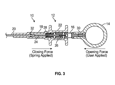
[0036] To help illustrate aspects of this disclosure, the handle and self-
actuating
mechanism at the proximal end of an exemplary grasper is schematically
depicted in
isometric view in FIG. 2. As shown, the proximal end of grasper 10 has handle
12 that
includes thumb loop 14 and plunger 16 extending coaxially into barrel 18.
Thumb loop
14 is an actuatable element activated by the user and other embodiments can
employ
different configurations, such as a slider, a lever, a button or others as
known in the art.
Elongated shaft member 20 extends distally from handle 12. Flange 22 provides
an
element that can be gripped by the user and to aid in manipulation and use of
grasper
10, particularly when engaging the thumb loop 14 or other actuator. Further,
FIG. 3
schematically depicts handle 12 in cross-sectional view to show details of the
self-
actuating mechanism. Particularly, plunger 16 is coupled to actuation wire 24
at
connection 26. In the depicted embodiment, this connection 26 is a friction
fit but any
suitable attachments can be used. Actuation wire 24 extends coaxially through
shaft
member 20 to the distal end of grasper 10 as described below. Handle 12 is
configured
to allow the user's thumb to be placed in thumb loop 14 and the user's fingers
around
flange 22. The self-actuation mechanism is driven by compression spring 28,
one end
of which is secured to the distal end of plunger 16 while the other is secured
to barrel
18. Therefore, the self-actuation mechanism causes plunger 16 to transmit a
withdrawal
force to actuation wire 24. Conversely, depressing thumb loop 14 while holding
flange
22 can overcome the withdrawal force imparted by spring 28. As desired, thumb
loop
14 can be secured to plunger 16 by a snap-fit connection 30, allowing for
partial
disassembly and/or reassembly of handle 12 to facilitate packaging,
sterilization or other
purpose. Alternatively, any other suitable means of attachment can be used,
including
permanent attachment, or thumb loop 14 and plunger 16 can be a unitary
structure. The
proximal end 32 of shaft member 20 may include a feature, such as a flange, to
help
secure and position shaft member 20 with respect to barrel 18. Turning now to
FIG. 4, a
detail view of the distal end of grasper 10 is shown, where opposing jaws 34
are
connected at an intermediate location by scissor pivot 36 and can transition
between
open and closed configurations. Actuation wire 24 is coupled to the proximal
ends 38
of jaws 34, so that the withdrawal force is a closing force and causes jaws 34
to pivot to
a closed position when applied to actuation wire 24, while application of a
distal force is
an opening force and pivots jaws 34 to the open position shown when applied to
actuation wire 24. As will be appreciated, the force of spring 28 is directly
related to
-8-
CA 03188586 2022-12-29
WO 2022/010838
PCT/US2021/040435
the clamping force of j aws 34. As desired, the user can selectively augment
the closing
pressure by manually depressing thumb loop 14 while holding flange 22. It
should also
be appreciated that this design can be adapted to employ jaws 34 that are
driven closed
by a distal force as opposed to a withdrawal force by substituting an
extension spring 28
for the compression spring 28. With this modification, the spring 28 still
automatically
supplies the closing force and the user can apply the opening force by
withdrawing
thumb loop 14.
[0037] Shaft member 20 is desirably thin, flexible, soft, and yet strong
enough to
facilitate advancement without kinking while also offering sufficient columnar
strength
to transmit forces through actuation wire 24. In one embodiment, shaft member
20 is
formed from a reinforced polymer extrusion. For example, the polymer extrusion
may
be made with materials such as PEBA (Polyether Block Amide),
Polytetrafluoroethylene (PTFE), etc. The extrusion may also be a multilayer
construction using different polymers or the same polymer but with differing
hardness.
The reinforcement may be metallic, such as stainless steel, nitinol, etc., or
a polymer,
such as PEEK (Poly-Ether-Ether-Ketone), Nylon, etc. The reinforcement may be
arranged in a coil or braided pattern and may not necessarily extend across
the entire
length of the shaft member 20. Alternatively, shaft member 20 may be
constructed
from extruded polymers alone. Shaft member 20 can also have a liner along its
inner
diameter to reduce friction with actuation wire 24, which may be made of PTFE,
Nylon,
or other materials with low coefficients of friction. Shaft member 20 may also
be
coated with additional lubricious or hydrophilic material on its outer
diameter to help
facilitate advancement through the urinary tract or other location in the
body. Suitable
wall thicknesses for shaft member 20 are 0.001-0.025", or more preferably
0.003-
0.015". In one embodiment, suitable dimensions of grasper 10 may be as
indicated in
FIG. 5, although it will be recognized that these dimensions can be modified
as desired
to suit the intended application.
[0038] In light of the above disclosure, it will be appreciated that
during one
illustrative example operating grasper 10, the user manipulates handle 12 to
advance
shaft member 20 to a desired location within the patient's body, such as by
positioning
jaws 34 proximally adjacent to the foreign object, for example, an implant
located in
-9-
CA 03188586 2022-12-29
WO 2022/010838
PCT/US2021/040435
urethra, to be removed. The user may then depress thumb loop 14 while holding
flange
22 to overcome the bias of spring 28 and open jaws 34. Further advancement of
shaft
member 20 can then position the foreign object within the range of jaws 34.
Next,
releasing the pressure on thumb loop 14 allows spring 28 to attempt to return
to its
native length, imparting the withdrawal force to actuation wire 24 and
correspondingly
causing jaws 34 to pivot towards the closed position to grasp the foreign
object. The
self-actuation mechanism described above maintains the force or pressure and
holds the
grasper 10 in the closed position until the force pressure is released by the
user by again
withdrawing thumb loop 14. This design minimizes the need for constant
pressure to be
applied with actuation methods used on currently available graspers and
reduces user
hand fatigue. As will be appreciated, the self-actuation mechanism is not
limited to just
a spring-driven design. A cam mechanism or similar mechanisms may also be
employed by those skilled in the art to achieve similar results. The self-
actuating grasper
described is capable of achieving grasping or grip forces, approximately
normal to
the jaws 34, between 1-20 Newtons or more preferably between 1-5 Newtons,
depending on the mechanism, jaw geometry, and spring constant of compression
spring
28. Once the prostatic implant, prostatic tissue expander or foreign object is
gripped,
the self-actuating grasper 10 is capable of withstanding retrieval forces,
applied
longitudinally along the axis of the shaft member 20, between 5-50 Newtons or
more
preferably between 5-20 Newtons.
[0039] In another embodiment, grasper 40 is schematically depicted in
FIG. 6, with
similar elements employing the same reference numbers. In this embodiment, the
self-
actuating mechanism is configured so that the user applies a withdrawal force
to open
the jaws 34. Here, plunger 42 is coupled to actuation wire 24 and contained
wholly
within body 44 of handle 12. Similar to the previous embodiment, plunger 42 is
biased
in a proximal direction by spring 28, thereby providing the closing force to
jaws 34 (not
shown in this view). To reverse the direction of force applied by the user,
plunger 42 is
coupled to extension 46 of thumb loop 14 by rotating link 48. As such, the
user applies
a withdrawal force to thumb loop 14 as the opening force. By changing gear
sizes in
rotating link 48, different leverage ratios can be obtained for the force
applied to thumb
loop 14. As with the previous embodiment, the user can augment the closing
force by
manually withdrawing thumb loop 14 given that the direction of user applied
force is
-10-
CA 03188586 2022-12-29
WO 2022/010838
PCT/US2021/040435
reversed in this configuration. Also, a modification of this embodiment
similar to the
previous embodiment can be achieved by substituting an extension spring for
the
compression spring. Once more, the spring automatically supplies the closing
force
while the user can apply the opening force by depressing thumb loop 14.
[0040] In yet another embodiment, grasper 50 is schematically depicted in
FIG. 7
and incorporates an adapter for directly connecting to a cystoscope. Handle 12
can
incorporate any of the self-actuating mechanisms discussed above, although the
first
embodiment is shown. Extension 52 of barrel 18 is coaxially disposed within
body 54
of cystoscope adapter 55 in a telescoping arrangement. As before, shaft member
20
extends from barrel 18 and coaxially through extension 52. Body 54 of adapter
55
features Luer lock ring 56 at its distal end for connection to a cystoscope
and an
irrigation line may be coupled to grasper 50 though irrigation connection 58.
The
telescoping arrangement of extension 52 of barrel 18 and body 54 of adapter 55
allows
the relative positioning of grasper 50 to the cystoscope to be adjusted to
accommodate
different designs. Once the appropriate position within the cystoscope is
achieved,
grasper 50 can be fixed relative to the cystoscope. Notably, the embodiment
shown
here features rack-like teeth 62 along the outer diameter of extension 52.
Lock lever 60
is pivotally connected to body 54, with corresponding rack-like teeth 62 that
are biased
into engagement with the teeth on extension 52 by compression spring 64. When
the
user depresses lock lever 60, the length of extension 52 of barrel 18 disposed
within
body 54 of adapter 55 can be telescopically adjusted and when lock lever 60 is
released,
spring 64 causes the lever 60 to pivot and engage teeth 62 with those on
extension 52 to
fix the relative position of grasper 50 and the cystoscope. Other suitable
techniques
may be employed to lock extension 52 at a desired degree of extension 52
within body
54, such as a frictional collet or clamp, a depressible projection that
engages one of a
plurality of longitudinally distributed holes along the body 54, or other
methods known
to those in the art.
[0041] In yet another embodiment, the handle 12 and self-actuating
mechanism at
the proximal end of an exemplary grasper 10 incorporate a safety feature to
prevent
damage to the distal jaws 34 assembly during transit and storage. As in FIGs.
2-4, it
comprises thumb loop 14 connected to plunger 16, shaft member 20 and activated
by
-11-
CA 03188586 2022-12-29
WO 2022/010838
PCT/US2021/040435
the compression spring 28. Referring to FIG. 8, these components are assembled
and
held in place by a threaded insert 71, a set screw 72, a release pin 73, a
spring pin 74
and a cowling (or cover) 75. The release pin 73 locks compression spring 28 in
the
compressed state and protects the grasper 10 in the packaged configuration
during
transit and storage, prior to use. Locking the spring 28 prevents damage to
the small pin
joints between the jaws 34 at the distal end of the shaft member 20, shown in
FIG. 9.
Before use, the user presses the release pin 73 and the compression spring 28
is free to
apply force on the shaft member 20, which then activates the proximal movement
of
thumb loop 14 towards the user and closes the grasper jaws 34. To use the
grasper, the
user squeezes the thumb loop 14 distally, which compresses the compression
spring 28,
shortening the distance between the handle 12 and thumb loop 14, and opening
the jaws
34.
[0042] FIG. 9 shows the detailed view of the distal end of another
embodiment of
the self-actuating grasper 10 in the fully open (as shown in FIG. 9a) and
fully closed
conditions (as shown in FIGs 9b and 9c). In this embodiment, the opposing jaws
34 can
be smooth without serrated teeth. The absence of serrations on inner surfaces
of the
jaws 34 provides greater gap between opposing jaws 34 and can assist securing
and
retrieval of an expander implant, stent, or a foreign object. The jaws 34 are
also
designed with features on the ends to interlock and secure the expander,
implant or
other foreign object. The end of one jaw 34 has a projection in the middle and
the
opposing jaw 34 has a groove that interlocks or with the projection. Note that
in the
closed state, there is some gap or opening between the interlocked jaws 34 in
the fully
closed condition. The gap between the jaws 34 enhances the ability of the
grasper 10 to
securely grab and snare or retrieve the expander, implant or any other foreign
object
from the body.
[0043] As will be appreciated from the above discussion and figures, the
self-
actuating grasper design of the present disclosure can be used to assist the
retrieval of a
foreign object, including a stent, an expander or other implant, or a
naturally occurring
deposit such as a stone or calculi, from the prostatic urethra, urinary
bladder, ureters,
kidneys urinary tract or urinary system in addition to other suitable
applications. By
employing the techniques of this disclosure, such a grasper may exhibit a
number of
-12-
CA 03188586 2022-12-29
WO 2022/010838 PCT/US2021/040435
beneficial characteristics, including without limitation: 1) the ability to
securely grip the
foreign object to facilitate retrieval; 2) being atraumatic to urethra and
other anatomical
structures during retrieval of foreign object; 3) being compatible with
commonly used
flexible and rigid cystoscopes, other medical imaging equipment and ancillary
medical
devices used in medical procedures; 4) allowing for self-actuation to
automatically lock,
together with the ability to un-lock as desired; 5) the ability to rotate
axially, such as by
having adequate torque transmission, to position for engagement with foreign
object for
retrieval; 6) allowing continuous imaging, with irrigation through the working
channel
of the cystoscope as needed, during the retrieval procedure; and 7) having
sufficient
locking force to firmly hold the foreign object, without loss, during the
retrieval
procedure.
[0044] The exemplary embodiments disclosed above are merely intended to
illustrate
the various utilities of this disclosure. It is understood that numerous
modifications,
variations and combinations of functional elements and features of the present
disclosure
are possible in light of the above teachings and, therefore, within the scope
of the appended
claims, the present disclosure may be practiced otherwise than as particularly
disclosed and
the principles of this disclosure can be extended easily with appropriate
modifications to
other applications.
[0045] All patents and publications are herein incorporated for reference
to the same
extent as if each individual publication was specifically and individually
indicated to be
incorporated by reference. It should be understood that although the present
disclosure
has been specifically disclosed by preferred embodiments and optional
features,
modification and variation of the concepts herein disclosed may be resorted by
those
skilled in the art, and that such modifications and variations are considered
to be within
the scope of this disclosure.
-13-