Note: Descriptions are shown in the official language in which they were submitted.
CA 03188591 2022-12-29
WO 2022/011222
PCT/US2021/041029
COMPOSITIONS AND METHODS FOR TREATING CANCER-ASSOCIATED
CACHEXIA
CROSS-REFERENCE TO RELATED APPLICATIONS
This application claims priority to U.S. Provisional Patent Application No.
63/050,352,
filed on July 10, 2020, which is incorporated by reference herein in its
entirety.
BACKGROUND
Cancer-associated cachexia (CAC) is a multifactorial syndrome defined by an
ongoing
loss of skeletal muscle mass, with or without loss of fat mass, that cannot be
entirely reversed
by conventional nutrition support. The onset of CAC increases chemotherapy
toxicity and
complications from surgeries, decreases quality of life in patients, and leads
to higher mortality
rates. In many instances, after onset of CAC in a patient, chemotherapy
treatment is
discontinued because of the inability of chemotherapy treatment to be
effective in a patient
who has developed CAC. Cachexia is prevalent among the deadliest cancers,
contributes to
half of all cancer deaths worldwide, is irreversible, and associates with end
stage disease.
Currently, there is no effective treatment available for CAC.
A key feature of CAC is chronic deterioration of lean body mass. Symptoms
include
weight loss, muscle wasting, nutrient deficiency, and loss of appetite. People
who develop
cachexia are not losing weight because they are trying to trim down with diet
or exercise.
Rather, they lose weight because they eat less due to a variety of reasons. At
the same time,
their metabolism changes, which causes their body to break down too much
muscle.
In patients with cancer, tumor cells release substances that reduce appetite
and cause
the body to burn calories more quickly than usual. Cancer and its treatments
can also cause
severe nausea or damage the digestive track, making it difficult for cancer
patients to eat and
absorb nutrients. As the body gets fewer nutrients, it burns fat and muscle,
and cancer cells use
what limited nutrients are left to survive and multiply.
Previous attempts to treat cancer-associated cachexia focused on the symptoms
such as
muscle wasting and nutrient deficiency. However, clinical trials based on
nutrient supplements
and anti-inflammatory treatments have all failed primarily because the focus
on the treatment
has been on the symptom rather than the root cause - the patient's brain and
metabolism
1
CA 03188591 2022-12-29
WO 2022/011222
PCT/US2021/041029
processing. Hence, there is a need for new and improved therapies for the
treatment of cancer-
associated cachexia.
SUMMARY
The Summary is provided to introduce a selection of concepts that are further
described
below in the Detailed Description. This Summary is not intended to identify
key or essential
features of the claimed subject matter, nor is it intended to be used as an
aid in limiting the
scope of the claimed subject matter.
In a first aspect of the invention, a method for treating cachexia in a
subject in need
thereof comprises stimulating the parasympathetic nervous system of the
subject thereby
treating cachexia in the subject. In a feature of this aspect, stimulating the
parasympathetic
nervous system in the subject increases expression of urea cycle enzymes in
the liver.
In additional aspects of the invention, methods for mitigating weight loss due
to
cachexia, mitigating fat loss due to cachexia, mitigating muscle wasting due
to cachexia, and
mitigating loss of appetite due to cachexia comprise stimulating the
parasympathetic nervous
system in the subject.
In another aspect of the invention, a method for mitigating urea cycle
dysregulation in
a subject in need thereof due to cachexia comprises stimulating the
parasympathetic nervous
system in the subject thereby mitigating urea cycle dysregulation due to
cachexia in the subject.
In a further aspect of the invention, stimulating the parasympathetic nervous
system
comprises stimulating the vagus nerve. In a feature of this aspect,
stimulating comprises
stimulating the cervical vagus nerve or the hepatic branch of the vagus nerve.
Stimulating the
vagus nerve can comprise delivering pulses at a frequency ranging from 1 Hz to
10Hz.
Regarding this feature, the pulses can have a pulse width of about 1
millisecond to about 100
milliseconds. In another feature, stimulating pulses can be delivered at a
frequency of about 5
kHz. Regarding this feature, the pulses can have a pulse width of greater than
0 and less than
0.2 milliseconds.
BRIEF DESCRIPTION OF THE DRAWINGS
The accompanying Figures and Examples are provided by way of illustration and
not
by way of limitation. The foregoing aspects and other features of the
disclosure are explained
2
CA 03188591 2022-12-29
WO 2022/011222
PCT/US2021/041029
in the following description, taken in connection with the accompanying
example figures (also
"FIG.") relating to one or more embodiments, in which:
FIG. 1 is a schematic diagram showing the autonomic activities of the
sympathetic
nervous system (SNS) and parasympathetic nervous system (PNS) during different
physiological states.
FIG. 2A provides an exemplary schematic diagram of functional electrical
stimulation
parameters.
FIG. 2B provides an exemplary schematic diagram of stimulation pattern
duration.
FIG. 3 is a graph showing the fold-changes of hepatic amino acid levels in
cachectic
mice using Student's t-test, Bonferroni FDR<0.05 in accordance with Example 1.
FIGS. 4A-3D are schematic illustrations and images showing exemplary
denervation
surgery.
FIGS. 5A-4D are images showing exemplary electrical stimulation of hepatic
sympathetic and parasympathetic nerves.
FIG. 6 is a schematic illustration showing a block diagram outlining an
exemplary
stimulation and recording pipeline.
FIGS. 7A and 7B are graphs showing blood glucose level after sympathetic
(Symp),
parasympathetic (Para) and sham (No stim) stimulations.
FIGS. 8A-8C are graphs showing hepatic metabolic gene expression levels
quantified
by qRCR after sympathetic and parasympathetic stimulation.
FIG. 9 is a series of photographs (top and bottom) and a schematic
illustration of
experimental procedures for Example 4.
FIGS. 10A-10D are charts and graphs illustrating how body weight was affected
by
cancer injection and vagus nerve stimulation.
FIGS. 11A and 11B are charts showing the effect of VNS or the absence of VNS
on
total fat and brown adipose tissue, respectively.
FIG. 12A includes photographs of skeletal muscle fiber for Control (top) and
Cancer
(bottom) mice.
FIG. 12B is a chart comparing muscle atrophy for mice having cancer, cancer
with VNS
therapy, cancer with vagotomy, and healthy control.
3
CA 03188591 2022-12-29
WO 2022/011222
PCT/US2021/041029
FIG. 13 is a chart showing the effect of vagal nerve stimulation on daily food
intake for
control mice, mice with cancer but no treatment, and cancer with VNS
treatment.
DETAILED DESCRIPTION
For the purposes of promoting an understanding of the principles of the
present
disclosure, reference will now be made to preferred embodiments and specific
language will
be used to describe the same. It will nevertheless be understood that no
limitation of the scope
of the disclosure is thereby intended, such alteration and further
modifications of the disclosure
as illustrated herein, being contemplated as would normally occur to one
skilled in the art to
which the disclosure relates.
Articles "a" and "an" are used herein to refer to one or to more than one
(i.e., at least
one) of the grammatical object of the article. By way of example, "an element"
means at least
one element and can include more than one element.
"About" is used to provide flexibility to a numerical range endpoint by
providing that
a given value may be "slightly above" or "slightly below" the endpoint without
affecting the
desired result.
The use herein of the terms "including," "comprising," or "having," and
variations
thereof, is meant to encompass the elements listed thereafter and equivalents
thereof as well as
additional elements. As used herein, "and/or" refers to and encompasses any
and all possible
combinations of one or more of the associated listed items, as well as the
lack of combinations
where interpreted in the alternative ("or").
As used herein, the transitional phrase "consisting essentially of (and
grammatical
variants) is to be interpreted as encompassing the recited materials or steps
and those that do
not materially affect the basic and novel characteristic(s)" of the claimed
invention. Thus, the
term "consisting essentially or as used herein should not be interpreted as
equivalent to
"comprising."
Moreover, the present disclosure also contemplates that in some embodiments,
any
feature or combination of features set forth herein can be excluded or
omitted. To illustrate, if
the specification states that a complex comprises components A, B and C, it is
specifically
intended that any of A, B or C, or a combination thereof, can be omitted and
disclaimed
singularly or in any combination.
4
CA 03188591 2022-12-29
WO 2022/011222
PCT/US2021/041029
Recitation of ranges of values herein are merely intended to serve as a
shorthand
method of referring individually to each separate value falling within the
range, unless
otherwise indicated herein, and each separate value is incorporated into the
specification as if
it were individually recited herein. For example, if a concentration range is
stated as 1% to
50%, it is intended that values such as 2% to 40%, 10% to 30%, or 1% to 3%,
etc., are expressly
enumerated in this specification. These are only examples of what is
specifically intended, and
all possible combinations of numerical values between and including the lowest
value and the
highest value enumerated are to be considered to be expressly stated in this
disclosure.
As used herein, "treatment," "therapy" and/or "therapy regimen" refer to the
clinical
intervention made in response to a disease, disorder or physiological
condition manifested by
a patient or to which a patient may be susceptible. The aim of treatment
includes the alleviation
or prevention of symptoms, slowing or stopping the progression or worsening of
a disease,
disorder, or condition and/or the remission of the disease, disorder or
condition.
The term "effective amount" or "therapeutically effective amount" refers to an
amount
sufficient to effect beneficial or desirable biological and/or clinical
results.
As used herein, the term "subject" and "patient" are used interchangeably and
refer to
both human and nonhuman animals. The term "nonhuman animals" includes all
vertebrates,
e.g., mammals and non-mammals, such as nonhuman primates, sheep, dog, cat,
horse, cow,
chickens, amphibians, reptiles, and the like. The methods and compositions
disclosed herein
.. can be used on a sample either in vitro (for example, on isolated cells or
tissues) or in vivo in a
subject (i.e. living organism, such as a patient). In some embodiments, the
subject comprises a
human who is suffering from cancer associated cachexia (CAC).
Unless otherwise defined, all technical terms used herein have the same
meaning as
commonly understood by one of ordinary skill in the art to which this
disclosure belongs.
Cachexia is a condition related to uncontrolled weight loss that occurs
incidental to
many severe diseases, including cancer, sepsis, and major organ failure.
Cachexia is defined as
the unwanted loss of a least 5% of lean mass within six months. Cachexia often
represents the
last step of a chronic disease. The condition affects many advanced cancer
patients and is
associated with poor prognosis regardless the tumor nature. It is generally
accepted that
cachexia is indirectly responsible for the death of at least 20% of all cancer
patients. The
incidence of cachexia among cancer patients is remarkably high, although it
varies by tumor
type. In patients with gastric or pancreatic cancer, the incidence is more
than 80%, whereas
5
CA 03188591 2022-12-29
WO 2022/011222
PCT/US2021/041029
approximately 50% of patients with lung, prostate or colon cancer are
affected, and around
40% of patients with breast tumors or some leukemias develop the cachexia.
Loss of skeletal muscle mass is recognized as an independent predictor of
mortality and
is associated with functional impairment, altered quality of life and reduced
tolerance and
response to anticancer therapies. Further, it has been shown that reversal of
muscle loss leads
to prolonged survival in animal models of cancer cachexia. These observations
support that
maintaining muscle mass is helpful in improving survival in cachectic
conditions.
Understanding molecular drivers of cachexia is important for the developing
management
strategies.
Cachexia affects various organs, which often results in systemic
complications. The
molecular mechanisms of cancer cachexia are not well characterized. Generally,
scientists
believe that cachexia results from abnormal metabolism and anorexia. The
development of
muscle atrophy results from an imbalance between muscle protein synthesis and
degradation
inducing a decrease in myofibrillar and sarcoplasmic proteins illustrated by
muscle fiber
shrinkage. However, the nature of the key factors responsible for muscle
atrophy in cancer
cachexia is unknown.
The present disclosure is based, in part, on the discovery that manipulation
of the vagus
nerve can be effective in reversing and/or mitigating cachexia. In
embodiments, manipulating
can include stimulation or denervation. Moreover, in embodiments, stimulation
can include,
without limitation, electrical stimulation or optogenetic stimulation. Much of
the description
provided herein relates to electrical stimulation. However, one of ordinary
skill in the art will
understand that stimulation may comprise optogenetic stimulation. Manipulating
the vagus
nerve via vagus nerve stimulation or denervation targets the gut-brain axis
and can effectively
reverse and/or mitigate cachexia.
The gut-brain axis is a bidirectional link connecting the gut and the brain
and including
communication between the central nervous system and the enteric nervous
system of the body.
The gut-brain axis includes communication between the endocrine (hypothalamic-
pituitary-
adrenal axis), immune (cytokine and chemokines) and the autonomic nervous
system (ANS).
In animal studies related to the gut-brain axis, stress was seen to inhibit
signals sent through
the vagus nerve and cause gastrointestinal problems. Similarly, a study in
humans found that
people with irritable bowel syndrome (IBS) or Crohn's disease had reduced
vagal tone,
indicating a reduced function of the vagus nerve.
6
CA 03188591 2022-12-29
WO 2022/011222
PCT/US2021/041029
Described herein is a method of treating cachexia in a subject in need
thereof. The
method comprises stimulating the parasympathetic nervous system of the subject
thereby
treating cachexia in the subject. It is contemplated that stimulating the
parasympathetic nervous
system may increase expression of urea cycle enzymes in the liver thereby
leading to reversal
or mitigation of cachexia.
As mentioned previously, cachexia is marked by uncontrolled weight loss.
Symptoms
include muscle wasting, nutrient deficiency, and loss of appetite.
Accordingly, described herein
are methods of reversing or mitigating the symptoms of cachexia. In
embodiments, this
includes a method of mitigating weight loss due to cachexia in a subject in
need thereof,
wherein the method comprises stimulating the parasympathetic nervous system in
the subject
thereby mitigating weight loss due to cachexia. In embodiments of the method,
the subject
having cachexia who received stimulation to the parasympathetic nervous system
experiences
statistically significantly less weight loss than subjects having cachexia
that received no
stimulation. In additional embodiments, there is no statistically significant
variation in weight
.. loss between the subject having cachexia who received stimulation to the
parasympathetic
nervous system and a healthy control subject. Additionally, described herein
is a method of
mitigating fat loss due to cachexia in a subject in need thereof, wherein the
method comprises
stimulating the parasympathetic nervous system in the subject thereby
mitigating fat loss due
to cachexia. In embodiments, the fat is brown adipose tissue. Regarding this
embodiment, there
is no significant change in brown adipose tissue mass between healthy control
subjects and the
subject having cachexia who received stimulation to the parasympathetic
nervous system.
Moreover, regarding this embodiment, a subject having cachexia who received
stimulation to
the parasympathetic nervous system experiences statistically significantly
less atrophy of
brown adipose tissue than subjects having cachexia that received no
stimulation. Additionally,
methods of mitigating muscle wasting due to cachexia in a subject in need
thereof are
described. The method comprises stimulating the parasympathetic nervous system
in the
subject thereby mitigating muscle wasting due to cachexia. Additionally,
methods of mitigating
loss of appetite due to cachexia in a subject in need thereof are described.
The methods
comprise stimulating the parasympathetic nervous system in the subject thereby
mitigating loss
of appetite. In embodiments, there is no statistically significant variation
in average daily food
intake between subjects having cachexia who received stimulation to the
parasympathetic
nervous system and healthy control subjects. In alternative embodiments,
subjects having
7
CA 03188591 2022-12-29
WO 2022/011222
PCT/US2021/041029
cachexia who received stimulation to the parasympathetic nervous system had
statistically
significantly higher daily food intake than subjects having cachexia that
received no
stimulation.
Additionally, in embodiments, a method of reversing and/or mitigating
cachectic urea
cycle dysregulation in a subject in need thereof is described. The method
comprises stimulating
the parasympathetic nervous system in the subject thereby reversing and/or
mitigating
cachectic urea cycle dysregulation in the subject.
The urea cycle is the primary means of nitrogen metabolism in humans and other
ureotelic organisms. There are five prominent hepatic enzymes in the urea
cycle: carbamoyl-
phosphate synthetase (CPS), omithine transcarbamylase (OTC), argininosuccinate
synthetase
(ASS), argininosuccinase lyase (ASL), and arginase (ARG). In healthy
individuals, muscle
breakdown leads to the flow of amino acids into the liver, where excess
nitrogen reacts with
aspartate to synthesize urea via the urea cycle to be disposed by urine.
Outside the liver,
different urea cycle enzymes are expressed to provide urea cycle intermediates
arginine and
omithine to supply cellular needs.
Without being bound by theory, it is believed that dysregulation of the
expression of
urea cycle (UC) enzymes promotes cancer proliferation by diversion of
aspartate and glutamine
toward pyrimidine rather than urea synthesis. In particular, it is believed
that in various types
of CAC, the expression and function of hepatic urea cycle enzymes is
downregulated despite
the high flux of amino acids that is generated by the protein breakdown of
skeletal muscle
secondary to an aberrant signaling cascade caused by cancer. This unexpected
result suggests
that dysregulated urea cycle enzyme expression in the liver of the host is
part of the systemic
dysregulation induced by the tumor to increase nitrogen availability for its
needs. Experimental
results provided below demonstrate that the overall increase in protein
turnover measured in
cachexia patients cannot be explained solely by tumor cellular turnover. This
result explains
previously unexplained systemic dysregulation in nitrogen homeostasis
experienced by
subjects who developed cachexia.
The autonomic nervous system (ANS) controls specific body processes, such as
circulation of blood, digestion, breathing, urination, heartbeat, etc. The
primary function of the
autonomic nervous system is homeostasis. Apart from maintaining the body's
internal
environment, it is also involved in controlling and maintaining multiple life
processes including
digestion and metabolism. There are two types of autonomic nervous system:
sympathetic
8
CA 03188591 2022-12-29
WO 2022/011222
PCT/US2021/041029
autonomic nervous system and parasympathetic autonomic nervous system. The
sympathetic autonomic nervous system is located near the thoracic and lumbar
regions in the
spinal cord. Its primary function is to stimulate the body's fight or flight
response. The
sympathetic nervous system is primarily associated with the energy
mobilization and fasting
phase of systemic metabolism. The parasympathetic autonomic nervous system is
located
between the spinal cord and the medulla. It primarily stimulates the body's
"rest and digest"
and "feed and breed" response. The parasympathetic nervous system is primarily
associated
with the feeding phase of systemic metabolism. The parasympathetic nervous
system includes
the parasympathetic vagus nerve. FIG. 1 is a bar graph illustrating some of
the functionalities
of the sympathetic and parasympathetic nervous systems.
Vagal nerve manipulation, in particular vagus nerve stimulation (VNS), is an
FDA-
approved therapy for treatment-resistant focal epilepsy, treatment-resistant
major depressive
disorder, episodic cluster headaches, and migraine pain. VNS is under
additional investigation
as a clinical tool for treatment of obesity, anxiety disorders, dementia,
alcohol addiction,
chronic heart failure, arrhythmia, autoimmune diseases, and chronic pain
conditions.
Moreover, studies have reported promising outcomes following vagus nerve
manipulation ¨
both stimulation and blockade ¨ for treatment of obesity-associated metabolic
syndromes in
clinical trial and preclinical studies. Additionally, vagus nerve stimulation,
in which the nerve
is stimulated with pulses of electricity, has been used to treat patients with
epilepsy, depression,
Alzheimer disease and migraine.
VNS is also being investigated for treatment of inflammation in several
autonomic or
inflammatory disorders. Preliminary studies have evaluated VNS being used for
stroke,
autoimmune diseases, heart and lung failure, obesity, and pain management, but
further studies
are needed to understand the mechanistic actions that explain VNS' s potential
role in treating
these disorders.
Despite any of the foregoing, prior to the work described herein, vagus nerve
manipulation, including denervation and stimulation, had not been investigated
to treat,
reverse, or mitigate cachexia. Moreover, in clinical trials of anti-
inflammatory therapies, such
as TNF-a and interleukins, the therapies were shown to not benefit cachexic
patients.
Accordingly, it is unlikely that a VNS effect on cachexia is related to
inflammation, or to
inflammation alone. Rather, in view of the work described herein, the effect
of VNS on
cachexia is believed to be related to regulating the gut-brain axis and
metabolism.
9
CA 03188591 2022-12-29
WO 2022/011222
PCT/US2021/041029
In conventional vagus nerve stimulation, a device is surgically implanted
under the skin
of a subject's chest, and a wire is threaded under the skin connecting the
device to the left
cervical vagus nerve. When activated, the device sends electrical signals
along the left vagus
nerve to the brainstem, which then sends signals to certain areas in the
brain. Conventionally,
the right vagus nerve is not used because it can be more likely to carry
fibers that supply nerves
to the heart. However, the right vagus also contains the dominant
parasympathetic fibers
innervating the gut and especially the liver; thus, unlike conventional vagus
nerve stimulation
and previously used configurations, the stimulation described herein is placed
on the right
cervical vagus nerve or the subdiaphragmatic common hepatic branch. This
subdiaphragmatic
branch of the vagus nerve does not contain fibers that extend to the heart but
does have
connections to the liver and other gastrointestinal organs, and through
testing related to the
present disclosure has been shown to produce meaningful changes in urea cycle
enzymes.
However, despite the foregoing, the complex relationship between the ANS and
whole-
body metabolism remains elusive. For instance, although a decrease in blood
glucose levels
after vagal nerve stimulation has been documented in preclinical settings,
cervical vagal nerve
stimulation seems to impair insulin release. This inconsistency might be
partly attributed to the
differences in afferent and efferent vagal nerve stimulation but can also be
explained by mixed
endocrine and vagal signaling to the liver. Importantly, these discrepancies
highlight the
unpredictable nature of the ANS and the need to explore and better understand
the role of the
ANS in the regulation of systemic metabolism in organ- and context-specific
manners.
Data provided in the examples below shows that denervation or stimulation of
the vagus
nerve impacts the urea cycle in the liver. In embodiments, stimulating the
parasympathetic
nervous system comprises stimulating the vagus nerve. For example, stimulating
the vagus
nerve can comprise stimulating the cervical vagus nerve or the hepatic branch
of the vagus
nerve.
The Examples provided below show that vagus nerve manipulation can affect
liver
metabolism and help restore systemic nitrogen homeostasis in subjects having
cancer
associated cachexia. The examples include investigations of systemic- and
liver-specific
nitrogen-related changes during cancer associated cachexia (Example 1) and ANS
perturbation
(Example 2) and evaluation of the hypothesis that ANS intervention can
mitigate or reverse
nitrogen and urea cycle dysregulation during cachexia (Example 3). Additional
examples
CA 03188591 2022-12-29
WO 2022/011222
PCT/US2021/041029
evaluate the impact of ANS intervention in subject's having cachexia on weight
loss, body fat
(total and brown adipose tissue), muscle mass, and food intake (Example 4).
The mouse models used in the examples were established KPC and TIC models,
which
are representative models for the study of cancer cachexia that robustly
recapitulate features of
human disease. The KPC model is described fully in Michaelis et al..
Establishment and
characterization of a novel murine model of pancreatic cancer cachexia
("Michaelis"), which
is incorporated by reference herein. Briefly, Michaelis describes that
syngeneic KPC allografts
are a robust model for studying cachexia, which recapitulate key features of
the pancreatic
ductal adenocarcinoma (PDAC) disease process and induce a wide array of
cachexia
manifestations. As such, this model is ideally suited for future studies
exploring the
physiological systems involved in cachexia and for preclinical studies of
novel therapies. The
LLC model is described fully in Choi, et al., Concurrent muscle and bone
deterioration in a
murine model of cancer cachexia ("Choi"), which is incorporated by reference
herein. Briefly,
Choi describes testing the validity of the Lewis lung carcinoma (LLC) as a
model of cancer
cachexia and examining its effect on the two major lean tissue components,
skeletal muscle,
and bone. Choi concluded that LLC is a valid model of cachexia that induces
rapid losses in
global bone mineral density and in lim.b and respiratory muscle function. Both
KPC and LLC
models combine established spontaneously occurring animal models and
genetically
engineered mouse models to ensure conservation of pathways across a diverse
array of
cancers.
Vagus nerve manipulation includes stimulation of the parasympathetic vagus
nerve,
including areas of the parasympathetic vagus nerve such as the cervical vagus
branch or the
hepatic vagus nerve branch.
In functional electrical stimulation, typically positive or negative pulsed
current is
delivered from. electrode surface contacts at the peripheral nerve location.
This methodology is
generally considered to be more physiologically relevant and produces less
damage than
sending a continuous signal. FIG. 2A provides an exemplary schematic diagram
of functional
electrical stimulation parameters. Pulse amplitude is variable and is
generally titrated for an
individual, but pulse width and stimulation frequency are set based on
neurophysiology
principles regarding evoking or suppressing activity along the vagus nerve.
The pulse width,
frequency, and amplitude are factors that can be used to describe an
electrical stimulus.
Additional parameters that can be used to describe an electrical stimulus
include pulse train
11
CA 03188591 2022-12-29
WO 2022/011222
PCT/US2021/041029
duration and stimulation pattern duration. Pulse train duration defines the
amount of time a
pulse train continues over a period of time. Stimulation pattern duration is
the time duration a
pulse train repeats over a period of time. FIG. 2B provides an exemplary
schematic diagram of
stimulation pattern duration.
Different stimulation frequencies may modulate nerves differently. For
example, 10Hz
pulses are commonly used to activate peripheral nerves, while high-frequency
pulses (e.g.,
5kHz) have been shown to block peripheral nerves.
In embodiments, the frequency of the pulses delivered to the vagus nerve
includes
frequencies ranging from 1 Hz to 10 Hz. For example, the frequency may be 1
Hz, 2 Hz, 3 Hz,
4 Hz, 5 Hz, 6 Hz, 7 Hz, 8 Hz, 9 Hz, or 10 Hz. In this frequency range, a pulse
may have a pulse
width of about 1 millisecond to about 100 milliseconds. For example, the pulse
width may be
about 1 millisecond (ms) to about 10 ms, including about 1 ms, 2 ms, 3 ms, 4
ms, 5 ms, 6 ms,
7 ms, 8 ms, 9 ms, or 10 ms. The pulse may be a charge balanced constant
current biphasic pulse
with alternating anodic and cathodic leading phases with a range of 0.1 mA to
10 mA.
In other embodiments, the frequency of the pulses delivered to the vagus nerve
includes
a frequency of 5 kHz. At or near this frequency range, a pulse may have a
pulse width of about
greater than 0 and less than or equal to 0.2 milliseconds. For example, the
pulse width may be
about 0.5 ms, 1 ms, 1.5 ms or 2 ms. The pulse may be a charge balanced
constant current
biphasic pulse with alternating anodic and cathodic leading phases with a
range of 0.1 mA to
10 mA.
In embodiments, pulse train duration and stimulation pattern duration can vary
over a
range values and can be affected by other stimulation parameters, such as, for
example, pulse
width, frequency, and amplitude. For example, the pulse train duration can
range from 1 minute
to 1 hour, and the stimulation train duration can vary from 20 minutes to 3
hours.
The following Examples are provided by way of illustration and not by way of
limitation.
EXAMPLES
EXAMPLE]. Characterization of the Systemic Nitrogen-Associated Metabolic
Changes in
the Host During CAC
12
CA 03188591 2022-12-29
WO 2022/011222
PCT/US2021/041029
Amino acid levels in cachectic liver. Amino acid levels in livers from tumor-
bearing cachectic
mice and control mice were measured and compared. Mice were injected with
saline to produce
healthy control mice, or a cancer cell line known to produce robust cachexic
phenotypes,
including Lewis lung carcinoma (LLC) or KRAS, p53, and Cre pancreatic cancer
(KPC). Using
liquid chromatography mass spectrometry (LC-MS) metabolomics on urine, plasma,
and
homogenates of excised liver, muscle, and tumors, significant alterations in
hepatic amino acid
levels in cachectic mice were observed compared to control mice. FIG. 2 is a
graph showing
the fold-changes of hepatic amino acid levels in cachectic mice using
Student's t-test,
Bonferroni FDR<0.05. FIG. 3 graphically illustrates the results. As can be
seen, the LC-MS
analysis showed fold-change decreases in glutamine and arginine and increases
in omithine
and aspartic acid for the cachectic subjects versus control, which supports
the idea that cachexia
causes urea cycle dysfunction.
EXAMPLE 2. Characterization of the Effects of Hepatic Sympathetic and
Parasympathetic
Interventions on the UC
Testing was performed to evaluate whether the autonomic nervous system plays a
role
in modulating the hepatic urea cycle. Denervation and electrical stimulation
of the hepatic
vagus nerve were performed to manipulate the ANS. Briefly, microwire cuff
electrodes were
placed on the cervical or subdiaphragmatic vagus nerve to enable VNS. The
inventor
performed the same metabolic and transcriptomic profiling described in Example
1 to measure
the impact of ANS perturbation on liver metabolism. Briefly, LC-MS based
metabolomics and
RNA-seq were carried out on the harvested liver cells to measure the
metabolite and enzyme
expression levels. The inventor also performed 15N-labeled nitrogen tracing
following the ANS
interventions.
Denervation surgery. All mice were anesthetized by intraperitoneal (i.p.)
injection of a
mix of ketamine- medetomidine-atropine (KMA) and after the surgery received a
subcutaneous
(s.c.) injection of antisedan and buprenorphine. The abdomen was shaved and
sterilized using
alternating applications of ethanol and iodine.
13
CA 03188591 2022-12-29
WO 2022/011222
PCT/US2021/041029
FIGS. 4A-4D provide schematic illustrations and photographic images showing
denervation surgery. FIG. 4A is a schematic representation illustrating the
positions of the
subdiaphragmatic vagal nerves. In FIG. 4A, the upper branch, labeled (A) is
dissected to
achieve a hepatic parasympathectomy, and the lower branch labeled (B) is
severed to achieve
a hepatic sympathectomy. FIG. 4B is an illustration of the underside of the
medial and left
lobes of the liver showing the hepatic artery (triangle), portal vein
(square), and bile duct (star).
Nerve bundles running along the hepatic artery were transected for a hepatic
sympathectomy.
FIG. 4C is an image taken during surgery of the sympathetic hepatic nerve
branches entering
the liver with the left and medial lobes lifted to expose the major vascular
and biliary tracts.
Nerve bundles, corresponding to those needed to be severed for a
sympathectomy, are marked
with arrows. FIG. 4D is an image taken during surgery of the left
subdiaphragmatic vagal nerve
running along the esophagus. The common hepatic vagus nerve branch is marked
with an
arrow. Severing this connection results in a parasympathectomy.
A ventral vertical midline incision was obtained by cutting the skin and
muscle layers
to visualize the liver. A binocular operating microscope was used for the
remainder of the
surgery. For the hepatic sympathectomy (Sx), the median and left liver lobes
were lifted to
visualize the region comprising the bile duct, hepatic artery, and portal vein
(Figure 4B). Nerve
bundles running along the hepatic artery proper were transected using
microsurgical
instruments. Any connective tissue attachments between the hepatic artery,
bile duct, and portal
vein were dissected (Figure 4C). To achieve a hepatic parasympathectomy (Px),
the common
hepatic vagal branch was transected by stretching the fascia containing the
common hepatic
vagal branch and transecting neural tissue between the ventral vagal trunk and
liver (Figure
4D). In mice with a sham denervation surgery, the same procedure of incision
and nerve
exposure was performed, but the nerves were not severed. After the
denervation, warm saline
was injected into the abdominal cavity, and the muscle and skin layers were
stitched.
Electrode Implantation: All mice were anaesthetized by intraperitoneal (i.p.)
injection
of ketamine-medetomidine-atropine (KMA) and after the surgery receive a
subcutaneous (s.c.)
injection of antisedan and buprenorphine. The abdomen was shaved and
sterilized using
alternating applications of ethanol and iodine. A ventral vertical midline
incision was
performed by cutting the skin and muscle layers to visualize the liver. A
binocular operating
14
CA 03188591 2022-12-29
WO 2022/011222
PCT/US2021/041029
microscope was used for the remainder of the surgery. Medtronic Streamline
unipolar
myocardial pacing leads were implanted for stimulation and recording (Figure
4A).
FIGS. 5A-5D provide images showing electrical stimulation of hepatic
sympathetic and
parasympathetic nerves. FIG. 5A is a photograph of clinical pacemaker leads
for stimulus and
recording. FIG. 5B is a photograph of leads implanted for recording at the
hepatic sympathetic
nerve branch (triangle) and stimulation at the hepatic parasympathetic nerve
branch (star). FIG.
5C is a photograph of the abdominal muscle closed with stitches, with leads
exiting the incision
site; leads were tunneled subcutaneously along the back before the skin was
closed. FIG. 5D is
a photograph of leads exiting the subcutaneous space at the neck, which are
kept from re-
entering with a loose knot in the lead.
The recording electrode coil was implanted around the parasympathetic branch
of the
hepatic nerve, and the stimulation recording electrode coil was implanted
around the
sympathetic hepatic nerve (Figure 5B). The tailing ends of the leads were
tunneled s.c. and
exited at the base of the neck to prevent the animal from chewing on the wires
(Figures 5C,
5D). Finally, warm saline was injected into the abdominal cavity, and the
muscle and skin
layers were stitched.
Electrical Stimulation and Recording. The stimulus applied comprised charge
balanced
2 ms biphasic pulses delivered at 10Hz with alternating anodic and cathodic
leading phases of
amplitude 0.1 to 10 mA, generated via a Tektronix AFG1062 arbitrary function
generator and
converted via a Digitimer DS5 isolated bipolar current stimulator. Recordings
were amplified
100x and isolated using a 300-5,000 Hz bandpass filter via an A-M Systems
microelectrode
AC amplifier model 1800 and recorded using a National Instruments BNC-2110
with a 10 kHz
sampling rate (Figure 6).
FIG. 6 is a schematic illustration showing a block diagram outlining the
stimulation and
recording pipeline used in the present example.
Preliminary data on ANS stimulation: The effect of sympathetic and
parasympathetic
electrostimulation on glucose was evaluated. To test the impact of sympathetic
and
parasympathetic nervous system stimulation on metabolism, animals were
implanted with
stimulation devices for sympathetic nerve (N=3), parasympathetic nerve (N=3),
or sham (N=2)
stimulation. For the testing, blood glucose levels were assessed using a
glucose meter and a
CA 03188591 2022-12-29
WO 2022/011222
PCT/US2021/041029
single drop of blood. Measurements were taken before, during, and after
stimulation.
Stimulation was delivered as described in the previous section to awake,
unanesthetized
animals. For glucose tolerance tests, stimulation was delivered for 45
minutes, with glucose
challenge injected intraperitoneally at 15 minutes into the study. For fasting
glucose tests,
baseline blood glucose measurements were taken for ten minutes before
stimulation was
initiated. Stimulation was delivered for 30 minutes, during which blood
glucose levels were
also evaluated. Blood glucose levels were also assessed for 100 minutes post-
stimulation.
FIGS. 7A-7B are graphs showing blood glucose level after sympathetic (Symp),
parasympathetic (Para) and sham (No stim) stimulations in the present example.
For the results
shown in FIG. 7A, an intraperitoneal glucose tolerance test with nerve
stimulation from time -
to 30 mm was performed, and glucose was injected at time 0 mm. For the results
shown in
FIG. 7B, the effect of nerve stimulation on fasting blood glucose was
evaluated. The nerves
were stimulated from time 10 to 40 mm. The testing showed that parasympathetic
stimulation
impaired glucose tolerance upon i.p. glucose challenge compared to the sham
control whereas
15 sympathetic stimulation did not (Figure 7A). The blood glucose level
returned to baseline at a
similar time for all groups. Conversely, sympathetic stimulation significantly
lowered fasting
blood glucose levels even after its cessation while parasympathetic
stimulation did not (Figure
7B). This experiment suggests that the whole-body glucose metabolism can be
modulated by
the ANS- mediated gut-brain axis.
FIGS. 8A-8C are graphs showing hepatic metabolic gene expression levels
quantified
by qRCR after sympathetic and parasympathetic stimulation in this example.
FIG. 8A shows
expression of lipid catabolism genes. FIG. 8B shows expression of lipogenesis
and VLDL
genes. FIG. 8C shows expression of urea cycle enzymes. Sham, N=2. Sympathetic
stimulation,
N=3. Parasympathetic stimulation, N=3. Error bar, SD.
The results suggest that ANS stimulation modulates hepatic lipid and urea
cycle
metabolism. Despite the limited number of animals in the preliminary study,
parasympathetic
stimulation alone increased lipid catabolism-related gene expression in the
liver (Figure 8A),
while sympathetic and parasympathetic stimulation similarly increased
lipogenesis- and
VLDL-related gene expression (Figure 8B).
Parasympathetic stimulation generally increased expression of urea cycle
enzymes in
the liver more than sympathetic stimulation (Figure 8C). Therefore, based on
the data shown
16
CA 03188591 2022-12-29
WO 2022/011222
PCT/US2021/041029
in the figures, stimulation of the parasympathetic nervous system has the
potential to increase
urea cycle flux, which may be able to reverse or mitigate cachectic urea cycle
dysregulation.
Together, these preliminary results suggest that ANS regulates liver
metabolism in
general and specifically expression of genes related to hepatic urea cycle.
EXAMPLE 3. ANS Interventions Reverse and/or Mitigate UCD and Cachectic
Phenotypes
Cancer cachexia is defined by progressive weight loss greater than 5% or 2% in
individuals already showing decrease in BMI or depletion in skeletal muscle
mass that cannot
be fully reversed by conventional nutritional support. As demonstrated in
Example 1, the
inventor was able to recapitulate this phenotype using the KPC pancreatic and
LLC lung mouse
cancer models. All injected mice developed pancreatic cancer in the pancreas,
and there was
no correlation between mouse weight at injection time and survival (data not
shown). Testing
in the cachectic phenotype of the KPC pancreatic cancer mouse model showed a
prominent
change in muscle mass in addition to the described loss of fat mass (data not
shown).
To rule out potential contribution to nitrogen dysregulated homeostasis by the
microbiome that may confound data interpretation, the KPC mice were challenged
with broad-
spectrum antibiotic treatment. Weight was tracked in four groups: mice
receiving antibiotics
with or without KPC, mice not receiving antibiotics and without KPC, and mice
with KPC not
receiving antibiotics. The results showed that mice with KPC lost weight;
however, antibiotic
treatment did not have a significant effect on the magnitude of weight loss or
on survival (data
not shown). Thus, the results showed that the microbiome did not contribute to
nitrogen
dysregulated homeostasis.
ANS intervention to mitigate cachexia. ANS intervention was evaluated for
ability to
reverse hepatic urea cycle dysregulation and mitigate the cachexia phenotype.
The following
experiments were performed:
1.
Hepatic sympathetic and parasympathetic denervation. The first experiment
examined (a) whether hepatic sympathetic or parasympathetic nerve activities
contributed to
the onset and progression of cachexia and (b) whether denervation could
prevent or deter
cachexia. As described in Example 2, hepatic sympathetic and parasympathetic
denervation,
17
CA 03188591 2022-12-29
WO 2022/011222
PCT/US2021/041029
as well as sham surgeries, were performed (see Figure 4). Following surgery,
the animals were
allowed to recover for ¨1 week prior to implantation with tumor cells for the
KPC and LLC
cachectic models. Tumor burden, weight loss, and survival were measured during
disease
progression.
2.
Hepatic sympathetic and parasympathetic stimulation. An evaluation of
whether neurostimulation can reverse or mitigate cachectic phenotypes was
performed. As
described in Example 2, the electrodes were implanted to stimulate the hepatic
sympathetic or
parasympathetic nerves (Figure 3). The animals were allowed to recover for ¨1
week before
KPC or LLC tumor cells were injected. After detection of cachexia based on the
diagnostic
criterion (weight loss greater than 5% or weight loss greater than 2% with BMI
<20 kg/m2
with depletion in skeletal muscle mass), which is recapitulated by the KPC
pancreatic and LLC
lung mouse cancer models), daily stimulation of the sympathetic and
parasympathetic nerves
using 10Hz (activation), 4KHz (blocking), and sham (control) were performed.
Tumor burden,
weight loss, and survival were monitored.
Preliminary data. An evaluation of whether perturbation of the sympathetic
system has
an independent effect on mice with cancer was performed. The sympathetic
system was
denervated using the norepinephrine analog 6-hydroxydopamine (6-0HDA) in mice
injected
with lung cancer cells. Post sacrifice, the RNA levels of urea cycle enzymes
in the livers were
analyzed. Sympathetic denervation was found to decrease the expression of urea
cycle enzymes
(unpublished data).
EXAMPLE 4
Sympathetic and parasympathetic stimulations using biphasic pulses delivered
at 5kHz
were evaluated.
FIG. 9 provides photographs and a schematic illustration of experimental
procedures
for Example 4. The top left photograph shows an exemplary microwire hook
electrode used
in the procedures with a penny for scale. As shown in the bottom left
photograph, the same
electrode was implanted on the right cervical vagus nerve of a C57B6/J mouse
to deliver vagus
nerve stimulation. Electrode lead wires were tunneled subcutaneously to the
back of the neck,
18
CA 03188591 2022-12-29
WO 2022/011222
PCT/US2021/041029
where a transcutaneous port for connections to the stimulation device was
employed. Post-
implantation, mice were given a recovery window of 3-7 days post-surgery
before treatment
began. Mice were randomly assigned to either healthy control (saline
injection), cancer with
no therapy, or cancer with vagus nerve stimulation therapy.
Cancer mice were injected with established cancer cell lines for studies of
cancer
cachexia, including KPC and LLC. Therapeutic stimulation was delivered daily
to the cervical
vagus nerve for 30 minutes at the beginning of the wake cycle. Stimulation
comprised charge
balanced biphasic stimulation delivered at 5 Hz with 100 ms pulse width and 50
¨ 300 mA
amplitude as titrated to produce local muscle twitch and up to 10% change in
heartrate. Mice
were evaluated daily for weight loss, tumor burden, and food intake. At study
termination, they
were additionally assessed for body fat content, muscle integrity, and other
physiologic
hallmarks of cachexic phenotypes and mechanisms. The results of these studies
are outlined in
the following paragraphs.
The effect of cancer xenograft on weight loss for mice that were both treated
and
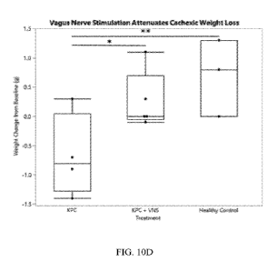
untreated with VNS was evaluated. FIGS. 10A-10D are charts and graphs
illustrating how body
weight was affected by cancer injection and vagus nerve stimulation. FIG. 10A
shows change
in body weight for mice inoculated with LLC. FIG. 10B shows change in body
weight in mice
inoculated with KPC. The cancer cell lines produced a marked cachexic
phenotype, including
marked weight loss compared to healthy controls. FIG. 10C and 10D show change
in body
weight for mice treated with vagus nerve stimulation therapy. FIG. 10C shows
results for mice
inoculated with LLC and FIG. 10D shows results for mice inoculated with KPD.
As can be
seen, VNS therapy provided a statistically significant reduction in cachexic
weight loss
compared to untreated cancer mice, without statistically significant variation
from healthy
control animals as evaluated at humane endpoints for the study. *: p<0.05
**p<0.01
***p<0.001.
The effect of cancer xenograft on total fat and brown adipose tissue was
evaluated for
cancer mice that both received and did not receive VNS. FIGS. 11A and 11B are
charts showing
the effect of VNS or the absence of VNS on total fat and brown adipose tissue,
respectively.
FIG. 11A illustrates that VNS, as well as vagal purturbation through right
cervical vagotomy,
provide attentuation of fat loss. FIG. 11B illustrates that brown adipose
tissue (which is a type
of fat that is critical for maintaining homeostasis and normal metabolic
function) atrophy was
pronounced in cachexic animals, but there was no significant change in BAT
observed between
19
CA 03188591 2022-12-29
WO 2022/011222
PCT/US2021/041029
healthy controls and cancer animals that received VNS or vagotomy therapies.
The
quantification were made at humane endpoints for the study (approximately 2
weeks post-
innoculation). *: p<0.05 **p<0.01 ***p<0.001.
Another clinical feature that impacts quality of life for cachexic patients is
muscle
wasting and loss of skeletal muscle mass. A common quantitiative approach to
clinical
assessment of muscle loss is quantification of mean muscle fiber diameter from
patient
biopsies, as the force generation a muscle is capable of is directly
proportional to the size of
the muscle fibers. Thus, muscle biopsys were used to quantify mean muscle
fiber diameter
from mice in the study.
At study termination, muscle was collected from the left thigh and assayed,
using a
wheat germ agglutinin stain to visualize cross-sectional area of skeletal
muscle fibers (left).
FIG. 12A includes photographs of skeletal muscle fiber for Control (top) and
Cancer (bottom)
mice. FIG. 12B is a chart comparing muscle atrophy for mice having cancer,
cancer with VNS
therapy, cancer with vagotomy, and healthy control. As shown in FIG. 12B, VNS
therapy
significantly attenuated the atrophy of muscle fibers compared to cachexic
mice receiving no
treatment, as evidenced by less reduction in muscle fiber size compared to
controls. *: p<0.05
**p<0.01 ***p<0.001.
Another hallmark of cachexia, which has a pronounced impact on patient quality
of life,
is anorexia. While dietary intake alone is insufficient to explain cachexia
(high fat, high protein,
and high calorie diets have not proved effective tools clinically for cachexia
treatment, nor has
tube feeding), patients routinely report reduced appetite as part of the
complex metabolic
syndrome and systemic symptoms. FIG. 13 is a chart showing the effect of vagal
nerve
stimulation on daily food intake for control mice, mice with cancer but no
treatment, and cancer
with VNS treatment. As shown, cachexic mice have significantly reduced food
intake despite
having free access to unlimited food, which is reversed in the cohort
receiving vagal
perturbation therapy. *: p<0.05 **p<0.01 ***p<0.001. As shown, there is no
statistical
difference between the control group and the cancer group having vagus nerve
stimulation
therapy.
One skilled in the art will readily appreciate that the present disclosure is
adapted to
carry out the objects and obtain the ends and advantages mentioned, as well as
those inherent
therein. The present disclosure described herein is representative of
exemplary embodiments
CA 03188591 2022-12-29
WO 2022/011222
PCT/US2021/041029
and are not intended as limitations on the scope of the present disclosure.
Changes therein and
other uses will occur to those skilled in the art which are encompassed within
the spirit of the
present disclosure as defined by the scope of the claims.
No admission is made that any reference, including any non-patent or patent
document
.. cited in this specification, constitutes prior art. It will be understood
that, unless otherwise
stated, reference to any document herein does not constitute an admission that
any of these
documents forms part of the common general knowledge in the art in the United
States or in
any other country. Any discussion of the references states what their authors
assert, and the
applicant reserves the right to challenge the accuracy and pertinence of any
of the documents
cited herein. All references cited herein are fully incorporated by reference,
unless explicitly
indicated otherwise. The present disclosure shall control in the event there
are any disparities
between any definitions and/or description found in the cited references.
21