Note: Descriptions are shown in the official language in which they were submitted.
CA 03189957 2023-01-23
WO 2022/018289
PCT/EP2021/070755
NEUROMODULATION FOR THE TREATMENT OF CIRCULATORY SYSTEM DISEASES
FIELD OF THE INVENTION
The present invention relates to a device and a method of non-invasive
neuromodulation by
application of a specific programme of electrical stimulation signals to
cutaneous sensory projections
of cranial nerves to modulate and improve blood flow in the brain (referred to
herein as cerebral blood
flow), subsequently leading to a reduction in the arterial blood pressure and
left ventricular
hypertrophy in human subjects with arterial hypertension or improvement of
cardiac function in human
subjects with heart failure. The present invention can be applied to the user
for any of the following
purposes: reducing systemic arterial blood pressure, reducing left ventricular
hypertrophy, reducing
pulmonary arterial blood pressure, treating heart failure and/or treating
atrial fibrillation.
BACKGROUND OF THE INVENTION
Systemic arterial hypertension, commonly referred to as "hypertension",
"essential hypertension" or
"high blood pressure", is a medical condition in which the systemic arterial
blood pressure is
chronically elevated.
Left ventricular hypertrophy (LVH) is a condition in which there is an
increase in left ventricular
myocardial mass, either due to an increase in wall thickness or due to left
ventricular cavity
enlargement, or both. Hypertension is the most common cause of LVH.
Pulmonary arterial hypertension is a medical condition in which the blood
pressure in the pulmonary
artery is chronically elevated.
Heart failure, also known as "congestive heart failure", "congestive cardiac
failure" or "chronic heart
failure", is a medical condition when the heart is unable to pump sufficiently
to maintain adequate
blood flow in the organs and tissues to meet the body's needs.
Atrial Fibrillation (referred herein to as AF) is a medical condition in which
the patient experiences an
abnormal heart rhythm (arrhythmia) characterized by rapid and irregular
beating of the atrial chambers
of the heart.
Hypertension is the leading health risk factor globally. High blood pressure
is associated with adverse
cardiovascular outcomes with elevated risk of myocardial infarction, heart
failure, arterial aneurysms,
kidney failure and stroke. Managing high blood pressure is critical: every 10
mmHg reduction in blood
1
CA 03189957 2023-01-23
WO 2022/018289
PCT/EP2021/070755
pressure results in a 17% reduction in coronary heart disease, 27% reduction
in the incidents of
stroke, 28% reduction in heart failure and 13% reduction in all-cause
mortality. American Heart
Association guidelines define hypertension as systolic blood pressure of 130
mmHg or greater or
diastolic blood pressure of 80 mmHg or greater. Adoption of these guidelines
labels 70.1 million
people in the US and 15 million people in the UK in the 45-75 year age group
as having hypertension,
representing >60% of the population in this age group (J Am Coll Cardiol
71:e127, 2018). In Europe,
it is estimated that only one third of hypertensive patients are diagnosed and
treated to achieve the
recommended levels of arterial blood pressure (Circulation 2016;134:441-450).
When hypertension
is secondary to another medical condition, it is generally prudent to treat
that primary condition first.
A number of pharmacological therapies are available to treat high blood
pressure. However,
pharmacological treatments are often not effective; -15% of all hypertensive
patients are drug-
resistant (Hypertension. 2018;72:e53-e90), most require taking two or more
drugs, their efficacy is
low in -50% of all patients, many patients need two or more drugs to control
their blood pressure with
>90% of these patients failing to control it within the recommended range with
poor compliance.
Underlying reasons include limited access to treatments, inadequate dosing or
combination of
treatments, poor patient adherence to treatment, the use of other interfering
drugs, or the presence
of treatment-resistant hypertension. A large number of patients with
hypertension are also reluctant
to adhere to pharmacological treatment regimens, because some medicines
interfere with their daily
lives, produce side effects, the patients prefer alternative medications, or
for other reasons (BMJ
2012;345:e3953.).
Left ventricular hypertrophy (LVH) is present in 15% to 20% of the general
population. It is more often
prevalent in the elderly, the obese, and in patients with hypertension. Two-
third of the patients with
LVH are hypertensive. A review of echocardiographic data of 37,700 individuals
revealed 19-48%
prevalence of LVH in untreated hypertensive patients and 58-77% - in high-risk
hypertensive patients
(J Hum Hypertens 26: 343-349, 2012). LVH is a compensatory but ultimately, an
abnormal increase
in the mass of the myocardium of the left ventricle induced by a chronically
elevated workload of the
heart muscle. LVH diagnosis is based on the assessment of left ventricular
mass. An echocardiogram
is the test of choice in diagnosis of LVH.
Cardiac ultrasound utilizes transthoracic or transesophageal positioning of
the transducer to measure
.. the left ventricular end-diastolic diameter, posterior wall thickness, and
interventricular septum
thickness. From these measurements and the patient's height and weight, the
left ventricular mass
index can be calculated. LVH treatment should be aggressive because patients
with LVH are at the
highest risk of cardiovascular morbidity and mortality. The goal of therapy is
to reduce LVH and
prevent left ventricular dysfunction and progression to heart failure. The
antihypertensive therapy
benefits the patient by reducing arterial blood pressure and may reduce the
degree of LVH,
2
CA 03189957 2023-01-23
WO 2022/018289
PCT/EP2021/070755
independently of blood pressure reduction, leading to a reduction of adverse
cardiovascular events
and mortality (Eur Heart J 39: 3021-3104, 2018).
Pulmonary hypertension encompasses a group of severe clinical entities, such
as pulmonary arterial
hypertension (PAH) in which the progressive loss and/or obstructive
remodelling of the pulmonary
vascular bed is responsible for the rise in pulmonary arterial pressure and
pulmonary vascular
resistance, resulting in a progressive right heart failure and right heart
functional decline. Pulmonary
hypertension is classified into five groups, depending on the cause. Group 1:
Idiopathic PAH with
unknown causes; Group 2: Pulmonary hypertension caused by left-sided heart
disease; Group 3:
Pulmonary hypertension caused by lung disease; Group 4: Pulmonary hypertension
caused by
chronic blood clots; and Group 5: Pulmonary hypertension associated with other
conditions. Because
current PAH treatments do not specifically target pulmonary vascular
remodelling and inflammation,
there is an urgent unmet clinical need to better identify the pathological
mechanisms underlying the
progressive narrowing of the pulmonary arterial lumen and perivascular
inflammation and the loss of
vessels in order to support therapeutic innovation aimed at reversing these
changes and regenerating
normal pulmonary vessels (Eur Respir J 53: 1801887, 2019)
Chronic heart failure (CHF) is one of the most common causes of morbidity and
mortality in the
developed world. Heart failure is associated with a diverse range of
complications, including lethal
arrhythmias and death as a result of the disease progression. In addition, CHF
can be the terminal
condition of many diseases of the circulatory system, including hypertension,
myocardial infarction
(MI), valvular heart disease, and various cardiomyopathies. CHF diagnosis is
based on the
assessment of cardiac left ventricular ejection fraction (LVEF). Heart failure
with normal LVEF 50%)
is defined as CHF with preserved ejection fraction (HFpEF), and CHF with low
LVEF (<40%) as HF
with reduced ejection fraction (HFrEF). The goals of heart failure therapy are
to improve patients'
clinical status, functional capacity and quality of life, reduce
hospitalization rates and reduce mortality.
Established pharmacological treatment of HFrEF involving several drug classes
(including beta-
adrenoceptor antagonists (or 8-blockers), diuretics, and inhibitors of renin-
angiotensin-aldosterone
system) improves symptoms and reduces mortality rates. Yet, drug therapy
remains insufficient, as
cardiac function continues to deteriorate over time and most patients have
poor prognosis. Moreover,
currently there is no treatment for HFpEF, representing an urgent unmet
clinical need.
Atrial fibrillation (AF) is an abnormal heart rhythm (arrhythmia)
characterized by rapid and irregular
beating of the atrial chambers of the heart (referred to as episodes). It
often begins as short periods
of abnormal beating which become longer or continuous as diseases progresses.
It may also start as
other forms of arrhythmia such as atrial flutter that then progresses into AF.
Often episodes of AF
have no symptoms. Occasionally there may be heart palpitations, fainting,
light-headedness,
3
CA 03189957 2023-01-23
WO 2022/018289
PCT/EP2021/070755
shortness of breath, or chest pain. The disease is associated with an
increased risk of heart failure,
dementia, and stroke. The repeated occurrence of episodes is sometimes
referred to as the AF
burden which may be defined as the duration of the longest AF episode, number
of AF episodes,
and/or the percentage of time the patient is in AF during a certain period of
time. There are four main
types of AF: paroxysmal, persistent, long-term persistent, and permanent AF.
The type of AF depends
on how often AF occurs and how the patient responds to treatment. A brief
event of AF is known as
paroxysm AF, this type of AF usually stops in less than 24 hours but may also
last for up to a week.
Paroxysmal AF can happen repeatedly. Persistent AF is a condition in which the
abnormal heart
rhythm lasts for more than a week. It may stop spontaneously, but in most
cases will need treatment.
With this condition, the abnormal heart rhythms last for more than a year
without going away.
Sometimes AF burden does not improve, even when patients have tried several
times to restore the
normal heart rhythm with medicines or other treatments.
Despite significant advances in medical research, the biological/physiological
mechanisms underlying
the development of arterial hypertension are not fully understood. One
hypothesis suggests that high
blood pressure develops as a compensatory condition when blood supply to the
brain is reduced, for
example as a result of increased resistance of cerebral vasculature. Blood
flow in the brain (cerebral
blood flow) is driven by the arterial blood pressure and inversely
proportional to cerebrovascular
resistance. Any increase in the resistance to blood flow, for example due to
(cerebro)vascular disease,
atherosclerotic legions, ageing, etc., would require compensatory increases in
the arterial pressure to
maintain brain perfusion, essential to support the function of brain nerve
cells processing information.
Therefore, it is possible to treat circulatory system disease (in general) and
reduce systemic arterial
blood pressure in patients with hypertension (in particular) by applying
methods or treatments
designed to improve blood flow in the brain.
Blood flow to the brain comes from two sources: Internal carotid arteries
(supply the anterior brain)
and vertebral arteries (supplying the brainstem and posterior brain) that
maintain cerebral circulation
(i.e. blood flow in the brain), which is the movement of blood through the
dense network of cerebral
arteries, capillaries and veins. The rate of the cerebral blood flow in the
healthy adult human being is
an average approximately 750 millilitres per minute but may be reduced in
several disease states
leading to compensatory responses as described above.
U52019351230 discloses an electrostimulation device includes a computer
generating an
electrostimulation generator control signal and outputting a music signal, a
transcutaneous
electrostimulation generator, an electronic signal conduit, and an electrode
coupler. U52019351230
makes reference to a first electrode on a first face of the tragus and a
second electrode on the second
face of the tragus, however, these are used to provide a connection with a
third electrode contacting
4
CA 03189957 2023-01-23
WO 2022/018289
PCT/EP2021/070755
the skin of the auditory canal. Disclosed current pathways in US2019351230
include pathways
between either the first or the second electrode and the third electrode.
SUMMARY OF THE INVENTION
The present invention describes a device and a method for non-invasive
electrical stimulation of
cranial nerves that project to the skin of the outer ear to modulate cerebral
blood flow of a user. In
some embodiments the electrical stimulation is for the purpose of modulating
the function of the
cardiovascular system of a user, such as modulating blood pressure or
functional and electrical
properties of the heart. One potential avenue for arterial blood pressure
control is via modulation of
the blood flow in the brain, and in particular in the brainstem ¨ the area of
the brain that controls the
cardiovascular system. According to the present invention, the above purposes
can be achieved non-
invasively by stimulation (i.e. electrical) of cutaneous sensory projections
of certain cranial and spinal
nerves that originate from the brainstem and spinal cord. Activation of these
nerves is expected to
facilitate the blood flow through the lower brainstem leading to a reduction
in the arterial blood
pressure and associated cardiac work. Regions of the external ear, and the
tragus in particular, are
innervated by the sensory branches of the fifth (V) and the tenth (X) cranial
nerves as well as branches
of the spinal nerves 02 and 03. According to the present invention, global
cerebral blood flow and
blood flow through the brainstem in particular can be facilitated by
transcutaneous or percutaneous
electrical stimulation of the sensory nerves projecting to of the outer ear
According to further aspect
of the invention, electrical stimulation of these nerves can achieve a
therapeutic effect in the treatment
of circulatory system disease.
The present disclosure is related to the field of medical treatment of
cardiovascular disease, including
hypertension, or high arterial blood pressure, pulmonary arterial
hypertension, heart failure and atrial
fibrillation.
In a first aspect of the invention there is provided a device for modulating
cerebral blood flow of a
user. The device comprises a generator configured to produce an electrical
stimulation signal and a
controller, connected to the generator and configured to determine the form of
the electrical
stimulation signal. The device also includes an earpiece, connected to the
generator and controller
and the earpiece has an electrode (or a pair of electrodes). The controller
transmits the electrical
stimulation signal to the electrode(s) and the electrode(s)are configured to
be placed in contact with
and provide the electrical stimulation signal to the skin of a tragus of the
user (such as across the
tragus). The electrical stimulation signal comprises a series of electrical
pulses, each pulse repeats
with a frequency of 1 Hz to 100 Hz and each pulse has duration of 10
microseconds to 500
5
CA 03189957 2023-01-23
WO 2022/018289
PCT/EP2021/070755
microseconds and an amplitude of 0.1 mA to 8 mA. The device is used by the
user at least once a
day for at least 3 consecutive days.
The device may be applied to the user to modulate cerebral blood flow and/or
for any of the following
.. purposes: reducing systemic arterial blood pressure, reducing left
ventricular hypertrophy, reducing
pulmonary arterial blood pressure, treating cardiovascular conditions such as
heart failure and atrial
fibrillation.
According to the embodiment, the device can be applied to the user such that
the electrical stimulation
signal is applied transcutaneously or percutaneously.
In some embodiments the earpiece is configured to apply the electrical
stimulation signal
transcutaneously. In such embodiment's electrodes comprise non-piercing
conductive surfaces. In
some embodiments the earpiece is configured to apply the electrical
stimulation signal
percutaneously. In such embodiments the electrodes comprise conductive
surfaces configured to
pierce the surface of the skin to make electrical contact with a subsurface
layer of tissue.
The device can be applied to the user for a minimum of 5 minutes and a maximum
of 2 hours per day.
The device may have a first and second electrode, the first electrode is
configured to be placed in
contact with the left tragus of the user and the second electrode is
configured to be placed in contact
with the right tragus of the user. Additionally, the device may have a first
earpiece and a second
earpiece, and the first earpiece has the first electrode and the second
earpiece has the second
electrode.
In some embodiments the first electrode is a first stimulating electrode and
the second electrode is a
second stimulating electrode. Each earpiece comprises a stimulating electrode
and a reference
electrode wherein the electrical stimulation signal is applied between the
stimulating electrode and
the reference electrode.
The device may comprise a first pair of electrodes and a second pair of
electrodes, wherein the first
pair of electrodes is configured to be placed in contact with the left tragus
of the user and the second
pair of electrodes is configured to be placed in contact with the right tragus
of the user. The device
may comprise a first earpiece and a second earpiece, wherein the first
earpiece is configured to be
placed in contact with the left tragus and comprises the first pair of
electrodes and the second earpiece
is configured to be placed in contact with the right tragus and comprises the
second pair of electrodes.
6
CA 03189957 2023-01-23
WO 2022/018289
PCT/EP2021/070755
In each pair of electrodes, one electrode may be a stimulating electrode and
one electrode may be a
reference electrode.
In some embodiments the first and the second earpiece are substantially
identical. In some
embodiments the first earpiece is shaped to fit on the left tragus and the
second earpiece is shaped
to fit on the right tragus. In some embodiments the left and the right
earpieces are substantially mirror
images of each other. In some embodiments an earpiece is shaped to conform to
a portion of the
tragus or another part of the ear, such that the earpiece fits preferentially
to the tragus such that the
stimulating electrode is in contact with the outer side of the tragus and the
reference electrode is in
contact with the inner side of the tragus. In some embodiments an earpiece is
shaped to conform to
a portion of the tragus or another part of the ear, such that the earpiece
fits preferentially to the tragus
such that the stimulating electrode is in contact with the inner side of the
tragus and the reference
electrode is in contact with the outer side of the tragus.
The device may further include a securing means configured to secure the
electrode to a tragus of a
user. The securing means may include a clip and the clip may include a first
gripping potion and a
second gripping portion which are biased into contact with each other. The
stimulating electrode may
be located on the first gripping portion, when the gripping portion is
present.
There may also be a reference electrode located on the first or second
gripping portion.
The device may include a physiological sensor configured to measure the value
of a physiological
parameter (e.g. heart rate, blood pressure) and optionally may store the value
in a memory portion of
the device. The device may also include a temperature sensor configured to
measure the temperature
of an area of the skin of the user. The temperature sensor may be configured
to measure the
temperature of the tragus. The device may store data from the temperature
sensor in a memory
portion of the device. The values stored in the memory portion is used by the
controller can be used
to determine the form of the electrical stimulation signal. In some
embodiments the physiological
sensor and/or the temperature sensor may be provided on an earpiece, for
example located on a clip
forming part of the earpiece.
In some embodiments, the physiological sensor measurements, temperature sensor
measurements
and time and date information on the use of the device by the patient are
recorded and stored in the
memory portion of the device.
The device may comprise means to measure one or more of: the voltage applied
between the
stimulating electrode and the reference electrode; the current flowing between
the stimulating
electrode and the reference electrode; and the time relation between the
voltage and the current.
7
CA 03189957 2023-01-23
WO 2022/018289
PCT/EP2021/070755
The device may comprise a stimulating electrode, a counter electrode and/or a
reference electrode,
and may be configured to measure one or both of: the voltage between the
stimulating electrode and
the reference electrode, and the voltage between the counter electrode and a
reference electrode. In
this way, the potential of the stimulating, counter and/or the reference
electrodes may be measured
without error arising from voltage drop across the electrode to skin
interface, as known in the art.
In some embodiments the device is configured to control the current flowing
between the stimulating
electrode and the reference electrode as a function of time, according to the
form of the electrical
stimulation signal, by means of a feedback control means using one or more
measured values. In
some embodiments the device is configured to control the voltage applied
between the stimulating
electrode and the reference electrode as a function of time, according to the
form of the electrical
stimulation signal, by means of a feedback control means using one or more
measured values.
In some embodiments the device is configured to derive the phase relationship
between the voltage
between the stimulating electrode and the reference electrode and the current
flowing through the
stimulating electrode and the reference electrode, for example to measure the
electrical impedance
of the tragus.
Measurement of the electrical impedance between the stimulating electrode and
the reference
electrode may be used to determine one or more of: that the electrodes are
correctly positioned on
the opposing faces of the tragus; that the device and method of the invention
is in use, and the times
and for the duration that the device and method are used; and to control the
voltage and/or current in
response to the impedance, for example to compensate for variation in the
structure or conductivity
of the tragus.
Additionally, measurements of current, voltage and phase relationship of the
electrical stimulation
signal can be stored in a memory portion of the device and used to determine
the electrical impedance
of the tragus.
The controller can be configured to produce the electrical stimulation signal
and the pattern of
stimulation based on a user input received at the controller. This user input
includes at least one of
the following: pulse duration, waveform, pulse frequency, pulse pattern and
current or voltage
amplitude of the electrical stimulation signal.
Preferred features of the second and subsequent aspects of the invention are
as for the first aspect
mutatis mutandis.
In a second aspect of the invention there is provided a system for non-
invasive electrical stimulation
of nerves that project to the skin of the outer ear and, in some embodiments,
for the purpose of
modulating cerebral blood flow of a user. The system includes a device as
disclosed herein and further
8
CA 03189957 2023-01-23
WO 2022/018289
PCT/EP2021/070755
comprises a communication module connected to the controller of the device.
The communication
module can be configured to send information from the device to an external
computer system and to
receive information from the external computer system, and the information
received from the external
computer system can be used by the controller to determine the form of the
electrical stimulation
signal.
The system is further configured to act upon information from the device
received by the external
computer system, wherein the information is compared to a secondary set of
information stored on
the external computer system to determine a set of actions to be performed by
the device and/or the
external computer.
In a third aspect of the invention there is provided a method of modulating
cerebral blood flow of a
user of the device. The method comprises producing, using a generator, an
electrical stimulation
signal; determining, using a controller connected to the generator, the form
of the electrical stimulation
signal; transmitting, the electrical stimulation signal to an electrode, for
example to an electrode pair
or pairs. The stimulating electrode is configured to be placed in contact with
and provide the electrical
stimulation signal to the skin of a tragus of the user. The electrical
stimulation signal comprises a
series of electrical pulses, each pulse repeats with a frequency of 1 Hz to
100 Hz and each pulse has
a duration of 10 microseconds to 500 microseconds and an amplitude of 0.1 mA
to 8 mA. The device
is applied to the user at least once a day for at least 3 consecutive days.
In another aspect of the invention there is provided a method for non-invasive
electrical stimulation of
nerves that project to the skin of the outer ear using a device as disclosed
herein, comprising the
steps of: bringing the stimulating electrode and the reference electrode into
contact with the tragus of
a user; producing, using the device, an electrical stimulation signal applied
to the stimulating electrode
and the reference electrode; and determining, using the controller, the
waveform and the frequency
of the electrical stimulation signal, wherein the electrical stimulation
signal comprises a series of
electrical pulses, each pulse repeating with a frequency of about 1 Hz to
about 100 Hz and each pulse
has a duration of about 10 microseconds to about 500 microseconds and an
amplitude of about 0.1
mA to about 20 mA.
In some embodiments, the electrical stimulation signal comprises a series of
electrical pulses, each
pulse repeating with a frequency in the range about 3 Hz to about 50 Hz and
each pulse having a
duration of about 100 microseconds to about 500 microseconds and an amplitude
of about 0.1 mA to
about 8 mA.
9
CA 03189957 2023-01-23
WO 2022/018289
PCT/EP2021/070755
In some embodiments the frequency is in the range from about 1 Hz to about 100
Hz, such as about
1 Hz to 10 Hz, 10 Hz to 20 Hz, 20 Hz to 30 Hz, 30 Hz to 40 Hz, 40 Hz to 50 Hz,
50 Hz to 60 Hz, 60Hz
to 70 Hz, 70 Hz to 80 Hz, 80 Hz to 90 Hz, or 90 Hz to about 100 Hz.
In some embodiments the frequency is in the range 3 Hz to 20 Hz, 5 Hz to 30
Hz, 10 Hz to 50 Hz, 15
Hz to 60 Hz, 20 Hz to 75 Hz, 25 Hz to 80 Hz, 30 Hz to 100 Hz.
In some embodiments the frequency is in the range 3 Hz to 50 Hz.
In some embodiments the pulse has a duration in the range about 10
microseconds to about 500
microseconds, such as about 10 microseconds to 100 microseconds, 20
microseconds to 200
microseconds, 30 microseconds to 300 microseconds, 40 microseconds to 400
microseconds, 50
microseconds to about 500 microseconds.
In some embodiments the pulse has a duration in the range 100 microseconds to
200 microseconds,
200 microseconds to 300 microseconds, 300 microseconds to 400 microseconds,
400 microseconds
to 500 microseconds.
In some embodiments the pulse has a duration in the range 50 microseconds to
200 microseconds,
100 microseconds to 250 microseconds, 200 microseconds to 500 microseconds.
In some embodiments the pulse has a duration in the range 100 microseconds to
500 microseconds.
In some embodiments the amplitude is in the range about 0.1 mA to about 10 mA,
such as about 0.1
mA to about 2 mA, about 0.2 mA to about 5 mA or about 0.5 mA to about 10 mA.
In some embodiments the amplitude is in the range 0.1 mA to 1 mA, 0.2 mA to 2
mA, 0.3 mA to 3 mA,
0.4 mA to 4 mA, 0.5 mA to 5 mA, 0.6 mA to 6 mA, 0.7 mA to 7 mA, 0.8 mA to 8
mA, 0.9 mA to 9 mA
or 1.0 mA to 10 mA.
In some embodiments the amplitude is in the range 0.1 mA to 5 mA, 0.5 mA to 8
mA, 1 mA to 10 mA,
0r2 mA to 20 mA.
In some embodiments the amplitude is in the range about 0.5 mA to about 5 mA.
In some embodiments the method is applied to the user to modulate cerebral
blood flow, and/or for
any of the following purposes: reducing systemic arterial blood pressure,
reducing left ventricular
hypertrophy, reducing pulmonary arterial blood pressure, treating
cardiovascular conditions such as
heart failure and atrial fibrillation
CA 03189957 2023-01-23
WO 2022/018289
PCT/EP2021/070755
In a fourth aspect of the invention there is provided a method for the
treatment of a disease or condition
selected from the group consisting of hypertension (high blood pressure), left
ventricular hypertrophy,
heart failure, atrial fibrillation, comprising administering to a subject an
electrical stimulation signal
using the device and/or methods described herein. Additionally, the method may
be applied for the
treatment of a disease or condition of one or more of the group consisting of
hypertension (high blood
pressure), left ventricular hypertrophy, heart failure, atrial fibrillation at
the same time. In some
embodiments the method is applied to modulate, such as to increase, cerebral
blood flow. The
electrical stimulation signal comprises a series of electrical pulses, each
pulse repeats with a
frequency of 1 Hz to 100 Hz and each pulse has a duration of 10 microseconds
to 500 microseconds
and an amplitude of 0.1 mA to 8 mA. The device is applied to the user at least
once a day for at least
3 consecutive day.
The stimulating and reference electrodes may be configured to be placed in
contact with an outward
facing surface and an inward facing surface of the tragus. In some
embodiments, the electrical
stimulation signal may be transmitted to at least a first pair of electrodes
and a second pair of
electrodes, wherein the first pair of electrodes is placed in contact with the
skin of the left tragus of
the user and the second pair of electrodes is placed in contact with the skin
of right tragus of the user.
The electrical stimulation signal applied to each of the left and right tragi
may be substantially the
same electrical stimulation signal and the signal may be applied
simultaneously or sequentially to
each of the left and right tragi. Alternatively, the electrical stimulation
signal applied to the left tragus
may be different from the electrical stimulation signal applied to the right
tragus and the electrical
stimulation signal applied to the left and right tragi may be applied
simultaneously or sequentially to
each of the left and right tragi. Alternatively, the electrical stimulation
signal applied to the left tragus
is different from the electrical stimulation signal applied to the right
tragus and the electrical stimulation
signal applied to the left tragus may be applied at a different time to the
electrical stimulation signal
applied to the right tragus.
When using the device and applying the method of treatment, the electrical
stimulation signal may be
of a sinusoidal, square, triangular, pulse or "white noise" waveform. The
electrical stimulation signal
may be a pulse waveform, the pulse being substantially a sinusoidal, square,
triangular, or "white
noise" waveform. The generated waveform can be monophasic symmetrical
waveform, or a biphasic
symmetrical waveform, or a triphasic symmetrical waveform. Alternatively, the
generated waveform
can be a biphasic asymmetrical waveform, or a triphasic asymmetrical waveform.
The method can be applied to the user for a minimum of 5 minutes and a maximum
of 2 hours per
day.
The device can be applied to the user for a minimum of 5 minutes and a maximum
of 2 hours per day.
11
CA 03189957 2023-01-23
WO 2022/018289
PCT/EP2021/070755
The method or device can be applied to the user for a minimum of 5 minutes and
a maximum of 2
hours per day and the method is applied at intervals separated by at least one
day.
The method or device can be applied to the user for a minimum of about 5
minutes and a maximum
of about 2 hours per day and the method may be applied at intervals in the
range about 1 day (i.e. 24
hr) to about 7 days, such as about 1 day to about 2 days.
In methods of treatment according to the invention, the method or device can
be applied to the user
for different periods. In an embodiment of a process of treatment according to
the invention, during a
first period, the method is applied to the user for between 5 minutes and 2
hours each day, the first
period comprising a minimum of 3 consecutive days; during a second period the
method is stopped
for at least 2 days and during a third period the method is applied to the
user for between 5 minutes
and 2 hours each day.
In a fifth aspect the invention provides a method of treatment of a medical
condition of a user
comprising applying the device and method of the invention to achieve non-
invasive electrical
stimulation of nerves that project to the skin of the outer ear, in
combination with providing a
medication to the user. In some embodiments use of the device and/or method of
the invention
modulates the pharmacological effect of the medication.
The method of treatment according to the present invention may therefore
further comprise a step of
administering to the patient (i.e. the user of the device as described herein)
a pharmaceutically active
composition for the treatment of a disease or condition selected from the
group consisting of
hypertension, left ventricular hypertrophy, heart failure and/or atrial
fibrillation, e.g. an agent to treat
hypertension, heart failure and/or atrial fibrillation. Additionally, the
method may be applied for the
treatment of a disease or condition of one or more of the group consisting of
hypertension (high blood
pressure), left ventricular hypertrophy, heart failure, atrial fibrillation at
the same time. Such
pharmaceutically active compositions may be administered separately,
sequentially or simultaneously
with the use of the device as described herein.
Suitable anti-hypertensive compositions may comprise diuretics, beta-
adrenoceptor antagonists (13-
blockers), angiotensin converting enzyme inhibitors, angiotensin II receptor
blockers, calcium channel
blockers, alpha-adrenoceptor antagonists (alpha-blockers), alpha-2 receptor
agonists, and/or
combined alpha- and 13-blockers. For example, suitable pharmaceutically active
substances for use
as anti-hypertensive agents include, but are not limited to, alfuzosin
hydrochloride, ambrisentan,
atenolol, bisoprolol, bosentan, clonidine hydrochloride, doxazosin,
epoprostenol, furosemide,
hydralazine hydrochloride; iloprost, indoramin, macitentan, methyldopa,
metoprolol, minoxidil,
12
CA 03189957 2023-01-23
WO 2022/018289
PCT/EP2021/070755
moxonidine, prazosin, propranolol, riociguat, sildenafil, sodium
nitroprusside, tadalafil, tamsulosin
hydrochloride, terazosin.
Suitable compositions for the treatment of heart failure may comprise
diuretics, beta-adrenoceptor
antagonists (13-blockers), angiotensin converting enzyme inhibitors,
angiotensin II receptor blockers,
calcium channel blockers. Other medicines suitable for use in the treatment of
heart failure include
but are not limited to heart rate lowering agents (for example ivabradine),
blood thinners (for example
antiplatelet drugs or anticoagulant drugs). Suitable antiplatelet drugs
include, but are not limited to,
anagrelide, aspirin, clopidogrel, prasugrel, ticagrelor, tirofiban, vorapaxar,
dipyridamole. Suitable
anticoagulant drugs include, but are not limited to, dabigatran, edoxaban,
rivaroxaban, apixaban,
warfarin, enoxaparin, dalteparin, fondaparinux.
Suitable antiarrhythmic agents for the treatment of atrial fibrillation
include, but are not limited to, beta-
adrenoceptor antagonists (13-blockers) (for example acebutolol, atenolol,
betaxolol, labetalol,
bisoprolol, carvedilol, metoprolol tartrate, metoprolol succinate, nebivolol,
penbutolol, propranolol,
sotalol hydrochloride, timolol, nadolol, pindolol), calcium channel blockers
(for example verapamil
hydrochloride, diltiazem hydrochloride), digitalis glycosides (for example
digoxin), sodium channel
blockers (for example disopyramide, mexiletine, quinidine, procainamide,
propafenone, flecainide),
potassium channel blockers (for example amiodarone, dronedarone, sotalol).
The method may further comprise measuring the blood pressure of a user;
determining whether the
blood pressure of the user is greater than a predetermined threshold value;
and if the blood pressure
of the user is greater than the predetermined threshold value, instructing the
device to produce the
electrical stimulation signal.
The agent may be provided to the user at a dose selected to produce a
therapeutic effect in
combination with use of the method of the invention. In some embodiments the
dose is selected to
be within the range known in the art to achieve therapeutic effect in the
condition. In some
embodiment the dose is selected to be below the range conventionally used in
the art to achieve
therapeutic effect in the condition. In the latter embodiments of the method
of treatment, use of the
device and method of the invention combines with the pharmacological mode of
action of the agent,
such that the potency of the agent in the condition may be enhanced. In this
way, a therapeutic effect
results from the combination of the method of the invention and the agent,
while any side effects
arising from the agent are reduced as a result of the use of a lower dose.
The determining, using a controller connected to the generator, the form of
the electrical stimulation
signal pulse comprises determining the pulse duration, pulse frequency,
waveform and waveform
pattern of the electrical stimulation signal.
13
CA 03189957 2023-01-23
WO 2022/018289
PCT/EP2021/070755
Furthermore, a method of identifying a patient suitable for treatment by a
device and method of
treatment of the present invention is disclosed. The method includes recording
an electrocardiogram
of a patient for a minimum period of 1 minute; analysing the power spectrum of
heart rate variability;
determining the low frequency (LF) to high frequency ratio (HF) of heart rate
variability spectrum
(LF/HF); and determining whether the LF/HF ratio of heart rate variability
spectrum is greater than a
predetermined threshold value. In some embodiments, the predetermined
threshold value of LF/HF
ratio heart rate variability spectrum is 1 or about 1.
Another method of identifying a patient suitable for treatment by a device and
method of treatment of
the present invention is also disclosed. The method includes recording the
baseline value of the heart
rate of a patient when resting in a supine position, followed by recording the
heart rate of the patient
when actively standing in a vertical position, wherein the heart rate is
recorded directly after the patient
stands in the vertical position and then subsequently after a period of time;
recording the heart rate of
the patient again while standing; and then determining whether the difference
between the heart rate
value recorded immediately after standing and heart rate value recorded
subsequently is less than a
predetermined value. The predetermined value of heart rate difference between
the value recorded
immediately after standing and heart rate value recorded subsequently while
standing is suitably less
than 6 (six) beats per minute.
Suitably, the patient is supine for 5 to 15 minutes, optionally 5 to 10
minutes, preferably about 10
minutes. The patient suitably actively stands in the vertical position in
about 5 to 10 seconds, suitably
in 5 seconds. The recording of the heart rate of the patient while standing
can be for around 1 to 5
minutes, suitably 1 to 3 minutes, preferably 1 minute. The recording of the
heart rate subsequently
while standing can be made between 10 to 30 seconds after standing, suitably
10 to 20 seconds after
standing.
In one embodiment, the method can comprise recording the supine baseline
values of heart rate for
1 minute, whilst the patient is resting in the supine position for 10 minutes;
the patient standing up,
wherein the patient stands up in a time period of less than 5 seconds;
recording the patient's heart
rate continuously for 1 minute after standing up; calculating the difference
between heart rate values
obtained while standing and baseline supine values of heart rate; determining
whether the difference
between the heart rate value recorded immediately after standing and heart
rate value recorded
between 10 seconds and 20 seconds after standing is less than a predetermined
value. The
predetermined threshold value of heart rate difference between the value
recorded immediately after
standing and heart rate value recorded between 10 seconds and 20 seconds after
standing is less
than 6 (six) beats per minute.
14
CA 03189957 2023-01-23
WO 2022/018289
PCT/EP2021/070755
The methods of screening of the present invention may therefore further
optionally comprise a step
of administering to the patient a pharmaceutically active composition for
treatment of a disease or
condition selected from the group consisting of hypertension, heart failure
and/or atrial fibrillation, e.g.
an agent to treat hypertension, heart failure and/or atrial fibrillation,
where the pharmaceutically active
substance may be as described above.
BRIEF DESCRIPTION OF THE DRAWINGS
Various embodiments of the invention are described by way of examples with
references to the
accompanying drawings in which:
Figure 1A shows sites on the left tragus and the right tragus of the human
outer ear that receive
sensory nerve innervation. Electrical stimulation of these nerves
transcutaneously or percutaneously
modulates cerebral brain blood and can be used to treat diseases of the
circulatory system according
to the present invention.
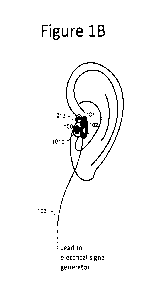
Figure 1B shows an embodiment of an earpiece comprising an electrode clip.
Figure 10 illustrates an electrode clip
Figure 1D illustrates positioning of an earpiece as shown in Figure 1B on the
tragus.
Figure lE shows a cross section through the tragus with the stimulating and
reference electrodes in
contact with the outer and inner surfaces of the tragus respectively, showing
current flow pathways
through the tragus.
Figure 2 illustrates changes in cerebral blood flow induced by non-invasive
neuromodulation, applied
in an animal model using different parameters of electrical stimulation to the
outer ear according to
the present invention.
Figure 3A and 3B show the blood pressure values in drug-resistant hypertensive
patients before and
after the use of non-invasive neuromodulation using a device and a method of
treatment according to
the invention.
Figure 4A and 4B show the blood pressure values in uncontrolled hypertensive
patients before and
after the use of non-invasive neuromodulation using a device and a method of
treatment according to
the invention.
CA 03189957 2023-01-23
WO 2022/018289
PCT/EP2021/070755
Figure 40 shows the values of left ventricular myocardial mass in uncontrolled
hypertensive patients
before and 12 months after the use of non-invasive neuromodulation using a
device and a method of
treatment according to the invention.
Figure 5A and 5B show the blood pressure values in uncontrolled hypertensive
patients before and
after the use of non-invasive neuromodulation using a device and a method of
treatment according to
the invention, applied in combination with the beta-adrenoceptor antagonist
bisoprolol.
Figure 6 shows a description and circuit block drawing for a device according
to the invention.
Figures 7A and 7B show possible waveforms of the electrical stimulation
signal.
DETAILED DESCRIPTION
The terms "subject", "individual" and "patient" as used herein refer to
humans, which do not denote a
particular age or sex. In certain embodiments the individual subject may be a
patient, a subject that
is a candidate for, or awaiting medical or other treatment, such as the method
of device-based
neuromodulation described herein. The term "about" as used herein means in
quantitative terms plus
or minus 10%. For example, "about 5 mmHg" would encompass the range 4.5 - 5.5
mmHg.
Hypertension
The disclosed device and method can be used to treat hypertension and lower
systemic arterial blood
pressure in a subject involving identifying a subject diagnosed with
hypertension. The disclosed
device and method can also be used to modulate cerebral blood flow for the
purpose of lowering
systemic arterial blood pressure in a subject involving identifying a subject
diagnosed with
hypertension. The term hypertension as used herein refers to a condition or
disease well known in
the art in which the systemic arterial blood pressure in a human subject is
chronically elevated.
To prevent, diagnose, and treat hypertension, blood pressure is categorized as
normal (less than 120
mmHg systolic and less than 80 mmHg diastolic), elevated (120 to 129 mmHg
systolic and less than
80 mmHg diastolic), stage 1 hypertension (130 to 139 mmHg systolic or 80 to 89
mmHg diastolic), or
stage 2 hypertension (more than 140 mmHg systolic or more than 90 mmHg
diastolic). Patients whose
systolic and diastolic blood pressures are in different categories are
assigned to the higher stage (for
16
CA 03189957 2023-01-23
WO 2022/018289
PCT/EP2021/070755
example a patient with a blood pressure of 128/82 mmHg should be diagnosed
with stage 1
hypertension).
Hypertension may refer to a condition in which a subject's resting systolic
arterial blood pressure is
above 120 mmHg and/or diastolic arterial blood pressure is above 80 mmHg. In
certain embodiments
hypertension may refer to a condition in which a subject's resting systolic
arterial blood pressure is
above any of the following limits: about 115 mmHg, about 120 mmHg, about 125
mmHg, about 130
mmHg, about 135 mmHg, about 140 mmHg, about 145 mmHg, about 150 mmHg, about
155 mmHg,
about 160 mmHg, about 165 mmHg, about 170 mmHg and/or when the systemic
diastolic arterial
blood pressure is above any of the following limits: about 80 mmHg, about 85
mmHg, about 90 mmHg,
about 95 mmHg, about 100 mmHg, about 105 mmHg, about 110 mmHg, about 115 mmHg,
about
120 mmHg. In some embodiments, systemic arterial hypertension may be chronic
treatment-resistant
hypertension, defined as sustained arterial blood pressure level above the
recommended target (24
h ambulatory systolic blood pressure higher than 130 mmHg) despite documented
treatment with at
least three antihypertensive medications in adequate doses, one of which is a
diuretic. Diagnosis of
hypertension in a subject may in various embodiments be performed by an
individual qualified to
make such diagnosis in a particular jurisdiction.
Left ventricular hypertrophy is diagnosed in patients if the left ventricular
myocardial mass indexed to
body surface area (LVMI) is greater than 95 g/m2 for women and greater than
115 g/m2 for men.
Pulmonary Arterial Hypertension
The disclosed device and method can also be used to treat pulmonary
hypertension. Pulmonary
arterial hypertension may refer to a condition in which a subject's resting
pulmonary systolic arterial
blood pressure is above about 25 mmHg.
Heart Failure
The disclosed device and method can also be used to treat heart failure. The
terms heart failure, or
congestive heart failure, or chronic heart failure as used herein refer to a
condition or disease well
known in the art in which the heart is unable to pump sufficiently to maintain
blood flow in the organs
and tissues to meet the body's needs. In certain embodiments heart failure may
refer to a condition
in which a subject's left ventricular ejection fraction is above about 50%
(HFpEF), or between about
40% and about 49% (heart failure with mid-range ejection fraction), or lower
than about 40% (HFrEF).
17
CA 03189957 2023-01-23
WO 2022/018289
PCT/EP2021/070755
Atrial Fibrillation
The disclosed device and method can also be used to treat atrial fibrillation
or AF. AF refers to a
condition or disease well known in the art in which the normal regular
electrical impulses generated
by the sinoatrial node in the right atrium of the heart are overwhelmed by
disorganized electrical
impulses usually originating in the roots of the pulmonary veins. This leads
to irregular conduction of
electrical impulses that generate the heartbeat.
Method and device for modulating the cerebral blood flow
The present invention employs a device and a specific method of non-invasive
neuromodulation to
modulate cerebral blood flow via electrical stimulation of afferent (sensory)
branches of cranial nerves
innervating the tragus (e.g. the auricular region) and projecting to the
brainstem, for the purpose of
lowering arterial blood pressure as the medical treatment of hypertension and
left ventricular
hypertrophy, or reducing cardiac work to improve heart function as the medical
treatment of heart
failure, or reducing the number and frequency of AF episodes. Additionally,
the device and method
may be applied for the treatment of a disease or condition of one or more of
the group consisting of
hypertension (high blood pressure), left ventricular hypertrophy, heart
failure, atrial fibrillation at the
same time. For example, a patient with both hypertension and AF could be
treated for both conditions
simultaneously using the claimed method or device.
More specifically, the present invention achieves a reduction in blood
pressure in hypertensive
individuals, reduces left ventricular hypertrophy, reduces the AF burden, and
improves cardiac
function in heart failure patients by non-invasive neuromodulation, produced
by a specific stimulation
treatment programme involving delivery of electrical pulses with specific
characteristics applied
transcutaneously (to the skin) or percutaneously (using electrodes through the
skin) to the inward and
outward facing regions of the tragus of both ears (Figure 1A). At its
broadest, the present invention
reduces blood pressure in hypertensive individuals, reduces left ventricular
hypertrophy, reduces AF
burden, and improves cardiac function in heart failure by stimulating cranial
and spinal nerve fibers
innervating the tragus region of the outer ear to modulate cerebral blood
flow.
Figure 1 illustrates the sites of electrical stimulation to activate nerves
projecting to the skin of the
tragus in order to modulate cerebral blood flow and treat diseases of the
circulatory system.
Figure 1 shows a device for modulating cerebral blood flow of a user. The
device comprises a
generator configured to produce an electrical stimulation signal; a
controller, connected to the
18
CA 03189957 2023-01-23
WO 2022/018289
PCT/EP2021/070755
generator and configured to determine the form of the electrical stimulation
signal and an earpiece,
connected to the generator and controller, the earpiece comprising a pair of
electrodes (Figure 1B
and 10), e.g. a stimulating electrode and a reference electrode. The earpiece
is connected to the
generator and controller via a lead. Alternatively, the earpiece may be
connected to the generator and
.. controller via a wireless connection. In such embodiments, the earpiece
comprises an earpiece signal
generator for generating the electrical stimulation signal, a wireless
receiver and a power supply,
wherein the earpiece signal generator is configured to receive instructions
from the wireless receiver
and to apply the electrical stimulation signal to the stimulating electrode
and the reference electrode.
.. The controller is configured to produce the electrical stimulation signal
and the pattern of stimulation
based on a user input received at the controller. The controller can therefore
adjust the parameters
of the electrical stimulation signal depending on the required treatment plan
for the user. The controller
may be connected to a communication module to deliver information to the
controller from an external
source. Alternatively, the controller can be controlled by the user of the
device directly. The user input
includes at least one of the pulse duration, waveform, pulse frequency, pulse
pattern and current
amplitude of the electrical stimulation signal. The user input may also
include information on the
duration of usage of the device and interval period between using the device
for subsequent rounds
of treatment. For example, informing the user that the device to be used for a
period of between 5 min
and 2 hours each day for a minimum of 3 consecutive days.
Figure 1A shows the schematic depiction of a human head and shows the tragus
100 on each ear.
The regions of particular interest for the present invention are the left
tragus and the right tragus 100.
Figure 1B shows the placement of the earpiece on the tragus of a user. Figure
10 illustrates an
embodiment of an earpiece in the form of an electrode clip. Figure 1D shows
the placement of the
earpiece shown in figure 10 onto the tragus of a user. The same pairs of
electrodes can be applied
to each of the left tragus and the right tragus, known as bilateral
stimulation (i.e. to both the left and
right tragi). Improved results are noted when using bilateral stimulation
compared to using stimulation
of just one tragus. A first earpiece may be placed on the left tragus and a
second earpiece may be
placed on the right tragus. In this way, pairs of stimulating and reference
electrodes, are placed on
each of the tragi. The stimulating electrode may be placed on the outer skin
surface of the tragus and
the reference electrode on the inner skin surface of the tragus. The device
enables both the left and
the right tragi to be electrically stimulated for the purpose of modulating
the cerebral blood flow. The
electrical stimulation signal generated by the device may be applied to each
of the left or right tragi
on their own or both simultaneously.
The electrical stimulation signal applied to the left and right tragi may be
substantially the same
electrical stimulation signal, i.e. the signal applied to the left and right
tragi may have substantially the
19
CA 03189957 2023-01-23
WO 2022/018289
PCT/EP2021/070755
same waveform. Alternatively, the electrical stimulation signal applied to the
left tragus may be
different from the electrical stimulation signal applied to the right tragus
(i.e. the signal applied to the
left and right tragi may have different waveforms). The electrical stimulation
signal applied to the left
and right tragi may be applied simultaneously or sequentially to each of the
left and right tragi.
'Simultaneously' means that the electrical stimulation signal is applied to
the left and right tragus at
substantially the same time. 'Sequentially' means that the electrical
stimulation signal is first applied
to one of the left or right tragus and is then subsequently applied to the
opposite tragus. This action
can be repeated several times to continuously apply the electrical stimulation
signal to each of the left
and right tragus in turn (for example, at 5 second intervals). Alternatively,
the electrical stimulation
signal can be applied to the left tragus at a different time from the
electrical stimulation signal applied
to the right tragus. 'Different' means that the electrical stimulation signal
may be applied to only one
of the left or right tragus and not both at the same time.
Earpiece
In some embodiments, the earpiece is configured to bring the stimulating
electrode into contact with
the outer face of the tragus of a user and the reference electrode into
contact with the inner face of
the tragus of the user. In some embodiments, each earpiece comprises only two
electrodes.
In some embodiments, the earpiece comprises only two current-carrying
electrodes, and the
stimulating electrode and the reference electrode are the current-carrying
electrodes. In some
embodiments the earpiece comprises a stimulating electrode, a counter
electrode and a reference
electrode, wherein the stimulating electrode and the counter electrode are the
current-carrying
electrodes, and the reference electrode is operable with the controller to
determine the electrical
stimulation signal. In some embodiments, the earpiece is configured to bring
all three electrodes into
contact with the tragus, for example, to bring the stimulating electrode and
the reference electrode
into contact with one face of the tragus and the counter electrode into
contact with the other face. In
some embodiments, the earpiece is configured to bring the stimulating
electrode and the reference
electrode into contact with the outer face of the tragus and the counter
electrode into contact with the
inner face. In some embodiments, the earpiece is configured to bring the
stimulating electrode into
contact with one face of the tragus and the reference electrode and the
counter electrode into contact
with the other face.
In some embodiments, the stimulating electrode and the reference electrode are
provided on the
earpiece such that when the stimulating electrode and the reference electrode
are in contact with the
tragus and the electrical stimulation signal is applied, the current flow
between the stimulating
electrode and the reference electrode is primarily between the outer and the
inner surfaces of the
tragus, through the tissue of the tragus.
CA 03189957 2023-01-23
WO 2022/018289
PCT/EP2021/070755
In some embodiments the earpiece is configured such that the current flow
between the stimulating
electrode and the reference electrode is exclusively between the outer and the
inner surfaces of the
tragus, through the tissue of the tragus. In this way, in such embodiments the
nerves innervating the
surface, and/or the interior tissue of the tragus are electrically excited by
the potential difference
between the stimulating electrode and the reference electrode electrodes,
and/or the current flowing
through the tissue surrounding the nerves.
In some embodiments the electrical stimulation signal is selected such that
over the course of a series
of cyclically repeating pulses, there is a net conventional current flow from
the stimulating electrode
to the reference electrode. In some embodiments the net conventional current
flow from the
stimulating electrode to the reference electrode is positive. In other
embodiments the net conventional
current flow is negative.
In some embodiments the earpiece is configured to bring the stimulating
electrode into contact with
the outer face of the tragus of a user and the reference electrode into
contact with the inner face of
the tragus of the user; the electrical stimulation signal comprises a
cyclically repeating series of
pulses; and the electrical stimulation signal is selected such that during
each cycle there is a net
conventional current flow from the stimulating electrode to the reference
electrode.
In some embodiments the stimulating electrode is the positive electrode and
the reference electrode
is the negative electrode and the net current flow is from the stimulating
electrode to the reference
electrode. In other embodiments the stimulating electrode is the negative
electrode and the reference
electrode is the positive electrode and the net current flow is from the
reference electrode to the
stimulating electrode.
In some embodiments the earpiece further includes a securing means to secure
the electrodes to the
tragus and hold them in place for an extended period of time such that the
treatment can be
continuously delivered to the user. The securing means is configured to secure
the earpiece and the
electrodes in place over the skin of the tragus. The securing means may
include a clip or the earpiece
itself may take the form of a clip, as illustrated by Figure 10. For example,
in some embodiments, the
clip may be configured to secure the earpiece electrodes in place by gripping
a user's tragus, with a
first gripping portion and a second gripping portion on respective sides of
the tragus. Where this is
the case, the stimulating electrode may be located on the first gripping
portion, and a reference
electrode may be located on the second gripping portion. Either the first
gripping portion or the second
gripping portion may extend into the ear canal. A physiological sensor may
also be located on the clip
and is preferably also located on the first gripping portion or the second
gripping portion. Alternatively,
the physiological sensor may be present on part of the device, such as on a
part of the earpiece which
is not the clip.
21
CA 03189957 2023-01-23
WO 2022/018289
PCT/EP2021/070755
In preferred some embodiments, the clip is shaped to provide an ergonomic fit
on the tragus. This is
advantageous for delivery of electrical pulses and for monitoring of
physiological parameters such as
heart rate and blood pressure, while minimizing motion-related artefacts in
the sensor signal, such as
a physiological signal such as heart rate.
Figure 10 shows an embodiment of an earpiece 101 in the form of an electrode
clip. Figure 1D shows
a clip with stimulating and reference electrodes in place on the tragus of a
user. Specifically, a
stimulating electrode 111 and a reference electrode 112 are embedded in a
tragus clip 101 which has
two lobes 101a and 101b which are biased, for example by means of a spring, to
urge the lobes
together so as to provide a gripping force when in place on the tragus 100 of
the user of the device.
In use, lobes 101a and 101b are positioned on either side of the tragus and
are biased against each
other to hold the tragus clip in place. The lobes 101a and 101b are positioned
against the skin of the
tragus. Lobe 101a includes a stimulating electrode 111 on its inner face, and
the opposite lobe 101b
includes a reference electrode 112 on its inner face, which are arranged to
provide an electrical
stimulation signal across the tragus. The lobe 101a includes a stimulating
electrode, and the opposite
lobe 101b includes a reference electrode, which are arranged to provide an
electrical stimulation
signal to the tragus. The earpiece optionally comprises a marking or a shape
to indicate to a user the
correct orientation of the earpiece such that the stimulating electrode is in
contact with the outer
surface of the tragus. For example, a marking 113 may be provided on a region
of the first lobe 101a.
The earpiece 101 may also include a physiological sensor 102 which is
configured to record the heart
rate, blood pressure, and/or temperature and store the value in a memory
portion of the device. The
earpiece may be configured to bring the physiological sensor 102 into contact
with a region of the
outer ear, such as a region of the antitragus or concha. The sensor 102 may be
provided on a lobe
101a or 101b such that the sensor is held in contact with a surface of the
auricle. Earpiece 101 is
connected to a device that generates the electrical signal by a lead 103. The
earpiece may comprise
leads 103a and 103b which deliver electrical stimulation signal to the
stimulating and reference
electrodes, respectively.
The clip has a first gripping portion and a second gripping portion which may
correspond to two lobes
101a and 101b which are biased to provide a gripping force on the tragus 100
of the user of the
device. The stimulating electrode 111 is located on the first gripping portion
and the reference
electrode 112 is located on the second gripping portion. In some embodiments
the device comprises
a first and a second pair of electrodes, wherein the first pair of electrodes
is configured to be placed
in contact with the left tragus of the user and the second pair of electrodes
is configured to be placed
in contact with the right tragus of the user. Additionally, the device
comprises a first earpiece and a
second earpiece, and the first earpiece comprises the first pair of electrodes
and the second earpiece
22
CA 03189957 2023-01-23
WO 2022/018289
PCT/EP2021/070755
comprises the second pair of electrodes. In some embodiments the first and the
second earpieces
are substantially identical and therefore the configuration shown in Figure 1D
can be applied to each
ear. The device stimulation may be applied to just one ear or alternatively to
both ears at the same
time. The earpiece is configured to be placed in contact with an outward
facing surface and an inward
facing surface of the tragus. In one embodiment, the reference electrode is in
contact with a first
surface of the tragus and the stimulating electrode is in contact with a
second surface of the tragus.
In some embodiments, the stimulating electrode is a positive electrode and is
in contact with a second
surface of the tragus and the reference electrode is a negative electrode and
is in contact with a first
surface of the tragus. The first surface of the tragus may, for example, be
facing inwards (i.e. towards
the head of the user) and the second surface of the tragus is therefore facing
outwards (i.e. away
from the head of the user). In some embodiments, the first surface of the
tragus may be facing
outwards and the second surface of the tragus may be facing inwards.
Figure 1 E shows a cross-section through the tragus 100 of a user with an
earpiece 101 as shown in
Figure 1D in position on the tragus. The first lobe 101a is in position in
contact with the outer face
100a of the tragus and the second lobe 101b is in contact with the inner face
100b of the tragus. The
stimulating electrode 111 is in electrical contact with the surface of skin on
the outer face of the tragus
and the reference electrode 112 is in electrical contact with the surface of
skin on the inner face of
the tragus. Current / is shown flowing through the leads 103a and 103b
connecting the electrodes
111 and 112 to the generator. A first current pathway /d is shown that is
approximately direct through
the tissue of the tragus and a second current pathway /s is shown that is
predominantly under and
close to the surface of the skin of the tragus. The device of the invention is
configured such that the
current resulting from the electrical stimulation signal is substantially or
wholly confined to flow within
the tragus, such that the current I flows at least predominantly, and in some
embodiments exclusively,
through the pathways Id and or Is. This is distinct from prior art devices, in
which one or more
electrodes are placed elsewhere on the body, such that current pathways exist
that are not confined
to the tragus.
The electrodes can be placed in contact with the skin via either a
transcutaneous or a percutaneous
contact. Where the contact is transcutaneous, this means that the electrode is
placed on the skin
surface but not piercing the skin. Where the contact is percutaneous this
means that the electrode
may have needles or electrodes that directly pierce the skin. The needle or
electrode may pierce the
skin to deliver the electrical stimulation signal to the user.
Device components and sensors
Additional measurements can also be taken via a sensor. For example, the
sensor may take sensor
measurements of features such as temperature and physiological parameters.
These may be used
23
CA 03189957 2023-01-23
WO 2022/018289
PCT/EP2021/070755
to determine the user's compliance with the device and method, for example,
which may have been
prescribed by a doctor or a medical member of staff. A sensor may take
temperature measurements
to determine whether the device is in contact with the human skin.
Alternatively, the sensor could also
sense the pulse of the user.
In some embodiments the device measures the physiological parameters and
temperature
parameters and stores these within a memory portion of the device. The
measurements of these
parameters are stored alongside the date and time stamp information, such that
a record can be kept.
Optionally, the device may include electronic circuitry and cardiovascular
function sensors to measure
and monitor the voltage, current and phase relationship of the electrical
stimulation signal.
Measurements of current, voltage and phase relationship of the electrical
stimulation signal are stored
in a memory portion of the device and may be used to determine the electrical
impedance of the
tragus. Measurements of electrical impedance are used to sense that the
electrodes are connected
to a human and/or monitoring/measuring the cardiovascular function. The
measurements of electrical
impedance may also be stored in the memory portion of the device.
The information stored in the memory portion of the device can be used to
determine the electrical
stimulation signal, for example, by adjusting the form of the signal depending
on the information
retained in the memory. In some embodiments the memory portion of the device
can be accessed
remotely or by a third party.
The device may further comprise a communication module connected to the
controller and the
memory portion of the device. The communication module is configured to send
information from the
device to an external computer system and to receive information from the
external computer system.
This information can be used to inform the patients treatment plan and
determine compliance with the
prescribed treatment plan. The information received from the external computer
system is used by
the controller to determine the form of the electrical stimulation signal.
Therefore, the electrical
stimulation signal may be remotely controlled. Information shared via the
communication module may
include the physiological and temperature measurements taken during the use of
the device. The
communication module and device together form a system for modulating cerebral
blood flow of a
user.
The device and external computer may be further configured to act upon
information from the device
received by the external computer system is compared to a secondary set of
information stored on
the external computer system to determine a set of actions to be performed by
the device and/or the
external computer.
24
CA 03189957 2023-01-23
WO 2022/018289
PCT/EP2021/070755
Experimental data
Figure 2 shows the values of brain tissue partial pressure of oxygen (Pt02)
recorded as a measure of
cerebral blood flow, in experimental animals (laboratory rats) before, during
and after transcutaneous
electrical stimulation of the auricle, using the device and the method of
treatment according to the
invention. The study was conducted in anaesthetised (urethane, 1.3 g/kg) and
artificially ventilated
rats. Pt02 was recorded in the left cerebral cortex using optical sensors
based on fluorescence
technology that allows real-time recordings of Pt02. In this experimental
model, changes in brain Pt02
parallel changes in cerebral blood flow and are used as a robust measure of
brain perfusion.
Transcutaneous electrical stimulation of the auricle was applied for 30
minutes using the following
parameters of stimulation: frequency 30 Hz, pulse width 50 microseconds;
frequency 30 Hz, pulse
width 200 microseconds; frequency 30 Hz, pulse width 260 microseconds;
frequency 3 Hz, pulse
width 200 microseconds; and frequency 100 Hz, pulse width 200 microseconds.
Stimulation current
was set between 1 and 3 mA. The brain Pt02 values recorded in individual
animals and means
standard errors of the mean are shown. Non-invasive neuromodulation by
transcutaneous electrical
stimulation of the auricle applied using a range of stimulation parameters
effectively increased
cerebral blood flow with a sustained effect. In this example the stimulating
electrode was positioned
on the skin of the outer surface of the auricle and the reference electrode
was positioned on the skin
of the inner surface of the auricle, and a biphasic asymmetrical pulse was
used to effectively stimulate
sensory nerves of the auricle (for example as shown in figure 7B).
Figure 3 shows the blood pressure values in drug-resistant hypertensive
patients (individual data,
n=9) before and after the use of the device and the method according to the
present invention. A
study was conducted to determine the blood pressure lowering effect of
transcutaneous electrical
tragus stimulation, applied for up to 2 hours each day for at least 3 days in
patients with drug-resistant
hypertension. Drug-resistant hypertension was diagnosed in patients that
displayed an office systolic
blood pressure of >150 mmHg and ambulatory systolic blood pressure of 130
mmHg, despite
adherence to maximally tolerated doses of at least three antihypertensive
medications, including a
.. diuretic. Transcutaneous electrical tragus stimulation led to a reduction
of the 24h ambulatory systolic
blood pressure (p=0.0008; paired t-test; Figure 3A) and 24h ambulatory
diastolic blood pressure
(p=0.014; paired t-test; Figure 3B) in these patients. In this example the
stimulating electrode was
positioned on the skin of the outer surface of the tragus and the reference
electrode was positioned
on the skin of the inner surface of the tragus. Bilateral stimulation using
biphasic asymmetrical pulses
with the following parameters was used: frequency 30 Hz, pulse width 200
microseconds, current
between 1 and 8 mA.
CA 03189957 2023-01-23
WO 2022/018289
PCT/EP2021/070755
Figure 4 shows the blood pressure values in uncontrolled hypertensive patients
(individual data, n=10)
before and after the use of the non-invasive neuromodulation method according
to the invention. A
study was conducted to determine the blood pressure lowering effect of
transcutaneous electrical
tragus stimulation, applied for up to 2 hours each day for at least 3 days in
patients with uncontrolled
hypertension. Uncontrolled hypertension was diagnosed in patients with
elevated blood pressure that
were previously untreated for high blood pressure (newly diagnosed), i.e. in
subjects not prescribed
with any antihypertensive medications, or patients that displayed either
average office systolic blood
pressure 130 mmHg and <180 mmHg and diastolic blood pressure 80 mmHg (mean of
two of the
3 readings), or daytime average systolic blood pressure of 120 mmHg and < 160
mmHg and daytime
average diastolic blood pressure of >80mm Hg, despite taking up to 3
antihypertensive agents.
Transcutaneous electrical tragus stimulation led to a reduction of the 24h
ambulatory systolic blood
pressure (p=0.026; paired t-test; Figure 4A) and 24h ambulatory diastolic
blood pressure (p=0.004;
paired t-test; Figure 4B) in these patients. In this example the stimulating
electrode was positioned on
the skin of the outer surface of the tragus and the reference electrode was
positioned on the skin of
the inner surface of the tragus. Bilateral stimulation using biphasic
asymmetrical pulses with the
following parameters was used: frequency 30 Hz, pulse width 200 microseconds,
current between 1
and 8 mA.
As a specific example, clinical data demonstrate that in patients with drug-
resistant hypertension (n=9)
application of electrical current pulses to the skin of the tragus
bilaterally, i.e. to the let and to the right
tragus, for up to 2 hours each day for a minimum of 3 consecutive days
resulted in a significant
reduction of the 24h ambulatory systolic blood pressure (Figure 3A) and 24h
ambulatory diastolic
blood pressure (Figure 3B). The application of electrical current pulses to
the tragus is done across
the tragus, with a stimulating and a reference electrodes placed on either
side of the tragus. Drug-
resistant hypertension was diagnosed in patients that displayed an office
systolic blood pressure of
>150 mmHg and ambulatory systolic blood pressure of 130 mmHg, despite
documented adherence
to maximally tolerated doses of at least three antihypertensive medications,
including a diuretic.
Similarly, in patients with uncontrolled hypertension (n=10) application of
current pulses to the tragus
bilaterally, i.e. to the left and to the right tragus, for up to 2 hours each
day for a minimum of 3
consecutive days resulted in a significant reduction of the 24h ambulatory
systolic blood pressure
(Figure 4A) and 24h ambulatory diastolic blood pressure (Figure 4B).
Uncontrolled hypertension was
diagnosed in patients with elevated blood pressure that were previously
untreated for high blood
pressure (newly diagnosed), i.e. in subjects not prescribed with any
antihypertensive medications, or
patients that displayed either average office systolic blood pressure of 130
mmHg and <180 mmHg
and diastolic blood pressure of 80 mmHg (mean of two of the 3 readings), or
daytime average
systolic blood pressure of 120 mmHg and < 160 mmHg and daytime average
diastolic blood
pressure of >80 mm Hg, despite taking up to 3 antihypertensive drugs.
26
CA 03189957 2023-01-23
WO 2022/018289
PCT/EP2021/070755
As another specific example, non-invasive neuromodulation in accord with the
present invention was
found to reduce left ventricular hypertrophy in patients with uncontrolled
hypertension. Figure 40
shows the left ventricular mass and left ventricular myocardial mass indexed
to body surface area
(left ventricular mass index) in uncontrolled hypertensive patients receiving
standard treatment
(individual data and means standard errors of the mean are shown, n=5) and
in uncontrolled
hypertensive patients (individual data and means standard errors of the mean
are shown, n=3)
before and 12 months after the use of the non-invasive neuromodulation method
according to the
invention. A study was conducted to determine the long-term effect of
transcutaneous electrical tragus
stimulation on left ventricular hypertrophy (assessed by echocardiography),
which is strongly
associated with established hypertension. Transcutaneous electrical tragus
stimulation was applied
for up to 2 hours each day for 10 days. Uncontrolled hypertension was
diagnosed in patients with
elevated blood pressure according to the criteria described above. No
differences in left ventricular
mass and left ventricular mass index were observed after 12 months of
observation in five patients
treated in accord with the current clinical guidelines (control group of
patients). Reductions in left
ventricular mass and left ventricular mass index were recorded in all three
patients (one man and two
woman) 12 months after receiving the course of treatment using the device and
the method according
to the invention (treatment group of patients). In this example the
stimulating electrode was positioned
on the skin of the outer surface of the tragus and the reference electrode was
positioned on the skin
of the inner surface of the tragus. Bilateral stimulation using biphasic
asymmetrical pulses with the
following parameters was used: frequency 30 Hz, pulse width 200 microseconds,
current between 1
and 8 mA.
As yet another specific example, unexpectedly, non-invasive neuromodulation in
accord with the
present invention was found to be highly efficacious in reducing arterial
blood pressure when applied
in combination with pharmacological treatment involving beta-adrenoceptor
blockade, even when the
dose of a 8-blocker used was much lower than the effective therapeutic dose
required to lower blood
pressure when a 8-blocker is given on its own (Cochrane Database Syst Rev.
2016 3: 0D007451).
Figure 5 illustrates the blood pressure values in uncontrolled hypertensive
patients (individual data
and means standard errors of the mean are shown, n=4) before and after the
use of the device and
the method of treatment according to the invention in combination with
pharmacological treatment
using a beta-adrenoceptor antagonist bisoprolol. A study was conducted to
determine the blood
pressure lowering effect of non-invasive neuromodulation according to the
invention applied for up to
2 hours each day for at least 3 days in combination with bisoprolol (3
patients received 1.25 mg per
day; 1 patient received 5 mg per day) in patients with uncontrolled
hypertension. Uncontrolled
hypertension was diagnosed in patients with elevated blood pressure according
to the criteria
described above. Electrical stimulation of the left and the right tragi
applied for up to 2 hours each day
27
CA 03189957 2023-01-23
WO 2022/018289
PCT/EP2021/070755
for a minimum of 3 consecutive days led to a reduction of the office systolic
blood pressure (by 9
mmHg; p=0.047; paired t-test; Figure 5A) and office diastolic blood pressure
(by 11 mmHg; p=0.017;
paired t-test; Figure 5B). In this example the stimulating electrode was
positioned on the skin of the
outer surface of the tragus and the reference electrode was positioned on the
skin of the inner surface
of the tragus. Bilateral stimulation using biphasic asymmetrical pulses with
the following parameters
was used: frequency 30 Hz, pulse width 200 microseconds, current between 1 and
8 mA. Non-
invasive neuromodulation in accord with the invention was found to have a
therapeutic effect in
lowering systemic blood pressure in combination with use of a beta-
adrenoceptor antagonist. The
effect of electrical stimulation of the tragus was greatly potentiated in
patients following treatment with
the beta-adrenoceptor antagonist bisoprolol, leading to a further reduction of
the office systolic blood
pressure (by a further 16 mmHg; p=0.001; paired t-test; Figure 5A) and office
diastolic blood pressure
(by a further 10 mmHg; p=0.023; paired t-test; Figure 5B). Thus, combination
of non-invasive
neuromodulation by bilateral transcutaneous electrical tragus stimulation with
systemic beta-
adrenoceptor blockade reduced systolic and diastolic blood pressure in
previously uncontrolled
hypertensive patients by 25 mmHg and 22 mmHg, respectively. The dose of
bisoprolol used in this
trial is below the level that is expected to have any therapeutic effect in
reducing blood pressure
(Cochrane Database Syst Rev. 2016 3: 0D007451) when used alone. Therefore, the
effect of the
combination of non-invasive neuromodulation by bilateral transcutaneous
electrical tragus stimulation
with systemic beta-adrenoceptor blockade in reducing systemic blood pressure
is greater than the
sum of the effects of each treatment when applied separately.
In patients with paroxysmal AF application of electrical current pulses to the
skin of the tragus
bilaterally reduced the frequency and duration of AF episodes.
To achieve a therapeutic effect manifested as a sustained reduction of
arterial blood pressure in
hypertensive patients, improved cardiac function in heart failure, or
reduction in AF burden, electrical
stimulation of the nerves innervating the tragus required application of
current pulses with the
following specific parameters: frequency 1-30 Hz, amplitude 0.1-8 mA, pulse
width 10-250
microseconds, square shape monophasic or biphasic asymmetrical or biphasic
symmetrical pulse.
Transcutaneous application of electrical current pulses at frequencies between
1-30 Hz, amplitudes
between 0.1-8 mA, and pulse widths between 10-250 microseconds triggers
reliable action potential
firing in the subcutaneous nerve fibers innervating the tragus, resulting in
neuromodulation and
improvement of cerebral blood flow, as illustrated by Figure 2. Application of
current pulses at
frequencies lower or higher than the 1-100 Hz range, amplitudes smaller or
higher than 0.1-8 mA
range, and pulse widths shorter or longer 10-500 microseconds range is without
the therapeutic effect.
Moreover, to achieve a sustained reduction of arterial blood pressure in
hypertensive patients,
neuromodulation by electrical stimulation of the tragus requires a course of
treatment involving several
28
CA 03189957 2023-01-23
WO 2022/018289
PCT/EP2021/070755
sessions of stimulation in accord with the following stimulation treatment
programme: stimulation is
applied daily to the left and right tragi simultaneously (i.e. bilaterally)
for a period of between 5 min
and 2 hours each day for a minimum of 3 consecutive days (initial course of
treatment). Then the
stimulation may be applied once a week (every 7 days) to the left and right
tragi simultaneously for a
period of up to 2 hours each session during the course of treatment
(subsequent course of treatment).
The stimulation can alternatively be applied several times a day. For example,
the device can be
applied to the user throughout several time periods in the day. The total sum
of all daily usage of the
device may add up to a period of between 5 min and 2 hours each day.
The reduction in arterial blood pressure following the initial course of
treatment has been found to
persist for several weeks with or without the patient receiving the subsequent
course to treatment. If
after the initial course of treatment, the blood pressure remains elevated,
further initial courses of
treatment, followed by (or not as required by a patient), subsequent courses
of treatment may be
administered to achieve and maintain the therapeutic effect. This cycle of
treatments may be applied
on a regular basis for as long as the hypertension condition exists, and/or
the therapeutic benefit
supports the well-being of a patient.
Patient Screening
It is possible to screen patients to identify those patients who are potential
responders to treatment in
accord with the present invention. To achieve this a patient's
electrocardiogram is recorded for a
minimum period of 1 min and the power spectrum of heart rate variability is
analysed to determine the
low frequency (LF) to high frequency (HF) ratio (LF/HF) of heart rate
variability spectrum. The method
of neuromodulation via stimulation of the sensory innervation of the tragus is
expected to reduce blood
pressure and left ventricular hypertrophy in hypertensive patients and improve
cardiac function in
patients with heart failure whose LF/HF ratio of heart rate variability
spectrum is larger than 1 (one).
It is also possible to screen patients to identify those patients who are
potential responders to
treatment in accord with the present invention by the assessment of their
heart rate recovery after
standing. To perform this test, the patients are asked to rest comfortably in
the supine position for 10
minutes before performing a stand-up test. The supine position means lying
horizontally with the face
and torso facing up. The supine baseline values of heart rate, systolic and
diastolic blood pressure
are recorded. Patients are asked to stand up in a timely manner (<5 s). Heart
rate, systolic and
diastolic blood pressure recordings are taken at 10 seconds time intervals for
about 1 minute after the
stand. Differences from the baseline measures are calculated by subtracting
baseline resting heart
rate values (supine baseline values) from heart rate values obtained at each
time point during
standing. The method of neuromodulation via stimulation of the sensory
innervation of the tragus is
29
CA 03189957 2023-01-23
WO 2022/018289
PCT/EP2021/070755
expected to reduce blood pressure in hypertensive patients and improve cardiac
function in patients
with heart failure whose heart rate recovery between 10 s and 20 s after
standing (measured as the
difference from the peak heart rate value immediately after standing) is less
than 6 (six) beats per
minute.
Device components
The device for modulating cerebral blood flow to treat diseases of the
circulatory system includes at
least one, and preferably two earpieces, each comprising a pair of
electrode(s) configured to provide
an electrical stimulation signal to the tragus, such as a stimulating
electrode and a reference electrode
configured such that the electrical current flows across the tragus, a
generator connected to the pairs
of electrodes for generating the electrical stimulation signal, a controller
connected to the generator,
for determining both the form of the electrical stimulation signal and the
pattern of stimulation.
Optionally, the device may include electronic circuitry and cardiovascular
function sensors to measure
and monitor the voltage and current of the applied electrical signal as well
as cardiovascular
physiological signals (e.g. ECG, blood pressure); a micro-controller or
computer, memory, user input
keypads and peripheral devices, physiological sensors, display element, and
associated circuitry to
input, control and record data associated with the use of the device, a
wireless and/or wired
communications system for interfacing with external devices, and a computer,
tablet and/or
smartphone external to the device to enable the device to be programmed,
and/or transfer of data
and information to and from the device. In this way, a patient's cerebral
blood flow is expected to be
improved, arterial blood pressure and left ventricular hypertrophy is expected
to be reduced and
cardiac function improved by using the device and receiving the course of
treatment involving the
stimulation of sensory innervation of the tragus in accord with the present
invention. In a preferred
embodiment, the device comprises a first and a second earpiece, and the
stimulation of sensory
innervation of the tragus is performed bilaterally.
Figure 6 shows a block diagram for a device according to the present
invention. The generator 10
may comprise a signal/waveform generator 4, a controller 5, memory 17,
auxiliary circuitry 3, user
data input/control device(s) 16 (such as keypads, dials, actuator/switches),
display device 15,
communication modules (wireless and/or wired communication), 6. The
illustrated generator 10
further includes a transceiver or communication module and other input/output
circuit(s) (i/o ports) 12.
The i/o ports allow the generator device to communicate with other devices 8,
and thus can be used
to program the generator device and/or upload historical generator data
recorded over a period of
time, for example. The i/o ports 12 may include a switch (such as mechanical,
electrical, electronic
and magnetic) providing a means for initiating a programmed stimulation
algorithm which may be
triggered by a physician, healthcare professional or patient.
CA 03189957 2023-01-23
WO 2022/018289
PCT/EP2021/070755
The generator 10 delivers an electrical stimulation signal (determined by the
stimulation algorithm)
using a defined schedule to modulate cerebral blood flow and lower arterial
blood pressure, reduce
left ventricular hypertrophy, and/or reduce AF burden and improve cardiac
function of the user.
According to various embodiments, the device further includes at least one
port 12 which may be part
of the controller 4 and/or the micro-controller 5 to connect to at least one
lead 13 (Figure 6). Thus, for
example, the lead(s) 13 is/are capable of detaching from the device 10, and
other leads are capable
of being used with the device. The lead 13 may be used to connect to
physiological and, or
temperature sensors. As is described above, the generator is for determination
of the electrical
stimulation signal. More specifically, the generator may be for the
determination of time-course
parameters related to the stimulation algorithm, such as pulse width, pulse
frequency, waveform and
waveform pattern herein referred to as the waveform. Examples of the waveform
pattern include, but
not restricted to, sinusoidal, square, triangular, biphasic symmetrical,
biphasic asymmetrical and
"white noise" signals. Figure 7B illustrates some examples of stimulation
waveform that can be used
to transcutaneously stimulate the sensory innervation of the tragus. The
controller preferably
produces the electrical parameters of the stimulation algorithm (current
and/or voltage amplitude,
frequency, burst-frequency, waveform and duration) of the stimulation
algorithm, herein referred to as
the waveform parameters, based on the signal determined by and received from
the generator. The
generator may also determine the waveform parameters based on a signal
received from a sensor.
This determination may take place in three ways: user-controlled, utilising
the display 15 and/or user
data input / control device(s) 16, or automatically from pre-programmed,
computer-readable
instructions determined by a micro-controller, or programmed, computer-
readable instructions
determined by an external computer 8.
The controller 4, micro-controller 5, memory 17, auxiliary circuitry 3, user
data input/ control device(s)
16 (such as keypads, dials switches), display device 15, communication modules
(wireless and/or
wired communication), 6 may be located within the same component, which may be
a portable battery
operated electronic device. The portable electronic device is preferably able
to run applications or
apps, and is preferably a laptop computer, a tablet or a smartphone 8.
Alternatively, the generator
may be in the form of a portable electronic device, and the generator may be a
separate component.
The controller 4 may further be connected to the Auxiliary Circuit 3 to
provide an electrical stimulation
signal to at least one pair of electrodes 1 & 2 to stimulate at least one
tragus and in a preferred
embodiment, both tragi of a human subject ear(s) when an appropriate signal is
provided to the
electrode or electrodes.
31
CA 03189957 2023-01-23
WO 2022/018289
PCT/EP2021/070755
A stimulation algorithm is provided using a single lead and a single electrode
on the lead. However,
multiple leads and multiple electrodes on the leads can be used. The
electrode(s) and/or 1 & 2 may
be of a wearable device is preferably connected to the generator device 10 via
an electric cable 14,
or via a wireless connection 14 such as Bluetooth. Some embodiments where more
than one
electrode is used to stimulate the patient, the same or different waveforms
may be applied to two or
more electrodes. The two different waveforms may vary in pattern and/or
waveform parameters.
The generator may be an open loop or closed loop system and controlled by
computer-readable
instructions. In the closed loop embodiment the stimulation algorithm may be
adapted in response to
.. signals from the cardiovascular parameters sensor(s) 7 which may include
one or a plurality of
sensors such as a blood pressure sensor, temperature sensor, pulse oximeter,
electrocardiogram
sensor, heart rate sensor, temperature and tissue impedance sensor designed to
sense a parameter
indicative that the electrodes are connected to a human, and/or
monitoring/measuring the
cardiovascular function where the stimulation algorithm is adapted to
chronically lower blood pressure
using the sensed parameter. Thus, the closed loop system is capable of
employing information from
the cardiovascular sensors as a feedback mechanism and/or programme to reduce
and/or increase
the stimulation intensity, alter and/or change the waveform and/or waveform
parameters as
appropriate to maintaining some measured physiological parameters within an
upper and lower
boundary during the stimulation. In the open loop embodiment, the stimulation
algorithm is adapted
to chronically lower blood pressure and/or adjusting the waveform parameters
using an external
device 8. Additionally, in various embodiments, the generator is adapted to
set parameters of the
stimulation signal and, in some embodiments, vary parameters of the
stimulation algorithm to adjust
the intensity of the stimulation using either the user data input device 16
and/or an external computer
8.
The memory 11 (or memory portion) includes computer-readable instructions that
are capable of
being operated on by the controller and/or micro-controller to perform
functions of the device. Thus,
in various embodiments, the generator is adapted to operate on the
instructions to provide an
electrical stimulation signal based on a programmed stimulation algorithm to
deliver a therapy such
as anti-hypertensive, heart failure, left ventricular hypertrophy and atrial
fibrillation improvement
therapies. Additionally, in various embodiments, the generator is adapted to
set parameters of the
stimulation signal and, in some embodiments, vary parameters of the
stimulation signal to adjust the
intensity of the electrical stimulation signal, such as is generally
illustrated by the stimulation intensity
as illustrated in Figure 7A.
The micro-controller and memory devices may include pre-programmed computer-
readable
instructions to provide controlled electronic access to the generator,
implement security, password
32
CA 03189957 2023-01-23
WO 2022/018289
PCT/EP2021/070755
and encryption feature to limit access to the device and stored data and store
information such as the
user and maintenance instruction, device specific data required by legal
and/or regulatory statutes.
According to various embodiments, a single or plurality of physiological
parameters of a patient may
be measured by means of sensors, such as cardiovascular sensors, and recorded
by the device.
Further, several such measurements may be made at different times during a
single stimulation period
or at different times over more than one stimulation period to establish the
value and/or range of
values for any particular physiological parameters. In one such embodiment,
the magnitude of the
voltage and/or current of the electrical stimulation signal and their phase
relationship may be used to
determine the electrical impedance of the patient's skin and be recorded by
the device. Thus, value
or range of values of such physiological parameters recorded during the
stimulation algorithm period
in conjunction with the usage of the device (date, time, waveform and waveform
parameters) may be
recorded and combined to construct a data-set (individual usage data),
indicative of the patient's
usage of the device, thus providing and recording information on the use of
the device by a patient.
This information may further be reported to the external device such that the
information may be used
to monitor a patient's condition, or to demonstrate and/or provide evidence of
a patient's compliance
with the stimulation/treatment programme as prescribed by a physician,
healthcare professional or
healthcare agreement, contract or health insurance agreement with a third
party. Further, in some
embodiments the individual usage dataset may be accessed remotely by means of
the
.. communication module to enable remote monitoring of patients, validating a
patient's compliance with
the healthcare plan and prescribed treatment, adapting initial and subsequent
course of stimulation
and the stimulation algorithm to change the prescribed treatment.
According to various embodiments, the device may communicate with an external
computer, tablet
.. or smartphone whereby by the computer, tablet or smartphone is further able
to communicate with a
cardiovascular function monitor such as a blood pressure monitor and/or heart-
rate monitor and/or
ECG monitor to record individual usage data and data from the cardiovascular
function monitor(s). A
physician, or a healthcare professional may examine a patient's cardiovascular
variables, advise the
patient to, or remotely (via the internet or other telecommunication/computer
network) on the initial
.. course of treatment and/or subsequent course of treatment and/or determine
a set of actions to be
performed by the device and/or the external computer.
According to various embodiments, the individual usage data may be analysed to
determine if a
patient had used the device as prescribed by a physician or a healthcare
professional and/or complied
with the terms of any healthcare or medical insurance policy agreement(s) and
thus used to determine
whether any financial penalties, changes in insurance premium or benefits
financial or otherwise may
be paid or accrue to individual.
33
CA 03189957 2023-01-23
WO 2022/018289
PCT/EP2021/070755
The device may include a plurality of stimulation electrodes on one earpiece
and may include a
reference electrode associated with each stimulation electrode, or a single
reference electrode
associated with the plurality of stimulation electrodes on a given earpiece.
Specifically, there may be
a plurality of stimulating electrodes configured to provide an electrical
stimulation signal to the tragus.
Pulse waveform
As shown on Figure 7A, the electrical stimulation signal has an amplitude,
waveform, a pulse width
and a frequency. The amplitude is the magnitude or intensity of the signal
waveform measured in
volts or amps (measured by the difference between the highest and lowest part
of the waveform). The
frequency is the number of times the waveform repeats itself within a one
second time period,
measured in Hz. The pulse width is the length of time in seconds that the
waveform takes to repeat
itself from start to finish. A square wave is illustrated in Figure 7A. Figure
7B illustrates some examples
of stimulation pulse waveform that can be used to transcutaneously stimulate
the sensory innervation
of the tragus, however, in accord with the present invention the waveform can
take any shape
including a sinusoidal, square, triangular, biphasic or 'white noise'
waveform.
The generated waveform can be a symmetrical monophasic waveform, or a
symmetrical biphasic
waveform, or a symmetrical triphasic waveform. The generated waveform can be
an asymmetrical
monophasic waveform, or an asymmetrical biphasic waveform, or an asymmetrical
triphasic
waveform. It is feasible for the waveform to take any of these shapes.
In some embodiments the electrical stimulation signal comprises a cyclically
repeating multiphasic
pulse waveform in which the amplitude and/or duration of the phases of the
pulse at which the signal
at the stimulating electrode is positive with respect to the signal at the
reference electrode is/are
greater than the amplitude and/or duration of the phases of the pulse at which
the signal at the
stimulating electrode is negative with respect to the signal at the reference
electrode.
In this way, a net conventional current flow is provided over the duration of
the pulse that is positive
from the stimulating electrode to the reference electrode.
In some embodiments the electrical stimulation signal comprises a cyclically
repeating multiphasic
pulse waveform in which the amplitude and/or duration of the phases of the
pulse at which the signal
at the stimulating electrode is positive with respect to the signal at the
reference electrode is/are less
than the amplitude and/or duration of the phases of the pulse at which the
signal at the stimulating
electrode is negative with respect to the signal at the reference electrode.
34
CA 03189957 2023-01-23
WO 2022/018289
PCT/EP2021/070755
In this way, a net conventional current flow is provided over the duration of
the pulse that is negative
from the stimulating electrode to the reference electrode.
In some embodiments the multiphasic pulse waveform is a biphasic pulse
waveform. In some
embodiments the multiphasic pulse waveform is a triphasic pulse waveform.
In some embodiments the electrical stimulation signal comprises a cyclically
repeating pulse
waveform comprising a plurality of pulses, in which the amplitude and/or
duration of the pulses during
which the signal at the stimulating electrode is positive with respect to the
signal at the reference
electrode is/are greater than the amplitude and/or duration of the pulses
during which the signal at
the stimulating electrode is negative with respect to the signal at the
reference electrode.
In this way, a net conventional current flow is provided over the duration of
the plurality of pulses that
is positive from the stimulating electrode to the reference electrode.
In some embodiments the electrical stimulation signal comprises a cyclically
repeating pulse
waveform comprising a plurality of pulses, in which the amplitude and/or
duration of the pulses during
which the signal at the stimulating electrode is positive with respect to the
signal at the reference
electrode is/are less than the amplitude and/or duration of the pulses during
which the signal at the
stimulating electrode is negative with respect to the signal at the reference
electrode.
In this way, a net conventional current flow is provided over the duration of
the plurality of pulses that
is negative from the stimulating electrode to the reference electrode.
Methods
There is provided a method for non-invasive electrical stimulation of nerves
that project to the skin of
the outer ear using a device as disclosed herein, comprising the steps of:
bringing the stimulating
electrode and the reference electrode into contact with the tragus of a user;
producing, using the
device, an electrical stimulation signal applied to the stimulating electrode
and the reference
electrode; and determining, using the controller, the waveform and the
frequency of the electrical
stimulation signal, wherein: the electrical stimulation signal comprises a
series of electrical pulses,
each pulse repeating with a frequency of about 1 Hz to about 100 Hz and each
pulse has a duration
of about 10 microseconds to about 500 microseconds and an amplitude of about
0.1 mA to about 20
mA.
In some embodiments the parameters of the frequency, the pulse duration and
the amplitude of a
pulse are each selected in a range as disclosed herein.
CA 03189957 2023-01-23
WO 2022/018289
PCT/EP2021/070755
In some embodiments, the method comprises applying the electrical stimulation
signal to the tragus
of the user such that the current flow between the stimulating electrode and
the reference electrode
is primarily through the tissue of the tragus, and negligibly through tissue
that does not form part of
the tragus.
In some embodiments the method comprises applying the electrical stimulation
signal to the tragus
of the user such that the current flow between the stimulating electrode and
the reference electrode
is primarily or exclusively between the outer and the inner surfaces of the
tragus, through the tissue
of the tragus.
In some embodiments the electrical stimulation signal comprises a cyclically
repeating series of
pulses; and the electrical stimulation signal is selected such that during
each cycle there is a net
conventional current flow from the stimulating electrode to the reference
electrode.
In some embodiments the electrical stimulation signal is selected such that
over the course of a series
of cyclically repeating pulses, there is a net conventional current flow from
the stimulating electrode
to the reference electrode.
In some embodiments the net conventional current flow is positive. In other
embodiments the net
conventional current flow is negative.
In some embodiments the electrical stimulation signal comprises a cyclically
repeating multiphasic
pulse waveform in which the amplitude and/or duration of the phases of the
pulse at which the signal
at the stimulating electrode is positive with respect to the signal at the
reference electrode is/are
greater than the amplitude and/or duration of the phases of the pulse at which
the signal at the
stimulating electrode is negative with respect to the signal at the reference
electrode. In this way, a
net conventional current flow is provided over the duration of the pulse that
is positive from the
stimulating electrode to the reference electrode.
In some embodiments the electrical stimulation signal comprises a cyclically
repeating multiphasic
pulse waveform in which the amplitude and/or duration of the phases of the
pulse at which the signal
at the stimulating electrode is positive with respect to the signal at the
reference electrode is/are less
than the amplitude and/or duration of the phases of the pulse at which the
signal at the stimulating
electrode is negative with respect to the signal at the reference electrode.
In this way, a net conventional current flow is provided over the duration of
the pulse that is negative
from the stimulating electrode to the reference electrode.
36
CA 03189957 2023-01-23
WO 2022/018289
PCT/EP2021/070755
In some embodiments the multiphasic pulse waveform is a biphasic pulse
waveform. In some
embodiments the multiphasic pulse waveform is a triphasic pulse waveform.
In some embodiments the electrical stimulation signal comprises a cyclically
repeating pulse
waveform comprising a plurality of pulses, in which the amplitude and/or
duration of the pulses during
which the signal at the stimulating electrode is positive with respect to the
signal at the reference
electrode is/are greater than the amplitude and/or duration of the pulses
during which the signal at
the stimulating electrode is negative with respect to the signal at the
reference electrode. In this way,
a net conventional current flow is provided over the duration of the plurality
of pulses that is positive
from the stimulating electrode to the reference electrode.
In some embodiments the electrical stimulation signal comprises a cyclically
repeating pulse
waveform comprising a plurality of pulses, in which the amplitude and/or
duration of the pulses during
which the signal at the stimulating electrode is positive with respect to the
signal at the reference
electrode is/are less than the amplitude and/or duration of the pulses during
which the signal at the
stimulating electrode is negative with respect to the signal at the reference
electrode.
In this way, a net conventional current flow is provided over the duration of
the plurality of pulses that
is negative from the stimulating electrode to the reference electrode.
Optimised (Example) treatment programmes
To achieve a therapeutic effect manifested as a sustained reduction of blood
pressure in hypertensive
patients, or reduction of AF burden, or improved cardiac function in heart
failure, electrical stimulation
of the sensory innervation of the tragus has been found to be desirable to be
applied using the
following specific parameters and ranges: frequency 1-30 Hz, amplitude 0.1-8
mA, pulse width 10-
250 microseconds. It is desirable to apply stimulation with a square shaped
monophasic or biphasic
symmetrical or asymmetrical pulses bilaterally, i.e. to the left and right
tragi simultaneously. Based on
the results from the experiment illustrated by Figure 2, it is believed that
the therapeutic effect is
associated with an improvement in cerebral blood flow and may result from such
improvement. In
particular, the therapeutic effect can be achieved by using a frequency of no
less than 1 Hz and no
more than 100 Hz. Furthermore, the therapeutic effect can also be observed by
using a frequency of
no less than 3 Hz and no more than 50 Hz. The therapeutic effect can be
achieved by using a pulse
width of no less than 10 microseconds, and no more than 500 microseconds and
amplitude of no less
than 0.1 mA, and no more than 8 mA. The therapeutic effect can also be
achieved by using a pulse
width of no less than 100 microseconds, and no more than 500 microseconds In
some embodiments
of the method, it is possible to optimise just one of the parameters of
stimulation, such as frequency,
amplitude or pulse width, and it is not required to optimise all to produce a
therapeutic effect. For
37
CA 03189957 2023-01-23
WO 2022/018289
PCT/EP2021/070755
example, according to the embodiment the electrical stimulation signal may
have a pulse that repeats
with a frequency of 1 Hz to 100 Hz or each pulse may have a duration of 10
microseconds to 500
microseconds or may have an amplitude of 0.1 mA to 20 mA.
For completeness we submit that in some embodiments, the electrical
stimulation signal comprises a
series of electrical pulses, each pulse repeating with a frequency in the
range about 3 Hz to about 50
Hz and each pulse having a duration of about 100 microseconds to about 500
microseconds and an
amplitude of about 0.1 mA to about 8 mA.
In some embodiments the frequency is in the range from about 1 Hz to about 100
Hz, such as about
1 Hz to 10 Hz, 10 Hz to 20 Hz, 20 Hz to 30 Hz, 30 Hz to 40 Hz, 40 Hz to 50 Hz,
50 Hz to 60 Hz, 60Hz
to 70 Hz, 70 Hz to 80 Hz, 80 Hz to 90 Hz, or 90 Hz to about 100 Hz.
In some embodiments the frequency is in the range 3 Hz to 20 Hz, 5 Hz to 30
Hz, 10 Hz to 50 Hz, 15
Hz to 60 Hz, 20 Hz to 75 Hz, 25 Hz to 80 Hz, 30 Hz to 100 Hz.
In some embodiments the frequency is in the range 3 Hz to 50 Hz. In some
embodiments the
frequency is in the range about 3 Hz to about 35 Hz.
In some embodiments the pulse has a duration in the range about 10
microseconds to about 500
microseconds, such as about 10 microseconds to 100 microseconds, 20
microseconds to 200
microseconds, 30 microseconds to 300 microseconds, 40 microseconds to 400
microseconds, 50
microseconds to about 500 microseconds.
In some embodiments the pulse has a duration in the range 100 microseconds to
200 microseconds,
200 microseconds to 300 microseconds, 300 microseconds to 400 microseconds,
400 microseconds
to 500 microseconds.
In some embodiments the pulse has a duration in the range 50 microseconds to
200 microseconds,
100 microseconds to 250 microseconds, 200 microseconds to 500 microseconds.
In some embodiments the pulse has a duration in the range 100 microseconds to
500 microseconds.
In some embodiments the pulse has a duration in the range about 100
microseconds to about 300
microseconds.
In some embodiments the amplitude is in the range about 0.1 mA to about 10 mA,
such as about 0.1
mA to about 2 mA, about 0.2 mA to about 5 mA or about 0.5 mA to about 10 mA.
38
CA 03189957 2023-01-23
WO 2022/018289
PCT/EP2021/070755
In some embodiments the amplitude is in the range 0.1 mA to 1 mA, 0.2 mA to 2
mA, 0.3 mA to 3 mA,
0.4 mA to 4 mA, 0.5 mA to 5 mA, 0.6 mA to 6 mA, 0.7 mA to 7 mA, 0.8 mA to 8
mA, 0.9 mA to 9 mA
or 1.0 mA to 10 mA.
In some embodiments the amplitude is in the range 0.1 mA to 5 mA, 0.5 mA to 8
mA or 1 mA to 10
mA.
In some embodiments the amplitude is in the range about 0.5 mA to about 5 mA.
In some embodiments the amplitude is in the range about 0.1 mA to about 20 mA.
To modulate cerebral blood flow in order to achieve a reduction of blood
pressure in hypertensive
patients, and/or left ventricular hypertrophy, neuromodulation by electrical
stimulation of the sensory
innervation of the tragus requires a course of treatment involving several
sessions of stimulation in
accord with the following stimulation treatment programme: stimulation is
applied daily to the left and
right tragi simultaneously (i.e. bilaterally) for a period of between 5 min
and 2 hours each day for a
minimum of 3 consecutive days (initial course of treatment). The therapeutic
effect can be optimised
by applying the method of electrical tragus stimulation to the user for a
minimum of 5 minutes and a
maximum of 2 hours per day. The electrical stimulation is applied to the user
separated by intervals
of at least one day. Then the stimulation may be applied once a week (every 7
days) to the left and
right tragi simultaneously for a period of up to 2 hours each session during
the course of treatment
(subsequent course of treatment).
Additionally, further treatment plans shown to be effective involve applying
the electrical stimulation
of the sensory innervation of the tragus of the user using different periods.
During a first period, the
method is applied to the user for between 5 minutes and 2 hours each day. The
first period is typically
a minimum of 3 consecutive days, although stimulations can be applied for more
consecutive days
depending on the needs of the patient to achieve a therapeutic effect. During
a second period the
method is stopped for at least 2 days. During a third period the method is
applied to the user for
between 5 minutes and 2 hours each day.
The use of the device and the method of treatment in accord with the present
invention may be applied
to the user in combination with any medications administered according to the
clinical guidelines for
modulation of the pharmacological effect.
Furthermore, in an embodiment of the method the treatment programme of the
user is adjusted in
response to measurements of their blood pressure. The treatment can involve
continuous
measurement of the patient's blood pressure followed by comparison to a
predetermined threshold
39
CA 03189957 2023-01-23
WO 2022/018289
PCT/EP2021/070755
value (for example, the level of blood pressure considered to be healthy). The
threshold value may
be set by the user or by a third-party controller. The third-party controller
may communicate with the
device via the communication module. Firstly, measurements of the user's blood
pressure are taken
and recorded in the memory portion of the device. Then the controller
determines whether the user's
blood pressure is greater than a predetermined threshold value; and if the
user's blood pressure is
greater than the predetermined threshold value, the generator is instructed to
produce the electrical
stimulation signal.
Currently the only regulatory approved medical treatment for hypertension
includes the consumption
of pharmaceutical agents, which do not work for some patients. The other big
challenge is that
hypertension is a life-long condition and requires patients to take daily
medication for the rest of their
life. Many patients (45% of all medicated patients) do not take their
medications as prescribed, in part
due to side-effects or poor adherence. The technical benefit of the claimed
device-based treatment
solution is that it works for drug-resistant patients and patients that are
uncontrolled on medications.
Also, the claimed solution means that it is possible to treat patients for a
short period or implant the
device and it continues to work without the patient having to do anything for
a long time or having to
remember to take a regular prescription. In the present case it has been found
that the treatment
involving stimulation of the sensory innervation of the tragus can be applied
to a patient for between
3 days to 2 weeks and their blood pressure remains reduced for several weeks
after the initial course
of treatment with some patients maintaining reduced blood pressure for up to
12 months after the
initial course of treatment. After this the treatment can be repeated. The
claimed treatment solution
can be used in combination with all prescribed pharmaceutical agents.
Another benefit of the device-based treatment solution is that it is possible
to check that the device
has been used so the health care practitioner can monitor whether a patient
has used it and complied
with their prescribed treatment. This monitoring can be carried out remotely.
The only way one can
do this with drugs is through a blood/urine testing which is time-consuming
and costly. Compliance
to medical treatment is a big issue for health insurance and public health
funders, as maintaining
blood pressure within the recommended range significantly reduces a patient's
risk of stroke,
myocardial infarction, kidney failure and dementia, thus reducing significant
life-long associated health
and social care costs.
Features of the above aspects can be combined in any suitable manner. It will
be understood that
the above description is of specific embodiments by way of aspect only and
that many modifications
and alterations will be within the skilled person's reach and are intended to
be covered by the scope
of the appendant claims.
40