Note: Descriptions are shown in the official language in which they were submitted.
WO 2022/040576
PCT/US2021/046978
SYSTEM TO DETECT FOOT ABNORMALITIES
CROSS REFERENCE TO RELATED APPLICATIONS
[0001] This application claims priority to U.S. Provisional Patent
Application No. 63/068,567
filed August 21, 2020, the entirety of which is incorporated by reference
herein.
INCORPORATION BY REFERENCE
[0002] All publications and patent applications mentioned in this
specification are herein
incorporated by reference to the same extent as if each individual publication
or patent application
was specifically and individually indicated to be incorporated by reference.
BACKGROUND
[0003] Many types of foot complications, particularly when left
untreated, can lead to serious
issues in patients that progress through the various layers of tissue in the
foot, even affecting bones.
Foot complications that progress too far ultimately lead to amputations. Early
intervention by
medical professionals is often critical for ensuring that foot complications
heal properly.
[0004] While foot complications can be caused by a number of
different factors, they are often
associated with diabetes and diabetic neuropathy. Patients with diabetic
neuropathy usually have
decreased sensation in their feet. This decreased sensation makes it difficult
for these patients to feel
foot complications as they develop, allowing foot complications to easily go
unnoticed in the early
stages.
[0005] Doctors typically examine the feet of at-risk patients for
foot complications during
routine visits. However, given the relatively low frequency of doctor visits
compared with the rate
of progression of many foot complications, doctors often must rely on self-
examinations by patients
at home in order to catch foot complications as they develop. Unfortunately,
many patients are
unable to view all parts of their feet, don't understand what they are looking
for, or simply forget to
do their self-examinations.
[0006] Accordingly, a system or device to examine patients' feet
for complications (outside of a
doctor's office, such as in a patient's home) in a reliable and frequent way
is desired so as to
promote key medical interventions more quickly than relying on patient self-
examinations alone.
- 1 -
CA 03190407 2023- 2- 21
WO 2022/040576
PCT/US2021/046978
SUMMARY OF THE DISCLOSURE
[0007] In general, in one embodiment, a device for detecting a foot
abnormality (e.g.,
complication) includes a platform configured to be stood upon by a user, an
imaging device within
the platform, and a processor connected to the imaging device. The imaging
device includes a large
area imaging sensor configured to image a foot of a user standing on the
platform. The processor is
configured to detect a foot complication from images gathered by the imaging
device.
[0008] In general, in one embodiment, a device for detecting a foot
abnormality (e.g.,
complication) includes a bathmat configured to be stood upon by a user, an
imaging device within
the bathmat, and a processor connected to the imaging device. The imaging
device is configured to
image a foot of a user standing on the platform. The processor is configured
to detect a foot
abnormality (e.g., complication) from images gathered by the imaging device.
[0009] In general, in one embodiment, a system for detecting a
foot abnormality includes a
platform configured for engagement with a foot of a user, an imaging device
within the platform
having a large area imaging sensor configured to image the foot of the user
while the foot is
engaged with the platform, and a connector connected to the imaging device
configured to
communicate with a processor to detect a foot abnormality from a plurality of
images gathered by
the imaging device.
[0010] In general, in one embodiment, a system for detecting a foot
abnormality includes a
bathmat platform configured for engagement with a foot of a user, an imaging
device within the
bathmat platform configured to image the foot of the user while the foot is
engaged with the
bathmat platform, and a connector connected to the imaging device configured
to communicate with
a processor to detect a foot abnormality from a plurality of images gathered
by the imaging device.
[0011] In general, in one embodiment, a system for detecting a foot
abnormality includes a
platform configured for engagement with a foot of a user, an imaging device
within the platform, a
sensor in or on the platform and configured to detect that the foot of the
user has engaged with the
platform, and a connector connected to the imaging device and the sensor,
wherein the connector is
configured to communicate with a processor to detect a foot abnormality from a
plurality of images
gathered by the imaging device. The imaging device is configured to
automatically image the foot
of the user after the sensor has detected that the foot of the user has
engaged with the platform
[0012] This and other embodiments can include one or more of the following
features. The
imaging device can include a large area imaging sensor configured to image the
foot of the user
while the foot is engaged with the platform. The system can further include
the processor. The
processor can be in the platform. The processor can be remote from the
platform. The processor
- 2 -
CA 03190407 2023- 2- 21
WO 2022/040576
PCT/US2021/046978
can be configured to issue an alert flag indicating suspicion of a foot
abnormality based on the
plurality of images gathered by the imaging device. The system can further
include a plurality of
side-facing cameras and/or wide-angle cameras on a vertical, raised side, or
overhang of the
platform. The system can further include a sensor in or on the platform and
configured to detect
that the user has stepped upon the platform. The imaging device can be
configured to automatically
image a foot of a user after the sensor has detected that the user has stepped
upon the platform. A
base of the platform can be less than 5 cm in height, less than 4 cm in
height, or less than 3 cm in
height. The platform can further include a scale configured to weigh the user.
The system can
further include a communication module configured to communicate with the user
about a position
of the user's foot on the platform and/or a stage of an imaging cycle. The
imaging device can be
configured to produce images of the foot within less than 10 seconds, within
less than 5 seconds,
within less than 3 seconds, or within less than 1 second of the sensor
detecting that the user has
stepped on the platform. The system can further include a collimator filter
configured to achieve a
tailored imaging depth. The large area imaging sensor can include a tailored
imaging depth such
that areas within 75 mm, within 50 mm, or within 40 mm are in focus and areas
further away are not
in focus. The processor can be configured to automatically detect an ulcer on
the user's foot. The
large area imaging sensor can include an array of photodetectors. The system
can further include a
sensor configured to detect a presence of the foot of the user. The imaging
device can be configured
to automatically begin imaging based upon a detection of the presence of the
foot. The sensor can
include a load sensor, pressure sensor, a capacitive proximity sensor, a heat
sensor, or a light sensor.
The system can further include a plurality of load sensors. The processor can
be further configured
to detect the foot abnormality based upon a force distribution of the foot
detected by the plurality of
load sensors. The processor can be wirelessly connected to the imaging device.
[0013] In general, in one embodiment, a system for detecting a foot
abnormality includes a
platform configured for engagement with a foot of a user, an imaging device
within the platform
configured to image the foot of the user when the foot is engaged with the
platform so as to gather a
plurality of images over time, and a processor connected to the imaging
device. The processor can
be configured to provide an indication of a changing condition of the foot
over time based upon the
plurality of images. The system can further include a large area imaging
sensor configured to image
the foot of the user while the foot is engaged with the platform. The
processor can be in the
platform. The processor can be remote from the platform. The processor can be
configured to issue
an alert flag indicating suspicion of a foot abnormality based on the
plurality of images gathered by
the imaging device. The system can further include a plurality of side-facing
cameras and/or wide-
- 3 -
CA 03190407 2023- 2- 21
WO 2022/040576
PCT/US2021/046978
angle cameras on a vertical, raised side, or overhang of the platform. The
system can further
include a sensor in or on the platform and configured to detect that the user
has stepped upon the
platform. The imaging device can be configured to automatically image a foot
of a user after the
sensor has detected that the user has stepped upon the platform. A base of the
platform can be less
than 5 cm in height, less than 4 cm in height, or less than 3 cm in height.
Thc platform can further
include a scale configured to weigh the user. The system can further include a
communication
module configured to communicate with the user about a position of the user's
foot on the platform
and/or a stage of an imaging cycle. The imaging device can be configured to
produce images of the
foot within less than 10 seconds, within less than 5 seconds, within less than
3 seconds, or within
less than 1 second of the sensor detecting that the user has stepped on the
platform. The system can
further include a collimator filter configured to achieve an imaging depth.
The large area imaging
sensor can include a tailored imaging depth, such that areas within 75 mm,
within 50 mm, or within
40 mm are in focus and areas further away are not in focus. The processor can
be configured to
automatically detect an ulcer on the user's foot. The large area imaging
sensor can include an array
of photodetectors. The system can further include a sensor configured to
detect a presence of the
foot of the user. The imaging device can be configured to automatically begin
imaging based upon
a detection of the presence of the foot. The one or more sensors can include a
load sensor, pressure
sensor, a capacitive proximity sensor, a heat sensor, or a light sensor. The
system can further
include a plurality of load sensors. The processor can be further configured
to detect the foot
abnormality based upon a force distribution of the foot detected by the
plurality of load sensors.
The processor can be wirelessly connected to the imaging device.
[0014] In general, in one embodiment, a method of detecting a foot
abnormality includes
automatically detecting that a foot of a user has engaged with an imaging
platform, after the step of
automatically detecting, imaging the foot of the user with an imaging device
in the imaging platform
to produce a plurality of images, and detecting a foot abnormality based upon
the plurality of
images.
[0015] In general, in one embodiment, a method of imaging a foot
includes automatically
detecting that a foot of a user has engaged with an imaging platform, after
the step of automatically
detecting, imaging a foot of the user with an imaging device in the imaging
platform to produce at
least one image, and automatically determining if a foot abnormality is
present based upon the
plurality of images.
[0016] This and other embodiments can include one or more of the
following features. The step
of automatically detecting can include automatically detecting before a user
steps into or after a user
- 4 -
CA 03190407 2023- 2- 21
WO 2022/040576
PCT/US2021/046978
steps out of a shower and/or while the user is standing or stepping in front
of a sink. The step of
imaging can include producing the plurality of images within 10 seconds of
when the user has
stepped onto the imaging platform. The imaging platform can be positioned in a
bathroom. The
method can further include notifying the user to reposition the user' s foot
or notifying the user of a
status of an imaging cycle. The method can further include determining a
weight of the user with
the imaging platform. The method can further include sending an alert flag to
a member of a care
team at a remote location indicating that a foot abnormality was detected. The
step of imaging can
include imaging with a large area imaging sensor. The step of imaging can
include imaging with a
tailored imaging depth of within 75 mm, within 50 mm, or within 40 mm. Imaging
can include
imaging the plantar surface of the foot. The imaging platform can further
include a plurality of
wide-angle cameras, and imaging can further include generating a plurality of
images of a side or
tops of a toe or a side of a heel of the user. The method can further include
generating a 3D visual
model of the foot of the user based upon the plurality of images. The method
can further include
displaying an image of the foot abnormality on a remote display. The method
can further include
displaying a series of images taken over time of the foot of the user on a
remote display. A first
image of the series of images includes an image of the foot having the foot
abnormality and a
second image of the series of images includes an image of the foot not having
the foot abnormality.
[0017]
In general, in one embodiment, a device for detecting a foot abnormality
includes a
platform configured for engagement with a foot of a user, an imaging device
within the platform
configured to image the foot of the user when the foot is engaged with the
platform, a processor
connected to the imaging device. The processor is configured to detect a foot
abnormality by
gathering a plurality of images of a plantar surface and a lateral, medial, or
dorsal surface of the foot
with the imaging device, stitching the plurality of images together to form a
three-dimensional
model of the foot, and detecting an abnormality in the three-dimensional model
indicative of a foot
abnormality.
[0018]
In general, in one embodiment, a device for detecting a foot abnormality
includes a
platform having a base configured for engagement with a foot of a user and an
edge extending
vertically upwards from the base, an imaging device within the base and the
edge configured to
image a plantar surface of a foot of the user from the base and to image a
lateral, medial, or dorsal
surface of the foot from the edge, and a processor connected to the imaging
device configured to
detect a foot abnormality from the captured images.
- 5 -
CA 03190407 2023- 2- 21
WO 2022/040576
PCT/US2021/046978
BRIEF DESCRIPTION OF THE DRAWINGS
[0019] The novel features of the invention are set forth with
particularity in the claims that
follow. A better understanding of the features and advantages of the present
invention will be
obtained by reference to the following detailed description that sets forth
illustrative embodiments,
in which the principles of the invention are utilized, and the accompanying
drawings of which:
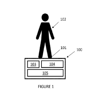
[0020] Figure 1 is a schematic showing use of an exemplary foot
complication detection system.
[0021] Figure 2 is a schematic showing an exemplary foot
complication detection system.
[0022] Figure 3 shows a foot complication detection system for use
near a bathtub.
[0023] Figure 4 shows a platform of a foot complication detection
system with a raised edge for
use near a bathtub.
[0024] Figure 5 shows a schematic of a foot complication detection
system for use near a
bathtub. The platform has an overhang for imaging the top of the foot.
[0025] Figure 6 shows a schematic of a platform of a foot
complication detection system for use
near a bathtub. The platform has three raised edges for imaging the front and
sides of a foot.
[0026] Figure 7 shows a schematic of a platform of a foot complication
detection system with
foot shaped cut-outs or contours for receiving the front of a foot.
[0027] Figure 8 shows a schematic of a platform of a foot
complication detection system with
holes or cavities shaped and sized for guiding and receiving a patient's feet
into a desired position.
[0028] Figure 9 shows a schematic of a platform of a foot
complication detection system
configured to sit in front of a toilet.
[0029] Figure 10 shows a flat mat platfoila configured to be placed
in a bathtub or shower.
[0030] Figure 11 shows a stool platform of a foot complication
detection system in front of a
toilet.
[0031] Figure 12 shows a block element of a foot complication
detection system with sensors
and imaging devices next to a bathtub. The block element can image a patient's
feet without the
patient stepping on the block element.
[0032] Figure 13 shows a block element with sensors and imaging
devices next to a sink.
[0033] Figure 14 shows block elements with sensors and imaging
devices placed at the corners
of a bathtub.
[0034] Figure 15 shows a block element with sensors and imaging devices
placed on the side of
a bathroom door.
[0035] Figure 16 shows a block element with sensors and imaging
devices shaped and sized to
partially wrap around the base of a toilet.
- 6 -
CA 03190407 2023- 2- 21
WO 2022/040576
PCT/US2021/046978
[0036] Figures 17A-17C show exemplary large area imaging sensors.
Figure 17A shows a large
area imaging sensor with an array of photodetectors and a lighting element
below the array. Figure
17B shows a large area imaging sensor with an array of photodetectors with a
lighting element
above the array. Figure 17C shows a large area imaging sensor with an array of
photodetectors with
a lighting element within the array.
[0037] Figure 18 is a schematic showing production of a 3D visual
model of a foot from a
plurality of 2D images.
[0038] Figures 19A-19B are schematics showing different types of
image generation. Figure
19A shows a schematic of a plantar image of a patient's foot with a foot
ulcer. Figure 19B shows a
schematic of a side image of the patient's foot. The side image in Figure 19B
can incorporate data
from the image taken in Figure 19A.
[0039] Figure 20 is a schematic showing production of a 3D visual
model of a foot using a 3D
model of a standard foot as a basis.
[0040] Figure 21A shows a schematic of a platform of a foot
complication detection system
configured to perform multiple functions, including a scale for measuring a
patient's weight as well
as a foot and leg imager.
[0041] Figure 21B shows a schematic of a platform of a foot
complication detection system
configured to perform multiple functions, including a scale for measuring a
patient's weight, a foot
and leg imager, and a bathroom mat.
[0042] Figure 22 is a schematic illustration of an automatic foot
complication detection system
with remote image processing.
[0043] Figure 23 is a schematic illustration of part of an
automatic foot complication detection
system comparing a series of images of a patient's feet over time. The series
shows progression of a
potential foot abnormality over time.
[0044] Figure 24 is a schematic illustration of an exploded view of a foot
complication detection
platform.
[0045] Figures 25A-25B are schematic illustrations of an exploded
view of a foot complication
detection platform with a plurality of side-facing cameras. Figure 25A
illustrates the side-facing
cameras in a vertical or raised side of the platform. Figure 25B illustrates
the overlapping angle of
views of the side-facing cameras for generating stereo images and a 3D model
of a user's feet.
- 7 -
CA 03190407 2023- 2- 21
WO 2022/040576
PCT/US2021/046978
DETAILED DESCRIPTION
[0046] Described herein are systems, devices, and methods for
detecting early stage foot
abnormalities (also referred to herein as foot complications or complications
(e.g., complications
caused by repetitive stress/pressure, trauma, vascular irregularities, and/or
infections, such as an
ulcer, callus, fungus, deformed toenail, wound, and/or laceration) to any part
of the leg or foot (e.g.,
the plantar, lateral, medial, or dorsal parts of the foot, toes, toenails,
heel, and/or ankle). The system
can use images, including images generated within the visual spectrum of light
and images
generated within a spectrum of light outside of the visual range (e.g., within
the infrared spectrum),
to identify foot complications. In some embodiments, the system can include a
platform that
includes a flat mat configured to image the plantar surface of the feet and/or
additional element(s)
configured to image the lateral, medial, and dorsal parts of feet. In some
embodiments, plantar
pressure or force distributions and/or temperature/infrared readings can be
used in combination with
the generated images to detect complications. In some embodiments, the system
can be connected
via a network for detection of complications and/or can trigger a notification
when complications
are identified.
[0047] Referring to Figure 1, an exemplary foot complication system
is shown. Figure 1 shows
a detection platform 100 (e.g., a mat or raised surface) configured to screen
the bottom of a patient's
foot 101 when the patient 102 steps barefoot onto the platform 100 for early
indicators and risk
factors for foot complications. The platform 100 can include one or more
presence sensors 103 to
detect the presence of the patient 102 and an imaging device 104 to take an
image of the foot 101.
In some embodiments, the presence sensors can be one or more load or pressure
sensors to detect
when a force or pressure is applied (e.g., by the foot) on the platform 100.
In some embodiments,
the presence sensors 103 can be one or more ambient light sensors to detect
when a light in the
room (e.g., bathroom) is turned on and/or a shadow is cast over the platform
100. In other
embodiments, the presence sensors 103 can be one or more capacitive or other
proximity sensors to
detect when a patient is close to the platform 100.
[0048] The imaging device 104 can be configured to take images of
the foot 101 (e.g., of the
plantar, anterior, posterior, lateral, medial, and/or dorsal surfaces). The
platform 100 can further
include a platform processor 105 configured to analyze the images taken with
the imaging device
104 to detect foot complications. The one or more presence sensors 103 can be
used to detect when
a person steps on the platform 100. In some embodiments, this detection can be
used to trigger the
imaging device 104 and/or platform processor 105. The platform 100 can further
include a battery
and/or power cord and/or can be configured for wireless charging.
- 8 -
CA 03190407 2023- 2- 21
WO 2022/040576
PCT/US2021/046978
[0049] The platform 100 can be used, for example, as a bathmat. To
function as a bathmat, for
example, the platform 100 can be waterproof and/or water wicking, can include
texturing, can
include an active drying mechanism, can have a pattern thereon with multiple
materials to absorb, or
can include light-transmissive sections or light guides within a water-
absorptive material. Further,
the platform 100 (or the base of the platform, excluding a vertical or raised
side, overhang, etc.) can
be 5 cm or less in height, such as 4 cm or less in height, 3 cm or less in
height, or 2 cm or less in
height
[0050] Referring to Figure 2, in some embodiments, the platform 100
(which can be, for
example, in a bathroom next to a sink 221) can be connected to a remote
processor 222 (for
example, via a connector such as an Ethernet cable connection, wireless
internet card, direct internet
connection, a cellular connector, Wi-Fi, or Bluetooth). The remote processor
222 can be used in
lieu of or in addition to the platform processor 105 in the platform 100 or
any other platform
described herein. In some embodiments, the platform 100 can be connected to a
local or platform
processor 105 via a connector, such as a data cable. In some embodiments, for
example, the
platform processor 105 can combine data from multiple sensors 103 together
into one packet (e.g.,
images from multiple image sensors and/or data from presence sensors), adding
additional size and
position information based on which sensor(s) 103 the data comes from, while
the remote processor
222 can create the visual model and perform the analysis to detect foot
complications. In some
variations, a system for detecting a foot complication may have multiple
processors, such as one or
more than one remote processor and one or more than one platform processor. In
some variations,
platform processor 105 and remote processor 222 may be configured to perform
the same or similar
functions (e.g., platform processor 105 and remote processor 222 may be
redundant and be
configured to perform redundant functions). A user may choose which type of
processor(s) to use
with a system_ As used herein, unless otherwise indicated, processor may refer
to a remote
processor and/or a platform processor.
[0051] Referring to Figure 22, system 1420 for detecting a foot
complication is configured to
issue an alert and/or communicate an alert flag to a patient or a member of a
care team at a remote
location. The alert flag can be issued and/or communicated to indicate data
generation and/or
detection of a foot abnormality (e.g., a foot complication). As illustrated in
Figure 22, platform
1400c can take one or more images of a patient's foot (not shown) and/or
generate other data at site
of use 1460. The platform 1400c can then, via one or more connectors such as a
data cable, an
Ethernet cable connector or a wireless card, send the one or more images to a
processor, send (arrow
1452) the one or more images of the patient's foot and other data taken at
platform 1400c at site of
- 9 -
CA 03190407 2023- 2- 21
WO 2022/040576
PCT/US2021/046978
use 1460 to internet cloud 1462 (e.g., a first remote processor). Cloud 1462
can store and/or analyze
the images and associated data and send (arrow 1454) an alert flag to remote
location 1464, such as
to remote processor 222 (a second remote processor in this example) or to
another remote receiver.
Remote processor 222 or another remote receiver may be monitored by a member
of a care team,
such as a doctor, a nurse, other caregiver, or a family member. Remote
processor 222 may generate
visual model 1450 showing a visual model of the patient's foot, and a member
of the care team may
view the visual model 1450. The visual model 1450 may be especially useful for
a member of the
care team to help determine the nature of a foot complication or foot concern
and next steps (if any
are needed) to help the patient. In some variations, platform processor 105 or
cloud 1462 may
generate a visual model, and remote processor 222 may receive the generated
visual model, e.g.,
from platform processor 105 or cloud 1462. In some examples, the alert flag
may be sent to remote
processor 222 only if a foot complication, foot abnormality, or other concern
is detected by system
1420. In some variations, the alert flag can be sent even if a foot
complication, foot abnormality, or
other concern is not detected, such as whenever an analysis is performed or on
a regular basis. In
some variations, when image analysis and/or data analysis are performed
locally by platform
processor 105, platform processor 105 can send an alert flag to cloud 1462
(which can send an alert
flag to remote processor 222) or can send an alert flag directly to remote
processor 222 (such as if a
system for detecting a foot complication is not connected to a cloud). The
remote processor can be,
for example a computer, a monitor, or a smart phone. The remote processor can
be monitored by a
member of a care team, such as a doctor, a nurse, or a family member. The
alert flag can be, for
example, an audible alert (e.g., an alarm, a beep, a phone call, a voicemail)
and/or a visual alert
(e.g., an email, a colored light, a message, a pop-up, a text.)
[0052] Further, the platform processor 105 or remote processor 222
can be configured to send
and/or make available raw data, processed or analyzed data, and/or
notifications to patients and/or
their providers and/or other members of their care team, for example their
family. In some
embodiments, for example, gathered and/or analyzed data can be accessed
through a web browser or
application-based service. In some embodiments, the user and/or provider can
receive notifications
on an app or via text message. In some embodiments, the user and/or provider
can receive
notifications via a communications module (a local (platform) communication
module or a remote
communication module), such as a speaker or lights on the platform 100 and/or
on the remote
processor 222 or other remote receiver. In some embodiments, the notifications
can include alerts
to the user to reposition the feet for better reading and/or where to
reposition the feet to, alerts to
- 10 -
CA 03190407 2023- 2- 21
WO 2022/040576
PCT/US2021/046978
indicate the timing in an imaging cycle (e.g., whether the user can move his
or her feet/leave the
platform), alerts to see a doctor, and/or alerts that a complication has or
has not been detected.
[0053] Referring to Figure 24, in some embodiments, an imaging
device (or any other imaging
device or system described herein) can include a large area imaging sensor
162, e.g., an imaging
sensor that is configured as a two-dimensional array of photodetectors where
the size of the sensor
is the same as the size of the field of view. The large area imaging sensor
can be positioned (e.g.,
immediately) below the horizontal surface 160c of the platform 100 on which
the user stands. The
large area imaging sensor can be positioned above support 164 of the platform
100. The imaging
device in Figure 24 also includes one or more than one (2, 3, 4, 5, 6, 7, 8)
force transducers or load
cells 168 that may rest upon support 164. This and other imaging devices
described herein may
contain one or more than one large area imaging sensor with these and other
features described
herein (e.g., each imaging device can be configured as a two-dimensional array
of photodetectors
where the size of the sensor is the same as the size of the field of view;
positioned (e.g.,
immediately) below the horizontal surface, etc.) Surface 160c on platform 100
may include a
protective, non-slip surface, such as made from a polyvinyl chloride (PVC) or
a thermoplastic
rubber (TPR) material. Surface 160c may be textured, such as with bulges,
dots, indents, lines, or
waves that prevent a patient from slipping and falling. For example, Figure
21A shows platform
1400a with surface 160a with textured lines, and Figure 21B shows platform
1400b with surface
160b with a checkered surface. Any of the surfaces (e.g., surface 160a, 160b,
and/or 160c as well as
associated structures including image sensors and support materials) can be a
continuous surface or
discontinuous surfaces. In some examples, a discontinuous surface may have two
separate surface
regions and act as a foot guide for a patient's feet. For example, Figure 21A
shows separated
surface 1438a and surface 1438b configured to separately act as foot guides
for placement of a
patient's left and right feet. Figure 22 shows a first large area image sensor
1442a and second large
area image sensor1442b. The image sensors are located under the regions upon
which a patient will
step. Thus, in some examples, the sensors can be smaller, easier to
manufacture, less expensive,
allow a more flexible or foldable mat, etc. Load cells 168 on platform 100 can
be configured to
convert compression or pressure into an output signal. Load cells 168 may be
useful as presence
sensors or, when a platform is also used as a scale, for determining a
patient's weight.
[0054] In some embodiments, the large area imaging sensor may
advantageously not require the
use of lenses for magnification or minification of the field of view. Further,
the large area imaging
sensor can advantageously complete imaging in less than 30 seconds, such as
less than 10 seconds,
such as in less than 5 seconds, such as in 3 seconds or less, such as in 1
second or less,
- 11 -
CA 03190407 2023- 2- 21
WO 2022/040576
PCT/US2021/046978
advantageously requiring the user to spend only a short amount of time on the
platform 100 while
still enabling detection of foot complications.
[0055] Referring to Figures 17A-17C, a large area imaging sensor
(e.g., large area imaging
sensor 162) can, for example, include an array 1716 of photodetectors 1717
that are positioned over
a plurality of lighting elements 1718 (e.g., LEDs or othcr lighting source)
and/or a single lighting
element 1718 (e.g., a single backlight (e.g., LCD)). Further, the lighting
element 1718 for the
platform 100 can advantageously be placed below the array 1716 (as shown in
Figure 17A), above
the array 1716 (as shown in Figure 17B), or within the array 1716 (as shown in
Figure 17C). In
some embodiments, the large area imaging sensor can include a filter (e.g.,
red, green, blue) placed
over each photodetector 1717 to ensure a given photodetector 1717 only
measures a specific
wavelength/color of light. In other embodiments, each photodetector 1717 can
be configured to be
sensitive to a specific wavelength or color light. Using a filter over each
photodetector 1717 or
having each photodetector 1717 be sensitive to a specific wavelength can
advantageously reduce
exposure time. In other embodiments, the lighting element 1718 can be
configured to emit a
specific wavelength or color of light, which can advantageously reduce the
number of
photodetectors 1717 required for a given pixel resolution.
[0056] In some embodiments, the large area image sensor may be made
from one or multiple
(e.g., 2, 3, 4, 5, or more) wafer-scale image sensors and the sensors may be
butted together or may
not be butted together (e.g., they may be separated). In some embodiments, the
photodetectors may
be discrete components mounted to a printed circuit board. In some
embodiments, the large area
image sensor may be made, for example, from amorphous silicon deposited onto a
substrate (e.g.,
amorphous silicon deposited onto a substrate and selectively crystalized into
a polycrystalline
silicon or amorphous silicon deposited onto a substrate and without being
selectively crystalized
into a polycrystalline silicon), or from other organic semiconductor
materials. In some
embodiments, the substrate of the large area image sensor can be a thin glass
substrate. In this
embodiment, a rigid transparent window can be placed above the large area
sensor and/or a rigid
support can be placed below the large area sensor (e.g., with the large area
sensor sandwiched
therebetween) to help avoid flexing of the large area image sensor. In other
embodiments, the
substrate of the large area image sensor can be a flexible (e.g., plastic)
substrate, which can
advantageously help prevent the large area imaging sensor from breaking even
under high user
loads.
[0057] The large area imaging sensor can include a tailored imaging
depth such that areas
within 75 mm, such as within 50 mm, such as within 40 mm are in focus and
areas further away are
- 12 -
CA 03190407 2023- 2- 21
WO 2022/040576
PCT/US2021/046978
not in focus. Imaging within this range can ensure that the entire foot can be
in focus in the image
while preventing privacy concerns by otherwise focusing on more of the
patient's body than
necessary. A longer imaging depth could be an issue since the imaging can be
performed and/or is
designed to be performed (in the bathroom) while a patient is undressed,
showering, using the toilet,
etc. In some embodiments, the large imaging sensor can include a collimator
filter therein or
thereover to achieve an imaging depth within the tailored range. The
collimator, for example, can
be fabricated with carbon nanotubes, with a traditional flat panel
manufacturing method, or via
micro-machined holes (e.g., with a precision laser cutter). In other
embodiments, additional lenses
can be used with the large area sensor to achieve an imaging depth within the
tailored range. These
additional lenses can be, for example, micro lenses, gradient-index lenses,
and/or composite lenses
made from laminated pieces of materials with different indexes of refraction
and placed over the
photodetectors of the large area imaging sensor.
[0058] Advantageously, the large area image sensor can be less than
20 mm, such as less than
10 mm, such as less than 5 mm, such as less than 3 mm, such as less than 2 mm
thick. Additionally,
the large area imaging sensor can acquire images quickly (e.g., within 10
seconds, within 10
seconds to 1 second (e.g., within 1 second, within 2 seconds, etc.), within 1
second to 0.1 seconds)
of the user stepping on or otherwise engaging with the platform). In some
examples, an imaging
sensor herein (e.g., large area imaging sensor) can acquire images faster than
other imaging
modalities can, such as other non-sensing modalities (e.g., contact
temperature sending) or a moving
scanner imaging sensor. Moreover, the large area imaging sensor can
advantageously gather images
from a wide range of angles and positions (e.g., rather than requiring the
user to stand directly on
specific imaging windows).
[0059] Referring to Figures 25A-25B, in some embodiments, the
imaging device can include
one or more additional cameras positioned on a first vertical or raised side
172 of the imaging
device (e.g., above a plantar imaging surface 170). A vertical or raised side
may also house
electronics for the device. The one or more additional cameras may be in
addition to or, in some
examples, instead of, the plantar large area imaging sensor 162. Figures 25A-
25B show, for
example, three wide-angle cameras strategically positioned to capture
different perspectives on the
foot or feet of the patient and may do so simultaneously or sequentially.
Other numbers of cameras
can also be used and/or placed on other surfaces, such as other side or
vertical surfaces.
Representative foot placement is shown in first foot location 1440a and second
foot location 1440b.
(See also Figure 22). The wide-angle camera lens can capture, for example,
from 60 to 180 , such
as from 60 to 100, from 100 to 150, from 150 to 170, or from 170 to 180. The
wide-angle camera
- 13 -
CA 03190407 2023- 2- 21
WO 2022/040576
PCT/US2021/046978
lens can produce a rectilinear image. In some examples, the wide-angle camera
lens can be an ultra-
wide-angle lens, such as a fisheye lens and may produce a circular rather than
a rectilinear image.
For example, if the heels are closest to the camera, first camera 1726a
captures a region indicated by
angle al, such as the left medial foot from the posterior up to and including
the toes, the right lateral
foot from the posterior up to and including the toes, the left heel, and the
right heel. The second
camera 1726b, in turn, captures a region indicated by angle a2, such as the
left medial foot from the
posterior up to and including the toes, the right medial foot from the
posterior up to and including
the toes, the left heel, and the right heel. Finally, the third camera 1726c
captures a region indicated
by angle a3, such as the left lateral foot up from the posterior up to and
including the toes, the right
medial foot from the posterior up to and including the toes, the left heel,
and the right heel. A single
camera may image one or more of the plantar aspect of a foot, the heel, the
lateral aspect of the foot,
ankle, or leg, medial aspect of the foot, ankle, or leg, or any of the toes.
Together, however, these
cameras can provide stereo images that can be used to generate a 3D model of a
user's feet (e.g., by
employing measurements made in two or more images taken from different
positions).
[0060] Non-plantar foot ulcers (typically presenting 5-6 times less
frequently than plantar foot
ulcers) tend to be concentrated on the toes and heel. In some examples, 3D
models create a
representation of the toes and/or heels of the patient's feet. The design of
the device can keep these
areas in view of the stereographic cameras during intended use. In some
examples, the cameras
(e.g., camera 1726a, camera 1726b, camera 1726c) are in fixed locations on
imaging device 104
(and/or relative to one another), and the fixed locations of the cameras is
known a priori. Having
fixed locations can obviate the first step of many photogrammetric pipelines:
registering images to
determine real-world positions of the cameras. In some variations, one or more
additional cameras
may be positioned along a second raised side, a third raised side, or a fourth
raised side and/or along
a bottom of a top surface of the imaging device (e.g., above the top of the
foot). Although described
with reference to imaging device 104, any system or imaging device described
herein may employ
one of more additional cameras positioned on e.g., a vertical or raised side
or top side thereof.
[0061] In some embodiments, the imaging device 104 can include, in
addition to or in lieu of the
large area imaging sensor, a linear array of photodetectors (e.g., a contact
imaging sensor), a
plurality of lights, and one or more scanners. The scanner(s) can move the
photodetectors along the
full length of the foot to produce the image. In other embodiments, the
imaging device 104 can
include one or more camera sensors with one or more corresponding lenses. In
some embodiments,
these camera sensors can be manufactured via wafer-level optics processes,
which advantageously
may allow them to be made more cheaply, more precisely, and in a smaller size.
- 14 -
CA 03190407 2023- 2- 21
WO 2022/040576
PCT/US2021/046978
[0062] The imaging device 104 can be designed to fit within a small
vertical space, such as 20
cm or less, 10 cm or less, 5 cm or less, 3 cm or less, 2 cm or less, or 1 cm
or less.
[0063] In some embodiments, the processor (e.g., platform processor
105 or remote processor
222) can build a visual model of the surface of the patient's foot based upon
images gathered by the
imaging device 104 and can detect one or more irregularities in the visual
model.
[0064] Referring to Figure 18, in some embodiments, the visual
model can be developed by
combining all of the images taken by the imaging device 104 to generate a
three-dimensional (fully
complete or partially complete) visual representation of the surface of the
foot, which can then be
analyzed for irregularities that may correspond with foot abnormalities or
other complications. For
example, a visual model can be developed using images from the plantar surface
(e.g., with the large
area imaging sensor) and from the anterior, posterior, lateral, dorsal, and/or
medial surfaces of the
foot (e.g., with one or more wide angle cameras). The images from the
anterior, posterior, plantar,
medial, lateral, and dorsal perspectives, and/or from any other perspectives,
taken during one
session (e.g., at the same time or slightly spaced apart temporally) can be
associated with (or
stitched) together. Image identification from the plantar images can allow the
orientation and
position of the foot to be determined (e.g., can enable identification of the
outline of the foot, the
location of the foot on the mat, and/or which way the heel and toes are
pointing) in order to create a
rudimentary foot model located in virtual 3D space. The side images (which can
utilize depth
information from a previous calibration, stereo information, geometrical
perspective with calibration
markers on the board, or other range-imaging methods such as time-of-flight
and structured/coded
light), in turn, can be used to apply further visual information to the
relevant surface of the foot
model, based on the associated position and orientation from the plantar
images.
[0065] In some embodiments, as a patient moves around on the mat,
images can be taken
continuously and/or at regular intervals. Taking images continuously and/or at
regular images can
enable the visual model of the patient's foot to be incrementally updated.
This incremental updating
can advantageously produce a higher resolution three-dimensional visual
representation of the foot
than the sensor resolution would allow for individual images.
[0066] In some embodiments a neural network deep-learning-based
approach can be used to
generate the 3D models. For example, a Volumetric Regression Network can be
used and may
advantageously not require the use of a 3D Morphable Model. In some
embodiments, a semi-global
matching algorithm can be used to compute a disparity map for image pairs,
providing depth
information. This map can then be used to reproject the images onto a 3D point
cloud.
- 15 -
CA 03190407 2023- 2- 21
WO 2022/040576
PCT/US2021/046978
[0067] In some embodiments, as shown in Figures 19A-19B, the visual
model can be developed
by tagging the images taken by the imaging device 104 with location and
position information of the
foot in each of the respective images, allowing a single image view to stand
on its own during
analysis for foot complications (e.g., enabling analysis with an imaging
device that includes only a
large area imaging sensor for imaging the plantar surface). That is, by using
a plantar image (shown
in Figure 19A), a bare model of the foot can be located and oriented in 3D
space. Then, as shown in
Figure 19B, the side image can be mapped directly onto the surface of that
model, as the distance
from the imaging device 104 to the boundary of the 3D model is known. In the
images shown in
Figures 19A and 19B, there is an ulcer 1919 that spreads from the medial to
plantar surfaces
because each image is tagged with the position and location information of the
foot as the images
are taken. In some examples, plantar images can be used without side images
and/or without a 3D
model to e.g., identify foot structures and foot abnormalities. For example,
one or more than one
plantar image can be analyzed to identify e.g., toes and heel so that the
plantar abnormalities are
associated with a location on the plantar surface of the foot.
[0068] In other embodiments, as shown in Figure 20, a three-dimensional
model of a standard
foot can be used as a basis for creating the visual model with the images from
the imaging device
104.
[0069] In some embodiments, as described above, the visual model
can be developed using
images from the plantar surface and from the anterior, posterior, medial,
dorsal, or lateral surfaces
of the foot. In other embodiments, an incomplete visual model can be developed
using images from
the plantar surface of the foot only.
[0070] The irregularities identified by the platform processor 105
or remote processor 222 in the
visual model can include, for example, a visual irregularity in a single
visual model at a given point
in time (e.g., a black spot corresponding to dried blood or necrotic tissue,
redness from erythema, a
white spot corresponding to a callus, a series of discolored lines indicating
fissures from dry skin, or
a discoloration under the toenail indicating fungus). The irregularities can
include, for example, a
difference in the visual model from one point in time compared with another
(e.g., the color of a
certain spot on a foot changed significantly from week to week, and the
discoloration has grown for
two days in a row). In some embodiments, the continuous and/or regular images
can be used in a
time-lapse analysis and/or presentation of the foot (e.g., to determine how a
foot complication
spread, healed, or otherwise changes over a period of time). Any of the images
referred to herein
can be black and white images (grayscale) or color images and any of the
analyses referred to herein
can be performed using black and white images (grayscale) or color images.
- 16 -
CA 03190407 2023- 2- 21
WO 2022/040576
PCT/US2021/046978
[0071] Referring to Figure 23, remote processor 222 includes di
splay 1430. Display 1430
displays patient information 14and a series of images 1432a, 1432b, 1432c,
1432d, and 1432e of a
patient's feet over time. Figure 23 shows image 1401a of patient's foot 101
with a 2.5 cm diameter
potentially abnormality 1434b. Figure 23 also shows image 1432b of patient's
foot 101 taken just
prior to the image 1432a. As shown in Figure 1432b, the potentially
abnormality 1434a has started
to develop, but is smaller or less severe than shown in Figure 1432a.
Moreover, the abnormality
1434a/b was not visible in earlier images (1432c, 1432d, and 1432e). By
comparing images over
time, a care provider can determine various characteristics such as how long a
potential abnormality
has been on a foot, if the potential abnormality has changed over time, how
the potential
abnormality has changed over time, how quickly it has changed, if the color of
the potential
abnormality has changed, etc. Images, such as those illustrated in images
1432a, 1432b, 1432c,
1432d, and 1432e can be automatically generated and analyzed using the
systems, devices, and
methods described herein. Using the systems, devices, and methods described
herein can include the
step of displaying a series of images taken over time of the foot of the user
on a remote (and/or
local) display, wherein a first image of the series of images includes an
image of the foot having the
foot complication and a second image of the series of images includes an image
of the foot not
having the foot complication.
[0072] One exemplary automated method for analyzing images is
through image
segmentation/region detection. Clinically relevant information can present in
the form of changes in
color of a region of the foot and/ or changes in size of those regions.
Examples of changes include: a
red spot appearing or growing in size across multiple days which may indicate
e.g., a region of
spreading inflammation; a region of red color shrinking in size may indicate
e.g., healing; a region
of black color appearing or growing in size may indicate the presence of
necrotic tissue and other
colors on a region of a patient's foot, such as yellow, could indicate an
infection; etc. Provided
herein are systems, devices, and methods for taking images across different
points in time,
automatically annotating the images with regions of interest highlighted,
measuring the size of a
region of interest, and comparing a size and color from the same region with
previous images. These
systems, devices, and methods may help care providers and clinicians better
understand how
different (foot) complications may be progressing.
[0073] To detect regions of interest, several processing steps can be used.
In one exemplary
method of detecting regions of interest, first, images can be color corrected
to, for example, account
for environment effects (e.g., lighting) on image color or minor manufacturing
variations across the
different photodetectors in an image sensor. Image sensors can be calibrated
against known targets
- 17 -
CA 03190407 2023- 2- 21
WO 2022/040576
PCT/US2021/046978
during manufacturing (such as in a factory), and color calibration targets can
also be included on the
platform (mat) to allow for live color correction in the field during platform
or mat use.
[0074] Once images have been color corrected, segmentation
algorithms, such as thresholding,
clustering, and/or neural network based algorithms, can be used to identify
regions of the photo
image that correspond to feet. Once images have been segmented to identify
foot regions, images
can be screened to separate out or remove any unusable or partial images.
[0075] Next, the size and shape of a foot in an image can be used
to identify whether it is a left
or right foot and/or whether it belongs to a user in question (as opposed to
another user). Users can
be filtered out, for example, by weight data from load sensors if included in
the mat, but analyzing
the images of feet directly can provide a level of redundancy. Once regions in
images have been
fully segmented and identified, these regions can be aligned with other images
in a given capture
session, as well as with images from other points in time. This approach can
allow images to be
analyzed not just alone, but also in comparison with other images.
[0076] Finally, foot regions from images can be processed with
finely tuned image
segmentation algorithms to identify regions of interest on the feet. These
regions of interest can then
be analyzed for e.g., size, average color, color extremes, color gradient
direction, etc., and these
measures can be compared with other images from other points in time to
understand how the
regions of interest are changing. Images can be presented to care providers or
clinicians with these
regions of interest highlighted and associated with the computed metadata
(e.g., additional
information about the region of interest, such as a size of an abnormality,
length of time the
abnormality has been visible, how quickly the abnormality is growing (e.g.,
how quickly the
abnormality is doubling in size), how abnormality color is changing over time,
time information
when different images were gathered.
[0077] In some embodiments, the visual model can be combined with
infrared images gathered
by the platform to provide additional foot complication detection. For
example, near-field infrared
can be used to determine blood flow and oxygenation, both of which can be used
to identify
inflammation or peripheral vascular complications. As another example, mid-
field and far-field
infrared can indicate temperature in order to identify inflammation (high-
temperature) or ischemia
(low-temperature). Infrared images can be generated, for example, by
reflectance spectroscopy
(emitting a light and measuring reflectivity/absorbance from the foot), by
emission spectroscopy
(measuring photon emissions from the foot), or by fluorescence spectroscopy
(emitting a light in
order to excite specific molecules/compounds in the foot and measuring the
resulting photons
released).
- 18 -
CA 03190407 2023- 2- 21
WO 2022/040576
PCT/US2021/046978
[0078] In some embodiments, the visual model can be combined with
pressure distribution
information gathered by the platform (e.g., to include weight in the
analysis). The pressure
distribution information can, for example, indicate a patient' s risk of
developing a foot complication
over time (e.g., because high pressure points can lead to calluses and
ulcers). Thus, for example,
high-pressure points in the plantar surface of the foot, particularly ones
that increase as time goes
on, can be flagged as risks for ulcer development. The information can also,
for example, be used to
identify a complication (for example, a patient's pressure distribution can
change with a wound in
the heel, as the body compensates). As another example, the pressure
distribution can be used to
estimate a patient's posture and loading patterns, tracked over time, to
identify key changes that
may indicate that a patient's musculoskeletal system is undergoing atrophy due
to a progression of
neuropathy.
[0079] Additional exemplary platforms similar to platform 100 are
shown in Figures 3-16.
[0080] Figures 3-8 show platforms positioned adjacent to a shower
or bathtub 331 (though each
of the platforms could be positioned adjacent to a sink as shown in Figure 2
or conforming to a
toilet base as shown in Figure 9). As shown in Figure 3, platform 300 is a
flat mat (e.g., a mat
having a thickness of less than 50 mm, such as less than 40 mm, such as less
than 30 mm)
positioned in front of the shower or bathtub 331. As shown in Figure 4,
platform 400 includes a flat
mat 441 with a raised edge 443 that is positioned against the bathtub 331
(e.g., so as to avoid
tripping thereover). The flat mat 441 can include an imaging device therein
configured to image the
bottom of the foot while the raised edge 443 can include an imaging device
therein configured to
image the front, sides, and/or top of the foot. As shown in Figure 5, platform
500 includes a flat mat
541 with a raised edge 543 having an overhang 551 to better image the top of
the foot. As shown in
Figure 6, the platform 600 includes a flat mat 641 with three raised edges
643a,b,c to better image
the front and sides of the foot. As shown in Figure 7, the platform 700
includes a flat mat 741 with
a raised element 777 with cut-outs 772 configured to conform to or closely
follow the contour of the
front of the foot. The raised element 777 can include an imaging device
therein configured to image
the front, sides, and/or top of the foot. As shown in Figure 8, the platform
800 includes a flat mat
841 with a raised top layer 888 having holes 882 (also referred to herein as
cavities or indents)
therein configured to enable the user to stand therein. The holes or cavities
extend only partway
through the platform or map. The raised top layer 888 can advantageously image
all the way around
the lateral surfaces of the foot when the user is positioned on the platform
800.
[0081] Additional platform designs are shown in Figures 21A-21B and
Figure 22. In some
variations, any platform as described herein can perform other functions, in
addition to performing
- 19 -
CA 03190407 2023- 2- 21
WO 2022/040576
PCT/US2021/046978
imaging functions and analysis. For example, platform 1400a in Figure 21A,
platform 1400b in
Figure 21B, and platform 1400c are combined scale and foot complication
detectors and include a
scale for determining a patient's weight as well as image sensors for
detecting a foot complication.
The patient's weight may be displayed to the patient on display 1430. A scale
may have a
piezoelectric transducer that compresses and produces an electric current whcn
a patient steps on the
platform 1400c. In some variations, display 1430 may display other
information, such as an alert
flag that indicates the patient may have a foot complication or should seek
medical attention. Figure
21B is additionally configured as a bathroom mat (bath room mat), such as for
use outside of a
bathtub, shower, or sink.
[0082] Other platform designs are possible. For example, as shown in Figure
9, the platform
900 can be a flat mat positioned and/or conforming to the base of toilet 1111.
As shown in Figure
10, in some embodiments, the platform 1000 can be a flat mat configured to be
placed in a bathtub
331 or shower. As shown in Figure 11, the platfomi 1100 can be a stool
configured to be placed in
front of toilet 1111.
[0083] In some embodiments, the platform can be replaced with a block
element (including the
sensors, imaging device, and/or other features of the platform as described
herein) that is configured
to be placed in the bathroom, but not stepped upon. For example, as shown in
Figure 12, an
elongated block element 1220 can be placed next to the bathtub 331. Similarly,
an elongated block
element 1320 can be placed next to the sink 221, as shown in Figure 13. In
other embodiments, one
or more block elements 1420a,b can be placed at the corners of the bathtub
331, as shown in Figure
14. One or more block elements 1520 can be placed on the side of the bathroom
door 1514 as
shown in Figure 15. One or more block elements 1620 can be placed around the
base of the toilet
1111 as shown in Figure 16.
[0084] Advantageously, the systems described herein can enable
passive visual monitoring for
foot complications. Passive monitoring (i.e., monitoring that does not require
activation or input by
an individual, such as the patient) can advantageously help ensure patient
compliance. Visual
monitoring can advantageously automate the current standard of care for foot
complication
detection and can provide the user (e.g., the medical provider) with detailed
medical information
regarding the patient's disease state.
[00851 Additionally, the systems described herein can advantageously be
placed in the bathroom
because, while many patients at high risk for ulcers are told to consistently
wear shoes, patients tend
to still be barefoot in the bathroom, thereby enabling imaging of the feet and
monitoring for foot
complications.
- 20 -
CA 03190407 2023- 2- 21
WO 2022/040576
PCT/US2021/046978
[0086] It should be understood that any feature described herein
with respect to one
embodiment can be used in addition to or in place of any feature described
with respect to another
embodiment.
[0087] When a feature or element is herein referred to as being
"on" another feature or element,
it can be directly on the other feature or element or intervening features
and/or elements may also be
present. In contrast, when a feature or element is referred to as being -
directly on- another feature or
element, there are no intervening features or elements present. It will also
be understood that, when
a feature or element is referred to as being "connected", "attached" or
"coupled" to another feature
or element, it can be directly connected, attached or coupled to the other
feature or element or
intervening features or elements may be present. In contrast, when a feature
or element is referred to
as being "directly connected", "directly attached" or "directly coupled" to
another feature or
element, there are no intervening features or elements present. Although
described or shown with
respect to one embodiment, the features and elements so described or shown can
apply to other
embodiments. It will also be appreciated by those of skill in the art that
references to a structure or
feature that is disposed "adjacent" another feature may have portions that
overlap or underlie the
adjacent feature.
[0088] Terminology used herein is for the purpose of describing
particular embodiments only
and is not intended to be limiting of the invention. For example, as used
herein, the singular forms
"a", -an" and "the" are intended to include the plural forms as well, unless
the context clearly
indicates otherwise. It will be further understood that the terms "comprises"
and/or "comprising,"
when used in this specification, specify the presence of stated features,
steps, operations, elements,
and/or components, but do not preclude the presence or addition of one or more
other features,
steps, operations, elements, components, and/or groups thereof. As used
herein, the term "and/or"
includes any and all combinations of one or more of the associated listed
items and may be
abbreviated as "/".
[0089] Spatially relative terms, such as "under-, "below-, "lower",
"over", "upper- and the like,
may be used herein for ease of description to describe one element or
feature's relationship to
another element(s) or feature(s) as illustrated in the figures. It will be
understood that the spatially
relative terms are intended to encompass different orientations of the device
in use or operation in
addition to the orientation depicted in the figures. For example, if a device
in the figures is inverted,
elements described as "under- or "beneath" other elements or features would
then be oriented
"over" the other elements or features. Thus, the exemplary term "under" can
encompass both an
orientation of over and under. The device may be otherwise oriented (rotated
90 degrees or at other
- 21 -
CA 03190407 2023- 2- 21
WO 2022/040576
PCT/US2021/046978
orientations) and the spatially relative descriptors used herein interpreted
accordingly. Similarly, the
terms "upwardly", "downwardly-, "vertical", "horizontal" and the like are used
herein for the
purpose of explanation only unless specifically indicated otherwise.
[0090] Although the terms "first" and "second" may be used herein
to describe various
features/elements (including steps), these features/elements should not be
limited by these terms,
unless the context indicates otherwise. These terms may be used to distinguish
one feature/element
from another feature/element. Thus, a first feature/element discussed below
could be termed a
second feature/element, and similarly, a second feature/element discussed
below could be termed a
first feature/element without departing from the teachings of the present
invention.
[0091] Throughout this specification and the claims which follow, unless
the context requires
otherwise, the word "comprise", and variations such as "comprises" and
"comprising" means
various components can be co-jointly employed in the methods and articles
(e.g., compositions and
apparatuses including device and methods). For example, the term "comprising"
will be understood
to imply the inclusion of any stated elements or steps but not the exclusion
of any other elements or
steps.
[0092] As used herein in the specification and claims, including as
used in the examples and
unless otherwise expressly specified, all numbers may be read as if prefaced
by the word "about" or
"approximately," even if the term does not expressly appear. The phrase
"about" or
"approximately" may be used when describing magnitude and/or position to
indicate that the value
and/or position described is within a reasonable expected range of values
and/or positions. For
example, a numeric value may have a value that is +/- 0.1% of the stated value
(or range of values),
+/- 1% of the stated value (or range of values), +/- 2% of the stated value
(or range of values), +/-
5% of the stated value (or range of values), +/- 10% of the stated value (or
range of values), etc.
Any numerical range recited herein is intended to include all sub-ranges
subsumed therein.
[0093] Although various illustrative embodiments are described above, any
of a number of
changes may be made to various embodiments without departing from the scope of
the invention as
described by the claims. For example, the order in which various described
method steps are
performed may often be changed in alternative embodiments, and in other
alternative embodiments
one or more method steps may be skipped altogether. Optional features of
various device and
system embodiments may be included in some embodiments and not in others.
Therefore, the
foregoing description is provided primarily for exemplary purposes and should
not be interpreted to
limit the scope of the invention as it is set forth in the claims.
- 22 -
CA 03190407 2023- 2- 21
WO 2022/040576
PCT/US2021/046978
[0094] The examples and illustrations included herein show, by way
of illustration and not of
limitation, specific embodiments in which the subject matter may be practiced.
As mentioned, other
embodiments may be utilized and derived there from, such that structural and
logical substitutions
and changes may be made without departing from the scope of this disclosure.
Such embodiments
of the inventive subject matter may be referred to herein individually or
collectively by the term
-invention- merely for convenience and without intending to voluntarily limit
the scope of this
application to any single invention or inventive concept, if more than one is,
in fact, disclosed.
Thus, although specific embodiments have been illustrated and described
herein, any arrangement
calculated to achieve the same purpose may be substituted for the specific
embodiments shown.
This disclosure is intended to cover any and all adaptations or variations of
various embodiments.
Combinations of the above embodiments, and other embodiments not specifically
described herein,
will be apparent to those of skill in the art upon reviewing the above
description.
- 23 -
CA 03190407 2023- 2- 21