Note: Descriptions are shown in the official language in which they were submitted.
CA 03190474 2023-01-30
WO 2022/026914
PCT/US2021/044043
1
ANTI-CONNEXIN ANTIBODY FORMULATIONS
CROSS-REFERENCE TO RELATED APPLICATIONS
This application claims priority to and the benefit of U.S. Provisional Patent
Application
No. 63/059,502 filed July 31, 2020, the entire disclosure of which is
incorporated herein by
reference.
SEQUENCE LISTING
The ASCII text file submitted herewith via EFS-Web, entitled "020602
SequenceListing.txt" created on July 30, 2021, having a size of 41,780 bytes,
is hereby
incorporated by reference in its entirety.
FIELD
The present disclosure generally relates to stable aqueous pharmaceutical
compositions
comprising anti-connexin (Cx) 43 antibodies.
BACKGROUND
Antibodies (Abs) have been used in the treatment of various diseases and
conditions due
to their specificity of target recognition, thereby generating highly
selective outcomes following
systemic administration. in order for antibodies to remain effective, they
must maintain their
biological activity during their production, purification, transport and
storage. New produch Oil
and purification techniques have been developed to provide for large amounts
of highly purified
monoclonal antibodies to be produced. However, challenges still exist to
stabilize these
antibodies for transport and storage, and yet even more challenges exist to
provide the antibodies
in a dosage form suitable for administration.
!Denaturation, aggregation, contamination, and particle formation can be
significant
obstacles in the formulation and storage of antibodies. Due to the wide
variety of antibodies,
there are no universal formulations or conditions suitable for storage of all
antibodies. Optimal
formulations and conditions suitable for storage of one antibody are often
specific to that
antibody. Thus, antibody storage formula Oils and methods are often a
significant part of the
research and development process for a commercial antibody.
Various methods have been proposed to overcome the challenges associated with
antibody stability. For example, in some instances, the antibody is often
lyophilized, and then
reconstituted shortly before administration. However, reconstitution is
generally not ideal, since
CA 03190474 2023-01-30
WO 2022/026914 PCT/US2021/044043
2
it adds an additional step to the administration process, and could introduce
contaminants to the
formulation. Additionally, even reconstituted antibodies can suffer from
aggregation and particle
formation. Thus, a need exists to provide stable, aqueous antibody
formulations, in particular
anti-Cx43 and body formulations that can overcome the challenges associated
with transport and
storage.
SUMMARY
The present disclosure provides, in one aspect, a pharmaceutical formulation
comprising:
an anti-Cx43 antibody (Ab) or antigen binding fragment thereof;
a buffer;
a surfactant; and
a stabilizer;
wherein the pharmaceutical formulation has a pH of between about 5 and about
6;
wherein the anti-Cx43 antibody or antigen binding fragment thereof comprises:
a first, second and third heavy chain complementarity determining region
(CDR) sequence having the amino acid sequence of SEQ ID NOs: 1, 2, and 3,
respectively; and
a first, second and third light chain CDR sequence having the amino acid
sequence of SEQ ID NOs: 4, 5, and 6, respectively.
In some embodiments, the anti-Cx43 antibody or antigen binding fragment
thereof
comprises a heavy chain variable domain having the amino acid sequence of SEQ
ID NO: 7, and
a light chain variable domain having the amino acid sequence of SEQ ID NO: 8.
In certain embodiments, the anti-Cx43 antibody or antigen binding fragment
thereof
comprises a heavy chain having an amino acid sequence selected from the group
consisting of
SEQ ID NOs: 9-17, and a light chain having the amino acid sequence of SEQ ID
NO: 18.
In certain embodiments, the anti-Cx43 antibody or antigen binding fragment
thereof binds
to an epitope located within the amino acid sequence of FLSRPTEKTI (SEQ ID NO:
19). In
some embodiments, the epitope comprises one or more amino acids selected from
the group
consisting of R4, P5, E7, K8 and 110 of SEQ ID NO: 19. In some embodiments,
the epitope
consists of R4, P5, E7, K8 and 110 of SEQ ID NO: 19. In some embodiments, the
epitope
comprises all ten amino acids of SEQ. ID NO: 19. In some embodiments, the
epitope consists of
all ten amino acids of SEQ ID NO: 19.
In some embodiments, the anti-Cx43 antibody or antigen binding fragment
thereof is
CA 03190474 2023-01-30
WO 2022/026914
PCT/US2021/044043
3
present at a concentration of between about 5 and about 100 mgitriL,
preferably between 20 and
80, more preferably from about 40 to 60 ruglinL.
In some certain embodiments, the buffer is selected from acetate/sodium
acetate,
histidine/aspartic acid, citric acid/sodium citrate, dibasic sodium
phosphate/sodium dihydrogen
phosphate, and histidine/histidine hydrochloride. In certain embodiments, the
buffer is
histidine/aspartic acid or histidine/histidine hydrochloride. In certain
embodiments, the buffer is
histidine/histidine hydrochloride.
In some embodiments, the surfactant is polysorbate 80 (PS80).
In certain embodiments, the stabilizer is selected from
ethylenediaminetetraacetic acid
(EDTA), sodium chloride, sorbitol, glycine, and sucrose. In certain
embodiments, the stabilizer
is sucrose.
In certain embodiments, the pH of the formulation is between about 5.4 to
about 5.6.
In some embodiments, the formulation is an aqueous formulation. In some
embodiments,
the formulation is a stable aqueous formulation.
Another aspect relates to a pharmaceutical formulation comprising:
about 40-60 mg/mL, preferably about 50 mg/mL of an anti-Cx43 antibody or
antigen binding fragment thereof;
about 10-40 mM, preferably about 20 mM histidine/histidine hydrochloride
buffer;
about 0.005%-0.05%, preferably about 0.02% w/v Polysorbate 80; and
about 1%-20% w/v, preferably about 8% w/v sucrose;
wherein the formulation has a pH of between about 5.4 to about 5.6, preferably
about 5.5.
A further aspect relates to a pharmaceutical formulation comprising:
about 50 mg/mL an anti-Cx43 antibody or antigen binding fragment thereof,
comprising a heavy chain having an amino acid sequence selected from the group
consisting of SEQ ID NOs: 9-17, and comprising a light chain having the amino
acid
sequence of SEQ ID NO: 18;
about 20 mM histidine/aspartic acid buffer;
about 0.02% w/v Polysorbate 80; and
about 8% w/v sucrose,
wherein the formulation has a pH of between about 5.4 to about 5.6, preferably
about 5.5.
CA 03190474 2023-01-30
WO 2022/026914
PCT/US2021/044043
4
Kits and/or unit dosages comprising any one of the pharmaceutical formulations
disclosed
herein are also provided.
Also provided herein is use of any one of the pharmaceutical formulations
disclosed
herein, for inhibiting opening of Cx43 hemichannels in astrocytes or
osteocytes, preferably for
treating an inflammatory disease or condition or a neurodegenerative disease
such as spinal cord
injury.
Additionally provided herein is a method of inhibiting opening of Cx43
hemichannels in
cells, comprising administering to a subject in need thereof any one of the
pharmaceutical
formulations disclosed herein. In some embodiments, the method can be used for
treating an
inflammatorN,, disease or condition or a neurodegenerative disease such as
spinal cord injury.
BRIEF DESCRIPTION OF THE DRAWINGS
The patent or application file contains at least one drawing executed in
color. Copies of
this patent or patent application publication with color drawing(s) will be
provided by the Office
upon request and payment of the necessary fee.
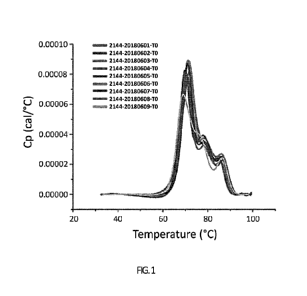
FIG. 1: MicroCal DSC thermogram overlay from the anti-Cx43 Ab pH/Buffer
screening
study.
FIG. 2: SEC-Main peak % comparison at 25 2 C (left) and 40 2 C (right) from
the
pH/buffer screening study.
FIG. 3: Comparison of cIEF main peak % at 25 2 C (left) and 40 2 C (right)
from the
pH/buffer screening study.
FIG. 4: Non-reduced SDS-Caliper purity % comparison from the pH/Buffer
screening
study at 25 2 C (left) and 40 2 C (right).
FIG. 5: Reduced SDS-Caliper purity % comparison from the pH/Buffer screening
study
at 25 2 C (left) and 40 2 C (right).
FIG. 6: SEC-HPLC main peak % comparison from the freeze/thaw study.
FIG. 7: Comparison of cIEF main peak % from the freeze/thaw study.
FIG. 8: Purity % comparison from the freeze/thaw study in non-reduced SDS-
Caliper
(left) and reduced SDS-Caliper (right).
FIG. 9: SEC-HPLC main peak % comparison from the agitation study.
FIG. 10: Comparison of cIEF main peak % from the agitation study.
FIG. 11: Purity % comparison from the agitation study in non-reduced SDS-
Caliper (left)
and reduced SDS-Caliper (right).
CA 03190474 2023-01-30
WO 2022/026914 PCT/US2021/044043
FIG. 12: SEC-Main peak % comparison at 2-8 C (left), 25 2 C (middle) and 40
2 C
(right).
FIG. 13: cIEF main peak % comparison at 2-8 C (left), 25 2 C (middle) and 40
2 C
(right).
5 FIG. 14: Non-reduced SDS-Caliper purity % comparison at 2-8 C (left),
25 2 C
(middle) and 40 2 C (right).
FIG. 15: Reduced SDS-Caliper purity % comparison at 2-8 C (left), 25 2 C
(middle)
and 40 2 C (right).
FIG. 16: MicroCal DSC thermogram overlay from the anti-Cx43 Ab formulation
confirmation study.
DETAILED DESCRIPTION
Disclosed herein, in some embodiments, is a stable, aqueous pharmaceutical
formulation
of anti-Cx43 antibodies. Such formulation can include: an anti-Cx43 antibody
or antigen binding
fragment thereof, a buffer, a surfactant, and a stabilizer. The pharmaceutical
formulation can
have a pH of between about 5 and about 6, or about 5.4-5.6, or about 5.5.
In some embodiments, the anti-Cx43 antibody or antigen binding fragment
thereof can
have a first, second and third heavy chain complementarity determining region
(CDR) sequence
having the amino acid sequence of SEQ ID NOs: 1, 2, and 3, respectively;
and/or a first, second
and third light chain CDR sequence having the amino acid sequence of SEQ ID
NOs: 4, 5, and
6, respectively.
In some embodiments, the anti-Cx43 antibody or antigen binding fragment
thereof can
have a heavy chain variable domain having the amino acid sequence of SEQ ID
NO: 7, and a
light chain variable domain having the amino acid sequence of SEQ ID NO: 8.
In certain embodiments, the anti-Cx43 antibody or antigen binding fragment
thereof
comprises a heavy chain having an amino acid sequence selected from the group
consisting of
SEQ ID NOs: 9-17, and a light chain having the amino acid sequence of SEQ ID
NO: 18.
In certain embodiments, the anti-Cx43 antibody or antigen binding fragment
thereof
binds to an epitope located within the amino acid sequence of FLSRPTEKTI (SEQ
ID NO: 19).
In various embodiments, the formulations disclosed herein can have improved
stability,
such that they display no significant changes (such as appearance, antibody
concentration, pH,
antibody aggregation, and antibody purity) observed at a predetermined
temperature (e.g., -
C or -20 C or refrigerated temperature of 2-8 C) for a period of time, e.g.,
ai least 3
months, at least 6 months, at least 1 year, or up to 3 years.
CA 03190474 2023-01-30
WO 2022/026914
PCT/US2021/044043
6
Definitions
Unless defined otherwise, all technical and scientific terms used herein have
the same
meaning as commonly understood by those of ordinary skill in the art to which
this disclosure
.. pertains. The following references provide one of skill with a general
definition of many of the
terms used in this disclosure: Academic Press Dictionary of Science and
Technology, Morris
(Ed.), Academic Press (1s' ed., 1992); 04brd .Dictionary of Biochemistry and
Molecular
Biology, Smith et al. (Eds.), Oxford University Press (revised ed., 2000);
Encyclopaedic
Dictionary of Chemistry, Kumar (Ed.), Anmol Publications Pvt. Ltd. (2002);
Dictionaty of
Microbiology and Molecular Biology, Singleton et al. (Eds.), John Wiley & Sons
(3 ed., 2002);
Dictionary giChernistiy, Hunt (Ed.), Routledg,e (1" ed.., 1999); Dictionary of
Pharmaceitical
Medicine, Na.hler (Ed.), Springer-Verlag Telos (1994); Dictionary of Organic
Chemistry, Kumar
and Anandand (Eds.), Anmol Publications Pvt. Ltd. (2002); and A. Dictionary of
Biology
(Oxford Paperback Reference), Martin and Hine (Eds.), Oxford University Press
(4th ed., 2000).
Further clarifications of some of these terms as they apply specifically to
this disclosure are
provided herein.
As used herein, the articles "a" and "an" refer to one or more than one, e.g.,
to at least
one, of the grammatical object of the article. The use of the words "a" or
"an" when used in
conjunction with the term "comprising" herein may mean "one," but it is also
consistent with the
.. meaning of "one or more," "at least one," and "one or more than one."
As used herein, "about" and "approximately" generally mean an acceptable
degree of
error for the quantity measured given the nature or precision of the
measurements. Exemplary
degrees of error are within 20 percent (%), typically, within 10%, and more
typically, within 5%
of a given range of values. The term "substantially" means more than 50%,
preferably more than
80%, and most preferably more than 90% or 95%.
As used herein the term "comprising" or "comprises" is used in reference to
compositions, methods, and respective component(s) thereof, that are present
in a given
embodiment, yet open to the inclusion of unspecified elements.
As used herein the term "consisting essentially of' refers to those elements
required for a
given embodiment. The term permits the presence of additional elements that do
not materially
affect the basic and novel or functional characteristic(s) of that embodiment
of the disclosure.
The term "consisting of' refers to compositions, methods, and respective
components
thereof as described herein, which are exclusive of any element not recited in
that description of
the embodiment.
CA 03190474 2023-01-30
WO 2022/026914 PCT/US2021/044043
7
An "anti-Cx43 antibody" is an antibody that immunospecifically binds to Cx43
(e.g., its
extracellular domain). The antibody may be an isolated antibody. Such binding
to Cx43 exhibits
a Ko with a value of, e.g., no greater than 1 uM, no greater than 100 0,1 or
no greater than 50
nM, KD can be measured by any methods known to one skilled in the art, such as
a surface
.. plasmon resonance assay or a cell binding assay. An anti-Cx43 antibody may
be a monoclonal
antibody, or antigen-binding fragments thereof. In some embodiments, the
antibody can be those
disclosed in PCT Application No. PCT/US2020/016606 filed February 4, 2020,
incorporated
herein by reference in its entirety,
An "antibody," as used herein is a protein comprising binding domains that
bind to a
target epitope. The term antibody includes monoclonal antibodies comprising
immunoglobulin
heavy and light chain molecules, single heavy chain variable domain
antibodies, and variants
and derivatives thereof, including chimeric variants of monoclonal and single
heavy chain
variable domain antibodies. Binding domains are substantially encoded by
immunoglobulin
genes or fragments of immunoglobulin genes, wherein the protein
immunospecifically binds to
an antigen. The recognized immunoglobulin genes include the kappa, lambda,
alpha, gamma,
delta, epsilon and mu constant region genes, as well as myriad immunoglobulin
variable region
genes. Light chains are classified as either kappa or lambda.. Heavy chains
are classified as
gamma, mu, alpha, delta, or epsilon, which in turn define the immunoglobulin
classes, IgG,
IgM, IgA, IgD and IgE, respectively. For most vertebrate organisms, including
humans and
murine species, the typical immunoglobulin structural unit comprises a
tetramer that is
composed of two identical pairs of polypeptide chains, each pair having one
"light" (about 25
kD) and one "heavy" chain (about 50-70 kD). "Vi.," and Vii" refer to the
variable domains of
these light and heavy chains respectively. "a" and CH" refer to the constant
domains of the
light and heavy chains. Loops of 13-strands, three each on the VL, and Vx are
responsible for
binding to the antigen, and are referred to as the "complementarity
determining regions" or
"CDRs". The "Fab" (fragment, antigen-binding) region includes one constant and
one variable
domain from each heavy and light chain of the antibody, i.e., Vt., CL, Vri and
CHL
Antibodies include intact immunoglobulins as well as antigen-binding fragments
thereof.
The term "antigen-binding fragment" refers to a polypeptide fragment of an
antibody which
binds antigen or competes with intact antibody (i.e., with the intact antibody
from which they
were derived) for antigen binding (i.e., specific binding). Antigen binding
fragments can be
produced by recombinant or biochemical methods that are well known in the art.
Exemplary
antigen-binding fragments include Fv, Fab, Fab', (Fab)2, CDR, paratope and
single chain Fv
CA 03190474 2023-01-30
WO 2022/026914
PCT/US2021/044043
8
antibodies (say) in which a Vu and a V1_, chain are joined together (directly
or through a peptide
linker) to form a continuous polypeptide.
Antibodies also include variants, chimeric antibodies and humanized
antibodies. The
term "antibody variant" as used herein refers to an antibody with single or
multiple mutations in
the heavy chains and/or light chains. In some embodiments, the mutations exist
in the variable
region. In some embodiments, the mutations exist in the constant region.
"Chimeric antibodies"
refers to those antibodies wherein one portion of each of the amino acid
sequences of heavy and
light chains is homologous to corresponding sequences in antibodies derived
from a particular
species or belonging to a particular class, while the remaining segment of the
chains is
homologous to corresponding sequences in another. Typically, in these chimeric
antibodies, the
variable region of both light and heavy chains mimics the variable regions of
antibodies derived
from one species of mammals, while the constant portions are homologous to the
sequences in
antibodies derived from another. One clear advantage to such chimeric forms is
that, for
example, the variable regions can conveniently be derived from presently known
sources using
readily available hybridomas or B cells from non-human host organisms in
combination with
constant regions derived from, for example, human cell preparations. While the
variable region
has the advantage of ease of preparation, and the specificity is not affected
by its source, the
constant region being human, is less likely to elicit an immune response from
a human subject
when the antibodies are injected than would the constant region from a non-
human source.
However, the definition is not limited to this particular example. "Humanized"
antibodies refer
to a molecule having an antigen-binding site that is substantially derived
from an
immunoglobulin from a non-human species and the remaining immunoglobulin
structure of the
molecule based upon the structure and/or sequence of a human immunoglobulin.
The antigen-
binding site may comprise either complete variable domains fused onto constant
domains or
only the complementarity determining regions (CDRs) grafted onto appropriate
framework
regions in the variable domains. Antigen binding sites may be wild type or
modified by one or
more amino acid substitutions, e.g., modified to resemble human immunoglobulin
more closely.
Some forms of humanized antibodies preserve all CDR sequences (for example, a
humanized
mouse antibody which contains all six CDRs from the mouse antibodies). Other
forms of
humanized antibodies have one or more CDRs (one, two, three, four, five, or
six) which are
altered with respect to the original antibody, which are also termed one or
more CDRs "derived
from" one or more CDRs.
As described herein, the amino acid residues of an antibody can be numbered
according
to the general numbering of Kabat (Kabat, et al (1991) Sequences of Proteins
of Immunological
CA 03190474 2023-01-30
WO 2022/026914 PCT/US2021/044043
9
Interest, 5th edition. Public Health Service, NIH, Bethesda, MD).
The term "binding" as used herein in the context of binding between an
antibody and an
epitope of Cx43 as a target, refers to the process of a non-covalent
interaction between
molecules. Preferably, said binding is specific. The specificity of an
antibody can be determined
based on affinity. A specific antibody can have a binding affinity or
dissociation constant KD for
its epitope of less than 10-7 M., preferably less than le M.
The term "antigen" refers to a molecule or a portion of a molecule capable of
being
bound by a selective binding agent, such as an antibody, and additionally
capable of being used
in an animal to produce antibodies capable of binding to an epitope of that
antigen. An antigen
may have one or more epitopes.
The term "epitope" includes any determinant, preferably a polypeptide
determinant,
capable of specific binding to an immunoglobulin or T-cell receptor. In
certain embodiments,
epitope determinants include chemically active surface groupings of molecules
such as amino
acids, sugar side chains, phosphoryl, or sulfonyl, and, in certain
embodiments, may have specific
three-dimensional structural characteristics, and/or specific charge
characteristics. In one
embodiment, an epitope is a region of an antigen that is bound by an antibody.
In certain
embodiments, an antibody is said to specifically bind an antigen when it
preferentially
recognizes its target antigen in a complex mixture of proteins and/or
macromolecules. Methods
for epitope mapping are well known in the art, such as X-ray co-
crystallography, array-based
oligo-peptide scanning, site-directed mutagenesis, high throughput mutagenesis
mapping and
hydrogen¨deuterium exchange. Epitopes can be formed both from contiguous amino
acids or
noncontiguous amino acids juxtaposed by tertiary folding of a protein.
Epitopes formed from
contiguous amino acids are typically retained on exposure to denaturing
solvents, whereas
epitopes formed by ter-6w), folding are typically lost on treatment with
denaturing solvents. An
epitope typically includes at least 3, and more usually, at least 5 or 8-10
amino acids in a unique
spatial conformation.
The term "subject" or "patient" includes a human or other mammalian animal
that
receives either prophylactic or therapeutic treatment,
The terms "treat," "treating," and "treatment," as used herein, refer to
therapeutic or
preventative measures such as those described herein. The methods of
"treatment" employ
administration to a patient a Cx43 iigand provided herein, for example, a
patient having an
inflammatory disease or condition or a neurodegenerative disease, in order to
prevent, cure,
delay, reduce the severity of, or ameliorate one or more symptoms of the
inflammatory disease
or condition or a neurodegen.erative disease, or in order to prolong the
survival of a patient
CA 03190474 2023-01-30
WO 2022/026914 PCT/US2021/044043
beyond that expected in the absence of such treatment. The methods of
"treatment" also employ
administration to a patient a Cx43 ligand provided herein (e.g., an antibody)
to provide therapy
in a patient beyond that expected in the absence of such treatment.
The term "inflammatory disease" broadly refers to the vast array of disorders
and
5 conditions that are characterized by inflammation. Examples include
arthritis, allergy, asthma,
autoirnmune diseases, coeliac disease, glomerulonephiitis, hepatitis,
inflammatory bowel disease
(including CroIan's disease and Ulcerative Colitis), reperfusion injury and
transplant rejection.
The term "autoimmune disease" broadly refers to diseases in which the immune
system
attacks its own proteins, cells, and tissues, or in which immune effector T
cells are autoreactive
10 to endogenous self peptides and cause destruction of tissue. Autoimmune
diseases include but
are not limited to rheumatoid arthritis, Crohn's disease, Type 1 diabetes,
alopecia, multiple
sclerosis, lupus, systemic lupus erythematosus (SLE), autoimmune
encephalomyelitis,
myasthenia gravis (MG), Hashimoto's thyroiditis, Goodpasture's syndrome,
pemphigus (e.g.,
pemphigus vulgaris), Grave's disease, autoimmune hemolytic anemia, autoimmune
thrombocytopenic purpura, scleroderma with anti-collagen antibodies, mixed
connective tissue
disease, polymyositis, pernicious anemia, idiopathic Addison's disease,
autoimmune-associated
infertility, glomerulonephritis (e.g., crescentic glomerulonephritis,
proliferative
glomerulonephritis), bullous pemphigoid, Sjogren's syndrome, insulin
resistance, and
autoimmune diabetes mellitus.
The term "neurodegenerative disease" broadly refers to diseases characterized
by the
progressive loss of structure and/or function of neurons. Neurodegenerative
diseases include but
are not limited to Alzheimer's disease (AD), lysosomal storage disorders,
bacterial meningitis,
amyotrophic lateral sclerosis, hypoxia, ischemia, glaucoma, schizophrenia,
major depression,
bipolar disorder, epilepsy, traumatic brain injury, post-traumatic stress
disorder, Parkinson's
disease, Down syndrome, spinocerebellar ataxia, Huntington's disease,
radiation therapy induced
neurodegeneration, chronic stress induced neurodegeneration, and
neurodegeneration associated
with normal aging or abuse of neuro-active drugs (such as alcohol, opiates,
methamphetamine,
phencyclidine, and cocaine).
The term "effective amount" as used herein, refers to that amount of an agent,
such as a
Cx43 ligand, for example an anti-Cx43 antibody, which is sufficient to effect
treatment,
prognosis or diagnosis of a disease, when administered to a patient. A
therapeutically effective
amount will vary depending upon the patient and disease condition being
treated, the weight and
age of the patient, the severity of the disease condition, the manner of
administration and the
like, which can readily be determined by one of ordinary skill in the art. The
dosages for
CA 03190474 2023-01-30
WO 2022/026914
PCT/US2021/044043
11
administration can range from, for example, about 1 ng to about 10,000 mg,
about 5 1112", to about
9,500 mg, about 10 ng to about 9,000 mg, about 20 ng to about 8,500 mg, about
30 ng to about
7,500 mg, about 40 ng to about 7,000 mg, about 50 ng to about 6,500 mg, about
100 ng to about
6,000 mg, about 200 ng to about 5,500 mg, about 300 ng to about 5,000 mg,
about 400 ng to
about 4,500 mg, about 500 ng to about 4,000 mg, about 1 pg to about 3,500 mg,
about 5 lag to
about 3,000 mg, about 10 ug to about 2,600 mg, about 20 ug to about 2,575 mg,
about 30 lig to
about 2,550 mg, about 40 !.ig to about 2,500 mg, about 50 lig to about 2,475
mg, about 100 pg to
about 2,450 mg, about 200 pg to about 2,425 mg, about 300 us, to about 2,000,
about 400 pg to
about 1,175 mg, about 500 lag to about 1,150 mg, about 0.5 mg to about 1,125
mg, about 1 mg
to about 1,100 mg, about 1.25 mg to about 1,075 mg, about 1.5 mg to about
1,050 mg, about 2.0
mg to about 1,025 mg, about 2.5 mg to about 1,000 mg, about 3.0 mg to about
975 mg, about
3.5 mg to about 950 mg, about 4.0 mg to about 925 mg, about 4.5 mg to about
900 mg, about 5
fig to about 875 mg, about 10 mg to about 850 mg, about 20 mg to about 825 mg,
about 30 mg
to about 800 mg, about 40 mg to about 775 mg, about 50 mg to about 750 mg,
about 100 mg to
about 725 mg, about 200 mg to about 700 mg, about 300 mg to about 675 mg,
about 400 mg to
about 650 mg, about 500 mg, or about 525 mg to about 625 mg, of an antibody or
antigen
binding portion thereof, as provided herein. Dosing may be, e.g., every week,
every 2 weeks,
every three weeks, every 4 weeks, every 5 weeks or every 6 weeks. Dosage
regimens may be
adjusted to provide the optimum therapeutic response. An effective amount is
also one in which
any toxic or detrimental effects (side effects) of the agent are minimized
and/or outweighed by
the beneficial effects. Administration may be intravenous at exactly or about
6 mg/kg or 12
mg/kg weekly, or 12 mg/kg or 24 mg/kg biweekly. Additional dosing regimens are
described
below.
As used herein, "formulation" is a composition of a pharmaceutically active
drug, such
as a biologically active protein (e.g., antibody), that is suitable for
parenteral administration
(including but not limited to intravenous, intramuscular, or subcutaneous) to
a patient in need
thereof and includes only pharmaceutically acceptable excipients, diluents,
and other additives
deemed safe by the Federal Drug Administration or other foreign national
authorities.
As used herein the phrases "liquid formulation" and "aqueous formulation" are
used
interchangeably to refer to a solution or liquid preparation that contains a
biopharmaceutical in
combination with one or more excipients (e.g., chemical additives)
dissolved in a suitable
solvent.
A "stable" formulation is a pharmaceutical formulation with no significant
changes
observed at a predetermined temperature (e.g., -40 "C or -20 "C or
refrigerated temperature of
CA 03190474 2023-01-30
WO 2022/026914
PCT/US2021/044043
12
2-8 CC) for a period of time, e.g., at least 3 months, at least 6 months, at
least 1 year, or up to 3
years. Stability of the formulations disclosed herein can be evaluated using
one or more of the
following criteria: 1) the aqueous formulation is colorless; or clear to
slightly opalescent by
visual analysis; 2) the protein content is maintained within +/---5 mg/mil.,
from initial
concentration; 3) the pH is maintained within /-0.5 pH units from target pH;
4) the percent of
monomer by SEC is ,,."--!95%; 5) the purity as measured by CE-SDS is :=.3:-
.90% and the relative
potency based on. ELISA is within 60-150%.
As used herein the term "excipient" is intended to mean a therapeutically
inactive
substance. Excipients are included in a formulation for a wide variety of
purposes, for example,
as a buffer; stabilizer; tonicity agent, surfactant, anti-oxidant,
cryoprotectant or diluent.
Suitable excipiems include, but are not limited to polyols (also known as
sugar alcohols)
such as mannitol or sorbitol, sugars such as sucrose, lactose or dextrose,
salts such as NaCI, KCI
or calcium phosphate, amino acids, for example, histidine, lysine, aspartic
acid, or glutamic acid,
surfactants, as well as water. The purity of the excipient should meet
com.pendi al standards (e.g.,
USP, EP, JP) and be of sufficient purity for subcutaneous, intramuscular, or
intravenous
injection into humans.
The term "buffer" or "buffering agent" as used herein, refers to a
pharmaceutically
acceptable excipient, which stabilizes the pH of a pharmaceutical preparation.
Suitable buffers
are well known in the art and can be found in the literature. For example,
citrate salts, acetate
salts, histidine salts, succinate salts; malate salts, phosphate salts or
lactate salts, and/or the
respective free acids or bases thereof, as well as mixtures of the various
salts and/or acids and
bases thereof can be employed. In a particular embodiment, pharmaceutically
acceptable buffers
comprise but are not limited to histidine buffers, citrate buffers, succinate
buffers, acetate
buffers and phosphate buffers. In a particular embodiment; buffers are acetate
buffers, for
example, sodium acetate buffer. Other particular buffers are hi sti dine
buffers, i.e. buffers having
histidine, generally L-histidine, as buffering agent. A particular buffer is L-
histidine/HC1 buffer,
comprising L-histidine or mixtures of L.-histidine and L-histidine
hydrochloride and pH
adjustment achieved with hydrochloric acid. Unless otherwise indicated, the
term "L-histi dine"
when used herein to describe a buffering agent, refers to L-histidinelHO
buffer. L-histidine/FIC1
buffer can be prepared by dissolving suitable amounts of L-hi sti dine and IL-
histidine
hydrochloride in water, or by dissolving a suitable amount of L-histidine in
water and adjusting
the pH to the desired value by addition of hydrochloric acid. The
abovemention.ed buffers are
generally used at a concentration of about 1 inN4 to about 100 inM, about 10
rnM to about 50
mM, about 15 to 30 mkt or 20 m.M. Regardless of the buffer used, the pH can be
adjusted to a
CA 03190474 2023-01-30
WO 2022/026914
PCT/US2021/044043
13
value in the range from about 4.0 to about 7.0, about 5.0 to about 6.0, about
5.4 to about 5.6, or
about 5.5, with an acid or a base known in the art, e.g., hydrochloric acid,
acetic acid,
phosphoric acid, sulfuric acid and citric acid, sodium hydroxide and potassium
hydroxide.
The term "surfactant" as used herein denotes a pharmaceutically acceptable,
surface
-
active agent. In a particular embodiment, a non-ionic surfactant is used.
Examples of
pharmaceutically acceptable surfactants include, but are not limited to,
polyoxyethylen-sorbitan
fatty acid esters (Tween), polyoxyethylene alkyl ethers (Brij),
alkylphenylpolyoxyetbylene
ethers (Triton X), polyoxyetksidene-polyoxypropylene copolymers (Poloxamer,
Pluronic), and
sodium dodecyl sulphate (SDS). In a particular embodiment, polyoxyethylene-
sorbitan fatty acid
.. esters are polysorbate. 20 (polyoxyethylene sorbitan monolaureate, sold
under the trademark
Tween 20Tm) and polysorbate SO (polyoxyethylene sorbitan monooleate, sold
under the
trademark Tween 80Tm). In a particular embodiment, polyethylene-polypropylene
copolymers
are those sold under the names Plutonic .F68 or Poloxamer 1881.m. In a
particular embodiment,
polyoxyethylene alkyl ethers are those sold under the trademark BrijmI. in a
particular
embodiment alkylphenylpolyoxyethylene ethers are sold under the tradenanie
Triton X, for
example, p-tert-octylphenoxy polyethoxyethanol (sold under the tradename
Triton X-10(I1m).
When polysorbate 20 (Tween 2OTM) and polysorbate 80 (Tween 8OTM) are used,
they are
generally used at a concentration range of about 0.001 to about 1%, about 0.01
to about 0.1% or
about 0.02% to about 0.05%. In the formulation of the disclosure, the
concentration of the
surfactant is described as a percentage, expressed in weight/volume (wN).
The term "stabilizer" as used herein denotes a pharmaceutically acceptable
excipient,
which protects the active pharmaceutical ingredient and/or the forniulati on
from chemical and/or
physical degradation during manufacturing, storage and application.
Stabilizers include but are
not limited to sacchafides, amino acids, polyols, e.g. mannitol, sorbitol,
xylitol, dextran, glycerol,
arabitol, propylene glycol, polyethylene glycol, cyclodextrines, e.g.
hydroxypropyl-P-
cyclodextrine, sulfobutylethyl-ii-cyclodextrine, p-cyclodextrine,
polyethylenci,lycols, e.g. PEG
3000, PEG 3350, PEG 4000, PEG 6000, albumines, e.g,. human serum albumin (I-
ISA), bovine
serum albumin (BSA), salts, e.g. sodium chloride, magnesium chloride, calcium
chloride,
chelators, e.g. MIA as hereafter defined. As mentioned hereinabove,
stabilizers can be present
in the formulation in an amount of about I to about 500 aiM, in an amount of
about 10 to about
300 in.N4 or in an amount of about 120 niM to about 300 rnIVI. More than one
stabilizer, selected
from the same or from different groups, can be present in the formulation.
The term "saccharide" as used herein includes monosaccharides and
oligosa.cchaTides. A
monosaccharide is a monomeric carbohydrate which is not hydrolysable by acids,
including
CA 03190474 2023-01-30
WO 2022/026914
PCT/US2021/044043
14
simple sugars and their derivatives, e.g. aminosugars. Saccharides are usually
in their D
conformation. :Examples of monosaccharides include glucose, fructose,
galactose, mannose,
sorbose, ribose, deoxyribose, neuraminic acid. An oligosaccharide is a
carbohydrate consisting
of more than one monomeric saccharide unit connected via glycosidic bond(s)
either branched
or in a linear chain. The monomeric saccharide units within an oligosaccharide
can be identical
or different. Depending on the number of monomed.c saccharide units the
oligosacchari de is a
di-, tri-, tetra- penta- and so forth saccharide. in contrast to
polysaccharides the monosaccharides
and oligosacchalides are water soluble. Examples of oligosaccharides include
sucrose, trehalose,
lactose, maltose and raffinose. In a particular embodiment, saccharides are
sucrose and trehalose
(i.e. a,ci-D-trehalose), for example, sucrose. Trehalose is available as
trehalose dihydrate.
Sacchari des can be present in the formulation in an amount of about 100 to
about 500 m.M, in an
amount of about 200 to about 300 itiM or in an amount of about 240 iuM.
A subgroup within the stabilizers are lyoprotectants. The term lyoprotectant."
denotes
pharmaceutically acceptable excipients, which protect the labile active
ingredient (e.g. a protein)
against destabilizing conditions during the lyophilisation process, subsequent
storage and
reconstitution. Lyoprotectants comprise but are not limited to the group
consisting of
saccharides, polyols (such as e.g. sugar alcohols) and amino acids. in a
particular embodiment,
ly'oprotectants can be selected from the group consisting of saccharides such
as sucrose,
trehalose, lactose, glucose, marmose, maltose, galactose, fructose, sorbose,
raffinose, neuraminic
acid, amino sugars such as giurosamine, galactosamine, N-methylglucosamine
("Meglumine"),
polyols such as mannitol and sorbitol, and amino acids such as arginine and
glycine or mixtures
thereof. Lyoprotectants are generally used in an amount of about 10 to 500
mkt, in an amount of
about 10 to about 300 triM or in an amount of about 100 to about 300 m:M.
Another subgroup within the stabilizers are antioxidants. The term
"antioxidant" denotes
pharmaceutically acceptable excipients, which prevent oxidation of the active
pharmaceutical
ingredient. Antioxidants comprise but are not limited to ascorbic acid,
gluthathione, cysteine,
methionine, citric acid, EDIA.. Antioxidants can be used in an amount of about
0.01 to about
100 mkt, in an amount of about 5 to about 50 mM or in an amount of about 5 to
about 25 iuM.
The formulations according to the disclosure may also comprise one or more
tonicity
agents. The term "tonicity agents" denotes pharmaceutically acceptable
excipients used to
modulate the tonicity of the form.ul ati. on . The form.ul ati. on can be
hypotoni c, isotonic or
hypertonic. Isotonicity in general relates to the osmotic pressure of a
solution, usually relative to
that of human blood serum (around 250-350 mOstn.ol/kg). The formulation
according to the
disclosure can be try'potonic, isotonic or hypertonic. In a particular
embodiment, the formulation
CA 03190474 2023-01-30
WO 2022/026914
PCT/US2021/044043
is isotonic. An isotonic formulation is liquid or liquid reconstituted from a
solid form, e.g. from
a lyophilized form, and denotes a solution having the same tonicity as some
other solution with
which it is compared, such as physiologic salt solution and the blood serum.
Suitable tonicity
agents comprise but are not limited to sodium chloride, potassium chloride,
glycerine and any
5 component from the group of amino acids or sugars, in particular glucose.
Tonicity agents are
generally used in an amount of about 5 mkt. to about 500 in.M.
Within the stabilizers and tonicity agents there is a group of compounds which
can
function in both ways, i.e. they can at the same time be a stabilizer and a
tonicity agent.
Examples thereof can be found in the group of sugars, amino acids, polyols,
cyclodextrines,
10 polyethyleneglycols and salts, An example for a sugar which can at the
same time be a stabilizer
and a tonicity agent is trehalose.
The "isoelectric point" or "pI" of a protein is the pH at which the protein
has a net
overall charge equal to zero, i.e., the at which the protein has an equal
number of positive
and negative charges. Determination of the pI for any given protein can be
done according to
15 well-established techniques, such as, e.g., by isoelectric focusing..
isoelectric focusing is a
technique for separating different molecules by differences in their
isoelectric point (pA). It is a
type of zone electrophoresis, usually- performed on proteins in a gel that
takes advantage of the
fact that overall charge on the molecule of interest is a function of the pH
of its surroundings.
Various aspects of the disclosure are described in further detail below.
Additional
definitions are set out throughout the specification.
Pharmaceutical Formulations
In some embodiments, the present disclosure provides a pharmaceutical
composition
comprising an anti-Cx43 antibody, or antigen binding fragment thereof, as
described herein. The
anti-Cx43 antibody, or antigen binding fragment thereof, can have a first,
second and third
heavy chain complementarity determining region (CDR) sequence having the amino
acid
sequence of SEQ ID NOs: 1, 2, and 3, respectively; and a first, second and
third light chain CDR
sequence having the amino acid sequence of SEQ ID NOs: 4, 5, and 6,
respectively.
In some embodiments, the anti-Cx43 antibody or antigen binding fragment
thereof can
include a heavy chain variable domain having the amino acid sequence of SEQ ID
NO: 7, and a
light chain variable domain having the amino acid sequence of SEQ ID NO: 8.
In certain embodiments, the anti-Cx43 antibody or antigen binding fragment
thereof
comprises a heavy chain having an amino acid sequence selected from the group
consisting of
SEQ ID NOs: 9-17, and a light chain having the amino acid sequence of SEQ ID
NO: 18.
CA 03190474 2023-01-30
WO 2022/026914
PCT/US2021/044043
16
In certain embodiments, the anti-Cx43 antibody or antigen binding fragment
thereof binds
to an epitope located within the amino acid sequence of FLSRPTEKTI (SEQ ID NO:
19).
In various embodiments, the anti-Cx43 antibody or antigen binding fragment
thereof can
be formulated in pharmaceutically acceptable amounts and in pharmaceutically
acceptable
compositions. As used herein, "pharmaceutically acceptable" shall refer to
that which is useful
in preparing a pharmaceutical composition that is generally safe, non-toxic,
and neither
biologically nor otherwise undesirable and includes that which is acceptable
for veterinary use
as well as human pharmaceutical use. Examples of "pharmaceutically acceptable
liquid carriers"
include water and organic solvents. Preferred pharmaceutically acceptable
aqueous liquids
include PBS, saline, and dextrose solutions etc.
As used herein, the term "pharmaceutically acceptable salt" means any
pharmaceutically
acceptable salt of the compounds disclosed herein. For example,
pharmaceutically acceptable
salts of any of the compounds described herein include those that are within
the scope of sound
medical judgment, suitable for use in contact with the tissues of humans and
animals without
undue toxicity, irritation, allergic response and are commensurate with a
reasonable benefit/risk
ratio. Pharmaceutically acceptable salts are well known in the art. For
example,
pharmaceutically acceptable salts are described in: Berge et al., I
Pharmaceutical Sciences
66:1-19, 1977 and in Pharmaceutical Salts: Properties, Selection, and Use,
(Eds. P. H. Stahl and
C. G. Wermuth), Wiley-VCH, 2008. The salts can be prepared in situ during the
final isolation
and purification of the compounds described herein or separately by reacting a
free base group
with a suitable organic acid.
Various lite.rature references are available to facilitate selection of
pharmace-utically
acceptable carriers or excipients. See, e.g., Remington's Pharmaceutical
Sciences and U.S.
Pharmacopeia: National Formulary, Mack Publishing Company, Easton, Pa, (1984);
:Harchnan et
al. (2001) Goodman and Ciilman's The Pharmacological Basis of Therapeutics,
McGraw-Hill,
New York, N.Y.; Gennaro (2000) Remington: The Science and Practice of
Pharmacy,
Lippincott, Williams, and Wilkins, New York, N.Y.; Avis et al. (eds.) (1993)
Pharmaceutical
Dosage Forms: Parenteral Medications, Marcel Dekker, NY; Lieberman, et al.
(eds.) (1990)
Pharmaceutical Dosage :Forms. Tablets, Marcel Dekker, NY; :Lieberman et al.
(eds.) (1990)
Pharmaceutical Dosage Forms: Disperse Systems, Marcel Dekker, NY; Weiner,
Wang, W., int.
I Pharm. 185:129-188 (1999) and Wang, W., hit. J. Pharm. 203:1-60 (2000), and
Kotkoskie
(2000) Excipient Toxicity and Safety, Marcel Dekker, Inc., New York, N.Y.
In some embodiments, the antibody formulation can comprise a buffer (e.g., hi
stidine,
acetate, phosphate or citrate buffer), a surfactant (e.g., polysorbate),
and/or a stabilizer agent
CA 03190474 2023-01-30
WO 2022/026914
PCT/US2021/044043
17
(e.g., sucrose), etc. In some certain embodiments, the buffer can be selected
from acetate/sodium
acetate, histidine/aspartic acid, citric acid/sodium citrate, dibasic sodium
phosphate/sodium
dihydrogen phosphate, and histidine/histidine hydrochloride. In certain
embodiments, the buffer
is hi stidine/aspartic acid or hi sti dine/hi sti dine hydrochloride. In
certain embodiments, the buffer
is hi sti dine/hi sti dine hydrochloride. In some embodiments, the surfactant
is poly s orb ate 80
(PS80). In certain embodiments, the stabilizer is selected from
ethylenediaminetetraacetic acid
(EDTA), sodium chloride, sorbitol, glycine, and sucrose. In certain
embodiments, the stabilizer
is sucrose.
some embodiments, the antibody forrmilati on can comprise pharmaceutically
acceptable carriers, including, e.g., ion exchangers, alumina, aluminum
stearate, lecithin, serum
proteins, such as human serum albumin, buffer substances such as phosphates,
sucrose, glycin.e,
sorbic acid, potassium sorbate, partial glyceride mixtures of saturated
vegetable fatty acids,
water, salts or electrolytes, such as protamine sulfate, disodium hydrogen
phosphate, potassium
hydrogen phosphate, sodium chloride, zinc salts, colloidal silica, magnesium
trisilicate,
polyvinyl pyrrolidone, cellulose.-based substances, polyethylene glycol,
sodium
carboxymethylcellulose, polyacrylates, polyethylene-polyoxypropylene-block
polymers, and
polyethylene glycol. In some embodiments, the antibody formulation further
comprises a.
surfactant. In some embodiments, the surfactant is selected from the group
consisting of
polysorbate, sodium dodecyl sulfate, and nonionic surfactant.
The formulation according to the disclosure can be in a liquid form, in a
lyophilized form
or in a liquid form reconstituted from a lyophilized form. In certain
embodiments, the
formulation is in a liquid form. The term "liquid." as used herein in
connection with the
formulation according to the disclosure denotes a formulation which is liquid
at a temperature of
at least about 2 to about 8 'C under atmospheric pressure. The term
"Iyophiliz.ed" as used herein
in connection with the formulation according to the disclosure denotes a
formulati011 which is
manufactured by freeze-drying methods known in the art per se. The solvent
(e.g., water) is
removed by freezing followed by sublimation of the ice under vacuum and
desorption of
residual water at elevated temperature. The lyophilizate usually has a
residual moisture of about
0.1 to 5% (w/w) and is present as a powder or a physically stable cake. The
lyophilizate is
characterized by a fast dissolution after addition of a reconstitution medium.
The term "reconstituted form" as used herein in connection with the
formulation
according to the disclosure denotes a formulation which is lyophilized and re--
dissolved by
addition of reconstitution medium. Suitable reconstitution media comprise but
are not limited to
water for injection (\\ 1) bacteriostatic water for injection (BWEI), sodium
chloride solutions
CA 03190474 2023-01-30
WO 2022/026914
PCT/US2021/044043
18
(e.g. 0.9% (w/v)NaC1), glucose solutions (e.g. 5% glucose), surfactant-
containing solutions (e.g.
0.02% polysorbate 80), pH-buffered solutions (eg. phosphate-buffered
solutions).
The formulation according to the disclosure is physiologically well tolerated,
can be
prepared easily, can be dispensed precisely and is stable with respect to
decomposition products
and aggregates over the duration of storage, during repeated freezing and
thawing cycles and
mechanical stress. It is stable at storage temperatures (e.g., -40 "C or -20
C or 2-8 C) over a
period of more than 1 year.
The antibody formulations of the present disclosure can be an aqueous
solution. In some
embodiments, the antibody formulation has not been subjected to freezing
temperatures, and/or
have not been frozen, i.e., they have remained in a liquid state. In some
embodiments, the
antibody in the antibody formulation has not been subjected to iyophilizati
on.
In some embodiments, the antibody formulations can have improved stability. As
used
herein, the term "stability" generally is related to maintaining the integrity
or to minimizing the
degradation, denaturation, aggregation or unfolding of a biologically active
agent such as a
protein, peptide or another bioactive macromolecule. As used herein, "improved
stability"
generally means that, under conditions known to result in degradation,
denaturation, aggregation
or unfolding., the protein (e.g., antibody such as anti-Cx43 .Ab), peptide or
another bioactive
in acromolecul e of interest maintains greater stability cornpared to a
control protein, peptide or
another bioactive macromolecule.
In some embodiments, stability refers to an antibody formulation haying low to
undetectable levels of particle formation. The phrase "low to undetectable
levels of particle
formation" as used herein refers to samples containing less than 30
particleslmfõ, less than 20
particles/ML, less than 20 particles/11Th, less than 15 particleslinL, less
than 10 particles/11'1h,, less
than 5 particles/m..1, less than 2 particles/int, or less than I particle/int,
as determined by HIAC
analysis or visual analysis. In some embodiments, no particles in the antibody
formulation are
detected, either by HIAC analysis or visual analysis.
In some embodiments, stability refers to reduced fragmentation of the
antibody. The
term "low to undetectable levels of fragmentation" as used herein refers to
samples containing
equal to or more than 80%, 85%, 90%, 95%, 98% or 99% of the total protein, for
example, in a
single peak as determined by liPSEC, or in two peaks (e.g., heavy- and light-
chains) (or as
in any peaks as there are subunits) by reduced Capillary Gel Electrophoresis
(rC,GE),
representing the non-degraded antibody or a non-degraded fragment thereof, and
containing no
other single peaks having more than 5%, more than 4%, more than 3%, more than
2%, more
than 1%, or more than 0.5% of the total protein in each. The term "reduced
Capillary Gel
CA 03190474 2023-01-30
WO 2022/026914
PCT/US2021/044043
19
Electrophoresis" as used herein refers to capillary gel electrophoresis under
reducing conditions
sufficient to reduce disulfide bonds in an antibody.
One of skill in the art will appreciate that stability of a protein is
dependent on other
features in addition to the composition of the formulation. For example,
stability can be affected
by temperature, pressure, humidity, pH, and external forms of radiation. Thus,
unless otherwise
specified, stability referred to herein is considered to be measured at -20
"C, one atmosphere
pressure, 50% relative humidity, pH of 5.5, and normal background levels of
radiation. Stability
of the antibody in the antibody formulation can be determined by various
means. In some
embodiments, the antibody stability is determined by size exclusion
chromatography (SEC).
SEC separates analytes (e.g., macromolecules such as proteins and antibodies)
on the basis of a
combination of their hydrodynamic size, diffusion coefficient, and surface
properties. Thus, for
example, SEC can separate antibodies in their natural three-dimensional
conformation from
antibodies in various states of denaturation, and/or antibodies that have been
degraded. 111 S EC,
the stationary phase is generally composed of inert particles packed into a
dense three
dimensional matrix within a glass or steel column. The mobile phase can be
pure water, an
aqueous buffer, an organic solvent, mixtures of these, or other solvents. The
stationary-phase
particles have small pores and/or channels which will only allow species below
a. certain size to
enter. Large particles are therefore excluded from these pores and channels,
hut the smaller
particles are removed from the flowing mobile phase. The time particles spend
immobilized in
the stationary-phase pores depends, in part, on how far into the pores they
can penetrate. Their
removal from the mobile phase flow causes them to take longer to elute from
the column and
results in a separation between the particles based on differences in their
size.
in some embodiments, SEC is combined with an identification technique to
identify or
characterize proteins, or fragments thereof. Protein identification and
characterization can be
accomplished by various techniques, including but not limited chromatographic
techniques, e.g.,
high-performance liquid chromatography (HPLC), immunoassays, electrophoresis,
ultra-
violet/visible/infrared spectroscopy, ram an spectroscopy, surface enhanced
raman spectroscopy,
mass spectroscopy, gas chromatography, static light scattering (SLS), Fourier
Transform
Infrared Spectroscopy (FITR,), circular dichroism (Cl)), urea-induced protein
unfolding
techniques, intrinsic tryptophan fluorescence, differential scanning calori
etry, and/or ANS
protein binding.
in some embodiments, protein identification is achieved by high-pressure
liquid
chromatography. Various instruments, and apparatuses are known to those of
skill in the art to
perform EIPLC. Cieneral l ElPLC involves loa.din.g a liquid solvent containing
the protein of
CA 03190474 2023-01-30
WO 2022/026914 PCT/US2021/044043
interest onto a separation column, in which the separation occurs. The hipLc
separation column
is filled with soli.d particles (e.g. silica, polymers, or sorbents), and the
sample mixture is
separated into compounds as it interacts with the column particles. HPLC
separation is
influenced by the liquid solvent's condition (e.g. pressure, temperature),
chemical interactions
5 between the sample mixture and the liquid solvent (e.g. hydrophobicity,
protonation, etc.), and
chemical interactions between the sample niixture and the solid particles
packed inside of the
separation column (e.g. ligand affinity, ion exchange, etc.).
In some enibodinients, the SEC and protein identification occurs within the
same
apparatus, or simultaneously. For example, SEC and HPLC can be combined, often
referred to
10 .. as SE4-1PLC.
In some embodiments, the aqueous formulation comprises about 2 mg,/mL to about
100
ing/mL antibody wherein the antibody comprises a heavy chain variable region
and a light chain
variable region, wherein the heavy chain variable region comprises the Kabat-
defined CDR1,
CDR2, and CDR3 sequences of SEQ ID NOs: 1-3, and wherein the light chain
variable region
15 comprises the Kabat-defined CDR1, CDR2, and CDR3 sequences of SEQ ID
NOs: 4-6, wherein
said formulation is stable upon storage at about 40 "C for at least 1 month.
In some
embodiments, the forimilation is stable upon storage at about 25 'C for at
least 3 months. In
some embodiments, the formulation is stable upon storage at about 5 "C for at
least 6 months. In
some embodiments, the formulation is stable upon storage at about 5 C for at
least 12 months.
20 .. In some embodiments, the formulation is stable upon storage at about 5
"C for at least 18
months. In some embodiments, the formulation is stable upon storage at about 5
C for at least
24 months, or 36 months.
The term "stable" can be relative and not absolute. Thus, in some embodiments
the
antibody is stable if less than 20%, less than 1.5%, less than 10%, less than
5% or less than 2% of
the antibody is degraded, denatured, aggregated or unfolded as determined by
SEC HPLC when
the antibody is stored -20 0C for 6 months. In some embodiments, the antibody
is stable if less
than 20%, less than 1.5%, less than 10%, less than 5% or less than 2% of the
antibody is
degraded, denatured, aggregated or unfolded as determined by SEC HPLC when the
antibody is
stored at -20 C for 12 months. In some embodiments, the antibody in the
antibody formulation
is stable if less than 20%, less than 15%, less than 10%, less than 5% or less
than 2% of the
antibody is degraded, denatured, aggregated or unfolded as determined by SEC
HPLC when the
antibody is stored at -20 "C for 18 months. In some embodiments, the antibody
in the antibody
formulation is stable if less than 20%, less than 15%, less than 10%, less
than 5% or less than 2%
of the antibody is degraded, denatured, aggregated or unfolded as determined b
SEC EIPI,C,
CA 03190474 2023-01-30
WO 2022/026914 PCT/US2021/044043
21
when the antibody is stored at -20 C for 24 months.
In some embodiments, the antibody is stable if less than 20%, less than 15%,
less than
10%, less than 5% or less than 2% of the antibody is degraded, denatured,
aggregated or
unfolded as determined by SEC IPLC when the antibody is stored at 23 C to 27
"C for 3
months. In some embodiments, the antibody is stable if less than 20%, less
than 15%, less than
10%, less than 5% or less than 2% of the antibody is degraded, denatured,
aggregated or
unfolded as determined by SEC HPLC when the antibody is stored at 23 'C to 27
C for 6
months. In some embodiments, the antibody is stable if less than 20%, less
than 15%, less than
10%, less than 5% or less than 2% of the antibody is degraded, denatured,
aggregated or
unfolded as determined by SEC }-PLC when the antibody is stored at 23 CC to 27
C for 12
months. In some embodiments, the antibody is stable if less than 20%, less
than 15%, less than
10%, less than 5% or less than 2% of the antibody is degraded, denatured,
aggregated or
unfolded as determined by SEC HPLC when the antibody is stored at 23 "C to 27
0C for 24
months.
In some embodiments the antibody is stable if less than 6%, less than 4%, less
than 3%,
less than 2% or less than 1% of the antibody is degraded, denatured,
aggregated or unfolded per
month as determined by SEC 1-IPLC when the antibody is stored at 40 C In some
embodiments
the antibody is stable if less than 6%, less than 4%, less than 3%, less than
2% or less than 1% of
the antibody is degraded, denatured, aggregated or unfolded per month as
determined by SEC
HPLC when the antibody is stored at 5 'C.
In some embodiments, the antibody formulations of the present disclosure can
be
considered stable if the antibody exhibits very little to no loss of the
binding activity of the
antibody (including antibody fragments thereof) of the formulation compared to
a reference
antibody as measured by antibody binding assays know to those in the art, such
as, e.g,, ELISAs,
etc., over a period of 8 weeks, 4 months, 6 months, 9 months, 12 months or 24
months. In some
embodiments, the antibody stored at about 40 C for at least I month retains
at least 60%, at
least 80%, at least about 85%, at least about 90%, at least about 95%, at
least about 98%, or at
least about 99% of binding ability to Cx43 compared to a reference antibody
which has not been
stored. In some embodiments, the antibody stored at about 5 "C for at least 6
months retains at
least 80%, at least about 85%, at least about 90%, at least about 95%, at
least about 98%, or at
least about 99% of binding ability to Cx43 compared to a reference antibody
which has not been
stored. in some embodiments, the antibody stored at about 40 C for at least 1
month retains at
least 95% of binding ability to Cx43 compared to a reference antibody which
has not been
stored. In some embodiments, the antibody stored at about 5 "C for at least 6
months retains at
CA 03190474 2023-01-30
WO 2022/026914 PCT/US2021/044043
22
least 95% of binding ability to Cx43 compared to a reference antibody which
has not been
stored.
The antibody formulations can provide low to undetectable levels of
aggregation of the
antibody. The phrase "low to undetectable levels of aggregation" as used
herein refers to
samples containing no more than about 5%, no more than about 4%, no more than
about 3%, no
more than about 2%, no more than about 1% and no more than about 0.5%
aggregation by
weight of protein as measured by high performance size exclusion
chromatography (HPSEC) or
static light scattering. (SI..,S) techniques. in some embodiments, less than
2% of the antibody
forms an aggregate upon storage at about 40 C for at least 4 weeks as
determined by as
determined by 1-1PSEC. In some embodiments, less than 2% of the antibody forms
an aggregate
upon storage at about 5" for at least 3 months, at least 6 months, at least 9
months, at least 12
months, at least 15 months, at least 18 months, at least 24 months, or at
least 36 months as
determined by HP SEC.
It has been discovered herein the antibody formulations provided herein result
in greatly
reduced particle formation as determined by visual inspection, micro-flowing
imaging (AR), or
size-exclusion chromatography (SEC). In some embodiments, the formulation is
substantially
free of particles upon storage at about 40 C for at least I month as
determined by visual
inspection. In some embodiments, the formulation is substantially free from
particles upon
storage at about 5 CC for at least 6 months, at least 9 months, at least 12
months, at least 15
months, at least 18 months, at least 24 months, or at least 36 months as
determined by visual
inspection.
The formulations may also contain adjuvants such as preservatives, wetting
agents,
emulsifying agents and dispersing agents. Prevention of presence of
microorganisms may be
ensured both by sterilization procedures, and by the inclusion of various
antibacterial and
antifungal agents, e.g. paraben, chlorobutanol, phenol, sorbic acid, and the
like. Preservatives
are generally used in an amount of about 0.001 to about 2% (wlv).
Preservatives comprise but
are not limited to ethanol, 'benzyl alcohol, phenol, m-cresol, p-chlor-m-
cresol, methyl or proryi
p arab en s, benzalkonium chloride.
The antibody formulations described herein can have various viscosities.
Methods of
measuring viscosity of antibody formulations are known to those in the art,
and can include, e.g.,
a rheometer (e.g., Anton Paar MCR301 Rheometer with either a 50 nun, 40 nun or
20 mm plate
accessory). In some embodiments of the present disclosure, the viscosities
were reported at a
high shear limit of 1000 per second shear rate, in sonic embodiments, the
antibody formulation
has a viscosity of less than 20 centipoise (cP), less than 18 cP, less than 15
cP, less than 13 cP,
CA 03190474 2023-01-30
WO 2022/026914 PCT/US2021/044043
23
or less than 11 cP. In some embodiments, the antibody formulation has a
viscosity of less than
13 cP. One of skill in the art will appreciate that viscosity is dependent on
temperature, thus,
unless otherwise specified, the viscosities provided herein are measured at 25
CC. unless
otherwise specified.
The antibody formulations can have different osmolarity concentrations.
Methods of
measuring osmolarity of antibody formulations are known to those in the art,
and can include,
e.g., an osmometer (e.g., an Advanced Instrument Inc 2020 freezing point
depression
osmometer). in some embodiments, the formulation has an osmolaiity of between.
200 and 600
mosm/kg, between 260 and 500 mosm/kg, or between 300 and 450 mosm/kg.
The antibody formulation of the present disclosure can have various pH levels.
In some
embodiments, the pH of the antibody formulation is between 4 and 7, between
4.5 and 6.5,
between 5 and 6, or between 5.4 to 5.6. In some embodiments, the pH of the
antibody
formulation is 5.5. in some embodiments, the pH of the antibody formulation is
6Ø In some
embodiments, the pH of the antibody formulation is >7Ø Various means may be
utilized in
achieving the desired pH level, including, but not limited to the addition of
the appropriate
buffer.
In some embodiments, the antibody formulation can include: about 40-60 mg/mL,
preferably about 50 mg/mL of an anti-Cx43 antibody or antigen binding fragment
thereof; about
10-40 mM, preferably about 20 mM histidine/histidine hydrochloride buffer;
about 0.005%-
0.05%, preferably about 0.02% w/v Polysorbate 80; and about 1%-20% w/v,
preferably about
8% w/v sucrose; wherein the formulation has a pH of between about 5.4 to about
5.6, preferably
about 5.5.
In some embodiments, the antibody formulation can include: about 50 mg/mL of
an anti-
Cx43 antibody or antigen binding fragment thereof, comprising a heavy chain
having an amino
acid sequence selected from the group consisting of SEQ ID NOs: 9-17, and
comprising a light
chain having the amino acid sequence of SEQ ID NO: 18; about 20 mM
histidine/aspartic acid
buffer; about 0.02% w/v Polysorbate 80; and about 8% w/v sucrose, wherein the
formulation has
a pH of between about 5.4 to about 5.6, preferably about 5.5.
In one embodiment, the antibody formulation can include: about 50 mg/mL of an
anti-
Cx43 antibody or antigen binding fragment thereof, comprising a heavy chain
having an amino
acid sequence of SEQ ID NO: 9 and a light chain having the amino acid sequence
of SEQ ID
NO: 18; about 20 mM histidine/aspartic acid buffer; about 0.02% w/v
Polysorbate 80; and about
8% w/v sucrose, wherein the formulation has a pH of between about 5.4 to about
5.6, preferably
about 5.5.
CA 03190474 2023-01-30
WO 2022/026914 PCT/US2021/044043
24
In one embodiment, the antibody formulation can include: about 50 mg/mL of an
anti-
Cx43 antibody or antigen binding fragment thereof, comprising a heavy chain
having an amino
acid sequence of SEQ ID NO: 10 and a light chain having the amino acid
sequence of SEQ ID
NO: 18; about 20 mM histidine/aspartic acid buffer; about 0.02% w/v
Polysorbate 80; and about
8% w/v sucrose, wherein the formulation has a pH of between about 5.4 to about
5.6, preferably
about 5.5.
In one embodiment, the antibody formulation can include: about 50 mg/mL of an
anti-
Cx43 antibody or antigen binding fragment thereof, comprising a heavy chain
having an amino
acid sequence of SEQ ID NO: 11 and a light chain having the amino acid
sequence of SEQ ID
.. NO: 18; about 20 mM histidine/aspartic acid buffer; about 0.02% w/v
Polysorbate 80; and about
8% w/v sucrose, wherein the formulation has a pH of between about 5.4 to about
5.6, preferably
about 5.5.
In one embodiment, the antibo(ty formulation can include: about 50 mg/mL of an
anti-
Cx43 antibody or antigen binding fragment thereof, comprising a heavy chain
having an amino
acid sequence of SEQ ID NO: 12 and a light chain having the amino acid
sequence of SEQ ID
NO: 18; about 20 mM histidine/aspartic acid buffer; about 0.02% w/v
Polysorbate 80; and about
8% w/v sucrose, wherein the formulation has a pH of between about 5.4 to about
5.6, preferably
about 5.5.
In one embodiment, the antibody formulation can include: about 50 mg/mL of an
anti-
Cx43 antibody or antigen binding fragment thereof, comprising a heavy chain
having an amino
acid sequence of SEQ ID NO: 13 and a light chain having the amino acid
sequence of SEQ ID
NO: 18; about 20 mM histidine/aspartic acid buffer; about 0.02% w/v
Polysorbate 80; and about
8% w/v sucrose, wherein the formulation has a pH of between about 5.4 to about
5.6, preferably
about 5.5.
In one embodiment, the antibody formulation can include: about 50 mg/mL of an
anti-
Cx43 antibody or antigen binding fragment thereof, comprising a heavy chain
having an amino
acid sequence of SEQ ID NO: 14 and a light chain having the amino acid
sequence of SEQ ID
NO: 18; about 20 mM histidine/aspartic acid buffer; about 0.02% w/v
Polysorbate 80; and about
8% w/v sucrose, wherein the formulation has a pH of between about 5.4 to about
5.6, preferably
about 5.5.
In one embodiment, the antibody formulation can include: about 50 mg/mL of an
anti-
Cx43 antibody or antigen binding fragment thereof, comprising a heavy chain
having an amino
acid sequence of SEQ ID NO: 15 and a light chain having the amino acid
sequence of SEQ ID
NO: 18; about 20 mM histidine/aspartic acid buffer; about 0.02% w/v
Polysorbate 80; and about
CA 03190474 2023-01-30
WO 2022/026914 PCT/US2021/044043
8% w/v sucrose, wherein the formulation has a pH of between about 5.4 to about
5.6, preferably
about 5.5.
In one embodiment, the antibody formulation can include: about 50 mg/mL of an
anti-
Cx43 antibody or antigen binding fragment thereof, comprising a heavy chain
having an amino
5 acid sequence of SEQ ID NO: 16 and a light chain having the amino acid
sequence of SEQ ID
NO: 18; about 20 mM histidine/aspartic acid buffer; about 0.02% w/v
Polysorbate 80; and about
8% w/v sucrose, wherein the formulation has a pH of between about 5.4 to about
5.6, preferably
about 5.5.
In one embodiment, the an.tibocb,,' formulation can include: about 50 mg/mL of
an anti-
10 Cx43 antibody or antigen binding fragment thereof, comprising a heavy
chain having an amino
acid sequence of SEQ ID NO: 17 and a light chain having the amino acid
sequence of SEQ ID
NO: 18; about 20 mM histidine/aspartic acid buffer; about 0.02% w/v
Polysorbate 80; and about
8% w/v sucrose, wherein the formulation has a pH of between about 5.4 to about
5.6, preferably
about 5.5.
15 In some embodiments, the disclosure provides a kit comprising any of the
antibody
formulations described herein, the containers described herein, the unit
dosage forms described
herein, or the pre-filled syringe described herein,
Therapeutic Uses
20 In some embodiments, the antibody formulation of the present disclosure
can be used for
pharmaceutical purposes. Antibodies used in pharmaceutical applications
generally must have a.
high level of purity, especially in regard to contaminants from the cell
culture, including cellular
protein contaminants, cellular DNA. contaminants, viruses and other
transmissible agents. See
"WHO Requirements for the use of animal cells as in vitro substrates for the
production of
25 biologicals: Requirements for Biological Substances No. 50." No. 878,
Annex 1, 1998. in
response to concerns about contaminants, The World Health Organization (WHO)
established
limits on the levels of various contaminants. For example, the WHO recommended
a DNA limit
of less than 10 ng per dose for protein products. Likewise, the United States
Food and Drug
Administration (FDA) set a DNA limit of less than or equal to 0.5 pg/mg
protein. Thus, in some
embodiments, the present disclosure is directed to antibody formulations
meeting or exceeding
contaminant limits as defined by oue or more governmental organizations, e.g.,
the United States
Food and Drug Administration and/or the World Health Organization.
The antibody formulation of the present disclosure can be administered to a
subject
through various means. In some embodiments, the antibody formulation is
suitable for
CA 03190474 2023-01-30
WO 2022/026914 PCT/US2021/044043
26
parenteral administration, e.g., via inhalation (e.g., powder or aerosol
spray), transmucosab
intravenous, subcutaneous, or intramuscular administration. In some
embodiments, the
formulation is an injectable formulation. In some embodiments; the disclosure
is directed to a
sealed container comprising any of the antibody formulations as described
herein.
in some aspects, the present disclosure is directed to various pharmaceutical
dosage
forms. Various dosage forms could be applicable to the formulations provided
herein. See, e.g.,
Pharmaceutical Dosage Form: Parenteral Medications, Volume 1, 2'd Edition. in
one
embodiment, a pharmaceutical unit dosage of the disclosure comprises the
antibody formulation
in a suitable container, e.g. a vial or syringe. In one embodiment, a
pharmaceutical unit dosage
of the disclosure comprises an intravenously, subcutaneously, or
intramuscularly delivered
antibody formulation in another embodiment, a pharmaceutical unit dosage of
the disclosure
comprises aerosol delivered antibody formulation. In a specific embodiment, a
pharmaceutical
unit dosage of the disclosure comprises a subcutaneously delivered antibody
formulation. In
another embodiment, a pharmaceutical unit dosage of the disclosure comprises
an aerosol
delivered antibody formulation. In a further embodiment, a pharmaceutical unit
dosage of the
disclosure comprises an intranasally administered antibody formulation.
.A composition of the present disclosure can be administered by a variety of
methods
known in the art. As will be appreciated by the skilled artisan, the route
and/or mode of
administration will vary depending upon the desired results.
To administer a composition of the disclosure by certain routes of
administration, it may.
be necessary to dilute the composition in a diluent. Pharmaceutically
acceptable diluents include
saline, glucose. Ringer and aqueous buffer solutions.
in a particular embodiment, the formulation according to the disclosure is
administered
by intravenous (iv), subcutaneous (s.c.) or any other parental administration
means such as
those known in the pharmaceutical art.
The phrases "parenteral administration" and "administered parenterally" as
used herein
mean modes of administration other than enteral and topical administration,
usually by injection,
and include, without limitation, intravenous; intramuscular, intraarterial,
intrathecal,
intraca.psulan intraorbita.i, intracardiac, intra.dermal, intraperitoneal,
transtra.cheal, subcutaneous,
subcuticular, intraarticular, subcapsular, subarachnoid, intraspinal, epidural
and intrastemal
injection and infusion.
The composition must be sterile and fluid to the extent that the composition
is
deliverable by syringe or an infusion system. In addition to water, the
carrier can be an isotonic
buffered saline solution, ethanol, polyol glycerol, propylene glycol, and
liquid
CA 03190474 2023-01-30
WO 2022/026914
PCT/US2021/044043
27
polyethylene glycol, and the like), and suitable mixtures thereof.
The formulation according to the disclosure can be prepared by methods known
in the art,
e.g., ultrafiltration-diafiltration, dialysis, addition and mixing,
lyophilisation, reconstitution, and
combinations thereof. Examples of preparations of formulations according to
the disclosure can
be found hereinafter.
The pharmaceutical composition as described herein may be used in treatment of
inflammatory disorders, including sterile as well as infectious inflammation,
such as
inflammatory lung diseases, osteoarthritis, and spinal cord injury.
Sterile inflammation is a common event, triggered by physical, chemical or
metabolic
noxiae. The different noxiae cause cell stress and hence stress responses.
Many types of stress
response exist (e.g. unfolding protein response, integrated stress response,
oxidative stress),
often entangled among each other. Stress responses trigger inflammation. When
noxiae persist,
inflammation does not resolve, resulting in a vicious circle that has a key
role in the
pathophysiology of many human disorders, including cancer, metabolic and
genetic diseases.
The acute conditions that result from sterile inflammation include ischemia
reperfusion
injury (IRI), trauma (e.g., spinal cord injury, traumatic brain injury,
peripheral nerve injury),
crystal-induced inflammation, and toxin exposure. Acute myocardial
infarctions, cerebral
infarctions, acute kidney injury and solid organ transplantation are all
conditions in which IRI
occurs. Crystal deposition within joints leads to gouty arthritis and elicits
the classic clinical
signs of inflammation including redness, pain, heat, swelling and loss of
function. Toxins, such
as acetaminophen or cobra venom, induce hepatic and muscle injury,
respectively. Trauma,
including crush injury, triggers an abrupt inflammatory response, and
endogenous and microbial
triggers (from bacterial exposure) may contribute to inflammation in this
context.
Chronic conditions that trigger or result from sterile inflammation include
particle-
induced lung diseases such as asbestosis and silicosis, chronic pulmonary
diseases such as cystic
fibrosis and idiopathic pulmonary fibrosis, cardiovascular diseases such as
atherosclerosis, some
causes of chronic heart failure, certain cases of tumors, arthritis (e.g.,
osteoarthritis and
rheumatoid arthritis (RA)), and autoimmune conditions.
Infectious inflammation can be caused by various pathogens such as bacteria
and fungi
in a number of tissues.
Inflammatory diseases as used herein refer to a vast array of disorders and
conditions
that are characterized by inflammation. Examples include arthritis, allergy,
asthma, autoimmune
diseases, coeliac disease, glomerulonephritis, hepatitis, inflammatory bowel
disease (including
Crohn's disease and Ulcerative Colitis), reperfusion injury and transplant
rejection.
CA 03190474 2023-01-30
WO 2022/026914 PCT/US2021/044043
28
Autoimmune diseases are diseases in which the immune system attacks its own
proteins,
cells, and tissues, or in which immune effector T cells are autoreactive to
endogenous self
peptides and cause destruction of tissue. Thus, an immune response is mounted
against a
subject's own antigens, referred to as self antigens. A comprehensive listing
and review of
autoimmune diseases can be found in The Autoimmune Diseases (Rose and Mackay,
2014,
Academic Press). Autoimmune diseases include but are not limited to rheumatoid
arthritis,
Crohn's disease, Type 1 diabetes, alopecia, multiple sclerosis, lupus,
systemic lupus
erythematosus (SLE), autoimmune encephalomyelitis, myasthenia gravis (MG),
Hashimoto's
thyroiditis, Goodpasture's syndrome, pemphigus (e.g., pemphigus vulgaris),
Grave's disease,
autoimmune hemolytic anemia, autoimmune thrombocytopenic purpura, scleroderma
with anti-
collagen antibodies, mixed connective tissue disease, polymyositis, pernicious
anemia,
idiopathic Addison's disease, autoimmune-associated infertility,
glomerulonephritis (e.g.,
crescentic glomerulonephritis, proliferative glomerulonephritis), bullous
pemphigoid, Sjogren's
syndrome, insulin resistance, and autoimmune diabetes mellitus.
Neurodegenerative diseases are diseases such as Alzheimer's disease (AD),
lysosomal
storage disorders, bacterial meningitis, amyotrophic lateral sclerosis,
hypoxia, ischemia,
glaucoma, schizophrenia, major depression, bipolar disorder, epilepsy,
traumatic brain injury,
post-traumatic stress disorder. Parkinson's disease, Down syndrome,
spinocerebellar ataxia,
Huntington's disease, radiation therapy induced neurodegeneration, chronic
stress induced
neurodegeneration, and neurodegeneration associated with normal aging or abuse
of neuro-
active drugs (such as alcohol, opiates, methamphetamine, phencyclidine, and
cocaine).
Osteoarthritis is a type of joint disease that results from the breakdown of
joint cartilage
and underlying bone. The most common symptoms are joint pain and stiffness,
which can
progress slowly over years. Osteoarthritis is believed to be caused by
mechanical stress on the
joint and low grade inflammatory processes. Damage from mechanical stress with
insufficient
self repair by joints is believed to be the primary cause of osteoarthritis.
Spinal cord injury is damage to the spinal cord that causes temporary or
permanent
changes in its function. Symptoms may include loss of muscle function,
sensation, or autonomic
function in the parts of the body served by the spinal cord below the level of
the injury. Injury
can occur at any level of the spinal cord and can be complete injury, with a
total loss of
sensation and muscle function, or incomplete, meaning some nervous signals are
able to travel
past the injured area of the cord. Depending on the location and severity of
the damage, the
symptoms vary, from numbness to paralysis to incontinence. Long term outcomes
also range
widely, from full recovery to permanent tetraplegia (also called quadriplegia)
or paraplegia.
CA 03190474 2023-01-30
WO 2022/026914 PCT/US2021/044043
29
Complications can include muscle atrophy, pressure sores, infections, and
breathing problems.
Spinal cord injury can be traumatic or nontraumatic, and can be classified
into three
types based on cause: mechanical forces, toxic, and ischemic (from lack of
blood flow). The
damage can also be divided into primary and secondary injury: the cell death
that occurs
immediately in the original injury, and biochemical cascades that are
initiated by the original
insult and cause further tissue damage. These secondary injury pathways
include the ischemic
cascade, inflammation, swelling, cell suicide, and neurotransmitter
imbalances. They can take
place for minutes or weeks following the injury.
EXAMPLES
The following examples are presented so as to provide those of ordinary skill
in the art
with a complete disclosure and description of how to make and use the
compositions and
methods and are not intended to limit the scope of what the inventors regard
as their invention.
Example 1: Materials and Methods
Abbreviations
Abbreviation Full name
Caliper-NR/CE-NR Non-Reduced CE-SDS Caliper
Caliper-R/CE-R Reduced CE-SDS Caliper
CE-SDS Capillary Electrophoresis-Sodium Dodecyl Sulfate
CEX Cation Exchange Chromatography
cIEF Capillary Isoelectric Focusing
DS Drug substance
DP Drug product
ECD Equivalent Circular Diameter
FT Freeze/Thaw
HMW High Molecular Weight
LMW Low Molecular Weight
mDSC Modulated Differential Scanning Calorimetry
MET Micro Flowing Imaging/Microfluidic Imaging
mM Millimoles/Liter
MW Molecular Weight
NA Not Applicable
ND Not Detected
Ph.Eur. European Pharmacopoeia
CA 03190474 2023-01-30
WO 2022/026914 PCT/US2021/044043
Isoelectric Point
PS80 Polysorbate 80
rpm Round Per Minute/Revolution Per Minute
RT Room Temperature
SDS-Caliper Caliper-Sodium Dodecyl Sulfate
SDS-CE-R Reduced Capillary Electrophoresis-Sodium Dodecyl
Sulfate
SDS-CE-NR Non-Reduced Capillary Electrophoresis-Sodium Dodecyl
Sulfate
SEC-HPLC Size Exclusion High Performance Liquid Chromatography
USP United States Pharmacopoeia
w/v Weight/Volume
A Agitation
Cycle
Day
Month
TO Time 0
Week
Equipment
Description Vendor Model
Agilent Technologies
Agilent HPLC 1260 series (1260/1290)
Singapore (Sales)Pt
Automated Bioanalysis
PerkinElmer LabChip GXII Touch HT
System
Capillary Isoelectric
ProteinSimple iCE3 or equivalent
Focusing Analyzer
Centrifuge Eppendorf Centrifuge 5804R
Clarity Detector Tianda Tianfa YB-2
Differential scanning
Malvern Microcal VP-Capillary
calorimetry
Drug Storage Box Haier HYC-940
Electronic Balance Mettler Toledo
M56002 S/O/MS 1003 S/01/XS205
MFI ProteinSimple 5200
Modulated Differential TA Instruments-Waters
DSC Q2000
Scanning Calorimetry LLC
Osmometer Advanced Instruments. INC Advanced
2020
pH Meter Mettler Toledo S40
Refrigerator Haier HYC-940/DW-40L508
CA 03190474 2023-01-30
WO 2022/026914 PCT/US2021/044043
31
Refrigerator Eppendorf U725
Safety Hood Shanghai Shangjing BSC-II-A2
Safety Hood Sujing Sutai BSC-II-A2
Thermostat Shaker Shanghai Tiancheng TS-200B
Stability Chamber MN/IM Climacell 707
Ultra-low Temperature
Eppendorf U725
Freezer
UV spectrophotometer Thermo Scientific NanoDrop 2000
Reagents
Reagent Grade Vendor Catalog # Lot#
000009091
L-Histidine Multi-Compendial J.T.Baker 2080-06
4
L-Histidine- 000017992
Multi-Compendial J.T.Baker 2081-06
monohydrochloride 2
Aspartic acid ph Eur/USP AppliChem A1701,1000 6T012474
Sodium dihydrogen
ph Eur/BP/USP/JPE/E339 Merck 1.06345.9026 K93518945
phosphate dihydrate
Di-sodium hydrogen
ph Eur/BP/USP Merck 1.06576.9029 K45710476
phosphate dihydrate
Citric Acid K48745442
ph Eur/BP/JP/USP/E330 Merck 1.00242.5000
Monohydrate 711
Tr-Sodium Citrate
ph Eur/BP/JP/USP/E331 Merck 1.06432.5000 K93697932
Dihydrate
000008497
Acetic Acid EP/BP/JP/USP JTBaker 9526-03
0
Sodium Acetate, AM102731
bio ph Eur/BP/JP/USP Merck 1.37012.9029
Trihydrate 2
000017286
EDTA USP J.T.Baker 8995-01
4
NaCl EP/BP/USP/JP Merck 1.16224.5000 K47447424
Polysorbate 80 Multi-Compendial NOF NA 704352A
Sucrose Multi-Compendial Pfanstiehl S-124-1-MC 36920A
Tianjin AGLY160
Glycine CHP NA
Tianyao 124
M8526977
Sorbitol USP Merck 1.11597.2500
05
Description Vendor Catalog # Lot #
20 mL Ultrafiltration 1709032V5/18020
Sartorius Stedim V52022
centrifuge tube 14V5
2 R Vial Schott (Suzhou) V002711080D 6104481548
CA 03190474 2023-01-30
WO 2022/026914 PCT/US2021/044043
32
6 R Vial Schott (Suzhou) V006111112C/114216104358817
96
13 mm Rubber Stopper West (U.S.) 1970-0004 D000063205
20 mm Rubber Stopper West (Singapore) 7002-2354 3172022309
13 mm Plastic-aluminum
West (U.S.) 5413-0921 0000928228
cap
20 mm Plastic-aluminum
West (India) 5420-3627 00001235077
cap
These anti-Cx43 Ab formulation development studies were aimed to develop
stable
liquid formulations that support the long-term storage of the anti-Cx43 Ab
drug product,
including stabilizers, surfactants and osmolality regulators in a stable
pH/buffer system. The
studies included pH/Buffer screening, excipient screening and PS80 strength
screening. The
impact of the buffer system, pH, excipients and PS80 on product stability was
evaluated under
freeze/thaw, agitation and heat stress conditions.
The target concentration of the anti-Cx43 Ab was 50 mg/mL, which was used for
these
formulation studies. Based on the results of the pH/Buffer screening study, 20
mM
histidine/histidine hydrochloride buffer at pH 5.5 was deemed the appropriate
pH/buffer system
for the further formulation studies.
The excipients and PS80 strength screening studies showed that the anti-Cx43
Ab in
histidine buffer with sucrose was relatively more stable than that with sodium
chloride, sorbitol
or glycine. The addition of PS80 significantly improved the stability of the
anti-Cx43 Ab at an
optimal concentration of 0.02%, while the EDTA and protein concentration
studies showed that
they provided no significant effect in the stability of the anti-Cx43 Ab.
50 mg/mL anti-Cx43 Ab in 20 mM histidine/histidine hydrochloride at pH 5.5
with 8%
sucrose and 0.02% (w/v) PS80 was selected for the formulation confirmation
study.
Sample number management rules
Sample number: PPP-YYYYMMNN-X-CC-TT
PPP represents the numerical part of the project name (this project is 2144).
YYYY, MM
and NN represents the year, the month and the serial number of sample
preparation in this month,
respectively.
X represents the testing condition. For example, FT and A represents freeze-
thaw and
agitation, respectively.
CC represents the testing temperature. For example, 05, 25 and 40 represents 2-
8 C, 25
C and 40 C, respectively.
CA 03190474 2023-01-30
WO 2022/026914 PCT/US2021/044043
33
TT represents the testing time. For example, TO, 7D, 4W and 1M represents the
start
time, 7 days, 4 weeks and 1 months, respectively.
F represents the formulation number. For example, Fl and F2 represents
formulationl
and formulation 2, respectively.
For instance: 2144-20180601-25-4W represents the first sample of project anti-
Cx43 Ab
prepared in June 2018. The sample was stored upright at 25 C for 4 weeks.
Analytical Methods
Appearance
The appearance of samples, including clarity, color, and visible particles,
was examined
against a black and white background using a YB-2 light box.
pH
The pH was measured using a Mettler Toledo S40 pH Meter. The pH meter was
calibrated prior to use.
Osmolality
Osmolality was measured using an Advanced 2020 Multi-Sample Osmometer using 20
[IL of sample. The testing accuracy of the osmometer was confirmed with a 290
mOsmol/kg
.. reference.
MFI
A Microflow Imaging (1VIFI) system was used for sub-visible particle analysis.
According to the user's manual, the 1VIFI test was performed with more than
1.3 mL samples.
The MFI data was analyzed with the MVAS software. The final data was reported
as the total
particle number at different size ranges.
Protein Concentration
The ultraviolet absorbance of a protein solution depends on the absorption
properties of
the aromatic amino acid residues of the protein molecule. According to the
Beer-Lambert Law,
the concentration of a protein solution can be calculated based on its
absorbance at a given
wavelength, the cuvette cell path length, and the extinction coefficient
value. DropSense 96 has
two path lengths of 0.1 mm and 0.7 mm, and chooses the appropriate path length
automatically.
The instrument obtained the absorbance value of anti-Cx43 Ab at 280 nm,
factoring in the
CA 03190474 2023-01-30
WO 2022/026914 PCT/US2021/044043
34
appropriate path length. Therefore, the protein concentration was calculated
as the absorbance
value divided by 1.420, the extinction coefficient of the anti-Cx43 Ab. The
relationship of the
absorbance value (A) of the protein solution at a particular ultraviolet
wavelength, the protein
concentration (c), optical path (b) and extinction coefficient (6) were in
accord with the
following formula: A = c *b*c (A is the absorbance value, c is the absorbance
coefficient, b is
the optical path and c is the concentration). The extinction coefficient of
anti-Cx43 Ab was
1.420 AU*mL*mg1*cm1. UV absorption at 280 nm was measured using a Nanodrop
2000
spectrophotometer.
DSC
Differential scanning calorimetry (DSC) was utilized to measure the thermal
stability of
proteins by detecting the heat capacity of the sample in heat flow.
Specifically, DSC was used to
measure the thermal transition midpoint (Tm) and onset of melting (Tmonset),
which are
indicators of the relative stability of the protein in solution. Samples were
diluted to 1 mg/mL
with a reference buffer. An aliquot of 400 [IL of reference buffer was added
into each odd-
numbered well of a 96-well plate while an aliquot of 400 [IL of each sample
was added into the
corresponding even-numbered well. The scanning temperature ranged from 20 C
to 100 C
with a scan rate of 200 C/hr. Data analysis was performed using MicroCal VP
Capillary DSC
Automated data analysis software 2Ø
cIEF
The method of Imaged Capillary Isoelectric focusing (iCIEF) separates proteins
based on
their charge differences in a pH gradient. Under an external electric field,
the charge variants of
monoclonal antibodies migrate along a continuous pH gradient formed by
ampholyte additives.
The charge variant stops where the pH equals to its pI. The pI value and
relative abundance of
the resolved peaks are identified and quantified with software. The master mix
was prepared
with the following proportion (for one sample amount): 0.5 tL pI 7.40 marker;
0.5 tL pI 9.46
marker; 1 tL Pharmalyte 3-10; 3 tL Pharmalyte 8-10; 35 tL 1% Methylcellulose;
40 tL H20.
The solution for one sample injection was composed of 20 tL of 1.0 mg/mL
diluted sample and
80 tL of master mix.
SDS-Caliper (Reduced and non-reduced)
SDS-Caliper is a high throughput chip based method which separates proteins
mainly by
their molecular size. Before each sample was tested, pretreatment, such as
incubation with
CA 03190474 2023-01-30
WO 2022/026914 PCT/US2021/044043
sample buffer, SDS and N-ethylmaleimide (for non-reduced) or dithiothreitol
(for reduced) at 70
C for 10 min was necessary. The loading mix with a minimum volume of 42 [EL
(final protein
concentration of 0.045 mg/mL) was then tested by LabChip GXII Touch at
excitation/emission
wavelengths of 635 and 700 nm. The final results were analyzed by Empower
software.
5
SEC-HPLC
Size exclusion chromatography (SEC) is a purity analysis method that separates
proteins
based on their size. Following separation, the relative percentages of UMW
species, monomer
and LMW species are quantified via UV detection. SEC was performed as follows:
if the sample
10 was above 10 mg/mL, it was diluted to 10 mg/mL with mobile phase before
SEC analysis. 100
[Eg of sample was injected into an Agilent 1260 HPLC system equipped with a
TSKgel
G3000SWXL column (7.8x300 mm, 5 [tm particle size) and a UV detector
(detection
wavelength: 280 nm). The mobile phase was 50 mM phosphate buffer with 300 mM
Sodium
Chloride (pH 6.8 0.1). An isocratic gradient was applied for 20 min at a flow
rate of 1 mL/min.
mDSC
Modulated Differential Scanning Calorimetry (mDSC) was performed using a DSC-
Q2000 system (TA instruments-Waters LLC). Tzero aluminum crucibles and Tzero
aluminum
lids, all from TA instruments, were used to contain the sample to be measured
and to seal the
crucible by means of a Tzero press. An empty Tzero crucible was similarly
prepared and used as
a reference. Approximately 10 [EL DS was added, pressed flat and transferred
in a Tzero crucible
sealed with a Tzero lid by means of a Tzero press. The calibration scanning
program was
equilibrated at -60.00 C for 5 min, then was run at a constant temperature
rate of 5.00 C/min to
10.00 C. Data acquisition and processing were performed with the help of
Universal Analysis
Software package.
Cation exchange chromatography (CEX)
CEX measures the charge heterogeneity of a monoclonal antibody solution by
separating
proteins according to differences in their net charge number in a buffered
solution. Samples in
low salt buffer, at a pH below the isoelectric point have a net positive
charge and adsorb on the
chromatographic resin which is negatively charged. A pH gradient is used to
elute the different
protein species based on charge heterogeneity, with the most positively
charged species binding
the strongest and therefore requiring the higher pH. The different eluted
charged species are
detected by ultraviolet absorbance at 280 nm. The percentage of main peak,
acid peak and basic
CA 03190474 2023-01-30
WO 2022/026914 PCT/US2021/044043
36
peak of the samples are determined by the method of peak area normalization.
CEX was
performed on an Agilent 1260 series Infinity system and a ProPac WCX-10
column. The mobile
phase A used here was 16 mM 2-Methylpiperazine, 16 mM Imidazole, 16 mM Tris,
pH 5.0 0.1.
The mobile phase B was 16 mM 2-Methylpiperazine, 16 mM Imidazole, 16 mM Tris,
80 mM
NaCl, pH=10.9 0.1. The flow rate was set as 1 mL/min. Samples were diluted to
1 mg/mL with
mobile phase A and 100 [IL of samples were eluted by gradient increasing the
amount of mobile
phase B. The detection wavelength was set at 280 nm. The running time was 60
minutes.
CE-SDS (Reduced)
Reduced Capillary Electrophoresis-Sodium Dodecyl Sulfate (CE-SDS) is a purity
analysis method that separates proteins based on their electrophoretic
mobility, where proteins
of smaller sizes move faster and larger sizes move slower. In this method, the
diluted protein
sample is first denatured with SDS then reduced with P-Mercaptoethanol (BME)
before being
injected into an uncoated capillary filled with a viscous SDS gel solution.
Components of
different molecule sizes in the protein samples were detected as they passed
through the
capillary with PDA detector at 220 nm.
CE-SDS (Non-reduced)
Non-reduced Capillary Electrophoresis-Sodium Dodecyl Sulfate (CE-SDS) is a
purity
analysis method that separates proteins based on their electrophoretic
mobility, where proteins
of smaller sizes move faster and larger sizes move slower. In this method, the
diluted protein
sample is first alkylated by N-ethylmaleimide (NEM) to prevent thermally
induced
fragmentation, then denatured with SDS before being injected into an uncoated
capillary filled
with a viscous SDS gel solution. Components of different molecule sizes in the
protein samples
were detected as they passed through the capillary with PDA detector at 220
nm.
Potency binding antigen
anti-Cx43 Ab ELISA potency assay is an enzyme-linked immunosorbent assay where
the
anti-Cx43 Ab product binds to biotin-peptide coated on streptavidin coated 96-
well plates.
Serially diluted anti-Cx43 Ab samples and a reference standard are allowed to
bind to biotin-
peptide on the plate. After washing, horseradish peroxidase (HRP)-conjugated
goat anti-human
IgG is added to the wells allowing it to interact with the anti-Cx43 Ab
product captured during
previous step. After a final wash step, the 3,3',5,5'-Tetrarnethylbenzidine
(TMB) substrate
CA 03190474 2023-01-30
WO 2022/026914
PCT/US2021/044043
37
solution is added to wells. TMB specifically reacts with the peroxide in the
presence of
peroxidase and produces a colorimetric signal that is proportional to the
amount of anti-Cx43 Ab
product bound to the wells. The dose response curves are fitted using a 4PL
model and the
results are reported as Relative Potency using the EC50 values of the
Reference Standard (RS) vs.
the Sample.
Example 2: pH/Buffer Screening
The pH/Buffer screening study was to determine the optimal pH/buffer systems
for the
anti-Cx43 Ab drug product formulation. The goal of this study was to select
one pH/buffer
system with maximum stabilizing capability for the anti-Cx43 Ab drug product
for further
formulation development studies.
Nine pH/buffer systems were designed based on the protein molecule and the
application
of buffer systems. Ultra-filtration centrifugal device was used to perform
buffer-exchange of
anti-Cx43 Ab DS (Lot: 21445D180528K01Y01D01). The DS formulated in 20 mM
.. histidine/histidine hydrochloride buffer at pH 5.5 was generated from a 50
L pool. The anti-
Cx43 Ab concentration in this study was 50 mg/mL. All formulations were
filtered and then
distributed into 2R vials. Vials of each formulation were stored at both 25 2
C and 40 2 C for
up to 4 weeks. Samples were retrieved timely at each time point and kept at 2-
8 C before
analysis. Testing items including appearance, pH, Conc-UV280, SEC-HPLC, cIEF,
Caliper-
SDS(R&NR), DSC were performed for this study. The sampling plan is listed in
Table 1.
Table 1. Study Parameters from the anti-Cx43 Ab pH/Buffer Screening
Stored at
Stored at 40 C
Formulation Buffer Time 25 C
Sample No. pH
No. System 0 4 4
2 W 2 W
W(opt)
W(opt)
2144- 20 mM
B1 5.0 x,y,z x,z
x,z
20180601 Acetate
2144-
B2 20 mM 5.0 x,y,z x x,z x x,z
20180602 Hi stidine/
2144- Aspartic
B3 20180603 acid 5.5 x,y,z x x,z x
x,z
2144-
B4 5.5 x,y,z x x,z x
x,z
20180604 20 mM
2144- Citrate
B5 6.0 x,y,z x,z
x,z
20180605
CA 03190474 2023-01-30
WO 2022/026914
PCT/US2021/044043
38
2144-
B6 20180606 5.5 x,y,z x x,z x
x,z
2144- 20 mM
B7 x,z x,z
20180607 Histidine 6.0 x,y,z
2144-
B8 20180608 6.5 x,y,z x x,z x x,z
2144- 20 mM
B9 7.0 x,y,z x,z
x,z
20180609 Phosphate
Notes: x = Appearance; SEC-HPLC; cIEF; Caliper-SDS(R&NR); y = DSC; z = pH;
Conc-UV280; (opt) = optional.
An ultra-filtration centrifugal device (30,000 MWCO PES, VIVASPIN 20) was used
to
perform buffer-exchange of anti-Cx43 Ab DS. The components of each final
target formulation
are shown in Table 1. Multiple rounds of ultrafiltration were performed until
the exchange rate
exceeded 98%. Then the protein concentration was adjusted to 50 mg/mL by the
corresponding
formulation buffers. Each formulation was filtered through a 0.22 p.m filter
(Millipore Express
PES Membrane) and then distributed into 2R vials with a 1 mL/vial filling
volume. Vials were
immediately stoppered and sealed after filling. All the filtration, filling
and sealing operations
were conducted in a bio-safety hood. The appropriate number of vials for each
formulation were
placed in 25 C and 40 C stability chambers respectively. Samples were drawn
and analyzed at
pre-determined time points.
Thermograms of anti-Cx43 Ab in the different buffer systems are shown in FIG.
1. The
Tm onset value, the temperature at which Abs start to unfold, is considered an
indicator for the
overall thermal stability of the formulation. As shown in Table 2, the B8 and
B9 samples had
lower Tm Onset than the others. This indicated that the thermal stability of
anti-Cx43 Ab was
not significantly influenced by other pH/buffer systems except B8 and B9.
Table 2. DSC data from the anti-Cx43 Ab pH/Buffer screening study
Formulation No. Tm Onset Tm! ( C) Tm2 ( C)
Tm3 ( C)
( C)
B1 62.8 71.1 79.3
86.5
B2 60.2 71.9 79.6
85.1
B3 62.0 71.4 79.5
86.0
B4 61.4 70.2 78.8
86.3
B5 62.4 70.2 78.3
86.5
B6 60.2 70.7 78.7
84.9
B7 62.2 71.0 79.1
86.6
B8 58.9 70.6 79.5
86.9
B9 59.9 69.4 76.2
86.5
CA 03190474 2023-01-30
WO 2022/026914
PCT/US2021/044043
39
The appearance, protein concentration and pH results of anti-Cx43 Ab in
different buffer
systems are summarized in Table 3 and Table 4.
The concentration of 9 samples were about 50 mg/mL, and the pH values were
around
the target pH. All the samples were colorless, slightly opalescent and free of
visible particles at
TO, while the opalescent levels of the B4, B5 and B9 samples were deeper than
the others. After
2 weeks of storage at 25 2 C and 40 2 C, slightly visible particles were
found in all samples
due to the absence of PS80.
This data suggested that the anti-Cx43 Ab was relatively more stable in the
Bl, B2, B3,
B6, B7 and B8 pH/buffer systems than other candidates.
Table 3. Protein concentration and pH results from the pH/Buffer screening
study
Protein concentration mg/mL pH
Formulation No.
TO 25-4W 40-4W TO 25-4W 40-4W
B1 51.9 51.4 51.8 5.2 5.2
5.3
B2 49.7 49.3 49.5 5.2 5.3
5.3
B3 50.9 50.5 50.9 5.5 5.7
5.7
B4 51.0 50.9 50.6 5.5 5.5
5.5
B5 50.5 50.1 50.1 5.9 6.0
5.9
B6 49.5 49.2 49.4 5.5 5.6
5.6
B7 49.3 49.5 48.9 6.0 6.1
6.2
B8 51.1 50.9 51.3 6.5 6.6
6.6
B9 51.0 51.2 50.7 7.0 6.9 6.9
Table 4. Appearance results from the pH/Buffer screening study
Appearance
Formulation No.
TO 25-2W 25-4W 40-2W
40-4W
B1 A B B B B
B2 A B B B B
B3 A B B B B
B4 A B B B B
B5 A B B B B
B6 A B B B B
B7 A B B B B
B8 A B B B B
B9 A B B B B
Notes: A = Colorless, slightly opalescent and free of visible particles; B =
Colorless,
slightly opalescent and slightly visible particles.
The SEC-HPLC results for all samples are shown in Table 5 and FIG. 2. All
samples had
comparable SEC purity with the main peak around 99.0% at TO. After incubation
at 25 C for 4
weeks, a slight decline of the SEC main peak in the range of 0.8 %-1.9 % was
observed. Sample
B9, with phosphate buffer, showed a marginally higher decrease at 1.9 %. For
samples
incubated at 40 C, a significant decrease of the main peak was observed after
2 weeks of
CA 03190474 2023-01-30
WO 2022/026914 PCT/US2021/044043
storage. After incubation at 40 C for 4 weeks, the decline of the main peak
was in the range of
2.1%-4.2%. Differentiation between samples was not apparent except for the B4,
B5 and B9
samples. The purity decline in the B4, B5 and B9 samples were 2.8%, 2.7% and
4.2%
respectively. The SEC data indicated the anti-Cx43 Ab was relatively more
stable in B2, B3 and
5 B6.
Table 5. SEC-HPLC results from the pH/Buffer screening study
Formulation No. SEC-HPLC results
TO 25-2W 25-4W 40-2W 40-4W
B1 99.2 99.2 98.3 97.4
96.8
B2 99.3 99.3 98.5 97.6
97.0
B3 99.3 99.2 98.4 97.6
97.2
B4 99.2 98.9 98.1 97.0
96.4
Main peak % B5 99.1 98.6 97.8 96.9
96.4
B6 99.3 99.2 98.5 97.6
97.2
B7 99.3 99.1 98.4 97.6
96.9
B8 99.2 99.0 98.2 97.3
96.9
B9 98.9 97.9 97.0 95.8
94.7
B1 0.7 0.7 0.8 1.0
1.0
B2 0.6 0.6 0.7 0.8
0.8
B3 0.7 0.7 0.8 0.8
0.9
B4 0.8 1.0 1.1 1.4
1.6
BMW % B5 0.8 1.3 1.4 1.6
1.8
B6 0.7 0.7 0.8 0.8
0.9
B7 0.7 0.8 0.8 0.9
1.0
B8 0.7 0.9 1.0 1.1
1.1
B9 1.1 2.0 2.1 2.6
3.1
B1 0.1 0.1 0.9 1.7
2.1
B2 0.1 0.1 0.8 1.7
2.2
B3 0.1 0.1 0.8 1.5
1.9
B4 0.1 0.1 0.8 1.6
2.0
LMW % B5 0.1 0.1 0.8 1.4
1.8
B6 0.1 0.1 0.8 1.6
1.9
B7 0.1 0.1 0.8 1.5
2.1
B8 0.1 0.1 0.8 1.7
2.0
B9 0.1 0.1 0.8 1.6
2.2
cIEF was used to determine the isoelectric point (pI) and charge variant
distribution of
anti-Cx43 Ab. The cIEF results for all samples are shown in Table 6 and FIG.
3. There was no
10 significant change in pI for all of the samples. The pI value of all
samples was 8.7. After storage
at 25 2 C for 4 weeks, the main peak of almost all of the samples declined
slightly except for
sample B2. The main peak decline for sample B9 was 7.7%. After storage at 40 2
C for 4
weeks, the main peak of each formulation significantly declined, together with
significantly
increased acidic peaks. The main peaks of formulation B5, B8 and B9 were
decreased to 23.0%,
15 22.0% and 7.9% respectively. In contrast, the main peak declines for B2,
B3 and B6 were
CA 03190474 2023-01-30
WO 2022/026914
PCT/US2021/044043
41
relatively milder than that of the other samples. The cIEF data indicated that
the anti-Cx43 Ab
was relatively more stable in B2, B3 and B6.
Table 6. cIEF results from the pH/buffer screening study
Formulation cIEF results
Purity
No. TO 25-2W 25-4W 40-2W 40-
4W
pI value B1-B9 8.7 8.7 8.7 8.7
8.7
B1 46.3 46.8 44.1 35.2
25.5
B2 45.1 45.8 45.2 35.4
26.7
B3 45.8 45.5 44.5 35.5
26.4
B4 46.3 46.5 45.0 34.6
25.2
Main peak % B5 46.3 46.6 45.7 32.0
23.0
B6 46.2 44.7 44.0 36.0
27.3
B7 45.7 46.1 43.8 33.9
24.4
B8 45.1 44.7 41.3 31.9
22.0
B9 45.7 42.9 38.0 17.5
7.9
B1 40.6 39.1 42.6 51.1
62.0
B2 40.9 39.9 42.1 49.2
59.8
B3 41.1 41.0 42.3 51.8
63.6
B4 41.1 39.5 42.2 52.6
63.9
Acidic peak % B5 41.4 39.5 42.0 55.6
66.6
B6 40.1 40.7 42.4 50.5
60.4
B7 41.0 40.9 43.4 53.9
65.2
B8 41.4 41.4 45.8 56.2
67.3
B9 41.3 44.1 49.1 73.2
87.5
B1 13.1 14.1 13.3 13.7
12.5
B2 14.1 14.2 12.7 15.3
13.5
B3 13.0 13.5 13.2 12.7
10.1
B4 12.6 14.0 12.9 12.8
11.0
Basic peak % B5 12.3 14.0 12.3 12.4
10.4
B6 13.6 14.6 13.6 13.5
12.3
B7 13.2 13.0 12.8 12.2
10.4
B8 13.5 13.9 12.9 11.9
10.7
B9 13.1 13.0 12.8 9.3
4.6
The SDS-Caliper results for all formulations are shown in Table 7, FIG. 4 and
FIG. 5.
There were no significant changes in non-reduced SDS-Caliper purity and
reduced SDS-Caliper
purity for all samples after storage at 25 2 C for 4 weeks. After 4-weeks of
storage at 40 2 C,
the non-reduced SDS-Caliper purity of samples B5 and B9 declined to 66.7% and
60.5%
respectively, which were greater than other formulations. The main peak
declines of samples B2,
B3 and B6 were relatively milder than that of other samples. The reduced SDS-
Caliper purity of
all samples declined slightly except B5 and B9. The SDS-Caliper data indicated
that the anti-
Cx43 Ab was relatively more stable in B2, B3 and B6.
CA 03190474 2023-01-30
WO 2022/026914 PCT/US2021/044043
42
Table 7. SDS-Caliper results from the pH/Buffer screening study
SDS-Caliper Purity
Formulatio Non-reduced SDS-Caliper Purity % Reduced SDS-Caliper Purity %
n No. TO TO 25- 25- 40- 40- 25- 25-
40- 40-
2W 4W 2W 4W 2W 4W 2W 4W
B1 98.5 97.5 97.9 89.2 78.8 98.1
97.9 97.9 96.1 94.6
B2 98.5 97.4 98.0 89.3 80.6 98.1
97.8 98.0 96.4 94.9
B3 98.6 97.5 97.8 84.6 75.8 98.1
97.6 97.7 95.8 93.2
B4 98.5 97.3 98.0 86.4 76.1 97.8
97.6 97.5 95.7 93.0
B5 98.3 97.1 97.8 80.7 66.7 97.8
97.4 97.3 94.3 89.0
B6 98.6 97.5 97.9 85.7 74.7 98.0
97.7 97.7 95.8 93.6
B7 98.5 96.4 97.8 82.9 72.1 98.0
97.5 97.5 95.2 91.9
B8 98.4 96.9 97.8 83.9 77.1 97.9
97.7 97.6 94.3 93.2
B9 98.5 96.7 96.8 80.5 60.5 97.8
97.4 97.0 94.7 86.2
In this study, nine samples in varying pH/buffer systems were designed and
incubated at
25 2 C and 40 2 C. On the basis of all the results, the performance of
formulation B6 (20 mM
histidine/histidine hydrochloride buffer at pH 5.5) and B2 (20 mM
histidine/aspartic acid buffer
at pH 5.0) were better than the other formulations. In conclusion, 20 mM
histidine/histidine
hydrochloride buffer at pH 5.5 (B6) would be used as a lead formulation and 20
mM
histidine/aspartic acid buffer at pH 5.0 (B2) would be used as a backup
formulation for further
studies.
Example 3: Excipients and PS80 Strength Screening
The aim of the excipients and PS80 screening study was to identify the most
stabilizing
excipients and evaluate the optimal strength of PS80 for the anti-Cx43 Ab in
candidate buffer
systems.
20 mM histidine/histidine hydrochloride buffer at pH 5.5 (B6) was chosen as
the buffer
system, and combined with sodium chloride, sorbitol, glycine, sucrose, PS80,
and EDTA
according to the study plan. 20 mM histidine/aspartic acid buffer at pH 5.0
(B2) was used as a
backup buffer for the excipients and PS80 strength screening study. Nine
formulations were
designed as shown in Table 8.
Formulations were frozen/thawed (-40 5 C/RT) for 5 cycles, agitated at 300
rpm at 25
C for 7 days, and stored at 2-8 C, 25 2 C and 40 2 C for 4 weeks. Samples
were retrieved
timely at each time point and kept at 2-8 C before analysis. Testing items
including appearance,
pH, Conc-UV280, osmolality, SEC-HPLC, cIEF, Caliper-SDS (R&NR) and MFI were
performed for this study. Table 9 shows the sampling conditions for the
excipients and PS80
strength screening study.
Table 8. Formulation designed table from the anti-Cx43 Ab excipients and PS80
strength screening study 0
t..)
o
PS 80
t..)
Formulation Buffer Target EDTA NaCl
Sorbitol Glycine Sucrose t..)
Sample No. (w/v
O-
t..)
No. system concentration ,
) (w/v)
(mM) (mM) (mM) (w/v) o
o
,..,
.6.
Fl 2144-20180801 / 150
mM / / /
F2 2144-20180802 /
/ 245 mM / /
F3 2144-20180803 /
/ / 260 mM /
0.02 A
F4 2144-20180804 /
/ / / 8%
50 mg/mL
H5.5 0.002%
p
F5 2144-20180805 (0.068
/ / / 8% .
,
mM)
.
.6.
,
c...)
.
F6 2144-20180806 0.05% /
/ / / 8%
o
,
F7 2144-20180807 / /
/ / / 8% ,
,
F8 2144-20180808 25 mg/mL 0.02% 0.002%
/ / / 8%
F9 2144-20180809 H-D-5.0 50 mg/mL 0.02% 0.002%
/ / / 8%
Notes: H5.5: 20 mM histidine/histidine hydrochloride buffer at pH 5.5; H-D-
5.0: 20 mM histidine/aspartic acid buffer at pH 5Ø
1-d
n
,-i
cp
,..,
=
,..,
-a
.6.
.6.
=
.6.
,,,
CA 03190474 2023-01-30
WO 2022/026914
PCT/US2021/044043
44
Table 9. Study parameters from the anti-Cx43 Ab excipients and PS80 strength
screening
-40 5 C
/RT 300 rpm 25
C 2-8 C 25 2 C 40 2 C
Formulat Freeze/Tha
TO Agitation
ion No.
(8W 2 4 2
5C 7D 4 W 4 W
W W W
F1¨F9 x,y x x x (x)
Notes: x = Appearance, pH, SEC-HPLC, cIEF, MFI, Caliper; y = Conc-UV280,
Osmolality; ( ) = optional.
Anti-Cx43 Ab DS (Lot: 2144SD180528K01Y01D01) formulated in 20 mM
histidine/histidine hydrochloride buffer at pH 5.5 was generated from a 50 L
pool. An
ultra-filtration centrifugal device (30,000 MWCO PES, VIVASPIN 20) was used to
perform buffer-exchange of anti-Cx43 Ab DS. The components of each final
target
formulation were calculated as described in Table 1. Multiple rounds of
ultrafiltration
were performed until the exchange rate exceeded 98%. The protein concentration
was
then adjusted to 25 mg/mL (B8) and 50 mg/mL (except B8) using the
corresponding
formulation buffers. Each formulation was filtered through a 0.22 p.m filter
(Millipore
Express PES Membrane) and then distributed into 6R vials with 4 mL/vial
filling volume.
Vials were immediately stoppered and sealed after filling. All the filtration,
filling and
sealing operations were conducted in a bio-safety hood. The appropriate number
of vials
for each formulation were placed in 2-8 C, 25 C and 40 C stability chambers
respectively. The formulations were frozen/thawed (-40 5 C/RT) for 5 cycles,
and
agitated at 300 rpm at 25 C for 7 days. Samples were drawn and analyzed at
pre-
determined time points.
The appearance, protein concentration, osmolality and pH value results of the
freeze/thaw studies are summarized in Table 10. The protein concentration and
osmolality
were all around the target value at TO. The pH value of the 9 samples were all
around the
target value after 5 freeze/thaw cycles (-40 5 C/RT). The samples were all
colorless,
slightly opalescent and free of visible particles at TO. After 5 freeze/thaw
cycles (-40 5
C/RT), the opalescent level of F 1 and F3 samples were deeper than the other
samples
and a large number of visible particles were found in the F7 sample due to the
absence of
PS80. This data suggested that anti-Cx43 Ab was relatively more stable in F2,
F4, F5, F6,
F8 and F9.
CA 03190474 2023-01-30
WO 2022/026914
PCT/US2021/044043
Table 10. Protein concentration, pH, osmolality and appearance results from
the
freeze/thaw study
Protein concentration Osmolality
pH
Appearance
No. mg/mL mOsm/kg
TO TO TO FT-5C TO FT-5C
Fl 50.7 322 5.6 5.5 A A
F2 51.6 311 5.5 5.4 A A
F3 51.9 306 5.6 5.6 A A
F4 50.3 331 5.6 5.5 A A
F5 50.1 328 5.5 5.5 A A
F6 50.0 342 5.5 5.5 A A
F7 50.4 333 5.5 5.5 A C
F8 25.8 311 5.4 5.4 A A
F9 51.4 311 5.2 5.1 A A
Notes: A = Colorless, slightly opalescent and free of visible particle; B =
Colorless, slightly opalescent and slightly visible particles; C = Colorless,
slightly
opalescent and a large number of visible particles.
The MFI results of freeze/thaw are summarized in Table 11. The particle counts
in
F7, with no PS80, was slightly more than the others at TO. After 5 freeze/thaw
cycles (-
40 5 C/RT), no growth trend was found for all formulations.
Table 11. MFI results from the freeze/thaw study
MFI (Counts/mL)
Formulation
ECD >2 pm ECD >10 tun ECD >25 pm
No.
TO FT-5C TO FT-5C TO FT-5C
Fl 3587 6575 68 92 9 2
F2 1452 2467 9 10 0 0
F3 1531 2602 12 17 0 0
F4 2135 894 17 5 2 0
F5 576 1138 10 5 0 0
F6 1528 1577 14 14 0 2
F7 6060 6028 97 40 4 2
F8 1954 2055 28 10 2 2
F9 458 1341 5 4 0 0
The SEC-HPLC results for all formulations are listed in Table 12 and FIG. 6.
At
TO, all formulations had similar SEC purity with the main peak around 99.5%.
After 5
freeze/thaw cycles (-40 5 C/RT), all formulations had comparable SEC main
peak purity
greater than 99% except for the F3 sample. The main peak purity decline in the
F3 sample
showed a slightly higher decrease at 2.7 %.
CA 03190474 2023-01-30
WO 2022/026914 PCT/US2021/044043
46
Table 12. SEC-HPLC results from the freeze/thaw study
SEC-HPLC results
Formulation
Main peak % HMW % LMW %
No.
TO FT-5C TO FT-5C TO FT-5C
Fl 99.4 99.4 0.6 0.6 ND ND
F2 99.4 99.5 0.6 0.6 ND ND
F3 99.5 96.8 0.5 3.2 ND ND
F4 99.5 99.5 0.5 0.6 ND ND
F5 99.5 99.5 0.5 0.5 ND ND
F6 99.5 99.5 0.6 0.5 ND ND
F7 99.5 99.5 0.5 0.5 ND ND
F8 99.5 99.5 0.5 0.5 ND ND
F9 99.5 99.5 0.5 0.5 ND ND
The cIEF results for all formulations are listed in Table 13 and FIG. 7. There
were
no significant changes in pI. The main peak for samples Fl, F7 and F9 was
slightly lower
than for the other formulations. Compared to TO, the proportion of main peak,
acidic peak
and basic peak also displayed no significant changes for all of the samples
through 5
freeze/thaw cycles (-40 5 C/RT).
Table 13. cIEF results from the freeze/thaw study
cIEF results
Formulati
PI Main peak
% Acidic peak % Basic peak %
on No.
TO FT-5C TO FT-5C TO FT-5C TO FT-5C
Fl 8.7 8.7 46.7 46.7 38.9 41.1 14.4 12.1
F2 8.7 8.7 47.9 47.0 38.7 40.8 13.4 12.2
F3 8.7 8.7 47.1 46.4 38.9 41.2 14.0 12.4
F4 8.7 8.7 48.2 47.1 38.5 40.7 13.3 12.2
F5 8.7 8.7 47.2 46.4 39.4 42.0 13.5 11.6
F6 8.7 8.7 48.1 46.4 38.4 40.8 13.6 12.8
F7 8.7 8.7 46.8 46.2 39.4 41.0 13.8 12.8
F8 8.7 8.7 47.3 46.9 39.4 41.1 13.3 12.0
F9 8.7 8.7 46.6 46.2 40.2 41.1 13.3 12.7
The SDS-Caliper data for all formulations are listed in Table 14 and FIG. 8.
All formulations showed stable purity in either non-reduced SDS-Caliper or
reduced
SDS-Caliper after 5 freeze/thaw cycles (-40 5 C/RT).
CA 03190474 2023-01-30
WO 2022/026914 PCT/US2021/044043
47
Table 14. SDS-Caliper results from the freeze/thaw study
SDS-Caliper Purity
Formulati
on No. Non-reduced SDS-Caliper Purity %
Reduced SDS-Caliper Purity %
TO FT-5C TO FT-5C
Fl 98.6 98.7 98.2 98.0
F2 98.6 98.8 98.1 98.1
F3 98.6 98.8 98.1 98.1
F4 98.6 98.7 98.2 98.2
F5 98.5 98.8 98.2 98.2
F6 98.5 98.7 98.1 98.1
F7 98.6 98.7 98.2 98.2
F8 98.6 98.7 98.1 98.1
F9 98.6 98.7 98.2 98.1
The appearance, protein concentration, osmolality and pH value results of the
agitation study are summarized in Table 15. The protein concentration and
osmolality
were all around the target value at TO. Except for F7, all formulations
remained stable in
pH value and appearance after agitation at 25 C for 7 days. The opalescent
level of the
Fl and F3 samples were deeper than the other samples and a large number of
visible
particles were found in the F7 sample after agitation at 25 C for 7 days.
Table 15. Protein concentration, pH value, osmolality and appearance results
from
the agitation study
Protein concentration Osmolality
pH Appearance
No. mg/mL mOsm/kg
TO TO TO A-7D TO A-7D
Fl 50.7 322 5.6 5.5 A A
F2 51.6 311 5.5 5.5 A A
F3 51.9 306 5.6 5.5 A A
F4 50.3 331 5.6 5.5 A A
F5 50.1 328 5.5 5.5 A A
F6 50.0 342 5.5 5.5 A A
F7 50.6 333 5.5 5.5 A C
F8 25.8 311 5.4 5.4 A A
F9 51.4 311 5.2 5.1 A A
Notes: A = Colorless, slightly opalescent and free of visible particle; B =
Colorless, slightly opalescent and slightly visible particles; C = Colorless,
slightly
opalescent and a large number of visible particles.
CA 03190474 2023-01-30
WO 2022/026914 PCT/US2021/044043
48
The MFI data for all the samples is listed in Table 16. The particle counts of
F7
were slightly more than the others at TO. After 7-day agitation at 25 C,
there was slight
growth trend of particle counts for F7 due to the absence of PS80. Except for
F7, all other
samples had similar particulate counts and no growth trend was found.
Table 16. MFI results from the agitation study
F MFI (Counts/mL)
ormulation
ECD >2 um ECD >10 um ECD >25 um
No.
TO A-7D TO A-7D TO A-7D
Fl 3587 2275 68 17 9 0
F2 1452 2219 9 63 0 0
F3 1531 4777 12 30 0 0
F4 2135 1895 17 15 2 0
F5 576 1652 10 7 0 0
F6 1528 1267 14 17 0 0
F7 6060 2198 97 156 4 30
F8 1954 1563 28 5 2 0
F9 458 484 5 4 0 0
The SEC-HPLC results for all formulations are listed in Table 17 and FIG. 9.
After 7-day agitation at 25 C, all formulations had similar SEC main peak
purity of more
than 99%.
Table 17. SEC-HPLC results from the agitation study
SEC-HPLC results
Formulation
Main peak % HMW % LMW %
No.
TO A-7D TO A-7D TO A-7D
Fl 99.4 99.3 0.6 0.7 ND ND
F2 99.4 99.4 0.6 0.6 ND ND
F3 99.5 99.5 0.5 0.5 ND ND
F4 99.5 99.4 0.5 0.6 ND ND
F5 99.5 99.5 0.5 0.6 ND ND
F6 99.5 99.4 0.6 0.6 ND ND
F7 99.5 99.1 0.5 0.9 ND ND
F8 99.5 99.5 0.5 0.5 ND ND
F9 99.5 99.5 0.5 0.5 ND ND
The cIEF results for all formulations are listed in Table 18 and FIG. 10.
There
were no changes in pI for all samples after 7-day agitation at 25 C. After 7-
day agitation
at 25 C, the main peak purity of all formulations remained stable except for
F7. The main
peak purity of formulation F7 declined 1.2 %.
Table 18. cIEF results from the agitation study
cIEF results
Formulation
pI value Main peak % Acidic peak % Basic
peak %
No.
TO A- TO A-7D TO A-7D TO A-7D
Fl 8.7 8.7
46.7 46.1 38.9 42.1 14.4 11.9
CA 03190474 2023-01-30
WO 2022/026914
PCT/US2021/044043
49
F2 8.7 8.7 47.9 46.5 38.7 41.8 13.4 11.7
F3 8.7 8.7 47.1 46.7 38.9 41.7 14.0 11.6
F4 8.7 8.7 48.2 46.2 38.5 41.6 13.3 12.2
F5 8.7 8.7 47.2 46.2 39.4 41.9 13.5 11.9
F6 8.7 8.7 48.1 46.5 38.4 41.7 13.6 11.9
F7 8.7 8.7 46.8 45.6 39.4 41.7 13.8 12.7
F8 8.7 8.7 47.3 46.2 39.4 41.7 13.3 12.1
F9 8.7 8.7 46.6 46.7 40.2 41.2 13.3 12.1
The SDS-Caliper results for all formulations are listed in Table 19 and FIG.
11.
All formulations showed stable purity in either non-reduced SDS-Caliper or
reduced
SDS-Caliper after 7-day agitation at 25 C.
Table 19. SDS-Caliper results from the agitation study
SDS-Caliper Purity
Formulation
No. Non-reduced SDS-Caliper Purity % Reduced SDS-Caliper Purity %
TO A-7D TO A-7D
Fl 98.6 98.7 98.2 98.0
F2 98.6 98.8 98.1 98.1
F3 98.6 98.6 98.1 97.9
F4 98.6 98.6 98.2 98.0
F5 98.5 98.6 98.2 98.0
F6 98.5 98.6 98.1 97.9
F7 98.6 98.7 98.2 98.0
F8 98.6 98.4 98.1 97.9
F9 98.6 98.5 98.2 98.0
The appearance, protein concentration, osmolality and pH value results for the
accelerated stability study are summarized in Table 20 and Table 21. The
protein
concentration and osmolality were all around the target value at TO. After
storage at 2-8
C, 25 2 C and 40 2 C for 4 weeks, the pH values remained unchanged, while
slightly
visible particles were found in F7 due to the absence of PS80.
Table 20. A-vearance results from the accelerated stability study
Formulation Appearance
No. TO 05-4W 25-2W 25-4W 40-2W 40-4W
Fl A A A A A A
F2 A A A A A A
F3 A A A A A A
F4 A A A A A A
F5 A A A A A A
F6 A A A A A A
F7 A B B B B B
F8 A A A A A A
F9 A A A A A A
CA 03190474 2023-01-30
WO 2022/026914
PCT/US2021/044043
Notes: A = Colorless, slightly opalescent and free of visible particle; B =
Colorless, slightly opalescent and slightly visible particles; C= Colorless,
slightly
opalescent and a large number of visible particles.
Table 21. Protein concentration, osmolality and pH results from the
accelerated
stability study
Conc. Osmolality
pH
(mg/mL) (mOsm/kg)
No.
05- 25- TO TO TO 25-4W 40-4W
4W 2W 2W
Fl 50.7 322 5.6 5.6 5.5 5.6 5.6 5.6
F2 51.6 311 5.5 5.6 5.5 5.6 5.6 5.5
F3 51.9 306 5.6 5.6 5.6 5.6 5.6 5.6
F4 50.3 331 5.6 5.5 5.5 5.6 5.5 5.5
F5 50.1 328 5.5 5.6 5.5 5.6 5.6 5.5
F6 50.0 342 5.5 5.6 5.6 5.6 5.5 5.5
F7 50.6 333 5.5 5.6 5.6 5.6 5.6 5.6
F8 25.8 311 5.4 5.5 5.5 5.5 5.5 5.5
F9 51.4 311 5.2 5.2 5.2 5.2 5.2 5.2
The MFI data for all the samples is listed in Table 22. After storage at 2-8
C and
25 2 C for 4 weeks, there was no obvious changes in sub-visible particles
counts in all
formulations. After storage at 40 2 C for 4 weeks, the increases of sub-
visible particle
counts (ECD ?ILO 1.tm and ECD ? 25 1.tm) in F7 was much higher than that in
the other
formulations and there was a slight growth trend for particle counts in F7.
C
Table 22. MFI results from the accelerated stability study
t..)
o
t..)
MFI (Counts/mL)
t..)
O-
t..)
ECD>2 um ECD> 10 um
ECD >25 um 40 o,
,o
No.
,-,
.6.
05-
- 40-
TO 4W TO TO
2W 4W 2W 4W
4W 2W 4W 2W 4W 4W 2W 4W 2W 4W
Fl 3587 938 1806 324 1025 4038 68 9
12 20 10 45 9 0 0 4 2 2
F2 1452 792 2101 2405 435 4287 9 7 10 20 7 25 0
0 4 4 0 0
F3 1531 579 1029 1437 654 1881 12 17 5 23 7 23 0
4 0 4 4 0
F4 2135 913 550 1319 407 1351 17 19 2 15 7 25 2 0 0 0 4 7
P
0
F5 576 405 240 600 261 761 10 9 4 7 2 12 0 0 2 2 0 4
,
0
F6 1528 655 404 1174 694 1414 14 12 14 10 30 20 0 2 4 0 0 2
"
0
F7 6060 1566 2994 2700 2635 5707 97 33 74 50 199 425 4
2 4 2 12 53
0
,
,
F8 1954 457 492 943 956 1260 28 5 10 17 7 9 2 0 4 2 0 0
F9 458 231 273 1467 240 921 5 4 5 23 4 12 0 0 4 0 0 2
1-d
n
1-i
cp
t..)
o
t..)
,-,
O-
.6.
.6.
o
.6.
(...,
CA 03190474 2023-01-30
WO 2022/026914
PCT/US2021/044043
52
The SEC-HPLC data for all the samples are listed in Table 23 and FIG. 12.
After
storage at 2-8 C or 25 2 C for 4 weeks, there was no obvious change in main
peak
purity in all formulations. After storage at 40 2 C for 2 weeks, significant
decrease of
the main peak was observed. After storage at 40 2 C for 4 weeks, decline of
the main
peak was in the range of 2.2%-4.9%. The decline of main peak purity in the Fl,
F3 and
F6 samples was 4.9%, 4.1% and 4.6% respectively. In contrast, the main peak
decline for
F5 and F8 were relatively milder than that for other formulations.
Table 23. SEC-HPLC results from the accelerated stability study
SEC-HPLC results
Formulation No.
TO 05-4W 25-2W 25-4W 40-2W 40-4W
Fl 99.4 99.3 99.2 99.1 96.6 94.5
F2 99.4 99.4 99.3 99.2 97.3 96.5
F3 99.5 99.5 99.3 99.2 96.3 95.4
F4 99.5 99.5 99.3 99.2 97.2 96.0
Main
F5 99.5 99.5 99.3 99.3 97.5 97.2
pea k%
F6 99.5 99.4 99.2 99.2 96.6 94.9
F7 99.5 99.5 99.3 99.3 97.5 97.0
F8 99.5 99.6 99.4 99.4 97.7 97.3
F9 99.5 99.5 99.3 99.3 97.4 97.0
Fl 0.6 0.7 0.7 0.8 1.4 2.8
F2 0.6 0.6 0.7 0.7 0.9 1.2
F3 0.5 0.5 0.6 0.7 1.3 1.7
F4 0.5 0.6 0.6 0.7 0.9 1.4
UMW % F5 0.5 0.5 0.6 0.6 0.7 0.8
F6 0.6 0.6 0.7 0.7 1.1 2.1
F7 0.5 0.5 0.6 0.6 0.7 0.8
F8 0.5 0.4 0.5 0.5 0.6 0.6
F9 0.5 0.5 0.6 0.6 0.7 0.7
Fl ND ND 0.1 0.1 2.0 2.7
F2 ND ND 0.1 0.1 1.8 2.3
F3 ND ND 0.1 0.1 2.5 2.9
F4 ND ND 0.1 0.1 1.9 2.6
LMW % F5 ND ND 0.1 0.1 1.8 2.1
F6 ND ND 0.1 0.1 2.3 3.0
F7 ND ND 0.1 0.1 1.8 2.2
F8 ND ND 0.1 0.1 1.7 2.1
F9 ND ND 0.1 0.2 1.9 2.3
The cIEF data for all samples is listed in Table 24 and FIG. 13. There were no
changes in pI for all samples stored at 2-8 C, 25 2 C or 40 2 C. After
storage at 2-8
C or 25 2 C for 4 weeks, the main peak purity for all samples declined
slightly and
there was no significant changes in the main peak purity for all formulations.
After
storage at 40 2 C for 4 weeks, the main peak percentage for all samples
declined
CA 03190474 2023-01-30
WO 2022/026914
PCT/US2021/044043
53
significantly, together with significantly increased acidic peak. There was no
significant
difference in main peak percentage for all the samples, and the decline of the
main peak
was in the range of 20.6 A-23.9%.
Table 24. cIEF results from the accelerated stability study
cIEF results
Formulation No.
TO 05-4W 25-2W 25-4W 40-2W 40-4W
pI value Fl-F9 8.7 8.7 8.7 8.7 8.7 8.7
Fl 46.7 44.6 45.3 43.5 36.0 25.2
F2 47.9 45.5 46.2 43.6 35.2 25.8
F3 47.1 45.9 45.8 43.5 34.6 25.0
F4 48.2 45.5 46.8 44.0 35.8 24.9
Main peak % FS 47.2 45.5 45.9 43.8 37.2 25.9
F6 48.1 45.1 46.8 43.7 34.1 24.2
F7 46.8 46.8 46.5 44.3 36.1 26.2
F8 47.3 46.3 46.3 44.1 37.6 26.0
F9 46.6 45.7 46.3 43.7 37.5 26.0
Fl 38.9 42.2 40.8 42.8 49.7 63.3
F2 38.7 42.2 41.0 43.3 51.9 63.0
F3 38.9 41.1 40.5 43.5 53.2 65.3
F4 38.5 41.5 39.5 42.6 51.3 63.7
Acidic peak
FS 39.4 41.3 40.1 43.0 50.3 62.1
%
F6 38.4 41.6 39.1 43.2 52.4 65.2
F7 39.4 39.9 40.0 42.3 50.0 61.7
F8 39.4 40.5 40.0 42.4 49.2 61.7
F9 40.2 41.2 39.9 42.6 49.0 61.5
Fl 14.4 13.2 13.9 13.8 14.3 11.5
F2 13.4 12.3 12.8 13.1 12.9 11.1
F3 14.0 12.9 13.7 13.0 12.3 9.7
F4 13.3 13.0 13.7 13.4 12.9 11.4
Basic peak % FS 13.5 13.2 14.0 13.2 12.5 12.0
F6 13.6 13.4 14.1 13.1 13.5 10.6
F7 13.8 13.4 13.5 13.4 13.8 12.1
F8 13.3 13.2 13.7 13.5 13.2 12.4
F9 13.3 13.1 13.9 13.8 13.5 12.5
The SDS-Caliper data for all samples is listed in Table 25, FIG. 14 and FIG.
15.
After storage at 2-8 C or 25 2 C for 4 weeks, all formulations showed stable
purity in
non-reduced SDS-Caliper and reduced SDS-Caliper. After storage at 40 2 C for
4 weeks,
the purity of all formulations declined significantly in non-reduced SDS-
Caliper and
reduced SDS-Caliper. The non-reduced purity decline in F3 was 23.2%, which was
the greatest reduction in all formulations. The purity decline in Fl and F3
was 6.0% and
6.1% respectively. The purity of FS and F9 was slightly higher than others
according to
CA 03190474 2023-01-30
WO 2022/026914
PCT/US2021/044043
54
the non-reduced SDS-Caliper data, while the purity of F2 and F9 was slightly
higher than
others according to the reduced SDS-Caliper data.
Table 25. SDS-Caliper results from the accelerated stability study
F ormulation No SDS-Caliper Purity
.
TO 05-4W 25-2W 25-4W 40-2W 40-4W
Fl 98.6 98.4 98.2 97.8 87.7 77.8
F2 98.6 98.5 98.2 97.9 87.1 76.5
F3 98.6 98.4 98.2 97.6 85.0 75.4
Non-reduced F4 98.6 98.4 98.3 97.8 86.5 76.8
SDS-Caliper F5 98.5 98.4 98.3 97.8 86.0 78.2
Purity % F6 98.5 98.3 98.2 97.8 85.4 77.9
F7 98.6 98.4 98.2 97.7 84.9 76.7
F8 98.6 98.4 98.3 98.1 87.8 77.2
F9 98.6 98.4 98.3 98.1 87.9 79.4
Fl 98.2 98.0 97.7 97.5 95.8 92.2
F2 98.1 98.2 97.7 97.7 95.7 93.8
F3 98.1 98.2 97.6 97.6 95.4 92.0
F4 98.2 98.3 97.7 97.6 95.9 92.7
Reduced 5P5- F5 98.2 98.2 97.7 97.6 95.7 92.9
Caliper Purity %
F6 98.1 98.1 97.7 97.5 95.7 92.8
F7 98.2 98.1 97.7 97.9 95.7 92.9
F8 98.1 98.2 97.8 97.5 96.6 93.0
F9 98.2 98.2 97.9 97.8 96.6 94.2
After 5 freeze/thaw cycles (-40 5 C/RT), anti-Cx43 Ab in all formulations had
no significant difference in protein concentration, pH value, osmolality and
purity (SDS-
Caliper Reduced & Non-Reduced). The opalescent level of the Fl and F3 samples
was
deeper than the other samples. Visible particles and sub-visible particle
counts (1VIFI) in
sample F7 without surfactant were much greater than in the other formulations,
but no
growth trend was found after 5 freeze/thaw cycles. The SEC main peak of the F3
sample
showed a marginally higher decrease at 2.7 %. The cIEF main peak changed
little among
all formulations.
After 7-day agitation at 25 C, anti-Cx43 Ab in all formulations had no
significant
difference in protein concentration, pH value, osmolality and purity (SEC-
HPLC, cIEF,
SDS-Caliper Reduced & Non-Reduced). In addition, the opalescent level of the
Fl and F3
samples was deeper than the other samples. Visible particles and sub-visible
particle
counts (MFI) in sample F7 without surfactant were much greater than other
formulations.
After storage at 2-8 C for 4 weeks, anti-Cx43 Ab in all formulations had no
significant difference in protein concentration, pH value, osmolality, sub-
visible particles
and purity (SEC-HPLC, cIEF, SDS-Caliper Reduced & Non-Reduced). Only slightly
CA 03190474 2023-01-30
WO 2022/026914
PCT/US2021/044043
visible particles were found in formulation F7 after storage at 2-8 C for 4
weeks.
After storage at 25 2 C for 4 weeks, anti-Cx43 Ab in all formulations had no
significant difference in protein concentration, pH value, osmolality, sub-
visible particles
and purity (SEC-HPLC, SDS-Caliper Reduced & Non-Reduced). In addition,
slightly
visible particles were found in formulation F7 because there was no addition
of PS80. The
cIEF main peak of all samples declined significantly, but no significant
difference was
found in nine formulations after storage at 25 2 C for 4 weeks.
After storage at 40 2 C for 4 weeks, anti-Cx43 Ab in all formulations had no
significant difference in protein concentration, pH value and osmolality.
Slightly visible
particles were found in formulation F7 because there was no addition of PS80.
The
increase of sub-visible particle counts (ECD >10 1.tm and ECD > 25 1.tm) in F7
was much
higher than that in the other formulations. The purity (SEC-HPLC, cIEF, SDS-
Caliper
Reduced & Non-Reduced) of all the samples declined significantly. The SEC main
peak
for Fl, F3 and F6 declined significantly more than for the other formulations.
The cIEF
main peak of all the samples showed no significant difference. The non-reduced
purity
decline in F3 was the greatest in all formulations. The purity decline in Fl
and F3 was
higher than others.
In summary, formulation development studies including pH/Buffer screening,
excipients and PS80 strength screening were performed to determine the lead
formulation.
In pH/buffer screening, the histidine/histidine hydrochloride buffer system
exhibited better capability for stabilizing the protein.
In the excipients and PS80 strength screening, sodium chloride, sorbitol,
glycine
and sucrose (F1, F2, F3 and F4) were chosen to investigate the stability of
anti-Cx43 Ab.
The results suggested that the anti-Cx43 Ab was relatively more stable in
histidine buffer
with sucrose as excipient. The stability data of samples with different
concentrations of
PS80 (F4, F6 and F7) showed that 0.02% PS80 stabilized the anti-Cx43 Ab better
than 0%
or 0.05% PS80.
The SDS-Caliper data from the pH/buffer screening (Table 7) showed that the
anti-Cx43 Ab was truncated after storage at 40 2 C for 2 or 4 weeks, which
was
confirmed by the generation of a LMW peak in the SEC-HPLC data. However, the
data
from the excipient and PS80 strength screening indicated that the addition of
excipients
did not alleviate the protein truncation. Analysis by peptide mapping showed
that the
protein was cleaved at the Asn-Pro site of the complementarity determining
region (CDR)
CA 03190474 2023-01-30
WO 2022/026914
PCT/US2021/044043
56
of the heavy chain. Furthermore, several studies have shown that the breakage
of Asn-Pro
is a hydrolysis reaction which happens due to the existence of water, and is
accelerated
under basic environment or higher temperature conditions. The SDS-Caliper data
from
pH/buffer screening showed that the lowest purity sample was B9 which was also
the
highest pH value (Table 7). Although the pH/buffer system chosen for excipient
screening
was acidic, protein truncation could not be prevented. The solution to
minimize the
truncation was to develop the Ab in frozen DP form.
The protein concentration study containing F5 (50 mg/mL) and F8 (25 mg/mL)
showed that concentration had little effect on the stability of the anti-Cx43
Ab. Although
the performance of F5 (with EDTA) was slightly better than F4 (without EDTA)
via SEC-
HPLC, Caliper-SDS and cIEF tests at 40 2 C, the differences between these two
formulations was negligible under low temperature conditions at 25 2 C and 2-
8 C.
In summary, 50 mg/mL of the anti-Cx43 Ab in 20 mM histidine/histidine
hydrochloride at pH 5.5 with 8% sucrose and 0.02% (w/v) PS80 was considered
the lead
formulation for the confirmation study.
Example 4: Formulation Confirmation Study
The anti-Cx43 Ab formulation confirmation study was performed to confirm the
stability of the selected formulation. The conditions evaluated in the
confirmation study
include storage conditions, stress conditions, freeze/thaw and agitation. The
formulation
selected from the formulation screening study was 50 mg/mL anti-Cx43 Ab in 20
mM
histidine/histidine hydrochloride buffer at pH 5.5 with 8% (w/v) sucrose and
0.02% (w/v)
PS80.
Stability studies were conducted for the selected lead formulation, which was
20
mM histidine/histidine hydrochloride buffer, 8% (w/v) sucrose, 0.02% (w/v)
PS80 at pH
5.5, generated from first 15L pool (Lot: 2144SD181129K01X01D01). The anti-Cx43
Ab
concentration in this study was 50 mg/mL. The formulations were stored at -40
C, -20 C,
2-8 C and 25 C for 3 months and stored at 40 C for 4 weeks, respectively.
Formulations were frozen/thawed (-40 5 C/RT) for 5 cycles, and agitated at
100 rpm at
25 C for 7 days. Samples were pulled timely at each time point and kept at 2-
8 C before
analysis. Testing items including appearance, pH, osmolality, Conc-UV280, SEC-
HPLC,
CEX-HPLC, CE-SDS (R&NR), MFI, mDSC and potency were performed for this study.
The sampling plan is shown in Table 26.
CA 03190474 2023-01-30
WO 2022/026914
PCT/US2021/044043
57
Table 26. Study Parameters from the anti-Cx43 Ab Formulation Confirmation
Study
DP (2144 150mg/3mL/vial; 6R glass vial)
-40 C -20 C 2-8 C 25 C 40 -40 C
Agitation
TO ¨RT 100
rpm
1M 3M 1M 3M 1M 3M 1M 2M 3M 2wks 4wks FT-5C 25-A-7D
x,y,z x x,z x x,z x x,z x x x,z x x,z
Notes: x = Appearance, pH, Osmolality, Conc-UV280, SEC-HPLC, CEX-HPLC,
CE-SDS (R&NR), MFI; y = mDSC; z = Potency.
Anti-Cx43 Ab DS was filtered through a 0.22 p.m filter (Millipore Express PVDF
Membrane) and then distributed into 6R vials with 3 mL/vial filling volume.
Vials were
immediately stoppered and sealed after filling. All the filtration, filling
and sealing
operations were conducted in a bio-safety hood. The appropriate number of
vials were
placed in -40 C, -20 C and 2-8 C refrigerators, and placed in 25 C and 40
C stability
chambers, respectively. Meanwhile, bottles were frozen in a -40 C freezer and
thawed at
room temperature for 5 cycles and fixed to 100 rpm thermostat shaker at 25 C
for 7 days,
respectively. Samples were pulled and analyzed at pre-determined time points.
The thermogram of the anti-Cx43 Ab in 20 mM histidine/histidine hydrochloride
at pH 5.5 with 8% sucrose and 0.02% (w/v) PS80 is shown in Table 27 and FIG.
16. The
Tg' onset value, the temperature at which the sample starts to glass
transition, was
considered an indicator for the storage condition for the sample. The Tg'
onset of the anti-
Cx43 Ab was -29.22 C.
Table 27. mDSC data from the anti-Cx43 Ab formulation confirmation study
Tg' onset ( C) Tg' middle ( C) Tg' end ( C)
-29.22 -28.60 -28.00
The appearance, protein concentration, pH and osmolality results of the
freeze/thaw and agitation studies are summarized in Table 28. There was no
obvious
changes in the appearance, protein concentration, pH and osmolality after 5
freeze/thaw
cycles (-40 5 C/RT) and 7-day agitation. All samples appeared colorless,
slightly
opalescent and free of visible particles. No obvious change was observed in
protein
concentration, and all results were within the specification of 50.0 5.0
mg/mL. No
obvious changes were observed in pH and osmolality compared to TO. The lower
CA 03190474 2023-01-30
WO 2022/026914 PCT/US2021/044043
58
osmolality was caused by insufficient addition of sucrose during DS
preparation. The
final concentration of sucrose in the confirmation study was 6.7%.
Theoretically, protein
formulated with 8.0% sucrose would be more stable than protein formulated with
6.7%
sucrose. It has been reported that Tm increases along with the escalation of
sucrose
concentration. The stabilization effect of sucrose is a consequence of the
high cohesive
force of the solvent, due to the strong interaction between water and sucrose,
excluding
the protein from the system and leading to the stabilization of the folded
protein. Thus, the
increase of Tm with sucrose concentration reflects the need for higher energy
to form a
cavity in the water-sucrose mixture to accommodate the protein molecule.
Table 28. The appearance, protein concentration, pH and osmolality results
from the
freeze-thaw and agitation studies
,
-'-' Item
Appearance TO FT-5C A-7D
A I
..
A ,
i
,
,
A ,
,µ
,µ
,
,
Concentration (mg/mL) 45.8 45.7 46.0
F ____________________________________ 1
: pH , 5.6 5.6 , 5.6
1
Osmolality
274 272 273
(m0Sm/kg) i
Notes: A = Colorless, slightly opalescent and free of visible particles.
The MFI results for the freeze/thaw and agitation studies are summarized in
Table
29. No growth trend of particle counts (ECD >10 1.tm and ECD > 25 1.tm) was
observed
after 5 freeze/thaw cycles (-40 5 C/RT) and 7-day agitation at 25 C.
Table 29. MFI data from the freeze-thaw and agitation studies
,
Concentration (#/mL)
NO. ECD > =2 um ECD >= 10 um ECD > =25 um
FT- 1 FT- FT- 1 TO A-7D TO A-7D TO
A-7D
5C 5C 1 5C I
F4 , 97 2836 1 2863 12 37 14 4 0 0
The SEC-HPLC results of the freeze/thaw and agitation studies are summarized
in
Table 30. No obvious change was observed after 5 freeze/thaw cycles and 7-day
agitation.
All samples maintained SEC main peak purity at 99.6% or 99.7%.
CA 03190474 2023-01-30
WO 2022/026914 PCT/US2021/044043
59
Table 30. SEC data from the freeze-thaw and agitation studies
I
NO 1 Main Peak % HMW Peak % LMW Peak %
, .................................................
FT-
1 TO A-7D TO FT-5C 1 A-7D TO FT-5C A-7D
... 1 ....... 5C
F4 1 99.7 99.7 99.6 0.2 0.3 1 0.3 0.0 0.1 0.1
The CE-SDS results for the freeze/thaw and agitation studies are summarized in
Table 31. No obvious changes were observed after 5 freeze/thaw cycles and 7-
day
agitation.
Table 31. CE-SDS data from the freeze-thaw and agitation .. studies
, , -""-
:
CE-R Purity % CE-NR Purity% CE-
NR Pre-peaks% 1
NO
;
1 FT-
TO 5 1 A-7D TO FT-5C A-7D TO FT-5C A-7D
i
t ............... CI
F4 98.7 98.3 1 98.5 97.7 97.6 , 97.6 , 0.9 , 0.7 ,
0.8
The CEX-HPLC results from the freeze/thaw and agitation studies are summarized
in Table 32. Compared to TO, the proportion of main peak, acidic peak and
basic peak had
no significant changes through 5 freeze/thaw cycles (-40 5 C/RT) and 7-day
agitation.
Table 32. CEX data from the freeze-thaw and agitation studies
I NO 1 Main Peak % Acid Peak% ,
, Basic Peak %
,
i
..
1 1 TO 1 FT-5C A-7D TO FT-5C 1 A-7D 1 TO FT-5C A-7D
... , , ....................................... , ...
: F4 1 70.4 1 72.5 1 72.1 20.2 17.9 18.1 9.3 1 9.6
9.8
The appearance, protein concentration, pH and osmolality results of different
storage conditions are summarized in Table 33. All the samples were colorless,
slightly
opalescent and free of visible particles, except one sample stored at 25 2 C
for 1 month
appeared to contain some visible particles by accident. No obvious change was
observed
in protein concentration, pH and osmolality compared to TO, and all results
were within
the specification.
The 1VIF I results of different storage conditions are summarized in Table 34.
There
was no obvious change in the sub-visible particle counts at -40 C, -20 C, 2-
8 C and 25
C. The amount of sub-visible particles increased significantly after storage
at 40 2 C
CA 03190474 2023-01-30
WO 2022/026914
PCT/US2021/044043
for 2 weeks, but no growth trend was found after storage at 40 2 C for 4
weeks. The data
at 25 2 C for 1 month was for reference due to the generation of visible
particles.
The SEC-HPLC results of samples under different storage conditions are
summarized in Table 35. No obvious changes were observed after storage at -40
5 C, -
20 5 C and 2-8 C for 3 months. All samples maintained SEC main peak purity
at 99.6%
or 99.7%. For samples incubated at 25 C, a slight decline of the main peak
(drop of main
peak was equal to 2.0%) was observed after 3 months of storage. For samples
incubated at
40 C, a significant decrease of main peak (drop of main peak was equal to
4.2%) was
observed after 4 weeks of storage.
The CE-SDS results for different storage conditions are shown in Table 36. No
obvious changes were observed after storage at -40 5 C, -20 5 C and 2-8 C
for 3
months. All samples maintained CE-R and CE-NR main peak purity at the range of
98.6%-99.3% and 97.0%-98.1%, respectively. For samples incubated at 25 C, a
slight
decline of main peak was observed after 1 month of storage, and a significant
decrease of
main peak (drop of CE-R main peak was equal to 4.4% and drop of CE-NR main
peak
was equal to 10.5%) was observed after 3 months of storage. For samples
incubated at 40
C, a significant decrease of main peak (drop of CE-R main peak was equal to
7.7% and
drop of CE-NR main peak was equal to 22.8%) was observed after 4 weeks of
storage.
The CEX-HPLC results for different storage conditions are shown in Table 37.
No
obvious changes were observed after storage at -40 5 C, -20 5 C and 2-8 C
for 3
months. All samples maintained CEX main peak at the range of 70.4% ¨73.8%. For
samples incubated at 25 C, a slight decline of main peak was observed after 1
month of
storage, and a significant decrease of main peak (drop of CEX main peak was
equal to
10.9%) was observed after 3 months of storage. For samples incubated at 40 C,
a
significant decrease of main peak (drop of CEX main peak was equal to 25.9%)
was
observed after 4 weeks of storage.
The potency results for different storage conditions are shown in Table 38.
The
potency for different storage conditions remained in the range of 60-140%. No
potency
decline was observed for the formulations during the study after storage at -
40 5 C, -
20 5 C, 2-8 C and 25 2 C for 3 months. A significant decline in potency was
observed after storage at 40 C for 4 weeks.
After 5 freeze/thaw cycles (-40 5 C/RT) and 7-day agitation at 25 C, the
anti-
Cx43 Ab in the selected formulation had no significant difference in
appearance, protein
CA 03190474 2023-01-30
WO 2022/026914
PCT/US2021/044043
61
concentration, pH value, osmolality and purity (SEC-HPLC, CEX-HPLC, CE-SDS
Reduced & Non-Reduced). After storage at -40 5 C, -20 5 C and 2-8 C for 3
months,
the anti-Cx43 Ab in the selected formulation had no significant difference in
appearance,
protein concentration, pH value, osmolality, purity (SEC-HPLC, CEX-HPLC, CE-
SDS
Reduced & Non-Reduced) and potency. After storage at 25 2 C for 3 months, the
anti-
Cx43 Ab in the selected formulation had no significant difference in
appearance, protein
concentration, pH value, osmolality and potency. The purity (SEC-HPLC, CEX-
HPLC,
CE-SDS Reduced & Non-Reduced) of the selected formulation declined slightly
after
storage at 25 2 C for 1 month and declined significantly after storage at 25
2 C for 3
months. After storage at 40 2 C for 4 weeks, the anti-Cx43 Ab in the selected
formulation had no significant difference in appearance, protein
concentration, pH value
and osmolality. The amount of sub-visible particles increased significantly
after storage at
40 2 C for 2 weeks, but no growth trend was found after storage at 40 2 C
for 4 weeks.
The purity (SEC-HPLC, CEX-HPLC, CE-SDS Reduced & Non-Reduced) and potency of
the selected formulation all declined significantly.
In summary, 50 mg/mL anti-Cx43 Ab in 20 mM histidine/histidine hydrochloride
at pH 5.5 with 8% sucrose and 0.02% (w/v) PS80 was selected as the lead
formulation for
the confirmation study. All items tested displayed no significant change when -
20 5 C
was the long-term storage temperature, indicating that the anti-Cx43 Ab was
stable in this
formulation.
Table 33. The appearance, protein concentration, pH and osmolality results
from the different storage temperature study
[ ,
;
;
-40 C 1
-20 C 1
1 5
C 25 C 40 C
Item TO
................................................................. r
............................ 0
t..)
1M 2M 3M 1M 2M 3M 1 1M 3M 1M ' 2M 3M 2W 4W o
t..)
t..)
O-
Appearance A A A A A A Al A , A
B A A A A t..)
o,
,z
,-,
.6.
Concentration
45.8 45.0 47.3 45.9 45.1 48.7 46.0 1 45.4
1 46.1 45.4 45.9 1 45.3 45.2 45.4
(mg/mL)
................................... ======4----, .. 1
pH 5.6 1 5.5 5.6 5.6 5.6 5.6 5.6 5.6
5.6 5.6 5.6 5.6 5.6 5.6
Osmolality
274 1 270 265 262 265 i 268 260 1 264 1 265
268 279 1 269 268 272
[ (m0Sm/kg) ,
,
Notes: A = Colorless, slightly opalescent and free of visible particles; B =
Colorless, slightly opalescent and visible particles.
t..)
.
Table 34. MFI data from the different storage temperature study ,,,
,
0
-40 C -20 C 5 C
25 C 40 C ,
,
Concentration
.
TO
...............................................................................
.......... r ...................
(#/mL)
1M j 2M 3M 1M 2M 3M 1M 3M 1M 2M j 3M 2W 4W
,
...............................................................................
................................... ,
,
1 ECD> =2ttm 97 2654 1 1994 2596 1792 1 1832 1170 1380
6917 16591 3887 1 3772 15633 6000 1
;
r ,
,
,
; 4
ECD> =10 um 12 1 50 1 17 28 i 37 20 10 i 19
133 187 28 1 71 371 100 1
................. "f"--- ...................................... lee, et_
...... .....1.
¨..,
n
ECD> =25 um 4 1 5 1 2 4 0 1 0 0 i 0 2
1 12 5 1 9 19 4
i ________________________________________________________________
,
I
I j cp
t..)
=
t..)
,-,
'a
.6.
.6.
=
.6.
(...,
Table 35. ,SEC data from the different, storage temperature study
i -40 C ,
-20 C 5 C
25 C 40 C 0
t..)
SEC (%) TO i .................................................... ;
........... 1 ................................. 2
t..)
j 1M I 2M 3M 1M I 2M 3M 1M 3M
1M 2M 3M 2W 4W
t..)
o
,
o
,..,
.6.
1 Main peak 99.7 1 99.7 99.7 99.7 99.7 99.7 99.7 99.6
99.6 98.9 98.3 97.7 97.2 , 95.5
I I
HMW 0.2 0.3 0.3 0.3 0.3 0.3 0.3 0.4 0.3
0.4 0.5 0.8 1.0 1 1.7
'
, ......................... -----,,- ............ -----,,-
........................... ,,,,,,,, ..
LMW 0.0 0.0 0.0 i 0.0 0.1 0.0 1 0.0
0.1 0.0 0.8 1.2 1.5 1.8 i 2.8 i
...............................................................................
..................... ...,1_ ..
Table 36. CE data from the different storage temperature study
p
,
, -40 C -20 C 5 C
25 C 40 C ,
,
.
,
CE% 1 TO .....................
;
........ i ........ 1 M 2M 3M 1M 2M 3M 1M 3M
1M 2M 3M 2W 4W rõ
0
rõ
,
,
CE-R purity 1 99.3 99.0 98.7 98.8 98.8 98.8 98.9 98.6
98.7 98.4 96.2 94.9 94.7 91.6 1 ;
- -1- ---t-- -1--- 1----
1 ,
CE-NR 1 97.7 97.4 97.7 i 98.1 1 97.0 97.7 98.0 97.5
98.1 92.8 89.9 87.2 87.8 74.9 1
purity i
;
; 5
I,
CE-NR
' 0.9 1.1 0.7 1 0.7 1.2 1 0.7 0.7 0.7
0.8 5.6 8.5 11.0 11.6 ' 24.2 '
pre-peak
1-d
n
1-i
cp
t..)
o
t..)
,..,
.6.
.6.
o
.6.
(...)
Table 37. CEX data from the different storage temperature study
0
,
, t..)
1
o -40 C -20 C
5 C 25 C 40 C t..)
t..)
1 CEX % TO ....... , ..................................................
I ......................................... -a
w
1 1M 1 2M 3M 1M 2M 3M
1M 3M i 1M 2M 3M 2W I, 4W o
o
,..,
! ................... + ................................ 1,- ..........
4- ........................................ .
,
;
Main peak 70.4 72.5 1 72.6 73.5 72.4 72.6
73.8 1 72.3 , 73.7 i 67.9 62.9 59.5 53.7 44.5
; ......
Acidic
; 20.2 18.0 i 18.1 17.4 18.1 18.0 17.3 18.2 17.5
21.1 26.6 30.2 34.0 ' 43.5
peak
...................... f ................................ f ......
,
Basic peak 9.3 9.5 1 9.3 9.0 9.5 9.3 9.0 9.5
8.8 11.0 10.5 10.3 12.3 I 11.9
P
2
,
Table 38. Potency data from the different storage temperature study
T
o.
C'
-J
Potency
rõ
rõ
;
; .................................................................. =
......................................... _____ ,õ
, ,
.
NO. ;
;
, -40 C -20 C 5 C
25 C 40 C ,
,
1 TO F f
...............................................................................
........... 4 ............................... 0
3M 3M 3M 1M
3M 2W 1 4W
Confirmation 1 97% 104% 116% 93% 90%
82% 93% 62%
1-d
n
1-i
cp
t..)
o
t..)
,-,
O-
.6.
.6.
o
.6.
(...,
CA 03190474 2023-01-30
WO 2022/026914 PCT/US2021/044043
MODIFICATIONS
Modifications and variations of the described methods and compositions of the
present
disclosure will be apparent to those skilled in the art without departing from
the scope and spirit
of the disclosure. Although the disclosure has been described in connection
with specific
5 embodiments, it should be understood that the disclosure as claimed
should not be unduly
limited to such specific embodiments. Indeed, various modifications of the
described modes for
carrying out the disclosure are intended and understood by those skilled in
the relevant field in
which this disclosure resides to be within the scope of the disclosure as
represented by the
following claims.
INCORPORATION BY REFERENCE
All patents and publications mentioned in this specification are herein
incorporated by
reference to the same extent as if each independent patent and publication was
specifically and
individually indicated to be incorporated by reference.