Note: Descriptions are shown in the official language in which they were submitted.
CA 03192041 2023-02-15
WO 2022/040580 PCT/US2021/046984
COMPOSITIONS AND METHODS FOR TARGETING TUMOR-ASSOCIATED
MACROPHAGES
CROSS-REFERENCE TO RELATED APPLICATION
[0001] This application claims priority from U.S. provisional application
No. 63/068,904,
filed August 21, 2020, entitled "COMPOSITIONS AND METHODS FOR TARGETING
TUMOR-ASSOCIATED MACROPHAGES," which is incorporated by reference in its
entirety
for all purposes.
BACKGROUND OF THE INVENTION
[0002] CD206+ cells, particularly macrophages, have been targeted by
various molecules in
the hopes of delivering diagnostic and therapeutic to sites where such cells
assemble. One
example of such molecules is found in US 2017/0209584, entitled, "Compositions
for Targeting
Macrophages and Other CD206 High Expressing Cells and Methods of Treating and
Diagnosis."
While the molecules disclosed in this reference and others may target the
CD206 + cells of
interest, the molecules suffer from a number of short comings.
BRIEF SUMMARY OF THE INVENTION
[0003] In one aspect, provided is a composition comprising: a CD206
targeting moiety
coupled to a glucan backbone comprising a plurality of backbone monomers via a
targeting
linker comprising a carbamate group and a chain moiety, wherein the carbamate
group is
connected to a backbone monomer and the chain moiety connects the carbamate
group and the
CD206 targeting moiety, and an active component coupled to the glucan
backbone.
[0004] Provided in some aspects are methods of delivering an agent to a
macrophage
comprising contacting said macrophage with a compound described herein.
[0005] Provided in some aspects are methods of treating cancer in a subject
comprising
administering to the subject a therapeutically effective amount of a compound
described herein,
wherein the active component is a therapeutic agent.
1
CA 03192041 2023-02-15
WO 2022/040580 PCT/US2021/046984
BRIEF DESCRIPTION OF THE DRAWINGS
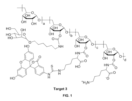
[0006] FIG. 1 is a graphic representation of a candidate CD206+ targeting
molecule labeled
with FITC.
[0007] FIG. 2 is a is a graphic representation of a candidate CD206+
targeting molecule
labeled with MMAE.
[0008] FIG. 3 is a graph showing the impact of Target 5 at three different
concentrations, 0.5
mg/ml (0), 5 mg/ml (N), and 50 mg/ml (o), of as compared to Temozolomide (1)
and a saline
vehicle control (*).
[0009] Fig. 4 is a graphic representation of a candidate CD206+ targeting
molecule showing
a cyclodextrin backbone and potential payloads.
[0010] Fig. 5 is a graphic representation of a candidate CD206+ targeting
molecule carrying a
metal ion chelator.
[0011] FIG. 6 is a graph showing tumor growth in a syngeneic murine model
of triple
negative breast cancer where mice are treated with either Target 5 at 5 mg/kg
(0), Target 5 at 15
mg/kg (1), or Paclitaxel at 15 mg/kg (x).
[0012] FIG. 7 is a graph showing survival of mice treated with Target 5 in
a U87 intracranial
model of glioblastoma as compared to mice administered saline.
[0013] FIG. 8A-8C is a graph showing the effect of various concentrations
of Target 5 on
tumor volume (FIG. 8A), percent tumor volume change (FIG. 8B), and body weight
(FIG. 8C) in
a GL261 glioma mouse model.
[0014] FIG. 9 is a graph showing the effect of Target 5 on tumor volume in
a syngeneic
murine colon cancer model.
[0015] FIG. 10 is a MRI image showing that Target-7 has greater specificity
for tumor with
potential for less toxicity.
2
CA 03192041 2023-02-15
WO 2022/040580 PCT/US2021/046984
[0016] FIG. 11A is a graph showing the signal intensity ratios of post-
contrast tumor to
selected tissues. FIG. 11B is a graph showing signal to noise ratio (SNR)
comparisons by group.
DETAILED DESCRIPTION OF THE INVENTION
[0017] The present invention relates to compounds that target monocytes,
macrophages and
other cells (such as dendritic cells) that express CD206, particularly those
cells that are
assembled at a site of disease, using a target moiety coupled to a glucan
backbone. The
compounds disclosed here preferably comprise a glucan backbone, a targeting
moiety, a
targeting moiety linker, a payload and optionally a payload linker. The
present invention also
provides methods of making such compounds and compositions. The present
invention also
provides diagnostic methods and methods of treatment using compounds
comprising a target
moiety coupled to a glucan backbone.
Chemical Definitions
[0018] "Alkyl" as used herein refers to and includes, unless otherwise
stated, a saturated
linear (i.e., unbranched) or branched univalent hydrocarbon chain or
combination thereof, having
the number of carbon atoms designated (i.e., Ci-Cio means one to ten carbon
atoms). Particular
alkyl groups are those having 1 to 20 carbon atoms (a "C1-C20 alkyl"), having
1 to 10 carbon
atoms (a "Ci-Cio alkyl"), having 6 to 10 carbon atoms (a "C6-C10 alkyl"),
having 1 to 6 carbon
atoms (a "C1-C6 alkyl"), having 2 to 6 carbon atoms (a "C2-C6 alkyl"), or
having 1 to 4 carbon
atoms (a "C1-C4 alkyl"). Examples of alkyl groups include, but are not limited
to, groups such as
methyl, ethyl, n-propyl, isopropyl, n-butyl, t-butyl, isobutyl, sec-butyl, n-
pentyl, n-hexyl, n-
heptyl, n-octyl, n-nonyl, n-decyl, and the like.
[0019] "Alkylene" as used herein refers to the same residues as alkyl, but
having bivalency.
Particular alkylene groups are those having 1 to 20 carbon atoms (a "C1-C20
alkylene"), having 1
to 10 carbon atoms (a "Ci-Cio alkylene"), having 6 to 10 carbon atoms (a "C6-
C10 alkylene"),
having 1 to 6 carbon atoms (a "C1-C6 alkylene"), 1 to 5 carbon atoms (a "C1-05
alkylene"), 1 to 4
3
CA 03192041 2023-02-15
WO 2022/040580 PCT/US2021/046984
carbon atoms (a "Ci-C4 alkylene") or 1 to 3 carbon atoms (a "Ci-C3 alkylene").
Examples of
alkylene include, but are not limited to, groups such as methylene (-CH2-),
ethylene (-CH2CH2-),
propylene (-CH2CH2CH2-), isopropylene (-CH2CH(CH3)-), butylene (-CH2(CH2)2CH2-
),
isobutylene (-CH2CH(CH3)CH2-), pentylene (-CH2(CH2)3CH2-), hexylene (-
CH2(CH2)4CH2-),
heptylene (-CH2(CH2)5CH2-), octylene (-CH2(CH2)6CH2-), and the like.
[0020] "Halo" or "halogen" refers to elements of the Group 17 series having
atomic number
9 to 85. Preferred halo groups include the radicals of fluorine, chlorine,
bromine and iodine.
Where a residue is substituted with more than one halogen, it may be referred
to by using a
prefix corresponding to the number of halogen moieties attached, e.g.,
dihaloaryl, dihaloalkyl,
trihaloaryl etc. refer to aryl and alkyl substituted with two ("di") or three
("tri") halo groups,
which may be but are not necessarily the same halogen; thus 4-chloro-3-
fluorophenyl is within
the scope of dihaloaryl. An alkyl group in which each hydrogen is replaced
with a halo group is
referred to as a "perhaloalkyl." A preferred perhaloalkyl group is
trifluoromethyl (-CF3).
Similarly, "perhaloalkoxy" refers to an alkoxy group in which a halogen takes
the place of each
H in the hydrocarbon making up the alkyl moiety of the alkoxy group. An
example of a
perhaloalkoxy group is trifluoromethoxy (-0CF3).
[0021] "Carbamate" refers to the group ¨0¨C(=0)¨NH¨. Unless specified
otherwise, it is
understood that the nitrogen atom of the carbamate group is unsubstituted
(i.e., bears a hydrogen
atom).
[0022] "Oxo" refers to the moiety =0.
[0023] "Optionally substituted" unless otherwise specified means that a
group may be
unsubstituted or substituted by one or more (e.g., 1,2, 3,4, 5, 6,7, 8, 9, 10,
11, or 12) of the
substituents listed for that group in which the sub stituents may be the same
of different. In one
embodiment, an optionally substituted group has one substituent. In another
embodiment, an
optionally substituted group has two substituents. In another embodiment, an
optionally
substituted group has three substituents. In another embodiment, an optionally
substituted group
has four substituents. In some embodiments, an optionally substituted group
has 1 to 2, 1 to 3, 1
4
CA 03192041 2023-02-15
WO 2022/040580 PCT/US2021/046984
to 4, 1 to 5, 2 to 3, 2 to 4, or 2 to 5 substituents. In one embodiment, an
optionally substituted
group is unsubstituted.
Compounds
[0024] The compounds disclosed here preferably comprise various components,
including a
glucan backbone, a targeting moiety, a targeting moiety linker, a payload and
optionally a
payload linker. The arrangement of these components provides a compound that
preferentially
targets CD206+ cells and is internalized. The ability to be internalized by
CD206+ cells allows
for the disclosed compounds to deliver payloads to disease sites where such
cells assemble.
Solid tumor cancers and granulomatous diseases often comprise by CD206+ cell
assemblies. The
present application describes improved compositions and methods for imaging
and treating solid
tumor cancers or granulomatous diseases by targeting the CD206+ cells that
assemble at or
otherwise are associated with these disease states. In certain embodiments,
the disclosed
compounds can also function as an intra-operative imaging agent, a MRI imaging
agent or
radiosensitizer, and to deliver radiopharmaceuticals to primary and metastatic
cancer cells in the
brain and body.
Glucan Backbone
[0025] The compounds described here comprise a glucan backbone, which is a
linear,
branched, or circular oligosaccharide or polysaccharide comprising a plurality
of glucose
monomers linked predominantly by C-1 ¨> C-6 glycosidic bonds. Other glycosidic
bonds such
as a-1,3 or a-1,4 linkages may also be present. A glucan backbone may also be
defined as a
polymer of glucose wherein the position of glycosidic bonds is varied. A
glucan backbone may
comprise the alpha or the beta isomer of glucose. Examples of glucan backbones
include
dextran, a linear or branched compound, and cyclodextrin, a circular glucan.
[0026] A glucan backbone may vary in mass and molecular weight, as
determined in part by
the number of glucose monomers. In some embodiments, a glucan backbone may
range in
molecular weight from 1-30 kilodaltons (kDa). Preferred embodiments include
glucan backbones
of approximately 1 kDa, 3 kDa, 6 kDa, 10 kDa, 20 kDa, or 30 kDa. In some
embodiments, the
glucan backbone may range in molecular mass from 1,000 to 30,000 grams per
mole (g/mol). In
CA 03192041 2023-02-15
WO 2022/040580 PCT/US2021/046984
some embodiments, the glucan backbone may contain glucose monomers ranging
from 5 to 167
in number. The glucan backbone can be linear, branched, circular, or
combinations thereof. For
example, dextran is an example of a linear or branched glucan backbone.
Cyclodextrin is
another example of a glucan backbone. The backbones described here can be
substituted or
unsubstituted. For example, a substituted cyclodextrin is a cyclodextrin
derivative that is
hydrophobic, hydrophilic, ionized, non-ionized, or any other variation
thereof.
Targeting moiety
[0027] The compounds disclose here comprise a targeting moiety coupled to a
glucan
backbone. In some embodiments, the targeting moiety is a CD206 targeting
moiety. In some
embodiments of the above aspects, the targeting moiety is a CD206 ligand. A
targeting moiety
is a molecule, a compound, a structure, or any combination thereof that
targets one or more
pattern recognition receptors on CD206+ cells. The targeting moiety may target
a pattern
recognition receptor that is also be characterized as a C-type lectin
receptor. Preferably, the
targeting moiety targets CD206, a mannose receptor. The targeting moiety may
target one or
more CD206+ cells, particularly CD206+ monocytes and macrophages. In some
embodiments,
the targeting moiety is or comprises a CD206 ligand. In some embodiments, the
CD206 ligand
comprises at least a portion of mannose, galactose, collagen, fucose, sulfated
N-
acetylgalactosamine, N-acetylglucosamine, luteinizing hormone, thyroid
stimulating hormone, or
a chondroitin sulfate. A preferred CD206 ligand is mannose, D- and L-isomers
thereof, and
furanoses (5-membered rings) and pyranoses (6-membered rings) thereof.
[0028] In some embodiments, the targeting moieties are attached to between
about 10% and
about 50% of the glucose residues of the glucan backbone, or between about 20%
and about 45%
of the glucose residues, or between about 25% and about 40% of the glucose
residues. (It should
be noted that the MWs referenced herein, as well as the number and degree of
conjugation of
receptor substrates, leashes, and diagnostic/therapeutic moieties attached to
the dextran backbone
refer to average amounts for a given quantity of carrier molecules, since the
synthesis techniques
will result in some variability.)
6
CA 03192041 2023-02-15
WO 2022/040580 PCT/US2021/046984
Ratio of targeting linker to backbone
[0029] The density of a targeting moiety relative to backbone subunits is
presented using a
targeting moiety to backbone subunit ratio for linear and branched
polysaccharide backbones.
Degree of substitution (d.s.) is used to communicate the density of targeting
moieties on circular
backbones. The ratio of a targeting moiety to a glucan backbone refers to the
number of
targeting moieties that substitute a backbone subunit or subunits. For
example, a ratio of 1:7 or 1
to 7 means that there is one targeting moiety for every seven glucose subunits
in a glucan
backbone. The d.s. describes the average number of substituents or substituted
positions per unit
base. For example, a d.s. of 0.9 means that one backbone subunit is
substituted with an average
of 0.9 targeting moieties. In some embodiments, the targeting moiety to
backbone subunit ratio
is from about 1:5 to about 1:25. In some embodiments, the targeting moiety to
backbone subunit
ratio is from about 1:6 to about 1:19. In some embodiments, the d.s. is from
about 0.1 to about
7. In some embodiments, the d.s. is from about 0.5 to 5.
Targeting linker
[0030] A targeting linker is a cleavable or a non-cleavable linker that
connects a glucan
backbone to a targeting moiety. A cleavable linker is capable of being cleaved
by an enzyme
(e.g., a protease), a change in temperature, a change in pH, a chemical
stimulus, or any
combination thereof. The cleavable linker may comprise a protease cleavage
site. In some
embodiments, the cleavable linker is capable of cleavage by a lysosomal
protease or an
endosomal protease.
[0031] The targeting linker may comprise a carbamate group. In some
embodiments, the
targeting linker comprises a carbamate group and a chain moiety, wherein the
carbamate group is
connected to a backbone monomer and the chain moiety connects the carbamate
group and the
targeting moiety. Herein, a carbamate functional group takes the plain and
ordinary meaning
derived from the field of organic chemistry. In some embodiments, the chain
moiety of the
targeting linker comprises one or more (e.g., 1, 2, 3, 4, 5, 6, 7, 8, 9, or
10) units selected from the
group consisting of an optionally substituted alkylene chain, an optionally
substituted CO-
alkylene chain, a peptide chain, a polymeric chain, and a heteroatom selected
from the group
7
CA 03192041 2023-02-15
WO 2022/040580 PCT/US2021/046984
consisting of an 0 atom, a S atom, and an optionally substituted N atom. In
some embodiments,
the chain moiety comprises a Ci-C12 alkylene chain. In some embodiments, the
chain moiety
comprises a C3-C7 alkylene chain. In some embodiments, the chain moiety
comprises a C6
alkylene chain. In some embodiments, the chain moiety is a C6 alkylene chain.
In some
embodiments, the alkylene chain is substituted by one or more substituents
selected from the
group consisting of oxo, OH, NH2, SH, Ci-C12 alkyl, Ci-C12 haloalkyl, 0(C i-
C12 alkyl), O(C1-
C12 haloalkyl), NH(Ci-C12 alkyl), NH(Ci-C12 haloalkyl), N(C1-C12 alky1)2, N(C1-
C12 haloalky1)2,
, S(Ci-C12 alkyl), S(Ci-C12 haloalkyl), C(0)0H, C(0)0(Ci-C12 alkyl), C(0)0(Ci-
C12 haloalkyl),
C(0)NH(Ci-Ci2 alkyl), C(0)NH(Ci-Ci2 haloalkyl), C(0)N(C1-C12 alky1)2, C(0)N(C1-
C12
haloalky1)2, C(0)S(Ci-Ci2 alkyl), and C(0)S(Ci-Ci2 haloalkyl). In some
embodiments, the
alkylene chain is unsubstituted.
[0032] In some embodiments, the one or more CD206 targeting moieties are
attached to the
glucan backbone through a linker. The linker may be attached at from about 1
to about 50% of
the backbone moieties.
Active Component
[0033] An active component is a molecule or a compound that may be used for
diagnostic
purposes, therapeutic purposes, or a combination thereof. An active component
is also referred
to as a payload. An active component may be or comprise a cytotoxic agent, an
imaging agent,
or a combination thereof.
Diagnostic Payloads
[0034] In some embodiments, the active component is an imaging agent. In
some
embodiments, the imaging agent is 5-carboxyfluorescein, fluorescein-5-
isothiocyanate,
fluorescein-6-isothiocyanate, 6-carboxyfluorescein, tetramethylrhodamine-6-
isothiocyanate, 5-
carboxytetramethylrhodamine, 5-carboxy rhodol derivatives, tetramethyl and
tetraethyl
rhodamine, diphenyldimethyl and diphenyldiethyl rhodamine, dinaphthyl
rhodamine, rhodamine
101 sulfonyl chloride, Cy3, Cy3B, Cy3.5, Cy5, Cy5 5, Cy7, DyLight650,
IRDye6S0, IRDye680,
DyLight750, Alexa Fluor 647, Alexa Fluor 750, IR800CW, ICG, Green Fluorescent
Protein,
EBFP, EBFP2, Azurite, mKalamal, ECFP, Cerulean, CyPet, YFP, Citrine, Venus,
YPet, a
8
CA 03192041 2023-02-15
WO 2022/040580 PCT/US2021/046984
gadolinium chelate, an iron oxide particle, a super paramagnetic iron oxide
particle, an ultrasmall
paramagnetic particle, a manganese chelate, gallium containing agent, 64Cu
diacetylbis(N4-
methylthiosemicarbazone), 18F-fluorodeoxyglucose, 18F-fluoride, 3'-deoxy-3'-
[18F]fluorothymidine, 18F-fluoromisonidazole, technetium-99m, thallium,
iodine, barium
sulphate, or a combination thereof. In some embodiments, an imaging agent is
conjugated to one
or more additional agents, such as a targeting agent, a cytotoxic agent, or a
macrophage
polarizing agent.
Therapeutic Payloads
[0035] In some embodiments, the active component is a therapeutic agent.
The therapeutic
agent may be any compound known to be useful for the treatment of a macrophage-
mediated
disease. Therapeutic agents include, but are not limited to, chemotherapeutic
agents, such as
doxorubicin; anti-infective agents, such as antibiotics (e.g. tetracycline,
streptomycin, and
isoniazid), anti-virals, anti-fungals, and anti-parasitics; immunological
adjuvants; steroids;
nucleotides, such as DNA, RNA, RNAi, siRNA, CpG or Poly (I:C); peptides;
proteins; or metals
such as silver, gallium or gadolinium.
[0036] In certain embodiments, the therapeutic agent is an antimicrobial
drug selected from
the group comprising or consisting of: an antibiotic; an anti-tuberculosis
antibiotic (such as
isoniazid, streptamycin, or ethambutol); an anti-viral or anti-retroviral
drug, for example an
inhibitor of reverse transcription (such as zidovudin) or a protease inhibitor
(such as indinavir);
drugs with effect on leishmaniasis (such as Meglumine antimoniate). In certain
embodiments,
the therapeutic agent is an anti-microbial active, such as amoxicillin,
ampicillin, tetracyclines,
aminoglycosides (e.g., streptomycin), macrolides (e.g., erythromycin and its
relatives),
chloramphenicol, ivermectin, rifamycins and polypeptide antibiotics (e.g.,
polymyxin,
bacitracin) and zwittermicin. In certain embodiments, the therapeutic agent is
selected from
isoniazid, doxorubicin, streptomycin, and tetracycline.
[0037] In some embodiments, the therapeutic agent comprises a high energy
killing isotope
which has the ability to kill macrophages and tissue in the surrounding
macrophage environment.
9
CA 03192041 2023-02-15
WO 2022/040580 PCT/US2021/046984
Suitable radioisotopes
include:210/212/213/214Bi,131/140Ba,11/14c,51cr,67/68Ga, 153Gd, 99mTc,
88/90/91y,
123/124/125/1311, 111/115min, 18F, 105Rh, 1535m, 67cii, 166H0, 177Lu, 186Re
and 188Re, 32/33F), 46/475c,
721755e, 35, 182Ta, 127/129/132Te, 65Z11 and 89/95Zr.
[0038] In other embodiments, the therapeutic agent comprises a non-
radioactive species
selected from, but not limited to, the group consisting of: Bi, Ba, Mg, Ni,
Au, Ag, V, Co, Pt, W,
Ti, Al, Si, Os, Sn, Br, Mn, Mo, Li, Sb, F, Cr, Ga, Gd, I, Rh, Cu, Fe, P, Se,
S, Zn and Zr.
[0039] In still further embodiments, the therapeutic agent is selected from
the group
consisting of cytostatic agents, alkylating agents, antimetabolites, anti-
proliferative agents,
tubulin binding agents, hormones and hormone antagonists, anthracycline drugs,
vinca drugs,
mitomycins, bleomycins, cytotoxic nucleosides, pteridine drugs, diynenes,
podophyllotoxins,
toxic enzymes, and radio sensitizing drugs. By way of more specific example,
the therapeutic
agent is selected from the group consisting of temozolomide, mechlorethamine,
triethylenephosphoramide, cyclophosphamide, ifosfamide, chlorambucil,
busulfan, melphalan,
triaziquone, nitrosourea compounds, adriamycin, carminomycin, daunorubicin
(daunomycin),
doxorubicin, isoniazid, indomethacin, gallium(III), 68ga11ium(III),
aminopterin, methotrexate,
methopterin, mithramycin, streptonigrin, dichloromethotrexate, mitomycin C,
actinomycin-D,
porfiromycin, 5-fluorouracil, floxuridine, ftorafur, 6-mercaptopurine,
cytarabine, cytosine
arabinoside, podophyllotoxin, etoposide, etoposide phosphate, melphalan,
vinblastine,
vincristine, leurosidine, vindesine, leurosine, taxol, taxane, cytochalasin B,
gramicidin D,
ethidium bromide, emetine, tenoposide, colchicin, dihydroxy anthracin dione,
mitoxantrone,
procaine, tetracaine, lidocaine, propranolol, puromycin, ricin subunit A,
abrin, diptheria toxin,
botulinum, cyanginosins, saxitoxin, shigatoxin, tetanus, tetrodotoxin,
trichothecene,
verrucologen, corticosteroids, progestins, estrogens, antiestrogens,
androgens, aromatase
inhibitors, calicheamicin, esperamicins, and dynemicins.
[0040] In embodiments wherein the therapeutic agent is a hormone or hormone
antagonist,
the therapeutic agent may be selected from the group consisting of prednisone,
hydroxyprogesterone, medroprogesterone, diethylstilbestrol, tamoxifen,
testosterone, and
aminogluthetimide.
CA 03192041 2023-02-15
WO 2022/040580 PCT/US2021/046984
[0041] In embodiments wherein the therapeutic agent is a prodrug, the
therapeutic agent may
be selected from the group consisting of phosphate-containing prodrugs,
thiophosphate-
containing prodrugs, sulfate containing prodrugs, peptide containing prodrugs,
(-lactam-
containing prodrugs, optionally substituted phenoxyacetamide-containing
prodrugs, optionally
substituted phenylacetamide-containing prodrugs, 5-fluorocytosinem, and 5-
fluorouridine
prodrugs that can be converted to the more active cytotoxic free drug.
[0042] In some embodiments the active component is a cytotoxic agent or
comprises a
cytotoxic agent. In some embodiments, the cytotoxic agent is a
chemotherapeutic agent, an
antitubulin agent, a DNA modifying agent, or a small interfering ribonucleic
acid. In some
embodiments, the cytotoxic agent is selected from the group consisting of an
auristatin, a
dolastatin, auristatin E, monomethyl auristatin E (MMAE), monomethyl
auristatin F (MMAF),
dimethylvaline-valine-dolaisoleuine-dolaproine-phenylalanine-p-
phenylenediamine (AFP), 5-
benzoylvaleric acid-auristatin E ester (AEVB), auristatin EB (AEB),
ansamitocin,
ivlertansine/emtansine (DMI), ravtansine/soravtansine (DM4), duocarmycins,
calicheamicins,
and pyrrolobenzodiazepines.
Payload Linker
[0043] In certain embodiments the active component or payload is coupled
directly to the
glucan backbone. In some embodiments, the active component is connected to a
glucan
backbone via a linker. The linker can be cleavable or non-cleavable. In some
embodiments, the
one or more therapeutic agent is attached via a biodegradable linker. In some
embodiments, the
biodegradable linker is acid sensitive, such as a hydrazone linker. The use of
an acid sensitive
linker enables the drug to be transported into the cell and allows for the
release of the drug
substantially inside of the cell. In some embodiments, the payload linker is a
Val-Cit linker.
[0044] The payload linker may comprise a carbamate group. In some
embodiments, the
payload linker comprises a carbamate group and a chain moiety, wherein the
carbamate group is
connected to a backbone monomer and the chain moiety connects the carbamate
group and the
active component. Herein, a carbamate functional group takes the plain and
ordinary meaning
11
CA 03192041 2023-02-15
WO 2022/040580 PCT/US2021/046984
derived from the field of organic chemistry. In some embodiments, the chain
moiety of the
payload linker comprises one or more (e.g., 1, 2, 3, 4, 5, 6, 7, 8, 9, or 10)
units selected from the
group consisting of an optionally substituted alkylene chain, an optionally
substituted CO-
alkylene chain, a peptide chain, a polymeric chain, and a heteroatom selected
from the group
consisting of an 0 atom, a S atom, and an optionally substituted N atom. In
some embodiments,
the chain moiety comprises a Ci-C12 alkylene chain. In some embodiments, the
chain moiety
comprises a C3-C7 alkylene chain. In some embodiments, the chain moiety
comprises a C6
alkylene chain. In some embodiments, the chain moiety is a C6 alkylene chain.
In some
embodiments, the alkylene chain is substituted by one or more substituents
selected from the
group consisting of oxo, OH, NH2, SH, Ci-C12 alkyl, Ci-C12 haloalkyl, 0(C i-
C12 alkyl), 0(Ci-
C12 haloalkyl), NH(Ci-C12 alkyl), NH(Ci-C12 haloalkyl), N(C1-C12 alky1)2, N(C1-
C12 haloalky1)2,
, S(Ci-C12 alkyl), S(Ci-C12 haloalkyl), C(0)0H, C(0)0(Ci-C12 alkyl), C(0)0(Ci-
C12 haloalkyl),
C(0)NH(Ci-Ci2 alkyl), C(0)NH(Ci-Ci2 haloalkyl), C(0)N(C1-C12 alky1)2, C(0)N(C1-
C12
haloalky1)2, C(0)S(Ci-Ci2 alkyl), and C(0)S(Ci-Ci2 haloalkyl). In some
embodiments, the
alkylene chain is unsubstituted.
Secondary payloads and linkers
[0045] In addition to the targeting, diagnostic, and therapeutic payloads,
the compounds
disclosed here can encompass the inclusion of secondary agents that can be
coupled to the glucan
backbone to add additional functional capabilities. Typically, the secondary
payload is coupled
to the linker in a manner similar to that used to couple the targeting moiety
to the targeting
linker.
[0046] A secondary payload can encompass, for example, additional agents
for imaging,
therapy, or for other purposes. Specifically, in one embodiment, combinations
of therapeutic and
imaging agents can be linked to the glucan backbone to combine diagnostic and
therapeutic
functionalities. In another embodiment, various amino acids, such as cysteine
or lysine can be
coupled to the linker to crosslink the molecule to a target.
[0047] A secondary payload linker is a cleavable or a non-cleavable linker
that connects a
glucan backbone to a secondary payload moiety. A cleavable linker is capable
of being cleaved
12
CA 03192041 2023-02-15
WO 2022/040580 PCT/US2021/046984
by an enzyme (e.g., a protease), a change in temperature, a change in pH, a
chemical stimulus, or
any combination thereof. The cleavable linker may comprise a protease cleavage
site. In some
embodiments, the cleavable linker is capable of cleavage by a lysosomal
protease or an
endosomal protease.
[0048] The secondary payload linker may comprise a carbamate group. In some
embodiments, the secondary payload linker comprises a carbamate group and a
chain moiety,
wherein the carbamate group is connected to a backbone monomer and the chain
moiety
connects the carbamate group and the secondary agent. Herein, a carbamate
functional group
takes the plain and ordinary meaning derived from the field of organic
chemistry. In some
embodiments, the chain moiety of the secondary payload linker comprises one or
more (e.g., 1,
2, 3, 4, 5, 6, 7, 8, 9, or 10) units selected from the group consisting of an
optionally substituted
alkylene chain, an optionally substituted CO-alkylene chain, a peptide chain,
a polymeric chain,
and a heteroatom selected from the group consisting of an 0 atom, a S atom,
and an optionally
substituted N atom. In some embodiments, the chain moiety comprises a CI-Cu
alkylene chain.
In some embodiments, the chain moiety comprises a C3-C7 alkylene chain. In
some
embodiments, the chain moiety comprises a C6 alkylene chain. In some
embodiments, the chain
moiety is a C6 alkylene chain. In some embodiments, the alkylene chain is
substituted by one or
more substituents selected from the group consisting of oxo, OH, NH2, SH, CI-
Cu alkyl, Ci-C12
haloalkyl, 0(Ci-C12 alkyl), 0(Ci-C12 haloalkyl), NH(Ci-C12 alkyl), NH(Ci-C12
haloalkyl), N(Ci-
C12 alky1)2, N(C1-C12 haloalky1)2, , S(Ci-C12 alkyl), S(Ci-C12 haloalkyl),
C(0)0H, C(0)0(Ci-C12
alkyl), C(0)0(Ci-C12 haloalkyl), C(0)NH(Ci-Ci2 alkyl), C(0)NH(Ci-Ci2
haloalkyl), C(0)N(Ci-
C12 alky1)2, C(0)N(C1-C12 haloalky1)2, C(0)S(Ci-Ci2 alkyl), and C(0)S(Ci-Ci2
haloalkyl). In
some embodiments, the alkylene chain is unsubstituted.
[0049] In some embodiments, the one or more secondary payload moieties are
attached to
the glucan backbone through a linker. The linker may be attached at from about
1 to about 50%
of the backbone moieties.
13
CA 03192041 2023-02-15
WO 2022/040580 PCT/US2021/046984
Diagnostic Methods
[0050] Diagnostic methods are disclosed for in vivo detection of diseases
or conditions using
the disclosed compounds. In certain embodiments, the disclosed compounds
include a detection.
As used herein, the term "detectable label or moiety" means an atom, isotope,
or chemical
structure which is: (1) capable of attachment to the carrier molecule; (2) non-
toxic to humans or
other mammalian subjects; and (3) provides a directly or indirectly detectable
signal, particularly
a signal which not only can be measured but whose intensity is related (e.g.,
proportional) to the
amount of the detectable moiety. The signal may be detected by any suitable
means, including
spectroscopic, electrical, optical, magnetic, auditory, radio signal, or
palpation detection means.
[0051] Detection labels include, but are not limited to, fluorescent
molecules (a.k.a.
fluorochromes and fluorophores), chemiluminescent reagents (e.g., luminol),
bioluminescent
reagents (e.g., luciferin and green fluorescent protein (GFP)), metals (e.g.,
gold nanoparticles),
and radioactive isotopes (radioisotopes). Suitable detection labels can be
selected based on the
choice of imaging method. For example, the detection label can be a near
infrared fluorescent
dye for optical imaging, a gadolinium chelate for MRI imaging, a radionuclide
for PET or
SPECT imaging, or a gold nanoparticle for CT imaging.
[0052] The disclosed compounds can include a detectable label useful for
optical imaging. A
number of approaches can be used for optical imaging. The various methods
depend upon
fluorescence, bioluminescence, absorption or reflectance as the source of
contrast. Fluorophores
are compounds or moieties that absorb energy of a specific wavelength and re-
emit energy at a
different (but equally specific) wavelength. In certain embodiments, the
detectable label is a
near-infrared (NIR) fluorophore. Suitable NIRs include, but are not limited
to, VivoTag-S®
680 and 750, Kodak X-SIGHT Dyes and Conjugates, DyLight 750 and 800 Fluors, Cy
5.5 and 7
Fluors, Alexa Fluor 680 and 750 Dyes, and 1RDye 680 and 800CW Fluors. In
certain
embodiments, Quantum dots, with their photostability and bright emissions, can
also be used
with optical imaging. In certain embodiments, pre-existing surgical
microscopes can be adapted
for use in "green" channel by adding a filter to the light source.
14
CA 03192041 2023-02-15
WO 2022/040580 PCT/US2021/046984
[0053] The disclosed compounds can include a detectable label (e.g., a
radionuclide) useful
for nuclear medicine imaging. Nuclear medicine imaging involves the use and
detection of
radioisotopes in the body. Nuclear medicine imaging techniques include
scintigraphy, single
photon emission computed tomography (SPECT), and positron emission tomography
(PET). In
these techniques, radiation from the radioisotopes can be captured by a gamma
camera to form
two-dimensional images (scintigraphy) or 3-dimensional images (SPECT and PET).
[0054] The disclosed compounds can be used in combination with molecular
imaging to
detect cancer cells, such as those that have metastasized and therefore spread
to another organ or
tissue of the body, using an in vivo imaging device. A non-invasive method is
therefore provided
for detecting cancer cells in a subject that involves administering a
pharmaceutical composition
containing the disclosed compounds to the subject and then detecting the
biodistribution of
disclosed compounds using an imaging device. In some embodiments, the
pharmaceutical
composition is injected into the parenchyma. In other embodiments, the
pharmaceutical
composition is injected into the circulation.
[0055] The disclosed compounds can also be used for intraoperative
detection of cancer. For
example, the disclosed compounds can be used for intraoperative lymphatic
mapping (ILM) to
trace the lymphatic drainage patterns in a cancer patient to evaluate
potential tumor drainage and
cancer spread in lymphatic tissue. In these embodiments, the disclosed
compounds are injected
into the tumor and their movement through the lymphatic system is traced using
a molecular
imaging device. As another example, the disclosed compounds can be used for
intraoperative
assessment of, for example, tumor margins and tumor adjacent tissues for the
presence of cancer
cells. This can be useful, for example, in effectively resecting tumors and
detecting the spread of
cancer proximal to the tumor. In some embodiments, the disclosed compounds are
able to
crosses the blood-tumor barrier. In some embodiments, the disclosed compounds
are able to
carry payloads into brain tumors and across the blood-tumor barrier without
leaking across the
blood-brain barrier.
[0056] The disclosed methods of imaging to detect cancer cells are referred
to herein as non-
invasive. By non-invasive is meant that the disclosed compounds can be
detected from outside of
the subject's body. By this it is generally meant that the signal detection
device is located outside
CA 03192041 2023-02-15
WO 2022/040580 PCT/US2021/046984
of the subject's body. It is understood, however, that the disclosed compounds
can also be
detected from inside the subject's body or from inside the subject's
gastrointestinal tract or from
inside the subject's respiratory system and that such methods of imaging are
also specifically
contemplated. For example, for intraoperative detection, the signal detection
device can be
located either outside or inside of the subject's body. From this it should be
understood that a
non-invasive method of imaging can be used along with, at the same time as, or
in combination
with an invasive procedure, such as surgery.
[0057] In some embodiments, the method can be used to diagnose cancer in a
subject or
detect cancer in a particular organ of a subject. A particularly useful aspect
of this method is the
ability to search for metastatic cancer cells in secondary tissues or organs,
such as lymph nodes,
or at or near tumor margins. Therefore, the disclosed methods can be used for
assessing lymph
node status in patients that have or are suspected of having cancer, such as
breast cancer. This
may avoid the need to biopsy the tissue or organ, e.g., remove a lymph node.
In some
embodiments, the method involves administering to the patient the disclosed
compounds and
detecting whether the compounds have bound to cells in a lymph node. In some
of these
embodiments, the lymph node can be an axillary lymph node (ALN). In other
embodiments, the
lymph node can be a sentinel lymph node. In further embodiments, both axillary
and sentinel
lymph nodes can be assessed for binding of the agent to cells in the lymph
node.
[0058] The method can also be used with other therapeutic or diagnostic
methods. For
example, the method can also be used during an operation to, for example,
guide cancer removal,
which is referred to herein as "intraoperative guidance" or "image guided
surgery." In a
particular embodiment, the method can be used for therapeutic treatment to
remove or destroy
cancer cells in a patient's lymph nodes. For example, the disclosed compounds
can be
administered to a patient, and the location of cancerous tissue (e.g., lymph
nodes) can be
determined and removed using image guided surgery. In another preferred
embodiment, the
method can be used for therapeutic treatment to prevent positive microscopic
margins after
tumor resection. For example, the disclosed compounds can be administered to a
patient, the
location of cancer cells around a tumor can be determined, and the complete
tumor removed
using image guided surgery. In these embodiments, the physician administers
the disclosed
16
CA 03192041 2023-02-15
WO 2022/040580 PCT/US2021/046984
compounds to the patient and uses an imaging device to detect the cancer
cells, guide resection
of tissue, and assure that all of the cancer is removed. In addition, the
imaging device can be
used post-operatively to determine if any cancer remains or reoccurs.
[0059] In some embodiments, the disclosed compounds can be linked to a
therapeutic
compound. The therapeutic compound or moiety can be one that kills or inhibits
cancer cells
directly (e.g., cisplatin) or it can be one that can kill or inhibit a cancer
cell indirectly (e.g., gold
nanoparticles that kill or destroy cancer cells when heated using a light
source). If the therapeutic
compound or moiety is one that kills or inhibits a cancer cell indirectly,
then the method further
comprises a step of taking appropriate action to "activate" or otherwise
implement the anti-
cancer activity of the compound or moiety. In a specific embodiment, the
therapeutic compound
or moiety attached to the agent can be a gold nanoparticle and following
administration to the
patient and binding of the agent to cancer cells, the gold nanoparticles are
heated, e.g., using a
laser light, to kill or destroy the nearby cancer cells (photothermal
ablation). For example, in
some embodiments, the method involves image guided surgery using the disclosed
compounds
to detect and resect cancer from a subject followed by the use of the same or
different disclosed
compounds linked to a therapeutic compound to kill remaining cancer cells.
[0060] The cancer of the disclosed methods can be any cell in a subject
undergoing
unregulated growth. The cancer can be any cancer cell capable of metastasis.
For example, the
cancer can be a sarcoma, lymphoma, leukemia, carcinoma, blastoma, or germ cell
tumor. A
representative but non-limiting list of cancers that the disclosed
compositions can be used to
detect include lymphoma, B cell lymphoma, T cell lymphoma, mycosis fungoides,
Hodgkin's
Disease, myeloid leukemia, multiple myeloma, bladder cancer, brain cancer,
nervous system
cancer, head and neck cancer, squamous cell carcinoma of head and neck, kidney
cancer, lung
cancers such as small cell lung cancer and non-small cell lung cancer,
neuroblastoma/glioblastoma, ovarian cancer, pancreatic cancer, prostate
cancer, skin cancer,
liver cancer, melanoma, squamous cell carcinomas of the mouth, throat, larynx,
and lung, colon
cancer, cervical cancer, cervical carcinoma, breast cancer, triple negative
breast cancer, epithelial
cancer, renal cancer, genitourinary cancer, pulmonary cancer, esophageal
carcinoma, head and
neck carcinoma, large bowel cancer, hematopoietic cancers; testicular cancer;
colon and rectal
17
CA 03192041 2023-02-15
WO 2022/040580 PCT/US2021/046984
cancers, prostatic cancer, gliosarcoma, Kaposi sarcoma, esophageal cancer,
hepatocellular
cancer, and pancreatic cancer.
[0061] The cancer can be breast cancer. Breast cancers originating from
ducts are known as
ductal carcinomas, and those originating from lobules that supply the ducts
with milk are known
as lobular carcinomas. Common sites of breast cancer metastasis include bone,
liver, lung and
brain.
[0062] The cancer can be non-small-cell lung carcinoma (NSCLC). NSCLC is
any type of
epithelial lung cancer other than small cell lung carcinoma (SCLC). The most
common types of
NSCLC are squamous cell carcinoma, large cell carcinoma, and adenocarcinoma,
but there are
several other types that occur less frequently, and all types can occur in
unusual histologic
variants and as mixed cell-type combinations.
Therapeutic Methods
[0063] Methods of treating or preventing diseases or disorders are provided
using the
disclosed compounds. The disclosed compounds can be used for targeting CD206+
expressing
cells. The disclosed compounds can be used for targeting of macrophages for
treatment of
intracellular pathogens (M. tuberculosis, F. tularensis, S. typhi). The
disclosed compounds can
be used to target tumor-associated macrophages, e.g. to be used for treating
cancer.
[0064] Macrophage-related and other CD206 high expressing cell-related
diseases for which
the compositions and methods herein may be used include, but are not limited
to: acute
disseminated encephalomyelitis (ADEM), Addison's disease, agammaglobulinemia,
allergic
diseases, alopecia areata, Alzheimer's disease, amyotrophic lateral sclerosis,
ankylosing
spondylitis, antiphospholipid syndrome, antisynthetase syndrome, arterial
plaque disorder,
asthma, atherosclerosis, atopic allergy, atopic dermatitis, autoimmune
aplastic anemia,
autoimmune cardiomyopathy, autoimmune enteropathy, autoimmune hemolytic
anemia,
autoimmune hepatitis, autoimmune hypothyroidism, autoimmune inner ear disease,
autoimmune
lymphoproliferative syndrome, autoimmune peripheral neuropathy, autoimmune
pancreatitis,
autoimmune polyendocrine syndrome, autoimmune progesterone dermatitis,
autoimmune
thrombocytopenic purpura, autoimmune urticarial, autoimmune uveitis, Balo
disease/Balo
18
CA 03192041 2023-02-15
WO 2022/040580 PCT/US2021/046984
concentric sclerosis, Behcet's disease, Berger's disease, Bickerstaffs
encephalitis, Blau syndrome,
bullous pemphigoid, Castleman's disease, celiac disease, Chagas disease,
chronic inflammatory
demyelinating polyneuropathy, chronic recurrent multifocal osteomyelitis,
chronic obstructive
pulmonary disease, chronic venous stasis ulcers, Churg-Strauss syndrome,
cicatricial
pemphigoid, Cogan syndrome, cold agglutinin disease, complement component 2
deficiency,
contact dermatitis, cranial arteritis, CREST syndrome, Crohn's disease,
Cushing's Syndrome,
cutaneous leukocytoclastic angiitis, Dego's disease, Dercum's disease,
dermatitis herpetiformis,
dermatomyositis, Diabetes mellitus type I, Diabetes mellitus type II diffuse
cutaneous systemic
sclerosis, Dressler's syndrome, drug-induced lupus, discoid lupus
erythematosus, eczema,
emphysema, endometriosis, enthesitis-related arthritis, eosinophilic
fasciitis, eosinophilic
gastroenteritis, eosinophilic pneumonia, epidermolysis bullosa acquisita,
erythema nodosum,
erythroblastosis fetalis, essential mixed cryoglobulinemia, Evan's syndrome,
fibrodysplasia
ossificans progressive, fibrosing alveolitis (or idiopathic pulmonary
fibrosis), gastritis,
gastrointestinal pemphigoid, Gaucher's disease, glomerulonephritis,
Goodpasture's syndrome,
Graves' disease, Guillain-Barre syndrome (GBS), Hashimoto's encephalopathy,
Hashimoto's
thyroiditis, heart disease, Henoch-Schonlein purpura, herpes gestationis (aka
gestational
pemphigoid), hidradenitis suppurativa, histocytosis, Hughes-Stovin syndrome,
hypogammaglobulinemia, infectious diseases (including bacterial infectious
diseases), idiopathic
inflammatory demyelinating diseases, idiopathic pulmonary fibrosis, idiopathic
thrombocytopenic purpura, IgA nephropathy, inclusion body myositis,
inflammatory arthritis,
inflammatory bowel disease, inflammatory dementia, interstitial cystitis,
interstitial pneumonitis,
juvenile idiopathic arthritis (aka juvenile rheumatoid arthritis), Kawasaki's
disease, Lambert-
Eaton myasthenic syndrome, leukocytoclastic vasculitis, lichen planus, lichen
sclerosus, linear
IgA disease (LAD), lupoid hepatitis (aka autoimmune hepatitis), lupus
erythematosus,
lymphomatoid granulomatosis, Majeed syndrome, malignancies including cancers
(e.g.,
sarcoma, lymphoma, leukemia, carcinoma and melanoma), Meniere's disease,
microscopic
polyangiitis, Miller-Fisher syndrome, mixed connective tissue disease,
morphea, Mucha-
Habermann disease (aka Pityriasis lichenoides et varioliformis acuta),
multiple sclerosis,
myasthenia gravis, myositis, narcolepsy, neuromyelitis optica (aka Devic's
disease),
neuromyotonia, occular cicatricial pemphigoid, opsoclonus myoclonus syndrome,
Ord's
thyroiditis, palindromic rheumatism, PANDAS (pediatric autoimmune
neuropsychiatric
19
CA 03192041 2023-02-15
WO 2022/040580 PCT/US2021/046984
disorders associated with streptococcus), paraneoplastic cerebellar
degeneration, Parkinsonian
disorders, paroxysmal nocturnal hemoglobinuria (PNH), Parry Romberg syndrome,
Parsonage-
Turner syndrome, pars planitis, pemphigus vulgaris, peripheral artery disease,
pernicious
anaemia, perivenous encephalomyelitis, POEMS syndrome, polyarteritis nodosa,
polymyalgia
rheumatic, polymyositis, primary biliary cirrhosis, primary sclerosing
cholangitis, progressive
inflammatory neuropathy, psoriasis, psoriatic arthritis, pyoderma gangrenosum,
pure red cell
aplasia, Rasmussen's encephalitis, Raynaud phenomenon, relapsing
polychondritis, Reiter's
syndrome, restenosis, restless leg syndrome, retroperitoneal fibrosis,
rheumatoid arthritis,
rheumatic fever, Rosai-Dorfman disease, sarcoidosis, schizophrenia, Schmidt
syndrome,
Schnitzler syndrome, scleritis, scleroderma, sepsis, serum Sickness, Sjogren's
syndrome,
spondyloarthropathy, Still's disease (adult onset), stiff person syndrome,
stroke, subacute
bacterial endocarditis (SBE), Susac's syndrome, Sweet's syndrome, Sydenham
chorea,
sympathetic ophthalmia, systemic lupus erythematosus, Takayasu's arteritis,
temporal arteritis
(aka "giant cell arteritis"), thrombocytopenia, Tolosa-Hunt syndrome,)
transplant (e.g.,
heart/lung transplants) rejection reactions, transverse myelitis,
tuberculosis, ulcerative colitis,
undifferentiated connective tissue disease, undifferentiated
spondyloarthropathy, urticarial
vasculitis, vasculitis, vitiligo, and Wegener's granulomatosis.
[0065] The disclosed compounds can include therapeutic agents including,
but not limited to,
cytotoxic agents, anti-angiogenic agents, pro-apoptotic agents, antibiotics,
hormones, hormone
antagonists, chemokines, drugs, prodrugs, toxins, enzymes, or other agents.
The disclosed
compounds can include chemotherapeutic agents; antibiotics; immunological
adjuvants;
compounds useful for treating tuberculosis; steroids; nucleotides; peptides;
or proteins, such as
those described above.
[0066] In certain embodiments, the disclosed compounds include a
chemotherapeutic agent
for the treatment or prevention of cancer. The cancer can be any cancer cell
capable of
metastasis. For example, the cancer can be a sarcoma, lymphoma, leukemia,
carcinoma,
blastoma, or germ cell tumor. A representative but non-limiting list of
cancers that the disclosed
compositions can be used to treat or prevent include lymphoma, B cell
lymphoma, T cell
lymphoma, mycosis fungoides, Hodgkin's Disease, myeloid leukemia, bladder
cancer, brain
CA 03192041 2023-02-15
WO 2022/040580 PCT/US2021/046984
cancer, nervous system cancer, head and neck cancer, squamous cell carcinoma
of head and
neck, kidney cancer, lung cancers such as small cell lung cancer and non-small
cell lung cancer,
neuroblastoma/glioblastoma, ovarian cancer, pancreatic cancer, prostate
cancer, skin cancer,
liver cancer, melanoma, squamous cell carcinomas of the mouth, throat, larynx,
and lung, colon
cancer, cervical cancer, cervical carcinoma, breast cancer, triple negative
breast cancer, epithelial
cancer, renal cancer, genitourinary cancer, pulmonary cancer, esophageal
carcinoma, head and
neck carcinoma, large bowel cancer, hematopoietic cancers; testicular cancer;
colon and rectal
cancers, prostatic cancer, gliosarcoma, Kaposi sarcoma, esophageal cancer,
hepatocellular
cancer, and pancreatic cancer.
[0067] In certain embodiments, the disclosed compounds are effective for
treating
autoimmune diseases, such as rheumatoid arthritis, lupus (SLE), or vasculitis.
In certain
embodiments, the disclosed compounds are effective for treating an
inflammatory disease, such
as Crohn's disease, inflammatory bowel disease, or collagen-vascular diseases.
[0068] One of ordinary skill in the art will appreciate that various kinds
of molecules and
compounds (e.g., therapeutic agents, detection labels, and combinations
thereof) can be delivered
to a cell or tissue using the disclosed compounds.
[0069] In one aspect, provided herein is a method of treating tuberculosis
comprising
administering to a subject in need thereof a compound as described herein.
[0070] In another aspect, provided herein is a method of diagnosing and
treating a
macrophage-mediated disorder comprising administering to a subject in need
thereof an effective
amount of a compound as described herein; and detecting the detection label at
a predetermined
location in the subject.
[0071] In another aspect, provided herein is a method of treating a
macrophage-mediated
disorder comprising administering to a subject in need thereof an effective
amount of a
compound as described herein.
[0072] In another aspect, provided herein is a method of treating a disease
comprising
administering to a subject in need thereof an effective amount of a compound
according as
21
CA 03192041 2023-02-15
WO 2022/040580 PCT/US2021/046984
described herein wherein the disease is an autoimmune disease, an inflammatory
disease, or
cancer.
[0073] In another aspect, provided herein is a method of targeting tumor-
associated
macrophages comprising administering to a subject in need thereof an effective
amount of a
compound as described herein.
[0074] In another aspect, provided herein is a method according to any of
those described
herein, wherein the compound contains at least one therapeutic agent and at
least one detection
label.
[0075] In another aspect, provided herein is a method according to any of
those described
herein, wherein a linker is used to attach the one or more CD206 targeting
moieties, one or more
therapeutic agents, and/or the one or more detection labels.
[0076] In another aspect, provided herein is a method according to any of
those described
herein, wherein the macrophage-mediated disorder is selected from the group
consisting of
tuberculosis and Leishmaniasis.
[0077] In another aspect, provided herein is a method according to any of
those described
herein, wherein the disease is rheumatoid arthritis.
[0078] In another aspect, provided herein is a method according to any of
those described
herein, wherein the disorder is cancer.
[0079] In another aspect, provided herein is a method according to any of
those described
herein, wherein the cancer is a sarcoma, lymphoma, leukemia, carcinoma,
blastoma, melanoma,
or germ cell tumor.
[0080] In another aspect, provided herein is a method according to any of
those described
herein, wherein at least one A is a detection label and the detection label is
a fluorophore.
[0081] In another aspect, provided herein is a method according to any of
those described
herein, wherein at least one Li-A comprises a chelator.
22
CA 03192041 2023-02-15
WO 2022/040580 PCT/US2021/046984
ADMINISTRATION
[0082] The disclosed compounds can be administered via any suitable method.
The disclosed
compounds can be administered parenterally into the parenchyma or into the
circulation so that
the disclosed compounds reach target tissues (e.g., where cancer cells may be
located). The
disclosed compounds can be administered directly into or adjacent to a tumor
mass. The
disclosed compounds can be administered intravenously. In still other
embodiments, the
disclosed compounds can be administered orally, intraperitoneally,
intramuscularly,
subcutaneously, intracavity, or transdermally.
[0083] Parenteral administration of the compounds, if used, is generally
characterized by
injection. Injectables can be prepared in conventional forms, either as liquid
solutions or
suspensions, solid forms suitable for solution of suspension in liquid prior
to injection, or as
emulsions. A revised approach for parenteral administration involves use of a
slow release or
sustained release system such that a constant dosage is maintained.
GENERAL SYNTHETIC METHODS
[0084] Compositions of the present disclosure will now be described by
reference to
illustrative synthetic schemes for their general preparation below and the
specific examples that
follow. Artisans will recognize that, to obtain the various compositions
herein, starting materials
may be suitably selected so that the ultimately desired substituents will be
carried through the
reaction scheme with or without protection as appropriate to yield the desired
product.
Alternatively, it may be necessary or desirable to employ, in the place of the
ultimately desired
substituent, a suitable group that may be carried through the reaction scheme
and replaced as
appropriate with the desired substituent. In addition, one of skill in the art
will recognize that
protecting groups may be used to protect certain functional groups (amino,
carboxy, or side
chain groups) from reaction conditions, and that such groups are removed under
standard
conditions when appropriate.
[0085] Chromatography, recrystallization and other conventional separation
procedures may
also be used with intermediates or final products where it is desired to
obtain a particular isomer
of a compound or to otherwise purify a product of a reaction.
23
CA 03192041 2023-02-15
WO 2022/040580
PCT/US2021/046984
[0086] General methods of preparing compositions described herein are
depicted in
exemplified methods below.
[0087] In some embodiments, the compositions of described herein can be
synthesized
according to the procedure as shown in Scheme Al.
[0088] Scheme Al
Step 1:
OH _
OH 0
OH
Activating Agent a OH
Glucan ______________________ 0-
OH 0-
0
/ _b
Activating
Agent
Step 2:
OH
Targeting moiety¨Targeting Linker¨NH2
OH 0
OH
_____________________________ 0,.. a OH _
Active Component¨Payload Linker¨NH2 OH 0 0
0./ c OH
Targeting moiety¨Targeting Linker¨NH OH 0-
0
0. _d
Active Component¨Payload Linker¨NH
24
CA 03192041 2023-02-15
WO 2022/040580
PCT/US2021/046984
[0089] Scheme A2
Step 1:
OH _
OH 0
OH
Activating Agent
a OH
Glucan ________________________
OH 0-
0
/ _b
Activating
Agent
Step 2A-1:
OH
Targeting moiety¨Targeting Linker¨NH2 .. OH
OH
a OH
_____________________________ 0- -
OH 0
0
Payload Linker¨N H2 C3, C OH
Targeting moiety¨Targeting Linker¨NH OH 0
0
O/d
Payload Linker¨NH
Step 2A-2:
OH
OH 0
OH
a OH
Active Component -
OH 0
0
0./ c OH
Targeting moiety¨Targeting Linker¨NH OH 0-
0
0/ _d
Active Component¨Payload Linker¨NH
[0090] As
can be seen in the above schemes, a glucan compound (such as a dextran or a
cyclodextrin) is reacted with an activating agent. The resulting activated
glucan derivative can
then be reacted with the appropriate reagents to introduce a targeting moiety
coupled to the
glucan backbone via a targeting linker, as well as an active component linked
to the glucan
backbone via a payload linker. The skilled artisan will recognize that the
above schemes are
CA 03192041 2023-02-15
WO 2022/040580 PCT/US2021/046984
illustrative and that the various reagents and order of synthetic steps can be
varied as required for
obtaining the intended final products.
EXAMPLES
[0091] The following examples are included for illustrative purposes only
and are not
intended to limit the scope of the invention.
Example 1: Structure and Synthesis of Target 3, a Test Compound Conjugated to
FITC
[0092] Target 3, shown in Figure 1, consisted of a mannosylated dextran
backbone
conjugated to a FITC payload. The attachment of mannose to a dextran backbone
serves as a
targeting ligand for mannose binding sites, while FITC allows for detection of
test compound
using confocal or surgical microscopy. The dextran backbone presented here has
a molecular
weight of about 10 kDa.
Example 2: Internalization of FITC-Conjugated Drug Target 3 by CD206+
Macrophages
[0093] A time course endocytosis assay was used to assess macrophage
internalization of Target 3, a construct composed of a dextran backbone with
mannose as a
targeting moiety conjugated to FITC. Resolving whether the test compound was
simply bound to
the surface or internalized by macrophages was critical in evaluating its
potential to reach the
desired drug target. Uptake by CD206+ macrophages and human embryonic kidney
cells
(HEK293) (ATCC CRL-1573Tm), a cell line lacking CD206 expression, was
monitored using
confocal microscopy over 34 min. Macrophages and HEK293 cells without antibody
and
expressing anti-CD206+ antibody were included in the assay to determine
whether the antibody
would block uptake of Target 3.
[0094] To prepare plates for the endocytosis assay, a 1 mg/ml stock
solution of fibronectin
was diluted to 10 i.tg/mL in PBS without Ca /Mg . Then, 20 0_, of the diluted
fibronectin
solution was added to each well of a 384-well plate (Perkin Elmer LLC
CellCarrierTm-384 Ultra
26
CA 03192041 2023-02-15
WO 2022/040580 PCT/US2021/046984
Microplate). Plate(s) were placed on a level surface at RT for 60 min before
excess fibronectin
solution was aspirated. Fibronectin-coated plate(s) were used immediately or
allowed to air-dry
under a laminar flow bench and stored at 4 C for up to 2 weeks.
[0095] Harvested macrophages and HEK293 cells were diluted to a density of
160,000
cells/mL (4,000 cells/well in 25 lL), in their respective growth media.
Cytokines and LPS were
added to
[0096] Complete Ml-Macrophage Generation Medium DXF. Cells (25 ilL) were
added to
desired wells and allowed to adhere overnight.
[0097] The following morning, test compound Target 3 was diluted in DMSO
and added to
desired wells with an Echo 555 Liquid Handler, using a ten point three-fold
dilution series at a
top final concentration of 50 i.i.M. Specific wells were treated only with
DMSO. Immediately
after addition test compound, the nuclear stain Hoechst was added to all plate
wells at a final
concentration of 1 i.tg/mL in a final volume of 50 ilL. Cells were then
incubated for 10 min at
37 C in a humidified incubator with 5% CO2.
[0098] Plate wells were imaged with the Opera PhenixTM High Content
Screening System
using confocal imaging with a 20X water objective, 9 fields per well, and the
Hoechst and Alexa
488 filters. Wells were imaged at 10, 20, and 34 min after addition of
Hoechst.
[0099] Images were analyzed with the Columbus Image Data Storage and
Analysis System
to generate quantitative measures of compound fluorescent intensity in the
nucleus and
cytoplasm of macrophages and HEK293 cells, and to identify the number of cells
with
compound fluorescent intensity above background levels, in this case >20,000
RFUs. Microsoft
Excel was used to compare quantitative data for macrophages and HEK293 cells
in graphic
format.
[0100] The percentage of internalization of Target 3 was detected after
incubation with
50i.tM Target 3. Nearly 100% of macrophages with and without antibody
internalized Target 3
after 10 min, indicating that Target 3 reached the desired drug target and the
anti-CD206+
antibody did not interfere with compound uptake. This mean percentage of cell
uptake was
27
CA 03192041 2023-02-15
WO 2022/040580 PCT/US2021/046984
maintained throughout the course of the assay, 34 min. In comparison, uptake
of Target 3 by
both groups of HEK293 cells was <26% at 34 min.
[0101] For reference, uptake of the 10,000 MW dextran pHrodoTm green was
also evaluated
in macrophages and HEK293 cells. Allowing for resolution between binding and
cellular uptake,
the pHrodoTM green dextran fluoresces strongly in acidic conditions but is
relatively non-
fluorescent at a neutral pH. Internalization of pHrodoTM green increased over
time, reaching
90% uptake at 17 h. Uptake by HEK293 cells was undetectable at other time
points but reached
14% at 17 h. Compared to macrophage internalization of Target 3 after 10 min,
17 hours passed
before a similar percentage of human macrophages internalized pHrodoTm green.
Example 3: Structure and Synthesis of Target-5, a Targeted Chemotherapeutic
Composed of a Mannosylated Dextran Ligand Connected by a Valine-Citrulline
Linker to
the Toxin Monomethyl Auristatin E
[0102] Target-5 consists of four components (ABCD). To form a mannose
binding site
targeting moiety, a dextran backbone (A) was mannosylated (B). The A and B
components that
make up the targeting ligand are connected by a valine-citrulline linker (C)
to a toxin (D). Here,
the linker joins the toxin monomethyl auristatin E (MMAE) to the targeting
moiety. A
representative Target 5 molecule is shown in Figure 2.
Example 4: Reduction of U87-MG Tumor Volume in vivo After Treatment with
Target-5
[0103] The anti-tumor activity of Target-5, a chemotherapeutic construct
composed of a
mannosylated dextran backbone connected to the toxin monomethyl auristatin E
(MMAE) with a
valine-citrulline linker, was assessed in vivo using a mouse model of
glioblastoma. Three distinct
doses of Target-5 were evaluated against temozolomide, an FDA-approved
chemotherapeutic for
the treatment of glioblastoma, and a negative control in athymic nude mice
bearing U87-MG
tumors.
[0104] To provide a murine glioblastoma model, U87-MG (ATCC HTB-14 TM)
cells were
injected into the crania of outbred athymic nude mice (Jackson Laboratories)
from 4-6 weeks of
age. In preparation for intracranial injection, U87-MG cells were grown for 10-
14 days in Fetal
28
CA 03192041 2023-02-15
WO 2022/040580 PCT/US2021/046984
Bovine Serum supplemented with Eagle's Minimum Essential Medium (EMEM) + 1X
Penicillin/Streptomycin, then split 1:5 upon reaching confluency. For
approximately 3-4 min at
37 C, cells were harvested from tissue culture flasks at approximately 70%
confluency with 3.0
ml of Tryp LE Express per flask. Trypsin activity was halted by adding 8 mls
of complete media
to each 75 cm2 flask, and detached cells were collected with a sterile 10 ml
stripette. Cells were
centrifuged for 4 min at 4 C at 1,100 RPMs, supernatant was aspirated, and
cells were washed
twice with sterile 1X PBS containing cations. Cells were then resuspended in
1X PBS. A
Hamilton syringe was used to intracranially inject a 5 ill volume containing
500,000 cells per
brain.
[0105] The surface area of a ventilated Animal Transfer Station (ATS) was
used as the
surgical area. The ATS surface was sterilized with 70% ethanol prior to
placing the KOPF
stereotaxic apparatus and surgical instruments on its surface. Mice were
anesthetized in
preparation for surgery. Mice bellies were swabbed with ethanol before a 40
ill intraperitoneal
injection of a Ketamine-Xylazine mixture in sterile saline. Once anesthetized,
the scalp was
prepared by swabbing it with a sterile alcohol prep pad (70% isopropyl
alcohol). Eye ointment
was applied to both eyes in order to maintain moisture during the procedure.
Using a sterile
scalpel, a sagittal incision of approximately 1 cm long was performed over the
head. The
exposed skull surface was then cleaned and dried using a sterile cotton swab
applicator. Once the
cranial bones dried, the bregma became visible.
[0106] For intracerebral tumor establishment, a sterile 25-gauge sharp
needle was used to
puncture the skull to create a small hole in the cranium for the subsequent
injection of tumor
cells. Cells were injected into the brain at coordinates starting 3 mm right
of the bregma, 1 mm
anterior of the coronal suture, and 3 mm deep from the surface of the cerebral
cortex. The needle
was brought down 3.5 mm from the surface to minimize the reflux of cells
during the injection
and to create a small pocket so that most of the injected cells stay 3 mm from
the brain surface.
The syringe was placed perpendicular to the skull, over the previously created
cranial hole, then
lowered. The cell suspension was slowly injected at an approximate rate of 1
ill to 1.5 ill per
min. The needle was kept in place for another minute before slow withdrawal to
reduce reflux of
the injected tumor cells.
29
CA 03192041 2023-02-15
WO 2022/040580 PCT/US2021/046984
[0107] The skull was cleaned and dried using a sterile dry cotton swab.
Using sterile forceps,
the scalp was drawn together over the skull and tissue glue was added to the
incision. The scalp
was then cleaned, and a triple antibiotic ointment was applied over the
incision. Post-operatively,
mice were monitored until they woke up from the anesthesia and normal activity
was recovered.
[0108] As shown in FIG. 3, treatment with Target-5 resulted in significant
reduction in tumor
volume (mm3) compared to mice treated with vehicle. Tumors removed from mice
treated with
vehicle (A), 5 mg/kg Target-5 (B), and 50 mg/kg Target-5 (C) are pictured in
FIG. 3. The data
suggest dose-dependent anti-tumor activity of Target-5. A 10-fold increase in
the dose of Target-
resulted in a 2-fold increase in anti-tumor activity. The results of this
study show that anti-
tumor efficacy of Target-5 was comparable to that of the standard
chemotherapeutic used to treat
glioblastoma, temozolomide.
Example 5: Structure and Synthesis of Target-6, a Modified Cyclodextrin That
Facilitates Intra-Operative Imaging of the Brain-Tumor Parenchyma Neovascular
Network and Targeted Chemotherapeutic Delivery
[0109] Target-6, as shown in Fig. 4, is composed of a cyclodextrin
backbone, mannose as a
targeting moiety, and lysine for tissue fixation. Conjugated to FITC or
another fluorescent
moiety, Target-6 offers utility as an intra-operative imaging agent by
allowing for accurate and
specific visualization of the brain-tumor parenchyma neovascular network. The
construct could
further be useful as a targeted chemotherapeutic by trading the fluorescent
moiety for a linker
and a toxin. Importantly, Target-6, with the different shaped backbone, is
still able to crosses the
blood-tumor barrier. Target-6 is able to carry payloads into brain tumors and
across the blood-
tumor barrier without leaking across the blood-brain barrier.
Example 6: Intravenously Injected Fluorescent Target-6 Cyclodextrin Compound
Targets the Brain-Tumor Parenchyma Neovascular Network
[0110] Target-6, a FITC-labeled cyclodextrin modified with mannose and
lysine, was
evaluated in vivo to determine whether it targets the brain tumor parenchymal
neovascular
network. This network is composed of tumor-associated macrophage vascular
mimicry, the
target of the modified cyclodextrin construct. The potential of Target-6 as an
intra-operative
imaging agent was assessed by the extent of detection of the compound in the
parenchyma. This
CA 03192041 2023-02-15
WO 2022/040580 PCT/US2021/046984
further served as a surrogate for evaluation of the utility of the construct
as a site-specific drug
delivery agent, provided replacement of FITC with a cytotoxic compound. The
specificity of
Target-6 and time from injection to detection were important to determining
the utility of the
construct as an intra-operative imaging agent.
[0111] To evaluate its utility in vivo, U87-MG tumor cells were implanted
as described
above and, after 10-12 days, Target-6 was intravenously injected at 50mg/m1
(200-250 pl) into
the tail vein of the athymic nude mice and allowed to circulate. Images were
taken at 10-12 days
after implantation and initial administration. The compound was allowed to
circulate
systemically for either 2 or 3 min before mice were euthanized with isoflurane
followed by
cervical dislocation.
[0112] Brains were then harvested. Harvested brains were fixed overnight in
4% PFA/PBS at
4 C. The next morning, the brains were rinsed with 4 mls of 1X PBS then rested
overnight in a
15% sucrose solution at 4 C. The following morning, the brains were
transferred to a 35%
sucrose solution in which they were stored overnight at 4 C. Brains were
frozen in optimal
cutting temperature compound and sectioned on cryostat at 60 micron thickness.
[0113] Sections were washed 3X with PBS then nuclei were stained with
Hoechst 33342 for
15-20 min at RT in darkness. Sections were washed 3X with PBS and mounted on
poly-L-
Lysine-coated frosted slides with a drop of slowfade reagent. Appropriate no-
secondary controls
were performed in all experiments.
[0114] All images were gathered with a confocal laser-scanning microscope
(LSM 700 or
710, Carl Zeiss) utilizing a Plan-Apochromat 20X/0 8, Plan-Apochromat 63X/I 4
Oil DIC,
CApochromat 40X/1 2W Korr UV-VIS objective lens (Carl Zeiss) and processed
with the ZEN
2010 software (Carl Zeiss). Scanning was performed in sequential laser
emission mode to avoid
scanning at other wavelengths. Three-dimensional reconstructions were
generated using ZEN
2010. Z-stacks were acquired using a Zeiss 710 laser scanning confocal
microscope using a 20X
objective (1 iim step size), or a 63X objective (0.3 iim step size) and
assembled in the Zen
software (4 experiments, n=3-5 per experiment).
31
CA 03192041 2023-02-15
WO 2022/040580 PCT/US2021/046984
[0115] Detection of Target-6 post-injection was performed by imaging brains
treated with
Hoechst nuclear stain in the blue fluorescent channel. Target-6 labeled with
FITC targeted the
brain-tumor parenchyma neovascular network, indicative of its potential
utility as an intra-
operative agent. Visualization specific to a tumor, without distortion from
off-target imaging of
surrounding tissue, is vital to determining the size and location of said
tumor.
[0116] In addition, the near immediate localization to tumor as well as
extended residence in
tumor tissue (24 hour) gave a wide window for surgery. Fluorescein is time
sensitive, sometime
the dye is washed out when the surgeon gets down to the tumor or else the dye
is not tumor
specific with leak given its low molecular weight.
[0117] Regarding its potential as a therapeutic, the sequestration into
tumor tissue of Target-
6 could be exploited by replacing FITC with a cytotoxic compound. Targeted
delivery of the
cytotoxic agent to tumor tissue using Target-6 would reduce delivery to
surrounding normal
brain tissue, and thereby decrease off-target toxicity.
[0118] For reference, fluorescein (FITC) alone was intravenously injected
into athymic mice
bearing U87-MG tumors. FITC showed little tumor specificity after 5 min of
systemic
circulation. Two hours post-intravenous injection, FITC had substantially
washed out from the
tumor and surrounding tissue. The lack of specificity of FITC for tumors in
mice compared to
the specificity of the molecules disclosed here clearly demonstrates an
improvement in the
delivery of FITC for intra-operative imaging.
Example 7: Synthesis of Target-7, a Targeted Magnetic Resonance Imaging Agent
Comprising DOTA and Gadolinium
[0119] A gadolinium-labeled construct comprised of a targeting element, a
dextran
backbone, and a DOTA chelator was synthesized to produce a compound with
specificity for
tumor-associated macrophages, capable of detection using MRI. An exemplary
molecule is
shown in Figure 5.
Example 8: Synthesis of a Targeted Radiotherapeutic Comprising DOTA and
Lutetium-
177
32
CA 03192041 2023-02-15
WO 2022/040580 PCT/US2021/046984
[0120] A lutetium-177-labeled construct similar to that shown in Figure 5
comprised of a
tumor-associated macrophage-targeting element, a dextran backbone, and a DOTA
chelator was
synthesized to produce a radiotherapeutic with activity against solid and
metastatic tumors.
[0121] Following MRI-detection of Target-7 to determine the size and
location of a primary
tumor and metastasized cancer cells, radiotherapy utilizing the targeted
lutetium-labeled
construct would be provided. Subsequent administration of Target-7 would allow
for assessment
of radiotherapeutic efficacy on the size of a tumor or tumors and the extent
of metastasized cells.
Example 9: Reduction of 4T1 Triple Negative Breast Cancer Tumor Volume in vivo
After Treatment with Target-5
[0122] The anti-tumor activity of Target-5 was assessed in vivo using a
mouse model of
triple negative breast cancer. Two distinct doses of Target-5 were evaluated
against paclitaxel, an
FDA-approved chemotherapeutic agent, and a vehicle control in BALB/c mice.
[0123] Specifically, six-week-old female BALB/c mice (Charles River
Laboratories) were
inoculated with 1 X 10 5 4T1 triple negative breast cancer cells per animal in
the 3rd mammary
fat pad region on Study Day 0. Randomization was performed; tumor volumes
averaged 162
mm3 and the weight ranged from 17.6 ¨ 18.4 gm/mouse (n=10/group) at start of
the study.
[0124] Target-5 was formulated in 0.9% saline and the mice were treated
twice a week with
either 5 mg/kg or 15 mg/kg Target-5 by tail vein injection. The dosing volume
was adjusted for
body weight. Paclitaxel was administered at a dosage of 15 mg/kg and given
twice weekly
intravenously.
[0125] As shown in FIG. 6, treatment with Target-5 resulted in significant
reduction in tumor
volume (mm3) compared to mice treated with vehicle. Mice treated with either
dosage of Target-
also experienced reduced tumor volume as compared to mice treated with
paclitaxel with mice
receiving 15 mg/kg of Target-5 experiencing the lowest tumor burden. These
data suggest dose-
dependent anti-tumor activity of Target-5. Additionally, these data suggest
that the anti-tumor
efficacy of Target-5 was more effective at reducing tumor volume that
paclitaxel in this 4T1
triple negative breast cancer model.
33
CA 03192041 2023-02-15
WO 2022/040580 PCT/US2021/046984
Example 10: Target-5 Extends Survival in U87 Intracranial Model of
Glioblastoma
[0126] To determine survival rates in mice treated with Target-5, mice were
implanted with
an intracranial tumor. Specifically, at study day 0, all mice were inoculated
intracranially with
U-87 MG cells (at 0.5 x 10⁶ cells/animal). U87MG cells had been cultured in
DMEM/10% FBS.
[0127] The surgeries were performed on the sterilized surface area of a
ventilated Animal
Transfer Station. Mice were anesthetized with 1.5-2% isoflurane. Once
anesthetized, the scalp
was swabbed with sterile alcohol prep pad. Puralube Vet eye ointment was
applied to both eyes.
Using a sterile scalpel, an approximately 1 cm long sagittal incision was made
down the center
of the head to expose the skull. The skull was cleaned and dried using a
sterile cotton swab
applicator allowing visualization of the bregma. A burr hole through the skull
was made using a
sterile 25-gauge needle at stereotactic coordinates and 0.5x10⁶ U87MG tumor
cells were injected
in a Sul volume using the following coordinates (3 mm right of the bregma, 1
mm anterior to the
coronal suture, and 3 mm deep. The needle was introduced to a 3.5 mm depth and
then retracted
0.5 mm to create a pocket to minimize the reflux of cells during the
injection. The cell
suspension was resuspended prior to each cell implantation and the cells were
slowly injected at
an approximate rate of 1 pi to 1.5 pi per min. The needle was kept in place
for another minute
before slow withdrawal to reduce reflux of the injected tumor cells. Following
tumor cell
implantation, the skull was cleaned and dried using a sterile dry cotton swab.
Using sterile
forceps, the incision was closed with tissue glue. The scalp was cleaned, and
a triple antibiotic
ointment was applied over the incision. Post-operative buprenorphine-SR was
used as an
analgesic at lmg/kg (1mL/kg). Mice were monitored postoperatively, in a warm
cage (using
circulating water heating pad) until they resumed normal activity.
[0128] At study day 9, body weights were measured for randomization, and
mice were
stratified by weight into 3 groups of 10 animals to obtain similar average
body weight among
groups. After randomization, test article administration (saline or Target-5)
was started.
[0129] Test articles were administered to each animal based on individual
body weight. At
study day 0, the mean weight of mice in each group was 24.7 gm. Test articles
were
administered intravenously into the lateral tail vein or orally twice a week
for 45 days. Drugs
were formulated fresh for each treatment.
34
CA 03192041 2023-02-15
WO 2022/040580 PCT/US2021/046984
[0130] Animals were weighed three times per week. Weight loss (in excess of
20%
compared to Day 0) would result in euthanasia. If weight loss -10% is
observed, animals will be
provided with daily subcutaneous dose of 0.1mL saline, powdered and moistened
food on petri
dish and hydrogel in the cage. If necessary, mice will be given 0.1mL PO.
[0131] Mice were checked daily for signs of distress and if meeting
criteria per IACUC
guidelines, were euthanized. On Study Day 45 all remaining mice were
euthanized by isoflurane
overdose. Survival Data was analyzed by Prism software.
[0132] As shown in FIG. 7, mice that were administered Target-5 (5 mg/kg)
had a higher
percentage of survival than mice that received saline. These data suggest that
not only can
Target-5 inhibit tumor volume, but Target-5 increases survival rate in an
intracranial model of
glioblastoma.
Example 11: Reduction of Glioma Tumor Volume in vivo After Treatment with
Target-5
[0133] The anti-tumor activity of Target-5 was assessed in vivo using an
immunocompetent
glioma mouse model. Three distinct doses of Target-5 were evaluated against
Temozolomide,
an FDA-approved chemotherapeutic agent, and a vehicle control in C57BL/6 mice.
[0134] Specifically, female C57BL/6 mice (Jackson Laboratories) were
inoculated with 5 x
106 GL261 cells (mycoplasma tested-negative) with 96% viability and 100% tumor
take rate
(cell passages prior to plating (#7)). Tumors were allowed to grow until they
reached an average
tumor volume of 95.1 mm3 and mice had an average body weight of 21.0 grams.
[0135] Target-5 was formulated in 0.9% saline and the mice were treated
twice a week with
either 5 mg/kg, 7.5 mg/kg, or 10 mg/kg Target-5 by tail vein injection. The
dosing volume was
adjusted for body weight. Temozolomide was formulated in 10% DMSO in 0.9%
sodium
chloride and mice were treated twice a week by gavage at a dosage of 12.5
mg/kg. n=10 for
each group.
[0136] As shown in FIG. 8A and 8B, treatment with Target-5 inhibited tumor
volume (mm3)
in a dose-dependent fashion as compared to mice treated with vehicle. On day
21, mean tumor
CA 03192041 2023-02-15
WO 2022/040580 PCT/US2021/046984
volume vehicle-treated =1687mm3, SD =928.9mm3; Mean tumor volume 5mg/kg Target
5
treated=440.5mm3, SD =159.2 mm3; Mean tumor volume 7.5mg/kg Target 5-treated
=275.1mm3, SD=120.7mm3; Mean tumor volume 10mg/kg Target 5-treated=117.2mm3,
SD=89.6mm3; Mean tumor volume temozolomide-treated=302.6mm3; SD=99.7. These
data
show that the protection provided by Target-5 is greater than the protection
provided by
temozolomide in this immunocompetent mouse glioma model. While Target-5 and
temozolomide inhibited tumor volume progression, there was no significant
change in the body
weight of the mice in any treatment group (FIG. 8C)
Example 12: Reduction of MC38 Colon Cancer Tumor Volume in vivo After
Treatment
with Target-5
[0137] The anti-tumor activity of Target-5 was assessed in vivo using a
mouse model of
colon cancer. Two distinct doses of Target-5 were evaluated against
Gemcitabine, an FDA-
approved chemotherapeutic agent, and a vehicle control in C57BL/6 mice.
[0138] Specifically, 8- to 12-week-old C57BL/6 female mice (Charles River
Laboratories)
were inoculated with 5 x 10 MC38 tumor cells in 0% Matrigel subcutaneously in
the flank in a
volume of 0.1 mL/mouse on Study Day 0. A pair match was performed when the
tumors
reached an average size of 80 - 120 mm3 at which time treatment began.
[0139] Target-5 was formulated in 0.9% saline and the mice were treated
intravenously twice
a week with either 5 mg/kg or 10 mg/kg Target-5 and then delivered
intraperitoneally after a
dosing holiday on day 15. The dosing volume was adjusted for body weight.
Gemcitabine was
administered at a dosage of 40 mg/kg and delivered intraperitoneally q3days X
4. Tumor was
measured by calipers twice a week. Body weight was measured every day for 5
days and then bi-
weekly to the end of the study. (n=10/group). The endpoint of study was when
tumor volume
reached 1500 mm3.
[0140] As shown in FIG. 9, treatment with Target-5 resulted in the
reduction of tumor
volume (mm3) compared to mice treated with vehicle. Gemcitabine appeared to
reduce tumor
volume more than mice treated with either dosage of Target-5, however multiple
doses of
36
CA 03192041 2023-02-15
WO 2022/040580 PCT/US2021/046984
Target-5 were missed due to mouse tail swelling resulting in the switching of
administration
routes from intravenous to intraperitoneally. The data show that mice
receiving 10 mg/kg of
Target-5 experienced a lower tumor volume than mice receiving 5 mg/kg of
Target-5 which
suggest dose-dependent anti-tumor activity of Target-5 in this model.
Example 13: Target-7 Shows Reliable Enhancement of Both Intracranial and
Subcutaneous U87MG Tumors
[0141] Data was generated comparing Target-7 versus Magnevist, a standard
of care
gadolinium MRI contrast agent, in both intracranial and subcutaneously
implanted U87MG
tumors in nu/nu mice. The intracranial model protocol is as described above.
Specifically,
50,000 U87 cells were implanted at same coordinates as described previously
(n=6). For the
subcutaneous tumor model, 4 x 10⁶ cells were injected into the right flank in
a volume of 50u1
(n=6).
[0142] MRI imaging was performed with staggered acquisition between day 14
and 18 post-
implantation of tumor cells (FIG. 10). The images illustrate that Target-7
crosses the blood-
tumor barrier but does not cross the blood-brain barrier. Target-7 also shows
less leakage into
normal tissues than Magnevist. For intracranial tumors, the mice were imaged
with T2-weighted
pre-contrast and Ti-weighted (pre and post-contrast administration). In the
subcutaneous model,
mice were imaged with Ti-weighted sequence both pre and post contrast
administration. FIG.
11A shows the signal intensity ratios of post-contrast tumor to selected
tissues.
[0143] A pre-contrast region of interest (ROI) was determined for the
subcutaneous tumors
and compared with the post-contrast ROI. For the intracranial study, the tumor
was contoured as
ROI and the contralateral hemisphere was used as a comparator. ROI analysis
was performed
using VivoQuant Software. The tumor ROI was manually segmented for each
transverse slice on
the following scans: Ti RARE pre-contrast (all subjects), Ti RARE post-
contrast (all subjects),
T2.
[0144] For mice injected subcutaneously, the Noise ROIs were generated by
placing fixed-
volume cylinders outside of the animal, but within the field of view (FOV) for
all scans listed
above. For mice injected intracranially, the Noise ROIs were represented as
manually drawn
37
CA 03192041 2023-02-15
WO 2022/040580 PCT/US2021/046984
prisms with similar volumes. For mice injected intracranially, the Normal
Tissue ROT was
generated using a reflection of the Tumor ROT in an area of the brain that did
not contain tumor
tissue. FIG 11B shows signal to noise ratios (SNR) of Ti tumor post-contrast
to Ti tumor pre-
contrast. The graph on the left shows "Noise" defined as intensity of
background region
containing no tissue in the FOV. The graph on the right shows "Noise" defined
as intensity of
brain tissue that does not contain tumor. MRI-based Tumor volume was
calculated by
multiplying the area of each segmented slice by the slice thickness.
38