Note: Descriptions are shown in the official language in which they were submitted.
WO 2022/060967
PCT/US2021/050642
CHEMICAL COMPOSITIONS AND METHODS OF USING THE SAME
CROSS-REFERENCE TO RELATED APPLWATIONS
100011 This application claims priority to, and the benefit ot7, U.S.
Provisional Application No.
63/078,965, filed on September 16, 2020. The contents of the aforementioned
patent application
are incorporated herein by reference in their entirety, for all purposes.
BACKGROUND
100021 Although there are currently a variety of methods for detecting nucleic
acids and proteins
in a biological sample, a need remains for improved, accurate, rapid, and
sensitive multiplexed
detection, identification, and quantification of target nucleic acids and
proteins within a
biological sample. Specifically, there is a need for the ability to detect the
abundance and spatial
location of specific nucleic acids and proteins within a tissue sample that
has maintained its
original morphology. The present disclosure addresses this need.
SUMMARY
100031 The present disclosure provides methods of determining the abundance
and spatial position
of at least two target artalytes in a biological sample, wherein the
biological sample is prepared by:
i) contacting the biological sample with at least one nucleic acid probe by
incubating the mounted
biological sample with a solution. comprising a plurality of NH probes,
wherein the solution
comprises at least two species oflISH probes, wherein at least one species
of15I-I probe comprises
a unique target binding domain that binds to one of at least two target
analytes and a unique barcode
domain specific for the target a.nalyte, wherein the barcode domain comprises
at least one
attachment position; ii) washing the biological sample, the methods
comprising: a) contacting the
prepared biological sample with a plurality of reporter probes, wherein each
reporter probe
comprises at least one detectable label, thereby hybridizing a reporter probe
to an attachment
region of a barcode domain of at least one ISFI probe hybridized to a target
analyte in the biological
sample; b) removing non-hybridized reporter probes from the biological sample.
c) recording the
identity and spatial position of the detectable labels of the hybridized
reporter probes; cl) removing
the detectable labels of the hybridized reporter probes; and e) repeating
steps (a)-(d) until each
attachment position in the barcode domains of ISH probes hybridized to a
target analyte in the
biological have been hybridized to a reporter probe comprising at least one
detectable label,
1.
CA 03192943 2023- 3- 16
WO 2022/060967
PCT/US2021/050642
thereby determining the abundance and spatial position of the at least two
target analytes in the
biological sample based on the sequence in which the detectable labels were
recorded.
100041 In some aspects of the methods of the present disclosure, the at least
two target analytes
are target nucleic acid molecules, and wherein the target binding domain is a
single-stranded
polynucleotide comprising a nucleic acid sequence that is complementary to a
target nucleic acid,
wherein the target binding domain is about 35 to about 40 nucleotides in
length, and wherein the
target binding domain comprises D-DNA, and wherein the barcode domain is a
single-stranded
polynucleotide comprising at least one attachment region, wherein each
attachment region
comprises about one attachment sequence, wherein each of the attachment
sequences is about 14
nucleotides in length, and wherein the sequences of each of the attachment
sequences are different,
and wherein the barcode domain comprises L-DNA.
itiosi In some aspects of the methods of the present disclosure, the at least
two target analytes
are target protein molecules, and wherein the target binding domain comprises
a protein, preferably
wherein the protein is an antibody, or antigen binding fragment, that
specifically binds to a target
protein molecule,
[01}061 In some aspects of the methods of the present disclosure, the barcode
domain comprises:
i) at least two; ii) at least three; iii) at least four or iv) at least five
attachment regions.
ioactri In some aspects of the methods of the present disclosure, the solution
comprises at least
one negative NH probe that is designed not to specifically bind to any target
analyte in the
biological sample, preferably wherein the NEI probe comprises at least one
Evaluation of the
External RNA Controls Consortium (ERCC) sequence, or a complement thereof In
some aspects
of the methods of the present disclosure, the negative IS VI probe is used to
determine the level of
background noise in the biological sample.
[0008] In some aspects of the methods of the present disclosure, the reporter
probes comprise L-
DN A
10009] In some aspects of the methods of the present disclosure, the reporter
probes comprise: a
primary nucleic acid molecule comprising a first domain, a second domain and a
photocleavable
linker located between the first domain and the second domain, wherein the
second domain of the
primary nucleic acid molecule is hybridized to about six secondary nucleic
acid molecules,
wherein each secondary nucleic acid molecule comprises a first domain, a
second domain and a
photocleavable linker located between the first domain and the second domain,
wherein the first
2
CA 03192943 2023- 3- 16
WO 2022/060967
PCT/US2021/050642
domain of each of the secondary nucleic acid molecules is hybridized to the
second domain of the
primary nucleic acid molecule, wherein the second domain of each of the
secondary nucleic acid
molecules is hybridized to about five tertiary nucleic acid molecules, Wherein
each of the tertiary
nucleic acid molecules comprise at least one detectable label, and wherein the
primary nucleic acid
molecule, the secondary nucleic acid molecules, and the tertiary nucleic acid
molecules comprise
iuoiol in some aspects of the methods of the present disclosure, the at least
one detectable label
is a fluorescent moiety.
marl in some aspects of the methods of the present disclosure, the method
further comprises
prior to step (a): pretreatin,g the biological sample by: i) incubating the
biological sample in a
Sulfo-NHS Acetate Blocking solution for about 15 minutes; ii) washing the
biological sample with
Reporter Wash Buffer; iii) incubating the biological sample in
autofluorescence suppressor buffer
and/or illuminating the biological sample with blue and/or UV light, thereby
quenching sample
autofluorescence via photobleaching; and iv) washing the biological sample
with Reporter Wash
Buffer.
1011121 In some aspects of the methods of the present disclosure, step (a)
comprises incubating the
biological sample with a solution comprising the reporter probes at a
concentration of 5 nM, 8.75x
SSPE solution, 0.5% Tween.-20 and, optionally 0.1% RNase inhibitor, in DEPC-
treated water for
at least about 15 minutes.
100131 In some aspects of the methods of the present disclosure, step (b)
comprises washing the
biological sample with Reporter Wash Buffer.
iti0i4i In some aspects of the methods of the present disclosure, step (c)
comprises: i) immersing
the biological sample in Imaging Buffer; and ii) imaging the biological sample
to record the
identity and spatial position of the detectable labels of the hybridized
reporter probes.
[mils] In some aspects of the methods of the present disclosure, step (d)
comprises: i) performing
at least one of or both of: illuminating the biological sample with UV light
sufficient to cleave
photocleavable linker moieties in the hybridized reporter probes; and washing
the biological
sample with Strip Wash Buffer; optionally, step (d) further comprises: iii)
immersing the biological
sample in Imaging Buffer; and iv) imaging the sample to ensure that there are
no remaining
detectable
3
CA 03192943 2023- 3- 16
WO 2022/060967
PCT/US2021/050642
itiou-31 In some aspects of the methods of the present disclosure, the method
further comprises
performing morphology scanning of the biological sample to determine one or
more regions of
interest, preferably wherein performing morphology scanning comprises at least
one of: i) staining
the biological sample with a membrane specific-fluorescent staining solution
and imaging the
biological sample to identify the spatial location of cellular membranes
within the sample; ii)
staining the biological sample with a nuclear-specific fluorescent staining
solution and imaging
the biological sample to identify the spatial location of cellular nuclei in
the sample; and iii)
performing cell segmentation.
100171 in some aspects of the methods of the present disclosure, the
biological sample is further
prepared prior to contacting the biological sample with at least one nucleic
acid probe by: aa)
mounting a biological sample onto a func,tionalized microscope slide thereby
producing a mounted
biological sample, wherein the biological sample is a. formal in fixed
paraffin embedded (FFPE.)
microtome section; bb) baking the mounted biological sample; cc)
deparaffinizing the mounted
biological sample; dd) performing a target retrieval reaction on the mounted
biological sample; ee)
permeabilizing the mounted biological sample; ff.) applying at least one
fiducial marker to the
mounted biological sample; and gg) fixing the mounted biological sample.
tools) In some aspects of the methods of the present disclosure, the method
further comprises
after step (ii), assembling the mounted biological sample into a flow cell.
100191 In some aspects of the methods of the present disclosure, the
functionalized microscope
slide is a positively charged microscope, preferably wherein the
finictionalized microscope slide
is a (3-Arninopropyl)frimethoxysilane (APTMS)-functionalized microscope slide.
100201 In some aspects of the methods of the present disclosure, the
biological sample is an FFPE
microtome section of a human tissue sample.
100211 In some aspects of the methods of the present disclosure, step (bb)
comprises baking the
mounted biological sample at about 60 C for about 1 hour.
100221 In some aspects of the methods of the present disclosure, step (cc)
comprises: i) incubating
the mounted biological sample in a first solution of xylene for about 5
minutes; ii) incubating the
mounted biological sample in a second solution of xylene for about 5 minutes;
iii) incubating the
mounted biological sample in a first 100% ethanol solution for about 2
minutes; iv) incubating
the mounted biological sample in the second 100% ethanol solution for about 2
minutes; and v)
drying the mounted biological sample at about 60 C for about 5 minutes.
4
CA 03192943 2023- 3- 16
WO 2022/060967
PCT/US2021/050642
100231 In some aspects of the methods of the present disclosure, step (dd)
comprises: i) incubating
the mounted biological sample in target retrieval solution at about 100 C; ii)
incubating the
mounted biological sample in DEPC-treated water for about 15 seconds; iii)
incubating the
mounted biological sample in a solution of 100% ethanol for about 3 minutes;
and iv) drying the
mounted biological sample.
100241 In some aspects of the methods of the present disclosure, the mounted
biological sample is
incubated in the target retrieval solution for a time period as put forth in
Table 1.
1002531 in some aspects of the methods of the present disclosure, the target
retrieval solution
comprises IRIS and EDTA solution and has a pn of about 9.
100261 In some aspects of the methods of the present disclosure, step (ee)
comprises: i) incubating
the mounted biological sample at about 40 C in a proteinase solution, wherein
the proteinase
solution comprises protease K; ii) washing the biological sample with a first
aliquot of DEPC-
treated water; and iii) washing the biological sample with a second aliquot of
DEPC-treated water.
100271 In some aspects of the methods of the present disclosure, the mounted
biological sample is
incubated in the proteinase K solution for a time period as put forth in Table
2.
[00281 In some aspects of the methods of the present disclosure, step (ft)
comprises: i) incubating
the mounted biological sample in a solution comprising at least one fiducial
marker for about 5
minutes at about room temperature, wherein the solution comprising at least
one fiducial marker
is a solution comprising carboxylated microspheres stained in red, yellow,
blue and/or green at a
concentration of about 0.0005% to about 0.003% in 2x SSCT solution; and ii)
washing the
mounted biological with lx PBS.
100291 In some aspects of the methods of the present disclosure, step (gg)
comprises i) incubating
the mounted biological sample in a 10% NBF for about 1 minutes; ii) incubating
the mounted
biological sample in a first tris glycine buffered solution for about 5
minutes; iii) incubating the
mounted biological sample in a second Iris glycine buffered solution for about
5 minutes; and iv)
incubating the mounted biological sample in ix PBS for about 5 minutes.
100301 In some aspects of the methods of the present disclosure, the method
further comprises
after step (gg), incubating the mounted biological sample in a blocking
solution, wherein
incubating the mounted biological sample in a blocking solution comprises: i)
incubating the
mounted biological sample in a Sulfo-NIIS-acetate/Tween20 solution for about
15 minutes,
wherein the Sulfo-NI-IS-acetate/Tween20 solution comprises about 100 mM Sulfo-
NHS-acetate,
CA 03192943 2023- 3- 16
WO 2022/060967
PCT/US2021/050642
about 0.5% Tween20 in about 100 triNI sodium phosphate pH 8; and ii)
incubating the mounted
biological sample in 1.x PBS for about 5 minutes.
[00311 In some aspects of the methods of the present disclosure, incubating
the mounted biological
sample with a solution comprising a plurality of ISH probes comprises:
incubating the mounted
biological sample with a solution comprising a plurality of 15111 probes for
about 16 to about 18
hours at about 37 C, thereby hybridizing at least one ISH probe to a target
analyte in the biological
sample.
[04)321 In some aspects of the methods of the present disclosure, washing the
biological sample
comprises: i) incubating the mounted biological sample with a first 2x SSC
solution; ii) incubating
the mounted biological sample in a first formamide solution; in) incubating
the mounted biological
sample with a second formamide solution; iv) incubating the mounted biological
sample with a
second 2x SSC solution, and v) incubating the mounted biological sample with a
third 2x SSC
solution.
100331 Any of the above aspects or aspects described herein can be combined
with any other
aspect.
100341 Unless otherwise defined, all technical and scientific term.s used
herein have the same
meaning as commonly understood by one of ordinary skill in the art to which
this disclosure
belongs. In the Specification, the singular forms also include the plural
unless the context clearly
dictates otherwise; as examples, the terms "a," "an," and "the" are understood
to be singular or
plural and the term "or" is understood to be inclusive. By way of example, "an
element" means
one or more element. Throughout the specification the word "comprising," or
variations such as
"comprises" or "comprising," will be understood to imply the inclusion of a
stated element,
integer or step, or group of elements, integers or steps, but not the
exclusion of any other
element, integer or step, or group of elements, integers or steps. About can
be understood as
within 10%, 9%, 8%, 7%, 6%, 5%, 4%, 3%, 2%, 1%, 0.5%, 0.1%, 0.05%, or 0.01% of
the stated
value. Unless otherwise clear from the context, all numerical values provided
herein are
modified by the term "about."
1003.51 Although methods and materials similar or equivalent to those
described herein can be
used in the practice or testing of the present disclosure, suitable methods
and materials are
described below. All publications, patent applications, patents, and other
references mentioned
herein are incorporated by reference in their entirety. The references cited
herein are not
(.5
CA 03192943 2023- 3- 16
WO 2022/060967
PCT/US2021/050642
admitted to be prior art to the claimed invention. In the case of conflict,
the present
Specification, including definitions, will control. In addition, the
materials, methods, and
examples are illustrative only and are not intended to be limiting. Other
features and advantages
of the disclosure will be apparent from the following detailed descripton and
claim.
BRIEF DESCRIPTION OF THE DRAWINGS
10036i The above and further features will be more clearly appreciated front
the following
detailed description when taken in conjunction with the accompanying drawings.
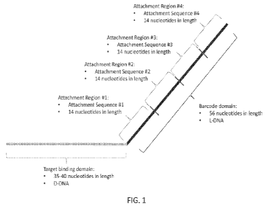
100371 FIG. I is a schematic diagram of an exemplary in situ hybridization
(ISM probe of the
present disclosure.
100381 FIG. 2 is a schematic diagram of an exemplary reporter probe of the
present disclosure.
100391 FICis. 3A, 3B, 3C, 3D, 3E, 3F and 3E1 are exemplary schematics of the
steps of a method
of detecting the abundance and spatial location of more than one species of
target nucleic acid in
a biological sample.
10041)] FIG. 4 shows a series of graphs comparing the abundance of RNA target
analytes in
various cells measured using the methods of the present disclosure and
standard RNA-sect
methods,
100411 FIG. 5 shows images of individual target analytes detected in a
biological sample
comprising MDA-MB-468 cells using the methods of the present disclosure. FIG 5
also shows
the quantification of the number of transcripts per cell analyzed.
V0042j FIG. 6A shows images of individual target analytes detected in Melanoma
FFPE tissue
samples using the methods of the present disclosure.
100431 FIG. 6B shows the results of cell typing analyses that can be performed
using spatial
abundance data collected using the methods of the present disclosure.
100441 FIG 6C shows the results cell interaction induced differential
expression analyses that
can be performed using spatial abundance data collected using the methods of
the present
disclosure.
[00451 FIG 61) shows images of individual target analytes detected in Melanoma
FFPE tissue
samples using the methods of the present disclosure.
100461 FIG. 6E shows images of individual target analytes detected in n.on-
small cell lung cancer
(NSCL.C.) FFPE tissue samples using the methods of the present disclosure.
7
CA 03192943 2023- 3- 16
WO 2022/060967
PCT/US2021/050642
100471 FIG 6F shows images of individual target analytes detected in renal
cell carcinoma FFPE
tissue samples using the methods of the present disclosure.
[00481 FIG. 6G shows images of individual target analytes detected in
colorectal cancer (CRC)
and tonsil FITE tissue samples using the methods of the present disclosure.
DETAILED DESCRIPTION
100491 The present disclosure provides methods for preparing a biological
sample for
fluorescent imaging. The present disclosure also provides in situ
hybridization (ISM probes and
reporter probes for use in the methods of the present disclosure, as well as
kits comprising these
ISH probes and reporter probes. The present disclosure also provides methods
of determining the
abundance and spatial position of at least two target nucleic acid molecules
in a biological
sample,
[00.s0i Methods of Sample Processing
posil In some aspects, the present disclosure provides a method of preparing a
biological
sample for fluorescent imaging, the method comprising: a) mounting a
biological sample onto a
functionalized microscope slide thereby producing a mounted biological sample,
wherein the
biological sample is a fonna lin fixed paraffin embedded (FT:PE) microtome
section; b) baking
the mounted biological sample; c) deparaffinizing the mounted biological
sample; d) performing
a target retrieval reaction on the mounted biological sample; e)
permeabilizing the mounted
biological sample; f) applying at least one fiducial marker to the mounted
biological sample; g)
fixing the mounted biological sample; h) contacting the mounted biological
sample with at least
one nucleic acid probe; and i) washing the mounted biological sample.
100521 In some aspects, the preceding methods can optionally further comprise
j) dehydrating
the mounted biological sample.
100531 In some aspects, the preceding methods can further comprise, after step
(i) or after step
(j) assembling the mounted biological sample into a flow cell.
100541 In some aspects, the preceding methods can further comprise after step
(g) and before
step (h), incubating the mounted biological sample in a blocking solution.
[005.51 In some aspects, the preceding methods can further comprise, before or
after any of the
steps, illuminating the biological sample with blue and/or UV light, thereby
quenching sample
autofluorescence via photobleaching. In some aspects, any combination of UV
and readout
channel illumination can be used to quench sample autofluorescence via
photobleaching. In
8
CA 03192943 2023- 3- 16
WO 2022/060967
PCT/US2021/050642
some aspects, the illumination can be performed concurrently with any of the
above steps,
including, but not limited to step (h). In some aspects, the illumination can
be performed using
low-dose illumination over extended time periods.
100561 in some aspects, a functionalized microscope slide can be a (3-
Aminopropyl)trimethoxysilane (APTivIS)-functionalized microscope slide. In
some aspects, an
APTIVIS functionalized microscope slide can prepared using the following
method: a) cleaning a
microscope slide using a plasma machine; b) incubating the microscope slide in
a 0.5% APTNIS
solution for soaking for about I minute; c) sonicating the microscope slide in
the 0.5% APTNIS
solution for about 10 seconds; d) repeating steps (b) and (c) twice such that
the microscope slide
is immersed in the 0.5% APINIS solution for about 3.5 minutes; e) rising the
microscope slide
with water at least 3 times; and f) drying the microscope slide under
nitrogen.
100571 In some aspects, a functionalized microscope slide can be any
positively-charged
microscope slide. As would be appreciated by the skilled artisan, non-limiting
examples of
commercially-available, positively-charged microscope slides include, but are
not limited to
poly-L-Lysin coated glass slide, ',mica BOND Plus slides and FisherhrandTM
SuperFrostTM Plus
[00581 In some aspects of the methods of the present disclosure, mounting a
biological sample
onto a functionalized microscope slide can comprise mounting the biological
sample onto the
functionalized microscope slide and drying the mounted biological sample for
at least about 12
hours, or at least about 13 hours.; or at least about 14 hours, or at least
about 1 5 hours, or at least
about 16 hours, or at least about 17 hours; or at least about 18 hours at room
temperature.
[00591 In some aspects of the methods of the present disclosure, baking a
mounted biological
sample can. comprise baking the mounted biological sample at least about 50 C,
or at least about
5.5 C, or at least about 60 C, or at least about 65 C, or at least about 70 C,
or at least about
75 C, or at least about 80 C. In some aspects, baking a mounted biological
sample can comprise
baking the mounted sample at about 60 C.
100601 In some aspects of the methods of the present disclosure, baking a
mounted biological
sample can comprising baking the mounted biological sample for at least about
0.5 hours, or at
least about 1 hour, or at least about 1.5 hours, or at least about 2 hours. in
some aspects, baking a
mounted biological sample can comprise baking the mounted biological sample
for about 1 hour.
9
CA 03192943 2023- 3- 16
WO 2022/060967
PCT/US2021/050642
100611 In some aspects of the methods of the present disclosure, baking a
mounted biological
sample can comprise baking the mounted biological sample at about 60 C for
about 1 hour.
100621 In some aspects of the methods of the present disclosure,
deparaffinizing a mounted
biological sample can comprise: a) incubating the mounted biological sample in
a first solution
of xylene for about 5 minutes; b) incubating the mounted biological sample in
a second solution
of xylene for about 5 minutes; c) incubating the mounted biological sample in
a first 100%
ethanol solution for about 2 minutes; d.) incubating the mounted biological
sample in the second
1000/0 ethanol solution for about 2 minutes; and e) drying: the mounted
biological sample at about
60 C for about 5 minutes. In some aspects, the incubation in the first
solution of xylene and/or
the second solution of xylene can comprise agitating the mounted biological
sample in the xylene
solution, for example, by moving the biological sample up and down in the
solution.
100631 Without wishing to be bound by theory, since FFPE samples contain DNA
molecules that
are crosslinked to each other as well as to RNA and protein molecules,
breakage of these crosslinks
can facilitate the release of DNA for subsequent purification. Breakage of
these crosslinks can be
achieved by performing a target retrieval reaction on a biological sample,
such as an FFPE sample.
In a target retrieval reaction, the biological sample, such as the FFPE
sample, can be incubated
with a target retrieval solution, wherein the target retrieval solution is
suitable for removing
crosslinking between DNA, RNA. and protein within the biological sample,
thereby allowing for
the recovery of analyzable bio Olecul es .
[006-ii In sonic aspects, a target retrieval solution can have a pH of about
8.0 to about 10Ø in
some aspects, a target retrieval solution can have a pH of about 8.5 to about
9.5. In some aspects,
a target retrieval solution can have a pH of about 9Ø In some aspects, a
target retrieval solution
can comprise a buffering agent. In some aspects, the buffering agent can be
IRIS.
10065] In some aspects, a target retrieval solution can comprise a chelator.
I.n some aspects, the
dictator can be ethylenedia.minetetraacetic acid (EDTAI). In some aspects a
target retrieval solution
can comprise about 0.1 to about 2 MIVI EDTA. In some aspects, a target
retrieval solution can
comprise about 0.5 to about 1.5 rnM EDTA. In some aspects, a target retrieval
solution can
comprise about 1.0 miNIEDTA.
100661 in some aspects, a target retrieval solution can be a MIS and EDTA
solution. In some
aspects, a target retrieval solution can be a solution of about 10 inM TR1S
and about 1 niM EDTA
at pH 9Ø
CA 03192943 2023- 3- 16
WO 2022/060967
PCT/US2021/050642
100671 In some aspects, a target retrieval solution can be RNAscope Target
Retrieval Solution
(ACD).
[00681 In some aspects of the methods of the present disclosure, performing a
target retrieval
reaction on a mounted biological sample can comprise incubating the mounted
biological sample
in a target retrieval solution at about 100 C. In some aspects, the mounted
biological sample is
incubated in target retrieval solution at about 100 C for an amount of time
specific to the type of
mounted biological sample. in a non-limiting example wherein the mounted
biological sample is
a human breast tumor sample, the mounted biological sample can be incubated in
target retrieval
solution at about 100 C for about 15 minutes. Incubation times for different
sample types are
shown in Table I. In some aspects, performing a target retrieval reaction can
further comprise,
after incubating the mounted biological sample in target retrieval solution,
incubating the mounted
biological sample in water for at least about 1.5 seconds; incubating the
mounted biological sample
in a solution of 100% ethanol for at least about 3 minutes; and drying the
mounted biological
sample. In some aspects, the water can be diethyl pyrocarbonate (DEPC)-treated
water.
100691 Accordingly, performing a target retrieval reaction on a mounted
biol.ogical sample can
comprise: a) incubating the mounted biological sample in target retrieval
solution at about 100 C
for a time period as put forth in Table 1 b) incubating the mounted biological
sample in DEPC-
treated water for about 15 seconds; c) incubating the mounted biological
sample in a solution of
1.00% ethanol for about 3 minutes; and d) drying the mounted biological
sample.
Table L Incubation times in ix target retrieval solution for various
biological sample types
Species of Incubation
Time:
Tissue Type Pathology
Biological Sample (minutes)
Intestine Normal 15
Intestine , Tumor 15
Embryo Normal , 15
Brain -Normal -15
Mouse
Spleen N orm al 15
Eye/Retina Normal 15
Liver Normal 30
Kidney , Normal , 15
Breast Tumor 15
Colon Tumor 15
Colon Normal , 15
Human Luna; Tumor 15
Lung Normal 15
Prostate , Tumor 15
Prostate Normal 15
Ii
CA 03192943 2023- 3- 16
WO 2022/060967
PCT/US2021/050642
Lymph node Tumor 15
Lymph node Normal 15
Tonsil Normal 15
Pancreas Normal 15
Cervical Cancer 15
Cervical Normal 15
Cervical dysplasia Abnormal 15
Brain Tumor 15
Brain Normal 15
Head Cancer 15
Neck Cancer 15
Liver Cancer 15
Kidney NO1111a1 15
Skin Normal 15
Melanoma Tusnor 15
Nevus Benign 15
Placenta Normal 15
Skin (tissue microarray
N 15
[TNIAD Normal
Breast TMA Normal 15
Melanoma TMA Normal 15
Nevus TivIA Benien 15
Stomach_ TMA Normal 15
Stomach TMA Tumor 15
Cell pellets, fixed with 10%
NSF
HeLa cells, fixed with 10%
Forma Idchydc/PBSIACD 15
Control
Cell Pellets (general) 8
VM701 in some aspects of the methods of the present disclosure, permeabilizing
the mounted
biological sample can comprise incubating the mounted biological sample in a
proteinase K
solution.
[00711 In some aspects, the proteinase K solution can be a solution wherein
the concentration of
proteinase K is at least about 0.1 g/mL, or at least about 0.25 g/mL, or at
least about 0.5
pigirnIõ or at least about 0.75 pig/mIõ or at least about 1 laginilL, or at
least about 1.25 uglinL, or
at least about 1.5 pg/mIõ or at least about 1.75 p.glinL., or at least about
21.1gImL. or at least
about 2.25 iag/mL, or at least about 2.5 gg/mL, or at least about 2.7514mL, or
at least about 3
p.g/mL, or at least about 3.25 ii.g/mL, or at least about 3.5 ps/mL, or at
least about 3.75 p.g/mL,
or at least about 4 gg/mL, or at least about 4.25 liglmL, or at least about
4.5 p.g/mL, or at least
about 4.75 tig,imL, or at least about 5 nit/IL In some aspects, the proteinase
K solution is a
12
CA 03192943 2023- 3- 16
WO 2022/060967
PCT/US2021/050642
solution wherein the concentration of proteinase K is about I agimt. In some
aspects, the
proteinase K solution is a solution wherein the proteinase K is diluted into
Phosphate Buffered
Saline (PBS). In some aspects, the proteinase K solution is a solution wherein
the proteinase K
is diluted into protease cocktail, including, but not limited to AD Protease
Pius.
100721 In some aspects, the PBS can comprise a combination of Na.CL, KC1,
Naa11P0.4 and
KEt2PO4. In some aspects, the PBS can comprise a solution of 137 m_M NaCl, 2.7
rriM KC1, 8
inTal Na2lliPO4, and 2. inIVI KH2PO4 at pH 7.4. Accordingly, in some aspects a
proteinase K
solution can be a solution wherein the concentration of proteinase. K is about
in PBS,
wherein the PBS comprises 137 tuM NaC1, 2.7 naM KC1, 8 naM Naalilllat, and 2
rriM KII7PO4 at
pH 7.4.
100731 in some aspects, permeabilizing the mounted biological sample can
comprise incubating
the mounted biological sample in a proteinase K solution at about 40 C. In
some aspects,
permeabilizing the mounted biological sample can comprise incubating the
mounted biological
sample in a proteinase K solution at about 40 C for an amount of time specific
to the type of
mounted biological sample. In a non-limiting example wherein the mounted
biological sample is
a human breast tumor sample, the mounted biological sample can be incubated in
a proteinase K
solution at about 40 C for about 30 minutes. Incubation times for different
sample types are
shown in Table 2.
100741 In some aspects, permeabilizing the mounted biological sample can.
comprise incubating
the mounted biological sample at about 40 C in a proteinase solution. In some
aspects,
permeabilizing the mounted biological sample can comprise incubating the
mounted biological
sample at about 40 C in a proteinase solution for an amount of time specific
to the type of
mounted biological sample. In a non-limiting example wherein the mounted
biological sample is
a human breast tumor sample, the mounted biological sample can be incubated at
about 40 C. in
a proteinase solution for about 30 minutes. Incubation times for different
sample types are shown
in Table 2.
100751 in some aspects, a proteinase solution can comprise a solution of
protease K at a
concentration of about 0.1 to about 5.0 ugimL, or 0.1 to 5.0 ug/mL. In some
aspects, a proteinase
solution can comprise a solution of protease K at a concentration of about 0.1
to about 5.0
ug/mL, or 0.1 to 5.0 ug/mL in PBS. In some aspects, a proteinase solution can
comprise a
solution of protease K at a concentration of about 0.1 to about 5.0 ug/mL, or
0.1 to 5.0 ug/mL in
13
CA 03192943 2023- 3- 16
WO 2022/060967
PCT/US2021/050642
a protease cocktail (e.g ACD protease plus solution). In some aspects, a
proteinase solution can
comprise a protease cocktail known in the art, e.g: ACD protease plus
solution.
Table 2. Incubation times in proteinase solution for various biological sample
types
Species of incubation
Time
Tissue Type Pathology
Biological Sample (,111
illtiteS)
Intestine Normal 30
Intestine Tumor 30
Embryo Normal 30
Brain Normal 30
Mouse
Spleen Normal 15
Eye/Retina Normal 30
Liver Normal 30
Kidney Normal 30
,
Breast 'Tumor 30
Colon Tumor 30
Colon Normal µ 30
Lung Tumor 30
Lung Normal 30
Prostate Tumor 10
Prostate Normal 30
Lymph node Tumor 30
Lymph node Normal µ 30
Tonsil Normal 30
Pancreas Normal 30
Cervical Cancer µ 30
Cervical Normal 30
Cervical dysplasi a Abnormal 30
Brain
Tumor
, 30
Brain Normal 30
Human Head , Cancer , 30
.
Neck Cancer µ 30
Liver Cancer 30
Kidney , Normal 30
=
Skin Normal 30
Melanoma Tumor 30
Nevus , Benign 30
=
Placenta Normal 30
Skin (tissue microarray
LIMA]) Normal 30
Breast TI'vtA Normal 30
Melanoma TMA Normal 30
Nevus TMA, Benign 30
Stomach TMA Normal 30
Stomach TMA , Tumor , 30
,
Cell pellets, fixed with 10% _ 15
NBF
14
CA 03192943 2023- 3- 16
WO 2022/060967
PCT/US2021/050642
HeLa cells, fixed with. 10%
Fonnaldehyde/PBS/A.CD 15
Control
Cell Pellets (general) 15
100761 In some aspects, incubating a mounted biological sample in a proteinase
K. solution can
further comprise drawing a hydrophobic barrier around the mounted biological
sample, for
example, with a PAP pen.
100771 In some aspects, permeabilizin.g a mounted biological sample can
comprise incubating
the mounted biological sample in a proteinase K solution in a container that
has been lined with
paper (e.g. kimwipes or a suitable alternative) that have been wet with DEPC-
treated water and
preheated to about 40 C for at least about 30 minutes.
100781 in some aspects, permeabilizing a mounted biological sample can further
comprise, after
incubating the mounted biological sample in a proteinase K solution, washing
the mounted
biological sample with water_ The water can be DEPC-treated water. In some
aspects, washing
the mounted biological sample with water can comprise washing the mounted
biological sample
with a first aliquot of DEPC-treated water and then washing the mounted
biological sample with
a second aliquot of D.EPC-treated water.
[00791 Accordingly, permeabilizing a mounted biological sample can comprise:
a) incubating
the mounted biological sample in a proteinase K solution at about 40 C for a
time period as put
forth in Table 2, wherein the concentration of proteinase K in the proteinase
K solution is about 1
lig/triL; b) washing the biological sample with a first aliquot of DEPC-
treated water; and c)
washing the biological sample with a second aliquot of DEPC -treated water.
100801 Accordingly, permeabilizing a mounted biological sample can comprise:
a) incubating
the mounted biological sample at about 40 C in a proteinase solution for a
time period as put
forth in Table 2, wherein the proteinase solution comprises a solution of
protease K at a
concentration of about 0.1 to about 5.0 ug/nile, or 0.1 to 5.0 uglmL; b)
washing the biological
sample with a first aliquot of DEPC-treated water; and c) washing the
biological sample with a
second aliquot of DEPC-treated water.
10811 In some aspects of the methods of the present disclosure, applying at
least one fiducial
marker to a mounted biological sample can comprise incubating the mounted
biological sample
in a solution comprising at least one fiducial marker. An at least one
fiducial marker can be any
fiducial marker known in the art to be useful for fluorescent imaging, as
would be appreciated by
CA 03192943 2023- 3- 16
WO 2022/060967
PCT/US2021/050642
the skilled artisan. In som.e aspects, the at least one fiducial marker can be
diluted in 2x saline-
sodium citrate (SSC) solution. In some aspects, the at least one fiducial
marker can be diluted in
2x saline-sodium citrate tween (SSCT) solution. In some aspects, the mounted
biological sample
can incubated in the solution comprising at least one fiducial marker for at
least about I minute,
or at least about 2 minutes, or at least about 3 minutes, or at least about 4
minutes, or at least
about 5 minutes, or at least about 6 minutes, or at least about 7 minutes, or
at least about 8
minutes, or at least about 9 minutes, or at least about 10 minutes. In some
aspects, the mounted
biological sample can be incubated in the solution comprising at least one
fiducial marker for
about 5 minutes. In some aspects, the mounted biological sample can be
incubated in the solution
comprising the at least one fiducial marker at about room temperature. In some
aspects, after
incubation with the solution comprising at least one fiducial marker, the
mounted biological
sample can be washed, for example, with phosphate buffered solution (PBS). In
some aspects,
prior to applying the solution comprising at least one fiducial marker to the
mounted biological
sample, the solution. can be agitated (e.g. vortexed) for at least 30 seconds.
100821 In some aspects of the methods of the present disclosure, 2x SSC buffer
can comprise
about 300 mTvINaCI and about 30 rnM sodium citrate. In some aspects of the
methods of the
present disclosure, 2x SSC buffer can comprise 300 rnNif Na.CI and 30 mNI
sodium citrate.
100831 In some aspects of the methods of the present disclosure, 2x SSCT
buffer can comprise
about 0.1% Tween20, about 300 iriM NaCl and about 30 rnM sodium citrate. In
some aspects of
the methods of the present disclosure, 2x SSCT buffer can comprise 0.1%
Tween20, 300 inM
NaCl and 30 niM sodium citrate.
100841 In some aspects, the at least one fiducial marker can be a 200 ntil
carboxylated
microspherc in red, blue, yellow and/or green. In some aspects, a solution
comprising at least one
fiducial marker can comprise 200 nm carboxylated microspheres in red, blue
and/or green at a
concentration of at least about 0.00025%, or at least about 0.0005%, or at
about 0.00075%, or at
least about 0.001%, or at least about 0.00125%, or at least about 0.0015%, or
at least about
0.00175%, or at least about 0.002%, or at least about 0.005%, or at least
about 0.01%. In some
aspects, a solution comprising at least one fiducial marker can comprise 200
nm carboxylated
microspheres in red, blue and/or green at a concentration of about 0.001%. ID
some aspects, the
at least one fiducial marker can be a carboxylated microspbere (e.g 200 tun
carboxylated
microspheres) stained in red, yellow, blue and/or green. In some aspects, a
solution comprising at
16
CA 03192943 2023- 3- 16
WO 2022/060967
PCT/US2021/050642
least one fiducial marker can comprise carboxylated microspheres stained in
red, yellow, blue
and/or green at a concentration of at least about 0.00025%, or at least about
0.0005%, or at about
0.00075%, or at least about 0.001%, or at least about 0.00125%, or at least
about 0.0015%, or at
least about 0.00175%, or at least about 0.002%, or at least about 0.005%, or
at least about 0.01%.
in some aspects, a solution comprising at least one fiducial marker can
comprise carboxylated
microspheres stained in red, yellow, blue and/or green at a concentration of
about 0.001%. In
some aspects, a solution comprising at least one fiducial marker can comprise
carboxylated
microspheres stained in red, yellow, blue and/or green at a concentration of
about 0.0005% to
about 0.003%, or 0.0005% to 0.003%.
100851 In some aspects, the at least one fiducial marker can be a fluorescent
nano-diamond
(FND). In some aspects, and FND can be a non-carboxylated FND. In some
aspects, a solution
comprising at least one fiducial marker can comprise FNDs at a concentration
of at least about
0.0001%, or at least about 0.00015%, or at least about 0.0002%, or at least
about 0.00025%, or at
least about 0.0003%, or at least about 0.00035%, or at least about 0.0004%, or
at least about
0.00045%, or at least about 0.0005%, or at least about 0.00055%, or at least
about 0.001%. In
some aspects, a solution comprising at least one fiducial marker can comprise
FNDs at a
concentration of about 0.00045%.
100861 In some aspects, a solution comprising at least one fiducial marker can
comprise a
combination of at least two fiducial markers. In a non-limiting example, a
solution comprisin.g at
least one fiducnal marker can comprise 200 ran carboxylated microspheres in
red, blue and/or
green and non-carboxylated -FNDs. In a non-limiting example, a solution
comprising at least one
fiducial marker can comprise 200 tun carboxylated microspheres in red, blue
and/or green at a
concentration of about a 0019/0 and non-carboxylated FNDs at a concentration
of about
0.00045%.
[0087] In some aspects, a solution comprising at least one fiducial marker can
comprise a
combination of at least two fiducial markers. In a non-limiting example, a
solution comprising at
least one fiducial marker can comprise carboxylated microspheres stained in
red, yellow, blue
and/or green and non-carboxylated FNDs. In a non-limiting example, a solution
comprising at
least one fiducial marker can comprise tim carboxylated microspheres stained
in red, blue and/or
green at a concentration of about 0.0005% to about 0.003%, or 0.0005% to
0.003%, and non-
carboxylated FNDs at a concentration of about 0.00045%.
17
CA 03192943 2023- 3- 16
WO 2022/060967
PCT/US2021/050642
10$1881 In some aspects, a solution comprising at least on fiducial marker can
be prepared by
diluting the at least one fiducial marker in a suitable buffer solution,
including, but not limited to
2x SSC solution, and then agitating (e.g vortexing) the solution for about 1
minute, then
sonicating the solution for about 2 minutes, then agitating the solution again
for about 1 minute,
then sonicating the solution again for about 2 minutes.
100891 In some aspects, a solution comprising at least on fiducial marker can
be prepared by
diluting the at least one fiducial marker in a suitable buffer solution,
including, but not limited to
2x SSCT solution, and then agitating (e.g. vortexing) the solution for about I
minute, then
sonicating the solution for about 2 minutes, then agitating the solution again
for about 1 minute,
then sonicating the solution again for about 2 minutes.
100901 Accordingly, applying at least one fiducial marker to a mounted
biological sample can
comprise: a) incubating the mounted biological, sample in a solution
comprising at least one
fiducial marker for about 5 minutes at about room temperature, wherein the
solution comprising
at least one fiducial marker is a solution comprising carboxylated
microspheres in red, blue
and/or green at a concentration of about 0.001% and non-carboxylated FNDs at a
concentration
of about 0.00045% in 2x SSC solution and, b) washing the mounted biological
with ix PBS,
[00911 Accordingly, applying at least one fiducial marker to a mounted
biological sample can
comprise: a) incubating the mounted biological sample in a solution comprising
at least one
fiducial marker for about 5 minutes at about room temperature, wherein the
solution comprising
at least one fiducial marker is a solution comprising carboxylated
microspheres stained in red,
yellow, blue andi'or green at a concentration of about 0,0005% to about
0.003%, or 0.00050/n to
0.003%; and b) washing the mounted biological with lx PBS.
100921 In some aspects of the methods of the present disclosure, fixing a
mounted biological
sample can comprise incubating the mounted biological sample in neutral
buffered formal
(NBF) solution, then incubating the mounted biological sample in a tris
glycine buffered
solution, and then incubating the mounted biological sample in lx PBS. In some
aspects, the
concentration of NBF in the NBF solution can be at least about 5%, or at least
about 10%, or at
least about 15%, or at least about 20%. In some aspects, the concentration of
NBF in the NBF
solution can be about 10%. In some aspects, any of the incubation steps in the
fixing of the
mounted biological sample can be for at least about 1 minute, or at least
about 2 minutes, or at
least about 3 minutes, or at least about 4 minutes, or at least about 5
minutes, or at least about 6
18
CA 03192943 2023- 3- 16
WO 2022/060967
PCT/US2021/050642
minutes, or at least about 7 minutes, or at least about 8 minutes, or at least
about 9 minutes, or at
least about 10 minutes. In some aspects; any of the incubation steps in the
fixing of the mounted
biological sample can be about 1 minute, or about 2 minutes, or about 3
minutes, or about 4
minutes, or about 5 minutes, or about 6 minutes, or about 7 minutes, or about
8 minutes, or about
9 minutes, or about 10 minutes. In some aspects, any of the incubation steps
can be for about 5
minutes. In some aspects, any of the incubation steps can be for about 1
minute. In some aspects,
incubating the mounted biological sample in a tris glycine buffered solution
can comprise
incubating the mounted biological sample in a first tris glycine buffered
solution followed by
incubating the mounted biological sample in a second tris glycine buffered
solution.
i00931 Accordingly, fixing a mounted biological sample can comprise: a)
incubating the
mounted biological sample in a 10% NalF for about 5 minutes; b) incubating the
mounted
biological sample in a first tris glycine buffered solution for about 5
minutes; c) incubating the
mounted biological sample in a second tris glycine buffered solution for about
5 minutes; and d)
incubating the mounted biological sample in lx PBS for about 5 minutes.
100941 Accordingly, fixing a mounted biological sample can comprise: a)
incubating the
mounted biological sample in a 10% NBF for about I minute; b) incubating the
mounted
biological sample in a first tris glycine buffered solution for about 5
minutes; c) incubating the
mounted biological sample in a second tris glycine buffered solution for about
5 minutes; and d)
incubating the mounted biological sample in Ix PBS for about 5 minutes.
[0095.1 In some aspects of the methods of the present disclosure, incubatin.g
the mounted
biological sample in a blockin.g solution can comprise incubating the mounted
biological sample
in a Sul1-o-NHS-acetate/Tween20 solution. In some aspects, a Sulfo-NHS-
acetatelTween20
solution can comprise about 100 inNI Sulfo-NE1S-acetate, about 0.5% Tween20 in
about 100 triM
sodium phosphate pH 8µ In some aspects, a Sulfo-NTIS-acetatelTween20 solution
can comprise
100 trilVi Sulfo-NHS-acetate, 0.5% Tween20 in 100 m1V1 sodium phosphate pH 8.
In some
aspects, the mounted biological sample can be incubated in a Sulfo-NHS-
acetatefTween20
solution for at least about 5 minutes, or at least about 10 minutes, or at
least about 15 minutes, or
at least about 20 minutes. In some aspects, the mounted biological sample can
be incubated in a
Sulfo-NIIS-acetateffween20 solution for about 5 minutes, or about 10 minutes,
or about 15
minutes, or about 20 minutes. In some aspects, the mounted biological sample
can be incubated
in a Sulfo-N1-1S-acetaterfween20 solution -for about 15 minutes.
19
CA 03192943 2023- 3- 16
WO 2022/060967
PCT/US2021/050642
In some aspects of the methods of the present disclosure, incubating the
mounted
biological sample in a blocking solution can comprise, after incubating the
mounted biological
sample in a Sulfo-NHS-acetatelfween20 solution, incubating the mounted
biological sample in a
Ix PBS for at least about 1 minute, or at least about 2 minutes, or at least
about 3 minutes, or at
least about 4 minutes, or at least about 5 minutes, or at least about 6
minutes, or at least about 7
minutes, or at least about 8 minutes, or at least about 9 minutes, or at least
about 10 minutes. In
some aspects of the methods of the present disclosure, incubating the mounted
biological sample
in a blocking solution can comprise, after incubating the mounted biological
sample in a Sulfo-
NHS-acetaterfween20 solution, incubating the mounted biological sample in a lx
PBS for about
1 minute, or about 2 minutes, or about 3 minutes, or about 4 minutes, or about
5 minutes, or
about 6 minutes, or about 7 minutes, or about 8 minutes, or about 9 minutes,
or about 10
minutes. In some aspects of the methods of the present disclosure, incubating
the mounted
biological sample in a blocking solution can comprise, after incubating the
mounted biological
sample in a Sulfo-NHS-acetatelTween20 solution, incubating the mounted
biological sample in a
1.x PBS for about 5 minutes.
0o971 A.ccordingly, incubating the mounted biological sample in a blocking
solution can
comprise: i) incubating the mounted biological sample in a Su1fo-NHS-
acetate/Tween20 solution
for about 1_5 minutes, wherein the Sulfo-NHS-acetate/Tween20 solution
comprises about 100
raM Sulfo-NHS-acetate, about 0.5% Tween20 in about 100 mM sodium phosphate pH
8; and ii)
incubating the mounted biological sample in lx PBS for about 5 minutes.
100981 In some aspects of the methods of the present disclosure contacting the
mounted
biological sample with at least one nucleic acid probe can comprise incubating
the mounted
biological sample with a solution comprising a plurality of ISH probes of the
present disclosure.
In some aspects, the mounted biological sample can be incubated with the
solution comprising a
plurality of NH probes for at least about 12 hours, or at least about 13
hours, or at least about 14
hours, or at least about 15 hours, or at least about 16 hours, or at least
about '17 hours, or least
about 18 hours, or at least about 19 hours, or at least about 20 hours, or at
least about 21 hours,
or at least about 22 hours, or at least about 23 hours, or at least about 24
hours. In some aspects,
the mounted biological sample can be incubated with the solution comprising a
plurality of Mil
probes for about 16 to about 18 hours.
CA 03192943 2023- 3- 16
WO 2022/060967
PCT/US2021/050642
199991 In some aspects, the mounted biological sample can be incubated with
the solution
comprising a plurality of ISH probes at a temperature of at least about 35 C,
or at least about
36 C, or at least about 37 C, or at least about 38 C, or at least about 39 C,
or at least about
40 C. In some aspects, the mounted biological sample can be incubated with the
solution
comprising a plurality of ISH probes at a temperature of about 35 C.
[001001 In some aspects, the solution comprising a plurality of ISH probes of
the present
disclosure can comprise a single species of ISH probe. In some aspects, the
solution comprising
a plurality of ISH probes of the present disclosure can comprise at least
about 2, or at least about
3, or at least about 4, or at least about 5, or at least about 6, or at least
about 7, or at least about 8,
or at least about 9, or at least about 10, or at least about 25, or at least
about 50, or at least about
75, or at least about 100, or at least about 250, or at least about 500, or at
least about 750, or at
least about 1000, or at least about 5,000, or at least about 10,000, or at
least about 15,000, or at
least about 20,000, or at least about 50,000, or at least about 100,000, or at
least about 500,000,
or at least about 1,000,000 different species of ISH probes.
lomin in some aspects, the concentration of at least on.e species of ISH probe
in the plurality can
be at least about 0.01 nM, or at least about 0.1 nM, or at least about 1 nM,
or at least about 5 nivi,
or at least about 10 nM, or at least about 25 nNI, or at least about 50 nM, or
at least about 75 nM,
or at least about 100 nM, or at least about 125 niS,4, or at least about 150
nM, or at least about 175
uM, or at least about 200 nM, or at least about 300 nM, or at least about 400
nM, or at least about
500 KNA. in som.e aspects, the concentration of at least one species of LSI4
probe in the plurality
can be about 0.01 riM, or about 0.1 riM, or about 1 UM, or about 5 nIVI, or
about 10 nM, or about
25 nM, or about 50 nkl, or about 75 nM, or about 100 nM, or about 125 nM, or
about 150 riN4, or
about 175 riN4, or about 200 nM, or about 300 DM, or about 400 nM, or about
500 UM. hi some
aspects, the concentration of at least one species of ISH probe in the
plurality can be about 200
nM. In some aspects, the concentration of at least one species of 1811 probe
in the plurality can
be about 1 UM.
[001021 In some aspects, the concentration of each species of 1SH probe in the
plurality can be at
least about 0.01 UM, or at least about 0.1 nt\4, or at least about I n114, or
at least about 5 nM, or at
least about 10 nivl, or at least about 25 nM., or at least about 50 nM, or at
least about 75 n1\4, or at
least about 100 nM, or at least about 125 nM, or at least about 150 nM, or at
least about 175 nM,
or at least about 200 nM, or at least about 300 nM, or at least about 400 UM,
or at least about 500
21
CA 03192943 2023- 3- 16
WO 2022/060967
PCT/US2021/050642
ri.M. In some aspects, the concentration of each species of ISH probe in the
pluralit2.,, can be about
0.01 JIM, or about 0.1 nitvl, or about 1 nM, or about 5 nM,. or about 10 nM,
or about 25 n141, or
about 50 nM, or about 75 tiM, or about 100 nM, or about 125 nl\,4, or about
150 nivi, or about 175
niNI, or about 200 nM, or about 300 riM, or about 400 11M, Of about 500 nlvi.
In some aspects, the
concentration of each species of ISH probe in the plurality can be about 200
nIVI. In some
aspects, the concentration of each species of ISH probe in the plurality can
be about 1 riMI.
itionoi In some aspects, a solution comprising a plurality of ISH probes can
comprise at least one
species of ISH probe that comprise target binding domains that are designed
not to specifically
bind to any target analyte (e.g. target nucleic acid molecule and/or target
protein molecule) in the
biological sample. In some aspects, a solution comprising a plurality of ISH
probes can comprise
at least two species, or at least three species, or at least four species, or
at least five species, or at
least six species, or at least seven. species, or at least eight species, or
at least nine species, or at
least ten species, or at least 50 species, or at least 100 species, or at
least 1000 species of ISH
probes that comprise target binding domains that are designed not to
specifically bind to any
target analyte (e.g. target nucleic acid molecule and/or target protein
molecule) in the biological
sample. These ISH probes that comprise target binding domains that are
designed not to
specifically bind to any target analyte are referred to herein as "negative
ISM probes". A non-
limiting example of a negative ISH probe is an ISH probe comprising a target
binding domain
that is a single-stranded nucleic acid, wherein the sequence of the single-
stranded nucleic acid is
designed such that it is not complem.entary to any known sequence specific to
the biological.
sample being analyzed and/or complementary to any known sequence present on
earth. As would
be appreciated by the skilled artisan, examples of such sequences include
those published by the
Evaluation of the External :RNA Controls Consortium (ERCC). Without wishing to
be bound by
theory, the use of these negative ISH probes in the methods of the present
disclosure can allow
the skill e,d artisan to determine the level of background noise in the
results from a biological
sample. As would be appreciated by the skilled artisan, since the negative 1SH
probes should not
bind to any target analyte, any signal originating from a negative ISH probe
that is recorded
represents non-specific binding of ISH probes within the sample (i.e.
background noise). In some
aspects, the skilled artisan can use the level of background noise detected by
negative 151-I probes
to more accurately determine the absolute abundance of target analytes within
the biological
sample.
22
CA 03192943 2023- 3- 16
WO 2022/060967
PCT/US2021/050642
luiliii41 In some aspects, the solution comprising a plurality of ISH probes
can comprise the ISH
probes diluted in buffer R.
In some aspects, buffer R can comprise at least one of dextran sulfate, bovine
serum
albumin (BSA), single-stranded DNA (ssDNA), saline-sodium citrate (SSC) and
fortnannde. In
some aspects, buffer R. can comprise a combination of dextran sulfate, BSA,
sSDNA, SSC and
formamide. In some aspects, the single-stranded DNA can comprise salmon sperm
DNA.
iutitiq In some aspects, the 1SH probes can be diluted in buffer R such that
the final
concentrati0E1 of dextran sulfate is about 0.5% to about 4.5%, or about 1.5%
to about 3.5%. In
some aspects, the ISH probes can be diluted in buffer R such that the final
concentration of
dextran sulfate is about 2.5%.
1001071 in some aspects, the ISI-1 probes can be diluted in buffer R such that
the final
concentration of BSA is about 0.01% to about 2%, or about 0.1% to about 1%. In
some aspects,
the ISH probes can be diluted in buffer R such that the final concentration of
BSA is about 0.2%.
1001081 In some aspects, the ISH probes can be diluted in buffer R such that
the final
concentration of ssDNA is about 0,01 mg/mi to about 1. .m,g/ml., or about 0.05
mg/m.1 to about 0.5
mg/ml. In some aspects, the 1ST-1 probes can be diluted in buffer R such that
the final
concentration of ssDNA is about 0.1 mg/nil.
[001091 In some aspects, the ISH probes can be diluted in buffer R such that
the final
concentration of SSC is about 0.5x to about 3.5x or about Ix to about 3x. In
some aspects, the
ISH probes can be diluted in buffer R such that the final concentration of SSC
is about 2x.
[00110i In some aspects, the NH probes can be diluted in buffer R. such that
the final
concentration of formamide is about 20% to about 60%, or about 30% to about
50%. in some
aspects, the -NH probes can be diluted in buffer R such that the final
concentration of formamide
is about 40%.
[Min In some aspects, the ISH probes can be diluted in buffer R such that the
final
concentration of dextran sulfate is about 2.5%, the final concentration of BSA
is about 0.2%, the
final concentration of ssDNA is about 0.1 mg/ml, the final concentration of
SSC is about 2x and
the final concentration of formamide is about 40%.
pot 121 In some aspects, the solution comprising a plurality of 1SH probes can
further comprise
an RNase inhibitor, including, but not limited to, SUPERase-inmi RN Me
inhibitor. The
concentration of RNAse inhibitor can be about 0.1 Units/1.d.
23
CA 03192943 2023- 3- 16
WO 2022/060967
PCT/US2021/050642
10$1/131 In some aspects, prior to incubating the mounted biological sample
with the solution
comprising a plurality of ISM probes, the ISH probes are first denatured by
incubating the ISH
probes at about 95 C for about 2 minutes and then immediately cooling the ISH
probes on ice for
about I minute.
w01141 In some aspects, the mounted biological sample can be incubated with
the solution
comprising a plurality of ISH probes in a container that has been rinsed with
an RNAse inhibitor
solution and that has been lined with paper (e.g. kimwipes or a suitable
alternative) that have
been wefted with DEPC-treated water
itionsi Accordingly, contacting the mounted biological sample with at least
one nucleic acid
probe can comprise: a) incubating the mounted biological sample with a
solution comprising a
plurality of ISH probes of the present disclosure for about 16 to about 18
hours at about 37 C,
wherein the solution comprises at least one species of ISH probe, wherein at
least one species of
ISH probe in the plurality is present at a concentration of about 200 nIVI.
In some aspects of the methods of the present disclosure, washing a mounted
biological
sample can comprise: a) incubating the mounted biological sample with first 2x
SSC solution; b)
incubating the mounted biological sample in a first formamide solution; c)
incubating the
mounted biological sample with a second formamide solution; d) incubating the
mounted
biological sample with a second 2x SSC solution; and e) incubating the mounted
biological
sample with a third 2x SSC solution.
iii301t7i In some aspects, a form.amide solution in be a formamide in 2x SSC
solution. In some
aspects, the concentration of formamide can be at least about 10%, or at least
about 20%, or at
least about 30%, or at least about 40%, or at least about 50%, or at least
about 60%, or at least
about 70%. In some aspects the concentration of formamide can be about 50%. In
some aspects,
the mounted biological sample can be incubated with the first formamide
solution and/or the
second formamide solution for at least about 15 minutes, or at least about 20
minutes, or at least
about 25 minutes, or at least about 30 minutes, or at least about 35 minutes,
or at least about 40
minutes. in some aspects, the mounted biological sample can be incubated with
the first
formamide solution and/or the second formamide solution for about 25 minutes.
[001181 In some aspects, the mounted biological sample can be incubated with
the second 2x SSC
solution and/or the third 2x SSC solution for at least about 0.5 minutes, or
at least about 1
minute, or at least about 1.5 minutes, or at least about 2.0 minutes, or at
least about 2.5 minutes,
24
CA 03192943 2023- 3- 16
WO 2022/060967
PCT/US2021/050642
or at least about 3.0 minutes, or at least about 3.5 minutes, or at least
about 4.0 minutes, or at
least about 4.5 minutes, or at least about 5 minutes. In some aspects, the
mounted biological
sample can be incubated with the second 2x SSC solution and/or the third 2x
SSC solution for
about 2 minutes.
to iii Accordingly, washing a mounted biological sample can comprise: a)
incubating the
mounted biological sample with first 2x SSC solution; b) incubating the
mounted biological
sample in a first 50% formamide in 2x SSC solution for about 25 minutes; c)
incubating the
mounted biological sample with a second 50 ,70 formamide in 2x SSC solution
for about 25
minutes; d) incubating the mounted biological sample with a second 2x SSC
solution for about
two minutes; and e) incubating the mounted biological sample with a third 2x
SSC solution for
about two minutes.
1001201 In some aspects of the methods of the present disclosure, dehydrating
a mounted
biological sample can comprise incubating the mounted biological sample in an
ethanol gradient,
as would be appreciated by the skilled artisan. In some aspects, incubating
the mounted
biological sample in an ethanol gradient can comprise: a) incubating the
mounted biological
sample in a 70% ethanol solution for about 3 minutes; b) incubating the
mounted biological
sample in a 85% ethanol solution for about 3 minutes; and c) incubating the
mounted biological
sample in a 100% ethanol solution for about 3 minutes,
mew] In some aspects, a biological sample can be an FFPE microtome section
that is at least
about I um, or at least about 2 tm. or at least about 3 um, or at least about
4 um, or at least
about 5 urn, or at least about 6 urn, or at least about 7 urn, or at least
about 8 pm, or at least
about 9 um, or at least about 10 um thick. In some aspects, the biological
sample is a.n FFPE
mierotome section that is about 5 gm thick.
1001221 In some aspects, the biological sample can be a tissue sample from any
organ. In some
aspects, the biological sample is a tissue sample from the Intestine, Embryo,
Brain, Spleen, Eye,
Retina, Liver, Kidney, Breast, Throat, Colon, Lung, Prostate, Lymph node,
Tonsil, Pancreas,
Cervix, Head, Neck, Liver, Skin, Nevus, Placenta or any other organ.
1001231 In some aspects, the biological sample can comprise non-cancerous
cells. In some
aspects, the biological sample can comprise cancerous cells. In some aspects,
the biological
sample can comprise a combination of both non-cancerous cells and cancerous
cells. The
cancerous cells can be from a carcinoma, lymphoma, blastoma, sarcoma, leukemia
and germ cell
CA 03192943 2023- 3- 16
WO 2022/060967
PCT/US2021/050642
tumors. The cancerous cells can be from a adrenocortical carcinoma, bladder
urothelial
carcinoma, breast invasive carcinoma, cervical squamous cell carcinoma,
endocervical
adenocarcinoma, cholangiocarcinoma, colon adenocarcinoma, lymphoid neoplasm
diffuse large
B-cell lymphoma, esophageal carcinoma, glioblastoma multiforme, head and neck
squamous cell
carcinoma, kidney chromophobe, kidney renal clear cell carcinoma, kidney renal
papillary cell
carcinoma, acute myeloid leukemia, brain lower grade glioma, liver
hepatocellular carcinoma,
lung adenocarcinoma, lung squamous cell carcinoma, mesothelioma, ovarian.
serous
cystadenocarcinoma, pancreatic adenocarcinoma, pheochromocytoma,
paraganglioma, prostate
adenocarcinoma, rectum adenocarcinoma, sarcoma, skin cutaneous melanoma,
stomach
adenocarcinoma, testicular germ cell tumors, thyroid carcinoma, thymoma,
uterine
carcinosarcoma, uveal melanoma. Other examples include breast cancer, lung
cancer,
lymphoma, melanoma, liver cancer, colorectal cancer, ovarian cancer, bladder
cancer, renal
cancer or gastric cancer. Further examples of cancer include neuroendocrine
cancer, non-small
cell lung cancer (NSCLC), small cell lung cancer, thyroid cancer, endometrial
cancer, biliary
cancer, esophageal cancer, anal cancer, salivary, cancer, .vulvar cancer,
cervical cancer, Acute
lymphoblastic leukemia (ALL), Acute myeloid leukemia (AML)õAdrenal gland
tumors, Anal
cancer, Bile duct cancer, Bladder cancer, Bone cancer, Bowel cancer, Brain
tumors, Breast
cancer, Cancer of unknown. primary (CUP), Cancer spread to bone, Cancer spread
to brain,
Cancer spread to liver, Cancer spread to lung, Carcinoid, Cervical cancer,
Children's cancers,
Chronic lymphocytic leukemia (CLL), Chrome myeloid leukemia (CML)., Colorectal
cancer, Ear
cancer, Endometrial cancer, Eye cancer, Follicular dendritic cell sarcoma,
Gallbladder cancer,
Gastric cancer, Gastro esophageal junction cancers. Germ cell tumors,
Gestational trophoblastic
disease (GET)), Hairy cell leukemia, Head and neck cancer, Hodgkin lymphoma,
Kaposi's
sarcoma, Kidney cancer, Laryngeal cancer, Leukemia, Gastric linitis plastica,
Liver cancer, Lung
cancer, Lymphoma, Malignant schwannoma., Mediastinal germ cell tumors,
Melanoma skin
cancer, Men's cancer, Merkel cell skin cancer, Mesothelioma, Molar pregnancy,
Mouth and
oropharyngeal cancer, Myeloma, Nasal and paranasal sinus cancer,
Nasopharyngeal cancer,
Neumblastoma. Neuroendocrine tumors, Non-Hodgkin lymphoma (NHL). Esophageal
cancer,
Ovarian cancer, Pancreatic cancer, Penile cancer, Persistent trophoblastic
disease and
choriocarcinoma, Pheochromocytoma, Prostate cancer, Pseudomyxoma peritonei,
Rectal cancer.
Retinoblastoma, Salivary gland cancer, Secondary' cancer, Signet cell cancer,
Skin cancer, Small
26
CA 03192943 2023-3- 16
WO 2022/060967
PCT/US2021/050642
bowel cancer, Soft tissue sarcoma, Stomach cancer, T cell childhood non
Hodgkin lymphoma
(NHL). Testicular cancer, Thymus gland cancer. Thyroid cancer, Tongue cancer,
Tonsil cancer,
Tumors of the adrenal gland. Uterine cancer. Vaginal cancer, Vulva] cancer,
Wilms' tumor,
Womb cancer and Gynaecological cancer. Examples of cancer also include, but
are not limited
to, :Hematologic malignancies, :Lymphoma, Cutaneous T-cell lymphoma,
Peripheral 'r-cell
lymphoma, Hodgkin's lymphoma, Non-Hodgkin's lymphoma, Multiple myeloma, Chrome
lymphocytic leukemia, chronic myeloid leukemia, acute myeloid leukemia,
M.yeloclysplastic
syndromes, Myelofibrosis, Biliary tract cancer, Hepatocellular cancer,
Colorectal cancer, Breast
cancer, Lung cancer, Non-small cell lung cancer, Ovarian cancer, Thyroid
Carcinoma, Renal
Cell Carcinoma, Pancreatic cancer, Bladder cancer, skin cancer, malignant
melanoma, merkel
cell carcinoma, Uveal Melanoma or Glioblastoma multifortne.
1001241 The biological sample can be derived from any species, including, but
not limited to,
humans, mice, rats, dogs, cats, sheep, rabbits, cows, goats or any other
species.
1001251 In situ hybridization (ISH)probes of the present disclosure
1001261 Target binding domain
1001271 The present disclosure provides in situ hybridization (ISH) probes for
use in the methods
of the present disclosure.
1001281 An ISH probe can comprise a target binding domain and a barcode
domain. In some
aspects, the target binding domain is operably linked to the barcode domain.
1001291 In some aspects, a target binding domain can comprise a protein, a
peptide, an aptamer,
or a peptoid which specifically binds to a target analyte in a biological
sample. In some aspects,
the protein can be an antibody, or an antigen binding fragment thereof In some
aspects, the
protein can be a lectin protein. In some aspects, the protein can be any
carbohydrate-binding
protein known in the art.
1001301 In some aspects, a target binding domain can be a single stranded
polynucleotide. A
target binding domain can comprise a sequence that is complementary to a
target nucleic acid
that is to be identified using the methods of the present disclosure.
1001311 In some aspects, a target binding domain in be at least about 35
nucleotides in length to at
least about 40 nucleotides in length. In some aspects, a target binding domain
can be about 35
nucleotides to about 40 nucleotides in length. In some aspects, a target
binding domain can
comprise about 20 nucleotides, or about 21 nucleotides, or about 22
nucleotides, or about 23
27
CA 03192943 2023-3- 16
WO 2022/060967
PCT/US2021/050642
nucleotides, or about 24 nucleotides, or about 25 nucleotides, or about 26
nucleotides; or about
27 nucleotides, or about 28 nucleotides, or about 29 nucleotides, or about 30
nucleotides, or
about 31 nucleotides, or about 32 nucleotides, or about 33 nucleotides, or
about 34 nucleotides,
or about 35 nucleotides, or about 36 nucleotides, or about 37 nucleotides, or
about 38
nucleotides, or about 39 nucleotides, or about 40 nucleotides, or about 41
nucleotides; or about
42 nucleotides, or about 43 nucleotides, or about 45 nucleotides in length.
iti01321 In some aspects, a target binding domain comprises D-DNA. In some
aspects, a target
binding domain consists of D.-DNA.
[00133l In some aspects, a target binding domain can be about 35 nucleotides
to about 40
nucleotides in length and comprises D-DNA. In some aspects, a target binding
domain can be
about 35 nucleotides to about 40 nucleotides in length and consists of D-DNA.
1001341 Barcode domain
10013.51 In some aspects, a barcode domain can be a single stranded
polynucleotide.
1001361 A barcode domain can comprise at least one attachment region. In some
aspects, a
barcode domain can comprise at least two, at least three, at least four, at
least five, at least six, at
least seven, at least eight, at least nine, or at least ten attachment
regions.
1001371 In some aspects, a barcode domain can comprise about 4 attachment
regions.
[001381 An attachment region can comprise at least one nucleic acid sequence
that is capable of
being reversibly bound by a reporter probe of the present disclosure. A.
nucleic acid sequence
that is capable of being reversibly bound by a reporter probe of the present
disclosure is herein
referred to as an attachment sequence. Accordingly, an attachment region of a
barcode domain
can comprise at least one attachment sequence. In some aspects, the attachment
sequences within
a single attachment region can be identical; thus, the reporter probes that
bind within that single
attachment region will be identical. In some aspects, the attachment sequences
within a single
attachment can be different; thus, the reporter probes that bind within that
single attachment will
be different.
[001391 In some aspects, wherein a barcode domain comprises more than one
attachment region,
the attachment sequences in each of the different attachment regions can he
different; thus,
different reporter probes will bind to each attachment region in the barcode
domain.
1001401 In some aspects, an attachment sequence can be about 5 nucleotides; or
about 6
nucleotides, or about 7 nucleotides, or about 8 nucleotides, or about 9
nucleotides, or about 1.0
28
CA 03192943 2023- 3- 16
WO 2022/060967
PCT/US2021/050642
nucleotides, or about II nucleotides, or about 12 nucleotides, or about 13
nucleotides, or about
14 nucleotides, or about 15 nucleotides, or about 16 nucleotides, or about 17
nucleotides, or
about 18 nucleotides, or about 19 nucleotides, or about 20 nucleotides in
length. In some aspects,
an attachment sequence can be about 14 nucleotides in length.
10014 ti In some aspects, a barcode domain comprises le-DNA. In some aspects,
a barcode
domain consists of L-DNA.
itm1421 in some aspects, a barcode domain can comprise about 4 attachment
regions, wherein
each attachment region comprises about I attachment sequence, wherein each
attachment
sequence is about 14 nucleotides in length, such that the barcode domain is
about 56 nucleotides
in length, and wherein the nucleic acid sequence of each of the attachment
sequences are
different, wherein the barcode domain comprises la-DNA. In some aspects, a
barcode domain
can comprise about 4 attachment regions, wherein each attachment region
comprises about I
attachment sequence, wherein each attachment sequence is about 14 nucleotides
in length, such
that the barcode domain is about 56 nucleotides in length, and wherein the
nucleic acid sequence
of each of the attachment sequences are different, wherein the barcode domain
consists of I,-
DNA.
[001431 Accordingly, the present disclosure provides an ISH probe comprising a
target binding
domain and a barcode domain, wherein the target binding domain is a single-
stranded
polynucleotide comprising a nucleic acid sequence that is complementary to a
target nucleic
acid, wherein, the target binding domain is about 35 to about 40 nucleotides
in length, and
wherein the target binding domain comprises fl-DNA, and wherein the barcode
domain is a
single-stranded polynucleotide comprising about four attachment regions,
wherein each
attachment region comprises about one attachment sequence, wherein each of the
attachment
sequences is about 14 nucleotides in length, and wherein the sequences of each
of the attachment
sequences are different, and wherein the barcode domain comprises In-DNA. A
schematic of this
exemplary ISI-I probe is shown in FIG. I.
[00144i Accordingly, the present disclosure provides an
probe comprising a target binding
domain and a barcode domain, wherein the target binding domain is a single-
stranded
polynucleotide comprising a nucleic acid sequence that is complementary to a
target nucleic
acid, wherein the target binding domain is about 35 to about 40 nucleotides in
length, and
wherein the target binding domain consists of D-DNA, and wherein the barcode
domain is a
29
CA 03192943 2023- 3- 16
WO 2022/060967
PCT/US2021/050642
single-stranded polynucleotide comprising about four attachment regions,
wherein each
attachment region comprises about one attachment sequence, wherein each of the
attachment
sequences is about 14 nucleotides in length, and wherein the sequences of each
of the attachment
sequences are different, and wherein the barcode domain consists of L-DNA.
pA1451 Reporter probes of the present disclosure
1001461 The present disclosure provides reporter probes for use in the methods
of the present
disclosure. The reporter probes of the present disclosure bind to the
attachment sequences within
the attachment regions of the barcode domains of the NH probes of the present
disclosure. The
reporter probes comprise at least one detectable label, e.g. a fluorescent
moiety, that allows them
to be detected in the methods of the present disclosure.
1001471 A reporter probe can comprise at least two domains, wherein the first
domain hybridizes
to an attachment sequence and the second domain comprises at least one
detectable label,
1001481 In some aspects, a reporter probe can comprise at least about 10, or
at least about 15, or at
least about 20, or at least about 25, or at least about 30, or at least about
35, or at least about 40,
or at least about 45, or at least about 50 detectable labels, In some aspects,
a reporter probe can
comprise about 10, or about 15, or about 20, or about 25, or about 30, or
about 35, or about 40,
or about 45, or about 50 detectable labels.
pa no] In some aspects, a reporter probe can be pre-assembled prior to being
contacted with a
biological sample.
[001501 In some aspects, a reporter probe can comprise a primary nucleic acid
molecule, .A
primary nucleic acid molecule can be a single-stranded polynucleotide. In some
aspects, a
primary nucleic acid molecule can comprise L-DNA. In some aspects, a primary
nucleic acid
molecule can consist of L-DNA.
10015al A primary nucleic acid molecule can comprise at least two domains. In
some aspects, the
first domain of a primary nucleic acid molecule can hybridize to an attachment
sequence in an
attachment region of a barcode domain of an ISH probe of the present
disclosure. In some
aspects, the second domain of a primary nucleic acid molecule comprises at
least one detectable
label.
1001:321 In some aspects, the second domain of a primary nucleic acid molecule
can hybridize to
at least one secondary nucleic acid molecule. In some aspects, a primary
nucleic acid molecule
can hybridize to at least about two, or at least about three, or at least
about four, or at least about
CA 03192943 2023- 3- 16
WO 2022/060967
PCT/US2021/050642
five, or at least about six, or at least about seven, or at least about eight,
or at least about nine, or
at least about ten secondary nucleic acid molecules. In some aspects, a
primary nucleic acid
molecule can hybridize to about 6 secondary nucleic acid molecules.
1001531 In some aspects, a primary nucleic acid molecule can further comprise
a cleavable linker
moiety. In some aspects, the cleavable linker moiety can be located between
the first domain and
the second domain, such that when the cleavable linker moiety is cleaved, the
first domain and
the second domain are separated. In preferred aspects, the cleavable linker
moiety is a
photocleavable linker moiety.
itRami in some aspects, the first domain of a primary nucleic acid molecule
can be about 5
nucleotides, or about 6 nucleotides, or about 7 nucleotides, or about 8
nucleotides, or about 9
nucleotides, or about 10 nucleotides, or about 11 nucleotides, or about 12
nucleotides, or about
13 nucleotides, or about 14 nucleotides, or about 15 nucleotides, or about -16
nucleotides, or
about 17 nucleotides, or about 18 nucleotides, or about 9 nucleotides, or
about 20 nucleotides in
length. In some aspects, the first domain of a primary nucleic acid molecule
can be about 14
nucleotides in length.
itilitssj In some aspects, the second domain of a primary nucleic acid
molecule can be about 75
nucleotides, or about 76 nucleotides, or about 77 nucleotides, or about 78
nucleotides, or about
79 nucleotides, or about 80 nucleotides, or about 81 nucleotides, or about 82
nucleotides, or
about 83 nucleotides, or about 84 nucleotides, or about 85 nucleotides, or
about 86 nucleotides,
or about 87 nucleotides, or about 88 nucleotides, or about 89 nucleotides, or
about 90 nucleotides
in length. In some aspects, the second domain of a primary nucleic acid
molecule can be about
84 nucleotides in length.
im561 in some aspects, a primary nucleic acid molecule can be about 90
nucleotides, or about
91 nucleotides, or about 92 nucleotides, or about 93 nucleotides, or about 94
nucleotides, or
about 95 nucleotides, or about 96 nucleotides, or about 97 nucleotides, or
about 98 nucleotides,
or about 99 nucleotides, or about 100 nucleotides, or about 101 nucleotides,
or about 102
nucleotides, or about 103 nucleotides, or about 104 nucleotides, or about 105
nucleotides, or
about 106 nucleotides, or about 107 nucleotides, or about 108 nucleotides, or
about 109
nucleotides, or about 110 nucleotides in length. In some aspects, a primary
nucleic acid can be
about 98 nucleotides in length.
31
CA 03192943 2023- 3- 16
WO 2022/060967
PCT/US2021/050642
11011571 In some aspects, a reporter probe can comprise at least one secondary
nucleic acid
molecule. In some aspects, a reporter probe can comprise at least about two,
or at least about
three, or at least about four, or at least about five, or at least about six,
or at least about seven, or
at least about eight, or at least about nine, or at least about ten secondary
nucleic acid molecules.
in som.e aspects, a reporter probe can comprise about six secondary nucleic
acid molecules. A
secondary nucleic acid molecule can be a single-stranded polynucleotide. In
some aspects, a
secondary nucleic acid molecule can comprise L-DNA. In some aspects, a
secondary nucleic
acid molecule can consist of uDNA.
[001581 A secondary nucleic acid molecule can comprise at least two domains.
In some aspects,
the first domain of a secondary nucleic acid molecule can hybridize to a
primary nucleic acid
molecule. In some aspects, the second domain of a secondary nucleic acid
molecule can
comprise at least one detectable label.
[001,591 In some aspects, a secondary nucleic acid molecule can further
comprise a cleava.ble
linker moiety. In some aspects, the cleavable linker moiety can be located
between the first
domain and the second domain, such that when the cleavable linker moiety is
cleaved, the first
domain and the second domain of the secondary nucleic acid molecule are
separated. In
preferred aspects, the cleavable linker moiety is a photocleava.ble linker
moiety.
[001601 In some aspects, the second domain of a secondary nucleic acid
molecule can. hybridize
to at least one tertiary nucleic acid molecule. In some aspects, the second
domain of a secondary
nucleic acid molecule can hybridize to at least about two, or at least about
three, Of at least about
four, or at least about five, or at least about six, or at least about seven,
or at least about eight, or
at least about nine, or at least about ten tertiary nucleic acid molecules. In
some aspects, the
second domain of a secondary nucleic acid molecule can hybridize to about five
tertiary nucleic
acid molecules.
1001611 In sonic aspects, the first domain of a secondary nucleic acid
molecule can be about 5
nucleotides, or about 6 nucleotides, or about 7 nucleotides, or about 8
nucleotides, or about 9
nucleotides, or about 10 nucleotides, or about 11 nucleotides, or about 12
nucleotides, or about
13 nucleotides, or about 14 nucleotides, or about 15 nucleotides, or about 16
nucleotides, or
about 17 nucleotides, or about 18 nucleotides, or about 19 nucleotides, or
about 20 nucleotides in
length. in some aspects, the first domain of a secondary nucleic acid molecule
can be about 14
nucleotides in length.
32
CA 03192943 2023- 3- 16
WO 2022/060967
PCT/US2021/050642
10$1./621 In some aspects, die second domain of a secondary nucleic acid
molecule can be about 65
nucleotides, or about 66 nucleotides, or about 67 nucleotides, or about 68
nucleotides, or about
69 nucleotides, or about 70 nucleotides, or about 71 nucleotides, or about 72
nucleotides, or
about 73 nucleotides, or about 74 nucleotides, or about 75 nucleotides, or
about 76 nucleotides,
or about 77 nucleotides, or about 78 nucleotides, or about 79 nucleotides, or
about 80
nucleotides, Of about 81 nucleotides, or about 82 nucleotides, or about 83
nucleotides, or about
84 nucleotides, or about 85 nucleotides in length, in some aspects, the second
domain of a
secondary nucleic acid molecule can be about 75 nucleotides in length.
[001631 In some aspects, a reporter probe can comprise at least one tertiary
nucleic acid molecule.
In some aspects, a reporter probe can comprise at least about 20, or at least
about 21, or at least
about 22, or at least about 23, Or at least about 24, or at least about 25, or
at least about 26, or at
least about 27, or at least about 28, or at least about 29, or at least about
30, or at least about 31,
or at least about 32, or at least about 33, or at least about 34, or at least
about 35, or at least about
36, or at least about 37, or at least about 38, or at least about 39, or at
least about 40 tertiary
nucleic acid molecules. In some aspects, a reporter probe can comprise about
30 tertiary nucleic
acid molecules.
WO 1641 In some aspects, a tertiary nucleic acid molecule can comprise a
domain that hybridizes
to a secondary nucleic acid molecule.
001651 in some aspects, a tertiary nucleic acid molecule can comprise at least
one detectable
label.
m1661 In sonic aspects, a tertiary nucleic acid molecule can be about 5
nucleotides, or about 6
nucleotides, or about 7 nucleotides, or about 8 nucleotides, or about 9
nucleotides, or about 10
nucleotides, or about 11 nucleotides, or about 12 nucleotides, or about 13
nucleotides, or about
14 nucleotides, or about 15 nucleotides, or about 16 nucleotides, or about 17
nucleotides, or
about 18 nucleotides, or about 19 nucleotides, or about 20 nucleotides, or
about 21 nucleotides,
or about 22 nucleotides, or about 23 nucleotides, or about 24 nucleotides, or
about 25 nucleotides
in length. In some aspects, a tertiary nucleic acid molecule can be about 15
nucleotides in length.
Im671 In some aspects wherein a reporter probe comprises more than one
detectable label, all of
the detectable labels of the reporter probe can have the same emission
spectrum. In aspects
wherein the detectable labels are fluorescent labels, reporter probes wherein
all of the detectable
labels have the same emission spectrum can be referred to as "single-color"
reporter probes.
33
CA 03192943 2023- 3- 16
WO 2022/060967
PCT/US2021/050642
otmq In some aspects wherein a reporter probe comprises more than one
detectable label, the
reporter probe can have two or more detectable labels that each have a
different emission spectra.
In aspects wherein the detectable labels are fluorescent labels, reporter
probes that have two or
more detectable labels that each have a different emission spectra can be
referred to as "multi-
color" reporter probes.
1001691 The present disclosure provides a reporter probe comprising a primary
nucleic acid
molecule comprising a first domain, a second domain and a photocleavable
linker located
between the first domain and the second domain, wherein the second domain of
the primary
nucleic acid molecule is hybridized to about six secondary nucleic acid
molecules, wherein each
secondary nucleic, acid molecule comprises a first domain, a second domain and
a
photocleavable linker located between the first domain and the second domain,
wherein the first
domain of each of the secondary nucleic acid molecules is hybridized to the
second domain of
the primary nucleic acid molecule, wherein the second domain of each of the
secondary nucleic
acid molecules is hybridized to about five tertiary nucleic acid molecules,
wherein each of the
tertiary nucleic acid molecules comprise at least one detectable label, and
wherein the primary
nucleic acid molecule, the secondary nucleic acid molecules, and the tertiary
nucleic acid
molecules comprise L-DNA. A schematic of this exemplary reporter probe is
shown in FIG. 2. In
some aspects, the first domain of the primary nucleic acid molecule is about
14 nucleotides in
length, the second domain of the primary nucleic acid molecule is about 84
nucleotides in length,
the first domain of the secondary nucleic acid molecules is about 14
nucleotides in length, the
second domain of the secondary nucleic acid molecules is about 75 nucleotides
in length, and
each of the tertiary nucleic acid molecules is about 15 nucleotides in length.
[00701 The present disclosure provides a reporter probe comprising a primary
nucleic acid
molecule comprising a first domain, a second domain and a photocleavable
linker located
between the first domain and the second domain, wherein the second domain of
the primary
nucleic acid molecule is hybridized to about six secondary nucleic acid
molecules, wherein each
secondary nucleic acid molecule comprises a first domain, a second domain and
a
photocleavable linker located between the first domain and the second domain,
wherein the first
domain of each of the secondary nucleic acid molecules is hybridized to the
second domain of
the primary nucleic acid molecule, wherein the second domain of each of the
secondary nucleic
acid molecules is hybridized to about five tertiary nucleic acid molecules,
wherein each of the
34
CA 03192943 2023- 3- 16
WO 2022/060967
PCT/US2021/050642
tertiary nucleic acid molecules comprise at least one detectable label, and
wherein the primary
nucleic acid molecule, the secondary nucleic acid molecules, and the tertiary
nucleic acid
molecules consists of L-DNA. In some aspects, the first domain of the primary
nucleic acid
molecule is about 14 nucleotides in length, the second domain of the primary
nucleic acid
molecule is about 84 nucleotides in length, the first domain of the secondary
nucleic acid
molecules is about 14 nucleotides in length, the second domain of the
secondary nucleic acid
molecules is about 75 nucleotides in length, and each of the tertiary nucleic
acid molecules is
about I 5 nucleotides in length.
[001711 in some aspects, a photocleava.ble moiety- can be cleaved upon
exposure to UV light The
light can be provided by a light source selected from the group consisting of
an arc-lamp, a laser,
a focused UV light source, and light emitting diode.
1001721 A cleavable linker moiety can be
0
0
02N
0 _______________________________________ NY....]
0
or a stereoisomer or salt thereof
001731 A cleavable linker moiety can be
CA 03192943 2023- 3- 16
WO 2022/060967
PCT/US2021/050642
IL
N
)r,.>
I
H
N
r )----`
, =,..õ, ,,õ.....õ S,
Y1'
N
/
o
or a stereoisomer or salt thereof
H3c
s
..---
n---<:,
11 NI) NO,
'...õ...."¨===-õ,.õ)-^,N ----Cc, ,
_________________________________________________________ /
1001741 A cleavable linker moiety can be H ;
0
0
HIN=)`'f
\L
OH i I
04, 0 O'''''N.----
H
__---
----0 "--,..õ---N
,,r,--'---,õ.---'
02N
-=,.-- -õ
o
>
o=OH
S
/
or
,
36
CA 03192943 2023- 3- 16
WO 2022/060967
PCT/US2021/050642
Oogo
61-1
NO2
\-2
1001751 A cleavable linker moiety can be (H
0
HiNV-IL`=%,
0
ON
ChH
0
0
)F1
CH 01-1
oligo
or
0
0H
02N
0
0 ........................... EL-oligG
OH
(P02
0-0NlEt
_NO2
[
101)176] A cleavable linker moiety can be
[001771 in preferred aspects, a detectable label can be a fluorescent moiety
or a fluorescent label.
One of skill in the art can consult references directed to labeling nucleic
acids. Examples of
fluorescent moieties include, but are not limited to, yellow fluorescent
protein (NIT), green
fluorescent protein (GFP), cyan fluorescent protein (CFP), red fluorescent
protein (RFP),
umbelliferone, fluorescein, fluorescein isothiocyanate, rhodamine,
dichlorotria.zinylamine
fluorescein, cyanines, (tansy' chloride, phycocyani.n, phycoerythrin and the
like.
1001781 Fluorescent labels and their attachment to nucleotides and/or
oligonucleotides are
described in many reviews, including Haugland. Handbook of Fluorescent Probes
and Research
Chemicals, Ninth Edition (Molecular Probes, Inc., Eugene, 2002); Keller and
Manak, DNA
Probes, 2nd Edition (Stockton Press, New York, 1993); Eckstein, editor,
Oligonucleotides and
Analogues: A Practical Approach (ERL Press, Oxford, 1991); and Wetinur,
Critical Reviews in
Biochemistry and Molecular Biology, 26:227-259 (1991). Particular
methodologies applicable to
37
CA 03192943 2023- 3- 16
WO 2022/060967
PCT/US2021/050642
the disclosure are disclosed in the following sample of references: U.S.
Patent Nos. 4,757,141;
5,151,507; and 5,091,519. One or more fluorescent dyes can be used as labels
for labeled target
sequences, e.g, as disclosed by U.S. Patent Nos. 5,188,934 (4,7-
dichlorofluorescein dyes);
5,366,860 (spectrally resolvable rhodamine dyes); 5,847,162 (4,7-
dichlororhodamine dyes);
4,318,846 (ether-substituted fluorescein dyes); 5,800,996 (energy transfer
dyes); Lee et al.
5,066,580 (xanthine dyes); 5,688,648 (energy transfer dyes); and the like.
Labelling can also be
carried out with quantum dots, as disclosed in the following patents and
patent publications: U.S.
Patent Nos. 6,322,901; 6,576,291; 6,423,551; 6,251,303; 6,319,426; 6,426,513;
6,444143;
5,990,479; 6,207,392; 2002/0045045; and 2003/0017264_ As used herein, the term
"fluorescent
label" comprises a signaling moiety that conveys information through the
fluorescent absorption
and/or emission properties of one or more molecules. Such fluorescent
properties include
fluorescence intensity, fluorescence lifetime, emission spectrum
characteristics, energy transfer,
and the like.
100791 Commercially available fluorescent nucleotide analogues readily
incorporated into
nucleotide and/or oligonucleotide sequences include, but are not limited to,
Cy3-dCTP, Cy3-
dUTP, Cy5-cICTP, Cy5-dUTP (Amersham Biosciences, Piscataway, NJ), fluorescein-
12-d-UTP,
tetramethyirbodarnine-6-dU ___ IP. 11X-AS REDTm-5-dUTP, CASCADE BIIIETm-7-
dUTP,
BODIPY TMFL-14-dUTP, BODIPY TMR-14-dUTP, BODIPY TMTR-14-dUTP,
RHODA,M1NE GREENTm-5-dUTP, OREGON GREENRTM 488-5-clUTP, TEXAS REDTm- 12-
dUTP, BODIPY TM 630/650- 14-dU
____________________________________________________ EP, BODIPY TM 650/665- 14-
dUTP, ALEXA FLUORTM
488-5-dUTP, ALEXA FLUORTm 532-5-dUTPõAtEXA FT.IjORTm 568-5-dUTP; ALEXA
FLUORTM 594-5-dU ________ IV, ALEXA FLLTORTM 546- 14-dUTP, fluorescein- 12-
UTP,
tetramethylrhodamine-6-UTP,
REDTm-5-IJTP, mCherry, CASCADE BUJErm-74JTP,
BODIPY TM FL-14-UTP, BODIPY TMR-14-UTP, BODIPY TM TR-14-13TP, RHODAMINE
GREENTm-5-UTP, ALEXA FLUORTm 488-5-UTP, LEXA FLUORTM 546- 14-UTP (Molecular
Probes, Inc. Eugene, OR) and the like. Alternatively, the above fluorophores
and those
mentioned herein can be added during oligonucleotide synthesis using for
example
phosphoroatnidite or NHS chemistry. Protocols are known in the art for custom
synthesis of
nucleotides having other fluorophores (See, Henegariu etal. (2000) Nature
Biotechnol. 18:345).
2-Aminopurine is a fluorescent base that can be incorporated directly in the
oligonucleotide
38
CA 03192943 2023- 3- 16
WO 2022/060967
PCT/US2021/050642
sequence during its synthesis. Nucleic acid could also be stained, a priori,
with an intercalating
dye such as DAPI, YOY0- 1 , ethidium bromide, cyanine dyes (e.g., SYBR Green)
and the like.
1001801 Other fluorophores available for post-synthetic attachment include,
but are not limited to,
ALEXA FILUORTM 350, ALEXA. FLUORTm 405, ALEXA FLUORTm 430, ALEXA FILUORTM
532, ALEXA Fl IjORTm 546, ALEXA FLUORrm 568, ALEXA FLUORTm 594, ALEXA.
FLUORTM 647, BODIPY 493/503, BODILY,/ FL, BODIPY R6G, BODIPY 530/550, BODIPY
TMR, BODIPY 558/568, BODIPY 558/568, BODIPY 564/570, BODIPY 576/589, BODIPY
581/59k BODIPY TR, BOD1PY 630/650, BODIPY 650/665, Cascade Blue, Cascade
Yellow,
Dansyl, lissamine rhodamine B, Marina Blue, Oregon Green 488, Oregon Green
514, Pacific
Blue, Pacific Orange, rhodamine 6G, rhodamine green, rhodamine red,
tetramethyl rhodamine,
Texas Red (available from Molecular Probes, Inc., Eugene, OR), Cy2, Cy3,
Cy3.5, Cy5, Cy5.5,
Cy7 (Amersham Biosciencesõ Piscataway, N-.1) and the like. FRET tandem
fluorophores can also
be used, including, but not limited to, PerCP-Cy5.5, PE-Cy5, PE-Cy5.5, PE-Cy7,
PE-Texas Red,
APC-Cy7, PE-Alexa dyes (610, 647, and 680), APC-Alexa dyes and the like.
Metallic silver or gold particles can be used to enhance signal from
fluorescently labeled
nucleotide and/or oligonucleotide sequences (Lakowicz etal. (2003)
BioTechniques 34:62).
11001821 Other suitable labels for an oligonucleotide sequence can include
fluorescein (PAM,
FITC), di.goxigenin, dinitrophenol. (DNP), dansyl, biotin., bromod.eoxyuridine
(BrdU),
hexahistidine (6xHis), phosphor-amino acids (e.g., Pty r, P-ser, P-thr) and
the like. The
following hapten/antibody pairs can be used for detection, in which each. of
the antibodies is
derivatized with a detectable label: biotin/a-biotin, digoxigenin/a-
digoxigenin, dinitrophenol
(DNP)/a-DNP, 5-Carbox3rfluorescein (FAM)/a-FAM.
iihrtsai Detectable labels described herein are spectrally resolvable.
"Spectrally resolvable" in
reference to a plurality of fluorescent labels means that the fluorescent
emission bands of the
labels are sufficiently distinct, i.e., sufficiently non-overlapping, that
molecular tags to which the
respective labels are attached can be distinguished on the basis of the
.fluorescent signal
generated by the respective labels by standard photodetection systems, e.g.,
employing a system
of band pass filters and photomultiplier tubes, or the like, as exemplified by
the systems
described in U.S. Patent Nos. 4,230,558; 4,811,218; or the like, or in
Wheeless etal.. pgs. 21-76,
in Flow Cytometry: Instrumentation and Data Analysis (Academic Press, New
York, 1985).
Spectrally resolvable organic dyes, such as fluorescein, rhodamine, and the
like, means that
39
CA 03192943 2023- 3- 16
WO 2022/060967
PCT/US2021/050642
wavelength emission maxima are spaced at least 20 nm apart, and in another
aspect, at least 40
am apart. For chelated lanthanide compounds, quantum dots, and the like,
spectrally resolvable
means that wavelength emission maxima are spaced at least 10 um apart, or at
least 15 nm apart.
1001841 imaging methods
1001851 The present disclosure provides a method of detecting the abundance
and spatial location
of more than one species of target analyte in a biological sample using the
ISH probes and
reporter probes of the present disclosure.
iotirmil -hi some aspects, a target analyte can be a nucleic acid (i.e. a
target nucleic acid or a target
nucleic acid molecule). Accordingly, the present disclosure provides a method
of detecting the
abundance and spatial location of more than one species of target nucleic acid
in a biological
sample using the ISH probes and reporter probes of the present disclosure.
1001871 In some aspects, a target analyte can be a protein (i.e. a target
protein or target protein
molecule). Accordingly, the present disclosure provides a method of detecting
the abundance and
spatial location of more than one species of target protein. in a biological
sample using the ISH
probes and reporter probes of the present disclosure.
1001881 In some aspects, a target analyte can be a carbohydrate molecule (e.g.
a sugar moiety,
specific glycosylation motifs, etc.). Accordingly, the present disclosure
provides a method of
detecting the abundance and spatial location of more than one species of
target carbohydrate
molecule,
[mum A. target nucleic acid can. be any nucleic acid to which an ISH probe of
the present
disclosure can hybridize. The target nucleic acid can be DNA or RNA. In
preferred aspects, a
target nucleic acid is an mRNA.
[001901 in brief, each species of target nucleic. acid that is to be detected
in a biological sample is
assigned a predetermined and unique ISH probe that comprises: a) a target
binding domain -that
is complementary to that specific species of target nucleic acid (i.e. that is
designed such that it
only hybridizes to that specific species of target nucleic acid); and b) a
unique barcode domain
comprising a unique nucleic acid sequence that is specific to that species of
target nucleic acid.
The unique nucleic acid sequence of the barcode domain is designed such that a
specific
sequence of reporter probes of the present disclosure will bind sequentially
to the different
attachment regions in the barcode domain, thereby creating a "linear order of
detectable labels"
which is specific to that species of target nucleic acid.
CA 03192943 2023- 3- 16
WO 2022/060967
PCT/US2021/050642
10$1/911 A target protein can be any protein to which an ISH probe of the
present disclosure can
bind.
00.1921 In brief, each species of target protein that is to be detected in a
biological sample is
assigned a predetermined and unique ISH probe that comprises: a) a target
binding domain that
binds to that specific species of target protein (i.e. that is designed such
that it only binds to that
specific species of protein); and b) a unique barcode domain comprising a
unique nucleic acid
sequence that is specific to that species of protein. The unique nucleic acid
sequence of the
barcode domain is designed such that a specific sequence of reporter probes of
the present
disclosure will bind sequentially to the different attachment regions in the
barcode domain,
thereby creating a "linear order of detectable labels" which is specific to
that species of target
protein.
1001931 A target carbohydrate molecule can be any carbohydrate molecule to
which an ISH probe
of the present disclosure ca.n bind. In some aspects, the target carbohydrate
molecule can be part
of a specific glycosylation motif In some aspects, the target carbohydrate can
be part of a
specific lipid to be detected.
1001941 In brief, each species of target carbohydrate that is to be detected
in a biological sample is
assigned a predetermined and unique ISH probe that comprises: a) a target
binding domain that
binds to that specific species of target carbohydrate (i.e. that is designed
such that it only binds to
that specific species of carbohydrate); and b) a unique barcode domain
comprising a unique
nucleic acid sequence that is specific to that species of carbohydrate. The
unique nucleic acid
sequence of the barcode domain is designed such that a specific sequence of
reporter probes of
the present disclosure will bind sequentially to the different attachment
regions in the barcode
domain, thereby creating a "linear order of detectable labels" which is
specific to that species of
target carbohydrate.
1001951 A schematic of a non-limiting example of these methods is shown in
FIGs. 3A-31-1, which
shows the detection of two different species of target nucleic acids in a
biological sample using
the ISH probes of the present disclosure and reporter probes of the present
disclosure. The
method begins in FIG. 3A with a biological sample that comprises two copies of
target nucleic
acid #1 (one in the upper left part of the sample and one in the lower right
part of the sample)
and one copy of target nucleic acid #2 (in the upper right part of the
biological sample. In the
first step of the method, the biological sample is contacted with a plurality
of ISH probes of the
41
CA 03192943 2023- 3- 16
WO 2022/060967
PCT/US2021/050642
present disclosure_ The ISH probes with target binding domains that are
complementary to target
nucleic acid 41 (ISH probe type 41) hybridize to target nucleic acid 41 and
ISH probes with
target binding domains that are complementary to target nucleic acid 42 (ISH
probe type 42)
hybridize to target nucleic acid #2. A third type of probe (ISH probe type
43), which has a target
binding domain complementary to a third type of target nucleic acid does not
hybridize within
the biological sample, because the biological sample does not contain the
third type of target
nucleic acid.
104)1961 In the second step, the non-hybridized NH probes are washed off of
the biological
sample_
1001971 In a third step, shown in FIG. 38, the biological sample is contacted
with a plurality of
reporter probes comprising detectable labels. In this non-limiting example,
the detectable labels
are fluorescent labels. The barcode domain of ISH probe type #i is designed
such that the first
attachment region hybridizes to a reporter probe with a red fluorescent label
and ISH probe type
ft2 is designed such that the first attachment region hybridizes to a reporter
probe with a green
fluorescent label.
1001981 In a fourth step, shown in FIG 3C, the identity and spatial location
of the detectable
labels of the hybridized reporter probes are recorded. Accordingly, during the
first round of
imaging, a red label was detected in "Location 1", a green label was detected
in "Location 2" and
a red label was detected in "Location 3".
1001991 In a fifth step, shown in FIG. 3Dõ the detectable labels are removed
from the hybridized
reporter probes. In this non-limiting example, the reporter probes comprise
photocleavable
moieties that can be cleaved by illumination with UV light, which releases the
detectable labels,
which are subsequently washed away.
100201 In a sixth step, shown in FIG. 3E, the biological sample is contacted
with a second
plurality of reporter probes comprising detectable labels. The barcode domain
of ISH probe type
41 is designed such that the second attachment region hybridizes to a reporter
probe with a
yellow fluorescent label and ISH probe type 42 is designed such that the
second attachment
region hybridizes to a reporter probe with a red fluorescent label.
1002011 In a seventh step, as shown in FIG-. 3F, the identity and spatial
location of the detectable
labels of the hybridized reporter probes are recorded. Accordingly, during the
second round of
42
CA 03192943 2023- 3- 16
WO 2022/060967
PCT/US2021/050642
imaging, a yellow label was detected in Location 1, a red label was detected
in Location 2 and a
yellow label was detected in Location 3,
1002021 In an eighth step, as shown in FIG. 3G the detectable labels are
removed from the
hybridized reporter probes by UV-induced cleavage of photocleavable moieties
within the
reporter probes.
1002031 These steps are repeated until each of the attachment regions in each
ISH probe has been
bound by a reporter probe, and the identity of the detectable label of the
reporter probe has been
recorded. Thus, at the end of the method, a -linear order of detectable
labels" will have been
recorded at each location of interest. As shown in FIG 311/ in this non-
limiting example, the
linear order of detectable labels at Location 1 and Location 3 was red-yellow-
green-red and the
linear order of detectable labels at Location 2 was green-red-yellow-yellow.
Thus, given that red-
yellow-green-red is specific to target nucleic acid #1 and green-red-yellow-
yellow is specific to
target nucleic acid #2, the method has allowed for the. identification of two
copies of target
nucleic acid #1 in the biological sample, with one of the copies being present
at Location 1 and
one of the copies bein.g present at Location 3, and the identification of one
copy of target nucleic
acid #2 at Location 2.
1002041 The above niethod can be multiplexed to detect any number of target
nucleic acids and/or
target proteins at any number of locations with a biological sample. In some
aspects, the methods
of the present disclosure can be used to determine the spatial abundance of at
least about 10, or at
least about 20, or at least about 30, or at least about 40, or at least about
50, or at least about 60,
or at least about 70, or at least about 80, or at least about 90, or at least
about 100, or at least
about 110, or at least about 120, or at least about 130, or at least about
140, or at least about 150,
or at least about 160, or at least about 170, or at least about 180, or at
least about 190, or at least
about 200, or at least about 21.0, or at least about 220, or at least about
240, or at least about 250,
or at least about 260, or at least about 270, or at least about 280, or at
least about 290, or at least
about 300, or at least about 500, or at least about 1,000, or at least about
10,000, or at least about
100,000, or at least about 1,000,000 different species of target nucleic acids
and/or target
proteins within a biological sample.
1002051 Accordingly, the present disclosure provides a method of determining
the abundance and
spatial position of at least one target analyte in a biological sample, the
method comprising: a)
contacting a biological sample prepared according to the sample preparation
methods described
43
CA 03192943 2023- 3- 16
WO 2022/060967
PCT/US2021/050642
herein with a plurality of reporter probes of the present disclosure, Wherein
each reporter probe
comprises at least one detectable label, thereby hybridizing a reporter probe
to an attachment
region of a barcode domain of at least one 11Sti probe bound to a target
analyte in the biological
sample; b) removing non-hybridized reporter probes from the biological sample;
c) recording the
identity and spatial position of the detectable labels of the hybridized
reporter probes; d)
removing the detectable labels of the hybridized reporter probes; and e)
repeating steps (a)-(d)
until each attachment region in the barcode domains of ISH probes bound to a
target analyte in
the biological sample have been hybridized to a reporter probe comprising at
least one detectable
label, thereby determining the abundance and spatial position of the at least
one target analyte in
the biological sample based on the sequence in which the detectable labels
were recorded.
1002061 Accordingly, the present disclosure provides a method of determining
the abundance and
spatial position of at least one target protein molecule in a biological
sample, the method
comprising: a) contacting a biological sample prepared according to the sample
preparation
methods described herein with a plurality of reporter probes of the present
disclosure, wherein
each reporter probe comprises at least one detectable label, thereby
hybridizing a reporter probe
to an attachment region of a barcode domain of at least one ISH probe bound to
a target protein
molecule in the biological sample; b) removing non-hybridized reporter probes
from the
biological sample; c) recording the identity and spatial position of the
detectable labels of the
hybridized reporter probes; d) removing the detectable labels of the
hybridized reporter probes;
and e) repeating steps (a)-(d) until each attachment region in the barcode
domains of ISE! probes
bound to a target protein molecule in the biological sample have been
hybridized to a reporter
probe comprising at least one detectable label, thereby determining the
abundance and spatial
position of the at least on.e target protein molecule in the biological sample
based on the
sequence in which the detectable labels were recorded.
[002071 Accordingly, the present disclosure provides a method of determining
the abundance and
spatial position of at least one target nucleic acid molecule in a biological
sample, the method
comprising: a) contacting a biological sample prepared according to the sample
preparation
methods described herein with a plurality of reporter probes of the present
disclosure, Wherein
each reporter probe comprises at least one detectable label, thereby
hybridizing a reporter probe
to an attachment region of a barcode domain of at least one ISH probe
hybridized to a target
nucleic acid molecule in the biological sample; b) removing non-hybridized
reporter probes from
44
CA 03192943 2023- 3- 16
WO 2022/060967
PCT/US2021/050642
the biological sample; c) recording the identity and spatial position of the
detectable labels of the
hybridized reporter probes; d) removing the detectable labels of the
hybridized reporter probes;
and e) repeating steps (a)-(d) until each attachment region in the barcode
domains of ISH. probes
hybridized to a target nucleic acid in the biological sample have been
hybridized to a reporter
probe comprising at least one detectable label, thereby determining the
abundance and spatial
position of the at least one target nucleic acid molecule in the biological
sample based on the
sequence in which the detectable labels were recorded.
i002ii)81 Accordingly, the present disclosure provides a method of determining
the abundance and
spatial position of at least one target analyte in a biological sample, the
method comprising: a)
contacting a biological sample prepared according to the sample preparation
methods described
herein with a plurality of reporter probes of the present disclosure, wherein
each reporter probe
comprises at least one detectable label, thereby hybridizing a reporter probe
to an attachment
region of a barcode domain of at least one ISH probe bound to a target analyte
in the biological
sample, b) removing non-hybridized reporter probes from the biological sample;
c) recording the
identity and spatial position of the detectable labels of the hybridized
reporter probes; d)
removing the detectable labels of the hybridized reporter probes; and e)
repeating steps (a)-(d)
until each attachment region of the at least one, or at least two, or at least
three, or at least four,
or at least five, or at least six, or at least seven, or at least eight, or at
least nine, or at least ten
attachment regions in the barcode domains of ISE probes bound to a target
analyte in the
biological sample have been hybridized to a reporter probe comprising at least
one detectable
label, thereby determining the abundance and spatial position of the at least
one target analyte
the biological sample based on the sequence in which the detectable labels
were recorded.
1002091 Accordingly, the present disclosure provides a method of determining
the abundance and
spatial position of at least one target protein molecule in a biological
sample, the method
comprising: a) contacting a biological sample prepared according to the sample
preparation
methods described herein with a plurality of reporter probes of the present
disclosure, Wherein
each reporter probe comprises at least one detectable label, thereby
hybridizing a reporter probe
to an attachment region of a barcode domain of at least one ISH probe bound to
a target protein
molecule in the biological sample; b) removing non-hybridized reporter probes
from the
biological sample, c) recording the identity and spatial position of the
detectable labels of the
hybridized reporter probes; d) removing the detectable labels of the
hybridized reporter probes;
CA 03192943 2023- 3- 16
WO 2022/060967
PCT/US2021/050642
and e) repeating steps (a)-(d) until each attachment region of the at least
one, or at least two, or at
least three, or at least four, or at least five, or at least six, or at least
seven, or at least eight, or at
least nine, or at least ten attachment regions in the barcode domains of ISH
probes bound to a
target protein molecule in the biological sample have been hybridized to a
reporter probe
comprising at least one detectable label, thereby determining the abundance
and spatial position
of the at least one target protein molecule in the biological sample based on
the sequence in
which the detectable labels were recorded.
[002101 Accordingly, the present disclosure provides a method of determining
the abundance and
spatial position of at least one target nucleic acid molecule in a biological
sample, the method
comprising: a) contacting a biological sample prepared according to the sample
preparation
methods described herein with a plurality of reporter probes of the present
disclosure, wherein
each reporter probe comprises at least one detectable label, thereby
hybridizing a reporter probe
to an attachment region of a barcode domain of at least one ISH probe
hybridized to a target
nucleic acid in the biological sample; b) removing non-hybridized reporter
probes from the
biological sample; c) recording the identity and spatial position of the
detectable labels of the
hybridized reporter probes; d) removing the detectable labels of the
hybridized reporter probes;
and e) repeating steps (a)-(d) until each attachment region of the at least
one, or at least two, or at
least three; or at least four, or at least five; or at least six, or at least
seven, or at least eight, or at
least nine, or at least ten attachment regions in the barcode domains of ISH
probes hybridized to
a target nucleic acid in the biological sample have been hybridized to a
reporter probe
comprising at least one detectable label, thereby determining the abundance
and spatial position
of the at least one target nucleic acid molecule in the biological sample
based on the sequence in
which the detectable labels were recorded.
10021-11 Accordingly, the present disclosure provides a method of determining
the abundance and
spatial position of at least two target analytes in a biological sample, the
method comprising: a)
contacting a biological sample prepared according to the sample preparation
methods described
herein with a plurality of reporter probes of the present disclosure, wherein
each reporter probe
comprises at least one detectable label, thereby hybridizing a reporter probe
to an attachment
region of a barcode domain of at least one NH probe bound to a target analyte
in the biological
sample; b) removing non-hybridized reporter probes from the biological sample;
c) recording the
identity and spatial position of the detectable labels of the hybridized
reporter probes; d)
46
CA 03192943 2023- 3- 16
WO 2022/060967
PCT/US2021/050642
removing the detectable labels of the hybridized reporter probes; and e)
repeating steps (a)-(4)
until each attachment region in the barcode domains of ISH probes bound to a
target analyte in
the biological sample have been hybridized to a reporter probe comprising at
least one detectable
label, thereby determining the abundance and spatial position of the at least
two target analytes in
the biological sample based on the sequence in which the detectable labels
were recorded.
1002121 Accordingly, the present disclosure provides a method of determining
the abundance and
spatial position of at least two target protein molecules in a biological
sample, the method
comprising: a) contacting a biological sample prepared according to the sample
preparation
methods described herein with a plurality of reporter probes of the present
disclosure, wherein
each reporter probe comprises at least one detectable label, thereby
hybridizing a reporter probe
to an attachment region of a barcode domain of at least one ISH probe bound to
a target protein
molecule in the biological sample; b) removing non-hybridized reporter probes
from the
biological sample; c) recording the identity and spatial position of the
detectable labels of the
hybridized reporter probes; d) removing the detectable labels of the
hybridized reporter probes;
and e) repeating steps (a)-(d) until each attachment region in the barcode
domains of ISH probes
bound to a target protein molecule in the biological sample have been
hybridized to a reporter
probe comprising at least one detectable label, thereby determining the
abundance and spatial
position of the at least two target protein molecules in the biological sample
based on the
sequence in which the detectable labels were recorded.
1002131 Accordingly, the present disclosure provides a method of determining
the abundance and
spatial position of at least two target nucleic acid molecules in a.
biological sample, the method
comprising: a) contacting a biological sample prepared according to the sample
preparation
methods described herein with a plurality of reporter probes of the present
disclosure, wherein
each reporter probe comprises at least one detectable label, thereby
hybridizing a reporter probe
to an attachment region of a barcode domain of at least one ISH probe
hybridized to a target
nucleic acid molecule in the biological sample; b) removing non-hybridized
reporter probes from
the biological sample; c) recording the identity and spatial position of the
detectable labels of the
hybridized reporter probes; d) removing the detectable labels of the
hybridized reporter probes;
and e) repeating steps (a)-(d) until each attachment region in the barcode
domains of ISH probes
hybridized to a target nucleic acid in the biological sample have been
hybridized to a reporter
probe comprising at least one detectable label, thereby determining the
abundance and spatial
47
CA 03192943 2023- 3- 16
WO 2022/060967
PCT/US2021/050642
position of the at least two target nucleic acid molecules in the biological
sample based on the
sequence in which the detectable labels were recorded.
1002141 Accordingly, the present disclosure provides a inethod of determining
the abundance and
spatial position of at least two target analytes in a biological sample, the
method comprising: a)
contacting a biological sample prepared according to the sample preparation
methods described
herein with a plurality of reporter probes of the present disclosure, wherein
each reporter probe
comprises at least one detectable label, thereby hybridizing a reporter probe
to an attachment
region of a barcode domain of at least one ISH probe bound to a target
a.nalyte in the biological
sample; b) removing non-hybridized reporter probes from the biological sample;
c) recording the
identity and spatial position of the detectable labels of the hybridized
reporter probes; d)
removing the detectable labels of the hybridized reporter probes; and e)
repeating steps (a)-(d)
until each attachment region of the at least one, or at least two, or at least
three, or at least four,
or at least five, or at least six, or at least seven, or at least eight or at
least nine, or at least ten
attachment regions in the barcode domains of ISH probes bound to a target
analyte in the
biological sample have been hybridized to a reporter probe comprising at least
one detectable
label, thereby determining the abundance and spatial position of the at least
two target analytes in
the biological sample based on the sequence in which the detectable labels
were recorded.
1002151 Accordingly, the present disclosure provides a method of determining
the abundance and
spatial position of at least two target protein molecules in a biological
sample, the method
comprising: a) contacting a biological sample prepared according to the sample
preparation
methods described herein with a plurality of reporter probes of the present
disclosure, wherein
each reporter probe comprises at least one detectable label, thereby
hybridizing a reporter probe
to an attachment. region of a barcode domain of at least one LSIT probe bound
to a target protein
molecule in the biological sample; b) removing non-hybridized reporter probes
from the
biological sample; c) recording the identity and spatial position of the
detectable labels of the
hybridized reporter probes; d) removing the detectable labels of the
hybridized reporter probes;
and e) repeating steps (a)-(d) until each attachment region of the at least
one, or at least two, or at
least three, or at least four, or at least five, or at least six, or at least
seven, or at least eight, or at
least nine, or at least ten attachment regions in the barcode domains of ISH.
probes bound to a
target protein molecule in. the biological sample have been hybridized to a
reporter probe
comprising at least one detectable label, thereby determining the abundance
and spatial position
48
CA 03192943 2023- 3- 16
WO 2022/060967
PCT/US2021/050642
of the at least two target protein molecules in the biological sample based on
the sequence in
which the detectable labels were recorded.
1002161 Accordingly, the present disclosure provides a method of determining
the abundance and
spatial position of at least two target nucleic acid molecules in a biological
sample, the method
comprising: a) contacting a biological sample prepared according to the sample
preparation
methods described herein with a plurality of reporter probes of the present
disclosure, wherein
each reporter probe comprises at least one detectable label, thereby
hybridizing a reporter probe
to an attachment region of a barcode domain of at least one ISH probe
hybridized to a target
nucleic acid in the biological sample; b) removing non-hybridized reporter
probes from the
biological sample; c) recording the identity and spatial position of the
detectable labels of the
hybridized reporter probes; d) removing the detectable labels of the
hybridized reporter probes;
and e) repeating steps (a)-(d) until each attachment region of the at least
one, or at least two, or at
least three, or at least four, or at least five, or at least six, or at least
seven, or at least eight, or at
least nine, or at least ten attachment region in the barcode domains of ISH
probes hybridized to a
target nucleic acid in the biological sample have been hybridized to a
reporter probe comprising
at least one detectable label, thereby determining the abundance and spatial
position of the at
least two target nucleic acid molecules in the biological sample based on the
sequence in which
the detectable labels were recorded.
[002171 in some aspects of the preceding methods, determining the abundance
and spatial
position of the target analyte(s) can comprise using the abundance and spatial
position. of said
target analytes to define one or more regions of interest within the
biological sample. in some
aspects, after identifying the one or more regions of interest within the
tissue sample, the nucleic
acid probes (i.e. ISLE probes) bound to the target analytes can be removed
from the biological
sample, the biological sample can be contacted again with at least one nucleic
acid probe (i.e.
Si-{ probe), and the imaging methods described above can be repeated only
within the identified
one or more regions of interest within the biological sample. Without wishing
to be bound by
theory, by identifying the one or more regions of interest within the
biological sample,
subsequent imaging rounds can be performed more quickly, as only certain areas
of the tissue
sample need to be interrogated as opposed to the entire tissue sample. In a
non-limiting example,
determining the abundance and spatial position of said target analytes to
define one or more
regions of interest can allow the skilled artisan performing the preceding
methods to identify
49
CA 03192943 2023- 3- 16
WO 2022/060967
PCT/US2021/050642
tumorous sections of a biological sample, and only these tumorous sections of
the biological
sample are then analyzed further in subsequent imaging cycles. This method of
determining one
or more regions of interests is referred to herein as morphology scanning.
1001181 in some aspects of the preceding methods, the biological sample can be
a mounted
biological sample prepared according to the sample processing methods
described herein. In
some aspects, the mounted biological sample is in a flow cell prepared using
the sample
processing methods described herein.
1002191 Any steps of the sample preparation methods described herein can be
combined with any
of the steps of the imaging methods described herein_
1002201 In some aspects of the preceding method, the method can further
comprise, prior to step
(a), pretreating the biological sample. In some aspects, pretreating the
biological sample can
comprise incubating the biological sample in a Sulfo-NTIS acetate blocking
solution, in some
aspects, pretreating the biological sample can comprise washing the biological
sample with
Reporter Wash Buffer. In some aspects, pretreating the biological sample can
comprise
incubating the biological sample in an autofluorescence suppressor buffer. In
some aspects,
pretreating the biological sample can. comprise illuminating the biological
sample with blue
and/or UV light, thereby quenching sample autofluorescence via photobleaching.
In some
aspects, any combination of I_TV and readout channel illumination can be used
to quench sample
autofluorescence via photobleaching.
1002211 In some aspects, pretreating the biological sample can comprise: i)
incubating the
biological sample, in a Sulfo-NUIS Acetate Blocking solution for about 13
minutes; and ii)
washing the biological sample with Reporter Wash Buffer. In some aspects,
pretreating the
biological sample can comprise: i) incubating the biological sample in a Sulfo-
NEIS Acetate
Blocking solution for about 15 minutes; ii) washing the biological sample with
Reporter Wash
Buffer; iii) incubating the biological sample in an autofluorescence
suppressor buffer; and iv)
washing the biological sample with Reporter Wash Buffer. In some aspects,
pretreating the
biological sample can comprise: i) incubating the biological sample in a Sulfo-
NIIS Acetate
Blocking solution for about 15 minutes; ii) washing the biological sample with
Reporter Wash
Buffer; iii) incubating the biological sample in an autofluorescence
suppressor buffer and/or
illuminating the biological sample with blue and/or UV light, thereby
quenching sample
CA 03192943 2023- 3- 16
WO 2022/060967
PCT/US2021/050642
autofluorescence via photoblea.ching; and iv) washing the biological sample
with Reporter Wash
Buffer.
1002221 In some aspects, washing the biological sample can comprise washing
the biological
sample with at least about 1000 ml of Reporter Wash Buffer.
1002231 In some aspects, Reporter Wash Buffer can con .prise a solution of
0.5% Tween-20 in lx
SSPE solution.
1002241 In some aspects, an autofluorescence suppressor buffer can comprise
any buffer that
decreases autofluorescence of tissue samples, as would be appreciated by the
skilled artisan. A
non-limiting example of an autofluorescence suppressor buffer is TrueB lack
Background
Suppressor Solution (available from Biotium, Inc., Fremont, CA),
100225] As would be appreciated by the skilled artisan, 20x SSPE buffer
comprises 0.02M EDIA
and 2.98M Na.CI, in 0.2M phosphate buffer pH 7.4.
1002261 In sonic aspects, a SuHo-NHS Acetate Blocking Solution can comprise a
solution of 100
rniM Sulfo-NHS acetate and 100 rriM Sodium Phosphate pH 8Ø
1002271 In some aspects of the preceding method, contacting the biological
sample with a
plurality of reporter probes of the present disclosure can comprise incubating
the biological
sample with a solution, wherein the solution comprises at least one species of
reporter probe at a
concentration of 5 aM, 8.75x SSPE solution, 0.5% Tween-20 and 0.1% RNase
inhibitor in
DEPC-heated water for at least about 15 minutes. In some aspects, of the
preceding method,
contacting the biological sample with a plurality of reporter probes of the
present disclosure can
comprise incubating the biological sample with a solution for about 15
minutes, wherein the
solution comprises more than one species of reporter probe, wherein at least
one species of
reporter probe is present at a concentration of 5 riM, and the solution
further comprises 8.75x.
SSPE solution, 0.5% Tween-20 and 0.1% RNase inhibitor.
1002281 In some aspects of the preceding method, contacting the biological
sample with a
plurality of reporter probes of the present disclosure can comprise incubating
the biological
sample with a solution, wherein the solution comprises at least one species of
reporter probe at a
concentration of 5 nM, 8.75x SSPE solution, 0.5% Tween-20 and optionally 0.1%
RNase
inhibitor in DEPC-heated water for at least about 15 minutes. In some aspects
of the preceding
method, contacting the biological sample with a plurality of reporter probes
of the present
disclosure can comprise incubating the biological sample with a solution for
about 15 minutes,
51
CA 03192943 2023- 3- 16
WO 2022/060967
PCT/US2021/050642
wherein the solution comprises more than one species of reporter probe,
wherein at least one
species of reporter probe is present at a concentration of 5 nIVI, and the
solution further comprises
8.75x SSPE solution, 0.5% Tween-20 and optionally 0.1% .RNase inhibitor.
g002291 In some aspects of the preceding method, removing non-hybridized
reporter probes from
the biological sample can comprise washing the biological sample with Reporter
Wash Buffer. In
some aspects, removing non-hybridized reporter probes from the biological
sample can comprise
washing the biological sample with at least about 2000 mL of Reporter Wash
Buffer.
1002301 in some aspects of the preceding method, recording the identity and
spatial position
within the biological sample of the detectable label of the hybridized
reporter probes can
comprise: i) immersino, the biological sample in Imaging Buffer; and it)
imaging the biological
sample to record the identity and spatial position of the detectable labels of
the hybridized
reporter probes.
[002311 In some aspects, the -imaging Buffer can match the refractive index of
water and allow for
imaging of fiduicals and reporter probes without bleaching or reduction of
fluorescent signal. In
a non-limiting example, Imaging Buffer can allow for up to 4000 rounds of
imaging per location
on the tissue without bleaching or reduction of fluorescent signal,
[002321 Imaging Buffers of the present disclosure can comprise one or more of
the following:
pyranose oxidase, catalase, glucose oxidase, tris (2-carboxyethyi) phosphine
(TCEP),
dithiothreitol (DTT), 2-mercaptoethanol (BME), p-phenylenediamine (PPD), n-
propyl gallate
(NPG), 1,4-diazobicyclo[2,2,2]-octane (DABCO), ascorbic acid, 3-carboxy-
proxyl, 4-hydroxy-
2,2,6,6-tetram.ethylpiperidin-1-oxyl (TEMPOL), N-tert-butyl-a-phenylnitrone, N-
tert-butyl-a.-(2-
sulfophenyl)nitrone, 5,5-dimethyl-l-pyrroline N-oxide, ethyl 4,4,4-
trilluorobutyrate, 4-
hydrazonomethy 1-i -hydroxy-2,2,5, 5-tetram ethy1-3 mida.zoline-3-oxide, (1-(4-
pyridyl N-oxide)-
N-tert-butylnitrone, silver diethyldithiocarbainate, sodi urn
diethyldithiocarbamate trihydrate,
3,3,5,5-tetramethyl- 1 -pyrrol
N-oxide, 1 ,3,5-tri-tert-butyl-2-nitrosobenzene, 2-(5,5-Di methy
2-oxo-2X5-[1,3,2jdioxaphosphinan-2-y1)-2-methyl-3,4-dihydro-2H-pyrrole 1-oxide
(CYPMPO),
vitamin E, -hydroxy-2,5,7,8-tetramethylchroman-2-carboxylic acid (Trolox), N-
acety1-1--
cysteine, 4-aminobenzohydrazide (myeloperoxidase balsalazide
disodium salt
hydrate, bilirubin, N-tert-butyl-a-phenyinitr one, caffeic acid, (3-carotene,
catechin galiate,
chlorogenic acid, chlorophyllin sodium, p-couinaric acid, delphinidin
chloride, 5-0-(trans-3,4-
Dihydroxycinnamoy1)-D-quinic acid, DL-a-lipoic Acid, ellagic acid, 2,4-dihydro-
5-methy1-2-
52
CA 03192943 2023- 3- 16
WO 2022/060967
PCT/US2021/050642
phenyl-31-.1-pyrazol-3-one (MCI-186), (--)-epicatechire (---)-Epicatechin
gallate, EUK-8, trans-
ferulic acid, 4-(5-Fluoro-M-indo1-3-yl)butanamide, MP() Inhibitor 11
(Myeloperoxidase
Inhibitor-II), Fe(111)tetrakis (1-inethy1-4-pyridyl) porphyrin
penta.chlorideporphyrin
pentachloride, Fe(14)5,10,15,20-tetrakis(4-sulfonatophenyl)porphyrinato
chloride, fucoxanthin
carotenoid antioxidant, gallic acid, (---)-gallocatechin, ginkgolide ,
glutathione monoethyl ester,
glutathione free acid, hesperidin, 3-hydroxytyrosol, 7-hydroxy-3-(4-
methoxyphenyl)chromen-4-
one (formononetin), kaempferol, linoleic acid, ( )-a-lipoic acid, luteolin,
lycopene, Lelysine ,
Mn(111)tetrakis(4-benzoic a.cid)porphyri a chloride (rvinTBAP), meso-Tetra.(N-
methy
pyridyl)porphine tetratosylate salt (IMPylP), oleic acid, resveratrol, rutin
hydrate, seleno-L-
methionine, se-(Methyl)selenocysteine, sodium selenite, taxifolin hydrate, (+)-
ct-tocopheroi, and
xanthophyll.
1002331 In some aspects, Imaging Buffer can comprise a solution of 98% Low
Salt Imaging
Buffer, 1% Protocatechuic Acid (PCA) and 1% Protocatechuate dic cygenase
(PCD). In some
aspects, Low Salt Imaging Buffer can comprise a solution of 1 M Tris-HCL pu
7.5, 5M Sodium
Chloride and 0.5% Tween-20 in DEPC-treated water.
1002341 In some aspects of the preceding method, removing the detectable
labels of the
hybridized reporter probes can comprise: i) illuminating the biological sample
with I_TV light
sufficient to cleave photocleavable linker moieties in the hybridized reporter
probes; and ii)
washing the biological sample with Reporter Wash Buffer. In some aspects of
the preceding
method, removing th.e detectable labels of the hybridized reporter probes can
comprise: i)
illuminating the biologi.cal sample with LW light sufficient to cleave
photocleavabie linker
moieties in the hybridized reporter probes; ii) washing the biological sample
with Reporter Wash
Buffer; iii) immersing the biological sample in Imaging Buffer, and iv)
imaging the sample to
ensure that there are no remaining detectable labels. In some aspects, washing
the biological
sample with Reporter Wash Buffer can comprise washing the biological sample
with at least
about 2000 mL of Reporter Wash buffer.
1002351 In some aspects of the preceding method, removing the detectable
labels of the
hybridized reporter probes can comprise: 1) illuminating the biological sample
with UV light
sufficient to cleave photocleavable linker moieties in the hybridized reporter
probes; and ii)
washing the biological sample with Strip Wash Buffer. In some aspects of the
preceding method,
removing the detectable labels of the hybridized reporter probes can comprise:
i) illuminating the
53
CA 03192943 2023- 3- 16
WO 2022/060967
PCT/US2021/050642
biological sample with UV light sufficient to cleave photocleavable linker
moieties in the
hybridized reporter probes; ii) washing the biological sample with Strip Wash
Buffer; iii)
immersing the biological sample in Imaging Buffer; and iv) imaging the sample
to ensure that
there are no remaining detectable labels. In some aspects, washing the
biological sample with
Strip Wash Buffer can comprise washing the biological sample with at least
about 2000 int of
Strip Wash buffer.
[002361 Without wishing to be bound by theory, the combination of illuminating
the biological
sample with UV light sufficient to cleave photocleavable linker moieties in
the hybridized
reporter probes and washing the biological sample with Strip Wash Buffer
unexpectedly results
in more efficient and complete removal of all fluorescent labels, thereby
removing possible
fluorescence contamination from future imagine cycles.
1002371 In some aspects, washing the biological sample with Reporter Wash
Buffer can comprise
washing at a flow rate of about 0.20 mi./min to about 0.55 milmin, or about
0.601111/min to about
0.90 milmin, or about 0.65 mllmin to about 0.85 .ml/ruin, or about 0.70 ml/min
to about 0.85
mIlmin, or about 0.75 In some aspects, washing the biological
sample with Reporter
Wash Buffer can comprise washing at a flow rate of about 0.75 ml/min. In some
aspects,
washing the biological sample with Reporter Wash Buffer can comprise washing
at a flow rate
of 0.20 mllmin to 0.55 nallmin, 0.60 mIlmin to 0.90 ml/min, or 0.65 ml/min to
0.85 ml/min, or
0.70 milmin to 0.85 mIlmin, or 0.75 milmin. In some aspects, washing the
biological sample
with Reporter Wash Buffer can comprise washing at a flow rate of 0.75 ml/ruin.
10023S in some aspects, washing the biological sample with Reporter Wash
Buffer can comprise
washing such that there is a sheer stress at the sample plane of about 0.01
dyn/cm2 to about 20
dyn/cm2, or about 0.01 dyn/cm2 to about 100 dyn/cm2, or about 8.89 dyn/cm2, or
about 9
dyn/cm2. In some aspects, washing the biological sample with Reporter Wash
Buffer can
comprise washing such that there is a sheer stress at the sample plane of
about 8.89 dyn/cm2. In
some aspects, washing the biological sample with Reporter Wash Buffer can
comprise washing
such that there is a sheer stress at the sample plane of 0.01 dynlcm2 to 20
dyn/cm2, or 0.01
dyn/cm2 to 100 dynIcm2, or 8.89 dynkm2, or 9 dyn/cm2. In some aspects, washing
the biological
sample with Reporter Wash Buffer can comprise washing such that there is a
sheer stress at the
sample plane of 8.89 dyn/cm2.
54
CA 03192943 2023- 3- 16
WO 2022/060967
PCT/US2021/050642
1002391 In some aspects, washing the biological sample with Strip Wash Buffer
can comprise
washing at a flow rate of about 0.25 ml/min to about 0.55 ml/min, or about 0.3
ml/min to about
0.5 ml/min, or about 0.35 mlimin to about 0.45 ml/mm, or about 0.4 mi/min In
some aspects,
washing the biological sample with Strip Wash Buffer can comprise washing at a
flow rate of
about 0.4 mi/min. In some aspects, washing the biological sample with Strip
Wash Buffer can
comprise washing at a flow rate of 0.25 ml/min to 0_55 ml/min, or 0.3 milmin
to 0.5 or
0.35 milmin to 0.45 mIlmin, or 0.4 mi/min. In some aspects, washing the
biological sample with
Strip Wash Buffer can comprise washing at a flow rate of 0.4 mi/min.
1002401 in some aspects, washing the biological sample with Strip Wash Buffer
can comprise
washing such that there is a sheer stress at the sample plane of about 0.01
dyn/cm2 to about 20
dyn/cm2, or about 0.01 dynicm2 to about 100 dynicm2, or about 8.89 dyn/cm2, or
about 9
dyn/cm2. In some aspects, washing the biological sample with Strip Wash Buffer
can comprise
washing such that there is a sheer stress at the sample plane of about 8.89
dyn/cm2. in some
aspects, washing the biological sample with Strip Wash Buffer can comprise
washing such that
there is a sheer stress at the sample plane of 0.01 dyn/cm2 to 20 dyn/cm2, or
0.01 dyn/cm2 to 100
dyn/cm2, or 8.89 dyn/cm2, or 9 dyn/cm.2, in some aspects, washing the
biological sample with
Strip Wash Buffer can comprise washing such that there is a sheer stress at
the sample plane of
8.89 dyn/cm2.
1002411 in some aspects, any of the methods of the present disclosure can
further comprise
morphology scanning of the biological sample. In some aspects, morphology
scanning can be
used to determine one or more region.s of interest to be imaged. in sortie
aspects, morphology
scanning can be used to identify specific features of the biological sample
(e.g 1 tumorous cells,
healthy cells, tumor margins, cellular membranes, cellular nuclei, one or more
cellular
organelles, vasculature, or any other features known in the art by the skilled
artisan). In some
aspects, the specific features of the biological sample can be correlated with
the abundance and
spatial position of target analytes measured using the methods of the present
disclosure. In some
aspects, morphology scanning cart be used to determine the boundaries of
individual cells within
the biological sample. The determination of the boundaries of individual cells
is referred to
herein as "cell segmentation".
1002421 In some aspects of the preceding method, the method can further
comprise staining the
biological sample with a membrane specific-fluorescent staining solution and
imaging the
CA 03192943 2023- 3- 16
WO 2022/060967
PCT/US2021/050642
biological sample to identify the spatial location of cellular membranes
within the sample. This
staining can be performed at any step in the protocol, e.g. before contacting
the mounted
biological sample with at least one nucleic acid probe, contacting a
biological sample prepared
according to the sample preparation methods described herein with a plurality
of reporter probes
of the present disclosure, after step (e), etc.
1002431 in some aspects of the preceding method, the method can further
comprise staining the
biological sample with a nuclear-specific fluorescent staining solution and
imaging the biological
sample to identify the spatial location of cellular nuclei in the sample. This
staining can be
performed at any step in the protocol, e.g before contacting the mounted
biological sample with
at least one nucleic acid probe, contacting a biological sample prepared
according to the sample
preparation methods described herein with a plurality of reporter probes of
the present
disclosure, after step (e), etc.
1002441 In some aspects of the preceding method, the method can further
comprise, after step (e),
staining the biological sample with a membrane specific-fluorescent staining
solution and
imaging the biological sample to identify the spatial location of cellular
membranes within the
sample.
[0024S] In some aspects of the preceding method, the method can further
comprise, after step (e),
staining the biological sample with a nuclear-specific fluorescent staining
solution and imaging
the biological sample to identify the spatial location of cellular nuclei in
the sample.
1002161 In some aspects, membrane and/or nuclear stains are used to perform
morphology
scanning on the biological sample. Accordingly, in aspects wherein the
membrane a.ndlor nuclear
stains are performed before contacting the biological sample with at least one
nucleic acid probe
or a plurality of reporter probes, the membrane arid/or nuclear stains can be
used to determine
one or more regions of interest to be imaged during determination of the
abundance and spatial
position of target analytes (e.g target nucleic acid molecules, target protein
molecules, etc.).
Without wishing to be bound by theory, by determining the region to be imaged
using the
membrane and/or nuclear stains, the total time to run imaging experiments can
be decreased by
imaging only particular regions of interest, as the total duration of an
experiment increases as
more areas of the biological sample are imaged. Morphology scanning using
membrane and/or
nuclear stains can be performed before or after any steps of any of the
methods of the present
disclosure and can also be repeated multiple times.
56
CA 03192943 2023- 3- 16
WO 2022/060967
PCT/US2021/050642
10$124171 In some aspects, performing morphology scanning on the biological
sample can comprise
incubating the biological sample with immunostaining blocking buffer. In some
aspects,
immunostaining blocking buffer can comprise Buffer W. In some aspects,
immunostaining
blocking buffer can comprise about 2% to about 5% BSA/I3CS and about 0.5%
Tween20 in
about 8.75x SSPE. In some aspects, immunostaining blocking buffer can comprise
2% to 5%
BSA1BCS and 0.5% Tween20 in 8.75x SSPE.
itm2481 in some aspects, performing morphology scanning on a biological sample
can comprise
contacting the biological sample with at least one probe. In some aspects, the
at least one probe
can be an ISH probe of the present disclosure. In some aspects, the at least
one probe can be an
antibody conjugated to a barcode domain disclosed herein. In some aspects, the
at least one
probe can be a lectin targeting morphology protein conjugated to a barcode
domain disclosed
herein.
[002491 In some aspects, after contacting the biological sample with at least
one probe,
morphology scanning can continue by washing the biological sample with PBS to
remove any
unbound probes.
1002501 In some aspects, after washing the biological sample with PBS to
remove any unbound
probes, morphology scanning can continue with visualizing the probes (e.g.
through the binding
of one or more reporter probes to the barcode domains). Visualizing the probes
can comprise: i)
contacting the biological sample with reporter probes described herein that
hybridize to the
barcode domains of the ISH probes; ii) washing, the biological sample with
Reporter Wash
Buffer; iii) incubating the biological sample with imagine buffer; iv)
recording the identity and
spatial position of the detectable labels of the hybridized reporter probes.
Visualizing the probes
can further comprise removing the detectable labels of the hybridized reporter
probes using
similar steps described herein, and repeating steps (i) (iv) until each
attachment position in the
barcode domains have been 1p,,,briclized to a reporter probe.
1002511 In some aspects of the preceding method, the method can further
comprise, after step (e):
f) washing the biological sample with Strip Wash Buffer; g) immersing the
biological sample in
Imaging Buffer; h) imaging the biological sample to ensure that there are no
remaining
detectable labels; i) incubating the biological sample with Membrane Stain
Blocking Solution;
incubating the biological sample with Membrane Stain solution; k) washing the
biological
sample Reporter Wash Buffer; l) immersing the biological sample in Imaging
Buffer; m)
57
CA 03192943 2023- 3- 16
WO 2022/060967
PCT/US2021/050642
imaging the biological sample to record the spatial position of the cellular
membranes in the
biological solution; n) incubating the sample with Nuclear Stain Solution; o)
washing the
biological sample with Reporter Wash Buffer; p) immersing the sample in
imaging Buffer; and
q) imaging the biological sample to record the spatial position of cellular
nuclei in the sample.
1002521 In some aspects of the preceding method, the method can further
comprise, after step (e):
I) washing the biological sample with Strip Wash Buffer; g) immersing the
biological sample in
imaging Buffer; h) imaging the biological sample to ensure that there are no
remaining
detectable labels; i) incubating the sample with Nuclear Stain Solution; j)
washing the biological
sample with Reporter Wash Buffer; k) immersing the sample in imaging Buffer;
and I) imaging
the biological sample to record the spatial position of cellular nuclei in the
sample; m) incubating
the biological sample with Membrane Stain Blocking Solution; 11) incubating
the biological
sample with Membrane Stain solution; o) washing the biological sample Reporter
Wash Buffer;
p) immersing the biological sample in Imaging Buffer; q) imaging the
biological sample to
record the spatial position of the cellular membranes in the biological
solution.
1002531 In some aspects of the preceding method, the method can further
comprise, before or
after any step: i) washing the biological sample with Strip Wash Buffer; ii)
immersing the
biological sample in Imaging Buffer; iii) imaging the biological sample to
ensure that there are
no remaining detectable labels; iv) incubating the biological sample with
Membrane Stain
Blocking Solution; v) incubating the biological sample with Membrane Stain
solution; vi)
washing the biological sample Reporter Wash Buffer; vii) immersing the
biological sample in
Imaging Buffer; viii) imaging the biological sample to record the spatial
position of the cellular
membranes in the biological solution; ix) incubating the sample with Nuclear
Stain Solution; x)
washing the biological sample with Reporter Wash Buffer; xi) immersing the
sample in Imaging
Buffer; and xii) imaging the biological sample to record the spatial position
of cellular nuclei in
the sample.
1002541 In some aspects of the preceding method, the method can further
comprise, before or
after any step: i) washing the biological sample with Strip Wash Buffer; ii)
immersing the
biological sample in imaging Buffer; iii) imaging the biological sample to
ensure that there are
no remaining detectable labels; iv) incubating the sample with Nuclear Stain
Solution; v)
washing the biological sample with Reporter Wash Buffer; vi) immersing the
sample in Imaging
Buffer; and vii) imaging the biological sample to record the spatial position
of cellular nuclei in
58
CA 03192943 2023- 3- 16
WO 2022/060967
PCT/US2021/050642
the sample; viii) incubating the biological sample with Membrane Stain
Blocking Solution; ix)
incubating the biological sample with Membrane Stain solution; x) washing the
biological
sample Reporter Wash Buffer; xi) immersing the biological sample in Imaging
Buffer; xii)
imaging the biological sample to record the spatial position of the cellular
membranes in the
biological solution.
1002551 In some aspects, the biological sample can be incubated with the
Membrane Stain
Blocking Solution for at least about 30 minutes.
i0025ail In some aspects, the biological sample can be incubated with the
Membrane Stain
solution for at least about 60 minutes_
1002571 In some aspects, the biological sample can be incubated with the
Nuclear Stain solution
for at least about 5 minutes. In some aspects, nuclear stain solution can
comprise 4',6-diamidino-
2-phenylindole (DAPI) or DAFT, dila.etate. In some aspects, Nuclear Stain
solution can comprise
about 100 TIM to about 500 nla4 DAPT diluted in PBS. Tn some aspects, nuclear
stain solution can
comprise about 300 nki DAP/ diluted in PBS.
1002581 In some aspects, Membrane Stain Solution can comprise a solution of 5%
NaN3 and 1%
DAPI in Buffer W. further comprising at least one of a fluorescently labeled
anti-CD298
antibody, a fluorescently labeled anti-CD3 antibody, a fluorescently labeled
anti-CD20 antibody,
and a fluorescently labeled anti-PanCK antibody.
1002591 In some aspects, Membrane Stain Solution can comprise a solution of
Membrane Stain
Blocking Solution, further comprising at least one of a fluorescently labeled
anti-CD298
antibody, a fluorescently labeled anti-B2M antibody, a fluorescently labeled
anti-CD3 antibody,
afluorescently labeled anti-CD20 antibody, a -fluorescently labeled anti-
PariCK antibody, a
fluorescently labeled anti-CD3 antibody, a fluorescently labeled anti-Histone
H3 antibody, a
fluorescently labeled wheat germ agglutinin protein, and a fluorescently
labeled concanavalin A
protein.
1002601 In some aspects, Strip Wash Buffer can comprise a solution of 0.0033x
SSPE buffer and
0.5% Tween-20. In some aspects, Strip Wash Buffer can comprise a solution of
about 0.0033x
SSPE buffet and about 0.5% Tween-20.
1002611 In some aspects, Strip Wash Buffer can comprise a solution of 0.0033x
SSPE buffer,
0.1% ProClin 950 and 0.5% Tween-20. In some aspects, Strip Wash Buffer can
comprise a
solution of about 0.0033x SSPE buffer, about 0.1% ProClin 950 and about 0.5%
Tween-20.
59
CA 03192943 2023- 3- 16
WO 2022/060967
PCT/US2021/050642
101)2621 In some aspects, Membrane Stain Blocking Solution can comprise a
solution of 0.5%
NaN3 and I% 4',6-diamidino-2-phenylindole (DAN) in Buffer W. In some aspects,
Membrane
Stain Blocking Solution can comprise a solution of about 0.5% NaN3 and about
1% 4',6-
diamidino-2-phenylindole (DAN) in Buffer W.
[00263] In some aspects, Membrane Stain Blocking Solution can comprise a
solution of 0.5%
NaN3 and 2 fig/mL 4',6-diamidino-2-phenylindole (DAPI) in Buffer W. In some
aspects,
Membrane Stain Blocking Solution can comprise a solution of about 0.5% NaN3
and about 2
ug/m1, 4',6-diamidino-Z-phenylindole (DAN) in Buffer W.
[00264] In some aspects, Buffer W can comprise at least one of bovine calf
serum (BCS), sodium
azide (NaN3), dextran sulfate and ssDNA. In some aspects, Buffer W can
comprise a combination
of BCS, NaN3, dextran sulfate and ssDNA. In some aspects, the concentration of
dextran sulfate
in Buffer W can be about 0.001% to about 0.1%, or about 0.005% to about 0.05%.
In some aspects,
the concentration of dextran sulfate in Buffer W can be about 0.01%. In some
aspects.: the
concentration of ssDNA. in Buffer W can be about 0.01 mg/ml to about 1 mglml,
or about 0.05
mg/nil to about 0.5 mg/ml. In some aspects, the concentration of ssDNA in
Buffer W can be about
0.1 mg/ml.
[00265] In some aspects, performing morphology scanning on the biological
sample can comprise:
i) contacting the biological sample with at least one ISH probe, wherein the
at least one 1SH probe
comprises a unique target binding domain that binds to a target analyte in the
biological sample
and a unique barcode domain specific for the target analyte, wherein the
barcode domain comprises
at least one attachment position; ii) contacting the prepared biological
sample with a plurality of
reporter probes, wherein each reporter probe comprises at least one detectable
label, thereby
hybridizing a reporter probe to an attachment region of a barcode domain of at
least one ISH probe
hybridized to a target analyte in the biological sample; iii) removing non-
hybridized reporter
probes from the biological sample; iv) recording the identity and spatial
position of the detectable
labels of the hybridized reporter probes; v) removing the detectable labels of
the hybridized
reporter probes; and optionally vi) repeating steps (ii)-(v) until each
attachment position in the
barcode domains of ISH probes hybridized to a target analyte in the biological
have been
hybridized to a reporter probe comprising at least one detectable label,
thereby determining the
abundance and/or spatial position of the at least two target anal ytes in the
biological sample based
on the sequence in which the detectable labels were recorded, thereby
determining one or more
CA 03192943 2023-3- 16
WO 2022/060967
PCT/US2021/050642
regions of interest. In some aspects of the preceding method, the target
binding domain can
comprise an antibody. In some aspects of the preceding method, the target
binding domain can
comprise a lectin protein. In some aspects of the preceding methods, the
barcode domains comprise
one attachment position.
100266] In some aspects of the preceding method, fiducial markers added to the
biological sample
can be used to focus the biological sample using methods standard in the art,
as would be
appreciated by the skilled artisan. Specifically, the fiducial markers can be
used to determine the
best z-position for imaging a particular location within the biological
sample. Additionally,
1002671 Kits
[00268j The present disclosure provides kits for use in the methods of the
present disclosure.
1002691 in some aspects, a kit of the present disclosure can comprise any of
the butlers and/or
solutions described herein.
102701 In some aspects, a kit of the present disclosure can comprise a
plurality of ISM- probes of
the present disclosure.
1002711 In some aspects, a kit of the present disclosure can comprise a
plurality of reporter probes
of the present disclosure.
1002721 In some aspects, a kit of the present disclosure can comprise an
apparatus suitable for use
in the methods of the present disclosure.
100273] Exemplary Embodiments:
1002741 Embodiment 1.. A method of preparing a biological sample for
fluorescent imaging, the
method comprising:
a) mounting a biological sample onto a functionalized microscope slide thereby
producing a mounted biological sample, wherein the biological sample is a
formalin fixed
paraffin embedded (FFPE) microtome section;
la) baking the mounted biological sample;
deparaffinizing the mounted biological sample;
d) performing a target retrieval reaction on the mounted biological sample;
e) permeabilizing the mounted biological sample;
f.) applying at least one fiducial marker to the mounted biological sample;
g) fixing the mounted biological sample;
h) contacting the mounted biological sample with at least one nucleic acid
probe;
61
CA 03192943 2023- 3- 16
WO 2022/060967
PCT/US2021/050642
i) washing the mounted biological sample.
1002751 Embodiment 2. The method of embodiment 1, further comprising after
step (j),
assembling the mounted biological sample into a flow cell.
1002761 Embodiment 3. The method of any of the preceding embodiments, wherein
the
functionalized microscope slide is a (3-Aminopropyl)trimethoxysilane (APTIVIS)-
functionalized
microscope slide.
1002771 Embodiment 4. The method of any of the preceding embodiments, wherein
the
biological sample is an FFPE microtome section of a human tissue sample.
[002781 Embodiment 5. The method of any of the preceding embodiments, wherein
step (b)
comprises baking the mounted biological sample at about 60 C for about 1 hour.
1002791 Embodiment 6. the method of any of the preceding embodiments, wherein
step (c)
comprises:
i) incubating the mounted biological sample in a first solution of xylene for
about 5
minutes;
ii) incubating the mounted biological sample in a second solution of xylene
for about 5
minutes;
iii) incubating the mounted biological sample in a fi rat 100% ethanol
solution for about 2
minutes;
iv) incubating the mounted biological sample in the second 100% ethanol
solution for
about 2 minutes; and
v) drying the mounted biological sample at about 60 C for about 5 minutes.
Ri02s0j Embodiment 7. The method of any of the preceding embodiments, wherein
step (d)
comprises:
i) incubating the mounted biological sample in target retrieval solution at
about 1 00 C;
ii) incubating the mounted biological sample in DEPC-treated water for about
15
seconds;
iii) incubating the mounted biological sample in a solution of 100% ethanol
for about 3
minutes; and
iv) drying the mounted biological sample.
1002811 Embodiment 8. The method of embodiment 7, wherein the mounted
biological sample is
incubated in the target retrieval solution for a time period as put forth in
Table 1.
62
CA 03192943 2023- 3- 16
WO 2022/060967
PCT/US2021/050642
10$12821 Embodiment 9. The method of embodiment 7 or embodiment 8, wherein the
target
retrieval solution comprises TRIS and EDTA solution and has a pH of about 9.
100283] Embodiment 10, The method of any of the preceding embodiments, wherein
step (e)
comprises:
i) incubating the mounted biological sample in a proteinase K solution at
about 40 C,
wherein the concentration of proteinase K in the proteinase K solution is
about 1 ;
ii) washing the biological sample with a first aliquot of DEPC-treated water;
and
iii) washing the biological sample with a second aliquot of DEPC-treated
water.
1002841 Embodiment 11. The method of embodiment 10, wherein the mounted
biological sample
is incubated in the proteinase K solution for a time period as put forth in
Table 2.
1002851 Embodiment 12. The method of any of the preceding embodiments, wherein
step
comprises:
i) incubating the mounted biological sample in a solution comprising at least
one fiducial
marker for about 5 minutes at about room temperature, wherein the solution.
com.prising
at least one fiducial marker is a solution comprising carboxylated
.microspheres in red,
blue and/or green at a concentration of about 0.001% and non-carboxylated FNDs
at a
concentration of about 0.00045% in 2x SSC Solution: and
ii) washing the mounted biological sample with lx PBS.
1002861 Embodiment 13. The method of any of the preceding embodiments, wherein
step (g)
comprises
i) incubating the mounted biological sample in a 10% NBF for about 5 minutes;
ii) incubating the mounted biological sample in a first tris glycine buffered
solution for
about 5 minutes;
iii) incubafing the mounted biological sample in a second tris glycine
buffered solution
for about 5 minutes; and
iv) incubating the mounted biological sample in ix PBS for about 5 minutes.
1002871 Embodiment 14. The method of any of the preceding embodiments, wherein
step (h)
comprises:
incubating the mounted biological sample with a solution comprising a
plurality of 1SH
probes for about 16 to about 18 hours at about 37 C, thereby hybridizing at
least one I.S11
probe to a .target nucleic acid in the biological sample,
63
CA 03192943 2023- 3- 16
WO 2022/060967
PCT/US2021/050642
wherein the solution comprises at least two species of ISH probe, wherein at
least one
species of ISITI probe is present at a concentration of about 200 nM,
wherein at least one species of IISH. probe comprises a unique target binding
domain that
hybridizes to one of at least two target nucleic acids and a unique barcode
domain
specific for the target nucleic acid, wherein the barcode domain comprises at
least four
attachment positions.
1002881 Embodiment 15. The method of embodiment 14,
wherein the target binding, domaindomain is a single-stranded polynucleotide
comprising a
nucleic acid sequence that is complementary to a target nucleic acid,
wherein the target binding domain is about 35 to about 40 nucleotides in
length, and
wherein the target binding domain comprises D-DNA, and
wherein the barcode domain is a single-stranded pol:s7nucleotide comprising at
least four
attachment regions,
wherein each attachment region comprises about one attachment sequence,
wherein, each of the attachment sequences is about 14 nucleotides in
length,
and wherein the sequences of each of the attachment sequences are
different,
and wherein the barcode domain comprises L-DNA.
[002891 Embodiment 16. The method of any of the preceding embodiments, wherein
step (i)
comprises.
i) incubating the mounted biological sample with first 2x SSC solution;
ii) incubating the mounted biological sample in a first formamide solution.;
iii) incubating the mounted biological sample with a second formamide
solution;
iv) incubating the mounted biological sample with a second 2x SSC solution;
arid
v) incubating the mounted biological sample with a third 2x SSC solution.
1902901 Embodiment 17. A method of determining the abundance and spatial
position of at least
two target nucleic acid molecules in a biological sample, the method
comprising:
a) contactir4.?;: the biological sample prepared according to any one of the
preceding
embodiments with a plurality of reporter probes of the present disclosure,
wherein each
reporter probe comprises at least one detectable label, thereby hybridizing a
reporter
64
CA 03192943 2023- 3- 16
WO 2022/060967
PCT/US2021/050642
probe to an attachment region of a barcode domain of at least one KR probe
hybridized
to a target nucleic acid in the biological sample;
b) removing non-hybridized reporter probes from the biological sample;
c) recording the identity and spatial position of the detectable labels of the
hybridized
reporter probes;
d) removing the detectable labels of the hybridized reporter probes; and
e) repeating steps (a)-(d) until each attachment position of the at least four
attachment
positions in the barcode domains of ISH probes hybridized to a target nucleic
acid in the
biological sample have been hybridized to a reporter probe comprising at least
one
detectable label,
thereby determining the abundance and spatial position of the at least two
target nucleic
acid molecules in the bioloa,ical sample based on the sequence in which the
detectable
labels were recorded.
101)291I Embodiment 18. The method of embodiment 17, wherein the reporter
probes comprise:
a primary nucleic acid molecule comprising a first domain, a second domain and
a
photocleavable linker located between the first domain and the second domain,
wherein the second domain of the primary nucleic acid molecule is hybridized
to about
six secondary nucleic acid molecules,
wherein each secondary nucleic acid molecule comprises a first domain, a
second domain
and a photocleavable linker located between the first doinain and the second
domain,
wherein the first domain of each of the secondary nucleic acid molecules is
hybridized to
the second domain of the primary nucleic acid molecule,
wherein the second domain of each of the secondary nucleic acid molecules is
hybridized
to about five tertiary nucleic acid molecules,
wherein each of the tertiary nucleic acid molecules comprise at least one
detectable label,
and
wherein the primary nucleic acid molecule, the secondary nucleic acid
molecules, and the
tertiary nucleic acid molecules comprise le-DNA.
v42921 Embodiment 19. The method of embodiment 17 or 18, wherein the at least
one
detectable label is a fluorescent moiety.
CA 03192943 2023- 3- 16
WO 2022/060967
PCT/US2021/050642
1002931 Embodiment 20. The method of any one of embodiments 17, 18 or 19, the
method
further comprising prior to step (a):
pretreating the biological sample by:
i) incubating the biological sample in a Sulfo-NHS Acetate Blocking solution
for about
15 minutes;
ii) washing the biological sample with Reporter Wash Buffer;
iii) incubating the biological sample in autofluorescence suppressor buffer;
and
iv) washing the biological sample with Reporter Wash Buffer.
1002941 Embodiment 21 The method of any one of embodiments 17, 18, 19 or 20,
wherein step
(a) comprises incubating the biological sample with a solution comprising the
reporter probes at
a concentration of 5 nIV1, 8.75x SSPE solution, 0.5% Tween-20 and 0.1% RNase
inhibitor in
DEPC-treated water for at least about 15 minutes.
10029.51 22. The method of any one of embodiments 17, 18, 19,20 or 21, wherein
step (b)
comprises washing the biological sample with Reporter Wash Buffer,
1002961 23. The method of any one of embodiments 17, 18, 19, 20, 21 or 22,
wherein step (c)
comprises: i) immersing the biological sample in Imaging Buffer; and ii)
imaging the biological
sample to record the identity and spatial position of the detectable labels of
the hybridized
reporter probes.
1002971 24. The method of any one of embodiments 1'7, 18, 19, 20, 21, 22 or
23, wherein step (d)
comprises:
i) illuminating the biological sample with UV light sufficient to cleave
photocleavable
linker moieties in the hybridized reporter probes;
ii) washing the biological sample with Reporter Wash Buffer;
iii) immersing the biological sample in Imaging Buffer; and
iv) imaging the sample to ensure that there are no remaining detectable
labels.
1002981 Embodiment 25. The method of any one of embodiments 17, 18, 19, 20,
21, 22, 23 or
24, the method further comprising, after step (e), staining the biological
sample with a membrane
specific-fluorescent staining solution and imaging the biological sample to
identify the spatial
location of cellular membranes within the sample.
1002991 Embodiment 26. The method of any one of embodiments 17, 18, 19, 20,
21, 22, 23, 24
or 25, the method further comprising, after step (e), staining the biological
sample with a
66
CA 03192943 2023- 3- 16
WO 2022/060967
PCT/US2021/050642
nuclear-specific fluorescent staining solution and imaging the biological
sample to identify the
spatial location of cellular nuclei in the sample.
1003001 Examples
1003011 Example la
1003021 The following non-limiting example describes a sample preparation
protocol for use in
the methods of the present disclosure.
1003031 First, a 5 um FITE microtome section of tissue is mounted onto a first
microscope slide
functionalized with 0.5% (3-Am inopropyl)tri methoxysilane. The first
microscope slide was
functionalized using methods standard in the art, as would be appreciated by
the skilled artisan.
As part of the sample preparation, the first microscope slide will serve as
one surface of the flow
cell that is to be assembled.
1003041 The slides are then baked at 60 C oven for 1 hour. Following baking,
the paraffin on the
slides is removed using xylene. Next, the previous paraformaldehyde fixation
is undone by
heating the slides at 100 C for 8 minutes in a target retrieval solution (e.g.
10n-1M Tris,1mM
EDTA at pH 9.0). The length of heating can be adjusted for the different
sample type (e.g.
tissue, cell pellet, etc.). In a non-limiting example, the slides can be
heated for 8 minutes in the
case of cell pellets.
[00311.5) After heating, a mixture of 0.001% 200nm earboxylated microsphere
fiducials in red,
blue, and green and 0.00045% non-carboxy-lated fluorescent nano-diamonds (END)
are applied
to the tissue and incubated for 5 minutes before washed off with 1X Phosphate
Buffered Solution
(PBS).
100306] Without wishing to be bound by theory, the mixture of fiducials and
FNDs are used in
subsequent imaging steps for autof7ocusing and image registration.
1003071 Following addition of fiducials and FNDs, the tissue sample is then
post-fixed in 10%
Neutral Buffered Fommlin (NBF) for 5 minutes. Without wishing to be hound by
theory, this
post-fixing step is used to preserve the morphology of the tissue sample. The
NBF is then
neutralized with Tris Cilycine buffer for 10 minutes and washed with 1X PBS.
1003081 In-situ hybridization (ISM probe of the present disclosure are then
denatured at 95 C for
2 minutes and crash cooled before being applied to the tissue sample at a 0.5
tiNI per probe
concentration in a hybridization mix that contains Buffer R solution (Stock
solution: 3.125%
Dextran Sulfate, 0.25% BSA, 0.125111g/1UL ssDNA, 2.5X SSC, 50% formamide;
Working
67
CA 03192943 2023- 3- 16
WO 2022/060967
PCT/US2021/050642
concentration: 2.5% Dextran Sulfate, 0.2% BSA, 0.1 mg/nit ssDNA, 2X SSC, 40%
formatnide)
and RNA.se inhibitor. The tissue sample is incubated with the IS1-1 probes for
16-18 hours at 37
C.
1003091 Following incubation with the 1SH probes, slides are then briefly
dipped into 2x saline-
sodium citrate (SSC) with 0.1% Tween-20 solution then incubated in 2 changes
of 50%
formamide and 2x SSC solution for at least 50 minutes to wash off excess,
unbound 1SH probes.
Slides are then optionally dehydrated in an ethanol gradient using methods
standard in the art, as
would be appreciated by the ski fled artisan. After dehydration, if any, the
tissue sample can be
either stored at 4 C for later use or immediately assembled into a flow cell
for subsequent
imagine steps.
1003101 Example lb
1003111 The following non-limiting example describes a sample preparation
protocol. for use in
the methods of the present disclosure.
1003121 First, a 5 um FFPE inicrotome section of tissue is mounted. onto a
first microscope slide
functionalized with 0.5% (3-A.minopropyl)trimethoxysilane. The first
microscope slide was
functional ized. using methods standard in the art, as would be appreciated by
the skilled artisan.
As part of the sample preparation, the first microscope slide will serve as
one surface of the flow
cell that is to be assembled.
1003131 The slides are then baked at 60 C oven. for I hour. Following baking,
the paraffin on the
slides is removed using xylem. Next, the previous parafornmidehyde fixation is
undone by
heating the slides at 100 C for 8 minutes in a target retrieval solution (e.g.
10nuM Tri.s,
-EDTA at pH 9.0). The length of heating can be adjusted for the different
sample type (e.g.
tissue, cell pellet, etc.). In a non-limiting example, the slides can be
heated for 8 minutes in the
case of cell pellets.
[003141 After heating, a solution of 0.0005% to 0.003% 200nm carboxylated
rnicrosphere
fiducials stained in red, yellow, blue, and green are applied to the tissue
and incubated for 5
minutes before washed off with 1X Phosphate Buffered Solution (PBS).
1003151 Without wishing to be bound by theory, the mixture of fiducials and
FNDs are used in
subsequent imaging steps for autofocusing and image registration.
100316] Following addition of fiducials, the tissue sample is then post-fixed
in 10% Neutral
Buffered Formalin (NET) for 5 minutes. Without wishing to be bound by theory,
this post-fixing
68
CA 03192943 2023- 3- 16
WO 2022/060967
PCT/US2021/050642
step is used to preserve the morphology of the tissue sample. The NBF is then
neutralized with
Tris Glycine buffer for 10 minutes and washed with 1X PBS.
1003171 In-situ hybridization (ISH) probe of the present disclosure are then
denatured at 95 C for
2 minutes and crash cooled before being applied to the tissue sample at a 0.5
WO per probe
concentration in a hybridization mix that contains Buffer R solution (Stock
solution: 1125%
Dextran Sulfate, 0.25% BSA, 0.125mg/mla ssDNA, 2.5X SSC, 50% formamide;
Working
concentration: 2.5% Dextran Sulfate, 0.2% BSA, 0.1 ing/mL ssirsTA, 2X SSC, 40%
fo.rmamido
and -RNA.se inhibitor. The tissue sample is incubated with the ISH probes for
I 618 hours at 37
C.
[imam Following incubation with the 1SH probes, slides are then briefly dipped
into 2x saline-
sodium citrate (SSC) with 0.1% ween-20 solution then incubated in 2 changes of
50%
formamide and 2x SSC solution for at least 50 minutes to wash off excess,
unbound ISH probes.
Slides are then optionally dehydrated in an ethanol gradient using methods
standard in the art, as
would be appreciated by the skilled artisan. After dehydration, if any, the
tissue sample can be
either stored at 4 C, for later use or immediately assembled into a flow cell
for subsequent
imaging steps.
1003191 Example 2
1003201 The following non-limiting example describes a sample preparation
protocol. for use in
the methods of the present disclosure.
1003211 Day 0 ------ Prior to the sample preparation protocol, microscope
slides can be funetionalized
using the following protocol. First, the microscope slides are cleaned using a
plasma machine
using methods standard in the art, as would he appreciated by the skilled
artisan. After cleaning,
the slides are placed into a 0.5% (3-Aminopropypt imethoxysilane solution for
soaking for I
minute. After soaking, the slides are sonicated in the 0.5% (3-
Aminopropyl)trimethoxysilane
solution for 10 seconds. The soaking and sonication are then repeated twice,
such that the total
time the slides spend in the 0.5% (3-Aminopropyl)trimethoxysilane solution is
about 3.5
minutes, The slides are then rinsed with water at least 3-4 times. Finally the
slides are dried with
under nitrogen.
1003221 Prior to the sample preparation, the biological sample (e.g. tissue
sample) can be
sectioned for use in the methods of the present disclosure. The biological
sample is cut into a 5
um ITPE microtome section and mounted onto a functionalized slide (see above).
The slide with
69
CA 03192943 2023- 3- 16
WO 2022/060967
PCT/US2021/050642
the mounted microtome section is then dried overnight at room temperature. If
slides are not to
be further processed immediately after drying, they are placed into storage at
4 C.
1003231 Day 1 ------- The slide with the mounted microtome section (see above)
is first baked at 60 C
for 1 hour.
1003241 Deparstfinizing
1003251 After baking, the slide is then immediately transferred into a
solution of xylene and
incubated for 5 minutes with agitation The slide is then transferred to a
fresh solution of xylene
and incubated for another 5 minutes with agitation. The slide is then
transferred to a 100%
ethanol solution and incubated for 2 minutes_ The slide is then transferred to
a fresh solution of
100% ethanol and incubated for another 2 minutes. After this incubation, the
slide is laid flat in a.
60 C oven for 5 minutes to dry.
1003261 Target Retrieval
103271 X target retrieval solution (prepared using diethyl pyrocarbonate
(DEPC) treated water)
is then preheated to 100 C. The solution can be preheated, for example, using
a pressure cooker.
The 1X target retrieval solution is not to be boiled for more than 15 minutes.
Once the 1X. target
retrieval solution reaches 100 C, the slide is incubated in the Ix target
retrieval solution for a
time period that corresponds with the type of sample being processed.
Incubation times for
different sample types are shown in Table 1.. After incubation at 100 C, the
slide is immediately
transferred to DEPC-treated water and incubated for 15 seconds with agitation.
The slide is then
transferred to a solution of 1000/ ethanol and incubated for three minutes.
The slide is then
removed from the ethanol solution and allowed for dry for 5 minutes.
1003281 Tissue Permeabilization and Fiducial/FNI) application
103291 After the target retrieval step, a hydrophobic barrier is drawn around
the sample mounted
on the slide, for example, using a PAP pen. Care is taken to ensure that the
harrier is not too
close to the tissue.
1003301 After the marking of the hydrophobic barrier, a 1 tig/triL proteinase
K solution in PBS is
prepared. The slide is then placed in a humidity tray that has been lined with
paper wetted with
DEPC water and heated at 40 C for at least 30 minutes. Once the slide is in
the humidity tray,
the proteinase K solution is applied to the biological sample mounted on the
slide. The slide is
then placed into an oven at 40 C and incubated according to the biological
sample type.
Incubation times for different sample types are shown in Table 2. After
incubation, the
CA 03192943 2023- 3- 16
WO 2022/060967
PCT/US2021/050642
proteinase K solution is removed from the biological sample. The slide is then
washed with
DEPC-treated water with agitation 3-6 times. Fresh DEPC-treated water is used
at least for the
last washing step.
1003311 Following washing of the slide, a Fiducial,IND mixture that has been
vortexed for 30
seconds is applied to the biological sample on the slide. An exemplary
Fiducial/FND mixture can
be prepared by diluting 0.001% 200rim carboxylated microsphere fiducials in
red, blue, and
green to 0.001% and non-carboxylated fluorescent nano-diamonds (END) to
0.00045% in 2x
saline-sodium citrate (SSC) solution. This solution is then vortexed for I
minute, then sonicated
for 2 minutes, then vortexed again for I minute, then sonicated again for 2
minutes. The
Fiducial/IND mixture is incubated with the biological sample for 5 minutes at
room temperature.
The slide is then washed with lx PBS.
1003321 Post Fix
103331 Following tissue permeabilization and fiducial/FND application, the
slide is incubated in
a 10 A) Neutral Buffered Formalin (NBF) for 5 minutes. The slide is then
incubated in Tris
Glycine buffer for 5 minutes. The slide is then incubated in a fresh batch of
Tris Glycine buffer
for 5 minutes. Finally, the slide is incubated in lx PBS for 5 minutes.
[003341 fiyhridization of ISTI probes of the present disclosure
1003351 In-situ hybridization (ISH) probe of the present disclosure are then
denatured at 95 C for
2 minutes and crash cooled on ice for 1 minute before being applied to the
tissue sample at a 0.5
rifV1 per probe concentration in a hybridization mix that contains Buffer R
solution (Stock
solution: 3.125% Dextran Sulfate, 0.25% BSA, 0.125mg/m.f, ssDNA, 2,5X SSC, 50%
formamide; Working concentration: 2.5% Dextran Sulfate, 0.2% BSA, 0.1 mg/mL
ssDNA, 2X
SSC, 40% forrnamide) and RNA.se inhibitor. The tissue sample is incubated with
the probes
for 1.6-18 hours at C in a container that was prewashed with RNase
inhibitor and lined with
paper wetted with 2x SSC solution.
1003361 Day 2 ------ Stringent washing of slide
1003371 After the hybridization of the ISH probes, the slide is removed from
the oven and dipped
briefly in 2x SSC solution. The slide is then incubated for 25 minutes with a
50% formamide in
2x SSC solution, wherein the formamide was preheated to 37 C. The slide is
then incubated in a
fresh aliquot of 50% formamide in 2x SSC solution for 25 minutes. The slide is
then incubated in
71
CA 03192943 2023- 3- 16
WO 2022/060967
PCT/US2021/050642
2x SSC solution for 2 minutes. Finally, the slide is incubated in a fresh
aliquot of 2x SSC
solution for another two minutes.
1003381 Optional Dehydration
1003391 After the stringent washing of the slide, the slide is optionally
dehydrated in an ethanol
gradient. First the slide is incubated in a 70% ethanol solution for 3
minutes, then is incubated in
an 850/o ethanol solution for 3 minutes, and then finally in a 100% ethanol
solution for 3 minutes.
iti03401 After dehydration, if any, the slide is either immediately assembled
into a flow cell to be
used in the imaging methods of the present disclosure or is stored at 4 C for
later use.
[003411 Example 3
1003421 The following non-limiting example describes a flow cell assembly
protocol usine, the
slides prepared in either Example 1 or Example 2 for use in the methods of the
present
disclosure.
103431 First, 300 um thick coverglass is cleaned with isopropanol to remove
dust, debris andior
water. 75 tun thick flow cell adhesive is then applied to the coverglass. The
slide comprising the
biological sample (prepared as described in Example 1 or Example 2) is then
cleaned with
isopropanol. The isopropanol is used to wipe around the biological sample
mounted on the slide
multiple times to remove any dust, and/or water. If the sample is not
dehydrated, a kimwipe or
suitable alternative is used to wipe around the mounted biological sample
until the slide is free of
liquid. Care is given to ensure that the biological sample remained wet,
including applying a
compatible buffered solution if the biological sample appears to be drying
out. The coverglass
with adhesive is then pressed onto the slide with the mounted biological
sample, for example, in
a hydraulic press at a pressure of 250 psi for at least 30 seconds to form the
.flow cell. The
coverglass is then further cleaned with isopropanol.
1003441 Example 4
1003451 The following non-limiting examples described various solutions that
can be used in the
methods of the present disclosure
1003461 Sulfo-.NIIS Acetate Blocking Solution: 100 niNT Sulfo-N.IIS acetate in
100 mlµ,1. Sodium
Phosphate pH 8Ø
1003471 Reporter Probe Solution: 5 nIVI reporter probes of the present
disclosure, 8.75x SSPE
Buffer, 0.5% Tween-20, 0.1% RNAse inhibitor in DEPC-treated water.
72
CA 03192943 2023- 3- 16
WO 2022/060967
PCT/US2021/050642
10$13481 Low Salt Imaging Buffer: 1 M Tris-HC.M. pH 7.5, 5M Sodium Chloride
and ().5% Tween-
20 in DEPC-treated water.
1003491 Imaging Buffer: 98% Low Salt Imaging Buffer, 1% Protocatechuic Acid
(PCA) and 1%
Protocatechuate dioxygenase (PCD).
100350i Membrane Stain Blocking Solution: 0.5% NaN3 and 1% 4',6-diamidino-2-
phenylindole
(DAPI) in Buffer W.
i0005.11 Membrane Stain Solution: 5% NaNs and 1% DAP1 in Buffer W further
comprising at
least one of a fluorescently labeled anti-CD298 antibody, a fluorescently
labeled anti-CD3
antibody, a fluorescently labeled anti-CD20 antibody, and a fluorescently
labeled anti-PanCK
antibody.
1003521 Reporter Wash Buffer: 0.5% Tween-20 in Ix SSPE solution
1003531 Strip Wash Buffer: 0.0033x SSPE buffer and 0.5% Tween-20
[003,541 Example 5
101)35,51 The following is a non-limiting example of the analysis of
biological samples using the
sample preparation methods and imaging methods of the present disclosure.
1003561 Biological samples, including samples comprising various cell lines
such as CCRF-CEM
cells, SUD1-11.4 cells, MDA-M-B-468 cells, HS578T cells, F.KNTK cells, HCT116
cells, 140P92
cells, and C01,0205 cells, as well as various FFPE samples, were prepared as
described in the
Examples above. Target analytes were then analyzed using the imaging methods
described
herein by sequential binding of reporter probes to 1SH probes bound to target
analytes in the
biological samples.
1003571 As a control, the abundance measurements made using the methods of the
present
disclosure were compared to publicly available abundance data collected using
standard RNA-
seq techniques. As shown in FIG 4, the nucleic acid abundance data measured
using the
methods of the present disclosure showed high concordance with the standard RN
A-seq data, for
genes above limit of detection (defined as >1 FPKM expression level in Cancer
Cell
Encyclopedia database), demonstrating comparable sensitivity and specificity
to that of standard
RNA-seq techniques. Without wishing to be bound by theory, these results
demonstrate that the
methods of the present disclosure accurately measure target analyte abundance,
with the added
advantage that the spatial context of the target analytes is preserved and
recorded, unlike with
standard RNA-seq.
73
CA 03192943 2023- 3- 16
WO 2022/060967
PCT/US2021/050642
10$13581 FIG. 5 shows images of individual target analytes detected in a
biological sample
comprising MDA-MB-468 cells, including the specific target analytes EEFI Al,
MALATle
114C3. Also included is the signal recorded from a negative probe (Nec,iPrb
6). The graphs and
tables in FIG. 5 also demonstrate the number of cells that a particular number
of transcripts
detected using the methods of the present disclosure. As shown in FIG. 5,
greater than 97% of
the cells having at least 100 transcripts detected, with a median transcripts
per cell of 11165.
Moreover, FIG. 5 shows that the methods of the present disclosure were able to
individually
segment 3257 cells in the biological sample analyzed. Without wishing to be
bound by theory,
the results shown in FIG. 5 demonstrate that the methods of the present
disclosure can determine
the abundance and spatial location of individual target analytes in a
biological sample with
subcellular resolution, including target analytes that are highly abundant
(e.g. EFT' Al in FIG.
5), moderately abundant (MALATI in FIG, 5) and rare transcripts (e.g. H4C2 in
FIG. 5).
[003,s3i FIG. 6A shows images of an FFPE melanoma tissue sample analyzed
according to the
methods of the present disclosure. In this experiment, 1,000 different target
analytes were
measured and detected spatially with subeellular resolution. More
specifically, 22 species of
negative probes and 997 species of ISH probes targeting specific target
nucleic acids were used
Without wishing to be bound by theory, the ability of the methods of the
present disclosure to
determine the spatial abundance of 1,000 target analytes in a target tissue
samples allows for a
comprehensive spatial single cell analysis to be performed on a tissue sample,
including cell
typing and mapping, identification of cellular state, idenii fie:anon of
cellular function, interaction
analyses, differential expression analyses and hormone activity analyses, as
is shown in FiGs. 6B
and 6C. This analysis using the methods of the present disclosure was
performed on additional
FFPE samples, including non-small--cell lung cancer (NSCI,C) FFPE samples,
Renal cell
Carcinoma FFPE samples, colorectal cancer (CRC) FFPE samples and Tonsil FFPE
samples,
whose cell typing results mapped to the tissue section is shown in FIGs. 6D-
6G.
1003601 The results described in this example demonstrate that the methods of
the present
disclosure allow for the simultaneous quantification of spatial abundance for
thousands of target
analytes in biological samples, such as tissue samples, with subcellular
resolution, thereby
allowing the skilled artisan to perform a variety of different biological
analyses at the single cell
level.
74
CA 03192943 2023- 3- 16
WO 2022/060967
PCT/US2021/050642
Equivalents
1003611 The foregoing description has been presented only for the purposes of
illustration and is
not intended to limit the disclosure to the precise form disclosed. The
details of one or more
embodiments of the disclosure are set forth in the accompanying description
above. Although
any methods and materials similar or equivalent to those described herein can
be used in the
practice Of testing of the present disclosure, the preferred methods and
materials are now
described. Other features, objects, and advantages of the disclosure will be
apparent from the
description and from the claims. lin the specification and the appended
claims, the singular forms
include plural referents unless the context clearly dictates otherwise. Unless
defined otherwise,
all technical and scientific terms used herein have the same meaning as
commonly understood by
one of ordinary skill in the art to which this disclosure belongs. All patents
and publications
cited in this specification are incorporated by reference.
CA 03192943 2023- 3- 16