Note: Descriptions are shown in the official language in which they were submitted.
CA 03193288 2023-02-27
WO 2022/047116
PCT/US2021/047885
IMMUNOGENIC CORONAVIRUS FUSION PROTEINS
AND RELATED METHODS
BACKGROUND
[0001] Coronaviruses (CoV) are a large family of viruses that cause human
illness ranging
from the common cold to more severe diseases, such as Middle East Respiratory
Syndrome
(MERS) and Severe Acute Respiratory Syndrome (SARS). Coronaviruses are
zoonotic,
meaning they can be transmitted between animals and humans. Coronaviruses are
large,
enveloped, single-stranded RNA viruses having a characteristic crown, or
corona, around the
virions, due to the surface of the virus particle being covered in well-
separated, petal-shaped
glycoprotein "spikes," having a diameter of 80-160 nm, that project from the
virions. Spike
glycoprotein is a Class I viral fusion protein located on an outer envelope of
the virion. Spike
protein plays an important role in viral infection by interacting with host
cell receptors.
[0002] Severe Acute Respiratory Syndrome Coronavirus 2 (SARS-CoV-2) is the
strain of
coronavirus that causes so-called coronavirus disease 2019 (COVID-19), a
respiratory illness.
SARS-CoV-2 has spread throughout the world and has already resulted in over 16
million
cases of COVID-19 and over 600 thousand deaths. SARS-CoV-2 can enter
eukaryotic cells
via endosomes or plasma membrane fusion. In both routes, spikes on the virion
surface bind
to the membrane-bound protein Angiotensin-converting enzyme 2 (ACE2) and
mediate
attachment to the membrane of and entry into a host cell. SARS-CoV-2 is highly
infectious
and primarily spreads between people through close contact and via respiratory
droplets.
Long-term control of SARS-CoV-2 will require an effective vaccine that can be
made widely
available across the globe.
SUMMARY
[0003] The terms "invention," "the invention," "this invention" and "the
present
invention," as used in this document, are intended to refer broadly to all of
the subject matter
of this patent application and the claims below. Statements containing these
terms should be
understood not to limit the subject matter described herein or to limit the
meaning or scope of
the patent claims below. Covered embodiments of the invention are defined by
the claims,
not this summary. This summary is a high-level overview of various aspects of
the invention
and introduces some of the concepts that are described and illustrated in the
present document
and the accompanying figures. This summary is not intended to identify key or
essential
1
CA 03193288 2023-02-27
WO 2022/047116
PCT/US2021/047885
features of the claimed subject matter, nor is it intended to be used in
isolation to determine
the scope of the claimed subject matter. The subject matter should be
understood by reference
to appropriate portions of the entire specification, any or all figures and
each claim. Some of
the exemplary embodiments of the present invention are discussed below.
[0004] Included among the embodiments of the present invention and described
in the
present disclosure are fusion proteins of an artificially modified amino acid
sequence of a
Spike protein of a coronavirus and an amino acid sequence of a ferritin
subunit polypeptide.
In some exemplary embodiments, the artificially modified amino acid sequence
of the Spike
protein is an artificially modified amino acid sequence of an ectodomain of
the Spike protein
with at least 90% sequence identity to SEQ ID NO:3, SEQ ID NO:4, SEQ ID NO:14,
or SEQ
ID NO:15. In some exemplary embodiments, the coronavirus is SARS-CoV-2. In
some
exemplary embodiments, the artificially modified amino acid sequence of the
Spike protein
of the coronavirus comprises a C-terminal deletion of at least an amino acid
sequence of
heptad repeat 2 (HR2). In some exemplary embodiments, the artificially
modified amino acid
sequence of the Spike protein of the coronavirus comprises a mutation
eliminating a furin
recognition site. In some exemplary embodiments, the artificially modified
amino acid
sequence of the Spike protein of the coronavirus comprises one or more
mutations stabilizing
the Spike protein a pre-fusion conformation. The ferritin subunit polypeptide
can be
Helicobacter pylori ferritin subunit polypeptide. In some exemplary
embodiments, the amino
acid sequence of the ferritin subunit polypeptide is a sequence with at least
90% sequence
identity to SEQ ID NO:2. In some exemplary embodiments, the ferritin subunit
polypeptide
contains one or more (i.e. at least one) artificial glycosylation sites. In
some exemplary
embodiments, the artificially modified amino acid sequence of the Spike
protein of the
coronavirus is joined to the amino acid sequence of the ferritin subunit
polypeptide by a
linker amino acid sequence. In some exemplary embodiments, the amino acid
sequence of the
fusion protein is a sequence with at least 90% sequence identity to SEQ ID
NO:12, SEQ ID
NO:13, SEQ ID NO:16, SEQ ID NO:17, or SEQ ID NO:18, SEQ ID NO:21, SEQ ID
NO:22,
SEQ ID NO:23, SEQ ID NO:24, SEQ ID NO:25, SEQ ID NO:26, SEQ ID NO:27, SEQ ID
NO:28, SEQ ID NO:29, SEQ ID NO:30, SEQ ID NO:33, or SEQ ID NO:34.
[0005] Also included among the embodiments of the present invention and
described in the
present disclosure are nanoparticles comprising oligomers of the fusion
proteins according to
the embodiments of the present invention. The nanoparticles according to the
embodiments
of the present invention comprise surface-exposed trimers of an ectodomain of
the Spike
2
CA 03193288 2023-02-27
WO 2022/047116
PCT/US2021/047885
protein of the coronavirus. In some exemplary embodiments, each nanoparticle
comprises
eight of the surface-exposed trimers of the ectodomain of the Spike protein of
the
coronavirus. Also included among the embodiments of the present invention and
described in
the present disclosure are nucleic acids encoding the fusion protein according
to the
embodiments of the present invention. The nucleic acids according to the
embodiments of the
present invention can be DNA or RNA. Also included among the embodiments of
the present
invention and described in the present disclosure are vectors comprising the
nucleic acids
according to the embodiments of the present invention. Also included among the
embodiments of the present invention and described in the present disclosure
are cells
comprising the nucleic acids according to the embodiments of the present
invention, or the
vectors according to the embodiments of the present invention. Also included
among the
embodiments of the present invention and described in the present disclosure
are methods of
producing fusion proteins according to the embodiments of the present
invention. A method
of producing a fusion proteins can comprise the steps of: introducing into a
cell a nucleic acid
according to the embodiments of the present invention, or a vector according
to the
embodiments of the present invention; incubating the cell under conditions
allowing for
expression of the fusion protein; and, isolating the fusion protein. Also
included among the
embodiments of the present invention and described in the present disclosure
are methods of
producing nanoparticles according to the embodiments of the present invention.
A method of
producing a nanoparticle can comprise the steps of: introducing into a cell a
nucleic acid
according to the embodiments of the present invention, or a vector according
to the
embodiments of the present invention; incubating the cell under conditions
allowing for
expression of a fusion protein according to the embodiments of the present
invention and
self-assembly of the nanoparticle; and, isolating the nanoparticle.
[0006] Also included among the embodiments of the present invention and
described in the
present disclosure are immunogenic compositions comprising the fusion proteins
according
to the embodiments of the present invention, the nanoparticles according to
the embodiments
of the present invention, the nucleic acids according to the embodiments of
the present
invention, or the vectors according to the embodiments of the present
invention. In some
exemplary embodiments, an immunogenic composition comprises two or more
different
fusion proteins according to the embodiments of the present invention, two or
more different
nanoparticles according to the embodiments of the present invention, two or
more different
nucleic acids according to the embodiments of the present invention, or two or
more different
3
CA 03193288 2023-02-27
WO 2022/047116
PCT/US2021/047885
vectors according to the embodiments of the present invention. The immunogenic
compositions can further comprise one or more adjuvants (i.e. at least one),
which may
comprise alum. In some exemplary embodiments, the immunogenic compositions are
lyophilized. Also included among the embodiments of the present invention and
described in
the present disclosure are kits comprising an immunogenic composition
according to one or
more of the embodiments of the present invention, and one or more of: a device
for
administering the immunogenic composition, and an excipient.
[0007] Also included among the embodiments of the present invention and
described in the
present disclosure are methods of inducing an immune response in a subject,
the method
comprising the step of administering to the subject an immunogenic composition
according to
the embodiments of the present invention. In such methods, an immunogenic
composition
can be administered in an amount capable of eliciting a protective immune
response against
the coronavirus in the subject. The immune response can comprise production of
neutralizing
antibodies against the coronavirus in the subject. In methods of inducing an
immune response
in a subject according to the embodiments of the present invention, the
subject can be a
human.
BRIEF DESCRIPTION OF THE DRAWINGS
[0008] The present disclosure includes the following figures. The figures are
intended to
illustrate certain embodiments and/or features of the compositions and
methods, and to
supplement any description(s) of the compositions and methods. The figures do
not limit the
scope of the compositions and methods, unless the written description
expressly indicates that
such is the case.
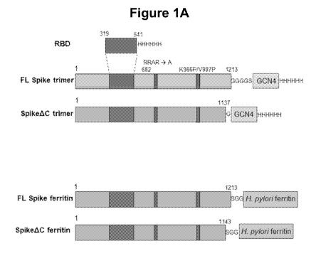
[0009] FIGURE 1A is a schematic illustration of SARS-CoV-2 Spike protein
antigen
polypeptide constructs according to certain aspects of this disclosure.
[0010] FIGURE 1B is a schematic illustration of three-dimensional structures
of SARS-
CoV-2 Spike protein antigen polypeptides according to certain aspects of this
disclosure,
which are based on the structures of Spike trimers determined by cryogenic
electron
microscopy (cryo-EM) and the structure of Heliocbacter pylon ferritin
nanoparticles
determined by X-ray crystallography.
[0011] FIGURE 2A shows a photographic image of the Western blot illustrating
the results
of expression and characterization of SARS-CoV-2 Spike protein antigens
according to
certain aspects of this disclosure.
4
CA 03193288 2023-02-27
WO 2022/047116
PCT/US2021/047885
[0012] FIGURE 2B shows photographic images of the SDS-PAGE gels illustrating
the
results of expression and characterization of SARS-CoV-2 Spike protein
antigens according
to certain aspects of this disclosure.
[0013] FIGURE 3 shows line plots illustrating the results of analytical scale
size-exclusion
chromatography coupled with multi-angle light scattering (SEC-MALS) of SARS-
CoV-2
Spike protein antigens according to certain aspects of this disclosure.
[0014] FIGURE 4 shows line plots illustrating the results of binding analysis
of SARS-
CoV-2 Spike protein antigens to human ACE2, purified SARS-CoV-2 reactive
monoclonal
antibodies CR3022, CB6, and COVA-2-15, and an Ebola virus reactive monoclonal
antibody
ADI-15731 (as a negative control) by enzyme-linked immunosorbent assay (ELISA)
according to certain aspects of this disclosure.
[0015] FIGURE 5A shows a representative motion-corrected cryo-EM micrograph
and
reference-free 2D class averages of SARS-CoV-2 SpikeAC-ferritin fusion protein
nanoparticles according to certain aspects of this disclosure.
[0016] FIGURE 5B, top panel, shows reconstructed cryo-EM map of SARS-CoV-2
SpikeAC-ferritin fusion protein nanoparticles in two different views according
to certain
aspects of this disclosure. The bottom panel shows two different views of the
atomic model
of SARS-CoV-2 SpikeAC-ferritin fusion protein docked into the cryo-EM map
displayed at
lower contour level than the top panel according to certain aspects of this
disclosure.
[0017] FIGURE 6 shows dot plots illustrating the results of ELISA binding
analysis of the
sera extracted at Day 21 after the initial immunization from the mice
immunized with SARS-
CoV-2 Spike protein antigens according to certain aspects of this disclosure.
The antigens are
indicated on the X-axes. The binding of the sera to SARS-CoV-2 RBD protein
(left graph)
and SARS-CoV-2 Spike protein (right graph) was analyzed. Each point shown on
the graphs
represents an average logio EC50 value from two technical replicate ELISA
curves from a
single animal. Each bar in the graphs represents the mean logio EC50 value of
10 animals,
and the error bars represent the standard deviations. Statistical comparisons
were performed
using Kruskal-Wallis ANOVA followed by Dunn's multiple comparisons. All p
values are
represented as following: * = p < 0.05, ** = p < 0.01, *** = p < 0.001, **** =
p < 0.0001.
[0018] FIGURE 7 shows dot plots illustrating the neutralization properties of
the sera
extracted at Day 21 after the initial immunization from the mice immunized
with SARS-
CoV-2 Spike protein antigens assessed using a luciferase-based SARS-CoV-2
Spike
5
CA 03193288 2023-02-27
WO 2022/047116
PCT/US2021/047885
pseudotyped lentiviral assay according to certain aspects of this disclosure.
The antigens are
indicated on the X-axes. The Y-axis is set at the assay limit of quantitation
(1:100 serum
dilution), and serum samples with neutralization activity less than that were
set at the LOQ.
Each point represents the logio IC50 value from a single animal derived from
four replicates.
To generate the four replicates, each experiment was performed twice on
different days, with
duplicate experiments performed on each of the days. This generated four
normalized
dilution curves that were then compiled to get each IC50 value for each
animal. Each point
on the graph bars represents the mean value for each group of 10 animals, and
the error bars
represent the standard deviations. Statistical comparisons were performed
using Kruskal-
Wallis ANOVA followed by Dunn's multiple comparisons. All p values are
represented as
following: * = p < 0.05, ** = p < 0.01, *** = p < 0.001, **** = p < 0.0001
[0019] FIGURE 8 shows dot plots illustrating the results of ELISA binding
analysis of the
sera extracted at Day 28 after the initial immunization from the mice
immunized with SARS-
CoV-2 Spike protein antigens according to certain aspects of this disclosure.
The antigens are
indicated on the X-axis. The binding of the sera to SARS-CoV-2 RBD protein
(left graph)
and SARS-CoV-2 Spike protein (right graph) was analyzed. Each point on the
graphs
represents an average logio EC50 value from two technical replicate ELISA
curves from a
single animal. The bars represents the mean of 10 animals, and the error bars
represent the
standard deviations. Statistical comparisons were performed using Kruskal-
Wallis ANOVA
followed by Dunn's multiple comparisons. All p values are represented as
following: * = p <
0.05, ** = p < 0.01, *** = p < 0.001, **** = p < 0.0001
[0020] FIGURE 9 shows dot plots illustrating the neutralization properties of
the sera
extracted at Day 28 after the initial immunization from the mice immunized
with SARS-
CoV-2 Spike protein antigens according to certain aspects of this disclosure.
The antigens are
indicated on the X-axis. The neutralization properties were assessed using a
luciferase-based
SARS-CoV-2 Spike pseudotyped lentiviral assay. The Y-axis is set at the assay
limit of
quantitation (1:100 serum dilution), and serum samples with neutralization
activity less than
that were set at the LOQ. Each point shown in the graph represents logio IC50
value from a
single animal derived from four replicates. To generate the four replicates,
each experiment
was performed twice on different days), with duplicate experiments performed
on each of the
days. This generated four normalized dilution curves that were then compiled
to get each
IC50 value for each animal. The bars represent the mean logio IC50 for each
group of 10
animals, and the error bars represent the standard deviations. Statistical
comparisons were
6
CA 03193288 2023-02-27
WO 2022/047116
PCT/US2021/047885
performed using Kruskal-Wallis ANOVA followed by Dunn's multiple comparisons.
All p
values are represented as following: * = p < 0.05, ** = p < 0.01, *** = p <
0.001, **** = p <
0.0001
[0021] FIGURE 10 shows dot plots illustrating the results of ELISA binding
analysis of
IgGl, IgG2a, and IgG2b subclass responses (as indicated on the X-axis) of the
sera extracted
from experimental mice immunized with two doses SARS-CoV-2 Spike protein
antigens
according to certain aspects of this disclosure. The antigens are indicated at
the top of each
panel. Each point on the graphs represents logio EC50 value from a single
animal; each
horizontal bar represents the mean logio EC50 titer for the group of 10
animals; the error bars
represent the standard deviations.
[0022] FIGURE 11A shows dot plots illustrating the ratio of IgG2a/IgG1 EC50s
determined by ELISA binding analysis of the sera extracted from experimental
mice
immunized with two doses SARS-CoV-2 Spike protein antigens according to
certain aspects
of this disclosure. The antigens are indicated on the X-axis. Each point on
the graphs
represents the ratio value from a single animal; each horizontal bar
represents the mean ratio
for the group of 10 animals; the error bars represent the standard deviations.
[0023] FIGURE 11B shows dot plots illustrating the ratio of IgG2b/IgG1 EC50s
determined by ELISA binding analysis of the sera extracted from experimental
mice
immunized with two doses SARS-CoV-2 Spike protein antigens according to
certain aspects
of this disclosure. The antigens are indicated on the X-axis. Each point on
the graphs
represents the ratio value from a single animal; each horizontal bar
represents the mean ratio
for the group of 10 animals; the error bars represent the standard deviations.
[0024] FIGURE 12 shows line plots illustrating the results of binding analysis
by ELISA
evaluating the levels of IgM in the sera extracted from experimental mice
immunized with
two doses SARS-CoV-2 Spike protein antigens according to certain aspects of
this
disclosure. The antigens are indicated at the top of each panel. Each point
represents an
experimental duplicate from each animal (n = 10 mice per group) fit with a
dose-response
association curve; error bars represent standard deviation for each point.
[0025] FIGURE 13A shows dot plots illustrating the neutralization properties
of the sera
extracted from the experimental mice at day 28 after administration of
different doses
(indicated on the X-axis) of a SARS-CoV-2 Spike protein antigen according to
certain
aspects of this disclosure. The neutralization properties were assessed using
a luciferase-
7
CA 03193288 2023-02-27
WO 2022/047116
PCT/US2021/047885
based SARS-CoV-2 Spike pseudotyped lentiviral assay according to certain
aspects of this
disclosure. The Y-axis is set at the assay limit of quantitation (1:100 serum
dilution), and
serum samples with neutralization activity less than that were set at the LOQ.
Each point
represents the logio IC50 value from a single animal. Each horizontal bar
represents the mean
value for each group of 10 animals, and the error bars represent the standard
deviations.
[0026] FIGURE 13B shows dot plots illustrating the neutralization properties
of the sera
extracted from the experimental mice at various time points (indicated on the
X-axis) after
administration a SARS-CoV-2 Spike protein antigen according to certain aspects
of this
disclosure. The neutralization properties were assessed using a luciferase-
based SARS-CoV-2
Spike pseudotyped lentiviral assay according to certain aspects of this
disclosure. The Y-axis
is set at the assay limit of quantitation (1:100 serum dilution), and serum
samples with
neutralization activity less than that were set at the LOQ. Each point
represents the logio IC50
value from a single animal. Each horizontal bar represents the mean value for
each group of
five animals, and the error bars represent the standard deviations.
[0027] FIGURE 14 shows dot plots illustrating the neutralization properties of
the sera
extracted from the experimental mice at various time points after the initial
immunization
(indicated on the X-axis) with SARS-CoV-2 Spike protein antigens (indicated at
the top of
each panel) according to certain aspects of this disclosure. The
neutralization properties were
assessed using a luciferase-based SARS-CoV-2 Spike pseudotyped lentiviral
assay according
to certain aspects of this disclosure. The Y-axis is set at the assay limit of
quantitation (1:100
serum dilution), and serum samples with neutralization activity less than that
were set at the
LOQ. Each point represents the logio IC50 value from a single animal. Each
horizontal bar
represents the mean value for each group of five animals, and the error bars
represent the
standard deviations.
[0028] FIGURE 15A shows dot plots illustrating the neutralization properties
of the sera
extracted from the experimental mice immunized with a single dose of 1 or
10 (as
indicated on the X-axis) of SARS-CoV-2 Spike protein antigen according to
certain aspects
of this disclosure. The sera was collected at week 3 after the initial
immunization. The SARS-
CoV-2 Spike protein antigen was adjuvanted with either 500 tg Alhydrogel and
20 tg CpG,
or 10 tg Quil-A and 10 tg MPLA, as indicated at the top of each panel. The
neutralization
properties were assessed using a luciferase-based SARS-CoV-2 Spike pseudotyped
lentiviral
assay according to certain aspects of this disclosure. The Y-axis is set at
the assay limit of
quantitation (1:100 serum dilution), and serum samples with neutralization
activity less than
8
CA 03193288 2023-02-27
WO 2022/047116
PCT/US2021/047885
that were set at the LOQ. Each point represents the logio IC50 value from a
single animal.
Each horizontal bar represents the mean value for each group of 10 or 20
animals (as shown),
and the error bars represent the standard deviations.
[0029] FIGURE 15B shows dot plots illustrating the neutralization properties
of the sera
extracted from the experimental mice immunized with one ("day 21") or two
("day 28")
doses of 1 tg or 10 tg (as indicated on the X-axis) of SARS-CoV-2 Spike
protein antigen
according to certain aspects of this disclosure. The SARS-CoV-2 Spike protein
antigen was
adjuvanted with either 500 tg Alhydrogel and 20 tg CpG, AddaVaxTM, or 10
and 10
MPLA (as indicated on the X-axis). The neutralization properties were assessed
using a luciferase-based SARS-CoV-2 Spike pseudotyped lentiviral assay
according to
certain aspects of this disclosure. The Y-axis is set at the assay limit of
quantitation (1:100
serum dilution), and serum samples with neutralization activity less than that
were set at the
LOQ. Each point represents the logio IC50 value from a single animal. Each
horizontal bar
represents the mean value for each group of 10 animals, and the error bars
represent the
standard deviations.
[0030] FIGURE 16 shows dot plots illustrating the neutralization properties of
the sera
extracted from the experimental mice immunized with two doses of two SARS-CoV-
2 Spike
protein antigens (as indicated) according to certain aspects of this
disclosure. The SARS-
CoV-2 Spike protein antigens administered to the experimental mice were
adjuvanted with 10
tg Quil-A and 10 tg MPLA. The sera were collected at days 21, 28, and 56 (as
indicated on
the X-axis) after the initial immunization. The neutralization properties were
assessed using a
luciferase-based SARS-CoV-2 Spike pseudotyped lentiviral assay according to
certain
aspects of this disclosure. The Y-axis is set at the assay limit of
quantitation (1:100 serum
dilution), and serum samples with neutralization activity less than that were
set at the LOQ.
Each point represents the logio IC50 value from a single animal. Each
horizontal bar
represents the mean value for each group of five animals, and the error bars
represent the
standard deviations.
[0031] FIGURE 17A shows a representative size-exclusion chromatography trace
of a
SARS-CoV-2 Spike protein antigen according to certain aspects of this
disclosure.
[0032] FIGURE 17B shows a bar graph illustrating a comparison of relative
amounts
SARS-CoV-2 Spike protein antigens expressed and purified according to certain
aspects of
this disclosure.
9
CA 03193288 2023-02-27
WO 2022/047116
PCT/US2021/047885
[0033] FIGURE 18 shows plots generated by bio-layer interferometry (BLI) on
the Octet
system (Sartorius, Gottingen, Germany) testing binding of SARS-CoV-2 Spike
protein
antigens according to certain aspects of this disclosure to conformational
monoclonal
antibodies and to ACE2 receptor. The monoclonal antibodies and ACE2 receptor
fused to Fc
fragment were immobilized on the biosensor surface, and the sensors were moved
into wells
containing SARS CoV-2 Spike protein antigens in solution, then into the wells
that did not
contain the antigens. Association and dissociation of the SARS CoV-2 Spike
protein
antigens to the antibodies and ACE2 results in changes in optical interference
between light
waves that reflect back to the spectrophotometers from an internal surface and
from the
external interface between sensor and solution. The change of the interference
was plotted on
the Y-axis and used to indicate the binding and dissociation. The magnitude of
the change in
the nm shift (plotted on the Y axis) is therefore used a surrogate for
binding, where, for
similar binding partners, a larger change reflects more binding.
[0034] FIGURE 19 shows dot plots illustrating the neutralization properties of
the sera
extracted from the experimental mice immunized with two doses of two SARS-CoV-
2 Spike
protein antigens (as indicated) according to certain aspects of this
disclosure. The SARS-
CoV-2 Spike protein antigens administered to the experimental mice were
adjuvanted with
500 tg Alum (Alhydrogel , InvivoGen, San Diego, California) and 20 tg CpG
(InvivoGen).
The sera were collected at days 21 and 42 (as indicated on the X-axis) after
the initial
immunization. The neutralization properties were assessed using a luciferase-
based SARS-
CoV-2 Spike pseudotyped lentiviral assay according to certain aspects of this
disclosure. The
IC50 values are shown as neutralization titers for different groups at
indicated time points.
Each point represents the logio IC50 value from a single animal. The
significance of
differences between the groups were calculated by student-t test and found not
significant
(NS), as indicated in the plot. Each horizontal bar represents the mean value
for each group of
ten animals, and the error bars represent the standard deviations.
[0035] FIGURE 20 shows UV spectra of lyophilized ("Lyol," "Lyo2," and "Lyo3")
and
frozen ("Frozen") SpikeHexaProAC protein antigen samples according to certain
aspects of
this disclosure.
[0036] FIGURE 21 shows, in the left panel, a line plot illustrating the
results of scanning
fluorimetry analysis of lyophilized ("Lyo") and frozen ("Frozen")
SpikeHexaProAC protein
antigen samples according to certain aspects of this disclosure.
CA 03193288 2023-02-27
WO 2022/047116
PCT/US2021/047885
[0037] FIGURE 22 shows the plots generated by BLI on Octet system to test
binding of
SARS-CoV-2 Spike protein antigen from lyophilized ("Lyol," "Lyo2," and "Lyo3")
and
frozen ("Frozen") samples according to certain aspects of this disclosure to
conformational
monoclonal antibodies and to ACE2 receptor. The monoclonal antibodies and ACE2
receptor
fused to Fc fragment were immobilized on the biosensor surface, and the
sensors were moved
into wells containing either frozen and thawed ("Frozen" ) of lyophilized and
reconstituted
("Lyo 1" ¨ "Lyo 3") SARS CoV-2 Spike protein antigens in solution, and then
into the wells
that did not contain the antigens. Association and dissociation of the SARS
CoV-2 Spike
protein antigens to the antibodies and ACE2 results in changes in optical
interference
between light waves that reflect back to the spectrophotometers from an
internal surface and
from the external interface between sensor and solution. The change of the
interference was
plotted on the Y-axis and used to indicate the binding and dissociation.
[0038] FIGURE 23 shows dot plots illustrating the binding of SARS-CoV-2 RBD
protein
(measured by ELISA as described elsewhere in the present disclosure and
indicated as EC50
values on the Y-axis) of the sera extracted from the groups of experimental
mice immunized
with SARS-CoV-2 Spike protein antigen from lyophilized and frozen samples (as
indicated
on the X-axis, three groups of mice each) according to certain aspects of this
disclosure
according to certain aspects of this disclosure. Each point represents the
logio EC50 value
from a single animal. The statistical differences in titers were analyzed by
student t-test and
found not significant (NS), as indicated in the plot.
[0039] FIGURE 24 shows dot plots illustrating the neutralization properties of
the sera
extracted from the experimental mice immunized with SARS-CoV-2 Spike protein
antigen
from lyophilized and frozen samples (as indicated on the X-axis, three groups
of mice each)
according to certain aspects of this disclosure according to certain aspects
of this disclosure.
The neutralization properties were assessed using a luciferase-based SARS-CoV-
2 Spike
pseudotyped lentiviral assay according to certain aspects of this disclosure.
The IC50 values
are shown as neutralization titers for different groups at indicated time
points. Each point
represents the logio IC50 value from a single animal.
[0040] FIGURE 25 shows the plots generated by BLI on the Octet system testing
binding
of lyophilized SARS-CoV-2 Spike protein antigen samples to conformational
monoclonal
antibody CB6 and to ACE2 receptor. The samples of SARS-CoV-2 Spike protein
antigen
were lyophilized in 10 mM ammonium bicarbonate pH 7.8 with 1%, 5%, or 10 %
sucrose (as
labeled), and SARS-CoV-2 Spike protein antigen samples frozen in either 10 mM
11
CA 03193288 2023-02-27
WO 2022/047116
PCT/US2021/047885
ammonium bicarbonate pH 7.8 with 10% sucrose ("AB frozen") or in PBS with 10 %
sucrose
("PBS"). The monoclonal antibodies and ACE2 receptor fused to Fc fragment were
immobilized on the biosensor surface, and the sensors were moved into wells
containing
protein antigens in solution, then into the wells that did not contain the
antigens. Association
and dissociation of the SARS CoV-2 Spike protein antigens to the antibodies
and ACE2
results in changes in optical interference between light waves that reflect
back to the
spectrophotometers from an internal surface and from the external interface
between sensor
and solution. The change of the interference was plotted on the Y-axis and
used to indicate
the binding and dissociation. The magnitude of the change in the nm shift
(plotted on the Y
axis) is therefore used a surrogate for binding, where, for similar binding
partners, a larger
change reflects more binding.
[0041] FIGURE 26 shows plots illustrating the results of size exclusion
chromatography ¨
multiple angle light scattering (SEC-MALS) testing the properties of SARS-CoV-
2 Spike
protein antigen lyophilized in volatile ammonium bicarbonate buffer. The
protein was tested
directly after reconstitution ("DAY1") and after being stored at room
temperature for 4 days
("DAY 4").
[0042] FIGURE 27 is a schematic illustration of the position of the engineered
glycosylation site in a SARS-CoV-2 Spike fusion protein nanoparticle according
to certain
aspects of the present dislcosure. Ferritin domains are shown in white. The
lysine residue
mutated to an asparagine residue in the engineered glycosylation site are
shown as black
spheres. The glutamic acid residue mutated to a threonine residue in the
engineered
glycosylation site is shown as grey spheres. The black triangle depicts the 3-
fold axis of
symmetry.
[0043] FIGURE 28 shows plots generated by BLI on the Octet system to test
binding of
SARS-CoV-2 Spike protein antigens according to certain aspects of this
disclosure to
conformational monoclonal antibodies and to ACE2 receptor. The monoclonal
antibodies and
ACE2 receptor fused to Fc fragment were immobilized on the biosensor surface
and sensors
were moved into wells containing SARS-CoV-2 Spike protein antigens in
solution, and then
into the wells that did not contain the antigens. Association and dissociation
of the SARS-
CoV-2 Spike protein antigens to the antibodies and ACE2 results in changes in
optical
interference between light waves that reflect back to the spectrophotometers
from an internal
surface and from the external interface between sensor and solution. The
change of the
interference was plotted on the Y-axis and used to indicate the binding and
dissociation. The
12
CA 03193288 2023-02-27
WO 2022/047116
PCT/US2021/047885
magnitude of the change in the nm shift (plotted on the Y axis) is therefore
used a surrogate
for binding, where, for similar binding partners, a larger change reflects
more binding. The
plot labels are as follows: "Original" - SpikeHexaProAC ferritin; "D614G" -
SpikeHexaProAC ferritin D614G; "B.1.1.7" - SpikeHexaProAC ferritin B.1.1.7;
"B.1.351" -
SpikeHexaProAC ferritin B.1.351; "LA" - SpikeHexaProAC ferritin B.1.429; "P1" -
SpikeHexaProAC ferritin P1.
[0044] FIGURE 29 shows "heat maps" of neutralizing activity (determined using
SARS-
CoV-2 Spike pseudotyped lentivirus neutralization assay) of SARS-CoV-2 Spike
protein
antigens against the panel of six pseudoviruses according to certain aspects
of this disclosure.
SARS-CoV-2 Spike protein antigens are listed on the x-axis of each "heat map,"
labeled as
follows: "Original" - SpikeHexaProAC ferritin; "D614G" - SpikeHexaProAC
ferritin D614G;
"B.1.1.7" - SpikeHexaProAC ferritin B.1.1.7; "B.1.351" - SpikeHexaProAC
ferritin B.1.351;
"LA" - SpikeHexaProAC ferritin B.1.429; "P1" - SpikeHexaProAC ferritin P1. The
pseudoviruses tested are plotted on the y-axis of each heat map and are based
on SARS-CoV-
2 strains Wuhan-1 (denoted as "WT"), D614G, B.1.429, B1.1.7, P1, and B.1.351.
Each value
of the heat map is a logioIC50 value of the pooled serum from the mice
immunized with the
same SARS-CoV-2 Spike protein antigen against a specific pseudotyped virus.
[0045] FIGURE 30A is a bar graph illustrating the testing of the
neutralization responses in
a group of 5 mice immunized with SpikeHexaProAC ferritin (SEQ ID NO:16) and
alum
according to certain aspects of this disclosure. The IC50 values are shown as
neutralization
titers for different groups at indicated time points. Each dot represents a
serum sample from
an individual mouse. The average IC50 values are indicated below the bars for
indicated time
points.
[0046] FIGURE 30B is a bar graph illustrating the testing of the
neutralization responses in
a group of 5 mice immunized with SpikeHexaProAC ferritin (SEQ ID NO:16) and
alum, and
boosted 21 days after the initial immunization according to certain aspects of
this disclosure.
The IC50 values are shown as neutralization titers for different groups at
indicated time
points. Each dot represents a serum sample from an individual mouse. The
average IC50
values are indicated below the bars for indicated time points.
[0047] FIGURE 31A is a bar graph illustrating the testing of the
neutralization responses
against wild type SARS-CoV-2 and SARS-CoV-2 variants (as indicated above the
bars) in
groups of 10 mice immunized with SpikeHexaProAC ferritin (SEQ ID NO:16)
adjuvanted
13
CA 03193288 2023-02-27
WO 2022/047116
PCT/US2021/047885
with alum according to certain aspects of this disclosure. The IC50 values are
shown as
neutralization titers for different groups. Each dot represents a serum sample
from an
individual mouse. The average IC50 values are indicated below the bars for
indicated time
points.
[0048] FIGURE 31B is a bar graph illustrating the testing of the
neutralization responses
against wild type SARS-CoV-2 and SARS-CoV-2 variants (as indicated above the
bars) in
groups of 10 mice immunized with SpikeHexaProAC ferritin (SEQ ID NO:16)
adjuvanted
with alum, and boosted 21 days after the initial immunization according to
certain aspects of
this disclosure. The IC50 values are shown as neutralization titers for
different groups. Each
dot represents a serum sample from an individual mouse. The average IC50
values are
indicated below the bars for indicated time points.
[0049] FIGURE 32A is a bar graph illustrating the testing of the
neutralization responses
against wild type SARS-CoV-2 and SARS-CoV-2 variants (as indicated above the
bars) in
groups of 10 mice immunized with SpikeHexaProAC ferritin (SEQ ID NO:16)
adjuvanted
with alum and CpG according to certain aspects of this disclosure. The IC50
values are
shown as neutralization titers for different groups. Each dot represents a
serum sample from
an individual mouse. The average IC50 values are indicated below the bars.
[0050] FIGURE 32B is a bar graph illustrating the testing of the
neutralization responses
against wild type SARS-CoV-2 and SARS-CoV-2 variants (as indicated above the
bars) in
groups of 10 mice immunized with SpikeHexaProAC ferritin (SEQ ID NO:16)
adjuvanted
with alum and CpG and boosted 21 days after the initial immunization according
to certain
aspects of this disclosure. The IC50 values are shown as neutralization titers
for different
groups. Each dot represents a serum sample from an individual mouse. The
average IC50
values are indicated below the bars.
[0051] FIGURE 33 is a bar graph illustrating the testing of the neutralization
responses
against wild type SARS-CoV-2 and SARS-CoV-2 variants (as indicated above the
bars) in
groups of 5 mice immunized with SpikeHexaProAC ferritin (SEQ ID NO:16)
adjuvanted
with different doses of alum, which are indicated on the x-axis, according to
certain aspects
of this disclosure. For each alum dose one group received single immunization,
and a second
group was boosted 21 days after the primary immunization, as indicated above
the plot. The
IC50 values are shown as neutralization titers for different groups. Each dot
represents a
14
CA 03193288 2023-02-27
WO 2022/047116
PCT/US2021/047885
serum sample from an individual mouse. The average IC50 values are shown as
neutralization titers for different groups at indicated time points.
[0052] FIGURE 34A is a bar graph illustrating the testing of the
neutralization responses
against wild type SARS-CoV-2 in groups of 5 mice immunized with SpikeHexaProAC
ferritin (SEQ ID NO:16) adjuvanted with different amounts of alum (indicated
on the x-axis)
either alone or in combination with 20 i.tg of CpG (as indicated below the x-
axis) according
to certain aspects of this disclosure. For each alum dose one group received
single
immunization, and a second group was boosted 21 days after the primary
immunization (as
indicated below the x-axis). The IC50 values are shown as neutralization
titers for different
groups. Each dot represents a serum sample from an individual mouse. The
average IC50
values are shown as neutralization titers for different groups of pooled
samples for each of
the indicated groups and time points.
[0053] FIGURE 34B is a bar graph illustrating the testing of the
neutralization responses
against B.1.421 variant of SARS-CoV-2 in groups of 5 mice immunized with
SpikeHexaProAC ferritin (SEQ ID NO:16) adjuvanted with different amounts of
alum
(indicated on the x-axis) either alone or in combination with 20 i.tg of CpG
(as indicated
below the x-axis) according to certain aspects of this disclosure. For each
alum dose one
group received single immunization, and a second group was boosted 21 days
after the
primary immunization (as indicated below the x-axis). The IC50 values are
shown as
neutralization titers for different groups. Each dot represents a serum sample
from an
individual mouse. The average IC50 values are shown as neutralization titers
for different
groups of pooled samples for each of the indicated groups and time points.
[0054] FIGURE 34C is a bar graph illustrating the testing of the
neutralization responses
against B.1.1.7. variant of SARS-CoV-2 in groups of 5 mice immunized with
SpikeHexaProAC ferritin (SEQ ID NO:16) adjuvanted with different amounts of
alum
(indicated on the x-axis) either alone or in combination with 20 i.tg of CpG
(as indicated
below the x-axis) according to certain aspects of this disclosure. For each
alum dose one
group received single immunization, and a second group was boosted 21 days
after the
primary immunization (as indicated below the x-axis). The IC50 values are
shown as
neutralization titers for different groups. Each dot represents a serum sample
from an
individual mouse. The average IC50 values are shown as neutralization titers
for different
groups of pooled samples for each of the indicated groups and time points.
CA 03193288 2023-02-27
WO 2022/047116
PCT/US2021/047885
[0055] FIGURE 34D is a bar graph illustrating the testing of the
neutralization responses
against B.1.351 variant of SARS-CoV-2 in groups of 5 mice immunized with
SpikeHexaProAC ferritin (SEQ ID NO:16) adjuvanted with different amounts of
alum
(indicated on the x-axis) either alone or in combination with 20 i.tg of CpG
(as indicated
below the x-axis) according to certain aspects of this disclosure. For each
alum dose one
group received single immunization, and a second group was boosted 21 days
after the
primary immunization (as indicated below the x-axis). The IC50 values are
shown as
neutralization titers for different groups. Each dot represents a serum sample
from an
individual mouse. The average IC50 values are shown as neutralization titers
for different
groups of pooled samples for each of the indicated groups and time points.
[0056] FIGURE 34E is a bar graph illustrating the testing of the
neutralization responses
against B.1.617.2 variant of SARS-CoV-2 in groups of 5 mice immunized with
SpikeHexaProAC ferritin (SEQ ID NO:16) adjuvanted with different amounts of
alum
(indicated on the x-axis) either alone or in combination with 20 i.tg of CpG
(as indicated
below the x-axis) according to certain aspects of this disclosure. For each
alum dose one
group received single immunization, and a second group was boosted 21 days
after the
primary immunization (as indicated below the x-axis). The IC50 values are
shown as
neutralization titers for different groups. Each dot represents a serum sample
from an
individual mouse. The average IC50 values are shown as neutralization titers
for different
groups of pooled samples for each of the indicated groups and time points.
[0057] FIGURE 34F is a bar graph illustrating the testing of the
neutralization responses
against P.1 variant of SARS-CoV-2 in groups of 5 mice immunized with
SpikeHexaProAC
ferritin (SEQ ID NO:16) adjuvanted with different amounts of alum (indicated
on the x-axis)
either alone or in combination with 20 i.tg of CpG (as indicated below the x-
axis) according
to certain aspects of this disclosure. For each alum dose one group received
single
immunization, and a second group was boosted 21 days after the primary
immunization (as
indicated below the x-axis). The IC50 values are shown as neutralization
titers for different
groups. Each dot represents a serum sample from an individual mouse. The
average IC50
values are shown as neutralization titers for different groups of pooled
samples for each of
the indicated groups and time points.
16
CA 03193288 2023-02-27
WO 2022/047116 PCT/US2021/047885
DETAILED DESCRIPTION
[0058] The inventors designed, generated, and characterized fusion proteins of
SARS-
CoV-2 Spike ectodomain polypeptide and ferritin ("SARS-CoV-2 Spike-ferritin
fusion
proteins") that self-assemble into nanoparticles displaying on their surfaces
the respective
versions of SARS-CoV-2 Spike ectodomain. Some versions of SARS-CoV-2 Spike-
ferritin
fusion protein contain the full-length ectodomain of SARS-CoV-2 Spike protein.
Other
versions contain a SARS-CoV-2 Spike protein ectodomain having C-terminal
deletions (in
one example, a C-terminal deletion of 70 amino acids). The inventors
discovered that,
surprisingly, a C-terminal deletion in the SARS-CoV-2 Spike protein amino acid
sequence
considerably improved the expression of the resulting fusion protein in
mammalian cells. The
inventors confirmed proper folding of Spike domains in each version of SARS-
CoV-2 Spike-
ferritin fusion proteins into a native-like conformation on the surface of the
nanoparticles by
cryo-EM, size-exclusion chromatography multi-angle light scattering (SEC-
MALS), and bio-
layer interferometry (BLI), which measured binding SARS-CoV-2 Spike-ferritin
fusion
proteins to ACE2 receptor and/or one or more Spike-specific monoclonal
antibodies. The
inventors tested the immunogenicity of SARS-CoV-2 Spike-ferritin fusion
proteins in
experimental animals, including comparatively with other SARS-CoV-2 fusion
protein
antigens. Following a single immunization of mice with SARS-CoV-2 Spike-
ferritin fusion
proteins, the inventors observed neutralizing antibody amounts comparable to
or greater than
those seen in human convalescent plasma, as determined using a lentiviral CoV-
2
pseudovirus assay. In contrast, a single immunization with either the CoV-2
receptor binding
domain (RBD) or isolated Spike trimers of SARS-CoV-2 Spike elicited much
weaker
neutralizing antibody responses. The inventors also tested SARS-CoV-2 virus
neutralizing
properties of the antibodies generated in the experimental animals to SARS-CoV-
2 Spike-
ferritin fusion proteins that were used as immunogens. The inventors
discovered that,
unexpectedly, SARS-CoV-2 Spike-ferritin fusion proteins capable of self-
assembly into
nanoparticles elicited significantly stronger antigen-specific and
neutralizing antibody
responses in the exprimental animals as compared to other SARS-CoV-2 Spike
protein
antigens. The inventors further discovered that the SARS-CoV-2 Spike-ferritin
fusion
proteins having C-terminal deletion in SARS-CoV-2 Spike protein ectodomain
amino acid
sequence ("C-terminal deletion") elicited the highest neutralizing antibody
response in the
experimental animals among all the antigens tested. The inventors realized
that, given the the
ability of SARS-CoV-2 Spike-ferritin fusion proteins to self-assemble into
nanoparticles after
17
CA 03193288 2023-02-27
WO 2022/047116
PCT/US2021/047885
production in mammalian cells, the achieved expression levels comparable to
those of
ectodomain of SARS-CoV-2 Spike protein, and the enhanced immune response
elicited by
SARS-CoV-2 Spike-ferritin fusion proteins, SARS-CoV-2 Spike-ferritin fusion
proteins
(including Spike-ferritin fusion proteins having the C-terminal deletion in
SARS-CoV-2
Spike protein ectodomain amino acid sequence) can be used in subunit or
nucleic acid
vaccines against SARS-CoV-2.
[0059] The inventors tested several SARS-CoV-2 Spike-ferritin fusion proteins
with the C-
terminal deletion and two or more proline substitutions and discovered that
SARS-CoV-2
Spike-ferritin fusion proteins with the C-terminal deletion and six proline
substitutions was
equally immunogenic to SARS-CoV-2 Spike-ferritin fusion proteins with the C-
terminal
deletion and two proline substitutions. Furthermore, expression and
purification yields of
SARS-CoV-2 Spike-ferritin fusion proteins with the C-terminal deletion and six
proline
substitutions were unexpetedly and remarkbly higher than those for SARS-CoV-2
Spike-
ferritin fusion proteins with the C-terminal deletion and fewer proline
substitutions. The
inventors created and tested several versions of SARS-CoV-2 Spike-ferritin
fusion proteins
with the C-terminal deletion and six proline substitutions. These versions
were based on of
naturally occurring variants of coronavirus Spike protein and, when
administered to
experimental animals, elicited antibodies with high neutralizing activity. The
inventors found
that lyophilized and subsequently reconsituted SARS-CoV-2 Spike-ferritin
fusion proteins
retained their structure and immunogenicity. Furthermore, the inventors
engineered SARS-
CoV-2 Spike ferritin fusion protein antigens with artificial glycosylation
sites in the ferritin
domain, in order to shield the ferritin domain from the immune system and
decrease immune
response against the ferritin domain (thus minimizing non-productive immune
responses
against the anti-SARS-CoV-2 vaccines concevied by the inventors).
[0060] Based on the above discoveries, the inventors conceived, and the
present disclosure
describes, various embodiments of coronavirus Spike-ferritin fusion proteins,
nanoparticles
composed of such fusion proteins, nucleic acids, nucleic acid constructs and
vectors encoding
coronavirus Spike-ferritin fusion proteins, as well as cells, compositions,
kits, and methods
related to production and use of coronavirus Spike-ferritin fusion proteins.
The production of
nanoparticles of coronavirus Spike-ferritin fusion proteins requires only a
single expression
plasmid. Expression and purification of coronavirus Spike-ferritin fusion
proteins can be
carried out and scaled using standard protocols for soluble proteins, with the
purified fusion
proteins self-assembling into homogenous populations of nanoparticles. In
contrast,
18
CA 03193288 2023-02-27
WO 2022/047116
PCT/US2021/047885
nanoparticles assembled from separate components require for the components to
be
generated separately and conjugated in a post purification conjugation step,
which can
drastically decrease the yield and create heterogeneous nanoparticle
populations. Coronavirus
Spike-ferritin fusion proteins and the related nucleic acids, nucleic acid
constructs, vectors,
cells, compositions, kits and methods conceived by the inventors and described
in the present
disclosure are useful for a variety of application, including, but not limited
to, development
and production of immunogenic compositions (vaccines), based on proteins or
nucleic acids
and useful for inducing an immune response against coronavirus infections, as
well as for
prevention or treatment of coronavirus infections, including, but not limited
to, SARS-CoV-2
infection. The experimental results obtained by the inventors demonstrated
that that
nanoparticles of Spike-ferritin fusion proteins displaying coronavirus Spike
protein
ectodomain can reliably elicit clinically relevant amounts of neutralizing
antibodies in
subjects. Accordingly, coronavirus Spike-ferritin fusion proteins and nucleic
constructs
encoding such fusion proteins can be used as vaccines, such as single-dose
vaccines, for
inducing protection against coronavirus infection.
Terms and concepts
[0061] A number of terms and concepts are discussed below. They are intended
to
facilitate the understanding of various embodiments of the invention in
conjunction with the
rest of the present document and the accompanying figures. These terms and
concepts may be
further clarified and understood based on the accepted conventions in the
fields of the present
invention, as well as the description provided throughout the present document
and/or the
accompanying figures. Some other terms can be explicitly or implicitly defined
in other
sections of this document and in the accompanying figures, and may be used and
understood
based on the accepted conventions in the fields of the present invention, the
description
provided throughout the present document and/or the accompanying figures. The
terms not
explicitly defined can also be defined and understood based on the accepted
conventions in
the fields of the present invention and interpreted in the context of the
present document
and/or the accompanying figures.
[0062] Unless otherwise dictated by context, singular terms shall include
pluralities, and
plural terms shall include the singular. Generally, nomenclatures used in
connection with, and
techniques of, cell and tissue culture, molecular biology, immunology,
microbiology,
genetics and protein and nucleic acid chemistry are those well-known and
commonly used.
Known methods and techniques are generally performed according to conventional
methods
19
CA 03193288 2023-02-27
WO 2022/047116
PCT/US2021/047885
well-known and as described in various general and more specific references,
unless
otherwise indicated. The nomenclatures used in connection with the laboratory
procedures
and techniques described in the present disclosure are those well-known and
commonly used.
[0063] As used herein, the terms "a", "an", and "the" can refer to one or more
unless
specifically noted otherwise.
[0064] The use of the term "or" is used to mean "and/or," unless explicitly
indicated to
refer to alternatives only, or the alternatives are mutually exclusive,
although the disclosure
supports a definition that refers to only alternatives and "and/or." As used
herein "another"
can mean at least a second or more.
[0065] The terms "about" and "approximately" as used herein shall generally
mean an
acceptable degree of error for the quantity measured given the nature or
precision of the
measurements. Exemplary degrees of error are within 20% (%); preferably,
within 10%; and
more preferably, within 5% of a given value or range of values. Any reference
to "about X"
or "approximately X" specifically indicates at least the values X, 0.95X,
0.96X, 0.97X,
0.98X, 0.99X, 1.01X, 1.02X, 1.03X, 1.04X, and 1.05X. Thus, expressions "about
X" or
"approximately X" are intended to teach and provide written support for a
claim limitation of,
for example, "0.98X." Alternatively, in biological systems, the terms "about"
and
"approximately" may mean values that are within an order of magnitude,
preferably within 5-
fold, and more preferably within 2-fold of a given value. Numerical quantities
given herein
are approximate unless stated otherwise, meaning that the term "about" or
"approximately"
can be inferred when not expressly stated. When "about" is applied to the
beginning of a
numerical range, it applies to both ends of the range.
[0066] The terms "protein," "peptide," and "polypeptide" are used
interchangeably to refer
to a polymer of amino acid residues. The term apply to naturally occurring
amino acid
polymers and non-natural amino acid polymers, as well as to amino acid
polymers in which
one (or more) amino acid residue is an artificial chemical mimetic of a
corresponding
naturally occurring amino acid. The terms encompass amino acid chains of any
length,
including full-length proteins, wherein the amino acid residues are linked by
covalent peptide
bonds.
[0067] An "isolated" or "purified" polypeptide or protein, or biologically
active portion a
polypeptide or a protein, is substantially or essentially free from components
that normally
accompany or interact with the polypeptide or protein as found in its
naturally occurring
CA 03193288 2023-02-27
WO 2022/047116
PCT/US2021/047885
environment. Thus, an isolated or purified polypeptide or protein is
substantially free of other
cellular material, or culture medium when produced by recombinant techniques,
or
substantially free of chemical precursors or other chemicals when chemically
synthesized. A
protein that is substantially free of cellular material includes preparations
of protein having
less than about 30%, 20%, 10%, 5%, 1%, 0.5%, or 0.1% (total protein) of
contaminating
protein. When the protein of the invention or its biologically active portion
is recombinantly
produced, optimally culture medium represents less than about 30%, 20%, 10%,
5%, 1%,
0.5%, or 0.1% (by concentration) of chemical precursors or non-protein-of-
interest
chemicals.
[0068] The term "amino acid" refers to any monomeric unit that can be
incorporated into a
peptide, polypeptide, or protein. Amino acids include naturally-occurring a-
amino acids and
their stereoisomers, as well as unnatural (non-naturally occurring) amino
acids and their
stereoisomers. "Stereoisomers" of a given amino acid refer to isomers having
the same
molecular formula and intramolecular bonds but different three-dimensional
arrangements of
bonds and atoms (e.g., an L-amino acid and the corresponding D-amino acid).
[0069] Naturally-occurring amino acids are those encoded by the genetic code,
as well as
those amino acids that are later modified, e.g., hydroxyproline, y-
carboxyglutamate, and 0-
phosphoserine. Naturally-occurring a-amino acids include, without limitation,
alanine (Ala),
cysteine (Cys), aspartic acid (Asp), glutamic acid (Glu), phenylalanine (Phe),
glycine (Gly),
histidine (His), isoleucine (Ile), arginine (Arg), lysine (Lys), leucine
(Leu), methionine (Met),
asparagine (Asn), proline (Pro), glutamine (Gin), serine (Ser), threonine
(Thr), valine (Val),
tryptophan (Trp), tyrosine (Tyr), and their combinations. Stereoisomers of a
naturally-
occurring a-amino acids include, without limitation, D-alanine (D-Ala), D-
cysteine (D-Cys),
D-aspartic acid (D-Asp), D-glutamic acid (D-Glu), D-phenylalanine (D-Phe), D-
histidine (D-
His), D-isoleucine (D-Ile), D-arginine (D-Arg), D-lysine (D-Lys), D-leucine (D-
Leu), D-
methionine (D-Met), D-asparagine (D-Asn), D-proline (D-Pro), D-glutamine (D-
Gln), D-
serine (D-Ser), D-threonine (D-Thr), D-valine (D-Val), D-tryptophan (D-Trp), D-
tyrosine (D-
Tyr), and their combinations.
[0070] Unnatural (non-naturally occurring) amino acids include, without
limitation, amino
acid analogs, amino acid mimetics, synthetic amino acids, N-substituted
glycines, and N-
methyl amino acids in either the L- or D-configuration that function in a
manner similar to
the naturally-occurring amino acids. For example, "amino acid analogs" can be
unnatural
amino acids that have the same basic chemical structure as naturally-occurring
amino acids
21
CA 03193288 2023-02-27
WO 2022/047116
PCT/US2021/047885
(i.e., a carbon that is bonded to a hydrogen, a carboxyl group, an amino
group) but have
modified side-chain groups or modified peptide backbones, e.g., homoserine,
norleucine,
methionine sulfoxide, methionine methyl sulfonium. "Amino acid mimetics" refer
to
chemical compounds that have a structure that is different from the general
chemical
structure of an amino acid, but that functions in a manner similar to a
naturally-occurring
amino acid. Amino acids may be referred to by either the commonly known three
letter
symbols or by the one-letter symbols recommended by the IUPAC-IUB Biochemical
Nomenclature Commission.
[0071] The expression "conservatively modified variant" and related expression
may apply
to amino acid sequences, as well to nucleic acid sequences encoding amino acid
sequence.
Substitutions, deletions or additions to a nucleic acid, peptide, polypeptide,
or protein
sequence which alters, adds or deletes a single amino acid or a small
percentage of amino
acids in the encoded sequence is a "conservatively modified variant" where the
alteration
results in the substitution of an amino acid with a chemically similar amino
acid.
Conservative substitution tables providing functionally similar amino acids
are well known in
the art. Such conservatively modified variants are in addition to and do not
exclude
polymorphic variants, interspecies homologs, and alleles of the invention. The
following
eight groups each contain amino acids that are conservative substitutions for
one another:
1) Alanine (A), Glycine (G);
2) Aspartic acid (D), Glutamic acid (E);
3) Asparagine (N), Glutamine (Q);
4) Arginine (R), Lysine (K);
5) Isoleucine (I), Leucine (L), Methionine (M), Valine (V);
6) Phenylalanine (F), Tyrosine (Y), Tryptophan (W);
7) Serine (S), Threonine (T); and
8) Cysteine (C), Methionine (M).
[0072] The terms "nucleic acid," "nucleic acid sequence," "nucleotide
sequence,"
"oligonucleotide," "polynucleotide" and the related terms and expressions
refer to
deoxyribonucleic acids (DNA) or ribonucleic acids (RNA) and their polymers.
Nucleic acid
sequences, as discussed in the present disclosure, encompass all forms of
nucleic acids,
including, but not limited to, single-stranded forms, double-stranded forms,
hairpins, stem-
22
CA 03193288 2023-02-27
WO 2022/047116
PCT/US2021/047885
and-loop structures, and the like. When an RNA sequence is described, its
corresponding
DNA sequence is also described, wherein uridine is represented as thymidine.
When a DNA
sequence is described, its corresponding RNA sequence is also described,
wherein thymidine
is represented as uridine. Unless specifically limited, the term "nucleic
acid" and the related
terms and expressions encompass nucleic acids containing known analogues of
natural
nucleotides that have similar properties as the reference nucleic acid, and
are metabolized in a
manner similar to naturally occurring nucleotides. A nucleic acid sequence can
include
combinations of deoxyribonucleic acids and ribonucleic acids. Such
deoxyribonucleic acids
and ribonucleic acids include both naturally occurring molecules and synthetic
analogues.
Unless otherwise indicated, a particular nucleic acid sequence also implicitly
encompasses
degenerate codon substitutions, alleles, orthologs, SNPs, and complementary
sequences as
well as the sequence explicitly indicated. Degenerate codon substitutions may
be achieved by
generating sequences in which the third position of one or more selected (or
all) codons is
substituted with mixed-base and/or deoxyinosine residues.
[0073] The terms "identity," "substantial identity," "similarity,"
"substantial similarity,"
"homology" and the related terms and expressions used in the context of
describing nucleic
acid or amino acid sequences refer to a sequence that has at least 60%
sequence identity to a
reference sequence. Examples include at least: 60%, 65%, 70%, 75%, 80%, 85%,
86%, 87%,
88%, 89%, 90%, 91%, 92%, 93%, 94%, 95%, 96%, 97%, 98%, or 99%, sequence
identity, as
compared to a reference sequence using the programs for comparison of nucleic
acid or
amino acid sequences, such as BLAST using standard parameters. For sequence
comparison,
typically one sequence acts as a reference sequence to which test sequences
are compared.
When using a sequence comparison algorithm, test and reference sequences are
entered into a
computer, subsequence coordinates are designated, if necessary, and sequence
algorithm
program parameters are designated. Default (standard) program parameters can
be used, or
alternative parameters can be designated. The sequence comparison algorithm
then calculates
the percent sequence identities for the test sequences relative to the
reference sequence, based
on the program parameters. A "comparison window" includes reference to a
segment of any
one of the number of contiguous positions (from 20 to 600, usually about 50 to
about 200,
more commonly about 100 to about 150), in which a sequence may be compared to
a
reference sequence of the same number of contiguous positions after the two
sequences are
optimally aligned. Methods of alignment of sequences for comparison are well-
known.
Optimal alignment of sequences for comparison may be conducted, for example,
by the local
23
CA 03193288 2023-02-27
WO 2022/047116
PCT/US2021/047885
homology algorithm of Smith and Waterman, 1981, by the homology alignment
algorithm of
Needleman and Wunsch, 1970, by the search for similarity method of Pearson and
Lipman,
1988, by computerized implementations of these algorithms (for example,
BLAST), or by
manual alignment and visual inspection.
[0074] Algorithms that are suitable for determining percent sequence identity
and sequence
similarity include BLAST and BLAST 2.0 algorithms, which are described in
Altschul et at.,
1990, and Altschul et al., 1977, respectively. Software for performing BLAST
analyses is
publicly available through the National Center for Biotechnology Information
(NCBI) web
site. The algorithm involves first identifying high scoring sequence pairs
(HSPs) by
identifying short words of length W in the query sequence, which either match
or satisfy
some positive-valued threshold score T when aligned with a word of the same
length in a
database sequence. T is referred to as the neighborhood word score threshold.
These initial
neighborhood word hits acts as seeds for initiating searches to find longer
HSPs containing
them. The word hits are then extended in both directions along each sequence
for as far as the
cumulative alignment score can be increased. Cumulative scores are calculated
using, for
nucleotide sequences, the parameters M (reward score for a pair of matching
residues; always
>0) and N (penalty score for mismatching residues; always <0). For amino acid
sequences, a
scoring matrix is used to calculate the cumulative score. Extension of the
word hits in each
direction are halted when: the cumulative alignment score falls off by the
quantity X from its
maximum achieved value; the cumulative score goes to zero or below, due to the
accumulation of one or more negative-scoring residue alignments; or the end of
either
sequence is reached. The BLAST algorithm parameters W, T, and X determine the
sensitivity
and speed of the alignment. The BLASTN program (for nucleotide sequences) uses
as
defaults a word size (W) of 28, an expectation (E) of 10, M=1, N=-2, and a
comparison of
both strands. For amino acid sequences, the BLASTP program uses as defaults a
word size
(W) of 3, an expectation (E) of 10, and the BLOSUM62 scoring matrix (Henikoff
and
Henikoff, 1989). The BLAST algorithm also performs a statistical analysis of
the similarity
between two sequences (Karlin and Altschul, 1993). One measure of similarity
provided by
the BLAST algorithm is the smallest sum probability (P(N)), which provides an
indication of
the probability by which a match between two nucleotide or amino acid
sequences would
occur by chance. For example, a nucleic acid is considered similar to a
reference sequence if
the smallest sum probability in a comparison of the test nucleic acid to the
reference nucleic
24
CA 03193288 2023-02-27
WO 2022/047116
PCT/US2021/047885
acid is less than about 0.01, more preferably less than about 10-5, and most
preferably less
than about 10-20
.
[0075] The term "antibody" and the related terms refer to an immunoglobulin or
its
fragment that binds to a particular spatial and polar organization of another
molecule.
Immunoglobulins include various classes and isotypes, such as IgA, IgD, IgE,
IgGl, IgG2a,
IgG2b and IgG3, IgG4, IgM, etc.. An antibody can be monoclonal or recombinant,
and can
be prepared by laboratory techniques, such as by preparing continuous hybrid
cell lines and
collecting the secreted protein, or by cloning and expressing nucleotide
sequences or their
mutagenized versions coding at least for the amino acid sequences required for
binding. The
term "antibody" encompasses natural, artificially modified, and artificially
generated
antibody forms, such as humanized, human, single-chain, chimeric, synthetic,
recombinant,
hybrid, mutated, grafted, and in vitro generated antibodies and their
fragments. The term
"antibody" also includes composite forms including but not limited to fusion
proteins
containing an immunoglobulin moiety. "Antibody" also refers to non-quaternary
antibody
structures (such as camelids and camelid derivatives). Antibody fragments may
include Fab,
Fv and F(ab')2, Fab', scFv, Fd, dAb, Fc, and the like. Antibodies may also be
single-chain
antibodies, chimeric antibodies, humanized antibodies, or any other antibody
derivative that
retains binding activity that is specific for a particular binding site. In
addition, aggregates,
polymers and conjugates of immunoglobulins or their fragments can be used
where
appropriate.
[0076] The expression "neutralizing antibody" can refer to an antibody capable
of keeping
an infectious agent, such as a virus, from infecting a cell by neutralizing or
inhibiting one or
more parts of the life cycle of the infectious agent. In the context of the
present disclosure,
neutralizing antibodies can prevent a coronavirus, such as, but not limited
to, SARS-CoV-2,
from completing its life cycle in host cell. The life cycle of the virus, for
example, a
coronavirus, starts with attachment of the virus to a host cell and ending
with budding of
newly formed virus from the host cell. This life cycle includes, but is not
limited to, the steps
of attaching to a cell, entering a cell, fusion of the viral membrane with the
host cell
membrane, release of viral ribonucleoproteins into the cytoplasm, formation of
new viral
particles and budding of viral particles from the host cell membrane
[0077] The term "immunogenic" and the related terms, when used in the context
of the
present disclosure, refers to the ability of an antigen, which can be a
protein, a polypeptide, or
a region of a protein or a polypeptide, to elicit in a subject an immune
response to the specific
CA 03193288 2023-02-27
WO 2022/047116
PCT/US2021/047885
antigen. In the context of the present disclosure, an immune response is the
development in a
subject of a humoral and/or a cellular immune response to an antigen. A
"humoral immune
response" refers to an immune response mediated by antibody molecules,
including secretory
(IgA) or IgG molecules, while a "cellular immune response" is one mediated by
T-
lymphocytes and/or other white blood cells. One important aspect of cellular
immunity
involves an antigen-specific response by cytolytic T-cells ("CTL"s). CTLs have
specificity
for peptide antigens that are presented in association with proteins encoded
by the major
histocompatibility complex (MHC) and expressed on the surfaces of cells. CTLs
help induce
and promote the destruction of intracellular microbes, or the lysis of cells
infected with such
microbes. Another aspect of cellular immunity involves an antigen-specific
response by
helper T-cells. Helper T-cells act to help stimulate the function, and focus
the activity of,
nonspecific effector cells against cells displaying peptide antigens in
association with MHC
molecules on their surface. A cellular immune response also refers to the
production of
cytokines, chemokines and other such molecules produced by activated T-cells
and/or other
white blood cells, including those derived from CD4+ and CDS+ T-cells. Thus,
an
immunogenic composition can stimulate CTLs, and/or the production or
activation of helper
T-cells. The production of chemokines and/or cytokines may also be stimulated.
An
immunogenic composition may also elicit an antibody-mediated immune response.
An
immunogenic composition may include one or more of the following effects upon
administration to a subject: production of antibodies by B-cells; and/or the
activation of
suppressor, cytotoxic, or helper T-cells and/or T-cells directed specifically
to a an antigen
protein present in the immunogenic composition. Immune response elicited in
the subject
may serve to neutralize infectivity of a virus, such as a coronavirus, for
example, SARS-
CoV-2, and/or mediate antibody-complement, or antibody dependent cell
cytotoxicity
(ADCC) to provide protection against viral infection to an immunized subject.
Various
aspects of an immune response elicited by an immunogenic compositions can be
determined
using standard assays, some of which are described in the present disclosure.
[0078] Immunogenic compositions, as described in the present disclosure, may
also be
referred to as "vaccines." Immunogenic compositions, or vaccines, may contain
antigens that
elicit immune response to them in a subject upon administration. For example,
some
immunogenic compositions, or vaccines, described in the present disclosure
contain
coronavirus Spike protein antigens, such as SARS-CoV-2 Spike protein antigens,
that can
elicit immune response to them in a subject upon administration. Immunogenic
compositions
26
CA 03193288 2023-02-27
WO 2022/047116
PCT/US2021/047885
may also contain nucleic acid sequences encoding such antigens. For example,
some
immunogenic compositions, or vaccines, described in the present disclosure
contain nucleic
acid sequences encoding coronavirus Spike protein antigens, such as SARS-CoV-2
Spike
protein antigens. Immunogenic compositions containing antigen-encoding nucleic
acid
sequences may be described or referred to as "nucleic acid vaccines." An
expression "nucleic
acid vaccine" and the related term and expressions encompasses naked DNA
vaccines, e.g.,
plasmid vaccine, and viral vector-based nucleic acids vaccines that are
comprised by a viral
vector and/or delivered as viral particles.
[0079] The term "antigen" refers to a molecule, such as a polypeptide,
containing one or
more epitopes (either linear, conformational or both) that can stimulate a
subject's immune
system to produce antigen-specific immune response. A polypeptide epitope may
include
between about 7 and 15 amino acids, such as, 9, 10, 12 or 15 amino acids. For
example, the
expression "coronavirus Spike protein antigen" may refer to a polypeptide of a
coronavirus
Spike protein, such as SARS-CoV-2 Spike protein. The term "antigen" may be
used
interchangeably with the term "immunogen."
[0080] "Virus" is used in both the plural and singular senses. "Virion" refers
to a single
virus. For example, the expression "coronavirus virion" refers to a
coronavirus particle.
[0081] Coronaviruses are a group of enveloped, single-stranded RNA viruses
that cause
diseases in mammals and birds. Coronavirus hosts include bats, pigs, dogs,
cats, mice, rats,
cows, rabbits, chickens and turkeys. In humans, coronaviruses cause mild to
severe
respiratory tract infections. Coronaviruses vary significantly in risk factor.
Some can kill
more than 30% of infected subjects. Some examples of human coronaviruses are:
Human
coronavirus 229E (HCoV-229E); Human coronavirus 0C43 (HCoV-0C43); Severe acute
respiratory syndrome coronavirus (SARS-CoV); Human coronavirus NL63 (HCoV-
NL63,
New Haven coronavirus); Human coronavirus HKU1 (HCoV-HKU1), which originated
from
infected mice, was first discovered in January 2005 in two patients in Hong
Kong; Middle
East respiratory syndrome-related coronavirus (MERS-CoV), also known as novel
coronavirus 2012 and HCoV-EMC; and Severe acute respiratory syndrome
coronavirus 2
(SARS-CoV-2), also known as 2019-nCoV or "novel coronavirus 2019" (Wu et al.,
2020). In
human, SARS-CoV-2 causes coronavirus disease termed COVID-19, which can cause
severe
symptoms and death.
27
CA 03193288 2023-02-27
WO 2022/047116
PCT/US2021/047885
[0082] Spike protein (or "S protein") is a coronavirus surface proteins that
is able to
mediate receptor binding and membrane fusion between a coronavirus virion and
its host cell.
Characteristic spikes on the surface of coronavirus virions are formed by
ectodomains of
homotrimers of Spike protein. Coronavirus Spike protein is highly
glycosylated, with
different versions containing 21 to 35 N-glycosylation sites. In comparison to
trimeric
glycoproteins found on other human-pathogenic enveloped RNA viruses,
coronavirus Spike
protein is considerably larger, and totals nearly 700 kDa per trimer.
Ectodomains of
coronavirus Spike proteins contain an a N-terminal domain named 51, which is
responsible
for binding of receptors on the host cell surface, and a C-terminal S2 domain
responsible for
fusion. 51 domain of SARS-CoV-2 Spike protein is able to bind to Angiotensin-
converting
enzyme 2 (ACE2) of host cells. The region of SARS-CoV-2 Spike protein 51
domain that
recognizes ACE2 is a 25 kDa domain called the receptor binding domain (RBD)
(Walls et
at., 2020). When expressed as a stand-alone polypeptide, the RBD can form a
functionally
folded domain capable of binding ACE2. In different coronaviruses, Spike
proteins may or
may not be cleaved during assembly and exocytosis of virions. In most
alphacoronaviruses,
and in betacoronavirus SARS-CoV, the virions harbor uncleaved Spike protein,
whereas in
virions of some betacoronaviruses, including SARS-CoV-2, and in known
gammacoronaviruses, Spike protein is found cleaved between the 51 and S2
domains. In
these virions, Spike protein is typically cleaved by furin, a Golgi-resident
host protease.
Accordingly, naturally occurring or "wild-type" amino acid sequence of Spike
protein of
SARS-CoV-2 (which is considered to be the sequence of the first virus SARS-CoV-
2 isolate,
Wuhan-Hu-1), contains a furin cleavage site between 51 and S2 domains. S2
domain of
coronavirus Spike proteins contain two heptad repeats, HR1 and HR2, which
contain a
repetitive heptapeptide characteristic of the formation of coiled-coil that
participate in the
fusion process. Analysis of sera from COVID-19 patients demonstrates that
antibodies are
elicited against the Spike protein and can inhibit viral entry into the host
cell (Brouwer et at.,
2020). The first Cryo-EM structure of SARS-CoV-2 Spike protein is described in
Wrapp et
at., 2020.
"Wild-type" amino acid sequence of Spike protein of SARS-CoV-2 - SEQ ID NO:!
MFVFLVLLPLVSSQCVNLTTRTQLPPAYTNS FTRGVYYPDKVFRSSVLHS TQDLFLPFFSNV
TWFHAIHVSGTNGTKRFDNPVLPFNDGVYFAS TEKSNI IRGW I FGTTLDSKTQSLL IVNNAT
NVVI KVCE FQ FCNDP FLGVYYHKNNKSWME S E FRVYS SANNC T FEYVSQPFLMDLEGKQGNF
KNLREFVFKNIDGYFKIYSKHTP INLVRDLPQGFSALEPLVDLP I GINI TRFQTLLALHRSY
LTPGDSSSGWTAGAAAYYVGYLQPRT FLLKYNENGT I TDAVDCALDPLSETKCTLKS FTVEK
GI YQT SNFRVQP TE S IVRFPNI TNLCPFGEVFNATRFASVYAWNRKRI SNCVADYSVLYNSA
28
CA 03193288 2023-02-27
WO 2022/047116
PCT/US2021/047885
S FS T FKCYGVSPTKLNDLCFTNVYADS FVIRGDEVRQIAPGQTGKIADYNYKLPDDFTGCVI
AWNSNNLDSKVGGNYNYLYRL FRKSNLKP FERD I S TE I YQAGS TPCNGVEGFNCYFPLQSYG
FQPTNGVGYQPYRVVVLS FE LLHAPATVCGPKKS TNLVKNKCVNFNFNGL T GT GVL TE SNKK
FLPFQQFGRDIADTTDAVRDPQTLE I LD I T PCS FGGVSVI TPGTNTSNQVAVLYQDVNCTEV
PVAIHADQLTPTWRVYS TGSNVFQTRAGCL I GAEHVNNSYECD I P I GAGI CASYQTQTNS PR
RARSVAS QS I IAYTMSLGAENSVAYSNNS IAI PTNFT I SVTTE I LPVSMTKT SVDCTMY I CG
DS TECSNLLLQYGS FCTQLNRAL TGIAVEQDKNTQEVFAQVKQ I YKT PP IKDFGGFNFS Q I L
PDPSKPSKRS FIEDLLFNKVTLADAGFIKQYGDCLGDIAARDL I CAQKFNGL TVLPPLL TDE
MIAQYTSALLAGT I TSGWT FGAGAALQ I P FAMQMAYRFNG I GVT QNVLYENQKL IANQFNSA
I GK I QDS L S S TASALGKLQDVVNQNAQALNTLVKQLSSNFGAI S SVLND I L S RLDKVEAEVQ
I DRL I TGRLQSLQTYVTQQL IRAAE IRASANLAATKMSECVLGQSKRVDFCGKGYHLMS FPQ
SAPHGVVFLHVTYVPAQEKNFT TAPAI CHDGKAH FPRE GVFVSNGTHW FVT QRNFYE PQ I IT
TDNT FVSGNCDVVIGIVNNTVYDPLQPELDS FKEELDKYFKNHT S PDVDLGD I SGINASVVN
I QKE I DRLNEVAKNLNE S L I DLQELGKYEQY IKWPWY IWLGFIAGL IAIVMVT IMLCCMTSC
CS CLKGCCS CGS CCKFDEDDSE PVLKGVKLHYT
[0083] A "domain" of a protein or a polypeptide refers to a region of the
protein or
polypeptide defined by structural and/or a functional properties. Exemplary
function
properties include enzymatic activity and/or the ability to bind to or be
bound by another
protein or non-protein entity. For example, coronavirus Spike protein contains
51 and S2
domains.
[0084] The term "oligomer" and related terms, when used in reference to
polypeptides or
proteins, refer to complexes formed by two or more polypeptide or protein
monomers, which
can also be referred to as "subunits" or "chains." For example, a trimer is an
oligomer formed
by three polypeptide subunits.
[0085] The terms "fusion protein," "fusion polypeptide," and the related terms
relate to
polypeptide molecules, including artificial or engineered polypeptide
molecules, that include
two or more amino acid sequences previously found in separate polypeptide
molecule, that
are joined or linked in a fusion protein amino acid sequence to form a single
polypeptide. For
example, a fusion protein can be an engineered recombinant protein containing
amino acid
sequence from at least two unrelated proteins that have been joined together,
via a peptide
bond, to make a single protein. In this context, proteins are considered
unrelated, if their
amino acid sequences are not normally found joined together via a peptide bond
in their
natural environment, for example, inside a cell. For example, the present
disclosure describes
fusion proteins that include an amino acid sequence of a Spike protein of a
coronavirus and
an amino acid sequence of a ferritin subunit polypeptide, which are unrelated
proteins. The
amino acid sequences of a fusion protein are encoded by corresponding nucleic
acid
sequences that are joined "in frame," so that they are transcribed and
translated to produce a
single polypeptide. The amino acid sequences of a fusion protein can be
contiguous or
29
CA 03193288 2023-02-27
WO 2022/047116
PCT/US2021/047885
separated by one or more spacer, linker or hinge sequences. Fusion proteins
can include
additional amino acid sequences, such as, for example, signal sequences, tag
sequences,
and/or linker sequences.
[0086] Ferritin is a globular protein found in animals, bacteria, and plants,
that acts
primarily to control the rate and location of polynuclear Fe(III)203 formation
through the
transportation of hydrated iron ions and protons to and from a mineralized
core. The globular
form of ferritin is made up of monomeric subunit proteins (also referred to as
monomeric
ferritin subunits), which are polypeptides having a molecule weight of
approximately 17-20
kDa. An example of the sequence of one such monomeric ferritin subunit is
represented by
SEQ ID NO:2. Each monomeric ferritin subunit has the topology of a helix
bundle which
includes a four antiparallel helix motif, with a fifth shorter helix (the c-
terminal helix) lying
roughly perpendicular to the long axis of the 4 helix bundle. According to
convention, the
helices are labeled 'A, B, C, and D & E' from the N-terminus respectively. The
N-terminal
sequence lies adjacent to the capsid three-fold axis and extends to the
surface, while the E
helices pack together at the four-fold axis with the C-terminus extending into
the particle
core. The consequence of this packing creates two pores on the capsid surface.
It is expected
that one or both of these pores represent the point by which the hydrated iron
diffuses into
and out of the capsid. Following production, these monomeric ferritin subunit
proteins self-
assemble into the globular ferritin protein. Thus, the globular form of
ferritin comprises 24
monomeric, ferritin subunit proteins, and has a capsid-like structure having
432 symmetry.
Amino acid sequence of Helicobacter pylori ferritin subunit with the N-
terminal deletion
of the first five amino acids - SEQ ID NO:2
DI I KLLNE QVNKEMQS SNLYMSMS SWCYTHS LDGAGL FL FDHAAEEYEHAKKL I I FLNENNV
PVQLTS I SAPEHKFEGL TQ I FQKAYEHEQH I SE S INNIVDHAIKSKDHAT FNFLQWYVAEQH
EEEVL FKD I LDK I EL I GNENHGLYLADQYVKG IAKSRKS
[0087] The terms "individual", "subject", and "patient" can be used
interchangeably in the
present disclosure to refer to a non-human animal or a human. Examples of
subjects include,
but are not limited to: humans and other primates, including non-human
primates, such as
chimpanzees and other apes and monkey species; farm animals, such as cattle,
sheep, pigs,
seals, goats and horses; domestic mammals such as dogs and cats; laboratory
animals
including rodents, such as mice, rats and guinea pigs; birds, including
domestic, wild and
game birds, such as chickens, turkeys and other gallinaceous birds, ducks,
geese, and the like.
The terms individual, subject, and patient, by themselves, do not denote a
particular age, sex,
race, or clinical status. Thus, subjects of any age, whether male or female,
are intended to be
CA 03193288 2023-02-27
WO 2022/047116
PCT/US2021/047885
covered by the present disclosure and include, but are not limited to the
elderly, adults,
children, babies, infants, and toddlers. Likewise, the methods of the present
invention can be
applied to any human race, including, for example, Caucasian (white), African-
American
(black), Native American, Native Hawaiian, Hispanic, Latino, Asian, and
European. An
infected subject is a subject that is known to have been infected by an
infections organism,
such as coronavirus, for example SARS-CoV-2.
[0088] The terms "administering" or "administration," when using in the
context of
administration of a composition described in the present disclosure to a
subject (and the
related terms and expression), refer to the act of physically delivering a
substance as it exists
outside the body (for example, an immunogenic composition described in the
present
disclosure) into a subject. Administration can be by mucosal, intradermal,
intravenous,
intramuscular, subcutaneous delivery and/or by any other known methods of
physical
delivery. Administration encompasses direct administration, such as
administration to a
subject by a medical professional or self-administration, or indirect
administration, which
may be the act of prescribing a composition described in the present
disclosure.
[0089] The term "glycosylation" and the related terms and expression refer to
a process
and/or result of post-translational modification of proteins and polypeptides
that adds
carbohydrate moieties (also referred to as "glycans") to certain amino acids
of a polypeptide
or protein molecules. In N-linked glycosylation, a carbohydrate moiety is
added to
asparagine. In 0-linked glycosylation, a carbohydrate moiety is added to
serine or threonine.
Attachment of the carbohydrate moiety requires recognition of a consensus
amino acid
sequence ("consensus sequence").
Fusion proteins and nanoparticles
[0090] Provided in this disclosure and included among the embodiments of the
present
invention are fusion proteins comprising an amino acid sequence of a Spike
protein of a
coronavirus ("coronavirus Spike protein") and an amino acid sequence of a
ferritin subunit
polypeptide. Coronavirus Spike protein amino acid sequence included in the
fusion proteins
according to the embodiments of the present invention may also be referred to
as "Spike
polypeptide," "Spike protein domain" or "Spike domain," while the ferritin
subunit
polypeptide amino acid sequence may be referred to as "ferritin amino acid
sequence,"
"ferritin", "ferritin domain", or "ferritin polypeptide." In addition to the
above amino acid
sequences, fusion proteins according to the embodiments of the present
invention can include
31
CA 03193288 2023-02-27
WO 2022/047116
PCT/US2021/047885
other amino acid sequences such as, but not limited to, amino acid sequence of
polypeptide
domains other than Spike domain and ferritin domains, linker sequences, signal
sequences,
tags, etc. Some of these other amino acid sequences are described elsewhere in
the present
disclosure.
[0091] An amino acid sequence of a coronavirus Spike protein included in a
fusion protein
according to embodiments of the present invention can be a Spike protein
sequence from any
coronavirus, such as an alphacoronavirus, a betacoronoviurs, a
gammacoronovirus, or a
deltacoronavirus. Some embodiments of the fusion proteins described in the
present
disclosure include an amino acid sequence of a Spike protein of a coronavirus
capable of
infecting humans ("human coronaviruses"), including, but not limited to, human
betacoronaviruses, for example, SARS-CoV, MERS-CoV, and SARS-CoV-2. Some
embodiments of the fusion proteins described in the present disclosure include
an amino acid
sequence of a Spike protein of a coronavirus capable of infecting non-human
animals
including, but not limited to, BatCoV RaTG13, Bat SARSr-CoV ZXC21, Bat SARSr-
CoV
ZC45, BatSARSr-CoV WIV1, or other coronaviruses described, for example, in
Zhang et at.,
2020. It is to be understood that a coronavirus Spike protein sequence may be
a full or a
partial amino acid sequence of a Spike protein, an amino acid sequence of a
fragment of a
Spike protein, or an amino acid sequence of a variant of a Spike protein,
including naturally
occurring and artificially generated variants. Some of exemplary variants of
Spike protein
amino acid sequences are variants found in naturally circulating SARS-CoV-2
variants, such
as, but not limited to, variants D614G, B.1.1.7 (also known as "alpha
variant"), B.1.429 (also
known as "LA variant"), P1 (also known as "gamma variant"), and B.1.351 (also
known as
"beta variant"), or B.1.617.2 (also known as "delta variant").
[0092] Some embodiments of the fusion proteins may contain a naturally
occurring (or
"wild-type") amino acid sequence of coronavirus Spike proteins or a portion
thereof Some
non-limiting examples of such wild-type sequences are: a wild-type amino acid
sequence of
51 domain of a coronavirus Spike protein; a wild-type amino acid sequence of
an RBD
domain of a coronavirus Spike protein; or a wild-type amino acid sequence of a
coronavirus
Spike protein with one or more C-terminal, N-terminal, or middle portions
deleted. One
example is a wild-type amino acid sequence of a coronavirus Spike protein with
a C-terminal
deletion encompassing the HR2 amino acid sequence. Some other examples of wild-
type
amino acid sequences of a coronavirus Spike protein are the sequences that
contain
mutations, in comparison to SEQ ID NO:1, found in naturally occurring SARS-CoV-
2
32
CA 03193288 2023-02-27
WO 2022/047116
PCT/US2021/047885
strains, which can also be referred to as "variants." One such example is a
wild-type amino
acid sequence of a coronavirus Spike protein having a deletion (in reference
to SEQ ID
NO:1) of residues 69-70 and residue 144, as found in strain SARS-CoV-2 VUI
202012/01 in
SARS-CoV-2 variant lineage B.1.1.7. One more example is a wild-type amino acid
sequence
of a coronavirus Spike protein having a D to G substitution at residue 614,
(in reference to
SEQ ID NO:1), as found in SARS-CoV-2 variant D614G. One more example is a wild-
type
amino acid sequence of a coronavirus Spike protein having the substitutions
(in reference to
SEQ ID NO:1) S13I, W152C, L452R, and D614G, as found in SARS-CoV-2 variant
B.1.429. Another example is a wild-type amino acid sequence of a coronavirus
Spike protein
having substitutions (in reference to SEQ ID NO:1) L18F, T2ON, P26S, D138Y,
R1905,
K417T, E484K, N501Y, D614G, H655Y, T1027I, as found in SARS-CoV-2 variant P1.
Yet
another example is a wild-type amino acid sequence of a coronavirus Spike
protein having
substitutions (in reference to SEQ ID NO:1) L18F, D80A, D215G, 242-244 del,
R246I,
K417N, E484K, N501Y, D614G, A701V, as found in SARS-CoV-2 variant B.1.351. One
more example is a wild-type amino acid sequence of a coronavirus Spike protein
having a
deletion (in reference to SEQ ID NO:1) of residues 69-70 and residue 144, and
substitutions
(in reference to SEQ ID NO:1) N501Y, A570D, D614G, P681H, T716I, 5982A,
D1118H, as
found in SARS-CoV-2 variant B.1.1.7. One more example is a wild-type amino
acid
sequence of a coronavirus Spike protein having a deletion (in reference to SEQ
ID NO:1) of
residues 156-157, and substitutions (in reference to SEQ ID NO:1) T19R, G142D,
R158G,
L452R, T478K, D614G, P681R, and D950N, as found in SARS-CoV-2 variant
B.1.617.2. An
additional examples include the sequence of other naturally occurring strains
having a
deletion of a few residues (e.g., 1-5) within the coronavirus Spike protein
before HR2 amino
acid sequence. Some of the features of the above amino acid sequences of a
coronavirus
Spike protein are summarized in Table 1. It is to be understood that, in some
examples of
SARS-CoV-2 Spike protein antigens according to the present disclosure, various
features and
mutations of the wild-type amino acid sequences of a coronavirus Spike
protein, including
but not limited to those discussed above and summarized above, can be found in
various
combinations and subcombinations.
33
CA 03193288 2023-02-27
WO 2022/047116
PCT/US2021/047885
Table 1. Exemplary features (in reference to SEQ ID NO:1) found in amino acid
sequences of
a coronavirus Spike protein.
SARS-CoV-2 SARS-CoV-2 SARS-CoV-2 SARS-CoV-2 SARS-CoV-2 SARS-CoV-2
D614G B.1.1.7 B.1.351 P.1 B.1.429
B.1.617.2
D614G 69-70 del L18F L18F S13I T19R
144 del D80A T2ON W152C
G142D
N501Y D215G P26S L452R
156-157 del
A570D 242-244 del D138Y D614G
R158G
D614G R2461 R1905
L452R
P681H K417N K417T
T478K
T716I E484K E484K
D614G
5982A N501Y N501Y
P681R
D1118H D614G D614G
D950N
A701V H655Y
T10271
[0093] Some embodiments of the fusion proteins may contain artificially
modified amino
acid sequences of coronavirus Spike proteins or portion thereof In some non-
limiting
examples, artificially modified amino acid sequences may contain one or more
features of the
wild-type amino acid sequences of a coronavirus Spike protein sequences, such
as, but not
limited to, those discussed in the present disclosure. In some exemplary
embodiments, the
features of the wild-type amino acid sequences of a coronavirus Spike protein
sequences may
be combined in ways that are not found naturally occurring sequence. For
example, an
artificially modified amino acid sequence of coronavirus Spike proteins or
portion thereof or
a portion thereof may include one or more features from each of two or more
naturally
circulating SARS-CoV-2 variants, such as, but not limited to, variants D614G,
B.1.1.7,
B.1.429, B.1.351, P1, and B.1.617.2, Some other non-limiting examples of such
artificially
modified sequences are: an artificially modified amino acid sequence of 51
domain of a
coronavirus Spike protein; an artificially modified amino acid sequence of an
RBD domain of
a coronavirus Spike protein; or an artificially modified amino acid sequence
of a coronavirus
Spike protein with one or more C-terminal, N-terminal, or middle portions
deleted, such as an
artificially modified amino acid sequence of a coronavirus Spike protein with
a C-terminal
deletion encompassing the HR2 amino acid sequence. Some exemplary embodiments
of
fusion proteins contain coronavirus Spike protein amino acid sequences,
naturally occurring
or artificially modified, with a C-terminal deletion in S2 domain encompassing
HR2 amino
34
CA 03193288 2023-02-27
WO 2022/047116
PCT/US2021/047885
acid sequence. For example, a coronavirus Spike protein amino acid sequence
may contain a
deletion of HR2 amino acid sequence or a deletion of 70 or fewer, 60 or fewer,
or 50 or
fewer, for example, 50 to 70, of C-terminal amino acids of the S2 domain.
Artificially
modified amino acid sequences of coronavirus Spike proteins may contain
various amino
acid modifications, as compared wild-type sequences. For example, an
artificially modified
amino acid sequence of a coronavirus Spike protein may contain mutations
removing or
adding glycosylation sites. In another example, an artificially modified amino
acid sequence
of a coronavirus Spike protein may contain one or more mutations eliminating a
protease
recognition site, such as furin recognition site. In another example, an
artificially modified
amino acid sequence of a coronavirus Spike protein may contain one or more
mutations
affecting a conformation of a Spike domain, such as mutations stabilizing a
Spike domain in
a pre-fusion conformation. Some exemplary modifications of wild-type SARS-CoV-
2 Spike
protein sequence are described, for example, in Amanat et at., 2020 and Hhsieh
et at., 2020.
SEQ ID NO:3, described in Amanat et al., 2020, is an artificially modified
SARS-CoV-2
Spike protein sequence with a furin cleavage site PRAR sequence mutated to
alanine (residue
667 in SEQ ID NOs 1 and 3) and proline substitutions at residues 968 and 969
of SEQ ID
NO: 1. SEQ ID NO:14, described in Hhsieh et at., 2020, is an artificially
modified SARS-
CoV-2 Spike protein sequence ("HexaPro") with six proline substitutions:
F817P, A892P,
A899P, A942P (all denoted with respect to SEQ ID NO:1), and proline
substitutions at
residues 968 and 969 of SEQ ID NO:l.
Artificially modified SARS-CoV-2 Spike protein sequence ¨ SEQ ID NO:3;
mutation of
PRAR furin cleavage site to alanine and proline substitutions are shown in
bold
CVNLTTRTQLPPAYTNS FTRGVYYPDKVFRSSVLHS TQDLFLPFFSNVTWFHAIHVSGTNGT
KRFDNPVLPFNDGVYFAS TEKSNI IRGW I FGTTLDSKTQSLL IVNNATNVVIKVCEFQFCND
PFLGVYYHKNNKSWMESEFRVYSSANNCT FEYVS QP FLMDLE GKQGNFKNLRE FVFKN I DGY
FKIYSKHTP INLVRDLPQGFSALEPLVDLP I GINI TRFQTLLALHRSYLTPGDSSSGWTAGA
AAYYVGYLQPRT FLLKYNENGT I TDAVDCALDPLSETKCTLKS FTVEKGIYQTSNFRVQPTE
S IVRFPN I TNLC P FGEVFNATRFASVYAWNRKR I SNCVADYSVLYNSAS FS T FKCYGVSPTK
LNDLCFTNVYADS FVIRGDEVRQ IAPGQTGKIADYNYKLPDDFTGCVIAWNSNNLDSKVGGN
YNYLYRLFRKSNLKPFERDI S TE I YQAGS TPCNGVEGFNCYFPLQSYGFQPTNGVGYQPYRV
VVLS FE LLHAPATVCGPKKS TNLVKNKCVNFNFNGL T GT GVL TE SNKKFL P FQQ FGRD IADT
TDAVRDPQTLE I LDI T PCS FGGVSVI TPGTNTSNQVAVLYQDVNCTEVPVAIHADQLTPTWR
VYS TGSNVFQTRAGCL I GAEHVNNSYECDI P I GAGI CASYQTQTNS PASVAS QS I IAYTMSL
GAENSVAYSNNS IAI PTNFT I SVTTE I LPVSMTKT SVDCTMY I CGDS TECSNLLLQYGS FCT
QLNRAL TGIAVEQDKNTQEVFAQVKQ I YKT PP IKDFGGFNFS Q I LPDPSKPSKRS FIEDLLF
NKVTLADAGFIKQYGDCLGDIAARDL I CAQKFNGL TVLPPLL TDEMIAQYT SALLAGT I TSG
WT FGAGAALQ I P FAMQMAYRFNG I GVT QNVLYENQKL IANQ FNSAI GK I QDS L S S TASALGK
LQDVVNQNAQALNTLVKQLSSNFGAI S SVLND I L S RLDPPEAEVQ I DRL I TGRLQSLQTYVT
QQL I RAAE I RASANLAATKMS E CVLGQS KRVD FCGKGYHLMS FPQSAPHGVVFLHVTYVPAQ
CA 03193288 2023-02-27
WO 2022/047116
PCT/US2021/047885
EKNFT TAPAI CHDGKAHFPREGVFVSNGTHWFVTQRNFYEPQ I I TTDNT FVSGNCDVVIGIV
NNTVYDPLQPELDS FKEELDKYFKNHTSPDVDLGDI SGINASVVNIQKE I DRLNEVAKNLNE
SL I DLQELGKYEQY IKWPS GR
Artificially modified SARS-CoV-2 Spike protein sequence "HexaPro" ¨ SEQ ID
NO:14;
proline substitutions are shown in bold
CVNLTTRTQLPPAYTNS FTRGVYYPDKVFRSSVLHS TQDLFLPFFSNVTWFHAIHVSGTNGT
KRFDNPVLPFNDGVYFAS TEKSNI IRGW I FGTTLDSKTQSLL IVNNATNVVIKVCEFQFCND
PFLGVYYHKNNKSWMESEFRVYSSANNCT FEYVS QP FLMDLE GKQGNFKNLRE FVFKN I DGY
FKI YSKHT P INLVRDLPQGFSALEPLVDLP I GINI TRFQTLLALHRSYLTPGDSSSGWTAGA
AAYYVGYLQPRT FLLKYNENGT I TDAVDCALDPLSETKCTLKS FTVEKGIYQTSNFRVQPTE
S IVRFPN I TNLC P FGEVFNATRFASVYAWNRKR I SNCVADYSVLYNSAS FS T FKCYGVSPTK
LNDLCFTNVYADS FVIRGDEVRQ IAPGQTGKIADYNYKLPDDFTGCVIAWNSNNLDSKVGGN
YNYLYRLFRKSNLKPFERDI S TE I YQAGS TPCNGVEGFNCYFPLQSYGFQPTNGVGYQPYRV
VVLS FE LLHAPATVCGPKKS TNLVKNKCVNFNFNGL T GT GVL TE SNKKFL P FQQ FGRD IADT
TDAVRDPQTLE I LDI T PCS FGGVSVI TPGTNTSNQVAVLYQDVNCTEVPVAIHADQLTPTWR
VYS TGSNVFQTRAGCL I GAEHVNNSYECDI P I GAGI CASYQTQTNS PGSAS SVAS QS I IAYT
MSLGAENSVAYSNNS IAIPTNFT I SVTTE I LPVSMTKT SVDCTMY I CGDS TECSNLLLQYGS
FCTQLNRAL TGIAVEQDKNTQEVFAQVKQ I YKT PP IKDFGGFNFS Q I LPDPSKPSKRSPIED
LLFNKVTLADAGFIKQYGDCLGDIAARDL I CAQKFNGL TVLPPLL TDEMIAQYT SALLAGT I
TSGWT FGAGPALQ I P FPMQMAYRFNGI GVTQNVLYENQKL IANQFNSAIGKIQDSLSS TPSA
LGKLQDVVNQNAQALNTLVKQLSSNFGAI S SVLND I L S RLDPPEAEVQ I DRL I TGRLQSLQT
YVTQQL I RAAE I RASANLAATKMS E CVLGQS KRVD FCGKGYHLMS FPQSAPHGVVFLHVTYV
PAQEKNFT TAPAI CHDGKAHFPREGVFVSNGTHWFVTQRNFYEPQ I I TTDNT FVSGNCDVVI
GIVNNTVYDPLQPELDS FKEELDKYFKNHTSPDVDLGDI SGINASVVNIQKE I DRLNEVAKN
LNESL I DLQELGKYEQY IKWPS GR
[0094] In some embodiments, the amino acid sequence of a Spike protein of a
coronavirus
included in a fusion protein as provided herein is an amino acid sequence with
at least 80%,
at least 85%, at least 90%, at least 95%, at least 96%, at least 97%, at least
98%, or at least
99% sequence identity to a wild-type or artificially modified amino acid
sequence of SARS-
CoV-2 Spike protein amino acid sequence. In some embodiments, the amino acid
sequence
of a Spike protein of a coronavirus included in a fusion protein as provided
herein is an
amino acid sequence with at least 80%, at least 85%, at least 90%, at least
95%, at least 96%,
at least 97%, at least 98%, or at least 99% sequence identity to a portion of
the amino acid
sequence of wild-type or artificially modified SARS-CoV-2 Spike protein amino
acid
sequence. In some instances, the Spike protein of a coronavirus included in a
fusion protein
as provided herein is a conservatively modified variant Spike protein
comprising one or more
amino acid residue substitutions. In some instances, the Spike protein of a
coronavirus
included in a fusion protein as provided herein comprises a deletion of one or
more amino
acid residues at the C-terminal, N-terminal, and/or middle portion of the
protein. In some
instances, the deletion may comprise a one or more consecutive amino acid
residues. In some
instances, the deletion may comprise a one or more non-consecutive amino acid
residues. In
36
CA 03193288 2023-02-27
WO 2022/047116
PCT/US2021/047885
some instances, the Spike protein may comprise a deletion of 1, 2, 3, 4, 5, 6,
7, 8, 9, or 10
amino acid residues. In some instances, the Spike protein may comprise a
deletion of 10, 15,
20, 25, 30, 35, 40, 45, 50, 55, 60, 65, 70, 75, 80, 85, 90, 95, or 100 amino
acid residues, such
as deletions of 10-15, 15-30, 25-50, 10-50, or 50-100 amino acid residues. For
example, the
amino acid sequence of a Spike protein of a coronavirus included in a fusion
protein as
provided herein may be a sequence with at least 80%, at least 85%, at least
90%, at least
95%, at least 96%, at least 97%, at least 98%, or at least 99% sequence
identity to residues 15
to 1146 of SEQ ID NO:1, residues 15 to 1213 of SEQ ID NO:1, or residues 1 to
1146 of SEQ
ID NO: 1. In some embodiments, an amino acid sequence of a Spike protein of a
coronavirus
included in a fusion protein as provided herein is a sequence with at least
80%, at least 85%,
at least 90%, at least 95%, at least 96%, at least 97%, at least 98%, or at
least 99% sequence
identity to SEQ ID NO:3. In some embodiments, an amino acid sequence of a
Spike protein
of a coronavirus included in a fusion protein as provided herein is a sequence
with at least
80%, at least 85%, at least 90%, at least 95%, at least 96%, at least 97%, at
least 98%, or at
least 99% sequence identity to SEQ ID NO:4. In some embodiments, an amino acid
sequence
of a Spike protein of a coronavirus included in a fusion protein as provided
herein is a
sequence with at least 80%, at least 85%, at least 90%, at least 95%, at least
96%, at least
97%, at least 98%, or at least 99% sequence identity to SEQ ID NO:14. In some
embodiments, an amino acid sequence of a Spike protein of a coronavirus
included in a
fusion protein as provided herein is a sequence with at least 80%, at least
85%, at least 90%,
at least 95%, at least 96%, at least 97%, at least 98%, or at least 99%
sequence identity to
SEQ ID NO:15.
Artificially modified partial SARS-CoV-2 Spike protein sequence - SEQ ID NO:4
CVNLTTRTQLPPAYTNS FTRGVYYPDKVFRSSVLHS TQDLFLPFFSNVTWFHAIHVSGTNGT
KRFDNPVLPFNDGVYFAS TEKSNI IRGW I FGTTLDSKTQSLL IVNNATNVVIKVCEFQFCND
PFLGVYYHKNNKSWMESEFRVYSSANNCT FEYVS QP FLMDLE GKQGNFKNLRE FVFKN I DGY
FKI YSKHT P INLVRDLPQGFSALEPLVDLP I GINI TRFQTLLALHRSYLTPGDSSSGWTAGA
AAYYVGYLQPRT FLLKYNENGT I TDAVDCALDPLSETKCTLKS FTVEKGIYQTSNFRVQPTE
S IVRFPN I TNLC P FGEVFNATRFASVYAWNRKR I SNCVADYSVLYNSAS FS T FKCYGVSPTK
LNDLCFTNVYADS FVIRGDEVRQ IAPGQTGKIADYNYKLPDDFTGCVIAWNSNNLDSKVGGN
YNYLYRLFRKSNLKPFERDI S TE I YQAGS TPCNGVEGFNCYFPLQSYGFQPTNGVGYQPYRV
VVLS FE LLHAPATVCGPKKS TNLVKNKCVNFNFNGL T GT GVL TE SNKKFL P FQQ FGRD IADT
TDAVRDPQTLE I LDI T PCS FGGVSVI TPGTNTSNQVAVLYQDVNCTEVPVAIHADQLTPTWR
VYS TGSNVFQTRAGCL I GAEHVNNSYECDI P I GAGI CASYQTQTNS PASVAS QS I IAYTMSL
GAENSVAYSNNS IAI PTNFT I SVTTE I LPVSMTKT SVDCTMY I CGDS TECSNLLLQYGS FCT
QLNRAL TGIAVEQDKNTQEVFAQVKQ I YKT PP IKDFGGFNFS Q I LPDPSKPSKRS FIEDLLF
NKVTLADAGFIKQYGDCLGDIAARDL I CAQKFNGL TVLPPLL TDEMIAQYT SALLAGT I TSG
WT FGAGAALQ I P FAMQMAYRFNG I GVT QNVLYENQKL IANQ FNSAI GK I QDS L S S TASALGK
37
CA 03193288 2023-02-27
WO 2022/047116
PCT/US2021/047885
LQDVVNQNAQALNTLVKQLSSNFGAI S SVLNDI LSRLDPPEAEVQ I DRL I TGRLQSLQTYVT
QQL I RAAE I RASANLAATKMS E CVLGQS KRVD FCGKGYHLMS FPQSAPHGVVFLHVTYVPAQ
EKNFT TAPAI CHDGKAHFPREGVFVSNGTHWFVTQRNFYEPQ I I TTDNT FVSGNCDVVIGIV
NNTVYDPLQPELD
Artificially modified partial SARS-CoV-2 Spike protein sequence ¨ SEQ ID NO:15
CVNLTTRTQLPPAYTNS FTRGVYYPDKVFRSSVLHS TQDLFLPFFSNVTWFHAIHVSGTNGT
KRFDNPVLPFNDGVYFAS TEKSNI IRGW I FGTTLDSKTQSLL IVNNATNVVIKVCEFQFCND
PFLGVYYHKNNKSWMESEFRVYSSANNCT FEYVS QP FLMDLE GKQGNFKNLRE FVFKN I DGY
FKI YSKHT P INLVRDLPQGFSALEPLVDLP I GINI TRFQTLLALHRSYLTPGDSSSGWTAGA
AAYYVGYLQPRT FLLKYNENGT I TDAVDCALDPLSETKCTLKS FTVEKGIYQTSNFRVQPTE
S IVRFPN I TNLC P FGEVFNATRFASVYAWNRKR I SNCVADYSVLYNSAS FS T FKCYGVSPTK
LNDLCFTNVYADS FVIRGDEVRQ IAPGQTGKIADYNYKLPDDFTGCVIAWNSNNLDSKVGGN
YNYLYRLFRKSNLKPFERDI S TE I YQAGS TPCNGVEGFNCYFPLQSYGFQPTNGVGYQPYRV
VVLS FE LLHAPATVCGPKKS TNLVKNKCVNFNFNGL T GT GVL TE SNKKFL P FQQ FGRD IADT
TDAVRDPQTLE I LDI T PCS FGGVSVI TPGTNTSNQVAVLYQDVNCTEVPVAIHADQLTPTWR
VYS TGSNVFQTRAGCL I GAEHVNNSYECDI P I GAGI CASYQTQTNS PGSAS SVAS QS I IAYT
MSLGAENSVAYSNNS IAIPTNFT I SVTTE I LPVSMTKT SVDCTMY I CGDS TECSNLLLQYGS
FCTQLNRAL TGIAVEQDKNTQEVFAQVKQ I YKT PP IKDFGGFNFS Q I LPDPSKPSKRS P IED
LLFNKVTLADAGFIKQYGDCLGDIAARDL I CAQKFNGL TVLPPLL TDEMIAQYT SALLAGT I
TSGWT FGAGPALQ I P FPMQMAYRFNGI GVTQNVLYENQKL IANQFNSAIGKIQDSLSS TPSA
LGKLQDVVNQNAQALNTLVKQLSSNFGAI S SVLNDI LSRLDPPEAEVQ I DRL I TGRLQSLQT
YVTQQL I RAAE I RASANLAATKMS E CVLGQS KRVD FCGKGYHLMS FPQSAPHGVVFLHVTYV
PAQEKNFT TAPAI CHDGKAHFPREGVFVSNGTHWFVTQRNFYEPQ I I TTDNT FVSGNCDVVI
GIVNNTVYDPLQPELD
[0095] Fusion proteins according to the embodiments of the present invention
include an
amino acid sequence of a ferritin subunit polypeptide ("ferritin amino acid
sequence"). The
ferritin amino acid sequence can be an amino acid sequence of a full length,
single ferritin
polypeptide, or any portion of ferritin amino acid sequence that is capable of
directing self-
assembly of monomeric ferritin subunits into oligomers. Fusion proteins
including ferritin
amino acid sequences are described, for example, in U.S. Patent No. 7,097,841.
The amino
acid sequences of monomeric ferritin subunits, or portions thereof, of any
ferritin protein can
be used to produce fusion proteins of the present disclosure, so long as the
monomeric ferritin
subunits are capable of self-assembling into an oligomer or a nanoparticle.
Variations can be
made in the amino acid sequence of a ferritin protein without affecting its
ability to self-
assemble into an oligomer or a nanoparticle. Such variations include insertion
of amino acid
residues, deletions of amino acid residues, or substitutions of amino acid
residues. For
example, the sequence of a monomeric ferritin subunit included in a fusion
protein according
to the embodiments of the present invention can be derived from a mammalian
ferritin amino
acid sequence, but be divergent enough from the naturally occurring sequence,
such that,
when administered as an immunogen to a mammalian subject of the species from
which the
38
CA 03193288 2023-02-27
WO 2022/047116
PCT/US2021/047885
mammalian ferritin amino acid sequence was derived, it does not result in the
production of
antibodies that react with the natural ferritin protein of the mammal. A
ferritin amino acid
sequence may be derived from a bacterial ferritin protein, a plant ferritin
protein, an algal
ferritin protein, an insect ferritin protein, a fungal ferritin protein,
and/or a mammalian ferritin
protein. In some embodiments of fusion proteins of the present disclosure,
ferritin amino acid
sequence is derived from H. pylori. For example, a ferritin amino acid
sequence included in a
fusion protein as provided herein may be or may be derived from a sequence
with at least
80%, at least 85%, at least 90%, at least 95%, at least 96%, at least 97%, at
least 98%, or at
least 99% sequence identity to SEQ ID NO:2. As discussed above, fusion
proteins according
to the embodiments the present invention need not comprise a full-length
sequence of a
ferritin subunit polypeptide of H. pylori. Portions, or regions, of H. pylori
ferritin subunit
polypeptide can be can be used that contain an amino acid sequence directing
self-assembly
of monomeric ferritin subunits into oligomers. One example of such a region is
located
between amino acids 5 and 168 of the amino acid sequence H. pylori ferritin
protein. More
regions are described in Zhang, 2011.
[0096] A ferritin amino acid sequence included in fusion proteins according to
the
embodiments of the present invention may include artificial glycosylation
sites, for example,
artificial (engineered) N-glycosylation sites, which are engineered by
inserting artificial
mutations into a ferritin amino acid sequence to create a consensus
glycosylation sequence.
For example, an artificial N-glycosylation site may be created by introducing
a consensus
sequence N-X-S/T (where X cannot be P) in a ferritin nucleic acid sequence. A
consensus
glycosylation sequence can be created by artificial substitutions of amino
acid residues in a
ferritin amino acid sequence. For example, an artificial N-glycosylation site
in SEQ ID NO:2
can be created by introducing two amino acid substitutions: K to N at a
position
corresponding to position 75 of SEQ ID NO:2, and E to T at a position
corresponding to
position 75 of SEQ ID NO:2. In another example, an artificial N-glycosylation
site in SEQ ID
NO:2 can be created by introducing two amino acid substitutions: T to N at a
position
corresponding to position 67 of SEQ ID NO:2, and Ito T at a position
corresponding to
position 69 of SEQ ID NO:2. In yet another example, an artificial N-
glycosylation site in
SEQ ID NO:2 can be created by introducing two amino acid substitutions: H to N
at a
position corresponding to position 74 of SEQ ID NO:2, and F to T at a position
corresponding to position 76 of SEQ ID NO:2. In one more example, an
artificial N-
glycosylation site in SEQ ID NO:2 can be created by introducing two amino acid
39
CA 03193288 2023-02-27
WO 2022/047116
PCT/US2021/047885
substitutions: E to N at a position corresponding to position 143 of SEQ ID
NO:2, and H to T
at a position corresponding to position 145 of SEQ ID NO:2.
[0097] Embodiments of fusion proteins according to the present invention
include an
amino acid sequence of a Spike protein of a coronavirus, such as SARS-CoV-2
(for example,
an amino acid sequence with at least 80%, at least 85%, at least 90%, at least
95%, at least
96%, at least 97%, at least 98%, or at least 99% sequence identity to SEQ ID
NO:3, SEQ ID
NO:4, SEQ ID NO:14, or SEQ ID NO:15) joined to at least 25 contiguous amino
acids, at
least 50 contiguous amino acids, at least 75 contiguous amino acids, at least
100 contiguous
amino acids, or at least 150 contiguous amino acids of an amino acid sequence
of a ferritin
subunit polypeptide. In the embodiments of fusion proteins according to the
present
invention, an amino acid sequence of a ferritin subunit polypeptide is
positioned after an
amino acid sequence of a Spike protein of a coronavirus (i.e. downstream or C'
terminally
relative to the Spike protein amino acid sequence). Due to the presence of an
amino acid
sequence of a ferritin subunit polypeptide, fusion proteins according to the
embodiments of
the present invention assemble into nanoparticles, which are described in more
detail
elsewhere in the present disclosure. In some embodiments of a fusion protein,
an amino acid
sequence of a Spike protein of a coronavirus is joined to at least 25
contiguous amino acids,
at least 50 contiguous amino acids, at least 75 contiguous amino acids, at
least 100
contiguous amino acids, or at least 150 contiguous amino acids an amino acid
sequence of a
ferritin subunit polypeptide of H. pylori. An amino acid sequence of a
ferritin subunit
polypeptide of H. pylori that is included in a fusion protein according to the
embodiments of
the present invention can have at least 80%, at least 85%, at least 90%, at
least 95%, at least
97%, at least 98%, or at least 99% sequence identity to SEQ ID NO:2. An amino
acid
sequence of a ferritin subunit polypeptide of H. pylori that is included in a
fusion protein
according to the embodiments of the present invention results in a fusion
protein that self-
assembles into oligomers or nanoparticles.
[0098] In some embodiments of the fusion proteins according to the present
invention, an
amino an amino acid sequence of a Spike protein of a coronavirus and an amino
acid
sequence of a ferritin subunit polypeptide are joined by a "linker" amino acid
sequence. The
peptide linker may be, for example, 2 to 5, 2 to 10, 2 to 20, 2 to 30, 2 to
40, 2 to 50, or 2 to
60, or more amino acids in length, for example, 2 amino acids, 3 amino acids,
4 amino acids,
5 amino acids, 10 amino acids, 15 amino acids, 25 amino acids, 35 amino acids,
45 amino
acids, 50 amino acids, or 60 amino acids. Depending on length, linker sequence
may have
CA 03193288 2023-02-27
WO 2022/047116
PCT/US2021/047885
various conformations in secondary structure, such as helical, 13-strand,
coil/bend, and turns.
In some instances, a linker sequence may have an extended conformation and
function as an
independent domain that does not interact with the adjacent protein domains. A
linker
sequence may be rigid or flexible. A flexible linker sequence may increase the
range of
orientations that may be adopted by the domains of the fusion protein. A rigid
linker can be
used to keep a fixed distance between the domains and to help maintain their
independent
functions. Linker sequences for fusion proteins are described, for example, in
Chen et at.,
2013. In some embodiments, a linker is or includes an amino acid sequence SGG,
GSG, GG,
GSGG (SEQ ID NO:5), NGTGGSG (SEQ ID NO:6), G, or GGGGS (SEQ ID NO:7). In an
exemplary embodiment of a fusion protein, a Spike protein amino acid sequence
with at least
80%, at least 85%, at least 90%, at least 95%, at least 96%, at least 97%, at
least 98%, or at
least 99% sequence identity to SEQ ID NO:3 or SEQ ID NO:4 is joined to an
amino acid
sequence of a ferritin subunit polypeptide with at least 80%, at least 85%, at
least 90%, at
least 95%, at least 96%, at least 97%, at least 98%, or at least 99% sequence
identity to SEQ
ID NO:2 by a linker with or including an amino acid sequence SGG, GSG, GG,
GSGG (SEQ
ID NO:5), NGTGGSG (SEQ ID NO:6), G, or GGGGS (SEQ ID NO:7).
[0099] Fusion proteins described in a present disclosure may include a domain
or sequence
useful for protein isolation. In some embodiments, the polypeptides comprise
an affinity tag,
for example an AviTagTm, a Myc tag, a polyhistidine tag (such as 8XHis tag),
an albumin-
binding protein, an alkaline phosphatase, an AU1 epitope, an AU5 epitope, a
biotin-carboxy
carrier protein (BCCP), or a FLAG epitope, to name a few. In some embodiments,
the
affinity tags are useful for protein isolation. See, for example, Kimple et
al., 2013. In some
embodiments, the polypeptides or proteins include a signal sequence useful for
protein
isolation, for example a mutated Interleukin-2 signal peptide sequence, which
promotes
secretion and facilitates protein isolation. See, for example, Low et at.,
2013. In some
embodiments, a fusion protein may include a protease recognition site, for
example, TEV
protease cut site, which may be useful for, among other things, removal of a
signal peptide or
affinity purification tag following fusion protein isolation.
[0100] Some embodiments of the fusion proteins described in the present
disclosure may
include a coronavirus signal sequence, for example, in order to facilitate
secretion of fusion
proteins from cells after expression. For example, in some embodiments, a
coronavirus Spike
protein amino acid sequence may be preceded by a native coronavirus signal
sequence. In
exemplary embodiments, a Spike protein amino acid sequence with at least 80%,
at least
41
CA 03193288 2023-02-27
WO 2022/047116
PCT/US2021/047885
85%, at least 90%, at least 95%, at least 96%, at least 97%, at least 98%, or
at least 99%
sequence identity to SEQ ID NO:3, SEQ ID NO:4, SEQ ID NO:14, or SEQ ID NO:15
is
preceded by a native coronavirus signal sequence 1VIFVFLVLLPLVSSQ (SEQ ID
NO:8),
MFVFLVLLPLVS (SEQ ID NO:31), or MFVFLVLLPLVSS (SEQ ID NO:32), which may
be referred to as "signal sequence." The signal sequence may immediately
precede Spike
protein amino acid sequence, or can there be a linker or a spacer sequence
between the signal
sequence and the Spike protein amino acid sequence. Some examples of amino
acid
sequences of the fusion proteins according to the embodiments of the present
invention are
sequences with at least 80%, at least 85%, at least 90%, at least 95%, at
least 96%, at least
97%, at least 98%, or at least 99% sequence identity to SEQ ID NO:12, SEQ ID
NO:13, SEQ
ID NO:16, SEQ ID NO:17, SEQ ID NO:18, SEQ ID NO:21, SEQ ID NO:22, SEQ ID
NO:23, SEQ ID NO:24, SEQ ID NO:25, SEQ ID NO:26, SEQ ID NO:27, SEQ ID NO:28,
SEQ ID NO:29, SEQ ID NO:30, SEQ ID NO:33, or SEQ ID NO:34. Some examples of
amino acid sequences of the fusion proteins according to the embodiments of
the present
invention are sequences with at least 80%, at least 85%, at least 90%, at
least 95%, at least
96%, at least 97%, at least 98%, or at least 99% sequence identity to SEQ ID
NO:12 without
the N-terminal signal sequence (SEQ ID NO:8), SEQ ID NO:13 without the N-
terminal
signal sequence (SEQ ID NO:8), SEQ ID NO:16 without the N-terminal signal
sequence
(SEQ ID NO:8), SEQ ID NO:17, SEQ ID NO:18 without the N-terminal signal
sequence
(SEQ ID NO:8), SEQ ID NO:21 without the N-terminal signal sequence (SEQ ID
NO:8),
SEQ ID NO:22 without the N-terminal signal sequence (SEQ ID NO:8), SEQ ID
NO:23
without the N-terminal signal sequence (SEQ ID NO:8), SEQ ID NO:24 without the
N-
terminal signal sequence (SEQ ID NO:8), SEQ ID NO:25 without the N-terminal
signal
sequence (SEQ ID NO:8), SEQ ID NO:26 without the N-terminal signal sequence
(SEQ ID
NO:8), SEQ ID NO:27 without the N-terminal signal sequence (SEQ ID NO:8), SEQ
ID
NO:28 without the N-terminal signal sequence (SEQ ID NO:8), SEQ ID NO:29
without the
N-terminal signal sequence (SEQ ID NO:8), SEQ ID NO:30 without the N-terminal
signal
sequence (SEQ ID NO:8), SEQ ID NO:33 without the N-terminal signal sequence
(SEQ ID
NO:31), or SEQ ID NO:34 without the N-terminal signal sequence (SEQ ID NO:32).
[0101] Provided in this disclosure and included among the embodiments of the
present
invention are nanoparticles that include fusion proteins comprising an amino
acid sequence of
a Spike protein of a coronavirus and an amino acid sequence of a ferritin
subunit polypeptide.
Due to the fact that fusion proteins according to the embodiments the present
invention
42
CA 03193288 2023-02-27
WO 2022/047116
PCT/US2021/047885
include an amino acid sequence of a ferritin subunit polypeptide, they can
self-assemble into
oligomers. An oligomeric structure, or supramolecule, resulting from such self-
assembly is
referred to as a as a nanoparticle. An exemplary embodiment of the present
invention is a
nanoparticle comprising an oligomer of a fusion protein, as described in the
present
disclosure.
[0102] Nanoparticles according to the embodiments of the present invention can
contain 24
fusion protein subunits and have 432 symmetry. Nanoparticles according to the
embodiments
of the present invention display at least a portion of the Spike protein on
their surface as
trimers. In other words, a nanoparticle according to the embodiments of the
present invention
comprises surface-exposed trimers of coronavirus Spike protein. A nanoparticle
can include
eight surface-exposed trimers of coronavirus Spike protein. When the
nanoparticle is
administered to a subject, the surface-exposed trimers of coronavirus Spike
protein trimer are
accessible to the immune system of the subject to and thus can elicit an
immune response to
coronavirus Spike protein. Immunogenic nanoparticles composed of fusion
proteins
incorporating ferritin amino acid sequences are described, for example, in
U.S. Patent Nos.
9,441,19 and 10,137,190, Kanekiyo et al., 2013, Kanekiyo et al., 2015, and He
et al., 2016.
Nucleic acids, vectors, cells, and related methods
[0103] Provided in this disclosure and included among the embodiments of the
present
invention are nucleic acids encoding fusion proteins according to the
embodiments of the
present invention and described elsewhere in the present disclosure. Nucleic
acids according
to the embodiments of the present invention encode fusion proteins of an amino
acid
sequence of a Spike protein of a coronavirus ("coronavirus Spike protein") and
an amino acid
sequence of a ferritin subunit polypeptide (which can be referred to simply as
"ferritin").
Nucleic acids according to the embodiments of the present invention can be DNA
or RNA.
Nucleic acids described in the present disclosure can be used for producing
fusion proteins
and nanoparticles according to the embodiments of the present invention. For
example,
nucleic acids described in the present disclosure can be used for producing
fusion proteins
and nanoparticles according to the embodiments of the present invention in
order to generate
fusion proteins or nanoparticles to be used as immunogenic compositions, or
vaccines,
against coronaviruses, such as, but not limited to, SARS-CoV-2. In another
example, nucleic
acids described in the present disclosure can be used as nucleic acid
vaccines, which are
administered to subjects for the purpose of producing in subject fusion
proteins and
nanoparticles according to the embodiments of the present invention, in order
to elicit in the
43
CA 03193288 2023-02-27
WO 2022/047116
PCT/US2021/047885
subjects protective immune response against a coronavirus, including, but not
limited to,
SARS-CoV-2. Methods of using nucleic acids according to the embodiments of the
present
invention are described elsewhere in the present disclosure.
[0104] Embodiments of nucleic acids encoding fusion proteins described in the
present
disclosure encode fusion proteins including an amino acid sequence of a Spike
protein of a
coronavirus, such as SARS-CoV-2 (for example, an amino acid sequence with at
least 80%,
at least 85%, at least 90%, at least 95%, at least 96%, at least 97%, at least
98%, or at least
99% sequence identity to SEQ ID NO:3, SEQ ID NO:4, SEQ ID NO:14, or SEQ ID
NO:15)
joined to at least 25 contiguous amino acids, at least 50 contiguous amino
acids, at least 75
contiguous amino acids, at least 100 contiguous amino acids, or at least 150
contiguous
amino acids an amino acid sequence of a ferritin subunit polypeptide. Some
embodiments of
nucleic acids encoding fusion proteins described in the present disclosure
encode fusion
proteins in which an amino acid sequence of a Spike protein of a coronavirus,
such as SARS-
CoV-2 (for example, an amino acid sequence with at least 80%, at least 85%, at
least 90%, at
least 95%, at least 96%, at least 97%, at least 98%, or at least 99% sequence
identity to SEQ
ID NO:3, SEQ ID NO:4, SEQ ID NO:14, or SEQ ID NO:15) is joined to at least 25
contiguous amino acids, at least 50 contiguous amino acids, at least 75
contiguous amino
acids, at least 100 contiguous amino acids, or at least 150 contiguous amino
acids of a ferritin
subunit polypeptide of H. pylori, such as an amino acid sequence with at least
80%, at least
85%, at least 90%, at least 95%, at least 97%, at least 98%, or at least 99%
sequence identity
to SEQ ID NO:2. Some examples of nucleic acids described in the present
disclosure encode
fusion proteins having amino acid sequences with at least 80%, at least 85%,
at least 90%, at
least 95%, at least 96%, at least 97%, at least 98%, or at least 99% sequence
identity to SEQ
ID NO:12, SEQ ID NO:13, SEQ ID NO:16, SEQ ID NO:17, SEQ ID NO:18, SEQ ID
NO:21, SEQ ID NO:22, SEQ ID NO:23, SEQ ID NO:24, SEQ ID NO:25, SEQ ID NO:26,
SEQ ID NO:27, SEQ ID NO:28, SEQ ID NO:29, SEQ ID NO:30, SEQ ID NO:12 without
the N-terminal signal sequence (SEQ ID NO:8), SEQ ID NO:13 without the N-
terminal
signal sequence (SEQ ID NO:8), SEQ ID NO:16 without the N-terminal signal
sequence
(SEQ ID NO:8), SEQ ID NO:17 without the N-terminal signal sequence (SEQ ID
NO:8),
SEQ ID NO:18 without the N-terminal signal sequence (SEQ ID NO:8), SEQ ID
NO:21
without the N-terminal signal sequence (SEQ ID NO:8), SEQ ID NO:22 without the
N-
terminal signal sequence (SEQ ID NO:8), SEQ ID NO:23 without the N-terminal
signal
sequence (SEQ ID NO:8), SEQ ID NO:24 without the N-terminal signal sequence
(SEQ ID
44
CA 03193288 2023-02-27
WO 2022/047116
PCT/US2021/047885
NO:8), SEQ ID NO:25 without the N-terminal signal sequence (SEQ ID NO:8), SEQ
ID
NO:26 without the N-terminal signal sequence (SEQ ID NO:8), SEQ ID NO:27
without the
N-terminal signal sequence (SEQ ID NO:8), SEQ ID NO:28 without the N-terminal
signal
sequence (SEQ ID NO:8), SEQ ID NO:29 without the N-terminal signal sequence
(SEQ ID
NO:8), SEQ ID NO:30 without the N-terminal signal sequence (SEQ ID NO:8), SEQ
ID
NO:33 without the N-terminal signal sequence (SEQ ID NO:31), or SEQ ID NO:34
without
the N-terminal signal sequence (SEQ ID NO:32).
[0105] Also provided in this disclosure and included among the embodiments of
the
present invention are nucleic acid constructs that include the nucleic acid
sequences provided
herein. Some embodiments of the nucleic acid constructs are purified nucleic
acid molecules
encoding fusion proteins according to the embodiments of the present
invention. For
example, a nucleic acid construct can be an engineered (recombinant) DNA
nucleic acid
sequence comprising a promoter operably linked to a nucleic acid encoding a
fusion protein
according to an embodiment of the present invention. A nucleic acid sequence
is "operably
linked" when it is placed into a functional relationship with another nucleic
acid sequence. A
promoter is a region or a sequence located upstream and/or downstream from the
start of
transcription that is involved in recognition and binding of RNA polymerase
and other
proteins to initiate transcription. A promoter is generally a nucleic acid
sequence or
sequences that function when in a relatively fixed location in regard to the
transcription start
site. A promoter contains core elements required for basic interaction of RNA
polymerase
and transcription factors, and may contain upstream elements and response
elements. A
promoter included in nucleic acid constructs according to embodiments of the
present
invention can be a eukaryotic or a prokaryotic promoter. In some embodiments,
the promoter
is an inducible promoter. In some embodiments, the promoter is a constitutive
promoter. A
promoter included in a nucleic acid construct according to the embodiments of
the present
invention is capable of directing or driving expression of nucleic acid
sequence encoding a
fusion protein described in the present disclosure in a host cell or host
organism of interest.
For preparing nucleic acid constructs according to the embodiments of the
present invention,
nucleic acids may be manipulated, so as to provide for the nucleic acid
sequences in the
proper orientation and, as appropriate, in the proper reading frame. Toward
this end, adapters
or linkers may be employed to join the nucleic acid fragments or other
manipulations may be
involved to provide for convenient restriction sites, removal of superfluous
nucleic acid
sequences, removal of restriction sites, etc. For this purpose, in vitro
mutagenesis, primer
CA 03193288 2023-02-27
WO 2022/047116
PCT/US2021/047885
repair, restriction, annealing, resubstitutions, such as transitions and
transversions, may be
involved.
[0106] A nucleic acid according to the embodiments of the present invention
can be
included in an expression cassette for expression of a fusion protein encoded
by the nucleic
acid in a host cell or an organism of interest. In some embodiments, a nucleic
acid according
to the embodiments of the present invention can be codon-optimized for
expression in a host
cell or an organism of interest. An expression cassette can include 5' and 3'
regulatory
sequences operably linked to the nucleic acid encoding a fusion protein
according to an
embodiment of the present invention. An expression cassette can also include
nucleic acid
sequences encoding other polypeptides or proteins. An expression cassette can
include a
plurality of restriction sites and/or recombination sites for insertion of
various nucleic acid
sequences into the expression cassette and/or for insertion of the expression
cassette into
other nucleic acids, such as vectors. An expression cassette can include
various regulatory
regions or sequences, such as, but are not limited to, transcriptional
initiation start sites,
operators, activators, enhancers, other regulatory elements, ribosomal binding
sites, initiation
codons, termination signals, and the like. Exemplary regulatory sequences
included in the
expression cassettes are promoters, transcriptional regulatory regions, and/or
translational
termination regions, which may be endogenous or heterologous to the host cell
or host
organism, or to each other. In this context, "heterologous" means a nucleic
acid sequence that
does not originate in the host cell or host organism, or is substantially
modified from its form
occurring in the host cell or host organism. An expression cassette can also
include one or
more selectable marker genes for the selection of host cells containing the
expression
cassette. Marker genes include, but are not limited to, genes conferring
antibiotic resistance,
such as those conferring hygromycin resistance, ampicillin resistance,
gentamicin resistance,
neomycin resistance, to name a few. Additional selectable markers are known
and any can be
used. An exemplary expression cassette can include, in the 5' to 3' direction,
a transcriptional
and translational initiation region (including a promoter), a nucleic acid
sequence encoding a
fusion protein described in the present disclosure, and transcriptional and
translational
termination regions functional in the host cell or host organism of interest.
[0107] Also included among the embodiments of the present invention are
vectors
including nucleic acids or nucleic acid constructs according to the
embodiments of the
present invention. Such vectors can include necessary functional elements that
direct and
regulate transcription of the nucleic acid sequences included in the vector.
These functional
46
CA 03193288 2023-02-27
WO 2022/047116
PCT/US2021/047885
elements include, but are not limited to, a promoter, regions upstream or
downstream of the
promoter, such as enhancers that may regulate the transcriptional activity of
the promoter, an
origin of replication, appropriate restriction sites to facilitate cloning of
inserts adjacent to the
promoter, antibiotic resistance genes or other markers that can serve to
select for cells
containing the vector or the vector containing the insert, RNA splice
junctions, a transcription
termination region, or any other region that may serve to facilitate the
expression of the
inserted gene or hybrid. The vector, for example, can be a plasmid.
[0108] A vector according to the embodiments of the present invention can be a
bacterial
vector, such as a bacterial expression vector. For example, a vector based on
one of numerous
E. coil expression vectors can be useful for the expression of a nucleic acid
according to the
embodiments of the present invention. Other bacterial hosts suitable for
expression of nucleic
acids according to the embodiments of the present invention include bacilli,
such as Bacillus
subtilis, and other enterobacteriaceae, such as Salmonella, Senatia, and
various Pseudomonas
species. In these prokaryotic hosts, one can also use suitable expression
vectors, which will
typically contain expression control sequences compatible with the host cell
(such as an
origin of replication). Any number of a variety of well-known promoters can be
used in
bacterial expression vectors, such as a lactose promoter system, a tryptophan
(Trp) promoter
system, a beta-lactamase promoter system, or a promoter system from phage
lambda.
[0109] Eukaryotic cells, including, but not limited to, yeast cells, mammalian
cells and
insect cells, also permit the expression of proteins in an environment that
favors important
post-translational modifications such as folding and cysteine pairing,
addition of complex
carbohydrate structures, and secretion of active protein. Accordingly, vectors
useful for the
expression of nucleic acids described in the present disclosure in yeast
cells, mammalian cells
and insect cells are also envisioned and included among the embodiments of the
present
invention. A vector according to the embodiments of the present invention can
be a yeast
expression vector suitable for expression of a nucleic acid according to the
embodiments of
the present invention in yeast cells, such as, but not limited to, cells of
Pichia pastoris or
Saccharomyces cerevisiae. Expression vectors used in eukaryotic cells may
contain
sequences necessary for the termination of transcription. These regions are
transcribed as
polyadenylated segments in the untranslated portion of the mRNA. Accordingly,
a
transcription unit included in an eukaryotic expression vector may contain a
polyadenylation
region. One benefit of this region is that it increases the likelihood that
the transcribed unit
will be processed and transported like mRNA. The 3' untranslated regions also
include
47
CA 03193288 2023-02-27
WO 2022/047116
PCT/US2021/047885
transcription termination sites. Expression vectors for eukaryotic cells can
include expression
control sequences, such as enhancers, and necessary information processing
sites, such as
ribosome binding sites, RNA splice sites etc.
[0110] Expression vectors according to the embodiments of the present
invention can also
include nucleic acids described in the present disclosure under the control of
an inducible
promoter such as the tetracycline inducible promoter or a glucocorticoid
inducible promoter.
The nucleic acids of the present invention can also be under the control of a
tissue-specific
promoter to promote expression of the nucleic acid in specific cells, tissues
or organs. Any
regulatable promoter, such as a metallothionein promoter, a heat-shock
promoter, and other
regulatable promoters are also contemplated. Furthermore, a Cre-loxP inducible
system can
also be used, as well as a Flp recombinase inducible promoter system.
[0111] In some embodiments, a nucleic acid encoding a fusion protein according
to the
embodiments of the present invention may be incorporated into a viral vector
for delivery
into a host cell or host organism. Accordingly, the vectors according to the
embodiments of
the present invention include viral vectors that transport the nucleic acids
encoding fusion
proteins described in the present disclosure into cells without degradation
and include a
promoter yielding expression of the nucleic acids in the cells into which it
is delivered.
Suitable viral vectors include adenovirus vectors, adeno-associated viral
(AAV) vectors,
herpes viral vectors, retroviral vectors, poxviral vectors, or lentiviral
vectors. Methods of
constructing and using such vectors are well known. Typically, viral vectors
contain,
nonstructural early genes, structural late genes, an RNA polymerase III
transcript, inverted
terminal repeats necessary for replication and encapsidation, and promoters to
control the
transcription and replication of the viral genome. When engineered as vectors,
viruses
typically have one or more of the early genes removed and a gene or
gene/promoter cassette
is inserted into the viral genome in place of the removed viral DNA. The
necessary functions
of the removed early genes are typically supplied by cell lines that have been
engineered to
express the gene products of the early genes in trans.
[0112] For example, recombinant viruses in the pox family of viruses can be
used as
vectors for delivering the nucleic acid molecules according to the embodiments
of the present
invention into a host cell or host organism. These include vaccinia viruses
and avian
poxviruses, such as the fowlpox and canarypox viruses. Methods for producing
recombinant
pox viruses are known. Representative examples of recombinant pox viruses
include
ALVAC, TROVAC, and NYVAC. In another example, adenovirus vectors can be used
for
48
CA 03193288 2023-02-27
WO 2022/047116
PCT/US2021/047885
delivering the nucleic acid molecules according to the embodiments of the
present invention
into a host cell or host organism. In one more example, adeno-associated virus
(AAV) vector
systems can be used for delivering the nucleic acid molecules according to the
embodiments
of the present invention into a host cell or host organism. In one more
example, retroviral
vectors can be used for delivering the nucleic acid molecules according to the
embodiments
of the present invention into a host cell or host organism. Examples of
retroviral vectors
include, but are not limited to, vectors based on Murine Maloney Leukemia
virus (MMLV),
and retroviruses that express the desirable properties of MMLV as a vector. In
yet another
example, molecular conjugate vectors, such as the adenovirus chimeric vectors
can be used
for delivering nucleic acid molecules according to the embodiments of the
present invention
into a host cell or host organism. Vectors derived from the members of the
Alphavirus genus,
such as, but not limited to, Sindbis, Semliki Forest, and Venezuelan Equine
Encephalitis
viruses, can also be used for delivering nucleic acid molecules according to
the embodiments
of the present invention into a host cell or host organism.
[0113] Also provided in this disclosure and included among the embodiments of
the
present invention are cells comprising a nucleic acid, a nucleic acid
construct, or a vector
according to the embodiments of the present invention. Such cells can be
referred to as "host
cells" (or "host cell," in singular). Some host cells can produce fusion
proteins described in
the present disclosure, while other host cells may be used for producing or
maintaining
nucleic acids, DNA constructs, or vectors according to the embodiments of the
present
invention. A host cell can be an in vitro, ex vivo, or in vivo host cell.
Populations of any of the
host cells and cell cultures comprising one or more host cells are also
included among the
embodiments of the present invention. The host cell can be a prokaryotic cell,
including, for
example, a bacterial cell. Alternatively, the cell can be a eukaryotic cell.
Examples of
prokaryotic host cells are cells of E. coli, Pseudomonas, Bacillus or
Streptomyces. Examples
of eukaryotic cells are yeast cells (such as cells of Saccharomyces yeast, or
methylotrophic
yeast such as Pichia, Candida, Hansenula, and Torulopsis); animal cells, such
as CHO, R1 .
1, B-W and LM cells, African Green Monkey kidney cells (for example, COS 1,
COS 7,
BSC1, B SC40, and BMT10), insect cells (for example, 519), human cells (such
as human
embryonic kidney cells, for instance, HEK293, or HeLa cells).
[0114] Methods of producing or generating host cells (meaning cells comprising
a nucleic
acid, a nucleic acid construct, or a vector according to the embodiments of
the present
invention) are also included among the embodiments of the present invention. A
nucleic acid,
49
CA 03193288 2023-02-27
WO 2022/047116
PCT/US2021/047885
a nucleic acid construct, or a vector according to the embodiments of the
present invention
can be transferred or introduced into the host cell by well-known methods,
which vary
depending on the type of the host cell. The "introducing" and the related
terms or phrases
used in the context of introducing a nucleic acid a nucleic acid construct, or
a vector into a
cell refers to the translocation of the nucleic acid sequence from outside a
cell to inside the
cell. In some cases, introducing refers to translocation of the nucleic acid
from outside the
cell to inside the nucleus of a eukaryotic cell. Various methods of such
translocation are
contemplated, including but not limited to, electroporation, nanoparticle
delivery, viral
delivery, contact with nanowires or nanotubes, receptor mediated
internalization,
translocation via cell penetrating peptides, liposome mediated translocation,
DEAE dextran,
lipofectamine, calcium phosphate or any method now known or identified in the
future for
introduction of nucleic acids into prokaryotic or eukaryotic cellular hosts. A
targeted nuclease
system (e.g., an RNA-guided nuclease (CRISPR-Cas9), a transcription activator-
like effector
nuclease (TALEN), a zinc finger nuclease (ZFN), or a megaTAL (MT) can also be
used to
introduce a nucleic acid into a cell.
[0115] Methods of producing or generating fusion proteins and nanoparticles
described in
the present disclosure are also included among the embodiments of the present
invention. An
exemplary method of producing the fusion protein or a nanoparticle can include
a step of
introducing into a cell a nucleic acid according to an embodiment of the
present invention, a
nucleic acid construct according to an embodiment of the present invention, or
a vector
according to an embodiment of the present invention. The introducing step is
carried out as
described elsewhere in the present disclosure, and, as an outcome of such
step, a cell (which
can be referred to as "a host cell") comprising the nucleic acid, the nucleic
acid construct or
the vector is generated. An exemplary method of producing the fusion protein
can include a
step of incubating the host cell under conditions allowing for expression of a
fusion protein.
An exemplary method of producing the nanoparticle can include a step of
incubating the host
cell under conditions allowing for expression of a fusion protein and self-
assembly of the
nanoparticle. After expression in the host cell, a fusion protein or a
nanoparticles can be
isolated or purified using various purification methods. In some embodiments,
the fusion
protein can be isolated from the host cell and allowed to self-assemble into
nanoparticles in
vitro.
[0116] In one example illustrating a process of producing or generating fusion
proteins and
nanoparticles described in the present disclosure, a nucleic acid or a nucleic
acid construct
CA 03193288 2023-02-27
WO 2022/047116
PCT/US2021/047885
encoding a fusion protein according to an embodiment of the present invention
is introduced
into a plasmid or other vector, which is then used to transform living cells.
For instance, a
nucleic acid encoding a fusion protein according to an embodiment of the
present invention is
inserted in a correct orientation into an expression vector that provides the
necessary
regulatory regions, such as promoters, enhancers, poly A sites and other
sequences. In some
cases. it may be desirable to express the fusion protein under the control of
an inducible or
tissue-specific promoter. The expression vector may then be transfected into
living cells
using various methods, such as lipofection or electroporation, thus generating
host cells
expressing the fusion protein. The cells the fusion protein may be selected by
appropriate
antibiotic selection or other methods and cultured. Larger amounts of the
fusion protein may
be produced by growing the cells in commercially available bioreactors. Once
expressed by
the host cells, the fusion protein may be isolated (purified) according to
standard procedures,
such as dialysis, filtration and chromatography. A step of lysing the cells to
isolate the fusion
protein can be included. Thus, a method of producing or generating a fusion
protein
according to an embodiment of the present invention may contain one or more
steps of
culturing a cell comprising a vector under conditions permitting expression of
the fusion
protein, harvesting the cells and/or harvesting the medium from the cultured
cells, and
isolating the fusion protein from the cells and/or the culture medium.
Compositions, methods
and kits related to the production of fusion proteins described in the present
disclosure are
included within the scope of the embodiments of the present invention.
Immunogenic compositions and kits
[0117] Immunogenic compositions containing any of the fusion proteins
described in the
present disclosure, nanoparticle described in the present disclosure, nucleic
acids described in
the present disclosure, nucleic acids constructs described in the present
disclosure, or vectors
described in the present disclosure are included among the embodiments of the
present
invention. Immunogenic compositions according to the embodiments of the
present invention
can be also referred to as "vaccines." An immunogenic composition may contain
a fusion
protein, a nanoparticle, a nucleic acid, a nucleic acids construct, or a
vector according to the
present invention and a pharmaceutically acceptable carrier (excipient). An
immunogenic
composition may contain a fusion protein, a nanoparticle, a nucleic acid, a
nucleic acids
construct, or a vector according to the embodiments of the present invention
and an adjuvant.
An immunogenic composition contain may contain a fusion protein, a
nanoparticle, a nucleic
acid, a nucleic acids construct, or a vector according to the embodiments of
the present
51
CA 03193288 2023-02-27
WO 2022/047116
PCT/US2021/047885
invention and other components, such as, but not limited to, a diluent,
solubilizer, emulsifier,
or preservative. An immunogenic composition according to the present invention
may be a
solution, such as an aqueous solution, a suspension, such as an aqueous
suspension, or may
be in dry form, such as in lyophilized form. Some of the components (or
ingredients)
included in immunogenic compositions in addition to a fusion protein, a
nanoparticle, a
nucleic acid, a nucleic acids construct, or a vector according to the
embodiments of the
present invention are described in more detail elsewhere in the present
disclosure.
[0118] Some embodiments of the immunogenic compositions contain one or more
fusion
proteins or nucleic acids encoding the fusion proteins described elsewhere in
the present
disclosure. For example, an immunogenic composition may contain two or more,
three or
more, four or more, five or more etc. different fusion proteins described
elsewhere in the
present disclosure. In another example, an immunogenic composition may contain
nucleic
acids encoding two or more, three or more, four or more, five or more etc.
different fusion
proteins described elsewhere in the present disclosure. The nucleic acids
encoding two or
more, three or more, four or more, five or more etc. different fusion may be
included in the
same nucleic acid construct, such as a vector, or in different nucleic acid
constructs. For
example, an immunogenic composition can contain one or more, two or more,
three or more,
four or more, five or more etc. of fusion proteins or nucleic acids encoding
fusion proteins
having amino acid sequences that have at least 80%, at least 85%, at least
90%, at least 95%,
at least 96%, at least 97%, at least 98%, or at least 99% sequence identity to
SEQ ID NO:12
without the N-terminal signal sequence (SEQ ID NO:8), SEQ ID NO:13 without the
N-
terminal signal sequence (SEQ ID NO:8), SEQ ID NO:16 without the N-terminal
signal
sequence (SEQ ID NO:8), SEQ ID NO:17, SEQ ID NO:18 without the N-terminal
signal
sequence (SEQ ID NO:8), SEQ ID NO:21 without the N-terminal signal sequence
(SEQ ID
NO:8), SEQ ID NO:22 without the N-terminal signal sequence (SEQ ID NO:8), SEQ
ID
NO:23 without the N-terminal signal sequence (SEQ ID NO:8), SEQ ID NO:24
without the
N-terminal signal sequence (SEQ ID NO:8), SEQ ID NO:25 without the N-terminal
signal
sequence (SEQ ID NO:8), SEQ ID NO:26 without the N-terminal signal sequence
(SEQ ID
NO:8), SEQ ID NO:27 without the N-terminal signal sequence (SEQ ID NO:8), SEQ
ID
NO:28 without the N-terminal signal sequence (SEQ ID NO:8), SEQ ID NO:29
without the
N-terminal signal sequence (SEQ ID NO:8), SEQ ID NO:30 without the N-terminal
signal
sequence (SEQ ID NO:8), SEQ ID NO:33 without the N-terminal signal sequence
(SEQ ID
NO:31), or SEQ ID NO:34 without the N-terminal signal sequence (SEQ ID NO:32).
52
CA 03193288 2023-02-27
WO 2022/047116
PCT/US2021/047885
[0119] An immunogenic composition according to the embodiments of the present
invention can include a pharmaceutically acceptable carrier or excipient. A
pharmaceutically
acceptable carrier or excipient is a material that is not biologically or
otherwise undesirable,
meaning the material that can be administered to a subject without causing
undesirable
biological effects or interacting in a deleterious manner with the other
components of the
pharmaceutical composition in which it is contained. The carrier or excipient
is typically
selected to minimize degradation of other ingredients of the composition in
which the carrier
or the excipient is included, and to minimize adverse side effects (such as
allergic side
effects) in the subject. Examples of aqueous pharmaceutically acceptable
carriers include, but
are not limited to, sterile water, saline, buffered solutions like Ringer's
solution, glycerol
solutions, ethanol, dextrose solutions, allantoic fluid, or combinations of
the foregoing. The
pH of the aqueous carriers is generally about 5 to about 8 or from about 7 to
7.5. A carrier
may include a pH controlling buffer. The preparation of such aqueous carriers
insuring
sterility, pH, isotonicity, and stability is effected according to established
protocols.
Examples of non-aqueous carriers are propylene glycol, polyethylene glycol,
vegetable oils
such as olive oil, and injectable organic esters such as ethyl oleate. Other
exemplary carries
sustained release preparations, such as semipermeable matrices of solid
hydrophobic
polymers. Other exemplary carriers are matrices in the form of shaped
articles, such as, but
not limited to, films, liposomes, or microparticles. Certain carriers may be
more preferable
depending upon, for instance, the route of administration and concentration of
composition
being administered.
[0120] An immunogenic composition according to the embodiments of the present
invention can include an adjuvant. Some examples of chemical adjuvants are
aluminum
phosphate, benzyalkonium chloride, ubenimex, Q521, aluminium hydroxide (such
as alum,
an aluminum hydroxide wet gel suspension, for example, Alhydrogel (Croda
International,
UK)), saponins (for example, QuilA (Croda International, UK)), squalenes (for
example,
AddaVaxTm). Some examples of the so-called "genetic" adjuvants are IL-2 gene
or its
fragments, granulocyte macrophage colony-stimulating factor (GM-CSF) gene or
fragments
thereof, IL-18 gene or fragments thereof, chemokine (C-C motif) ligand 21
(CCL21) gene or
fragments thereof, IL-6 gene or or fragments thereof, CpG, LPS, TLR agonists
(for example,
Monophosphoryl Lipid A (MPLA)), and other immune stimulatory genes. Some
examples of
protein adjuvants are IL-2 or or fragments thereof, granulocyte macrophage
colony-
stimulating factor (GM-CSF) or fragments thereof, IL-18 or its fragments,
chemokine (C-C
53
CA 03193288 2023-02-27
WO 2022/047116
PCT/US2021/047885
motif) ligand 21 (CCL21) or fragments thereof, IL-6 or fragments thereof, CpG,
LPS, TLR
agonists and other immune stimulatory cytokines or their fragments. Some
examples of lipid
adjuvants are cationic liposomes, N3 (cationic lipid), MPLA, Quil-A , and
AddaVaxTM.
Other exemplary adjuvants include, but are not limited to, cholera toxin,
enterotoxin, Fms-
like tyrosine kinase-3 ligand (Flt-3L), bupivacaine, marcaine, and levamisole.
In some
embodiments, the immunogenic composition comprises QuilA . In some
embodiments, the
immunogenic composition comprises alum. In some embodiments, the immunogenic
composition comprises CpG. More than one adjuvant may be included in
immunogenic
compositions according to the embodiments of the present invention. For
example, in some
embodiments, the immunogenic composition can comprise alum and CpG.
[0121] Immunogenic compositions according to the embodiments of the present
invention
are generally formulated to be nontoxic or minimally toxic to subject at the
dosages and
concentrations used for administration. In some embodiments, a formulation of
an
immunogenic compositions may include an appropriate amount of a
pharmaceutically
acceptable salt to render the formulation isotonic. In some embodiments, a
formulation of an
immunogenic compositions may include components for modifying, maintaining, or
preserving, for example, the pH, osmolality, viscosity, clarity, color,
isotonicity, odor,
sterility, stability, rate of dissolution or release, adsorption or
penetration of the composition.
A formulation of an immunogenic composition may include one or more of the
following
components: amino acids (such as glycine, glutamine, asparagine, arginine or
lysine);
antimicrobials; antioxidants (such as ascorbic acid, sodium sulfite or sodium
hydrogen-
sulfite); buffers (such as borate, bicarbonate, Tris-HC1, citrates, phosphates
or other organic
acids); bulking agents (such as mannitol or glycine); chelating agents (such
as
ethylenediamine tetraacetic acid (EDTA)); complexing agents (such as caffeine,
polyvinylpyrrolidone, beta-cyclodextrin or hydroxypropyl-beta- cyclodextrin);
fillers;
monosaccharides, disaccharides, and other carbohydrates (such as glucose,
mannose or
dextrins); proteins (such as serum albumin, gelatin or immunoglobulins);
coloring, flavoring
and diluting agents; emulsifying agents; hydrophilic polymers (such as
polyvinylpyrrolidone); low molecular weight polypeptides; salt-forming
counterions (such as
sodium); preservatives (such as benzalkonium chloride, benzoic acid, salicylic
acid,
thimerosal, phenethyl alcohol, methylparaben, propylparaben, chlorhexidine,
sorbic acid or
hydrogen peroxide); solvents (such as glycerin, propylene glycol or
polyethylene glycol);
sugar alcohols (such as mannitol or sorbitol); suspending agents; surfactants
or wetting agents
54
CA 03193288 2023-02-27
WO 2022/047116
PCT/US2021/047885
(such as pluronics, PEG, sorbitan esters, polysorbates such as polysorbate 20,
polysorbate 80,
triton, tromethamine, lecithin, cholesterol, tyloxapal); stability enhancing
agents (such as
sucrose or sorbitol); tonicity enhancing agents (such as alkali metal halides,
preferably
sodium or potassium chloride, mannitol sorbitol); and/or delivery vehicles.
[0122] In some embodiments, an immunogenic composition can be prepared in a
dry form
(i.e. dehydrated form), such as a lyophilized form. Such a formulation can be
referred to as
"lyophilized" or a "lyophilizate." Lyophilization is a process of or freeze-
drying, during
which a solvent is removed from a liquid formulation. Lyophilization process
may include
one or more of simultaneous or sequential steps of freezing and drying.
Immunogenic
compositions according to the embodiments of the present invention can be
lyophilized in an
aqueous solution comprising a nonvolatile or volatile buffer. Non-limiting
examples of
suitable nonovolatile buffers are PBS, Tris-HC1, HEPES, or L-Histidine buffer.
Non-limiting
examples of suitable volatile buffers are ammonium bicarbonate, Ammonia/acetic
acid, or N-
ethylmorpholine/acetate buffer. A lyophilized immunogenic composition
according to the
embodiments of the present invention can include appropriate carriers or
excipients. Such
appropriate excipients may include, but are not limited to, a cryo-
preservative, a bulking
agent, a surfactant, or their combinations. Exemplary excipients include one
or more of a
polyol, a disaccharide, or a polysaccharide, such as, for example, mannitol,
sorbitol, sucrose,
trehalose, and/or dextran 40. In some instances, the cryo-preservative may be
sucrose and/or
trehalose. In some instances, the bulking agent may be glycine or mannitol. In
one example,
the surfactant may be a polysorbate such as, for example, polysorbate-20
and/or polysorbate-
80. A lyophilized immunogenic composition according to the embodiments of the
present
invention can be, for example, in a cake or powder form. Lyophilized
immunogenic
compositions may be rehydrated / solubilized / reconstituted in a carrier or
excipient (e.g.,
water or buffer solution) prior to use. Some embodiments of the immunogenic
compositions
are reconstituted in a water or buffer solution comprising sucrose.
[0123] An immunogenic composition according to embodiments of the present
invention
can be sterile prior to administration to a subject. Sterilization can be
accomplished by
filtration through sterile filtration membranes. When the immunogenic
composition is
lyophilized, sterilization can be conducted either prior to or following
lyophilization and
reconstitution. An immunogenic composition can be stored in sterile
containers, such as vials
or bags, as a solution, suspension, gel, emulsion, solid, or as a dehydrated
or lyophilized
powder.
CA 03193288 2023-02-27
WO 2022/047116
PCT/US2021/047885
[0124] Kits including immunogenic compositions described in the present
disclosure are
also included among the embodiments of the present invention. For example, a
kit may
include an immunogenic composition and a container for its storage, such as a
bag or a vial.
Such a container may have a sterile access port, for example, a bag or vial
having a stopper
pierceable by a hypodermic injection needle. In another example, a kit may
include an
immunogenic composition in lyophilized or concentrated form and diluent. In
such a kit, a
diluent may also be a pharmaceutically acceptable carrier or excipient, as
described
elsewhere in the present disclosure. Examples of diluents that may be included
in such a kit
are saline, buffered saline, water, or sucrose. In another example, a kit may
include an
immunogenic composition and a device for administering the immunogenic
composition. A
device for administering the composition may be a syringe for injection or
oral administration
(for example, the kit may be a syringe pre-filled with a liquid immunogenic
composition), a
microneedle device, such as a microneedle patch, an inhaler, or a nebulizer.
In some
embodiments, a kit may contain a defined amount of an immunogenic composition
capable of
eliciting a protective immune response against a coronavirus in a subject,
when administered
as a single dose. In some embodiments, a kit may contain multiple doses of a
defined amount
of an immunogenic composition capable of eliciting a protective immune
response against a
coronavirus in a subject. For example, a kit may contain multiple vials,
syringes or
microneedle patches containing an immunogenic composition.
Methods of inducing an immune response
[0125] Methods of inducing or eliciting an immune response against a
coronavirus in a
subject by administering to the subject the an immunogenic composition
described in the
present disclosure are included among the embodiments of the present
invention. In
embodiments of such methods, an immunogenic composition is administered in an
amount
capable of inducing or eliciting a protective immune response against a
coronavirus in the
subject. A protective immune response against a coronavirus in the subject may
include
production of anti-coronavirus neutralizing antibodies in the subject. An
amount of the
immunogenic composition capable of inducing or eliciting a protective immune
response
against a coronavirus in the subject can be described as an "effective amount"
or
"immunologically effective amount," and may be administered as one dose or as
two or more
doses. Effective amounts and schedules for administration may be determined
empirically.
[0126] Dosage ranges for administration of the immunogenic compositions
described in the
present disclosure are those large enough to produce the desired effect ¨ i.e.
eliciting a
56
CA 03193288 2023-02-27
WO 2022/047116
PCT/US2021/047885
protective immune response against a coronavirus, such as SARS-CoV-2. The
dosage should
not be so large as to cause substantial adverse side effects, such as unwanted
cross-reactions,
anaphylactic reactions, and the like. Generally, the dosage may vary with the
age, condition,
sex, medical status, route of administration, or whether other drugs are
included in the
regimen. The dosage can be adjusted by a medical professional in the event of
any
contraindications. Dosages can vary, and the agent can be administered in one
or more dose
administrations daily, for one or several days, including a prime and boost
paradigm.
[0127] When used in the context of methods of inducing or eliciting a
protective immune
response against a coronavirus in a subject, immunogenic compositions
described in the
present disclosure can be administered via any of several routes of
administration, including,
but not limited to, orally, parenterally, intravenously, intramuscularly,
subcutaneously,
transdermally, by nebulization/inhalation, or by installation via
bronchoscopy. An
immunogenic composition can be administered by oral inhalation, nasal
inhalation, or
intranasal mucosal administration. Administration of the immunogenic
compositions
described in the present disclosure by inhalant can be through the nose or
mouth via delivery
by spraying or droplet mechanism, for example, in the form of an aerosol. A
form of
administration may be chosen to optimize a protective immune response against
a
coronavirus in a subject.
[0128] In the provided methods in which the immunogenic composition comprises
a
nucleic acid, a nucleic acid construct, or a vector according to the
embodiments of the present
invention (such a composition may be termed a "nucleic acid immunogenic
composition" or
a "nucleic acid vaccine"), the immunogenic composition can be introduced into
the cells of
the subject. Examples of nucleic acid delivery technologies include "naked
DNA" facilitated
(bupivacaine, polymers, peptide-mediated) delivery, and cationic lipid
complexes or
liposomes. The nucleic acids can be administered using ballistic delivery as
described, for
instance, in U.S. Patent No. 5,204,253 or pressure (see, for example, U.S.
Patent No.
5,922,687). In some examples, particles comprised solely or mostly of a
nucleic acid, a
nucleic acid construct, or a vector according to the embodiments of the
present invention can
be administered to the subject. In some examples, a nucleic acid, a nucleic
acid construct, or a
vector according to the embodiments of the present invention can be adhered to
particles,
such as gold particles, for administration to the subject. When an immunogenic
composition
includes a viral vector, the viral vector can be introduced into cells
obtained from the subject
(autologous cells) and the cells can be administered to the subject. In some
embodiments, an
57
CA 03193288 2023-02-27
WO 2022/047116
PCT/US2021/047885
immunogenic composition comprising a nucleic acid, a nucleic acid construct,
or a vector
according to the embodiments of the present invention can be administered by
injection or
electroporation, or a combination of injection and electroporation.
[0129] In the context of the methods described in the present disclosure, a
subject may be
healthy and without higher risk for a coronavirus invention than the general
public. In some
instances, the subject can have an elevated risk of developing a coronavirus
infection such
that they are predisposed to contracting an infection, or may be predisposed
to developing a
serious form of coronavirus disease, such as COVID-19 (for example, persons
over 65,
persons with asthma or other chronic respiratory disease, young children,
pregnant women,
persons with a hereditary predisposition, persons with a compromised immune
system may
be predisposed to developing a serious form of COVID-19). A subject may also
be a subject
with a current coronavirus infection, and may have one or more than one
symptom of the
infection. A subject currently with a coronavirus infection may have been
diagnosed with
coronavirus infection based on the symptoms or the results of diagnostic test.
[0130] The methods according to the embodiments of the present invention are
useful for
both prophylactic and therapeutic purposes. Methods of treating or preventing
a coronavirus
infection in a subject, which include administering to a subject with
coronavirus infection or
susceptible to a coronavirus infection an effective dose an immunogenic
compositions
described in the present disclosure are also included among the embodiments of
the present
invention. In the methods according to the embodiments of the present
invention, an
immunogenic composition can be used alone or in combination with one or more
therapeutic
agents such as, for example, antiviral compounds for the treatment of
coronavirus infection or
disease. For prophylactic use, an effective amount of an immunogenic
compositions
described in the present disclosure can be administered to a subject prior to
onset of
coronavirus infection (for example, before obvious signs of infection) or
during early onset
(for example, upon initial signs and symptoms of infection). Prophylactic
administration can
occur at several days to years prior to the manifestation of symptoms of
coronavirus
infection. Prophylactic administration can be used, for example, in the
preventative treatment
of subjects identified as having a predisposition to a coronavirus infection.
Therapeutic
treatment involves administering to a subject a therapeutically effective
amount of an
immunogenic composition described in the present disclosure after diagnosis or
development
of infection.
58
CA 03193288 2023-02-27
WO 2022/047116
PCT/US2021/047885
[0131] In the context of the embodiments of the present invention, the terms
"treatment,"
"treat," "treating" and the related terms and expressions refer to reducing
one or more of the
effects of a coronavirus infection or one or more symptoms of the coronavirus
infection by
eliciting an immune response in the subject. Thus in the disclosed method,
treatment can refer
to a 10%, 20%, 30%, 40%, 50%, 60%, 70%, 80%, 90%, or 100% reduction in the
severity of
an established coronavirus infection or a symptom of the coronavirus
infection. For example,
a method for treating a coronavirus infection is considered to be a treatment
if there is a 10%
reduction in one or more symptoms of the coronavirus infection in a subject,
as compared to
a control. Thus the reduction can be a 10%, 20%, 30%, 40%, 50%, 60%, 70%, 80%,
90%,
100%, or any percent reduction in between 10% and 100% as compared to native
or control
levels. It is understood that treatment does not necessarily refer to a cure
or complete ablation
of the coronavirus infection or disease or symptoms of the coronavirus
infection or disease.
[0132] In the context of the embodiments of the present invention, the terms
"prevent,"
"preventing," "prevention" of a coronavirus infection or disease, and the
related terms and
expressions, refer to an action, for example, administration of an immunogenic
composition
that occurs before or at about the same time a subject begins to show one or
more symptoms
of the coronavirus infection, which inhibits or delays onset or exacerbation
or delays
recurrence of one or more symptoms of the infection. As used in the present
disclosure,
references to decreasing, reducing, or inhibiting include a change of 10%,
20%, 30%, 40%,
50%, 60%, 70%, 80%, 90% or greater as compared to a control level. For
example, the
methods described in the present disclose can be considered to effect
prevention of a
coronavirus infection, if there is about a 10% reduction in onset,
exacerbation or recurrence
of a coronavirus infection, or symptoms of infection in a subject exposed to a
coronavirus to
whom an immunogenic composition described in the present disclosure was
administered,
when compared to control subjects exposed to coronavirus that did not receive
a composition
for decreasing infection. Thus, the reduction in onset, exacerbation or
recurrence of a
coronavirus infection can be about a 10, 20, 30, 40, 50, 60, 70, 80, 90, 100%,
or any amount
of reduction in between as compared to control subjects.
59
CA 03193288 2023-02-27
WO 2022/047116
PCT/US2021/047885
EXAMPLES
[0133] The following examples are offered to illustrate, but not to limit the
claimed
invention.
Example 1: Materials and methods.
A. DNA constructs.
[0134] The construct encoding receptor binding domain (RBD) of SARS-CoV-2
Spike
protein ("RBD construct") is described in Amanat et at. (2020). The SARS-CoV-2
Spike
receptor RBD spans amino acid residues 319-541 of SARS-CoV-2 Wuhan-Hu-1. The
RBD
construct contains nucleic acid sequence encoding the native signal peptide
(amino acids 1-
14), followed by the sequence encoding residues 319-541 from the SARS-CoV-2
Wuhan-Hu-
1 genome sequence (GenBank Ref No. MN9089473), and a sequence encoding
hexahistidine
tag at the C-terminus.
[0135] Full-length and C-terminally truncated (AC) SARS-CoV-2 Spike protein
ectodomain constructs were prepared from full-length Spike protein construct
also described
in Amanat et at. (2020), which contains a nucleic acid sequence from the SARS-
CoV-2
Wuhan-Hu-1 genome sequence (GenBank MN9089473) encoding residues 1-1213 of the
Spike protein, with the furin site (RRAR) mutated to alanine, and two proline
mutations
(K986P and V987P) stabilizing the Spike trimer in the prefusion conformation.
Following the
nucleic acid sequence encoding residue 1213 of the Spike protein, nucleic acid
sequences
were added encoding a GCN4 trimerization domain and hexahisitine tag. The
above construct
("FL Spike trimer") was used as a basis for the construct encoding truncated
SARS-CoV-2
Spike protein ectodomain with the deletion of heptad repeat 2 (HR2). The
construct encoding
AC SARS-CoV-2 Spike protein ectodomain ("SpikeAC trimer"), only the sequence
encoding
residues 1-1137 of the Spike protein was included. The above constructs were
transferred into
pADD2 mammalian expression vector using HiFi PCR (Takara), followed by
InFusion
cloning with EcoRI/XhoI restriction sites. Full-length Spike ferritin ("FL
Spike ferritin") and
AC Spike ferritin ("SpikeAC ferritin") constructs were cloned by PCR-
amplifying the
sequences encoding either full-length Spike protein ectodomain (residues 1-
1213) or AC
Spike protein ectodomain (residues 1-1143) off the expression vector, followed
by stitching
PCR, in which the constructs were annealed to an amplicon encoding SGG linker
followed
by H. pylori ferritin sequence (residues 5-168). The resulting amplicons were
then inserted
CA 03193288 2023-02-27
WO 2022/047116
PCT/US2021/047885
into the pADD2 mammalian expression vector via InFusion, using EcoRI/XhoI
restriction
sites. The final sequences were confirmed using Sanger Sequencing.
[0136] The constructs discussed above are schematically illustrated in Figure
1, and the
amino acid sequences encoded by the constructs are shown below as SEQ ID NOs 7-
11, with
SARS-CoV-2 Spike signal peptide sequence shown in bold/underlined font,
Hexahistidine
tag sequences shown in bold, Ser/Gly linker regions underlined, GCN4
trimerization domain
italicized, and H. pylori ferritin sequences italicized and underlined.
RBD ¨ SEQ ID NO:9
MFVFLVLLPLVSSQRVQPTES IVRFPNI TNLC P FGEVFNATRFASVYAWNRKR I SNCVADYS
VLYNSAS FS T FKCYGVSPTKLNDLCFTNVYADS FVIRGDEVRQIAPGQTGKIADYNYKLPDD
FTGCVIAWNSNNLDSKVGGNYNYLYRL FRKSNLKP FERD I S TE I YQAGS TPCNGVEGFNCYF
PLQSYGFQPTNGVGYQPYRVVVLS FE LLHAPATVCGPKKS TNLVKNKCVNFHHHHHH
FL Spike trimer ¨ SEQ ID NO:10
MFVFLVLLPLVSSQCVNLTTRTQLPPAYTNS FTRGVYYPDKVFRSSVLHS TQDLFLPFFSNV
TWFHAIHVSGTNGTKRFDNPVLPFNDGVYFAS TEKSNI IRGW I FGTTLDSKTQSLL IVNNAT
NVVI KVCE FQ FCNDP FLGVYYHKNNKSWME S E FRVYS SANNC T FEYVSQPFLMDLEGKQGNF
KNLREFVFKNIDGYFKIYSKHTP INLVRDLPQGFSALEPLVDLP I GINI TRFQTLLALHRSY
LTPGDSSSGWTAGAAAYYVGYLQPRT FLLKYNENGT I TDAVDCALDPLSETKCTLKS FTVEK
GI YQT SNFRVQP TE S IVRFPNI TNLCPFGEVFNATRFASVYAWNRKRI SNCVADYSVLYNSA
S FS T FKCYGVSPTKLNDLCFTNVYADS FVIRGDEVRQIAPGQTGKIADYNYKLPDDFTGCVI
AWNSNNLDSKVGGNYNYLYRL FRKSNLKP FERD I S TE I YQAGS TPCNGVEGFNCYFPLQSYG
FQPTNGVGYQPYRVVVLS FE LLHAPATVCGPKKS TNLVKNKCVNFNFNGL T GT GVL TE SNKK
FLPFQQFGRDIADTTDAVRDPQTLE I LD I T PCS FGGVSVI TPGTNTSNQVAVLYQDVNCTEV
PVAIHADQLTPTWRVYS TGSNVFQTRAGCL I GAEHVNNSYECD I P I GAGI CASYQTQTNS PA
SVAS QS I IAYTMSLGAENSVAYSNNS IAI PTNFT I SVTTE I LPVSMTKT SVDCTMY I CGDS T
ECSNLLLQYGS FCTQLNRAL TGIAVEQDKNTQEVFAQVKQ I YKT PP IKDFGGFNFS Q I LPDP
SKPSKRS FIEDLLFNKVTLADAGFIKQYGDCLGDIAARDL I CAQKFNGL TVLPPLL TDEMIA
QYTSALLAGT I TSGWT FGAGAALQ I P FAMQMAYRFNG I GVT QNVLYENQKL IANQFNSAIGK
I QDS LS S TASALGKLQDVVNQNAQALNTLVKQLSSNFGAI S SVLND I LSRLDPPEAEVQ I DR
L I TGRLQSLQTYVTQQL IRAAE IRASANLAATKMSECVLGQSKRVDFCGKGYHLMS FPQSAP
HGVVFLHVTYVPAQEKNFT TAPAI CHDGKAHFPREGVFVSNGTHWFVTQRNFYE PQ I I TTDN
T FVSGNCDVVIGIVNNTVYDPLQPELDS FKEELDKYFKNHT S PDVDLGD I SGINASVVNIQK
E I DRLNEVAKNLNE S L I DLQE LGKYE QY I KWP S GRGGGGS RMKQIEDKIEEILSKQYHIENE
IARIKKLIGERGGSGGHHHHHH
AC Spike trimer ("SpikeAC trimer") ¨ SEQ ID NO:!!
MFVFLVLLPLVSSQCVNLTTRTQLPPAYTNS FTRGVYYPDKVFRSSVLHS TQDLFLPFFSNV
TWFHAIHVSGTNGTKRFDNPVLPFNDGVYFAS TEKSNI IRGW I FGTTLDSKTQSLL IVNNAT
NVVI KVCE FQ FCNDP FLGVYYHKNNKSWME S E FRVYS SANNC T FEYVSQPFLMDLEGKQGNF
KNLREFVFKNIDGYFKIYSKHTP INLVRDLPQGFSALEPLVDLP I GINI TRFQTLLALHRSY
LTPGDSSSGWTAGAAAYYVGYLQPRT FLLKYNENGT I TDAVDCALDPLSETKCTLKS FTVEK
GI YQT SNFRVQP TE S IVRFPNI TNLCPFGEVFNATRFASVYAWNRKRI SNCVADYSVLYNSA
S FS T FKCYGVSPTKLNDLCFTNVYADS FVIRGDEVRQIAPGQTGKIADYNYKLPDDFTGCVI
AWNSNNLDSKVGGNYNYLYRL FRKSNLKP FERD I S TE I YQAGS TPCNGVEGFNCYFPLQSYG
61
CA 03193288 2023-02-27
WO 2022/047116
PCT/US2021/047885
FQPTNGVGYQPYRVVVLS FE LLHAPATVCGPKKS TNLVKNKCVNFNFNGL T GT GVL TE SNKK
FLPFQQFGRDIADTTDAVRDPQTLE I LD I T PCS FGGVSVI TPGTNTSNQVAVLYQDVNCTEV
PVAIHADQLTPTWRVYS TGSNVFQTRAGCL I GAEHVNNSYECD I P I GAGI CASYQTQTNS PA
SVAS QS I IAYTMSLGAENSVAYSNNS IAI PTNFT I SVTTE I LPVSMTKT SVDCTMY I CGDS T
ECSNLLLQYGS FCTQLNRAL TGIAVEQDKNTQEVFAQVKQ I YKT PP IKDFGGFNFS Q I LPDP
SKPSKRS FIEDLLFNKVTLADAGFIKQYGDCLGDIAARDL I CAQKFNGL TVLPPLL TDEMIA
QYTSALLAGT I TSGWT FGAGAALQ I P FAMQMAYRFNG I GVT QNVLYENQKL IANQFNSAIGK
I QDS LS S TASALGKLQDVVNQNAQALNTLVKQLSSNFGAI S SVLND I LSRLDPPEAEVQ I DR
L I TGRLQSLQTYVTQQL IRAAE IRASANLAATKMSECVLGQSKRVDFCGKGYHLMS FPQSAP
HGVVFLHVTYVPAQEKNFT TAPAI CHDGKAHFPREGVFVSNGTHWFVTQRNFYE PQ I I TTDN
T FVSGNCDVVIGIVNNTVYDPGRMKQIEDKIEEILSKQYHIENEIARIKKL/GERGGSGGHH
_
HHHH
FL Spike ferritin fusion protein ("FL Spike ferritin") ¨ SEQ ID NO:12
MFVFLVLLPLVSSQCVNLITRTQLPPAYINS FTRGVYYPDKVFRSSVLHS TQDLFLPFFSNV
TWFHAIHVSGTNGTKRFDNPVLPFNDGVYFAS TEKSNI IRGW I FGTTLDSKTQSLL IVNNAT
NVVI KVCE FQ FCNDP FLGVYYHKNNKSWME S E FRVYS SANNC T FEYVSQPFLMDLEGKQGNF
KNLREFVFKNIDGYFKIYSKHTP INLVRDLPQGFSALEPLVDLP I GINI TRFQTLLALHRSY
LTPGDSSSGWTAGAAAYYVGYLQPRT FLLKYNENGT I TDAVDCALDPLSETKCTLKS FTVEK
GI YQT SNFRVQP TE S IVRFPNI TNLCPFGEVFNATRFASVYAWNRKRI SNCVADYSVLYNSA
S FS T FKCYGVSPTKLNDLCFTNVYADS FVIRGDEVRQIAPGQTGKIADYNYKLPDDFTGCVI
AWNSNNLDSKVGGNYNYLYRL FRKSNLKP FERD I S TE I YQAGS TPCNGVEGFNCYFPLQSYG
FQPTNGVGYQPYRVVVLS FE LLHAPATVCGPKKS TNLVKNKCVNFNFNGL T GT GVL TE SNKK
FLPFQQFGRDIADTTDAVRDPQTLE I LD I T PCS FGGVSVI TPGTNTSNQVAVLYQDVNCTEV
PVAIHADQLTPTWRVYS TGSNVFQTRAGCL I GAEHVNNSYECD I P I GAGI CASYQTQTNS PA
SVAS QS I IAYTMSLGAENSVAYSNNS IAI PTNFT I SVTTE I LPVSMTKT SVDCTMY I CGDS T
ECSNLLLQYGS FCTQLNRAL TGIAVEQDKNTQEVFAQVKQ I YKT PP IKDFGGFNFS Q I LPDP
SKPSKRS FIEDLLFNKVTLADAGFIKQYGDCLGDIAARDL I CAQKFNGL TVLPPLL TDEMIA
QYTSALLAGT I TSGWT FGAGAALQ I P FAMQMAYRFNG I GVT QNVLYENQKL IANQFNSAIGK
I QDS LS S TASALGKLQDVVNQNAQALNTLVKQLSSNFGAI S SVLND I LSRLDPPEAEVQ I DR
L I TGRLQSLQTYVTQQL IRAAE IRASANLAATKMSECVLGQSKRVDFCGKGYHLMS FPQSAP
HGVVFLHVTYVPAQEKNFT TAPAI CHDGKAHFPREGVFVSNGTHWFVTQRNFYE PQ I I TTDN
T FVSGNCDVVIGIVNNTVYDPLQPELDS FKEELDKYFKNHT S PDVDLGD I SGINASVVNIQK
E I DRLNEVAKNLNE S L I DLQE LGKYE QY I KWP S GRS GGD 1- IKLLNEQVNKEMQS SNL
YMSMS
SWCY THS LDGAGL FL FDHAAEE YEHAKKL I IFLNENNVPVQL TS ISAPEHKFEGL TQ IFQKA
YEHEQHISES INNIVDHAIKSKDHATFNFLQWYVAEQHEEEVLFKDILDKIELIGNENHGLY
LADQYVKGIAKSRKS
AC Spike ferritin fusion protein ("SpikeAC ferritin") ¨ SEQ ID NO:13
MFVFLVLLPLVSSQCVNLITRTQLPPAYINS FTRGVYYPDKVFRSSVLHS TQDLFLPFFSNV
TWFHAIHVSGTNGTKRFDNPVLPFNDGVYFAS TEKSNI IRGW I FGTTLDSKTQSLL IVNNAT
NVVI KVCE FQ FCNDP FLGVYYHKNNKSWME S E FRVYS SANNC T FEYVSQPFLMDLEGKQGNF
KNLREFVFKNIDGYFKIYSKHTP INLVRDLPQGFSALEPLVDLP I GINI TRFQTLLALHRSY
LTPGDSSSGWTAGAAAYYVGYLQPRT FLLKYNENGT I TDAVDCALDPLSETKCTLKS FTVEK
GI YQT SNFRVQP TE S IVRFPNI TNLCPFGEVFNATRFASVYAWNRKRI SNCVADYSVLYNSA
S FS T FKCYGVSPTKLNDLCFTNVYADS FVIRGDEVRQIAPGQTGKIADYNYKLPDDFTGCVI
AWNSNNLDSKVGGNYNYLYRL FRKSNLKP FERD I S TE I YQAGS TPCNGVEGFNCYFPLQSYG
FQPTNGVGYQPYRVVVLS FE LLHAPATVCGPKKS TNLVKNKCVNFNFNGL T GT GVL TE SNKK
FLPFQQFGRDIADTTDAVRDPQTLE I LD I T PCS FGGVSVI TPGTNTSNQVAVLYQDVNCTEV
PVAIHADQLTPTWRVYS TGSNVFQTRAGCL I GAEHVNNSYECD I P I GAGI CASYQTQTNS PA
62
CA 03193288 2023-02-27
WO 2022/047116
PCT/US2021/047885
SVAS QS I IAYTMSLGAENSVAYSNNS IAI PTNFT I SVTTE I LPVSMTKT SVDCTMY I CGDS T
ECSNLLLQYGS FCTQLNRAL TGIAVEQDKNTQEVFAQVKQ I YKT PP IKDFGGFNFS Q I LPDP
SKPSKRS FIEDLLFNKVTLADAGFIKQYGDCLGDIAARDL I CAQKFNGL TVLPPLL TDEMIA
QYTSALLAGT I TSGWT FGAGAALQ I P FAMQMAYRFNG I GVT QNVLYENQKL IANQFNSAIGK
I QDS LS S TASALGKLQDVVNQNAQALNTLVKQLSSNFGAI S SVLND I LSRLDPPEAEVQ I DR
L I TGRLQSLQTYVTQQL IRAAE IRASANLAATKMSECVLGQSKRVDFCGKGYHLMS FPQSAP
HGVVFLHVTYVPAQEKNFT TAPAI CHDGKAHFPREGVFVSNGTHWFVTQRNFYE PQ I I TTDN
T FVS GNCDVVI G IVNNTVYDPLQPE LDS GGDI IKLLNEQVNKEMQS SNL YMSMS SWCY THS L
DGAGL FL FDHAAEE YEHAKKL IFLNENNVPVQL TS ISAPEHKFEGLTQIFQKAYEHEQHIS
ES INNIVDHAIKSKDHATFNFLQWYVAEQHEEEVLFKDILDKIELIGNENHGLYLADQYVKG
IAKSRKS
[0137] The variable heavy chain and variable light chain sequences for SARS-
CoV-2
reactive monoclonal antibodies, CR3022, CB6, and COVA-2-15 were codon-
optimized for
human expression and ordered as gene block fragments from Integrated DNA
Technologies
(IDT). Fragments were PCR-amplified and inserted into linearized CMV/R
expression
vectors containing either the heavy chain or light chain Fc sequence from
VRC01 using
InFusion.
[0138] Soluble human ACE2 fused to an Fc tag was constructed by PCR amplifying
ACE2
(residues 1-615) from an Addgene plasmid and fusing it to a human Fc domain,
separated by
a TEV-GSGG (SEQ ID NO:12) linker using a stitching PCR step. hACE2-Fc was then
inserted into pADD2 mammalian expression vector via the InFusion cloning
system using
EcoRI/XhoI cut sites.
[0139] All cloned plasmids were sequence-confirmed using Sanger sequencing.
Following
sequencing confirmation, plasmids were transformed into Stellar Cells (Takara)
and grown
overnight in LB/Carbenicillin cultures, with the exception of the CMV/R mAb
plasmids
which were grown in LB/Kanamycin cultures. Plasmids were prepared for
mammalian cell
transfection using Macherey Nagel Maxi Prep columns. Eluted DNA was filtered
in a
biosafety hood using a 0.22 p.m filter prior to transfection.
B. Expression and purification of SARS-CoV-2 antigens.
[0140] All proteins were expressed in Expi293F cells. Expi293F cells were
cultured using
66% Freestyle/33% Expi media (ThermoFisher) and grown in TriForest
polycarbonate
baffled shaking flasks at 37 C in 8% CO2. The cells were transfected at a
density of
approximately 3-4 x 10⁶ cells/mL. Transfection mixtures were made by adding
568 tg maxi-
prepped DNA to 113 mL culture media (per liter of transfected cells) followed
by addition of
1.48 mL FectoPro (Polyplus). The mixtures were incubated are room temperature
for 10 min
and then added to cells. Cells were immediately boosted with D-glucose (0.04
g/L final
63
CA 03193288 2023-02-27
WO 2022/047116
PCT/US2021/047885
concentration) and 2-propylpentanoic (valproic) acid (3 mM final
concentration). The cells
were harvested 3-5 days post-transfection by spinning the cultures at 7,000 x
g for 15
minutes. Supernatants were filtered using a 0.22 p.m filter.
[0141] RBD, FL Spike trimer, and AC Spike trimer polypeptide antigens were
purified
using HisPurTM Ni-NTA resin (ThermoFisher). Prior to purification, the resin
was washed 3
times with approx. 10 column volumes of wash buffer (10 mM imidazole/1X PBS).
Cell
supernatants were diluted 1:1 with 10 mM imidazole/1X PBS, the resin was added
to diluted
cell supernatants, which were then incubated at 4 C while spinning.
Resin/supernatant
mixtures were added to glass chromatography columns for gravity flow
purification. The
resin in the column was washed with 10 mM imidazole/1X PBS, and the proteins
were eluted
with 250 mM imidazole/1X PBS. Column elutions were concentrated using
centrifugal
concentrators (10 kDa cutoff for RBD, and 100 kDa cutoff for trimer
constructs), followed by
size-exclusion chromatography on a AKTA Pure system (Cytiva). RBD was purified
using an
S200. FL Spike trimer and AC Spike trimer antigens were purified on an S6.
Columns were
pre-equilibrated in 1X PBS prior to purification.
[0142] FL Spike ferritin and AC Spike ferritin nanoparticles were isolated
using anion
exchange chromatography, followed by size-exclusion chromatography using an
SRT SEC-
1000 column. Briefly, Expi293F supernatants were concentrated using a AKTA
Flux S
column (Cytiva). The buffer was then changed to 20 mM Tris, pH 8.0 via
overnight dialysis
at 4 C using 100 kDa molecular weight cut-off (MWCO) dialysis tubing. Dialyzed
supernatants were filtered through a 0.22 p.m filter and loaded onto a HiTrap
Q anion
exchange column equilibrated in 20 mM Tris, pH 8Ø Spike nanoparticles were
eluted using
a 0 ¨ 1 M NaCl gradient. Protein-containing fractions were initially
identified using Western
blot analysis with CR3022, as discussed further below. Protein-containing
fractions were
pooled and concentrated using a 100 kDa MWCO Amicon spin filter, and
subsequently
purified on a AKTA Pure system (Cytiva) using an SRT SEC-1000 SEC column
equilibrated in lx PBS. Fractions were pooled based on A280 signals and SDS-
PAGE
analysis on 4-20% Mini-PROTEAN TGXTm protein gels stained with GelCodeTM Blue
Stain
Reagent (ThermoFisher). Prior to immunizations, the samples were supplemented
with 10%
glycerol, filtered through a 0.22 p.m filter, snap frozen, and stored at -20 C
until use.
64
CA 03193288 2023-02-27
WO 2022/047116
PCT/US2021/047885
C. Western blot analysis of Expi supernatants.
[0143] Expi293F supernatants were collected 3 days post-transfection,
harvested by
spinning at 7,000 xg for 15 minutes, and filtered through a 0.22 p.m filter.
Samples were
diluted in SDS-PAGE Laemmli loading buffer (Bio-Rad), boiled at 95 C, and run
on a 4-
20% Mini-PROTEAN TGX protein gel (Bio-Rad) at 250V. Proteins were transferred
to
nitrocellulose membranes using a TransBlot TurboTm transfer system (Bio-Rad).
Blots
were blocked in 5% milk / PB ST and following blocking blots were washed with
PBST. In-
house made primary antibody (CR3022, 5 tM stock concentration) was added at a
1:10,000
in PB ST. The blots were washed with PB ST and secondary rabbit anti-human IgG
H&L HRP
(abcam ab6759) was added at 1:50,000 dilution in PBST. The blots were
developed using
PierceTM ECL Western blotting substrate (ThermoFisher) and imaged using a GE
Healthcare
Life Sciences imager.
D. Enzyme-linked immunosorbent assays (ELISAs) with purified mAbs and
mouse sera.
[0144] ELISA binding with SARS-CoV-2 antigens was performed by coating
antigens on
MaxiSorpTM 96-well plates (ThermoFisher) at 2 pg/mL in 1X PBS overnight at 4
C.
Following coating, the plates were washed 3X with PBST and blocked overnight
at 4 C using
ChonBlockTM Blocking/Dilution ELISA Buffer (Chondrex). The buffer was removed
manually and plates were washed 3X with PB ST. Mouse serum samples, purified
monoclonal
antibodies, and hACE2-Fc were serially diluted in diluent buffer starting at
either 1:50 serum
dilution or 10 pg/mL, and then added to coated plates for 1 hr at room
temperature. Plates
were washed 3X with PBST. For mouse serum ELISAs, HRP goat anti-mouse
(BioLegend
405306) was added at a 1:10,000 dilution in diluent buffer for 1 hr at room
temperature. For
purified mAbs and hACE2-Fc, Direct-Blot HRP anti-human IgG1 Fc antibody was
added at a
1:10,000 dilution in diluent buffer for 1 hr at room temperature. Following
incubation with
secondary antibody, ELISA plates were washed 6X with PB ST. Plates were
developed for six
minutes using 1StepTM Turbo TMB substrate (Pierce) and were quenched with 2M
sulfuric
acid. Absorbance at 450 nm was read out using a BioTek plate reader.
E. Mouse immunizations.
[0145] Balb/C mice were procured from The Jackson Laboratories (Bar Harbor,
ME). All
animals were maintained at Stanford University according to Public Health
Service Policy for
'Humane Care and Use of Laboratory Animals' following a protocol approved by
Stanford
CA 03193288 2023-02-27
WO 2022/047116
PCT/US2021/047885
University Administrative Panel on Laboratory Animal Care (APLAC). Six to
eight weeks
old female Balb/C mice were immunized by subcutaneous injection of 10 tg of
SARS-Cov-2
Spike protein immunogens (or otherwise stated) with 10 tg Quil-A adjuvant
(InVivogen,
San Diego, CA) and 10 tg Monophosphoryl Lipid A (InVivogen, San Diego, CA)
(MPLA)
as adjuvants diluted in 1X PBS. The list of immunogens and adjuvant
combinations is
provided in Table 2.
Table 2. Immunogens and adjuvant combinations used in mice immunizations.
Antigen Dose Adjuvant dose
/ 10
SARS-CoV-2 RBD 10 tg 1VIPL A
tg
10 / 10
ig
FL Spike trimer 10 tg (monomer concentration)
1V113 L A
10 / 10
ig
SpikeAC trimer 10 tg (monomer concentration)
1V113 L A
10 / 10
ig
FL Spike ferritin 10 tg (monomer concentration)
1V113 L A
10 / 10
ig
Spike AC ferritin 10 tg (monomer concentration)
1VIPL A
F. SARS-CoV-2 pseudotyped lentivirus production and viral
neutralization
10 assays.
[0146] SARS-CoV-2 Spike pseudotyped lentivirus was produced in HEK293T cells
using
calcium phosphate transfection reagent. Six million cells were seeded in D10
media (DMEM
+ additives: 10% FBS, L-glutamate, penicillin, streptomycin, and 10 mM HEPES)
in 10 cm
plates one day prior to transfection. A five-plasmid system was used for viral
production,
including the lentiviral packaging vector (pHAGE Luc2 IRES ZsGreen), the SARS-
CoV-2
Spike vector ("FL Spike"), and the lentiviral helper plasmids (HDM-Hgpm2, HDM-
Tatlb,
and pRC-CMV Revlb), as described in Crawford et at., 2020. The Spike vector
contained
the full-length wild-type Spike sequence from the Wuhan-Hu-1 strain of SARS-
CoV-2. The
plasmids were added to filter-sterilized water in the following ratios: 10
pHAGE Luc2 IRS ZsGreen, 3.4 tg FL Spike, 2.2 tg HDM-Hgpm2, 2.2 tg HDM-Tatlb,
2.2 tg pRC-CMV Revlb in a final volume of 500 L. HEPES Buffered Saline (2X,
pH 7.0)
was added dropwise to this mixture to a final volume of 1 mL. To form
transfection
complexes, 100 tL 2.5 M CaCl2 was added dropwise while gently agitating the
solution.
66
CA 03193288 2023-02-27
WO 2022/047116
PCT/US2021/047885
Transfection reactions were incubated for 20 min at RT, and then slowly added
dropwise to
plated cells. Culture medium was removed 24 hours post-transfection and
replaced with fresh
D10 medium. Viral supernatants were harvested 72 hours post-transfection by
spinning at
300 x g for 5 min followed by filtering through a 0.45 p.m filter. Viral
stocks were aliquoted
and stored at -80 C until further use.
[0147] The target cells used for infection in viral neutralization assays were
from a HeLa
cell line stably overexpressing the SARS-CoV-2 receptor, ACE2. Production of
this cell line
is described in detail in Rogers et at., 2020. ACE2/HeLa cells were plated one
day prior to
infection at 5,000 cells per well. Mouse serum was heat inactivated for 30 min
at 56 C,
diluted in D10 medium, and incubated with virus for 1 hour at 37 C. Polybrene
was added at
a final concentration of 5 pg/mL prior to inhibitor/virus dilutions. Following
incubation, the
medium was removed from the cells, replaced with an equivalent volume of
inhibitor/virus
dilutions and incubated at 37 C for approximately 48 hours. Infectivity
readout was
performed by measuring luciferase levels. Cells were lysed by adding
BriteLiteTM assay
readout solution (Perkin Elmer) and luminescence values were measured using a
BioTek
plate reader. Each plate was normalized by averaging six cells only (0%
infectivity) and six
virus only (100% infectivity) wells. Normalized values were fit with a three
parameter non-
linear regression inhibitor curve in Prism to obtain IC50 values.
G. Cryo-EM data acquisition
[0148] The samples were diluted to a final concentration of around 0.4 mg/mL
for both the
AC Spike and FL Spike ferritin nanoparticles, following purification. Three tL
of each of the
samples were applied onto glow-discharged 200-mesh R2/1 Quantifoil grids
coated with
continuous carbon. The grids were blotted for 2 s and rapidly cryocooled in
liquid ethane
using a VitrobotTM Mark IV (Thermo Fisher Scientific) at 4 C and 100%
humidity. The
samples were screened using a TalosTm ArcticaTM cryo-electron microscope
(Thermo Fisher
Scientific) operated at 200 kV. Then the samples were imaged in a Titan
KriosTM cryo-
electron microscope (Thermo Fisher Scientific) operated at 300 kV with GIF
energy filter
(Gatan) at a magnification of 130,000x (corresponding to a calibrated sampling
of 1.06 A per
pixel) for both samples. Micrographs were recorded by EPU software (Thermo
Fisher
Scientific) with a Gatan K2 Summit direct electron detector, where each image
was
composed of 30 individual frames with an exposure time of 6 s and an exposure
rate of 7.8
electrons per second per A2. A total of 3,684 movie stacks were collected.
67
CA 03193288 2023-02-27
WO 2022/047116
PCT/US2021/047885
H. Single-particle image processing and 3D reconstruction
[0149] All the movie stacks were first imported into RELION (for REgularised
LIkelihood
OptimisatioN) software for image processing. The motion-correction was
performed using
MotionCor2, and the contrast transfer function (CTF) was determined using
CTFFIND4
(Rohou et al., 2015). All the particles were autopicked using the NeuralNet
option in
EMAN2, yielding 152,734 particles from selected 3,540 micrographs. Then,
particle
coordinates were imported to the RELION software, where the poor 2D class
averages were
removed by several rounds of 2D classification. The initial model was built in
the
cryoSPARC platform using the ab-initio reconstruction option with octahedral
symmetry
applied. The final 3D refinement was performed using 62,837 particles with or
without
octahedral symmetry applied, and a X-A map and a X-A map were obtained,
respectively.
Resolution for the final maps was estimated with the 0.143 criterion of the
Fourier shell
correlation curve. A Gaussian low-pass filter was applied to the final 3D maps
displayed in
the University of California San Francisco Chimera software package.
Example 2: Expression and characterization of SARS-CoV-2 antigens.
[0150] SARS-CoV-2 Spike protein antigens encoded by the constructs described
in
Example 1 were expressed as discussed in Example 1 and characterized. The
results of the
characterization are illustrated in Figures 2A, 2B and 3. As illustrated in
Figure 2A, Western
blot analysis of Expi293F cell supernatant indicated that expression levels
varied among
different SARS-CoV-2 Spike protein antigens. To produce Western blots shown in
Figure
2A, supernatants were boiled in non-reducing SDS loading buffer, run on a 10%
gel for
separation, transferred to a nitrocellulose membrane, and blotted with
recombinant anti-
SARS-CoV-2 Spike Glycoprotein 51 monoclonal antibody (mAb) produced in-house.
As
illustrated in Figure 2B, SDS-PAGE analysis of purified SARS-CoV-2 RBD
(expected 1\4W
25.9 kDa), FL Spike trimer (expected monomer MW 138.3 kDa), AC Spike trimer
(expected
monomer 1\4W 129.3 kDa), FL Spike ferritin (expected monomer MW 151.9 kDa),
and AC
Spike ferritin (expected monomer MW 143.8 kDa) showed as-expected molecular
weights of
the above SARS-CoV-2 antigens. For SDS-PAGE, the samples were boiled in non-
reducing
SDS loading buffer, run on a 10% gel for separation, and visualized by
Coomassie stain.
Analytical scale size-exclusion chromatography coupled with multi-angle light
scattering
(SEC-MALS) analysis was used to confirm the purity, homogeneity, and size of
SARS-CoV-
2 antigen preparations prior to immunization of the experimental animals. The
results of
SEC-MALS analysis are illustrated in Figure 3. The RBD antigen was analyzed on
an S200
68
CA 03193288 2023-02-27
WO 2022/047116
PCT/US2021/047885
column, and the other four antigens were analyzed on an SRT-1000 column.
Compiled UV
signal, light scattering signal, and refractive index signal from samples were
used to calculate
an estimated molecular weight and hydrodynamic radius for each preparation
using ASTRA
software analysis. Importantly, this analysis confirmed that all SARS-CoV-2
Spike protein
antigens were stably multimerized and were not dissociating in the monomeric
forms. Using
the UV, light scattering, and refractive index measurements for each purified
protein, we
calculated an estimated molecule weight and hydrodynamic radius for each
antigen.
Additionally, this analysis confirmed that the purified samples were
homogenous in nature
and were not prone to aggregation under these conditions. The assessment of
expression
levels from Expi supernatants via a Western blot using CR3022, a SARS1
monoclonal
antibody that binds to the SARS-CoV-2 RBD, demonstrated that the C-terminal
deletion
encompassing the HR2 region resulted in enhancement of expression level in the
context of
the Spike trimer, and an even greater enhancement in expression of the Spike
ferritin fusion
protein.
Example 3: ELISA binding analysis of SARS-CoV-2 Spike protein antigens.
[0151] ELISA was used to compare the binding of SARS-CoV-2 Spike protein
antigens to
human ACE2, COVID-19 purified monoclonal antibodies (CR3022, CB6, COVA2-15),
and
COVID-19 patient serum (ADI-15731). For ELISA, each SARS-CoV-2 Spike protein
antigens were hydrophobically plated at equivalent concentrations. ELISA
binding curves
illustrated in Figure 4 indicated that SARS-CoV-2 Spike protein antigens
presented both the
ACE2 binding site and monoclonal antibody epitopes similarly, as determined by
comparable
binding levels to each one.
Example 4: Cryo-EM analysis SARS-CoV-2 Spike-ferritin proteins.
[0152] Cryo-EM analysis SARS-CoV-2 Spike-ferritin proteins was performed, with
the
results illustrated in Figure 5. Based on the results of Cryo-EM analysis,
SARS-CoV-2 Spike-
ferritin proteins formed nanoparticles contained of the surface-exposed
trimers of the Spike
protein of the coronavirus. The cryo-EM raw images of both the FL Spike
ferritin and AC
Spike ferritin showed clear densities around apoferritin particles, indicating
proper formation
of the nanoparticles and display of the Spike trimers on the surface. The 2D
class averages
further showed the densities of the Spike trimers outside the apoferritin,
however, the spike
protein densities are smeared due to its flexibility. As the raw image and 2D
class averages of
the AC Spike ferritin particles were better than those of the FL Spike
ferritin particles, the
69
CA 03193288 2023-02-27
WO 2022/047116
PCT/US2021/047885
former were chose for further data collection and image processing. Using
single-particle
analysis, the three-dimensional (3D) structure of the AC Spike ferritin
complex was
determined with and without octahedral symmetry applied. The two cryo-EM maps
were
very similar, with the cross-correlation coefficient of 0.9857. The cryo-EM
analysis
confirmed that the Spike trimers were presented in a folded conformation on
the surface of
the nanoparticles.
Example 5: Immunogenicity of SARS-CoV-2 Spike protein antigens.
[0153] Immunogenicity analysis of SARS-CoV-2 Spike protein antigens was
performed,
with the experimental results illustrated in Figures 6-9. Groups of mice were
immunized with
10 [tg of each SARS-CoV-2 Spike protein antigen, 10 [tg Quil-A and 10 [tg
MPLA as
adjuvants, with the initial immunization performed at "Day 0." The mice were
bled at "Day
21" and "Day 28" after the initial immunization, and administered a boost dose
of
immunogen at "Day 21." The sera extracted from the immunized mice at Day 21
and Day 28
was analyzed by ELISA and luciferase-based SARS-CoV-2 Spike pseudotyped
lentiviral
assay. Neutralization with pseudotyped viruses is a common way to assess viral
inhibition in
a research laboratory setting.
[0154] ELISA was used to assess the binding of the sera to SARS-CoV-2 RBD
protein and
SARS-CoV-2 Spike protein. ELISA binding analysis of the sera extracted at Day
21 (Figure
6) and Day 28 (Figure 8) indicated that all five SARS-CoV-2 Spike protein
antigens elicited
antibodies directed toward the SARS-CoV-2 RBD and full-length Spike proteins.
Serum
neutralization of SARS-CoV-2 was assessed using a luciferase-based SARS-CoV-2
Spike
pseudotyped lentiviral assay. The results of the SARS-CoV-2 Spike pseudotyped
lentiviral
assay of the sera extracted at Day 21 (Figure 7) and Day 28 (Figure 9)
indicated that each of
SARS-CoV-2 antigens elicited Spike-directed antibodies capable of neutralizing
SARS-CoV-
2 pseudotyped lentivirus. However, AC Spike ferritin fusion protein elicited
the highest
neutralizing antibody response in the experimental animals among all the
antigens tested.
SARS-CoV-2 Spike pseudotyped lentiviral assay was performed on the sera
extracted at Day
21, a set of 20 convalescent COVID-19 patient plasma samples ("convalescent
COVID-19
plasma," indicated as "CCP" in Figure 7) was used for comparison. The
comparison
indicated that immunization with AC Spike ferritin fusion protein elicited at
least two-fold
greater neutralizing antibody titers, as compared to convalescent COVID-19
plasma.
CA 03193288 2023-02-27
WO 2022/047116
PCT/US2021/047885
Example 6: Immunoglobulin-specific responses following immunization with SARS-
CoV-2 Spike protein antigens.
[0155] Immunoglobulin-specific responses in the experemintal animals (mice)
following
immunization with SARS-CoV-2 Spike protein antigens adjuvanted with Quil-
Ac)/MPLA
were assessed using ELISA. The experimental results are illustrated in Figures
10-12. Figure
illustrates the results of ELISA binding analysis of IgGl, IgG2a, and IgG2b
subclass
responses of the sera extracted from experimental mice immunized with two
doses of SARS-
CoV-2 Spike protein antigens FL Spike ferritin ("S-Fer"), SpikeAC ferritin
("SAC-Fer"), FL
Spike trimer ("S-GCN4"), SpikeAC trimer ("SAC-GCN4"), and RBD. Two 10 tg doses
of
10 the antigens were administered, with the second dose administered at day
21 after the first
administration. The experiments showed that immunization with two doses SARS-
CoV-2
Spike protein antigens adjuvanted with Quil-A and 1VIPLA led to robust IgG1
and IgG2
responses, and minimal levels of IgM responses.
[0156] The experimental results illustrated in Figure 10 demonstrated broad
IgG responses
with varied ratios of IgG subclasses among different SARS-CoV-2 Spike protein
antigen
groups.As further illustrated in Figure 11A, SpikeAC ferritin and FL Spike
trimer elicited
higher IgG2a responses, as compared to IgG1 responses, FL Spike ferritin and
SpikeAC
trimer groups elicited roughly balanced levels of IgG2a and IgG1 responses,
and RBD
elicited substantially greater IgG1 response than IgG2a response. As further
illustrated in
Figure 11B, each of SARS-CoV-2 Spike protein antigens elicited the responses
with
IgG2b/IgG1 ratios less than 1, indicating a lower IgG2b response, as compared
to IgG1
response. ELISA was also used to determine SARS-CoV-2 Spike protein antigen-
speicific
IgM titers in the experimental animals, with the results illustrated in Figure
12. Lower levels
of IgM, as compared to IgGs, were detected.
Example 7: Stable neutralizing antibody responses following immunization with
SARS-
CoV-2 Spike protein antigens.
[0157] Neutralizing antibody responses following immunization with SARS-CoV-2
Spike
protein antigens FL Spike ferritin ("S-Fer"), SpikeAC ferritin ("SAC-Fer"), FL
Spike trimer
("S-GCN4"), SpikeAC trimer ("SAC-GCN4"), and RBD were assessed using
luciferase-
based SARS-CoV-2 Spike pseudotyped lentiviral assay, with the results
illustrated in Figures
13A, 13B and 14. Among other things, the experimental results indicated that
immunization
71
CA 03193288 2023-02-27
WO 2022/047116
PCT/US2021/047885
with SpikeAC ferritin led to a dose-dependent neutralizing antibody response
and elicited
neutralizing antibody levels that were stable up to 20-weeks post
immunization.
[0158] Figure 13A illustrates the neutralization properties of the sera
extracted from the
experimental mice at day 28 after subcutaneous administration of 0.1 pg, 1 pg,
or 10
SpikeAC ferritin adjuvanted with 10 tg Quil-A and 10 tg MPLA. Figure 13B
illustrates
that neutralizing antibody responses increased in the experimental animals
between 2- and 6-
weeks after subcutatenous administration of 20 tg SpikeAC ferritin adjuvanted
with 10 tg
Quil-A and that the neutraliziing antibody responses remained stable for up
to 20 weeks
after SpikeAC ferritin administration. Figure 14 illustrates the longevity of
neutralizing
antibody responses to SARS-CoV-2 Spike protein antigens in the experimental
mice
following subcutaneous administration of two 10 tg doses of a SARS-CoV-2 Spike
protein
antigen adjuvanted with 10 tg Quil-A and 10 tg MPLA in a total volume of 100
L. The
second dose was administered at day 21 after the administration of the first
dose. The
neutralizing antibody levels were assessed from serum collected at weeks 4, 9,
and 15 after
the initial administration.
Example 8: Screening of adjuvants and dosing conditions.
[0159] Screening of adjuvants and dosing conditions for immunization with
SpikeAC
ferritin was conducted, with the results illustrated in Figures 15A and 15B.
The neutralization
properties of the sera collected from the experimental animals were assessed
using a
luciferase-based SARS-CoV-2 Spike pseudotyped lentiviral assay. Figure 15A
illustrates the
comparison of adjuvant and dosing conditions for single-dose immunization with
SpikeAC
ferritin. Experimental mice were subcutaneously aministered a single dose of 1
or 10
of SpikeAC ferritin adjuvanted with either 500 tg Alhydrogel and 20 tg CpG,
or 10 tg
Quil-A and 10 1.1.g MPLA. The sera were collected at week 3 post-
immunization. Figure 15B
illustrates the comparison of adjuvant and dosing conditions for one- and two-
dose
immunization with SpikeAC ferritin. Experimental mice were subcutaneously
aministered a
first (initial or prime) dose of 1 or 10 of SpikeAC ferritin adjuvanted
with either 500
tg Alhydrogel and 20 tg CpG, AddaVaxTM, or 10 tg Quil-A and 10 tg MPLA. The
sera
was colleted at day 21 after the initial immization, at which point the
experiemental mice
were subcutaneously aministered a second (boost) dose of 1 tg or 10 tg of
SpikeAC ferritin
adjuvanted with either 500 tg Alhydrogel and 20 tg CpG, AddaVaxTM, or 10
and 10 tg MPLA. The prime and the boost doses were identical in each group of
experimental animals. The sera was also collected at day 28 after the initial
immunization.
72
CA 03193288 2023-02-27
WO 2022/047116
PCT/US2021/047885
The results illustrated in Figure 15B showed that all the adjuvant conditions
tested elicited
quantifiable neutralizing antibody levels following immunization with SpikeAC
ferritin, with
500 tg Alhydrogel and 20 tg CpG eliciting the most robust response following
one dose,
and 10 tg Quil-A and 10 tg MPLA eliciting the most robust response following
two doses.
Example 9: Comparison of neutralizing antibody responses elicited by two
different
SARS-CoV-2 Spike protein antigens.
[0160] Comparison of neutralizing antibody responses elicited by two different
SARS-
CoV-2 Spike protein antigens, SpikeAC ferritin ("SAC-Fer McLellan") and
SpikeHexaProAC
ferritin ("SAC-Fer HexaPro") was conducted, with the results illustrated in
Figure 16.
SpikeHexaProAC ferritin (SEQ ID NO:16) was expressed and purified using the
procedures
substantially similar to those described in Example 1 and Hsieh et at., 2020.
Using the
procedures substantially similar to those described in Example 1, experimental
mice were
immunized with two doses 10 tg of SpikeAC ferritin or SpikeHexaProAC ferritin
adjuvanted
with 10 tg Quil-A and 10 tg MPL. The second (boost) dose was administered at
day 21
after the initial immunization. The sera were collected at days 21, 28, and 56
after the initial
immunization. The neutralization properties of the sera collected from the
experimental mice
were assessed using a luciferase-based SARS-CoV-2 Spike pseudotyped lentiviral
assay. The
comparison of neutralizing antibody responses elicited by SpikeAC ferritin and
SpikeHexaProAC ferritin revealed that SpikeHexaProAC ferritin was more
immunogenic
than SpikeAC ferritin. SARS-CoV-2 Spike protein antigens based on HexaPro SARS-
CoV-2
Spike protein sequence (SEQ ID NO:14) are shown below. SARS-CoV-2 Spike signal
peptide sequences are shown in bold/underlined font, Hexahistidine tag
sequences are shown
in bold, Ser/Gly linker regions are underlined, GCN4 trimerization domain
sequences are
italicized, and H. pylori ferritin sequences are italicized and underlined.
SpikellexaProAC ferritin ("HexaPro AC ferritin") ¨ SEQ ID NO:16
MFVFLVLLPLVSSQCVNLITRTQLPPAYINS FTRGVYYPDKVFRSSVLHS TQDLFLPFFSNV
TWFHAIHVSGTNGTKRFDNPVLPFNDGVYFAS TEKSNI IRGW I FGTTLDSKTQSLL IVNNAT
NVVI KVCE FQ FCNDP FLGVYYHKNNKSWME S E FRVYS SANNC T FEYVSQPFLMDLEGKQGNF
KNLREFVFKNIDGYFKIYSKHTP INLVRDLPQGFSALEPLVDLP I GINI TRFQTLLALHRSY
LTPGDSSSGWTAGAAAYYVGYLQPRT FLLKYNENGT I TDAVDCALDPLSETKCTLKS FTVEK
GI YQT SNFRVQP TE S IVRFPNI TNLCPFGEVFNATRFASVYAWNRKRI SNCVADYSVLYNSA
S FS T FKCYGVSPTKLNDLCFTNVYADS FVIRGDEVRQIAPGQTGKIADYNYKLPDDFTGCVI
AWNSNNLDSKVGGNYNYLYRL FRKSNLKP FERD I S TE I YQAGS TPCNGVEGFNCYFPLQSYG
FQPTNGVGYQPYRVVVLS FE LLHAPATVCGPKKS TNLVKNKCVNFNFNGL T GT GVL TE SNKK
FLPFQQFGRDIADTTDAVRDPQTLE I LD I T PCS FGGVSVI TPGTNTSNQVAVLYQDVNCTEV
PVAIHADQLTPTWRVYS TGSNVFQTRAGCL I GAEHVNNSYECD I P I GAGI CASYQTQTNS PG
73
CA 03193288 2023-02-27
WO 2022/047116
PCT/US2021/047885
SAS SVAS QS I IAYTMSLGAENSVAYSNNS IAI PTNFT I SVTTE I LPVSMTKT SVDCTMY I CG
DS TECSNLLLQYGS FCTQLNRAL TGIAVEQDKNTQEVFAQVKQ I YKT PP IKDFGGFNFS Q I L
PDPSKPSKRSP IEDLLFNKVTLADAGFIKQYGDCLGDIAARDL I CAQKFNGL TVLPPLL TDE
MIAQYTSALLAGT I TSGWT FGAGPALQ I P FPMQMAYRFNG I GVT QNVLYENQKL IANQFNSA
I GKI QDS LS S TPSALGKLQDVVNQNAQALNTLVKQLSSNFGAI S SVLND I LSRLDPPEAEVQ
I DRL I TGRLQSLQTYVTQQL IRAAE IRASANLAATKMSECVLGQSKRVDFCGKGYHLMS FPQ
SAPHGVVFLHVTYVPAQEKNFT TAPAI CHDGKAH FPRE GVFVSNGTHW FVT QRNFYE PQ I IT
TDNT FVS GNCDVVI G IVNNTVYDPLQPE LDS GGD I IKLLNEQVNKEMQS SNL YMSMS SWCY T
HS LDGAGL FL FDHAAEE YEHAKKL I IFLNENNVPVQL TS ISAPEHKFEGLTQIFQKAYEHEQ
HISES INNIVDHAIKSKDHATFNFLQWYVAEQHEEEVLFNDILDKIELIGNENHGLYLADQY
VKGIAKSRKS
SpikeHexaProAC ferritin variant ("HexaPro AC ferritin variant") ¨ SEQ ID NO:17
MFVFLVLLPLVSSQCVNLITRTQLPPAYINS FTRGVYYPDKVFRSSVLHS TQDLFLPFFSNV
TWFHAIHVSGTNGTKRFDNPVLPFNDGVYFAS TEKSNI IRGW I FGTTLDSKTQSLL IVNNAT
NVVI KVCE FQ FCNDP FLGVYYHKNNKSWME S E FRVYS SANNC T FEYVSQPFLMDLEGKQGNF
KNLREFVFKNIDGYFKIYSKHTP INLVRDLPQGFSALEPLVDLP I GINI TRFQTLLALHRSY
LTPGDSSSGWTAGAAAYYVGYLQPRT FLLKYNENGT I TDAVDCALDPLSETKCTLKS FTVEK
GI YQT SNFRVQP TE S IVRFPNI TNLCPFGEVFNATRFASVYAWNRKRI SNCVADYSVLYNSA
S FS T FKCYGVSPTKLNDLCFTNVYADS FVIRGDEVRQIAPGQTGKIADYNYKLPDDFTGCVI
AWNSNNLDSKVGGNYNYLYRL FRKSNLKP FERD I S TE I YQAGS TPCNGVEGFNCYFPLQSYG
FQPTNGVGYQPYRVVVLS FE LLHAPATVCGPKKS TNLVKNKCVNFNFNGL T GT GVL TE SNKK
FLPFQQFGRDIADTTDAVRDPQTLE I LD I T PCS FGGVSVI TPGTNTSNQVAVLYQDVNCTEV
PVAIHADQLTPTWRVYS TGSNVFQTRAGCL I GAEHVNNSYECD I P I GAGI CASYQTQTNS PG
SAS SVAS QS I IAYTMSLGAENSVAYSNNS IAI PTNFT I SVTTE I LPVSMTKT SVDCTMY I CG
DS TECSNLLLQYGS FCTQLNRAL TGIAVEQDKNTQEVFAQVKQ I YKT PP IKDFGGFNFS Q I L
PDPSKPSKRSP IEDLLFNKVTLADAGFIKQYGDCLGDIAARDL I CAQKFNGL TVLPPLL TDE
MIAQYTSALLAGT I TSGWT FGAGPALQ I P FPMQMAYRFNG I GVT QNVLYENQKL IANQFNSA
I GKI QDS LS S TPSALGKLQDVVNQNAQALNTLVKQLSSNFGAI S SVLND I LSRLDPPEAEVQ
I DRL I TGRLQSLQTYVTQQL IRAAE IRASANLAATKMSECVLGQSKRVDFCGKGYHLMS FPQ
SAPHGVVFLHVTYVPAQEKNFT TAPAI CHDGKAH FPRE GVFVSNGTHW FVT QRNFYE PQ I IT
TDNT FVS GNCDVVI G IVNNTVYDPLQPE LDS GGDI IKLLNEQVNKEMQS SNL YMSMS SWCYT
HS LDGAGLFL FDHAAEE YEHAKKL I IFLNENNVPVQL TS ISAPEHKFEGL TQ IFQKAYEHEQ
HISESINNIVDHAIKSKDHATFNFLQWYVAEQHEEEVLFKDILDKIELIGNENHGLYLADQY
VKGIAKSRKS
SpikeHexaPro ferritin ("HexaPro ferritin") ¨ SEQ ID NO:18
MFVFLVLLPLVSSQCVNLITRTQLPPAYINS FTRGVYYPDKVFRSSVLHS TQDLFLPFFSNV
TWFHAIHVSGTNGTKRFDNPVLPFNDGVYFAS TEKSNI IRGW I FGTTLDSKTQSLL IVNNAT
NVVI KVCE FQ FCNDP FLGVYYHKNNKSWME S E FRVYS SANNC T FEYVSQPFLMDLEGKQGNF
KNLREFVFKNIDGYFKIYSKHTP INLVRDLPQGFSALEPLVDLP I GINI TRFQTLLALHRSY
LTPGDSSSGWTAGAAAYYVGYLQPRT FLLKYNENGT I TDAVDCALDPLSETKCTLKS FTVEK
GI YQT SNFRVQP TE S IVRFPNI TNLCPFGEVFNATRFASVYAWNRKRI SNCVADYSVLYNSA
S FS T FKCYGVSPTKLNDLCFTNVYADS FVIRGDEVRQIAPGQTGKIADYNYKLPDDFTGCVI
AWNSNNLDSKVGGNYNYLYRL FRKSNLKP FERD I S TE I YQAGS TPCNGVEGFNCYFPLQSYG
FQPTNGVGYQPYRVVVLS FE LLHAPATVCGPKKS TNLVKNKCVNFNFNGL T GT GVL TE SNKK
FLPFQQFGRDIADTTDAVRDPQTLE I LD I T PCS FGGVSVI TPGTNTSNQVAVLYQDVNCTEV
PVAIHADQLTPTWRVYS TGSNVFQTRAGCL I GAEHVNNSYECD I P I GAGI CASYQTQTNS PG
SAS SVAS QS I IAYTMSLGAENSVAYSNNS IAI PTNFT I SVTTE I LPVSMTKT SVDCTMY I CG
DS TECSNLLLQYGS FCTQLNRAL TGIAVEQDKNTQEVFAQVKQ I YKT PP IKDFGGFNFS Q I L
74
CA 03193288 2023-02-27
WO 2022/047116
PCT/US2021/047885
PDPSKPSKRSP IEDLLFNKVTLADAGFIKQYGDCLGDIAARDL I CAQKFNGL TVLPPLL TDE
MIAQYTSALLAGT I TSGWT FGAGPALQ I P FPMQMAYRFNG I GVT QNVLYENQKL IANQFNSA
I GKI QDS LS S TPSALGKLQDVVNQNAQALNTLVKQLSSNFGAI S SVLND I LSRLDPPEAEVQ
I DRL I TGRLQSLQTYVTQQL IRAAE IRASANLAATKMSECVLGQSKRVDFCGKGYHLMS FPQ
SAPHGVVFLHVTYVPAQEKNFT TAPAI CHDGKAH FPRE GVFVSNGTHW FVT QRNFYE PQ I IT
TDNT FVSGNCDVVIGIVNNTVYDPLQPELDS FKEELDKYFKNHT S PDVDLGD I SGINASVVN
I QKE I DRLNEVAKNLNE S L I DLQE LGKYE QS GGD 1- IKLLNEQVNKEMQS SNL YMSMS SWCY
T
HSLDGAGL FL FDHAAEE YEHAKKL I IFLNENNVPVQL TS ISAPEHKFEGLTQIFQKAYEHEQ
HISES INNIVDHAIKSKDHATFNFLQWYVAEQHEEEVLFKDILDKIELIGNENHGLYLADQY
VKGIAKSRKS
SpikeHexaPro GCN4 ("HexaPro GCN4") ¨ SEQ ID NO:19
MFVFLVLLPLVSSQCVNLITRTQLPPAYINS FTRGVYYPDKVFRSSVLHS TQDLFLPFFSNV
TWFHAIHVSGTNGTKRFDNPVLPFNDGVYFAS TEKSNI IRGW I FGTTLDSKTQSLL IVNNAT
NVVI KVCE FQ FCNDP FLGVYYHKNNKSWME S E FRVYS SANNC T FEYVSQPFLMDLEGKQGNF
KNLREFVFKNIDGYFKIYSKHTP INLVRDLPQGFSALEPLVDLP I GINI TRFQTLLALHRSY
LTPGDSSSGWTAGAAAYYVGYLQPRT FLLKYNENGT I TDAVDCALDPLSETKCTLKS FTVEK
GI YQT SNFRVQP TE S IVRFPNI TNLCPFGEVFNATRFASVYAWNRKRI SNCVADYSVLYNSA
S FS T FKCYGVSPTKLNDLCFTNVYADS FVIRGDEVRQIAPGQTGKIADYNYKLPDDFTGCVI
AWNSNNLDSKVGGNYNYLYRL FRKSNLKP FERD I S TE I YQAGS TPCNGVEGFNCYFPLQSYG
FQPTNGVGYQPYRVVVLS FE LLHAPATVCGPKKS TNLVKNKCVNFNFNGL T GT GVL TE SNKK
FLPFQQFGRDIADTTDAVRDPQTLE I LD I T PCS FGGVSVI TPGTNTSNQVAVLYQDVNCTEV
PVAIHADQLTPTWRVYS TGSNVFQTRAGCL I GAEHVNNSYECD I P I GAGI CASYQTQTNS PG
SAS SVAS QS I IAYTMSLGAENSVAYSNNS IAI PTNFT I SVTTE I LPVSMTKT SVDCTMY I CG
DS TECSNLLLQYGS FCTQLNRAL TGIAVEQDKNTQEVFAQVKQ I YKT PP IKDFGGFNFS Q I L
PDPSKPSKRSP IEDLLFNKVTLADAGFIKQYGDCLGDIAARDL I CAQKFNGL TVLPPLL TDE
MIAQYTSALLAGT I TSGWT FGAGPALQ I P FPMQMAYRFNG I GVT QNVLYENQKL IANQFNSA
I GKI QDS LS S TPSALGKLQDVVNQNAQALNTLVKQLSSNFGAI S SVLND I LSRLDPPEAEVQ
I DRL I TGRLQSLQTYVTQQL IRAAE IRASANLAATKMSECVLGQSKRVDFCGKGYHLMS FPQ
SAPHGVVFLHVTYVPAQEKNFT TAPAI CHDGKAH FPRE GVFVSNGTHW FVT QRNFYE PQ I IT
TDNT FVSGNCDVVIGIVNNTVYDPLQPELDS FKEELDKYFKNHT S PDVDLGD I SGINASVVN
I QKE I DRLNEVAKNLNE S L I DLQE LGKYE QGGGGS RMKQIEDKIEE ILSKQYHIENE IARIK
KL IGERGGSGGHHHHHH
SpikeHexaProAC GCN4 ("HexaPro AC GCN4") ¨ SEQ ID NO:20
MFVFLVLLPLVSSQCVNLITRTQLPPAYINS FTRGVYYPDKVFRSSVLHS TQDLFLPFFSNV
TWFHAIHVSGTNGTKRFDNPVLPFNDGVYFAS TEKSNI IRGW I FGTTLDSKTQSLL IVNNAT
NVVI KVCE FQ FCNDP FLGVYYHKNNKSWME S E FRVYS SANNC T FEYVSQPFLMDLEGKQGNF
KNLREFVFKNIDGYFKIYSKHTP INLVRDLPQGFSALEPLVDLP I GINI TRFQTLLALHRSY
LTPGDSSSGWTAGAAAYYVGYLQPRT FLLKYNENGT I TDAVDCALDPLSETKCTLKS FTVEK
GI YQT SNFRVQP TE S IVRFPNI TNLCPFGEVFNATRFASVYAWNRKRI SNCVADYSVLYNSA
S FS T FKCYGVSPTKLNDLCFTNVYADS FVIRGDEVRQIAPGQTGKIADYNYKLPDDFTGCVI
AWNSNNLDSKVGGNYNYLYRL FRKSNLKP FERD I S TE I YQAGS TPCNGVEGFNCYFPLQSYG
FQPTNGVGYQPYRVVVLS FE LLHAPATVCGPKKS TNLVKNKCVNFNFNGL T GT GVL TE SNKK
FLPFQQFGRDIADTTDAVRDPQTLE I LD I T PCS FGGVSVI TPGTNTSNQVAVLYQDVNCTEV
PVAIHADQLTPTWRVYS TGSNVFQTRAGCL I GAEHVNNSYECD I P I GAGI CASYQTQTNS PG
SAS SVAS QS I IAYTMSLGAENSVAYSNNS IAI PTNFT I SVTTE I LPVSMTKT SVDCTMY I CG
DS TECSNLLLQYGS FCTQLNRAL TGIAVEQDKNTQEVFAQVKQ I YKT PP IKDFGGFNFS Q I L
PDPSKPSKRSP IEDLLFNKVTLADAGFIKQYGDCLGDIAARDL I CAQKFNGL TVLPPLL TDE
MIAQYTSALLAGT I TSGWT FGAGPALQ I P FPMQMAYRFNG I GVT QNVLYENQKL IANQFNSA
CA 03193288 2023-02-27
WO 2022/047116
PCT/US2021/047885
I GKI QDS LS S TPSALGKLQDVVNQNAQALNTLVKQLSSNFGAI S SVLND I LSRLDPPEAEVQ
I DRL I TGRLQSLQTYVTQQL IRAAE IRASANLAATKMSECVLGQSKRVDFCGKGYHLMS FPQ
SAPHGVVFLHVTYVPAQEKNFT TAPAI CHDGKAH FPRE GVFVSNGTHW FVT QRNFYE PQ I IT
TDNT FVSGNCDVVIGIVNNTVYDPLQPELDGGGGSRMKQIEDKIEEILSKQYHIENEIARIK
KL IGERGGSGGHHHHHH
Example 10: Comparison of expression and purification yields of three
different SARS-
CoV-2 Spike protein antigens.
[0161] Expression and purification yields of the following SARS-CoV-2 Spike
protein
antigens were compared: AC Spike ferritin fusion protein ("SpikeAC ferritin,"
SEQ ID
NO:13, denoted as "Krammer" in Figures 17B-19), AC Spike ferritin fusion
protein variant
("SpikeAC ferritin variant," SEQ ID NO:21, denoted as "McLellan" in Figures
17B-19), and
SpikeHexaProAC ferritin ("HexaPro AC ferritin," SEQ ID NO:16, denoted as
"HexaPro" in
Figures 17B-19) was conducted, with the results illustrated in Figures 17A and
17B. Amino
acid sequence of AC Spike ferritin fusion protein variant (SEQ ID NO:14) is
shown below.
SARS-CoV-2 Spike signal peptide sequence is shown in bold/underlined font,
Ser/Gly linker
region is underlined, and H. pylori ferritin sequences are italicized and
underlined.
AC Spike ferritin fusion protein variant ("SpikeAC ferritin variant") ¨ SEQ ID
NO:21
MFVFLVLLPLVSSQCVNLITRTQLPPAYINS FTRGVYYPDKVFRSSVLHS TQDLFLPFFSNV
TWFHAIHVSGTNGTKRFDNPVLPFNDGVYFAS TEKSNI IRGW I FGTTLDSKTQSLL IVNNAT
NVVI KVCE FQ FCNDP FLGVYYHKNNKSWME S E FRVYS SANNC T FEYVSQPFLMDLEGKQGNF
KNLREFVFKNIDGYFKIYSKHTP INLVRDLPQGFSALEPLVDLP I GINI TRFQTLLALHRSY
LTPGDSSSGWTAGAAAYYVGYLQPRT FLLKYNENGT I TDAVDCALDPLSETKCTLKS FTVEK
GI YQT SNFRVQP TE S IVRFPNI TNLCPFGEVFNATRFASVYAWNRKRI SNCVADYSVLYNSA
S FS T FKCYGVSPTKLNDLCFTNVYADS FVIRGDEVRQIAPGQTGKIADYNYKLPDDFTGCVI
AWNSNNLDSKVGGNYNYLYRL FRKSNLKP FERD I S TE I YQAGS TPCNGVEGFNCYFPLQSYG
FQPTNGVGYQPYRVVVLS FE LLHAPATVCGPKKS TNLVKNKCVNFNFNGL T GT GVL TE SNKK
FLPFQQFGRDIADTTDAVRDPQTLE I LD I T PCS FGGVSVI TPGTNTSNQVAVLYQDVNCTEV
PVAIHADQLTPTWRVYS TGSNVFQTRAGCL I GAEHVNNSYECD I P I GAGI CASYQTQTNS PG
SAS SVAS QS I IAYTMSLGAENSVAYSNNS IAI PTNFT I SVTTE I LPVSMTKT SVDCTMY I CG
DS TECSNLLLQYGS FCTQLNRAL TGIAVEQDKNTQEVFAQVKQ I YKT PP IKDFGGFNFS Q I L
PDPSKPSKRS FIEDLLFNKVTLADAGFIKQYGDCLGDIAARDL I CAQKFNGL TVLPPLL TDE
MIAQYTSALLAGT I TSGWT FGAGAALQ I P FAMQMAYRFNG I GVT QNVLYENQKL IANQFNSA
I GKI QDS LS S TASALGKLQDVVNQNAQALNTLVKQLSSNFGAI S SVLND I LSRLDPPEAEVQ
I DRL I TGRLQSLQTYVTQQL IRAAE IRASANLAATKMSECVLGQSKRVDFCGKGYHLMS FPQ
SAPHGVVFLHVTYVPAQEKNFT TAPAI CHDGKAH FPRE GVFVSNGTHW FVT QRNFYE PQ I I T
TDNT FVS GNCDVVI G IVNNTVYDPLQPE LDS GGDIIKLLNEQVNKEMQS SNL YMSMS SWCY T
HS LDGAGL FL FDHAAEE YEHAKKL IIFLNENNVPVQL TS ISAPEHKFEGL TQ IFQKAYEHEQ
HISES INNIVDHAIKSKDHATFNFLQWYVAEQHEEEVLFKDILDKIELIGNENHGLYLADQY
VKGIAKSRKS
76
CA 03193288 2023-02-27
WO 2022/047116
PCT/US2021/047885
[0162] Each of the above three SARS-CoV-2 Spike protein antigens was expressed
and
purified using the procedures based on those described in Example 1 and Hsieh
et at., 2020.
The expression, which was performed in duplicate for each SARS-CoV-2 Spike
protein, was
conducted in Expi293F cells cultured in medium containing FreestyleTM and
Expi293TM
expression media (Thermo Fisher Scientific, Waltham, Massachusetts) mixed at
2:1 ratio and
transfected with FectPRO reagent (Polyplus transfection, New York, New York)
according
to the manufacturer's instructions. After 4-5 days of culture, the culture
medium was clarified
by spinning and filtration. Clarified media was diluted with 20 mM Tris, pH
8.0, buffer and
loaded with a sample pump on HiTrap Q HP (Cytiva, Marlborough, Massachusetts)
column
pre-equilibrated with a low ionic strength buffer (Buffer A, 10 mM Tris, pH
8.0). The column
was washed with 5 column volumes of Buffer A and a gradient of Buffer B (10 mM
Tris, pH
8.0, 1M NaCl) was applied. The fractions eluted with 5-25% buffer B were
collected and
concentrated 20-fold using centrifugal concentrators (Amicon , MilliporeSigma,
Burlington,
Massachusetts), 100 kDa cutoff). The resulting concentrate was diluted 10
times by PBS
and concentrated again with the centrifugal concentrators. AKTATm pure FPLC
(Cytiva,
Marlborough, Massachusetts) system with SRT1000 gel filtration column was used
for
further purification.
[0163] For gel filtration, 2 ml of sample was injected into the FPLC system
using a 2 ml
loop and applied to a SRT1000 column pre-equilibrated with degassed PBS
buffer. The
fractions containing SARS-CoV-2 Spike protein antigen were collected, pooled
and
concentrated with the centrifugal concentrators. Glycerol or sucrose was added
to the
concentrated samples to final concentration of 10% (by weight for sucrose or
by volume for
glycerol) which were then filtered with 0.22 p.m filters and flash-frozen with
liquid nitrogen
at 0.4-0.5 mg/ml. Figure 17A shows a representative size-exclusion
chromatography trace of
a SARS-CoV-2 Spike protein antigen, with the pooled fractions shaded. A
relative amount of
each a SARS-CoV-2 Spike protein obtained was calculated as a shaded area under
the curve
representing the fractions containing SARS-CoV-2 Spike protein antigen
(illustrated in
Figure 17A). Figure 17B illustrates a comparison of relative amounts of each
SARS-CoV-2
Spike protein antigen obtained by the above-described expression and
purification procedure.
The comparison illustrated in Figure 17B revealed that the yield of
SpikeHexaProAC ferritin
was approximately 2.5 higher than the yield of either SpikeAC ferritin, or
SpikeAC ferritin
variant.
77
CA 03193288 2023-02-27
WO 2022/047116
PCT/US2021/047885
Example 11: Immunogenicity of three different SARS-CoV-2 Spike protein
antigens.
[0164] Potential immunogenicity of each of the three SARS-CoV-2 Spike protein
antigens
described in Example 10 was assessed. Bio-layer interferometry (BLI) on the
Octet system
(Sartorius, Gottingen, Germany) was used to test binding of SARS-CoV-2 Spike
protein
antigens to the conformational monoclonal antibodies (mAbs) and to ACE2
receptor.
Variable heavy chain (HC) and variable light chain (LC) sequences for SARS-CoV-
2 reactive
mAbs, CR3022 (HC GenBank DQ168569, LC Genbank DQ168570), CB6 (HC GenBank
MT470197, LC GenBank MT470196), and COVA-2-15 (HC GenBank MT599861, LC
GenBank MT599945) were codon-optimized for human expression using the IDT
Codon
Optimization Tool and ordered as gene-block fragments from IDT. The fragments
were
amplified by PCR and inserted, using In-Fusion cloning system (Takara Bio,
Shiga, Japan),
into CMV/R expression vectors containing heavy chain or light chain Fc
sequence from
VRC01. Soluble human ACE2 with an Fc tag was constructed by PCR-amplifying
ACE2
(sequence encoding amino acid residues 1-615) from Addgene plasmid #1786 and
fusing it to
a human Fc domain from VRC01, separated by a TEV-GSGG (SEQ ID NO:5) linker
using a
stitching PCR step. ACE2-Fc was inserted into the pADD2 mammalian expression
vector via
In-Fusion using EcoRI/XhoI cut sites. SARS-CoV-2 mAbs to purified spike
nanoparticles
and ACE2 receptor-Fc fusion protein were loaded on Octet Fc-binding tips at
100 nM
concentration, and the tips were dipped into wells with SARS-CoV-2 Spike
protein antigen
being tested diluted to 150 nM (SARS-CoV-2 Spike protein antigen monomer
concentration)
with Octet binding buffer. After 60 seconds of association, the tips were
moved into wells
with only buffer present (in order to measure dissociation). Equivalent
binding of each of the
three SARS-CoV-2 Spike protein antigens to conformational antibodies and ACE2
receptor
was observed, as illustrated by Figure 18. The above experimental observations
confirmed
that each of the three SARS-CoV-2 Spike protein antigens displayed epitopes in
a similar
manner and demonstrated that the presentation of the immunogenic sites was not
affected by
the sequence differences among the tested SARS-CoV-2 Spike protein antigens.
[0165] Comparison of neutralizing antibody responses elicited by SARS-CoV-2
Spike
protein antigens was conducted using the following immunization scheme. Ten
mice per
group were immunized with two doses of 10 [tg of each SARS-CoV-2 Spike protein
antigen
adjuvanted with 500 [tg Alum (InvinoGen, San Diego, California) and 20 [tg CpG
(InvivoGen). The doses were administered by intramuscular injection on "Day 0"
and "Day
21," and blood samples were drawn on "Day 0" (prior to immunization), "Day
21," and
78
CA 03193288 2023-02-27
WO 2022/047116
PCT/US2021/047885
"Day 42." The neutralization titers of the sera collected from the
experimental animals were
assessed using SARS-CoV-2 Spike pseudotyped lentivirus neutralization assay
described in
Example 1. The infectivity of the cells were measured after 48 hours by
measuring luciferase
enzyme activity. The relative luciferase enzyme activity was plotted against
the serum
dilution and the 50% infective concentration (IC50) was calculated from the
dilution curves.
The results are illustrated in Figure 19. The three SARS-CoV-2 Spike protein
antigens tested
produced neutralization titers that were not statistically different.
Example 12: Lyophilization of SARS-CoV-2 Spike protein antigen.
[0166] Experimental studies of lyophilized SpikeHexaProAC ferritin were
conducted and
demonstrated that SpikeHexaProAC ferritin lyophilized in presence of sucrose
and
subsequently reconstituted retained its structure and immunogenicity. The
results of the
experimental studies are illustrated in Figures 20-26. For the first series of
studies,
SpikeHexaProAC ferritin was expressed and purified as described in Example 10
and flash
frozen in PBS with 10% sucrose. To generate lyophilized and reconstituted
SpikeHexaProAC
ferritin ("lyophilized samples"), frozen samples were lyophilized overnight on
a freeze dryer
(LabconcoTM, Kansas City, Missouri) and resuspended in a volume of water equal
to the
starting volume of PBS with 10% sucrose.
[0167] To confirm the that SpikeHexaProAC ferritin can be lyophilized and
reconstituted
without loss, the UV absorbance spectra of frozen and thawed SpikeHexaProAC
ferritin
samples ("frozen samples") and of the lyophilized samples were compared, with
the results
illustrated in Figure 20. Differential scanning fluorimetry (the results are
illustrated in Figure
21) confirmed that SpikeHexaProAC ferritin had the same thermal stability in
frozen and
lyophilized samples. To confirm that SpikeHexaProAC ferritin in the
lyophilized samples
retained its conformational epitopes, both samples were tested by BLI
substantially as
described in Example 11. The results of BLI analysis are illustrated in Figure
22. BLI
analysis showed that frozen and lyophilized samples bound to conformational
antibodies and
to the ACE receptor in a similar manner, demonstrating that the presentation
of the
immunogenic sites was not affected by lyophilization and reconstitution.
[0168] The immunogenicity of lyophilized and reconstituted SpikeHexaProAC
ferritin was
compared to the immunogenicity of frozen and thawed SpikeHexaProAC ferritin.
Frozen and
lyophilized samples were administered to three identical groups of five mice
each (six groups
total). Prior to administration, lyophilized and frozen samples were incubated
at room
79
CA 03193288 2023-02-27
WO 2022/047116
PCT/US2021/047885
temperature for 1 hour. After 1 hour, the samples were formulated by mixing 10
of
protein with 500 tg Alum and 20 1.1.g CpG. The mice were primed by
immunization via intra-
muscular injection on "Day 0," and blood samples were collected on "Day 0"
before priming,
"Day 21," and "Day 42" after immunization. The binding of the antisera to SARS-
CoV-2
RBD protein was measured on "Day 21." 96-well plates were coated with
recombinant
SARS-CoV-2 RBD protein, and the titers of diluted serum samples were measured
by
ELISA. Optical densities were plotted against serum dilution, and 50 %
effective
concentrations (EC50) were calculated from the dilution curves. The results
are illustrated in
Figure 23. SARS-CoV-2 pseudovirus neutralization titers were tested on "Day
21" and "Day
42." Diluted mouse serum samples were incubated with pseudo-typed SARS-CoV-2
virus
harboring "Delta 21-Spike" protein (SARS-CoV-2 Spike protein with C-terminal
21 amino
acids deletion) and luciferase for 1 hour, and the added onto HeLa cells
expressing ACE2 and
transmembrane serine protease 2 (TMPRSS2). The infectivity of the cells was
measured after
48 hours by measuring luciferase enzyme activity. The relative luciferase
enzyme activity
was plotted against the serum dilution and the 50 % infective concentration
(IC50)was
calculated from the dilution curves. The results are illustrated in Figure 24.
The above studies
showed that RBD binding titers and SARS-CoV-2 pseudovirus neutralization
titers were not
statistically different between the sera from mice immunized with frozen and
lyophilized
vaccine candidates.
[0169] It was demonstrated that SpikeHexaProAC ferritin can be lyophilized in
volatile
ammonium bicarbonate buffer and resuspended at concentrations above 10 mg/ml.
Lyophilization in non-volatile buffers, such as PBS, necessitates resuspension
in comparable
volumes of water to prevent a buildup of very high salt concentrations post-
reconstitution.
Using a volatile buffer allows for the protein to be resuspended in smaller
volume compared
to the starting volume, increasing the sample concentration. For the
lyophilization in
ammonium bicarbonate buffer, 1% sucrose (by weight) was used as a stabilizing
agent. 1%
sucrose was chosen based of ease of reconstitution (solubilization) of the
lyophilized sample.
SpikeHexaProAC ferritin was expressed and purified as described in Example 10,
dialyzed
overnight into 10 mM ammonium bicarbonate, pH 7.8. After dialysis, sucrose was
added to
1% final concentration (by weight). The sample was then flash frozen at 1
mg/ml protein
concentration in liquid nitrogen, lyophilized overnight, and resuspended in
PBS at protein
concentration of approximately 11 mg/ml. The reconstituted samples was then
tested for
binding to the conformational antibody CB6 and ACE2 receptor by BLI (the
results are
CA 03193288 2023-02-27
WO 2022/047116
PCT/US2021/047885
illustrated in Figure 25). Structural integrity of the SpikeHexaProAC ferritin
nanoparticles in
the sample was confirmed by size exclusion chromatography ¨ multiple angle
light scattering
(SEC-MALS). The results of SEC-MALS experiments are illustrated in Figure 26.
Figure 26
illustrates the results of SEC-MALS testing the properties of SARS-CoV-2 Spike
protein
antigen lyophilized in volatile ammonium bicarbonate buffer. For the SEC-MALS
experiment, 5 tg of protein was loaded, directly after reconstitution, onto
SRT SEC-1000 4.6
x 300 mm column equilibrated in PBS. A single prominent peak detected in in
both the UV
and light-scattering traces confirmed that the nanoparticles in the sample
were homogeneous
and did not aggregate. The sample was then stored at room temperature for 4
days, and the
SEC-MALS experiment was repeated to verify sample
Example 13: Decreasing ferritin domain immunogenicity by engineered
glycosylation.
[0170] In order to decrease immunogenicity of the ferritin domain of SARS-CoV-
2 Spike
ferritin fusion protein antigens according to certain embodiments of the
present disclosure,
artificial glycosylation sites were designed to be installed into the ferritin
domain. The ferritin
domain of the fusion proteins according to the present disclosure do not
contain the naturally
occurring consensus sequence N-X-S/T (where X cannot be P) that is required
for N-linked
protein glycosylation. To construct an artifical glycosysiation site in the
ferritin domain, a
position was selected that was distant from the 3-fold axis of symmetry of a
fusion protein
nanoparticle, and two amino acid substitutions were introduced, resulting in
an arficial
glycosylation site. Selecting a position that is far from the 3-fold axis of
symmetry is
envisioned to minimize disruptions of the immune response to the Spike protein
domain
(which is located at the 3 fold axis) of SARS-CoV-2 Spike fusion protein
antigen. Examples
of SpikeHexaProAC ferritin variants with artificial glycosylation sites are
shown as SEQ ID
NOs 22-25. SARS-CoV-2 Spike signal peptide sequences are shown in
bold/underlined font,
Ser/Gly linker regions are underlined, and H. pylori ferritin sequences are
italicized and
underlined, and amino acid substitutions in the ferritin domain are
italicized, underlined and
bolded.
SpikellexaProAC ferritin with artificial glycosylation site variant 1
("HexaPro AC Gly 1
ferritin") ¨ SEQ ID NO:22
MFVFLVLLPLVSSQCVNLITRTQLPPAYINS FTRGVYYPDKVFRSSVLHS TQDLFLPFFSNV
TWFHAIHVSGTNGTKRFDNPVLPFNDGVYFAS TEKSNI IRGW I FGTTLDSKTQSLL IVNNAT
NVVI KVCE FQ FCNDP FLGVYYHKNNKSWME S E FRVYS SANNC T FEYVSQPFLMDLEGKQGNF
KNLREFVFKNIDGYFKIYSKHTP INLVRDLPQGFSALEPLVDLP I GINI TRFQTLLALHRSY
LTPGDSSSGWTAGAAAYYVGYLQPRT FLLKYNENGT I TDAVDCALDPLSETKCTLKS FTVEK
81
CA 03193288 2023-02-27
WO 2022/047116
PCT/US2021/047885
GI YQT SNFRVQP TE S IVRFPNI TNLCPFGEVFNATRFASVYAWNRKRI SNCVADYSVLYNSA
S FS T FKCYGVSPTKLNDLCFTNVYADS FVIRGDEVRQIAPGQTGKIADYNYKLPDDFTGCVI
AWNSNNLDSKVGGNYNYLYRL FRKSNLKP FERD I S TE I YQAGS TPCNGVEGFNCYFPLQSYG
FQPTNGVGYQPYRVVVLS FE LLHAPATVCGPKKS TNLVKNKCVNFNFNGL T GT GVL TE SNKK
FLPFQQFGRDIADTTDAVRDPQTLE I LD I T PCS FGGVSVI TPGTNTSNQVAVLYQDVNCTEV
PVAIHADQLTPTWRVYS TGSNVFQTRAGCL I GAEHVNNSYECD I P I GAGI CASYQTQTNS PG
SAS SVAS QS I IAYTMSLGAENSVAYSNNS IAI PTNFT I SVTTE I LPVSMTKT SVDCTMY I CG
DS TECSNLLLQYGS FCTQLNRAL TGIAVEQDKNTQEVFAQVKQ I YKT PP IKDFGGFNFS Q I L
PDPSKPSKRSP IEDLLFNKVTLADAGFIKQYGDCLGDIAARDL I CAQKFNGL TVLPPLL TDE
MIAQYTSALLAGT I TSGWT FGAGPALQ I P FPMQMAYRFNG I GVT QNVLYENQKL IANQFNSA
I GKI QDS LS S TPSALGKLQDVVNQNAQALNTLVKQLSSNFGAI S SVLND I LSRLDPPEAEVQ
I DRL I TGRLQSLQTYVTQQL IRAAE IRASANLAATKMSECVLGQSKRVDFCGKGYHLMS FPQ
SAPHGVVFLHVTYVPAQEKNFT TAPAI CHDGKAH FPRE GVFVSNGTHW FVT QRNFYE PQ I IT
TDNT FVS GNCDVVI G IVNNTVYDPLQPE LDS GGDI IKLLNEQVNKEMQS SNL YMSMS SWCYT
HS LDGAGL FL FDHAAEE YEHAKKL I IFLNENNVPVQL T S ISAPEHNFTGLTQ IFQKAYEHEQ
HI SE S INNIVDHAIKSKDHATFNFLQWYVAEQHEEEVLFNDILDKIELIGNENHGLYLADQY
VKGIAKSRKS
SpikellexaProAC ferritin with artificial glycosylation site variant 2
("HexaPro AC Gly 2
ferritin") ¨ SEQ ID NO:23
MFVFLVLLPLVSSQCVNLITRTQLPPAYINS FTRGVYYPDKVFRSSVLHS TQDLFLPFFSNV
TWFHAIHVSGTNGTKRFDNPVLPFNDGVYFAS TEKSNI IRGW I FGTTLDSKTQSLL IVNNAT
NVVI KVCE FQ FCNDP FLGVYYHKNNKSWME S E FRVYS SANNC T FEYVSQPFLMDLEGKQGNF
KNLREFVFKNIDGYFKIYSKHTP INLVRDLPQGFSALEPLVDLP I GINI TRFQTLLALHRSY
LTPGDSSSGWTAGAAAYYVGYLQPRT FLLKYNENGT I TDAVDCALDPLSETKCTLKS FTVEK
GI YQT SNFRVQP TE S IVRFPNI TNLCPFGEVFNATRFASVYAWNRKRI SNCVADYSVLYNSA
S FS T FKCYGVSPTKLNDLCFTNVYADS FVIRGDEVRQIAPGQTGKIADYNYKLPDDFTGCVI
AWNSNNLDSKVGGNYNYLYRL FRKSNLKP FERD I S TE I YQAGS TPCNGVEGFNCYFPLQSYG
FQPTNGVGYQPYRVVVLS FE LLHAPATVCGPKKS TNLVKNKCVNFNFNGL T GT GVL TE SNKK
FLPFQQFGRDIADTTDAVRDPQTLE I LD I T PCS FGGVSVI TPGTNTSNQVAVLYQDVNCTEV
PVAIHADQLTPTWRVYS TGSNVFQTRAGCL I GAEHVNNSYECD I P I GAGI CASYQTQTNS PG
SAS SVAS QS I IAYTMSLGAENSVAYSNNS IAI PTNFT I SVTTE I LPVSMTKT SVDCTMY I CG
DS TECSNLLLQYGS FCTQLNRAL TGIAVEQDKNTQEVFAQVKQ I YKT PP IKDFGGFNFS Q I L
PDPSKPSKRSP IEDLLFNKVTLADAGFIKQYGDCLGDIAARDL I CAQKFNGL TVLPPLL TDE
MIAQYTSALLAGT I TSGWT FGAGPALQ I P FPMQMAYRFNG I GVT QNVLYENQKL IANQFNSA
I GKI QDS LS S TPSALGKLQDVVNQNAQALNTLVKQLSSNFGAI S SVLND I LSRLDPPEAEVQ
I DRL I TGRLQSLQTYVTQQL IRAAE IRASANLAATKMSECVLGQSKRVDFCGKGYHLMS FPQ
SAPHGVVFLHVTYVPAQEKNFT TAPAI CHDGKAH FPRE GVFVSNGTHW FVT QRNFYE PQ I IT
TDNT FVS GNCDVVI G IVNNTVYDPLQPE LDS GGDI IKLLNEQVNKEMQS SNL YMSMS SWCYT
HS LDGAGL FL FDHAAEE YEHAKKL I IFLNENNVPVQLNS TSAPEHKFE GL TQ IFQKAYEHEQ
HI SE S INNIVDHAIKSKDHATFNFLQWYVAEQHEEEVLFKDILDKIEL IGNENHGLYLADQY
VKGIAKSRKS
SpikellexaProAC ferritin with artificial glycosylation site variant 3
("HexaPro AC Gly 3
ferritin") ¨ SEQ ID NO:24
MFVFLVLLPLVSSQCVNLITRTQLPPAYINS FTRGVYYPDKVFRSSVLHS TQDLFLPFFSNV
TWFHAIHVSGTNGTKRFDNPVLPFNDGVYFAS TEKSNI IRGW I FGTTLDSKTQSLL IVNNAT
NVVI KVCE FQ FCNDP FLGVYYHKNNKSWME S E FRVYS SANNC T FEYVSQPFLMDLEGKQGNF
KNLREFVFKNIDGYFKIYSKHTP INLVRDLPQGFSALEPLVDLP I GINI TRFQTLLALHRSY
82
CA 03193288 2023-02-27
WO 2022/047116
PCT/US2021/047885
LTPGDSSSGWTAGAAAYYVGYLQPRT FLLKYNENGT I TDAVDCALDPLSETKCTLKS FTVEK
GI YQT SNFRVQP TE S IVRFPNI TNLCPFGEVFNATRFASVYAWNRKRI SNCVADYSVLYNSA
S FS T FKCYGVSPTKLNDLCFTNVYADS FVIRGDEVRQIAPGQTGKIADYNYKLPDDFTGCVI
AWNSNNLDSKVGGNYNYLYRL FRKSNLKP FERD I S TE I YQAGS TPCNGVEGFNCYFPLQSYG
FQPTNGVGYQPYRVVVLS FE LLHAPATVCGPKKS TNLVKNKCVNFNFNGL T GT GVL TE SNKK
FLPFQQFGRDIADTTDAVRDPQTLE I LD I T PCS FGGVSVI TPGTNTSNQVAVLYQDVNCTEV
PVAIHADQLTPTWRVYS TGSNVFQTRAGCL I GAEHVNNSYECD I P I GAGI CASYQTQTNS PG
SAS SVAS QS I IAYTMSLGAENSVAYSNNS IAI PTNFT I SVTTE I LPVSMTKT SVDCTMY I CG
DS TECSNLLLQYGS FCTQLNRAL TGIAVEQDKNTQEVFAQVKQ I YKT PP IKDFGGFNFS Q I L
PDPSKPSKRSP IEDLLFNKVTLADAGFIKQYGDCLGDIAARDL I CAQKFNGL TVLPPLL TDE
MIAQYTSALLAGT I TSGWT FGAGPALQ I P FPMQMAYRFNG I GVT QNVLYENQKL IANQFNSA
I GKI QDS LS S TPSALGKLQDVVNQNAQALNTLVKQLSSNFGAI S SVLND I LSRLDPPEAEVQ
I DRL I TGRLQSLQTYVTQQL IRAAE IRASANLAATKMSECVLGQSKRVDFCGKGYHLMS FPQ
SAPHGVVFLHVTYVPAQEKNFT TAPAI CHDGKAH FPRE GVFVSNGTHW FVT QRNFYE PQ I IT
TDNT FVS GNCDVVI G IVNNTVYDPLQPE LDS GGDI IKLLNEQVNKEMQS SNL YMSMS SWCYT
HS LDGAGL FL FDHAAEE YEHAKKL I IFLNENNVPVQL TS ISAPENKTEGL TQ IFQKAYEHEQ
HISES INNIVDHAIKSKDHATFNFLQWYVAEQHEEEVLFKDILDKIELIGNENHGLYLADQY
VKGIAKSRKS
SpikellexaProAC ferritin with artificial glycosylation site variant 4
("HexaPro AC Gly 4
ferritin") ¨ SEQ ID NO:25
MFVFLVLLPLVSSQCVNLITRTQLPPAYINS FTRGVYYPDKVFRSSVLHS TQDLFLPFFSNV
TWFHAIHVSGTNGTKRFDNPVLPFNDGVYFAS TEKSNI IRGW I FGTTLDSKTQSLL IVNNAT
NVVI KVCE FQ FCNDP FLGVYYHKNNKSWME S E FRVYS SANNC T FEYVSQPFLMDLEGKQGNF
KNLREFVFKNIDGYFKIYSKHTP INLVRDLPQGFSALEPLVDLP I GINI TRFQTLLALHRSY
LTPGDSSSGWTAGAAAYYVGYLQPRT FLLKYNENGT I TDAVDCALDPLSETKCTLKS FTVEK
GI YQT SNFRVQP TE S IVRFPNI TNLCPFGEVFNATRFASVYAWNRKRI SNCVADYSVLYNSA
S FS T FKCYGVSPTKLNDLCFTNVYADS FVIRGDEVRQIAPGQTGKIADYNYKLPDDFTGCVI
AWNSNNLDSKVGGNYNYLYRL FRKSNLKP FERD I S TE I YQAGS TPCNGVEGFNCYFPLQSYG
FQPTNGVGYQPYRVVVLS FE LLHAPATVCGPKKS TNLVKNKCVNFNFNGL T GT GVL TE SNKK
FLPFQQFGRDIADTTDAVRDPQTLE I LD I T PCS FGGVSVI TPGTNTSNQVAVLYQDVNCTEV
PVAIHADQLTPTWRVYS TGSNVFQTRAGCL I GAEHVNNSYECD I P I GAGI CASYQTQTNS PG
SAS SVAS QS I IAYTMSLGAENSVAYSNNS IAI PTNFT I SVTTE I LPVSMTKT SVDCTMY I CG
DS TECSNLLLQYGS FCTQLNRAL TGIAVEQDKNTQEVFAQVKQ I YKT PP IKDFGGFNFS Q I L
PDPSKPSKRSP IEDLLFNKVTLADAGFIKQYGDCLGDIAARDL I CAQKFNGL TVLPPLL TDE
MIAQYTSALLAGT I TSGWT FGAGPALQ I P FPMQMAYRFNG I GVT QNVLYENQKL IANQFNSA
I GKI QDS LS S TPSALGKLQDVVNQNAQALNTLVKQLSSNFGAI S SVLND I LSRLDPPEAEVQ
I DRL I TGRLQSLQTYVTQQL IRAAE IRASANLAATKMSECVLGQSKRVDFCGKGYHLMS FPQ
SAPHGVVFLHVTYVPAQEKNFT TAPAI CHDGKAH FPRE GVFVSNGTHW FVT QRNFYE PQ I IT
TDNT FVS GNCDVVI G IVNNTVYDPLQPE LDS GGDI IKLLNEQVNKEMQS SNL YMSMS SWCYT
HS LDGAGL FL FDHAAEE YEHAKKL I IFLNENNVPVQL TS ISAPEHKFEGL TQ IFQKAYEHEQ
HISES INNIVDHAIKSKDHATFNFLQWYVAEQHEEEVLFKDILDKIELIGNNNTGLYLADQY
VKGIAKSRKS
[0171] In SEQ ID NO:22, the two amino acid substitutions are K to N at a
position
corresponding to position 75 of SEQ ID NO:2, and E to T at a position
corresponding to
position 77 of SEQ ID NO:2. In SEQ ID NO:23, the two amino acid substitutions
are T to N
at a position corresponding to position 67 of SEQ ID NO:2, and Ito T at a
position
83
CA 03193288 2023-02-27
WO 2022/047116
PCT/US2021/047885
corresponding to position 69 of SEQ ID NO:2. In SEQ ID NO:24, the two amino
acid
substitutions are H to N at a position corresponding to position 74 of SEQ ID
NO:2, and F to
T at a position corresponding to position 76 of SEQ ID NO:2. In SEQ ID NO:25,
the two
amino acid substitutions are E to N at a position corresponding to position
143 of SEQ ID
NO:2, and H to T at a position corresponding to position 145 of SEQ ID NO:2.
Figure 27
schematically illustrates the position of the engineered glycosylation site in
a SARS-CoV-2
Spike fusion protein nanoparticle formed from SEQ ID NO:22.
Example 14: Testing of SARS-CoV-2 Spike protein antigens based on of naturally
occurring variants of coronavirus Spike protein.
[0172] Testing was conducted of SARS-CoV-2 Spike protein antigens based on
naturally
occurring variants of coronavirus Spike protein. Coronavirus Spike protein
variants were
selected for the study from five naturally circulating SARS-CoV-2 variants:
D614G, B.1.1.7,
B.1.429 (also known as "LA variant"), P1, and B.1.351, which, among others,
were deemed
"variants of concern" by Centers for Disease Control and Prevention of the
U.S. Department
of Health and Human Services. The amino acid sequences of the fusion proteins
based on
these SARS-CoV-2 Spike protein variants ("variant SARS-CoV-2 Spike protein
antigens")
are shown below as SEQ ID NO:26 (based on D614G), SEQ ID NO:27 (based on
B.1.1.7),
SEQ ID NO:28 (based on B.1.351), SEQ ID NO:29 (based on B.1.429), and SEQ ID
NO:30
(based on P1). SARS-CoV-2 Spike signal peptide sequences are shown in
bold/underlined
font, Ser/Gly linker regions are underlined, H. pylori ferritin sequences are
italicized and
underlined, amino acid substitutions within the Spike domain in comparison to
SEQ ID NO:2
(also summarized in Table 1) are shown in bold, and deletions are shown with
an underscore
symbol.
SpikeHexaProAC ferritin D614G ("HexaPro AC ferritin D614G") ¨ SEQ ID NO:26
MFVFLVLLPLVSSQCVNLITRTQLPPAYINS FTRGVYYPDKVFRSSVLHS TQDLFLPFFSNV
TWFHAIHVSGTNGTKRFDNPVLPFNDGVYFAS TEKSNI IRGW I FGTTLDSKTQSLL IVNNAT
NVVI KVCE FQ FCNDP FLGVYYHKNNKSWME S E FRVYS SANNC T FEYVSQPFLMDLEGKQGNF
KNLREFVFKNIDGYFKIYSKHTP INLVRDLPQGFSALEPLVDLP I GINI TRFQTLLALHRSY
LTPGDSSSGWTAGAAAYYVGYLQPRT FLLKYNENGT I TDAVDCALDPLSETKCTLKS FTVEK
GI YQT SNFRVQP TE S IVRFPNI TNLCPFGEVFNATRFASVYAWNRKRI SNCVADYSVLYNSA
S FS T FKCYGVSPTKLNDLCFTNVYADS FVIRGDEVRQIAPGQTGKIADYNYKLPDDFTGCVI
AWNSNNLDSKVGGNYNYLYRLFRKSNLKPFERDI S TE I YQAGS TPCNGVEGFNCYFPLQSYG
FQPTNGVGYQPYRVVVLS FE LLHAPATVCGPKKS TNLVKNKCVNFNFNGL T GT GVL TE SNKK
FLPFQQFGRDIADTTDAVRDPQTLE I LDI T PCS FGGVSVI TPGTNTSNQVAVLYQGVNCTEV
PVAIHADQLTPTWRVYS TGSNVFQTRAGCL I GAEHVNNSYECDI P I GAGI CASYQTQTNS PG
SAS SVAS QS I IAYTMSLGAENSVAYSNNS IAI PTNFT I SVTTE I LPVSMTKT SVDCTMY I CG
DS TECSNLLLQYGS FCTQLNRAL TGIAVEQDKNTQEVFAQVKQ I YKT PP IKDFGGFNFS Q I L
84
CA 03193288 2023-02-27
WO 2022/047116
PCT/US2021/047885
PDPSKPSKRSP IEDLLFNKVTLADAGFIKQYGDCLGDIAARDL I CAQKFNGL TVLPPLL TDE
MIAQYTSALLAGT I TSGWT FGAGPALQ I P FPMQMAYRFNG I GVT QNVLYENQKL IANQFNSA
I GKI QDS LS S TPSALGKLQDVVNQNAQALNTLVKQLSSNFGAI S SVLND I LSRLDPPEAEVQ
I DRL I TGRLQSLQTYVTQQL IRAAE IRASANLAATKMSECVLGQSKRVDFCGKGYHLMS FPQ
SAPHGVVFLHVTYVPAQEKNFT TAPAI CHDGKAH FPRE GVFVSNGTHW FVT QRNFYE PQ I IT
TDNT FVS GNCDVVI G IVNNTVYDPLQPE LDS GGD I IKLLNEQVNKEMQS SNL YMSMS SWCY T
HSLDGAGL FL FDHAAEE YEHAKKL I IFLNENNVPVQL TS ISAPEHKFEGLTQIFQKAYEHEQ
HISES INNIVDHAIKSKDHATFNFLQWYVAEQHEEEVLFKDILDKIELIGNENHGLYLADQY
VKGIAKSRKS
SpikellexaProAC ferritin B.1.1.7 ("HexaPro AC ferritin B.1.1.7") ¨ SEQ ID
NO:27
MFVFLVLLPLVSSQCVNLITRTQLPPAYINS FTRGVYYPDKVFRSSVLHS TQDLFLPFFSNV
TWFHAI SGTNGTKRFDNPVLPFNDGVYFAS TEKSNI IRGW I FGTTLDSKTQSLL IVNNAT
NVVIKVCEFQFCNDPFLGVY_HKNNKSWMESEFRVYSSANNCT FEYVSQPFLMDLEGKQGNF
KNLREFVFKNIDGYFKIYSKHTP INLVRDLPQGFSALEPLVDLP I GINI TRFQTLLALHRSY
LTPGDSSSGWTAGAAAYYVGYLQPRT FLLKYNENGT I TDAVDCALDPLSETKCTLKS FTVEK
GI YQT SNFRVQP TE S IVRFPNI TNLCPFGEVFNATRFASVYAWNRKRI SNCVADYSVLYNSA
S FS T FKCYGVSPTKLNDLCFTNVYADS FVIRGDEVRQIAPGQTGKIADYNYKLPDDFTGCVI
AWNSNNLDSKVGGNYNYLYRL FRKSNLKP FERD I S TE I YQAGS TPCNGVEGFNCYFPLQSYG
FQPTYGVGYQPYRVVVLS FE LLHAPATVCGPKKS TNLVKNKCVNFNFNGL T GT GVL TE SNKK
FLPFQQFGRDIDDTTDAVRDPQTLE I LD I T PCS FGGVSVI TPGTNTSNQVAVLYQGVNCTEV
PVAIHADQLTPTWRVYS TGSNVFQTRAGCL I GAEHVNNSYECD I P I GAGI CASYQTQTNSHG
SAS SVAS QS I IAYTMSLGAENSVAYSNNS IAI PINFT I SVTTE I LPVSMTKT SVDCTMY I CG
DS TECSNLLLQYGS FCTQLNRAL TGIAVEQDKNTQEVFAQVKQ I YKT PP IKDFGGFNFS Q I L
PDPSKPSKRSP IEDLLFNKVTLADAGFIKQYGDCLGDIAARDL I CAQKFNGL TVLPPLL TDE
MIAQYTSALLAGT I TSGWT FGAGPALQ I P FPMQMAYRFNG I GVT QNVLYENQKL IANQFNSA
I GKI QDS LS S TPSALGKLQDVVNQNAQALNTLVKQLSSNFGAI S SVLND I LARLDPPEAEVQ
I DRL I TGRLQSLQTYVTQQL IRAAE IRASANLAATKMSECVLGQSKRVDFCGKGYHLMS FPQ
SAPHGVVFLHVTYVPAQEKNFT TAPAI CHDGKAH FPRE GVFVSNGTHW FVT QRNFYE PQ I IT
THNT FVS GNCDVVI G IVNNTVYDPLQPE LDS GGDI IKLLNEQVNKEMQS SNL YMSMS SWCYT
HSLDGAGL FL FDHAAEE YEHAKKL I IFLNENNVPVQL TS ISAPEHKFEGLTQIFQKAYEHEQ
HISES INNIVDHAIKSKDHATFNFLQWYVAEQHEEEVLFKDILDKIELIGNENHGLYLADQY
VKGIAKSRKS
SpikellexaProAC ferritin B.1.351 ("HexaPro AC ferritin B.1.351") ¨ SEQ ID
NO:28
MFVFLVLLPLVSSQCVNFTTRTQLPPAYTNS FTRGVYYPDKVFRSSVLHS TQDLFLPFFSNV
TWFHAIHVSGTNGTKRFANPVLPFNDGVYFAS TEKSNI IRGW I FGTTLDSKTQSLL IVNNAT
NVVI KVCE FQ FCNDP FLGVYYHKNNKSWME S E FRVYS SANNC T FEYVSQPFLMDLEGKQGNF
KNLREFVFKNIDGYFKIYSKHTP INLVRGLPQGFSALEPLVDLP I GINI TRFQTL HI SY
LTPGDSSSGWTAGAAAYYVGYLQPRT FLLKYNENGT I TDAVDCALDPLSETKCTLKS FTVEK
GI YQT SNFRVQP TE S IVRFPNI TNLCPFGEVFNATRFASVYAWNRKRI SNCVADYSVLYNSA
S FS T FKCYGVSPTKLNDLCFTNVYADS FVIRGDEVRQIAPGQTGNIADYNYKLPDDFTGCVI
AWNSNNLDSKVGGNYNYLYRL FRKSNLKP FERD I S TE I YQAGS TPCNGVKGFNCYFPLQSYG
FQPTYGVGYQPYRVVVLS FE LLHAPATVCGPKKS TNLVKNKCVNFNFNGL T GT GVL TE SNKK
FLPFQQFGRDIADTTDAVRDPQTLE I LD I T PCS FGGVSVI TPGTNTSNQVAVLYQGVNCTEV
PVAIHADQLTPTWRVYS TGSNVFQTRAGCL I GAEHVNNSYECD I P I GAGI CASYQTQTNS PG
SAS SVAS QS I IAYTMSLGVENSVAYSNNS IAI PTNFT I SVTTE I LPVSMTKT SVDCTMY I CG
DS TECSNLLLQYGS FCTQLNRAL TGIAVEQDKNTQEVFAQVKQ I YKT PP IKDFGGFNFS Q I L
PDPSKPSKRSP IEDLLFNKVTLADAGFIKQYGDCLGDIAARDL I CAQKFNGL TVLPPLL TDE
MIAQYTSALLAGT I TSGWT FGAGPALQ I P FPMQMAYRFNG I GVT QNVLYENQKL IANQFNSA
CA 03193288 2023-02-27
WO 2022/047116
PCT/US2021/047885
I GKI QDS LS S TPSALGKLQDVVNQNAQALNTLVKQLSSNFGAI S SVLND I LSRLDPPEAEVQ
I DRL I TGRLQSLQTYVTQQL IRAAE IRASANLAATKMSECVLGQSKRVDFCGKGYHLMS FPQ
SAPHGVVFLHVTYVPAQEKNFT TAPAI CHDGKAH FPRE GVFVSNGTHW FVT QRNFYE PQ I IT
TDNT FVS GNCDVVI G IVNNTVYDPLQPE LDS GGDIIKLLNEQVNKEMQSSNLYMSMSSWCYT
HSLDGAGLFLFDHAAEEYEHAKKLIIFLNENNVPVQLTSISAPEHKFEGLTQIFQKAYEHEQ
HISESINNIVDHAIKSKDHATFNFLQWYVAEQHEEEVLFKDILDKIELIGNENHGLYLADQY
VKGIAKSRKS
SpikellexaProAC ferritin B.1.429 ("HexaPro AC ferritin B.1.429") ¨ SEQ ID
NO:29
MFVFLVLLPLVS/QCVNLITRTQLPPAYINS FTRGVYYPDKVFRSSVLHS TQDLFLPFFSNV
TWFHAIHVSGTNGTKRFDNPVLPFNDGVYFAS TEKSNI IRGW I FGTTLDSKTQSLL IVNNAT
NVVIKVCEFQFCNDPFLGVYYHKNNKSCMESEFRVYSSANNCT FEYVSQPFLMDLEGKQGNF
KNLREFVFKNIDGYFKIYSKHTP INLVRDLPQGFSALEPLVDLP I GINI TRFQTLLALHRSY
LTPGDSSSGWTAGAAAYYVGYLQPRT FLLKYNENGT I TDAVDCALDPLSETKCTLKS FTVEK
GI YQT SNFRVQP TE S IVRFPNI TNLCPFGEVFNATRFASVYAWNRKRI SNCVADYSVLYNSA
S FS T FKCYGVSPTKLNDLCFTNVYADS FVIRGDEVRQIAPGQTGKIADYNYKLPDDFTGCVI
AWNSNNLDSKVGGNYNYRYRL FRKSNLKP FERD I S TE I YQAGS TPCNGVEGFNCYFPLQSYG
FQPTNGVGYQPYRVVVLS FE LLHAPATVCGPKKS TNLVKNKCVNFNFNGL T GT GVL TE SNKK
FLPFQQFGRDIADTTDAVRDPQTLE I LD I T PCS FGGVSVI TPGTNTSNQVAVLYQGVNCTEV
PVAIHADQLTPTWRVYS TGSNVFQTRAGCL I GAEHVNNSYECD I P I GAGI CASYQTQTNS PG
SAS SVAS QS I IAYTMSLGAENSVAYSNNS IAI PTNFT I SVTTE I LPVSMTKT SVDCTMY I CG
DS TECSNLLLQYGS FCTQLNRAL TGIAVEQDKNTQEVFAQVKQ I YKT PP IKDFGGFNFS Q I L
PDPSKPSKRSP IEDLLFNKVTLADAGFIKQYGDCLGDIAARDL I CAQKFNGL TVLPPLL TDE
MIAQYTSALLAGT I TSGWT FGAGPALQ I P FPMQMAYRFNG I GVT QNVLYENQKL IANQFNSA
I GKI QDS LS S TPSALGKLQDVVNQNAQALNTLVKQLSSNFGAI S SVLND I LSRLDPPEAEVQ
I DRL I TGRLQSLQTYVTQQL IRAAE IRASANLAATKMSECVLGQSKRVDFCGKGYHLMS FPQ
SAPHGVVFLHVTYVPAQEKNFT TAPAI CHDGKAH FPRE GVFVSNGTHW FVT QRNFYE PQ I IT
TDNT FVS GNCDVVI G IVNNTVYDPLQPE LDS GGDIIKLLNEQVNKEMQSSNLYMSMSSWCYT
HSLDGAGLFLFDHAAEEYEHAKKLIIFLNENNVPVQLTSISAPEHKFEGLTQIFQKAYEHEQ
HISESINNIVDHAIKSKDHATFNFLQWYVAEQHEEEVLFKDILDKIELIGNENHGLYLADQY
VKGIAKSRKS
SpikellexaProAC ferritin P1 ("HexaPro AC ferritin P1") ¨ SEQ ID NO:30
MFVFLVLLPLVSSQCVNFTNRTQLPSAYTNS FTRGVYYPDKVFRSSVLHS TQDLFLPFFSNV
TWFHAIHVSGTNGTKRFDNPVLPFNDGVYFAS TEKSNI IRGW I FGTTLDSKTQSLL IVNNAT
NVVI KVCE FQ FCNYP FLGVYYHKNNKSWME S E FRVYS SANNC T FEYVSQPFLMDLEGKQGNF
KNLSEFVFKNIDGYFKIYSKHTP INLVRDLPQGFSALEPLVDLP I GINI TRFQTLLALHRSY
LTPGDSSSGWTAGAAAYYVGYLQPRT FLLKYNENGT I TDAVDCALDPLSETKCTLKS FTVEK
GI YQT SNFRVQP TE S IVRFPNI TNLCPFGEVFNATRFASVYAWNRKRI SNCVADYSVLYNSA
S FS T FKCYGVSPTKLNDLCFTNVYADS FVIRGDEVRQIAPGQTGTIADYNYKLPDDFTGCVI
AWNSNNLDSKVGGNYNYLYRL FRKSNLKP FERD I S TE I YQAGS TPCNGVKGFNCYFPLQSYG
FQPTYGVGYQPYRVVVLS FE LLHAPATVCGPKKS TNLVKNKCVNFNFNGL T GT GVL TE SNKK
FLPFQQFGRDIADTTDAVRDPQTLE I LD I T PCS FGGVSVI TPGTNTSNQVAVLYQGVNCTEV
PVAIHADQLTPTWRVYS TGSNVFQTRAGCL I GAEYVNNSYECD I P I GAGI CASYQTQTNS PG
SAS SVAS QS I IAYTMSLGAENSVAYSNNS IAI PTNFT I SVTTE I LPVSMTKT SVDCTMY I CG
DS TECSNLLLQYGS FCTQLNRAL TGIAVEQDKNTQEVFAQVKQ I YKT PP IKDFGGFNFS Q I L
PDPSKPSKRSP IEDLLFNKVTLADAGFIKQYGDCLGDIAARDL I CAQKFNGL TVLPPLL TDE
MIAQYTSALLAGT I TSGWT FGAGPALQ I P FPMQMAYRFNG I GVT QNVLYENQKL IANQFNSA
I GKI QDS LS S TPSALGKLQDVVNQNAQALNTLVKQLSSNFGAI S SVLND I LSRLDPPEAEVQ
I DRL I TGRLQSLQTYVTQQL IRAAE IRASANLAAIKMSECVLGQSKRVDFCGKGYHLMS FPQ
86
CA 03193288 2023-02-27
WO 2022/047116
PCT/US2021/047885
SAPHGVVFLHVTYVPAQEKNFT TAPAI CHDGKAH FPRE GVFVSNGTHW FVT QRNFYE PQ I IT
TDNT FVSGNCDVVI G IVNNTVYDPLQPE LDS GGDI IKLLNEQVNKEMQS SNL YMSMS SWCY T
HS LDGAGLFL FDHAAEE YEHAKKL IFLNENNVPVQL TS ISAPEHKFEGLTQIFQKAYEHEQ
HISES INNIVDHAIKSKDHATFNFLQWYVAEQHEEEVLFKDILDKIEL IGNENHGLYLADQY
VKGIAKSRKS
[0173] Expression and purification of the above SARS-CoV-2 Spike protein
antigens based
on naturally occurring variants of coronavirus Spike protein was performed
substantially as
described in Example 10. Protein samples were flash frozen in PBS with 10%
sucrose for
storage. BLI was used to check the binding of SARS-CoV-2 Spike protein
antigens to
conformational mAbs and to ACE2 receptor. The BLI experiments were conducted
substantially as described in Example 11. The results are summarized in Figure
28.
Equivalent binding of SpikeHexaProAC ferritin and each of the five variant
SARS-CoV-2
Spike protein antigens to conformational antibodies and ACE2 receptor was
observed.
[0174] Testing of neutralizing antibody responses elicited by variant SARS-CoV-
2 Spike
protein antigens was conducted. Five mice per groups were immunized with each
of variant
SARS-CoV-2 Spike protein antigens and SpikeHexaProAC ferritin (SEQ ID NO:16).
The
immunization was conducted substantially as described in Example 11. The blood
samples
were drawn on "Day 0" (prior to immunization), "Day 21," and "Day 28" The
neutralization
titers of the sera collected from the experimental animals were assessed using
SARS-CoV-2
Spike pseudotyped lentivirus neutralization assay described in Example 1
against the panel of
six pseudoviruses (Wuhan-1, D614G, B.1.429, B1.1.7, P1, and B.1.351). The
results are
summarized in 36 IC50 values were generated from using SARS-CoV-2 Spike
pseudotyped
lentivirus neutralization assay with pooled serum from "Day 21," and another
36 values from
the pooled serum at "Day 28." The results are summarized as a "heat map" shown
in the
tables in Figure 29. Each value shown in tables is a logioIC50 value of the
pooled serum from
the mice immunized with the same SARS-CoV-2 Spike protein antigen against a
specific
pseudotyped virus. The analysis summarized in Figure 29 allowed for comparison
of
neutralizing activity of each SARS-CoV-2 Spike protein antigen against each
virus variant.
The animals immunized with SpikeHexaProAC ferritin version of the SARS-CoV-2
Spike
protein antigen had the highest neutralization titers across the panel of the
tested
pseudoviruses.
Example 15: Adjuvant testing.
[0175] Adjuvant testing was conducted by testing SARS-CoV-2 neutralization
response in
mice immunized with adjuvanted SpikeHexaProAC ferritin (SEQ ID NO:16). The
results are
87
CA 03193288 2023-02-27
WO 2022/047116
PCT/US2021/047885
illustrated in Figures 30A-34F. Figures 30A and 30B illustrate the results of
the experimental
testing of neutralization responses against wild type SARS-CoV-2 in mice
immunized with
SpikeHexaProAC ferritin adjuvanted with 500 i.tg alum. Groups of 5 mice were
immunized
with 5 i.tg of SpikeHexaProAC ferritin adjuvanted with 500 i.tg alum
(Alhydrogel ,
InvivoGen, San Diego, California) via subcutaneous injections. The first group
(Figure 30A)
was immunized once, and the second group (Figure 30B) was boosted 21 days
after the initial
immunization. Mice were bled at the indicated time points to monitor immune
response, and,
subsequently, wild type SARS-CoV-2 pseudo-virus neutralization titers were
measured
substantially as discussed elsewhere in the present disclosure. Briefly,
diluted mouse serum
samples were incubated with pseudo-typed SARS-CoV-2 virus for 1 hour and added
onto
HeLa cells expressing ACE2 and T1VIPRSS 2. The infectivity of the cells were
measured after
48 hours by measuring luciferase enzyme activity. The relative luciferase
enzyme activity
was plotted against the serum dilution. and the 50 % infective concentrations
(IC50) were
calculated from the dilution curves. The experiments showed that a single dose
immunization
with SpikeHexaProAC ferritin adjuvanted with alum induced SARS-CoV-2
neutralization
response in mice. While a boost at day 21 improved the neutralization
response, a single-dose
immunization with SpikeHexaProAC ferritin adjuvanted with alum was sufficient
to generate
adequate immune response against SARS-CoV-2.
[0176] Figures 31A and 31B illustrate the results of the experimental testing
of the
neutralization responses against wild type SARS-CoV-2 and SARS-CoV-2 variants
in mice
immunized with SpikeHexaProAC ferritin adjuvanted with 500 i.tg alum. Groups
of 10 mice
were immunized with 5 i.tg of SpikeHexaProAC ferritin adjuvanted with 500 i.tg
alum
(Alhydrogel , InvivoGen, San Diego, California) via subcutaneous injections.
The first group
(Figure 31A) was immunized once, and the second group (Figure 31B) was boosted
21 days
after the initial immunization. Mice were bled 63 days after the initial
immunization to
monitor immune response. Subsequently, neutralizing titers of the serum
samples were
assayed against pseudo-typed wild type SARS-CoV-2 and SARS-CoV-2 variants
substantially as discussed above and elsewhere in the present disclosure. The
experiments
showed that sera from mice immunized with single dose of SpikeHexaProAC
ferritin
adjuvanted with alum were able to neutralize both wild type SARS-CoV-2 and
SARS-CoV-2
variants. While a boost at day 21 increased the neutralization activity
against, a single dose
immunization with SpikeHexaProAC ferritin advanced with alum was effective to
mount
88
CA 03193288 2023-02-27
WO 2022/047116
PCT/US2021/047885
SARS-CoV-2 antiviral response against all the variant tested, including
B.1.617.2 ("delta
variant").
[0177] Figures 32A and 32B illustrate the results of the experimental testing
of the
neutralization responses against wild type SARS-CoV-2 and SARS-CoV-2 variants
in mice
immunized with SpikeHexaProAC ferritin adjuvanted with alum and CpG. Groups of
10
mice were immunized with 5 [tg of SpikeHexaProAC ferritin adjuvanted with 500
[tg alum
(Alhydrogel , InvivoGen, San Diego, California) and 20 [tg of CpG (InvivoGen,
San Diego,
California) via subcutaneous injections. The first group (Figure 32A) was
immunized once,
and the second group (Figure 32B) was boosted 21 days after the initial
immunization. Mice
were bled 63 days after the initial immunization to monitor immune response.
Subsequently,
neutralizing titers of the serum samples were assayed against pseudo-typed
wild type SARS-
CoV-2 variants substantially as discussed above and elsewhere in the present
disclosure. The
experiments showed that a single dose of SpikeHexaProAC ferritin adjuvanted
with alum and
CpG induced strong neutralization response in mice against both wild type SARS-
CoV-2 and
SARS-CoV-2 variants. A boost at day 21 increased the neutralization activity.
The
experimental testing showed that inclusion of of CpG as an adjuvant in
addition to alum was
beneficial in comparison to the use of alum alone.
[0178] Figure 33 illustrates the results of the experimental testing of the
neutralization
responses against wild type SARS-CoV-2 in mice immunized with SpikeHexaProAC
ferritin
adjuvanted with different doses of alum (Alhydrogel , InvivoGen, San Diego,
California).
Groups of 5 mice were immunized with 5 [tg of SpikeHexaProAC ferritin
adjuvanted with
500, 50, or 5 [tg alum (Alhydrogel , InvivoGen, San Diego, California) via
subcutaneous
injections. Mice were bled at different time points after the initial
immunization to monitor
immune response. Subsequently, neutralizing titers of the serum samples were
assayed
against pseudo-typed wild type SARS-CoV-2 variants substantially as discussed
above and
elsewhere in the present disclosure. The experiments showed that increasing
doses of alum
improved the immune response, and that, at lower doses of alum, a boost was
beneficial. The
experiments also showed that neutralization responses induced by single-dose
SpikeHexaProAC immunization (no boost) adjuvanted with the highest tested dose
of alum
improved with time. With the highest tested dose of alum, single-dose
neutralization
responses measured at day 42 and day 84 were comparable to the neutralization
response
induced by a prime-boost regimen. Thus, a single dose immunization with
SpikeHexaProAC
89
CA 03193288 2023-02-27
WO 2022/047116
PCT/US2021/047885
adjuvanted with higher amounts of alum may be sufficient to mount anti-SARS-
CoV-2
responses.
[0179] Figures 34A-34F illustrate the results of the experimental testing of
the
neutralization responses against wild type SARS-CoV-2 and SARS-CoV-2 variants
in mice
immunized with SpikeHexaProAC ferritin adjuvanted with different doses of alum
(Alhydrogel , InvivoGen, San Diego, California), either alone or in
combination with 20 tg
of CpG. Groups of 5 mice were immunized with 10 tg of SpikeHexaProAC ferritin
adjuvanted with 500, 50, or 50 tg alum (Alhydrogel , InvivoGen, San Diego,
California) via
subcutaneous injections. For each tested adjuvant, one group received single
immunization,
and a second group was boosted 21 days after the primary immunization. Mice
were bled at
day 21 and day 28 to monitor immune response. Serum samples from 5 mice of
each group
were pooled Subsequently, neutralizing titers of the pooled serum samples were
assayed
against pseudo-typed wild type SARS-CoV-2 variants substantially as discussed
above and
elsewhere in the present disclosure. The experiments showed immunization with
SpikeHexaProAC adjuvanted with alum doses between 50 and 150 tg in prime-boost
regimen induced adequate neutralization responses against both wild-type SARS-
CoV-2 and
SARS-CoV-2 variants, including B.1.617.2 ("delta variant").
Example 16: SpikeHexaProAC ferritin variations.
[0180] Variations of SpikeHexaProAC ferritin that include different versions
of a signal
peptide sequence of SARS-CoV-2 protein are envisioned, with two examples shown
below as
SEQ ID NOs 33 and 34. SARS-CoV-2 Spike signal peptide sequences are shown in
bold/underlined font, Ser/Gly linker regions are underlined, and H. pylori
ferritin sequences
are italicized and underlined.
SpikeHexaProAC ferritin variation¨ SEQ ID NO:33
MFVFLVLLPLVSQCVNLITRTQLPPAYINS FTRGVYYPDKVFRSSVLHS TQDLFLPFFSNVT
WFHAIHVSGTNGTKRFDNPVLPFNDGVYFAS TEKSNI IRGW I FGTTLDSKTQSLL IVNNATN
VVI KVCE FQ FCNDP FLGVYYHKNNKSWME S E FRVYS SANNC T FEYVSQPFLMDLEGKQGNFK
NLREFVFKNIDGYFKIYSKHTP INLVRDLPQGFSALEPLVDLP I GINI TRFQTLLALHRSYL
TPGDSSSGWTAGAAAYYVGYLQPRT FLLKYNENGT I TDAVDCALDPLSETKCTLKS FTVEKG
I YQT SNFRVQP TE S IVRFPNI TNLCPFGEVFNATRFASVYAWNRKRI SNCVADYSVLYNSAS
FS T FKCYGVSPTKLNDLCFTNVYADS FVIRGDEVRQIAPGQTGKIADYNYKLPDDFTGCVIA
WNSNNLDSKVGGNYNYLYRLFRKSNLKPFERDI S TE I YQAGS TPCNGVEGFNCYFPLQSYGF
QPTNGVGYQPYRVVVLS FE LLHAPATVCGPKKS TNLVKNKCVNFNFNGL T GT GVL TE SNKKF
LP FQQFGRDIADT TDAVRDPQTLE I LDI T PCS FGGVSVI TPGTNTSNQVAVLYQDVNCTEVP
VAIHADQLTPTWRVYS TGSNVFQTRAGCL I GAEHVNNSYECDI P I GAGI CASYQTQTNS PGS
AS SVAS QS I IAYTMSLGAENSVAYSNNS IAI PTNFT I SVTTE I LPVSMTKT SVDCTMY I CGD
CA 03193288 2023-02-27
WO 2022/047116
PCT/US2021/047885
S TECSNLLLQYGS FCTQLNRAL TGIAVEQDKNTQEVFAQVKQ I YKT PP IKDFGGFNFS Q I LP
DPSKPSKRSP IEDLLFNKVTLADAGFIKQYGDCLGDIAARDL I CAQKFNGL TVLPPLL TDEM
IAQYTSALLAGT I TSGWT FGAGPALQ I P FPMQMAYRFNG I GVT QNVLYENQKL IANQFNSAI
GKI QDS LS S TPSALGKLQDVVNQNAQALNTLVKQLSSNFGAI S SVLND I LSRLDPPEAEVQ I
DRL I TGRLQSLQTYVTQQL IRAAE IRASANLAATKMSECVLGQSKRVDFCGKGYHLMS FPQS
APHGVVFLHVTYVPAQEKNFT TAPAI CHDGKAHFPREGVFVSNGTHWFVTQRNFYE PQ I ITT
DNT FVS GNCDVVI G IVNNTVYDPLQPE LDS GGDI IKLLNEQVNKEMQS SNL YMSMS SWCYTH
S LDGAGLFL FDHAAEE YEHAKKL I IFLNENNVPVQL TS ISAPEHKFEGL TQ IFQKAYEHEQH
ISE S INNIVDHAIKSKDHATFNFLQWYVAEQHEEEVL FND ILDKIEL IGNENHGL Y LADQYV
KG IAKSRKS
SpikellexaProAC ferritin variation¨ SEQ ID NO:34
MFVFLVLLPLVSSSQCVNLTTRTQLPPAYTNS FTRGVYYPDKVFRSSVLHS TQDLFLPFFSN
VTWFHAIHVSGTNGTKRFDNPVLPFNDGVYFAS TEKSNI IRGW I FGTTLDSKTQSLL IVNNA
TNVVI KVCE FQ FCNDP FLGVYYHKNNKSWME S E FRVYS SANNC T FEYVSQPFLMDLEGKQGN
FKNLREFVFKNIDGYFKIYSKHTP INLVRDLPQGFSALEPLVDLP I GINI TRFQTLLALHRS
YLTPGDSSSGWTAGAAAYYVGYLQPRT FLLKYNENGT I TDAVDCALDPLSETKCTLKS FIVE
KG I YQT SNFRVQP TE S IVRFPNI TNLCPFGEVFNATRFASVYAWNRKRI SNCVADYSVLYNS
AS FS T FKCYGVSPTKLNDLCFTNVYADS FVIRGDEVRQIAPGQTGKIADYNYKLPDDFTGCV
IAWNSNNLDSKVGGNYNYLYRL FRKSNLKP FERD I S TE I YQAGS TPCNGVEGFNCYFPLQSY
GFQPTNGVGYQPYRVVVLS FE LLHAPATVCGPKKS TNLVKNKCVNFNFNGL T GT GVL TE SNK
KFLPFQQFGRDIADTTDAVRDPQTLE I LD I T PCS FGGVSVI TPGTNTSNQVAVLYQDVNCTE
VPVAIHADQLTPTWRVYS TGSNVFQTRAGCL I GAEHVNNSYECD I P I GAGI CASYQTQTNS P
GSAS SVAS QS I IAYTMSLGAENSVAYSNNS IAI PTNFT I SVTTE I LPVSMTKT SVDCTMY I C
GDS TECSNLLLQYGS FCTQLNRAL TGIAVEQDKNTQEVFAQVKQ I YKT PP IKDFGGFNFS Q I
LPDPSKPSKRSP IEDLLFNKVTLADAGFIKQYGDCLGDIAARDL I CAQKFNGL TVLPPLL TD
EMIAQYTSALLAGT I TSGWT FGAGPALQ I P FPMQMAYRFNG I GVT QNVLYENQKL IANQFNS
AI GKI QDS LS S TPSALGKLQDVVNQNAQALNTLVKQLSSNFGAI S SVLND I LSRLDPPEAEV
Q I DRL I TGRLQSLQTYVTQQL IRAAE IRASANLAATKMSECVLGQSKRVDFCGKGYHLMS FP
QSAPHGVVFLHVTYVPAQEKNFT TAPAI CHDGKAH FPRE GVFVSNGTHW FVT QRNFYE PQ I I
TTDNT FVS GNCDVVI G IVNNTVYDPLQPE LDS GGDI IKLLNEQVNKEMQS SNL YMSMS SWCY
THSLDGAGL FL FDHAAEE YEHAKKL I IFLNENNVPVQL TS ISAPEHKFEGLTQIFQKAYEHE
QHISES INNIVDHAIKSKDHATFNFLQWYVAEQHEEEVLFNDILDKIELIGNENHGLYLADQ
YVKGIAKSRKS
[0181] It is understood that the examples and embodiments described in the
present
disclosure are for illustrative purposes only and that various modifications
or changes in light
thereof will be suggested to persons skilled in the art and are to be included
within the spirit
and purview of this application and scope of the appended claims. All
publications, patents,
and patent applications cited in the present disclosure are hereby
incorporated by reference in
their entirety for all purposes.
91
CA 03193288 2023-02-27
WO 2022/047116
PCT/US2021/047885
PUBLICATIONS CITED IN THIS DISCLOSURE
Altschul et at., 1990, "Basic local alignment search tool." I Mot. Biol. 215:
403-410.
Altschul et at., 1977, "Gapped BLAST and PSI-BLAST: a new generation of
protein
database search programs" Nucleic Acids Res. 25: 3389-3402.
Amanat et al., 2020, "A serological assay to detect SARS-CoV-2 seroconversion
in humans."
Nature Medicine 26:1033-1036.
Brouwer et at., 2020, "Potent neutralizing antibodies from COVID-19 patients
define
multiple targets of vulnerability." Science 369(6504):643-650/
Chen et at., 2013, "Fusion protein linkers: Property, design and
functionality." Adv. Drug
Deliv. Rev. 65(10): 1357-1369 (doi: 10.1016/j .addr.2012.9.039).
Crawford et at., 2020, "Protocol and Reagents for Pseudotyping Lentiviral
Particles with
SARS-CoV-2 Spike Protein for Neutralization Assays." Viruses 12(5):513-542.
He et at., 2016, "Presenting native-like trimer HIV-1 antigens with self-
assembling
nanoparticles." Nat. Commun. 7:12041 doi:10.1038/ncomms12041.
Henikoff and Henikoff, 1989, "Amino acid substitution matrices from protein
blocks" Proc.
Natl. Acad. Sci. USA 89:10915-10919.
Hsieh et al., 2020, "Structure-based design of prefusion-stabilized SARS-CoV-2
spikes."
Science 369:1501-1505.
Kanekiyo et at., 2013, "Self-assembling influenza nanoparticle vaccines elicit
broadly
neutralizing H1N1 antibodies." Nature 499(7456):102-106 doi:
10.1038/nature12202.
Kanekiyo et at., 2015, "Rational design of an Epstein-Barr virus vaccine
targeting the
receptor-binding site." Cell 162(5):1090-100 doi: 10.1016/j .ce11.2015.07.043.
Karlin and Altschul, 1993, "Applications and statistics for multiple high-
scoring segments in
molecular sequences." Proc. Nat'l. Acad. Sci. USA 90:5873-5787.
Kimple et al., 2013, "Overview of Affinity Tags for Protein Purification,"
Curr. Protoc.
Protein Sci. 73: Unit-9.9.
Low et at., 2013, "Optimisation of signal peptide for recombinant protein
secretion in
bacterial hosts." Applied Microbiology and Biotechnology 97:3811-3826.
Needleman and Wunsch, 1970, "A general method applicable to the search for
similarities in
the amino acid sequence of two proteins." I Mot. Biol. 48:443-453.
92
CA 03193288 2023-02-27
WO 2022/047116
PCT/US2021/047885
Pearson and Lipman, 1988, "Improved tools for biological sequence comparison."
Proc. Natl.
Acad. Sci. (U.S.A.) 85: 2444-2448.
Rohou et al., 2015, "CTFFIND4: Fast and accurate defocus estimation from
electron
micrographs." I Struct. Biol. 192(2):216-221.
Rogers et at., 2020, "Isolation of potent SARS-CoV-2 neutralizing antibodies
and protection
from disease in a small animal model." Science June 15, 2020 doi:
10.1126/science.abc7520
Smith and Waterman, 1981, "Comparison of biosequences." Add. APL. Math. 2:482-
489.
Wu et al., 2020, "A new coronavirus associated with human respiratory disease
in China."
Nature 579:265-269.
Walls et al., 2020, "Structure, function and antigenicity of the SARS-CoV-2
spike
glycoprotein." Cell 181:281-292.
Wrapp et al., 2020, "Cryo-EM structure of the 2019-nCoV spike in the prefusion
conformation." Science 367(6483):1260-1263.
Zhang, 2011, "Self-assembly in the ferritin nano-cage protein super family."
Int. I Mot. Sci.
12:5406-5421.
Zhang et at., 2020, Probable Pangolin Origin of SARS-CoV-2 Associated with the
COVID-
19 Outbreak." Current Biology 30:1346-1351
93