Note: Descriptions are shown in the official language in which they were submitted.
WO 2022/077065
PCT/AU2021/051200
- 1 -
MULTI-MODAL DIAGNOSTIC TEST APPARATUS
TECHNICAL FIELD
[0001] This invention relates to the general fields of
diagnostic and biomedical testing
using fluorescent and/or colorimetric assays. More particularly, the invention
relates to a
multi-modal test instrument or apparatus for reading lateral flow strips or
fluidic cartridges,
suitable for use in diagnostics, including Point-of-Care (POC) medical
testing.
BACKGROUND
[0002] Reference to any prior art in the present specification
is not an
acknowledgement or suggestion that this prior art forms part of the common
general
knowledge in any jurisdiction or that this prior art could reasonably be
expected to be
understood, regarded as relevant and/or combined with any other pieces of
prior art by a
skilled person in the art.
Lateral flow strips
[0003] lmmunochromatography or lateral flow strips, herein
referred to as lateral
flow strips, are commonly used in rapid diagnostic applications.
[0004] Typical components of a lateral flow strip include an
absorptive sample
application or input pad at a first end of the strip, a membrane along which
the analyte flows,
a conjugate pad between the input pad and membrane containing, for example,
dried bio-
active conjugates, and a waste adsorbing pad at the opposite end of the strip.
The
components are bonded by an adhesive layer onto a carrier strip or cartridge,
which is
typically plastic.
[0005] One or several test regions, comprising, for example,
test lines or multi-dot
arrays, are immobilised on the lateral flow strip membrane, and contain
capture antigens or
antibodies for the target or targets of interest. Further, the membrane
typically includes a
control region containing affinity ligands to indicate whether or not the
biomolecules from
the conjugate pad have migrated along the strip during the test run.
CA 03193647 2023- 3- 23
WO 2022/077065
PCT/AU2021/051200
- 2 -
[0006] Lateral flow and other similar types of biomedical test
strips are widely used
to qualitatively diagnose a range of medical conditions from pregnancy to
infectious diseases,
for example influenza, by determining the presence or absence of some pathogen
or
biomarker in a sample collected from a subject. These tests often involve
colorimetric
immunoassays designed to produce control and test lines or other shaped
regions that are
visible to a human user.
[0007] Colorimetric lateral flow strips with visible target and
control regions are often
contained in a plastic cartridge having an opening for sample introduction,
and an open or
transparent "window" for viewing the test regions. For example, the test
result of a pregnancy
test strip can be viewed in the home by simple visual inspection under natural
or other
ordinary ambient lighting conditions. Semi-quantitative results are possible
with colorimetric
strips, wherein the visual intensity or obviousness of an immunoassay capture
region is
indicative of the quantity of the target within the sample.
[0008] Lateral flow strips can also be used in quantitative
analyses. For example, a
fluorescent label can be added to either antigens or antibodies at an
immunoassay capture
region of a lateral flow strip, such that the detected intensity of a
fluorescent signal produced
at the immunoassay capture region is proportional to the amount of target
analyte present
in the sample. Quantification can also be performed with colorimetric tests;
for example, by
using absorption/reflection measurements of incident light at a capture region
of the strip.
Fluidic sample test cartridges
[0009] Other known investigative procedures involve the use of
fluidic biological or
environmental sample test cartridges, chips or slides, herein referred to as
fluidic cartridges.
[0010] A fluidic cartridge can be constructed to operate in a
similar way to a lateral
flow strip, in that sample fluid flows laterally through a cartridge and into
one or more
reaction chambers. The cartridge may include optical areas or immunoassay
binding at
features within the cartridge, which may be optically detected within a
viewing area or
viewing window.
CA 03193647 2023- 3- 23
WO 2022/077065
PCT/AU2021/051200
- 3 -
[0011] Typical components of a fluidic cartridge include a
sample input port which
connects directly or via a fluidic channel to a reaction chamber. The
cartridge may also include
a vent and vent membrane to remove air from the reaction chamber.
[0012] A test involving a fluidic cartridge typically involves
preloading a reaction
chamber with a soluble, dried or lyophilised reagent, and subsequently
introducing a fluid
sample into the reaction chamber. Depending on the specific test, a biological
fluid sample
may be, for example, blood, urine, saliva, plasma, semen, sputum, breast milk,
or
cerebrospinal fluid. A fluid environmental sample may be, for example, water
from a lake,
reservoir, aquifer, or stream.
[0013] A fluidic cartridge may be relatively simple, wherein a
single sample is
introduced into one preloaded reaction chamber, or more complex, including,
for example,
multiple sample input ports, mixing wells, fluidic channels, reaction
chambers, etc. for
performing more complicated sample preparation steps and/or multiplexed tests.
Further, a
fluidic cartridge may include only a single layer wherein movement of fluid
within the
cartridge occurs in a single plane, or multiple layers where fluid can also
move between layers.
In multiple layer cartridges, the layers may or may not move relative to each
other to assist
fluid flow throughout the cartridge.
[0014] Pursuant to lateral flow assays, fluidic cartridge assays
may involve colorimetry
and/or the use of fluorescence. For example, in a visual colorimetric test,
the colour of the
liquid within the reaction chamber may change, depending on the presence or
absence of
some target analyte in a sample.
[0015] Fluorescence-based tests using either fluidic cartridges
or lateral flow strips
require a stimulating signal to stimulate the emission of a second signal
(from a reaction
chamber or test area) that is indicative of the test result. The stimulating
signal may be an
optical signal of a specific wavelength or wavelength range.
[0016] Typically, the stimulating/excitation signal will consist
of a narrow band of
wavelengths, which can be produced using a bandpass filter in combination with
a broadband
signal source, or simply by using a narrowband signal source. The stimulated
emission signal
is then typically detected through a second filter that excludes the
wavelengths of the
stimulating signal, such that only the stimulated emission signal is detected
by the sensor.
CA 03193647 2023- 3- 23
WO 2022/077065
PCT/AU2021/051200
- 4 -
Diagnostic test readers
[0017] The use of diagnostic test readers can provide a
significant improvement in the
reliability, repeatability and sensitivity of tests using either fluidic test
cartridges or lateral
flow-strips, even where a test has visually readable results.
[0018] Human-read visual test results are susceptible to
interpretation errors, and
different people can have different levels of visual acuity and proficiency at
interpreting test
results. For example, where a colorimetric lateral flow strip is used to test
for the presence or
absence of some target analyte, a false negative may result from a faint or
barely visible (low
chromaticity) test line.
[0019] A variety of readers are known. Typically, these fall
into two categories, the
first being readers that read colorimetric tests, where optical detection may
involve
absorption or reflection methods, and the second being readers that read tests
with
fluorescent signal outputs. There are, however, a number of functional
limitations to both
reader types.
[0020] For example, colorimetric readers have a high detection
sensitivity when using
a narrowband illumination spectrum with a corresponding narrowband image
sensor.
However, some lateral flow strips that test for more than one target often
have immunoassay
capture regions with different absorption spectra, e.g., different colours,
for each test.
Similarly, multiplexed colorimetric fluidic cartridges may require the
interpretation of
different colours. Often in this case, a reader with wide bandwidth
illumination such as white
light, along with a colour image sensor, is used. However, this approach does
not provide the
same level of detection sensitivity as narrowband illumination and detection.
There is
therefore a need for improved colorimetric readers, capable of more accurately
reading test
results having multiple spectral properties.
[0021] Further, many fluorescence-based readers require the use
of a spectrometer
within a laboratory setting. Some point-of-care fluorescent readers use UV
light to analyse
blood samples. However, known point-of-care fluorescence readers are less
accurate than
spectrometer-based readers. There is therefore a need for point-of-care
fluorescence-based
diagnostic test readers with improved accuracy.
CA 03193647 2023- 3- 23
WO 2022/077065
PCT/AU2021/051200
- 5 -
[0022] It is desired to alleviate or overcome one or more
difficulties of the prior art,
or to at least provide a useful alternative.
SUMMARY
[0023] In work leading up to the invention, the inventors
determined that combining
colorimetric and fluorescence-based readings enables broader and more accurate
test
results. Moreover, there are specific applications in which combined test
results are
beneficial.
[0024] Accordingly, embodiments of the present invention
include readers with
multiple reading modes; for example, a reader with separate reading modes for
reading
calorimetric tests with different spectral properties, and/or further reading
modes for reading
fluorescence-based tests. The reader may include further reading modes for
reading cartridge
features and other useful visual features. Measurements in these separate
reading modes are
combined to obtain a final test result.
[0025] In accordance with some embodiments of the present
invention, there is
provided a multi-modal diagnostic test reading apparatus, comprising:
a diagnostic test assembly receiving component;
at least one image sensor;
a plurality of light sources having respective different spectral properties;
and
a controller;
wherein the controller is configured to:
(i) control operation of the light sources and the at least one image
sensor to
acquire a plurality of images, each of the acquired images representing at
least
a corresponding portion of the diagnostic test assembly as illuminated by a
corresponding one of the light sources; and
(ii) process the acquired images to determine a diagnostic test result of
the
diagnostic test, the diagnostic test result being dependent upon the processed
images representing illumination by respective ones of the light sources
having
respective different spectral properties.
CA 03193647 2023- 3- 23
WO 2022/077065
PCT/AU2021/051200
- 6 -
[0026] In some embodiments, the apparatus further comprises an
optical filtering
component operable by the controller to selectably locate a corresponding
optical filter of
one or more optical filters between the image sensor and the diagnostic test
assembly to filter
corresponding wavelengths from the image sensor when acquiring one or more
corresponding images of the acquired images.
[0027] In some embodiments, the controller is configured to
include an operating
mode wherein the plurality of images include:
an absorption/reflection-based image of a first colorimetric signal produced
at a first
test region of the diagnostic test assembly; and
an absorption/reflection-based image of a second colorimetric signal produced
at a
second test region of the diagnostic test assembly;
wherein the spectral properties of the first colorimetric signal are different
to the
spectral properties of the second colorimetric signal.
[0028] In some embodiments, the image sensor includes a Bayer
filter, and while
acquiring respective images of the plurality of images, the controller is
configured to
selectively use only respective different subsets of pixels of the image
sensor selected from
three subsets of pixels of the image sensor with red, blue and green Bayer
filter elements,
respectively.
[0029] In some embodiments, the controller is configured to
include an operating
mode wherein the plurality of images include:
an absorption/reflection-based image of a colorimetric signal produced at a
first test
region of the diagnostic test assembly; and
a fluorescence-based image of a fluorescent signal produced at a second test
region
of the diagnostic test assembly.
CA 03193647 2023- 3- 23
WO 2022/077065
PCT/AU2021/051200
- 7 -
[0030] In some embodiments:
the colorimetric signal is a signal produced by colloidal gold labelled
particles; and
the fluorescent signal is a signal produced by europium chelate fluorescence
labelled
particles.
[0031] In some embodiments, the first and second test regions
are first and second
immunoassay capture lines of a lateral flow strip.
[0032] In some embodiments, the controller is configured to
include an operating
mode wherein the plurality of images include:
an absorption/reflection-based image of a modified sample contained within a
diagnostic test assembly, relating to a first property of the sample; and
multiple fluorescence-based images of a fluorescent signal produced by the
modified
sample relating to a second property of the sample.
[0033] In some embodiments:
the modified sample is blood mixed with a buffer solution;
the first sample property is an amount of haemoglobin in the sample;
the second sample property is an amount of nicotinamide adenine dinucleotide
phosphate hydrogen (NADPH) produced while running the test;
wherein the controller is configured to determine, based on changes in the
multiple
fluorescence-based images over time, an amount of glucose-6-phosphate
dehydrogenase
(G6PD) present in the sample; and
wherein determining the test result comprises calculating the amount of G6PD
relative to the amount of haemoglobin in the sample.
[0034] In some embodiments, the controller is configured to
include an operating
mode wherein the plurality of images include:
a first, absorption/reflection-based image of one or more visual features
within a test
area of the diagnostic test assembly; and
CA 03193647 2023- 3- 23
WO 2022/077065
PCT/AU2021/051200
- 8 -
a second image of at least a signal produced at a test region of the
diagnostic test
assembly, the second image being an image of either:
a fluorescent signal; or
a colorimetric signal;
wherein the diagnostic test result is dependent upon the one or more visual
features and
the signal of the processed images.
[0035] In some embodiments, the one or more visual features
comprise one or more of:
flow of a coloured sample through a fluidic test cartridge;
flow of a coloured sample along a lateral flow strip;
wetting of a lateral flow strip due to flow of a transparent sample;
variation in illumination;
background staining on a lateral flow strip;
dirt, dust, or imperfections of a lateral flow strip; and
reflections from a reflective surface of the viewing window.
[0036] In some embodiments:
the one or more visual features comprise background staining and variation in
illumination level; and
the controller is configured to subtract the first image from the second image
to produce a third image with reduced contribution from the background
staining or
variation in illumination level,
wherein the third image is used to determine the diagnostic test result.
[0037] In some embodiments, the controller is configured to
include an operating
mode wherein the plurality of images include one or more absorption/reflection-
based
images of one or more features of the diagnostic test assembly, the diagnostic
test result
being dependent upon the one or more visual features, and wherein the one or
more features
comprise one or more of:
the outline of the diagnostic test assembly;
a viewing window of the diagnostic test assembly;
CA 03193647 2023- 3- 23
WO 2022/077065
PCT/AU2021/051200
- 9 -
a data code printed or etched on the diagnostic test assembly; and
a label of or affixed to the diagnostic test assembly.
[0038] In some embodiments:
the one or more features comprise the data code,
the controller is configured to obtain information from the data code; and
the controller processes the information obtained from the data code to
determine parameters including a test identifier, wherein one or more of the
parameters are used to determine the diagnostic test result, and are displayed
together with the diagnostic test result.
[0039] In some embodiments, the apparatus further comprises one
or more optical
diffusers positioned between one or more of the light sources and a diagnostic
test assembly
received in the apparatus to improve illumination of the diagnostic test
assembly.
[0040] In some embodiments, the controller is configured to
automatically determine
one or more operating modes for acquiring the plurality of images, and to
control operation
of the light sources and the at least one image sensor in accordance with the
determined one
or more operating modes to acquire the plurality of images.
[0041] In some embodiments, the controller is configured:
(a) to control operation of the light sources and the at least one image
sensor to
acquire at least one image of the plurality of images;
(b) to process the at least one image to determine one or more operating modes
for
acquiring one or more other images of the plurality of images; and
(c) to control operation of the light sources and the at least one image
sensor in
accordance with the determined one or more operating modes to acquire the one
or more other images of the plurality of images.
CA 03193647 2023- 3- 23
WO 2022/077065
PCT/A112021/051200
- 10 -
[0042] In some embodiments, the at least one image includes at
least one
absorption/reflection-based image of one or more of:
an outline of the diagnostic test assembly;
a data code printed on or etched into the diagnostic test assembly; and
a label of or affixed to the diagnostic test assembly;
wherein the controller is configured to determine an operating mode for
acquiring at
least one of the one or more other image by processing the at least one image
to
determine at least one of:
a type of the diagnostic test assembly represented in the image; and
a type of the diagnostic test of the diagnostic test assembly.
[0043] In accordance with some embodiments of the present
invention, there is
provided a process executed by at least one processor of a multi-modal
diagnostic test
reading apparatus comprising at least one image sensor and a plurality of
light sources having
respective different spectral properties, the process comprising the steps of:
(1) controlling operation of the light sources and the at
least one image sensor to
acquire a plurality of images, each of the acquired images representing at
least
a corresponding portion of the diagnostic test assembly as illuminated by a
corresponding one of the light sources; and
(ii) processing the acquired images to determine a diagnostic
test result of the
diagnostic test, the diagnostic test result being dependent upon the processed
images representing illumination by respective ones of the light sources
having
respective different spectral properties.
[0044] In some embodiments, the process further comprises a
step of automatically
determining one or more operating modes for acquiring the plurality of images;
wherein the
step of controlling comprises controlling operation of the light sources and
the at least one
image sensor in accordance with the determined one or more operating modes to
acquire
the plurality of images.
CA 03193647 2023- 3- 23
WO 2022/077065
PCT/AU2021/051200
- 11 -
[0045] In some embodiments, the step of controlling comprises:
(a) controlling operation of the light sources and the at least one image
sensor to
acquire at least one image of the plurality of images;
(b) processing the at least one image to determine one or more operating modes
for
acquiring one or more other images of the plurality of images; and
(c) controlling operation of the light sources and the at least one image
sensor in
accordance with the determined one or more operating modes to acquire the one
or more other images of the plurality of images.
[0046] In accordance with some embodiments of the present
invention, there is
provided at least one computer-readable storage medium having stored thereon
processor-
executable instructions and/or FPGA configuration data that, when executed by
at least one
processor of a multi-modal diagnostic test reading apparatus, and/or when used
to configure
an FPGA of a multi-modal diagnostic test reading apparatus, cause the at least
one processor
and/or the FPGA to execute any one of the above processes.
BRIEF DESCRIPTION OF THE DRAWINGS
[0047] Some embodiments of the present invention are
hereinafter described, by way
of example only, with reference to the accompanying drawings, wherein:
[0048] FIG. 1A is a schematic illustration showing typical
components of a prior art
lateral flow strip, with immunoassay capture lines present in a test area of
the strip;
[0049] FIG. 1B is a schematic plan view of a portion of a prior
art cartridge configured
to house a lateral flow strip;
[0050] FIG. 2A is a first view of a prior art fluidic
cartridge;
[0051] FIG. 2B is a second view of a prior art fluidic
cartridge;
[0052] FIG. 3A is a first view of a multi-modal reader
according to an embodiment of
the present invention, a cartridge containing a lateral flow strip being
depicted inside
a cartridge drawer of the reader;
CA 03193647 2023- 3- 23
WO 2022/077065
PCT/AU2021/051200
- 12 -
[0053] FIG. 38 provides a non-limiting second view of a multi-
modal reader according
to an embodiment of the present invention.
[0054] FIG. 3C provides a non-limiting view of a multi-modal
reader according to
another embodiment of the present invention, with an opening for receiving a
cartridge, rather than a drawer.
[0055] FIG. 3D provides a non-limiting view of a multi-modal
reader according to an
embodiment of the present invention, with an opening for receiving a
cartridge. By
way of example only, the reader is depicted with a fluidic cartridge.
[0056] FIG. 4 illustrates the interaction between certain
components of an example
embodiment of the present invention.
[0057] FIG. 5 provides a block diagram of a control system of an
example embodiment
of the present invention.
[0058] FIG. 6 provides an approximate set of illumination
emission wavelength curves
for ultraviolet, blue, green and red LEDs.
[0059] FIG. 7 provides an example of a known Bayer filter
arrangement, comprising
an array of green, red and blue filters.
[0060] FIG. 8 depicts an example dual-mode LED array for
illuminating or stimulating
at least the test area of a lateral flow strip or fluidic cartridge.
[0061] FIG. 9 depicts a lateral flow strip, and captured images
of a test region of the
lateral flow strip, wherein the images are captured in two different reading
modes of
a multi-modal reader. A depiction of a third, calculated image is also
provided.
[0062] FIG. 10 provides an example NADPH fluorescence versus
time plot used in a
test for G6PD, wherein the slope of the graph can be used to estimate the
amount of
G6PD present in a sample.
[0063] FIG. 11 depicts two images of a cartridge, wherein the
first represents an image
captured using a feature reading mode of a multi-modal device, and the second
represents an image captured in a fluorescence reading mode.
CA 03193647 2023- 3- 23
WO 2022/077065
PCT/AU2021/051200
- 13 -
[0064] FIG. 12 is a flow diagram of a multi-modal diagnostic
test process in accordance
with some embodiments of the present invention.
DETAILED DESCRIPTION
[0065] FIG. 1 shows a typical prior art lateral flow strip 100
as commonly used in rapid
diagnostic applications. The strip 100 includes an absorptive sample
application or
input pad 102, a conjugate pad 104, a membrane 106 along which the analyte
flows,
and a waste adsorbing pad 108. These components are bonded by an adhesive
layer
110 onto a carrier strip 112, usually constructed from plastic sheet.
[0066] Immobilised on the typically nitrocellulose membrane are
one or more test
regions or test lines 114, containing capture antigens or antibodies for the
target or
targets of interest. Control region or line 116 contains control capture
antigens or
antibodies. As described above, visible, coloured or fluorescent labels are
incorporated, such that the test result is displayed as one or more visible or
otherwise
optically detectible lines at the test line(s) 114 and/or the control line 116
of a test
area 408. Commonly used coloured particles include latex, which produces a
blue
colour, and gold, which produces a red colour. Other lateral flow strip and
cartridge
arrangements are known, including, for example, lateral flow strips with dots
or multi-
dot arrays instead of lines.
[0067] FIG. 1B shows an example prior art immunoassay cartridge
or cassette 150.
The cartridge 150 may be configured to house a lateral flow strip, for example
the
lateral flow strip 100 of FIG. 1. Cartridge 150 comprises a plastic housing
124 which
may be, for example, constructed of injection moulded plastic. Here, the
cartridge
further includes a sample input port 126 through which a sample is added to
the
lateral flow strip 100, a viewing window 140, a unique quick response (OR)
code 142,
a label 144 and waste pad ventilation holes 128. Regions 114' and 116' of the
viewing
window 140 provide visibility of the test and control lines 114, 116 of the
lateral flow
strip 100, respectively.
CA 03193647 2023- 3- 23
WO 2022/077065
PCT/AU2021/051200
- 14 -
[0068] Figures 2A and 2B provide two views of an example prior
art biological sample
test cartridge 200. Figure 2B shows the cartridge 200 with its cover
plate/panel 214
removed to show components obscured by the cover plate/panel 214 in Figure 2A.
The cartridge 200 includes a sample chamber 208 into which a sample is loaded,
a cap
210, a viewing window 240 which provides a view to a reaction chamber 202, a
cover
plate/panel 214, a ventilation hole 212 which provides an outlet for
ventilation
membrane 204, attachment portions 216, and a seal 206.
[0069] Turning now to figure 2B, in this second view, the cap
210 is screwed in place.
The cap 210 may include a protruding piercing portion (not shown), such that,
in
operation, closing the cap 210 pierces the seal 206. The seal may comprise,
for
example, a thin layer of foil.
[0070] Piercing the seal 206 allows the sample, which may be
mixed with a buffer
solution, to move from the sample chamber 208 to the reaction chamber via a
fluidic
channel 218. The reaction chamber 202 may be preloaded with a soluble, dried
or
lyophilised reagent, and is where the test then takes place.
[0071] In Figure 2B, the cover 214 is removed to show the
fluidic channel 218 between
the sample chamber 208 and reaction chamber 202, and a further fluidic channel
220
between the reaction chamber 202 and the ventilation membrane 204. This second
channel 220 allows air to escape from the reaction chamber 202. The depicted
fluidic
cartridge 200 comprises a single reaction chamber 202, however some prior art
cartridges comprise multiple reaction chambers for performing multiplexed
testing.
[0072] Both lateral flow strip and fluidic cartridge-based
tests may involve
colorimetric and/or fluorescence-based tests. Fluorescence-based tests require
an
excitation source for the test results to be detectable by a reader.
[0073] An illumination source of a specific wavelength or
wavelength range may also
be used for improved or targeted absorption/reflection-based readings, herein
referred to as AR-based readings. In AR-based readings, an incident light
source is used
to illuminate a lateral flow strip 100, a cartridge 150, 200, or some portion
of a strip
or cartridge, for example a viewing window 140, 240 or test area 408.
Depending on
the surface properties of each point the incident illumination hits, specific
CA 03193647 2023- 3- 23
WO 2022/077065
PCT/AU2021/051200
- 15 -
wavelengths are reflected, transmitted or absorbed. The wavelengths of the
reflected
light (i.e., the wavelengths that are not transmitted or absorbed) are
detected by a
sensor, and may be used to determine, for example, properties of the area
being read.
Alternatively, relative differences in detected wavelengths may be used to
detect, for
example, edges of a cartridge feature.
[0074] Example excitation/illumination sources include
broadband mercury-arc or
tungsten halogen lamps, laser sources, for example UV lasers, compact violet
405nm
lasers, 488nm blue-green argon lasers, 543nm helium-neon green lasers, 633nm
helium-neon red lasers or mixed gas lasers e.g. krypton-argon lasers, or one
or more
light emitting diodes (LEDs). LEDs and laser diode components have the
advantages of
being compact, solid-state, lower in cost and energy consumption, and longer
lifetimes. These components are therefore suited to point-of-care/portable
devices.
Further, like lasers, single colour LEDs and laser diodes emit wavelengths
within a
narrow range, making them suitable for either direct use in the instrument, or
use
with a bandpass filter.
[0075] FIG. 6 is a graph showing an approximate set of emission
wavelength curves
for ultraviolet LEDs 610, blue LEDs 612, green LEDs 614 and red LEDs 616,
which may
be used in some embodiments of the present invention, however other wavelength
LEDs are also available, and may also be used in some embodiments of the
present
invention. As will be described in detail below, the wavelength of the
illumination or
excitation source is selected based on specific test reading requirements.
[0076] As described below, embodiments of the present invention
include a multi-
modal diagnostic test reading apparatus and a multi-modal diagnostic test
reading
process that determine a diagnostic test result from a diagnostic test
assembly (such
as a lateral flow strip cartridge or fluidic cartridge) by illuminating the
diagnostic test
assembly (or portion(s) thereof) using multiple light sources having
respective
different spectral properties, and where determination of the diagnostic test
result
relies on those different spectral properties.
CA 03193647 2023- 3- 23
WO 2022/077065
PCT/AU2021/051200
- 16 -
[0077] The phrase "diagnostic test result" as used in this
specification is to be
construed broadly to include information relevant for its assessment. For
example,
where a diagnostic test result provides a binary output representing a
determination
as to whether a subject does or does not have a particular disease or other
condition
or characteristic, the diagnostic test result can include an indication of the
accuracy or
reliability of that determination. Similarly, a diagnostic test result can
include or be in
the form of an indication that the determination (or measurement) could not be
made.
[0078] As described below, in some embodiments the multi-modal
diagnostic test
reading apparatus automatically determines one or more operating modes from a
received diagnostic test assembly, and then controls the operation of its
light sources,
image sensor(s) and (if present) optical filter(s) in accordance with the
determined
operating mode(s) to acquire corresponding images of the diagnostic test
assembly or
portion(s) thereof under different spectral illuminations. The diagnostic test
result is
then determined from those images.
[0079] FIG. 3A is an image of a multi-modal diagnostic test
reading apparatus (also
referred to herein for brevity as a "multi-modal reader") 300 according to an
embodiment of the present invention, and which is capable of accepting and
reading
lateral flow strip cartridges or fluidic cartridges, for example a cartridge
150 containing
strip 100, or a cartridge 200, as described above.
[0080] In this embodiment, the reader 300 comprises front and
rear covers 312, 314
and a display 310. The display 310 may provide, for example, directives to a
user for
conducting a test, and display test results. In some embodiments, the display
is touch
sensitive for receiving user input; for example, the user may select the
operation mode
of the reader or enter patient data.
[0081] In some embodiments, the display 310 provides an
overlayed or an otherwise
combined visual representation of results obtained in multiple reading modes.
The
visual representation may include a virtual representation of the cartridge
under test.
The virtual cartridge may include visually readable test regions that
represent non-
visually readable regions of the cartridge under test.
CA 03193647 2023- 3- 23
WO 2022/077065
PCT/AU2021/051200
- 17 -
[0082] The multi-modal diagnostic test reading apparatuses
described herein
comprise a diagnostic test assembly receiving component configured to receive
a
diagnostic test assembly. In the embodiment of Fig. 3A, this is in the form of
a
drawer 304 of reader 300 that allows a diagnostic test cartridge 150, 200 to
be
inserted into the reader 300. The drawer may be attached to the reader 300, or
the
drawer 304 may be removable. Placement of a cartridge 150, 200 into the drawer
304
may be detected by cartridge presence sensors 540 (not shown). Cartridge
presence
sensors 540 may comprise, for example, optical detectors or mechanical
switches.
[0083] In some embodiments, the drawer 304 may be manually
pushed into the
reader 300. Alternatively, a drawer motor or actuator 530, for example a
stepper
motor, may automatically slide the drawer 304 into the reader 300 when, for
example,
the cartridge presence sensor 540 detects that the cartridge 150 is correctly
inserted
or upon instructions from a device controller 514 of the reader 300, as
described
below.
[0084] In some alternative embodiments, as shown in Figures 3C
and 3D, the reader
300 does not have a drawer, and the cartridge 150, 200 is instead inserted
directly
into an opening 318 in the reader 300. It will be apparent to those skilled in
the art
that in other embodiments other arrangements may be used for inserting a
cartridge
into the reader 300, or otherwise aligning a cartridge with the device image
sensor.
[0085] In the described embodiments, input/output ports are
provided; for example,
an Ethernet port 524 for connection to a PC or a server and/or a network
printer, and
one or more USB ports 522 for connecting to, for example a Seiko 5PL620 or
other
label printer for printing test report receipts, or a USB flash memory key.
However, in
other embodiments, the reader may include a wireless network interface to
allow the
reader to wirelessly communicate with computers, printers and/or other
networked
devices.
[0086] FIG. 3B is a rear view of the reader 300 of FIG. 3A. The
reader 300 includes a
power connector 302, however in other emboiments, the reader 300 may be
battery
powered. Some further optional features include a speaker 306 for providing
audible
instructions and/or feedback to a user or other chimes/sounds for improved
usability,
CA 03193647 2023- 3- 23
WO 2022/077065
PCT/AU2021/051200
- 18 -
an on/off switch 305, and an anti-theft security slot 308 for use with, for
example, a
Kingston security lock. In an alternative embodiment, the instrument may
simply turn
on when it is connected to an external power supply, in which case the on/off
switch
305 is not required. Screws within screw holes 316 affix the front cover 312
to the
back cover 314.
[0087] FIG. 3C is an image of an alternative embodiment of a
multi-modal reader 300,
wherein instead of a drawer 304, the reader 300 includes an opening 318 into
which
a cartridge 150, 200 is inserted. Similarly, the embodiment of a multi-modal
reader
300 depicted in FIG. 3D has an opening 318 for cartridge insertion, and is
depicted
with a fluidic cartridge 200 positioned for insertion into the opening 318.
The
embodiments of FIG. 3C and FIG. 3D have stabilising platforms 320 to assist
with
cartridge insertion.
[0088] FIG 5. is a block diagram of a control component 500 of
a multi-modal reader
300 according to some embodiments of the present invention. The control
component
500 includes a storage and processing component 502 comprising non-volatile
data
storage 510 for creating and maintaining, for example, a database of test
results and
relevant test information, operating random access memory (RAM) 512, a
controller
514 that executes instructions of custom software 518 and an operating system
516,
these being stored on the non-volatile data storage 510.
[0089] In the described embodiments, the multi-modal diagnostic
test process
executed by the multi-modal reader is implemented in the form of executable
instructions of the custom software 518 executed by the controller 514.
However, it
will be apparent to those skilled in the art that at least a portion of the
process can
alternatively be implemented in one or more other forms, for example as
configuration data of a field-programmable gate array (FPGA), or as one or
more
dedicated hardware components, such as application-specific integrated
circuits
(ASICs), or as any combination of such forms.
CA 03193647 2023- 3- 23
WO 2022/077065
PCT/AU2021/051200
- 19 -
[0090] In some embodiments, the controller of the apparatus is
a single-chip
microcontroller that includes dynamic storage memory, non-volatile
programmable
memory for the arithmetic functions, and I/O interfaces. For example, in some
embodiments the controller is an i.MX 8 Series multicore processor based on
the
Arm TM Coretex" architecture and available from NXP SemiconductorsTM.
[0091] The external data interfaces 504 may include a serial
port 520, one or more
USB ports 522 for, for example, exporting data or connecting to other devices
for
example a printer, Ethernet 524 for data transfer, and in some embodiments
includes
WiFi 526 and Bluetooth 528 interfaces for cloud-based data storage and
management,
for example.
[0092] In some embodiments, real-time data obtained by the
reader automatically
populates a laboratory information system (LIS) or another database. This data
may
then be used to prompt immediate health responses. Alternatively, or
additionally,
the data may be linked to a stock control and procurement system.
[0093] A drawer control component 506 comprises drawer motor or
actuator 530,
which operates to open or close the drawer 304 upon receipt of instructions
from the
controller 514. Control of the drawer motor or actuator 530 may be dependent
on
information received from the drawer position sensor 532 and the cartridge
presence
sensor 540. In some embodiments, the motor or actuator 530 operates to open or
close the drawer 304 based on input received from a user.
[0094] A reading control component 508 is also provided, and
includes a filter motor
or actuator 414, temperature sensor 538, heater element 422, image sensor 420
and
one or more light sources 542. As described in more detail below, a
stimulating
signal/excitation or other light source 542 may be required to enable the
image sensor
420 to read the test results. As the instrument is capable of operating in
multiple
reading modes, the one or more light sources 542 are selectively energised
when
required, by the controller 514.
CA 03193647 2023- 3- 23
WO 2022/077065
PCT/AU2021/051200
- 20 -
[0095] If one or more of the tests being read requires
temperature regulation, the
reader 300 may include a heater element 422 located in close proximity to the
cartridge under test. Temperature control is provided by way of a temperature
feedback loop between a temperature sensor 538, the heater element 422, and
the
controller 514.
[0096] Further, depending on specific configuration and the
reading modes provided
by the reader, one of possibly multiple optical filters may be placed in front
of the
image sensor 420 in particular modes. Movement of the one or more filters is
effected
by the filter motor or actuator 414.
[0097] Communication between the storage and processing
component 502, the
drawer control component 506, and the reading control component 508 occurs by
way of an internal control and communications bus 550.
[0098] The reader 300 may further include a GPS module (not
shown), which enables
geospatial data acquisition to be combined with diagnostic test results. In
the case of
a biomedical diagnostic test, for example, relating to a particular disease,
combining
test results and GPS information enables automated mapping, as well as
predictive
analytics or machine learning to determine disease emergence, geographic 'hot
spots', spread, community transfer rate, transmissibility, etc. Further,
different
regions may have different regulatory requirements, and these requirements may
be
automatically reflected in the content of test result outputs, based on the
test
location.
[0099] Where patient data is entered into the reader 300, the
above analysis may be
further enhanced. For example, by combining test results, GPS-based
environmental
factors and patient data such as age, gender, ethnicity, diet, blood-type, pre-
existing
conditions, current treatments and medicaments, etc., vital insights may be
obtained.
These insights will be especially pertinent for new diseases, and may, for
example, be
used to identify measures for slowing the spread of a disease, protect
vulnerable
people and identify, for example, sources of immunity, possible treatments and
areas
where further research is needed.
CA 03193647 2023- 3- 23
WO 2022/077065
PCT/AU2021/051200
- 21 -
Image acquisition
[00100]
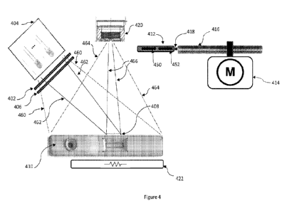
FIG. 4 illustrates the interaction between components of an example
embodiment of a multi-modal reader 300. Here, the reader 300 is depicted as
reading
a cartridge 410, which may be a fluidic biological sample test cartridge, or a
cartridge
containing a lateral flow strip.
[0001]
In the illustrated embodiment, a LED assembly including one or more
light emitting diodes (LEDs) 404 may be used to illuminate/energise at least
the test
area 408 of the cartridge 410, as indicated by lines 462 in one reading mode,
to enable
an image sensor 420 to read test features as indicated by lines 466. The test
area 408
may be, for example, the viewing window 240 or a region within the viewing
window
240 as shown. Alternatively, the LEDs 404 may illuminate the whole cartridge
410, as
indicated by lines 460, such that the reader 300 can read other cartridge
features
outside of the test area 408, as indicated by lines 464. For example, where
the
cartridge is similar to the cartridge 150 of Figure 1B, the reader 300 can
read the
cartridge label 144 (if present) and the QR code 142 (if present).
[00101]
For absorption/reflection (AR)-based reading of colorimetric tests, the
best
read is achieved when the peak wavelength of the illumination signal is the
same or
similar to the wavelength(s) of the visual feature(s) that indicate the test
result
Conversely, for fluorescence-based readings the excitation source
wavelength(s) is
selected to be different, and non-overlapping with, the wavelength of the
stimulated
emission signal.
[00102]
Multiple LEDs may be used to provide a source having a more even spatial
distribution of illumination. In some embodiments, two sets of multiple LEDs
are
provided. One set of LEDs is for use in a first reader mode and its LEDs are
all of the
same type and have the same centre wavelength, and the other set of LEDs is
for use
in a second reading mode, and its LEDs are of the same type and have the same
centre
wavelength, where the centre wavelengths of the two sets of LEDs are
different.
CA 03193647 2023- 3- 23
WO 2022/077065
PCT/AU2021/051200
- 22 -
[00103] The LEDs may be mounted to the surface of a printed circuit board of
the
apparatus, wherein the controller 514 selectively energises the first set of
LEDs when
the device is in a first reading mode, and the second set of LEDs when the
device is in
a second reading mode.
[00104] FIG. 8 is a schematic plan view of an example LED array
for use in an
embodiment of the present invention. The array comprises two sets of LEDs,
wherein
the black circles 404a represent LEDs of a first wavelength, in an energised
or 'on'
state. The white circles 404b represent LEDs with a second wavelength
different from
the first wavelength, and which are in a de-energised or 'off' state. As will
be apparent
to those skilled in the art, many alternative LED array arrangements can be
used to
achieve similar results. Further, in an alternative arrangement, LEDs of
different sets
may be mounted to different, moveable surfaces, such that only the light
produced by
a single set of LEDs is incident on the test area 408 or cartridge 410 at any
time.
[00105] Returning now to FIG. 4, the LED assembly may
additionally include one or
more diffusers 402, 406 disposed between the LEDs 404 and the test area
408/cartridge 410 to further disperse and improve the evenness of the
excitation/illumination of the cartridge 410.
[00106] In one or more reading modes, an optical filter, for
example a bandpass filter,
may be movably positioned between the test area 408 or cartridge 410 and the
image
sensor 420.In one embodiment, a movable slide 412 may comprise two filter
positions
450, 452 into which optical filters may be inserted.
[00107] In a first reading mode, the controller 514 can instruct
the motor or actuator
414 to position the slide 412 such that a first filter is positioned in the
direct optical
path between the image sensor 420 and the cartridge 410 under test. In a
second
reading mode, the second filter may be positioned between the image sensor 420
and
the cartridge 410.
[00108] If, however an optical filter is not required for a
particular reading mode, one
of the slide positions 450, 452 may remain empty, or the slide 412 may simply
be
positioned out of the way of the image sensor 420.
CA 03193647 2023- 3- 23
WO 2022/077065
PCT/AU2021/051200
- 23 -
[00109] Linear movement of the slide 412 is controlled by the
motor or actuator 414.
In some embodiments, the slide 412 is attached to the motor or actuator 414 by
a lead
screw 416 and nut 418. Alternatively, or additionally, the slide may be
configured to
move within a channel or guide rails (not shown).
[00110] If required, the reaction temperature for the lateral
flow strip can be
controlled using heating element 422, which may comprise, for example, one or
more
resistors. A temperature sensor 538 (not shown) provides feedback in a
standard
temperature control circuit. Electrical connections to the heating element and
temperature sensor may be by way of cable connections or slip rings.
[00111] The image sensor 420 may be, for example, a 5-megapixel CMOS sensor, a
CCD
sensor or an arrangement of photodiodes. The image sensor 420 may further
include
an integrated red, blue, and green Bayer filter 700 or other colour filter to
increase
the detection sensitivity for visible wavelengths. FIG. 7 provides an example
of a
known Bayer filter arrangement 700, comprising an array of green 710., red 712
and
blue 714 filters.
[00112] The wavelengths of LEDs 404a and 404b, and the optical wavelength
filters for
filter positions 450, 452 are selected to suit the different reading
requirements of the
specific test(s).
[00113] The multi-modal reader can be configured to change between the two or
more
reading modes in response to, for example, a user selection made using an
apparatus
user interface, or, for example, by automatic detection of the test cartridge
type. The
reader 300 may then operate according to a workflow suitable for the specific
cartridge under test 410.
[00114] In various embodiments, the test cartridge type can be
identified visually, by
processing an initial image or images to determine one or more visual indicia
or other
visual features (e.g. shape) of the test cartridge, or alternatively by one or
more non-
imaging sensors determining a physical shape or physical feature of the test
cartridge
(e.g., by determining the actuation or non-actuation of one or more
microswitches of
the test apparatus receiving portion of the apparatus, or the blocking or non-
blocking
CA 03193647 2023- 3- 23
WO 2022/077065
PCT/AU2021/051200
- 24 -
of one or more light beams), or by electronically reading or measuring an
electronic
feature or microchip of the test cartridge.
[00115] In each reading mode, the reader may obtain a single
measurement, or the
reader may obtain repeated measurements over time to produce a temporal
kinetic
readout. A temporal kinetic readout may be obtained to monitor, for example,
changes in binding events over time or enzyme activity. A non-limiting example
of a
repeated measurement application is described in detail below in example
configuration 3.
Example configuration 1
[00116] In a first example configuration, a multi-modal reader
300 is configured to read
colorimetric test or control lines with colloidal gold/red labelled particles
of a lateral
flow strip in a first reading mode, and fluorescence-based immunoassay capture
lines
with europium chelate fluorescence labelled particles in a second reading
mode.
[00117] In a first reading mode, an absorption/reflection (AR)-
based image is captured.
Green 520nm LEDs are provided for narrow-band illumination of the test area
408,
and the first position 450 of slide 412 is intentionally left empty, i.e. all
wavelengths
are allowed to pass. Here, a bandpass filter is not required to read the
colorimetric
immunoassay capture lines; however, in this embodiment the sensor 420 includes
a
Bayer filter.
[00118] In the first reading mode, upon instruction from the
controller 514, green LEDs
404a are positioned and/or energised to illuminate the test area 408, and the
controller 514 instructs the motor or actuator 414 to move the empty filter
location
450 in front of the image sensor 420.
[00119] As a Bayer filter is used in this embodiment, an image
captured in the first
reading mode comprises pixels acquired with red, blue and green pixel filter
elements.
The controller 514 selectively uses only the green filter element pixels for
the reading
because these pixels have very high sensitivity to the green absorption at
around
520nm, which is the characteristic maximum absorption wavelength of colloidal
gold
particles.
CA 03193647 2023- 3- 23
WO 2022/077065
PCT/AU2021/051200
- 25 -
[00120] Additionally, or alternatively, a green colour filter may
be located or moved in
front of the sensor 420. Using a narrower green bandpass filter of the same
wavelength range as the incident light source, in combination with the green
Bayer
filtered pixels, can produce a more accurate AR-based reading.
[00121] For the second reading mode, the second filter position
452 of slide 412 is
fitted with a 615nm centred bandpass filter, and second reading mode LEDs 404b
are
selected to provide UV emissions with a centre wavelength of approximately
360nm.
[00122] In the second reading mode, the controller 514 positions
and/or energises the
UV LEDs 404b to stimulate the test area 408. The filter position 452 is
positioned in
front of the sensor 420. This second reading mode arrangement provides an
ideal
configuration for the image sensor 420 to detect signals produced by the
europium
chelate fluorescence labelled particles.
[00123] In this way, the same reader 300 is capable of reading a
lateral flow strip with
test and/or control lines being either or both colorimetric and fluorescence
labelled
particles. The acquired images can then be used in subsequent image analyses.
[00124] The present configuration is described above with regard
to test and control
lines of a lateral flow strip. However, as will be appreciated, the same or a
similar
configuration may be also be used to read lateral flow strips with otherwise
shaped
test and control regions (e.g. dots), a fluidic cartridge or other type of
test assembly,
where the test involves colloidal gold and europium chelate fluorescence
labelled
particles.
[00125] Both readings may be, for example, displayed to a user
via a user display 310.
Results may alternatively or additionally be used by the controller 514 to
produce a
test result that combines data from both of the reading modes to form a result
that
could not be arrived at using individual reading modes only. Direct and
calculated
results may then be stored in memory 510.
CA 03193647 2023- 3- 23
WO 2022/077065
PCT/AU2021/051200
- 26 -
Example configuration 2
[00126] In a second example configuration, a multi-modal reader
300 is configured to
read immunoassay capture lines of a lateral flow strip with, for example,
colorimetric
immunoassay capture lines with colloidal gold particles in a first reading
mode, and to
detect one or more lateral flow strip artefacts, for example background
staining, in a
second reading mode. The multi-mode reader 300 can then remove or at least
mitigate the effects of the artefacts on the test result.
[00127] Background staining can occur in lateral flow strips
tests involving inherently
coloured samples. Background staining is common, for example, in lateral flow
strips
designed to test for gastrointestinal related illnesses using diluted stool
samples. As a
sample progresses along the strip, visible staining may be produced. If only a
single
reading mode of imaging is used, the staining may interfere with
interpretation of the
test results.
[00128] For example, if one or more flow stain lines are formed
in the proximity of the
immunoassay capture lines, the staining may interfere with the determination
of the
test or control line values. For example, a qualitative test may produce a
false positive
result, or a quantitative test may produce overstated values.
[00129] In order to address such difficulties, one configuration
of the multi-mode
reader 300 corrects for background staining by capturing two or more images of
the
test region in two or more reading modes.
[00130] Unlike colloidal gold particles which have a narrow
spectral response, stains
have a broad spectral response, and the staining will therefore be present in
images
captured under a wide range of illumination and capture conditions. Image
analysis,
including subtracting one image from another image, can provide a combined
result
that removes or at least alleviates the impact of the staining on the test
results.
[00131] In a first reading mode, the multi-mode reader 300 captures an
absorption/reflection (AR)-based image, which is particularly sensitive to the
colloidal
gold particles of the immunoassay capture lines. A similar LED and filter
arrangement
may be used as described above with regard to example configuration 1. Due to
the
CA 03193647 2023- 3- 23
WO 2022/077065
PCT/AU2021/051200
- 27 -
broad spectral response of the stain, the first image will include both the
capture lines,
if present, and some background staining.
[00132] In a second reading mode, a second AR-based reading mode
is used to obtain
an image of the staining only, wherein the reading mode has low sensitivity to
the
colloidal gold particles, but is approximately equally sensitive to the
background
staining.
[00133] For example, in a second reading mode, red LEDs may be
selected, with a
wavelength within the range of 620nm-660nm. For example, LEDs of wavelength
640nm may be used to illuminate the test area 408. In the second reading mode,
the
controller 514 selectively uses red Bayer filter element pixels for the
reading. These
red filtered pixels have low sensitivity to the emissions from the colloidal
gold
particles. Therefore, an image captured in the second reading mode will
include the
staining only. In an alternative arrangement, rather than using a Bayer
filter, a red
colour filter may be located or moved in front of the sensor 420.
[00134] A staining scenario is depicted in FIG. 9, with a
lateral flow strip 100, including,
for example, visible colloidal gold test and control lines 114, 116, and
staining 902. A
first image (A) of the test area 408 is captured in the first reading mode of
this
embodiment, and a second image (B) of test area 408 is captured in the second
reading mode of this embodiment. Once both images have been captured, the
controller 514 may then subtract image (B) from image (A) to generate a new,
calculated or 'synthetic' image (C).
[00135] Image (C) is free from, or at least less affected by,
the staining 902, and can
therefore be used to more accurately determine the presence or absence of the
control and test lines 114, 116, and/or be used for increasing the accuracy of
quantification.
[00136] As will be appreciated, other similar undesirable
lateral flow strip artefacts can
be subtracted from the detected image using a similar method, for example
dirt, dust,
or imperfections of the strip itself. Further, while the present example
configuration
is described above with respect to test and control lines, the same or a
similar
configuration can also be used to reduce the effects of lateral flow strips
with
CA 03193647 2023- 3- 23
WO 2022/077065
PCT/AU2021/051200
- 28 -
otherwise shaped test and control regions (e.g., dots). A similar method may
be used
to reduce the effects of dirt, dust, reflections etc in images received from a
fluidic
cartridge 200.
Example configuration 3
[00137] In a third example configuration, a multi-modal reader
300 is configured to
obtain fluorescence-based readings relating to accumulating concentrations of
NADPH in a fluidic cartridge 200 in a first reading mode, and absorbance-based
haemoglobin (Hb) measurements in a fluidic cartridge 200 in a second reading
mode.
The rate of change of NADPH measurements overtime, which can be determined
from
two or more images, provides an indication of the amount of glucose-6-
phosphate
dehydrogenase (G6PD) enzyme present in a sample. Combined knowledge of G6PD
and Hb levels in a sample is relevant for informing treatment decisions when
treating
humans with malaria.
[00138] Presently, drugs are available for treating latent
malaria infection; however,
these drugs are potentially harmful if administered to patients with low
levels of the
enzyme G6PD relative to Hb. Quantifying G6PD activity requires a compensation
process that also accounts for a quantified Hb level. Therefore, multiple
reading mode
outputs are required to determine an appropriate treatment for a patient.
[00139] A cost effective G6PD test is therefore pertinent for
the safe treatment of
malaria, wherein the test is capable of determining the patient's G6PD level,
Hb level,
and then calculating, for example, a ratio of G6PD activity to Hb level. This
ratio of
G6PD enzyme activity to Hb level, also referred to as the "compensated G6PD
value",
can then be used by clinicians to determine potential risks of drug
applications or
treatments.
[00140] G6PD is active in essentially all types of cells, and is
involved in the normal
processing of carbohydrates. It is responsible for the first step in the
pentose
phosphate pathway, a series of chemical reactions which includes converting
the
oxidised form of NADP, referred to as NADI'', to NADPH. The rate at which this
conversion occurs is known to be a measure of the amount of G6PD present. The
CA 03193647 2023- 3- 23
WO 2022/077065
PCT/AU2021/051200
- 29 -
global health organisation PATH has produced a commonly used guide to
fluorescent
spot testing for G6PD deficiency.
[00141] The multi-modal reader of the present embodiment may be used to
provide a
diagnostic test result that combines NADPH test results and Hb levels, wherein
the
example fluidic cartridge 200 of figures 2A and 2B may be used to perform the
test.
[00142] In an example test configuration, a 5 I blood sample and
500p.I of sample
buffer solution is used in the test. As the person skilled in the art will
appreciate
however, many different volumic arrangement may be used in alternative test
configurations.
[00143] The blood sample may be acquired from the patient using a finger prick
with a
sterile lancet, and transferred from the finger droplet to the test cartridge
200 using
either a transfer device or by directly applying the finger droplet to a
feature of the
cartridge 200. The sample preparation buffer liquid is preloaded in the sample
chamber 208 of cartridge 200. Further, the sample preparation buffer fluid can
be an
aqueous solution with detergent, salt, hypertonic water, or other reagent
configured
to cause the lysis of red blood cells within the added samples and to
distribute the
haemoglobin throughout the solution. Further, the reaction chamber 202 is
preloaded
with a soluble, dried or lyophilised reagent containing NADP+.
[00144] To start the test, the sample is added to the sample chamber 208, and
the
dispense cap 210 is screwed on. The cap 210 may include a piercing tip, which
pierces
a seal 206, allowing the blood sample, diluted with the aqueous solution, to
flow into
the reaction chamber 202. Once inside the reaction chamber 202, the G6PD
present
in the sample starts to convert the preloaded NADP+ into NADPH.
[00145] The NADPH generated by this reaction is a naturally
fluorescent molecule, with
a fluorescence wavelength around 500nm. In the first reading mode, stimulating
LEDs
404a may be selected to illuminate the viewing window 240 with a UV wavelength
in
the range of 320nm to 380nm. For example, LEDs 404a may be selected with a
centre
wavelength of approximately 350nm. Further, for the first reading mode, a band
pass
filter with a non-overlapping (the stimulating UV wavelength) pass band may be
CA 03193647 2023- 3- 23
WO 2022/077065
PCT/AU2021/051200
- 30 -
placed in the first filter location of slide 412, such that, in the first
reading mode, the
excitation signal of the NADPH alone is detected by the image sensor 420.
[00146] The measured level of fluorescence is proportional to the quantity of
NADPH
present in the reaction chamber 202 at the time the fluorescence response
image is
acquired. A set of NADPH levels can be measured over time, wherein the
slope/gradient of the NADPH increase over time, i.e. the rate of change in the
NADPH
level, can be used to calculate the relative level of G6PD present in the
sample. That
is, the more G6PD present, the faster the preloaded NADP+ will be converted to
NADPH. Therefore, in a first reading mode, multiple NADPH measurements are
made
using sensor 420, and the controller 514 processes these measurements to
calculate
the amount of G6PD within the sample.
[00147] The above reaction is temperature dependent, and therefore the
temperature
of the cartridge, and more specifically, the temperature of the reaction
chamber 202,
may be maintained at a specific known temperature for the duration of the
test. For
example, the temperature for the reaction may be selected to be between 38 C
and
45 C. More specifically, the temperature may be maintained at 40 C throughout.
To
maintain the temperature, the reaction chamber 202 may be in contact with or
in
close proximity to, for example, an anodised aluminium or ceramic block (i.e.,
to
increase thermal mass) attached to the heating element 422.
[00148] Alternatively, the temperature of the cartridge 200 may
not be controlled, but
rather simply measured throughout the reaction. In this case, the controller
514 uses
temperature measurements to adjust the NADPH rate calculation, in order to
determine the level of G6PD.
[00149] FIG. 10 provides an example NADPH versus time graph,
wherein NADPH
readings were obtained by a multi-modal reader according to an embodiment of
the
present invention, wherein the temperature of the reaction was constant. The
test
process used by the reader to determine the level of G6PD from the
measurements
may be based on a built-in test library, initially created using hundreds of
clinical
samples. Alternatively, in another embodiment, the reader may be calibrated by
the
user, using a range of samples with known concentrations of the G6PD enzyme.
CA 03193647 2023- 3- 23
WO 2022/077065
PCT/AU2021/051200
-31 -
[00150] In a second reading mode, the reader obtains a Hb
measurement. Blue LEDs
are used to illuminate the viewing window 240 of the cartridge 200 under test,
and
the image sensor 420 comprises a Bayer filter. The controller 514 selectively
uses only
pixels acquired with blue filter elements. Additionally or alternatively, a
blue filter may
be placed in a second filter position 452 of slide 412. The resulting image or
digital
representation of the viewing window 240 is then analysed to determine the
amount
of Hb present in the sample. Hb, being red, also absorbs green light well, and
therefore, in an alternative embodiment, green illumination with appropriate
green
filtering may be used.
[00151] Once both the G6PD and Hb levels in the sample have been determined by
the
controller 514, the controller 514 then combines measurements obtained from
the
first and second reading modes to calculate the compensated G6PD value. This
value
can then be communicated to the user, for example via the display 310.
[00152] As will be appreciated by the skilled person, G6PD
results are not only relevant
when treating latent malaria. A wide range of applications exist for a point-
of-care
reader able to determine G6PD levels. A multi-modal reader as described herein
may,
for example, be configured to read test cartridges in one reading mode and
other
either related and/or unrelated test cartridges in further modes.
Alternatively, a point-
of-care reader as described herein may be configured to use multiple reading
modes
to read any other desirable multiplexed G6PD test.
[00153] Similar repeated measurement methods may be used to obtain other
temporal kinetic readouts for monitoring, for example, changes in binding
events over
time, or the activity of other enzymes, which may then be combined with
results or
information obtained in other reading modes.
Example configuration 4
[00154] When performing any diagnostic test, it is critically
important to ensure that
the test results are reliably linked to the corresponding patient. Test
cartridges may
therefore have identifying marks such as a code 39, an EAN or PDF-417 barcode,
a 2D
DataMatrix, or a QR Code 142 printed or otherwise marked onto the surface of
the
cartridge. Further, the identifying mark may include encoded information about
the
CA 03193647 2023- 3- 23
WO 2022/077065
PCT/AU2021/051200
- 32 -
test or cartridge itself, which reading mode or sequence should be used for
reading
and interpreting the test results, and any relevant calibration information.
[00155] In some embodiments, an external scanner may be used to
read the identifying
mark. For example, reader 300 may be configured to accept test specific inputs
from
an external scanner, for example user ID and test ID. An external reader may
be
connected via, for example, a USB port 522. The external scanner may be, for
example,
a Datalogic QuickScanTM Wand 0D2430, which is capable of reading a variety of
identifying marks. In the present embodiment however, the multi-modal reader
300
itself is configured to detect and interpret any such identifying marks.
[00156] For cartridge/sample
storage/retrieval/identification/management (i.e., for
purposes other than determining diagnostic test results), identifying marks
should be
visually readable by standard readers (e.g., a standard barcode reader)
separate to
the multi-modal readers described herein. Standard barcodes will not be
available in
images produced by readers configured to read only fluorescence-based tests.
Indeed,
prior art fluorescence readers cannot read barcodes. This embodiment overcomes
this
limitation by providing both absorption/reflection (AR) and fluorescence-based
reading modes.
[00157] Further, if an identifying mark does not identify the
cartridge type, then the
reader 300 may use an AR-based reading mode to obtain further cartridge
information. For example, the reader 300 may determine the outline of the
cartridge
for comparison with a database of known cartridge shape and feature
dimensions.
Further still, alternatively, or additionally, the reader 300 may use an AR-
based
reading mode to locate the position of the viewing window 140, 240.
[00158] Turning now to FIG. 11, FIG. 11A depicts an image of a
cartridge 150 captured
in an AR-based reading mode, and FIG. 11B depicts an image of a cartridge 150
captured in a fluorescence-based reading mode. As is apparent in FIG. 11B,
cartridge
features are not apparent in the fluorescence-based reading mode.
[00159] In FIG. 2B, two immunoassay capture lines are apparent
in the fluorescence-
based image, i.e. a test and control line. However, where only a single line
is present,
it is critically important to know the relative positioning of the line with
respect to the
CA 03193647 2023- 3- 23
WO 2022/077065
PCT/AU2021/051200
- 33 -
viewing window 140, i.e. whether the single line is a test or control line.
For example,
for a qualitative diagnostic test, if only a control line is present, the test
result is
negative, however if only a test line is present, an error has occurred, and a
result
cannot be determined.
[00160] In the present embodiment, a first, AR-based, reading
mode is provided, such
that the instrument can read features of a cartridge 150, 200. A standard edge
detection method is used to determine the coordinates of edges of the viewing
window 140. Advantageously, in a multi-modal reader, the cartridge does not
move
between image captures, and coordinates of the viewing window may be used when
interpreting an image captured in a second or any further subsequent reading
modes.
[00161] For the first reading mode, a visible image can be
obtained by the image sensor
using red LED illumination of a selected wavelength within the range of 620nm-
660nm, for example, red LEDs of wavelength 640nm, to illuminate the cartridge
features. Either red Bayer filter elements may be used for the reading, or
alternatively
a red filter may be placed in a second filter position 452 of slide 412 and be
positioned
in front of the sensor 420.
[00162] In the first reading mode, reflected light from the
cartridge features passes
through the red band pass filter with the same band pass filter for detection
in the
image sensor. This reading mode obtains an image of the data code and/or
features
of the cartridge. Image analysis by the controller 514 can then extract data
from the
image, for example the encoded information from a data code and/or viewing
window
coordinates. The data code may include information such as calibration factors
and
expiry dates, which are then available for combining with results of other
reading
modes to generate a final test result. For example, the expiry date may
indicate
whether or not accelerated product degradation is likely to have occurred, and
incorporating an expiry date into the diagnostic test process may therefore
lead to a
more accurate result.
[00163] For the second reading mode, the reader 300 may be
configured to obtain, for
example, a fluorescence-based reading in accordance with any of the above
described
fluorescence reading modes, or any other desirable reading.
CA 03193647 2023- 3- 23
WO 2022/077065
PCT/AU2021/051200
- 34 -
[00164] In a specific example, if a Europium fluorescence-based
test is used in the
cartridge 150, 200, then a fluorescence image can be produced using UV
illumination.
The same red bandpass sensor filter can be used in the second reading mode, as
in
the first, AR-based reading mode. This specific configuration of reading modes
has the
advantage that the image sensor band pass filter is not required to
move/change
position between reading modes.
Example configuration 5
[00165] In a fifth example configuration, a multi-modal reader
is configured to obtain
absorption/reflection (AR)-based readings relating to sample flow in a fluidic
cartridge
200 or along a lateral flow strip 100 in a first reading mode, and
fluorescence-based
measurements in second reading mode.
[00166] As described above, lateral flow strips often include a
control immunoassay
capture line to indicate that the sample has successfully migrated across the
strip.
However, in some configurations, including tests involving a fluidic
cartridge, a control
measure may be desirable as an alternative or in addition to other controls.
If the flow
can be observed in an AR-based reading mode, i.e. the materials are not
fluorescent,
then information relating to the flow can be used in the test process.
Fluorescence can
then be independently measured.
[00167] For tests involving blood, for example, sample flow
through a cartridge or
lateral flow strip may be inherently capable of being imaged using an AR-based
reading mode. In other samples, for example urine or a dilute sample, a visual
dye may
be added, such that flow is evident. Alternatively, the flow may be evident as
discolouration due to wetting.
[00168] The presence of the flow, including its completion
through the full length of
the strip, or into/through a reservoir/channel/chamber etc of a fluidic
cartridge can
be utilised within the test process as an improved control to confirm that the
sample
was adequately added to the test area 408.
[00169] Additional characteristics of the sample flow reading
mode, such as the rate of
progress of the leading edge of the flow or an AR-read quantification
measurement
CA 03193647 2023- 3- 23
WO 2022/077065
PCT/AU2021/051200
- 35 -
may be used to determine the amount of sample added, or at least to confirm
that an
adequate amount of sample was added. The controller 514 may then use the
multiple
readings to perform calculations that allow, for example, for the amount of
the analyte
detected to be compensated for by the amount of sample applied. That is, an
optical
AR-based measurement can be used to detect the quantity/amount of a sample
material applied to the test strip of a test cartridge, and fluorescence
detection can be
used to detect the level of a specific analyte within the sample.
[00170] The controller 514 can then combine the multiple measurements to
generate
an overall test result. For example, the reading may prevent an incorrect test
result
from being generated if an inadequate amount of sample was added.
[00171] In an example embodiment, a blue dye may be added to the
sample, which
has, for example, absorption in the range of 440-490nm, wherein the dye is
visually
detectable in a first, AR-based reading mode.
[00172] In a second reading mode, UV LEDs in the range 340-370nm
may excite, for
example, Europium fluorescence of a test region. The image sensor 420, with an
appropriate bandpass filter, obtains a reading of the resulting red emission
within the
wavelength range 605-625nm. As the sample is stained with a blue dye, the dye
does
not interfere with the fluorescence measurement in the fluorescent reading
mode.
Other embodiments
[00173] The above example embodiments involve immunoassays and
enzymatic
assays; however, as those skilled in the art will appreciate, the multi-modal
readers
described herein may also be used to perform analogous diagnostic tests
involving
nucleic acid amplification assays.
[00174] Further, in any embodiment of the invention where the
reader is configured
to read visually interpretable cartridges or test strips, the reader may be
further
configured such that a user's subjective visual interpretation of a test may
be entered
as input into the device. The controller 514 may then correlate the user's
subjective
interpretation with an objective test result or other analysis performed by
the reader,
CA 03193647 2023- 3- 23
WO 2022/077065
PCT/AU2021/051200
- 36 -
which can advantageously be used to determine end-user competencies and
identify
user training requirements.
[00175] While the above example embodiments generally describe
using different
reading modes to read different types of tests, and/or different cartridge or
test area
features, the multiple reading modes can also be used to gain further insights
regarding the same test. Obtaining different absorption/reflection (AR)-based
readings can be used to determine characteristics of a signal produced at a
single test
region. For example, if a lateral flow strip comprises a colloidal gold (red)
colorimetric
test line, it would be expected that reading modes that use green illumination
and
green filtering will produce the strongest AR-based reading. As a control
measure, the
signal can be checked by obtaining a further reading of the same immunoassay
capture line using blue illumination and blue filtering or red illumination
and red
filtering. As will be appreciated, characteristics of both colorimetric and
fluorescent
test and control signals may be determined or verified using multiple readings
with
different optical filters.
[00176] Further, while the readers described herein were
developed for the primary
purpose of reading a diagnostic test assembly in multiple reading modes to
determine
a diagnostic result, some types of diagnostic test assembly may require only
one
reading mode, and the readers described herein can also be used with such
diagnostic
test assemblies.
Incorporating further reading modes
[00177] The multi-modal readers described above include two sets of LEDs 404a,
404b
and two filter positions 450, 452; however, a person skilled in the art will
appreciate
that the readers can easily be adapted to include further reading modes. As
will be
apparent to the person skilled in the art, many combinations of the above
described
reading modes are possible, and the reader 300 may read in 2, 3, 4, 5 ... 10
or more
different modes.
CA 03193647 2023- 3- 23
WO 2022/077065
PCT/AU2021/051200
- 37 -
[00178] To enable the further reading modes, additional sets of
LEDs may be selectively
energised or positioned to illuminate/energise a cartridge under test, and the
slide
412 may be configured to accommodate more than two filters.
[00179] Multi-mode readers with a large number of optical filters
may require an
alternative arrangement for filter positioning. For example, the filters may
be inserted
into a rotary carousel, wherein the active filter is positioned between the
test area
408/cartridge 150, 200 and the image sensor 420 by a motor rotating the
carousel.
[00180] Alternatively, a variable filter may be used; for
example, a Delata 3G LVSWP
linear variable filter may be used. Instead of, or additionally to using
multiple filters, a
variable filter may be moved by motor or actuator 414, to provide a large
number of
reading modes covering a full spectrum. This may be used in addition to
fluorescence-
based reading modes.
[00181] Other mechanical arrangements for both the LED
illumination of the test area
408 or cartridge and the active filter positioning will be apparent to those
skilled in the
art.
[00182] As used herein, except where the context requires otherwise the term
'comprise' and variations of the term, such as 'comprising', 'comprises' and
'comprised', are not intended to exclude other additives, components, integers
or
steps.
[00183] The foregoing summary is not intended to summarise each potential
embodiment or every aspect of the present disclosure.
[00184] Many modifications within the scope of the present
invention will be apparent
to those skilled in the art in light of this disclosure.
CA 03193647 2023- 3- 23