Note: Descriptions are shown in the official language in which they were submitted.
WO 2022/076462
PCT/US2021/053636
DOSING FOR TREATMENT WITH ANTI-FCRH5/ANTI-CD3 BISPECIFIC ANTIBODIES
SEQUENCE LISTING
The instant application contains a Sequence Listing which has been submitted
electronically
in ASCII format and is hereby incorporated by reference in its entirety. Said
ASCII copy, created on
October 4, 2021, is named 50474-213W05 Sequence Listing 10 4_21_ST25 and is
33,733 bytes in
size.
FIELD OF THE INVENTION
The present invention relates to the treatment of cancers, such as B cell
proliferative
disorders. More specifically, the invention concerns the specific treatment of
human patients having
multiple myeloma (MM) using anti-fragment crystallizable receptor-like 5
(FcRH5)/anti-cluster of
differentiation 3 (CD3) bispecific antibodies.
BACKGROUND
Cancer remains one of the most deadly threats to human health. In the U.S.,
cancer affects
more than 1.7 million new patients each year and is the second leading cause
of death after heart
disease, accounting for approximately one in four deaths.
Hematologic cancers, in particular, are the second leading cause of cancer-
related deaths.
Hematologic cancers include multiple myeloma (MM), a neoplasm characterized by
the proliferation
and accumulation of malignant plasma cells. Worldwide, approximately 110,000
people are
diagnosed with MM annually. MM remains incurable despite advances in
treatment, with an
estimated median survival of 8-10 years for standard-risk myeloma and 2-3
years for high-risk
disease, despite receipt of an autologous stern-cell transplant. Despite the
significant improvement in
patient's survival over the past 20 years, only 10-15% of patients achieve or
exceed expected survival
compared with the matched general population. Increased survival has been
achieved with the
introduction of proteasome inhibitors, immunomodulatory drugs (IMiDs), and
monoclonal antibodies.
Nevertheless, most patients (if not all) eventually relapse, and the outcome
of patients with MM after
they become refractory, or ineligible to receive a proteasome inhibitor or an
IMiD, is quite poor, with
survival less than 1 year. Therefore, relapsed or refractory (R/R) MM, in
particular, continues to
constitute a significant unmet medical need, and novel therapeutic agents are
needed. For such
patients, alternative or secondary treatment modalities, such as bispecific
antibody-based
immunotherapies, may be particularly efficacious. There is an unmet need in
the field for the
development of efficacious methods of dosing therapeutic bispecific antibodies
(e.g., anti-FcRH5/anti-
CD3 bispecific antibodies) for the treatment of cancers (e.g., MM, e.g., R/R
MM) that achieve a more
favorable benefit-risk profile.
1
CA 03193952 2023- 3- 27
WO 2022/076462
PCT/US2021/053636
SUMMARY OF THE INVENTION
In one aspect, the disclosure features a method of treating a subject having a
multiple
myeloma (MM) comprising administering to the subject a bispecific antibody
that binds to FcRH5 and
CD3 in a dosing regimen comprising at least a first dosing cycle, wherein the
first dosing cycle
comprises a first dose (Cl Dl), a second dose (Cl D2), and a third dose (Cl
D3) of the bispecific
antibody, wherein the Cl Dl is between about 0.01 mg to about 2.9 mg, the Cl
D2 is between about 3
mg to about 19.9 mg, and the Cl D3 is between about 20 mg to about 600 mg.
In some aspects, the Cl Dl is between about 0.1 mg to about 1.5 mg; the Cl D2
is between
about 3.2 mg to about 10 mg; and the Cl D3 is between about 80 mg to about 300
mg. In some
aspects, the Cl Dl is about 0.3 mg; the Cl D2 is about 3.6 mg; and the Cl D3
is about 160 mg.
In some aspects, the dosing regimen further comprises a second dosing cycle
comprising a
single dose (C2D1) of the bispecific antibody, wherein the C2D1 is equal to or
greater than the Cl D3
and is between about 20 mg to about 600 mg. In some aspects, the C2D1 is
between about 80 mg to
about 300 mg. In some aspects, the C2D1 is about 160 mg.
In another aspect, the disclosure features a method of treating a subject
having a MM
comprising administering to the subject a bispecific antibody that binds to
FcRH5 and CD3 in a dosing
regimen comprising at least a first dosing cycle, wherein the first dosing
cycle comprises a first dose
(Cl Dl), a second dose (Cl D2), and a third dose (Cl D3) of the bispecific
antibody, wherein the Cl Dl
is between about 0.2 mg to about 0.4 mg, the Cl D2 is greater than the Cl Dl,
and the Cl D3 is
greater than the Cl D2.
In some aspects, the Cl Dl is about 0.3 mg. In some aspects, the Cl D2 is
between about 3
mg to about 19.9 mg. In some aspects, the Cl D2 is between about 3.2 mg to
about 10 mg. In some
aspects, the Cl D2 is about 3.6 mg. In some aspects, the Cl D3 is between
about 20 mg to about 600
mg. In some aspects, the Cl D3 is between about 80 mg to about 300 mg. In some
aspects, the
C1D3 is about 160 mg.
In some aspects, the dosing regimen further comprises a second dosing cycle
comprising a
single dose (C2D1) of the bispecific antibody, wherein the C2D1 is equal to or
greater than the Cl D3
and is between about 20 mg to about 600 mg. In some aspects, the C2D1 is
between about 80 mg to
about 300 mg. In some aspects, the C2D1 is about 160 mg.
In some aspects, the length of the first dosing cycle is 21 days. In some
aspects, the method
comprises administering to the subject the Cl Dl, the Cl D2, and the Cl D3 on
or about Days 1, 8, and
15, respectively, of the first dosing cycle.
In some aspects, the length of the second dosing cycle is 21 days. In some
aspects, the
method comprises administering to the subject the C2D1 on or about Day 1 of
the second dosing
cycle.
In some aspects, the dosing regimen comprises one or more additional dosing
cycles. In
some aspects, the dosing regimen comprises four additional dosing cycles,
wherein the length of
each of the four additional dosing cycles is 21 days. In some aspects, the
four additional dosing
cycles each comprise a single dose of the bispecific antibody, wherein the
single dose is between
2
CA 03193952 2023- 3- 27
WO 2022/076462
PCT/US2021/053636
about 80 mg to about 300 mg, and wherein the method comprises administering to
the subject the
single dose on or about Day lof each of the four additional dosing cycles. In
some aspects, the
dosing regimen further comprises up to 17 additional dosing cycles, wherein
the length of each of the
additional dosing cycles is 21 days. In some aspects, the up to 17 additional
dosing cycles each
comprise a single dose of the bispecific antibody, wherein the single dose is
between about 80 mg to
about 300 mg, and wherein the method comprises administering to the subject
the single dose on or
about Day 1 of each of the up to 17 additional dosing cycles.
In some aspects, the median peak IL-6 level in a population of subjects
treated according to
the method does not exceed 125 pg/mL between the Cl Dl and the Cl D2. In some
aspects, the
median peak IL-6 level in a population of subjects treated according to the
method does not exceed
100 pg/mL between the Cl Dl and the Cl D2. In some aspects, the median peak IL-
6 level in a
population of subjects treated according to the method does not exceed 125
pg/mL between the
Cl D2 and the Cl D3. In some aspects, the median peak IL-6 level in a
population of subjects treated
according to the method does not exceed 100 pg/mL between the Cl D2 and the Cl
D3. In some
aspects, the median peak IL-6 level in a population of subjects treated
according to the method does
not exceed 125 pg/mL following the Cl D3. In some aspects, the median peak IL-
6 level in a
population of subjects treated according to the method does not exceed 100
pg/mL following the
Cl D3. In some aspects, the IL-6 level is measured in a peripheral blood
sample.
In some aspects, the peak level of CD8+ T cell activation in the subject in
the first dosing
cycle occurs between the Cl D2 and the Cl D3. In some aspects, the peak level
of CD8+ T cell
activation in the subject in the first dosing cycle occurs within 24 hours of
the Cl D2.
In another aspect, the disclosure features a method of treating a subject
having a multiple
myeloma (MM) comprising administering to the subject a bispecific antibody
that binds to FcRH5 and
CD3 in a dosing regimen comprising at least a first dosing cycle, wherein the
first dosing cycle
comprises a first dose (Cl Dl) and a second dose (Cl D2) of the bispecific
antibody, wherein the
Cl D1 is between about 0.5 mg to about 19.9 mg and the Cl D2 is between about
20 mg to about 600
mg. In some aspects, the Cl Dl is between about 1.2 mg to about 10.8 mg and
the Cl D2 is between
about 80 mg to about 300 mg. In some aspects, the Cl Dl is about 3.6 mg and
the Cl D2 is about
198 mg. In some aspects, the length of the first dosing cycle is 21 days. In
some aspects, the
method comprises administering to the subject the Cl Dl and the Cl D2 on or
about Days 1 and 8,
respectively, of the first dosing cycle. In some aspects, the dosing regimen
further comprises a
second dosing cycle comprising a single dose (C2D1) of the bispecific
antibody, wherein the C2D1 is
equal to or greater than the Cl D2 and is between about 20 mg to about 600 mg.
In some aspects,
the C2D1 is between about 80 mg to about 300 mg. In some aspects, the C2D1 is
about 198 mg. In
some aspects, the length of the second dosing cycle is 21 days. In some
aspects, the method
comprises administering to the subject the C2D1 on Day 1 of the second dosing
cycle. In some
aspects, the dosing regimen comprises one or more additional dosing cycles. In
some aspects, the
dosing regimen comprises one to 17 additional dosing cycles. In some aspects,
the length of each of
the one or more additional dosing cycles is 21 days. In some aspects, each of
the one or more
3
CA 03193952 2023- 3- 27
WO 2022/076462
PCT/US2021/053636
additional dosing cycles comprises a single dose of the bispecific antibody.
In some aspects, the
method comprises administering to the subject the single dose of the
bispecific antibody on Day 1 of
the one or more additional dosing cycles.
In some aspects of any of the methods described herein, the bispecific
antibody comprises an
anti-FcRH5 arm comprising a first binding domain comprising the following six
hypervariable regions
(HVRs): (a) an HVR-H1 comprising the amino acid sequence of RFGVH (SEQ ID NO:
1); (b) an HVR-
H2 comprising the amino acid sequence of VIWRGGSTDYNAAFVS (SEQ ID NO: 2); (c)
an HVR-H3
comprising the amino acid sequence of HYYGSSDYALDN (SEQ ID NO:3); (d) an HVR-
L1 comprising
the amino acid sequence of KASQDVRNLVV (SEQ ID NO: 4); (e) an HVR-L2
comprising the amino
acid sequence of SGSYRYS (SEQ ID NO: 5); and (f) an HVR-L3 comprising the
amino acid sequence
of QQHYSPPYT (SEQ ID NO: 6). In some aspects, the bispecific antibody
comprises an anti-FcRH5
arm comprising a first binding domain comprising (a) a heavy chain variable
(VH) domain comprising
an amino acid sequence having at least 95% sequence identity to the amino acid
sequence of SEQ
ID NO: 7; (b) a light chain variable (VL) domain comprising an amino acid
sequence having at least
95% sequence identity to the amino acid sequence of SEQ ID NO: 8; or (c) a VH
domain as in (a) and
a VL domain as in (b). In some aspects, the first binding domain comprises a
VH domain comprising
an amino acid sequence of SEQ ID NO: 7 and a VL domain comprising an amino
acid sequence of
SEQ ID NO: 8. In some aspects, wherein the bispecific antibody comprises an
anti-CD3 arm
comprising a second binding domain comprising the following six HVRs: (a) an
HVR-H1 comprising
the amino acid sequence of SYYIH (SEQ ID NO: 9); (b) an HVR-H2 comprising the
amino acid
sequence of WIYPENDNTKYNEKFKD (SEQ ID NO: 10); (c) an HVR-H3 comprising the
amino acid
sequence of DGYSRYYFDY (SEQ ID NO: 11); (d) an HVR-L1 comprising the amino
acid sequence of
KSSQSLLNSRTRKNYLA (SEQ ID NO: 12); (e) an HVR-L2 comprising the amino acid
sequence of
WTSTRKS (SEQ ID NO: 13); and (f) an HVR-L3 comprising the amino acid sequence
of KQSFILRT
(SEQ ID NO: 14). In some aspects, the bispecific antibody comprises an anti-
CD3 arm comprising a
second binding domain comprising (a) a VH domain comprising an amino acid
sequence having at
least 95% sequence identity to the amino acid sequence of SEQ ID NO: 15; (b) a
VL domain
comprising an amino acid sequence having at least 95% sequence identity to the
amino acid
sequence of SEQ ID NO: 16; or (c) a VH domain as in (a) and a VL domain as in
(b). In some
aspects, the second binding domain comprises a VH domain comprising an amino
acid sequence of
SEQ ID NO: 15 and a VL domain comprising an amino acid sequence of SEQ ID NO:
16. In some
aspects, the bispecific antibody comprises an anti-FcRH5 arm comprising a
heavy chain polypeptide
(H1) and a light chain polypeptide (L1) and an anti-CD3 arm comprising a heavy
chain polypeptide
(H2) and a light chain polypeptide (L2), and wherein: (a) H1 comprises the
amino acid sequence of
SEQ ID NO: 35; (b) L1 comprises the amino acid sequence of SEQ ID NO: 36; (c)
H2 comprises the
amino acid sequence of SEQ ID NO: 37; and (d) L2 comprises the amino acid
sequence of SEQ ID
NO: 38.
In some aspects of any of the methods described herein, the bispecific
antibody comprises an
aglycosylation site mutation. In some aspects, the aglycosylation site
mutation reduces effector
4
CA 03193952 2023- 3- 27
WO 2022/076462
PCT/US2021/053636
function of the bispecific antibody. In some aspects, wherein the
aglycosylation site mutation is a
substitution mutation. In some aspects, the bispecific antibody comprises a
substitution mutation in
the Fc region that reduces effector function. In some aspects, the bispecific
antibody is a monoclonal
antibody. In some aspects, the bispecific antibody is a humanized antibody. In
some aspects, the
bispecific antibody is a chimeric antibody. In some aspects, the bispecific
antibody is an antibody
fragment that binds FcRH5 and CD3. In some aspects, the antibody fragment is
selected from the
group consisting of Fab, Fab'-SH, Fv, scFv, and (Fab')2 fragments. In some
aspects, the bispecific
antibody is a full-length antibody. In some aspects, the bispecific antibody
is an IgG antibody. In
some aspects, the IgG antibody is an IgGi antibody.
In some aspects of any of the methods described herein, the bispecific
antibody comprises
one or more heavy chain constant domains, wherein the one or more heavy chain
constant domains
are selected from a first CH1 (CH1 i) domain, a first CH2 (CH2 /) domain, a
first CH3 (CH3 /) domain, a
second CH1 (CH12) domain, second CH2 (CH22) domain, and a second CH3 (CH32)
domain. In
some aspects, at least one of the one or more heavy chain constant domains is
paired with another
heavy chain constant domain. In some aspects, the CH3/ and CH32 domains each
comprise a
protuberance or cavity, and wherein the protuberance or cavity in the CH3/
domain is positionable in
the cavity or protubcrancc, respectively, in thc 0H32 domain. In some aspects,
thc CH3, and CH32
domains meet at an interface between the protuberance and cavity. In some
aspects, the CH2/ and
CH22 domains each comprise a protuberance or cavity, and wherein the
protuberance or cavity in the
CH2/ domain is positionable in the cavity or protuberance, respectively, in
the CH22 domain. In some
aspects, the CH2/ and 0H22 domains meet at an interface between said
protuberance and cavity. In
some aspects, the anti-FcRH5 arm comprises the protuberance and the anti-CD3
arm comprises the
cavity. In some aspects, a CH3 domain of the anti-FcRH5 arm comprises a
protuberance comprising
a T366W amino acid substitution mutation (EU numbering) and a CH3 domain of
the anti-CD3 arm
comprises a cavity comprising T366S, L368A, and Y407V amino acid substitution
mutations (EU
numbering).
In some aspects of any of the methods described herein, the bispecific
antibody is
administered to the subject as a monotherapy.
In some aspects of any of the methods described herein, the bispecific
antibody is
administered to the subject as a combination therapy. In some aspects, the
bispecific antibody is
administered to the subject concurrently with one or more additional
therapeutic agents. In some
aspects, the bispecific antibody is administered to the subject prior to the
administration of one or
more additional therapeutic agents. In some aspects, the bispecific antibody
is administered to the
subject subsequent to the administration of one or more additional therapeutic
agents. In some
aspects, the one or more additional therapeutic agents comprise an effective
amount of tocilizumab.
In some aspects, tocilizumab is administered to the subject by intravenous
infusion. In some aspects,
(a) the subject weighs 100 kg, and tocilizumab is administered to the subject
at a dose of 800 mg;
(b) the subject weighs 30 kg and < 100 kg, and tocilizumab is administered to
the subject at a dose
of 8 mg/kg; or (c) the subject weighs < 30 kg, and tocilizumab is administered
to the subject at a dose
5
CA 03193952 2023- 3- 27
WO 2022/076462
PCT/US2021/053636
of 12 mg/kg. In some aspects, tocilizumab is administered to the subject 2
hours before
administration of the bispecific antibody. In some aspects, the one or more
additional therapeutic
agents comprise an effective amount of pomalidomide, daratumumab, or a B-cell
maturation antigen
(BCMA)-directed therapy.
In some aspects of any of the methods described herein, the bispecific
antibody is
administered to the subject by intravenous infusion.
In some aspects of any of the methods described herein, the bispecific
antibody is
administered to the subject subcutaneously.
In some aspects of any of the methods described herein, the subject has a
cytokine release
syndrome (CRS) event, and the method further comprises treating the symptoms
of the CRS event
while suspending treatment with the bispecific antibody. In some aspects, the
method further
comprises administering to the subject an effective amount of tocilizumab to
treat the CRS event. In
some aspects, tocilizumab is administered intravenously to the subject as a
single dose of about 8
mg/kg. In some aspects, the CRS event does not resolve or worsens within 24
hours of treating the
symptoms of the CRS event, and the method further comprising administering to
the subject one or
more additional doses of tocilizumab to manage the CRS event. In some aspects,
the one or more
additional doses of tocilizumab are administered intravenously to the subject
at a dose of about 8
mg/kg. In some aspects, the one or more additional therapeutic agents comprise
an effective amount
of a corticosteroid. In some aspects, the corticosteroid is administered
intravenously to the subject.
In some aspects, the corticosteroid is methylprednisolone. In some aspects,
methylprednisolone is
administered at a dose of about 80 mg. In some aspects, the corticosteroid is
dexamethasone. In
some aspects, dexamethasone is administered at a dose of about 20 mg. In some
aspects, the one
or more additional therapeutic agents comprise an effective amount of
acetaminophen or
paracetamol. In some aspects, acetaminophen or paracetamol is administered at
a dose of between
about 500 mg to about 1000 mg. In some aspects, acetaminophen or paracetamol
is administered
orally to the subject. In some aspects, the one or more additional therapeutic
agents comprise an
effective amount of diphenhydramine. In some aspects, diphenhydramine is
administered at a dose
of between about 25 mg to about 50 mg. In some aspects, diphenhydramine is
administered orally to
the subject.
In some aspects of any of the methods described herein, the MM is a relapsed
or refractory
(R/R) MM. In some aspects, the individual has received at least three prior
lines of treatment for the
MM. In some aspects, the individual has received at least four prior lines of
treatment for the MM. In
some aspects, the individual has been exposed to a prior treatment comprising
a proteasome
inhibitor, an IMiD, and/or an anti-CD38 therapeutic agent. In some aspects,
the proteasome inhibitor
is bortezomib, carfilzomib, or ixazomib. In some aspects, the IMiD is
thalidomide, lenalidomide, or
pomalidomide. In some aspects, the anti-0D38 therapeutic agent is an anti-CD38
antibody. In some
aspects, the anti-CD38 antibody is daratumumab, M0R202, or isatuximab. In some
aspects, the anti-
CD38 antibody is daratumumab. In some aspects, the individual has been exposed
to a prior
treatment comprising an anti-SLAMF7 therapeutic agent, a nuclear export
inhibitor, a histone
6
CA 03193952 2023- 3- 27
WO 2022/076462
PCT/US2021/053636
deacetylase (HDAC) inhibitor, an autologous stem cell transplant (ASCT), a
bispecific antibody, an
antibody-drug conjugate (ADC), a CAR-T cell therapy, or a BCMA-directed
therapy. In some aspects,
the anti-SLAMF7 therapeutic agent is an anti-SLAMF7 antibody. In some aspects,
the anti-SLAMF7
antibody is elotuzumab. In some aspects, the nuclear export inhibitor is
selinexor. In some aspects,
the HDAC inhibitor is panobinostat. In some aspects, the BCMA-directed therapy
is an antibody-drug
conjugate targeting BCMA.
BRIEF DESCRIPTION OF THE DRAWINGS
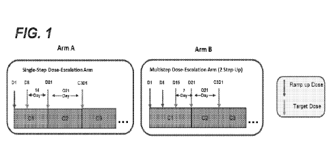
Fig. 1 is a schematic diagram showing dose escalation schedules for Arm A
(single-step dose
escalation arm) and Arm B (multi-step dose escalation arm) of the G039775
Phase I dose-escalation
study. C: cycle; D: day; Q: every.
Fig. 2 is a schematic diagram showing a possible single-step dose-escalation
scenario for
Arm A of the 0039775 Phase I dose-escalation study. AE: adverse event; DLT;
dose-limiting toxicity;
ISC: Internal Safety Committee; MAD: maximum achieved dose; MTD: maximum
tolerated dose; pts:
patients. Dose levels are in milligrams. Dose levels and dose modifications
are for illustrative
purposes only. "AE" refers to adverse events not considered by the
investigator to be attributable to
another clearly identifiable cause (e.g., disease progression).
Fig. 3 is a schematic diagram showing a possible two-step dose-escalation
scenario for Arm
B of the 0039775 Phase I dose-escalation study. CRS: cytokine release
syndrome. Dose levels are
in milligrams. Dose levels and dose modifications are for illustrative
purposes only.
Fig. 4A is a schematic diagram showing the progression of doses in the single-
step dose-
escalation arm (Arm A) and the single-step expansion arm (Arm C) of the
G039775 Phase I dose-
escalation study. Dose levels are in milligrams.
Fig. 4B is a schematic diagram showing the progression of doses in the double-
step dose-
escalation arm (Arm B) and the double-step expansion arm (Arm D) of the
0039775 Phase I dose-
escalation study. Dose levels are in milligrams.
Fig. 5 is a bar graph showing the best percent change from baseline (baseline
level of M-
protein or affected light chain for light chain multiple myeloma (LCMM)
patients) for patients treated
with 3.6 mg and 20 mg, 40 mg, 60 mg, or 90 mg cevostamab (BFCR4350A) on Cl D1
(cycle 1, day 1)
and Cl D8 (cycle 1, day 8), respectively, and a table showing the best
response (PD: progressive
disease; SD: stable disease; MR: minimal response; PR: partial response; VGPR:
very good partial
response; SCR: stringent complete response; CR: complete response) and time to
best response in
days; treatment history (Dara: daratumumab. PI: proteasome inhibitor; IMiD:
immunomodulatory
drug; auto: autologous stem cell transplantation (ASCT); and cytology for each
patient. High risk
cytology (including 1q21, t(4;14), t(11 ;14), t(14;1 6), and del(17p)) is
defined using the International
Myeloma Working Group (IMWG) criteria, as shown in Table 1.
Fig. 6 is a table showing the best response; presence or absence of extra-
medullary (ext
med) disease; presence or absence of high-risk cytology; and prior daratumumab
status for thirteen
patients who showed a response to cevostamab therapy and a chart showing
timelines of treatment
7
CA 03193952 2023- 3- 27
WO 2022/076462
PCT/US2021/053636
for each patient. Dose levels, overall response (MRD: minimal residual
disease), and events
(adverse events, ongoing treatment, overall response, and disease progression)
are shown.
Fig. 7 is a bar graph showing the frequency of clinical symptoms of Grade 1
and Grade 2+
CRS. The grade of each symptom (adverse event; AE) is indicated by shade.
Fig. 8 is a set of tables showing the best overall response (PD, progressive
disease; SD/MR,
stable disease/minimal response; PR, partial response; VG PR, very good
partial response; CR/sCR,
complete response/stringent complete response) and the frequency and severity
of CRS in patients
enrolled in Arm B or Arm A of the G039775 Phase I dose-escalation study.
Fig. 9 is a set of box plots showing pharmacodynamic (PD) parameters at the
indicated
cevostarnab dose levels in Arms A, B, and C of the G039775 Phase I dose-
escalation study. CRS
grade (no CRS, Grade 1, Grade 2, or Grade 3) is indicated by shade. Dashed
lines in the Peak IL-6
plots indicate an IL-6 level of 100-125 pg/mL, which is a rough threshold for
clinical significance based
on CAR-T data. All flow cytometry timepoints are predose.
Fig. 104 is a set of box plots showing baseline FcRH5 expression level
(molecules of
equivalent soluble fluorochrome (MESF)) for patients in Arm A (20mg target
dose) and Arm C
(3.6/90mg) of the G039775 Phase I dose-escalation study.
Fig. 10B is a sot of box plots showing baseline FcRH5 expression level (MESF)
and
response (R, response; NR, no response; NA, data not available) for patients
in Arm A (20mg target
dose) and Arm C (3.6/90mg) of the G039775 Phase I dose-escalation study.
Fig. 114 is a schematic diagram showing an experimental protocol for the
tocilizumab
prophylaxis arm of the G039775 Phase I dose-escalation study. 15 patients are
treated with
tocilizumab prophylaxis and cevostannab, the study is paused for a review of
safety, and 20 additional
patients are treated following the safety review.
Fig. 11B is a schematic diagram showing a 6+6 experimental protocol for the
tocilizumab
prophylaxis arm of the G039775 Phase I dose-escalation study. An initial group
of patients are
treated with tocilizumab prophylaxis and cevostamab, safety is reviewed, and
about 30 additional
patients are treated following the safety review.
Fig. 11C is a schematic diagram showing guidelines for opening an arm of the
tocilizumab
prophylaxis study including a prophylactic tocilizumab treatment at Cl D8. ":
based on success
criteria.
Fig. 12 is a scatter plot showing peak IL-6 levels (pg/mL) in patients in the
G039775 Phase I
study having no CRS or Grade 1, Grade 2, or Grade 3 CRS.
Fig. 13 is a set of scatter plots showing FcRH5 expression levels (MESF) for
all biomarker-
evaluable patients (left panel) and for biomarker-evaluable patients in the
patients in active dose
cohorts (doses at or above 3.6 mg on Cycle 1, Day 1 and 20 mg on Cycle 1, Day
8) who had less
than a partial response (<PR; includes progressive disease, minimal response,
and stable disease) or
at least a partial response (IDR; includes partial response, very good partial
response, and stringent
complete response) (right panel) in the G039775 Phase I study.
8
CA 03193952 2023- 3- 27
WO 2022/076462
PCT/US2021/053636
Fig. 14A is a set of scatter plots showing absolute counts of CD8+ T-cells and
CD4+ T-cells
measured in peripheral blood of patients in the 3039775 Phase I study at the
indicated time points.
E01: end of infusion.
Fig. 14B is a set of scatter plots showing levels of T-cell activation
(assessed as levels of
CD8+ CD69+ T-cells) and T-cell proliferation (assessed as levels of CD8+ CD69+
T-cells) measured
in peripheral blood of patients in the 3039775 Phase I study at the indicated
time points.
Fig. 14C is a scatter plot showing levels of IFN-y measured in plasma of
patients in the active
dose cohorts of the G039775 Phase I study at the indicated time points.
Fig. 15A is a scatter plot showing IL-6 levels (pg/mL) as measured in plasma
of patients in
the active dose cohorts of the G039775 Phase I study at the indicated time
points.
Fig. 15B is a scatter plot showing peak IL-6 levels (pg/mL) as measured in
plasma of patients
in the active dose cohorts of the G039775 Phase I study after the Cl D1 dose
(left panel) or Cl D8
dose (right panel) who experienced no CRS or Grade 1, 2, or 3 CRS. Symbols
indicate whether the
patient received tocilizumab after the Cl D1 dose as a part of CRS treatment.
Fig. 16A is a set of graphs showing the density of CD8+ tumor-infiltrating T-
cells in the tumor
region (cells/mm2) for patients in the G039775 Phase I study who were non-
responders or
responders during Cycle 1 and a scatter plot showing the log fold change in
CD8+ tumor-infiltrating T-
cells in non-responders and responders. **: p<0.01; NS: non-significant.
Fig. 16B is a set of micrographs showing dual chromogenic immunohistochemistry
(IHC)
staining for CD8 and 0D138 in formalin-fixed, decalcified, and paraffin-
embedded sections of bone
marrow biopsies from screening (left panel, labeled "A") and on treatment
(right panel, labeled "B") in
a patient having a stringent complete response. Images are shown at 200x
magnification. At
screening, numerous CD138+ plasma cells were observed, with scattered CD8+ T-
cells. On
treatment, a single CD138+ plasma cell was observed, surrounded by large
numbers of CD8+ T-cells.
Fig. 17 is a bar graph showing the incidence ( /0) and severity of CRS events
at the indicated
cycle dates.
Fig. 18 is a bar graph showing response rates for patients treated with the
indicated doses of
cevostamab in the 3039775 Phase I study.
Fig. 19 is a chart showing timelines of treatment for patients treated with
cevostamab at the
indicated dose levels. Overall response (PD, SD, MR (minor response), PR,
VGPR, CR, or sCR)),
and events (treatment completed, adverse events, disease progression,
physician decisions, and
ongoing treatment) are indicated by colors and symbols.
Fig. 20 is a graph showing the mean PK concentration (ng/mL) of cevostamab in
serum at the
indicated days after infusion and at the indicated doses.
Fig. 21 is a bar graph showing the overall response rate (ORR) (%) for
efficacy evaluable
patients who received the indicated prior therapy and were treated at or above
the 3.6/20 mg dose
level of cevostamab in the G039775 Phase I study. BCMA: B-cell maturation
antigen; CAR-T:
chimeric antigen receptor T cell therapy; ADC, antibody¨drug conjugate; ASCT,
autologous stem cell
transplant.
9
CA 03193952 2023- 3- 27
WO 2022/076462
PCT/US2021/053636
Fig. 22A is a scatter plot showing FcRH5 expression on myeloma cells (MESF) in
samples
from patients who have received six or more lines (6L) or five or fewer lines
(5L) of prior treatment
for MM.
Fig. 22B is a pair of scatter plots showing FcRH5 expression on tumor cells
(MESF) in
samples from patients who are triple-refractory (left panel; Y: triple-
refractory; N; not triple-refractory)
or penta-refractory (right panel; Y: penta-refractory; N; not penta-
refractory) to prior MM therapy.
Fig. 22C is a set of scatter plots showing FcRH5 expression on myeloma cells
(MESF) in
samples from patients who have received prior anti-CD8 antibody therapy (left
panel; Y: received prior
anti-CD8 antibody therapy; N; did not receive such therapy); patients who have
received prior anti-
BCMA therapy (center panel; Y: received prior anti-BCMA therapy; N; did not
receive such therapy);
and patients who have received prior ASCT therapy (center panel; Y: received
prior ASCT therapy; N;
did not receive such therapy).
Fig. 23A is a set of scatter plots showing FcRH5 expression on tumor cells
(MESF) in
samples from patients who have 2, 1, or 0 high-risk cytogenetic abnormalities
(left panel) and in all
patients having high risk cytogenetics (at least one high-risk cytogenetic
abnormality) or standard risk
cytogenetics (right panel). n.s.: not significant.
Fig. 23B is a set of scatter plots showing FcRH5 expression on tumor cells
(MESF) in
samples from patients having (Y) or not having (N) 1q21 gain (left panel);
t(4;14) abnormalities (center
panel); and del(17p) abnormalities (right panel).
Fig. 24 is a schematic diagram showing the chemical structure of cevostamab
(BFCR4350A).
Anti-CD3: anti-cluster of differentiation 3; anti-FcRH5: anti-fragment
crystallizable receptor-like 5;
TDB: T-cell-dependent bispecific antibody.
Fig. 25 is a bar graph showing the cytokine release syndrome (CRS) profile in
single step-up
(right) and double step-up (left) dosing regimens in the G039775 study. TD:
target dose.
Fig. 26 is an exposure-response (E-R) plot showing the exposure-safety
relationship of
cevostamab (probability of the occurrence of Grade
CRS events vs. target dose Cmax in Cycle 1)
following the target dose administration based on pooled data from the single
step and double step
regimens of Study G039775. Filled circles at 0% and 100% probabilities of
Grade CRS represent
the observed data using pooled data from the single step-up and double step-up
regimens. The E-R
plots are divided into intervals (dashed grey lines) indicating the quintiles
of the corresponding
exposure metric. Black filled circles at each quintile indicate the observed
median exposure and the
observed probability of patients having Grade
CRS. Shaded areas and black curves represent the
90% Cls and the median of fitted logistic regression model from 1000 bootstrap
samples,
respectively. Horizontal bars represent the population pharmacokinetic model
predicted exposures
(geometric mean and 90% Cls) at the planned dose cohorts of 500 simulations at
each cohort.
AlC=Akaike information criterion; Cmax Cycle 1 target dose=maximum
concentration following the
target dose administration of cevostamab; CRS=cytokine release syndrome;
E0=baseline estimate of
efficacy; EC50=half maximal effective concentration; Emax=maximal effect; E-R=
exposure-response;
Gr=Grade 2.
CA 03193952 2023- 3- 27
WO 2022/076462 PCT/US2021/053636
Fig. 27A is an E-R plot showing the exposure-safety relationship of cevostamab
for
occurrence of grade ?1 CRS events following the Cl D1 step-up dose
administration using pooled
data from the single step-up and double step-up regimens.
Fig. 27B is an E-R plot showing the exposure-safety relationship of cevostamab
for
occurrence of grade CRS events following the target dose administration
using pooled data from
the single step-up and double step-up regimens.
Fig. 28A is an E-R plot showing the exposure-safety relationship of cevostamab
for
occurrence of grade !CANS events following the Cl D1 step-up dose
administration using pooled
data from the single step-up and double step-up regimens.
1 0 Fig. 28B is an E-R plot showing the exposure-safety relationship of
cevostamab for
occurrence of grade !CANS events following the target dose administration
(Cmax,ss) using pooled
data from the single step-up and double step-up regimens. Cmax,ss=maximum
concentration following
the target dose administration of cevostamab at steady-state in both the
single step-up and double
step-up regimen.
Fig. 29 is a pair of plots showing the exposure-efficacy relationship of
cevostamab for
probability of objective response following cevostamab administration using
pooled data from the
single-step and double-step dosing regimens of study G039775 (left: AUCss;
right: Cmin,ss ).
E0=baseline estimate of efficacy; EC50=half maximal effective concentration;
Emax=maximal effect.
Fig. 30A is a plot showing the exposure-efficacy relationship of cevostamab
for probability of
\/GPR following cevostamab administration using pooled data from the single-
step and double-step
dosing regimens of Study G039775 (AUCss).
Fig. 30B is a plot showing the exposure-efficacy relationship of cevostamab
for probability of
=VGPR following cevostamab administration using pooled data from the single-
step and double-step
dosing regimens of Study G039775 (Cmin,ss)=
Fig. 31 is a plot showing the exposure-efficacy relationship of cevostamab
exposure (AUCss)
for probability of an ORR of PR or better following cevostamab administration
using pooled data from
the single-step and double-step dosing regimens of Study G039775.
Fig. 32 is a set of Sankey diagrams showing the proportion of patients
experiencing no CRS
or Grade 1, Grade 2, or Grade 3 CRS in the indicated cycles of the indicated
dosing regimens.
Fig. 33A is a box-and-whisker plot showing peak interleukin 6 (IL-6)
concentrations
determined between Cl D1 to Cl D8 in patients who received an 0.3 mg dose of
cevostamab in the
double-step dosing schedule compared to patients who received 3.6 mg in the
single-step dosing
schedule. CRS grade and tocilizumab (toci) administration (yes or no) are also
shown for each
patient.
Fig. 33B is a box-and-whisker plot showing peak IL-6 concentrations determined
between
Cl D8 to Cl D15 in patients who received the 3.6 mg Cl D8 dose of cevostamab
following the 0.3 mg
Cl Dl dose (denoted as 0.3/3.6) in the double-step dosing schedule compared to
peak IL-6 levels
determined between Cl D1 to Cl D8 in patients who received the 3.6 mg Cl Dl
dose in single-step
11
CA 03193952 2023- 3- 27
WO 2022/076462
PCT/US2021/053636
dosing schedule. CRS grade and tocilizumab (toci) administration (yes or no)
are also shown for
each patient.
Fig. 33C is a box-and-whisker plot showing peak IL-6 concentrations determined
post-target
dose on Cl D15 in the double-step dosing schedule compared to those on Cl DB
in the single-step
dosing schedule. CRS grade and tocilizumab (toci) administration (yes or no)
are also shown for
each patient.
Fig. 34 is a pair of box-and-whisker plots showing IL-6 concentration and CD8
T-cell
activation pharmacodynamic (PD) data that support 0.3 mg as the lowest Cl D1
dose. CRS grade
and tocilizumab (toci) administration (yes or no) are also shown for each
patient. Trt: treatment.
Fig. 35 is a plot showing the exposure-safety relationship of cevostamab for
occurrence of
grade CRS events following the Cl Dl step dose administration using
pooled data from the single
step and double step dosing regimens in Study G039775 (Step Dose Cma.).
Fig. 36 is a stacked bar graph showing the time to onset of CRS after each
Cycle 1 dose of
the recommended phase II dose.
Fig. 37A is a plot showing the relationship between the target dose and AUC7-
21d, following
the target dose administration of cevostamab on Cycle 1 Day 8 (ranging from
0.15 mg to 198 mg) in
the single step-up dose cohort. Black solid line represents the best-fit
regression line using the power
model. Colored dots represent the observed data at the tested target doses.
The black filled circles
represent the geometric mean of the exposures, with black bars representing
the 90% Cls of the
exposures at the tested doses.
Fig. 376 is a plot showing the relationship between the target dose and Cm.,
following the
target dose administration of cevostamab on Cycle 1 Day 8 (ranging from 0.15
mg to 198 mg) in
single step-up dose cohorts and on Cycle 1 Day 14 (ranging from 60 mg to 160
mg) in double step-up
dose cohorts.
Fig. 38 is a set of box-and-whisker plots showing peak interleukin 6 (IL-6)
concentrations
determined following Cl D1 in patients who received an 0.3 mg, 0.6 mg, 1.2 mg,
or 3.6 mg dose of
cevostamab (left panel) and following Cl D8 in patients who received 0.3/3.6
mg, 0.6/3.6 mg, or
1.2/3.6 mg Cl D1/C1D8 doses in the double-step dosing schedule (right panel)
compared to patients
who received a 3.6 mg Cl Dl in the single-step dosing schedule. CRS grade and
tocilizumab (toci)
administration (yes or no) are also shown for each patient.
Fig. 39 is a set of plots showing percent CD8+ T-cell activation at the
indicated time points
during treatment with the indicated dosing regimens of cevostamab.
Fig. 40A is a scatter plot showing the relationship between peak IL-6 level
observed following
the step-up dose of cevostamab and the probability of Grade 1+ CRS. A linear
logistic regression
analysis is shown. IL-6 data following tocilizumab administration were
censored.
Fig. 40B is a scatter plot showing the relationship between peak IL-6 level
observed following
the step-up dose of cevostamab and the probability of Grade 2+ CRS. A linear
logistic regression
analysis is shown. IL-6 data following tocilizumab administration were
censored.
12
CA 03193952 2023- 3- 27
WO 2022/076462
PCT/US2021/053636
Fig. 41A is a scatter plot showing the relationship between peak IL-6 level
observed following
the target dose of cevostamab and the probability of Grade 1+ CRS. A linear
logistic regression
analysis is shown. IL-6 data following tocilizumab administration were
censored.
Fig. 41B is a scatter plot showing the relationship between peak IL-6 level
observed following
the target dose of cevostamab and the probability of Grade 2+ CRS. A linear
logistic regression
analysis is shown. IL-6 data following tocilizumab administration were
censored.
Fig. 42 is a pair of scatter plots showing the relationship between the
percent of CD8+ T-cell
activation observed following the Cl Dl step-up dose of cevostamab and the
probability of Grade 1+
(left panel) or Grade 2+ (right panel) CRS. A linear logistic regression
analysis is shown.
Fig. 43 is a pair of scatter plots showing the relationship between the
percent of CD8+ T-cell
activation observed following the target dose of cevostamab and the
probability of Grade 1+ (left
panel) or Grade 2+ (right panel) CRS. A linear logistic regression analysis is
shown.
Fig. 44 is a scatter plot showing the relationship between the cevostamab Cl
Dl step dose
Cmax and peak IL-6 concentration following administration of the Cl Dl step
dose. Pooled data from
the single step and double step regimens of Study G039775 are shown.
Fig. 45 is a scatter plot showing the relationship between the cevostamab
target dose Cmax
and peak IL-6 concentration following administration of the target dose.
Pooled data from the single
step and double step regimens of Study G039775 are shown.
DETAILED DESCRIPTION OF THE INVENTION
I. DEFINITIONS
The term "about" as used herein refers to the usual error range for the
respective value
readily known to the skilled person in this technical field. Reference to
"about" a value or parameter
herein includes (and describes) aspects that are directed to that value or
parameter per se.
It is understood that aspects of the invention described herein include
"comprising,"
''consisting," and "consisting essentially of" aspects.
The term "FcRH5" or "fragment crystallizable receptor-like 5," as used herein,
refers to any
native FcRH5 from any vertebrate source, including mammals such as primates
(e.g. humans) and
rodents (e.g., mice and rats), unless otherwise indicated, and encompasses
"full-length," unprocessed
FcRH5, as well as any form of FcRH5 that results from processing in the cell.
The term also
encompasses naturally occurring variants of FcRH5, including, for example,
splice variants or allelic
variants. FcRH5 includes, for example, human FcRH5 protein (UniProtKB/Swiss-
Prot ID: 096RD9.3),
which is 977 amino acids in length.
The terms "anti-FcRH5 antibody" and "an antibody that binds to FcRH5" refer to
an antibody
that is capable of binding FcRH5 with sufficient affinity such that the
antibody is useful as a diagnostic
and/or therapeutic agent in targeting FcRH5. In one embodiment, the extent of
binding of an anti-
FcRH5 antibody to an unrelated, non-FcRH5 protein is less than about 10% of
the binding of the
antibody to FcRH5 as measured, e.g., by a radioimmunoassay (RIA). In certain
embodiments, an
antibody that binds to FcRH5 has a dissociation constant (KD) of 5 1pM, 5 250
nM, 5 100 nM, 5 15
13
CA 03193952 2023- 3- 27
WO 2022/076462
PCT/US2021/053636
nM, 10 nM, 6 nM, 4 nM, 2 nM, 1 nM, 0.1 nM, 0.01 nM, or 0.001 nM (e.g. 10-8M or
less, e.g. from 10-8M to 10-13M, e.g., from 10-8M to 10-13M). In certain
embodiments, an anti-FcRH5
antibody binds to an epitope of FcRH5 that is conserved among FcRH5 from
different species.
The term "cluster of differentiation 3" or "CD3," as used herein, refers to
any native CD3 from any
vertebrate source, including mammals such as primates (e.g. humans) and
rodents (e.g., mice and rats),
unless otherwise indicated, including, for example, CD3E, CD3y, CD3a, and
CD313 chains. The term
encompasses "full-length," unprocessed CD3 (e.g., unprocessed or unmodified
CD3E or CD3y), as well as
any form of CD3 that results from processing in the cell. The term also
encompasses naturally occurring
variants of CD3, including, for example, splice variants or allelic variants.
CD3 includes, for example,
human CD3E protein (NCB! RefSeq No. NP 000724), which is 207 amino acids in
length, and human
CD3y protein (NCB! RefSeq No. NP 000064), which is 182 amino acids in length.
The terms "anti-CD3 antibody" and "an antibody that binds to CD3" refer to an
antibody that is
capable of binding CD3 with sufficient affinity such that the antibody is
useful as a diagnostic and/or
therapeutic agent in targeting CD3. In one embodiment, the extent of binding
of an anti-CD3 antibody
to an unrelated, non-CD3 protein is less than about 10% of the binding of the
antibody to CD3 as
measured, e.g., by a radioimmunoassay (RIA). In certain embodiments, an
antibody that binds to
CD3 has a dissociation constant (KO of 1pM, 250 nM, 100 nM, 15 nM, 10 nM, 5
nM, 1
nM, 0.1 nM, 0.01 nM, or 0.001 nM (e.g. 10-3M or less, e.g. from 10-3M to 10-
13M, e.g., from 10-9
M to 10-13 M). In certain embodiments, an anti-CD3 antibody binds to an
epitope of CD3 that is
conserved among CD3 from different species.
For the purposes herein, "cevostamab," also referred to as BFCR4350A or
R07187797, is an
Fc-engineered, humanized, full-length non-glycosylated IgG1 kappa T-cell-
dependent bispecific
antibody (TDB) that binds FcRH5 and CD3 and comprises an anti-FcRH5 arm
comprising the heavy
chain polypeptide sequence of SEQ ID NO: 35 and the light chain polypeptide
sequence of SEQ ID
NO: 36 and an anti-CD3 arm comprising the heavy chain polypeptide sequence of
SEQ ID NO: 37
and the light chain polypeptide sequence of SEQ ID NO: 38. Cevostamab
comprises a threonine to
tryptophan amino acid substitution at position 366 on the heavy chain of the
anti-FcRH5 arm (1366W)
using EU numbering of Fc region amino acid residues and three amino acid
substitutions (tyrosine to
valine at position 407, threonine to serine at position 366, and leucine to
alanine at position 368) on
the heavy chain of the anti-CD3 arm (Y407V, T366S, and L368A) using EU
numbering of Fc region
amino acid residues to drive heterodimerization of the two arms (half-
antibodies). Cevostamab also
comprises an amino acid substitution (asparagine to glycine) at position 297
on each heavy chain
(N297G) using EU numbering of Fc region amino acid residues, which results in
a non-glycosylated
antibody that has minimal binding to Fc (Fcy) receptors and, consequently,
prevents Fc-effector
function. Cevostamab is also described in WHO Drug Information (International
Nonproprietary
Names for Pharmaceutical Substances), Recommended INN: List 84, Vol. 34, No.
3, published 2020
(see page 701).
The term "antibody" herein is used in the broadest sense and encompasses
various antibody
structures, including but not limited to monoclonal antibodies, polyclonal
antibodies, multispecific
14
CA 03193952 2023- 3- 27
WO 2022/076462
PCT/US2021/053636
antibodies (e.g., bispecific antibodies), and antibody fragments (e.g., bis-
Fabs) so long as they exhibit
the desired antigen-binding activity.
"Affinity" refers to the strength of the sum total of noncovalent interactions
between a single
binding site of a molecule (e.g., an antibody) and its binding partner (e.g.,
an antigen). Unless
indicated otherwise, as used herein, "binding affinity" refers to intrinsic
binding affinity which reflects a
1:1 interaction between members of a binding pair (e.g., antibody and
antigen). The affinity of a
molecule X for its partner Y can generally be represented by the dissociation
constant (KD). Affinity
can be measured by common methods known in the art, including those described
herein. Specific
illustrative and exemplary aspects for measuring binding affinity are
described in the following.
An "affinity matured" antibody refers to an antibody with one or more
alterations in one or
more hypervariable regions (HVRs), compared to a parent antibody which does
not possess such
alterations, such alterations resulting in an improvement in the affinity of
the antibody for antigen.
The terms "full-length antibody," "intact antibody," and "whole antibody" are
used herein
interchangeably to refer to an antibody having a structure substantially
similar to a native antibody
structure or having heavy chains that contain an Fc region as defined herein.
An "antibody fragment" refers to a molecule other than an intact antibody that
comprises a
portion of an intact antibody that binds the antigen to which the intact
antibody binds. Examples of
antibody fragments include but are not limited to bis-Fabs; Fv; Fab; Fab, Fab'-
SH; F(ab')2; diabodies;
linear antibodies; single-chain antibody molecules (e.g., scFv, ScFab); and
multispecific antibodies
formed from antibody fragments.
A "single-domain antibody" refers to an antibody fragment comprising all or a
portion of the
heavy chain variable domain or all or a portion of the light chain variable
domain of an antibody. In
certain aspects, a single-domain antibody is a human single-domain antibody
(see, e.g., U.S. Patent
No. 6,248,516 B1). Examples of single-domain antibodies include but are not
limited to a VHH.
A "Fab' fragment is an antigen-binding fragment generated by papain digestion
of antibodies
and consists of an entire L chain along with the variable region domain of the
H chain (VH), and the
first constant domain of one heavy chain (CH1). Papain digestion of antibodies
produces two
identical Fab fragments. Pepsin treatment of an antibody yields a single large
F(ab')2 fragment which
roughly corresponds to two disulfide linked Fab fragments having divalent
antigen-binding activity and
is still capable of cross-linking antigen. Fab' fragments differ from Fab
fragments by having an
additional few residues at the carboxy terminus of the CH1 domain including
one or more cysteines
from the antibody hinge region. Fab'-SH is the designation herein for Fab' in
which the cysteine
residue(s) of the constant domains bear a free thiol group. F(a1.3')2 antibody
fragments originally were
produced as pairs of Fab' fragments which have hinge cysteines between them.
Other chemical
couplings of antibody fragments are also known.
"Fv" consists of a dimer of one heavy- and one light-chain variable region
domain in tight,
non-covalent association. From the folding of these two domains emanate six
hypervariable loops (3
loops each from the H and L chain) that contribute the amino acid residues for
antigen binding and
confer antigen binding specificity to the antibody. However, even a single
variable domain (or half of
CA 03193952 2023- 3- 27
WO 2022/076462
PCT/US2021/053636
an Fv comprising only three CDRs specific for an antigen) has the ability to
recognize and bind
antigen, although often at a lower affinity than the entire binding site.
The term "Fc region" herein is used to define a C-terminal region of an
immunoglobulin heavy
chain, including native sequence Fc regions and variant Fc regions. Although
the boundaries of the
Fc region of an immunoglobulin heavy chain might vary, the human IgG heavy
chain Fc region is
usually defined to stretch from an amino acid residue at position Cys226, or
from Pro230, to the
carboxyl-terminus thereof. The C-terminal lysine (residue 447 according to the
EU numbering
system) of the Fc region may be removed, for example, during production or
purification of the
antibody, or by recombinantly engineering the nucleic acid encoding a heavy
chain of the antibody.
Accordingly, a composition of intact antibodies may comprise antibody
populations with all Lys447
residues removed, antibody populations with no Lys447 residues removed, and
antibody populations
having a mixture of antibodies with and without the Lys447 residue.
A "functional Fc region" possesses an "effector function" of a native sequence
Fc region.
Exemplary "effector functions" include C1q binding; CDC; Fc receptor binding;
ADCC; phagocytosis;
down regulation of cell surface receptors (e.g., B cell receptor; BCR), etc.
Such effector functions
generally require the Fc region to be combined with a binding domain (e.g., an
antibody variable
domain) and can be assessed using various assays as disclosed, for example, in
definitions herein.
A "native sequence Fc region" comprises an amino acid sequence identical to
the amino acid
sequence of an Fc region found in nature. Native sequence human Fc regions
include a native
sequence human IgG I Fc region (non-A and A allotypes); native sequence human
IgG2 Fc region;
native sequence human IgG3 Fc region; and native sequence human IgG4 Fc region
as well as
naturally occurring variants thereof.
A "variant Fc region" comprises an amino acid sequence which differs from that
of a native
sequence Fc region by virtue of at least one amino acid modification,
preferably one or more amino
acid substitution(s). Preferably, the variant Fc region has at least one amino
acid substitution
compared to a native sequence Fc region or to the Fc region of a parent
polypeptide, e.g., from about
one to about ten amino acid substitutions, and preferably from about one to
about five amino acid
substitutions in a native sequence Fc region or in the Fc region of the parent
polypeptide. The variant
Fc region herein will preferably possess at least about 80% homology with a
native sequence Fc
region and/or with an Fc region of a parent polypeptide, preferably at least
about 90% homology
therewith, or preferably at least about 95% homology therewith.
"Fc complex" as used herein refers to CH3 domains of two Fc regions
interacting together to
form a dimer or, as in certain aspects, two Fc regions interact to form a
dimer, wherein the cysteine
residues in the hinge regions and/or the CH3 domains interact through bonds
and/or forces (e.g., Van
der Waals, hydrophobic forces, hydrogen bonds, electrostatic forces, or
disulfide bonds).
"Fc component" as used herein refers to a hinge region, a CH2 domain or a CH3
domain of
an Fc region.
16
CA 03193952 2023- 3- 27
WO 2022/076462
PCT/US2021/053636
"Hinge region" is generally defined as stretching from about residue 216 to
230 of an IgG (EU
numbering), from about residue 226 to 243 of an IgG (Kabat numbering), or from
about residue 1 to
15 of an IgG (IMGT unique numbering).
The "lower hinge region" of an Fc region is normally defined as the stretch of
residues
immediately C-terminal to the hinge region, Le., residues 233 to 239 of the Fc
region (EU numbering).
A "variant Fc region" comprises an amino acid sequence which differs from that
of a native
sequence Fc region by virtue of at least one amino acid modification,
preferably one or more amino
acid substitution(s). Preferably, the variant Fc region has at least one amino
acid substitution
compared to a native sequence Fc region or to the Fc region of a parent
polypeptide, e.g., from about
one to about ten amino acid substitutions, and preferably from about one to
about five amino acid
substitutions in a native sequence Fc region or in the Fc region of the parent
polypeptide. The variant
Fc region herein will preferably possess at least about 80% homology with a
native sequence Fc
region and/or with an Fc region of a parent polypeptide, and preferably at
least about 90% homology
therewith, more preferably at least about 95% homology therewith.
"Fc receptor" or "FcR" describes a receptor that binds to the Fc region of an
antibody. A
preferred FcR is a native sequence human FcR. Moreover, a preferred FcR is one
that binds an IgG
antibody (a gamma receptor) and includes receptors of the FcyRI, FcyRII, and
FcyRIII subclasses,
including allelic variants and alternatively spliced forms of these receptors.
FcyRII receptors include
FcyRI IA (an "activating receptor") and FcyRIIB (an "inhibiting receptor"),
which have similar amino
acid sequences that differ primarily in the cytoplasmic domains thereof.
Activating receptor FcyRIIA
contains an immunoreceptor tyrosine-based activation motif (ITAM) in its
cytoplasmic domain.
Inhibiting receptor FcyRIIB contains an immunoreceptor tyrosine-based
inhibition motif (ITIM) in its
cytoplasmic domain (see review M. in Daeron, Annu. Rev. Immunol. 15:203-234
(1997)). FcRs are
reviewed in Ravetch and Kinet, Annu. Rev. Immunol. 9:457-492 (1991); Capel
etal., Immunomethods
4:25-34 (1994); and de Haas etal., J. Lab. Clin. Med. 126:330-41 (1995). Other
FcRs, including
those to be identified in the future, are encompassed by the term "FcR"
herein. The term also
includes the neonatal receptor, FcRn, which is responsible for the transfer of
maternal IgGs to the
fetus (Guyer etal., J. Immunol. 117:587 (1976) and Kim et al., J. Immunol.
24:249 (1994)).
The term "knob-into-hole" or "KnH" technology as mentioned herein refers to
the technology
directing the pairing of two polypeptides together in vitro or in vivo by
introducing a protuberance
(knob) into one polypeptide and a cavity (hole) into the other polypeptide at
an interface in which they
interact. For example, KnHs have been introduced in the Fc:Fc interaction
interfaces, CL:CH1
interfaces or VH/VL interfaces of antibodies (e.g., US2007/0178552, WO
96/027011, WO 98/050431
and Zhu etal. (1997) Protein Science 6:781-788). This is especially useful in
driving the pairing of
two different heavy chains together during the manufacture of multispecific
antibodies. For example,
multispecific antibodies having KnH in their Fc regions can further comprise
single variable domains
linked to each Fc region, or further comprise different heavy chain variable
domains that pair with
identical, similar, or different light chain variable domains. KnH technology
can also be used to pair
17
CA 03193952 2023- 3- 27
WO 2022/076462
PCT/US2021/053636
two different receptor extracellular domains together or any other polypeptide
sequences that
comprise different target recognition sequences.
"Framework" or "FR" refers to variable domain residues other than
hypervariable region
(HVR) residues. The FR of a variable domain generally consists of four FR
domains: FR1, FR2,
FR3, and FR4. Accordingly, the HVR and FR sequences generally appear in the
following sequence
in VH (or VL): FR1-H1(L1)-FR2-H2(L2)-FR3-H3(L3)-FR4.
The "CH1 region" or "CH1 domain" comprises the stretch of residues from about
residue 118
to residue 215 of an IgG (EU numbering), from about residue 114 to 223 of an
IgG (Kabat
numbering), or from about residue 1.4 to residue 121 of an IgG (IMGT unique
numbering) (Lefranc
M-P, Giudicelli V, Duroux P, Jabado-Michaloud J, Folch G, Aouinti S, Carillon
E, Duvergey H, Houles
A, Paysan-Lafosse T, Hadi-Saljoqi S, Sasorith S, Lefranc G, Kossida S. IMGT,
the international
ImMunoGeneTics information systems 25 years on. Nucleic Acids Res. 2015
Jan;43(Database
issue):D413-22).
The "CH2 domain" of a human IgG Fc region usually extends from about residues
244 to
about 360 of an IgG (Kabat numbering), from about residues 231 to about 340 of
an IgG (EU
numbering), or from about residues 1.6 to about 125 of an IgG (IGMT unique
numbering). The CH2
domain is unique in that it is not closely paired with another domain. Rather,
two N-linked branched
carbohydrate chains are interposed between the two CH2 domains of an intact
native IgG molecule.
It has been speculated that the carbohydrate may provide a substitute for the
domain-domain pairing
and help stabilize the CH2 domain. Burton, Molec. Immuno1.22: 161-206(1985).
The "CH3 domain" comprises the stretch of residues C-terminal to a CH2 domain
in an Fc
region (i.e., from about amino acid residue 361 to about amino acid residue
478 of an IgG (Kabat
numbering), from about amino acid residue 341 to about amino acid residue 447
of an IgG (EU
numbering), or from about amino acid residue 1.4 to about amino acid residue
130 of an IgG (IGMT
unique numbering)).
The "CL domain" or "constant light domain" comprises the stretch of residues C-
terminal to a
light-chain variable domain (VL). The light chain of an antibody may be a
kappa (K) ("CK") or lambda
(A) ("CA") light chain region. The C-K region generally extends from about
residue 108 to residue 214
of an IgG (Kabat or EU numbering) or from about residue 1.4 to residue 126 of
an IgG (IMGT unique
numbering). The CA, residue generally extends from about residue 1 07a to
residue 215 (Kabat
numbering) or from about residue 1.5 to residue 127 (IMGT unique numbering)
(Lefranc M-P,
Giudicelli V, Duroux P, Jabado-Michaloud J, Folch G, Aouinti S, Carillon E,
Duvergey H, Houles A,
Paysan-Lafosse T, Hadi-Saljoqi S, Sasorith S, Lefranc G, Kossida S. IMGT , the
international
IrnMunoGeneTics information system 25 years on. Nucleic Acids Res. 2015
Jan;43(Database
issue):D413-22).
The light chain (LC) from any vertebrate species can be assigned to one of two
clearly distinct
types, called kappa and lambda, based on the amino acid sequences of their
constant domains.
Depending on the amino acid sequence of the constant domain of their heavy
chains (CH),
immunoglobulins can be assigned to different classes or isotypes. There are
five classes of
18
CA 03193952 2023- 3- 27
WO 2022/076462
PCT/US2021/053636
immunoglobulins: IgA, IgD, IgE, IgG, and IgM, having heavy chains designated
a, 5, y, E, and p,
respectively. The y and a classes are further divided into subclasses on the
basis of relatively minor
differences in CH sequence and function, e.g., humans express the following
subclasses: IgG1, IgG2,
IgG3, IgG4, IgA1, and IgA2.
The term "chimeric" antibody refers to an antibody in which a portion of the
heavy and/or light
chain is derived from a particular source or species, while the remainder of
the heavy and/or light
chain is derived from a different source or species.
The "class' of an antibody refers to the type of constant domain or constant
region possessed
by its heavy chain. There are five major classes of antibodies: IgA, IgD, IgE,
IgG, and IgM, and
several of these may be further divided into subclasses (isotypes), e.g.,
IgGi, IgG2, IgG3, IgG4, Ig
and IgA2. The heavy chain constant domains that correspond to the different
classes of
immunoglobulins are called a, 6, e, 7, and pi, respectively.
A "human antibody" is one which possesses an amino acid sequence which
corresponds to
that of an antibody produced by a human or a human cell or derived from a non-
human source that
utilizes human antibody repertoires or other human antibody-encoding
sequences. This definition of a
human antibody specifically excludes a humanized antibody comprising non-human
antigen-binding
residues. Human antibodies can be produced using various techniques known in
the art, including
phage-display libraries. Hoogenboom and Winter. J. MoL Biol. 227:381,1991;
Marks et al. J. Mol.
Biol. 222:581, 1991. Also available for the preparation of human monoclonal
antibodies are methods
described in Cole etal. Monoclonal Antibodies and Cancer Therapy, Alan R.
Liss, p. 77 (1985);
Boerner et al. J. ImmunoL, 147(1):86-95,1991. See also van Dijk and van de
Winkel. Curr. Op/n.
PharmacoL 5:368-74, 2001. Human antibodies can be prepared by administering
the antigen to a
transgenic animal that has been modified to produce such antibodies in
response to antigenic
challenge, but whose endogenous loci have been disabled, e.g., immunized
xenomice (see, e.g., U.S.
Pat. Nos. 6,075,181 and 6,150,584 regarding XENOMOUSETm technology). See also,
for example, Li
et al. Proc. Natl. Acad. Sci. USA. 103:3557-3562, 2006 regarding human
antibodies generated via a
human B-cell hybridoma technology.
A "human consensus framework" is a framework which represents the most
commonly
occurring amino acid residues in a selection of human immunoglobulin VL or VH
framework
sequences. Generally, the selection of human immunoglobulin VL or VH sequences
is from a
subgroup of variable domain sequences. Generally, the subgroup of sequences is
a subgroup as in
Kabat et al. Sequences of Proteins of Immunological Interest, Fifth Edition,
NIH Publication 91-3242,
Bethesda MD (1991), vols. 1-3. In one aspect, for the VL, the subgroup is
subgroup kappa I as in
Kabat et al. supra. In one aspect, for the VH, the subgroup is subgroup III as
in Kabat et al. supra.
A "humanized" antibody refers to a chimeric antibody comprising amino acid
residues from
non-human HVRs and amino acid residues from human FRs. In certain aspects, a
humanized
antibody will comprise substantially all of at least one, and typically two,
variable domains, in which all
or substantially all of the HVRs (e.g., CDRs) correspond to those of a non-
human antibody, and all or
substantially all of the FRs correspond to those of a human antibody. In
certain aspects in which all
19
CA 03193952 2023- 3- 27
WO 2022/076462
PCT/US2021/053636
or substantially all of the FRs of a humanized antibody correspond to those of
a human antibody, any
of the FRs of the humanized antibody may contain one or more amino acid
residues (e.g., one or
more Vernier position residues of FRs) from non-human FR(s). A humanized
antibody optionally may
comprise at least a portion of an antibody constant region derived from a
human antibody. A
"humanized form" of an antibody, e.g., a non-human antibody, refers to an
antibody that has
undergone humanization.
The term "variable region" or "variable domain" refers to the domain of an
antibody heavy or
light chain that is involved in binding the antibody to antigen. The variable
domains of the heavy
chain and light chain (VH and VL, respectively) of a native antibody generally
have similar structures,
with each domain comprising four conserved framework regions (FRs) and three
hypervariable
regions (HVRs). (See, e.g., Kindt et al. Kuby Immunology, 601 ed. W.H. Freeman
and Co., page 91
(2007).) A single VH or VL domain may be sufficient to confer antigen-binding
specificity.
Furthermore, antibodies that bind a particular antigen may be isolated using a
VH or VL domain from
an antibody that binds the antigen to screen a library of complementary VL or
VH domains,
respectively. See, e.g., Portolano et al. J. ImmunoL 150:880-887, 1993;
Clarkson et al. Nature
352:624-628, 1991.
The term "hypervariable region" or "HVR" as used herein refers to each of the
regions of an
antibody variable domain which are hypervariable in sequence ("complementarity
determining
regions" or "CDRs"). Generally, antibodies comprise six CDRs: three in the VH
(CDR-H1, CDR-H2,
CDR-H3), and three in the VL (CDR-L1, CDR-L2, CDR-L3). Exemplary CDRs herein
include:
(a) CDRs occurring at amino acid residues 26-32 (L1), 50-52 (L2), 91-96 (L3),
26-32 (H1), 53-
55 (H2), and 96-101 (H3) (Chothia and Lesk, J. MoL Biol. 196:901-917,1987);
(b) CDRs occurring at amino acid residues 24-34 (L1), 50-56 (L2), 89-97 (L3),
31-35b (H1),
50-65 (H2), and 95-102 (H3) (Kabat et al. Sequences of Proteins of
Immunological Interest, 5th Ed.
Public Health Service, National Institutes of Health, Bethesda, MD (1991));
and
(c) antigen contacts occurring at amino acid residues 27c-36 (L1), 46-55 (L2),
89-96 (L3), 30-
35b (H1), 47-58 (H2), and 93-101 (H3) (MacCallum et al. J. MoL BioL 262: 732-
745, 1996).
Unless otherwise indicated, HVR residues and other residues in the variable
domain (e.g., FR
residues) are numbered herein according to Kabat et al. supra.
"Single-chain Fv" also abbreviated as "sFv" or "scFv" are antibody fragments
that comprise
the VH and VL antibody domains connected into a single polypeptide chain.
Preferably, the scFv
polypeptide further comprises a polypeptide linker between the VH and VL
domains, which enables
the scFv to form the desired structure for antigen binding. For a review of
scFv, see Pluckthun, The
Pharmacology of Monoclonal Antibodies, vol. 113, Rosenburg and Moore eds.,
Springer- Verlag, New
York, pp. 269-315 (1994); Malmborg et al., J. Immunol. Methods 183:7-13, 1995.
By "targeting domain" is meant a part of a compound or a molecule that
specifically binds to a
target epitope, antigen, ligand, or receptor. Targeting domains include but
are not limited to
antibodies (e.g., monoclonal, polyclonal, recombinant, humanized, and chimeric
antibodies), antibody
fragments or portions thereof (e.g., bis-Fab fragments, Fab fragments,
F(ab')2, scFab, scFv
CA 03193952 2023- 3- 27
WO 2022/076462
PCT/US2021/053636
antibodies, SMIP, single-domain antibodies, diabodies, minibodies, scFv-Fc,
affibodies, nanobodies,
and VH and/or VL domains of antibodies), receptors, ligands, aptamers, peptide
targeting domains
(e.g., cysteine knot proteins (CKP)), and other molecules having an identified
binding partner. A
targeting domain may target, block, agonize, or antagonize the antigen to
which it binds.
The term "monoclonal antibody" as used herein refers to an antibody obtained
from a
population of substantially homogeneous antibodies, i.e., the individual
antibodies comprising the
population are identical and/or bind the same epitope, except for possible
variant antibodies, e.g.,
containing naturally occurring mutations or arising during production of a
monoclonal antibody
preparation, such variants generally being present in minor amounts. In
contrast to polyclonal
antibody preparations, which typically include different antibodies directed
against different
determinants (epitopes), each monoclonal antibody of a monoclonal antibody
preparation is directed
against a single determinant on an antigen. Thus, the modifier "monoclonal"
indicates the character
of the antibody as being obtained from a substantially homogeneous population
of antibodies, and is
not to be construed as requiring production of the antibody by any particular
method. For example,
the monoclonal antibodies to be used in accordance with the present invention
may be made by a
variety of techniques, including but not limited to the hybridoma method,
recombinant DNA methods,
phage-display methods, and methods utilizing transgenic animals containing all
or part of the human
immunoglobulin loci, such methods and other exemplary methods for making
monoclonal antibodies
being described herein.
The term "multispecific antibody" is used in the broadest sense and
specifically covers an
antibody that has polyepitopic specificity. In one aspect, the multispecific
antibody binds to two
different targets (e.g., bispecific antibody). Such multispecific antibodies
include, but are not limited
to, an antibody comprising a heavy chain variable domain (VH) and a light
chain variable domain
(VL), where the VH/VL unit has polyepitopic specificity, antibodies having two
or more VL and VH
domains with each VH/VL unit binding to a different epitope, antibodies having
two or more single
variable domains with each single variable domain binding to a different
epitope, full-length
antibodies, antibody fragments such as Fab, Fv, dsFv, scFv, diabodies,
bispecific diabodies and
triabodies, antibody fragments that have been linked covalently or non-
covalently. "Polyepitopic
specificity" refers to the ability to specifically bind to two or more
different epitopes on the same or
different target(s). "Monospecific" refers to the ability to bind only one
antigen. In one aspect, the
monospecific biepitopic antibody binds two different epitopes on the same
target/antigen. In one
aspect, the rnonospecific polyepitopic antibody binds to multiple different
epitopes of the same
target/antigen. According to one aspect, the multispecific antibody is an IgG
antibody that binds to
each epitope with an affinity of 5 pM to 0.001 pM, 3 pM to 0.001 pM, 1 pM to
0.001 pM, 0.5 pM to
0.001 pM, or 0.1 pM to 0.001 pM.
A "naked antibody" refers to an antibody that is not conjugated to a
heterologous moiety (e.g.,
a cytotoxic moiety) or radiolabel. The naked antibody may be present in a
pharmaceutical
formulation.
21
CA 03193952 2023- 3- 27
WO 2022/076462
PCT/US2021/053636
"Native antibodies" refer to naturally occurring immunoglobulin molecules with
varying
structures. For example, native IgG antibodies are heterotetrameric
glycoproteins of about 150,000
daltons, composed of two identical light chains and two identical heavy chains
that are disulfide-
bonded. From N- to C-terminus, each heavy chain has a variable region (VH),
also called a variable
heavy domain or a heavy chain variable domain, followed by three constant
domains (CH1, CH2, and
CH3). Similarly, from N- to C-terminus, each light chain has a variable region
(VL), also called a
variable light domain or a light chain variable domain, followed by a constant
light (CL) domain. The
light chain of an antibody may be assigned to one of two types, called kappa
(K) and lambda (A),
based on the amino acid sequence of its constant domain.
As used herein, the term "immunoadhesin" designates molecules which combine
the binding
specificity of a heterologous protein (an "adhesin") with the effector
functions of immunoglobulin
constant domains. Structurally, the immunoadhesins comprise a fusion of an
amino acid sequence
with a desired binding specificity, which amino acid sequence is other than
the antigen recognition
and binding site of an antibody (Le., is "heterologous" compared to a constant
region of an antibody),
and an immunoglobulin constant domain sequence (e.g., CH2 and/or CH3 sequence
of an IgG). The
adhesin and immunoglobulin constant domains may optionally be separated by an
amino acid spacer.
Exemplary adhesin sequences include contiguous amino acid sequences that
comprise a portion of a
receptor or a ligand that binds to a protein of interest. Adhesin sequences
can also be sequences
that bind a protein of interest, but are not receptor or ligand sequences
(e.g., adhesin sequences in
peptibodies). Such polypeptide sequences can be selected or identified by
various methods, include
phage display techniques and high throughput sorting methods. The
immunoglobulin constant
domain sequence in the immunoadhesin can be obtained from any immunoglobulin,
such as IgG1,
IgG2, IgG3, or IgG4 subtypes, IgA (including IgA1 and IgA2), IgE, IgD, or IgM.
"Chemotherapeutic agent" includes chemical compounds useful in the treatment
of cancer.
Examples of chemotherapeutic agents include erlotinib (TARCEVAO, Genentech/OSI
Pharm.),
bortezomib (VELCADEO, Millennium Pharm.), disulfiram, epigallocatechin gallate
, salinosporamide
A, carfilzomib, 17-AAG (geldanamycin), radicicol, lactate dehydrogenase A (LDH-
A), fulvestrant
(FASLODEXO, AstraZeneca), sunitib (SUTENT@, Pfizer/Sugen), letrozole (FEMARA ,
Novartis),
imatinib mesylate (GLEEVECO, Novartis), finasunate (VATALANIBO, Novartis),
oxaliplatin
(ELOXATINO, Sanofi), 5-FU (5-fluorouracil), leucovorin, Rapamycin (Sirolimus,
RAPAMUNEO,
Wyeth), Lapatinib (TYKERB@, GSK572016, Glaxo Smith Kline), Lonafamib (SCH
66336), sorafenib
(NEXAVARO, Bayer Labs), gefitinib (IRESSAO, AstraZeneca), AG1478, alkylating
agents such as
thiotepa and CYTOXANG cyclosphosphamide; alkyl sulfonates such as busulfan,
improsulfan and
piposulfan; aziridines such as benzodopa, carboquone, meturedopa, and uredopa;
ethylenimines and
methylamelamines including altretamine, triethylenemelamine,
triethylenephosphoramide,
triethylenethiophosphoramide and trimethylomelamine; acetogenins (especially
bullatacin and
bullatacinone); a camptothecin (including topotecan and irinotecan);
bryostatin; callystatin; CC-1065
(including its adozelesin, carzelesin and bizelesin synthetic analogs);
cryptophycins (particularly
cryptophycin 1 and cryptophycin 8); adrenocorticosteroids (including
prednisone and prednisolone);
22
CA 03193952 2023- 3- 27
WO 2022/076462
PCT/US2021/053636
cyproterone acetate; 5a-reductases including finasteride and dutasteride);
vorinostat, romidepsin,
panobinostat, valproic acid, mocetinostat dolastatin; aldesleukin, talc
duocarmycin (including the
synthetic analogs, KW-2189 and CB1-TM1); eleutherobin; pancratistatin; a
sarcodictyin; spongistatin;
nitrogen mustards such as chlorambucil, chlomaphazine, chlorophosphamide,
estramustine,
ifosfamide, mechlorethamine, mechlorethamine oxide hydrochloride, melphalan,
novembichin,
phenesterine, prednimustine, trofosfamide, uracil mustard; nitrosoureas such
as carmustine,
chlorozotocin, fotemustine, lomustine, nimustine, and ranimnustine;
antibiotics such as the enediyne
antibiotics (e.g., calicheamicin, especially calicheamicin y1I and
calicheamicin ul I (Angew Chem. Intl.
Ed. Engl. 1994 33:183-186); dynemicin, including dynemicin A; bisphosphonates,
such as clodronate;
an esperamicin; as well as neocarzinostatin chromophore and related
chromoprotein enediyne
antibiotic chromophores), aclacinomysins, actinomycin, authramycin, azaserine,
bleomycins,
cactinomycin, carabicin, caminomycin, carzinophilin, chromomycinis,
dactinomycin, daunorubicin,
detorubicin, 6-diazo-5-oxo-L-norleucine, ADRIAMYCINO (doxorubicin), morpholino-
doxorubicin,
cyanomorpholino-doxorubicin, 2-pyrrolino-doxorubicin and deoxydoxorubicin),
epirubicin, esorubicin,
idarubicin, marcellomycin, mitomycins such as mitomycin C, mycophenolic acid,
nogalamycin,
olivomycins, peplomycin, porfiromycin, puromycin, quelarnycin, rodorubicin,
streptonigrin,
streptozocin, tubercid in, ubenimex, zinostatin, zorubicin; anti-metabolites
such as methotrexate and 5-
fluorouracil (5-FU); folic acid analogs such as denopterin, methotrexate,
pteropterin, trimetrexate;
purine analogs such as fludarabine, 6-mercaptopurine, thiamiprine,
thioguanine; pyrimidine analogs
such as ancitabine, azacitidine, 6-azauridine, carmofur, cytarabine,
dideoxyuridine, doxifluridine,
enocitabine, floxuridine; androgens such as calusterone, dromostanolone
propionate, epitiostanol,
mepitiostane, testolactone; anti-adrenals such as aminoglutethimide, mitotane,
trilostane; folic acid
replenisher such as frolinic acid; aceglatone; aldophosphamide glycoside;
aminolevulinic acid;
eniluracil; amsacrine; bestrabucil; bisantrene; edatraxate; defofamine;
demecolcine; diaziquone;
elfomithine; elliptinium acetate; an epothilone; etoglucid; gallium nitrate;
hydroxyurea; lentinan;
lonidainine; maytansinoids such as maytansine and ansamitocins; mitoguazone;
mitoxantrone;
mopidamnol; nitraerine; pentostatin; phenamet; pirarubicin; losoxantrone;
podophyllinic acid; 2-
ethylhydrazide; procarbazine; PSKO polysaccharide complex (JHS Natural
Products, Eugene, Oreg.);
razoxane; rhizoxin; sizofuran; spirogermanium; tenuazonic acid; triaziquone;
2,2,2"-
trichlorotriethylamine; trichothecenes (especially 1-2 toxin, verracurin A,
roridin A and anguidine);
urethan; vindesine; dacarbazine; mannomustine; mitobronitol; mitolactol;
pipobroman; gacytosine;
arabinoside ('Ara-C"); cyclophosphamide; thiotepa; taxoids, e.g., TAXOL
(paclitaxel; Bristol-Myers
Squibb Oncology, Princeton, N.J.), ABRAXANEe (Cremophor-free), albumin-
engineered nanoparticle
formulations of paclitaxel (American Pharmaceutical Partners, Schaumberg,
Ill.), and TAXOTEREO
(docetaxel, doxetaxel; Sanofi-Aventis); chloranmbucil; GEMZARO (gemcitabine);
6-thioguanine;
mercaptopurine; methotrexate; platinum analogs such as cisplatin and
carboplatin; vinblastine;
etoposide (VP-16); ifosfamide; mitoxantrone; vincristine; NAVELBINEO
(vinorelbine); novantrone;
teniposide; edatrexate; daunomycin; aminopterin; capecitabine (XELODA0);
ibandronate; CP1-11;
23
CA 03193952 2023- 3- 27
WO 2022/076462
PCT/US2021/053636
topoisomerase inhibitor RFS 2000; difluoromethylornithine (DMF0); retinoids
such as retinoic acid;
and pharmaceutically acceptable salts, acids and derivatives of any of the
above.
Chemotherapeutic agent also includes (i) anti-hormonal agents that act to
regulate or inhibit
hormone action on tumors such as anti-estrogens and selective estrogen
receptor modulators
(SERMs), including, for example, tamoxifen (including NOLVADEXO; tamoxifen
citrate), raloxifene,
droloxifene, iodoxyfene , 4-hydroxytamoxifen, trioxifene, keoxifene, LY117018,
onapristone, and
FARESTONO (toremifine citrate); (ii) aromatase inhibitors that inhibit the
enzyme aromatase, which
regulates estrogen production in the adrenal glands, such as, for example,
4(5)-imidazoles,
aminoglutethimide, MEGASE (megestrol acetate), AROMASINO (exemestane;
Pfizer), formestanie,
fadrozole, RIVISORO (vorozole), FEMARAO (letrozole; Novartis), and ARIMIDEXO
(anastrozole;
AstraZeneca); (iii) anti-androgens such as flutamide, nilutamide,
bicalutamide, leuprolide and
goserelin; buserelin, tripterelin, medroxyprogesterone acetate,
diethylstilbestrol, premarin,
fluoxymesterone, all transretionic acid, fenretinide, as well as troxacitabine
(a 1,3-dioxolane
nucleoside cytosine analog); (iv) protein kinase inhibitors; (v) lipid kinase
inhibitors; (vi) antisense
oligonucleotides, particularly those which inhibit expression of genes in
signaling pathways implicated
in aberrant cell proliferation, such as, for example, PKC-alpha, Ralf and H-
Ras; (vii) ribozymes such
as VEGF expression inhibitors (e.g., ANGIOZYMEO) and HER2 expression
inhibitors; (viii) vaccines
such as gene therapy vaccines, for example, ALLOVECTINO, LEUVECTINO, and
VAXIDO;
PROLEUKING, rIL-2; a topoisomerase 1 inhibitor such as LURTOTECANg; ABARELIX
rmRH; and
(ix) pharmaceutically acceptable salts, acids and derivatives of any of the
above.
Chemotherapeutic agent also includes antibodies such as alemtuzumab (Campath),
bevacizumab (AVASTINO, Genentech); cetuximab (ERBITUXO, !melone); panitumunnab
(VECTIBIXO, Amgen), rituximab (RITUXANO, Genentech/Biogen Idec), pertuzumab
(OMNITARGO,
204, Genentech), trastuzumab (HERCEPTINO, Genentech), tositumomab (Bexxar,
Corixia), and the
antibody drug conjugate, gemtuzumab ozogamicin (MYLOTARGO, VVyeth). Additional
humanized
monoclonal antibodies with therapeutic potential as agents in combination with
the compounds of the
invention include: apolizumab, aselizumab, atlizumab, bapineuzumab,
bivatuzumab mertansine,
cantuzumab mertansine, cedelizumab, certolizumab pegol, cidfusituzumab,
cidtuzumab, daclizurnab,
eculizumab, efalizumab, epratuzumab, erlizumab, felvizumab, fontolizumab,
gemtuzumab
ozogamicin, inotuzumab ozogamicin, ipilimumab, labetuzurnab, lintuzurnab,
matuzumab,
mepolizumab, motavizumab, motovizumab, natalizumab, nimotuzumab, nolovizumab,
numavizumab,
ocrelizumab, omalizumab, palivizumab, pascolizumab, pecfusituzumab,
pectuzumab, pexelizumab,
ralivizumab, ranibizumab, reslivizumab, reslizumab, resyvizumab, rovelizumab,
ruplizumab,
sibrotuzumab, siplizumab, sontuzumab, tacatuzumab tetraxetan, tadocizumab,
talizumab,
tefibazumab, tocilizumab, toralizumab, tucotuzumab celmoleukin, tucusituzumab,
umavizumab,
urtoxazumab, ustekinumab, visilizumab, and the anti¨interleukin-12 (ABT-
874/J695, Wyeth Research
and Abbott Laboratories) which is a recombinant exclusively human-sequence,
full-length IgG1 A
antibody genetically modified to recognize interleukin-12 p40 protein.
24
CA 03193952 2023- 3- 27
WO 2022/076462
PCT/US2021/053636
Chemotherapeutic agent also includes "EGFR inhibitors," which refers to
compounds that
bind to or otherwise interact directly with EGFR and prevent or reduce its
signaling activity, and is
alternatively referred to as an "EGFR antagonist." Examples of such agents
include antibodies and
small molecules that bind to EGFR. Examples of antibodies which bind to EGFR
include MAb 579
(ATCC CRL HB 8506), MAb 455 (ATCC CRL HB8507), MAb 225 (ATCC CRL 8508), MAb
528 (ATCC
CRL 8509) (see, US Patent No. 4,943, 533, Mendelsohn et al.) and variants
thereof, such as
chimerized 225 (C225 or Cetuximab; ERBUTIXO) and reshaped human 225 (H225)
(see, WO
96/40210, Imclone Systems Inc.); IMC-11F8, a fully human, EGFR-targeted
antibody (Imclone);
antibodies that bind type ll mutant EGFR (US Patent No. 5,212,290); humanized
and chimeric
antibodies that bind EGFR as described in US Patent No. 5,891,996; and human
antibodies that bind
EGFR, such as ABX-EGF or Panitumumab (see W098/50433, Abgenix/Amgen); EMD
55900
(Stragliotto et al. Eur. J. Cancer 32A:636-640 (1996)); EMD7200 (matuzumab) a
humanized EGFR
antibody directed against EGFR that competes with both EGF and TGF-alpha for
EGFR binding
(EMD/Merck); human EGFR antibody, HuMax-EGFR (GenMab); fully human antibodies
known as
E1.1, E2.4, E2.5, E6.2, E6.4, E2.11, E6. 3 and E7.6. 3 and described in US
6,235,883; MDX-447
(Medarex Inc); and mAb 806 or humanized mAb 806 (Johns et al., J. Biol. Chem.
279(29):30375-
30384 (2004)). The anti-EGFR antibody may be conjugated with a cytotoxic
agent, thus generating
an immunoconjugate (see, e.g., EP659,439A2, Merck Patent GmbH). EGFR
antagonists include
small molecules such as compounds described in US Patent Nos: 5,616,582,
5,457,105, 5,475,001,
5,654,307, 5,679,683, 6,084,095, 6,265,410, 6,455,534, 6,521,620, 6,596,726,
6,713,484, 5,770,599,
6,140,332, 5,866,572, 6,399,602, 6,344,459, 6,602,863, 6,391,874, 6,344,455,
5,760,041, 6,002,008,
and 5,747,498, as well as the following PCT publications: W098/14451,
W098/50038, W099/09016,
and W099/24037. Particular small molecule EGFR antagonists include OSI-774 (CP-
358774,
erlotinib, TARCEVACO Genentech/OSI Pharmaceuticals); PD 183805 (Cl 1033, 2-
propenamide, N-[4-
[(3-chloro-4-fluorophenyl)amino]-743-(4-morpholinyl)propoxy]-6-quinazolinylF,
dihydrochloride, Pfizer
Inc.); ZD1839, gefitinib (IRESSAO) 4-(3'-Chloro-4'-fluoroanilino)-7-methoxy-6-
(3-
morpholinopropoxy)quinazoline, AstraZeneca); ZM 105180 ((6-amino-4-(3-
methylphenyl-amino)-
quinazoline, Zeneca); BIBX-1382 (N8-(3-chloro-4-fluoro-phenyl)-N2-(1-methyl-
piperidin-4-y1)-
pyrimido[5,4-d]pyrimidine-2,8-diamine, Boehringer Ingelheim); PKI-166 ((R)-4-
[4-[(1-
phenylethyl)amino]-1H-pyrrolo[2,3-d]pyrimidin-6-yI]-phenol); (R)-6-(4-
hydroxyphenyI)-4-[(1-
phenylethyl)amino]-7H-pyrrolo[2,3-d]pyrimidine); CL-387785 (N-[4-[(3-
bromophenyl)amino]-6-
quinazolinyI]-2-butynamide); EKB-569 (N44-[(3-chloro-4-fluorophenyl)amino]-3-
cyano-7-ethoxy-6-
quinoliny1]-4-(dirnethylamino)-2-butenamide) (Wyeth); AG1478 (Pfizer); AG1571
(SU 5271; Pfizer);
dual EGFR/HER2 tyrosine kinase inhibitors such as lapatinib (TYKERBO,
GSK572016 or N-[3-chloro-
4-[(3 fluorophenyl)methoxy]pheny11-6[5[[[2methylsulfonypethyl]amino]methyl]-2-
furanyl]-4-
quinazolinamine).
Chemotherapeutic agents also include "tyrosine kinase inhibitors" including
the EGFR-
targeted drugs noted in the preceding paragraph; small molecule HER2 tyrosine
kinase inhibitor such
as TAK165 available from Takeda; CP-724,714, an oral selective inhibitor of
the ErbB2 receptor
CA 03193952 2023- 3- 27
WO 2022/076462
PCT/US2021/053636
tyrosine kinase (Pfizer and OSI); dual-HER inhibitors such as EKB-569
(available from Wyeth) which
preferentially binds EGFR but inhibits both HER2 and EGFR-overexpressing
cells; lapatinib
(G8K572016; available from Glaxo-SmithKline), an oral HER2 and EGFR tyrosine
kinase inhibitor;
PKI-166 (available from Novartis); pan-HER inhibitors such as canertinib (CI-
1033; Pharmacia); Raf-1
inhibitors such as antisense agent ISIS-5132 available from ISIS
Pharmaceuticals which inhibit Raf-1
signaling; non-HER targeted TK inhibitors such as imatinib mesylate (GLEEVECO,
available from
Glaxo SmithKline); multi-targeted tyrosine kinase inhibitors such as sunitinib
(SUTENTO, available
from Pfizer); VEGF receptor tyrosine kinase inhibitors such as vatalanib
(PTK787/ZK222584,
available from Novartis/Schering AG); MAPK extracellular regulated kinase I
inhibitor CI-1040
(available from Pharmacia); quinazolines, such as PD 153035,4-(3-
chloroanilino) quinazoline;
pyridopyrimidines; pyrimidopyrimidines; pyrrolopyrimidines, such as CGP 59326,
CGP 60261 and
CGP 62706; pyrazolopyrimidines, 4-(phenylamino)-7H-pyrrolo[2,3-d] pyrimidines;
curcumin (diferuloyl
methane, 4,5-bis (4-fluoroanilino)phthalimide); tyrphostines containing
nitrothiophene moieties; PD-
0183805 (Warner-Lambe* antisense molecules (e.g. those that bind to HER-
encoding nucleic acid);
quinoxalines (US Patent No. 5,804,396); tryphostins (US Patent No. 5,804,396);
ZD6474 (Astra
Zeneca); PTK-787 (Novartis/Schering AG); pan-HER inhibitors such as CI-1033
(Pfizer); Affinitac
(ISIS 3521; Isis/Lilly); imatinib mesylate (GLEEVECO); PKI 166 (Novartis);
GW2016 (Glaxo
SmithKline); CI-1033 (Pfizer); EKB-569 (Wyeth); Semaxinib (Pfizer); ZD6474
(AstraZeneca); PTK-787
(Novartis/Schering AG); INC-1C11 (Imolone), rapamycin (sirolimus, RAPAMUNEe);
or as described
in any of the following patent publications: US Patent No. 5,804,396; WO
1999/09016 (American
Cyanamid); WO 1998/43960 (American Cyanamid); WO 1997/38983 (Warner Lambert);
WO
1999/06378 (Warner Lambert); WO 1999/06396 (Warner Lambert); WO 1996/30347
(Pfizer, Inc); WO
1996/33978 (Zeneca); WO 1996/3397 (Zeneca) and WO 1996/33980 (Zeneca).
Chemotherapeutic agents also include dexamethasone, interferons, colchicine,
metoprine,
cyclosporine, amphotericin, metronidazole, alemtuzumab, alitretinoin,
allopurinol, amifostine, arsenic
trioxide, asparaginase, BCG live, bevacuzimab, bexarotene, cladribine,
clofarabine, darbepoetin alfa,
denileukin, dexrazoxane, epoetin alfa, elotinib, filgrastim, histrelin
acetate, ibritumomab, interferon
alfa-2a, interferon alfa-2b, lenalidornide, levarnisole, mesna, methoxsalen,
nandrolone, nelarabine,
nofetumomab, oprelvekin, palifermin, pamidronate, pegademase, pegaspargase,
pegfilgrastim,
pemetrexed disodium, plicamycin, porfimer sodium, quinacrine, rasburicase,
sargramostim,
temozolomide, VM-26, 6-TG, toremifene, tretinoin, ATRA, valrubicin,
zoledronate, and zoledronic
acid, and pharmaceutically acceptable salts thereof.
Chemotherapeutic agents also include hydrocortisone, hydrocortisone acetate,
cortisone
acetate, tixocortol pivalate, triamcinolone acetonide, triarncinolone alcohol,
mometasone, amcinonide,
budesonide, desonide, fluocinonide, fluocinolone acetonide, betamethasone,
betamethasone sodium
phosphate, dexamethasone, dexamethasone sodi urn phosphate, fluocortolone,
hydrocortisone-17-
butyrate, hydrocortisone-17-valerate, aclornetasone dipropionate,
betamethasone valerate,
betamethasone dipropionate, prednicarbate, clobetasone-17-butyrate, clobetasol-
17-propionate,
fluocortolone caproate, fluocortolone pivalate and fluprednidene acetate;
immune selective anti-
26
CA 03193952 2023- 3- 27
WO 2022/076462
PCT/US2021/053636
inflammatory peptides (ImSAIDs) such as phenylalanine-glutamine-glycine (FEG)
and its D-isomeric
form (feG) (IMULAN BioTherapeutics, LLC); anti-rheumatic drugs such as
azathioprine, ciclosporin
(cyclosporine A), D-penicillamine, gold salts, hydroxychloroquine,
leflunornideminocycline,
sulfasalazine, tumor necrosis factor alpha (TNFa) blockers such as etanercept
(Enbrel), infliximab
(Remicade), adalimurnab (Humira), certolizumab pegol (Cimzia), golimumab
(Simponi), interleukin 1
(IL-1) blockers such as anakinra (Kineret), T cell costimulation blockers such
as abatacept (Orencia),
interleukin 6 (IL-6) blockers such as tocilizumab (ACTEMRA0); interleukin 13
(IL-13) blockers such as
lebrikizumab; interferon alpha (IFN) blockers such as Rontalizumab; beta 7
integrin blockers such as
rhuMAb Beta7; IgE pathway blockers such as Anti-M1 prime; Secreted
homotrimeric LTa3 and
membrane bound heterotrimer LTa1/02 blockers such as anti-lymphotoxin alpha
(LTa); radioactive
isotopes (e.g., At211, 1131, 1125, )190, Re186, Re158, Sm153, 13i212, F32,
Fb212, and radioactive isotopes of
Lu); miscellaneous investigational agents such as thioplatin, P5-341,
phenylbutyrate, ET-18- OCH3,
or farnesyl transferase inhibitors (L-739749, L-744832); polyphenols such as
quercetin, resveratrol,
piceatannol, epigallocatechine gallate, theaflavins, flavanols, procyanidins,
betulinic acid and
derivatives thereof; autophagy inhibitors such as chloroquine; delta-9-
tetrahydrocannabinol
(dronabinol, MARINOLO); beta-lapachone; lapachol; colchicines; betulinic acid;
acetylcamptothecin,
scopolectin, and 9-aminocamptothecin); podophyllotoxin; tegafur (UFTORAL0);
bexarotene
(TARGRETI Ne) ; bisphosphonates such as clodronate (for example, BONEFOS or
OSTA00),
etidronate (DIDROCALa), NE-58095, zoledronic acid/zoledronate (ZOMETA0),
alendronate
(FOSAMAX8), pamidronate (AREDIA0), tiludronate (SKELID8), or risedronate
(ACTONELO); and
epidermal growth factor receptor (EGF-R); vaccines such as THERATOPEO vaccine;
perifosine,
COX-2 inhibitor (e.g. celecoxib or etoricoxib), proteosome inhibitor (e.g.
PS341); CCI-779; tipifarnib
(R11577); orafenib, ABT510; BcI-2 inhibitor such as oblimersen sodium
(GENASENSE0); pixantrone;
farnesyltransferase inhibitors such as lonafarnib (SCH 6636, SARASARTM); and
pharmaceutically
acceptable salts, acids or derivatives of any of the above; as well as
combinations of two or more of
the above such as CHOP, an abbreviation for a combined therapy of
cyclophosphamide, doxorubicin,
vincristine, and prednisolone; and FOLFOX, an abbreviation for a treatment
regimen with oxaliplatin
(ELOXATINTm) combined with 5-FU and leucovorin.
Chemotherapeutic agents also include non-steroidal anti-inflammatory drugs
with analgesic,
antipyretic and anti-inflammatory effects. NSAIDs include non-selective
inhibitors of the enzyme
cyclooxygenase. Specific examples of NSAIDs include aspirin, propionic acid
derivatives such as
ibuprofen, fenoprofen, ketoprofen, flurbiprofen, oxaprozin and naproxen,
acetic acid derivatives such
as indomethacin, sulindac, etodolac, diclofenac, enolic acid derivatives such
as piroxicam,
meloxicam, tenoxicam, droxicam, lornoxicam and isoxicam, fenamic acid
derivatives such as
mefenamic acid, meclofenamic acid, flufenamic acid, tolfenamic acid, and COX-2
inhibitors such as
celecoxib, etoricoxib, lumiracoxib, parecoxib, rofecoxib, and valdecoxib.
NSAIDs can be indicated for
the symptomatic relief of conditions such as rheumatoid arthritis,
osteoarthritis, inflammatory
arthropathies, ankylosing spondylitis, psoriatic arthritis, Reiter's syndrome,
acute gout,
27
CA 03193952 2023- 3- 27
WO 2022/076462
PCT/US2021/053636
dysmenorrhoea, metastatic bone pain, headache and migraine, postoperative
pain, mild-to-moderate
pain due to inflammation and tissue injury, pyrexia, ileus, and renal colic.
The term "cytotoxic agent" as used herein refers to a substance that inhibits
or prevents a
cellular function and/or causes cell death or destruction. Cytotoxic agents
include, but are not limited
to, radioactive isotopes (e.g., At211, 1131, 1125, y90, Re186, Re188, Sm153,
11212, p32, pb212, and radioactive
isotopes of Lu); chemotherapeutic agents or drugs (e.g., methotrexate,
adriamicin, vinca alkaloids
(vincristine, vinblastine, etoposide), doxorubicin, melphalan, mitomycin C,
chlorambucil, daunorubicin
or other intercalating agents); growth inhibitory agents; enzymes and
fragments thereof such as
nucleolytic enzymes; antibiotics; toxins such as small molecule toxins or
enzymatically active toxins of
bacterial, fungal, plant or animal origin, including fragments and/or variants
thereof; and the various
antitumor or anticancer agents disclosed below.
A "disorder" is any condition that would benefit from treatment including, but
not limited to,
chronic and acute disorders or diseases including those pathological
conditions which predispose a
mammal to the disorder in question. In one aspect, the disorder is a cancer,
e.g., a multiple myeloma
(MM).
The terms "cell proliferative disorder" and "proliferative disorder" refer to
disorders that are
associated with some degree of abnormal cell proliferation. In one aspect, the
cell proliferative
disorder is cancer. In one aspect, the cell proliferative disorder is a tumor.
"Tumor," as used herein, refers to all neoplastic cell growth and
proliferation, whether
malignant or benign, and all pre-cancerous and cancerous cells and tissues.
The terms "cancer,"
"cancerous," "cell proliferative disorder," "proliferative disorder," and
"tumor" are not mutually exclusive
as referred to herein.
The terms "cancer" and "cancerous" refer to or describe the physiological
condition in
mammals that is typically characterized by unregulated cell
growth/proliferation. Aspects of cancer
include solid tumor cancers and non-solid tumor cancers. Examples of cancer
include, but are not
limited to, B cell proliferative disorders, such as multiple myeloma (MM),
which may be relapsed or
refractory MM. The MM may be, e.g., typical MM (e.g., immunoglobulin G (IgG)
MM, IgA MM, IgD
MM, IgE MM, or IgM MM), light chain MM (LCMM) (e.g., lambda light chain MM or
kappa light chain
MM), or non-secretory MM. The MM may have one or more cytogenetic features
(e.g., high-risk
cytogenic features), e.g., t(4;14), t(11;14), t(14;16), and/or del(17p), as
described in Table 1 and in the
International Myeloma Working Group (IMWG) criteria provided in Sonneveld et
al., Blood, 127(24):
2955-2962, 2016, and/or 1q21, as described in Chang et al., Bone Marrow
Transplantation, 45: 117-
121, 2010. Cytogenic features may be detected, e.g., using fluorescent in situ
hybridization (FISH).
Table 1. Cytogenic features of MM
Primary genetic events Secondary genetic events
IgH translocation Gene(s) IDeletion Gene(s)
It(4;14) FGFR3IMMSET Hp CDKN2C, FAF1,
FAM46C
it(6;14) CCND3 6q
1(11;14) CCND1
Pp
it(14;16) MAF 113 'RB1, DIS3
28
CA 03193952 2023-3-27
WO 2022/076462
PCT/US2021/053636
Primary genetic events Secondary genetic events
IgH translocation Gene(s) Deletion Gene(s)
t(14;20) MAFB 11q BIRC2/BIRC3
---------------------------------------------- +14q TRAF3
I16q WWOX, C YLD
17p TP53
Hyperdiploidy Gain
Trisomies of chromosomes
3, 5, 7, 9, 11, 15, 19, 21 1q CKS1B, ANP32E
The term "B cell proliferative disorder" or "B cell malignancy" refers to a
disorder that is
associated with some degree of abnormal B cell proliferation and includes, for
example, a lymphoma,
leukemia, myeloma, and myelodysplastic syndrome. In one embodiment, the B cell
proliferative
disorder is a lymphoma, such as non-Hodgkin's lymphoma (NHL), including, for
example, diffuse
large B cell lymphoma (DLBCL) (e.g., relapsed or refractory DLBCL). In another
embodiment, the B
cell proliferative disorder is a leukemia, such as chronic lymphocytic
leukemia (CLL). Other specific
examples of cancer also include germinal-center B cell-like (GCB) diffuse
large B cell lymphoma
(DLBCL), activated B cell-like (ABC) DLBCL, follicular lymphoma (FL), mantle
cell lymphoma (MCL),
acute myeloid leukemia (AML), chronic lymphoid leukemia (CLL), marginal zone
lymphoma (MZL),
small lymphocytic leukemia (SLL), lymphoplasmacytic lymphoma (LL), Waldenstrom
macroglobulinernia (WM), central nervous system lymphoma (CNSL), Burkitt's
lymphoma (BL), B cell
prolymphocytic leukemia, splenic marginal zone lymphoma, hairy cell leukemia,
splenic
lymphoma/leukemia, unclassifiable, splenic diffuse red pulp small B cell
lymphoma, hairy cell
leukemia variant, heavy chain diseases, a heavy chain disease, y heavy chain
disease, p heavy chain
disease, plasma cell myeloma, solitary plasmacytoma of bone, extraosseous
plasmacytoma,
extranodal marginal zone lymphoma of mucosa-associated lymphoid tissue (MALT
lymphoma), nodal
marginal zone lymphoma, pediatric nodal marginal zone lymphoma, pediatric
follicular lymphoma,
primary cutaneous follicle center lymphoma, T cell/histiocyte rich large B
cell lymphoma, primary
DLBCL of the CNS, primary cutaneous DLBCL, leg type, EBV-positive DLBCL of the
elderly, DLBCL
associated with chronic inflammation, lymphomatoid gran ulomatosis, primary
mediastinal (thymic)
large B cell lymphoma, intravascular large B cell lymphoma, ALK-positive large
B cell lymphoma,
plasrnablastic lymphoma, large B cell lymphoma arising in HHV8-associated
multicentric Castleman
disease, primary effusion lymphoma: B cell lymphoma, unclassifiable, with
features intermediate
between DLBCL and Burkitt lymphoma, and B cell lymphoma, unclassifiable, with
features
intermediate between DLBCL and classical Hodgkin's lymphoma. Further examples
of cancer
include, but are not limited to, carcinoma, lymphoma, blastoma, sarcoma, and
leukemia or lymphoid
malignancies, including B cell lymphomas. More particular examples of such
cancers include, but are
not limited to, low grade/follicular NHL; small lymphocytic (SL) NHL;
intermediate grade/follicular NHL;
intermediate grade diffuse NHL; high grade immunoblastic NHL; high grade
lymphoblastic NHL; high
grade small non-cleaved cell NHL; bulky disease NHL; AIDS-related lymphoma;
and acute
lymphoblastic leukemia (ALL); chronic myeloblastic leukemia; and post-
transplant lymphoproliferative
disorder (PTLD). Examples of solid tumors include squamous cell cancer (e.g.,
epithelial squamous
29
CA 03193952 2023- 3- 27
WO 2022/076462
PCT/US2021/053636
cell cancer), lung cancer including small-cell lung cancer, non-small cell
lung cancer, adenocarcinoma
of the lung and squamous carcinoma of the lung, cancer of the peritoneum,
hepatocellular cancer,
gastric or stomach cancer including gastrointestinal cancer and
gastrointestinal stromal cancer,
pancreatic cancer, glioblastoma, cervical cancer, ovarian cancer, liver
cancer, bladder cancer, cancer
of the urinary tract, hepatoma, breast cancer, colon cancer, rectal cancer,
colorectal cancer,
endometrial or uterine carcinoma, salivary gland carcinoma, kidney or renal
cancer, prostate cancer,
vulval cancer, thyroid cancer, hepatic carcinoma, anal carcinoma, penile
carcinoma, melanoma,
superficial spreading melanoma, lentigo maligna melanoma, acral lentiginous
melanomas, nodular
melanomas, as well as abnormal vascular proliferation associated with
phakomatoses, edema (such
as that associated with brain tumors), Meigs syndrome, brain, as well as head
and neck cancer, and
associated metastases. In certain embodiments, cancers that are amenable to
treatment by the
antibodies of the invention include breast cancer, colorectal cancer, rectal
cancer, non-small cell lung
cancer, glioblastoma, non-Hodgkins lymphoma (NHL), renal cell cancer, prostate
cancer, liver cancer,
pancreatic cancer, soft-tissue sarcoma, Kaposi's sarcoma, carcinoid carcinoma,
head and neck
cancer, ovarian cancer, and mesothelioma.
"Effector functions" refer to those biological activities attributable to the
Fc region of an
antibody, which vary with the antibody isotype. Examples of antibody effector
functions include: C1q
binding and complement dependent cytotoxicity (CDC); Fc receptor binding;
antibody-dependent cell-
mediated cytotoxicity (ADCC); phagocytosis; down regulation of cell surface
receptors (e.g., B cell
receptor); and B cell activation.
"Complement dependent cytotoxicity" or "CDC" refers to the lysis of a target
cell in the
presence of complement. Activation of the classical complement pathway is
initiated by the binding of
the first component of the complement system (Cl q) to antibodies (of the
appropriate subclass) that
are bound to their cognate antigen. To assess complement activation, a CDC
assay, e.g., as
described in Gazzano-Santoro etal., J. Immunol. Methods 202:163 (1996), can be
performed.
"Antibody-dependent cell-mediated cytotoxicity" or "ADCC" refers to a form of
cytotoxicity in
which secreted Ig bound onto Fc receptors (FcRs) present on certain cytotoxic
cells (e.g., Natural
Killer (NK) cells, neutrophils, and macrophages) enable these cytotoxic
effector cells to bind
specifically to an antigen-bearing target cell and subsequently kill the
target cell with cytotoxic agents.
The antibodies "arm" the cytotoxic cells and are absolutely required for such
killing. The primary cells
for mediating ADCC, NK cells, express FcyRIII only, whereas monocytes express
FcyRI, FcyRII, and
FcyRIII. FcR expression on hematopoietic cells is summarized in Table 3 on
page 464 of Ravetch
and Kinet. Annu. Rev. Immunol. 9:457-92, 1991. To assess ADCC activity of a
molecule of interest,
an in vitro ADCC assay, such as that described in U.S. Patent No. 5,500,362 or
5,821,337 can be
performed. Useful effector cells for such assays include peripheral blood
mononuclear cells (PBMC)
and Natural Killer (NK) cells. Alternatively, or additionally, ADCC activity
of the molecule of interest
can be assessed in vivo, e.g., in an animal model such as that disclosed in
Clynes et al. Proc. Natl.
Acad. Sc!. USA. 95:652-656, 1998.
CA 03193952 2023- 3- 27
WO 2022/076462
PCT/US2021/053636
"Complex" or "complexed" as used herein refers to the association of two or
more molecules
that interact with each other through bonds and/or forces (e.g., Van der
Weals, hydrophobic,
hydrophilic forces) that are not peptide bonds. In one aspect, the complex is
heteromultimeric. It
should be understood that the term "protein complex" or "polypeptide complex"
as used herein
includes complexes that have a non-protein entity conjugated to a protein in
the protein complex (e.g.,
including, but not limited to, chemical molecules such as a toxin or a
detection agent).
As used herein, "delaying progression" of a disorder or disease means to
defer, hinder, slow,
retard, stabilize, and/or postpone development of the disease or disorder
(e.g., a cell proliferative
disorder, e.g., cancer). This delay can be of varying lengths of time,
depending on the history of the
disease and/or individual being treated. As is evident to one skilled in the
art, a sufficient or
significant delay can, in effect, encompass prevention, in that the individual
does not develop the
disease. For example, a late stage cancer, such as development of metastasis,
may be delayed.
An "effective amount" of a compound, for example, an anti-FcRH5/anti-CD3 1-
cell-dependent
bispecific antibody (TDB) of the invention or a composition (e.g.,
pharmaceutical composition) thereof,
is at least the minimum amount required to achieve the desired therapeutic or
prophylactic result,
such as a measurable improvement or prevention of a particular disorder (e.g.,
a cell proliferative
disorder, e.g., cancer). An effective amount herein may vary according to
factors such as the disease
state, age, sex, and weight of the patient, and the ability of the antibody to
elicit a desired response in
the individual. An effective amount is also one in which any toxic or
detrimental effects of the
treatment are outweighed by the therapeutically beneficial effects. For
prophylactic use, beneficial or
desired results include results such as eliminating or reducing the risk,
lessening the severity, or
delaying the onset of the disease, including biochemical, histological and/or
behavioral symptoms of
the disease, its complications, and intermediate pathological phenotypes
presenting during
development of the disease. For therapeutic use, beneficial or desired results
include clinical results
such as decreasing one or more symptoms resulting from the disease, increasing
the quality of life of
those suffering from the disease, decreasing the dose of other medications
required to treat the
disease, enhancing effect of another medication such as via targeting,
delaying the progression of the
disease, and/or prolonging survival. In the case of cancer or tumor, an
effective amount of the drug
may have the effect in reducing the number of cancer cells; reducing the tumor
size; inhibiting (i.e.,
slow to some extent or desirably stop) cancer cell infiltration into
peripheral organs; inhibit (i.e., slow
to some extent and desirably stop) tumor metastasis; inhibiting to some extent
tumor growth; and/or
relieving to some extent one or more of the symptoms associated with the
disorder. An effective
amount can be administered in one or more administrations. For purposes of
this invention, an
effective amount of drug, compound, or pharmaceutical composition is an amount
sufficient to
accomplish prophylactic or therapeutic treatment either directly or
indirectly. As is understood in the
clinical context, an effective amount of a drug, compound, or pharmaceutical
composition may or may
not be achieved in conjunction with another drug, compound, or pharmaceutical
composition. Thus,
an "effective amount" may be considered in the context of administering one or
more therapeutic
31
CA 03193952 2023- 3- 27
WO 2022/076462
PCT/US2021/053636
agents, and a single agent may be considered to be given in an effective
amount if, in conjunction
with one or more other agents, a desirable result may be or is achieved.
As used herein, "overall survival" or "OS" refers to the percentage of
individuals in a group
who are likely to be alive after a particular duration of time.
As used herein, "objective response rate" (ORR) refers to the sum of stringent
complete
response (sCR), complete response (CR), very good partial response (VG PR),
and partial response
(PR) rates as determined using the International Myeloma Working Group
response criteria (Table 4).
The term "epitope" refers to the particular site on an antigen molecule to
which an antibody
binds. In some aspects, the particular site on an antigen molecule to which an
antibody binds is
determined by hydroxyl radical footprinting. In some aspects, the particular
site on an antigen
molecule to which an antibody binds is determined by crystallography.
A "growth inhibitory agent" when used herein refers to a compound or
composition which
inhibits growth of a cell either in vitro or in vivo. In one aspect, growth
inhibitory agent is growth
inhibitory antibody that prevents or reduces proliferation of a cell
expressing an antigen to which the
antibody binds. In another aspect, the growth inhibitory agent may be one
which significantly reduces
the percentage of cells in S phase. Aspects of growth inhibitory agents
include agents that block cell
cycle progression (at a place other than S phase), such as agents that induce
G1 arrest and M-phase
arrest. Classical M-phase blockers include the vincas (vincristine and
vinblastine), taxanes, and
topoisomerase ll inhibitors such as doxorubicin, epirubicin, daunorubicin,
etoposide, and bleomycin.
Those agents that arrest G1 also spill over into S-phase arrest, for example,
DNA alkylating agents
such as tamoxifen, prednisone, dacarbazine, mechlorethamine, cisplatin,
methotrexate, 5-fluorouracil,
and ara-C. Further information can be found in Mendelsohn and Israel, eds.,
The Molecular Basis of
Cancer, Chapter 1, entitled "Cell cycle regulation, oncogenes, and
antineoplastic drugs" by Murakami
et al. (W.B. Saunders, Philadelphia, 1995), e.g., p. 13. The taxanes
(paclitaxel and docetaxel) are
anticancer drugs both derived from the yew tree. Docetaxel (TAXOTEREO, Rhone-
Poulenc Rorer),
derived from the European yew, is a semisynthetic analogue of paclitaxel
(TAXOLO, Bristol-Myers
Squibb). Paclitaxel and docetaxel promote the assembly of microtubules from
tubulin dimers and
stabilize microtubules by preventing depolymerization, which results in the
inhibition of mitosis in cells.
An "immunoconjugate" is an antibody conjugated to one or more heterologous
molecule(s),
including but not limited to a cytotoxic agent.
The term "immunomodulatory agent" refers to a class of molecules that modifies
the immune
system response or the functioning of the immune system. Immunomodulatory
agents include, but
are not limited to, PD-L1 axis binding antagonists, thalidomide (a-N-
phthalimido-glutarimide) and its
analogues, OTEZLAO (apremilast), REVLIMIDO (lenalidomide) and POMALYSTO
(pomalidomide),
and pharmaceutically acceptable salts or acids thereof.
A "subject" or an "individual" is a mammal. Mammals include, but are not
limited to,
domesticated animals (e.g., cows, sheep, cats, dogs, and horses), primates
(e.g., humans and non-
human primates such as monkeys), rabbits, and rodents (e.g., mice and rats).
In certain aspects, the
subject or individual is a human.
32
CA 03193952 2023- 3- 27
WO 2022/076462
PCT/US2021/053636
An "isolated" protein or peptide is one which has been separated from a
component of its
natural environment. In some aspects, a protein or peptide is purified to
greater than 95% or 99%
purity as determined by, for example, electrophoresis (e.g., SDS-PAGE,
isoelectric focusing (IEF),
capillary electrophoresis) or chromatography (e.g., ion exchange or reverse
phase HPLC).
An "isolated" nucleic acid refers to a nucleic acid molecule that has been
separated from a
component of its natural environment. An isolated nucleic acid includes a
nucleic acid molecule
contained in cells that ordinarily contain the nucleic acid molecule, but the
nucleic acid molecule is
present extrachromosomally or at a chromosomal location that is different from
its natural
chromosomal location.
The term "PD-L1 axis binding antagonist" refers to a molecule that inhibits
the interaction of a
PD-L1 axis binding partner with either one or more of its binding partners, so
as to remove T cell
dysfunction resulting from signaling on the PD-L1 signaling axis ¨ with a
result being to restore or
enhance T cell function (e.g., proliferation, cytokine production, target cell
killing). As used herein, a
PD-L1 axis binding antagonist includes a PD-1 binding antagonist, a PD-L1
binding antagonist and a
PD-L2 binding antagonist.
The term "PD-1 binding antagonist" refers to a molecule that decreases,
blocks, inhibits,
abrogates or interferes with signal transduction resulting from the
interaction of PD-1 with one or more
of its binding partners, such as PD-L1, PD-L2. In some aspects, the PD-1
binding antagonist is a
molecule that inhibits the binding of PD-1 to one or more of its binding
partners. In a specific aspect,
the PD-1 binding antagonist inhibits the binding of PD-1 to PD-L1 and/or PD-
L2. For example, PD-1
binding antagonists include anti-PD-1 antibodies, antigen binding fragments
thereof,
immunoadhesins, fusion proteins, oligopeptides and other molecules that
decrease, block, inhibit,
abrogate or interfere with signal transduction resulting from the interaction
of PD-1 with PD-L1 and/or
PD-L2. In one aspect, a PD-1 binding antagonist reduces the negative co-
stimulatory signal mediated
by or through cell surface proteins expressed on T lymphocytes mediated
signaling through PD-1 so
as render a dysfunctional T cell less dysfunctional (e.g., enhancing effector
responses to antigen
recognition). In some aspects, the PD-1 binding antagonist is an anti-PD-1
antibody. In a specific
aspect, a PD-1 binding antagonist is MDX-1106 (nivolumab). In another specific
aspect, a PD-1
binding antagonist is MK-3475 (pembrolizumab). In another specific aspect, a
PD-1 binding
antagonist is AMP-224. In another specific aspect, a PD-1 binding antagonist
is MED1-0680. In
another specific aspect, a PD-1 binding antagonist is PDR001. In another
specific aspect, a PD-1
binding antagonist is REGN2810. In another specific aspect, a PD-1 binding
antagonist is BGB-108.
The term "PD-L1 binding antagonist" refers to a molecule that decreases,
blocks, inhibits,
abrogates or interferes with signal transduction resulting from the
interaction of PD-L1 with either one
or more of its binding partners, such as PD-1, B7-1. In some aspects, a PD-L1
binding antagonist is a
molecule that inhibits the binding of PD-L1 to its binding partners. In a
specific aspect, the PD-L1
binding antagonist inhibits binding of PD-L1 to PD-1 and/or B7-1. In some
aspects, the PD-L1 binding
antagonists include anti-PD-L1 antibodies, antigen binding fragments thereof,
immunoadhesins,
fusion proteins, oligopeptides and other molecules that decrease, block,
inhibit, abrogate or interfere
33
CA 03193952 2023- 3- 27
WO 2022/076462
PCT/US2021/053636
with signal transduction resulting from the interaction of PD-L1 with one or
more of its binding
partners, such as PD-1, B7-1. In one aspect, a PD-L1 binding antagonist
reduces the negative co-
stimulatory signal mediated by or through cell surface proteins expressed on T
lymphocytes mediated
signaling through PD-L1 so as to render a dysfunctional T cell less
dysfunctional (e.g., enhancing
effector responses to antigen recognition). In some aspects, a PD-L1 binding
antagonist is an anti-
PD-L1 antibody. In still another specific aspect, an anti-PD-L1 antibody is
MPDL3280A
(atezolizurnab, marketed as TECENTRIQTm with a WHO Drug Information
(International
Nonproprietary Names for Pharmaceutical Substances), Recommended INN: List 74,
Vol. 29, No. 3,
2015 (see page 387)). In a specific aspect, an anti-PD-L1 antibody is
YVV243.55.S70. In another
specific aspect, an anti-PD-L1 antibody is MDX-1105. In another specific
aspect, an anti PD-L1
antibody is MSB00157180. In still another specific aspect, an anti-PD-L1
antibody is MEDI4736.
The term "PD-L2 binding antagonist" refers to a molecule that decreases,
blocks, inhibits,
abrogates or interferes with signal transduction resulting from the
interaction of PD-L2 with either one
or more of its binding partners, such as PD-1. In some aspects, a PD-L2
binding antagonist is a
molecule that inhibits the binding of PD-L2 to one or more of its binding
partners. In a specific aspect,
the PD-L2 binding antagonist inhibits binding of PD-L2 to PD-1. In some
aspects, the PD-L2
antagonists include anti-PD-L2 antibodies, antigen binding fragments thereof,
immunoadhesins,
fusion proteins, oligopeptides and other molecules that decrease, block,
inhibit, abrogate or interfere
with signal transduction resulting from the interaction of PD-L2 with either
one or more of its binding
partners, such as PD-1. In one aspect, a PD-L2 binding antagonist reduces the
negative co-
stimulatory signal mediated by or through cell surface proteins expressed on T
lymphocytes mediated
signaling through PD-L2 so as render a dysfunctional T cell less dysfunctional
(e.g., enhancing
effector responses to antigen recognition). In some aspects, a PD-L2 binding
antagonist is an
immunoadhesin.
The term "protein," as used herein, refers to any native protein from any
vertebrate source,
including mammals such as primates (e.g., humans) and rodents (e.g., mice and
rats), unless
otherwise indicated. The term encompasses "full-length," unprocessed protein
as well as any form of
the protein that results from processing in the cell. The term also
encompasses naturally occurring
variants of the protein, e.g., splice variants or allelic variants.
"Percent ( /0) amino acid sequence identity" with respect to a reference
polypeptide sequence
is defined as the percentage of amino acid residues in a candidate sequence
that are identical with
the amino acid residues in the reference polypeptide sequence, after aligning
the sequences and
introducing gaps, if necessary, to achieve the maximum percent sequence
identity, and not
considering any conservative substitutions as part of the sequence identity.
Alignment for purposes
of determining percent amino acid sequence identity can be achieved in various
ways that are within
the skill in the art, for instance, using publicly available computer software
such as BLAST, BLAST-2,
ALIGN or Megalign (DNASTAR) software. Those skilled in the art can determine
appropriate
parameters for aligning sequences, including any algorithms needed to achieve
maximal alignment
over the full-length of the sequences being compared. For purposes herein,
however, % amino acid
34
CA 03193952 2023- 3- 27
WO 2022/076462
PCT/US2021/053636
sequence identity values are generated using the sequence comparison computer
program ALIGN-2.
The ALIGN-2 sequence comparison computer program was authored by Genentech,
Inc., and the
source code has been filed with user documentation in the U.S. Copyright
Office, Washington D.C.,
20559, where it is registered under U.S. Copyright Registration No. TXU510087.
The ALIGN-2
program is publicly available from Genentech, Inc., South San Francisco,
California, or may be
compiled from the source code. The ALIGN-2 program should be compiled for use
on a UNIX
operating system, including digital UNIX V4.0D. All sequence comparison
parameters are set by the
ALIGN-2 program and do not vary.
In situations where ALIGN-2 is employed for amino acid sequence comparisons,
the % amino
acid sequence identity of a given amino acid sequence A to, with, or against a
given amino acid
sequence B (which can alternatively be phrased as a given amino acid sequence
A that has or
comprises a certain % amino acid sequence identity to, with, or against a
given amino acid sequence
B) is calculated as follows:
100 times the fraction X/Y
where X is the number of amino acid residues scored as identical matches by
the sequence
alignment program ALIGN-2 in that program's alignment of A and B, and where Y
is the total number
of amino acid residues in B. It will be appreciated that where the length of
amino acid sequence A is
not equal to the length of amino acid sequence B, the % amino acid sequence
identity of A to B will
not equal the % amino acid sequence identity of B to A. Unless specifically
stated otherwise, all %
amino acid sequence identity values used herein are obtained as described in
the immediately
preceding paragraph using the ALIGN-2 computer program.
The term "pharmaceutical formulation" refers to a preparation which is in such
form as to
permit the biological activity of an active ingredient contained therein to be
effective, and which
contains no additional components which are unacceptably toxic to a subject to
which the formulation
would be administered.
A "pharmaceutically acceptable carrier" refers to an ingredient in a
pharmaceutical
formulation, other than an active ingredient, which is nontoxic to a subject.
A pharmaceutically
acceptable carrier includes, but is not limited to, a buffer, excipient,
stabilizer, or preservative.
By "radiation therapy" is meant the use of directed gamma rays or beta rays to
induce
sufficient damage to a cell so as to limit its ability to function normally or
to destroy the cell altogether.
It will be appreciated that there will be many ways known in the art to
determine the dosage and
duration of treatment. Typical treatments are given as a one-time
administration and typical dosages
range from 10 to 200 units (Grays) per day.
As used herein, "treatment" (and grammatical variations thereof such as
"treat" or "treating")
refers to clinical intervention in an attempt to alter the natural course of
the individual being treated,
and can be performed either for prophylaxis or during the course of clinical
pathology. Desirable
effects of treatment include, but are not limited to, preventing occurrence or
recurrence of disease,
alleviation of symptoms, diminishment of any direct or indirect pathological
consequences of the
disease, preventing metastasis, decreasing the rate of disease progression,
amelioration or palliation
CA 03193952 2023- 3- 27
WO 2022/076462
PCT/US2021/053636
of the disease state, and remission or improved prognosis. In some aspects,
antibodies of the
invention (e.g., anti-FcRH5/anti-CD3 TDBs of the invention) are used to delay
development of a
disease or to slow the progression of a disease.
By "reduce" or "inhibit" is meant the ability to cause an overall decrease,
for example, of 20%
or greater, of 50% or greater, or of 75%, 85%, 90%, 95%, or greater. In
certain aspects, reduce or
inhibit can refer to the effector function of an antibody that is mediated by
the antibody Fe region, such
effector functions specifically including complement-dependent cytotoxicity
(CDC), antibody-
dependent cellular cytotoxicity (ADCC), and antibody-dependent cellular
phagocytosis (ADCP).
According to the invention, the term "vaccine" relates to a pharmaceutical
preparation
(pharmaceutical composition) or product that upon administration induces an
immune response, in
particular a cellular immune response, which recognizes and attacks a pathogen
or a diseased cell
such as a cancer cell. A vaccine may be used for the prevention or treatment
of a disease. A vaccine
may be a cancer vaccine. A "cancer vaccine" as used herein is a composition
that stimulates an
immune response in a subject against a cancer. Cancer vaccines typically
consist of a source of
cancer-associated material or cells (antigen) that may be autologous (from
self) or allogenic (from
others) to the subject, along with other components (e.g., adjuvants) to
further stimulate and boost the
immune response against the antigen. Cancer vaccines can result in stimulating
the immune system
of the subject to produce antibodies to one or several specific antigens,
and/or to produce killer T cells
to attack cancer cells that have those antigens.
As used herein, "administering" is meant a method of giving a dosage of a
compound (e.g.,
an anti-FcRH5/anti-CD3 TDB of the invention) to a subject. In some aspects,
the compositions
utilized in the methods herein are administered intravenously. The
compositions utilized in the
methods described herein can be administered, for example, intramuscularly,
intravenously,
intradermally, percutaneously, intraarterially, intraperitoneally,
intralesionally, intracranially,
intraarticularly, intraprostatically, intrapleurally, intratracheally,
intranasally, intravitreally,
intravaginally, intrarectally, topically, intratumorally, peritoneally,
subcutaneously, subconjunctivally,
intravesicularlly, mucosally, intrapericardially, intraumbilically,
intraocularly, orally, topically, locally, by
inhalation, by injection, by infusion, by continuous infusion, by localized
perfusion bathing target cells
directly, by catheter, by lavage, in cremes, or in lipid compositions. The
method of administration can
vary depending on various factors (e.g., the compound or composition being
administered and the
severity of the condition, disease, or disorder being treated).
"CD38" as used herein refers to a CD38 glycoprotein found on the surface of
many immune
cells, including CD4+, CD8+, B lymphocytes, and natural killer (NK) cells, and
includes any native
CD38 from any vertebrate source, including mammals such as primates (e.g.,
humans) and rodents
(e.g., mice and rats), unless otherwise indicated. CD38 is expressed at a
higher level and more
uniformly on myeloma cells as compared to normal lymphoid and myeloid cells.
The term
encompasses "full-length," unprocessed CD38, as well as any form of 0D38 that
results from
processing in the cell. The term also encompasses naturally occurring variants
of 0D38, e.g., splice
variants or allelic variants. 0D38 is also referred to in the art as cluster
of differentiation 38, ADP-
36
CA 03193952 2023- 3- 27
WO 2022/076462
PCT/US2021/053636
ribosyl cyclase 1, cADPr hydrolase 1, and cyclic ADP-ribose hydrolase 1. CD38
is encoded by the
CD38 gene. The nucleic acid sequence of an exemplary human CD38 is shown under
NCB!
Reference Sequence: NM 001775.4 or in SEQ ID NO: 33. The amino acid sequence
of an
exemplary human CD38 protein encoded by CD38 is shown under UniProt Accession
No. P28907 or
in SEQ ID NO: 34.
The term "anti-CD38 antibody" encompasses all antibodies that bind CD38 with
sufficient
affinity such that the antibody is useful as a therapeutic agent in targeting
a cell expressing the
antigen, and does not significantly cross-react with other proteins such as a
negative control protein in
the assays described below. For example, an anti-0D38 antibody may bind to
CD38 on the surface
of a MM cell and mediate cell lysis through the activation of complement-
dependent cytotoxicity,
ADCC, antibody-dependent cellular phagocytosis (ADCP), and apoptosis mediated
by Fc cross-
linking, leading to the depletion of malignant cells and reduction of the
overall cancer burden. An anti-
CD38 antibody may also modulate CD38 enzyme activity through inhibition of
ribosyl cyclase enzyme
activity and stimulation of the cyclic adenosine diphosphate ribose (cADPR)
hydrolase activity of
CD38. In certain aspects, an anti-0D38 antibody that binds to CD38 has a
dissociation constant (KO
of 1pM, 100 nM, 10 nM, 1 nM, 0.1 nM, 0.01 nM, or 0.001 nM (e.g., 10-8 M or
less, e.g.,
from 10-8 M to 10-13 M, e.g., from 10-9 M to 10-13 M). In certain aspects, the
anti-0D38 antibody may
bind to both human CD38 and chimpanzee CD38. Anti-CD38 antibodies also include
anti-CD38
antagonist antibodies. Bispecific antibodies wherein one arm of the antibody
binds CD38 are also
contemplated. Also encompassed by this definition of anti-0D38 antibody are
functional fragments of
the preceding antibodies. Examples of antibodies which bind 0D38 include:
daratumumab
(DARZALEXO) (U.S. Patent No: 7,829,673 and U.S. Pub. No: 20160067205 Al);
"M0R202" (U.S.
Patent No: 8,263,746); and isatuximab (SAR-650984).
II. THERAPEUTIC METHODS
The invention is based, in part, on methods of treating a subject having
cancer (e.g., multiple
myelorna (MM)) using fractionated, dose-escalation dosing regimens with anti-
fragment crystallizable
receptor-like 5 (FcRH5)/anti-cluster of differentiation 3 (CD3) bispecific
antibodies. The methods are
expected to reduce or inhibit unwanted treatment effects, which include
cytokine-driven toxicities
(e.g., cytokine release syndrome (CRS)), infusion-related reactions (IRRs),
macrophage activation
syndrome (MAS), neurologic toxicities, severe tumor lysis syndrome (TLS),
neutropenia,
thrombocytopenia, and/or elevated liver enzymes. Therefore, the methods are
useful for treating the
subject while achieving a more favorable benefit-risk profile.
The invention provides methods useful for treating a subject having a cancer
(e.g., multiple
myeloma) that include administering to the subject a bispecific antibody that
binds to FcRH5 and CD3
(i.e., an anti-FcRH5/anti-CD3 antibody) in a fractionated, dose-escalation
dosing regimen.
37
CA 03193952 2023- 3- 27
WO 2022/076462
PCT/US2021/053636
A. Dosing regimens
Single step-up dosing regimens
In some aspects, the invention provides methods of treating a subject having a
cancer (e.g., a
multiple myeloma (MM)) comprising administering to the subject a bispecific
antibody that binds to
FcRH5 and CD3 in a single step-up dosing regimen.
In some aspects, the invention provides a method of treating a subject having
a multiple
myeloma (MM) comprising administering to the subject a bispecific antibody
that binds to FcRH5 and
CD3 in a dosing regimen comprising at least a first dosing cycle, wherein the
first dosing cycle
comprises a first dose (Cl Dl) and a second dose (C1D2) of the bispecific
antibody, wherein the
Cl Dl is between about 0.05 mg to about 180 mg (e.g., between about 0.1 mg to
about 160 mg,
between about 0.5 mg to about 140 mg, between about 1 mg to about 120 mg,
between about 1.5 mg
to about 100 mg, between about 2.0 mg to about 80 mg, between about 2.5 mg to
about 50 mg,
between about 3.0 mg to about 25 mg, between about 3.0 mg to about 15 mg,
between about 3.0 mg
to about 10 mg, or between about 3.0 mg to about 5 mg) and the C1D2 is between
about 0.15 mg to
about 1000 mg (e.g., between about 0.5 mg to about 800 mg, between about 1 mg
to about 700 mg,
between about 5 mg to about 500 mg, between about 10 mg to about 400 mg,
between about 25 mg
to about 300 mg, between about 40 mg to about 200 mg, between about 50 mg to
about 100 mg,
between about 75 mg to about 100 mg, or between about 85 mg to about 100 mg)
and the C1D2 is
between about 0.15 mg to about 1000 mg (e.g., between about 0.5 mg to about
800 mg, between
about 1 mg to about 700 mg, between about 5 mg to about 500 mg, between about
10 mg to about
400 mg, between about 25 mg to about 300 mg, between about 50 mg to about 250
mg, between
about 100 mg to about 225 mg, or between about 150 mg to about 200 mg).
In some aspects, the invention provides a method of treating a subject having
a cancer (e.g.,
a multiple myeloma) comprising administering to the subject a bispecific
antibody that binds to FcRH5
and CD3 in a dosing regimen comprising at least a first dosing cycle and a
second dosing cycle,
wherein (a) the first dosing cycle comprises a first dose (Cl Dl; cycle 1,
dose 1) and a second dose
(C1D2; cycle 1, dose, 2) of the bispecific antibody, wherein the Cl Dl is less
than the C1D2, and
wherein the Cl Dl is between about 0.05 mg to about 180 mg (e.g., between
about 0.1 mg to about
160 mg, between about 0.5 mg to about 140 mg, between about 1 mg to about 120
mg, between
about 1.5 mg to about 100 mg, between about 2.0 mg to about 80 mg, between
about 2.5 mg to about
50 mg, between about 3.0 mg to about 25 mg, between about 3.0 mg to about 15
mg, between about
3.0 mg to about 10 mg, or between about 3.0 mg to about 5 mg) and the C1D2 is
between about 0.15
mg to about 1000 mg (e.g., between about 0.5 mg to about 800 mg, between about
1 mg to about
700 mg, between about 5 mg to about 500 mg, between about 10 mg to about 400
mg, between
about 25 mg to about 300 mg, between about 40 mg to about 200 mg, between
about 50 mg to about
100 mg, between about 75 mg to about 100 mg, or between about 85 mg to about
100 mg); and (b)
the second dosing cycle comprises a single dose (C2D1; cycle 2, dose 1) of the
bispecific antibody,
wherein the C2D1 is equal to or greater than the C1D2 and is between about
0.15 mg to about 1000
mg (e.g., between about 0.5 mg to about 800 mg, between about 1 mg to about
700 mg, between
38
CA 03193952 2023- 3- 27
WO 2022/076462
PCT/US2021/053636
about 5 mg to about 500 mg, between about 10 mg to about 400 mg, between about
25 mg to about
300 mg, between about 40 mg to about 200 mg, between about 50 mg to about 100
mg, between
about 75 mg to about 100 mg, or between about 85 mg to about 100 mg).
In some aspects, (a) the Cl Dl is between about 0.5 mg to about 19.9 mg (e.g.,
between
about 1 mg to about 18 mg, between about 2 mg to about 15 mg, between about 3
mg to about 10
mg, between about 3.3 mg to about 6 mg, or between about 3.4 mg to about 4 mg,
e.g., about 3 mg,
3.2 mg, 3.4 mg, 3.6 mg, 3.8 mg, 4 mg, 4.2 mg, 4.4 mg, 4.6 mg, 4.8 mg, 5 mg,
5.2 mg, 5.6 mg, 5.8 mg,
6 mg, 6.2 mg, 6.4 mg, 6.6 mg, 6.8 mg, 7 mg, 7.2 mg, 7.4 mg, 7.6 mg, 7.8 mg, 8
mg, 8.2 mg, 8.4 mg,
8.6 mg, 8.8 mg, 9 mg, 9.2 mg, 9.4 mg, 9.6 mg, 9.8 mg, 10 mg, 10.2 mg, 10.4 mg,
10.6 mg, 10.8 mg,
11 mg, 11.2 mg, 11.4 mg, 11.6 mg, 11.8 mg, 12 mg, 12.2 mg, 12.4 mg, 12.6 mg,
12.8 mg, 13 mg,
13.2 mg, 13.4 mg, 13.6 mg, 13.8 mg, 14 mg, 14.2 mg, 14.4 mg, 14.6 mg, 14.8 mg,
15 mg, 15.2 mg,
15.4 mg, 15.6 mg, 15.8 mg, 16 mg, 16.2 mg, 16.4 mg, 16.6 mg, 16.8 mg, 17 mg,
18.2 mg, 18.4 mg,
18.6 mg, 18.8 mg, 19 mg, 19.2 mg, 19.4 mg, 19.6 mg, or 19.8 mg), and (b) the
Cl D2 is between
about 20 mg to about 600 mg (e.g., between about 30 mg to 500 mg, 40 mg to 400
mg, 60 mg to 350
mg, 80 mg to 300 mg, 100 mg to 200 mg, or 140 mg to 180 mg, e.g., about 20,
40, 60, 80, 100, 120,
140, 160, 180, 200, 220, 240, 260, 280, 300, 320, 340, 360, 380, 400, 420,
440, 460, 480, 500, 520,
540, 560, 580, or 600 mg).
In some aspects, the Cl Dl is between about 1.2 mg to about 10.8 mg and the Cl
D2 is
between about 80 mg to about 300 mg. In some aspects, the Cl Dl is about 3.6
mg and the Cl D2 is
about 198 mg. In some aspects, the Cl Dl is between 1.2 mg to 10.8 mg and the
Cl D2 is between
80 mg to 300 mg. In some aspects, the Cl Dl is 3.6 mg and the Cl D2 is 198 mg.
In some instances, the methods described above may include a first dosing
cycle of three
weeks or 21 days. In some instances, the methods may include administering to
the subject the
Cl Dl and the Cl D2 on or about Days 1 and 8, respectively, of the first
dosing cycle.
Double step-up dosing regimens
In other aspects, the invention provides methods of treating a subject having
a cancer (e.g., a
multiple myeloma (MM)) comprising administering to the subject a bispecific
antibody that binds to
FcRH5 and CD3 in a double step-up dosing regimen.
In some aspects, the disclosure features a method of treating a subject having
a cancer (e.g.,
a MM) comprising administering to the subject a bispecific antibody that binds
to FcRH5 and CD3 in a
dosing regimen comprising at least a first dosing cycle, wherein the first
dosing cycle comprises a first
dose (Cl Dl), a second dose (Cl D2), and a third dose (Cl D3) of the
bispecific antibody, wherein the
Cl Dl is between about 0.2 mg to about 0.4 mg (e.g., is about 0.20 mg, 0.21
mg, 0.22 mg, 0.23 mg,
0.24 mg, 0.25 mg, 0.26 mg, 0.27 mg, 0.28 mg, 0.29 mg, 0.30 mg, 0.31 mg, 0.32
mg, 0.33 mg, 0.34
mg, 0.35 mg, 0.36 mg, 0.37 mg, 0.38 mg, 0.39mg, or 0.40 mg); the Cl D2 is
greater than the Cl Dl,
and the Cl D3 is greater than the Cl D2. In some aspects, the Cl Dl is about
0.3 mg.
In some aspects, the Cl Dl is between 0.2 mg to and 0.4 mg (e.g., is 0.20 mg,
0.21 mg, 0.22
mg, 0.23 mg, 0.24 mg, 0.25 mg, 0.26 mg, 0.27 mg, 0.28 mg, 0.29 mg, 0.30 mg,
0.31 mg, 0.32 mg,
39
CA 03193952 2023- 3- 27
WO 2022/076462
PCT/US2021/053636
0.33 mg, 0.34 mg, 0.35 mg, 0.36 mg, 0.37 mg, 0.38 mg, 0.39mg, or 0.40 mg). In
some aspects, the
Cl Dl is 0.3 mg.
In some aspects, the disclosure provides a method of treating a subject having
a cancer (e.g.,
a MM) comprising administering to the subject a bispecific antibody that binds
to FcRH5 and CD3 in a
dosing regimen comprising at least a first dosing cycle, wherein the first
dosing cycle comprises a first
dose (Cl Dl), a second dose (Cl D2), and a third dose (Cl D3) of the
bispecific antibody, wherein the
Cl Dl is between about 0.01 mg to about 2.9 mg, the Cl D2 is between about 3
mg to about 19.9 mg,
and the Cl D3 is between about 20 mg to about 600 mg.
In some aspects, the invention provides a method of treating a subject having
a cancer (e.g.,
a MM) comprising administering to the subject a bispecific antibody that binds
to FcRH5 and CD3 in a
dosing regimen comprising at least a first dosing cycle and a second dosing
cycle, wherein (a) the
first dosing cycle comprises a first dose (Cl Dl), a second dose (Cl D2), and
a third dose (Cl D3) of
the bispecific antibody, wherein the Cl Dl and the Cl D2 are each less than
the Cl D3, and wherein
the Cl Dl is between about 0.01 mg to about 2.9 mg, the Cl D2 is between about
3 mg to about 19.9
mg, and the Cl D3 is between about 20 mg to about 600 mg; and (b) the second
dosing cycle
comprises a single dose (C2D1) of the bispecific antibody, wherein the C2D1 is
equal to or greater
than the Cl D3 and is between about 20 mg to about 600 mg.
In some aspects, the Cl Dl is between about 0.05 mg to about 2.5 mg, about 0.1
mg to about
2 mg, about 0.2 mg to about 1 mg, or about 0.2 mg to about 0.4 mg (e.g., about
0.01 mg, 0.05 mg,
0.1 mg, 0.2 mg, 0.3 mg, 0.4 mg, 0.5 mg, 0.6 mg, 0.7 mg, 0.9 mg, 1 mg, 1.1 mg,
1.2 mg, 1.3 mg, 1.4
mg, 1.5 mg, 1.6 mg, 1.7 mg, 1.8 mg, 1.9 mg, 2 mg, 2.1 mg, 2.2 mg, 2.3 mg, 2.4
mg, 2.5 mg, 2.6 mg,
2.7 mg, 2.8 mg, or 2.9 mg). In some aspects, the C1D1 is about 0.3 mg.
In some aspects, the Cl Dl is between 0.05 mg to 2.5 mg, 0.1 mg to 2 mg, 0.2
mg to 1 mg, or
0.2 mg to 0.4 mg (e.g., 0.01 mg, 0.05 mg, 0.1 mg, 0.2 mg, 0.3 mg, 0.4 mg, 0.5
mg, 0.6 mg, 0.7 mg,
0.9 mg, 1 mg, 1.1 mg, 1.2 mg, 1.3 mg, 1.4 mg, 1.5 mg, 1.6 mg, 1.7 mg, 1.8 mg,
1.9 mg, 2 mg, 2.1 mg,
2.2 mg, 2.3 mg, 2.4 mg, 2.5 mg, 2.6 mg, 2.7 mg, 2.8 mg, or 2.9 mg). In some
aspects, the Cl D1 is
0.3 mg.
In some aspects, the Cl D2 is between about 3 mg to about 19.9 mg (e.g.,
between about 3
mg to about 18 mg, between about 3.1 mg to about 15 mg, between about 3.2 mg
to about 10 mg,
between about 3.3 mg to about 6 mg, or between about 3.4 mg to about 4 mg,
e.g., about 3 mg, 3.2
mg, 3.4 mg, 3.6 mg, 3.8 mg, 4 mg, 4.2 mg, 4.4 mg, 4.6 mg, 4.8 mg, 5 mg, 5.2
mg, 5.6 mg, 5.8 mg, 6
mg, 6.2 mg, 6.4 mg, 6.6 mg, 6.8 mg, 7 mg, 7.2 mg, 7.4 mg, 7.6 mg, 7.8 mg, 8
mg, 8.2 mg, 8.4 mg, 8.6
mg, 8.8 mg, 9 mg, 9.2 mg, 9.4 mg, 9.6 mg, 9.8 mg, 10 mg, 10.2 mg, 10.4 mg,
10.6 mg, 10.8 mg, 11
mg, 11.2 mg, 11.4 mg, 11.6 mg, 11.8 mg, 12 mg, 12.2 mg, 12.4 mg, 12.6 mg, 12.8
mg, 13 mg, 13.2
mg, 13.4 mg, 13.6 mg, 13.8 mg, 14 mg, 14.2 mg, 14.4 mg, 14.6 mg, 14.8 mg, 15
mg, 15.2 mg, 15.4
mg, 15.6 mg, 15.8 mg, 16 mg, 16.2 mg, 16.4 mg, 16.6 mg, 16.8 mg, 17 mg, 18.2
mg, 18.4 mg, 18.6
mg, 18.8 mg, 19 mg, 19.2 mg, 19.4 mg, 19.6 mg, or 19.8 mg). In some aspects,
the Cl D2 is between
about 3.2 mg to about 10 mg. In some aspects, the Cl D2 is about 3.6 mg.
CA 03193952 2023- 3- 27
WO 2022/076462
PCT/US2021/053636
In some aspects, the C1D2 is between 3 mg to 19.9 mg (e.g., between 3 mg to 18
mg,
between 3.1 mg to 15 mg, between 3.2 mg to 10 mg, between 3.3 mg to 6 mg, or
between 3.4 mg to
4 mg, e.g., 3 mg, 3.2 mg, 3.4 mg, 3.6 mg, 3.8 mg, 4 mg, 4.2 mg, 4.4 mg, 4.6
mg, 4.8 mg, 5 mg, 5.2
mg, 5.6 mg, 5.8 mg, 6 mg, 6.2 mg, 6.4 mg, 6.6 mg, 6.8 mg, 7 mg, 7.2 mg, 7.4
mg, 7.6 mg, 7.8 mg, 8
mg, 8.2 mg, 8.4 mg, 8.6 mg, 8.8 mg, 9 mg, 9.2 mg, 9.4 mg, 9.6 mg, 9.8 mg, 10
mg, 10.2 mg, 10.4 mg,
10.6 mg, 10.8 mg, 11 mg, 11.2 mg, 11.4 mg, 11.6 mg, 11.8 mg, 12 mg, 12.2 mg,
12.4 mg, 12.6 mg,
12.8 mg, 13 mg, 13.2 mg, 13.4 mg, 13.6 mg, 13.8 mg, 14 mg, 14.2 mg, 14.4 mg,
14.6 mg, 14.8 mg,
mg, 15.2 mg, 15.4 mg, 15.6 mg, 15.8 mg, 16 mg, 16.2 mg, 16.4 mg, 16.6 mg, 16.8
mg, 17 mg,
18.2 mg, 18.4 mg, 18.6 mg, 18.8 mg, 19 mg, 19.2 mg, 19.4 mg, 19.6 mg, or 19.8
mg). In some
10 aspects, the Cl D2 is between 3.2 mg to 10 mg. In some aspects, the Cl
D2 is 3.6 mg.
In some aspects, the Cl D3 is between about 20 mg to about 600 mg (e.g.,
between about 30
mg to about 500 mg, about 40 mg to about 400 mg, about 60 mg to about 350 mg,
about 80 mg to
about 300 mg, about 100 mg to about 200 mg, or about 140 mg to about 180 mg,
e.g., about 20, 40,
60, 80, 100, 120, 140, 160, 180, 200, 220, 240, 260, 280, 300, 320, 340, 360,
380, 400, 420, 440,
15 460, 480, 500, 520, 540, 560, 580, or 600 mg). In some aspects, the Cl
D3 is between about 80 mg
to about 300 mg. In some aspects, the Cl D3 is about 160 mg.
In some aspects, the Cl D3 is between 20 mg to 600 mg (e.g., between 30 mg to
500 mg, 40
mg to 400 mg, 60 mg to 350 mg, 80 mg to 300 mg, 100 mg to 200 mg, or 140 mg to
180 mg, e.g., 20,
40, 60, 80, 100, 120, 140, 160, 180, 200, 220, 240, 260, 280, 300, 320, 340,
360, 380, 400, 420, 440,
460, 480, 500, 520, 540, 560, 580, or 600 mg). In some aspects, the Cl D3 is
between 80 mg to 300
mg. In some aspects, the Cl D3 is 160 mg.
In some aspects, the method comprises only a single dosing cycle (e.g., a
dosing cycle
comprising a Cl Dl, a Cl D2, and a Cl D3). In other aspects, the dosing
regimen further comprises a
second dosing cycle comprising at least a single dose (C2D1) of the bispecific
antibody. In some
aspects, the C2D1 is equal to or greater than the Cl D3 and is between about
20 mg to about 600 mg
(e.g., between about 30 mg to about 500 mg, about 40 mg to about 400 mg, about
60 mg to about
350 mg, about 80 mg to about 300 mg, about 100 mg to about 200 mg, or about
140 mg to about 180
mg, e.g., about 20, 40, 60, 80,100, 120, 140, 160, 180, 200, 220, 240, 260,
280, 300, 320, 340, 360,
380, 400, 420, 440, 460, 480, 500, 520, 540, 560, 580, or 600 mg). In some
aspects, the C2D1 is
between about 80 mg to about 300 mg. In some aspects, the C2D1 is about 1 60
mg.
In some aspects, the C2D1 is between 20 mg to 600 mg (e.g., between 30 mg to
500 mg, 40
mg to 400 mg, 60 mg to 350 mg, 80 mg to 300 mg, 100 mg to 200 mg, or 140 mg to
180 mg, e.g., 20,
40, 60, 80, 100, 120, 140, 160, 180, 200, 220, 240, 260, 280, 300, 320, 340,
360, 380, 400, 420, 440,
460, 480, 500, 520, 540, 560, 580, or 600 mg). In some aspects, the C2D1 is
between 80 mg to 300
mg. In some aspects, the C2D1 is 160 mg. In some aspects, the C2D1 is 159 mg.
Alternatively, in any of the above embodiments, the Cl Dl may be between about
0.01 mg to
about 60 mg (e.g., between about 0.05 mg to about 50 mg, between about 0.01 mg
to about 40 mg,
between about 0.1 mg to about 20 mg, between about 0.1 mg to about 10 mg,
between about 0.1 mg
to about 5 mg, between about 0.1 mg to about 2 mg, between about 0.1 mg to
about 1.5 mg, between
41
CA 03193952 2023- 3- 27
WO 2022/076462
PCT/US2021/053636
about 0.1 mg to about 1.2 mg, between about 0.1 mg to about 0.5mg, or between
about 0.2 mg to
about 0.4 mg, e.g., about 0.3 mg, e.g., 0.3 mg), the Cl D2 may be between
about 0.05 mg to about
180 mg (e.g., between about 0.1 mg to about 160 mg, between about 0.5 mg to
about 140 mg,
between about 1 mg to about 120 mg, between about 1.5 mg to about 100 mg,
between about 2.0 mg
to about 80 mg, between about 2.5 mg to about 50 mg, between about 3.0 mg to
about 25 mg,
between about 3.0 mg to about 15 mg, between about 3.0 mg to about 10 mg,
between about 3.0 mg
to about 5 mg, or between about 3.0 mg to about 4.0 mg, e.g., about 3.6 mg,
e.g., 3.6 mg), and the
C1D3 may be between about 0.15 mg to about 1000 mg (e.g., between about 0.5 mg
to about 800
mg, between about 1 mg to about 700 mg, between about 5 mg to about 500 mg,
between about 10
mg to about 400 mg, between about 25 mg to about 300 mg, between about 40 mg
to about 200 mg,
between about 50 mg to about 190 mg, between about 140 mg to about 180 mg, or
between about
150 mg to about 170 mg, e.g., about 160 mg, e.g., 160 mg); and in aspects
comprising a second
dosing cycle, the C2D1 may be between about 0.15 mg to about 1000 mg (e.g.,
between about 0.5
mg to about 800 mg, between about 1 mg to about 700 mg, between about 5 mg to
about 500 mg,
between about 10 mg to about 400 mg, between about 25 mg to about 300 mg,
between about 40 mg
to about 200 mg, between about 50 mg to about 190 mg, between about 140 mg to
about 180 mg, or
between about 150 mg to about 170 mg, e.g., about 160 mg, e.g., 160 mg).
In some instances, the length of the first dosing cycle is three weeks or 21
days. In some
instances, the methods may include administering to the subject the Cl Dl, the
Cl D2, and the Cl D3
on or about Days 1, 8, and 15, respectively, of the first dosing cycle.
Further dosing cycles
In some instances, the methods described above may include a second dosing
cycle of three
weeks or 21 days. In some instances, the methods may include administering to
the subject the
C2D1 on or about Day 1 of the second dosing cycle.
In some instances in which the methods include at least a second dosing cycle,
the methods
may include one or more additional dosing cycles. In some instances, the
dosing regimen comprises
1 to 17 additional dosing cycles (e.g., 1, 2, 3, 4, 5, 6, 7, 8, 9, 10,11,
12,13, 14, 15, 16, or 17
additional dosing cycles, e.g., 1-3 additional dosing cycles, 1-5 additional
dosing cycles, 3-8 additional
dosing cycles, 5-10 additional dosing cycles, 8-12 additional dosing cycles,
10-15 additional dosing
cycles, 12-17 additional dosing cycles, or 1 5-1 7 additional dosing cycles,
i.e., the dosing regimen
includes one or more of additional dosing cycle(s) C3, C4, C5, C6, C7, C8, C9,
C10, C11, C12, C13,
C14, 015, 016, 017, 018, and C19. In some embodiments, the length of each of
the one or more
additional dosing cycles is 7 days, 14 days, 21 days, or 28 days. In some
embodiments, the length of
each of the one or more additional dosing cycles is between 5 days and 30
days, e.g., between 5 and
9 days, between 7 and 11 days, between 9 and 13 days, between 11 and 15 days,
between 13 and
17 days, between 15 and 19 days, between 17 and 21 days, between 19 and 23
days, between 21
and 25 days, between 23 and 27 days, or between 25 and 30 days. In some
instances, the length of
each of the one or more additional dosing cycles is three weeks or 21 days. In
some instances, each
42
CA 03193952 2023- 3- 27
WO 2022/076462
PCT/US2021/053636
of the one or more additional dosing cycles comprises a single dose of the
bispecific antibody. In
some aspects, the dose of the bispecific antibody in the one or more
additional dosing cycles is equal
to the C2D1, e.g., is between about 20 mg to about 600 mg (e.g., between about
30 mg to about 500
mg, about 40 mg to about 400 mg, about 60 mg to about 350 mg, about 80 mg to
about 300 mg,
about 100 mg to about 200 mg, or about 140 mg to about 180 mg, e.g., about 20,
40, 60, 80, 100,
120, 140, 160, 180, 200, 220, 240, 260, 280, 300, 320, 340, 360, 380, 400,
420, 440, 460, 480, 500,
520, 540, 560, 580, or 600 mg). In some aspects, the dose of the bispecific
antibody in the one or
more additional dosing cycles is about 160 mg. In some aspects, the dose of
the bispecific antibody
in the one or more additional dosing cycles is about 198 mg. In some aspects,
the dose of the
bispecific antibody in the one or more additional dosing cycles is equal to
the C2D1, e.g., is between
mg to 600 mg (e.g., between 30 mg to 500 mg, 40 mg to 400 mg, 60 mg to 350 mg,
80 mg to 300
mg, 100 mg to 200 mg, or 140 mg to 180 mg, e.g., 20,40, 60, 80, 100, 120, 140,
160, 180, 200, 220,
240, 260, 280, 300, 320, 340, 360, 380, 400, 420, 440, 460, 480, 500, 520,
540, 560, 580, or 600
mg). In some aspects, the dose of the bispecific antibody in the one or more
additional dosing cycles
15 is 160 mg. In some aspects, the dose of the bispecific antibody in the
one or more additional dosing
cycles is 198 mg. In some instances, the method comprises administering to the
subject the single
dose of the bispecific antibody on or about Day 1 of the one or more
additional dosing cycles.
In some aspects, the bispecific antibody is administered to the subject every
21 days (03W)
until progressive disease is observed, for up to 18 cycles, or until minimal
residual disease (MRD) is
20 observed.
In some instances, the bispecific anti-FcRH5/anti-CD3 antibody is administered
to the subject
as a monotherapy.
B. IL-6 and CDS+ T cell activation thresholds
Peak /L-6 levels
In some aspects of the dosing regimens (e.g., double-step dosing regimens)
described
herein, the peak IL-6 level in a sample from a patient or population of
patients does not exceed a
threshold for clinical significance, e.g., a threshold associated with
increased risk of cytokine release
syndrome (CRS). Peak IL-6 is the highest measured or reported IL-6 value taken
during the time
period following a dose of the bispecific antibody that binds to FcRH5 and CD3
(e.g., a time period
between end of infusion (E0I) of the dose and the administration of the next
dose. IL-6 level may be
measured in any appropriate sample. In some aspects, the IL-6 level is
measured in a peripheral
blood sample.
In some aspects, the peak IL-6 level in a subject or the median peak IL-6
level in a population
of subjects treated according to a method provided herein does not exceed 125
pg/mL (e.g., does not
exceed 124, 123, 122, 121, 120, 119, 118, 117, 116, 115, 114, 113, 112, 111,
110, 109, 108, 107,
106, 105, 104, 103, 102, 101, or 100 pg/mL) between the C1 D1 and the C1D2.
For example, in some
aspects, in which the Cl Dl is administered on Day 1 and the Cl D2 is
administered on Day 8 of a
dosing cycle, the peak IL-6 level in the subject or the median peak IL-6 level
in the population of
43
CA 03193952 2023- 3- 27
WO 2022/076462
PCT/US2021/053636
subjects does not exceed 125 pg/mL on Day 1 following administration of the Cl
Dl, on any of Days
2-7, or on Day 8 before the administration of the Cl D2. In some aspects, the
peak IL-6 level in a
subject or the median peak IL-6 level in a population of subjects treated
according to the method does
not exceed 100 pg/mL between the Cl Dl and the Cl D2.
In some aspects, the peak IL-6 level in a subject or the median peak IL-6
level in a population
of subjects treated according to a method provided herein does not exceed 125
pg/mL (e.g., does not
exceed 124, 123, 122, 121, 120, 119, 118, 117, 116, 115, 114, 113, 112, 111,
110, 109, 108, 107,
106, 105, 104, 103, 102, 101, or 100 pg/mL) between the C1D2 and the C1D3. For
example, in some
aspects, in which the Cl D2 is administered on Day 8 and the Cl D3 is
administered on Day 15 of a
dosing cycle, the peak IL-6 level in the subject or the median peak IL-6 level
in the population of
subjects does not exceed 125 pg/mL on Day 8 following administration of the Cl
Dl, on any of Days
9-14, or on Day 15 before the administration of the Cl D3. In some aspects,
the peak IL-6 level in a
subject or the median peak IL-6 level in a population of subjects treated
according to the method does
not exceed 100 pg/mL between the Cl D2 and the Cl D3.
In some aspects, the peak IL-6 level in a subject or the median peak IL-6
level in a population
of subjects treated according to a method provided herein does not exceed 125
pg/mL (e.g., does not
exceed 124, 123, 122, 121, 120, 119, 118, 117, 116, 115, 114, 113, 112, 111,
110, 109, 108, 107,
106, 105, 104, 103, 102, 101, or 100 pg/mL) following the Cl D3. For example,
in some aspects, in
which the Cl D3 is administered on Day 15 of a 21-day dosing cycle, the peak
IL-6 level in the subject
or the median peak IL-6 level in the population of subjects does not exceed
125 pg/mL on Day 15
following administration of the Cl Dl or on any of days 16-21 of the dosing
cycle. In some aspects,
the peak IL-6 level in a subject or the median peak IL-6 level in a population
of subjects treated
according to the method does not exceed 100 pg/mL following the Cl D3.
In some aspects, the peak IL-6 level in a subject or the median peak IL-6
level in a population
of subjects treated according to a method provided herein does not exceed 125
pg/mL (e.g., does not
exceed 124, 123, 122, 121, 120, 119, 118, 117, 116, 115, 114, 113, 112, 111,
110, 109, 108, 107,
106, 105, 104, 103, 102, 101, or 100 pg/mL) at any point during the dosing
cycle. In some aspects,
the peak IL-6 level in a subject or the median peak IL-6 level in a population
of subjects treated
according to a method provided herein does not exceed 100 pg/mL at any point
during treatment.
In some aspects, the disclosure features a method of treating a subject having
a cancer (e.g.,
a MM) comprising administering to the subject a bispecific antibody that binds
to FcRH5 and CD3 in a
dosing regimen comprising at least a first dosing cycle, wherein the first
dosing cycle comprises a first
dose (Cl Dl), a second dose (Cl D2), and a third dose (Cl D3) of the
bispecific antibody, wherein the
Cl Dl is between about 0.2 mg to about 0.4 mg (e.g., is about 0.20 mg, 0.21
mg, 0.22 mg, 0.23 mg,
0.24 mg, 0.25 mg, 0.26 mg, 0.27 mg, 0.28 mg, 0.29 mg, 0.30 mg, 0.31 mg, 0.32
mg, 0.33 mg, 0.34
mg, 0.35 mg, 0.36 mg, 0.37 mg, 0.38 mg, 0.39 mg, or 0.40 mg); the Cl D2 is
greater than the Cl Dl,
and the Cl D3 is greater than the Cl D2, wherein the peak IL-6 level in the
subject or the median peak
IL-6 level in the population of subjects treated according to the method does
not exceed 125 pg/mL
44
CA 03193952 2023- 3- 27
WO 2022/076462
PCT/US2021/053636
(e.g., does not exceed 100 pg/mL) between the Cl Dl and the Cl D2; between the
Cl D2 and the
Cl D3: and/or following the Cl D3.
In some aspects, the invention provides a method of treating a subject having
a cancer (e.g.,
a MM) comprising administering to the subject a bispecific antibody that binds
to FcRH5 and CD3 in a
dosing regimen comprising at least a first dosing cycle, wherein the first
dosing cycle comprises a first
dose (Cl Dl), a second dose (Cl D2), and a third dose (Cl D3) of the
bispecific antibody, wherein the
Cl Dl and the Cl D2 are each less than the Cl D3, and wherein the Cl Dl is
between about 0.01 mg
to about 2.9 mg, the Cl D2 is between about 3 mg to about 19.9 mg, and the Cl
D3 is between about
20 mg to about 600 mg, wherein the peak IL-6 level in the subject or the
median peak IL-6 level in the
population of subjects treated according to the method does not exceed 125
pg/mL (e.g., does not
exceed 100 pg/mL) between the Cl Dl and the Cl D2; between the Cl D2 and the
Cl D3; and/or
following the C-1D3.
T cell activation
In some aspects of the double-step dosing regimens described herein, the peak
level of
CD8+ T cell activation in the subject in the first dosing cycle occurs between
the Cl D2 and the Cl D3.
For example, in some aspects, in which the Cl D2 is administered on Day 8 and
the Cl D3 is
administered on Day 15 of a dosing cycle, the peak level of CD8+ T cell
activation in the subject
occurs on Day 8 following administration of the Cl D2, on any of Days 9-14, or
on Day 15 before the
administration of the Cl D3. In some aspects, the peak level of CD8+ T cell
activation in the subject in
the first dosing cycle occurs within 24 hours of the Cl D2, e.g., occurs
within 20 hours, 18 hours, 16
hours, 14 hours, or 12 hours of the Cl D2.
In some aspects, the disclosure features a method of treating a subject having
a cancer (e.g.,
a MM) comprising administering to the subject a bispecific antibody that binds
to FcRH5 and CD3 in a
dosing regimen comprising at least a first dosing cycle, wherein the first
dosing cycle comprises a first
dose (Cl Dl), a second dose (Cl D2), and a third dose (Cl D3) of the
bispecific antibody, wherein the
peak level of CD8+ T cell activation in the subject in the first dosing cycle
occurs between the Cl D2
and the Cl D3.
In some aspects, the invention provides a method of treating a subject having
a cancer (e.g.,
a MM) comprising administering to the subject a bispecific antibody that binds
to FcRH5 and CD3 in a
dosing regimen comprising at least a first dosing cycle, wherein the first
dosing cycle comprises a first
dose (Cl Dl), a second dose (Cl D2), and a third dose (Cl D3) of the
bispecific antibody, wherein the
Cl Dl and the Cl D2 are each less than the Cl D3, and wherein the peak level
of CD8+ T cell
activation in the subject in the first dosing cycle occurs between the Cl D2
and the C-1D3.
C. Combination therapies
In some instances, the bispecific anti-FcRH5/anti-CD3 antibody is administered
to the subject
in a combination therapy. For example, the bispecific anti-FcRH5/anti-CD3
antibody may be co-
administered with one or more additional therapeutic agents.
CA 03193952 2023- 3- 27
WO 2022/076462
PCT/US2021/053636
I. Tocilizumab and treatment of CRS
In one instance, the additional therapeutic agent is an effective amount of
tocilizumab
(ACTEMRA8). In some instances, the subject has a cytokine release syndrome
(CRS) event (e.g.,
has a CRS event following treatment with the bispecific antibody, e.g., has a
CRS event following a
Cl Dl a Cl D2, a Cl D3, a C2D1, or an additional dose of the bispecific
antibody), and the method
further comprises treating the symptoms of the CRS event (e.g., treating the
CRS event by
administering to the subject an effective amount of tocilizumab) while
suspending treatment with the
bispecific antibody. In some aspects, tocilizumab is administered
intravenously to the subject as a
single dose of about 8 mg/kg. In some aspects, the CRS event does not resolve
or worsens within 24
hours of treating the symptoms of the CRS event, and the method further
comprising administering to
the subject one or more additional doses of tocilizumab to manage the CRS
event, e.g., administering
one or more additional doses of tocilizumab intravenously to the subject at a
dose of about 8 mg/kg.
In some aspects, treating the symptoms of the CRS event further comprises
treatment with a
high-dose vasopressor (e.g., norepinephrine, dopamine, phenylephrine,
epinephrine, or vasopressin
and norepinephrine), e.g., as described in Tables 5A, 5B, and 6.
In other instances, tocilizumab is administered as a premedication, e.g., is
administered to the
subject prior to the administration of the bispecific anti-FcRH5/anti-CD3
antibody. In some instances,
tocilizumab is administered as a premedication in Cycle 1, e.g., is
administered prior to a first dose
(Cl Dl), a second dose (Cl D2), and/or a third dose (Cl D3) of the bispecific
anti-FcRH5/anti-CD3
antibody. In some aspects, the tocilizumab is administered intravenously to
the subject as a single
dose of about 8 mg/kg.
CRS symptoms and grading
CRS may be graded according to the Modified Cytokine Release Syndrome Grading
System
established by Lee et al., Blood, 124: 188-195, 2014 or Lee et al., Blot Blood
Marrow Transplant,
25(4): 625-638, 2019, as described in Table 5A. In addition to diagnostic
criteria, recommendations
on management of CRS based on its severity, including early intervention with
corticosteroids and/or
anti-cytokine therapy, are provided and referenced in Tables 5A and 5B.
Mild to moderate presentations of CRS and/or infusion-related reaction (IRR)
may include
symptoms such as fever, headache, and myalgia, and may be treated
symptomatically with
analgesics, anti-pyretics, and antihistamines as indicated. Severe or life-
threatening presentations of
CRS and/or IRR, such as hypotension, tachycardia, dyspnea, or chest discomfort
should be treated
aggressively with supportive and resuscitative measures as indicated,
including the use of high-dose
corticosteroids, IV fluids, admission to intensive care unit, and other
supportive measures. Severe
CRS may be associated with other clinical sequelae such as disseminated
intravascular coagulation,
capillary leak syndrome, or macrophage activation syndrome (MAS). Standard of
care for severe or
life threatening CRS resulting from immune-based therapy has not been
established; case reports
and recommendations using anti-cytokine therapy such as tocilizumab have been
published (Teachey
46
CA 03193952 2023- 3- 27
WO 2022/076462
PCT/US2021/053636
et al., Blood, 121: 5154-5157, 2013; Lee et al., Blood, 124: 188-195, 2014;
Maude et al., New Engl J
Med, 371: 1507-1517, 2014).
As noted in Table 5A, even moderate presentations of CRS in subjects with
extensive
comorbidities should be monitored closely, with consideration given to
intensive care unit admission
and tocilizumab administration.
Administration of tocilizumab as a premedication
In some aspects, an effective amount of tocilizumab is administered as a
premedication
(prophylaxis), e.g., is administered to the subject prior to the
administration of the bispecific antibody
(e.g., administered about 2 hours prior to the administration of the
bispecific antibody). Administration
of tocilizumab as a premedication may reduce the frequency or severity of CRS.
In some aspects,
tocilizumab is administered as a premedication in Cycle 1, e.g., is
administered prior to a first dose
(Cl Dl; cycle 1, dose 1), a second dose (Cl D2; cycle 1, dose, 2), and/or a
third dose (Cl D3; cycle 1,
dose 3) of the bispecific antibody. In some aspects, the tocilizumab is
administered intravenously to
the subject as a single dose of about 1 mg/kg to about 15 mg/kg, e.g., about 4
mg/kg to about 10
mg/kg, e.g., about 6 mg/kg to about 10 mg/kg, e.g., about 8 mg/kg. In some
aspects, the tocilizumab
is administered intravenously to the subject as a single dose of about 8
mg/kg. In some aspects, the
tocilizumab is administered intravenously to the subject as a single dose of
about 8 mg/kg for patients
weighing 30 kg or more (maximum 800 mg) and at a dose of about 12 mg/kg for
patients weighing
less than 30 kg. Other anti-IL-6R antibodies that could be used in combination
with tocilizumab
include sarilumab, vobarilizumab (ALX-0061), SA-237, and variants thereof.
For example, in one aspect, the bispecific antibody is co-administered with
tocilizumab
(ACTEMRAO/ ROACTEMRAO), wherein the subject is first administered with
tocilizumab
(ACTEMRAO/ ROACTEMRAO) and then separately administered with the bispecific
antibody (e.g.,
the subject is pre-treated with tocilizumab (ACTEMRAO/ ROACTEMRAO)).
In some aspects, the incidence of CRS (e.g., Grade 1 CRS, Grade 2 CRS, and/or
Grade 3+
CRS) is reduced in patients who are treated with tocilizumab as a
premedication relative to patients
who are not treated with tocilizumab as a premedication. In some aspects, less
intervention to treat
CRS (e.g., less need for additional tocilizumab, IV fluids, steroids, or 02)
is required in patients who
are treated with tocilizumab as a premedication relative to patients who are
not treated with
tocilizumab as a premedication. In some aspects, CRS symptoms have decreased
severity (e.g., are
limited to fevers and rigors) in patients who are treated with tocilizumab as
a premedication relative to
patients who are not treated with tocilizumab as a premedication.
Tocilizumab administered to treat CRS
In some aspects, the subject experiences a CRS event during treatment with the
therapeutic
bispecific antibody and an effective amount of tocilizumab is administered to
manage the CRS event.
In some aspects, the subject has a CRS event (e.g., has a CRS event following
treatment
with the bispecific antibody, e.g., has a CRS event following a first dose or
a subsequent dose of the
47
CA 03193952 2023- 3- 27
WO 2022/076462
PCT/US2021/053636
bispecific antibody), and the method further includes treating the symptoms of
the CRS event while
suspending treatment with the bispecific antibody.
In some aspects, the subject experiences a CRS event, and the method further
includes
administering to the subject an effective amount of an interleukin-6 receptor
(IL-6R) antagonist (e.g.,
an anti-IL-6R antibody, e.g., tocilizumab (ACTEMRAO / ROACTEMRAO)) to manage
the CRS event
while suspending treatment with the bispecific antibody. In some aspects, the
IL-6R antagonist (e.g.,
tocilizumab) is administered intravenously to the subject as a single dose of
about 1 mg/kg to about
mg/kg, e.g., about 4 mg/kg to about 10 mg/kg, e.g., about 6 mg/kg to about 10
mg/kg, e.g., about 8
mg/kg. In some aspects, the tocilizumab is administered intravenously to the
subject as a single dose
10 of about 8 mg/kg. Other anti-IL-6R antibodies that could be used in
combination with tocilizumab
include sarilumab, vobarilizumab (ALX-0061), SA-237, and variants thereof.
In some aspects, the CRS event does not resolve or worsens within 24 hours of
treating the
symptoms of the CRS event, and the method further includes administering to
the subject one or
more additional doses of the IL-6R antagonist (e.g., an anti-IL-6R antibody,
e.g., tocilizumab) to
15 manage the CRS event, e.g., administering one or more additional doses
of tocilizumab intravenously
to the subject at a dose of about 1 mg/kg to about 15 mg/kg, e.g., about 4
mg/kg to about 10 mg/kg,
e.g., about 6 mg/kg to about 10 mg/kg, e.g., about 8 mg/kg. In some aspects,
the one or more
additional doses of tocilizumab are administered intravenously to the subject
as a single dose of
about 8 mg/kg.
In some aspects, the method further includes administering to the subject an
effective amount
of a corticosteroid. The corticosteroid may be administered intravenously to
the subject. In some
aspects, the corticosteroid is methylprednisone (methylprednisolone). In some
instances, the
methylprednisone is administered at a dose of about 1 mg/kg per day to about 5
mg/kg per day, e.g.,
about 2 mg/kg per day. In some instances, the corticosteroid is dexamethasone.
In some instances,
the dexamethasone is administered at a dose of about 10 mg (e.g., a single
dose of about 10 mg
intravenously) or at a dose of about 0.5 mg/kg/day.
The subject may be administered a corticosteroid, such as methylprednisolone
or
dexamethasone, if the CRS event is not managed with administration of the IL-
6R antagonist (e.g.,
tocilizumab) alone. In some aspects, treating the symptoms of the CRS event
further includes
treatment with a high-dose vasopressor (e.g., norepinephrine, dopamine,
phenylephrine, epinephrine,
or vasopressin and norepinephrine), e.g., as described in Tables 5A, 5B, and
6. Tables 2 and 5A
provide details about tocilizumab treatment of severe or life-threatening CRS.
Management of CRS events by grade
Management of the CRS events may be tailored based on the grade of the CRS
(Tables 2
and 5A) and the presence of comorbidities. Table 2 provides recommendations
for the management
of CRS syndromes by grade.
48
CA 03193952 2023- 3- 27
WO 2022/076462
PCT/US2021/053636
Table 2. Recommendations for management of cytokine release syndrome
Event a,b Action to be taken
Grade 1 Immediate actions:
Fever, constitutional = If infusion is still ongoing, slow the
infusion rate up to 50% or
symptoms interrupt infusion.
= Treat symptomatically as indicated, including antihistamines,
antipyretics, and/or analgesics as needed.
= Treat fever and neutropenia if present.
= Monitor fluid balance; administer IV fluids as clinically indicated.
Restarting infusion:
= If therapeutic bispecific antibody infusion was interrupted, wait
until 30 minutes after the event has resolved before restarting the
infusion at 50% of the original infusion rate.
Grade 2 Immediate actions:
Hypotension: responds to = Follow all Grade 1 recommendations.
fluids or a single low-dose = Hold further bispecific antibody
treatment until symptoms
pressor C completely resolved.
Hypoxia: requires <40% Fi02 = Consider treatment with IV
corticosteroids (such as
to maintain adequate methylprednisolone 2 mg/kg/day or, if
neurologic symptoms are
hemoglobin oxygen saturation present, dexamethasone 0.5 mg/kg/day). b
Organ toxicity: Grade 2 = Consider administering tocilizumab 8
mg/kg IV as a single dose.
= Monitor cardiac and other organ function closely.
= Provide hemodynarnic support as indicated.
= Provide oxygen for hypoxia.
= Admit to ICU as appropriate.
= If no improvement within 24 hours, manage as a Grade 3 event:
¨ Initiate workup and assess for signs and symptoms of
MAS/HLH.
= May receive the next dose of bispecific antibody if symptoms
resolve to Grade for 3 consecutive days.
Restarting infusion:
= Wait until 30 minutes after the event has resolved before
restarting the infusion at up to 25% of the original infusion
rate.
= If hypotension or hypoxia recurs, stop infusion immediately.
Bispecific antibody should not be re-administered (restarted)
again during this cycle.
= If hypotension or hypoxia recurs, manage as a Grade 3 event.
Next cycle:
= May receive the next dose of bispecific antibody if symptoms
resolve to Grade for 3 consecutive days, as
follows:
Administer bispecific antibody at 50% of the initial infusion rate of
the previous cycle if the event occurred during or within 24 hours
of the infusion. d
Subsequent cycles:
= If
there is an occurrence of IRR or CRS Grade in any of
the subsequent cycles, permanently discontinue bispecific
antibody regardless of recovery (see Grade 3 management
guidelines).
If there is an occurrence of a Grade CRS in
subsequent
cycles, manage as indicated by severity (see Grade 1 or 2
49
CA 03193952 2023- 3- 27
WO 2022/076462
PCT/US2021/053636
management guidelines).
Grade 3 Immediate actions:
Hypotension: requires multiple = Stop further infusion of bispecific
antibody.
pressor or high-dose pressor C = Treat symptomatically as indicated,
including antihistamines,
Hypoxia: requires N-0% Fi02 antipyretics, and/or analgesics as needed.
to maintain adequate = Provide other supportive care as
clinically indicated (e.g., fever
hemoglobin oxygen saturation and neutropenia, infection).
Organ toxicity: Grade 3 = Monitor fluid balance; administer IV
fluids as clinically indicated.
(e.g., Grade 4 transaminitis) = Hospitalize patient for at least 24
hours.
= Treat with IV corticosteroids (such as methylprednisolone 2
mg/kg/day or, if neurologic symptoms are present,
dexamethasone 0.5 mg/kg/day).
= Administer tocilizumab 8 mg/kg IV.
¨ If there is no improvement after 24 hours, repeat tocilizumab
administration.
Initiate work up and assess for signs and symptoms of MAS/HLH.
= Monitor cardiopulmonary and organ function in ICU.
= Provide oxygen for hypoxia.
= Admission to ICU is recommended.
Restarting infusion:
= Bispecific antibody should not be administered again during this
cycle.
Next cycle:
= If the patient had a Grade 2 IRR or CRS in any previous cycle,
permanently discontinue bispecific antibody.
= If patient does not recover (is febrile or still on
vasopressors) within 8 hours after corticosteroid and tocilizumab
treatment, permanently discontinue bispecific antibody.
= If patient recovers (is afebrile and off vasopressors) within 8
hours following corticosteroid and tocilizumab treatment, bispecific
antibody can be administered in next cycle, as follows:
¨ Hospitalize patient for at least 24 hours.
¨ Administer bispecific antibody at 50% of the initial infusion rate
of the previous cycle if the event occurred during or within 24
hours of the infusion. d
Subsequent cycles:
= If a Grade 3 CRS recurs, permanently discontinue bispecific
antibody.
If there is an occurrence of a Grade 2 CRS in subsequent
cycles, manage as indicated by severity (i.e., Grade 1 or 2
management guidelines).
Grade 4 = Follow all Grade 3 management
guidelines.
Mechanical ventilation = Permanently discontinue bispecific
antibody treatment.
required;
Organ toxicity: Grade 4
(excluding transaminitis)
CRS = cytokine release syndrome; HLH = hemophagocytic lymphohistiocytosis; ICU
= intensive
care unit; IV = intravenous; MAS = macrophage activation syndrome.
Note: CRS is a disorder characterized by nausea, headache, tachycardia,
hypotension, rash,
shortness of breath, and renal, coagulation, hepatic and neurologic disorders;
it is caused by
the release of cytokines from cells (Lee et al., Blood, 124: 188-195, 2014).
a Refer to Table 5A for description of grading of symptoms.
b Guidance for CRS management based on Lee et al., Blood, 124: 188-195, 2014.
CA 03193952 2023- 3- 27
WO 2022/076462
PCT/US2021/053636
c Refer to Table 5B for a description and calculation of high-dose
vasopressors.
d If the patient does not experience CRS during the next infusion at the 50%
reduced rate, the
infusion rate can be increased to the initial rate in subsequent cycles.
However, if this patient
experiences another CRS event, the infusion rate should be reduced by 25%-50%
depending on the
severity of the event.
Management of Grade 2 CRS events
If the subject has a grade 2 CRS event (e.g., a grade 2 CRS event in the
absence of
comorbidities or in the presence of minimal comorbidities) following
administration of the therapeutic
bispecific antibody, the method may further include treating the symptoms of
the grade 2 CRS event
while suspending treatment with the bispecific antibody. If the grade 2 CRS
event then resolves to a
grade 1 CRS event for at least three consecutive days, the method may further
include resuming
treatment with the bispecific antibody without altering the dose. On the other
hand, if the grade 2
CRS event does not resolve or worsens to a grade 3 CRS event within 24 hours
of treating the
symptoms of the grade 2 CRS event, the method may further involve
administering to the subject an
effective amount of an interleukin-6 receptor (IL-6R) antagonist (e.g., an
anti-IL-6R antibody, e.g.,
tocilizumab (ACTEMRAO / ROACTEMRAO)) to manage the grade 2 or grade 3 CRS
event. In
some instances, tocilizumab is administered intravenously to the subject as a
single dose of about 8
mg/kg. Other anti-IL-6R antibodies that could be used in combination with
tocilizumab include
sarilumab, vobarilizumab (ALX-0061), SA-237, and variants thereof.
If the subject has a grade 2 CRS event in the presence of extensive
comorbidities following
administration of the therapeutic bispecific antibody, the method may further
include administering to
the subject a first dose of an IL-6R antagonist (e.g., an anti-IL-6R antibody,
e.g., tocilizumab
(ACTEMRAO/ ROACTEMRAO)) to manage the grade 2 CRS event while suspending
treatment with
the bispecific antibody. In some instances, the first dose of tocilizumab is
administered intravenously
to the subject at a dose of about 8 mg/kg. Other anti-IL-6R antibodies that
could be used in
combination with tocilizumab include sarilumab, vobarilizumab (ALX-0061), SA-
237, and variants
thereof. In some instances, if the grade 2 CRS event resolves to a grade 1 CRS
event within two
weeks, the method further includes resuming treatment with the bispecific
antibody at a reduced
dose. In some instances, the reduced dose is 50% of the initial infusion rate
of the previous cycle if
the event occurred during or within 24 hours of the infusion. If, on the other
hand, the grade 2 CRS
event does not resolve or worsens to a grade > 3 CRS event within 24 hours of
treating the symptoms
of the grade 2 CRS event, the method may further include administering to the
subject one or more
(e.g., one, two, three, four, or five or more) additional doses of an IL-6R
antagonist (e.g., an anti-IL-6R
antibody, e.g., tocilizumab) to manage the grade 2 or grade 3 CRS event. In
some particular
instances, the grade 2 CRS event does not resolve or worsens to a grade > 3
CRS event within 24
hours of treating the symptoms of the grade 2 CRS event, and the method may
further include
administering to the subject one or more additional doses of tocilizumab to
manage the grade 2 or
grade 3 CRS event. In some instances, the one or more additional doses of
tocilizumab is
administered intravenously to the subject at a dose of about 1 mg/kg to about
15 mg/kg, e.g., about 4
mg/kg to about 10 mg/kg, e.g., about 6 mg/kg to about 10 mg/kg, e.g., about 8
mg/kg. In some
51
CA 03193952 2023- 3- 27
WO 2022/076462
PCT/US2021/053636
instances, the method further includes administering to the subject an
effective amount of a
corticosteroid. The corticosteroid may be administered before, after, or
concurrently with the one or
more additional doses of tocilizumab or other anti-IL-6R antibody. In some
instances, the
corticosteroid is administered intravenously to the subject. In some
instances, the corticosteroid is
methylprednisolone. In some instances, the methylprednisolone is administered
at a dose of about 1
mg/kg per day to about 5 mg/kg per day, e.g., about 2 mg/kg per day. In some
instances, the
corticosteroid is dexamethasone. In some instances, the dexamethasone is
administered at a dose of
about 10 mg (e.g., a single dose of about 10 mg intravenously) or at a dose of
about 0.5 mg/kg/day.
Management of Grade 3 CRS events
If the subject has a grade 3 CRS event following administration of the
therapeutic bispecific
antibody, the method may further include administering to the subject a first
dose of an IL-6R
antagonist (e.g., an anti-IL-6R antibody, e.g., tocilizumab (ACTEMRAO /
ROACTEMRAO)) to manage
the grade 3 CRS event while suspending treatment with the bispecific antibody.
In some instances,
the first dose of tocilizumab is administered intravenously to the subject at
a dose of about 8 mg/kg.
Other anti-IL-6R antibodies that could be used in combination with tocilizumab
include sarilumab,
vobarilizumab (ALX-0061), SA-237, and variants thereof. In some instances, the
subject recovers
(e.g., is afebrile and off vasopressors) within 8 hours following treatment
with the bispecific antibody,
and the method further includes resuming treatment with the bispecific
antibody at a reduced dose. In
some instances, the reduced dose is 50% of the initial infusion rate of the
previous cycle if the event
occurred during or within 24 hours of the infusion. In other instances, if the
grade 3 CRS event does
not resolve or worsens to a grade 4 CRS event within 24 hours of treating the
symptoms of the grade
3 CRS event, the method may further include administering to the subject one
or more (e.g., one, two,
three, four, or five or more) additional doses of an IL-6R antagonist (e.g.,
an anti-IL-6R antibody, e.g.,
tocilizumab) to manage the grade 3 or grade 4 CRS event. In some particular
instances, the grade 3
CRS event does not resolve or worsens to a grade 4 CRS event within 24 hours
of treating the
symptoms of the grade 3 CRS event, and the method further includes
administering to the subject
one or more additional doses of tocilizumab to manage the grade 3 or grade 4
CRS event. In some
instances, the one or more additional doses of tocilizumab is administered
intravenously to the
subject at a dose of about 1 mg/kg to about 15 mg/kg, e.g., about 4 mg/kg to
about 10 mg/kg, e.g.,
about 6 mg/kg to about 10 mg/kg, e.g., about 8 mg/kg. In some instances, the
method further
includes administering to the subject an effective amount of a corticosteroid.
The corticosteroid may
be administered before, after, or concurrently with the one or more additional
doses of tocilizumab or
other anti-IL-6R antibody. In some instances, the corticosteroid is
administered intravenously to the
subject. In some instances, the corticosteroid is methylprednisolone. In some
instances, the
methylprednisolone is administered at a dose of about 1 mg/kg per day to about
5 mg/kg per day,
e.g., about 2 mg/kg per day. In some instances, the corticosteroid is
dexamethasone. In some
instances, the dexamethasone is administered at a dose of about 10 mg (e.g., a
single dose of about
10 mg intravenously) or at a dose of about 0.5 mg/kg/day.
52
CA 03193952 2023- 3- 27
WO 2022/076462
PCT/US2021/053636
Management of Grade 4 CRS events
If the subject has a grade 4 CRS event following administration of the
therapeutic bispecific
antibody, the method may further include administering to the subject a first
dose of an IL-6R
antagonist (e.g., an anti-IL-6R antibody, e.g., tocilizumab (ACTEMRAO /
ROACTEMRAO)) to manage
the grade 4 CRS event and permanently discontinuing treatment with the
bispecific antibody. In some
instances, the first dose of tocilizumab is administered intravenously to the
subject at a dose of about
8 mg/kg. Other anti-IL-6R antibodies that could be used in combination with
tocilizumab include
sarilumab, vobarilizumab (ALX-0061), SA-237, and variants thereof. The grade 4
CRS event may, in
some instances, resolve within 24 of treating the symptoms of the grade 4 CRS
event. If the grade 4
CRS event does not resolve within 24 hours of treating the symptoms of the
grade 4 CRS event, the
method may further include administering to the subject one or more additional
doses of an IL-6R
antagonist (e.g., an anti-IL-6R antibody, e.g., tocilizumab (ACTEMRAO /
ROACTEMRAO)) to manage
the grade 4 CRS event. In some particular instances, the grade 4 CRS event
does not resolve within
24 hours of treating the symptoms of the grade 4 CRS event, and the method
further includes
administering to the subject one or more (e.g., one, two, three, four, or five
or more) additional doses
of tocilizumab to manage the grade 4 CRS event. In some instances, the one or
more additional
doses of tocilizumab is administered intravenously to the subject at a dose of
about 1 mg/kg to about
15 mg/kg, e.g., about 4 mg/kg to about 10 mg/kg, e.g., about 6 mg/kg to about
10 mg/kg, e.g., about 8
mg/kg. In some instances, the method further includes administering to the
subject an effective
amount of a corticosteroid. The corticosteroid may be administered before,
after, or concurrently with
the one or more additional doses of tocilizumab or other anti-IL-6R antibody.
In some instances, the
corticosteroid is administered intravenously to the subject. In some
instances, the corticosteroid is
methylprednisolone. In some instances, the methylprednisolone is administered
at a dose of about 1
mg/kg per day to about 5 mg/kg per day, e.g., about 2 mg/kg per day. In some
instances, the
corticosteroid is dexamethasone. In some instances, the dexamethasone is
administered at a dose of
about 10 mg (e.g., a single dose of about 10 mg intravenously) or at a dose of
about 0.5 mg/kg/day.
Corticosteroids
In another instance, the additional therapeutic agent is an effective amount
of a corticosteroid.
The corticosteroid may be administered intravenously to the subject. In some
aspects, the
corticosteroid is methylprednisone. The methylprednisone may be administered
to the subject at a
dose of about 80 mg. In other aspects, the corticosteroid is dexamethasone.
The dexamethasone
may be administered to the subject at a dose of about 80 mg. In some aspects,
the corticosteroid
(e.g., methylprednisone or dexamethasone) is administered to the subject prior
to the administration
of the bispecific anti-FcRH5/anti-CD3 antibody, e.g., administered one hour
prior to the administration
of the bispecific anti-FcRH5/anti-CD3 antibody.
53
CA 03193952 2023- 3- 27
WO 2022/076462
PCT/US2021/053636
Acetaminophen or paracetamol
In another instance, the additional therapeutic agent is an effective amount
of acetaminophen
or paracetamol. The acetaminophen or paracetamol may be administered orally to
the subject, e.g.,
administered orally at a dose of between about 500 mg to about 1000 mg. In
some aspects, the
acetaminophen or paracetamol is administered to the subject as a
premedication, e.g., is
administered prior to the administration of the bispecific anti-FcRH5/anti-CD3
antibody.
iv. Diphenhydramine
In another instance, the additional therapeutic agent is an effective amount
of
diphenhydramine. The diphenhydramine may be administered orally to the
subject, e.g.,
administered orally at a dose of between about 25 mg to about 50 mg. In some
aspects, the
diphenhydramine is administered to the subject as a premedication, e.g., is
administered prior to the
administration of the bispecific anti-FcRH5/anti-CD3 antibody.
v. Anti-myeloma agents
In another instance, the additional therapeutic agent is an effective amount
of an anti-
myeloma agent, e.g., an anti-myeloma agent that augments and/or complements T-
cell-mediated
killing of myeloma cells. The anti-myeloma agent may be, e.g., pomalidomide,
daratumumab, and/or
a B-cell maturation antigen (BCMA)-directed therapy (e.g., an antibody-drug
conjugate targeting
BCMA (BCMA-ADC)). In some aspects, the anti-myeloma agent is administered in
four-week cycles.
In some aspects, the anti-myeloma agent is pomalidomide. In some aspects, the
pomalidomide is administered orally at a dose of 4 mg on days 1-28 of a 28-day
cycle. In some
aspects, the pomalidomide is administered in combination with dexamethasone,
e.g., administered in
combination with dexamethasone administered on days 1, 8, 15, and 22 of a 28-
day cycle.
In some aspects, the anti-myeloma agent is daratumumab. In some aspects, the
daratumumab is administered by intravenous infusion (e.g., infusion over 3-5
hours) at a dose of 16
mg/kg once every week, once every two weeks, or once every four weeks. In some
aspects, the
daratumumab is administered by intravenous infusion (e.g., infusion over 3-5
hours) at a dose of 16
mg/kg once every week for two 28-day cycles, once every two weeks for three 28-
day cycles, and
once every four weeks for one or more additional cycles.
vi. Other combination therapies
In some aspects, the one or more additional therapeutic agents comprise a PD-
L1 axis
binding antagonist, an immunomodulatory agent, an anti-neoplastic agent, a
chemotherapeutic agent,
a growth inhibitory agent, an anti-angiogenic agent, a radiation therapy, a
cytotoxic agent, a cell-
based therapy, or a combination thereof.
54
CA 03193952 2023- 3- 27
WO 2022/076462
PCT/US2021/053636
PD-Li axis binding antagonists
In some aspects, the additional therapeutic agent is a PD-L1 axis binding
antagonist.
Exemplary PD-L1 axis binding antagonists include agents that inhibit the
interaction of a PD-L1 axis
binding partner with one or more of its binding partners, so as to remove T
cell dysfunction resulting
from signaling on the PD-1 signaling axis, with a result being to restore or
enhance T cell function
(e.g., proliferation, cytokine production, target cell killing). As used
herein, a PD-L1 axis binding
antagonist includes a PD-1 binding antagonist, a PD-L1 binding antagonist, and
a PD-L2 binding
antagonist.
The term "PD-1 binding antagonist" refers to a molecule that decreases,
blocks, inhibits,
abrogates or interferes with signal transduction resulting from the
interaction of PD-1 with one or more
of its binding partners, such as PD-L1, or PD-L2. In some aspects, the PD-1
binding antagonist is a
molecule that inhibits the binding of PD-1 to one or more of its binding
partners. In a specific aspect,
the PD-1 binding antagonist inhibits the binding of PD-1 to PD-L1 and/or PD-
L2. For example, PD-1
binding antagonists include anti-PD-1 antibodies, antigen binding fragments
thereof,
immunoadhesins, fusion proteins, oligopeptides and other molecules that
decrease, block, inhibit,
abrogate or interfere with signal transduction resulting from the interaction
of PD-1 with PD-L1 and/or
PD-L2. In one aspect, a PD-1 binding antagonist reduces the negative co-
stimulatory signal mediated
by or through cell surface proteins expressed on T lymphocytes mediated
signaling through PD-1 so
as render a dysfunctional T cell less dysfunctional (e.g., enhancing effector
responses to antigen
recognition). In some aspects, the PD-1 binding antagonist is an anti-PD-1
antibody. In a specific
aspect, a PD-1 binding antagonist is MDX-1106 (nivolumab). In another specific
aspect, a PD-1
binding antagonist is MK-3475 (pembrolizumab). In another specific aspect, a
PD-1 binding
antagonist is AMP-224. In another specific aspect, a PD-1 binding antagonist
is MEDI -0680. In
another specific aspect, a PD-1 binding antagonist is PDR001 (spartalizumab).
In another specific
aspect, a PD-1 binding antagonist is REGN2810 (cemiplimab). In another
specific aspect, a PD-1
binding antagonist is BGB-108.
The term "PD-L1 binding antagonist" refers to a molecule that decreases,
blocks, inhibits,
abrogates or interferes with signal transduction resulting from the
interaction of PD-L1 with either one
or more of its binding partners, such as PD-1 and B7-1. In some aspects, a PD-
L1 binding antagonist
is a molecule that inhibits the binding of PD-L1 to its binding partners. In a
specific aspect, the PD-L1
binding antagonist inhibits binding of PD-L1 to PD-1 and/or B7-1. In some
aspects, the PD-L1 binding
antagonists include anti-PD-L1 antibodies, antigen binding fragments thereof,
immunoadhesins,
fusion proteins, oligopeptides and other molecules that decrease, block,
inhibit, abrogate or interfere
with signal transduction resulting from the interaction of PD-L1 with one or
more of its binding
partners, such as PD-1 and B7-1. In one aspect, a PD-L1 binding antagonist
reduces the negative
co-stimulatory signal mediated by or through cell surface proteins expressed
on T lymphocytes
mediated signaling through PD-L1 so as to render a dysfunctional T cell less
dysfunctional (e.g.,
enhancing effector responses to antigen recognition). In some aspects, a PD-L1
binding antagonist is
an anti-PD-L1 antibody. In still another specific aspect, an anti-PD-L1
antibody is MPDL3280A
CA 03193952 2023- 3- 27
WO 2022/076462
PCT/US2021/053636
(atezolizumab, marketed as TECENTRIOTm with a WHO Drug Information
(International
Nonproprietary Names for Pharmaceutical Substances), Recommended INN: List 74,
Vol. 29, No. 3,
2015 (see page 387)). In a specific aspect, an anti-PD-L1 antibody is
YW243.55.S70. In another
specific aspect, an anti-PD-L1 antibody is MDX-1105. In another specific
aspect, an anti PD-L1
antibody is MSB0015718C. In still another specific aspect, an anti-PD-L1
antibody is MEDI4736.
The term "PD-L2 binding antagonist" refers to a molecule that decreases,
blocks, inhibits,
abrogates or interferes with signal transduction resulting from the
interaction of PD-L2 with either one
or more of its binding partners, such as PD-1. In some aspects, a PD-L2
binding antagonist is a
molecule that inhibits the binding of PD-L2 to one or more of its binding
partners. In a specific aspect,
the PD-L2 binding antagonist inhibits binding of PD-L2 to PD-1. In some
aspects, the PD-L2
antagonists include anti-PD-L2 antibodies, antigen binding fragments thereof,
immunoadhesins,
fusion proteins, oligopeptides and other molecules that decrease, block,
inhibit, abrogate or interfere
with signal transduction resulting from the interaction of PD-L2 with either
one or more of its binding
partners, such as PD-1. In one aspect, a PD-L2 binding antagonist reduces the
negative co-
stimulatory signal mediated by or through cell surface proteins expressed on T
lymphocytes mediated
signaling through PD-L2 so as render a dysfunctional T cell less dysfunctional
(e.g., enhancing
effector responses to antigen recognition). In some aspects, a PD-L2 binding
antagonist is an
immunoadhesin.
Growth inhibitory agents
In some aspects, the additional therapeutic agent is a growth inhibitory
agent. Exemplary
growth inhibitory agents include agents that block cell cycle progression at a
place other than S
phase, e.g., agents that induce G1 arrest (e.g., DNA alkylating agents such as
tamoxifen, prednisone,
dacarbazine, mechlorethamine, cisplatin, methotrexate, 5-fluorouracil, or ara-
C) or M-phase arrest
(e.g., vincristine, vinblastine, taxanes (e.g., paclitaxel and docetaxel),
doxorubicin, epirubicin,
daunorubicin, etoposide, or bleomycin).
Radiation therapies
In some aspects, the additional therapeutic agent is a radiation therapy.
Radiation therapies
include the use of directed gamma rays or beta rays to induce sufficient
damage to a cell so as to limit
its ability to function normally or to destroy the cell altogether. Typical
treatments are given as a one-
time administration and typical dosages range from 10 to 200 units (Grays) per
day.
Cytotoxic agents
In some aspects, the additional therapeutic agent is a cytotoxic agent, e.g.,
a substance that
inhibits or prevents a cellular function and/or causes cell death or
destruction. Cytotoxic agents
include, but are not limited to, radioactive isotopes (e.g., At211 , 1131,
1125, yso, Reiss, Re188, srn153, Bi212,
pa2, pb212, and radioactive isotopes of Lu); chemotherapeutic agents or drugs
(e.g., methotrexate,
adriamicin, vinca alkaloids (vincristine, vinblastine, etoposide),
doxorubicin, melphalan, mitomycin C,
56
CA 03193952 2023- 3- 27
WO 2022/076462
PCT/US2021/053636
chlorarnbucil, daunorubicin or other intercalating agents); growth inhibitory
agents; enzymes and
fragments thereof such as nucleolytic enzymes; antibiotics; toxins such as
small molecule toxins or
enzymatically active toxins of bacterial, fungal, plant or animal origin,
including fragments and/or
variants thereof; and antitumor or anticancer agents.
Anti-cancer therapies
In some instances, the methods include administering to the individual an anti-
cancer therapy
other than, or in addition to, a bispecific anti-FcRH5/anti-CD3 antibody
(e.g., an anti-neoplastic agent,
a chemotherapeutic agent, a growth inhibitory agent, an anti-angiogenic agent,
a radiation therapy, or
a cytotoxic agent).
In some instances, the methods further involve administering to the patient an
effective
amount of an additional therapeutic agent. In some instances, the additional
therapeutic agent is
selected from the group consisting of an anti-neoplastic agent, a
chemotherapeutic agent, a growth
inhibitory agent, an anti-angiogenic agent, a radiation therapy, a cytotoxic
agent, and combinations
thereof. In some instances, a bispecific anti-FcRH5/anti-CD3 antibody may be
administered in
conjunction with a chemotherapy or chemotherapeutic agent. In some instances,
a bispecific anti-
FcRH5/anti-CD3 antibody may be administered in conjunction with a radiation
therapy agent. In
some instances, a bispecific anti-FcRH5/anti-CD3 antibody may be administered
in conjunction with a
targeted therapy or targeted therapeutic agent. In some instances, a
bispecific anti-FcRH5/anti-CD3
antibody may be administered in conjunction with an immunotherapy or
immunotherapeutic agent, for
example a monoclonal antibody. In some instances, the additional therapeutic
agent is an agonist
directed against a co-stimulatory molecule. In some instances, the additional
therapeutic agent is an
antagonist directed against a co-inhibitory molecule.
Without wishing to be bound to theory, it is thought that enhancing T-cell
stimulation, by
promoting a co-stimulatory molecule or by inhibiting a co-inhibitory molecule,
may promote tumor cell
death thereby treating or delaying progression of cancer. In some instances, a
bispecific anti-
FcRH5/anti-CD3 antibody may be administered in conjunction with an agonist
directed against a co-
stimulatory molecule. In some instances, a co-stimulatory molecule may include
CD40, CD226,
CD28, 0X40, GITR, CD137, 0D27, HVEM, or CD127. In some instances, the agonist
directed
against a co-stimulatory molecule is an agonist antibody that binds to CD40,
CD226, CD28, 0X40,
GITR, 0D137, CD27, HVEM, or CD127. In some instances, a bispecific anti-
FcRH5/anti-CD3
antibody may be administered in conjunction with an antagonist directed
against a co-inhibitory
molecule. In some instances, a co-inhibitory molecule may include CTLA-4 (also
known as CD152),
TIM-3, BTLA, VISTA, LAG-3, B7-H3, B7-H4, IDO, TIGIT, MICA/B, or arginase. In
some instances,
the antagonist directed against a co-inhibitory molecule is an antagonist
antibody that binds to CTLA-
4, TIM-3, BTLA, VISTA, LAG-3, B7-H3, B7-H4, IDO, TIGIT, MICA/B, or arginase.
In some instances, a bispecific anti-FcRH5/anti-CD3 antibody may be
administered in
conjunction with an antagonist directed against CTLA-4 (also known as CD152),
e.g., a blocking
antibody. In some instances, a bispecific anti-FcRH5/anti-CD3 antibody may be
administered in
57
CA 03193952 2023- 3- 27
WO 2022/076462
PCT/US2021/053636
conjunction with ipilimumab (also known as MDX-010, MDX-101, or YERVOYe). In
some instances,
a bispecific anti-FcRH5/anti-CD3 antibody may be administered in conjunction
with tremelimumab
(also known as ticilimumab or CP-675,206). In some instances, a bispecific
anti-FcRH5/anti-CD3
antibody may be administered in conjunction with an antagonist directed
against B7-H3 (also known
as CD276), e.g., a blocking antibody. In some instances, a bispecific anti-
FcRH5/anti-CD3 antibody
may be administered in conjunction with MGA271. In some instances, a
bispecific anti-FcRH5/anti-
CD3 antibody may be administered in conjunction with an antagonist directed
against a TGF-beta,
e.g., metelimumab (also known as CAT-192), fresolimumab (also known as
GC1008), or LY2157299.
In some instances, a bispecific anti-FcRH5/anti-CD3 antibody may be
administered in
conjunction with a treatment comprising adoptive transfer of a 1-cell (e.g., a
cytotoxic T-cell or CTL)
expressing a chimeric antigen receptor (CAR). In some instances, bispecific
anti-FcRH5/anti-CD3
antibody may be administered in conjunction with a treatment comprising
adoptive transfer of a 1-cell
comprising a dominant-negative TGF beta receptor, e.g., a dominant-negative
TGF beta type II
receptor. In some instances, a bispecific anti-FcRH5/anti-CD3 antibody may be
administered in
conjunction with a treatment comprising a HERCREEM protocol (see, e.g.,
ClinicalTrials.gov Identifier
N0T00889954).
In some instances, a bispecific anti-FcRH5/anti-CD3 antibody may be
administered in
conjunction with an agonist directed against CD137 (also known as TNFRSF9, 4-1
BB, or ILA), e.g.,
an activating antibody. In some instances, a bispecific anti-FcRH5/anti-CD3
antibody may be
administered in conjunction with urelumab (also known as BMS-663513). In some
instances, a
bispecific anti-FcRH5/anti-CD3 antibody may be administered in conjunction
with an agonist directed
against CD40, e.g., an activating antibody. In some instances, bispecific anti-
FcRH5/anti-CD3
antibody may be administered in conjunction with CP-870893. In some instances,
bispecific anti-
FcRH5/anti-CD3 antibody may be administered in conjunction with an agonist
directed against 0X40
(also known as CD134), e.g., an activating antibody. In some instances, a
bispecific anti-FcRH5/anti-
CD3 antibody may be administered in conjunction with an anti-0X40 antibody
(e.g., Agon0X). In
some instances, a bispecific anti-FcRH5/anti-CD3 antibody may be administered
in conjunction with
an agonist directed against CD27, e.g., an activating antibody. In some
instances, a bispecific anti-
FcRH5/anti-CD3 antibody may be administered in conjunction with CDX-1127. In
some instances, a
bispecific anti-FcRH5/anti-CD3 antibody may be administered in conjunction
with an antagonist
directed against indoleamine-2,3-dioxygenase (IDO). In some instances, with
the IDO antagonist is
1-methyl-D-tryptophan (also known as 1-D-MT).
In some instances, a bispecific anti-FcRH5/anti-CD3 antibody may be
administered in
conjunction with an antibody-drug conjugate. In some instances, the antibody-
drug conjugate
comprises mertansine or monomethyl auristatin E (MMAE). In some instances, a
bispecific anti-
FcRH5/anti-CD3 antibody may be administered in conjunction with an anti-NaPi2b
antibody-MMAE
conjugate (also known as DNIB0600A or RG7599). In some instances, a bispecific
anti-FcRH5/anti-
CD3 antibody may be administered in conjunction with trastuzumab emtansine
(also known as 1-
DM1, ado-trastuzumab emtansine, or KADCYLAO, Genentech). In some instances, a
bispecific anti-
58
CA 03193952 2023- 3- 27
WO 2022/076462
PCT/US2021/053636
FcRH5/anti-CD3 antibody may be administered in conjunction with DMUC5754A. In
some instances,
a bispecific anti-FcRH5/anti-CD3 antibody may be administered in conjunction
with an antibody-drug
conjugate targeting the endothelin B receptor (EDNBR), e.g., an antibody
directed against EDNBR
conjugated with MMAE.
In some instances, a bispecific anti-FcRH5/anti-CD3 antibody may be
administered in
conjunction with an anti-angiogenesis agent. In some instances, a bispecific
anti-FcRH5/anti-CD3
antibody may be administered in conjunction with an antibody directed against
a VEGF, e.g., VEGF-
A. In some instances, a bispecific anti-FcRH5/anti-CD3 antibody may be
administered in conjunction
with bevacizumab (also known as AVASTINO, Genentech). In some instances, a
bispecific anti-
FcRH5/anti-CD3 antibody may be administered in conjunction with an antibody
directed against
angiopoietin 2 (also known as Ang2). In some instances, a bispecific anti-
FcRH5/anti-CD3 antibody
may be administered in conjunction with MEDI3617.
In some instances, a bispecific anti-FcRH5/anti-CD3 antibody may be
administered in
conjunction with an antineoplastic agent. In some instances, a bispecific anti-
FcRH5/anti-CD3
antibody may be administered in conjunction with an agent targeting CSF-1R
(also known as M-
CSFR or CD115). In some instances, a bispecific anti-FcRH5/anti-CD3 antibody
may be
administered in conjunction with anti-CSF-1R (also known as IMC-CS4). In some
instances, a
bispecific anti-FcRH5/anti-CD3 antibody may be administered in conjunction
with an interferon, for
example interferon alpha or interferon gamma. In some instances, a bispecific
anti-FcRH5/anti-CD3
antibody may be administered in conjunction with Roferon-A (also known as
recombinant Interferon
alpha-2a). In some instances, a bispecific anti-FcRH5/anti-CD3 antibody may be
administered in
conjunction with GM-CSF (also known as recombinant human granulocyte
macrophage colony
stimulating factor, rhu GM-CSF, sargramostim, or LEUKINE8). In some instances,
a bispecific anti-
FcRH5/anti-CD3 antibody may be administered in conjunction with IL-2 (also
known as aldesleukin or
PROLEUKINO). In some instances, a bispecific anti-FcRH5/anti-CD3 antibody may
be administered
in conjunction with IL-12. In some instances, a bispecific anti-FcRH5/anti-CD3
antibody may be
administered in conjunction with an antibody targeting CD20. In some
instances, the antibody
targeting CD20 is obinutuzumab (also known as GA101 or GAZYVAO) or rituximab.
In some
instances, a bispecific anti-FcRH5/anti-CD3 antibody may be administered in
conjunction with an
antibody targeting GITR. In some instances, the antibody targeting GITR is
TRX518.
In some instances, a bispecific anti-FcRH5/anti-CD3 antibody may be
administered in
conjunction with a cancer vaccine. In some instances, the cancer vaccine is a
peptide cancer
vaccine, which in some instances is a personalized peptide vaccine. In some
instances the peptide
cancer vaccine is a multivalent long peptide, a multi-peptide, a peptide
cocktail, a hybrid peptide, or a
peptide-pulsed dendritic cell vaccine (see, e.g., Yamada et al., Cancer Sci.
104:14-21, 2013). In
some instances, a bispecific anti-FcRH5/anti-CD3 antibody may be administered
in conjunction with
an adjuvant. In some instances, a bispecific anti-FcRH5/anti-CD3 antibody may
be administered in
conjunction with a treatment comprising a TLR agonist, e.g., Poly-ICLC (also
known as HILTONOLO),
LPS, MPL, or CpG ODN. In some instances, a bispecific anti-FcRH5/anti-CD3
antibody may be
59
CA 03193952 2023- 3- 27
WO 2022/076462
PCT/US2021/053636
administered in conjunction with tumor necrosis factor (TNF) alpha. In some
instances, a bispecific
anti-FcRH5/anti-CD3 antibody may be administered in conjunction with IL-1. In
some instances, a
bispecific anti-FcRH5/anti-CD3 antibody may be administered in conjunction
with HMGB1. In some
instances, a bispecific anti-FcRH5/anti-CD3 antibody may be administered in
conjunction with an IL-
10 antagonist. In some instances, a bispecific anti-FcRH5/anti-CD3 antibody
may be administered in
conjunction with an IL-4 antagonist. In some instances, a bispecific anti-
FcRH5/anti-CD3 antibody
may be administered in conjunction with an IL-13 antagonist. In some
instances, a bispecific anti-
FcRH5/anti-CD3 antibody may be administered in conjunction with an HVEM
antagonist. In some
instances, a bispecific anti-FcRH5/anti-CD3 antibody may be administered in
conjunction with an
ICOS agonist, e.g., by administration of ICOS-L, or an agonistic antibody
directed against !COS. In
some instances, a bispecific anti-FcRH5/anti-CD3 antibody may be administered
in conjunction with a
treatment targeting CX3CL1. In some instances, a bispecific anti-FcRH5/anti-
CD3 antibody may be
administered in conjunction with a treatment targeting CXCL9. In some
instances, a bispecific anti-
FcRH5/anti-CD3 antibody may be administered in conjunction with a treatment
targeting CXCL10. In
some instances, a bispecific anti-FcRH5/anti-CD3 antibody may be administered
in conjunction with a
treatment targeting CCL5. In some instances, a bispecific anti-FcRH5/anti-CD3
antibody may be
administered in conjunction with an LEA-1 or ICAM1 agonist. In some instances,
a bispecific anti-
FcRH5/anti-CD3 antibody may be administered in conjunction with a Selectin
agonist.
In some instances, a bispecific anti-FcRH5/anti-CD3 antibody may be
administered in
conjunction with a targeted therapy. In some instances, a bispecific anti-
FcRH5/anti-CD3 antibody
may be administered in conjunction with an inhibitor of B-Raf. In some
instances, a bispecific anti-
FcRH5/anti-CD3 antibody may be administered in conjunction with vemurafenib
(also known as
ZELBORAFO). In some instances, a bispecific anti-FcRH5/anti-CD3 antibody may
be administered in
conjunction with dabrafenib (also known as TAFINLARO). In some instances, a
bispecific anti-
FcRH5/anti-CD3 antibody may be administered in conjunction with erlotinib
(also known as
TARCEVAO). In some instances, a bispecific anti-FcRH5/anti-CD3 antibody may be
administered in
conjunction with an inhibitor of a MEK, such as MEK1 (also known as MAP2K1) or
MEK2 (also known
as MAP2K2). In some instances, a bispecific anti-FcRH5/anti-CD3 antibody may
be administered in
conjunction with cobimetinib (also known as G DC-0973 or XL-518). In some
instances, a bispecific
anti-FcRH5/anti-CD3 antibody may be administered in conjunction with
trametinib (also known as
MEKINISTO). In some instances, a bispecific anti-FcRH5/anti-CD3 antibody may
be administered in
conjunction with an inhibitor of K-Ras. In some instances, a bispecific anti-
FcRH5/anti-CD3 antibody
may be administered in conjunction with an inhibitor of c-Met. In some
instances, a bispecific anti-
FcRH5/anti-CD3 antibody may be administered in conjunction with onartuzumab
(also known as
MetMAb). In some instances, a bispecific anti-FcRH5/anti-CD3 antibody may be
administered in
conjunction with an inhibitor of Alk. In some instances, a bispecific anti-
FcRH5/anti-CD3 antibody
may be administered in conjunction with AF802 (also known as CH5424802 or
alectinib). In some
instances, a bispecific anti-FcRH5/anti-CD3 antibody may be administered in
conjunction with an
inhibitor of a phosphatidylinositol 3-kinase (PI3K). In some instances, a
bispecific anti-FcRH5/anti-
CA 03193952 2023- 3- 27
WO 2022/076462
PCT/US2021/053636
CD3 antibody may be administered in conjunction with BKM120. In some
instances, a bispecific anti-
FcRH5/anti-CD3 antibody may be administered in conjunction with idelalisib
(also known as GS-1101
or CAL-101). In some instances, a bispecific anti-FcRH5/anti-CD3 antibody may
be administered in
conjunction with perifosine (also known as KRX-0401). In some instances, a
bispecific anti-
FcRH5/anti-CD3 antibody may be administered in conjunction with an inhibitor
of an Akt. In some
instances, a bispecific anti-FcRH5/anti-CD3 antibody may be administered in
conjunction with
MK2206. In some instances, a bispecific anti-FcRH5/anti-CD3 antibody may be
administered in
conjunction with GSK690693. In some instances, a bispecific anti-FcRH5/anti-
CD3 antibody may be
administered in conjunction with GDC-0941. In some instances, a bispecific
anti-FcRH5/anti-CD3
antibody may be administered in conjunction with an inhibitor of mTOR. In some
instances, a
bispecific anti-FcRH5/anti-CD3 antibody may be administered in conjunction
with sirolimus (also
known as rapamycin). In some instances, a bispecific anti-FcRH5/anti-CD3
antibody may be
administered in conjunction with temsirolimus (also known as CCI-779 or
TORISEL0). In some
instances, a bispecific anti-FcRH5/anti-CD3 antibody may be administered in
conjunction with
everolimus (also known as RAD001). In some instances, a bispecific anti-
FcRH5/anti-CD3 antibody
may be administered in conjunction with ridaforolimus (also known as AP-23573,
MK-8669, or
deforolimus). In some instances, a bispecific anti-FcRH5/anti-CD3 antibody may
be administered in
conjunction with OSI-027. In some instances, a bispecific anti-FcRH5/anti-CD3
antibody may be
administered in conjunction with AZD8055. In some instances, a bispecific anti-
FcRH5/anti-CD3
antibody may be administered in conjunction with INK128. In some instances, a
bispecific anti-
FcRH5/anti-CD3 antibody may be administered in conjunction with a dual
PI3K/mTOR inhibitor. In
some instances, a bispecific anti-FcRH5/anti-CD3 antibody may be administered
in conjunction with
XL765. In some instances, a bispecific anti-FcRH5/anti-CD3 antibody may be
administered in
conjunction with GDC-0980. In some instances, a bispecific anti-FcRH5/anti-CD3
antibody may be
administered in conjunction with BEZ235 (also known as NVP-BEZ235). In some
instances, a
bispecific anti-FcRH5/anti-CD3 antibody may be administered in conjunction
with BGT226. In some
instances, a bispecific anti-FcRH5/anti-CD3 antibody may be administered in
conjunction with
GSK2126458. In some instances, a bispecific anti-FcRH5/anti-CD3 antibody may
be administered in
conjunction with PF-04691502. In some instances, a bispecific anti-FcRH5/anti-
CD3 antibody may be
administered in conjunction with PF-05212384 (also known as PKI-587).
In some instances, a bispecific anti-FcRH5/anti-CD3 antibody may be
administered in
conjunction with a chemotherapeutic agent. A chemotherapeutic agent is a
chemical compound
useful in the treatment of cancer. Exemplary chemotherapeutic agents include,
but are not limited to
erlotinib (TARCEVAe, Genentech/OSI Pharm.), anti-hormonal agents that act to
regulate or inhibit
hormone action on tumors such as anti-estrogens and selective estrogen
receptor modulators
(SERMs), antibodies such as alemtuzumab (Campath), bevacizumab (AVASTINO,
Genentech);
cetuximab (ERBITUX8, !melone); panitumumab (VECTIBIXO, Amgen), rituximab
(RITUXANO,
Genentech/Biogen Idec), pertuzumab (OMNITARGO, 2C4, Genentech), or trastuzumab
(HERCEPTINO, Genentech), EGFR inhibitors (EGFR antagonists), tyrosine kinase
inhibitors, and
61
CA 03193952 2023- 3- 27
WO 2022/076462
PCT/US2021/053636
chemotherapeutic agents also include non-steroidal anti-inflammatory drugs
(NSAIDs) with analgesic,
antipyretic and anti-inflammatory effects.
In instances for which the methods described herein involve a combination
therapy, such as a
particular combination therapy noted above, the combination therapy
encompasses the co-
administration of the bispecific anti-FcRH5/anti-CD3 antibody with one or more
additional therapeutic
agents, and such co-administration may be combined administration (where two
or more therapeutic
agents are included in the same or separate formulations) or separate
administration, in which case,
administration of the bispecific anti-FcRH5/anti-CD3 antibody can occur prior
to, simultaneously,
and/or following, administration of the additional therapeutic agent or
agents. In one embodiment,
administration of the bispecific anti-FcRH5/anti-CD3 antibody and
administration of an additional
therapeutic agent or exposure to radiotherapy can occur within about one
month, or within about one,
two or three weeks, or within about one, two, three, four, five, or six days,
of each other.
In some aspects, the subject does not have an increased risk of CRS (e.g., has
not
experienced Grade 3+ CRS during treatment with a bispecific antibody or CAR-T
therapy; does not
have detectable circulating plasma cells; and/or does not have extensive
extramedullary disease).
D. Cancers
Any of the methods of the invention described herein may be useful for
treating cancer, such
as a B cell proliferative disorder, including multiple myeloma (MM), which may
be relapsed or
refractory (R/R) MM. In some aspects, the patient has received at least three
prior lines of treatment
for the B cell proliferative disorder (e.g., MM), e.g., is 4L+, e.g., has
received three, four, five, six, or
more than six prior lines of treatment. For example, the patient may have been
exposed to a
proteasome inhibitor (PI), an immunomodulatory drug (IMiD), an autologous stem
cell transplant
(ASCT), an anti-0D38 therapy (e.g., anti-0D38 antibody therapy, e.g.,
daratumumab therapy), a
CAR-T therapy, or a therapy comprising a bispecific antibody. In some
instances, the patient has
been exposed to all three of PI, IMiD, and anti-CD38 therapy. Other examples
of B cell proliferative
disorders/malignancies amenable to treatment with a bispecific anti-FcRH5/anti-
CD3 antibody in
accordance with the methods described herein include, without limitation, non-
Hodgkin's lymphoma
(NHL), including diffuse large B cell lymphoma (DLBCL), which may be relapsed
or refractory DLBCL,
as well as other cancers including germinal-center B cell-like (GCB) diffuse
large B cell lymphoma
(DLBCL), activated B cell-like (ABC) DLBCL, follicular lymphoma (FL), mantle
cell lymphoma (MCL),
acute myeloid leukemia (AML), chronic lymphoid leukemia (CLL), marginal zone
lymphoma (MZL),
small lymphocytic leukemia (SLL), lymphoplasmacytic lymphoma (LL), Waldenstrom
macroglobulinernia (WM), central nervous system lymphoma (CNSL), Burkitt's
lymphoma (BL), B cell
prolynnphocytic leukemia, splenic marginal zone lymphoma, hairy cell leukemia,
splenic
lymphoma/leukemia, unclassifiable, splenic diffuse red pulp small B cell
lymphoma, hairy cell
leukemia variant, Waldenstrom macroglobulinemia, heavy chain diseases, a heavy
chain disease, y
heavy chain disease, p heavy chain disease, plasma cell myeloma, solitary
plasmacytoma of bone,
extraosseous plasmacytoma, extranodal marginal zone lymphoma of mucosa-
associated lymphoid
62
CA 03193952 2023- 3- 27
WO 2022/076462
PCT/US2021/053636
tissue (MALT lymphoma), nodal marginal zone lymphoma, pediatric nodal marginal
zone lymphoma,
pediatric follicular lymphoma, primary cutaneous follicle centre lymphoma, T
cell/histiocyte rich large B
cell lymphoma, primary DLBCL of the CNS, primary cutaneous DLBCL, leg type,
EBV-positive DLBCL
of the elderly, DLBCL associated with chronic inflammation, lymphomatoid
granulomatosis, primary
mediastinal (thymic) large B cell lymphoma, intravascular large B cell
lymphoma, ALK-positive large B
cell lymphoma, plasmablastic lymphoma, large B cell lymphoma arising in HHV8-
associated
multicentric Castleman disease, primary effusion lymphoma: B cell lymphoma,
unclassifiable, with
features intermediate between DLBCL and Burkitt lymphoma, and B cell lymphoma,
unclassifiable,
with features intermediate between DLBCL and classical Hodgkin's lymphoma.
Further examples of
B cell proliferative disorders include, but are not limited to, multiple
myeloma (MM); low grade/follicular
NHL; small lymphocytic (SL) NHL; intermediate grade/follicular NHL;
intermediate grade diffuse NHL;
high grade immunoblastic NHL; high grade lymphoblastic NHL; high grade small
non-cleaved cell
NHL; bulky disease NHL; AIDS-related lymphoma; and acute lymphoblastic
leukemia (ALL); chronic
myeloblastic leukemia; and post-transplant lymphoproliferative disorder
(PTLD). Further examples of
cancer include, but are not limited to, carcinoma, lymphoma, blastoma,
sarcoma, and leukemia or
lymphoid malignancies, including B cell lymphomas. More particular examples of
such cancers
include, but are not limited to, low grade/follicular NHL; small lymphocytic
(SL) NHL; intermediate
grade/follicular NHL; intermediate grade diffuse NHL; high grade immunoblastic
NHL; high grade
lymphoblastic NHL; high grade small non-cleaved cell NHL; bulky disease NHL;
AIDS-related
lymphoma; and acute lymphoblastic leukemia (ALL); chronic myeloblastic
leukemia; and post-
transplant lymphoproliferative disorder (PTLD). Solid tumors that may by
amenable to treatment with
a bispecific anti-FcRH5/anti-CD3 antibody in accordance with the methods
described herein include
squamous cell cancer (e.g., epithelial squamous cell cancer), lung cancer
including small-cell lung
cancer, non-small cell lung cancer, adenocarcinoma of the lung and squamous
carcinoma of the lung,
cancer of the peritoneum, hepatocellular cancer, gastric or stomach cancer
including gastrointestinal
cancer and gastrointestinal stromal cancer, pancreatic cancer, glioblastoma,
cervical cancer, ovarian
cancer, liver cancer, bladder cancer, cancer of the urinary tract, hepatoma,
breast cancer, colon
cancer, rectal cancer, colorectal cancer, endometrial or uterine carcinoma,
salivary gland carcinoma,
kidney or renal cancer, prostate cancer, vulval cancer, thyroid cancer,
hepatic carcinoma, anal
carcinoma, penile carcinoma, melanoma, superficial spreading melanoma, lentigo
maligna
melanoma, acral lentiginous melanomas, nodular melanomas, as well as abnormal
vascular
proliferation associated with phakomatoses, edema (such as that associated
with brain tumors),
Meigs' syndrome, brain, as well as head and neck cancer, and associated
metastases. In certain
embodiments, cancers that are amenable to treatment by the antibodies of the
invention include
breast cancer, colorectal cancer, rectal cancer, non-small cell lung cancer,
glioblastoma, non-
Hodgkins lymphoma (NHL), renal cell cancer, prostate cancer, liver cancer,
pancreatic cancer, soft-
tissue sarcoma, Kaposi's sarcoma, carcinoid carcinoma, head and neck cancer,
ovarian cancer, and
mesothelioma.
63
CA 03193952 2023- 3- 27
WO 2022/076462
PCT/US2021/053636
E. Prior anti-cancer therapy
In some aspects, the subject has previously been treated for the B cell
proliferative disorder
(e.g., MM). In some aspects, the subject has received at least one, two,
three, four, five, six, seven,
eight, nine, ten, eleven, twelve, thirteen, fourteen, fifteen, or more than
fifteen lines of treatment for
the B cell proliferative disorder, e.g., is 2L+, 3L+, 4L+, 5L+, 6L+, 7L+, 8L+,
9L+, 10L+, 11L+, 12L+,
13L+, 14L+, or 15L+. In some aspects, the subject has received at least three
prior lines of treatment
for the B cell proliferative disorder (e.g., MM), e.g., is 4L+, e.g., has
received three, four, five, six,
seven, eight, nine, ten, eleven, twelve, thirteen, fourteen, fifteen, or more
than fifteen lines of
treatment. In some aspects, the subject has relapsed or refractory (R/R)
multiple myeloma (MM),
e.g., has 4L+ R/R MM.
In some aspects, the prior lines of treatment include one or more of a
proteasome inhibitor
(PI), e.g., bortezomib, carfilzomib, or ixazomib; an immunomodulatory drug
(IMiD), e.g., thalidomide,
lenalidomide, or pomalidomide; an autologous stem cell transplant (ASCT); an
anti-0D38 agent, e.g.,
daratumumab (DARZALEXO) (U.S. Patent No: 7,829,673 and U.S. Pub. No:
20160067205 Al),
"M0R202" (U.S. Patent No: 8,263,746), isatuximab (SAR-650984); a CAR-T
therapy; a therapy
comprising a bispecific antibody: an anti-SLAMF7 therapeutic agent (e.g., an
anti-SLAMF7 antibody,
e.g., elotuzumab); a nuclear export inhibitor (e.g., selinexor); and a histone
deacetylase (HDAC)
inhibitor (e.g., panobinostat). In some aspects, the prior lines of treatment
include an antibody-drug
conjugate (ADC). In some aspects, the prior lines of treatment include a B-
cell maturation antigen
(BCMA)-directed therapy, e.g., an antibody-drug conjugate targeting BCMA (BCMA-
ADC).
In some aspects, the prior lines of treatment include all three of a
proteasome inhibitor (P1),
an IMiD, and an anti-CD38 agent (e.g., daratumumab).
In some aspects, the B cell proliferative disorder (e.g., MM) is refractory to
the lines of
treatment, e.g., is refractory to one or more of daratumumab, a PI, an IMiD,
an ASCT, an anti-0D38
agent, a CAR-T therapy, a therapy comprising a bispecific antibody, an anti-
SLAMF7 therapeutic
agent, a nuclear export inhibitor, a HDAC inhibitor, an ADC, or a BCMA-
directed therapy. In some
aspects, the B cell proliferative disorder (e.g., MM) is refractory to
daratumumab.
F. Risk-benefit profile
The methods described herein may result in an improved benefit-risk profile
for patients
having cancer (e.g., a multiple myeloma (MM), e.g., a relapsed or refractory
(R/R) MM), e.g., a 4L+
R/R MM, being treated with a bispecific anti-FcRH5/anti-CD3 antibody. In some
instances, treatment
using the methods described herein that result in administering the bispecific
anti-FcRH5/anti-CD3
antibody in the context of a fractionated, dose-escalation dosing regimen may
result in a reduction
(e.g., by 20% or greater, 25% or greater, 30% or greater, 35% or greater, 40%
or greater, 45% or
greater, 50% or greater, 55% or greater, 60% or greater, 65% or greater, 70%
or greater, 75% or
greater, 80% or greater, 85% or greater, 90% or greater, 95% or greater, 96%
or greater, 97% or
greater, 98% or greater, or 99% or greater) or complete inhibition (100%
reduction) of undesirable
events, such as cytokine-driven toxicities (e.g., cytokine release syndrome
(CRS)), infusion-related
64
CA 03193952 2023- 3- 27
WO 2022/076462
PCT/US2021/053636
reactions (IRRs), macrophage activation syndrome (MAS), neurologic toxicities,
severe tumor lysis
syndrome (TLS), neutropenia, thrombocytopenia, elevated liver enzymes, and/or
central nervous
system (CNS) toxicities, following treatment with a bispecific anti-FcRH5/anti-
CD3 antibody using the
fractionated, dose-escalation dosing regimen of the invention relative to
treatment with a bispecific
anti-FcRH5/anti-CD3 antibody using an non-fractioned dosing regimen.
G. Safety and efficacy
I. Safety
In some aspects, less than 15% (e.g., less than 14%, less than 13%, less than
12%, less than
11%, less than 10%, less than 9%, less than 8%, less than 7%, less than 6%,
less than 5%, less than
4%, less than 3%, less than 2%, or less than 1%) of patients treated using the
methods described
herein experience Grade 3 or Grade 4 cytokine release syndrome (CRS). In some
aspects, less than
5% of patients treated using the methods described herein experience Grade 3
or Grade 4 CRS.
In some aspects, less than 10% (e.g., less than 9%, less than 8%, less than
7%, less than
6%, less than 5%, less than 4%, less than 3%, less than 2%, or less than 1%)
of patients treated
using the methods described herein experience Grade 4+ CRS. In some aspects,
less than 3% of
patients treated using the methods described herein experience Grade 4+ CRS.
In some aspects, no
patients experience Grade 4+ CRS.
In some aspects, less than 10% (e.g., less than 9%, less than 8%, less than
7%, less than
6%, less than 5%, less than 4%, less than 3%, less than 2%, or less than 1%)
of patients treated
using the methods described herein experience Grade 3 CRS. In some aspects,
less than 5% of
patients treated using the methods described herein experience Grade 3 CRS. In
some aspects, no
patients experience Grade 3 CRS.
In some aspects, Grade 2+ CRS events occur only in the first cycle of
treatment. In some
aspects, Grade 2 CRS events occur only in the first cycle of treatment. In
some aspects, Grade 2
CRS events do not occur.
In some aspects, less than 3% of patients treated using the methods described
herein
experience Grade 4+ CRS, less than 5% of patients treated using the methods
described herein
experience Grade 3 CRS, and Grade 2+ CRS events occur only in the first cycle
of treatment.
In some aspects, no Grade 3+ CRS events occur and Grade 2 CRS events occur
only in the
first cycle of treatment.
In some aspects, symptoms of immune effector cell-associated neurotoxicity
syndrome
(ICANS) are limited to confusion, disorientation, and expressive aphasia and
resolve with steroids.
In some aspects, less than 10% (e.g., less than 9%, less than 8%, less than
7%, less than
6%, less than 5%, less than 4%, less than 3%, less than 2%, or less than 1%)
of patients treated
using the methods described herein experience seizures or other Grade 3+
neurologic adverse
events. In some aspects, less than 5% of patients experience seizures or other
Grade 3+ neurologic
adverse events. In some aspects, no patients experience seizures or other
Grade 3+ neurologic
adverse events.
CA 03193952 2023- 3- 27
WO 2022/076462
PCT/US2021/053636
In some aspects, all neurological symptoms are either self-limited or resolved
with steroids
and/or tocilizumab therapy.
ii. Efficacy
In some aspects, the overall response rate (ORR) for patients treated using
the methods
described herein is at least 25%, e.g., is at least 30%, 35%, 40%, 45%, 50%,
55%, 60%, 65%, 70%,
75%, 80%, 85%, 90%, 95%, 99%, or 100%. In some aspects, the ORR is at least
40%. In some
aspects, the ORR is at least 45% (e.g., at least 45%, 45.5%, 46%, 46.5% 47%,
47.5%, 48%, 48.5%,
49%, 49.5%, or 50%) at least 55%, or at least 65%. In some aspects, the ORR is
at least 47.2%. In
some aspects, the ORR is about 47.2%. In some aspects, the ORR is 75% or
greater. In some
aspects, at least 1% of patients (e.g., at least 2%, 3%, 4%, 5%, 8%, 7%, 8%,
9%, 10%, 11%, 12%,
13%, 14%, 15%, 16%, 17%, 18%, 19%, 20%, 21%, 22%, 23%, 24%, 25%, 26%, 27%,
28%, 29%,
30%, 31%, 32%, 33%, 34%, 35%, 36%, 37%, 38%, 39%, 40%, 41%, 42%, 43%, 44%,
45%, 46%,
47%, 48%, 49%, 50%, 51%, 52%, 53%, 54%, 55%, 56%, 57%, 58%, 59%, 60%, 61%,
62%, 63%,
64%, 65%, 66%, 67%, 68%, 69%, 70%, 71%, 72%, 73%, 74%, 75%, 76%, 77%, 78%,
79%, 80%,
81%, 82%, 83%, 84%, 85%, 86%, 87%, 88%, 89%, 90%, 91%, 92%, 93%, 94%, 95%,
96%, 97%,
98%, 99%, or 100% of patients) have a complete response (CR) or a very good
partial response
(VGPR). In some aspects, the ORR is 40%-50%, and 10%-20% of patients have a CR
or a VGPR.
In some aspects, the ORR is at least 40%, and at least 20% of patients have a
CR or a VGPR.
In some aspects, the average duration of response (DoR) for patients treated
using the
methods described herein is at least two months, e.g., at least three months,
at least four months, at
least five months, at least six months, at least seven months, at least eight
months, at least nine
months, at least ten months, at least eleven months, at least one year, or
more than one year. In
some aspects, the average DoR is at least four months. In some aspects, the
average DoR is at
least five months. In some aspects, the average DoR is at least seven months.
In some aspects, the six month progression-free survival (PFS) rate for
patients treated using
the methods described herein is at least 10%, e.g., is at least 15%, 20%, 25%,
30%, 35%, 40%, 45%,
50%, 55%, 60%, 65%, 70%, 75%, 80%, 85%, 90%, 95%, 99%, or 100%. In some
aspects, the six
month PFS rate is at least 25%. In some aspects, the six month PFS rate is at
least 40%. In some
aspects, the six month PFS rate is at least 55%.
H. Methods of administration
The methods may involve administering the bispecific anti-FcRH5/anti-CD3
antibody (and/or
any additional therapeutic agent) by any suitable means, including parenteral,
intrapulmonary, and
intranasal, and, if desired for local treatment, intralesional administration.
Parenteral infusions include
intravenous, subcutaneous, intramuscular, intraarterial, and intraperitoneal
administration routes. In
some embodiments, the bispecific anti-FcRH5/anti-CD3 antibody is administered
by intravenous
infusion. In other instances, the bispecific anti-FcRH5/anti-CD3 antibody is
administered
subcutaneously.
66
CA 03193952 2023- 3- 27
WO 2022/076462
PCT/US2021/053636
In some instances, the bispecific anti-FcRH5/anti-CD3 antibody administered by
intravenous
injection exhibits a less toxic response (i.e., fewer unwanted effects) in a
patient than the same
bispecific anti-FcRH5/anti-CD3 antibody administered by subcutaneous
injection, or vice versa.
In some aspects, the bispecific anti-FcRH5/anti-CD3 antibody is administered
intravenously
over 4 hours ( 15 minutes), e.g., the first dose of the antibody is
administered over 4 hours 15
minutes.
In some aspects, the first dose and the second dose of the antibody are
administered
intravenously with a median infusion time of less than four hours (e.g., less
than three hours, less
than two hours, or less than one hour) and further doses of the antibody are
administered
intravenously with a median infusion time of less than 120 minutes (e.g., less
than 90 minutes, less
than 60 minutes, or less than 30 minutes.
In some aspects, the first dose and the second dose of the antibody are
administered
intravenously with a median infusion time of less than three hours and further
doses of the antibody
are administered intravenously with a median infusion time of less than 90
minutes.
In some aspects, the first dose and the second dose of the antibody are
administered
intravenously with a median infusion time of less than three hours and further
doses of the antibody
are administered intravenously with a median infusion time of less than 60
minutes. In some
aspects, the patient is hospitalized (e.g., hospitalized for 72 hours, 48
hours, 24 hours, or less than 24
hours) during one or more administrations of the anti-FcRH5/anti-CD3 antibody,
e.g., hospitalized for
the Cl Dl (cycle 1, dose 1) or the Cl Dl and the Cl D2 (cycle 1, dose 2). In
some aspects, the
patient is hospitalized for 72 hours following administration of the Cl Dl and
the Cl D2. In some
aspects, the patient is hospitalized for 24 hours following administration of
the Cl Dl and the Cl D2.
In some aspects, the patient is not hospitalized following the administration
of any dose of the anti-
FcRH5/anti-CD3 antibody.
For all the methods described herein, the bispecific anti-FcRH5/anti-CD3
antibody would be
formulated, dosed, and administered in a fashion consistent with good medical
practice. Factors for
consideration in this context include the particular disorder being treated,
the particular mammal being
treated, the clinical condition of the individual patient, the cause of the
disorder, the site of delivery of
the agent, the method of administration, the scheduling of administration, and
other factors known to
medical practitioners. The bispecific anti-FcRH5/anti-CD3 antibody need not
be, but is optionally
formulated with, one or more agents currently used to prevent or treat the
disorder in question. The
effective amount of such other agents depends on the amount of the bispecific
anti-FcRH5/anti-CD3
antibody present in the formulation, the type of disorder or treatment, and
other factors discussed
above. The bispecific anti-FcRH5/anti-CD3 antibody may be suitably
administered to the patient over
a series of treatments.
67
CA 03193952 2023- 3- 27
WO 2022/076462
PCT/US2021/053636
I. Anti-FcRH5/Anti-CD3 bispecific antibodies
The methods described herein include administering to a subject having a
cancer (e.g., a
multiple myeloma, e.g., an R/R multiple myeloma) a bispecific antibody that
binds to FcRH5 and CD3
(i.e., a bispecific anti-FcRH5/anti-CD3 antibody).
In some instances, any of the methods described herein may include
administering a
bispecific antibody that includes an anti-FcRH5 arm having a first binding
domain comprising at least
one, two, three, four, five, or six hypervariable regions (HVRs) selected from
(a) an HVR-H1
comprising the amino acid sequence of RFGVH (SEQ ID NO: 1); (b) an HVR-H2
comprising the
amino acid sequence of VIWRGGSTDYNAAFVS (SEQ ID NO: 2); (c) an HVR-H3
comprising the
amino acid sequence of HYYGSSDYALDN (SEQ ID NO:3); (d) an HVR-L1 comprising
the amino acid
sequence of KASQDVRNLVV (SEQ ID NO: 4); (e) an HVR-L2 comprising the amino
acid sequence of
SGSYRYS (SEQ ID NO: 5); and (f) an HVR-L3 comprising the amino acid sequence
of QQHYSPPYT
(SEQ ID NO: 6). In some instances, the bispecific anti-FcRH5/anti-CD3 antibody
comprises at least
one (e.g., 1, 2, 3, or 4) of the heavy chain framework regions FR-H1, FR-H2,
FR-H3, and FR-H4
comprising the sequences of SEQ ID NOs: 17-20, respectively, and/or at least
one (e.g., 1, 2, 3, or 4)
of the light chain framework regions FR-L1, FR-L2, FR-L3, and FR-L4 comprising
the sequences of
SEQ ID NOs: 21-24, respectively.
In some instances, any of the methods described herein may include
administering a
bispecific antibody that includes an anti-FcRH5 arm having a first binding
domain comprising the
following six HVRs: (a) an HVR-H1 comprising the amino acid sequence of RFGVH
(SEQ ID NO: 1);
(b) an HVR-H2 comprising the amino acid sequence of VIWRGGSTDYNAAFVS (SEQ ID
NO: 2); (c)
an HVR-H3 comprising the amino acid sequence of HYYGSSDYALDN (SEQ ID NO:3);
(d) an HVR-
L1 comprising the amino acid sequence of KASQDVRNLVV (SEQ ID NO: 4); (e) an
HVR-L2
comprising the amino acid sequence of SGSYRYS (SEQ ID NO: 5); and (f) an HVR-
L3 comprising
the amino acid sequence of QQHYSPPYT (SEQ ID NO: 6). In some instances, the
bispecific anti-
FcRH5/anti-CD3 antibody comprises at least one (e.g., 1, 2, 3, or 4) of the
heavy chain framework
regions FR-H1, FR-H2, FR-H3, and FR-H4 comprising the sequences of SEQ ID NOs:
17-20,
respectively, and/or at least one (e.g., 1, 2, 3, or 4) of the light chain
framework regions FR-L1, FR-L2,
FR-L3, and FR-L4 comprising the sequences of SEQ ID NOs: 21-24, respectively.
In some instances, the bispecific antibody comprises an anti-FcRH5 arm
comprising a first
binding domain comprising (a) a heavy chain variable (VH) domain comprising an
amino acid
sequence having at least 90% sequence identity (e.g., at least 91%, 92%, 93%,
94%, 95%, 96%,
97%, 98%, or 99% sequence identity) to, or the sequence of, SEQ ID NO: 7; (b)
a light chain variable
(VL) domain comprising an amino acid sequence having at least 90% sequence
identity (e.g., at least
91%, 92%, 93%, 94%, 95%, 96%, 97%, 98%, or 99% sequence identity) to, or the
sequence of, SEQ
ID NO: 8; or (c) a VH domain as in (a) and a VL domain as in (b). Accordingly,
in some instances, the
first binding domain comprises a VH domain comprising an amino acid sequence
of SEQ ID NO: 7
and a VL domain comprising an amino acid sequence of SEQ ID NO: 8.
68
CA 03193952 2023- 3- 27
WO 2022/076462
PCT/US2021/053636
In some instances, any of the methods described herein may include
administering a
bispecific anti-FcRH5/anti-CD3 antibody that includes an anti-CD3 arm having a
second binding
domain comprising at least one, two, three, four, five, or six HVRs selected
from (a) an HVR-H1
comprising the amino acid sequence of SYYIH (SEQ ID NO: 9); (b) an HVR-H2
comprising the amino
acid sequence of WIYPENDNTKYNEKFKD (SEQ ID NO: 10); (c) an HVR-H3 comprising
the amino
acid sequence of DGYSRYYFDY (SEQ ID NO: 11); (d) an HVR-L1 comprising the
amino acid
sequence of KSSQSLLNSRTRKNYLA (SEQ ID NO: 12); (e) an HVR-L2 comprising the
amino acid
sequence of WTSTRKS (SEQ ID NO: 13); and (f) an HVR-L3 comprising the amino
acid sequence of
KQSFILRT (SEQ ID NO: 14). In some instances, the anti-FcRH5/anti-CD3
bispecific antibody
comprises at least one (e.g., 1, 2, 3, or 4) of heavy chain framework regions
FR-H1, FR-H2, FR-H3,
and FR-H4 comprising the sequences of SEQ ID NOs: 25-28, respectively, and/or
at least one (e.g.,
1, 2, 3, or 4) of the light chain framework regions FR-L1, FR-L2, FR-L3, and
FR-L4 comprising the
sequences of SEQ ID NOs: 29-32, respectively.
In some instances, any of the methods described herein may include
administering a
bispecific anti-FcRH5/anti-CD3 antibody that includes an anti-CD3 arm having a
second binding
domain comprising the following six HVRs: (a) an HVR-H1 comprising the amino
acid sequence of
SYYIH (SEQ ID NO: 9); (b) an HVR-H2 comprising the amino acid sequence of
WIYPENDNTKYNEKFKD (SEQ ID NO: 10); (c) an HVR-H3 comprising the amino acid
sequence of
DGYSRYYFDY (SEQ ID NO: 11); (d) an HVR-L1 comprising the amino acid sequence
of
KSSOSLLNSRTRKNYLA (SEQ ID NO: 12); (e) an HVR-L2 comprising the amino acid
sequence of
WTSTRKS (SEQ ID NO: 13); and (f) an HVR-L3 comprising the amino acid sequence
of KQSFILRT
(SEQ ID NO: 14). In some instances, the anti-FcRH5/anti-CD3 bispecific
antibody comprises at least
one (e.g., 1, 2, 3, or 4) of heavy chain framework regions FR-H1, FR-H2, FR-
H3, and FR-H4
comprising the sequences of SEQ ID NOs: 25-28, respectively, and/or at least
one (e.g., 1, 2, 3, or 4)
of the light chain framework regions FR-L1, FR-L2, FR-L3, and FR-L4 comprising
the sequences of
SEQ ID NOs: 29-32, respectively.
In some instances, the bispecific antibody comprises an anti-CD3 arm
comprising a second
binding domain comprising (a) a VH domain comprising an amino acid sequence
having at least 90%
sequence identity (e.g., at least 91%, 92%, 93%, 94%, 95%, 96%, 97%, 98%, or
99% sequence
identity) to, or the sequence of, SEQ ID NO: 15; (b) a VL domain comprising an
amino acid sequence
having at least 90% sequence identity (e.g., at least 91%, 92%, 93%, 94%, 95%,
96%, 97%, 98%, or
99% sequence identity) to, or the sequence of, SEQ ID NO: 16; or (c) a VH
domain as in (a) and a VL
domain as in (b). Accordingly, in some instances, the second binding domain
comprises a VH
domain comprising an amino acid sequence of SEQ ID NO: 15 and a VL domain
comprising an amino
acid sequence of SEQ ID NO: 16.
In some instances, any of the methods described herein may include
administering a
bispecific antibody that includes (1) an anti-FcRH5 arm having a first binding
domain comprising at
least one, two, three, four, five, or six HVRs selected from (a) an HVR-H1
comprising the amino acid
sequence of RFGVH (SEQ ID NO: 1); (b) an HVR-H2 comprising the amino acid
sequence of
69
CA 03193952 2023- 3- 27
WO 2022/076462
PCT/US2021/053636
VIWRGGSTDYNAAFVS (SEQ ID NO: 2); (c) an HVR-H3 comprising the amino acid
sequence of
HYYGSSDYALDN (SEQ ID NO:3); (d) an HVR-L1 comprising the amino acid sequence
of
KASQDVRNLVV (SEQ ID NO: 4); (e) an HVR-L2 comprising the amino acid sequence
of SGSYRYS
(SEQ ID NO: 5); and (f) an HVR-L3 comprising the amino acid sequence of
QQHYSPPYT (SEQ ID
NO: 6) and (2) an anti-CD3 arm having a second binding domain comprising at
least one, two, three,
four, five, or six HVRs selected from (a) an HVR-H1 comprising the amino acid
sequence of SYYIH
(SEQ ID NO: 9); (b) an HVR-H2 comprising the amino acid sequence of
WIYPENDNTKYNEKFKD
(SEQ ID NO: 10); (c) an HVR-H3 comprising the amino acid sequence of
DGYSRYYFDY (SEQ ID
NO: 11); (d) an HVR-L1 comprising the amino acid sequence of KSSQSLLNSRTRKNYLA
(SEQ ID
NO: 12); (e) an HVR-L2 comprising the amino acid sequence of WTSTRKS (SEQ ID
NO: 13); and (f)
an HVR-L3 comprising the amino acid sequence of KQSFILRT (SEQ ID NO: 14).
In some instances, any of the methods described herein may include
administering a
bispecific antibody that includes (1) an anti-FcRH5 arm having a first binding
domain comprising the
following six HVRs: (a) an HVR-H1 comprising the amino acid sequence of RFGVH
(SEQ ID NO: 1);
(b) an HVR-H2 comprising the amino acid sequence of VIWRGGSTDYNAAFVS (SEQ ID
NO: 2); (c)
an HVR-H3 comprising the amino acid sequence of HYYGSSDYALDN (SEQ ID NO:3);
(d) an HVR-
L1 comprising the amino acid sequence of KASQDVRNLVV (SEQ ID NO: 4); (e) an
HVR-L2
comprising the amino acid sequence of SGSYRYS (SEQ ID NO: 5); and (f) an HVR-
L3 comprising
the amino acid sequence of OOHYSPPYT (SEQ ID NO: 6) and (2) an anti-CD3 arm
having a second
binding domain comprising the following six HVRs: (a) an HVR-H1 comprising the
amino acid
sequence of SYYIH (SEQ ID NO: 9); (b) an HVR-H2 comprising the amino acid
sequence of
WIYPENDNTKYNEKFKD (SEQ ID NO: 10); (c) an HVR-H3 comprising the amino acid
sequence of
DGYSRYYFDY (SEQ ID NO: 11); (d) an HVR-L1 comprising the amino acid sequence
of
KSSQSLLNSRTRKNYLA (SEQ ID NO: 12); (e) an HVR-L2 comprising the amino acid
sequence of
WTSTRKS (SEQ ID NO: 13); and (f) an HVR-L3 comprising the amino acid sequence
of KQSFILRT
(SEQ ID NO: 14).
In some instances, the anti-FcRH5/anti-CD3 bispecific antibody comprises (1)
at least one
(e.g., 1, 2, 3, or 4) of heavy chain framework regions FR-H1, FR-H2, FR-H3,
and FR-H4 comprising
the sequences of SEQ ID NOs: 17-20, respectively, and/or at least one (e.g.,
1, 2, 3, or 4) of the light
chain framework regions FR-L1, FR-L2, FR-L3, and FR-L4 comprising the
sequences of SEQ ID NOs:
21-24, respectively, and (2) at least one (e.g., 1, 2, 3, or 4) of heavy chain
framework regions FR-H1,
FR-H2, FR-H3, and FR-H4 comprising the sequences of SEQ ID NOs: 25-28,
respectively, and/or at
least one (e.g., 1, 2, 3, or 4) of the light chain framework regions FR-L1, FR-
L2, FR-L3, and FR-L4
comprising the sequences of SEQ ID NOs: 29-32, respectively. In some
instances, the anti-
FcRH5/anti-CD3 bispecific antibody comprises (1) all four of heavy chain
framework regions FR-H1,
FR-H2, FR-H3, and FR-H4 comprising the sequences of SEQ ID NOs: 17-20,
respectively, and/or all
four of the light chain framework regions FR-L1, FR-L2, FR-L3, and FR-L4
comprising the sequences
of SEQ ID NOs: 21-24, respectively, and (2) all four of heavy chain framework
regions FR-H1, FR-H2,
FR-H3, and FR-H4 comprising the sequences of SEQ ID NOs: 25-28, respectively,
and/or all four
CA 03193952 2023- 3- 27
WO 2022/076462
PCT/US2021/053636
(e.g., 1, 2, 3, or 4) of the light chain framework regions FR-L1, FR-L2, FR-
L3, and FR-L4 comprising
the sequences of SEQ ID NOs: 29-32, respectively.
In some instances, the anti-FcRH5/anti-CD3 bispecific antibody comprises (1)
an anti-FcRH5
arm comprising a first binding domain comprising (a) a VH domain comprising an
amino acid
sequence having at least 90% sequence identity (e.g., at least 91%, 92%, 93%,
94%, 95%, 96%,
97%, 98%, or 99% sequence identity) to, or the sequence of, SEQ ID NO: 7; (b)
a VL domain
comprising an amino acid sequence having at least 90% sequence identity (e.g.,
at least 91%, 92%,
93%, 94%, 95%, 96%, 97%, 98%, or 99% sequence identity) to, or the sequence
of, SEQ ID NO: 8;
or (c) a VH domain as in (a) and a VL domain as in (b), and (2) an anti-CD3
arm comprising a second
binding domain comprising (a) a VH domain comprising an amino acid sequence
having at least 90%
sequence identity (e.g., at least 91%, 92%, 93%, 94%, 95%, 96%, 97%, 98%, or
99% sequence
identity) to, or the sequence of, SEQ ID NO: 15; (b) a VL domain comprising an
amino acid sequence
having at least 90% sequence identity (e.g., at least 91%, 92%, 93%, 94%, 95%,
96%, 97%, 98%, or
99% sequence identity) to, or the sequence of, SEQ ID NO: 16; or (c) a VH
domain as in (a) and a VL
domain as in (b). In some instances, the anti-FcRH5/anti-CD3 bispecific
antibody comprises (1) a first
binding domain comprising a VH domain comprising an amino acid sequence of SEQ
ID NO: 7 and a
VL domain comprising an amino acid sequence of SEQ ID NO: 8 and (2) a second
binding domain
comprising a VH domain comprising an amino acid sequence of SEQ ID NO: 15 and
a VL domain
comprising an amino acid sequence of SEQ ID NO: 16.
In some instances, the anti-FcRH5/anti-CD3 bispecific antibody comprises an
anti-FcRH5 arm
comprising a heavy chain polypeptide (H1) and a light chain polypeptide (L1),
wherein (a) H1
comprises an amino acid sequence having at least 90% sequence identity (e.g.,
at least 91 /o, 92%,
93%, 94%, 95%, 96%, 97%, 98%, or 99% sequence identity) to, or the sequence
of, SEQ ID NO: 35
and/or (b) L1 comprises an amino acid sequence having at least 90% sequence
identity (e.g., at least
91%, 92%, 93%, 94%, 95%, 96%, 97%, 98%, or 99% sequence identity) to, or the
sequence of, SEQ
ID NO: 36.
In some instances, the anti-FcRH5/anti-CD3 bispecific antibody comprises an
anti-FcRH5 arm
comprising a heavy chain polypeptide (H1) and a light chain polypeptide (L1),
wherein (a) H1
comprises the amino acid sequence of SEQ ID NO: 35 and/or (b) L1 comprises the
amino acid
sequence of SEQ ID NO: 36.
In some instances, the anti-FcRH5/anti-CD3 bispecific antibody comprises an
anti-CD3 arm
comprising a heavy chain polypeptide (H2) and a light chain polypeptide (L2),
wherein (a) H2
comprises an amino acid sequence having at least 90% sequence identity (e.g.,
at least 91`)/0, 92%,
93%, 94%, 95%, 96%, 97%, 98%, or 99% sequence identity) to, or the sequence
of, SEQ ID NO: 37
and/or (b) L2 comprises an amino acid sequence having at least 90% sequence
identity (e.g., at least
91%, 92%, 93%, 94%, 95%, 96%, 97%, 98%, or 99% sequence identity) to, or the
sequence of, SEQ
ID NO: 38.
In some instances, the anti-FcRH5/anti-CD3 bispecific antibody comprises an
anti-CD3 arm
comprising a heavy chain polypeptide (H2) and a light chain polypeptide (L2),
wherein (a) H2
71
CA 03193952 2023- 3- 27
WO 2022/076462
PCT/US2021/053636
comprises the amino acid sequence of SEQ ID NO: 37; and (b) L2 comprises the
amino acid
sequence of SEQ ID NO: 38.
In some instances, the anti-FcRH5/anti-CD3 bispecific antibody comprises an
anti-FcRH5
arm comprising a heavy chain polypeptide (H1) and a light chain polypeptide
(L1) and an anti-CD3
arm comprising a heavy chain polypeptide (H2) and a light chain polypeptide
(L2), and wherein (a) H1
comprises an amino acid sequence having at least 90% sequence identity (e.g.,
at least 91%, 92%,
93%, 94%, 95%, 96%, 97%, 98%, or 99% sequence identity) to, or the sequence
of, SEQ ID NO: 35;
(b) L1 comprises an amino acid sequence having at least 90% sequence identity
(e.g., at least 91%,
92%, 93%, 94%, 95%, 96%, 97%, 98%, or 99% sequence identity) to, or the
sequence of, SEQ ID
NO: 36; (c) H2 comprises an amino acid sequence having at least 90% sequence
identity (e.g., at
least 91%, 92%, 93%, 94%, 95%, 96%, 97%, 98%, or 99% sequence identity) to, or
the sequence of,
SEQ ID NO: 37; and (d) L2 comprises an amino acid sequence having at least 90%
sequence identity
(e.g., at least 91%, 92%, 93%, 94%, 95%, 96%, 97%, 98%, or 99% sequence
identity) to, or the
sequence of, SEQ ID NO: 38.
In some instances, the anti-FcRH5/anti-CD3 bispecific antibody comprises an
anti-FcRH5
arm comprising a heavy chain polypeptide (H1) and a light chain polypeptide
(L1) and an anti-CD3
arm comprising a heavy chain polypeptide (H2) and a light chain polypeptide
(L2), and wherein (a) H1
comprises the amino acid sequence of SEQ ID NO: 35; (b) L1 comprises the amino
acid sequence of
SEQ ID NO: 36; (c) H2 comprises the amino acid sequence of SEQ ID NO: 37; and
(d) L2 comprises
the amino acid sequence of SEQ ID NO: 38.
In some instances, the anti-FcRH5/anti-CD3 bispecific antibody is cevostamab.
In some instances, the anti-FcRH5/anti-CD3 bispecific antibody according to
any of the above
embodiments described above may incorporate any of the features, singly or in
combination, as
described in Sections 1-7 below.
/. Antibody affinity
In certain embodiments, an antibody provided herein has a dissociation
constant (Ku) of
1pM, 250 nM, 100 nM, 15 nM, 10 nM, 6 nM, 4 nM, 2 nM, 1 nM, 0.1 nM, 0.01 nM,
or 0.001 nM (e.g. 10-8M or less, e.g. from 10-8M to 10-13M, e.g., from 10-9M
to 10-13 M).
In one embodiment, KD is measured by a radiolabeled antigen binding assay
(RIA). In one
embodiment, an RIA is performed with the Fab version of an antibody of
interest and its antigen. For
example, solution binding affinity of Fabs for antigen is measured by
equilibrating Fab with a minimal
concentration of (1251)-labeled antigen in the presence of a titration series
of unlabeled antigen, then
capturing bound antigen with an anti-Fab antibody-coated plate (see, e.g.,
Chen et al., J. MoL Biol.
293:865-881(1999)). To establish conditions for the assay, MICROTITER multi-
well plates (Thermo
Scientific) are coated overnight with 5 pg/ml of a capturing anti-Fab antibody
(Cappel Labs) in 50 mM
sodium carbonate (pH 9.6), and subsequently blocked with 2% (w/v) bovine serum
albumin in PBS for
two to five hours at room temperature (approximately 23 C). In a non-adsorbent
plate (Nunc
#269620), 100 pM or 26 pM [1251]-antigen are mixed with serial dilutions of a
Fab of interest (e.g.,
72
CA 03193952 2023- 3- 27
WO 2022/076462
PCT/US2021/053636
consistent with assessment of the anti-VEGF antibody, Fab-12, in Presta et
al., Cancer Res. 57:4593-
4599 (1997)). The Fab of interest is then incubated overnight; however, the
incubation may continue
for a longer period (e.g., about 65 hours) to ensure that equilibrium is
reached. Thereafter, the
mixtures are transferred to the capture plate for incubation at room
temperature (e.g., for one hour).
The solution is then removed and the plate washed eight times with 0.1%
polysorbate 20 (TWEEN-
20 ) in PBS. When the plates have dried, 150 p1/well of scintillant
(MICROSCINT-20 TM ; Packard) is
added, and the plates are counted on a TOPCOUNT TM gamma counter (Packard) for
ten minutes.
Concentrations of each Fab that give less than or equal to 20% of maximal
binding are chosen for use
in competitive binding assays.
According to another embodiment, KD is measured using a BIACORE 6 surface
plasmon
resonance assay. For example, an assay using a BIACORE-2000 or a BIACORE-3000
(BlAcore,
Inc., Piscataway, NJ) is performed at 37 C with immobilized antigen CMS chips
at -10 response units
(RU). In one embodiment, carboxymethylated dextran biosensor chips (CM5,
BIACORE, Inc.) are
activated with N-ethyl-A/- (3-dimethylaminopropyI)-carbodiimide hydrochloride
([DC) and N-
hydroxysuccinimide (NHS) according to the supplier's instructions. Antigen is
diluted with 10 mM
sodium acetate, pH 4.8, to 5 pg/ml (-0.2 pM) before injection at a flow rate
of 5 p1/minute to achieve
approximately 10 response units (RU) of coupled protein. Following the
injection of antigen, 1 M
ethanolamine is injected to block unreacted groups. For kinetics measurements,
two-fold serial
dilutions of Fab (0.78 nM to 500 nM) are injected in PBS with 0.05%
polysorbate 20 (TWEEN-20Tm)
surfactant (PBST) at 37 C at a flow rate of approximately 25 pl/min.
Association rates (kon, or ka) and
dissociation rates (koff, or kd) are calculated using a simple one-to-one
Langmuir binding model
(BIACORE Evaluation Software version 3.2) by simultaneously fitting the
association and
dissociation sensorgrams. The equilibrium dissociation constant (KO is
calculated as the ratio koff/kon.
See, for example, Chen et al., J. MoL Biol. 293:865-881 (1999). If the on-rate
exceeds 106M-1s_1 by
the surface plasmon resonance assay above, then the on-rate can be determined
by using a
fluorescent quenching technique that measures the increase or decrease in
fluorescence emission
intensity (excitation = 295 nm; emission = 340 nm, 16 nm band-pass) at 37 C of
a 20 nM anti-antigen
antibody (Fab form) in PBS, pH 7.2, in the presence of increasing
concentrations of antigen as
measured in a spectrometer, such as a stop-flow equipped spectrophotometer
(Aviv Instruments) or a
8000-series SLM-AMINCOTm spectrophotometer (ThernnoSpectronic) with a stirred
cuvette.
2. Antibody fragments
In certain embodiments, an antibody provided herein (e.g., an anti-FcRH5/anti-
CD3 TDB) is
an antibody fragment that binds FcRH5 and CD3. Antibody fragments include, but
are not limited to,
Fab, Fab', Fab'-SH, F(alo')2, Fv, and scFv fragments, and other fragments
described below. For a
review of certain antibody fragments, see Hudson et al. Nat. Med. 9:129-134
(2003). For a review of
scFv fragments, see, e.g., PluckthOn, in The Pharmacology of Monoclonal
Antibodies, vol. 113,
Rosenburg and Moore eds., (Springer-Verlag, New York), pp. 269-315 (1994); see
also WO
93/16185; and U.S. Patent Nos. 5,571,894 and 5,587,458. For discussion of Fab
and F(alo')2
73
CA 03193952 2023- 3- 27
WO 2022/076462
PCT/US2021/053636
fragments comprising salvage receptor binding epitope residues and having
increased in vivo half-life,
see U.S. Patent No. 5,869,046.
Diabodies are antibody fragments with two antigen-binding sites that may be
bivalent or
bispecific. See, for example, EP 404,097; WO 1993/01161; Hudson et al. Nat.
Med. 9:129-134
(2003); and Hollinger et al. Proc. Natl. Acad. Sci. USA 90: 6444-6448 (1993).
Triabodies and
tetrabodies are also described in Hudson et al. Nat. Med. 9:129-134 (2003).
Single-domain antibodies are antibody fragments comprising all or a portion of
the heavy
chain variable domain or all or a portion of the light chain variable domain
of an antibody. In certain
embodiments, a single-domain antibody is a human single-domain antibody
(Domantis, Inc.,
Waltham, MA; see, e.g., U.S. Patent No. 6,248,516 B1).
Antibody fragments can be made by various techniques, including but not
limited to
proteolytic digestion of an intact antibody as well as production by
recombinant host cells (e.g. E. coil
or phage), as described herein.
3. Chimeric and humanized antibodies
In certain embodiments, an antibody provided herein (e.g., an anti-FcRH5/anti-
CD3 TDB) is a
chimeric antibody. Certain chimeric antibodies are described, e.g., in U.S.
Patent No. 4,816,567; and
Morrison et al. Proc. Natl. Acad. Sci. USA, 81:6851-6855 (1984)). In one
example, a chimeric
antibody comprises a non-human variable region (e.g., a variable region
derived from a mouse, rat,
hamster, rabbit, or non-human primate, such as a monkey) and a human constant
region. In a further
example, a chimeric antibody is a "class switched" antibody in which the class
or subclass has been
changed from that of the parent antibody. Chimeric antibodies include antigen-
binding fragments
thereof.
In certain embodiments, a chimeric antibody is a humanized antibody.
Typically, a non-
human antibody is humanized to reduce immunogenicity to humans, while
retaining the specificity and
affinity of the parental non-human antibody. Generally, a humanized antibody
comprises one or more
variable domains in which HVRs (or portions thereof), for example, are derived
from a non-human
antibody, and FRs (or portions thereof) are derived from human antibody
sequences. A humanized
antibody optionally will also comprise at least a portion of a human constant
region. In some
embodiments, some FR residues in a humanized antibody are substituted with
corresponding
residues from a non-human antibody (e.g., the antibody from which the HVR
residues are derived),
e.g., to restore or improve antibody specificity or affinity.
Humanized antibodies and methods of making them are reviewed, e.g., in Almagro
and
Fransson, Front. Biosci. 13:1619-1633 (2008), and are further described, e.g.,
in Riechmann et al.,
Nature 332:323-329 (1988); Queen et al., Proc. Nat'l Acad. Sci. USA 86:10029-
10033 (1989); US
Patent Nos. 5, 821,337, 7,527,791, 6,982,321, and 7,087,409; Kashmiri et aL,
Methods 36:25-34
(2005) (describing specificity determining region (SDR) grafting); Padlan, MoL
ImmunoL 28:489-498
(1991) (describing "resurfacing"); Dall'Acqua et al., Methods 36:43-60 (2005)
(describing "FR
74
CA 03193952 2023- 3- 27
WO 2022/076462
PCT/US2021/053636
shuffling"); and Osbourn et al., Methods 36:61-68 (2005) and Klimka et al.,
Br. J. Cancer, 83:252-260
(2000) (describing the "guided selection" approach to FR shuffling).
Human framework regions that may be used for humanization include but are not
limited to:
framework regions selected using the "best-fit" method (see, e.g., Sims et al.
J. ImmunoL 151:2296
(1993)); framework regions derived from the consensus sequence of human
antibodies of a particular
subgroup of light or heavy chain variable regions (see, e.g., Carter et al.
Proc. Natl. Acad. Sci. USA,
89:4285 (1992); and Presta et al. J. ImmunoL, 151:2623 (1993)); human mature
(somatically
mutated) framework regions or human germline framework regions (see, e.g.,
Almagro and Fransson,
Front. Biosci. 13:1619-1633 (2008)); and framework regions derived from
screening FR libraries (see,
e.g., Baca et al., J. Biol. Chem. 272:10678-10684 (1997) and Rosok et al., J.
Biol. Chem. 271:22611-
22618 (1996)).
4. Human antibodies
In certain embodiments, an antibody provided herein (e.g., an anti-FcRH5/anti-
CD3 TDB) is a
human antibody. Human antibodies can be produced using various techniques
known in the art.
Human antibodies are described generally in van Dijk and van de Winkel, Curr.
Opin. PharmacoL 5:
368-74 (2001) and Lonbcrg, Curr. Op/n. ImmunoL 20:450-459 (2008).
Human antibodies may be prepared by administering an immunogen to a transgenic
animal
that has been modified to produce intact human antibodies or intact antibodies
with human variable
regions in response to antigenic challenge. Such animals typically contain all
or a portion of the
human immunoglobulin loci, which replace the endogenous immunoglobulin loci,
or which are present
extrachromosomally or integrated randomly into the animal's chromosomes. In
such transgenic mice,
the endogenous immunoglobulin loci have generally been inactivated. For review
of methods for
obtaining human antibodies from transgenic animals, see Lonberg, Nat. Biotech.
23:1117-1125
(2005). See also, e.g., U.S. Patent Nos. 6,075,181 and 6,150,584 describing
XENOMOUSETm
technology; U.S. Patent No. 5,770,429 describing HuMABO technology; U.S.
Patent No. 7,041,870
describing K-M MOUSE technology, and U.S. Patent Application Publication No.
US 2007/0061900,
describing VELOCIMOUSEO technology). Human variable regions from intact
antibodies generated
by such animals may be further modified, e.g., by combining with a different
human constant region.
Human antibodies can also be made by hybridoma-based methods. Human myeloma
and
mouse-human heteromyeloma cell lines for the production of human monoclonal
antibodies have
been described. (See, e.g., Kozbor J. ImmunoL, 133: 3001 (1984); Brodeur et
al., Monoclonal
Antibody Production Techniques and Applications, pp. 51-63 (Marcel Dekker,
Inc., New York, 1987);
and Boerner et al., J. ImmunoL, 147: 86 (1991).) Human antibodies generated
via human B-cell
hybridoma technology are also described in Li et al., Proc. Natl. Acad. Sci.
USA, 103:3557-3562
(2006). Additional methods include those described, for example, in U.S.
Patent No. 7,189,826
(describing production of monoclonal human IgM antibodies from hybridoma cell
lines) and Ni,
Xiandai Mianyixue, 26(4):265-268 (2006) (describing human-human hybridomas).
Human hybridoma
technology (Trioma technology) is also described in Vollmers and Brandlein,
Histology and
CA 03193952 2023- 3- 27
WO 2022/076462
PCT/US2021/053636
Histopathology, 20(3):927-937 (2005) and Vollmers and Brandlein, Methods and
Findings in
Experimental and Clinical Pharmacology, 27(3):185-91 (2005).
Human antibodies may also be generated by isolating Fv clone variable domain
sequences
selected from human-derived phage display libraries. Such variable domain
sequences may then be
combined with a desired human constant domain. Techniques for selecting human
antibodies from
antibody libraries are described below.
5. Multispecific antibodies
In any one of the above aspects, an anti-FcRH5/anti-CD3 antibody provided
herein is a
multispecific antibody, for example, a bispecific antibody. Multispecific
antibodies are antibodies (e.g.,
monoclonal antibodies) that have binding specificities for at least two
different sites, e.g., antibodies
having binding specificities for an immune effector cell and for a cell
surface antigen (e.g., a tumor
antigen, e.g., FcRH5) on a target cell other than an immune effector cell. In
some aspects, one of the
binding specificities is for FcRH5 and the other is for CD3.
In some aspects, the cell surface antigen may be expressed in low copy number
on the target
cell. For example, in some aspects, the cell surface antigen is expressed or
present at less than
35,000 copies per target cell. In some embodiments, the low copy number cell
surface antigen is
present between 100 and 35,000 copies per target cell; between 100 and 30,000
copies per target
cell; between 100 and 25,000 copies per target cell; between 100 and 20,000
copies per target cell;
between 100 and 15,000 copies per target cell; between 100 and 10,000 copies
per target cell;
between 100 and 5,000 copies per target cell; between 100 and 2,000 copies per
target cell; between
100 and 1,000 copies per target cell; or between 100 and 500 copies per target
cell. Copy number of
the cell surface antigen can be determined, for example, using a standard
Scatchard plot.
In some embodiments, a bispecific antibody may be used to localize a cytotoxic
agent to a
cell that expresses a tumor antigen, e.g., FcRH5. Bispecific antibodies may be
prepared as full-length
antibodies or antibody fragments.
Techniques for making multispecific antibodies include, but are not limited
to, recombinant co-
expression of two immunoglobulin heavy chain-light chain pairs having
different specificities (see
Milstein and Cuello, Nature 305: 537 (1983)), WO 93/08829, and Traunecker et
al., EMBO J. 10:
3655 (1991)), and "knob-in-hole" engineering (see, e.g., U.S. Patent No.
5,731,168). "Knob-in-hole"
engineering of multispecific antibodies may be utilized to generate a first
arm containing a knob and a
second arm containing the hole into which the knob of the first arm may bind.
The knob of the
multispecific antibodies of the invention may be an anti-CD3 arm in one
embodiment. Alternatively,
the knob of the multispecific antibodies of the invention may be an anti-
target/antigen arm in one
embodiment. The hole of the multispecific antibodies of the invention may be
an anti-CD3 arm in one
embodiment. Alternatively, the hole of the multispecific antibodies of the
invention may be an anti-
target/antigen arm in one embodiment.
Multispecific antibodies may also be engineered using immunoglobulin crossover
(also known
as Fab domain exchange or CrossMab format) technology (see, e.g.,
W02009/080253; Schaefer et
76
CA 03193952 2023- 3- 27
WO 2022/076462
PCT/US2021/053636
al., Proc. Natl. Acad. Sci. USA, 108:11187-11192 (2011)). Multi-specific
antibodies may also be
made by engineering electrostatic steering effects for making antibody Fc-
heterodimeric molecules
(WO 2009/089004A1); cross-linking two or more antibodies or fragments (see,
e.g., US Patent No.
4,676,980, and Brennan et al., Science, 229: 81 (1985)); using leucine zippers
to produce bi-specific
antibodies (see, e.g., Kostelny et al., J. ImmunoL, 148(5):1547-1553 (1992));
using "diabody"
technology for making bispecific antibody fragments (see, e.g., Hollinger et
al., Proc. Natl. Acad. Sci.
USA, 90:6444-6448 (1993)); and using single-chain Fv (sFv) dimers (see, e.g.
Gruber et al., J.
ImmunoL, 152:5368 (1994)); and preparing trispecific antibodies as described,
e.g., in Tutt et al. J.
Immuna 147: 60 (1991).
Engineered antibodies with three or more functional antigen binding sites,
including "Octopus
antibodies," are also included herein (see, e.g. US 2006/0025576A1).
The antibodies, or antibody fragments thereof, may also include a "Dual Acting
FAb" or "DAF"
comprising an antigen binding site that binds to CD3 as well as another,
different antigen (e.g., a
second biological molecule) (see, e.g., US 2008/0069820).
6. Antibody variants
In some aspects, amino acid sequence variants of the bispecific anti-
FcRH5/anti-CD3
antibodies of the invention are contemplated. For example, it may be desirable
to improve the binding
affinity and/or other biological properties of the antibody. Amino acid
sequence variants of an
antibody may be prepared by introducing appropriate modifications into the
nucleotide sequence
encoding the antibody, or by peptide synthesis. Such modifications include,
for example, deletions
from, and/or insertions into and/or substitutions of residues within the amino
acid sequences of the
antibody. Any combination of deletion, insertion, and substitution can be made
to arrive at the final
construct, provided that the final construct possesses the desired
characteristics, for example,
antigen-binding.
a. Substitution, insertion, and deletion variants
In certain embodiments, antibody variants having one or more amino acid
substitutions are
provided. Sites of interest for substitutional mutagenesis include the CDRs
and FRs. Conservative
substitutions are shown in Table 3 under the heading of "preferred
substitutions." More substantial
changes are provided in Table 3 under the heading of "exemplary
substitutions," and as further
described below in reference to amino acid side chain classes. Amino acid
substitutions may be
introduced into an antibody of interest and the products screened for a
desired activity, for example,
retained/improved antigen binding, decreased immunogenicity, or improved ADCC
or CDC.
77
CA 03193952 2023- 3- 27
WO 2022/076462
PCT/US2021/053636
Table 3. Exemplary and Preferred Amino Acid Substitutions
Original Exemplary Preferred
Residue Substitutions Substitutions
Ala (A) Val; Leu; Ile Val
Arg (R) Lys; Gln; Asn Lys
Asn (N) Gin; His; Asp, Lys; Arg Gin
Asp (D) Glu; Asn Glu
Cys (C) Ser; Ala Ser
Gin (Q) Asn; Glu Asn
Glu (E) Asp; Gin Asp
Gly (G) Ala Ala
His (H) Asn; Gln; Lys; Arg Arg
Ile (I) Leu; Val; Met; Ala; Phe; Norleucine Leu
Leu (L) Norleucine; Ile; Val; Met; Ala; Phe Ile
Lys (K) Arg; Gin; Asn Arg
Met (M) Leu; Phe; Ile Leu
Phe (F) Trp; Leu; Val; Ile; Ala; Tyr Tyr
Pro (P) Ala Ala
Ser (S) Thr Thr
Thr (T) Val; Ser Ser
Trp (W) Tyr; Phe Tyr
Tyr (Y) Trp; Phe; Thr; Ser Phe
Val (V) Ile; Leu; Met; Phe; Ala; Norleucine Leu
Amino acids may be grouped according to common side-chain properties:
(1) hydrophobic: Norleucine, Met, Ala, Val, Leu, Ile;
(2) neutral hydrophilic: Cys, Ser, Thr, Asn, Gin;
(3) acidic: Asp, Glu;
(4) basic: His, Lys, Arg;
(5) residues that influence chain orientation: Gly, Pro;
(6) aromatic: Trp, Tyr, Phe.
Non-conservative substitutions will entail exchanging a member of one of these
classes for
another class.
One type of substitutional variant involves substituting one or more
hypervariable region
residues of a parent antibody (e.g., a humanized or human antibody).
Generally, the resulting
78
CA 03193952 2023- 3- 27
WO 2022/076462
PCT/US2021/053636
variant(s) selected for further study will have modifications (e.g.,
improvements) in certain biological
properties (e.g., increased affinity, reduced immunogenicity) relative to the
parent antibody and/or will
have substantially retained certain biological properties of the parent
antibody. An exemplary
substitutional variant is an affinity matured antibody, which may be
conveniently generated, e.g.,
using phage display-based affinity maturation techniques such as those
described herein. Briefly,
one or more CDR residues are mutated and the variant antibodies displayed on
phage and screened
for a particular biological activity (e.g. binding affinity).
Alterations (e.g., substitutions) may be made in CDRs, e.g., to improve
antibody affinity.
Such alterations may be made in CDR "hotspots," i.e., residues encoded by
codons that undergo
mutation at high frequency during the somatic maturation process (see, e.g.,
Chowdhury, Methods
MoL BioL 207:179-196 (2008)), and/or residues that contact an antigen, with
the resulting variant VH
or VL being tested for binding affinity. Affinity maturation by constructing
and reselecting from
secondary libraries has been described, e.g., in Hoogenboom et al. in Methods
in Molecular Biology
178:1-37 (O'Brien et al., ed., Human Press, Totowa, NJ, (2001).) In some
embodiments of affinity
maturation, diversity is introduced into the variable genes chosen for
maturation by any of a variety of
methods (e.g., error-prone PCR, chain shuffling, or oligonucleotide-directed
mutagenesis). A
secondary library is then created. The library is then screened to identify
any antibody variants with
the desired affinity. Another method to introduce diversity involves CDR-
directed approaches, in
which several CDR residues (e.g., 4-6 residues at a time) are randomized. CDR
residues involved in
antigen binding may be specifically identified, e.g., using alanine scanning
mutagenesis or modeling.
CDR-H3 and CDR-L3 in particular are often targeted.
In certain embodiments, substitutions, insertions, or deletions may occur
within one or more
CDRs so long as such alterations do not substantially reduce the ability of
the antibody to bind
antigen. For example, conservative alterations (e.g., conservative
substitutions as provided herein)
that do not substantially reduce binding affinity may be made in CDRs. Such
alterations may, for
example, be outside of antigen contacting residues in the CDRs. In certain
embodiments of the
variant VH and VL sequences provided above, each CDR either is unaltered, or
contains no more
than one, two or three amino acid substitutions.
A useful method for identification of residues or regions of an antibody that
may be targeted
for mutagenesis is called "alanine scanning mutagenesis" as described by
Cunningham and Wells
(1989) Science, 244:1081-1085. In this method, a residue or group of target
residues (e.g., charged
residues such as arg, asp, his, lys, and glu) are identified and replaced by a
neutral or negatively
charged amino acid (e.g., alanine or polyalanine) to determine whether the
interaction of the antibody
with antigen is affected. Further substitutions may be introduced at the amino
acid locations
demonstrating functional sensitivity to the initial substitutions.
Alternatively, or additionally, a crystal
structure of an antigen-antibody complex to identify contact points between
the antibody and antigen.
Such contact residues and neighboring residues may be targeted or eliminated
as candidates for
substitution. Variants may be screened to determine whether they contain the
desired properties.
79
CA 03193952 2023- 3- 27
WO 2022/076462
PCT/US2021/053636
Amino acid sequence insertions include amino- and/or carboxyl-terminal fusions
ranging in
length from one residue to polypeptides containing a hundred or more residues,
as well as
intrasequence insertions of single or multiple amino acid residues. Examples
of terminal insertions
include an antibody with an N-terminal methionyl residue. Other insertional
variants of the antibody
molecule include the fusion to the N- or C-terminus of the antibody to an
enzyme (e.g. for ADEPT) or
a polypeptide which increases the serum half-life of the antibody.
b. Glycosylation variants
In certain embodiments, bispecific anti-FcRH5/anti-CD3 antibodies of the
invention can be
altered to increase or decrease the extent to which the antibody is
glycosylated. Addition or deletion
of glycosylation sites to anti-FcRH5 antibody of the invention may be
conveniently accomplished by
altering the amino acid sequence such that one or more glycosylation sites is
created or removed.
Where the antibody comprises an Fc region, the carbohydrate attached thereto
may be
altered. Native antibodies produced by mammalian cells typically comprise a
branched, biantennary
oligosaccharide that is generally attached by an N-linkage to Asn297 of the
CH2 domain of the Fc
region. See, e.g., Wright et al. TIBTECH15:26-32 (1997). The oligosaccharide
may include various
carbohydrates, e.g., mannose, N-acetyl glucosamine (GIcNAc), galactose, and
sialic acid, as well as
a fucose attached to a GIcNAc in the "stem" of the biantennary oligosaccharide
structure. In some
embodiments, modifications of the oligosaccharide in an antibody of the
invention may be made in
order to create antibody variants with certain improved properties.
In one embodiment, bispecific anti-FcRH5/anti-CD3 antibody variants are
provided having a
carbohydrate structure that lacks fucose attached (directly or indirectly) to
an Fc region. For example,
the amount of fucose in such antibody may be from 1% to 80%, from 1% to 65%,
from 5% to 65% or
from 20% to 40%. The amount of fucose is determined by calculating the average
amount of fucose
within the sugar chain at Asn297, relative to the sum of all glycostructures
attached to Asn 297 (e. g.
complex, hybrid and high mannose structures) as measured by MALDI-TOF mass
spectrometry, as
described in WO 2008/077546, for example. Asn297 refers to the asparagine
residue located at
about position 297 in the Fc region (EU numbering of Fc region residues);
however, Asn297 may also
be located about 3 amino acids upstream or downstream of position 297, i.e.,
between positions
294 and 300, due to minor sequence variations in antibodies. Such fucosylation
variants may have
improved ADCC function. See, e.g., US Patent Publication Nos. US 2003/0157108
(Presta, L.); US
2004/0093621 (Kyowa Hakko Kogyo Co., Ltd). Examples of publications related to
"defucosylated" or
"fucose-deficient" antibody variants include: US 2003/0157108; WO 2000/61739;
WO 2001/29246;
US 2003/0115614; US 2002/0164328; US 2004/0093621; US 2004/0132140; US
2004/0110704; US
2004/0110282; US 2004/0109865; WO 2003/085119; WO 2003/084570; WO 2005/035586;
WO
2005/035778; W02005/053742; W02002/031140; Okazaki et al. J. Mol. Biol.
336:1239-1249 (2004);
Yamane-Ohnuki et al. Biotech. Bioeng. 87: 614 (2004). Examples of cell lines
capable of producing
defucosylated antibodies include Lec13 CHO cells deficient in protein
fucosylation (Ripka et al. Arch.
Biochem. Biophys. 249:533-545 (1986); US Pat Appl No US 2003/0157108 Al,
Presta, L; and
CA 03193952 2023- 3- 27
WO 2022/076462
PCT/US2021/053636
W02004/056312 Al, Adams etal., especially at Example 11), and knockout cell
lines, such as alpha-
1,6-fucosyltransferase gene, FUT8, knockout CHO cells (see, e.g., Yamane-
Ohnuki et al. Biotech.
Bioeng. 87: 614 (2004); Kanda, Y. et al., Biotechnol. Bioeng., 94(4):680-688
(2006); and
W02003/085107).
Bispecific anti-FcRH5/anti-CD3 antibody variants are further provided with
bisected
oligosaccharides, for example, in which a biantennary oligosaccharide attached
to the Fc region of the
antibody is bisected by GIcNAc. Such antibody variants may have reduced
fucosylation and/or
improved ADCC function. Examples of such antibody variants are described,
e.g., in WO
2003/011878 (Jean-Mairet et al.); US Patent No. 6,602,684 (Umana et al.); and
US 2005/0123546
(Umana et al.). Antibody variants with at least one galactose residue in the
oligosaccharide attached
to the Fc region are also provided. Such antibody variants may have improved
CDC function. Such
antibody variants are described, e.g., in WO 1997/30087 (Patel et al.); WO
1998/58964 (Raju, S.);
and WO 1999/22764 (Raju, S.).
c. Fc region variants
In certain embodiments, one or more amino acid modifications may be introduced
into the Fc
region of a bispecific anti-FcRH5/anti-CD3 antibody, thereby generating an Fc
region variant (see
e.g., US 2012/0251531). The Fc region variant may comprise a human Fc region
sequence (e.g., a
human IgGl, IgG2, IgG3 or IgG4 Fc region) comprising an amino acid
modification (e.g., a
substitution) at one or more amino acid positions.
In certain embodiments, the invention contemplates a bispecific anti-
FcRH5/anti-CD3
antibody variant that possesses some but not all effector functions, which
make it a desirable
candidate for applications in which the half-life of the antibody in vivo is
important, yet certain effector
functions (such as complement and ADCC) are unnecessary or deleterious. In
vitro and/or in vivo
cytotoxicity assays can be conducted to confirm the reduction/depletion of CDC
and/or ADCC
activities. For example, Fc receptor (FcR) binding assays can be conducted to
ensure that the
antibody lacks FcyR binding (hence likely lacking ADCC activity), but retains
FcRn binding ability.
The primary cells for mediating ADCC, NK cells, express Fc(RIII only, whereas
monocytes express
Fc(RI, Fc(RII and Fc(RIII. FcR expression on hematopoietic cells is summarized
in Table 3 on page
464 of Ravetch and Kinet, Annu. Rev. Immunol. 9:457-492 (1991). Non-limiting
examples of in vitro
assays to assess ADCC activity of a molecule of interest is described in U.S.
Patent No. 5,500,362
(see, e.g. Hellstrom, I. et al. Proc. Nat'l Acad. Sci. USA 83:7059-7063
(1986)) and Hellstrom, I et al.,
Proc. Nat'l Acad. Sci. USA 82:1499-1502 (1985); 5,821,337 (see Bruggemann, M.
et al., J. Exp. Med.
166:1351-1361 (1987)). Alternatively, non-radioactive assays methods may be
employed (see, for
example, ACTITm non-radioactive cytotoxicity assay for flow cytometry
(CellTechnology, Inc. Mountain
View, CA; and CytoTox 96 non-radioactive cytotoxicity assay (Promega,
Madison, WI). Useful
effector cells for such assays include peripheral blood mononuclear cells
(PBMC) and Natural Killer
(NK) cells. Alternatively, or additionally, ADCC activity of the molecule of
interest may be assessed in
vivo, e.g., in an animal model such as that disclosed in Clynes et al. Proc.
Nat'l Acad. Sci. USA
81
CA 03193952 2023- 3- 27
WO 2022/076462
PCT/US2021/053636
95:652-656 (1998). C1g binding assays may also be carried out to confirm that
the antibody is unable
to bind C1g and hence lacks CDC activity. See, e.g., C1g and C3c binding [LISA
in
WO 2006/029879 and WO 2005/100402. To assess complement activation, a CDC
assay may be
performed (see, for example, Gazzano-Santoro et al. J. Immunol. Methods
202:163 (1996); Cragg,
M.S. et al. Blood. 101:1045-1052 (2003); and Cragg, M.S. and M.J. Glennie
Blood. 103:2738-2743
(2004)). FcRn binding and in vivo clearance/half life determinations can also
be performed using
methods known in the art (see, e.g., Petkova, S.B. et al. Int'l. Immunol.
18(12):1759-1769 (2006)).
Antibodies with reduced effector function include those with substitution of
one or more of Fc
region residues 238, 265, 269, 270, 297, 327 and 329 (U.S. Patent Nos.
6,737,056 and 8,219,149).
Such Fc mutants include Fc mutants with substitutions at two or more of amino
acid positions 265,
269, 270, 297 and 327, including the so-called "DANA" Fc mutant with
substitution of residues 265
and 297 to alanine (US Patent No. 7,332,581 and 8,219,149).
In certain embodiments, the proline at position 329 of a wild-type human Fc
region in the
antibody is substituted with glycine or arginine or an amino acid residue
large enough to destroy the
proline sandwich within the Fc/Fc.gamma. receptor interface that is formed
between the proline 329 of
the Fc and tryptophan residues Trp 87 and Trp 110 of FcgRIII (Sondermann et
al. Nature. 406, 267-
273, 2000). In certain embodiments, the antibody comprises at least one
further amino acid
substitution. In one embodiment, the further amino acid substitution is S228P,
E233P, L234A, L235A,
L235E, N297A, N297D, or P331 S, and still in another embodiment the at least
one further amino acid
substitution is L234A and L235A of the human IgG1 Fc region or S228P and L235E
of the human
IgG4 Fc region (see e.g., US 2012/0251531), and still in another embodiment
the at least one further
amino acid substitution is L234A and L235A and P329G of the human IgG1 Fc
region.
Certain antibody variants with improved or diminished binding to FcRs are
described. (See,
e.g., U.S. Patent No. 6,737,056; WO 2004/056312, and Shields et al., J. Biol.
Chem. 9(2): 6591-6604
(2001).)
In certain embodiments, an antibody variant comprises an Fc region with one or
more amino
acid substitutions which improve ADCC, e.g., substitutions at positions 298,
333, and/or 334 of the Fc
region (EU numbering of residues).
In some embodiments, alterations are made in the Fc region that result in
altered (i.e., either
improved or diminished) C1g binding and/or Complement Dependent Cytotoxicity
(CDC), e.g., as
described in US Patent No. 6,194,551, WO 99/51642, and Idusogie et al. J.
Immunol. 164: 4178-4184
(2000).
Antibodies with increased half lives and improved binding to the neonatal Fc
receptor (FcRn),
which is responsible for the transfer of maternal IgGs to the fetus (Guyer et
al., J. Immunol. 117:587
(1976) and Kim et al., J. Immunol. 24:249 (1994)), are described in
US2005/0014934A1 (Hinton et
al.). Those antibodies comprise an Fc region with one or more substitutions
therein which improve
binding of the Fc region to FcRn. Such Fc variants include those with
substitutions at one or more of
Fc region residues: 238, 256, 265, 272, 286, 303, 305, 307, 311, 312, 317,
340, 356, 360, 362, 376,
378, 380, 382, 413, 424 or 434, e.g., substitution of Fc region residue 434
(US Patent No. 7,371,826).
82
CA 03193952 2023- 3- 27
WO 2022/076462
PCT/US2021/053636
See also Duncan & Winter, Nature 322:738-40 (1988); U.S. Patent No. 5,648,260;
U.S.
Patent No. 5,624,821; and WO 94/29351 concerning other examples of Fc region
variants.
In some aspects, the anti-FcRH5 and/or anti-CD3 antibody (e.g., bispecific
anti-FcRH5
antibody) comprises an Fc region comprising an N297G mutation (EU numbering).
In some aspects,
the anti-FcRH5 arm of the bispecific anti-FcRH5 antibody comprises a N297G
mutation and/or the
anti-CD3 arm of the bispecific anti-FcRH5 antibody comprises an Fc region
comprising an N297G
mutation.
In some embodiments, the anti-FcRH5 antibody comprising the N297G mutation
comprises
an anti-FcRH5 arm comprising a first binding domain comprising the following
six HVRs (a) an HVR-
H1 comprising the amino acid sequence of SEQ ID NO: 1; (b) an HVR-H2
comprising the amino acid
sequence of SEQ ID NO: 2; (c) an HVR-H3 comprising the amino acid sequence of
SEQ ID NO: 3; (d)
an HVR-Ll comprising the amino acid sequence of SEQ ID NO: 4; (e) an HVR-L2
comprising the
amino acid sequence of SEQ ID NO: 5; and (f) an HVR-L3 comprising the amino
acid sequence of
SEQ ID NO: 6; and an anti-CD3 arm comprising an N297G mutation. In some
embodiments, the anti-
CD3 arm comprising the N297G mutation comprises the following six HVRs: (a) an
HVR-H1
comprising the amino acid sequence of SEQ ID NO: 9; (b) an HVR-H2 comprising
the amino acid
sequence of SEQ ID NO: 10; (c) an HVR-H3 comprising the amino acid sequence of
SEQ ID NO: 11;
(d) an HVR-L1 comprising the amino acid sequence of SEQ ID NO: 12; (e) an HVR-
L2 comprising the
amino acid sequence of SEQ ID NO: 13; and (f) an HVR-L3 comprising the amino
acid sequence of
SEQ ID NO: 14.
In some embodiments, the anti-FcRH5 antibody comprising the N297G mutation
comprises
an anti-FcRH5 arm comprising a first binding domain comprising (a) a VH domain
comprising an
amino acid sequence of SEQ ID NO: 7 and (b) a VL domain comprising an amino
acid sequence of
SEQ ID NO: 8, and an anti-CD3 arm comprising an N297G mutation. In some
embodiments, the anti-
CD3 arm comprising the N2970 mutation comprises comprising (a) a VH domain
comprising an
amino acid sequence of SEQ ID NO: 15 and (b) a VL domain comprising an amino
acid sequence of
SEQ ID NO: 16.
In some embodiments, the anti-FcRH5 antibody comprising the N297G mutation
comprises
one or more heavy chain constant domains, wherein the one or more heavy chain
constant domains
are selected from a first CH1 (CH1 1) domain, a first CH2 (CH2 /) domain, a
first CH3 (CH3 /) domain, a
second CH1 (CH12) domain, second CH2 (CH22) domain, and a second CH3 (CH32)
domain. In
some aspects, at least one of the one or more heavy chain constant domains is
paired with another
heavy chain constant domain. In some aspects, the CH3/ and CH32 domains each
comprise a
protuberance or cavity, and wherein the protuberance or cavity in the CH3/
domain is positionable in
the cavity or protuberance, respectively, in the CH32 domain. In some aspects,
the CH3, and CH32
domains meet at an interface between said protuberance and cavity. In some
aspects, the CH2/ and
CH22 domains each comprise a protuberance or cavity, and wherein the
protuberance or cavity in the
CH2/ domain is positionable in the cavity or protuberance, respectively, in
the CH22 domain. In other
83
CA 03193952 2023- 3- 27
WO 2022/076462
PCT/US2021/053636
instances, the CH21 and CH22domains meet at an interface between said
protuberance and cavity.
In some aspects, the anti-FcRH5 antibody is an IgGi antibody.
In some embodiments, the anti-FcRH5 antibody comprising the N297G mutation
comprises
an anti-FcRH5 arm comprising a first binding domain comprising (a) a VH domain
comprising the
amino acid sequence of SEQ ID NO: 7 and (b) a VL domain comprising the amino
acid sequence of
SEQ ID NO: 8, and an anti-CD3 arm, wherein (a) the anti-FcRH5 arm comprises
T366S, L368A,
Y407V, and N297G amino acid substitution mutations (EU numbering) and (b) the
anti-CD3 arm
comprises T366W and N297G substitution mutations (EU numbering). In some
embodiments, the
anti-CD3 arm comprising the T366W and N2970 mutations comprises comprising (a)
a VH domain
comprising an amino acid sequence of SEQ ID NO: 15 and (b) a VL domain
comprising an amino
acid sequence of SEQ ID NO: 16.
In other embodiments, the anti-FcRH5 antibody comprising the N2970 mutation
comprises
an anti-FcRH5 arm comprising a first binding domain comprising (a) a VH domain
comprising an
amino acid sequence of SEQ ID NO: 7 and (b) a VL domain comprising an amino
acid sequence of
SEQ ID NO: 8, and an anti-CD3 arm, wherein (a) the anti-FcRH5 arm comprises
T366W and N297G
substitution mutations (EU numbering) and (b) the anti-CD3 arm comprises
T366S, L368A, Y407V,
and N297G mutations (EU numbering). In some embodiments, the anti-CD3 arm
comprising the
N297G mutation comprises comprising (a) a VH domain comprising an amino acid
sequence of SEQ
ID NO: 15 and (b) a VL domain comprising an amino acid sequence of SEQ ID NO:
16.
d. Cysteine engineered antibody variants
In certain embodiments, it may be desirable to create cysteine engineered
antibodies, e.g.,
"thioMAbs," in which one or more residues of an antibody are substituted with
cysteine residues. In
particular embodiments, the substituted residues occur at accessible sites of
the antibody. By
substituting those residues with cysteine, reactive thiol groups are thereby
positioned at accessible
sites of the antibody and may be used to conjugate the antibody to other
moieties, such as drug
moieties or linker-drug moieties, to create an immunoconjugate, as described
further herein. In
certain embodiments, any one or more of the following residues may be
substituted with cysteine:
V205 (Kabat numbering) of the light chain; A118 (EU numbering) of the heavy
chain; and S400 (EU
numbering) of the heavy chain Fe region. Cysteine engineered antibodies may be
generated as
described, for example, in U.S. Patent No. 7,521,541.
e. Antibody derivatives
In certain embodiments, a bispecific anti-FcRH5/anti-CD3 antibody provided
herein may be
further modified to contain additional nonproteinaceous moieties that are
known in the art and readily
available. The moieties suitable for derivatization of the antibody include
but are not limited to water
soluble polymers. Non-limiting examples of water soluble polymers include, but
are not limited to,
polyethylene glycol (PEG), copolymers of ethylene glycol/propylene glycol,
carboxymethylcellulose,
dextran, polyvinyl alcohol, polyvinyl pyrrolidone, poly-1, 3-dioxolane, poly-
1,3,6-trioxane,
84
CA 03193952 2023- 3- 27
WO 2022/076462
PCT/US2021/053636
ethylene/maleic anhydride copolymer, polyaminoacids (either homopolymers or
random copolymers),
and dextran or poly(n-vinyl pyrrolidone)polyethylene glycol, propropylene
glycol homopolymers,
prolypropylene oxide/ethylene oxide co-polymers, polyoxyethylated polyols
(e.g., glycerol), polyvinyl
alcohol, and mixtures thereof. Polyethylene glycol propionaldehyde may have
advantages in
manufacturing due to its stability in water. The polymer may be of any
molecular weight, and may be
branched or unbranched. The number of polymers attached to the antibody may
vary, and if more
than one polymer are attached, they can be the same or different molecules. In
general, the number
and/or type of polymers used for derivatization can be determined based on
considerations including,
but not limited to, the particular properties or functions of the antibody to
be improved, whether the
antibody derivative will be used in a therapy under defined conditions, etc.
In another embodiment, conjugates of an antibody and nonproteinaceous moiety
that may be
selectively heated by exposure to radiation are provided. In one embodiment,
the nonproteinaceous
moiety is a carbon nanotube (Kam et al., Proc. Natl. Acad. Sci. USA 102: 11600-
11605 (2005)). The
radiation may be of any wavelength, and includes, but is not limited to,
wavelengths that do not harm
ordinary cells, but which heat the nonproteinaceous moiety to a temperature at
which cells proximal to
the antibody-nonproteinaceous moiety are killed.
7. Charged regions
In some aspects, the binding domain that binds FcRH5 or CD3 comprises a VH1
comprising
a charged region (CR,) and a VL1 comprising a charged region (CR2), wherein
the CR/ in the VH1
forms a charge pair with the CR2 in the VL1. In some aspects, the CR,
comprises a basic amino acid
residue and the CR2 comprises an acidic amino acid residue. In some aspects,
the CR, comprises a
039K substitution mutation (Kabat numbering). In some aspects, the CR,
consists of the 039K
substitution mutation. In some aspects, the CR2 comprises a 038E substitution
mutation (Kabat
numbering). In some aspects, the CR2 consists of the 038E substitution
mutation. In some aspects,
the second binding domain that binds CD3 comprises a VH2 comprising a charged
region (CR3) and
a VL2 comprising a charged region (CR4), wherein the CR4 in the VL2 forms a
charge pair with the
CR3 in the VH2. In some aspects, the CR4comprises a basic amino acid residue
and the CR3
comprises an acidic amino acid residue. In some aspects, the CR4 comprises a
038K substitution
mutation (Kabat numbering). In some aspects, the CR4 consists of the 038K
substitution mutation.
In some aspects, the CR3 comprises a 039E substitution mutation (Kabat
numbering). In some
aspects, the CR3 consists of the 039E substitution mutation. In some aspects,
the VL1 domain is
linked to a light chain constant domain (CL1) domain and the VH1 is linked to
a first heavy chain
constant domain (CH1), wherein the CL1 comprises a charged region (CR5) and
the CH1 comprises a
charged region (CR6), and wherein the CR5 in the CL1 forms a charge pair with
the CR6 in the CH1,.
In some aspects, the CR5 comprises a basic amino acid residue and the CR6
comprises an acidic
residue. In some aspects, the CR5 comprises a V133K substitution mutation (EU
numbering). In
some aspects, the CR5 consists of the Vi 33K substitution mutation. In some
aspects, the CR6
CA 03193952 2023- 3- 27
WO 2022/076462
PCT/US2021/053636
comprises a Si 83E substitution mutation (EU numbering). In some aspects, the
CR6 consists of the
S183E substitution mutation.
In other aspects, the VL2 domain is linked to a CL domain (CL2) and the VH2 is
linked to a
CH1 domain (CH12), wherein the CL2 comprises a charged region (CR7) and the
CH12 comprises a
charged region (CR8), and wherein the CR8 in the CH12 forms a charge pair with
the CR7 in the CL2.
In some aspects, the CR8 comprises a basic amino acid residue and the
CR7comprises an acidic
amino acid residue. In some aspects, the CR8 comprises a Si 83K substitution
mutation (EU
numbering). In some aspects, the CR8 consists of the Si 83K substitution
mutation. In some aspects,
the CR7comprises a V133E substitution mutation (EU numbering). In some
aspects, the CR7
consists of the V133E substitution mutation.
In other aspects, the VL2 domain is linked to a CL domain (CL2) and the VH2 is
linked to a
CH1 domain (CH12), wherein (a) the CL2 comprises one or more mutations at
amino acid residues
F116, L135, S174, S176, and/or T178 (EU numbering) and (b) the CH12 comprises
one or more
mutations at amino acid residues A141, F170, 5181, S183, and/or V185 (EU
numbering). In some
aspects, the CL2 comprises one or more of the following substitution
mutations: Fl 16A, L135V,
S174A, S176F, and/or T178V. In some aspects, the CL2 comprises the following
substitution
mutations: Eli 6A, Li 35V, Si 74A, Si 76F, and Ti 78V. In some aspects, the
CH1 2 comprises one or
more of the following substitution mutations: A1411, Fl 70S, S181M, S183A,
and/or V185A. In some
aspects, the CH12 comprises the following substitution mutations: Al 411, Fl
70S, S181M, Si 83A, and
V185A.
In other aspects, the binding domain that binds FcRH5 or CD3 comprises a VH
domain (VH1)
comprising a charged region (CR /) and a VL domain (VL1) comprising a charged
region (CR2),
wherein the CR2 in the VL / forms a charge pair with the CR / in the VH1. In
some aspects, the CR2
comprises a basic amino acid residue and the CR / comprises an acidic amino
acid residue. In some
aspects, the CR2 comprises a 038K substitution mutation (Kabat numbering). In
some aspects, the
CR2 consists of the 038K substitution mutation. In some aspects, the CR/
comprises a 039E
substitution mutation (Kabat numbering). In some aspects, the CR / consists of
the 039E substitution
mutation. In some aspects, the second binding domain that binds CD3 comprises
a VH domain
(VH2) comprising a charged region (CR3) and a VL domain (VL2) comprising a
charged region (CR4),
wherein the CR3 in the VH2 forms a charge pair with the CR4 in the VL2. In
some aspects, the CR3
comprises a basic amino acid residue and the CR4 comprises an acidic amino
acid residue. In some
aspects, the CR3 comprises a 039K substitution mutation (Kabat numbering). In
some aspects, the
CR3 consists of the 039K substitution mutation. In some aspects, the CR4
comprises a 038E
substitution mutation (Kabat numbering). In some aspects, the CR4 consists of
the 038E substitution
mutation. In some aspects, the VL1 domain is linked to a light chain constant
domain (CL1) and the
VH1 is linked to a first heavy chain constant domain (CH1 /), wherein the CL1
comprises a charged
region (CR5) and the CH1 / comprises a charged region (CR6), and wherein the
CR6 in the CH1 / forms
a charge pair with the CR5 in the CL1. In some aspects, the CR6 comprises a
basic amino acid
residue and the CR5 comprises an acidic amino acid residue. In some aspects,
the CR6 comprises a
86
CA 03193952 2023- 3- 27
WO 2022/076462
PCT/US2021/053636
S183K substitution mutation (EU numbering). In some aspects, the CR6 consists
of the S183K
substitution mutation. In some aspects, the CR5 comprises a V133E substitution
mutation (EU
numbering). In some aspects, the CR5 consists of the V133E substitution
mutation.
In other aspects, the VL2 domain is linked to a CL domain (CL2) and the VH2 is
linked to a
CH1 domain (CH12), wherein the CL2 comprises a charged region (CR7) and the
CH12 comprises a
charged region (CR8), and wherein the CR7 in the CL2 forms a charged pair with
the CR8 in the CH12.
In some aspects, the CR7 comprises a basic amino acid residue and the CR8
comprises an acidic
residue. In some aspects, the CR7 comprises a V133K substitution mutation (EU
numbering). In
some aspects, the CR7 consists of the Vi 33K substitution mutation. In some
aspects, the CR8
comprises a Si 83E substitution mutation (EU numbering). In some aspects, the
CR8 consists of the
S183E substitution mutation.
In other aspects, the VL2 domain is linked to a CL domain (CL2) and the VH2 is
linked to a
CH1 domain (CH12), wherein (a) the CL2 comprises one or more mutations at
amino acid residues
F116, L135, S174, S176, and/or T178 (EU numbering) and (b) the CH12 comprises
one or more
mutations at amino acid residues A141, F170, S181, S183, and/or V185 (EU
numbering). In some
aspects, the CL2 comprises one or more of the following substitution
mutations: Fl 16A, L135V,
S174A, S176F, and/or T178V. In some aspects, the CL2 comprises the following
substitution
mutations: Fl 1 6A, Li 35V, Si 74A, Si 76F, and Ti 78V. In some aspects, the
CH12 comprises one or
more of the following substitution mutations: A1411, F1705, S181M, Si 83A,
and/or Vi 85A. In some
aspects, the CH12 comprises the following substitution mutations: Al 411, Fl
70S, S181M, Si 83A, and
V185A. In some aspects, the anti-FcRH5 antibody comprises one or more heavy
chain constant
domains, wherein the one or more heavy chain constant domains are selected
from a first CH2
domain (CH2,), a first CH3 domain (CH3 /), a second CH2 domain (CH22), and a
second CH3 domain
(0H32). In some aspects, at least one of the one or more heavy chain constant
domains is paired
with another heavy chain constant domain. In some aspects, the CH3/ and the
CH32 each comprise
a protuberance (Pr) or a cavity (CI), and wherein the P, or the Cr in the CH3,
is positionable in the C,
or the Pr, respectively, in the 0H32. In some aspects, the 0H3/ and the CH32
meet at an interface
between the Pr and the Cr. In some aspects, the CH2, and the 0H22 each
comprise (P2) or a cavity
(C2), and wherein the P2 or the 02 in the CH2, is positionable in the C2 or
the P2, respectively, in the
CH22. In some aspects, the CH2/ and the CH22 meet at an interface between the
P2 and the C2.
J. Recombinant methods and compositions
Bispecific anti-FcRH5/anti-CD3 antibodies of the invention may be produced
using
recombinant methods and compositions, for example, as described in U.S. Patent
No. 4,816,567. In
one embodiment, an isolated nucleic acid encoding an anti-FcRH5 antibody
described herein is
provided. Such nucleic acid may encode an amino acid sequence comprising the
VL and/or an amino
acid sequence comprising the VH of the antibody (e.g., the light and/or heavy
chains of the antibody).
In another embodiment, an isolated nucleic acid encoding an anti-CD3 antibody
described herein is
provided. Such a nucleic acid may encode an amino acid sequence comprising the
VL and/or an
87
CA 03193952 2023- 3- 27
WO 2022/076462
PCT/US2021/053636
amino acid sequence comprising the VH of the antibody (e.g., the light and/or
heavy chains of the
antibody). In a further embodiment, one or more vectors (e.g., expression
vectors) comprising such a
nucleic acid are provided. In a further embodiment, a host cell comprising
such a nucleic acid is
provided. In one such embodiment, a host cell comprises (e.g., has been
transformed with): (1) a
vector comprising a nucleic acid that encodes an amino acid sequence
comprising the VL of the
antibody and an amino acid sequence comprising the VH of the antibody, or (2)
a first vector
comprising a nucleic acid that encodes an amino acid sequence comprising the
VL of the antibody
and a second vector comprising a nucleic acid that encodes an amino acid
sequence comprising the
VH of the antibody. In one embodiment, the host cell is eukaryotic, e.g. a
Chinese Hamster Ovary
(CHO) cell or lymphoid cell (e.g., YO, NSO, Sp20 cell). In one embodiment, a
method of making a
bispecific anti-FcRH5/anti-CD3 antibody is provided, wherein the method
comprises culturing a host
cell comprising a nucleic acid encoding the antibody, as provided above, under
conditions suitable for
expression of the antibody, and optionally recovering the antibody from the
host cell (or host cell
culture medium).
For recombinant production of a bispecific anti-FcRH5/anti-CD3 antibody, a
nucleic acid
encoding an antibody, e.g., as described above, is isolated and inserted into
one or more vectors for
further cloning and/or expression in a host cell. Such nucleic acid may be
readily isolated and
sequenced using conventional procedures (e.g., by using oligonucleotide probes
that are capable of
binding specifically to genes encoding the heavy and light chains of the
antibody).
1. Two-cell methods for manufacturing bispecific antibodies
In some aspects, an antibody of the invention (e.g., a bispecific anti-
FcRH5/anti-CD3
antibody) is manufactured using a method comprising two host cell lines. In
some aspects, a first arm
of the antibody (e.g., a first arm comprising a hole region) is produced in a
first host cell line, and a
second arm of the antibody (e.g., a second arm comprising a knob region) is
produced in a second
host cell line. The arms of the antibody are purified from the host cell lines
and assembled in vitro.
2. One-cell methods for manufacturing bispecific antibodies
In some aspects, an antibody of the invention (e.g., a bispecific anti-
FcRH5/anti-CD3
antibody) is manufactured using a method comprising a single host cell line.
In some aspects, a first
arm of the antibody (e.g., a first arm comprising a hole region) and a second
arm of the antibody (e.g.,
a second arm comprising a knob region) are produced in and purified from a
single host cell line.
Preferably, the first arm and the second arm are expressed at comparable
levels in the host cell, e.g.,
are both expressed at a high level in the host cell. Similar levels of
expression increase the likelihood
of efficient TDB production and decrease the likelihood of light chain (LC)
mispairing of TDB
components. The first arm and second arm of the antibody may each further
comprise amino acid
substitution mutations introducing charge pairs, as described in Section IIB
(7) herein. The charge
pairs promote the pairing of heavy and light chain cognate pairs of each arm
of the bispecific
antibody, thereby minimizing mispairing.
88
CA 03193952 2023- 3- 27
WO 2022/076462
PCT/US2021/053636
3. Host cells
Suitable host cells for cloning or expression of antibody-encoding vectors
include prokaryotic
or eukaryotic cells described herein. For example, antibodies may be produced
in bacteria, in
particular when glycosylation and Fc effector function are not needed. For
expression of antibody
fragments and polypeptides in bacteria, see, e.g., U.S. Patent Nos. 5,648,237,
5,789,199, and
5,840,523. (See also Charlton, Methods in Molecular Biology, Vol. 248 (B.K.C.
Lo, ed., Humana
Press, Totowa, NJ, 2003), pp. 245-254, describing expression of antibody
fragments in E. co/i.) After
expression, the antibody may be isolated from the bacterial cell paste in a
soluble fraction and can be
further purified.
In addition to prokaryotes, eukaryotic microbes such as filamentous fungi or
yeast are suitable
cloning or expression hosts for antibody-encoding vectors, including fungi and
yeast strains whose
glycosylation pathways have been "humanized," resulting in the production of
an antibody with a
partially or fully human glycosylation pattern. See Gerngross, Nat. Biotech.
22:1409-1414 (2004), and
Li et al., Nat. Biotech. 24:210-215 (2006).
Suitable host cells for the expression of glycosylated antibody are also
derived from
multicellular organisms (invertebrates and vertebrates). Examples of
invertebrate cells include plant
and insect cells. Numerous baculoviral strains have been identified which may
be used in conjunction
with insect cells, particularly for transfection of Spodoptera frugiperda
cells.
Plant cell cultures can also be utilized as hosts. See, e.g., US Patent Nos.
5,959,177,
6,040,498, 6,420,548, 7,125,978, and 6,417,429 (describing PLANTIBODIESTm
technology for
producing antibodies in transgenic plants).
Vertebrate cells may also be used as hosts. For example, mammalian cell lines
that are
adapted to grow in suspension may be useful. Other examples of useful
mammalian host cell lines
are monkey kidney CV1 line transformed by SV40 (COS-7); human embryonic kidney
line (293 or 293
cells as described, e.g., in Graham et al., J. Gen ViroL 36:59 (1977)); baby
hamster kidney cells
(BHK); mouse sertoli cells (TM4 cells as described, e.g., in Mather, Biol.
Reprod. 23:243-251 (1980));
monkey kidney cells (CV1); African green monkey kidney cells (VERO-76); human
cervical carcinoma
cells (HELA); canine kidney cells (MDCK; buffalo rat liver cells (BRL 3A);
human lung cells (W138);
human liver cells (Hep G2); mouse mammary tumor (MMT 060562); TRI cells, as
described, e.g., in
Mather et al., Annals N.Y. Acad. Sci. 383:44-68 (1982); MRC 5 cells; and FS4
cells. Other useful
mammalian host cell lines include Chinese hamster ovary (CHO) cells, including
DHFR- CHO cells
(Urlaub et al., Proc. Natl. Acad. Sci. USA 77:4216 (1980)); and myeloma cell
lines such as YO, NSO
and Sp2/0. For a review of certain mammalian host cell lines suitable for
antibody production, see,
e.g., Yazaki and Wu, Methods in Molecular Biology, Vol. 248 (B.K.C. Lo, ed.,
Humana Press, Totowa,
NJ), pp. 255-268 (2003).
89
CA 03193952 2023- 3- 27
WO 2022/076462
PCT/US2021/053636
K. lmmunoconjugates
The invention also provides immunoconjugates comprising a bispecific anti-
FcRH5/anti-CD3
antibody herein conjugated to one or more cytotoxic agents, such as
chemotherapeutic agents or
drugs, growth inhibitory agents, toxins (e.g., protein toxins, enzymatically
active toxins of bacterial,
fungal, plant, or animal origin, or fragments thereof), or radioactive
isotopes.
In one embodiment, an immunoconjugate is an antibody-drug conjugate (ADC) in
which an
antibody is conjugated to one or more drugs, including but not limited to a
maytansinoid (see U.S.
Patent Nos. 5,208,020, 5,416,064 and European Patent EP 0 425 235 B1); an
auristatin such as
monomethylauristatin drug moieties DE and DF (MMAE and MMAF) (see U.S. Patent
Nos. 5,635,483
and 5,780,588, and 7,498,298); a dolastatin; a calicheamicin or derivative
thereof (see U.S. Patent
Nos. 5,712,374, 5,714,586, 5,739,116, 5,767,285, 5,770,701, 5,770,710,
5,773,001, and 5,877,296;
Hinman et al., Cancer Res. 53:3336-3342 (1993); and Lode et al., Cancer Res.
58:2925-2928
(1998)); an anthracycline such as daunomycin or doxorubicin (see Kratz et al.,
Current Med. Chem.
13:477-523 (2006); Jeffrey et al., Bioorganic & Med. Chem. Letters 16:358-362
(2006); Torgov et al.,
Bioconj. Chem. 16:717-721 (2005); Nagy et al., Proc. Natl. Acad. Sc!. USA
97:829-834 (2000);
Dubowchik et al., Bioorg. & Med. Chem. Letters 12:1529-1532 (2002); King et
al., J. Med. Chem.
45:4336-4343 (2002); and U.S. Patent No. 6,630,579); methotrexate; vindesine;
a taxane such as
docetaxel, paclitaxel, larotaxel, tesetaxel, and ortataxel; a trichothecene;
and CC1065.
In another embodiment, an immunoconjugate comprises a bispecific anti-
FcRH5/anti-CD3
antibody as described herein conjugated to an enzymatically active toxin or
fragment thereof,
including but not limited to diphtheria A chain, nonbinding active fragments
of diphtheria toxin,
exotoxin A chain (from Pseudomonas aeruginosa), ricin A chain, abrin A chain,
modeccin A chain,
alpha-sarcin, Aleurites fordii proteins, dianthin proteins, Phytolaca
americana proteins (PAPI, PAPII,
and PAP-S), momordica charantia inhibitor, curcin, crotin, sapaonaria
officinalis inhibitor, gelonin,
mitogellin, restrictocin, phenomycin, enomycin, and the tricothecenes.
In another embodiment, an immunoconjugate comprises a bispecific anti-
FcRH5/anti-CD3
antibody described herein conjugated to a radioactive atom to form a
radioconjugate. A variety of
radioactive isotopes are available for the production of radioconjugates.
Examples include At211, 1131,
1125, y90, Reim, Reias, sm153, Bi212, p32, p.-212
u and radioactive isotopes of Lu.
When the
radioconjugate is used for detection, it may comprise a radioactive atom for
scintigraphic studies, for
example tc99m or 1123, or a spin label for nuclear magnetic resonance (NMR)
imaging (also known
as magnetic resonance imaging, mri), such as iodine-123 again, iodine-131,
indium-111, fluorine-19,
carbon-13, nitrogen-15, oxygen-17, gadolinium, manganese or iron.
Conjugates of an antibody and cytotoxic agent may be made using a variety of
bifunctional
protein coupling agents such as N-succininnidy1-3-(2-pyridyldith10) propionate
(SPDP), succinimidy1-4-
(N-maleimidomethyl) cyclohexane-1-carboxylate (SMCC), iminothiolane (IT),
bifunctional derivatives
of imidoesters (such as dimethyl adipimidate HCI), active esters (such as
disuccinimidyl suberate),
aldehydes (such as glutaraldehyde), bis-azido compounds (such as bis (p-
azidobenzoyl)
hexanediamine), bis-diazonium derivatives (such as bis-(p-diazoniumbenzoyI)-
ethylenediamine),
CA 03193952 2023- 3- 27
WO 2022/076462
PCT/US2021/053636
diisocyanates (such as toluene 2,6-diisocyanate), and bis-active fluorine
compounds (such as 1,5-
difluoro-2,4-dinitrobenzene). For example, a ricin immunotoxin can be prepared
as described in
Vitetta et al., Science 238:1098 (1987). Carbon-14-labeled 1-
isothiocyanatobenzy1-3-
methyldiethylene triaminepentaacetic acid (MX-DTPA) is an exemplary chelating
agent for
conjugation of radionucleotide to the antibody. See W094/11026. The linker may
be a "cleavable
linker" facilitating release of a cytotoxic drug in the cell. For example, an
acid-labile linker, peptidase-
sensitive linker, photolabile linker, dimethyl linker or disulfide-containing
linker (Chari et al., Cancer
Res. 52:127-131 (1992); U.S. Patent No. 5,208,020) may be used.
The immunuoconjugates or ADCs herein expressly contemplate, but are not
limited to such
conjugates prepared with cross-linker reagents including, but not limited to,
BMPS, EMCS, GMBS,
HBVS, LC-SMCC, MBS, MPBH, SBAP, SIA, SIAB, SMCC, SMPB, SMPH, sulfo-EMCS, sulfo-
GMBS,
sulfo-KMUS, sulfo-MBS, sulfo-SIAB, sulfo-SMCC, and sulfo-SMPB, and SVSB
(succinimidy1-(4-
vinylsulfone)benzoate) which are commercially available (e.g., from Pierce
Biotechnology, Inc.,
Rockford, IL., USA).
L. Pharmaceutical compositions and formulations
Pharmaceutical compositions and formulations of the anti-FcRH5/anti-CD3
bispecific
antibodies can be prepared by mixing such antibodies having the desired degree
of purity with one or
more optional pharmaceutically acceptable carriers (Remington's Pharmaceutical
Sciences 16th
edition, Osol, A. Ed. (1980)), in the form of lyophilized formulations or
aqueous solutions.
Pharmaceutically acceptable carriers are generally nontoxic to recipients at
the dosages and
concentrations employed, and include, but are not limited to: buffers such as
L-Histidine/glacial acetic
acid (e.g., at pH 5.8), phosphate, citrate, and other organic acids; tonicity
agents, such as sucrose;
stabilizers, such as L-methionine; antioxidants including N-acetyl-DL-
tryptophan, ascorbic acid, and
methionine; preservatives (such as octadecyldimethylbenzyl ammonium chloride;
hexamethonium
chloride; benzalkonium chloride; benzethonium chloride; phenol, butyl or
benzyl alcohol; alkyl
parabens such as methyl or propyl paraben; catechol; resorcinol; cyclohexanol;
3-pentanol; and m-
cresol); low molecular weight (less than about 10 residues) polypeptides;
proteins, such as serum
albumin, gelatin, or immunoglobulins; hydrophilic polymers such as
polyvinylpyrrolidone; amino acids
such as glycine, glutamine, asparagine, histidine, arginine, or lysine:
monosaccharides,
disaccharides, and other carbohydrates including glucose, mannose, or
dextrins; chelating agents
such as EDTA; sugars such as sucrose, mannitol, trehalose or sorbitol; salt-
forming counter-ions such
as sodium; metal complexes (e.g. Zn-protein complexes); and/or non-ionic
surfactants such as
polysorbate 20 or polyethylene glycol (PEG). Exemplary pharmaceutically
acceptable carriers herein
further include insterstitial drug dispersion agents such as soluble neutral-
active hyaluronidase
glycoproteins (sHASEGP), for example, human soluble PH-20 hyaluronidase
glycoproteins, such as
rHuPH20 (HYLENEX , Baxter International, Inc.). Certain exemplary sHASEGPs and
methods of
use, including rHuPH20, are described in US Patent Publication Nos.
2005/0260186 and
91
CA 03193952 2023- 3- 27
WO 2022/076462
PCT/US2021/053636
2006/0104968. In one aspect, a sHASEGP is combined with one or more additional
glycosaminoglycanases such as chondroitinases.
Exemplary lyophilized antibody formulations are described in US Patent No.
6,267,958.
Aqueous antibody formulations include those described in US Patent No.
6,171,586 and
W02006/044908, the latter formulations including a histidine-acetate buffer.
The formulation herein may also contain more than one active ingredients as
necessary for
the particular indication being treated, preferably those with complementary
activities that do not
adversely affect each other. For example, it may be desirable to further
provide an additional
therapeutic agent (e.g., a chemotherapeutic agent, a cytotoxic agent, a growth
inhibitory agent, and/or
an anti-hormonal agent, such as those recited herein above). Such active
ingredients are suitably
present in combination in amounts that are effective for the purpose intended.
Active ingredients may be entrapped in microcapsules prepared, for example, by
coacervation techniques or by interfacial polymerization, for example,
hydroxymethylcellulose or
gelatin-microcapsules and poly-(methylmethacylate) microcapsules,
respectively, in colloidal drug
delivery systems (for example, liposomes, albumin microspheres,
microemulsions, nano-particles and
nanocapsules) or in macroemulsions. Such techniques are disclosed in
Remington's Pharmaceutical
Sciences 16th edition, Osol, A. Ed. (1980).
Sustained-release preparations may be prepared. Suitable examples of sustained-
release
preparations include semipermeable matrices of solid hydrophobic polymers
containing the antibody,
which matrices are in the form of shaped articles, for example, films, or
microcapsules.
The formulations to be used for in vivo administration are generally sterile.
Sterility may be
readily accomplished, e.g., by filtration through sterile filtration
membranes.
ARTICLES OF MANUFACTURE
In another aspect of the invention, an article of manufacture containing
materials useful for
the treatment, prevention, and/or diagnosis of the disorders described above
is provided. The article
of manufacture comprises a container and a label or package insert on or
associated with the
container. Suitable containers include, for example, bottles, vials, syringes,
IV solution bags, etc.
The containers may be formed from a variety of materials such as glass or
plastic. The container
holds a composition which is by itself or combined with another composition
effective for treating,
preventing and/or diagnosing the condition and may have a sterile access port
(for example the
container may be an intravenous solution bag or a vial having a stopper
pierceable by a hypodermic
injection needle). At least one active agent in the composition is an anti-
FcRH5/anti-CD3 bispecific
antibody described herein. In some aspects, the article of manufacture
comprises at least two
containers (e.g., vials), a first container holding an amount of the
composition suitable for a Cl Dl
(cycle 1, dose 1) and a second container holding an amount of the composition
suitable for a Cl D2
(cycle 1, dose 2). In some aspects, the article of manufacture comprises at
least three containers
(e.g., vials), a first container holding an amount of the composition suitable
for a Cl Dl, a second
container holding an amount of the composition suitable for a Cl D2, and a
third container holding an
92
CA 03193952 2023- 3- 27
WO 2022/076462 PCT/US2021/053636
amount of the composition suitable for a Cl D3. In some aspects, the
containers (e.g., vials) may be
different sizes, e.g., may have sizes proportional to the amount of the
composition they contain.
Articles of manufacture comprising containers (e.g., vials) proportional to
the intended doses may,
e.g., increase convenience, minimize waste, and/or increase cost-
effectiveness. The label or
package insert indicates that the composition is used for treating the
condition of choice (e.g., a
multiple myeloma (MM), e.g., relapsed or refractory MM, e.g., 4L+ R/R MM) and
further includes
information related to at least one of the dosing regimens described herein.
Moreover, the article of
manufacture may comprise (a) a first container with a composition contained
therein, wherein the
composition comprises an anti-FcRH5/anti-CD3 bispecific antibody described
herein; and (b) a
second container with a composition contained therein, wherein the composition
comprises a further
cytotoxic or otherwise therapeutic agent. Alternatively, or additionally, the
article of manufacture may
further comprise a second (or third) container comprising a pharmaceutically
acceptable buffer, such
as bacteriostatic water for injection (BWFI), phosphate-buffered saline,
Ringer's solution and dextrose
solution. It may further include other materials desirable from a commercial
and user standpoint,
including other buffers, diluents, filters, needles, and syringes.
IV. EXAMPLES
The following are examples of the methods of the invention. It is understood
that various
other embodiments may be practiced, given the general description provided
above, and the
examples are not intended to limit the scope of the claims.
Example 1. Phase I trial evaluating the safety and efficacy of escalating
doses of cevostamab
(BFCR4350A) in patients with R/R MM
G039775 (NCT03275103) is an open-label, multicenter, Phase I trial evaluating
the safety
and pharmacokinetics of escalating doses of the anti-FcRH5/anti-CD3 T-cell-
dependent bispecific
antibody (TDB) cevostamab (BFCR4350A) in approximately 150 patients with
relapsed or refractory
multiple myeloma for whom no established therapy for MM is appropriate and
available or who are
intolerant to those established therapies. A dedicated expansion arm to test
tocilizumab
pretreatment in ameliorating the frequency and/or severity of CRS following
treatment with
cevostamab (Arm E) is included.
A. Background
Cevostamab (BFCR4350A) is a humanized, full-length immunoglobulin (Ig) G1 anti-
fragment
crystallizable receptor-like 5/cluster of differentiation 3 (anti-FcRH5/anti-
CD3) T-cell-dependent
bispecific antibody (TDB) produced in Chinese hamster ovary cells using knobs-
into-holes technology
(Atwell et al., J Mol Bio, 270: 26-35, 1997; Spiess et al., Nat Biotechnol,
31(8): 753-758, 2013) (Fig.
24). Cevostamab contains the N297G amino acid substitution in the Fc regions
of the KFCR8534A
and HCDT4425A half-antibodies based on EU numbering, which results in non-
glycosylated heavy
93
CA 03193952 2023- 3- 27
WO 2022/076462
PCT/US2021/053636
chains that have minimal binding to Fcy receptors (FcyRs) and, consequently,
attenuates Fc-effector
function.
B. Inclusion criteria
This study enrolls patients with a history of R/R MM that is expected to
express the FcRH5
antigen and who meet the inclusion and exclusion criteria as outlined below.
Confirmation of FcRH5
expression is not required during eligibility screening prior to enrollment,
but is evaluated
retrospectively, based on the following rationale:
¨ Nonclinical studies have demonstrated that cevostamab is broadly active
in cell killing in
multiple human MM cell lines and primary human MM plasma cells with a wide
range of
FcRH5 expression levels, including cells with minimal FcRH5 expression,
suggesting that
even very low levels of FcRH5 expression may be sufficient for clinical
activity (Li et al.,
Cancer Cell, 31: 383-395, 2017).
¨ FcRH5 is a cell-surface antigen whose expression is restricted to cells
of the B lineage,
including plasma cells. It is expressed with 100% prevalence on MM samples
tested to date
(Elkins et al., Mol Cancer Thor, 11: 2222-2232, 2012; Li et al. Cancer Cell,
31: 383-395,
2017).
o Bone marrow samples obtained from all patients are retrospectively analyzed
for
FcRH5 expression with validation of assays (e.g., quantitative reverse
transcription-
PCR, immunohistochemistry, and quantitative flow cytometry). These data are
used
to inform how best to utilize FcRH5 expression screening in subsequent
studies.
Patients must meet the following criteria for study entry:
Age? 18 years
Eastern Cooperative Oncology Group (ECOG) Performance Status of 0 or 1
Life expectancy of at least 12 weeks
Patients must have R/R MM for which no established therapy for MM is
appropriate and available
or be intolerant to those established therapies
Adverse events from prior anti-cancer therapy resolved to Grade 1, with the
following
exceptions:
¨ Any grade alopecia
¨ Peripheral sensory or motor neuropathy must have resolved to Grade 2
Measurable disease defined as at least one of the following:
¨ Serum monoclonal protein (M-protein) ? 0.5 g/dL (? 5 g/L)
¨ Urine M-protein ? 200 mg/24 hr
¨ Serum free light chain (SFLC) assay: Involved SFLCs 10 mg/dL 100 mg/L)
and an
abnormal SFLC ratio (<0.26 or >1.65)
Laboratory values as follows:
¨ Hepatic function
94
CA 03193952 2023- 3- 27
WO 2022/076462
PCT/US2021/053636
o AST and ALT 3 X ULN
o Total bilirubin 1.5 X ULN; patients with a documented history of Gilbert
syndrome and in
whom total bilirubin elevations are accompanied by elevated indirect bilirubin
are eligible.
¨ Hematologic function (requirement prior to first dose of cevostamab)
o Platelet count 50,000/mm3 without transfusion within 14 days prior to
first dose of
cevostamab
o ANC 1000/mm3
o Total hemoglobin 8 g/dL
o Patients who do not meet criteria for hematologic function because of MM-
related
cytopenias (e.g. due to extensive marrow involvement by MM) may be enrolled
into
the study after discussion with and with the approval of the Medical Monitor.
Patients may receive supportive care to meet hematologic function eligibility
criteria (e.g., transfusions, G-CSF, etc.).
¨ Creatinine 2.0 mL/dL and creatinine clearance (CrCI) 30 mL/min (either
calculated or per
24-hr urine collection)
¨ Serum calcium (corrected for albumin) level at or below Grade 1
hypercalcemia (patient may
receive treatment for hypercalcemia to meet eligibility criteria)
For women of childbearing potential: agreement to remain abstinent (refrain
from heterosexual
intercourse) or use contraceptive measures.
For men: agreement to remain abstinent (refrain from heterosexual intercourse)
or use a condom,
and agreement to refrain from donating sperm.
C. Exclusion criteria
Patients who meet any of the following criteria are excluded from study entry:
Pregnant or breastfeeding, or intending to become pregnant during the study or
within 3 months
after the last dose of study drug.
Women of childbearing potential must have a negative serum pregnancy test
result within 7 days
prior to initiation of study drug.
Prior use of any monoclonal antibody, radioimmunoconjugate, or antibody-drug
conjugate as anti-
cancer therapy within 4 weeks before first cevostamab infusion.
Prior treatment with chimeric antigen receptor (CAR) 1-cell therapy within 12
weeks
before first cevostamab infusion.
Prior treatment with systemic immunotherapeutic agents, including, but not
limited to, cytokine
therapy and anti-CTLA4, anti-PD-1, and anti-PD-L1 therapeutic antibodies,
within 12 weeks or 5
half-lives of the drug, whichever is shorter, before first cevostamab
infusion.
Known treatment-related, immune-mediated adverse events associated with prior
immunotherapeutic agents as follows:
¨ Prior PD-Ll/PD-1 or CTLA-4 inhibitor: Grade 3 adverse events, with the
exception of
Grade 3 endocrinopathy managed with replacement therapy
CA 03193952 2023- 3- 27
WO 2022/076462
PCT/US2021/053636
¨ Grade 1-2 adverse events that did not resolve to baseline after treatment
discontinuation
Treatment with radiotherapy, any chemotherapeutic agent, or treatment with any
other anti-cancer
agent (investigational or otherwise) within 4 weeks or 5 half-lives of the
drug, whichever is shorter,
prior to first cevostamab infusion.
Autologous stern cell transplantation (SCT) within 100 days prior to first
cevostamab infusion.
Prior allogeneic SOT.
Absolute plasma cell count exceeding 500/pL or 5% of the peripheral blood
white cells.
Prior solid organ transplantation.
History of autoimmune disease, including, but not limited to, myasthenia
gravis, myositis,
autoimmune hepatitis, systemic lupus erythematosus, rheumatoid arthritis,
inflammatory bowel
disease, vascular thrombosis associated with antiphospholipid syndrome,
Wegener's
granulomatosis, SjOgren's syndrome, Guillain-Barre syndrome, multiple
sclerosis, vasculitis, or
glomerulonephritis.
¨ Patients with a history of autoimmune-related hypothyroidism on a stable
dose of thyroid
replacement hormone may be eligible for this study.
Patients with history of confirmed progressive multifocal leukoencephalopathy.
History of severe allergic or anaphylactic reactions to monoclonal antibody
therapy (or
recombinant antibody-related fusion proteins).
Patients with known history of amyloidosis (e.g., positive Congo Red stain or
equivalent in tissue
biopsy).
Patients with lesions in proximity of vital organs that may develop sudden
decompensation/deterioration in the setting of a tumor flare.
¨ Patients may be eligible after discussion with the Medical Monitor.
History of other malignancy that could affect compliance with the protocol or
interpretation of
results.
¨ Patients with a history of curatively treated basal or squamous cell
carcinoma of the skin or in
situ carcinoma of the cervix are allowed.
Patients with a malignancy that has been treated with curative intent will
also be allowed if the
malignancy has been in remission without treatment for 2 years prior to first
cevostamab
infusion.
Current or past history of CNS disease, such as stroke, epilepsy, CNS
vasculitis,
neurodegenerative disease, or CNS involvement by MM.
¨ Patients with a history of stroke who have not experienced a stroke or
transient ischemic
attack in the past 2 years and have no residual neurologic deficits as judged
by the
investigator are allowed.
¨ Patients with a history of epilepsy who have had no seizures in the past
2 years while not
receiving any anti-epileptic medications are allowed.
96
CA 03193952 2023- 3- 27
WO 2022/076462
PCT/US2021/053636
Significant cardiovascular disease (such as, but not limited to, New York
Heart Association Class
III or IV cardiac disease, myocardial infarction within the last 6 months,
uncontrolled arrhythmias,
or unstable angina) that may limit a patient's ability to adequately respond
to a CRS event
Symptomatic active pulmonary disease requiring supplemental oxygen.
Known active bacterial, viral, fungal, mycobacterial, parasitic, or other
infection (excluding fungal
infections of nail beds) at study enrollment, or any major episode of
infection requiring treatment
with IV antibiotics within 4 weeks prior to first cevostamab infusion.
Known or suspected chronic active EBV infection. Guidelines for diagnosing
chronic active EBV
infection are provided by Okano et al., Am J Hematot, 30: 64-69, 2005.
Recent major surgery within 14 days prior to first cevostamab infusion.
¨ Protocol-mandated procedures (e.g., bone marrow biopsies) are permitted.
Positive serologic or PCR test results for acute or chronic HBV infection
¨ Patients whose HBV infection status cannot be determined by serologic
test results must be
negative for HBV by PCR to be eligible for study participation.
Acute or chronic HCV infection.
¨ Patients who are positive for HCV antibody must be negative for HCV by
PCR to be eligible
for study participation.
Known history of HIV seropositivity.
Administration of a live, attenuated vaccine within 4 weeks before first
cevostamab infusion or
anticipation that such a live attenuated vaccine will be required during the
study
Received systemic immunosuppressive medications (including, but not limited
to,
cyclophosphamide, azathioprine, methotrexate, thalidomide, and anti-tumor
necrosis factor
agents) with the exception of corticosteroid treatment 10 mg/day prednisone or
equivalent within
2 weeks prior to first dose of cevostamab and, if applicable, tocilizumab
premedication prior
to first dose of cevostamab.
¨ Patients who received acute, low-dose, systemic immunosuppressant
medications (e.g.,
single dose of dexamethasone for nausea) may be enrolled in the study after
discussion with
and approval of the Medical Monitor.
¨ The use of inhaled corticosteroids is permitted.
¨ The use of mineralocorticoids for management of orthostatic hypotension is
permitted.
¨ The use of physiologic doses of corticosteroids for management of adrenal
insufficiency is
permitted.
History of illicit drug or alcohol abuse within 12 months prior to screening,
in the investigator's
judgment
D. Dosage and administration: cevostamab
Flat dosing independent of body weight is used for cevostamab. The dose of
cevostamab for
each patient depends on their dose level assignment, as described in Example
2.
97
CA 03193952 2023- 3- 27
WO 2022/076462
PCT/US2021/053636
Cevostamab is directed against the extracellular domains of the FcRH5 and CD3
antigens.
Engagement of both arms of anti-FcRH5/anti-CD3 TDB results in T-cell-directed
cell killing of FcRH5+
malignant cells for the treatment of MM. Therefore, at pharmacologically
active doses, T-cell
activation, including cytokine release, is anticipated in the presence of
FcRH5+ cells. Consequently,
determination of the recommended safe starting dose in this Phase I study
(G039775) employed a
minimum anticipated biological effect level (MABEL) approach based on in vitro
T-cell activation. The
proposed starting dose in patients is a flat dose of 0.05 mg (0.7 p.g/kg based
on a 70-kg patient) and
is supported by in vitro experiments with human peripheral blood mononuclear
cells (PBMCs) co-
cultured with MOLP-2 cells. The 4-week dose toxicity study in cynomolgus
monkey also supports the
proposed starting dose for cevostamab.
The estimated Cmax at the proposed starting dose is approximately 14 ng/mL
(range of 8-25
ng/mL, based on body weight range of 40-120 kg, assuming a 50 mL/kg human
volume of distribution
to the central compartment). This estimated Cmax has a predicted
pharmacological activity of
approximately 20%-25% based on the 50% effective concentration (EC50) value
(58.8 41 ng/mL,
and taking into account donor variability) for T-cell activation in the in
vitro human PBMC:MOLP-2 co-
culture (based on the calculation [C/EC50 + C], where C is the estimated
concentration at 0.05 mg;
Sabcr ct al., Regul Toxicol Pharmacol, 81: 448-456, 2016; Sabcr ct al.,
Society of Toxicology,
abstract 1556, 2017). 1-cell activation in the in vitro human PBMC:MOLP-2 co-
culture is the most
sensitive safety endpoint in the most sensitive assay. Moreover, this
projected Cmax is lower than
EC50 cytokine release in the in vitro human PBMC:MOLP-2 co-culture (minimal
cytokine release with
high donor to donor variability; EC50 values range from 63.6-289.25 ng/mL). At
this estimated Cmax,
CD3 receptor occupancy is calculated to be 4%, based on the 2.6 nM monovalent
dissociation
constant (KD) of cevostamab.
The proposed starting dose is supported by the established highest non-
severely toxic dose
(HNSTD) of 4 mg/kg in the cynomolgus monkey. Based on the Cmax achieved in
cynomolgus monkey
studies (Cmax = 40.7 p.g/mL at 4-mg/kg doses for fractionated dose, and Cmax =
129 p.g/mL), the
proposed starting dose of 0.05 mg has a 2900- to 9200-fold safety factor
range. The body-weight-
normalized dose-based safety factor is 5600 (calculated as follows:
DOSecynomolgus monkey, HNSTD
/Dose human, proposed starting
dose = 4 mg/kg/0.7 p.g/kg). The pharmacologically active dose of 0.01 mg/kg
was also established
based on changes in B-cell counts, 1-cell activation, and cytokine level
increases in cynomolgus
monkeys peripheral blood. The estimated Cmax at the proposed starting dose is
approximately 10-fold
below the observed Cmax Of 135 ng/mL at the cynomolgus monkey
pharmacologically active dose.
Cevostamab exhibited potent B-cell killing in cynomolgus monkey in vitro and
in vivo, compared to
minimal to moderate (20%-40%) in vitro B-cell killing observed with cevostamab
in human PBMCs.
PK simulations based upon other therapeutic IgG1 antibodies with similar PK
characteristics do not
suggest clinically meaningful differences in exposure variability following
fixed dose or dose adjusted
for weight (Bai et al., Clin Pharmacokinet, 51:119-135, 2012). On the basis of
this simulations-based
evaluation, fixed doses are proposed for this study. Fixed dosing has been
utilized and approved for
98
CA 03193952 2023- 3- 27
WO 2022/076462
PCT/US2021/053636
multiple monoclonal antibodies (e.g., GAZYVAO (obinutuzumab), U.S. Package
Insert, Genentech
USA, Inc.).
Cevostamab is administered to patients by IV infusion using standard medical
syringes and
syringe pumps or IV bags where applicable. Compatibility testing has shown
that cevostamab is
stable in extension sets and polypropylene syringes. The Drug Product is
delivered by syringe pump
via an IV infusion set or by IV bag infusion, with a final cevostamab volume
determined by the dose.
Hospitalization requirements for patients receiving cevostamab are described
herein.
Cevostamab is administered in a setting with immediate access to trained
critical care personnel and
facilities equipped to respond to and manage medical emergencies.
Alternatively, cevostamab is
administered to patients by subcutaneous (SQ or SC) injection.
All cevostamab doses are administered to well-hydrated patients.
Corticosteroid
premedication consisting of dexamethasone 20 mg IV or methylprednisolone 80 mg
IV must be
administered 1 hour prior to the administration of each cevostamab dose in
Cycle 1 and Cycle 2, or
in the subsequent cycle if the patient experienced CRS with the prior dose.
Starting in
Cycle 3, corticosteroid premedication may be discontinued in patients who did
not have
CRS in the prior dose. In addition, premedication with oral acetaminophen or
paracetamol (e.g.,
500-1000 mg) and 25-50 mg diphenhydramine must be administered prior to
administration of
cevostamab, unless contraindicated. For sites that do not have access to
diphenhydramine, an
equivalent medication may be substituted per local practice.
Initially, cevostamab is administered over 4 hours ( 15 minutes). The
infusion may
be slowed or interrupted for patients experiencing IRRs. At the end of the
cevostamab infusions
during Cycle 1, patients are hospitalized. Patients are observed at least 90
minutes for fever, chills,
rigors, hypotension, nausea, or other signs and symptoms of IRRs following
each subsequent
cevostamab infusion. Also, in the absence of IRRs, the infusion time of
cevostamab in subsequent
cycles may be reduced to 2 hours.
Patients who undergo intrapatient dose escalation should receive the first
higher infusion of
cevostamab over a minimum of 4 hours.
E. Dosage and administration: tocilizumab
Tocilizumab is administered when necessary, as described below. Based on
review of
available clinical data, it may be required that tocilizumab be administered
prior to administration of
cevostamab during Cycle 1. In some aspects, tocilizumab is administered to all
patients prior to the
administration of cevostamab.
CRS is a potentially life-threatening symptom complex, caused by the excessive
release of
cytokines by immune effector or target cells during an exaggerated and
sustained immune response.
CRS can be triggered by a variety of factors, including infection with
virulent pathogens, or by
medications that activate or enhance the immune response, resulting in a
pronounced and sustained
immune response.
99
CA 03193952 2023- 3- 27
WO 2022/076462
PCT/US2021/053636
Regardless of the inciting agent, severe or life-threatening CRS is a medical
emergency. If
unsuccessfully managed, it can result in significant disability or fatal
outcome. Current clinical
management focuses on treating the individual signs and symptoms, providing
supportive care, and
attempting to dampen down the inflammatory response using high-dose
corticosteroids. However,
this approach is not always successful, especially in the case of late
intervention. Moreover, steroids
may negatively impact T-cell function, which may diminish the clinical benefit
of immune modulating
therapies in the treatment of cancer.
CRS is associated with elevations in a wide array of cytokines, including
marked elevations in
IFN-y, IL-6, and tumor necrosis factor-alpha (TNF-a) levels. Emerging evidence
implicates IL-6 as a
central mediator in CRS. IL-6 is a pro-inflammatory multi-functional cytokine
produced by a variety of
cell types, which has been shown to be involved in a diverse array of
physiological processes
including 1-cell activation.
Regardless of the inciting agent, CRS is associated with high IL-6 levels
(PaneIli et al., J
Trans! Med, 2: 17, 2004; Lee et al., Blood, 124: 188-195, 2014; Doessegger and
Banholzer, Clin
Trans! Immunology, 4: e39, 2015), and IL-6 correlates with the severity of CRS
with patients who
experience severe or life-threatening CRS (NCI CTCAE Grades 4 or 5) having
much higher IL-6
levels compared with their counterparts who do not experience CRS or
experience milder CRS
reactions (NCI CTCAE Grades 0-3) (Chen et al., J Immunol Methods, 434: 1-8,
2016).
Tocilizumab (ACTEMRAe/ROACTEMRAG) is a recombinant, humanized, anti-human
monoclonal antibody directed against soluble and membrane-bound IL-6R, which
inhibits IL-6
mediated signaling. Patients treated with cevostamab who develop severe CRS
may benefit from
tocilizumab therapy.
On August 30, 2017, the U.S. Food and Drug Administration approved tocilizumab
for the
treatment of severe or life-threatening CAR-T cell-induced CRS in adults and
in pediatric patients 2
years of age and older. Initial clinical data (Locke et al., Blood, 130:1547,
2017) suggests that
tocilizumab prophylaxis may reduce the severity of CAR-T cell-induced CRS by
blocking IL-6
receptors from signaling prior to cytokine release. Consequently, tocilizumab
premedication may also
reduce the frequency or lower the severity of CRS associated with cevostamab.
Tocilizumab may be
required to be administered as a premedication in Cycle 1 in either treatment
arm (i.e., Arm A or Arm
B) if there would likely be benefit in further reducing the frequency or
severity of CRS, based on the
totality of the data with step fractionation. Patients may be administered one
or more than one dose
of tocilizumab. The tocilizumab label allows up to four doses 8 hours apart
for treatment of CRS.
CRS treatment may include administration of IV steroids.
F. Disease-specific assessments
Patients are evaluated for disease response and progression according to the
International
Myeloma Working Group (IMWG) response criteria (Table 4) during each cycle of
treatment. Cycles
of treatment are described in detail in Example 2.
100
CA 03193952 2023- 3- 27
WO 2022/076462
PCT/US2021/053636
A bone marrow biopsy and aspirate are required prior to Cl Dl dosing, between
the Cycle 1
target dose infusion day and C2D1, within 7 days prior to or on Cycle 4, and
at the time of
confirmation of CR or disease progression.
The following myeloma-specific tests are conducted at the beginning of every
cycle, starting
with C1D1:
¨ Serum protein electrophoresis (SPEP) with serum immunofixation
electrophoresis (SIFE)
¨ SFLCs
¨ Quantitative Ig levels
The following myeloma-specific tests should be performed at screening and as
needed to confirm a
response:
¨ A 24-hour urine protein electrophoresis (UPEP) with urine immunofixation
electrophoresis
(UIFE) for M-protein quantitation
The following confirmatory assessments are required for all response
categories (stringent
complete response (sCR), CR, VGPR, PR, and minimal response (MR)), as defined
in Table 4:
¨ If extra-medullary disease was previously present, CT scan or MRI with bi-
dimensional
measurements to confirm reduction in size per IMWG criteria
¨ If extra-medullary disease was previously present, PET-CT scan, CT scan,
or MRI to confirm
complete resolution
¨ 24-hour UPEP/UIFE is required to confirm VGPR even if a UPEP was not
performed at
screening.
The following additional samples/assessments are required to confirm a sCR or
CR:
¨ SIFE
¨ SFLC
¨ 24-hour UPEP/UIFE (performed locally) is required to confirm CR/sCR even
if a UPEP was
not performed at screening
¨ Bone marrow aspiration and biopsy
¨ If extra-medullary disease was previously present, PET-CT scan, CT scan,
or MRI to confirm
complete resolution
To confirm progressive disease, the following are required:
¨ If progressive disease is suspected by rising M-protein, SPEP, UPEP, or SFLC
analysis
should be obtained on two consecutive assessments in two consecutive cycles.
¨ If progressive disease is suspected on development of new bone lesions or
soft tissue
plasmacytomas or an increase in size of existing bone lesions or soft tissue
plasmacytomas,
skeletal survey/CT scan/MRI should be obtained and compared with baseline
imaging.
¨ If progressive disease is suspected on hypercalcemia attributed solely to
MM, local laboratory
results levels of serum calcium should be 11 mg/dL and confirmed on a second
assessment.
All patients with clinically suspected extra-medullary disease or known extra-
medullary
disease at the time of screening must undergo imaging during screening to
evaluate for the
presence/extent of extramedullary disease. This can be performed using PET/CT,
CT scan, or whole-
101
CA 03193952 2023- 3- 27
WO 2022/076462
PCT/US2021/053636
body MRI. Patients who are found to have extra-medullary disease undergo
repeat imaging
(preferably the same modality as performed at screening) every 12 weeks ( 7
days). Imaging should
also be performed upon clinical suspicion of progressive disease.
A skeletal survey is completed at screening and as clinically indicated. Plain
films and CT
scans are both acceptable imaging modalities for assessing skeletal disease.
Imaging should include
the skull, long bones, chest, and pelvis. If plasmacytomas are seen on
skeletal survey, bi-
dimensional tumor measurements should be recorded. The skeletal survey may be
omitted if a
PET/CT scan or a low-dose, whole-body CT is performed as part of screening.
Table 4. International Myeloma Working Group (IMWG) uniform response criteria
(2016)
Response Subcategory Response Criteria
All response categories require two consecutive assessments made any time
before starting any new
therapy
Stringent complete CR as defined below, plus:
response (sCR) Normal FLC ratio and absence of clonal cells in
BM by
immunohistochemistry (kappa/lambda ratio
5 4:1 or 1:2 for kappa and lambda patients, respectively after counting
100 plasma cells in BM
Complete response No evidence of initial monoclonal protein
isotype(s) on immunofixation of
(CR) the serum arid urine,' disappearance of any soft
tissue plasmacytomas,
and 55% plasma cells in BM
Very good partial Serum and urine M-protein detectable by
immunofixation but not on
response (VGPR) electrophoresis; or 90% reduction in serum M-
protein plus urine M-protein
level < 100 mg/24 hr
Partial response (PR) 50% reduction of serum M-protein and reduction
in 24-hour urine M-
protein by 90`)/0 or to <200 mg/ 24 hr
= If the serum and urine M-protein are unmeasurable, a 50%
decrease in the difference between involved and uninvolved FLC
levels is required in place of the M-protein criteria.
= If serum and urine M-protein are unmeasurable and serum FLC
assay is also unmeasurable, 50% reduction in plasma cells is
required in place of M-protein, provided baseline BM plasma cell
percentage was 30%
= In addition to the above listed criteria, if present at baseline, a
50% reduction in the size (SPD)c of soft tissue plasmacytomas is
also required.
Minimal response (MR) L: 25% but 5 49% reductions of serum M-protein
and reduction in 24-hour
urine M-protein by 50%-89%
= In addition to the above criteria, if present at baseline, 25%-49%
reduction in the size (SPD) of soft tissue plasmacytomas is also
required.
Stable disease (SD) Not meeting criteria for MR, CR, VG PR, PR, or PD
Progressive disease Any one or more of the following criteria:
(PD) d, e = Increase of 25`)/0 from lowest response value in one or
more of
the following:
= Serum M-protein (absolute increase must be 0.5 g/dL)
= Serum M-protein increase 1g/dL, if the lowest M component was
5g/dL
= Urine M-protein (absolute increase must be 200 mg/24 hr)
= In patients without measurable serum and urine M-protein levels:
the difference between involved and uninvolved FLC levels
(absolute increase must be > 10 mg/dL)
102
CA 03193952 2023- 3- 27
WO 2022/076462
PCT/US2021/053636
= In patients without measurable serum and urine M-protein levels
and without measurable disease by FLC: BM plasma cell
percentage irrespective of baseline status (absolute A, must be
10%) b
= Appearance of new lesion(s), 50% increase from nadir in SPD of
> 1 lesion, or 50% increase in the longest diameter of a previous
lesion > 1 cm in short axis
= 50% increase in circulating plasma cells (minimum 200 cells per
microliter) if this is the only measure of disease
= Development of new CRAB criteria events
Clinical relapse Requires one or more of the following:
= Direct indications of increasing disease and/or end organ
dysfunction (CRAB features) f related to the underlying clonal
plasma cell proliferative disorder. It is not used in calculation of
time to progression or PFS but is listed here as something that can
be reported optionally or for use in clinical practice.
= Development of new soft tissue plasmacytomas or bone lesions
(osteoporotic fractures do not constitute progression)
= Definite increase in the size of existing plasmacytomas or bone
lesions. A definite increase is defined as a 50% (and 1
cm)
increase as measured serially by the sum of the products of the
cross-diameters of the measurable lesion.
= Hypercalcemia > 11 mg/dL (2.65 mmol/L)
= Decrease in hemoglobin of 2 g/dL (1.25 mmol/L) not related to
therapy or other non-myeloma related conditions
= Rise in serum creatinine by 2 mg/dL or more (177 pmol/L or more)
from the start of therapy and attributable to myeloma
= Hyperviscosity related to serum paraprotein
Relapse from CR (to be Any one or more of the following:
used only if the = Reappearance of serum or urine M-protein by
immunofixation or
endpoint studied is electrophoresis
PFS) " = Development of 5% plasma cells in the BM
= Appearance of any other sign of progression (i.e., new
plasmacytoma, lytic bone lesion, or hypercalcemia)
BM = bone marrow; CT = computed tomography; FLC = free light chain; M-protein
= monoclonal
protein; MRI = magnetic resonance imaging; PET = positron emission tomography;
PFS
= progression-free survival; SPD = sum of the products of diameters.
a Special attention should be given to the emergence of a different M-protein
following treatment,
especially in the setting of patients having achieved a conventional CR, often
related to oligoclonal
reconstitution of the immune system. These bands typically disappear over
time, and in some studies,
have been associated with a better outcome. Also, appearance of IgGk in
patients receiving
monoclonal antibodies should be differentiated from the therapeutic antibody.
b In some cases it is possible that the original M-protein light-chain isotype
is still detected on
immunofixation, but the accompanying heavy-chain component has disappeared;
this would not be
considered a CR even though the heavy-chain component is not detectable, since
it is possible that
the clone evolved to one that secreted only light chains. Thus, if a patient
has IgA lambda myeloma,
then to qualify as a CR there should be no IgA detectable on serum or urine
immunofixation; if free
lambda is detected without IgA, then it must be accompanied by a different
heavy-chain isotype (IgG,
IgM, etc.). Modified from Dune et al. 2006. Requires two consecutive
assessments to be carried out at
any time before the institution of any new therapy (Dune et al. 2015).
Plasmacytoma measurements should be taken from the CT portion of the PET/CT or
MRI scans, or
dedicated CT scans where applicable. For patients with only skin involvement,
the skin lesions
should be measured with a ruler. Measurement of tumor size will be determined
by the SPD.
d Positive immunofixation alone in a patient previously classified as
achieving a CR will not be
considered progression. Criteria for relapse from a CR should be used only
when calculating
disease-free survival.
103
CA 03193952 2023- 3- 27
WO 2022/076462
PCT/US2021/053636
e In the case where a value is felt to be a spurious result per investigator
discretion (e.g. a possible
laboratory error), that value will not be considered when determining the
lowest value.
f CRAB features = calcium elevation, renal failure, anemia, lytic bone
lesions.
Example 2. Study design
i. Description of study
Patients are enrolled in one of two arms: the single-step dose escalation arm
(Arm A) or the
multistep dose-escalation arm (Arm B). The study enrolls approximately 50-70
patients in the dose-
escalation arms at approximately 20-25 sites globally. Cevostamab is
administered in 21-day cycles.
Patients with acceptable toxicity and evidence of clinical benefit may
continue to receive cevostamab
up to a maximum of 17 cycles until disease progression (as determined
according to International
Myeloma Working Group (IMWG) criteria (Table 4) or unacceptable toxicity,
whichever occurs first.
An exception is made for patients who undergo intra-patient dose escalation,
as is described below;
these patients may continue to receive cevostamab up to a maximum of 17 cycles
at the new,
increased dose until disease progression or unacceptable toxicity, whichever
occurs first. Patients
who complete 17 cycles of treatment may be eligible for cevostamab re-
treatment.
The rationale for limiting the duration of cevostamab treatment to 17 cycles
is 3-fold. First,
chronic, and/or cumulative toxicity potentially associated with prolonged
treatment duration can be
minimized. Second, a limited duration of treatment provides an opportunity to
assess the duration of
response once cevostamab treatment is discontinued. Finally, limiting
cevostamab treatment to 17
cycles provides an opportunity to explore the possibility of cevostamab re-
treatment in patients who
achieve an objective response (PR or CR) or SD with initial cevostamab
treatment provided that the
criteria outlined above are met
Patients who complete 17 cycles of study treatment (or re-treatment, if
eligible), will continue
to have tumor and additional assessments as outlined herein until disease
progression, start of new
anti-cancer therapy, or withdrawal from study participation, whichever occurs
first.
All patients are closely monitored for adverse events throughout the study and
for at least 90
days after the last dose of study treatment. Adverse events are graded
according to the National
Cancer Institute Common Terminology Criteria for Adverse Events, Version 4.0
(NCI CTCAE v4.0),
with the exception of cytokine release syndrome (CRS), which is graded
according to the Modified
Cytokine Release Syndrome Grading System established by Lee et al., Blood,
124: 188-195, 2014 or
the updated ASTCT Consensus Grading for Cytokine Release Syndrome established
by Lee
et al., Biol Blood Marrow Transplant, 25(4): 625-638, 2019 and described in
Table 5A. The NCI
CTCAE v4.0 CRS grading scale was based on characterizations of CRS following
treatment with
monoclonal antibodies (Lee et al., Blood, 124: 188-195, 2014). T-cell directed
therapies, including
bispecifics such as blinatumomab, and adoptive cell therapies, such as
engineered T-cells expressing
CARs, result in PD profiles of cytokine release from T-cell activation
distinct from those associated
with conventional monoclonal antibodies. Consequently, the clinical features
of CRS as defined by
NCI CTCAE v4.0 may not be applicable to those following T-cell directed
therapy.
104
CA 03193952 2023- 3- 27
WO 2022/076462
PCT/US2021/053636
Several alternate grading scales have been proposed and published which are
specifically
geared toward evaluation of CRS for T-directed therapies (Davila et al., Sci
Trans' Med, 6: 224ra25,
2014; Lee et al., Blood, 124: 188-195, 2014; Porter et al., Sci Transl Med, 7:
303ra139, 2015). The
grading system of Lee et al. is based on CRS arising from treatment with CD19-
directed CAR-T cell
and blinatumomab. It is a modification of NCI CTCAE v4.0, which provides
further diagnostic detail
including accounting for transient elevations in liver transaminases that may
occur in the setting of
CRS. In addition to diagnostic criteria, recommendations on management of CRS
based on its
severity, including early intervention with corticosteroids and/or anti-
cytokine therapy, are provided
and referenced in Tables 5A and 5B. Incorporation of the CRS grading scale
therefore allows for
alignment between reporting and management guidelines that have been published
and widely
adopted.
Table 5A. Cytokine release syndrome grading systems
Grade Modified Cytokine Release Syndrome ASTCT Consensus
Grading
Grading System System
Grade 1 Symptoms are not life threatening and Temperature
38 C
require symptomatic treatment only (e.g. No hypotension
fever, nausea, fatigue, headache, myalgia, No hypoxia
malaise)
Grade 2 Symptoms require and respond to Temperature 38 C*
with
moderate intervention hypotension not
requiring
Oxygen requirement < 40%; or vasopressors and/ort
hypoxia
Hypotension responsive to fluids or low requiring low-flow
nasal cannulat or
dose a of one vasopressor; or blow-by
Grade 2 organ toxicity
Grade 3 Symptoms require and respond to Temperature 38 C*
with
aggressive intervention hypotension requiring
a vasopressor
Oxygen requirement N-0%; or with or without
vasopressin and/ort
Hypotension requiring high dose b or hypoxia requiring
high-flow nasal
multiple vasopressors; or cannula , facemask,
nonrebreather
Grade 3 organ toxicity or Grade 4 mask, or Venturi mask
transaminitis
Grade 4 Life-threatening symptoms Temperature 38 C*
with
Requirement for ventilation support or hypotension requiring
multiple
Grade 4 organ toxicity (excluding vasopressors
(excluding
transaminitis) vasopressin) and/ort
hypoxia
requiring positive pressure (e.g.,
CPAP, BiPAP, intubation and
mechanical ventilation)
105
CA 03193952 2023- 3- 27
WO 2022/076462
PCT/US2021/053636
Grade 5 Death Death
Lee 2014 criteria: Lee et al., Blood, 124: 188-195, 2014.
ASTCT consensus grading: Lee et al., Biol Blood Marrow Transplant, 25(4): 625-
638, 2019.
a Low-dose vasopressor: single vasopressor at doses below that shown in Table
5B.
b High-dose vasopressor: as defined in Table 5B.
*Fever is defined as temperature 38 C not attributable to any other cause. In
patients who have
CRS then receive antipyretic or anticytokine therapy such as tocilizumab or
steroids, fever is no
longer required to grade subsequent CRS severity. In this case, CRS grading is
driven by
hypotension and/or hypoxia.
tCRS grade is determined by the more severe event: hypotension or hypoxia not
attributable to any
other cause. For example, a patient with temperature of 39.5 C, hypotension
requiring 1
vasopressor, and hypoxia requiring low-flow nasal cannula is classified as
grade 3 CRS.
tLow-flow nasal cannula is defined as oxygen delivered at 6L/minute. Low flow
also includes blow-
by oxygen delivery, sometimes used in pediatrics. High-flow nasal cannula is
defined as oxygen
delivered at >6L/minute.
Table 5B. High-dose vasopressors
High-Dose Vasopressors (duration n hours)
Pressor Dose
Norepinephrine monotherapy 20 p.g/min
Dopamine monotherapy 10 p.g /kg/min
Phenylephrine monotherapy 200 pg/min
Epinephrine monotherapy 10 p.g/min
If on vasopressin Vasopressin + norepinephrine equivalent
of 10 pg/min a
If on combination or vasopressors Norepinephrine equivalent of 20 p.g/min a
(not vasopressin)
min = minute; VASST = Vasopressin and Septic Shock Trial.
a VASST vasopressor equivalent equation: norepinephrine equivalent dose =
[norepinephrine
(p.g /min)] + [dopamine (p.g /kg/min) + 2] + [epinephrine (p.g /min)] +
[phenylephrine (p.g /min) + 10].
Dose escalation and expansion arms
Monoclonal antibodies used for the treatment of hematologic malignancies are
administered
on schedules based on 3- to 4-week cycles. For PK and safety reasons, the
first cycle of treatment is
frequently modified in that the antibody is administered more frequently in
split or fractionated doses
(GAZYVAO (obinutuzumab) U.S. Package Insert, Genentech USA, Inc.). In
analogous fashion, the
bispecific T-cell engager blinatumomab targeting CD19, which is administered
as a continuous IV
infusion, employs a step-dosing strategy in the treatment of acute
lymphoblastic leukemia (ALL)
(BLINCYTOO (blinatumomab) U.S. Package Insert, Amgen, Inc.) and non-Hodgkin's
lymphoma
(NHL) (Viardot et al., Blood, 127: 1410-1416, 2016). Nonclinical data with
cevostamab resulted in
acute cytokine release following the first dose and not at all, or to a lesser
degree, in subsequent
doses. Therefore, the collective nonclinical and clinical data from T-cell-
engaging antibodies targeting
B-cell malignancies suggest that step dosing has the potential to minimize
treatment-emergent toxicity
with cevostamab. Cevostamab is therefore administered using a Cycle 1 step-
dose schedule as
described herein.
The optimal ratio between doses in a step dose approach is unknown, but based
on clinical
information for other bispecific molecules, a 1 to 3 ratio of Cl Dl (cycle 1,
day 1) dose to Cl D8 (cycle
1, day 8) dose is a rational initial dosing regimen for cevostamab. However,
given that the Cl Dl
106
CA 03193952 2023- 3- 27
WO 2022/076462
PCT/US2021/053636
dose may be fixed while the Cl D8 dose continues to be dose escalated, other
ratios between doses
may be tested in this study.
The single-step dose-escalation arm (Arm A) of the study assesses the safety,
tolerability,
and pharmacokinetics of cevostamab administered by IV infusion on Day 1 and
Day 8 of the first 21-
day cycle, followed thereafter by IV infusion on Day 1 of each 21-day cycle.
Enrollment in the
multistep dose-escalation arm (Arm B) began after Arm A had completed
assessment of at least 10
dose cohorts; Arm B then ran in parallel with Arm A. Arm B assesses the
safety, tolerability, and
pharmacokinetics of cevostamab administered by IV infusion on Day 1, Day 8 and
Day 15 of the first
21-day cycle, followed thereafter by IV infusion on Day 1 of each 21-day
cycle. For both Arms A and
B, "target dose" refers to the highest dose administered in Cycle 1; this
"target dose" is administered
on Day 1 of subsequent cycles.
Arm A (Single-step dose escalation arm)
For Arm A only, to minimize the number of patients exposed to subtherapeutic
doses, initially
1 patient was enrolled into each dose-escalation cohort. A conversion to a
standard 3+3 design was
based on the occurrence of one of the following events:
¨ Observation of a Grade 2 adverse event not considered by the investigator
to be
attributable to another clearly identifiable cause; or
¨ Any DLT is observed in either Window 1 or Window 2.
Dose-escalation cohorts consist of at least 3 patients, unless dose-limiting
toxicities (DLTs)
are observed in the first 2 patients prior to enrollment of a third patient,
according to a standard 3 + 3
design.
Two dose-limiting toxicity (DLT) assessment windows are utilized, as follows:
¨ The first DLT assessment window (Window 1, step-up DLT window) consists
of the period of
time between Cycle 1, Day 1 (Cl Dl) and the initiation of the cevostamab
infusion on Cycle 1,
Day 8 (Cl D8).
The second DLT assessment window (Window 2, target dose DLT window) is defined
as a
period of 14 days following the initiation of the C1D8 infusion.
Arms C and F (Single-step dose expansion arms)
Arm C and Arm F are dose-expansion arms to obtain safety, tolerability,
pharmacokinetic, and preliminary clinical activity data with single-step
cevostamab
treatment, based on emergent clinical data from Arm A. Based on data from Arm
A, the
dose level of 3.6 mg / 90 mg was selected for Arm C and the arm was opened.
107
CA 03193952 2023- 3- 27
WO 2022/076462
PCT/US2021/053636
Arm B (Multistep dose escalation arm)
A multistep dose escalation arm (Arm B) was added to assess the safety,
tolerability, and
pharmacokinetics of a multistep dosing regimen in Cycle 1. Emerging clinical
data indicates that
multiple-step dose fractionation is effective in mitigating CRS-related
adverse events that may be
induced by TDBs (Budde et al., Blood, 132: 399, 2018). The proposed starting
doses for the
multistep dose-escalation arm are based on available clinical data from Arm A
of the study.
The first step-up dose on Cl Dl is less than or equal to the highest DLT-
cleared Cl Dl dose in
Arm A.
The second step-up dose on Cl D8 may be up to the next permitted dose level
from the DLT-
cleared Cl Dl dose in Arm A based on escalation guidelines. (e.g., if 3.6 mg
is the highest cleared
Arm A step-up dose with allowance of up to 100% dose increase, the highest
permitted starting dose
for Arm B Cl D8 will be 7.2 mg).
Finally, the Arm B Cl D15 target dose starts at the highest DLT-cleared Cl D8
dose in Arm A.
Arm B is conducted using a standard 3 + 3 design. Dose-escalation cohorts
consist of at
least 3 patients, unless DLTs are observed in the first 2 patients prior to
enrollment of a third patient,
according to a standard 3 + 3 design. In Cycle 1, patients in Arm B receive 2
step-up doses and a
target dose. These three doses are administered one week apart on Days 1, 8,
and 15.
The DLT assessment windows are utilized as follows:
¨ Each step-up dose has a DLT assessment window defined as a period of 7
days following the
initiation of the step-up dose. If the Cycle 1, Day 1 or Day 8 step-up dose is
less than or
equal to a previously cleared Cycle 1, Day 1 or Day 8 step-up dose,
respectively, in either
Arm A or Arm B, the DLT assessment window is not required.
¨ The target dose DLT assessment window is defined as a period of 7 days
following the
initiation of the target dose cevostamab infusion.
Dosing days for the dose-escalation arms are illustrated in Fig. 1.
Arms D and G (Multistep dose expansion arms)
Arm D and Arm G are dose-expansion arms to obtain safety, tolerability,
pharmacokinetic, and preliminary clinical activity data with multistep
cevostamab treatment
at different dose(s), based on emergent clinical data from Arm B.
All dose-escalation arms
The dose-escalation rules outlined above are designed to ensure patient safety
while
minimizing the number of patients exposed to sub-therapeutic doses of study
treatment. For this
reason, single-patient dose-escalation cohorts are initially used with dose-
escalation intervals not
exceeding 200% of the preceding dose level, with conversion to a standard 3 +
3 dose-escalation
design and lower dose-escalation intervals based on rules outlined above.
For each dose-escalation cohort, treatment with the first dose of cevostamab
is staggered
such that the second patient enrolled in the cohort receives cevostamab at
least 72 hours after the
108
CA 03193952 2023- 3- 27
WO 2022/076462
PCT/US2021/053636
first patient receives cevostamab to allow assessment of any severe and
unexpected acute or
subacute drug or infusion-related toxicities; dosing in subsequent patients in
each cohort is staggered
by at least 24 hours. Dose-escalation rules are defined below.
Patients who discontinue from the study prior to completing the DLT assessment
windows for
reasons other than a DLT are considered non-evaluable for dose-escalation
decisions and maximum
tolerated dose (MTD) assessments, and are replaced by an additional patient at
that same dose level.
Patients who miss any dose during the DLT assessment windows for reasons other
than a DLT are
also replaced. Patients who receive supportive care (including radiotherapy)
during the DLT
assessment windows that confounds the evaluation of DLTs (not including
supportive care described
below as part of the DLT definition) may be replaced.
Definition of dose-limiting toxicity
For the initial assessment of cevostamab in patients, the interval between
repeat dosing is 21
days. As outlined herein, the DLT observation period for dose escalation is
the 21-day period
following the first dose of cevostamab. In the nonclinical toxicity studies in
cynomolgus monkeys, this
observation period allowed for adequate recovery from observed toxicities
related to cevostamab.
All adverse events, including DLTs, are graded according to NCI CTCAE v4.0
unless
otherwise indicated. DLTs are treated according to clinical practice and are
monitored through their
resolution. All adverse events are considered related to cevostamab unless
such events are clearly
attributed by the investigator to another clearly identifiable cause (e.g.,
disease progression,
concomitant medication, or pre-existing medical condition).
Decreases in B cells, lymphopenia, and/or leukopenia due to decreases in B
cells or T cells
are not considered DLTs as they are expected pharmacodynamic (PD) outcomes of
cevostamab
treatment based on nonclinical testing of this molecule.
A DLT is defined as any of the following adverse events occurring during the
DLT assessment
windows:
Any Grade 4 or 5 adverse event not considered by the investigator to be
attributable to
another clearly identifiable cause, with the following exception:
¨ Grade 4 lymphopenia, which is an expected outcome of therapy
¨ Grade 4 neutropenia that is not accompanied by temperature elevation (oral
or
tympanic temperature of 1 00.4uF [38uC]) and improves to Grade 2 (or to 80%
of the baseline ANC, whichever is lower) within 1 week with or without G-CSF
Grade 4 thrombocytopenia that improves to Grade 2 (or to 80% of the baseline
platelet count, whichever is lower) within 1 week without platelet transfusion
(unless
previously transfusion dependent) and not associated with bleeding that is
considered clinically significant by the investigator.
Any Grade 3 hematologic adverse event not considered by the investigator to be
attributable
to another clearly identifiable cause, with the following exceptions:
¨ Grade 3 lymphopenia, which is an expected outcome of therapy.
109
CA 03193952 2023- 3- 27
WO 2022/076462
PCT/US2021/053636
¨ Grade 3 neutropenia that is not accompanied by temperature elevation (oral
or tympanic
temperature of 100.4 F (38 C)) and improves to Grade 5 2 (or to 80% of the
baseline
ANC, whichever is lower) with or G-CSF within 1 week.
Grade 3 thrombocytopenia that improves to Grade 5 2 (or to 80% of the baseline
platelet
count, whichever is lower) within 1 week without platelet transfusion and is
not associated with
bleeding that is considered clinically significant by the investigator.
Any Grade 3 non-hematologic adverse event not considered by the investigator
to be
attributable to another clearly identifiable cause, with the following
exceptions:
¨ Grade 3 nausea or vomiting in the absence of premedication or that can be
managed with
resulting resolution to Grade 5 2 with oral or IV anti-emetics within 24
hours.
¨ Grade 3 nausea or vomiting that requires total parenteral nutrition or
hospitalization are not
excluded and should be considered a DLT.
¨ Grade 3 fatigue lasting 5 3 days.
¨ Grade 3 laboratory abnormalities that are asymptomatic and resolve to
Grade 1 or baseline
within 7 days.
Any hepatic function abnormality as defined by the following:
¨ AST or ALT > 3 X the upper limit of normal (ULN) and total bilirubin > 2
X ULN, with the
following exception: any AST or ALT > 3 X the ULN and total bilirubin > 2 X
ULN where no
individual laboratory value exceeds Grade 3 that occurs in the context of
Grade 5 2 CRS (as
defined by the criteria established by Lee et al., Biol Blood Marrow
Transplant, 25: 625-638,
2019; see Table 5A); and resolves to Grade 5 1 within <3 days will not be
considered a DLT.
¨ Any Grade 3 AST or ALT elevation with the following exception:
o Any Grade 3 AST or ALT elevation that occurs in the context of Grade 2 CRS
(as defined
by the criteria established by Lee et al., Biol Blood Marrow Transplant, 25:
625-638, 2019
(Table 5A) and resolves to Grade 5 1 within <3 days will not be considered a
DLT.
Any Grade 2 neurologic toxicity mapping to a MedDRA High-Level Group Term from
the list
consisting of cranial nerve disorders (excluding neoplasms), demyelinating
disorders,
encephalopathies, mental impairment disorders, movement disorders (including
parkinsonism),
neurological disorders NEC (not elsewhere classified), seizures (including
subtypes), cognitive and
attention disorders and disturbances, communication disorders and
disturbances, delirium (including
confusion), and dementia and amnestic conditions that is not considered by the
investigator to be
attributable to another clearly identifiable cause and that does not resolve
to baseline within 72 hours
will be considered a DLT.
Grade 1 depressed level of consciousness or Grade 1 dysarthria that is not
considered by the
investigator to be attributable to another clearly identifiable cause and that
does not resolve to
baseline within 72 hours will be considered a DLT.
Any grade seizure that is not considered by the investigator to be
attributable to another
clearly identifiable cause will be considered a DLT.
110
CA 03193952 2023- 3- 27
WO 2022/076462
PCT/US2021/053636
Dose escalation rules
Cevostamab is administered using a step-dose approach in Cycle 1. For Arm A,
the initial
dose given on Cl Dl (cycle 1, day 1) (the step dose) is less than a second
dose (target dose) given
on Cl D8 (cycle 1, day 8). The starting dose of cevostamab was 0.05 mg and
0.15 mg on Cl Dl and
Cl D8, respectively, administered intravenously (Fig. 4A).
Patients are hospitalized during Cycle 1. Treatment-emergent toxicities,
notably CRS and
neurologic toxicity, have been observed with blinatumomab and CAR-T therapies
(Kochenderfer et
al., Blood, 119: 2709-2720, 2012; Grupp et al., New Engl J Med, 368: 1509-
1518, 2013). These
toxicities generally occur upon first exposure to the therapeutic agent. While
the mechanisms of
action of these toxicities are not completely understood, it is believed that
they are the result of
immune cell activation resulting in inflammatory cytokine release. With CAR-T
and blinatumomab, the
onset of laboratory and clinical manifestations of cytokine release generally
occur within 24 hours of
first exposure to the therapeutic agent and substantially decrease in
frequency and severity over time
(Klinger et al., Blood, 119: 6226-6233, 2012). A similar pattern has been
observed with the anti-
CD20/CD3 TDB BTCT4465A, with the onset of CRS occurring within 24 hours of the
Cl Dl dose in
the majority of patients that developed CRS. The onset of CRS has correlated
well with increases in
serum interleukin (IL)-6, which have been observed most frequently 4-6 hours
after the completion of
Cl Dl dosing. Therefore, on the basis of this prior clinical experience,
hospitalization is required as
described herein.
For Arm B, two step-up doses are given on a weekly basis on Days 1 and 8
followed by
administration of the target dose on Day 15. The target dose is administered 7
days after the last
step-up dose. The starting dose of cevostamab was 1.2 mg, 3.6 mg, and 60 mg on
Cl Dl, Cl D8, and
C1D15, respectively, administered intravenously (Fig. 4B). Doses of 0.3 or 0.6
mg, 3.6 mg, and 90
mg on Cl Dl, Cl D8, and C1D15, respectively, administered intravenously, were
also tested (Fig. 4B).
The Cycle 2, Day 1 (C2D1) dose must be given a minimum of 14 days after the
target dose is
given in Cycle 1 for Arm A and a minimum of 7 days after the target dose is
given in Cycle 1 for Arm
B. Thereafter, cevostamab is administered on Day 1 of a 21-day cycle as
described above, but may
be given up to 2 days from the scheduled date (i.e., with a minimum of 19
days between doses) for
logistic/scheduling reasons. The C2D1 dose and all subsequent doses are equal
to the Cycle 1 target
dose unless a dose modification is required or intrapatient dose escalation
occurs.
The step-up and target doses may be increased up to a maximum of 3-fold of the
preceding
dose levels for each successive cohort until a safety threshold (defined as
the observation of a Grade
2 adverse event not considered by the investigator to be attributable to
another clearly identifiable
cause in 34% of patients is observed) is reached. Once this safety threshold
has been met during a
DLT window of a given cohort, the corresponding dose may be increased by up to
a maximum of 2-
fold of the preceding dose for subsequent cohorts (see Figs. 2 and 3 for
illustrative examples).
Following the observation of a DLT in < 17% of> 6 patients during a DLT window
of a given cohort,
the corresponding dose may be increased no more than 50% of the preceding dose
for subsequent
cohorts.
111
CA 03193952 2023- 3- 27
WO 2022/076462
PCT/US2021/053636
DLT criteria, as defined above, are the same for all DLT assessment windows.
The totality of
safety data from both arms of the study is considered when making dose
escalation decisions.
However, for dose escalation decisions, DLTs are counted independently for
each study arm.
Similarly, the MTD and maximum achieved dose (MAD) for Arms A and B will be
determined
separately.
Rules for dose escalation of the step-up dose(s) are as follows:
If none of the first 3 DLT-evaluable patients in a given cohort experiences a
DLT during the
step-up dose DLT window, the step-up dose may be escalated in the next cohort
according to the
rules described above.
If 1 of the first 3 DLT-evaluable patients experiences a DLT during the step-
up dose
DLT window, the cohort is expanded to 6 patients. If there are no further DLTs
in the 6 DLT-
evaluable patients during the step-up dose DLT window, the step-up dose may be
escalated by no
more than 50% of the preceding Cl Dl dose in subsequent cohorts.
If 2 or more of the first 3 DLT-evaluable patients in a given cohort
experience a DLT
during the step-up dose DLT window, the corresponding step-up dose MTD will
have been
exceeded and escalation at that step-up dose will stop. An additional 3
patients will be evaluated for
DLTs using a dosing scheme consisting of the preceding step-up dose level and
the highest cleared
target dose level, unless 6 patients have already been evaluated at that
level.
If the step-up dose level at which the dose MTD is exceeded is 25% higher than
the
preceding tested step-up dose, additional dose cohorts of at least 6 patients
may be evaluated at
intermediate step-up dose(s) for evaluation as the MTD.
Rules for dose escalation of the target dose are as follows:
¨ If none of the first 3 DLT-evaluable patients in a given cohort
experiences a DLT during the
target dose DLT window, enrollment of the next cohort at the next highest dose
level for the
target dose DLT window may proceed according to the dose-escalation rules
outlined above.
¨ If 1 of the first 3 DLT-evaluable patients experiences a DLT during the
target dose DLT
window, the cohort will be expanded to 6 patients at the same dose level.
(Note: if the step-
up dose at a given level has been shown to exceed the step-up dose MTD, the
additional
patients enrolled in the cohort will be enrolled at a lower, previously
cleared step-up dose.) If
there are no further DLTs in 6 DLT-evaluable patients during the target dose
DLT window,
enrollment of the next cohort may proceed with the target dose being escalated
by no more
than 50% of the preceding target dose.
If 2 or more DLT-evaluable patients in a cohort experience a DLT during the
target dose DLT
window, the target dose MTD will have been exceeded and escalation of the
target dose will stop,
with the following exception:
¨ If all DLTs experienced at a given target dose were reported as CRS or
its symptoms, an
additional 3 patients may be evaluated for DLTs by dose escalating the step-up
dose(s) (if
allowed per criteria above) and using a lower, previously cleared target dose.
If all 3 patients
112
CA 03193952 2023- 3- 27
WO 2022/076462
PCT/US2021/053636
do not experience CRS or its symptoms in the new regimen, then the previously
tested target
dose can be retested using a higher step up regimen and may continue to
escalate.
¨ If only CRS-related DLTs are observed at the Arm B target dose,
additional step-up regimens
may be explored with a lower, previously cleared target dose before declaring
MTD for the
target dose. If the new step-up regimen is tolerated, the original target dose
with CRS-related
DLTs may be reassessed.
If the target dose MTD has been exceeded and no escalation of the step-up dose
is planned,
the following rules will apply:
¨ An additional 3 patients may be evaluated for DLTs using a dosing scheme
consisting of the
highest cleared step-up dose level and the highest cleared target dose level,
unless 6 patients
have already been evaluated at that level.
¨ If the target dose MTD is exceeded at any dose level, the highest target
dose at which fewer
than 2 of 6 DLT-evaluable patients (i.e., <17%) experience a DLT will be
declared the target
dose MTD.
¨ If the target dose level at which the target dose MTD is exceeded is 25%
higher than the
preceding tested target dose, additional dose cohorts of at least 6 patients
may be evaluated
at intermediate target dose(s) for evaluation as the MTD.
Additional dose cohorts that assess intermediate dose levels between two dose
levels that
have been demonstrated to not exceed the MTD may be evaluated to further
characterize dose-
dependent toxicities. Enrollment of cohorts to evaluate intermediate dose
levels may occur
concurrently with enrollment of dose-escalation cohorts to identify the MTD.
For each dose-escalation arm, if the target dose MTD is not exceeded at any
dose level, the
highest doses administered in this study for step-up and target dose in a
single cohort will be declared
the MADs.
If only CRS-related DLTs are observed at the Arm B target dose, additional
step-up regimens
may be explored with a lower, previously cleared target dose before declaring
MTD for the target
dose. If the new step-up regimen is tolerated, the original target dose with
CRS-related DLTs may be
reassessed.
To acquire additional safety and PD data to better fully inform the
recommended Phase II
dose, additional patients may be enrolled at a dose levels that have been
shown to not exceed the
MTD based on the dose-escalation criteria described above, and at which there
is evidence of anti-
tumor activity and/or PD biomarker modulation. Up to approximately 3
additional patients per dose
level may be enrolled. For the purposes of dose-escalation decisions, these
patients will not be
included as part of the DLT-evaluable population.
lntrapatient dose escalation
In dose-escalation Arms A and B only, to maximize the collection of
information at relevant
doses and minimize the exposure of patients to suboptimal doses of cevostamab,
intrapatient dose
escalation may be permitted. The dose of cevostamab for an individual patient
may be increased to
113
CA 03193952 2023- 3- 27
WO 2022/076462
PCT/US2021/053636
the highest cleared dose level that is tolerated by completed cohorts through
at least one cycle of
cevostamab administration. Patients are able to undergo intrapatient dose
escalation after
completing at least two cycles at their originally assigned dose level.
Subsequent intrapatient dose
escalations may occur after at least one cycle of any subsequently higher
cleared dose level without
any adverse event that meets the definition of a DLT or necessitates post-
administration
hospitalization. Because intrapatient dose escalation will be conducted in
this manner, additional
information regarding step dosing as a mitigation strategy against treatment-
emergent toxicity can be
acquired.
Once the MTD is declared and the recommended Phase II dose is determined,
intrapatient
dose escalation directly to the recommended Phase II dose is permitted for
patients who remain on
study and continue to tolerate cevostamab.
Rules for continued dosing beyond Cycle 1
Patients who do not experience a DLT during the DLT observation period are
eligible to
receive additional infusions of cevostamab as follows:
Ongoing clinical benefit: Patients must have no clinical signs or symptoms of
progressive
disease (patients will be clinically assessed for disease progression on Day 1
of each cycle). Patients
will also be assessed at the beginning of each cycle for progression based on
the International
Myeloma Working Group (IMWG) criteria (see Table 4). Patients with solely
biochemical disease
progression (defined as an increase of monoclonal paraprotein in absence of
organ dysfunction and
clinical symptoms) and who qualify for intrapatient dose escalation may
receive additional infusions.
For determining disease progression according to IMWG criteria after a patient
has undergone
intrapatient dose escalation, baseline will be reestablished at each new dose
level assessed for a
patient.
Acceptable toxicity: Patients who experience Grade 4 non-hematologic adverse
events with
the possible exception of Grade 4 tumor lysis syndrome (TLS) should
discontinue study treatment and
may not be re-treated. Patients who experience Grade 4 TLS may be considered
for continued study
treatment. All other study treatment¨related adverse events from prior study
treatment infusions must
have decreased to Grade or baseline grade by the next infusion.
Exceptions on the basis of
ongoing overall clinical benefit may be allowed. Any treatment delay for
adverse events not attributed
to study treatment may not require study treatment discontinuation. Dose
reductions of cevostamab
may be allowed if it is determined that clinical benefit may be maintained.
114
CA 03193952 2023- 3- 27
WO 2022/076462
PCT/US2021/053636
Cevostamab re-treatment
Patients who initially respond to cevostamab, but subsequently develop
recurrent or
progressive disease after the completion of therapy, may benefit from
additional cycles of cevostamab
treatment. To test this hypothesis, patients are eligible for cevostamab re-
treatment as described
below. The cevostamab dose and schedule for these patients will be the dose
and schedule that has
been found to be safe at the time of re-treatment, provided the following
criteria are met:
¨ Pertinent eligibility criteria are met at the time that cevostamab
treatment is re-initiated.
Manageable and reversible immune-related adverse events with initial
cevostamab treatment
are allowed and do not constitute an exclusionary history of autoimmune
disease.
¨ Patients must have had documented objective response (complete response
(CR), very good
partial response (VGPR), or partial response (PR)) per IMWG criteria at the
end of initial
cevostamab treatment and for at least one post-treatment tumor assessment
after the end of
treatment.
¨ Patients must not have experienced Grade 4 non-hematologic adverse events
related to
study treatment during initial cevostamab treatment.
¨ Patients who experienced Grade 2 or Grade 3 adverse events during initial
treatment must
have resolved these toxicities to Grade 1.
¨ No intervening systemic anti-cancer therapy was administered between the
completion of
initial cevostamab treatment and re-initiation of cevostamab treatment.
A repeat bone marrow biopsy and aspirate to assess FcRH5 expression status and
the tumor
microenvironment must be obtained prior to cevostamab re-treatment.
The schedule of activities for patients who receive cevostamab re-treatment
will follow the
schedule of activities currently implemented in dose escalation or expansion.
Patients who complete
17 cycles of re-treatment will continue to have tumor and additional
assessments as outlined herein
until disease progression, start of new anti-cancer therapy, or withdrawal
from study participation,
whichever occurs first.
Pharmacokinetic, pharmacodynamic, and anti-drug antibody sampling schedule
The PK sampling schedule that follows the cevostamab administration is
designed to capture
cevostamab exposure data at a sufficient number of timepoints to provide a
detailed profile of the
concentration-time curve. Additionally, the PD sampling schedule is designed
to provide a detailed
profile of the magnitude and kinetics of T-cell activation, possible
peripheral blood B-cell depletion,
and cytokine release following cevostamab treatment. These data are used to
understand the
relationship of dose to exposure and to support PK- and/or PD-based dose
selection and schedules
of cevostamab administration as single agent and in combinations with other
agents used to treat
MM. Anti-drug antibodies (ADAs) against cevostamab may have an impact on its
benefit-risk profile.
Therefore, a risk-based strategy (Rosenberg and Worobec, Biopharm
International, 17: 22-26, 2004;
Rosenberg and Worobec, Biopharm International,17: 34-42, 2004; Rosenberg and
Worobec,
Biopharm International, 18: 32-36, 2005; Koren et al., J lmmunol Methods,
333:1-9, 2008) is utilized
115
CA 03193952 2023- 3- 27
WO 2022/076462
PCT/US2021/053636
to detect and characterize ADA responses to cevostamab. Validated screening
and confirmatory
assays are used to detect ADA at timepoints before, during, and after
cevostamab treatment. In
addition, the correlation of ADA responses to relevant clinical endpoints may
be assessed.
Biomarker Assessments
Understanding the mechanism of action of cevostamab and identifying prognostic
and
predictive biomarkers for safety clinical activity in patients with R/R MM
forms the underlying rationale
for their assessment in this study.
The biomarker sampling schedule (from peripheral blood, and bone marrow
biopsies and
aspirates) following cevostamab administration is designed to provide a
detailed profile of the
following:
¨ Time course of cytokine release in relation to cevostamab
pharmacokinetics and clinical
safety during the DLT observation period. Assessments of cytokine levels
beyond the DLT
observation period permit correlations with any chronic safety signals
observed with chronic
cevostamab treatment.
¨ Expression of phenotypic markers of T-cell function and potential markers
of resistance to
cevostamab therapy. Examples of these include, but are not limited to, markers
of T-cell
activation and proliferation as well as expression of PD-1 and other
inhibitory molecules on T
cells.
¨ Dynamic quantitative changes in T-cell, B-cell, and natural killer (NK) cell
counts.
¨ Monitoring for minimal residual disease (MRD) and establishing
correlations with objective
response and survival.
In addition to biomarker sampling, bone marrow biopsies and aspirates are
obtained.
Evaluating changes to the tumor immune microenvironment is important in
understanding the
mechanism of action of cevostamab, understanding potential mechanisms of
cevostamab resistance,
and providing biologic rationale for combinations of cevostamab with other
anti-cancer therapies. The
sampling schedule is therefore designed to capture quantitative and functional
changes in the
immune cell infiltrate as well as changes to disease biology using both
phenotypic and gene
expression assays.
As described herein, patients experiencing disease progression or disease
relapse after
cevostamab treatment may be eligible for re-treatment. Given that loss of
FcRH5 expression after
cevostamab treatment is a potential mechanism of resistance to T cell-directed
therapies (Topp et al.,
Lancet Oncol, 16: 57-66, 2011), a repeat biopsy from a safely accessible site
should be obtained prior
to cevostamab re-treatment to both confirm FcRH5 expression and assess tumor
immune status.
QT/OTc Assessment
Assessment for QT/OTc prolongation is based on recommendations of the ICH E14
guideline.
Nonclinical studies in cynomolgus monkeys showed tachycardia and consequent
decreases in RR,
PR, and QT intervals at doses 0.1 mg/kg. Collection of triplicate 12-lead ECGs
at pharmacologically
116
CA 03193952 2023- 3- 27
WO 2022/076462
PCT/US2021/053636
matched timepoints and with the option for assessment by a dedicated
centralized ECG laboratory
allows for an assessment of the relationship between cevostamab exposure and
any QT/QTc interval
changes.
Example 3. Assessment of safety
This is the first study in which cevostamab is administered to humans.
Specific anticipated or
potential toxicities associated with administration of cevostamab, as well as
the measures taken to
avoid or minimize such toxicities in this trial, are described below.
i. Dose and schedule modifications
Cevostamab dosing (and tocilizumab remedication, if applicable) occurs only if
a patient's
clinical assessment and laboratory test values are acceptable. Management
guidelines, including
study treatment dose and schedule modifications for specific adverse events,
are described herein.
The following guidelines regarding dose and schedule modifications should be
followed:
In general, patients receiving cevostamab who experience a Grade 4 adverse
event that is
not considered by the investigator to be attributable to another clearly
identifiable cause should
permanently discontinue all study treatment. However, for patients with Grade
4 adverse events of
asymptomatic laboratory changes, study treatment may be resumed upon
resolution to Grade 1.
For patients who experience IRRs with the first dose of cevostamab, or are at
increased risk
of recurrent IRRs with subsequent doses, the infusion rate should be slowed by
50%. If the patient
does not experience IRR with the subsequent dose, the infusion rate may be
brought back
to the initial rate during the infusion based on the investigator's
discretion.
In general, patients who experience either an adverse event that meets the
definition of a DLT
or other Grade 3 adverse event that is not considered by the investigator to
be attributable to another
clearly identifiable cause (e.g., disease progression, concomitant medication,
or pre-existing medical
condition) will be allowed to delay dosing for up to 2 weeks (or longer if
approved by the Medical
Monitor) in order to recover from the toxicity. Patients may continue to
receive additional infusions of
cevostamab, provided that the toxicity has resolved to Grade 1 (or for
laboratory abnormalities,
return to 80% of the baseline value), within 2 weeks.
A reduced dose for subsequent infusions of cevostamab should be considered. If
the
intended reduced dose (e.g., to the next highest cleared dose level assessed
during dose escalation)
is to a dose level where there is no evidence of cevostamab PD activity (e.g.,
no evidence of changes
in serum cytokine levels), the patient may be discontinued from study
treatment. Decisions on
continued treatment following a DLT or other study treatment-related Grade 3
toxicity should be made
following a careful assessment, including in the following scenarios:
¨ If an elevation of AST or ALT > 3 X ULN and/or total bilirubin > 2 X ULN,
with no individual
laboratory value exceeding Grade 3, occurs in the context of Grade <2 CRS that
lasts <3
days, cevostamab dosing may continue without dose reduction.
117
CA 03193952 2023- 3- 27
WO 2022/076462
PCT/US2021/053636
¨ Patients with Grade 3 events of anemia if manageable by red blood cell
transfusions as per
institutional practice may continue without dose reduction.
¨ Patients with Grade 3 or 4 events of thrombocytopenia or neutropenia if
manageable by
transfusions (platelets) or granulocyte colony-stimulating factor (GCSF) as
per institutional
practice may continue without dose reduction.
Patients with a Grade 3 or 4 event of neutropenia or thrombocytopenia that is
considered due
to disease and does not require transfusions or GCSF may continue dosing
without dose reduction.
Any patient in whom similar toxicity recurs at a reduced dose should be
discontinued from further
cevostamab treatment.
Patients who do not fulfill the criteria for dosing after the additional 2
weeks have elapsed are
discontinued from study treatment (unless a longer dose delay was approved by
the Medical Monitor)
and are followed for safety outcomes as described below. Exceptions to this on
the basis of ongoing
clinical benefit may be allowed following investigator assessment of risk
versus benefit. In addition,
delay of therapy because of toxicities not attributed to study drug may not
require discontinuation.
Depending on the length of treatment delay, the patient may be required to
repeat step-up
dosing. If a patient's dose is delayed more than 2 to 4 weeks beyond their
normally scheduled dose,
the investigator should consult with the Medical Monitor to determine if
repeat step-up dosing is
required. If a patient's dose is delayed by more than 4 weeks beyond their
normally scheduled dose,
repeat step-up dosing is mandatory. Patients will require hospitalization
following the first repeat
step-up infusion of cevostamab.
Risks associated with cevostamab
The mechanism of action of cevostamab is immune cell-activation against FcRH5-
expressing
cells; therefore, a spectrum of events involving IRRs, target-mediated
cytokine release, and/or
hypersensitivity with or without emergent ADAs, may occur. Other bispecific
antibody therapeutics
involving T-cell activation have been associated with IRR, CRS, and/or
hypersensitivity reactions.
Based on nonclinical data, cevostamab has the potential to cause rapid
increases in plasma
cytokine levels. Thus, IRR may be clinically indistinguishable from
manifestations of CRS, defined as
a disorder characterized by nausea, headache, tachycardia, hypotension, rash,
and shortness of
breath (NCI CTCAE v.4.0), given the expected human pharmacology of cevostamab,
where T-cell
engagement with plasma cells and B cells results in T-cell activation and
cytokine release. The
selection of MABEL as the initial dose of cevostamab and the design of the
dose-escalation scheme
are specifically intended to minimize risk of exaggerated cytokine release.
To minimize the risk and sequelae of IRR and CRS, cevostamab is administered
over a
minimum of 4 hours in Cycle 1 in a clinical setting. Corticosteroid
premedication must be
administered as described in Example 1.
Mild to moderate presentations of IRR and/or CRS may include symptoms such as
fever,
headache, and myalgia, and may be treated symptomatically with analgesics,
anti-pyretics, and
antihistamines as indicated. Severe or life-threatening presentations of IRR
and/or CRS, such as
118
CA 03193952 2023- 3- 27
WO 2022/076462
PCT/US2021/053636
hypotension, tachycardia, dyspnea, or chest discomfort should be treated
aggressively with
supportive and resuscitative measures as indicated, including the use of high-
dose corticosteroids, IV
fluids, admission to intensive care unit, and other supportive measures per
institutional practice.
Severe CRS may be associated with other clinical sequelae such as disseminated
intravascular
coagulation, capillary leak syndrome, or MAS. Standard of care for severe or
life threatening CRS
resulting from immune-based therapy has not been established; case reports and
recommendations
using anti-cytokine therapy such as tocilizumab have been published (Teachey
et al., Blood, 121:
5154-5157, 2013; Lee et al., Blood, 124: 188-195, 2014; Maude et al., New Engl
J Med, 371:1507-
1517, 2014). The grading of CRS follows the modified grading scale described
in Table 5A. As
noted in Table 5A, even moderate presentations of CRS in patients with
extensive comorbidities
should be monitored closely with consideration given to intensive care unit
admission and tocilizumab
administration. Table 6 provides details about tocilizumab treatment of severe
or life-threatening
CRS.
119
CA 03193952 2023- 3- 27
n
>
o
u,
,
Lo
u,
,c,
u,
r., Cri
r.,
o
r.,
`.'
Y
Z 7 Table 6. Tocilizumab treatment of severe or life-threatening cytokine
release syndrome (CRS)
71.. n- ..
N Assessment/Procedure Pre-TCZ TCZ Post-TCZ Treatment a 0
73 p) 0
CD a -, '
N
0 = 7, Treatment
Administration o
N
cn a) sD (within 24
t..)
CD a o 0
,
0 --
o
hours)
-4
0- 0 6
hours 1 day 2 days 3 days 8 days o
t..)
Er TCZ Administration (8 mg/kg) x
O , D
a H 0 - = b
- 5 N 0 Vital signs x g
Measure at least every 6 hours until resolution
¨ 'no 6 S to
baseline, then every 12 hours through Day 8
* * -. = .
COT until discharge from ICU
u, 0 p 0
Pressor documentation d
M- cp 0 X c
Record at least every 6 hours until pressors
CD D 51) .-'-_,
a 0 0- 0
are discontinued C
c a = n
E 0 5. Fi02 x c
Record at least every 6 hours until patient on
room air c
D) C)Eg
O co .
= Pulse oximetry, resting
x C Measure at least every 6 hours until resolution
En CD to
baseline, then every 12 hours through Day 8
0 CT) ca- p
(f) D 0
c or until discharge from ICU
¨ 5 m
Ci) 0 Local Laboratory Assessments
cc:) 0= - Hematology x x
x x x x
-.- n i=
0 ¨ .
Liver function tests (AST, ALT, total bilirubin) x x
x x x x
CD a) Cl)
O ._, -
Serum chemistry and creatine e X X
X X X X
a 0
cl -5 CRP,LDH, and serum ferritin x x
x x x x
a = 0 =:'
it
Coagulation (aPTT, PT/INR, fibrinogen) x x
x x x x r)
51) < co
0 -s Infection workup i x
cp
F) 3
N
CD Cl)
0
N
ITI 8 Central Laboratory Assessments
< a
C-6
CD µ.<
Plii
., Po Plasma cytokines x x
x x x x w
o
3
w
o
_.
.9. Plasma IL-6 PD markers g x x h X
X X X X
Serum TCZ pharmacokinetics x x h X
X X X X
WO 2022/076462
PCT/US2021/053636
b Includes respiratory rate, heart rate, and systolic and diastolic blood
pressure while the patient is in a
seated or supine position, and
temperature.
c The maximum and minimum values for any 24-hour period should be recorded in
the clinical
database.
d Document vasopressor type and dose in the concomitant medication eCRF.
e Includes sodium, potassium, chloride, bicarbonate, glucose and blood urea
nitrogen
f Includes assessment for bacterial, fungal, and viral infections.
g Includes IL-6, soluble IL-6R, and sgp130.
h Blood draws for serum TCZ PK and plasma IL-6 PD markers will be performed at
the end of TCZ
infusion, and will be drawn from the arm which was not used to administer TCZ.
Safety parameters and definitions
Safety assessments consist of monitoring and recording adverse events,
including serious
adverse events and adverse events of special interest, performing protocol-
specified safety laboratory
assessments, measuring protocol-specified vital signs, and conducting other
protocol-specified tests
that are deemed critical to the safety evaluation of the study.
iv. Adverse events
According to the ICH guideline for Good Clinical Practice, an adverse event is
any untoward
medical occurrence in a clinical investigation subject administered a
pharmaceutical product,
regardless of causal attribution. An adverse event can therefore be any of the
following:
Any unfavorable and unintended sign (including an abnormal laboratory
finding), symptom, or
disease temporally associated with the use of a medicinal product, whether or
not considered related
to the medicinal product.
Any new disease or exacerbation of an existing disease (a worsening in the
character,
frequency, or severity of a known condition), excluding the exceptions
described herein.
Recurrence of an intermittent medical condition (e.g., headache) not present
at baseline
Any deterioration in a laboratory value or other clinical test (e.g., ECG, X-
ray) that is
associated with symptoms or leads to a change in study treatment or
concomitant treatment or
discontinuation from study drug.
Adverse events that are related to a protocol-mandated intervention, including
those that
occur prior to assignment of study treatment (e.g., screening invasive
procedures such as biopsies).
v. Serious adverse events
A serious adverse event is any adverse event that meets any of the following
criteria:
¨ Is fatal (i.e., the adverse event actually causes or leads to death)
¨ Is life threatening (i.e., the adverse event, in the view of the
investigator, places the patient at
immediate risk of death). This does not include any adverse event that, had it
occurred in a
more severe form or was allowed to continue, might have caused death.
¨ Requires or prolongs inpatient hospitalization.
¨ Results in persistent or significant disability/incapacity (i.e., the
adverse event results in
substantial disruption of the patient's ability to conduct normal life
functions)
121
CA 03193952 2023- 3- 27
WO 2022/076462
PCT/US2021/053636
¨ Is a congenital anomaly/birth defect in a neonate/infant born to a mother
exposed to study
drug
¨ Is a significant medical event in the investigator's judgment (e.g., may
jeopardize the patient
or may require medical/surgical intervention to prevent one of the outcomes
listed above)
The terms "severe" and "serious" are not synonymous. Severity refers to the
intensity of an
adverse event (e.g., rated as mild, moderate, or severe, or according to NCI
CTCAE); the event itself
may be of relatively minor medical significance (such as severe headache
without any further
findings). Severity and seriousness are independently assessed for each
adverse event.
Adverse events of special interest
Adverse events of special interest for this study are as follows:
Cases of potential drug-induced liver injury that include an elevated ALT or
AST in
combination with either an elevated bilirubin or clinical jaundice, as defined
by Hy's Law.
Suspected transmission of an infectious agent by the study drug. Any organism,
virus, or
infectious particle (e.g., prion protein transmitting transmissible spongiform
encephalopathy),
pathogenic or non-pathogenic, is considered an infectious agent. A
transmission of an infectious
agent may be suspected from clinical symptoms or laboratory findings that
indicate an infection in a
patient exposed to a medicinal product. This term applies only when a
contamination of the study
drug is suspected.
DLTs.
Adverse events of special interest specific to cevostamab:
¨ Grade 2 IRR.
¨ Grade 2 neurologic adverse event.
¨ Any grade CRS.
¨Any suspected MAS/HLH.
¨ TLS (Grade 3 by definition).
¨ Febrile neutropenia (Grade 3 by definition).
¨ Any grade disseminated intravascular coagulation (minimum Grade 2 by
definition).
¨ Grade 3 AST, ALT, or total bilirubin elevation.
¨ Any adverse event that fulfills protocol-defined DLT criteria.
Example 4. Statistical analysis
Descriptive statistics are used to summarize the safety, tolerability,
pharmacokinetics, and
clinical activity of cevostamab. Data are described and summarized as
warranted by number of
patients in question. All analyses are based on the safety-evaluable
population, defined as all
patients who receive any amount of study drug.
Continuous variables are summarized using means, standard deviations, median
and
ranges; categorical variables will be presented using counts and percentages.
All summaries are
presented by cohort.
122
CA 03193952 2023- 3- 27
WO 2022/076462
PCT/US2021/053636
i. Determination of sample size
The sample size for this trial is based on the dose-escalation rules described
in Example 2.
The planned enrollment for the escalation stage (Arms A and B) of this study
is approximately
150 patients. The planned enrollment for each expansion arm of this study
(Arms C, D, E, F, and
G) is approximately 30 patients.
This trial initially utilized single-patient dose-escalation cohorts, but
converted to a standard 3
+ 3 design, as discussed above. Table 7 provides the probability of not
observing a DLT in 3 patients
or observing 1 DLT in 6 patients given different underlying DLT rates.
Table 7. Probability of observing DLTs with different underlying DLT rates
Probability of
True Underlying DLT Probability of Observing no DLT in 3
Observing
Rate Patients
1 DLT in 6 Patients
0.10 0.73
0.89
0.20 0.51
0.66
0.33 0.30
0.36
0.40 0.22
0.23
0.50 0.13
0.11
0.60 0.06
0.04
Safety analyses
The safety analyses include all patients who received any amount of study
drug. Safety is
assessed through summaries of adverse events, changes in laboratory test
results, changes in ECGs,
changes in anti-drug antibodies (ADAs), and changes in vital signs. Summaries
are presented by
cohort and overall. Verbatim descriptions of adverse events are mapped to
MedDRA thesaurus
terms. All adverse events occurring on or after treatment on Cl Dl are
summarized by mapped term,
appropriate thesaurus levels, and NCI CTCAE toxicity grade. In addition, all
serious adverse events,
including deaths, are listed separately. DLTs and adverse events leading to
treatment discontinuation
are also listed separately. Relevant laboratory and vital sign data are
displayed by time. Additionally,
laboratory data are summarized by NCI CTCAE grade where the grading is
available.
Pharmacokinetic analyses
Individual and mean serum cevostamab concentration versus time data are
tabulated and
plotted by dose level. The following PK parameters are derived when
appropriate, as data allow:
¨ Total exposure (area under the concentration-time curve (AUC))
¨ Maximum observed serum concentration (C.)
¨ Minimum observed serum concentration (Cmin)
123
CA 03193952 2023- 3- 27
WO 2022/076462
PCT/US2021/053636
¨ Clearance
¨ Volume of distribution at steady state
Compartmental, non-compartmental, and/or population methods may be considered.
Estimates for these parameters are tabulated and summarized (mean, standard
deviation, coefficient
of variation, median, minimum, and maximum). Other parameters, such as
accumulation ratio, half-
life, and dose proportionality, are also calculated. Additional PK analyses
are conducted as
appropriate.
iv. Activity analyses
Response assessment data and duration of response are summarized for all
patients by
cohort.
Objective response is defined as a sCR, CR, VGPR, or PR as determined by
investigator
assessment using IMWG response criteria. Patients with missing or no response
assessments are
classified as non-responders. The objective response rate is summarized for
patients receiving the
recommended Phase II dose.
Among patients with an objective response, duration of response is defined as
the time from
the initial objective response to the time of disease progression or death. If
a patient does not
experience disease progression or death before the end of the study, duration
of response is
censored at the day of the last tumor assessment. If no tumor assessments were
performed after the
time of first objective response, duration of response is censored at the time
of first objective
response.
v. lmmunogenicity analyses
The numbers and proportions of ADA-positive patients and ADA-negative patients
at baseline
(baseline prevalence) and after baseline (post-baseline incidence) are
summarized. The relationship
between ADA status and safety, drug activity, PK, and biomarker endpoints is
analyzed and reported
via descriptive statistics.
When determining postbaseline incidence, patients are considered to be ADA
positive if they are ADA negative or have missing data at baseline but develop
an ADA
response following study drug exposure (treatment-induced ADA response), or if
they are
ADA positive at baseline and the titer of one or more postbaseline samples is
at least 0.60
titer unit greater than the titer of the baseline sample (treatment-enhanced
ADA response).
Patients are considered to be post-treatment ADA negative if they are ADA
negative or
have missing data at baseline and all postbaseline samples are negative, or if
they are ADA
positive at baseline but do not have any postbaseline samples with a titer
that is at least
0.60 titer unit greater than the titer of the baseline sample (treatment
unaffected).
124
CA 03193952 2023- 3- 27
WO 2022/076462
PCT/US2021/053636
Example 5. Results of Phase I dose escalation study
The ongoing G039775 Phase I, multicenter, open-label, dose-escalation study
described in
Examples 1-4 investigated cevostamab (Fig. 24) as a monotherapy in patients
with R/R MM for whom
no established therapy for MM is appropriate and available or who are
intolerant to those established
therapies. In this study, cevostamab was administered by intravenous (IV)
administration in a step-up
dose approach (single step-up dose and double step-up dose regimens) to
mitigate cytokine release
syndrome (CRS).
Current clinical efficacy data indicate promising clinical activity of
cevostamab in both single
step-up dose (Cohen et al., Blood, 136 (Supplement 1): 42-43, 2020) and double
step-up dose
regimens in heavily pretreated R/R MM patients who have exhausted available
treatment options. The
safety profile of cevostamab is manageable, with CRS as the most frequently
reported adverse event
(AE). Available efficacy and safety data and clinical pharmacology data of
Study G039775 are
provided below.
As of the final clinical cut-off date (CCOD) presented in these Examples, 163
patients had
been enrolled in Study 5039775, with 160 patients receiving cevostamab
monotherapy in single step-
up and double step-up regimens. Overall, for all 163 patients enrolled, the
median time on treatment
was 51 days (range: 1-703 days) with a median of 3 cycles of treatment (range:
1-34 cycles). Data
are shown for the initial treatment phase. As of the CCOD, 1 patient was
eligible for re-treatment after
completing initial therapy and subsequently relapsing.
Of these 160 patients who received cevostamab monotherapy, 136 patients (85%)
were triple-
class refractory and received two or more prior lines of therapy and 42
patients (26%) had received
prior CAR-T or ADC BCMA-targeting therapy and were triple-class exposed (at
least one PI, one IMiD,
and an anti-CD38 MAb). Patients' demography and disease characteristics are
described in Table 8.
All patients were heavily pre-treated, with a median of 6 prior lines of
therapy. Moreover, all patients
had received prior treatment with a PI and an IMiD, and 141 patients (88%) had
received an anti-CD38
MAb. Patients' characteristics were very similar across the different patient
populations.
Table 8. Summary of Baseline Characteristics of Patients Treated with
Cevostamab in Study
G039775 (ITT population)
Overall Prior BCMA Triple-
class
N=160 ADC or CAR-T a Refractory
N=42 N=136
Median age, years 64 (33-82) 61(33-81) 64 (33-
82)
(range)
Male sex, n ( /0) 93 (58) 29 (69) 77
(57)
ECOG, n (%)
0 60 (38) 21(50) 49
(36)
1 99 (62) 21(50) 86
(63)
High-risk 71(44) 19 (45)
59(43)
cytogenetics b, n (%)
Extramedullary 34 (21) 7 (17) 30
(22)
disease, n (%)
Median time from first 6.1 (0.3-22.8) 7.3 (1.2-21.8) 6.8 (0.3-
22.8)
125
CA 03193952 2023- 3- 27
WO 2022/076462
PCT/US2021/053636
myeloma treatment,
years (range)
Median number of 6(2-18) 7.5 (4-18) 6(2-
18)
prior lines, (range)
Prior stem cell 142 (89) 39 (93) 120
(88)
transplant, n (%)
Triple-class- 136 (85) 39(93) 136
(100)
refractory C, n (%)
ADC = antibody-drug conjugate; BCMA = B-cell maturation antigen; CAR-T =
chimeric antigen receptor
T-cell; CD38=cluster of differentiation 38; ECOG = Eastern Cooperative
Oncology Group; IMiD =
immunomodulatory agent; ITT = intent-to-treat; MAb = monoclonal antibody; PI =
proteasome inhibitor.
a Prior BCMA is defined as patients previously treated with a BCMA-targeting
ADC or CAR-T therapy
and triple-exposed (to a PI, an IMiD and an aCD38 rnAB), excluding patients
who were exposed to a
bispecific MAb therapy.
b High-risk cytogenetics are defined as 1q21 gain, translocation t(4;14),
translocation t(14;16), and
deletion 17p. Among all patients (n=160), 71(44.4%) had missing or unknown
cytogenetics risk and
could not be classified.
c Triple-class refractory is defined as patients refractory to an IMiD, a PI,
and a 0D38 MAb.
Demographics and baseline characteristics were similar between patients
receiving the single
step-up dose regimen and double step-up dose regimen (Table 9). A total of 54
patients received a
prior BCMA-targeting therapy; 51 of those were also exposed to a prior PI,
IMiD and an anti-0D38
therapy, and of those 23 had a prior ADC therapy, 28 had a prior CAR-T
therapy, and 9 had a prior
bispecific mAb.
Table 9. Demographics and baseline characteristics (G039775)
Single Single Double Double All
Clinically RP2D
Step-Up Step-Up Step- Step- Patientsa Active Regimen
Dose Dose Up Dose Up Dose N=160
Doses (Arm
Regimen Regimen Regimen Regimen in
B+D)'
(Arms (Arms (Arms (Arms Arms A+C
N=36
A+C) A+C) B+D) B+D) N=82
N=99 N=85 N=61 N=44
Dose (mg) 0.05/0.15 3.6/Target 1.2/3.6/60
0.3/3.6/90 3.6/20+ e 0.3/3.6/160
to to
3.6/198c 0.3/3.6/160
Age, median (range) 63 (33- 63 (33-76) 64 (45-82)
64 (47-82) 64 (33-82) 62.5 (33- 64.5 (47-
years 80) 76)
82)
Male sex, n (%) 60 (60.6) 50 (58.8) 33 (54.1) 22
(50.0) 22 (50.0) 50 (61.0) 18 (50.0)
Race, n (%)
American Indian or
Alaska Native 2 (2.0) 2 (2.4) 1 (1.6) 1 (2.3) 8
(4.9) 2 (2.4) 1 (2.8)
Asian 7(7.1) 6(7.1) 1(1.6) 1(2.3) 8(5.0)
5(6.1) 0
Black or African
American 8 (8.1) 6 (7.1) 4 (6.6) 3 (6.8) 12
(7.5) 6 (7.3) 3 (8.3%)
Native Hawaiian or
other Pacific Islander 2 (2.0) 2 (2.4) 0 0 2 (1.3)
2 (2.4) 0
White 77 (77.8) 66 (77.6) 53 (86.9) 38
(86.4) 130 (81.3) 64 (78.0) 31 (86.1)
Unknown 3 (3.0) 3 (3.5) 2 (3.3) 1 (2.3) 5
(3.1) 3 (3.7) 1 (2.8)
BMI, median (range), 27.71 28.28 27.2 28.25 27.49 28.06
28.79
kg/m2 (11.3- (11.3 -60.0) (16.4 - (16.4 -
(11.3 - (11.3 - (16.4 -
61.0) 42.3) 42.3). 61.0) 60.0)
42.3)
High-risk cytogenetics,
n(%)
Unknown or Missing 45 (45.5) 39 (45.9) 26 (42.6) 21
(47.7) 71 (44.4) 36 (43.9) 17 (47.2)
cytogenetics 39 (39.4) 32 (37.7) 27 (44.2) 17
(38.6) 66 (41.3) 32 (39.0) 14 (38.9)
126
CA 03193952 2023- 3- 27
WO 2022/076462
PCT/US2021/053636
Amplified 1q21 f 32 (32.3) 28 (32.9) 15 (24.6) 11
(25.0) 47 (29.4) 25 (30.5) 7(19.4)
Translocation t(4;14) f 8 (8.1) 7 (8.2) 5 (8.2) 5 (11.4)
13 (8.1) 6 (7.3) 4 (11.1)
Translocation t(11;14) f 9 (9.1) 9 (10.6) 5 (8.2) 3 (6.8)
14 (8.8) 9 (11.0) 3 (8.3)
Translocation t(14;16) f 1 (1.0) 1 (1.2) 0 0 1 (0.6)
1 (1.2) 0
Deletion of TP53(17p) f 15 (15.2) 11(129) 11 (18.0) 9(205)
26 (16.3) 10(12.2) 9(250)
Baseline ECOG
performance
status, n (%)
0 41 (41.8) 38 (44.7) 19 (31.1) 11
(25.0) 60 (37.7) 38 (46.3) 8(22.2)
1 57 (58.2) 47 (55.3) 42 (68.9) 33
(75.0) 99 (62.3) 44 (53.7) 28 (77.8)
Extramedullary Disease
at Screening, n (%)
99 85 61 44 160 82
36
Yes 20 (20.2) 17 (20.0) 14 (23.0) 11
(25.0) 34 (21.3) 17 (20.7) 9 (25.0)
No 78 (78.8) 67 (78.8) 46 (75.4) 32
(72.7) 124 (77.5) 64 (78.0) 27 (75.0)
Unknown 1 (1.0) 1 (1.2) 1 (1.6) 1 (2.3) 2
(1.3) 1 (1.2%) 0
Time since first multiple
5.7 (1.1 - 5.70 (1.1- 7.2 (0.3 - 7.30 (0.3-
6.10 (0.3 - 5.7 (1.1 - 7.5 (0.3 -
myeloma therapy, 22.8) 17.6) 21.8) 21.8) 22.8) 17.6)
21.8)
median (range) years
Prior lines of therapy, 6.00 (2.0- 6.00 (2.0- 6.00 (2.0-
6.00 (2.0- 6.00 (2.0- 6.00 (2.0- 6.00 (2.0-
median (range) 14.0) 14.0) 18.0) 18.0) 18.0) 14.0)
14.0)
Refractory to last prior
87 (87.9) 75 (88.2) 55 (90.2) 41 (93.2) 142
(88.8) 72 (87.9) 33 (91.7)
therapy, n (%)
Prior triple-class
exposed (IMiD, PI, and 86 (86.9) 73 (85.9) 42 (95.5) 42
(95.5) 141 (88.1) 72 (87.8) 35 (97.2)
Anti-0D38)
Triple-class refractory 82 (82.8) 69 (81.2) 41 (93.2) 41
(93.2) 136 (85.0) 68 (82.9) 34 (94.4)
Penta-refractory (2
IMiDs, 2 PI, and Anti- 68 (68.7) 58 (68.2) 29 (65.9) 29
(65.9) 109 (68.1) 58 (70.7) 22 (61.1)
CD38)
Prior BCMA therapy g 31 (31.3) 28 (33.0) 23 (37.7) 16
(36.3) 54 (33.7) 28 (34.2)f 12 (33.4)
BCMA=B-cell maturation antigen; BMI=body mass index; 0D38=cluster of
differentiation 38;
ECOG=Eastern Cooperative Oncology Group; IMiD=immunomodulatory drug;
Pl=proteasome
inhibitor; Q3W=every 3 weeks; RP2D=recommended Phase II dose.
a All patients refers to all patients in Arms A-D; data for 3 patients in Arm
E are not presented.
b The proposed RP2D and regimen is 0.3/3.6/160 mg 03W: Cevostamab is
administered at 0.3 mg
(step-up dose) on Cycle 1 Day 1, 3.6 mg (step-up dose) on Cycle 1 Day 8, and
160 mg (target dose)
on Cycle 1 Day 15 and Day 1 of subsequent 03W cycles.
Cevostamab is administered on Day 1 (step-up dose) and Day 8 (target dose) of
Cycle 1 and on Day
1 (target dose) of subsequent Q3W cycles.
d Cevostarnab is administered on Day 1 (step-up dose), Day 8 (step-up dose),
and Day 15 (target
dose) of Cycle 1 and on Day 1 (target dose) of subsequent 03W cycles.
a Doses -3_6 mg/20 mg, at which objective responses were observed, are
considered the clinically
active doses.
f Among all patients (n=160), 72 (45.0%), 66 (41.3%), 75 (46.9%), 69 (43.1%),
51 (31.9%) patients
had missing or unknown Amplified 1q21, Translocation t(4;14), Translocation
t(11;14), Translocation
t(14;16), and Deletion of TP53(17p), respectively and could not be classified.
g Includes patients who received 1 or more prior BCMA therapies.
Efficacy results
Of all 160 patients treated with the single or double step-up dose regimen,
158 were efficacy
evaluable. Efficacy-evaluable patients were defined as patients who received
treatment and had a
response assessment, who had more than 30 days of treatment exposure, or
discontinued
cevostamab treatment prior to the 30 days of treatment exposure. Efficacy-
evaluable patients who had
no response assessments were considered as non-responders.
Clinical activity of cevostamab was seen starting at a target dose (TD) of 20
mg; any dose
level with target 20 mg was considered to be an active dose. Clinically
meaningful response rates
127
CA 03193952 2023- 3- 27
WO 2022/076462
PCT/US2021/053636
were observed. Responses deepened over time and were generally durable. At a
median follow-up
time of 6.1 months (range 0.2-39.4), the projected median DOR was 15.6 months
(95%Cl: 6.4, 21.6)
(Table 10).
Table 10. Summary of Best Overall Responses According to IMWG Response
Criteria for
Efficacy-Evaluable Patients treated at Active Doses and Patients Treated at
Target Doses >90
mg
Overall Prior BCMA ADC Triple-
Class
or CAR-T a Refractory
b
Enrolled patients 160 42 136
Efficacy-evaluable 141 39 120
patients treated
at active doses 20 mg
ORR ( /0) 61 (43.3) 17 (43.6) 50 (41.7)
sCR (%) 11(7.8) 3 (7.7) 10 (8.3)
CR ( /0) 3(2.1) 2(5.1) 3(2.5)
VGPR (%) 14(9.9) 5(12.8) 13 (10.8)
PR (%) 33 (23.4) 7 (18.0) 24(20)
mD0R, months (95% 15.6 (6.4-21.6) 15.6 (3.7-NE) 15.6 (11.5-
NE)
CI)
Event free rate, %
(95% CI)
6 months 66.4 (52.6-80.3) 65.5 (40.8-90.1) 68.1
(52.0-84.3)
12 months 53.1 (35.2-71.0) 65.5 (40.8-90.1) 59.6
(38.6-80.7)
Efficacy-evaluable 59 15 55
patients treated
at >90 mg 0
ORR (%) 31 (52.5) 7 (46.7) 29 (52.7)
sCR ( /0) 4 (6.8) 1 (6.7) 4 (7.3)
CR ( /0) 2 (3.4) 1 (6.7) 2 (3.6)
VGPR (%) 7 (11.9) 2 (13.3) 7 (12.7)
PR ( /0) 18 (30.5) 3(20) 16 (29.1)
ADC = antibody-drug conjugate; BCMA = B-cell maturation antigen; CAR-T =
chimeric antigen receptor
T-cell; 0D38=cluster of differentiation 38; CR=complete response; IMiD =
immunomodulatory drug;
IMWG=International Myeloma Working Group; mAb = monoclonal antibody;
mDOR=modified duration
of response; ORR=objective response rate; PI = proteasome inhibitor;
PR=partial response;
RP2D=recommended Phase II dose; sCR= stringent complete response; VGPR= very
good partial
response.
a Prior BCMA is defined as patients previously treated with a BCMA-targeting
ADC or CAR-T therapy
and triple-exposed (to a PI, an IMiD and an aCD38 rnAB).
b Triple-class refractory is defined as patients refractory to an IMiD, a PI,
and a 0D38 MAb.
Activity observed in a dose escalation regimen with 23 patients treated with a
single step-up dose of
3.6 mg followed by target doses of 132 mg, 160 mg, or 198 mg, and 36 patients
treated with double
step-up doses at 0.3/3.6/160 mg. This subset of patients represent activity
expected with the RP2D at
which only 7 post BCMA patients have been treated in the G039775 study.
Among efficacy-evaluable patients with prior BCMA targeting CAR-T or ADC
treated at
clinically active doses, the ORR was 44% (17/39 patients), 95% CI: 28-60. At
the time of CCOD, 11 of
these patients were in continued response. The estimated DOR rate at 6 months
was 65.5% (95% CI:
40.8, 90.1). This subgroup of prior BCMA targeting CAR-T or ADC patients had
received a median of
7.5 prior lines of therapy, and a high proportion were also triple-class
refractory (94%).
128
CA 03193952 2023- 3- 27
WO 2022/076462
PCT/US2021/053636
Triple-class refractory patients treated at clinically active doses showed an
ORR of 42%
(50/120 patients), 95% CI: 33-51. At the time of CCOD, 35 of these patients
were in continued
response. The estimated DOR rate at 6 months was 67.7% (95% CI: 52.1, 83.3).
This subgroup of
patients had received a median of 6 prior lines of therapy, and a high
proportion were also penta-class
refractory (81%).
At clinically active doses, efficacy in prior BCMA patients who received an
ADC compound or a
CAR T-cell product seemed to be similar (47.4% vs 41.7%). Although based on a
small sample size,
the efficacy observed in patients who received a BCMA-targeting bispecific
antibody was limited, with
only 1 of 9 patients showing a response.
At the proposed dose and schedule of RP2D, the reported response rate was
42.9% (3/7
patients) and 47.1% (16/34 patients) for patients with prior BCMA-targeting
therapies and triple-class
refractory patients, respectively.
At doses >90 mg across all arms in Study G039775, 31 of 59 patients responded
for an ORR
of 53% (95% Cl: 39-66). In a heavily pre-treated patient population, with a
median of 6 prior lines of
therapy (range: 2-18) and with a median observation time on study of 6.1
months (range: 0.2-39.4), the
projected median DOR in 61 responders is 15.6 months (95%Cl: 6.4-21.6). These
efficacy results
compare favorably with the current standard of care or published results in
late line R/R MM such as
those obtained with Selinexor/dexamethasone, belantamab mafodotin, melflufen
or from the
MAMMOTH retrospective study of outcome with current standard of care, showing
response rates of
26% to 31% and median DORs of 4.4 to 11 months (Chari et al., N Engl J Med,
381: 727-738, 2019;
Gandhi et al., Leukemia, 33: 2266-2275, 2019; Lonial et al., Lancet Oncol,
21(2): 207-221, 2020;
Richardson et al., J Clin Oncol, 39: 757-767, 2021).
The key efficacy findings from ongoing Study G039775 are as follows:
= The observed ORR across all doses explored was 38.6% (61 out of 158
patients, 95% Cl:
30.7, 46.5). Clinical active doses are defined as TD 20 mg, dose at which
first responses were
observed. The ORR at active dose was 43.3% (61 out of 141 patients, 95% Cl:
35.0, 51.9).
= Of the 61 responders, 21 had ongoing responses at 6 months, and 8
patients had an ongoing
response at 12 months. The estimated DOR rate at 6 months was 66% (95% Cl: 53,
80) and the
estimated median DOR, 15.6 months (95%Cl: 6.4-21.6).
= In single step-up clinically active doses, the ORR was 45.0% (36/80
patients; 95%Cl: 33.5-
56.5). The median to time to first response was 29 days (range: 21-105) and
response deepened over
time, with a median time to best overall response at 51 days (range: 21-323).
A dose response
relationship was observed with higher activity at TDs >3.6/90 mg showing an
ORR of 60.9% (14/23
patients, 95% Cl: 38.8, 83.0) as compared with 38.6% (21/57 patients, 95% Cl:
25.1, 52.1) at doses of
3.6/20-90 mg. As of the CCOD, the median follow-up time was 9.3 months (range:
0.2-28.5) and the
estimated median DOR was 15.6 months (95% Cl: 11.5, 21.6).
= In double step-up dosing cohorts, the ORR was 41.0% (25/61 patients,
95%C1:27.8-54.1).
With a short median follow up time of 3.3 months (range: 0.5-18.5), the best
overall response and
129
CA 03193952 2023- 3- 27
WO 2022/076462
PCT/US2021/053636
response -VGPR are still preliminary as depth of response for recently
enrolled patients might still
evolve. The estimated median DOR was 8.3 months (95%Cl: 2.3-NE).
- At the RP2D (0.3/3.6/160 mg), the ORR was 47.2% (17/36 patients, 95% Cl:
30.4, 64.5), with
2.8% (1 patient) achieving a sCR, 2.8% (1 patient) achieving a CR, and 8.3% (3
patients) achieving a
VGPR.
Efficacy in single step-up dose regimens (Arm A Arm C)
Of the 80 efficacy-evaluable patients treated at clinically active doses (3.6
mg/20 mg) in Arms
A and C, 36 patients (45.0%) had objective responses (Table 11). As of the
CCOD, the median follow-
up time was 9.3 months (range: 0.2-28.6). The median follow-up time of the 36
responders was 11.2
months (range: 2.7-28.6 months), with the median time on treatment of 7.2
months (range: 0.3-30.2
months). The median time to best overall response among responders was 50 days
(range: 21-323
days). The estimated median DOR was 15.6 months (95% Cl: 11.5, 21.6). At the
time of CCOD, 9 of
the 36 responders (25%) still remained on treatment.
A dose-response relationship was observed in the dose-escalation cohorts, and
higher
response rate was reported at TDs > 3.6/90 mg with an ORR of 60.9% (95% Cl:
38.8, 83.0) as
compared with 38.6% (95% Cl: 25.1, 52.1) at doses of 3.6/20-90 mg.
Table 11. Summary of Best Overall Responses According to IMWG Response
Criteria for
Efficacy-Evaluable Patients Receiving M.6/20 mg Doses in Single Step-Up Dose
Regimen (Arm
A and Arm C) of Study G039775
Clinically Doses>90
Arm C
Active mg
in
Arm A (Dose Escalation at Active Doses)a (Dose Doses in
Arm
Arm
Expansion)
A+Arm A+Arm
Dose 3.6/(20- 3.6/132 3.6/160 3.6/198 Total 3.6/90 3.6/90
3.6/90
(mg) 90)
No. of N=27 N=7 N=8 N=8 N=50 N=30 N=80
N=23
Patients
ORRb 13 5 (71.4) 7 (87.5) 2 (25.0) 27 9 (30.0) 36
(45.0) 14 (60.9)
(%) (48.1) (54.0)
sCR (%) 4 (14.8) 2 (28.6) 1 (12.5) 0 7 3 (10.0)
10 (12.5) 3 (13.0)
(14.0)
CR ( /0) 1(3.7) 0 1(12.5) 0 2(4.0) 0 2(2.5)
1(4.3)
VGPR 3(11.1) 2(28.6) 2(25.0) 0 7
3(10.0) 10 (12.5) 4(17.4)
(0/) (14.0)
PR (%) 5(18.5) 1(14.3) 3(37.5) 2(25.0) 11
3(10.0) 14 (17.5) 6(26.1)
(22.0)
MR (%) 4(14.8) 0 0 0 4(8.0) 2(6.7) 6(7.5)
0
SD (Ye) 5(18.5) 1(14.3) 0 5(62.5) 11 11 (36.7)
22 (27.5) 6(26.1)
(22.0)
PD ( /0) 5(18.5) 1(14.3) 1 (12.5) 1(12.5) 8
7(23.3) 15 (18.8) 3(13.0)
(16.0)
Clinical 0 0 0 0 0 0 0 0
Relapse
130
CA 03193952 2023- 3- 27
WO 2022/076462
PCT/US2021/053636
(0/0)
______________________________________________________________________________
Missing 0 0 0 0 0 1 (3.3) 1 (1.3)
0
or NE
(0/0)
CR=complete response; IMWG=International Myeloma Working Group; MR=minimal
response;
NE=not evaluable; ORR=objective response rate; PD=progressive disease;
PR=partial response;
sCR=stringent complete response; SD=stable disease; VGPR=very good partial
response.
Note: Non-response includes MR, SD, PD, clinical relapse, or missing/NE.
allo objective responses were observed at doses <3.6 mg/20 mg (0.05 mg/0.15 mg
to 3.6 mg/10.8 mg)
in Arm A.
bORR is defined as the proportion of patients who achieved sCR, CR, VGPR, or
PR as determined by
investigator assessment according to the IMWG response criteria.
Efficacy in double step-up dose regimens (Arm B + Arm D)
Of the 61 efficacy-evaluable patients in Arms B and D, 25 patients (41.0%) had
objective
responses (Table 12). As of the CCOD, the median follow-up time was 3.3 months
(range: 0.5-18.5).
The median follow-up time of the 25 responders was 3.3 months (range: 1.6-18.2
months) and all but
one remained on treatment for a median time on treatment of 1.5 months (range:
0-12.0 months). At
the time of CCOD, 18 responders (72.0%) remained on treatment.
In Study G039775, for the 36 patients who received cevostamab at the RP2D
(0.3/3.6/160
mg), the ORR was 47.2% (17 patients), with 2.8% (1 patient) achieving a sCR,
2.8% (1 patient)
achieving a CR, and 8.3% (3 patients) achieving a VGPR. Based on the short
follow up time for the
double step-up arms, the reported rates of VGPR or better response might be
underestimated.
Table 12. Summary of Best Overall Responses According to IMWG Response
Criteria for
Efficacy-Evaluable Patients in Double Step-Up Dose Regimen Arms (Arm B and Arm
D) of
Study G039775
Arm B (Dose Escalation) Arm D
Clinically Propose
(Dose
Active d RP2D
Expansi
Doses in and
on) Arm
Regimen
B+Arm
a
Dose 1.2/3.6/60 1.2/3.6/90 0.3/3.6/90 0.6/3.6/90 0.3/3.6/ Total
0.3/3.6/ ?3.6/20 0.3/3.6/
(mg) 160 160
160
No. of N=6 N=3 N=8 N=8 N=5 N=30 N=31 N=61
N=36
Patients
ORRb (%) 1 (16.7) 2 (66.7) 3 (37.5) 2 (25.0) 2 (40.0) 10
(33.3) 15 (48.4) 25 (41.0) 17 (47.2)
sCR (%) 0 0 0 0 0 0 1 (3.2) 1
(1.6) 1 (2.8)
CR (%) 0 0 0 0 1 (20.0) 1 (3.3) 0
1 (1.6) 1 (2.8)
VGPR 0 1 (33.3) 0 0 0 1 (3.3) 3 (9.7)
4 (6.6) 3 (8.3)
(0/0)
PR (%) 1(16.7) 1(33.3) 3(37.5) 2(25.0) 1(20.0)
8(26.7) 11 (35.5) 19 (31.1) 12 (33.3)
MR (%) 0 0 0 0 0 0 3 (9.7) 3
(4.9) 3 (8.3)
SD (%) 4 (66.7) 1 (33.3) 4 (50.0) 2 (25.0) 2 (40.0)
13 (43.3) 9 (29.0) 22 (36.1) 11 (30.6)
PD (%) 1(16.7) 0 1 (12.5) 3(37.5) 0 5(16.7) 3
(9.7) 8 (13.1) 3 (8.3)
Clinical 0 0 0 0 0 0 0 0
0
Relapse
(0/o)
Missing 0 0 0 0 0 0 0 0
0
or NE
(%)
131
CA 03193952 2023- 3- 27
WO 2022/076462
PCT/US2021/053636
CR=complete response; IMWG=International Myeloma Working Group; MR=minimal
response;
NE=not evaluable; ORR=objective response rate; PD=progressive disease;
PR=partial response;
RP2D=recommended Phase II dose; sCR=stringent complete response; SD=stable
disease;
VGPR=very good partial response.
Note: Non-response includes MR, SD, PD, clinical relapse, or missing/non-
evaluable.
aThe RP2D and regimen is 0.3/3.6/160 mg 03W: Cevostamab is administered at 0.3
mg (step-up
dose) on Cycle 1 Day 1, 3.6 mg (step-up dose) on Cycle 1 Day 8, and 160 mg
(target dose) on Cycle 1
Day 15 and Day 1 of subsequent 03W cycles.
b ORR is defined as the proportion of patients who achieved sCR, CR, VGPR, or
PR as determined by
investigator assessment according to the IMWG response criteria.
Safety results
The clinical safety data presented in Table 13 include data from 160 safety-
evaluable patients
(defined as patients who received cevostamab treatment) in Study G039775: 68
patients in Arm A and
31 patients in Arm C (single step-up dose regimen) and 30 patients in Arm R
and 31 patients in Arm D
(double step-up dose regimen). Clinical safety data were also presented for
patients treated at the
proposed RP2D and regimen (0.3 mg/3.6 mg/160 mg) and for patients treated at
clinically active doses
in Arm A (3.6 mg/20 mg).
Table 13. Overview of adverse events in G039775 (Safety-Evaluable Patients)
Single Step- Double Step- All Patients a
Recommended
Up Dose Up Dose N=160 Phase
ll
Regimen Regimen Dose
and
(Arms A+C) (Arms B+D)
Regimen b
N=99 N=61 N=36
Dose (mg) 0.05/0.15 to 1.2/3.6/60 to
0.3/3.6/160
3.6/198 C 0.3/3.6/160 d
Total number of patients 98 (99.0) 61 (100) 159 (99.4) 36
(100)
with at least one AE (%)
Total number of events 1083 675 1758 364
Total number of deaths 43 (43.4) 10 (16.4) 53 (33.1) 3(8.3)
(%)
Total number of patients 0 0 0 0
withdrawn from the
study due to an AE (%)
Total number of patients with:
AE with fatal outcomeb 18 (18.2) 6 (9.8) 24 (15.0) 2
(5.6)
(0/)
SAE (%) 57 (57.6) 32 (52.5) 89 (55.6) 19
(52.8)
Related SAE (%) 24 (24.2) 16 (26.2) 40 (25.0) 10
(27.8)
Grade AE ( /0) 76 (77.6) 42 (68.9) 118 (74.2) 25
(69.4)
Related Grade AE (%) 49 (50.0) 27 (44.3) 76 (47.8) 14
(38.9)
AE leading to treatment 12 (12.1) 4 (6.6) 16 (10.0) 3
(8.3)
withdrawal ("Yo)
AE leading to dose 32 (32.3) 18 (29.5) 50 (31.3) 11
(30.6)
modification/interruption
CYO
Related AE 94 (94.9) 57 (93.4) 151 (94.4) 33
(91.7)
AE=adverse event; ASTCT=American Society for Transplantation and Cellular
Therapy; CRS=cytokine
release syndrome; NCI CTCAE=National Cancer Institute Common Terminology
Criteria for Adverse
Events; 03W=every 3 weeks; RP2D=recommended Phase II dose; SAE=serious adverse
event.
Note: Investigator text for AEs encoded using MedDRA version 24Ø Only
treatment emergent AEs are
displayed. Percentages are based on N in the column headings.
132
CA 03193952 2023- 3- 27
WO 2022/076462
PCT/US2021/053636
Note: Toxicity grade of CRS was evaluated by ASTCT 2019 criteria, while all
other non-CRS events
were assessed by NCI CTCAE grading criteria v4.
a "All Patients" refer to all patients in Arms A-D, data for the 3 patients in
Arm E are not presented in
this document.
b The proposed RP2D and regimen is 0.3/3.6/160 mg 03W: Cevostamab is
administered at 0.3 mg
(step-up dose) on Cycle 1 Day 1, 3.6 mg (step-up dose) on Cycle 1 Day 8, and
160 mg (target dose)
on Cycle 1 Day 15 and Day 1 of subsequent 03W cycles.
Cevostamab is administered on Day 1 (step-up dose) and Day 8 (target dose) of
Cycle 1 and on Day
1 (target dose) of subsequent 03W cycles.
d Cevostamab is administered on Day 1 (step-up dose), Day 8 (step-up dose),
and Day 15 (target
dose) of Cycle 1 and on Day 1 (target dose) of subsequent 03W cycles.
e Includes deaths attributed to progression of cancer that occurred during the
protocol-specified AE
reporting period (i.e., 90 days after the last dose of study treatment or
until the initiation of another
systemic anti-cancer therapy, whichever occurred first), which were reportable
as SAEs with fatal
outcome.
Overall, the majority of the reported AEs were of low grade and reversible.
CRS was the most
frequently reported AE among treated patients. The maximum tolerated dose
(MTD) was not reached
at the time of CCOD. The safety profile of cevostamab is currently manageable
and will continue to be
further characterized.
The safety profile between patients treated with single step-up dose and
double step-up dose
regimen the was generally similar. Differences were noted in the CRS/ICANS
profile, as described
herein. At the 0.3/3.6/160 mg double step-up dose regimen and at clinically
active doses, the safety
profile was consistent with that in the overall safety-evaluable population of
the study. Across the
clinically active dose ranges, there are no trends towards TD-dependent
toxicity.
The triple-class refractory patients as well as the patients who have received
a prior PI, IMiD,
anti-CD38 MAb, and BCMA-targeting therapy had a similar safety profile
compared to the overall 160
patients in Study G039775, as summarized in Table 14.
The most frequently reported AEs reported in these populations compared to the
overall 160
patients in Study G039775 are described in Table 15. Though the subset of
patients with prior BCMA
is small, similar trends are seen in the AEs in this and the triple class
refractory population compared to
the overall 160 patients in Study G039775.
Table 14. Overview of Adverse Events in G039775 in Overall Safety-Evaluable,
Prior BCMA,
and Triple-Class Refractory Population
All Patients a Prior BCMA Triple-
Class
N=160 Population Refractory
N=42 Population
N=136
Total number of patients 159 (99.4) 42 (100) 136 (100)
with at least one AE ( /0)
Total number of events 1758 514 1469
Total number of deaths 53 (33.1) 11 (26.2) 47 (34.6)
cam
Total number of patients 0 0 0
withdrawn from the
study due to an AE
Total number of patients with at least one:
133
CA 03193952 2023- 3- 27
WO 2022/076462
PCT/US2021/053636
AE with fatal outcomeb 24 (15.0) 7 (16.7) 21 (15.4)
(%)
SAE (%) 89 (55.6) 21 (50.0) 74 (53.2)
Related SAE (To) 40 (25.0) 11 (26.2) 29 (21.3)
Grade 3 AE CYO 118 (74.2) 30 (71.4) 10 (73.5)
Related Grade AE (Y.) 76 (47.8) 21 (50.0)
61 (44.9)
AE leading to treatment 16 (10.0) 7 (16.7) 13 (9.6)
withdrawal (c)/0)
AE leading to dose 50 (31.3) 14 (33.3) 40 (29.4)
modification/interruption
(070)
Related AE 151 (94.4) 41(976) 128 (94.1)
AE=adverse event; MedDRA=Medical Dictionary for Regulatory Activities; NCI
CTCAE=National
Cancer Institute Common Terminology Criteria for Adverse Events; Q3W=every 3
weeks; SAE=serious
adverse event.
Note: Investigator text for AEs encoded using Med DRA version 24Ø Only
treatment emergent AEs are
displayed. Percentages are based on N in the column headings.
Note: Toxicity grade of Cytokine Release Syndrome (CRS) was evaluated by ASTCT
2019 criteria,
while all other non-CRS events were assessed by NCI CTCAE grading criteria v4.
a "All Patients" refer to all patients in Arms A-D, data for the 3 patients in
Arm E are not presented in
this document.
b Includes deaths attributed to progression of cancer that occurred during the
protocol-specified AE
reporting period (i.e., 90 days after the last dose of study treatment or
until the initiation of another
systemic anti-cancer therapy, whichever occurred first), which were reportable
as SAEs with fatal
outcome.
Table 15. Frequency 15cY.:. of Adverse Events in Study G039775 in Overall
Safety-Evaluable,
Prior BCMA, and Triple-Class Refractory Population
Prior BCMA Triple-
Class
N=160
All Patients Population Refractory Population
N=42 N=136
Patients (%) with:
CRS 128 (80.0) 39 (92.9)
111 (79.9)
Anemia 51 (31.9) 13 (31.0)
44 (31.7)
Diarrhea 42 (26.3) 9 (21.4)
36 (26.5)
Cough 37 (23.1) 11 (26.2)
33 (24.3)
Nausea 35 (21.9) 10 (23.8)
29 (21.3)
Neutropenia 29 (18.1) 9(21.4)
22 (16.2)
Infusion Related
28 (17.5) 11 (26.2) 22 (16.2)
Reaction (IRR)
Fatigue 26 (16.3) 9 (21.4)
24 (17.6)
Pyrexia 25 (15.6) 8(19.0)
20 (14.7)
Aspartate
Aminotransferase 25 (15.6) 11 (26.2)
21 (15.4)
Increased
Hypomagnesemia 25 (15.6) 8(19.0)
21 (15.4)
Alanine
Aminotransferase 24 (15.0) 11 (26.2)
23 (16.9)
increased
Neutrophil Count
24 (15.0) 8 (19.0) 20 (14.7)
Decreased
The key safety findings from ongoing Study G039775 are as follows:
134
CA 03193952 2023- 3- 27
WO 2022/076462
PCT/US2021/053636
= The overall incidence rate of AEs was 99.4% (159 patients). The most
frequently reported AEs are
CRS (80.0%), anemia (31.9%), diarrhea (26.3%), cough (23.1%), nausea (21.9%),
neutropenia
(18.1%), and infusion-related reaction (17.5%).
= Fatal AEs (not including Grade 5 AEs of disease progression) were
reported in 5 patients (3.1%).
Causes of death include respiratory failure in 3 patients, acute kidney
injury, and HLH. HLH was the
only death considered related to cevostamab treatment.
= Serious AEs (not including Grade 5 AEs of disease progression) were
reported in 70 patients
(43.8%). The most frequently reported SAEs are CRS (13.8%) and pneumonia
(6.3%).
= Grade 3 AEs (not including Grade 5 AEs of disease progression) were
reported in 99 patients
(61.9%). The most frequently reported Grade AEs are
anemia (21.9%), neutropenia (1 6.3%), and
neutrophil count decreased (13.8%).
= AEs leading to withdrawal of cevostamab were reported in 16 patients
(10%). A total of 6 patients
(4.4%) withdrew from AEs related to study treatment. AEs leading to dose
modification of cevostamab
were reported in 5 patients (3.1%).
= At the RP2D, 36 patients have been treated and the most frequently reported
AE was CRS (80.6%).
Timing of CRS and hospitalization requirement
In the G039775 study, patients are required, as per protocol, to be
hospitalized for a minimum
of 72 hours during all cycle 1 doses in order to ensure rapid detection and
management of CRS. This
decision was made, for the safety of the patients, before any clinical data
were available.
Hospitalization has proven to be an effective risk mitigation measure in
detecting and treating CRS
quickly; however, the requirement of keeping patients in the hospital for 48
hours regardless of whether
they develop CRS or not, and regardless of how quickly they recover, adds an
undue burden to
patients and hospitals. Based on the available data in the ongoing Study
G039775, the current
hospitalization requirement may be reduced to a minimum of 24 hours of
hospitalization after the
completion of infusion for Cl D1 and 48 hours of hospitalization after the
completion of infusion for
Cl D8 and C1D15; if the duration of CRS extends beyond the 24 or 48 hours,
patients remain in
hospital and the event is considered a serious adverse event (SAE) due to
prolonged hospitalization.
Patients may be discharged after 24 hours (Cl Dl) or 48 hours (Cl D8, Cl D15)
if they meet all of the
following criteria: no evidence of ongoing CRS; no evidence of neurological
toxicity; vitals and oxygen
saturation return to baseline; abnormal laboratory values attributed to
cevostamab are improving
towards normal or baseline.
Patients who do not experience IRR or CRS with the Cycle 1 TD will not require
hospitalization
for the next dose on C2D1 and subsequent doses. Hospitalization for subsequent
doses is considered
for individual patients based on how they tolerated the initial doses in Cycle
1.
At the first step-up dose of the RP2D (0.3 mg, Cl D1), the rate of CRS was low
(7 out of 36
patients, 19.4%), all CRS events were Grade 1, no ICANS symptoms were
reported, and all but 2
patients (both experiencing symptoms limited to fever and chills) had an onset
within 24 hours (see Fig.
36). No intervention with fluids or oxygen was required and only 2 of the 7
patients reporting CRS
135
CA 03193952 2023- 3- 27
WO 2022/076462
PCT/US2021/053636
were treated with tocilizumab and/or steroids for prolonged fever. All events
resolved by the following
day (within 24 hours of onset).
At the second step up dose of the RP2D (3.6 mg, Cl D8), all CRS events had an
onset within
48 hours post infusion. At the first TD of the RP2D (160 mg, Cl D15), all CRS
events except for one
had an onset within 48 hours post-infusion. This patient reported cough,
dyspnea, dysphonia, and
hypoxia requiring low flow oxygen onset at 56 hours post infusion at C1D15.
All events resolved and
the patient went on to get further cycles without recurrence.
Example 6. Results at intermediate data cut-off points
Arm A (Single-step dose escalation arm) at first data cut-off
At a first data cut-off, 51 patients had been enrolled into Arm A. Patients
had a median age of
62.0 years (range: 33-80 years). 28 patients had high-risk (HR) cytogenetics
(1q21, t(4;14), t(14;16)
or del(17p)).
The median number of prior lines of therapy was 6 (range: 2-15). Prior
treatments included:
= proteasome inhibitors (Pis) (all patients; 94.1% refractory);
= immunomodulatory drugs (IMiDs) (all patients, 98.0% refractory);
= anti-0D38 monoclonal antibodies (rnAbs) (40 patients (78.4%); 92.5%
refractory);
= CAR-T cells, T-cell engaging bispecific antibodies (bsAbs), or antibody-
drug
conjugates (ADCs) (12 patients (23.5%)); and
= auto logous stem cell transplant (44 patients, 86.3%).
34 patients (66.7%) were triple-class refractory PI, IMiD, and
anti-0D38 mAb), and
48 paticnts (94.1%) were refractory to their last therapy.
The dose escalation study followed a typical 3+3 design. Patients in Cohort 1
were treated
with an 0.05 mg step dose on Cl Dl (cycle 1, day 1) and an 0.15 mg target dose
on Cl D8 (cycle 1,
day 8) and beyond (Fig. 4A). No DLTs or activity were observed in Cohorts 2-6.
At Cohort 7 (Cl Dl:
3.6 mg; Cl D8: 20 mg), objective response of partial response (PR) or higher
was first observed.
Activity continued to be observed as the dose was escalated up to the highest
cleared dose at Cl Dl:
3.6 mg; Cl D8: 132 mg (Fig. 4A). Cevostamab was thus observed to be active at
target doses of 20
mg and higher. 34 patients were treated at an effective dose (3.6/20 mg to
3.6/90 mg). Baseline
demographics for these patients are shown in Table 16.
Cohort 10 of Arm A was treated with 3.6 mg and 90 mg cevostamab on Cl Dl and
Cl D8,
respectively, administered intravenously (Fig. 4A).
Table 16. Baseline patient demographics for Arm A
Baseline Characteristic N=34
Age, median [range] 62 [33-75]
Prior lines of treatment, median [range] 6 [2-15]
Prior IMID, n (%), refractory [%] 34 (100%) [98%]
136
CA 03193952 2023- 3- 27
WO 2022/076462
PCT/US2021/053636
Prior PI, n ( /0), refractory [%] 34 (100%) [94%]
Prior Dara, n (%), refractory PA 25 (73%) [92%]
Prior ASCT, n (/o) 29 (85%)
Refractory to last line, n (%) 32 (94%)
Triple class [PI, IMID, aCD38] refractory, n ( /0) 23 (66%)
High risk cytogenetics, n (`)/0)* 19 (56%)
*High risk cytogenetics by central assessment: [1q21, t(4;14), t(14;16), or
del(17p)]
i. Efficacy
At the cut-off used in this Example, 46 of the 51 patients were evaluable for
efficacy.
Responses were observed at and above the 3.6/20mg dose level in 15 of 29
patients (51.7%) (Table
17 and Fig. 8). Responses included 3 stringent complete responses (sCRs), 3
complete responses
(CRs), 4 very good partial responses (VGPRs), and 5 partial responses (PRs)
(Table 17). At the
active dose level and above, responses were observed in patients with HR
cytogenetics (9/17), triple-
class refractory disease (10/20), and prior exposure to anti-0D38 mAbs
(11/22), CAR-Ts (2/3), or
ADCs (2/2). 6 of 15 patients have been responding for more than 6 months at
cut-off. Responses
were observed across a range of FcRH5 expression levels, including in patients
with lower levels of
expression.
Most patients treated in the single-step fractionation arm have received six
or more prior lines
of therapy, as shown in Fig. 5, e.g., have received a proteasome inhibitor
(PI), IMiD, and/or anti-CD38
therapy (e.g., daratumumab). ORR was similar in patients who had received
prior daratumumab
(50%, 11/22) (Fig. 5). Responses were also seen in patients who had previously
received an
antibody-drug conjugate targeting B-cell maturation antigen (BCMA-ADC (2/3
patients)) or chimeric
antigen receptor T cell (CAR-T) therapy (2/5 patients). Efficacy was observed
regardless of high risk
cytogenetics, number of prior lines, previous therapies used, or other
demographic stratifications.
Six of eleven patients in Cohort 10 had an objective response: one patient had
a partial
response (PR), four patients had a very good partial response (VG PR), and one
patient had a
complete response (CR). The time to first response varied among patients. The
majority of patients
who responded to treatment had PR or better within the first three cycles of
treatment.
Fig. 6 shows timelines of treatment for each of thirteen patients who showed a
response to
cevostamab therapy. Six of the thirteen responders have maintained response
for over six months,
and several are approaching one year on treatment.
137
CA 03193952 2023- 3- 27
WO 2022/076462 PCT/US2021/053636
Table 17. Summary of best overall response by investigator assessment at the
active dose
level (3.26/20mg and above) in single-step dose escalation arm
Cohort 7 Cohort 8 Cohort 9 Cohort 10 Cohort 11 Total of
Total of
3.6/ 3.6/ 3.6/ 3.6/ 3.6/ Cohorts All
20 mg 40 mg 60 mg 90 mg 132 mg 7-11 Cohorts
N=3 N=6 N=7 N = 11 N= 7 N=34 N=51
ORR' (%) 2 (66.7) 4 (66.7) 1 (14.3) 6 (54.5)
5 (71.4) 18 (52.9) 18 (35.3)
sCR (%) 2 (33.3) 1 (14.3) 1 (14.3) 4 (11.8)
4 (7.8)
CR (Y0) 1 (9.1) 1(14.3)
2(5.9) 2(3.9)
VGPR (%) 4 (36.4) 4 (11.8)
4 (7.8)
PR (%) 2(66.7) 2(33.3) 1 (9.1)
3(42.9) 8(23.5) 8(15.7)
MR/SD/PD
1 (33.3) 2 (33.3) 6 (85.7) 4 (36.3) 2(28.5) 16
(47.0) 33 (64.7)
(%)
'patients with best overall response of sCR, CR, VGPR or PR by IMWG uniform
response criteria
2016; ORR, overall response rate; CR, complete response; PR, partial response;
sCR, stringent CR;
VGPR, very good PR; MR, minimal response; SD, stable disease; PD, progressive
disease.
Safety
Median follow-up for safety was 6.2 months (range: 0.2-26.3 months). Almost
all patients (49/51) had
treatment-related adverse event (AE). The most common treatment-related AE was
cytokine
release syndrome (CRS), as defined by the criteria established by Lee et al.,
Blood, 124:188-195,
2014 (Table 5A). 42 of 46 patients (91%) treated with clinically active doses
of cevostamab
(3.6mg/20mg) experienced CRS (Tables 18-20).
Table 18. Frequency and grade of CRS
All Safety All Safety All Safety Arm A Arm B
Arm C
Evaluable Evaluable at Evaluable at
(Single- (Double- (Single-
n=72 Clinically 3.6mg/90mg Step) Step) Step)
Active Dose Dose n=52 n=9
3.6mg/90mg
Levels n=23
n=11
(a3.6mg/20mg)
n=46
Any 57(79.2%) 42(91%) 22 (96%) 39 (75%) 8
(88.9%) 10(91%)
Grade
Grade 29 (40.3%) 20 (43%) 10 (43%) 20
(38%) 4 (44.4%) 5 (45.4%)
1
138
CA 03193952 2023- 3- 27
WO 2022/076462
PCT/US2021/053636
Grade 26 (36.1%) 20 (43%) 11(49%) 18 (35%) 4 (44.4%)
4 (36%)
2
Grade 2 (2.8%)* 2 (4.3%)* 1 (4.3%)* 1 (2%)* 0
1 (9%)*
3
Grade 0 0 0 0 0
0
4
Grade 0 0 0 0 0
0
*Grade 3 as assessed per Lee et al., Blood, 124: 188-195, 2014 (Table 5A);
Grade 1 and Grade 2 if
assessed per ASTCT Grading Scale (2019) (Table 5A).
Table 19. Frequency and grade of CRS in Cycle 1 after Day 1 step-up doses in
Arms A, B, and
C
Grade Step-up Doses
0.05 mg 0.1 mg 0.3mg 0.9 mg 1.2 mg* 1.8 mg
3.6 mg
(n=3) (n=4) (n=3) (n=9) (n=3)
(n=49)
1 0 1 0 0 3 1
22
2 0 0 0 1 3 0
15
3 0 0 0 0 0 0
2^
Total 0 1 (33%) 0 1(33%) 6 (66%) 1(33%)
39 (80%)
5 *first step up dose in Arm B. "Grade 3 as assessed per Lee et al.,
Blood, 124: 188-195, 2014 (Table
5A) due to elevation in liver function tests (LFTs); one would be Grade 1 and
one would be Grade 2 if
assessed per ASTCT Grading Scale (2019) (Table 5A).
139
CA 03193952 2023- 3- 27
WO 2022/076462
PCT/US2021/053636
Table 20. Frequency and grade of CRS in Cycle 1 after Day 8 target doses in
Arms A and C
Grade Target Doses*
0.3 mg 2.7 mg 5.4 mg 20 mg 40 mg 60 mg 90 mg
132 mg
(n=3) (n=3)
(n=3) (n=3) (n=6) (n=7) (n=23)
(n=7)
1 1 1 1 1 0 0 2 2
2 0 1 0 0 3 1 4 0
3 0 0 0 0 0 0 0 0
Total 1 (33%) 2 (66%) 1 (33%) 1 (33%) 3 (50%) 1 (14%) 6 (26%)
2 (29%)
*Doses: 0.15 (n=1), 0.90 (n=4), 10.8 (n=3) not in table as only doses with CRS
were included.
CRS was Grade 1 in 20 patients (43%), Grade 2 in 20 patients (43%), and Grade
3 in 2
patients (4.3%) (both due to transient transaminase elevation that fully
resolved). Clinical symptoms
of Grade 1 and Grade 2 CRS arc shown in Fig. 7. Clinical symptoms of Grade 1
PRS were primarily
due to fever (pyrexia) and are generally treatable with antipyretics and do
not require urgent
intervention. Grade 2 events do not require an intensive care unit (ICU) level
of care. Management
of Grade 2 hypotension was predominantly limited to administration of
intravenous fluids (IVF). One
patient received a single low dose vasopressor prior to receiving tocilizumab.
Grade 2 hypoxia was
managed with standard supplementary oxygen. No patient has required hi-flow
oxygen or
mechanical ventilation. No Grade 4 or Grade 5 CRS events were observed.
CRS was most common in cycle 1 (Cl) (38 patients) and was uncommon or absent
in
subsequent cycles (4 patients). CRS was reversible with standard of care
treatment, steroids, or
tocilizumab if clinically warranted. Most CRS events (49/58, 84.5%) resolved
within 2 days. 18 of 38
patients (47.3%) with CRS received tocilizumab and/or steroids.
Other treatment-related AEs occurring in
patients were neutropenia and lymphocyte count
decreased (6 patients each, 11.8%); aspartate aminotransferase increased; and
platelet count
decreased (5 patients each, 9.8%). Treatment-related Grade 3-4 AEs (20
patients, 39.2%) occurring
in 3 patients were lymphocyte count decreased (6 patients, 11.8%); neutropenia
(5 patients, 9.8%);
anemia; and platelet count decreased (3 patients each, 5.9%). No treatment-
related Grade 5 (fatal)
AEs were observed. Treatment-related AEs leading to withdrawal of treatment
were uncommon (1
patient, 2.0%). One dose-limiting toxicity (DLT) was observed in the 3.6/90mg
cohort, but the
maximum tolerated dose (MTD) was not reached.
140
CA 03193952 2023- 3- 27
WO 2022/076462
PCT/US2021/053636
In Cohort 10 (patients treated with 3.6 mg and 90 mg cevostarnab on Cl Dl and
Cl D8,
respectively), a 54.5% ORR was observed (6/11 patients were responders): one
patient had a partial
response (PR), four patients had a very good partial response (VG PR), and one
patient had a
complete response (CR) (Table 17). CRS was observed in 96% of patients
(22/23). CRS was Grade
1 in 10 patients (43%), Grade 2 in 11 patients (49%), and Grade 3 in 1 patient
(4.3%). No significant
chronic adverse events (AE) were observed in the patients identified as
responders. Thus,
BFCR4305A is tolerable and drives meaningful deep responses in a population
with high unmet need
using the 3.6/90 mg dosing regimen.
No clear dose-dependent increase in CRS was observed across dose levels. Non-
CRS
adverse events (AEs) occurred sporadically at different dose levels with no
pattern or dose
dependency. At the 90 mg dose, grade 3 (G3) pneumonia, pneumonitis (n=1) and
general malaise
requiring dose reduction (n=1) were observed. At the 132 mg dose, G2 foot
blisters (n=1) and
malaise and diarrhea requiring dose reduction (n=1) were observed.
Cevostamab PK was linear across the active dose levels tested, and the
estimated half-life
was supportive of the 03W dosing regimen.
Conclusions
Cevostamab monotherapy demonstrates promising activity in heavily pre-treated
R/R MM,
with deep and durable responses observed in patients with HR cytogenetics,
triple-class refractory
disease, and/or prior exposure to anti-CD38 mAbs (e.g., daratumumab), CAR-Ts,
or ADCs, thereby
establishing FcRH5 as a novel target in MM. Toxicity was manageable, with Cl
single step-up dosing
effectively mitigating the risk for severe CRS and allowing escalation to
clinically active doses.
Arm A (Single-step dose escalation arm) at second data cut-off
At a second cut-off, 53 patients had been enrolled into Arm A. Baseline
characteristics for
these patients are provided in Table 21.
Table 21. Baseline characteristics for Arm A at second data cut-off
N (%) unless stated N=53
Age in years, median (range) 62 (33-80)
Male 31(59)
High-risk cytogenetics* 28 (53)
Extramedullary disease at screening 9 (17)
Time since first multiple myeloma therapy in years, median (range) 5.7 (1.2-
22.8)
Number of prior lines of therapy, 6 (2-15)
median (range)
Prior PI 53 (100)
Prior IMiD 53(100)
Prior anti-CD38 antibody 43 (81)
Prior anti-BCMAt 11(21)
Prior bispecific antibody 2 (4)
141
CA 03193952 2023- 3- 27
WO 2022/076462
PCT/US2021/053636
Prior ADC 9(17)
Prior CAR-T 6 (11)
Prior ASCT 47 (89)
Refractory to prior PI 50 (94)
Refractory to prior IMiD 52 (98)
Refractory to prior anti-0D38 antibody 41(77)
Triple-class refractory 38 (72)
Penta-drug refractory 24 (45)
Refractory to last prior therapy 50 (94)
*1q21 gain, 21/53 (40%);t(4:14), 5/53(9%); t(14;16), 0/53; del(17p),
10/53(19%); *; PI, proteasome
inhibitor; IMiD, immunomodulatory drug; ADC, antibody¨drug conjugate; CAR-T,
chimeric antigen
receptor T cell therapy; ASCT, autologous stem cell transplant; BCMA, B-cell
maturation antigen;
tCAR-T, 6/11; ADC, 5/11.
I. Safety
Median follow-up was 8.1 months (range: 0.2-30.4). 28 patients experienced
serious AEs.
13 of these patients experienced treatment-related events; for 6 of these
patients, the event was
CRS.
Five patients (9%) experienced AEs leading to withdrawal. For two of these
patients, the AE
was treatment-related. One patient experienced pneumonitis, and one patient
experienced
meningitis.
Seven patients (13%) experienced Grade 5 AEs, which included malignant
neoplasm
progression (5 patients) and respiratory failure (2 patients). No treatment-
related grade 5 events were
observed.
One patient (2%) experienced a DLT of Gr 3 pneumonia in the 3.6/90mg cohort;
MTD was not
reached. Adverse events are summarized in Table 22.
Table 22. Frequency and grade of adverse events for Arm A at second data cut-
off
N (%) All Gr (N=53) All Gr 3-4 (N=53)
Hematologic AEs (15%)
Anemia 15(28) 10(19)
Thrombocytopenia 9 (17) 7 (13)
Neutropenia 9(17) 8(15)
Platelet count decreased 8 (15) 6 (11)
Lymphocyte count decreased 8 (15) 8 (15)
Non-hematologic AEs (15%)
Cytokine release syndrome 40 (76) 1 (2)
Hypomagnesemia 15(28) 0
Diarrhea 15 (28) 1 (2)
142
CA 03193952 2023- 3- 27
WO 2022/076462
PCT/US2021/053636
Infusion-related reaction 12 (23) 0
Hypokalemia 11(21) 2(4)
Hypophosphatemia 10(19) 5(9)
Nausea 10 (19) 0
Fatigue 9 (17) 2 (4)
AST increased 8 (15) 1 (2)
CRS events occurring in five or more patients were pyrexia (39 patients, 74%),
hypotension
(16 patients, 30%), tachycardia (14 patients, 26%), chills (8 patients, 15%),
confusional state (7
patients, 13%), and hypoxia (5 patients, 9%). Neurological events occurring in
two or more patients
were confusional state (7 patients, 13%), headache (4 patients, 8%), aphasia
(3 patients, 6%), and
cognitive disorder (2 patients, 4%). All events occurred in the setting of CRS
and resolved with CRS
resolution.
All CRS events resolved with standard of care (tocilizumab, 13 patients (33%);
steroids, 9 patients
(23%)). CRS events are summarized in Table 23 and Fig. 17.
Table 23. Frequency and grade of CRS events for Arm A at second data cut-off
N (cY0) unless stated N=53
Any CRS event* 40 (76)
Grade 1 18(34)
Grade 2 21(40)
Grade 3 1 (2)1-
Median time to onset, hours (range) 6-12 (0-55)*
Any neurological event 15 (28)
Grade 1 10(19)
Grade 2 5(9)
Median time to onset, hours (range) x¨x (x¨x)
CRS was assessed by Lee et al., Blood, 124: 188-195, 2014 criteria. tDue to
transient Gr 4
transaminase elevation; *missing CRS onset time was imputed with 23:59:59.
ii. Efficacy
51 of 53 patients were efficacy evaluable No response was observed in the
3.6/10.8rng
cohorts. ORR in the 3.6mg/20mg cohorts (defined as the best response of PR,
VGPR, CR or sCR by
IMWG Uniform Response Criteria 2016) was 53% (18/34) in all patients; 42%
(7/17) in penta-drug
refractory patients; and 63% (5/8) in patients with prior anti-BCMA exposure.
The median time to first response was 29.5 days (range: 21-105 days). The
median time to
best response was 57.5 days (range: 21-272). Responses were observed
irrespective of level of
target expression. MRD negativity by next-generation sequencing (NGS) (<10-5)
in 6/7 evaluable
143
CA 03193952 2023- 3- 27
WO 2022/076462
PCT/US2021/053636
patients with n/GPR. Response rates are summarized in Fig. 18.
Median length of follow-up in responders was 10.3 months (range: 2.7-19.5).
Eight patients
had a duration of response of 6 months or longer, and four patients showed
durable responses off-
treatment. Two of these patients completed 17 cycles of treatment, and two
discontinued treatment
prematurely due to AEs (Fig. 19).
Conclusions
The data collected at the second data cut-off indicate that the cevostamab
safety profile is
manageable, with Cl single step-up dosing effectively mitigating the risk for
severe CRS. CRS in
76% of patients (40/53), but was Grade 3 in only 2% (1 patient).
Cevostamab was found to be highly active in heavily pre-treated RR/MM
patients, with 53%
ORR in the 3.6mg/20mg cohorts (32% with n/GPR); 42% ORR in penta-drug
refractory patients:
63% ORR in patients with prior anti-BCMA; MRD negativity by next-generation
sequencing (NGS)
(<10-5) in 6/7 evaluable patients with VGPR; and response irrespective of
level of target expression.
Arm B (Multistep dose escalation arm)
Cohort 1 of Arm B was treated with 1.2 mg, 3.6 mg, and 60 mg cevostamab on Cl
Dl, Cl D8,
and Cl D16, respectively, administered intravenously (IV). Cohort 2 of Arm B
was treated with 1.2
mg, 3.6 mg, and 90 mg cevostamab on Cl Dl, Cl D8, and Cl D16, respectively
(IV). Cohorts 3 and 4
of Arm B were treated with 0.3 or 0.6 mg, 3.6 mg, and 90 mg cevostamab on Cl
Dl, Cl D8, and
C1D16, respectively (IV) (Fig. 4B). Arm D was opened as an expansion of Arm B.
i. Safety and efficacy of 1.2 mg double-step dose
Nine patients were treated with the 1.2 mg double-step dose: six patients
received 1.2/3.6/60
mg, and three received 1.2/3.6/90 mg. Eight of the nine patients had CRS in
the first cycle; six of
those eight patients had CRS (Grade 1 or Grade 2) at the first 1.2 mg dose
(Fig. 8; Table 18). Grade
2 CRS was also observed at the C1D15 target dose (Fig. 8). Double-step
fractionation did not
prevent patients from developing CRS at the target dose on Cl D15. The
severity of CRS at the 1.2
mg dose was not superior to that at the 3.6 mg dose tested in the single-step
fractionation arms (Arm
A and Arm C).
ii. Additional Arm B cohorts
Additional cohorts 3 and 4 were opened to investigate step-up doses lower than
1.2 mg. An
initial dose of 0.3 mg was selected as the lowest dose based on the
observations that this dose had
minimal pharmacodynamic (PD) activation, i.e., limited T-cell
activation/proliferation (Fig. 9) and that
some biological effect of cevostamab was observed at this dose. Initial doses
of 0.05 mg and 0.15
mg were also tested.
144
CA 03193952 2023- 3- 27
WO 2022/076462
PCT/US2021/053636
Arm C (Single-step dose expansion arm)
Arm C was opened as an expansion of Arm A. The first cohort of Arm C was
treated with 3.6
mg and 90 mg cevostamab on Cl Dl and Cl D8, respectively, administered
intravenously (Fig. 4A).
31 patients had been enrolled at the data cut-off.
I. Safety
CRS incidence and severity in Arm C were consistent with Arms A and B (Tables
18, 19, and
24). Predictors of response analyses were performed; no significant predictors
of Grade 2+ CRS
were identified in multivariable analysis. No additional safety signals were
observed.
Table 24. CRS profile in Arms A, B, and C
Arm A* n=38 Arm B n=9
Arm C n=29
Any CRS 34 89% 7 78%
26 90%
Cl D1 32 84% 6 67%
24 83%
C1D8 8 21% 2 22%
8 28%
C1D15 - - 5 56% -
-
C2D1+ 3 8% 1 11%
2 7%
Arm A* n=38 Arm B n=9
Arm C n=30
No CRS 6 16% 1 11%
4 13%
Grade 1 12 32% 4 44%
13 43%
Grade 2 19 50% 4 44%
11 37%
Grade 3 1 3%
2 7%
*Arm A cohorts with step dose 3.6mg ** Based on Lee et al., Blood, 124: 188-
195,2014 (Table 5A)
ii. Efficacy
No obvious differences in demographic data between Arm A and Arm C were
identified.
Baseline FcRH5 expression between the two patient populations was comparable
(Figs. 10A and
10B). As in Arm A, responding patients showed deep responses, and multiple
very good partial
responses or complete responses (VGPR/CR) were observed. Responses were
observed across the
FcRH5 expression spectrum, including in patients with low expression (Fig.
10B).
145
CA 03193952 2023- 3- 27
WO 2022/076462
PCT/US2021/053636
Results at third data cut-off
As of a third clinical cutoff date (CCOD), cevostamab monotherapy continued to
show
clinically meaningful activity and manageable safety in patients with heavily
pre-treated
relapsed/refractory multiple myeloma (R/R MM). This example presents updated
safety and efficacy
data from a larger cohort of patients, including results comparing Cycle (C) 1
single step-up and
double step-up dosing for the mitigation of cytokine release syndrome (CRS).
i. Methods
Cevostamab (intravenous infusion) was administered in 21-day cycles, as
described above.
In the single step-up cohorts, the step dose (0.05-3.6mg) was given on Cl Day
(D) 1 and the target
dose (0.15-198mg) on Cl D8. In the double step-up cohorts, the step doses were
given on Cl Dl
(0.3-1.2mg) and Cl D8 (3.6mg) and the target dose (60-160mg) on C1D15. In both
regimens, the
target dose was given on D1 of subsequent cycles. Cevostamab was continued for
a total of 17
cycles, unless progressive disease or unacceptable toxicity occurred. CRS was
reported using
ASTCT criteria (Lee et al., Biol Blood Marrow Transplant, 25: 625-638, 2019)
(Table 5A).
Results
At the third data cut-off, 160 patients had been enrolled (median age: 64
years, range: 33-82
years; male: 58.1%); 21.3% of patients had extramedullary disease. The median
number of prior
lines of therapy was 6 (range: 2-18). Most patients (85.0%) were triple-class
refractory (PI, IMiD,
anti-CD38 antibody). 28 patients (17.5%) had received
prior CAR-T, 13 patients (8.1%) had
received 1 prior BsAb, 27 patients (16.9%) had received ?1 prior antibody¨drug
conjugate (ADC),
and 54 patients (33.8%) had received 1 prior anti-BCMA targeting agent.
Median follow-up in exposed patients was 6.1 months. Almost all had
adverse event
(Table 25). The most common adverse event was cytokine release syndrome (CRS)
(128/160
patients [80.0%]; Grade [Gr] 1: 42.5%; Gr 2: 36.3%; Gr 3: 1.3%). Immune
effector cell-associated
neurotoxicity syndrome (ICANS) associated with CRS was observed in 21 patients
(13.1%) and in
34/211 (16.1%) CRS events (Gr 1: 8.5%; Or 2: 6.2%; Gr 3: 1.4%). Most CRS
events occurred in
Cycle 1 (87.2%), arose within 24 hours of cevostamab administration (70.5%),
and resolved within 48
hours of onset (83.4%). In the patients with CRS, tocilizumab was used for CRS
management in
43.8% of patients and steroids in 25.8% of patients (both agents were used in
18.0% of patients). In
single step-up dose-escalation (68 patients), 3.6mg was chosen as the most
effective Cl Dl single
step-up dose for limiting CRS in Cycle 1, with no target dose-dependent
increase in the rate or
severity of CRS observed after the Cl D8 administration. Likewise, in double
step-up dose-escalation
(30 patients), 0.3/3.6mg was identified as the preferred C1D1/C1D8 DS dose for
limiting CRS in Cycle
1. Notably, the overall rate of CRS was lower in the patients who received the
0.3/3.6mg/target
double step-up regimen than in those who received the 3.6mg/target single step-
up regimen (77.3%
[34/44] vs. 88.2% [75/85], respectively). The rate of !CANS associated with
CRS was also lower in
the 0.3/3.6mg/target double step-up cohort than in the 3.6mg/target SS cohort
(4.5% [2/44] vs. 21.2%
146
CA 03193952 2023- 3- 27
WO 2022/076462
PCT/US2021/053636
[18/85], respectively).
Table 25. Adverse event summary
Any AE Any Gr 3-
4 AE
N MO of patients (N=160) (N=160)
Any AE* 159 (99.4) 94 (58.8)
Cytokine release syndrome 128 (80.0) 2
(1.3)1:
Infections (SOC) 68 (42.5) 30
(18.8)
Neurological/Psychiatric (SOC) 65 (40.6) 6 (3.8)1:
Anemia 51 (31.9) 35
(21.9)t
Diarrhea 42 (26.3) 1
(0.6)1:
Cough 37 (23.1) 0
Nausea 35 (21.9) 0
Neutropenia 29 (18.1) 26
(16.3)
Infusion-related reaction 28 (17.5) 0
Fatigue 26 (16.3) 3 (1.9)t
Aspartate arninotransferase increased 25 (15.6) 10 (6.3)
Hypomagnesaemia 25 (15.6) 1
(0.6)1:
Pyrexia 25 (15.6) 0
Neutrophil count decreased 24 (15.0) 22
(13.8)
Alanine aminotransferase increased 24 (15.0)
11(6.9)1:
Any serious AE 89 (55.6)
Any TR serious AEt 40 (25.0)
Any Gr 5 (fatal) AE 24 (15.0)**
Any TR Gr 5 (fatal) AEI 1 (0.6)tt
Any AE leading to withdrawal of cevostamab 16 (10.0)
Any TR AE leading to withdrawal of cevostamabt 7 (4.4)
*listed preferred terms are those with L.15% incidence; tAE considered related
to cevostamab by the
investigator; tGr 3 only; **acute kidney injury, n=1; hemophagocytic
lymphohistiocytosis, n=1;
malignant neoplasm progression, n=17; plasma cell myeloma, n=1; progressive
disease, n=1;
respiratory failure, n=3; tthemophagocytic lymphohistiocytosis, n=1.
AE, adverse event; SOC, System Organ Class; TR, treatment-related.
At the data cut-off, 158/160 patients were efficacy evaluable. In dose-
escalation, responses
were observed at the 20-198mg target dose levels, and data suggested a target
dose-dependent
increase in clinical efficacy. Median time to response was 29 days (range: 20-
179 days). Two dose-
expansion cohorts were opened: overall response rate (ORR) was higher at the
160mg dose level
(54.5%, 24/44 patients) than at the 90mg dose level (36.7%, 22/60). At target
dose levels >90mg,
ORRs in patients with prior exposure to CAR-Ts, BsAbs, ADCs, and anti-BCMA
targeting agents were
44.4% (4/9 patients), 33.3% (3/9 patients), 50.0% (7/14 patients), and 36.4%
(8/22 patients)
respectively. Median follow-up among all responders (n=61) was 8.1 months;
estimated median
duration of response was 15.6 months (95% Cl: 6.4, 21.6).
iii. Conclusions
Cevostamab monotherapy continues to show clinically meaningful activity in a
large cohort of
patients with heavily pre-treated R/R MM, with a target dose-dependent
increase in ORR, but no
increase in CRS rate. Responses appear durable, and are observed in patients
with prior exposure to
CAR-Ts, BsAbs, and ADCs. Compared with single step-up dosing, double step-up
dosing at the
0.3/3.6mg level appears to be associated with a trend for an improved Cl
safety profile.
147
CA 03193952 2023- 3- 27
WO 2022/076462
PCT/US2021/053636
Example 7. Tocilizumab for treatment of CRS
Tocilizumab was found to be highly effective at treating FcRH5 TDB-mediated
CRS. At the
data cutoff used in this Example, of the 82 patients in the G039775 trial who
had CRS (data includes
Arm A cohorts with 3.6 mg step doses, Arm B, and Arm C), 25 patients received
tocilizumab to treat
the CRS. 5 of these patients had Grade 1 CRS, 17 had Grade 2 CRS, and 3 had
Grade 3 CRS. In
19 patients, CRS symptoms resolved within 1 day, and in 5 patients, symptoms
resolved within 3
days. 24 of the 25 patients continued with cevostamab dosing in the next
cycle. Early intervention
with tocilizumab helps to limit progression of CRS to higher grades (e.g.,
Grade 3 or higher).
67 patients in Arm A or Arm C were administered a 3.6mg dose on C1D1 . 56
(84%) patients
had CRS at Cl Dl: 45% were Grade 1 (n=30), 36% were Grade 2 (n=24), and 4%
were Grade 3
(n=3). 16(24%) patients had CRS at Cl D8: 10% G1 (n=7), 13% G2 (n=9). 3
patients did not have
CRS on Cl Dl; 3 patients did not have Cl D8 dose due to withdrawal and PD; 2
patients had repeat
dose of 3.6 mg on Cl D8 and no additional CRS after Cl Dl. The decreased
incidence of CRS on
Cl D8 suggests that step dose Cl Dl is mitigating CRS (Tables 18, 19, and 24).
Most cases of CRS resolved with one dose of tocilizumab. As of the second data
cut-off, only
four patients have required two doses within 24 hours. No patients have
required more than 2 doses
to treat a CRS event.
A single dose of tocilizurnab is not expected to have high impact on safety
profile. A risk of
neutropenia was identified with chronic administration of tocilizumab. In
cycle 1, a subset of patients
developed neutropenia that was transient, reversible, and responsive to growth
factor support. No
signal of more severe or persistent neutropenia has been observed in G039775
patients who
received tocilizumab as compared to patients who did not receive tocilizumab.
Preliminary data suggest no difference in response rates in patients who have
received
tocilizumab as compared to patients who have not received tocilizumab. 9/22
patients who received
tocilizumab to treat CRS (all arms; efficacy evaluable) had a response.
Example 8. Arm E: Tocilizumab prophylaxis arm
Arm E is a dose-expansion arm to investigate the use of tocilizumab
pretreatment to
potentially mitigate the frequency and/or severity of CRS events associated
with
cevostamab treatment, based on emergent clinical data from Arms A, B, and C.
Approximately 30 patients are enrolled into Arm E at the single-step
cevostamab
dose regimen of 3.6 mg/90 mg. Cevostamab dosing is performed as described
above, and
existing steroid premedication during Cl is continued as described above. All
patients in Arm E
will receive a single dose of 8 mg/kg tocilizumab IV (maximum 800 mg) 2 hours
prior to the
Cycle 1 Day 1 dose of cevostamab as premedication. Patients who weigh less
than 30 kg will
receive a dose of 12 mg/kg.
If the initial data demonstrate an acceptable safety and efficacy profile in
mitigating
CRS on Cl D1 but patients are experiencing CRS in subsequent doses, then an
additional
148
CA 03193952 2023- 3- 27
WO 2022/076462
PCT/US2021/053636
dose of 8 mg/kg tocilizumab (maximum 800 mg) may be instituted as
premedication for
subsequent Cycle 1 dose(s) of cevostamab (Fig. 11C). Additionally, based on
emerging
data from Arm E, tocilizumab premedication may be instituted for Cycle 1
cevostamab
doses for other treatment arms. Administration prior to the Cl Dl dose may
additionally mitigate
CRS at the Cl D8 dose, as the receptor occupancy (RO) for 8 mg/kg tocilizumab
dose was >99%
following the first dose in RA patients over a 4-week dosing interval (Xu et
al., Arthritis Rheumatol, 71
(suppl. 10), 2019).
Enrollment of the first 3 patients in Arm E is staggered such that the
respective
Cl Dl treatments are administered 72 hours apart. Initially, a 6+6
safety run-in is tested (Fig.
11B). Safety signals (e.g., worsening of CRS profile (Grade 3+ CRS) or
overlapping toxicities) are
assessed, and the arm may be expanded to enroll about 30 patients.
Alternatively, an experimental
design including a pause for safety review is used (Fig. 11A).
Breakthrough CRS is managed per existing protocols. Additional institutional
management
guidelines (e.g., guidelines for tocilizumab refractory CRS) may be used if
CRS does not resolve.
For all patients, tocilizumab should be administered when indicated as
described in the
protocol for CRS that occurs following the cevostamab dose.
The primary study objectives are assessment of Grade 2+ CRS incidence in
patients treated
with tocilizurnab prophylaxis with cevostamab and assessment of the safety
profile of tocilizumab
prophylaxis with cevostamab. The impact of tocilizumab prophylaxis on efficacy
(e.g., ORR, DoR) is
also assessed.
Exploratory biomarkers (e.g., PK/PD relationship with IL6, sIL6R, and PD
biomarkers (e.g.,
lymphocyte transient decrease, T cell activation and proliferation)) are
assessed. Biomarker sampling
timepoints are as described above; minor adjustments may be made. An
additional measurement of
IL6 and other cytokine levels is taken before tocilizumab infusion. Additional
flow cytometry
measurements are taken before tocilizumab infusion, at C2D2 and at C3D1.
Example 9. Assessment of biomarkers
The Phase I dose-escalation study (G039775; N0T03275103) investigated the
pharmacodynamics (PD) of cevostamab monotherapy in patients with
relapsed/refractory (R/R)
multiple myeloma (MM). Early PD changes in T-cell activation, proliferation,
and cytokine production
were detected and confirm the mode of action of cevostamab, support Cycle 1
(Cl) Cl step-up
dosing for CRS mitigation in R/R MM, and offer insight into markers that may
predict response. The
data show that at the end of Cl, higher peripheral CD8+ T-cell expansion and
TILs may be observed
in responding patients than in non-responding patients.
149
CA 03193952 2023- 3- 27
WO 2022/076462
PCT/US2021/053636
i. Assessment methods and patient population
Pharmacodynamic changes in peripheral blood were assessed at baseline and at
multiple
timepoints within Cl by whole blood flow cytometry, plasma cytokine
electrochemiluminescence, and
digital ELISA. Tumor biomarkers were assessed at baseline at before Cycle 2
(02) by bone marrow
biopsy dual CD138/CD8 immunohistochemistry staining and bone marrow aspirate
flow cytometry. At
the cut-off date used in this Example, 43 of 53 patients (Examples 1-4 and 6)
were biomarker
evaluable, including up to 33 patients treated at clinically active doses (at
or above doses of 3.6 mg
on Cycle 1, Day 1 and 20 mg on Cycle 1, Day 8). FcRH5 expression on myeloma
cells was detected
in all biomarker-evaluable patients (Fig. 13). A wide range of FcH5 expression
levels was detected by
flow cytometry on myeloma cells in bone marrow aspirates. Data suggest that
response to
cevostamab was observed irrespective of FcRH5 levels in patients.
T-cell activation and proliferation
Transient T-cell reduction was observed in peripheral blood (PB) at 24 hours
after the end of
the Cl Dl infusion, with recovery by Cl D8 (Fig. 14A). Dose-dependent PD
changes in peripheral
blood were observed 24-192 hours after the Cl Dl infusion. Variable reduction
in circulating T cells
was observed 24 hours after the 0.3-1.8mg Cl Dl doses, while robust reduction
was observed after
the 3.6mg Cl Dl dose, with recovery by Cl D8 (192 hours). 1-cell activation
was detected 24 hours
post-infusion by upregulation of CD69 in CD8+ and CD4+ T cells (Fig. 14B) and
elevation of IFN-y in
plasma (median increase of about 150-fold from baseline) (Fig. 140), while T-
cell proliferation (Ki67+)
peaked by Cl D8. At the 3.6mg Cl Dl dose, CD8+ 1-cell activation and
proliferation were up to 20-fold
higher than at baseline (Fig. 14B). IFN-y induction was detected at end of
infusion (E0I) on Cl Dl
and Cl D8, with the Cl Dl elevation being greater than the Cl D8 (target dose)
elevation (Fig. 14C).
Data indicate that patients who respond to cevostamab may have more pronounced
T-cell
expansion in peripheral blood by the end of Cycle 1, irrespective of baseline
CD8+ T-cell levels during
the first week of Cl (Fig. 16A).
Analysis of the subset of patients with paired bone marrow biopsies (n=19
patients) revealed
that levels of CD8+ tumor infiltrating T-cells (TILs) were higher on treatment
(timepoints between
Cycle 1, Day 9 and Cycle 1, Day 21) in responding patients compared to non-
responding patients
(Figs. 16A and 16B).
Cytokine production
Minimal elevation of IL-6 was observed post-infusion in patients who received
sub-efficacious
doses, while more consistent increases (100pg/m1) were observed at clinically
active doses
(3.6mg/20mg dose and above). At clinically active doses, IL-6 elevation was
detected at E01 on
Cl Dl and Cl D8, with the Cl Dl elevation being greater than the Cl D8 (target
dose) elevation (Fig.
15A). IL-6 levels peaked within 24 hours of the Cl Dl dose, and the kinetics
of IL-6 increase were
associated with the onset of risk for CRS at the Cl Dl 3.6 mg step-up dose,
but not at the Cl D8 target
dose (20-132 mg) (Figs. 12 and 15B). Patients who received tocilizumab as a
part of CRS treatment
150
CA 03193952 2023- 3- 27
WO 2022/076462 PCT/US2021/053636
are indicated in Fig. 15B. Tocilizumab has previously been shown to increase
soluble IL-6 in plasma
due to the formation of tocilizumab-soluble IL-R complexes (Nishimoto et al.,
Blood, 112: 3959-394,
2008). Step-up dosing mitigated the risk for severe CRS, as evidenced by lower
IL-6 levels after the
Cl D8 target dose compared to the 3.6mg Cl Dl step dose in 27/33 patients
(82%) (see Example 6).
No Grade 3 CRS events were observed at the Cl D8 dose, despite an up to 36-
fold increase in dose
relative to the Cl Dl dose.
iv. Pharmacokinetics
PK behavior of cevostamab was supportive of the 03W dosing regimen. Serum
concentrations of cevostamab peaked after infusion and declined in a multi-
exponential fashion (Fig.
20). A generally linear increase in exposure (Cmax and AUC) with increasing
dose across the range
0.9/2.7mg to 3.6/132mg was observed. There was evidence of target-mediated
drug disposition
leading to rapid clearance at lower dose levels (0.05/0.15mg-0.3/0.9 mg).
Example 10. Additional formulations
i. Overview
During clinical development, additional formulations and vial configurations
of cevostamab are
used, as outlined in Tables 26 and 27. Nominal content of formulation
components for each vial
configuration of cevostamab are provided in Table 26.
Table 26. Overview of Cevostamab Drug Product Formulation Development
Concentration 20 mg/mL 3mg/mL
Description 40 mg/vial 90 mg/vial
Cevostamab (mg) 40 90.0
L-Histidine (mg) 6.21 93.0
Glacial Acetic Acid (mg) 1.56 24.0
Sucrose (mg) 164 2466
Polysorbate 20 (mg) 0.40 9.00
L-Methionine (mg) 1.49 44.7
N-Acetyl-DL-Tryptophan
0.148 2.22
(mg)
Water for Injection (mL) QS to 2.00 QS to 30.0
Primary Packaging
6 mL vials 50 mL vials
Configuration
151
CA 03193952 2023- 3- 27
WO 2022/076462
PCT/US2021/053636
Table 27. Overview of Cevostamab Drug Product Configurations
20 mg/mL 20 mg/mL 3 mg/mL 3 mg/mL
Component
Toxicology Clinical Clinical Clinical
Vial
6 mL Type I Glass 6 mL Type I Glass 2 mL Type I Glass 50 mL Type I Glass
20 mm butyl 20 mm butyl rubber 13 mm
butyl rubber 20 mm butyl rubber
rubber fluororesin fluororesin fluororesin
fluororesin
Stopper laminated, serum- laminated, serum-
laminated, serum- laminated, serum-
type, type, type,
type,
USP/Ph. Eur. USP/Ph. Eur. USP/Ph. Eur. USP/Ph.
Eur.
20 mm aluminum 20 mm aluminum 13 mm aluminum 20 mm
aluminum
Cap seal with plastic
seal with plastic flip- seal with plastic flip- seal with plastic flip-
flip-off cap off cap off cap off cap
Fill Volume 2 mL 2 mL 0.4 mL
20 L
Components of the drug product
Drug substance
Cevostamab is the only active ingredient in the drug product. The drug
substance
manufacturing process, testing procedures, and release criteria used to
control the drug substance
are given in the corresponding drug substance sections. The drug product
cevostamab, polysorbate
20, and methionine concentrations in the drug substance are altered during
drug product
manufacturing through a dilution step. No incompatibility exists between the
excipients in the
formulation and the active drug, as demonstrated by the drug substance and
drug product stability
data.
Excipients
The 3 mg/mL and 20 mg/mL drug products are formulated with the same buffer and
excipients at a target pH of 5.8. The 3 mg/mL drug product is formulated with
a greater amount of
polysorbate 20 and methionine than in the 20 mg/mL drug product, as described
below. The rationale
for all formulation excipients is listed below and is the same for both 3
mg/mL and 20 mg/mL
formulations.
L-Histidine/Glacial Acetic Acid [5.8]
Function: Buffer to maintain solution pH at 5.8.
Concentration: 20 mM in drug substance and drug product.
L-histidine provides buffering capacity at target pH 5.8. A L-histidine
concentration of 20 mM
was shown to be sufficient to maintain the formulation pH through the
manufacturing of the drug
product, as well as during storage of the drug substance and drug product.
The total concentration of the buffering system (histidine acetate) is 20
miVi_
152
CA 03193952 2023- 3- 27
WO 2022/076462
PCT/US2021/053636
Sucrose
Function: Tonicity agent.
Concentration: 240 mM in drug substance and drug product.
A sucrose concentration of 240 mM is sufficient to achieve isotonicity and
provide stability for
the drug substance and drug product.
Polysorbate 20
Function: Surfactant to prevent losses due to surface adsorption as well as to
minimize the potential
formation of soluble aggregates and/or insoluble proteinaceous particles.
Concentration: 0.2 mg/mL in drug substance and 1.2 mg/mL in drug product.
A polysorbate 20 level of 0.2 mg/mL in the drug substance and 1.2 mg/mL in the
drug product
was shown to be sufficient to protect cevostamab against stresses that may
occur during processing
(e.g., freezing and thawing), handling, and storage and in-use administration.
L-Methionine
Function: Stabilizer.
Concentration: 5 mM L-methionine in the drug substance and 10mM L-methionine
in drug product.
An L-methionine drug substance concentration of 5mM and drug product
concentration of
10mM are sufficient to provide stability for the cevostamab drug substance and
drug product.
N-Acetyl-DL-Tryptophan
Function: Anti-oxidant.
Concentration: 0.3 mM N-acetyl-DL-tryptophan in drug substance and drug
product.
An N-acetyl-DL-tryptophan concentration of 0.3 mM is sufficient to provide
stability for the
cevostamab drug substance and drug product.
Drug product
Formulation Development
A single-dose formulation designed as a solution for intravenous (IV) infusion
or
subcutaneous (SC) injection was developed for the initiation of Phase 1
cevostamab clinical trials.
The drug substance and drug product were composed of 50 mg/mL and 20 mg/mL
cevostamab,
respectively, in 20 mM L-histidine acetate, 240 mM sucrose, 5 mM L-methionine,
0.3 mM N-acetyl-
DL-tryptophan, 0.2 mg/mL polysorbate 20, pH 5.8. The protein concentration in
drug product differs
from that of drug substance due to a dilution step during drug product
manufacturing.
A 3 mg/mL drug product formulation was developed to enable delivery of a wider
dose range
expected during subsequent clinical trials as an IV infusion. This drug
product formulation is
composed of 3 mg/mL cevostamab in 20 mM L-histidine acetate, 240 mM sucrose,
10 mM L-
methionine, 0.3 mM N-acetyl-DL-tryptophan, and 1.2 mg/mL polysorbate 20, pH
5.8. The formulation
differs from drug substance due to a dilution step, which alters protein, L-
methionine and polysorbate
153
CA 03193952 2023- 3- 27
WO 2022/076462
PCT/US2021/053636
20 concentrations during dilution to drug product. The drug substance
composition was not altered
during the development of the 3 mg/mL drug product.
All the excipients and excipient concentrations of the 20 mg/mL and 3 mg/mL
formulations are
the same, with the exception of the polysorbate 20 and L-methionine. The
surfactant concentration
was determined based on studies designed to determine stability of diluted
drug product into saline-
containing IV bags. Based on the results of this study, 1.2 mg/mL polysorbate
20 was found to be
sufficient to ensure drug product stability and was therefore selected for the
3 mg/mL drug product
formulation. L-methionine was added to the drug product formulation as a
stabilizer. Formulation
development studies demonstrated that 10mM L-methionine was sufficient to
ensure stability of the 3
mg/mL drug product formulation. Formulation studies were performed to
demonstrate acceptable
stability in the 3 mg/mL cevostamab drug product.
Based on the results from these formulation development studies, a liquid
formulation
consisting of 3 mg/mL cevostamab in 20 mM histidine acetate, 240 mM sucrose,
10 mM L-
methionine, 0.3 mM N-Acetyl-DL-Tryptophan, 1.2 mg/mL polysorbate 20, with a
target pH of 5.8 was
selected as the drug product formulation.
For the initiation of the clinical studies, cevostamab 40 mg/vial (20 mg/mL)
was used. Current
patients are transitioned to and new patients begin using the newly developed
1.2 mg/vial and 60
mg/vial drug product.
Physicochemical and Biological Properties
All characterization testing was performed on the drug substance. Extended
characterization
of drug product lots are provided in Table 28 below.
Table 28. Extended Characterization of Cevostamab Drug Product Batches
Analytical Procedure Batch 1 Batch 2
Mass Spectrometry-Based
<0.5 <0.5
Anti-CD3 Homodimer (%)
T-Cell Activation Assay (%) <0.5 <0.5
The formulation remains stable at the recommended storage conditions of 2 C ¨
8 C when
protected from light.
There was no increase in visible or subvisible particles ( 0 p.m and 25
p.m) at the
recommended storage temperature (2 C - 8 C), as shown by the 3 mg/mL drug
product
representative stability study.
Subvisible particles 2 gm and 5 gm in size (in addition to 10 gm and 25 pm,
which are
part of the control system) are monitored using the light-obscuration method
through development.
These evaluations are conducted as part of extended characterization performed
at the time of drug
product release and during stability.
154
CA 03193952 2023- 3- 27
WO 2022/076462
PCT/US2021/053636
Intravenous (IV)
An in-line filter (0.2 jam) is used for administration of clinical material at
this stage of
development, as a measure of precaution.
iv. Manufacturing Process Development
Changes in the drug product manufacturing process are highlighted in Table 29.
No changes
have been made to the manufacturing process of the drug substance. The drug
product
manufacturing process for BFCR350A is a standard, aseptic manufacturing
procedure. For the 40
mg/vial drug product (DP), thawed drug substance is diluted with formulation
buffer to 20 mg/mL
followed by processing through bioburden reduction and sterile filtration
steps. Next, 2 mL of diluted
solution is filled into 6-mL glass vials, stoppered, capped, labeled and
packaged. For the 1.2 ring/vial
and 60 mg/vial DP, thawed drug substance is diluted to 3 mg/mL with a dilution
buffer, followed by
processing through bioburden reduction and sterile filtration steps. Next, 0.4
mL of diluted solution is
filled into 2-mL glass vials or 20 mL diluted solution is filled into 50-mL
vials. Vials are then
stoppered, capped, labeled, and packaged.
Table 29. Comparison of the Manufacturing Process of BFCR430A 20 mg/mL DP and
3 mg/mL
DP
Process
Ccvostamab Solution for Injcction in Vial
Ccvostamab Solution for Injcction in Vial
Step
1 Thaw the drug substance solution Thaw the drug
substance solution
2 Prepare buffer solution Prepare buffer
solution
Mix drug substance solution with buffer
Mix drug substance solution with buffer
solution to obtain a bulk drug product
solution to obtain a bulk drug product
3
solution with a concentration of
solution with a concentration of 3 mg/mL
mg/mL cevostamab cevostamab
Bioburden reduction filtration and sterile
Bioburden reduction filtration and sterile
4
filtration (in-line filtration) filtration (in-
line filtration)
5 Aseptically fill into 6-mL vials Aseptically fill
into 2-mL or 50-mL vials
6 Close vials with stoppers Close vials with
stoppers
7 Cap with aluminum seal Cap with aluminum
seal
20 Example 11. FcRH5 target expression in patients with relapsed/refractory
(R/R) multiple
myeloma (MM) treated with cevostamab in an ongoing Phase I dose escalation
study
Cevostamab showed promising activity and manageable toxicity as monotherapy in
the
ongoing cevostamab Phase 1 dose escalation study (G039775) enrolling late-line
R/R MM patients
(Examples 1-8). Responses were observed in patients with prior exposure to
standard and emerging
therapies (including anti-BCMA) and high-risk cytogenetics.
155
CA 03193952 2023- 3- 27
WO 2022/076462
PCT/US2021/053636
In this Example, the relationships between baseline (pre-treatment) FcRH5
expression and
baseline patient and disease characteristics (e.g., prior therapy, cytogenetic
risk status), and between
baseline FcRH5 expression and response to cevostamab monotherapy, are explored
in G039775.
Bone marrow aspirates (BMA) were collected prior to cevostamab treatment.
FcRH5 cell
surface expression on myeloma cells was assessed using flow cytometry and the
levels of expression
(reported as molecules of equivalent soluble fluorochrome (MESF)) were
compared between patients
by prior therapy and stratified by cytogenetic risk status (determined by
fluorescence in situ
hybridization (FISH)).
As of the CCOD used in this Example, 53 patients (median age: 62.0 years;
range: 33-80)
had enrolled in the study. The median number of prior lines of therapy was 6
(range: 2-15). Prior
treatments included proteasome inhibitors (PI) (100%; 94.1% refractory);
immunomodulatory drugs
(IMiD) (100%; 98.0% refractory); anti-CD38 mAbs (81%; 92% refractory); and
autologous stem cell
transplant (86%). Overall, 72% of patients were triple-class refractory PI,
IMiD, and 1 anti-
CD38 mAb), and 94% of patients were refractory to their last therapy (Table
21).
ORRs at the active dose level by prior therapy were generally consistent. In
patients who
received 3.6mg/20mg doses of cevostamab, the overall response rate (ORR) was
53% (18/34);
response was consistent in patents with exposure to prior daratumumab and anti-
BCMA agents (Fig.
21). Prior therapy and the number of lines of treatments do not appear to
affect FcRH5 expression.
FcRH5 expression was detected on myeloma cells from the bone marrow of all
patients with
adequate BMA samples for biomarker evaluation at baseline (n=44). Response to
cevostamab was
observed irrespective of FcRH5 expression levels in patients with R/R MM
assessed to date; no
obvious relationship between response to cevostamab and baseline FcRH5
expression level was
observed at the active dose level (Fig. 13). FcRH5 expression did not appear
to be affected by the
number of lines or types of prior therapies, including prior anti-BCMA agents
(Figs. 22A-22C).
There is a trend towards higher FcRH5 expression levels in the cytogenetics
high-risk
patients (Figs. 23A-23C). Of the patients with evaluable samples for
cytogenetics (n=28), 25 patients
were high risk (HR; defined as presence of one or more of the following
abnormalities: 1q21, t(4;14),
t(4,16) or del(17p)) and 3 were standard risk (SR). Baseline FcRH5 expression
stratified by
cytogenetic risk showed a trend towards higher expression in patients with HR
cytogenetics; median
MESF was 6329 (minimum: 352; maximum: 44409) in HR patients and 2591 (minimum:
766;
maximum: 4560) in SD patients (Fig. 23A). MESF was 8839 (range: 2137-32381) in
patients with
two cytogenetic abnormalities (n=9), 5379 (range: 352-44409) in patients with
one cytogenetic
abnormality (n=16), and 2591 (range: 766-4560) in patents without cytogenetic
abnormalities (n=3)
(Fig. 23A). Expression levels were consistent in patients with and without
1q21 gain, t(4,14) vs no
t(4,14), and del(17p) vs no del(17p) (Fig. 23B). No patients with t(14;16)
were detected to date. No
clear correlations were observed between response to cevostamab in the active
dose cohort and
baseline expression levels of FcRH5.
These data further confirm FcRH5 as a promising target for MM therapeutics.
156
CA 03193952 2023- 3- 27
WO 2022/076462
PCT/US2021/053636
Example 12. Rationale for doses and indications
Dose
Based on the totality of clinical safety and efficacy, pharmacokinetic (PK)
and
pharmacodynamic (PD) data, and PK-PD/ exposure-response (E-R) analyses
generated in the
G039775 study, 0.3/3.6/160 mg (double step-up doses of 0.3 mg on Cycle 1 Day 1
and 3.6 mg on
Cycle 1 Day 8, followed by a target dose (TD) of 160 mg on Cycle 1 Day 15 and
Day 1 of subsequent
03W cycles) is recommended as a cevostamab monotherapy dosing regimen for
patient having R/R
MM. These doses and schedule were selected not only to ensure that overall
safety and CRS are
manageable, but to also enable patients to safely receive the TD that drives
higher, deeper, and
durable responses.
In the ongoing G039775 study, cevostamab demonstrated a positive benefit/risk
profile in a
heavily pre-treated MM population (median 6 prior lines of therapy; Table 9).
At TDs 20 mg, at which
objective responses were observed (clinically active doses), cevostamab had an
overall response rate
(ORR) of 43.3% and responses were shown to be durable with a median duration
of response (DOR)
of 15.6 months (95% Cl: 6.4, 21.6). The most frequently reported adverse event
(AE), CRS (graded
by the American Society for Transplantation and Cellular Therapy (ASTCT) 2019
criteria (Lee et al.,
Biol Blood Marrow Transplant, 25: 625-638, 2019)), was effectively managed
with the use of stop-up
dosing to limit the frequency of severe CRS; rates of Grade 3 CRS were low
(1.3% overall), and no
Grade 4 or 5 CRS events occurred. CRS events predominantly occurred during
Cycle 1 while
patients were under inpatient observation, enabling prompt identification and
management of CRS.
Non-CRS AEs also predominately occurred in early cycles and no cumulative
toxicity was observed.
Overall, cevostamab had a clear benefit/risk for heavily pre-treated MM
patients, with strong evidence
of clinical activity coupled with a manageable safety profile.
Limiting the rates of severe CRS and ensuring that patients can safely
escalate to clinically
active doses was a key objective of the Phase 1 study. To this end, both
single and double step-up
dosing regimens were tested in escalation and expansion. As further detailed
below, clinical safety,
PK, and PD data identified the 3.6 mg single step-up and 0.3/3.6 mg double
step-up regimens for
further evaluation in dose expansion. Data from the Phase 1 study demonstrate
that both single step-
up and double step-up regimens effectively limited the rate of severe CRS.
However, the totality of
data also indicates that the proposed double step-up regimen further improved
the CRS profile of
cevostamab compared with the single step-up regimen. Lower CRS rates were
observed with the
0.3/3.6 mg/TD double step-up doses (77.3%) versus 3.6 mg/TD single step-up
dose (88.2%). Notably,
the rates of neurological symptoms consistent with immune-effector cell
associated neurotoxicity
syndrome (ICANS) accompanying CRS were considerably lower with the 0.3/3.6
mg/TD double step-
up doses (4.5%) compared with the 3.6 mg/TD single step-up dose (21.2%) (Table
30). Given these
meaningful improvements in CRS profile and neurological symptoms consistent
with !CANS, the
0.3/3.6 mg double step-up is recommended.
157
CA 03193952 2023- 3- 27
WO 2022/076462
PCT/US2021/053636
Table 30. Summary of Neurological Symptoms Consistent with !CANS in Single
Step-Up and
Double Step-Up Regimens Irrespective of Target Dose
Single Step-Up Double Step-Up
Arms A+C (3.6/TD) Arms B+D
(0.3/3.6/TD)
N=85 N=44
Total number of patients with at
22 (25.9%) 6 (13.6%)
least one event, n (%)
!CANS accompanying CRS (s/s
18 (21.2`%) 2 (4.5%)
of CRS)
!CANS like NAEs (not reported 4(4.70/0 % 4 (9.1)
as s/s of CRS)
Recurring symptoms or AEs 9 (10.6%) 0 (0.0%)
AE=adverse event; CRS= cytokine release syndrome; !CANS= immune-effector cell
associated
neurotoxicity syndrome; NAE=neurological adverse event; s/s=signs/symptoms;
TD=target
dose.
Data from the Phase 1 study support the proposed 160 mg cevostamab TD based
upon
improvements in the response rates compared to lower TDs while maintaining a
tolerable safety profile.
Cevostamab has been tested across a wide range of TDs (0.15-198 mg), with
initial clinical activity
being observed at the 20 mg dose. Data from this dose escalation showed an
increasing response
rate with increasing TDs, independent of single vs. double step-up regimen,
and thus expansion arms
were opened at both 90 mg and 160 mg to confirm the dose-response
relationship. Consistent with
the results from dose escalation, a higher ORR was observed at the 160 mg TD
(54.5%) compared
with the lower 90 mg TD (36.7%). Exposure-response (E-R) and Population
Pharmacokinetics-Tumor
Growth Inhibition (PopPK-TGI) analyses for efficacy confirmed the observed
dose-response
relationship, with a significant improvement in the probability of both the
ORR and \/GPR rates with
an increasing TD (range tested: 0.15-198 mg). Similar ORR and \/GPR rates were
predicted at
matched TD levels for single step-up and double step-up doses using the PopPK-
TGI model.
Importantly, safety and tolerability at the 160 mg dose is comparable to other
lower active
doses tested. No significant positive E-R relationships were observed between
increasing exposure
and the risk of key safety events across the TDs tested (0.15-198 mg). Across
active TDs, Grade
AE rates in later cycles remained low and, similarly, chronic cumulative
toxicity has not been observed.
Taken together, cevostamab was shown to be well tolerated across all TDs and,
supported by the
evidence that increasing exposure improves the probability of obtaining a
clinical response, it is
believed that that the 160 mg TD maximizes the benefit/risk ratio for
patients.
In summary, data from the initial Phase 1 G039975 study demonstrate that
cevostamab offers
a positive benefit/risk for patients with late-line MM.
A. 0.3/3.6 mg Cycle 1, Day 1 / Cycle 1, Day 8 (C1D1/C1D8) double-step is
recommended
for Cycle 1 step-up doses
Emerging data with T-cell engaging bispecific therapies demonstrate that step-
up dosing is an
effective method to mitigate CRS, but the optimal number of step-up doses and
the mechanism by
158
CA 03193952 2023- 3- 27
WO 2022/076462
PCT/US2021/053636
which step-up dosing limits CRS are not known. In Study G039775, both single
and double step-up
regimens were tested to inform the selection of RP2D with the goals of:
1. Limiting the rates of severe, Grade 3 CRS;
2. Limiting the majority of CRS events to the first cycle when patients are
hospitalized for
observation and allowing prompt intervention if needed; and
3. Enabling the safe administration of higher TDs to provide anti-myeloma
activity.
Both single and double step-up regimens effectively limited the severity of
CRS and enabled
patients to safely receive TDs up to 198 mg (maximum administered dose to
date). Grade 3 CRS
events were observed in 1.3% of patients on study. and no Grade 4 or Grade 5
CRS events were
reported. Only 1 of 160 patients discontinued cevostamab treatment due to CRS.
Similarly, CRS
events with both step-up regimens were primarily observed during the initial
cycle and occurred while
patients were hospitalized for observation. In this regard, both step-up
regimens enabled the safe
management of CRS.
While either step-up regimen effectively limits severe CRS, the totality of
data suggests that
the 0.3/3.6 mg double step-up regimen further improves the CRS profile
compared to the 3.6 mg single
step-up regimen. As detailed below, among double step-up regimens, the 0.3/3.6
mg doses were
identified as the optimal double step-up regimen that limited CRS during step
dosing while still enabling
safe administration of the TDs. Further testing of this double step-up regimen
in dose expansion
demonstrated improvement in the overall CRS profile as compared to the 3.6 mg
single step-up
regimen: not only was the overall rate of CRS lowered from 88.2% to 77.3%, but
the Grade 1 CRS
symptom profile was improved (Fig. 25).
Neurological symptoms are another important concern with T-cell engaging
therapies. In
0039775, neurological symptoms that the investigator attributed to CRS were
captured as signs and
symptoms of CRS. Any neurological symptoms not attributed to CRS were captured
as an adverse
event. For completeness and to not miss any potential signals, all of these
symptoms/adverse
events were reviewed for neurological symptoms that would be consistent with
immune-effector cell
associated neurotoxicity syndrome (ICANS) as defined in Lee et al., Biol Blood
Marrow Transplant,
25(4): 625-638, 2019.
Neurological symptoms consistent with !CANS that were reported as a symptom of
CRS
are referred to as !CANS accompanying CRS, as these are thought to be immune
effector cell-
associated. Neurological events reported as AEs that are symptoms consistent
with the definition of
!CANS were reported but not all of these were due to immune effector cell
activation, but may be due
to other causes (e.g. underlying disease, concomitant medications, subdural
hematoma). These are
referred to as ICANS-like NAEs.
The 0.3/3.6 mg double step-up regimen appears to limit the occurrence of
neurological
symptoms consistent with !CANS (i.e., both !CANS accompanying CRS and ICANS-
like NAEs). With
3.6 mg single step-up dosing, 18 of 85 patients (21.2%) experienced !CANS
accompanying CRS
(Table 24); importantly, all events were Grade 1 or Grade 2 events, with the
exception of one reversible
Grade 3 event, indicating that !CANS accompanying CRS is manageable with
single step-up dosing.
159
CA 03193952 2023- 3- 27
WO 2022/076462
PCT/US2021/053636
With the 0.3/3.6 mg double step-up regimen, the rate of !CANS accompanying CRS
was notably lower,
with only 2 of 44 patients (4.5%) experiencing these symptoms. For both
patients, the !CANS
accompanying CRS were limited to Grade 1 and did not reoccur with subsequent
doses. Taken
together, neurological symptoms consistent with !CANS were limited in severity
and were manageable
with either step-up approach. However, the trends towards lower rates of ICANS
accompanying CRS
with 0.3/3.6 mg/TD double step-up doses favor this regimen.
Differences in the observed CRS profiles between step-up regimens are unlikely
to be due to
variability in CRS interventions (Table 31) or baseline demographics (Table
9), which were similar
between regimen groups. Given that the 0.3/3.6 mg double step-up regimen
improves various aspects
of the CRS and !CANS profile as compared to the single step-up dose, this
approach is proposed to
limit the CRS incidence for treatment of prior BCMA and triple-refractory MM.
Table 31. Management of Cytokine Release Syndrome Events in Study G039775
Single Step-
Double Step-Up Dose Proposed
Up Dose
All Patientsd
Regimen b RP213c
Reg imena
N=85 N=17 N=44 N=36
N=160
Dose (mg) 3.6/TD 0.6-1.2/3.6/TD 0.3/3.6/TD
0.3/3.6/160 128
Total patients
with 75 14 34 29
128
CRS
Patients treated with:
Tocilizumab 31(41.3%) 9(64.3%) 16(47.1%) 16(55.2%)
56 (43.8%)
Steroids 18 (24.0%) 5 (35.7%) 9 (26.5%)
9 (31.0%) 33 (25.8%)
Tocilizumab or
37 (49.3%) 10 (71.4%) 18 (52.9%) 18 (62.1%) 66
(51.6%)
steroids
Tocilizumab
12(16.0%) 4 (28.6%) 7 (20.6%) 7 (24.1%) 23 (18.0%)
and steroids
Fluids 24(32.0%) 7 (50%) 7 (20.6%) 6 (20.7%)
39 (30.5%)
Vasopressor(s) 1(1.3%) 0 0
1 (0.8%)
Low flow 02 11(14.7%) 6 (42.8%) 9(26.5%)
9 (31.0%) 26 (20.3%)
High flow 02 0 0 1 (2.9%) 1 (3.4%)
1 (0.8%)
ICU 0 0 1 (2.9%) 1 (3.4%)
1 (0.8%)
admittance
CRS=cytokine release syndrome; ICU=intensive care unit; RP2D=recommended Phase
ll dose;
TD=target dose; Q3W=every 3 weeks.
Note: Percentages are based on the number of patients who experienced CRS in
each column.
aCevostamab is administered on Day 1 (step-up dose) and Day 8 (target dose) of
Cycle 1 and on Day
1 (target dose) of subsequent 03W cycles.
b Cevostamab is administered on Day 1 (step-up dose), Day 8 (step-up dose),
and Day 15 (target
dose) of Cycle 1 and on Day 1 (target dose) of subsequent 03W cycles.
o The proposed RP2D arid regimen is 0.3/3.6/160 mg 03W: Cevostamab is
administered at 0.3 mg
(step-up dose) on Cycle 1 Day 1, 3.6 mg (step-up dose) on Cycle 1 Day 8, and
160 mg (target dose)
on Cycle 1 Day 15 and Day 1 of subsequent 03W cycles.
d All Patients refers to all patients in Arms A-D. Data for the 3 patients in
Arm E are not presented.
160
CA 03193952 2023- 3- 27
WO 2022/076462
PCT/US2021/053636
Overall CRS profile of recommended 0.3/3.6 mg double step-up regimen was shown
to be
tolerable, manageable, and reversible
Cl Dl doses of 0.3 mg, 0.6 mg, and 1.2 mg were tested in Arm B. Initial
biomarker data
suggested that doses from 0.3 mg to 1.2 mg were associated with lower PD
markers than the 3.6mg
dose; PD was not observed at doses <0.3mg. It was observed that the 1.2 mg
dose reduced CRS at
3.6mg dose, but overall Cl rates of CRS are unchanged, and that the 0.6 mg
dose was insufficient to
mitigate CRS at 3.6mg but was high enough to induce Cl Dl CRS. These data
suggest that Cl Dl
dose shapes the overall Cl CRS profile across a narrow dose range. Cl Dl doses
associated with
maximal T-cell activation are most likely to reduce CRS rates on subsequent
doses, but also lead to
higher grade CRS and IL-6 release. Cl Dl doses that elicit sub-maximal T-cell
activation while limiting
IL-6 may improve the overall CRS profile.
The clinical data generated to date indicate that the overall CRS profile of
the proposed 0.3/3.6
mg double step-up regimen, irrespective of the TD, is well tolerated. A total
of 60 CRS events were
reported in 34 of 44 treated patients (77.3%) with the 0.3/3.6 mg double step-
up regimen. The most
frequently reported symptoms (10%) associated with CRS at this regimen
included fever (75% of
patients), hypoxia (27.3%), chills (25%), tachycardia (18.2%), and hypotension
(15.9%).
With the 0.3/3.6 mg double step-up regimen, 95% of CRS events occurred within
48 hours
post dosing (and all but 2 events had onset within 24 hours), which falls
within the protocol-specified
hospitalization window. CRS events beyond the first cycle are infrequent, with
90.0% of CRS events
occurring within the initial cycle. In the limited cases where CRS events
occurred after Cycle 1, CRS
events were mostly Grade 1, and no patient on study experienced Grade CRS
events occurring
after Cycle 1. The predictable onset of CRS as well as the mandatory Cycle 1
inpatient observations
for each step-up dose and the initial TD have also ensured that CRS events are
promptly identified and
managed in the inpatient setting.
As detailed above, !CANS accompanying CRS were infrequent, reversible and
limited to
Grade 1 severity with the 0.3/3.6 mg double step-up regimen. Most CRS events
with the 0.3/3.6 mg
double step-up regimen were Grade 1 or Grade 2 and were reversible with either
supportive care
(acetaminophen, fluids, low-flow oxygen), or tocilizumab and/or
corticosteroids. A total of 34 patients
with this regimen experienced CRS, of whom 18 (52.9%) were treated with
tocilizumab or steroids and
7 (20.6%) with both for CRS (Table 25). One patient experienced Grade 3 CRS at
the TD due to rapid
onset of hypoxia requiring high flow oxygen. The patient was admitted to the
intensive care unit (ICU)
and treated with tocilizumab with prompt improvement in oxygenation. CRS fully
resolved within 48
hours and the patient continued the study and has not experienced any
additional CRS events. No
other patients experienced Grade 3 CRS with the 0.3/3.6 double step-up
regimen. CRS
including neurological symptoms consistent with !CANS with the 0.3/3.6 mg
double step-up doses
were manageable, reversible, and in all but one patient limited to Grade
In summary, the totality of safety data generated to date indicates that the
0.3/3.6 mg double
step-up regimen enables the safe administration of cevostamab and ensures that
CRS events were
manageable and reversible.
161
CA 03193952 2023- 3- 27
WO 2022/076462
PCT/US2021/053636
B. Target dose of 160 mg is recommended based on positive dose response for
clinical
activity and lack of target dose dependent toxicity
The proposed TD of 160 mg is recommended based on the clear benefit/risk
assessment by
using a combination of efficacy, safety, PK, PD and PK-PD/E-R analyses. TDs
have been assessed in
dose escalation studies across a broad range (0.15-198 mg) and a maximum
tolerated dose (MTD)
has not been reached. An initial expansion arm was opened at the 90 mg TD to
generate additional
safety/efficacy data at the single step-up regimen while dose escalation was
continued in parallel.
While data from the Study G039775 Arm C expansion at 90 mg showed a positive
benefit/risk ratio,
emerging data from the ongoing dose escalation indicated that TDs greater than
90 mg could further
increase ORR while maintaining a similar safety profile. For this reason, an
additional dose expansion
at the 160 mg dose was completed. As detailed below, the totality of data
collected to date
demonstrates a consistent safety profile across all TDs and that higher TDs
enhance clinical activity
and patients' chances of obtaining deep responses.
Target Dose Safety
Across all TDs tested, the safety profile remained consistent and manageable.
A total of 160
safety-evaluable patients in Arms A-D received a median of 3.5 (range: 1-34)
treatment cycles and
were followed up for a median 6.1 (range: 0.2-39.4) months. No cumulative or
late-onset toxicities
have been identified to date. Rates of both overall AEs and CRS were highest
in the initial treatment
cycle and then remained low across later cycles, and showed no trend of
worsening with higher TDs.
Discontinuations of cevostamab due to AEs were infrequent (16/160 patients,
10.0%), were not driven
by any single preferred term, and were not TD-dependent, supporting the
tolerability of cevostamab
treatment at the recommended TD of 160 mg.
The clinical safety data and PK-PD/exposure-safety analyses demonstrate that
the increasing
TDs (0.15-198 mg) did not demonstrate a significant positive relationship in
the overall AE risk
regardless of the step-up doses tested.
Exposure-safety analyses showed that there were no significant positive
relationships between
the TD exposures and the following key safety events based on the pooled data
from the single step-
up and double step-up dosing regimens: Grades and CRS (Figs. 27A and 27B
and Fig. 26);
Grades 1 neurological symptoms consistent with !CANS (Figs. 28A and 28B);
Grades 3 cytopenias
(neutropenia, anemia and thrombocytopenia); Grade infusion related
reactions (IRRs); Grade
infections; and any pooled Grade AEs.
Exposure-Response Analysis
Clinical efficacy data indicate that increases in TDs are associated with an
improved probability
of achieving objective response and very good partial response or better
(VGPR). As discussed
previously, 160 mg TD resulted in an improved ORR compared to the lower TDs
(see Table 32).
162
CA 03193952 2023- 3- 27
WO 2022/076462
PCT/US2021/053636
Table 32. Responders and nfGPR Rates Observed at Target Doses of 90 mg and 160
mg
(Study G039775)
Target Dose (90 mg) Target Dose (160 mg)
N=60 N=44
Responders 22 (36.7%) 24 (54.6%)
VGPR 12 (20.0%) 9 (20.5%)
VGPR=very good partial response or better. Note: Patients receiving a target
dose of 90 or 160 mg,
regardless of single step or double step, were aggregated for response
analysis.
E-R and the Population Pharmacokinetics-Tumor Growth Inhibition (PopPK-TGI)
analyses
confirmed the observed clinical dose-response.
The PopPK-TGI evaluation showed an improvement in the predicted response
estimates at the
higher TDs tested. To aid in further refining understanding of the nature of
the exposure-efficacy
relationship of cevostamab, an evaluation using a PopPK-TGI model of M-protein
was performed.
Since this model relates the longitudinal time course of PK and M-protein data
using a population
modeling approach, it can be leveraged to account for differences in dose
levels and schedules on the
treatment response. Consistent with the observed clinical response data
(Tables 32, 11, and 12), the
model predicted an improvement in the response estimates for ORR and VGPR at
the higher TDs
tested. At the proposed TD of 160 mg, the predicted response rates appear to
approach a plateau
(Table 33). The ORR and n/GPR predictions from this evaluation demonstrate
reasonable
concordance with the observed clinical data. Of note, similar (<5% difference)
ORR and VGPR rates
were predicted at matched TD levels for single step-up and double step-up
doses (Table 29). This
suggests that choice of step dose regimen does not strongly influence the
probability of response. This
similarity in the model-predicted ORR at the same TD level is apparent in the
observed clinical data at
the TDs with a considerable sample size (i.e., for the 90 mg TD: ORR for
single step-up was 37% for
N=41 patients; and ORR for double step-up was 37% for N=19 patients).
Table 33. Comparison of Model-Predicted Best ORR and ?VGPR Rates at Target
Doses of 90
mg and 160 mg
PopPK-TGI Prediction E-R Predictiona E-R
Predictiona
Target Dose (mg) Median (90% Cl) Median (90% Cl) Median
(90% Cl)
Single Step-Up Double Step-Up AUCss Cmin,
ss
Best ORR
90 44% (26-60%) 40% (26-56%) 37% (29-45%) 36%
(26-47%)
160 50% (34-66%) 46% (30-62%) 54% (46-63%) 53%
(44-62%)
niGPR Rate
90 24% (8-38%) 22% (6-36%) 21% (15-28%) 19% (12-
27%)
160 30% (10-42%) 26% (10-42%) 30% (22-39%) 29%
(21-38%)
AUCss=area under the concentration-time curve at steady-state; Cmin s=trough
concentration at
steady-state; E-R=exposure-response; ORR=objective response rate;
PopPK=population
pharmacokinetics; TGI=tumor growth inhibition; VGPR=very good partial
response.
a Results of the logistic regression modeling for exposure-efficacy analyses
using pooled data from the
single step-up and double step-up dosing regimen for the ORR and VGPR
endpoints.
163
CA 03193952 2023- 3- 27
WO 2022/076462
PCT/US2021/053636
Based on E-R analyses across the range of tested TDs (0.15 mg ¨ 198 mg), the
ORR and
n/GPR significantly increased with an increase in cevostamab exposure (AUCss
and cmm,ss) (Figs. 29,
30A, and 30B). The nature of these E-R relationships appeared to follow an
Emax model with the TD of
160 mg approaching a plateau for these clinical endpoints using pooled data
from the single step-up
and double step-up regimen, suggesting that TDs of 198 mg or higher may not
lead to a substantial
improvement in the response rates. A pooled E-R evaluation was performed given
that these empirical
models cannot distinguish the impact of dosing schedules on the clinical
endpoints. The estimated
response rates using the E-R models are consistent with the PopPK-TGI
estimates (Table 12) and the
ORR and VGPR rates from these E-R models demonstrate reasonable concordance
with the
observed clinical data.
Furthermore, as a sensitivity analysis, E-R analyses performed for the TD
ranges of 90 mg ¨
198 mg between AUCss and ORR preserved the statistically significant
improvements in ORR with
increasing exposures (Fig. 31), confirming the significant improvement in the
response rates at the 160
mg TD compared to the lower TD cohorts.
Based on the E-R evaluation using Cmin,ss as the exposure metric, at TDs 160
mg, the trough
concentrations were shown to approach a near maximal effect (i.e., E090) for
cevostamab in patients
with MM. The model-derived 90% maximal effect (EC90) for Cmia,ss was 4.4
pg/mL. Of note, this E-R
derived clinical EC90 estimate was comparable with the observed ex vivo
maximum EC90 (2.7 pg/mL;
range: 0.03 pg/mL ¨ 2.7 pg/mL). The value was generated from an ex vivo T-cell
dependent
cytotoxicity assay on patient-derived primary myeloma cell evaluated by co-
culturing human myeloma
bone marrow mononuclear cells (N=4) with CD8+ T-cells isolated from healthy
donor and varying
concentrations of cevostamab (Li et al., Cancer Cell, 31: 383-395, 2017). The
maximum observed
EC90 is likely to be a relevant pharmacological target given the uncertainty
around the effector-to-T-
cell ratio in the bone marrow, particularly for patients with high tumor
burden, thereby supporting a
need for higher TDs in patients with MM.
In conclusion, higher TDs within the tested range improve the probability of
clinical responses
without an apparent increased risk for adverse events as compared to lower
TDs. Both the E-R
analyses and PopPK-TGI evaluation confirm that a TD of 160 mg improves the
probability of patients
achieving an objective response and n/GPR compared to lower doses and
demonstrate reasonable
concordance in the prediction of these responses. Moreover, similar ORR and
n/GPR rate estimates
were predicted at the same TD level for the single step-up and double step-up
dosing schedules based
on the PK-TGI evaluation. Therefore, the proposed TD of 160 mg is recommended
based on the
positive benefit/risk assessment using a combination of efficacy, safety, PK,
PD and PK-PD/E-R
analyses.
Rationale for selection of the 0.3 mg and 3.6 mg as step-up doses
A final consideration is whether different step-up dose levels could
potentially improve the
overall CRS profile of cevostamab relative to the 0.3/3.6 mg step levels.
Based on the totality of
clinical efficacy and safety, PD and the PK-PD/E-R analyses, it is believed
that additional changes to
164
CA 03193952 2023- 3- 27
WO 2022/076462
PCT/US2021/053636
either the Cycle 1, Day 1 (Cl Dl) and/or Cycle 1, Day 8 (Cl D8) step-up doses
are unlikely to improve
the cevostamab CRS profile.
Selection of 0.3 mg as the C1D1 double step-up dose
The 0.3 mg dose is considered the optimal Cycle 1, Day 1 (Cl Dl) dose in the
double step-up
regimen based on its ability to lower the CRS rate at the second step while
also limiting the overall
Cl Dl CRS rate and severity. Rates of CRS at the subsequent Cl D8 3.6 mg dose
were lower (54.5%
N=44) compared with CRS rates after Cl Dl 3.6 mg dose in the single step-up
regimen (80.0%, N=85)
(Fig. 32). Similarly, the 0.3 mg Cl Dl dose appears to reduce not only the
overall CRS rate after a 3.6
mg dose, but also the rate of Grade CRS seen
after a 3.6 mg dose (15.9% double step-up, 30.6%
single step-up).
Consistent with this effect, PD data indicate that peak IL-6 levels following
the 3.6 mg dose are
lower when preceded by the 0.3 mg as compared to the 3.6 mg step dose
administered on Cl Dl
(Figs. 33A-33C). The overall rates of CRS following the initial 0.3 mg dose
itself are low and limited to
Grade 1. Taken together, the clinical and PD data indicate that the 0.3 mg
dose accomplishes the goal
of blunting CRS at the subsequent Cl D8 dose.
Clinical and PK-PD/E-R analyses do not support either increasing or decreasing
the Cl Dl
dose. Doses of 0.6 and 1.2 mg were tested as potential Cl D1 doses. Rates of
CRS including Grade
CRS events were higher following the 0.6 and 1.2 mg Cl Dl dose (Fig. 32)
compared to the 0.3 mg
dose. Additionally, peak IL-6 levels after the 0.6 and 1.2 mg Cl Dl doses were
higher than levels after
the 0.3 mg Cl Dl dose, and were similar to IL-6 levels after the 3.6 mg Cl Dl
dose, consistent with the
observed increased risk of CRS at these higher doses (Fig. 34), and suggesting
that increasing the
Cl Dl dose above 0.3 mg would worsen the safety profile.
On the other hand, reduction of the Cl Dl dose is also not warranted because
PD data indicate
that Cl D1 doses below 0.3 mg are not associated with T-cell activation,
suggesting that these doses
may be too low to blunt CRS at subsequent doses (Fig. 34). As mentioned
previously, the observed
safety profile with the 0.3 mg dose is acceptable with low rates of CRS and no
observed Grade 2
events. These findings are consistent with the E-R analyses where the 0.3 mg
step-up dose led to a
significant reduction in the occurrence of Grade
CRS (Fig. 35) compared to the 3.6 mg as the Cl Dl
dose. Furthermore, no significant positive E-R relationships were seen for key
AEs (other than CRS)
and cevostamab C. after the Cl Dl step dose (0.05-3.6 mg), suggesting little
benefit would be
achieved by further lowering the first dose below 0.3 mg. In summary, clinical
data and PK-PD/E-R
analyses support 0.3 mg as the optimal Cl Dl dose.
Selection of 3.6 mg as the Cycle 1, Day 8 (C1D8) double step-up dose
The second step-up dose of 3.6 mg was selected based on the totality of data
in both single
step-up and double step-up dose escalation arms and is supported by
quantitative systems
pharmacology (QSP) modeling. In both single and double step-up dosing, the 3.6
mg step dose was
165
CA 03193952 2023- 3- 27
WO 2022/076462
PCT/US2021/053636
effective at limiting the frequency of CRS and Grade
CRS at higher TDs on Cycle 1, Day 8 (Cl D8)
or Cycle 1, Day 15 (C1D15) (TDs from 10.8 to 198 mg) (Fig. 16).
Adjustment of the Cl D8 3.6 mg dose is not expected to reduce overall rates of
CRS.
Increasing the dose is likely to increase the incidence of CRS after Cl D8,
while lowering the Cl D8
dose may shift CRS to the TD. Tested doses below 3.6 mg (0.6 mg and 1.2 mg)
when administered
on Cl Dl were associated with rates of Grade 2 CRS events similar to that
following the 3.6mg step up
dose (Table 34, Fig. 32). For the Cl Dl step doses of 0.6 mg and 1.2 mg, the
peak IL-6 concentrations
were similar to the 3.6 mg dose (Fig. 34). Similarly, T-cell activation was
comparable between the 0.6,
1.2, and 3.6 mg doses (Fig. 34). Taken together, the PD and clinical data
suggest the 0.6 and 1.2 mg
doses are associated with similar risks of CRS as the recommended 3.6 mg Cl D8
second step-up
dose. Thus, lowering the Cl D8 dose has a low likelihood of improving the
overall CRS profile.
Table 34. Events of Cytokine Release Syndrome by Severity in Study G039775 -
Double Step-
Up Dose Regimen (Arms B+D, Safety-Evaluable Patients)
0.3/3.6/Target Dose 0.6/3.6/Target Dose (N=8)
1.2/3.6/Target Dose (N=9)
Dose
0.3 3.6 90-160 Any 0.6 3.6 90 Any 1.2 3.6 60-90 Any
(mg) (N=44) (N=44) (N=42) time time
time
(N,44)
Patients ( /0) with highest CRS grade:
Any 7 24 24 34 4 5 6 6 6 2
5 8
Grade
(15.9) (54.5) (57.1) (77.3) (50.0) (62.5) (75.0) (75.0) (66.7) (22.2)
(55.6) (88.9)
Grade 1 6 17 13 19 1 2 4 1 3 2
3 3
(13.6) (38.6) (31.0) (43.2) (12.5) (25.0) (50.0) (12.5) (33.3) (22.2) (33.3)
(33.3)
Grade 2 0 7 9 14 3 3 2 5 3 0
2 5
(15.9) (21.4) (31.8) (37.5) (37.5) (25.0) (62.5) (33.3)
(22.2) (55.6)
Grade 3 0 0 1 (2.4) 1 (2.3) 0 0 0 0
0 0 0 0
Grade 4 0 0 0 0 0 0 0 0 0 0
0 0
Not 1(2.3) 0 1(2.4) 0 0 0 0 0 0
0 0 0
evaluable
by
ASTCT
1 5 ASTCT=American Society for Transplantation and Cellular Therapy;
CRS=cytokine release syndrome;
D=Day; NCI CTCAE=National Cancer Institute Common Terminology Criteria for
Adverse Events.
Note: Toxicity grade of CRS events were evaluated by ASTCT 2019 criteria,
either as collected or
derived programmatically, while the signs and symptoms of CRS
were evaluated by NCI CTCAE grading criteria v4.
Given that alternative Cl D8 doses other than 3.6 mg were not tested in the
double step
regimen in study G039775, an in silico evaluation using an exploratory OSP
model was performed to
assess the impact of alternative Cl D8 doses (0.3-40 mg) on the risk of CRS
for the 0.3/C1 D8/160 mg
double step regimen. Consistently, the OSP model predicted that an adjustment
of the Cl D8 3.6 mg
dose is not expected to meaningfully reduce rates of overall CRS (Grade CRS
and Grade CRS)
compared to the 0.3/3.6/160 mg double step-up dose regimen (Fig. 32), despite
shifting the Grade
CRS dynamics at the individual Cl D8 and C1D15 doses. A lower Cl D8 dose (<3.6
mg) reduced the
Grade CRS risk at the Cl D8 dose but increased the Grade l CRS risk
at the C1D15 dose. In
contrast, a higher Cl D8 (>3.6 mg) dose was predicted to increase the Grade
CRS risk at the Cl D8
dose despite lowering the Grade CRS at
Cl D15 dose. The Grade CRS risk at both Cl D8 and
C1D15 doses for the altered Cl D8 doses were predicted to be similar to the
3.6 mg Cl D8 doses. In
166
CA 03193952 2023-3-27
WO 2022/076462
PCT/US2021/053636
summary, the exploratory QSP model simulations support that altering the Cl D8
dose has low
likelihood of improving the overall CRS profile.
Taken together, the observed clinical and PD data, along with QSP modeling,
demonstrate
that the 3.6 mg step-up dose safely enables patients to step-up to TDs that
will drive clinical activity.
In summary, the data generated in the Phase 1 G039975 study demonstrate that
cevostamab
delivers a positive benefit/risk for patients with late-line MM. Based upon
extensive dose finding,
dosing regimens have been identified that enable patients to safely receive
clinically effective doses of
cevostamab while limiting the risk of severe CRS.
Example 13. IL-6 release and CD8 +T cell activation
Peak IL-6
Elevated IL6 is a pronounced and causative factor of CRS. PD data demonstrated
that 0.3 mg
is the lowest Cl Dl dose with marginal T-cell activation and minimal IL-6
elevations compared to ?0.6
mg step doses tested in Cl Dl in both treatment schedules. This is consistent
with the lowest rates of
Grade CRS observed at the 0.3 mg Cl Dl dose, suggesting that doses higher
than 0.3 mg on Cl Dl
will not improve CRS profiles further.
As is shown in Fig. 38, the 0.3mg Cl Dl dose of cevostamab used in the double
step-up
method was associated with lower IL-6 release compared to doses ftemg. The
median peak IL-6
level (highest measured or reported IL-6 value taken during the time period
following a dose of the
bispecific antibody) was lower following the 0.3 mg dose compared to doses
0.6mg, despite similar
CD8+ T cell activation profiles between these doses, and average peak IL-6 in
patients treated with the
0.3 mg dose was below a 100-125 pg/mL threshold for clinical significance
(Fig. 38). At the 3.6 mg
dose, comparable median peak IL-6 levels were observed when preceded by either
the 0.3 or 0.6 mg
dose. Double step dosing median peak IL-6 levels were shown to be lower
compared to single-step
levels.
Following the C-1D1 step dose, a statistically significant relationship was
observed between
peak IL-6 concentrations and the probability of grades 1 and 2 CRS (Figs. 40A
and 40B).
Following administration of the target dose, a significant trend was observed
for peak IL-6 and
probability of Grade CRS events; no apparent trend was noted for peak IL-
6 and probability of
Grade CRS events (Figs. 41A and 41B).
A pharmacokinetic-pharmacodynamic (PK-PD) evaluation was performed to evaluate
the
relationship between the peak IL-6 concentrations and cevostamab exposures
following the
administration of step-up and TDs. For the step-up doses, linear regression
analyses were performed
to evaluate the relationship between the step-up dose exposures (step-up dose
Cm.. and AUC0_7d) and
the peak IL6 concentrations from the time of administration of the step-up
dose on Cycle 1 Day 1 until
the time of the administration of subsequent step-up/TDs in Cycle 1 using
pooled data from single step-
up and double step-up dosing regimen. In addition, linear regression analyses
were performed to
evaluate the relationship between the TD exposures (TD Cm.. and AUC7-21w14-
21d) and peak IL-6
concentrations following the administration of the TDs on Day 8 or Day 15 in
Cycle 1 until the
167
CA 03193952 2023- 3- 27
WO 2022/076462
PCT/US2021/053636
administration of subsequent TDs using pooled data from the single step-up and
double step-up dosing
regimens.
As shown in Fig. 44, statistically significant elevations in the peak IL-6
levels were observed
following the step dose exposures at the range of tested step-up doses of 0.05
mg ¨ 3.6 mg.
Of note, as shown in Fig. 45, no significant trends were noted for the peak
IL6 levels following
the TD exposures.
CD8+ T-cell activation
Peak CD8 +T cell activation was delayed at the lower step-up doses (0.3 mg and
0.6 mg)
suggesting biological changes with regards to pharmacodynamics (PD) (Fig. 39).
This CD8+ T cell
activation PD data suggests not all double step (DS) dosing regimens are
equivalent. In 0.3 mg and
0.6 mg Cl D1 step up doses, the highest activation levels are observed on Cl
D9, whereas a 1.2 mg
dose results in peak levels on Cl D2, similar to 3.6mg in single step (SS)
dosing. Median percent
CD8+ T-cell activation following the 3.6mg dose in single-step-up dosing (Cl
D2) and double step-up
dosing (Cl D9, in 0.3 mg and 0.6 mg) were comparable.
Cl Dl doses of 0.3 mg and 0.6 mg do not blunt T cell activation at subsequent
doses.
Although positive trends were observed for grade 1 CRS, no statistically
significant
relationship was observed between the percentage of CD8+ 1-cell activation and
the probability of
grades and
CRS following the Cl Dl step dose (Fig. 42). T-cell activation was
evaluated at 24
hours post-dose.
Similarly, although positive trends were observed for grade 1 CRS, no
statistically significant
relationship was observed between the percentage of CD8+ 1-cell activation and
the probability of
grades and
CRS following the target dose (Fig. 43). 1-cell activation was evaluated
at 24 hours
post-dose.
Example 14. Clinical pharmacology
Clinical pharmacokinetics
Following the administration of the TD on Cycle 1 Day 8 or Day 15, the
cevostamab exposures
(Cm. and AUC) generally increased with an increase in dose across the dose
cohorts tested in Study
G039775 (Figs. 37A and 37B). Furthermore, proportional increases in Cmax and
AUC were apparent
with an increase in the TDs for both the single step-up and double step-up
dosing regimen.
The majority of the responders achieved their initial responses when the drug
concentrations
had reached the steady-state, i.e., by the end of cycle 4 (day 84) following
the 03W administration of
cevostamab in both the single step-up and double step-up doing schedules.
The exposure-efficacy analyses across the range of tested TDs (0.15 mg ¨ 198
mg) indicated
a statistically significant increase in the best clinical ORR and VGPR rates
with increasing
cevostamab exposures (AUC ss and Cmin,ss) using pooled data from the single
step-up and double step-
up dosing regimen. At the TD of 160 mg, the steady-state cevostamab exposures
were shown to
approach a plateau for both the ORR and N/GPR endpoints.
168
CA 03193952 2023- 3- 27
WO 2022/076462
PCT/US2021/053636
Furthermore, as a sensitivity analysis, an E-R evaluation was performed for
the TD ranges of
90 mg ¨ 198 mg between the cevostamab exposure (AUCss) and ORR. Interestingly,
a statistically
significant increase in the probability of ORR was observed with increasing
target exposures (Fig. 31),
confirming the improvement in ORR at the proposed dose of 160 mg as the
proposed RP2D.
Taken together, the clinical data and the E-R analyses show a significant
improvement in the
ORR and VGPR rates with increasing doses/exposures with the exposures at the
TD of 160 mg
approaching a plateau for the clinical endpoints in both the single step-up
and double step-up dosing
regimen. Furthermore, based on the sensitivity analysis, a significant
improvement in ORR was
observed at the TD range of 90 mg-198 mg in both the single step-up and double
step-up regimen. In
addition, the trough concentrations (Cmin,ss) at TD of 160 mg approach the E-R
model estimated clinical
E090 for Cmin,ss (4.4 pg/mL). Interestingly, this clinical E090 was comparable
with the ex vivo max.
EC90 (2.7 pg/mL) generated from the ex vivo T-cell dependent cytotoxicity
assay performed using
frozen purified bone marrow mononuclear cells from patients with multiple
myeloma. Consistent with
the E-R evaluation for efficacy, improved response estimates were predicted at
the higher TDs tested
using a PK-TGI model. Furthermore, similar (<5% difference) ORR estimates were
predicted at the
same TD level for both the single step-up and double step-up dosing schedules.
In conclusion, the higher TDs tested maximize the probability of clinical
responses, with the TD
of 160 mg approaching a plateau for the clinical endpoints.
Exposure-safety relationship of cevostamab for CRS events
Exposure-safety analyses showed no evidence of an apparent relationship
between the TD
exposures (TD Cmax and AUC7-21d/14-21d) and the frequency of Grades and 2
CRS following the
administration of the TD on Cycle 1 Day 8 or Cycle 1 Day 15 until the end of
treatment across the dose
levels tested (0.15 mg ¨ 198 mg) in both the single step-up and double step-up
dosing schedules.
However, statistically significant trends were observed between cevostamab
step dose exposures
(step-up dose Cmax and AUC0-7d) and the frequency of the Grades
and CRS events across the
range of tested step-up doses (0.05 mg ¨ 3.6 mg) in both the single step-up
and double step-up dosing
schedules. These findings indicate that the step-up doses of 3.6 mg or
0.3/3.6mg adequately cap the
overall acute CRS risks while maximizing the safety margin to allow further
escalation of the TDs of
cevostamab up to 198 mg.
Conclusions
Taken together, the safety, PK, PD, and the PK-PD/E-R analyses continue to
support the
safety of risk mitigation through the step dose approach, which resulted in a
lack of E-R relationship for
the frequency of all CRS, Grades CRS, and
Grades !CANS despite the dose escalation of the
TDs up to 198 mg administered on Cycle 1 Day 8/Day 15, Cycle 2 Day 1, and
every 3 weeks thereafter
in both the single step-up and double step-up dosing schedules. This suggests
that 3.6 mg or 0.3/3.6
mg doses adequately cap the overall acute safety risks (CRS and !CANS) and
maximize the safety
margin for TDs up to 198 mg. Moreover, the 0.3 mg dose led to a significant
reduction in the peak IL6
169
CA 03193952 2023- 3- 27
WO 2022/076462
PCT/US2021/053636
levels compared to the 3.6 mg step dose when administered on Cl Dl which is
consistent with the E-R
analyses wherein the 0.3 mg step-up dose led to a significant reduction in the
occurrence of Grades
CRS events.
Furthermore, no significant positive E-R relationship was observed for
cevostamab step dose
and TD exposures and the other key AEs of concern, i.e., Grades
cytopenias (neutropenia, anemia
and thrombocytopenia), Grade 2 IRRs, Grade 2 infections and Grade 3 any pooled
AEs for the
single step-up and double step-up schedules.
In conclusion, the TDs (up to 198 mg) do not change the overall risk profile
of cevostamab
regardless of the step doses tested. The addition of the 0.3 mg dose prior to
the 3.6 mg dose
demonstrates additional improvements in the CRS and !CANS profile as compared
to the single step-
up 3.6 mg regimen.
Although the foregoing invention has been described in some detail by way of
illustration and
example for purposes of clarity of understanding, the descriptions and
examples should not be
construed as limiting the scope of the invention. The disclosures of all
patent and scientific literature
cited herein are expressly incorporated in their entirety by reference.
170
CA 03193952 2023- 3- 27