Note: Descriptions are shown in the official language in which they were submitted.
CA 03194364 2023-03-08
DESCRIPTION
Title of Invention: MEDIUM FOR TISSUE FOR
TRANSPLANTATION
Technical Field
[0001] The present invention relates to a vehicle that is used in
subretinally transplanting retinal tissue into an eyeball, and a composition
for transplantation comprising retinal tissue and the vehicle, etc.
Background Art
[0002] The transplantation of retinal tissue is considered as promising
treatment for patients having a disease involving the degeneration or
injury of a retina, such as retinitis pigmentosa or age-related macular
degeneration, and research and development are underway. For
example, it is possible to produce retinal tissue containing photoreceptor
cells by differentiation from pluripotent stem cells (Non Patent Literature
1). Also, a case in which an autologous RPE cell sheet produced from
pluripotent stem cells was transplanted to a patient has been reported as
a method for subretinally transplanting retinal tissue into an eyeball (Non
Patent Literature 2). In Non Patent Literature 1, it has been reported that
a RPE cell sheet was successfully engrafted by aspirating RPE tissue to
the inside of a 20 gauge Surflo needle, then inserting the Surflo needle to
subretinal space in an eyeball, and discharging the RPE tissue from the
tip of the Surflo needle to the subretinal space. However, a vehicle used
in transplanting the RPE cell sheet is not described.
1
Date Recue/Date Received 2023-03-08
CA 03194364 2023-03-08
[0003] For the transplantation of retinal tissue, particularly, retinal tissue
containing photoreceptor cells, it is necessary to accurately insert retinal
tissue to be transplanted to an affected part in need of repair, and engraft
it. Therefore, a suitable vehicle that enables transplantation surgery of
retinal tissue to be performed without depending on physician's technique
has been demanded.
[0004] Meanwhile, in Patent Literature 1, a device for subretinally
transplanting an implant (retina cell graft) is disclosed. Although it
states that an aqueous hyaluronic acid solution is used therein, neither
specific retinal tissue nor the concentration of hyaluronic acid is
described. Non Patent Literature 3 states that vehicles for preserving
RPE stem cells before transplantation were screened and optimized, and
influence on the survival, attachment, distribution, proliferation and
differentiation potency into RPE cells, of RPE stem cells was examined
as to each vehicle. It states that in the case of using a 0.2% hyaluronic
acid solution as a preservation vehicle for RPE stem cells, the favorable
distribution and proliferation of RPE stem cells as well as cobblestone
formation unique to matured RPE cells and the expression of RPE cell
markers have been found to be promoted after preservation for 96 hours.
However, in Non Patent Literature 3, an effect in the case of actual use in
transplantation surgery is not described.
[0005] It has been further reported that in the case of subretinally
injecting hyaluronic acid, retinal detachment occurs (see Non Patent
Literature 4).
[0006] As described above, a suitable vehicle for subretinally
transplanting retinal tissue has not yet been established.
2
Date Recue/Date Received 2023-03-08
CA 03194364 2023-03-08
Citation List
Patent Literature
[0007]
Patent Literature 1: Japanese Unexamined Patent Publication No. 2005-
517468
Non Patent Literature
[0008]
Non Patent Literature 1: A. Kuwahara et al., "Generation of a ciliary
margin-like stem cell niche from self-organizing human retinal tissue"
Nature Communications, 6, Article number: 6286 (2015).
Non Patent Literature 2: M. Mandai et al., "Autologous InducedStem-
Cell-Derived Retinal Cells for Macular Degeneration", The New England
Journal of Medicine, 2017, 376(11), p.1038-1046.
Non Patent Literature 3: Y. Tian et al., "Screening and optimization of
potential injection vehicles for storage of retinalpigment epithelial stem
cell before transplantation", J Tissue Eng Regen Med. 2019;13(1): p'76-
86.
Non Patent Literature 4: T. Nakazawa, et al., "Tumor Necrosis Factor-
Mediates Photoreceptor Death in a Rodent Model of Retinal Detachment"
Investigative Ophthalmology & Visual Science, March 2011, Vol.52,
No.3, p1384-1391
Summary of Invention
Technical Problem
[0009] An object of the present invention is to provide a vehicle and a
3
Date Recue/Date Received 2023-03-08
CA 03194364 2023-03-08
composition for transplantation comprising retinal tissue and the vehicle,
which are for the treatment of retinal degenerative diseases such as
retinitis pigmentosa (RP) and are suitable for the subretinal
transplantation of retinal tissue.
Solution to Problem
[0010] In order to attain the object, the present inventors have conducted
diligent studies on a vehicle for transplantation that is used for
subretinally transplanting retinal tissue, and consequently completed the
present invention by finding a vehicle for transplantation that enables
retinal tissue, specifically, a neural retina sheet, to be aspirated to and
discharged from an administration instrument (tip for transplantation),
and is useful for subretinally transplanting it to a recipient while
appropriately maintaining an apical surface/basal surface direction.
[0011] Specifically, the present invention provides the following.
[1]
A vehicle for transplantation for subretinally transplanting retinal
tissue, the vehicle having a viscosity of 5 to 500 mPa.s at a shear rate of
2 (1/s) at 25 C, and comprising hyaluronic acid and a pharmaceutically
acceptable aqueous liquid.
[2]
The vehicle for transplantation according to [1], wherein the
viscosity at a shear rate of 2 (1/s) at 25 C is from 10 to 100 mPa.s.
[31
The vehicle for transplantation according to [1] or [2], wherein
viscosity at a shear rate of 1000 (1/s) at 25 C is 100 mPa.s or less, and a
4
Date Recue/Date Received 2023-03-08
CA 03194364 2023-03-08
viscosity difference between viscosity at a shear rate of 1 (1/s) and
viscosity at a shear rate of 10 (1/s) is 100 mPa.s or less.
[4]
The vehicle for transplantation according to any of [1] to [3],
wherein pH is from 7.0 to 7.5.
[5]
The vehicle for transplantation according to any of [1] to [4],
wherein the pharmaceutically acceptable aqueous liquid is a balanced salt
solution.
[6]
The vehicle for transplantation according to any of [1] to [5],
comprising 0.15 w/v% to 1.50 w/v% of hyaluronic acid having an
average molecular weight of 500,000 to 3,900,000.
[7]
The vehicle for transplantation according to any of [1] to [6],
further comprising chondroitin sulfate.
[8]
The vehicle for transplantation according to [7], comprising 0.3
w/v% to 1.0 w/v% of chondroitin sulfate.
[9]
The vehicle for transplantation according to any of [1] to [8],
comprising neither an antimicrobial agent nor an antiseptic.
[10]
A composition for transplantation comprising a transplant retinal
tissue and the vehicle for transplantation according to any of [1] to [9].
[11]
5
Date Recue/Date Received 2023-03-08
CA 03194364 2023-03-08
The composition for transplantation according to [10], wherein
the transplant retinal tissue is a transplant neural retina sheet comprising
a neural retinal layer.
[12]
The composition for transplantation according to [11], wherein
the transplant neural retina sheet has a front surface and a back surface,
wherein the front surface constitutes an apical surface containing a neural
retinal layer, the back surface constitutes a basal surface adjacent to an
inner layer of the neural retina, a thickness from the front surface to the
back surface of the transplant neural retina sheet is from 100 gm to 1000
gm, a major axis of the transplant neural retina sheet is from 600 gm to
2500 gm, and a minor axis of the transplant neural retina sheet is from
200 gm to 1500 gm.
[13]
The composition for transplantation according to [12], wherein
the front surface has a smooth shape with less change in curvature, and
the back surface has an irregular shape with large change in curvature.
[14]
The composition for transplantation according to any of [11] to
[13], comprising 1 to 30 of the transplant neural retina sheets and 5 to 500
gl of the vehicle for transplantation.
[15]
The composition for transplantation according to any of [11] to
[14], wherein the transplant neural retina sheet is a neural retina sheet
(1) being derived from a pluripotent stem cell,
(2) having a three-dimensional structure,
6
Date Recue/Date Received 2023-03-08
CA 03194364 2023-03-08
(3) comprising a neural retinal layer having a plurality of layer structures
including a photoreceptor layer and an inner layer,
(4) the photoreceptor layer comprising one or more cells selected from
the group consisting of a photoreceptor progenitor cell and a
photoreceptor cell,
(5) the inner layer comprising one or more cells selected from the group
consisting of a retinal progenitor cell, a ganglion cell, an amacrine cell
and a bipolar cell,
(6) the surface of the neural retinal layer having an apical surface,
(7) the inner layer being present inside the photoreceptor layer present
along the apical surface,
(8) the area of the apical surface of the neural retinal layer being 50% or
more with respect to the total area of the surface of the neural retina sheet,
(9) the area of a continuous epithelium structure being 80% or more with
respect to the total area of the apical surface of the neural retinal layer,
and
(10) the expression of neural retina-related cell-related gene being found
and the expression of non-neural retina-related cell-related gene being not
found in the transplant neural retina sheet, wherein the non-neural retina-
related cell-related gene comprising one or more genes selected from the
group consisting of brain and spinal cord tissue marker gene and eyeball-
related tissue marker gene.
[16]
The composition for transplantation according to any of [11] to
[15], wherein the transplant neural retina sheet
(1) has been isolated from a cell aggregate containing a neural retinal
7
Date Recue/Date Received 2023-03-08
CA 03194364 2023-03-08
layer,
(2) contains a region of the center and/or its neighborhood of continuous
epithelial tissue in the cell aggregate, and
(3) is from 600 iiim to 2500 iiim in major axis, from 200 iiim to 1500 iiim
in minor axis, and from 100 iiim to 1000 iiim in thickness.
[17]
The composition for transplantation according to any of [11] to
[16], wherein the transplant neural retina sheet
(1) has been isolated from a cell aggregate containing at least first
epithelial tissue and second epithelial tissue, wherein
the first epithelial tissue contains a human neural retina, and the
second epithelial tissue has the continuity of the slope of a tangent line to
a surface different from the continuity of the slope of a tangent line to the
surface of the first epithelial tissue, and contains a non-neural retina-
related cell,
(2) contains a region on the first epithelial tissue most distant from the
second epithelial tissue, and
(3) is from 600 iiim to 2500 iiim in major axis, from 200 iiim to 1500 iiim
in minor axis, and from 100 iiim to 1000 iiim in thickness, wherein
the second epithelial tissue is a tissue selected from the group
consisting of eyeball-related tissue, brain and spinal cord tissue and other
tissues different from the neural retina of the first epithelial tissue.
[18]
The composition for transplantation according to any of [11] to
[17], wherein the ratio of a Rx-positive cell to the total number of cells in
the transplant neural retina sheet is 30% or more and 80% or less, 40% or
8
Date Recue/Date Received 2023-03-08
CA 03194364 2023-03-08
more and 70% or less, 45% or more and 60% or less, or 50% or more and
60% or less.
[19]
The composition for transplantation according to any of [11] to
[18], wherein the ratio of a Chx10-positive cell to the total number of
cells in the transplant neural retina sheet is 10% or more and 80% or less,
20% or more and 70% or less, 30% or more and 60% or less, or 40% or
more and 50% or less.
[20]
The composition for transplantation according to any of [11] to
[19], wherein the ratio of a Pax6-positive cell to the total number of cells
in the transplant neural retina sheet is 10% or more and 80% or less, 20%
or more and 70% or less, 30% or more and 60% or less, or 40% or more
and 50% or less.
[21]
The composition for transplantation according to any of [11] to
[20], wherein the ratio of a Crx-positive cell to the total number of cells
in the transplant neural retina sheet is 10% or more and 70% or less, 10%
or more and 60% or less, 20% or more and 60% or less, 30% or more and
60% or less, 40% or more and 60% or less, or 50% or more and 60% or
less.
[22]
A method for treating a disease caused by the damage of a neural
retina-related cell or a neural retina or the injury of a neural retina,
comprising subretinally transplanting the composition for transplantation
according to any of [11] to [21] to a subject in need of transplantation.
9
Date Recue/Date Received 2023-03-08
CA 03194364 2023-03-08
Advantageous Effects of Invention
[0012] According to the present invention, it has become possible to
provide a vehicle and a composition for transplantation comprising
retinal tissue and the vehicle, which are suitable for the subretinal
transplantation of retinal tissue. The composition for transplantation is
useful in transplantation therapy with retinal tissue for the treatment of
retinal degenerative diseases such as retinitis pigmentosa.
Brief Description of Drawings
[0013]
[Figure 1] Figure 1 is a diagram showing a method for subretinally
transplanting a retina sheet using a tip for transplantation in Example 1.
[Figure 2] Figure 2 is a graph showing results of measuring the
correlation between velocity gradient (shear rate) and viscosity under a
condition of 30 C as to vehicles (1) to (10) shown in Table 12 in Example
1.
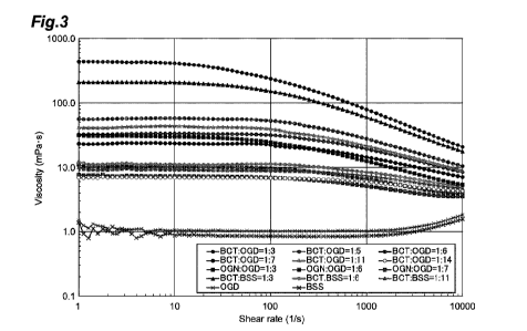
[Figure 3] Figure 3 is a graph showing results of measuring the
correlation between velocity gradient (shear rate) and viscosity under a
condition of 25 C as to vehicles shown in Table 13 in Example 1.
[Figure 4] Figure 4 is fluorescence microscope images showing results of
perfointing immunostaining on cell aggregates containing a transplant
neural retina with Crx and Chx10 in Reference Example 1.
[Figure 5] Figure 5 is fluorescence microscope images showing results of
perfointing immunostaining on cell aggregates containing a transplant
neural retina with Rx and Recoverin in Reference Example 1.
Date Recue/Date Received 2023-03-08
CA 03194364 2023-03-08
[Figure6] Figure 6 is a conceptual view of preparing a Cap and a Ring
from a typical cell aggregate.
[Figure 7] Figure 5 is a conceptual view of preparing a Cap and a Ring
from cell aggregates having various shapes. Portions indicated in black
color and gray color mean non-target tissue.
[Figure 8] Figure 6 shows images of typical grafts and a schematic view
of a graft as well as the heights, major axes and minor axes of grafts in
Reference Example 3.
[Figure 9] Figure 9 is confocal fluorescence microscope images showing
results of performing immunostaining on grafts with Crx, Chx10, Rx and
Recoverin in Reference Example 4.
[Figure 10A] Figure 10A shows results of analyzing gene expression for
RNA extracted from a Cap and a Ring by quantitative PCR in Reference
Example 5.
[Figure 10B] Figure 10B shows results of analyzing gene expression for
RNA extracted from a Cap and a Ring by quantitative PCR in Reference
Example 5.
[Figure 11] Figure 11 is images showing results of analyzing RNA
extracted from a Ring by quantitative PCR, then subretinally
transplanting a graft (cap) to a rat, and observing an image of post-
transplant engraftment under a fluorescence microscope in Reference
Example 6.
[Figure 12] Figure 12 is fluorescence microscope images showing results
of performing immunostaining on a Cap and a Ring prepared from one
cell aggregate in Reference Example 7.
[Figure 13] Figure 13 is fluorescence microscope images showing results
11
Date Recue/Date Received 2023-03-08
CA 03194364 2023-03-08
of performing immunostaining on a cap prepared from one cell aggregate
in Reference Example 8.
Description of Embodiments
[0014] 1. Vehicle for transplantation
The present invention provides a vehicle for transplantation that
is used in subretinally transplanting a graft of retinal tissue to a mammal.
[0015] The vehicle for transplantation of the present invention is not
limited as long as it is a vehicle having a viscosity of 5 to 500 mPa.s,
preferably 10 to 100 mPa.s, at a shear rate of 2 (1/s) at 25 C and
comprising hyaluronic acid and a pharmaceutically acceptable aqueous
liquid.
[0016] In the vehicle for transplantation of the present invention,
preferably, viscosity at a shear rate of 1000 (1/s) at 25 C is 100 mPa.s or
less, preferably 30 mPa.s or less, and the amount of change in viscosity
from shear rates of 1 to 10 (1/s) (i.e., the viscosity difference between
viscosity at a shear rate of 1 (1/s) and viscosity at a shear rate of 10
(1/s))
is 100 mPa.s or less, preferably 30 mPa.s or less. Alternatively, it is
preferable the rate of change in viscosity from shear rates of 1 to 10 (1/s)
(i.e., percentage when the viscosity difference between viscosity at a
shear rate of 1 (1/s) and viscosity at a shear rate of 10 (1/s) is divided by
viscosity at a shear rate of 1 (1/s)) should be about 10% or less.
[0017] The vehicle for transplantation of the present invention preferably
further comprises chondroitin sulfate. The hyaluronic acid and the
chondroitin sulfate function as thickening components of the vehicle for
transplantation.
12
Date Recue/Date Received 2023-03-08
CA 03194364 2023-03-08
[0018] In the specification, the hyaluronic acid is linear macromolecular
polysaccharide with a molecular weight of several tens of thousands to
several millions having a structure where D-glucuronic acid and D-N-
acetylglucosamine are alternately linked via 0-1,4 and 0-1,3 glycosidic
bonds. The hyaluronic acid is a substance that has high ability to retain
water and viscosity and is widely used in medicines, cosmetics, etc. In
the specification, the "hyaluronic acid" is meant to conceptually include
both a free form of hyaluronic acid and a salt thereof. Examples of the
salt of hyaluronic acid include alkali metal salts and alkaline earth metal
salts of hyaluronic acid. Specifically, commercially available sodium
hyaluronate, potassium hyaluronate, or the like can be used.
[0019] In the specification, the hyaluronic acid is not particularly limited
as long as the viscosity of the vehicle for transplantation at a shear rate of
2 (1/s) at 25 C can be kept at 5 to 500 mPa.s, preferably 10 to 100 mPa.s.
The vehicle for transplantation can be blended with hyaluronic acid
having an average molecular weight of about 500,000 to 3,900,000 at a
concentration of 0.15 w/v% to 1.50 w/v%, preferably 0.15 w/v% to 0.75
w/v%, more preferably 0.30 w/v% to 0.50 w/v%.
[0020] The chondroitin sulfate is mucopolysaccharide having a structure
where sulfuric acid is bonded to a sugar chain having repeats of two
saccharides D-glucuronic acid (GlcA) and N-acetyl-D-galactosamine
(GalNAc). In the specification, the "chondroitin sulfate" is meant to
conceptually include both a free form of chondroitin sulfate and a salt
thereof. Examples of the salt of chondroitin sulfate include alkali metal
salts and alkaline earth metal salts of chondroitin sulfate. Specifically,
commercially available chondroitin sulfate sodium salt, chondroitin
13
Date Recue/Date Received 2023-03-08
CA 03194364 2023-03-08
sulfate potassium salt, or the like can be used.
[0021] In the specification, the chondroitin sulfate is not particularly
limited as long as the viscosity of the vehicle for transplantation at a shear
rate of 2 (1/s) at 25 C can be kept at 5 to 500 mPa.s, preferably 10 to 100
mPa.s, together with hyaluronic acid. The vehicle for transplantation
can be blended with chondroitin sulfate having an average molecular
weight of about 20,000 to 24,000 at a concentration of 0.3 w/v% to 1.0
w/v%, preferably 0.4 w/v% to 0.7 w/v%.
[0022] The "pharmaceutically acceptable aqueous liquid" in the vehicle
of the present invention is not particularly limited as long as it is an
aqueous solution that can be administered to a living body and has
physical properties suitable for transplantation. For
example, an
aqueous solution containing a buffer, a transfusion, saline, injectable
water, or a perfusate may be used. A buffer is preferable.
[0023] In this context, examples of the physical properties suitable for
transplantation include pH and osmotic pressure. The pH of the vehicle
for transplantation is not particularly limited as long as it is in a neutral
range. The vehicle for transplantation of the present invention may be
adjusted to pH 6.5 to 8.0, preferably pH on the order of 7.0 to 7.5.
[0024] Examples of the osmotic pressure of the vehicle for
transplantation include hypotonic, isotonic, and hypertonic pressures. It
is preferable to be close to isotonic pressure. The osmotic pressure ratio
may be adjusted to 0.7 to 1.3, preferably 0.9 to 1.1.
[0025] Examples of the "pharmaceutically acceptable aqueous liquid"
specifically include aqueous solutions containing a balanced salt (i.e.,
balanced salt solutions). In the aqueous solution containing a balanced
14
Date Recue/Date Received 2023-03-08
CA 03194364 2023-03-08
salt, a buffer, a tonicity agent, a pH adjuster, an antioxidant, a chelating
agent, or the like can be appropriately selected and contained within a
range that does not influence the survival rate of retinal tissue to be
transplanted.
[0026] Examples of the buffer include phosphate buffers, borate buffers,
citrate buffers, tartrate buffers, acetate buffers, amino acids, and epsilon-
aminocaproic acid.
[0027] Examples of the tonicity agent include sugars such as sorbitol,
glucose, mannitol, polyhydric alcohols such as glycerin and propylene
glycol, salts such as sodium chloride, and boric acid.
[0028] Examples of the chelating agent include sodium edetate and citric
acid.
[0029] Examples of the pH adjuster include sodium hydroxide,
potassium hydroxide, sodium carbonate, sodium bicarbonate, boric acid
and salts thereof (borax), hydrochloric acid, citric acid and salts thereof
(sodium citrate, sodium dihydrogen citrate, etc.), phosphoric acid and
salts thereof (disodium hydrogen phosphate, potassium dihydrogen
phosphate, etc.), acetic acid and salts thereof (sodium acetate, ammonium
acetate, etc.), and tartaric acid and salts thereof (sodium tartrate, etc.).
[0030] Examples of the antioxidant include ascorbic acid, glutathione,
sodium bisulfite, dry sodium sulfite, sodium pyrosulfite, and tocopherol.
[0031] The vehicle for transplantation of the present invention can be
preferably subjected to sterilization treatment such as filter sterilization
using a membrane filter or the like.
[0032] Examples of the "pharmaceutically acceptable aqueous liquid"
specifically include aqueous solutions containing one or more
Date Recue/Date Received 2023-03-08
CA 03194364 2023-03-08
components selected from sugars such as glucose, inorganic salts such as
calcium chloride, sodium chloride, and magnesium sulfate, inorganic
materials such as sodium bicarbonate, sodium acetate, sodium citrate,
sodium dihydrogen phosphate, sodium hydrogen phosphate, anhydrous
sodium monohydrogen phosphate, and hydrochloric acid, and chelating
agents such as edetate (e.g., sodium edetate).
[0033] As hyaluronic acid or a salt thereof, a commercially available
aqueous solution of hyaluronic acid or a salt thereof can be used.
Specifically, examples thereof include Opegan(R) 0.6 ophthalmic
viscoelastic preparation containing 0.6% sodium hyaluronate, sodium
chloride, sodium dihydrogen phosphate and sodium hydrogen phosphate
as components, Opegan(R) 1.1 ophthalmic viscoelastic preparation
containing 1.1% sodium hyaluronate, sodium chloride, sodium
dihydrogen phosphate and sodium hydrogen phosphate as components,
and Hyalein Mini(R) ophthalmic solution 0.3% containing 0.3% sodium
hyaluronate, epsilon-aminocaproic acid, sodium edetate hydrate,
potassium chloride, sodium chloride and a pH adjuster as components.
[0034] For preparing the pharmaceutically acceptable aqueous liquid, a
liquid commercially available as an intraocular perfusate or lavage can be
used. As the intraocular perfusate or lavage, specifically, an aqueous
solution commercially available as Opeguard(R) containing 1.5 mg of
glucose, 0.18 mg of calcium chloride hydrate, 0.3 mg of magnesium
sulfate hydrate, 2.1 mg of sodium bicarbonate, and sodium citrate
hydrate, sodium acetate hydrate and hydrochloric acid as additives in 1
mL, or an oxyglutathione ocular perfusate or the like can be used. For
preparing the pharmaceutically acceptable aqueous liquid, Hank's
16
Date Recue/Date Received 2023-03-08
CA 03194364 2023-03-08
balanced salt solution (HBSS), Eagle balanced salt solution (EBSS),
phosphate-buffered saline (PBS), Dulbecco's phosphate-buffered saline
(DPBS), or the like can also be used.
[0035] An embodiment of the vehicle for transplantation of the present
invention includes a vehicle for transplantation containing only
hyaluronic acid as a thickening component. Specifically, it can be
prepared, for example, by blending Opegan(R) 0.6 ophthalmic
viscoelastic preparation with an aqueous liquid such as Opeguard at 1:1
to 1:3. In another embodiment, it can be prepared, for example, by
blending Hyalein Mini(R) ophthalmic solution 0.3% with an aqueous
liquid such as Opeguard at 3:1 to 1:1.
[0036] An embodiment of the vehicle for transplantation of the present
invention includes a vehicle for transplantation containing hyaluronic
acid and chondroitin sulfate as thickening components. In this context,
the concentrations of the hyaluronic acid and the chondroitin sulfate are
not particularly limited as long as the viscosity of the vehicle for
transplantation at a shear rate of 2 (1/s) at 25 C can be kept at 5 to 500
mPa.s, preferably 10 to 100 mPa.s. An embodiment of the vehicle for
transplantation of the present invention includes a composition for
transplantation containing 0.15 w/v% to 1.50 w/v% of hyaluronic acid
having an average molecular weight of 500,000 to 3,900,000, and 0.3
w/v% to 1.0 w/v% of chondroitin sulfate having a molecular weight of
20,000 to 24,000 or a salt thereof.
[0037] In an embodiment of the vehicle for transplantation of the present
invention, Viscoat(R) 0.5 ophthalmic viscoelastic preparation containing
sodium hyaluronate, chondroitin sulfate sodium salt, sodium dihydrogen
17
Date Recue/Date Received 2023-03-08
CA 03194364 2023-03-08
phosphate, sodium hydrogen phosphate and a tonicity agent as
components can be used. Specifically, it can be prepared, for example,
by blending Viscoat(R) 0.5 ophthalmic viscoelastic preparation with an
aqueous liquid such as Opeguard at 1:3 to 1:7.
[0038] The "phamiaceutically acceptable aqueous liquid" preferably
comprises neither an antimicrobial agent nor an antiseptic.
[0039] 2. Transplant retinal tissue
(Definition)
In the specification, the "tissue" refers to a structure of a cell
population having a structure where one or more types of cells differing
in morphology or properties are three-dimensionally arranged in a
predetermined pattern.
[0040] In the specification, the "retinal tissue" means a tissue in which
one or more types of retina cells, such as photoreceptor cells, horizontal
cells, bipolar cells, amacrine cells, retinal ganglion cells, Muller glial
cells, retinal pigment epithelial cells, their progenitor cells, or retinal
progenitor cells, constituting each retinal layer in a retina in vivo are
three-dimensionally arranged, preferably three-dimensionally arranged
in a layer pattern, and may be a cell aggregate or a cell sheet mentioned
later.
[0041] The photoreceptor progenitor cells, the horizontal progenitor cells,
the bipolar progenitor cells, the amacrine progenitor cells, the retinal
ganglion progenitor cells, the Muller glial progenitor cells, and the retinal
pigment epithelial progenitor cells refer to progenitor cells destined for
differentiation into photoreceptor cells, horizontal cells, bipolar cells,
amacrine cells, retinal ganglion cells, Muller glial cells, and retinal
18
Date Recue/Date Received 2023-03-08
CA 03194364 2023-03-08
pigment epithelial cells, respectively.
[0042] The "retinal progenitor cells" are progenitor cells capable of
differentiating into any one of the immature retinal cells such as
photoreceptor progenitor cells, horizontal progenitor cells, bipolar
progenitor cells, amacrine progenitor cells, retinal ganglion progenitor
cells, Muller glial cells, and retinal pigment epithelial progenitor cells,
and refer to progenitor cells also capable of eventually differentiating into
any one of the matured retinal cells such as photoreceptor cells, rod
photoreceptor cells, cone photoreceptor cells, horizontal cells, bipolar
cells, amacrine cells, retinal ganglion cells, and retinal pigment epithelial
cells.
[0043] In the specification, the "retinal layer" means a cell population in
a layer pattern in which one or more cells constituting the retina form a
single layer or a plurality of layers in a predetermined pattern, and
examples thereof specifically include retinal pigment epithelial layer
containing retinal pigment epithelial cells, and neural retinal layer
containing neural retina cells. The neural retina cells are also referred
to as retinal layer-specific neuronal cells.
[0044] Examples of the neural retinal layer include outer limiting
membrane, photoreceptor layer (outer nuclear layer), outer plexiform
layer, inner nuclear layer, inner plexifoim layer, ganglion cell layer, nerve
fiber layer and inner limiting membrane.
[0045] In the specification, the "neural retina" means retinal tissue
containing a neural retinal layer.
[0046] Examples of the neural retina cells include photoreceptor cells
(including photoreceptor progenitor cells and matured photoreceptor
19
Date Recue/Date Received 2023-03-08
CA 03194364 2023-03-08
cells), bipolar cells, retinal ganglion cells, amacrine cells, horizontal
cells,
Muller glial cells, and their progenitor cells.
[0047] In the specification, the "neural retinal progenitor cells" mean
progenitor cells of neural retina cells.
[0048] The "photoreceptor cells" are present in the photoreceptor layer
of a retina in vivo and plays a role in absorbing light stimuli and
converting them to electrical signals. The photoreceptor cells have two
types, cone cells which function in the light and rod cells which function
in the dark. Examples of the cone photoreceptor cells can include S
cone photoreceptor cells which express S-opsin and receive blue light, L
cone photoreceptor cells which express L-opsin and receive red light, and
M cone photoreceptor cells which express M-opsin and receive green
light. The photoreceptor cells are matured after differentiation from
photoreceptor progenitor cells. Whether or not cells are photoreceptor
cells or photoreceptor progenitor cells can be readily confirmed by those
skilled in the art, for example, through the expression of cell markers (Crx
and Blimpl expressed in photoreceptor progenitor cells, recoverin
expressed in photoreceptor cells, rhodopsin, S-opsin and MIL-opsin
expressed in mature photoreceptor cells, etc.) mentioned later or the
formation of an outer segment structure. In an embodiment, the
photoreceptor progenitor cells are Crx-positive cells, and the
photoreceptor cells are rhodopsin-, S-opsin- and MIL-opsin-positive
cells. In an embodiment, the rod photoreceptor cells are NRL- and
rhodopsin-positive cells. In an embodiment, the S cone photoreceptor
cells are S-opsin-positive cells, the L cone photoreceptor cells are L-
opsin-positive cells, and the M cone photoreceptor cells are M-opsin-
Date Recue/Date Received 2023-03-08
CA 03194364 2023-03-08
positive cells. Specifically, in the specification, the photoreceptor cells
conceptually include photoreceptor progenitor cells and matured
photoreceptor cells.
[0049] In the neural retina cells, neither retinal pigment epithelial cells
nor ciliary body cells are included.
[0050] In the specification, an embodiment of the retinal tissue includes
a neural retina, preferably a neural retina containing a retinal layer-
specific neuronal cell layer, more preferably a neural retina containing a
photoreceptor layer.
[0051] The neural retina contains preferably 10% or more, more
preferably 20% or more, of a photoreceptor cell or a photoreceptor
progenitor cell.
[0052] The neural retina contains preferably 10% or more, more
preferably 20% or more, of a neural retinal progenitor cell. Also, the
neural retina contains preferably 10% or more of a photoreceptor
progenitor cell. In an embodiment, the neural retina contains 3% or
more of a matured photoreceptor cell.
[0053] In the specification, an embodiment of the retinal tissue includes
retinal tissue containing both a neural retina and a cell layer containing
an RPE cell. Examples of the retinal tissue include neural retinas
covered with RPE cells described in W02019/050015.
[0054] In the specification, an embodiment of the retinal tissue includes
retinal tissue (complex) in which a neural retina and a RPE cell have
adhered via polymer hydrogel. Specifically, examples thereof include a
complex comprising a neural retina, a retinal pigment epithelial cell, and
hydrogel, wherein the neural retina and the retinal pigment epithelial cell
21
Date Recue/Date Received 2023-03-08
CA 03194364 2023-03-08
are each derived from a human pluripotent stem cell, a neural retinal layer
containing at least a photoreceptor layer is formed in the neural retina,
and the photoreceptor layer contains one or more cells selected from the
group consisting of a photoreceptor cell, a photoreceptor progenitor cell
and a retinal progenitor cells. In this context, the hydrogel is not
particularly limited as long as it is a polymer and a substance having
physical properties useful in the adhesion between a tissue and a tissue
(examples thereof include glues, adhesives, matter having gelling
physical properties, gelatin, and oil and fat).
[0055] Each cell constituting the retinal tissue mentioned above can be
detected or identified by using a retina cell marker that is expressed
(positive) or not expressed (negative) therein.
[0056] Examples of the retina cell marker include Rx (also referred to as
Rax) and PAX6 expressed in retinal progenitor cells, Rx, PAX6 and
Chx10 (also referred to as Vsx2) expressed in neural retinal progenitor
cells, or Crx and Blimpl expressed in photoreceptor progenitor cells.
Examples of the negative marker for retinal progenitor cells or retina cells
include Nloc2.1 and SOX1.
[0057] Examples of the marker for retinal layer-specific neuronal cells
include recoverin expressed in photoreceptor cells (particularly, matured
photoreceptor cells), Crx and Blimpl expressed in photoreceptor cells
(particularly, photoreceptor progenitor cells), rhodopsin expressed in rod
cells, Nrl expressed in rod photoreceptor cells and rod photoreceptor
progenitor cells, S-opsin and LM-opsin expressed in cone photoreceptor
cells, RXR-y expressed in cone cells, cone photoreceptor progenitor cells
and ganglion cells, TR132, OTX2 and 0C2 expressed in cone
22
Date Recue/Date Received 2023-03-08
CA 03194364 2023-03-08
photoreceptor cells that appear at the early phase of differentiation among
cone photoreceptor cells, or progenitor cells thereof, Chx10, PKCa, Goa,
VSX1 and L7 expressed in bipolar cells, Tun and Brn3 expressed in
retinal ganglion cells, calretinin and HPC-1 expressed in amacrine cells,
calbindin expressed in horizontal cells, and Pax6 commonly expressed in
horizontal cells, amacrine cells and ganglion cells.
[0058] A layer that has a low appearance ratio of photoreceptor cells or
photoreceptor progenitor cells, is rich in neural retinal progenitor cells,
and is at a stage of differentiation before forming a photoreceptor layer is
referred to as "neuroblastic layer" and includes inner neuroblastic layer
and outer neuroblastic layer. Those skilled in the art can make a
judgment from the shade of color (the outer neuroblastic layer is light,
and the inner neuroblastic layer is dark) by a known method, for example,
under a bright field microscope.
[0059] The presence or absence of expression of the retina cell marker,
or the ratio of retina cell marker-positive cells in a cell population or a
tissue can be readily confirmed by those skilled in the art. Examples
thereof include an approach using an antibody, an approach using nucleic
acid primers, and an approach using sequencing reaction. As the
approach using an antibody, the expression of a protein of the retina cell
marker can be confirmed, for example, by dividing the number of
predetermined retina cell marker-positive cells by the total number of
cells in accordance with an approach such as flow cytometry or
immuno staining using a commercially available antibody. As the
approach using nucleic acid primers, the expression of RNA of the retina
cell marker can be confirmed by, for example, PCR, semiquantitative
23
Date Recue/Date Received 2023-03-08
CA 03194364 2023-03-08
PCR, or quantitative PCR (e.g., real-time PCR). As the approach using
sequencing reaction, the expression of RNA of the retina cell marker can
be confirmed using, for example, a nucleic acid sequencer (e.g., next-
generation sequencer).
[0060] The "positive cells" mean cells expressing a predetermined
marker on the cell surfaces or within the cells. For example, the
"Chx10-positive cells" mean cells expressing Chx10 protein.
[0061] The "negative cells" mean cells that do not express a
predetermined marker on the cell surfaces or. For example, the "SOX1-
negative cells" mean cells that do not express SOX1 protein. In this
context, "not express" encompasses the case where the expression level
thereof is below a detection limit, or the case where the expression is 1/10
or less, preferably 1/50 or less, as compared with positive cells.
[0062] In another embodiment, for example, the "SOX1-negative cells"
mean cells that do not express mRNA of SOX1. In this context, "not
express" encompasses the case where the expression level thereof is
below a detection limit, or the case where the expression is 1/10 or less,
preferably 1/50 or less, as compared with positive cells. Particularly, in
the case of measuring a mRNA level by qPCR, delta between a target
gene (e.g., SOX1) and an internal standard (GAPDH or ACTB) can be
evaluated as a ACt value. The term "not express" encompasses the case
where the ACt value is 4 or more, more preferably 6 or more, further
preferably 8 or more.
[0063] The "retinal pigment epithelial cells" mean epithelial cells present
outside the neural retina in a retina in vivo. Whether or not cells are
retinal pigment epithelial cells can be readily confirmed by those skilled
24
Date Recue/Date Received 2023-03-08
CA 03194364 2023-03-08
in the art, for example, through the expression of cell markers (RPE65,
MITF, CRALBP, MERTK, BEST1, TTR, etc.), the presence of melanin
granules (brown-black), intercellular tight junctions, or
polygonal/flagstone-like characteristic cell morphology. Whether or
not cells have a function of retinal pigment epithelial cells can be readily
confirmed from the ability to secrete cytokines such as VEGF and PEDF.
In an embodiment, the retinal pigment epithelial cells are RPE65-positive
cells, MITF-positive cells, or RPE65-positive and MITF-positive cells.
[0064] The "ciliary body" includes "ciliary body" and "ciliary marginal
zone" in the process of development and of an adult. Examples of a
marker of the "ciliary body" include Zicl, MAL, I-INF lbeta, FoxQ 1 ,
CLDN2, CLDN1, GPR177, AQP1 and AQP4. Examples of the "ciliary
marginal zone (CMZ)" can include a tissue that is present in a boundary
region between the neural retina and the retinal pigment epithelium in a
retina in vivo, and is a region containing tissue stem cells of the retina
(retinal stem cells). The ciliary marginal zone is also called ciliary
margin or retinal margin, and the ciliary marginal zone, the ciliary margin
and the retinal margin are equivalent tissues. The ciliary marginal zone
is known to play an important role in the supply of retinal progenitor cells
or differentiated cells to retinal tissue, the maintenance of a retinal tissue
structure, etc. Examples of a marker gene of the ciliary marginal zone
can include Rdh10 gene (positive), Otx 1 gene (positive) and Zic 1
(positive). The "ciliary marginal zone-like structure" is a structure
similar to the ciliary marginal zone.
[0065] (Structure of retinal tissue)
In the specification, the retinal tissue may be a form having a
Date Recue/Date Received 2023-03-08
CA 03194364 2023-03-08
three-dimensional structure, i.e., a cell aggregate (also referred to as a
cell
cluster, stereoscopic tissue, or organoid), or may be a cell sheet in which
a structure in a layer pattern is two-dimensionally expanded, i.e., a retinal
cell sheet. In the cell aggregate or the cell cluster, a sphere is also
included. In this context, the sphere means a cell aggregate having a
stereoscopic shape close to a spherical shape. The stereoscopic shape
close to a spherical shape is a shape having a three-dimensional structure,
and examples thereof include a spherical shape that exhibits a circle or an
ellipse when projected onto a two-dimensional surface, and a shape
formed by fusing a plurality of spherical shapes (e.g., which exhibits a
shape formed by 2 to 4 circles or ellipses overlapping when two-
dimensionally projected). In an embodiment, the core part of the
aggregate has a vesicular lamellar structure and is characterized in that
the central part is observed to be dark and the outer edge portion is
observed to be bright under a bright field microscope.
[0066] In an embodiment, some or all cells contained in the cell
aggregate or the cell sheet mutually adhere. Specifically, in a portion or
the whole of the aggregate, cells may mutually form cell-cell junction or
cell adhesion, for example, adherence junction.
[0067] The shape and size of the cell aggregate or the cell sheet are not
particularly limited and can be appropriately set according to an area in
need of repair of retinal tissue in the case of being administered as a graft
to a mammal, preferably a monkey or a human. Specifically, the size of
the retinal tissue to be transplanted to a recipient is a size suitable for
the
recipient and is a size that permits movement inside a cell suction portion
(needle tube for transplantation) in a device that is used in transplantation.
26
Date Recue/Date Received 2023-03-08
CA 03194364 2023-03-08
[0068] In the specification, an embodiment of the retinal tissue contained
in a composition for transplantation specifically includes retinal tissue
that is from 200 to 1500 iiim in minor axis, from 600 to 2500 iiim or less,
and from 100 iiim to 1000 in thickness. The retinal tissue of size suitable
for transplantation, i.e., a graft, can be prepared by appropriately
dissecting the size suitable for transplantation from the cell aggregate or
the cell sheet.
[0069] Specifically, a cell sheet of size suitable for transplantation can be
dissected from the cell aggregate and used as a graft.
[0070] In the specification, the retinal tissue may have an epithelial
structure. The epithelial structure is formed by covering the surface of
a tissue with cells without any space, and polarized to have an "apical
surface" and a "basal membrane (basal surfacer In this context, the
"basal membrane" is a layer that is rich in laminin and IV-type collagen
and is 50 to 100 nm. The "apical surface" refers to the surface (upper
surface layer) fonned on the opposite side to the "basal membrane". In
an embodiment, in the retinal tissue developed to the extent that
photoreceptor cells or photoreceptor progenitor cells are observed, the
"apical surface" refers to a surface in contact with photoreceptor layer
(outer nuclear layer) in which outer limiting membrane is formed and
photoreceptor cells and photoreceptor progenitor cells are present. Such
an apical surface can be identified by, for example, immunostaining
(known to those skilled in the art) using an antibody against an apical
surface marker (e.g., atypical PKC (hereinafter, abbreviated to "aPKC"),
E-cadherin, N-cadherin).
[0071] Cells constituting the epithelial structure, i.e., epithelial cells,
can
27
Date Recue/Date Received 2023-03-08
CA 03194364 2023-03-08
mutually and firmly join via adherence junction and/or tight junction to
form a layer of the cells. A tissue formed from a single layer or dozen
layers overlapping of this layer of the cells is the epithelial structure.
[0072] In the specification, an epithelial structure containing neural
tissue is referred to as neural epithelium. Particularly, an epithelial
structure containing a neural retina is referred to as neural retinal
epithelium.
[0073] In the specification, in an embodiment, the neural retina may
assume a polarized layer structure. For example, the neural retina, when
having a shape of a cell sheet, may have apical/basal polarity. When the
neural retina is a sphere-like cell aggregate, the cell aggregate may have
an epithelial structure, and the epithelial structure may have apical/basal
polarity in the surface and the inside of the cell aggregate.
[0074] In the specification, one embodiment of the retinal tissue
contained in a composition for transplantation includes retinal tissue
containing a front surface (apical surface) and a back surface (basal
surface), wherein the front surface constitutes an apical surface
containing a neural retinal layer which is epithelial tissue by forming the
adherence junction between cells, and the back surface constitutes a basal
surface adjacent to the inner layer of the neural retina. Such retinal
tissue can be referred to as neural retinal epithelium. For such retinal
tissue, it is preferable to be a neural retina sheet which is a sheet-shaped
retinal tissue. The front surface has a smooth shape with less change in
curvature, and the back surface has an irregular shape with large change
in curvature. In an embodiment, the change in the curvature of the front
surface of the retinal tissue may be, for example, close to change in
28
Date Recue/Date Received 2023-03-08
CA 03194364 2023-03-08
curvature of an ellipse (e.g., an ellipse having a major axis of 1 to 10 with
respect to a minor axis of 1) (also referred to as continuous change in
curvature). In an embodiment, the change in the curvature of the back
surface of the retinal tissue may be, for example, close to sharp change in
curvature that goes back and force between positive values and negative
values, as in "teeth of a saw" (also referred to as sharp change in
curvature).
[0075] In the specification, the retinal tissue is preferably retinal tissue
having a continuous epithelial structure. The "continuous epithelial
structure" is a structure where the epithelial tissue is continuously formed,
and is also referred to as "continuous epithelium". The epithelium tissue
continuously formed is a state in which 10 cells to 107 cells, for example,
in the tangent direction of the epithelial tissue, preferably 30 cells to 107
cells, further preferably 102 cells to 107 cells, in the tangent direction,
are
aligned. The continuous epithelial structure does not have a structure
where an apical surface is divided, as found in a rosette-like structure.
In an embodiment, the number of cells per area of the cross section of
retinal tissue having the continuous epithelial structure is 10 cells to 900
cells, preferably 30 cells to 300 cells, more preferably 50 cells to 250
cells, still more preferably 75 cells to 160 cells, per 100 um2, for example,
in the case of evaluating the number of nuclei of cells in a frozen section
having a thickness on the order of 10 iiim.
[0076] For example, in the continuous epithelium formed in retinal
tissue, the retinal tissue has an apical surface intrinsic to the epithelial
tissue. The apical surface is formed almost in parallel to, for example,
at least photoreceptor layer (outer nuclear layer) among the layers
29
Date Recue/Date Received 2023-03-08
CA 03194364 2023-03-08
forming a neural retinal layer and continuously on the surface of the
retinal tissue. For example, in the case of a cell aggregate containing
retinal tissue prepared from pluripotent stem cells, the apical surface is
formed on the surface of the aggregate, and continuous neural epithelium
is formed in which 10 cells or more, preferably 30 cells or more, more
preferably 100 cells or more, further preferably 400 cells or more of
photoreceptor cells or photoreceptor progenitor cells are regularly and
continuously aligned in the tangent direction of the surface. A neural
retina containing such continuous neural epithelium is neural retinal
epithelium containing continuous epithelium.
[0077] Whether retinal tissue contains continuous epithelium can be
confirmed from the continuity (i.e., undivided form) of the apical surface
of retinal tissue. The continuity of the apical surface can be determined,
for example, by immunostaining a marker of the apical surface (e.g.,
aPKC, E-cadherin, N-cadherin) and a marker of photoreceptor cells or
photoreceptor progenitor cells positioned on the apical surface side (e.g.,
Crx or recoverin), and analyzing obtained images, etc. for the positional
relationship of the apical surface to a photoreceptor layer and each retinal
layer. A retinal layer other than the apical surface or the photoreceptor
layer (outer nuclear layer) can be identified by, for example, DAPI
staining which stains the nuclei of cells, PI staining, Hoechst staining, or
immunostaining with a marker protein (Rx, Chx10, Ki67, Crx, etc.) or
the like localized in the nuclei of cells.
[0078] Specifically, the continuity of the apical surface can be identified
from the continuous presence of cells co-expressing a marker of cells
present on the apical surface side, i.e., a photoreceptor cell marker or a
Date Recue/Date Received 2023-03-08
CA 03194364 2023-03-08
photoreceptor progenitor cell marker, and a marker capable of staining
the nuclei of cells.
[0079] In the specification, the retinal tissue preferably includes a neural
retina containing a photoreceptor cell or a photoreceptor progenitor cell
and in other words, a neural retina containing a photoreceptor layer.
[0080] In the specification, the retinal tissue preferably includes a neural
retina in which a neural retinal layer or a photoreceptor layer has a
continuous epithelial structure, i.e., has a continuous neuroepithelial
structure.
[0081] In the specification, the retinal tissue may contain an "inner layer"
containing a ganglion cell or an amacrine cell inside a photoreceptor
layer. The inner layer may be in contact with the basal surface.
[0082] These neural retinas can be obtained by producing a cell
aggregate containing a neural retina by a production method mentioned
later.
[0083] (Cell aggregate containing neural retina)
In an embodiment, the cell aggregate containing a neural retina is
a sphere-like cell aggregate. In an embodiment, in the cell aggregate
containing a neural retina, a plurality of neural retinas may be present
with an overlap (e.g., see conceptual views (1) and (2) in Figure 7). In
an embodiment, the cell aggregate containing a neural retina contains first
epithelial tissue (target epithelial tissue) containing the transplant neural
retina, and second epithelial tissue (non-target epithelial tissue) having
the continuity of the slope of a tangent line to a surface different from the
continuity of the slope of a tangent line to the surface of the first
epithelial
tissue, and containing a non-neural retina-related cell. In this context,
31
Date Recue/Date Received 2023-03-08
CA 03194364 2023-03-08
the first epithelial tissue refers to epithelial tissue that does not
substantially contain a non-neural retina-related cell (non-target cell) and
allows the transplant neural retina to be dissected. On the other hand,
the second epithelial tissue is epithelial tissue that may contain a neural
retina, but is ineligible for dissecting the transplant neural retina because
of containing non-target cells. In
another embodiment, the cell
aggregate containing a neural retina contains only the first epithelial
tissue (target epithelial tissue) containing the transplant neural retina and
does not contain non-target epithelial tissue.
[0084] (Transplant retinal tissue)
The transplant retinal tissue is the retinal tissue described in the
specification and is human retinal tissue suitable for transplantation in
humans. It is preferably a neural retina and more preferably consists of
only the neural retina.
[0085] The transplant retinal tissue can be prepared by dissecting a site
suitable for transplantation from the cell aggregate mentioned above. In
an embodiment, the transplant retinal tissue can be prepared by dissecting
a neural retina from a cell aggregate containing the neural retina. When
the retinal tissue, preferably the neural retina, contains continuous
epithelium, a sheet-shaped retinal tissue (hereinafter, also referred to as a
retina sheet), preferably a sheet-shaped neural retina (hereinafter, also
referred to as a neural retina sheet), containing the continuous epithelium
can be dissected.
[0086] The transplant neural retina contains at least a photoreceptor layer.
The photoreceptor layer is fonned at least in the outmost of the cell
aggregate. Also, photoreceptor cells or photoreceptor progenitor cells
32
Date Recue/Date Received 2023-03-08
CA 03194364 2023-03-08
may be present in the inside. Alternatively, the photoreceptor layer may
be formed in the inside.
Photoreceptor cells, etc. are present
continuously, i.e., by mutual adhesion, in the tangent direction of the
surface of the cell aggregate. The photoreceptor cells, etc. are present
continuously in the tangent direction of the surface of the cell aggregate,
thereby forming a photoreceptor layer containing the photoreceptor cells,
etc. The tangent direction refers to a direction tangent to the surface of
the cell aggregate, i.e., a direction along which the photoreceptor cells,
etc. in the photoreceptor layer are arranged, and is the direction in parallel
to the neural retina or the lateral direction. The slope of a tangent line
to the surface of epithelial tissue refers to a direction along which cells
are arranged when individual cells in the epithelial tissue are arranged in
a predetermined direction, and refers to the direction in parallel to the
epithelial tissue (or epithelial sheet) or the lateral direction.
[0087] The cell aggregate that is used for preparing the transplant neural
retina may contain non-target tissue other than a neural retina.
Examples of the non-target tissue include epithelial tissue other than a
neural retina, i.e., second epithelial tissue containing non-target epithelial
tissue. Examples of the second epithelial tissue include eyeball-related
tissue and brain and spinal cord tissue. The eyeball-related tissue means
a non-neural retinal tissue surrounding eyeball tissue, and examples
thereof include retinal pigment epithelial cells, ciliary body (e.g., ciliary
marginal zone), lens, and cornea. The brain and spinal cord tissue
means neural tissue of the brain and the spinal cord, and examples thereof
include the forebrain, the telencephalon, the cerebrum, the diencephalon,
the hypothalamus, the midbrain, the hindbrain, the cerebellum, and the
33
Date Recue/Date Received 2023-03-08
CA 03194364 2023-03-08
spinal cord. In an embodiment, the brain and spinal cord tissue may
contain pituitary gland.
[0088] One example of the cell aggregate containing the first epithelial
tissue and the second epithelial tissue includes cell aggregates shown in
a conceptual view of Figure 6 and conceptual views (3) and (5) of Figure
7. The conceptual view of Figure 6 shows one example of a cell
aggregate in which eyeball-related tissue (retinal pigment epithelial cells,
ciliary body) (black portion of Figure 6) is present as the second epithelial
tissue in a part of a neural retina which is the first epithelial tissue. The
conceptual view (3) of Figure 7 shows one example of a cell aggregate in
which eyeball-related tissue (retinal pigment epithelial cells, ciliary body)
(black portion of the conceptual view (3) of Figure 7) is further present
as the second epithelial tissue when a plurality of neural retinas are
present with an overlap (e.g., conceptual views (1) and (2) of Figure 7).
The conceptual view (5) of Figure 7 shows one example of a cell
aggregate in which brain and spinal cord tissue (cerebrum, etc.) (gray
portion of the conceptual view of Figure 7(5)) is present as the second
epithelial tissue. As shown in the conceptual view (4) of Figure 7, non-
target tissue may be contained inside the cell aggregate containing a
transplant neural retina. This case does not apply to the definition
"having the continuity of the slope of a tangent line to a surface different
from the continuity of the slope of a tangent line to the surface of the first
epithelial tissue", and therefore does not apply to the second epithelial
tissue. It is preferable that the transplant neural retina and the sample
for quality evaluation should be selected from a cell aggregate that does
not contain non-target tissue in the inside.
34
Date Recue/Date Received 2023-03-08
CA 03194364 2023-03-08
[0089] <Sampling step>
In the case of dissecting a transplant neural retina from a cell
aggregate, it is desirable to use containing a marker-positive cell of a
photoreceptor cell or a photoreceptor progenitor cell as an index. In the
case of dissecting a transplant neural retina from a cell aggregate, it is
desirable to use containing a marker-positive cell of a retinal progenitor
cell or a neural retinal progenitor cell as an index. It is also desirable to
use not containing a marker-positive cell of a non-target cell as an index.
[0090] For obtaining a transplant neural retina that contains a marker-
positive cell of a photoreceptor cell or a photoreceptor progenitor cell and
contains a marker-positive cell of a non-target cell below a certain level
(or is negative to a non-target cell marker), it is desirable to evaluate the
quality of the transplant neural retina in advance.
[0091] The quality of the transplant neural retina can be evaluated, for
example, by sampling a part or the whole of a cell aggregate containing
a neural retina having an epithelial structure derived from a pluripotent
stem cell as a sample for quality evaluation (hereinafter, referred to as "
sampling step"), and detecting a marker expressed in the obtained sample
by a method known to those skilled in the art. The sampling of a part of
the cell aggregate as the sample for quality evaluation means selecting
some (one or more) cell aggregates, or all cell aggregates from among a
plurality of cell aggregates, and isolating (e.g., dissecting) a portion of
the
selected cell aggregates as the sample for evaluation using tweezers,
scissors and/or a knife, etc. The sampling of the whole of the cell
aggregate as the sample for quality evaluation means selecting some (one
or more) cell aggregates from among a plurality of cell aggregates, and
Date Recue/Date Received 2023-03-08
CA 03194364 2023-03-08
separately picking up the whole of the selected one or more cell
aggregates as the sample for quality evaluation. In the case of selecting
one or more cell aggregates from among a plurality of cell aggregate,
random sampling is preferable. In the specification, the cell aggregate
in the case of sampling a part of the cell aggregate as the sample for
quality evaluation is referred to as "cell aggregate containing the sample
for quality evaluation", and the cell aggregate in the case of sampling the
whole ofthe cell aggregate as the sample for quality evaluation is referred
to as "cell aggregate of the sample for quality evaluation".
[0092] In an embodiment, the sample for quality evaluation is a part of a
cell aggregate containing a neural retina having an epithelial structure
derived from a pluripotent stem cell. By sampling a part of the cell
aggregate as the sample for quality evaluation, there is an advantage that
a neural retina contained in the remaining portion can be used in
transplantation without completely destroying the cell aggregate.
Specifically, provided that the sample for quality evaluation which is a
part of the cell aggregate is determined as being accepted by a
determination step mentioned later, a neural retina having an epithelial
structure in the cell aggregate containing the sample for quality
evaluation is regarded as being applicable as the transplant neural retina
and can be used in transplantation.
[0093] In the cell aggregate, it can be determined that a site having a
continuous epithelium structure where an outer neuroblastic layer and an
inner neuroblastic layer appear to be divided as two layers is the neural
retina. On the other hand, eyeball-related tissue as the second epithelial
tissue, particularly, retinal pigment epithelial cells, assume black color
36
Date Recue/Date Received 2023-03-08
CA 03194364 2023-03-08
visually or under a microscope and therefore, can readily be distinguished
from the neural retina by those skilled in the art. Also, brain and spinal
cord tissue as the second epithelial tissue, visually or under a microscope,
cannot be confirmed to have a continuous epithelium structure, which is
a morphological feature, on the surface of the cell aggregate, cannot be
confirmed to have morphological features intrinsic to the neural retina,
and/or appears to have a dull color, and thus, can readily be distinguished
from the neural retina by those skilled in the art by focusing thereon.
Thus, those skilled in the art can isolate the transplant neural retina and
the sample for quality evaluation from the first epithelial tissue containing
the neural retina even in a cell aggregate containing the second epithelial
tissue.
[0094] As mentioned above, in an embodiment, the sample for quality
evaluation is set and sampled depending on a predetermined positional
relationship with the transplant neural retina or a candidate of the
transplant neural retina. In other words, a region to be dissected as the
sample for quality evaluation can be fixed by the setting of the transplant
neural retina or its candidate. In this context, in an embodiment, the
transplant neural retina (also referred to as a graft or a cap) and its
candidate can be defined by the position in the cell aggregate mentioned
above (e.g., being the center and/or its neighborhood of the epithelial
tissue (continuous epithelial tissue), and in the case of having the second
epithelial tissue, being a region on the first epithelial tissue most distant
from the second epithelial tissue), and a size described in (Transplant
neural retina sheet) mentioned later, etc. Thus, those skilled in the art
can set a neural retina having these features as the transplant neural retina
37
Date Recue/Date Received 2023-03-08
CA 03194364 2023-03-08
or its candidate.
In the case of sampling the transplant neural retina and the sample
for quality evaluation from the same cell aggregate, the sample for quality
evaluation (also referred to as a ring) can be set as a region continuous or
adjacent at least partially to the transplant neural retina set as mentioned
above, and a region as narrow as possible within a range that permits
quality evaluation, by those skilled in the art. In the case of sampling a
part of one or more cell aggregates among the cell aggregates of the same
lot as the sample for quality evaluation, the sample for quality evaluation
can be sampled as the transplant neural retina mentioned above or its
candidate portion in the cell aggregates by those skilled in the art. In
this case, a size to be dissected as the sample for quality evaluation may
be a size described in (Transplant neural retina sheet) mentioned later, or
may be smaller. Thus, the sample for quality evaluation can be set and
sampled depending on the positional relationship with the transplant
neural retina or its candidate and the size.
[0095] <Detection step>
The method for evaluating the quality of a transplant neural retina
according to the present invention comprises detecting the expression of
a neural retina-related cell-related gene and a non-neural retina-related
cell-related gene (non-target cell-related gene) in the sample for quality
evaluation (detection step). It is preferable for the detection step to
quantitatively detect the expression levels of the genes. The non-target
cell-related gene comprises one or more genes selected from the group
consisting of brain and spinal cord tissue marker gene and eyeball-related
tissue marker gene.
38
Date Recue/Date Received 2023-03-08
CA 03194364 2023-03-08
[0096] (Neural retina-related cell-related gene)
The neural retina-related cell-related gene (target cell-related
gene) means a gene expressed by neural retina-related cells. As the
neural retina-related cell-related gene, a gene highly expressed in
photoreceptor cells (rod photoreceptor cell, cone photoreceptor cell),
horizontal cells, amacrine cells, intermediate neuronal cells, retinal
ganglion cells (ganglion cell), bipolar cells (rod bipolar cell, cone bipolar
cell), Muller glial cells, or progenitor cells of these cells, neural retinal
progenitor cells, or the like as compared with non-target cells is preferable.
Examples of the neural retina-related cell-related gene include the neural
retina-related cell markers described above, and RAX, Chx10, SIX3,
SIX6, RCVRN, CRX, NRL and NESTIN are preferable. GenBank IDs
of the neural retina-related cell markers are shown in Table 1 below.
[Table 1]
Gene name GenBank ID
RAX NM 013435.2
Chx10 NM 182894.2
SIX3 NM 005413.4
SIX6 NM 007374.2
RCVRN NM 002903.2
CRX NM 000554.6
NRL NM 006177.4
NESTIN NM 006617.2
[0097] The neural retina-related cell-related gene is preferably the gene
described in Table 1, though not limited thereto. Other examples of the
neural retina-related cell-related gene include Rax2, Vsxl, Blimp 1,
RXRG, S-opsin, M/L-opsin, rhodopsin, Brn3, and L7.
39
Date Recue/Date Received 2023-03-08
CA 03194364 2023-03-08
[0098] (Non-neural retina-related cell-related gene)
Identification can be performed by detecting a gene (hereinafter,
referred to as non-neural retina-related cell-related gene or non-target
cell-related gene) expressed in a non-target cell induced as a by-product
in the process of producing the cell aggregate containing a neural retina
as a medicine raw material, or a cell or tissue haying the possibility that
the non-target cell is produced as a by-product.
[0099] In an embodiment, examples of the non-neural retina-related cell-
related gene (non-target cell-related gene) include brain and spinal cord
tissue marker gene and eyeball-related tissue marker gene. In an
embodiment, as the non-neural retina-related cell-related gene,
undifferentiated iPS cell marker gene may be contained.
[0100] In an embodiment, the brain and spinal cord tissue marker gene
may be one or more genes selected from the group consisting of
telencephalon marker gene, diencephalon/midbrain marker gene and
spinal cord marker gene. The diencephalon/midbrain marker gene may
be one or more genes selected from the group consisting of diencephalon
marker gene, midbrain marker gene, and hypothalamus marker gene
regarding the hypothalamus which is a part of the diencephalon.
[0101] In an embodiment, the eyeball-related tissue marker gene may be
one or more genes selected from the group consisting of optic stalk
marker gene, ciliary body marker gene, lens marker gene and retinal
pigment epithelium marker gene.
[0102] The telencephalon marker gene means a gene expressed in the
telencephalon. The telencephalon marker gene may comprise one or
more genes selected from the group consisting ofFoxG1 (also called Bfl),
Date Recue/Date Received 2023-03-08
CA 03194364 2023-03-08
Emx2, Dlx2, Dlx1 and Dlx5. GenBank IDs of the telencephalon marker
genes are shown in Table 2 below.
[Table 2]
Gene name GenBank ID
FOXG1 NM 005249.4
NM 004098.4
Emx2
NM 001165924.1
DLX2 NM 004405.4
NM 178120.5
DLX1
NM 001038493.1
NM 005221.6
DLX5 XM 005250185.3
XM 017011803.1
[0103] The telencephalon marker gene is preferably the gene described
in Table 2, though not limited thereto. Other
examples of the
telencephalon marker gene include Emxl , LHX2, LHX6, LHX7, and
Gsh2.
[0104] The diencephalon/midbrain marker gene means a gene expressed
in the diencephalon and/or the midbrain. The diencephalon/midbrain
marker gene may comprise one or more genes selected from the group
consisting of OTX1, OTX2 and DMBX1. GenBank IDs of the
diencephalon/midbrain marker genes are shown in Table 3 below. The
diencephalon/midbrain marker gene may comprise a hypothalamus
marker mentioned later regarding the hypothalamus which is a region of
the diencephalon. In other words, the diencephalon/midbrain marker
gene may comprise one or more genes selected from the group consisting
41
Date Recue/Date Received 2023-03-08
CA 03194364 2023-03-08
of OTX1, OTX2, OTX2, DMBX1, Rx, Nkx2.1, OTP, FGFR2, EFNA5
and GAD1.
[Table 3]
Gene name Gen Bank ID
NM 001199770.1
OTX1
NM 014562.4
NM 001270523.1
NM 001270524.1
OTX2 NM 001270525.1
NM 021728.3
NM 172337.2
NM 172225.1
NM 147192.2
DMBX1
XM 011540668.2
XM 017000289.1
[0105] The hypothalamus marker gene means a gene expressed in the
hypothalamus. The hypothalamus marker gene may comprise one or
more genes selected from the group consisting of Rx, Nkx2.1, Dmbxl,
OTP, gadl, FGFR2 and EFNA5. GenBank IDs of the hypothalamus
marker gene are shown in Table 4 below.
[Table 4]
42
Date Recue/Date Received 2023-03-08
CA 03194364 2023-03-08
Gene name Gen Bank ID
Rx NM 013435.2
Nkx2.1 NM 3, 003317 NM _001079668.2
=
OTP NM 032109.2
gad1 NM 3, 000817 NM _013445.3
=
XM 005246444.3, XM_011510922.1
XM 017003756.1, XM_017003758.2
XM 2, 017003757 XM
_024452783.1
=
FGFR2 NM 022970.3, NM 000141.4
NM 023029.2, NM_001144913.1
NM 001144914.1, NM_001144915.1
NM 001144916.1, NM_001144917.1
NM 001144918.1, NM_001144919.1
NM 1, 001320654 NM
_001320658.1
=
NR_073009.1, XM_006717708.3
XM 006717710.4, XM_017015920.2
XM 017015921.2, XM_017015924.2
XM 017015925.2, XM_024447888.1
XM 024447887.1, XM_024447890.1
XM 1, 024447889 XM
_024447892.1
=
XM 024447891.1
EFNA5 NM 001962.3, XM 006714565.3
XM 3, 011543250 XM
_011543251.2
=
XM 017009205.1
[0106] The spinal cord marker gene means a gene expressed in the spinal
cord. The spinal cord marker gene may comprise one or more genes
selected from the group consisting of HoxB2, HoxA5, HOXC5, HOXD1,
HOXD3 and HOXD4. GenBank IDs of the spinal cord marker gene are
shown in Table 5 below.
43
Date Recue/Date Received 2023-03-08
CA 03194364 2023-03-08
[Table 5]
Gene name Gen Bank ID
NM 002145.3
HOXB2
XM 005257275.4
HOXA5 NM 019102.4
NM 018953.3
HOXC5
NR_003084.2
HOXD1 NM 024501.3
NM 006898.4
XM 005246509.4
XM 005246511.4
HOXD3
XM 005246513.5
XM 011511065.3
XM 011511066.3
NM 014621.3
HOXD4
XM 005246514.4
[0107] The spinal cord marker gene is preferably the gene described in
Table 5, though not limited thereto. Other examples of the spinal cord
marker gene include a gene group forming the Hox cluster.
[0108] Meanwhile, in an embodiment, in the case of using retinoids
(examples thereof include retinoic acid, retinal, retinol, all-trans-retinoic
acid, and 11-cis-retinoic acid) in a production step, the expression of
HOX gene (e.g., HOXC5, HOXA5 and HOXB2) may be found even if a
good product of retinal tissue is produced. It is considered that the
expression of the HOX gene is regulated by retinoic acid signals, and the
HOX gene expression increases to an extent that does not influence
differentiation into retinal tissue. This effect of the retinoic acid signals
44
Date Recue/Date Received 2023-03-08
CA 03194364 2023-03-08
is considered to be ascribable to the promotion ofposterior shift along the
anteroposterior axis. Thus, in the case of using retinoids in a production
step (particularly, in the case of using retinoids at the time or later when
differentiation into the retina has started), the HOX gene (e.g., HOXC5,
HOXA5 and HOXB2) can be excluded from subject genes of quality
evaluation, or the quality of a transplant neural retina can be determined
as being good even if the expression of these genes is found.
[0109] The optic stalk marker gene means a gene expressed in the optic
stalk. The optic stalk marker gene may comprise one or more genes
selected from the group consisting of GREM1, GPR17, ACVR1C, CDH6,
Pax2, Pax8, GAD2 and SEMA5A. GenBank IDs of the optic stalk
marker genes are shown in Table 6 below.
[Table 6]
Date Recue/Date Received 2023-03-08
CA 03194364 2023-03-08
Gene name GenBank ID
NM 013372.7, NM_001191322.1
GREM1 NM 1, 001191323 NM _001368719.1
=
XM 017022077.1
NM 001161417.1, NM_005291.2
GPR17 NM 1, 001161415 NM _001161416.1
=
XM 017003833.2
NM 145259.3 NM 001111031.1
, ACVR1C _
NM 1, 001111032 NM _001111033.1
=
NM 004932.4, NM_001362435.1
CDH6 XM 011513921 3, XM _017008910.2
=
XR_001741972.2
NM 000278.4, NM_003987.4
Pax2 NM 003988.4, NM 003989.4
NM 4, 003990 NM _001304569.1
=
NM 003466.4 NM 013952.3
, _
Pax8
NM 3, 013953 NM _013992.3
=
GAD2 NM 2, 001134366 NM _000818.2
=
NM 003966.3, XM_006714506.3
XM 006714507.3, XM_011514155.2
SEMA5A XM 011514156.2, XM 011514157.2
XM 2, 011514158 XM _011514159.2
=
XM 017010016.2
[0110] The lens marker gene means a gene expressed in the lens. The
lens marker gene may comprise one or more genes selected from the
group consisting of CRYAA and CRYBA1 . GenBank IDs of the lens
marker genes are shown in Table 7 below.
[Table 7]
46
Date Recue/Date Received 2023-03-08
CA 03194364 2023-03-08
Gene name GenBank ID
NM 000394.3
CRYAA
NM 001363766.1
NM 005208.4
CRYBA1
XM 017024198.1
[0111] The ciliary body marker gene means a gene expressed in the
ciliary body and/or the ciliary marginal zone. The ciliary body marker
gene may comprise one or more genes selected from the group consisting
of Zicl, MAL, HNF lbeta, FoxQl, CLDN2, CLDN1, GPR177, AQP1
and AQP4. GenBank IDs of the ciliary body marker genes are shown
in Table 8 below.
[Table 8]
47
Date Recue/Date Received 2023-03-08
CA 03194364 2023-03-08
Gene name Gen Bank ID
Zic1 NM 003412.4
NM 002371.4 NM 022438.2
, MAL _
NM 2, 022439 NM _022440.2
=
NM 4, 000458 NM _001165923.3
=
NM 001304286.1 XM 011525160.1
, HNF1beta _
XM 011525161.1,XM_011525162.2
XM 2, 011525163 XM
_011525164.1
=
FoxQ1 NM 033260.4
NM 001171092.1 NM 020384.3
, _
CLDN2
NM 001171095.1
CLDN1 NM 021101.5
NM 024911.7, NM_001002292.3
GPR177 NM 001193334.1,XM 011542191.2
XM 3, 011542192 XM
_017002390.2
=
AQP1 NM 3, 198098 NM _001329872.1
=
NM 7, 001650 NM _004028.4
=
NM 001317384.2 NM 001317387.2
, _
AQP4
NM 001364286.1, NM_001364287.1
NM 1, 001364289 XM
_011525942.3
=
[0112] The retinal pigment epithelium marker gene means a gene
expressed in retinal pigment epithelial cells. Examples of the retinal
pigment epithelium marker gene include the retinal pigment epithelium
markers described above, and may comprise one or more genes selected
from the group consisting of MITF, TTR and BEST1. GenBank IDs of
the retinal pigment epithelium marker genes are shown in Table 9 below.
[Table 9]
48
Date Recue/Date Received 2023-03-08
CA 03194364 2023-03-08
Gene name GenBank ID
NM 000248.3, NM_006722.2
NM 198158.2, NM_198159.2
NM 198177.2, NM_198178.2
MITF NM 001184967.1, NM 001184968.1
NM 001354604.1, NM_001354605.1
NM 1, 001354606 NM _001354607.1
=
NM 001354608.1
NM 004183.4, NM_001139443.1
NM 001300786.1, NM_001300787.1
NM 001363591.1, NM_001363592.1
NM 1, 001363593 NR_ 134580.1
=
XM 005274210.4 XM 005274215.4
, BEST1 _
XM 005274216.4, XM_005274219.4
XM 005274221.4, XM_011545229.3
XM 011545230.3, XM_011545233.3
XM 017018230.2, XR_001747952.2
XR 2, 001747953 XR_ 001747954.2
=
TTR NM 000371.3
[0113] In an embodiment, the non-target cell-related gene may further
comprise undifferentiated pluripotent stem cell marker gene.
[0114] The undifferentiated pluripotent stem cell marker gene may
comprise one or more genes selected from the group consisting of 0ct3/4,
Nanog and 1in28. Preferably, the undifferentiated pluripotent stem cell
marker gene is one or more genes selected from the group consisting of
0ct3/4, Nanog and 1in28. GenBank IDs of the undifferentiated
pluripotent stem cell marker genes are shown in Table 10 below.
[Table 10]
49
Date Recue/Date Received 2023-03-08
CA 03194364 2023-03-08
Gene name Gen Bank ID
NM 002701.6 NM 203289.5
, _
Oct3/4
NM 001173531.2 NM 001285986.1
, _
(POU5F1)
NM 001285987.1
Nanog NM 024865.4, NM _001297698.1
lin28 NM 024674.6, XM _011542148.2
SOX2 NM 003106.4
KLF4 NM 001314052.1, NM _004235.6
c-Myc NM 001354870.1, NM _002467.6
NM 001367484.1, NM _147193.2
XM 017000409.1 XM 017000411.1
Glis1 , _
XM 017000408.1, XM _017000410.1
XM 017000412.1
NM 001318031.1, NM_020436.5
Sall4 XM 011528921.2, XM _011528922.2
XM 005260467.4
NM 004452.3, XM _011536553.2
XM 024449508.1 XM 011536547.2
, Esrrb _
XM 011536554.2, XM_011536550.2
XM 024449509.1, XM _017021085.1
[0115] (Detection approach)
In an embodiment, although the detection of the expression of the
neural retina-related cell-related gene and the non-neural retina-related
non-target cell-related gene is not particularly limited, examples thereof
include approaches such as Western blotting, immunostaining, flow
cytometry analysis/flow cytometers (FACS(R) manufactured by Becton,
Dickinson and Company), etc.), Northern blotting, electrophoresis, PCR
Date Recue/Date Received 2023-03-08
CA 03194364 2023-03-08
(preferably, quantitative PCR (qPCR) and/or real-time PCR), gene chip
analysis, and next-generation sequencers. Among them, quantitative
PCR is useful from the viewpoint of quantitativeness, detection
sensitivity, stability of results and rapidness. Furthermore, by applying
an apparatus that is used for performing single-cell quantitative PCR (e.g.,
Biomark HID (manufactured by Fluidigm Corp.)) to usual quantitative
PCR, it is possible to evaluate a plurality of samples for quality evaluation
in a short time.
[0116] In an embodiment, the respective expression levels of the neural
retina-related cell-related gene and the non-neural retina-related cell-
related gene in two or more samples for quality evaluation may be
simultaneously detected by quantitative PCR. The quantitative PCR
may be performed by, for example, a method comprising the following
steps (1) to (5). Specific methods are known to those skilled in the art.
(1) Providing a flow channel plate having one sample well group
consisting of 8 or more and 800 or less independent sample wells, one or
more primer well groups consisting of 8 or more and 800 or less
independent primer wells, and flow channels connecting the independent
sample wells in the sample well group with the independent primer wells
in each primer well group, solutions containing nucleic acids obtained
from the two or more of the samples for quality evaluation (sample
solutions), and a solution containing one or a plurality of primers specific
for each of one or more of the neural retina-related cell-related genes or
the non-neural retina-related cell-related genes (primer solution);
(2) adding the sample solutions at one sample solution/one
sample well for each of the samples for quality evaluations to the sample
51
Date Recue/Date Received 2023-03-08
CA 03194364 2023-03-08
well group;
(3) adding the primer solution to one or more primer wells in the
one or more primer well groups so as to be different primer well groups;
(4) separately mixing the primers with the nucleic acids via the
flow channels; and
(5) perfointing quantitative PCR using the mixture obtained in
(4).
[0117] Flow cytometry analysis using a flow cytometer capable of
detecting the ratios of expressing cells is also useful. In recent years,
improvement in detection rate has advanced, and a flow cytometer
capable of evaluating multiple samples with high-throughput properties
(FACS(R), etc.) is also available. Thus,
for the detection of the
expression of the neural retina-related cell-related gene and the non-
neural retina-related non-target cell-related gene, use of a high-
throughput flow cytometer is also useful. It is possible for such a high-
throughput flow cytometer to use a commercially available product (e.g.,
MACSQuant(R) Analyzers: manufactured by Miltenyi Biotec).
[0118] <Determination step>
It can be determined that, when the expression of the neural
retina-related cell-related gene is found and the expression of the non-
neural retina-related cell-related gene is not found, the transplant retinal
tissue, preferably the transplant neural retina sheet, dissected from the cell
aggregate is applicable to transplantation, i.e., is suitable for
transplantation (detei ___ mination step).
[0119] "The expression of the neural retina-related cell-related gene is
found" means that, for a detection method for gene expression, the
52
Date Recue/Date Received 2023-03-08
CA 03194364 2023-03-08
expression of the neural retina-related cell-related gene at a level
substantially detectable by the detection method (e.g., detection lower
limit value or more) is found. Also, "the expression of the non-neural
retina-related cell-related gene is not found" means that, for a detection
method for gene expression, the expression of the non-neural retina-
related cell-related gene cannot be substantially detected by the detection
method (e.g., less than detection lower limit value). The substantial
detectability means that the gene is detected beyond an extent that cannot
regard the gene as substantially functioning. Those skilled in the art are
appropriately capable of setting it according to the genes and the detection
method. For example, in the case of a detection method for gene
expression with quantitativeness, it can be determined that the range of
more than 0% to 10% or less or more than 0% to 5% or less with reference
to the detection lower limit value of the gene expression is not the
substantially detectable level (i.e., the expression of the gene cannot be
detected).
[0120] In an embodiment, it is preferable to determine being applicable
as the transplant neural retina when the following reference 1 and
reference 2 are satisfied in the quantitative PCR:
reference 1: the difference between the threshold cycle (Ct) value
of the neural retina-related cell-related gene and the Ct value of an
internal standard gene (ACt value) is 10 or less, and
reference 2: the difference between the Ct value of the non-neural
retina-related cell-related gene and the Ct value of the internal standard
gene (ACt value) is 5 or more.
[0121] The threshold cycle (Ct) value means the number of cycles that
53
Date Recue/Date Received 2023-03-08
CA 03194364 2023-03-08
reaches a predetermined amount of an amplification product in a region
where the amplification of a gene by PCR occurs exponentially. The Ct
value has an inverse correlation with the initial amount of the gene and
as such, is used in the calculation of the initial copy number of the gene.
In an embodiment, the "2^Ct value (Ct value power of 2)" is inversely
proportional to the initial amount of the gene and as such, is used in the
calculation of the initial copy number of the gene. Specifically, a
sample containing a 2-fold initial amount of a gene has a Ct value more
rapid by one cycle than that of a sample containing the gene at only half
the copy number before amplification. The predetei _________ mined amount of
an
amplification product can fall within the region where the amplification
of a gene by PCR occurs exponentially, and can be set by those skilled in
the art.
[0122] The internal standard gene means a gene whose difference in
expression level is small among samples. As the internal standard gene,
the one known to those skilled in the art can be appropriately used, and
examples thereof include 18S ribosomal RNA, 13 actin, HPRT, a tubulin,
transferrin receptor, ubiquitin, and GAPDH, with GAPDH being
preferable.
[0123] The Ct value is inversely correlated with the initial amount of a
gene and therefore depends on the expression level of the gene within
cells. Specifically, when the concentration of a nucleic acid-containing
solution is constant, the Ct value differs depending on the internal
standard gene used and the difference between the Ct value of a
predetermined gene and the Ct value of the internal standard gene (ACt
value) is influenced by the internal standard gene used. In the
54
Date Recue/Date Received 2023-03-08
CA 03194364 2023-03-08
specification, the ACt value is described with reference to a value with
GAPDH used as the internal standard gene, unless otherwise specified.
[0124] In the case of using an internal standard gene other than GAPDH
as the internal standard gene, the ACt values of the reference 1 and the
reference 2 can be corrected by comparing the expression levels of
GAPDH and the internal standard gene other than GAPDH.
[0125] In an embodiment, in the case of using 0 actin as the internal
standard gene, the Ct value of GAPDH is lower by about 1 than the Ct
value of 0 actin, i.e., the absolute amount of GAPDH RNA is about twice
the absolute amount of 13 actin RNA, as to GAPDH and 13 actin in the
production method of the present application. Therefore,
reference 1: the difference between the threshold cycle (Ct) value
of the neural retina-related cell-related gene and the Ct value of an
internal standard gene (ACt value) is 9 or less, and
reference 2: the difference between the Ct value of the non-neural
retina-related cell-related gene and the Ct value of the internal standard
gene (ACt value) is 4 or more.
[0126] In an embodiment, in the case of using HPRT as the internal
standard gene, the Ct value of GAPDH is lower by about 7 than that of
HPRT, i.e., the absolute amount of GAPDH RNA is about 27 (128 times)
of the absolute amount of HPRT RNA, as to GAPDH and HPRT in the
production method of the present application. Therefore,
reference 1: the difference between the threshold cycle (Ct) value
of the neural retina-related cell-related gene and the Ct value of an
internal standard gene (ACt value) is 3 or less, and
reference 2: the difference between the Ct value of the non-neural
Date Recue/Date Received 2023-03-08
CA 03194364 2023-03-08
retina-related cell-related gene and the Ct value of the internal standard
gene (ACt value) is -2 or more.
[0127] The neural retina-related cell-related gene can be the gene
mentioned above. As the neural retina-related cell-related gene, a
plurality of genes are present. Specifically, even in the case of sampling
it from the same neural retina-related cells, the Ct value of the reference
1 may differ depending on the type of the neural retina-related cell-related
gene. Those skilled in the art can set a ACt value from which the
expression ofthe neural retina-related cell-related gene can be determined
on a gene basis, from known infoimation such as the expression site or
expression level of the neural retina-related cell-related gene.
[0128] For example, as for the Chx10 gene, when GAPDH is used as the
internal standard, the ACt value may be 20 or less, preferably 15 or less,
more preferably 10 or less.
[0129] For example, as for the recoverin gene, when GAPDH is used as
the internal standard, the ACt value may be 16 or less, preferably 11 or
less, more preferably 6 or less.
[0130] In general, the difference between the Ct value of the neural
retina-related cell-related gene and the Ct value of the internal standard
gene (e.g., GAPDH) (ACt value) may be, for example, 25 or less, 20 or
less, 15 or less or 10 or less. The difference between the Ct value of the
neural retina-related cell-related gene and the Ct value of the internal
standard gene may be, for example, -10 or more, -5 or more, 0 or more or
5 or more.
[0131] The non-neural retina-related cell-related gene can be the gene
mentioned above. As the non-neural retina-related cell-related gene, a
56
Date Recue/Date Received 2023-03-08
CA 03194364 2023-03-08
plurality of genes are present. Specifically, even in the case of sampling
it from the same non-retinal cells, the Ct value differs depending on the
non-neural retina-related cell-related gene. Those skilled in the art can
set a ACt value from which the expression ofthe non-neural retina-related
cell-related gene can be determined on a gene basis, from known
information such as the expression site or expression level of the non-
neural retina-related cell-related gene. For example, as for the PAX2
gene, when GAPDH is used as the internal standard, the ACt value may
be 5 or more. As for the HOXB2 gene, when GAPDH is used as the
internal standard, the ACt value may be 5 or more. In general, the
difference between the Ct value of the non-neural retina-related cell-
related gene and the Ct value of the internal standard gene may be 30 or
less, 25 or less or 20 or less. Also, the difference between the Ct value
of the non-neural retina-related cell-related gene and the Ct value of the
internal standard gene may be, for example, 0 or more, 3 or more or 5 or
more.
[0132] (Transplant neural retina sheet)
In the specification, it is preferable that the transplant retinal
tissue should be a transplant neural retina, and it is more preferable to be
a transplant neural retina sheet comprising a neural retinal layer. The
transplant neural retina sheet includes a transplant neural retina sheet
having a front surface and a back surface, wherein the front surface
constitutes an apical surface containing a neural retinal layer, the back
surface constitutes a basal surface containing a basal membrane
component, a thickness from the basal surface to the apical surface of the
transplant neural retina sheet is from 100 gm to 1000 gm, a major axis of
57
Date Recue/Date Received 2023-03-08
CA 03194364 2023-03-08
the transplant neural retina sheet is from 600 iiim to 2500 iiim, and a minor
axis of the transplant neural retina sheet is from 200 iiim to 1500 iiim.
[0133] In an embodiment of the present invention, the transplant retinal
tissue is a transplant neural retina sheet
(1) being derived from a pluripotent stem cell,
(2) having a three-dimensional structure,
(3) comprising a neural retinal layer having a plurality of layer structures
including a photoreceptor layer and an inner layer,
(4) the photoreceptor layer comprising one or more cells selected from
the group consisting of a photoreceptor progenitor cell and a
photoreceptor cell,
(5) the inner layer comprising one or more cells selected from the group
consisting of a retinal progenitor cell, a ganglion cell, an amacrine cell
and a bipolar cell,
(6) the surface of the neural retinal layer having an apical surface,
(7) the inner layer being present inside the photoreceptor layer present
along the apical surface,
(8) the area of the apical surface of the neural retinal layer being 50% or
more with respect to the total area of the surface of the transplant neural
retina sheet,
(9) the area of a continuous epithelium structure being 80% or more with
respect to the total area of the apical surface of the neural retinal layer,
and
(10) the expression of neural retina-related cell-related gene being found
and the expression of non-neural retina-related cell-related gene being not
found in the transplant neural retina sheet, wherein the non-neural retina-
58
Date Recue/Date Received 2023-03-08
CA 03194364 2023-03-08
related cell-related gene comprising one or more genes selected from the
group consisting of brain and spinal cord tissue marker gene and eyeball-
related tissue marker gene.
[0134] The transplant neural retina sheet (3) comprises a neural retinal
layer having a plurality of layer structures including a photoreceptor layer
and an inner layer. As described in (6) and (7), although the
photoreceptor layer is present outside (surface) the transplant neural
retina sheet, an ectopic photoreceptor layer may be present in the inner
layer.
[0135] In the transplant neural retina sheet, (5) the inner layer comprises
one or more cells selected from the group consisting of a retinal
progenitor cell, a ganglion cell, an amacrine cell and a bipolar cell, but
may comprise one or more cells selected from the group consisting of an
ectopic photoreceptor progenitor cell and photoreceptor cell. In an
embodiment, a transplant neural retina sheet in which the content of a
ganglion cell, an amacrine cell and a horizontal cell is 30% or less of the
total number of cells, a transplant neural retina sheet in which the content
of a ganglion cell, an amacrine cell, a horizontal cell and a bipolar cell is
30% or less of the total number of cells, and/or a transplant neural retina
sheet in which the content of a bipolar cell is 10% or less of the total
number of cells is also provided.
[0136] In the transplant neural retina sheet, (8) the area of the neural
retinal layer is 40% or more, preferably 50% or more, more preferably
60% or more, with respect to the total area of the surface of the transplant
neural retina sheet. In the transplant neural retina sheet, (9) the area of
a continuous epithelium structure is 60% or more, preferably 70% or
59
Date Recue/Date Received 2023-03-08
CA 03194364 2023-03-08
more, more preferably 80% or more, with respect to the total area of the
apical surface of the neural retinal layer.
[0137] The neural retina-related cell-related gene and the non-neural
retina-related cell-related gene (brain and spinal cord tissue marker gene
and eyeball-related tissue marker gene) are the genes mentioned above.
[0138] (10) The expression of neural retina-related cell-related gene
being found and the expression of non-neural retina-related cell-related
gene being not found in the transplant neural retina sheet can be revealed
by isolating a part of the transplant neural retina sheet, and detecting the
expression of the genes. For a transplant neural retina sheet isolated
from a cell aggregate in which the expression of neural retina-related cell-
related gene is substantially found and the expression of non-neural
retina-related cell-related gene is not substantially found in a sample for
quality evaluation by the method for evaluating the quality of a transplant
neural retina mentioned above, the detection of gene expression in the
transplant neural retina sheet itself is unnecessary. The expression of
the gene being substantially found or the expression being not found is
determined, as mentioned above, by the detection method for gene
expression, depending on whether or not to be a level substantially
detectable by the detection method.
[0139] The neural retina-related cell-related gene in the transplant neural
retina sheet may be, for example, one or more selected from the group
consisting of Rx, Chx10, Pax6 and Crx. The ratio of cells expressing
the neural retina-related cell-related gene (positive cell) to the total
number of cells differs depending on the stage of differentiation into the
neural retina.
Date Recue/Date Received 2023-03-08
CA 03194364 2023-03-08
[0140] In an embodiment, the ratio of a Rx-positive cell to the total
number of cells in the transplant neural retina sheet may be 30% or more,
40% or more, 50% or more, or 60% or more. In an embodiment, the
ratio of a Chx10-positive cell or a Pax6-positive cell to the total number
of cells in the transplant neural retina sheet may be 10% or more, 20% or
more, 30% or more, 40% or more, or 50% or more. In an embodiment,
the ratio of a Crx-positive cell to the total number of cells in the
transplant
neural retina sheet may be 10% or more, 20% or more, 30% or more, 40%
or more, or 50% or more.
[0141] In an embodiment, the ratio of the Rx-positive cell to the total
number of cells in the transplant neural retina sheet may be 30% or more
and 80% or less, 40% or more and 70% or less, 45% or more and 60% or
less, or 50% or more and 60% or less. In an embodiment, the ratio of
the Chx10-positive cell or the Pax6-positive cell to the total number of
cells in the transplant neural retina sheet may be 10% or more and 80%
or less, 20% or more and 70% or less, 30% or more and 60% or less, or
40% or more and 50% or less. In an embodiment, the ratio of the Crx-
positive cell to the total number of cells in the transplant neural retina
sheet is 10% or more and 70% or less, 10% or more and 60% or less, 20%
or more and 60% or less, 30% or more and 60% or less, 40% or more and
60% or less, or 50% or more and 60% or less.
[0142] In an embodiment, (1) the ratio of a Chx10-positive and Pax6-
positive cell (neural retinal progenitor cell) may be 10% or more and 50%
or less or 10% or more and 30% or less, (2) the ratio of a Chx10-positive
and Pax6-negative cell (progenitor cell biased toward a bipolar cell) may
be 10% or more and 25% or less or 15% or more and 25% or less, and
61
Date Recue/Date Received 2023-03-08
CA 03194364 2023-03-08
(3) the ratio of a Chx10-negative and Pax6-positive cell (ganglion cell
and amacrine cell) may be 10% or more and 25% or less or 10% or more
and 20% or less, to the total number of cells in the transplant neural retina
sheet.
[0143] In another embodiment, (1) the ratio of the Chx10-positive and
Pax6-positive cell (neural retinal progenitor cell) may be 20% or more
and 40% or less, (2) the ratio of the Chx10-positive and Pax6-negative
cell (progenitor cell biased toward a bipolar cell) may be 5% or more and
20% or less, and (3) the ratio of the Chx10-negative and Pax6-positive
cell (ganglion cell and amacrine cell) may be 5% or more and 20% or less
or 5% or more and 15% or less, to the total number of cells in the
transplant neural retina sheet.
[0144] In an embodiment, the transplant neural retina sheet according to
the present invention is a transplant neural retina determined as being
applicable as the transplant neural retina by the method for evaluating the
quality of a transplant neural retina, and may be an isolated sheet-shaped
transplant neural retina.
[0145] In an embodiment, the transplant neural retina sheet according to
the present invention has been isolated from a cell aggregate containing
a neural retina, and may be a transplant neural retina sheet containing a
region of the center and/or its neighborhood of continuous epithelial
tissue in the cell aggregate.
[0146] In an embodiment, the transplant retinal tissue is a transplant
neural retina sheet
(1) has been isolated from a cell aggregate containing a neural retinal
layer,
62
Date Recue/Date Received 2023-03-08
CA 03194364 2023-03-08
(2) contains a region of the center and/or its neighborhood of continuous
epithelial tissue in the cell aggregate, and
(3) is from 600 iiim to 2500 iiim in major axis, from 200 iiim to 1500 iiim
in minor axis, and from 100 iiim to 1000 iiim in thickness.
[0147] In an embodiment, the transplant neural retina sheet according to
the present invention has been isolated from a cell aggregate containing
a neural retina, a retinal pigment epithelial cell and a ciliary marginal zone
structure, and may be a transplant neural retina sheet that has the
continuity of the slope of a tangent line to a surface different from the
continuity of the slope of a tangent line to the surface of a neural retinal
epithelial structure, and contains a region on an epithelial structure most
distant from a portion containing the retinal pigment epithelial cell.
[0148] In an embodiment, the transplant neural retina sheet according to
the present invention has been isolated from a cell aggregate containing
a neural retina and brain and spinal cord tissue, and may be a transplant
neural retina sheet that has the continuity of the slope of a tangent line to
a surface different from the continuity of the slope of a tangent line to the
surface of a neural retinal epithelial structure, and contains a region on an
epithelial structure most distant from a portion containing the brain and
spinal cord tissue.
[0149] In an embodiment, the transplant neural retina sheet according to
the present invention has been isolated from two or more cell aggregates
each containing neural retinal epithelium, and may be a transplant neural
retina sheet containing the central part of morphologically favorable
and/or large-size neural retinal epithelium suitable for isolation
thereamong.
63
Date Recue/Date Received 2023-03-08
CA 03194364 2023-03-08
[0150] In an embodiment, the transplant retinal tissue is a transplant
neural retina sheet, and the transplant neural retina sheet
(1) has been isolated from a cell aggregate containing at least first
epithelial tissue and second epithelial tissue, wherein
the first epithelial tissue contains a human neural retina, and the
second epithelial tissue has the continuity of the slope of a tangent line to
a surface different from the continuity of the slope of a tangent line to the
surface of the first epithelial tissue, and contains a non-neural retina-
related cell,
(2) contains a region on the first epithelial tissue most distant from the
second epithelial tissue, and
(3) is from 600 iiim to 2500 iiim in major axis, from 200 iiim to 1500 iiim
in minor axis, and from 100 iiim to 1000 iiim in thickness, wherein
the second epithelial tissue is a tissue selected from the group
consisting of eyeball-related tissue, brain and spinal cord tissue and other
tissues different from the neural retina of the first epithelial tissue.
[0151] In an embodiment, the major axis of the transplant neural retina
sheet according to the present invention may be, for example, from 300
iiim to 3300 iiim and is preferably from 600 iiim to 2500 iiim, more
preferably from 1100 iiim to 1700 iiim.
[0152] In an embodiment, the minor axis of the transplant neural retina
sheet according to the present invention may be, for example, from 100
iiim to 2000 iiim and is preferably from 200 iiim to 1500 iiim, more
preferably from 400 iiim to 1100 iiim.
[0153] In an embodiment, the height of the transplant neural retina sheet
according to the present invention may be, for example, from 50 iiim to
64
Date Recue/Date Received 2023-03-08
CA 03194364 2023-03-08
1500 iiim and is preferably from 100 iiim to 1000 iiim, more preferably
from 200 gm to 700 iiim.
[0154] In an embodiment, the volume ofthe transplant neural retina sheet
according to the present invention may be, for example, from 0.001 mm3
to 4.0 mm3 and is preferably from 0.01 mm3 to 1.5 mm3, more preferably
from 0.07 mm3 to 0.57 mm3.
[0155] Methods for measuring the major axis, minor axis and height of
the transplant neural retina sheet are not particularly limited, and they can
be measured, for example, from an image taken under a microscope.
For example, a front image taken with a cut surface turned to an objective
lens side, and a side image taken with the cut surface inclined so as to be
perpendicular to an objective lens are taken under a stereo microscope as
to the transplant neural retina sheet dissected from a cell aggregate, and
they can be measured from the taken images. In this context, the major
axis means the longest line segment among line segments connecting two
end points on the sheet cross section in the front image, and the length
thereof. The minor axis means the longest line segment among line
segments connecting two end points on the sheet cross section in the front
image and orthogonal to the major axis, and the length thereof. The
height means the longest line segment among line segments orthogonal
to the sheet cross section and having a point intersecting the sheet cross
section and the apex of the retina sheet as end points, and the length
thereof. The volume of the sheet means a volume calculated according
to the following calculation expression by approximating a graft as being
an ellipsoid halved such that the cross section passes through the major
axis.
Date Recue/Date Received 2023-03-08
CA 03194364 2023-03-08
Volume = 2/3 x Ratio of the circumference of a circle (Tr) x (Major
axis / 2) x (Minor axis / 2) x Height
[0156] The retinal tissue that may be administered using the vehicle of
the present invention is not particularly limited.
[0157] In an embodiment, examples of the retinal tissue include retina
sheets described in Bryce T. McLelland et al., IOVS, May 2018, Vol. 59,
No. 6, p. 2586.
[0158] In an embodiment, as the retinal tissue, a retina sheet may be
prepared from retinal tissue produced by production methods described
in the following literatures.
Nakano,T. et al. Cell Stem Cell 10, 771-785 (2012).
KawaharaA. et al. Nature Communications, 6, p.6286(2015).
KuwaharaA, Yamasaki S, et al. Sci Rep. 2019 Dec 12;9(1):18936.
Lamba,D. A., Gust, J. & Reh, T. A. Cell Stem Cell 4, 73-79, (2009).
Zhu,J., Cifuentes, H., Reynolds, J. & Lamba, D. A. Cell Stem Cell
20,374-384.e375(2017)
Meyer,J. S. et al. Stem Cells 29, 1206-1218, (2011).
Zhong,X. et al. Nat Commun 5, doi:10.1038/ncomms5047 (2014).
Boucherie,C., et al. Stem Cells 31, 408-414, doi:10.1002/stem.1268
(2013).
Gonzalez-Cordero,A. et al. Nat Biotechnol 31, 741-747, (2013).
Mellough,C. B. et al. Stem Cells 33, 2416-2430, (2015).
Hallam,D. et al. Stem Cells, doi:10.1002/stem.2883 (2018).
Reichman,S. et al.PNAS 111, 8518-8523, (2014).
Gagliardi,G. et al. Stem Cell Reports 11, 665-680, (2018).
Tucker,B. A., et al. Stem Cells Transl Med 2, 16-24, (2013).
66
Date Recue/Date Received 2023-03-08
CA 03194364 2023-03-08
Wahlin,K. J. et al. Sci Rep 7, 766, (2017).
DiStefano,T. et al. Stem Cell Reports 10, 300-313, (2018).
[0159] (Production of retinal tissue)
In the specification, the retinal tissue that is used for
transplantation may be retinal tissue excised from a living body (e.g., a
fetus), or may be retinal tissue obtained by differentiation from
autologous or allogeneic pluripotent stem cells (e.g., embryonic stem
cells (ES cells) established from the embryo within 14 days after
fertilization, induced pluripotent stem cells (iPS cells)). The retinal
tissue may be a neural retina containing a photoreceptor cell.
[0160] A method for providing retinal tissue from a living body is known
to those skilled in the art. Specifically, the retinal tissue can be dissected
under anesthesia.
[0161] Examples of the retinal tissue obtained by differentiation from
pluripotent stem cells include retinal tissue obtained by suspension-
culturing a cell aggregate formed from pluripotent stem cells under
appropriate differentiation conditions.
[0162] In this context, the "pluripotent stem cells" that are used as a
starting material can be induced from, e.g., a fertilized egg, a cloned
embryo, germline stem cells, tissue stem cells and somatic cells.
Examples of the pluripotent stem cells can include embryonic stem cells
(ES cells), embryonic germ cells (EG cells) and induced pluripotent stem
cells (iPS cells). Muse cells (Multi-lineage differentiating stress
enduring cells) obtained from the mesenchymal stem cells (MSC) and GS
cells prepared from germ cells (for example, testis) are included in the
pluripotent stem cells.
67
Date Recue/Date Received 2023-03-08
CA 03194364 2023-03-08
[0163] Human embryonic stem cells were established in 1998 and have
been used also for regenerative medicine. The method for producing
embryonic stem cells is described, for example, in W096/22362,
W002/101057, US5,843,780, US6,200,806, US6,280,718. The
embryonic stem cells are available from a predeteintined institution and
also, commercially available. For example, human embryonic stem
cells such as KhES-1, KhES-2 and KhES-3 are available from the
Institute for Frontier Life and Medical Sciences, Kyoto University.
Human embryonic stem cells such as Crx::Venus strain (derived from
KhES-1) are available from RIKEN.
[0164] The "induced pluripotent stem cells" refers to cells having
pluripotency, which is induced by reprogramming somatic cells by a
method known in the art.
[0165] The induced pluripotent stem cells were established in mouse
cells by Yamanaka et al., in 2006 (Cell, 2006, 126 (4), pp. 663-676).
The induced pluripotent stem cells were also established in human
fibroblasts in 2007. The
induced pluripotent stem cells have
pluripotency and self-renewal ability similarly to embryonic stem cells
(Cell, 2007, 131 (5), pp. 861-872; Science, 2007, 318 (5858), pp. 1917-
1920; Nat. Biotechnol., 2008, 26 (1), pp. 101-106).
[0166] The induced pluripotent stem cells more specifically refer to cells
which are induced to be pluripotent by reprogramming somatic cells
differentiated into, for example, fibroblasts and peripheral blood
mononuclear cells, by allowing any one of sets of a plurality of genes
selected from a reprogramming gene group containing 0ct3/4, 5ox2,
Klf4, Myc (c-Myc, N-Myc, L-Myc), Glisl, Nanog, 5a114, 1in28 and Esrrb
68
Date Recue/Date Received 2023-03-08
CA 03194364 2023-03-08
to express. Examples of a preferable set of reprogramming factors may
include (1) 0ct3/4, Sox2, Klf4, and Myc (c-Myc or L-Myc) and (2)
0ct3/4, Sox2, Klf4, Lin28 and L-Myc (Stem Cells, 2013; 31: 458-466).
[0167] Other than producing induced pluripotent stem cells through
direct reprogramming by gene expression, the pluripotent stem cells can
be artificially induced from somatic cells, for example, by adding a
chemical compound (Science, 2013, 341, pp. 651-654).
[0168] Alternatively, an induced pluripotent stem cell strain is available.
For example, human induced pluripotent cell strains established by Kyoto
University, such as 201B7 cell, 201B7-Ff cell, 253G1 cell, 253G4 cell,
1201C1 cell, 1205D1 cell, 1210B2 cell and 1231A3 cell, are available
form Kyoto University and iPS Academia Japan, Inc. As the induced
pluripotent stem cell strains, for example, Ff-I01 cell, Ff-I14 cell and
QHJI01s04 cell established by Kyoto University, are available from
Kyoto University.
[0169] In the specification, the pluripotent stem cells are preferably
embryonic stem cells or induced pluripotent stem cells, more preferably
induced pluripotent stem cells.
[0170] In the specification, the pluripotent stem cells are human
pluripotent stem cells, preferably human induced pluripotent stem cells
(iPS cells) or human embryonic stem cells (ES cells).
[0171] Pluripotent stem cells such as human iPS cells can be subjected
to maintenance culture and expansion culture performed by methods
known to those skilled in the art.
[0172] A method for producing the retinal tissue described in the section
2 is not particularly limited, and it can be produced by a method known
69
Date Recue/Date Received 2023-03-08
CA 03194364 2023-03-08
to those skilled in the art.
[0173] The retinal tissue described in the section 2 is preferably retinal
tissue (neural retina) comprising a neural retinal layer. The neural
retinal tissue that is used in transplantation may be a cell aggregate
containing a neural retina or a retina sheet obtained by dissecting a
portion thereof.
[0174] In the specification, the cell aggregate containing a neural retina
has an epithelial structure and can be obtained by differentiating
pluripotent stem cells.
[0175] An embodiment includes a method for producing the cell
aggregate containing a neural retina using a differentiation factor.
Examples of the differentiation factor include basal membrane
preparations, BMP signaling pathway agonists, Wnt signaling pathway
inhibitors, and IGF signaling pathway agonists. An embodiment
includes a method for producing the cell aggregate containing a neural
retina by self-organization. The self-organization refers to a mechanism
under which a population of cells autonomically yields a complicated
structure. The self-organization can be performed by, for example,
SFEB (serum-free floating culture of embryoid bodies-like aggregates)
(W02005/12390) or SFEBq (W02009/148170).
[0176] Examples of a method for forming a cell aggregate from
pluripotent stem cells include SFEB (serum-free floating culture of
embryoid bodies-like aggregates) (W02005/12390) and SFEBq
(W02009/148170).
[0177] Examples of a method for differentiating pluripotent stem cells
into retinal tissue include, but are not particularly limited to, methods
Date Recue/Date Received 2023-03-08
CA 03194364 2023-03-08
disclosed in literatures such as W02011/055855, W02013/077425,
W02015/025967, W02016/063985,
W02016/063986,
W02017/183732, "PLoS One. 2010 Jan 20; 5 (1): e8763", "Stem Cells.
2011 Aug; 29 (8):1206-18", "Proc Natl Acad Sci USA. 2014 Jun 10; 111
(23): 8518-23", "Nat Commun. 2014 Jun 10; 5: 4047", W02012/173207,
W02015/053375, W02015/053376,
W02015/068505,
W02017/043605, W02017/183732,
W02019/017492,
W02019/054514, W02019/054515, "Stem Cell Reports, 2 (2), 205-218
(2014)", "Cell Stem Cell, 10 (6), 771-785 (2012)", and "Nature
Communications 6: 6286 (2015)".
[0178] Examples of a method for producing retinal tissue can include
methods described in Bryce T. McLelland et al., IOVS, May 2018, Vol.
59, No. 6, p. 2586.
[0179] Other examples of the method for producing retinal tissue include
methods described in the following literatures.
Nakano,T. et al. Cell Stem Cell 10, 771-785 (2012).
KawaharaA. et al. Nature Communications, 6, p.6286(2015).
KuwaharaA, Yamasaki S, et al. Sci Rep. 2019 Dec 12;9(1):18936.
Lamba,D. A., Gust, J. & Reh, T. A. Cell Stem Cell 4, 73-79, (2009).
Zhu,J., Cifuentes, H., Reynolds, J. & Lamba, D. A. Cell Stem Cell
20,374-384 .e375(2017)
Meyer,J. S. et al. Stem Cells 29, 1206-1218, (2011).
Zhong,X. et al. Nat Commun 5, doi:10.1038/ncomms5047 (2014).
Boucherie,C., et al. Stem Cells 31, 408-414, doi:10.1002/stem.1268
(2013).
Gonzalez-Cordero,A. et al. Nat Biotechnol 31, 741-747, (2013).
71
Date Recue/Date Received 2023-03-08
CA 03194364 2023-03-08
Mellough,C. B. et al. Stem Cells 33, 2416-2430, (2015).
Hallam,D. et al. Stem Cells, doi:10.1002/stem.2883 (2018).
Reichman,S. et al.PNAS 111, 8518-8523, (2014).
Gagliardi,G. et al. Stem Cell Reports 11, 665-680, (2018).
Tucker,B. A., et al. Stem Cells Transl Med 2, 16-24, (2013).
Wahlin,K. J. et al. Sci Rep 7, 766, (2017).
DiStefano,T. et al. Stem Cell Reports 10, 300-313, (2018).
[0180] In a specific embodiment, the cell aggregate containing a neural
retina can be prepared by a method comprising the following steps (A),
(B), (C) and (D):
(A) culturing pluripotent stem cells in a culture medium for pluripotent
stem cell culture in the absence of feeder cells;
(B) forming a cell aggregate by suspension-culturing the cells obtained
in the step (A);
(C) further suspension-culturing the cell aggregate obtained in the step
(B) in a culture medium containing a BMP signaling pathway agonist;
and
(D) suspension-culturing and maturing the cell aggregate obtained in the
step (C).
The step (A) may further involve a TGF13 family signaling
pathway inhibitor and/or a sonic hedgehog signaling pathway agonist.
Also, the step (B) may involve a sonic hedgehog signaling
pathway agonist and/or a Wnt signaling pathway inhibitor, as mentioned
later.
[0181] This method is also disclosed in, for example, W02015/025967,
W02016/063985, and W02017/183732. For
more details, see
72
Date Recue/Date Received 2023-03-08
CA 03194364 2023-03-08
W02015/025967, W02016/063985,
W02017/183732,
W02019/017492, W02019/054514, W02019/054515, etc.
[0182] The "culture medium for pluripotent stem cell culture" that is used
in the step (A) is a culture medium that allows pluripotent stem cells to
be cultured under feeder-free conditions. Examples of the culture
medium include culture media containing a factor for maintaining
undifferentiated state.
[0183] In the specification, the factor for maintaining undifferentiated
state is not particularly limited as long as it is a substance having an
action
of suppressing the differentiation ofpluripotent stem cells. Examples of
the factor for maintaining undifferentiated state that is generally used by
those skilled in the art can include FGF signaling pathway agonists,
TGF13 family signaling pathway agonists, and insulin. Examples of the
FGF signaling pathway agonist specifically include fibroblast growth
factors (e.g., bFGF, FGF4, FGF8). Examples of the TGF13 family
signaling pathway agonist include TGF13 signaling pathway agonists and
Nodal/activin signaling pathway agonists. Examples of the TGF13
signaling pathway agonist include TGF131 and TGF132. Examples of the
Nodal/activin signaling pathway agonist include Nodal, activin A, and
activin B. In the case of culturing human pluripotent stem cells (human
ES cells, human iPS cells), the culture medium in the step (A) preferably
contains bFGF as the factor for maintaining undifferentiated state.
[0184] The concentration of the factor for maintaining undifferentiated
state in the culture medium that is used in the step (A) is a concentration
capable of maintaining the undifferentiated state of the pluripotent stem
cells to be cultured, and can be appropriately set by those skilled in the
73
Date Recue/Date Received 2023-03-08
CA 03194364 2023-03-08
art. For example, specifically, in the case of using bFGF as the factor
for maintaining undifferentiated state in the absence of feeder cells, its
concentration is usually on the order of 4 ng to 500 ng/mL, preferably on
the order of 10 ng to 200 ng/mL, more preferably on the order of 30 ng
to 150 ng/mL.
[0185] Many synthetic media have been developed or are commercially
available as culture media for pluripotent stem cell cultures applicable
under feeder-free conditions. Examples thereof include Essential 8
medium (manufactured by Life Technologies Corp.). The Essential 8
medium contains L-ascorbic acid-2-phosphate magnesium (64 mg/L),
sodium selenium (14 jig/L), insulin (19.4 mg/L), NaHCO3 (543 mg/L),
transferrin (10.7 mg/L), bFGF (100 ng/mL), and the TGF13 family
signaling pathway agonist (TGF131 (2 ng/mL) or Nodal (100 ng/mL)) as
additives in DMEM/F12 medium (Nature Methods, 8, 424-429 (2011)).
Examples of other commercially available feeder-free media include 5-
medium (manufactured by DS Pharma Biomedical Co., Ltd.), StemPro
(manufactured by Life Technologies Corp.), hESF9 (Proc. Natl. Acad.
Sci. USA. 2008 Sep 9; 105 (36): 13409-14), mTeSR1 (manufactured by
STEMCELL Technologies Inc.), mTeSR2 (manufactured by
STEMCELL Technologies Inc.), TeSR-E8 (manufactured by
STEMCELL Technologies Inc.), and StemFit (manufactured by
Ajinomoto Co., Inc.). In the step (A), the present invention can be
conveniently carried out by using these. By using these culture media,
it is possible to perform the culture of pluripotent stem cells under feeder-
free conditions. The culture medium that is used in the step (A) is, as
one example, a serum-free medium that is not supplemented with any of
74
Date Recue/Date Received 2023-03-08
CA 03194364 2023-03-08
the BMP signaling pathway agonist, the Wnt signaling pathway agonist
and the Wnt signaling pathway inhibitor.
[0186] The culture medium that is used in the preparation of the cell
aggregate containing a neural retina, i.e., the culture medium that is used
in the steps (B), (C) and (D), can employ a basal medium for cell
proliferation (also referred to as a basal medium), unless otherwise
specified. The basal medium for cell proliferation is not particularly
limited as long as the culture of cells is possible. A basal medium
commercially available as a culture medium for cell proliferation can be
appropriately used. Specifically, examples thereof can include culture
media that can be used in the culture of animal cells, such as BME
medium, BGJb medium, CMRL 1066 medium, Glasgow MEM(GMEM)
medium, Improved MEM Zinc Option medium, IMDM medium,
Medium 199 medium, MEM medium, Eagle MEM medium, aMEM
medium, DMEM medium, F-12 medium, DMEM/F12 medium,
IMDM/F12 medium, Ham's medium, RPMI 1640 medium, Fischer's
medium, Leibovitz's L-15 medium and mixtures of these media.
Alternatively, a culture medium supplemented with N2 medium which is
an assisted culture medium may be used.
[0187] In the specification, the TGF13 family signaling pathway inhibitor
refers to a substance inhibiting the TGF13 family signaling pathway, i.e.,
the signaling pathway transduced by the Smad family. Specifically,
examples thereof can include TGF13 signaling pathway inhibitors (e.g.,
5B431542, LY-364947, 5B505124, A-83-01), Nodal/activin signaling
pathway inhibitors (e.g., SB431542, A-83-01) and BMP signaling
pathway inhibitors (e.g., LDN193189, dorsomorphin). These
Date Recue/Date Received 2023-03-08
CA 03194364 2023-03-08
substances are commercially available and can be obtained.
[0188] In the specification, the sonic hedgehog (hereinafter, also referred
to as "Shh") signaling pathway agonist is a substance capable of
enhancing signal transduction mediated by Shh. Examples of the Shh
signaling pathway agonist include SHH, partial peptides of SHH (e.g.,
sonic hedgehog N-terminus (Shh-N), recombinant human sonic
hedgehog (C24I1) N-terminus (SHH-C2411), recombinant mouse sonic
hedgehog (C25I1) N-terminus (SHH-C2511)), hedgehog family proteins
other than Shh (e.g., Hh, IHH, DHH, EHH, TwHH), PMA
(purmorphamine), and SAG (smoothened agonist).
[0189] In the step (A), the concentrations of the TGF13 family signaling
pathway inhibitor and the sonic hedgehog signaling pathway agonist can
be concentrations capable of inducting differentiation into retinal cells.
For example, 5B431542 is used at a concentration of usually 0.1 to 200
M, preferably 2 to 50 M. A-83-01 is used at a concentration of
usually 0.05 to 50 M, preferably 0.5 to 5 M. LDN193189 is used at
a concentration ofusually 1 to 2000 nM, preferably 10 to 300 nM. SAG
is used at a concentration of usually 1 to 2000 nM, preferably 10 to 700
nM. PMA is used at a concentration of usually 0.002 to 20 M,
preferably 0.02 to 2 M.
[0190] In the culture of pluripotent stem cells under feeder-free
conditions in the step (A), a suitable matrix may be used as a scaffold in
order to provide a scaffold as a replacement for feeder cells to the
pluripotent stem cells. Examples of the matrix that can be used as a
scaffold include laminin (Nat Biotechnol 28, 611-615, (2010)), laminin
fragments (Nat Commun 3, 1236, (2012)), basal membrane preparations
76
Date Recue/Date Received 2023-03-08
CA 03194364 2023-03-08
(Nat Biotechnol 19, 971-974, (2001)), gelatin, collagen, heparan sulfate
proteoglycan, entactin, and vitronectin.
[0191] The culture time of the pluripotent stem cells in the step (A) is not
particularly limited within a range in which an effect of improving the
quality of the cell aggregate to be formed in the step (B) can be achieved
in the case of culture in the presence of the TGF13 family signaling
pathway inhibitor and/or the sonic hedgehog signaling pathway agonist
(e.g., from 100 nM to 700 nM), and is usually from 0.5 to 144 hours. In
an embodiment, it is preferably from 2 to 96 hours, more preferably from
6 to 48 hours, further preferably from 12 to 48 hours, still further
preferably from 18 to 28 hours (e.g., 24 hours).
[0192] The culture medium that is used in the step (B) may be a serum-
containing medium or a serum-free medium. A serum-free medium is
suitably used from the viewpoint of circumventing contamination with
chemically undetermined components. In order to circumvent the
complication of preparation, examples thereof include serum-free media
supplemented with an appropriate amount of a serum replacement such
as commercially available KSR. The amount of KSR added to the
serum-free medium is usually from about 1% to about 30%, preferably
from about 2% to about 20%.
[0193] For the formation of the aggregate, first, dispersed cells are
prepared by the dispersion operation of the cells obtained in the step (A).
The "dispersed cells" obtained by dispersion operation include a state in
which 70% (preferably 80% or more) or more are single cells and 30%
or less (preferably 20% or less) of 2- to 50-cell masses are present. The
dispersed cells include a state in which the mutual adhesion (e.g., surface
77
Date Recue/Date Received 2023-03-08
CA 03194364 2023-03-08
adhesion) of cells has been mostly lost.
[0194] A suspension of the dispersed cells is seeded into an incubator,
and the dispersed cells are cultured under conditions of non-adhesive to
the incubator, thereby causing the aggregation of a plurality of cells to
form an aggregate. In an embodiment, when a predetermined number
of dispersed stem cells is placed in each well of a multi-well plate (U-
bottom, V-bottom) such as a 96-well plate and this is statically cultured,
the cells aggregate rapidly, thereby forming one aggregate in each well
(SFEBq). In the case of suspension-culturing cells using a 96-well
plate, a liquid prepared so as to attain about 1 x 10³ to about 1 x 10⁵ cells
(preferably about 3 x 10³ to about 5 x 10⁴ cells or about 4 x 10³ to about
2 x 10⁴ cells) per well is added to the wells, and the plate is left standing
to form aggregates.
[0195] In an embodiment, the culture medium that is used in the step (B)
contains a sonic hedgehog signaling pathway agonist.
In other words, in a specific embodiment, the cell aggregate
containing a neural retina can be prepared by a method comprising the
following steps (A), (B) and (C):
(A) culturing pluripotent stem cells in a culture medium containing a
factor for maintaining undifferentiated state and optionally containing a
TGF13 family signaling pathway inhibitor and/or a sonic hedgehog
signaling pathway agonist in the absence of feeder cells;
(B) forming a cell aggregate by suspension-culturing the cells obtained
in the step (A) in a culture medium containing a sonic hedgehog signaling
pathway agonist; and
(C) further suspension-culturing the cell aggregate obtained in the step
78
Date Recue/Date Received 2023-03-08
CA 03194364 2023-03-08
(B) in a culture medium containing a BMP signaling pathway agonist.
As the sonic hedgehog signaling pathway agonist in the step (B),
the one mentioned above can be used at the concentration mentioned
above (e.g., from 10 nM to 300 nM). The sonic hedgehog signaling
pathway agonist is preferably contained in the culture medium from the
start of suspension culture. A ROCK inhibitor (e.g., Y-27632) may be
added to the culture medium. The culture time is, for example, from 12
hours to 6 days. The culture medium that is used in the step (B) is, as
one example, a culture medium that is not supplemented with one or more
(preferably all) selected from the group consisting of a BMP signaling
pathway agonist, a Wnt signaling pathway agonist, a TGF13 family
signaling pathway inhibitor and a TGF13 family signaling pathway
agonist.
[0196] In an embodiment, the cell aggregate in the step (B) is at a stage
of differentiation where a pluripotency marker is expressed.
Specifically, it is a state of differentiation where one or more markers
selected from 0ct3/4, 5ox2, Klf4, Nanog, 5a114, 1in28, Esrrb and Esrrb
are detectable.
[0197] In the specification, the BMP signaling pathway agonist is a
substance capable of enhancing the signaling pathway mediated by BMP.
Examples of the BMP signaling pathway agonist include BMP protein
such as BMP2, BMP4 and BMP7, GDF protein such as GDF7, anti-BMP
receptor antibodies, and BMP partial peptides. The BMP2 protein, the
BMP4 protein and the BMP7 protein are available from, for example,
R&D Systems, Inc., and the GDF7 protein is available from, for example,
Wako Pure Chemical Industries, Ltd.
79
Date Recue/Date Received 2023-03-08
CA 03194364 2023-03-08
[0198] Examples of the culture medium that is used in the step (C)
include serum-free media and serum media (preferably serum-free
media) supplemented with a BMP signaling pathway agonist. The
serum-free medium and the serum medium can be provided as mentioned
above. The culture medium that is used in the step (C) is, as one
example, a culture medium that is not supplemented with one or more
(preferably all) selected from the group consisting of a Wnt signaling
pathway agonist, a TGF13 family signaling pathway inhibitor and a TGF13
family signaling pathway agonist. Alternatively, the culture medium
that is used in the step (C) is, as one example, a culture medium that is
not supplemented with a sonic hedgehog signaling pathway agonist.
Alternatively, the culture medium that is used in the step (C) is a culture
medium that may be supplemented with a Wnt signaling pathway agonist.
[0199] The concentration of the BMP signaling pathway agonist can be
a concentration capable of inducing differentiation into retinal cells. For
example, human BMP4 protein is added to the culture medium so as to
attain a concentration of about 0.01 nM to about 1 iiiM, preferably about
0.1 nM to about 100 nM, more preferably about 1 nM to about 10 nM,
further preferably about 1.5 nM (55 ng/mL).
[0200] The BMP signaling pathway agonist can be added about 24 hours
or later after the start of suspension culture in the step (A), and may be
added to the culture medium within several days (e.g., within 15 days)
after the start of suspension culture. Preferably, the BMP signaling
pathway agonist is added to the culture medium between Day 1 and Day
15, more preferably between Day 1 and Day 9, most preferably on Day
3, after the start of suspension culture.
Date Recue/Date Received 2023-03-08
CA 03194364 2023-03-08
[0201] In a specific embodiment, a part or the whole of the culture
medium is exchanged with a culture medium containing BMP4, for
example, on Days 1 to 9, preferably Days 1 to 3, after the start of
suspension culture in the step (B), and the medium is prepared such that
the final concentration of BMP4 becomes about 1 to 10 nM. Culture
can be performed for, for example, 1 to 12 days, preferably 2 to 9 days,
further preferably 2 to 5 days, in the presence of BMP4. In this context,
in order to maintain the concentration of BMP4 at the same concentration,
a part or the whole of the culture medium can be exchanged with a culture
medium containing BMP4 once or about twice. Alternatively, the
concentration of BMP4 may be decreased in stages. For example, the
concentration of the BMP signaling pathway agonist (BMP4) is
maintained from Days 2 to 10 after the start of suspension culture in the
step (B), and then, the concentration of the BMP signaling pathway
agonist (BMP4) may be decreased in stages from Days 6 to 20 after the
start of suspension culture in the step (B).
[0202] Culture conditions such as culture temperature and CO2
concentration in the step (A) to the step (C) can be appropriately set.
The culture temperature is, for example, from about 30 C to about 40 C,
preferably about 37 C. The CO2 concentration is, for example, from
about 1% to about 10%, preferably about 5%.
[0203] Retinal cells at various stages of differentiation can be produced
as retinal cells contained in the cell aggregate by varying the culture
period in the step (C). In other words, retinal cells in the cell aggregate
containing immature retinal cells (e.g., retinal progenitor cell,
photoreceptor progenitor cell) and matured retinal cells (e.g.,
81
Date Recue/Date Received 2023-03-08
CA 03194364 2023-03-08
photoreceptor cell) at various ratios can be produced. The ratio of
matured retinal cells can be increased by extending the culture period in
the step (C).
[0204] The step (B) and/or the step (C) may employ a method disclosed
in W02017/183732. Specifically, in the step (B) and/or the step (C), the
cell aggregate can be formed by suspension culture in a culture medium
further containing a Wnt signaling pathway inhibitor.
[0205] The Wnt signaling pathway inhibitor that is used in the step (B)
and/or the step (C) is not particularly limited as long as it is capable of
suppressing signal transduction mediated by Wnt, and may be any of a
protein, a nucleic acid, a low-molecular compound, and the like.
Signals mediated by Wnt are transduced via Wnt receptor present as a
heterodimer of frizzled (Fz) and LRP5/6 (low-density lipoprotein
receptor-related protein 5/6). Examples of the Wnt signaling pathway
inhibitor include, but are not limited to, substances acting directly on Wnt
or Wnt receptor (anti-Wnt neutralizing antibody, anti-Wnt receptor
neutralizing antibody, etc.), substances suppressing the expression of a
gene encoding Wnt or Wnt receptor (e.g., antisense oligonucleotide,
siRNA), substances inhibiting the binding of Wnt to Wnt receptor
(soluble Wnt receptor, dominant negative Wnt receptor, etc., Wnt
antagonist, Dkkl, Cerberus protein, etc.), and substances inhibiting
bioactivity caused by signal transduction ascribable to Wnt receptor [e.g.,
low-molecular compounds such as CKI-7 (N-(2-aminoethyl)-5-
chloroisoquinoline-8-sulfonamide), D4476 (4- [4-(2,3-dihydro -1,4-
benzodioxin-6-y1)-5-(2-pyridiny1)-1H-imidazol-2-yl]benzamide), IWR-
1-endo (IWR1e) (4- [(3 aR,4 S ,7R,7aS)-1,3,3 a,4,7,7a-hexahydro -1,3-
82
Date Recue/Date Received 2023-03-08
CA 03194364 2023-03-08
dioxo-4,7-methano-2H-isoindo1-2-y1]-N-8-quinolinyl-benzamide), and
IWP-2 (N-(6-methy1-2-benzothiazoly1)-2-[(3,4,6,7-tetrahydro-4-oxo-3-
phenylthieno[3,2-d]pyrimidin-2-yl)thio]acetamide)]. One or
two or
more of these may be contained as the Wnt signaling pathway inhibitor.
CKI-7, D4476, IWR-1-endo (IWR1e), IWP-2, and the like are known
Wnt signaling pathway inhibitors, and commercially available products,
etc. can be appropriately obtained. IWRle is preferably used as the Wnt
signaling pathway inhibitor.
[0206] The concentration of the Wnt signaling pathway inhibitor in the
step (B) can be a concentration capable of inducing the favorable
formation of the cell aggregate. For example, IWR-1-endo is added to
the culture medium so as to attain a concentration of about 0.1 M to
about 100 M, preferably about 0.3 M to about 30 M, more preferably
about 1 M to about 10 M, further preferably about 3 M. In the case
of using a Wnt signaling pathway inhibitor other than IWR-1-endo, it is
desirable to be used at a concentration that exhibits Wnt signaling
pathway inhibitory activity equivalent to the concentration of IWR-1-
endo.
[0207] In the step (B), the timing of adding the Wnt signaling pathway
inhibitor to the culture medium is preferably earlier. The Wnt signaling
pathway inhibitor is added to the culture medium usually within 6 days,
preferably within 3 days, more preferably within 1 day, more preferably
within 12 hours, from the start of suspension culture in the step (B),
further preferably at the start of suspension culture in the step (B).
Specifically, for example, the addition of a basal medium supplemented
with the Wnt signaling pathway inhibitor, or the exchange of a part or the
83
Date Recue/Date Received 2023-03-08
CA 03194364 2023-03-08
whole of the culture medium with the basal medium can be performed.
Although a period for which the Wnt signaling pathway inhibitor is
allowed to act on the cells obtained in the step (A) in the step (B) is not
particularly limited, preferably, it is added to the culture medium at the
start of suspension culture in the step (B) and then allowed to act until the
completion of the step (B) (immediately before addition of a BMP
signaling pathway agonist). Further preferably, as mentioned later,
exposure to the Wnt signaling pathway inhibitor is continued even after
the completion of the step (B) (i.e., during the period of the step (C)). In
an embodiment, as mentioned later, the action of the Wnt signaling
pathway inhibitor is continued even after the completion of the step (B)
(i.e., during the period of the step (C)), and the action may be performed
until retinal tissue is formed.
[0208] In the step (C), as the Wnt signaling pathway inhibitor, any of the
Wnt signaling pathway inhibitors mentioned above can be used.
Preferably, the same type as the Wnt signaling pathway inhibitor used in
the step (B) is used in the step (C).
[0209] The concentration of the Wnt signaling pathway inhibitor in the
step (C) can be a concentration capable of inducing retinal progenitor
cells and retinal tissue. For example, IWR-1-endo is added to the
culture medium so as to attain a concentration of about 0.1 M to about
100 M, preferably about 0.3 M to about 30 M, more preferably about
1 M to about 10 M, further preferably about 3 M. In the case of
using a Wnt signaling pathway inhibitor other than IWR-1-endo, it is
desirable to be used at a concentration that exhibits Wnt signaling
pathway inhibitory activity equivalent to the concentration of IWR-1-
84
Date Recue/Date Received 2023-03-08
CA 03194364 2023-03-08
endo. The concentration of the Wnt signaling pathway inhibitor in the
culture medium in the step (C) is preferably 50 to 150, more preferably
80 to 120, further preferably 90 to 110, when the concentration of the Wnt
signaling pathway inhibitor in the culture medium in the step (B) is
defined as 100. It is more preferable to be equivalent to the
concentration of the Wnt signaling pathway inhibitor in the culture
medium in the second step.
[0210] The timing of addition of the Wnt signaling pathway inhibitor to
the culture medium is not particularly limited within a range that can
achieve the formation of an aggregate containing retinal cells or retinal
tissue, and is preferably earlier. Preferably, the Wnt signaling pathway
inhibitor is added to the culture medium at the start of the step (C).
More preferably, the Wnt signaling pathway inhibitor is added in the step
(B) and then also continuously (i.e., from the start of the step (B))
contained in the culture medium in the step (C). Further preferably, the
Wnt signaling pathway inhibitor is added at the start of suspension culture
in the step (B) and then also continuously contained in the culture
medium in the step (C). For example, a BMP signaling pathway agonist
(e.g., BMP4) can be added to the cultures (suspension of aggregates in a
culture medium containing a Wnt signaling pathway inhibitor) obtained
in the step (B).
[0211] A period for which the Wnt signaling pathway inhibitor is allowed
to act is not particularly limited, but is preferably from 2 days to 30 days,
more preferably from 6 days to 20 days, from 8 days to 18 days, from 10
days to 18 days, or from 10 days to 17 days (e.g., 10 days), with the start
of suspension culture in the step (B) as a commencement when the Wnt
Date Recue/Date Received 2023-03-08
CA 03194364 2023-03-08
signaling pathway inhibitor is added at the start of suspension culture in
the step (B). In another embodiment, the period for which the Wnt
signaling pathway inhibitor is allowed to act is preferably from 3 days to
15 days (e.g., 5 days, 6 days, 7 days), more preferably from 6 days to 10
days (e.g., 6 days), with the start of suspension culture in the step (B) as
a commencement when the Wnt signaling pathway inhibitor is added at
the start of suspension culture in the step (B).
[0212] A neural retina having a ciliary marginal zone-like structure can
also be produced by culturing the cell aggregate obtained by the method
mentioned above in a serum-free medium or a serum medium containing
a Wnt signaling pathway agonist and/or a FGF signaling pathway
inhibitor for a period on the order of 2 days to 4 days (step (D)), followed
by culture in a serum-free medium or a serum medium containing neither
a Wnt signaling pathway agonist nor a FGF signaling pathway inhibitor
for about 30 days to about 200 days (from 30 days to 150 days, from 50
days to 120 days, from 60 days to 90 days) (step (E)).
[0213] In an embodiment, a neural retina having a ciliary marginal zone-
like structure can be produced by the step (D) and the step (E) from the
cell aggregate obtained in the steps (A) to (C), the cell aggregate being of
Days 6 to 30 or Days 10 to 20 (Day 10, Day 11, Day 12, Day 13, Day 14,
Day 15, Day 16, Day 17, Day 18, Day 19 or Day 20) after the start of
suspension culture in the step (B).
[0214] The Wnt signaling pathway agonist is not particularly limited as
long as it is capable of enhancing signal transduction mediated by Wnt.
Examples of a specific Wnt signaling pathway agonist can include
GSK313 inhibitors (e.g., 6-bromoindirubin-3'-oxime (BIO), CHIR99021,
86
Date Recue/Date Received 2023-03-08
CA 03194364 2023-03-08
kenpaullone). For example, in the case of CHIR99021, the range of
about 0.1 M to about 100 M, preferably about 1 M to about 30 M,
can be included.
[0215] The FGF signaling pathway inhibitor is not particularly limited as
long as it can inhibit signal transduction mediated by FGF. Examples of
the FGF signaling pathway inhibitor include SU-5402, AZD4547, and
BGJ398. For example, SU-5402 is added at a concentration of about
0.1 M to about 100 M, preferably about 1 M to about 30 M, more
preferably about 5 M.
[0216] The culture medium that is used in the step (D) is, as one example,
a culture medium that is not supplemented with one or more (preferably
all) selected from the group consisting of a BMP signaling pathway
agonist, a Wnt signaling pathway inhibitor, a SHH signaling pathway
agonist, a TGF13 family signaling pathway inhibitor and a TGF13 family
signaling pathway agonist.
[0217] A part of the step (E) or the whole step can perform culture using
a culture medium for continuous epithelial tissue maintenance disclosed
in W02019/017492. Specifically, the continuous epithelium structure
of the neural retina can be maintained by culture using a culture medium
for continuous epithelial tissue maintenance. One example of the
culture medium for continuous epithelial tissue maintenance can include
a medium in which Neurobasal medium (e.g., manufactured by Thermo
Fisher Scientific Inc., 21103049) is blended with B27 supplement (e.g.,
Thermo Fisher Scientific Inc., 12587010).
[0218] For the culture in the step (E), exchange with the culture medium
for continuous epithelial tissue maintenance in stages is preferable for
87
Date Recue/Date Received 2023-03-08
CA 03194364 2023-03-08
achieving both the differentiation and/or maturation of retinal cells
(particularly, photoreceptor cell) and the maintenance of the continuous
epithelium structure. For example, culture can be performed using a
basal medium for cell proliferation (e.g., a culture medium in which
DMEM/F12 medium is supplemented with 10% fetal bovine serum, 1%
N2 supplement, and 100 ILIM taurine) for first 10 days to 30 days, a
mixture of a basal medium for cell proliferation and a culture medium for
continuous epithelial tissue maintenance (culture medium in which a
medium in which DMEM/F12 medium is supplemented with 10% fetal
bovine serum, 1% N2 supplement, and 100 ILIM taurine, and a medium in
which Neurobasal medium is supplemented with 10% fetal bovine serum,
2% B27 supplement, 2 mM glutamine, and 100 ILIM taurine, are mixed at
a ratio of 1:3) for next 10 days to 40 days, and a culture medium for
continuous epithelial tissue maintenance (e.g., a culture medium in which
Neurobasal medium is supplemented with 10% fetal bovine serum, 2%
B27 supplement, 2 mM glutamine, and 100 ILIM taurine) for next 20 days
to 140 days.
[0219] In a part of the step (E) or the whole step, in the case of using any
medium of the basal medium for cell proliferation, the culture medium
for continuous epithelial tissue maintenance or a mixture of these media,
a thyroid honnone signaling pathway agonist may be further contained.
By culture in a culture medium containing a thyroid hormone signaling
pathway agonist, the production of a cell aggregate containing a neural
retina becomes possible in which the ratio of bipolar cells, amacrine cells,
ganglion cells or horizontal cells, etc. contained in the neural retina is low
and the ratio of photoreceptor progenitor cells has been increased.
88
Date Recue/Date Received 2023-03-08
CA 03194364 2023-03-08
[0220] In the specification, the thyroid hormone signaling pathway
agonist is a substance capable of enhancing signal transduction mediated
by thyroid hormone, and is not particularly limited as long as it is capable
of enhancing the thyroid hormone signaling pathway. Examples of the
thyroid hormone signaling pathway agonist include triiodothyronine
(hereinafter, also abbreviated to T3), thyroxin (hereinafter, also
abbreviated to T4), and thyroid hormone receptor (preferably TRI3
receptor) agonists.
[0221] Examples of the thyroid hormone receptor agonist known to those
skilled in the art can include compounds such as diphenylmethane
derivatives, diaryl ether derivatives, pyridazine derivatives, pyridine
derivatives and indole derivatives described in International Publication
No. WO 97/21993, International Publication No. WO 2004/066929,
International Publication No. WO 2004/093799, International
Publication No. WO 2000/039077, International Publication No. WO
2001/098256, International Publication No. WO 2003/018515,
International Publication No. WO 2003/084915, International
Publication No. WO 2002/094319, International Publication No. WO
2003/064369, Japanese Unexamined Patent Publication No. 2002-
053564, Japanese Unexamined Patent Publication No. 2002-370978,
Japanese Unexamined Patent Publication No. 2000-256190, International
Publication No. WO 2007/132475, International Publication No. WO
2007/009913, International Publication No. WO 2003/094845,
International Publication No. WO 2002/051805 or International
Publication No. WO 2010/122980.
[0222] In the case of using T3 as the thyroid hoinione signaling pathway
89
Date Recue/Date Received 2023-03-08
CA 03194364 2023-03-08
agonist, it can be added to the culture medium so as to attain, for example,
the range of 0.1 to 1000 nM. Preferably, examples thereof include
concentrations having thyroid hormone signaling enhancing activity that
corresponds to T3 with a concentration of 1 to 500 nM; more preferably
10 to 100 nM; further preferably 30 to 90 nM; still more preferably
around 60 nM. In the case of using T4 as the thyroid hormone signaling
pathway agonist, it can be added to the culture medium so as to attain, for
example, the range of 1 nM to 500 M. Preferably, it is the range of 50
nM to 50 M; more preferably 500 nM to 5 M. In the case of using
other thyroid hormone receptor agonists, the concentration can exhibit
activity equivalent to the agonist activity exhibited by T3 or T4 with the
concentration mentioned above.
[0223] The culture medium that is used in the step (E) may appropriately
contain L-glutamine, taurine, serum, or the like. The culture medium
that is used in the step (E) is, as one example, a culture medium that is
not supplemented with one or more (preferably all) selected from the
group consisting of a BMP signaling pathway agonist, a FGF signaling
pathway inhibitor, a Wnt signaling pathway agonist, a Wnt signaling
pathway inhibitor, a SHH signaling pathway agonist, a TGF13 family
signaling pathway inhibitor and a TGF13 family signaling pathway
agonist.
[0224] In a specific embodiment, the cell aggregate containing a neural
retina can be prepared by a method comprising the following steps (A) to
(E):
(A) culturing pluripotent stem cells in a culture medium containing a
factor for maintaining undifferentiated state and optionally containing a
Date Recue/Date Received 2023-03-08
CA 03194364 2023-03-08
TGF13 family signaling pathway inhibitor and/or a sonic hedgehog
signaling pathway agonist in the absence of feeder cells;
(B) forming a cell aggregate by suspension-culturing the cells obtained
in the step (A) in a culture medium optionally containing a Wnt signaling
pathway inhibitor and/or a sonic hedgehog signaling pathway agonist;
(C) further suspension-culturing the cell aggregate obtained in the step
(B) in a culture medium containing a BMP signaling pathway agonist;
(D) culturing the cell aggregate obtained in the step (C) in a serum-free
medium or a serum medium containing a Wnt signaling pathway agonist
and/or a FGF signaling pathway inhibitor for a period on the order of 2
days to 4 days; and
(E) culturing the cell aggregate obtained in the step (D) in a serum-free
medium or a serum medium containing neither a Wnt signaling pathway
agonist nor a FGF signaling pathway inhibitor and optionally containing
a thyroid honnone signaling pathway agonist for about 30 days to about
200 days.
[0225] In a specific embodiment, the cell aggregate containing a neural
retina can be prepared by a method comprising the following steps (A) to
(E):
(A) culturing pluripotent stem cells in a culture medium containing a
factor for maintaining undifferentiated state and containing a TGF13
family signaling pathway inhibitor and/or a sonic hedgehog signaling
pathway agonist in the absence of feeder cells for 12 hours to 48 hours;
(B) forming a cell aggregate by suspension-culturing the cells obtained
in the step (A) in a culture medium containing a Wnt signaling pathway
inhibitor and/or a sonic hedgehog signaling pathway agonist for 12 hours
91
Date Recue/Date Received 2023-03-08
CA 03194364 2023-03-08
to 72 days (24 hours to 48 hours);
(C) further suspension-culturing the cell aggregate obtained in the step
(B) in a culture medium containing a BMP signaling pathway agonist for
8 days to 15 days (10 days to 13 days);
(D) culturing the cell aggregate obtained in the step (C) in a serum-free
medium or a serum medium containing a Wnt signaling pathway agonist
and/or a FGF signaling pathway inhibitor for 2 days to 4 days; and
(E) culturing the cell aggregate obtained in the step (D) in a serum-free
medium or a serum medium containing neither a Wnt signaling pathway
agonist nor a FGF signaling pathway inhibitor and optionally containing
a thyroid honnone signaling pathway agonist for about 10 days to about
200 days.
[0226] In this context, the step (E) may comprise the step of performing
culture in a basal medium for cell proliferation for 10 days to 30 days,
subsequently perfonning culture in a mixture of a basal medium for cell
proliferation and a culture medium for continuous epithelial tissue
maintenance containing a thyroid hormone signaling pathway agonist for
10 days to 40 days, and further performing culture in a culture medium
for continuous epithelial tissue maintenance containing a thyroid
hormone signaling pathway agonist for 20 days to 140 days.
In an embodiment, the step (E) comprises performing culture in
the presence of a thyroid hormone signaling pathway agonist for 20 days
to 60 days (30 days to 50 days).
In an embodiment, the culture period from the step (B) to the step
(E) is from 70 days to 100 days (from 80 days to 90 days).
[0227] The cell aggregate containing a neural retina can be produced by
92
Date Recue/Date Received 2023-03-08
CA 03194364 2023-03-08
the method mentioned above, though not limited thereto. In an
embodiment, the cell aggregate containing a neural retina can also be
obtained as a mixture of cell aggregates. In another embodiment, for
example, one cell aggregate may be produced per well of a 96-well plate,
and cell aggregates containing a neural retina may be obtained one by
one.
[0228] 3. Composition for transplantation
The composition for transplantation of the present invention
comprises the vehicle for transplantation described in the section 1 and
the transplant retinal tissue described in the section 2. The transplant
retinal tissue is preferably the transplant neural retina sheet described in
the section 2.
[0229] The composition for transplantation of the present invention
comprises the transplant retinal tissue to be subjected to one
transplantation to a recipient, i.e., a graft, and the vehicle for
transplantation in a necessary amount for smoothly performing a
procedure of aspirating the graft into a device for transplantation and
subretinally discharging it to the recipient, without damaging the graft.
[0230] Specifically, the composition for transplantation of the present
invention comprises, for example, 1 to 30 grafts and 5 to 500 1 of the
composition for transplantation. Preferably, it comprises 1 to 2 grafts
and 5 to 50 Ill of the composition for transplantation. In another
embodiment, it comprises 3 to 4 grafts and 5 to 100 Ill of the composition
for transplantation. In an alternative embodiment, it comprises 5 to 6
grafts and 10 to 150 Ill of the composition for transplantation. In an
alternative embodiment, it comprises 7 to 10 grafts and 20 to 200 ial of
93
Date Recue/Date Received 2023-03-08
CA 03194364 2023-03-08
the composition for transplantation. In an alternative embodiment, it
comprises 11 to 30 grafts and 30 to 300 Ill of the composition for
transplantation. The grafts are preferably transplant neural retina
sheets.
[0231] Specifically, the composition for transplantation of the present
invention comprises, for example, 1 to 30 grafts and 5 to 500 Ill of the
composition for transplantation. In this context, the number of cells
contained in one graft described above includes 10,000 to 1,000,000 cells,
preferably 5,000 to 300,000 cells.
[0232] Preferably, it comprises 1 to 2 grafts (the number of cells is 10,000
to 2,000,000 cells, preferably 5,000 to 600,000 cells) and 5 to 50 Ill of the
composition for transplantation. In another embodiment, it comprises 3
to 4 grafts (the number of cells is 30,000 to 4,000,000 cells, preferably
15,000 to 1,200,000 cells) and 5 to 100 Ill of the composition for
transplantation. In an alternative embodiment, it comprises 5 to 6 grafts
(the number of cells is 50,000 to 6,000,000 cells, preferably 25,000 to
1,800,000 cells) and 10 to 150 Ill of the composition for transplantation.
In an alternative embodiment, it comprises 7 to 10 grafts (the number of
cells is 70,000 to 10,000,000 cells, preferably 35,000 to 3,000,000 cells)
and 20 to 200 Ill ofthe composition for transplantation. In an alternative
embodiment, it comprises 11 to 30 grafts (the number of cells is 110,000
to 30,000,000 cells, preferably 55,000 to 9,000,000 cells) and 30 to 300
Ill of the composition for transplantation.
[0233] In an aspect of the present invention, a pharmaceutical
composition comprising the composition for transplantation of the
present invention comprising the transplant retinal tissue and the vehicle
94
Date Recue/Date Received 2023-03-08
CA 03194364 2023-03-08
for transplantation, is provided.
[0234] The pharmaceutical composition can be used in the treatment of
a disease caused by the damage of a neural retina-related cell or a neural
retina or the injury of a neural retina. Examples of the disease caused
by the damage of a neural retina-related cell or a neural retina include
ophthalmic diseases such as retinal degenerative diseases, macular
degeneration, age-related macular degeneration, retinitis pigmentosa,
glaucoma, corneal diseases, retinal detachment, central serous
chorioretinopathy, cone dystrophy, and cone rod dystrophy. Examples
of the injury state of a neural retina include a state in which photoreceptor
cells die of degeneration.
[0235] In an aspect of the present invention, a therapeutic product for a
disease caused by the damage of a neural retina, comprising the
composition for transplantation of the present invention, is provided.
[0236] The composition for transplantation of the present invention can
be prepared through the step of removing a vehicle for preservation from
a composition containing a graft and the vehicle for preservation, and
exchanging it with the vehicle for transplantation. In this context, one
example of the vehicle for preservation includes, but is not particularly
limited to, preservation solutions described in W02019/017491.
[0237] 4. Transplantation method and treatment method
The composition for transplantation of the present invention can
be subretinally injected and thereby transplanted to a mammal (e.g., a
human, a mouse, a rat, preferably a human) having a retinal disease, for
example, macular degeneration (e.g., atrophic and exudative age-related
macular degeneration, Stargardt disease), hereditary retinal disease,
Date Recue/Date Received 2023-03-08
CA 03194364 2023-03-08
retinitis pigmentosa (also referred to as retinal pigment degeneration),
cone dystrophy, rod dystrophy, cone rod dystrophy, macular hole, giant
macular hole, or glaucoma. Examples of a transplantation method
include a method of subretinally transplanting the composition for
transplantation to an injured site through an incision to an eyeball.
Examples of a method for transplantation include a method of performing
infusion using a device for transplantation including a syringe, a needle,
or a plastic tip of appropriate size, and a method of performing
transplantation by sandwiching between tweezers.
[0238] In the case of using the composition for transplantation of the
present invention in the treatment of retinitis pigmentosa, it may be used
in the treatment of, for example, corrected visual acuity less than 0.7,
preferably visual acuity less than 0.2.
[0239] In the case of using the composition for transplantation of the
present invention in the treatment of retinitis pigmentosa, examples
thereof include diseases having tunnel vision, for example, a disease in
which a central 20-degree visual field remains in static perimetry (which
can be tested by Humphrey visual field test), preferably a disease in which
a central 10-degree visual field remains in static perimetry (which can be
tested by Humphrey visual field test), more preferably a disease in which
a central 5-degree visual field remains in static perimetry (which can be
tested by Humphrey visual field test). The
composition for
transplantation of the present invention can be transplanted to treat a
retinal region that has lost visual functions.
[0240] In the case of using the composition for transplantation of the
present invention in the treatment of retinitis pigmentosa, it may be used
96
Date Recue/Date Received 2023-03-08
CA 03194364 2023-03-08
in the treatment of, for example, a MD value less than -30 dB in
Humphrey visual field test (10-2).
[0241] In an aspect of the present invention, a method for treating a
disease caused by the damage of a neural retina-related cell or a neural
retina or the injury of a neural retina, comprising transplanting the
composition for transplantation of the present invention to a subject in
need of transplantation (e.g., subretinally to an eye having the ophthalmic
disease), is also provided. As the therapeutic product for a disease
caused by the damage of a neural retina, or in order to make up for a
corresponding injured site in the injury state of the neural retina, the
composition for transplantation of the present invention can be used.
The disease caused by the damage of a neural retina-related cell or a
neural retina, or the injury state of a neural retina can be treated by
transplanting the composition for transplantation of the present invention
to a patient having the disease caused by the damage of a neural retina-
related cell or a neural retina, or a patient with the injury state of a
neural
retina, in need of transplantation, and making up for the neural retina-
related cell or the damaged neural retina.
Examples
[0242] Hereinafter, the present invention will be described more
specifically with reference to Examples.
However, the present
invention is not limited by these by any means.
[0243] Example 1: Study on viscosity of candidate of vehicle for
transplantation
In order to establish an approach of subretinally transplanting a
97
Date Recue/Date Received 2023-03-08
CA 03194364 2023-03-08
retinal tissue sheet having a size of 1 mm or more in major axis to a
human, a vehicle for transplantation thereof was studied.
[0244] First, a preliminary study on surgery to subretinally transplant a
retina sheet to a monkey was conducted. As a
vehicle for
transplantation, a balanced salt solution (BSS) containing neither
hyaluronic acid nor chondroitin sulfate was used.
[0245] Posterior vitreous detachment was caused as needed by
perfointing generally practiced vitreous stem microscopic transection
using a cataract and vitrectomy surgery apparatus. A balanced salt
solution was subretinally injected using the liquid injection function of
the cataract and vitrectomy surgery apparatus to prepare localized retinal
detachment (bleb). A partial incision was made in a retina at the site
detached with vitreous scissors to form a wound for transplantation.
[0246] A plastic tip (tip for transplantation) was connected to a discharge
apparatus, and a balanced salt solution containing a retina sheet was
aspirated thereto. The tip for transplantation was inserted from an
incision wound prepared in the sclera, and the tip for transplantation was
allowed to reach the wound for transplantation prepared in the retina.
The balanced salt solution containing a retina sheet was subretinally
inserted using the liquid injection function of the cataract and vitrectomy
surgery apparatus (see Figure 1).
[0247] As a result, subretinal administration was difficult to carry out
stably because the retina sheet moved around in the bleb.
[0248] Accordingly, the composition of the vehicle for transplantation
was studied using the aqueous hyaluronic acid solutions shown in the
following Table 11.
98
Date Recue/Date Received 2023-03-08
CA 03194364 2023-03-08
[Table 11]
Opegan 0.6 or 1.1 Hyalein Mini
Product Viscoat 0.5 ophthalmic
= ophthalmic
viscoelastic ophthalmic solution
name viscoelastic preparation
preparation 0.3%
Sodium hyaluronate Sodium hyaluronate Sodium hyaluronate
Sodium dihydrogen Epsilon-aminocaproic
Sodium chloride
phosphate acid
Sodium hydrogen Sodium dihydrogen Sodium edetate
Component phosphate phosphate hydrate
Chondroitin sulfate Sodium hydrogen
Potassium chloride
sodium salt phosphate
Tonicity agent Sodium chloride
pH adjuster
[0249] Specifically, as candidates of the vehicle for transplantation, the
following vehicles were prepared:
(1) Viscoat stock solution,
(2) Viscoat diluted 2-fold (Viscoat:Opeguard = 1:1),
(3) Viscoat diluted 4-fold (Viscoat:Opeguard = 1:3),
(4) Hyalein Mini,
(5) Provisc,
(6) Healon,
(7) Opegan,
(8) Opegan diluted 1.5-fold (Opegan:Opeguard = 2:1), and
(9) Opegan diluted 2-fold (Opegan:Opeguard = 1:1).
Opeguard is, as mentioned above, an aqueous solution containing
1.5 mg of glucose, 0.18 mg of calcium chloride hydrate, 0.3 mg of
magnesium sulfate hydrate, 2.1 mg of sodium bicarbonate, and sodium
citrate hydrate, sodium acetate hydrate and hydrochloric acid as additives
in 1 mL (i.e., a balanced salt solution containing sugar).
[0250] For comparison, (10) a balanced salt solution (BSS) was used.
99
Date Recue/Date Received 2023-03-08
CA 03194364 2023-03-08
Viscosity was measured as to the vehicles (1) to (10) under conditions of
"30 C and velocity gradient 1/s = 2" using a viscometer. The results are
shown in Table 12.
[Table 12]
Viscosity Sodium Chondroitin sulfate
Vehicle hyaluronate ester sodium
mPas
concentration (w/v) concentration (w/v)
(1) Viscoat 43600 3.00% 4.00%
(2) Viscoat diluted 2-fold 2900 1.50% 2.00%
(3) Viscoat diluted 4-fold 172 0.75% 1.00%
(4) Hyalein Mini 37 0.30% NA.
(5) Provisc 30700 1.00% NA.
(6) Healon 22800 1.00% NA
(7) Opegan 4650 1.00% NA.
(8) Opegan diluted 1.5-fold 1040 0.66% NA.
(9) Opegan diluted 2-fold 564 0.50% NA.
(10) Balanced salt solution (BSS) 1 NA. NA.
NA: not applicable
[0251] Also, the correlation between velocity gradient (shear rate) and
viscosity was measured as to the vehicles (1) to (9) under a condition of
30 C using a viscometer. The results are shown in Figure 2 and Table
13.
[Table 13]
100
Date Recue/Date Received 2023-03-08
CA 03194364 2023-03-08
Amount of change in Viscosity at velocity
Vehicle viscosity* gradient
of 1000 (1/s)
(mPa.$) (mPa.$)
(1) Viscoat 25800 821
(2) Viscoat diluted 2-fold 690 209
(3) Viscoat diluted 4-fold 18 51.6
(4) Hyalein Mini 1.5 16.8
(5) Proviso 40000 182
(6) Healon 28510 139
(7) Opegan 3090 125
(8) Opegan diluted 1.5-fold 314 68
(9) Opegan diluted 2-fold 151 46.4
*Absolute value of the amount of change in viscosity (mPa.$) from shear
rates of 1 to 10
[0252] Each of the vehicles (1) to (10) was added to a retina sheet, and
the behavior of the retina sheet was observed by perfoiming suction and
ejection using a plastic tip (tip for transplantation, manufactured by
Bethel Co., Ltd.; which is a tip for transplantation, wherein the retina
sheet illustrated in Figure 8 can pass though the inside of the tip and the
length of the tip is 1 cm or more and 10 cm or less). As a result, since
(1), (5), (6), and (7) had too high viscoelasticity, the retina sheet was
immediately broken into pieces in an attempt to move it in the vehicle,
and repetitive sliding was found impossible. For (2) Viscoat diluted 2-
fold (2900 mPa.$), it was found that although the retina sheet was
slidable, the retina sheet was difficult to move in the liquid and was fragile
so as to be broken. For (3) Viscoat diluted 4-fold, (8) Opegan diluted
1.5-fold, (9) Opegan diluted 2-fold, and (4) Hyalein Mini (37 mPa.$), the
retina sheet slid favorably. On the other hand, in the case of using (10)
a balanced salt solution having lower viscosity, vigorous discharge was
101
Date Recue/Date Received 2023-03-08
CA 03194364 2023-03-08
found. It was found that when viscosity is low, transplantation is
difficult.
[0253] Similar results are also obtained when a commercially available
tip for transplantation other than the tip plastic tip described above is
used.
[0254] In short, it was found that: a vehicle with adjusted viscosity is
important as a vehicle for transplantation of a retina sheet; and favorable
results are given when viscosity at a shear rate of 2 (1/s) is 2 mPa.s or
more and 1040 mPa.s or less.
[0255] As shown in Figure 2, when the "velocity gradient" (shear rate)
shown in the X-axis was changed, the "viscosity" shown in the Y-axis was
changed. It was able to be confirmed that the correlation therebetween
differs depending on each administered vehicle. Accordingly, it was
thought that in optimizing the vehicle for transplantation, a better vehicle
for transplantation could be characterized by evaluating the relationship
between the "velocity gradient" and the "viscosity".
[0256] From the results of Figure 2 and Table 13, it was found that when
the shear rate is small, for example, 10 (1/s) or less, a smaller rate of
change in viscosity is more preferable. It was also found that the
amount of change in viscosity from shear rates of 1 to 10 (1/s) (i.e., the
viscosity difference between viscosity at a shear rate of 1 (1/s) and
viscosity at a shear rate of 10 (1/s)) is 500 mPa.s or less (314 mPa.s in (8)
of Table 12).
[0257] It was also found that viscosity at a shear rate of 1000 (1/s) is 100
mPa.s or less (68 mPa.s in (8) of Table 12).
[0258] In order to find the optimum viscosity, an additional study was
102
Date Recue/Date Received 2023-03-08
CA 03194364 2023-03-08
further conducted. Viscosity measured under conditions of "25 C and
velocity gradient 1/s = 2" using the aqueous hyaluronic acid solutions
shown in Table 14 below is shown in Table 14. Further, results of
measuring the correlation between velocity gradient and viscosity at 25 C
are shown in Figure 3 and Table 14.
[Table 14]
Viscosity at
Amount of
velocity
Dilution Viscosity change in
Vehicle gradient of
ratio (mPa.$) viscosity*
1000 (1/s)
(mPa-s)
(mPa-s)
BCT:OGD=1:3 x4 434.0 26.0 78.8
BCT:OGD=1:5 x6 56.0 1.6 27.6
BCT:OGD=1:6 x7 32.3 1.2 19.1
BCT:OGD=1:7 x8 23.2 0.3 14.3
BCT:OGD=1:11 x12 11.2 0.9 8.4
BCT:OGD=1:14 x15 6.9 0.0 5.9
Opegan:OGD=1:3 x4 30.5 1.5 12.4
Opegan:OGD=1:6 x7 10.0 0.7 6.5
Opegan:OGD=1:7 x8 7.2 0.4 5.1
BCT:BSS+=1:3 x4 209.0 2.0 59.6
BCT:BSS+=1:6 x7 40.9 2.3 21.4
BCT:BSS+=1:11 x12 9.1 0.8 7.4
OGD N.A. 1.1 0.4 1.0
BSS+ N.A. 1.0 0.5 0.9
BCT: Viscoat; OGD: Opeguard; N.A.: not applicable (i.e., not diluted);
BSS+: buffer (manufactured by Alcon Inc.) in which oxyglutathione was
added to a buffer (BSS)
*Absolute value of the amount of change in viscosity (mPa-s) from shear
rates of 1 to 10
[0259] Each of the vehicles described above was added to a retina sheet,
and the behavior of the retina sheet, i.e., whether to slide smoothly, was
103
Date Recue/Date Received 2023-03-08
CA 03194364 2023-03-08
observed by perfoiming suction and ejection using a plastic tip (tip for
transplantation, manufactured by Bethel Co., Ltd.). As a result, for all
the vehicles except for Opeguard and BSS+, the retina sheet slid
favorably. In short, as the viscosity of the vehicle for transplantation at
a shear rate of 2 (1/s) at 25 C, 5 to 500 mPa.s was found appropriate.
[0260] On the basis of the results of Figure 3, the correlation between
velocity gradient and viscosity at 25 C of the vehicle for transplantation
was studied.
[0261] As a result, as for the physical properties of the vehicle, it was
found that favorable results are shown
in a viscosity (mPa.$) range of 6.9 to 434.0 when the velocity gradient
(1/s) is 2,
in a viscosity (mPa.$) range of 6.9 to 410.0 when the velocity gradient
(1/s) is 10,
in a viscosity (mPa.$) range of 6.8 to 236.0 when the velocity gradient
(1/s) is 100,
in a viscosity (mPa.$) range of 5.9 to 78.8 when the velocity gradient (1/s)
is 1000, and
in a viscosity (mPa.$) range of 4.3 to 20.6 when the velocity gradient (1/s)
is 10000.
These results were also consistent with the results of Figure 2.
In short, it was found that: when the shear rate is small, for example, 10
(1/s) or less, a smaller rate of change in viscosity is more preferable; and,
for example, the rate of change in viscosity from shear rates of 1 to 10
(1/s) (i.e., percentage when the viscosity difference between viscosity at
a shear rate of 1 (1/s) and viscosity at a shear rate of 10 (1/s) is divided
104
Date Recue/Date Received 2023-03-08
CA 03194364 2023-03-08
by viscosity at a shear rate of 1 (1/s)) is about 10% or less, and the amount
of change in viscosity is 30 mPa.s or less (26 mPa.s as to BCT:OGD =
1:3 in Table 14).
[0262] It was also found that viscosity at a shear rate of 1000 (1/s) is 100
mPa.s or less.
[0263] Example 2: Study on viscosity of composition for transplantation
in subretinal administration to rat
A study on the viscosity of a composition for transplantation in
subretinal administration to a rat was conducted. The method for
transplantation to a rat was performed by the method described in Shirai
et al., Proc Natl Acad Sci U S A 2016, 113: E81-90.
[0264] On the basis of the results of Example 1, transplantation was
performed under two conditions:
Viscoat diluted 2-fold (Viscoat:Opeguard = 1:1), and
Viscoat diluted 4-fold (Viscoat:Opeguard = 1:3).
As a result, for Viscoat diluted 4-fold, it was found that subretinal
transplantation can be stably performed. In actuality, transplantation
was performed to 20 nude rats under this condition, and successful
transplantation was made in 18 rats. On the other hand, for Viscoat
diluted 2-fold, it was found that although transplantation itself is possible,
viscosity is high and slightly hinders transplantation.
[0265] As a result of pathologically analyzing 6 months later the eyeballs
of the rats that underwent transplantation with Viscoat diluted 4-fold,
findings about any safety problem were not observed, and it was found
that a retina sheet was successfully engrafted. It was able to be
demonstrated that Viscoat diluted 4-fold does not inhibit the engraftment
105
Date Recue/Date Received 2023-03-08
CA 03194364 2023-03-08
of a retina sheet.
[0266] In short, it was able to be demonstrated that viscoelastic
substances (hyaluronic acid, etc.) contained in Viscoat diluted 4-fold are
free from concern about safety and the engraftment of a retina sheet, even
if subretinally administered at this concentration or volume. In other
words, it was able to be concerned that there is no concern about safety
and graft survival as long as the concentration or the amount is equal to
or less than that of Viscoat diluted 4-fold.
[0267] From the results described above, the vehicle of Viscoat diluted
4-fold was found excellent in safety and functionality and also suitable
for transplantation procedures.
[0268] Example 3: Study on viscosity of composition for transplantation
in subretinal administration to monkey
A study on the viscosity of a composition for transplantation in
subretinal administration to a monkey was conducted using cynomolgus
monkeys. Transplantation was perfoimed by the method described in
Example 1. For the transplantation, a skilled surgeon, i.e., a clinical
surgeon having 10-year or more experience of ophthalmic surgery,
evaluated procedural usability.
[0269] As administered vehicles, the following vehicles were used on the
basis of the results of Example 1.
Viscoat diluted 2-fold (Viscoat:Opeguard = 1:1), viscosity (30 C,
velocity gradient 1/s = 2): 2900 mPa.s
Viscoat diluted 4-fold (Viscoat:Opeguard = 1:3), viscosity (25 C,
velocity gradient 1/s = 2): 434 mPa.s
Viscoat diluted 6-fold (Viscoat:Opeguard = 1:5), viscosity (25 C,
106
Date Recue/Date Received 2023-03-08
CA 03194364 2023-03-08
velocity gradient 1/s = 2): 56 mPa.s
Viscoat diluted 7-fold (Viscoat:Opeguard = 1:6), viscosity (25 C,
velocity gradient 1/s = 2): 32.3 mPa.s
Hyalein Mini, viscosity (30 C, velocity gradient 1/s = 2): 37 mPa.s
[0270] As in Example 2, there were evaluation results indicating that
Viscoat diluted 2-fold had high viscosity and was difficult to use in the
transplantation method described in Example 1. Specifically, in a
procedure of aspirating a retina sheet and discharging it at an appropriate
position, transplant slidability was not favorable, and a little better
slidability was evaluated as leading to easier transplantation. As for
Viscoat diluted 4-fold, albeit better than Viscoat diluted 2-fold,
improvement in slidability was also evaluated as being necessary.
[0271] On the other hand, transplantation to monkeys was carried out
using Hyalein Mini, Viscoat diluted 6-fold and Viscoat diluted 7-fold, and
the transplantation was also found successful without problems about the
technique of the evaluator. Particularly, for Viscoat diluted 6-fold,
transplantation was performed to two monkeys, and successful
transplantation was able to be confirmed.
[0272] Specifically, it was found that a retina sheet can be subretinally
administered slowly to a monkey and movement in bleb is also gentle,
leading to easy transplantation.
[0273] Example 4: Study on viscosity of composition for transplantation
in subretinal administration using harvested pig eye
A study on a composition for transplantation in subretinal
administration was conducted using harvested pig eyes. Surgery was
perfonned by the method described in Example 1. As the composition
107
Date Recue/Date Received 2023-03-08
CA 03194364 2023-03-08
for transplantation, Viscoat diluted 7-fold (Viscoat:Opeguard = 1:6,
viscosity (30 C, velocity gradient 1/s = 2): 32.3 mPa.$) was used.
[0274] First, from Example 1, for example, in the case of using a diluted
vehicle of Viscoat, a dilution ratio on the order of 4-fold to 14-fold,
preferably a dilution ratio of 5-fold to 11-fold, was found suitable. In
short, it was found that: viscosity (25 C, velocity gradient 1/s = 2) of 7
mPa.s to 434 mPa.s, preferably 11 to 200 mPa.s, gives favorable results;
and subretinal transplantation to a pig eye can be performed without
problems using Viscoat diluted 7-fold.
[0275] Various studies were conducted as mentioned above, and Viscoat
was found preferable as a viscoelastic substance. This was considered
to be associated not only with viscosity but also with the physical
properties (stickiness and the degree of stringiness) of Viscoat. As an
approach of physicochemically analyzing the physical properties, it was
considered that the correlation between velocity gradient and viscosity
influenced the behavior of a retina sheet in bleb. Specifically, as
considered in Example 2, viscosity when a velocity gradient (1/S) at 25 C
is 2 is appropriately 5 to 500 mPa.s. Also, change in viscosity from
velocity gradients of 1 to 10 is 500 or less, preferably 30 or less, further
preferably 2 or less, and viscosity at a velocity gradient of 1000 is 100 or
less, further preferably 30 or less.
[0276] In the present invention, a vehicle for transplantation for cells or
tissue in a totally new dosage form, i.e., a retina sheet, was studied. A
technique of subretinally transplanting a three-dimensional tissue such as
a retina sheet, not a two-dimensional tissue, to a human stably has not
been established. According to the present invention, the physical
108
Date Recue/Date Received 2023-03-08
CA 03194364 2023-03-08
properties of the vehicle for transplantation suitable for a retina sheet has
been revealed.
[0277] <Reference Example 1 Production of retina sheet containing
neural retina>
Human iPS cells (DSP-SQ strain, established by Sumitomo
Dainippon Pharma Co., Ltd.) were subjected to feeder-free culture in
accordance with the method described in Scientific Reports, 4, 3594
(2014). As a
feeder-free medium, StemFit medium (AKO3N,
manufactured by Ajinomoto Co., Inc.) was used, and as a feeder-free
scaffold, Laminin511-E8 (manufactured by Nippi, Inc.) was used.
[0278] Operation of differentiation was carried out as follows: Human
iPS cells (DSP-SQ strain) were cultured in feeder-free using StemFit
medium until 2 days before the cells reached sub-confluency (state where
about 30% of the culture area is covered by cells). The human iPS cells
the 2 days before the sub-confluency were subjected to feeder-free culture
for 2 days (preconditioning treatment) in the presence of SAG (300 nM).
[0279] The preconditioned human iPS cells were treated for cell
dispersions using TrypLE Select (manufactured by Life Technologies)
and further separated into single cells by pipetting. Thereafter, the
separated human iPS single cells were suspended in 100 IA of a serum-
free medium such that the density of cells per well of a non-cell adhesive
96-well culture plate (PrimeSurface, 96 V-bottom plate, manufactured by
Sumitomo Bakelite Co., Ltd.) was 1.3 x 10⁴ cells, and subjected to
suspension culture in the conditions of 37 C and 5% CO2. The serum-
free medium (gfCDM + KSR) used herein is a serum-free medium
prepared by adding 10% KSR and 450 iiM 1-monothioglycerol and 1X
109
Date Recue/Date Received 2023-03-08
CA 03194364 2023-03-08
Chemically defined lipid concentrate to a mixture of culture fluids
containing F-12 medium and IMDM medium in a ratio of 1:1. At the
initiation time of the suspension culture (Day 0 from initiation of the
suspension culture), Y-27632 (final concentration 20 iuM) and SAG (final
concentration 10 nM) were added to the serum-free medium. Day 2
from initiation of the suspension culture, 50 Ill of a fresh serum-free
medium (the same one as mentioned above), which did not contain Y-
27632 or SAG and contained human recombinant BMP4 (manufactured
by R&D), was added such that the final concentration of exogenous
human recombinant BMP4 became 1.5 nM (55 ng/ml).
[0280] Four days later (that is, Day 6 from initiation of the suspension
culture), the medium was exchanged with the serum free medium, which
did not contain Y-27632, SAG or human recombinant BMP4.
Operation of medium exchange was carried out as follows: 60 Ill of the
medium in the incubator was discarded, 90 jil of a fresh serum-free
medium (the same one as mentioned above) was added. This operation
was carried out to control the total medium volume to be 180 pl.
Thereafter, a half of the medium was exchanged with serum-free
medium, which did not contain Y-27632, SAG or human recombinant
BMP4, once every 2 to 4 days. The operation for exchanging a half
volume of the medium was as follows. A half volume, i.e., 90111, of the
medium in the incubator was discarded, 90 jil of a fresh serum-free
medium (the same one as mentioned above) was added to control the total
medium volume to be 180 pl.
[0281] The cell mass obtained on Day 13 from initiation of the
suspension culture was cultured in a serum free medium (prepared by
110
Date Recue/Date Received 2023-03-08
CA 03194364 2023-03-08
adding 1% N2 supplement to DMEM/F12 medium) containing
CHIR99021 (3 iiiM) and SU5402 (5 iiiM), for 3 days, i.e., up to Day 16
from initiation of the suspension culture.
[0282] The resultant cell aggregate on Day 16 from initiation of the
suspension culture was cultured in each of the serum media shown in the
following [1], [2] and [3] in the condition of 5% CO2 up to Day 75 from
initiation of the suspension culture.
[1] Day 16 to day 40 from initiation of the suspension culture:
DMEM/F12 medium containing 10% fetal bovine serum, 1% N2
supplement and 100 iuM taurine (hereinafter referred to as medium A).
[2] Day 40 to day 60 from initiation of the suspension culture:
Mixture of culture fluids containing medium A and a medium,
which was Neurobasal medium containing 10% fetal bovine serum, 2%
B27 supplement, 2 mM glutamine, 60 nM T3 and 100 iuM taurine
(hereinafter referred to as medium B) in a ratio of 1:3.
[3] On and after Day 60 from initiation of the suspension culture:
medium B.
[0283] The cell mass on Day 75 from initiation of the suspension culture
was observed under an inverted microscope to confirm morphology. It
was found here that a neuroepithelial structure was formed.
[0284] The cell mass on Day 75 from initiation of the suspension culture
was fixed with 4% paraformaldehyde, frozen and sectioned. The frozen
sections were subjected to immuno staining to stain a neural retina marker,
Chx10 (anti-Chx10 antibody, Exalpha Biologicals, sheep) and a
photoreceptor progenitor cell marker Crx (anti-Crx antibody, Takara Bio
Inc., rabbit) (Figure 4). Other frozen sections were subjected to
111
Date Recue/Date Received 2023-03-08
CA 03194364 2023-03-08
immunostaining to stain a neural retina marker Rx (anti-Rx antibody,
Takara Bio Inc., guinea pig) and a photoreceptor cell marker recoverin
(anti-recoverin antibody, Proteintech Group, rabbit) (Figure 5). The
nuclei of the cells were stained with DAPI.
[0285] These sections stained were observed using a fluorescence
microscope (manufactured by Keyence Corp.) to obtain immunostained
images. The photographs in which the produced cells were observed
under a fluorescence microscope are shown in Figure 4 and Figure 5.
The upper boxes of Figure 4 and Figure 5 are images taken with a low-
magnification lens, and the lower boxes are images taken with a high-
magnification lens.
[0286] From the DAPI-stained images of Figure 4 and Figure 5, it was
found that neural tissue densely packed with cells was formed on the
surface of the cell mass and this neural tissue formed a continuous
epithelium structure. As a result of analyzing the image of Figure 4, it
was found that in this neural tissue, a Crx-positive layer (photoreceptor
layer) with a thickness on the order of 2 to 5 cells was formed on the
surface of the cell mass, a Chx10-positive layer with a thickness on the
order of 5 to 20 cells was formed inside the Crx-positive layer, and a layer
in which Crx-positive cells were sparsely present was further formed
inside it (Figure 4). It was found that the surface of this cell mass was
morphologically an apical surface. Furthermore, as a result of analyzing
the image of Figure 5, it was found that in this neural tissue, a recoverin-
positive layer (photoreceptor layer) was formed and a Rx-positive layer
was also formed. From these results, it was found that in this neural
tissue, a photoreceptor layer containing Crx-positive cells and recoverin-
112
Date Recue/Date Received 2023-03-08
CA 03194364 2023-03-08
positive cells was formed on the surface, a retinal progenitor cell layer
containing Chx10-positive cells was formed inside the photoreceptor
layer, and a cell layer was also formed inside the retinal progenitor cell.
In short, it was found that by this production method, a neural retina
containing a photoreceptor layer and a retinal progenitor cell layer can be
prepared from human iPS cells and this neural retina has a continuous
epithelium structure.
[0287] <Reference Example 2 Preparation and evaluation of graft>
A three-dimensional retina prepared from human iPS cells
consists of a neural retina having a neuroepithelial structure with the
continuity of the composition or distribution of cells. This neural retina
having the neuroepithelial structure has a layer structure constituted by a
photoreceptor layer and an inner layer and has a characteristic appearance
and morphology (Figure 4).
[0288] Individual human three-dimensional retinas differ in shape with a
size on the order of 1 to 2 mm. Although a neural retina that is used in
transplantation is a main product owing to the characteristics of a
production method using self-organization culture, eyeball-related tissue
(RPE, ciliary body, etc.) and brain and spinal cord tissue (telencephalon,
spinal cord, etc.) which are non-neural retinas are produced as by-
products. Therefore, the central part of a neural retina that did not
contain a non-neural retina was dissected to obtain a retina piece (graft,
cap) (Figure 6 and Figure 7). Specifically, a neighboring part of the
retina piece (Cap) was used as a sample for quality evaluation (Ring), and
only a Cap corresponding to a Ring adapted for references was used as a
transplant neural retina by conducting analysis (preferably, quantitative
113
Date Recue/Date Received 2023-03-08
CA 03194364 2023-03-08
PCR).
[0289] Figure 6 and Figure 7 are conceptual views of typical cell
aggregates. A site at which the neuroepithelial structure (preferably
continuous epithelium structure) intrinsic to the neural retina where a
photoreceptor layer and an inner layer appeared to be divided as two
layers was found, was regarded as a graft (cap). A site that is a
neighboring site of the Cap and exhibits a neuroepithelial structure
(preferably continuous epithelium structure) similar to that of the Cap
was regarded as a sample for quality evaluation (ring). A site other than
the Cap and the Ring was referred to as a root.
[0290] An approach of isolating a Cap and a Ring from a neuroepithelial
structure contained in one cell aggregate is as mentioned below.
[0291] <Reference Example 3 Shape of graft (cap)>
Grafts (caps) were prepared by the following method (Figure 8).
First, a bright-field image (phase contrast image) of a cell aggregate on
Day 99 from initiation of suspension culture prepared from human iPS
cells (DSP-SQ strain) in accordance with the method described in
Reference Example 1 was taken under an inverted microscope
(manufactured by Olympus Corp.). After confirming that a neural
retina was present on the cell aggregate, the cell aggregate was transferred
to under a stereo microscope, and various sizes of the neural retina were
dissected as grafts by the method given below. Also, study was made
on the influence of a graft size on the operation of transplantation with a
device for transplantation.
[0292] (Dissection of retina sheet of neural retina)
The cell aggregate was subjected to observation by an inverted
114
Date Recue/Date Received 2023-03-08
CA 03194364 2023-03-08
microscope (ECLIPSE Ti, manufactured by Nikon Corp.) as a bright-
field image (phase contrast image). The observation was performed,
particularly, focusing on features of the morphology of individual cells
and the mutual adhesion state between the cells. In the cell aggregate, a
site having a continuous epithelium structure where an outer neuroblastic
layer (containing photoreceptor layer and neural retinal progenitor cell
layer) and an inner neuroblastic layer appeared to be divided as two layers
was determined as the neural retina. Also, a tissue in which a
continuous epithelium structure was not found, and a site having a
continuous epithelium structure where, however, an outer neuroblastic
layer and an inner neuroblastic layer were not able to be distinguished
from each other and appeared to be one layer, were determined as by-
products. Thereafter, while observed under a stereo microscope, tissue
pieces were prepared by dissecting the neural retina from the cell
aggregate under the stereo microscope using tapered tweezers and
scissors.
[0293] (Influence of dissected retina sheet on transplantation operation)
A front image taken with a cut surface turned to an objective lens
side, and a side image taken with the cut surface inclined so as to be
perpendicular to an objective lens were taken under a stereo microscope
as to the dissected grafts. Thereafter, the major axes, minor axes, and
heights of the grafts were measured from the taken images.
For the measurement, the major axis was defined as the longest line
segment among line segments connecting two end points on the retina
sheet cross section in the front image, and the length thereof. The minor
axis was defined as the longest line segment among line segments
115
Date Recue/Date Received 2023-03-08
CA 03194364 2023-03-08
connecting two end points on the retina sheet cross section in the front
image and orthogonal to the major axis, and the length thereof. The
height was defined as the longest line segment among line segments
orthogonal to the retina sheet cross section in the side image and having
a point intersecting the retina sheet cross section and the surface of the
retina sheet as end points, and the length thereof The volume of the
graft was calculated according to the following calculation expression by
approximating the graft as being an ellipsoid halved such that the cross
section passed through the major axis.
Volume = 2/3 x Ratio of the circumference of a circle (Tr) x (Major
axis / 2) x (Minor axis / 2) x Height
[0294] As a result, it was found that the loading of a graft in a device for
transplantation, the stability of the graft in the device for transplantation
and the discharge of the graft from the device for transplantation were
influenced by the size of the graft. It was also
suggested that,
particularly, the minor axis was a useful parameter. The major axis, the
minor axis, the height and the volume were calculated as to each of 11
grafts for which the operation of transplantation with the device for
transplantation was favorable. Results of determining an average value,
the maximum value and the minimum value as to each parameter were
summarized in Table 15. From this result, it was found that the graft
(cap) was at least from 0.8 to 1.7 mm in major axis, from 0.4 to 1.1 mm
in minor axis, from 0.2 to 0.7 mm in height, from about 0.07 to about 0.57
mm3 in apparent volume.
[Table 15]
116
Date Recue/Date Received 2023-03-08
CA 03194364 2023-03-08
Major axis Minor axis Height Volume
[mm] [mm] [mm] [mm]
Average value 1.184 0.646 0.421 0.184
Maximum value 1.606 1.051 0.640 0.565
Minimum value 0.870 0.462 0.258 0.070
[0295] <Reference Example 4 Composition of cell in graft (cap)>
Grafts (caps) were prepared by the following method (Nos:
18001MF, d89, H5). First, grafts (caps) were isolated by the methods
described in Examples 2 and 3 from a cell aggregate on Day 89 from
initiation of suspension culture prepared from human iPS cells (DSP-SQ
strain) in accordance with the method described in Example 1.
[0296] The graft was fixed with 4% paraformaldehyde, frozen and
sectioned. The frozen sections were subjected to immunostaining to
stain a neural retina marker, Chx10 (anti-Chx10 antibody, Exalpha
Biologicals, sheep) and a photoreceptor progenitor cell marker Crx (anti-
Crx antibody, Takara Bio Inc., rabbit) (Figure 9). Other frozen sections
were subjected to immunostaining to stain a neural retina marker Rx
(anti-Rx antibody, Takara Bio Inc., guinea pig) and a photoreceptor cell
marker recoverin (anti-recoverin antibody, Proteintech Group, rabbit)
(Figure 9). The nuclei of the cells were stained with DAPI. These
sections stained were observed using a confocal laser microscope
(manufactured by Olympus Corp.) to obtain immunostained images.
[0297] From the stained images, it was found that neural tissue densely
packed with cells was formed on the surface (left side in the drawing) of
the graft (cap) and this neural tissue foinied a neuroepithelial structure
117
Date Recue/Date Received 2023-03-08
CA 03194364 2023-03-08
(particularly, continuous epithelium structure) (Figure 9). It was further
found that in this neural tissue, a Crx-positive layer (photoreceptor layer,
Figure 9) with a thickness on the order of 2 to 10 cells was formed on the
surface of the cell mass, a Chx10-positive layer with a thickness on the
order of 5 to 20 cells was formed inside the Crx-positive layer, and a layer
in which Crx-positive cells were present was further formed inside it
(Figure 9). It was found that the surface of this graft (cap) was
morphologically an apical surface. Furthermore, it was found that in
this neural tissue, a recoverin-positive layer (photoreceptor layer, Figure
7, arrow) was formed and a Rx-positive layer was also formed. From
these results, it was found that in this neural tissue, a photoreceptor layer
containing Crx-positive cells and recoverin-positive cells was formed on
the surface, a retinal progenitor cell layer containing Chx10-positive cells
was formed inside the photoreceptor layer, and a cell layer was also
formed inside the retinal progenitor cell. In short, it was found that the
graft (cap) can prepare a neural retina containing a photoreceptor layer
and a retinal progenitor cell layer and this neural retina has a continuous
epithelium structure.
[0298] <Reference Example 5 Verification of equivalent state between
Cap and ring>
Gene expression between a Cap and a ring was compared by the
method given below. First, a cell aggregate on Day 99 from initiation
of suspension culture was prepared from human iPS cells (DSP-SQ
strain) in accordance with the method described in Reference Example 1,
and used as lot 1. Further, a cell aggregate on Day 82 from initiation of
suspension culture was prepared from human iPS cells (DSP-SQ strain)
118
Date Recue/Date Received 2023-03-08
CA 03194364 2023-03-08
in accordance with the method described in Reference Example 1, and
used as lot 2. The main product neural retina and by-products were
determined as to these two lots using a microscope by the methods
described in Example 3 to isolate a Cap of the neural retina and caps of
the by-products. Rings were isolated by dissection under a stereo
microscope using tapered tweezers and scissors, as in the grafts. From
the caps and the rings isolated from the neural retina and the by-products,
total RNA was extracted using a spin column (manufactured by Qiagen
N.V., RNeasy Micro kit) by the method described in the manual attached
to the kit.
[0299] The concentration of the total RNA was measured in
measurement equipment (Nanodrop, manufactured by Thermo Fisher
Scientific Inc.), and then, it was reversely transcribed into cDNA using
reverse transcriptase and primers (Reverse Transcription Master Mix Kit,
manufactured by Fluidigm Corp.). The cDNA was subjected to
multiplex-PCR reaction (Pre-Run) using all the probes used in the test
and using a PCR apparatus (Veriti 96 well thermal cycler, manufactured
by Applied Biosystems). Thereafter, the Pre-Run reaction solution was
injected to multi-wells with flow channels (96.96 Dynamic Array IFC,
manufactured by Fluidigm Corp.) using IFC Controller HX
(manufactured by Fluidigm Corp.), and the expression level of marker
gene in the neural retina and the by-products other than the neural retina
was measured by real-time PCR using a multi-sample real-time PCR
system (Biomark HD, manufactured by Fluidigm Corp.). The probes
for PCR used in the test are shown in Table 16.
[0300]
119
Date Recue/Date Received 2023-03-08
CA 03194364 2023-03-08
[Table 16]
120
Date Recue/Date Received 2023-03-08
CA 03194364 2023-03-08
Gene
Classification Probe ID GenBank ID
name
NM_001256799.2, NM_001289745.2
Internal GAPDH Hs02758991_g1 NM_001289746.1, NM_001357943.1
standard NM_002046.7
ActinB Hs01060665_g1 NM_001101.5
RAX Hs00429459_m1 NM_013435.2
Chx10 Hs01584047_m1 NM_182894.2
SIX3 Hs00193667_m1 NM_005413.4
SIX6 Hs00201310_ml NM .2 _007374
Neural retina
RCVRN Hs00610056_m1 NM_002903.2
CRX Hs00230899_m1 NM_000554.6
NRL Hs00172997_m1 NM_006177.4
NESTIN Hs04187831_g1 NM_006617.2
FOXG1 Hs01850784_s1 NM_005249.4
Emx2 Hs00244574_m1 NM_004098.4, NM_001165924.1
Nkx2.1 Hs00968940_m1 NM_0033173, NM_001079668.2
Cerebrospinal Dmbx1 Hs00542612 m1 NM_172225.1, NM_147192.2
_
XM_011540668.2, XM_017000289.1
HOXB2 Hs01911167_s1 NM_0021453, XM_005257275.4
HoxA5 Hs00430330_m1 NM_019102.4
NM_000248.3, NM_006722.2
NM_198158.2, NM_198159.2
NM_198177.2, NM_198178.2
MITF Hs01117294_m1 NM_001184967.1, NM_001184968.1
NM_001354604.1, NM_001354605.1
NM_001354606.1, NM_001354607.1
Eyeball NM_001354608.1
aqp1 Hs01028916_m1 NM_198098.3, NM_001329872.1
ZIC1 Hs00602749_m1 NM_003412.4
NM_000278.4, NM_0039874
PAX2 Hs01057416_m1 NM_003988.4, NM_0039894
NM_003990.4, NM_001304569.1
NM_002701.6, NM_203289.5
Undifferentiated POU5F1 Hs00999632_g1 NM_001173531.2, NM_001285986.1
iPSC NM_001285987.1
Nanog Hs04260366_g1 NM_0248654, NM_001297698.1
121
Date Recue/Date Received 2023-03-08
CA 03194364 2023-03-08
[0301] The results are shown in a heatmap in Figure 10A and Figure 10B.
The gene expression levels were evaluated from ACt values calculated
from the difference between the Ct value of the target gene and the Ct
value of the GAPDH gene used as an internal standard. A lower ACt
value represents a higher gene expression level, and a higher ACt value
represents a lower gene expression level. The gray color corresponds to
a high gene expression level, and the black color corresponds to a low
gene expression level (a lighter color corresponds to a higher level of
gene expression). As a result of examining gene expression in each Cap
and ring, the neural retina marker gene group was expressed in the Cap
and the Ring isolated from the neural retina in both the lot 1 and the lot 2.
On the other hand, as a result of examining gene expression in the caps
and the rings isolated from the by-products, the expression level of the
neural retina marker gene group was low and the expression levels of the
by-product marker gene groups were high, on the contrary to the neural
retina, in both the lots. Moreover, as a result of comparing gene
expression between the Cap and the Ring isolated from the same cell
aggregate, it was found that the expression level of the neural retina
marker gene or the expression level of the by-product marker gene was
equivalent between the Cap and the Ring isolated from any of the neural
retina and the by-products.
[0302] From these results, it was able to be demonstrated that, provided
that the Ring is the neural retina, the Cap is also the neural retina. It was
also able to be demonstrated that gene expression is equivalent between
the Cap and the ring.
[0303] <Reference Example 6 Transplantation results of graft>
122
Date Recue/Date Received 2023-03-08
CA 03194364 2023-03-08
The gene expression of a Ring was analyzed by the method
described in Reference Example 5, and then, the corresponding Cap
(neural retina sheet) was used as the retina sheet described in Reference
Example 1. The retina sheet was dipped in "Viscoat diluted 4-fold
(Viscoat:Opeguard = 1:3)" as a vehicle for transplantation to prepare a
composition for transplantation. This was transplanted to a retinal
degenerative nude rat to evaluate images of post-transplant engraftment.
[0304] First, a cell aggregate was prepared from human iPS cells (DSP-
SQ strain) in accordance with the method described in Example 1.
Thereafter, a Cap and a Ring were isolated by the method described in
Reference Example 3 from the cell aggregate on Day 75 or later from
initiation of suspension culture. The isolated Cap was preserved using
a commercially available preservation solution while the gene analysis of
the Ring was carried out. The isolated Ring was subjected to gene
expression analysis by real-time PCR using Biomark HD (manufactured
by Fluidigm Corp.) in accordance with the method described in
Reference Example 5. From the results of the gene expression analysis,
a Ring that expressed the neural retina marker gene and did not express
the by-product marker gene was selected, and a Cap corresponding to this
Ring was selected as a graft (retina sheet to be transplanted). The graft
was washed with a buffer (manufactured by Thermo Fisher Scientific
Inc.) and then dipped in the vehicle described in Example 1 to prepare a
composition for transplantation. This was subretinally transplanted to a
retinal degenerative nude rat (photoreceptor cell degenerative model, SD-
Foxnl Tg(5334ter)3LavRrrc nude rat) using the injector described in the
known literature (Shirai et al., PNAS 113, E81-E90).
123
Date Recue/Date Received 2023-03-08
CA 03194364 2023-03-08
[0305] The eye tissue obtained on Days 230 to 240 from initiation of the
suspension culture was fixed with paraformaldehyde (PFA fixed) and
subjected to sucrose replacement. The eye tissue fixed was frozen and
sectioned by use of a cryostat. These frozen sections were subjected to
immunostaining to stain a human nucleus (anti-HuNu antibody, Merck
Millipore, mouse, or anti-HNA antibody), a photoreceptor cell marker
recoverin (anti-recoverin antibody, Proteintech Group, rabbit) and a
bipolar cell marker PKCa (anti-PKCa antibody, R&D systems, Inc.,
goat).
[0306] Results of summarizing quality evaluation results by the gene
expression analysis of rings and transplantation results of grafts are
shown in Table 17. A method for calculating ACt values employed the
method described in Reference Example 5. The gene expression
analysis of rings passed them on the quality evaluation test (ring-PCR
test) when the ACt value of a neural retina marker gene, recoverin, was
10 or less and each of the ACt values of by-product marker genes FOXG1,
HOXB2, ZIC1 and OCT3/4 was 5 or more. As for the transplantation
results, engraftment was evaluated as being favorable when human
nucleus-positive and recoverin-positive photoreceptor cells were able to
be subretinally detected. It was determined that swelling was not
detected unless the transplantation site was much thicker than the proper
size of engraftment.
[0307] A typical image of engraftment is shown in Figure 11. As a
result of evaluating images of post-transplant engraftment as to 14 eyes
passed on the quality evaluation test before transplantation, recoverin-
positive photoreceptor cells were detected in all the 14 eyes. Thus,
124
Date Recue/Date Received 2023-03-08
CA 03194364 2023-03-08
favorable engraftment was found. Since these cells were HuNu-
positive, it was found that the recoverin-positive photoreceptor cells were
derived from the transplanted caps. Swelling was not detected in any of
the 14 eyes.
[0308] From the results described above, a graft that was subretinally
favorably engrafted, i.e., in which photoreceptor cells were engrafted
without causing swelling, was able to be selected by examining the
expression levels of the neural retina and by-products marker genes
before transplantation by the gene expression analysis of the ring.
[0309] The safety of "Viscoat diluted 4-fold (Viscoat:Opeguard = 1:3)",
a vehicle for transplantation, in subretinal administration was able to be
demonstrated. Specifically, although hyaluronic acid and chondroitin
sulfate are contained in Viscoat diluted 4-fold, the safety of a vehicle
using 0.75 w/v% of hyaluronic acid and 1.00 w/v% of chondroitin sulfate
in subretinal administration was able to be demonstrated.
[0310]
[Table 17]
Graft Transplantation results
Engraftment of
Quality
Strain photoreceptor Swelling
evaluation test
cell
All cases of 14
All cases of 14
Pass on ring- eyes
DSP-SQ eyes
PCR test Favorable
Not observed
engraftment
[0311] <Reference Example 7 Expression of marker in Cap and Ring>
In Reference Example 5, it was demonstrated by PCR that the
125
Date Recue/Date Received 2023-03-08
CA 03194364 2023-03-08
composition of cells was equivalent between a Cap and a Ring.
Accordingly, whether the composition of cells and a tissue structure were
equivalent between the Cap and the Ring was then examined by
immuno staining.
[0312] First, human iPS cells (DSP-SQ strain) were differentiated into
retinas by the production method described in Reference Example 1.
Thereafter, a Cap and a Ring of the neural retina were isolated by the
method described in Reference Example 5 from a cell aggregate on Day
120 from initiation of suspension culture. Thereafter, the Cap and the
Ring were washed, then fixed with 4% paraformaldehyde (PFA fixed)
and subjected to sucrose replacement. The Cap and the Ring fixed were
frozen and sectioned by use of a cryostat. These frozen sections were
subjected to immunostaining to stain a nucleus with DAPI, a
photoreceptor progenitor cell marker Crx (anti-Crx antibody, Takara Bio
Inc., rabbit), a neural retina marker Chx10 (anti-Chx10 antibody, Exalpha
Biologicals, sheep), a rod photoreceptor progenitor cell marker NRL
(anti-NRL antibody, Bio-Techne Corp., goat), a telencephalon marker
FOXG1 (anti-FOXG1 antibody, Takara Bio Inc., rabbit), an optic stalk
marker PAX2 (anti-PAX2 antibody, Thermo Fisher Scientific Inc., rabbit),
and an undifferentiated pluripotent stem cell marker NANOG (anti-
NANOG antibody, Merck, mouse).
[0313] The results of the immunostaining are shown in Figure 12. As
for the Cap and the Ring dissected from the same cell aggregate, the
results of immunostaining the Ring are shown in the upper boxes, and the
results of immunostaining the Cap are shown in the lower boxes. From
these results, it was found that Crx-positive photoreceptor progenitor
126
Date Recue/Date Received 2023-03-08
CA 03194364 2023-03-08
cells, Chx10-positive neural retina, and NRL-positive rod photoreceptor
progenitor cells were expressed in a continuous layer pattern in both the
Cap and the ring. FOXG1-positive telencephalon, PAX2-positive optic
stalk, or NANOG-positive pluripotent stem cells were not detected in any
of the Cap and the ring. When Crx-, Chx10- and NRL-stained images
were compared, it was confirmed that these neural retina markers
exhibited almost equivalent distribution between the Cap and the ring.
[0314] < Reference Example 8 Ratios of photoreceptor progenitor cell
and neural retinal progenitor cell constituting transplant neural retina
sheet>
The ratios of photoreceptor progenitor cells and neural retinal
progenitor cells to cells constituting a neural retina sheet prepared from a
cell aggregate differentiated from pluripotent stem cells were analyzed
and quantified by an immunostaining method, immunohistochemistry
(IHC).
[0315] Human iPS cells (DSP-SQ strain) were differentiated into retinas
by the production method described in Example 1. Thereafter, caps and
rings were isolated by the method described in Reference Example 3 from
the cell aggregates on Days 84, 92 and 93 from initiation of suspension
culture. The gene expression analysis of the isolated rings was carried
out by the method described in Reference Example 6. A Ring that
expressed the neural retina marker gene and did not express the by-
product marker gene was selected by the method described in Reference
Example 6, and a Cap corresponding to this Ring was used as a transplant
neural retina sheet. In this way, one transplant neural retina sheet from
the cell aggregate on Day 84 from initiation of suspension culture, two
127
Date Recue/Date Received 2023-03-08
CA 03194364 2023-03-08
transplant neural retina sheets from the cell aggregate on Day 92 from
initiation of suspension culture, and one transplant neural retina sheet
from the cell aggregate on Day 93 from initiation of suspension culture,
were prepared. In other words, a total of four transplant neural retina
sheets were prepared.
[0316] The obtained transplant neural retina sheets were cultured for 7
days in B medium for analysis. The cultured transplant neural retina
sheets were fixed with 4% paraformaldehyde, frozen and sectioned.
The frozen sections were subjected to immunostaining to stain a neural
retinal progenitor cell marker, Chx10 (anti-Chx10 antibody, Exalpha
Biologicals, sheep) and a photoreceptor progenitor cell marker Crx (anti-
Crx antibody, Takara Bio Inc., rabbit). Other frozen sections were
subjected to immunostaining to stain a neural retina marker Rx (anti-Rx
antibody, Takara Bio Inc., guinea pig) and a photoreceptor cell marker
recoverin (anti-recoverin antibody, Proteintech Group, rabbit). The
nuclei of the cells were stained with DAPI. These sections stained were
observed using a fluorescence microscope (manufactured by Keyence
Corp.) to obtain immunostained images. One example thereof (D3) is
shown in Figure 13.
[0317] The immunostained images were analyzed in ImageJ (version
1.52a, manufactured by National Institutes of Health (NIH)) to analyze
the number of DAPI-positive cells, the number of DAPI-positive and
Chx10-positive cells, and the number of DAPI-positive and Crx-positive
cells as to each of the four transplant neural retina sheets. The
immunostained images were analyzed in the same manner to analyze the
number of DAPI-positive cells and the number of DAPI-positive and Rx-
128
Date Recue/Date Received 2023-03-08
CA 03194364 2023-03-08
positive cells. From these numerical values, the ratio of Chx10-positive
cells, the ratio of Crx-positive cells, and the ratio of Rx-positive cells
were
calculated. The obtained results are shown in Table 18.
[Table 18]
The number of Ratio of positive cell (%)
days from initiation
Cap ID No
of suspension
culture (d) Chx10 Crx Rx
B3 84+7 44.6 29.7 39.5
D2 92+7 38.9 41.5 44.3
D3 92+7 22.7 39.0 53.5
G3 93+7 36.6 55.5 53.5
[0318] From the results described above, it was found that the ratio of
Chx10-positive cells contained in the transplant neural retina sheet
dissected from the cell aggregate was on the order of 23 to 45%, the ratio
of Crx-positive cells was on the order of 30 to 56%, and the ratio of Rx-
positive cells was on the order of 40 to 54%.
[0319] In short, it was suggested that about 34% (about 23 to 45%)
Chx10-positive neural retinal progenitor cells, about 40% (30 to 56%)
Crx-positive photoreceptor progenitor cells, and about 47% (40 to 54%)
Rx-positive cells were contained in the transplant neural retina sheet.
[0320] < Reference Example 9 Ratios of photoreceptor progenitor cell
and neural retinal progenitor cell constituting transplant neural retina
sheet>
The composition of cells constituting transplant neural retina
sheets prepared from cell aggregates differentiated from various
129
Date Recue/Date Received 2023-03-08
CA 03194364 2023-03-08
pluripotent stem cells was examined by an immunostaining method, flow
cytometry (also referred to as FACS).
[0321] Human iPS cells (QHJI01s04 strain) were differentiated into
retinas by the production method described in Reference Example 1.
Thereafter, a Cap and a Ring were isolated by the method described in
Reference Example 3 from the cell aggregate on Day 88 from initiation
of suspension culture. The Cap was used as a transplant neural retina
sheet. The transplant neural retina sheet was preserved at a low
temperature of 17 C for 2 days. Five transplant neural retina sheets
obtained were combined as one sample, washed with PBS, enzymatically
treated at 37 C for 30 minutes using a neuronal cell dispersion solution
(manufactured by FUJIFILM Wako Pure Chemical Corp, containing
papain), and dispersed into single cells by pipetting to obtain a single-cell
suspension. The obtained single-cell suspension was fixed using a
fixative solution (manufactured by Becton, Dickinson and Company,
CytoFix) to obtain a sample for FACS. The sample for FACS was
subjected to blocking and permeation treatment (cell membrane
perforation) using Perm/Wash (manufactured by Becton, Dickinson and
Company) containing serum. Then, immunostaining was performed
with the following antibodies fluorescently labeled: anti-Chx10 antibody
(manufactured by Santa Cruz Biotechnology, Inc.), anti-Pax6 antibody
(manufactured by Becton, Dickinson and Company), and anti-Crx
antibody (manufactured by Santa Cruz Biotechnology, Inc.). Then,
analysis was conducted by flow cytometry using an analyzer
(manufactured by Becton, Dickinson and Company).
[0322] As a result, it was found that a Chx10-positive and Pax6-positive
130
Date Recue/Date Received 2023-03-08
CA 03194364 2023-03-08
fraction (neural retinal progenitor cell fraction) occupied 11.5%, a
Chx10-positive and Pax6-negative fraction (progenitor cell fraction
biased toward bipolar cells) occupied 23.4%, a Chx10-negative and
Pax6-positive fraction (ganglion cell and amacrine cell fraction) occupied
10.7%, and a Crx-positive cell fraction (photoreceptor progenitor cell
fraction) occupied 17.4%.
[0323] Further, human iPS cells (DSP-SQ strain) were differentiated into
retinas by the production method described in Reference Example 1.
Thereafter, 11 cell aggregates on Day 88 from initiation of suspension
culture were prepared, and 11 each of caps and rings were isolated by the
method described in Reference Example 3 from each of the cell
aggregates. The 11 caps were combined as one sample. Likewise, the
11 rings were combined as one sample. The Cap sample and the Ring
sample were each washed with PBS and enzymatically treated at 37 C
for 30 minutes using a neuronal cell dispersion solution (manufactured
by FUJIFILM Wako Pure Chemical Corp, containing papain) to obtain
respective single-cell suspensions of the Cap and the ring. The obtained
respective single-cell suspensions of the Cap and the Ring were fixed
using a fixative solution (manufactured by Becton, Dickinson and
Company, CytoFix) to obtain samples for FACS. The samples for
FACS were subjected to blocking and perforation using Perm/Wash
(manufactured by Becton, Dickinson and Company) containing serum
and subjected to immunostaining with the following antibodies
fluorescently labeled: anti-Chx10 antibody (manufactured by Santa Cruz
Biotechnology, Inc.), anti-Crx antibody (manufactured by Santa Cruz
Biotechnology, Inc.), and anti-SSEA-4 antibody. Isotype controls were
131
Date Recue/Date Received 2023-03-08
CA 03194364 2023-03-08
used as negative controls for immunostaining. Then, analysis was
conducted by flow cytometry using an analyzer (manufactured by Becton,
Dickinson and Company). The ratio of Chx10-positive cells, the ratio
of Crx-positive cells, and the ratio of SSEA-4-positive cells were
calculated from delta from their respective isotype controls. The results
are described in Table 19.
[Table 19]
The number of days Ratio of positive cell (%)
Sample from initiation of
suspension culture (d) Chx10 Crx SSEA-4
Cap 88 29.4 15.8 <1
Ring 88 28.1 21.7 <1
[0324] In the Cap sample, the ratio of neural retinal progenitor cell
marker Chx10-positive cells was 29.4%, the ratio of photoreceptor
progenitor cell marker Crx-positive cells was 15.8%, and the ratio of
pluripotent stem cell marker SSEA-4-positive cells (non-target cells) was
less than 1%. In the Ring sample, the ratio of neural retinal progenitor
cell marker Chx10-positive cells was 28.1%, the ratio of photoreceptor
progenitor cell marker Crx-positive cells was 21.7%, and the ratio of
pluripotent stem cell marker SSEA-4-positive cells (non-target cells) was
less than 1%.
[0325] From these results, first, it was found that the Cap sample and the
Ring sample were neural retinas containing Chx10-positive cells and
Crx-positive cells and did not substantially contain undifferentiated iPS
cells. Furthermore, it was able to be demonstrated that the ratios of
132
Date Recue/Date Received 2023-03-08
CA 03194364 2023-03-08
Chx10-positive cells and Crx-positive cells contained in the Cap sample
were equivalent to the ratios of Chx10-positive cells and Crx-positive
cells contained in the Ring sample. Moreover, it was able to be
demonstrated that, provided that the Ring sample was the neural retina,
the Cap was also the neural retina.
[0326] Besides, it was found that, in the case of using such a Cap or a
Ring (preferably cap) as a transplant neural retina sheet, the Chx10-
positive fraction (neural retinal progenitor cell fraction) contained in this
transplant neural retina sheet occupied about 30% (about 20 to 40%), and
the Crx-positive cell fraction (photoreceptor progenitor cell fraction)
occupied about 17% (about 10 to 30%).
Industrial Applicability
[0327] The vehicle of the present invention and the composition for
transplantation comprising a retinal tissue and the vehicle are very useful
in the subretinal transplantation of a retinal tissue for the treatment of
retinal degenerative diseases such as retinitis pigmentosa (RP).
133
Date Recue/Date Received 2023-03-08