Note: Descriptions are shown in the official language in which they were submitted.
DESCRIPTION
Title of Invention: OLIGONUCLEIC ACID CONJUGATE
Technical Field
[0001] The present invention relates to an oligonucleotide conjugate.
Background Art
[0002] Nucleic acid therapeutics, which can directly regulate the
expression of various gene products expressed in cells, can be therapeutic
agents for diseases to which conventional medicines cannot be applied,
and thus, thier medical applications are strongly expected. However,
nucleic acid therapeutics cannot spontaneously permeate cell membranes
because the nucleic acid molecules themselves have a large molecular
weight, many negative charges, and high hydrophilicity. In order to
transport a nucleic acid molecule into cytoplasm, where it works, a
method which involves modifying the nucleic acid molecule with a
cellular internalization enhancer such as a hydrophobic molecule or a
saccharide, and a method which involves encapsulating a nucleic acid
molecule in a functional nanoparticle are known.
[0003] For example, Patent Literature 1 and Non-Patent Literature 1
disclose a method of preparing a nanostructure by covalently bonding a
nucleic acid molecule and a cellular internalization enhancer to a
polymer, and transporting the nucleic acid molecule into cytoplasm.
Citation List
Patent Literature
[0004] Patent Literature 1: International Publication
No.
2013/062982
Non-Patent Literature
1
CA 03194894 2023- 4-4
[0005] Non-Patent Literature 1:
Charles L. McCormick et al.,
Biomacromolecules, vol. 11, p. 505-514 (2010)
Summary of Invention
Technical Problem
[0006] The methods of Patent Literature 1 and Non-Patent Literature 1
have room for improvement in terms of allowing a cellular internalization
enhancer to efficiently interact with a target cell. A main object of the
present invention is to increase the amount of oligonucleotides
transported into cytoplasm by allowing a cellular internalization enhancer
to efficiently interact with a target cell.
Solution to Problem
[0007] As a result of extensive research, the present inventors have
developed a method capable of efficiently transporting oligonucleotides
into a cell.
The present invention provides an oligonucleotide
conjugate, which is a functional nanoparticle containing a cellular
internalization enhancer, and a method for producing the same.
[0008] That is, according to one embodiment of the present invention,
there is provided an oligonucleotide conjugate, which is a nanoparticle
composed of a single molecule comprising a core of a dendritic polymer,
and a plurality of oligonucleotides, one or a plurality of hydrophilic
linkers, and one or a plurality of cellular internalization enhancers, which
are arranged around the core, wherein the oligonucleotides and the
hydrophilic linkers are bonded to the core, preferably through covalent
bonds, and the cellular internalization enhancers are bonded to the
hydrophilic linkers, preferably through covalent bonds. According to
another embodiment of the present invention, reactive functional groups
2
CA 03194894 2023- 4-4
of the core dendritic polymer are used to bond to capping agents in
addition to the oligonucleotides and the hydrophilic linkers. According
to still another embodiment of the present invention, the linear length of
the hydrophilic linker are longer than the molecular lengths of the
oligonucleotides, or the spatial extent of the hydrophilic linkers (radius of
gyration) is not completely enclosed within the spatial extent of the
oligonucleotides, so that it becomes easier for the cellular internalization
enhancers to be presented on the outermost layer of the functional
nanoparticles and to interact with a target cell.
[0009] Namely, the present invention is as follows.
[1] An oligonucleotide conjugate comprising: a dendritic
polymer; a plurality of oligonucleotides; one or a plurality of cellular
internalization enhancers; and one or a plurality of hydrophilic linkers,
wherein
each oligonucleotide is bonded to the dendritic polymer directly
or through a linker, and
each cellular internalization enhancer is bonded to the dendritic
polymer through the hydrophilic linker.
[2] The oligonucleotide conjugate according to [1], wherein
bonds between the dendritic polymer and the oligonucleotides, bonds
between the dendritic polymer and the hydrophilic linkers, bonds
between the dendritic polymer and the linkers, bonds between the cellular
internalization enhancers and the hydrophilic linkers, and bonds between
the linkers and the oligonucleotides are covalent bonds, metal
coordinations, or host-guest interactions.
[3] The oligonucleotide conjugate according to [1], wherein
3
CA 03194894 2023- 4-4
bonds between the dendritic polymer and the oligonucleotides, bonds
between the dendritic polymer and the hydrophilic linkers, bonds
between the dendritic polymer and the linkers, bonds between the cellular
internalization enhancers and the hydrophilic linkers, and bonds between
the linkers and the oligonucleotides are covalent bonds or metal
coordinations.
[4] The oligonucleotide conjugate according to [1], wherein
bonds between the dendritic polymer and the oligonucleotides, bonds
between the dendritic polymer and the hydrophilic linkers, bonds
between the dendritic polymer and the linkers, bonds between the cellular
internalization enhancers and the hydrophilic linkers, and bonds between
the linkers and the oligonucleotides are covalent bonds.
[5] The oligonucleotide conjugate according to any one of [1] to
[4], wherein at least some of reactive functional groups of the dendritic
polymer are capped with a capping agent.
[6] The oligonucleotide conjugate according to [5], wherein the
capping agent is one or more molecules selected from the group
consisting of a hydrophilic molecule and hydrophobic molecule.
[7] The oligonucleotide conjugate according to [6], wherein the
capping agent is a hydrophilic molecule.
[8] The oligonucleotide conjugate according to [6], wherein the
capping agent is one or more hydrophilic molecules selected from the
group consisting of an electrically neutral hydrophilic molecule, polar
molecule that protonates under acidic conditions, anionic molecule, and
cationic molecule.
[9] The oligonucleotide conjugate according to [6], wherein the
4
CA 03194894 2023- 4-4
capping agent is one or more hydrophilic molecules selected from the
group consisting of an electrically neutral hydrophilic molecule, polar
molecule that protonates under acidic conditions, and anionic molecule.
[10] The oligonucleotide conjugate according to [6], wherein the
capping agent is a hydrophobic molecule.
[11] The oligonucleotide conjugate according to [6], wherein the
capping agent is one or more molecules selected from the group
consisting of an aliphatic compound, an aromatic compound, a
triallcylamine, and a steroid.
[12] The oligonucleotide conjugate according to [6], wherein the
capping agent is an aliphatic compound.
[13] The oligonucleotide conjugate according to any one of [1] to
[12], wherein the dendritic polymer is a dendrigraft or a dendrimer.
[14] The oligonucleotide conjugate according to any one of [1] to
[12], wherein monomers in the dendritic polymer are bonded to each
other by amide bonds, ester bonds, or glycosidic bonds.
[15] The oligonucleotide conjugate according to any one of [1] to
[9], wherein monomers in the dendritic polymer are bonded to each other
by amide bonds or ester bonds.
[16] The oligonucleotide conjugate according to any one of [1] to
[12], wherein the dendritic polymer is a poly-L-lysine dendrigraft, a
polyamidoamine dendrimer, or a 2,2-bis(hydroxyl-methyl)propionic acid
dendrimer.
[17] The oligonucleotide conjugate according to any one of [1] to
[16], wherein the oligonucleotide is a gene expression modifier.
[18] The oligonucleotide conjugate according to [17], wherein the
5
CA 03194894 2023- 4-4
gene expression modifier is a molecule that downregulates mRNA
expression.
[19] The oligonucleotide conjugate according to [17], wherein the
gene expression modifier is an RNA interference inducer or an antisense
oligonucleotide.
[20] The oligonucleotide conjugate according to any one of [1] to
[19], wherein an average linear distance between ends of each
hydrophilic linker is 1/5 or more of a length of the oligonucleotide.
[21] The oligonucleotide conjugate according to any one of [1] to
[19], wherein an average linear distance between ends of each
hydrophilic linker is 1/4 or more of a length of the oligonucleotide.
[22] The oligonucleotide conjugate according to any one of [1] to
[19], wherein an average linear distance between ends of each
hydrophilic linker is 1/3 or more of a length of the oligonucleotide.
[23] The oligonucleotide conjugate according to any one of [1] to
[19], wherein an average linear distance between ends of each
hydrophilic linker is 2/5 or more of a length of the oligonucleotide.
[24] The oligonucleotide conjugate according to any one of [1] to
[19], wherein an average linear distance between ends of each
hydrophilic linker is half or more of a length of the oligonucleotide.
[25] The oligonucleotide conjugate according to any one of [1] to
[24], wherein the hydrophilic linker is one or more hydrophilic linkers
selected from the group consisting of polyethylene glycol, poly(2-alky1-
2-oxazoline), polypeptide, and polypeptoid.
[26] The oligonucleotide conjugate according to any one of [1] to
[24], wherein the hydrophilic linker is one or more hydrophilic linkers
6
CA 03194894 2023- 4-4
selected from the group consisting of polyethylene glycol, poly(2-
methy1-2-oxazoline), EK peptide, and polysarcosine.
[27] The oligonucleotide conjugate according to any one of [1] to
[26], wherein the cellular internalization enhancer is one or more cellular
internalization enhancers selected from the group consisting of a small-
molecule ligand, polypeptide, aptamer, antibody or fragment thereof,
saccharide, and lipid.
[28] The oligonucleotide conjugate according to any one of [1] to
[26], wherein the cellular internalization enhancer is a small-molecule
ligand.
[29] The oligonucleotide conjugate according to any one of [1] to
[26], wherein the cellular internalization enhancer is a polypeptide.
[30] The oligonucleotide conjugate according to any one of [1] to
[26], wherein the cellular internalization enhancer is an aptamer.
[31] The oligonucleotide conjugate according to any one of [1] to
[26], wherein the cellular internalization enhancer is an antibody or a
fragment thereof.
[32] The oligonucleotide conjugate according to any one of [1] to
[26], wherein the cellular internalization enhancer is a saccharide.
[33] The oligonucleotide conjugate according to any one of [1] to
[26], wherein the cellular internalization enhancer is a lipid.
[34] A pharmaceutical composition comprising the
oligonucleotide conjugate according to any one of [1] to [33] as an active
ingredient.
[35] A therapeutic agent or a preventive agent comprising the
oligonucleotide conjugate according to any one of [1] to [33] as an active
7
CA 03194894 2023- 4-4
ingredient,
wherein the therapeutic agent or the preventive agent is for a
disease selected from the group consisting of inborn errors of
metabolism, a congenital endocrine disease, a single gene disorder, a
neurodegenerative disease, a neurologic disease, a myopathy, a
meningitis, an encephalitis, an encephalopathy, a lysosome disease, a
malignant neoplasm, a fibrosis, an inflammatory disease, an
immunodeficiency disease, an autoimmune disease, and an infectious
disease.
[36] A method for treating and/or preventing a disease selected
from the group consisting of inborn errors of metabolism, a congenital
endocrine disease, a single gene disorder, a neurodegenerative disease, a
neurologic disease, a myopathy, a meningitis, an encephalitis, an
encephalopathy, a lysosome disease, a malignant neoplasm, a fibrosis, an
inflammatory disease, an immunodeficiency disease, an autoimmune
disease, and an infectious disease, the method comprising:
administering a therapeutically effective amount of the
oligonucleotide conjugate according to any one of [1] to [33].
[37] A use of the oligonucleotide conjugate according to any one
of [1] to [33], for producing a therapeutic agent and/or a preventive agent
for a disease selected from the group consisting of inborn errors of
metabolism, a congenital endocrine disease, a single gene disorder, a
neurodegenerative disease, a neurologic disease, a myopathy, a
meningitis, an encephalitis, an encephalopathy, a lysosome disease, a
malignant neoplasm, a fibrosis, an inflammatory disease, an
immunodeficiency disease, an autoimmune disease, and an infectious
8
CA 03194894 2023- 4-4
disease.
[38] The oligonucleotide conjugate according to any one of [1] to
[33] for use in the treatment and/or prevention of a disease selected from
the group consisting of inborn errors of metabolism, a congenital
endocrine disease, a single gene disorder, a neurodegenerative disease, a
neurologic disease, a myopathy, a meningitis, an encephalitis, an
encephalopathy, a lysosome disease, a malignant neoplasm, a fibrosis, an
inflammatory disease, an immunodeficiency disease, an autoimmune
disease, and an infectious disease.
[39] A medicament comprising a combination of:
the oligonucleotide conjugate according to any one of [1] to [33];
and
one or more therapeutic agents and/or one or more preventive
agents for a disease,
wherein the disease is selected from the group consisting of
inborn errors of metabolism, a congenital endocrine disease, a single gene
disorder, a neurodegenerative disease, a neurologic disease, a myopathy,
a meningitis, an encephalitis, an encephalopathy, a lysosome disease, a
malignant neoplasm, a fibrosis, an inflammatory disease, an
immunodeficiency disease, an autoimmune disease, and an infectious
disease.
[40] The oligonucleotide conjugate according to any one of [1] to
[33] for treating a disease in combination with one or more therapeutic
agents and/or one or more preventive agents for the disease,
wherein the disease is selected from the group consisting of
inborn errors of metabolism, a congenital endocrine disease, a single gene
9
CA 03194894 2023- 4-4
disorder, a neurodegenerative disease, a neurologic disease, a myopathy,
a meningitis, an encephalitis, an encephalopathy, a lysosome disease, a
malignant neoplasm, a fibrosis, an inflammatory disease, an
immunodeficiency disease, an autoimmune disease, and an infectious
disease.
[41] A method for producing the oligonucleotide conjugate
according to any one of [1] to [33], the method comprising steps of:
bonding a plurality of oligonucleotides and one or more
hydrophilic linkers to a dendritic polymer; and
bonding a cellular internalization enhancer to each hydrophilic
linker.
[42] The method for producing the oligonucleotide conjugate according
to [41], further comprising a step of bonding a capping agent to the
dendritic polymer.
Advantageous Effects of Invention
[0010] According to the present invention, since the cellular
internalization enhancer can efficiently interact with a target cell, the
oligonucleotides can be efficiently transported into the cells and
accordingly, the amount of oligonucleotides transported into cytoplasm
can be improved. In addition, according to the present invention,
intrinsic limitations in the structure of self-assembled nanoparticles,
which are representative of conventional functional nanoparticles, can be
avoided. For example, functional nanoparticles using liposomes or
micelles are structurally unstable and can be dissociated by organic
solvents, surfactants, dilution, shear stress, or interaction with biological
components. In addition, it was difficult to precisely control the size of
CA 03194894 2023- 4-4
these particles below 50 nm. In contrast, the oligonucleotide conjugate
according to the present invention has a stable structure and the size
thereof can be easily controlled.
Brief Description of Drawings
[0011] FIGS. 1(A) and 1(B) are schematic diagrams of one embodiment
of an oligonucleotide conjugate, and in FIG. 1(B), a hydration layer
formed around hydrophilic linkers is shown.
FIG. 2 is a graph showing in vitro cellular uptake of an
oligonucleotide conjugate which contains cRGD and uses a fourth
generation polylysine dendrigraft as a core.
FIG. 3 is a graph showing in vitro gene knockdown efficiency of
an oligonucleotide conjugate which contains cRGD and uses a fourth
generation polylysine dendrigraft as a core.
FIG. 4 is a graph showing in vitro nucleic acid sequence-specific
gene knockdown efficiency of an oligonucleotide conjugate which
contains cRGD and uses a fourth generation polylysine dendrigraft as a
core.
FIG. 5 is a graph showing in vitro cellular uptake of an
oligonucleotide conjugate which contains GE 1 1 and uses a fourth
generation polylysine dendrigraft as a core.
FIG. 6 is a graph showing in vitro gene knockdown efficiency of
an oligonucleotide conjugate which contains GE11 and uses a fourth
generation polylysine dendrigraft as a core.
FIG. 7 is a graph showing in vitro cellular uptake of
oligonucleotide conjugates which contain cRGDs and use PAMAMs as
cores.
11
CA 03194894 2023- 4-4
FIG. 8 is a graph showing in vitro gene knockdown efficiency of
oligonucleotide conjugates which contain cRGDs and use PAMAMs as
cores.
FIG. 9 is a graph showing in vitro comparison of the number of
cRGD modifications and the amount of cellular uptake of an
oligonucleotide conjugate which contains cRGD and uses a fourth
generation polylysine dendrigraft as a core.
FIG. 10 is a graph showing in vitro comparison of the number of
cRGD modifications and gene knockdown efficiency of an
oligonucleotide conjugate which contains cRGD and uses a fourth
generation polylysine dendrigraft as a core.
FIG. 11 is a graph showing in vitro cellular uptake of an
oligonucleotide conjugate which contains c(avb6) and uses a fourth
generation polylysine dendrigraft as a core.
FIG. 12 is a graph showing in vitro gene knockdown efficiency
of an oligonucleotide conjugate which contains c(avb6) and uses a fourth
generation polylysine dendrigraft as a core.
FIG. 13 is a graph showing in vitro cellular uptake of an
oligonucleotide conjugate which contains a folic acid and uses a fourth
generation polylysine dendrigraft as a core.
FIG. 14 is a graph showing in vitro cellular uptake of
oligonucleotide conjugates which contain nucleolin aptamers and use
fourth generation polylysine dendrigrafts as cores.
FIG. 15 is a graph showing in vitro gene knockdown efficiency
of oligonucleotide conjugates which contain nucleolin aptamers and use
fourth generation polylysine dendrigrafts as cores.
12
CA 03194894 2023- 4-4
FIG. 16 is a graph showing in vitro comparison of cellular uptake
of oligonucleotide conjugates which contain cRGDs and use polylysine
dendrigrafts of different generations as cores.
FIG. 17 is a graph showing in vitro comparison of gene
knockdown efficiency of oligonucleotide conjugates which contain
cRGDs and use polylysine dendrigrafts of different generations as cores.
FIG. 18 is a graph showing in vitro comparison of cellular uptake
of oligonucleotide conjugates which contain cRGDs, use fourth
generation polylysine dendrigrafts as cores, and contain PEGs, which are
hydrophilic linkers, with a molecular weight of 2k, 3.4k, or 5k.
FIG. 19 is a graph showing in vitro comparison of cellular uptake
of oligonucleotide conjugates which contain cRGDs, use fourth
generation polylysine dendrigrafts as cores, and contain PEGs, which are
hydrophilic linkers, with a molecular weight of 5k or 10k.
FIG. 20 is a graph showing in vitro cellular uptake of an
oligonucleotide conjugate which contains cRGD, uses a fourth generation
polylysine dendrigraft as a core, and contains pMe0x10k as a hydrophilic
linker.
FIG. 21 is a graph showing in vitro gene knockdown efficiency
of an oligonucleotide conjugate which contains cRGD, uses a fourth
generation polylysine dendrigraft as a core, and contains pMe0x10k as a
hydrophilic linker.
FIG. 22 is a graph showing in vitro cellular uptake of an
oligonucleotide conjugate which contains cRGD, uses a fourth generation
polylysine dendrigraft as a core, and contains pSar 1 Ok as a hydrophilic
linker.
13
CA 03194894 2023- 4-4
FIG. 23 is a graph showing in vitro gene knockdown efficiency
of an oligonucleotide conjugate which contains cRGD, uses a fourth
generation polylysine dendrigraft as a core, and contains pSar 1 Ok as a
hydrophilic linker.
FIG. 24 is a graph showing in vitro comparison of cellular uptake
of oligonucleotide conjugates which contain cRGDs, use fourth
generation polylysine dendrigrafts as cores, and are modified with
different capping agents.
FIG. 25 is a graph showing in vitro comparison of gene
knockdown efficiency of oligonucleotide conjugates which contain
cRGDs, use fourth generation polylysine dendrigrafts as cores, and are
modified with different capping agents.
FIG. 26 is a graph showing in vitro comparison of cellular uptake
of oligonucleotide conjugates which contain cRGDs, use fourth
generation polylysine dendrigrafts as cores, and are modified with
capping agents having protonation abilities.
FIG. 27 is a graph showing in vitro comparison of gene
knockdown efficiency of oligonucleotide conjugates which contain
cRGDs, use fourth generation polylysine dendrigrafts as cores, and are
modified with capping agents having protonation abilities.
FIG. 28 is a graph showing comparison of pH sensitivity of fourth
generation polylysine dendrigrafts modified with capping agents having
protonation abilities.
Description of Embodiments
[0012] Hereinafter, preferred embodiments of the present invention will
be described.
14
CA 03194894 2023- 4-4
[0013] An oligonucleotide conjugate according to an aspect of the
present invention contains a dendritic polymer, a plurality of
oligonucleotides, one or a plurality of cellular internalization enhancers,
and one or a plurality of hydrophilic linkers, wherein each
oligonucleotide is bonded to the dendritic polymer directly or through a
linker, and each cellular internalization enhancer is bonded to the
dendritic polymer through the hydrophilic linker. In the present
specification, an oligonucleotide conjugate means a single molecule
formed by conjugating oligonucleotides with other molecules.
[0014] In the present specification, a dendritic polymer means a polymer
branched from the center in a dendritic manner and having regularity in
branching. A dendritic polymer may be a dendrimer, dendron, or
dendrigraft. Dendrimers are generally three-dimensionally highly
branched molecules with a dendritic structure, and have an approximately
spherical shape. A dendron has a structure in which at least one functional
group in the center part of the dendrimer is unbranched. Dendrimers
and dendrons have a regular branched structure, and the repeating units
thereof are called "generation". In a dendrigraft, molecular chains are
bonded in a comb-like manner to the side chains on the backbone, and
further, molecular chains are bonded in a comb-like manner to the side
chains of the comb-like molecular chains, thereby forming a structure that
spreads in a radial shape. In the case of dendrigrafts, comb-like
repeating units are called "generation".
[0015] The generation of the dendritic polymer is preferably third to
twentieth generation. For example, in the case of a polyamidoamine
(PAMAM) dendrimer with an ethylenediamine core, the generation is
CA 03194894 2023- 4-4
preferably fifth to twentieth generation, more preferably fifth to tenth
generation. In the case of a polylysine dendrigraft, the generation is
preferably third to sixth generation, more preferably third to fifth
generation. In the case of a 2,2-bis(hydroxyl-methyl)propionic acid
(Bis-MPA) dendrimer, the generation is preferably fourth to twentieth
generation, more preferably fourth to tenth generation.
[0016] The average diameterof the dendritic polymer is preferably 5 nm
or more, more preferably 5 nm to 25 nm, still more preferably 5 nm to 15
nm. In the present specification, the average diameter of the dendritic
polymer means the average diameter in the particle size distribution
measured by dynamic light scattering.
[0017] Monomers in the dendritic polymer may be bonded to each other
by bonding types such as a single bond, a double bond, a triple bond, a
carbon-silicon bond, an amide bond, a glycosidic bond, an ester bond, an
ether bond, a urethane bond, an acetal bond, a phosphate ester bond, a
thioether bond, a thioester bond, a disulfide bond, a triazole bond, a
hydrazone bond, a hydrazide bond, an imine or oxime bond, a urea or
thiourea bond, an amidine bond, or a sulfonamide bond, but bonding
types are not limited to them. Although any of these bonding types may
be used, from the viewpoint of safety, those of which bonds are cleaved
by an enzyme, or of which bonds are cleaved under certain in vivo
conditions such as an acidic condition or a reducing condition are
preferable. Examples of preferable bonding types are an amide bond, an
ester bond, or a glycosidic bond, but bonding types are not limited to
them.
[0018] Examples of suitable dendritic polymers include polylysine
16
CA 03194894 2023- 4-4
dendrimers, polylysine dendrigrafts, PAMAM dendrimers, Bis-MPA
dendrimers, or glucose dendrimers, but dendritic polymers are not limited
to them. A dendritic polymer may be, for example, a poly-L-lysine
dendrimer or a poly-L-lysine dendrigraft.
[0019] In the present specification, an oligonucleotide is a polymer of
which a repeating unit is a nucleotide consisting of a base, a sugar and a
phosphoric acid. The type of oligonucleotide is not particularly limited,
and the oligonucleotide conjugate may contain one or two or more types
of oligonucleotides. Examples of oligonucleotides include single-
stranded or double-stranded RNA, DNA, or combinations thereof, and
also include oligonucleotides in which RNA and DNA are mixed on the
same strand. Nucleotides contained in the oligonucleotide may be natural
nucleotides or chemically modified non-natural nucleotides, and may be
nucleotides to which amino groups, thiol groups, or molecules such as
fluorescent compounds are bonded. The oligonucleotide may be a non-
natural oligonucleotide, and examples of the non-natural oligonucleotide
include artificial molecules, such as a peptide nucleic acid (PNA) having
a peptide structure in the backbone, or a morpholino nucleic acid having
a morpholine ring in the backbone, that have the similar effect as natural
oligonucleotides in controlling gene expression.
[0020] The function or action of oligonucleotides is not limited, but
examples of oligonucleotides include antisense oligonucleotides,
sgRNA, RNA editing nucleic acids, miRNA, siRNA, saRNA, shRNA, or
dicer substrate RNA.
[0021] An oligonucleotide may be, for example, a gene expression
modifier. Gene expression modifiers are compounds that activate or
17
CA 03194894 2023- 4-4
inhibit the expression of specific gene products. Examples of gene
products include mRNA or precursors thereof, miRNA or precursors
thereof, ncRNA, enzymes, antibodies, or other proteins. Examples of
such gene expression modifiers include molecules that positively or
negatively regulate mRNA expression (that is, activate or inhibit
expression), molecules that edit RNA, and molecules that edit DNA.
Examples of such gene expression modifiers include nucleic acids that
induce RNA interference (RNAi) such as miRNA or siRNA (RNAi
inducers), antisense oligonucleotides, miRNA inhibitors, RNA activating
nucleic acids, RNA editing-inducing nucleic acids, or nucleic acids
necessary to induce genome editing, but gene expression modifiers are
not limited to them.
[0022] The lengths of oligonucleotides may be, for example, 4 to 200
bases (pairs), 7 to 100 bases (pairs), or 12 to 30 bases (pairs).
[0023] The number of oligonucleotides in the oligonucleotide conjugate
is not particularly limited, and for example, may be 1 or more, 2 or more,
6 or more, 10 or more, 18 or more, 20 or more, 21 or more, 25 or more,
26 or more, 28 or more, 35 or more, or 50 or more, and may be 400 or
less, 200 or less, or 100 or less. When the oligonucleotides are covalently
bonded to the dendritic polymer, the number of oligonucleotides may be,
for example, 1 or more, or 0.5% or more, 1% or more, or 2% or more of
the reactive functional groups of the dendritic polymer, more preferably
3% or more or 5% or more of the reactive functional groups of the
dendritic polymer. The number of oligonucleotides in the oligonucleotide
conjugate may be determined, for example, by measuring the
concentration of dendritic polymer and the concentration of
18
CA 03194894 2023- 4-4
oligonucleotide in the solution containing the oligonucleotide conjugate,
and calculating the ratio of the oligonucleotide to the dendritic polymer
based on these values. The concentration of dendritic polymer in the
solution containing the oligonucleotide conjugate may be measured, for
example, by high performance liquid chromatography (HPLC). The
concentration of the oligonucleotide may be determined, for example,
from absorbance at 260 nm measured using an ultraviolet-visible
spectrophotometer.
[0024] Oligonucleotides may be produced as described anywhere in the
literature. Oligonucleotides may be produced, for example, by the
phosphoramidite chemistry or the triester chemistry in a solid-phase
synthesis or a liquid-phase synthesis with or without automated
oligonucleotide synthesizers.
[0025] Each oligonucleotide is bonded to a dendritic polymer either
directly or through a linker. The linker that links the dendritic polymer
and the oligonucleotide is not particularly limited and may be a known
linker such as polyethylene glycol (PEG). The oligonucleotide conjugate
may contain one or two or more types of the linker. From the viewpoint
of allowing the cellular internalization enhancer to efficiently interact
with a target cell to improve the transport efficiency of the
oligonucleotide conjugate into the cell, the average linear distance
between the ends of a linker that links the dendritic polymer and the
oligonucleotide is preferably shorter than the average linear distance
between the ends of a hydrophilic linker that links the dendritic polymer
and the cellular internalization enhancer. In one example, when the linker
that links the dendritic polymer and the oligonucleotide is PEG, the
19
CA 03194894 2023- 4-4
number average molecular weight thereof may be 1000 or less, 800 or
less, 600 or less, or 300 or less.
[0026] In the present specification, a hydrophilic linker is a hydrophilic
molecule for linking the dendritic polymer and the cellular internalization
enhancer. A hydrophilic molecule means a molecule that easily forms a
hydrogen bond with water and is easily dissolved or mixed with water.
Hydrophilic molecules may be charged molecules or uncharged highly
polar molecules. The charged groups of the charged molecules may be
positively charged groups (cations), negatively charged groups (anions),
or a combination thereof. The hydrophilicity of the hydrophilic linker for
linking the dendritic polymer and the cellular internalization enhancer is
advantageous from the viewpoint of suppressing aggregation, improving
solubility, avoiding phagocytosis by the reticuloendothelial system,
avoiding non-specific interactions with biological components, and
improving pharmacokinetics (that is, prolonging blood circulation time)
of the oligonucleotide conjugate. Examples of hydrophilic linkers include
PEG, poly(2-alkyl-2-oxazoline), polypeptide, polypeptoid, or
polybetaine, but hydrophilic linkers in the present invention are not
limited to them. Oligonucleotide conjugates may contain one or two or
more kinds of hydrophilic linkers. The hydrophilic linker is preferably
one or two or more types selected from the group consisting of PEG,
poly(2-methyl-2-oxazoline) (pMe0x), polysarcosines (pSar), and EK
peptides. The EK peptide herein is a peptide comprised from alternating
glutamic acid and lysine.
[0027] A single hydrophilic linker may have multiple segments.
Examples of hydrophilic linkers having multiple segments include
CA 03194894 2023- 4-4
polymers formed by bonding EK peptides and PEG, but the hydrophilic
linkers having multiple segments are not limited to them. A hydrophilic
linker may have a linear structure or a branched structure.
[0028] In one example, when an oligonucleotide with a length of 12 to
30 bases (pairs) is bonded to the dendritic polymer directly or through
PEG having a number average molecular weight of 800 or less, and the
hydrophilic linker is PEG, from the viewpoint of allowing the cellular
internalization enhancer to efficiently interact with a target cell to
improve the transport efficiency of the oligonucleotide conjugate into the
cell, the number average molecular weight of the hydrophilic linker is
2000 or more, 3400 or more, 5000 or more, 6000 or more, 8000 or more,
or 10000 or more. Alternatively, when the hydrophilic linker is pMeOx
or pSar, the number average molecular weight of the hydrophilic linker
may be 4000 or more, 7000 or more, 10000 or more, 15000 or more, or
20000 or more. Alternatively, when the hydrophilic linker is an EK
peptide, the repeating number of glutamic acid and lysine unit may be 5
or more, 7 or more, 10 or more, 15 or more, or 20 or more. In the present
specification, the number average molecular weight is a value determined
by an end-group analysis method using nuclear magnetic resonance
(NMR) or a size exclusion chromatography (SEC) method.
[0029] The number of hydrophilic linkers may be set according to the
type and number of cellular internalization enhancers. The number of
hydrophilic linkers may be less than, more than, or the same as the
number of cellular internalization enhancers. The number of hydrophilic
linkers covalently bonded to the dendritic polymer may be, for example,
1 or more, 2 or more, or 1% or more of the reactive functional groups of
21
CA 03194894 2023- 4-4
the dendritic polymer, preferably 2% or more, more preferably 3% or
more or 5% or more of the reactive functional groups of the dendritic
polymer.
[0030] In the present specification, the cellular internalization enhancer
is a molecular species that interacts specifically or non-specifically with
a target cell to induce the internalization of a substance to which the
cellular internalization enhancer is bonded into the target cell. In the
oligonucleotide conjugate according to the present aspect, by bonding the
cellular internalization enhancer to the dendritic polymer through a
hydrophilic linker, the oligonucleotide can be efficiently transported into
a target cell as compared with the case where the cellular internalization
enhancer is not contained (for example, the case where the
oligonucleotide is used alone). Examples of cellular internalization
enhancers include substances that interact with cell surface receptors,
substances that interact with membrane transporters, substances that
interact with cell adhesion factors, and other substances that interact with
the cell membrane surface, but the cellular internalization enhancers are
not limited to them. Examples of cellular internalization enhancers
include substances that interact with integrins, which are cell adhesion
factors present on the cell membrane surface, substances that interact with
epithelial cell adhesion molecules, substances that interact with a
nucleolin, substances that interact with a vimentin, which is a cytoskeletal
element, substances that interact with prostate-specific membrane
antigens, substances that interact with cell surface receptors such as
epidermal growth factor receptors, somatostatin receptors, mannose
receptors, asialoglycoprotein receptors, or folate receptors, or substances
22
CA 03194894 2023- 4-4
that interact with transporters such as glucose transporters or non-
selective monoamine transporters.
[0031] Examples of cellular internalization enhancers include
hydrophobic molecules, polycations, small-molecule ligands,
polypeptides, aptamers, antibodies or fragments thereof, saccharides, or
lipids, but cellular internalization enhancers are not limited to them. The
oligonucleotide conjugate may contain one or two or more kinds of
cellular internalization enhancers. Cellular internalization enhancers are
preferably one or two or more types selected from the group consisting
of small-molecule ligands, polypeptides, aptamers, and saccharides.
Cellular internalization enhancers are more preferably polypeptides,
small-molecule ligands, or aptamers.
[0032] The molecular weight of the polypeptide may be, for example, 50
kDa or less, 15 kDa or less, 6 kDa or less, 2 kDa or less, or 1 kDa or less,
but is not limited to them. The molecular weight of the polypeptide may
be determined, for example, by mass spectrometry.
[0033] In the present specification, an antibody or fragment thereof refers
to a scaffold protein that has an ability to specifically bind to a particular
factor, and includes, but is not limited to, immunoglobulins such as IgA,
IgD, IgE, IgG, or IgM, fragmented antibodies such as F(ab)'2, Fab', Fab,
or scFv, single domain antibodies such as shark VNAR or camel VHH,
and antibody mimetics such as affibodies, affilins, monobodies, or
alphabodies.
[0034] Specific examples of cellular internalization enhancers include
polypeptides shown in the following Formulas (I) to (IV). The
polypeptide shown in Formula (I) is cRGDfK (molecular weight: 603.7
23
CA 03194894 2023- 4-4
Da, Pharmaceutics, 2018, 10, 2), which is a type of cyclic peptide ligand
containing an arginine-glycine-aspartic acid sequence (cRGD), which
interacts with integrin avr33. cRGD other than cRGDfK can also be used
as a cellular internalization enhancer. The polypeptide shown in Formula
(II) is c(avb6) (molecular weight: 1046.2, ACS Omega, 2018, 3, 2428-
2436), which interacts with integrin avr36. The polypeptide shown in
Formula (III) is GE 1 1 (molecular weight: 1539.7 Da), which interacts
with epidermal growth factor receptors. The polypeptide shown in
Formula (IV) is an octreotide derivative (OCT; molecular weight: 1577.8
Da), which interacts with somatostatin receptors. Commercially available
products may be used as cRGD, and peptides shown in Formulas (II) to
(IV) are readily available by well-known synthetic methods.
[0035]
24
CA 03194894 2023- 4-4
Kil
= 0 40
NH2
.----1
"NNP4/12
NH
00---0 44 HN HO
-N
\rj
0
( I )
0
0 H
H NO
X_)/VN.,..,,,.._....õ,,,,.,
N
0 0
HN H2N H N
I HN .¨N.\....._\
0 0
0
:T.
H
HO H H
0 NH2
. ( I I )
0.,
0,1i4s3IN
11142
0
HN
1
0 0 dyi:Dr:Illi 144:4)1:4);:\AN)3 t43(1.42
tglio:6/1.1ii H ..stlit
( I I I )
CA 03194894 2023- 4- 4
COOH COOH 140:1 lel
0 0 0 0
u
H2N......A.
N fy N....A N -Jcr. N nr 45-LLN
H a H I H H
a # .
o ...I o HN 000,
.....COOH 0
f I
S
NH HN 0 HN
H
HN NH2 HO.....X -irt(Njyy1114110".H2
H
0 0
HO HO ( I v )
[0036] Specific examples of other cellular internalization enhancers
include small molecules shown in the following Formulas (V) to (VII).
The small molecule shown in Formula (V) is folic acid, which interacts
with folate receptors. The small molecule shown in Formula (VI) is
DUPA, which interacts with prostate-specific membrane antigens. The
small molecule shown in Formula (VII) is indatraline (IND), which
interacts with non-selective monoamine transporters.
[0037]
26
CA 03194894 2023- 4-4
H
H2N,N",..õ,.../N....õ..
i 1 H
N \,,,õ===,.N.': '\.õ,/ N
0
H
0 N
0 )0H
0 OH
(V)
HO_ , 0 0...,OH
HOõ,r,
OH
N N
H H
0 0 (V I)
rEI
,NH
f
CI
CI (V I I )
[0038] Specific examples of other cellular internalization enhancers
include saccharides shown in the following Formulas (VIII) to (XII). The
saccharide shown in Formula (VIII) is glucose (Glu), which interacts with
glucose transporters. The saccharide shown in Formula (IX) is mannose
(Man), which interacts with mannose receptors. The saccharides shown
in Formulas (X) and (XI) are N-acetylgalactosamine (GalNAc) and
galactose (Gal), which interact with asialoglycoprotein receptors. The
saccharide shown in Formula (XII) is N-acetylglucosamine (G1cNAc),
27
CA 03194894 2023- 4-4
which interacts with a cyto skeletal element vimentin.
[0039]
OH
OH (VI I I)
OH
OH 0
HO
HO
OH ( I X )
OH
OH
va000,10;61\____
HO OH
NH
0
( X )
OH H
HO-
OH ( X I )
OH
0
HO
HO OH
NH
0-K
(X I I )
[0040] Examples of other cellular internalization enhancers include
aptamers having the nucleotide sequences represented by SEQ ID NO: 1
28
CA 03194894 2023- 4-4
to 6 shown in the table below. Examples of DNA aptamers that interact
with nucleolin include AS1411 shown in SEQ ID NO: 1 (Oncotarget,
2015, 6(26), 22270-22281) and FAN-1524d1 shown in SEQ ID NO: 2
(Scientific Reports, 2016, 6, 1-12). Examples of aptamers that interact
with epithelial cell adhesion molecules include EpCAM Aptamer shown
in SEQ ID NO: 3 (Molecular Cancer Therapeutics, 2015, 14 (10), 2279-
2291) and EpCAM Aptamer shown in SEQ ID NO: 4 (Theranostics,
2015, 5(10), 1083-1097). Examples of aptamers that interact with
transferrin receptors include FB4 shown in SEQ ID NO: 5 (Proc Natl
Acad Sci USA., 2008, 105(41), 15908-15913) and G524 shown in SEQ
ID NO: 6 (Mol Ther Nucleic Acids, 2014, 3(1), e144).
[0041]
[Table 1]
SEQ ID
NO Sequence (from 5 to 3)
1 ggtggtggtggttgtggtggtggtgg
2 ggtggtggtggttgiggtggtggigg
GC(F)GAC(F)U(F)GGU(F)U(F)AC(F)C(F)C(F)GGU(F)C(F)GU(F)
3
U(F)U(F)
4
cgcgcgccgcAC(F)GU(F)AU(F)C(F)C(F)C(F)U(F)U(F)U(F)U(F)C(F
)GC(F)GU(F)Acggcgcgcg
GGGCGAAUUCCGCGUGUGCUGAGGGCGGAAGAACUAAUU
5 UGGGACGGAUUGCGGCCGUUGUCUGUGGCGUCCGUUCGG
G
6 gcgtgtgcacacggtcacttagtatcgctacgttattggttccgttcgg
In the table: lower case = DNA, upper case = RNA, (F) = 2'-F substitution
[0042] From the viewpoint of allowing the cellular internalization
enhancer to efficiently interact with a target cell to improve the transport
efficiency of the oligonucleotide conjugate into the cell, the number of
cellular internalization enhancers in the oligonucleotide conjugate may
29
CA 03194894 2023- 4-4
be, for example, 1 or more, 2 or more, 6 or more, 12 or more, 18 or more,
25 or more, or 26 or more, and may be 400 or less, 200 or less, or 100 or
less. The number of cellular internalization enhancers in the
oligonucleotide conjugate may be determined, for example, by measuring
the concentration of dendritic polymer and the concentration of cellular
internalization enhancer in the solution containing the oligonucleotide
conjugate, and based on these values, calculating the ratio of the cellular
internalization enhancer to the dendritic polymer. The concentration of
dendritic polymer and the concentration of cellular internalization
enhancer concentration may be measured, for example, by HPLC or
ultraviolet-visible spectrophotometer.
[0043] As described above, oligonucleotides or hydrophilic linkers are
bonded to the dendritic polymer, more specifically, at least some of the
reactive functional groups (these are terminal functional groups) of the
dendritic polymer. In one embodiment, at least some or all of the
unreacted reactive functional groups that are not bonded to the
oligonucleotide and hydrophilic linker may be capped with a capping
agent. Capping a reactive functional group is, in other words, reducing
the reactivity of the reactive functional group by bonding. Capping agents
protect the dendritic polymer from various interactions or chemical
reactions, by capping the reactive functional groups of the dendritic
polymer. For example, capping agents protect the dendritic polymer from
electrostatic interactions, degradation reactions, condensation reactions,
addition reactions, and the like.
[0044] In addition, the capping agent, by bonding to the dendritic
polymer, can add functions or activities that the dendritic polymer does
CA 03194894 2023- 4-4
not originally have to the dendritic polymer. Examples of such capping
agents include molecules that improve stealth properties, molecules that
interact with lipid bilayer membranes, and molecules that have proton
buffering capacity, but capping agents are not limited to them.
[0045] The capping agent may be, for example, one or two kinds of
molecules selected from the group consisting of a) hydrophilic molecules
and b) hydrophobic molecules.
[0046] The a) hydrophilic molecule may be a-1) an electrically neutral
hydrophilic molecule, a-2) a polar molecule that protonates under acidic
conditions, a-3) an anionic molecule, or a-4) a cationic molecule. a) The
hydrophilic molecule may be of the same molecular species as the
hydrophilic linker or may be of a different molecular species than the
hydrophilic linker. In the present specification, "electrically neutral"
indicates that the number of cations and anions is equal, or the difference
in the number of cations and anions is within 10% of the number of larger
numbers of charged groups.
[0047] Examples of the above-mentioned a-1) electrically neutral
hydrophilic molecules include molecules having hydrophilic groups such
as hydroxyl groups, alkoxy groups, oxime groups, ester groups, amide
groups, imide groups, alkoxyamide groups, carbonyl groups, sulfonyl
groups, nitro groups, or pyrrolidone groups; zwitterion such as betaine;
PEG; and alkoxy polyethylene glycol such as methoxypolyethylene
glycol, but the hydrophilic molecules are not limited to them.
[0048] The above a-2) polar molecules that are protonated under acidic
conditions are molecules that have different charges under acidic
conditions such as in endosomes and under physiological conditions such
31
CA 03194894 2023- 4-4
as in blood or interstitial fluid. A polar molecule that is protonated under
acidic conditions refers to a molecule that has an acid dissociation
constant (pKa) of 7.4 or less, preferably 5.0 to 7.4. Examples of polar
molecules that are protonated under acidic conditions include molecules
having polar groups such as tertiary amino groups, diethyltriamine (DET)
groups (-NH-CH2-CH2-NH-CH2-CH2-NH2), morpho lino groups,
thiomorpholino groups, imidazolyl groups, pyridyl groups, or carboxy
groups, but polar molecules that are protonated under acidic conditions
are not limited to them.
[0049] The above a-3) anionic molecule is a negatively charged molecule
under physiological conditions. Examples thereof include molecules
having functional groups such as a carboxy group, a sulfo group, a
phosphate group, or a phosphate ester group, but anionic molecules are
not limited to them.
[0050] The above a-4) cationic molecule is a positively charged molecule
under physiological conditions. Examples thereof include molecules
having functional groups such as primary amino groups, secondary amino
groups, tertiary amino groups, or guanidino groups, but cationic
molecules are not limited to them.
[0051] The above b) hydrophobic molecule means a molecule that hardly
forms a hydrogen bond with water and has a low affinity for water.
Hydrophobic molecules may be non-polar molecules or molecules with
a partition coefficient of 2.0 or greater. Examples of hydrophobic
molecules include molecules having hydrophobic groups such as
aliphatic compounds, triallcylamine aromatic groups, or cholesterol or
steroids, but the hydrophobic molecules are not limited to them.
32
CA 03194894 2023- 4-4
[0052] In the present specification, "bond" refers to direct or indirect,
irreversible bond. An irreversible bond refers to a bond of which reaction
does not proceed reversibly, that is, a bond that, once formed, does not
dissociate by a reverse reaction or a bond that dissociates due to a reverse
reaction to a negligible extent. The bond between the dendritic polymer
and the oligonucleotide or the linker bonded to the oligonucleotide, the
bond between the oligonucleotide and the linker, the bond between the
dendritic polymer and the hydrophilic linker, and the bond between the
cellular internalization enhancer and the hydrophilic linker may be, for
example, covalent bonds resulting from chemical reactions such as
nucleophilic addition reactions, nucleophilic substitution reactions, or
electrophilic substitution reactions between functional groups, metal
coordination bonds such as a bond between ammonia and platinum, or
host-guest interaction such as a bond between biotin and avidin. From the
viewpoint of achieving high structural stability and controlling the size of
the oligonucleotide conjugate, the bonds are preferably covalent bonds.
[0053] Examples of covalent bonds include single bond, double bond,
triple bond, amide bond, glycosidic bond, ester bond, ether bond,
urethane bond, acetal bond, phosphate ester bond, thioether bond,
thioester bond, disulfide bond, triazole bond, hydrazone bond, hydrazide
bond, imine or oxime bond, urea or thiourea bond, amidine bond,
sulfonamide bond, or bond formed by inverse electron demand Diels-
Alder reaction, but the covalent bonds are not limited to them.
[0054] An amide bond is formed between a carboxy group and an amino
group. Amide bonds are formed using conventional amide bond
formation reactions, for example, between a suitably protected amino
33
CA 03194894 2023- 4-4
group and an activated carboxylic acid (such as a N-hydroxysuccinimide-
activated ester).
[0055] A disulfide bond (-S-S-) is formed, for example, by thiol
exchange between a component containing a thiol group (also called a
mercaptan group) (-SH) and an activated thiol group of another
component.
[0056] A thioether bond (-S-) is formed, for example, using a
conventional thioether bond formation reaction that occurs between a
thiol group and a maleimide group.
[0057] A triazole bond is formed between an azide group and a carbon-
carbon triple bond. A triazole bond is formed, for example, by so-called
click chemistry, such as Huisgen cycloaddition using a metal catalyst or
strain-promoted allcyne-azide cycloaddition without using a metal
catalyst.
[0058] Metal coordination is a bonding type in which a metal ion and a
ligand are bonded by forming a complex. Examples of metal ions include,
but are not limited to, ions of metal elements such as platinum group
elements, manganese, cobalt, copper, or gadolinium. Examples of ligands
include, but are not limited to, ammonia, pyridine, bipyridine,
ethylenediamine, ethylenediaminetetraacetic acid, acetylacetonate, and
derivatives thereof.
[0059] A host-guest interaction is an interaction between a host molecule,
which is a molecule that provides a space in which a particular molecule
can be selectively recognized, and a guest molecule, which is a molecule
that is accepted therein. Examples of host molecules include, but are not
limited to, cyclodextrin, carcerand, cavitand, crown ether, cryptand,
34
CA 03194894 2023- 4-4
cucurbituril, calixarene, avidin, and streptavidin. Examples of guest
molecules include, but are not limited to, adamantane, diadamantane,
cholesterol, naphthalene, and biotin.
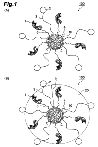
[0060] Next, the structure of the oligonucleotide conjugate will be
described with reference to FIG. 1. FIG. 1(A) is a schematic diagram
showing one embodiment of an oligonucleotide conjugate. An
oligonucleotide conjugate 100 includes a core 10 of the dendritic
polymer, and a plurality of oligonucleotides 1, cellular internalization
enhancers 2, and hydrophilic linkers 3, that are arranged around the core
10. The oligonucleotides 1 are bonded to the core 10 through linkers 5.
The hydrophilic linkers 3 are bonded to the core 10, and the cellular
internalization enhancers 2 are bonded to the hydrophilic linkers 3. In
addition, capping agents 4 are also bonded to the core 10. Since all the
components of the oligonucleotide conjugate 100 other than the dendritic
polymer are bonded to the dendritic polymer in this manner, the dendritic
polymer constitutes the "core" 10, that is, the center part of the
oligonucleotide conjugate 100. In addition, in an aqueous solution, the
oligonucleotides 1 and the hydrophilic linkers 3 extend substantially
radially from the core 10, and accordingly, the oligonucleotide conjugate
100 takes the shape of a substantially spherical nanoparticle. Thus, the
oligonucleotide conjugate 100 exhibits the behavior of a nanoparticle.
In this field, there is a wealth of knowledge regarding the behavior of
nanoparticles in vivo. Note that, although the oligonucleotides 1 are
bonded to the core 10 through the linkers 5 in FIG. 1, the oligonucleotides
1 may be bonded directly to the core 10 as described above.
[0061] The average particle diameter of the oligonucleotide conjugate
CA 03194894 2023- 4-4
100 is preferably 10 to 100 nm, more preferably 15 to 45 nm, still more
preferably 15 to 35 nm. In the present specification, the average particle
diameter of the oligonucleotide conjugate means the average particle
diameter in the particle size distribution obtained by dynamic light
scattering. Since the oligonucleotide conjugate 100 has a dendritic
polymer as the core 10, size control is easy and precise design is possible.
[0062] In order for the oligonucleotide conjugate 100 to be transported
into a cell, the cellular internalization enhancer 2 needs to interact with
the cell. From the viewpoint of improving the transport efficiency of the
oligonucleotide conjugate into the cell, the density of the cellular
internalization enhancers 2 is preferably high. According to the
oligonucleotide conjugate 100, since the cellular internalization
enhancers 2 are bonded to the core 10 of the highly branched dendritic
polymer, a high density of the cellular internalization enhancers 2 can be
achieved, and accordingly, the cellular internalization enhancers 2 can
efficiently interact with a target cell.
[0063] Moreover, in order for the cellular internalization enhancer 2 to
interact with a cell, the cellular internalization enhancer 2 is preferably
present at the outer part of the nanoparticle of the oligonucleotide
conjugate 100. In one embodiment, more preferably, as shown in FIG.
1(A), the cellular internalization enhancer 2 is present at the outermost
part of a true sphere that approximates the structure of the oligonucleotide
conjugate 100, that is, on the surface of the nanoparticle of the
oligonucleotide conjugate 100. In another embodiment, even when the
cellular internalization enhancer 2 is not present at the outermost part of
the true sphere that approximates the structure of the oligonucleotide
36
CA 03194894 2023- 4-4
conjugate 100, it is preferable that the spatial extent (radius of gyration)
of the hydrophilic linkers 3 is not completely enclosed by the spatial
extent of the nucleic acids 1 and the hydrophilic linkers 3 are substantially
exposed to the outside. In the oligonucleotide conjugate 100 according to
the present aspect of the present invention, since the linker for linking the
dendritic polymer and the cellular internalization enhancer is hydrophilic,
non-specific interaction is suppressed, and the cellular internalization
enhancer 2 is likely to be present at the outer part of nanoparticles of the
oligonucleotide conjugate 100. The position of the cellular internalization
enhancer 2 in the nanop article of the oligonucleotide conjugate 100 may
be adjusted by the type and length of the hydrophilic linker 3. From the
viewpoint of allowing the cellular internalization enhancer 2 to efficiently
interact with a target cell to improve the transport efficiency of the
oligonucleotide conjugate into the cell, the average linear distance
between the ends of each hydrophilic linker 3 may be 1/5 or more, 1/4 or
more, 1/3 or more, 2/5 or more, or half or more of the length of the
oligonucleotide 1. Here, the linear distance between the ends of each
hydrophilic linker 3 is the linear distance between the end bonded to the
core 10 and the end bonded to the cellular internalization enhancer 2 of
each hydrophilic linker 3. More precisely, the average linear distance
between the ends of each hydrophilic linker 3 may be preferably 1/5 or
more, 1/4 or more, 1/3 or more, 2/5 or more, or half or more of the average
linear distance from the surface of the core 10 to the free end of the
oligonucleotide 1. Here, the linear distance from the surface of the core
10 to the free end of the oligonucleotide 1 indicates the linear distance
between the end of the linker 5 bonded to the core 10 (however, in the
37
CA 03194894 2023- 4-4
case where the oligonucleotide 1 is directly bonded to the core 10, the end
of oligonucleotide 1 bonded to core 10) and the end of oligonucleotide 1
that is not bonded to the linker 5 or the core 10. For example, when the
linear length of the oligonucleotides 1 is 5 nm and the oligonucleotides 1
are directly bonded to the core 10, the average linear distance between
the ends of each hydrophilic linker 3 may be 1 nm or more, 1.25 nm or
more, 1.67 nm, 2 nm or more, or 2.5 nm or more.
[0064] The average linear distance between the ends of each hydrophilic
linker 3 may be determined by measuring the thickness of the hydration
layer formed by the presence of the hydrophilic linkers 3 in some cases.
The hydration layer will now be described with reference to FIG. 1(B).
Since the hydrophilic linkers 3 are hydrophilic, water molecules are fixed
between the hydrophilic linkers 3 (that is, the oligonucleotide conjugate
100 is hydrated) in an aqueous solution, thereby forming a layer of water
molecules, namely a hydration layer 20, is formed around the hydrophilic
linkers 3. Although depending on the type of hydrophilic linker, a
thickness h of the hydration layer 20 formed around a given hydrophilic
linker 3 may be equal or substantially equal to the linear distance between
the ends of that hydrophilic linker 3 in some cases. Therefore, in such a
case (for example, when the hydrophilic linker 3 is PEG), the average
linear distance between the ends of each hydrophilic linker 3 can be
defined as the average value of the thickness h of the hydration layer 20.
The average value of the thickness h of the hydration layer 20 may be
determined by multi-angle dynamic light scattering, for example. More
specifically, first, a series of nanoparticle compounds each containing a
core 10 of a dendritic polymer and a plurality of hydrophilic linkers 3
38
CA 03194894 2023- 4-4
bonded to the core 10, wherein the molecular weight of the hydrophilic
linker 3 of each nanoparticle compound is different from that of the
hydrophilic linker 3 of other nanoparticle compounds, are prepared.
Next, the average particle diameter of each nanoparticle compound is
measured by multi-angle dynamic light scattering, and from the
difference in the average particle diameter and the difference in the
molecular weight of the hydrophilic linker 3, the correlation function
between the molecular weight of the hydrophilic linker 3 and the
thickness of the hydration layer is determined. Based on this correlation
function, the thickness h of the hydration layer 20 of the oligonucleotide
conjugate 100 can be calculated from the molecular weight of the
hydrophilic linker 3 in the oligonucleotide conjugate 100.
[0065] The oligonucleotide conjugate may be a free body or a
pharmaceutically acceptable salt. The oligonucleotide conjugate may be
either a solvate (for example, hydrates, ethanol solvates, or propylene
glycol solvates) or a non-solvate. Pharmaceutically acceptable salts may
be acid addition salts or base addition salts. Examples of acid addition
salts include salts with organic acids such as formate, acetate,
trifluoroacetic acid (TFA), propionate, succinate, lactate, malate, adipate,
citrate, tartrate, methane sulfonate, fumarate, maleate, p-toluenesulfonate,
or ascorbate; and salts with inorganic acids such as hydrochloride,
hydrobromide, sulfate, nitrate, or phosphate. Examples of base addition
salts include alkali metal salts such as sodium salts or potassium salts;
alkaline earth metal salts such as calcium salts or magnesium salts;
ammonium salts; trimethylamine salts; triethylamine salts; aliphatic
amine salts such as dicyclohexylamine salts, ethanolamine salts,
39
CA 03194894 2023- 4-4
diethanolamine salts, triethanolamine salts, or brocaine salts;
arallcylamine salts such as N,N-dibenzylethylenediamine; heterocyclic
aromatic amine salt such as pyridine salts, picoline salts, quinoline salts,
or isoquinoline salts; quaternary ammonium salts such as
tetramethylammonium salts, tetraethylammonium salts,
benzyltrimethylammonium salts, benzyltriethylammonium salts,
benzyltributylammonium salts, methyltrioctylammonium salts, or
tetrabutylammonium salts; and basic amino acid salts such as arginine
salts or lysine salts.
[0066] The present invention also provides a method for producing the
oligonucleotide conjugate according to the above aspect. Namely, one
aspect of the present invention is a method for producing the
oligonucleotide conjugate including steps of: bonding a plurality of
oligonucleotides and one or more hydrophilic linkers to a dendritic
polymer; and bonding a cellular internalization enhancer to each
hydrophilic linker. Oligonucleotides may be bonded to the dendritic
polymer either directly or through linkers. In one embodiment, the
method for producing an oligonucleotide conjugate may further include
a step of bonding a capping agent to the dendritic polymer. This allows
for the production of an oligonucleotide conjugate in which at least some
of the reactive functional groups of the dendritic polymer are capped with
a capping agent.
[0067] Any of the above steps may be performed using a
methodcommonly used in this field. Examples of such methods include
a method in which an amino group and a carboxy group are allowed to
react using an activating group to form an amide bond, a method in which
CA 03194894 2023- 4-4
thiol groups are allowed to react with each other using an activating group
to form a disulfide bond, a method in which a thiol group and a maleimide
group are allowed to react to form a thioether bond, a method in which a
click chemistry using a catalyst or an activating group is used to form a
triazole bond from an azide group and an alkynyl group, and a method in
which an inverse electron demand Diels-Alder reaction is used to form a
bond from an highly electron-deficient heterocycle such as a tetrazine or
triazine and a compound with strained carbon multiple bonds such as
norbornene, trans-cyclooctene, or cyclooctyne.
[0068] The oligonucleotide conjugate according to the above aspect may
be produced by a known method other than the method according to the
above aspect.
[0069] One aspect of the present invention is a pharmaceutical
composition containing the oligonucleotide conjugate according to the
above aspect as an active ingredient. The pharmaceutical composition
contains a pharmaceutically acceptable additive. In the present
specification, "pharmaceutically acceptable" refers to being acceptable to
mammals from a pharmacological or toxicological point of view. That is,
a "pharmaceutically acceptable" substance refers to a substance that is
physiologically acceptable and that typically does not cause an allergic or
other adverse or toxic reaction when administered to a mammal. A
"pharmaceutically acceptable" substance means a substance which is
approved by a generally recognized regulatory agency or listed in a
generally recognized pharmacopoeia for use in mammals, more
particularly humans. "pharmaceutically acceptable additive" means a
pharmacologically inert material that is used with the oligonucleotide
41
CA 03194894 2023- 4-4
conjugate to formulate a pharmaceutical composition.
[0070] Additives may be liquid or solid. The additives are selected with
the planned administration method in mind so as to obtain a
pharmaceutical composition with the desired dosage, consistency, and the
like. Additives are not particularly limited, and examples thereof include
water, physiological saline, other aqueous solvents, various carriers such
as aqueous or oily bases, excipients, binders, pH adjusters, disintegrants,
absorption promoters, lubricants, coloring agents, corrigents, and
fragrances. The blending ratio of the additive may be appropriately set
based on the range normally employed in the pharmaceutical field.
[0071] A pharmaceutical composition may, for example, be a sterile
composition for injection. Sterile compositions for injection may be
prepared according to normal pharmaceutical practice (for example,
dissolving or suspending the active ingredient in a solvent such as water
for injection or natural vegetable oil). As aqueous solutions for injection,
for example, isotonic solutions containing physiological saline, glucose,
or other adjuvants (for example, D-sorbitol, D-mannitol, lactose, sucrose,
or sodium chloride) are used. Aqueous solutions for injection may, for
example, further contain suitable solubilizers such as alcohols (for
example, ethanol), polyalcohols (for example, propylene glycol or
polyethylene glycol), or nonionic surfactants (for example, polysorbate
80TM or HCO-50). In addition, aqueous solutions for injection may
contain buffers (for example, phosphate buffer solution or sodium acetate
buffer solution), soothing agents (for example, benzallconium chloride or
procaine hydrochloride), stabilizers (for example, human serum albumin
or polyethylene glycol), preservatives (for example, benzyl alcohol or
42
CA 03194894 2023- 4-4
phenol), antimicrobial agents, dispersants, antioxidants, and various other
materials known in the related art. Injections may be, for example,
lyophilized formulations.
[0072] The oligonucleotide conjugate or pharmaceutical composition
according to the above aspects of the present invention can be used to
treat and/or prevent diseases associated with specific gene products.
Examples of diseases associated with specific gene products include
inborn errors of metabolism, a congenital endocrine disease, a single gene
disorder, a neurodegenerative disease, a neurologic disease, a myopathy,
a meningitis, an encephalitis, an encephalopathy, a lysosome disease, a
malignant neoplasm, a fibrosis, an inflammatory disease, an
immunodeficiency disease, an autoimmune disease, or an infectious
disease, but the diseases are not limited to them. Therefore, one aspect of
the present invention is a therapeutic agent or a preventive agent for the
above diseases, which contains the oligonucleotide conjugate as an active
ingredient.
[0073] Another aspect of the present invention is a method for treating
and/or preventing the above diseases including administering a
therapeutically effective amount of the oligonucleotide conjugate to a
human or non-human animal. The human may be a human in need of
treatment, namely a patient. Non-human animals include animals such as
warm-blooded mammals such as primates; birds; domestic or livestock
animals such as cats, dogs, sheep, goats, cows, horses, or pigs; laboratory
animals such as mice, rats, or guinea pigs; fish; reptiles; zoo animals; or
wild animals. Administration methods include, but are not limited to, oral,
sublingual, intravenous, intraarterial, subcutaneous, intradermal,
43
CA 03194894 2023- 4-4
intraperitoneal, intramuscular, intrathecal, intracerebroventricular,
intranasal, transmucosal, rectal, ophthalmic, intraocular, transpulmonary,
transdermal, intra-articular, topical (cutaneous), intrafollicular,
intravaginal, intrauterine, intratumoral, or intralymphatic administration,
or combinations thereof.
[0074] Another aspect of the present invention is the oligonucleotide
conjugate for use in the treatment and/or prevention of the diseases
described above. Another aspect of the present invention is the use of
oligonucleotide conjugate for producing a therapeutic agent and/or a
preventive agent for the above diseases.
[0075] The oligonucleotide conjugate or pharmaceutical composition
according to the above aspects of the present invention may also be used
in combination with one or more other drugs. Other drugs may be one or
more therapeutic agents and/or preventive agents for diseases associated
with the specific gene products described above. For example, when the
disease of interest is a malignant neoplasm, examples of other drugs
include drugs that can be used in chemotherapy. That is, one aspect of the
present invention is the oligonucleotide conjugate for treating diseases in
combination with one or more therapeutic agents and/or preventive
agents for the above diseases. Another aspect of the present invention is
a medicament containing a combination of the oligonucleotide conjugate
or pharmaceutical composition and one or more therapeutic agents and/or
preventive agents for the above diseases. However, the present
invention is a platform technology that can efficiently transport
oligonucleotides into a cell, and can be used for any disease as long as the
oligonucleotides can be applied to the diseases as a therapeutic agent or
44
CA 03194894 2023- 4-4
preventive agent, and thus, other drugs are not limited to specific drugs.
[0076] The timing of administration of the oligonucleotide conjugate or
pharmaceutical composition and other drugs above used in combination
therewith is not limited, and these may be administered to humans or
animals other than humans at the same time or at appropriate intervals.
Alternatively, the pharmaceutical composition according to the above
aspect may be blended with other drugs above to prepare a combination
drug. The administration dosage and blending amount of other drugs
above may be appropriately determined based on the doses used
clinically. The blending ratio of the oligonucleotide conjugate or
pharmaceutical composition and other drugs above may be appropriately
determined according to the administration target, administration route,
target disease, symptom, combination of other drugs, and the like.
Examples
[0077] The present invention will be described in detail below with
reference to examples and test examples, but the present invention is not
limited to these examples. In addition, "%" in the following description
means % by weight unless otherwise specified.
[0078] <Synthesis of oligonucleotide>
siRNAs and antisense oligonucleotides shown in Table 2 were
prepared. A thiol group was bonded to the 3' end of the sense strand
RNA of the siRNAs and the 5' end of the antisense oligonucleotide
through Spacer18 (hexaethylene glycol). These nucleic acids were
produced by GeneDesign, Inc.
[Table 2]
Atp5b-siRNA 5'-
CA 03194894 2023- 4-4
sense strand
U(F)AG(M)AU(F)C(M)A(F)U(M)U(F)G(F)G(F)A(M)G(F)A(
(SEQ ID NO: 7) M)A(F)C(M)C(F)U(M)A(F)U(M)U(F)tt-3'_1 8_511
Atp5b-siRNA 5'-
antisense strand p_A(M)AA(F)AU(M)A(F)G(M)G(F)U(M)U(F)C(M)U(F)C(M)
(SEQ ID NO: 8) C(M)A(M)A(F)U(M)G(F)A(M)C(F)A(M)AtAt-3'
scramble-siRNA 5'-
sense strand
G(F)AC(M)AU(F)A(M)G(F)A(M)C(F)U(F)G(F)U(M)U(F)U(
(SEQ ID NO: 9) M)A(F)A(M)C(F)U(M)G(F)A(M)U(F)tt-3'_1 8_SH
scramble-siRNA 5'-
antisense strand p_A(M)AU(F)AC(M)A(F)G(M)U(F)U(M)A(F)A(M)A(F)C(M)
(SEQ ID NO: 10) A(M)G(M)U(F)C(M)U(F)A(M)G(F)C(M)AtAt-3'
Malatl-ASO SH_1 8_5'-
(SEQ ID NO: 11) mC(L)AT(L)AA(L)AgAtAtAcAaAcAtAgAaAaATpAGpAincp_3,
In the table, upper case = RNA, lower case = DNA, mC = 5-
methylcytosine, (M) = 2'-0-CH3 substitution, (F) = 2'-F substitution, (L)
= Locked nucleic acid, A = phosphorothioate linkage, p = PO4, 18 =
spacer18
[0079] siRNA and ethylenediaminetetraacetic acid trisodium salt (EDTA
3Na) were dissolved in 10 mM phosphate buffered saline (PBS) at pH
7.4, and dithiothreitol (DTT) was added (final concentration: EDTA 0.5
mM, DTT 40 mM). After heating this solution at 25 C for 6 hours, this
solution was purified 6 times by ultrafiltration (molecular weight cut-off
10 kDa) using PBS. The nucleic acid concentration of the obtained
solution was determined from the absorbance measurement values at 260
nm using an ultraviolet-visible spectrophotometer (manufactured by
Tecan Group Ltd., Infinite M200 PRO).
[0080] <Example 1. Production of AF5-labeled cRGD-functionalized
oligonucleotide conjugate using fourth generation polylysine dendrigraft
as core>
(A) Synthesis of azide-PEG5k SPDP AF5 DGL G4
As the dendritic polymer, a dendri-grafted poly-L-lysine G4
46
CA 03194894 2023- 4-4
(DGL G4) having amino groups on the surface manufactured by
COLCOM Group was used. To 10 L of 50 mg/mL dimethyl sulfoxide
(DMSO) solution of DGL G4, 1.53 L of 300 mM DMSO solution of
PEG12-SPDP (manufactured by Thermo Fisher Scientific Inc.), 1.17 L
of 50% v/v dimethylformamide (DMF) solution of triethylamine (TEA),
and 1.28 L of 30 mM DMSO solution of AlexaFluor (registered
trademark) 546 MIS ester (manufactured by Thermo Fisher Scientific
Inc.) were added, and the mixture was stirred at room temperature for 4.5
hours. Next, to this reaction solution, 68.9 L of 20 mM DMSO
solution of Azide-PEG5k-NHS (manufactured by Nanocs Inc., number
average molecular weight of PEG: 5000) was added, and the mixture was
further stirred at room temperature for 18 hours. Next, 15.3 L of 200
mM DMSO solution of Methyl-PEG12-NHS (manufactured by Thermo
Fisher Scientific Inc.) was added, and the mixture was further stirred at
room temperature for 6 hours. By allowing the amino groups of DGL
G4 and the N-hydroxysuccinimide (NHS) groups of Azide-PEG5k-NHS,
PEG12-SPDP, the anionic fluorescent dye AlexaFluor 546 NHS ester,
and Methyl-PEG12-NHS to react as described above, a nanoparticle
compound azide-PEG5k SPDP AF5 DGL G4 was obtained. After
adding 400 L of pure water to the reaction solution and mixing, the
mixture was purified 6 times by ultrafiltration (manufactured by Merck
& Co., Amicon Ultra, molecular weight cut-off: 10 kDa) using pure
water. Pure water was added to the collected aqueous solution to adjust
the volume of the liquid to 100 L.
[00811(B) Synthesis of azide-PEG5k siRNA AF5 DGL G4
To 20 L of aqueous solution of azide-PEG5k SPDP AF5 DGL
47
CA 03194894 2023- 4-4
G4 shown in (A), 12 L of 3 M sodium chloride aqueous solution and
72.1 L of 5.1 mM PBS solution of siRNA were added, and the mixture
was stirred at 25 C for 15 hours to alllow the pyridyl disulfide group of
azide-PEG5k SPDP AF5 DGL G4 and the SH group of siRNA to react.
The reaction solution was purified by gel filtration using Hiprep 16/60
Sephacryl S-200 HR (manufactured by GE HealthCare Technologies
Inc.) (eluent: PBS). Fractions containing siRNA-bonded DGL G4 were
collected and concentrated by ultrafiltration (manufactured by Merck &
Co., Amicon Ultra, molecular weight cut-off 30 kDa), and the volume of
the liquid was adjusted to 170 L.
[0082] (C) Synthesis of cRGD-DBCO
To 33.1 L of 100 mM DMSO solution of Cyclo(-RGDfK)
(manufactured by ChemScence), 33.1 L of 300 mM DMSO solution of
DBCO-NHCO-PEG4-NHS (manufactured by BroadPharm) and 1.39 L
of TEA were added, and the mixture was stirred at 25 C for 16 hours to
allow the amino group of Cyclo(-RGDfK) and the NHS ester of DBCO-
NHCO-PEG4-NHS to react. The reaction solution was concentrated by
removing the solvent under reduced pressure while heating at 45 C, and
purified by reversed-phase HPLC (column: manufactured by Waters
Corporation, )(bridge Peptide BEH C18, 300 A, 4.6 x 100 mm, eluent A:
0.1% v/v TFA, eluent B: 0.1% v/v TFA/acetonitrile (10/90; v/v)). After
removing the solvent under reduced pressure while heating at 45 C,
DMSO was added to adjust the concentration to 50 mM.
[0083] (D) Synthesis of cRGD-PEG5k siRNA AF5 DGL G4
To 30 L of aqueous solution of azide-PEG5k siRNA AF5 DGL
G4 obtained in (B), 4.1 L of DMSO solution of cRGD-DBCO obtained
48
CA 03194894 2023- 4-4
in (C) was added, and the mixture was stirred at 25 C for 20 hours to
allow the azide group of azide-PEG5k siRNA AF5 DGL G4 and the
DBCO group (dibenzocyclooctyne group) of cRGD-DBCO to react.
Then, after adding 65 ptL of PBS, purification was performed using NAP-
5 Columns (manufactured by GE HealthCare Technologies Inc.). The
collected solution was concentrated using ultrafiltration (molecular
weight cut-off 30 kDa), and the volume of the solution was adjusted to
95 ptL to obtain a solution of oligonucleotide conjugate cRGD-PEG5k
siRNA AF5 DGL G4.
[0084] <Example 2. Production of AF5-labeled GE11-functionalized
oligonucleotide conjugate using fourth generation polylysine dendrigraft
as core>
To 30 ptL of azide-PEG5k siRNA AF5 DGL G4 aqueous solution
obtained in (B) of Example 1,4.1 ptL of 50 mM DMSO solution of GE11-
DBCO (manufactured by GeneDesign, Inc.) and 32.8 ptL of DMSO were
added, and the mixture was stirred at 25 C for 20 hours to allow the azide
group of azide-PEG5k siRNA AF5 DGL G4 and the DBCO group of
GE11-DBCO to react. Then, when 130 ptL of PBS was added, a
precipitate was obtained, and thus this precipitate was subjected to
centrifugal sedimentation to remove the supernatant. After dissolving
the obtained precipitate with 100 ptL of DMSO, when 400 ptL of 2-
propanol was added, a precipitate was obtained. Thus, this precipitate
was subjected to centrifugal sedimentation again to remove the
supernatant. After the obtained precipitate was dissolved in 500 ptL of
PBS, the solution was purified using NAP-5 Columns (manufactured by
GE HealthCare Technologies Inc.).
The collected solution was
49
CA 03194894 2023- 4-4
concentrated using ultrafiltration (molecular weight cut-off 30 kDa), and
the volume of the solution was adjusted to 95 ptL to obtain a solution of
oligonucleotide conjugate GE11-PEG5k siRNA AF5 DGL G4.
[0085] <Example 3. Production of TF7-labeled cRGD-functionalized
oligonucleotide conjugate using PAMAM G5 as core>
(A) Synthesis of azide-PEG5k SPDP TF7 PAMAM G5
As the dendritic polymer, a fifth generation PAMAM dendrimer
(PAMAM G5) having amino groups on the surface manufactured by
Sigma-Aldrich was used. To 3.0 ptL of 5% wt methanol solution of
PAMAM G5, 1.24 ptL of 100 mM DMSO solution of PEG12-SPDP, 1.66
ptL of 10 mM DMSO solution of Tide Fluor (trademark) 7W5,
succinimidyl ester (manufactured by AAT Bioquest, Inc.), and 1.48 ptL of
10% v/v DMF solution of TEA were added, and the mixture was stirred
at room temperature for 5 hours. Next, to this reaction solution, 12.4 ptL
of 30 mM DMSO solution of Azide-PEG5k-NHS, 1.03 ptL of 400 mM
DMSO solution of NHS (manufactured by FuJIFILM Wako Pure
Chemical Corporation), and 1.03 ptL of 400 mM DMSO solution of 1-
ethy1-3-(3-dimethylaminopropyl)carbodiimide hydrochloride (EDC-
HC1, manufactured by Nacalai Tesque, Inc.) was added, and the mixture
was further stirred at room temperature for 18.5 hours. Next, 2.65 ptL
of 200 mM DMSO solution of Methyl-PEG12-NHS was added, and the
mixture was further stirred at room temperature for 6 hours. By
allowing the amino groups of PAMAM G5 and the NHS groups ofAzide-
PEG5k-NHS, PEG12-SPDP, Tide Fluor 7W5, succinimidyl ester, and
Methyl-PEG12-NHS to react as described above, a nanoparticle
compound azide-PEG5k SPDP TF7 PAMAM G5 was obtained. After
CA 03194894 2023- 4-4
adding 400 L of pure water to the reaction solution and mixing, the
mixture was purified 6 times by ultrafiltration (manufactured by Merck
& Co., Amicon Ultra, molecular weight cut-off: 10 kDa) using pure
water. Pure water was added to the collected aqueous solution to adjust
the volume of the liquid to 80 L.
[0086] (B) Synthesis of azide-PEG5k siRNA TF7 PAMAM G5
To 10.0 L of aqueous solution of azide-PEG5k SPDP TF7
PAMAM G5 shown in (A), 3.2 L of 3 M sodium chloride aqueous
solution and 12.4 L of 5.0 mM PBS solution of siRNA were added, and
the mixture was stirred at 25 C for 15 hours to allow the pyridyl disulfide
group of azide-PEG5k SPDP TF7 PAMAM G5 and the SH group of
siRNA to react. The reaction solution was purified by gel filtration
using Hiprep 16/60 Sephacryl S-200 HR (eluent: PBS). Fractions
containing siRNA-bonded PAMAM G5 were collected and concentrated
by ultrafiltration (manufactured by Merck & Co., Amicon Ultra,
molecular weight cut-off 30 kDa), and the volume of the liquid was
adjusted to 100 L.
[0087] (C) Synthesis of cRGD-PEG5k siRNA TF7 PAMAM G5
To 500 L of aqueous solution of azide-PEG5k siRNA TF7
PAMAM G5 obtained in (B), 3.70 L of DMSO solution of cRGD-
DBCO obtained in (C) of Example 1 was added, and the mixture was
stirred at 25 C for 16 hours to allow the azide group of azide-PEG5k
siRNA TF7 PAMAM G5 and the DBCO group of cRGD-DBCO to react.
Then, after adding PBS to adjust the liquid volume to 100 L, the solution
was purified using NAP-5 Columns. The collected solution was
concentrated using ultrafiltration (molecular weight cut-off 30 kDa), and
51
CA 03194894 2023- 4-4
the volume of the solution was adjusted to 110 L to obtain a solution of
oligonucleotide conjugate cRGD-PEG5k siRNA TF7 PAMAM G5.
[0088] <Example 4. Production of TF7-labeled cRGD-functionalized
oligonucleotide conjugate using PAMAM G6 as core>
(A) Synthesis of azide-PEG5k SPDP TF7 PAMAM G6
As the dendritic polymer, a sixth generation PAMAM dendrimer
(PAMAM G6) having amino groups on the surface manufactured by
Sigma-Aldrich was used. To 5.0 L of 5% wt methanol solution of
PAMAM G6, 1.39 L of 100 mM DMSO solution of PEG12-SPDP, 2.78
L of 10 mM DMSO solution of Tide Fluor 7W5, succinimidyl ester, and
2.48 L of 10% v/v DMF solution of TEA were added, and the mixture
was stirred at room temperature for 5 hours. Next, to this reaction
solution, 12.4 L of 30 mM DMSO solution of Azide-PEG5k-NHS, 1.72
L of 400 mM DMSO solution of NHS, and 1.72 L of 400 mM DMSO
solution of EDC-HC1 were added, and the mixture was further stirred at
room temperature for 18.5 hours. Next, 4.44 L of 200 mM DMSO
solution of Methyl-PEG12-NHS was added, and the mixture was further
stirred at room temperature for 6 hours. By allowing the amino groups
of PAMAM G6 and the NHS groups of Azide-PEG5k-NHS, PEG12-
SPDP, Tide Fluor 7W5, succinimidyl ester, and Methyl-PEG12-NHS to
react as described above, a nanoparticle compound azide-PEG5k SPDP
TF7 PAMAM G6 was obtained. After adding 400 L of pure water to
the reaction solution and mixing, the mixture was purified 6 times by
ultrafiltration (manufactured by Merck & Co., Amicon Ultra, molecular
weight cut-off: 10 kDa) using pure water. Pure water was added to the
collected aqueous solution to adjust the volume of the liquid to 80 L.
52
CA 03194894 2023- 4-4
[0089] (B) Synthesis of azide-PEG5k siRNA TF7 PAMAM G6
To 9.0 L of aqueous solution of azide-PEG5k SPDP TF7
PAMAM G6 shown in (A), 3.1 L of 3 M sodium chloride aqueous
solution and 12.5 L of 5.0 mM PBS solution of siRNA were added, and
the mixture was stirred at 25 C for 15 hours to allow the pyridyl disulfide
group of azide-PEG5k SPDP TF7 PAMAM G6 and the SH group of
siRNA to react. The reaction solution was purified by gel filtration
using Hiprep 16/60 Sephacryl S-200 HR (eluent: PBS). Fractions
containing siRNA-bonded PAMAM G6 were collected and concentrated
by ultrafiltration (manufactured by Merck & Co., Amicon Ultra,
molecular weight cut-off 30 kDa), and the volume of the liquid was
adjusted to 100 L.
[0090] (C) Synthesis of cRGD-PEG5k siRNA TF7 PAMAM G6
To 500 L of aqueous solution of azide-PEG5k siRNA TF7
PAMAM G6 obtained in (B), 4.51 L of DMSO solution of cRGD-
DBCO obtained in (C) of Example 1 was added, and the mixture was
stirred at 25 C for 16 hours to allow the azide group of azide-PEG5k
siRNA TF7 PAMAM G6 and the DBCO group of cRGD-DBCO to react.
Then, after adding PBS to adjust the liquid volume to 100 L, the solution
was purified using NAP-5 Columns. The collected solution was
concentrated using ultrafiltration (molecular weight cut-off 30 kDa), and
the volume of the solution was adjusted to 110 L to obtain a solution of
oligonucleotide conjugate cRGD-PEG5k siRNA TF7 PAMAM G6.
[0091] <Example 5. Production of TF7-labeled cRGD-functionalized
oligonucleotide conjugate using fourth generation polylysine dendrigraft
as core>
53
CA 03194894 2023- 4-4
Oligonucleotide conjugate cRGD-PEG5k siRNA TF7 DGL G4
was synthesized according to the synthesis of cRGD-PEG5k siRNAAF5
DGL G4 in Example 1. However, Tide Fluor 7WS, succinimidyl ester
was used instead of AlexaFluor 546 NHS ester.
[0092] <Example 6. Production of AF5-labeled cRGD peptide-
functionalized oligonucleotide conjugate 2 using fourth generation
polylysine dendrigraft as core>
(A) Synthesis of azide-PEG5k SPDP AF5 DGL G4
According to (A) of Example 1, azide-PEG5k SPDP AF5 DGL
G4 was synthesized.
[0093] (B) Synthesis of azide-PEG5k siRNA AF5 DGL G4
To 20 L of aqueous solution of azide-PEG5k SPDP AF5 DGL
G4 obtained in (A), 12 L of 3 M sodium chloride aqueous solution and
76.7 L of 6.0 mM PBS solution of siRNA were added, and the mixture
was stirred at 25 C for 18 hours to allow the pyridyl disulfide group of
azide-PEG5k SPDP AF5 DGL G4 and the SH group of siRNA to react.
The reaction solution was purified by gel filtration using Hiprep 16/60
Sephacryl S-200 HR (manufactured by GE HealthCare Technologies
Inc.) (eluent: PBS). Fractions containing siRNA-bonded DGL G4 were
collected and concentrated by ultrafiltration (manufactured by Merck &
Co., Amicon Ultra, molecular weight cut-off 30 kDa), and the volume of
the liquid was adjusted to 200 L.
[0094] (C) Synthesis of cRGD-PEG5k siRNA AF5 DGL G4
To 30 L of aqueous solution of azide-PEG5k siRNA AF5 DGL
G4 obtained in (B), 5.8 L of 5 mM DMSO solution of cRGD-DBCO
obtained in (C) of Example 1 was added, and the mixture was stirred at
54
CA 03194894 2023- 4-4
25 C for 20 hours to allow the azide group of azide-PEG5k siRNA AF5
DGL G4 and the DBCO group of cRGD-DBCO to react. Then, after
adding 65 ptL of PBS, the solution was purified using NAP-5 Columns
(manufactured by GE HealthCare Technologies Inc.). The collected
solution was concentrated using ultrafiltration (molecular weight cut-off
30 kDa), and the volume of the solution was adjusted to 95 ptL to obtain
a solution of oligonucleotide conjugate cRGD-PEG5k siRNA AF5 DGL
G4.
[0095] <Example 7. Production of AF5-labeled cRGD peptide-
functionalized oligonucleotide conjugate 3 using fourth generation
polylysine dendrigraft as core>
According to the synthesis of Example 6, an oligonucleotide
conjugate similar to that of Example 6 was synthesized. However,
instead of adding 5.8 ptL of 5 mM DMSO solution of cRGD-DBCO, 1.4
ptL of 5 mM DMSO solution of cRGD-DBCO and 4.3 ptL of DMSO were
added.
[0096] <Example 8. Production of AF5-labeled cRGD peptide-
functionalized oligonucleotide conjugate 4 using fourth generation
polylysine dendrigraft as core>
According to the synthesis of Example 6, an oligonucleotide
conjugate similar to that of Example 6 was synthesized. However,
instead of adding 5.8 ptL of 5 mM DMSO solution of cRGD-DBCO, 1.2
ptL of 5 mM DMSO solution of cRGD-DBCO and 4.6 ptL of DMSO were
added.
[0097] <Example 9. Production of AF5-labeled cRGD peptide-
functionalized oligonucleotide conjugate 5 using fourth generation
CA 03194894 2023- 4-4
polylysine dendrigraft as core>
According to the synthesis of Example 6, an oligonucleotide
conjugate similar to that of Example 6 was synthesized. However,
instead of adding 5.8 ptL of 5 mM DMSO solution of cRGD-DBCO, 0.9
ptL of 5 mM DMSO solution of cRGD-DBCO and 4.9 ptL of DMSO were
added.
[0098] <Example 10. Production of AF5-labeled cRGD peptide-
functionalized oligonucleotide conjugate 6 using fourth generation
polylysine dendrigraft as core>
According to the synthesis of Example 6, an oligonucleotide
conjugate similar to that of Example 6 was synthesized. However,
instead of adding 5.8 ptL of 5 mM DMSO solution of cRGD-DBCO, 0.6
ptL of 5 mM DMSO solution of cRGD-DBCO and 5.2 ptL of DMSO were
added.
[0099] <Example 11. Production of AF5-labeled c(avb6) peptide-
functionalized oligonucleotide conjugate using fourth generation
polylysine dendrigraft as core>
According to the synthesis of Example 1, oligonucleotide
conjugate c(avb6)-PEG5k siRNA AF5 DGL G4 was synthesized.
However, instead of cRGD-DBCO, c(avb6)-DBCO (manufactured by
GeneDesign, Inc.) obtained by allowing the amino group of the lysine
side chain of c(avb6) and the NHS ester of DBCO-NHCO-PEG4-NHS to
react was used.
[0100] <Example 12. Production of AF5-labeled folic acid-
functionalized oligonucleotide conjugate using fourth generation
polylysine dendrigraft as core>
56
CA 03194894 2023- 4-4
According to the synthesis of Example 1, oligonucleotide
conjugate FA-PEG5k siRNA AF5 DGL G4 was synthesized. However,
Folic acid-PEG2 DBCO (manufactured by Nanocs Inc.) was used instead
of cRGD-DBCO.
[0101] <Example 13. Production of AF6-labeled indatraline-
functionalized oligonucleotide conjugate using fourth generation
polylysine dendrigraft as core>
(A) Synthesis of azide-PEG5k SPDP AF6 DGL G4
To 18 ptL of 50 mg/mL DMSO solution of DGL G4, 13.8 ptL of
60 mM DMSO solution of PEG12-SPDP, 8.9 ptL of 8 mM DMSO
solution of an anionic fluorescent dye AlexaFluor (registered trademark)
647 NHS ester (manufactured by Thermo Fisher Scientific Inc.), and 14.0
ptL of 10% v/v DMSO solution of TEA were added, and the mixture was
stirred at room temperature for 11 hours. Next, to this reaction solution,
1417.6 ptL of 1.8 mM DMSO solution of N3-PEG-NHS (manufactured
by Biopharma PEG Scientific Inc., number average molecular weight of
PEG: 5000), 24.8 ptL of 200 mM DMSO solution of EDC-HC1, and 24.8
ptL of 200 mM DMSO solution of NHS were added, and the mixture was
further stirred at room temperature for 13 hours. Next, 37.8 ptL of 200
mM DMSO solution of Methyl-PEG12-NHS was added, and the mixture
was further stirred at room temperature for 6 hours. By allowing the
amino groups of DGL G4 and the NHS groups of N3-PEG-NHS, PEG12-
SPDP, AlexaFluor 647 NHS ester, and Methyl-PEG12-NHS to react as
described above, a nanoparticle compound azide-PEG5k SPDP AF6 DGL
G4 was obtained. After adding 700 ptL of pure water to the reaction
solution and mixing, the mixture was purified 6 times by ultrafiltration
57
CA 03194894 2023- 4-4
(manufactured by Merck & Co., Amicon Ultra, molecular weight cut-off
30 kDa) using pure water. Pure water was added to the collected
aqueous solution to adjust the volume of the liquid to 420 L.
[0102] (B) Synthesis of azide-PEG5k siRNA AF6 DGL G4
To 410.0 L of aqueous solution of azide-PEG5k SPDP AF6 DGL
G4 obtained in (A), 127.1 L of 3 M sodium chloride aqueous solution,
465.0 L of 5.0 mM PBS solution of siRNA, and 111.3 L of DMSO
were added, and the mixture was stirred at 25 C for 13 hours to allow the
pyridyl disulfide group of azide-PEG5k SPDP AF6 DGL G4 and the SH
group of siRNA to react. The reaction solution was purified by gel
filtration using Hiprep 16/60 Sephacryl S-200 HR (eluent: PBS).
Fractions containing siRNA-bonded DGL G4 were collected and
concentrated by ultrafiltration (manufactured by Merck & Co., Amicon
Ultra, molecular weight cut-off 30 kDa), and the volume of the liquid was
adjusted to 700 L.
[0103] (C) Synthesis of IND-DBCO
To 15.5 L of 800 mM DMSO solution of indatraline
(manufactured by Sigma-Aldrich), 16.5 L of 300mM DMSO solution of
DBCO-NHCO-PEG4-NHS, 3.62 L of TEA, and 20 L of acetonitrile
were added, and the mixture was stirred at 25 C for 18 hours to allow the
amino group of indatraline and the NHS ester of DBCO-NHCO-PEG4-
NHS to react. The reaction solution was purified by gel filtration using
Sephadex (registered trademark) LH-20 (manufactured by Cytiva)
(eluent: ethanol/acetonitrile (50/50; v/v)). After fractions containing the
desired product, IND-DBCO, were collected, and the solvent was
removed under reduced pressure while heating at 45 C, DMSO was
58
CA 03194894 2023- 4-4
added to adjust the concentration to 20 mM.
[0104] (D) Synthesis of IND-PEG5k siRNAAF6 DGL G4
To 370 ptL of PBS solution of azide-PEG5k siRNAAF6 DGL G4
obtained in (B), 42.7 ptL of 20 mM DMSO solution of IND-DBCO
obtained in (C) and 49.8 ptL of DMSO were added, and the mixture was
stirred at 25 C for 22 hours to allow the azide group of azide-PEG5k
siRNA AF6 DGL G4 and the DBCO group of IND-DBCO to react.
Then, the reaction solution was purified using NAP-10 Columns
(manufactured by GE HealthCare Technologies Inc.). The collected
solution was concentrated using ultrafiltration (molecular weight cut-off
10 kDa) and the volume of the solution was adjusted to 95 ptL to obtain a
solution of oligonucleotide conjugate IND-PEG5k siRNAAF6 DGL G4.
[0105] <Example 14. Production of AF6-labeled aptamer-functionalized
oligonucleotide conjugate 1 using fourth generation polylysine
dendrigraft as core>
(A) Synthesis of azide-PEG5k SPDP AF6 DGL G4
To 8 ptL of 50 mg/mL DMSO solution of DGL G4, 6.1 ptL of 60
mM DMSO solution of PEG12-SPDP, 3.8 ptL of 8 mM DMSO solution
of AlexaFluor 647 NHS ester, and 6.5 ptL of 10% v/v DMSO solution of
TEA were added, and the mixture was stirred at room temperature for 7
hours. Next, to this reaction solution, 700.1 ptL of 1.8 mM DMSO
solution of N3-PEG-NHS (manufactured by Biopharma PEG Scientific
Inc., number average molecular weight of PEG: 5000), 12.3 ptL of 200
mM DMSO solution of EDC-HC1, and 12.3 ptL of 200 mM DMSO
solution of NHS were added, and the mixture was further stirred at room
temperature for 16 hours. Next, 16.8 ptL of 200 mM DMSO solution of
59
CA 03194894 2023- 4-4
Methyl-PEG12-NHS was added, and the mixture was further stirred at
room temperature for 6 hours. By allowing the amino groups of DGL
G4 and the NHS groups of N3-PEG-NHS, PEG12-SPDP, AlexaFluor 647
NHS ester, and Methyl-PEG12-NHS to react as described above, a
nanoparticle compound azide-PEG5k SPDP AF6 DGL G4 was obtained.
After adding 700 L of pure water to the reaction solution and mixing,
the mixture was purified 6 times by ultrafiltration (manufactured by
Merck & Co., Amicon Ultra, molecular weight cut-off 30 kDa) using pure
water. Pure water was added to the collected aqueous solution to adjust
the volume of the liquid to 190 L.
[01061(B) Synthesis of azide-PEG5k siRNA AF6 DGL G4
To 120 L of aqueous solution of azide-PEG5k SPDP AF6 DGL
G4 obtained in (A), 33.9 L of 3 M sodium chloride aqueous solution,
108.3 L of 6.0 mM PBS solution of siRNA, and 29.4 L of DMSO were
added, and the mixture was stirred at 25 C for 14 hours to allow the
pyridyl disulfide group of azide-PEG5k SPDP AF6 DGL G4 and the SH
group of siRNA to react. The reaction solution was purified by gel
filtration using Hiprep 16/60 Sephacryl S-200 HR (eluent: PBS).
Fractions containing siRNA-bonded DGL G4 were collected and
concentrated by ultrafiltration (manufactured by Merck & Co., Amicon
Ultra, molecular weight cut-off 30 kDa), and the volume of the liquid was
adjusted to 800 L.
[0107] (C) Synthesis of NU1-PEG5k siRNA AF6 DGL G4
DBCO-NHCO-PEG4-NHS was allowed to react with the 3' end
of an aptamer AS1411 having the nucleotide sequence shown in SEQ ID
NO: 1 through an Amino C6 linker to obtain NU1-DBCO (manufactured
CA 03194894 2023- 4-4
by GeneDesign, Inc.). 22.7 ptL of 2.3 mM PBS solution ofNU1-DBCO,
95 ptL of PBS solution of azide-PEG5k siRNA AF6 DGL G4 obtained in
(B), and 14.1 ptL of DMSO were mixed and stirred at 25 C for 15 hours
to allow the azide group of azide-PEG5k siRNA AF6 DGL G4 and the
DBCO group of NU1-DBCO to react. The reaction solution was
purified by gel filtration using Hiprep 16/60 Sephacryl S-200 HR (eluent:
PBS). Fractions containing the desired product were collected and
concentrated by ultrafiltration (manufactured by Merck & Co., Amicon
Ultra, molecular weight cut-off 30 kDa). Furthermore, purification was
performed by gel filtration using two Superose 6 Increase (manufactured
by Cytiva, eluent: PBS) connected in series. Fractions containing the
desired product were collected and concentrated by ultrafiltration
(manufactured by Merck & Co., Amicon Ultra, molecular weight cut-off
30 kDa). The volume of the liquid was adjusted to 194 ptL to obtain a
solution of oligonucleotide conjugate NU1-PEG5k siRNAAF6 DGL G4.
[0108] <Example 15. Production of AF6-labeled aptamer-functionalized
oligonucleotide conjugate 2 using fourth generation polylysine
dendrigraft as core>
According to the synthesis of Example 14, NU2-PEG5k siRNA
AF6 DGL G4 was synthesized. However, instead of NU1-DBCO,
NU2-DBCO (manufactured by GeneDesign, Inc.) obtained by allowing
DBCO-NHCO-PEG4-NHS to react with the 3' end of an aptamer FAN-
1524d1 having the nucleotide sequence shown in SEQ ID NO: 2 through
an Amino C6 linker was used.
[0109] <Example 16. Production of AF6-labeled aptamer-functionalized
oligonucleotide conjugate 3 using fourth generation polylysine
61
CA 03194894 2023- 4-4
dendrigraft as core>
According to the synthesis of Example 14, EP1-PEG5k siRNA
AF6 DGL G4 was synthesized. However, instead of NU1-DBCO, EP1-
DBCO (manufactured by GeneDesign, Inc.) obtained by allowing
DBCO-NHCO-PEG4-NHS to react with the 3' end of an aptamer having
the nucleotide sequence shown in SEQ ID NO: 3 through an Amino C6
linker was used.
[0110] <Example 17. Production of AF6-labeled aptamer-functionalized
oligonucleotide conjugate 4 using fourth generation polylysine
dendrigraft as core>
According to the synthesis of Example 14, EP2-PEG5k siRNA
AF6 DGL G4 was synthesized. However, instead of NU1-DBCO, EP2-
DBCO (manufactured by GeneDesign, Inc.) obtained by allowing
DBCO-NHCO-PEG4-NHS to react with the 5' end of an aptamer having
the nucleotide sequence shown in SEQ ID NO: 4 through an Amino C6
linker was used.
[0111] <Example 18. Production of AF5-labeled cRGD-functionalized
oligonucleotide conjugate using third generation polylysine dendrigraft
as core>
(A) Synthesis of azide-PEG5k SPDP AF5 DGL G3
As the dendritic polymer, a dendri-grafted poly-L-lysine G3
(DGL G3) having amino groups on the surface manufactured by
COLCOM Group was used. To 10 ptL of 50 mg/mL DMSO solution of
DGL G3, 1.5 ptL of 300 mM DMSO solution of PEG12-SPDP, 3.8 ptL of
10 mM DMSO solution of AlexaFluor 546 NHS ester, and 1.0 ptL of 20%
v/v DMF solution of TEA were added, and the mixture was stirred at
62
CA 03194894 2023- 4-4
room temperature for 7 hours. Next, to this reaction solution, 45.9 L
of 30 mM DMSO solution of Azide-PEG5k-NHS (manufactured by
Nanocs Inc., number average molecular weight of PEG: 5000), 6.9 L of
400 mM DMSO solution of EDC-HC1, and 6.9 L of 400 mM DMSO
solution of NHS were added, and the mixture was further stirred at room
temperature for 16 hours. Next, 4.7 L of 200 mM DMSO solution of
Methyl-PEG12-NHS was added, and the mixture was further stirred at
room temperature for 8 hours. By allowing the amino groups of DGL
G3 and the NHS groups of Azide-PEG5k-NHS, PEG12-SPDP,
AlexaFluor 546 NHS ester, and Methyl-PEG12-NHS to react as
described above, a nanoparticle compound azide-PEG5k SPDP AF5 DGL
G3 was obtained. After adding 700 L of pure water to the reaction
solution and mixing, the mixture was purified 6 times by ultrafiltration
(manufactured by Merck & Co., Amicon Ultra, molecular weight cut-off:
10 kDa) using pure water. Pure water was added to the collected
aqueous solution to adjust the volume of the liquid to 100 L.
[0112] (B) Synthesis of azide-PEG5k siRNA AF5 DGL G3
To 4.0 L of aqueous solution of azide-PEG5k SPDP AF5 DGL
G3 obtained in (A), 2.4 L of 3 M sodium chloride aqueous solution and
14.8 L of 5.0 mM PBS solution of siRNA were added, and the mixture
was stirred at 25 C for 18 hours to allow the pyridyl disulfide group of
azide-PEG5k SPDP AF5 DGL G3 and the SH group of siRNA to react.
The reaction solution was purified by gel filtration using Hiprep 16/60
Sephacryl S-200 HR (eluent: PBS). Fractions containing siRNA-
bonded DGL G3 were collected and concentrated by ultrafiltration
(manufactured by Merck & Co., Amicon Ultra, molecular weight cut-off
63
CA 03194894 2023- 4-4
30 kDa), and the volume of the liquid was adjusted to 100 L.
[0113] (C) Synthesis of cRGD-PEG5k siRNA AF5 DGL G3
To 40 L of PBS solution of azide-PEG5k siRNA AF5 DGL G3
obtained in (B), 3.1 L of 5 mM DMSO solution of cRGD-DBCO
obtained in (C) of Example 1 was added, and the mixture was stirred at
25 C for 18 hours to allow the azide group of azide-PEG5k siRNA AF5
DGL G3 and the DBCO group of cRGD-DBCO to react. Then, after
adding PBS to adjust the liquid volume to 100 L, the solution was
purifiedusing NAP-5 Columns.
The collected solution was
concentrated using ultrafiltration (manufactured by Merck & Co.,
Amicon Ultra, molecular weight cut-off 30 kDa) and the volume of the
solution was adjusted to 110 L to obtain a solution of oligonucleotide
conjugate cRGD-PEG5k siRNAAF5 DGL G3.
[0114] <Example 19. Production of AF5-labeled cRGD-functionalized
oligonucleotide conjugate using fifth generation polylysine dendrigraft as
core>
(A) Synthesis of azide-PEG5k SPDP AF5 DGL G5
As the dendritic polymer, a dendri-grafted Poly-L-Lysine G5
(DGL G5) having amino groups on the surface manufactured by
COLCOM Group was used. To 3.0 L of 50 mg/mL DMSO solution of
DGL G5, 0.46 L of 300 mM DMSO solution of PEG12-SPDP, 1.1 L
of 10 mM DMSO solution of AlexaFluor 546 NHS ester, and 2.3 L of
20% v/v DMF solution of TEA were added, and the mixture was stirred
at room temperature for 7 hours. Next, to this reaction solution, 13.8 L
of 30 mM DMSO solution of Azide-PEG5k-NHS (manufactured by
Nanocs Inc., number average molecular weight of PEG: 5000), 2.1 L of
64
CA 03194894 2023- 4-4
400 mM DMSO solution of EDC-HC1, and 2.1 L of 400 mM DMSO
solution of NHS were added, and the mixture was further stirred at room
temperature for 16 hours. Next, 11.1 L of 200 mM DMSO solution of
Methyl-PEG12-NHS was added, and the mixture was further stirred at
room temperature for 8 hours. By allowing the amino groups of DGL
G5 and the NHS groups of Azide-PEG5k-NHS, PEG12-SPDP,
AlexaFluor 546 NHS ester, and Methyl-PEG12-NHS to react as
described above, a nanoparticle compound azide-PEG5k SPDP AF5 DGL
G5 was obtained. After adding 700 L of pure water to the reaction
solution and mixing, the mixture was purified 6 times by ultrafiltration
(manufactured by Merck & Co., Amicon Ultra, molecular weight cut-off:
10 kDa) using pure water. Pure water was added to the collected
aqueous solution to adjust the volume of the liquid to 100 L.
[0115] (B) Synthesis of azide-PEG5k siRNA AF5 DGL G5
To 13.0 L of aqueous solution of azide-PEG5k SPDP AF5 DGL
G5 obtained in (A), 4.0 L of 3 M sodium chloride aqueous solution and
14.8 L of 5.0 mM PBS solution of siRNA were added, and the mixture
was stirred at 25 C for 18 hours to allow the pyridyl disulfide group of
azide-PEG5k SPDP AF5 DGL G5 and the SH group of siRNA to react.
The reaction solution was purified by gel filtration using Hiprep 16/60
Sephacryl S-200 HR (eluent: PBS). Fractions containing siRNA-
bonded DGL G5 were collected and concentrated by ultrafiltration
(manufactured by Merck & Co., Amicon Ultra, molecular weight cut-off
kDa), and the volume of the liquid was adjusted to 100 L.
25 [01161(C) Synthesis of cRGD-PEG5k siRNA AF5 DGL G5
To 40 L of PBS solution of azide-PEG5k siRNA AF5 DGL G5
CA 03194894 2023- 4-4
obtained in (B), 3.0 ptL of 5 mM DMSO solution of cRGD-DBCO
obtained in (C) of Example 1 was added, and the mixture was stirred at
25 C for 18 hours to allow the azide group of azide-PEG5k siRNA AF5
DGL G5 and the DBCO group of cRGD-DBCO to react. Then, after
adding PBS to adjust the liquid volume to 100 pL, the solution was
purifiedusing NAP-5 Columns.
The collected solution was
concentrated using ultrafiltration (manufactured by Merck & Co.,
Amicon Ultra, molecular weight cut-off 30 kDa), and the volume of the
solution was adjusted to 110 ptL to obtain a solution of oligonucleotide
conjugate cRGD-PEG5k siRNAAF5 DGL G5.
[0117] <Example 20. Production of AF6-labeled cRGD-functionalized
oligonucleotide conjugate using sixth generation Bis-MPA dendrimer as
core>
(A) Synthesis of PFD-G6-TMP-NH2
5.25 mg of PFD-G6-TMP-NHBoc (manufactured by Polymer
Factory) was dissolved in 200 ptL of TFA and stirred at room temperature
for 3 hours. Then, diethyl ether was added to this reaction solution, and
the precipitated solid was subjected to centrifugal sedimentation. The
supernatant was removed and the solid was washed 3 times with diethyl
ether. After removing the supernatant, 300 ptL of DMSO was added to
dissolve the solid.
[0118] (B) Synthesis of azide-PEG5k SPDP AF6 MPA G6
To 20 ptL of DMSO solution of PFD-G6-TMP-NH2 obtained in
(A), 6.4 ptL of 60 mM DMSO solution of PEG12-SPDP, 3.4 ptL of 8 mM
DMSO solution of AlexaFluor 647 NHS ester, 384.0 ptL of 2.5 mM
DMSO solution of N3-PEG-NHS (manufactured by Biopharma PEG
66
CA 03194894 2023- 4-4
Scientific Inc., number average molecular weight of PEG: 5000), 9.6 L
of 200 mM DMSO solution of EDC-HC1, 9.6 L of 200 mM DMSO
solution of NHS, 6.1 L of 200 mM DMSO solution of Methyl-PEG12-
NHS, and 6.1 L of 10% v/v DMSO solution of TEA were added, and
the mixture was stirred at room temperature for 12 hours. By allowing
the amino group of PFD-G6-TMP-NH2 and the NHS groups ofN3-PEG-
NHS, PEG12-SPDP, AlexaFluor 647 NHS ester, and Methyl-PEG12-
NHS to react as described above, a nanoparticle compound azide-PEG5k
SPDP AF6 MPA G6 was obtained. After adding 700 L of pure water
to the reaction solution and mixing, the mixture was purified 6 times by
ultrafiltration (manufactured by Sartorius AG, Vivaspin, molecular
weight cut-off: 50 kDa) using pure water. Pure water was added to the
collected aqueous solution to adjust the volume of the liquid to 310 L.
[0119] (C) Synthesis of azide-PEG5k siRNA AF6 MPA G6
To 6.0 L of aqueous solution of azide-PEG5k SPDP AF6 MPA
G6 obtained in (B), 1.9 L of 3 M sodium chloride aqueous solution, 7.4
L of 6.0 mM PBS solution of siRNA, and 3.8 L of DMSO were added,
and the mixture was stirred at 25 C for 16 hours to allow the pyridyl
disulfide group of azide-PEG5k SPDP AF6 MPA G6 and the SH group
of siRNA to react. The reaction solution was purified by gel filtration
using Hiprep 16/60 Sephacryl S-200 HR (eluent: PBS). Fractions
containing siRNA-bonded MPA G6 was collected and concentrated by
ultrafiltration (manufactured by Sartorius, Vivaspin, molecular weight
cut-off 50 kDa), and the volume of the liquid was adjusted to 80 L.
[0120] (D) Synthesis of cRGD-PEG5k siRNA AF6 MPA G6
To 50 L of PBS solution of azide-PEG5k siRNA AF6 MPA G6
67
CA 03194894 2023- 4-4
obtained in (C), 1.5 ptL of 10 mM DMSO solution of cRGD-DBCO
obtained in (C) of Example 1 and 4.1 ptL of DMSO were added, and the
mixture was stirred at 25 C for 16 hours to allow the azide group of azide-
PEG5k siRNA AF6 MPA G6 and the DBCO group of cRGD-DBCO to
react. The reaction solution was purified using Zeba (registered
trademark) Spin Desalting Column (manufactured by Thermo Fisher
Scientific Inc., molecular weight cut-off 40 kDa). Next, purification
was performed 3 times using PBS by ultrafiltration (manufactured by
Merck & Co., Amicon Ultra, molecular weight cut-off 50 kDa), and the
volume of the liquid was adjusted to 70 ptL to obtain a solution of
oligonucleotide conjugate cRGD-PEG5k siRNA AF6 MPA G6.
[0121] <Example 21. Production of AF6-labeled cRGD-functionalized
oligonucleotide conjugate with small number of oligonucleotide
modifications, using fourth generation polylysine dendrigraft as core>
(A) Synthesis of azide-PEG5k SPDP-C3 AF6 DGL G4
According to (A) of Example 14, azide-PEG5k SPDP-C3 AF6
DGL G4 was synthesized. However, instead of SPDP-PEG12, N-
Succinimidyl 3-(2-pyridyldithio)propionate (manufactured by Tokyo
Chemical Industry Co., Ltd.) was used.
[01221(B) Synthesis of azide-PEG5k siRNA-C3 AF6 DGL G4
To 10.0 ptL of aqueous solution of azide-PEG5k SPDP-C3 AF6
DGL G4 obtained in (A), 0.79 ptL of 6.5 mM PBS solution of siRNA, 1.9
ptL of 3 M NaCl aqueous solution, and 1.4 ptL of DMSO were added, and
the mixture was stirred at room temperature for 13 hours to allow the
pyridyl disulfide group of azide-PEG5k SPDP-C3 AF6 DGL G4 and the
SH group of siRNA to react. The reaction solution was purified by gel
68
CA 03194894 2023- 4-4
filtration using Hiprep 16/60 Sephacryl S-200 HR (eluent: PBS).
Fractions containing siRNA-bonded DGL G4 was collected and
concentrated by ultrafiltration (manufactured by Sartorius, Vivaspin,
molecular weight cut-off 30 kDa), and the volume of the liquid was
adjusted to 95 L.
[0123] (C) Synthesis of cRGD-PEG5k siRNA-C3 AF6 DGL G4
To 60 L of PBS solution of azide-PEG5k siRNA-C3 AF6 DGL
G4 obtained in (B), 4.1 L of 5 mM DMSO solution of cRGD-DBCO
obtained in (C) of Example 1 and 2.6 L of DMSO were added, and the
mixture was stirred at 25 C for 14 hours to allow the azide group of azide-
PEG5k siRNA-C3 AF6 DGL G4 and the DBCO group of cRGD-DBCO
to react. The reaction solution was purified using Zeba (registered
trademark) Spin Desalting Column. Next, purification was performed
3 times using PBS by ultrafiltration (manufactured by Merck & Co.,
Amicon Ultra, molecular weight cut-off 50 kDa), and the volume of the
liquid was adjusted to 70 L to obtain a solution of oligonucleotide
conjugate cRGD-PEG5k siRNA-C3 AF6 DGL G4.
[0124] <Example 22. Production of AF6-labeled c(avb6)-functionalized
oligonucleotide conjugate using fourth generation polylysine dendrigraft
as core and using antisense oligonucleotide >
(A) Synthesis of azide-PEG5k ASO AF6 DGL G4
According to (B) of Example 14, azide-PEG5k ASO AF6 DGL
G4 was synthesized. However, Malatl-ASO shown in Table 2 was used
instead of siRNA.
[0125] (B) Synthesis of c(avb6)-PEG5k ASO AF6 DGL G4
To 60 L of PBS solution of azide-PEG5k ASO AF6 DGL G4
69
CA 03194894 2023- 4-4
obtained in (A), 1.6 ptL of 1 mM DMSO solution of c(avb6)-DBCO and
5.1 ptL of DMSO were added, and the mixture was stirred at 25 C for 15
hours to allow the azide group of azide-PEG5k ASO AF6 DGL G4 and
the DBCO group of c(avb6)-DBCO to react. The reaction solution was
purified using Zeba (registered trademark) Spin Desalting Column.
Next, purification was performed 3 times using PBS by ultrafiltration
(manufactured by Merck & Co., Amicon Ultra, molecular weight cut-off
50 kDa), and the volume of the liquid was adjusted to 100 ptL to obtain a
solution of oligonucleotide conjugate c(avb6)-PEG5k ASO AF6 DGL
G4.
[0126] <Example 23. Production of AF5-labeled cRGD-functionalized
oligonucleotide conjugate modified with PEG2k, using fourth generation
polylysine dendrigraft as core>
(A) Synthesis of azide-PEG2k SPDP AF5 DGL G4
To 2.0 ptL of 50 mg/mL DMSO solution of DGL G4, 0.9 ptL of
100 mM DMSO solution of PEG12-SPDP, 1.5 ptL of 5 mM DMSO
solution of AlexaFluor 546 NHS ester, and 2.5 piL of 10% v/v DMF
solution of TEA were added, and the mixture was stirred at room
temperature for 6 hours. Next, to this reaction solution, 6.1 ptL of 100
mM DMSO solution of Azide-PEG2k-NHS (manufactured by Nanocs
Inc., number average molecular weight of PEG: 2000), 3.1 ptL of 400 mM
DMSO solution of EDC-HC1, and 3.1 ptL of 400 mM DMSO solution of
NHS were added, and the mixture was further stirred at room temperature
for 14 hours. Next, 2.8 ptL of 200 mM DMSO solution of Methyl-
PEG12-NHS was added, and the mixture was further stirred at room
temperature for 6 hours. By allowing the amino groups of DGL G4 and
CA 03194894 2023- 4-4
the NHS groups of Azide-PEG2k-NHS, PEG12-SPDP, AlexaFluor 546
NHS ester, and Methyl-PEG12-NHS to react as described above, a
nanoparticle compound azide-PEG2k SPDP AF5 DGL G4 was obtained.
After adding 700 L of pure water to the reaction solution and mixing,
the mixture was purified 6 times by ultrafiltration (manufactured by
Merck & Co., Amicon Ultra, molecular weight cut-off 30 kDa) using pure
water. Pure water was added to the collected aqueous solution to adjust
the volume of the liquid to 230 L.
[0127] (B) Synthesis of azide-PEG2k siRNA AF5 DGL G4
To 50 L of aqueous solution of azide-PEG2k SPDP AF5 DGL
G4 obtained in (A), 7.3 L of 3 M sodium chloride aqueous solution and
12.0 L of 5.0 mM PBS solution of siRNA were added, and the mixture
was stirred at 25 C for 18 hours to allow the pyridyl disulfide group of
azide-PEG2k SPDP AF5 DGL G4 and the SH group of siRNA to react.
The reaction solution was purified by gel filtration using Hiprep 16/60
Sephacryl S-200 HR (eluent: PBS). Fractions containing siRNA-
bonded DGL G4 were collected and concentrated by ultrafiltration
(manufactured by Merck & Co., Amicon Ultra, molecular weight cut-off
30 kDa), and the volume of the liquid was adjusted to 185 L.
[0128] (C) Synthesis of cRGD-PEG2k siRNA AF5 DGL G4
To 90 L of PBS solution of azide-PEG2k siRNA AF5 DGL G4
obtained in (B), 4.0 L of 5.0 mM DMSO solution of cRGD-DBCO
obtained in (C) of Example 1 and 6.0 L of DMSO were added, and the
mixture was stirred at 25 C for 18 hours to allow the azide group of azide-
PEG2k siRNA AF5 DGL G4 and the DBCO group of cRGD-DBCO to
react. Then, the reaction solution was purified using NAP-5 Columns.
71
CA 03194894 2023- 4-4
The collected solution was concentrated using ultrafiltration
(manufactured by Merck & Co., Amicon Ultra, molecular weight cut-off
30 kDa), and the volume of the solution was adjusted to 110 ptL to obtain
a solution of oligonucleotide conjugate cRGD-PEG2k siRNA AF5 DGL
G4.
[0129] <Example 24. Production of AF5-labeled cRGD-functionalized
oligonucleotide conjugate modified with PEG3.4k, using fourth
generation polylysine dendrigraft as core>
According to the synthesis of Example 23, oligonucleotide
conjugate cRGD-PEG3.4k siRNA AF5 DGL G4 was synthesized.
However, instead of Azide-PEG2k-NHS, Azide-PEG3.4k-NHS
(manufactured by Nanocs Inc., number average molecular weight of
PEG: 3400) was used.
[0130] <Example 25. Production of AF5-labeled cRGD-functionalized
oligonucleotide conjugate modified with PEG5k, using fourth generation
polylysine dendrigraft as core>
(A) Synthesis of azide-PEG5k SPDP AF5 DGL G4
To 2 ptL of 50 mg/mL DMSO solution of DGL G4, 3.1 ptL of 30
mM DMSO solution of PEG12-SPDP, 1.5 ptL of 4 mM DMSO solution
of AlexaFluor 546 NHS ester, and 1.6 ptL of 10% v/v DMSO solution of
TEA were added, and the mixture was stirred at room temperature for 8
hours. Next, to this reaction solution, 306.3 ptL of 1.0 mM DMSO
solution of N3-PEG-NHS (manufactured by Biopharma PEG Scientific
Inc., number average molecular weight of PEG: 5000), 3.1 ptL of 200 mM
DMSO solution of EDC-HC1, and 3.1 ptL of 200 mM DMSO solution of
NHS were added, and the mixture was further stirred at room temperature
72
CA 03194894 2023- 4-4
for 15 hours. Next, 4.2 L of 200 mM DMSO solution of Methyl-
PEG12-NHS was added, and the mixture was further stirred at room
temperature for 8 hours. By allowing the amino groups of DGL G4 and
the NHS groups of N3-PEG-NHS, PEG12-SPDP, AlexaFluor 546 NHS
ester, and Methyl-PEG12-NHS to react as described above, a
nanoparticle compound azide-PEG5k SPDP AF5 DGL G4 was obtained.
After adding 700 L of pure water to the reaction solution and mixing,
the mixture was purified 6 times by ultrafiltration (manufactured by
Merck & Co., Amicon Ultra, molecular weight cut-off 30 kDa) using pure
water. Pure water was added to the collected aqueous solution to adjust
the volume of the liquid to 50 L.
[01311(B) Synthesis of azide-PEG5k siRNA AF5 DGL G4
To 18.0 L of aqueous solution of azide-PEG5k SPDP AF5 DGL
G4 obtained in (A), 4.1 L of 3 M sodium chloride aqueous solution and
7.9 L of 6.0 mM PBS solution of siRNA were added, and the mixture
was stirred at 25 C for 14 hours to allow the pyridyl disulfide group of
azide-PEG5k SPDP AF5 DGL G4 and the SH group of siRNA to react.
The reaction solution was purified by gel filtration using Hiprep 16/60
Sephacryl S-200 HR (eluent: PBS). Fractions containing siRNA-
bonded DGL G4 were collected and concentrated by ultrafiltration
(manufactured by Merck & Co., Amicon Ultra, molecular weight cut-off
kDa), and the volume of the liquid was adjusted to 100 L.
[0132] (C) Synthesis of cRGD-PEG5k siRNA AF5 DGL G4
To 70 L of PBS solution of azide-PEG5k siRNA AF5 DGL G4
25 obtained in (B), 6.9 L of 2.0 mM DMSO solution of cRGD-DBCO
obtained in (C) of Example 1 and 0.9 L of DMSO were added, and the
73
CA 03194894 2023- 4-4
mixture was stirred at 25 C for 14 hours to allow the azide group of azide-
PEG5k siRNA AF5 DGL G4 and the DBCO group of cRGD-DBCO to
react. Then, the reaction solution was purified using NAP-5 Columns.
The collected solution was concentrated using ultrafiltration
(manufactured by Merck & Co., Amicon Ultra, molecular weight cut-off
30 kDa). The volume of the solution was adjusted to 70 ptL, and filter
filtration (Ultrafree manufactured by Merck & Co.; -MC, GV, 0.22 pm)
was performed to obtain a solution of oligonucleotide conjugate cRGD-
PEG5k siRNAAF5 DGL G4.
[0133] <Example 26. Production of AF5-labeled cRGD-functionalized
oligonucleotide conjugate modified with PEG10k, using fourth
generation polylysine dendrigraft as core>
According to the synthesis of Example 25, oligonucleotide
conjugate cRGD-PEG1 Ok siRNA AF5 DGL G4 was synthesized.
However, instead of adding 306.3 ptL of 1.0 mM DMSO solution of N3-
PEG-NHS (manufactured by Biopharma PEG Scientific Inc., number
average molecular weight of PEG: 5000), 1531 ptL of 0.2 mM DMSO
solution of N3-PEG-NHS (manufactured by Biopharma PEG Scientific
Inc., number average molecular weight of PEG: 10000) was added.
[0134] <Example 27. Production of TF7-labeled cRGD-functionalized
oligonucleotide conjugatemodified with pMe0x10k, using fourth
generation polylysine dendrigraft as core>
(A) Synthesis of azide-pMe0x10k SPDP TF7 DGL G4
To 4.0 ptL of 50 mg/mL DMSO solution of DGL G4, 1.2 ptL of
150 mM DMSO solution of PEG12-SPDP, 1.5 ptL of 10 mM DMSO
solution of Tide Fluor7WS, and succinimidyl ester, and 1.3 ptL of a 25%
74
CA 03194894 2023- 4-4
v/v DMF solution of TEA were added, and the mixture was stirred at
room temperature for 4 hours. Next, to this reaction solution, a mixed
solution obtained by stirring 76.6 L of 8 mM DMSO solution of Poly(2-
methy1-2-oxazoline), carboxy initiated, azide terminated (manufactured
by Ultroxa, number average molecular weight of pMe0x: 10000), 4.9 L
of 500 mM DMSO solution of EDC-HC1, and 4.9 L of 500 mM DMSO
solution of NHS at room temperature for 0.5 hours was added, and the
mixture was further stirred for 18 hours. Next, 5.6 L of 200 mM
DMSO solution of Methyl-PEG12-NHS was added, and the mixture was
further stirred at room temperature for 6 hours. By allowing the amino
groups of DGL G4 to react with the COOH group of Poly(2-methy1-2-
oxazoline), carboxy initiated, azide terminated, and the NHS groups of
PEG12-SPDP, Tide Fluor7WS, and Methyl-PEG12-NHS as described
above, a nanoparticle compound azide-pMe0x10k SPDP TF7 DGL G4
was obtained. After adding 700 L of pure water to the reaction solution
and mixing, the mixture was purified 6 times by ultrafiltration
(manufactured by Merck & Co., Amicon Ultra, molecular weight cut-off
30 kDa) using pure water. Pure water was added to the collected
aqueous solution to adjust the volume of the liquid to 230 L.
[01351(B) Synthesis of azide-pMe0x10k siRNA TF7 DGL G4
To 8.0 L of aqueous solution of azide-pMe0x10k SPDP TF7
DGL G4 obtained in (A), 2.9 L of 3 M sodium chloride aqueous solution
and 13.1 L of 5.0 mM PBS solution of siRNA were added, and the
mixture was stirred at 25 C for 14 hours to allow the pyridyl disulfide
group of azide-pMe0x10k SPDP TF7 DGL G4 and the SH group of
siRNA to react. The reaction solution was purified by gel filtration
CA 03194894 2023- 4-4
using Hiprep 16/60 Sephacryl S-200 HR (eluent: PBS). Fractions
containing siRNA-bonded DGL G4 were collected and concentrated by
ultrafiltration (manufactured by Merck & Co., Amicon Ultra, molecular
weight cut-off 30 kDa), and the volume of the liquid was adjusted to 100
L.
[0136] (C) Synthesis of cRGD-pMe0x10k siRNA TF7 DGL G4
To 40 L of PBS solution of azide-pMe0x10k siRNA TF7 DGL
G4 obtained in (B), 1.6 L of 10.0 mM DMSO solution of cRGD-DBCO
obtained in (C) of Example 1 was added, and the mixture was stirred at
25 C for 19 hours to allow the azide group of azide-pMe0x10k siRNA
TF7 DGL G4 and the DBCO group of cRGD-DBCO to react. Then, the
reaction solution was purified using NAP-5 Columns. The collected
solution was concentrated using ultrafiltration (manufactured by Merck
& Co., Amicon Ultra, molecular weight cut-off 30 kDa), and the volume
of the solution was adjusted to 110 L to obtain a solution of
oligonucleotide conjugate cRGD-pMe0x10k siRNA TF7 DGL G4.
[0137] <Example 28. Production of AF5-labeled cRGD-functionalized
oligonucleotide conjugatemodified with pSarl0k, using fourth generation
polylysine dendrigraft as core>
(A) Synthesis of azide-pSarlOk SPDP AF5 DGL G4
To 4.0 L of 50 mg/mL DMSO solution of DGL G4, 1.2 L of
150 mM DMSO solution of PEG12-SPDP, 1.0 L of 15 mM DMSO
solution of AlexaFluor 546 NHS ester, and 1.6 L of 20% v/v DMF
solution of TEA were added, and the mixture was stirred at room
temperature for 8 hours. Next, to this reaction solution, a mixed solution
obtained by stirring 61.3 L of 10 mM DMSO solution of N3-pSar(150)-
76
CA 03194894 2023- 4-4
COOH (manufactured by Iris Biotech GmbH, number average molecular
weight of pSar: 11400), 2.5 L of 500 mM DMSO solution of EDC-HC1,
and 2.5 L of 500 mM DMSO solution of NHS at room temperature for
0.5 hours was added, and the mixture was further stirred for 19 hours.
Next, 5.6 L of 200 mM DMSO solution of Methyl-PEG12-NHS was
added, and the mixture was further stirred at room temperature for 9
hours. By allowing the amino groups of DGL G4 to react with the
COOH group of N3-pSar(150)-COOH and the NHS groups of PEG12-
SPDP, AlexaFluor 546 NHS ester, and Methyl-PEG12-NHS as described
above, a nanoparticle compound azide-pSarlOk SPDP AF5 DGL G4 was
obtained. After adding 700 L of pure water to the reaction solution and
mixing, the mixture was purified 6 times by ultrafiltration (manufactured
by Merck & Co., Amicon Ultra, molecular weight cut-off 30 kDa) using
pure water. Pure water was added to the collected aqueous solution to
adjust the volume of the liquid to 90 L.
[0138] (B) Synthesis of azide-pSarlOk siRNA AF5 DGL G4
To 12.0 L of aqueous solution of azide-pSarlOk SPDP AF5 DGL
G4 obtained in (A), 4.4 L of 3 M sodium chloride aqueous solution and
19.7 L of 5.0 mM PBS solution of siRNA were added, and the mixture
was stirred at 25 C for 18 hours to allow the pyridyl disulfide group of
azide-pSarlOk SPDP AF5 DGL G4 and the SH group of siRNA to react.
The reaction solution was purified by gel filtration using Hiprep 16/60
Sephacryl S-200 HR (eluent: PBS). Fractions containing siRNA-
bonded DGL G4 were collected and concentrated by ultrafiltration
(manufactured by Merck & Co., Amicon Ultra, molecular weight cut-off
kDa), and the volume of the liquid was adjusted to 100 L.
77
CA 03194894 2023- 4-4
[0139] (C) Synthesis of cRGD-pSarlOk siRNAAF5 DGL G4
To 40 ptL of PBS solution of azide-pSarlOk siRNAAF5 DGL G4
obtained in (B), 1.6 ptL of 10.0 mM DMSO solution of cRGD-DBCO
obtained in (C) of Example 1 was added, and the mixture was stirred at
25 C for 19 hours to allow the azide group of azide-pSarlOk siRNAAF5
DGL G4 and the DBCO group of cRGD-DBCO to react. Then, the
reaction solution was purified using NAP-5 Columns. The collected
solution was concentrated using ultrafiltration (manufactured by Merck
& Co., Amicon Ultra, molecular weight cut-off 30 kDa), and the volume
of the solution was adjusted to 110 ptL to obtain a solution of
oligonucleotide conjugate cRGD-pSarlOk siRNAAF5 DGL G4.
[0140] <Example 29. Production of AF5-labeled cRGD-functionalized
oligonucleotide conjugate modified with PEG5k through disulfide bond,
using fourth generation polylysine dendrigraft as core>
(A) Synthesis of SPDP AF5 DGL G4
To 3 ptL of 50 mg/mL DMSO solution of DGL G4, 2.8 ptL of 100
mM DMSO solution of PEG12-SPDP, 2.3 ptL of 5 mM DMSO solution
of AlexaFluor 546 NHS ester, and 1.8 ptL of 10% v/v DMF solution of
TEA were added, and the mixture was stirred at room temperature for 19
hours. Next, 4.2 ptL of 200 mM DMSO solution of Methyl-PEG12-
NHS was added, and the mixture was further stirred at room temperature
for 6 hours. By allowing the amino groups of DGL G4 and the NHS
groups of PEG12-SPDP, AlexaFluor 546 NHS ester, and Methyl-PEG12-
NHS to react as described above, a nanoparticle compound SPDP AF5
DGL G4 was obtained. After adding 700 ptL of pure water to the
reaction solution and mixing, the mixture was purified 6 times by
78
CA 03194894 2023- 4-4
ultrafiltration (manufactured by Merck & Co., Amicon Ultra, molecular
weight cut-off 30 kDa) using pure water. Ethanol was added to the
collected aqueous solution to adjust to 100 L of 40% v/v
ethanol/aqueous solution.
[0141] (B) Synthesis of azide-PEG5k-SS siRNA AF5 DGL G4
To 8.0 L of SPDP AF5 DGL G4 solution obtained in (A), 10.6
L of 5.0 mM PBS solution of siRNA and 8.7 L of 3 M sodium chloride
aqueous solution were added, and the mixture was stirred at 25 C for 8
hours. Next, to this reaction solution, 79.4 L of 25 mM 30% v/v
DMSO/aqueous solution of Azide-PEG5k-Thiol (manufactured by
Nanocs Inc., number average molecular weight of PEG: 5000) was
added, and the mixture was further stirred for 15 hours. As such, the
pyridyl disulfide groups of SPDP AF5 DGL G4 and the SH groups of
siRNA and Azide-PEG5k-Thiol were allowed to react. The reaction
solution was purified by gel filtration using Hiprep 16/60 Sephacryl 5-
200 HR (eluent: PBS). Fractions containing siRNA-bonded DGL G4
were collected and concentrated by ultrafiltration (manufactured by
Merck & Co., Amicon Ultra, molecular weight cut-off 30 kDa). The
obtained solution was purified by gel filtration using TSKgel (registered
trademark) G2000swx1 (manufactured by Tosoh Corporation). The
collected solution was concentrated by ultrafiltration (manufactured by
Merck & Co., Amicon Ultra, molecular weight cut-off 30 kDa), and the
volume of the liquid was adjusted to 120 L.
[0142] (C) Synthesis of cRGD-PEG5k-SS siRNA AF5 DGL G4
To 80 L of PBS solution of azide-PEG5k-SS siRNA AF5 DGL
G4 obtained in (B), 7.4 L of 5 mM DMSO solution of cRGD-DBCO
79
CA 03194894 2023- 4-4
obtained in (C) of Example 1 and 1.5 ptL of DMSO were added, and the
mixture was stirred at 25 C for 19 hours to allow the azide group of azide-
PEG5k-SS siRNA AF5 DGL G4 and the DBCO group of cRGD-DBCO
to react. Then, after adding PBS to adjust the liquid volume to 100 ptL,
purification was performed using NAP-5 Columns. The collected
solution was concentrated using ultrafiltration (manufactured by Merck
& Co., Amicon Ultra, molecular weight cut-off 30 kDa), and the volume
of the solution was adjusted to 110 ptL to obtain a solution of
oligonucleotide conjugate cRGD-PEG5k-SS siRNA AF5 DGL G4.
[0143] <Example 30. Production of AF6-labeled cRGD-functionalized
oligonucleotide conjugate modified with EK peptide, using fourth
generation polylysine dendrigraft as core>
(A) Synthesis of azide-PEG500 SPDP AF6 DGL G4
To 5 ptL of 50 mg/mL DMSO solution of DGL G4, 1.5 ptL of 150
mM DMSO solution of PEG12-SPDP, 1.5 ptL of 10 mM DMSO solution
of AlexaFluor 647 NHS ester, 3.8 ptL of 100 mM DMSO solution of
Azide-PEG12-NHS, and 2.3 ptL of 10% v/v DMF solution of TEA were
added, and the mixture was stirred at room temperature for 9 hours.
Next, 4.2 ptL of 200 mM DMSO solution of Methyl-PEG12-NHS was
added, and the mixture was further stirred at room temperature for 15
hours. By allowing the amino groups of DGL G4 and the NHS groups
of PEG12-SPDP, AlexaFluor 647 NHS ester, Azide-PEG12-NHS, and
Methyl-PEG12-NHS to react as described above, a nanoparticle
compound azide-PEG500 SPDP AF6 DGL G4 was obtained. After
adding 700 ptL of pure water to the reaction solution and mixing, the
mixture was purified 6 times by ultrafiltration (manufactured by Merck
CA 03194894 2023- 4-4
& Co., Amicon Ultra, molecular weight cut-off 30 kDa) using pure water.
Pure water was added to the collected aqueous solution to adjust to 100
L of 40% v/v ethanol/aqueous solution.
[0144] (B) Synthesis of azide-PEG500 siRNA AF6 DGL G4
To 7.0 L of azide-PEG500 SPDP AF6 DGL G4 solution obtained
in (A), 16.1 L of 5.0 mM PBS solution of siRNA, 2.7 L of 3 M sodium
chloride aqueous solution, and 5.2 L of DMSO were added, and the
mixture was stirred at 25 C for 17 hours. As such, the pyridyl disulfide
group of azide-PEG500 SPDP AF6 DGL G4 and the SH group of siRNA
were allowed to react. The reaction solution was purified by gel
filtration using Hiprep 16/60 Sephacryl S-200 HR (eluent: PBS).
Fractions containing siRNA-bonded DGL G4 were collected and
concentrated by ultrafiltration (manufactured by Merck & Co., Amicon
Ultra, molecular weight cut-off 30 kDa), and the volume of the liquid was
adjusted to 80 L.
[0145] (C) Synthesis of cRGD-EK-maleimide
To 50.0 L of 20 mM PBS solution of the peptide cRGD-EK-SH
(manufactured by GeneDesign, Inc.) shown in the following Formula
(XIII), 6.7 L of 300 mM 50% v/v DMSO/DMF solution of DBCO-
maleimide (manufactured by Tokyo Chemical Industry Co., Ltd.) and
20.0 L of DMF were added, and the mixture was stirred at 4 C for 15
hours to allow the SH group of cRGD-EK-SH and the maleimide group
of DBCO-maleimide to react. The solvent was removed under reduced
pressure while heating at 45 C. 10 L of DMF was added to dissolve
the sample, 400 L of pure water was further added, filter filtration
(Ultrafree manufactured by Merck & Co.; -MC, GV, 0.22 m) was
81
CA 03194894 2023- 4-4
performed, and the solvent was removed under reduced pressure while
heating at 45 C. The obtained solid was dissolved with 30 ptL of
DMSO.
Sc_Pro,s Pro ,Glu, GluGI Glu ,G1u,
Pro PsOF ..**Pro- 'Lye' -'Lyd- 'Lys " -"Irs"
'sly/ 17,1 LYs' -CV
0
Fir/V
HO
0 9r404
0
0
\r"
1-01
(XI I I)
[0146] (D) Synthesis of cRGD-EK siRNA AF6 DGL G4
To 20 ptL of PBS solution of azide-PEG500 siRNA AF6 DGL G4
obtained in (B), 4.8 ptL of DMSO solution of cRGD-EK-DBCO obtained
in (C) and 20 ptL of 1 M magnesium chloride aqueous solution
(manufactured by Nippon Gene) were added, and the mixture was stirred
at 25 C for 18 hours to allow the azide group of azide-PEG500 siRNA
AF6 DGL G4 and the DBCO group of cRGD-EK-DBCO to react. After
subjecting the precipitated solid to centrifugal sedimentation, the
supernatant was removed, 100 ptL of PBS was added, and the solution
was purified using NAP-5 Columns. The collected solution was
concentrated using ultrafiltration (manufactured by Merck & Co.,
Amicon Ultra, molecular weight cut-off 30 kDa), and the volume of the
solution was adjusted to 110 ptL to obtain a solution of oligonucleotide
conjugate cRGD-EK siRNA AF6 DGL G4.
[0147] <Example 31. Production of AF5-labeled cRGD-functionalized
oligonucleotide conjugate modified with mPEG4, using fourth
generation polylysine dendrigraft as core>
82
CA 03194894 2023- 4-4
According to the synthesis of Example 1, oligonucleotide
conjugate cRGD-PEG5k siRNA AF5 mPEG4 DGL G4 was synthesized.
However, instead of Methyl-PEG12-NHS, m-dPEG, (registered
trademark) 4-NHS Ester (manufactured by Quanta Biodesig) was used.
[0148] <Example 32. Production of AF5-labeled cRGD-functionalized
oligonucleotide conjugate modified with glycolic acid, using fourth
generation polylysine dendrigraft as core>
According to the synthesis of Example 1, oligonucleotide
conjugate cRGD-PEG5k siRNA AF5 GA DGL G4 was synthesized.
However, Glicolic Acid (manufactured by Sigma-Aldrich) was used
instead of Methyl-PEG12-NHS, and at the same time that Glicolic Acid
was added, 9.3 ptL of 400 mM DMSO solution of EDC-HC1 and 9.3 ptL
of 400 mM DMSO solution of NHS were added.
[0149] <Example 33. Production of AF5-labeled cRGD-functionalized
oligonucleotide conjugate modified with sulfobetaine, using fourth
generation polylysine dendrigraft as core>
According to the synthesis of Example 1, oligonucleotide
conjugate cRGD-PEG5k siRNA AF5 sbeta DGL G4 was synthesized.
However, instead of adding Methyl-PEG12-NHS and stirring at 25 C, 3-
[[2-(Methacryloyloxy)ethyl]dimethylammonio]propane-1-sulfonate
(manufactured by Sigma-Aldrich) was added and stirred at 60 C.
[0150] <Example 34. Production of AF5-labeled cRGD-functionalized
oligonucleotide conjugate in which dimethylamine is introduced and
fourth generation polylysine dendrigraft is used as core>
According to the synthesis of Example 32, oligonucleotide
conjugate cRGD-PEG5k siRNA AF5 tN DGL G4 was synthesized.
83
CA 03194894 2023- 4-4
However, dimethylamino propionic acid hydrochloride (manufactured by
Tokyo Chemical Industry Co., Ltd.) was used instead of Glicolic Acid.
[0151] <Example 35. Production of AF5-labeled cRGD-functionalized
oligonucleotide conjugate in which butyl group is introduced and fourth
generation polylysine dendrigraft is used as core>
According to the synthesis of Example 32, oligonucleotide
conjugate cRGD-PEG5k siRNA AF5 nBu DGL G4 was synthesized.
However, n-Valeric Acid (manufactured by Kanto Chemical Industry
Co., Ltd.) was used instead of Glicolic Acid.
[0152] <Example 36. Production of AF5-labeled cRGD-functionalized
oligonucleotide conjugate in which isobutyl group is introduced and
fourth generation polylysine dendrigraft is used as core>
According to the synthesis of Example 32, oligonucleotide
conjugate cRGD-PEG5k siRNA AF5 iBu DGL G4 was synthesized.
However, Isovaleric Acid (manufactured by Tokyo Chemical Industry
Co., Ltd.) was used instead of Glicolic Acid.
[0153] <Example 37. Production of AF5-labeled cRGD-functionalized
oligonucleotide conjugate in which morpholino group is introduced and
fourth generation polylysine dendrigraft is used as core>
According to the synthesis of Example 32, oligonucleotide
conjugate cRGD-PEG5k siRNA AF5 MP DGL G4 was synthesized.
However, 3-morpholin-4-yl-propionic acid (manufactured by Santa Cruz
Biotechnology, Inc.) was used instead of Glicolic Acid.
[0154] <Example 38. Production of MS-labeled cRGD-functionalized
oligonucleotide conjugate in which thiomorpholino group is introduced
and fourth generation polylysine dendrigraft is used as core>
84
CA 03194894 2023- 4-4
According to the synthesis of Example 32, oligonucleotide
conjugate cRGD-PEG5k siRNA AF5 TP DGL G4 was synthesized.
However, 4-thiomorpholinylacetic acid hydrochloride (manufactured by
Fluorochem) was used instead of Glicolic Acid.
[0155] <Comparative Example 1. Production of non-cell-recognizing
oligonucleotide conjugate>
(A) Synthesis of mPEG5k SPDP AF5 DGL G4
To 10 L of 50 mg/mL DMSO solution of DGL G4, 1.53 L of
300 mM DMSO solution of PEG12-SPDP, 1.17 L of 50% v/v DMF
solution of TEA, and 1.28 L of 30 mM DMSO solution of AlexaFluor
546 NHS ester were added, and the mixture was stirred at room
temperature for 4.5 hours. Next, to this reaction solution, 68.9 L of 20
mM DMSO solution of mPEG5k-NHS (manufactured by Iris Biotech
GmbH, number average molecular weight of PEG: 5000, lot number:
1217558) was added, and the mixture was further stirred at room
temperature for 18 hours. Next, 15.3 L of 200 mM DMSO solution of
Methyl-PEG12-NHS ester was added, and the mixture was further stirred
at room temperature for 6 hours. By allowing the amino groups of DGL
G4 and the NHS groups of mPEG5k-NHS, PEG12-SPDP, AlexaFluor
546 MIS ester, and Methyl-PEG12-NHS to react as described above, a
nanoparticle compound mPEG5k SPDP AF5 DGL G4 was obtained.
After adding 400 L of pure water to the reaction solution and mixing,
the mixture was purified 6 times by ultrafiltration (molecular weight cut-
off 10 kDa) using pure water. Pure water was added to the collected
aqueous solution to adjust the volume of the liquid to 100 L.
[0156] (B) Synthesis of mPEG5k siRNA AF5 DGL G4
CA 03194894 2023- 4-4
To 10 ptL of aqueous solution of mPEG5k SPDP AF5 DGL G4
shown in (A), 6.0 !IL of 3 M sodium chloride aqueous solution and 36.0
!IL of 5.1 mM PBS solution of siRNA were added, and the mixture was
stirred at 25 C for 15 hours to allow the pyridyl disulfide group of
mPEG5k SPDP AF5 DGL G4 and the SH group of siRNA to react. The
reaction solution was purified by gel filtration using Hiprep 16/60
Sephacryl S-200 HZ (eluent: PBS), and fractions containing siRNA-
bonded DGL G4 were collected.
The collected solution was
concentrated using ultrafiltration (molecular weight cut-off 30 kDa), and
the volume of the liquid was adjusted to 95 !IL to obtain a solution of
oligonucleotide conjugate mPEG5k siRNA AF5 DGL G4.
[0157] <Reference Example 1. Production of PEG2000-modified fourth
generation polylysine dendrigraft>
(A) Synthesis of azide-PEG2k AF5 DGL G4
To 3.5 ptL of 50 mg/mL DMSO solution of DGL G4, 2.68 ptL of
5 mM DMSO solution of AlexaFluor 546 NHS ester and 2.05 ptL of 10%
v/v DMF solution of TEA were added, and the mixture was stirred at
room temperature for 6.5 hours. Next, to this reaction solution, 12.9 ptL
of 75 mM DMSO solution of Azide-PEG2k-NHS (manufactured by
Nanocs Inc., number average molecular weight of PEG: 2000, lot
number: 190220), 4.82 ptL of 400 mM DMSO solution of NHS, and 4.82
ptL of 400 mM DMSO solution of EDC-HC1 were added, and the mixture
was further stirred at room temperature for 17.5 hours. Next, 4.9 ptL of
200 mM DMSO solution of Methyl-PEG12-NHS was added, and the
mixture was further stirred at room temperature for 6 hours. By
allowing the amino groups of DGL G4 and the NHS groups of Azide-
86
CA 03194894 2023- 4-4
PEG2k-NHS, AlexaFluor 546 NHS ester, and Methyl-PEG12-NHS to
react as described above, a nanoparticle compound azide-PEG2k AF5
DGL G4 was obtained. After adding 3 mL of pure water to the reaction
solution and mixing, the mixture was purified 6 times by ultrafiltration
(molecular weight cut-off 30 kDa) using pure water. The collected
aqueous solution was purified by reverse phase HPLC (column: XBridge
peptide BEH C18 manufactured by Waters Corporation, 4.6 x 180 mm,
eluent A: 0.1% TFA aqueous solution/acetonitrile (90/10; v/v), eluent B:
0.1% TFA aqueous solution/acetonitrile (10/90; v/v)) to separate and
collect the target fraction. The solvent in the collected fraction was
exchanged with PBS using ultrafiltration (manufactured by Merck & Co.,
Amicon Ultra, molecular weight cut-off 10 kDa), and the volume of the
solution was adjusted to 110 L.
[0158] (B) Synthesis of Cy7-PEG2k AF5 DGL G4
To 40 L of azide-PEG2k AF5 DGL G4 obtained in (A), 8.8 L
of 20 mM aqueous solution of Cy7-DBCO (manufactured by Click
Chemistry Tools) and 5.4 L of DMSO were added, and the mixture was
stirred at 40 C for 13.5 hours to allow the azide group of azide-PEG2k
AF5 DGL G4 and the DBCO group of Cy7-DBCO to react. Then, after
adding 47 L of PBS, the solution was purified using NAP-5 Columns.
The collected solution was concentrated using ultrafiltration (molecular
weight cut-off 10 kDa), and the volume of the solution was adjusted to
110 L to obtain a solution of nanoparticle compound Cy7-PEG2k AF5
DGL G4. The concentration of AlexaFluor 546 and the concentration
of Cy7 in the obtained solutions were determined from absorabnces at
554 nm and 754 nm, respectively, using an ultraviolet-visible
87
CA 03194894 2023- 4-4
spectrophotometer. In addition, the concentration of DGL G4 was
determined by quantitative amino acid analysis using the AQC method
described later. From these concentrations, the number of Cy7 bonded
to one DGL G4 was calculated. The number of PEG2k determined from
the number of Cy7 was 21. Therefore, it can be said that the number of
PEG2k in the azide-PEG2k AF5 DGL G4 synthesized in (A) is also 21.
[0159] <Reference Example 2. Production of PEG5000-modified fourth
generation polylysine dendrigraft>
(A) Synthesis of azide-PEG5k AF5 DGL G4
To 7.0 !IL of 25 mg/mL DMSO solution of DGL G4, 1.34 !IL of
10 mM DMSO solution ofAlexaFluor 546 NHS ester and 1.03 ptL of 20%
v/v DMF solution of TEA were added, and the mixture was stirred at
room temperature for 7 hours. Next, to this reaction solution, 16.1 !IL
of 30 mM DMSO solution of Azide-PEG5k-NHS (manufactured by
Nanocs Inc., number average molecular weight of PEG: 5000, lot
number: 2005EC), 2.41 !IL of 400 mM DMSO solution of NHS, and 2.41
!IL of 400 mM DMSO solution of EDC-HC1 were added, and the mixture
was further stirred at room temperature for 16 hours. Next, 4.9 !IL of
200 mM DMSO solution of Methyl-PEG12-NHS was added, and the
mixture was further stirred at room temperature for 6 hours. By
allowing the amino groups of DGL G4 and the NHS groups of Azide-
PEG5k-NHS, AlexaFluor 546 NHS ester, and Methyl-PEG12-NHS to
react as described above, a nanoparticle compound azide-PEG5k AF5
DGL G4 was obtained. After adding 3 mL of pure water to the reaction
solution and mixing, the mixture was purified 6 times by ultrafiltration
(molecular weight cut-off 30 kDa) using pure water. The collected
88
CA 03194894 2023- 4-4
aqueous solution was purified by reverse phase HPLC (column: XBridge
peptide BEH C18 manufactured by Waters Corporation, 4.6 x 180 mm,
eluent A: 0.1% TFA aqueous solution/acetonitrile (90/10; v/v), eluent B:
0.1% TFA aqueous solution/acetonitrile (10/90; v/v)) to separate and
collect the target fraction. The solvent in the collected fraction was
exchanged with PBS using ultrafiltration (molecular weight cut-off 10
kDa), and the volume of the solution was adjusted to 200 L.
[01601(B) Synthesis of AF405-PEG5k AF5 DGL G4
To 100 L of azide-PEG5k AF5 DGL G4 obtained in (A), 5.12
L of 1 mM DMSO solution ofAFDye (registered trademark) 405 DBCO
(manufactured by Click Chemistry Tools) was added, and the mixture
was stirred at room temperature for 30 hours to allow the azide group of
azide-PEG5k AF5 DGL G4 and the DBCO group of AFDye 405 DBCO
to react. Then, the reaction solution was purified using NAP-5
Columns. The collected solution was concentrated using ultrafiltration
(molecular weight cut-off 30 kDa), and the volume of the solution was
adjusted to 110 L to obtain a solution of nanoparticle compound AF405-
PEG5k AF5 DGL G4. The concentration of AFDye 405 and the
concentration of AlexaFluor 546 in the obtained solutions were
determined from absorbances at 405 nm and 554 nm, respectively, using
an ultraviolet-visible spectrophotometer. In addition, the concentration
of DGL G4 was determined by quantitative amino acid analysis using the
AQC method described later. From these concentrations, the number of
AFDye 405 bonded to one DGL G4 was calculated. The number of
PEG5k determined from the number of AFDye 405 was 26. Therefore,
it can be said that the number of PEG5k in the azide-PEG5k AF5 DGL
89
CA 03194894 2023- 4-4
G4 synthesized in (A) is also 26.
[0161] <Reference Example 3. Production of fourth generation
polylysine dendrigraft with morpholino group introduced>
To 50 L of 50 mg/mL DMSO solution of DGL G4, 2.6 L of 300
mM DMSO solution of PEG12-SPDP and 0.26 L of 50% v/v DMF
solution of TEA were added, and the mixture was stirred at room
temperature for 2 hours. 38.3 L of 20 mM DMSO solution of
mPEG2k-NHS (manufactured by Iris Biotech GmbH, number average
molecular weight of PEG: 2000) and 1.3 L of 10% v/v DMF solution of
TEA were added, and the mixture was stirred at room temperature for 2
hours. Then, 187.2 L of 100 mM DMSO solution of 3-morpholin-4-
yl-propionic acid, 93.6 L of 400 mM DMSO solution of EDC-HC1, and
93.6 L of 400 mM DMSO solution of NHS were mixed, 31.3 L of 10%
v/v DMF solution of TEA was added, and the mixture was stirred at room
temperature overnight. By allowing the amino groups of DGL G4 to
react with the COOH group of 3-morpholin-4-yl-propionic acid and the
NHS groups of PEG12-SPDP and mPEG2k-NHS as described above, a
nanoparticle compound mPEG2k SPDP MP DGL G4 was obtained.
After adding 700 L of pure water to the reaction solution and mixing,
the mixture was purified 6 times by ultrafiltration (manufactured by
Merck & Co., Amicon Ultra, molecular weight cut-off: 10 kDa) using
pure water. Pure water was added to the collected aqueous solution to
adjust the volume of the liquid to 100 L.
[0162] <Reference Example 4. Production of fourth generation
polylysine dendrigraft with thiomorpholino group introduced>
According to the synthesis of Reference Example 3, a
CA 03194894 2023- 4-4
nanoparticle compound mPEG2k SPDP TP DGL G4 was synthesized.
However, 4-thiomorpholinylacetic acid hydrochloride was used instead
of 3-morpholin-4-yl-propionic acid.
[0163] <Reference Example 5. Production of fourth generation
polylysine dendrigraft with mPEG12 introduced>
According to the synthesis of Reference Example 3, a
nanoparticle compound mPEG2k SPDP mPEG12 DGL G4 was
synthesized. However, Methyl-PEG12-NHS was used instead of 3-
morpholin-4-yl-propionic acid.
[0164] <Reference Example 6. Productionof nanoparticle compound for
evaluation of oligonucleotide conjugate of Example 13>
According to the synthesis of Example 13, a nanoparticle
compound AF4-PEG5k siRNA AF6 DGL G4 was synthesized.
However, instead of IND-DBCO, AFDye405 DBCO (manufactured by
Click Chemistry Tools) was used. It can be said that the number of
fluorescent molecules AFDye405 in the obtained nanoparticle compound
is equal to the number of indatraline in the oligonucleotide conjugate
obtained in Example 13.
[0165] <Reference Example 7. Production of nanoparticle compound for
evaluation of oligonucleotide conjugates of Examples 14 to 17>
According to the synthesis of Example 14, a nanoparticle
compound AF4-PEG5k siRNA AF6 DGL G4 was synthesized.
However, instead of NU1-DBCO, AFDye405 DBCO (manufactured by
BroadPharm) was used. It can be said that the number of fluorescent
molecules AFDye405 in the obtained nanoparticle compound is equal to
the number of hydrophilic linkers (that is, the number of azide groups) in
91
CA 03194894 2023- 4-4
the nanoparticle compound azide-PEG5k siRNA AF6 DGL G4 obtained
in Example 14(B).
[0166] <Reference Example 8. Production of oligonucleotide conjugate
for evaluation of oligonucleotide conjugate of Example 14>
To 30 ptL of PBS solution of oligonucleotide conjugate obtained
in Example 14, 1.6 ptL of PBS solution of AFDye405 DBCO and 3.5 ptL
of DMSO were added, and the mixture was stirred at 25 C for 14 hours.
The reaction solution was purified using Zeba (registered trademark)
Spin Desalting Column (manufactured by Thermo Fisher Scientific Inc.,
molecular weight cut-off 40 kDa). Next, purification was performed 3
times using PBS by ultrafiltration (manufactured by Merck & Co.,
Amicon Ultra, molecular weight cut-off 30 kDa), and the volume of the
liquid was adjusted to 70 ptL to obtain a solution of oligonucleotide
conjugate AF4-NU1-PEG5k siRNA AF6 DGL G4. It can be said that
the number of fluorescent molecules AFDye405 in the obtained
oligonucleotide conjugate is equal to the number of unreacted hydrophilic
linkers (that is, unreacted azide groups) in the oligonucleotide conjugate
obtained in Example 14. Therefore, the number of aptamers AS1411 in
the obtained oligonucleotide conjugate AF4-NU1-PEG5k siRNA AF6
DGL G4 can be calculated by subtracting the number of fluorescent
molecules AFDye405 in the oligonucleotide conjugate AF4-NU1-PEG5k
siRNA AF6 DGL G4 from the number of fluorescent molecules
AFDye405 in the nanoparticle compound AF4-PEG5k siRNA AF6 DGL
G4 obtained in Reference Example 7, and it can be said that this value is
equal to the number of aptamers AS1411 in the oligonucleotide conjugate
obtained in Example 14.
92
CA 03194894 2023- 4-4
[0167] <Reference Example 9. Production of oligonucleotide conjugate
for evaluation of oligonucleotide conjugate of Example 15>
According to the synthesis of Reference Example 8, a solution of
oligonucleotide conjugate AF4-NU2-PEG5k siRNA AF6 DGL G4 was
obtained. However, instead of the oligonucleotide conjugate obtained
in Example 14, the oligonucleotide conjugate obtained in Example 15
was used.
[0168] <Reference Example 10. Production of oligonucleotide conjugate
for evaluation of oligonucleotide conjugate of Example 16>
According to the synthesis of Reference Example 8, a solution of
oligonucleotide conjugate AF4-EP1-PEG5k siRNA AF6 DGL G4 was
obtained. However, instead of the oligonucleotide conjugate obtained
in Example 14, the oligonucleotide conjugate obtained in Example 16
was used.
[0169] <Reference Example 11. Production of oligonucleotide conjugate
for evaluation of oligonucleotide conjugate of Example 17>
According to the synthesis of Reference Example 8, a solution of
oligonucleotide conjugate AF4-EP2-PEG5k siRNA AF6 DGL G4 was
obtained. However, instead of the oligonucleotide conjugate obtained
in Example 14, the oligonucleotide conjugate obtained in Example 17
was used.
[0170] <Test Example 1. Evaluation of oligonucleotide conjugate>
The oligonucleotide conjugates of Examples and Comparative
Examples were evaluated as follows.
[0171] (A) Evaluation of number of oligonucleotides, cellular
internalization enhancers, and fluorescent molecules
93
CA 03194894 2023- 4-4
The concentrations of oligonucleotide, cellular internalization
enhancer, and fluorescent molecule in the oligonucleotide conjugate
samples of Examples and Comparative Examples were determined as
follows. The concentrations of oligonucleotides, folic
acids, and
fluorescent molecules were obtained from absorbances at the following
wavelengths using an ultraviolet-visible spectrophotometer: 260 nm for
oligonucleotides (siRNA and antisense oligonucleotides), 300 nm for
folic acid, 402 nm for AFDye405, 554 nm for AlexaFluor 546, 651 nm
for AlexaFluor 647, and 749 nm for Tide Fluor 7WS. In addition, the
concentrations of the dendritic polymer and the polypeptide-based
cellular internalization enhancer in the sample were quantified by the
quantitative amino acid analysis (PTC method or AQC method) shown
below. The PTC method was used for the samples of Example 1 and
Comparative Example 1, and the AQC method was used for the samples
of other Examples.
[0172] Regarding the number of indatraline in the oligonucleotide
conjugate sample of Example 13, a nanoparticle compound (Reference
Example 6) in which indatraline in the oligonucleotide conjugate of
Example 13 is substituted with a fluorescent molecule AFDye405 was
synthesized, and the number of AFDye405 in the nanoparticle compound
was determined and deemed as the number of indatraline in the
oligonucleotide conjugate of Example 13.
[0173] Regarding the oligonucleotide conjugate samples of Examples 14
to 17 having an aptamer as a cellular internalization enhancer, the number
of oligonucleotides in the sample was determined by determining the
number of oligonucleotides in the synthetic intermediate (nanoparticle
94
CA 03194894 2023- 4-4
compound) before the aptamer was allowed to react. In addition, the
number of aptamers in the sample was determined as follows. First, a
nanoparticle compound (Reference Example 7) in which the aptamers in
the oligonucleotide conjugates of Examples 14 to 17 were substituted
with a fluorescent molecule AFDye405 was synthesized, and the number
of fluorescent molecules in the nanoparticle compound (which is equal to
the number of hydrophilic linkers in the synthetic intermediate before the
aptamer was allowed to react) was determined.
Separately,
oligonucleotide conjugates (Reference Examples 8 to 11) in which the
oligonucleotide conjugates of Examples 14 to 17 were further allowed to
react with a fluorescent molecule AFDye405 were prepared, and the
number of AFDye405 in the oligonucleotide conjugates (which is equal
to the number of unreacted hydrophilic linkers left after being allowed to
react with the aptamer) was determined. Then, the difference in the
number of these AFDye405 was calculated and deemed as the number of
aptamers in the oligonucleotide conjugates of Examples 14 to 17.
[0174] From these concentrations, the number of oligonucleotides,
cellular internalization enhancers, and fluorescent molecules bonded to
one dendritic polymer was calculated. The calculation results are
shown in each Test Example or Table 3 below.
[0175]
[Table 3]
IN D-PEG5k EP1-PEG5k EP2-PEG5k
cRGD-PEG5k
Sample siRNA AF6 siRNA AF6 siRNA AF6 siRNA
AF6
DGL G4 DGL G4 DGL G4 MPA
G6
Corresponding
13 16 17 20
Example
Dendritic DGL G4 DGL G4 DGL G4
Bis-MPA G6
polymer
CA 03194894 2023- 4-4
Oligonucleotide Atp5b-siRNA Atp5b-siRNA Atp5b-siRNA Atp5b-siRNA
Number of
20.3 15.8 15.8 5
oligonucleotides
Hydrophilic
PEG5k PEG5k PEG5k PEG5k
linker
Cellular EpCAM EpCAM
Aptamer Aptamer
internalization Indatraline
cRGDfK
enhancer represented by represented by
SEQ ID NO: 3 SEQ ID NO: 4
Number of
cellular
28.1 18.8 15 11.9
internalization
enhancers
Fluorescent AlexaFluor 647 AlexaFluor 647 AlexaFluor 647
AlexaFluor 647
molecule
Number of
fluorescent 4 1.9 2.3
2.8
molecules
Capping agent Methyl-PEG12 Methyl-PEG12 Methyl-PEG12
Methyl-PEG12
cRGD-PEG5k c(avb6)-PEG5k cRGD-PEG5k- cRGD-EK
Sample siRNA-C3 AF6 ASO AF6 DGL SS siRNA AF5
siRNA AF6
DGL G4 G4 DGL G4 DGL G4
Corresponding
21 22 29
30
Example
Dendritic
DGL G4 DGL G4 DGL G4 DGL G4
polymer
scramble-
Oligonucleotide siRNA Malat1-ASO scramble-siRNA Atp5b-siRNA
Number of
2.6 28.4 32.7 28.4
oligonucleotides
Hydrophilic
PEG5k PEG5k PEG5k EK peptide
linker
Cellular
internalization cRGDfK c(avb6) cRGDfK
cRGDfK
enhancer
Number of
cellular
26 25.7 10.2
20.6
internalization
enhancers
Fluorescent AlexaFluor 647 AlexaFluor 647 AlexaFluor 546
AlexaFluor 647
molecule
Number of
fluorescent 2.9 3.6 4.4
3.1
molecules
Capping agent Methyl-PEG12 Methyl-PEG12 Methyl-PEG12
Methyl-PEG12
[0176] (A-1) Quantitative amino acid analysis (PTC method)
30 piL of aqueous solution of the oligonucleotide conjugate
96
CA 03194894 2023- 4-4
sample and 300 ptL of constant boiling hydrochloric acid were added to a
sealable glass bottle, sealed, and hydrolyzed by heating at 105 C for 24
hours. After removing the solvent under reduced pressure while heating
at 45 C, 150 ptL of mixed solution of acetonitrile/pyridine/TEA/pure
water (10/5/2/3; v/v/v/v) was added, and the solvent was removed under
reduced pressure while heating at 45 C. 150 ptL of mixed solution of
acetonitrile/pyridine/TEA/pure water/PITC (10/5/2/3/1; v/v/v/v/v) was
added and stirred at 25 C for 30 minutes, and then the solvent was
removed under reduced pressure while heating at 45 C. The PITC used
is manufactured by FUJIFILM Wako Pure Chemical Corporation. The
obtained solid was analyzed by reverse phase HPLC (column: Wakopak
Wakosil-PTC manufactured by FuJIFILM Wako Pure Chemical
Corporation, 4.0 x 250 mm, eluent A: PTC-Amino Acids Mobile Phase
A, eluent B: PTC-Amino Acids Mobile Phase B), the concentration of
DGL G4 was quantified from the peak area of the lysine residue peak,
and the concentration of cRGDfK was quantified from the peak area of
the phenylalanine residue peak.
[0177] (A-2) Quantitative amino acid analysis (AQC method)
30 ptL of aqueous solution of the oligonucleotide conjugate
sample and 300 ptL of constant boiling hydrochloric acid were added to a
sealable glass bottle, sealed, and hydrolyzed by heating at 110 C for 24
hours. After hydrolysis, the solvent was removed under reduced
pressure while heating at 45 C. AQC (manufactured by Adipogen Life
Sciences) was dissolved in acetonitrile (super dehydrated) at 60 C and
adjusted to 3 mg/mL. To the glass bottle containing the dried
oligonucleotide conjugate sample, 30 ptL of 20 mM hydrochloric acid, 90
97
CA 03194894 2023- 4-4
pi, of 0.2 M borate buffer solution (pH 8.8), and 30 pi, of 3 mg/mL AQC
acetonitrile solution were added, stirred, and allowed to stand at 60 C.
After incubation for 10 minutes, the solvent was removed under reduced
pressure while heating at 45 C. The obtained solid was dissolved in 150
pi, of eluent A, filter filtration (Ultrafree manufactured by Merck & Co.;
-MC, GV, 0.22 gm) was performed. The obtained filtrate was analyzed
by reverse phase HPLC (column: AccQ-Tag Column, 60 A, 4 gm 3.9 x
150 mm, eluent A: AccQ-Tag Eluent A/water (1/9; v/v), eluent B:
water/acetonitrile (1/1; v/v)), the concentrations of DGL G3, DGL G4,
and DGL G5 were quantified from the peak areas of the lysine residue
peaks, and the concentrations of PAMAM G5, PAMAM G6, Bis-MPA
dendrimer, cRGDfK, c(avb6), and GE 1 1 were quantified from the peak
areas of each unique peak. AccQ-Tag Column and AccQ-Tag Eluent A
were purchased from Waters Corporation.
[0178] (B) Evaluation of particle size distribution of oligonucleotide
conjugate
The average particle diameter of mPEG5k siRNA AF5 DGL G4
obtained in Comparative Example 1 was measured using a particle size
analyzer (Zetasizer Nano ZS manufactured by Malvern Panalytical).
ZEN0040 manufactured by Malvern Panalytical was used as a cell, and
the measurement was performed by a dynamic light scattering method.
Table 4 shows the results obtained.
[0179]
[Table 4]
98
CA 03194894 2023- 4-4
Average particle diameter
Sample
(based on scattering intensity)
mPEG siRNA AF5 DGL G4 36.2 nm
[0180] (C) Evaluation of thickness h of hydration layer of
oligonucleotide conjugate
As described above, in order to obtain the average linear distance
between the ends of hydrophilic linkers bonded to a dendritic polymer, a
method using a plurality of types of dendritic polymers to which the
hydrophilic linkers of different lengths are bonded, can be used.
Specifically, by measuring these particle diameters and creating a
calibration curve between the molecular weight of the hydrophilic linker
and the distance between the ends of the hydrophilic linker, the distance
between the ends of the hydrophilic linkers of experimentally unused
molecular weights can be deduced. As an example, the case where PEG
was used as a hydrophilic linker and two types of samples were used to
create a calibration curve is shown below.
[0181] The thickness h of the hydration layer (that is, the average linear
distance between the ends of PEG5k) of the oligonucleotide conjugate of
Example was obtained as follows. First, the average particle diameters
of the nanoparticle compound azide-PEG2k AF5 DGL G4 in Reference
Example 1 and the nanoparticle compound azide-PEG5k AF5 DGL G4
in Reference Example 2 were measured by a multi-angle dynamic light
scattering method using a particle size analyzer (Zetasizer Ultra
manufactured by Malvern Panalytical). As a cell, a Sarstedt cuvette
(product number: 67.754) was used. The results are shown in Table 5.
Next, from the difference between the average particle diameter of azide-
PEG5k AF5 DGL G4 and the average particle diameter of azide-PEG2k
99
CA 03194894 2023- 4-4
AF5 DGL G4, the thickness of the hydration layer per unit molecular
weight of PEG was calculated. Specifically, the thickness of the
hydration layer per PEG with molecular weight of 1000 was calculated
as (32.9 - 21.5)/[2 x (5000-2000)] x 1000 = 1.9 nm. From this result,
the thickness h of the hydration layer of the oligonucleotide conjugate of
the Examples having PEG with a molecular weight of 5000 as the
hydrophilic linker was calculated as 5000/1000 x 1.9 = 9.5 nm. In other
words, it can be said that the average linear distance between the ends of
the hydrophilic linker PEG5k is 9.5 nm. On the other hand, in
Examples, siRNA (molecular length: approximately 5 nm) was bonded
to the dendritic polymer through PEG12 (PEG with a molecular weight
of approximately 500) and Spacer18 (PEG with a molecular weight of
approximately 250), and thus the average linear distance from the core
surface to the free end of siRNA is calculated as 5 + (500 + 250)/1000 x
1.9 = 6.4 nm. Therefore, in the oligonucleotide conjugates of Examples,
it can be said that the average linear distance between the ends of the
hydrophilic linker PEG5k is longer than the length of siRNA and longer
than the average linear distance from the core surface to the free end of
the siRNA. However, this analysis result and this discussion are limited
to cases where the nanoparticle compounds of Reference Examples 1 and
2 are used as samples and it is assumed that the PEG molecular weight
and the thickness of the hydration layer are in a linear relationship.
Although two-point approximation is shown as an example, multi-point
approximation is desirable for more accurate analysis.
[0182]
[Table 5]
100
CA 03194894 2023- 4-4
Average particle
Molecular weight
Sample of PEG diameter
(based on
scattering intensity)
azide-PEG2k AF5 DGL G4 2000 21.5 nm
azide-PEG5k AF5 DGL G4 5000 32.9 nm
[0183] (D) Comparison of average linear distance between ends of
hydrophilic linkers and length of oligonucleotide
The average particle diameters of the nanoparticle compound
azide-PEG5k SPDP AF5 DGL G4 obtained in (A) of Example 25 and the
nanoparticle compound azide-PEG5k siRNA AF5 DGL G4 (synthesized
in (B) of Example 25) obtained by bonding siRNA to the nanoparticle
compound were measured by a multi-angle dynamic light scattering
method using a particle size analyzer (Zetasizer Ultra manufactured by
Malvern Panalytical). As a cell, a Sarstedt cuvette (product number:
67.754) was used. Similarly, the average particle diameters of the
nanoparticle compound azide-PEG1 Ok SPDP AF5 DGL G4 obtained in
the synthesis process of Example 26 and the nanoparticle compound
azide-PEGlOk siRNA AF5 DGL G4 (acquired in the synthesis process of
Example 26) obtained by bonding siRNA to the nanoparticle compound
were measured. The results are shown in Table 6.
[0184]
[Table 6]
101
CA 03194894 2023- 4-4
azide-PEG5k
azide-PEGlOk
azide-PEG5k azide-PEGlOk
Sample siRNA AF5 siRNA AF5
AF5 DGL G4 AF5 DGL G4
DGL G4 DGL
G4
Synthetic Synthetic
Synthetic
Synthetic
Corresponding intermediate intermediate
intermediate
intermediate
Example obtained in obtained in
obtained in 26
obtained in 26
25(A) 25(B)
Dendritic
DGL G4 DGL G4 DGL G4 DGL
G4
polymer
Oligonucleotide scramble-siRNA scramble-siRNA scramble-siRNA scramble-siRNA
Number of
oligonucleotide - 16.7 -
15.3
s
Hydrophilic
PEG5k PEG5k PEGlOk PEGlOk
linker
Number of
hydrophilic 31.0 31.0 21.8
21.8
linkers
Capping agent Methyl-PEG12 Methyl-PEG12 Methyl-PEG12 Methyl-PEG12
Fluorescent
AlexaFluor 546 AlexaFluor 546 AlexaFluor 546 AlexaFluor 546
molecule
Number of
fluorescent 1.8 1.8 4.5
4.5
molecules
Average particle
diameter (based
23.5 nm 28.9 nm 33.9 nm
32.7 nm
on scattering
intensity)
[0185] When PEG5k (PEG with a molecular weight of 5000) was used
as the hydrophilic linker, the average particle diameter of the nanoparticle
compound increased by 5.4 nm by bonding scramble-siRNAs to the core.
From this result, it can be said that the average linear distance from the
core surface to the free end of the siRNA is 2.7 nm longer than the average
linear distance between the ends of the hydrophilic linker PEG5k.
Therefore, contrary to the results in (C) of Test Example 1, in the
oligonucleotide conjugates of Examples having a hydrophilic linker
PEG5k, it was found that the average linear distance between the ends of
the hydrophilic linker PEG5k was shorter than the average linear distance
from the core surface to the free end of siRNA.
102
CA 03194894 2023- 4-4
[0186] On the other hand, when PEG 10k (PEG with a molecular weight
of 10000) was used as the hydrophilic linker, the average particle
diameter of the nanoparticle compound with scramble-siRNAs
conjugated to the core was almost the same as that without scramble-
siRNAs. From this result, it can be said that the average linear distance
between the ends of the hydrophilic linker PEG5k is longer than the
average linear distance from the core surface to the free end of the siRNA.
[0187] Note that, since the average linear distance from the core surface
to the free end of the siRNA is 2.7 nm longer than the average linear
distance between the ends of the hydrophilic linker PEG5k, if the average
linear distance between the ends of the hydrophilic linker PEG5k is 1.35
nm or more and less than 2.7 nm, it can be said that the average linear
distance is 1/3 or more and less than half the length from the core surface
to the free end of the oligonucleotide. Further, if the average linear
distance between the ends of the hydrophilic linker PEG5k is 2.7 nm or
more, it can be said that the average linear distance is half or more of the
length from the core surface to the free end of the oligonucleotide.
[0188] However, this analysis result is limited to this synthesis lot, and
are not generalized when materials of the same molecular weight are
used. A similar analysis should be conducted for each synthetic lot to
compare the average linear distance between the ends of the hydrophilic
linkers and the length of the oligonucleotide.
[0189] <Test Example 2. in vitro evaluation of cRGD ligand-
functionalized oligonucleotide conjugate>
U-87MG cells (human glioblastoma cell line) were seeded in a
96-well plate and cultured in a DMEM medium containing 10% FBS at
103
CA 03194894 2023- 4-4
37 C with 5% CO2. The next day, the medium was exchanged, and the
sample was added to each well to transfect the cells, which were then
cultured at 37 C with 5% CO2. The concentrations of siRNA for
transfection were 0.1 M or 1 M. 48 hours after transfection, the cells
were washed with PBS and then fluorescence intensity was measured
(excitation wavelength 540 nm, fluorescence wavelength 585 nm).
Furthermore, the siRNA concentration was converted to the
concentration of the fluorescent molecules from the ratio of the number
of siRNAs and the number of fluorescent molecules (number of
siRNAs/number of fluorescent molecules), and after approximating that
the fluorescence intensity and the concentration of the fluorescent
molecules were in a direct proportional relationship, the fluorescence
intensity when the concentration of the fluorescent molecules was 0.1 M
or 1 M was calculated. The used samples are shown in Table 7 and the
obtained results are shown in FIG. 2.
[0190] In Table 7, mPEG-siAtp5b corresponds to Comparative Example
1 and cRGD-siAtp5b corresponds to Example 1.
[Table 7]
104
CA 03194894 2023- 4-4
Sample mPEG-siAtp5b cRGD-
siAtp5b
Corresponding Example/
Comparative Example 1 Example 1
Comparative Example
Dendritic polymer DGL G4 DGL G4
Oligonucleotide Atp5b-siRNA Atp5b-siRNA
Number of
20.8 35.5
oligonucleotides
Hydrophilic linker PEG5k PEG5k
Cellular internalization
- cRGDfk
enhancer
Number of cellular
- 25.4
internalization enhancers
Fluorescent molecule AlexaFluor 546 AlexaFluor
546
Number of fluorescent
6.7 16.9
molecules
Capping agent Methyl-PEG12 Methyl-
PEG12
[0191] U-87MG cells (human glioblastoma cell line) were seeded in a
96-well plate and cultured in a DMEM medium containing 10% FBS at
37 C with 5% CO2. The next day, the medium was exchanged, and the
sample was added to each well to transfect the cells, which were then
cultured at 37 C with 5% CO2. The concentrations of siRNA for
transfection were 0.1 M or 1 M. For the control group, PBS was
added instead of sample. 48 hours after transfection, mRNA was
extracted using RNeasy Mini Kit (manufactured by Qiagen), and cDNA
was synthesized from a fixed amount of mRNA using High Capacity
RNA-to-cDNA Kit (Applied Biosystems (registered trademark)).
Subsequently, quantitative RT-PCR was performed using the obtained
cDNA as a template and using PowerUp SYBR Green Master Mix
(Applied Biosystems). As ATP5B primers, primers of SEQ ID NO: 12
and SEQ ID NO: 13 shown in Table 8 below were used, and as GAPDH
primers, primers of SEQ ID NO: 14 and SEQ ID NO: 15 shown in Table
8 below were used. PCR conditions (temperature and time) were as
follows. One cycle was designed to be 95 C for 1 second and 60 C for
105
CA 03194894 2023- 4-4
30 seconds, and 40 cycles were performed. Based on the results of
quantitative RT-PCR, the value of "hATP5B expression level/hGAPDH
(internal standard gene) expression level" was calculated, and the
calculation result for the control group and the calculation result for the
sample addition group were compared. The used samples are shown in
Table 7 and the obtained results are shown in FIG. 3.
[Table 8]
ATP5B-forward direction
5'-GGTCCTGAGACTTTGGGCAGAA-3'
(SEQ ID NO: 12)
ATP5B-reverse direction
5'-CCTCAGCATGAATGGGAGCA-3'
(SEQ ID NO: 13)
GAPDH-forward direction
5'-GCACCGTCAAGGCTGAGAAC-3'
(SEQ ID NO: 14)
GAPDH-reverse direction
5'-TGGTGAAGACGCCAGTGGA-3'
(SEQ ID NO: 15)
[0192] <Test Example 3. Evaluation of siRNA sequence-specific
knockdown efficiency using cRGD ligand-functionalized
oligonucleotide conjugate>
U-87MG cells (human glioblastoma cell line) were seeded in a
96-well plate and cultured in a DMEM medium containing 10% FBS at
37 C with 5% CO2. The next day, the medium was exchanged, and the
sample was added to each well to transfect the cells, which were then
cultured at 37 C with 5% CO2. The concentrations of siRNA for
transfection were 0.1 M or 1 M. For the control group, PBS was
added instead of sample. 48 hours after transfection, mRNA was
extracted using RNeasy Mini Kit (manufactured by Qiagen), and cDNA
was synthesized from a fixed amount of mRNA using High Capacity
RNA-to-cDNA Kit (Applied Biosystems). Subsequently, quantitative
106
CA 03194894 2023- 4-4
RT-PCR was performed using the obtained cDNA as a template and using
PowerUp SYBR Green Master Mix (Applied Biosystems). As ATP5B
primers, primers of SEQ ID NO: 12 and SEQ ID NO: 13 shown in Table
8 above were used, and as GAPDH primers, primers of SEQ ID NO: 14
and SEQ ID NO: 15 shown in Table 8 above were used. PCR conditions
(temperature and time) were as follows. One cycle was designed to be
95 C for 1 second and 60 C for 30 seconds, and 40 cycles were
performed. Based on the results of quantitative RT-PCR, the value of
"hATP5B expression level/hGAPDH (internal standard gene) expression
level" was calculated, and the calculation result for the control group and
the calculation result for the sample addition group were compared. The
used samples are shown in Table 9 and the obtained results are shown in
FIG. 4.
[0193] In Table 9, mPEG-siAtp5b corresponds to Comparative Example
1 and the remaining samples correspond to Example 1.
[Table 9]
107
CA 03194894 2023- 4-4
Sample
mPEG-siAtp5b cRGD-siScramble cRGD-siAtp5b
Corresponding
Comparative
Example/Comparative Example 1
Example 1
Example 1
Example
Dendritic polymer DGL G4 DGL G4
DGL G4
Oligonucleotide Atp5b-siRNA scramble-siRNA Atp5b-siRNA
Number of
20.8 18.0 28.0
oligonucleotides
Hydrophilic linker PEG5k PEG5k
PEG5k
Cellular
internalization - cRGDfK
cRGDfK
enhancer
Number of cellular
internalization - 26.2
25.6
enhancers
Fluorescent molecule AlexaFluor 546 AlexaFluor 546
AlexaFluor 546
Number of fluorescent
6.7 5.8 6.1
molecules
Capping agent Methyl-PEG12 Methyl-PEG12
Methyl-PEG12
[0194] <Test Example 4. in vitro evaluation of GE11 ligand-
functionalized oligonucleotide conjugate>
A431 cells (human squamous cell carcinoma cell line) were
seeded in a 96-well plate and cultured in a DMEM medium containing
10% FBS at 37 C with 5% CO2. The next day, the medium was
exchanged, and the sample was added to each well to transfect the cells,
which were then cultured at 37 C with 5% CO2. The concentrations of
siRNA for transfection were 0.1 M or 1 M. 48 hours after
transfection, the cells were washed with PBS and then fluorescence
intensity was measured (excitation wavelength 540 nm, fluorescence
wavelength 585 nm). Furthermore, the siRNA concentration was
converted to the concentration of the fluorescent molecules from the ratio
of the number of siRNAs and the number of fluorescent molecules
(number of siRNAs/number of fluorescent molecules), and after
approximating that the fluorescence intensity and the concentration of the
108
CA 03194894 2023- 4-4
fluorescent molecules were in a direct proportional relationship, the
fluorescence intensity when the concentration of the fluorescent
molecules was 0.1 M or 1 M was calculated. The used samples are
shown in Table 10 and the obtained results are shown in FIG. 5.
[0195] In Table 10, mPEG-siAtp5b corresponds to Comparative
Example 1 and GE11-siAtp5b corresponds to Example 2.
[Table 10]
Sample mPEG-siAtp5b GE11-
siAtp5b
Corresponding
Example/Comparative Comparative Example 1 Example
2
Example
Dendritic polymer DGL G4 DGL G4
Oligonucleotide Atp5b-siRNA Atp5b-
siRNA
Number of oligonucleotides 20.8 21.5
Hydrophilic linker PEG5k PEG5k
Cellular internalization
- GE 11
enhancer
Number of cellular
0
19.1
internalization enhancers
Fluorescent molecule AlexaFluor 546
AlexaFluor 546
Number of fluorescent
6.7 8.2
molecules
Capping agent Methyl-PEG12 Methyl-
PEG12
[0196] A431 cells (human squamous cell carcinoma cell line) were
seeded in a 96-well plate and cultured in a DMEM medium containing
10% FBS at 37 C with 5% CO2. The next day, the medium was
exchanged, and the sample was added to each well to transfect the cells,
which were then cultured at 37 C with 5% CO2. The concentrations of
siRNA for transfection were 0.1 M or 1 M. For the control group,
PBS was added instead of sample. 48 hours after transfection, mRNA
was extracted using RNeasy Mini Kit (manufactured by Qiagen), and
cDNA was synthesized from a fixed amount of mRNA using High
Capacity RNA-to-cDNA Kit (Applied Biosystems). Subsequently,
109
CA 03194894 2023- 4-4
quantitative RT-PCR was performed using the obtained cDNA as a
template and using PowerUp SYBR Green Master Mix (Applied
Biosystems). As ATP5B primers, primers of SEQ ID NO: 12 and SEQ
ID NO: 13 shown in Table 8 above were used, and as GAPDH primers,
primers of SEQ ID NO: 14 and SEQ ID NO: 15 shown in Table 8 above
were used. PCR conditions (temperature and time) were as follows.
One cycle was designed to be 95 C for 1 second and 60 C for 30 seconds,
and 40 cycles were performed. Based on the results of quantitative RT-
PCR, the value of "1-iATP5B expression level/hGAPDH (internal standard
gene) expression level" was calculated, and the calculation result for the
control group and the calculation result for the sample addition group
were compared. The used samples are shown in Table 10 and the
obtained results are shown in FIG. 6.
[0197] <Test Example 5. in vitro evaluation of cRGD ligand-
functionalized oligonucleotide conjugate using PAMAM as core>
U-87MG cells (human glioblastoma cell line) were seeded in a
96-well plate and cultured in a DMEM medium containing 10% FBS at
37 C with 5% CO2. The next day, the medium was exchanged, and the
sample was added to each well to transfect the cells, which were then
cultured at 37 C with 5% CO2. The concentrations of dendrimer for
transfection were 2 nM or 10 nM. 48 hours after transfection, the cells
were washed with PBS and then fluorescence intensity was measured
(excitation wavelength 740 nm, fluorescence wavelength 780 nm).
Furthermore, the dendrimer concentration was converted to the
concentration of the fluorescent molecules from the number of
fluorescent molecules, and after approximating that the fluorescence
110
CA 03194894 2023- 4-4
intensity and the concentration of the fluorescent molecules were in a
direct proportional relationship, the fluorescence intensity when the
concentration of the fluorescent molecules was 5 nM or 25 nM was
calculated. The used samples are shown in Table 11 and the obtained
results are shown in FIG. 7.
[0198] In Table 11, cRGD-DGL4 corresponds to Example 5, cRGD-
PAM5 corresponds to Example 3, and cRGD-PAM6 corresponds to
Example 4.
[Table 11]
Sample
cRGD-DGL4 cRGD-PAM5 cRGD-PAM6
Corresponding Example 5 3 4
Dendritic polymer DGL G4
PAMAM G5 PAMAM G6
Oligonucleotide
Atp5b-siRNA Atp5b-siRNA Atp5b-siRNA
Number of oligonucleotides 25.6 13.9
15.9
Hydrophilic linker PEG5k PEG5k
PEG5k
Cellular internalization
cRGDfK cRGDfK cRGDfK
enhancer
Number of cellular
19.9 10.5 20.8
internalization enhancers
Fluorescent molecule
TideFluor 7W5 TideFluor 7W5 TideFluor 7W5
Number of fluorescent
2.9 1.0 2.0
molecules
Capping agent
Methyl-PEG12 Methyl-PEG12 Methyl-PEG12
[0199] U-87MG cells (human glioblastoma cell line) were seeded in a
96-well plate and cultured in a DMEM medium containing 10% FBS at
37 C with 5% CO2. The next day, the medium was exchanged, and the
sample was added to each well to transfect the cells, which were then
cultured at 37 C with 5% CO2. The concentrations of siRNA for
transfection were 0.1 M or 1 M. For the control group, PBS was
added instead of sample. 48 hours after transfection, mRNA was
extracted using RNeasy Mini Kit (manufactured by Qiagen), and cDNA
was synthesized from a fixed amount of mRNA using High Capacity
111
CA 03194894 2023- 4-4
RNA-to-cDNA Kit (Applied Biosystems (registered trademark)).
Subsequently, quantitative RT-PCR was performed using the obtained
cDNA as a template and using PowerUp SYBR Green Master Mix
(Applied Biosystems). As ATP5B primers, primers of SEQ ID NO: 12
and SEQ ID NO: 13 shown in Table 8 above were used, and as GAPDH
primers, primers of SEQ ID NO: 14 and SEQ ID NO: 15 shown in Table
8 above were used. PCR conditions (temperature and time) were as
follows. One cycle was designed to be 95 C for 1 second and 60 C for
30 seconds, and 40 cycles were performed. Based on the results of
quantitative RT-PCR, the value of "hATP5B expression level/hGAPDH
(internal standard gene) expression level" was calculated, and the
calculation result for the control group and the calculation result for the
sample addition group were compared. The used samples are shown in
Table 11 and the obtained results are shown in FIG. 8.
[0200] <Test Example 6. in vitro evaluation of cRGD ligand-
functionalized oligonucleotide conjugate with various numbers of
modifications>
Evaluation of cellular uptake was performed according to the
procedure of Test Example 5. The concentrations of dendrimer for
transfection were 2 nM or 10 nM. Fluorescence intensity was measured
at an excitation wavelength of 540 nm and a fluorescence wavelength of
580 nm. The dendrimer concentration was converted to the
concentration of the fluorescent molecules from the number of
fluorescent molecules, and after approximating that the fluorescence
intensity and the concentration of the fluorescent molecules were in a
direct proportional relationship, the fluorescence intensity when the
112
CA 03194894 2023- 4-4
concentration of the fluorescent molecules was 10 n1V1 or 50 n1V1 was
calculated. The used samples are shown in Table 12 and the obtained
results are shown in FIG. 9.
[0201] Evaluation of knockdown efficiency was performed according to
the procedure of Test Example 5. The used samples are shown in Table
12 and the obtained results are shown in FIG. 10.
[0202]
[Table 12]
cRGD- cRGD- cRGD- cRGD- cRGD-
Sample N3-DGL4
DGL4 1 DGL4 2 DGL4 3 DGL4_4 DGL4 5
Synthetic
Corresponding 6 7 8 9 10
intermediate
Example
obtained in
6(B)
Dendritic
DGL G4 DGL G4 DGL G4 DGL G4 DGL G4 DGL G4
polymer
Oligonucleotid Atp5b-si Atp5b- Atp5b- Atp5b- Atp5b-
Atp5b-
e RNA siRNA siRNA siRNA siRNA siRNA
Number of
oligonucleotid 26.7 27.2 27.3 29.0 29.9
27.5
es
Hydrophilic
PEG5k PEG5k PEG5k PEG5k PEG5k PEG5k
linker
Cellular
internalization cRGDfK cRGDfK cRGDfK cRGDfK cRGDfK cRGDfK
enhancer
Number of
cellular
25.6 20.7 16.2 13.1 8.8
-
internalization
enhancers
Fluorescent AlexaFlu AlexaFlu AlexaFlu AlexaFlu AlexaFlu AlexaFluor
molecule or 546 or 546 or 546 or 546
or 546 546
Number of
fluorescent 7.4 7.8 7.5 8.4 8.5
6.0
molecules
C
Methyl- Methyl- Methyl- Methyl- Methyl- Methyl-
apping agent
PEG12 PEG12 PEG12 PEG12 PEG12 PEG12
[0203] <Test Example 7. in vitro evaluation of integrin al/36 targeted
peptide ligand-functionalized oligonucleotide conjugate>
Evaluation of cellular uptake was performed according to the
113
CA 03194894 2023- 4-4
procedure of Test Example 6.
H2009 cells (human lung
adenocarcinoma cell line) were used as the cells. Fluorescence intensity
was measured at an excitation wavelength of 540 nm and a fluorescence
wavelength of 580 nm. The dendrimer concentration was converted to
the concentration of the fluorescent molecules from the number of
fluorescent molecules, and after approximating that the fluorescence
intensity and the concentration of the fluorescent molecules were in a
direct proportional relationship, the fluorescence intensity when the
concentration of the fluorescent molecules was 5 nM or 25 nM was
calculated. The used samples are shown in Table 13 and the obtained
results are shown in FIG. 11.
[0204] Evaluation of knockdown efficiency was performed according to
the procedure of Test Example 6. The concentrations of siRNA for
transfection were of 0.05 M or 0.5 M. The used samples are shown
in Table 13 and the obtained results are shown in FIG. 12.
[0205]
[Table 13]
114
CA 03194894 2023- 4-4
Sample N3-DGL4
c(avb6)-DGL4
Synthetic
Corresponding Example intermediate
11
obtained in 1(B)
Dendritic polymer DGL G4 DGL G4
Oligonucleotide
Atp5b-siRNA Atp5b-siRNA
Number of oligonucleotides 30.2
21.9
Hydrophilic linker PEG5k PEG5k
Cellular internalization enhancer
c(avb6)
Number of cellular
-
16.5
internalization enhancers
Fluorescent molecule
AlexaFluor 546 AlexaFluor 546
Number of fluorescent molecules 2.5
4.4
Capping agent
Methyl-PEG12 Methyl-PEG12
[0206] <Test Example 8. in vitro evaluation of folic acid ligand-
functionalized oligonucleotide conjugate>
Evaluation of cellular uptake was performed according to the
procedure of Test Example 6. KB cells (human oral epidermoid
carcinoma cell line) were used as the cells. The fluorescence
concentration of samples at the time of transfection was 10 nM or 100
nM. The used samples are shown in Table 14 and the obtained results
are shown in FIG. 13.
[0207]
[Table 14]
115
CA 03194894 2023- 4-4
Sample N3-DGL4
FA-DGL4
Synthetic
Corresponding Example intermediate
12
obtained in 1(B)
Dendritic polymer DGL G4 DGL G4
Oligonucleotide Atp5b-siRNA Atp5b-siRNA
Number of oligonucleotides 20.5
28.5
Hydrophilic linker PEG5k PEG5k
Cellular internalization enhancer
Folic acid
Number of cellular
-
31.5
internalization enhancers
Fluorescent molecule
AlexaFluor 546 AlexaFluor 546
Number of fluorescent molecules 10.0
8.9
Capping agent Methyl-PEG12 Methyl-PEG12
[0208] <Test Example 9. in vitro evaluation of nucleolin-targeted
aptamer ligand-functionalized oligonucleotide conjugate>
Evaluation of cellular uptake was performed according to the
procedure of Test Example 6. MCF-7 (human breast cancer cell line)
were used as the cells. The concentrations of dendrimer for transfection
were 3 nM, 10 nM, or 30 nM. Fluorescence intensity was measured at
an excitation wavelength of 650 nm and a fluorescence wavelength of
695 nm. The dendrimer concentration was converted to the
concentration of the fluorescent molecules from the number of
fluorescent molecules, and after approximating that the fluorescence
intensity and the concentration of the fluorescent molecules were in a
direct proportional relationship, the fluorescence intensity when the
concentration of the fluorescent molecules was 6 nM, 20 nM, or 60 nM
was calculated. The used samples are shown in Table 15 and the
obtained results are shown in FIG. 14.
[0209] Evaluation of knockdown efficiency was performed according to
116
CA 03194894 2023- 4-4
the procedure of Test Example 6. The concentrations of dendrimer for
transfection were 3 nM, 10 nM, or 30 nM. The used samples are shown
in Table 15 and the obtained results are shown in FIG. 15.
[0210]
[Table 15]
Sample N3-DGL4 NU1-DGL4
NU2-DGL4
Synthetic
Corresponding Example intermediate 14
15
obtained in 14(B)
Dendritic polymer DGL G4 DGL G4
DGL G4
Oligonucleotide Atp5b-siRNA Atp5b-siRNA Atp5b-
siRNA
Number of
14.3 15.3 15.3
oligonucleotides
Hydrophilic linker PEG5k PEG5k
PEG5k
Cellular internalization
-
AS1411 FAN-1524d1
enhancer
Number of cellular
-
16.3 16.6
internalization enhancers
Fluorescent molecule AlexaFluor 647 AlexaFluor 647
AlexaFluor 647
Number of fluorescent
2.4 2.0 1.8
molecules
Capping agent Methyl-PEG12 Methyl-PEG12 Methyl-PEG12
[0211] <Test Example 10. in vitro evaluation of cRGD ligand-bonded
oligonucleotide conjugate using different generations of polylysine
dendrigrafts>
Evaluation of cellular uptake and knockdown efficiency were
performed according to the procedure of Test Example 6. The used
samples are shown in Table 16 and the obtained results are shown in
FIGS. 16 and 17.
[0212]
[Table 16]
117
CA 03194894 2023- 4-4
Sample cRGD-DGL3 cRGD-DGL4 cRGD-DGL5
Corresponding Example 18 1 19
Dendritic polymer DGL G3 DGL G4
DGL G5
Oligonucleotide Atp5b-siRNA Atp5b-siRNA Atp5b-siRNA
Number of
10.9 25.3
44.8
oligonucleotides
Hydrophilic linker PEG5k PEG5k
PEG5k
Cellular internalization
cRGDfK cRGDfK cRGDfK
enhancer
Number of cellular
7.2 23.6
40.5
internalization enhancers
Fluorescent molecule
AlexaFluor 546 AlexaFluor 546 AlexaFluor 546
Number of fluorescent
1.5 6.5
14.0
molecules
Capping agent
Methyl-PEG12 Methyl-PEG12 Methyl-PEG12
[0213] <Test Example 11. in vitro evaluation 1 of cRGD ligand-
functionalized oligonucleotide conjugate modified with PEG with
different molecular weights>
Evaluation of cellular uptake was performed according to the
procedure of Test Example 6. The concentrations of dendrimer for
transfection were 2 nM or 10 nM. The dendrimer concentration was
converted to the concentration of the fluorescent molecules from the
number of fluorescent molecules, and after approximating that the
fluorescence intensity and the concentration of the fluorescent molecules
were in a direct proportional relationship, the fluorescence intensity when
the concentration of the fluorescent molecules was 20 nM or 100 nM was
calculated. The used samples are shown in Table 17 and the obtained
results are shown in FIG. 18.
[0214]
[Table 17]
118
CA 03194894 2023- 4-4
N3- cRGD- N3- cRGD- N3-
cRGD-
Sample PEG2k- PEG2k- PEG3.4k- PEG3.4k- PEG5k- PEG5k-
DGL4 DGL4 DGL4 DGL4 DGL4 DGL4
Synthetic Synthetic
Synthetic
Correspon .
intermedia intermedia
intermedia
ding 23 24 25
te obtained te obtained
te obtained
Example in 23(B) in 24
in 25(B)
Dendritic
DGL G4 DGL G4 DGL G4 DGL G4 DGL G4 DGL G4
polymer
Oligonucle scramble scramble scramble scramble Atp5b- Atp5b-
otide -siRNA -siRNA -siRNA -siRNA siRNA siRNA
Number of
oligonucle 18.3 17.9 15.5 13.7 20.5 22.9
otides
Hydrophili
PEG2k PEG2k PEG3.4k PEG3.4k PEG5k PEG5k
c linker
Cellular
internalizat
- cRGDfK - cRGDfK -
cRGDfK
ion
enhancer
Number of
cellular
internalizat - 30.2 - 31.8 - 34.9
ion
enhancers
Fluorescen AlexaFluo AlexaFluo AlexaFluo AlexaFluo AlexaFluo AlexaFluo
t molecule r 546 r 546 r 546 r 546 r 546 r 546
Number of
fluorescent 9.5 7.4 10.4 7.8 10.0 11.0
molecules
Capping Methyl- Methyl- Methyl- Methyl- Methyl- Methyl-
agent PEG12 PEG12 PEG12 PEG12 PEG12
PEG12
[0215] <Test Example 12. in vitro evaluation 2 of cRGD ligand-
functionalized oligonucleotide conjugate modified with PEG with
different molecular weights>
Evaluation of cellular uptake was performed according to the
procedure of Test Example 6. The used samples are shown in Table 18
and the obtained results are shown in FIG. 19.
[0216]
[Table 18]
119
CA 03194894 2023- 4-4
Sample cRGD-PEG5k-DGL4 cRGD-PEG10k-DGL4
Corresponding
25 26
Example
Dendritic polymer DGL G4 DGL G4
Oligonucleotide Atp5b-siRNA scramble-
siRNA
Number of
18.3 13.1
oligonucleotides
Hydrophilic linker PEG5k PEG 10k
Cellular internalization
cRGDfK cRGDfK
enhancer
Number of cellular
internalization 34.8
21.8
enhancers
Fluorescent molecule AlexaFluor 546 AlexaFluor
546
Number of fluorescent
1.7 3.7
molecules
Capping agent Methyl-PEG12 Methyl-
PEG12
[0217] <Test Example 13. in vitro evaluation of cRGD ligand-
functionalized oligonucleotide conjugate using pMe0x>
Evaluation of cellular uptake was performed according to the
procedure of Test Example 6. The concentrations of dendrimer for
transfection were 4 nM or 20 nM. Fluorescence intensity was measured
at an excitation wavelength of 740 nm and a fluorescence wavelength of
780 nm. The dendrimer concentration was converted to the
concentration of the fluorescent molecules from the number of
fluorescent molecules, and after approximating that the fluorescence
intensity and the concentration of the fluorescent molecules were in a
direct proportional relationship, the fluorescence intensity when the
concentration of the fluorescent molecules was 10 nM or 50 nM was
calculated. The used samples are shown in Table 19 and the obtained
results are shown in FIG. 20.
120
CA 03194894 2023- 4-4
[0218] Evaluation of knockdown efficiency was performed according to
the procedure of Test Example 6. The used samples are shown in Table
19 and the obtained results are shown in FIG. 21.
[0219]
[Table 19]
Sample cRGD-PEG5k-DGL4 cRGD-pMe0x10k-DGL4
Corresponding Example 5 27
Dendritic polymer DGL G4 DGL G4
Oligonucleotide Atp5b-siRNA Atp5b-siRNA
Number of
17.7 16.8
oligonucleotides
Hydrophilic linker PEG5k pMe0x10k
Cellular internalization
cRGDfK cRGDfK
enhancer
Number of cellular
14.0 14.7
internalization enhancers
Fluorescent molecule TideFluor7WS
TideFluor7WS
Number of fluorescent
2.1 2.5
molecules
Capping agent Methyl-PEG12 Methyl-PEG12
[0220] <Test Example 14. in vitro evaluation of cRGD ligand-
functionalized oligonucleotide conjugate using pSar>
Evaluation of cellular uptake and knockdown efficiencywere
performed according to the procedure of Test Example 6. The used
samples are shown in Table 20 and the obtained results are shown in
FIGS. 22 and 23.
[0221]
[Table 20]
121
CA 03194894 2023- 4-4
Sample cRGD-PEG5k-DGL4 cRGD-pSarlOk-DGL4
Corresponding
1 28
Example
Dendritic polymer DGL G4 DGL G4
Oligonucleotide Atp5b-siRNA Atp5b-
siRNA
Number of
27.2 21.5
oligonucleotides
Hydrophilic linker PEG5k pSarlOk
Cellular internalization
cRGDfK cRGDfK
enhancer
Number of cellular
internalization 20.7
22.9
enhancers
Fluorescent molecule AlexaFluor 546 AlexaFluor
546
Number of fluorescent
7.8 3.5
molecules
Capping agent Methyl-PEG12 Methyl-
PEG12
[0222] <Test Example 15. in vitro evaluation of cRGD ligand-
functionalized oligonucleotide conjugate modified with various capping
agents>
Evaluation of cellular uptake was performed according to the
procedure of Test Example 6. The concentrations of dendrimer for
transfection were 2 nM or 10 nM. The dendrimer concentration was
converted to the concentration of the fluorescent molecules from the
number of fluorescent molecules, and after approximating that the
fluorescence intensity and the concentration of the fluorescent molecules
were in a direct proportional relationship, the fluorescence intensity when
the concentration of the fluorescent molecules was 20 nM or 100 nM was
calculated. The used samples are shown in Table 21 and the obtained
results are shown in FIG. 24.
[0223] Evaluation of knockdown efficiency was performed according to
122
CA 03194894 2023- 4-4
the procedure of Test Example 6. The used samples are shown in Table
21 and the obtained results are shown in FIG. 25.
[0224]
[Table 21]
cRGD- cRGD- cRGD- cRGD- cRGD- cRGD- cRGD-
Sample mPEG1 mPEG4 GA- tN- sbeta- nBu- iBu-
2-DGL4 -DGL4 DGL4 DGL4 DGL4 DGL4 DGL4
Corresponding 1
31 32 33 34 35
36
Example
Dendritic DGL DGL DGL DGL DGL DGL DGL
polymer G4 G4 G4 G4 G4 G4
G4
Oligonucleotid Atp5b Atp5b Atp5b Atp5b Atp5b Atp5b Atp5b
e -siRNA -siRNA -siRNA -siRNA -siRNA -siRNA -
siRNA
Number of
oligonucleotid 28.1 37.6 34.0 33.0 29.0
35.7 34.9
es
Hydrophilic
PEG5k PEG5k PEG5k PEG5k PEG5k PEG5k PEG5k
linker
Cellular
cRGDf cRGDf cRGDf cRGDf cRGDf cRGDf cRGDf
internalization
K K K K K K
K
enhancer
Number of
cellular
22.3 24.9 27,6 25.6 26.1
13.1 13.9
internalization
enhancers
Alexa Alexa Alexa Alexa Alexa Alexa Alexa
Fluorescent
Fluor Fluor Fluor Fluor Fluor
Fluor Fluor
molecule
546 546 546 546 546 546
546
Number of
fluorescent 4.3 14.4 8.2 7.6 9.3 9.1
8.4
molecules
Methyl Methyl Glycolic Dimeth Sulfobet
Capping agent n-Bu i-Bu
-PEG12 -PEG4 acid ylamine aine
[0225] <Test Example 16. in vitro evaluation of cRGD ligand-
functionalized oligonucleotide conjugate modified with capping agent
having protonation ability>
Evaluation of cellular uptake was performed according to the
procedure of Test Example 6. The concentrations of siRNA for
transfection were 0.1 NI or 1 NI, the dendrimer concentration was
converted to the concentration of the fluorescent molecules from the
123
CA 03194894 2023- 4-4
number of fluorescent molecules, and after approximating that the
fluorescence intensity and the concentration of the fluorescent molecules
were in a direct proportional relationship, the fluorescence intensity when
the concentration of the fluorescent molecules was 0.1 M or 1 M was
calculated. The used samples are shown in Table 22 and the obtained
results are shown in FIG. 26.
[0226] Evaluation of knockdown efficiency was performed according to
the procedure of Test Example 6. The used samples are shown in Table
22 and the obtained results are shown in FIG. 27.
[0227]
[Table 22]
Sample
cRGD-mPEG12-DGL4 cRGD-MP-DGL4 cRGD-TP-DGL4
Corresponding Example 1 37
38
Dendritic polymer DGL G4 DGL G4
DGL G4
Oligonucleotide Atp5b-siRNA Atp5b-siRNA
Atp5b-siRNA
Number of
35.5 14.2 16.8
oligonucleotides
Hydrophilic linker PEG5k PEG5k
PEG5k
Cellular internalization
cRGDfK cRGDfK
cRGDfK
enhancer
Number of cellular
25.4 28.8 28.3
internalization enhancers
Fluorescent molecule AlexaFluor 546 AlexaFluor 546
AlexaFluor 546
Number of fluorescent
16.9 10.1 11.4
molecules
Morpholinyl
Thiomorpholinyl
Capping agent Methyl-PEG12
group
group
[0228] <Test Example 17. Evaluation of pH sensitivity of dendritic
polymer modified with capping agent having protonation ability>
Pure water was added to samples to adjust the dendrimer
concentration of each sample to 8.2 M. In addition, 6-(p-toluidino)-2-
naphthalenesulfonic acid sodium salt (TNS, manufactured by Sigma-
Aldrich) was dissolved in DMSO to prepare a 2 mg/mL TNS solution.
124
CA 03194894 2023- 4-4
To 12.4 ptL of each sample, 487 ptL of 10 mM citrate-20 mM phosphate
buffered saline at pH 4.5, 5.5, 6.5, or 7.5 was added, and then 5 ptL of
TNS solution was added and vigorously stirred. After adding 150 ptL of
each mixed solution to 3 wells in a 96-well plate, fluorescence intensity
was measured (excitation wavelength 325 nm, fluorescence wavelength
435 nm). Relative fluorescence intensity was calculated by dividing the
fluorescence intensity at each pH by the fluorescence intensity at pH 7.5.
The used samples are shown in Table 23 and the obtained results are
shown in FIG. 28.
[0229]
[Table 23]
Sample mPEG 1 2-DGL4 MP-DGL4
TP-DGL4
Reference Example 5 3 4
Dendritic polymer DGL G4 DGL G4
DGL G4
Hydrophilic linker mPEG2k mPEG2k
mPEG2k
Capping agent Methyl-PEG12 Morpholinyl group
Thiomorpholinyl
group
Industrial Applicability
[0230] Since the oligonucleotide conjugate according to one aspect of the
present invention can improve the amount of oligonucleotides transported
into cytoplasm, the oligonucleotide conjugate can be used as a
pharmaceutical composition or medicament for treating or preventing
diseases.
Reference Signs List
[0231] 1 Oligonucleotide
2 Cellular internalization enhancer
3 Hydrophilic linker
4 Capping agent
125
CA 03194894 2023- 4-4
Linker
Core
Hydration layer
100 Oligonucleotide conjugate
5
126
CA 03194894 2023- 4-4