Note: Descriptions are shown in the official language in which they were submitted.
WO 2022/087730
PCT/CA2021/051510
MASS SPECTROMETRY-BASED METHODS AND KITS FOR NUCLEIC ACID DETECTION AND
DISEASE
DIAGNOSTIC
RELATED APPLICATIONS
[0001] This PCT application claims priority to US Application
Serial No. 63/105,554, filed October 26,
2020, herein incorporated by reference.
FIELD
[0002] The present disclosure pertains to methods of and kits for
detecting and measuring a target
nucleic acid using a mass spectrometric method. Further, the present
disclosure relates to methods and kits for
disease diagnostics.
INTRODUCTION
[0003] The capacity to accurately detect and quantify bio
molecules is of great importance in multiple
fields including basic biochemistry research, diagnostic and therapeutic
medicine as well as water and food
safety. Many potential diagnostic DNA molecules and therapeutic proteins at
the edge of detection by present
methods need to be absolutely quantified. The discovery of biologically
important nucleic acids by semi
quantitative "counting" methods such as polymerase chain reaction (PCR)
amplification and DNA sequencing
on polystyrene oligo synthesis microbeads has revealed important molecules
(Consortium, 2011) that need to
be absolutely quantified alongside standards by linear and Gaussian
hybridization assays. Current techniques
such as PCR are not able to accurately quantify molecules at these levels of
zeptomole (1 0 -21) to yoctomole
(10-24) amounts under assay (Rutledge, 2003).
[0004] PCR (Chin, 2013) has been used to detect as little as a single
polymerase template but is non-
linear, may show false negative results, has large quantitative errors, and
the mathematical procedure to extract
absolute quantification from PCR reactions is daunting (Rutledge, 2003).
Analysis of HIV and other animal
viruses by PCR has a significant false negative rate (Xie, 2020; Xiao, 2020).
A wide range of sensitivity values
have been reported for Hybridization and Hybridization Chain reaction (Bashi,
2020; Santhanam, 2020,
Doddapaneni, 2020; Jiao, 2020; Vermisoglou, 2020). A recent application of
quantitative DNA based assays
on solid supports may have reached the pico molar (pM) concentration range or
using fluorescence that uses
a broad absorption range, using electrochemical detection or TIRE that is not
inherently linear and Gaussian or
using schemes with multiple rounds of amplification by PCR or HCR followed by
enzyme amplification that may
show multiplication of error (Xu, 2016) Shi, Guo, Xiong and or ultrasensitive
refences. In contrast mass
spectrometry is more specific to a single mass to charge ratio instead of a
broad spectrum, is inherently linear
and Gaussian and can be amplified with one round of enzyme amplification to
reach pM or lower concentration
ranges.
[0005] Total internal reflectance of fluorescence (TIRF) can be
used in the qualitative detection of
nucleotides in DNA sequences (Vandamme, 1995). However, the signal is non-
linear such that that calibration
can be out by 1000 fold (Tobos, 2019; Tangemann, 1995) and relies on the
aggregation of qualitative data that
- 1 -
CA 03195481 2023-4- 12
WO 2022/087730
PCT/CA2021/051510
prevents computing of a safe detection limit (Rissin, 2010). Quantification
from TIRF has practical limitations
and was recently shown to provide results similar to those of enzyme
amplification using horseradish peroxidase
(HRP) (Li, 2017).
[0006] Mass spectrometry is a linear and Gaussian analytical
technique (Razumienko, 2008; Bowden,
2012) that detects adenosine at 100 picomolar concentration (100 pM) where 1
microlitre injected (1pL)
corresponds to 100 attomole (100 amol) on column even prior to enzyme
amplification (Florentinus, 2011;
Onisko, 2007).
[0007] Liquid chromatography electrospray ionization tandem mass
spectrometry (LC-ESI-MS/MS)
has some powerful advantages compared to other methods that can directly
detect proteins from blood to ng/ml
levels without immunological or enzymatic amplification (Munge, 2005).
[0008] Immuno-Matrix Assisted Laser Desorption/lonization (MALDI)
directly analyzes immune
complexes of proteins or peptides (Li, 2017) but has not been as useful for
DNA. Moreover, its signal does not
benefit from enzyme amplification and only reaches ng/ml sensitivity.
[0009] Similarly, liquid chromatography inductively coupled
plasma mass spectrometry (LC-ICP-MS)
may commonly reach ng/ml levels similar to the existing detection limits of
ELISA (Shukla, 2013).
[0010] Existing electrochemical methods have been reported to
reach the yoctomole range. However,
the signal is not inherently linear or Gaussian (Saiki, 1985; Rissin, 2010).
[0011] UV/VIS detection is not as sensitive or specific as mass
spectrometry; but the combination of
enzyme amplification and UV/VIS detection powerfully increased the sensitivity
of UV/VIS analysis. The use of
enzyme amplification by alkaline phosphatase (AP), DNA polymerase, horse
radish peroxidases or luciferase
has increased the useful sensitivity of methods such as UV-VIS, ECL or
fluorescent detection (Ronaghi, 1996;
Chen, 1994; Florentinus-Mefailoski, 2014; Walt, 2013; Munge, 2005; Saiki,
1985; Sun, 2006; Shukla, 2013;
Chin, 2013; Tobos, 2019; Vandamme, 1995; Tangemann, 1995; Tucholska, 2009; Li,
2017; Razumienko, 2008;
Bowden, 2012; Florentinus-Mefailoski, 2015; Florentinus, 2011; Onisko, 2007).
[0012] Using enzyme linked immuno mass spectrometric assay (ELiMSA),
proteins and antibodies
have been previously absolutely quantified on polystyrene supports using 96-
well plate with deoxycholate or N-
octyl glucoside modified, LC-ESI-MS compatible protein interaction buffers
(Florentinus-Mefailoski, 2014;
Florentinus-Mefailoski, 2016; Florentinus-Mefailoski, 2014; Florentinus-
Mefailoski, 2015). ELiMSA assay has
been described in US Patent No. 9,964,538. Compared to direct measurement by
traditional colorimetric
enzyme linked immunosorbent assay (ELISA), which reaches nanogram amounts of
proteins, ELiMSA has
reached picogram sensitivity for the detection of protein using alkaline
phosphatase streptavidin (APSA)
enzyme conjugate that is detectable to 50 femtogram (Florentinus-Mefailoski,
2015).
[0013] Detection of prostate specific antigen (PSA) and
antibodies using the APSA enzyme conjugate
reached high yoctomole range on normal phase silica stationary phase
(Florentinus-Mefailoski, 2014;
- 2 -
CA 03195481 2023-4- 12
WO 2022/087730
PCT/CA2021/051510
Florentinus-Mefailoski, 2015; Florentinus-Mefailoski, 2016). Protein detection
by ELiMSA was blind tested to
show results that agreed with the commercial fluorescent and ECL systems at
high concentrations, but was far
more sensitive and continued to show linear quantification of far below 1
ng/ml (femto mole range) (Florentinus-
Mefailoski, 2015).
[0014]
The quantification of nucleic acid by mass spectrometry can be difficult. For
example, buffers
typically used with nucleic acid binding, hybridization and reaction contain
salts such as NaCI to promote nucleic
acid interaction. However, inorganic salts such as NaCI cannot easily be used
in mass spectrometric
measurements.
[0015]
Accordingly, there is a need for linear and Gaussian assays for
detection and quantification of
nucleic acids that is sensitive at low concentrations, for example where the
nucleic acid is present in a femto
molar to atto molar concentration range, and/or preferably compatible with MS.
SUMMARY
[0016]
It has been shown presently that low concentrations of target
nucleic acid molecule from for
example biological samples or PCR reaction products can be sensitively and
specifically detected and
quantified. Methods described herein include methods that involve
amplification using selective capture and/or
detection oligonucleotide probes coupled with measuring an enzymatic activity
of a reporter enzyme such as
alkaline phosphatase (AP) for detection by mass spectrometric (MS) methods.
Further, it has been shown that
when at least one primer of a PCR reaction is functionalized with a secondary
target moiety such as biotin, the
PCR product can be directly detected and quantified with a reporter enzyme
detection probe that binds to the
secondary target moiety and that has enzymatic activity that amplifies the
presence of the PCR product for
detection by MS.
[0017]
Further, it has been shown that volatile buffers can be used to
replace salt such as NaCI in one
or more buffers to minimize residual salt in MS analysis.
[0018]
The methods of the present disclosure are useful as selective and
sensitive diagnostic
methods.
[0019]
Accordingly, in one aspect, the present disclosure includes a method
of detecting a target
nucleic acid molecule comprising
a.
i.
incubating a sample putatively comprising the target nucleic acid
molecule with a capture
oligonucleotide probe that comprises a sequence complementary to the target
nucleic acid
molecule and that is attached to a solid phase, in a first binding solution,
optionally wherein the
solid phase is attached to the capture oligonucleotide probe through a linker;
or
- 3 -
CA 03195481 2023-4- 12
WO 2022/087730
PCT/CA2021/051510
ii.
incubating a sample putatively comprising the target nucleic acid molecule
with a solid phase
to attach said sample/target nucleic acid molecule to said solid phase, in a
first binding solution,
optionally wherein the solid phase is attached to the sample/target nucleic
acid molecule
through a linker;
b. binding any target nucleic acid molecule to a detection oligonucleotide
probe in a second binding
solution under conditions for forming a target:detection complex;
c. incubating any target:detection complex with a reporter enzyme detection
probe in a third volatile
binding solution under conditions for forming a target:detection:enzyme
complex, the third volatile
binding solution substantially free of inorganic salt such as NaCI;
d. washing the solid phase to remove any unbound reporter enzyme detection
probe with a washing
solution,;
e. incubating any target:detection:enzyme complex with a reporter enzyme
detection probe substrate in
a substrate reaction solution to generate one or more ionizable products; and
f. detecting at least one of the one or more ionizable products using mass
spectrometry (MS),
wherein
i. at least the third binding solution among the first binding solution,
the second binding solution, and
the third binding solution is substantially free of inorganic salt;
ii. the washing solution is substantially free of inorganic salt;
iii. the method further comprises cross-linking components of any
target:detection:enzyme complex
and the capture oligonucleotide probe prior to the optional step d) and the
step e); and/or
iv. the method further comprises separating the one or more ionizable
products prior to detection using
MS; and
wherein detection of the at least one of the one or more ionizable products is
indicative of the sample
comprising the target nucleic acid molecule.
[0020]
The detection oligonucleotide probe can be a detection oligonucleotide primer.
In such cases,
the step comprises amplifying the target nucleic acid molecule with a
detection oligonucleotide primer, in an
amplification solution and binding any amplified target to the detection
oligonucleotide probe in the second
binding solution under conditions for forming a target:detection complex.
- 4 -
CA 03195481 2023-4- 12
WO 2022/087730
PCT/CA2021/051510
[0021] In another aspect, the present disclosure includes a method of
quantifying the amount of a
target nucleic acid molecule in a sample comprising the steps:
a detecting a target nucleic acid molecule according to a method of the
present disclosure; and
b. quantifying the amount of target nucleic acid molecule in the sample
based on the intensity of the signal
for one or more of the ionizable products detected by mass spectrometry.
[0022] In another aspect, the present disclosure includes a method of
detecting a target nucleic acid
molecule comprising
performing a nucleic acid amplification such as a polymerase chain reaction
(PCR) or a hybridization
chain reaction (HCR) or rolling circle reaction or other nucleic acid reaction
on a test sample putatively
comprising the target nucleic acid molecule with a modified primer and a
second primer to obtain an
amplified nucleic acid product, optionally a PCR product, comprising the
modified primer, the modified
primer being functionalized with a secondary target moiety or a reporter
enzyme;
separating the amplified nucleic acid product from any unreacted modified
primer;
when the modified primer is functionalized with the secondary target moiety,
incubating the amplified
nucleic acid product with a reporter enzyme detection probe in a first binding
solution under conditions
to form an amplified nucleic acid product:reporter enzyme complex, and
removing any unbound
reporter enzyme detection probe with a washing solution, the reporter enzyme
detection probe
comprising a secondary target binding moiety and a reporter enzyme;
incubating the amplified nucleic acid product or the amplified nucleic acid
product:reporter enzyme
complex with a reporter enzyme substrate in a substrate reaction solution to
generate one or more
ionizable products; and
detecting the one or more ionizable products using mass spectrometry (MS),
wherein when the modified primer is a forward primer, the second primer is a
reverse primer, and
wherein when the modified primer is a reverse primer, the second primer is a
forward primer.
[0023] In another aspect, the present disclosure includes a method of
quantifying the amount of a
target nucleic acid molecule in a test sample comprising the steps:
a. detecting the target nucleic acid molecule according to a method of
detecting a target nucleic acid
molecule of the present disclosure; and
b. quantifying the amount of target nucleic acid molecule in the test
sample based on the intensity of the
signal for one or more of the ionizable products detected by mass
spectrometry.
- 5 -
CA 03195481 2023-4- 12
WO 2022/087730
PCT/CA2021/051510
[0024]
In another aspect, the present disclosure includes a method of
detecting HIV comprising a
method of detecting a target nucleic acid molecule of the present disclosure,
wherein the target nucleic acid
molecule is a HIV nucleic acid molecule.
[0025]
In another aspect, the present disclosure includes a method of
detecting SARS-CoV2
comprising a method of detecting a target nucleic acid molecule of the present
disclosure, wherein the target
nucleic acid molecule is a SARS-CoV2 nucleic acid molecule.
[0026] In another aspect, the present disclosure includes a kit
comprising:
i.
a capture oligonucleotide probe, the capture oligonucleotide probe optionally
bound of a solid phase,
optionally through a linker;
ii.
a binding solution comprising a volatile buffer and being substantially free
of NaCI or comprising a
cross-linking agent;
iii. a detection oligonucleotide probe, the detection oligonucleotide probe
comprising an oligonucleotide
and a secondary target moiety;
iv. a reporter enzyme detection probe, the reporter enzyme detection probe
comprising a reporter enzyme
and a secondary target binding moiety capable of binding the secondary target
moiety; and/or
v. one or more of: a substrate, a solid phase, a standard, optionally a
product ion standard, optionally for
preparing a standard curve or tuning calibrant, a second binding solution, a
third binding solution, a
substrate reaction solution, ionization solution, quenching solution,
optionally a second binding solution,
detection probe solution, substrate reaction solution, quenching solution,
ionization solution as defined
herein.
[0027] In another aspect, the present aspect includes a kit
comprising:
i. a modified primer, the modified primer being functionalized with a
secondary target moiety or a reporter
enzyme;
ii. a second primer;
iii.
when the modified primer is functionalized with the secondary target moiety, a
reporter enzyme
detection probe, the reporter enzyme detection probe comprising a reporter
enzyme and a secondary
target binding moiety capable of binding the secondary target moiety; and
iv.
one or more of: a substrate, a solid phase, a standard, optionally a product
ion standard, optionally for
preparing a standard curve or tuning calibrant, a binding solution, a second
binding solution, a washing
solution, a substrate reaction solution, ionization solution, quenching
solution, optionally a binding
- 6 -
CA 03195481 2023-4- 12
WO 2022/087730
PCT/CA2021/051510
solution, second binding solution, detection probe solution, substrate
reaction solution, quenching
solution, ionization solution as defined herein,
wherein when the modified primer is a forward primer, the second primer is a
reverse primer, and when the
modified primer is a reverse primer, the second primer is a forward primer.
[0028] In another aspect, the present disclosure includes a nucleic acid of
sequence selected from
SEQ ID 2 to 37.
[0029] Other features and advantages of the present disclosure
will become apparent from the
following detailed description. It should be understood, however, that the
detailed description and the specific
examples while indicating preferred embodiments of the disclosure are given by
way of illustration only, since
various changes and modifications within the spirit and scope of the
disclosure will become apparent to those
skilled in the art from this detailed description.
DRAWINGS
[0030] An embodiment of the present disclosure will now be
described in relation to the drawings in
which:
[0031] Figure 1 is a series of graphs that shows detection of a viral DNA
performed using a capture
oligonucleotide probe absorbed to 0.45 micron PVDF 96 well filter plates
without vacuum. Panel A shows MS
signal intensity at 268 [M-I-H] with blank (Tris buffer), no target nucleic
acid molecule (0 Target) and 100 fmol
target nucleic acid molecule (100fmol Target). Panels B and C show scans from
m/z 200 to 400. Panel D shows
signal intensity of no target nucleic acid molecule compared to 100 fmol
target nucleic acid molecule.
[0032] Figure 2 is a series of graphs that shows detection of viral DNA
performed in a polylysine
coated 96 well polystyrene plate by NHS-PEG-NHS crosslinking capture
oligonucleotide probe to the plate.
Panel A shows MS signal intensity at 268 [M+H] with blank (Tris buffer), no
target nucleic acid molecule (0
Target) and 100 fmol target nucleic acid molecule (100fmol Target). Panel B
and C show scans from m/z 200
to 400. Panel D shows signal intensity of no target nucleic acid molecule
compared to 100 fmol target nucleic
acid molecule.
[0033] Figure 3 is a series of graphs that shows detection of
viral DNA performed using capture
oligonucleotide probe immobilized on the amine-reactive Nunc Immobilizer"
Amino 96 well polystyrene plate.
Panel A shows MS signal intensity at 268 [M+H] with blank (Tris buffer), no
target nucleic acid molecule (0
Target) and 100 fmol target nucleic acid molecule (100fmol Target). Panel B
and C show scans from m/z 200
to 400. Panel D shows signal intensity of no target nucleic acid molecule
compared to 100 fmol target nucleic
acid molecule.
[0034] Figure 4 is a series of graphs that shows detection of
viral DNA performed using capture
oligonucleotide probe immobilized on NOS surface chemistry 96 well polystyrene
reactive plates. Panel A
shows MS signal intensity at 268 [M+H] with blank (Tris buffer), no target
nucleic acid molecule (0 Target) and
- 7 -
CA 03195481 2023-4- 12
WO 2022/087730
PCT/CA2021/051510
100 fmol target nucleic acid molecule (100 fmol Target). Panel B and C show
scans from m/z 200 to 400. Panel
D shows signal intensity of no target nucleic acid molecule compared to 100
fmol target nucleic acid molecule.
[0035] Figure 5 is a series of graphs that shows detection of
viral DNA performed by capture
oligonucleotide probe with 3' links to polystyrene oligosynthesis beads in a
96 well PVDF filter plate. Panel A
shows MS signal intensity at 268 [M+H]* with blank (Tris buffer), no target
nucleic acid molecule (0 Target) and
100 fmol target nucleic acid molecule (100fmol Target). Panel B and C show
scans from m/z 200 to 400. Panel
D shows signal intensity of no target nucleic acid molecule compared to 100
fmol target nucleic acid molecule.
[0036] Figure 6 is a series of graphs that shows detection of
viral DNA performed on an amino-silylated
cover glass by NHS-PEG-NHS crosslinking capture oligonucleotide probe to the
glass. Panel A shows MS
signal intensity at 268 [m+H]* with blank (Tris buffer), no target nucleic
acid molecule (0 Target) and 100 fmol
target nucleic acid molecule (100fmol Target). Panel B and C show scans from
m/z 200 to 400. Panel D shows
signal intensity of no target nucleic acid molecule compared to 100 fmol
target nucleic acid molecule.
[0037] Figure 7 is a series of graphs that shows results from
optimization of NaCI in binding buffer for
detecting HIV DNA with capture oligonucleotide probe bound to polystyrene
oligosynthesis beads in 96-well
plates. Panel A shows the average signal intensity of two injections at 268.2
m/z for different concentrations of
NaCI. Panel B shows the signal intensity of each run. (B is blank with Tris
buffer; 0, 0.05, 0.1, 0.5, 0.6, 0.7, 0.8,
0.9, 1.0, 1.5, and 2.0 indicates molar concentration of NaCI; 0 tgt is without
any target nucleic acid molecule)
[0038] Figure 8 is a series of graphs that shows results from
optimization of ammonium bicarbonate
in binding buffer for detecting HIV DNA during and after hybridization with
capture oligonucleotide probe linked
to polystyrene oligosynthesis beads in 96 well 0.45um high binding PVDF filter
plates. Panel A shows the
average signal intensity of two injections at 268.2 m/z for different
concentrations of ammonium bicarbonate
and 1 M NaCI as comparison. Panel B shows the signal intensity of each run. (B
is blank with Tris buffer; 0, 0.1,
0.5, 0.6, 0.7, 0.8, 0.9, 1.0, 1.5, 2.0, 2.5 indicates molar concentration of
ammonium bicarbonate)
[0039] Figure 9 is a series of graphs that shows results with
different volatile buffers: ethanolamine,
ammonium acetate, ammonium bicarbonate and triethyl ammonium bicarbonate
substituted for 1.5M NaCI after
hybridization for HIV DNA with capture oligonucleotide probe linked to
polystyrene oligosynthesis beads in 96
well 0.45um high binding PVDF filter plates. Panel A shows the average signal
intensity of two injections at
268.2 m/z for different concentrations of various volatile buffers at the
concentrations indicated on the X-axis
and 1.5 M NaCI as comparison. Panel B shows the signal intensity of each run.
(Rxn B is blank with Tris buffer)
[0040] Figure 10 shows a graph showing MS signal intensities of HIV target
DNA detection where
different percentages of ethanol from 10 to 55% was used in the detection
enzyme (APSA) binding step and
the washing step (Columns 5 to 10). Columns 1 to 3 present results for
negative controls including a reaction
buffer control (column 1), a control where no salt in either detection enzyme
(APSA) binding or washing step
(column 2), and a control where 1.5 M NaCI was used in the hybridization step
and no NaCI was used in the
- 8 -
CA 03195481 2023-4- 12
WO 2022/087730
PCT/CA2021/051510
detection enzyme (APSA) binding step and the washing step (column 3)_ Column 4
presents a positive control,
where 1.5 M NaCI was used in the hybridization step and in the detection
enzyme (APSA) binding step and the
washing step.
100411 Figure 11 shows a graph of MS signal intensity measured at
m/z = 267.74 ¨ 268.74 in a SARS-
CoV2 DNA detection assay, where 1.5 M NaGi, 2 NI ethanoiamlne, 0.5 M
triethylammonium bicarbonate, 2 M
sucrose or 2 M glycine were used in the hybridization step, the detection
enzyme (APSA) binding step and the
washing step, or where 1.5 M NaCI was used in the hybridization step and 2 M
ethanolamine, 0_5 M
triethylammonium bicarbonate, 2 M sucrose or 2 M glycine were used in the
detection enzyme (APSA) binding
step and the washing step without NaCl.
100421 Figure 12 shows a polyacrylamide gel showing the PCR products of
Example 11, where lane
1 corresponds to a direct load wide range molecular weight marker (5 pl), lane
2 corresponds to the PCR
product obtained with primer combination 1, lane 4 corresponds to the PCR
product obtained with primer
combination 2, lane 6 corresponds to the PCR product obtained with primer
combination 3, lane 4 corresponds
to the PCR product obtained with primer combination 4, lanes 3, 5, 7, and 9
correspond to control runs where
no template plasmid DNA was used, and lane 10 corresponds to negative control
(4 pl of DNA loading buffer).
100431 Figure 13 shows a polyacrylamide gel showing the PCR
amplification products of SARS-CoV2
using SARS-CoV2 Set 1 PCR Primers (SEQ ID Nos 2 and 3) and different amounts
of template from 0 template
(lane 0) , trace template (lane 1) and a linear dilution series (0.1 ng, 1 ng,
10 ng, 50 ng, lanes 2 to 5). Lanes 6
to 10 show PCR product using 10 rig template and different amount of Mg2'
(2ruM, 2.5 niM, 3.0 rnM, 3.5 riiM,
or 4.0 mM Mg2+ respectively).
100441 Figure 14 shows a graph of relative abundance of MS signal
observed at m/z = 267.74 ¨268.74
detecting PCR products of SARS-CoV2 nucleocapsid gene by DNA detection assay
of the present disclosure
using the capture oligonucleotide probe (SEQ ID No. 6) attached to solid
support at the 3., and the 5'-biotinylated
detection oligonucleotide (SEQ ID No. 5) with no template DNA (0 NC), PuC19 as
template DNA (negative
control for PCR), trace amount of plasmid carrying SARS-CoV2 nucleocapsid gene
(T), and different amounts
of plasmid carrying SARS-CoV2 nucleocapsid gene (10 fg, 100 fg, 1 pg. 10 pg,
and 100 pg, corresponding to
10, 100, 1000, 10,000, and 100,000 respectively).
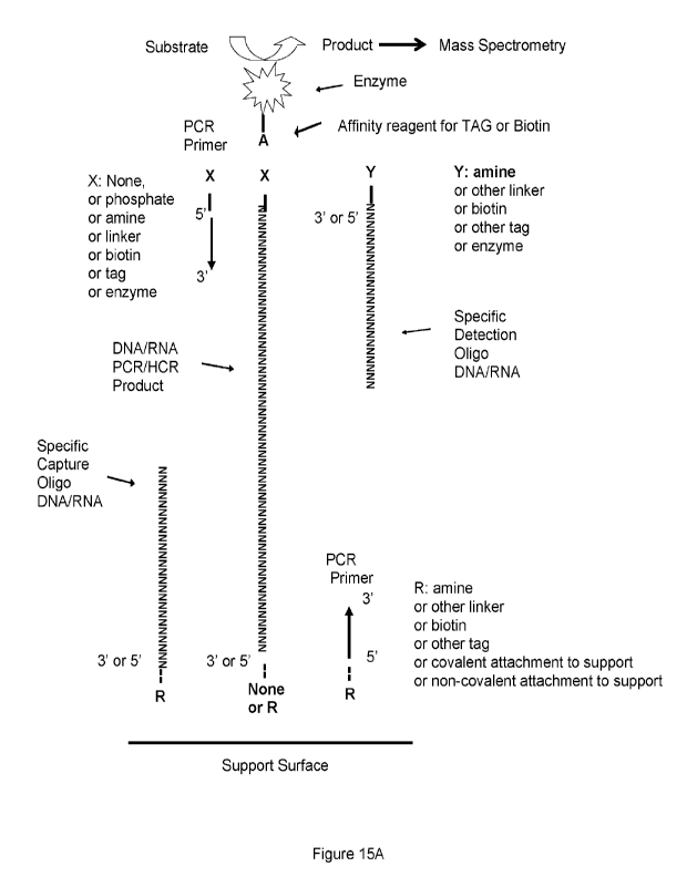
100451 Figure 15A shows an exemplified schematic illustrating
detection of, for example, a
hypothetical PCR or other products. Nucleic acid target can be DNA or RNA.
100461 Figure 15B shows a schematic illustrating detection of, for example,
a PCR product_ The primer
may be represented by AIR indicating it may be untagged or tagged for example
with biotin or presented by
CID indicating it may be unattached or attached to a solid surface_
- 9 -
RECTIFIED SHEET (RULE 9t1)
CA 03195481 2023-4- 12
WO 2022/087730
PCT/CA2021/051510
[0047] Figure 16 shows a graph of MS signal intensity measured at
m/z = 267.74-268.74 of a DNA
detection assay where (i) the hybridization step and the washing step was
performed in presence of salt, (ii)
the hybridization step and the binding of APSA was performed in presence of
salt, followed by cross-linking the
target:detection:enzyme complex with glutaraldehyde (GA) prior to enzyme
reaction in Tris buffer, (iii) the
hybridization step and the binding of APSA were performed in presence of salt,
followed by washing with a
volatile washing solution comprising either ammonium bicarbonate buffer
(AMBIC) or ethanolamine buffer (EA)
and the enzyme reaction occurring in a volatile substrate reaction buffer
comprising either ammonium
bicarbonate buffer (AMBIC) or ethanolamine buffer (EA), or (iv) the
hybridization step and the binding of APSA
were performed in presence of salt, followed by the enzyme reaction occurring
in presence of a polymer (PEG)
or dextran sulfate sodium (DSS). 10 mM Tris was used as a negative control for
the MS measurement. Zero
target nucleic acid (0) is used as negative control for the DNA detection
assay.
[0048] Figure 17 shows a flowchart illustrating exemplary methods
of the present disclosure.
[0049] Figure 18 shows a graph of MS signal intensity (log scale,
y-axis, intensity m/z = 268) of HIV
DNA detection assay at different concentrations of target nucleotide acid
molecule (log scale, x-axis) of 10g10
(attomolar+1) i.e. from 0 to 100 picomolar concentration where 1 microlitre
was injected.
[0050] Figure 19 shows a graph of MS signal intensity (log scale,
y-axis, m/z = 268) of SARC-CoV2
DNA detection assay at different concentrations of target nucleotide acid
molecule (log scale, x-axis) of 10g10
(picomolar+1) from 0 to 100 nM concentration where 1 micro litre was injected
on to the HPLC column (i.e. 0
to 100 femtomole on column).
[0051] Figure 20 shows a graph of MS signal intensity at m/z 136 at
different concentrations of HIV DNA
target nucleic acid molecule (1 pM to 500 pM).
[0052] Figure 21 shows a graph of MS signal intensity at m/z 136
at different concentrations of SARS-
CoV 2 target nucleic acid molecule (100 fM to 10 nM).
[0053] Figure 22 shows a graph of MS signal intensity at m/z 136
at different concentrations of STEC
target nucleic acid molecule (1 pM to 1 nM).
[0054] Figure 23 shows a graph of MS signal intensity at m/z 136
at different concentrations of hemolysin
target nucleic acid molecule (1 pM to 1 nM).
[0055] Figure 24A shows a graph of MS signal intensity at m/z 136
at different concentrations of HIV
258nt PCR product target nucleic acid molecule.
[0056] Figure 24B shows an image of a GelRed stained agarose gel showing
PCR reactions using
increasing amounts of HIV plasmid.
[0057] Figure 24 C shows a graph quantitiation of bands in Figure
24B.
- 10 -
CA 03195481 2023-4- 12
WO 2022/087730
PCT/CA2021/051510
[0058] Figure 25 shows a graph of MS signal intensity at m/z 136
at different concentrations of SARS-
CoV-2 target nucleic acid molecule.
[0059] Figure 26 shows a graph of MS signal intensity at m/z 136
at different concentrations of HIV
target nucleic acid molecule (100 fM to 100 nM).
[0060] Figure 27 shows a graph of MS signal intensity at m/z 268 at
different concentrations of SARS-
CoV-2 target nucleic acid molecule (1 pM to 1 pM) where the capture is bound
to PVDF.
[0061] Figure 28A shows an image of gels where the upper panel
showing PCR products produced
using biotinylated HIV forward primer 3 and unlabelled HIV reverse primer 3
and where the lower panel showing
PCR products produced using unlabelled HIV forward primer 3 and biotinylated
HIV reverse primer 3.
[0062] Figure 28B shows a graph of MS signal intensity at m/z 136 at
different concentrations of HIV
template using biotin labelled HIV forward primer and unlabelled HIV reverse
primer.
[0063] Figure 28C shows a graph of MS signal intensity at m/z 136
at different concentrations of HIV
template using unlabelled HIV forward primer and biotin labelled HIV reverse
primer.
[0064] Figure 29 shows a graph of MS signal intensity at m/z 136
for HIV synthetic target immobilized
on PDVF.
[0065] Figure 30A shows an image of a gel showing various COVID-
19 FOR reactions.
[0066] Figure 30B shows an image of a gel showing COVID-19 FOR
reactions at different
concentrations of COVID-19 template.
DESCRIPTION OF VARIOUS EMBODIMENTS
I. Definitions
[0067] Unless otherwise indicated, the definitions and
embodiments described in this and other
sections are intended to be applicable to all embodiments and aspects of the
present disclosure herein
described for which they are suitable as would be understood by a person
skilled in the art.
[0068] The term "or" "and/or" as used herein means that the listed items
are present, or used,
individually or in combination. In effect, this term means that "at least one
of" or "one or more" of the listed items
is used or present.
[0069] As used in the present disclosure, the singular forms "a",
"an" and "the" include plural
references unless the content clearly dictates otherwise. For example, an
embodiment including "a compound"
should be understood to present certain aspects with one compound, or two or
more additional compounds.
[0070] In embodiments comprising an "additional" or "second"
component, such as an additional or
second compound, the second component as used herein is chemically different
from the other components or
- 11 -
CA 03195481 2023-4- 12
WO 2022/087730
PCT/CA2021/051510
first component. A "third" component is different from the other, first, and
second components, and further
enumerated or "additional" components are similarly different.
[0071] As used in this disclosure and claim(s), the words
"comprising" (and any form of comprising,
such as "comprise" and "comprises"), "having" (and any form of having, such as
"have" and "has"), "including"
(and any form of including, such as "include" and "includes") or "containing"
(and any form of containing, such
as "contain" and "contains"), are inclusive or open-ended and do not exclude
additional, unrecited elements or
process steps.
[0072] The term "consisting" and its derivatives as used herein
are intended to be closed terms that
specify the presence of the stated features, elements, components, groups,
integers, and/or steps, and also
exclude the presence of other unstated features, elements, components, groups,
integers and/or steps.
[0073] The term "consisting essentially of", as used herein, is
intended to specify the presence of the
stated features, elements, components, groups, integers, and/or steps as well
as those that do not materially
affect the basic and novel characteristic(s) of these features, elements,
components, groups, integers, and/or
steps.
[0074] The term "suitable" as used herein means that the selection of the
particular compound or
conditions would depend on the specific synthetic manipulation to be
performed, the identity of the molecule(s)
to be transformed and/or the specific use for the compound, but the selection
would be well within the skill of a
person trained in the art.
[0075] The term "amine" or "amino," as used herein, whether it is
used alone or as part of another
group, refers to groups of the general formula NR'R", wherein R' and R" are
each independently selected from
hydrogen or C1_6alkyl.
[0076] The term "atm" as used herein refers to atmosphere.
[0077] The term "MS" as used herein refers to mass spectrometry.
[0078] The term "aq." as used herein refers to aqueous.
[0079] Me0H as used herein refers to methanol.
[0080] MeCN as used herein refers to acetonitrile.
[0081] HCI as used herein refers to hydrochloric acid.
[0082] pwave as used herein refers to a microwave reaction
vessel.
[0083] LCMS as used herein refers to liquid chromatography-mass
spectrometry.
[0084] TRIS as used herein refers to tris(hydroxymethyl)aminomethane.
[0085] EDTA as used herein refers to ethylenediaminetetraacetic
acid.
- 12 -
CA 03195481 2023-4- 12
WO 2022/087730
PCT/CA2021/051510
[0086] The term "adenosine monophosphate" or "AMP" as used herein
means a compound having
the structure:
H,N1
N
0
HO¨P-0 N
0
0
OH OH
or pharmaceutically acceptable salts or solvates thereof as well as mixtures
thereof. AMP can be obtained for
example from Sigma Aldrich.
[0087] [00133] The term "Amplex Red" or "AR" as used herein
means:
HO 0 0 OH
oCH3
or pharmaceutically acceptable salts or solvates thereof as well as mixtures
thereof. Amplex Red can be
obtained for example from Resazurin which is structurally related and has
the formula 7-Hydroxy-3H-
phenoxazin-3-one 10-oxide is also referred to as Amplex Red. Accordingly,
Amplex Red as used herein
includes both AR and Resazurin.
[0088] The term "5-Bromo-4-chloro-3-indolylphosphate" or "BCIP"
means as used herein a compound
having the structure:
0
CI O-P-OH
r OH
or pharmaceutically acceptable salts or solvates thereof as well as mixtures
thereof. BCIP can be obtained for
example from Sigma Aldrich.
[0089] The term "ionizable product", as used herein means a
product generated by a reporter enzyme,
that comprises one or more ionizable groups.. For example, an ionizable
product may have one or more basic
or amine groups for positive ionization and one or more acidic or hydroxyl
groups for negative ionization.
- 13 -
CA 03195481 2023-4- 12
WO 2022/087730
PCT/CA2021/051510
Ionizable groups may include =NH, -NH2, guanidinium, methyl, ethyl, alky,
phenyl, ribose, inositiol,
phospholipid, carbohydrate, nucleic acid, carbonyl, aldehyde, ketone,
carboxyl, hydroxyl, enol, guanidium,
imidazole, sulfhydryl, disulfide, sulfate, phosphate, sulfonyl, nitrate,
nitric oxide, thioester, ester, ether,
anhydride, phosphoryl, mixed anhydride, and/or other ionizable groups known in
the art. An ionizable product
assessed, optionally efficiently enters the gas phase by electrospray
ionization.
[0090] The term "L-(+)-2-amino-6-phosphonohexanoic acid" as used
herein means:
0 NH2
HO - P -(CH2)4
1
OH COOH
or pharmaceutically acceptable salts or solvates thereof as well as mixtures
thereof. L-(+)-2-amino-6-
phosphonohexanoic acid can be obtained for example from Sigma Aldrich.
[0091] The term "Lumigen TMA-3" or "TMA-3" as used herein means
H3CS
HO OH
Ph
or pharmaceutically acceptable salts or solvates thereof as well as mixtures
thereof. TMA-3 can be obtained
for example from Beckman Coulter Company.
[0092] The term "Lumigen TMA-6" or "TMA-6" as used herein means
(-1'1
HO OH
Ph
or pharmaceutically acceptable salts or solvates thereof as well as mixtures
thereof. TMA-6 can be obtained
for example from Beckmann Coulter Company.
[0093] The term "4-Methylumbelliferyl phosphate" or "4-MUP" as
used herein means a compound
having the structure:
- 1 4 -
CA 03195481 2023-4- 12
WO 2022/087730
PCT/CA2021/051510
CH3
0 0 0
HO¨P¨OH
II
or pharmaceutically acceptable salts or solvates thereof as well as mixtures
thereof. 4-MUP can be obtained
for example from Sigma Aldrich.
[0094] The term "Naphthol ASMX phosphate" as used herein means a
compound having the structure:
00) CH3
0
CH3
0
0=P¨OH
OH
or pharmaceutically acceptable salts or solvates thereof as well as mixtures
thereof. Naphthol ASMX phosphate
can be obtained for example from Sigma Aldrich.
[0095] The term "O-phospho-DL-Threonine" as used herein means a
compound having the structure:
0
HO ¨P ¨OH
0 NH2 0
CH3C H ___________ CH ¨C ¨ OH
or pharmaceutically acceptable salts or solvates thereof as well as mixtures
thereof. 0-phospho-DL-Threonine
can be obtained for example from Sigma Aldrich.
[0096] The term "Para nitrophenol phosphate" or "PNPP" as used
herein means a compound having
the structure:
0
II
P¨OH
0 I
OH
NO2
- 15 -
CA 03195481 2023-4- 12
WO 2022/087730 PCT/CA2021/051510
or pharmaceutically acceptable salts or solvates thereof as well as mixtures
thereof. Para nitrophenol
phosphate can be obtained for example from Sigma Aldrich.
[0097] The term "phenylbenzene w phosphono-a-amino acid" as used
herein means compound
having the structure:
HO¨P¨OH 0
L\
NH2 H
[0098] or pharmaceutically acceptable salts or solvates thereof
as well as mixtures thereof.
Phenylbenzene w phosphono-a-amino acid can be obtained for example from Sigma
Aldrich.
[0099] The term "pyridoxamine 5- phosphate" or "PA5P" as used
herein means compound having the
structure:
H2N
HO 0
= //
OH
/ 0 ¨=
HO
.===
CH3
or pharmaceutically acceptable salts or solvates thereof as well as mixtures
thereof. PA5P can be obtained for
example from Sigma Aldrich.
[00100] The term "sphingosine-1 phosphate" as used herein means a
compound having the structure:
OF-
or pharmaceutically acceptable salts or solvates thereof as well as mixtures
thereof. Sphingosine-1 phosphate
can be obtained for example from Sigma Aldrich.
[00101] The term "detection oligonucleotide probe" as used herein
comprises a oligonucleotide coupled
to a secondary target moiety such as biotin wherein the oligonucleotide or a
portion thereof is complementary
to and binds selectively to a target nucleic acid molecule, for example, but
not limited to, a bacterial, viral or
fungal nucleic acid sequence. The detection oligonucleotide probe can be a
detection oligonucleotide primer in
some embodiments. The detection oligonucleotide probe can also optionally be
coupled to the secondary target
moiety, such as biotin. The detection oligonucleotide probe can also
optionally be coupled to an enzyme such
as the reporter enzyme. For example, the detection oligonucleotide can be
optionally coupled to enzymes or
catalysts including but not limited to ribozyme, a DNAzyme, phosphatase (for
example AP), peroxidase (for
example HRP), DNA polymerase, or glucose oxidase. For example, the detection
oligonucleotide probe can
- 16 -
CA 03195481 2023-4- 12
WO 2022/087730
PCT/CA2021/051510
comprise a single stranded oligonucleotide sequence complementary to that of
the target nucleic acid molecule
and can selectively bind to the target nucleic acid molecule through
hybridization.
[00102]
It can be appreciated by a person skilled in the art that the
secondary target moiety and the
secondary target binding moiety have high mutual affinity such that the
secondary target moiety and the
secondary target binding moiety selectively bind to each other. Accordingly,
it can be appreciated by a person
skilled in the art that a suitable secondary target binding moiety can be
selected by a person skilled in the art
based on the nature of the secondary target moiety and vice versa. The
following list contains non-limiting
examples of pairs of selectively binding chemical entities. The secondary
target moiety and the secondary target
binding moiety can be selected from pairs of chemical entities listed below.
For example, the secondary target
moiety can be biotin. For example, the secondary target binding moiety can be
avidin or streptavidin.
List of high affinity selective binding pairs of chemical entities:
SEQ ID NO Tag Binding
partner
Biotin Avidin
Biotin Streptavidin
47 ALFA-tag (SRLEEELRRRLTE) Single-domain
antibodies
48 AviTag (GLNDIFEAQKIEVVHE) Avidin or
Streptavidin
biotinylated
49 C-Tag (EPEA) single-domain
camelid antibody
50 Calmodulin-Tag Calmodulin
(KRRVVKKNFIAVSAANRFKKISSSGAL)
51 Polyglutamate tag (EEEEEE) anion-exchange
resin (e.g. Mono-Q)
Polyarginine tag cation-exchange
resin (from 5 to 9
consecutive R)
52 E-tag (GAPVPYPDPLEPR) Anti-E-tag
antibody
53 FLAG-tag (DYKDDDDK) Anti-FLAG-tag
antibody
54 HA-Tag (YPYDVPDYA) Anti-HA-Tag
antibody
His-Tag (5-10 histidines) Nickel or cobalt
chelate
55 Myc-Tag (EQKLISEEDL) Anti-Myc-Tag
antibody
56 NE-tag (TKENPRSNQEESYDDNES) Anti-NE-Tag IgG1
antibody
57 Rho1D4-tag (TETSQVAPA) Anti-Rho1D4-tag
antibody
58 S-tag (KETAAAKFERQHMDS) Anti-S-tag
antibody
59 SBP-tag Streptavidin
(MDEKTTGVVRGGHVVEGLAGELEQLR
ARLEHHPQGQREP)
60 Softag 1(SLAELLNAGLGGS) Anti-Softag 1
antibody
61 Softag 3 (TQDPSRVG) Anti-Softag 3
antibody
62 Spot-tag (PDRVRAVSHWSS) Single-domain
antibody nanobody
63 Strep-tag (WSHPQFEK) Streptavidin
64 Strep-tag (WSHPQFEK) Streptactin
65 T7-tag (MASMTGGQQMG) Anti-T7-tag
antibody
66 TC-tag (CCPGCC) FlAsH and
ReAsH biarsenical
compounds
67 Ty1 tag (EVHTNQDPLD) Anti-Ty1 tag
antibody
68 V5 tag (GKPIPNPLLGLDST) Anti-V5 tag
antibody
69 VSV-tag (YTDIEMNRLGK) Anti-VSV tag
antibody
70 Xpress tag (DLYDDDDK) Anti-Xpress tag
antibody
71 lsopeptag (TDKDMTITFTNKKDAE) pilin-C protein
-17-
CA 03195481 2023-4- 12
WO 2022/087730
PCT/CA2021/051510
72 SpyTag (AHIVMVDAYKPTK) SpyCatcher protein
73 SnoopTag (KLGDIEFIKVNK) SnoopCatcher
protein
74 DogTag SnoopTagJr protein
(DIPATYEFTDGKHYITNEPIPPK)
75 SdyTag (DPIVMIDNDKPIT) SdyCatcher protein
Biotin Carboxyl Carrier Protein Streptavidin
Glutathione-S-transferase tag Glutathione
Green Fluorescent protein (GFP) tag GFP-antibody
HaloTag Haloalkane
substrates
SNAP-tag benzylguan me
derivatives
CLIP-tag benzylcytosine
derivatives
HUH-tag HUH specific DNA
sequence
Maltose-binding protein-tag Amylose agarose
Nus-tag Nus tag antibody
Thioredoxin-tag Anti-Thioredoxin-
tag antibody
Fc-tag Protein-A
sepharose
CRDSAT-tag (carbohydrate Recognition Lactose, agarose, sepharose
Domain)
[00103] The term "oligonucleotide" as used herein as used herein
refers to a sequence of nucleoside
or nucleotide monomers consisting of naturally occurring bases, sugars and
intersugar (backbone) linkages.
The term also includes modified or substituted sequences comprising non-
naturally occurring monomers or
portions thereof. The nucleic acid sequences of the present application may be
deoxyribonucleic acid
sequences (DNA) or ribonucleic acid sequences (RNA) and may include naturally
occurring bases including
adenine, guanine, cytosine, thymidine and uracil. The sequences may also
contain modified bases. Examples
of such modified bases include aza and deaza adenine, guanine, cytosine,
thymidine and uracil; and xanthine
and hypoxanthine. The nucleic acid can be either double stranded or single
stranded, and represents the sense
or antisense strand. For example, the capture, detection, target or primer
sequences can be oligonucleotides.
[00104] The term "reporter enzyme detection probe" as used herein
comprises a reporter enzyme
component comprising an enzymatic activity, coupled to a detection probe
component comprising a secondary
target binding moiety, for example avidin or streptavidin when the secondary
target moiety is biotin. The reporter
enzyme is optionally a peroxidase such as horseradish peroxidase or a
phosphatase such as alkaline
phosphatase although any stable enzyme that can produce ionizable products can
be used including for
example a lyase, hydrolase, synthase, synthetase, oxidoreductase,
dehydrogenase, oxidase, transferease,
isomerase, ligase, protease, such as trypsin, proteinase, peroxidase, glucose
oxidase, myeloperoxidase,
oxidase, monooxygenase, cytochrome, phosphatase such as alkaline phosphatase,
decarboxylase, lipase,
caspase, amylase, peptidase, transaminase, and kinase. Additional enzymes can
include DNA or RNA
polymerase, TAQ, restriction enzymes, klenow fragment, DNA ligase. The
secondary target binding moiety
selectively binds the secondary target moiety of the detection oligonucleotide
probe. For example, the
secondary target binding moiety comprises avidin or streptavidin that
selectively binds a biotinylated detection
oligonucleotide probe (e.g. wherein the secondary target moiety comprises
biotin).
- 18 -
CA 03195481 2023-4- 12
WO 2022/087730
PCT/CA2021/051510
[00105] The term "selective" as used herein in reference to a
probe, optionally an oligonucleotide, is
used contextually, to characterize the binding properties of the probe,
optionally an oligonucleotide. For
example, an oligonucleotide probe that binds selectively to a given target
nucleic acid molecule will bind to that
target nucleic acid molecule either with greater avidity or with more
specificity, relative to another, different
target nucleic acid molecule. In an embodiment, the probe, optionally an
oligonucleotide probe, binds at least
2 fold, 3 fold, or 5 fold more efficiently, optionally 3-5 fold, 5-7 fold, 7-
10, 10-15, 5-15, or 5-30 fold more efficiently.
[00106] The term "target nucleic acid molecule" as used herein
refers to any nucleic acid polymer that
comprises a sequence that is complementary to the oligonucleotide portion of a
detection oligonucleotide probe.
For example, the target nucleic acid molecule can be RNA or DNA, or
derivatives thereof. The target nucleic
acid can be any nucleic acid that is at least 30 nucleotides long. For
example, the target nucleic acid molecule
can be about or at least 30 nucleotides, about or at least 40 nucleotides,
about or at least 50 nucleotides, about
or at least 80 nucleotides, about or at least 100 nucleotides, about or at
least 130 nucleotides, about or at least
180 nucleotides, about 200 nucleotides, about 250 nucleotides, about 300
nucleotides, about 350 nucleotides,
about 450 nucleotides, about 600 nucleotides, about 700 nucleotides, about 850
nucleotides, or about 1000
nucleotides. In some embodiments, the target nucleic acid molecule is about 30
nucleotides to about 1500
nucleotides in length. For example, the target nucleic acid molecule is about
30 nucleotides to about 1000
nucleotides in length, about 30 nucleotides to about 300 nucleotides in
length, about 100 nucleotides to about
500 nucleotides in length, about 100 nucleotides to about 600 nucleotides in
length, about 100 nucleotides to
about 700 nucleotides in length, about 100 nucleotides to about 800
nucleotides in length, about 100
nucleotides to about 900 nucleotides in length, or about 100 nucleotides to
about 1000 nucleotides in length.
For example, the target nucleic acid molecule can be single stranded or double
stranded. For example, the
target nucleic acid molecule can be plasmid DNA, a bacterial, viral, or fungal
nucleic acid molecule or a
mammalian or plant nucleic acid e.g. in a gene or in mRNA. The target nucleic
acid can also be a synthetic
nucleic acid for detection of nucleic acid tagged compounds and the like.
II. Methods and Kits
[00107] Described herein is a transformative technology that
permits detection of nucleic acid
molecules in the femto mol to pico mol ranges and/or lower. It is demonstrated
herein that dection in the zepto
mol to atto mol range can be achieved.
[00108] Enzmye linked immuno sorbent assays (ELISA) are the
preferred analytical method for the
repetitive quantitative analysis of polypeptides molecules of biomedical
importance: ELISA may use reporter
enzymes such as Horseradish peroxidase (HRP) and or alkaline phosphatase (AP)
coupled to specific detection
antibodies that capture and bind to each analyte of importance (Engvall, 1971;
Van Weemen 1971).
[00109] At present substrates for the reporter enzymes horseradish
peroxidase (HRP) or alkaline
phosphatase (AP) yield colored, fluorescent or luminescent products. The
present disclosure provides a method
for detecting the enzymatic products of reporter enzymes that ionize
efficiently with a high signal to noise ratio
- 19 -
CA 03195481 2023-4- 12
WO 2022/087730
PCT/CA2021/051510
measured by mass spectrometry. Mass spectrometry is sensitive enough to permit
detections at amounts far
below ECL, fluorescence or colorimetric methods, but also permits monitoring
of multiple substrates and
products at discrete m/z values. It is possible using the methods described
herein to measure the products of
common industrial reporter enzymes to zepto mol amounts or lower with limits
of quantification to atto mol
amounts or lower.
[00110] The use of mass spectrometry to measure small molecules
may commonly reach the femto to
pico mol levels with high signal to noise. The industrial enzymes HRP or AP
for example are rugged and durable
and have a high catalysis rate for the creation of new small molecule
products. The AP or HRP enzymes are
for example covalently attached to a specific detection probe such as a
polypeptide or antibody that may bind
their target and then catalyze many different product reactions over the
course of a brief incubation. Thus, the
binding of atto mol, or even sub atto mol, amounts of enzyme-probe will yield
amounts of small molecule
products that accumulate in the femto mol to pico mol range well within the
detectable range of by LC-ESI-
MS/MS.
[00111] Liquid chromatography electrospray ionization and tandem
mass spectrometry (LC-ESI-
ms/ms) is more sensitive than colorimetric, fluorescent or ECL detection. The
combination of the enzymatic
production of reported molecules coupled with sensitive mass spectrometry for
highly ionizable substrates
should provide sensitivity in excess of RIA but without the requirement for
standards labelled with isotope or
probes labeled with isotope.
[00112] Quantification of HRP and AP is demonstrated using LC-ESI-
MS/MS to detect the products of
the AP and HRP reporter enzyme reactions. It is demonstrated herein that a
mass spectrometer can also detect
the small molecule products of reporter enzyme activity bound to a specific
molecular probe such as an
antibody. One atto mol or less of a reporter enzyme such as AP or HRP bound to
a specific molecular probe
such as a detection antibody will rapidly form femto mol to pico mol amounts
of reporter enzyme reaction
products well within the reliable detection and quantification limits of LC-
ESI-MS/MS. Hence in ELiMSA and
related DNA methods (e.g. DNA ELiMSA) the reporter enzymes such as HRP or AP
may produce a range of
products that can be easily distinguished and detected by mass spectrometry.
Antibodies coupled to reporter
enzymes that are widely used in biomedical and environmental applications can
now be detected and quantified
using very sensitive mass spectrometry to create a sensitive and flexible
system. Since mass spectrometers
can separate and analyze many analytes simultaneously using the methods
described herein can allow
identification and quantification of many different antigens at the same time
to levels far below that which is
possible by direct mass spectrometric analysis.
[00113] The reaction is reporter enzyme dependent. For example, it
is demonstrated herein that
incubating a substrate that can be acted upon by the reporter enzyme detection
probe in an appropriate
substrate reaction solution produces little or no signal in the absence of the
reporter enzyme detection probe.
In contrast, the addition of reporter enzyme detection probe comprising HRP or
AP enzyme resulted in strong
detection of an ELiMSA product ion. The product ion was shown to be dependent
on the presence of the
- 20 -
CA 03195481 2023-4- 12
WO 2022/087730
PCT/CA2021/051510
enzyme, and to be both time and concentration dependent. Thus, the ELiMSA
product ions show all the
hallmarks of an enzyme dependent assay.
[00114] Depending on the reporter enzyme or enzyme substrate,
different ionizable products can be
detected. Fragments thereof can also be detected. For example, adenosine can
be ionized and detected at 268
m/z or fragmented and the fragment can be detected at 136 m/z.
[00115] As shown in the examples, a capture oligonucleotide probe
can be used to capture a target
nucleic acid molecule. In other examples the target nucleic acid molecule can
be attached, covalently or non-
covalently, to a solid support (e.g. solid phase) directly and a labelled
detection probe optionally a labelled
primer, can be used to detect the attached target nucleic acid molecule.
[00116] In one aspect, the present disclosure includes a method of
detecting a target nucleic acid
molecule comprising
a.
i. incubating a sample putatively comprising the target nucleic acid
molecule with a capture
oligonucleotide probe that comprises a sequence complementary to the target
nucleic acid
molecule and that is attached to a solid phase, in a first binding solution,
optionally wherein the
solid phase is attached to the capture oligonucleotide probe through a linker;
or
ii. incubating a sample putatively comprising the target nucleic acid
molecule with a solid phase
to attach said sample/target nucleic acid molecule to said solid phase, in a
first binding solution,
optionally wherein the solid phase is attached to the sample/target nucleic
acid molecule
through a linker;
b. binding any target nucleic acid molecule to a detection oligonucleotide
probe in a second binding
solution under conditions for forming a target:detection complex;
c. incubating any target:detection complex with a reporter enzyme detection
probe in a third binding
solution under conditions for forming a target:detection:enzyme complex;
d. washing the solid phase to remove any unbound reporter enzyme detection
probe with a washing
solution;
e. incubating any target:detection:enzyme complex with a reporter enzyme
detection probe substrate in
a substrate reaction solution to generate one or more ionizable products; and
f. detecting at least one of the one or more ionizable products using mass
spectrometry (MS),
wherein
-21 -
CA 03195481 2023-4- 12
WO 2022/087730
PCT/CA2021/051510
I. at
least the third binding solution among the first binding solution, the second
binding solution, and
the third binding solution is substantially free of inorganic salt;
ii the washing solution is substantially free of inorganic salt;
iii. the method further comprises cross-linking components of any
target:detection:enzyme complex
and the capture oligonucleotide probe prior to the optional step d) and the
step e); and/or
iv. the method further comprises separating the one or more ionizable
products prior to detection using
MS; and
wherein detection of the at least one of the one or more ionizable products is
indicative of the sample
comprising the target nucleic acid molecule.
[00117] The
detection oligonucleotide probe can be a detection oligonucleotide primer. In
such cases,
the step comprises amplifying the target nucleic acid molecule with a
detection oligonucleotide primer, in an
amplification solution and binding any amplified target to the detection
oligonucleotide probe in the second
binding solution under conditions for forming a target:detection complex.
[00118] it
is also contemplated that the detection oligonucleotide probe can be
covalently attached to
the reporter enzyme directly through covalent attachment, optionally though a
linker. In such a case, the
target:detection complex is sufficient to react with the reporter enzyme
detection probe substrate. Thus, the
secondary target moiety and the secondary target binding moiety are not
required. Accordingly, in another
aspect, the present disclosure includes a method of detecting a target nucleic
acid molecule comprising
a.
i.
incubating a sample putatively comprising the target nucleic acid
molecule with a capture
oligonucleotide probe that comprises a sequence complementary to the target
nucleic acid
molecule and that is attached to a solid phase, in a first binding solution,
optionally wherein the
solid phase is attached to the capture oligonucleotide probe through a linker;
or
ii.
incubating a sample putatively comprising the target nucleic acid molecule
with a solid phase
to attach said sample/target nucleic acid molecule to said solid phase, in a
first binding solution,
optionally wherein the solid phase is attached to the sample/target nucleic
acid molecule
through a linker;
b. binding any target nucleic acid molecule to a detection oligonucleotide
probe in a second binding
solution under conditions for forming a target:detection complex, the
detection oligonucleotide probe
comprising an oligonucleotide and a reporter enzyme;
- 22 -
CA 03195481 2023-4- 12
WO 2022/087730
PCT/CA2021/051510
c. washing the solid phase to remove any unbound detection oligonucleotide
probe with a washing
solution;
ft
incubating the target:detection complex with a reporter enzyme
detection probe substrate in a substrate
reaction solution to generate one or more ionizable products; and
e. detecting one or more of the one or more ionizable products using mass
spectrometry (MS),
wherein either
I. at
least the second binding solution among the first binding solution, and the
second binding
solution is substantially free of inorganic salt;
ii. the washing solution is substantially free of inorganic salt;
iii. the
method further comprises cross-linking components of any target:detection
complex and the
capture oligonucleotide probe prior to the optional step c) and the step d);
andor
iv. the
method further comprises separating the one or more ionizable products prior
to detection using
MS; and
wherein detection of the at least one of the one or more ionizable products is
indicative of the sample
comprising the target nucleic acid molecule.
[00119] The
detection oligonucleotide probe can be a detection oligonucleotide primer. In
such cases,
the step comprises amplifying the target nucleic acid molecule with a
detection oligonucleotide primer, in an
amplification solution and binding any amplified target to the detection
oligonucleotide probe in the second
binding solution under conditions for forming a target:detection complex.
[00120] In
some embodiments, the second binding solution, the third binding solution and
the substrate
reaction solution each comprises a Tris buffer.
[00121] In
some embodiments, the capture oligonucleotide probe is directly immobilized to
the solid
phase, optionally by non-covalent or covalent binding to the solid phase.
[00122] In
some embodiments, the capture oligonucleotide probe comprises a
oligonucleotide that has
a sequence complementary to a part of the target nucleic acid molecule that is
at least 25 nucleotides in length,
at least 35 nucleotides in length, optionally the capture oligonucleotide
probe has a sequence complementary
to a part of the sequence of the target nucleic acid molecule that is about 30
nucleotides to about 60 nucleotides
in length, or about 40 nucleotides to about 55 nucleotides in length.
- 23 -
CA 03195481 2023-4- 12
WO 2022/087730
PCT/CA2021/051510
[00123] In some embodiments, the detection oligonucleotide probe
comprises an oligonucleotide that
has a sequence complementary to another part of the target nucleic acid
molecule, and a secondary target
moiety selected from biotin.
[00124] In some embodiments, the sequence of the oligonucleotide
of the detection oligonucleotide
probe complementary to the other part of the sequence of the target nucleic
acid molecule is at least 25
nucleotides in length, at least 35 nucleotides in length, optionally the
detection oligonucleotide probe is about
30 nucleotides to about 60 nucleotides in length, or about 40 nucleotides to
about 55 nucleotides in length.
[00125] In some embodiments, the capture oligonucleotide probe and
the detection oligonucleotide
probe can both bind the target nucleic acid molecule at non-overlapping
regions, optionally the non-overlapping
regions are directly adjacent, optionally the non-overlapping regions are at
least one nucleotide apart, optionally
the non-overlapping regions are at least 5 nucleotides apart, optionally the
non-overlapping regions are about
2 nucleotides, about 5 nucleotides, about 10 nucleotides, about 20
nucleotides, about 25 nucleotides, about 50
nucleotides, about 100 nucleotides, about 500 nucleotides, or about 1000
nucleotides apart. In some
embodiments, the non-overlapping regions are about 1kb apart. In some
embodiments, the non-overlapping
regions are more than 1kb apart.
[00126] In some embodiments, when a binding solution and/or a
washing solution is substantially free
of inorganic salt, the binding solution and/or the washing solution is each
independently a volatile solution. In
some embodiments, the volatile solution comprises a volatile buffer. In some
embodiments, the volatile buffer
is selected from ethanolamine, ammonium bicarbonate, ammonium formate,
pyridinium formate,
trialkylammonium/formic acid, ammonium acetate, trialkylammonium bicarbonate,
N-ethylmorpholine/acetate,
trialkylammonium acetate, or combinations thereof. In some embodiments, the
volatile buffer is selected from
ethanolamine, ammonium acetate, trialkylammonium bicarbonate, or combinations
thereof. In some
embodiments, the trialkylammonium is selected from trimethylammonium,
triethylammonium, or combinations
thereof. In some embodiments, the volatile buffer is ethanolamine. It can be
appreciated by a person skilled in
the art that ammonium bicarbonate is not stable to heat. For example, ammonium
bicarbonate decomposes at
about or above 90 C. Accordingly, for steps involving heating, other volatile
buffers such as ethanolamine is
preferred.
[00127] In some embodiments, when the first binding solution, the
second solution, the third binding
solution, and/or the washing solution is substantially free of inorganic salt,
the first binding solution, the second
solution, the third binding solution, and/or the washing solution each
independently comprises ethanolamine,
optionally the second binding solution and the third binding solution each
comprises ethanolamine, optionally
the first binding solution, the second binding solution, and the third binding
solution each comprises
ethanolamine, optionally the washing solution comprises ethanolamine.
[00128] In some embodiments, step a) and step b) are performed
simultaneously, and the first binding
solution of step a) is the second binding solution of step b).
- 24 -
CA 03195481 2023-4- 12
WO 2022/087730
PCT/CA2021/051510
[00129] In some embodiments, the first binding solution, the
second binding solution, the third binding
solution, and the substrate reaction solution each independently has a pH of
about 7 to about 10, optionally of
about 7 to about 8, optionally about 8.8.
[00130] In some embodiments, any of the volatile binding solutions
can be used to wash the solid
support, optionally to remove any inorganic salt that may be present.
[00131] In some embodiments, the target:detection:enzyme complex
is incubated with the reporter
enzyme detection probe substrate in the substrate reaction solution to
generate the one or more ionizable
products for a period of time less than 72 hours, less than 24 hours, less
than 12 hours, less than 60 minutes,
less than 50 minutes, less than 40 minutes, less than 30 minutes, less than 20
minutes, less than 15 min, less
than 10 min, less than 5 min, less than 2 min, or less than 1 min.
[00132] In some embodiments, at least the third binding solution
among the first binding solution, the
second binding solution, and the third binding solution is substantially free
of inorganic salt and comprises a
volatile buffer described herein.
[00133] In some embodiments, the method comprises washing the
solid phase to remove any unbound
reporter enzyme detection probe with the washing solution, wherein the washing
solution is substantially free
of inorganic salt and comprises a volatile buffer as described herein.
[00134] In some embodiments, the components of any
target:detection:enzyme complex and the
capture oligonucleotide probe are cross-linked prior to the optional step d)
and the step e), and the cross-linking
is through H-hydroxysuccinimide (NHS), N-oxysuccinimide (NOS), maleimide,
hydrazide, glutaraldehyde
coupling, disuccinimidyl suberate (DSS) cross-linking or PEG crosslinking.
[00135] In some embodiments, the cross-linking of the components
of any target:detection:enzyme
complex and the capture oligonucleotide probe is through glutaraldehyde
coupling, DSS cross-linking, or PEG
cross-linking.
[00136] In another aspect, the present disclosure includes a
method of quantifying the amount of a
target nucleic acid molecule in a sample comprising the steps:
a. detecting a target nucleic acid molecule according to a method of the
present disclosure; and
b. quantifying the amount of target nucleic acid molecule in the sample
based on the intensity of the signal
for one or more of the ionizable products detected by mass spectrometry.
[00137] In some embodiments, the quantification comprises
comparing the intensity of the signal for
one or more products against signal intensities generated using known
quantities of target substance, under
similar conditions.
[00138] In some embodiments, the target nucleic acid molecule is
present or suspected to be present
in the sample in or up to a pico mol, femto mol, or atto mol range.
- 25 -
CA 03195481 2023-4- 12
WO 2022/087730
PCT/CA2021/051510
[00139]
In some embodiments, the target nucleic acid molecule is selected
from DNA, RNA, and
combinations and derivatives thereof.
[00140]
In some embodiments, the sample is a biological sample, industrial
product, environmental
sample, or a polymerase chain reaction (PCR) reaction product. In some
embodiments, the biological sample
is a blood sample, urine sample, fecal sample, effusate, tissue sample or
sputum sample.
[00141]
In another aspect, the present disclosure includes a method of
detecting a target nucleic acid
molecule comprising
performing a nucleic acid amplification such as a polymerase chain reaction
(PCR) or a hybridization
chain reaction (HCR) or rolling circle reaction or other nucleic acid reaction
on a test sample putatively
comprising the target nucleic acid molecule with a modified primer and a
second primer to obtain an
amplified nucleic acid product, optionally a PCR product, comprising the
modified primer, the modified
primer being functionalized with a secondary target moiety or a reporter
enzyme;
separating the amplified nucleic acid product from any unreacted modified
primer;
when the modified primer is functionalized with the secondary target moiety,
incubating the amplified
nucleic acid product with a reporter enzyme detection probe in a first binding
solution under conditions
to form an amplified nucleic acid product:reporter enzyme complex, and
removing any unbound
reporter enzyme detection probe with a washing solution, the reporter enzyme
detection probe
comprising a secondary target binding moiety and a reporter enzyme;
incubating the amplified nucleic acid product or the amplified nucleic acid
product:reporter enzyme
complex with a reporter enzyme substrate in a substrate reaction solution to
generate one or more
ionizable products; and
detecting the one or more ionizable products using mass spectrometry (MS),
wherein when the modified primer is a forward primer, the second primer is a
reverse primer, and
wherein when the modified primer is a reverse primer, the second primer is a
forward primer.
[00142]
In some embodiments, the second primer is attached to a solid phase,
optionally the second
primer is attached to the solid phase through a linker.
[00143]
In some embodiments, the second primer is directly attached to the
solid phase, optionally by
non-covalent or covalent binding to the solid phase.
[00144]
In some embodiments, the separation of the unreacted modified primer
from the amplified
nucleic acid product is by centrifugation, filtration and/or solvent wash.
- 26 -
CA 03195481 2023-4- 12
WO 2022/087730
PCT/CA2021/051510
[00145] In some embodiments, the method further comprises
incubating the amplified nucleic acid
product comprising the modified primer with a solid phase in a second binding
solution under conditions to bind
the amplified nucleic acid product onto the solid phase, prior to incubating
the amplified nucleic acid product
with the reporter enzyme detection probe, the solid phase having a capture
oligonucleotide probe attached
thereon that comprises a sequence complementary to the amplified nucleic acid
product, optionally, the solid
phase is attached to the capture oligonucleotide probe through a linker.
[00146] In some embodiments, the capture oligonucleotide probe is
directly attached to the solid phase,
optionally by non-covalent or covalent binding to the solid phase.
For example, various embodiments are shown in Figure 15A and Figure 15B.
Figures 15 A and B show
embodiments that can be referred to as "full sandwich", or "half sandwich"
involving in some embodiments
amplification, for example producing a PCR product, and embodiments of
covalent or chemical linkage or non-
covalent attachment such as adsorption to a solid support. For specifically,
"half-sandwhich" embodiment is
shown using biotinylated primers and optionally a 5' attachment or a 3'
attachment of the capture oligonucleotide
probe, for example using a NOS chemical attachment plate. Also shown is an
embodiment, wherein the target
nucleic acid molecule is adsorbed to PVDF and detected with a biotinylated
detector probe. Also shown are
"full sandwich" embodiments, where a capture oligonucleotide probe and a
detection oligonucleotide probe are
used. The target nucleic acid sample can be chemically attached or adsorbed
and detected using a tagged
detector probe. Other embodiments and combinations are also described herein.
[00147] The "R" shown in Figure 15A can be any one or more of an
amine or other linker, biotin or other
tag, or attachment to a solid support. For example, the amine may be an amine
group present in an
oligonucleotide or added to the oligonucleotide.
[00148] The linker may be a chemical bond or may for example
include a moiety such as a PEG chain
that ends in amine. Other moieties such as a a carbon chain that comprises an
amine.
[00149] The linker can be an amine (or amine linkage once linked),
or NHS, or carboxyl link or cysteine
link or a PEG for example with an amine or amine reactive group or any other
suitable link. Others can be used
including others that are described herein or in the table below.
Reactivity class Chemical group
Carboxyl-to-amine reactive groups Carbodiimide (e.g., EDC)
Amine-reactive groups NHS ester
I mid ester
Pentathiorophenyl ester
Hydroxymethyl phosphine
- 27 -
CA 03195481 2023-4- 12
WO 2022/087730
PCT/CA2021/051510
Sulfhydryl-reactive groups Maleimide
Haloacetyl (Bromo- or lodo-)
Pyridyldisulfide
Thiosulfon ate
Vinylsulfone
Aldehyde-reactive groups Hydrazide
i.e., oxidized sugars (carbonyls) Alkoxyamine
Photoreactive groups Diazirine
i.e., nonselective, random insertion Aryl Azide
Hydroxyl (nonaqueous)-reactive groups lsocyanate
[00150] Various tags can be used. For example the tag may be
biotin, ALFA-tag, AviTag, C-tag,
Calmoudulin-Tag, Polyglutamate Tag, E-Tag, Flag-tag, HA-tag, His-Tag, myc-Tag,
NE-tag, Rhol D4-Tag, 5-
Tag, SBP-Tag, Softag 1, Softag 3, Spot-tag, Strept-tag, T7-tag, TC-tag, Ty1
tag, V5 tag, VSV-tag, Xpress tag,
Isopeptag, SpyTag, SnoopTag, DogTag, Sdy Tag, Biotin carboxyl carrier protein,
glutathione-S-transferase tas,
GFP tag, HaloTag, SNAP-tag, CLIP-tag, HUH-Tag, Maltose-binding protein tag,
Nus-tag, thioredoxin-tag, Fc-
tag, or CRDSAT-tag. Others for example described elsewhere herein can also be
used. The tag is some
embodiments is biotin.
[00151] As discussed covalent and non covalent atttachments can be
used. For example, attachment
to the support, can be a covalent attachment such as [H-hydroxysuccinimide
(NHS), N-oxysuccinimide (NOS),
maleimide, hydrazide, glutaraldehyde coupling, or PEG cross-linking or a non-
covalent attachment [Adsorption
to PVDF, silica, polystyrene, nylon, etc. This may be effected through or
without a linker. Such as adsorption
to PVDF, polystyrene or silica or nylon, acrylamide, alginate, melamine or any
othe supprt
[00152] The solid support can for example be a plate such as a
polystyrene plate, or chemically reactive
NOS polystyrene plate, and the plate may be a 96 well plate, micro well or
nanowell plate, a membrane such
as PVDF membrane in for example a 96 well plate, or a micro or nanosized
particle such as a bead. Other
attacments include for example silica, PVDF, polystyrene, nylon, acrylamide,
alginate, melamine
[00153] A more specific example is shown in Figure 15B. In this Figure, P
refers to phosphate and N
refers to amine. Primer A/B refers to the primer with and without a tag such
as biotin. Primer C/D refers to the
primer free or affixed to a surface support.
- 28 -
CA 03195481 2023-4- 12
WO 2022/087730
PCT/CA2021/051510
[00154] In some embodiments, the capture oligonucleotide probe has
a sequence complementary to a
part of the sequence of the amplified nucleic acid product comprising the
modified primer.
[00155] In some embodiments, the first binding solution and/or the
washing solution is volatile and
substantially free of NaCI.
[00156] In some embodiments, the second binding solution being volatile and
substantially free of NaCI.
[00157] In some embodiments, the first binding solution or the
second binding solution each comprises
a volatile buffer.
[00158] In some embodiments, the volatile buffer is selected from
ethanolamine, ammonium
bicarbonate, ammonium formate, pyridinium formate, trialkylammonium/formic
acid, ammonium acetate,
trialkylammonium bicarbonate, N-ethylmorpholine/acetate, trialkylammonium
acetate, and combinations
thereof.
[00159] In some embodiments, the volatile buffer is selected from
ethanolamine, ammonium acetate,
trialkylammonium bicarbonate, and combinations thereof.
[00160] In some embodiments, the trialkylammonium is selected from
trimethylammonium,
triethylammonium, and combinations thereof.
[00161] In some embodiments, the volatile buffer is ethanolamine.
[00162] In some embodiments, the method further comprising washing
the solid phase with a blocking
agent, optionally bovine serum albumin (BSA), prior to binding the amplified
nucleic acid product to the solid
phase.
[00163] In some embodiments, the first binding solution or the second
binding solution each
independently has a pH of about 7 to about 10, optionally of about 7 to about
8, optionally about 8.8.
[00164] In some embodiments, the removing of any unbound reporter
enzyme detection probe from
the amplified nucleic acid product:reporter enzyme complex is by
centrifugation, filtration and/or solvent wash.
[00165] In some embodiments, the amplified nucleic acid product or
the amplified nucleic acid
product:reporter enzyme complex is incubated with the reporter enzyme
substrate in the substrate reaction
solution to generate the one or more ionizable products for a period of time
less than 72 hours, less than 24
hours, less than 12 hours, less than 60 minutes, less than 50 minutes, less
than 40 minutes, less than 30
minutes, less than 20 minutes, less than 15 min, less than 10 min, less than 5
min, less than 2 min, or less than
1 min.
[00166] In some embodiments, the test sample is a biological sample,
industrial product, or
environmental sample.
[00167] In some embodiments, the biological sample is a blood
sample, urine sam-pie, fecal sample,
effusate, tissue sample or sputum sample.
- 29 -
CA 03195481 2023-4- 12
WO 2022/087730
PCT/CA2021/051510
[00168] In some embodiments, the PCR is selected from real time
PCR (rtPCR), quantitative PCR
(qPCR), reverse transcription PCR, nested PCR, hy-bridization chain reaction,
rolling circle PCR, and substrate
recycling reaction.
[00169] The reporter enzyme detection probe can comprise a
reporter enzyme component and a
detection probe component that are coupled together, optionally covalently. It
is also contemplated that in some
embodiments, the detection oligonucleotide probe can be attached to the report
enzyme directly through
covalent attachment optionally through a linker. When the detection
oligonucleotide probe is already attached
to the reporter enzyme, the reporter enzyme detection probe is not required.
In an embodiment, the reporter
enzyme comprises peroxidase activity, monooxygenase activity, phosphatase
activity, glucose oxidase,
1 0 protease or caspase activity, for example the reporter enzyme is a
peroxidase, monooxygenase, phosphatase,
glucose oxidase, protease, endoproteinase, exopeptidase or a caspase. In
another embodiment, the reporter
enzyme is selected from a lyase, hydrolase, synthase, synthetase,
oxidoreductase, dehydrogenase, oxidase,
transferease, isomerase, ligase, protease, such as trypsin, endoproteinase,
exopeptidase, proteinase,
peroxidase, glucose oxidase, myeloperoxidase, oxidase, monooxygenase,
cytochrome, phosphatase sicj as
alkaline phosphatase, decarboxylase, lipase, caspase, amylase, peptidase,
transaminase, and kinase.
Additional enymes can include DNA or RNA polymerase, TAQ, restriction enzymes,
klenow fragment, DNA
ligase. In yet another embodiment, the reporter enzyme is selected from HRP,
AP, ligase, DNA Polymerase
(for example klenow or TAQ), restriction enzymes, and proteases, cytochrome
monooxygenases, glucose
oxidase, GAPDH, and other glycolysis and TCA cycle enzymes.
[00170] The solid phase can be any reaction vessel, optionally a bead, rod
or plate, such as a microtitre
plate, for example having a polystyrene surface. The solid phase may be any
surface, including metal, gold,
stainless steel, plastic, glass, silica, normal phase, reverse phase,
polycarbonate, polyester, PVDF,
nitrocellulose, cellulose, poly styrene, polymer, iron, magnetic, coated
magnetic, microbeads, nanobeads,
nanotubules, nanofibers or fullerene. An immunosorbent polystyrene rod with
eight to 12 protruding cylinders
has been described for example in US Patent 7510687.
[00171] The binding of the target nucleic acid molecule to the
detection oligonucleotide probe, and of
the detection oligonucleotide probe to the reporter enzyme detection probe can
occur in a buffered solution.
The conversion of the substrate by the reporter enzyme detection probe can
occur in a substrate reaction buffer.
Suitable buffers include volatile buffers that are substantially free of NaCI
and are volatile buffers that are
compatible with mass spectrometric conditions. Such suitable buffers include
but are not limited to ammonium
bicarbonate, ammonium formate, pyridinium formate, trimethylamine/formic acid,
ammonium acetate,
trimethylamine bicarbonate, N-ethylmorpholine/acetate, triethylamine/formic
acid, triethylamine bicarbonate, or
a polymer such as polyethylene glycol or dextran sulfate and combinations
thereof. Buffers that hold the pH of
the solution near the optimal for the maximal activity of the reporter enzyme
are preferred. These same buffers
might be used for the binding of the test substance or the reaction buffer.
- 30 -
CA 03195481 2023-4- 12
WO 2022/087730
PCT/CA2021/051510
[00172] The same binding buffer may be used for the binding of the
target nucleic acid molecule to the
detection oligonucleotide probe, and of the detection oligonucleotide probe to
the reporter enzyme detection
probe. Optionally, the substrate reaction buffer may be the same as the
binding buffer.
[00173] In embodiments comprising an amplication, the binding
buffer may comprise reagents for
amplification and be referred to as an amplification solution, e.g. comprising
polymerase, nucleotides, etc. in a
buffer suitable for amplification.
[00174] The method disclosed herein can also be performed in
solution in the absence of a solid phase,
wherein the target substance is not immobilized but suspended on microbeads or
magnetic microbeads or in a
colloidal suspension or otherwise not entirely immobilized but free to move in
a solution
[00175] Substrates that produce ionizable products that provide a high
signal to noise ratio are desired.
For example, the selected signal to noise ratio is at least 3, at least 4, at
least 5, at least 6, at least 10. In an
embodiment, the signal to noise ratio is greater than or equal to 5. The
signal to noise ratio is the ratio of the
mass signal (peak height) to noise (amplitude of base level fluctuation). The
signal to noise ratio can be
determined for example, by measuring the ratio of signal intensity from a
blank sample or base line compared
to that of a known quantity of analyte or a sample using MS. An example of a
substrate that produces an
ionizable product that when ionized to a product ion has a high signal to
noise ratio is naphthol ASMX
phosphate, which is dephosphorylated. A high signal to noise ratio, as used
herein, is a signal to noise ratio
greater than at least 5, at least 6, at least 10.
[00176] The substrate requires at least one ionizable group for
example comprising at least one of
NO2, so4, P03, NH2, =NH-, COOH, NH-NHR-, NH2-NR-NH2, ionizable for example by
electrospray or MALDI,
and is a substrate for a selected reporter enzyme. In the case of HRP for
example, a suitable substrate is one
that is able to donate an electron to H202. As another example, in the case of
phosphatases such as AP the
substrate has at least one phosphate group that may be cleaved by the enzyme.
[00177] In some embodiments, the methods of the present disclosure
further comprise separating the
one or more ionizable products prior to detection using MS. In some
embodiments, the separation is by liquid
chromatograph, centrifugation, filtration, solvent wash, and/or salt
diversion. In some embodiments, separation
is by liquid chromatography, optionally isocratic normal phase chromatography.
In some embodiments, the
liquid chromatography is by reverse-phase chromatography. In some embodiments,
the reverse-phase
chromatography is C18 chromatography. In some embodiments, the liquid
chromatography is high-
performance liquid chromatography (HPLC). In some embodiments, the HPLC is
nanoflow liquid
chromatography.
[00178] In some embodiments, the step of detecting the one or more
ionizable products using MS
comprises ionizing the one or more ionizable products, optionally by
electrospray ionization (ESI), MALDI,
chemical ionization, electron impact, laser desorption, electrical ionization,
or heat ionization to produce one or
more product ions, and subjecting the one or more product ions to MS
optionally tandem MS (MS/MS).
- 31 -
CA 03195481 2023-4- 12
WO 2022/087730
PCT/CA2021/051510
[00179] In some embodiments, the ionizing is positive ionization
or negative ionization.
[00180] In some embodiments, the produced one or more product ions
have a selected signal to noise
ratio that is at least 3, at least 4, at least 5, at least 6, at least 7, at
least 8, at least 9 or at least 10.
[00181] In some embodiments, the MS is selected from electrospray
ionization tandem MS (ESI-
MS/MS), matrix-assisted laser desorption/ionization time-of-flight (MALDI-
TOF), tandem MS (MS/MS), multiple
rounds of fragmentation MSN, MALDI, electrospray, nanospray, surface
ionization, laser desorption &
ionization, atmospheric ionization, vacuum ionization, and MS equipped with
capillary electrophoresis, ultra
sonic or sonic or vibration, nanodroplet or mivrodroplet sample introduction
system.
[00182] In some embodiments, the detecting using MS comprises
recording product ion intensity by
single ion monitoring (SIM) and/or product ion parent to fragment transition
by single reagent monitoring (SRM).
[00183] In some embodiments, the reporter enzyme detection probe
comprises a reporter enzyme and
optionally a secondary target binding moiety, and wherein the secondary target
binding moiety is covalently
bound to the reporter enzyme.
[00184] In some embodiments, the secondary target moiety is
selected from biotin, ALFA-tag, AviTag,
C-tag, Calmoudulin-Tag, Polyglutamate Tag, E-Tag, Flag-tag, HA-tag, His-Tag,
myc-Tag, NE-tag, Rho1D4-
Tag, S-Tag, SBP-Tag, Softag 1, Softag 3, Spot-tag, Strept-tag, T7-tag, TC-tag,
Ty1 tag, V5 tag, VSV-tag,
Xpress tag, Isopeptag, SpyTag, SnoopTag, DogTag, Sdy Tag, Biotin carboxyl
carrier protein, glutathione-S-
transferase tas, GFP tag, HaloTag, SNAP-tag, CLIP-tag, HUH-Tag, Maltose-
binding protein tag, Nus-tag,
thioredoxin-tag, Fc-tag, and CRDSAT-tag, optionally the second target moiety
is biotin.
[00185] In some embodiments, the secondary target binding moiety binds the
secondary target moiety
and is selected from avidin, streptavidin, calmodulin, anion-exchange resin,
Mono-Q, cation-exchange resin,
anti-E-tag antibody, anti-FLAG-tag antibody, anti-HA-tag antibody, nickel or
cobalt chelate, anti-Myc-tag
antibody, anti-NE-tag antibody, anti-Rho1D4-tag antibody, anti-S-tag antibody,
anti-Softag 1 antibody, anti-
Softag 3 antibody, nanobody, streptactin, anti-T7-tag antibody, FlAsH
biarsenical compounds, ReAsH
biarsenical compounds, anti-Ty1 tag antibody, anti-V5 tag antibody, anti-VSV
tag antibody, anti-Xpress tag
antibody, pilin-C protein, SpyCatcher protein, SnoopCatcher protein,
SnoopTagJr protein, SdyCatcher protein,
glutathione, GFP-antibody, haloalkane substrate, benzylguanine derivatives,
benzylcytosine derivatives, HUH
specific DNA sequence, amylose agarose, Nus-tag antibody, anti-thioredoxin-tag
antibody, protein-A
sepharose, lactose, agarose, and sepharose, optionally the secondary target
binding moiety is selected from
avidin and streptavidin.
[00186] In some embodiments, the secondary target binding moiety
binds the secondary target moiety
of the detection oligonucleotide probe and is selected from avidin, and
streptavidin when the secondary target
moiety is biotin.
- 32 -
CA 03195481 2023-4- 12
WO 2022/087730
PCT/CA2021/051510
[00187]
In some embodiments, the reporter enzyme is selected from a
phosphatase, optionally alkaline
phosphatase, lyase, hydrolase, synthase, synthetase, oxidoreductase,
dehydrogenase, oxidase, transferease,
isomerase, ligase, protease, such as trypsin, proteinase, peroxidase, glucose
oxidase, myeloperoxidase,
oxidase, monooxygenase, cytochrome, decarboxylase, lipase, caspase, amylase,
peptidase, transaminase,
kinase activity, DNA or RNA polymerase, optionally TAQ, restriction enzyme,
klenow fragment, and DNA ligase.
[00188]
In some embodiments, the reporter enzyme is selected from alkaline
phosphatase, horseradish
peroxidase, trypsin, cytochrome C monooxygenase, and myeloperoxidase,
optionally, the reporter enzyme is
alkaline phosphatase or horseradish peroxidase.
[00189]
In some embodiments, the one or more ionizable products are readily
ionizable under ESI-
MS/MS or MALDI-TOF and generates a product ion characterized by a high signal
to noise ratio, and the
substrate is optionally selected from:
a.
a phosphorylated nucleoside, optionally AMP or CMP, or nucleotide,
optionally ATP or CTP,
phosphorylated alkaloid, phosphorylated amino acid, phosphorylated amino acid
polymer, and
phosphorylated metabolite when the enzyme is alkaline phosphatase (AP);
b. a
compound selected from phenols, amines, optionally phenolic amines, aromatic
compounds,
olefin halogenations, luminol, pyrogallol, 2,2'-azino-bis(3-
ethylbenzothiazoline-6-sulphonic acid
(ABTS), and Amplex Red when the reporter enzyme is horseradish peroxidase
(HRP); or from
C. opiates, detergents, dye precursor, alcohols, and matrix.
[00190]
In some embodiments, the reporter enzyme detection probe substrate
is se-lected from
pyridoxamine-5-phosphate (PA5P), p-nitrophenyl phosphate (PNPP), Am-plex Red
(AR), naphthol ASMX
phosphate, luminol, Lumigen TMA3, Lumigen TMA6, sphingosine, 4MUP, L-(+)-2-
amino-6-
phosphonohexanoic acid, 5-bromo-4-chloro-3-indoly1 phosphate (BCIP),
BluePhose, phenylbenzene w
phosphono-a-amino acid, 0-phospho-DL-threonine, adenosine monophosphate (AMP),
AR (3-amino-9-
ethylcarbazole), 4-CN (4-chloro-1-naphtol), DAB (3,3'-DiAminoBenzimidine), OPD
(o-phenylene diamine), TMB
(3,3",5,5"-tetramethylbenzidine), pNPP (p-nitrophenyl phosphate), NBT
(nitroblue tetrazolium), INT( p-
iodonitrotetrazolium), MUP (4-methylumbelliferyl phosphate), and FDP
fluorescein diphosphate), pyrogallol.
[00191] In some embodiments, the reporter enzyme detection probe
substrate is selected from:
a.
AR, luminol, Lumigen TMA3, and Lumigen TMA6, when the reporter
enzyme detection
probe comprises HRP; or from
b.
naphthol ASMX phosphate, and PNPP, when the reporter enzyme detection probe
comprises
AP.
- 33 -
CA 03195481 2023-4- 12
WO 2022/087730
PCT/CA2021/051510
[00192]
In some embodiments, the method of detecting a target nucleic acid
molecule of the present
disclosure further comprises washing the solid phase with the second binding
solution prior to incubating the
target:detection complex with the reporter enzyme detection probe.
[00193]
In some embodiments, the method of detecting a target nucleic acid
molecule of the present
disclosure further comprises washing the solid phase with a blocking agent,
optionally bovine serum albumin
(BSA), prior to binding the target nucleic acid molecule to the solid phase.
[00194]
In some embodiments, the substrate reaction solution comprises a non-
ionic non polymeric
detergent, optionally selected from N-octylglucoside, deoxycholate, rapigest,
octyl-beta-glucopyranoside,
octylglucopyranoside, chaps, big chap, non-ionic acid labile surfactants,
glucosides, n-Octyl-p-D-
glucopyranoside, n-Nonyl-p-D-glucopyranoside thioglucosides, n-Octyl-p-D-
thioglucopyranoside malto-sides,
n-Decyl-p-D-maltopyranoside, n-Dodecyl-p-D-maltopyranoside, n-Undecyl-p-D-
maltopyranoside, n-Tridecyl-p-
D-maltopyranoside, cymal-5. cymal-6, thiomaltosides, n-Dodecyl-p-D-
thiomaltopyranoside alkyl
glycosides, octyl glucose neopentyl gly-col, polyoxyethylene glycols, triton,
NP40, tween TM, tween TM 20, Triton
X-100, triton x-45, C8E4, C8E5, C10E5, C12E8, C12E9, Brij, Anapoe-58, Brij-58,
and combinations thereof.
[00195]
In some embodiments, the substrate reaction solution further comprises 4-
iodophenylboronic
acid when the substrate comprises luminol.
[00196]
In some embodiments, the solid phase is a reaction vessel optionally
a bead, a plate, a
capillary, a filter, or a nano/micro/milli well reaction vessel, and wherein
the surface is selected from paper,
nitrocellulose, acrylate, plastic, polystyrene, polyvinylene fluoride (PVDF),
melamine, silica, polylysine coated
glass, 3-aminopropyl-triethoxysilane (APTES) treated glass, and 3-aminopropyl-
trimethoxysilane (APTMS)
treated glass.
[00197]
In some embodiments, the attaching of the capture oligonucleotide
probe to the solid phase is
through H-hydroxysuccinimide (NHS), N-oxysuccinimide (NOS), maleimide,
hydrazide, glutaraldehyde
coupling, or PEG cross-linking.
[00198]
In some embodiments, the product ion is assayed by SIM and/or SRM using an
optimized
fragmentation energy and m/z range.
[00199]
In some embodiments, the substrate is AMP, ADP or ATP and one or the
ionizable products
generated comprises adenosine, the product ion of which is assayed by SIM at
268m/z; or the substrate is
CMP, CDP or CTP and one or the ionizable products generated comprises
cytosine, the product ion of which
is assayed by SIM at 283 m/z; or the substrate is AR and one of the one or
more ionizable products generated
comprises resorufin, the product ion of which is assayed by SIM at 214 m/z and
SRM using the major intense
fragment at 214-186 m/z.
[00200]
In some embodiments, the substrate is naphthol ASMX phosphate and
one of the one or more
ionizable products generated comprises dephosphorylated naphthol ASMX, the
product ion of which is assayed
- 34 -
CA 03195481 2023-4- 12
WO 2022/087730
PCT/CA2021/051510
by SIM at 292 m/z and SRM using the major intense fragment at 292-171 m/z or
the substrate is PA5P and one
or the ionizable products generated comprises PA, the product ion of which is
assayed by SIM at 169 m/z.
[00201] In some embodiments, the ionizable products are ionized to
product ions in ionization solution.
[00202] In another aspect, the present disclosure includes a
method of quantifying the amount of a
target nucleic acid molecule in a test sample comprising the steps:
a. detecting the target nucleic acid molecule according to a method of
detecting a target nucleic acid
molecule of the present disclosure; and
ft quantifying the amount of target nucleic acid molecule in the
test sample based on the intensity of the
signal for one or more of the ionizable products detected by mass
spectrometry.
[00203] In some embodiments, the quantification comprises comparing the
intensity of the signal for
one or more products against signal intensities generated using known
quantities of the target nucleic acid
molecule, under similar conditions.
[00204] In some embodiments, the target nucleic acid molecule is
present or suspected to be present
in the sample in or up to a pico mol, femto mol, or atto mol range.
[00205] In some embodiments, one or more target oligonucleotide templates
are detected.
[00206] In some embodiments, the target nucleic acid molecule is a
plasmid DNA or a sequence
comprised in a bacterial, viral, fungal, mammalian or plant genome.
[00207] In some embodiments, the bacterial genome is selected from
E. coli, Staphylococcus aureus,
Chlamydia, Vibrio cholera, Clostridium, Enterococci, Fusobacterium, anaerobic
bacilli, Gram negative cocci,
Gram positive bacilli, Haemophilus, Haemophilus influenza, Klebsiella,
Lactobacillus, Listeria, Borrelia,
Mycobacterium, Mycoplasma, Neisseria, Prevotella, Pseudomonas, Salmonella,
Shigella, Spirochaetes,
Staphylococcus, Streptococcus, and Yersinia genome.
[00208] In some embodiments, the bacterial genome is selected from
E. coli, and Staphylococcus
aureus.
[00209] In some embodiments, the viral genome is selected from HIV, SARS-
CoV, MERS, SARS-CoV-
2, Ebola virus, influenza virus, coronavirus genome, Enteroviruses, Hepatitis
virus, Herpes virus, HPV,
Noroviruses, Parainfluenza, Rhinoviruses, and Varicella Virus genome
[00210] In some embodiments, the viral genome is selected from
HIV, SARS-CoV, MERS, SARS-CoV-
2, Ebola virus, influenza virus, and coronavirus genome.
[00211] In some embodiments, the fungal genome is selected from Candida
genome.
[00212] In some embodiments, the mammalian genome is a human
genome.
- 35 -
CA 03195481 2023-4- 12
WO 2022/087730
PCT/CA2021/051510
[00213] In some embodiments, the target nucleic acid molecule has
a sequence comprised in the HIV
genome. In some embodiments, the target nucleic acid molecule has a sequenced
comprised in the SARS-
CoV-2 genome.
[00214] In another aspect, the present disclosure includes a
method of detecting HIV comprising a
method of detecting a target nucleic acid molecule of the present disclosure,
wherein the target nucleic acid
molecule is a HIV nucleic acid molecule.
[00215] In some embodiments, the method of detecting HIV comprises
a method of detecting a target
nucleic acid molecule of the present disclosure, wherein the capture
oligonucleotide probe has a sequence
selected from SEQ ID No. 14, SEQ ID No 17, SEQ ID No 20, and SEQ ID No 23.
[00216] In some embodiments, the method of detecting HIV comprises a method
of detecting a target
nucleic acid molecule of the present disclosure, wherein the detection
oligonucleotide probe oligonucleotide
has a sequence selected from SEQ ID No. 16, SEQ ID No 19, SEQ ID No 22, and
SEQ ID No. 25.
[00217] In some embodiments, the method of detecting HIV comprises
a method of detecting a target
nucleic acid molecule of the present disclosure, wherein the capture
oligonucleotide probe has a sequence
selected from SEQ ID No. 14, SEQ ID No 17, SEQ ID No 20, and SEQ ID No 23.
[00218]
[00219] In another aspect, the present disclosure includes a
method of detecting SARS-CoV2
comprising a method of detecting a target nucleic acid molecule of the present
disclosure, wherein the target
nucleic acid molecule is a SARS-CoV2 nucleic acid molecule.
[00220] In some embodiments, the method of detecting SARS-CoV2 of the
present disclosure
comprises a method of detecting a target nucleic acid molecule of the present
disclosure, wherein the capture
oligonucleotide probe has a sequence selected from SEQ ID No. 6, and SEQ ID
No. 13.
[00221] In some embodiments, the method of detecting SARS-CoV2 of
the present disclosure
comprises a method of detecting a target nucleic acid molecule of the present
disclosure, wherein the detection
oligonucleotide probe oligonucleotide has a sequence selected from SEQ ID No.
5, and SEQ ID No. 12.
[00222] In some embodiments of the method of detecting SARS-CoV2
of the present disclosure, the
modified primer has a sequence selected from SEQ ID No. 2, SEQ ID No. 3, SEQ
ID No. 7, SEQ ID No. 8, SEQ
ID No. 9, and SEQ ID No. 10.
[00223] In some embodiments of the method of detecting SARS-CoV2
of the present disclosure, the
second primer has a sequence selected from SEQ ID No. 2, SEQ ID No. 3, SEQ ID
No. 7, SEQ ID No. 8, SEQ
ID No. 9, and SEQ ID No. 10.
- 36 -
CA 03195481 2023-4- 12
WO 2022/087730
PCT/CA2021/051510
[00224] In some embodiments of the method of detecting SARS-CoV2
of the present disclosure, the
modified primer has sequence of SEQ ID No. 2, and the second primer has
sequence of SEQ ID No. 3, or SEQ
ID No 8.
[00225] In some embodiments of the method of detecting SARS-CoV2
of the present disclosure, the
modified primer has sequence of SEQ ID No. 3, and the second primer has
sequence of SEQ ID No. 2, or SEQ
ID No. 7.
[00226] In some embodiments of the method of detecting SARS-CoV2
of the present disclosure, the
modified primer has sequence of SEQ ID No.7, and the second primer has
sequence of SEQ ID No 3, or SEQ
ID No. 8.
[00227] In some embodiments of the method of detecting SARS-CoV2 of the
present disclosure, the
modified primer has sequence of SEQ ID No. 8, and the second primer has
sequence of SEQ ID No 2, SEQ ID
No. 7.
[00228] In some embodiments of the method of detecting SARS-CoV2
of the present disclosure, the
modified primer has sequence of SEQ ID No. 9, and the second primer has
sequence of SEQ ID No.10.
[00229] In some embodiments of the method of detecting SARS-CoV2 of the
present disclosure, the
modified primer has sequence of SEQ ID No. 10, and the second primer has
sequence of SEQ ID No.9.
[00230] In some embodiments of the method of detecting SARS-CoV2
of the present disclosure, the
modified primer has sequence of SEQ ID No. 38, and the second primer has
sequence of SEQ ID No.39.
[00231] In some embodiments of the method of detecting SARS-CoV2
of the present disclosure, the
modified primer has sequence of SEQ ID No. 39, and the second primer has
sequence of SEQ ID No.38.
[00232] In some embodiments of the method of detecting SARS-CoV2
of the present disclosure, the
modified primer has sequence of SEQ ID No. 41, and the second primer has
sequence of SEQ ID No.42.
[00233] In some embodiments of the method of detecting SARS-CoV2
of the present disclosure, the
modified primer has sequence of SEQ ID No. 42, and the second primer has
sequence of SEQ ID No.41.
[00234] Other primers could also be used.
[00235] In another aspect, the present disclosure includes a kit
comprising:
I. a capture oligonucleotide probe, the capture oligonucleotide
probe optionally bound of a solid phase,
optionally through a linker;
ii. a binding solution comprising a volatile buffer and being
substantially free of NaCI or comprising a
cross-linking agent;
- 37 -
CA 03195481 2023-4- 12
WO 2022/087730
PCT/CA2021/051510
iii.
a detection oligonucleotide probe, the detection oligonucleotide
probe comprising an oligonucleotide
and a secondary target moiety;
iv
a reporter enzyme detection probe, the reporter enzyme detection
probe comprising a reporter enzyme
and a secondary target binding moiety capable of binding the secondary target
moiety; and/or
v.
one or more of: a substrate, a solid phase, a standard, optionally a product
ion standard, optionally for
preparing a standard curve or tuning calibrant, a second binding solution, a
third binding solution, a
substrate reaction solution, ionization solution, quenching solution,
optionally a second binding solution,
detection probe solution, substrate reaction solution, quenching solution,
ionization solution as defined
herein.
[00236] In another aspect, the present aspect includes a kit comprising:
i. a modified primer, the modified primer being functionalized with a
secondary target moiety or a reporter
enzyme;
ii. a second primer;
iii. when the modified primer is functionalized with the secondary target
moiety, a reporter enzyme
detection probe, the reporter enzyme detection probe comprising a reporter
enzyme and a secondary
target binding moiety capable of binding the secondary target moiety; and
iv. one or more of: a substrate, a solid phase, a standard, optionally a
product ion standard, optionally for
preparing a standard curve or tuning calibrant, a binding solution, a second
binding solution, a substrate
reaction solution, ionization solution, quenching solution, a washing
solution, a cross-linking agent,
optionally a binding solution, second binding solution, detection probe
solution, substrate reaction
solution, quenching solution, ionization solution as defined herein,
wherein when the modified primer is a forward primer, the second primer is a
reverse primer, and when the
modified primer is a reverse primer, the second primer is a forward primer.
[00237]
In some embodiments, the ionization solution comprises an acid or a
base, optionally selected
from formic acid, acetic acid, trifluoroacetic acid. ammonium hydroxide,
methylamine, ethylamine, or
propyla mine.
[00238]
In some embodiments, the quenching solution comprises optionally 50%
Acetonitrile, 0.1%
Acetic acid or 0.1% formic acid or 0.1% trifluoroacetic acid for positive
ionization or 0.1% ammonium hydroxide
for negative ionization.
[00239]
In some embodiments, the capture oligonucleotide probe comprises a sequence
selected from
SEQ ID No. 6, SEQ ID No. 13, SEQ ID No. 14, SEQ ID No 17, SEQ ID No 20, SEQ ID
No 23, SEQ ID No 26,
SEQ ID No 29, SEQ ID No 32, and SEQ ID No 35.
- 38 -
CA 03195481 2023-4- 12
WO 2022/087730
PCT/CA2021/051510
[00240] In some embodiments, the oligonucleotide of the detection
oligonucleotide probe comprises a
sequence selected from SEQ ID No. 5, SEQ ID No. 12, SEQ ID No. 16, SEQ ID No
19, SEQ ID No 22, SEQ ID
No 25, SEQ ID No 28, SEQ ID No 31, SEQ ID No 34, and SEQ ID No 37.
[00241] In some embodiments, the capture oligonucleotide probe
comprises a sequence of SEQ ID No
14, and the oligonucleotide of the detection oligonucleotide probe has a
sequence of SEQ ID No. 16.
[00242] In some embodiments, the capture oligonucleotide probe
comprises a sequence of SEQ ID
No. 6, and the oligonucleotide of the detection oligonucleotide probe has a
sequence of SEQ ID No. 5.
[00243] In some embodiments, the capture oligonucleotide probe
comprises a sequence of SEQ ID
No. 13, and the oligonucleotide of the detection oligonucleotide probe has a
sequence of SEQ ID No.12.
[00244] In some embodiments, the capture oligonucleotide probe comprises a
sequence of SEQ ID No
17, and the oligonucleotide of the detection oligonucleotide probe has a
sequence of SEQ ID No. 19.
[00245] In some embodiments, the capture oligonucleotide probe
comprises a sequence of SEQ ID No
20, and the oligonucleotide of the detection oligonucleotide probe has a
sequence of SEQ ID No. 22.
[00246] In some embodiments, the capture oligonucleotide probe
comprises a sequence of SEQ ID No
23, and the oligonucleotide of the detection oligonucleotide probe has a
sequence of SEQ ID No. 25.
[00247] In some embodiments, the capture oligonucleotide probe
comprises a sequence of SEQ ID No
26, and the oligonucleotide of the detection oligonucleotide probe has a
sequence of SEQ ID No. 28.
[00248] In some embodiments, the capture oligonucleotide probe
comprises a sequence of SEQ ID No
29, and the oligonucleotide of the detection oligonucleotide probe has a
sequence of SEQ ID No. 31.
[00249] In some embodiments, the capture oligonucleotide probe comprises a
sequence of SEQ ID No
32, and the oligonucleotide of the detection oligonucleotide probe has a
sequence of SEQ ID No. 34.
[00250] In some embodiments, the capture oligonucleotide probe
comprises a sequence of SEQ ID No
35, and the oligonucleotide of the detection oligonucleotide probe has a
sequence of SEQ ID No. 37.
[00251] In some embodiments, the capture probe is SEQ ID NO: 44 or
45.
[00252] The capture oligonucleotide probe can also be a fragment of a
capture probe described herein,
for example comprising at least 70%, 80% or 90% of the probe sequence.
[00253] In some embodiments, the modified primer and the second
primer are primers for a target
nucleic acid molecule that has a sequence comprised in a bacterial, viral,
fungal, mammalian or plant genome.
[00254] In some embodiments, the modified primer has a sequence
selected from SEQ ID No. 2, SEQ
ID No. 3, SEQ ID No. 7, SEQ ID No. 8, SEQ ID No. 9, SEQ ID No. 10, SEQ ID NO:
38, SEQ ID NO: 39, SEQ
ID NO: 41 and SEQ ID NO: 42.
- 39 -
CA 03195481 2023-4- 12
WO 2022/087730
PCT/CA2021/051510
[00255] In some embodiments, the second primer has a sequence
selected from SEQ ID No. 2, SEQ
ID No. 3, SEQ ID No. 7, SEQ ID No. 8, SEQ ID No. 9, SEQ ID No. 10, SEQ ID NO:
38, SEQ ID NO: 39, SEQ
ID NO: 41 and SEQ ID NO: 42.
[00256] In some embodiments, the modified primer has sequence of
SEQ ID No. 2, and the second
primer has sequence of SEQ ID No. 3, or SEQ ID No 8.
[00257] In some embodiments, the modified primer has sequence of
SEQ ID No. 3, and the second
primer has sequence of SEQ ID No. 2, or SEQ ID 7.
[00258] In some embodiments, the capture oligonucleotide has
sequence of SEQ ID No. 6.
[00259] In some embodiments, the modified primer has sequence of
SEQ ID No.7, and the second
primer has sequence of SEQ ID No. 8.
[00260] In some embodiments, the modified primer has sequence of
SEQ ID No. 8, and the second
primer has sequence of SEQ ID No.7.
[00261] In some embodiments, the modified primer has sequence of
SEQ ID No. 9, and the second
primer has sequence of SEQ ID No.10.
[00262] In some embodiments, the modified primer has sequence of SEQ ID No.
10, and the second
primer has sequence of SEQ ID No.9.
[00263] In some embodiments, the capture oligonucleotide has
sequence of SEQ ID No. 13.
[00264] In some embodiments, the modified primer has sequence of
SEQ ID No. 38, and the second
primer has sequence of SEQ ID No. 39.
[00265] In some embodiments, the modified primer has sequence of SEQ ID No.
39, and the second
primer has sequence of SEQ ID No. 38.
[00266] In some embodiments, the modified primer has sequence of
SEQ ID No.41, and the second
primer has sequence of SEQ ID No. 42.
[00267] In some embodiments, the modified primer has sequence of
SEQ ID No. 42, and the second
primer has sequence of SEQ ID No.41.
[00268] The primer can also be a fragment of a primer provided
herein or comprise additional
complementary sequence. For example, the fragment can be at least 70%, 80%, or
90% of the sequence of a
primer described herein.
[00269] In some embodiments, the capture oligonucleotide has
sequence of SEQ ID No. 44 or 45.
[00270] In another aspect, the present disclosure includes a nucleic acid
of sequence selected from
SEQ ID No. 2 to 46.
- 40 -
CA 03195481 2023-4- 12
WO 2022/087730
PCT/CA2021/051510
[00271] Also provided is a vector, kit or composition comprising
one or more of the nucleic acids of
sequence selected from SEQ ID No. 2 to 46.
[00272] The nucleic acids can in some embodiments be labelled with
a tag. They may also be provided
unlabelled optionally in combinations such as in a kit, with a label and
reagents for producing the labelled nucleic
acid.
EXAMPLES
[00273] The following non-limiting examples are illustrative of
the present disclosure.
General Methods
[00274] Alkaline phosphatase streptavidin conjugate (APSA) with a
nominal mass of 195,000 kDa (1mg
in 1mL of 0.01M Tris-HCI, 0.25M NaCI, pH 8.0 with 15mg/mL Bovine Serum
Albumin) was from Jackson
Immuno Research Laboratories (West Grove, PA, USA). The AMP substrate and Tris
buffer were from Sigma
Aldrich (St Louis MO, USA). The NHS-PEG12-Biotin was from Pierce (Thermo
Fisher Scientific). The NHS-
PEG-NHS was 0,0-Bis[2-(N-Succinimidyl-succinylamino)ethyl]polyethylene glycol,
2000, from Sigma Aldrich.
The 96 well reactive plates were NuncTm Immobilizer Amino plates from Thermo
Fisher Scientific and Corning
DNA-BIND 96 well plates from Sigma Aldrich. The round cover glass (5mm
Diameter, 0.16¨ 0.19mm thickness)
was from Electron Microscopy Sciences. 3-Aminopropyltriethoxysilane (APTES) is
from Thermo Fisher
Scientific.
[00275] The PVDF membrane can be any common PVDF transfer membrane
used for example for
Western blots. For example, suitable PVDF membranes include lmmobilonPTM
transfer membrane. For
example, suitable PVDF membranes can have a pore size of 0.45 pm. For example,
PVDF membrane can be
in the form of a filter plate, optionally a multiwell filter plate. For
example, the bottom of each well of a plate can
be fitted with a PVDF membrane. For example, the multiwell plate can be a 96-
well filter plate.
[00276] The polystyrene support used below is a 1000 A, C-18
linker attached, non-cleavable spacer
polystyrene support obtained from ChemGenes Coporation (Catalog # N-4545-10b).
The polystyrene support
has the following structure where DMTr refers to dimethyltrityl:
DMTr0¨ (CH2CH20)5.
0
0
=
[00277] Long-chain alkylamine carboxyl controlled pore glass (CPG
long-chain alkylamine support, 500
A pore size, 125-177 micron diameter) may be obtained from Pierce Chem. Co.
Sephacryl S-500 may be
obtained from Pharmacia. 12% cross-linked polystyrenedivinylbenzene (12%
polystyrene-divinylbenzene) resin
(200-400 mesh) was purchased from Polysciences.
-41 -
CA 03195481 2023-4- 12
WO 2022/087730
PCT/CA2021/051510
[00278] The nucleocapsid plasmid was obtained from IDT 2019-
nCoV_N_Positive Control plasmid
(Cat#10006625) and transformed into DH5a from Invitrogen and plated on
ampicillin plates, streaked, cultured
overnight and then grown for a Qiagen maxi-preps and then quantified by
260/280 ratio. The PCR reactions
will created using the ROCHE PCR buffers and with a log titration from 1, 10,
100 zeptomol, 1, 10, 100 attomol,
1, 10, 100 femtomol, 1, 10 picomol of the nucleocapsid plasmid obtained from
plasmid per reaction in a Bio-
Rad T100 Thermo Cycler for 35 cycles. The PCR products less than 300 bases
were resolved by TBE PAGE
for quantification by Gelred alongside standard and cut plasmid quantitative
standard curve run into the gel.
[00279] The model 1100 HPLC was from Agilent (Santa Clara, CA,
USA). The model 7725 injector was
from Rheodyne (IDEX, Rohnert Park, CA). The LTQ XL linear quadrupole ion trap
was from Thermo Electron
Corporation (Waltham, MA, USA). The Zorbax 3.5 micron 300 A C18 resin was from
Chromatographic
Specialties (Brockville, ON, CANADA).
[00280] The APSA enzyme that is a universal biotin binding signal
amplification enzyme conjugate
showed a linear range from 1 pg to 50 pg per 96 well with BCIP/NBT in pH 8.85
20 mM Tris by UV/VIS detection
at around 600 nm.
[00281] APSA was dissolved in Reaction Buffer (20 mM Tris, pH 8.85) for
assay by colorimetric reaction
with BCIP/NBT dye substrate to form indigo blue in 0.1% Tween 20, and measured
at 595 nm on a 96 well
plate reader (Bio-Rad). Adenosine served as an absolute standard for LC-ESI-MS
reactions and was dissolved
in 70% acetonitrile (ACN) with 0.1% acetic acid. In parallel, APSA was reacted
with AMP to form adenosine
that may be sensitively detected by LC-ESI-MS. For "DNA ELiMSA" assays, the
APSA was dissolved in 10 ml
of reaction buffer of 20 mM Tris, pH 8.85, to yield a 1 ng per pL stock. The
APSA 1 ng/pL was diluted in series
by dissolving 10 pL in 10 ml reaction buffer to yield 1 pg/pL and then the
working stock of 1 fg/pL (1000 ag/pL).
The 1 fg/pL working stock was used to make a linear dilution series from 0.1
to 1000 femtogram per ml of buffer
and reacted at 37 C with 1pM to 1 mM AMP for 2 h. For LC-ESI-MS/MS assays the
reaction was quenched 1:1
(DF 2) in acetonitrile with 0.2% acetic acid on ice and then loaded into a 96
well plate autosampler injecting 2
pL with isocratic separation at 200 pL per minute with an Agilent 1100 HPLC
over 5 micron C18 (2.1 mm x 150
mm) in 7.5% or 95% acetonitrile with 0.1% acetic acid at 20 pL/min for LC-ESI-
MS with a linear quadrupole ion
trap (Thermo) tuned with adenosine at 268.2 [M+H]. The AMP substrate and
adenosine product from the
enzyme conjugate APSA were quantified in the SIM mode and the adenosine peak
data extracted after
subtracting and averaging local background adjacent to the 268.24 [M+H] m/z
chromatographic peak at about
1.2 minutes. Alternatively the SRM product of MS/MS: Full scan: m/z 120 to 400
m/z SRM: 268 ¨> 136, isolation
window: 2 Da, Collision energy 35 CID was monitored.
[00282] Blocking buffer can be but is not limited to a serum-
based, BSA or Albumin based, polylysine-
based, fibronectin-based, gelatin-based, or skim milk powder-based buffer. The
blocking buffer can further
comprise detergents such as non-ionic detergents including deoxycholate, n-
octylglucoside N-octy1-13-
glucopyranoside, Big CHAP deoxy, acid-cleavable detergent, EDTA. The blocking
buffer can further comprise
a buffering agent such as TRIS. It may be appreciated by a person skilled in
the art that other blocking buffers
- 42 -
CA 03195481 2023-4- 12
WO 2022/087730
PCT/CA2021/051510
similar to the ones described above can also be used depending on the specific
application of the methods of
the present disclosure.
[00283]
Binding buffer can be but is not limited to TRIS, PBS, HEPES, MES or
MOPS-based buffer. It
may be appreciated by a person skilled in the art that other binding buffers
similar to the ones described above
can also be used depending on the specific application of the methods of the
present disclosure. In some
instances, the binding buffer can further comprise other components such as
salts. In the case where the binding
buffer comprises salts, for the MS analysis, the sample containing the one or
more ionizable products may be
optionally run with a salt divert valve to prevent salt from reaching the
ionization source. Alternatively or
additionally, the sample containing the one or more ionizable products may
also be desalted by chromatography
(for example using C18 chromatography column) prior to the MS analysis.
Further, the sample containing the one
or more ionizable products may also be diluted in organic solvent and
centrifuged prior to injection.
Example 1 Detection of Nucleic acid on High Binding 0.45 micron PVDF 96-well
Filter Plates
[00284]
The following shows a general method of nucleic acid adsorption and
detection on a PVDF
filter plate.
= Capture oligonucleotide probe adsorption to the PVDF filter plate: Pre-wet
the PVDF with methanol;
spot 10pL 100pM capture oligonucleotide probe per well; let it dry for lh
under fume hood.
= Blocking: Add 200 pL Blocking Solution (3% (w/v) BSA in 20mM Tris pH8.00
+1mM EDTA) per well,
incubate for lh at 37 C, and wash 3X 2min with 20mM Tris pH8.00 +1mM EDTA
(same for the following
washing steps).
= Washing: Wash the wells 3X with Binding Buffer (20mM Tris pH8.00 + 1M
NaCI+1mM EDTA).
= Target nucleic acid molecule hybridization:
0 Dilute target nucleic acid molecule (1pM) and detection oligonucleotide
probe 1/10 in Binding
Buffer. Add 100pL Binding Buffer + detection oligonucleotide probe 1/10 10pL
for 0 target
nucleic acid molecule vs. 100uL1pM target nucleic acid molecule + detection
oligonucleotide
probe 1/10 10pL for 1pM target nucleic acid molecule (100fmol Target injected)
per reaction,
and incubate at 90 C for 15min.
0
Pre-incubate the plate to the hybridization temperature, 60 C. Add
110pL of the target nucleic
acid molecule and detection oligonucleotide probe mixture solution per well,
and incubate for
1h.
= Washing (optional): Wash 3X with 200pL Binding Buffer.
= Reporter enzyme detection probe binding: Dissolve 10 pg (10pL) reporter
enzyme detection probe (e.g.
APSA) in 1 mL Binding Buffer, and further dilute reporter enzyme detection
probe 1/100 in Binding
- 43 -
CA 03195481 2023-4- 12
WO 2022/087730
PCT/CA2021/051510
Buffer (long/m1); add reporter enzyme detection probe dilution 100 pL per well
(1ng), and incubate at
37 C for 15min.
= Washing: Wash 9X with 20mM Tris pH8.00 + 1M NaCI (no EDTA), and remove
the last bit of solution
in the well.
=
Reporter enzyme reaction: Add 100 pL of 1mM reporter enzyme detection probe
substrate (e.g. AMP)
in substrate reaction solution (20 mM Tris pH 8.85) and incubate 2h at 37 C.
= Collect 50pL per well of the reactant and transfer it to a new tube.
= Quench the reaction 1:1 with 50 pL 0.2 % acetic acid in 100% acetonitrile
(ACN) (HPLC), and then
diluted 1:10 (final DF20) for MS analysis in Scan mode.
[00285]
Using the target nucleic acid molecule, the capture oligonucleotide and the
detection
oligonucleotide sequences of HIV viral DNA listed in Table 6 as examples,
immobilization of the specific Capture
oligonucleotide probe by adsorption to lmmobilonPTM PVDF membrane in 96 well
plates resulted in a signal
intensity of over 55,000 arbitrary counts on a background of less than 7,000
counts from the specific detection
DNA. The signal for 100 fmol of target viral DNA on column 4 independent
replicates is shown in Figure 1.
Example 2 Detection of Nucleic acid crosslinked to polylysine coating in a 96
well polystyrene plate
[00286]
The following shows a general method of nucleic acid adsorption and
detection on a polylysine
coated polystyrene plate.
= Polylysine coating the polystyrene plate: Add 0.01% poly-L-lysine
solution (Sigma P4707) 100pL per
well and incubate overnight (16 -18h) at 4 C; washed 3X 2min with 200pL 1XPBS,
pH7.2, on a tilting
shaker (Same to the following washing steps)
= Capture oligonucleotide probe crosslinking: Add 1mM NHS-PEG-NHS 1XPBS
solution to the dissolved
1pM aminated Capture oligonucleotide probe solution in 1XPBS at 10pM final
concentration (DF100,
10pL per 1m1 Capture oligonucleotide probe solution); transfer 100pL to each
well and incubate 30min
at 37 C; wash 3X with 1XPBS.
= Quenching and blocking: Add 200 pL 3% (w/v) BSA in 20mM Tris pH8.00 +1mM
EDTA per well,
incubate for lh at 37 C, and wash 3X with 20mM Tris pH8.00 +1mM EDTA.
= Washing (optional): Wash the wells 3X with Binding Buffer (20mM Tris
pH8.00 + 1M NaCI+1mM EDTA)
= Target nucleic acid molecule hybridization: (Same as Example 1)
= Washing: (Same as Example 1)
= Reporter enzyme detection probe binding: (Same as Example 1)
= Washinq (optional): (Same as Example 1)
- 44 -
CA 03195481 2023-4- 12
WO 2022/087730
PCT/CA2021/051510
= Reporter enzyme reaction: (Same as Example 1)
[00287]
Using the target nucleic acid molecule, the capture oligonucleotide
and the detection
oligonucleotide sequences of HIV viral DNA listed in Table 6 as examples, a
signal intensity of 42,000 counts
on a background of about 8,000 counts was observed when viral target nucleic
acid molecule was captured on
polystyrene plates coated with polylysine and crosslinked with an 5'- or 3'-
aminated viral Capture
oligonucleotide probe by NHS-PEG-NHS and the equivalent of 100 fmol of
captured target nucleic acid
molecule was injected on column. The results from the equivalent of 100 fmol
target nucleic acid molecule
injected on column from 3 independent replicates are shown Figure 2.
Example 3 Detection of Nucleic acid on Amine-reactive Nunc Immobilizer Amino
96 well polystyrene
plate
[00288]
The following shows a general method of nucleic acid adsorption and
detection on an amine-
reactive Nunc lmmobilizerTM Amino polystyrene plate.
= Capture oligonucleotide probe immobilization to the plate: Dilute Capture
oligonucleotide probe Stock
(100 pM) 1/10 in Surface Binding Buffer (100mM Sodium Carbonate buffer, pH
9.6), and add 100 pL
of the dilution per well; incubate overnight at 4 C; wash 3X 2min with 200pL
Surface Binding Buffer on
a tilt table (Same to the following washing steps)
= Quenching and blocking: Add 200 pL 3% (w/v) BSA in 20mM Tris pH8.00 +1mM
EDTA per well,
incubate for lh at 37 C, and wash 3X with 20mM Tris pH8.00 +1mM EDTA
= Washing: Wash 3X with Binding buffer (20mM Tris pH8.00 + 1M NaCI+1mM
EDTA)
= Target nucleic acid molecule hybridization: (Same as Example 1)
= Washing (optional): (Same as Example 1)
= Reporter enzyme detection probe binding: (Same as Example 1)
= Washing: (Same as Example 1)
= Reporter enzyme reaction: (Same as Example 1)
[00289]
Using the target nucleic acid molecule, the capture oligonucleotide and the
detection
oligonucleotide sequences of HIV viral DNA listed in Table 6 as examples,
immobilization of the 5'- or 3'-
aminated viral Capture oligonucleotide probe via amine-reactive functional
groups (reactive carboxylic
functional groups) in 96 well Nunc Immobilizer Amino plates resulted in 27,000
counts on a background of 3,000
counts. The results from the equivalent of 100 fmol Target nucleic acid
molecule injected on column are shown
in Figure 3.
Example 4 Detection of Nucleic acid on NOS surface chemistry 96 well
polystyrene reactive plate
- 45 -
CA 03195481 2023-4- 12
WO 2022/087730
PCT/CA2021/051510
[00290]
The following shows a general method of nucleic acid adsorption and
detection on an N-
oxysuccinimide (NOS) surface chemistry polystyrene plate.
= Capture oligonucleotide probe immobilization to the plate: Dilute Capture
DNA Stock (100 uM) 1/10 in
Surface Binding Buffer (10mM Na2PO4 +1mM EDTA buffer, pH 8.5), and add 100 pL
per well, incubate
overnight at 4 C, decant the solution and wash 3X 2min with Surface Binding
Buffer
= Quenching and blocking: Block and quench the surface with 200pL 3% (w/v)
BSA in 20mM Tris pH8.00
+1mM EDTA per well, incubate for lh at 37 C, decant, and wash 3X with 20mM
Tris pH8.00 +1mM
EDTA.
= Washing: Wash 3X with Binding Buffer (20mM Tris pH8.00 + 1M NaCI+1mM
EDTA).
= Target nucleic acid molecule hybridization: (Same as Example 1)
= Washing: (Same as Example 1)
= Reporter enzyme detection probe binding: (Same as Example 1)
= Washing (optional): (Same as Example 1)
= Reporter enzyme reaction: (Same as Example 1)
[00291]
Using the target nucleic acid molecule, the capture oligonucleotide and the
detection
oligonucleotide sequences of HIV viral DNA listed in Table 6 as examples,
immobilization of 5'- or 3'- aminated
viral Capture oligonucleotide probe by N-oxysuccinimide (NOS) surface
chemistry in Corning DNA-BIND 96
well plates for capturing target nucleic acid molecule resulted in 22,000
specific counts compared to a
background of about 3,000 counts. The results from the equivalent of 100 fmol
Target DNA injected on column
from 3 independent replicates are shown in Figure 4.
Example 5 Detection of Nucleic acid 3' linked to polystyrene olidosynthesis
beads in a 96 well PVDF
filter plate
[00292]
The following shows a general method of nucleic acid detection where
the capture
oligonucleotide probe is 3' linked polystyrene oligosynthesis beads in a PVDF
filter plate.
= Blocking the filter plate: Add 200pL Blocking solution (3% (w/v) BSA in 20mM
Tris pH8.00 +1mM EDTA)
per well, incubate for lh at 37 C, decant, and wash 3X with 20mM Tris pH8.00
+1mM EDTA using the
vacuum manifold setup (Same to the follow-ing washing steps).
= Washing: Wash 3X with Binding buffer (20mM Tris pH8.00 + 1M NaCI+1mM
EDTA)
= Target nucleic acid hybridization:
0 Use 10 pL of Capture oligonucleotide probe bead suspension per well. Pellet
beads (5 minutes
at 16,000 RCF) in the centrifuge tube, and remove supernatant.
- 46 -
CA 03195481 2023-4- 12
WO 2022/087730
PCT/CA2021/051510
O Re-suspend Capture beads in 500pL Blocking Solution for 15 minutes at RT
on Ferris Wheel.
O Wash beads 2X in 1mL 20 mM Tris pH 8.00 + 1mM EDTA and centrifuge for 5
min at 16,000
RCF to remove supernatant.
O Re-suspend Capture beads in Binding Buffer (20mM Tris pH8.00 + 1M
NaCI+1mM EDTA).
o Dilute Target nucleic acid molecule (1pM) and capture oligonucleotide
probe 1/10 in Binding
Buffer, and add 100pL Binding Buffer + capture oligonucleotide probe 1/10 10pL
for 0 Target
nucleic acid molecule vs. 100pL1 pM Target nucleic acid molecule + capture
oligonucleotide
probe 1/10 10pL for 1pM Target nucleic acid molecule per reaction to each tube
with Capture
beads.
o Incubate at 90 C for 15min and then at 60 C for lh
O Transfer the DNA mixture to the filter plate.
= Washing (optional): Wash 3X with 200pL Binding Buffer per well.
= Reporter enzyme detection probe binding: (Same as Example 1)
= Washing: (Same as Example 1)
= Reporter enzyme reaction: (Same as Example 1)
[00293] Using the target nucleic acid molecule, the capture
oligonucleotide and the detection
oligonucleotide sequences of HIV viral DNA listed in Table 6 as examples,
viral DNA captured by Capture
oligonucleotide probe with 3' links to polystyrene oligosynthesis beads in 96
well PVDF filter plate resulted in a
signal of 42,000 specific counts compared to a background of about 8,000
counts. The results from the
equivalent of 100 fmol Target nucleic acid molecule injected on column from 3
independent replicates are shown
in Figure 5.
Example 6 Detection of nucleic acid with capture oligonucleotide probe
crosslinked to amino-silvlated
cover Wass surface
[00294] The following shows a general method of nucleic acid
detection where the capture
oligonucleotide probe is crosslinked to amino-silylated cover glass surface.
= Amino-silylation of the cover glass:
O Thoroughly wash the cover glass by soaking it into 2.5M NaOH overnight,
wash with DI water,
immerse into 10% HCI, rinse with DI water and methanol, and allow surface to
air-dry.
O Prepare a 2% solution of ATPES in acetone, and immerse the cover glass in
the solution for
15min. Rinse the cover glass with acetone and leave it to air-dry.
O Carefully put the cover glass into the well of a 96 well polystyrene
plate.
- 47 -
CA 03195481 2023-4- 12
WO 2022/087730
PCT/CA2021/051510
= Capture oligonucleotide probe crosslinking: Add 1mM NHS-PEG-NHS 1XPBS
solution to the dissolved
1pM Capture oligonucleotide probe solution in 1XPBS at 10pM final
concentration (10uL per 1m1
Capture oligonucleotide probe solution); transfer 100uL per well and incubate
30min at 37 C; wash 3X
with 1XPBS.
=
Quenching and blocking: Add 200 pL 3% (w/v) BSA in 20mM Tris pH8.00 +1mM EDTA
per well,
incubate for lh at 37 C, and wash 3X with 20mM Tris pH8.00 +1mM EDTA.
= Washing: Wash the wells 3X with Binding Buffer (20mM Tris pH8.00 + 1M
NaCI+1mM EDTA).
= Target nucleic acid molecule hybridization: (Same as the previous Example
1)
= Washing (optional): (Same as Example 1)
= Reporter enzyme detection probe binding: (Same as Example 1)
= Washing: (Same as Example 1)
= Reporter enzyme reaction: (Same as Example 1)
[00295]
Using the target nucleic acid molecule, the capture oligonucleotide
and the detection
oligonucleotide sequences of HIV viral DNA listed in Table 6 as examples,
immobilization of the 5'- or 3'-
aminated viral Capture oligonucleotide probe on a glass surface via the amino-
silylation of the glass and
crosslinking by NHS-PEG-NHS resulted in 35,000 counts on a background of 6,000
counts. The results from
the equivalent of 100 fmol Target nucleic acid molecule injected on a column
from 3 independent replicates are
shown in Figure 6.
Example 7 Optimization of NaCI in binding buffer for DNA
Buffer Optimization
[00296]
Buffer optimization experiments are described in Examples 7 and 8
using capture
oligonucleotide probe linked to polystyrene oligosynthesis beads in 96 well
0.451Jm PVDF filter plates as
previously described in Example 5 with slight modifications.
[00297]
The PVDF filter plate was blocked with 3% BSA for lh and washed 3
times with 20mM Tris-
HCI, 1mM EDTA, pH8.0 and equilibrated in binding buffers of various NaCI
concentrations: 0, 0.05, 0.1, 0.5,
0.6, 0.7, 0.8, 0.9, 1.0, 1.5 and 2.0 M. Separately, capture beads were blocked
with 3% BSA for 15min and
washed in 20mM Tris-HCI, 1mM EDTA by centrifugation and equilibrated in the
various binding buffers. The
capture oligonucleotide probe and target nucleic acid molecule were applied to
the beads in the various binding
buffers for DNA hybridization at 90 C for 15 minutes followed by 60 C for 1
hour. Beads were transferred to the
PVDF filter plates, washed 3 times in the various binding buffers and
incubated for 15 min at 37 C with APSA.
Unbound APSA was washed away in 9 washes of the various binding buffers and
the beads were incubated
with 1mM AMP substrate for 2 hours in 20mM Tris-HCI, pH 8.85. The reaction was
quenched and diluted 1:20
in 100% and a final concentration of 0.1% acetic acid. The samples were
separated by a C18 reverse phase
- 48 -
CA 03195481 2023-4- 12
WO 2022/087730
PCT/CA2021/051510
3.5um column with a 100% acetonitrile, 0.1% acetic acid mobile phase and the
adenosine product was detected
by an LTQ linear ion trap at 268.2 m/z [M+H]. Each sample was injected twice
and the NaCI optimum was
identified at 1.5M. The results of MS signal intensity at different
concentrations of NaCI are shown in Figure 7.
Example 8 Optimization of ammonium bicarbonate in bindinq buffer for DNA
[00298] The PVDF filter plate was blocked with 3% BSA for lh and washed 3
times with 20mM Tris-
HCI, 1mM EDTA, pH8.0 and equilibrated in binding buffers of various ammonium
bicarbonate concentrations:
0, 0.1, 0.5, 1.0, 1.5, 2.0, 2.5 M. Separately, capture beads were blocked with
3% BSA for 15min and washed
in 20mM Tris-HCI, 1mM EDTA by centrifugation and equilibrated in the various
binding buffers. The capture
oligonucleotide probe and target nucleic acid molecule were applied to the
beads in the various binding buffers
for DNA hybridization at 90 C for 15 minutes followed by 60 C for 1 hour.
Beads were transferred to the PVDF
filter plates washed 3 times in the various binding buffers and incubated for
15 min at 37 C with APSA. Unbound
APSA was washed away in 9 washes of the various binding buffers and the beads
were incubated with 1mM
AMP substrate for 2 hours in 20mM Tris-HCI, pH 8.85. The reaction was quenched
and diluted 1:20 in 100%
and a final concentration of 0.1% acetic acid. The samples were separated by a
C18 reverse phase 3.5um
column with a 100% acetonitrile, 0.1% acetic acid mobile phase and the
adenosine product was detected by
an LTQ linear ion trap at 268.2 rn/z [M+H]t Each sample was injected twice and
the ammonium bicarbonate
optimum was identified at 1.5M. The results are shown in Figure 8.
[00299] The target nucleic acid molecule, capture oligonucleotide
probe and detection oligonucleotide
probe are HIV sequences are shown in Table 6.
Example 9 Comparison of volatile buffers in binding buffer after hybridization
[00300] The PVDF filter plate was blocked with 3% BSA for lh and
washed 3 times with 20mM Tris-
HCI, 1mM EDTA, pH8.0 and equilibrated in 1.5M NaCI, 20mM Tris-HCI, 1mM EDTA,
pH8.0 (binding buffer).
Separately, capture beads were blocked with 3% BSA for 15min and washed in
20mM Tris-HCI, 1mM EDTA
by centrifugation and equilibrated in binding buffer. The capture
oligonucleotide probe and target nucleic acid
molecule were applied to the beads in binding buffer for DNA hybridization at
90 C for 15 minutes followed by
60 C for 1 hour. Beads were transferred to the PVDF filter plates washed 3
times in binding buffers where the
1.5M NaCI was replaced by either: 0.5, 1.0, 1.5, 2.0, 2.5 M ethanolamine, 0.5,
1.0, 1.5, 2.0, 2.5 M ammonium
acetate, 0.5M triethyl ammonium bicarbonate, 0.5, 1.0, 1.5, 2.0, 2.5 M
ammonium bicarbonate or the standard
1.5M NaCI. APSA was applied the beads in the various binding buffers and
incubated for 15 min at 37 C.
Unbound APSA was washed away in 9 washes of the various binding buffers and
the beads were incubated
with 1mM AMP substrate for 2 hours in 20mM Tris-HCI, pH 8.85. The reaction was
quenched and diluted 1:20
in 100% and a final concentration of 0.1% acetic acid. The samples were
separated by a C18 reverse phase
3.5um column with a 100% acetonitrile, 0.1% acetic acid mobile phase and the
adenosine product was detected
by an LTQ linear ion trap at 268.2 m/z [m+H]. Each sample was injected twice
and the best performing volatile
buffer was 2M ethanolamine. The results are shown in Figure 9.
- 49 -
CA 03195481 2023-4- 12
WO 2022/087730
PCT/CA2021/051510
Example 10 PCR primers, oliqonucleotide capture probe and oliqonucleotide
detection probe sequences
[00301] PCR primers and oligo capture and detection DNA sequences
were designed using the NCB!
PCR and oligo DNA algorithm PCR-BLAST that takes into account the interfering
effects of miRNA and ncRNA
(Tables 1 to 6). A high false negative rate observed in rtPCR reactions of for
SARS-CoV-2 (Xie, 2020). A flexible
set of PCR primers and/or nested oligo capture sequences were designed to
amplify and then capture the SARS-
CoV-2 PCR products for a second stage amplification by alkaline phosphatase
and LC-ESI-MS detection. The
primers are compared to those recommended by the World Health Organization as
a control.
[00302] Design considerations for oligonucleotide hybridization
probes specific for SARS-CoV-2
nucleocapsid gene (SEQ ID No.1) that is homologous to SARS and MERS showing
region targeted by the
NCB! Primer-BLAST algorithm in the NC_045512.2:28274-29533 Wuhan seafood
market pneumonia virus
isolate Wuhan-Hu-1, nucleocapsid gene. In Table 1, capture and detection oligo
sites are shown in bold
underline.
Table 1 SARS-CoV-2 nucleocapsid sequence
SARS-CoV-2 Nucleocapsid
ATGTCTGATAATGGACCCCAAAATCAGCGAAATGCACCCCGCATTACGTTTGGTGGACCCTCAGATT
CAA
CTGGCAGTAACCAGAATGGAGAACGCAGTGGGGCGCGATCAAAACAACGTCGGCCCCAAGGTTTAC
CCAA
TAATACTGCGTCTTGGTTCACCGCTCTCACTCAACATGGCAAGGAAGACCTTAAATTCCCTCGAGGAC
AA
GGCGTTCCAATTAACACCAATAGCAGTCCAGATGACCAAATTGGCTACTACCGAAGAGCTACCAGAC
GAA
TTCGTGGTGGTGACGGTAAAATGAAAGATCTCAGTCCAAGATGGTATTTCTACTACCTAGGAACTGGG
CC
AGAAGCTGGACTTCCCTATGGTGCTAA CAAAGA CGGCATCATATGGGTTGCAACTGAGGGAGCCTTG
AAT
ACACCAAAAGATCACATTGGCACCCGCAATCCTGCTAACAATGCTGCAATCGTGCTACAACTTCCTCA
AG
GAACAACATTGCCAAAAGGCTTCTACGCAGAAGGGAGCAGAGGCGGCAGTCAAGCCTCTTCTCGTTC
CTC
ATCACGTAGTCGCAACAGTTCAAGAAATTCAACTCCAGGCAGCAGTAGGGGAACTTCTCCTGCTAGAA
TG
GCTGGCAATGGCGGTGATGCTGCTCTTGCTTTGCTGCTGCTTGACAGATTGAACCAGCTTGAGAGCAA
AA
- 50 -
CA 03195481 2023-4- 12
WO 2022/087730
PCT/CA2021/051510
TGTCTGGTAAAGGCCAACAACAACAAGGCCAAACTGTCACTAAGAAATCTGCTGCTGAGGCTTCTAAG
AA
GCCTCGGCAAAAACGTACTGCCACTAAAGCATACAATGTAACACAAGCTTTCGGCAGACGTGGTCCAG
AA
CAAACCCAAGGAAATTTTGGGGACCAGGAACTAATCAGACAAGGAACTGATTACAAACATTGGCCGCA
AA
TTGCACAATTTGCCCCCAGCGCTTCAGCGTTCTTCGGAATGTCGCGCATTGGCATGGAAGICACACCT
TO
GGGAACGTGGTTGACCTACACAGGTGCCATCAAATTGGATGACAAAGATCCAAATTTCAAAGATCAAG
TC
ATTTTGCTGAATAAGCATATTGACGCATACAAAACATTCCCACCAACAGAGCCTAAAAAGGACAAAAAG
A
AGAAGGCTGATGAAACTCAAGCCTTACCGCAGAGACAGAAGAAACAGCAAACTGTGACTCTTCTTCCT
GC
TGCAGATTTGGATGATTTCTCCAAACAATTGCAACAATCCATGAGCAGTGCTGACTCAACTCAGGCCTA
A (SEQ ID No.1)
Capture and detection regions are underlined. Italics indicate primer regions.
[00303] A first set (SARS CoV2 Set 1) of PCR primers for SARS-CoV-
2 nucleocapsid are shown in
Table 2. Abbreviations: FP, forward primer; RP, reverse primer; P/N
polystyrene oligosynthesis bead / covalent
Amine link 96 well plate; B, biotin. Optionally, the primers (e.g. SEQ ID No.
2) can be functionalized with biotin
and/or be conjugated to a polystyrene oligosynthesis bead.
Table 2 SARS CoV2 Set 1 of PCR Primers for SARS-CoV-2
Forward Primers
5'-CAAAACAACGTCGGCCCCAAGG-3' (SEQ ID No. 2)
B-5'-CAAAACAACGTCGGCCCCAAGG-3' (SEQ ID No. 2)
P/N-5'-CAAAACAACGTCGGCCCCAAGG-3' (SEQ ID No. 2)
Reverse Primers
5'-GGTCATCTGGACTGCTATTGGTGT-3' (SEQ ID No. 3)
B-5'-GGTCATCTGGACTGCTATTGGTGT-3' (SEQ ID No. 3)
P/N-5'-GGTCATCTGGACTGCTATTGGTGT-3' (SEQ ID No. 3)
[00304] A second set (SARS CoV2 Set 2) of PCR primers for SARS-CoV-
2 nucleocapsid gene, an
1 0 example of a corresponding target nucleic acid molecule sequence and an
exemplary set of corresponding
capture and detection oligonucleotide probes are shown in Table 3.
Abbreviations: C, capture oligo; D, detection
- 51 -
CA 03195481 2023-4- 12
WO 2022/087730
PCT/CA2021/051510
oligo; FP, forward primer; RP, reverse primer; P/N polystyrene oligosynthesis
bead / covalent Amine link 96
well plate; b, biotin; PCR, reaction product.
Table 3 SARS CoV2 Set 2 PCR Primers and probe Design for SARS-CoV-2
Forward Primers
5'-CAAAACAACGTCGGCCCCAAGG-3' (SEQ ID No. 2)
Reverse Primers
5'- GGTCATCTGGACTGCTATTGGTGT -3' (SEQ ID No. 3)
Target nucleic acid molecule for SARS-CoV-2
5'-
CAAAACAACGTCGGCCCCAAGGTTTACCCAATAATACTGCGTCTTGGTTCACCGCTCTCACTCAACAT
GGCAAGGAAGACCTTAAATTCCCTCGAGGACAAGGCGTTCCAATTAACACCAATAGCAGTCCAGATGA
CC-3' (SEQ ID No. 4)
Detection probe oligonucleotide
5'-GGTCATCTGGACTGCTATTGGTGTTAATTGGAACGCCTTGTCCTCGAGGG-3' (SEQ ID No. 5)
B - 5'- GGTCATCTGGACTGCTATTGGTGTTAATTGGAACGCCTTGTCCTCGAGGG -3' (SEQ ID No. 5)
Capture oligonucleotide probe sequence
5'- GAACCAAGACGCAGTATTATTGGGTAAACCTTGGGGCCGACGTTGTTTTG -3' (SEQ ID No. 6)
5'- GAACCAAGACGCAGTATTATTGGGTAAACCTTGGGGCCGACGTTGTTTTG -3' ¨ P/N (SEQ ID No.
6)
[00305] A third set (SARS CoV2 Set 3) of PCR primers for the SARS-CoV-2
Nucleocapsid sequence
is shown in Table 4. Abbreviations: FP, forward primer; RP, reverse primer;
P/N polystyrene oligosynthesis
bead / covalent Amine link 96 well plate; B, biotin.
Table 4 SARS CoV2 Set 3 PCR Primers
Forward Primer
5'- TGGACCCCAAAATCAGCGAA -3' (SEQ ID No. 7)
b-5'- TGGACCCCAAAATCAGCGAA -3' (SEQ ID No. 7)
P/N-5'- TGGACCCCAAAATCAGCGAA -3' (SEQ ID No. 7)
Reverse primer
5'- TGCCGTCTTTGTTAGCACCA -3' (SEQ ID No. 8)
b-5'- TGCCGTCTTTGTTAGCACCA -3' (SEQ ID No. 8)
PIN-5'- TGCCGTCTTTGTTAGCACCA -3' (SEQ ID No. 8)
- 52 -
CA 03195481 2023-4- 12
WO 2022/087730
PCT/CA2021/051510
[00306] Another set (SARS CoV2 Set 4) of PCR primer design for
SARS-CoV-2, an example of a
corresponding target nucleic acid molecule sequence and an exemplary set of
corresponding capture and
detection oligonucleotide probes are shown in Table 5. Abbreviations: FP,
forward primer; RP, reverse primer;
P/N polystyrene oligosynthesis bead/ amine link; b, biotin. A longer reaction
product with the same capture and
detection oligonucleotides (Table 3) results from the primers: Forward, 5'-
TGGACCCCAAAATCAGCGAA-3'
(SEQ ID No. 7); Reverse, 5'-TGCCGTCTTTGTTAGCACCA-3' (SEQ ID No. 8).
Table 5 SARS CoV2 Set 4 PCR Primer and capture/detection probe design for SARS-
CoV-2
Forward Primers
5'-CGAGGACAAGGCGTTCCAAT-3' (SEQ ID No. 9)
Reverse Primers
5'- TGTTAGCAGGATTGCGGGTG -3' (SEQ ID No. 10)
Target nucleic acid molecule for SARS-CoV-2
5'-
GCGTTCCAATTAACACCAATAGCAGTCCAGATGACCAAATTGGCTACTACCGAAGAGCTACCAGACGA
ATTCGTGGTGGTGACGGTAAAATGAAAGATCTCAGTCCAAGATGGTATTTCTACTACCTAGGAACTGG
GCCAGAAGCTGGACTICCCTATGGTGCTAACAAAGACGGCATCATATGGGTTGCAACTGAGGGAGCC
TTGAATACACCAAAAGATCAC-3' (SEQ ID No. 11)
Detection probe oligonucleotide
5'-GCATCATATGGGTTGCAACTGAGGGAGCCTTGAATACACCAAAAGATCAC-3' (SEQ ID No. 12)
B - 5'-GCATCATATGGGTTGCAACTGAGGGAGCCTTGAATACACCAAAAGATCAC-3' (SEQ ID No. 12)
Capture oligonucleotide probe sequence
5'-GCGTTCCAATTAACACCAATAGCAGTCCAGATGACCAAATTGGCTACTAC-3' (SEQ ID No. 13)
5'-GCGTTCCAATTAACACCAATAGCAGTCCAGATGACCAAATTGGCTACTAC-3' ¨ P/N (SEQ ID No. 13)
[00307] HIV specific capture and detection oligonucleotide probe
sequences (HIV Set 1) and a possible
corresponding target nucleic acid molecule are listed in Table 6. Other sets
(HIV Sets 2 to 4) of HIV specific
capture and detection oligonucleotide probe sequences and possible
corresponding target nucleic acid
molecules are listed in Tables 7 to 9 respectively. The bolded sequences in
the target nucleic acid molecule
sequences corresponding to the overlap with the capture and detection
oligonucleotide probe sequences.
Table 6 HIV Set 1: HIV Specific Capture and Detection Oligonucleotide Probes
and Target Nucleic Acid
Molecule
Capture oligonucleotide probe
5'-CTTTCCGCTGGGGACTTTCCAGGGAGGCGTGGCCTGGGCGGGACTGC-3 (SEQ ID NO. 14)
5'-CTTTCCGCTGGGGACTTTCCAGGGAGGCGTGGCCTGGGCGGGACTGC-3'-P/N (SEQ ID No. 14)
Target nucleic acid molecule
- 53 -
CA 03195481 2023-4- 12
WO 2022/087730
PCT/CA2021/051510
5'-
CAGTCCCGCCCAGGCCACGCCTCCCTGGAAAGTCCCCAGCGGAAAGTCCCTTGTAGCAAGCTCGAT
GTCAGCAGTTCTTGAAGTACTCCGGAT-3 (SEQ ID NO. 15)
Detection oligonucleotide probe
5'-GATCCGGAGTACTTCAAGAACTGCTGACATCGAGCTTGCTACAAG-3' (SEQ ID NO. 16)
b-5'-GATCCGGAGTACTTCAAGAACTGCTGACATCGAGCTTGCTACAAG-3' (SEQ ID NO. 16)
Table 7 HIV Set 2: HIV Specific Capture and Detection Oligonucleotide Probes
and Target Nucleic Acid
Molecules
Capture oligonucleotide probe
5'-AAGTTCTTCTGATCCTGTCTGAAGGGATGGTTGTAAATGCCCTATTATTC-3' (SEQ ID No 17)
5'-AAGTTCTTCTGATCCTGTCTGAAGGGATGGTTGTAAATGCCCTATTATTC-3'-P/N (SEQ ID No 17)
Target nucleic acid molecule
5'-
GAATAATAGGGCATTTACAACCATCCCTTCAGACAGGATCAGAAGAACTTAAATCATTATATAATTTA
GTAGCAGTCCTTTATTGTTATTGTGTGCATCAAAGGATAGAGGTAAAAGACACCAATG-3' (SEQ ID No
18)
Detection oligonucleotide probe
5'- CATTGGTGTCTTTTACCTCTATCCTTTGATGCACACAATAACAATAAAGG -3' (SEQ ID No 19)
b-5'- CATTGGTGTCTTTTACCTCTATCCITTGATGCACACAATAACAATAAAGG -3' (SEQ ID No 19)
Table 8 HIV Set 3: HIV Specific Capture and Detection Oligonucleotide Probes
and Target Nucleic Acid
Molecules
Capture oligonucleotide probe
5'- TGGGGTGGCCCCTTCTGATAATGCTGTAAACATGGGTATTACTICTGGGC -3' (SEQ ID No 20)
5'- TGGGGTGGCCCCTTCTGATAATGCTGTAAACATGGGTATTACTTCTGGGC -3'-P/N (SEQ ID No 20)
Target nucleic acid molecule
5'-
GCCCAGAAGTAATACCCATGTTTACAGCATTATCAGAAGGGGCCACCCCACAAGATTTAAACACCAT
GTTAAACACAGTGGGGGGACATCAAGCAGCCATGCAAATGTTAAAAGAGACCATCAATGAGGAAGC
TGCAGAATG -3' (SEQ ID No 21)
Detection oligonucleotide probe
5'- CATTCTGCAGCTICCTCATTGATGGTCTCTTTTAACATTTGCATGGCTGC -3' (SEQ ID No 22)
b-5'- CATTCTGCAGCTTCCTCATTGATGGTCTCTTTTAACATTTGCATGGCTGC -3' (SEQ ID No 22)
Table 9 HIV Set 4: HIV Specific Capture and Detection Oligonucleotide Probes
and Target Nucleic Acid
Molecules
Capture oligonucleotide probe
5'- AATCCCAGGATTATCCATCTTTTATAGATTTCTCCTACTGGGATAGGTGG -3' (SEQ ID No 23)
5'- AATCCCAGGATTATCCATCTTTTATAGATTTCTCCTACTGGGATAGGTGG -3'-P/N (SEQ ID No 23)
Target nucleic acid molecule
5'-
CCACCTATCCCAGTAGGAGAAATCTATAAAAGATGGATAATCCTGGGATTAAATAAAATAGTAAGAAT
GTATAGCCCTACCAGCATTCTGGACATAAGACAAGGACCAAAAGAACCCTTTAGAGACTATGTAG -3'
(SEQ ID No 24)
Detection oligonucleotide probe
5'- CTACATAGTCTCTAAAGGGTTCTTTTGGTCCTTGTCTTATGTCCAGAATG -3' (SEQ ID No 25)
b-5'- CTACATAGTCTCTAAAGGGTTCTTTTGGTCCTTGTCTTATGTCCAGAATG -3' (SEQ ID No 25)
- 54 -
CA 03195481 2023-4- 12
WO 2022/087730
PCT/CA2021/051510
[00308] Shiga toxin-producing E. coli (STEC) specific capture and
detection oligonucleotide probe
sequences (STEC Sets 1 to 3) and a possible corresponding target nucleic acid
molecule are listed in Tables
to 12 respectively. The bolded sequences in the target nucleic acid molecule
sequences corresponding to
the overlap with the capture and detection oligonucleotide probe sequences.
5 Table 10 STEC Set 1: STEC Specific Capture and Detection
Oligonucleotide Probes and Target Nucleic
Acid Molecules
Capture oligonucleotide probe
5'-TATATGTTCAAGAGGGGTCGATATCTCTGTCCGTATACTATTTAACGAAG -3' (SEQ ID No 26)
5'-TATATGTTCAAGAGGGGTCGATATCTCTGTCCGTATACTATTTAACGAAG -3'-P/N (SEQ ID No 26)
Target nucleic acid molecule
5'-
CTTCGTTAAATAGTATACGGACAGAGATATCGACCCCTCTTGAACATATATCTCAGGGGACCACATC
GGTGTCTGTTATTAACCACACCCCACCGGGCAGTTATTTTGCTGTGGATATACGAGGGCTTGATGTCT
ATC -3 (SEQ ID No 27)
Detection oligonucleotide probe
5.-GATAGACATCAAGCCCTCGTATATCCACAGCAAAATAACTGCCCGGTGGG -3' (SEQ ID No 28)
b-5.-GATAGACATCAAGCCCTCGTATATCCACAGCAAAATAACTGCCCGGTGGG -3' (SEQ ID No 28)
Table 11 STEC Set 2: STEC Specific Capture and Detection Oligonucleotide
Probes and Target Nucleic
Acid Molecules
Capture oligonucleotide probe
5'- ATTCAGTATAACGGCCACAGTCCCCAGTATCGCTGATATATTATTAAAGG -3' (SEQ ID No 29)
5'- ATTCAGTATAACGGCCACAGTCCCCAGTATCGCTGATATATTATTAAAGG -3'-P/N (SEQ ID No 29)
Target nucleic acid molecule
5'-
CCTTTAATAATATATCAGCGATACTGGGGACTGTGGCCGTTATACTGAATTGCCATCATCAGGGGGC
GCGTTCTGTTCGCGCCGTGAATGAAGAGAGTCAACCAGAATGTCAGATAACTGG -3' (SEQ ID No 30)
Detection oligonucleotide probe
5'- CCAGTTATCTGACATTCTGGTTGACTCTCTTCATTCACGGCGCGAACAGA -3' (SEQ ID No 31)
b-5'- CCAGTTATCTGACATTCTGGTTGACTCTCTTCATTCACGGCGCGAACAGA -3' (SEQ ID No 31)
Table 12 STEC Set 3: STEC Specific Capture and Detection Oligonucleotide
Probes and Target Nucleic
Acid Molecules
Capture oligonucleotide probe
5'- CAGCGACTGGTCCAGTATTCTTTCCCGTCAACCTTCACTGTAAATGTGTC -3' (SEQ ID No 32)
5'- CAGCGACTGGTCCAGTATTCTTTCCCGTCAACCTTCACTGTAAATGTGTC -3'-P/N (SEQ ID No 32)
Target nucleic acid molecule
5'-
GACACATTTACAGTGAAGGTTGACGGGAAAGAATACTGGACCAGTCGCTGGAATCTGCAACCGTTA
CTGCAAAGTGCTCAGTTGACAGGAATGACTGTCACAATCAAATCCAGTACCTGTGAATCAGGCT -3'
(SEQ ID No 33)
Detection oligonucleotide probe
5'- AGCCTGATTCACAGGTACTGGATTTGATTGTGACAGTCATTCCTGTCAAC -3' (SEQ ID No 34)
b-5'- AGCCTGATTCACAGGTACTGGATTTGATTGTGACAGTCATTCCTGTCAAC -3' (SEQ ID No 34)
- 55 -
CA 03195481 2023-4- 12
WO 2022/087730
PCT/CA2021/051510
[00309] Alpha-hemolysin producing Staphylococcus aureus specific
capture and detection
oligonucleotide probe sequences (SAUREUS Set 1) and a possible corresponding
target nucleic acid molecule
are listed in Table 13. The bolded sequences in the target nucleic acid
molecule sequences corresponding to
the overlap with the capture and detection oligonucleotide probe sequences.
Table 13 SAUREUS Set 1: Alpha-Hemolysin producing S. Aureus Specific Capture
and Detection
Oligonucleotide Probes and Target Nucleic Acid Molecule
Capture oligonucleotide probe
5'- CATGAAAAGTTGATTGCCATATACCGGGTTCCAAGAATCTCTATCATATG -3' (SEQ ID No 35)
5'- CATGAAAAGTTGATTGCCATATACCGGGTTCCAAGAATCTCTATCATATG -3'-P/N (SEQ ID No 35)
Target nucleic acid molecule
5'-
CATATGATAGAGATTCTTGGAACCCGGTATATGGCAATCAACTTTTCATGAAAACTAGAAATGGTICT
ATGAAAGCAGCAGAGAACTTCCTTGATCCTAACAAAGCAAGTTCTCTATTATCTTCAGGGTTTTCACC
AGACTTCG-3 (SEQ ID No 36)
Detection oligonucleotide probe
5'- CGAAGTCTGGTGAAAACCCTGAAGATAATAGAGAACTTGCTTTGTTAGGA -3' (SEQ ID No 37)
b-5'- CGAAGTCTGGTGAAAACCCTGAAGATAATAGAGAACTTGCTTTGTTAGGA -3' (SEQ ID No 37)
FOR TABLES 6, 7, 8 9 the PCR primers may be in the first 36 bases on the 5'
side or any flanking sequence that
will amplify the target that will generate product of at least 100 bp or more
optimally 150, 200 or 300 bp.
P/N denotes tha the sequence can comprise a phosphate end (as found in
nucleotides) or an amine for example
for attachment to a solid support.
Example 11 PCR Detection of SARS-CoV2
[00310] PCR reactions were initiated with 10 ng of template
plasmid DNA (SARS-CoV2 nucleocapsid
plasmid) with the following primer combinations:
1. Forward primer SEQ ID No 2 and reverse primer SEQ ID No 3 (PCR product of
138bp);
2. Forward primer SEQ ID No 2 and reverse primer SEQ ID No 8 (PCR product
of 279bp);
3. Forward primer SEQ ID No 7 and reverse primer SEQ ID No 3 (PCR product
of 236bp); and
4. Forward primer SEQ ID No 7 and reverse primer SEQ ID No 8 (PCR product
of 377bp).
The expected length of the corresponding PCR product of each of the primer
combinations can be calculated
using the nucleocapsid gene sequence of SARS-CoV2 as shown in Table 1. The
expected lengths of PCR
products are listed in parentheses above. The PCR reactions were run for 35
cycles, with lid temperature of
105 C, in a reaction volume of 100 pl, melting temperature of 94 C (30 sec),
annealing temperature of 55 C (30
sec), extension temperature of 72 C (1 min). The PCR products were separated
by a 16`70TBE polyacrylamide
gel run at 100 volts alongside a molecular weight marker. The resulting gel is
shown in Figure 10. The gel
- 56 -
CA 03195481 2023-4- 12
WO 2022/087730
PCT/CA2021/051510
showed that in each primer combination, a PCR product of corresponding to the
expected length was observed,
confirming that the PCR reactions successfully amplified the desired sequences
in the SARS-CoV2
nucleocapsid gene.
[00311] Figure 11 shows PCR results using plasmid containing SARS-
CoV2nucleocapsid gene as
template and SARS-CoV2 set 1 PCR primers (SEQ ID Nos 2 and 3). Different
amounts of template was used
including 0 template, trace detection amount of template and different amounts
of template (0.1 ng, 1 ng, 10
ng, 50 ng). Figure 13 also shows PCR product produced using different
concentrations of Mg (2mM, 2.5 mM,
3.0 mM, 3.5 mM, or 4.0 mM) in lanes 6 to 10. PCR was run for 35 cycles, with
an annealing temperature of 55
C using a hot start. Primers resuspended in 10mM Tris, 0.1mM EDTA to a final
concentration of 0.2uM in PCR
rxri using Qiagen mastermix. Plasmid concentrations ranged from 100 pL PCR
product separated by a 16%
TBE polyacrylamide gel run at 100 volts. Largest amount of PCR product was
produced with 50 ng of starting
plasmid DNA and between 1.5 to 4mM of Mg2'. Significant and observable amount
of PCR product was
obtained with trace detection amount of template (lane 1), subnanogram (lane
2) and nanogram amount (lane
3) of template DNA.
[00312] Figure 12 shows results of detection of PCR product of SARS-CoV2
nucleocapsid gene using
the DNA detection method of the present disclosure. Detection was performed
with the capture oligonucleotide
probe SEQ ID No. 6 with 3' attached to solid support and the 5'-biotinylated
detection oligonucleotide SEQ ID
No. 5. Zero template DNA and PuC19 plasmid DNA were used as negative controls.
Various amounts of
template DNA ranging from trace, 10 fg, 100 fg, 1 pg, 10 pg, to 100 pg were
used. MS signal was observed for
all amounts of template DNA. No signal was observed for no template or Puc19
plasmid. The amplified target
nucleic acid product is 138 nt in length of SEQ ID No. 4.
[00313] The methods described herein can be applied to detect
viral target nucleic acid molecule through
highly selective hybridization method when the capture oligos are immobilized
to the solid state and with
independent biotinylated detection oligonucleotide probes for secondary enzyme
amplification by the reaction of
AMP with APSA. As shown herein viral DNA can be detected using various
immobilization methods of Capture
oligonucleotide probe and followed by selective hybridization and APSA
amplification, including: Capture
oligonucleotide probe non-covalently bound to PVDF membrane, Capture
oligonucleotide probe 3' coupled to
polystyrene beads in a 96 well filter plate, Capture oligonucleotide probe
covalently immobilized to 96 well reactive
plates, Capture oligonucleotide probe covalently immobilized to 96 well
polystyrene plates through polylysine
coating, and to cover glass (SiO2) by amino silylation and crosslinking.
Example 12 Preparation of Oliponucleotide Probes
[00314] It can be appreciated that the oligonucleotide probes of
the present disclosure may be prepared
according methods known to a person skilled in the art or may be purchased
from existing commercial sources.
[00315] For example, capture oligonucleotides may be presented on
silica, polystyrene, agarose,
melamine, PVDF, or other supports. The silica, polystyrene, agarose, melamine,
PVDF, or other supports can be
- 57 -
CA 03195481 2023-4- 12
WO 2022/087730
PCT/CA2021/051510
in the form of microparticles or nanoparticles. Optionally, the silica,
polystyrene, agarose, melamine, PVDF, or
other supports can be a 2-dimensional surface, a 3-dimentional surface, or a 1-
dimentional fibre or filament.
[00316] For example, silica microparticle or nanoparticle may be
functionalized to produce reactive
sites for attachment of oligonucleotides. For example, silica microparticle or
nanoparticle may be functionalized
with an amine group using 3-aminopropyltrimethoxysilane or with an epoxide
group with 3' glycidoxy
propyltrimethoxysilane. In this case, the amine or the epoxide can serve as
reactive sites for attachment of
oligonucleotides. Other reactive or functional sites include silanol,
hydroxyl, carboxylic acid, anything that reacts
with amine or carboxyl groups, maleimide, N-hydroxysuccinimide (NHS), N-
oxysuccimide (NOS) H-
hydroxysuccinimide (NHS), N-osuccinimide (NOS), maleimide, hydrazide,
glutaraldehyde coupling, or PEG
cross-linking
etc. Although typical for the capture or primer to comprise an amine group and
the solid support to comprise a
group that can react therewith, other configurations can be used. For example,
a NHS group or other NOS linker
can be added to the capture oligonucleotide or primer to be attached to the
solid surface and the solid surface
can comprise a functionalizable amine.
[00317] For example, the oligonucleotide may be attached to reactive sites
one nucleotide at a time. For
example, a first nucleotide may be attached via the Cl position, the 3'-OH
group or the 5'-OH group. Optionally,
the first nucleotide may be attached to the solid support via a linker. For
example, amine oligonucleotides may be
attached to carboxyl groups, such as carboxylic acid groups, optionally
through activated esters thereof. For
example, the first nucleotide may be attached via the 3'-OH position and the
5'-OH may be protected with
dimethyltrityl (DMT) group.
[00318] For example, the oligonucleotide may be attached in
portions of oligomers of nucleotides, or it
may be attached as one oligonucleotide.
[00319] The oligonucleotide may be synthesized through
conventional nucleotide synthesis methods
known to persons skilled in the art. During organic synthesis, the synthetic
intermediates can be protected using
conventional protective groups known to persons skilled in the art. For
example, the nucleotide base may be
protected using benzoyl group or isobutyryl group. Additionally, during
organic synthesis, functional groups may
be modified to increase their reactivity using methods known to persons
skilled in the art. For example, carboxylic
acids can be activated through activated esters such as succinimide esters.
For example, thiol, mecapto or
sulphide or SH-oligonucleotides may be covalently linked via an alkylating
agent such as iodoacetamide.
[00320] It can be appreciated that an oligonucleotide can be attached
covalently to an enzyme by
methods known to persons skilled in the art. For instance, the detection
oligonucleotide may be attached
covalently to the detection enzyme, such as APSA.
[00321] For example, proteins, peptides, enzymes, DNA, RNA or
antibodies, oligomers or polymers
may be coupled or cross linked primary amines (¨NH2) found in N-terminus and
many amino acids, carboxyls
(¨COOH at the C-terminus of each polypeptide chain and in the side chains of
aspartic acid (Asp, D) and
- 58 -
CA 03195481 2023-4- 12
WO 2022/087730
PCT/CA2021/051510
glutamic acid (Glu, E), Sulfhydryls (¨SH) in the side chain of cysteine (Cys,
C) and Carbonyls (¨CHO) such as
Ketone or aldehyde groups can be created in glycoproteins by oxidizing the
polysaccharide post-translational
modifications (glycosylation) with sodium meta-periodate. For example, NHS-
activated acid may couple to a
carboxylic acid in the presence of organic base in an anhydrous solvent. A
coupling reagent such as
dicyclohexylcarbodiimide (DCC) or ethyl(dimethylaminopropyl) carbodiimide
(EDC) is then added to form a
stable bound with a primary amine.
[00322] Optionally, cross-linking agents may be used. For example,
mono, bifunctional or
multifunctional cross-linking reagents may be used. For example, NHS, sulfo-
NHS, DSS, BS3 (sulfo-DSS),
amine-to-amine cross-linkers may be used. For example, water-soluble analog
sulfo-NHS,
hydroxybenzotriazole (HOBt), 1-hydroxy-7-azabenzotriazole (HOAt), and
pentafluorophenol may all be used
as linking reagents for nucleic acids, peptides and proteins or antibodies.
For example, maleimide may be used
for cross-linking thiol groups in for example cysteine.
[00323] It can be appreciated by a person skilled in the art that
protein, peptides and nucleic acids
present primary amines and/or hydroxyl groups, and may be modified or cross-
linked through the primary
amines and/or hydroxyl groups.
[00324] For example, Sulfo-SMCC (sulfosuccinimidyl 4-(N-
maleimidomethyl)cyclohexane-1-
carboxylate), EDC 1-Ethyl-3-(3-dimethylaminopropyl) carbodiimide, Sulfo-NHS (N-
hydroxysulfosuccinimide),
BS3 (bis(sulfosuccinimidyl)suberate), DST (disuccinimidyl tartrate), SPDP
(succinimidyl 3-(2-
pyridyldithio)propionate) may be used as cross-linking agents. For example,
dithiobis succinimidyl propionate
can be used to cross-link amine to amine. For example, BMH bismaleimidohexane
can be used to cross-link
sulfhydryls to sulfhydryls, such as in cysteine residues in proteins or
peptides.
[00325] For example, sulfo-EGS (ethylene glycol
bis(sulfosuccinimidyl succinate)) can be used to
cross-link amines to amines.
[00326] For example, SM(PEG)4 (PEGylated SMCC crosslinker) can be
used to crosslink amines to
sulfhydryl. These crosslinkers containing NHS-ester and maleimide groups at
ends of water-soluble
polyethylene glycol spacer arms (17.6 to 95.2A).
[00327] For example, Sulfo-EMCS (N-E-maleimidocaproyl-
oxysulfosuccinimide ester) can be used to
crosslink amines to sulfhydryls.
[00328] For example, Sulfo-SMPB (sulfosuccinimidyl 4-(N-
maleimidophenyl)butyrate) can be used to
crosslink amines to sulfhydryls.
[00329] It can be appreciated that a protein, a peptide, or an
amine-containing molecule can be
biotinylated using methods known to persons skilled in the art. For example,
NHS-PEG4-Biotin N-
Hydroxysuccinimide (NHS) is a pegylated, water-soluble reagent for biotin
labeling.
[00330] Optionally, a linker may be used between an
oligonucleotide and a protein such as an enzyme.
- 59 -
CA 03195481 2023-4- 12
WO 2022/087730
PCT/CA2021/051510
[00331] For example, the derivatization of the 1% cross-linked
polystyrene resin may be performed
according to the procedure of Horiki et al. (15).
[00332] For example, Polystyrene Carboxyl Resin: may be prepared
by the method of Bayer et al. (16).
[00333] For example, Cyanogen bromide activation of Sephacryl S-
500 may be performed as described
by Biinemann (8).
[00334] For example, Chondroitin Sulfate-Coated CPG Supports: may
be prepared by CPG long-chain
alkylamine of chondroitin sulfate (type A or type C) with EDC (Ghosh Musso NAR
1987).
[00335] For example, oligonucleotides may be prepared by blocking
with 5'-aminohexyl and 5'-
Cystaminyl Phosphoramidate or other derivatives of oligonucleotides. For
example, reaction of the 5-
phosphorylated oligonucleotides with 1,6-diaminohexane in the presence of 0.1
M EDC in 0.1 M N-
methylimidazole, pH 6.0 may be carried out according to the direct coupling
protocol described by Chu et al.
(20)
[00336] For example, oliqonucleotides may be attached to N-
Hydroxysuccinimide-activated using N-
hydroxysuccinimide-activated carboxyl Sephacryl support with 5'-a minohexyl or
5'-cystaminyl phosphoramidate
or other protected oligonucleotide in 0.2 M HEPES, pH 7.7.
Example 13 DNA Detection at Attomolar Concentration
[00337] DNA detection assay was performed according to a method of
DNA detection (Example 5) as
described herein. HIV DNA was used as a target. The target nucleic acid
molecule has sequence of SEQ ID
No.15. The capture oligonucleotide probe of SEQ ID No.14 was used, with 3'
attached to polystyrene as solid
support. The detection oligonucleotide probe of SEQ ID No. 16 was used with 5'
being biotinylated. Different
concentrations of the target nucleic acid molecule were used: 0 attomolar
(negative control), 1 attomolar, 2
attomolar, 3 attomolar, 4 attomolar, 5 attomolar, and 6 attomolar. The
hybridization of capture and detection
oligonucleotide probes to the target nucleic acid molecule was done in
presence of NaCI. The solid support
(polystyrene bead) was washed with buffer containing NaCI, and separated by
centrifugation. The APSA enzyme
reaction was performed in presence of NaCI. The reaction mixture was treated
with C18 reverse phase
chromatography using 70% acetonitrile in water as the mobile phase. 1pL of the
reaction product was injected on
MS. MS detection was done at m/z = 268.
[00338] Figure 18 shows a standardization curve of concentration
of DNA target nucleic acid molecule vs
MS signal intensition. The results show that the DNA detection assay is
sensitive at the attomolar range.
Example 14 DNA Detection at Attomolar Concentration
[00339] DNA detection assay was performed according to a method of
DNA detection (Example 5) as
described herein. SARS-CoV2 DNA was used as a target. The target nucleic acid
molecule has sequence of SEQ
ID No.11. The capture oligonucleotide probe of SEQ ID No.13 was used, with 3'
attached to polystyrene as solid
support. The detection oligonucleotide probe of SEQ ID No. 12 was used with 5'
being biotinylated. Different
- 60 -
CA 03195481 2023-4- 12
WO 2022/087730
PCT/CA2021/051510
concentrations of the target nucleic acid molecule were used from 1 picomolar
to 1 micromolar (0.1 attomolar, 1
attomolar, 10 attomolar, 100 attomolar, 1 femtomolar, 10 femtomolar, 100
femtomolar). Tris buffer was used as
blank negative control for MS detection. Zero DNA target nucleic acid was used
as negative control for the
detection assay. The hybridization of capture and detection oligonucleotide
probes to the target nucleic acid
molecule was done in presence of NaCI. The solid support (polystyrene bead)
was washed with buffer containing
NaCI, and separated by centrifugation. The APSA enzyme reaction was performed
in presence of NaCI. The
reaction mixture was treated with C18 reverse phase chromatography using 95%
acetonitrile in water, 0.1% acetic
acid as the mobile phase, 20 pL/min. 1pL of the reaction product was injected
on MS. MS detection was done at
m/z = 268.
[00340] Figure 19 shows a standardization curve of concentration of DNA
target nucleic acid molecule vs
MS signal intensition. The results show that the DNA detection assay is
sensitive at picomolar to micromolar
range.
Example 15 Detection of low concentration Synthetic DNA targets
[00341] Synthetic DNA, PCR products and plasmid DNA were each used
as target nucleic acid
molecules. PCR products and plasmid DNA assays are described further in
Example 16. Synthetic DNA, as used
in these examples refers to single stranded DNA that is synthesized. PCR
products refer to amplified DNA
(optionally starting from RNA) and plasmid DNA comprises the target of
interest in the context of a larger plasmid.
[00342] The PCR primers, oligonucleotide capture and detection
probes designed in Example 10 were
used in this Example and in Example 16. HIV, COVID, Shiga-toxin producing E.
coil (STEC), and hemolysin DNA
from synthetic targets and HIV and COVID plasmids were detected using various
methods of the present
disclosure as described below.
[00343] The capture oligonucleotide pobe was immobilized on a NOS
surface chemistry 96 well
polystyrene reactive plate.
[00344] In general, the capture oligonucleotide probe in surface
binding buffer (10mM Na2PO4 +1mM
EDTA buffer, pH 8.5) was added to the plate and incubated at 4 C overnight.
The wells were then washed 3 times
with additional surface binding buffer, and quenched and blocked with 3% BSA
for lh. The quenched and blocked
plate was washed 3 times with 20mM Tris pH8.00 + 1mM EDTA followed by 3 times
with Binding Buffer (20mM
Tris pH8.00 + 1M NaCI+1mM EDTA)
[00345] HIV, COVID, Shiga and hemolysin DNA from synthetic targets
and the HIV and COVID plasmids
were detected using capture oligonucleotide probes described herein.
[00346] The appropriate capture oligonucleotide probe was
immobilized at the 3'-end or the 5' end via an
amine functionality, as mentioned on NOS surface chemistry 96 well polystyrene
reactive plates. Each experiment
was done in triplicates (n = 3).
-61 -
CA 03195481 2023-4- 12
WO 2022/087730
PCT/CA2021/051510
[00347] The synthetic (results described below), PCR or plasmid
viral DNA (results described in Example
16) (e.g. the target nucleic aid molecule) and the corresponding detection
oligonucleotide probe were added to
each well of the plate (comprising the capture oligonucleotide probe) to start
DNA hybridization for around 1.5h.
The wells were then washed 3 times with Binding Buffer (20mM Tris pH8.00 + 1M
NaCI+1mM EDTA). The plate
was blocked with 1% BSA for 5min and then incubated with APSA solution in 1%
BSA for 15min, and washed 11
times with designated buffers (6X quick wash with Binding Buffer (20mM Tris
pH8.00 + 1M NaCl-'-1 mM EDTA),
3X5min with 20mM Tris pH8.00 + 1M NaCI (no EDTA), and 2x with 20mM Tris pH8.00
+ 2M AMBIC (1X5min and
1X15min). The plate was then incubated with 1mM AMP for 2h before collecting
the assay products. The collected
samples were analyzed using mass spectrometry (m/z 136).
[00348] Figures 20, 21, 22 and 23 show results where single stranded
synthetic DNA targets.
HIV Oligonucleotide Target
[00349] Figure 20 shows a graph of MS signal intensity at rn/z 136
at different concentrations of HIV
synthetic DNA target nucleic acid molecule (1 pM to 500 pM).
[00350] The HIV DNA sequences used are shown in Table 9. The
target nucleic acid molecule has
sequence of SEQ ID No. 24. The capture oligonucleotide probe of SEQ ID No. 23
was used, with 3' attached
through an amine functionality to NOS-surfaced polystyrene as solid support.
The detection oligonucleotide probe
of SEQ ID No. 25 was used with 5' being biotinylated.
[00351] Figure 20 shows that the detection method described herein
was able to detect HIV DNA target
nucleic acid molecule at concentrations as low as 1 pM. Injecting 1 uL of a 1
pM solution corresponds to
detection at the atto mol amount. This level of detection is achieved without
PCR.
SARS-CoV 2 (CO VID) DNA Oligonucleotide Target Detection
[00352] Figure 21 shows a graph of MS signal intensity at rn/z 136
at different concentrations of SARS-
CoV 2 synthetic target nucleic acid molecule (100 fM to 10 nM). The method
used herein was able to detect
SARS-CoV 2 target oligonucleotide at concentrations as low as 100 fM. The
sequence detected is part of the
nucleocapsid phosphoprotein.
[00353] The SARS-Co-V 2 DNA sequences used in the assay in Figure
21 are shown in Table 3. The
target nucleic acid molecule has sequence of SEQ ID No. 4. The capture
oligonucleotide probe of SEQ ID No. 6
was used, with 3' attached through an amine functionality to NOS-surfaced
polystyrene as solid support. The
detection oligonucleotide probe of SEQ ID No. 5 was used with 5' being
biotinylated
Shiqa-toxin producing E. coli (STEC) DNA Detection
[00354] Figure 22 shows a graph of MS signal intensity at rn/z 136
at different concentrations of synthetic
STEC target nucleic acid molecule (1 pM to 1 nM). The method used herein was
able to detect STEC target
nucleic acid molecule at concentrations as low as 1 pM.
- 62 -
CA 03195481 2023-4- 12
WO 2022/087730
PCT/CA2021/051510
[00355] The STEC DNA sequences used are shown in Table 11. The
target nucleic acid molecule has
sequence of SEQ ID No. 30. The capture oligonucleotide probe of SEQ ID No. 29
was used, with 3' attached
through an amine functionality to NOS-surfaced polystyrene as solid support.
The detection oligonucleotide probe
of SEQ ID No. 31 was used with 5' being biotinylated.
S. aureus Hemolysin DNA Detection
[00356] Figure 23 shows a graph of MS signal intensity at m/z 136
at different concentrations of hemolysin
synthetic target nucleic acid molecule (1 pM to 1 nM). The method used herein
was able to detect hemolysin
target nucleic acid molecule at concentrations as low as 1 Pm (e.g. 1 uL of a
1 pM solution was detectable or 1
attomole).
[00357] The hemolysin DNA sequences used are shown in Table 13. The target
nucleic acid molecule
has sequence of SEQ ID No. 36. The capture oligonucleotide probe of SEQ ID No.
35 was used, with 3' attached
through an amine functionality to NOS-surfaced polystyrene as solid support.
The detection oligonucleotide probe
of SEQ ID No. 37 was used with 5' being biotinylated.
Example 16 Detection of low concentration PCR and plasmid DNA targets
[00358] Example 15 provides some details on the detection of PCR
and plasmid DNA products further
described here.
[00359] For PCR target, the target nucleic acid was prepared by
amplification of a plasmid using a PCR
reaction. An agarose gel was run visualizing the PCR products amplified from
the HIV plasmid and for sensitivity
comparison to methods described herein.
[00360] As described and shown here, the described methods using
mass spectrometry resulted in
enhanced sensitivity.
[00361] PCR reactions were run with 1 ng to 1 attogram (rep1
&rep2) as template. Reactions initiated
with 1 pl HIV plasmid DNA. PCR 35 cycles, lid temp 105 C, 25 pL reaction
volume, 94 C melting (30s), 58 C
annealing (30s), 72 C extension (30s). The products were separated by a 2%
Agarose gel run at 100 volts for
2 hour big gel tank, ladder runs straight). 5p1 sample loaded. 3 pl ladder
loaded. InGel staining with GelRedTM.
[00362] In experiments using PCR template, 5p1 of the PCR sample
(same as loaded on gel) was also
subjected to hybridation steps described herein.
[00363] In experiments using plasmid, plasmid was either attached
via NOS to PVDF in a 96 well format
or adsorbed thereon.
[00364] Further details are provided below.
HIV DNA Detection
- 63 -
CA 03195481 2023-4- 12
WO 2022/087730
PCT/CA2021/051510
[00365] An HIV Gag Pr55 coding plasmid (e.g. Accesion number
GQ432554.1) was used in an assay
comparing primers that hybridize within the capture oligonucleotide probe
region and primers that hybridize
outside the probe region of the capture oligonucleotide probe. Two sets of
primers generating two PCR
products of different length, one of 133bp fragment (primers within region of
capture probe) and one of 258bp
fragment (primers outside region of capture probe), were used and the PCR
products were used as target
nucleic acid molecule (e.g. PCR template). The assay involved the use of a
capture probe and detection probe
(e.g. full sandwich method) and was compared to the sensitivity PCR amplified
plasmid as shown by gel and
quantified using image analysis. See Figure 24A, B and 24C.
[00366] PCR reactions were run with atto, femto, pico or nano gram
amounts of the template HIV
plasmid DNA. Reactions initiated with 1 pl HIV plasmid DNA. PCR 35 cycles, lid
temp 105 C, 25 pL reaction
volume, 94 C melting (30s), 58 C annealing (30s), 72 C extension (30s). The
PCR products were used (either
for gel visualization or for use in the assay described and mass spec
analysis) without purification (e.g. 5 pl
aliquot of reaction used directly).
[00367] The capture oligonucleotide probe (SEQ ID No. 23), and
detection oligonucleotide probe (SEQ
ID No. 25) for the 258bp target are shown in Table 9. The forward primer used
to generate the PCR product
had sequence 5'-CCAGGCCAGATGAGAGAACC-3' (SEQ IC No. 38). The reverse primer
used to generate the
PCR product had sequence 5'-TGAAGCTTGCTCGGCTCTTA-3' (SEQ ID No. 39). The 258
target nucleic acid
molecule has sequence:
5'-
CCAGGCCAGATGAGAGAACCAAGGGGAAGTGACATAGCAGGAACTACTAGTACCCTTCAGGAACAAATA
G GATG GATGACAAATAATC CAC CTATC C CAG TAG GAGAAATTTATAAAAGATG GATAATC CTG G
GATTAA
ATAAAATAGTAAGAATGTATAGCCCTACCAGCATTCTGGACATAAGACAAGGACCAAAAGAACCCTTTAG
AGACTATGTAGACCGGTTCTATAAAACTCTAAGAGCCGAGCAAGCTTCA-3' (SEQ ID No. 40).
[00368] For the 258bp PCR product target, the PCR primers were
outside or out flanked the capture and
detection sequences. The detection was done as described below.
[00369] 5pL of the PCR sample solution, diluted into 100pL with
Binding Buffer, was used for each
"DNA ELiMSA reaction" e.g. for the incubating with a capture and detection
probes, for incubating with the
reporter enzyme detection probe and substrate and for mass spec analysis). The
capture oligonucleotide probe
was covalently immobilized on NOS surface chemistry 96 well polystyrene
reactive plates (n = 3) through a 3'
amine on the capture oligo probe. The capture oligonucleotide probe (also
referred to as Capture DNA) in
Surface Binding Buffer (10mM Na2PO4 +1mM EDTA buffer, pH 8.5) was added to the
plate and incubated at
4 C overnight. Washed 3 times with Surface Binding Buffer, and then quenched
and blocked the plate with 3%
BSA for lh and washed 3 times with 20mM Tris pH8.00 + 1mM EDTA followed by 3
times with Binding Buffer
(20mM Tris pH8.00 + 1M NaCI+1mM EDTA). Target nucleic acid molecule and
detection Probe DNA was added
to each well of the plate to start DNA hybridization for around 1.5h, and
washed 3 times with Binding Buffer
- 64 -
CA 03195481 2023-4- 12
WO 2022/087730
PCT/CA2021/051510
(20mM Tris pH8.00 + 1M NaCI+1mM EDTA). The plated was blocked with 1% BSA for
5min and then incubated
with APSA solution in 1% BSA for 15min, and washed 10 times with designated
buffers (6X quick wash with
Binding Buffer (20mM Iris pH8.00 + 1M NaCI+1mM EDTA), 3X5min with 20mM Tris
pH8.00 + 1M NaCI (no
EDTA), and 2x with 20mM Tris pH8.00 + 2M AMBIC (1X5min and 1X15min). The plate
was then incubated with
imM AMP for 2h before collecting the assay products. NTC represents the no-
template-control of the PCR
reaction. Error bar = STDEV
[00370] Figure 24A shows the mass spectrometry detection of the
258 nt PCR product from femto gram
amounts of the HIV plasmid DNA sequence where the PCR primers were outside or
out flanked the capture and
detection sequences. As mentioned crude PCR product was used in the assay.
[00371] Figure 24B shows an agarose gel demonstrating that only 100 fg is
faintly visible. Figure 24C
quantifies the amount seen on the gel in Figure 24B.
[00372] The capture oligonucleotide probe (SEQ ID No. 23), and
detection oligonucleotide probe (SEQ
ID No. 25) for the 133bp target were the same as for the 258 np PCR product
and are shown in Table 9. The
forward primer used to generate the PCR product
had sequence 5'-
CCACCTATCCCAGTAGGAGAAATCTATAAAAGATGG-3' (SEQ IC No. 41). The reverse primer
used to
generate the PCR product had sequence 5'-CTACATAGTCTCTAAAGGGTTCTTTTGGTCCTTGTC-
3' (SEQ
ID No. 42). The target nucleic acid molecule has sequence:
5'-
C CAC C TATC C C AGTAG GAGAAATTTATAAAAGATG GATAATC C TG G GATTAAATAAAATAG
TAAGAATGTA
TAGCCCTACCAGCATTCTGGACATAAGACAAGGACCAAAAGAACCCTTTAGAGACTATGTAG-3' (SEQ ID
No. 43).
Covid detection
[00373] Covid PCR product was also assayed using primers and
probes described herein.
[00374] SARS-Co-V 2 target nucleic acid molecule PCR product was
prepared by the following PCR
conditions. The PCR reactions initiated with lOng of SARS-CoV-2 positive ctrl
Plasmid. (35 cycles, lid temp 105 C,
50pL reaction volume, 94 C melting (30s), 58 C annealing (30s), 72 C extension
(1 mm)). 5p1 of the products were
separated and 5 pl were subjected to assays described herein. Specifically,
the 25 uL PCR reaction was then
aliquoted 5 uL for GEL analysis and 5 uL for Hybridization and mass
spectrometry analysis.
[00375] Figure 25 shows the detection of the 138 nt PCR product
from the Covid DNA plasmid DNA
sequence where the PCR primers were within the capture and detection
sequences.
[00376] The methods described are sensitive particularly
considering that only a small volume of the
total reaction volume is subjected to mass spectrometry analysis. Typically 1-
2p1 of the 200p1 the final reaction
volume was subjected to mass spectrometry. In each case, the "DNA EliMSA"
described herein detected target
with 10-100 times more sensitivity. As only 1/100 or 1/200 of the reaction
volume was assayed by mass
- 65 -
CA 03195481 2023-4- 12
WO 2022/087730
PCT/CA2021/051510
spectrometry, DNA EliMSA assays as described herein for the same level of
template can be 10,000-20,000
times more sensitive.
[00377] PCR is considered to be very sensitive but can be labor
intensive or time consuming as it
typically involves manual gel loading, gel staining and quantification. The
methods described can be automated.
For example as the assays described herein can be performed in 96 well plates,
96 well injection robots can
be used to automate.
[00378] Direct detection of plasmid DNA was also demonstrated.
Figure 26 shows detection of HIV
plasmid that was attached to a NOS plate via nucleotide amines. Concentrations
of plasmid tested was from
100fM to 100 nM were tested. Other steps and the detection probe used is as
above. Figure 26 shows detection
in the picomolar range of a supercoiled plasmid which was detectable without
sample manipulation (e.g.
cleavage).
Example 17 Detection of SARS-CoV-2 DNA on PVDF
[00379] The capture oligo probe can also be non-covalently
attached.
[00380] A SARS-CoV-2 plasmid, IDT CAT10006625, was detected with Capture
oligonucleotide probe
absorbed to High Binding 0.45 micro PVDF 96 well filter plates. "DNA ELiMSA"
performed by Capture DNA
immobilized by adsorption(n = 3). Capture DNA was added to the PVDF. The PVDF
was blocked with 3%
BSA for lh and washed 3 times with 20mM Tris pH8.00 + 1mM EDTA followed by 3
times with Binding Buffer
(20mM Tris pH8.00 + 1M NaCl-'-1mM EDTA). Target nucleic acid molecule (e.g.
Covid plasmid) and detection
oligonucleotide probe DNA were heated to 95C and added to each well of the
plate to start DNA hybridization
for around 1.5h, and washed 3 times with Binding Buffer (20mM Tris pH8.00 + 1M
NaCI+1mM EDTA). The
plate was blocked with 1% BSA for 5min and then incubated with APSA solution
in 1% BSA for 15min, and
washed 10 times with designated buffers (6X quick wash with Binding Buffer
(20mM Tris pH8.00 + 1M
NaCI+1mM EDTA), 3X5min with 20mM Tris pH8.00 + 1M NaCI (no EDTA), and 2x with
20mM Tris pH8.00 +
2M AMBIC (1X5min and 1X15min). The plate was then incubated with 1mM AMP for
2h before collecting the
assay products.
[00381] Figure 27 shows a graph of MS signal intensity at m/z 268
at different concentrations of SARS-
CoV-2 target nucleic acid molecule (eg. double stranded coiled plasmid)) (1 pM
to 1 pM). The method used herein
was able to detect SARS-CoV-2 target nucleic acid molecule at concentrations
as low as 1 pM. Linear detection
over about 6 logs was possible. In this embodiment the target nucleic acid was
a Covid plasmid and the target
was adsorbed to PVDF (e.g. non covalent attachment of a double stranded
plasmid).
Example 18
[00382] Several methods shown in Fig 15A using a capture probe and
tagged primer are exemplified
herein. These can be referred to as half sandwhich methods as a detection
probe is not used in these assays.
- 66 -
CA 03195481 2023-4- 12
WO 2022/087730
PCT/CA2021/051510
[00383]
Various single stranded capture oligonucleotide probes were attached
to NOS plates via an
amine functionality. Other attachments can also be used. Attachment to the
solid surface was either via the 3'
or 5' end of the capture oligonucleotide probe (e.g. the amine functionality
could be on the 3' or the 5' end or
both). Both 3' and 5' attachments were tested and both were shown to allow
detection. Both antisense and
sense strands were attached and both shown to allow detection. If an antisense
strand was attached to the
solid support, a 5' biotinylated forward primer was used. If a sense strand
was attached to the solid support, a
5' biotinylated reverse primer was used. Both sense and antisense strands
could be attached and in such case
5'biotin labelled forward and reverse primers can be used.
[00384]
In one example Biotinylated PCR Primer sequences were used with 3'
AMINE capture probe
to NOS plates.
[00385] Biotinylated HIV_Primer_3_Forward
5'biotin-CCAGGCCAGATGAGAGAACC (SEQ ID
NO. 38)
[00386] Biotinylated HIV_Primer_3_Reverse
5'biotin-TGAAGCTTGCTCGGCTCTTA (SEQ ID No.
39)
[00387]
In another example biotinylated PCR primer sequences were used with 5' amine
capture to
NOS plates.
[00388] The capture probe for reactions using HIV Primer 3
can be
CCACCTATCCCAGTAGGAGAAATCTATAAAAGATGGATAATCCTGGGAT
(SEQ ID NO: 45) which can
be tethered via a 5' amine. As demonstrated 5' tethering and 3' tethering can
be advantageously used.
[00389]
Figure 28A and B show results from an assay using a biotinylated forward
primer and a
biotinylated reverse primer.
[00390]
Figure 28A shows PCR reactions from HIV plasmid. 1 ng-1 attogram
plasmid was used as
template. Reactions initiated with 1 pl HIV plasmid DNA. PCR 35 cycles, lid
temp 105C, 25 uL reaction volume,
94C melting (30s), 58C annealing (30s), 72C extension (1 min). The products
were separated by a 2% Agarose
gel run at 100 volts for 1 hour 20 minutes. 5u1 sample loaded. 3 ul ladder
loaded. In Gel staining with GelRed.
Fig 28A Upper gel: BHIV_Primer_3_Forward+HIV_Primer_3_Reverse PCR set.
Lower gel
HIV_Primer 3_Forward+BHIV_Primer_3_Reverse PCR set.
[00391]
The amplified antisense strand (capture oligo probe) was 3' AMINE
Captured on a NOS plate.
As an antisense strand was covalently attached to the NOS plate, a
biotinylated Forward primer is used to
prepare the a labelled sense strand that can adhere to the capture
oligonucleotide probe. As the PCR strand
being amplified is biotinulated via the primer, a detection probe is not per
se necessary.
[00392]
This can be referred to as a half-sandwich assay with NOS_3'-Amine
Capture probe. Crude
biotin-PCR 258nt is subjected to mass spectrometry as described herein.
- 67 -
CA 03195481 2023-4- 12
WO 2022/087730
PCT/CA2021/051510
[00393] HIV PCR product produced is SEQ ID NO: 40 (258nt)
comprising a Biotin 5' end eg.: Biotin-5'-
SEQ ID NO: 40.
[00394] The HIV Capture III probe used in this example is (50nt):
5'-
AATCCCAGGATTATCCATCTTTTATAGATTTCTCCTACTGGGATAGGTGG-3'-Amine (SEQ ID NO: 44).
[00395] As mentioned, a Biotin labelled -Forward Primer + unlabelled
Reverse Primer were used.
[00396] Figure 28B shows results after detection with mass
spectrometry using the method describe
below.
[00397] The biotin labelled forward primer produced, an HIV
biotinylated PCR product, which was
258nt. The PCR reaction comprising the PCR product was without purification,
detected by a half-sandwich
HIV PCR DNA ELiMSA (e.g. capture probe, biotinylated primer, reaction with
reporter enzyme detection probe
and detection of one or more ionizable products) which was performed by
Capture DNA immobilized on NOS
surface chemistry 96 well polystyrene reactive plates (n = 3). 5uL of the PCR
sample solution ,diluted into 100uL
with Binding Buffer, was used for each DNA ELiMSA reaction. Zero represents no
addition of a Target DNA
sequence; NTC represents the no-template-control of the PCR reaction; A full-
sandwich HIV DNA ELiMSA with
a synthetic Target DNA sequence all 00nM was used as the positive control.
Error bar = STDEV
[00398] DNA ELiMSA was performed by Capture DNA immobilized on NOS
surface chemistry 96 well
polystyrene reactive plates. Capture DNA in Surface Binding Buffer (10mM
Na2PO4 +1mM EDTA buffer, pH
8.5) was added to the plate and incubated at 4 C overnight. Washed 3 times
with Binding Buffer (20mM Tris
pH8.00 + 1M NaCI+1mM EDTA), and then quenched and blocked the plate with 3%
BSA for lh and washed 3
times with 20mM Tris pH8.00 + 1mM EDTA followed by 3 times with Binding
Buffer. The PCR products were
denatured, as well as the synthetic Target and Detection DNA sequences, and
was added to each well of the
plate to start DNA hybridization for around 1.5h, and washed 3 times with
Binding Buffer. The plated was
blocked with 1% BSA for 5min and then incubated with APSA solution in 1% BSA
for 15min, and washed 10
times with designated buffers (6X quick wash with Binding Buffer (20mM Tris
pH8.00 + 1M NaCI+1mM EDTA),
3X5min with 20mM Tris pH8.00 + 1M NaCI (no EDTA), and 2x with 20mM Tris pH8.00
+ 2M AMBIC (1X5min
and 1X15min). The plate was then incubated with 1mM AMP for 2h before
collecting the assay products. 1 or
2 pl of the assay products (200 pl) was subjected to mass spectrometry 1 or 2
uL injected (see Figure 28B).
In the gel shown in Figure 28A, detection of 100 fg target was barely
detectable. Using the method described
herein, and as shown in Figure 28B, 100 fg template is clearly detectable.
Further as mentioned, only a small
fraction (1/100, 1/200) of the assay products are run using mass spectrometry
where as the gel uses the full
equivalent sample.
[00399] A similar assay was performed using 5' amine attachment of
the capture probe and a
biotinylated Reverse primer.
[00400] In this assay, a single stranded sense strand HIV capture
probe was covalently attached to a
NOS plate through an amine at its 5' end.
- 68 -
CA 03195481 2023-4- 12
WO 2022/087730
PCT/CA2021/051510
[00401] The HIV Capture III (50nt) is
Amine-5'-
CCACCTATCCCAGTAGGAGAAATCTATAAAAGATGGATAATCCTGGGATT-3` (SEQ ID NO: 45).
[00402] In this assay crude PCR product (also referred to as "raw"
product) demonstrating the
robustness of the method.
[00403] HIV PCR product (258nt):
Biotin-5'-
TGAAGCTTGCTCGGCTCTTAGAGTTTTATAGAACCGGTCTACATAGTCTCTAAAGGGTTCTTTTGGTCCTT
GTCTTATGTCCAGAATGCTGGTAGGGCTATACATTCTTACTATTTTATTTAATCCCAGGATTATCCATCITT
TATAAATTTCTCCTACTGGGATAGGTGGATTATTTGTCATCCATCCTATTTGITCCTGAAGGGTACTAGTA
GTTCCTGCTATGTCACTTCCCCTTGGTTCTCTCATCTGGCCTGG-3 (SEQ ID NO; 46) complementarity
to
SEQ ID NO: 40
[00404] Results are shown in Figure. 28C.
[00405] Figure 28C shows that an HIV biotinylated PCR product,
258nt, without purification, was
detected by a half-sandwich HIV PCR DNA ELiMSA which was performed by Capture
DNA immobilized on
NOS surface chemistry 96 well polystyrene reactive plates (n = 3). 5uL of the
PCR sample solution ,diluted into
100uL with Binding Buffer, was used for each DNA ELiMSA reaction. Zero
represents no addition of a Target
DNA sequence; NTC represents the no-template-control of the PCR reaction; A
full-sandwich HIV DNA ELiMSA
with a synthetic Target DNA sequence at 100nM was used as the positive
control. Error bar = STDEV
[00406] DNA ELiMSA was performed by Capture DNA immobilized on NOS
surface chemistry 96 well
polystyrene reactive plates. Capture DNA in Surface Binding Buffer (10mM
Na2PO4 +1mM EDTA buffer, pH
8.5) was added to the plate and incubated at 4 C overnight. Washed 3 times
with Binding Buffer (20mM Tris
pH8.00 + 1M NaCI+1mM EDTA), and then quenched and blocked the plate with 3%
BSA for lh and washed 3
times with 20mM Tris pH8.00 + 1mM EDTA followed by 3 times with Binding
Buffer. The PCR products were
denatured by heat, as well as the synthetic Target and Detection DNA
sequences, and was added to each well
of the plate to start DNA hybridization for around 1.5h, and washed 3 times
with Binding Buffer. The plated was
blocked with 1% BSA for 5min and then incubated with APSA solution in 1% BSA
for 15min, and washed 10
times with designated buffers (6X quick wash with Binding Buffer (20mM Tris
pH8.00 + 1M NaCI+1mM EDTA),
3X5min with 20mM Tris pH8.00 + 1M NaCI (no EDTA), and 2x with 20mM Tris pH8.00
+ 2M AMBIC (1X5min
and 1X15min). The plate was then incubated with 1mM AMP for 2h before
collecting the assay products.
[00407] In this assay as little as starting template 10fg produced
a reproducible signal.
[00408] A further assay used non-covalent attachment and PVDF. In this
example, HIV was detected
using a half-sandwich assay where the single stranded HIV target sequence was
adsorbed onto PVDF and a
detection probe that is complementary and labelled with a tag is used to
detect the target sequence. No capture
probe is used The assay compared 0 vs. 100pmol target.
- 69 -
CA 03195481 2023-4- 12
WO 2022/087730
PCT/CA2021/051510
[00409] HIV Detection Ill (50nt):
Biotin-5'-
CTACATAGTCTCTAAAGGGTTCTTTTGGTCCTTGTCTTATGTCCAGAATG-3' (SEQ ID NO: 25).
[00410] HIV oligo Target
(133nt):
CCACCTATCCCAGTAGGAGAAATCTATAAAAGATGGATAATCCTGGGATTAAATAAAATAGTAAGAATGTA
TAGCCCTACCAGCATTCTGGACATAAGACAAGGACCAAAAGAACCCTTTAGAGACTATGTAG (SEQ ID
NO: 24).
[00411]
HIV half-sandwich DNA ELiMSA was performed by the synthetic Target
DNA absorbed to a
0.45 micron PVDF 96 well filter plate (n = 3). The PVDF filter plate was pre-
wetted by methanol, spotted with
Target DNA, blocked with 3% BSA for lh, and washed 3 times with 20mM Tris
pH8.00 -'-1mM EDTA followed
by 3 times with Binding Buffer Buffer (20mM Tris pH8.00 + 1M NaCI+1mM EDTA).
Denatured Detection DNA
sequence was added to each well of the plate to start DNA hybridization for
around 1.5h, and washed 3 times
with Binding Buffer. The plated was blocked with 1% BSA for 5min and then
incubated with APSA solution in
1%
BSA for 15min, and washed 10 times with designated buffers (6X quick
wash
with Binding Buffer (20mM Tris pH8.00 + 1M NaCI+1mM EDTA), 3X5min with 20mM
Tris pH8.00 + 1M NaCI
(no EDTA), and 2xwith 20mM Tris pH8.00 + 2M AMBIC (1X5min and 1X15min). The
plate was then incubated
with 1mM AMP for 2h before collecting the assay products.
[00412]
As shown in Figure 29, the noncovalently attached HIV target
sequence could be detected
using the "DNA ELIMSA" methods described herein.
Example 19
[00413]
Additional assays were conducted adding SDS or non-relevant nucleic
acid to assess the
robustness of the method.
[00414]
Covid DNA ELiMSA performed by 5' N Capture DNA immobilized on NOS
surface chemistry
96 well polystyrene reactive plates. Capture DNA in Surface Binding Buffer
(10mM Na2PO4 +1mM EDTA
buffer, pH 8.5) was added to the plate and incubated at 4 C overnight. Washed
3 times with Surface Binding
Buffer, and then quenched in 20 mM Tris pH 8.5. The plate was blocked with the
3% BSA and/or blocked with
5 micro grams of salmon sperm DNA either before, during or after or after BSA
or during target hybridization
for lh and washed 3 times with 20mM Tris pH8.00 + 1mM EDTA followed by 3 times
with Binding Buffer (20mM
Tris pH8.00 + 1M NaCI+1mM EDTA). The plated was blocked with 1% BSA for 5min
and then incubated with
APSA solution in 1% BSA for 15min, and washed 10 times with designated buffers
(6X quick wash with Binding
Buffer, 3X5min with 20mM Tris pH8.00 + 1M NaCI (no EDTA), and 2x with 20mM
Tris + 2M AMBIC (1X5min
and 1X15min). The plate was then incubated with 1mM AMP for 2h before
collecting the assay products. Assay
products were measured using mass spectrometry m/z 136.
- 70 -
CA 03195481 2023-4- 12
WO 2022/087730
PCT/CA2021/051510
[00415] The addition salmon sperm DNA did not appreciably affect
the assay where added before BSA
or in combination with BSA or during the hybridization step.
[00416] SDS was also added in other tests.
[00417] Target and Probe DNA were incubated with 0, 0.1, 0.5, 1
and 2% w/v sodium dodecyl sulfate
(SDS) which was added to the designated well of the plate to start DNA
hybridization for around 1.5h. addition
of SDS appeared to reduce non-specific binding at long template
concentrations. The assay can tolerate high
concentrations of SDS or non ionic surfactants.
Example 20
[00418] PCR primers were designed for COVID which worked very well
in PCR. They can also be used
in methods described herein.
[00419] SARS-Co-V 2 PCR reactions were prepared using SARS-Co-V2
plamid comprising nucleocapsid
using the following PCR conditions. The PCR reactions initiated with lOng of
SARS-CoV-2 positive ctrl Plasmid.
(35 cycles, lid temp 105 C, 50pL reaction volume, 94 C melting (30s), 58 C
annealing (30s), 72 C extension
(1min)).
[00420] As shown in Figure 30 10 pl of PCR reactions were loaded for WHO
primers (WHO Ni (72 nt)
and WHO N2 (67 nt product) and 5 pl PCR samples loaded for primers tested (see
Example 11, SEQ ID NOS: 2
ad 3 produced a 138 nt product, and SEQ ID NOS: 7 and 8 produced 377 nt
product. 4 pl ladder loaded. Post
staining with GelRed for 30 minutes. No bands were present in the absence of
template or the absence of DNA
template to be amplified. Fragments of the expected size were obtained ranging
from 67 to 377 nt depending on
the primer pair used. Products produced by primer pairs SEQ ID Nos: 2 and 3
and 7 and 8 produced a stronger
band thatn either pair of VVHO primers. See Fig. 30.
[00421] While the present disclosure has been described with
reference to examples, it is to be
understood that the scope of the claims should not be limited by the
embodiments set forth in the examples, but
should be given the broadest interpretation consistent with the description as
a whole.
[00422] All publications, patents and patent applications are herein
incorporated by reference in their
entirety to the same extent as if each individual publication, patent or
patent application was specifically and
individually indicated to be incorporated by reference in its entirety. Where
a term in the present disclosure is
found to be defined differently in a document incorporated herein by
reference, the definition provided herein is
to serve as the definition for the term.
-71 -
CA 03195481 2023-4- 12
WO 2022/087730
PCT/CA2021/051510
CITATIONS FOR DOCUMENTS REFERRED TO HEREIN
Banks, J.F., Jr., et al., Ultrasonically assisted electrospray ionization for
LC/MS determination of nucleosides
from a transfer RNA digest. Anal Chem, 1994. 66(3): p. 406-14.
Basin i A, Heidari A, Nadi MF, Fallahy MTP, Nezamabadi SS, Sedighi M,
Saghazadeh A, Rezaei N. Microfluidic
devices for detection of RNA viruses. Rev Med Virol. 2020 Aug 26:e2154.
Bowden, P., et al., Quantitative statistical analysis of standard and human
blood proteins from liquid
chromatography, electrospray ionization, and tandem mass spectrometry. Journal
of proteome research, 2012.
11: p. 2032-2047.
Chen, D.Y. and N.J. Dovichi, Yoctomole detection limit by laser-induced
fluorescence in capillary
electrophoresis. J Chromatogr B Biomed Appl, 1994. 657(2): p. 265-9.
Chin, C.S., et al., Nonhybrid, finished microbial genome assemblies from long-
read SMRT sequencing data.
Nat Methods, 2013. 10(6): P. 563-9.
Consortium, E.P., A user's guide to the encyclopedia of DNA elements (ENCODE).
PLoS Biol, 2011. 9(4): p.
e1001046.
Doddapaneni H, Cregeen SJ, Sucgang R, Meng Q, Qin X, Avadhanula V, Chao H,
Menon V, Nicholson E,
Henke D, Piedra FA, Rajan A, Momin Z, Kottapalli K, Hoffman KL, Sedlazeck FJ,
Metcalf G, Piedra PA, Muzny
DM, Petrosino JF, Gibbs RA. Oligonucleotide capture sequencing of the SARS-CoV-
2 genome and subgenomic
fragments from COVID-19 individuals. bioRxiv [Preprint]. 2020 Jul
27:2020.07.27.223495. doi:
10.1101/2020.07.27.223495.
Engvall, E. and P. Perlman, Enzyme-linked immunosorbent assay (ELISA).
Quantitative assay of
immunoglobulin G. Immunochemistry, 1971. 8(9): p. 871-4.
Florentinus, A.K., et al., The Fe receptor-cytoskeleton complex from human
neutrophils. Journal of proteornics,
2011. 75: p. 450-468.
Florentinus-Mefailoski, A. and J.G. Marshall, A pyridoxamine-5-phosphate
ELIMSA substrate for linear absolute
quantification of alkaline phosphatase to the yoctomole range applied to
prostate specific antigen. Anal Chem,
2014.
Florentinus-Mefailoski, A. and J.G. Marshall, Linear quantification of a
streptavidin-alkaline phosphatase probe
for enzyme-linked immuno mass spectrometric assay. Anal Biochem, 2016. 503: p.
50-5.
Florentinus-Mefailoski, A., et al., An enzyme-linked immuno-mass spectrometric
assay with the substrate
adenosine monophosphate. Anal Bioanal Chem, 2015. 407(4): p. 1119-30.
Florentinus-Mefailoski, A., F. Safi, and J.G. Marshall, Enzyme Linked Immuno
Mass Spectrometric Assay
(ELIMSA). J Proteomics, 2014. 96: p. 343-52.
Fritz, E., et al., Quantification of Coxiella burnetii by polymerase chain
reaction (PCR) and a colorimetric
microtiter plate hybridization assay (CMHA). Eur J Epidemiol, 1995. 11(5): p.
549-57.
Jiao J, Duan C, Xue L, Liu Y, Sun W, Xiang Y. DNA nanoscaffold-based SARS-CoV-
2 detection for COVID-19
diagnosis. Biosens Bioelectron. 2020 Nov 1167:112479. doi:
10.1016/j.bios.2020.112479.
Li, H., et al., Peptide and Protein Quantification Using Automated Immuno-
MALDI (iMALDI). J Vis Exp,
2017(126).
- 72 -
CA 03195481 2023-4- 12
WO 2022/087730
PCT/CA2021/051510
Mansfield, M.A., Rapid immunodetection on polyvinylidene fluoride membrane
blots without blocking. Anal
Biochem, 1995. 229(1): p. 140-3.
Marshall, J., et al., Involvement of cytosolic phospholipase A2 and secretory
phospholipase A2 in arachidonic
acid release from human neutrophils. J Immunol, 2000 164(4): p. 2084-91.
Munge, B., et al., Multiple enzyme layers on carbon nanotubes for
electrochemical detection down to 80 DNA
copies. Anal Chem, 2005. 77(14): p. 4662-6.
Onisko, B., et al., Mass spectrometric detection of attomole amounts of the
prion protein by nanoLC/MS/MS. J
Am Soc Mass Spectrom, 2007. 18(6): p. 1070-9.
Razumienko, E., et al., Element-tagged immunoassay with ICP-MS detection:
evaluation and comparison to
conventional immunoassays. J Immunol Methods, 2008. 336(1): p.56-63.
Rissin, D.M., et al., Single-molecule enzyme-linked immunosorbent assay
detects serum proteins at
subfemtomolar concentrations. Nat Biotechnol, 2010. 28(6): p.595-9.
Ronaghi, M., et al., Real-time DNA sequencing using detection of pyrophosphate
release. Anal Biochem, 1996.
242(1): p. 84-9.
Rutledge, R.G. and C. Cote, Mathematics of quantitative kinetic PCR and the
application of standard curves.
Nucleic Acids Res, 2003. 31(16): p. e93.
Saiki, R.K., et al., Enzymatic amplification of beta-globin genomic sequences
and restriction site analysis for
diagnosis of sickle cell anemia. Science, 1985. 230(4732): p. 1350-4.
Santhanam M, Algov I, Alfonta L. DNA/RNA Electrochemical Biosensing Devices a
Future Replacement of PCR
Methods for a Fast Epidemic Containment. Sensors (Basel). 2020 Aug
18;20(16):4648.
Shukla, R., et al., Quantitative determination of human interleukin 22 (IL-22)
in serum using Singulex-Erenna(R)
technology. J Immunol Methods, 2013. 390(1-2): p. 30-4.
Sun, X., N. Gao, and W. Jin, Monitoring yoctomole alkaline phosphatase by
capillary electrophoresis with on-
capillary catalysis-electrochemical detection. Anal Chim Acta, 2006. 571(1):
p. 30-3.
Tangemann, K. and J. Engel, Demonstration of non-linear detection in ELISA
resulting in up to 1000-fold too
high affinities of fibrinogen binding to integrin alpha Ilb beta 3. FEBS Lett,
1995. 358(2): p. 179-81.
To, K.K., et al., Temporal profiles of viral load in posterior oropharyngeal
saliva samples and serum antibody
responses during infection by SARS-CoV-2: an observational cohort study.
Lancet Infect Dis, 2020.
Tobos, Cl., et al., Sensitivity and binding kinetics of an ultra-sensitive
chemiluminescent enzyme-linked
immunosorbent assay at arrays of antibodies. J Immunol Methods, 2019. 474: p.
112643.
Tucholska, M., et al., Human serum proteins fractionated by preparative
partition chromatography prior to LC-
ESI-MS/MS. Journal of proteome research, 2009.8: p. 1143-1155.
Van Weemen, B.K. and A.H. Schuurs, Immunoassay using antigen-enzyme
conjugates. FEBS Lett, 1971.
15(3): p. 232-236.
Vandamme, A.M., et al., Detection of HIV-1 RNA in plasma and serum samples
using the NASBA amplification
system compared to RNA-PCR. J Virol Methods, 1995. 52(1-2): p. 121-32.
- 73 -
CA 03195481 2023-4- 12
WO 2022/087730
PCT/CA2021/051510
Vermisoglou E, Panabek D, Jayaramulu K, Pykal M, Frebort I, Kolar M, Hajduch
M, Zboril R, Otyepka M. Human
virus detection with graphene-based materials. Biosens Bioelectron. 2020 Oct
15;166:112436.
Walt, D.R., Optical methods for single molecule detection and analysis. Anal
Chem, 2013. 85(3): p. 1258-63.
Yalow, R.S. and S.A. Berson, Immunoassay of endogenous plasma insulin in man.
J Clin Invest, 1960. 39: p.
1157-75.
Xie, X., et al., Chest CT for Typical 2019-nCoV Pneumonia: Relationship to
Negative RT-PCR Testing.
Radiology, 2020: p. 200343.
Xiao, A.T., Y.X. Tong, and S. Zhang, False negative of RT-PCR and prolonged
nucleic acid conversion in
COVID-19: Rather than recurrence. J Med Virol, 2020.
Xu, Y. and Zheng Z., Direct RNA detection without nucleic acid purification
and PCR: Combining sandwich
hybridization with signal amplification based on branched hybridization chain
reaction. Biosens Bioelectron.
2016 May 15;79:593-9. doi: 10.1016/j.bios.2015.12.057. Epub 2015 Dec 21.
- 74 -
CA 03195481 2023-4- 12