Note: Descriptions are shown in the official language in which they were submitted.
WO 2022/093829
PCT/US2021/056661
DIRECTIONAL AND SCALABLE ELECTRODE ARRAY
CROSS-REFERENCE TO RELATED APPLICATIONS
[0001] The present application claims priority to U.S. Provisional Patent
Application
No. 63/105,403, filed October 26, 2020, entitled "Directional and Scalable
Electrode
Array," which is hereby incorporated herein by reference in its entirety.
BACKGROUND
[0002] In the human body, electrical signals are transmitted via the nervous
system
to and from the brain. For example, various body subsystems, such as sensory
organs, generate electrical signals that are transmitted to the brain via the
nervous
system. Similarly, the brain generates electrical signals for controlling
muscles and
other body systems. A variety of sensing devices have been developed to
interface
with neural tissue and detect the electrical signals propagated within the
neural tissue.
SUM MARY
[0003] A directional and scalable (DISC) electrode array that provides a
number of
advantages over conventional neural sensors is described herein. In one
example, a
DISC electrode array includes an insulating body, a first plurality of
microelectrodes, and
a second plurality of microelectrodes. The insulating body includes an
electrically
insulating material, and has a length and a diameter. The diameter is at least
400
microns, and the length is greater than the diameter.
The first plurality of
microelectrodes is disposed along the length of the insulating body. The
second
plurality of microelectrodes is disposed along the length of the insulating
body opposite
the first plurality of microelectrodes.
[0004] An embodiment of the DISC electrode array may include a third plurality
of
microelectrodes disposed along the length of the insulating body orthogonal to
the first
1
CA 03196206 2023- 4- 19
WO 2022/093829
PCT/US2021/056661
plurality of microelectrodes and the second plurality of microelectrodes. An
embodiment
of the DISC electrode array may also include a fourth plurality of
microelectrodes
disposed along the length of the insulating body opposite the third plurality
of
microelectrodes. An embodiment of the DISC electrode array may also include a
fifth
plurality of microelectrodes and a sixth plurality of microelectrodes disposed
along the
length of the insulating body and opposite to each other. An embodiment of the
DISC
electrode array may also include a seventh plurality of microelectrodes and an
eighth
plurality of microelectrodes disposed along the length of the insulating body
opposite to
each other, and placed orthogonal to the fifth plurality of microelectrodes
and the sixth
plurality of microelectrodes. In an embodiment of the DISC electrode array,
the
diameter of the insulating body is in a range of 400 microns to 2000 microns.
In one
embodiment of the DISC electrode array, the diameter of the insulating body is
approximately 800 microns.
[0005] In an embodiment of the DISC electrode array, the length of the
insulating body
is in a range of 3 millimeters to 150 millimeters. In an embodiment of the
DISC electrode
array, the microelectrodes have a diameter of at least 10 microns. Some
embodiments
of the DISC electrodes have a diameter of no more than 400 microns. In an
embodiment
of the DISC electrode array, two adjacent microelectrodes of the first
plurality of
microelectrodes are spaced apart in a range of 200-600 microns and are
electrically
independent from adjacent electrodes. In an embodiment of the DISC electrode
array,
the insulating body is cylindrical. In an embodiment of the DISC electrode
array, the
insulating body is polygonal in cross section,
[0006] In another example, a method for using a DISC electrode array includes
acquiring first neuroelectric signals via a first plurality of microelectrodes
disposed along
a length of an insulating body formed of an electrically insulating material.
The method
2
CA 03196206 2023- 4- 19
WO 2022/093829
PCT/US2021/056661
also includes acquiring second neuroelectric signals via a second plurality of
microelectrodes disposed opposite the first plurality of microelectrodes along
the length
of the insulating body. The method further includes providing the first
neuroelectric
signals and the second neuroelectric signals to a processing system for
interpretation
of neural activity.
[0007] An embodiment of the method may also include 1) acquiring third
neuroelectric
signals via a third plurality of microelectrodes disposed along the length of
the insulating
body orthogonal to the first plurality of microelectrodes and the second
plurality of
microelectrodes, 2) acquiring fourth neuroelectric signals via a fourth
plurality of
microelectrodes disposed opposite the third plurality of microelectrodes along
the length
of the insulating body; and 3) providing the third neuroelectric signals and
the fourth
neuroelectric signals to the processing system for interpretation of neural
activity. An
embodiment of the method may also include: 1) selecting the neuroelectric
signals
acquired by the first plurality of microelectrodes, the second plurality of
microelectrodes,
the third plurality of microelectrodes, and the fourth plurality of
microelectrodes disposed
along a selected length of the DISC electrode array; and 2) combining the
selected
neuroelectric signals to simulate output of a ring electrode if so desired,
such as
averaging local electrodes to lower independent noise sources. Another
combination of
local electrodes will primarily be along the length and only partially along
the radial axis.
[0008] An embodiment of the method may also include: 1) selecting the
neuroelectric
signals acquired by a single rnicroelectrode of the first plurality of
microelectrodes; and
2) sensing a local field potential source (e.g., 0.1 to 300 Hz) based on the
neuroelectric
signals acquired by the single microelectrode.
[0009] In a further example, a method for fabricating a directional and
scalable (DISC)
electrode array includes securing a first linear array of microelectrodes to
an insulating
3
CA 03196206 2023- 4- 19
WO 2022/093829
PCT/US2021/056661
body, and securing a second linear array of microelectrodes to the insulating
body
opposite the first linear array of microelectrodes. An embodiment of the
method may
also include applying an adhesive to one or more of the first linear array of
microelectrodes or the insulating body. An embodiment of the method may also
include
wrapping a thin film array of microelectrodes around the insulating body to
form the first
linear array of microelectrodes and the second linear array of
microelectrodes. An
embodiment of the method may also include: 1) positioning the first linear
array of
microelectrodes in a mold; 2) positioning the second linear array of
microelectrodes in
the mold opposite the first linear array of microelectrodes; and 3) injecting
an insulating
material into the mold. Another embodiment of the method may also include: 1)
positioning a single multi-column array of electrodes in a mold; and 2)
injecting an
insulating material into the mold resulting in multiple columns and rows of
microelectrodes. An embodiment of the method may also include additive
manufacturing of the conductors, substrate, electrodes, and connection pads.
An
embodiment of the method may also include: 1) printing an insulating substrate
onto a
sacrificial cylinder; 2) printing a plurality of conductors and electrodes,
and 3) printing an
outer insulating layer to electrically isolate the plurality of conductors.
BRIEF DESCRIPTION OF THE DRAWINGS
[0010] For a detailed description of various examples, reference will now be
made to
the accompanying drawings in which:
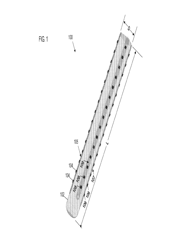
[0011] FIG. 1 shows an example directional and scalable (DISC) electrode
array.
[0012] FIGS. 2A-2C illustrate directionality and amplitude of as a function of
substrate
diameter. FIG. 20 summarizes directionality for an insulator (darker shade)
and for a
hypothetical substrate with the same conductivity as surrounding tissue
(lighter shade).
Inset A refers back to FIG. 2A and inset B refers back to FIG. 2B.
4
CA 03196206 2023- 4- 19
WO 2022/093829
PCT/US2021/056661
[0013] FIG. 3 illustrates normalized voltage amplitude as a function of
electrode
diameter. Sensitivity to a generic distant dipole decreases as shown with
increasing
electrode diameter until finally a ring is formed and the amplitude reduction
is a step
function.
[0014] FIGS. 4A-4E show an electro-quasistatic 3D dipole model demonstrating
directional sensitivity of a DISC electrode array in a multi-source
configuration. FIGS.
4A and 4B show the geometric orientation of 8 sources (only half of a dipole
source is
shown) and the resulting superimposed potential when all sources are activated
at
maximum current density. FIG. 40 illustrates the ability to detect source 1
for 1 trial up
to 50 repeated trials. CAR is the acronym for common average referencing. SNR
is
higher initially and increases at a faster rate with repeated trials. FIG. 4E
shows the
SNR results at 1 (lighter shade) and 50 trials (darker shade) for all 8
sources in this
multi-source model.
[0015] FIGS. 5A-50 illustrate directional sensitivity of a DISC electrode
array. In one
example experiment, FIG 5A shows, the location of the DISC electrode array
relative to
neural barrel cortex sources is identified. In FIG. 5B, the predicted
magnitude, direction,
and profile of each distinct neural source is shown as an example of source
separation.
FIG. 50 is an example of using the laminar information available in some uses
of DISC
to provide multi-directional current source density analysis.
[0016] FIG. 6 shows a schematic cross-section of polyimide linear electrode
(PLE)
array suitable for use in a DISC electrode array.
[0017] FIG. 7 shows a micrograph of an example strip of single-column
microelectrodes suitable for use in a DISC electrode array.
CA 03196206 2023- 4- 19
WO 2022/093829
PCT/US2021/056661
[0018] FIG. 8 illustrates examples of scalability achievable using various
combinations
of microelectrode referencing schemes of the DISC electrode array of FIG. Ito
produce
improved sensitivity in venous volumes of surrounding tissue.
[0019] FIG, 9 shows an example DISC electrode array implanted for use as a
translatable human brain-computer interface,
[0020] FIGS. 10A-10B illustrate example methods for fabricating a DISC
electrode
array,
DETAILED DESCRIPTION
[0021] Certain terms have been used throughout this description and claims to
refer
to particular system components. As one skilled in the art will appreciate,
different
parties may refer to a component by different names. This document does not
intend
to distinguish between components that differ in name but not function. In
this disclosure
and claims, the terms "including" and "comprising" are used in an open-ended
fashion,
and thus should be interpreted to mean "including, but not limited to... ."
Also, the term
"couple" or "couples" is intended to mean either an indirect or direct
connection. Thus,
if a first device couples to a second device, that connection may be through a
direct
connection or through an indirect connection via other devices and
connections. The
recitation "based on" is intended to mean "based at least in part on."
Therefore, if X is
based on Y, X may be a function of Y and any number of other factors.
[0022] Various sensor technologies for detecting electrical signals in neural
tissue are
available. Microwires, stereo-electroencephalogram (sEEG) depth electrodes,
and
electrocorticography (ECoG) electrodes (also known as grid arrays) are
examples of
conventional neural sensing technologies. These sensor technologies are
subject to a
number of shortcomings. For example, microwires and ring electrodes in sEEG,
ECoG,
and local field potential (LFP), record from millions of neurons over long
distances,
6
CA 03196206 2023- 4- 19
WO 2022/093829
PCT/11S2021/056661
especially in the cortex of mammals including humans. The signals from
overlapping
neural input for any brain network ¨ say speech production ¨ are difficult to
deconvolve,
rendering sub-optimal decoding of phonological or articulatory codes.
Implanting of grid
electrodes can result in hemorrhaging, infections, and/or migraine headaches.
Relative
to craniotomies of several inches or larger, sEEG methods reduce these 3
important
adverse events.
[0023] The UTAH ARRAY is a high-density, multi-channel neural sensor used in
brain-
computer interface applications. The Utah array acts as a high-density bundle
of
microwires, and microwires offer no substrate shielding because each
insulating shaft
is small relative to the geometric size of the neural source (FIG 4).
[0024] The directional and scalable (DISC) electrode array described herein
provides
a number of advantages over conventional neural sensors when recording local
field
potentials (LFPs).
[0025] The DISC electrode array is the first stereotadically delivered
microelectrode
array designed to separate LFP sources from other simultaneous LFP sources, or
any
voltage source having an origin that is of a particular size and distance.
This is a feature
not available with stereo-electroencephalograms (sEEGs) using macro-scale
electrodes, microwires, microwire arrays, or ultrafine microelectrode arrays.
The DISC
electrode array is designed to maximize sensitivity to "mesoscale" neural
sources by
identifying the source direction from the lead body of the implanted device
using a
scarcely known phenomenon referred to herein as "substrate shielding." This is
the first
application of substrate shielding to produce directional (stereo)
measurements of local
field potentials in a depth array. To address the accuracy issues of
microwires and
macroelectrodes, the DISC electrode array detects voltage signals (originating
from
7
CA 03196206 2023- 4 19
WO 2022/093829
PCT/US2021/056661
current sinks and sources) in a direction of tissue within a radius of
approximately 0.1 -
millimeters (mm) (i.e., mesoscale).
[0026] An array of microelectrodes in the geometry described herein also can
simultaneously produce voltage recordings almost identical to ring electrodes
or large
"directional leads" (segmented ring electrodes) and so can be a complete
replacement
for rnacroelectrodes. For example, any circular pattern of microelectrodes
when
averaged together form a virtual rnacroelectrode with a height equivalent to
the height
of the rings of microelectrodes . In another example, microelectrodes can be
averaged
producing the pattern of large directional electrodes having a known height
and arc,
[0027] To improve spatial resolution, the DISC electrode array utilizes
substrate
shielding to provide directional isolation of overlapping LFP signals. This
yields a spatial
scale that is unique for both neuroscience and brain-computer interfaces
(BC1s). The
DISC electrode array, includes 2 or 4 linear electrodes (e.g., opposing pairs)
with a
variable longitudinal span (e.g., 20 mm) and placed on a 0.8 mm cylinder in
some
examples. Wider diameters provide better shielding but at the cost of
displacing and
potentially damaging more tissue. The 0.8mm diameter is advantageous because
it is
already a safe standard established by neurosurgeons using sEEG and can access
deep brain regions,
[0028] FIG. 1 shows an example DISC electrode array 100. The DISC electrode
array
100 is an elongate structure having a diameter D and length L. The diameter D
may be
in a range of 400-2000 microns. For example, an implementation of the DISC
electrode
array 100 may have a diameter D of approximately 800 microns (e.g., 800
microns +1-
10%). The length L of the DISC electrode array 100 may in a range of 3-150
millimeters
in some implementations. The DISC electrode array 100 includes an insulating
body
102 and one or more microelectrodes attached to the insulating body 102. The
8
CA 03196206 2023- 4- 19
WO 2022/093829
PCT/US2021/056661
insulating body 102 may be formed of material having an electrical
conductivity that is
less than 10-8 siemens per meter. The insulating body 102 is cylindrical in
shape in
some implementations (i.e., circular in cross-section). Some implementations
of the
insulating body may be non-circular in cross-section.
For example, some
implementations of the in insulating body 102 maybe oval, square, hexagonal,
octagonal, decagonal, etc. in cross-section.
[0029] The microelectrodes 104, 106, and 108 are shown FIG. 1. The
microelectrode
104 and the microelectrode 106 are disposed opposite one another on the
insulating
body 102. That is, the microelectrode 104 and the microelectrode 106 are
disposed to
form an opposing pair of microelectrodes. The microelectrode 108 is disposed
on the
insulating body 102 equidistant from the microelectrode 104 and the
microelectrode
106. The DISC electrode array 100 may also include a microelectrode opposite
the
microelectrode 108 forming an opposing pair with the microelectrode 108. Any
conducting surface attached to the surface of the insulating body 102 with a
conductive
path to a low noise amplifier qualifies as a microelectrode. The
microelectrodes 104
form a linear subarray 105, the microelectrodes 106 form a linear subarray
107, and the
microelectrodes 108 form a linear subarray 108, The linear subarray 105 is
opposite
(on an opposite side of the insulating body 102 from the linear subarray 107.
Similarly,
a linear subarray of electrodes may be disposed on the insulating body 102
opposite
the linear subarray 109. In the present disclosure, microelectrodes or linear
subarrays
of microelectrodes are opposite one another if disposed at an angle of 180
10% from
one another about the circumference of the insulating body 102.
[0030] Various implementations of the DISC electrode array 100 include
different
numbers of microelectrodes disposed about the circumference of the insulating
body
102 arranged on approximately opposite sides. For example, the DISC electrode
array
9
CA 03196206 2023- 4- 19
WO 2022/093829
PCT/US2021/056661
100 as illustrated in FIG. 1 includes four microelectrodes arranged about the
circumference of the insulating body as two opposing pairs. Other
implementations of
the DISC electrode array 100 may include 2, 6, 8, 10, etc. microelectrodes
arranged
about the circumference of the insulating body 102 as opposing pairs.
Alternatives to
opposing pairs of microelectrodes (i.e., an odd number of microelectrodes) is
also
possible if multiple microelectrode referencing is used as the reference for
the recorded
voltage. In this case, the centroid of the multiple microelectrode references
should be
a point opposing the primary microelectrode of interest (referred to as a
"common
shielded reference" for simplicity).
[0031] Implementations of the DISC electrode array 100 include multiple
instances of
the 104 arranged in a line or row along the length of the insulating body 102,
multiple
instances of the 106 arranged in a line or row along the length of the
insulating body
102, multiple instances of the 108 arranged in a line or row along the length
of the
insulating body 102, etc. For each instance of each microelectrode disposed on
the
insulating body 102, an instance of an opposing microelectrode may be disposed
on the
insulating body 102
[0032] The microelectrodes (e.g., the microelectrodes 104, 106, 108, etc.) may
have
a diameter in a range of 8-500 microns. The placement of microelectrodes can
be
limited to the anticipated locations in the brain where neural sources of
interest may be
found. The spacing (pitch) of microelectrodes near the regions of interest may
be
arranged in a line or row along the length of the insulating body may be in a
range of
200-600 microns. For example, the spacing (pitch) of microelectrodes 104, 106,
108
arranged in a line or row along the length of the insulating body may be about
320
microns in some implementations. In addition to linear arrays of electrodes
with
opposing linear arrays, electrodes may also be staggered by some arbitrary
angle along
CA 03196206 2023- 4- 19
WO 2022/093829
PCT/US2021/056661
the longitudinal length. FIG. 9 is an example where every other row is
staggered forming
diamond patterns of electrodes across the array.
[0033] The DISC electrode array 100 may also include a conductor coupled to
each
of the microelectrodes for conveying electrical signal from the
microelectrodes to a
processing system coupled to the DISC electrode array 100.
[0034] FIGS. 2A-2C illustrate directionality and amplitude as a function of
substrate
and electrode diameters. The substrates are 65 pm and 800 pm in diameter
respectively
in FIGS. 2A and 2B, with an electrode on opposite sides of the substrate
positioned a
fixed distance from a dipole source. All electrodes are independent. Voltage V
results
from the current source shown to left of the lead body representing layer V
pyramidal
cells in a virtual cortex (200-pm diameter) created in a finite element model.
Voltage
is inversely related to distance and perturbed by changes in conductivity a in
space
such as are created by the insulating body. FIG. 2C illustrates an ANSYS model
of
the front to back electrode voltage ratio as a function of diameter (0sh).
When a of the
lead body matches local tissue (a=0.26 S/m), FIB ratio increases due to the
increasing
distance between front and back electrodes. As shown, substrate shielding
magnifies
the difference between the front and back electrodes by much more than the
previously known method of separating electrodes over a greater distance.
[0035] FIG. 3 illustrates normalized voltage amplitude as a function of
electrode
diameter. The source is the same as in FIGS. 2A and 2B. Attenuation becomes
significant beyond about 120 pm. At 1238 pm, a ring forms (e.g., sEEG) and
amplitude
is attenuated by 60% relative to a microelectrode. Increasing the ring area
beyond
this point has negligible attenuation.
[0036] FIGS. 4A-4E show an electro-quasistatic 3D dipole model demonstrating
directional sensitivity in a multi-source configuration. FIG. 4A shows eight
simultaneous
11
CA 03196206 2023- 4- 19
WO 2022/093829
PCT/US2021/056661
dipoles (labeled 1-8) modeled in a finite element method with an identical
surface
boundary current density (only sink is shown, 0.5 mm grid). Three device types
are
modeled: 1) an implementation of the DISC electrode array 100 (shown in FIG.
4A); 2)
microwire; and 3) a 0.4-mm tall ring electrode. FIG. 4B is a voltage heat map
through
layer V dipoles with sources on a peak current density of 1.39 pA/mm2 as
described in
Murakami, Shingo, and Yoshio Okada, Neurolmage Invariance in Current Dipole
Moment Density across Brain Structures and Species: Physiological Constraint
for
Neuroimaging, Neurolmage 111: 49-58 (2015). The heat map is at the plane
intersecting the dipole sinks. FIG. 4C shows signal-to-noise ratio (dBV) for a
macro
(ring) electrode, a DISC electrode array, and a DISC electrode array with CAR
during
trial 1 and cumulative trials. FIG. 4D shows waveform examples for 1 and 50
trials for
the macro electrode and DISC electrode array when phase locked to Source 1.
Sources 2-8 are assigned a random phase and frequency. Noise of 2.7 pVrms and
4.3pV is assigned to each ring or microelectrode, respectively. FIG. 4E shows
a
signal-to-noise ratio comparison of the simulated potentials for each source
independently phase-locked. The microwire is 65 pm in diameter. Trial 1 is
shown in
the lighter shade, and trial 50 is shown in the darker shade (avg). This
embodiment
of DISC electrode array 100 is useful for maximizing SNR for all 8 sources
relative to
other recording methods simulated.
[0037] FIGS. 5A-5C illustrate directional sensitivity in a DISC electrode
array. FIG.
5A shows an arrangement of nine sources Bl, B2,13, Cl, C2, D1, D2, 6, and Y
relative
to a DISC electrode array having eight columns of electrodes. FIG. 5B shows
results of
finite element modeling using the nine sources and the DISC electrode array.
This
illustrates why the DISC electrode array 100 is useful for either source
separation
applications, for example brain computer interfaces, or source localization,
for example
12
CA 03196206 2023- 4- 19
WO 2022/093829
PCT/US2021/056661
diagnostic neurosurgery. FIG. 50 shows multi-directional current source
density (CSD)
from the DISC electrode array when two sources are activated. A first source
(D1) is
located closest to column 4 of the DISC electrode array, and a second source
(Y) is
located closest to column 8 of the DISC electrode array (about 1800). FIG. 50
shows
distinct amplitude attenuation for a source on the opposite side of the
microelectrode
column of interest.
[0038] FIG. 6 shows a schematic cross-section of a polyimide linear electrode
(PLE)
array suitable for use in the DISC electrode array 100. An example
microelectrode 602
is shown in the center with insulated interconnects 604 shown traversing
parallel to each
other in/out of the cross-section.
[0039] FIG. 7 shows a micrograph of an example strip of microelectrodes
suitable for
use in the DISC electrode array 100. Some examples have 80-micron diameter
electrodes on a 320-micron pitch. A hole 702 enables easy handling during
assembly.
Assembly may utilize multiple single-column arrays. Alternatively, not shown,
a multi-
column microelectrode arrays could also be used to wrap around a cylinder or
other
geometry and record from other directions_
[0040] The DISC electrode array 100 offers the ability to sense LFP sources at
the
microscale (e.g., current source density), the rnesoscale (as demonstrated in
FIG. 5B),
and at the macroscaie. The DISC electrode array 100 is especially novel and
useful at
the mesoscale (1-5mm) given that other technologies fail to provide the
amplitude and
direction resolution of this invention.
[0041] At the microscale, it is notable that the DISC electrode array 100 can
uniquely
measure current source density in tissue from multiple directions
simultaneously which
is a novel capability useful in the study of laminar communication.
Additionally, the
amplification of a large diameter substrate makes it possible to record from
distant multi-
13
CA 03196206 2023- 4- 19
WO 2022/093829
PCT/US2021/056661
unit or multi-cell action potentials. Most recording arrays for neuroscience
applications
lack the substrate size to amplify a single-cell or multi-cell source beyond
100-120 pm,
however a 1.2-mm diameter DISC substrate will sense a single large pyramidal
cell (20-
pm diameter, or 2-3 smaller cells) out to 200 pm distance above a 60 pV
threshold.
[0042] Macroscale recordings from sEEG and ECoG are also highly valuable, and
may have advantages to a group model decoder since it uses larger neuron
populations
and thus may be more predictive of some kinds of behavior (motor movement,
speech,
etc.) when comparing between patients having such a sensor implanted in
similar brain
regions. DISC macroscale recordings can be acquired by simply grouping rings
of
rnicroelectrodes together mimicking properties of a solid ring electrode
(albeit with a
slightly larger noise floor). The ability to offer multiscale recordings is
another important
innovation, and it should be expected that any successful BCI solution,
especially for
speech, will be multi-scale. Further, the safety and simplicity of
stereotactic insertion
will result in a lower threshold for translation to humans.
[0043] In practice, the DISC electrode array 100 may include or be
communicatively
coupled to circuitry that maximizes the accuracy of source localization of an
LPF signal.
Such circuitry may include analog circuits, digital circuits, or a processor-
based
implementation executing software instructions retrieved from memory. In one
implementation, analog differential signals acquired from a low-noise
amplifier are used
to isolate LFP sources from multiple directions.
[0044] In one application of the DISC electrode array 100, circuitry
incorporated in or
communicatively coupled to the DISC electrode array 100 can reconstruct many
virtual
macroelectrodes through the averaging of signal acquired from a select set of
the
microelectrodes to generate higher resolution spatial recordings yet still at
the macro
14
CA 03196206 2023- 4- 19
WO 2022/093829
PCT/US2021/056661
scale and comparable to conventional SEEG/depth electrodes that sense in 360
degrees or over smaller angular ranges, such as 120, 90, 01 45 degrees.
[0045] In a system that includes the DISC electrode array 100, a processor may
execute software instructions to selectively reference signals acquired from
the
microelectrodes to maximize the accuracy of source localization of an LFP
signal (-1-
350 Hz) or a multi-cell source (-300-1500 Hz). The optimal referencing is
predicted
using a biophysical model of the source and an electro-quasistatic model
relating current
sources to predicted voltages. Such operations are possible only using the
DISC
electrode array 100.
[0046] FIG. 8 illustrates examples of scalability achievable using various
combinations
of microelectrode referencing schemes of the DISC electrode array 100 to
produce
improved sensitivity in various volumes of surrounding tissue. Various
combinations of
the microelectrodes of the DISC electrode array 100 are selected and processed
to: 1)
produce in 801, by averaging signal from all microelectrodes along a selected
length of
the DISC electrode array 100 output equivalent to sEEG to allow use of
standard clinical
methods; 2) in 802, isolate semi-local or rnesoscale sources; and/or 3) in
803, provide
maximum isolation of local sources using current source density analysis which
is a form
of referencing to only local microelectrodes.
[0047] FIG. 9 shows an example DISC electrode array 100 implanted for use as a
translatable human BC" In this alternative arrangement of electrodes, every
other row
of electrodes is rotated by 45 decrees to effectively create 8 unique columns
while only
using 4 microelectrodes at any given axial position.
[0048] The DISC electrode array 100 can be fabricated using various
manufacturing
methods. One embodiment includes applying the rnicrofabrication methods of
thin film
deposition, photolithography, and film etching. Another method includes using
medical
CA 03196206 2023- 4- 19
WO 2022/093829
PCT/US2021/056661
grade adhesives to attach a thin linear array of microelectrodes (opposing
linear arrays
of microelectrodes) along the length of an insulating cylinder as shown in
FIG. 10A, In
one embodiment, the microelectrode arrays may be placed in a hollow cylinder
mold
with electrodes facing the walls of the mold and then the mold injected with a
medical
grade insulator, e.g., polyurethane or silicone. Alternatively, a core can be
molded or
extruded separate from a thin-film array of microelectrodes and the latter is
wrapped
around the former using a biostable, medical grade adhesive.
[0049] In another method, shown in FIG. 10B, for fabricating the DISC
electrode
assembly 100, a thin (e.g., 20 pm) adhesive sheet is patterned and mounted on
the
backside of a two-dimensional electrode array (e.g., a 128-channel array). The
electrode array and adhesive sheet are wrapped around an insulated substrate
(a 432-
pm diameter substrate) using heat shrinkable tubing. The tubing is heated to
affix the
electrode array to the substrate, and the tubing is removed.
[0050] Electronic circuitry, including amplifiers, analog-to-digital
converters,
multiplexers, etc. may be connected to the thin film interconnects of the
microelectrodes.
In some implementations, such circuitry is disposed within the insulating body
102 ( and
beneath the skull after implantation of the DISC electrode array 100). In
other
implementations, the circuitry may be disposed in an insulating body in the
skull or
outside the skull.
[0051] The above discussion is meant to be illustrative of the principles and
various
embodiments of the present invention. Numerous variations and modifications
will
become apparent to those skilled in the art once the above disclosure is fully
appreciated. It is intended that the following claims be interpreted to
embrace all such
variations and modifications.
16
CA 03196206 2023- 4- 19