Note: Descriptions are shown in the official language in which they were submitted.
CA 03196411 2023-03-22
WO 2022/064293 PCT/IB2021/057601
1
MEDICAL PUNCTURE DEVICE
FIELD:
[0001]This document relates to medical devices. More specifically, this
document relates
to medical devices that use radiofrequency energy to puncture tissue.
SUMMARY:
[0002]The following summary is intended to introduce the reader to various
aspects of
the detailed description, but not to define or delimit any invention.
[0003]Medical puncture devices are disclosed. According to some aspects, a
medical
puncture device includes an elongate shaft having a proximal portion defining
a proximal
end and a distal portion defining a distal end. The distal portion tapers in
outer diameter
going towards the distal end to define a dilating tip. A lumen extends through
the shaft
from the proximal end to the distal end. The shaft includes a first electrical
conductor that
extends from the proximal portion to the distal portion and that is
electrically connectable
to a radiofrequency generator. A radiofrequency puncture electrode is
positioned proud
of the distal end and is electrically connected to the first electrical
conductor.
[0004]In some examples, the shaft includes a polymeric sleeve, and the first
electrical
conductor is in the form of a metallic hypotube received in the polymeric
sleeve. The
polymeric sleeve can be a high-density polyethylene sleeve and the metallic
hypotube
can be a stainless steel hypotube.
[0005]In some examples, the shaft includes a polymeric inner layer and a
polymeric outer
layer, and the electrical conductor is positioned between the polymeric inner
layer and
the polymeric outer layer. The polymeric inner layer can be a high-density
polyethylene
inner layer, and the polymeric outer layer can be a low-density polyethylene
layer.
[0006]In some examples, the electrical conductor is in the form of a wire or a
braid.
[0007]In some examples, the device further includes a second electrical
conductor
electrically connecting the electrode to the first electrical conductor. The
second electrical
CA 03196411 2023-03-22
WO 2022/064293 PCT/IB2021/057601
2
conductor can be in the form of a wire having a first end and a second end.
The first end
can be joined to the first electrical conductor, and the second end can be
spaced distally
of the distal end and joined to the radiofrequency puncture electrode. The
wire can be J-
shaped or coiled.
[0008] In some examples, the radiofrequency puncture electrode is retractable
towards
the shaft.
[0009] In some examples, the radiofrequency puncture electrode is atraumatic.
[0010] In some examples, the device includes a handle at the proximal end of
the shaft.
The first electrical conductor can be electrically connectable to the
radiofrequency
generator via the handle. The handle can include a hemostatic valve, a
stopcock, and/or
a syringe luer.
[0011]In some examples, the shaft has an outer diameter of between about 12.5
Fr and
about 24 Fr.
[0012] In some examples, the electrode can collect electrical signals from the
heart.
[0013] In some examples, the shaft is steerable.
[0014] In some examples, the distal portion is curved.
[0015] In some examples, the shaft includes a radiopaque marker, and/or an
echogenic
marker.
[0016]Medical methods are also disclosed. According to some aspects, a medical
method includes advancing the medical puncture device towards an atrial
septum;
delivering radiofrequency energy from the radiofrequency puncture electrode to
create a
puncture in the atrial septum; and advancing the dilating tip through the
puncture to dilate
the puncture.
[0017]Medical puncture systems are also disclosed. According to some aspects,
a
medical puncture system includes a radiofrequency generator and a medical
puncture
CA 03196411 2023-03-22
WO 2022/064293 PCT/IB2021/057601
3
device. The medical puncture device includes an elongate shaft having a
proximal portion
defining a proximal end and a distal portion defining a distal end. The distal
portion tapers
in outer diameter going towards the distal end to define a dilating tip. A
lumen extends
through the shaft from the proximal end to the distal end. The shaft includes
a first
electrical conductor extending from the proximal portion to the distal portion
and
electrically connectable to a radiofrequency generator. A radiofrequency
puncture
electrode is positioned proud of the distal end and electrically connected to
the first
electrical conductor.
BRIEF DESCRIPTION OF THE DRAWINGS:
[0018]The accompanying drawings are for illustrating examples of articles,
methods, and
apparatuses of the present disclosure and are not intended to be limiting. In
the drawings:
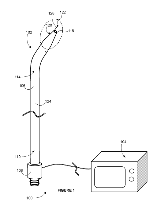
[0019]Figure 1 is a perspective view of an example medical puncture system
including a
medical puncture device;
[0020] Figure 2 is an enlarged view of the encircled region in Figure 1;
[0021] Figure 3 is a cross section taken through the medical puncture device
of in Figure
1;
[0022] Figure 4A is a partial perspective view of the distal portion of
another example
medical puncture device, with the electrode thereof in a retracted
configuration;
[0023] Figure 4B is a partial perspective view of the distal portion of the
medical puncture
device of Figure 4A, with the electrode thereof in a deployed configuration;
[0024] Figure 5 is a partial perspective view of the distal portion of another
example
medical puncture device;
[0025] Figure 6 is a partial perspective view of the distal portion of another
example
medical puncture device;
CA 03196411 2023-03-22
WO 2022/064293 PCT/IB2021/057601
4
[0026] Figure 7 is a schematic view of a step of a method for puncturing and
dilating an
atrial septum using the system of Figure 1;
[0027] Figure 8 is a schematic view of a subsequent step of the method of
Figure 7; and
[0028] Figure 9 is a schematic view of a subsequent step of the method of
Figure 8.
DETAILED DESCRIPTION:
[0029] Various apparatuses or processes or compositions will be described
below to
provide an example of an embodiment of the claimed subject matter. No example
described below limits any claim and any claim may cover processes or
apparatuses or
compositions that differ from those described below. The claims are not
limited to
apparatuses or processes or compositions having all of the features of any one
apparatus
or process or composition described below or to features common to multiple or
all of the
apparatuses or processes or compositions described below. It is possible that
an
apparatus or process or composition described below is not an embodiment of
any
exclusive right granted by issuance of this patent application. Any subject
matter
described below and for which an exclusive right is not granted by issuance of
this patent
application may be the subject matter of another protective instrument, for
example, a
continuing patent application, and the applicants, inventors or owners do not
intend to
abandon, disclaim or dedicate to the public any such subject matter by its
disclosure in
this document.
[0030]Generally disclosed herein are medical devices that can be used to
puncture and
dilate tissue, and related systems and methods. Such devices may be referred
to herein
as 'medical puncture devices'. The medical puncture devices may be used in a
variety of
medical procedures, but may be particularly useful in procedures that require
transseptal
access to the left atrium with a relatively large outer diameter catheter.
Such procedures
can include, for example, cryoablation procedures, mitral valve replacement
procedures,
and atrial appendage closure procedures. The medical puncture devices
disclosed herein
can puncture the atrial septum and dilate the puncture to a sufficiently large
diameter (e.g.
between about 12.5 Fr and about 24 Fr).
CA 03196411 2023-03-22
WO 2022/064293 PCT/IB2021/057601
[0031] Referring now to Figures 1 and 2, a system 100 is shown that generally
includes
a medical puncture device 102, and a radiofrequency (RF) generator 104 to
which the
medical puncture device 102 is electrically connected.
[0032]The RF generator 104 can be any RF generator suitable for use in
puncturing
tissue, such as one sold by Baylis Medical Company (Montreal, Canada) under
the brand
name RFP-100A RF Puncture Generator, and/or other electrosurgery equipment,
and will
not be described in detail herein.
[0033] Referring still to Figures 1 and 2, as mentioned above, the medical
puncture device
102 is configured to puncture tissue using RF energy, and to dilate the
puncture to a
relatively large diameter. In the example shown, the medical puncture device
102 includes
an elongate shaft 106, a handle 108, and a radiofrequency puncture electrode
122. The
shaft 106 has a proximal portion 110 that defines a proximal end 112 (shown in
Figure
3), and a distal portion 114 that defines a distal end 116. A lumen 118 (shown
in Figure
3) extends through the shaft 106 from the proximal end 112 to the distal end
116. The
distal portion 114 is curved, and in the distal portion 114, the shaft 106
tapers in outer
diameter going towards the distal end 116, to define a dilating tip 120. The
handle 108 is
at the proximal end 112, and can include various optional features such as a
syringe luer,
a hemostatic valve, and/or a stopcock (e.g. a 3-way stopcock) for fluid
delivery through
the lumen 118 via the handle 108.
[0034]As mentioned above, the shaft 106 can have a relatively large outer
diameter, for
example between about 12.5 Fr and about 24 Fr.
[0035] In the example shown, the shaft 106 is of a generally fixed shape. In
alternative
examples, the shaft can be steerable. In such examples, the device can include
one or
more pull wires or other actuators for steering the shaft.
[0036]The shaft 106 can optionally include one or more radiopaque markers
and/or
echogenic markers (not shown), to enhance visualization of the position of the
shaft 106.
[0037] Referring still to Figures 1 and 2, the RF puncture electrode 122 is
joined to the
shaft 106, and is positioned proud of the distal end 116 of the shaft 106. The
RF puncture
CA 03196411 2023-03-22
WO 2022/064293 PCT/IB2021/057601
6
electrode 122 is electrically connectable to the RF generator 104 (as will be
described
below) and can deliver RF energy to tissue, to puncture the tissue. In the
example shown,
the RF puncture electrode 122 is atraumatic ¨ that is, the RF puncture
electrode 122 is
blunt (e.g. rounded) in order to avoid damaging tissue unless RF energy is
being delivered
from the RF puncture electrode 122 to the tissue.
[0038] In some examples (not shown), the RF puncture electrode can be used to
collect
electrical signals from the heart.
[0039] Referring now to Figure 3, in the example shown, the shaft 106 includes
a
polymeric sleeve 124 (e.g. a high-density polyethylene sleeve), and an
electrical
conductor 126 (also referred to herein as a 'first electrical conductor') in
the form of a
metallic hypotube (e.g. a stainless steel hypotube) received in the polymeric
sleeve 124.
The electrical conductor 126 extends from the proximal portion 110 to the
distal portion
114. The electrical conductor 126 is electrically connectable to the RF
generator 104 via
the handle 108, and is electrically connected to the RF puncture electrode
122, so that
RF energy can be delivered from the RF generator 104 to the RF puncture
electrode 122
via the electrical conductor 126.
[0040] In an alternative example (not shown), the shaft can include a
polymeric inner layer
(e.g. a high-density polyethylene layer) and a polymeric outer layer (e.g. a
low-density
polyethylene layer), and the electrical conductor can be positioned between
the polymeric
inner layer and polymeric outer layer. In further alternative examples, the
electrical
conductor can be in the form of a wire or a braid instead of a hypotube.
[0041] Referring still to Figure 3, in the example shown, the first electrical
conductor 126
extends to a position shy of the distal end 116 of the shaft 106. A second
electrical
conductor 128 electrically connects the first electrical conductor 126 and the
RF puncture
electrode 122. The second electrical conductor 128 is in the form of a wire
that has a first
end that is joined to the first electrical conductor 126 and a second end that
is spaced
distally from the distal end 116 of the shaft 106 and is joined to the RF
puncture electrode
122, to conduct RF energy from the first electrical conductor 126 to the RF
puncture
electrode 122.
CA 03196411 2023-03-22
WO 2022/064293 PCT/IB2021/057601
7
[0042] In the example shown, the RF puncture electrode 122 is generally fixed
in position
with respect to the shaft 106. An alternative example is shown in Figures 4A
and 4B, in
which features that are like those of Figures 1 to 3 are referenced with like
reference
characters, incremented by 300. In the medical puncture device 402 of Figures
4A and
4B, the RF puncture electrode 422 can be retracted towards the shaft 406 (as
shown in
Figure 4A) and deployed away from the shaft 406 (as shown in Figure 4B). In
such
examples, the device 402 can include one or more pull wires or other actuators
(not
shown) for retracting and deploying the RF puncture electrode 422. Another
alternative
example is shown in Figure 5, in which features that are like those of Figures
1 to 3 are
referenced with like reference characters, incremented by 400. In the medical
puncture
device 502 of Figure 5, in addition to being retractable and deployable, the
second
electrical conductor 528 is J-shaped. This can enhance patient safety, as the
electrode
522 will be directed back from tissue when deployed. Yet another alternative
example is
shown in Figures 6, in which features that are like those of Figures 1 to 3
are referenced
with like reference characters, incremented by 500. In the medical puncture
device 600
of Figure 6, in addition to being retractable and deployable, the second
electrical
conductor 628 is coiled (also referred to as 'pig-tail shaped'). Again, this
can enhance
patient safety, as the electrode 622 will be directed back from tissue when
deployed. In
yet further alternative examples (not shown), the electrode can be joined
directly to the
first electrical conductor, and the second electrical conductor can be
omitted.
[0043] Referring now to Figures 7 to 9, a method for puncturing and dilating
an atrial
septum 700 using the medical puncture device 102 will be described. As a first
step (not
shown), a guidewire can be advanced into the superior vena cava, via the
femoral vein.
The medical puncture device 102 can then be advanced over the guidewire until
the distal
end 116 of the shaft is in the superior vena cava. The guidewire can then be
removed.
The medical puncture device 102 can then be pulled down into the right atrium,
and
positioned with the RF puncture electrode 122 against the fossa ovalis of the
atrial septum
700, to tent the atrial septum 700, as shown in Figure 7. Optionally,
fluoroscopy or another
visualization technique can be used to confirm the positioning of the medical
puncture
device 102. As a next step, as shown in Figure 8, the RF generator 104 (not
shown in
Figure 8) can be activated so that RF energy is delivered from the RF puncture
electrode
CA 03196411 2023-03-22
WO 2022/064293 PCT/IB2021/057601
8
122 to puncture the atrial septum 700 and pass the electrode 122 through the
atrial
septum 700 into the left atrium. Optionally fluoroscopy or another
visualization technique
can again be used to confirm the positioning of the electrode 122. As a next
step, as
shown in Figure 9, the medical puncture device 102 can be advanced so that the
dilating
tip 120 passes through puncture in the atrial septum 700 and dilates the
puncture. After
the puncture has been dilated, various steps can be carried out (e.g.
cryoablation, mitral
valve replacement, or atrial appendage closure), depending on the nature of
the medical
procedure
[0044]While the above description provides examples of one or more processes
or
apparatuses or compositions, it will be appreciated that other processes or
apparatuses
or compositions may be within the scope of the accompanying claims.
[0045]To the extent any amendments, characterizations, or other assertions
previously
made (in this or in any related patent applications or patents, including any
parent, sibling,
or child) with respect to any art, prior or otherwise, could be construed as a
disclaimer of
any subject matter supported by the present disclosure of this application,
Applicant
hereby rescinds and retracts such disclaimer. Applicant also respectfully
submits that any
prior art previously considered in any related patent applications or patents,
including any
parent, sibling, or child, may need to be re-visited.
