Note: Descriptions are shown in the official language in which they were submitted.
USE OF REGULATOR OF ITPRIPL1 IN PREPARATION OF DRUG THAT REGULATES
IMMUNE RESPONSES OR FIGHTS TUMORS
100011 This application claims priorities to Chinese Patent Application No.
202011191447.3, entitled
"USE OF REGULATOR OF ITPRIPL1 IN PREPARATION OF DRUG THAT REGULATES
IMMUNE RESPONSES OR FIGHTS TUMORS", filed to China National Intellectual
Property
Administration on Oct. 30, 2020, and Chinese Patent Application No.
202110566040.2, entitled
"ISOLATED ANTIGENIC ITPRIPL1-BINDING PROTEIN AND USE THEREOF", filed to China
National Intellectual Property Administration on May. 24, 2021, the entire
contents of which are
incorporated herein by reference.
TECHNICAL FIELD
100021 The present disclosure relates to the field of biomedicine, and
specifically to a use of a regulator
of ITPRIPL1 in the preparation of a drug that regulates immune responses or
resists tumors.
BACKGROUND OF THE INVENTION
100031 TTPRTPL1-encoded protein is Tnositol 1,4,5-trisphosphate receptor-
interacting protein-like 1,
for which the function of ITPRIPL1 has never been reported. Based on the
annotation of UniProtKB
database, ITPRIPL1 of human includes 555 amino acids, which are divided into
an extracellular
domain (1-103 amino acids), a transmembrane domain (104-124 amino acids), and
an intracellular
domain (125-555 amino acids).
100041 T cells are key effector cells of adaptive immune responses, which have
many important roles
in eliminating pathogens and autoimmune diseases. There are several
subpopulations of T cells, each
with a different function. TCRs (T cell receptors) found on the surface of T
cells are heterodimers
composed of a and 13 polypeptide chains, which constitute about 95% of the TCR
population, or they
are composed of y and 6 polypeptide chains (Pitcher and van Oers, 2003). Each
kind of polypeptide
includes constant (C) and variable (V) regions. The constant regions are
anchored in the cell membrane,
while the variable regions extend outside the cells and are responsible for
binding the antigen. The
cytoplasmic short tails of TCRs are lack of the ability to transduce signals.
Intracellular signaling is
1
CA 03196686 2023- 4- 25
initiated by the CD3 protein complex, which includes intracellular
immunoreceptor tyrosine-based
activation motifs (ITAMs).
100051 CD3 (Cluster of Differentiation 3) T cell co-receptors are a kind of
protein complex consisting
of four different chains. In mammals, a complex includes one CD3y (y) chain,
one CD36 (6) chain and
two CDR (E) chains. These chains are correlated to TCRs and C chain (zeta
chain), and produce
activation signals in T lymphocytes. The TCRs, C chain and CD3 molecules
together form the TCR
complex. CD37, CD3 6 and CD3E chains are highly related cell surface protein
of the immunoglobulin
superfamily containing a single extracellular immunoglobulin domain. TCRs are
not capable of
binding free epitopes/antigens. In contrast, TCRs can bind cleaved fragments
of larger polypeptides
associated with a major histocompatibility complex (MHC), which is synonymous
with the human
leukocyte antigen (HLA) system in humans. Such an interaction occurs in a
space known as the
immune synapse. Class I MHC molecules are expressed on all nucleated cells of
human, and present
antigens to cytotoxic T cells, on which CD8 stabilizes the MHC/TCR
interaction. The activation of
cytotoxic T cells then leads to the destruction of target cells. Class IT MHCs
are found on macrophages,
B cells and dendritic cells. These immune cells present antigens to helper T
cells with CD4 that
stabilizes the MHC/TCR interaction. The interaction between Class IT MHC and
TCR finally leads to
antibody-mediated immune responses. Other costimulatory molecules, such as
CD45, CD28 and CD2,
contribute to the activation of T cells in the immune synapse and initiate the
formation of TCR
signalosomes which are macromolecular protein complexes responsible for
intracellular signaling.
100061 Several antibodies that bind to human CD3E have ever been reported, for
example, antibody
OKT3 (see, e.g., Kung, P. et, al, Science 206 (1979) 347-349; Salmeron, A. et,
al, J Immunol 147
(1991) 3047-3052), antibody UCHT1 (see, e.g., Callard, RE et, al, Clin Exp
Immunol 43 (1981) 497-
505) or antibody SP34 (see, e.g., Pessano, S. et, al, EMBO J 4 (1985) 337-
344). Among the currently
known antibodies, SP34 is cross-reactive in human and cynomolgus monkey
(Conrad M.L. et, al,
Cytometry A 71 (2007)925-933). It has been known that proteins directly
binding to the CD3E
extracellular domain are antibody molecules, and there has never reported that
natural non-antibody
2
CA 03196686 2023- 4- 25
proteins (e.g., transmembrane proteins, secretory proteins and other typical
ligands) bind to the CD3E
extracellular domain.
Neuropilin-2 (i.e., NRP-2) is a kind of receptor capable of regulating the
function of immune cells
(Am J Physiol Lung Cell Mol Physiol. 2018), which is reported to regulate the
function of antigen-
presenting cells and promote the immune evasion of tumors (Sohini Roy et, al,
Cancer Res. 2018);
NRP2 may affect the migration and phagocytic function of immune cells as well
as the contact among
immune cells (S Schellenburg et, al, Mol Immunol. 2017). Co-receptors fotmed
from NRP2 and Plexin
have negative chemotactic effects on the migration of lymphatic endothelial
cells (Liu X et, al, Cell
Rep. 2016). NRP2 can also regulate the NFKB signaling in cells, which is seen
in the report (Rizzolio,
S. et, al. Cancer Research.2017). NRP2 ligands that have been found include
Semaphorin family
members, but other types of ligands have not been reported.
SUMMARY OF THE INVENTION
100071 The present disclosure is intended to provide a method of regulating
immune responses and
suppressing tumors, as well as a use of a regulator of ITPRIPL1 in the
preparation of a drug that
regulates immune responses or resists tumors.
100081 To achieve the above objectives, the present disclosure provides a use
of a regulator of
ITPRIPL1 in the preparation of a drug that regulates immune responses or
resists tumors, in which the
regulator is used to increase or decrease the expression or function of the
TTPRTPL1 gene or protein
in an organism.
100091 Preferably, the regulator includes any one of the following:
(1) a gene editing system that enables the knockout or mutation of the
ITPRIPL1 gene in cells;
(2) an RNA molecule that reduces the expression level of the ITPRIPL1 gene;
(3) a nucleic acid molecule for being introduced into a cell, the nucleic acid
molecule encodes
ITPRIPL1 and increases the expression level of TTPRTPL1;
3
CA 03196686 2023- 4- 25
(4) an isolated ITPRIPL1 recombinant protein;
(5) an antibody that recognizes and binds to the ITPRIPL1.
[0010] Preferably, the gene editing system is a CRISPR/Cas9 gene editing
system; a target sequence
used in the CRISPR/Cas9 gene editing system is selected from any one sequence
as set forth in SEQ
ID NOs: 11-13, and an oligomeric DNA sequence for encoding sgRNA is selected
from SEQ ID
NOs: l4-19;
[0011] the nucleic acid molecule includes: a sequence as set forth in SEQ ID
NO: 8, SEQ ID NO: 9
or SEQ ID NO: 10;
[0012] the ITPRIPL1 recombinant protein includes: a ftmctional fragment
capable of binding to CDR
or NRP2 protein in an extracellular domain of the ITPRIPL1 protein;
[0013] the antibody that recognizes and binds to ITPRIPL1 is a polyclonal
antibody, a monoclonal
antibody, a single-chain antibody, an antigen binding domain, a bispecific
antibody, a multi-specific
antibody, or an antigen binding portion in a chimeric antigen receptor.
[0014] Preferably, the sequence of the functional fragment is selected from
any one of SEQ ID NO: 1
to SEQ ID NO: 4, or a derivative sequence thereof. The derivative sequence
includes
DRMDLDTLARSRQLEKRMSEEMRxLEMEFEERxxxAExxQKxENxWxGxTSxDQ ("x" is any
amino acid). The derivation method includes: substituting, deleting or
inserting more than one amino
acid without changing the function of the sequence.
[0015] Preferably, the ITPRIPL1 recombinant protein forms a fusion protein (as
set forth in SEQ ID
NOs: 5-7) with an antibody constant region, or font's a fusion protein with a
coagulation factor;
alternatively, the ITPRIPL1 recombinant protein is modified by means of:
polyethylene glycol
modification, glycosylation modification, polysialic acid modification, fatty
acid modification, KLH
modification, biotin modification.
4
CA 03196686 2023- 4- 25
Fool 61 Preferably, the nucleic acid molecule is introduced into the cell
through a drug delivery system
which includes recombinant expression vectors, viruses, lipidosome or
nanomaterials.
100171 Preferably, the regulation of immune responses includes: regulating the
functions of antigen
presenting cells and T lymphocytes during the processes of autoimmune
responses, transplant
rejection-suppressing immune responses, allergies, anti-infection immune
responses, and anti-tumor
immune responses.
100181 Preferably, the immune responses include: type I diabetes, immunologic
infertility, rejection
after organ transplantation, allergies, systemic inflammation or cytokine
storm, and infection.
100191 Preferably, the tumors are solid tumors or hematological tumors; the
solid tumors include:
glioma, lung cancer, head and neck cancer, gastric cancer, colorectal cancer,
thyroid cancer, esophagus
cancer, urothelial carcinoma, testicular cancer, breast cancer, cervical
cancer, endometrial cancer,
melanoma, pancreatic cancer or liver cancer; the hematological tumors include:
leukemia or
lymphoma.
100201 The present disclosure further provides a pharmaceutical composition,
which includes a
regulator used in the above use, and a pharmaceutically acceptable carrier.
100211 The present disclosure further provides an isolated ITPRTPL1
recombinant protein, and the
recombinant protein is a functional fragment capable of binding to CD3E or
NRP2 protein in the
extracellular domain of the ITPRIPL1 protein.
100221 The present disclosure further provides an antibody that recognizes and
binds to the ITPRIPL1,
and the antibody recognizes and binds to the extracellular domain of the
TTPRIPL1 protein.
100231 The present disclosure further provides an application of the isolated
ITPRIPL1 recombinant
protein for detecting the presence of its own anti-TTPRIPL1 antibody, wherein
the main steps of
detection include directly contacting the ITPRIPL1 recombinant protein with
the blood sample from
a subject, and washing to remove nonspecific binding.
CA 03196686 2023- 4- 25
100241 The present disclosure further provides applications of the above
antibody for detecting the
content of ITPRIPL1 in a sample, and for judging the expression of ITPRIPL1 in
cells, tissues, organs
or individuals by the antibody that recognizes and binds to the ITPRIPL1, or
for judging whether it is
suitable to apply the method of the present disclosure to regulate immune
responses and suppress
tumors by targeting ITPRIPL1. In some embodiments, provided are applications
of an antibody that
specifically recognizes ITPRIPL1 for marking boundaries between cancer tissues
and para-cancerous
tissues in primary lesions, boundaries between cancer cells that have
metastasized to lymph nodes and
normal lymphatic tissues, boundaries between cancer cells with distant
metastases and normal tissues
of the metastatic organ, as well as marking living cancer tissue cells in
other biological samples.
100251 The present disclosure provides a use of an isolated antigenic TTPREPL1-
binding protein in the
preparation of a drug that regulates immune responses or resists tumors as
well as for detecting the
expression of ITPRIPL1 in an individual, where, the isolated antigenic
ITPRIPL1-binding protein is
capable of binding to an amino acid sequence as set forth in SEQ ID NO: 49 or
SEQ ID NO: 1 in the
antigenic TTPRIPL1.
100261 In some embodiments, a heavy chain variable region and a light chain
variable region are
included, where, the heavy chain variable region includes HCDR1, HCDR2 and
HCDR3 in the heavy
chain variable region VH as set forth in any one of amino acid sequence SEQ ID
NO: 24 or SEQ ID
NO: 34; the light chain variable region includes LCDR1, LCDR2 and LCDR3 in the
light chain
variable region VL as set forth in any one of amino acid sequence SEQ ID NO:
25 or SEQ ID NO: 35.
100271 In some embodiments, in the heavy chain variable region VH in the amino
acid sequence SEQ
ID NO: 24, the amino acid sequence of the HCDR1 is as set forth in SEQ ID NO:
26, the amino acid
sequence of the HCDR2 is as set forth in SEQ ID NO: 27, and the amino acid
sequence of the HCDR3
is as set forth in SEQ ID NO: 28.
100281 In some embodiments, in the amino acid sequence SEQ ID NO: 34, the
amino acid sequence
of the HCDR1 is as set forth in SEQ ID NO: 36, the amino acid sequence of the
HCDR2 is as set forth
in SEQ ID NO: 37, and the amino acid sequence of the T-TCDR3 is as set forth
in SEQ ID NO: 38.
6
CA 03196686 2023- 4- 25
100291 In some embodiments, in the amino acid sequence SEQ ID NO: 25, the
amino acid sequence
of the LCDR1 is as set forth in SEQ ID NO: 29, the amino acid sequence of the
LCDR2 is KV, and
the amino acid sequence of the LCDR3 is as set forth in SEQ ID NO: 31, or is
more than 80% similar
to an amino acid sequence as set forth in SEQ TD NO: 31.
100301 In some embodiments, in the amino acid sequence SEQ ID NO: 35, the
amino acid sequence
of the LCDR1 is as set forth in SEQ ID NO: 39, the amino acid sequence of the
LCDR2 is KV, and
the amino acid sequence of the LCDR3 is as set forth in SEQ ID NO: 41.
100311 In some embodiments, the heavy chain variable region includes HCDR1,
HCDR2 and HCDR3
in the heavy chain variable region VH as set forth in the amino acid sequence
SEQ ID NO: 24; the
light chain variable region includes LCDR1, LCDR2 and LCDR3 in the light chain
variable region
VL as set forth in the amino acid sequence SEQ ID NO: 25.
100321 In some embodiments, the heavy chain variable region includes HCDR1,
HCDR2 and HCDR3
in the heavy chain variable region VII as set forth in the amino acid sequence
SEQ ID NO: 34; the
light chain variable region includes LCDR1, LCDR2 and LCDR3 in the light chain
variable region
VL as set forth in the amino acid sequence SEQ ID NO: 35.
100331 In some embodiments, an antibody heavy chain constant region is
included, and the antibody
heavy chain constant region is derived from a human TgG heavy chain constant
region.
100341 In some embodiments, an antibody light chain constant region is
included, and the antibody
light chain constant region includes a human Igic constant region.
[0035] In some embodiments, an antibody heavy chain HC is included, and the HC
includes an amino
acid sequence as set forth in any one of SEQ ID NO: 22 or 32.
100361 In some embodiments, an antibody light chain LC is included, and the LC
includes an amino
acid sequence as set forth in any one of SEQ ID NO: 23 or 33.
7
CA 03196686 2023- 4- 25
100371 In some embodiments, an antibody or an antigen binding fragment thereof
is included, wherein
the antigen binding fragment includes Fab, Fab', F(ab)2, Fv fragment, F(ab')2,
scFv and/or di-scFv.
100381 In some embodiments, the ITPRIPL1 protein includes human ITPRIPL1 or
the ITPRIPL1
protein of cynomolgus monkey, rat, mouse, gorilla, grivet, golden snub-nosed
monkey, black snub-
nosed monkey, Amazon squirrel monkey.
100391 In some embodiments, the human ITPRIPL1 protein includes an amino acid
sequence as set
forth in SEQ TD NO: 20.
[0040] In another aspect, the present disclosure further provides a chimeric
antigen receptor (CAR),
which includes the isolated antigenic TTPRTPL I -binding protein of the
present disclosure.
100411 In another aspect, the present disclosure further provides an
immunoconjugate, which includes
the isolated antigenic ITPRIPL1-binding protein of the present disclosure.
100421 In another aspect, the present disclosure further provides one or more
isolated nucleic acid
molecules, which encode the isolated antigenic ITPRIPL1-binding protein or the
chimeric antigen
receptor of the present disclosure.
100431 In another aspect, the present disclosure further provides a vector,
which includes the nucleic
acid molecules of the present disclosure.
100441 In another aspect, the present disclosure further provides a cell,
which includes the nucleic acid
molecules or the vector of the present disclosure.
100451 In another aspect, the present disclosure further provides a
pharmaceutical composition, which
includes the isolated antigenic ITPRIPL1 -binding protein, the chimeric
antigen receptor, the
immunoconjugate of the present disclosure, and optionally a pharmaceutically
acceptable adjuvant.
100461 In another aspect, the present disclosure further provides a method for
preparing the isolated
antigenic TTPRTPL1 -binding protein of the present disclosure, which includes
culturing the cell of the
8
CA 03196686 2023- 4- 25
present disclosure under a condition enabling the expression of the isolated
antigenic ITPRIPL1-
binding protein of the present disclosure.
100471 In another aspect, the present disclosure further provides uses of the
isolated antigenic
ITPRIPL1-binding protein, the chimeric antigen receptor, the immunoconjugate,
and/or the
pharmaceutical composition of the present disclosure in the preparation of a
drug which is used for
preventing, alleviating and/or treating tumors. In some embodiments, the
tumors include solid tumors
and lymphoma.
100481 In another aspect, the present disclosure further provides a linear
epitope polypeptide that can
be used for efficiently screening and preparing an ITPRIPL1 function-
regulating antibody, which is
characterized in that, the peptide includes: (i) an amino acid sequence of SEQ
ID NO: 49, i.e.,
RLLEMEFEERKRAAE; (ii) or, an amino acid sequence of xxLxxxFxxRxxx (x is any
amino acid),
in which 1-3 amino acids at both ends can be deleted; (iii) or, an amino acid
sequence obtained by
substituting, inserting or deleting 1, 2, 3, 4, 5, 6, 7, 8, 9 or 10 amino
acids of SEQ ID NO: 49.
[0049] The above linear epitope peptide has outstanding advantages in many
aspects: it can be
synthesized at low cost; the function of the ITPRIPL1 specific antibody can be
determined by
analyzing the binding ability of the epitope peptide with advantages of simple
operation and stable
and reliable detection results; the linear epitope peptide can be directly
injected to animals as
immunogen or be screened, thereby obtaining more effective functional
antibodies than those obtained
by using full-length proteins, thus improving the discovery efficiency of
related drugs.
100501 The present disclosure further provides a method for searching and
identifying the function
regulator of ITPRIPL1, which is characterized in that, one or more of the
following properties of the
test molecules are detected: ability of specifically binding to TTPRIPL1
expressed on the cell surface;
effect on the binding of ITPRIPL1 to CD3E; effect on the binding of ITPRIPL1
to NRP2; effect on the
binding of ITPRIPL1 to SEMA3G; effect on the binding of ITPRIPL1 to EBI2; and
effect on the
function of immune cells or tumor cells.
9
CA 03196686 2023- 4- 25
100511 The present disclosure creatively develops a targeting antibody that
can bind to ITPRIPL1,
which can bind the above target protein with high affinity and neutralize its
function, and inhibit its
binding to one or more ligands, thereby disabling the immune evasion function
of the tumor cells and
promoting the killing of tumor cells by immune cells in vitro and in vivo. The
antibody provided in
the present disclosure can be used as the active ingredient to prepare a drug
for treating tumors,
providing a new effective solution for the treatment of tumors. At the same
time, the present disclosure
also provides linear epitopes corresponding to the antibody with excellent
neutralizing functions, and
biomarkers for administering the antibody.
100521 The conception, specific structure and technical effects produced
therefrom will be further
illustrated below in conjunction with the accompanying drawings, so as to
fully understand the
purposes, features and effects of the present disclosure.
100531 Compared with the prior art, the present disclosure has the following
beneficial effects:
[0054] The present disclosure discloses a method of regulating immune
responses and suppressing
tumors, which is achieved by regulating the expression or function of the
TTPRIPL1 gene. The present
disclosure is based on a new scientific discovery that ITPRIPL1 binds to
proteins such as CDR, so as
to regulate the functions of different immune cells, and then participate in
the regulation of immune
responses and the immune evasion process of tumors. The present disclosure has
confirmed that the
regulator of TTPRTPL1 can be used to prepare drugs or pharmaceutical
compositions, with promising
applications in the suppression of diseases such as tumors, autoimmune
diseases, transplant rejection,
allergies and infections.
BRIEF DESCRIPTION OF THE DRAWING
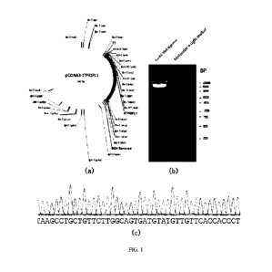
100551 FIG. 1 shows the plasmid construction results of Example 1;
100561 FIG. 2 shows the results of the co-immunoprecipitation experiment of
Example 1;
100571 FIG. 3 is a diagram showing the colocalization results of exogenously
expressed ITPRIPL1
and CD3E in cells according to Example 1;
CA 03196686 2023- 4- 25
100581 FIG. 4 shows the construction results of the expression vector
expressing the receptor-binding
domain of the extracellular domain of ITPRIPL1 according to Example 2;
100591 FIG. 5 shows the results of the co-immunoprecipitation experiment of
Example 2;
[0060] FIG. 6 shows the experimental results of immunofluorescence and
colocalization analysis of
Example 2 that the ITPRIPL1 extracellular domain and the CD3 extracellular
domain are in trans-
binding;
[0061] FIG. 7 shows the Coomassie brilliant blue staining results of the
ITPRIPL1-RBD recombinant
protein (IT1-RBD protein) of Example 3 after gel electrophoresis (Note:
ITPRIPL1 = TT1, the same
below);
100621 FIG. 8 shows the ELTSA experimental results of Example 4;
100631 FIG. 9 and FIG. 10 are diagrams showing the results of the flow
cytometry in Example 5,
demonstrating that the purified protein fragments from the ITPRIPL1
extracellular domain binds to
Jurkat cells that highly express CD3, wherein, FIG. 9 (a) shows the threshold
setting for not classified
as dead cells, FTG. 9 (b) reflects the binding of Jurkat cells to different
concentrations of purified
protein fragments from the ITPRIPL1 extracellular domain. FIG. 10 shows
specific staining and
protein binding profiles under various conditions in FIG. 9 (b).
100641 FIG. 11 and FIG. 12 are diagrams showing the results of the flow
cytometry in Example 5,
demonstrating that the ITPRIPL1 protein binds to cells overexpressing CD3 with
higher efficiency,
wherein, FIG. 11(a) shows the threshold setting for not classified as dead
cells, FIG. 11(b) reflects
the binding of cells with different CDR expression to different concentrations
of ITPRIPL1
recombinant protein. FIG. 12 shows specific staining and protein binding under
each condition in FIG.
11(b).
100651 FIG. 13 and FIG. 14 are diagrams showing the results of the flow
cytometry in Example 5,
demonstrating that CD3 binds to cells overexpressing ITPRIPL1 with higher
efficiency, wherein, (a)
of FIG. 13 shows the threshold setting for not classified as dead cells, (b)
of FIG. 13 reflects the
11
CA 03196686 2023- 4- 25
binding of cells with different expression of ITPRIPL1 to CD3c protein. FIG.
14 shows specific
staining and protein binding under each condition in (b) of FIG. 13,
respectively;
100661 FIG. 15 is a diagram showing the results of NFKB signaling changes as a
function of the
concentration of ITPRIPL1 proteins under the activation of 501.ig/m1 ConA in
Example 5;
100671 FIG. 16 is a diagram showing the results of NFKB signaling changes as a
function of the
concentration of microsphere-coated ITPRIPL1 proteins under the activation of
50 ug/m1 ConA in
Example 5;
[0068] FIG. 17 shows the expression of ITPRIPL1 in different types of tumor
cells after the alignment
of GAPDH internal reference in Western blotting;
100691 FIG. 18 shows the mRNA expression level of ITPRIPL1 in normal tissues
and tumor tissues;
100701 FIG. 19 is a diagram showing the results of the enzyme-linked
immunosorbent assay in
Example 6, demonstrating that the polyclonal antibody can bind to cells
expressing ITPRIPL1;
[0071] FIG. 20 is a diagram showing the results of the enzyme-linked
immunosorbent assay in
Example 6, demonstrating that the polyclonal antibody can block the binding of
ITPRIPL1 to CD3E;
100721 FIG. 21 and FIG. 22 are diagrams showing the results of the flow
cytometry in Example 6,
demonstrating that the polyclonal antibody can block the binding of ITPRIPL1
to cells overexpressing
CDR, wherein, (a) of FIG. 21 shows the threshold setting for not classified as
dead cells, (b) of FIG.
21 reflects the changes in the binding of ITPRIPL1 to CDR when the
concentration of the polyclonal
antibody changes. FIG. 22 shows specific staining and protein binding under
each condition in (b) of
FIG. 21;
[0073] FIG. 23 is a diagram showing the results of the luciferin reporter
assay in Example 7,
demonstrating that HCT116 cells overexpressing ITPRIPL1 can reduce the NFKB
proliferation
signaling in Jurkat-dual cells more;
12
CA 03196686 2023- 4- 25
100741 FIG. 24 is a diagram showing the results of the luciferin reporter
assay in Example 7,
demonstrating that CD3E protein can block the inhibition of NFKB proliferation
signaling in Jurkat-
dual cells by the ITPRIPL1 protein;
100751 FIG. 25 and FIG. 26 are diagrams showing the results of the flow
cytometry in Example 8,
demonstrating that the ITPRIPL1-RBD recombinant protein can reduce the killing
of kidney-derived
H3K293 cells by human peripheral blood mononuclear cells (PBMCs), wherein,
FIG. 25 (a) and (b)
show the classification of 293E cells according to CD45. FIG. 25 (c) shows the
relative killing activity
of PBMCs calculated based on each group of apoptosis data under the condition
of different ITPRIPL1
protein concentrations. FIG. 26 shows specific apoptosis staining under each
condition in FIG. 25 (c);
100761 FIG. 27 and FIG. 28 are diagrams showing the results of the flow
cytometry in Example 9,
demonstrating that the overexpression of TTPRIPL1 can reduce the killing of
tumor cells by PBMCs,
while the knockout of TTPRIPL1 can promote the killing of tumor cells by
PBMCs, wherein, FIG. 27
(a) and (b) indicate that HCT116 cells are divided based on CD45. FIG. 27 (c)
shows the relative
killing activity of PBMCs calculated based on each group of apoptosis data
under the condition of
different polyclonal antibody concentrations. FIG. 28 is shows specific
apoptosis staining under each
condition in FIG. 27 (c);
100771 FIG. 29 and FIG. 30 are diagrams showing the results of the flow
cytometry in Example 10,
demonstrating that the TTPR TPL1 polyclonal antibody can promote the killing
of tumor cells by
PBMCs. FIG. 29 (a) and (b) indicate that HCT116 cells are divided based on
CD45. FIG. 29 (c) shows
the relative killing activity of PBMCs calculated based on each group of
apoptosis data under the
condition of different polyclonal antibody concentrations. FIG. 30 is shows
specific apoptosis staining
under each condition in FIG. 29 (c);
100781 FIG. 31 shows the results of the co-immunoprecipitation experiment of
Example 11;
[0079] FIG. 32 is a diagram showing the results of the enzyme-linked
immunosorbent assay in
Example 12;
13
CA 03196686 2023- 4- 25
100801 FIG. 33 and FIG. 34 show the experimental results of the flow cytometry
in Example 12,
demonstrating that NRP2 binds to cells overexpressing ITPRIPL1 with higher
efficiency. Wherein,
FIG. 33 (a) shows the threshold setting for not classified as dead cells. FIG.
33 (b) reflects the binding
of cells with different expression of ITPRTPL1 to NRP2 protein. FIG. 34 shows
specific staining and
protein binding under each condition in FIG. 33 (b);
100811 FIG. 35 shows the results of the luciferin reporter assay in Example
13;
[0082] FIG. 36 shows the results of the luciferin reporter assay in Example
14;
[0083] FIG. 37 and FIG. 38 are diagrams showing the results of the flow
cytometry in Example 14,
demonstrating that the purifiedIT 1-RBD1-Fc recombinant protein can reduce the
killing of kidney-
derived fl3K293 cells by human peripheral blood mononuclear cells (PBMCs),
wherein, FIG. 37 (a)
indicate that 293E cells are divided based on CD45. FIG. 37 (b) shows the
relative killing activity of
PBMCs calculated based on each group of apoptosis data under the condition of
different proteins,
and FIG. 38 shows specific apoptosis staining under each condition in FIG. 37
(b);
100841 FIG. 39 shows the experimental results of Western Blot in Example 14;
100851 FIG. 40 shows the interaction of OCTET molecules in Example 5
demonstrating that the
ITPRIPL1 protein can bind to CD3E protein directly;
100861 FIG. 41 shows the experimental results of Western Blot in Example 14
that the CDR protein
block the effect of the ITPRIPL1-RBD-Fc recombinant protein on the
phosphorylation pathway;
[0087] FIG. 42 shows the experimental results of Western Blot in Example 15
showing the effect of
the CD3 mutant of Jurkat on the phosphorylation pathway;
[0088] FIG. 43 shows the immunofluorescence experimental results of the
ITPRIPL1-RBD-Fc
recombinant protein on the Jurkat intracellular calcium ion flux in Example
15;
14
CA 03196686 2023- 4- 25
100891 FIG. 44 shows the immunofluorescence experimental results showing
different responses of
the CD3 mutant of Jurkat to the effect of intracellular calcium ion of the
ITPRIPL1-RBD-Fc
recombinant protein in Example 15;
100901 FIG. 45 shows the experimental results of Western Blot in Example 16
that the TTPRIPL1-
RBD-Fc recombinant protein increases the binding of CD3 to Nck;
100911 FIG. 46 shows the results of the proximity ligation assay in Example 16
that the TTPRIPL1-
RBD-Fc recombinant protein increases the binding of CD3 to Nck;
[0092] FIG. 47 is the experimental results of monitoring the tumor volume and
measuring the tumor
weight in the humanized CD3E mouse MC38 subcutaneous xenograft tumor model in
Example 17;
100931 FIG. 48 shows the experimental results of flow cytometry on PBMCs which
are harvested from
the humanized CD3 g mouse MC38 subcutaneous xenograft tumor model after
sacrifice for analysis of
T cell-related immune regulatory points in Example 17;
100941 FIG. 49 shows the immunohistochemical results of tumor tissues from the
humanized CD3e
mouse MC38 subcutaneous xenograft tumor model in Example 17;
100951 FIG. 50 shows the experimental results of flow cytometry on PBMCs of
TTPRIPL1-knockout
and wild-type mice for analysis of T cell-related immune regulatory points in
Example 17;
100961 FIG. 51 shows the experimental results of ELTSA analysis on the
secretory cytokine in PBMCs
of TTPRIPL1-knockout and wild-type mice in Example 17;
100971 FIG. 52 shows the immumohistochemical staining results of testis tissue
T cells of ITPRIPL1-
knockout and wild-type mice in Example 17, as well as the analysis results of
sperm morphology and
motility;
100981 FIG. 53 shows the analysis results of the expression of ITPRIPL1 in
tumor and normal tissues
in Example 18;
CA 03196686 2023- 4- 25
100991 FIG. 54 shows the TTPRIPL1 sequence and function annotation as
described in Example 14,
wherein the bar chart shows the binding of different species;
101001 FIG. 55 shows the marking and differentiation roles of the ITPRIPL1
antibody on tumor tissues
and normal tissues;
101011 FIG. 56 shows the specific marking and differentiation roles of the
ITPRIPL1 antibody to
tumor cells with distant metastases;
[0102] FIG. 57 is a diagram showing the binding results of the mouse hybridoma
antibody to
ITPRIPL1 in the present disclosure, wherein, FIG. 57A is a diagram showing the
FLISA results in
which 100 hybridoma antibodies of 1 g/m1 react with ITPRIPL1 of 1 g/ml, FIG.
57B is a diagram
showing the ELISA results in which 9 sorted hybridoma antibodies of 1 g/m1
react with ITPRIPL1
of 1 g/ml. FIG. 57C is a binding curve of 13B7 antibody to ITPRIPL1;
[0103] FIG. 58 is a diagram showing the results of the flow cytometry in the
present disclosure that
detects the binding of each mouse hybridoma antibody to Jurkat cells with high
endogenous expression
of ITPRIPL1, wherein, FIG. 58A shows the gate setting of Jurkat cells, FIG.
58B shows the statistical
results of the binding rate of each hybridoma antibody, FIG. 58C-58L shows the
results of flow
cytometry on the binding of different hybridoma antibodies including 2E7, 5E5,
13B7, 13F7, 15C9,
16E1, 18B12, 18G5, 19B11 and 20E3 to Jurkat cells;
101041 FIG. 59 is a diagram showing the results of the flow cytometry in the
present disclosure that
detects the binding of mouse hybridoma 13B7 antibody to various tumor cells
expressing ITPRIPL1,
wherein, FIG. 59A represents a group of HCT116 without antibody control, FIG.
59B represents a
group of HCT116 with antibody, FIG. 59C represents a group of A549 without
antibody control, FIG.
59D represents a group of A549 with antibody, FIG. 59F, represents a group of
MC38 without antibody
control, FIG. 59F represents a group of MC38 with antibody, FIG. 59G
represents a group of MC38-
ITPRIPL1 stably transfected cell strains without antibody control, FIG. 59H
represents a group of
MC38-ITPRIPL1 stably transfected cell strains with antibody, FIG. 591
represents a group of Jurkat
16
CA 03196686 2023- 4- 25
without antibody control, FIG. 59J represents a group of Jurkat with antibody,
FIG. 59K represents a
group of Raji without antibody control, FIG. 59L represents a group of Raji
with antibody, FIG. 59M
shows the statistical results of the binding rate of 13B7 antibody to
different tumor cells expressing
ITPRIPL1;
101051 FIG. 60 a diagram showing the results of the flow cytometry in the
present disclosure that
detects the binding of different concentrations of mouse hybridoma 13B7
antibodies to Jurkat cells
with high endogenous expression of ITPRIPL1, wherein, FIG. 60A shows the gate
setting, FIG. 60B
shows the negative control, FIG. 60C-FIG. 60H show the binding rates of
0.0625/0.125/0.25/0.5/1/2
jig/m1 of 13B7 antibody during binding, respectively, and FIG. 601 shows the
data statistical results of
each group of binding rate;
[0106] FIG. 61 is a diagram showing the results of Western Blot in the present
disclosure analyzing
the binding of 13B7 antibody to ITPRIPL1, wherein, the Western Blot experiment
is conducted with
Jurkat cells with high endogenous expression of ITPRIPL1, HCT116 cells with
endogenous
expression of ITPRIPL1 and MC38 cells without the expression of ITPRIPL1, and
the 13B7 antibody
is used for incubation;
101071 FIG. 62 shows the results of different mouse hybridoma antibody
blocking the binding of
ITPRIPL1 to different proteins in the present disclosure, wherein, FIG. 62A
shows the results of
different mouse hybridoma antibody blocking the binding of TTPRIPL1 to CD3E,
and FIG. 62B shows
the results of different mouse hybridoma antibody blocking the binding of
ITPRIPL1 to SEMA3G;
[0108] FIG. 63 shows the ELTS A results of the binding of mouse hybridoma
monoclonal antibody to
ITPRIPL1 in the present disclosure;
[0109] FIG. 64 is a diagram showing the results of the flow cytometry in the
present disclosure that
detects the binding of different mouse hybridoma monoclonal antibodies to
Jurkat cells with high
endogenous expression of ITPRIPL1;
17
CA 03196686 2023- 4- 25
101101 FIG. 65 is a diagram showing the statistical results in the present
disclosure that different
mouse hybridoma monoclonal antibodies block the binding of ITPRIPL1 to
different proteins as well
as the sequence comparison between two antibodies, wherein, FIG. 65A is a
diagram showing the
results that different mouse hybridoma monoclonal antibodies block the binding
of TTPRIPL1 to
CD3E, FIG. 65B is a statistical diagram showing that different mouse hybridoma
antibodies block the
binding of ITPRIPL1 to SEMA3G, and FIG. 65C shows the comparison and analysis
between the
sequences of the two antibodies;
101111 FIG. 66 is a diagram showing the identification results of ITPRIPL1
antigen binding regions
in the present disclosure. Wherein, FIG. 66A-66M are diagrams showing the
statistical results of the
binding of antibodies 18B12, 18B12D1A6, 13B7, 13B7A6H3, 16E1, 18G5, 20E3,
16E1D8H1, 5E5,
2E7, 19B7, 13F7, 18G5F3F4 to different peptide segments from the ITPRIPL1
protein, in turn;
101121 FIG. 67 is a diagram showing the detection results of flow cytometry in
the present disclosure
that different mouse hybridoma monoclonal antibodies promote the killing of
Raji cells with high
endogenous expression of ITPRIPL1 by PBMCs, wherein, FIG. 67A-B show the gate
setting, FIG.
67C shows the autogenic apoptosis control of Raji cells, FIG. 67D shows the
addition of PBMCs and
the killing of negative serum, FIG. 67E-FTG. 67N respectively show the
detection results of adding
0.514/m1 of 13B7A6H3 monoclonal antibody, 2 lig/m1 of 13B7A6H3 monoclonal
antibody, 0.51.1g/m1
of 16E1D8C4 monoclonal antibody, 2 pg/m1 of 16E1D8C4 monoclonal antibody, 0.5
1.ig/m1 of
18G5F3E5 monoclonal antibody, 2 ig/m1 of 18G5F3E5 monoclonal antibody, 0.5
i.tg/m1 of 18B12D1
monoclonal antibody, 2 g/m1 of 18B12D1 monoclonal antibody, 0.5 1.1g/m1 of
18B12D1A6
monoclonal antibody, 2 jig/m1 of 18B12D1A6 monoclonal antibody while adding
PBMCs;
101131 FIG. 68 a statistical diagram showing the detection results of flow
cytometry in the present
disclosure that different mouse hybridoma monoclonal antibodies promote the
killing of Raji cells
with high endogenous expression of TTPRIPL1 by PBMCs;
101141 FIG. 69 shows the ELTSA experimental results of the binding of P8
polypeptide segments to
the 13B7A6H3 monoclonal antibody after different point mutations according to
the present disclosure;
18
CA 03196686 2023- 4- 25
101151 FIG. 70 shows the experimental results and the corresponding antibody
grouping analysis by
using epitope mapping in the present disclosure;
101161 FIG. 71 shows the experimental results of the changes in tumor volume
and mass of the mouse
MC38-ITPRIPL1-overexpressed subcutaneous xenograft tumor model treated with a
monoclonal
antibody that binds to ITPRIPL1-RBD;
101171 FIG. 72 shows the experimental results of the flow cytometry on
peripheral blood PBMCs
from the mouse MC38-TTPRIPL1-overexpressed subcutaneous xenograft tumor model
treated with a
monoclonal antibody that binds to TTPRIPL1-RBD;
[0118] FIG. 73 shows the immumohistochemical staining results of tumor tissues
from the mouse
MC38-TTPRIPL1-overexpressed subcutaneous xenograft tumor model treated with a
monoclonal
antibody that binds to TTPRIPL1-RBD;
[0119] FIG. 74 shows the ELISA experimental results of the humanized antibody
binding to the P8
polypepti de;
[0120] FIG. 75 shows the ELISA experimental results of the corresponding
polypeptide of
cynomolgus monkey ITPRIPL1-P8 binding to the 13B7A6H3 monoclonal antibody.
DETAILED DESCRIPTION
101211 Although the present invention can be implemented in many different
forms, disclosed herein
are specific illustrative examples thereof that verify the principles of the
invention. it should be
emphasized that the present invention is not limited to the specific
embodiments illustrated by the
examples given herein. In addition, any section headings used herein are only
for organizational
purposes and are not to be construed as limiting the subject matter described.
Experimental methods
for which the specific conditions are not indicated in the following examples
are usually conducted
according to conventional conditions, for example, the conditions as described
in (Sambrook and
Russell et, al, Molecular Cloning-A Laboratory Manual (Third Edition) (2001)
CSHL Press), or the
conditions as recommended by the manufacturers. Unless otherwise indicated,
percentages and parts
19
CA 03196686 2023- 4- 25
are by weight. Unless otherwise indicated, materials and reagents used in the
examples of the present
invention are all commercial products.
101221 Unless otherwise defined below, all technical and scientific terms used
in the detailed
description of the present invention are intended to have the same meaning as
commonly understood
by those skilled in the art. While the following terms are believed to be
easily understood by those
skilled in the art, the following definitions are set forth to better explain
the present invention.
101231 The term "include", "comprise", "have", "contain" or "involve" is
inclusive or open-ended,
and does not exclude other unerrumerated elements or process steps. The term
"composed of ..." is
considered as the preferable implementation of the term "comprise". If one
group is defined below as
comprising at least a certain number of examples, this should also be
understood as disclosing a group
which preferably consists only of these examples.
101241 The indefinite or definite article used when referring to a noun in the
singular form, such as
"a" or "a", "the", it also includes the plural form of the noun.
101251 Furthermore, the terms first, second, third, (a), (b), (c) and the like
in the specification and
claims are used to distinguish similar elements and are not necessary for the
descriptive order or the
chronological order. It should be understood that the terms so applied are
interchangeable in
appropriate circumstances, and that the examples described herein can be
implemented in other
sequences different from those described or exemplified herein.
101261 The term "and/or" is considered to be a specific disclosure of each of
the two specified features
or components with or without the other. Therefore, the term "and/or" used in
the phrase, e.g., "A
and/or B" as used herein is intended to include A and B; A or B; A (alone);
and B (alone). Likewise,
the term "and/or" used in the phrase, e.g., "A, B and/or C" is intended to
cover each of the followings:
A, B and C; A, B or C; A or C; A or B; B or C; A and C; A and B; B and C; A
(alone); B (alone); and
C (alone).
101271 The term "e.g." and "i.e." are only used as examples, with no intention
of limiting, and should
not be interpreted as only relating to items explicitly enumerated in the
description.
CA 03196686 2023- 4- 25
101281 The terms "or more", "at least", "more than" and the like, e.g., "at
least one" should be
understood to include, but not limited to, at least 1, 2, 3, 4, 5, 6, 7, 8, 9,
10, 11, 12, 13, 14, 15, 16, 17,
18, 19, 20, 21, 22, 23, 24, 25, 26, 27, 28, 29, 30, 31, 32, 33, 34, 35, 36,
37, 38, 39, 40, 41, 42, 43, 44,
45, 46, 47, 48, 49, 50, 51, 52, 53, 54, 55, 56, 57, 58, 59, 60, 61, 62, 63,
64, 65, 66, 67, 68, 69, 70, 71,
72, 73, 74, 75, 76, 77, 78, 79, 80, 81, 82, 83, 84, 85, 86, 87, 88, 89, 90,
91, 92, 93, 94, 95, 96, 97, 98,
99, 100 or 200, 300, 400, 500, 600, 700, 800, 900, 1000, 2000, 3000, 4000,
5000 or those more than
the value. Any larger numbers or fractions therebetween are also included.
101291 On the contrary, the term "not more than" includes each value less than
that value. For example,
"not more than 100 nucleotides" include 100, 99, 98, 97, 96, 95, 94, 93, 92,
91, 90, 89, 88, 87, 86, 85,
84, 83, 82, 81, 80, 79, 78, 77, 76, 75, 74, 73, 72, 71, 70, 69, 68, 67, 66,
65, 64, 63, 62, 61, 60, 59, 58,
57, 56, 55, 54, 53, 52, 51, 50, 49, 48, 47, 46, 45, 44, 43, 42, 41, 40, 39,
38, 37, 36, 35, 34, 33, 32, 31,
30, 29, 28, 27, 26, 25, 24, 23, 22, 21, 20, 19, 18, 17, 16, 15, 14, 13, 12,
11, 10, 9, 8, 7, 6, 5, 4, 3, 2, 1
and 0 nucleotides. Any smaller numbers or fractions therebetween are also
included.
101301 The term "multiple", "at least two", "two or more", "at least a
second", and the like should be
understood to include, but not limited to, at least 2, 3, 4, 5, 6, 7, 8, 9,
10,11, 12, 13, 14, 15, 16, 17,18,
19 20, 21, 22, 23, 24, 25, 26, 27, 28, 29, 30, 31, 32, 33, 34, 35, 36, 37, 38,
39, 40, 41, 42, 43, 44, 45,
46, 47, 48, 49, 50, 51, 52, 53, 54, 55, 56, 57, 58, 59, 60, 61, 62, 63, 64,
65, 66, 67, 68, 69, 70, 71, 72,
73, 74, 75, 76, 77, 78, 79, 80, 81, 82, 83, 84, 85, 86, 87, 88, 89, 90, 91,
92, 93, 94, 95, 96, 97, 98, 99,
100 or 200, 300, 400, 500, 600, 700, 800, 900, 1000, 2000, 3000, 4000, 5000 or
more. Any larger
numbers or fractions therebetween are also included.
[0131] The term "approximately" or "substantially" indicates a range of
accuracy that can be
understood by those skilled in the art and can still ensure the technical
effect of the feature in question.
The term generally indicates a deviation of 10%, preferably 5%, from the
indicated value.
101321 The term "derivation" or "mutation" refers to the formation of a new
sequence through
substitution, deletion, insertion, or other changes of a nucleic acid or amino
acid sequence, the amino
acid sequence in the group which is composed of the new sequence may be at
least 70%, 80%, 90%,
95% or 99% identical to the sequence in the group;
101331 As used herein, unless otherwise specified, any concentration range,
percentage range, ratio
range or integer range should be understood to include any integer value
within the indicated range,
and, if appropriate, include fractions thereof (e.g., one-tenth and one-
hundredth of the integer).
21
CA 03196686 2023- 4- 25
101341 In order to make the purposes, technical solutions, and advantages of
the present disclosure
clearer, some of the terms involved in the present invention will be explained
below:
101351 The term "immune response" refers to the body's defensive response to a
foreign component
or a variant of its own component;
101361 The term "tumor" refers to a neoplasm or solid lesion formed from the
growth of abnormal
cells;
[0137] The term "gene editing system" refers to the editing of target genes,
specifically obtaining gene
sequences by means of knockout, insertion, mutation, etc. of specific DNA
fragments.
[0138] The term "sgRNA" is a small guide RNA that directs the insertion or
deletion of uridine
residues into the kinetoplasts during the process of RNA editing and is a kind
of small non-coding
RNA;
101391 The term "antibody constant region" refers to the relatively stable
region of amino acids at the
C-terminus of an antibody molecule, which has many important biological
functions;
101401 The term "coagulation factor" refers to various protein components
involved in the process of
blood coagulation;
101411 The term "antigen presenting cell" is a cell in the body that takes up,
processes and transmits
antigenic information and induces immune responses from T and B cells, mainly
including
macrophages, dendritic cells as well as B cells;
101421 The term "autoimmune disease" refers to a disease in which the body
develops an immune
response to its own antigens, resulting in a damage to its own tissues;
101431 The term "transplant rejection" refers to the immunological response of
the recipient to a
foreign tissue or organ graft after an allogeneic tissue or organ
transplantation, in which the foreign
tissue or organ is recognized by the recipient's immune system as a "foreign
component" and the latter
initiates an attack, destruction and removal against the graft;
22
CA 03196686 2023- 4- 25
101441 The term "allergies" refers to a tissue damage or dysfunction that
occurs when an organism
that has developed immunity is re-stimulated by the same antigen;
101451 The term "infection" refers to local tissue and systemic inflammatory
responses caused by
bacteria, viruses, fungi, parasites and other pathogens invading the human
body;
101461 The term "neutralizing antibody" refers to the corresponding antibody
that is produced when
pathogenic microorganisms invade the body. When the pathogenic microorganisms
invade cells, they
need to rely on specific molecules expressed by the pathogen itself to bind to
receptors on the cells so
as to infect the cells and further amplify. The neutralizing antibodies are
certain antibodies produced
by B lymphocytes, which can bind to antigens on the surface of pathogenic
microorganisms, thereby
preventing the pathogenic microorganisms from adhering to target cell
receptors and preventing them
from invading the cells;
101471 The term "blocking antibody" binds to the molecule-acting site on the
cell surface, which
mainly acts to block the binding of receptors to ligands;
101481 The term "enzyme-linked immunosorbent assay" refers to the detection of
test specimens by
making use of the specific bonding between antigens and antibodies; since
antigens or antibodies
bound to a solid support can still be immunologically active, the bonding
mechanism is designed to
indicate the presence of specific antigens or antibodies when combined with
the coloring reaction of
an enzyme, and the shade of the coloring can be used for quantitative
analysis;
101491 The term "flow cytometry" is used for the counting and sorting of tiny
particles suspended in
a fluid. Such a technique can be used to perform continuous multiparameter
analysis of individual
cells flowing through an optical or electronic detector;
101501 The term "signaling pathway" refers to a phenomenon that when a certain
response is to occur
in a cell, a signal transmits a message from outside the cell to the inside of
the cell, and the cell will
response according to this message;
23
CA 03196686 2023- 4- 25
101511 The term "immune evasion" refers to the antagonism, blockage and
suppression of the body's
immune responses by immunosuppressive pathogens through their structural and
nonstructural
products.
101521 The term "modification" refers to the linking of a polypeptide or
protein with other compounds
or functional groups by means of chemical linkage, for example, antibody
constant regions (Fc),
polyethylene glycol modification, glycosylation modification, polysialic acid
modification, fatty acid
modification, KLH modification, biotin modification, etc.
101531 In the present disclosure, the term "isolated" generally refers to
artificially obtained from the
natural state or synthesized artificially. if a certain "isolated" substance
or component occurs in nature,
it may be due to a change in its natural environment, or the substance may be
isolated from its natural
environment, or both. For example, a certain non-isolated polynucleotide or
polypeptide naturally
exists in a living animal, and the same polynucleotide or polypeptide with a
high purity isolated from
this natural state is called isolated. The term "isolated" does not exclude
the mixing of artificial or
synthetic substances, nor does it exclude the presence of other impure
substances that do not affect the
activity of the substance.
101541 As used herein, the term "antibody" (Ab) includes, but not limited to,
glycoprotein
immunoglobulin that specifically binds to an antigen. in general, an antibody
can include at least two
heavy (H) chains and two light (L) chains which are connected to each other
through disulfide bonds,
or antigen binding molecules thereof. Each H chain includes a heavy chain
variable region
(abbreviated as VH herein) and a heavy chain constant region. The heavy chain
constant region
includes three constant domains: CHI, CH2 and CH3. Each light chain includes a
light chain variable
region (abbreviated as VL herein) and a light chain constant region. The light
chain constant region
includes one constant domain, CL. VH and VL regions can be further subdivided
into hypervariable
regions called complementarity determining regions (CDR) interspersed with
more conservative
regions called framework regions (FR). Each of VT-T and VL includes three CDRs
and four FRs,
arranged in the following order from the amino terminus to the carboxyl
terminus: FR1, CDR1, FR2,
CDR2, FR3, CDR3 and FR4. The variable regions of heavy chain and light chain
contain binding
domains interacting with antigens. The constant region of Ab can mediate the
binding of
immunoglobulins to host tissues or factors, including various cells of the
immune system (e.g., effector
cells) and the first component of the classical complement system (CI q).
24
CA 03196686 2023- 4- 25
101551 The light chain variable region and the heavy chain variable region
include a "framework"
region interspersed with three hypervariable regions (also known as
"complementarity determining
region" or "CDR"), respectively. The "complementarity determining region" or
"CDR region" or
"CDR" or "hypervariable region" (that can be used interchangeably with
hypervariable region "HVR"
herein) is a region in the variable domain of an antibody, which is
hypervariable in sequence and forms
a structurally defined loop ("a hypervariable loop") and/or contains antigen-
contacting residues
("antigen contact points"). CDRs are mainly responsible for binding to
antigenic epitopes. The CDRs
of the heavy chain and the light chain are generally referred to as CDR1, CDR2
and CDR3, which are
numbered sequentially from the N-terminus. The CDRs within the heavy chain
variable domain of an
antibody are referred to as HCDR1, HCDR2 and HCDR3, and the CDRs within the
light chain variable
domain of an antibody are referred to as LCDR1, LCDR2 and LCDR3. In the amino
acid sequence of
a given light chain variable region or heavy chain variable region, the
precise amino acid sequence
boundary of each CDR can be determined by any one of many well-known antibody
CDR assignment
systems or a combination thereof, which include, for example: Chothia based on
the three-dimensional
structure of an antibody and the topology of CDR loops (Chothia et, al. (1989)
Nature 342:877-883,
Al-Lazikani et, al, "Standard conformations for the canonical structures of
immunoglobulins", Journal
of Molecular Biology, 273, 927-948 (1997)), Kabat based on the variability of
antibody sequence
(Kabat et, al, Sequences of Protein of Immunological Interest, the 4th
Edition, U.S. Department of
Health and Human Services, National Institutes of Health (1987)), AbM
(University of Bath), Contact
(University College London), International ImMunoGeneTics database (IMGT), as
well as North
CDR Definition based on the affinity propagation clustering using a large
number of crystal structures.
101561 However, it should be noted that, the CDR boundaries of the variable
region of the same
antibody obtained based on different assignment systems might differ. That is,
the CDR sequences of
the variable regions of the same antibody as defined by different assignment
systems are different. For
example, the residue ranges defined by different assignment systems for CDR
regions using Kabat
and Chothia numbering are shown in Table A below.
101571 Table A. CDR residue ranges under the definition of different
assignment systems
[0158]
Loops Kabat CDR AbM Chothia Contact
IMGT
Li L24-L34 L24-L34 L24-L34 L30-L36
L27-L32
CA 03196686 2023- 4- 25
L2 L50-L56 L50-L56 L50-L56 L46-
L55 L50-L52
L3 L89-L97 L89-L97 L89-L97 L89-
L96 L89-L96
Til 1131-1135b 1126-1135b 1126-1132_34
1130-1135b 1126-1135b
Kabat
numbering
H1 H31-H35 H26-H35 H26-H32 H30-
H35 H26-H35
Chothia
numbering
H2 H50-H65 H50-H58 H52-H56 H47-
H58 H51-H57
H3 H95-H102 H95-H102 H95-H102 H93-
H101 H93-H102
101591 Therefore, when referring to instances where an antibody is defined by
specific CDR sequences
as defined in the present invention, the scope of the antibody also
encompasses antibodies whose
variable region sequences include the specific CDR sequences, but due to the
application of different
schemes (for example, different assignment system rules or combination
thereof), the claimed CDR
boundaries may be different from the specific CDR boundaries as defined in the
present invention.
101601 The CDRs of the antibodies of the present invention can be evaluated
manually to determine
the boundaries according to any protocols in the art or a combination thereof.
Unless otherwise stated,
in the present invention, the term "CDR" or "CDR sequence" encompasses CDR
sequences
determined in any one of the above ways.
101611 Antibodies can include, for example, a monoclonal antibody, a
recombinantly produced
antibody, a monospecific antibody, a multi-specific antibody (including a
bispecific antibody ), a
human antibody, an engineered antibody, a humanized antibody, a chimeric
antibody,
immunoglobulin, a synthetic antibody, a tetrameric antibody comprising two
heavy chain and two
light chain molecules, an antibody light-chain monomer, an antibody heavy-
chain monomer, an
antibody light-chain dimer, an antibody heavy-chain dimer, an antibody light
chain-antibody heavy
chain pair, an intracellular antibody, an antibody fusion (herein sometimes
referred to as "antibody
conjugate"), a heteroconjugate antibody, a single-domain antibody, a
monovalent antibody, a single-
chain antibody or a single-chain Fv (scFv), a camelid antibody, an affibody, a
Fab fragment, a F(ab')2
fragment, Fv(sdFv) linked through a disulfide bond, an anti-idiotype (anti-Id)
antibody (including, for
example, anti-anti-id antibody), a minibody, a domain antibody, a synthetic
antibody (herein
sometimes referred to as "antibody mimic")and antigen binding fragments of any
of the above.
26
CA 03196686 2023- 4- 25
101621 The term "humanized antibody" is intended to refer to an antibody
obtained by grafting a CDR
sequence derived from the germline of another mammalian species, such as
mouse, onto the human
framework sequence. Other framework region modifications can be made in human
framework
sequences.
101631 As used herein, "antigen binding molecules", "antigen binding
fragments" or "antibody
fragments" refer to any molecules including antigen binding fragments (for
example, CDR) of an
antibody from which the molecules are derived. The antigen binding molecules
may include antigen
complementarity determining regions (CDRs). Examples of antibody fragments
include, but not
limited to, Fab, Fab', F(ab')2 and Fv fragments formed from antigen binding
molecules, dAb, linear
antibodies, scFv antibodies and multi-specific antibodies. In some
embodiments, antigen binding
molecules bind to ITPRIPL1 protein. In some embodiments, antigen binding
molecules have
neutralizing activities so that they can inhibit the binding of ITPRIPL1 to
the receptor CD3E or EBI2.
101641 The term "chimeric antigen receptors" (i.e., CARs) includes
extracellular domains,
transmembrane domains and possible intracellular domains, the extracellular
domains being composed
of protein domains that recognize and bind to specific antigens. The chimeric
antigen receptors can be
expressed in immune cells and regulate their ability to interact with target
cells.
101651 The term "immunoconjugate" may include antibody immunoconjugate (that
is, antibody-drug
conjugate, ADC), in which biologically active small-molecule drugs are linked
to antibodies through
chemical linkage. Similarly, the immunoconjugate also includes protein-drug
conjugates, nucleic acid-
drug conjugates.
101661 As used herein, the term "antigen" refers to any molecules that induce
immune responses or
can be bound by antibody or antigen binding molecules. Immune responses may
involve the
production of antibodies or the activation of specific immunocompetent cells
or both. Those skilled in
the art will readily understand that any macromolecules (including almost all
the proteins or peptides)
can serve as antigens. Antigens can be expressed endogenously, i.e., they can
be expressed by genomic
DNA or can be expressed recombinantly. Antigens may be specific to certain
tissues, e.g., cancer cells,
or they may be expressed widely. Furthermore, fragments of larger molecules
can serve as antigens.
In some embodiments, the antigens are TTPRIPL1 protein antigens.
[0167] As used herein, in some embodiments, antigen binding molecules, scFv,
antibodies or
fragments thereof block the binding sites on the ligands directly or change
the binding ability of the
27
CA 03196686 2023- 4- 25
ligands indirectly (for example, by changing the structure or energy of the
ligands). In some
embodiments, antigen binding molecules, scFv, antibodies or fragments thereof
prevent the proteins
to which they bind from performing their biological functions.
[0168] As used herein, the terms "peptide", "polypeptide" and "protein" can be
used interchangeably
and refer to compounds comprising amino acids residues covalently linked
through peptide bonds.
Proteins or peptides contain at least two amino acids and there is no
limitation to the maximum number
of amino acids that can include the sequence of the protein or peptide.
Polypeptides include any
peptides or proteins that comprise two or more amino acids linked to each
other via peptide bonds. As
used herein, this term refers to both short chains (in the art, they are also
generally referred to as, for
example, peptides, oli gopepti des and oligomers) and longer chains (in the
art, they are generally
referred to as proteins, of many types). "Polypeptides" include, for example,
biologically active
fragments, substantially homologous polypepti des, oligopeptides, homologous
dimers, heterologous
dimers, variants of the polypeptides, modified polypeptides, derivatives,
analogues, fusion proteins,
etc. Polypeptides include native peptides, recombinant peptides, synthetic
peptides or a combination
thereof.
101691 As used herein, the term "specific binding" or "specifically binding to-
refers to a non-random
binding reaction between two molecules, for example, between an antibody and
an antigen.
101701 As used herein, the ability of "inhibiting the binding", "blocking the
binding" or "competing
for the same epitope" refers to the ability of an antibody to inhibit the
binding of two molecules to any
detectable degree. In some embodiments, the antibody blocking the binding of
two molecules inhibits
the binding interaction between the two molecules by at least 50%. In some
embodiments, the
inhibition may be greater than 20%, 30%, greater than 40%, greater than 50%,
greater than 60%,
greater than 70%, greater than 80% or greater than 90%.
101711 As used herein, the term "Ka" is intended to indicate the association
rate of the specific
antibody-antigen interaction, while the term "Kd" used herein is intended to
indicate the dissociation
rate of the specific antibody-antigen interaction. As used herein, the term
"KD- or "KD value- is
intended to indicate the dissociation constant of the specific antibody-
antigen interaction, which is
obtained from the ratio of Kd to Ka (i.e., Kd/Ka) and represented as molar
concentration (M). The KD
value of an antibody can be determined using a well-established method in the
art.
28
CA 03196686 2023- 4- 25
101721 As used herein, the term an antibody of "high affinity" refers to an
antibody with a KD value
of 1 x10⁻⁷ M or lower, more preferably 5 x10⁻⁸ M or lower, even more
preferably lx10-8 M or lower,
even more preferably 5x 10⁻⁹ M or lower, and even more preferably lx 10-9 M or
lower, against the
target antigen.
101731 As used herein, the term "epitope" refers to the portion of an antigen
to which an
immunoglobulin or antibody specifically binds. The "epitope" is also referred
to as "antigenic
determinant". The epitope or antigenic determinant is usually composed of
chemically active surface
groups on the side chain of molecules such as amino acids, carbohydrates, or
sugar, and usually has a
specific three-dimensional structure and specific charge features. For
example, an epitope usually
includes at least 3, 4, 5, 6, 7, 8, 9, 10, 11, 12, 13, 14 or 15 continuous or
discontinuous amino acids in
a unique stereoscopic conformation, which can be a "linear epitope" or
"conformational epitope". See,
for example, Epitope Mapping Protocols in Methods in Molecular Biology, Vol.
66, G. E. Morris, Ed.
(1996). In the linear epitope, all interaction sites between a protein and an
interacting molecule (e.g.,
an antibody) are arranged linearly along the primary amino acid sequence of
the protein. In the
conformational epitope, interaction sites span amino acid residues in the
protein that are separated
from each other. Depending on the competition of binding identical epitopes as
detected by
conventional techniques known to those skilled in the art, the antibodies can
be screened. For example,
competition or cross-competition studies can be conducted to obtain antibodies
that compete or cross-
compete with each other to bind antigens (e.g., CLDN18.2). International
Patent Application WO
03/048731 describes a high-throughput method for obtaining antibodies that
bind identical epitopes,
which is based on their cross-competition.
[0174] The ten-n "nucleic acid" or "nucleic acid sequence" in the present
invention refers to any
molecules, preferably polymeric molecules, comprising units of ribonucleic
acid, deoxyribonucleic
acid or analogues thereof. The nucleic acid may be single-stranded or double-
stranded. The single-
stranded nucleic acid may be the nucleic acid of one strand of denatured
double-stranded DNA.
Alternatively, the single-stranded nucleic acid may be a single-stranded
nucleic acid not deriving from
any double-stranded DNA.
101751 The term "complementary" as used herein relates to the hydrogen-bonded
base pairing
between nucleotide bases G, A, T, C and U, such that when two given
polynucleotides or
polynucleotide sequences anneal to each other, A pairs with T and G pairs with
C in DNA, and G pairs
with C and A pairs with U in RNA.
29
CA 03196686 2023- 4- 25
101761 As used herein, the term "cancer" refers to a large group of various
diseases characterized by
the uncontrolled growth of abnormal cells in the body. Unregulated cell
division and growth result in
the formation of malignant tumors that invade adjacent tissues and can also
metastasize to distal parts
of the body through the lymphatic system or bloodstream. "Cancers" or
"cancerous tissues" can
include tumors, such as: bone cancer, pancreatic cancer, skin cancer, head or
neck cancer, malignant
melanoma of the skin or eye, uterine cancer, ovarian cancer, rectal cancer,
anal cancer, gastrointestinal
cancer, testicular cancer, uterine cancer, fallopian tube cancer, en dom etri
al cancer, cervical cancer,
vaginal cancer, vulva cancer, Hodgkin's disease, non-Hodgkin's lymphoma,
esophageal cancer, small
intestine cancer, cancer of the endocrine system, thyroid cancer, parathyroid
cancer, adrenal cancer,
soft tissue sarcoma, urethral cancer, penile cancer, chronic or acute leukemia
(including acute myeloid
leukemia, chronic myeloid leukemia, acute lymphoblastie leukemia, chronic
lymphocytic leukemia),
childhood solid tumors, lymphocydc lymphoma, bladder cancer, renal or ureteral
cancer, renal pelvis
cancer, neoplasms/tumors of central nervous system (CNS), primary CNS
lymphoma, tumor
angiogenesis, spinal axis tumors, brainstem gliomas, pituitary adenomas,
Kaposi's sarcoma,
epidermoid carcinoma, squamous cell carcinoma, T-cell lymphoma,
environmentally induced cancers
(including those induced by asbestos), and combinations of these cancers.
[0177] As used herein, the term "ITPRIPL1-associated cancers" refers to any
cancers caused by,
exacerbated by, or otherwise associated with, increased or decreased
expression or activity of
ITPRIPL1. In some embodiments, the method disclosed herein can be used in the
treatment of a cancer
selected from colorectal cancer, lung cancer, breast cancer, melanoma,
lymphoma, liver cancer, head
and neck cancer, gastric cancer, kidney cancer, bladder cancer, prostatic
cancer, testicular cancer,
endometri al cancer, breast cancer, and ovarian cancer.
101781 As used herein, an -effective dose", -effective amount" or
"therapeutically effective dose" is
any amount that, when used alone or in combination with another therapeutic
agent, protects the
subject from the onset of a disease or promotes the disease regression.
Evidences of the disease
regression include a decrease in the severity of disease symptoms, an increase
in the frequency and
duration of asymptomatic periods of the disease or prevention of injury or
disability due to disease
affliction. The ability of a therapeutic agent to promote disease regression
can be assessed using a
variety of methods known to the skilled practitioner, such as by assessing in
human subjects during
clinical trials, by assessing in animal model systems used to predict efficacy
in humans, or by
deteimining the activity of the reagent in an in vitro assay.
CA 03196686 2023- 4- 25
101791 As used herein, an "individual" or "subject" is a mammal. Mammals
include primates (for
example, human and non-human primates, such as monkey) and rodents (for
example, mice and rats).
In some embodiments, an individual or subject is human. The "subject" can be a
"patient"¨the patient
is a human subject in need of treatment, which may be an individual with
ITPRIPL1-associated cancer
such as breast cancer, or a subject with the risk of having an ITPRIPL1-
associated cancer such as
breast cancer.
101801 As used herein, the term "in vitro cells" refers to any cells cultured
ex vivo. Particularly, in
vitro cells may include T cells.
101811 As used herein, the term "pharmaceutically acceptable" refers to that
the vector, diluent,
excipient and/or salts thereof are chemically and/or physically compatible
with other components in
the preparation, and physiologically compatible with the recipient.
[0182] As used herein, the term "pharmaceutically acceptable carrier and/or
excipient" refers to the
carrier and/or excipient that are pharmacologically and/or physiologically
compatible with the subject
and the activating agent, which are well known in the art (see, e.g.,
Remington's Pharmaceutical
Sciences. Edited by Gennaro AR, 19th ed. Pennsylvania: Mack Publishing
Company, 1995), and
include, but not limited to, a pH regulator, a surfactant, an adjuvant and an
ionic strength enhancer.
For example, the pH regulator includes, but not limited to, phosphate buffer;
the surfactant includes,
but not limited to, cationic, anionic or nonionic surfactant, for example,
Tween-80; and the ionic
strength enhancer includes, but not limited to, sodium chloride.
101831 As used herein, the term "modulate" or "regulate" generally includes
the meaning of upward
or downward regulation in two different directions, which in some cases can be
understood as
inhibition or enhancement, in some cases can be understood as reduction or
improvement, in some
cases can be understood as decreasing or increasing, etc. Its specific
interpretation is not limited, and
it should be understood and interpreted according to the actual application
context. Exemplarily, in
some embodiments, "modulating" the tumor cell growth can be understood as
inhibiting or enhancing
the tumor cell growth.
[0184] As used herein, the term "decreasing" and "reduction" are used
interchangeably and indicate
any changes that are less than the original. "Decreasing" and "reduction" are
relative terms, and it
requires a comparison before and after measurement. "Decreasing" and
"reduction" include complete
consumption; likewise, the terms "increasing" and "improvement" are
interpreted on the contrary.
31
CA 03196686 2023- 4- 25
101851 The "treatment" or "treating" of a subject means any type of
intervention or process performed
on the subject, or the administration of an activating agent to the subject,
for the purpose of reversing,
alleviating, ameliorating, suppressing, slowing, or preventing the onset,
progression, development,
severity, or recurrence of a symptom, complication, or condition or
biochemical indicators associated
with a disease. In some embodiments, "treatment" or "treating" includes
partial remission. In another
embodiment, "treatment" or "treating" includes complete remission.
101861 The term "the expression of TTPRIPL1 in an individual" refers to the
expression level of the
protein or mRNA of ITPRIPL1 in normal human and patients, including the
contents in diseased
tissues such as tumor tissues or autoimmune disease tissues, blood samples,
urine, feces and other
biological samples. This expression level can be used as a diagnostic
biomarker for a disease, or as a
companion diagnostic marker for a related drug.
[0187] The technical solutions of the present disclosure will be further
illustrated below in conjunction
with the attached drawings and examples.
101881 The present disclosure firstly discloses that, the ITPRIPL1
extracellular domain binds to CDR
extracellular domain and NRP2 extracellular domain, and also discloses the
regulation roles of
ITPRIPL1 on T cells and antigen presenting cells respectively after binding to
CD3s and NRP2. Based
on new scientific findings, the present disclosure discloses a method for
regulating immune responses
and suppressing tumors by targeting ITPRIPL1 and discloses a use of a
regulator of TTPRIPL1 in the
preparation of a drug that regulates immune responses or resists tumors.
Specifically, the present
disclosure provides a method for editing the ITPRIPL1 gene, and a method for
introducing genetic
materials into cells to regulate the expression of TTPRIPL1. The present
disclosure further discloses
the receptor-binding domain (RBD) of ITPRIPL1 and provides a protocol for
preparing ITPRIPL1 -
RBD isolated protein, and demonstrates a method for binding ITPRIPL1 -RBD to
CDR and NRP2 so
as to regulate the function of immune cells. Furthermore, the present
disclosure exemplifies a method
for preparing an TTPRIPL1 antibody, and demonstrates that the antibody can
enhance the anti-tumor
effect of immune cells. The method for regulating immune responses and
suppressing tumors by
targeting ITPRIPL1 as disclosed in the present disclosure shows the value of
ITPRIPL1 as a target in
32
CA 03196686 2023- 4- 25
the treatment of tumors, autoimmune diseases, transplant rejection, allergies,
infections and other
diseases.
101891 There are no previous reports on the function of ITPRIPL1 gene. The
present disclosure
discloses ITPRIPL1 as a newly identified ligand for CD3c (Example 1) and
elucidates the receptor-
binding domain (RBD) of the ITPRIPL1 extracellular domain as amino acids 25-
103 (Example 2), on
the basis of which isolated recombinant proteins with the function of
regulating CD3c and T cells are
prepared (Example 3). The present disclosure also discloses the preparation,
use and administration
method of the above isolated proteins.
101901 Further, the present disclosure discloses the ability of the isolated
TTPRIPL1-RBD protein to
bind CDR (Example 4), and the ability of the ITPRIPL1-RBD isolated protein to
bind to CDR
extracellular domain has been demonstrated by using enzyme-linked
immunosorbent assay (ELTSA),
flow cytometry or any other experimental methods, respectively.
[0191] Since ITPRIPL1-RBD (seen SEQ ID NO: 1 for its sequence) is the key
region for binding
CD3c, the isolated ITPRIPL1-RBD protein can be prepared by means of
recombinant expression and
can be used to bind the CD3c protein on the cell surface and transmit
inhibitory signals into T cells
(Example 5), so the isolated ITPRIPL1-RBD protein can be used to regulate the
function of T cells.
Although the present disclosure has provided the roles of the isolated protein
of sequence SEQ ID NO:
1 in binding to CD3E, regulating T cells and preparing neutralizing
antibodies, other protein sequences
with similar functions obtained by non-creative derivation methods (such as,
truncation, insertion,
mutation or fusion, etc.) that are easy to implement by those skilled in the
art also fall within the scope
as claimed in the present disclosure.
[0192] The present disclosure discloses an application of the isolated
ITPRIPL1-RBD as immunogen
in the preparation of neutralizing antibodies (Example 6). In the case that
ITPRIPL1 has not been
disclosed as a ligand for CD3c, those skilled in the art cannot purposefully
prepare antibodies for
blocking the binding of ITPRIPL1 to CD3. The present disclosure discloses the
existence of the pair
of receptor ligands CD3c and TTPRIPL1, defines the key structure for the
binding of ITPRIPL1 to
33
CA 03196686 2023- 4- 25
CD3, and provides a preparation method of the ITPRIPL1-RBD isolated protein,
making the
preparation of antibodies for blocking the binding of ITPRIPL1 to CD3 become
an easily achievable
goal by those of skills in the art through conventional techniques (e.g.,
hybridoma, phage display, etc.).
Therefore, the blocking antibodies prepared by using the fragment from
TTPRIPL1 extracellular
domain as the immunogen also fall within the scope of the claims of the
present disclosure.
101931 The present disclosure also discloses that, the activation of
proliferation signaling pathway of
T cell-derived cell lines is regulated by regulating the binding of ITPRIPL1-
RBD to the CD3
extracellular domain (Example 7).
101941 The present disclosure discloses that, ITPRIPL1-RBD recombinant protein
can reduce the
killing of kidney-derived H3K293 cells by human peripheral blood mononuclear
cells (PBMCs)
(Example 8), on the basis of which the application of the targeting ITPRIPL1
in inhibiting
autoimmunity, as well as its application value in the preparation of
pharmaceutical compositions for
treating autoimmune diseases, transplant rejection, allergies, infections and
other diseases are
proposed.
101951 The present disclosure discloses that, the ITPRIPL1 that reduces the
expression of tumor cells
can significantly increase the killing of tumor cells by human peripheral
blood mononuclear cells
(Example 9), supporting the role of ITPRIPL1 in the immune evasion of tumors,
on the basis of which
the important value of TTPR TPL1 as the target of the immunofherapy of tumors
is proposed.
[0196] The present disclosure also discloses that, the antibodies prepared by
using the ITPRIPL1-
RBD protein as the immunogen effectively promote the killing of tumor cells by
immune cells
(Example 10). This verifies the important value of ITPRIPL1 as the target for
immunotherapy of
tumors, and establishes a method for blocking the binding of ITPRIPL1 to CD3
with the antibodies.
The antibodies represent a class of antibodies with completely new functions,
i.e., antibodies capable
of recognizing ITPRIPL1 and blocking its binding to CDR, thereby regulating
the function of T cells.
34
CA 03196686 2023- 4- 25
101971 Furthermore, by comparing the fragments from the ITPRIPL1 extracellular
domain of different
length, the present disclosure demonstrates that the ability of the ITPRIPL1-
RBD2 sequence to bind
CD3E is slightly reduced, while the ability of the ITPRIPL1-RBD3 to bind CD3
is significantly
reduced, thereby disclosing the positive correlation between the length of the
ITPRIPL1 extracellular
domain and the ability to bind CD3E and characterizing the essential
properties of a functional
ITPRIPL1 extracellular domain sequence (Example 11). The present disclosure
discloses that the
TTPRIPL1-RBD protein has the ability of binding NRP2 (Example 12). And the
isolated TTPRIPL1-
RBD protein has the ability of transmitting inhibitory signals to
differentiated THP1 macrophages that
express the NRP2 protein (Example 13).
101981 Modification methods are often used in the development of drugs to
improve their
pharmacokinetic properties, in which polypeptides or proteins can be linked to
other compounds or
functional groups, for example, antibody constant regions (Fc), polyethylene
glycol modification,
glycosylation modification, polysialic acid modification, fatty acid
modification, KLH modification,
biotin modification, etc. The present disclosure exemplifies the effect of
modification on the function
of the ITPRIPL1 recombinant protein by means of Fc modification. The ITPRIPL1-
RBD-Fc modified
protein obtained after purification has the functions of inhibiting T cell
pathway signaling and killing
(Example 14). Furthermore, the ITPRIPL1-RBD-Fc protein can inhibit the
phosphorylation pathway
of T cells, and can be blocked by CD3E (Example 14).
101991 The present disclosure prepares Jurkat CD3 mutants and further
discloses the related
mechanism of affecting the phosphorylation pathway (Example 15). At the same
time, the effect of
ITPRIPL1-Fc on Jurkat cells with different CD3 expression is explored by
detecting the intracellular
calcium ion flux (Example 15), and find that TTPRIPL1-Fc regulates the pathway
by increasing the
binding of CD3 to Nck (Example 16).
102001 Furthermore, the present disclosure is applied to an in vivo animal
model to construct a MC38
subcutaneous xenograft tumor model in a humanized CD3E mouse, so as to detect
the tumor growth,
the function of T cells in PBMCs, and the infiltration of T cells in tumor
cells (Example 17). At the
CA 03196686 2023- 4- 25
same time, an ITPRIPL1 knockout mouse model is established to detect the
function of T cells in
PBMCs, the changes of cytokine, and the infiltration of testicular T cells,
thereby verifying that the
present disclosure can be applied in vivo (Example 17).
102011 The present disclosure discloses the expression of ITPRIPL1 in common
multi-organ cancers
and compares it with the corresponding para-cancerous tissues, confirming that
ITPRIPL1 is increased
in carcinomas (Example 18).
102021 Example 1: ITPRIPL1 as a newly identified ligand for CD3c
[0203] 1. Construction of expression plasmid
[0204] Based on the published sequence (NCBI reference sequence
NM_001008949.3), a Flag-tagged
ITPRIPL1 plasmid was produced by synthesizing the cDNA of a full-length human
ITPRIPL1 with
pcDNA3.1 as the vector, in which the C terminus was fused to the Flag tag. The
results of plasmid
construction were shown in FIG. 1. FIG. 1 (a) is a schematic diagram of the
vector cloning structure,
FIG. 1 (b) is a DNA gel electrophoretogram of the gene expression vector
plasmid after being digested
by EcoRI/XhoI, and the size of the resulting fragments was as expected, and
FIG. 1 (c) shows a partial
interception of the vector sequencing verification results.
102051 2. The analysis results of co-immunoprecipitation and Western Blot
demonstrate that
ITPRIPL1 binds to CD3.
102061 The pcDNA3.1 plasmids containing Flag-tagged ITPRIPL1 (or empty) and HA-
tagged CDR
were co-transfected into HCT116 cells (ATCC, VA, USA), and cultured in a 6-
well plate (Coming,
NY, USA) for 48-72 hours until the protein was fully expressed, and then lysed
with a hybrid lysate
of immunoprecipitation lysate (Thermo Fisher, MA, USA) mixed with a triple of
protease-
phosphatase-PMSF (Consun, Shanghai, China) at 1:100, and the cells were
scraped. A portion of the
cell samples were centrifuged, mixed with loading buffer (Beyotime, Shanghai,
China) and denatured
in a metal bath at 100 C to obtain an input level of protein samples; the
remaining cell samples were
immunoprecipitated with Flag-tagged specific mouse antibodies (CST, MA, USA),
washed with PBS,
36
CA 03196686 2023- 4- 25
mixed with the loading buffer (Beyotime, Shanghai, China) and denatured in a
metal bath at 100 C to
obtain immunoprecipitated protein samples. And then, 12.5% of PAGE gel
(Epizyme, Shanghai,
China) was formulated in a gel plate (Bio-Rad, CA, USA) according to the
instructions. The
formulated gel was placed in an electrophoresis cell (Bio-Rad, CA, USA), the
power (Bio-Rad, CA,
USA) was turned on to let the strips run through the stacking gel at a
constant voltage of 80 V and run
through the separating gel at a constant voltage of 120 V. When the strips run
to the bottom of the
separating gel, the film was transferred in an electrophoretic transfer cell
(Bio-Rad, CA, USA) by a
method of tank blot at a constant current of 350 mA for 90 minutes. After the
film transfer was
completed, the film was sheared according to the mass of the ITPRIPL1-Flag and
CD3c-HA protein.
After blocking with rapid blocking buffer (Epizyme, Shanghai, China) for 10
minutes, the
corresponding ITPRIPL1-Flag and CD3E-HA strips were respectively incubated
with Flag-tagged
specific rabbit antibodies (Abcam, MA, USA) and HA-tagged specific rabbit
antibodies (CST, MA,
USA) at 4 C overnight. The next day, after washing with TBST, the strips were
incubated with specific
anti-rabbit secondary antibodies (Consun, Shanghai, China) that were diluted
with 5% skimmed milk
(Sangon, Shanghai, China) dissolved in TBS at room temperature for 1 hour,
then washed with TBST,
placed in a hybrid luminescent fluid (Share-Bio, Shanghai, China) for 1
minute, and exposed under a
Gel-Imager (Bio-Rad, CA, USA).
102071 The results of the co-immunoprecipitation experiment were shown in FIG.
2. FIG. 2 (a) and
(b) show the contents of ITPRIPL1 and CDR in the input protein for co-
irnmunoprecipitation,
respectively, that is, the input levels; FIG. 2 (c) shows the directly
precipitated ITPRIPL1, and FIG. 2
(d) shows the indirectly precipitated CD3c that binds to ITPRIPL1. The results
of the co-
immunoprecipitation experiment demonstrated the binding of TTPRIPL1 to CD3.
102081 3. Immunofluorescence and colocalization analysis demonstrates that
ITPRIPL1 has
significant colocalization with CD3.
102091 The pcDNA3.1 plasmid containing Flag-tagged TTPRIPL1 (or empty) and HA-
tagged CD3e
was co-transfected into HCT116 cells (ATCC, VA, USA), cultured in an 8-well
glass slide (Thermo
37
CA 03196686 2023- 4- 25
Fisher, MA, USA) for 30 hours until the protein was fully expressed, and then
the culture medium was
discarded. After washing with PBS, it was fixed with 4% paraformaldehyde for
20 minutes, washed
with PBS again and blocked with a membrane-permeable blocking buffer for 1
hour, and then Flag-
tagged specific mouse antibodies (CST, MA, USA) and HA-tagged specific rabbit
antibodies (CST,
MA, USA) which had been diluted with the membrane-permeable blocking buffer
were added and
incubated at 4 C overnight. After washing the 8-well glass slides with PBS
overnight, Alexa Fluor
488 fluorescence specific anti-mouse antibodies (Invitrogen, CA, USA) and
Alexa Fluor 594
fluorescence specific anti-rabbit antibodies (Tnvitrogen, CA, USA) which had
been diluted with PBS
were added and incubated at room temperature for 20 minutes. After washing
with PBS, the slides
were mounted with DAPT, and observed under a fluorescence microscope after the
mounting medium
was dried.
102101 FIG. 3 shows the significant colocalization between the exogenously
expressed TTPRIPL1 and
CD3E in the cells. FIG. 3 (a) shows the localization pattern of ITPRIPL1; (b)
shows the localization
pattern of CD3E protein; (c) shows the superposition of the protein
localization patterns of two colors,
wherein the fluorescence intensity of ITPRIPL1 and CD3E on the white straight
path was shown in
FIG. 3 (d). It can be observed from the above results that the localizations
of the two proteins have
significantly correlation, supporting the mutual binding of two proteins.
102111 Example 2: Functions of ITPRIPLI extracellular domain receptor-binding
domains
(RBDs) and derivative sequences thereof
102121 I . Construction of expression plasmid
102131 Based on the published sequence (NCB' reference sequence NM
001008949.3), from which
an extracellular domain (25-103 amino acids), an extracellular domain-and-
transmembrane domain
(25-124 amino acids), a transmembrane domain-and-intracellular domain (104-555
amino acids) were
respectively selected as three different target sequences to synthesize a full-
length human ITPRIPL1
with pcDNA3.1 as the vector, wherein the C termini of the extracellular domain
and the extracellular
domain-and-transmembrane domain were fused to Flag tags and green fluorescent
proteins (GFPs),
38
CA 03196686 2023- 4- 25
the N terminus of the transmembrane domain-and-intracellular domain was fuse
to Flag tags and green
fluorescent proteins (GFPs), thereby producing ITPRIPL1 extracellular domain,
extracellular domain-
and-transmembrane domain, transmembrane domain-and-intracellular domain
plasmids containing
Flag tags and green fluorescent proteins (GFPs). Wherein, the construction
results of the expression
vector of ITPRIPL1 (25-103), i.e., the CD3 binding domain, were shown in FIG.
4. FIG. 4 (a) shows
the construction map of the vector plasmid. The cDNA encoding the amino acid
fragment of ITPRIPL1
(25-103) was linked to the cDNA encoding the Flag tag at the end, and inserted
into pEGFP-C1
(Shanghai Generay Biotech Co., Ltd). The vector expression product carries GFP
fluorescent markers,
but its main function product is ITPRIPL1 (25-103), i.e., the CD3 binding
fragment. FIG. 4 (b) is a
DNA gel eleetrophoretogram of the gene expression vector plasmid after being
digested by
EcoRI/XhoI, and the size of the resulting fragments was as expected, and FIG.
4 (c) shows a screenshot
of the peak plot of the sequencing results.
102141 2. The results of the co-immunoprecipitation experiment demonstrate
that the ITPRIPL1
extracellular domain (rather than the intracellular domain or the
transmembrane domain) binds to
CD3E.
102151 The pcDNA3.1 plasmids containing the Flag-tagged TTPRIPT,1
extracellular domain, the
extracellular domain-and-transmembrane domain, the intracellular domain-and-
transmembrane
domain and HA-tagged CD3 E were respectively co-transfected into HCT116 cells
(ATCC, VA, USA),
and cultured in a 6-well plate (Corning, NY, USA) for 48-72 hours until the
protein was fully expressed,
and then lysed with a hybrid lysate of immunoprecipitation lysate (Thermo
Fisher, MA, USA) mixed
with a triple of protease-phosphatase-PMSF (Consun, Shanghai, China) at 1:100,
and the cells were
scraped. A portion of the cell samples were centrifuged, mixed with loading
buffer (Beyotime,
Shanghai, China) and denatured in a metal bath at 100 C to obtain an input
level of protein samples;
the remaining cell samples were immunoprecipitated with Flag-tagged specific
mouse antibodies
(CST, MA, USA), washed with PBS, mixed with the loading buffer (Beyotime,
Shanghai, China) and
denatured in a metal bath at 100 C to obtain immunoprecipitated protein
samples. And then, 12.5% of
PAGE gel (Epizyme, Shanghai, China) was formulated in a gel plate (Bio-Rad,
CA, USA) according
39
CA 03196686 2023- 4- 25
to the instructions. The formulated gel was placed in an electrophoresis cell
(Bio-Rad, CA, USA), the
power (Bio-Rad, CA, USA) was turned on to let the strips run through the
stacking gel at a constant
voltage of 80 V and run through the separating gel at a constant voltage of
120 V. When the strips run
to the bottom of the separating gel, the film was transferred in an
electrophoretic transfer cell (Bio-
Rad, CA, USA) by a method of tank blot at a constant current of 350 mA for 90
minutes. After the
film transfer was completed, the film was sheared according to the mass of the
ITPRIPL1 extracellular
domain, the extracellular domain-and-transmembrane domain, the intracellular
domain-and-
transmembrane domain-Flag and the CD3c-HA protein. After blocking with rapid
blocking buffer
(Epizyme, Shanghai, China) for 10 minutes, the corresponding ITPRIPL1-Flag and
CD3c-HA strips
were respectively incubated with Flag-tagged specific rabbit antibodies
(Abeam, MA, USA) and HA-
tagged specific rabbit antibodies (CST, MA, USA) at 4 C overnight. The next
day, after washing with
TBST, the strips were incubated with specific anti-rabbit secondary antibodies
(Consun, Shanghai,
China) that were diluted with 5% skimmed milk (Sangon, Shanghai, China)
dissolved in TBS at room
temperature for 1 hour, then washed with TBST, placed in a hybrid luminescent
fluid (Share-Bio,
Shanghai, China) for 1 minute, and exposed under a Gel-imager (Bio-Rad, CA,
USA).
102161 FIG. 5 shows the results of the co-immunoprecipitation experiment. As
shown in FIG. 5, FIG.
5(a) shows the content of ITPRIPL1 in the input protein as detected by Flag
antibodies, while FIG. 5
(b) shows the content of CD3E in the input protein. FIG. 5 (c) shows different
mutants of ITPRIPL1
that were precipitated directly, while FIG. 5 (d) shows the CDR that were
correspondingly
precipitated indirectly (because of binding to the TTPRIPL1 mutants). The
above results show that, in
the presence of ITPRIPL1 extracellular domain (rather than the intracellular
domain or the
transmembrane domain), CD3c and ITPRIPL1 remain the status of binding.
Therefore, the TTPRIPL1
extracellular domain binds to CD3 E, which is consistent with the conclusion
that the ligand ITPRIPL1
of CD3c has an extracellular domain receptor-binding domain (RBD) of amino
acids 25-103.
102171 3. Immunofluorescence and colocalization analysis demonstrates that the
ITPRIPL1
extracellular domain and the CD3 extracellular domain are in trans-binding.
CA 03196686 2023- 4- 25
102181 HA-tagged CD3E was transfected into HCT116 cells; additionally, the
pcDNA3.1 plasmids of
Flag-tagged ITPRIPL1 (respectively the ITPRIPL1 extracellular domains of
human, mouse, gorilla,
grivet, golden snub-nosed monkey and Amazon squirrel monkey, and the ITPRIPL1
transmembrane
domain of human)-green fluorescent protein were separately transfected into
another batch of HCT116;
finally, the two separately transfected cells were co-cultured and the
localization patterns of the two
proteins were determined by an immunofluorescence double staining process.
Wherein, the sequences
of the ITPRIPL1 extracellular domains of rat, mouse, gorilla, grivet, golden
snub-nosed monkey, black
snub-nosed monkey, and Amazon squirrel monkey were seen in FIG. 54.
102191 The specific steps were as below: the pcDNA3.1 plasmids containing HA-
tagged CD3a were
transfected into one batch of HCT116 cells (ATCC, VA, USA); additionally, the
pcDNA3.1 plasmids
containing Flag-tagged ITPRIPL1 (the extracellular domain-and-transmembrane
domain)-green
fluorescent protein were separately transfected into another batch of HCT116
cells (ATCC, VA, USA);
20 hours after transfection, the cells were individually digested with
trypsin, mixed and resuspended
well, and spread onto 8-well glass slides (Thermo Fisher, MA, USA) and
continued to culture for 10
hours. The culture medium was discarded. After washing with PBS, the cells
were fixed with 4%
paraformaldehyde for 20 minutes, washed with PBS again and blocked with a
membrane-permeable
blocking buffer for 1 hour, and then HA-tagged specific rabbit antibodies
(CST, MA, USA) which had
been diluted with the membrane-permeable blocking buffer were added and
incubated at 4 C
overnight. After washing the 8-well glass slides with PBS overnight, Alexa
Fluor 594 fluorescence
specific anti-rabbit antibodies (Tnvitrogen, CA, USA) which had been diluted
with PBS were added
and incubated at room temperature for 20 minutes. After washing with PBS, the
slides were mounted
with DAN, and observed under a fluorescence microscope after the mounting
medium was dried. The
localization patterns of the two proteins were determined by an
immunofluorescence double staining
process.
102201 As shown in FIG. 6, FIG. 6 (a) shows the overlapping image of ITPRIPL1
and CD3E staining,
FIG. 6 (b) and (c) shows the staining results of ITPRIPL1 receptor-binding
domain (ITPRIPL1-RBD)
and CDR, respectively. FIG. 6 (d) shows the co-localized regions of the two
proteins obtained by
41
CA 03196686 2023- 4- 25
analyzing with the co-localization analysis module of the ImageJ software
package. Wherein, the
fluorescence intensity of ITPRIPL1 and CDR on the white straight path was
shown in FIG. 6 (e). It
can be seen that the signals of ITP-RBD (green fluorescence) and CD3c (red
fluorescence) are
obviously concentrated and enhanced at the junction of the two cells,
supporting the trans-binding of
the extracellular domains of the two proteins on the cell surface. The
experimental results also
demonstrated that, the ITPRIPLI extracellular domains of mouse, gorilla,
grivet, golden snub-nosed
monkey, and Amazon squirrel monkey bind to the CD3E extracellular domain of
human.
102211 Example 3: Preparation of ITPRIPLI-RBD isolated recombinant protein
with the
function of regulating CD3E and T cells
102221 Based on the published sequence (NCBT reference sequence NP
001008949.1), human
ITPRIPL1 extracellular domain (25-103 amino acids) recombinant protein was
synthesized by
Cusabio (Wuhan, China), expressed and purified by yeast, wherein the C
terminus was fused to 6x-
His and Myc tags, so as to produce ITPRIPL1-RBD recombinant proteins
containing 6x-His and Myc-
tags.
102231 As shown in FIG. 7, it shows the Coomassie brilliant blue staining
results of the TTPRIPL1-
RBD recombinant protein after gel electrophoresis, indicating that its
molecular weight and purity
were as expected.
102241 Example 4: Concanavalin A (ConA) and the isolated ITPRIPL1-RBD protein
have the
ability to bind CD3E, respectively
102251 Enzyme-linked immunosorbent assay (ELISA) shows that there is direct
concentration-
dependent binding of Concanavalin A (ConA) and the isolated fragments from the
ITPRIPL1
extracellular domain to the CD3c extracellular domain, respectively. An FT ISA
special plate (costar,
ME, USA) was used. Firstly, the plate was coated with 0.5/1/2/4 Ag/ml of ConA
or
0.03125/0.0625/0.125/0.25/0.5/1/2 ug/m1 of TTPRIPL1-RBD recombinant protein
each dissolved in
100 ul of ELISA coating buffer (Solarbio, Beijing, China), while for the
negative control, the plate
42
CA 03196686 2023- 4- 25
was coated with 100 IA of coating buffer free of proteins. The plate was
coated at 4 C overnight. After
washing with PBST, they were blocked with 100 ill of 5% BSA dissolved in PBS
(VWR, PA, USA)
in an incubator at 37 C for 90 minutes. After washing with PBST, they were
incubated with 1 tg/m1
of the protein fragments from the CD3E extracellular domain (Sino Biological
Inc., Beijing, China) in
an incubator at 37 C for 60 minutes for binding, in which the CD3E (Metl-
Asp126) proteins contained
hFc tags. After washing with PBST, they were incubated with PBS-diluted
specific anti-human Fc
segment antibodies (Abcam, MA, USA) in an incubator at 37 C for 30 minutes for
binding. After
washing with PBST, a color developing solution (Sangon, Shanghai, China) was
added at 100 1 per
well, and the plate was placed in the incubator to react for 5-30 minutes,
then 50 ill of stop solution
(Sangon, Shanghai, China) was further added, and the plate was placed under a
microplate reader
(Thermo Fisher, MA, USA) for color development reading at 450 nm.
102261 FIG. 8 shows the ELISA experimental results. The experiment shows that
there is direct
concentration-dependent binding of Concanavalin A (ConA) and the isolated
purified protein from the
ITPRIPT,1 extracellular domain to the purified protein from the CD3E
extracellular domain,
respectively, and the binding intensity of IT1-RBD was greater than that of
ConA. The above results
show that Concanavalin A (ConA) and the isolated purified protein from the
ITPRTPL1 extracellular
domain (IT1-RBD) directly bind to the purified protein from the CD3E
extracellular domain,
respectively, and the binding intensity of IT1-RBD is greater than that of
ConA.
102271 Example 5: The isolated ITPRIPL1-RBD protein can be used to bind the
CD3E protein
on the cell surface, and transmit inhibitory signals into T cells
102281 1. Construction of HCT116-TTPRIPL1 and HCT116-CD3E stably transfected
cell lines
102291 The constructed full-length ITPRIPL1-Flag plasmid and CD3E-HA plasmid
as well as empty
pcDNA3.1 plasmid was respectively transfected into HCT116 cells (ATCC, VA,
USA), cultured in
an incubator for 24-48 hours, and screened by adding 1000 ttg/m1 of Geneticin
(G418) (Gibco, CA,
USA). 10-14 days later, after the empty pcDNA3.1 plasmid-transfected group of
cells all died,
1-ICT116-ITPRIPL1 and 1ICT116-CD3E stably transfected cell lines were
obtained.
43
CA 03196686 2023- 4- 25
102301 2. Flow cytometry demonstrates that the purified protein fragments from
the ITPRIPL1
extracellular domain bind to Jurkat cells with high expression of CD3.
102311 The cultured Jurkat cells (ATCC, VA, USA) were counted, and the cell
number was adjusted
to 2x10³iml. 200 1..t1 of them was respectively added into six EP tubes of 1.5
ml (Axygen, CA, USA).
Into four of the EP tubes were respectively added 0.1 1.tg, 0.2 1..tg, 0.4
fig, 0.814 ITPRIPL1-RBD-6x-
His recombinant protein (TT1-6x-His protein) so that the concentrations were
0.5 pg/ml, 1 n/ml, 2
jig/ml, 4 jig/ml, respectively. All the EP tubes were placed in a cell
incubator, standing for 30 minutes.
After then, the EP tubes were taken out and centrifuged at 400 rcf for 5
minutes, and the supernatants
were then discarded. The cells were resuspended and washed with 500 ill of
cell staining buffer
(Invitrogen, CA, USA), centrifuged at 400 rcf for 5 minutes, the supernatants
were then discarded
again and the washing was repeated. The 6x-His-FITC antibodies (Abeam, MA,
USA) were diluted
with the cell staining buffer at 1:500. In addition to the negative control,
200 IA of antibody diluent
was added into each EP tube and incubated at room temperature for 30 minutes
at 40 rpm on a shaker.
After then, the EP tubes were taken out and centrifuged at 400 rcf for 5
minutes, and the supernatants
were then discarded. The cells were resuspended and washed with 1000 ill of
the cell staining buffer
and centrifuged at 400 rcf for 5 minutes, the supernatants were then discarded
again and the washing
was repeated. After the completion of washing, 300 ittl of the cell staining
buffer was added into each
EP tube for resuspension, and transferred into flow tubes (Falcon, NY, USA)
for on-board analysis
(Miltenyi Biotec, Cologne, Germany).
102321 FIG. 9 (a) shows the threshold setting for not classified as dead
cells. FIG. 9 (b) reflects the
binding of Jurkat cells to different concentrations of the purified protein
fragments from the ITPRIPL1
extracellular domain. FIG. 10 shows specific staining and protein binding
under each condition in FIG.
9 (b). The above results show that the purified protein fragments from the
ITPRIPL1 extracellular
domain can bind to Jurkat cells with high expression of CD3.
102331 3. Flow cytometry demonstrates that ITPRIPL1 protein binds to cells
overexpressing CD3 with
higher efficiency
44
CA 03196686 2023- 4- 25
102341 The cultured HCT116 wild-type cells (ATCC, VA, USA) and HCT116-CD3E
stably
transfected cell lines were digested and then counted, and the cell number was
respectively adjusted
to 2x10'/m1. 200 ul of them were respectively added into five and three EP
tubes of 1.5 ml (Axygen,
CA, USA). into three EP tubes of HCT116 wild-type cells and three EP tubes of
HCT116-CD3E stably
transfected cell lines were respectively added 0.2 ug, 0.4 jig, 0.8 jig
ITPRIPL1-RBD-6x-His
recombinant protein so that the concentrations were 1 jig/ml, 2 jig/ml, 4
jig/ml, respectively. All the
EP tubes were placed in a cell incubator, standing for 30 minutes. After then,
the EP tubes were taken
out and centrifuged at 400 ref for 5 minutes and the supernatants were then
discarded. The cells were
resuspended and washed with 500 IA of cell staining buffer (Invitrogen, CA,
USA), centrifuged at 400
rcf for 5 minutes, the supernatants were then discarded again and the washing
was repeated. The 6x_
His-FITC antibodies (Abeam, MA, USA) were diluted with the cell staining
buffer at 1:500. In
addition to the negative control, 200 ul of antibody diluent was added into
each FP tube and incubated
at room temperature for 30 minutes at 40 rpm on a shaker. After then, the EP
tubes were taken out and
centrifuged at 400 ref for 5 minutes, and the supernatants were then
discarded. The cells were
resuspended and washed with 1000 ul of the cell staining buffer and
centrifuged at 400 ref for 5
minutes, the supernatants were then discarded again and the washing was
repeated. After the
completion of washing, 300 ul of the cell staining buffer was added into each
EP tube for resuspension,
and transferred into flow tubes (Falcon, NY, USA) for on-board analysis
(Miltenyi Biotec, Cologne,
Germany).
[0235] FIG. 11(a) shows the threshold setting for not classified as dead
cells. FIG. 11(b) reflects the
binding of cells with different expression of CD3E to different concentrations
of TTPRIPL1
recombinant proteins. FIG. 12 shows specific staining and protein binding
under each condition in
FIG. 11 (b). The above results show that the purified protein fragments from
the TTPRIPL1
extracellular domain can bind to LICT116 cells overexpressing CD3, but do not
bind to HCT116 cells
not expressing CD3.
102361 4. Flow cytometry demonstrates that CD3 binds to cells overexpressing
ITPRIPL1 with higher
efficiency.
CA 03196686 2023- 4- 25
102371 The cultured HCT116 wild-type cells (ATCC, VA, USA) and HCT116-ITPRIPL1
stably
transfected cell lines were digested and then counted, and the cell number was
respectively adjusted
to 2x10³/ml. 200 ill of them were respectively added into three and one EP
tubes of 1.5 ml (Axygen,
CA, USA). into one EP tube of HCT116 wild-type cells and the EP tube of HCT116-
TTPRIPL1 stably
transfected cell lines were respectively added 0.4 pig CD3E-human Fc protein
(Sino Biological Inc.,
Beijing, China) so that the concentration was 2 ig/mi. All the EP tubes were
placed in a cell incubator,
standing for 30 minutes. After then, the EP tubes were taken out and
centrifuged at 400 rcf for 5
minutes and the supernatants were then discarded. The cells were resuspended
and washed with 500
tl of cell staining buffer (Invitrogen, CA, USA), centrifuged at 400 ref for 5
minutes, the supernatants
were then discarded again and the washing was repeated. The anti-human igG
Alexa Fluor 647
antibodies (Invitrogen, CA, USA) were diluted with the cell staining buffer at
1:1000. In addition to
the negative control, 200 IA of antibody diluent was added into each FP tube
and incubated at room
temperature for 30 minutes at 40 rpm on a shaker. After then, the EP tubes
were taken out and
centrifuged at 400 ref for 5 minutes, and the supernatants were then
discarded. The cells were
resuspended and washed with 1000 ill of the cell staining buffer and
centrifuged at 400 ref for 5
minutes, the supernatants were then discarded again and the washing was
repeated. After the
completion of washing, 300 Ill of the cell staining buffer was added into each
EP tube for resuspension,
and transferred into flow tubes (Falcon, NY, USA) for on-board analysis
(Miltenyi Biotec, Cologne,
Germany).
[0238] FIG. 13 (a) shows the threshold setting for not classified as dead
cells. FIG. 13 (b) reflects the
binding of cells with different expression of ITPRIPL1 to CDR proteins. FIG.
14 shows specific
staining and protein binding under each condition in FIG. 13 (b),
respectively. The above results show
that CD3E can bind to HCT116 expressing ITPRIPL1, and can bind to T-TCT116
cells overexpressing
ITPRIPL1 more strongly.
102391 5. Luciferin reporter assay demonstrates that the purified protein
fragments from the ITPRIPL1
extracellular domain inhibit the ConA-activated NFKB proliferation signaling
in Jurkat-dual cells.
46
CA 03196686 2023- 4- 25
102401 The cultured Jurkat-dual cells (Invivogen, CA, USA) were counted,
centrifuged at 1000 rpm
for 5 minutes, and then resuspended with antibiotic-free IMDM medium (Gibco,
CA, USA) to adjust
the cell number to 2x10⁶/ml. 200 1 of the cells were added into each well of
a transparent 96-well
plate (Thermo Fisher, MA, USA). into each well was respectively added 0.2 g,
0.4 pg, 0.8 jig
ITPRIPL1-6x-His recombinant protein so that the concentrations were 1 jig/ml,
2 g/ml, 4 jig/ml,
respectively. After reaction for two hours in a cell incubator, 10 g/m1 of
Concanavalin A (ConA)
(Aladdin, Shanghai, China) was added into each well and reacted in the cell
incubator for 18-24 hours.
After the completion of reaction, a non-transparent 96-well plate (costar, ME,
USA) was taken, into
each well of which were added 50 1 of Quanti-luc reagent (Invivogen, CA, USA)
and 20 pi of the
reaction mixture and mixed well, and then the signals were detected
immediately with a multimode
microplate reader (Thermo Fisher, MA, USA).
102411 As shown in FIG. 15, the NFKB signaling changed as a function of the
concentration of
ITPRIPL1 protein under the activation of 10 jig/ml of Con A. The above results
show that the
ITPRTRL1-RBD purified recombinant protein can inhibit the Con A-activated NFKB
proliferation
signaling in Jurkat-dual cells in a concentration-dependent manner.
102421 6. Taciferin reporter assay demonstrates that the purified protein
fragments from the TTPR TPT .1
extracellular domain immobilized on the surface of microspheres inhibit the
ConA-activated NFKB
proliferation signaling in Jurkat-dual cells.
102431 Protein G magnetic microspheres (Thermo Fisher, MA, USA) and His
antibodies (Abcam, MA,
USA) were placed in four EP tubes (Axygen, CA, USA), and revolved in a DNA
mixer (Scientz,
Ningbo, China) at the lowest speed at room temperature for 1 hour. Into three
of the EP tubes were
added 0.2 jig, 0.4 lag, 0.8 g ITPRIPL1-6x-His recombinant protein,
respectively, and revolved at the
lowest speed at room temperature for another 1 hour. The cultured Jurkat-dual
cells (Invivogen, CA,
USA) were counted, centrifuged at 1000 rpm for 5 minutes and then resuspended
with antibiotic-free
IMDM medium (Gibco, CA, USA) to adjust the cell number to 2x10⁶/ml. 200 1 of
the cells were
added into each well of a transparent 96-well plate (Thermo Fisher, MA, USA).
Into each well was
47
CA 03196686 2023- 4- 25
added the content of each EP tube respectively so that the concentrations of
the coated proteins were
1 1..tg/ml, 2 jig/ml, 4 ug/ml, respectively. After reaction for two hours in a
cell incubator, 50 jig/m1
Concanavalin A (ConA) (Aladdin, Shanghai, China) was added into each well and
reacted in the cell
incubator for 18-24 hours. After the completion of reaction, a non-transparent
96-well plate (costar,
ME, USA) was taken, into each well of which were added 50 ul of Quanti-luc
reagent (Invivogen, CA,
USA) and 20 ul of the reaction mixture and mixed well, and then the signals
were detected
immediately with a multimode microplate reader (Thermo Fisher, MA, USA).
102441 As shown in FIG. 16, the NFKB signaling changed as a function of the
concentration of
microsphere-coated ITPRIPL1 proteins under the activation of 50 ug/m1 of ConA.
The above results
show that the ITPRIPL1-RBD purified recombinant proteins immobilized on the
surface of
microspheres can inhibit the ConA-activated NFKB proliferation signaling in
Jurkat-dual cells in a
concentration-dependent manner.
102451 7. Detection of the expression level of ITPRIPL1 in different types of
tumor cells
[0246] Since the ITPRIPL1 extracellular domain has the function of inhibiting
immune cells (for
example, T cells), tumor cells may express ITPRIPL1 to evade surveillance and
killing by the immune
system. If a variety of tumor cells abnormally express ITPRIPL1, it suggests
that ITPRIPL1 may
contribute to tumorigenesis and its progression, including immune evasion. The
present invention
reveals that TTPRTPL1 is abnormally expressed in cell lines from different
types of tumors. The
specific experimental procedures were: the cultured cell lines were counted,
from which 2x10⁶ cells
were taken into a centrifuged tube of 15 ml and centrifuged at 800 rpm for 4
minutes, and the
supernatants were then discarded. The cells were resuspended and washed with
PBS, centrifuged at
800 rpm for 4 minutes, and resuspended and washed with PBS again. After the
completion of
centrifugation, the supernatants were discarded. The RIPA lysate and a triple
of protease-phosphatase-
PMSF were formulated at a ratio of 1:100. 120 111 of the hybrid lysate was
added into each tube of
cells, and transferred into EP tubes. Each EP tube was frozen and thawed on
liquid nitrogen-ice for
three cycles, and centrifuged at 12000 rpm at 4 C for 15 minutes after the
last thawing. After the
48
CA 03196686 2023- 4- 25
completion of centrifugation, the supernatants were taken and formulated into
a variety of cell samples
at a ratio of 4:1 of the supernatant to 5xloading buffer, and denatured in a
metal bath at 100 C for 10
minutes. After the completion of denaturation, the endogenous expression of
ITPRIPL1 in the variety
of tumor cell lines was determined through gel electrophoresis and Western
Blot assay, with GAPDH
as the internal reference.
102471 FIG. 17 shows the expression of ITPRIPL1 after the alignment of GAPDH
internal reference
in Western blotting. As shown in FIG. 17, the expression level of ITPRIPL1
protein in tumor cell lines
was detected through Western Blot experiment, wherein there was high level of
expression in cells of
LoVo colorectal cancer, Raji lymphoma, RL lymphoma, MDA-MB-231 breast cancer,
HCT116
colorectal cancer, A549 lung cancer, HL60, Jurkat lymphoma, H1299 lung cancer,
and A375
melanoma. Although the inventors did not detect the expression of ITPRIPL1 in
all types of tumor
cells, it can be judged from the proportion of ITPRIPL1 expressed in tumor
cells that have been
detected (10/11, more than 90%) that, ITPRIPL1 may be expressed in multiple
types and quite widely
in malignant tumors.
102481 Correspondingly, Protein Atlas database indicated, based on the results
extracted by analyzing
the high-throughput mRNA expression profile, that ITPRIPL1 may have
significantly elevated
expression in a variety of tumors. There have not been any reports of ITPRIPL1
1-Unction in the past.
Therefore, before the disclosure of the contents of the present invention,
those skilled in the art could
not have predicted any functions of ITPRIPL1 in tumorigenesis and its
progression as well as immune
evasion. The present invention has disclosed the abnormal elevated expression
level of ITPRIPL1
tumor cell proteins, the ITPRIPL1 extracellular domain binding to CD3E and
causing inhibition of T
cell, as well as other experimental data, which firstly disclosed the
important value of TTPRIPL1 as
the target in tumor immunotherapy.
102491 As shown in FIG. 18, according to the mRNA expression profile data
collected from the
Protein Atlas website, ITPRIPL1 man be mainly in testis, T cells in normal
tissues or cells. It should
be noted that the expression of mRNA does not directly represent the
expression level of protein, and
49
CA 03196686 2023- 4- 25
in general, the reanalysis of expression profiling data also requires
validation by low-throughput
biological experiments (e.g., protein gel electrophoresis-Western Blot assay)
before reliable
conclusions can be drawn.
102501 8. OCTET molecular interaction experiments demonstrate that the
purified protein fragments
from the ITPRIPL1 extracellular domain can directly bind to CDR protein.
102511 The OCTET instrument was initiated. 100 lig of CD3E-Fc protein (Sino
Biological Inc.,
Beijing, China) was adsorbed to saturation by Fc probes, and then 50 m of
recombinant protein from
the ITPRIPL1 extracellular domain was correspondingly bound at concentrations
of
800nM/1600nM/3200nM, thereby plotting the binding curves and calculating the
dissociation
constants.
102521 As shown in FIG. 40, the whole adsorption-dissociation process was
shown in this diagram
and the relevant constants were calculated. The above results show that the
purified protein fragments
from the ITPRTPLI extracellular domain can directly bind to CD3E protein.
102531 Example 6: Isolated ITPRIPIA-RBD as an immunogen to prepare antibodies
for
inhibiting the in vivo tumor growth
102541 1. Preparation of mouse polyclonal antibodies and polyclonal antibodies
with ITPRIPL1-RBD
as an immunogen
102551 By using the human TTPRIPL1-RBD recombinant protein as the immunogen,
and after
verifying the purity and biological activity of the samples in the above
examples, C57BL/6 mice were
subjected to multiple immunizations to enhance the effect: (1) primary
immunization, with the antigen
at 50 [tg per mouse, subcutaneous multi-point injection with Freund's complete
adjuvant, at an interval
of 3 weeks; (2) secondary immunization, with the dose and route the same as
above but using Freund's
incomplete adjuvant, at an interval of 3 weeks; (3) third immunization, with
the dose the same as above,
no addition of adjuvant, intraperitoneal injection at an interval of 3 weeks;
(4) booster immunization,
at a dose of 50 pig, intraperitoneal injection. Three days after the last
injection, blood was taken and
CA 03196686 2023- 4- 25
tested for its potency. After the immunization effect was detected to meet the
requirements, the blood
was taken and the polyclonal antibodies were isolated in a cumulative manner
for multiple times, and
the polyclonal antibodies were purified, for which the specific experimental
procedures included: (1)
Preparation of a protein G sepharose CL-4B affinity column. 10 mL of protein G
sepharose CL-4B
packings were prepared, and equal volumes of the packings and TBS buffer
solution were mixed in a
vacuum flask and stirred. Evacuation was performed for 15 minutes to remove
air bubbles in the
packings. The protein G sepharose CL-4B packings were slowly added into a
glass column while
controlling the filling rate at 1 mL/min-2 mL/min with a pump. To avoid column
dryness, the column
was equilibrated with a pre-cooled TBS buffer solution that was 10 times the
bed volume. (2)
Preparation of polyclonal antibodies. The polyclonal antibodies were slowly
thawed in ice water or in
a 4 C refrigerator to avoid the aggregation of protein. The aggregation that
occurs during the protein
thawing can be dissolved by preheating at 37 C. Solid sodium azide was added
to a concentration of
0.05%, and centrifuged at 15,000 x g for 5 min at 4 C. The clarified
polyclonal antibodies were
removed out and filtered through a filter to remove excess lipid. (3) Affinity
chromatography. The
antibodies were diluted with TBS buffer solution at a ratio of 1:5, and
filtered through a filter. The
polyclonal antibodies were loaded onto the column at a rate of 0.5 mL/min. To
ensure the binding of
the polyclonal antibodies to the packings, the column should be loaded twice
in succession and the
loading effluent should be retained. After washing the column with TBS buffer
solution until AX 280
rim was < 0.008, an elution buffer solution at Ph 2.7 was added to elute at a
rate of 0.5 mL/min until
all protein flew down. The eluent was collected into EP tubes of 1.5 mlwhich
had been added with
100 [it of neutralizing buffer solution, mixed well and detected with pH test
paper for the pH of the
eluent. If the pH was lower than 7, it could be adjusted to about pH 7.4 with
a neutralizing buffer so
as to avoid the denaturation of the antibodies. Into the column was added 10
mL of elution buffer
solution at pH 1.9 to collect the eluent according to the above method until
AX 280 nm was <0.008.
The protein content in each tube was determined by a spectrophotometer.
[0256] 2. Enzyme-linked immunosorbent assay (ELISA) demonstrates that
polyclonal antibodies with
ITPRIPL1-RBD as the immunogen can bind to cells expressing TTPRTPL1
51
CA 03196686 2023- 4- 25
102571 An ELTSA special plate (costar, ME, USA) was used. Firstly, B16 cells
not expressing
ITPRIPL1 (ATCC, VA, USA), LoVo cells moderately expressing ITPRIPL1 (ATCC, VA,
USA) and
HCT116-ITPRIPL1 stably transfected cell lines were digested with trypsin and
counted, and the cell
number was adjusted to 2x10⁶/ml. Each well was plated with 100 ill of cells
and coated at 4 C
overnight. After washing with PBST, they were blocked with 100 1.11 of 5% skim
milk powder
dissolved in PBS (Sangon, Shanghai, China) in an incubator at 37 C for 90
minutes. After washing
with PBST, the polyclonal antibodies at a total IgG concentration of 10 mg/nil
were then diluted at a
gradient of 1:1000/1:500/1:250/1:125, incubated with plated cells in an
incubator at 37 C for 60
minutes for binding. After washing with PBST, the PBS-diluted specific anti-
mouse Fc segment
antibodies (Consun, Shanghai, China) were incubated in an incubator at 37 C
for 30 minutes for
binding. After washing with PBST, a color developing solution (Sangon,
Shanghai, China) was added
at 100 1 per well, and the plate was placed in the incubator to react for 5-
30 minutes, then 50 1.11 of
stop solution (Sangon, Shanghai, China) was further added, and the plate was
placed under a
microplate reader (Thermo Fisher, MA, USA) for color development reading at
450 nm.
102581 FIG. 19 (a) shows the changes in the binding rate of B16 cells to
different concentrations of
polyclonal antibodies. FIG. 19 (b) shows the changes in the binding rate of
LoVo cells to different
concentrations of polyclonal antibodies. FIG. 19 (c) shows the changes in the
binding rate of HCT116-
ITPRIPL1 stably transfected cell lines to different concentrations of
polyclonal antibodies. This
experiment demonstrated that, with the increase of the IgG concentration in
the polyclonal antibodies,
there was no obvious change trend in the binding rate of B16 cells to
polyclonal antibodies, the binding
rate of LoVo cells to polyclonal antibodies showed some degree of
concentration-dependent increase,
and the binding rate of HCT116-ITPRIPL1 stably transfected cell lines to
polyclonal antibodies show
obvious concentration-dependent increase. The above results show that the
polyclonal antibodies can
bind to cells expressing ITPRIPL1.
102591 3. Enzyme-linked immunosorbent assay (ELISA) demonstrates that
polyclonal antibodies can
block the binding of ITPRIPL1 to CD3E.
52
CA 03196686 2023- 4- 25
102601 An ELISA special plate (costar, ME, USA) was used. Firstly, the plate
was coated with 0.1 pg
of CD3E protein fragments (Sino Biological Inc., Beijing, China) dissolved in
100 ul of ELISA coating
buffer (Solarbio, Beijing, China), in which the CD3E (Met 1-Asp117) proteins
contained hFc tags,
while for the negative control, the plate was coated with 100 1 of coating
buffer free of proteins. The
plate was coated at 4 C overnight. After washing with PBST, they were blocked
with 100 IA of 5%
skim milk powder dissolved in PBS (Sangon, Shanghai, China) in an incubator at
37 C for 90 minutes.
After washing with PBST, 2 ug/n-il of TTPRTPL1-RBD-6x-His protein was mixed
with polyclonal
antibodies at a total IgG concentration of 10 mg/ml at ratios of
1:1000/1:500/1 :250/1:125, respectively,
and then co-incubated in an incubator at 37 C for 60 minutes for binding.
After washing with PBST,
they were incubated with PBS-diluted specific anti-6x-1lis-tagged horseradish
peroxidase antibodies
(Abeam, MA, USA) in an incubator at 37 C for 30 minutes for binding. After
washing with PBST, a
color developing solution (Sangon, Shanghai, China) was added at 100 id per
well, and the plate was
placed in the incubator to react for 5-30 minutes, then 50 [Ll of stop
solution (Sangon, Shanghai, China)
was further added, and the plate was placed under a microplate reader (Thermo
Fisher, MA, USA) for
color development reading at 450 nm.
102611 As shown in FIG. 20, this experiment demonstrated that, with the
increase of the IgG
concentration in the polyclonal antibodies, the binding rate of CD3E to
TTPRIPL1-RBD protein
gradually decreased. The above results show that polyclonal antibodies can
block the binding of
TTPRIPL1 to CD3E.
102621 4. Flow cytometry demonstrates that polyclonal antibodies can block the
binding of TTPRIPL1
to cells overexpressing CD3E.
102631 HCT116-CD3E stably transfected cell lines were digested and then
counted, and the cell
number was respectively adjusted to 2x10⁵/ml. 200 IA of them were respectively
added into eight EP
tubes of 1.5 ml (Axygen, CA, USA). into each EP tube was added 0.8 pg IT1-RBD
protein so that the
concentrations were 4 ug/m1 respectively. Into five EP tubes of them were
respectively added
polyclonal antibodies at a total IgG concentration of 10 mg/ml which had been
diluted at
53
CA 03196686 2023- 4- 25
1:1000/1:500/1:250/1:125/1:67.5, and left in a cell incubator for 30 minutes.
After then, the EP tubes
were taken out and centrifuged at 400 rcf for 5 minutes and the supernatants
were then discarded. The
cells were resuspended and washed with 500 ill of cell staining buffer
(Invitrogen, CA, USA),
centrifuged at 400 rcf for 5 minutes, the supernatants were then discarded
again and the washing was
repeated. The 6x-His FITC antibodies (Abeam, MA, USA) were diluted with the
cell staining buffer
at 1:500. In addition to the negative control, 200 ill of antibody diluent was
added into each EP tube
and incubated at room temperature for 30 minutes at 40 rpm on a shaker. The EP
tubes were taken out
and centrifuged at 400 rcf for 5 minutes, and the supernatants were then
discarded. The cells were
resuspended and washed with 1000 IA of the cell staining buffer and
centrifuged at 400 rcf for 5
minutes, the supernatants were then discarded again and the washing was
repeated. After the
completion of washing, 300 Id of the cell staining buffer was added into each
EP tube for resuspension,
and transferred into flow tubes (Falcon, NY, USA) for on-board analysis
(Miltenyi Biotec, Cologne,
Germany).
102641 FIG. 21(a) shows the threshold setting for not classified as dead
cells. FIG. 21(b) reflects the
binding profile of ITPRIPL1 to CDR when the concentration of polyclonal
antibodies changes. FIG.
22 shows specific staining and protein binding under each condition in FIG.
21(b). The above results
show that polyclonal antibodies can block the binding of ITPRIPL1 to cells
overexpressing CDR.
102651 5. Preparing monoclonal antibodies on the basis of polyclonal
antibodies, and further verifying
the ability of the antibodies to inhibit the tumor growth.
102661 Since the expression of TTRPRIPL1 is up-regulated in a variety of
tumors, the antibody that
specifically binds to TTPRIPL1 can recognize tumor cells in the body, and kill
tumor cells by exerting
ADCC, ADCP and CDC effects through the Fc segment of the constant region of
the antibody. ADCC,
i.e., Antibody-Dependent Cell-mediated Cytotoxicity, refers to that the Fab
segment of an antibody
binds to the antigenic epitopes of virus-infected cells or tumor cells, and
its Fe segment binds to the
FcR on the surface of killer cells (NK cells, macrophages, neutrophils, etc.),
which mediate the direct
killing of target cells by killer cells, and this is an important mechanism
for the action of anti-tumor
therapeutic antibody drugs. ADCP, i.e., Antibody-Dependent Cellular
Phagocytosis, is also an
important mechanism for recognizing and mediating the effect of therapeutic
antibodies on tumor cells.
54
CA 03196686 2023- 4- 25
CDC, i.e., Complement Dependent Cytotoxicity, refers to complement-involved
cytotoxicity, which
means that through the binding of specific antibodies to the corresponding
antigen on the surface of
the cell membrane, a complex is formed to activate the classical pathway of
complement, and the
resulting membrane-attacking complex has a lyti c effect on target cells. This
example will confirm the
inhibition of in vivo tumor growth by an antibody that specifically recognizes
the ITPRIPL1
extracellular domain. The specific implementation steps are as below:
102671 a) The hybridoma cells obtained from fusion were diluted into a 96-well
plate (the estimated
density was 0.5 cells per well), and further cultured to the formation of
clones. The culture supernatant
derived from monoclonal hybridoma was taken for ELISA test to determine the
degree of antigen
binding to 0.2 ug/m1 of ITPRIPL1 (RBD1 protein)-coated plate, which was ranked
according to the
0D450 adsorption value. And the monoclonal hybridoma cells corresponding to
the most strongly
bound antibody were taken, expanded and cultured, and the ascites antibodies
(hereafter referred to as:
RBD1-conjugated antibodies) were prepared for in vivo functional studies in
animals.
102681 b) The constructed full-length ITPRIPL1-Flag plasmids and empty
pcDNA3.1 plasmids were
respectively transfected into MC38 cells (Kerafast, MA, USA), and cultured in
an incubator for 24-48
hours, into which was then added 200 ig/m1 of Geneticin (G418) (Gibco, CA,
USA) for screening.
10-14 days later, after the empty pcDNA3.1 plasmid-transfected group of cells
all died, MC38-
ITPRIPL1 stably transfected cell lines were obtained. 6-8-week-old humanized
CD3E mice (Model
Organisms Center, Shanghai, China) were selected and randomly grouped
according to the weight,
with 6 mice in each group. MC38 wild-type and MC38-ITPRTPL1 stably transfected
cell lines were
counted, and resuspended with PBS to a cell density of 1.5x 10⁷/ml. The mice
were shaved, and 1.5
x106 ITPRIPL1-overexpressed MC38 cells were subcutaneously inoculated into the
right armpit to
construct in vivo models of humanized CD3E mouse MC38 ITPRIPL1-overexpressed
subcutaneous
xenograft tumor. From the fifth day of tumor inoculation, each group of MC38
ITPRIPL1-
overexpressed transplanted tumor-bearing mice were injected intraperitoneally
with 100 pg of RBD1-
conjugated antibody or an equivalent amount of PBS every three days, and then
injected every three
days for a total of four treatments. The tumor size was measured with a
vernier caliper. The long
diameter and short diameter of the tumor were measured each time, and the
tumor size was calculated
CA 03196686 2023- 4- 25
following the formula of 1/2*A*a*a. Measurement was conducted every three
days, and the tumor
sizes were recorded. On day 23 after inoculation, all mice were sacrificed.
After stripping the tumors,
the tumor weight was weighed and statistically analyzed. As shown in FIG. 71,
both the tumor size
and the tumor weight show that the tumor growth is significantly inhibited
after treating with RBD1-
conjugated antibodies, confirming that the ITPRIPL1 monoclonal antibodies have
in vivo functions.
102691 c) Flow cytometry demonstrated that ITPRIPL1 monoclonal antibodies can
significantly
increase the activity of T cells in ITPRIPL1-overexpressed tumors. At least 1
ml of peripheral blood
was obtained from mice by means of cardiac blood sampling. Mouse PBMC cells
were obtained by
using a mouse peripheral blood PBMC separation kit (Solarbio, Beijing, China)
according to the
corresponding instruction of the kit. The mouse PBMCs were placed into EP
tubes (Axygen, CA,
USA), centrifuged at 400 ref for 5 minutes and the supernatants were then
discarded. The cells were
resuspended and washed with 500 p1 of cell staining buffer (invitrogen, CA,
USA), and centrifuged
and washed again. Mouse CD8-APC antibodies (Biolegend, CA, USA), mouse CD69-
APC antibodies
(Biolegend, CA, USA), and mouse CD137-APC antibodies (Biolegend, CA, USA) were
diluted with
the cell staining buffer at 1:20. 200 ill of the mixture solution was added
into each EP tube for
resuspension, and incubated at room temperature while shaking slowly for 30
minutes. After
centrifugation at 400 rcf for 5 minutes, the supernatant was discarded, and
the cells were resuspended
and washed with 500 ill of the cell staining buffer; and centrifuged and
washed again. 200 ul of the
cell staining buffer was respectively added, and transferred into flow tubes
(Falcon, NY, USA) for
loading (Miltenyi Biotec, Cologne, Gertnany). As shown in FIG. '72, according
to the fluorescence
positive rate of cells, RBD1-bound antibody can significantly increase the
expression of CD8, CD25,
CD139. The experimental results show that RBD1-bound antibody can relieve the
inhibition of
ITPRIPL1 on the activity of T cells in mice.
102701 d) immunohistochemical staining demonstrated that RBD1-bound antibody
can significantly
increase the infiltration of immune cells in TTPRIPL1-overexpressing tumors.
MC38-TTPRIPT
overexpressed tumor tissues were stripped for sectioning and paraffin
embedding treatment (Biossci,
Wuhan, China). The resulting paraffin sections were subjected to dewaxing,
hydration, antigen
retrieval or other treatment, and then incubated with mouse CD 8 antibodies
(CST, MA, USA) in a wet
box overnight. The next day, after rewarming, secondary antibodies were
incubated according to the
reagent instructions, stained with DAB (Solarbio, Beijing, China) and
hematoxylin (Solarbio, Beijing,
China), and then air-dried in a reverse alcohol concentration gradient and
mounted. After the mounting
56
CA 03196686 2023- 4- 25
was completed, the slide was observed under a fluorescence microscope and
photographed under
natural light. As shown in FIG. 73, the positive rate of CD8 in tumor tissues
can be significantly
increased after using the RBD1-bound antibody. The experimental results show
that RBD1-bound
antibody can increase the infiltration of immune cells in MC38 TTPRIPL1-
overexpressing tumor
tissues.
[0271] The above results show that, antibodies that specifically recognize the
ITPRIPL1 extracellular
domain, i.e., RBD1, can significantly inhibit the tumor cell growth in vivo.
Since TTPRIPL1 is
expressed in primary cancer, lymph metastatic carcinoma, distant metastatic
carcinoma
simultaneously, this allows the antibody drug to act on tumors at different
sites and stages of
development, exert a more sustained and widespread anti-tumor effect through
ADCC, ADCP and
CDC effects, and activate the local immune response in the tumor. At the same
time, since ITPRIPL1
is expressed very low in the control normal tissues of the above tumors, the
toxic side effects that may
be produced from such an antibody against ITPRIPL1 on normal tissues and cells
are mor limited. The
above properties highlight the outstanding advantages and application
prospects of the ITPRIPL1
specific antibody as a new anti-cancer therapy.
[0272] Example 7: Regulation of the activation of proliferation signaling
pathway of T cell-
derived cell lines by regulating the binding of ITPRIPL1-RBD to CD3
extracellular domain
102731 Since previous studies have shown that, CDR binding may cause
alterations in T cell
proliferation and function, and since T cells are primary cells that are not
conducive to intervention
and detection, the most widely used model for such studies is Jurkat cells,
that is, a cell line derived
from T cells, and NF-KB signaling is a widely used assay that reflects the
degree of activation of
Jurkat or T cells. Therefore, we constructed a Jurkat-NFKB reporter cell line
(by transducing firefly
luciferase downstream of the NF-KB promoter into Jurkat cells by lentivirus,
which was tested to be
activated with Concanavalin A), in order to test the effect of TTPRIPL1 on the
functional status of co-
cultured T cells in the presence of tumor cell expression.
[0274] Firstly, the luciferin reporter assay demonstrates that HCT116 cells
overexpressing ITPRIPL1
can reduce the NFKB proliferation signaling in Jurkat-dual cells more, as
shown in FIG. 23. The
57
CA 03196686 2023- 4- 25
cultured Jurkat-dual cells were counted, centrifuged at 1000 rpm for 5
minutes, and then resuspended
with antibiotic-free IMDM medium to adjust the cell number to 2x 10⁶/ml. 200
tl of the cells were
added into each well of a transparent 96-well plate. HCT116 cells and HCT116-
ITPRIPL1 stably
transfected cell lines were digested with trypsin and counted, and the cell
number was adjusted to
2x10⁶/ml. 201..11 of them were respectively added and mixed with the
corresponding Jurkat-dual cells.
They were co-cultured in a cell incubator for 18-24 hours. After the
completion of reaction, a non-
transparent 96-well plate was taken, into each well of which were added 50
1.t1 of Quanti-luc reagent
and 20 p1 of the reaction mixture and mixed well, and then the signals were
detected immediately with
a multimode microplate reader. The above figure reflects that HCT116 cells
expressing ITPRIPL1 can
inhibit the NFKB proliferation signaling in Jurkat-dual cells, while HCT116
cells overexpressing
ITPRIPL1 can further reduce the NFKB proliferation signaling. The above
results show that HCT116
cells overexpressing TTPRIPT.1 can reduce the NFKB proliferation signaling in
Jurkat-dual cells more.
102751 Furthermore, the luciferin reporter assay demonstrates that CD3E
protein can block the
inhibition of NFKB proliferation signaling in Jurkat-dual cells by the
TTPRIPL1 protein, as shown in
FIG. 24. The cultured Jurkat-dual cells were counted, centrifuged at 1000 rpm
for 5 minutes, and then
resuspended with antibiotic-free TMDM medium to adjust the cell number to
2x10⁶/ml. 200 pl of the
cells were added into each well of a transparent 96-well plate. Tnto each well
were respectively added
2 pg/ml of ITPRIPL1 protein and 0 ig/ml, 1 g/ml, 2 [tg/ml, 4 Kg/m1 of CD3E
protein mixture. After
reaction for two hours in a cell incubator, 50 g / m 1 of Concanavalin A
(ConA) was added into each
well and reacted in the cell incubator for 18-24 hours. After the completion
of reaction, a non-
transparent 96-well plate was taken, into each well of which were added 50 pi
of Quanti-luc reagent
and 20 pl of the reaction mixture and mixed well, and then the signals were
detected immediately with
a multimode microplate reader. The above figure showed the variation in the
NFKB signals of Jurkat-
dual cells under the condition of adding 2 ig/m1 of ITPRIPL1 protein and
different concentrations of
CD3E protein, under the activation of 50 g/m1 of ConA. The above results show
that the inhibition
on the ConA-activated NFKB proliferation signaling in Jurkat-dual cells by the
TTPRIPL1 protein can
58
CA 03196686 2023- 4- 25
be blocked by CD3c protein in a concentration-dependent manner, thus
indicating that the inhibitory
effect of ITPRIPL1 is produced from CDR.
102761 Example 8: ITPRIPL1-RBD recombinant protein can reduce the killing of
kidney-
derived H3K293 cells by human peripheral blood mononuclear cells (PBMCs)
102771 It was demonstrated by using flow cytometry that ITPRIPL1-RBD
recombinant protein can
reduce the killing of kidney-derived I-13K293 cells by human peripheral blood
mononuclear cells
(PBMCs). CD3 and CD28 antibodies (Invitrogen, CA, USA) were diluted by mixing
with PBMCs
(ATCC, VA, USA) to a final concentration of 1 ug/m1 so as to activate T cells,
and then cultured
overnight. The next day, the PBMCs and 293E (ATCC, VA, USA) cells were
counted, and the cell
number was respectively adjusted to lxleml. Into the control group were added
100 ul of 293E cells
and 100 IA of the culture medium. Each 100 ul of the two cells in the
experimental groups were added
into a 96-well plate (Thermo Fisher, MA, USA). Into 4 of the experimental
groups was respectively
added 1, 2, 4, 8 pg/m1 of the ITPRIPL1-RBD recombinant protein, and incubated
in an incubator for
6 hours. The 96-well plate was taken out. Each well of cells were placed in an
EP tube (Axygen, CA,
USA), centrifuged at 400 rcf for 5 minutes and the supernatants were then
discarded. The cells were
resuspended and washed with 500 ul of cell staining buffer (invitrogen, CA,
USA), and centrifuged
and washed again. The CD45-APC antibodies (Invitrogen, CA, USA) were diluted
with the cell
staining buffer at 1:20. 200 ul of the mixture solution was added into each EP
tube for resuspension,
and incubated at room temperature while shaking slowly for 30 minutes. After
centrifugation at 400
ref for 5 minutes, the supernatant was discarded, and the cells were
resuspended and washed with 1
ml of binding buffer (Meilun Biotech, Shanghai, China); and centrifuged and
washed again. An
unstained group without the addition of PBMCs, a double-stained group without
the addition of
PBMCs, an unstained group with the addition of PBMCs, a single-stained Annexin
V-FITC group
with the addition of PBMCs, a single-stained PI group with the addition of
PBMCs, a double-stained
group with the addition of PBMCs, and a double-stained group each with the
addition of PBMCs were
set, into which were respectively added 100 ul of the binding buffer, 5 ul of
Annexin V-FITC (Meilun
Biotech, Shanghai, China) and 10 1 of PI (Meilun Biotech, Shanghai, China)
according to the
59
CA 03196686 2023- 4- 25
conditions, and incubated at room temperature while shaking slowly for 15
minutes. 400 Ill of the
binding buffer was respectively further added, and transferred into flow tubes
(Falcon, NY, USA) for
loading (Miltenyi Biotec, Cologne, Germany).
102781 FIG. 25 (a) and (b) show the classification of 293E cells according to
CD45. FIG. 25 (c) shows
the relative killing activity of PBMCs calculated based on each group of
apoptosis data under the
condition of different ITPRIPL1 protein concentrations. FIG. 26 shows specific
apoptosis staining
under each condition in FIG. 25 (c). The above results show that the ITPRIPL1-
RBD recombinant
protein can reduce the killing of kidney-derived H3K293 cells by human
peripheral blood
mononuclear cells (PBMCs).
102791 Example 9: Knockout of ITPRIPL1 expressed in tumor cells by gene
editing can
significantly increase the killing of tumor cells by human peripheral blood
mononuclear cells
102801 1. Construction of CRISPR/Cas9 gene editing system, i.e., a lentivirus
containing a puromycin-
resistant sgRNA for specific cleavage of ITPRTPL1.
102811 General experimental procedures: Firstly synthesizing single-stranded
DNA oligo of the
gRNA sequence, then annealing and pairing to produce double-stranded DNA
oligo, and then linking
it directly into the enzymatically cleaved CRTSPR/Cas9 vector through the
enzymatic cleavage sites
contained in its both ends; transferring the ligation product into the
prepared bacterial competent cells,
and sending the grown monoclonal colonies to a sequencing company for
sequencing identification,
wherein the clones that were correct through comparison were successfully
constructed CRTSPR/Cas9
vectors. With regard to the target gene sequence of the target gene, multiple
target site sequences were
designed using the design principles of gRNA sequences as provided in the
public website, which
were shown in SEQ ID NOs:11-13. 3 pairs of gRNA oligomeric single-stranded DNA
were
respectively designed and synthesized according to the gene sequence, with the
oligo sequences shown
in SEQ ID NOs: 14-19, wherein SEQ ID NOs: 14, 15 are gRNA oligomeric single-
stranded DNA
sequences corresponding to the target sequence SEQ ID NO: 11; SEQ ID NOs: 16,
17 are gRNA
oligomeric single-stranded DNA sequences corresponding to the target sequence
SEQ ID NO: 12; and
CA 03196686 2023- 4- 25
SEQ ID NOs: 18, 19 are gRNA oligomeric single-stranded DNA sequences
corresponding to the target
sequence SEQ ID NO: 13. The oligomeric single-stranded DNA was annealed to
double-stranded, and
the double-stranded gRNA oligo was respectively inserted into CRISPR/Cas9
vectors to construct
CRISPR/Cas9 recombinant plasmids, which were transformed to competent cells
Stb13. The
construction of vectors includes the following specific steps:
102821 1) Annealing of gRNA: the primers were diluted with a sterile TE buffer
to a final
concentration of 100 iaMol. 10 il of upstream and downstream primers were
separately pipetted and
mixed, then blown evenly into a PCR tube for annealing. After completion, the
tube was placed on ice
for a few minutes, and then the primer mixture can be used for direct ligation
or cryopreserved at -
20 C.
[0283] 2) Enzyme digestion and recovery of CRISPR/Cas9 vector: the enzyme
digestion system was
as below: CRISPR/Cas9 vector 5 jig; 10*Buffer 5 pi; BsmBI 4 ill; ddH20
complement to 50 il. The
enzyme digestion was conducted at 37 C for about 30 mm. During this time, 0.8%
of agarose gel can
be formulated to be used for nucleic acid electrophoresis at the end of enzyme
digestion. After
electrophoresis, the strip containing the target fragment was cut off. The
total weight was weighed
with a balance, from which the weight of an empty tube was subtracted to
calculate the gel weight,
and the gel volume can be calculated by taking 100 mg as approximately 100 pl.
3 times the gel volume
of QG buffer was added into the gel and the tube were placed in a water bath
at 50 C to completely
melt the gel, during which the EP tube was shaken appropriately to accelerate
the dissolution of the
gel. After the gel was completely melted, an equal volume of isopropanol with
the gel was added and
mixed uniformly. The above liquid was all transferred into a filter column and
centrifuged at 13000
rpm for 30 seconds. The liquid in the tube was discarded, and 750 },t1 of PE
buffer was added into the
column and centrifuged for 1 minute. The liquid in the tube was discarded, and
the empty tube was
centrifuged for another 1 minute. A new EP tube of 1.5 ml was changed, 50 pl
of EB buffer was added
into the column and centrifuged for 1 min, the content in the centrifugal tube
was the recovered vector.
61
CA 03196686 2023- 4- 25
102841 3) Ligation of CRISPR/Cas9 vector and primer. The ligation system is as
below: the recovered
vector 3 )11 (50 ng); Oligo primer 1 1 (0.5 M); T4 DNA ligase buffer 1.5 1;
T4 DNA ligase 1 1;
ddH20 complement to 15 1. Incubation in a water bath at 25 C for 30 min.
102851 4) Transformation: the competent cells were placed on ice for natural
thawing, after then the
ligation products were all added into the competent cells, and placed on ice
for 20 minutes, and then
heat-shocked in a water bath at 42 C for 90 s. They were then immediately
placed on ice for 2-3
minutes. 1000 1 of antibiotic-free LB medium was added and cultured at 37 C
while shaking at 150
rpm for 45 minutes, and centrifuged at 3000 rpm for 2 mm. About 850 1 of
supernatant was discarded.
The bacterial solution at the bottom of the tube was blown to disperse, added
to a petri dish containing
the corresponding resistance, and coated evenly with a sterilized coater. The
petri dish was inverted in
a constant temperature incubator at 37 C for overnight culture.
102861 5) Preparation of recombinant plasmid: a few single colonies were
picked and subjected to a
small amount of shake culture.
[0287] 6) Identification of positive clones by sequencing: it was showed upon
comparison that the
sequence of the fragment inserted in the recombinant clone was completely
consistent with the
designed oligo sequence, so the vector was successfully constructed.
[0288] 2. Gene editing with CRISPR/Cas9 gene editing system to construct
HCT116-ITPRIPL1-
knockout cell lines.
102891 HCT116 cells (ATCC, VA, USA) were plated in a 24-well plate (Coming,
NY, USA) at an
appropriate density (growing to a density of about 30-40% the next day), and
digested and counted the
next day. The cells were first infected with a quantitative gradient of GFP
control lentivirus
(Genomeditech, Shanghai, China). After 48 hours, the medium was changed. 72
hours later, the GFP
fluorescence was observed under a microscope to determine the optimal virus-to-
cell infection ratio
so as to explore the MOT value. After the MOT value was determined, the plate
was re-plated to infect
HCT116 cells with Cas9 system lentivirus (Genomeditech, Shanghai, China)
containing blasticidin-
62
CA 03196686 2023- 4- 25
resistance at the MOT value, and the medium was changed after 48 hours. 72
hours later, blasticidin
(Invivogen, CA, USA) was added at a concentration gradient for screening for
10-14 days, and the
cell lines obtained from the final screening were HCT116 cells containing the
Cas9 system. After then,
the HCT116 cells containing the Cas9 system were plated in a 24-well plate and
counted, and infected
with the lentivirus containing puromycin-resistant sgRNA that specifically
cleaves ITPRIPL1 at the
MOI value, and the medium was changed after 48 hours. 72 hours later,
puromycin (Invivogen, CA,
USA) was added at a concentration gradient for screening for 10-14 days,
resulting in the TICT116-
TTPRIPL1-knockout cell lines.
102901 3. Flow cytometry demonstrates that the knockout of TTPRIPL1 expressed
in tumor cells can
significantly increase the killing of tumor cells by human peripheral blood
mononuclear cells.
102911 CD3 and CD28 antibodies (Invitrogen, CA, USA) were diluted by mixing
with PBMCs (ATCC,
VA, USA) to a final concentration of 1 ig/m1 so as to activate the T cells,
and then cultured overnight.
The next day, the PBMCs and HCT116 wild-type (ATCC, VA, USA)/ITPRIPL1-
overexpressed/ITPRIPL1-knockout cells were counted, and the cell number was
respectively adjusted
to 1 x10⁶/m1 . -into the control group were added 100 ul of tumor cells and
100 [1.1 of the culture medium.
Each 100 i_t1 of the tumor cells and PBMCs in the experimental groups were
added into a 96-well plate
(Thermo Fisher, MA, USA), and incubated in an incubator for 6 hours. The 96-
well plate was taken
out and rinsed with PBS (Meilun Biotech, Dalian, China), and then the cells
were digested with EDTA-
free trypsin (Beyotime, Shanghai, China), placed in EP tubes (Axygen, CA,
USA), and centrifuged at
400 ref for 5 minutes, and the supernatants were then discarded. The cells
were resuspended and
washed with 500 pl of cell staining buffer (Invitrogen, CA, USA), and
centrifuged and washed again.
The CD45-APC antibodies (Trivitrogen, CA, USA) were diluted with the cell
staining buffer at 1:20.
200 IA of the mixture solution was added into each EP tube for resuspension,
and incubated at room
temperature while shaking slowly for 30 minutes. After centrifugation at 400
ref for 5 minutes, the
supernatant was discarded, and the cells were resuspended and washed with 1 ml
of binding buffer
(Meilun Biotech, Dalian, China); and centrifuged and washed again. An
unstained group without the
addition of PBMCs, a double-stained group without the addition of PBMCs, an
unstained group with
the addition of PBMCs, a single-stained Annexin V-FITC group with the addition
of PBMCs, a single-
stained PI group with the addition of PBMCs, a double-stained group with the
addition of PBMCs,
and a double-stained group each with the addition of TT1 protein were set,
into which were respectively
63
CA 03196686 2023- 4- 25
added 100 1 of the binding buffer, 5 1 of Annexin V-FITC (Meilun Biotech,
Dalian, China) and 10
1 of PI (Meilun Biotech, Dalian, China) according to the conditions, and
incubated at room
temperature while shaking slowly for 15 minutes. 400 pi of the binding buffer
was respectively further
added, and transferred into flow tubes (Falcon, NY, USA) for loading (Miltenyi
Biotec, Cologne,
Germany).
[0292] FIG. 27 (a) and (b) show the classification of tumor cells according to
CD45. FIG. 27 (c) shows
the relative killing activity of PBMCs calculated based on each group of
apoptosis data under different
ITPRIPL1 expression conditions. FIG. 28 shows specific apoptosis staining
under each condition in
FIG. 27 (c). The above results show that the overexpression of ITPRIPL1 can
reduce the killing of
tumor cells by PBMCs, while the knockout of ITPRIPL1 can promote the killing
of tumor cells by
PBMCs.
102931 Example 10: Antibodies prepared with ITPRIPL1-RBD protein as the
immunogen can
effectively promote the killing of tumor cells by immune cells
[0294] CD3 and CD28 antibodies (Invitrogen, CA, USA) were diluted by mixing
with PBMCs (ATCC,
VA, USA) to a final concentration of 1 jig/m1 so as to activate T cells, and
then cultured overnight.
The next day, the PBMCs and HCT116 cells (ATCC, VA, USA) were counted, and the
cell number
was respectively adjusted to 1 x leml. Into the control group were added 100
pl of HCT116 cells and
100111 of the culture medium. Each 100 pl of the two cells of the experimental
groups were added into
a 96-well plate (Thermo Fisher, MA, USA). Into 4 of the experimental groups
were respectively added
ITPRIPL1 polyclonal antibodies of 1:500/250/125/62.5, and incubated in an
incubator for 6 hours.
The 96-well plate was taken out and rinsed with PBS (Meilun Biotech, Dalian,
China), and then the
cells were digested with EDTA-free trypsin (Beyotime, Shanghai, China), placed
in EP tubes (Axygen,
CA, USA), and centrifuged at 400 ref for 5 minutes, and the supernatants were
then discarded. The
cells were resuspended and washed with 500 pl of cell staining buffer
(Tnvitrogen, CA, USA), and
centrifuged and washed again. The CD45-APC antibodies (Invitrogen, CA, USA)
were diluted with
the cell staining buffer at 1:20. 200 1.11 of the mixture solution was added
into each EP tube for
resuspension, and incubated at room temperature while shaking slowly for 30
minutes. After
64
CA 03196686 2023- 4- 25
centrifugation at 400 ref for 5 minutes, the supernatant was discarded, and
the cells were resuspended
and washed with 1 ml of binding buffer (Meilun Biotech, Shanghai, China); and
centrifuged and
washed again. An unstained group without the addition of PBMCs, a double-
stained group without
the addition of PBMCs, an unstained group with the addition of PBMCs, a single-
stained Annexin V-
FITC group with the addition of PBMCs, a single-stained PI group with the
addition of PBMCs, a
double-stained group with the addition of PBMCs, and a double-stained group
each with the addition
of TTPRTPL1 protein were set, into which were respectively added 100 111 of
the binding buffer, 5 ul
of Annexin V-FITC (Meilun Biotech, Shanghai, China) and 10 pi of PI (Meilun
Biotech, Shanghai,
China) according to the conditions, and incubated at room temperature while
shaking slowly for 15
minutes. 400 ul of the binding buffer was respectively further added, and
transferred into flow tubes
(Falcon, NY, USA) for loading (Miltenyi Biotec, Cologne, Germany).
102951 FIG. 29 (a) and (b) show the classification of TICT116 cells according
to CD45. FIG. 29 (c)
shows the relative killing activity of PBMCs calculated based on each group of
apoptosis data under
the condition of different polyclonal antibody concentrations. FIG. 30 shows
specific apoptosis
staining under each condition in FIG. 29 (c). The above results show that the
ITPRIPL1 polyclonal
antibodies can promote the killing of tumor cells by PBMCs.
102961 Example 11: The binding ability of ITPRIPL1-RBD2 sequence mutants to
CD3E is
reduced to some extent, and the binding ability of ITPRIPL1-RBD3 to CD3E is
significantly
reduced
102971 I. Construction of HCT116-RBD2 and RBD3 sequence mutants
102981 Based on the published sequence (NCBI reference sequence NM
001008949.3), from the
extracellular domain (25-103 amino acids)
of which
DRMDLDTLARSRQUEKRMSEEMRLLEMEFEERKR A AEQRQKAENFWTGDTSSDQ
(ITPRIPL1-RBD2, i.e., SEQ ID NO: 2)
and
MDLDTLARSRQLEKRMSEEMRLLEMEFEERKRAAEQRQKAEN (ITPRIPL1-RBD3, i.e., SEQ
ID NO: 3) were respectively selected as two different target sequences to
synthesize a specific
CA 03196686 2023- 4- 25
ITPRIPL1 sequence mutant with pcDNA3.1 as the vector, wherein the C termini of
the extracellular
domain and the extracellular domain-and-transmembrane domain were fused to
Flag tags and green
fluorescent proteins (GFPs), thereby producing ITPRIPL1 extracellular domain,
ITPRIPL1-RBD2,
ITPRIPL1-RBD3 plasmids containing Flag tags and green fluorescent proteins
(GFPs).
102991 2. it is analyzed by co-immunoprecipitation experiment that ITPRIPL1-
RBD2 is the shortest
sequence that binds to CD3E.
103001 The pcDNA3.1 plasmids respectively containing Flag-tagged ITPRIPL1
extracellular domain,
ITPRIPL1-RBD2, TTPRIPL1-RBD3 and HA-tagged CD3E were co-transfected into
HCT116 cells
(ATCC, VA, USA), cultured in a 6-well plate (Corning, NY, USA) for 48-72 hours
until the protein
was fully expressed, and then lysed with a hybrid lysate of
imniunoprecipitation lysate (Thenuo Fisher,
MA, USA) mixed with a triple of protease-phosphatase-PMSF (Consun, Shanghai,
China) at 1:100,
and the cells were scraped. A portion of the cell samples were centrifuged,
mixed with loading buffer
(Beyotime, Shanghai, China) and denatured in a metal bath at 100 C to obtain
an input level of protein
samples; the remaining cell samples were immunoprecipitated with HA-tagged
specific mouse
antibodies (Biolegend, CA, USA) or Flag-tagged specific mouse antibodies (CST,
MA, USA), washed
with PBS, mixed with the loading buffer (Beyotime, Shanghai, China) and
denatured in a metal bath
at 100 C to obtain immunoprecipitated protein samples. And then, 12.5% of PAGE
gel (Epizyme,
Shanghai, China) was formulated in a gel plate (Bio-Rad, CA, USA) according to
the instructions. The
formulated gel was placed in an electrophoresis cell (Bio-Rad, CA, USA), the
power (Bio-Rad, CA,
USA) was turned on to let the strips run through the stacking gel at a
constant voltage of 80 V and run
through the separating gel at a constant voltage of 120 V. When the strips run
to the bottom of the
separating gel, the film was transferred in an electrophoretic transfer cell
(Bio-Rad, CA, USA) by a
method of tank blot at a constant current of 350 mA for 90 minutes. After the
film transfer was
completed, the film was sheared according to the mass of the ITPRIPL1
extracellular domain,
ITPRIPL1-RBD2, ITPRIPL1-RBD3 and CD3E-HA protein. After blocking with rapid
blocking buffer
(Epizyrne, Shanghai, China) for 10 minutes, the corresponding ITPRIPL1
extracellular domain,
ITPRIPL1-RBD2, ITPRIPL1-RBD3 and CD3E-HA strips were respectively incubated
with Flag-
66
CA 03196686 2023- 4- 25
tagged specific rabbit antibodies (Abcam, MA, USA) and HA-tagged specific
rabbit antibodies (CST,
MA, USA) at 4 C overnight. The next day, after washing with TBST, the strips
were incubated with
specific anti-rabbit secondary antibodies (Consun, Shanghai, China) that were
diluted with 5%
skimmed milk (Sangon, Shanghai, China) dissolved in TBS at room temperature
for 1 hour, then
washed with TBST, placed in a hybrid luminescent fluid (Share-Bio, Shanghai,
China) for 1 minute,
and exposed under a Gel-Imager (Bio-Rad, CA, USA).
103011 FIG. 31(a) shows the contents of mutants of different ITPRIPL1
sequences and CD3E in the
input protein, and FIG. 31(b) shows the mutants of different ITPRIPL1
sequences that are indirectly
precipitated because of binding to CD3E, and the directly precipitated CDR.
FIG. 31(c) shows the
contents of mutants of different ITPRIPL1 sequences and CD3E in the input
protein, and FIG. 31(d)
shows the CD3E that are indirectly precipitated because of binding to mutants
of different ITPRIPL1
sequences, and the directly precipitated mutants of different ITPRIPL1
sequences. The above results
show that, in the presence of ITPRIPL1 extracellular domain and RBD2 sequence
mutants, CD3E and
ITPRIPL1 remain the status of binding; while no significant binding to RBD3
sequences is detected.
It can be concluded from the above that there is a positive correlation
between the length or
completeness of the ITPRIPL1 extracellular domain and the ability to bind
CD3E.
[03021 Example 12: Isolated ITPRIPL1-RBD protein having the ability to bind
NRP2
103031 1. Enzyme-linked immunosorbent assay (ELTSA) shows that there is direct
concentration-
dependent binding of the isolated fragments from the ITPRIPL1 (IT1)
extracellular domain to the
NRP2 extracellular domain.
103041 An ELISA special plate (costar, ME, USA) was used. Firstly, the plate
was coated with
0.03125/0.0625/0.125/0.25/0.5/1/2 1.tg/m1 of TTPRIPL1-RBD recombinant protein
each dissolved in
100 ul of ELTSA coating buffer (Solarbio, Beijing, China), while for the
negative control, the plate
was coated with 100 ul of coating buffer free of proteins. The plate was
coated at 4 C overnight. After
washing with PBST, they were blocked with 100 ul of 5% BSA dissolved in PBS
(VWR, PA, USA)
in an incubator at 37 C for 90 minutes. After washing with PBST, they were
incubated with 1 pg/m1
67
CA 03196686 2023- 4- 25
of protein fragments from the NRP2 extracellular domain (Sino Biological Inc.,
Beijing, China) in an
incubator at 37 C for 60 minutes for binding, in which the NRP2 proteins
contained hFc (N2-Fc).
After washing with PBST, they were incubated with PBS-diluted specific anti-
human Fc segment
antibodies (Abeam, MA, USA) in an incubator at 37 C for 30 minutes for
binding. After washing with
PBST, a color developing solution (Sangon, Shanghai, China) was added at 100
!Alper well for color
development, and the plate was placed in the incubator to react for 5-30
minutes, then 50 IA of stop
solution (Sangon, Shanghai, China) was further added, and the plate was placed
under a microplate
reader (Thermo Fisher, MA, USA) for color development reading at 450 nm.
103051 As shown in FIG. 32, this experiment shows that there is direct
concentration-dependent
binding of the isolated fragments from the ITPRIPL1 extracellular domain to
the NRP2 extracellular
domain. The above results demonstrate that the isolated purified protein from
the ITPRIPL1
extracellular domain (IT 1 -RBD) directly binds to the purified protein from
the NRP2 extracellular
domain in a concentration-dependent manner.
103061 2. Flow cytometry demonstrates that NRP2 binds to cells overexpressing
TTPRTPL1 with
higher efficiency.
103071 It was found after testing that, T-TEK293 cells endogenously expressed
a certain level of
ITPRTPL1, and the expression level was further increased after stable
transfection. The cultured 293E
wild-type cells (ATCC, VA, USA) and 293E-TTPRTPL1 stably transfected cell
lines were digested and
then counted, and the cell number was respectively adjusted to 2x10⁶/ml. 200
ill of them were
respectively added into the wells of a 96-well plate (costar, ME, USA). Into
the wells of each
experimental group were respectively added 0/0.5/1/2/4 ug/m1 of NRP2 protein
(Sino Biological Inc.,
Beijing, China). The 96-well plate was placed in a cell incubator, standing
for 30 minutes, and then
taken out and transferred into EP tubes (Axygen, CA, USA), centrifuged at 400
ref for 5 minutes and
the supernatants were then discarded. The cells were resuspended and washed
with 500 1.11 of cell
staining buffer (Invitrogen, CA, USA) and centrifuged at 400 ref for 5
minutes, the supernatants were
then discarded again and the washing was repeated. Anti-human IgG Alexa Fluor
647 antibodies
68
CA 03196686 2023- 4- 25
(Invitrogen, CA, USA) were diluted with the cell staining buffer at 1:1000. In
addition to the negative
control, 200 10 of antibody diluent was added into each EP tube and incubated
at room temperature
for 30 minutes at 40 rpm on a shaker. After then, the EP tubes were taken out
and centrifuged at 400
ref for 5 minutes and the supernatants were then discarded. The cells were
resuspended and washed
with 1000 ill of the cell staining buffer and centrifuged at 400 ref for 5
minutes, the supernatants were
then discarded again and the washing was repeated. After the completion of
washing, 300 IA of the
cell staining buffer was added into each EP tube for resuspension, and
transferred into flow tubes
(Falcon, NY, USA) for on-board analysis (Miltenyi Biotec, Cologne, Germany).
103081 FIG. 33 (a) shows the threshold setting for not classified as dead
cells. FIG. 33 (b) reflects the
binding of cells with different expression of ITPRIPL1 to NRP2 protein. FIG.
34 shows the specific
staining and protein binding under each condition in FIG. 33 (b),
respectively. The above results show
that NRP2 can bind to 293E with a certain expression of ITPRIPL1, and can bind
to 293E cells
overexpressing ITPRIPL1 more strongly.
[0309] Example 13: Isolated ITPRIPL1-RBD protein has the ability to transmit
inhibitory
signals to differentiated THP1 macrophages expressing the NRP2 protein
103101 The luciferin reporter assay demonstrates that the free isolated
purified protein from the
ITPRIPL1 extracellular domain (IT1-RBD) can inhibit the TFN proliferation
signaling of THP1-dual
cells completed by PMA-induced differentiation under the condition of
inactivation.
[0311] The cultured THP1-dual cells (Invivogen, CA, USA) were counted,
centrifuged at 1000 rpm
for 5 minutes, and then the cells were resuspended with antibiotic-free RPMT-
1640 medium (Gibco,
CA, USA) to adjust the cell number to 2x10⁶/ml. 200 ill of the cells and 20
ng/ml of PMA reagent
(Invivogen, CA, USA) were added into each well of a transparent 96-well plate
(Thermo Fisher, MA,
USA) for induction, cultured in an incubator for 3 hours, and then washed with
PBS and the medium
was exchanged. 72 hours later, the medium was exchanged again, 0/1/2/4/8
lag/m1 of free isolated
purified protein from the ITPRIPL1 extracellular domain were respectively
added and cultured in a
cell incubator for 18-24 hours. After the completion of reaction, a non-
transparent 96-well plate was
69
CA 03196686 2023- 4- 25
taken, into each well of which were added 50 d of Quanti-luc reagent
(Invivogen, CA, USA) and 10
tl of the cell mixture and mixed well, and then the signals were detected
immediately with a
multimode microplate reader.
103121 As shown in FIG. 35, the above results show that the free isolated
purified protein from the
ITPRIPL1 extracellular domain (IT1-RBD) can inhibit the IFN proliferation
signaling of THP1-dual
cells completed by PMA-induced differentiation under the condition of
inactivation. It can be
concluded from the above that the isolated ITPRIPL1-RBD protein has the
ability to transmit
inhibitory signals to differentiated THP1 macrophages expressing the NRP2
protein.
103131 Example 14: the purified IT1-RBD-Fc protein has the functions of
inhibiting T cell
signals and killing
103141 1. Separation and purification of IT1-RBD-Fc protein.
[0315] Based on the published sequence (NCBI reference sequence
NM_001008949.3), from which
an extracellular
domain
(HPLMVSDRMDLDTLARSRQLEKRMSEEMRLLEMEFEERKRAAEQRQKAENFWTGDTSSD
QLVLGKKDMGWPFQADGQEG) was selected as IT1-RBD1, i.e., all parts in the
extracellular
domain with signal peptides
removed;
DRMDLDTLARSRQLEKRMSEEMRLLEMEFEERKR A AEQRQKAENFWTGDTSSDQ was
selected as IT1-RBD2, i.e., multiple conserved sequence amino acids at the N-
terminus and multiple
conserved and non-conserved sequence amino acids at the C-terminus were
removed on the basis of
RBD1 (for verifying the importance of conserved amino acid sites of mammals on
the function);
MDLDTLARSRQLEKRMSEEMRLLEMEFEERKRAAEQRQKAEN was selected as IT1-RBD3
(only the sequence predicted to be the Alpha helical secondary structure was
reserved, verifying
whether the secondary structure was the minimal sequence necessary for the
function), followed by a
tandem Fc sequence that did not contain the
CH1 region
(PKSCDKTHTCPPCPAPELLGGPSVFLEPPKPKDTLMISRTPEVTCVVVDVSHEDPEVKFNWY
VDGVEVHNAKTKPREEQYNSTYRVVSVLTVLHQDWLNGKEYKCKVSNKALPAPIEKTISK
CA 03196686 2023- 4- 25
AKGQPREPQVYTLPPSRDELTKNQVSLTCLVKGFYPSDIAVEWESNGQPENNYKTTPPVLDS
DGSFFLYSKLTVDKSRWQQGNVFSCSVMHEALHNHYTQKSLSLSPGK),
corresponding
plasmids were construct with pcDNA3.1 as the vector. The mutated derivative
sequences of the above
several TTPRIPL1 extracellular domains are shown in FIG. 54.
[0316] The sequence in which the TTPRIPL1 extracellular domain exerted its
function was
characterized through this example. Protein-protein docking using the
AlphaFold predicted structure
of the functional fragment of the ITPRIPL1 extracellular domain and the x-ray
diffraction crystal
structure (1XIW.pdb) of CD3E was analyzed, indicating that the ITPRIPL1 of
multiple species such
as human, mouse, rat, grivet, golden monkey, black snub-nosed monkey, Bolivian
squirrel monkey,
Ma's night monkey, chimpanzee, etc. as shown in FIG. 54 all can bind to CD3E,
which is consistent
with the results of immunofluorescence and colocalization analysis in Example
2.
103171 Therefore, according to the different sites of different species, an
RBD1 derivative sequence
with CD3E binding function was
obtained:
DRMDLDTLARSRQLEKRMSEEMRxLEMEFEERxxxAExxQKxENxWxGxTSxDQ (wherein x
represents an amino acid that can be substituted).
103181 Functional detection and analysis of RBD1, RBD2, RBD3 were conducted in
this example.
[0319] After plasmids were acquired, they were transfected with a PEI reagent
(Life-iLab, Shanghai,
China) into 293E cells (ATCC, VA, USA), and 120 hours later, the cells were
lysed with RTPA lysate
(Beyotime, Shanghai, China) and a triple of protease inhibitor-phosphatase
inhibitor-PMSF (Consun,
Shanghai, China) on ice for 10 minutes, and centrifuged at 12000 rpm for 15
minutes. The supernatants
were then aspirated and shaken with 1:100 of protein A magnetic bead (Smart-
Lifesciences,
Changzhou, China) at 130 rpm for 2 hours at room temperature. The mixture
solution was aspirated
into a filter column (Millipore, MA, USA) pre-equilibrated with equilibrium
liquid (20 mM disodium
hydrogen phosphate, 0.15 M sodium chloride, pH 7.0) for washing with the
equilibrium liquid for two
times, and then eluted with an eluent (0.1 M glycine, pH 3.0): neutralization
solution (1 M Tris-HC1,
71
CA 03196686 2023- 4- 25
pH 8.5) of 16:1, resulting in the purified IT1-RBD-Fc protein. The
concentration of the protein was
determined with a Nanodrop spectrophotometer, and it was stored at -20 C.
103201 2. Luciferin reporter assay demonstrates that the purified IT1-RBD-Fc
protein can inhibit the
NFKB proliferation signaling in Jurkat-dual cells under activation and
inactivation conditions of ConA.
103211 The cultured Jurkat-dual cells (Invivogen, CA, USA) were counted,
centrifuged at 1000 rpm
tor 5 minutes and then resuspended with antibiotic-tree IMDM medium (Gibco,
CA, USA) to adjust
the cell number to 2x10⁶/ml. 200 ul of the cells were added into each well of
a transparent 96-well
plate. into each well was respectively added 0/4 g/ml of the purified IT1-
RBD1/RBDIRBD3-Fc
protein. After reaction in a cell incubator for two hours, into each well was
added 50 g/ml of
Concanavalin A (ConA) (Aladdin, Shanghai, China) as the activation group or
added an equal volume
of endotoxin-free water as the inactivation group and reacted in the cell
incubator for 18-24 hours.
After the completion of reaction, a non-transparent 96-well plate (costar, ME,
USA) was taken, into
each well of which were added 50 ill of Quanti-luc reagent (Invivogen, CA,
USA) and 20 ul of the
reaction mixture and mixed well, and then the signals were detected
immediately with a multimode
microplate reader.
103221 As shown in FIG. 36, it shows the NFKB signaling changes in Jurkat-dual
cells under the
condition of adding 4 [ig/m1 of Fc protein of different IT1-RBD segments,
under the activation and
inactivation conditions of 50 ug/m1 of ConA. The above results show that the
purified TT1-RBD1-Fc
protein can inhibit the NFKB proliferation signaling in Jurkat-dual cells
under the activation and
inactivation conditions of ConA, and with the gradual shortening of the
sequence, the inhibitory effect
gradually diminishes.
103231 3. Flow cytometry demonstrates that the purified IT1-RBD1-Fc
recombinant protein can
reduce the killing of kidney-derived H3K293 cells by human peripheral blood
mononuclear cells
(PBMCs).
72
CA 03196686 2023- 4- 25
103241 HEK293 cells have been widely used to construct autoimmune disease
models for the studies
on the disease progression, molecular and cellular biological mechanisms, and
pharmacology of
kidney autoimmune diseases, autoimmune diseases of the nervous system,
systemic lupus
erythematosus (SLE), etc. (Stepanenko AA, Gene. 2015 Sep 15; 569(2):l 82-90,
Hira S. J Physiol Sci.
2019 Sep; 69(5):723-732., Keskitalo S, Front Immunol. 2019 Dec 5; 10:2770).
Furthermore, HEK293
cells have also been used to construct post-transplant rejection disease
models (Yi Gao. Int J Clin Exp
Med. 2014; 7(11): 4572-4583, Lorber, Marc I, Transplantation: 1999-67(6)-p 897-
903, Pabois A,
Biochem Pharmacol. 2016 Mar 15; 104:95-107), antiviral infection immune
responses (Ismail Cem
Yilmaz, Allergy. 2021 Sep 14. doi: 10.1111/a11.15091, Elizabeth A Reap,
Vaccine. 2007 Oct 16;
25(42):7441-9), and tumor immune responses, etc. (A A Stepanenko, Gene. 2015
Sep 15; 569(2):182-
90).
103251 Furthermore, peripheral blood mononuclear cells (PBMCs) derived from
autoimmune diseases
or normal people have also been used in many studies to construct autoimmune
disease models
(Yoshikawa N. Norm Metab Res. 1994 Sep; 26(9):419-23.), post-transplant
rejection (Transplant Proc.
2016 Oct; 48(8):2840-2844.), anti-infective immune responses (Har-Noy M, J
Transl Med. 2020 May
12; 18(1):196, Nakamura-Hoshi M, Sci Rep. 2020 Jul 9; 10(1):11394.), anti-
tumor immune responses,
etc. (Zhuang X, Cancer Immunol Res. 2019 Jun; 7(6):939-951), in which the
target cells involved also
include HEK293 cells. In fact, the effect of PBMC-based humanized NSG mouse
model is highly
correlated with the effect of PBMCs on target cells in vitro. Therefore, an in
vitro model based on
PBMCs and target cells plays an important role in the disease process and the
manifestation of drug
efficacy.
[0326] By using HEK293 cells as target cells for autoimmunity, transplant
rejection, allergies, and
anti-tumor reactions, and by using human peripheral blood mononuclear cells
(PBMCs) as the effector
cells, this example exemplifies the effect of ITPRIPL1-based regulators on the
interaction of antigen-
presenting cells and T cells as well as the intervention effect on the above
different diseases through a
representative disease model.
73
CA 03196686 2023- 4- 25
103271 The implementation process is detailed below: CD3 and CD28 antibodies
(Invitrogen, CA,
USA) were diluted by mixing with PBMCs (ATCC, VA, USA) to a final
concentration of 1i.tg/m1 so
as to activate T cells, and then cultured overnight. The next day, the PBMCs
and 293E wild-type
(ATCC, VA, USA)/TTPRIPL1-overexpressed cells were counted, and the cell number
was
respectively adjusted to 1 x10⁶/ml. Into the control group were added 100 tl
of 293E wild-type
/TTPRIPL1-overexpressed cells and 100 Id of culture medium. For the
experimental groups, each 100
id of PBMCs and another kind of cells were added into a 96-well plate (costar,
ME, USA). Among
the experimental groups, into 3 groups of wild-type 293E cells was further
respectively added 2 jig/m1
of IT1-RBD1/RBD2/RBD3-Fc protein, and incubated in an incubator for 6 hours.
The 96-well plate
was taken out, from which the cells were aspirated, placed in an EP tube
(Axygen, CA, USA), and
centrifuged at 400 ref for 5 minutes, and the supernatant was then discarded.
The cells were
resuspended and washed with 500 pl of cell staining buffer (Invitrogen, CA,
USA), and centrifuged
and washed again. The CD45-APC antibodies (Invitrogen, CA, USA) were diluted
with the cell
staining buffer at 1:20. 2001.11 of the mixture solution was added into each
EP tube for resuspension,
and incubated at room temperature while shaking slowly for 30 minutes. After
centrifugation at 400
ref for 5 minutes, the supernatant was discarded, and the cells were
resuspended and washed with 1
ml of binding buffer (Beyotime, Shanghai, China); and centrifuged and washed
again. An unstained
group without the addition of PBMCs, a double-stained group without the
addition of PBMCs, an
unstained group with the addition of PBMCs, a single-stained Annexin V-FITC
group with the
addition of PBMCs, a single-stained PI group with the addition of PBMCs, a
double-stained group
with the addition of PBMCs, and a double-stained group each with the addition
of IT1 protein were
set, into which were respectively added 100 Ill of the binding buffer, 5 ill
of Atmexin V-FITC
(Beyotime, Shanghai, China) and 10 jil of PI (Beyotime, Shangshai, China)
according to the conditions,
and incubated at room temperature while shaking slowly for 15 minutes. 400
l_t1 of the binding buffer
was respectively further added, and transferred into flow tubes (Falcon, NY,
USA) for loading
(Miltenyi Biotec, Cologne, Germany).
[0328] FIG. 37 (a) shows the classification of 293E cells according to CD45.
FIG. 37 (b) shows the
relative killing activity of PBMCs calculated based on each group of apoptosis
data under the
conditions of different ITPRIPT,1 expression and different proteins. FIG. 38
shows specific apoptosis
staining under each condition in FIG. 37 (b). The above results show that the
purified IT1-RBD1-Fc
recombinant protein can reduce the killing of kidney-derived H3K293 cells by
human peripheral blood
74
CA 03196686 2023- 4- 25
mononuclear cells (PBMCs), and with the gradual shortening of the sequence,
the reduction effect
gradually diminishes.
103291 4. Western Blot experiment demonstrates that the ITPRIPL1-RBD
recombinant protein and
the purified IT1-RBD1-Fc recombinant protein have the function of inhibiting
the ZAP70 and Akt
pathways downstream of the CD3E.
103301 The cultured Jurkat cells (ATCC, VA, USA) were counted. 4x10⁶ of the
cells were taken into
a centrifugal tube of 15 ml (Corning, NY, USA) and centrifuged at 800 rpm for
4 minutes, and the
supernatant was then discarded. The cells were resuspended in serum-free RPM-
11640 medium
(Meilun Biotech, Dalian, China), cultured in an incubator for 3 hours of
starvation, centrifuged at 800
rpm for 4 minutes, and resuspended in 4 ml of complete medium (Meilun Biotech,
Dalian, China).
Each 1 ml of them was taken and added into each well of a 12-well plate
(Corning, NY, USA). into
the each well was respectively added: no treatment; 4 g/ml of IT1-RBD
protein; 4 1..tgiml of RBD1-
Fc protein; 4 pg/m1 of RBD2-Fc protein. After mixing well and culturing in the
incubator for 10 mm,
the plate was taken out, and the cells were transferred into an EP tube of 1.5
ml (Axygen, CA, USA),
centrifuged at 800 rpm for 4 minutes, resuspended and washed with PBS and
centrifuged, and washed
again. After the completion of centrifugation, the supernatant was discarded.
The RTPA lysate
(Beyotime, Shanghai, China) and a triple of protease inhibitor-phosphatase
inhibitor-PMSF (Consun,
Shanghai, China) were formulated at 1:100, and 80 1..t1 of the hybrid lysate
was added into each tube
of cells. Each EP tube was frozen and thawed on liquid nitrogen-ice for three
cycles, and centrifuged
at 12000 rpm at 4 C for 15 minutes after the last thawing. After the
completion of centrifugation, the
supernatants were taken and formulated into a variety of cell samples at a
ratio of 4:1 of the supernatant
to 5xloading buffer (Beyotime, Shanghai, China), and denatured in a metal bath
at 100 C for 10
minutes to produce protein samples. And then, 10% of PAGE gel (Epizyme,
Shanghai, China) was
formulated in a gel plate (Bio-Rad, CA, USA) according to the instructions.
The formulated gel was
placed in an electrophoresis cell (Bio-Rad, CA, USA), the power (Bio-Rad, CA,
USA) was turned on
to let the strips run through the stacking gel at a constant voltage of 80 V
and run through the separating
gel at a constant voltage of 120 V. When the strips run to the bottom of the
separating gel, the film
CA 03196686 2023- 4- 25
was transferred in an electrophoretic transfer cell (Bio-Rad, CA, USA) by a
method of tank blot at a
constant current of 350 mA for 90 minutes. After the film transfer was
completed, the film was sheared
according to the mass of Akt, ZAP70 and GAPDH protein. After blocking with
rapid blocking buffer
(Epizyme, Shanghai, China) for 10 minutes, the corresponding strips were
respectively incubated with
pAkt specific rabbit antibodies (CST, MA, USA), Akt specific rabbit antibodies
(CST, MA, USA),
pZAP70 specific rabbit antibodies (CST, MA, USA), GAPDH antibodies (Consun,
Shanghai, China)
at 4 C overnight. The next day, after washing with TBST, the strips were
incubated with specific anti-
rabbit secondary antibodies (Consun, Shanghai, China) that were diluted with
5% skimmed milk
(Sangon, Shanghai, China) dissolved in TBS at room temperature for 1 hour,
then washed with TBST,
placed in a hybrid luminescent fluid (Share-Bio, Shanghai, China) for 1
minute, and exposed under a
Gel-Imager (Bio-Rad, CA, USA).
103311 FIG. 39 (a) and (b) respectively shows the expression of the
phosphorylated Akt and the
phosphorylated ZAP70 after the alignment of GAPDH internal reference in
Western blotting, wherein
both the phosphorylation of Akt and the phosphorylation of ZAP70 are reduced
to some extent after
treating with the ITPRIPL1-RBD recombinant protein and the purified ITT-RBD-Fc
protein, and the
reduction of phosphorylation by the IT1-RBD1-Fc recombinant protein is more
obvious than that by
the IT1-RBD2-Fc protein. The experimental results show that the TTPRIPL1-RBD
recombinant
protein and the purified IT1-RBD1-Fc protein have the effect of reducing the
phosphorylation of Akt
and ZAP70 downstream of the CD3E, thus indicating that they have the function
of inhibiting the
corresponding T cell pathways.
103321 5. Western Blot experiment demonstrates that CD3E protein has the
function of blocking the
effect of ITPRIPL1-RBD1-Fc recombinant protein on the phosphorylation of ZAP70
and Akt
pathways.
103331 The cultured Jurkat cells (ATCC, VA, USA) were counted. 4x10⁶ of the
cells were taken into
a centrifugal tube of 15 ml (Corning, NY, USA) and centrifuged at 800 rpm for
4 minutes, and the
supernatant was then discarded. The cells were resuspended in serum-free
RPMI1640 medium
76
CA 03196686 2023- 4- 25
(Meilun Biotech, Dalian, China), cultured in an incubator for 3 hours of
starvation, centrifuged at 800
rpm for 4 minutes, and resuspended in 4 ml of complete medium (Meilun Biotech,
Dalian, China).
Each 1 ml of them was taken and added into each well of a 12-well plate
(Corning, NY, USA). Into
the each well was respectively added: no treatment; 4 g/m1 of RBD1-Fc
protein; 4 g/m1 of RBD1-
Fc protein + 4 g/m1 of CD3E protein. After mixing well and culturing in the
incubator for 10 mm,
the plate was taken out, and the cells were transferred into an EP tube of 1.5
ml (Axygen, CA, USA),
centrifuged at 800 rpm for 4 minutes, resuspended and washed with PBS and
centrifuged, and washed
again. After the completion of centrifugation, the supernatant was discarded.
The RIPA lysate
(Beyotime, Shanghai, China) and a triple of protease inhibitor-phosphatase
inhibitor-PMSF (Consun,
Shanghai, China) were formulated at 1:100, and 80 ul of the hybrid lysate was
added into each tube
of cells. Each EP tube was frozen and thawed on liquid nitrogen-ice for three
cycles, and centrifuged
at 12000 rpm at 4 C for 15 minutes after the last thawing. After the
completion of centrifugation, the
supernatants were taken and formulated into a variety of cell samples at a
ratio of 4:1 of the supernatant
to 5xloading buffer (Beyotime, Shanghai, China), and denatured in a metal bath
at 100 C for 10
minutes to produce protein samples. And then, 10% of PAGE gel (Epizyme,
Shanghai, China) was
formulated in a gel plate (Bio-Rad, CA, USA) according to the instructions.
The formulated gel was
placed in an electrophoresis cell (Bio-Rad, CA, USA), the power (Bio-Rad, CA,
USA) was turned on
to let the strips run through the stacking gel at a constant voltage of 80 V
and run through the separating
gel at a constant voltage of 120 V. When the strips run to the bottom of the
separating gel, the film
was transferred in an electrophoretic transfer cell (Bio-Rad, CA, USA) by a
method of tank blot at a
constant current of 350 m A for 90 minutes. After the film transfer was
completed, the film was sheared
according to the mass of Akt, ZAP70, ERK and GAPDH protein. After blocking
with rapid blocking
buffer (Epizyme, Shanghai, China) for 10 minutes, the corresponding strips
were respectively
incubated with pAkt specific rabbit antibodies (CST, MA, USA), Akt specific
rabbit antibodies (CST,
MA, USA), pZAP70 specific rabbit antibodies (CST, MA, USA), ZAP70 specific
rabbit antibodies
(CST, MA, USA), pERK specific rabbit antibodies (CST, MA, USA), ERK specific
rabbit antibodies
(CST, MA, USA), GAPDH antibodies (Consun, Shanghai, China) at 4 C overnight.
The next day,
77
CA 03196686 2023- 4- 25
after washing with TBST, the strips were incubated with specific anti-rabbit
secondary antibodies
(Consun, Shanghai, China) that were diluted with 5% skimmed milk (Sangon,
Shanghai, China)
dissolved in TBS at room temperature for 1 hour, then washed with TBST, placed
in a hybrid
luminescent fluid (Share-Bio, Shanghai, China) for 1 minute, and exposed under
a Gel-imager (Bio-
Rad, CA, USA).
103341 FIG. 41 shows the Western Blot experimental results after changing the
corresponding
phosphorylation pathways. As shown in the figure, the phosphorylation of Akt,
ZAP70, ERK was all
significantly reduced after the addition of ITPRIPL1-RBD1-Fc, with ZAP70 being
the most obvious;
and the downward trend of the phosphorylation was counteracted with the
addition of CD3E protein.
The experimental results show that CD3E can block the modulation of
phosphorylation pathway by
the ITPRIPL1-RBD1-Fc protein, and ITPRIPL1-RBD-Fc regulates the
phosphorylation pathway of T
cells through CD3.
[03351 Example 15: Regulation of T cell functions by ITPRIPL1 requires correct
conformational
changes in CD3 and PRS segments
103361 1. Construction of CD3 mutant Jurkat cells
103371 The cultured Jurkat cells (ATCC, VA, USA) were counted. 2x 10 of the
cells were taken into
each well of a 24-well plate (Corning, NY, USA), and plated overnight. 500
1.11 of complete medium
(Meilun Biotech, Dalian, China) was mixed with knockout lentivirus
(GenePharma, Shanghai, China)
against CD3 at a MOT value of 100 for viral infection. 48 hours later, the
medium was changed. After
continuing the incubation for 24 hours, 1 pig/m1 of puromycin was added for
screening for a period of
one week. At the end of screening, CD3-knockout Jurkat cell lines were
obtained.
[0338] The CD3-knockout Jurkat cells were counted. 2x10' of the cells were
taken into each well of
a 24-well plate, and plated overnight. 500111 of complete medium was mixed
with CD3-K76T (unable
to make correct conformational changes) and CD3-APRS (with changes in the PRS
segments)-
overexpressed viruses at a MOT value of 100 for viral infection. 48 hours
later, the medium was
78
CA 03196686 2023- 4- 25
changed. After continuing the incubation for 24 hours, 600 ug/m1 of Geneticin
(G418) (Gibco, CA,
USA) was added for screening for a period of one week. At the end of
screening, CD3-mutated Jurkat
cell lines were obtained.
103391 2. Western Blot experiment demonstrates that the modulation on the
phosphorylation pathway
of T cells by ITPRIPL1 requires the conformational changes in CD3 and PRS
segments
103401 Wild-type Jurkat cells, CD3-knockout Jurkat cells, CD3-K76T Jurkat
cells, and CD3-APRS
cells were counted. 1x10⁶ of the cells were taken into each well of a 12-well
plate (Corning, NY, USA).
After activation by adding 10 ug/m1 of OKT3, the wells were respectively
subjected to no treatment
or treated with 4 ug/m1 of ITPRIPL1-RBD1-Fc, and cultured for 10 minutes to
harvest the cells. The
cells were transferred into EP tubes of 1.5 ml (Axygen, CA, USA), centrifuged
at 800 rpm for 4
minutes, resuspended and washed with PBS and centrifuged, and washed again.
After the completion
of centrifugation, the supernatants were discarded. The R1PA lysate (Beyotime,
Shanghai, China) and
a triple of protease inhibitor-phosphatase inhibitor-PMSF (Consun, Shanghai,
China) were formulated
at 1:100, and 80 ul of the hybrid lysate was added into each tube of cells.
Each EP tube was frozen
and thawed on liquid nitrogen-ice for three cycles, and centrifuged at 12000
rpm at 4 C for 15 minutes
after the last thawing. After the completion of centrifugation, the
supernatants were taken and
formulated into a variety of cell samples at a ratio of 4:1 of the supernatant
to 5xloading buffer
(Beyotime, Shanghai, China), and denatured in a metal bath at 100 C for 10
minutes to produce protein
samples. And then, 10% of PAGE gel (Epizyme, Shanghai, China) was formulated
in a gel plate (Bio-
Rad, CA, USA) according to the instructions. The formulated gel was placed in
an electrophoresis cell
(Bio-Rad, CA, USA), the power (Bio-Rad, CA, USA) was turned on to let the
strips run through the
stacking gel at a constant voltage of 80 V and run through the separating gel
at a constant voltage of
120 V. When the strips run to the bottom of the separating gel, the film was
transferred in an
electrophoretic transfer cell (Bio-Rad, CA, USA) by a method of tank blot at a
constant current of 350
mA for 90 minutes. After the film transfer was completed, the film was sheared
according to the mass
of Akt, ZAP70, ERK and GAPDH protein. After blocking with rapid blocking
buffer (Epizyme,
Shanghai, China) for 10 minutes, the corresponding strips were respectively
incubated with pAkt
79
CA 03196686 2023- 4- 25
specific rabbit antibodies (CST, MA, USA), Akt specific rabbit antibodies
(CST, MA, USA), pZAP70
specific rabbit antibodies (CST, MA, USA), ZAP70 specific rabbit antibodies
(CST, MA, USA),
pERK specific rabbit antibodies (CST, MA, USA), ERK specific rabbit antibodies
(CST, MA, USA),
GAPDH antibodies (Consun, Shanghai, China) at 4 C overnight. The next day,
after washing with
TBST, the strips were incubated with specific anti-rabbit secondary antibodies
(Consun, Shanghai,
China) that were diluted with 5% skimmed milk (Sangon, Shanghai, China)
dissolved in TBS at room
temperature for 1 hour, then washed with TBST, placed in a hybrid luminescent
fluid (Share-Bio,
Shanghai, China) for 1 minute, and exposed under a Gel-imager (Bio-Rad, CA,
USA).
103411 FIG. 42 shows the changes in the phosphorylation pathway of the CD3
mutant Jurkat cells
under the action of ITPRIPL1-RBD1-Fc protein as shown by Western blotting. The
results show that,
the reduced phosphorylation of wild-type Jurkat cells caused by ITPRIPL1 was
not observed in CD3-
knockout/K76T-mutated/APRS-mutated Jurkat cells, demonstrating that the
modulation on the
phosphorylation pathway of T cells by TTPRIPL1 requires the conformational
changes in CD3 and
PR S segments.
103421 3. Fluorescence staining experiment of intracellular calcium ion flux
demonstrates that
TTPRIPL1 has the effect of reducing the activity of T cells
103431 The cultured Jurkat cells (ATCC, VA, USA) were counted. 5<1O of the
cells were taken into
each well of a 12-well plate (Corning, NY, USA), and treated with PBS or 2
ug/m1 of TTPRTPL1-R BD
protein for 24 hours. After the treatment was completed, 4 limo] of Fluo-8 AM
(Abcam, MA, USA)
intracellular calcium ion indicator was given for staining, and incubated in
an incubator in dark for 1
hour. After washing with HHBS buffer (Solarbio, Beijing, China) twice, the
plate was observed under
a fluorescence microscope.
103441 FIG. 43 shows the signal intensity of intracellular calcium ion flux
observed under the
fluorescence microscope. The experimental results show that, ITPRIPL1 has the
function of
significantly reducing the calcium ion flux within the Jurkat cells,
demonstrating that ITPRIPL1 has
the effect of reducing the activity of T cells.
CA 03196686 2023- 4- 25
103451 4. Fluorescence staining experiment of intracellular calcium ion flux
demonstrates that the
effect of ITPRIPL1 to reduce the activity of T cells relies on the
conformational changes of CD3 and
PRS segments
103461 Wild-type Jurkat cells, CD3-knockout Jurkat cells, CD3-K76T Jurkat
cells, CD3-APRS cells
were counted. 5x10' of cells were taken into each well of a 12-well plate
(Corning, NY, USA), and
treated with PBS or 4 mg/m1 of ITPRIPL1-RBD1-Fc protein for 24 hours. After
the treatment was
completed, 4 limo' of Fluo-8 AM (Abeam, MA, USA) intracellular calcium ion
indicator was given
for staining, and incubated in an incubator in dark for 1 hour. After washing
with HHBS buffer
(Solarbio, Beijing, China) twice, the plate was observed under a fluorescence
microscope.
103471 FIG. 44 shows the signal intensity of intracellular calcium ion flux
observed under the
fluorescence microscope. The experimental results show that, no significant
effect of ITPRIPL1 on
the calcium ion flux within wild-type Jurkat cells was observed in CD3-
knockout/K76T-
mutated/APRS-mutated Jurkat cells, demonstrating that the signaling regulation
of T cells by
ITPRIPL1 requires the conformational changes of CD3 and PRS seginents.
[03481 Example 16: ITPRIPL1 regulates the signaling and function of T cells by
regulating the
binding of CD3-Nck
103491 1. Western Blot experiment demonstrates that ITPRIPL1-RBD1-Fc protein
can increase the
binding signal of CD3 to Nck in a concentration-dependent manner.
103501 The cultured Jurkat cells (ATCC, VA, USA) were counted. 5x10⁶ of the
cells were taken into
a centrifugal tube of 15 ml (Corning, NY, USA) and centrifuged at 800 rpm for
4 minutes and the
supernatant was then discarded. The cells were resuspended in serum-free
RPMI1640 medium
(Meilun Biotech, Dalian, China), cultured in an incubator for 3 hours of
starvation, centrifuged at 800
rpm for 4 minutes, and resuspended in 5 ml of complete medium (Meilun Biotech,
Dalian, China).
Each 1 ml of them was taken and added into each well of a 12-well plate
(Corning, NY, USA), into
which was firstly added 10 ug/m1 of OKT3 for activation. And at the same time,
into the each well
81
CA 03196686 2023- 4- 25
was respectively added: no treatment; 0.5/1/2/4 ig/m1 of RBD1-Fc protein.
After mixing well and
culturing in the incubator for 5 mm, the plate was taken out, and the cells
were transferred into an EP
tube of 1.5 ml (Axygen, CA, USA), centrifuged at 800 rpm for 4 minutes,
resuspended and washed
with PBS and centrifuged, and washed again. After the completion of
centrifugation, the supernatant
was discarded. The immunoprecipitation lysate (Thermo Fisher, MA, USA) and a
triple of protease
inhibitor-phosphatase inhibitor-PMSF (Consun, Shanghai, China) were formulated
at 1:100, and 200
ul of the hybrid lysate was added into each tube of cells. A portion of the
cell samples were centrifuged,
mixed with loading buffer (Beyotime, Shanghai, China) and denatured in a metal
bath at 100 C to
obtain an input level of protein samples; the remaining cell samples were
immunoprecipitated with
CD3E specific mouse antibodies (Santa cruz biotechnology, CA, USA), washed
with PBS, mixed with
the loading buffer (Beyotime, Shanghai, China) and denatured in a metal bath
at 100 C to obtain
immunoprecipitated protein samples. The supernatant after centrifugation was
formulated with
5x loading buffer (Beyotime, Shanghai, China) at a ratio of 4:1 into various
cell samples, and denatured
in a metal bath at 100 C for 10 minutes to obtain protein samples. 10% of PAGE
gel (Epizyme,
Shanghai, China) was formulated in a gel plate (Bio-Rad, CA, USA) according to
the instructions. The
formulated gel was placed in an electrophoresis cell (Bio-Rad, CA, USA), the
power (Bio-Rad, CA,
USA) was turned on to let the strips run through the stacking gel at a
constant voltage of 80 V and run
through the separating gel at a constant voltage of 120 V. When the strips run
to the bottom of the
separating gel, the film was transferred in an electrophoretic transfer cell
(Bio-Rad, CA, USA) by a
method of tank blot at a constant current of 350 mA for 90 minutes. After the
film transfer was
completed, the film was sheared according to the mass of CD3E and Nck protein.
After blocking with
rapid blocking buffer (Epizyme, Shanghai, China) for 10 minutes, the
corresponding strips were
respectively incubated with CD3E specific rabbit antibodies (CST, MA, USA) and
Nck specific rabbit
antibodies (CST, MA, USA) at 4 C overnight. The next day, after washing with
TBST, the strips were
incubated with specific anti-rabbit secondary antibodies (Consun, Shanghai,
China) that were diluted
with 5% skimmed milk (Sangon, Shanghai, China) dissolved in TBS at room
temperature for 1 hour,
82
CA 03196686 2023- 4- 25
then washed with TBST, placed in a hybrid luminescent fluid (Share-Bio,
Shanghai, China) for 1
minute, and exposed under a Gel-Imager (Bio-Rad, CA, USA).
103511 As shown in FIG. 45, the above results show that ITPRIPL1-RBD1-Fc
protein can upregulate
the binding of CD3 to Nck in a concentration-dependent manner. It can be
concluded from the above
that ITPRIPL1 can increase the binding signal of CD3 to Nck in a concentration-
dependent manner.
103521 2. Proximity ligation assay demonstrates that TTPRIPL1-RBD1-Fc protein
can increase the
binding signal of CD3 to Nck.
[0353] The cultured Jurkat cells (ATCC, VA, USA) were counted. 2x10⁶ of the
cells were taken into
a centrifugal tube of 15 ml (Corning, NY, USA), centrifuged at 800 rpm for 4
minutes and the
supernatant was then discarded. The cells were resuspended in serum-free RPM-
11640 medium
(Meilun Biotech, Dalian, China), cultured in an incubator for 3 hours of
starvation, centrifuged at 800
rpm for 4 minutes, and resuspended in 2 ml of complete medium (Meilun Biotech,
Dalian, China).
Each 1 nil of them was taken and added into each well of a 12-well plate
(Corning, NY, USA), into
which was firstly added 10 g/m1 of OKT3 for activation. And at the same time,
into the each well
was respectively added: no treatment; 4 ug/m1 of RBD1-Fc protein. After
culturing in the incubator
for 5 mm, Jurkat cells were taken out, and subjected to relative operations
with the reagents in the
proximity ligation assay kit (Merck, NJ, USA) as well as CD3E specific mouse
antibodies (Santa cruz
biotechnology, CA, USA) and Nck specific rabbit antibodies (CST, MA, USA)
according to the
instructions provided with the kit. After mounting was completed, the slide
was observed under a
fluorescence microscope.
103541 As shown in FIG. 46, the proximity ligation assay shows that the
ITPRIPL1-RBD1-Fc protein
can significantly increase the binding of CD3 to Nck, thereby regulating the
signaling and function of
T cells.
[0355] Example 17: ITPRIPL1 has the functions of in vivo regulating immunity
and the function
of T cells, as well as promoting the immune evasion of tumors
83
CA 03196686 2023- 4- 25
103561 1. Construction of humanized CD3E mouse MC38 subcutaneous xenograft
tumor in vivo
model
103571 The constructed fill-length ITPRIPL1-Flag plasmid and the empty
pcDNA3.1 plasmid was
respectively transfected into MC38 cells (Kerafast, MA, USA), cultured in an
incubator for 24-48
hours, and screened by adding 200 ug/m1 of Geneticin (G418) (Gibco, CA, USA).
10-14 days later,
after the empty pcDNA3.1 plasmid-transfected group of cells all died, MC38-
TTPRTPL1 stably
transfected cell lines were obtained.
103581 6-8-week-old humanized CD3E mice (Model Organisms Center, Shanghai,
China) were
selected and randomly grouped according to the weight, with 6 mice in each
group. MC38 wild-type
and MC38-ITPRIPL1 stably transfected cell lines were counted, and resuspended
with PBS to a cell
density of 1.5x10⁷/ml. The mice were shaved, and 1.5 x10⁶ wild-type or
ITPRIPL1-overexpressed
MC38 cells were subcutaneously inoculated into the right armpit to construct
in vivo models of
humanized CD3E mouse MC38 subcutaneous xenograft tumor.
[0359] 2. Overexpression of ITPRIPL1 significantly increases the tumor growth
in mice
103601 After completing the construction of humanized CD3E mouse MC38
subcutaneous xenograft
tumor in vivo models, the tumor size was measured with a vernier caliper from
the fifth day after
inoculation. The long diameter and short diameter of the tumor were measured
each time, and the
tumor size was calculated following the formula of 1/2*A*a*a. Measurement was
conducted every
three days, and the tumor sizes were recorded. On day 23 after inoculation,
all mice were sacrificed.
After removing the tumors, the tumor weight was weighed and statistically
analyzed.
[0361] As shown in FIG. 47, both the tumor size and tumor weight show that the
overexpression of
ITPRIPT I can significantly increase the tumor growth, with an in vivo
function. Due to the widely
upregulation of the expression of ITPRIPL1 in human tumors (see other examples
of the present
disclosure), the results of this example show that ITPRIPL1 play a role of
promoting the immune
evasion in human tumors. Therefore, the inhibitors against TTPRIPL1 have the
effect of inhibiting
84
CA 03196686 2023- 4- 25
tumor growth, and the regulator of ITPRIPL1 has an application value in the
preparation of drugs for
treating tumors.
103621 3. Flow cytometry demonstrates that the overexpression of ITPRIPL1
inhibits the activity of
T cells in mice
[0363] At least 1 ml of peripheral blood was obtained from mice by means of
cardiac blood sampling.
Mouse PBMC cells were obtained by using a mouse peripheral blood PBMC
separation kit (Solarbio,
Beijing, China) according to the corresponding instruction of the kit.
103641 The mouse PBMCs were placed into EP tubes (Axygen, CA, USA),
centrifuged at 400 rcf for
minutes and the supernatants were then discarded. The cells were resuspended
and washed with 500
pl of cell staining buffer (Invitrogen, CA, USA), and centrifuged and washed
again. Mouse CD8-APC
antibodies (Biolegend, CA, USA), mouse CD69-APC antibodies (Biolegend, CA,
USA), and mouse
CD137-APC antibodies (Biolegend, CA, USA) were diluted with the cell staining
buffer at 1:20. 200
1 of the mixture solution was added into each EP tube for resuspension, and
incubated at room
temperature while shaking slowly for 30 minutes. After centrifugation at 400
ref for 5 minutes, the
supernatant was discarded, and the cells were resuspended and washed with 500
1 of the cell staining
buffer; and centrifuged and washed again. 200 ul of the cell staining buffer
was respectively added,
and transferred into flow tubes (Falcon, NY, USA) for loading (Miltenyi
Biotec, Cologne, Germany).
103651 As shown in FIG. 72, according to the fluorescence positive rate of
cells, RBD1-bound
antibody can significantly increase the expression of CD8, CD25, CD139. The
experimental results
show that RBD1-bound antibody can relieve the inhibition of ITPRIPL1 on the
activity of T cells in
mice.
103661 As shown in FIG. 48, according to the mean fluorescence intensity, the
overexpression of
TTPRIPL1 can significantly reduce the expression of CD8, CD25, CD139. The
experimental results
show that the overexpression of TTPRIPL1 can inhibit the activity of T cells
in mice.
103671 4. Tmmunohistochemical staining demonstrates that the overexpression of
TTPRIPL1 reduces
the infiltration of T cells in tumors
103681 MC38 tumor tissues were stripped for sectioning and paraffin embedding
treatment (Biossci,
Wuhan, China). The resulting paraffin sections were subjected to dewaxing,
hydration, antigen
CA 03196686 2023- 4- 25
retrieval or other treatment, and then incubated with mouse CD8 antibodies
(CST, MA, USA) in a wet
box overnight. The next day, after rewarming, secondary antibodies were
incubated according to the
reagent instructions, stained with DAB (Solarbio, Beijing, China) and
hematoxylin (Solarbio, Beijing,
China), and then air-dried in a reverse alcohol concentration gradient and
mounted. After the mounting
was completed, the slide was observed under a fluorescence microscope and
photographed under
natural light.
103691 As shown in FIG. 49, the overexpression of TTPRIPL1 can significantly
reduce the positive
rate of CD8 in tumor tissues. The experimental results show that the
overexpression of TTPRIPL I can
reduce the infiltration of T cells in MC38 tumor tissues.
[0370] 5. Construction of ITPRIPL1 heterozygous/homozygous knockout mouse in
vivo models
103711 By using the CRISPR/Cas9 technique, Itpripl 1 gene protein reading
frame shift and function
deletion were caused by introducing mutations through non-homologous
recombination repairment.
The brief process was as below: Cas9 mRNA and gRNA were obtained by means of
in vitro
transcription; Cas9 mRNA and gRNA were microinjected into the fertilized eggs
of C57BL/6J mice
to obtain mice of FO generation. The positive mice of FO generation identified
by PCR amplification
and sequencing were mated with C57BL/6J mice to obtain six positive mice of Fl
generation, that
were, heterozygous knockout and homozygous knockout ITPRIPL1 mouse.
[0372] 6. Flow cytometry demonstrates that ITPRIPL1-knockout enhances the
activity of T cells
103731 Blood was sampled from mouse models, from which PBMCs were separated.
The cells in the
cell suspension were counted. After counting, the cell suspension was diluted
with PBS solution to
adjust the concentration of the test cells to 106/mL. 200 [11 of the cell
suspension was taken and
centrifuged at 1000 rpm for 5 min (at 4 C). The cells were resuspended in 100
pl of PBS; the test tubes
were set as below:
103741 Negative control tube: 100 pl of cell suspension;
103751 CD39 test tube:100 .1 of cell suspension adding with 2 Ll of PD1
antibody, mixing well gently;
86
CA 03196686 2023- 4- 25
103761 CD73 test tube:100 ul of cell suspension adding with 2 ul of PD-Ll
antibody, mixing well
gently;
103771 The antibodies are all from (Thermo Fisher, MA, USA).
[0378] Incubation at 4 C in dark for 30 minutes. At the end of incubation,
testing with a flow
cytometer.
[0379] As shown in FIG. 50, after knockout of ITPRIPL1, the inhibitory surface
molecules CD39,
CD73 all significantly decreased, indicating that the knockout of TTPRIPL1-
knockout can enhance the
in vivo activity of T cells, thus verifying the negative regulation effect of
ITPRIPL1 itself on T cells.
[0380] 7. ELISA assay demonstrates that the knockout of ITPRIPL1 increases the
secretion of
immuno-activated cytokines
103811 Mouse PBMCs were operated and treated according to the instructions of
ELISA kits for
cytokines such as granzyme A, granzyme B, IL-2, TNF-alpha, IL-21 (Abnova, CA,
USA), obtaining
the corresponding experimental results.
103821 As shown in FIG. 51, the knockout of ITPRIPL1 increases the secretion
of cytokines such as
granzyme A, granzyme B, IL-2, TNF-alpha, IL-21, indicating that the in vivo
secretion of immuno-
activated factors increases after the knockout of ITPRIPL1, thus suggesting
the immunosuppressive
effect of ITPRIPL1 itself.
[0383] 8. Immunohistochemical staining demonstrates that the knockout of
ITPRIPL1 increases the
infiltration of T cells in testicular tissues
103841 The testicular tissues of mice were stripped for staining and
sectioning (JRDUN, Shanghai,
China), and observed under a fluorescence microscope for the corresponding
results.
103851 As shown in FIG. 52, the immunohistochemical staining demonstrates that
after the knockout
of TTPRIPL1, the positive rates of CD3, CD4, CD8 in testis all increase,
suggesting the increase in the
infiltration of T cells. Correspondingly, heterozygous and homozygous knockout
mice have abnormal
87
CA 03196686 2023- 4- 25
sperm morphology, abnormal motion mode and significantly reduced vitality,
which are consistent
with the significant inflammatory infiltration occurring in the testis, thus
strongly supporting the
autoimmune disorder in testicular tissues themselves. The above results
indicate that ITPRIPL1
negatively regulates the activity of T cells under physiological conditions in
the testis, and that
ITPRIPL plays a key role in maintaining the immune privilege state of the
testis. Therefore, the
expression of ITPRIPL1 may be used to predict the occurrence of testicular
autoimmune diseases and
autoimmune infertility, and regulate the activity of TTPRTPL1 so as to be used
for the intervention and
treatment of autoimmune infertility.
[03861 Example 18: ITPRIPL1 as a biomarker for indicating the presence of
tumor cells in the
body, predicting the progression and stages of tumors, and distinguishing the
boundaries
between tumors and normal tissues.
103871 The present disclosure innovatively reveals that ITPRIPL1 plays an
important role in the
immune evasion of tumors, and tumors with elevated expression of ITPRIPL I are
suitable to be treated
with the inhibitor of ITPRIPL1 to inhibit the tumor growth. Therefore, it is
very important to detect
the expression of ITPRIPL1. The present disclosure exemplifies a method of
detecting the expression
of ITPRIPL1 using its specific antibodies, and reveals that the high
expression of ITPRIPL1 can be
used to indicate the presence of tumor cells in the body, and can precisely
mark the boundaries between
tumor tissues and normal tissues. Furthermore, ITPRIPL1 is also significantly
correlated with the
progression and stages of tumors. In particular:
103881 The procedures for producing ITPRIPL1 polyclonal antibodies were as
previously described.
ITPRIPL1 monoclonal antibodies were further prepared by hybridoma screening
and isolation and
culture. Immunohistochemical staining was performed on microarrays of a
variety of tumor tissues
(Shanghai Outdo Biotech) with the antibodies of ITPRIPL1, and the images were
scanned and the
expression level and distribution of TTPRIPL1 were statistically analyzed. As
shown in FIG. 53, the
protein expression level of ITPRIPL1 in many common cancers were significantly
increased, and were
obviously higher than that in para-cancerous tissues. At the same time,
ITPRIPL1 was significantly
88
CA 03196686 2023- 4- 25
correlated with the progression and stages of the tumor. For example, the
expression at the progressive
stage of breast cancer (stages of 3C, 4) was significantly higher than that at
the early stage (stage 1);
the expression at the progressive stage of lung cancer (stage 4) was
significantly higher than that at
the early stage (stage 1A); ITPRIPL1 in colorectal cancer and thyroid cancer
had the above
characteristic of having higher expression at the progressive stage than at
the early stage. The results
show that, the ITPRIPL1 biomarker can be used to indicate the presence of
tumor cells in the body
and predict the progression and stages of tumors.
103891 The staining of TTPRIPL1 in tumor tissues showed a very prominent
consistency. Firstly,
ITPRIPL1 was generally significantly up-regulated in many of the above tumors,
with exceptions in
only a few tumors. Secondly, there was strong uniformity in the expression of
ITPRIPL1 in tumor
tissues, i.e., the expression was relatively consistent in different sites of
the primary lesions and in
different cells of the metastases. This characteristic can be seen in FIG. 56.
ITPRIPL1 was significantly
expressed in tumor cells that metastasize into lymph nodes, which can be used
to distinguish cancer
cell tissues and normal lymph node tissues; ITPRTPL1 was also significantly
highly expressed in
distant metastatic tumor cells, which can used to distinguish metastases and
normal tissues. The above
characteristics of TTPRIPL I make it suitable for distinguishing tumor and
normal cells and tissues, as
well as for precisely marking the boundaries between tumors and normal cells
and tissues. Such a
marker is suitable for analyzing the tumor size and infiltration extent before
and after the treatment.
Therefore, the expression of ITPRIPL1 is an important companion diagnostic
marker for the
ITPRIPL1 regulator drug. At the same time, the lower heterogeneity makes
ITPRIPL1 a very
promising biomarker for tumor diagnosis.
103901 Example 19: mouse hybridoma antibody can specifically bind to ITPRIPL1
103911 1. Preparation of ITPRIPL1 mouse hybridoma antibody
[0392] (1) By using human ITPRIPL1-Fc recombinant protein (as set forth in SEQ
ID NO: 21) as
the immunogen, C57BL/6 mice were immunized for multiple times to enhance the
effect:
89
CA 03196686 2023- 4- 25
103931 1) primary immunization, 50 i.tg of antigen per mouse, multi-point
subcutaneous injection with
Freund's complete adjuvant, at an interval of 3 weeks;
[0394] 2) secondary immunization, dosage and route as above, with Freund's
incomplete adjuvant, at
an interval of 3 weeks;
103951 3) third immunization, dosage as above, without the adjuvant,
intraperitoneal injection at an
interval of 3 weeks;
[0396] 4) booster immunization, at a dosage of 50 fig, intraperitoneal
injection. Three days after the
last injection, blood sampling to test its potency.
103971 (2) After the immune effect had been tested to meet the requirements,
blood was taken,
polyclonal antibodies were separated and purified in a cumulative manner for
several times. Specific
experimental steps include:
103981 1) Preparation of a protein G sepharose CL-4B affinity column. 10 mL of
protein G sepharose
CL-4B packings were prepared, and equal volumes of the packings and TBS buffer
solution were
mixed in a vacuum flask and stirred. Evacuation was performed for 15 minutes
to remove air bubbles
in the packings. The protein G sepharose CL-4B packings were slowly added into
a glass column
while controlling the filling rate at 1 mL/min-2 mL/min with a pump. To avoid
column dryness, the
column was equilibrated with a pre-cooled TBS buffer solution that was 10
times the bed volume;
103991 (2) Preparation of polyclonal antibodies. The polyclonal antibodies
were slowly thawed in ice
water or in a 4 C refrigerator to avoid the aggregation of protein. Solid
sodium azide was added to a
concentration of 0.05%, and centrifuged at 15,000 x g for 5 min at 4 C. The
clarified polyclonal
antibodies were removed out and filtered through a filter to remove excess
lipid;
104001 (3) Affinity chromatography. The antibodies were diluted with TBS
buffer solution at a ratio
of 1:5, and filtered through a filter. The polyclonal antibodies were loaded
onto the column at a rate
of 0.5 mL/min. To ensure the binding of the polyclonal antibodies to the
packings, the column should
be loaded twice in succession and the loading effluent should be retained.
After washing the column
with TBS buffer solution until Ak 280 nm was <0.008, an elution buffer
solution at Ph 2.7 was added
to elute at a rate of 0.5 mL/min until all protein flew down. The eluent was
collected into EP tubes of
1.5 mlwhich had been added with 100 i.tL of neutralizing buffer solution,
mixed well and detected with
pH test paper for the pH of the eluent. If the pH was lower than 7, it could
be adjusted to about pH 7.4
CA 03196686 2023- 4- 25
with a neutralizing buffer so as to avoid the denaturation of the antibodies.
Into the column was added
mL of elution buffer solution at pH 1.9 to collect the eluent according to the
above method until
AX 280 nm was < 0.008. The protein content in each tube was determined by a
spectrophotometer.
[0401] 2. Enzyme-linked immunosorbent assay (ELISA) verifying the binding of
each hybridoma
antibody to TTPRTPL1
104021 In order to verify the binding capacity of each hybridoma antibody to
ITPRIPL1, an enzyme-
linked immunosorbent assay (ELISA) was performed to confirm the direct binding
of each antibody
to ITPRIPL1 protein. An ELISA special plate (costar, ME, USA) was used.
Firstly, the plate was
coated with 100 ill (1 [ig/m1) of ITPRIPL1 recombinant protein (cusabio,
Wuhan, China) in ELISA
coating buffer (Solarbio, Beijing, China), while for the negative control, the
plate was coated with 100
tl of coating buffer free of ITPRIPL1 recombinant protein. The plate was
coated at 4 C overnight.
After washing with PBST, they were blocked with 100 i_t1 of 5% BSA dissolved
in PBS (VWR, PA,
USA) in an incubator at 37 C for 90 minutes. After washing with PBST, they
were incubated in an
incubator at 37 C for 60 minutes for binding. After washing with PBST, they
were incubated with
PBS-diluted specific anti-mouse Fe segment antibodies (Consun, Shanghai,
China) in an incubator at
37 C for 30 minutes for binding. After washing with PBST, a color developing
solution (Tnvitrogen,
CA, USA) was added at 100 pA per well, and the plate was placed in the
incubator to react for 5-30
minutes, then 50 Ill of stop solution (Sangon, Shanghai, China) was further
added, and the plate was
placed under a microplate reader (Thermo Fisher, MA, USA) for color
development reading at 450
[0403] FIG. 57 shows the ELISA experimental results. The upper left figure
shows the ELISA results
of all the 100 resulting antibodies binding to the ITPRIPL1 protein, and the
upper right figure shows
the results of individual replicate experiments of 9 strains of antibodies
with better binding selected
from the results of the upper left figure. The lower figure shows the
concentration-dependent binding
curve of the 13B7 antibody. It was demonstrated from the experiment that, the
capacity of binding to
ITPRIPL1 varied among antibodies, indicating the heterogeneity among the
antibodies, while 13B7
was the antibody with the strongest binding capacity. The above results
demonstrated that each
hybridoma can bind to ITPRIPL1 with varying binding capacities.
104041 2. Flow cytometry (FA CS) verifying the binding of each hybridoma
antibody to ITPRIPL1
91
CA 03196686 2023- 4- 25
104051 In order to further verify the binding capacity of each hybridoma
antibody to ITPRIPL1, flow
cytometry (FACS) was used to confirm the direct binding of each antibody to
ITPRIPL1 protein.
Jurkat cells (ATCC, VA, USA) with high endogenous expression of ITPRIPL1 were
counted, and the
cell number was adjusted to 1x10⁶/ml. Each 100 ul of the cells were added into
the wells of a 96-well
plate (Thermo Fisher, MA, USA). Into each well was respectively added
hybridoma antibody or
control serum at a final concentration of 1 jig/ml, and incubated in an
incubator for 30 minutes. The
96-well plate was taken out, and the cells of each well were placed in EP
tubes (Axygen, CA, USA)
and centrifuged at 400 rcf for 5 minutes, and the supernatants were then
discarded. The cells were
resuspended and washed with 500 1,1.1 of cell staining buffer (Invitrogen, CA,
USA), and centrifuged
and washed again. Anti-mouse Fc segment-Alexa Fluor 488 antibodies
(Invitrogen, CA, USA) were
diluted with the cell staining buffer at 1:500. 200 ul of the mixture solution
was added into each EP
tube for resuspension, and incubated at room temperature for 30 minutes. After
centrifugation at 400
ref for 5 minutes, the supernatants were discarded, and the cells were
resuspended and washed with 1
ml of the cell staining buffer; and centrifuged and washed again. 3001..d of
the cell staining buffer was
respectively added, and transferred into flow tubes (Falcon, NY, USA) for
loading (Miltenyi Biotec,
Cologne, Germany) to test.
[0406] FIG. 58 shows the FACS experimental results. It was demonstrated from
the experiment that,
the capacity of binding to ITPRIPL1 varied among antibodies, indicating the
heterogeneity among the
antibodies, wherein 13B7 was the antibody with the strongest binding capacity.
104071 Example 20: 13B7 antibody can specifically bind to ITPRIPL1 with high
affinity
[0408] 1. Flow cytometry (FACS) verifying the binding of 13B7 antibody to
cells with different
expression levels of ITPRIPL1
104091 In order to further verify the binding capacity of 13B7 to ITPRIPL1,
flow cytometry (FACS)
was used to confirm the direct binding of 13B7 antibody to various cells with
different expression
levels of TTPRIPLI. The cells with different endogenous expression levels of
ITPRIPL1, i.e., HCT116
(ATCC, VA, USA), A549 (ATCC, VA, USA), MC38 (Kerafast, MA, USA), Jurkat (ATCC,
VA,
USA), Raji cells (ATCC, VA, USA) as well as MC38-ITPRIPL1 stably transfected
cell lines, were
counted and the cell number was adjusted to 1x10⁶/ml. Each 100 1.11 of the
cells were added into the
wells of a 96-well plate (Thermo Fisher, MA, USA). Into each well was
respectively added the 13B7
antibody at a final concentration of 1 ug/ml, and incubated in an incubator
for 30 minutes. The 96-
well plate was taken out, and the cells of each well were placed in EP tubes
(Axygen, CA, USA) and
92
CA 03196686 2023- 4- 25
centrifuged at 400 rcf for 5 minutes, and the supernatants were then
discarded. The cells were
resuspended and washed with 500 ul of cell staining buffer (Invitrogen, CA,
USA), and centrifuged
and washed again. Anti-mouse Fc segment-Alexa Fluor 488 antibodies
(Invitrogen, CA, USA) were
diluted with the cell staining buffer at 1:500. 200 ill of the mixture
solution was added into each EP
tube for resuspension, and incubated at room temperature for 30 minutes. After
centrifugation at 400
ref for 5 minutes, the supernatants were discarded, and the cells were
resuspended and washed with 1
ml of the cell staining buffer; and centrifuged and washed again. 300 [d of
the cell staining buffer was
respectively added, and transferred into flow tubes (Falcon, NY, USA) for
loading (Miltenyi Biotec,
Cologne, Germany) to test.
104101 FIG. 59 shows the FACS experimental results, which show that the
binding capacity of each
cell line to the 13B7 antibody is substantially consistent with the expression
of ITPRIPL1 protein in
each cell line, indicating that the 13B7 antibody can specifically bind to
ITPRTPL1 on the cell line
expressing the ITPRIPL1.
104111 2. Flow cytometry (FACS) verifies that the 13B'7 antibody can bind to
Jurkat cells in a
concentration-dependent manner
104121 hi order to verify the binding capacity of 13B7 to ITPRIPL I, flow
cytometry (FACS) was used
to confirm the concentration-dependent binding of the 13B7 antibody to Jurkat
cells. The Jurkat cells
with high endogenous expression of TTPRIPL1 were counted, and the cell number
was adjusted to
1x10⁶/ml. Each 100 Ell of the cells were added into the wells of a 96-well
plate (Thermo Fisher, MA,
USA). Into each well was respectively added the 13B7 antibody at a final
concentration of
0.0625/0.125/0.25/0.5/1/2 ps/ml, and incubated in an incubator for 30 minutes.
The 96-well plate was
taken out, and the cells of each well were placed in EP tubes (Axygen, CA,
USA) and centrifuged at
400 rcf for 5 minutes, and the supernatants were then discarded. The cells
were resuspended and
washed with 500 1 of cell staining buffer (Invitrogen, CA, USA), and
centrifuged and washed again.
Anti-mouse Fc segment-Alexa Fluor 488 antibodies (Invitrogen, CA, USA) were
diluted with the cell
staining buffer at 1:500. 200 tl of the mixture solution was added into each
EP tube for resuspension,
and incubated at room temperature for 30 minutes. After centrifugation at 400
rcf for 5 minutes, the
supernatants were discarded, and the cells were resuspended and washed with 1
ml of the cell staining
buffer; and centrifuged and washed again. 300 Ill of the cell staining buffer
was respectively added,
and transferred into flow tubes (Falcon, NY, USA) for loading (Miltenyi
Biotec, Cologne, Germany)
to test.
93
CA 03196686 2023- 4- 25
104131 FIG. 60 shows the FACS experimental results, showing that Jurkat cells
can bind to the 13117
antibody with high affinity in a concentration-dependent manner.
[0414] 3. Protein Western Blot verifies that the 13B7 antibody can
specifically bind to the ITPRIPL1
protein of cells with different expression levels of ITPRIPL1
104151 Jurkat, HCT116, MC38 cells with different endogenous expression
profiles of ITPRIPL1 were
counted. lx106 cells were respectively taken, washed with PBS and centrifuged,
and then lysed with
60 1.11 of RIPA lysate (Beyotime, Shanghai, China) and a triple of protease
inhibitor-phosphatase
inhibitor-PMSF (Consun, Shanghai, China) at a ratio of 1:100 on ice for 15
minutes. They were frozen
and thawed with liquid nitrogen for three cycles. After centrifugation, mixing
with loading buffer
(Beyotime, Shanghai, China) and denaturation in a metal bath at 100 C, an
input level of protein
sample was produced. And then, 12.5% of PAGE gel (Epizyme, Shanghai, China)
was formulated in
a gel plate (Bio-Rad, CA, USA) according to the instructions. The formulated
gel was placed in an
electrophoresis cell (Bio-Rad, CA, USA), the power (Bio-Rad, CA, USA) was
turned on to let the
strips run through the stacking gel at a constant voltage of 80 V and run
through the separating gel at
a constant voltage of 120 V. When the strips run to the bottom of the
separating gel, the film was
transferred in an electrophoretic transfer cell (Bio-Rad, CA, USA) by a method
of tank blot at a
constant current of 350 mA for 90 minutes. After the film transfer was
completed, the film was sheared
according to the mass of the ITPRIPL1 protein and the internal reference GAPDH
protein. After
blocking with rapid blocking buffer (Epizyme, Shanghai, China) for 10 minutes,
the corresponding
ITPRIPL1 and GAPDH strips were respectively diluted with the 13B7 antibody at
a final concentration
of 1 ig/m1 and incubated with GAPDH-HRP antibodies (Consun, Shanghai, China)
at 4 C overnight.
The next day, after washing with TBST, the strips were incubated with specific
anti-mouse secondary
antibodies (Consun, Shanghai, China) that were diluted with 5% skimmed milk
(Sangon, Shanghai,
China) dissolved in TBS at room temperature for 1 hour, then washed with TBST,
placed in a hybrid
luminescent fluid (Share-Bio, Shanghai, China) for I minute, and exposed under
a Gel-Imager (Bio-
Rad, CA, USA).
104161 FIG. 61 shows the Western Blot experimental results. As shown in the
figure, according to the
indication of the GAPDT-1 internal reference, the 13B7 antibody can
specifically show a band at the
location of the molecular weight of the ITPRIPL1 protein, which is consistent
with the endogenous
expression of ITPRIPL1 in the various cells, indicating that the 13B7 antibody
can specifically bind
to ITPRIPL1 protein.
94
CA 03196686 2023- 4- 25
104171 Example 21: Determination of the blocking effect of each hybridoma
antibody on the
binding of ITPRIPL1 to CD3E/SEMA3G
[0418] In order to verify whether there is a blocking effect of each hybridoma
antibody on the binding
of TTPRIPL1 to CD3E or SEMA3G, an enzyme-linked immunosorbent assay (ELISA)
was used to
verify the blocking effect of the antibodies. An ELISA special plate (costar,
ME, USA) was used.
Firstly, the plate was coated with 1 jig/m1 of ITPRIPL1-Fc-tagged recombinant
protein (Abclonal,
Wuhan, China) or 1 jig/ml of CD3E-Fc-tagged protein (Acro, Beijing, China)
dissolved in 100 ul of
ELISA coating buffer (Solarbio, Beijing, China), while for the negative
control, the plate was coated
with 100 ul of coating buffer free of proteins. The plate was coated at 4 C
overnight. After washing
with PBST, they were blocked with 100 ul of 5% BSA dissolved in PBS (VWR, PA,
USA) in an
incubator at 37 C for 90 minutes. After washing with PBST, into the ITPRIPL1-
Fc protein-coated
wells were added SEMA3G-His-tagged protein (cusabio, Wuhan, China) at a final
concentration of 1
jig/m1 and hybridoma antibodies at a final concentration of 1/2 jig/m1 at the
same time; into the CD3E-
Fc protein-coated wells were added ITPRIPL1-His-tagged protein (cusabio,
Wuhan, China) at a final
concentration of 1 vg/m1 and hybridoma antibodies at a final concentration of
1/2 jig/m1 at the same
time; they were bound in an incubator at 37 C for 60 minutes. After washing
with PBST, they were
incubated with PBS-diluted specific anti-His segment antibodies (Abcam, MA,
USA) in an incubator
at 37 C for 30 minutes for binding. After washing with PBST, a color
developing solution (invitrogen,
CA, USA) was added at 100 1.11 per well, and the plate was placed in the
incubator to react for 5-30
minutes, then 50 ul of stop solution (Sangon, Shanghai, China) was further
added, and the plate was
placed under a microplate reader (Thermo Fisher, MA, USA) for color
development reading at 450
urn.
104191 FIG. 62 shows the ELISA experimental results. The upper figure shows
the blocking of each
hybridoma antibody during the TTPRIPL1-CD3E binding, and the lower figure
shows the blocking of
each hybridoma antibody during the ITPRIPL1-SEMA3G binding. The experimental
results show that
the various hybridoma antibodies can block the binding of TTPRIPL1 to CD3E or
SEMA3G to
different extent, indicating the heterogeneity among the antibodies, in which
the 18B12 and 13B7
antibodies have the best binding effect.
[0420] Example 22: monoclonal antibodies can bind to ITPRIPL1 with higher
affinity
104211 1. Immunosorbent assay (ELISA) verifies the binding capacity of each
monoclonal antibody
to ITPRIPL1
CA 03196686 2023- 4- 25
104221 After monoclonal purification of each hybridoma antibody, 16 monoclonal
antibodies with
better growth were obtained. In order to verify the binding capacity of each
monoclonal antibody to
ITPRIPL1, an enzyme-linked immunosorbent assay (ELISA) was used to verify the
direct binding of
each antibody to the TTPRIPL1 protein. An ELISA special plate (costar, ME,
USA) was used. Firstly,
the plate was coated with 1001.tl (11.tg/m1) of ITPRIPL1 recombinant protein
(cusabio, Wuhan, China)
in ELISA coating buffer (Solarbio, Beijing, China), while for the negative
control, the plate was coated
with 100 1.t1 of coating buffer free of protein. The plate was coated at 4 C
overnight. After washing
with PBST, they were blocked with 100 p1 of 5% BSA dissolved in PBS (VWR, PA,
USA) in an
incubator at 37 C for 90 minutes. After washing with PBST, different
monoclonal antibodies at a final
concentration of 1 ug/m1 were added and bound in an incubator at 37 C for 60
minutes. After washing
with PBST, they were incubated with PBS-diluted specific anti-mouse Fc segment
antibodies (Consun,
Shanghai, China) in an incubator at 37 C for 30 minutes for binding. After
washing with PBST, a
color developing solution (Invitrogen, CA, USA) was added at 100 I.11 per
well, and the plate was
placed in the incubator to react for 5-30 minutes, then 50 p.i of stop
solution (Sangon, Shanghai, China)
was further added, and the plate was placed under a microplate reader (Thermo
Fisher, MA, USA) for
color development reading at 450 nm.
[0423] FIG. 63 shows the ELISA experimental results. The experimental results
show that there are
some differences in the binding capacity of each monoclonal antibody to
ITPRIPL1, while the overall
binding capacity to ITPRIPL1 is significantly stronger than that before
monoclonal purification,
indicating that the monoclonal antibody can bind to ITPRIPL1 with higher
affinity.
[0424] 2. Flow cytometry (FACS) verifying the binding of each monoclonal
antibody to ITPRIPL1
[0425] In order to further verify the binding capacity of each monoclonal
antibody to ITPRIPL1, flow
cytometry (FACS) was used to confirm the direct binding of each antibody to
ITPRIPL1 protein.
Jurkat cells with high endogenous expression of ITPRIPL1 were counted, and the
cell number was
adjusted to lx 106/ml. Each 100 ul was added into a 96-well plate (Thermo
Fisher, MA, USA). Into
each well was respectively added monoclonal antibody or control serum at a
final concentration of 1
tig/ml, and incubated in an incubator for 30 minutes. The 96-well plate was
taken out, and each well
of cells was placed in EP tubes (Axygen, CA, USA) and centrifuged at 400 ref
for 5 minutes, and the
supernatants were then discarded. The cells were resuspended and washed with
500 tl of cell staining
buffer (Invitrogen, CA, USA), and centrifuged and washed again. Anti-mouse Fc
segment-Alexa Fluor
488 antibodies (Invitrogen, CA, USA) were diluted with the cell staining
buffer at 1:500. 200 i.t1 of the
96
CA 03196686 2023- 4- 25
mixture solution was added into each EP tube for resuspension, and incubated
at room temperature for
30 minutes. After centrifugation at 400 rcf for 5 minutes, the supernatant was
discarded, and the cells
were resuspended and washed with 1 ml of the cell staining buffer; and
centrifuged and washed again.
300 id of the cell staining buffer was respectively added, and transferred
into flow tubes (Falcon, NY,
USA) for loading (Miltenyi Biotec, Cologne, Germany) to test.
[0426] The results were shown in FIG. 64, showing that there are some
differences in the binding
capacity of each monoclonal antibody to ITPRIPL1, indicating the heterogeneity
among the antibodies,
while the overall binding capacity is stronger than that before monoclonal
purification. The above
results demonstrate that each hybridoma can bind to the ITPRIPL1 protein with
different binding
capacities, and the binding capacity to ITPRIPL1 is stronger after monoclonal
purification.
[0427] Example 23: Determination of the blocking effect of each hybridoma
antibody on the
binding of ITPRIPL1 to CD3E/SEMA3G
[0428] In order to verify whether there is a blocking effect of the resulting
monoclonal antibodies on
the binding of ITPRIPL1 to CD3E or SEMA3G, an enzyme-linked immunosorbent
assay (ELTSA)
was used to verify the blocking effect of the antibodies. An ELISA special
plate (costar, ME, USA)
was used. Firstly, the plate was coated with ITPRIPL1-Fc-tagged recombinant
protein (Abclonal,
Wuhan, China) at a final concentration of 1 ug/m1 or CD3E-Fc-tagged protein
(Acro, Beijing, China)
at a final concentration of 1 jig/ml which was dissolved in 100111 of ELTSA
coating buffer (Solarbio,
Beijing, China), while for the negative control, the plate was coated with 100
ill of coating buffer free
of proteins. The plate was coated at 4 C overnight. After washing with PBST,
they were blocked with
100 IA of 5% BSA dissolved in PBS (VWR, PA, USA) in an incubator at 37 C for
90 minutes. After
washing with PBST, into the ITPRIPL1-Fc protein-coated wells were added SEMA3G-
His-tagged
protein (cusabio, Wuhan, China) at a final concentration of 1 jig/m1 and
monoclonal antibodies at a
final concentration of 1 jig/m1 at the same time; into the CD3E-Fc protein-
coated wells were added
ITPRIPL1-His-tagged protein (cusabio, Wuhan, China) at a final concentration
of 1 jig/ml and
monoclonal antibodies at a final concentration of 1/2 jig/m1 at the same time;
they were bound in an
incubator at 37 C for 60 minutes. After washing with PBST, they were incubated
with PBS-diluted
specific anti-His segment antibodies (Abcam, MA, USA) in an incubator at 37 C
for 30 minutes for
binding. After washing with PBST, a color developing solution (Invitrogen, CA,
USA) was added at
100 ill per well, and the plate was placed in the incubator to react for 5-30
minutes, then 50 ill of stop
97
CA 03196686 2023- 4- 25
solution (Sangon, Shanghai, China) was further added, and the plate was placed
under a microplate
reader (Thermo Fisher, MA, USA) for color development reading at 450 nm.
[0429] FIG. 65 shows the ELISA experimental results. The experimental results
show that, there are
still some differences in the blocking effect of each monoclonal antibody on
the binding of ITPRIPL1
to CD3E/SEMA3G, wherein 13B7A6H3/18B12D1A6/18B12D1F7 has a better blocking
effect, which
is consistent with the conclusion before monoclonal purification.
[0430] Example 24: Determination of the binding of hybridoma antibodies to
monoclonal
antibody and polypeptide segments
104311 Based on the protein sequence of the extracellular segment of ITPRIPL1,
polypeptide segments
with 1/3 overlapping sequence were designed, with a total of 17 segments
(Genscript, Nanjing, China).
Their sequences were shown in SEQ ID NOs: 42-58. The resulting polypeptide
segments were
dissolved in DMSO at a final concentration of 400 pg/ml. An enzyme-linked
immunosorbent assay
(ELISA) was used to verify each hybridoma antibody, and 4 strains of
monoclonal antibodies were
selected to verify the binding of polypeptide segments. An ELISA special plate
(costar, ME, USA)
was used. Firstly, the plate was coated with polypeptide segments at a final
concentration of 1 jig/m1
in 100 pl of ELISA coating buffer (Solarbio, Beijing, China), while for the
negative control, the plate
was coated with 100 pl of the coating buffer free of protein. The plate was
coated at 4 C overnight.
After washing with PBST, they were blocked with 100 pl of 5% BSA dissolved in
PBS (VWR, PA,
USA) in an incubator at 37 C for 90 minutes. After washing with PBST,
different hybridoma
antibodies or monoclonal antibodies at a final concentration of 1 jig/ml were
added and bound in an
incubator at 37 C for 60 minutes. After washing with PBST, they were incubated
with PBS-diluted
specific anti-mouse Fe segment antibodies (Consun, Shanghai, China) in an
incubator at 37 C for 30
minutes for binding. After washing with PBST, a color developing solution
(Invitrogen, CA, USA)
was added at 100 ill per well, and the plate was placed in the incubator to
react for 5-30 minutes, then
50 p1 of stop solution (Sangon, Shanghai, China) was further added, and the
plate was placed under a
microplate reader (Thermo Fisher, MA, USA) for color development reading at
450 nm.
104321 FIG. 66 shows the ELISA experimental results. When the binding reading
to a specific
polypeptide segment is more than three times higher than that of the
background and other polypeptide
segments, it is considered to be bound to a specific peptide segment. The
experimental results show
that, the monoclonal antibodies produced by 13B7/18B12 with good blocking
effect all bind to P8,
98
CA 03196686 2023- 4- 25
while hybridoma antibodies and monoclonal antibodies with no or poor blocking
effect have no
specific binding peptide segments, indicating that P8 is the dominant binding
site.
104331 Example 25: Determination of the promoting effect of ITPRIPL1
monoclonal antibodies
on the killing of tumor cells by PBMC cells
104341 Flow cytometry demonstrates that monoclonal antibodies with the effect
of blocking the
binding of ITPRIPL1 to CD3E/SEMA3G can promote the killing of tumor cells by
PBMCs.
[0435] Resuscitated PBMC cells were activated with 1 lig/mL of Anti-CD3/CD28
(Invitrogen, CA,
USA) 24 hours in advance. PBMC cells and Raji cells were counted, and the cell
number was
respectively adjusted to 4x10⁶/m1 or 1x10⁶/ml. Each 100 pl was added into a 96-
well plate (Thermo
Fisher, MA, USA). At the same time, two concentrations of different monoclonal
antibodies or control
serum were added, mixed and then co-incubated in an incubator for 6 hours. The
96-well plate was
taken out, and each well of cells was placed in EP tubes (Axygen, CA, USA) and
centrifuged at 400
ref for 5 minutes, and the supernatants were then discarded. The cells were
resuspended and washed
with 500 tl of cell staining buffer (Invitrogen, CA, USA), and centrifuged and
washed again. After
centrifugation at 400 rcf for 5 minutes, the supernatant was discarded, and
the cells were resuspended
and washed with 1 ml of binding buffer (Beyotime, Shanghai, China); and
centrifuged and washed
again. Experimental groups and a control group were set, into which were
optionally added 100 1 of
the binding buffer, 5 1.t1 of Annexin V-FITC (Beyotime, Shanghai, China) and
10 ill of PI (Beyotime,
Shanghai, China) respectively, and incubated at room temperature for 15
minutes. 400 1 of the
binding buffer was further respectively added, and transferred into flow tubes
(Falcon, NY, USA) for
loading (Miltenyi Biotec, Cologne, Germany) to test, with the results shown in
FIG. 67 and 68. The
results show that, compared with the control, the monoclonal antibody
13B7A6H3/18B12D1A6,
which has the effect of blocking the binding of ITPRIPL1 to CD3E/SEMA3G and
can bind to P8 (SEQ
ID NO: 49), significantly increases the killing of Raji cells by PBMCs.
104361 It can be known from sequencing that, for the monoclonal antibody
13B7A6H3, the heavy
chain sequence is as set forth in SEQ ID NO: 22, the light chain sequence is
as set forth in SEQ ID
NO: 23, the sequence of the heavy chain variable region VH is as set forth in
SEQ ID NO: 24, the
sequence of the light chain variable region VL is as set forth in SEQ ID NO:
25, the sequence of the
heavy chain cornplementarity determining region IICDR1 is as set forth in SEQ
TD NO: 26, the
sequence of the heavy chain complementarity determining region TICDR2 is as
set forth in SEQ ID
NO: 27, the sequence of the heavy chain complementarity determining region
HCDR3 is as set forth
99
CA 03196686 2023- 4- 25
in SEQ ID NO: 28, the sequence of the light chain complementarity determining
region LCDR1 is as
set forth in SEQ ID NO: 29, the sequence of the light chain complementarity
determining region
LCDR2 is KV, and the sequence of the light chain complementarity determining
region LCDR3 is as
set forth in SEQ ID NO: 31; for the monoclonal antibody 18B12D1A6, the heavy
chain sequence is
as set forth in SEQ ID NO: 32, the light chain sequence is as set forth in SEQ
ID NO: 33, the sequence
of the heavy chain variable region VH is as set forth in SEQ ID NO: 34, the
sequence of the light chain
variable region VL is as set forth in SEQ ID NO: 35, the sequence of the heavy
chain complementarity
deteimining region HCDR1 is as set forth in SEQ ID NO: 36, the sequence of the
heavy chain
complementarity determining region HCDR2 is as set forth in SEQ ID NO: 37, the
sequence of the
heavy chain complementarity determining region HCDR3 is as set forth in SEQ ID
NO: 38, the
sequence of the light chain complementarity determining region LCDR1 is as set
forth in SEQ ID NO:
39, the sequence of the light chain complementarity determining region LCDR2
is KV, and the
sequence of the light chain complementarity determining region LCDR3 is as set
forth in SEQ ID NO:
41.
[0437] By analyzing the similarity of the antibody sequences (FIG. 65C), it
can be seen that the
sequence of the light chain CDR1 is xSLxNSKGNTII (x represents any amino
acids) and it is 81.8%
similar to the 13B7A6H3 light chain CDR1, the sequence of the light chain CDR3
is SQSTHxPYT
and it is 87.5% similar to the 13B7A61-T3 light chain CDR1. Other CDR
sequences are the same.
Therefore, it can be predicted that, when the similarity of the light chains
CDR1 and CDR3 to
13B7A61-I3 is higher than 80%, and other CDR sequences are the same as 13B7A61-
13, the antibody
can still have the activity of binding ITPRIPL1. If the overall similarity of
all the CDR sequences is
compared, it can be seen that sequences with an overall similarity of 94% to
the heavy chain CDR1,
CDR2, CDR3 and the light chain CDR1, CDR2, CDR3 of 13B7A6H3 can have the
activity of binding
ITPRIPL1.
[0438] Example 26: Determination of the effect of point mutation of
polypeptide segments on
the binding of monoclonal antibodies
104391 Tmmunosorbent assay (ELISA) verifying the effect of point mutation of
polypeptide segments
on monoclonal antibodies
104401 The specific amino acid sequences of the previously obtained
polypeptide segment P8 was
mutated to alanine (A) one by one, and the original alanine sites were not
mutated, and the resulting
sequences were as set forth in SEQ ID NOs: 59-71. The resulting polypeptide
segments and non-
100
CA 03196686 2023- 4- 25
mutated polypeptide segments were dissolved in DMSO at a final concentration
of 400 pg/ml, and the
binding to the 13B7A6H3 monoclonal antibody was verified by enzyme-linked
immunosorbent assay
(ELISA). An ELISA special plate (costar, ME, USA) was used. Firstly, the plate
was coated with
polypeptide segments at a final concentration of 114/m1 in 100 l of ELISA
coating buffer (Solarbio,
Beijing, China), while for the negative control, the plate was coated with 100
ill of the coating buffer
free of protein. The plate was coated at 4 C overnight. After washing with
PBST, they were blocked
with 100 41 of 5% BSA dissolved in PBS (VWR, PA, USA) in an incubator at 37 C
for 90 minutes.
After washing with PBST, 13B7A6H3 monoclonal antibody at a final concentration
of 1 1.tg/m1 was
added and bound in an incubator at 37 C for 60 minutes. After washing with
PBST, they were
incubated with PBS-diluted specific anti-mouse Fc segment antibodies (Consun,
Shanghai, China) in
an incubator at 37 C for 30 minutes for binding. After washing with PBST, a
color developing solution
(Invitrogen, CA, USA) was added at 100 il per well, and the plate was placed
in the incubator to react
for 5-30 minutes, then 50 ill of stop solution (Sangon, Shanghai, China) was
further added, and the
plate was placed under a microplate reader (Thermo Fisher, MA, USA) for color
development reading
at 450 nm.
104411 FIG. 69 shows the ELISA experimental results. The experimental results
show that, after
mutations at sites 3 (L), 7 (F), 10 (R) in P8, the 13B7A6H3 antibody no longer
binds to polypeptide
segments, while mutations at other sites does not affect the binding,
indicating that the sites 3 (L), 7
(F), 10 (R) are the key sites for the binding of the 13B7A6H3 monoclonal
antibody to ITPRIPL1
protein.
104421 Example 27: Competitive analysis of ITPRIPL1 monoclonal antibody
clusters by epitope
mapping
104431 The cluster analysis of ITPRIPL1 monoclonal antibodies was performed by
epitope mapping.
104441 The resulting ITPRIPL1 monoclonal antibodies were analyzed by epitope
mapping for the
competition among the currently obtained TTPRIPL1 monoclonal antibodies and
clustered. An ELISA
special plate (costar, ME, USA) was used. Firstly, the plate was coated with
ITPRIPL1 protein at a
final concentration of 0.5 pig/m1 in 100 i_t1 of ELISA coating buffer
(Solarbio, Beijing, China), while
for the negative control, the plate was coated with 100 IA of the coating
buffer free of protein. The
plate was coated at 4 C overnight. After washing with PBST, they were blocked
with 100 pi of 5%
BSA dissolved in PBS (VWR, PA, USA) in an incubator at 37 C for 90 minutes.
After washing with
PBST, 0.5 jig/m1 of each ITPRIPL1 monoclonal antibody was added, and at the
same time each
101
CA 03196686 2023- 4- 25
ITPRIPL1 monoclonal antibody at a final concentration of 0.5 [tg/m1 was added
crossover, and bound
in an incubator at 37 C for 60 minutes. After washing with PBST, they were
incubated with PBS-
diluted specific anti-mouse Fc segment antibodies (Consun, Shanghai, China) in
an incubator at 37 C
for 30 minutes for binding. After washing with PBST, a color developing
solution (Invitrogen, CA,
USA) was added at 100 1 per well, and the plate was placed in the incubator
to react for 5-30 minutes,
then 50 1,11 of stop solution (Sangon, Shanghai, China) was further added, and
the plate was placed
under a microplate reader (Thermo Fisher, MA, USA) for color development
reading at 450 nm.
104451 FIG. 70 shows the ELTSA experimental results of the epitope mapping.
The experimental
results show that, there is competition between 13B7 and 18B12, there is
competition between 131110,
15C9, and 16E1, and there is no competition between other antibodies,
indicating that the current
ITPRIPL1 monoclonal antibodies can be divided into four clusters: (1) 13B7,
18B12; (2) 13H10, 15C9,
16E1; (3) 13E8; (4) 18G5.
104461 Example 28: Construction of humanized ITPRIPL1 antibody and
determination of
binding site
104471 1. Construction of humanized ITPRIPL1 antibody
104481 According to the results of the previous antibody sequencing, the
paired sequences SEQ ID
NO: 73-82 corresponding to the 13B7A6H3 monoclonal antibody were optimized and
re-encoded by
using humanized gene expression to obtain a corresponding humanized sequence.
The obtained
sequence was inserted into the corresponding sequence vector of human Fc
segments to obtain a
humanized ITPRIPL1 antibody sequence plasmid. The humanized ITPRIPL1 antibody
sequence
plasmid was transfected into HEK293 cells and cultured. 72 hours later, the
supernatant was harvested,
mixed with the equilibrated protein A magnetic beads at room temperature for 2
hours, and then flowed
through a chromatographic column to be equilibrated/washed and eluted, and the
product obtained
after neutralization was the humanized ITPRIPL1 antibody.
[0449] 2. Determination of binding activity and site of humanized ITPRIPL1
antibody
[0450] An ELISA special plate (costar, ME, USA) was used. Firstly, the plate
was coated with
ITPRIPL1 P8 polypeptide at a final concentration of 1 pig/m1 in 100 ul of
ELISA coating buffer
(Solarbio, Beijing, China), while for the negative control, the plate was
coated with 100 ill of the
coating buffer free of protein, and for the positive control, the plate was
coated with human ITPRIPL1
P8 polypepti de. The plate was coated at 4 C overnight. After washing with
PBST, they were blocked
102
CA 03196686 2023- 4- 25
with 100 n1 of 5% BSA dissolved in PBS (VWR, PA, USA) in an incubator at 37 C
for 90 minutes.
After washing with PBST, chimeric and humanized 13B7A6H3 monoclonal antibodies
at a final
concentration of 0.5 jig/m1 were added and bound in an incubator at 37 C for
60 minutes. After
washing with PBST, they were incubated with PBS-diluted specific anti-human Fc
segment antibodies
(Abeam, MA, USA) in an incubator at 37 C for 30 minutes for binding. After
washing with PBST, a
color developing solution (Invitrogen, CA, USA) was added at 100 111 per well,
and the plate was
placed in the incubator to react for 5-30 minutes, then 50 [L1 of stop
solution (Sangon, Shanghai, China)
was further added, and the plate was placed under a microplate reader (Thenno
Fisher, MA, USA) for
color development reading at 450 nm.
104511 FIG. 74 shows the ELISA experimental results of the binding of
humanized antibodies to
ITPRIPL1 P8 polypeptide segments. The experimental results show that the
resulting humanized
antibodies all have activities and can specifically bind to ITPRIPL1
polypeptide segment sites to which
13B7A6H3 binds, demonstrating that the antibodies have been constructed
successfully.
104521 Example 29: Determination of the binding of monoclonal ITPRIPL1
antibodies to
cynomolgus monkey ITPRIPL1 protein
104531 Tmmunosorbent assay (ELTSA) verifies that 13B7A6H3 monoclonal
antibodies can bind to the
corresponding polypeptide segments of cynomolgus monkey ITPRIPL1.
104541 According to NCBT protein database, it can be known that the sequence
of cynomolgus monkey
ITPRIPL1 protein is SEQ ID NO: 72; wherein the sequence of the cynomolgus
monkey corresponding
polypeptide segment to human ITPRIPL1 P8 is SEQ ID NO: Corresponding
polypeptide was
constructed according to the cynomolgus monkey corresponding polypeptide
segment. An ELISA
special plate (costar, ME, USA) was used. Firstly, the plate was coated with
cynomolgus monkey
ITPRIPL1 polypeptide at a final concentration of 1 jig/ml in 100ial of ELTS A
coating buffer (Solarbio,
Beijing, China), while for the negative control, the plate was coated with 100
1 of the coating buffer
free ofprotein, and for the positive control, the plate was coated with human
ITPRIPL1 P8 polypeptide.
The plate was coated at 4 C overnight. After washing with PBST, they were
blocked with 100 ill of
5% BSA dissolved in PBS (VWR, PA, USA) in an incubator at 37 C for 90 minutes.
After washing
with PBST, 13B7A6H3 monoclonal antibodies at a final concentration of 0.5
pg/rn1 were added and
bound in an incubator at 37 C for 60 minutes. After washing with PBST, they
were incubated with
PBS-diluted specific anti-mouse Fc segment antibodies (Consun, Shanghai,
China) in an incubator at
37 C for 30 minutes for binding. After washing with PBST, a color developing
solution (Invitrogen,
103
CA 03196686 2023- 4- 25
CA, USA) was added at 100 1..11 per well, and the plate was placed in the
incubator to react for 5-30
minutes, then 50 Ill of stop solution (Sangon, Shanghai, China) was further
added, and the plate was
placed under a microplate reader (Thermo Fisher, MA, USA) for color
development reading at 450
MT1.
104551 FIG. 75 shows the ELISA experimental results of the binding of the
cynomolgus monkey
ITPRIPL1 polypeptides to the ITPRIPL1 monoclonal antibodies. The experimental
results show that,
there is no significant difference in the binding of cynomolgus monkey
ITPRIPL1 polypeptide and
human ITPRIPL1 P8 polypeptide to the 13B7A6H3 monoclonal antibody, indicating
that 13B7A6H3
monoclonal antibody can bind to cynomolgus monkey ITPRIPL1 protein.
[0456] In summary, the present application discloses a newly identified native
membrane protein
ITPRIPL1 that binds the CD3E extracellular domain, and also discloses the
altered signaling that
occurs following the binding of ITPRIPL1 to CD3E and controls T cell
activation. This indicates that,
antibodies that have been identified in the past to bind CD3E may mimic or
affect the natural ligand
function of ITPRIPL1 to some extent. The discovery of CDR and ITPRIPL1, a pair
of immune
checkpoint receptor ligands, brings a new insight to the in-depth
understanding of the maintenance of
the body's immune homeostasis and the immune evasion mechanism of tumor cells.
104571 Although the content of the present application has been described in
detail through the
preferred examples described above, it should be appreciated that the above
description should not be
construed as limiting the present application. Various modifications and
substitutions to the present
application will be apparent to those skilled in the art after reading the
foregoing. Therefore, the
protection scope of the present application shall be defined by the appended
claims.
104
CA 03196686 2023- 4- 25