Note: Descriptions are shown in the official language in which they were submitted.
WO 2022/093757
PCT/US2021/056568
1
KITS, REAGENTS AND METHODS FOR THE ASSESSMENT OF LIVER DISEASES
CROSS REFERENCE TO RELATED APPLICATIONS
The present application claims the benefit of U.S. Provisional Application
serial No.
63/107,730, filed on October 30, 2020, which is incorporated herein by
reference in its entirety.
SEQUENCE LISTING
This application contains a Sequence Listing in computer readable form
entitled
"11718_419_SeqList.txt", created on October 1, 2021 and having a size of about
25 kB. The
computer readable form is incorporated herein by reference.
TECHNICAL FIELD
The present disclosure generally relates to the field of liver diseases, and
more particularly
to the assessment of the status and progression of nonalcoholic fatty liver
(NAFL), nonalcoholic
steatohepatitis (NASH), and liver fibrosis.
BACKGROUND ART
NAFL is defined by excess storage of triglyceride in hepatocytes (steatosis)
and is often
characterized by resultant inflammation, cellular ballooning and damage, and
fibrosis. Significant
changes in this regard lead to NASH. Nonalcoholic fatty liver disease (NAFLD)
may progress to
fibrosis and ultimately cirrhosis and is an increasingly important cause of
end-stage liver disease
in the general population, and has also been studied in people living with HIV
(1-6). NAFL/NASH
have a higher prevalence in HIV patients and tend to progress faster than in
the general
population. In contrast to many HIV-associated comorbidities that worsen with
increased HIV-
disease severity, NAFLD may occur more commonly in HIV patients with weight
gain, and it is
associated with central adiposity. In people living with HIV (PLWH), weight
gain, abdominal fat
accumulation, and increases in visceral fat are common and seen even with
newer antiretrovirals.
There are currently no simple and reliable assays to monitor NAFLD/NASH
development
and progression in a patient. The presence of NASH is the main predictor of
development and
progression to liver fibrosis, and progression of liver fibrosis is the main
determinant of adverse
liver-related clinical outcomes. Therefore, identifying and monitoring
NAFLD/NASH and advanced
fibrosis have important prognostic and disease management implications.
NAFLD/NASH may be suspected in subjects with increased levels of the liver
enzymes
alanine aminotransferase (ALT) and aspartate aminotransferase (AST), but these
markers are
also upregulated in other liver conditions. Imaging techniques such as
ultrasound, computerized
tomography (CT) scans, magnetic resonance imaging (MRI), ultrasound
elastography (USE),
quantitative ultrasound-based techniques, magnetic resonance elastography
(MRE), and
magnetic resonance-based fat quantitation technique, are also used to detect
fat in the liver, but
CA 03196736 2023- 4- 26
WO 2022/093757
PCT/US2021/056568
2
they usually fail to detect liver inflammation and/or fibrosis. Also, these
techniques require
specialized imaging devices and analysis of the images by a radiologist. Liver
biopsy remains the
gold standard for the diagnosis and staging of NASH, mainly due to the lack of
a reliable
noninvasive method. However, liver biopsy is expensive, subjective, and
associated with risks for
patients.
There is thus a need for the development of simple, reliable non-invasive
assays for the
assessment of the status and progression of NAFLD/NASH and liver fibrosis in
patients.
The present description refers to a number of documents, the content of which
is herein
incorporated by reference in their entirety.
SUMMARY OF THE DISCLOSURE
The present disclosure generally relates to the field of liver diseases, and
more particularly
to the assessment of the status and progression of nonalcoholic fatty liver
(NAFL), nonalcoholic
steatohepatitis (NASH), and liver fibrosis.
In various aspects and embodiments, the present disclosure provides the
following items:
1. A method for assessing the severity of nonalcoholic fatty liver disease
(NAFLD) in a patient
over time, the method comprising
measuring a first protein level of Vascular Endothelial Growth Factor A
(VEGFA),
Transforming Growth Factor Beta 1 (TGFB1), and/or Colony Stimulating Factor 1
(CSF1)
in a biological sample from the patient at a first time point;
measuring a second protein level of VEGFA, TGFB1, and/or CSF1 in a
corresponding
biological sample from the patient at a second, later time point;
wherein a decrease in the second protein level relative to the first protein
level of VEGFA, TGFB1,
and/or CSF1 between said first and second time points is indicative that NAFLD
severity has
regressed over time in the patient;
wherein an increase in the second protein level relative to the first protein
level of VEGFA, TGFB1,
and/or CSF1 between said first and second time points is indicative that NAFLD
severity has
progressed over time in the patient; and
wherein no change in the second protein level relative to the first protein
level of VEGFA, TGFB1,
and CSF1 between said first and second time points is indicative that NAFLD
severity has been
stable over time in the patient.
2. The method of item 1, wherein the method comprises measuring protein
levels of VEGFA.
3. The method of item 1 or 2, wherein the method comprises measuring
protein levels of
TGFB1.
4. The method of any one of items 1 to 3, wherein the method comprises
measuring protein
levels of CSF1.
CA 03196736 2023- 4- 26
WO 2022/093757
PCT/US2021/056568
3
5. The method of any one of items Ito 4, wherein NAFLD severity comprises
the fibrosis score
and/or the NAFLD Activity Score (NAS).
6. The method of item 5, wherein NAFLD severity comprises the fibrosis
score.
7. The method of item 5 or 6, wherein NAFLD severity comprises the NAS.
8. The method of any one of items 1 to 7, wherein the patient has received
a treatment against
NAFLD between said first time point and said second time point.
9. The method of item 8, wherein the treatment comprises administration of
a Growth
Hormone-Releasing Hormone (GHRH) molecule or an analog thereof.
10. The method of item 9, wherein the treatment comprises administration of
trans-3-hexenoyl-
GHRH(1.44.)-NH2 or a pharmaceutically acceptable salt thereof.
11. The method of any one of items Ito 10, wherein the biological sample is a
blood-derived
sample.
12. The method of item 11, wherein the blood-derived sample is plasma.
13. The method of any one of items 1 to 12, wherein measuring protein levels
of VEGFA
comprises contacting the biological sample with an antibody or antigen-binding
fragment thereof
that specifically binds to VEGFA, and measuring the amount of complexes
between VEGFA and
the antibody or antigen-binding fragment thereof.
14. The method of any one of items 1 to 13, wherein measuring protein levels
of TGFB1
comprises contacting the biological sample with an antibody or antigen-binding
fragment thereof
that specifically binds to TGFB1, and measuring the amount of complexes
between TGFB1 and
the antibody or antigen-binding fragment thereof.
15. The method of any one of items 1 to 14, wherein measuring protein levels
of CSF1
comprises contacting the biological sample with an antibody or antigen-binding
fragment thereof
that specifically binds to CSF1, and measuring the amount of complexes between
CSF1 and the
antibody or antigen-binding fragment thereof.
16. The method of any one of items 13 to 15, wherein the antibody or
antigen-binding fragment
thereof is conjugated to a detectable moiety.
17. The method of any one of items 1 to 16, wherein said patient suffers from
human
immunodeficiency virus (HIV) infection.
18. A method for assessing the likelihood that a subject suffers from
nonalcoholic fatty liver
disease (NAFLD), the method comprising measuring protein levels of Vascular
Endothelial
Growth Factor A (VEGFA), Transforming Growth Factor Beta 1 (TGFB1), and/or
Colony
Stimulating Factor 1 (CSF1) in a biological sample from the subject, wherein a
higher level of
VEGFA, TGFB1, and/or CSF1 in the sample relative to a corresponding control
level is indicative
of an increased likelihood that the subject suffers from NAFLD.
19. The method of item 18, wherein the method comprises measuring
protein levels of VEGFA.
CA 03196736 2023- 4- 26
WO 2022/093757
PCT/US2021/056568
4
20. The method of item 18 or 19, wherein the method comprises measuring
protein levels of
TGFB1.
21. The method of any one of items 18 to 20, wherein the method comprises
measuring protein
levels of CSF1.
22. The method of any one of items 18 to 21, wherein the NAFLD is nonalcoholic
steatohepatitis
(NASH).
23. The method of any one of items 18 to 22, wherein the NAFLD comprises
liver fibrosis.
24. The method of any one of items 18 to 23, wherein the biological sample
is a blood-derived
sample.
25. The method of item 23, wherein the blood-derived sample is plasma.
26. The method of any one of items 18 to 25, wherein measuring protein levels
of VEGFA
comprises contacting the biological sample with an antibody or antigen-binding
fragment thereof
that specifically binds to VEGFA, and measuring the amount of complexes
between VEGFA and
the antibody or antigen-binding fragment thereof.
27. The method of any one of items 18 to 26, wherein measuring protein levels
of TGFB1
comprises contacting the biological sample with an antibody or antigen-binding
fragment thereof
that specifically binds to TGFB1, and measuring the amount of complexes
between TGFB1 and
the antibody or antigen-binding fragment thereof.
28. The method of any one of items 18 to 27, wherein measuring protein levels
of CSF1
comprises contacting the biological sample with an antibody or antigen-binding
fragment thereof
that specifically binds to CSF1, and measuring the amount of complexes between
CSF1 and the
antibody or antigen-binding fragment thereof.
29. The method of any one of items 26 to 28, wherein the antibody or
antigen-binding fragment
thereof is conjugated to a detectable moiety.
30. The method of any one of items 18 to 29, wherein the method is performed
on a biological
sample from a subject that is suspected of suffering from NAFLD.
31. The method of item 30, wherein the subject has increased levels of alanine
aminotransferase (ALT) and/or aspartate aminotransferase (AST).
32. The method of any one of items 18 to 31, wherein said subject suffers from
human
immunodeficiency virus (HIV) infection.
33. A method for treating nonalcoholic fatty liver disease (NAFLD), the method
comprising
identifying a subject having an increased likelihood of suffering from NAFLD
using the method of
any one of items 18 to 32, and administering a treatment against NAFLD to the
subject.
34. The method of item 33, wherein the treatment comprises administration of
Growth
Hormone-Releasing Hormone (GHRH) or an analog thereof.
35. The method of item 34, wherein the treatment comprises administration of
trans-3-
hexenoyl-GHRH (1-44)- NH2 or a pharmaceutically acceptable salt thereof.
CA 03196736 2023- 4- 26
WO 2022/093757
PCT/US2021/056568
36. A kit for use in (a) assessing the severity of nonalcoholic fatty liver
disease (NAFLD) in a
patient over time, and/or (b) assessing the likelihood that a subject suffers
from NAFLD, the kit
comprising reagents for measuring protein levels of Vascular Endothelial
Growth Factor A
(VEGFA), Transforming Growth Factor Beta 1 (TGFB1), and/or Colony Stimulating
Factor 1
5 (CSF1) in a biological sample; and instructions for correlating the
protein levels of VEGFA,
TGFB1, and/or CSF1 with the severity of NAFLD and/or the likelihood of
suffering from NAFLD.
37. The kit of item 36, wherein the kit comprises (i) an antibody or antigen-
binding fragment
thereof that specifically binds to VEGFA, (ii) an antibody or antigen-binding
fragment thereof that
specifically binds to TGFB1; (iii) an antibody or antigen-binding fragment
thereof that specifically
binds to CSF1; or (iv) any combination of (i) to (iii).
38. The kit of item 37, wherein the antibody or antigen-binding fragment
thereof is conjugated
to a detectable moiety.
39. An assay mixture comprising (a) reagents for measuring protein levels of
Vascular
Endothelial Growth Factor A (VEGFA), Transforming Growth Factor Beta 1
(TGFB1), and/or
Colony Stimulating Factor 1 (CSF1) in a biological sample; and (b) a
biological sample from a
subject suffering from, or suspected of suffering from, nonalcoholic fatty
liver disease (NAFLD).
40. The assay mixture of item 39, wherein the assay mixture comprises (i) an
antibody or
antigen-binding fragment thereof that specifically binds to VEGFA, (ii) an
antibody or antigen-
binding fragment thereof that specifically binds to TGFB1; (iii) an antibody
or antigen-binding
fragment thereof that specifically binds to CSF1; or (iv) any combination of
(i) to (iii).
41. The assay mixture of item 40, wherein the assay mixture comprises any
combination of (i)
to (iii).
42. The assay mixture of item 40 or 41, wherein the antibody or antigen-
binding fragment
thereof is conjugated to a detectable moiety.
43. The assay mixture of any one of items 39 to 42, wherein the biological
sample is a blood-
derived sample.
44. The assay mixture of item 43, wherein the blood-derived sample is plasma.
45. The assay mixture of any one of items 39 to 44, wherein the biological
sample is from a
subject suffering from NAFLD.
Other objects, advantages and features of the present disclosure will become
more
apparent upon reading of the following non-restrictive description of specific
embodiments
thereof, given by way of example only with reference to the accompanying
drawings.
BRIEF DESCRIPTION OF DRAWINGS
In the appended drawings:
CA 03196736 2023- 4- 26
WO 2022/093757
PCT/US2021/056568
6
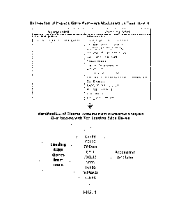
FIG. 1 is a schematic of the analysis performed in the studies described
herein. A total of
13 plasma proteins were examined, which corresponded to top leading-edge genes
within
differentially modulated hepatic gene pathways. The analysis was focused on
the subset of 9
proteins in which the directionality of treatment effect was concordant with
the directionality of
change in hepatic gene expression. Abbreviations: CASP8, caspase 8; CCL20, C-C
motif
chemokine ligand 20; CRTAM, cytotoxic and regulatory T-cell molecule; CSF1,
macrophage
colony stimulating factor 1; CXCL12, C-X-C motif chennokine ligand 12; NCR1,
natural cytotoxicity
triggering receptor 1; TGFB1, transforming growth factor beta 1; TNFRSF21,
tumor necrosis
factor receptor superfamily member 21; VEGFA, vascular endothelial growth
factor A.
FIGs. 2A-C are graphs showing the differential changes in plasma VEGFA (FIG.
2A),
TGFB1 (FIG. 2B), and CSF1 (FIG. 2C) by treatment status. Tesamorelin led to
significant
reductions in plasma VEGFA (10g2-fold change, mean SD, -0.20 0.35 vs. 0.05
0.34, P =
0.02), TGFB1 (10g2-fold change - 0.35 0.56 vs. - 0.05 0.43, P = 0.05), and
CSF1 (10g2-fold
change - 0.17 0.21 vs. 0.02 0.20, P= 0.004) relative to placebo. Bars and
error bars indicate
mean and standard error of the mean, respectively. Abbreviations: CSF1,
macrophage colony
stimulating factor 1; TGFB1, transforming growth factor beta 1; VEGFA,
vascular endothelial
growth factor A.
FIGs. 3A and B are graphs showing the relationship of changes in Plasma VEGFA
(FIG.
3A) and CSF1 (FIG. 3B) with change in NAS score in tesamorelin-treated
participants. Within the
tesamorelin-treated arm, reductions in plasma VEGFA (r= 0.62, P= 0.006) and
CSF1 (r= 0.50,
P = 0.04) were associated with a decrease in NAS score. Linear regression
lines with 95%
confidence intervals are shown. Abbreviations: CSF1, macrophage colony
stimulating factor 1;
NAS, NAFLD activity score; VEGFA, vascular endothelial growth factor A.
FIGs. 4A and 4B are graphs depicting the relationship of changes in plasma
TGFB1 and
CSF1 with change in gene-level fibrosis score. Among tesamorelin-treated
participants, declines
in plasma TGFB1 (FIG. 4A) (r = 0.61, P = 0.009) and CSF1 (FIG. 4B) (r = 0.64,
P = 0.006) were
associated with improved gene-level fibrosis score. Linear regression lines
with 95% confidence
intervals are shown. Abbreviations: CSF1, macrophage colony stimulating factor
1; TGFB1,
transforming growth factor beta 1.
FIG. 5 shows the amino acid sequence of human VEGFA (SEQ ID NO: 5). Amino
acids 1-
26 (SEQ ID NO: 6) define the signal peptide; amino acids 27-232 (SEQ ID NO: 7)
define the
mature polypeptide.
FIG. 6 shows the amino acid sequence of human TGFB1 (SEQ ID NO: 8). Amino
acids 1-
29 (SEQ ID NO: 9) define the signal peptide; amino acids 30-278 (SEQ ID NO:
10) define the
latency-associated peptide; amino acids 279-390 (SEQ ID NO: 11) define the
mature polypeptide.
CA 03196736 2023- 4- 26
WO 2022/093757
PCT/US2021/056568
7
FIG. 7 shows the amino acid sequence of human CSF1 (SEQ ID NO: 12). Amino
acids 1-
32 (SEQ ID NO: 13) define the signal peptide and residues 33-450 defining the
processed mature
form (SEQ ID NO: 14).
FIG. 8 shows the structure of tesamorelin (trans-3-hexenoyi-GHRH(l_44)-NH2;
SEQ ID NO:
1),
DETAILED DISCLOSURE
The use of the terms "a" and "an" and "the" and similar referents in the
context of describing
the technology (especially in the context of the following claims) are to be
construed to cover both
the singular and the plural, unless otherwise indicated herein or clearly
contradicted by context.
The terms "comprising", "having", "including", and "containing" are to be
construed as open-
ended terms (i.e., meaning "including, but not limited to") unless otherwise
noted.
All methods described herein can be performed in any suitable order unless
otherwise
indicated herein or otherwise clearly contradicted by context.
The use of any and all examples, or exemplary language ("e.g.", "such as")
provided herein,
is intended merely to better illustrate embodiments of the claimed technology
and does not pose
a limitation on the scope unless otherwise claimed.
No language in the specification should be construed as indicating any non-
claimed element
as essential to the practice of embodiments of the claimed technology.
Herein, the term "about" has its ordinary meaning. The term "about" is used to
indicate that
a value includes an inherent variation of error for the device or the method
being employed to
determine the value, or encompass values close to the recited values, for
example within 10% of
the recited values (or range of values).
Recitation of ranges of values herein are merely intended to serve as a
shorthand method
of referring individually to each separate value falling within the range,
unless otherwise indicated
herein, and each separate value is incorporated into the specification as if
it were individually
recited herein. All subsets of values within the ranges are also incorporated
into the specification
as if they were individually recited herein.
Where features or aspects of the disclosure are described in terms of Markush
groups or
list of alternatives, those skilled in the art will recognize that the
disclosure is also thereby
described in terms of any individual member, or subgroup of members, of the
Markush group or
list of alternatives.
Unless specifically defined otherwise, all technical and scientific terms used
herein shall be
taken to have the same meaning as commonly understood by one of ordinary skill
in the art (e.g.,
in stem cell biology, cell culture, molecular genetics, immunology,
immunohistochemistry, protein
chemistry, and biochemistry).
CA 03196736 2023- 4- 26
WO 2022/093757
PCT/US2021/056568
a
Unless otherwise indicated, the recombinant protein, cell culture, and
immunological
techniques utilized in the present disclosure are standard procedures, well
known to those skilled
in the art. Such techniques are described and explained throughout the
literature in sources such
as, J. Perbal, A Practical Guide to Molecular Cloning, John Wiley and Sons
(1984), J. Sambrook
et al., Molecular Cloning: A Laboratory Manual, Cold Spring Harbour Laboratory
Press (1989), T.
A. Brown (editor), Essential Molecular Biology: A Practical Approach, Volumes
1 and 2, IRL Press
(1991), D. M. Glover and B. D. Hames (editors), DNA Cloning: A Practical
Approach, Volumes 1-
4, IRL Press (1995 and 1996), and F. M. Ausubel etal. (editors), Current
Protocols in Molecular
Biology, Greene Pub. Associates and Wiley-Interscience (1988, including all
updates until
present), Ed Harlow and David Lane (editors) Antibodies: A Laboratory Manual,
Cold Spring
Harbour Laboratory, (1988), and J. E. Coligan et al. (editors) Current
Protocols in Immunology,
John Wiley & Sons (including all updates until present).
In the studies described herein, the present inventors have shown that reduced
levels of
Vascular Endothelial Growth Factor A (VEGFA), Transforming Growth Factor Beta
1 (TGFB1),
and Colony Stimulating Factor 1 (CSF1) are detected in the plasma of patients
suffering from
NAFLD treated with tesamorelin. The reduction in VEGFA, TGFB1, and/or CSF1
levels were
shown to correlate with improvements of pathological features of NAFLD, such
as a reduction of
the NAFLD Activity Score (NAS) and/or gene-level fibrosis score in the
patients.
NAFLD Activity Score (NAS) calculated according to the NAS Clinical Research
Network
(NAS CRN) scoring system comprises the sum of grades for steatosis (grades 0-
3), hepatocellular
ballooning (grades 0-2), and lobular inflammation (grades 0-3) (Kleiner DE, et
al. Hepatology
2005; 41:1313-21).
In an aspect, the present disclosure provides a method for assessing the
likelihood that a
subject suffers from NAFLD, the method comprising measuring protein levels of
Vascular
Endothelial Growth Factor A (VEGFA), Transforming Growth Factor Beta 1
(TGFB1), and/or
Colony Stimulating Factor 1 (CSF1) in a biological sample from the subject,
wherein a higher level
of VEGFA, TGFB1, and/or CSF1 in the sample relative to a corresponding control
level is
indicative of an increased likelihood that the subject suffers from NAFLD.
"Control level" or "reference level" or "standard level" are used
interchangeably herein and
broadly refers to a separate baseline level measured in one or more comparable
"control"
samples, which may be from subjects not suffering from the disease (e.g.,
NAFLD). The
corresponding control level may be a level corresponding to an average/mean or
median level
calculated based of the levels measured in several reference or control
subjects (e.g., a pre-
determined or established standard level). The control level may be a pre-
determined "cut-off"
value recognized in the art or established based on levels measured in samples
from one or a
group of control subjects. For example, the "threshold reference level" may be
a level
corresponding to the minimal level of VEGFA, TGFB1, and/or CSF1 (cut-off) that
permits to
CA 03196736 2023- 4- 26
WO 2022/093757
PCT/US2021/056568
9
distinguish in a statistically significant manner patients having a higher
likelihood or risk of
suffering from NAFLD from those not having a higher likelihood or risk of
suffering from NAFLD,
which may be determined using samples from NAFLD patients and from healthy
subjects (i.e.,
not suffering from NAFLD), for example. The corresponding reference/control
level may be
adjusted or normalized for age, gender, race, or other parameters. The
"control level" can thus
be a single number/value, equally applicable to every patient individually, or
the control level can
vary, according to specific subpopulations of patients. Thus, for example,
older men may have a
different control level than younger men, and women may have a different
control level than men.
The predetermined standard level can be arranged, for example, where a tested
population is
divided equally (or unequally) into groups, such as a low-risk group, a medium-
risk group and a
high-risk group or into quadrants or quintiles, the lowest quadrant or
quintile being individuals with
the lowest risk (i.e., lowest levels of VEGFA, TGFB1, and/or CSF1) and the
highest quadrant or
quintile being individuals with the highest risk (i.e., highest levels of
VEGFA, TGFB1, and/or
CSF1). It will also be understood that the control levels according to the
disclosure may be, in
addition to predetermined levels or standards, levels measured in other
samples (e.g., from
healthy/normal subjects) tested in parallel with the experimental sample. The
reference or control
levels may correspond to normalized levels, i.e., reference or control values
subjected to
normalization based on the expression of a housekeeping gene.
In embodiments, the control level is a corresponding level of VEGFA, TGFB1,
and/or CSF1
determined in a biological sample of a subject known not to suffer from NAFLD,
or an established
reference or standard level of VEGFA, TGFB1, and/or CSF1.
The present disclosure also provides a method for assessing the severity of
nonalcoholic
fatty liver disease (NAFLD) in a patient over time, the method comprising:
measuring protein levels of VEGFA, TGFB1, and/or CSF1 in a biological sample
from the
patient at a first time point;
measuring protein levels of VEGFA, TGFB1, and/or CSF1 in a corresponding
biological
sample from the patient at a second, later time point;
wherein a decrease in protein levels of VEGFA, TGFB1, and/or CSF1 between said
first and
second time points is indicative that NAFLD severity has regressed over time
in the patient;
wherein an increase in protein levels of VEGFA, TGFB1, and/or CSF1 between
said first and
second time points is indicative that NAFLD severity has progressed over time
in the patient; and
wherein no change in protein levels of VEGFA, TGFB1, and CSF1 between said
first and second
time points is indicative that NAFLD severity has been stable over time in the
patient.
VEGFA (UniProtKB accession No. P15692) is a protein of 232 amino acids
(precursor,
isoform 1), with amino acids 1-26 defining the signal peptide and amino acids
27-232 defining the
mature polypeptide. The amino acid sequence of VEGFA (isoform 1) is depicted
at FIG. 5.
CA 03196736 2023- 4- 26
WO 2022/093757
PCT/US2021/056568
TGFB1 (UniProtKB accession No. P01137) is a protein of 390 amino acids
(precursor), with
amino acids 1-29 defining the signal peptide, and which is proteolytically
processed to produce a
mature peptide of 112 amino acid (residues 279-390). The amino acid sequence
of TGFB1 is
depicted at FIG. 6.
5
CSF1 (UniProtKB accession No. P09603) is initially produced as a precursor
that is
membrane bound but processed and secreted upon stimulation. The precursor
comprises 554
amino acids (isoform 1), with amino acids 1-32 defining the signal peptide,
and residues 33-450
defining the processed mature form. The amino acid sequence of CSF1 (isoform
1) is depicted at
FIG. 7.
10
The above-noted method for assessing the severity of NAFLD over time may be
performed
at several time points, i.e., protein levels of VEGFA, TGFB1, and/or CSF1 in
corresponding
biological sample(s) from the patient may be performed at a third, fourth,
fifth, etc. time points.
The interval between two time points may be, e.g., 1 day, 2 days, 3 days, 1
week, 2 weeks, 1
month, 2 months, 3 months, 6 months, 1 year, etc., and may be the same for all
time points or
may vary (e.g., 1 week between the first and second time points, and 1 month
between the second
and third time points).
The method permits to determine whether the patient's condition improves,
deteriorates, or
is stable over time. In an embodiment, the protein levels of TGFB1 are
decreased between a first
and a second time point, and the decrease is indicative of a reduction of the
NAS score and/or
liver fibrosis in the patient. In an embodiment, the protein levels of TGFB1
are increased between
a first and a second time point, and the increase is indicative of an increase
of the NAS score
and/or liver fibrosis in the patient. In an embodiment, the protein levels of
CSF1 are decreased
between a first and a second time point, and the decrease is indicative of a
reduction of the NAS
score and/or liver fibrosis in the patient In an embodiment, the protein
levels of CSF1 are
increased between a first and a second time point, and the increase is
indicative of an increase
of the NAS score and/or liver fibrosis in the patient. In an embodiment, the
protein levels of VEGFA
are decreased between a first and a second time point, and the decrease is
indicative of a
reduction of the NAS score. In an embodiment, the protein levels of VEGFA are
increased
between a first and a second time point, and the increase is indicative of an
increase of the NAS
score. In an embodiment, the protein levels of VEGFA and CSF1 are decreased
between a first
and a second time point, and the decrease is indicative of a reduction of the
NAS score in the
patient. In an embodiment, the protein levels of VEGFA and CSF1 are increased
between a first
and a second time point, and the increase is indicative of an increase of the
NAS score in the
patient. In an embodiment, the protein levels of TGFB1 and CSF1 are decreased
between a first
and a second time point, and the decrease is indicative of a reduction of the
liver fibrosis in the
patient. In an embodiment, the protein levels of TGFB1 and CSF1 are increased
between a first
CA 03196736 2023- 4- 26
WO 2022/093757
PCT/US2021/056568
11
and a second time point, and the increase is indicative of an increase of the
liver fibrosis in the
patient.
The above-noted method for assessing the severity of NAFLD over time may be
useful for
determining whether a patient suffering from NAFLD responds or not to a
treatment/therapy
against NAFLD, i.e., to determine whether the treatment/therapy is effective
and improves the
patient's condition or not. Thus, in another embodiment, the patient is being
administered a
treatment/therapy between the first and second time points. In another
embodiment, the patient
undergoes a weight loss program, i.e., healthy (low calorie) diet and/or
physical exercise, between
the first and second time points.
Accordingly, in another aspect, the present disclosure relates to a method for
assessing
whether a treatment improves the condition of a patient suffering from NAFLD,
the method
comprising:
measuring protein levels of VEGFA, TGFB1, and/or CSF1 in a biological sample
from the
patient at a first time point;
administering a treatment against NAFLD to the patient for a period of time;
and
measuring protein levels of VEGFA, TGFB1, and/or CSF1 in a corresponding
biological
sample from the patient at a second time point after said period of time;
wherein a decrease in protein levels of VEGFA, TGFB1, and/or CSF1 between said
first and
second time points is indicative that the treatment has improved the patient's
condition;
wherein no change or an increase in protein levels of VEGFA, TGFB1, and/or
CSF1 between said
first and second time points is indicative that the treatment has not improved
the patient's
condition.
In an embodiment, the improvement of the patient's condition comprises
reduction of the
NAS score. In a further embodiment, the improvement of the patient's condition
comprises
reduction of the NAS score and the method comprises measuring the levels of
VEGFA and/or
CSF 1 .
In an embodiment, the improvement of the patient's condition comprises
reduction of liver
fibrosis. In a further embodiment, the improvement of the patient's condition
comprises reduction
of liver fibrosis and the method comprises measuring the levels of TGFB1
and/or CSF1.
In an embodiment, the improvement of the patient's condition comprises
reduction of the
NAS score and reduction of liver fibrosis. In a further embodiment, the
improvement of the
patient's condition comprises reduction of the NAS score and reduction of
liver fibrosis and the
method comprises measuring the levels of CSF1.
In another aspect, the present disclosure relates to a method for determining
whether a
candidate therapy may be useful for the treatment of NAFLD, the method
comprising:
measuring a first protein level of VEGFA, TGFB1, and/or CSF1 in a biological
sample from
a subject suffering from NAFLD;
CA 03196736 2023- 4- 26
WO 2022/093757
PCT/US2021/056568
12
administering the candidate therapy to the subject for a period of time; and
measuring a second protein level of VEGFA, TGFB1, and/or CSF1 in a biological
sample
from the subject after said period of time;
wherein a lower level of the second protein level relative to the first
protein level is indicative
that the candidate therapy may be useful for the treatment of NAFLD.
In an embodiment, such studies are carried out in the context of a clinical
trial that typically
entails additionally administering a placebo to a second subject suffering
from NAFLD. In such a
case, in an embodiment, the method for determining whether a candidate therapy
may be useful
for the treatment of NAFLD comprises:
measuring first protein levels of VEGFA, TGFB1, and/or CSF1 in biological
samples from
first and second subjects suffering from NAFLD;
administering the candidate therapy to the first subject and a placebo to the
second subject
for a period of time; and
measuring second protein levels of VEGFA, TGFB1, and/or CSF1 in biological
samples
from the first and second subjects after said period of time.
Similarly, in such an embodiment, a decrease in the level of the second
protein level relative
to the first protein level of VEGFA, TGFB1, and/or CSF1 in the biological
sample from the first
subject is indicative that the candidate therapy may be useful for the
treatment of NAFLD. The
determination of the first and second protein levels in the second subject
provide an additional
control in the context of such a trial.
In an embodiment, the above-mentioned methods comprise measuring protein
levels of
VEGFA. In an embodiment, the above-mentioned methods comprise measuring
protein levels of
TGFB1. In an embodiment, the above-mentioned methods comprise measuring
protein levels of
CSF1. In an embodiment, the above-mentioned methods comprise measuring protein
levels of
VEGFA and TGFB1. In an embodiment, the above-mentioned methods comprise
measuring
protein levels of VEGFA and CSF1. In an embodiment, the above-mentioned
methods comprise
measuring protein levels of TGFB1 and CSF1. In an embodiment, the above-
mentioned methods
comprise measuring protein levels of VEGFA, TGFB1 and CSF1.
In another aspect, the present disclosure relates to a method for treating
nonalcoholic
NAFLD, the method comprising administering a treatment against NAFLD to a
subject having an
increased likelihood of suffering from NAFLD identifying using the method
described herein.
In another aspect, the present disclosure relates to a method for treating
nonalcoholic
NAFLD, the method comprising identifying a subject having an increased
likelihood of suffering
from NAFLD using the method described herein, and administering a treatment
against NAFLD
to the subject.
CA 03196736 2023- 4- 26
WO 2022/093757
PCT/US2021/056568
13
In another aspect, the present disclosure relates to the use of a treatment
against NAFLD
in a subject, wherein the subject is identified by the method of identifying a
subject having an
increased likelihood of suffering from NAFLD described herein.
In another aspect, the present disclosure relates to a treatment/therapy for
use in a
treatment against NAFLD in a subject, wherein the subject is identified by the
method of
identifying a subject having an increased likelihood of suffering from NAFLD
described herein.
The treatment/therapy administered to or performed on the patient in the
methods described
herein may be an experimental or candidate treatment/therapy, e.g., a
treatment/therapy tested
in a clinical study, or an approved or established treatment/therapy for
NAFLD.
In an embodiment, the treatment/therapy comprises administration or use of a
cholesterol-
lowering medication, such as statins (e.g., Atorvastatin, Fluvastatin,
Lovastatin, Pitavastatin,
Pravastatin, Rosuvastatin, Simvastatin), bile acid sequestrants (e.g.,
Cholestyramine,
Colesevelam, Colestipol), cholesterol absorption blockers (e.g., ezetimibe),
PCSK9 inhibitors
(e.g., anti-PCSK9 antibodies such as Alirocumab and Evolocumab), niacin,
fibrates (e.g.,
Fenofibrate, Gemfibrozil), Adenosine triphosphate-citrate Lyase (ACL)
inhibitors (e.g., bempedoic
acid), or omega-3 products (e.g., Icosapent ethyl, Omega-3-acid ethyl esters).
In another
embodiment, the treatment/therapy comprises a change in lifestyle, e.g.,
undergoing a weight
loss program, i.e., healthy (low calorie) diet and/or physical exercise.
In an embodiment, the treatment/therapy comprises administration or use of a
GHRH
molecule. The term "GHRH molecule" as used in the context of the present
disclosure includes,
without limitation, human native GHRH(144) and fragments thereof (e.g.,
GHRH(_40), GHRH(1_20),
fragments ranging between 1-29 and the 1-44 sequence), and any other
fragments; GHRH from
other species and fragments thereof; GHRH variants containing amino acid(s)
substitution(s),
addition(s) and/or deletion(s); derivatives or analogs of GHRH or fragments or
variants thereof
having for example an organic group or a moiety coupled to the GHRH amino acid
sequence at
the N-terminus, the C-terminus or on the side-chain; and pharmaceutically
acceptable salts of
GHRH (human or from other species), as well as pharmaceutically acceptable
salts of native
GHRH or fragments, variants, analogs and derivatives thereof. The GHRH
molecules of the
present disclosure also encompass the GHRH molecules currently known in the
art, including,
without limitation, albumin-conjugated GHRH (U.S. Patent No. 7,268,113);
pegylated GHRH
peptide (U.S. Patent Nos. 7,256,258 and 6,528,485); porcine GHRH (1-40) (U.S.
Patent No.
6,551,996); canine GHRH (U.S. patent application no. 2005/0064554); GHRH
variants of 1-29 to
1-44 amino acid length (U.S. Patent Nos. 5,846,936, 5,696,089, 5,756,458 and
5,416,073, and
U.S. patent application Nos. 2006/0128615 and 2004/0192593); and Pro -
GHRHpeptide and
variants thereof (U.S. Patent No. 5,137,872).
The GHRH analogs include those described in U.S. Patent Nos. 5,681,379 and
5,939,386, which also describe their method of synthesis. More particularly,
these GHRH analogs
CA 03196736 2023- 4- 26
WO 2022/093757
PCT/US2021/056568
14
are defined by the following formula A:
X-GHRH Peptide (A)
wherein the GHRH peptide is a peptide of the following formula B (SEQ ID
NO:2):
Al -A2-Asp-Ala-lle-Phe-Thr-A8-Ser-Tyr-Arg-Lys-A13-Leu-A15-Gln-Leu-Al 8-Ala-Arg-
Lys-
Leu-Leu-A24-A25-Ile-A27-A28-Arg-A30-A31-A32-A33-A34-A35-A36-A37-A38-A39-A40-
A41-
A42- A43-A44-R0 (B)
wherein,
Al is Tyr or His;
A2 is Val or Ala;
A8 is Asn or Ser;
A13 is Val or Ile;
A15 is Ala or Gly;
A18 is Ser or Tyr;
A24 is Gin or His;
A25 is Asp or Glu;
A27 is Met, Ile or Nle
A28 is Ser or Asn;
A30 is absent or is any amino acid, preferably Gin;
A31 is absent or is any amino acid, preferably Gin;
A32 is absent or is any amino acid, preferably Gly;
A33 is absent or is any amino acid, preferably Glu;
A34 is absent or is any amino acid, preferably Ser;
A35 is absent or is any amino acid, preferably Asn;
A36 is absent or is any amino acid, preferably Gin;
A37 is absent or is any amino acid, preferably Glu;
A38 is absent or is any amino acid, preferably Arg;
A39 is absent or is any amino acid, preferably Gly;
A40 is absent or is any amino acid, preferably Ala;
A41 is absent or is any amino acid, preferably Arg;
A42 is absent or is any amino acid, preferably Ala;
A43 is absent or is any amino acid, preferably Arg;
A44 is absent or is any amino acid, preferably Leu; and
RO is NH2 or NH-(CH2)n-CONH2, with n = 1 to 12.
The group X is a hydrophobic tail anchored via an amide bond to the N-terminus
of the
peptide and the hydrophobic tail defining a backbone of 5 to 7 atoms. The
backbone can be
substituted by C1_6 alkyl, C3_6 cycloalkyl, or C6.12 aryl and the backbone
comprises at least one
CA 03196736 2023- 4- 26
WO 2022/093757
PCT/US2021/056568
rigidifying moiety connected to at least two atoms of the backbone. The
rigidifying moiety is a
double bond, triple bond, saturated or unsaturated 03.9 cycloalkyl, or 06.12
aryl.
In an embodiment, group X is:
RpJ
(R¨H or at or C42CH2) , 2 (1111orcHot CHSKI) ,3 (R41 ot C113 or 04013)
5 4 (RAT or at or atat), S (Rzli ot CH3 or efizeN 6
it -a-41er er 012ais),
0
ij
7 (R-14 or CA. or Cfizac), (R.1-1 CH; C112a9,
(1t41 or CHI or OWN,
ftslai
11)
Ã61(R4f or at. or MAW, I (R41 or C11, ot
alp%) , 2 (R-11 or (i3*r OW%) ,
R
13 (14:41 or C',134 or CAC14) or
In an embodiment, in formula B, A30-A44 are: (a) absent; (b) an amino acid
sequence
10
corresponding to positions 30-44 of a native GHRH peptide (SEQ ID NO: 3), or
(c) the amino acid
sequence of (b) having a 1-14 amino acid deletion from its C-terminus.
In an embodiment, the GHRH peptide is a polypeptide comprising the amino acid
sequence
of SEQ ID NO: 4.
In an embodiment, the GHRH molecule is (hexenoyl trans-3)hGHRH(1_44.)NH2 (SEQ
ID NO:
15 1)
or a pharmaceutically acceptable salt thereof. trans-3-hexenoy1MGHRH(l -44)
amide (also
referred to as tesamorelin and (hexenoyl trans-3)hGHRH(1-44)NH2) is a
synthetic human GHRH
(hGHRH) analog that comprises the 44-amino acid sequence of hGHRH on which a
hexenoyl
CA 03196736 2023- 4- 26
WO 2022/093757
PCT/US2021/056568
16
moiety, a Ce side chain, has been anchored on the amino-terminal tyrosine
residue. The structure
of [trans-3-hexenoyl]liGHRH(1_44) amide is depicted at FIG. 8.
The term "pharmaceutically acceptable salt" refers to a salt of a GHRH
molecule (e.g., trans-
3-hexenoyl-GHRH(l.44.)-NH2) that is pharmacologically acceptable and
substantially non-toxic to
the subject to which it is administered. More specifically, these salts retain
the biological
effectiveness and properties of the GHRH molecules (e.g., trans-3-hexenoyl-
GHRI-1(l_44)-NH2) and
are formed from suitable non-toxic organic or inorganic acids or bases.
For example, these salts include acid addition salts of GHRH molecules (e.g.,
trans-3-
hexenoyl-GHRI-1(l.44)-NH2) which are sufficiently basic to form such salts.
Such acid addition salts
include acetates, adipates, alginates, lower alkanesulfonates such as a
methanesulfonates,
trifluoromethanesulfonatse or ethanesulfonates, arylsulfonates such as a
benzenesulfonates, 2-
naphthalenesulfonates, or toluenesulfonates (also known as tosylates),
ascorbates, aspartates,
benzoates, benzenesulfonates, bisulfates, borates, butyrates, citrates,
camphorates,
camphorsulfonates, cinnamates, cyclopentanepropionates, digluconates,
dodecylsulfates,
ethanesulfonates, fumarates, glucoheptanoates, glycerophosphates,
hemisulfates, heptanoates,
hexanoates, hydrochlorides, hydrobromides, hydroiodides, hydrogen sulphates, 2-
hydroxyethanesulfonates, itaconates, lactates, maleates, mandelates,
methanesulfonates,
nicotinates, nitrates, oxalates, pamoates, pectinates, perchlorates,
persulfates, 3-
phenylpropionates, phosphates, picrates, pivalates, propionates, salicylates,
succinates,
sulfates, sulfonates, tartrates, thiocyanates, undecanoates and the like.
Additionally, acids which are generally considered suitable for the formation
of
pharmaceutically useful salts from basic pharmaceutical compounds are
discussed, for example,
by P. Stahl etal., Camille G. (eds.) Handbook of Pharmaceutical Salts.
Properties, Selection and
Use. (2002) Zurich: Wiley-VCH; S. Berge eta!, Journal of Pharmaceutical
Sciences (1977) 66(1)
1-19; P. Gould, International J. of Pharmaceutics (1986) 33 201-217; Anderson
et al, The
Practice of Medicinal Chemistry (1996), Academic Press, New York; and in The
Orange Book
(Food & Drug Administration, Washington, D.C. on their website).
Such salts can be formed quite readily by those skilled in the art using
standard techniques.
Indeed, the chemical modification of a pharmaceutical compound (i.e., drug)
into a salt is a
technique well known to pharmaceutical chemists, (See, e.g., H. Ansel et. al.,
Pharmaceutical
Dosage Forms and Drug Delivery Systems (6' Ed. 1995) at pp. 196 and 1456-
1457). Salts of the
trans-3-hexenoyl-GHRI-1(l.4.4)-NH2 may be formed, for example, by reacting the
trans-3-hexenoyl-
GHRH(l.44)-NH2 with an amount of acid or base, such as an equivalent amount,
in a medium such
as one in which the salt precipitates or in an aqueous medium followed by
lyophilization.
In an embodiment, the pharmaceutically acceptable salt of the GHRH molecule,
preferably
trans-3-hexenoyl-GHRH (1 .44)-N H2, is an acetate salt.
CA 03196736 2023- 4- 26
WO 2022/093757
PCT/US2021/056568
17
In an embodiment, the GHRH molecule, preferably trans-3-hexenoyl-GHRH(1_44)-
NH2, or
pharmaceutically acceptable salt thereof, is present in a pharmaceutical
composition at a dose of
about 1 rng/rnIto about 10 rng/nnl. In a further embodiment, the GHRH
molecule, preferably trans-
3-hexenoyl-GHRH(1.44.)-NH2, or pharmaceutically acceptable salt thereof is
present in a
pharmaceutical composition at a dose of about 1 mg/ml to about 10 mg/ml,
preferably about 1
mg/ml to about 8 mg/ml or about 4 mg/ml to about 8 mg/ml, for example about 1
mg/ml, about 2
mg/ml, about 3 mg/ml, about 4 mg/ml, about 5 mg/ml, about 6 mg/ml, about 7
mg/ml, or about 8
mg/ml.
In an embodiment, the GHRH molecule, preferably trans-3-hexenoyl-GHRH(1_44)-
NH2, or
pharmaceutically acceptable salt thereof is present in a pharmaceutical
composition comprising
one or more pharmaceutically acceptable excipients.
The term "pharmaceutically acceptable excipient" as used herein has its normal
meaning in
the art and is any ingredient that is not an active ingredient (drug) itself.
Excipients include for
example binders, lubricants, diluents, bulking agents (fillers), thickening
agents, disintegrants,
plasticizers, coatings, barrier layer formulations, lubricants, stabilizing
agent, release-delaying
agents and other components. "Pharmaceutically acceptable excipient" as used
herein refers to
any excipient that does not interfere with effectiveness of the biological
activity of the active
ingredients and that is not toxic to the subject, i.e., is a type of excipient
and/or is for use in an
amount which is not toxic to the subject. Excipients are well known in the
art, and the present
composition is not limited in these respects. In certain embodiments, the
pharmaceutical
composition comprises one or more excipients, including for example and
without limitation, one
or more binders (binding agents), thickening agents, surfactants, diluents,
release-delaying
agents, colorants, flavoring agents, fillers, disintegrants/dissolution
promoting agents, lubricants,
plasticizers, silica flow conditioners, glidants, anti-caking agents, anti-
tacking agents, stabilizing
agents, anti-static agents, swelling agents and any combinations thereof. As
those of skill would
recognize, a single excipient can fulfill more than two functions at once,
e.g., can act as both a
binding agent and a thickening agent. As those of skill will also recognize,
these terms are not
necessarily mutually exclusive. Therapeutic formulations are prepared using
standard methods
known in the art by mixing the active ingredient having the desired degree of
purity with one or
more optional pharmaceutically acceptable carriers, excipients and/or
stabilizers. The excipient(s)
may be suitable, for example, for intravenous, parenteral, subcutaneous,
intramuscular,
intracranial, intraorbital, ophthalmic, intraventricular, intracapsular,
intraspinal, intrathecal,
epidural, intracisternal, intraperitoneal, intranasal or pulmonary (e.g.,
aerosol) administration (see
Remington: The Science and Practice of Pharmacy, by Loyd V Allen, Jr, 2012,
22nd edition,
Pharmaceutical Press; Handbook of Pharmaceutical Excipients, by Rowe etal.,
2012, 7th edition,
Pharmaceutical Press). In an embodiment, the pharmaceutical composition is an
injectable
CA 03196736 2023- 4- 26
WO 2022/093757
PCT/US2021/056568
18
composition. In an embodiment, the pharmaceutical composition comprises one or
more
excipients for subcutaneous administration/injection.
Methods to measure the amount/level of proteins in a biological sample are
well known in
the art. Protein levels may be detected directly using a ligand binding
specifically to the protein
(mature protein), such as an antibody or a fragment thereof. In embodiments,
such a binding
molecule or reagent (e.g., antibody) is labeled/conjugated, e.g., radio-
labeled, chromophore-
labeled, fluorophore-labeled, or enzyme-labeled to facilitate detection and
quantification of the
complex (direct detection). Alternatively, protein levels may be detected
indirectly, using a binding
molecule or reagent, followed by the detection of the [protein/ binding
molecule or reagent]
complex using a second ligand (or second binding molecule) specifically
recognizing the binding
molecule or reagent (indirect detection). Such a second ligand may be radio-
labeled,
chromophore-labeled, fluorophore-labeled, or enzyme-labeled to facilitate
detection and
quantification of the complex. Enzymes used for labeling antibodies for
immunoassays are known
in the art, and the most widely used are horseradish peroxidase (HRP) and
alkaline phosphatase
(AP). Examples of binding molecules or reagents include antibodies (monoclonal
or polyclonal),
natural or synthetic ligands, and the like.
Examples of methods to measure the amount/level of protein in a sample
include, but are
not limited to: Western blot, immunoblot, enzyme-linked immunosorbent assay
(ELISA),
"sandwich" immunoassays, radioimmunoassay (RIA), Proximity Extension Assay
(PEA),
immunoprecipitation, surface plasmon resonance (SPR), chemiluminescence,
fluorescent
polarization, phosphorescence, immunohistochemical (INC) analysis, matrix-
assisted laser
desorption/ionization time-of-flight (MALDI-TOF) mass spectrometry,
microcytometry, microarray,
antibody array, microscopy (e.g., electron microscopy), flow cytometry,
proteomic-based assays,
and assays based on a property or activity of the protein including but not
limited to ligand binding
or interaction with other protein partners, enzymatic activity, fluorescence.
For example, if the
protein of interest is a kinase known to phosphorylate a given target, the
level or activity of the
protein of interest may be determined by measuring the level of
phosphorylation of the target in
the presence of the test compound. If the protein of interest is a
transcription factor known to
induce the expression of one or more given target gene(s), the level or
activity of the protein of
interest may be determined by the measuring the level of expression of the
target gene(s). In an
embodiment, the amount/level of VEGFA, TGFB1, and/or CSF1 in the sample is
measured by
Proximity Extension Assay (PEA). PEA is an affinity-based assay that
characterizes abundance
levels of pre-determined sets of proteins. Each protein is targeted by a
unique pair of
oligonucleotide-labeled antibodies. When in close proximity, the
oligonucleotides undergo a
proximity-dependent DNA polymerization event to form a PCR target sequence.
The resultant
DNA sequence is detected and quantified using standard real-time PCR. PEA
gives protein
abundance levels of Normalized Protein eXpression (NPX) on a 10g2-scale.
CA 03196736 2023- 4- 26
WO 2022/093757
PCT/US2021/056568
19
In an embodiment, the above-noted measuring protein levels of VEGFA, TGFB1, or
CSF1
comprises contacting the biological sample with a ligand that specifically
binds to the protein(s),
such as an antibody or antigen-binding fragment thereof that specifically
binds to VEGFA, TGFB1,
or CSF1, and measuring the amount of complexes between VEGFA, TGFB1, or CSF1
and the
ligand (e.g., antibody or antigen-binding fragment thereof). The term
"antibody or antigen-binding
fragment thereof" as used herein refers to any type of antibody/antibody
fragment including
monoclonal antibodies (including full-length monoclonal antibodies),
polyclonal antibodies,
multispecific antibodies, humanized antibodies, CDR-grafted antibodies,
chimeric antibodies and
antibody fragments so long as they exhibit the desired antigenic
specificity/binding activity.
Antibody fragments comprise a portion of a full-length antibody, generally an
antigen binding or
variable region thereof. Examples of antibody fragments include Fab, Fab',
F(ab.)2, and Fv
fragments, diabodies, linear antibodies, single-chain antibody molecules,
single domain
antibodies (e.g., from camelids), shark NAR single domain antibodies, and
multispecific
antibodies formed from antibody fragments. Antibody fragments can also refer
to binding moieties
comprising CDRs or antigen binding domains including, but not limited to, VH
regions (VH, VH-VH),
anticalins, PepBodies, antibody-T-cell epitope fusions (Troybodies) or
Peptibodies.
In an embodiment, the antibody or antigen-binding fragment thereof is
labelled. The
antibody or antigen-binding fragment thereof may be labeled with one or more
labels such as a
biotin label, a fluorescent label, an enzyme label, a coenzyme label, a
cherniluminescent label, or
a radioactive isotope label. In an embodiment, the antibody or antigen-binding
fragment thereof
is labelled with a detectable label/moiety, for example a fluorescent moiety
(fluorophore). Useful
detectable labels include fluorescent compounds (e.g., fluorescein
isothiocyanate, Texas red,
rhodamine, fluorescein, Alexa Fluor dyes, and the like), radiolabels, enzymes
(e.g., horseradish
peroxidase, alkaline phosphatase and others commonly used in an protein
detection assays),
streptavidin/biotin, and colorimetric labels such as colloidal gold, colored
glass or plastic beads
(e.g., polystyrene, polypropylene, latex, etc.). Chemiluminescent compounds
may also be used.
In another embodiment, the antibody or antigen-binding fragment thereof is
conjugated to an
oligonucleotide, e.g., to perform Proximity Extension Assay, as described
above.
In an embodiment, the ligand that specifically binds to the protein(s) (e.g.,
an antibody or
antigen-binding fragment thereof that specifically binds to VEGFA, TGFB1, or
CSF1), is attached
or immobilized on a solid support. The solid support may be any solid support
which permits the
binding (e.g., immobilization) of the ligand and which may be used for the
desired application. It
includes for example glass or plastic plates/slides. In an embodiment, the
above-mentioned solid
support is a plastic plate/slide. In embodiments, the above-mentioned
plates/slides may be
modified (e.g., coated, chemically modified, derivatized) prior to
immobilization of the ligand. In
an embodiment, the solid support is modified to permit or facilitate the
covalent or non-covalent
immobilization of the ligand, using any method known in the art. The solid
support may be either
CA 03196736 2023- 4- 26
WO 2022/093757
PCT/US2021/056568
amino- or carboxy-functionalized, depending on whether immobilization of the
ligand through its
C- or N-terminal end is desired. The solid support may be modified/coated
using any conventional
moiety capable of binding to a corresponding moiety (affinity tag) conjugated
to the ligand, e.g.,
using typical affinity tags-based systems such as NTA ¨ "His-Tag" systems,
biotin ¨
5
avidin/streptavidin systems, glutathione S-transferase (GST) ¨ glutathione
systems, Maltose
Binding Protein (MBP) ¨ amylose systems, as well as antigen ¨ antibody
systems.
In an embodiment, the above-mentioned method comprises a step of normalizing
the
protein levels, i.e., normalization of the measured levels of the above-noted
proteins against a
stably expressed control protein (or housekeeping protein) to facilitate the
comparison between
10
different samples. "Normalizing" or "normalization" as used herein refers to
the correction of raw
protein level values/data between different samples for sample to sample
variations, to take into
account differences in "extrinsic" parameters such as protein quality,
efficiency of purification,
etc., i.e., differences not due to actual "intrinsic" variations in proteins
in the samples. Such
normalization is performed by correcting the raw protein level values/data for
a test protein (or
15
protein of interest, i.e., VEGFA, TGFB1, and/or CSF1) based on the protein
level values/data
measured for one or more "housekeeping" or "control" protein, i.e., whose
levels are known to be
constant (i.e., to show relatively low variability) in the biological sample
under different
experimental conditions. Thus, in an embodiment, the above-mentioned method
further
comprises measuring the level of expression of a housekeeping protein in the
biological sample.
20
The raw levels of VEGFA, TGFB1, and/or CSF1 measured in the sample may be
subjected
to mathematical transformations prior to analysis, such as log
transformations. In an embodiment,
the methods described herein comprises performing a Log2 transformation of the
raw levels of
VEGFA, TGFB1, and/or CSF1 measured in the sample prior to analysis.
In accordance with the present disclosure, a biological sample (e.g., a
medical/clinical
sample) encompasses any sample (crude or processed) obtained from a
subject/patient
suspected of containing the one or more target proteins described herein
(VEGFA, TGFB1, and
CSF1). Such substance may originate from a variety of sources. In an
embodiment, a sample
suspected to contain one or more target proteins may be obtained from any
tissue/organ and/or
from bodily excretions or fluids. The sample, if need be, may be prepared
using techniques known
to a person skilled in the art including, without limitation, mechanical
lysis, detergent extraction,
sonication, electroporation, denaturants, etc., and may also be purified if
need be. In further
embodiments, the sample may be processed to obtain an extract thereof enriched
in proteins,
ranging from relatively crude to relatively pure protein preparations.
In an embodiment, the above-mentioned biological sample is a biological fluid,
e.g., urine,
saliva, lymph, or a blood-derived sample. The term "blood-derived sample" as
used herein refers
to blood (e.g., fresh blood, stored blood) or to a fraction thereof, such as
serum, plasma and the
like. It also refers to any sample that may be obtained following one or more
purification,
CA 03196736 2023- 4- 26
WO 2022/093757
PCT/US2021/056568
21
enrichment, and/or treatment steps using blood (obtained by venous puncture,
for example) as
starting material. In an embodiment, the biological sample is a blood-derived
sample, in a further
embodiment plasma.
The sample may be obtained from a subject who is suspected of suffering from
NAFLD, for
example a subject who has one or more symptoms of fatty liver and/or liver
fibrosis. The subject
may be suspected of suffering from NAFLD, or having been diagnosed for NAFLD,
based on
results of laboratory testing such as elevated liver enzymes alanine
aminotransferase (ALT)
and/or aspartate aminotransferase (AST), evidence of liver fat detected by
imaging techniques,
and/or liver biopsy. The term NAFLD refers to a chronic liver disease defined
as the pathological
presence of hepatic steatosis (> 5% of the cross-sectional area of the liver
occupied by fat
vacuoles) in the absence of any secondary cause for hepatic fat accumulation,
such as alcohol
use, steatogenic medication, and hereditary disorders. NAFLD comprises a
spectrum of disease
that can be simplified into two categories: (1) Simple Steatosis (SS) or
nonalcoholic fatty liver
(NAFL), 70%-75% of cases, defined by excess liver fat without inflammation or
cellular injury; and
(2) nonalcoholic steatohepatitis (NASH), 25%-30% of cases, defined by the
presence of excess
liver fat with inflammation and cellular injury with or without perisinusoidal
fibrosis. In an
embodiment, the biological sample is from a subject suffering from or
suspected of suffering from
NAFL. In another embodiment, the biological sample is from a subject suffering
from or suspected
of suffering from NASH. In another embodiment, the subject is an HIV-infected
subject, i.e., the
subject suffers from HIV-associated NAFLD.
In an embodiment, the methods described herein further comprise performing one
or more
additional assays to assess/diagnose NAFLD/NASH in the subject. Such assays
include for
example determining the levels of liver enzymes such as alanine
aminotransferase (ALT) and/or
aspartate aminotransferase (AST) in a biological sample from the subject,
performing an imaging
of the liver using imaging techniques such as ultrasound, computerized
tomography (CT) scans,
magnetic resonance imaging (MRI), ultrasound elastography (USE), quantitative
ultrasound-
based techniques, magnetic resonance elastography (MRE), and magnetic
resonance-based fat
quantitation technique, or histological analysis of a liver sample (e.g.,
liver biopsy). Such
additional assay(s) may be performed on patients suspected of suffering from
NAFLD based on
higher/increased levels of VEGFA, TGFB1, and/or CSF1 in their biological
samples (relative to
reference levels), as described herein.
In another aspect, the present disclosure provides an assay mixture for (a)
assessing the
severity of NAFLD in a patient over time, and/or (b) assessing the likelihood
that a subject suffers
from NAFLD, the assay mixture comprising: (i) a biological sample from a
subject suffering from
or suspected of suffering from NAFLD; and (ii) one or more reagents for
determining/measuring
the protein levels of VEGFA, TGFB1, and/or CSF1 in the sample. In an
embodiment, the biological
sample is a blood-derived sample, in a further embodiment plasma. In an
embodiment, the
CA 03196736 2023- 4- 26
WO 2022/093757
PCT/US2021/056568
22
biological sample is from a subject suffering from NAFLD. In another
embodiment, the biological
sample is from an HIV-infected subject.
In another aspect, the present disclosure provides a system for (a) assessing
the severity
of NAFLD in a patient over time, and/or (b) assessing the likelihood that a
subject suffers from
NAFLD, the system comprising: (i) a biological sample from a subject suffering
from or suspected
of suffering from NAFLD; and (ii) and one or more assays for
determining/measuring the protein
levels of VEGFA, TGFB1, and/or CSF1 in the sample. In an embodiment, the
biological sample
is a blood-derived sample, in a further embodiment plasma. In an embodiment,
the biological
sample is from a subject suffering from NAFLD. In another embodiment, the
biological sample is
from an HIV-infected subject.
In another aspect, the present disclosure provides a system for (a) assessing
the severity
of NAFLD in a patient over time, and/or (b) assessing the likelihood that a
subject suffers from
NAFLD, the system comprising: a sample analyzer configured to produce a signal
corresponding
to the protein levels of VEGFA, TGFB1, and/or CSF1 in a biological sample of
the subject; and a
computer sub-system programmed to calculate, based on the one or more of the
protein levels,
whether the signal is higher or lower than a reference value. In various
embodiments, the system
further comprises the biological sample. In an embodiment, the biological
sample is a blood-
derived sample, in a further embodiment plasma. In an embodiment, the
biological sample is from
a subject suffering from NAFLD. In another embodiment, the biological sample
is from an HIV-
infected subject.
In another aspect, the present disclosure relates to a kit for use in (a)
assessing the severity
of NAFLD in a patient over time, and/or (b) assessing the likelihood that a
subject suffers from
NAFLD, the kit comprising reagents for measuring protein levels of VEGFA,
TGFB1, and/or CSF1
in a biological sample; and instructions for correlating the protein levels of
VEGFA, TGFB1, and/or
CSF1 with the severity of NAFLD and/or the likelihood of suffering from NAFLD.
In an embodiment, the reagents in the assay mixture, system and/or kit
comprise, for
example, ligands for VEGFA, TGFB1, and/or CSF1 (e.g., antibody(ies) or
fragments thereof),
solution(s), buffer(s), nucleic acid amplification reagent(s) (e.g., DNA
polymerase, DNA
polymerase cofactor, dNTPs), nucleic acid hybridization/detection reagent(s),
and/or reagents for
detecting antigen-antibody complexes, etc. In an embodiment, the reagents
comprise ligands
(e.g., antibody(ies) or fragments thereof) for at least two of VEGFA, TGFB1,
and/or CSF1. In an
embodiment, the reagents comprise ligands (e.g., antibody(ies) or fragments
thereof) for (i)
VEGFA and TGFB1; (ii) VEGFA and CSF1; (iii) TGFB1 and CSF1; or (iv) VEGFA,
TGFB1 and
CSF1. In an embodiment, the assay mixture, system and/or kit comprise an array
comprising
ligands (e.g., antibody(ies) or fragments thereof) for (i) VEGFA and TGFB1;
(ii) VEGFA and CSF1;
(iii) TGFB1 and CSF1; or (iv) VEGFA, TGFB1 and CSF1.
CA 03196736 2023- 4- 26
WO 2022/093757
PCT/US2021/056568
23
In an embodiment, the kit according to the present disclosure may be divided
into separate
packages or compartments containing the respective reagent components
explained above.
In addition, such a kit may optionally comprise one or more of the following:
(1) instructions
for using the reagents for performing the methods described herein and/or for
interpreting the
results obtained; (2) one or more containers; and/or (3) appropriate
controls/standards. Such a
kit can include reagents for collecting a biological sample from a patient and
reagents for
processing the biological sample.
Informational material included in the kits can be descriptive, instructional,
marketing or
other material that relates to the methods described herein and/or the use of
the reagents for the
methods described herein. For example, the informational material of the kit
can contain contact
information, e.g., a physical address, email address, website, or telephone
number, where a user
of the kit can obtain substantive information about performing the method
described herein and
interpreting the results.
The kits featured herein can also provide software necessary to infer the
severity of NAFLD
in a patient and/or the likelihood that a subject suffers from NAFLD from the
protein level data.
In another aspect, there is provided the use of the kit or assay mixture
described herein for
(a) assessing the severity of NAFLD in a patient over time, and/or (b)
assessing the likelihood
that a subject suffers from NAFLD.
MODE(S) FOR CARRYING OUT THE INVENTION
The present disclosure is illustrated in further details by the following non-
limiting examples.
Example 1: Materials and methods
Study Design
A randomized, double blind trial in which individuals with HIV-associated
NAFLD were
assigned to receive the growth hormone-releasing hormone (GHRH) analogue
tesamorelin 2 mg
daily or identical placebo for 12 months (7). Leveraging plasma specimens from
this trial, the
current study builds significantly on prior, purely transcriptomic analyses
(8) to examine specific
proteins. Changes in circulating levels of proteins corresponding to top
leading-edge genes in
pathways responsive to tesamorelin between treatment groups were investigated,
and the
relationships of these proteins to histologic, radiographic, and
transcriptomic indices was
assessed to identify plasmatic markers of NAFLD/NASH and elucidate potential
mechanisms of
tesamorelin response.
61 men and women 18-70 years old who had documented HIV infection and liver
steatosis
as defined by hepatic fat fraction 5% on 1H-magnetic resonance spectroscopy (H-
MRS) were
enrolled. Participants were required to have been on stable antiretroviral
therapy (ART) for 3
months with CD4-' T cell count > 100 cells/mm3 and HIV viral load < 400
copies/mL. Exclusion
CA 03196736 2023- 4- 26
WO 2022/093757
PCT/US2021/056568
24
criteria included excess alcohol use (> 20 g daily for women or > 30 g daily
for men), active
hepatitis B or C, other known hepatic disease, cirrhosis, and inadequately
controlled diabetes
mellitus (H bA1c 7%). Participants were enrolled at the Massachusetts General
Hospital (MGH,
Boston, MA) and the National Institutes of Health (NI H, Bethesda, MD) between
August 20, 2015
and January 16, 2019. Informed consent in writing was obtained from each
participant. All
methods were carried out in accordance with guidelines and regulations.
Study Procedures
Study procedures for the parent clinical trial have been described in detail
elsewhere (7, 9).
All study procedures were conducted in a fasting state. In brief, hepatic 1H-
MRS was performed
for measurement of hepatic fat fraction at baseline and 12 months. An
ultrasound-guided
percutaneous liver biopsy yielding two cores also was completed at each time
point. The first core
was fixed in formalin, and subsequently underwent histopathologic review by a
single expert
pathologist blinded to treatment (D.E.K., National Institutes of Health).
Histological scoring,
including NAFLD Activity Score (NAS) and fibrosis stage, was performed
according to the
Nonalcoholic Steatohepatitis Clinical Research Network scoring system (10).
The second core
was placed in an RNA stabilization reagent (RNAlater , Qiagen) and stored at -
80 C for gene
expression analyses. Blood specimens were collected at baseline and 12 months
and stored at -
80 C. Serum IGF-1 was measured using standard techniques (Quest Laboratories).
Hepatic Transcriptomic Assessment
Liver tissue underwent RNA extraction, cDNA library construction, and I
Ilumina sequencing
using methods that have been previously described (9). To identify pathways
differentially
modulated from pre- to post-treatment time points between tesamorelin- and
placebo-treated
participants, GSEA was performed using the desktop module from the Broad
Institute
(www.broadinstitute.org/gsea/). Gene sets used included the Molecular
Signatures Database
(MsigDB) hallmark gene set collection (11) and custom gene sets pertaining to
HCC prognosis
(9). GSEA leading-edge genes were the subset of genes in a significantly
enriched gene set that
accounted for the enrichment signal and were used for the subsequent
quantification of pathway
gene expression. Gene sets with false discovery rate (FDR) <0.05 were
considered enriched.
Utilizing this approach, 14 hallmark gene pathways that were differentially
regulated by
tesamorelin versus placebo were previously discovered. In this regard, a gene
set pertaining to
oxidative phosphorylation was upregulated with treatment. Furthermore, 13 gene
sets involved in
inflammation, tissue repair, and cell division were downregulated among
tesamorelin-treated
individuals (FIG. 1). The RNA-Seq data were submitted to the Gene Expression
Omnibus
repository at the National Center for Biotechnology Information (accession
number GSE150026).
CA 03196736 2023- 4- 26
WO 2022/093757
PCT/US2021/056568
Plasma Proteomic Assessment
For this analysis, change in targeted proteins over 12 months was assessed
using an Olink
Multiplex proximity extension assay (PEA) platform. The PEA is an affinity-
based assay that
characterizes abundance levels of pre-determined sets of proteins. Each
protein is targeted by a
5 unique pair of oligonucleotide-labeled antibodies. When in close
proximity, the oligonucleotides
undergo a proximity-dependent DNA polymerization event to form a PCR target
sequence. The
resultant DNA sequence is detected and quantified using standard real-time PCR
on the Fluidigm
BioMarkTm HD real-time PCR platform. The PEA gives protein abundance levels of
Normalized
Protein eXpression (NPX) on a 10g2-scale. Assay characteristics including
detection limits and
10 measurements of assay performance are available from the manufacturer
(Olink, Uppsala,
Sweden). Specificity is high due to the precision of the methodology, which
enabled assessment
of change overtime. Across all proteins within the high-multiplex panel
utilized (below), the mean
intra-assay and inter-assay variation were reported as 8.3% and 11.5%,
respectively.
15 Targeted Proteomic Analysis
An objective of the current study was to delineate potential response pathways
of
tesamorelin effects in NAFLD, and to determine a protein signature that might
be used to detect
a treatment response to tesamorelin among patients with NAFLD. To do so, all
plasma proteins
within a high-multiplex panel of nearly 100 proteins (Olink lmmuno-Oncology;
see www. olink.
20 com for the complete protein list) that were found to overlap with top
leading genes from
tesamorelin-responsive gene sets were flagged (8). Among this targeted set of
proteins, changes
in plasma levels by treatment status were compared. Proteins found to be
differentially modulated
by tesamorelin relative to placebo were then examined in relation to
radiographic, histologic, and
transcriptomic indices of NAFLD severity both at baseline and longitudinally.
As a surrogate for
25 fibrosis stage, a gene-level fibrosis score derived from the hepatic
expression of 18 genes shown
to correlate with fibrosis (11) was utilized, which was validated in the
current sample to histological
changes as previously described (8). Changes in levels of these proteins were
also related to
changes in their corresponding hepatic transcript level and change in serum
IGF-1.
Continuous variables were expressed as mean standard deviation, whereas
categorical
variables were indicated as a frequency (cY0). Differences between groups were
compared using
a two-tailed independent samples t-test for continuous variables and chi-
square test for
categorical variables. Correlations were assessed with Pearson correlation
coefficient. A value of
P 0.05 was the pre-defined threshold for statistical
significance. Statistical analyses were
performed using JMP Pro 14 (SAS Institute Inc., Cary, North Carolina, USA).
Table 1: Overlap of Proteins Studied with Top Leading-edge Genes Within
Differentially
Modulated Gene Sets
CA 03196736 2023- 4- 26
WO 2022/093757
PCT/US2021/056568
26
Plasma protein Gene set with corresponding top leading edge gene
CASP8 APOPTOSIS
CCL20 I NFLAMMATORY_RESPONSE, RAS_SIGNALING_UP,
TN FA_SIGNALI NG_VIA_N FKB
CRTAM ALLOG RAFT_REJ ECTI ON
CSF1 I L6_JAK_STAT3_SIGNALI NG, I NFLAMMATORY_RESPONSE
CXCL12 EPITHELIAL_MESENCHYMAL_TRANSITION
NCR1 ALLOG RAFT_REJ ECTI ON
TGFB1 TGF_BETA_SIGNALI NG, I L6_JAK_STAT3_SIGNALI NG
TNFRSF21 TNFRSF21
VEGFA TNFA_SIGNALING_VIA_NFKB
Abbreviations: CASP8, caspase 8; CCL20, C¨C motif chemokine ligand 20; CRTAM,
cytotoxic
and regulatory T-cell molecule; CSF1, macrophage colony stimulating factor 1;
CXCL12, C-X-C
motif chemokine ligand 12; NCR1, natural cytotoxicity triggering receptor 1;
TGFB1, transforming
growth factor beta 1; TNFRSF21, tumor necrosis factor receptor superfamily
member 21; VEGFA,
vascular endothelial growth factor A.
Example 2: Characteristics of Study Participants
Of 61 participants with HIV-associated NAFLD in the randomized-controlled
trial, 58
individuals had a plasma protein panel obtained at baseline that was available
for analysis.
Moreover, 44 of these individuals (20 assigned to tesamorelin, 24 assigned to
placebo) had
plasma protein panels repeated at 12 months. Characteristics of each treatment
group in the
overall sample are summarized in Table 2 and have been described previously
(7). Tesamorelin
and placebo groups were well balanced with respect to key clinical variables.
Briefly, participants
(53 7 years old, 79% male) had well-controlled HIV infection for 17 9
years. All subjects
received stable ART with 64% on integrase inhibitor-based regimens. Baseline
hepatic fat content
was 14 8% as measured by hepatic 1H-MRS. A total of 33% and 43% had
histologic evidence
of NASH and fibrosis, respectively, on initial liver biopsy.
Table 2: Baseline Demographic and Clinical Characteristics
Overall Sample
Subset with Paired Protein
Panels
Tesamorelin Placebo Tesamorelin
Placebo
(n = 31) (n = 30) (n = 20) (n = 24)
Age (years) 52 8 54 7 53 6 54
7
% Male 77 80 70
79
% Race
White 68 63 55
71
Black 26 33 40
29
Other 6 3 5
0
% Hispanic 19 10 10
8
Duration of HIV infection 16 9 18 8 17 10 18
8
(years)
CD4 count (cells/mm3) 733 290 798 260 740
265 792 269
log HIV viral load 0.34 0.59 0.50 0.74 0.39
0.61 0.41 0.66
% Current Antiretroviral
Use
CA 03196736 2023- 4- 26
WO 2022/093757
PCT/US2021/056568
27
NRTI 87 97 85
96
NNRTI 39 37 30
38
PI 29 20 25
25
Integrase Inhibitor 68 60 80
58
% Type 2 Diabetes 13 13 5
13
Liver biopsy length (mm) 13 3 13 3 14 2 13 3
Hepatic Fat Fraction CYO 13 8 15 9 15 9 15 9
% NASH 34 31 22
35
% Fibrosis 48 38 39
39
Stage 1 14 17 17
13
Stage 2 21 14 17
17
Stage 3 14 7 6
9
There were no statistically significant differences between groups at baseline
for any of the
variables shown above.
Continuous variables are presented as mean standard deviation.
Abbreviations: NASH, nonalcoholic steatohepatitis; NNRTI, non-nucleoside
reverse transcriptase
inhibitor; NRTI, nucleoside reverse transcriptase inhibitor; PI, protease
inhibitor.
Example 3: Plasma Proteins Differentially Regulated by Tesamorelin
Nine plasma proteins were identified as corresponding to top leading edge
genes
modulated by tesamorelin (FIG. 1). These leading-edge genes were contained
within gene sets
pertaining to inflammation, tissue repair, and cell division (Table 1) that
were downregulated by
treatment with tesamorelin versus placebo. Of these proteins, treatment with
tesamorelin led to
reductions in plasma VEGFA (10g2-fold change - 0.20 0.35 vs. 0.05 0.34,
P=0.02), TGFB1
(10g2-fold change - 0.35 0.56 vs. -0.05 0.43, P=0.05), and CSF1 (10g2-fold
change
-0.17 0.21 vs. 0.02 0.20, P=0.004) compared to placebo (FIGs. 2A-2C).
Moreover, plasma
CCL20 tended to decrease with tesamorelin though the difference between groups
did not reach
statistical significance (10g2-fold change - 0.28 0.88 vs. 0.20 0.79,
P=0.06). The effect of
treatment versus placebo on all 9 plasma proteins is summarized in Table 3.
Table 3: Effects of Tesamorelin Versus Placebo on Select Plasma Proteins
Log2-Baseline Log2-Fold change
Plasma
Tesamorelin Placebo Tesamorelin Placebo
P-value
protein
(n=20) (n=24) (n=20) (n=24)
4.91 (4.52, 4.76 (4.39, -0.37 0.72 -0.02 0.65 0.10
CASP8
5.30) 5.13)
CCL20 7.53 (6.85, 7.15 (6.62, -0.28 0.88
0.20 0.79 0.06
8.21) 7.68)
5.71 (5.40, 6.08 (5.89, -0.35 0.73 -0.04 0.50 0.10
CRTAM
6.02) 6.27)
9.84 (9.77, 9.79 (9.72, -0.17 0.21 0.02 0.20 0.004
CSF1
9.92) 9.85)
CXCL12 1.16 (1.08, 1.15 (1.10, -0.03 0.18
-0.003 0.17 0.54
1.25) 1.20)
CA 03196736 2023- 4- 26
WO 2022/093757
PCT/ITS2021/056568
28
3.05 (2.81, 3.21 (2.99, -0.20 0.52 -0.08 0.31 0.37
NCR1
3.29) 3.42)
8.99 (8.73, 8.82 (8.67, -0.35 0.56 -0.05 0.43 0.05
TGFB1 9.24) 8.98)
TNFR F21 7.57 (7.44, 7.62 (7.52, -0.13
0.22 -0.05 0.23 0.25
S
7.71) 7.72)
8.13 (7.97, 7.92 (7.81, -0.20 0.35 0.05 0.34 0.02
VEGFA
8.29) 8.03)
Bold text denotes P < 0.05. Data are represented as mean (95% confidence
interval) or mean
SD.
Abbreviations: CASP8, caspase 8; CCL20, C-C motif chemokine ligand 20; CRTAM,
cytotoxic
and regulatory T-cell molecule; CSF1, macrophage colony stimulating factor 1;
CXCL12, C-X-C
motif chemokine ligand 12; NCR1, natural cytotoxicity triggering receptor 1;
TGFB1, transforming
growth factor beta 1; TNFRSF21, tumor necrosis factor receptor superfamily
member 21; VEGFA,
vascular endothelial growth factor A.
Example 4: Association of Key Plasma Proteins with NAFLD Phenotype
Given their differential regulation by tesamorelin, the relationships of
VEGFA, TGFB1, and
CSF1 with NAFLD phenotype (Table 4) was next studied. At baseline, in the
overall sample,
plasma CSF1 level directly correlated with NAS score (r= 0.38, P= 0.004) and
gene-level fibrosis
score (r= 0.37, P= 0.03). In contrast, VEGFA and TGFB1 were not found to be
associated with
either of these parameters. Furthermore, there was no baseline relationship of
VEGFA, TGFB1,
or CSF1 with hepatic fat fraction.
Table 4: Relationships of Plasma Proteins Downregulated by Tesamorelin with
NAFLD Severity
A) Baseline
Log2 Plasma Protein NAS Score
Hepatic Fat Fraction Fibrosis-Related
(n = 58) (/o)
Gene Score
(n = 61) (n =
37)
VEGFA r= -0.10, P= 0.48 r= -0.001, P= 0.99 r= -0.09, P= 0.60
TGFB1 r= 0.31, P = 0.02 r= 0.30, P= 0.02 r= 0.22, P= 0.19
CSF1 r= 0.38, P= 0.004 r= 0.21, P= 0.12 r= 0.37, P = 0.03
B) Longitudinal
Change in NAS Score
Log2-Fold Change in
Plasma Protein Tesamorelin Placebo
(n = 18) (n = 22)
VEGFA r= 0.62, P= 0.006 r= 0.11, P=
0.62
TGFB1 r= 0.31, P= 0.22 r= 0.24, P=
0.29
CSF1 r= 0.50, P = 0.04 r= 0.21, P= 0.35
Change in Hepatic Fat Fraction (/o)
Log2-Fold Change in
Plasma Protein Tesamorelin Placebo
(n = 19) (n = 23)
VEGFA r= 0.30, P= 0.22 r= 0.12, P=
0.57
CA 03196736 2023- 4- 26
WO 2022/093757
PCT/US2021/056568
29
TGFB1 r= 0.17, P= 0.49 r= 0.34, P= 0.11
CSF1 r= 0.13, P= 0.60 r= 0.16, P= 0.46
Change in Fibrosis-Related Gene Score
Log2-Fold Change in
Plasma Protein Tesamorelin Placebo
(n = 17) (n = 19)
VEGFA r= 0.30, P= 0.24 r= -0.04, P= 0.86
TGFB1 r= 0.61, P = 0.009 r= -0.07, P= 0.79
CSF1 r= 0.64, P= 0.006 r= 0.29, P =0.23
Within the tesamorelin-treated arm, reductions in plasma VEGFA (r= 0.62, P=
0.006) and
CSF1 (r= 0.50, P = 0.04) strongly correlated with a decline in NAS score
(FIGs. 34, 3B and Table
4). Furthermore, among tesamorelin-treated participants, reductions in TGFB1
(r= 0.61, P= 0.009) and CSF1 (r= 0.64, P= 0.006) were associated with a
decline in gene-level
fibrosis score (FIGs. 44, 4B). Changes in these 3 plasma proteins were not
found to correlate
with change in NAS score or gene-level fibrosis score among placebo-treated
participants, or with
change in hepatic fat fraction within either treatment group.
Example 5: Association of Changes in Key Plasma Proteins with Changes in
Hepatic Transcript Levels and Serum IGF-1
To elucidate the regulation of plasma VEGFA, TGFB1, and CSF1, their
relationships with
corresponding hepatic transcript levels and serum IGF-1 levels within the
overall sample were
next investigated. CSF1 exhibited a correlation between changes in plasma
protein and hepatic
transcript levels (r= 0.50, P= 0.002). Additionally, an increase in serum IGF-
1 was associated
with a linear decline in CSF1 (r= -0.38, P= 0.01).
Although the present invention has been described hereinabove by way of
specific
embodiments thereof, it can be modified, without departing from the spirit and
nature of the
subject invention as defined in the appended claims. In the claims, the word
"comprising" is used
as an open-ended term, substantially equivalent to the phrase "including, but
not limited to". The
singular forms "a", "an" and "the" include corresponding plural references
unless the context
clearly dictates otherwise.
CA 03196736 2023- 4- 26
WO 2022/093757
PCT/US2021/056568
REFERENCES
1. Maurice JB, Patel A, Scott AJ, Patel K, Thursz M, Lemoine M. Prevalence
and risk factors
of nonalcoholic fatty liver disease in HIV-monoinfection. AIDS 2017;31:1621-
1632.
2. Koethe JR, Jenkins CA, Lau B, Shepherd BE, Justice AC, Tate JP, Buchacz
K, et al. Rising
5 Obesity Prevalence and Weight Gain Among Adults Starting Antiretroviral
Therapy in the United
States and Canada. AIDS Res Hum Retroviruses 2016;32:50-58.
3. Grant PM, Kitch D, McComsey GA, Collier AC, Bartali B, Koletar SL,
Erlandson KM, et al.
Long-term body composition changes in antiretroviral-treated HIV-infected
individuals. AIDS
2016;30:2805-2813.
10 4. Vodkin I, Valasek M, Bettencourt R, Cachay E, Loomba R. Clinical,
biochemical and
histological differences between HIV-associated NAFLD and primary NAFLD: a
case¨control
study. Alimentary pharmacology & therapeutics 2015;41:368-378.
5. Fourman LT, Stanley TL, Zheng I, Pan CS, Feldpausch MN, Purdy J,
Aepfelbacher J, et al.
Clinical Predictors of Liver Fibrosis Presence and Progression in HIV-
Associated NAFLD. Clin
15 Infect Dis 2020.
6. Wong VW, Singal AK. Emerging medical therapies for non-alcoholic fatty
liver disease and
for alcoholic hepatitis. Trans! Gastroenterol Hepatol 2019;4:53.
7. Stanley TL, Fourman LT, Feldpausch MN, Purdy J, Zheng I, Pan CS,
Aepfelbacher J, et al.
Effects of tesamorelin on non-alcoholic fatty liver disease in HIV: a
randomised, double-blind,
20 multicentre trial. Lancet HIV 2019;6:e821-e830.
8. Fourman LT, Billingsley JM, Agyapong G, Ho Sui SJ, Feldpausch MN, Purdy
J, Zheng I, et
al. Effects of tesannorelin on hepatic transcriptonnic signatures in HIV-
associated NAFLD. JCI
Insight 2020;5.
9. Kleiner DE, Brunt EM, Van Natta M, Behling C, Contos MJ, Cummings OW,
Ferrell LD, et
25 al. Design and validation of a histological scoring system for
nonalcoholic fatty liver disease.
Hepatology 2005;41:1313-1321.
10. Liberzon A, Birger C, Thorvaldsdottir H, Ghandi M, Mesirov JP, Tamayo P.
The Molecular
Signatures Database (MSigDB) hallmark gene set collection. Cell Syst
2015;1:417-425.
11. Hoang SA, Oseini A, Feaver RE, Cole BK, Asgharpour A, Vincent R,
Siddiqui M, et al. Gene
30 Expression Predicts Histological Severity and Reveals Distinct Molecular
Profiles of Nonalcoholic
Fatty Liver Disease. Sci Rep 2019;9:12541.
12. Kitade M, Yoshiji H, Kojima H, Ikenaka Y, Noguchi R, Kaji K, Yoshii J, et
al.
Neovascularization and oxidative stress in the progression of non-alcoholic
steatohepatitis. Mol
Med Rep 2008;1:543-548.
13. Coulon S, Francque S, Colle I, Verrijken A, Blomme B, Heindryckx F, De
Munter S, et al.
Evaluation of inflammatory and angiogenic factors in patients with non-
alcoholic fatty liver
disease. Cytokine 2012;59:442-449.
CA 03196736 2023- 4- 26
WO 2022/093757
PCT/US2021/056568
31
14. Cayon A, Crespo J, Guerra AR, Pons-Romero F. [Gene expression in obese
patients with
non-alcoholic steatohepatitis]. Rev Esp Enferm Dig 2008;100:212-218.
15. Coulon S, Legry V, Heindryckx F, Van Steenkiste C, Casteleyn C, Olievier
K, Libbrecht L,
et al. Role of vascular endothelial growth factor in the pathophysiology of
nonalcoholic
steatohepatitis in two rodent models. Hepatology 2013;57:1793-1805.
16. Fabregat I, Moreno-Caceres J, Sanchez A, Dooley S, Dewidar B, Giannelli
G, Ten Dijke P.
TGF-13 signalling and liver disease. FEBS J 2016;283:2219-2232.
17. Yang L, Roh YS, Song J, Zhang B, Liu C, Loomba R, Seki E. Transforming
growth factor
beta signaling in hepatocytes participates in steatohepatitis through
regulation of cell death and
lipid metabolism in mice. Hepatology 2014;59:483-495.
18. Cayon A, Crespo J, Mayorga M, Guerra A, Pons-Romero F. Increased
expression of Ob-
Rb and its relationship with the overexpression of TGF-beta1 and the stage of
fibrosis in patients
with nonalcoholic steatohepatitis. Liver Int 2006;26:1065-1071.
19. Hamilton, J. A., Cook, A. D. & Tak, P. P. Anti-colony-stimulating factor
therapies for
inflammatory and autoimmune diseases. Nat. Rev. Drug Discov. 16, 53-70
20. Stutchfield BM, Antoine DJ, Mackinnon AC, Gow DJ, Bain CC, Hawley CA,
Hughes MJ, et
al. CSF1 Restores Innate Immunity After Liver Injury in Mice and Serum Levels
Indicate
Outcomes of Patients With Acute Liver Failure. Gastroenterology 2015;149:1896-
1909 e1814.
21. Naito M, Hayashi S, Yoshida H, Nishikawa S, Shultz LD, Takahashi K.
Abnormal
differentiation of tissue macrophage populations in 'osteopetrosis (op) mice
defective in the
production of macrophage colony-stimulating factor. Am J Pathol 1991;139:657-
667.
22. Radi ZA, Koza-Taylor PH, Bell RR, Obert LA, Runnels HA, Beebe JS, Lawton
MP, et al.
Increased serum enzyme levels associated with kupffer cell reduction with no
signs of hepatic or
skeletal muscle injury. Am J Pathol 2011;179:240-247.
23. Tosello-Trampont AC, Landes SG, Nguyen V, Novobrantseva TI, Hahn YS.
Kuppfer cells
trigger nonalcoholic steatohepatitis development in diet-induced mouse model
through tumor
necrosis factor-a production. J Biol Chem 2012;287:40161-40172.
24. Li H, Zhou Y, Wang H, Zhang M, Qiu P, Zhang M, Zhang R, et al.
Crosstalk Between Liver
Macrophages and Surrounding Cells in Nonalcoholic Steatohepatitis. Front
Immunol
2020;11:1169.
25. Rosenfeld ME, Yla-Herttuala S, Lipton BA, Ord VA, Witztum JL, Steinberg D.
Macrophage
colony-stimulating factor mRNA and protein in atherosclerotic lesions of
rabbits and humans. Am
J Pathol 1992;140:291-300.
26. Sjaarda J, Gerstein H, Chong M, Yusuf S, Meyre D, Anand SS, Hess S, et
al. Blood CSF1
and CXCL12 as Causal Mediators of Coronary Artery Disease. J Am Coll Cardiol
2018;72:300-
310.
CA 03196736 2023- 4- 26