Note: Descriptions are shown in the official language in which they were submitted.
WO 2022/109053
PCT/US2021/059764
1
TYROSYL-LOCK PEPTIDES
CROSS-REFERENCE TO RELATED APPLICATIONS
[0001] This patent application claims the benefit of priority to
co-pending U.S.
Provisional Patent Application No. 63/115,418 filed November 18, 2020, which
is hereby
incorporated by reference in its entirety.
STATEMENT REGARDING
FEDERALLY SPONSORED RESEARCH OR DEVELOPMENT
[0002j This invention was made with Government support under
project number
Z0IZIABC006150 and ZOIZIABC 006161 by the National Institutes of Health,
National
Cancer Institute. The Government has certain rights in this invention.
INCORPORATION-BY-REFERENCE OF MATERIAL SUBMITTED
ELECTRONICALLY
[0003i Incorporated by reference in its entirety herein is a
computer-readable
nucleotide/amino acid sequence listing submitted concurrently herewith and
identified as
follows: One 26,697 Byte ASCII (Text) file named "757881 5T25.txt," dated
November 17,
2021.
BACKGROUND OF THE INVENTION
[0004] Relaxation of supercoiled DNA by topoisomerases is
necessary for the normal cell
functions of DNA transcription, replication, recombination, and repair.
Topoisomerase I
(TOP1) mediates both DNA strand break and religation by forming a transient,
covalent 3'-
phospho-tyrosyl bond with the DNA substrate. This TOP1-DNA cleavage complex is
the
target of chemotherapeutic TOP1 inhibitors such as the natural product
camptothecin.
Irinotecan, an analogue of camptothecin, is a widely-used anti-cancer agent
that stabilizes the
TOP1-DNA cleavage complex, causing irreversible double-strand DNA breaks,
eventually
leading to the death of replicating cancer cells. Tyrosyl-DNA
phosphodiesterase 1 (TDP1) is
an enzyme that, upon recognizing stalled TOP1-DNA cleavage complexes,
catalyzes the
cleavage of the 3'-phopho-tyrosyl bond between DNA and TOP. TDP1 is composed
of an
as-yet unstructured N-terminal regulatory domain whose function has been
reported to be
modulated by both phosphorylation and SUMOylation and a C-terminal catalytic
domain that
CA 03199368 2023- 5- 17
WO 2022/109053
PCT/US2021/059764
2
utilizes two histidine residues to effect phosphodiester cleavage at Tyr723 of
TOP1. After
removal of the 3' adduct, polynucleotide kinase phosphatase prepares the
degraded DNA
strands for further repair by DNA polymerase 13 and DNA ligase 111. The
clearance of
TOP1-DNA complexes results in escape from TOP1 inhibitor-induced cell death.
This
activity has led researchers to consider TDP1 a molecular target for the
sensitization of
replicating cancer cells to camptothecin and related chemotherapeutic agents.
[00051 Although these chemotherapeutic agents are effective, they
have downsides
including negative side effects. Given that cancer is currently a major health
concern, there is
an urgent need for new TDP1 inhibitors.
BRIEF SUMMARY OF THE INVENTION
[00061 An embodiment of the invention provides knotted cyclic
peptides comprising the
amino acid sequence of SEQ ID NO: 11
(CX1X2XXXCXXXXXXXXXCCXXXXXXSXXLXXXXXXCXXC), wherein X, Xi, and
X2 can be any amino acid provided that at least one of Xi and X2 is tyrosine,
phenylalanine,
or alanine.
l00071 An additional embodiment of the invention provide isolated
or purified peptides
comprising SEQ ID NO: 1, optionally with 1-6 amino acid substitutions or
deletions.
According to other aspects, there is provided a peptide comprising
ZEAFCYSDRFCQNYIGSIPDCCFGRGSYSFELQPPPWECYQC (SEQ ID NO: 16),
optionally with 1-6 amino acid substitutions or deletions; and a peptide
comprising
GVFCYSDRFCQNPIDN FDCCFSRGSYSFVPQPTPWDCFQC (SEQ ID NO: 30),
optionally with 1-6 amino acid substitutions or deletions.
[OWN Still another embodiment of the invention provides
pharmaceutical compositions
comprising peptides of an embodiment of the present invention and a
pharmaceutically
acceptable carrier.
[00091 Another embodiment of the invention provides peptides of
an embodiment of the
present invention, or pharmaceutical compositions of an embodiment of the
present
invention, for use in treating or preventing cancer.
[00191 A further embodiment of the invention provides methods of
treating or preventing
cancer in a mammal, the method comprising administering to the mammal the
peptides of an
embodiment of the present invention, or pharmaceutical compositions of an
embodiment of
the present invention, in an amount effective to treat or prevent cancer in
the mammal.
CA 03199368 2023- 5- 17
WO 2022/109053
PCT/US2021/059764
3
100111 An additional embodiment of the invention provides methods
of inhibiting the
cleavage of phosphodiester bonds by enzyme Tyrosyl-DNA phosphodiesterase 1
(TDP1) in a
mammal, the method comprising administering to the mammal the peptides of an
embodiment of the present invention, or pharmaceutical compositions of an
embodiment of
the present invention, in an amount effective to inhibiting the cleavage of
phosphodiester
bonds by enzyme TDP1.
100121 Another embodiment of the invention provides nucleic acids
encoding the
peptides of an embodiment of the present invention, optionally in a vector or
a cell.
[00131 A further embodiment of the invention provides methods of
preparing the peptides
of an embodiment of the present invention, by expressing a nucleic acid
encoding the peptide
in a host cell, optionally wherein the nucleic acid is in a vector.
BRIEF DESCRIPTION OF THE SEVERAL VIEWS OF THE DRAWING(S)
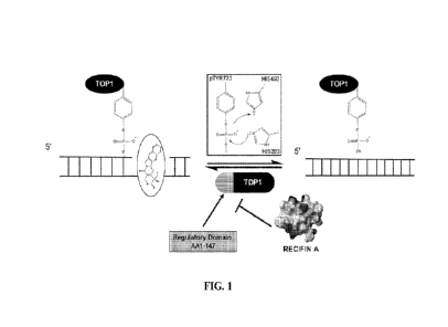
[00141 Figure 1 is a schematic showing TDP1 processing of 3'-TOP1
DNA adducts and
inhibition by an embodiment of the present invention, e.g., recifin A. TOP1
catalyzes single-
strand DNA breaks via a transitory, covalent phosphotyrosine linkage involving
tyrosine 723
(pTyr723). The cleavage complex is stabilized by the natural product
camptothecin, which
inhibits DNA religation, trapping TOP1 on the DNA strand, ultimately leading
to double
strand breaks and cell death. TDP1 removes the 3'-TOP1-pTyr-DNA adducts via a
nucleophilic attack on the phosphodiester bond by histidine 263 (HIS263) and
subsequent
hydrolysis by histidine 493 (HIS493). After removal of the 3' adduct, the DNA
strand is
further enzymatically repaired and re-ligated. Inhibitors of TDP1 catalytic
activity, such as
recifin A (depicted here by its electrostatic surface potential model) can
sensitize cancer cells
to TOP1 poisons.
100151 Figure 2A is a graph showing recifin A inhibition of full-
length human TDP1
enzymatic activity. Serial dilutions of purified recifin A were combined with
a synthetic
5'[32131-labeled, 3'-phosphotyrosine capped oligonucleotide DNA substrate and
incubated
with either full-length recombinant human TDP1 (rhTDP1) or human TDP1
complemented
DT40 knockout whole cell extracts (hTDP1 WCE).
100161 Figure 2B are images of poly-acrylamide gel
electrophoresis gels following
phosphorimaging of the reactions of Figure 2A. Activity was calculated as
percent of non-
inhibited substrate cleavage reaction control.
CA 03199368 2023- 5- 17
WO 2022/109053
PCT/US2021/059764
4
100171 Figure 3A is a graph showing disulfide mapping of recifin
A, specifically RP-
HPLC analysis of partially re-duced and alkylated recifin A. The mixture of
native recifin A
(3 intact disulfides/3-SS), partially reduced and alkylated isoforms (2 intact
disulfides/2-SS, 1
intact disulfide/1-SS) and completely reduced and alkylated recifin A (0-SS)
was desalted
and separated by RP-HPLC prior to further analysis.
100181 Figure 3B shows the MS/MS sequencing results for the 2-SS
recifin A isoform
trypsin fragments established the Cys IV-VI disul-fide linkage (SEQ ID NO: 1).
pGlu is
pyroglutamic acid; IAA. is iodoacetamide alkylated cysteine; NEM is N-
ethylmaleimide
alkylated cysteine.
[00191 Figure 3C shows an example of a recifin A disulfide
bonding pattern: Cys I-III,
Cys II-V, and Cys IV-VI. pGlu is pyroglutamic acid; IAA (SEQ ID NO: 2).
100201 Figure 4A shows an example of a NMR solution structure of
recifin A. The 20
best structures based on MolProbity scores superposed over residues 3-18 and
26-42,
emphasising the well-ordered core.
[00211 Figure 4B shows examples of ribbon structures showing the
four antiparallel 13-
strands (I-IV) and the threading of the third 3-strand through the ring formed
by the three
disulfide bonds and 3-strands I and IV. The ribbon structure on the right is
the ribbon
structure on the left rotated 90 degrees.
100221 Figure 5A shows a ribbon structure of a 3-strand threaded
Tyr-lock peptide
embodiment of the present invention, e.g., recifin A. Recifin A is stabilised
by the three
disulfide bonds Cys I-III, Cys II-V, and Cys IV-VI, forming a ring together
with two of the f3-
strands, which is penetrated by a third 3-strand. The recifin A structure is
further stabilised
by a central Tyr6 residue locking the structure in place, which is reminiscent
of microcin J25.
100231 Figure 5B shows a ribbon structure of a lasso peptide
microcin 125 (PDB ID:
1Q71). Microcin J25, lacks disulfide bonds, but a threaded structure is formed
by a
cyclisation via an amide-bond between the N-terminal amino-group and the
sidechain
carboxyl group of Glu8, which creates a circle that wraps around the C-
terminal part of the
sequence. The threaded structure is locked in place by two aromatic residues,
Phe19 and
Tyr20, making it sterically impossible for the structure to unravel.
[00241 Figure 5C shows a ribbon structure of a cyclic inhibitory
cystine knot peptide
kalata B1 (PDB ID: 1NB1). Kalata B1 is the prototypical plant cyclotide, which
contains an
inhibitory cystine knot motif and a head-to-tail backbone cyclisation. The ICK
is formed by
CA 03199368 2023- 5- 17
WO 2022/109053
PCT/US2021/059764
three disulfide bonds (Cys 1-1V, Cys 11-V, Cys 111-V1), two of which together
with the
backbone form a ring that the third disulfide bond is threaded through.
[00251 Figure 5D shows a ribbon structure of a shows a ribbon
structure of an-strand
threaded Tyr-lock peptide embodiment of the present invention, e.g., recifin
A.
[00261 Figure 5E shows a ribbon structure of a lasso peptide
microcin J25 (PDB ID:
1Q71).
[00271 Figure 5F shows a ribbon structure of a cyclic inhibitoy
cystine knot peptide
kalata B1 (PDB ID: 1NB1).
100281 Figure 6 shows the stabilizing function of Tyr6 (Y6)
residue in the overall, Tyr-
lock structure of recifin A. Cysll is C11, Tyr14 is Y14, Ser29 is S29, and and
Leu32 is L32.
Sidechains of residues that pack around Tyr6 are shown with thin lines
indicating confirmed
inter-residual NOEs.
100291 Figure 7 is a graph showing the biological activity and
specificity of recifin A.
Recifin A inhibited full-length TDP1, but not N-terminally truncated TDP1
(A147TDP1),
enzymatic activity in a concentration-dependent manner with an IC50 of 0.19
M.
100301 Figure 8A is a graph showing the steady-state analysis of
recifin A modulation of
full-length TDP1. Recifin A in-creased both the Km and Vmax kinetic constants
of the
TDP1 FRET assay24, exhibiting characteristics of both an enzyme inhibitor and
activator.
100311 Figure 8B is a graph showing the effect of recifin A on
A147TDP1 kinetic
parameters. Addition of recifin A did not affect either the Km or Vmax kinetic
constants of
the A147TDP1 FRET assay. As A147TDP1 enzyme retained the identical substrate
binding
and catalytic sites as full-length TDP1, this suggested the allosteric
modulation of TDP1,
dependent of the N-terminal 147 amino acid residues.
[00321 Figure 9A is a LC-MS analysis of the peptides in bulk-
purified Axinella sp.
aqueous extract. Total ion chromatogram (TIC), UV absorbance at 280, and the
separation
Gradient are shown.
[00331 Figure 9B is another LC-MS analysis of the peptides in
bulk-purified Axinella sp.
aqueous extract that were eluted prior to 6 min.
100341 Figure 9C is another LC-MS analysis of the peptides in
bulk-purified Axinella sp.
aqueous extract that were eluted prior to 6 min. (60% acetonitrile) showed
peptide-like mass-
to-charge ratios which deconvoluted to average masses of 4683.87, 4785.89,
4915.95
(recifin), and 5674.47.
CA 03199368 2023- 5- 17
WO 2022/109053
PCT/US2021/059764
6
[00351 Figure 10A is a LC-MS analysis showing the relative
abundance of partially-
purified RP-HPLC fraction A from Axinella sp aqueous peptide extract
[0036j Figure 10B is a LC-MS analysis showing the relative
abundance of partially-
purified RP-HPLC fraction B from Axinella sp. aqueous peptide extract.
[00371 Figure 10C is a LC-MS analysis showing the relative
abundance of partially-
purified RP-HPLC fraction C from Axinella ,sp. aqueous peptide extract.
100381 Figure 10D is a LC-MS analysis showing the relative
abundance of partially-
purified RP-HPLC fraction D from Axinella sp. aqueous peptide extract.
[00391 Figure 11 is graph showing the TDP1 inhibitory activity of
the peptide
constituents of fractions A-D of Axinella sp. aqueous extract. Fraction A was
determined to
be the most active and contained the highest abundance of recifin.
[00401 Figure 12A shows a MS analysis of native recifin A. The
monoisotopic mass of
native recifin was determined to be 4912.9661 Da.
100411 Figure 12B shows a MS analysis of reduced and alkylated
recifin A. The peptide
was reduced with 2-mercaptoethanol and alkylated with 4-vinylpyridine (105.06
Da), after
which a mass increase of 638.38 Da was observed, indicating the conversion of
six cysteine
residues to S-pyridylethyl cysteine and three disulfide bonds.
[00421 Figure 13A shows a tandem mass spectra (MS/MS) and
automated de novo and
amino acid sequencing of recifin A by Edman degradation. Alkylated recifin A
tryptic
fragment A was subjected to LC-MS and CID MS/MS. PEAKS de novo sequencing
software
was utilized to interpret the MS/MS spectra. An N-terminal pyro-glutamic acid
ion (e) was
identified, which prevented Edman degradation analysis (SEQ ID NO: 4).
[00431 Figure 13B shows a tandem mass spectra (MS/MS) and
automated de novo and
amino acid sequencing of recifin A by Edman degradation. Alkylated recifin A
tryptic
fragment B was subjected to LC-MS and CID MS/MS. PEAKS de novo sequencing
software
was utilized to interpret the MS/MS spectra. Fragment B was fully sequenced by
Edman
degradation. Pyroglutamate aminopeptidase digestion of intact recifin A
(reduced and
alkylated) afforded Edman degradation sequencing of 35 amino acids, which
provided both
the order of the tryptic fragments within the molecule and leucine/isoleucine
assignments
(SEQ ID NO: 5).
100441 Figure 13C shows a tandem mass spectra (MS/MS) and
automated de novo and
amino acid sequencing of recifin A by Edman degradation. Alkylated recifin A
tryptic
fragment C was subjected to LC-MS and CID MS/MS. PEAKS de novo sequencing
software
CA 03199368 2023- 5- 17
WO 2022/109053
PCT/US2021/059764
7
was utilized to interpret the MS/MS spectra. Fragment B was fully sequenced by
Edman
degradation. Pyroglutamate aminopeptidase digestion of intact recifin A
(reduced and
alkylated) afforded Edman degradation sequencing of 35 amino acids, which
provided both
the order of the tryptic fragments within the molecule and leucine/isoleucine
assignments
(SEQ ID NO: 6).
100451 Figure 14 shows the amino acid sequence of recifin A (SEQ
ID NO: 2) and an
enzymatic digest map. Reduced and alkylated recifin A was subjected to
digestion with
various enzymes and sequenced by CID MS/MS to confirm the proposed amino acid
sequence. C indicates alkylated, pGlu is pyroglutamic acid. Brackets indicate
fragments
sequenced by MS/MS. Bolded amino acids in the sequences below indicate the
protease
recognizes them and digests the polypeptide at that location.
Trypsin: pGluEAFCYSDRFCQNYIGSIPDCCFGRGSYSFELQPPPWECYQC (SEQ ID
NO: 2)
Glu-C: pGluEAFCYSDRFCQNYIGSIPDCCFGRGSYSFELQPPPWECYQC (SEQ ID NO:
2)
Proline endopeptidase:
pGluEAFCYSDRFCQNYIGSIPDCCFGRGSYSFELQPPPWECYQC (SEQ ID NO: 2)
Chymotrypsin: pGluEAFCYSDRFCQNYIGSIPDCCFGRGSYSFELQPPPWECYQC (SEQ
ID NO: 2)
100461 Figure 15A shows a 1D 1H NMR spectra of a ¨2 mg sample of
recifin A in
90/10% H20/D20 at 298K acquired on a Bruker AVANCE III equipped with a
cryoprobe (ns
32).
[00471 Figure 15B shows secondary Ha chemical shifts compared to
random coil values
highlighting positive stretches of secondary chemical shifts indicative of 13-
sheets combined
with negative stretches suggesting a-helices.
[00481 Figure 16 is a graph showing the effect of recifin A on
TDP1 kinetic parameters.
[00491 Figure 17 shows a Total Correlated Spectroscopy (TOCSY)
spectrum of the
amide region of recifin A. The amide region shows that the spin systems
(numbered) are well
dispersed, and it highlights the unusual up-field shift of the NH proton of
residue 16 and the
Ha proton of Tyrll as well as down-field shift of the Ha proton residue 28.
100.501 Figure 18A shows a ribbon structure illustrating the
position of the buried Tyr6.
Figure 18B shows a schematic illustrating the threading of the third n-strand
through the
embedded ring formed by the three disulfide bonds. Figure 18C shows the
recifin A
CA 03199368 2023- 5- 17
WO 2022/109053
PCT/US2021/059764
8
sequence; disulfide bond connections are shown with brackets, residues in the
ring are at
positions 5, 7-11, 21-22, and 39-42 and Tyr at position 6.
[00511 Figure 19 shows a synthetic strategy for recifin A using
native chemical ligation
of peptide hydrazides.
[00521 Figure 20A shows a superposition of TOCSY spectra of
native and synthetic
recifin A.
100531 Figure 20B shows a solution NMR structure of [Phel recifin
showing disulfides.
100541 Figure 20C shows superposition of [Phe61 recifin and
native recifin A highlighting
the similarities in the Tyr-lock region and hydrogen bonds.
[00551 Figure 21A shows FL-TDP1 FRET Assay results for a reaction
progress curve - 1
nM, 0.25 u.M S, 1XPBS pH 7.4, 80 mNIKC1, 1mM TCEP. Figure 21B shows FL-TDP1
FRET Assay results for a reaction progress curve - 1 nM, 0.25 uM S. 1XPBS pH
7.4, 80 mM
KC1.
100561 Figure 22 shows FL-TDP1 FRET Assay results - 1 nM E, 0.25
uM S, T=45 min,
1XPBS pH 7.4, 80 mM KC1.
100571 Figure 23A shows oxidative folding of recifin A. Figure
23B shows oxidative
folding of [Phel recifin. HPLC traces of each time point taken for the
oxidation of synthetic
peptides. Oxidation was performed in 0.1 M ammonium bicarbonate (pH 8.0) with
oxidized
(0.5 mM) and reduced (2 mM) glutathi one at a concentration of 0.125 mg/mL at
room
temperature. Aliquots were removed at time points 0, 8, and 48 h, quenched
with 6 M
guanidine hydrochloric acid (pH 3.7). Samples were analyzed by analytical RP-
HPLC on a
Cis column using a gradient of 5% buffer B for the first 10 min followed by 5-
65% B (buffer
A: H20/0.05% TFA; buffer B: 90% CH3CN/10% H20/0.045% TFA) in 65 min.
100581 Figures 24A-24 F show final analytical trace and ES1-MS
spectra of oxidized
recifin A and analogues.
[00591 Figure 25 shows 1D 11-1 Nuclear Magnetic Resonance spectra
of recifin A and
analogues in 90/10% H20/D20 at 298 K acquired on a Bruker Avance III 900 MHz
spectrometer equipped with a cryoprobe. The majority of the purified peptides
gave dispersed
1HNMR spectra with sharp lines, implying that they adopt ordered structures in
solution.
However, the [Alal recifin analogue spectra appeared broad and lacked
dispersion of the HN
signals indicating that the peptide is misfolded. Thus, while substitution of
Tyr6 with Phe is
well tolerated, incorporating an alanine at position 6 prevents folding of the
peptide.
[00601 Figure 26A and 26 B are nuclear magnetic resonance scans
of recifin A peptides.
CA 03199368 2023- 5- 17
WO 2022/109053
PCT/US2021/059764
9
10061] Figure 27 shows aligned sequences of recifin A and
analogues with the black line
highlighting the disulfide bond connection: Cys I-III, Cys II-V, and Cys IV-
VT.
[0062j Figure 28A to 28F show thermal stability (298-333 K) of
native recifin A,
synthetic recifin A and synthetic recifin A analogues carried out using
nuclear magnetic.
resonance on a 500 or 700 MHz Bruker Avance III equipped with a cryo probe.
[0063] Figure 29 shows secondary Ha chemical shifts compared to
random coil values[141
highlighting positive stretches of secondary chemical shifts indicative of f3-
sheets combined
with negative stretches suggesting a-helices.
[0064] Figures 30A-30G show ES-MS spectra of recifin A and its
analogues hydrazide
fragments, as well as the cysteine fragment used for native chemical ligation.
[0065j Figure 31A-31F show ES-MS spectra of ligated recifin A and
analogues.
DETAILED DESCRIPTION OF THE INVENTION
[00661 High-throughput screening for inhibitors of TDP1 activity
resulted in the
discovery of a new class of knotted cyclic peptides from the marine sponge,
Axinella .sp.
Bioassay-guided fractionation of the source extract resulted in the isolation
of the active
component which was determined to be an unprecedented 42-residue cysteine-rich
peptide
named recifin A. The native NMR structure revealed a novel fold comprising a
four strand
anti-parallel 13-sheet and two helical turns stabilized by a complex disulfide
bond network
that creates an embedded ring around one of the strands. The resulting
structure, called
herein a "Tyr-lock peptide" is stabilized by a tyrosine residue locked into
three-dimensional
space.
[00671 Recifin A inhibited the cleavage of phosphodiester bonds
by TDP1 in a Forster
resonance energy transfer assay (FRET) with a IC5o of 190 nM. Enzyme kinetics
studies
revealed that recifin A can specifically modulate the enzymatic activity of
full-length TDP1
while not affecting the activity of a truncated catalytic domain of TDP1
lacking the N-
terminal regulatory domain (A1-147), suggesting an allosteric binding site for
recifin A on the
regulatory domain of TDP I. This is a previously unknown mechanism of TDP1
inhibition
that could be used for anticancer applications.
[0068] The recifin A secondary and tertiary structure is
stabilized by three disulfide
bonds, Cys5-Cys21, Cys22-Cys42 and Cys11-39, which provides a I-III, II-V, IV-
VI
arrangement of the cysteine bonds. Figure 20A shows a superposition of TOCSY
spectra of
CA 03199368 2023- 5- 17
WO 2022/109053
PCT/US2021/059764
native and synthetic recifin A. Figure 20B shows a solution NMR structure of
[Phe6] recifin
showing disulfides. Peptides with three difsulfide bonds often form
topologically complex
arrangements referred to as cystine knots, in which two disulfide bonds and
their
interconnecting backbone form a ring through which the third disulfide bond is
threaded.
However, what is unique about the recifin A structure is that all three
disulfide bonds together
with backbone segments form a ring that wraps around the third (3-strand
(residues 27-29).
The fold of the peptide is stabilized by Tyr6, which is deeply buried in the
peptide core and
locked in place by interactions with surrounding residues (Figures 18A-C).
[00691 In summary, the 42-residue peptide recifin A was
sequenced, the disulfide
connectivity elucidated, and the unique three-dimensional structure of the
peptide was solved
using homonuclear solution state NMR spectroscopy. Recifin A was also
synthetically made
and found to be stable during many different laboratory conditions. The
isolated peptide
recifin A is shown to specifically modulate the enzymatic activity of full-
length TDP1, but
not an enzymatically active N-terminal truncated variant (A147TDP1) lacking
the regulatory
domain, suggesting an allosteric recifin A binding site within the regulatory
domain of TDP1.
Peptides
[00701 An embodiment of the invention provides a knotted cyclic
peptide comprising, or
consisting of, the amino acid sequence of SEQ ID NO: 11
(CX1X2XXXC CC SXXL CXXC). wherein X,
Xi and X2
can be any amino acid provided that at least one of Xi and X2 is tyrosine,
phenylalanine, or
alanine. In an embodiment, Xi is tyrosine, phenylalanine, or alanine. In an
embodiment, X2
is tyrosine, phenylalanine, or alanine. In an embodiment, Xi is tyrosine. In
an embodiment,
Xi is phenylalanine. In an embodiment Xi is alanine.
10071i In an embodiment, the peptides are isolated. The term
"isolated," as used herein,
means having been removed from its natural environment.
[00721 In another embodiment, the peptides are purified. The term
"purified," as used
herein, means having been increased in purity, wherein "purity- is a relative
term, and not to
be necessarily construed as absolute purity. For example, the purity can be
about 50% or
more, about 60% or more, about 70% or more, about 80% or more, about 90% or
more, or
about 100%. The purity preferably is about 90% or more (e.g., about 90% to
about 95%) and
more preferably about 98% or more (e.g., about 98% to about 99%).
[0073] The peptides of the present invention may also comprise a
four strand anti-
parallel 13-sheet and two helical turns. In an embodiment, the peptide may
comprise a
CA 03199368 2023- 5- 17
WO 2022/109053
PCT/US2021/059764
11
disulfide bond network that creates an embedded ring structure. In an
embodiment, the
peptide may comprise one, two, three, or four disulfide bonds. In an
embodiment, the peptide
may comprise three disulfide bonds, e.g., Cys I-111, Cys II-V, and Cys 1V-VI,
wherein Cys I
refers to the first cysteine of SEQ ID NO: 11, Cys II refers to the second
cysteine of SEQ ID
NO: 11, Cys III refers to the third cysteine of SEQ ID NO: 11, Cys IV refers
to the fourth
cysteine of SEQ ID NO: 11, Cys V refers to the fifth cysteine of SEQ ID NO:
11, and Cys VI
refers to the sixth cysteine of SEQ ID NO: 11. In an embodiment, the peptide
may be in a
configuration wherein the peptide may be stabilized by three disulfide bonds
Cys I-III, Cys
II-V, and Cys IV-VI, forming a ring together with two of the 13-strands. In an
embodiment,
the peptide may be in a configuration wherein the ring that is formed by the
three disulfide
bonds (Cys I-III, Cys II-V, and Cys IV-VI) and the two 13-strands is
penetrated by a third f3-
strand. In an embodiment, the peptide may be in a configuration wherein the
peptide may
stabilized by a central tyrosine residue "locking" the structure in place
(e.g. Figures 5A, 5D,
and 6).
[00741 In an embodiment, the peptide comprises, consists
essentially of, or consists of,
SEQ ID NO: 7 (CYXXXXC,OCY CC SXXL
CXXC), wherein
X can be any amino acid. This embodiment corresponds to a peptide comprising
SEQ ID
NO: 11, wherein Xi of SEQ ID NO: 11 is tyrosine. X2 of SEQ ID NO: 11 is any
amino acid,
and the X residues are any amino acids.
10075_1 In an embodiment, the peptide comprises, consists
essentially of, or consists of,
the amino acid sequence of SEQ ID NO: 8
(CYSXXXCXXYXGSXXXCCXXXXSYSXELX,OCPWXCYXC), wherein X is any amino
acid. Without being bound to any particular theory, the amino acids required
in SEQ ID NO:
8 may be involved in the knotted cyclic shape of the peptides.
[00761 In an embodiment, the peptide comprises, consists
essentially of, or consists of,
the amino acid sequence of SEQ ID NO: 9
(CYXXRFCXXY
CCXXRXXXSXXLXXXXWXC,O(C), wherein X is any amino
acid. Without being bound to any particular theory, the amino acids required
in SEQ ID NO:
9 may be involved in the knotted cyclic shape of the peptides and/or interact
with the
regulatory domain of TDP1.
[00771 In an embodiment, the peptide comprises, or consists of,
the amino acid sequence
of SEQ ID NO: 12 (CYSXRFCXXYXGSXXXCCXXRXSYSXELXXXPWXCYXC),
CA 03199368 2023- 5- 17
WO 2022/109053
PCT/US2021/059764
12
wherein X is any amino acid. Without being bound to any particular theory, the
amino acids
required in SEQ ID NO: 12 may be involved in the knotted cyclic shape of the
peptides
and/or interact with the regulatory domain of TDP1.
[00781 In an embodiment, the peptide comprises SEQ ID NO: 1, SEQ
ID NO: 2, or SEQ
ID NO: 16. In an embodiment, the peptide comprises SEQ ID NO: 1, SEQ ID NO: 2,
or SEQ
ID NO: 16, optionally with 1, 2, 3, 4, 5, or 6 amino acid substitutions,
additions, or deletions.
In an embodiment, the peptide is synthetically synthesized and comprises SEQ
ID NO: 2 or
SEQ ID NO: 16. In an embodiment, the peptide is synthetically synthesized and
comprises
SEQ ID NO: 1, SEQ ID NO: 2, or SEQ ID NO: 16, optionally with 1, 2, 3, 4, 5,
or 6 amino
acid substitutions, additions, or deletions. In an embodiment, the peptide
comprises SEQ ID
NO: 1, optionally with 1, 2, 3, 4, 5, or 6 amino acid substitutions. In an
embodiment, the
peptide comprises SEQ ID NO: 2, optionally with 1, 2, 3, 4, 5, or 6 amino acid
substitutions.
In an embodiment, the peptide comprises SEQ ID NO: 16, optionally with 1, 2,
3, 4, 5, or 6
amino acid substitutions. In an embodiment, the substitutions, additions, or
deletions, as
applicable in the above embodiments, are not at the position of the cysteine
residues of the
sequence.
[00791 In an embodiment, the peptide comprises the amino acid
sequence of SEQ ID NO:
1, 2, or 16, optionally with 1, 2, 3, 4, 5, or 6 amino acid substitutions, and
with an N-terminus
truncation of 1, 2, 3, or 4 amino acids. In this regard, the peptide may
comprise, consist
essentially of, or consist of,
SEQ ID NO: 10 (EAFCYSDRFCQNYIGSIPDCCFGRGSYSFELQPPPWECYQC),
SEQ ID NO: 13 (AFCYSDRFCQNYIGSIPDCCFGRGSYSFELQPPPWECYQC),
SEQ ID NO: 14 (FCYSDRFCQNYIGSIPDCCFGRGSYSFELQPPPWECYQC), or
SEQ ID NO: 15 (CYSDRFCQNYIGSIPDCCFGRGSYSFELQPPPWECYQC).
[00801 In some embodiments, the peptide comprises SEQ ID NO: 16
(ZEAFCYSDRECQNYIGS1PDCCFGRGSYSFELQPPPWECYQC) with one or more of the
following modifications:
(a) deletion of residue 1, residues 1 and 2, residues 1-3, or residues 1-4;
or
substitution Z 1P;
(b) Y6F or Y6A;
(c) R9A;
(d) Fl OA;
(e) E31R;
CA 03199368 2023- 5- 17
WO 2022/109053
PCT/US2021/059764
13
(0 P35A; and/or
(g) E38R;
wherein Z is glutamine or glutamic acid (i.e., glx); and number refers to the
positions of the
amino acids residues in SEQ ID NO: 16. In some embodiments, the peptide
comprises SEQ
ID NO: 16 (ZEAFCYSDRFCQNYIGSIPDCCFGRGSYSFELQPPPWECYQC) with one or
more of the following modifications:
(a) deletion of residue 1, residues 1 and 2, residues 1-3, or residues 1-4;
or
substitution Z1P;
(b) Y6F or Y6A; and/or
(c) FlOA;
wherein Z is glutamine or glutamic acid (i.e., glx); and number refers to the
positions of the
amino acids residues in SEQ ID NO: 16. Figure 28A to 28F show thermal
stability (298-333
K) of native recifin A, synthetic recifin A and synthetic recifin A analogues
carried out using
nuclear magnetic resonance on a 500 or 700 MHz Bruker Avance III equipped with
a cryo
probe. Figures 30A-30G show ES-MS spectra of recifin A and its analogues
hydrazide
fragments, as well as the cysteine fragment used for native chemical ligation.
Figure 31A-
31F show ES-MS spectra of ligated recifin A and analogues. Table 7 shows FL-
TDP1
inhibitory activity of recifin A and certain analogues.
[0081 j In some embodiments, the peptide is not a naturally
occurring peptide. Thus, for
instance, in some embodiments the peptide can comprise a non-naturally
occurring amino
acid sequence, or is modified by the inclusion of additional moieties (e.g.,
PEG, cell
penetrating peptides, or other modifications known in the art examples of
which are described
herein) to provide a peptide that is non-naturally occuring. In addition, or
alternatively, in
some embodiments the peptide does not comprise the entirety of the amino acid
sequence of
a naturally occurring peptide. For instance, in some embodiments, the peptide
can comprise
SEQ ID NO: 1, 2, or 16 with one or more (e.g., 1, 2, 3, 4, 5, or 6)
substitutions, additions, or
deletions. For instance, the peptide can comprise an amino acid sequence with
about 85% to
about 99% sequence identity (e.g, about 90-99% sequence identity or about 95-
99% sequence
identity) to SEQ ID NO: 1, 2, or 16 provided it includes at least one amino
acid modification
as compared to SEQ ID NO: 1. In some embodiments, the peptide comprises SEQ ID
NO: 1,
2, or 16 with such modification (e.g., 1-6 substitutions, additions, or
deletions), but still
retains the amino acids specified in SEQ ID NO: 11, or in SEQ ID NO: 7, 8, 9,
or 12 as
described herein.
CA 03199368 2023- 5- 17
WO 2022/109053
PCT/US2021/059764
14
[0082] In an embodiment, the peptide comprises the amino acid
sequence of recifin
fragment SEQ ID NO: 4 (pyroglutamic acid EAFCYSDR).
[0083] In an embodiment, the peptide comprises the amino acid
sequence of recifin
fragment SEQ ID NO: 5 (FCQNYIGSIPDCCFGR).
[00841 In an embodiment, the peptide comprises the amino acid
sequence of recifin
fragment SEQ ID NO: 6 (GSYSFELQPPPQCQC).
[0085] An embodiment of the invention provides an isolated or
purified peptide
comprising, consisting essentially of, or consisting of, SEQ ID NO: 1, 2, or
16, or the amino
acid sequence of SEQ ID NO: 1, 2, or 16 with 1, 2, 3, 4, 5, or 6 amino acid
substitutions,
additions, or deletions. In an embodiment, the peptide retains the amino acids
specified in the
amino acid sequence of SEQ ID NO: 7. In an embodiment, the peptide retains the
amino
acids specified in the amino acid sequence of SEQ ID NO: 8. In an embodiment,
the peptide
retains the amino acids specified in the amino acid sequence of SEQ ID NO: 9.
In an
embodiment, the peptide retains the amino acids specified in the amino acid
sequence of SEQ
ID NO: 12. The amino acids can be substituted, deleted, or inserted by any
known suitable
means, including by site mutagenesis. In some embodiments, the modifications
to the amino
acid sequence of SEQ ID NO: 1, 2, or 16 consist of amino acid substitutions.
[0086] In some embodiments, the peptide can comprise the amino
acid sequence of SEQ
ID NO: 1, 2, or 16 with 1, 2, 3, 4, 5, or 6 conservative amino acid
substitutions. Conservative
amino acid substitutions are known in the art and include amino acid
substitutions in which
one amino acid having certain chemical and/or physical properties is exchanged
for another
amino acid that has the same chemical or physical properties. For instance,
the conservative
amino acid substitution can be an acidic amino acid substituted for another
acidic amino acid
(e.g., Asp or Glu), an amino acid with a nonpolar side chain substituted for
another amino
acid with a nonpolar side chain (e.g., Ala, Gly, Val, Ile, Leu, Met, Phe, Pro,
Trp, Val, etc.), a
basic amino acid substituted for another basic amino acid (Lys, Arg, etc.), an
amino acid with
a polar side chain substituted for another amino acid with a polar side chain
(Asn, Cys, Gln,
Ser, Thr, Tyr, etc.), etc.
[0087] Altematively or additionally, the peptides can comprise
the amino acid sequence
of SEQ ID NO: 1, 2, or 16 with 1, 2, 3, 4, 5, or 6 non-conservative amino acid
substitutions.
In this case, it is preferable for the non-conservative amino acid
substitution to not interfere
with or inhibit the biological activity and 3D structure of the peptides.
Preferably, the non-
CA 03199368 2023- 5- 17
WO 2022/109053
PCT/US2021/059764
conservative amino acid substitution enhances the biological activity of the
peptides, such
that the biological activity of the peptide is increased as compared to the
parent peptide.
[00881 The peptides of the invention can comprise synthetic amino
acids in place of one
or more naturally-occurring amino acids. Such synthetic amino acids are known
in the art
and include, for example, aminocyclohexane carboxylic acid, norleucine, a-
amino n-
decanoic acid, homoserine, S-acetylaminomethyl-cysteine, trans-3- and trans-4-
hydroxyproline, 4-aminophenylalanine, 4-nitrophenylalanine, 4-
chlorophenylalanine, 4-
carboxyphenylalanine, P-phenylserine P-hydroxyphenylalanine, phenylglycine, a-
naphthylalanine, cyclohexylalanine, cyclohexylglycine, indoline-2-carboxylic
acid, 1,2,3,4-
tetrahydroisoquinoline-3-carboxylic acid, aminomalonic acid, aminomalonic acid
monoamide, N'-benzyl-N'-methyl-lysine, N',N'-dibenzyl-lysine, 6-hydroxylysine,
omithine,
a-aminocyclopentane carboxylic acid, a-aminocyclohexane carboxylic acid, a-
aminocycloheptane carboxylic acid, a-(2-amino-2-norbornane)-carboxylic acid,
a,y-
diaminobutyric acid, a,13-diaminopropionic acid, homophenylalanine, and a-tert-
butylglycine.
100891 The peptides of the invention can be further modified. For
instance, the peptides
can be glycosylated, amidated, carboxylated, phosphorylated, esterified, N-
acylated, cyclized
via, e.g., a disulfide bridge, or converted into an acid addition salt and/or
optionally
dimerized or polymerized, or conjugated.
100901 In an embodiment, the peptide is modified by addition of a
cell-penetrating
peptide sequence. In a further embodiment, the cell-penetrating peptide
sequence is at the N-
terminus of the peptide. Cell-penetrating peptides assist with the delivery of
peptides. Cell-
penetrating peptides typically are composed of 5-30 amino acids and are
usually positively
charged at physiological pH due to the presence of several arginine and/or
lysine residues.
Any suitable cell-penetrating peptide may be used, for example, PENETRATIN,
R8, TAT,
TRANSPORTAN, and XENTRY.
100911 In an embodiment, the peptide is modified by addition of
at least one ethylene
glycol ((CH2OH)2) group (e.g., polyethylene glycol). In a further embodiment,
the at least
one ethylene glycol is at the N-terminus of the peptide. In an embodiment, the
at least one
ethylene glycol is a polyethylene glycol of formula H¨(0¨CH2¨CH2),¨OH, wherein
n can
be from about 100 to about 800 (e.g., from about 150 to about 750, from about
200 to about
700, from about 250 to about 650, from about 300 to about 600, from about 350
to about 550,
from about 400 to about 500, from about 420 to about 480, from about 440 to
about 460, or
CA 03199368 2023- 5- 17
WO 2022/109053
PCT/US2021/059764
16
about 450). In this regard, the at least one ethylene glycol is a polyethylene
glycol and
comprises from about 100 to about 800 ethylene glycols, from about 150 to
about 750
ethylene glycols, from about 200 to about 700 ethylene glycols, from about 250
to about 650
ethylene glycols, from about 300 to about 600 ethylene glycols, from about 350
to about 550
ethylene glycols, from about 400 to about 500 ethylene glycols, from about 420
to about 480
ethylene glycols, from about 440 to about 460 ethylene glycols, or about 450
ethylene
glycols.
[00921 In an embodiment, the at least one ethylene glycol is a
polyethylene glycol and
has a molecular weight from about 5 kDaltons to about 40 kDaltons. In this
regard, the the at
least one ethylene glycol has a molecular weight of from about 6 kDaltons to
about 38
kDaltons, from about 8 kDaltons to about 35 kDaltons, from about 9 kDaltons to
about 32
kDaltons, from about 11 kDaltons to about 30 kDaltons, from about 13 kDaltons
to about 28
kDaltons, from about 15 kDaltons to about 25 kDaltons, from about 18 kDaltons
to about 22
kDaltons, or about 20 kDaltons.
100931 The polyethylene glycol can be linear or branched. A
branched polyethylene
glycol is defined herein as two or more polyethylene glycol chains linked to a
common
center. In contrast, a linear polyethylene glycol defined herein as a
polyethylene glycol that
does not have any chains linked to a common center.
Pharmaceutical Compositions
100941 An embodiment of the invention provides pharmaceutical
compositions
comprising (a) the peptide of the present invention described herein (referred
to as "inventive
molecule-) and (b) a pharmaceutically acceptable carrier. The inventive
peptides, nucleic
acids, recombinant expression vectors, host cells (including populations
thereof), and
populations of cells, all of which are collectively referred to as -inventive
molecules"
hereinafter, can be formulated into a composition, such as a pharmaceutical
composition. In
this regard, the invention provides a pharmaceutical composition comprising
any of the
inventive molecules, and a pharmaceutically acceptable carrier. The
pharmaceutical
composition containing any of the inventive molecules can comprise more than
one inventive
molecules, e.g., a peptide and a nucleic acid. Alternatively, the
pharmaceutical composition
can comprise inventive molecules in combination with one or more other
pharmaceutically
active agents or drugs, such as a chemotherapeutic agents, e.g., a
topoisomerase I inhibitor,
asparaginase, busulfan, carboplatin, cisplatin, daunorubicin, doxorubicin,
fluorouracil,
gemcitabine, hydroxyurea, methotrexate, paclitaxel, rituximab, vinblastine,
vincristine, etc.
CA 03199368 2023- 5- 17
WO 2022/109053
PCT/US2021/059764
17
In some embodiments, the pharmaceutical composition comprises a topoisomerase
I inhibitor
such as Camptothecin (CPT) or an analogue thereof (e.g., Topotecan,
Irinotecan, Silatecan,
Cositecan, Exatecan, Lurtotecan, Gimatecan, Belotecan, Rubitecan, CRLX101, or
the like).
100951 Preferably, the carrier is a pharmaceutically acceptable
carrier. With respect to
pharmaceutical compositions, the carrier can be any of those conventionally
used and is
limited only by chemico-physical considerations, such as solubility and lack
of reactivity
with the active compound(s), and by the route of administration. The
pharmaceutically
acceptable carriers described herein, for example, vehicles, adjuvants,
excipients, and
diluents, are well-known to those skilled in the art and are readily available
to the public. It is
preferred that the pharmaceutically acceptable carrier be one which is
chemically inert to the
active agent(s) and one which has no detrimental side effects or toxicity
under the conditions
of use.
100961 The choice of carrier will be determined in part by the
particular inventive
molecules, as well as by the particular method used to administer the
inventive molecules.
Accordingly, there are a variety of suitable formulations of the
pharmaceutical composition
of the invention. The following formulations for parenteral (e.g.,
subcutaneous, intravenous,
intraarterial, intramuscular, intradermal, interperitoneal, and intrathecal)
administration are
exemplary and are in no way limiting. More than one route can be used to
administer the
inventive molecules, and in certain instances, a particular route can provide
a more
immediate and more effective response than another route.
100971 Formulations suitable for parenteral administration
include aqueous and
non-aqueous, isotonic sterile injection solutions, which can contain anti-
oxidants, buffers,
bacteriostats, and solutes that render the formulation isotonic with the blood
of the intended
recipient, and aqueous and non-aqueous sterile suspensions that can include
suspending
agents, solubilizers, thickening agents, stabilizers, and preservatives. The
inventive
molecules can be administered in a physiologically acceptable diluent in a
pharmaceutical
carrier, such as a sterile liquid or mixture of liquids, including water,
saline, aqueous dextrose
and related sugar solutions, an alcohol, such as ethanol or hexadecyl alcohol,
a glycol, such
as propylene glycol or polyethylene glycol, dimethylsulfoxide, glycerol,
ketals such as 2,2-
dimethy1-1,3-dioxolane-4-methanol, ethers, poly(ethyleneglycol) 400, oils,
fatty acids, fatty
acid esters or glycerides, or acetylated fatty acid glycerides with or without
the addition of a
pharmaceutically acceptable surfactant, such as a soap or a detergent,
suspending agent, such
CA 03199368 2023- 5- 17
WO 2022/109053
PCT/US2021/059764
18
as pectin, carbomers, methylcellulose, hydroxypropylmethylcellulose, or
carboxymethylcellulose, or emulsifying agents and other pharmaceutical
adjuvants.
[0098j Oils, which can be used in parenteral formulations include
petroleum, animal,
vegetable, or synthetic oils. Specific examples of oils include peanut,
soybean, sesame,
cottonseed, corn, olive, petrolatum, and mineral. Suitable fatty acids for use
in parenteral
formulations include oleic acid, stearic acid, and isostearic acid. Ethyl
oleate and isopropyl
myristate are examples of suitable fatty acid esters.
[00991 Suitable soaps for use in parenteral formulations include
fatty alkali metal,
ammonium, and triethanolamine salts, and suitable detergents include (a)
cationic detergents
such as, for example, dimethyl dialkyl ammonium halides, and alkyl pyridinium
halides, (b)
anionic detergents such as, for example, alkyl, aryl, and olefin sulfonates,
alkyl, olefin, ether,
and monoglyceride sulfates, and sulfosuccinates, (c) nonionic detergents such
as, for
example, fatty amine oxides, fatty acid alkanolamides, and
polyoxyethylenepolypropylene
copolymers, (d) amphoteric detergents such as, for example, alkyl-13-
aminopropionates, and
2-alkyl-imidazoline quaternary ammonium salts, and (e) mixtures thereof
[0100] The parenteral formulations will typically contain from
about 0.5% to about 25%
by weight of the inventive molecules material in solution. Preservatives and
buffers may be
used. In order to minimize or eliminate irritation at the site of injection,
such compositions
may contain one or more nonionic surfactants having a hydrophile-lipophile
balance (HLB)
of from about 12 to about 17. The quantity of surfactant in such formulations
will typically
range from about 5% to about 15% by weight. Suitable surfactants include
polyethylene
glycol sorbitan fatty acid esters, such as sorbitan monooleate and the high
molecular weight
adducts of ethylene oxide with a hydrophobic base, formed by the condensation
of propylene
oxide with propylene glycol. The parenteral formulations can be presented in
unit-dose or
multi-dose sealed containers, such as ampoules and vials, and can be stored in
a freeze-dried
(lyophilized) condition requiring only the addition of the sterile liquid
excipient, for example,
water, for injections, immediately prior to use. Extemporaneous injection
solutions and
suspensions can be prepared from sterile powders, granules, and tablets of the
kind
previously described. The requirements for effective pharmaceutical carriers
for parenteral
compositions are well-known to those of ordinary skill in the art (see, e.g.,
Lloyd et al. (eds.),
Remington: The Science and Practice of Pharmacy, 22nd Ed., Pharmaceutical
Press (2012)).
CA 03199368 2023- 5- 17
WO 2022/109053
PCT/US2021/059764
19
[0101] It will be appreciated by one of skill in the art that, in
addition to the above-
described pharmaceutical compositions, the inventive molecules of the
invention can be
formulated as inclusion complexes, such as cyclodextrin inclusion complexes,
or liposomes.
[0102] For purposes of the invention, the amount or dose of the
inventive molecules
administered should be sufficient to effect a desired response, e.g., a
therapeutic or
prophylactic response, in the mammal over a reasonable time frame. For
example, the dose
of the inventive molecules should be sufficient to inhibit growth of a target
cell or treat or
prevent cancer in a period of from about 2 hours or longer, e.g., 12 to 24 or
more hours, from
the time of administration. In certain embodiments, the time period could be
even longer.
The dose will be determined by the efficacy of the particular inventive
molecules and the
condition of the mammal (e.g., human), as well as the body weight of the
mammal (e.g.,
human) to be treated.
[0103] Many assays for determining an administered dose are known
in the art. An
administered dose may be determined in vitro (e.g., cell cultures) or in vivo
(e.g., animal
studies). For example, an administered dose may be determined by determining
the IC50 (the
dose that achieves a half-maximal inhibition of symptoms), LD50 (the dose
lethal to 50% of
the population), the ED5o (the dose therapeutically effective in 50% of the
population), and
the therapeutic index in cell culture and/or animal studies. The therapeutic
index is the ratio
of LD5oto ED50 (i.e., LD.50/ED50).
[0104] The dose of the inventive molecules also will be
determined by the existence,
nature, and extent of any adverse side effects that might accompany the
administration of a
particular inventive molecules. Typically, the attending physician will decide
the dosage of
the inventive molecules with which to treat each individual patient, taking
into consideration
a variety of factors, such as age, body weight, general health, diet, sex,
inventive molecules to
be administered, route of administration, and the severity of the condition
being treated. By
way of example and not intending to limit the invention, the dose of the
inventive molecules
can be about 0.001 to about 1000 mg/kg body weight of the subject being
treated/day, from
about 0.01 to about 10 mg/kg body weight/day, about 0.01 mg to about 1 mg/kg
body
weight/day, from about 1 to about to about 1000 mg/kg body weight/day, from
about 5 to
about 500 mg/kg body weight/day, from about 10 to about 250 mg/kg body
weight/day, about
25 to about 150 mg/kg body weight/day, or about 10 mg/kg body weight/day.
[0105] The inventive molecules may be assayed for cytotoxicity by
assays known in the
art. Examples of cytotoxicity assays include a WST assay, which measures cell
proliferation
CA 03199368 2023- 5- 17
WO 2022/109053
PCT/US2021/059764
using the tetrazolium salt WST-1 (reagents and kits available from Roche
Applied Sciences),
as described in International Patent Application Publication WO 2011/032022.
[0106] In an embodiment, the concentration of the peptides of the
invention in the
pharmaceutical composition is at least 0.05 mg/ml (e.g., at least about 0.1
mg/ml, at least
about 0.2 mg/ml, at least about 0.5 mg/ml, or at least about 1 mg/ml). This
concentration is
greater than the naturally occurring concentration of the peptides in their
natural environment
(e.g., in a sea sponge).
[0107] In an embodiment, the pharmaceutical composition comprises
the peptide of the
present invention that is modified with a cell-penetrating peptide sequence as
described
herein. In a further embodiment, the pharmaceutical composition comprises the
peptide of
the present that is modified with a cell-penetrating peptide sequence at the N-
terminus.
[0108] In an embodiment, the pharmaceutical composition comprises
the peptide of the
present invention that is modified with at least one ethylene glycol (e.g.,
PEG) as described
herein. In an embodiment, the pharmaceutical composition comprises the peptide
of the
present invention with at least one ethylene glycol (e.g., PEG) at the N-
terminus. In still
other embodiments, the pharmaceutical composition comprises the peptide
described herein
formulated with a delivery agent, such as a liposome or nanoparticle (e.g.,
lipid or polymer
nanoparticle).
Treatment methods
101091 An embodiment of the invention provides a peptide or
pharmaceutical
composition of the present invention for use in treating or preventing cancer.
Without being
bound by a particular theory or mechanism, it is believed that the peptides
inhibit the
cleavage of phosphodiester bonds by TDP1.
[0110] Another embodiment of the invention provides methods of
treating or preventing
cancer in a mammal, the method comprising administering to the mammal the
peptide or
pharmaceutical composition of the present invention in an amount effective to
treat or
prevent cancer in the mammal.
[0111] A further embodiment of the invention provides methods of
inhibiting the
cleavage of phosphodiester bonds by enzyme TDP1 in a mammal, the method
comprising
administering to the mammal the peptide or the pharmaceutical composition of
the present
invention in an amount effective to treat or prevent cancer in the mammal.
[0112] In an embodiment, the uses and methods herein further
comprise administering to
the mammal a topoisomerase I inhibitor, simultaneously or sequentially in any
order with the
CA 03199368 2023- 5- 17
WO 2022/109053
PCT/US2021/059764
21
peptide provided herein. Any topoisomerase 1 inhibitor can be used including,
for instance,
Camptothecin (CPT) and analogues thereof (e.g., Topotecan, Irinotecan,
Silatecan, Cositecan,
Exatecan, Lurtotecan, Gimatecan, Belotecan, Rubitecan, CRLX101, and the like).
[0113] In an embodiment, the peptide is at a concentration during
use that inhibits the
cleavage of phosphodiester bonds by enzyme TDP1 by at least 15% (e.g., by
about 20%,
about 25%, about 30%, about 35%, about 40%, about 45%, about 50%, about 55%,
about
60%, about 65%, about 70%, about 75%, about 80%, about 85%, about 90%, about
95%, or
about 100%).
[0114] The terms "treat- and -prevent- as well as words stemming
therefrom, as used
herein, do not necessarily imply 100% or complete treatment or prevention.
Rather, there are
varying degrees of treatment or prevention of which one of ordinary skill in
the art recognizes
as having a potential benefit or therapeutic effect. In this respect, the
inventive methods can
provide any amount of any level of treatment or prevention of cancer in a
mammal.
Furthermore, the treatment or prevention provided by the inventive method can
include
treatment or prevention of one or more conditions or symptoms of the disease,
e.g., cancer,
being treated or prevented. Also, for purposes herein, -prevention" can
encompass delaying
the onset of the disease, or a symptom or condition thereof
101151 With respect to the inventive methods, the cancer can be
any cancer, including
any of adrenal gland cancer, sarcomas (e.g., synovial sarcoma, osteogenic
sarcoma,
leiomyosarcoma uteri, angiosarcoma, fibrosarcoma, rhabdomyosarcoma,
liposarcoma,
myxoma, rhabdomyoma, fibroma, lipoma, and teratoma), lymphomas (e.g., small
lymphocytic lymphoma, Hodgkin lymphoma, and non-Hodgkin lymphoma),
hepatocellular
carcinoma, glioma, head cancers (e.g., squamous cell carcinoma), neck cancers
(e.g.,
squamous cell carcinoma), acute lymphocytic cancer, leukemias (e.g., hairy
cell leukemia,
myeloid leukemia (acute and chronic), lymphatic leukemia (acute and chronic),
prolymphocytic leukemia (PLL), myelomonocytic leukemia (acute and chronic),
and
lymphocytic leukemia (acute and chronic)), bone cancer (osteogenic sarcoma,
fibrosarcoma,
malignant fibrous histiocytoma, chondrosarcoma, Ewing's sarcoma, malignant
lymphoma
(reticulum cell sarcoma), multiple myeloma, malignant giant cell tumor,
chordoma,
osteochondroma (osteocartilaginous exostoses), benign chondroma,
chondroblastoma,
chondromyxoid fibroma, osteoid osteoma, and giant cell tumors), brain cancer
(astrocytoma,
medulloblastoma, glioma, ependymoma, germinoma (pinealoma), glioblastoma
multiforme,
oligodendroglioma, schwannoma, and retinoblastoma), fallopian tube cancer,
breast cancer,
CA 03199368 2023- 5- 17
WO 2022/109053
PCT/US2021/059764
22
cancer of the anus, anal canal, or anorectum, cancer of the eye, cancer of the
intrahepatic bile
duct, cancer of the joints, cancer of the neck, gallbladder, or pleura, cancer
of the nose, nasal
cavity, or middle ear, cancer of the oral cavity, cancer of the vulva (e.g.,
squamous cell
carcinoma, intraepithelial carcinoma, adenocarcinoma, and fibrosarcoma),
myeloproliferative
disorders (e.g., chronic myeloid cancer), colon cancers (e.g., colon
carcinoma), esophageal
cancer (e.g., squamous cell carcinoma, adenocarcinoma, leiomyosarcoma, and
lymphoma),
cervical cancer (cervical carcinoma and pre-invasive cervical dvsplasia),
gastric cancer,
gastrointestinal carcinoid tumor, hypopharynx cancer, larynx cancer, liver
cancers (e.g.,
hepatocellular carcinoma, cholangiocarcinoma, hepatoblastoma, angiosarcoma,
hepatocellular adenoma, and hemangioma), lung cancers (e.g., bronchogenic
carcinoma
(squamous cell, undifferentiated small cell, undifferentiated large cell, and
adenocarcinoma),
alveolar (bronchiolar) carcinoma, bronchial adenoma, chondromatous hamartoma,
small cell
lung cancer, non-small cell lung cancer, and lung adenocarcinoma), malignant
mesothelioma,
skin cancer (e.g., melanoma, basal cell carcinoma, squamous cell carcinoma,
Kaposi's
sarcoma, nevi, dysplastic nevi, lipoma, angioma, dermatofibroma, and keloids),
multiple
myeloma, nasopharynx cancer, ovarian cancer (e.g., ovarian carcinoma (serous
cystadenocarcinoma, mucinous cystadenocarcinoma, endometrioid carcinoma, and
clear cell
adenocarcinoma), granulosa-theca cell tumors, Sertoli-Leydig cell tumors,
dysgerminoma,
and malignant teratoma), pancreatic cancer (e.g., ductal adenocarcinoma,
insulinoma,
glucagonoma, gastrinoma, carcinoid tumors, and V1Poma), peritoneum, omentum,
mesentery
cancer, pharynx cancer, prostate cancer (e.g., adenocarcinoma and sarcoma),
rectal cancer,
kidney cancer (e.g., adenocarcinoma, Wilms tumor (nephroblastoma), and renal
cell
carcinoma), small intestine cancer (adenocarcinoma, lymphoma, carcinoid
tumors, Kaposi's
sarcoma, leiomyoma, hemangioma, lipoma, neurofibroma, and fibroma). soft
tissue cancer,
stomach cancer (e.g., carcinoma, lymphoma, andleiomyosarcoma), testicular
cancer (e.g.,
seminoma, teratoma, embryonal carcinoma, teratocarcinoma, choriocarcinoma,
sarcoma,
Leydig cell tumor, fibroma, fibroadenoma, adenomatoid tumors, and lipoma),
cancer of the
uterus (e.g., endometrial carcinoma), thyroid cancer, and urothelial cancers
(e.g., squamous
cell carcinoma, transitional cell carcinoma, adenocarcinoma, ureter cancer,
and urinary
bladder cancer). In a preferred embodiment, the cancer is a cancer that is
characterized by
the expression or overexpression of CD22 (such as, for example, hairy cell
leukemia, CLL,
PLL, non-Hodgkin's lymphoma, SLL, and ALL), BCMA (such as, for example,
multiple
CA 03199368 2023- 5- 17
WO 2022/109053
PCT/US2021/059764
23
myeloma and Hodgkin's lymphoma), or mesothelin (such as, for example,
mesothelioma and
ovarian and pancreatic adenocarcinoma).
[0116] As used herein, the term "mammal" refers to any mammal,
including, but not
limited to, mammals of the order Rodentia, including mice and hamsters,
mammals of the
order Logomorpha, including rabbits, mammals from the order Carnivora,
including Felines
(cats) and Canines (dogs), mammals from the order Artiodactyla, including
Bovines (cows)
and Swines (pigs), mammals from the order Perssodactyla, including Equines
(horses),
mammals of the order Primates, Ceboids, or Simoids (monkeys), and mammals of
the order
Anthropoids (humans and apes). An especially preferred mammal is the human.
Nucleic acids, Vectors, and Cells
[0117] An embodiment of the invention provides a nucleic acid
encoding a peptide of the
present invention. The term "nucleic acid," as used herein, includes
"polynucleotide,"
"oligonucleotide," and "nucleic acid molecule," and generally means a polymer
of DNA or
RNA, which can be single-stranded or double-stranded, which can be synthesized
or obtained
(e.g., isolated and/or purified) from natural sources, which can contain
natural, non-natural or
altered nucleotides, and which can contain a natural, non-natural, or altered
internucleotide
linkage, such as a phosphoroamidate linkage or a phosphorothioate linkage,
instead of the
phosphodiester found between the nucleotides of an unmodified oligonucleotide.
It is
generally preferred that the nucleic acid does not comprise any insertions,
deletions,
inversions, and/or substitutions. However, it may be suitable in some
instances, as discussed
herein, for the nucleic acid to comprise one or more insertions, deletions,
inversions, and/or
substitutions.
101181 Preferably, the nucleic acids of the invention are
recombinant. As used herein, the
term "recombinant" refers to (i) molecules that are constructed outside living
cells by joining
natural or synthetic nucleic acid segments, or (ii) molecules that result from
the replication of
those described in (i) above. For purposes herein, the replication can be in
vitro replication or
in vivo replication.
[0119] The nucleic acids can be constructed based on chemical
synthesis and/or
enzymatic ligation reactions using procedures known in the art. For example, a
nucleic acid
can be chemically synthesized using naturally occurring nucleotides or
variously modified
nucleotides designed to increase the biological stability of the molecules or
to increase the
physical stability of the duplex formed upon hybridization (e.g.,
phosphorothioate derivatives
and acridine substituted nucleotides). Examples of modified nucleotides that
can be used to
CA 03199368 2023- 5- 17
WO 2022/109053
PCT/US2021/059764
24
generate the nucleic acids include, but are not limited to, 5-fluorouracil, 5-
bromouracil, 5-
chlorouracil, 5-iodouracil, hypoxanthine, xanthine, 4-acetyl cytosine, 5-
(carboxyhydroxymethyl) uracil, 5-carboxymethylaminomethy1-2-thiouridine, 5-
carboxymethylaminomethyluracil, dihydrouracil, beta-D-galactosylqueosine,
inosine, N6-
isopentenyladenine, 1-methylguanine, 1-methylinosine, 2,2-dimethylguanine, 2-
methyladenine, 2-methylguanine, 3-methylcytosine, 5-methylcytosine, N6-
substituted
adenine, 7-methylguanine, 5-methylaminomethyluracil, 5-methoxyaminomethy1-2-
thiouracil,
beta-D-mannosylqueosine, 5'-methoxycarboxymethyluracil, 5-methoxyuracil, 2-
methylthio-
N6-isopentenyladenine, uracil-5-oxyacetic acid (v), wybutoxosine,
pseudouracil, queosine, 2-
thiocytosine, 5-methyl-2-thiouracil, 2-thiouracil, 4-thiouracil, 5-
methyluracil, uracil-5-
oxyacetic acid methylester, 3-(3-amino-3-N-2-carboxypropyl) uracil, and 2,6-
diaminopurine.
Alternatively, one or more of the nucleic acids of the invention can be
purchased from
companies, such as Macromolecular Resources (Fort Collins, CO) and Synthegen
(Houston,
TX).
[0120] The nucleic acids of the invention can be incorporated
into a recombinant
expression vector. In this regard, the invention provides recombinant
expression vectors
comprising any of the nucleic acids of the invention. For purposes herein, the
term
-recombinant expression vector" means a genetically-modified oligonucleotide
or
polynucleotide construct that permits the expression of an mRNA, protein,
polypeptide, or
peptide by a host cell, when the construct comprises a nucleotide sequence
encoding the
mRNA, protein, polypeptide, or peptide, and the vector is contacted with the
cell under
conditions sufficient to have the mRNA, protein, polypeptide, or peptide
expressed within the
cell. The vectors of the invention are not naturally-occurring as a whole.
However, parts of
the vectors can be naturally-occurring. The inventive recombinant expression
vectors can
comprise any type of nucleotide, including, but not limited to DNA and RNA,
which can be
single-stranded or double-stranded, which can be synthesized or obtained in
part from natural
sources, and which can contain natural, non-natural or altered nucleotides.
The recombinant
expression vectors can comprise naturally-occurring, non-naturally-occurring
intemucleotide
linkages, or both types of linkages. Preferably, the non-naturally occurring
or altered
nucleotides or intemucleotide linkages does not hinder the transcription or
replication of the
vector.
[0121] The recombinant expression vector of the invention can be
any suitable
recombinant expression vector, and can be used to transform or transfect any
suitable host
CA 03199368 2023- 5- 17
WO 2022/109053
PCT/US2021/059764
cell. Suitable vectors include those designed for propagation and expansion or
for expression
or for both, such as plasmids and viruses. The vector can be selected from the
group
consisting of the pUC series (Fermentas Life Sciences), the pBluescript series
(Stratagene,
LaJolla, CA), the pET series (Novagen, Madison, WI), the pGEX series
(Pharmacia Biotech,
Uppsala, Sweden), and the pEX series (Clontech, Palo Alto, CA). Bacteriophage
vectors,
such as ?,GT10, ?,GT11, ZapTT (Stratagene), 2JEMBL4, and ?.1\1M1149, also can
be used.
Examples of plant expression vectors include pB101, pB1101.2, pB1101.3, pB1121
and
pBIN19 (Clontech). Examples of animal expression vectors include pEUK-C1,
pMAM, and
pMAMneo (Clontech). Preferably, the recombinant expression vector is a viral
vector, e.g., a
retroviral vector.
[0122] The recombinant expression vectors of the invention can be
prepared using
standard recombinant DNA techniques. Constructs of expression vectors, which
are circular
or linear, can be prepared to contain a replication system functional in a
prokaryotic or
eukaryotic host cell. Replication systems can be derived, e.g., from ColE1, 2
[i plasmid,
SV40, bovine papilloma virus, and the like.
[0123] Desirably, the recombinant expression vector comprises
regulatory sequences,
such as transcription and translation initiation and termination codons, which
are specific to
the type of host (e.g., bacterium, fungus, plant, or animal) into which the
vector is to be
introduced, as appropriate and taking into consideration whether the vector is
DNA- or RNA-
based.
[0124] The recombinant expression vector can include one or more
marker genes, which
allow for selection of transformed or transfected hosts. Marker genes include
biocide
resistance, e.g., resistance to antibiotics, heavy metals, etc.,
complementation in an
auxotrophic host to provide prototrophy, and the like. Suitable marker genes
for the
inventive expression vectors include, for instance, neomycin/G418 resistance
genes,
hygromycin resistance genes, histidinol resistance genes, tetracycline
resistance genes, and
ampicillin resistance genes.
[0125] The recombinant expression vector can comprise a native or
nonnative promoter
operably linked to the nucleotide sequence encoding the inventive molecule
(including
functional portions and functional variants), or to the nucleotide sequence
which is
complementary to or which hybridizes to the nucleotide sequence encoding the
molecule.
The selection of promoters, e.g., strong, weak, inducible, tissue-specific,
and developmental-
specific, is within the ordinary skill of the artisan. Similarly, the
combining of a nucleotide
CA 03199368 2023- 5- 17
WO 2022/109053
PCT/US2021/059764
26
sequence with a promoter is also within the ordinary skill of the artisan. The
promoter can be
a non-viral promoter or a viral promoter, e.g., a cytomegalovirus (CMV)
promoter, an SV40
promoter, an RSV promoter, or a promoter found in the long-terminal repeat of
the murine
stem cell virus.
[0126] The inventive recombinant expression vectors can be
designed for either transient
expression, for stable expression, or for both. Also, the recombinant
expression vectors can
be made for constitutive expression or for inducible expression.
[0127] Another embodiment of the invention further provides a
host cell comprising any
of the recombinant expression vectors described herein. As used herein, the
term "host cell"
refers to a cell that can contain the inventive recombinant expression vector.
For purposes of
producing a recombinant inventive molecule, the host cell is preferably a
prokaryotic cell
(e.g., a bacteria cell), e.g., an E. colt cell.
[0128] Also provided by the invention is a population of cells
comprising at least one
host cell described herein. The population of cells can be a heterogeneous
population
comprising the host cell comprising any of the recombinant expression vectors
described, in
addition to at least one other cell, e.g., a host cell which does not comprise
any of the
recombinant expression vectors. Alternatively, the population of cells can be
a substantially
homogeneous population, in which the population comprises mainly (e.g.,
consisting
essentially of) host cells comprising the recombinant expression vector. The
population also
can be a clonal population of cells, in which all cells of the population are
clones of a single
host cell comprising a recombinant expression vector, such that all cells of
the population
comprise the recombinant expression vector. In one embodiment of the
invention, the
population of cells is a clonal population of host cells comprising a
recombinant expression
vector as described herein.
Illethod.s of Preparation
[0129] The peptides can be prepared by any of a number of
conventional techniques. The
peptides can be isolated or purified from a recombinant source. For instance,
a DNA
fragment encoding a desired a peptide can be subcloned into an appropriate
vector using
well-known molecular genetic techniques. The fragment can be transcribed and
the
polypeptide subsequently translated in vitro. Commercially available kits also
can be
employed. The polymerase chain reaction optionally can be employed in the
manipulation of
nucleic acids. An embodiment of the invention provides methods of preparing
the peptides
CA 03199368 2023- 5- 17
WO 2022/109053
PCT/US2021/059764
27
of the present invention by expressing a nucleic acid encoding the peptide in
a host cell. In
an embodiment, the nucleic acid is in a vector. In an embodiment, the host
cell is not E coli
[0130] The peptides also can be synthesized using an automated
peptide synthesizer in
accordance with methods known in the art. Alternately, the peptides can be
synthesized
using standard peptide synthesizing techniques well-known to those of skill in
the art (e.g., as
summarized in Bodanszky. Principles of Peptide Synthesis, (Springer-Verlag,
Heidelberg:
1984)). In particular, the peptides can be synthesized using the procedure of
solid-phase
synthesis (see, e.g., Merrifield, I Am. Chem. Soc., 85: 2149-54 (1963): Barany
et al., Int.
Peptide Protein Res., 30: 705-739 (1987); and U.S. Patent No. 5,424,398,
incorporated herein
by reference). If desired, this can be done using an automated peptide
synthesizer. Removal
of the t-butyloxy carbonyl (t-BOC) or 9-fluorenylmethyloxy carbonyl (Fmoc)
amino acid
blocking groups and separation of the polypeptide from the resin can be
accomplished by, for
example, acid treatment at reduced temperature. The protein-containing mixture
then can be
extracted, for instance, with diethyl ether, to remove non-peptidic organic
compounds, and
the synthesized polypeptide can be extracted from the resin powder (e.g., with
about 25% w/v
acetic acid). Following the synthesis of the polypeptide, further purification
(e.g., using
HPLC) optionally can be performed in order to eliminate any incomplete
proteins,
polypeptides, peptides or free amino acids. Amino acid and/or HPLC analysis
can be
performed on the synthesized polypeptide to validate its identity.
[0131] In one embodiment, a peptide as described herein is
provided by a method that
comprises (a) synthesizing an N-terminal fragment of the peptide and
synthesizing a C-
terminal fragment of the peptide, (b) ligating the N-terminal fragment of the
peptide to the C-
terminal fragment of the peptide to provide the whole peptide, and (c)
oxidizing the ligated
peptide to induce folding.
[0132] The N-terminal and C-terminal fragments can be prepared by
any method of
peptide synthesis, such as the methods described above or other methods known
in the art.
Furthermore, the N-terminal and C-terminal fragments can be of any suitable
length,
provided the ligated fragments provide the entire length of the desired end
product peptide.
The N-terminal and C-terminal fragments can each be, for instance, 5-40 amino
acids long,
provided the ligated fragments provide the desired product.
[0133] Ligation of the N-terminal and C-terminal fragments can be
performed by any
suitable method (e.g., Zheng et al., Nature Protocols, 8: 2483-2495(2013)). In
some
embodiments, a hydrazide group can be provided on the N-terminal fragment,
such as by
CA 03199368 2023- 5- 17
WO 2022/109053
PCT/US2021/059764
28
incubating with NH2NH2. Ligation can then be performed by converting the
hydrazide to an
azide and reacting with the C-terminal peptide fragment.
[0134] The resulting peptide can be folded by inducing the
formation of cysteine bonds
between the cysteine residues of the peptide. Any suitable method can be used,
for instance,
by oxidation of the peptide through exposure to an oxidation buffer (e.g.,
ammonium
bicarbonate buffer with reduced and oxidized glutathione).
[0135] The following examples further illustrate the invention
but, of course, should not
be construed as in any way limiting its scope.
EXAMPLES
[0136] All purification solvents were of High Performance Liquid
Chromatography
(HPLC) and spectrophotometry grade. Mass spectrometry solvents were Liquid
chromatography¨mass spectrometry (LC-MS) grade and purchased from either
Thermo
Fisher Scientific (Waltham, MA) or Burdick & Jackson (Muskegon, MI). Mass
spectrometry
measurements were performed using an Accurate-Mass Quadrupole-Tof (Q-TOF) Dual-
ES1
6530B instrument with an online 1260 Infinity binary HPLC system (Agilent
Technologies,
Inc., Santa Clara, CA), calibrated daily and operated with continual, internal
calibration using
reference mass ions at 121 and 1221 m/z. For MS, chromatographic separations
were
performed using linear gradients from 0-60% acetonitrile (0.1 % v/v formic
acid modified) at
1.00 mL/min on a POROSHELL 300SB-C18, 5 pun, 2.1 x 75 mm column (Agilent
Technologies, Inc., Santa Clara, CA) maintained at 40 C. Source parameters
for dual-
electrospray ionization+ (ESI+) were: capillary 4000 V, fragmentor 150-175 V,
skimmer 65
V. Nitrogen flow was 12 L/min at 350 C. High-resolution measurements (minimum
of
20,000 resolution at 1521 m/z) were acquired in the range from 100-3200 m/z at
a scan rate
of 1 spectra/sec., and for MS/MS was 50-3200 m/z at a scan rate of 3
spectra/sec for both MS
and MS/MS. Collision induced dissociation was accomplished using nitrogen gas
and
ramped collision energies (CE) calculated using the equation:
4 x (¨m)
CE= _________________________________________________
100
EXAMPLE 1
[0137] This example demonstrates the extraction and isolation of
recifin A.
CA 03199368 2023- 5- 17
WO 2022/109053
PCT/US2021/059764
29
[0138] The sponge Axinella sp., (Voucher # ID 0CDN7410, NSC #
CO20686) was
harvested at a depth of 40 m at the Thunderbolt reef, south-southwest of Cape
Recife Nature
Reserve, Port Elizabeth, South Africa. A voucher specimen for this collection
is maintained
at the Smithsonian Institution (Suitland, MD). Aqueous extracts of Axinella
sp. were
provided by the Natural Products Branch of the National Cancer Institute and
were prepared
as previously reported (McCloud, Molecules, 15(7): 4526-63 (2010)). The dried
extract was
reconstituted in water at a concentration of 10 mg/nit and then subjected to
vacuum-assisted
chromatography using Bakerbond C4 wide-pore media (Mallinckrodt Baker, Inc.,
Phillipsburg, NJ). Compounds were eluted using a stepwise methanol gradient of
five
column volumes (CV) each of 100% water, 40% methanol, 60% methanol and 100%
methanol, and the resulting fractions were evaporated under vacuum and then
lyophilized to
dryness. A high-throughput biochemical assay for inhibition of TDP1 enzymatic
activity was
utilized to track fraction activity (Bermingham, et al., SLAS Discov.,
2472555217717200
(2017)). Active fractions were subjected to RP-HPLC at room temperature, first
using a
DYNAMAX 300 A, 5 m, C4 column (Rainin, Woburn, MA), eluted with a 0-60%
methanol
gradient over 20 CV, and then purified to homogeneity using a VYDAC
Protein&Peptide,
300 A, 5 qm, C18 column (Grace Davison Discovery Science, Deerfield, IL),
eluted either
with a 0-60% methanol, 20 CV gradient, or a 5-40%, 20 CV acetonitrile
gradient. Purified
peptides were lyophilized and stored at -20 C.
[0139] A family of four main Axinella peptides was isolated from
the initial
chromatographic step with observed average masses of 4683.87, 4785.89,
4915.95, and
5674.47 Da (Figures 9A-9C).
[0140] A combination of reversed-phase-high pressure liquid
chromatography (RP-
HPLC) and bioassay-guided fractionation was used to isolate the most abundant
and most
active peptide, recifin A (MW 4915.95 Da), to homogeneity. The yield of
purified recifin A
from the crude aqueous extract was approximately 0.1% w/w. Recifin A was found
to inhibit
full-length recombinant human TDP1 enzymatic activity in a concentration-
dependent
manner with an IC5i) of 2.4 [tM in a biochemical assay for cleavage of a 5'-
radiolabeled
oligonucleotide DNA substrate containing a 3'-phosphotyrosyl residue (Figures
2A-2B).
Recifin A retained the ability to inhibit TDPI processing of the radiolabeled
oligonucleotide
within a whole-cell extract assay context, indicating the specificity and
stability of the
molecule. This is significant as it shows that recifin A could exert its
inhibitory activity
against TDP1 in the presence of other cellular macromolecules and against an
enzyme whose
CA 03199368 2023- 5- 17
WO 2022/109053
PCT/US2021/059764
regulatory domain had potentially been post-translationally modified. The
other main
Axine//a-derived peptides showed weaker TDP1 inhibitory activity, indicating
they are likely
additional members of the same structural class of peptides (Figures 10A-D and
Figure 11;
and Tables 1-4).
Table 1 (data from Figure 10A)
Peak.: PT Area 'Area SUM %. Av. rsilass). Da
6.331 1010.78 50,26 49Th (Recifin A)
6.472 350.22 17.41 4787
s. 5 ,j 6.634 452,26 22.49 4787(4916,,64636899
Table 2 (data from Figure 10B)
Peak RI Area Area Sum % Av. Mass, De
1 - 6337 883.62 15.32 4916
2 - 6.498 561.4 9v73 47a6
3 6,564 256.53 4.45 4684
4 - 6.667 3273.91 56,77 4786
5 6.915 250.8 . 4.35 4684
7 - 7.079 258,96 449 5674
Table 3 (data from Figure 10C)
Peak RT Area Area SUITI% Av. IV1ase, De
3 6.642 2375,4:3 44,36 4684
4 6.672 1372.85 25.64 4786
6 6.964 25899 4,84 4684
7 ,7.074 79737 14.89 S574
Table 4 (data from Figure 10D)
CA 03199368 2023- 5- 17
WO 2022/109053
PCT/US2021/059764
31
Peak RT Area Ama Sum % At& rinassõ Da
6497 373.07 6.05 4684
2 6.643 1135.53 18.42 4684
3 6.738 509.02 8.26 4786
4 6.802 412.81 6.7 4786
6.91.2 267.69 4.34 4684
6 6.975 ' 386.16 6.26 4684
7 7.082 , 2198.17 35.65 5674
9 7,363 555.98 9.02 5674
EXAMPLE 2
[0141] This example demonstrates the amino acid sequencing and
disulfide assignments
of recifin A.
101421 Purified recifin A was dissolved in 0.25 M Tris HC1, I
m1VI
ethylenediaminetetraacetic acid (EDTA), 6 M guanidine HC1, reduced at room
temperature
with 2-mercaptoethanol and alkylated with 4-vinylpyridine according to
standard techniques
(Crimmins, et al., Curr. Protoc. Protein Sci., Chapter 11, Unit 11 (2005). The
peptide was
purified by RP-HPLC using a VYDAC C18 column and eluted using an acetonitrile
gradient
as described above. Reduced and alkylated peptide was subjected to digestion
with various
proteases per manufacturer's protocols (Roche Diagnostics, Indianapolis, IN):
trypsin,
chymotrypsin; (Thermo Scientific, Rockford, IL): glu-c; (Sigma-Aldrich, St.
Louis, MO):
proline specific endopeptidase; (Clontech Takara Bio USA, Inc., Mountain View,
CA): pfu-
pyroglutamate aminopeptidase). Peptide fragments were sequenced by MS/MS CID
or
purified by RP-HPLC and sequenced by automated N-terminal Edman degradation on
an
Applied Biosystems 494 protein sequencer (Applied Biosystems, Foster City, CA)
according
to manufacturer's protocols. PEAKS software version 7.5 was used for de novo
peptide
sequencing (Bioinformatics Solutions, Inc., Waterloo, ON, Canada). Precursor
mass error
tolerances were set to 5 ppm and fragment ion error tolerance was set to 0.1
Da.
[0143] Disulfide bonds were mapped using a partial reduction and
sequential alkylation
technique (Gray, Protein Sci., 2(10): 1732-48 (1993)). A quantity of 1 nmol
recifin A (81
leaM final concentration) was incubated in 0.1 M glycine HC1 pH 2.5 with 5,
10, 20, or 50 m1\4
TCEP at 37 C for 30 min. N-ethylmaleimide, freshly prepared in acetonitrile,
was added to
the reaction to a final concentration of 250 mM and incubated at 37 C for 15
min. Partially
alkylated species were desalted and separated by RP-HPLC using a VYDAC Protein
&
CA 03199368 2023- 5- 17
WO 2022/109053
PCT/US2021/059764
32
Peptide, 300 A, 5 pm, C18 column at 40 C, using a linear gradient of water
with 0.05% (v/v)
trifluoroacetic acid (TFA) to 50% acetonitrile with 0.05% (v/v) TFA. The
partially
reduced/alkylated species were either combined with 0.1 M tris-HC1 pH 8.0, 1 M
urea, and
digested with chymotrypsin for 18 hr at room temperature, or fully reduced
with 5 mNI
(dithiothreitol) DTT, alkylated with 14 mNI iodoacetamide and digested with
trypsin for 18
hours at 37 C. Fragments were sequenced by LC-MS/MS collision induced
dissociation and
PEAKS de novo sequencing software as described above. Intact disulfide-bridged
peptides
were analyzed by LC-MS and assigned using MassHunter qualitative analysis
software with
BioConfirm, version B.07.00 (Agilent Technologies, Inc., Santa Clara, CA).
Input amino
acid sequences of the disulfide isoforms were constructed with a fixed N-
terminal
pyroglutamic acid residue and amino acid numbers 22 and 42 were fixed as N-
ethylmaleimide alkylated cysteine residues.
[0144] The monoisotopic mass of the intact recifin A peptide was
observed at 636.38 Da,
which indicated the conversion of six cysteine residues to S-pyridylethyl
cysteine and three
disulfide bonds (Figures 12A-12B). Neither the native peptide nor the 4-VP
alkylated
peptide was amenable to N-terminal amino acid sequencing by Edman degradation,
which
suggested a blocked N-terminus. A trypsin digest of the 4-VP alkylated peptide
was
performed which generated three fragments, A, B, and C with molecular weights
of 1205.48,
2136.94, and 2242.95 Da, respectively (Figures 13A-13C).
[0145] Sequencing of the tryptic fragments by LC/MS/MS and
confirmation by Edman
degradation (Figures 13A-13C) indicated the presence of a pyroglutamic acid
residue (pGlu)
on the N-terminus of Fragment A explaining the lack of success with N-terminal
Edman
degradation of recifin A. This was confirmed by selective cleavage of the pGlu
with Pfu
pyroglutamate aminopeptidase. Upon successful enzymatic removal of the N-
terminal pGlu
from the reduced, alkylated recifin A, 35 contiguous amino acids of the N-
terminally-
truncated peptide were able to be sequenced by Edman degradation. In addition
to trypsin
digestion, the alkylated peptide was subjected to digestion with chymotrypsin,
glutamic acid
C-terminal (Glu-C), and proline endopeptidases. The resultant fragments were
sequenced by
CID MS/MS only (Figure 14) and confirmed the full sequence of recifin A. The
theoretical
mass of the proposed amino acid sequence of recifin A was 4918.9994 Da, which
differed
from the observed mass by 6.0333 Da, confirming the presence of three
disulfide bonds (2.1
ppm mass error).
CA 03199368 2023- 5- 17
WO 2022/109053
PCT/US2021/059764
33
[0146] A combination of 8011M recifin A and 50 m1\4 Tris(2-
carboxyethyl)phosphine
(TCEP) yielded one (two intact cystines, 2-SS), and two disulfide bond (one
intact cystine, 1-
SS) reduction events and the fully-reduced species (zero intact cystines, 0-
SS) as shown in
Figure 3A.
[0147] The main 2-SS, N-ethylmaleimide (NEM) alkylated peptide
isoform was fully
reduced, alkylated, and digested with trypsin. The resultant trypsin fragments
were
sequenced to map the positions of the alkylation events (Figures 3A and 3B).
[0148] NEM-alkylated cysteine residues were found to be at
positions 22 and 42, which
mapped a projected cystine linkage at Cys IV-VI. The 2-SS, NEM-alkylated
peptide was
digested with chymotrypsin to map the remaining, intact disulfide linkages by
LC-MS.
MassHunter (Agilent Technologies, Inc.) software was used to construct a
database of the
three possible disulfide-linked sequence permutations (Cys I-II, Cys Cys IV-
VI; Cys I-
III, Cys II-V, Cys IV-VI; and Cys I-V, Cys II-III, Cys IV-VI) and to match the
observed
chymotrypsin fragment masses to a set of theoretical digest fragment masses. A
limitation of
ppm mass error was applied to the fragment matching process. Only fragments
which
linked Cys I-III and Cys IT-V were observed (Table 6). Taken together, the
data indicated the
disulfide bond connectivity of recifin A to be Cys 1-111, Cys TI-V. and Cys 1V-
VI (Figure 3C).
CA 03199368 2023- 5- 17
WO 2022/109053
PCT/US2021/059764
34
Table 5. Recifin A 2-SS NEM isoform observed chymotrypsin fragments
Observed Theoretical Mass Error,
('ysdne
Sequence
Mass Mass ppm.
Linkage
476.19 477.20 ¨ 1) 476.19 0.77 pG111-EAF
1360,51 6$L26 ,==, 2) 1360.51 -0,78
CY 4 IGSIPI)CCE C.'ys I-Iff
1818.69 910.35 2) 1618.70 -1.24 03-10-EATLI IGSIPOCcF
Cys1.411
1880.75 627.93 (.7, = 3) 1860,75 -1,43 CV IGSIPIX:CFGROSY
Cysii-iii
2289.94 '764.32 (; 2289,95 -1.47 SDRFLQNY + ELQPPPWE.CY
Lys 11-V
2338.93 1170,47 ¨ 2338,93 -3,10
pOlu-EAFCY + ICiSIPDC(.'FGRGSY Cys I-Ill
2524.04 842,35 (z = 3) 2524.05 -4.50 SDRFCQ.NY SM:QPPPWECY
Cys 11-V
2646,05 883.03 ¨ 3) 2640,06 -4,34 SDRIVQNY EI.QPPI"NECYQC
cys II-V
2880_15 061.06 (7: ¨ 3) 2880.16 -3.61 SDRECQNY 4
SFELOPPPWFLY.QC Lys1I-V
101491
In Table 5, the observed chymotrypsin digested recifin A 2-SS isoform
peptides
were matched to theoretical digest fragments of the three possible disulfide-
linked amino acid
sequence permutations. Recifin A amino acid sequence fixed modifications
included N-
terminal pyroglutamic acid (pG1u) and N-ethvlmaleimide alkylated cysteines (C)
Cys IV and
VI. Mass error tolerance for matching was set to 5 ppm.
101501 The molecular weight, number of cysteine residues, along
with the stability of
recifin A, is similar to that reported for members of the inhibitory cystine
knot (ICK) family,
comprising protease inhibitors, toxins, and anti-microbial peptides. However,
the ICK family
is characterized by the intertwined, or "knotted," Cys I-IV, Cys II-V, Cys III-
VI disulfide
bond arrangement (Pallaghy, et al., Protein Sci., 3(10): 1833-9 (1994). The
recifin A
disulfide bond framework is Cys I-III, Cys II-V, and Cys IV-VI, so while
recifin A is a CRP,
the peptide is not a member of the ICK family. The primary amino acid sequence
of recifin
A is not homologous to any sequence within the non-redundant GenBank
translated protein
database (BLASTp search). Further, recifin A has no identified amino acid
sequence
alignments with Asteropus-derived CRPs (or ICK peptides) within the KNOTTIN
database
(Postic, et al., Nucleic Acids Res., 46(D1): D454-D458 (2018)).
CA 03199368 2023- 5- 17
WO 2022/109053
PCT/US2021/059764
EXAMPLE 3
[0151] This example demonstrates the NMR Spectroscopy and
Structure Determination
of recifin A.
[0152] All spectra were acquired on a 600 MHz Bruker AVANCE III
equipped with a
cryogenically cooled probe (Bruker Biospin, Billerica, MA). An approximately 2
mg sample
of recifin A was dissolved in 90% H20/10% D20 at pH 4.85 and 1D and 2D '1-1-
1-H Total
Correlated Spectroscopy (TOCSY) (mixing time 80 ms) and 11-1-1FINuclear
Overhauser
Effect Spectroscopy (NOESY) (mixing time 200 ms) experiments were acquired at
298K. In
addition, a series of 11-1-1H TOCSY experiments were acquired over 24 h,
directly after
adding lyophilized recifin A to 100% D20 to investigate slow exchange of HN
protons. This
was followed by acquisition of 'H-'3C HSQC and 'H-'H NOESY (200 ms mixing
time)
experiments in 100% D20. TOPSPIN 3.5 (Bruker) was used to process the spectra,
and the
data were referenced to water at 6114.76 ppm. Sequential assignments were
completed using
CCPNMR analysis 2.4.1 (CCPN, University of Cambridge, Cambridge, UK) and XEASY
(Bartels, et al., 1 Biomol. NMR., 6(1): 1-10 (1995)). Distance restraints were
derived from
11-1-1H NOESY experiments acquired in 90% H20/10% D20 and 100% D20, and (I)
and if
dihedral angle restraints were derived from chemical shifts from 1H-1H NOESY
and 'H-'3C
HSQC experiments analyzed by the online version of TALOS-N (Shen, et al., J
Biomol NMR,
56(3): 227-41 (2013)) to derivell) and Nf dihedral angle restraints. xl and x2
dihedral
restraints for Cys residues were derived from DISH (Armstrong, et al., Chem.
Sc., 9(31):
6548-6556 (2018)) and additional xl dihedral restraints were derived from a
combination of
TALOS-N, patterns of NOE intensities and preliminary structure calculations.
Hydrogen
bonds were introduced based on D20 exchange data, or in the case of hydroxyl
groups based
on exchange behavior in the H20 sample, and preliminary structure
calculations. An initial
20 structures were calculated using the using automated assignments in CYANA
(Guntert,
Methods Mot. Biol., 278: 353-78 (2004); Guntert, et al., J. Mol. Biol.,
273(1): 283-98 (1997)).
After manual assessment of the output all remaining NOEs could be
unambiguously
assigned. Structural refinement was carried out in a watershell using CNS
(Linge, et al.,
Proteins, 50(3): 496-506 (2003)) where 50 structures were calculated and 20
representative
structures selected based on MolProbity scores (Chen, et al., Acta
Crystallogr. D. Biol.
Crystallogr., 66(Pt 1): 12-21 (2010)) and energies. Root mean square
deviations (RMSDs)
were calculated using MOLMOL (Koradi, et al., I Mot. Graph., 14(1): 51-5, 29-
32 (1996))
and structural visualization was carried out using MOLMOL and PyMOL (the PyMOL
CA 03199368 2023- 5- 17
WO 2022/109053
PCT/US2021/059764
36
Molecular Graphics System, Version 1.7.4, Schrodinger, LLC). Recifin A
structure has been
deposited into the PDB (Berman, et al., Nucleic Acids Res., 28(1): 235-42
(2000); ID 6XN9)),
and NMR data have been deposited into the Biological Magnetic Resonance Bank
Ulrich, et
al., Nucleic Acids Res., 36 (Database issue), D402-8 (2008)) (ID 30767).
[0153] Given the lack of sequence homology to known proteins and
unexpected disulfide
array when compared to other CRPs, recifin A was subjected to solution NMR
spectroscopy
in an attempt to characterize its three-dimensional structure. The one-
dimensional 1H NMR
spectrum showed excellent signal dispersion across the entire spectral region
indicating a
well-structured peptide (Figure 15A). Homonuclear 1H TOCSY (Figure 17) and
NOESY
data were used for sequential assignments as described previously (Schroeder,
et al., Methods
Mol. Biol., 2068: 129-162 (2020)). This process proved a significant challenge
due to a
number of unusual chemical shifts and features in the NMR data for recifin A.
Chemical
shift anomalies included the Gly16 HN proton at 5.51 ppm, upfield of several
His protons.
The H13 resonances of Tyr40 and Pro35 were observed at 1.15 and -0.33 ppm,
respectively,
the latter being the most upfield resonance in the spectrum. Finally, the His
of Cysll was
essentially overlapping one of the HI3 resonance at 2.68 ppm. Resonances
observed at 4.94
and 5.58 were, after identification of TOCSY peaks to their respective HI3
protons, assigned
as the hydroxyl protons of Ser27 and Ser29, and a resonance at 7.96 as the
phenolic proton of
Tyr6 because of a lack of TOCSY peaks but strong NOESY connections to Tyr6 Ha.
These
protons are all not expected to be visible in the spectra due to fast exchange
with the solvent,
but in the recifin A structure must clearly be involved in strong hydrogen
bonds and protected
from the solvent. Finally, four individual aromatic 1H signals were identified
for Tyr6 (H81,
flo2, Hel, He2) revealing that it is positioned in a tightly packed
environment where ring-
flips are sufficiently slowed down to prevent averaging into the typically
observed single Ho*
and HE* resonances. Line broadening, suggesting dynamics, was also observed
around
residues 21-25, with the HN proton of Arg25 broadened beyond detection. In
addition to the
homonuclear data, a 1H-13C HSQC data set was recorded at natural abundance,
which was
essential for confirming all proton assignments and provided 13C chemical
shift information
for dihedral restraints.
[0154] Initial analysis of secondary Ha chemical shifts suggested
secondary structural
features in form of short 3-strands and a-helices/turns, as indicated by
significant positive and
negative shifts, respectively (Figure 15B). The three-dimensional solution
structure of recifin
CA 03199368 2023- 5- 17
WO 2022/109053
PCT/US2021/059764
37
A was calculated using torsion angle dynamics in CYANA followed by refinement
in a
watershell using Crystallography and NMR System (CNS). A total of 425 distance
restraints,
including 403 distance restraints derived from NOEs, 22 hydrogen bond
restraints, and 75
dihedral angle restraints (0, í,x) were included in the calculations (Table
6). A family of 20
structures were chosen to represent the solution structure of recifin A based
on energies,
stereochemical quality and consistency with the experimental data (Table 6).
As seen from
the superposition of the ensemble, the structure is well defined, except a
loop region
comprising residues, 21-25 consistent with the observed line broadening
(Figures 4A and
4B). The structure is dominated by a central, antiparallel 13-sheet comprising
four strands
involving residues 4-6,14-16,27-29 and 40-41, and two short 3io helical turns
involving
residues 21-23 and 36-38. The elements of secondary structure are stabilized
by the three
disulfide bonds, with the Cys5¨Cys21 and Cys22¨Cys42 disulfides bracing the 21-
23 turn to
strands 1 and 4, respectively, and the Cys 11¨Cys39 cross-bracing two loops.
Intriguingly,
the disulfides form an embedded ring together with their backbone segments,
through which
the third strand (27-29) is threaded. This arrangement gives rise to a
previously not observed
fold and represents a new type of cysteine-rich peptide knot. Although this is
somewhat
reminiscent of the inhibitory cystine knot, where two of the disulfide bonds
form a ring
structure through, which the third disulfide bond is threaded forming the knot
(Daly, et al.,
Curr. Opin. Chem. Biol., 15(3): 362-8 (2011); Craik, Curr. Opin. Chem. Biol.,
38: 8-16
(2017)), it bears perhaps even more resemblance to the lasso peptides, in
which the peptide
backbone is threaded through a ring formed by an N-terminus to side chain
carboxyl lactam
bond (Figures 5A-5F) (Maksimov, et al., Nat. Prod. Rep., 29(9): 996-1006
(2012)).
Table 6. Statistical analysis of the 20 best structural models
of recifin A based on MolProbity scores.
Distance restraints
Intraresidue ( = 0) 126
Sequential( 1) 115
Medium range ( 5) 50
Long range ( li-11> 5) 112
Hydrogen bonds 22
Total 425
CA 03199368 2023- 5- 17
WO 2022/109053
PCT/US2021/059764
38
Dihedral angle restraints
(I) 25
X 25
Total 75
Structure statistics
Energies (kcal/mol, mean SD)
Overall -1422.3
36.5
Bonds 16.9 +
1.2
Angles 49.8
4.4
Improper 19.3
2.4
Dihedral 181.4
1.6
Van de Waals -221.2 3.9
Electrostatic -1469.1
35.2
NOE (experimental) 0.03
0.01
Constrained dihedrals (experimental) 0.6 0.3
Atomic RMSD (A)
Mean global backbone (1-42)a 0.93
0.31
Mean global heavy (1-42)a 1.57
0.26
Mean global backbone (3-17, 27-42) 0.44
0.08
Mean global heavy (3-17, 27-42) 1.17
0.15
MolProbity statistics
Clash score, all atomsb 10.02
1.9
Poor rotamers 0 0
Favoured rotamers 95.8
1.6
Ramachandran outliers (%) 0 0
Ramachandran favoured (%) 94.9
3.3
MolProbityc score 1.8 0.2
MolProbity percentile 82.9
8.3
Violations
CA 03199368 2023- 5- 17
WO 2022/109053
PCT/US2021/059764
39
Distance constraints (>0.5 A) 0
Dihedral-angle constraints (>5 ) 0
aPairwise RMSD from 20 refined structures over amino acids 1-42
bNumber of steric overlaps (>0.4 A)/1000 atoms
'100% is the best among structures of comparable resolution. 0% is the worst.
101551 However, the embedded ring in recifin A (Figure 5A) is
bigger than both lasso-
peptides (e.g., microcin J25) and prototypic ICK peptides (e.g., kalata B1)
(Figure 5B and
5C, respectively; see also Figures 5E and 5F, respectively). The unusual fold
of recifin A is
further stabilized by Tyr6, which is deeply buried in the middle of the
peptide (Figure 6), and
locked in place by a number of residues, most notably Cysll, Tyr14, Ser29, and
Leu32. It is
because of this tight packing that Tyr6 does not undergo the usual fast "ring
flips" typically
observed for aromatic residues, where only one resonance line and set of NOEs
can be
observed for each of the geminal H6* and HE* protons. Instead, recifin A has
extensive
NOES from surrounding residues to both H61/2 and HE1/2 protons locking Tyr6 in
a specific
conformation. In addition, a series of NOEs from the phenolic proton of Tyr6
to other
surrounding residues can be observed, further highlighting the structurally
stabilizing role of
Tyr6 as these types of NOEs are rarely seen in a NOESY spectrum. The buried
Tyr6 phenol
group serves both as hydrogen bond donor, to the backbone carbonyl of Glu31,
and as
hydrogen bond acceptor for the FIN proton of Gln33, while the hydroxyl groups
of Ser27 and
Ser29 serve as hydrogen bond donors to the carbonyls of Asp8 and Glu31,
respectively. Ring
current effects from aromatic residues are responsible for the unusual
chemical shifts with
Tyr6 packing against the Ha of Cysll, while the positioning of the side chains
of Tyr14,
Tyr28 and Trp37 are consistent with ring current effects on the FIN of Gly16,
and the HP
resonances of Tyr40 and Pro35, respectively. This is an unprecedented
structural
arrangement.
[0156] One side of recifin A has a patch of residues known to be
involved in protein-
protein interactions, including Arg9, Phe10, Arg25 and Trp37, and this region
may be the
binding interface with regulatory domain of TDP1.
CA 03199368 2023- 5- 17
WO 2022/109053
PCT/US2021/059764
EXAMPLE 4
[0157] This example demonstrates the biological activity and
kinetics of recifin A.
[0158] TDP1 enzymatic activity inhibition assays using a
radiolabeled oligonucleotide
DNA substrate were carried out as previously described (Marchand, et al., Mol.
Cancer Ther.,
13(8): 2116-26 (2014)). Briefly, serial dilutions of recifin A were incubated
with 1 nM 5'-
32P-labeled DNA oligos (P14Y: 5'43211-GATCTAAAAGACTT(3'-pTyr)-3') (SEQ ID NO:
3), 30 pM recombinant human TDP1 or 2 pg/mL of hTDP1 WCE which were collected
from
TDP1 knockout (TDP1-/-) DT40 cells complemented with human TDP1. The reactions
were
carried out in a final volume of 10 [IL in 1 < LMP 1 reaction buffer (50 mA4
Tris-HCl, pH
7.5, 80 mMKC1, 2 m1\4 EDTA, 1 m1\4 DTT, 40 [ig/mL BSA, 0.01% TWEEN 20) at room
temperature for 15 minutes and terminated by adding 101,t1_, of 2 x stop
buffer (99.5%
formamide, 10 m1\4 EDTA, 0.01% methylene blue, 0.01% bromophenol blue). A 20%
DNA
sequencing gel was used to load the samples and exposed to a PHOSPHORIMAGER
screen
for further analysis by TYPHOON FLA 9500 (GE Healthcare).
[0159] FRET-based TDP1 enzymatic activity inhibition assays were
carried out as
previously described (Bermingham, et al., SLAS Discov., 2472555217717200
(2017)).
Briefly, for Michaelis-Menten analysis, an eight-point FRET substrate
concentration response
was used (from 0.01-3 itiM substrate) in the presence of 0, 0.2, 0.5, 1, and 2
itiM recifin A.
Quadruplicate reactions were setup in which a 1.25X concentration of either
full-length TDP1
or A1-147TDP1 was diluted to lx by the addition of a 6X solution of substrate
and recifin A
to reach a final concentration of 0.5 nM TDP1 (full length or truncated) and
the indicated
substrate and recifin A concentration in 1X Phosphate Buffered Saline (PBS) pH
7.4, 80 ml\/1
potassium chloride, 1 nt\/1 TCEP, referred to as "1X TDP1 buffer." After
dilution these
reactions were transferred to a black small volume 384-well plate (Greiner Bio-
One, Monroe,
NC). Fluorescence measurements (excitation: 520 nM, emission: 550 nm) were
taken at 30
sec intervals for 1 h using a i3x SpectraMax plate reader (Molecular Devices,
Sunnyvale,
CA). Reaction progression curves for each condition were examined for
linearity over the
time course and the reaction rate for each condition was determined by linear
regression
using GraphPad Prism software (version 8.3.1, San Diego, CA). Reaction rates
were
replotted in terms of substrate concentration, and kinetic parameters for each
recifin A
treatment concentration were calculated by non-linear regression (GraphPad
Prism)
according to the following equation:
CA 03199368 2023- 5- 17
WO 2022/109053
PCT/US2021/059764
41
Vrnax [S]
v =
Kn, + [S]
[0160] For IC50 determinations, a 12-point concentration response
curve was prepared
over a recifin A concentration range of 0-15 M. This was accomplished by
diluting a 5X
stock solution of recifin A and TDP1 FRET substrate into a stock solution of
1.25X TDP1
buffer containing 0.625 nM full-length TDP1 or A147TDP1, bringing the final
concentration
to 1X TDP1 buffer, 0.5 nM enzyme (or a no enzyme control), 1 M FRET
substrate, and 0-
15 M recifin A. Reactions were setup in triplicate using the same plates and
plate reader
described above for the kinetic measurements. Reaction wells were read at 0
(To) and 15
(T15) min after initiation. The T15 data was background corrected by
subtracting To
fluorescence measurements. Corrected data was normalized to a control with no
enzyme
present (0% activity) and a vehicle control (100% activity). Recifin A
concentrations were
converted to logio-values and normalized data were fitted to the following
equation by
nonlinear regression (least squares fit with a variable slope) and an IC50
value was calculated
using GraphPad Prism software:
100
% Normalized Activity = _________________________________________________
(1 + 10((1og/Cs0¨[Recifin])*HillSlope))
101611 Recifin A inhibitory activity was confirmed in the FRET
assay format as shown in
Figure 7. Recifin A inhibited full-length TDP1 enzymatic activity in a
concentration-
dependent manner with an apparent IC5o of 190 nM. The ability of recific A to
inhibit the
enzymatic activity of a N-terminal truncated form of TDP1 (A147TDP1), in which
the
regulatory domain had been removed (Huang, et al., Expert Opin. Ther. Pat.,
21(9): 1285-92
(2011)) was also evaluated. Only a minimal effect (approximately 20% maximal
inhibition)
at the highest concentration (1500 nM) was observed. Initial kinetic
evaluation of the effect
of recifin A on full-length TDP1 activity revealed that sub-micromolar
concentrations of
recifin A increased the Kill for the substrate, broadly defined as an
inhibitory characteristic.
In addition, a modest increase of the observed Vmax value was also detected.
This second
observation is most often associated with allosteric enzymatic activators
(Figure 8A) (Segel,
Wiley: New York, p xxii, 957 p. (1975); Henage, et al., I Biol. Chem., 281(6):
3408-17
(2006).
[0162] Further analysis of this data revealed that the recifin A-
dependent increase in the
Km for the substrate is significantly more pronounced (approximately 6-fold
higher) than the
CA 03199368 2023- 5- 17
WO 2022/109053
PCT/US2021/059764
42
modest effect on the observed V. (approximately 1.6-fold higher, Figure 16),
consistent
with our initial discovery of this peptide as a TDP1 inhibitor. To further
characterize the
inhibitory effects of recifin A on TDP1 a FRET based assay was used to
determine if recifin
A had any effect on the enzymatic activity of A147TDPI, lacking the regulatory
domain of
TDP1. While smaller, A147TDP1 retains the substrate binding cleft and dual
histidine-
lysine-aspartic acid (HKD) motifs responsible for phosphodiesterase catalysis
(Davies, et al..
Structure, 10(2): 237-48 (2002); Interthal, et al., PNAS, 98(21): 12009-14
(2001)).
[0163] As shown in Figure 8B, recifin A had overlapping 95%
confidence intervals for
both Km and Vmax with the untreated controls, indicating that recifin A does
not affect the
enzymatic activity of truncated TDP1. This suggests that the binding site for
recifin A on
TDP1 is outside of the active site region common to both the truncated and
full-length forms
of TDP1 and is consistent with our results suggesting that recifin A acts as
an allosteric
modulator of TDP1 enzymatic activity that is binding to the N-terminal TDP1
regulatory
domain. Additionally, evaluation of extract of the marine sponge Axinella sp.
that yielded
recifin A, in an assay to identify inhibitors of the related enzyme tyrosyl-
DNA
phosphodiesterase II (TDP2), indicated lack of inhibition of TDP2. The lack of
activity
against this related phosphodiesterase suggests another level of specificity
for recifin A
against TDP1.
101641 Mechanistically, the recifin A-TDP1 interaction is
interesting in that modulators
that increase the Km of an enzyme for the substrate are most often
characterized as
competitive inhibitors. However, the fact that an enzymatically active but
truncated form of
the protein, with an identical active site, was unaffected by recifin A
indicates that the peptide
was not directly competing for substrate binding at the active site.
Additionally, the
observation that recifin A treatment increased the Vinax of the enzyme is a
general
characteristic of an enzymatic activator; further highlighting the novelty of
the recifin A-
TDPI interaction and reinforcing the evidence that recifin A does not compete
with the
phosphotyrosyl-DNA TDP1 substrate. These attributes together in a single
interaction are
unusual but not without precedent when considering that recifin A is not a
small molecule but
a complex peptide. There are several classes of enzymes for which a protein-
protein
interaction is known to change substrate specificity, catalytic efficiency, or
both (Paw-son, et
al., Genes Dev., 14(9): 1027-47 (2000); Haendeler, et al., FEBS Lett., 536(1-
3): 180-6 (2003);
Moscat, et al., Trends Biochem. Sci., 32(2): 95-100 (2007); Grimsby, et al.,
Curr. Top Med.
Chem., 8(17): 1524-32 (2008)). That recifin A may bind TDP1 allosterically
suggests that
CA 03199368 2023- 5- 17
WO 2022/109053
PCT/US2021/059764
43
there may be more to understand about the allosteric regulation of cellular
TDP1 activity and
that more of the TDP1 protein may be both pharmacologically accessible and
therapeutically
relevant. It is worth noting that the importance and major topological
features present in the
first 147 amino acids (deleted from the truncated variant) have not been
resolved in a
published crystal structure. The few existing publications about this region
suggest that it has
several known and potential post-translational modification sites (12
predicted according to at
least one source), including phosphorylation of serine 81 and SUMOylation of
lysine 111,
which are important for the regulation of TDP1 intracellular activity (Das, et
al., Ell4B0
28(23): 3667-80 (2009); Chiang, et al., Cell Cycle, 9(3): 588-595 (2010); Das,
et al., Nucleic
Acids Res., 42(7): 4435-49 (2014); Hudson, et al., Nat. Commun., 3: 733
(2012)).
[0165] As the data demonstrates, there are substantial enzymatic
differences with regard
to both Km and Vmax of the truncated and full-length TDP1 enzymes.
EXAMPLE 5
[0166] This example demonstrates the stability of recifin A.
[0167] A series of experiments were conducted to determine the
stability of recifin A.
The results of these studies are summarized as follows:
= Recifin A is still active following:
= DTP extract preparation conditions;
= Complete and/or partial drying under nitrogen gas at room temperature;
= Complete and/or partial drying under nitrogen gas at room temperature
prior to lyophilization;
= Freezing peptide solutions at -20 C, -80 C, and on dry ice prior to
lyophilization;
= Lyophilization; and
= Freezing and thawing processes;
= Recifin A is stable in water, PBS pH 7.4, Tris HCl pH 8.0, and solvents
methanol,
acetonitrile, and DMSO.
= Recifin A is stable during standard reversed-phase high performance
liquid
chromatographic (RP-HPLC) procedures including procedures conducted at room
temperature and heated to 40 C, with and without the addition of (0.05%, v/v)
TFA (pH approximately equal to 2). Note, RP-HPLC fractions containing recifin
A form precipitates upon evaporation of organic solvent when TFA is present.
CA 03199368 2023- 5- 17
WO 2022/109053
PCT/US2021/059764
44
= Recifin A, in its native form, is resistant to digestion with
carboxypeptidase Y
(1:18 enzyme to target protein ratio by mass, 20 minutes at room temperature).
= Recifin A, in its native form, is resistant to digestion with
chymotrypsin (1:20
enzyme to target protein ratio by mass, overnight digestion at room
temperature).
= Recifin A, in its native form, is resistant to digestion with trypsin
(1:20 enzyme to
target protein ratio by mass, overnight digestion at 37 "V).
= Recifin A. in its native form, is resistant to digestion with
pyroglutamate
aminopeptidase under the following conditions: 2 microgram peptide to 0.2
milliunits enzyme in PBS pH 7.4 buffer, 24 hour digestion at 37 C. Note, when
digested under the same conditions in phosphate buffer containing 10 mM DTT,
the N-terminal pyroglutamate residue is fully removed.
EXAMPLE 6
[0168] This example demonstrates that recifin A can be
synthetically synthesized
providing for generation of analogues.
[0169] Recombinant production of recifin A in E.coli failed to
produce an active protein,
and recifin A and analogues thereof could not successfully be assembled in one
fragment
using Fmoc solid phase peptide synthesis (SPPS). Therefore, a native chemical
ligation
(NCL) approach using peptide hydrazides was applied to ligate the N- and C-
terminal
fragments of recifin A and analogues thereof between the 3rd and the 4th
cysteine residues
(Figure 19).
[0170] The N-terminal peptide hydrazide fragment was synthesized
following the
protocol established by Zheng et al. Nature Protocols 2013, S. 2483-2495.
Briefly, 2-C1-
(Trt)-C1 (0.5 mmol scale) was washed with DMF three times, DCM three times and
DMF
three times. The resin was swelled in 50% (v/v) DMF/DCM for 30 mins. After,
the solution
was drained and 5% (v/v) freshly made NH2NH2 in DMF was added to the resin for
hydrazination. The mixture was gently agitated for 30 min at room temperature.
The resin
was then washed with DMF and DCM three times before repeating incubation with
5% (v/v)
freshly made NH2NH2 in DMF for 30 mins. After 30 min the resin was washed with
DMF
and DCM three times before 5% (v/v) Me0H/DMF was added to the resin and
agitated for 10
min to cap unreacted resin. Finally, the resin was washed with DMF three
times, DCM three
times and DMF three times before manual coupling of the first amino acid.
Cysteine(Trt) (4
eq.) was coupled to the hydrazine resin with HBTU (4 eq.) and DIPEA (8 eq.)
for 2 x 1 h.
CA 03199368 2023- 5- 17
WO 2022/109053
PCT/US2021/059764
The remainder of fragment 1 for native recifin A and analogies were
synthesized using a
CS136X synthesizer (CSBio) at 40 degrees C with Fmoc chemistry using HBTIJ
(0.4 M) and
DIPEA (0.8 M) coupling reagents.
[0171] For the C-terminal fragment, 2-C1-(Trt)-C1 (0.25 mmol
scale) was swelled in
DCM for 30 mins before manual addition of Cys(Trt) (4 eq.) in DCM and DIPEA (8
eq.). A
few drops of DMF was added to dissolve the amino acid completely. The amino
acid was
coupled for 2 x 1 hr. The remainder of fragment 2 was synthesized using a
CS136X
synthesizer (CSBio) at 40 [IC with Fmoc chemistry using HBTU (0.4 M) and DIPEA
(0.8
M) coupling reagents.
[0172] Peptides were cleaved from the resin using TFA with DODT,
TIPS, and H20 as
scavengers (90:5:2.5:2.5) at room temperature for 2 h. TFA was removed under
vacuum and
peptide precipitated with ice-cold diethyl ether. The precipitate was filtered
and dissolved in
50% acetonitrile containing 0.05% TFA. The remaining diethyl ether was removed
under
vacuum and the peptide solution lyophilized. Crude peptides were purified by
reverse phase-
HPLC (RP-HPLC) on a C18 column using a gradient of 0-90% B (Buffer A: 0.05%
TFA;
Buffer B: 90% ACN / 0.045% TFA) in 90 min. Electrospray ionization-mass
spectroscopy
(ESI-MS) with declustering potential set to 40 was used to confirm the
molecular mass of the
synthesized peptide fragments using an ABSciex API 2000TM before
lyophilization.
[0173] Ligation was performed as follows: N-terminal peptide
fragment 1¨NHNH2 (1
mM) was dissolved in 1 mL of ligation buffer (6 M Gn.HCL, 0.2 M phosphate
buffer) and
pH was adjusted to ¨ 3 with 1 M HCL. The peptide solution was cooled in a -15
degrees C
ice/salt bath (12 g NaCl to 50 g of ice) before the addition of NaNO2 (10
eq.). The peptide
solution was gently agitated in the ice bath for 20 mm to convert the peptide
hydrazide to the
corresponding azide (N-terminal fragment 1¨N3). 0.4 M MPAA was dissolved in 1
mL of
ligation buffer and pH was adjusted to 6.8 with 10 M NaOH. C-terminal peptide
fragment 2¨
COOH (1 mM) was dissolved in the 0.4 M MPAA solution and added to the N-
terminal
fragment 1¨N3 solution. The ligation mixture was brought to room temperature
and pH was
slowly adjusted to 7 using 1 M NaOH. The ligation reaction was left at room
temperature for
2 h and monitored using liquid chromatography-mass spectrometry (LC-MS). Upon
completion of the reaction, the ligation solution was reduced in 10 mL of 6 M
Gn.HCL and
0.1 M TCEP and incubated for 20 mins. After, the ligation solution was diluted
tenfold with
deionized H20 before being filtered and purified by RP-HPLC on a C18 column
using a
CA 03199368 2023- 5- 17
WO 2022/109053
PCT/US2021/059764
46
gradient of 0-90% B in 90 min. ES-MS with declustering potential set to 40 was
used to
confirm the molecular mass of the ligated peptides before lyophilization.
[0174] The full-length ligated peptide was then folded using
ammonium bicarbonate
buffer with reduced and oxidized glutathione. Pure reduced ligated peptides
were dissolved
in 0.1 M NH4HCO3 buffer (pH 8) with oxidized (0.5 m1\4) and reduced (2 mM)
glutathione
at a concentration of 0.125 mg/mL for 48 h at room temperature. Aliquots of 10
juL were
taken at timepoint intervals (0 h, 30 min, 1 h, 2 h, 4 h, 6 h, 8 h, 24 h and
48 h) and quenched
in 10 vit 6 M Gn.HCL (pH 3.7). Samples were analyzed by analytical RP-HPLC on
a C18
column using a gradient of 5% buffer B for the first 10 min followed by 5-65%
B in 65 min.
The remaining peptides were oxidized using the above method and were purified
by RP-
HPLC on a C18 column using a gradient of 0-90% B in 90 mm. ESI-MS with
declustering
potential set to 40 was used to confirm the molecular mass of the oxidized
peptides before
lyophilization. Analytical RP-HPLC was used to confirm peptide purity.
[0175] Surprisingly, despite the expected complexity required for
correct folding, a single
dominant product appeared almost immediately under these conditions (Figures
23A-23B).
This product was obtained in high purity after HPLC purification (Figures 24A-
24F) and
solution Nuclear Magnetic Resonance (NMR) spectroscopy revealed a well
dispersed 1H
NMR spectrum, implying that the peptide adopted an ordered structure in
solution (Figure
25).
[0176] The native isolated recifin A and the synthetic version
were compared using
LC/MS analysis. Individual analysis of the two peptides found they possessed
the same
retention time. A co-elution experiment of the two peptides showed no
significant difference
in retention time or peak shape. Comparing the two recifin A peptide's
molecular charge
envelope, identical ionization patterns and distribution of charge states were
observed, with
nearly identical isotopic distribution of [M+3H] 3+ (Figures 26A-26B).
Furthermore, 2D
NMR spectra including TOCSY and NOESY were recorded for synthetic recifin A
and
compared to the data used for structure determination of the native peptide,
showing
conserved peak patterns and positions (Figure 18A). The NMR data of recifin A
is highly
sensitive to minute changes in pH conditions and concentration, making it
difficult to
replicate conditions perfectly. Consequently, some minor differences in
chemical shifts are
observed. Taken together these data verify that the synthetic recifin A
possesses the same
chemical properties as the isolated peptide.
CA 03199368 2023- 5- 17
WO 2022/109053
PCT/US2021/059764
47
101771 Based on the structural data of the native recifin A
peptide, several analogues
were synthesized using procedures similar to that provided above to
investigate the effects of
the mutations on the peptide structure using NMR spectroscopy (Table 8; Figure
27). Two
peptides were designed with mutations at the N-terminus. Recifin 3-42 (SEQ ID
NOs:19) is a
truncated version of the native peptide, removing the first two N-terminal
residues,
pyroglutamic acid and glutamic acid. The [Pro]] recifin analogue (SEQ ID NO:
21) replaces
the N-terminal pyroglutamic acid residue with another five membered ring
residue, proline.
Two analogues were designed that possessed a mutation of the Tyr6, an
important residue
that is responsible for further stabilization of the native recifin A peptide.
This was replaced
with the aromatic residue, phenylalanine ([Phe61 recifin) (SEQ ID NO: 22), as
well as the
non-aromatic residue alanine ([Alal recifin) (SEQ ID NO: 23). A further
recifin A analogue
that was designed was [Alan recifin (SEQ ID NO: 25), where Phel0 was replaced
with Ala.
Phenylalanine residues are rarely found on the surface of proteins, unless
they are involved in
intermolecular interactions. Therefore, it is hypothesized that Phe10 may be a
key binding
residue and involved in protein-protein interactions between recifin A and the
regulatory
domain of TDP1.
101781 Each recifin analogue was synthesized using NCL and folded
with the same
conditions as the synthetic recifin A. Most peptide analogues were found to
fold into one
isomer, which was confirmed by NMR spectroscopy (Figure 25). [Ale] recifin 1D
NMR
spectra appeared broad and the peaks not widely dispersed, indicating a
misfolded peptide.
The structures of each analogue, except for [Ala61 recifin, were further
analyzed by 2D NMR
spectroscopy. Secondary Hoc chemical shifts revealed that the secondary
structural features
follow the same trend to that of the native recifin A peptide (Figure 29). All
peptides were
shown to possess short 13-strands and a-helices/turns, as indicated by
significant positive and
negative shifts, respectively. [Phe6] recifin was investigated in more detail,
given the peptide
was able to fold despite a conservative change to the class-defining Tyr6.
Calculating a three-
dimensional structure of [Phe6[ recifin revealed a structure with essentially
identical
backbone to native recifin A. The key Tyr-lock region observed in the native
recifin A
structure is maintained in the [Phe6] recifin analogue, despite the loss of a
hydrogen bond
from the Tyr6 hydroxyl proton to the backbone carbonyl of Glu31. Interestingly
two other
side chain hydrogen bonds in the region, from Ser27 to Glu8 carbonyl and from
Ser29 to
Glu31 carbonyl are maintained, as the hydroxyl protons are visible in the
spectra, like in
native recifin A. Broadening was however observed for the backbone amides of
residues 30-
CA 03199368 2023- 5- 17
WO 2022/109053
PCT/US2021/059764
48
33. This indicates that the aromatic ring supplied by a Phe residue is
sufficient to maintain the
so-called Tyr-lock, although there may be some increased dynamics in the
region.
[0179] The native recifin A peptide was reported to inhibit full-
length TDP1 enzymatic
activity in a concentration dependent manner with an IC50 of 0.19 IAM. The
synthetic recifin
A peptide and majority of the analogues were also found to have TDP1
inhibitory activity
when using a FRET assay (Figure 22). Interestingly the truncated analogue
recifin 3-42, was
found to have no TDP1 inhibitory activity. This suggest that the second
residue of the native
recifin A, glutamic acid, is important for TDP1 inhibitory activity.
[0180] For these FRET assays, TCEP was omitted from the reaction
buffer. Peptides
were evaluated for FL-TDP1 inhibitory activity using an 8-pt, 100.5 dilution
series at a high-
test concentration of 20 p.M. RXN conditions: 1 nM FL-TDP1, 0.25 M substrate,
T=15
min.; rxn buffer (1XPBS pH 7.4, 80 mM KCl) (-TCEP). See Figures 21A and 21B
and
Figure 22.
CA 03199368 2023- 5- 17
WO 2022/109053
PCT/US2021/059764
49
Table 7
PEPTIDE ICso iM
Native 1.63
Synthetic 0.50
Truncated > 10
PTO i 0.69
Alal0 > 10
CA 03199368 2023- 5- 17
WO 2022/109053
PCT/US2021/059764
Table 8: Analogue Sequences
SEQ
ID
NO. Peptide Code Sequence
16 Recifin Ref 001 pG1n-
EAFCYSDRFCQNYIGSIPDCCFGRGSYSFELQPPPWECYQC
19 Recifin (3-42) Ref 002
AFCYSDRFCQNYIGSIPDCCFGRGSYSELLQPPPWECYQC
20 Recifin (5-42) Ref 003 (Thz)YSDRFCQNYIGSIPD CCFGRG SY
SFELQPPPWECYQ C
21 [Pro 1 ] recifin Ref 004
PEAFCYSDRFCQNYIGSIPDCCFGRGSYSFELQPPPWECYQC
22 [Plie6]reci fin Ref 005 pG1n-
EAFCFSDRFCQNYIGSIPDCCFGRGSYSFELQPPPWECYQC
23 [A1a6]recifin Ref 006 pG1n-
EAFCASDRFCQNYIGSIPDCCFGRGSYSFELQPPPWECYQC
24 [A1a9]recifin Ref 007 pG1n-EAFCYSDAFCQNYIGSIPD CCFGRG SY
SFELQPPPWECYQ C
25 [AlalO]recifin Ref 008 pG1n-
EAFCYSDRACQNYIGSIPDCCFGRGSYSI,ELQPPPWECYQC
26 [A1a35]recifin Ref 009 pG1n-
EAFCYSDRFCQNYIGSIPDCCFGRGSYSFELQPAPWECYQC
27 [Arg3lirecifin Ref 010 pG1n-EAFC Y SDRFCQN YIGSIPDCCFGRGS Y
SFRLQPPPWEC Y QC
28 [Arg38]recifin Ref 011 pG1u-
EAFCYSDRFCQNYIGSIPDCCFGRGSYSFELQPPPWRCYQC
29 D-recifin Ref 012 pG1n-
EAFCYSDRFCQNYIGSIPDCCFGRGSYSFELQPPPWECYQC
[0181] All references, including publications, patent
applications, and patents, cited
herein are hereby incorporated by reference to the same extent as if each
reference were
individually and specifically indicated to be incorporated by reference and
were set forth in
its entirety herein.
101821 The use of the terms "a" and -an" and -the" and -at least
one" and similar
referents in the context of describing the invention (especially in the
context of the following
claims) are to be construed to cover both the singular and the plural, unless
otherwise
indicated herein or clearly contradicted by context. The use of the term "at
least one"
followed by a list of one or more items (for example, "at least one of A and
B") is to be
construed to mean one item selected from the listed items (A or B) or any
combination of two
or more of the listed items (A and B), unless otherwise indicated herein or
clearly
contradicted by context. The terms "comprising," "having," "including," and
"containing"
are to be construed as open-ended terms (i.e., meaning "including, but not
limited to,") unless
otherwise noted. Recitation of ranges of values herein are merely intended to
serve as a
shorthand method of referring individually to each separate value falling
within the range,
unless otherwise indicated herein, and each separate value is incorporated
into the
specification as if it were individually recited herein. All methods described
herein can be
performed in any suitable order unless otherwise indicated herein or otherwise
clearly
CA 03199368 2023- 5- 17
WO 2022/109053
PCT/US2021/059764
51
contradicted by context. The use of any and all examples, or exemplary
language (e.g., -such
as") provided herein, is intended merely to better illuminate the invention
and does not pose a
limitation on the scope of the invention unless otherwise claimed. No language
in the
specification should be construed as indicating any non-claimed element as
essential to the
practice of the invention.
[0183] Preferred embodiments of this invention are described
herein, including the best
mode known to the inventors for carrying out the invention. Variations of
those preferred
embodiments may become apparent to those of ordinary skill in the art upon
reading the
foregoing description. The inventors expect skilled artisans to employ such
variations as
appropriate, and the inventors intend for the invention to be practiced
otherwise than as
specifically described herein. Accordingly, this invention includes all
modifications and
equivalents of the subject matter recited in the claims appended hereto as
permitted by
applicable law. Moreover, any combination of the above-described elements in
all possible
variations thereof is encompassed by the invention unless otherwise indicated
herein or
otherwise clearly contradicted by context.
CA 03199368 2023- 5- 17