Note: Descriptions are shown in the official language in which they were submitted.
CA 03199754 2023-04-26
WO 2022/098714
PCT/US2021/057841
1
INTRAPERITONEAL PRESSURE ("IPP") MEASUREMENT APPARATUSES
AND SYSTEMS
BACKGROUND
[0001] Due to various causes, a person's renal system can fail. Renal failure
produces several physiological derangements. For instance, it is no longer
possible for a
person with renal failure to balance water and minerals or to excrete daily
metabolic
load. Additionally, toxic end products of metabolism, such as, urea,
creatinine, uric acid
and others, may accumulate in a patient's blood and tissue.
[0002] Reduced kidney function and, above all, kidney failure is treated with
dialysis. Dialysis removes waste, toxins and excess water from the body that
normal
functioning kidneys would otherwise remove. Dialysis treatment for replacement
of
kidney functions is critical to many people because the treatment is
lifesaving.
[0003] One type of kidney failure therapy is peritoneal dialysis ("PD"), which
infuses a dialysis solution, also called dialysis fluid or PD fluid, into a
patient's
peritoneal cavity via a catheter. The dialysis fluid contacts a peritoneal
membrane in a
patient's peritoneal cavity. Waste, toxins and excess water pass from the
patient's
bloodstream, through the capillaries in the peritoneal membrane, and into the
dialysis
fluid due to diffusion and osmosis (i.e., an osmotic gradient occurs across
the
membrane). An osmotic agent in the dialysis fluid provides the osmotic
gradient. Used
or spent dialysis fluid is drained from the patient, removing waste, toxins,
and excess
water from the patient. This cycle is repeated multiple times for a patient.
[0004] There are various types of peritoneal dialysis therapies, including
continuous ambulatory peritoneal dialysis ("CAPD"), automated peritoneal
dialysis
("APD"), tidal flow dialysis, and continuous flow peritoneal dialysis
("CFPD"). CAPD
is a manual dialysis treatment. Here, the patient manually connects an
implanted catheter
to a drain line to enable used or spent dialysis fluid to drain from the
peritoneal cavity.
The patient then switches fluid communication so that the patient catheter
communicates
with a bag of fresh dialysis fluid to infuse the fresh dialysis fluid through
the catheter and
into the patient. The patient disconnects the catheter from the fresh dialysis
fluid bag and
allows the dialysis fluid to dwell within the peritoneal cavity, where the
transfer of waste,
toxins and excess water takes place. After a dwell period, the patient repeats
the manual
dialysis procedure, for example, four times per day. Manual peritoneal
dialysis requires
CA 03199754 2023-04-26
WO 2022/098714
PCT/US2021/057841
2
a significant amount of time and effort from the patient, leaving ample room
for
improvement.
[0005] Automated peritoneal dialysis ("APD") is similar to CAPD in that the
dialysis treatment includes drain, fill, and dwell cycles. APD machines,
however,
perform the cycles automatically, typically while the patient sleeps. APD
machines free
patients from having to manually perform the treatment cycles and from having
to
transport supplies during the day. APD machines connect fluidly to an
implanted
catheter, to a source or bag of fresh dialysis fluid, and to a fluid drain.
APD machines
pump fresh dialysis fluid from a dialysis fluid source, through the catheter
and into the
patient's peritoneal cavity. APD machines also allow for the dialysis fluid to
dwell
within the chamber and for the transfer of waste, toxins, and excess water to
take place.
The source may include multiple liters of dialysis fluid including several
solution bags.
[0006] APD machines pump used or spent dialysate from the patient's peritoneal
cavity, though the catheter, and to the drain. As with the manual process,
several drain,
fill, and dwell cycles occur during dialysis. A "last fill" may occur at the
end of the APD
treatment. The last fill fluid may remain in the peritoneal cavity of the
patient until the
start of the next treatment, or may be manually emptied at some point during
the day.
[0007] Oftentimes, a clinician determines certain parameters that specify how
a
PD treatment is to be administered. For instance, a clinician may specify a
fill volume
parameter that defines an amount of dialysis fluid that is to be provided into
a patient's
peritoneal cavity during fill phases of a treatment cycle. A clinician may
also specify a
drain parameter, which defines how much used or spent dialysate (and
ultrafiltrate) is to
be removed during drains. A clinician may further specify a dwell parameter
that defines
a duration of time during which the dialysis fluid is to remain in the
patient's peritoneal
cavity. For many treatments, a clinician may also prescribe a certain
concentration of
dextrose for the dialysis fluid to achieve certain treatment objectives.
[0008] While all of the above-parameters are important for a PD treatment, the
fill volume parameter can be critical. If the fill volume parameter is too
high, a patient
can become overfilled during treatment, leading to discomfort. If the fill
volume
parameter is too low, the PD treatment may be less effective at removing
accumulated
toxins. Currently, many clinicians estimate the fill volume parameter using
measurements of a patient's intraperitoneal pressure ("IPP"), which is a
measure of
pressure in a patient's peritoneal cavity as a result of accumulated fluid and
waste
CA 03199754 2023-04-26
WO 2022/098714
PCT/US2021/057841
3
products. Generally, a patient's IPP increases as a fluid volume increases. A
fill volume
may be determined as an amount of PD fluid provided to a patient's peritoneal
cavity that
causes the pressure to reach a certain clinically permissible threshold, which
is generally
between 15 to 20 centimeters ("cm") H20 (0.213 to 0.284 pounds per square inch
("psig")). In some instances, the volume of a patient's peritoneal cavity is
estimated
using a patient's height, age, and gender in comparison to population averages
for similar
individuals. The estimated volume may then be adjusted based on a measured IPP
for
determining a fill volume parameter for a PD treatment.
[0009] For various reasons, IPP measurements may be less than accurate. The
relatively low peritoneal pressure makes IPP measurements especially
challenging
because many pressure sensors provide more accurate measurements above 1.0
psig,
which may be greater than some IPP ranges. In some instances, a patient or
measuring
equipment may be moved during a measurement, which affects IPP measurements.
Even
slight movements can cause IPP measurements to vary by 20 to 30%. In addition,
patient
food and beverage consumption in the twenty-four hours leading up to a
measurement
can affect IPP measurement results.
[0010] A need accordingly exists for improved IPP measurement systems and
methods.
SUMMARY
[0011] Example systems, methods, and apparatuses are disclosed herein for
improved intraperitoneal pressure ("IPP") measurements or estimations. In some
embodiments, the systems, methods, and apparatuses include a pressure
amplifier
provided with a pressure sensor that is connected to or otherwise integrated
with a
transfer set or catheter. The example pressure amplifier includes a first side
that contacts
the transfer set or catheter and a second side that contacts a pressure sensor
element. The
first side has a smaller diameter compared to the second side. Pressure
imparted on the
first side of the amplifier by PD fluid located within the transfer set or
catheter is
increased in magnitude based on Pascal's law to impart a proportionally
greater force on
the pressure sensor element. In alternative embodiments, pressure
amplification may
occur using a different material having greater elasticity than the reminder
of the transfer
set or catheter. The region with greater elasticity imparts a proportionally
greater
CA 03199754 2023-04-26
WO 2022/098714
PCT/US2021/057841
4
pressure on a sensor element. The improved pressure measurement enables a
clinician to
determine a fill volume parameter that is appropriate for a patient.
[0012] Additionally or alternatively, in some embodiments, the systems,
methods, and apparatuses disclosed herein include a force sensor provided
within a
pressure sensor housing for measuring IPP. The force sensor may include at
least one of
an inertial sensor, a gyroscope, and/or an accelerometer for sensing at least
one of linear
and/or rotational acceleration in one or more axis. The force sensor provides
an
indication of patient movement and/or pressure sensor movement during an IPP
measurement. Data output from the force sensor is used to normalize or adjust
IPP
measurement data to compensate for any detected patient and/or pressure sensor
movement that would otherwise affect IPP measurement results.
[0013] Additionally or alternatively, in some embodiments, the systems,
methods, and apparatuses disclosed herein include a spirometer for an IPP
pressure
measurement. The spirometer records a patient's lung capacity as additional
amounts of
PD fluid are delivered to the patient's peritoneal cavity. The correlation
between a
patient's lung capacity and IPP for different fill volumes enables a clinician
to determine
a fill volume parameter using measured lung capacity. In some instances, the
spirometer
is used with a pressure sensor for providing a more accurate estimation of IPP
and/or a
fill volume parameter. In other instances, the spirometer is used instead of a
pressure
sensor for estimating a patient's IPP for determining a fill volume parameter
for PD
treatments.
[0014] Additionally or alternatively, in some embodiments, the systems,
methods, and apparatuses disclosed herein include a processor that performs a
comparison of IPP measurements to one or more ranges of pressure data to
determine if a
catheter and/or transfer set is partially blocked or misaligned. During a fill
of PD fluid
into a patient's peritoneal cavity, detected pressure is compared to the one
or more
ranges. Detection of IPP data within a certain range may cause an alarm to be
provided,
prompting a clinician to check the catheter or transfer set. In some
instances, IPP
measurement data is not accepted until the IPP measurement data during PD
fluid fills is
within an acceptable range. Further, the processor may be configured to
compare the
measured IPP data to one or more acceptable ranges to confirm that the IPP
measurements correspond to a dwell rather than a PD fluid fill.
CA 03199754 2023-04-26
WO 2022/098714
PCT/US2021/057841
[0015] Additionally or alternatively, in some embodiments, the systems,
methods, and apparatuses disclosed herein include a processor that receives
patient
information indicative of a patient's urine output, food/beverage intake,
heart rate, and/or
blood pressure. The patient information may correspond to periods before,
during,
and/or after the IPP measurement. The processor is configured to use the
patient
information to adjust a fill volume parameter such that the parameter is not
based solely
on IPP measurements alone. The patient information includes factors that may
affect IPP
measurements. For example, a high degree of beverage consumption with low
urine
output may indicate that a patient is bloated or retaining water, which may
cause IPP
measurements to be greater in value compared to if the patient had a more
normal fluid
balance. Accounting for these factors enables a more accurate fill volume to
be
determined for a patient.
[0016] In light of the disclosure set forth herein, and without limiting the
disclosure in any way, in a first aspect of the present disclosure, which may
be combined
with any other aspect, or portion thereof, described herein an intraperitoneal
pressure
("IPP") measurement apparatus includes a transfer set or catheter that is
fluidly coupled
to a patient's peritoneal cavity, and a pressure sensor configured to contact
the transfer
set or catheter. The pressure sensor is configured to transmit output data
indicative of an
IPP within the patient's peritoneal cavity. The pressure sensor includes a
pressure
element configured to measure a pressure imparted by a fluid within the
transfer set or
catheter, and a pressure amplifier having a first side that contacts a portion
of the transfer
set or catheter and a second side that contacts the pressure element. The
first side has a
greater diameter or surface area compared to the second side.
[0017] In a second aspect of the present disclosure, which may be combined
with
any other aspect, or portion thereof, described herein, the first side
includes a diameter or
surface area that is at least twice a diameter or surface area of the second
side to provide
a pressure amplification by at least a multiple of two.
[0018] In a third aspect of the present disclosure, which may be combined with
any other aspect, or portion thereof, described herein, the pressure element
includes at
least one of a piezoresistive strange gauge, a pressure sensing diaphragm, a
capacitive
diaphragm, a pressure sensing capsule, or a bourdon tube.
CA 03199754 2023-04-26
WO 2022/098714
PCT/US2021/057841
6
[0019] In a fourth aspect of the present disclosure, which may be combined
with
any other aspect, or portion thereof, described herein, the pressure sensor is
formed
integrally with the transfer set or catheter.
[0020] In a fifth aspect of the present disclosure, which may be combined with
any other aspect, or portion thereof, described herein, the pressure sensor is
mechanically
connected to the transfer set or catheter.
[0021] In a sixth aspect of the present disclosure, which may be combined with
any other aspect, or portion thereof, described herein, an intraperitoneal
pressure ("IPP")
measurement system includes a fluid container containing peritoneal dialysis
("PD")
fluid, and a transfer set and catheter in fluid communication with the fluid
container and
configured to fluidly communicate with a patient's peritoneal cavity to enable
PD fluid to
be provided to the patient's peritoneal cavity. The system also includes a
pressure sensor
configured to contact the transfer set or the catheter. The pressure sensor is
configured to
transmit output data indicative of an IPP within the patient's peritoneal
cavity. The
pressure sensor includes a pressure element configured to measure a pressure
imparted
by a fluid within the transfer set or catheter, and a pressure amplifier
having a first side
that contacts a portion of the transfer set or catheter and a second side that
contacts the
pressure element. The first side has a greater diameter or surface area
compared to the
first side. The system further includes a processor communicatively coupled to
the
pressure sensor. The processor is configured to receive the output data
indicative of the
IPP within the patient's peritoneal cavity, and at least one of use the output
data
indicative of the IPP to determine a fill volume parameter for a PD treatment
for the
patient, or cause the output data indicative of the IPP to be displayed to
enable a
determination of the fill volume parameter.
[0022] In a seventh aspect of the present disclosure, which may be combined
with any other aspect, or portion thereof, described herein, the output data
indicative of
the IPP within the patient's peritoneal cavity corresponds to pressure
measurements
made by the pressure sensor during dwell intervals between when the PD fluid
is
provided to and removed from the patient's peritoneal cavity.
[0023] In an eighth aspect of the present disclosure, which may be combined
with
any other aspect, or portion thereof, described herein, the fluid container is
placed at a
head height, and the system further includes a line clamp that, when closed,
occludes a
flow of the PD fluid through the transfer set or catheter.
CA 03199754 2023-04-26
WO 2022/098714
PCT/US2021/057841
7
[0024] In a ninth aspect of the present disclosure, which may be combined with
any other aspect, or portion thereof, described herein, the system further
includes a pump
configured to move, when activated, the PD fluid from the fluid container
through the
transfer set and catheter to the patient's peritoneal cavity.
[0025] In a tenth aspect of the present disclosure, which may be combined with
any other aspect, or portion thereof, described herein, the system further
includes an
automated peritoneal dialysis ("APD") machine configured to provide the PD
treatment
for the patient using at least the fill volume parameter.
[0026] In an eleventh aspect of the present disclosure, which may be combined
with any other aspect, or portion thereof, described herein, the system
further includes a
force sensor included with the pressure sensor or adapted to contact the
transfer set or
catheter. The force sensor includes at least one of an inertial sensor, a
gyroscope, or an
accelerometer for sensing at least one of linear or rotational acceleration in
one or more
axis. The force sensor is configured to output force data indicative of at
least one of
patient movement or pressure sensor movement.
[0027] In a twelfth aspect of the present disclosure, which may be combined
with
any other aspect, or portion thereof, described herein, the processor is
configured to
receive the force data and use the force data to adjust the output data
indicative of the IPP
to account for measurement components related to at least one of patient
movement or
pressure sensor movement.
[0028] In a thirteenth aspect of the present disclosure, which may be combined
with any other aspect, or portion thereof, described herein, the processor is
configured to
compare the output data indicative of the IPP to at least one data range, when
the
comparison is outside the at least one data range, provide an indication there
is an issue
with at least one of the transfer set or the catheter, and when the comparison
is within the
at least one data range, use the output data indicative of the IPP to
determine the fill
volume parameter.
[0029] In a fourteenth aspect of the present disclosure, which may be combined
with any other aspect, or portion thereof, described herein, the processor is
configured to
receive from the pressure sensor, second output data indicative of pressure
during filling
of the patient's peritoneal cavity with increasing amounts of the PD fluid,
compare the
second output data indicative of pressure during the filling of the patient's
peritoneal
cavity to a second data range, when the comparison is outside the second data
range,
CA 03199754 2023-04-26
WO 2022/098714
PCT/US2021/057841
8
provide an indication there is an issue with at least one of the transfer set
or the catheter,
and when the comparison is within the second data range, use the output data
indicative
of the IPP to determine the fill volume parameter.
[0030] In a fifteenth aspect of the present disclosure, which may be combined
with any other aspect, or portion thereof, described herein, the processor is
configured to
receive patient information including at least one or urine output within a
defined time
period, food/beverage intake within a defined time period, a heart rate, or a
blood
pressure, and adjust the output data indicative of the IPP or the fill volume
parameter
using the patient information.
[0031] In a sixteenth aspect of the present disclosure, which may be combined
with any other aspect, or portion thereof, described herein, the defined time
period
includes at least one of twenty-four hours or forty-eight hours prior to
having the
pressure sensor provide the output data indicative of the IPP of the patient.
[0032] In a seventeenth aspect of the present disclosure, which may be
combined
with any other aspect, or portion thereof, described herein, an
intraperitoneal pressure
("IPP") measurement system includes a fluid container containing peritoneal
dialysis
("PD") fluid, and a transfer set and catheter that is fluidly coupled to the
fluid container
and a patient's peritoneal cavity to enable PD fluid to be provided to the
patient's
peritoneal cavity. The system further includes a spirometer for transmitting
output data
indicative of the patient's lung capacity, and a processor communicatively
coupled to the
spirometer. The processor is configured to record the output data from the
spirometer
during dwell intervals between when the PD fluid is provided to and removed
from the
patient's peritoneal cavity, and use a correlation between lung capacity and
IPP to
determine at least one of IPP or a fill volume parameter based at least on the
output data
from the spirometer.
[0033] In an eighteenth aspect of the present disclosure, which may be
combined
with any other aspect, or portion thereof, described herein, the system
further includes a
pressure sensor adapted to contact the transfer set or the catheter. The
pressure sensor is
configured to transmit second output data indicative of an IPP within the
patient's
peritoneal cavity, wherein the processor is configured to use the output data
from the
spirometer and the second output data from the pressure sensor to determine
the fill
volume parameter.
CA 03199754 2023-04-26
WO 2022/098714
PCT/US2021/057841
9
[0034] In a nineteenth aspect of the present disclosure, which may be combined
with any other aspect, or portion thereof, described herein, the fluid
container is placed at
a head height, and the system further comprises a line clamp that, when
closed, occludes
a flow of the PD fluid through the transfer set or catheter.
[0035] In a twentieth aspect of the present disclosure, which may be combined
with any other aspect, or portion thereof, described herein, the system
further includes a
pump configured to move the PD fluid from the fluid container through the
transfer set
and catheter to the patient's peritoneal cavity.
[0036] In a twenty-first aspect, any of the features, functionality and
alternatives
described in connection with any one or more of Figs. 2 to 15 may be combined
with any
of the features, functionality and alternatives described in connection with
any other of
Figs. 2 to 15.
[0037] In light of the present disclosure and the above aspects, it is
therefore an
advantage of the present disclosure to provide improved IPP measurements or
estimations.
[0038] It is another advantage of the present disclosure to determine a more
accurate fill volume parameter for PD treatments.
[0039] It is yet another advantage of the present disclosure to consider
patient
factors and/or movement during IPP measurements to provide adjustments to IPP
measurements.
[0040] Additional features and advantages are described in, and will be
apparent
from, the following Detailed Description and the Figures. The features and
advantages
described herein are not all-inclusive and, in particular, many additional
features and
advantages will be apparent to one of ordinary skill in the art in view of the
figures and
description. Also, any particular embodiment does not have to have all of the
advantages
listed herein and it is expressly contemplated to claim individual
advantageous
embodiments separately. Moreover, it should be noted that the language used in
the
specification has been selected principally for readability and instructional
purposes, and
not to limit the scope of the inventive subject matter.
BRIEF DESCRIPTION OF THE FIGURES
[0041] Fig. 1 shows a diagram of a known IPP measurement technique.
CA 03199754 2023-04-26
WO 2022/098714
PCT/US2021/057841
[0042] Fig. 2 is a diagram that shows how a volume of a peritoneal cavity
changes during respiratory inspiration and expiration.
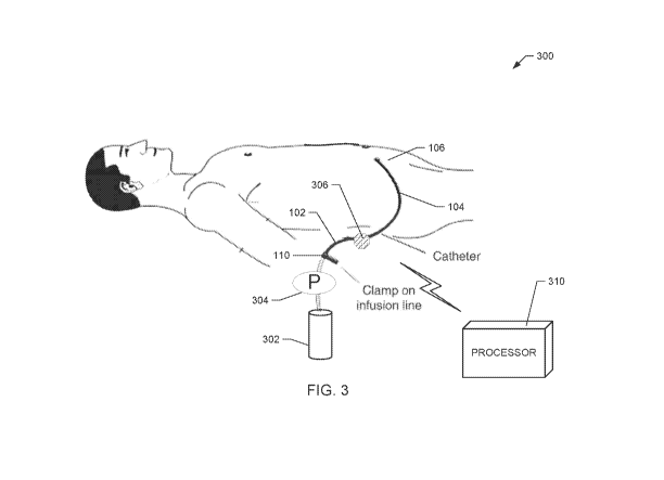
[0043] Figs. 3 and 4 are diagrams of example IPP measurement systems,
according to example embodiments of the present disclosure.
[0044] Figs. 5 to 7 are diagrams of the pressure sensor of Figs. 3 and 4,
according
to example embodiments of the present disclosure.
[0045] Fig. 8 is a diagram that shows a force sensor connected to or otherwise
integrated with the pressure sensor of Figs. 3 to 7, according to an example
embodiment
of the present disclosure.
[0046] Fig. 9 is a flow diagram of an example procedure for using force output
data in conjunction with IPP measurements to determine a fill volume
parameter,
according to an example embodiment of the present disclosure.
[0047] Fig. 10 is a diagram that is illustrative as to how a processor and/or
a
portable device calculates an IPP component related to patient and/or sensor
movement,
according to an example embodiment of the present disclosure.
[0048] Figs. 11 and 12 are diagrams of graphs that are illustrative of a
comparison of IPP measurements to one or more ranges and/or thresholds,
according to
an example embodiment of the present disclosure.
[0049] Fig. 13 is a diagram that illustrates data processing by a processor
and/or a
portable device to adjust IPP measurements based on patient information,
according to an
example embodiment of the present disclosure.
[0050] Fig. 14 is a diagram of an example system in which a spirometer is used
to
conduct lung capacity measurements to determine a fill volume parameter for a
PD
treatment, according to an example embodiment of the present disclosure.
[0051] Fig. 15 is a diagram of a graph of a patient-specific correlation
between
lung capacity and fill volume, according to an example embodiment of the
present
disclosure.
DETAILED DESCRIPTION
[0052] Methods, systems, and apparatuses are disclosed herein for improved
intraperitoneal pressure IPP measurements or estimations. The methods,
systems, and
apparatuses provide a more accurate IPP measurement and/or fill volume
estimation
compared to known IPP measurement techniques. As described herein, the
methods,
CA 03199754 2023-04-26
WO 2022/098714
PCT/US2021/057841
11
systems, and apparatuses include one or more of (i) providing a sensor
amplifier to
amplify pressure sensor measurements to coincide with a more sensitive and
precise area
of a pressure sensor element, (ii) using a force sensor to adjust for pressure
sensor and/or
patient movement during an IPP measurement, (iii) using a spirometer to
correlate lung
capacity with IPP and/or a patient fill volume, (iv) using known ranges to
validate IPP
measurement data, and/or (v) using urine output data, food/beverage
consumption data,
blood pressure data, and/or heart rate data to adjust IPP measurements and/or
fill volume
estimations.
[0053] Disclosure is directed herein to performing IPP measurements for
determining a fill volume parameter for PD treatments. It should be
appreciated that any
of the methods, systems, and apparatuses disclosed herein may also be used to
measure
IPP during a PD treatment. IPP measurements during treatment may be used to
stop PD
fluid fills when a detected IPP exceeds a threshold, lengthen PD drains,
and/or change
from a continuous cycling peritoneal ("CCPD") to a tidal therapy if a residual
volume in
a patient's peritoneal cavity exceeds a threshold. In some instances, IPP
measurements
that exceed a threshold may trigger an alert for the patient and/or an alert
to be
communicated to a clinician.
[0054] Fig. 1 shows a diagram of a known IPP measurement technique. A
known IPP measurement system 100 includes a transfer set 102 fluidly connected
to a
catheter 104, which is inserted or fluidly connected to a peritoneal cavity
106 of a
patient. Another end of the transfer set 102 (not shown) is connected to a
source or
container of fluid, such as PD fluid. The IPP measurement system 100 also
includes a
measurement or drainage line 108, which is fluidly connected to the catheter
104 and/or
the transfer set 102.
[0055] An IPP measurement provides a measure of IPP in a patient's peritoneal
cavity for a certain amount of infused PD fluid. For IPP measurements, a
patient is
usually in a supine or a horizontal position, as shown in Fig. 1. Also, the
patient is
relaxed and their head is supported to enable their abdominal wall to relax.
This patient
positioning avoids pressure on the abdomen. As shown in Fig. 1, a drainage bag
112 is
held in a raised support for the drainage line 108. A graduated ruler or other
distance
measurement device 114 is placed next to the drainage line 108 going from the
patient up
to the bag 112 and aligning level 0 (i.e., 0 cm) with a mid-axillary line, as
shown.
CA 03199754 2023-04-26
WO 2022/098714
PCT/US2021/057841
12
[0056] To perform a measurement, PD fluid is provided from the source through
the transfer set 102 and the catheter 104 to the peritoneal cavity 106 of the
patient. The
peritoneal cavity 106 is filled to a certain percentage of cavity capacity.
After a desired
amount of PD fluid is provided to the peritoneal cavity 106, a clamp 110 is
closed to
prevent further fluid flow from the source. Next, a catheter connection is
opened to
enable at least some of the PD fluid from the patient's peritoneal cavity to
flow into the
drainage line 108. A column of the PD fluid rises in the drainage line 108 to
a level
where it stabilizes with a respiratory oscillation of 1 to 3 cm of H20, which
provides an
average measurement. Fig. 2 is a diagram that shows how a volume of a
peritoneal
cavity changes during respiratory inspiration and expiration. As shown in this
figure,
IPP during inspiration is greater because the peritoneal cavity contracts to
become
smaller. The IPP delta between inspiration and expiration is averaged to
determine the
IPP for the patient. In other words, the IPP is measured as the midpoint of
that
oscillation, and is expressed in centimeters ("cm") of H20. Once the
measurement is
obtained, the peritoneal cavity is drained and the volume is recorded in the
drainage bag
112 as the fill volume. The process may be repeated for different amounts of
PD fluid to
determine a correlation between IPP measurements and fill volume for the
particular
patient.
[0057] In stable adult PD patients, an IPP of 10 to 16 cm of H20 on the mid-
axillary line is considered acceptable for PD treatments, which generally
corresponds to
between 1.3 and 2.8 liters ("L") of infused PD fluid. The difference among
patients
between IPP and infused PD fluid volumes is due to variations in
intraperitoneal volume
("IPV"), body position (with standing patients showing increases between 2 to
4 cm of
H20 compared to laying down), physical activity, weight, height, and gender.
Clinicians
typically prefer to keep IPP below 18 to 20 cm of H20 since higher pressures
are
associated with symptoms, such as discomfort, fullness, sleep disturbances,
hemodynamic issues, and respiratory alterations. Higher pressures may also
contribute
to certain mechanical complications (leakage, hernia, etc.).
[0058] IPP measurements may also be made while a patient is standing or
sitting.
In these instances, the point "0" is considered in the mid-axillary line at a
midpoint
between the xiphoid and pubic symphysis or in the antero-superior iliac spine
of the
patient. Despite a change in position, the IPP measurements are performed in
the same
manner as described-above for a patient laying down.
CA 03199754 2023-04-26
WO 2022/098714
PCT/US2021/057841
13
I. IPP Measurement Embodiments
[0059] Figs. 3 and 4 are diagrams of example IPP measurement systems 300,
according to example embodiments of the present disclosure. The example system
300
includes a transfer set 102 that has a first end connected to a fluid
container 302. The
fluid container 302 may include any source of physiologically compatible
fluid. The
fluid container 302 is a PD fluid source and may include a bag or other
enclosure
configured to hold a volume of fluid, such as one to two liters of the fluid.
In some
embodiments, the fluid container 302 includes fresh, premade PD fluid having a
certain,
prescribed dextrose concentration. In some embodiments, the fluid container
302 may
include two chambers, one with dialysis concentrate and another with purified
water. In
such embodiments, the container 302 includes a seal, which when broken,
enables fluid
in the two chambers to mix. The physiologically compatible fluid may include a
PD
fluid, saline, renal replacement fluid, etc.
[0060] A second end of the transfer set 102 is connected to a catheter 104,
which
is fluidly coupled to a peritoneal cavity 106 of a patient. The transfer set
102 and/or
catheter 104 may be made of any one or more of polyvinyl chloride ("PVC"),
polyethylene ("PE"), polyurethane ("PU"), polycarbonate or other non-PVC
material.
[0061] In some embodiments, the system 300 of Fig. 3 may include a line clamp
110 to selectively restrict the flow of PD fluid through the transfer set 102.
The
illustrated embodiment may also include a pump 304. The example pump 304 may
include a pump head that is fluidly connected to the transfer set 102. The
pump 306 may
be any type of fluid pump, such as a peristaltic pump, a gear pump, or a
membrane
pump. The pump head may be disposable and connected to a reusable actuator,
which is
controlled by an internal or external control unit. The example pump 304 is
configured
to pump fresh PD fluid from the container 302 to the patient's peritoneal
cavity 106 to
perform IPP measurements. The example pump 304 may also pump used PD fluid
(including removed toxins and absorbed ultrafiltrate) from the patient's
peritoneal cavity
106 back to the container 302 after IPP measurements have been recorded. In
alternative
embodiments, separate pumps are provided for (i) pumping fluid to a patient
and (ii)
pumping or pulling fluid from the patient. In some embodiments, the pump 304
is
configured to occlude fluid flow from the fluid container 302 until the pump
head is
actuated, thereby preventing free flow of PD fluid and enabling the clamp 110
to be
omitted.
CA 03199754 2023-04-26
WO 2022/098714 PCT/US2021/057841
14
[0062] The IPP measurement system 300 of Fig. 3 also includes a pressure
sensor
306 for performing IPP measurements. The pressure sensor 306 in the
illustrated
embodiment is positioned to measure fluid pressure within the transfer set
102. In other
embodiments, the pressure sensor 306 may be connected to or provided with the
catheter
104. When PD fluid is provided to the peritoneal cavity 106 or removed from
the
peritoneal cavity 106, the pressure measurements are indicative of fluid
pressure
delivered to or removed from the peritoneal cavity 106. When pumping stops and
the PD
fluid is permitted to dwell in the peritoneal cavity for a specified duration,
the pressure
measurements provided by the pressure sensor 306 are indicative of IPP. The
pressure
measurements may also be used for detecting a line occlusion (based on an
upward
positive or negative pressure spike/trend) or a fluid leak (based on downward
positive or
negative pressure spike/trend).
[0063] In the illustrated example, the pressure sensor 306 is shown as being
in-
line with the transfer set 102. It should be appreciated that the pressure
sensor 306 may
be in-line or otherwise integrated with the catheter 104. It should also be
appreciated that
the pressure sensor 306 may include disposable tube sections that contact the
transfer set
102 and the PD fluid, while the reminder of the sensor 306 is reusable between
IPP
measurements. Alternatively, the entire pressure sensor 306 may be disposable.
[0064] In some embodiments, the system 300 may also include a flow sensor (not
shown) having an output that is integrated to measure a volume of PD fluid
provided to
the patient and/or removed from the patient. It should be appreciated that one
or more
pressure sensors 306 may additionally or alternatively be used to measure a
flow or flow
rate of the fluid delivered to or removed from the peritoneal cavity 106.
Further, the
system 300 may include a heater for warming the PD fluid prior to infusion
into the
patient. The system 300 may further include a temperature sensor to ensure the
PD fluid
is heated to a desired temperature.
[0065] Although not illustrated, an airtrap may be provided in the transfer
set 102
to remove air from the PD fluid prior to patient delivery. In other instances,
priming of
the transfer set 102 may remove air without the need for an airtrap. Heating
dialysis
fluid tends to separate dissolved air from the dialysis fluid. It is
accordingly
contemplated to locate the airtrap downstream from a heater, e.g., along the
transfer set
102 and upstream of a temperature sensor.
CA 03199754 2023-04-26
WO 2022/098714
PCT/US2021/057841
[0066] The example system 300 also includes a processor 310 for communicating
with the pressure sensor 306. The processor 310 may include any computer,
laptop,
workstation, server, etc. In some embodiments, the processor 310 is
communicatively
coupled to the pressure sensor 306 via a wired interface, such as a universal
serial bus
("USB") connection, or a wireless interface, such as a Bluetooth0, Zigbee0, or
Near-
Field Communication ("NFC") connection. Further, the processor 310 may also be
communicatively coupled to the pump 304.
[0067] As described herein, the example processor 310 executes machine
readable instructions stored in a memory device. The instructions may comprise
an
application or software program. Execution of the instructions causes the
processor 310
to perform the operations described herein. For instance, the processor 310
receives IPP
measurement output data, which is transmitted from the pressure sensor 306.
The
processor 310 may ensure the received IPP output data conforms to a specified
range.
Further, the processor 310 may make adjustments to the output data based on
patient
information and/or force sensor information.
[0068] The operations performed by the processor 310 provide for the
determination of a fill volume parameter for PD treatments. In some
embodiments, the
processor 310 uses the received data to calculate or otherwise determine a
fill volume
parameter for a patient under measurement. Additionally or alternatively, the
processor
310 may cause a display device to display IPP measurements and/or adjustment
information to enable a clinician to determine a fill volume parameter for a
patient's PD
treatment.
[0069] Fig. 4 is a diagram of another embodiment of the IPP measurement
system 300. In the illustrated example of Fig. 4, the pump 304 is replaced by
positioning
the fluid container 302 at or above a head-height of a patient (e.g., three to
six feet above
ground level). This enables gravity to pull PD fluid from the fluid container
302 through
the transfer set 102 to the peritoneal cavity 106 of the patient. In the
illustrated example,
the clamp 110 provides for selective flow of the PD fluid.
[0070] Additionally, Fig. 4 shows that a portable device 402 is
communicatively
coupled to the pressure sensor 306. The connection may be via a wired
interface, such as
a USB connection, or a wireless interface, such as Bluetooth0, Zigbee0, NFC,
etc. The
portable device 402 may include a smartphone, a tablet computer, a laptop
computer, etc.
In some instances, the portable device 402 is communicatively coupled to a
server or the
CA 03199754 2023-04-26
WO 2022/098714
PCT/US2021/057841
16
processor 310 of Fig. 3 via the Internet or a local area connection, such as
Wi-Fi. The
portable device 402 is configured to receive IPP output data from the pressure
sensor 306
for determining a fill volume parameter for a patient. Similar to the
processor 310 of Fig.
3, the portable device 402 enables adjustments to be made to the IPP
measurement and/or
fill volume parameter based on force sensor output data and/or patient
information.
[0071] Figs. 5 to 7 are diagrams of the pressure sensor 306 of Figs. 3 and 4,
according to example embodiments of the present disclosure. In the illustrated
embodiments, the pressure sensor includes an amplifier. Typical IPP values are
between
15 to 20 centimeters ("cm") of H20 (0.213 to 0.284 pounds per square inch
("psig")).
However, many commercial pressure sensors for medical applications have
pressure
ranges from 0.0 to 5.0 psig. As a result, the use of a commercial pressure
sensor for
measuring IPP may only use a small portion of range on a low side of
detectable
pressures. Many known pressure sensors are less accurate below 0.8 psig, and
may not
adequately have measurement precision for pressure ranges between 0.2 and 0.3
psig.
The disclosed amplifier increases the measurement range, thereby enabling the
pressure
sensor 306 to provide a more accurate differentiation between IPP
measurements.
[0072] Fig. 5 shows a pressure sensor 306, which is adapted to contact the
transfer set 102. In other instances, the pressure sensor 306 may be connected
to or
integrated with the catheter 104. The pressure sensor 306 includes a pressure
element
502 that transduces a measured pressure into a digital and/or analog signal.
The pressure
element 502 includes at least one of a piezoresistive strange gauge, a
pressure sensing
diaphragm, a pressure pod, a capacitive diaphragm, a pressure sensing capsule,
or a
bourdon tube.
[0073] The pressure sensor 306 also includes an amplifier 504. The amplifier
504 includes a first side that contacts a portion of the transfer set 102. A
second,
opposite side of the amplifier 504 contacts the pressure element 502. The
first side of the
amplifier 504 has a greater diameter or surface area compared to the second
side that
contacts the pressure element 502. The difference in force is illustrated in
Fig. 5 by
pistons, where a first piston has a surface area that is greater than a second
piston. Force
applied from the transfer set 502 to the first area causes the first piston to
apply force
against the second piston. The force from the first piston is condensed
against the
smaller surface area of the second piston. This condensation of force causes
the applied
force value to increase, which is sensed by the pressure element 502.
CA 03199754 2023-04-26
WO 2022/098714
PCT/US2021/057841
17
[0074] The pressure amplifier 504 uses Pascal's Law in one embodiment to
amplify fluid pressure in the transfer set 102. The pressure amplification
enables medical
grade pressure sensors to be used in this low IPP measurement application.
According to
Pascal's law, force or pressure is proportional to a surface area upon which
the force is
applied. In an example, a force having a value of one psig applied to a first
surface area
of two cm2 causes a pneumatically and/or mechanically coupled second surface
having a
surface area of one cm2 to impart a force of approximately two psig. In the
illustrated
example, the first side of the amplifier 504 has an area (Ai) that is at least
twice the area
of the second side (A2), thereby providing an amplification factor of at least
two. In
other embodiments, the areas of the first side and second side may be selected
to provide
an amplification factor of three, four, five, ten, twenty, etc.
[0075] In the illustrated example, the processor 310 and/or the portable
device
402 is configured to normalize the IPP measurements to account for the
amplification.
For example, if the amplification is provided by the amplifier 504, the
processor 310
and/or the portable device 402 may reduce the IPP measurement by the
amplification
factor. In other embodiments, the fill volume parameter may be correlated with
the
amplified IPP measurement values.
[0076] Fig. 6 shows an alternative embodiment of the pressure sensor 306. In
the
illustrated embodiment, a section 602 of the transfer set 102 includes
material having a
greater elasticity compared to other sections. The greater elasticity enables
the section
602 to amplify pressure imparted on the pressure element 502 as pressure
increases
within the transfer set 102. Similar to the example discussed in connection
with Fig. 5,
the example of Fig. 6 provides an increased IPP measurement range, thereby
improving
IPP measurement detection accuracy. In some embodiments, the elastic expansion
of a
material of the section 602 is linear. If the material of the section 602
exhibits non-linear
expansion, the processor 310 and/or the portable device 402 is configured to
account for
the non-linearity of the material. This accounting may include providing a non-
linear
calibration curve for the section 602 that corresponds to linear pressure
changes within
the transfer set 102.
[0077] Fig. 7 shows a further embodiment of the pressure sensor 306. In this
example, at least a portion of the transfer set 102 includes a duel lumen with
a fluid path
side 702 and a non-fluid path side 704. The dual lumen may extend through the
transfer
set 102 or be located at a section adjacent to a sensor element. The non-fluid
path side
CA 03199754 2023-04-26
WO 2022/098714
PCT/US2021/057841
18
704 may be filled with air or fluid of a known volume and/or pressure, thereby
providing
a reference pressure. A diaphragm 706 separates the sides 702 and 704 of the
transfer set
102. The diaphragm 706 moves towards the fluid path side 702 when the
reference side
704 has a greater pressure, and vice versa. A sensor element 710 may be
positioned
adjacent to the reference side 704. As the diaphragm 706 moves, a volume
within the
side 704 changes, thereby changing the internal pressure. The sensor element
710 senses
this internal pressure, which is transmitted to the processor 310 and/or the
portable
device 402 as the IPP measurement.
II. Force Sensing Embodiments
[0078] Fig. 8 is a diagram that shows a force sensor 802 connected to or
otherwise integrated with the pressure sensor 306 of Figs. 3 to 7, according
to an
example embodiment of the present disclosure. In some instances, the IPP
measurement
performed by the pressure sensor 306 may be inaccurate due to a change in
orientation of
a patient or the sensor itself The change in orientation or position causes
increases or
decreases in the IPP pressure reading due to changes in head height and/or
stress placed
on the peritoneal cavity.
[0079] To reduce IPP measurement error, the example force sensor 802 provides
force output data that is indicative of pressure sensor 306 and/or patient
movement. The
force output data is received by the processor 310 and/or portable device 402
to adjust an
IPP measurement and/or a fill volume parameter. In some instances, force
values above
a certain threshold may cause the IPP measurement to be disregarded by the
processor
310 and/or the portable device 402. For instance, detection of a significant
change in
patient position may cause IPP measurements recorded during that movement to
be
removed from the processor 310 and/or the portable device 402 since the
movement
likely contributed significant error to the measurement.
[0080] The force sensor 802 may include an inertial sensor, gyroscope, and/or
accelerometer. Sensing may be provided in at least one axis including an x, y,
z, yaw,
pitch, and/or roll axis. In some embodiments, the force sensor 802 and/or the
pressure
sensor 306 are placed on or in-line with the patient's mid-line (if supine) or
pelvic cup (if
sitting/standing). The force sensor 802 detects relative changes in
orientation/angle of
the pressure sensor 306 and/or the patient from the initial placement
position.
CA 03199754 2023-04-26
WO 2022/098714
PCT/US2021/057841
19
[0081] The force sensor 802 transmits force output data to the processor 310
and/or the portable device 402. In some instances, the force sensor 802 may
use the
same transceiver or transmitter as the pressure sensor 306. In other
instances, the force
sensor 802 may have its own transceiver or transmitter. The processor 310
and/or the
portable device 402 uses the force output data to determine if the IPP
measurement is to
be processed, and if so, provides an adjustment to the IPP measurement and/or
a fill
volume parameter.
[0082] Fig. 9 is a flow diagram of an example procedure 900 for using force
output data in conjunction with IPP measurements to determine a fill volume
parameter
of a patient, according to an example embodiment of the present disclosure.
Although
the procedure 900 is described with reference to the flow diagram illustrated
in Fig. 9, it
should be appreciated that many other methods of performing the steps
associated with
the procedure 900 may be used. For example, the order of many of the blocks
may be
changed, certain blocks may be combined with other blocks, and many of the
blocks
described may be optional. In an embodiment, the number of blocks may be
changed.
For example, the force output data may be used to correct a fill volume
parameter rather
than the IPP measurement. The actions described in the procedure 900 are
specified by
one or more instruction and may be performed among multiple devices including,
for
example, the force sensor 802, the pressure sensor 306, the processor 310,
and/or the
portable device 402.
[0083] The example procedure 900 begins when a patient is connected to the
transfer set 102 and the catheter 104. After the transfer set 102 is in place,
the force
sensor 802 is zeroed or reset while being placed at the patient's mid-line or
low pelvis
(block 902). Such resetting provides a zero-point of the inertial sensors
and/or
accelerometers. In some instances, a clinician may manually zero the force
sensor 802
by pressing a reset button on the sensor. Alternatively, the clinician may
enter an input
into the processor 310 and/or the portable device 402, which transmits an
instruction to
the force sensor 802 causing it to zero or reset.
[0084] The clinician than begins filling the patient's peritoneal cavity.
After a
certain percentage of the cavity is filled, the flow of PD fluid is stopped
and the pressure
sensor 306 transmits IPP measurement data 903 that is indicative of an IPP
within the
peritoneal cavity (block 904). The processor 310 and/or the portable device
402 receives
force output data 905 from the force sensor 802 (block 906). The processor 310
and/or
CA 03199754 2023-04-26
WO 2022/098714
PCT/US2021/057841
the portable device 402 compares the force output data 905 to one or more
force limits
(block 908). If the force output data 905 exceeds the one or more force limit,
the
processor 310 and/or the portable device 402 disregards the corresponding IPP
measurement data 903 (block 910). The force output data exceeding the one or
more
limit may be indicative of a patient changing position, exerting more
substantive
movement, or dropping the pressure sensor 306 or transfer line 102. In these
instances,
IPP measurement data will not be accurate or representative of the actual IPP
pressure.
[0085] When the force output data is within the one or more limits, the
processor
310 and/or the portable device 402 proceeds to process the IPP measurement
data 903
(block 912). This includes determining an IPP measurement component that is
due to
measured forces (block 914). Fig. 10 is a diagram that is illustrative as to
how the
processor 310 and/or the portable device 402 calculates an IPP component
related to
patient and/or sensor movement, according to an example embodiment of the
present
disclosure. In the illustrated example, the sensor 306, 802 may be moved to a
different
or more convenient location for the clinician or the patient, such as either
higher or
lower. This may be due to the patient having a higher exit site for their
catheter, or to
enable the sensor 306, 802 to be in the most comfortable or convenient
location for the
clinician/patient. The processor 310 and/or the portable device 402 calculates
a change
in position of the sensor 306, 802 using raw force output data 1002. The
change in
position provides, for example, a head height change, which is correlated to a
change in
pressure within the transfer set 102 based on a degree of the change. Changes
to lateral
position and/or rotation also correspond to changes in pressure. The pressure
changes are
summed as the IPP component related to movement of the sensor 306, 802 and/or
patient
(i.e., Ah). As shown in Fig. 10, the processor 310 and/or the portable device
402 adjusts
the IPP measurement based on the IPP component related to the force output
data. This
may include an update adjustment or subtraction based on the IPP component
(block 916
of Fig. 9).
[0086] Returning to Fig. 9, the processor 310 and/or the portable device 402
then
outputs or otherwise causes the adjusted IPP measurement 917 to be displayed
(block
918). In some instances, the steps of blocks 902 to 918 are repeated at least
once to
obtain a sample set of IPP measurements over one or more respiratory cycles to
enable
the IPP measurement to be averaged. In some embodiments, the processor 310
and/or
the portable device 402 causes a graph to be displayed that shows IPP
measurements
CA 03199754 2023-04-26
WO 2022/098714
PCT/US2021/057841
21
over time, thereby enabling an average to be computed or otherwise determined.
The
processor 310 and/or the portable device 402 next determines a fill volume
parameter
based in the adjusted IPP measurement (block 920). The fill volume may be
determined
by correlating the IPP measurement to a fill volume for patients with similar
body
masses/heights as the patient under measurement. In other instances, the
volume of PD
fluid infused into the patient may be measured using either a flow sensor or
draining and
measuring the PD fluid.
[0087] In some embodiments, additional PD fluid may be added to the patient if
the IPP measurement falls below a threshold for performing an adequate PD
fill. The
steps of 902 to 918 may be repeated until the adjusted IPP measurement is
between 16 to
19 cm of H20 or between 0.25 to 0.28 psig, which is indicative of an adequate
fill
volume for a PD treatment. The fill volume parameter is then determined from
the
patient characteristics and/or detected amount of PD fluid infused into the
patient's
peritoneal cavity. The fill volume parameter may then be used for subsequent
PD
treatments, using a PD machine or manually for a continuous ambulatory
peritoneal
dialysis ("CAPD") treatment. The example procedure 900 then ends.
[0088] In some embodiments, the patient may wear a force sensor. For example,
the force sensor may be connected to a wrist or abdomen of the patient. Output
data
from the sensor provides further data indicative of patient movement. The
force sensor
worn by the patient may be used with the force sensor 802 provided with the
pressure
sensor 306. Alternatively, only a force sensor connected to a patient is
provided. In
some instances, data from the force sensor connected to the patient is tracked
over time
in conjunction with IPP measurements. For example, a patient may go through a
daily
routine or a set of activities with the transfer set 102 connected. The IPP
measurements
may be correlated with force data (to enable force-related components to be
removed) to
identify how IPP changes for a patient for different orientations and/or
activities. A
clinician may use this correlation to ensure a fill volume does not cause a
patient's IPP to
exceed clinically recommended limits regardless of which position or activity
is
conducted by the patient, thereby improving patient comfort during treatment.
The fill
volume determined by the clinician may then be set in the patient's treatment
or device
prescription and downloaded locally or remotely to the patient's cycler or
peritoneal
dialysis machine.
CA 03199754 2023-04-26
WO 2022/098714
PCT/US2021/057841
22
III. IPP Measurement Validation Embodiment
[0089] In some embodiments, the processor 310 and/or the portable device 402
is
configured to validate IPP measurement data prior to processing the data. For
example,
as shown in block 908 of Fig. 9, the processor 310 and/or the portable device
402
compares received IPP measurement data to one or more ranges or thresholds
that are
indicative of substantial patient and/or sensor movement. This operation may
also
include a comparison to one or more range and/or limit that corresponds to
normal fill
pressures and/or expected IPP measurement values. IPP measurements that are
outside
of the ranges and/or limits may be indicative of an issue with a catheter
connection,
catheter blockage, leakage in a transfer set, or other fluid connectivity
issues.
[0090] Fig. 11 shows a graph 1100 that is illustrative of a comparison of IPP
measurements to one or more ranges and/or thresholds, according to an example
embodiment of the present disclosure. The graph 1100 includes a first range
1102 that
corresponds to acceptable pressure measurement values when PD fluid is being
infused
into the peritoneal cavity of a patient. During a PD fluid fill, force is
applied to the
sensor element (e.g., transducer membrane) as a result of the fluid flow. The
first range
1102 may correlate to fluid fills for gravity fed administration, while a
second range may
be used if a pump provides the PD fluid.
[0091] The graph 1100 also includes a second range 1104 that corresponds to
reduced pressure as a result of a transfer set or catheter partial occlusion.
During a fill
phase, the processor 310 and/or the portable device 402 receives IPP
measurement data
and compares the data to the first range 1102 and the second range 1104. If
the IPP
measurement data corresponds to the second range 1104, the processor 310
and/or the
portable device 402 may generate an alert or other message/indication that
there is an
issue with the catheter and/or transfer set. Further, the processor 310 and/or
the portable
device 402 may prevent subsequent IPP measurements from being processed until
it is
confirmed that a patient has been properly filled with PD fluid.
[0092] The graph 1100 shows that over time, pressure measurement data
decreases. This decrease is a result of a lower flowrate as a gravity-fed PD
fluid bag
empties into a patient. In some instances, the ranges 1102 and 1104 may have
corresponding decreases over time to account for expected pressure declines
during PD
fluid infusion. The pressure values on the y-axis are normalized for brevity.
CA 03199754 2023-04-26
WO 2022/098714
PCT/US2021/057841
23
[0093] The graph 1100 also includes a third range 1106, which is applied after
the flow of PD fluid has been stopped and the fluid is permitted to dwell in a
patient's
peritoneal cavity. The processor 310 and/or the portable device 402 may use
the third
range 1106 to identify IPP measurements that exceed allowable pressure
thresholds,
which may be indicative of patient movement, transfer set movement, or
overfilling of a
patient. IPP measurements over this third range 1106 may be disregarded by the
processor 310 and/or the portable device 402. Additionally or alternatively,
the
processor 310 and/or the portable device 402 may generate an alarm. It should
be noted
that IPP measurements increase over time because the PD fluid absorbs waste
and other
toxins from the patient, which increases the volume of fluid in the peritoneal
cavity,
thereby increasing the measured pressure. The processor 310 and/or the
portable device
402 may be configured to log the IPP measurement over time to ensure a PD fill
volume
does not exceed allowable IPPs during a dwell phase, which may cause patient
discomfort during a PD treatment.
[0094] Fig. 12 shows a graph 1200 of an alternative embodiment in which the
processor 310 and/or the portable device 402 uses a recorded bag fill head
height value
and bag solution volume to determine a threshold 1202, which corresponds to an
expected fill pressure. When a pump is provided, an expected pump pressure
value may
be used instead. In this example, the processor 310 and/or the portable device
402
estimates the threshold based on actual fill conditions to more accurately
determine if
there is an issue infusing a PD fluid into a patient's peritoneal cavity. The
graphs 1100
and 1200 may be displayed by the processor 310 and/or the portable device 402
to a
clinician.
IV. IPP Measurement Adjustment using Patient Information Embodiment
[0095] During an IPP measurement, the processor 310 and/or the portable device
402 may adjust an IPP measurement or a fill volume parameter based on received
patient
information. In some situations, a patient's water retention may affect
peritoneal cavity
volume or pressure provided on the cavity, which affects IPP measurements.
Additionally, a patient's blood pressure or heart rate may be indicative as to
whether the
patient is under stress or exertion, which can affect IPP measurements.
[0096] Fig. 13 is a diagram 1300 that illustrates data processing by the
processor
310 and/or the portable device 402 to adjust IPP measurements based on patient
CA 03199754 2023-04-26
WO 2022/098714
PCT/US2021/057841
24
information, according to an example embodiment of the present disclosure. As
shown,
the processor 310 and/or the portable device 402 receives IPP measurement data
903.
The processor 310 and/or the portable device 402 may also receive urine data
1302,
which indicates a patient's urine output in a specified time before the IPP
measurement,
such as twenty-four or forty-eight hours. The urine output may be self-
reported by the
patient and entered into the processor 310 and/or the portable device 402. In
other
instances, the urine output may be measured in a container and entered into
the processor
310 and/or the portable device 402.
[0097] The processor 310 and/or the portable device 402 also receives food and
beverage consumption information 1304. This information provides an indication
as to
how much food and beverage was consumed by a patient in a time period leading
up to
the IPP measurement or between IP measurements. Together, the urine data 1302
and
the food/beverage data 1304 provides fluid balance information. The processor
310
and/or the portable device 402 is configured to calculate a patient's fluid
balance by
summing the food/beverage data 1304 and subtracting the urine data 1302, and
accounting for metabolic burn of fluids based on patient populations of
similar age,
gender, height, and weight. The processor 310 and/or the portable device 402
then
determines if the patient's fluid balance imparts an IPP measurement component
by
comparing the calculated balance information to a correlation of balance
information and
IPP measurements for patients with similar heights, genders, weights, etc. The
processor
310 and/or the portable device 402 then adjusts the IPP measurement data 903
by
accounting for the IPP component related to fluid balance. In instances where
the fluid
balance is negative, such as instances of dehydration, the adjustment may
result in an
increase in the value of the IPP measurement.
[0098] The processor 310 and/or the portable device 402 also receives heart
rate
and blood pressure data 1306. The processor 310 and/or the portable device 402
correlates the data 1306 to an IPP measurement component based on a population
of
patients with similar heights, weights, genders, ages, etc. The processor 310
and/or the
portable device 402 then adjusts the IPP measurement by the identified IPP
measurement
component.
[0099] After adjusting the IPP measurement, the processor 310 and/or the
portable device 402 determines the fill volume parameter 917. As discussed
above, this
may include comparing the adjusted IPP measurement (or a trend of adjusted IPP
CA 03199754 2023-04-26
WO 2022/098714
PCT/US2021/057841
measurements) to fill IPP limits for PD therapy. Once the IPP measurements are
close,
but do not exceed a limit, the processor 310 and/or the portable device 402
determines a
fill volume as a volume of PD fluid within the patient's peritoneal cavity
using either a
body mass of the patient or determining the PD fluid volume by draining the
fluid and/or
using a flow sensor. The fill volume parameter 917 may then be used for
subsequent PD
treatments.
V. Lung Capacity for Determining Fill Volume Embodiment
[00100] In the examples discussed above, IPP measurements have been
made using the pressure sensor 306. In some embodiments, the pressure sensor
306 may
be replaced with a lung capacity sensor, such as a spirometer. Lung capacity
has been
shown to decrease as IPP increases. The processor 310 and/or the portable
device 402
may use known correlations between lung capacity and IPP measurements to
determine a
fill volume parameter for a patient without the use of a pressure sensor.
[00101] Fig. 14 shows an example system 1400 in which a spirometer
1402
is used to conduct lung capacity measurements to determine a fill volume
parameter,
according to an example embodiment of the present disclosure. Such lung
capacity
measurements enable greater efficiency for IPP measurements that may otherwise
result
in the errors discussed above. Further, the use of the spirometer 1402 enables
a standard
transfer set and catheter to be used as opposed to a transfer set or catheter
equipped with
a pressure sensor.
[00102] As shown in Fig. 14, the spirometer 1402 measures patient
respiratory capacity at different fill levels, shown as MO (dry state), M1
(10% of fill
capacity), M2 (20% of fill capacity), M3 (50% of fill capacity), etc. After
the patient is
filled to an estimated desired percentage of cavity capacity, the spirometer
1402 records
the patient's lung capacity. The spirometer 1402 may record lung capacity over
one or
more respiratory cycle to determine an average lung capacity.
[00103] The example processor 310 and/or the portable device 402
receives the lung capacity data from the spirometer 1402. The processor 310
and/or the
portable device 402 use known correlations between lung capacity and IPP to
adjust an
IPP measurement to provide a more accurate measurement. The identified IPP
value
may then be used for determining fill volume and/or identifying when the PD
fluid fill
has reached a desired percentage of capacity so as to be effective for a PD
treatment.
CA 03199754 2023-04-26
WO 2022/098714
PCT/US2021/057841
26
This fill volume is stored by the processor 310 and/or the portable device 402
as the fill
volume parameter for use in a PD treatment for the patient.
[00104] A patient-specific correlation between fill volume and lung
capacity may be determined and subsequently used for PD treatments. In these
embodiments, a PD machine may use periodic lung capacity measurements for
estimating a patient's IPP or fill volume during different phases of a PD
treatment. Fig.
15 shows a graph 1500 of a patient-specific correlation between lung capacity
and fill
volume, according to an example embodiment of the present disclosure. The
graph 1500
shows that lung capacity decreases as PD fill volume increases. Such a
correlation may
be useful for PD treatments where a lung capacity measurement may be used
instead of
attempting to estimate or directly measure a patient's IPP.
VI. Conclusion
[00105] It should be understood that various changes and
modifications to
the presently preferred embodiments described herein will be apparent to those
skilled in
the art. Such changes and modifications can be made without departing from the
spirit
and scope of the present subject matter and without diminishing its intended
advantages.
It is therefore intended that such changes and modifications be covered by the
appended
claims.