Note: Descriptions are shown in the official language in which they were submitted.
WO 2022/120234
PCT/US2021/061895
SYSTEMS AND METHODS FOR FACILITATING MULTISITE PAIRED
CORTICOSPINAL-MOTONEURONAL STIMULATION THERAPY
CROSS-REFERENCE TO RELATED APPLICATIONS
[0001] This is a PCT application that claims benefit to
U.S. Provisional
Patent Application Serial No. 63/121,211 filed December 3, 2020, which is
herein
incorporated by reference in its entirety.
FIELD
[0002] The present disclosure generally relates to spinal
cord
rehabilitation control systems, and in particular, to a system and associated
method
for facilitating multisite paired corticospinal-nnotoneuronal stimulation
(MPCMS)
therapy.
BACKGROUND
[0003] Spinal cord injuries (SCI) can involve weakening
of an electrical
connection between a pre-synaptic cell (corticospinal neuron) and a post-
synaptic
cell (peripheral motor neuron) of a corticospinal motoneuronal pairing. The
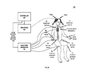
pre-
synaptic cell is a corticospinal neuron that has its body in the cortex and
its axon
through the spinal cord where it makes connections (synapses) to the post-
synaptic
cell, the peripheral motor neuron. Synaptic connections can, in some
instances, be
strengthened in vitro through repeated electrical stimulation to the pre-
synaptic cell
and the post-synaptic cell. However, translating this in vivo remains a
challenge.
There is a need for a system that can effectively boost residual corticospinal
connections in paralyzed or partially paralyzed subjects at multiple locations
to
augment exercise-mediated recovery in humans with different levels of SCI.
[0004] It is with these observations in mind, among
others, that various
aspects of the present disclosure were conceived and developed.
1
CA 03200200 2023- 5- 25
WO 2022/120234
PCT/US2021/061895
BRIEF DESCRIPTION OF THE DRAWINGS
[0005] FIG. 1 is a simplified diagram showing a system
for facilitating
MPCMS therapy;
[0006] FIGS. 2A and 2B are simplified diagrams showing
connection of
the body with the system of FIG. 1;
[0007] FIG. 3 is a simplified illustration showing MPCMS
stimulation of
a synapse by the system of FIG. 1;
[0008] FIG. 4 is a process flow showing a method for
facilitating
MPCMS therapy by the system of FIG. 1;
[0009] FIG. 5 is a simplified diagram showing an example
computing
system for implementation of the system of FIG. 1;
[0010] FIG. 6 is an illustration showing placement of pre-
and post-
synaptic stimuli for facilitation of MPCMS protocol using the system of FIG. 1
according to a second validation study;
[0011] FIGS. 7A-7F is a series of photographs showing
exercise
according to the MPCMS protocol; and
[0012] FIG. 8 is a diagram illustrating facilitation of
MPCMS protocol for
the second validation study;
[0013] FIGS. 9A-D are a series of graphical
representations showing
MEP, C-root, M-wave and other results for biceps brachii;
[0014] FIGS. 10A-D are a series of graphical
representations showing
MEP, C-root, M-wave and other results for first dorsal interosseous;
[0015] FIGS. 11A-D are a series of graphical
representations showing
MEP, C-root, M-wave and other results for quadriceps;
[0016] FIGS. 12A-D are a series of graphical
representations showing
MEP, C-root, M-wave and other results for tibialis anterior;
[0017] FIGS. 13A-D are a series of graphical
representations showing
raw MEP traces for various muscle groups before, following 20 sessions, and
following 40 sessions;
[0018] FIGS. 14A-D are a series of graphical
representations showing
rectified electromyographic traces during MVCs for various muscle groups
before,
following 20 sessions, and following 40 sessions;
[0019] FIGS. 15A and 15B are graphical representations
showing
sensory outcomes prior to and following 40 sessions;
2
CA 03200200 2023- 5- 25
WO 2022/120234
PCT/US2021/061895
[0020] FIGS. 16A-16C are graphical representations
showing motor
outcome scores prior to and following 40 sessions;
[0021] FIGS. 17A and 17B are graphical representations
and related
images showing functional outcome scores including GRASSP and 10-m walk prior
to and following 40 sessions; and
[0022] FIG. 18 is a graphical representation showing
quality of life
improvement scores following 40 sessions.
[0023] Corresponding reference characters indicate
corresponding
elements among the view of the drawings. The headings used in the figures do
not
limit the scope of the claims.
3
CA 03200200 2023- 5- 25
WO 2022/120234
PCT/US2021/061895
DETAILED DESCRIPTION
[0024] Various embodiments of a system that engages
residual
neuronal networks in humans with spinal cord injuries by facilitating
multisite paired
corticospinal-motoneuronal stimulation (MPCMS) therapy are described herein.
In
MPCMS, corticospinal electrical volleys (pre-synaptic stimulus) evoked by
transcranial magnetic stimulation (TMS) over the primary motor cortex or
electrical
stimulation (ES) over the spine are timed to arrive at corticospinal-
motoneuronal
synapses of limb muscles before or after antidromic potentials (post-synaptic
stimulus) elicited in motoneurons by electrical stimulation of a peripheral
nerve.
MPCMS likely elicits spike-timing dependent plasticity (STDP) changes at
spinal
synapses of somatic motoneurons. The system described herein applies a pre-
synaptic stimulus to a pre-synaptic cell of a corticospinal-motoneuronal
pairing, and
subsequently applies a post-synaptic stimulus to a post-synaptic cell of the
corticospinal-motoneuronal pairing such that the pre-synaptic stimulus from
the
cortex arrives at a synapse of the corticospinal-motoneuronal pairing a
predetermined time interval, preferably 1-2 ms, before the post-synaptic
stimulus
from the peripheral nerve. In some embodiments, the system can apply stimulus
to
target multiple muscle groups at a time through more than one peripheral nerve
to
improve patient outcomes. Referring to the drawings, embodiments of a system
for
facilitating MPCMS therapy are illustrated and generally indicated as 100 in
FIGS. 1-
18.
[0025] Referring to FIGS. 1-5, an embodiment of the
system 100
includes a controller 300 in electrical communication with a transcranial
magnetic
stimulation (TMS) device 120 for generating a pre-synaptic stimulus for
application to
a body during multisite paired corticospinal-motoneuronal stimulation (MPCMS)
therapy. Further, the system 100 includes a waveform generator 110 for
generating
a plurality of post-synaptic stimuli for application to the body during MPCMS
therapy.
In some embodiments, the waveform generator 110 can also generate an
additional
pre-synaptic stimulus for application to the body during MPCMS therapy, as
will be
described in greater detail herein. As specifically illustrated in FIGS. 1-3,
the system
100 includes one or more TMS coils 122 of a TMS device 120 configured to
induce
or otherwise apply the pre-synaptic stimulus to a motor cortex of a body
according to
TMS pre-synaptic parameters including pulse initiation time provided to the
TMS
device 120 by the controller 300. Similarly, the system 100 includes a
plurality of
4
CA 03200200 2023- 5- 25
WO 2022/120234
PCT/US2021/061895
post-synaptic electrodes 140 in communication with the waveform generator 110
configured to induce or otherwise apply the post-synaptic stimuli to a
plurality of
peripheral nerves of the body according to a plurality of post-synaptic
waveform
parameters including pulse initiation time provided to the waveform generator
110 by
the controller 300. In some embodiments, the system 100 is configured to apply
an
additional pre-synaptic stimulus to the thoracic spine through a pre-synaptic
electrode 130 in communication with the waveform generator 110 based on pre-
synaptic parameters including pulse initiation time provided by the controller
300. In
practice, the additional pre-synaptic stimulus is applied to the thoracic
spine to aid in
application of MPCMS therapy to the lower body.
[0026] Referring directly to FIG. 2A, TMS is applied
cranially and
induces action potentials (pre-synaptic stimulus) within the corticospinal
pathway.
Combined pre-synaptic stimuli and post-synaptic stimuli can aid in therapeutic
restoration of function in targeted muscles. Thus, the controller 300
modulates or
otherwise maintains control of pre-synaptic waveforms and post-synaptic
waveforms
representative of pre-synaptic stimuli and post-synaptic stimuli to be
generated by
the waveform generator 110. The waveform generator 110 individually applies
electrical current to each peripheral nerve of the plurality of peripheral
nerves (or to
the thoracic spine) according to associated waveform parameters for
application of
the post-synaptic stimuli (peripheral nerve) or pre-synaptic stimulus
(thoracic spine)
to a plurality of corticospinal-motoneuronal pairs. Similarly, the TMS device
120
induces an action potential as a pre-synaptic stimulus within the brain for
application
of the pre-synaptic stimulus to each corticospinal-motoneuronal pair.
Empirical
evidence has demonstrated that an optimal interstimulus interval between a pre-
synaptic time-of-arrival of the pre-synaptic stimulus and a respective post-
synaptic
time-of-arrival of each post-synaptic stimulus is between 1-2 milliseconds for
each
grouping of stimuli applied. Thus, the controller 300 adjusts pre-synaptic and
post-
synaptic pulse initiation times provided to the waveform generator 110 to
accommodate for differences in nerve length between various peripheral nerves
such that the post-synaptic stimuli arrives at a synapse of the corticospinal-
motoneuronal pair 1-2 milliseconds after the pre-synaptic stimuli.
CA 03200200 2023- 5- 25
WO 2022/120234
PCT/US2021/061895
Pre-synaptic Stimuli
[0027] As discussed, the system 100 applies or otherwise
induces a
pre-synaptic stimulus to the motor cortex which terminates at a pre-synaptic
cell of a
corticospinal-motoneuronal pair. The pre-synaptic stimulus is applied or
induced
within the motor cortex which causes an action potential to propagate
orthodromically down an axon of the pre-synaptic cell. The pre-synaptic
stimulus
induces an action potential in the pre-synaptic axon (corticospinal neuron)
and is
paired with a post-synaptic stimulus to an associated peripheral nerve (spinal-
motoneuron). Pre-synaptic stimuli can be applied or induced in at least two
ways:
1. TMS.
[0028] TMS can be used to induce the pre-synaptic
stimulus by
applying a magnetic field parallel to the skull. This stimulates an electrical
field
perpendicular to the skull, which in turn triggers an action potential
(electrical
impulse) in a corticospinal neuron that propagates down through the spinal
cord and
connects to a peripheral motor nerve in the spinal cord. Referring to FIG 1,
the
controller 300 provides TMS pre-synaptic parameters including pulse initiation
time
to the TMS device 120 to generate a magnetic field within one or more TMS
coils
122 of the TMS device 120. In the examples shown, the system 100 includes a
first
TMS coil 122A and a second TMS coil 122B of the one or more TMS coils 122 that
apply the magnetic field to a respective left side and right side of the
skull. This
induces the action potential (pre-synaptic stimulus) that propagates down the
corticospinal neuron and to the synapse.
2. Direct Thoracic Stimulation.
[0029] Further, in some embodiments, the waveform
generator 110 can
additionally apply a pre-synaptic stimulus to the thoracic spine. The
controller 300
provides pre-synaptic waveform parameters including pulse initiation time to
the
waveform generator 110 to apply a current corresponding with the pre-synaptic
waveform parameters to the corticospinal neuron through a pre-synaptic
electrode
130 in communication with the waveform generator 110. This induces the action
potential (pre-synaptic stimulus) that propagates down the corticospinal
neuron and
to the synapse.
6
CA 03200200 2023- 5- 25
WO 2022/120234
PCT/US2021/061895
Post-synaptic Stimuli
[0030] As further shown in FIG. 2A, the system 100 is
configured to
apply post-synaptic stimuli to a plurality of peripheral nerves at a time. In
the
example shown, post-synaptic stimuli are applied from the waveform generator
110
to eight separate locations on the body; particularly to peripheral limbs such
as right
and left common peroneal nerves, right and left femoral nerves, right and left
ulnar
nerves, and right and left brachial plexus nerves that communicate with the
spinal
cord. Each location requires different waveform parameters including pulse
initiation
time to arrive at the synapse at the proper time due to physiological length
of the
associated peripheral nerve. The system 100 applies post-synaptic stimuli to
the
peripheral limbs through N post-synaptic electrodes 140A-140N in communication
with the waveform generator 110, where N is the number of peripheral nerves to
be
stimulated. The waveform generator 110 generates N post-synaptic stimuli at N
post-
synaptic electrodes 140A-140N. The controller 300 provides N post-synaptic
waveform parameters to the waveform generator 110 to apply current
corresponding
with the N post-synaptic waveform parameters to associated peripheral nerves
through respective post-synaptic electrodes 140A-140N in communication with
the
waveform generator 110. This induces the action potential (post-synaptic
stimulus)
that propagates up the peripheral nerve and to the synapse. Each set of post-
synaptic waveform parameters includes a respective post-synaptic pulse
initiation
time that is specific to the associated peripheral nerve to ensure that the
post-
synaptic stimulus arrives at the synapse 1-2 ms after the associated pre-
synaptic
stimulus arrives.
[0031] FIG. 3 illustrates a corticospinal-motoneuronal
neuronal pair
including a pre-synaptic cell in association with the motor cortex
(corticospinal
neuron) and a post-synaptic cell in association with the peripheral nerve. A
junction
of the two is illustrated at the synapse. The system 100 facilitates MPCMS
therapy to
restore corticospinal-motoneuronal nerve function by first stimulating the
corticospinal neuron by applying the pre-synaptic stimulus to the pre-synaptic
cell
through a pre-synaptic electrode 130 or through a TMS coil 122. The system 100
subsequently stimulates the peripheral nerve by applying post-synaptic
stimulus in
the form of simple electrical pulses to major peripheral nerves in the limbs
through
one or more post-synaptic electrodes 140. For effective MPCMS, pre-synaptic
pulse
initiation time and post-synaptic pulse initiation time are important. They
must occur
7
CA 03200200 2023- 5- 25
WO 2022/120234
PCT/US2021/061895
such that the signal from the cortex arrives at the synapse 1-2 ms before the
signal
from the peripheral nerve. So, the electrical pulse is applied to the
peripheral nerves
following a delay after the stimulation to the cortex. The length of the delay
is
dependent upon a length of the peripheral nerve. (i.e. if the pre-synaptic
stimulus
arrives at time to, then the post-synaptic stimulus must arrive at time to+ [1
ms, 2
ms]).
[0032] The controller 300 manages application of pre-
synaptic and
post-synaptic stimuli to the body by providing control inputs to the TMS
device 120
and waveform generator 110. In particular, the controller 300 determines and
communicates waveform parameters including the first pre-synaptic and post-
synaptic pulse initiation times that the interstimulus interval is preferably
1-2 ms. In
other embodiments, the interval may differ and can be greater than Oms and
less
than 5ms. During MPCMS, the system 100 delivers 180 pairs of pre-synaptic and
post-synaptic stimuli every 10 seconds (-30 min, 0.1 Hz), where corticospinal
volleys
(pre-synaptic stimuli) evoked by TMS over the primary motor cortex are timed
to
arrive at corticospinal-motoneuronal synapses of each muscle -1-2 ms before
the
post-synaptic antidromic potentials evoked in motoneurons by peripheral nerve
stimulation (PNS).
[0033] Referring to FIGS. 1-4, for MPCMS facilitation, in
a first step, two
or more peripheral nerves innervating at least two different targeted muscle
sites in
the subject and forming two or more peripheral nerve-muscle pairings must be
identified. This involves identifying two or more corticospinal-motoneuronal
connections, each comprising a corticospinal neuron connected at a synapse
with
each peripheral nerve in each of the peripheral nerve-muscle pairings. Once
the
appropriate peripheral nerve-muscle pairings have been identified, the system
100
acquires a plurality of latency values associated with the targeted peripheral
nerves
and the motor pathway. In some embodiments, this can be achieved using a
waveform acquisition device 150 such as a Power1401 acquisition interface that
is
operable for obtaining a plurality of motor response waveforms including MEP,
F-
wave, and M-max waveforms for the body. In some embodiments, the waveform
acquisition device 150 acquires the plurality of motor response waveforms from
the
body through a sensing electrode array 160 that includes a plurality of
electrodes in
communication with the two or more peripheral nerves and the motor pathway.
The
controller 300 determines or otherwise obtains the associated plurality of
latency
8
CA 03200200 2023- 5- 25
WO 2022/120234
PCT/US2021/061895
values including MEP, F-wave, and M-max latencies. The controller 300 then
uses
the plurality of latency values to calculate a peripheral conduction time
(PCT) and a
central conduction time (CCT) for each of the peripheral nerve-muscle
pairings. PCT
is the amount of travel time necessary for a post-synaptic stimulus to arrive
at the
synapse when applied to a location along the peripheral nerve. Likewise, CCT
is the
amount of travel time necessary for a pre-synaptic stimulus to arrive at the
synapse
when applied to a location along the cortical or motor pathway nerve. The
controller
300 then adjusts waveform parameters including a pulse initiation time for
each pre-
synaptic stimulus and post-synaptic stimulus based on the calculated PCT and
CCT.
The system 100 then applies, based on the waveform parameters, a resultant pre-
synaptic stimulus and the post-synaptic stimuli that arrive at the synapse
within the
appropriate interstimulus interval.
[0034] PCT. The values to calculate PCT for a peripheral
nerve-muscle
pairing are found using a plurality of latency values from the plurality of
motor
response waveforms including MEPs, F-wave, and M-max that are recorded by the
system 100 for the body. MEP latencies are recorded during isometric -10% of
MVC
of the target muscle to determine the shortest and clearest response for
estimations.
The onset latency is defined as the time when each response exceeded 2 SD of
the
mean rectified pre-stimulus activity (100 ms) in the averaged waveform.
Peripheral
conduction time (PCT) is calculated using the following equation:
PCT = (F-wave latency - M-max latency) x 0.5
[0035] CCT. Central conduction time (OTT) was calculated
using the
following equation:
CCT = MEP latency - (PCT + M-max latency)
[0036] Alternatively, the latency of H-reflex can be used
instead when it
is difficult to elicit F-waves. When it was not possible to record F-waves or
H-reflex
(i.e. biceps brachii), then 0-roots can be stimulated with TMS at cervical
spinous
processes 05-6. Then, CCT is calculated by adding to the latency from TMS of
the
0-root to 1.5 ms [estimated time of synaptic transmission plus conduction to
the
nerve root at the vertebral foramina] and subtracting from the MEP latency
[MEP -
(C-root + 1.5)]. PCT is calculated by subtracting the M-max latency from the 0-
root
latency and adding 0.5 ms, the estimated time of antidromic conduction time
from
the vertebral foramina to the dendrites [(C-root - M-max)+ 0.5)].
9
CA 03200200 2023- 5- 25
WO 2022/120234
PCT/US2021/061895
[0037] Adjusting Pulse Initiation Time. Following
determination of PCT
and CCT, the controller 300 determines a pulse initiation time for each pre-
synaptic
and post-synaptic stimulus to be applied such that the pulse arrival time for
the
associated stimulus is within the appropriate interstimulus interval relative
to one
another.
[0038] For instance, given a goal interstimulus interval
(ISI), a
calculated CCT and a calculated PCT for a particular peripheral nerve of the
plurality
of peripheral nerves, the controller 300 determines a delay interval at which
a pre-
synaptic pulse initiation time of the pre-synaptic stimulus is delayed
relative to a
post-synaptic pulse initiation time of the post-synaptic stimulus to account
for
differences in conduction time between different nerves.
delay interval = PCT ¨ CCT - ISI
pre-synaptic pulse initiation time = ISI + CCT ¨ delay interval
= delay interval + post-synaptic pulse initiation time
[0039] For example, consider a calculated PCT value of 7
ms delay
before arrival of the post-synaptic stimulus at the synapse and a calculated
CCT
value of 3 ms delay before arrival of the post-synaptic stimulus at the
synapse. To
arrive within an interstimulus interval of 1.5 ms, the post-synaptic stimulus
would
need to be initiated 2.5ms before initiation of the pre-synaptic stimulus
(delay interval
= 2.5m5). In most scenarios, the PCT value is longer than that of the OCT
value and
the interstimulus interval combined. In such a situation, the controller 300
initiates
the post-synaptic stimulus at the post-synaptic initiation time and then
initiates the
pre-synaptic stimulus afterward at the pre-synaptic initiation time, which
would be at
the post-synaptic pulse initiation time followed by the delay interval. In
other
scenarios in which the OCT value combined with the interstimulus interval are
smaller than the PCT value, then an initiation order between the post-synaptic
stimulus and the pre-synaptic stimulus would be reversed. The controller 300
initiates the pre-synaptic stimulus at the pre-synaptic initiation time and
then initiates
the post-synaptic stimulus afterward at the post-synaptic initiation time,
which would
be at the pre-synaptic pulse initiation time followed by the delay interval.
[0040] Extending this logic to the multiple peripheral
limbs to be
stimulated, the controller 300 selects an optimal post-synaptic pulse
initiation time of
CA 03200200 2023- 5- 25
WO 2022/120234
PCT/US2021/061895
each individual post-synaptic stimulus such that the interstimulus interval
between
the pre-synaptic time of arrival and each respective post-synaptic time of
arrival at a
synapse is within 1-2 ms. Further, the post-synaptic pulse initiation times
can be set
and the controller 300 can select an optimal pre-synaptic pulse initiation
time such
that the interstimulus interval between the pre-synaptic time of arrival and
each
respective post-synaptic time of arrival at a synapse is preferably within 1-2
ms.
[0041] Referring to FIG. 4, a process flow 200 is
illustrated for
execution by the controller 300 of the system 100. At block 210, the
controller 300
receives a selection of peripheral limbs, muscles, or peripheral nerves to be
targeted. At block 220, the controller measures the plurality of latency
values
associated with the targeted peripheral nerves and the motor pathway through
recordation of a plurality of motor response waveforms from which the
plurality of
latency values are extracted. As discussed above, this can be achieved using
the
waveform acquisition device 150 that is operable for obtaining a plurality of
motor
response waveforms including MEP, F-wave, and M-max waveforms for the body.
The controller 300 determines or otherwise obtains the associated plurality of
latency
values including MEP, F-wave, and M-max latencies from the waveform
acquisition
device 150. At block 230, the controller 300 determines a peripheral
conduction time
(PCT) and a central conduction time (COT) based on the plurality of latency
values.
At block 240, the controller 300 selects a post-synaptic pulse initiation time
of the
post-synaptic stimulus such that the interstimulus interval between the pre-
synaptic
time of arrival and the post-synaptic time of arrival at a synapse is within
the
appropriate interval.
[0042] At block 250, the controller 300 periodically
applies the pre-
synaptic stimulus having the pre-synaptic time of arrival from the motor
cortex to the
spinal cord. This is achieved as described above using TMS device 120 or using
waveform generator 110 to apply or otherwise induce the pre-synaptic stimulus
to
the motor cortex (corticospinal neuron). In some embodiments, the waveform
acquisition device 150 can additionally aid in facilitating communication with
the
waveform generator 110 and the TMS device 120. The controller 300 can provide
pulse initiation signals at respective pulse initiation times to the waveform
acquisition
device 150 that instructs the waveform generator 110 to generate associated
waveforms according to the waveform parameters at the pulse initiation time
dictated
by the pulse initiation signal. At block 260, the controller 300 periodically
applies the
11
CA 03200200 2023- 5- 25
WO 2022/120234
PCT/US2021/061895
post-synaptic stimulus having the post-synaptic time of arrival to the
peripheral nerve
of the body. This is achieved as described above using waveform generator 110
to
apply the post-synaptic stimulus to the peripheral nerve.
[0043] In one embodiment of the system 100, a Power1401
acquisition
interface from Cambridge Electric Design is used in communication with the
controller 300 as the waveform acquisition device 150 to obtain the plurality
of
latency values and also to act as the waveform generator 110 to trigger
several
electrical stimulators (in one example, a plurality of Digitimer DS7R
stimulators) and
TMS devices using a customized cable and a written configuration that contains
11
states as follow:
[0044] State 1: STDP (all sites are triggered at specific
times according
to their individual PCT or COT values)
[0045] State 2: A pulse initiation signal to stimulate
the right brachial
plexus
[0046] State 3: A pulse initiation signal to stimulate
the right ulnar nerve
[0047] State 4: A pulse initiation signal to stimulate
the right femoral
nerve
[0048] State 5: A pulse initiation signal to stimulate
the right common
peroneal nerve
[0049] State 6: A pulse initiation signal to stimulate
the left brachial
plexus
[0050] State 7: A pulse initiation signal to stimulate
the left ulnar nerve
[0051] State 8: A pulse initiation signal to stimulate
the left femoral
nerve
[0052] State 9: A pulse initiation signal to stimulate
the left common
peroneal nerve
[0053] State 10: Thoracic electrical stimulation
[0054] State 11: TMS
[0055] Each state has a duration of 10 seconds and a
predefined pulse
initiation time at which the stimulation is triggered by communication of the
pulse
initiation signal from the controller 300. All pulse initiation times for each
pulse
initiation signal within each state are adjusted depending on specific COT and
PCT
values defined during the assessments. It should be noted that multiple states
can
12
CA 03200200 2023- 5- 25
WO 2022/120234
PCT/US2021/061895
be triggered at a time, and that alternative peripheral nerves or limbs can be
selected
as well.
[0056] Depending on the number of targeted peripheral
nerves, the
controller 300 periodically applies additional post-synaptic stimuli having
post-
synaptic times of arrival to additional peripheral nerves within the body. In
a
therapeutic setting, it is highly recommended that the subject exercise
affected
peripheral limbs immediately following MPCMS application to improve results.
Computer-implemented System
[0057] FIG. 5 is a schematic block diagram of an example
device 300
that may be used with one or more embodiments described herein, e.g., as a
component of system 100 shown in FIG. 1.
[0058] Device 300 comprises one or more network
interfaces 310 (e.g.,
wired, wireless, PLC, etc.), at least one processor 320, and a memory 340
interconnected by a system bus 350, as well as a power supply 360 (e.g.,
battery,
plug-in, etc.).
[0059] Network interface(s) 310 include the mechanical,
electrical, and
signaling circuitry for communicating data over the communication links
coupled to a
communication network. Network interfaces 310 are configured to transmit
and/or
receive data using a variety of different communication protocols. As
illustrated, the
box representing network interfaces 310 is shown for simplicity, and it is
appreciated
that such interfaces may represent different types of network connections such
as
wireless and wired (physical) connections. Network interfaces 310 are shown
separately from power supply 360, however it is appreciated that the
interfaces that
support PLC protocols may communicate through power supply 360 and/or may be
an integral component coupled to power supply 360.
[0060] Memory 340 includes a plurality of storage
locations that are
addressable by processor 320 and network interfaces 310 for storing software
programs and data structures associated with the embodiments described herein.
In
some embodiments, device 300 may have limited memory or no memory (e.g., no
memory for storage other than for programs/processes operating on the device
and
associated caches).
13
CA 03200200 2023- 5- 25
WO 2022/120234
PCT/US2021/061895
[0061] Processor 320 comprises hardware elements or logic
adapted to
execute the software programs (e.g., instructions) and manipulate data
structures
345. An operating system 342, portions of which are typically resident in
memory
340 and executed by the processor, functionally organizes device 300 by, inter
alia,
invoking operations in support of software processes and/or services executing
on
the device. These software processes and/or services may include MPCMS
facilitation processes/services 314 described herein. Note that while MPCMS
facilitation processes/services 314 is illustrated in centralized memory 340,
alternative embodiments provide for the process to be operated within the
network
interfaces 310, such as a component of a MAC layer, and/or as part of a
distributed
computing network environment.
[0062] It will be apparent to those skilled in the art
that other processor
and memory types, including various computer-readable media, may be used to
store and execute program instructions pertaining to the techniques described
herein. Also, while the description illustrates various processes, it is
expressly
contemplated that various processes may be embodied as modules or engines
configured to operate in accordance with the techniques herein (e.g.,
according to
the functionality of a similar process). In this context, the term module and
engine
may be interchangeable. In general, the term module or engine refers to model
or
an organization of interrelated software components/functions. Further, while
the
MPCMS facilitation processes/services 314 is shown as a standalone process,
those
skilled in the art will appreciate that this process may be executed as a
routine or
module within other processes.
Method of Treatment
[0063] In accordance with some aspects of the present
disclosure, a
method of treating a subject is also provided. The method comprises (a)
identifying
two or more peripheral nerves innervating at least two different muscle sites
in the
subject and forming two or more peripheral nerve-muscle pairings; (b)
identifying two
or more corticospinal-motoneuronal connections each comprising a corticospinal
neuron connected at a synapse with each peripheral nerve in each of the
peripheral
nerve-muscle pairings;(c) calculating a peripheral conduction time (PCT) and a
central conduction time (CCT) for the each of the peripheral nerve-muscle
pairings;
(d) periodically applying a first stimulus to a location in the central
nervous system
14
CA 03200200 2023- 5- 25
WO 2022/120234
PCT/US2021/061895
(CNS) in the subject such that the first stimulus triggers a descending signal
in at
least one corticospinal neuron in the corticospinal-motoneuron connections;
and (e)
periodically applying a second stimulus to each of the two or more peripheral
nerves
such that the second stimulus triggers an ascending signal in the each of the
two or
more peripheral nerves, wherein, each ascending signal and each descending
signal
arrive at the synapse of each corticospinal-motoneuronal connections and the
descending signal arrives at a pre-determined interstimulus interval (ISI)
prior to the
arrival of the ascending signal.
[0064] In various methods, the two or more peripheral
nerve-muscle
pairings may comprise one or more peripheral nerves selected from the group
consisting of brachial plexus, ulnar nerve, femoral nerve, and common peroneal
nerve. In various methods, the two or more peripheral nerve-muscle pairings
may
comprise two or more peripheral nerves selected from the group consisting of
brachial plexus, ulnar nerve, femoral nerve, and common peroneal nerve.
[0065] In various embodiments, the peripheral conduction
time (PCT)
for each peripheral nerve-muscle pairing is calculated using the following
equation:
PCT = (F-wave latency ¨ M-max latency) x 0.5. In some embodiments, the central
conduction time (COT) for each peripheral nerve-muscle pairing is calculated
using
the following equation: COT = MEP latency ¨ (PCT + M-max latency).
[0066] In various aspects, the first stimulus may be
applied using
transcranial magnetic stimulation. In other aspects, the first stimulus may be
applied
using thoracic spinal stimulation. In any of these embodiments, the second
stimulus
may be applied using electrical stimulation.
[0067] In various aspects, the interstimulus interval
(ISI) is about 0-5
milliseconds. For example, the interstimulus interval (151) may be about 1 to
2
milliseconds. In some aspects, paired sets of first and second stimuli are
applied at a
frequency of about 0.1 Hz for about 30 seconds.
[0068] In any of the methods of treatment provided
herein, the subject
is paralyzed, partially paralyzed and/or have or have had a spinal cord injury
(e.g., a
cervical spinal cord injury). In some aspects, the subject is a mammal (e.g.,
a
human).
[0069] In accord with various aspects of the present
disclosure, the
methods of treatment provided herein may be performed using any of the systems
or
controllers described herein.
CA 03200200 2023- 5- 25
WO 2022/120234
PCT/US2021/061895
Validation Study
[0070] The below validation study (Noninvasive-Multisite
Corticospinal
Synaptic Plasticity Restores Arm and Leg Function in Humans with Chronic
Tetraplegia) is included herein to provide additional practical implementation
details
and clinical results for application of MPCMS therapy using embodiments of the
system 100.
[0071] The validation study includes an embodiment of the
system 100
that first stimulates the cortex in more than one area, and then stimulates
more than
one peripheral nerve. The system 100 applies or otherwise induces the pre-
synaptic
stimulus to the motor cortex, specifically the portion of motor cortex that
controls the
peripheral limb of interest, through the TMS device 110 that applies a
magnetic field
parallel to the skull and triggers an action potential (electrical impulse) in
a
corticospinal neuron that stretches down through the spinal cord and connects
to a
peripheral motor nerve in the spinal cord. In some embodiments, two TMS coils
122A and 122B are used to induce the action potentials on either side of the
skull.
The validation study further includes application of the additional pre-
synaptic
stimulus in the form of simple electrical pulses to the thoracic spine using
the
waveform generator 110 in communication with a pre-synaptic electrode 130 to
aid
in rehabilitation of the lower body. Further, the system 100 applies the post-
synaptic
stimulus in the form of simple electrical pulses to a plurality of peripheral
nerves in
the peripheral limbs of interest through the waveform generator 110. Each
peripheral
nerve receives its own signal from a respective post-synaptic electrode 140.
For N
peripheral nerves to be stimulated, N post-synaptic electrodes 140A-N are
provided.
The system 100 applies the post-synaptic stimuli such that the pre-synaptic
stimulus
from the cortex arrives at the synapse 1-2 ms before each post-synaptic
stimulus
from the peripheral nerves. In particular, the electrical pulse is applied to
the
peripheral nerves after a delay after the stimulation to the cortex. Subjects
were then
required to exercise and results were collected after several sessions.
Validation Study: Noninvasive-Multisite Corticospinal Synaptic Plasticity
Restores Arm and Leg Function in Humans with Chronic Tetraplegia
[0072] Cervical spinal cord injury (tetraplegia) causes
permanent
deficits in the control of voluntary movement of the arms and legs. Voluntary
movement depends on the efficacy of synapses between corticospinal axons and
16
CA 03200200 2023- 5- 25
WO 2022/120234
PCT/US2021/061895
spinal motor neurons. This validation study developed a noninvasive
stimulation
protocol that targets corticospinal-motoneuronal synapses of multiple upper
and
lower limb muscles simultaneously using principles of spike-timing dependent
plasticity facilitated by the system 100 (FIGS. 1-5). After 40 sessions over 8
weeks of
targeted multisite stimulation, combined with standard rehabilitation, nine
tetraplegic
patients with permanent deficits in arm and leg function (1-27 years)
exhibited a
twofold increase in grasping, overground walking ability, and quality of life
outcomes.
One of the patients that could not walk at the start of the protocol
progressed to
walking several steps independently with the support of an assistive device.
Electrophysiological responses elicited by stimulation of corticospinal axons
increased in size in all targeted upper and lower limb muscles, suggesting a
spinal
origin for this plasticity. These results demonstrate for the first time that
a
noninvasive method that strengthen corticospinal synaptic transmission at
multiple
sites for 40 sessions in 8-weeks can recover arm and leg function
simultaneously in
humans with long-term tetraplegia.
Introduction
[0073] Cervical spinal cord injury (SCI) or tetraplegia
is the most
frequent neurological category reported in humans. Tetraplegia disrupts
connections
from the central nervous system to upper and lower limb muscles leading to
simultaneous deficits in daily life functions such as grasping and walking. In
humans,
the use of exercise combined with either epidural or transcutaneous electrical
stimulation of the spinal cord showed substantial restoration in the ability
to grasp
and walk after SCI. Although there is a paucity of studies on transcutanueous
stimulation applied at cervical spinal cord to target upper limb function,
epidural
stimulation approaches in humans have been predominantly applied at lumbar
spinal
cord to target the lower limb function. Epidural lumbar spinal cord
stimulation showed
that some of the most substantial restoration in the ability to walk after
SCI. The
efficacy has been demonstrated in motor complete SCI, leading to the recovery
of
independent stepping in the presence of epidural stimulation. Similar results
have
been attained in incomplete cervical SCI subjects and additionally voluntary
control
of previously paralyzed lower limb muscles without stimulation has been
reported.
Although these approaches are beneficial, the epidural stimulation requires a
surgical procedure and needed a large number of sessions (>100) to show the
17
CA 03200200 2023- 5- 25
WO 2022/120234
PCT/US2021/061895
reported effects on motor function. There is a need to develop interventions
that can
more effectively engage spared neural connections in both upper and lower
limbs to
further improve voluntary motor function in tetraplegia.
[0074] Voluntary motor function is largely controlled by
the corticospinal
tract, which is a major descending motor pathway in mammals. A role of the
corticospinal tract in functional recovery after SCI has been proposed for
animals
and humans. Corticospinal transmission largely depends on the strength of
synaptic
connections between corticospinal drive and spinal motoneurons. Therefore,
strengthening of synaptic connections after SCI would be critical to maximize
the
transmission of descending command through residual corticospinal tract to the
motoneurons. Long-lasting potentiation of synaptic strength can be induced by
precisely timing the arrival of presynaptic action potentials prior to
postsynaptic
depolarizing action potentials (a process known as spike timing-dependent
plasticity
(STDP)), which was previously showed to enhance voluntary motor output when
targeting the spinal cord in intact humans. More recently, a paired
stimulation
technique based on STDP was combined with exercise in individuals with chronic
incomplete SCI showed that corticospinal drive and maximal voluntary
contraction
(MVC) in the targeted muscle as well as functional outcomes increased after 10
sessions and preserved for up to six months.
[0075] Here, the development of a non-invasive
stimulation protocol
that targeted synapses between corticospinal axons and spinal motoneurons of
multiple upper and lower limb muscles simultaneously by using principle of
STDP is
reported. It was hypothesized that multisite corticospinal-motor neuronal
plasticity
would enable voluntary locomotion despite chronic paralysis, and that the
ability to
sustain active movements during training would promote meaningful functional
improvements with and even without stimulation. To test this hypothesis,
individuals
with chronic incomplete SCI underwent 40 sessions of multi-site MPCMS combined
with exercise training. It was found that corticospinal drive in all targeted
muscles
increased to greater extent after multi-site MPCMS. Maximal voluntary
contraction in
all targeted muscles increased after MPCMS in all targeted muscles, which was
also
reflected in increase in AIS scores. Behavioral effects were preserved for 6-
months
as well as self-reported functional changes for walking. This validation study
suggests that targeting multiple spinal synapses is an effective strategy to
facilitate
and preserve motor functional recovery in humans with SCI.
18
CA 03200200 2023- 5- 25
WO 2022/120234
PCT/US2021/061895
Results
Multi-segmental spinal plasticity protocol
[0076] This validation study applied multisite paired
corticospinal
motoneuronal stimulation (MPCMS) using the system 100 (FIGS. 1-5) to elicit
multi-
segmental spinal plasticity. Specifically, 8 muscles which included right and
left
biceps brachii, first dorsal interosseous, quadriceps, and tibialis anterior
were
targeted in each individual. For each targeted muscle, corticospinal volleys
evoked
by either transcranial magnetic stimulation (TMS; for muscles in the upper
extremities) or electrical stimulation on the thoracic spine (for muscles in
the lower
extremities) were timed to arrive at corticospinal-motoneuronal synapses of
the
targeted muscle before antidromic potentials elicited in motoneurons by
electrical
stimulation of a peripheral nerve. Eight individuals with chronic cervical SCI
completed 40 sessions of MPCMS combined with exercise (FIGS. 6-8).
Effects of MPCMS on electrophysiological recordings: motor evoked potentials
(MEPs)
[0077] Referring to FIGS. 9A-13D, since MPCMS targets to
strengthen
corticospinal motoneuronal synapses, changes in transmission in the
corticospinal
pathway were first examined by assessing the size of MEPs before, after 20
sessions, and after 40 sessions of MPCMS combined with exercise. Stimulation
used to evoke descending volleys was applied to assess MEPs: TMS for muscles
in
the upper extremities and electrical stimulation on the thoracic spine for
muscles in
the lower extremities. Participants with SCI showed increase in the size of
MEPs
after 20 sessions (p=0.006) and further increase after 40 sessions (p=0.007)
in all
targeted muscles. FIG. 9A shows raw MEP traces from representative
participants in
the biceps brachii (right: subject #6, left: subject #7), FIG. 10A shows the
same for
first dorsal interosseous (right: subject #6, left: subject #3), FIG. 11A
shows the
same for quadriceps (right: subject #2, left: subject #7), and FIG. 12A shows
the
same for tibialis anterior (right: subject #7, left: subject #1) muscles. Note
that all
participants showed increases in the amplitude of MEP after 20 sessions
compared
with baseline assessment and further increased after additional 20 sessions
(FIG.
13D). There was no effect of muscles in the amplitude of MEP. Specifically, in
biceps
brachii, MEP size increased by 231.7 186.7% after 20 sessions and by
391.1 200.6% after 40 sessions. In first dorsal interosseous, MEP size
increased by
19
CA 03200200 2023- 5- 25
WO 2022/120234
PCT/US2021/061895
168.3 64.4% after 20 sessions and by 252.9 89.0% after 40 sessions. In
quadriceps, MEP size increased by 209.3 138.0% after 20 sessions and by
356.7 237.2% after 40 sessions. In tibialis anterior, MEP size increased by
316.1 210.9% after 20 sessions and by 517.0 259.1% after 40 sessions (FIGS.
13B).
[0078] Additionally, MEP elicited by TMS in lower
extremity (n=5) was
assessed and similar results were observed. The amplitude of MEP increased
after
20 sessions of MPCMS combined with exercise (189.9 25.7%, p=0.004) and further
increased after 40 sessions (328.3 62.0%, p=0.037). There was no effect of
muscles
in the amplitude of MEP. Specifically, in quadriceps, MEP size increased by
197.2 39.5% after 20 sessions and by 318.8 108.2% after 40 sessions. In
tibialis
anterior, MEP size increased by 182.7 19.9% after 20 sessions and by 337.8
94.1%
after 40 sessions.
Effects of MPCMS on electrophysiological recordings: maximal voluntary
contractions (MVCs)
[0079] Referring to FIGS. 14A and 14B, the validation
study next
examined whether strengthening in transmission leads to changes in maximal
voluntary contractions in the targeted muscles. FIG. 14A shows raw EMG traces
during MVC from representative participants in the biceps brachii (right:
subject #7,
left: subject #8), first dorsal interosseous (right: subject #3, left: subject
#7),
quadriceps (right: subject #8, left: subject #6), and tibialis anterior
(right: subject #2,
left: subject #4) muscles. In all subjects, MVC increased in targeted muscles
after 20
sessions of MPCMS combined with exercise and further increased after
additional
20 sessions. There was no effect of muscles in MVC. Specifically, in biceps
brachii,
MVC increased by 152.3 51.5% after 20 sessions and by 188.5 76.9%% after 40
sessions. In first dorsal interosseous, MVC increased by 135.2 25.0% after 20
sessions and by 154.8 37.0% after 40 sessions. In quadriceps, MVC increased by
137.1 20.6% after 20 sessions and by 158.4 23.0% after 40 sessions. In
tibialis
anterior, MVC increased by 147.3 35.1% after 20 sessions and by 169.4 36.2%
after 40 sessions (FIG. 14B).
Effects of MPCMS on sensory and motor function
[0080] American Spinal Injuries Association Impairment
Scale (AIS)
was tested prior to the intervention and after 40 sessions of intervention.
FIG. 15A
shows examples of dermatomes for sensory scores before and after 40 sessions
in a
CA 03200200 2023- 5- 25
WO 2022/120234
PCT/US2021/061895
representative subject. Note that this subject fully restored in right hand
and parts of
upper limb (score of 4 shown in orange) and partially restored in left hand
and upper
limb. He did not have much sensation in his lower limbs but restored some
sensation
in most parts of lower limbs after intervention. All participants increased
total sensory
scores after intervention (p=0.015; FIG. 15B) and the lowest level with intact
sensory
(score of 4) changed to lower level in majority of participants (6 out of 8;
FIG. 15B).
[0081] FIG. 16A shows motor scores of each muscle group
before and
after intervention. Note that motor score in all muscles increased after
intervention as
well as all participants increased mean motor scores in muscles with score of
less
than 5 at pre-assessment (p=0.013; FIG. 16B). Overall mean increased 0.5 0.4
points.
Improvement in grasping and walking
[0082] The validation study further examined whether
changes in
corticospinal transmission and muscle strength elicited by protocol affected
functional performance of upper- and lower-limbs. For upper-limb function,
tested
gross (i.e. jar opening and water bottle tests) and fine (i.e. key, coin, nut
and bolt,
and nine-hole peg tests) grasping functions were tested using subcomponents of
the
Graded and Redefined Assessment of Strength, Sensibility and Prehension
(GRASSP) test. The results showed that the time to perform GRASSP decreased
after 20 sessions of MPCMS combined with exercise (25.2 10.8%, p=0.001) and
further decreased after 40 sessions 39.0 12.7%, p=0.003). Note that all
participants
showed improved hand function after 20 sessions compared with baseline and
further improved after additional 20 sessions (FIG. 17A). Similarly, 10-meter
walk
test used to test lower-limb function revealed that the time to perform 10-
meter walk
test decreased after 20 sessions of MPCMS combined with exercise (44.5 31.9%,
p=0.017) and further decreased after 40 sessions 55.7 25.7%, p=0.04). Note
that all
participants showed improved walking speed during POST 20- compared with PRE-
assessment and majority of participants (7 out of 8) further improved during
POST
40-compared with POST 20-assessment (FIG. 17B). Notably, functional outcomes
remained increased at the 6-months follow-up. GRASSP performance increased
after 40 sessions of MPCMS+exercise (by 39.0 12.7%) and remained increased for
6 months (by 47.1 9.5%; p<0.001) compared with baseline. Similarly, 10-meter
walk
speed increased after 40 sessions of MPCMS+exercise (by 54.3 26.0%) and
remained increased for 6 months (by 47.7 33.2%; p=0.009) compared with
baseline.
21
CA 03200200 2023- 5- 25
WO 2022/120234
PCT/US2021/061895
Improvement in quality of life
[0083] Finally, the validation study tested how these
physiological and
functional improvements were perceived by participants and affected their
quality of
life. 6 subdomains of the Spinal Cord Injury ¨ Quality of Life (SCI-QOL)
measurement were used including ambulation, basic mobility, fine motor
functioning,
and self-care for physical functioning as well as bowel management
difficulties and
bladder management difficulties for physical-medical health. Repeated-measures
ANOVA showed an effect of FUNCTION (F1.2,8.6=22.7, p=0.001) and TIME
(F1,7=6.8,
p=0.03) but not in their interaction (F1 3,88=0.7, p=0.4) on physical
functioning
subdomains. Post-hoc analysis revealed that self-reported function improved in
ambulation (p=0.023) and self-care (p=0.036) sub-sections after the
intervention
while basic mobility (p=0.17) and fine motor (p=0.23) did not change
significantly
(Figure 8). Repeated-measures ANOVA showed an effect of FUCTION (F1,7=6.9,
p=0.033) and TIME (F1,7=13.6, p=0.008) but not in their interaction (F1,7=3.9,
p=0.09)
on physical-medical health subdomains. Post-hoc analysis revealed that self-
reported function improved in bladder difficulties (p=0.007) and bowel
management
(p=0.04) sub-sections after the intervention. Notably, self-reported
functional
changes for ambulation remained increased at the 6-months follow-up (p=0.04).
However, changes in other sections returned close to baseline for self-care
(p=0.4),
bladder difficulties (p=0.3) and bowel management (p=0.3) sub-sections after 6
months.
Materials and Methods
[0084] Participants. Eight individuals with chronic
cervical SCI (mean
age 45.9 16.4 years, 4 female) participated in the study. Written informed
consent
was obtained from all subjects for study participation for publishing their
images or
video in an online open-access publication. All procedures were approved by
the
local ethics committee at the Northwestern University in accordance with the
guidelines established in the Declaration of Helsinki. Participants with SCI
had a
chronic (>1 year) injury between C1-05. Two out of 8 individuals were
categorized
by the American Spinal Cord Injury Impairment Scale (AIS) as AIS C and the
other 6
individuals were classified as incomplete AIS D.
[0085] Study design. Individuals completed 40 sessions of
MPCMS
combined with exercise in 8-12 weeks (FIG. 6). Participants were asked to have
3-5
22
CA 03200200 2023- 5- 25
WO 2022/120234
PCT/US2021/061895
sessions per week. Studies have previously showed that the facilitatory
effects of
MPCMS on corticospinal excitability returned to baseline -60-80 min after the
end of
the stimulation. Thus, the exercise training (FIGS. 7A-F) lasted for 60 min
and
started immediately after MPCMS. In all subjects, the following measurements
(FIGS. 8-12D) were tested prior to the intervention (PRE), after 20 sessions
of
intervention (POST 20) and after 40 sessions of intervention (POST 40: motor
evoked potentials (MEP), maximal voluntary contractions (MVC), functional
outcomes. American Spinal Injuries Association Impairment Scale (AIS) and self-
administrated questionnaires, Spinal Cord Injury-Functional Index (SCI-FI),
were
tested prior to the intervention and after 40 sessions of intervention. All
subjects
returned for a 6-month follow-up session to examine the functional outcomes
and
SCI-Fl.
[0086] Experimental set up. During testing of first
dorsal interosseous,
participants were seated in an armchair with both arms relaxed and flexed at
the
elbow by 900 with the forearm pronated and the wrist and forearm restrained by
straps. When the biceps brachii was tested, individuals were seated in an
armchair
with a custom device attached to maintain the position of the tested arm with
the
shoulder and elbows flexed at 900. When the quadriceps and tibialis anterior
was
tested, both feet were placed on a custom platform with the ankle flexed at
900 and
restrained by straps.
[0087] Electromyography (EMG) recordings. EMG was
recorded for 8
muscles which included right and left biceps brachii, first dorsal
interosseous,
quadriceps, and tibialis anterior through surface electrodes secured to the
skin over
the belly of each muscle (Ag-AgCI, 10 mm diameter). The signals were
amplified,
filtered (20-1000 Hz), and sampled at 10 kHz for offline analysis (CED 1401
with
Signal software, Cambridge Electronic Design, Cambridge, UK).
[0088] TMS. Transcranial magnetic stimuli were delivered
from the
TMS device 120 of the system 100 (FIGS. 1-5) through either a figure-of-eight
coil
(used for muscles in the upper extremities; loop diameter, 7 cm; type number
SP15560) or a double-cone coil (used for muscles in the lower extremities;
type
number 9902-00) with a monophasic current waveform. TMS was delivered to the
optimal scalp position. The optimal scalp position for upper extremities was
determined by moving the coil in small steps along the hand/arm representation
of
the primary motor cortex to find the region where the largest MEP could be
evoked in
23
CA 03200200 2023- 5- 25
WO 2022/120234
PCT/US2021/061895
both biceps brachii and first dorsal interosseous with the minimum intensity.
The
optimal scalp position for lower extremities was determined by moving the coil
in
small steps along the leg representation of the primary motor cortex to find
the
region where the largest MEP could be evoked in quadriceps and tibialis
anterior
with the minimum intensity. These scalp positions were saved using a
stereotaxic
neuro-navigation system (Brainsight 2, Rogue Research, Montreal, Canada) and
used for assessments and MPCMS sessions. The TMS coil was held to the head of
the subject with a custom coil holder, while the head was firmly secured to a
headrest by straps to limit head movements.
[0089] Thoracic spine stimulation. Electrical
stimulation of thoracic
spine will be carried out by passing a high-voltage electrical current (200
ps) from the
waveform generator 110 of the system 100 (FIGS. 1-5) between surface
electrodes
(7.5x13 cm) with the cathode between the spine of T3 and T4 and an anode 5-10
cm above it.
[0090] PNS. Supra-maximum electrical stimulation (200-
1000 ps pulse
duration) was delivered from the waveform generator 110 of the system 100
(FIGS.
1-5) to left and right brachial plexus at the Erb's point (to target left and
right biceps
brachii) and left and right ulnar nerve at the wrist (to target left and right
first dorsal
interosseous), left and right femoral nerve at inguinal crease (to target left
and right
quadriceps), and left and right common peroneal nerve under the head of the
fibula
(to target left and right tibialis anterior). The anode and cathode were 3 cm
apart and
1 cm in diameter with the cathode positioned proximally. The stimuli were
delivered
at an intensity of 120% of the M-max for each muscle.
[0091] MPCMS. During MPCMS, 180 sets of stimuli were
delivered
every 10 s (-30 min, 0.1 Hz) where two TMS coils were applied at the right and
left
arm/hand representation of primary motor cortex to generate descending volleys
to
all four targeting muscles in the upper extremities and each antidromic volley
from
four peripheral nerves was precisely timed to arrive at corticospinal-
motoneuronal
synapses of each muscle -1-2 ms after descending TMS volleys. Additionally,
thoracic spine stimulation was applied to generate descending volleys to all
four
targeting muscles in the lower extremities and each antidromic volley from
four
peripheral nerves was precisely timed to arrive at corticospinal-motoneuronal
synapses of each muscle -1-2 ms after descending thoracic spine stimulation
volleys. TMS stimuli were delivered at an intensity of 100% of the maximum
24
CA 03200200 2023- 5- 25
WO 2022/120234
PCT/US2021/061895
stimulator output during MPCMS. Thoracic spine stimulation was delivered at an
intensity of 120% of the minimum intensity that can elicit thoracic MEPs 50 pV
in
all four targeting muscles in legs. PNS stimuli were delivered at an intensity
of 120%
of the maximal motor response (M-max) for each muscle.
[0092] MPCMS interstimulus interval (ISI). The ISI
between descending
volleys (from TMS or thoracic spine stimulation) and antidromic PNS volleys
was set
to allow descending volleys to arrive at the presynaptic terminal of
corticospinal
neurons -1-2 ms before antidromic PNS volleys reached the motoneurons during
MPCMS. The methods for timing the arrival of volleys at the spinal cord have
been
described previously. Briefly, the ISI was tailored to individual subjects
based on
conduction times calculated from latencies of MEPs, F-wave, and M-max (FIGS.
9A-
12D). MEP latencies were recorded during isometric -10% of MVC of the target
muscle to determine the shortest and clearest response for estimations. The
onset
latency was defined as the time when each response exceeded 2 SD of the mean
rectified pre-stimulus activity (100 ms) in the averaged waveform. Peripheral
conduction time (PCT) was calculated using the following equation:
PCT = (F-wave latency - M-max latency) x 0.5
[0093] Central conduction time (OTT) was calculated using
the
following equation:
COT = MEP latency - (PCT + M-max latency)
[0094] The latency of H-reflex was used instead when it
is difficult to
elicit F-waves. When it was not possible to record F-waves or H-reflex (i.e.
biceps
brachii) C-roots were stimulated with TMS at cervical spinous processes 05-6
as in a
previous study. Then, CCT was calculated by adding to the latency from TMS of
the
0-root to 1.5 ms [estimated time of synaptic transmission plus conduction to
the
nerve root at the vertebral foramina] and subtracting from the MEP latency
[MEP -
(C-root + 1.5)]. PCT was calculated by subtracting the M-max latency from the
C-root
latency and adding 0.5 ms, the estimated time of antidromic conduction time
from
the vertebral foramina to the dendrites [(C-root - M-max)+ 0.5)].
[0095] Exercise training. All participants exercised for -
60 min
immediately after MPCMS. Upper-limb exercises involved gross grasping, fine
grasping, and hand cycle using an arm ergometer. During gross grasping,
subjects
were asked to reach and grasp a cylinder (6-cm diameter and 16-cm height, 100
gms), block (6.5x6.5x6.5 cm, 110 gms), cup (6-cm diameter at the bottom and 10-
CA 03200200 2023- 5- 25
WO 2022/120234
PCT/US2021/061895
cm height, 50 gnns) and lid (10-cm diameter and 1-cm height, 15 gnns) randomly
presented on a table located in front of them at a height of -20 cm. Then,
subjects
were asked to reach and grasp the object to put it back on the table. During
fine
grasping, participants performed similar movements but now they were asked to
reach and grasp smaller objects (peg, bead, pinch pin, cube). These sets of
movements were repeated 20 times for each object for 20 min with breaks as
needed. During hand cycle, the arm ergometer was used for 10 minutes and
grasping gloves were used as needed. Lower-limb exercises involved over-ground
walking, treadmill walking, and stair climbing training. During walking,
subjects used
a harness connected to an overhead track and uses an active trolley system
that
automatically follows the patient as he or she walks. During treadmill
walking,
subjects walked at a speed of 0.1-0.3 m/s for 10 minutes using the ZeroG
system.
During stair climbing, subjects climbed up and down 4 steeps with 3 full
repetitions.
Note that although all participants were ambulatory a few of them were not
able to
walk without an assistive device (n=3).
[0096]
MEPs. Cortically evoked motor potentials were measured in all 8
muscles with TMS. The maximal MEP size (MEP-max) was found in each subject for
each muscle tested. The MEP-max was defined in all participants at rest by
increasing stimulus intensities in 5% steps of maximal device output until the
MEP
amplitude did not show additional increase. For MEP measurements, TMS
intensity
was set at the intensity required to elicit an MEPs of 50% of MEP-max size on
each
muscle tested. Note that MEPs in one or both sides of quadriceps and/or
tibialis
anterior could not be elicited in some participants (3 out of 8) although they
have
voluntary activity in those muscles, likely due to higher thresholds.
Therefore,
subcortically evoked potentials were additionally measured with thoracic spine
stimulation for leg muscles at the intensity defined for MPCMS and used for
MEP
comparisons in legs. All stimuli were delivered at 4s intervals (0.25 Hz).
Twenty
MEPs were recorded for each muscle and peak-to-peak MEP amplitude was
measured in each trial and averaged. The same intensity was used during the
pre,
post, and follow-up assessments. In order to compare MEPs with similar
background
EMG activity between interventions, trials in which the background EMG
activity (100
ms before the TMS stimulus artifact) was 2SD above the mean resting background
EMG activity were excluded from the analysis; 4.7 4.1% of trials were excluded
in
SCI participants.
26
CA 03200200 2023- 5- 25
WO 2022/120234
PCT/US2021/061895
[0097] MVCs. Note that during MVC testing subjects were
asked to
perform three brief MVCs for 3-5 s with each of the muscles tested, separated
by
-30 s of rest. The order of tested muscles was randomized. MVCs were performed
into index finger abduction for first dorsal interosseous, into elbow flexion
for biceps
brachii, into knee extension for quadriceps and into ankle dorsiflexion for
tibialis
anterior. The maximal mean EMG activity measure over a period of 1 s on the
rectified response generated during each MVC was analyzed and the highest
value
of the three trials was used. Note that for these measurements, the mean
background resting EMG activity obtained on each day (1 s before the MVC) was
subtracted to facilitate comparisons of EMG amplitudes across different days.
[0098] Functional outcomes. For upper-limb function,
gross (i.e. jar
opening and water bottle tests) and fine (i.e. key, coin, nut and bolt, and
nine-hole
peg tests) grasping functions were tested using subcomponents of the Graded
and
Redefined Assessment of Strength, Sensibility and Prehension (GRASSP) test.
During jar opening, subjects were asked to open a jar lid with a tested hand
while
holding the jar (7-cm diameter and 9-cm height) with the other hand as fast as
possible. During the water bottle test, subjects were asked to lift a bottle
(6-cm
diameter and 20-cm height, filled with water -200mL) from the table and pour
water
into the cup, approximately 3/4 full. During the key test, subjects were asked
to lift a
key from the table, insert it in a lock, and turn it 90 . During coin test,
subjects were
asked to insert four coins to a coin slot one by one. During nut and bolt
test, subjects
were asked to screw four nuts onto bolts. During nine-hole peg test, subjects
were
asked to pick up nine pins and position each one of them into a reservoir. The
instruction for all tests was to perform each task as fast and accurately as
possible.
The tasks were repeated 3 times for each hand. The distance and position
between
each subject's hand and the apparatus was recorded and maintained constant for
pre- and post-assessments. For lower-limb function, we used the 10-meter walk
test
to assess walking speed. Overall, a stopwatch was used to measure the time to
execute each task. Each task was repeated 3 times and the average was used.
For
6-month follow-up, one participant did not try 10-m walk test because of leg
pain but
GRASSP was tested. All other participants were tested for both walking and
GRASS P.
[0099] AIS. Motor and sensory function was evaluated with
AIS by an
experienced physical therapist specialized in SCI. For sensory scores, the
lowest
27
CA 03200200 2023- 5- 25
WO 2022/120234 PCT/US2021/061895
level with intact sensory scores (2 for light touch and 2 for pin prick) was
identified
and the scores below that level were summed to get total sensory scores. The
level
from pre-assessment was used for both pre- and post 40-assessments for
comparison. For motor scores, the average of muscles that scored below 5
during
the pre-assessment was calculated.
[00100] SCI-Fl. Questionnaires on ambulation, basic mobility, fine motor,
self-care, bladder difficulties and bowel management were used to assess
physical
functioning and quality of life. Raw total scores of each section was
converted to T-
scores. Note that higher scores indicate improvement in function for
ambulation,
basic mobility, fine motor and self-care whereas lower scores indicate better
function
of bowel and bladder.
[00101] Data analysis. Normal distribution was tested by the Shapiro-
Wilk's test and homogeneity of variances by the Mauchly's test of sphericity.
When
sphericity could not be assumed, the Greenhouse-Geisser correction statistic
was
used. Repeated-measures analysis of variance was performed to determine the
effect of TIME (pre-assessment, post 20-assessment, post 40-assessment) and
MUSCLE (biceps brachii, first dorsal interosseous, quadriceps, tibialis
anterior) on
MEP size, background EMG activity before MEP stimulus artifact, and MVCs. The
same repeated-measures analysis of variance was performed to determine the
effect
of TIME and LE-MUSCLE (quadriceps, tibialis anterior) on MEP size from TMS in
muscles in the lower extremity. Data for right and left sides were averaged
within
each muscle for comparison. Repeated-measures analysis of variance was used to
compare difference across TIME in functional outcomes. Right and left sides
data for
GRASSP were averaged within each subjects. Repeated-measures analysis of
variance was used to determine the effect of TIME2 (pre-assessment, post 20-
assessment) and FUNCTION on SCI-FI T-scores. FUNCTION includes ambulation,
basic mobility, fine motor, self-care for motor categories and bowel and
bladder
difficulties and bowel management for bowel and bladder categories. Bonferroni
post
hoc tests were used to test significant comparisons. Paired t-tests were used
to
compare motor and sensory scores of AIS scores and SCI-Fl results between pre-
and post-40-assessments and between pre- and follow-up. Significance was set
at
p<0.05. Group data are presented as the means SD in the text.
28
CA 03200200 2023- 5- 25
WO 2022/120234 PCT/US2021/061895
Results
MEPs.
[00102] FIG. 13A shows raw MEP traces from representative
participants in the biceps brachii (right: subject #6, left: subject #7),
first dorsal
interosseous (right: subject #6, left: subject #3), quadriceps (right: subject
#2, left:
subject #7), and tibialis anterior (right: subject #7, left: subject #1)
muscles. Note that
the amplitude of MEPs increased in targeted muscles after 20 sessions of MPCMS
combined with exercise and further increased after additional 20 sessions.
[00103] Repeated-measures ANOVA showed an effect of TIME
(F2,14=33.6, p<0.001) but not MUSCLE (F3,21=2.0, p=0.2) nor in their
interaction
(F6,42=1.8, p=0.2) on MEP size. Post-hoc analysis revealed that the amplitude
of
MEP increased after 20 sessions of MPCMS combined with exercise (231.4 62.3%,
p=0.006) and further increased after 40 sessions (379.4 108.9%, p=0.007; FIG.
13C). Note that all participants showed increases in the amplitude of MEP
during
POST 20- compared with PRE-assessment and further increases during POST 40-
compared with POST 20-assessment (FIG. 13D). There was no effect of muscles in
the amplitude of MEP. Specifically, in biceps brachii, MEP size increased by
231.7 186.7% after 20 sessions and by 391.1 200.6% after 40 sessions. In first
dorsal interosseous, MEP size increased by 168.3 64.4% after 20 sessions and
by
252.9 89.0% after 40 sessions. In quadriceps, MEP size increased by
209.3 138.0% after 20 sessions and by 356.7 237.2% after 40 sessions. In
tibialis
anterior, MEP size increased by 316.1 210.9% after 20 sessions and by
517.0 259.1% after 40 sessions (FIG. 13B).
[00104] MEP elicited by TMS in lower extremity (n=5) showed similar
results. Repeated-measures ANOVA showed an effect of TIME (F2,8=41.3, p=0.001)
but not LE-MUSCLE (F1,4=0.003, p=0.9) nor in their interaction (F1,41=0.2,
p=0.7) on
MEP size. Post-hoc analysis revealed that the amplitude of MEP increased after
20
sessions of MPCMS combined with exercise (189.9 25.7%, p=0.004) and further
increased after 40 sessions (328.3 62.0%, p=0.037). There was no effect of
muscles
in the amplitude of MEP. Specifically, in quadriceps, MEP size increased by
197.2 39.5% after 20 sessions and by 318.8 108.2% after 40 sessions. In
tibialis
anterior, MEP size increased by 182.7 19.9% after 20 sessions and by 337.8
94.1%
after 40 sessions. No effect of TIME (F2,35=2.4, p=0.2), MUSCLE (F1,35=0.3,
p=0.6)
29
CA 03200200 2023- 5- 25
WO 2022/120234 PCT/US2021/061895
nor in their interaction (F2,35=1.5, p=0.2) was found on mean background EMG
activity measured prior to the TMS stimulus artifact.
MVCs.
[00105] FIG. 13A shows raw EMG traces during MVC from
representative participants in the biceps brachii (right: subject #7, left:
subject #8),
first dorsal interosseous (right: subject #3, left: subject #7), quadriceps
(right: subject
#8, left: subject #6), and tibialis anterior (right: subject #2, left: subject
#4) muscles.
Note that the MVC increased in targeted muscles after 20 sessions of MPCMS
combined with exercise and further increased after additional 20 sessions.
[00106] Repeated-measures ANOVA showed an effect of TIME
(F2,14=52.7, p<0.001) but not MUSCLE (F3,21=0.6, p=0.6) nor in their
interaction
(F1.8,12.7=0.7, p=0.5) on MVC. Post-hoc analysis revealed that the amplitude
of MEP
increased after 20 sessions of MPCMS combined with exercise (143.0 17.3%,
p=0.001) and further increased after 40 sessions (167.8 24.4%, p=0.003; FIG.
14C).
Note that all participants showed increases MVC during POST 20- compared with
PRE-assessment and further increases during POST 40-compared with POST 20-
assessment (FIG. 14D). There was no effect of muscles in MVC. Specifically, in
biceps brachii, MVC increased by 152.3 51.5% after 20 sessions and by
188.5 76.9%% after 40 sessions. In first dorsal interosseous, MVC increased by
135.2 25.0% after 20 sessions and by 154.8 37.0% after 40 sessions. In
quadriceps, MVC increased by 137.1 20.6% after 20 sessions and by 158.4 23.0%
after 40 sessions. In tibialis anterior, MVC increased by 147.3 35.1% after 20
sessions and by 169.4 36.2% after 40 sessions (FIG. 14B).
Functional Outcomes
[00107] Repeated-measures ANOVA showed an effect of TIME
(F2,14=54.5, p<0.001) on GRASSP. Post-hoc analysis revealed that the time to
perform GRASSP decreased after 20 sessions of MPCMS combined with exercise
(25.2 10.8%, p=0.001) and further decreased after 40 sessions 39.0 12.7%,
p=0.003). Note that all participants showed improved hand function during POST
20-
compared with PRE-assessment and further improved during POST 40-compared
with POST 20-assessment (FIG. 17A). Repeated-measures ANOVA showed an
effect of TIME (F12,81=22.2, p=0.001) on 10-meter walk test. Post-hoc analysis
revealed that the time to perform 10-meter walk test decreased after 20
sessions of
CA 03200200 2023- 5- 25
WO 2022/120234 PCT/US2021/061895
MPCMS combined with exercise (44.5 31.9%, p=0.017) and further decreased after
40 sessions 55.7 25.7%, p=0.04). Note that all participants showed improved
walking speed during POST 20- compared with PRE-assessment and majority of
participants (7 out of 8) further improved during POST 40-compared with POST
20-
assessment (FIG. 17B). Notably, functional outcomes remained increased at the
6-
months follow-up. GRASSP performance increased after 40 sessions of
MPCMS-Fexercise (by 39.0 12.7%) and remained increased for 6 months (by
47.1 9.5%; p<0.001) compared with baseline. Similarly, 10-meter walk speed
increased after 40 sessions of MPCMS+exercise (by 54.3 26.0%) and remained
increased for 6 months (by 47.7 33.2%; p=0.009) compared with baseline.
AIS
[00108] FIGS. 15A-C show examples of dermatomes for sensory scores
before and after 40 sessions in a representative subject. Note that this
subject fully
restored in right hand and parts of upper limb (score of 4 shown in orange)
and
partially restored in left hand and upper limb. He did not have much sensation
in his
lower limbs but restored some sensation in most parts of lower limbs after
intervention. All participants increased total sensory scores after
intervention
(p=0.015; FIG. 15B) and the lowest level with intact sensory (score of 4)
changed to
lower level in majority of participants (6 out of 8; FIG. 15B).
[00109] FIGS. 16A-C show motor scores of each muscle group before
and after intervention. Note that motor score in all muscles increased after
intervention as well as all participants increased mean motor scores in
muscles with
score of less than 5 at pre-assessment (p=0.013; FIG. 16B). Overall mean
increased
0.5 0.4 points.
SCI-F/
[00110] Repeated-measures ANOVA showed an effect of FUNCTION
(F1.2,8.6=22.7, p=0.001) and TIME (F1,7=6.8, p=0.03) but not in their
interaction
(F1 3,8 8=0.7, p=0.4) on motor categories. Post-hoc analysis revealed that
self-
reported function improved in ambulation (p=0.023) and self-care (p=0.036) sub-
sections after the intervention while basic mobility (p=0.17) and fine motor
(p=0.23)
did not change significantly (FIG. 18). Repeated-measures ANOVA showed an
effect
of FUCTION (F1,7=6.9, p=0.033) and TIME (F1,7=13.6, p=0.008) but not in their
interaction (F1,7=3.9, p=0.09) on bowel and bladder categories. Post-hoc
analysis
31
CA 03200200 2023- 5- 25
WO 2022/120234 PCT/US2021/061895
revealed that self-reported function improved in bladder difficulties
(p=0.007) and
bowel management (p=0.04) subsections after the intervention. Notably, self-
reported functional changes for ambulation remained increased at the 6-months
follow-up (p=0.04). However, changes in other sections returned close to
baseline for
self-care (p=0.4), bladder difficulties (p=0.3) and bowel management (p=0.3)
sub-
sections after 6 months.
Discussion
[00111] MPCMS was customized to target muscles in the upper and
lower extremities simultaneously in individuals with incomplete cervical SCI.
It was
found that clinical functional outcomes improved in both hand function and
walking
by 48% after 40 sessions. Notably, this is reflected in self-reported
improvements in
quality of life in all eight participants. It was found that both motor and
sensory
scores of ASIA increased after protocol. Above improvements in clinical
outcomes
were accompanied by physiological changes such as - 279% increase in the
amplitude of motor evoked potentials of all muscles targeted by MPCMS. Maximal
voluntary contractions also increased -68% in all muscles targeted by MPCMS.
The
functional improvement as well as improvements in quality of life persisted
for 6
months, indicating that MPCMS induces stable plastic changes in the spinal
synapses. These findings demonstrate that targeted non-invasive stimulation of
multiple spinal synapses might represent an effective strategy to facilitate
exercise-
mediated recovery that can lead to improved function and quality of life in
humans
with spinal cord injury.
[00112] Recent studies have tested the effect of paired neural
stimulation paradigms on exercise-induced recovery from SCI. Here, it was
found
that 40 sessions of MPCMS combined with upper and lower limb exercise improve
fine and gross hand function and walking ability in individuals with chronic
incomplete SCI. Compared to previous MPCMS protocol targeting one muscle for
10
sessions, current protocol were able to increase the same outcome measurements
to a greater extent. Specifically, this validation study showed increase in
MEP by
65% in a targeted muscle after 10 sessions in another study and it further
increased
by 131% after 20 sessions and 279% after 40 sessions in 8 targeted muscles on
average. Increase in MVC was by 48% in a targeted muscle after 10 sessions
whereas in the current protocol the increase was by 43% after 20 sessions and
68%
32
CA 03200200 2023- 5- 25
WO 2022/120234
PCT/US2021/061895
after 40 sessions in 8 targeted muscles on average. The increase was not as
pronounced in MVC and it was speculated that it might be because in the
previous
study, the weaker side was targeted with residual function present in each
subject
while in the current study, the same 8 muscles were targeted in all
participants.
Therefore, some participants in the current protocol had stronger strength in
some
muscles at baseline measurements compared to previous study participants (MVCs
for biceps brachii: previous=0.47, current=0.43; FDI: previous=0.05,
current=0.16;
tibialis anterior: previous=0.05, current=0.1 mV), which may explain less
pronounced
increase in MVC after the protocol because measurements in each subjects were
compared with their own baseline. Indeed, a negative correlation was found
between
baseline MVC and increase in MVC after 40 sessions (r=-0.78, p=0.03).
Considering
profound impact of SCI on quality of life, it is particularly encouraging that
all study
participants rated their quality of life with higher total scores after the
MPCMS
protocol applied using the present system 100. Specifically, subsections of
ambulation, self-care, bladder difficulties, and bowel management showed
increase
after 40 sessions. Although changes in quality of life were not measured in
anotherstudy of 10 sessions, a recent study on paired associative stimulation
showed that the protocol enhanced strength and walking speed without detecting
changes in self-reported function in self-care or mobility after 28 sessions (-
8
weeks) when applied on legs in individuals with chronic incomplete cervical
SCI.
Interestingly, the same stimulation protocol was applied on hands for an
individual
with cervical SCI for longer term (-47 weeks) and the subject reported
improved
function for mobility as well as self-care. Although it is difficult to
compare the
magnitude of the effect across different stimulation protocols because the
types of
training, methods to quantify improvements, and characteristics of
participants might
differ, this disclosure would like to emphasize that participants with
incomplete
cervical SCI reported improvement in quality of life only after 40 sessions (8-
12
weeks). While improvements in ambulation category persisted in 6-month follow-
up
questionnaires as well as functional measurements during GRASSP and walking,
scores of other sub-categories of questionnaires returned back to baseline at
6-
month measurements. Improvements in bowel and bladder function were also
reported in studies using other types of neuromodulation such as epidural
stimulation
and transcutaneous stimulation with its effect lasting at least for up to 3
months.
These results suggest that the effects of bowel and bladder function were less
33
CA 03200200 2023- 5- 25
WO 2022/120234 PCT/US2021/061895
persistent compared to its effect on motor function after the protocol. In
fact, self-
care section of questionnaires involved numerous questions related to bowel
and
bladder function, which explains decreased scores for self-care at follow-up.
Note
that recent review on the efficacy of activity-based therapy interventions on
quality of
life reported no positive effects on quality of life outcomes in people with
SCI.
[00113] .. It is speculated that MPCMS strengthens the connections
between corticospinal neurons and motoneurons and increases motor output by
enhancing synaptic plasticity, which persists after the protocol. This is
supported by
results on functional outcomes improved by -47% after 40 sessions in gross and
fine
hand motor tasks and walking speed (compare to -20% improvement after 10
sessions) and lasted for at least up to -6 months. Note that functional
improvements
were further enhanced without plateauing, which support the longer application
of
the protocol to explore its potential effect in individuals with SCI in future
studies.
[00114] It should be understood from the foregoing that, while particular
embodiments have been illustrated and described, various modifications can be
made thereto without departing from the spirit and scope of the invention as
will be
apparent to those skilled in the art. Such changes and modifications are
within the
scope and teachings of this invention as defined in the claims appended
hereto.
34
CA 03200200 2023- 5- 25