Note: Descriptions are shown in the official language in which they were submitted.
WO 2022/109655
PCT/AU2021/051294
1
"Rectal anaesthesia delivery device and method"
Technical Field
[0001] The present invention relates to devices and methods for use in the
management of patient pain through nerve anaesthesia. More specifically, the
invention
relates to an delivery device for an implantable catheter system for the
delivery of
anaesthetic to an area of tissue around a nerve of a patient following a
haemorrhoidectomy or similar surgery.
Background
[0002] Haemorrhoids are enlarged, prolapsing anal cushions, which can result
in
bleeding, itching and pain and can affect more than 50% of people at some
point in
their lives.
[0003] In severe cases, treatment may involve surgery to physically remove the
haemorrhoids, a procedure known as a haemorrhoidectomy. Under local or general
anaesthesia, incisions are made in the tissue of a patient around the
haemorrhoid. The
vessels inside the haemorrhoid are tied off to prevent bleeding, and the
haemorrhoid is
removed.
[0004] Haemorrhoidectomy is generally considered a day procedure, with the
patient
typically released from hospital to return home within 24 hours. However, due
to the
extensive network of nerves within the anal canal, postoperative pain can he
significant
for the patient.
[0005] Currently, after haemorrhoidectomy surgery, local anaesthetic is
injected into
the area immediately after surgery with the effect lasting for up to 24 hours.
The patient
is then given oral opioids/narcotics to manage pain which may persist for many
weeks.
Complete recovery from the procedure can vary between patients, from between 2
weeks to 2 months.
CA 03200208 2023- 5- 25
WO 2022/109655
PCT/AU2021/051294
2
[0006] Severe postoperative pain not only requires opioid use, which may have
unwanted risks and side effects, but may also prolong the hospital stay and
affect the
comfort and wellbeing of the patient.
[0007] It is therefore desirable to provide a controlled delivery of a
medicament, such
as an anaesthetic, to a nerve branch of a patient following a
haemorrhoidectomy. or
similar (other) surgery of the anal canal.
[0008] Any discussion of documents, acts, materials, devices, articles or the
like
which has been included in the present specification is not to be taken as an
admission
that any or all of these matters form part of the prior art base or were
common general
knowledge in the field relevant to the present disclosure as it existed before
the priority
date of each of the appended claims.
Summary
[0009] According to one aspect of the present disclosure, there is provided a
nerve
stimulating trocar assembly for insertion into tissue of a patient comprising:
an elongate
trocar body extending from a proximal end to a distal end, the trocar body
having an
elongate open channel which extends along a length of the trocar body; a nerve
stimulator having a shaft extending from a proximal end to a distal end, and
at least one
electrode at or adjacent to the distal end of the shaft; wherein the open
channel of the
trocar body is configured to receive both a catheter tube and the shaft of the
nerve
stimulator such that the catheter is releasably secured between the trocar
body and the
nerve stimulator in an assembled configuration.
[0010] In some embodiments, the electrode may partially extend beyond the
distal
end of the trocar body. In such embodiments, the electrode may also act as a
tissue
separator during insertion of the trocar. For example, a distal tip of the
electrode may
have a curved or wedge-shaped profile. Further, a width, height or diameter of
the
electrode may be smaller than a width, height or diameter of the trocar body,
such that
the electrode encounters less initial resistance to passing through the
tissue. The
CA 03200208 2023- 5- 25
WO 2022/109655
PCT/AU2021/051294
3
electrode may have a substantially rounded or curved distal tip. The electrode
tip may
be devoid of sharp or bevelled edges, for example. The non-sharp shape of the
distal
end of electrode tip and the distal end of the trocar body may allow the
electrode and
trocar of the nerve stimulation assembly to pass through tissue without
causing damage
to anatomical structures, such as the pudendal nerves. This may be in contrast
to
conventional insertion of infusion catheters using a sharp trocar which could
cut or
otherwise damage anatomical structures, such as nerves, during insertion.
[0011] The channel may have a substantially U-shaped cross section.
[0012] The assembly may further comprise a locking mechanism for releasably
locking the nerve stimulator to the trocar body in the assembled
configuration. The
locking mechanism may be provided at or adjacent to the distal end of the
trocar body.
[0013] In further embodiments, the locking mechanism comprises a notched
region in
a distal wall of the trocar body and a transversely extending locking bar of
the
electrode. The transversely extending locking bar of the nerve stimulator may
be
received in the notched region.
[0014] When in an assembled configuration, the locking bar of the nerve
stimulator is
typically held in a tight engagement with the distal wall. The notched region
of the
distal wall may include a stop surface to prevent the locking bar advancing
towards the
distal end of the trocar body when in the assembled configuration. The tight
engagement of the locking bar with the distal wall and the stop surface
together prevent
any substantial longitudinal movement of the nerve stimulator shaft relative
to the
trocar body in the assembled configuration.
[0015] The tight engagement of the locking bar with the distal wall may be
released
by pulling the nerve stimulator shaft in a direction away from the distal end
of the
trocar body, wherein the force applied to the nerve stimulator shaft is
sufficient to cause
the locking bar to release its engagement with the distal wall.
CA 03200208 2023- 5- 25
WO 2022/109655
PCT/AU2021/051294
4
[0016] The notched region of the distal wall may include a ramped surface
proximal
to the stop surface. When a force is applied to the nerve stimulator shaft in
a direction
away from the distal end of the trocar body, the locking bar typically
releases from its
tight engagement and rides up the ramped surface such that the nerve
stimulator may be
withdrawn entirely from the trocar body.
[0017] In the assembled configuration, the nerve stimulator overlies the
catheter and
substantially locks the catheter within the channel of the trocar body. A
distal end of
the catheter may be positioned set back proximally from the distal end of the
trocar
body.
[0018] The assembly may further comprise a handle at a proximal region of the
nerve
stimulating trocar assembly. Typically, the assembly is made of two separate
components including a base and a slider. The base may be connected to the
proximal
end of the trocar body. In one embodiment, the base is integral with the
trocar body to
form a single unit. The base may include an elongate channel extending
longitudinally
along its length. The channel of the base is typically aligned with the open
channel of
the trocar body. Further, the channel of the base of the handle is typically
in fluid
communication with the open channel such that together they may both receive a
length
of a catheter therein.
[0019] The base and the slider may be slidably connected to each other in the
assembled configuration. Where the base in integral with the trocar body and
therefore
fixed relatively to the trocar body, the slider may slide longitudinally
relative to the
base. In the assembled configuration, the slider and the base may be in
relatively
locking engagement to each other to prevent sliding of the slider. However,
the
engagement between the two parts may be released to allow the side to move
relative to
the base.
[0020] The slider may be comprised of a pair of shells, which may mate to form
a
housed configuration. The slider may be connected to the proximal end of the
nerve
stimulator shaft. The slider may be configured to receive the nerve stimulator
shaft in
CA 03200208 2023- 5- 25
WO 2022/109655
PCT/AU2021/051294
the housed configuration. One or more shells of the slider may include a
notched
region, configured to engage a locking bar of the nerve stimulator shaft,
thereby to
inhibit relative longitudinal movement between the nerve stimulator shaft and
the slider
when the nerve stimulator shaft is received in the slider in the housed
configuration. In
this embodiment, sliding of the slider relative to the base in a direction
away from the
distal end of the trocar body will apply a force on the nerve stimulator
shaft. A
sufficient force will release the transverse locking bar of the electrode from
its tight
engagement with the distal wall of the trocar body and up the ramped surface
to allow
withdrawal the nerve stimulator from the trocar body. In use, this allows a
surgeon to
fully withdraw the nerve stimulator once the trocar body is in a desired
location within
the tissue of a patient.
[0021] The slider may include a housing having a passage extending from a
proximal
end opening to a distal end opening. A proximal length of the nerve stimulator
shaft
may be received through the distal end opening. The proximal end opening of
the
slider may receive an electrical lead for electrical connection with the
proximal end of
the nerve stimulator shaft. The electrical lead may be connected to an energy
source to
deliver energy to the electrode of the nerve stimulator.
[0022] According to another aspect of the present disclosure, there is
provided a
trocar assembly having: an elongate trocar shaft extending from a proximal end
to a
distal, tissue separating end and including an adapter at the proximal end to
connect the
trocar shaft to a catheter; a handle configured to house a length of the
trocar shaft
adjacent to its proximal end in a housed configuration and to disengage from
the trocar
shaft in a release configuration; wherein in the housed configuration, the
handle
includes an access port for connection of the adapter with the catheter.
[0023] In some embodiments, the trocar assembly may further comprise a
catheter
connector configured to connect to the adapter. In some embodiments, the
catheter
connector comprises a male connecting portion. The adapter typically comprises
a
receiving portion to receive the male connecting portion. The catheter
connector may
also include a female connecting portion at an opposite end to the male
connecting
CA 03200208 2023- 5- 25
WO 2022/109655
PCT/AU2021/051294
6
portion. The female connecting portion may be configured to receive an end of
the
catheter.
[0024] In some embodiment, the catheter connector may include two male
connecting
portions and two female connecting portions. In such embodiments, the adapter
may
include two receiving portions to receive the two male connecting portions of
the
connector.
[0025] In other embodiments, a proximal portion of the trocar shaft may
include an
adapter configured to receive the ends of one or more catheter tubes. For
example, the
proximal portion of the trocar shaft may define one or more bores, apertures
or slits
configured to receive the ends of the one or more catheter tubes. The proximal
portion
of the trocar shaft may be configured to be crimped, thereby to secure the one
or more
catheter tubes within the adapter.
[0026] For example, in some embodiments, the proximal portion of the trocar
shaft
may include one or more walls defining a central bore, or lumen. The bore or
lumen
may be configured to receive the ends of one or more catheter tubes. The walls
of the
proximal region of the trocar shaft may be configured to be crimped,
compressing the
catheter ends within the bore thereby to secure the catheter tubes to the
trocar shaft.
[0027] In other embodiments, a proximal region of the trocar shaft may define
one or
more apertures configured to receive the end of one or more catheter tubes
therethrough
(in the manner of threading a needle, for example). In some embodiments, the
proximal
region of the trocar shaft may define a transverse aperture having a widened
portion
and a narrowed portion. The trocar shaft may be configured to be crimped at
the
proximal end, adjacent the aperture, thereby to secure the ends of one or more
catheter
tubes therein.
[0028] In alternative embodiments, the trocar shaft may include two or more
tines at a
proximal end of the shaft. The tines may define a slit therebetween, adapted
for
receiving one or more catheter tubes. One or more of the tines may be adapted
to be
CA 03200208 2023- 5- 25
WO 2022/109655
PCT/AU2021/051294
7
crimped (by bending the tines towards each other, for example) to secure the
ends of
the one or more catheter tubes in the slit. One or more of the tines may
include one or
more notches for more securely gripping the one or more catheter tubes.
[0029] In some embodiments, one or more surfaces of the trocar shaft may be
roughened for more securely gripping the one or more catheter tubes.
[0030] The handle may be configured to house a length of the trocar shaft
adjacent to
its proximal end in the housed configuration and to disengage from the trocar
shaft in
the release configuration. In the housed configuration, the handle may include
an
access port for connection of the adapter with a catheters.
[0031] The handle of the assembly may be configured to facilitate easier
insertion of
the trocar shaft through the tissue of the patient. Once the distal tip of the
trocar shaft
has passed through an exit incision, the handle may be removed, by causing it
to move
to its release configuration. This allows the trocar shaft and attached
catheter tube to be
drawn through the tissue of the patient.
[0032] The handle may comprise two components, mateable with each another to
form the housed configuration. In some embodiments, the handle is comprised of
a
pair of shells. The handle may comprise a snap-fit connection system for
fastening the
shells together in the housed configuration. In some embodiments, one of the
shells
comprises one or more projections receivable in corresponding recesses in the
other
shell portion to prevent relative movement between the shell portions in the
housed
configuration.
[0033] The handle may further comprise a release mechanism to release the one
or
more projections from their corresponding recesses. The release mechanism may
be in
the form of a button which may be depressed to release the shells from their
housed
engagement.
CA 03200208 2023- 5- 25
WO 2022/109655
PCT/AU2021/051294
8
[0034] The handle further comprises interior ribs configured to engage
corresponding
notches on a proximal portion of the trocar shaft to prevent relative axial
movement of
the handle and the trocar shaft when in the housed configuration.
[0035] In some embodiments, the handle may be configured to crimp a portion of
the
trocar shaft. For example, the handle may comprise one or more interior
projections or
surfaces configured to impinge on and deform a region of the trocar shaft as
the shells
are mated to crimp the region of the shaft.
[0036] According to another aspect of the present disclosure, there is
provided a
method for positioning a catheter in a target tissue site to deliver a
medicament to a
patient after haemorrhoid surgery, the method including:
providing a first catheter which extends from a proximal end to a distal end,
the
first catheter having a sidewall which defines an internal lumen, the distal
end of the
catheter having one or more apertures for the release of the medicament to the
target
tissue site;
making a first incision in the skin on one side of the anus;
making a lateral incision in the skin of the thigh of the patient;
connecting the proximal end of the first catheter to an end of a first trocar;
tunnelling the first trocar from the first incision through the tissue and
through
the lateral incision;
pulling the first trocar from the lateral incision until a desired length of
the first
catheter is pulled through the lateral incision, such that the proximal end of
the first
catheter extends from the lateral incision and the distal end of the first
catheter extends
from the first incision;
disconnecting the proximal end of the first catheter from the trocar;
connecting the distal end of the first catheter to a stimulator trocar, the
stimulator trocar comprising an elongate body and a nerve stimulating
electrode
configured to stimulate nerves in the target tissue site;
advancing the stimulator trocar, together with the distal end of the first
catheter,
through the tissue of the patient;
CA 03200208 2023- 5- 25
WO 2022/109655
PCT/AU2021/051294
9
actuating the nerve stimulator at a determined frequency;
adjusting the positioning of the stimulator trocar and the distal end of the
first
catheter until a physical contraction of the external anal sphincter is
observed at a
frequency that correlates with the determined frequency of the nerve
stimulator;
identifying the location of the nerve stimulator and the distal end of the
catheter
where the physical contraction of the external anal sphincter is achieved as
the first
target tissue site;
disconnecting the stimulator trocar from the first catheter and withdrawing
the
stimulator trocar through the first incision leaving the distal end of the
catheter
implanted in the first target tissue site;
connecting the proximal end of the catheter to a reservoir of medicament and
infusing the medicament through the internal lumen of the catheter to deliver
the
medicament to the first target tissue site.
[0037] The first trocar typically includes a removable handle which is removed
from
the trocar after the step of tunnelling the first trocar from the first
incision through the
tissue and through the lateral incision.
[0038] According to another aspect of the present disclosure, there is
provided a
method for positioning first and second catheters in respective first and
second target
tissue sites to deliver a medicament to a patient after haemorrhoid surgery,
the method
including:
providing a first catheter which extends from a proximal end to a distal end,
the
first catheter having a sidewall which defines an internal lumen, the distal
end of the
first catheter having one or more apertures for the release of the medicament
to the first
target tissue site;
providing a second catheter which extends from a proximal end to a distal end,
the second catheter having a sidewall which defines an internal lumen, the
distal end of
the second catheter having one or more apertures for the release of the
medicament to
the second target tissue site;
making a first incision in the skin on one side of the anus;
CA 03200208 2023- 5- 25
WO 2022/109655
PCT/A112021/051294
making a second incision in the skin on an opposite side of the anus to the
first
incision;
connecting the proximal end of the first catheter to a first trocar;
tunnelling the first trocar from the first incision through the tissue across
the
midline of the patient and through the second incision;
pulling the first trocar from the second incision until a desired length of
the first
catheter is pulled through the second incision, such that the proximal end of
the first
catheter extends from the second incision and the distal end of the first
catheter extends
from the first incision;
disconnecting the proximal end of the first catheter from the first trocar;
connecting the proximal end of the first catheter to a second trocar;
connecting the proximal end of the second catheter to the second trocar;
making a lateral incision in the skin of the thigh of the patient, the lateral
incision being on the same side of the anus as the second incision;
tunnelling the second trocar from the second incision through the tissue and
through the lateral incision;
pulling the second trocar from the lateral incision until a desired length of
the
first and second catheters is pulled through the lateral incision, such that
the proximal
ends of the first and second catheters extend from the lateral incision, the
distal end of
the first catheter extends from the first incision and the distal end of the
second catheter
extends from the second incision,
connecting the distal end of the first catheter to a stimulator trocar, the
stimulator trocar comprising an elongate body and a nerve stimulator
configured to
stimulate nerves in the target tissue site;
advancing the stimulator trocar and the distal end of the first catheter
through
the tissue of the patient;
actuating the nerve stimulator at a determined frequency;
adjusting the positioning of the stimulator trocar and the distal end of the
first
catheter until a physical contraction of the external anal sphincter is
observed at a
frequency that correlates with the determined frequency;
CA 03200208 2023- 5- 25
WO 2022/109655
PCT/AU2021/051294
11
identifying the location of the nerve stimulator and the distal end of the
first
catheter where physical contraction of the external anal sphincter is achieved
as the first
target tissue site;
disconnecting the first catheter from the stimulator trocar and withdrawing
the
stimulator trocar through the first incision leaving the distal end of the
first catheter
implanted in the first target tissue site;
connecting the distal end of the second catheter to the stimulator trocar;
advancing the stimulator trocar and the distal end of the second catheter
through
the second incision through tissue of the patient;
actuating the nerve stimulator at the determined frequency;
adjusting the positioning of the stimulator trocar and the distal end of the
second
catheter until a physical contraction of the external anal sphincter is
observed at a
frequency that correlates with the determined frequency;
identifying the location of the nerve stimulator and the distal end of the
second
catheter where physical contraction of the external anal sphincter is achieved
as the
second target tissue site;
disconnecting the second catheter from the stimulator trocar and withdrawing
the stimulator trocar through the second incision leaving the distal end of
the first
catheter implanted in the second target tissue site;
connecting the proximal ends of the first and second catheters to a reservoir
of
medicament and infusing the medicament through the internal lumens of the
first and
second catheters to deliver the medicament to the first and second target
tissue sites.
[0039] The first trocar again typically includes a removable handle which is
removed
from the trocar after the step of tunnelling the first trocar from the first
incision through
the tissue across the midline of the patient and through the second incision.
The second
trocar may also include a removable handle which is removed after the step of
tunnelling the second trocar from the second incision through the tissue and
through the
lateral incision.
CA 03200208 2023- 5- 25
WO 2022/109655
PCT/AU2021/051294
12
[0040] In another embodiment, rather than a connecting both catheters to the
second
trocar, one catheter may be connected to the second trocar and the other to a
third
trocar, each of which may pull the respective catheter through the lateral
incision.
[0041] The target tissue site may comprise tissue adjacent to the anal and/or
rectal
branches of the pudendal nerves although any site adjacent to nerve of
interest is
envisaged. The first target tissue site may comprise tissue around the left
pudendal
nerve and the second target tissue site may comprise tissue around the right
pudendal
nerve or vice versa.
[0042] The medicament to be delivered may be an analgesic or anaesthetic
agent.
The analgesic or anaesthetic agent may include but is not limited to
bupivacaine,
lidocaine, ropivacaine, opioid analgesics such as buprenorphine,
hydromorphone,
ketobemidone, levomethadyl, lcvorphanol, mcpiridine, methadone, morphine,
nalbuphinc, opium, oxycodone, pentazocine, phenoperi- dine, butorphanol,
dextromoramide, dezocine, dextropropoxyphene, diamorphine, fentanyl,
alfentanil,
sufentanil, hydrocodone, piritramide, dextropropoxyphene, remifentanil,
sufentanil,
tilidine, tramadol, codeine, dihydrocodeine, meptazinol, dezocine, eptazocine,
flupirtine or a combination thereof.
[0043] Typically the analgesic is ropivacaine. The ropivacaine may be
delivered in
solution with a concentration of 0.25%, or 0.5% or 0.75% or 1.0%. The
ropivacaine
may be in a concentration of between 0.25 to 0.5% or 0.5% to 1.0%. In some
embodiments, the concentration of ropivacaine may exceed 0.75%.
[0044] The analgesic may be delivered at a dose of 20m1s per day (24 hours).
Alternatively, the analgesic may be delivered at a dose of lml or 5m1 or 10mls
or 15mls
or 25m1s or 30na1s per day. The dose may, in some embodiments exceed 30m1s per
day
including 40m1s, 50rn1s, 60m1s, 70m1s, 80m1s, 90m1s or 100mls per day. In some
embodiments, the dose may exceed 100m1s per day.
CA 03200208 2023- 5- 25
WO 2022/109655
PCT/AU2021/051294
13
[0045] The analgesic may be delivered to the patient continuously over the
time
period. Alternatively, it may be delivered in dosage intervals within the time
period.
The dosage intervals may occur every 1, 5, 10, 20, 30, 40 or 50 minutes.
Further, the
dosage intervals may occur hourly, every 2 hours, every 3 hours, every 4
hours, every 5
hours, every 6 hours, every 7 hours, every 8 hours, every 9 hours, every 10
hours, every
11 hours or every 12 hours.
[0046] The dosage intervals may range of from 30 secs up to 6 hours. The
dosage
intervals may be 30 minutes, 1 hour, 2 hours, 3 hours, 4 hours, 5 hours of 6
hours. In
some cases, the dosage intervals may be longer than 6 hours. For example the
dosage
interval may be 7 hours, 8 hours, 9 hours, 10 hours, 11 hours or 12 hours.
Further, the
dosage intervals may exceed 12 hours and may be from 12 hours to 23 hours.
[0047] The time period may range from 1 day to 3 months. For example, the time
period may be 1 week, 2 weeks, 3 weeks, 4 weeks, 5 weeks, 6 weeks, 7 weeks. 2
months or 3 months.
[0048] The analgesic may be delivered in an initial burst followed by
continuous
delivery thereafter or alternatively followed by dosage intervals.
[0049] The initial burst may enable a larger initial dose to be delivered over
an initial
period of time to achieve an immediate pain relief for the patient. The
initial burst may
deliver a percentage of the daily dose. For example, the initial burst may
deliver 10%,
20%, 30%, 40%, 50%. 60%, 70%, 80% or 90% of the daily dose. The regimen may be
such that such an initial burst occurs daily, or every second, third, fourth,
fifth or sixth
day during the time period. Further, an initial burst may occur once every
week, every
2 weeks, every three weeks.
[0050] Alternatively, an initial burst may only occur once, for example, on
the first
day of treatment.
CA 03200208 2023- 5- 25
WO 2022/109655
PCT/AU2021/051294
14
[0051] In a preferred embodiment, the catheters are relatively flexible.
However, the
catheters may have sufficient rigidity so as to permit passage through the
body to the
target tissue site.
[0052] The catheters may comprise a single aperture at or adjacent the distal
end.
Alternatively the catheters may include a plurality of apertures at or
adjacent the distal
end. The plurality of apertures may be positioned around the circumference of
the
catheters.
[0053] The apertures may be evenly spaced relative to each other or unevenly
spaced.
Further, the apertures may be arranged in ring-like arrangements around the
circumference of the catheters. The apertures may extend a length of the
catheter from
the distal end. Alternatively, the apertures may be positioned along one side
of a
catheter. In such an embodiment, the catheter may be oriented during use such
that the
apertures are adjacent and facing the nerve or nerve branch.
[0054] The apertures of a catheter may be arranged in a number of
configurations in
addition to the ring like arrangement described above. For example, the
apertures may
be arranged in a helical or partially helical arrangement along a length of
the catheter.
[0055] An aperture may be formed in a distal end of the catheter. In one
embodiment,
the internal lumen may be open ended to allow fluid to flow therethrough and
out of the
distal end through the lumen.
[0056] In a further aspect, there is provided a surgical kit comprising:
first and second catheters configured for implantation in respective first and
second target tissue sites of a patient, each catheter extending from a
proximal end to a
distal end and each having a sidewall which defines an internal lumen, said
distal ends
having one or more apertures;
a first tunnelling trocar configured for attachment to the distal end of the
first
catheter;
a second tunnelling trocar configured for simultaneous attachment to the
distal
CA 03200208 2023- 5- 25
WO 2022/109655
PCT/AU2021/051294
ends of both the first and second catheters;
a stimulator trocar comprising an elongate body and a nerve stimulator
configured to stimulate nerves in a target tissue site.
[0057] The surgical kit may further comprise a sheet for application to the
patient's
skin around the surgical site, the sheet having an adhesive surface for
adhering to the
skin of the patient and a series of frangible regions for removal of parts of
the sheet.
[0058] The surgical kit may further comprise a retainer for securing one or
more
catheters at an exit wound site of a patient, the retainer comprising a
retainer body
having a lower, skin facing surface and a guide surface configured to receive
one or
more catheters, the retainer further comprising a plurality of clips to secure
a length of
the one or more catheters to the retainer.
[0059] Throughout this specification the word "comprise", or variations such
as
"comprises" or "comprising", will be understood to imply the inclusion of a
stated
element, integer or step, or group of elements, integers or steps, but not the
exclusion of
any other element, integer or step, or group of elements, integers or steps.
Brief Description of Drawings
[0060] By way of example only, embodiments are now described with reference to
the accompanying drawings, in which:
[0061] Figure la shows a perspective view of a wearable apparatus for delivery
of a
medicament device according to an embodiment of the present disclosure;
[0062] Figure lb shows a perspective view of a catheter tube of the wearable
apparatus of Figure la;
[0063] Figures 2a and 2b show front and perspective views, respectively, of a
distal
end of the catheter tube of Figure lb;
CA 03200208 2023- 5- 25
WO 2022/109655
PCT/AU2021/051294
16
[0064] Figures 2c and 2d show front and perspective views, respectively, of a
distal
end of another catheter tube of the wearable apparatus of Figure la;
[0065] Figure 3a shows a side view of a trocar assembly according to an
embodiment
of the present disclosure;
[0066] Figure 3b shows a perspective view of a distal end of the trocar
assembly of
Figure 3a;
[0067] Figure 3c shows a perspective view of a proximal end of the trocar
assembly
of Figure 3a, a catheter connector and the distal end of a catheter tube of
the wearable
apparatus of Figure 1;
[0068] Figure 4a shows a side view of a trocar assembly according to another
embodiment of the present disclosure;
[0069] Figure 4b shows a perspective view of a distal end of the trocar
assembly of
Figure 4a;
[0070] Figure 4c shows a perspective view of a proximal end of the trocar
assembly
of Figure 4a, a catheter connector and the distal ends of two catheter tubes
of the
wearable apparatus of Figure 1;
[0071] Figure 5a shows a perspective view of the catheter assembly of Figure
4a;
[0072] Figure 5b shows a cross-sectional view of the handle of the trocar
assembly of
Figure 4a;
[0073] Figure 6a shows a partial perspective exploded view of the trocar
assembly of
Figure 4a;
[0074] Figure 6b shows a partial perspective view of the trocar assembly of
Figure 4a;
CA 03200208 2023- 5- 25
WO 2022/109655
PCT/AU2021/051294
17
[0075] Figure 7 shows a side view of a nerve stimulating trocar assembly
according to
an embodiment of the present disclosure;
[0076] Figures 8a and 8b show perspective views of the nerve stimulating
trocar
assembly of Figure 7 in a disassembled state and an assembled state,
respectively;
[0077] Figures Sc and 8d show a distal end of the nerve stimulating trocar
assembly
of Figure 7 in a disassembled state and an assembled state, respectively;
[0078] Figures 9a and 9b show a side view and a cross-sectional view,
respectively,
of the nerve stimulating trocar assembly of Figure 7;
[0079] Figure 9c shows the nerve stimulating trocar assembly of Figure 7 in
use;
[0080] Figures 10a-h show steps in one embodiment of a method of implanting
two
catheter tubes in a patient;
[0081] Figures ha-c are perspective views of a retainer of the wearable
apparatus of
Figure 1;
[0082] Figure 12 shows a surgical kit according to an embodiment of the
present
disclosure;
[0083] Figures 13a and 13b show front and side views, respectively, of a
trocar shaft
according to an embodiment of the present disclosure;
[0084] Figures 13c and 13d show front and side views, respectively, of a
distal end of
the trocar shaft of Figure 13a;
[0085] Figures 13e and 13f show cross-sectional side and end views,
respectively, of
a proximal end of the trocar shaft of Figure 13a;
CA 03200208 2023- 5- 25
WO 2022/109655
PCT/AU2021/051294
18
[0086] Figures 14a and 14b show front and side views, respectively, of a
trocar shaft
according to an embodiment of the present disclosure;
[0087] Figures 14c and 14d show front and side views, respectively, of a
distal end of
the trocar shaft of Figure 14a;
[0088] Figures 14e and 14f show cross-sectional side and end views,
respectively, of
a proximal end of the trocar shaft of Figure 14a;
[0089] Figure 15 shows a perspective view of the proximal end of the trocar
shaft of
Figure 14a engaging a catheter tube;
[0090] Figures 16 and 17 show the proximal end of the trocar shaft of Figure
14a
received in a handle;
[0091] Figures 18a-e show side and perspective views of a proximal end of a
trocar
shaft according to another embodiment of the present disclosure;
[0092] Figures 19a-d show side and perspective views of a proximal end of a
trocar
shaft according to another embodiment of the present disclosure;
[0093] Figures 20a and 20b show perspective views of a top shell and base
shell,
respectively, of a trocar handle according to an embodiment of the present
disclosure;
[0094] Figures 21a and 21b show perspective views of a top shell and base
shell,
respectively, of a trocar handle according to another embodiment of the
present
disclosure; and
[0095] Figures 22a and 22b show perspective views of a top shell and base
shell,
respectively, of a trocar handle according to another embodiment of the
present
disclosure.
CA 03200208 2023- 5- 25
WO 2022/109655
PCT/AU2021/051294
19
Description of Embodiments
[0096] A wearable apparatus according to one embodiment of the present
disclosure
is illustrated as 10 in Figure la. The wearable apparatus 10 includes a belt
11, pump 12,
catheters 13a, 13b and housing units 14a, 14b. As discussed in more detail
below, the
belt 11 is attachable to a patient around their waist and catheters 13a, 13b
are
configured to be implanted in the patient with distal ends 31a and 3 lb at or
adjacent to
a right and left pudendal nerve respectively to deliver pain relief medicament
directly to
the nerves.
[0097] In other embodiments, as discussed in more detail below, each of the
catheters
may exit the body on the same side and connect to a single housing unit.
[0098] In the depicted embodiments, both proximal ends 30a, 30b of catheters
13a,
13b, respectively, are in fluid connection with pump 12 such that the pump 12
pumps a
medicament into both catheters 13a, 13b. The proximal end of the two catheters
13a
and 13b may be connected either directly or indirectly to pump 12. The
medicament
may be pumped in unison, delivering medicament to both catheters 13a and 13b
at
substantially the same time. Alternatively, the pump may alternate between
delivery of
medicament to catheter 13a and catheter 13b at different time periods.
[0099] Pump 12 may be positioned either at the front of the belt such that it
sits
adjacent to the navel region of a wearer or at the rear of belt 11 to sit
adjacent the small
of the back of a wearer, or at the side of the belt. Pump 12 may draw
medicament from
a single reservoir or it may draw from multiple reservoirs.
[0100] Figure lb depicts a view of one of the catheters 13a. The same features
equally apply to catheter 13b and catheter 13a has been selected purely for
illustrative
purposes. Catheter 13a extends from a proximal end 30a to a distal end 31a and
has a
sidewall 32a which defines an internal lumen 34a. Adjacent distal end 31a, the
catheter
sidewall has a plurality of apertures 35a for the delivery of a medicament.
Apertures
35a provide a fluid flow path from the internal lumen 34a to the outside of
catheter 13a.
CA 03200208 2023- 5- 25
WO 2022/109655
PCT/AU2021/051294
[0101] In the embodiment shown in Figure lb, a coiled region 60a of catheter
13a
extends from proximal end 30a. Coiled region 60a may be configured such that
it is
positioned beneath the skin of the patient when the catheter 13a is implanted.
The
coiled structure acts as a strain relief and prevents pulling or tugging at
the wound site
which could open the wound and cause discomfort for the patient and an
increased risk
of infection. Typically, the coiled region 60a extends a length of the
catheter 13a such
that it sits in the fatty layer beneath the skin of the patient. Coiled
regions 60a and 60b
may include a shape memory material such that when inserted in the body, said
regions
adopt the coiled configuration. In other embodiments, the catheter may be
substantially
straight. In other embodiments a strain relief (e.g. a coiled portion) in the
catheter may
he extracorporeal. One such embodiment is described in further detail below
with
reference to Figures 10a to 10c.
[0102] Figures 2a and 2b show part of a catheter 100 according to an
embodiment of
the present disclosure. Catheter 100 extends from a proximal end 101 to a
distal end
102 and has a sidewall 103 which defines an internal lumen 104. One or more
apertures 105 are formed in the sidewall adjacent distal end 102 to allow for
fluid in the
internal lumen to pass into the surrounding tissue.
[0103] Figures 2c and 2d show part of a catheter 200 according to an
embodiment of
the present disclosure. Catheter 200 extends from a proximal end 201 to a
distal end
202 and has a sidewall 203 which defines an internal lumen 204. One or more
apertures 205 are formed in the sidewall adjacent distal end 202 to allow for
fluid in the
internal lumen to pass out and into the surrounding tissue.
[0104] Figure 3a shows a trocar assembly 300 according to an embodiment of the
present disclosure. The trocar assembly 300 has an elongate trocar shaft 310
extending
from a proximal end 311 to a distal end 312.
[0105] The distal end 312 of the trocar assembly 300 includes a tissue
separator 330.
An enlarged view of tissue separator 330 is shown in Figure 3b. In the
illustrated
embodiment, the tissue separator 330 comprises a substantially wedge-shaped
portion
CA 03200208 2023- 5- 25
WO 2022/109655
PCT/AU2021/051294
21
of the distal end 312 of the trocar. The tissue separator 330 is curved and
devoid of
sharp or cutting edges which may damage tissue during insertion. As shown in
Figure
5, the tissue separator 330 comprises a stepped tip. The stepped tip comprises
at least a
distal step 331 and a proximal step 332. The distal step 331 is substantially
wedge
shaped and has a generally sharper curvature than the proximal step 332 which
is
substantially blunt. Alternately, the tip of the trocar may be a more rounded
structure
devoid of the steps 331 and 332, for example, as shown in Figures 13a-d and
Figures.
14a-d.
[0106] An adapter 320 is provided at the proximal end for connecting the
trocar shaft
310 to a catheter. In some embodiments, trocar assembly 300 further comprises
a
catheter connector 340 configured to connect to the adapter 320. One example
of a
catheter connector 340 is shown in Figure 3c. In the illustrated embodiment,
the
catheter connector 340 comprises a male connecting portion 341. The adapter
320
typically comprises a corresponding receiving portion 321 to receive the male
connecting portion 341. The catheter connector 340 also includes a female
connecting
portion 342 at an opposite end to the male connecting portion 341. In this
embodiment,
the female connecting portion 342 is configured to receive an end of a
catheter 100.
[0107] Figure 4a shows another embodiment of a trocar assembly 400. The trocar
assembly 400 has an elongate trocar shaft 410 extending from a proximal end
411 to a
distal end 412. The shaft 410 of the trocar assembly 400 may be thicker than
the shaft
310 of the trocar assembly 300.
[0108] The distal end 412 of the trocar assembly 400 includes a tissue
separator 430,
as shown in Figure 4b. The features of the tissue separator 430 may be
substantially as
for tissue separator 330, discussed above.
[0109] An adapter 420 is provided at the proximal end 411 for connecting the
trocar
shaft 410 to one or more catheters. In some embodiments, trocar assembly 400
further
comprises a catheter connector 440 configured to connect to the adapter 420.
One
example of a catheter connector 440 is shown in Figure 4c. In the illustrated
CA 03200208 2023- 5- 25
WO 2022/109655
PCT/AU2021/051294
22
embodiment, the catheter connector 440 comprises two male connecting portions
441a,
441b. In this embodiment, the adapter 420 includes two receiving portions
421a, 421b
to receive the two male connecting portions 441a, 441b of the connector 440.
In other
embodiments, a single male connecting portion 441 may be used. In other
embodiments, the connection may be reversed. That is, the adapter 420 may
include
male connecting portions (for example, as shown in Figure 4a) adapted to be
received
in a corresponding receiving portion of the catheter connector 440.
[0110] The catheter connector 440 shown in Figure 4c also includes two female
connecting portions 442a, 442b at an opposite end to the male connecting
portions
441a, 441b. In this embodiment, the female connecting portions 442a, 442b are
each
configured to receive an end of one of the catheters 100, 200 such that two
catheters
may be connected to the catheter connector 440.
[0111] Each of the trocar assemblies 300, 400 further includes a handle 350,
450.
Figures 5a shows the trocar assembly 400 including the handle 450, while
Figure 5b
shows a cross section of the handle 450. Figures 6a and 6b show the handle 450
in
various configurations. The features of handle 450 discussed below may apply
equally
to handle 350 of trocar assembly 300.
[0112] In the illustrated embodiment, the handle 450 is comprised of a pair of
shells
451a, 451b which mate together via a snap-fit connection system to form a
housed
configuration. Shell 451a comprises projections 452a, 452b which are
receivable in
corresponding recesses of shell 451b to align the shell members and fasten the
shells to
each other and to prevent relative movement between the shell portions in the
housed
configuration. Projections 452b comprise resilient arm members having shoulder
portions 453 which latch in corresponding recesses in shell 451b to fasten the
shells
451a, 451b to each other in housed engagement.
[0113] The handle further comprises a release mechanism 454 to release the
shoulder
portions 453 of the resilient arm members 452 from their corresponding
recesses,
thereby to release the two shells from their housed engagement. In the
illustrated
CA 03200208 2023- 5- 25
WO 2022/109655
PCT/AU2021/051294
23
embodiment, the release mechanism 454 is a button, which may be depressed to
release
the shells 451a, 451b. When depressed, the button pushes against sloped
surfaced of
the resilient arm members 452, causing them to deform out of latching
engagement
with the recesses, releasing the shells 451a, 451b from their engagement. In
the
embodiment of Figures 6a ¨ 6d, the button is positioned on a top of the shell
451b. In
other embodiments, a button may be positioned elsewhere. For example, Figure
4c
shows a button positioned on a rear portion of shell 451a.
[0114] In the housed configuration, the handle 450 houses a length of the
trocar shaft
410 adjacent to its proximal end 411 (e.g. as shown in Figure 6b). In the
released
configuration, the handle 450 disengages from the trocar shaft 410 (e.g. as
shown in
Figure 6c). In the housed configuration, the handle 450 includes an access
port for
connection of the adapter 420 with one or more catheters (e.g. via the
catheter
connector 440).
[0115] The handle 450 of the assembly may be configured to facilitate easier
insertion
of the trocar shaft 410 through the tissue of the patient. Once the distal tip
412 of the
trocar shaft 410 has passed through an exit incision, the handle 450 may be
removed,
by causing it to move to its release configuration. This allows the trocar
shaft 410 and
attached catheter tube (or tubes) to be drawn through the tissue of the
patient.
[0116] The handle 450 further comprises interior ribs 455 configured to engage
corresponding notches on a proximal portion of the trocar shaft 410 to prevent
relative
axial movement of the handle 450 and the trocar shaft 410 when in the housed
configuration.
[0117] Figures 13a-f show another embodiment of a trocar shaft 1310 according
to
the present disclosure. The trocar shaft 1310 is elongate and extends from a
proximal
end 1311 to a distal end 1312. The distal end 1312 of the trocar shaft 1310
includes a
tissue separator 1330. An enlarged view of tissue separator 1330 is shown in
Figure 13c
and 13d. In the illustrated embodiment, the tissue separator 1330 comprises a
substantially wedge-shaped portion of the distal end 1312 of the trocar shaft
1310. The
CA 03200208 2023- 5- 25
WO 2022/109655
PCT/AU2021/051294
24
tissue separator 1330 has a rounded front profile, as shown in Figure 13c, and
a blunt
side profile, as shown in Figure 13d. This combination of round and flattened
profiles
may enable the tissue separators 1330 to effectively tunnel through tissue by
parting the
tissues, that is, by pushing anatomical features to the side rather than
cutting or
damaging the tissues as a sharper trocar tip might do.
[0118] An adapter in the form of a bore 1320 is provided for connecting the
trocar
shaft 1310 to a catheter. The bore 1320 is defined by a peripheral wall 1315
at the
proximal end 1311 of the trocar shaft 1310 and is configured to receive one or
more
catheter tubes therein. The wall 1315 is configured to be crimped, thereby to
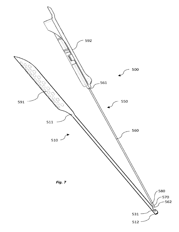
secure the
one more catheter tubes within the bore 1320.
[0119] Figures 14a-f and Figure 15 show another embodiment of a trocar shaft
1410
according to the present disclosure. The trocar shaft 1410 may be comprised in
a trocar
assembly 1400 including a handle 1450 as shown in Figures 16 and 17. Referring
to
Figures 14a and 14b, the trocar shaft 1410 is elongate and extends from a
proximal end
1411 to a distal end 1412. The distal end 1412 of the trocar shaft 1410
includes a tissue
separator 1430, as shown in Figures 14c and 14d, which is substantially the
same as
tissue separator 1330 described above. In this embodiment, the proximal end
1411 of
the trocar shaft 1410 defines a catheter adapter including an aperture 1420
for engaging
one or more catheters. Aperture 1420 extends through a flattened portion of
the
proximal end 1411, transverse to a longitudinal axis of the trocar shaft 1410.
Aperture
1420 includes an enlarged portion 1421 and a narrowed portion 1422. To secure
a
catheter tube, such as catheter tube 100, to the trocar shaft 1410, a proximal
end
101/201 of the catheter tube may be advanced through the enlarged portion 1421
in the
manner of threading a needle. The catheter tube 100 may be subsequently pulled
into
the narrowed portion 1422, as shown in Figure 15. The proximal end 1411 of the
trocar
shaft 1410 may be crimped to secure the catheter tube 100 in the aperture
1420.
[0120] An alternative embodiment of a trocar shaft 1510 is shown in Figures
18a-e. In
this embodiment, the proximal end 1511 of the trocar shaft 1510 includes a
catheter
adapter comprising a pair of tines 1520a and 1520b, defining a slit 1521 there
between.
CA 03200208 2023- 5- 25
WO 2022/109655
PCT/AU2021/051294
The upper tine 1520b extends proximally beyond the lower tine 1520a. The slit
1521 is
configured to receive the ends of one or more catheter tubes therein. In the
illustrated
example, the slit receives the ends 102, 201 of two tubes 100, 200, as shown
in Figure
18d. However, the slit may also receive a single tube. Once the ends 102, 201
of the
catheter tubes 100, 200 are received within the slit 1521, the proximal end
1511 of the
trocar shaft 1510 may be crimped to secure the tubes 100, 200 to the shaft
1510 by
bending the upper tine 1520b towards the lower tine 1520a, as shown in Figure
18e. In
other embodiments, the lower tine may be bent towards the upper tine, or both
tines
may be bent towards each other. Upper tine 1520b defines a notch 1522, adapted
for
gripping the catheter tubes 100, 200. The notch 1522 is positioned such that,
when the
tines 1520a, 1520b are crimped, the notch 1522 aligns with a proximal end 1523
of
lower tine 1520a. As such, when the proximal end 1511 of the trocar shaft 1510
is
crimped, the proximal end 1523 of the lower tine 1520a compresses the ends
102, 201
of the catheter tubes 100, 200 into the notch 1522, thereby to more securely
attach the
tubes 100, 200 to the trocar shaft 1510.
[0121] Another embodiment of a trocar shaft 1610 is shown in Figures 19a-d.
The
trocar shaft 1610 is similar to trocar shaft 1510 described above, having a
lower tine
1620a and an upper tine 1620b defining a catheter tube receiving slit 1621
therebetween. However, in this embodiment, the lower tine 1620a extends in a
proximal direction beyond the upper tine 1620b. The lower tine 1620a defines a
notch
1622. When the upper tine 1620b is bent toward the lower tine 1620a to secure
catheter
tubes 100, 200 therebewteen, a proximal end 1623 of the upper tine 1620b is
aligned
with the notch 1622. As such, when the proximal end 1611 of the trocar shaft
1610 is
crimped, the proximal end 1623 of the upper tine 1620a compresses the ends
102, 201
of the catheter tubes 100, 200 into the notch 1622, thereby to more securely
attach the
tubes 100, 200 to the trocar shaft 1610.
[0122] Each of the trocar shafts 1410, 1310, 1510 is receivable in a
respective handle
1450, 1350, 1550 as shown in Figures 20a-22b. Handles 1350, 1450, 1550 are
similar
to handles 350 and 450 described above. In these embodiments, however, each
handle
1350, 1450, 1550 is configured to crimp the proximal ends 1311, 1411, 1511 of
the
CA 03200208 2023- 5- 25
WO 2022/109655
PCT/AU2021/051294
26
trocar shafts 1310, 1410, 1510 to secure one or more catheter tubes thereto.
In other
embodiments, the proximal ends 1311, 1411, 1511 of the trocar shafts 1310,
1410,
1510 may be manually crimped.
[0123] Handle 1350 is comprised of a pair of shells 1351a, 1351b (as shown in
Figures 20a and 20b, respectively), which mate together to form a housed
configuration. When the trocar shaft 1310 is received between the shells
1351a, 135 lb
with a catheter tube received in the bore 1320, projections within the top
shell 1351a
and the base shell 135 lb impinge on the distal end 1311 of the trocar shaft
as the shells
mate, deforming wall 1315 and crimping the bore 1320 to secure the catheter
tube
therein.
[0124] Handle 1450 is similarly comprised of paired top shell 1451a and base
shell
1451 b, as shown in Figures 21a and 21b, respectively. When the trocar shaft
1410 is
received between the shells, as shown in Figures 16 and 17, a pair of ramped
surfaces
1453a and 1453b in the base shell 145 lb flank the distal end 1411 of the
trocar shaft
1410 adjacent the aperture 1420. The top shell 1451a includes a projection
1456
configured to impinge on the distal end 1411 of the trocar shaft 1410 as the
shells
1451a and 1451b are mated, so as to push the trocar shaft 1410 into the base
shell
145 lb. As the trocar shaft 1410 is pushed into the base shell 145 lb, the
ramped
surfaces deform the distal end 1411 of the trocar shaft, crimping the distal
end 1411
(reducing the size of the aperture 1420) and securing the catheter tube 100
therein.
[0125] Similarly, handle 1550 is comprised of paired top and base shells
1551a,
155 lb. (as shown in Figures 20a and 20b, respectively), which mate together
to form a
housed configuration. When the trocar shaft 1510 is received between the
shells 1551a,
155 lb with catheter tubes received in the slit 1521, projections within the
top shell
1551a and the base shell 155 lb impinge on and deform upper tine 1520b of the
trocar
shaft 1510 as the shells 1551a, 155 lb mate, crimping the tines 1520a, 1520b
together to
secure the catheter tubes therein.
CA 03200208 2023- 5- 25
WO 2022/109655
PCT/AU2021/051294
27
[0126] Figure 7 shows a nerve stimulating trocar assembly 500 according to an
embodiment of the invention in a disassembled configuration. The nerve
stimulating
trocar assembly 500 comprises an elongate trocar body 510 and a nerve
stimulator 550.
[0127] The nerve stimulator 550 has a shaft 560 extending from a proximal end
561
to a distal end 562, and at least one electrode 570 at or adjacent to the
distal end 562 of
the shaft 560.
[0128] The elongate trocar body 510 extends from a proximal end 511 to a
distal end
512. As shown in Figures 8a and 8b, the trocar body 510 has a U-shaped
elongate open
channel 520 which extends along a length of the trocar body 510. The open
channel
520 of the trocar body 510 is shaped and configured to receive both a catheter
tube and
the shaft 560 of the nerve stimulator 550 such that the catheter is releasably
secured
between the trocar body 510 and the nerve stimulator 550 in an assembled
configuration. When assembled with a catheter, the nerve stimulator 550
overlies the
catheter and substantially locks the catheter within the channel 520 of the
trocar body
510. A distal end of the catheter may be positioned set back proximally from
the distal
end 512 of the trocar body 510.
[0129] As shown in Figure 8d, in the assembled configuration, the electrode
570 of
the nerve stimulator 550 extends slightly beyond the distal end 512 of the
trocar body
510. In this embodiment, the electrode 570 has a curved distal tip which acts
as a tissue
separator during insertion of the trocar body 510 into tissue of the patient.
As the
diameter of the of the electrode 570 is smaller than the diameter of the
trocar body 510,
the electrode encounters less initial resistance to passing through the tissue
and inhibits
damage of anatomical structures, such as nerves.
[0130] The nerve stimulating trocar assembly 500 further comprises a locking
mechanism 530 adjacent the distal end 512 of the trocar body 510 for
releasably
locking the nerve stimulator 550 to the trocar body 510 in the assembled
configuration.
The locking mechanism 530 is shown in detail in Figures 8c and 8d. The locking
CA 03200208 2023- 5- 25
WO 2022/109655
PCT/AU2021/051294
28
mechanism 530 comprises a notched region 531 in a distal wall 513 of the
trocar body
510 and a transversely extending locking bar 580 of the nerve stimulator 550.
[0131] When the trocar body 510 and nerve stimulator 550 are in an assembled
configuration (for example, as shown in Figure 8b and 8d), the transversely
extending
locking bar 580 of the nerve stimulator 550 is received in the notched region
531. The
locking bar 580 of the nerve stimulator 550 is typically held in a tight
engagement with
the distal wall 513 of the trocar body 510. The notched region 531 of the
distal wall
513 may include a stop surface 532 to prevent the locking bar 580 advancing
towards
the distal end 512 of the trocar body 510 when in the assembled configuration.
The
tight engagement of the locking bar 580 with the distal wall 513 and the stop
surface
532 together prevent any substantial longitudinal movement of the nerve
stimulator
shaft 560 relative to the trocar body 510 in the assembled configuration.
[0132] The tight engagement of the locking bar 580 with the distal wall 513
may be
released by pulling the nerve stimulator shaft 560 in a direction away from
the distal
end 512 of the trocar body 510. The notched region 531 of the distal wall 513
includes
a ramped surface 533 proximal to the stop surface 532. When sufficient force
is
applied to the nerve stimulator shaft 560 in a direction away from the distal
end 512 of
the trocar body 510, the locking bar 580 typically releases from its tight
engagement
with the distal wall 513 and rides up the ramped surface 533 such that the
nerve
stimulator 550 may be withdrawn entirely from the trocar body 510.
[0133] The assembly may further comprise a handle 590 at a proximal region of
the
nerve stimulating trocar assembly. Typically, the assembly is made of two
separate
components including a base 591 and a slider 592. The base 591 is connected to
the
proximal end 511 of the trocar body 510. In the illustrated embodiment, the
base 591 is
integral with the trocar body 510 to form a single unit. The base 591 includes
an
elongate channel extending longitudinally along its length. The channel of the
base
591 is typically aligned with the open channel 520 of the trocar body.
Further, the
channel of the base of the handle is typically in fluid communication with the
open
channel such that together they may both receive a length of a catheter
therein.
CA 03200208 2023- 5- 25
WO 2022/109655
PCT/AU2021/051294
29
[0134] The base 591 and the slider 592 are longitudinally slidably connected
to each
other in the assembled configuration. In the assembled configuration, the
slider 592
and the base 591 may be in relatively locking engagement to each other to
prevent
sliding of the slider 592. However, the engagement between the two parts may
be
released to allow the slider 592 to move relative to the base 591.
[0135] An alternative embodiment of a nerve stimulator 550' including a shaft
560'
and a slider 592' is shown in Figure 23. The slider 592' is comprised of two
shells
592'a and 592'b which mate together to form a housed configuration. The nerve
stimulator 550' is substantially the same as nerve stimulator 550, except
that, in this
embodiment, the shaft 560' includes a proximal locking bar 568' (which may be
in
addition to a locking bat at a distal end, such as bar 580). The proximal
locking bar
568' is receivable in a corresponding notched region 569' of shell 592'b. The
notched
region 569' engages the proximal locking bar 568' to inhibit longitudinal
movement of
the proximal locking bar 568' relative to the slider 592' when the shaft 560'
is received
in the slider 592' in the housed configuration.
[0136] As shown in Figures 8a and 8b, the slider 592 is connected to the
proximal end
561 of the nerve stimulator shaft 560. In this embodiment, sliding of the
slider 592
relative to the base 591 in a direction away from the distal end of the trocar
body 510
will apply a force on the nerve stimulator shaft 560. A sufficient force will
release the
transverse locking bar 580 of the electrode 570 from its tight engagement with
the
distal wall 513 of the trocar body 510 and up the ramped surface 533 to allow
withdrawal of the nerve stimulator 550 from the trocar body 510. In use, this
allows a
surgeon to fully withdraw the nerve stimulator 550 once the trocar body 510 is
in a
desired location within the tissue of a patient.
[0137] The slider 592 includes a housing 593 having a passage extending from a
proximal end opening 594 to a distal end opening 595. A proximal length 561 of
the
nerve stimulator shaft may be received through the distal end 594 opening. The
proximal end opening of the slider may receive an electrical lead 565 for
electrical
connection with the proximal end 561 of the nerve stimulator shaft. The
electrical lead
CA 03200208 2023- 5- 25
WO 2022/109655
PCT/AU2021/051294
565 may be connected to an energy source to deliver energy to the electrode
570 of the
nerve stimulator 550.
[0138] Figures 9a and 9b show the assembled nerve stimulating trocar assembly
including a catheter 100.
[0139] In some embodiments, the slider 592 is provided with a distal grip
portion 596
engageable by a user's finger to enable force to be exerted on the assembly in
a distal
direction, e.g., during insertion of the nerve stimulating trocar assembly 500
through
tissue of a patient. Figure 9c shows the assembly 500 as it might be gripped
by a user's
hand during insertion into a patient's tissue.
[0140] Turning to the method shown in Figures 10a ¨ 10h, the drawings depict a
patient in position for an operation to surgically remove their haemorrhoids.
The
patient is typically placed on the operating table under general anaesthetic
with muscle
relaxation in the depicted modified lithotomy position. The perineum, pelvic
area and
thighs are prepared in a sterile field using a solution of iodine and alcohol.
[0141] An iodine-impregnated adhesive mat 600 is placed vertically from the
pubis to
cover the vagina/scrotum and the anal region. This area is kept sealed until
catheters
100 and 200 are implanted in the desired position. The adhesive mat 600
comprises
first and second frangible regions 601, 602 with serrations in the mat
allowing a small
area of the mat (here shown as a square but could be any shape) to be torn
off, for
access to the skin beneath. The surgeon makes a stab incision A at the first
frangible
region and then removes the second frangible region 602 of mat 600 and makes a
second stab incision B at this region, on the other side of the anus to stab
incision B. A
lateral stab incision C is made at the lateral thigh.
[0142] A proximal end of catheter 100 is attached to trocar 300. Trocar 300 is
tunnelled from incision A to incision B, through the patient's tissue across
the midline
of the patient as shown in Figure 10a. Once the distal end of trocar 300 has
exited the
stab incision B, the surgeon may remove the handle 350 of trocar 300, and pull
the
CA 03200208 2023- 5- 25
WO 2022/109655
PCT/AU2021/051294
31
trocar 300 by the distal end 312 to draw the trocar 300, together with
catheter 100,
through stab incision B until a desired length of catheter 100 extends from
incision B.
Catheter 100 is then disconnected from trocar 300. A length of catheter 100
adjacent
proximal end 101 extends from incision point B and a length of catheter 100
adjacent
distal end 102 extends from incision point A, as depicted in Figure 1013.
[0143] The proximal end 201 of the catheter 200 is attached to trocar 400.
Additionally, the proximal end 102 of catheter 100 is connected to trocar 400.
Trocar
400 is then passed from incision B to incision C, as shown in Figure 10c. Once
the
distal end of trocar 400 has exited the incision C, the surgeon may remove the
handle
450 of trocar 400, and pull the trocar 400 by the distal end 412 to draw the
trocar 400,
together with catheters 100 and 200, through stab incision C until a desired
length of
catheters 100 and 200 extends from incision C. Catheters 100, 200 are then
disconnected from trocar 400. At this point, a length of catheter 200 adjacent
distal end
202 extends from incision point B, a length of catheter 100 adjacent distal
end 102
extends from incision point A, and lengths of catheters 100, 200 adjacent
proximal ends
101, 201 extend from incision point C, as depicted in Figure 10d.
[0144] As shown in Figure 10e, distal end 102 of catheter 100 is then
assembled in
nerve stimulating trocar assembly 500. The nerve stimulator 550 is locked into
the
trocar body 510 overlying catheter 100, securing distal end 102 under the
locking bar
580. Catheter 100 and nerve stimulating trocar assembly 500 are then passed
through
incision A and angled to tunnel deeply into the tissue until at a region where
the target
nerve branches (e.g., the pudendal nerves) are located.
[0145] The energy source is then activated. In one embodiment, the energy
source is
an electrical energy source which delivers an electrical stimulus to electrode
570 of
nerve stimulating trocar assembly 500. The electrical stimulus is typically
between 3-
mAmp and at a frequency of between 0.5-1.0 Hz. The surgeon watches for
contraction of the external sphincter of the anus at a cycle rate
corresponding to the
applied electrical stimulus. The surgeon may move the trocar 500 and catheter
100
until this contraction is observed. This contraction indicates to the surgeon
that the
CA 03200208 2023- 5- 25
WO 2022/109655
PCT/AU2021/051294
32
electrode 570 of nerve stimulating trocar assembly 500 is adjacent to the
branches of
the pudendal nerve and that the distal end 102 of the catheter 100 is thus in
a position to
infuse medicament from apertures 105 to the branches of the pudendal nerve.
[0146] The nerve stimulator 550 may then be separated from the nerve
stimulating
trocar assembly 500 by sliding the slider 592 of the handle 590 in a proximal
direction.
This applies tension on the nerve stimulator shaft 560, causing the locking
bar 580 to
disengage from the notched region 531 in the trocar body 510. The nerve
stimulator
550 may then be withdrawn from the trocar body 510, and fully withdrawn from
incision A. Withdrawal of the nerve stimulator 550 releases catheter 100 from
the
trocar body 510. Trocar body may then be withdrawn through incision A, careful
not
to dislodge distal end 102 from its position adjacent to the pudendal nerve
branches.
[0147] This nerve stimulation process is then repeated with catheter 200
through
incision B, as also shown in Figure 10e. The process may be performed with the
same
nerve stimulating trocar assembly 500, or with a second nerve stimulating
trocar
assembly 500.
[0148] The proximal ends 101 and 201 of catheters 100 and 200 are then pulled
in the
direction shown by the arrows in Figure 10f to pull the excess of each
catheter at
incisions A and B subcutaneously. The catheters are not pulled so far as to
cause any
dislodgement of the positioning of the distal ends 102 and 202 adjacent to the
branches
of the pudendal nerves.
[0149] The surgeon then removes a portion of the adhesive mat 600 by tearing
along
frangible line 503. The incisions A and B are then sealed.
[0150] The lengths of catheter that exit incision C may be retained by
retainer 700, as
shown in Figures 1 la to 11c. Retainer 700 both protects the wound of the
patient and
prevents strain on catheters 100/200 which could cause them to be pulled out
of the
incision C.
CA 03200208 2023- 5- 25
WO 2022/109655
PCT/AU2021/051294
33
[0151] Retainer 700 includes a retainer body 720 which comprises a partially
circular
structure having a relatively planar base surface 721 configured to sit on the
skin (or on
an adhesive film applied over incision C) and an opposed guide surface 722 to
receive a
length of the first catheter 100 and/or the second catheter 200. The guide
surface 722
may be curved and terminate in an inner rim 723a and an outer rim 723b. A
series of
clips 730 retain the catheters 100 and/or 200 to the guide surface 722. The
clips 730
may be formed integrally with the retainer body 720 or may be a separate
structure
attachable onto the retainer body 720. In the illustrated embodiments, the
clips 730
include hinged portions 731 and a fastening mechanism comprising resiliently
deformable projections 732 at one end of the clip 730 which latch in an
aperture 733 at
an opposite end of the clip 730. The hinged portions 731 may be made from a
flexible
material such that the clips are moveable between an open configuration in
which the
catheters may be laid along the guide surface 722 and a closed configuration
in which
clips 730 are folded around the catheters 100, 200 and the projections 732
latched in
the apertures 733 to secure the catheters 100 and 200.
[0152] In the illustrated embodiment, the retainer 700 includes two clips
730a, 730b.
The catheters may be positioned in a desired location in the retainer 700
before
securing with the clips 730. Figures 11 a -11c illustrated a sequence of steps
to secure
the catheters 100, 200 in the retainer 700. In Figure lla, both clips 730a,
730b are in
the open configuration, with the catheters 100, 200 partially wound around the
guide
surface 722. . In Figure 11 b, the catheters 100, 200 are in their final
position and
secured by one clip 730a which has been fastened in the closed configuration.
In
Figure 11c the second clip 730b has been fastened in the closed configuration
to secure
the catheters 100, 200.
[0153] The proximal ends 101 and 201 of the catheters are ultimately connected
to a
medicament reservoir 800 which can be carried in a pouch 900 (e.g., as shown
in
Figure 12) or like device anywhere on the patient's body. The patient may
choose their
preference for the location of pouch 900 prior to surgery and the retainer
body 720
oriented accordingly such that the catheters 100 and/or 200 are directed
towards the
pouch 900.
CA 03200208 2023- 5- 25
WO 2022/109655
PCT/AU2021/051294
34
[0154] Figure 12 shows the parts of the system in a kit 1000 which includes
the
various parts of the system disclosed herein to allow the placement of
catheters 100 and
200 adjacent to respective pudendal nerve branches to deliver medicament to a
patient
and alleviate pain after the surgical removal of haemorrhoids. Kit 1000
comprises two
catheters 100 and 200, retainer 700, tunnelling trocar 300, tunnelling trocar
400, two
nerve stimulating trocar assemblies 500, mat 600 and casing 810 housing
medicament
reservoir 800 and pumps 850a, 850b.
[0155] It will be appreciated by persons skilled in the art that numerous
variations
and/or modifications may be made to the above-described embodiments, without
departing from the broad general scope of the present disclosure. The present
embodiments are, therefore, to be considered in all respects as illustrative
and not
restrictive.
CA 03200208 2023- 5- 25