Note: Descriptions are shown in the official language in which they were submitted.
WO 2022/120238
PCT/US2021/061901
CHEST TUBE AND PERICARDIOCENTESIS TRAINER APPARATUS
BACKGROUND OF THE INVENTION
[0001] 1. Field of the Invention.
[0002] The present invention relates to the training of medical professionals
in the procedures of
chest tube insertion. More particularly, the present invention relates to
apparatuses and methods
for training medical professionals to carry out chest tube insertion including
for, but not limited
to, pericardiocentesis. Still more particularly, the present invention relates
to a convenient,
relatively inexpensive, and portable simulator apparatus designed to simulate
the conditions
associated with chest tube insertion.
[0003] 2. Description of the Prior Art.
[0004] Emergency medicine and other medical specialties are responsible for
preforming several
lifesaving procedures. Many of these procedures are rare, limiting the amount
of skill
maintenance that can be done with patient care. Procedural skill proficiency
and competence can
be maintained through simulation, but most active practicing clinicians do not
have ready access
to a simulation center. The training of medical professionals requires access
to easy-to-use
simulators which allow procedural skillset maintenance, as well as the
training of medical
students, residents, and other learners.
[0005] Many current trainers are complex, expensive, and are therefore only
available to
learners/practitioners who have access to a simulation center. Most currently
practicing clinicians
do not have ready access to a simulation center, have to pay significant costs
for the use of a
simulation center space and resources, and/or do not, given the above noted
obstacles, utilize
such facilities for a variety of reasons. This leads to procedural skill set
decay, especially in
uncommon and infrequent procedures. Having deliverable trainers, to the end
users' homes or
places of practice, would help eliminate many of these barriers to procedural
training and,
therefore, procedural skill maintenance.
[0006] A need therefore exists for low- to mid-fidelity simulators that can be
utilized outside of a
simulation center. In particular, practitioners would benefit from easy
assembled, simple to use
simulators that maintain the necessary fidelity to practice procedures,
particularly ones that are
1
CA 03201093 2023- 6-2
WO 2022/120238
PCT/US2021/061901
rarely seen in clinical practice. Such a simulator would be useful in two
important, but rarely
executed medical procedures- chest tube insertion and pericardiocentesis.
SUMMARY OF THE INVENTION
[0007] The current invention provides a solution to the need for a simulator
that aids in training
users to carry out chest tube insertion and pericardiocentesis procedures. The
invention is a
medical procedure training simulator apparatus that facilitates the practice
of chest tube insertion
and pericardiocentesis. The simulator is in the form of an easy-to-ship
product that can be
utilized anywhere. Prior to this invention, the ability to practice these
procedures in situ (where
they practice medicine, an ideal location for training) or in the home
environment, was not
feasible to the training or practicing clinician due to lack of access,
expense of materials or a
combination of the above.
[0008] In an embodiment, the simulator is a three-part structure: a first
stand element and a
second stand element that are removably clipped together to form a rigid, 90-
degree base, and a
rib panel, which is removably clipped to the base. The combination of two-
element base and rib
panel is configured so that the simulator can lay on a standard-dimension
medical basin for
support and ease of access, but it can also be used in other settings. The rib
panel is a frame,
which may be a rectangular frame, with internal curved slot elements designed
to mirror the
anatomy of the human rib cage, and four winged clips, which allow the rib
panel to clip on to,
rest on, or otherwise be removably engaged with the base.
[0009] The first stand element has two or more clips protruding from an edge
thereof, dependent
on the specific configuration of the base to be formed, and two or more slots
inset from an edge
opposing the edge having the protruding clips. Alternatively, the first stand
element has a
multitude of hinge elements located on the edge thereof, dependent upon the
specific
configuration of the base to be formed, which engage with a multitude of hinge
elements located
on an edge of the second stand element. The edge associated with the slots may
include a lip
affixed thereto extending at about a 90-degree angle from that edge and
arranged to enable the
slot edge to be spaced above an underlying substrate when the simulator is
assembled.
[0010] The second stand element has ports spaced from an edge thereof and
corresponding in
number to the number of clips of the first stand element. The ports of the
second stand element
have dimensions about the same as but slightly larger than the dimensions of
the clips of the first
2
CA 03201093 2023- 6-2
WO 2022/120238
PCT/US2021/061901
stand element so that the clips of the first stand element and the ports of
the second stand element
can be removably joined together when the clips are inserted into the ports.
When that joining is
completed, the base of the simulator is established as an upright frame with
the second stand
element extending upwardly from the edge of the first stand element with the
clips at an angle of
about 90 degrees. The second stand element also has two or more slots inset
from an edge
opposing the edge having the ports.
[0011] The base formed by removably joining the first stand element and the
second stand
element together establishes an angled frame that is used to removably secure
the rib panel
therein. The rib panel includes a panel body having a first edge spaced from
an opposing second
edge. Each of the first and second edges includes two or more wings extending
therefrom. The
wings are configured to removably fit into the slots of the first and second
stand elements,
wherein the wings of the first edge of the panel fit into the slots of the
first stand element and the
wings of the second edge of the panel fit into the slots of the second stand
element. When the rib
panel is inserted into the base, the rib panel rests at an angle of about 45
degrees between the first
stand element and the second stand element. The rib panel includes a panel
body between the
first edge and second edge. The panel body includes a set of parallel curved
slots extending
within a perimeter of the panel body, and corresponding parallel curved slats
between slots.
Curvature of the parallel curved slats is selected to represent human's ribs.
The parallel curved
slats may be aligned in a two- or three-dimensional manner.
[0012] The simulator of the present invention with the combination of the base
and rib panel can
be used to carry out chest tube insertion training. Specifically, the first
and second stand
elements are clipped together to create the base. The practitioner can then
lay the rib panel into
the base. The practitioner can then place off-the-shelf subcutaneous tissue
and training skin
overlays to allow for the practice of inserting chest tubes and pigtail
catheters through the
overlays and into the curved slots of the rib panel. At least one side of the
rib panel may include
attachment elements such as pegs, which attachment elements can be used to
attach to overlays
to panel.
[0013] In another embodiment, the simulator is a four-part structure: a first
stand element and a
second stand element that are removably clipped together with hinges to form a
90-degree base,
a rib panel that is removably clipped to the first stand element with a hinge,
and a fitted rib panel
frame that is removably engaged to the rib panel. The rib panel frame is
configured to mirror the
3
CA 03201093 2023- 6-2
WO 2022/120238
PCT/US2021/061901
shape of the rib panel with a middle cavity. The middle cavity of the rib
panel frame allows the
user access to the rib panel while the rib panel frame is engaged.
[0014] The hinge elements in the above embodiment function to engage the first
stand element,
second stand element, and rib panel together. The hinges allow for the
invention to remain
connected but facilitate easier transportation and storage. The hinge elements
serve to hold each
element in place while the simulator is in use. Additionally, the rib panel
may be configured with
a latch feature aligned to receive the second edge of the second stand
element. The hinges allow
for the invention to be expanded back into working condition and the latch of
the rib panel
engages with the second edge of the second stand element to secure the device
in place for use.
[0015] The rib panel frame is a continuous piece shaped to mirror the outer
edge of the rib panel.
The rib panel frame has a middle cavity and a multitude of pins. The pins are
aligned to be
removably engaged with the rib panel to secure the rib panel frame to the rib
panel. The middle
cavity is aligned to allow access to the curved slots, mirroring the ribs of a
human. It is possible
to configure the curvature to enable tube insertion for other animals. The off-
the-shelf
subcutaneous tissue and training skin overlay is placed on the rib panel with
the edges of the
tissue overlapping the outer edge of the rib panel. The rib panel frame is
then placed over the
tissue and overlays and secured to the rib panel via the multitude of pins.
The attachment of the
rib panel and rib panel frame secures the tissues and overlays in place while
the user uses the
simulator.
[0016] The rib panel of the simulator can also separately be used to provide
pericardiocentesis
training. The rib panel may be sized and shaped to lay within a standard
medical basin. During
training of pericardiocentesis, the practitioner lays a simulation heart
composed of off-the-shelf
materials into the basin with water, water and fiber supplement, gelatin, or
other substance to
facilitate ultrasound use. The rib panel or rib panel with rib panel frame is
then laid atop the
medical basin. A training skin can then be laid over the rib panel and the
practitioner can practice
the procedure of pericardiocentesis, or drainage of fluid from around the
heart.
[0017] The simulator of the present invention, including the combination of
the base and rib
panel or the rib panel alone, can be used in a convenient way in most any
location to enable
training for chest tube insertion or pericardiocentesis.
BRIEF DECRIPTION OF THE DRAWINGS
4
CA 03201093 2023- 6-2
WO 2022/120238
PCT/US2021/061901
[0018] The foregoing features of the invention will be more readily understood
by reference to
the accompanying drawings, in which:
[0019] FIG 1 is a plan view of the first stand element of the base of a first
embodiment of the
simulator apparatus of the present invention.
[0020] FIG 2 is a side view of the first stand element.
[0021] FIG 3 is a plan view of the second stand element of the base.
[0022] FIG 4 is a side perspective view of the first stand element and second
stand element
joined together to form the base of the simulator apparatus.
[0023] FIG 5 is a plan view of the rib panel of the present invention.
[0024] FIG 6 is a side view of the rib panel.
[0025] FIG 7A is a side view of the simulator apparatus of the present
invention for the purpose
of training chest tube insertion.
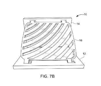
[0026] FIG. 7B is a front view of the simulator of FIG. 7A.
[0027] FIG 8A is a plan view of the rib panel atop a medical basin for the
purpose of training
pericardiocentesis.
[0028] FIG. 8B is a side view of the rib panel of FIG. 8A.
[0029] FIG 9A is a side view of a second embodiment of the simulator apparatus
of the present
invention for the purpose of training chest tube insertion with the rib panel
frame version of the
invention.
[0030] FIG. 9B is a front view of the simulator of FIG. 9A
[0031] FIG. 10A is a side perspective view of the simulator of FIG. 9A with an
exploded view of
the rib panel frame.
[0032] FIG. 10B is a side view of the simulator apparatus of the present
invention with the rib
panel frame depicting hinge elements joining the first stand element and
second stand element
and the first stand element and the rib panel.
[0033] FIG. 10C is a side perspective view of the simulator of FIG. 9A
collapsed to align the
first stand element, the second stand element, the rib panel, and rib panel
frame in a parallel
fashion for easier transportation and storage.
DETAILED DESCRIPTION OF THE INVENTION
CA 03201093 2023- 6-2
WO 2022/120238
PCT/US2021/061901
[0034] A first medical training simulator apparatus 10 is shown in FIGS. 1-8B.
The simulator
apparatus 10 includes a first stand element 12 shown alone in FIGS. 1 and 2, a
second stand
element 14 shown alone in FIG. 3, and a rib panel 16 shown alone in FIGS. 5
and 6. FIG. 4
shows the first stand element 12 and the second stand element 14 joined
together to form a base
18 of the simulator apparatus 10. FIGS. 7A and 7B show the rib panel 16
coupled to the base 18.
FIGS. 8A and 8B show the rib panel 16 as a standalone component suitable to
aid in training for
pericardiocentesis positioned on a medical basin 80. FIGS. 9A and 9B show a
second medical
training simulator apparatus 100 with the rib panel 16 coupled to the base 18
and a rib panel
frame 17. FIG. 10A depicts the apparatus 100 of FIGS. 9A and 9B with the rib
panel frame 17
detached from the rib panel 16. FIG. 10B depicts the apparatus 100 of FIG. 9A,
9B, and 10A in
an expanded view wherein a multitude of hinge elements 23 connect the first
stand element 12
with the second stand element 14 and the first stand element 12 with the rib
panel 16. FIG. 10C
shows the apparatus 100 in a collapsed configuration for ease of transport.
[0035] The first stand element 12 includes a primary body 20 with a first edge
22 and a second
edge 24 opposite the first edge 22. A first clip 26 and a second clip 28
extend from the body 20
at the first edge 22. The first stand element 12 also includes a first rib
panel receiving slot 30
and a second rib panel receiving slot 32 each extending partially or
completely through a front
side 34 to a back side 36 of the body 20 in substantial alignment with one
another and set into the
body 20 from the second edge 22. The first rib panel receiving slot 30 and the
second rib panel
receiving slot 32 are arranged to removably receive therein a portion of the
rib panel 16. The
number of clips may be more than two or fewer than two. The number of panel
receiving slots
may be more than two or less than two. The first stand element 12 may be made
of any suitable
material, including metallic or nonmetallic material. The nonmetallic material
may be a
viscoelastic material such as a polymeric material. For example, the first
stand element 12 may
be formed of polyethylene but not limited thereto.
[0036] The first stand element 12 optionally includes a spacing bar 38
extending from the second
edge 24 on the backside 36 of the first stand element 12. The spacing bar 38
may extend
partially or entirely a width of the body 20 and is of selectable height. The
spacing bar 38
enables standoff of a portion 40 of the body 20 when on a substrate so that a
component of the
simulator 10 may be selectably and removably inserted into the slots 30 and
32.
6
CA 03201093 2023- 6-2
WO 2022/120238
PCT/US2021/061901
[0037] The second stand element 14 includes a primary body 42 with a first
edge 44 and a
second edge 46 opposite from the first edge 44. The second stand element 14
also includes a
first clip receiving port 48 and a second clip receiving port 50, each
extending partially or
completely through a front side 52 to a back side 54 of the body 42 in
substantial alignment with
one another and set into the body 42 from the first edge 44. The second stand
element 14 further
includes a first rib panel receiving slot 56 and a second rib panel receiving
slot 58, each
extending partially or completely through the front side 52 to the back side
54 in substantial
alignment with one another and set into the body 42 from the second edge 46.
The first clip
receiving port 48 and the second clip receiving port 50 are arranged to
removably retain therein
the first clip 26 and the second clip 28 of the first stand element 12. The
first rib panel receiving
slot 56 and the second rib panel receiving slot 58 are arranged to removably
receive therein a
portion of the rib panel 16. The second stand element 14 may be made of any
suitable material,
including metallic or nonmetallic material. The nonmetallic material may be a
viscoelastic
material such as a polymeric material. For example, the second stand element
14 may be formed
of polyethylene but not limited thereto.
[0038] FIG. 4 shows the first stand element 12 and second stand element 14
coupled together to
form the base 18. Specifically, the clips 26 and 28 of the first stand element
12 are inserted into
the clip receiving ports 48 and 50 of the second stand element 14 to form the
base 18 having a
substantially right-angle configuration with the slots 30 and 32 of the first
stand element 12
angled and spaced from the slots 56 and 58 of the second stand element 14.
[0039] The rib panel 16 includes a primary body 60 with a first edge 62 and a
second edge 64
opposite from the first edge 62. The rib panel 16 also includes a first rib
wing 66 and a second
rib wing 68 extending from the body 60 at the first edge 62, and a third rib
wing 70 and a fourth
rib wing 72 extending from the body 60 at the second edge 64. The rib panel is
substantially
symmetrical so that the first and second rib wings 66 and 68 may be removably
inserted into the
slots 30 and 32 of the first stand element 12 with the third and fourth rib
wings 70 and 72
removably inserted into slots 56 and 58 of the second stand element 14, or the
orientation of the
rib wings may be reversed. FIGS. 7A and 7B show the simulator apparatus 10 put
together with
the first stand element 12 and second stand element 14 coupled together, as
well as the rib panel
16 coupled to the first stand element 12 and the second stand element 14.
7
CA 03201093 2023- 6-2
WO 2022/120238
PCT/US2021/061901
[0040] The rib panel 16 also includes a plurality of parallel curved slots 74
extending within a
perimeter of the body 60, and corresponding parallel curved slats 76
alternating between the slots
74. The spacing, size and curvature of the slots 74 and slats 76 are arranged
to represent a two-
dimensional version of a human's ribs. Other configurations are possible
provided the rib panel
16 is arranged to enable a user to simulate relevant medical activities of
interest with and through
the rib panel 16. For example, the rib panel 16 forming part of the simulator
10 shown in FIGS.
7A and 7B may be used to practice chest tube insertion, while the rib panel 16
alone shown in
FIGS. 8A and 8B may be used to practice pericardiocentesis when removably
placed on the
medical basin 80.
[0041] FIGS. 9A and 9B show the second embodiment of the simulator apparatus
100 with the
first stand element 12 and the second stand element 14 coupled together. The
rib panel 16 is
coupled to the first stand element 12 and the second stand element 14. The rib
panel frame 17 is
removably engaged with the rib panel 16. The rib panel 16 of the apparatus of
9A and 9B is
arranged to depict the spacing, size, and curvature of the slots 74 and slats
76 to represent a
three-dimensional version of a human's ribs.
[0042] FIG. 10A depicts the apparatus 100 with a multitude of pins 19 located
on the rib panel
frame 17 and a multitude of ports 21 located on the rib panel 16 wherein the
pins 19 may be
removably engaged with the ports 21 to attach the rib panel frame 17 to the
rib panel 16. The
pins 19 and ports 21 facilitate the coupling of the rib panel 16 to the rib
panel frame 17. The rib
panel frame 17 has a middle cavity 25, allowing access to the rib panel 16 by
the user when the
rib panel frame 17 is attached. The rib panel frame 17 secures the off-the-
shelf subcutaneous
tissue to the rib panel 16.
[0043] FIG. 10B depicts the apparatus 100 with the first stand element 12 and
the second stand
element 14 coupled together with the hinge elements 23. Additionally, the
first stand element 12
and the rib panel 16 are coupled together with a hinge element 23. The
apparatus 100 may have a
multitude of hinge elements 23 to appropriately secure the panels together.
The use of hinge
elements 23 allows the user to fold the apparatus 100 into a more portable and
easily
transportable form. The apparatus 100 of FIG. 10B has a latch 27 which is
configured to be
removably engaged with the second edge 46 of the second stand element 14. The
latch 27
facilitates the apparatus 100 to be secured in an "in use" position with the
first stand element 12
and the second stand element 14 oriented in about a 90-degree angle, with the
rib panel 16
8
CA 03201093 2023- 6-2
WO 2022/120238
PCT/US2021/061901
oriented at about a 45-degree angle. The latch 27 may be uncoupled from the
second edge 46 of
the second stand element 14, allowing the hinge elements 23 to be utilized to
fold the device
down to a relatively flat orientation. The first stand element 12, the second
stand element 14, and
the rib panel 16 may lay flat relatively to each piece, such that the user may
more easily transport
the apparatus 100 while maintaining the coupling of the pieces of the
apparatus 100.
Alternatively, the user may utilize the apparatus 100 in its folded down
manner with the rib panel
16 oriented parallel to the surface that the apparatus 100 is placed on.
[0044] FIG. 10C depicts the apparatus 100 of FIG. 9A wherein the hinge
elements 23 allow the
apparatus 10 to be collapsed down. The first stand element 12, the second
stand element 14, the
rib panel 16, and rib panel frame 17 are aligned in a parallel fashion to
facilitate easier
transportation and storage of the apparatus 100. The collapsed apparatus 100
of FIG. 10C is
configured to be used on a flat surface or for easier transportation or
storage.
[0045] 'The rib panel 16 optional includes one or more pegs 82 on at least one
face of the body
60, which pegs may be used to removably retain to the rib panel 16
supplemental materials
useful in carrying out a simulated procedure of interest. For example,
practitioners can then place
off-the-shelf subcutaneous tissue and skin overlay on the rib panel 16 for
training in placing
chest tubes and pigtail catheters, which overlays may be removably affixed to
the pegs 82 The
rib panel frame 17 provides another method in which to secure the off-the-
shelf subcutaneous
tissue and skin overlay to the rib panel 16. The rib panel 16 is designed to
fit securely in the
medical basin 18, with the slats 76 functioning as rib equivalents inset into
the basin 80 slightly,
with the opposing sets of wings 66 and 68 and 70 and 72 positioned at the most
superior part of
the frame of the basin 80. This allows a simulation heart, created with off-
the-shelf materials, to
be placed in the basin 80 which is then filled with water, water and fiber
supplement, or gelatin
to create an echogenic model that can utilize ultrasound for the training in
ultrasound guided
pericardiocentesis through the rib panel 16. The rib panel 16 may be made of
any suitable
material, including metallic or nonmetallic material. The nonmetallic material
may be a
viscoelastic material such as a polymeric material. For example, the rib panel
16 may be formed
of polyethylene but not limited thereto.
[0046] The present invention has been described with reference to specific
examples and
configurations. It is only intended to be limited to the description set out
in the claims and
equivalents.
9
CA 03201093 2023- 6-2