Note: Descriptions are shown in the official language in which they were submitted.
WO 2022/125535
PCT/US2021/062182
DEVICE, METHOD AND SYSTEM FOR RESHAPING A HEART VALVE
ANNULUS
CROSS-REFERENCE TO RELATED APPLICATION(S)
100011 This application claims benefit of priority under 35 U.S.C. 119(e) of
U.S.
Provisional Patent Application Serial No. 63/122,420, filed December 7, 2020.
The
disclosure of the prior application is considered part of and is incorporated
by reference in
the disclosure of this application.
BACKGROUND OF THE INVENTION
FIELD OF THE INVENTION
100021 The present invention relates generally to medical device and
procedures, and more
particularly to devices, methods and systems for anchoring of an implant
within the body
and/or reshaping an organ within the body.
BACKGROUND INFORMATION
100031 The healthy human heart (is a muscular two-side self-regulating pump
slightly larger
than a clenched fist, as can be seen in Figures 2A-2C. It is composed of four
chambers
including the right atrium (RA) and right ventricle (RV), and the left atrium
(LA) and LV
(LV). The RA collects poorly oxygenated blood returning from the lower body
via the
inferior vena cava (IVC) and from the head and upper body via the superior
vena cava
(SVC) and delivers it through the tricuspid valve to the RV. The RV then
contracts which
has the effect of closing the tricuspid valve and forcing the blood through
the pulmonary
valve into the pulmonary artery for circulation to the lungs. The left side of
the heart
collects the oxygenated blood in the LA returning from the lungs via the
pulmonary veins.
From, there the blood is delivered to the LV. The LV then powerfully contracts
having the
effect of closing the mitral valve (MV) and forcing the blood through the
aortic valve into
the aorta and thence throughout the body.
1
CA 03201390 2023- 6-6
WO 2022/125535
PCT/US2021/062182
[0004] The interatrial septum, a wall composed of fibrous and muscular parts
that separates
the RA and LA, as can be seen in Figure 2C. The fibrous interatrial septum is,
compared to
the more friable muscle tissue of the heart, a more materially strong tissue
structure in its
own extent in the heart. An anatomic landmark on the interatrial septum is an
oval,
thumbprint sized depression called the oval fossa, or fossa ovalis, as can be
seen in Figure
2C, which is a remnant of the oval foramen and its valve in the fetus. It is
free of any vital
structures such as valve structure, blood vessels and conduction pathways.
Together with its
inherent fibrous structure and surrounding fibrous ridge, which makes it
identifiable by
angiographic techniques, the fossa ovalis is the favored site for trans-septal
diagnostic and
therapeutic procedures from the right into the left heart. Before birth,
oxygenated blood
from the placenta was directed through the oval foramen into the LA, and after
birth the
oval foramen closes. The heart's four valves function primarily to ensure the
blood does not
flow in the wrong direction during the cardiac cycle e.g., backflow from the
ventricles to the
atria or backflow from the arteries into the corresponding ventricles.
[0005] The synchronous pumping actions of the left and right sides of the
heart constitute
the cardiac cycle. The cycle begins with a period of ventricular relaxation,
called ventricular
diastole. At the beginning of ventricular diastole (e.g., ventricular
filling), the aortic and
pulmonary valves are closed to prevent backflow from the arteries into the
ventricles.
Shortly thereafter, the tricuspid and mitral valves open to allow flow from
the atria into the
corresponding ventricles. Shortly after ventricular systole (e.g., ventricular
contraction and
emptying) begins, the tricuspid and mitral valves close to prevent backflow
from the
ventricles into the corresponding atria. The aortic and pulmonary valves then
open to permit
discharge of blood into the arteries from the corresponding ventricles. The
opening and
closing of the heart valves occur primarily as a result of pressure
differences. For example,
the opening and closing of the mitral valve occurs as a result of the pressure
differences
between the LA and the LV. During ventricular diastole, when the LV is
relaxed, the blood
returning from the lungs into the LA causes the pressure in the atrium to
exceed that in the
LV. As a result, the mitral valve opens, allowing blood to flow from the LA
into the LV.
Subsequently as the now full ventricle contracts in ventricle systole, the
intraventricular
pressure rises above the pressure in the atrium and pushes the mitral valve
shut.
2
CA 03201390 2023- 6-6
WO 2022/125535
PCT/US2021/062182
[0006] The mitral and tricuspid valves are defined by fibrous rings of
collagen, each called
an annulus, which forms a part of the fibrous skeleton of the heart The
annulus provides
attachment to cusps or leaflets of the mitral valve (called the anterior and
posterior cusps or
leaflets) and the three cusps or leaflets of the tricuspid valve. The cusps of
a healthy mitral
valve are shown in Figure 2B. Proper closing function is also aided by a
tethering action of
chordae tendineae and one or more papillary muscles. Also of structural
relevance to this
invention and located in the vicinity of the annulus of the mitral valve is
the coronary sinus
and its tributaries including the great cardiac vein (GVC), as can be seen in
Figure 2C. The
GVC generally courses around the lower wall of the LA outside the atrial
chamber but
within the atrial wall. The GVC empties into the RA through the coronary
sinus.
[0007] Each of the valves in question is a one-way valve that function to
allow blood to
flow only in the appropriate direction. If any of the valves does not function
properly, that
will affect the efficiency of the heart and may result in significant health
issues. For
example, failure of the mitral valve between the LA and the LV, to fully seal
while the LV
is contracting results in some portion of the blood in the LV being expelled
retrograde back
into the LA. This is generally termed mitral regurgitation and depending on
severity, can
result in insufficient blood flow throughout the body with resultant serious
health
implications.
[0008] II. Characteristics and Causes of Mitral Valve Dysfunction
[0009] When the LV contracts after filling with blood from the LA, the walls
of the
ventricle move inward and release some of the tension from the papillary
muscle and
chords. The blood pushed up against the under-surface of the mitral leaflets
causes them to
rise toward the annulus plane of the mitral valve. As they progress toward the
annulus, the
leading edges of the anterior and posterior leaflet come together forming a
seal and closing
the valve. In the healthy heart, leaflet coaption occurs near the plane of the
mitral annulus.
The blood continues to be pressurized in the LV until it is ejected into the
aorta. Contraction
of the papillary muscles is simultaneous with the contraction of the ventricle
and serves to
3
CA 03201390 2023- 6-6
WO 2022/125535
PCT/US2021/062182
keep healthy valve leaflets tightly shut at peak contraction pressures exerted
by the
ventricle.
[00010] In a healthy heart, the dimensions of the mitral valve annulus create
an anatomic
shape and tension such that the leaflets coapt, forming a tight junction, at
peak contraction
pressures. Where the leaflets coapt at the opposing medial and lateral sides
of the annulus
are called the leaflet commissures CM, CL, as shown Figure 2B. Valve
malfunction can
result from the chordae tendineae (the chords) becoming stretched, and in some
cases
tearing. When a chord tears, this results in a leaflet that flails Also, a
normally structured
valve may not function properly because of an enlargement of or shape change
in the valve
annulus. This condition is referred to as a dilation of the annulus and
generally results from
heart muscle failure. In addition, the valve may be defective at birth or
because of an
acquired disease. Regardless of the cause, mitral valve dysfunction can occur
when the
leaflets do not coapt at peak contraction pressures. When this occurs, the
coaption line of
the two leaflets is not tight at ventricular systole. As a result, an
undesired back flow of
blood from the LV into the LA can occur.
[00011] This mitral regurgitation, if significant in amount, may have has
several serious
health consequences. For example, blood flowing back into the atrium may cause
high atrial
pressure and reduce the flow of blood into the LA from the lungs. As blood
backs up into
the pulmonary system, fluid leaks into the lungs and causes pulmonary edema.
Another
health problem resulting from mitral valve dysfunction is the reduction of
ejection fraction
of the heart, or the effective pumping of the blood through the body of that
blood that does
enter the LV. The blood volume regurgitating back into the atrium reduces the
volume of
blood going forward into the aorta causing low cardiac output. Excess blood in
the atrium as
a result of mitral valve regurgitation may also over-fill the ventricle during
each cardiac
cycle and causes volume overload in the LV. Over time, this may result in
dilation of the
LV and indeed the entire left side of the heart. This may further reduce the
effective cardiac
output and further worsen the mitral regurgitation problem by dilating the
mitral valve
annulus. Thus, once the problem of mitral valve regurgitation begins, the
resultant cycle
may cause heart failure to be hastened. Treating the problem therefore not
only has the
4
CA 03201390 2023- 6- 6
WO 2022/125535
PCT/US2021/062182
immediate effect of alleviating the heart output problems mentioned above, but
also may
interrupt the downward cycle toward heart failure.
[00012] III. Current Treatment Methods
[00013] Various methods of treating this serious heart condition have been
suggested. In
one approach, the native valve is removed and replaced with a new valve, such
as described
in U.S. Pat. No. 6,200,341 to Jones et al and U.S. Pat. No. 7,645,568 to
Stone. While this
approach may be of use in some situations, such surgical procedures generally
require open
chest surgery, which is invasive and often contraindicated for very sick or
old patients,
which includes many of those suffering from mitral valve regurgitation.
[00014] Another method which has been suggested is to apply tension across the
LV to
reshape the 1,V, thereby affect the functioning of the mitral valve, such as
described in U.S.
2005/0075723 to Schroeder et al. This approach uses a splint that spans across
a ventricle
and extends between epicardi al pads that engage outside surfaces of the
heart. This
approach is also invasive and potentially problematic as it penetrates an
outer surface of the
heart.
[00015] Another method that has been suggested is the attempted constriction
of the LA by
means of a belt like constricting device extending inside the GVC which runs
along the
posterior wall of the LA, such as described in U.S. 2002/0183841 Al to Cohn et
al. While
this may be partially helpful, often the device fails to sufficiently alter
the shape of the left
atrium to fully resolve the failure of the leaflets to coapt.
[00016] Yet another method that has proven particularly useful is to employ a
system that
applies direct tension across the width of the LA and across the minor axis of
the annulus of
the mitral valve, such as shown in Figure 3. System 1 utilizes a bridging
element 2 that
extends between an anterior anchor 3 and a posterior anchor 4. The anterior
anchor 3 is
generally located at the wall between the LA and the RA, for example, on the
fossa ovalis
on the septal wall, and is attached to the bridging element 2 that spans the
LA. Posterior
anchor 4 is located across the atrium posterior to the anterior anchor and may
be located
CA 03201390 2023- 6-6
WO 2022/125535
PCT/US2021/062182
outside the atrium chamber in the GVC. The bridging element is affixed to the
posterior
anchor and provides a bridge across the LA between the septum. The GVC and is
tensioned
to directly affect the shape of the LA, and in particular, the annulus of the
mitral valve. By
adjusting the tension of the bringing element, the shape of the LA and
particularly the
annulus of the mitral valve can be adjusted to achieve optimum closure of the
mitral valve
during cardiac function. An example of this approach is described in detail in
U.S. Pat. No.
8,979,925 B2 to Chang et al., the entire contents of which are incorporated
herein by
reference for all purposes.
[00017] This approach has many advantages over conventional approaches,
including
avoiding invasive procedures such as open heart surgery or being placed on a
heart-lung
machine. However, there are still a number of challenges that must be
addressed. While the
anterior anchor provides relatively robust and secure anchoring with the fossa
ovalis,
anchoring within a body vessel, such as the GCV is more problematic. While the
fossa
ovalis is defined by a notable depression, which lends itself to having an
anchor disposed
within, the GCV lacks any notable anatomical features and is defined by a
relatively
smooth-walled vessel along the outer wall of the left atrium. In addition, the
heart is a
highly dynamic organ such that any implant disposed therein is subjected to
highly variable
forces and movements due to the contortions of the heart muscle during a
pumping cycle of
the heart. These aspects make anchoring within the GCV particularly
challenging. Thus,
there is need for devices, systems and methods that allow for robust and
dependable
anchoring within a vessel, such as the GCV. There is further need for such
anchoring
devices that can withstand considerable forces over the lifetime of the
device. There is
further need for such anchoring devices that can assist in reshaping of an
organ, such as the
heart.
SUMMARY OF THE INVENTION
[00018] The present invention provides systems, methods and associated devices
for
delivery and deployment of heart implants for reshaping a heart valve annulus
for treatment
of a heart disorder, such as mitral valve regurgitation.
6
CA 03201390 2023- 6-6
WO 2022/125535
PCT/US2021/062182
[00019] Accordingly, in one embodiment, the invention provides an anchor
system
including an augmentation device and an anchor. In some aspects, the
augmentation device
has an elongated cylindrical body defined by a substantially cylindrical wall.
The lumen is
configured to receive an anchor and the cylindrical wall includes slots
disposed along a
length of the cylindrical body for engaging a bridging element of the anchor.
The system
further includes an anchor having a substantially cylindrical body that is
sized to pass within
the elongated cylindrical body of the augmentation device, and a bridging
element coupled
to an intermediate portion of the anchor.
[00020] In another aspect, the augmentation device has an elongated shaft
body, wherein
the shaft body has a first elongated configuration and a second flexed
configuration. The
second flexed configuration has a reduced length as compared to the first
elongated
configuration. The system further includes an anchor having a substantially
cylindrical body
having a length less than the that of the augmentation device, and a bridging
element
coupled to an intermediate portion of the anchor. The system is configured
such that when
the augmentation device and anchor are coupled and deployed in a body lumen, a
force
upon a wall of the body lumen from the anchor is translated to the
augmentation device to
deform the wall.
[00021] In various embodiments, the invention provides an anchor system that
includes: an
anterior anchor and a posterior anchor. In some aspects, the anchor system
includes an
anterior anchor having an anchor portion operable to secure the anterior
anchor in tissue, a
through hole extending through the anchor member, and an elongated tube having
a lumen
coextensive with the through hole, wherein the elongated tube is composed of a
semi-rigid
or rigid material that resists flexing; and a posterior anchor coupled to a
first end of a
bridging element, wherein a second end of the bridging element is configured
to traverse the
lumen of the elongated tube of the anterior anchor.
[00022] In another aspect, the anchor system includes: an anterior anchor
having an anchor
portion operable to secure the anterior anchor in tissue, a through hole
extending through
the anchor member, and an adjustable arm extending from the anchor portion;
and a
7
CA 03201390 2023- 6-6
WO 2022/125535
PCT/US2021/062182
posterior anchor coupled to a first end of a bridging element, wherein a
second end of the
bridging element is configured to traverse the through hole of the anterior
anchor, and
wherein the adjustable arm is operable to adjust positioning of the bridging
element when
the anterior anchor and the posterior anchor are coupled via the bridging
element upon
deployment in a body vessel.
[00023] In yet another embodiment, the invention provides an anchor system
including an
anterior implant and a posterior anchor. In some aspects, the anterior implant
has a first
anterior anchor, a second anterior anchor, a connecting rail extending between
the first and
second anterior anchors, and a bridging element connector disposed on the
connecting rail.
The system further includes a posterior anchor coupled to a first end of a
bridging element,
wherein a second end of the bridging element is configured to engage the
bridging element
connector and traverse a through hole of the first anterior anchor or a
through hole of the
second anterior anchor when the anterior implant and the posterior anchor are
coupled via
the bridging element upon deployment in a body vessel. In some aspects, the
bridging
element connector is configured as a slidable lock slidably disposed on the
connecting rail
to allow adjustment of the bridging element positioning along the connecting
rail.
[00024] In another embodiment, the invention provides a method of reshaping a
heart
chamber in a subj ect. The method includes implanting the anchor system of the
invention in
the heart chamber, thereby reshaping the heart chamber of the subject.
[00025] In still another embodiment, the invention provides a method of
treating mitral
valve regurgitation in a subject by reshaping a left atrial heart chamber of a
subject. The
method includes implanting the anchor system of the invention in the left
atrial heart
chamber, thereby treating mitral valve regurgitation in the subject.
BRIEF DESCRIPTION OF THE FIGURES
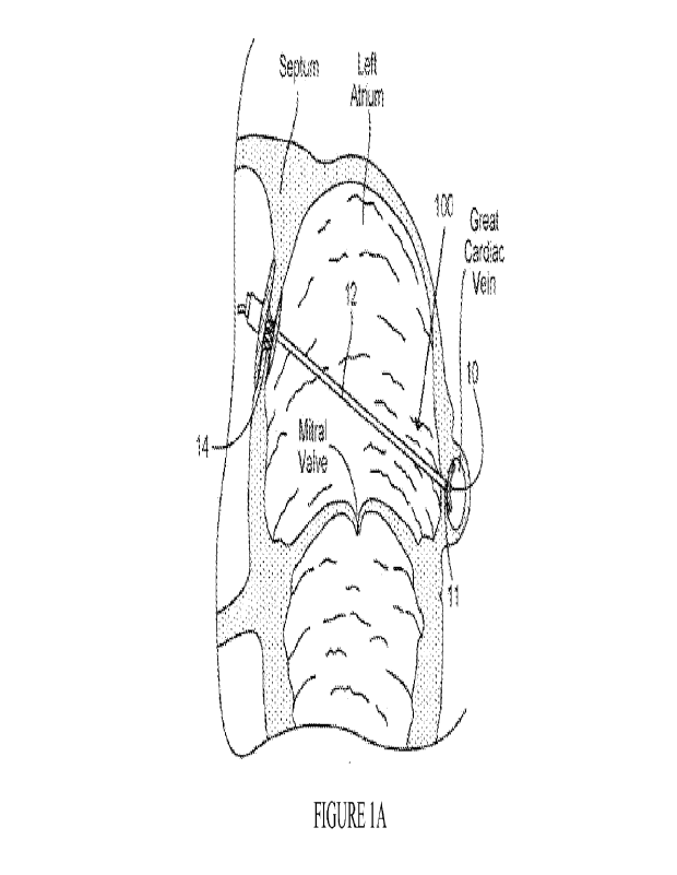
[00026] Figure lA illustrate a heart implant system that includes an inter-
atrial bridging
element that spans the mitral valve annulus between an anterior anchor
disposed in the fossa
8
CA 03201390 2023- 6-6
WO 2022/125535
PCT/US2021/062182
ovalis and a posterior anchor positioned in the GVC in accordance with aspects
of the
invention.
[00027] Figure 1B illustrate a heart implant system that includes an inter-
atrial bridging
element that spans the mitral valve annulus between an anterior anchor
disposed in the fossa
ovalis and a posterior anchor positioned in the GVC in accordance with aspects
of the
invention.
[00028] Figure 2A is an anatomic superior view of a section of the human heart
showing the
tricuspid valve in the right atrium, the mitral valve in the LA, and the
aortic valve in
between, with the tricuspid and mitral valves open and the aortic and
pulmonary valves
closed during ventricular diastole (ventricular filling) of the cardiac cycle.
[00029] Figure 213 illustrates a healthy mitral valve demonstrating full
coaptati on between
leaflets along the entire major axis of the valve.
[00030] Figure 2C is an anatomic anterior perspective view of the left and
right atriums,
with portions broken away and in section to show the interior of the heart
chambers and
associated structures, such as the fossa ovalis, coronary sinus, and the GVC.
[00031] Figure 3 shows a conventional implant system having a bridge spanning
the left
atrium between an anterior anchor disposed in the fossa ovalis and a curved
posterior
anchor disposed in the GCV.
[00032] Figures 4A illustrates the tendency of a conventional curved posterior
anchor to flip
or invert when tension forces are applied.
[00033] Figures 4B illustrates the tendency of a conventional curved posterior
anchor to flip
or invert when tension forces are applied.
[00034] Figure 5 illustrates a posterior anchor with a jacket attached to a
tensioning member
in accordance with some embodiments.
9
CA 03201390 2023- 6-6
WO 2022/125535
PCT/US2021/062182
[00035] Figure 6 illustrates a posterior anchor with a jacket attached to a
tensioning
member, in accordance with some embodiments.
[00036] Figure 7A illustrates a posterior anchor attached to a tensioning
member with an
anti-flipping feature, in accordance with some embodiments.
[00037] Figure 7B illustrates a posterior anchor attached to a tensioning
member with an
anti-flipping feature, in accordance with some embodiments.
[00038] Figure 8 illustrates a posterior anchor attached to a tensioning
member with another
anti-flipping feature, in accordance with some embodiments.
[00039] Figure 9A illustrates a posterior anchor that includes a support
element disposed in
a far side of a compressible cylinder so as to deform the cylinder when
tensioned, in
accordance with some embodiments.
[00040] Figure 9B illustrates the posterior anchor in Figure 9A disposed
within the GCV
before and after deformation, respectively, in accordance with some
embodiments.
[00041] Figure 9C illustrates the posterior anchor in Figure 9A disposed
within the GCV
before and after deformation, respectively, in accordance with some
embodiments.
[00042] Figure 10A illustrates a heart implant system having an anterior
anchor and
multiple bridge elements, each extending to a separate posterior anchor within
the GCV, in
accordance with some embodiments.
[00043] Figure 10B illustrates a heart implant system having an anterior
anchor and
multiple bridge elements extending to a single posterior anchor within the
GCV, in
accordance with some embodiments.
[00044] Figure 10C illustrates a heart implant system for reshaping the
tricuspid valve, the
system having two bridge elements extending from anchors in the superior and
inferior vena
cava to a posterior anchor disposed in the right ventricle, in accordance with
some
embodiments.
CA 03201390 2023- 6-6
WO 2022/125535
PCT/US2021/062182
[00045] Figure 11A illustrates a posterior anchor that is curvable or
conformable upon
adjustment of the tensioning member by use of one or more tethers, in
accordance with
some embodiments
[00046] Figure 11B illustrates a posterior anchor that is curvable or
conformable upon
adjustment of the tensioning member by use of one or more tethers, in
accordance with
some embodiments.
[00047] Figure 11C illustrates a posterior anchor that is curvable or
conformable upon
adjustment of the tensioning member by use of one or more tethers, in
accordance with
some embodiments
[00048] Figure 11D illustrates a posterior anchor that is curvable or
conformable upon
adjustment of the tensioning member by use of one or more tethers, in
accordance with
some embodiments,
[00049] Figure 11E illustrates a posterior anchor that is curvable or
conformable upon
adjustment of the tensioning member by use of one or more tethers, in
accordance with
some embodiments,
[00050] Figure 11F illustrates a posterior anchor that is curvable or
conformable upon
adjustment of the tensioning member by use of one or more tethers, in
accordance with
some embodiments.
[00051] Figure 11G illustrates a posterior anchor that is curvable or
conformable upon
adjustment of the tensioning member by use of one or more tethers, in
accordance with
some embodiments.
[00052] Figure 12A illustrates a posterior anchor defined by an expandable
structure that is
laterally collapsible upon tensioning of a support backbone, in accordance
with some
embodiments.
11
CA 03201390 2023- 6-6
WO 2022/125535
PCT/US2021/062182
[00053] Figure 12B illustrates a posterior anchor defined by an expandable
structure that is
laterally collapsible upon tensioning of a support backbone, in accordance
with some
embodiments.
[00054] Figure 12C illustrates a posterior anchor defined by an expandable
structure that is
laterally collapsible upon tensioning of a support backbone, in accordance
with some
embodiments.
[00055] Figure 13A illustrates an alternative posterior anchor defined by an
expandable
structure having folding zones that facilitate lateral collapse upon
tensioning of a support
backbone, in accordance with some embodiments.
[00056] Figure 13B illustrates an alternative posterior anchor defined by an
expandable
structure having folding zones that facilitate lateral collapse upon
tensioning of a support
backbone, in accordance with some embodiments.
[00057] Figure 14 illustrates an anchor system of the present invention
defined by an
augmentation device having slots to allow engagement with the bridging element
of a
posterior anchor of the present invention, in accordance with some
embodiments.
[00058] Figure 15 illustrates an anchor system of the present invention
defined by an
augmentation device configured to change shape upon deployment and operable to
couple
to a posterior anchor of the present invention, in accordance with some
embodiments.
[00059] Figure 16 illustrates an anchor system of the present invention which
includes an
anterior anchor having a hypotube, in accordance with some embodiments.
[00060] Figure 17 illustrates an anterior anchor of the present invention, in
accordance with
some embodiments.
[00061] Figure 18 illustrates implantation of an anchor system of the present
invention, in
accordance with some embodiments.
12
CA 03201390 2023- 6-6
WO 2022/125535
PCT/US2021/062182
[00062] Figure 19 illustrates an anterior anchor of the present invention, in
accordance with
some embodiments
[00063] Figure 20 illustrates operation of the anterior anchor depicted in
Figure 19, in
accordance with some embodiments.
[00064] Figure 21 illustrates portions of the anterior anchor depicted in
Figure 19, in
accordance with some embodiments.
[00065] Figure 22 illustrates an anchor system of the present invention which
includes an
anterior implant of the present invention and a posterior anchor of the
present invention, in
accordance with some embodiments.
[00066] Figure 23 illustrates aspects of the anchor system depicted in Figure
22, in
accordance with some embodiments.
[00067] Figure 24 illustrates aspects of the anchor system depicted in Figure
22, in
accordance with some embodiments.
[00068] Figure 25 illustrates aspects of the anchor system depicted in Figure
22, in
accordance with some embodiments.
[00069] Figure 26 illustrates aspects of the anchor system depicted in Figure
22, in
accordance with some embodiments.
[00070] Figure 27 illustrates aspects of the anchor system depicted in Figure
22, in
accordance with some embodiments.
[00071] Figure 28 illustrates aspects of the anchor system depicted in Figure
22, in
accordance with some embodiments.
[00072] Figure 29 illustrates an anchor system of the present invention which
includes an
anterior anchor of the present invention and a posterior anchor of the present
invention, in
accordance with some embodiments.
13
CA 03201390 2023- 6-6
WO 2022/125535
PCT/US2021/062182
[00073] Figure 30 illustrates aspects of the anchor system depicted in Figure
29, in
accordance with some embodiments.
[00074] Figure 31 illustrates aspects of the anchor system depicted in Figure
29, in
accordance with some embodiments.
DETAILED DESCRIPTION OF THE INVENTION
[00075] The present invention relates to devices, systems, and methods for
intravascular
anchoring of an implant within the body and/or reshaping an organ within the
body by use
of an anchor deployed within a body lumen or body vessel. Implants described
herein and
associated anchors are directed to improving the function of a heart valve by
reshaping a
mitral valve annulus for treatment of mitral valve regurgitation. It is
appreciated that any
heart implant system can utilize a posterior anchor having any of the features
described
herein, or any combination thereof. Further, although the following
embodiments describe
posterior anchors for use in heart implant systems having a bridging element
that spans the
left atrium between an anterior anchor and the posterior anchor disposed in
the GCV, it is
appreciated that the features described herein pertain to implant systems for
treatment of
any heart valve, or can pertain to any anchor for deployment in a body lumen
and could be
utilized in various other implant systems at other bodily locations in
accordance with the
concepts described herein.
[00076] One important feature of the heart valve treatment systems for
treatment of mitral
valve regurgitation presented herein is the posterior anchor. As shown in the
implant system
100 in Figures 1A-1B, once installed, the posterior anchor 10 is generally
located within the
GVC. It is important for the posterior anchor to spread tensioning forces from
the bridging
element as broadly as possible along the length of the GVC to avoid tearing
the GVC/LA
wall or pulling the posterior anchor through the tissue of the GVC/LA wall and
thus
reducing or eliminating the tension on the bridging element. It is also
helpful to the
treatment of restoring the shape and anatomical distance of the LA from the
septum and the
annulus of the mitral valve that the tensioning on the bridging element pull
much of the LV
wall in the area of the annulus forward toward the septum. If the tension is
instead
14
CA 03201390 2023- 6-6
WO 2022/125535
PCT/US2021/062182
concentrated at a point on the LA wall, this may tend to pull just a limited
point area
forward and not significantly move the entire wall of the LA. The tissue may
pucker or fold
inward rather than pull the full wall of the LA forward.
[00077] Unlike previous GCV device concepts where the device is placed solely
within the
GCV to reshape the left atrium, these systems rely on additional lateral force
applied to the
LA wall that is supplied by, attached to and maintained by an anchor on the
substantially
thicker and robust septal wall to a preferred septal-lateral spacing that is
controlled by the
operator. Although GCV only devices attempt to reshape the path of the GCV
inward, their
ability to move surrounding tissue, including portions of the ventricle, is
severely limited all
applied forces must resolve or balance in the GCV itself_ There is a need for
an anchor for
the GCV that distributes these substantially large forces in a manner that
uniformly moves
the lateral wall to cause the leaflets to co-apt without trauma or erosion,
ideally maintaining
as much of the natural shape, contour, and function of the GCV and the septal-
lateral
spacing with the septum as possible.
[00078] Among the challenges associated with such implant systems is the
difficulty in
providing stable, secure engagement of the posterior anchor along the
posterior wall of the
left atrium while disposed within the GCV. First, since the inside wall of the
GCV along the
left atrium is generally smooth-walled without any notable anatomical
features, the
posterior anchor has a tendency to slide or move, which can lead to
variability of the septal-
lateral spacing provided by the implant system such that some level of mitral
valve
regurgitation may still occur. Furthermore, since the heart is subjected to a
significant
amount of cyclical movement during the cardiac cycle, this sliding movement of
the
posterior anchor over time can lead to erosion of tissues or enlargement of
the penetration
through which the bridging element extends, leading to tearing of the LA wall
along the
GCV. Secondly, in such systems having curved or flexible posterior anchors,
the curvature
of the anchor often does not match the natural curvature of the atrium wall
such that the
posterior anchor fails to consistently engage a large enough portion of the
posterior wall of
the left atrium to ensure a desired reshaping of the annulus is maintained
throughout the
entire cardiac cycle. To address these challenges, presented herein are
anchors having
CA 03201390 2023- 6-6
WO 2022/125535
PCT/US2021/062182
improved design features that provide increased stability and consistency in
anchoring as
well as improved engagement with adjacent tissues, particularly when deployed
in a body
vessel. In one aspect, the anchor has an elongate main body sized and
dimensioned for
delivery and deployment within the vasculature of the patient. For heart
implant systems,
such anchors can have a length dimension between 1 cm and 10 cm, typically
between 2 cm
and 8 cm, so as to distribute laterally applied anchoring forces and engage a
substantial
portion of the heart wall. The anchor can have a width dimension of between
0.5 cm and 5
cm, typically between 1 cm and 3 cm. The anchor can be contoured or curved
along its
length dimension, as well as along a width dimension, so as to conform more
closely to an
anatomy of the body lumen or an adjacent organ. In some embodiments, the
anchor is
specially shaped so as to engage at least a portion of one side of the vessel
in which it is
deployed, while leaving the remainder of the vessel open to facilitate blood
flow
therethrough. Examples of such shapes includes a D or C-shape, as well as an
ovoid shape,
all of which increase the contact area of the posterior anchor along the one
side of the body
vessel, while maintaining patency of the vessel.
[00079] Figures 1A-1B illustrate an example heart valve treatment system 100
that includes
bridging element 12 that spans across the left atrium, extending between
anterior anchor 14
secured in the fossa ovalis and posterior anchor 10 deployed in the GCV. In
this
embodiment, posterior anchor 10 is a cylindrical structure, such as those
detailed in Figure
13A, that is laterally collapsible so as to provide an increased contact
surface area along the
inner wall of the GCV along the wall of the LA when deployed. As can be seen
in Figure
1B, posterior anchor 10 is also curved along its length so as to conform more
closely with
the anatomy of the outside curvature of the LA along which the GCV extends.
Posterior
anchor 10 can further include an anti-flipping feature 11 to inhibit flipping
or inversion
along its length due to movement and forces caused imparted by the structures
of the heart
during the cardiac cycle. While a particular design of posterior anchor is
shown in Figures
1A-1B, it is appreciated that system 100 could utilize any suitable posterior
anchor,
including any of those described herein or any suitable anchor features in
accordance with
the concepts described herein.
16
CA 03201390 2023- 6-6
WO 2022/125535
PCT/US2021/062182
[00080] In some embodiments, the intravascular anchors are defined as an
elongate member
having a central rigid portion along where the tensioning member attaches and
flexible outer
ends. The central rigid portion can include a stress-relief feature such as an
attachment point
that is flexible, movable or pivots to accommodate abrupt movements of the
tensioning
member so as to maintain engagement of the anchor with adjacent tissues during
the heart
cycle. The flexible outer ends can be provided by a modifications to the
central rigid portion
(e.g. notches, kerfs), or can be provided by additional components, such as a
polymer jacket
or cover that fits over the rigid portion.
[00081] In some embodiments, the intravascular anchor is contoured or shaped
to conform
to at least a portion of one side of the vessel in which it is disposed. In
some embodiments,
the intravascular anchor has a fixed shape, while in other embodiments, the
shape of the
anchor is flexible or conformable. In some embodiments, the intravascular
anchor can
assume multiple configurations of varying size and shape to facilitate
delivery and
deployment. In any of the embodiments described herein, the anchor can be
defined with a
hollow lumen therethrough to facilitate intravascular delivery via a guidewire
or catheter.
[00082] These and other aspects of the improved anchor can be further
understood by
referring to the embodiments depicted in Figures 5-13B. While these
embodiments describe
a posterior anchor for use in a tensioned heart implant, it is appreciated
that these anchor
features can apply to various other types of anchors for implants in various
other bodily
locations. For example, any of the features described can be used in an
implant to provide
improved anchoring, which can include improved conformance against anchored
tissues,
improved distribution of forces, and improved engagement of tissues to
facilitate reshaping
of a body organ.
[00083] Figure 5 illustrates a posterior anchor defined as a T-bar 110 that is
jacketed to
provide strain relief and an atraumatic tip configuration. In some
embodiments, a thin or
thick walled polymeric jacket 160 can be fit over a conventional rigid T-bar
anchor to
provide an atraumatic surface. T-bar 110 is coupled with the bridge element
105, which can
be a suture, tether, or any element suitable for spanning across the left
atrium and
17
CA 03201390 2023- 6-6
WO 2022/125535
PCT/US2021/062182
maintaining tension sufficient to reshape the atrium. The jacket 160 is sized
and
dimensioned so that the end portions of the jacket extend beyond the ends of
the rigid T-bar
110. Jacket 160 can be formed of PTFE, high silicone soft-block urethanes,
silicones, or any
suitable material and can further include a thin fabric outer covering, such
as polyester. In
some embodiments, the jacket is preferably formed of a material that
encourages tissue
ingrowth. The jacket may be held in place by adhesive or shrunk over the T-bar
or both. In
this embodiment, jacket 160 is defined as two end pieces abutting the inner
attached central
bridge attachment, although the jacket could be defined a single piece jacket
attached over
an entire length of the T-bar, such as in the next embodiment described below.
The tip
extensions may be shaped to reduce tissue strain, for example curved or
serpentine (not
shown) to increase stability and aid delivery. This approach allows a
conventional T-bar
anchor to be retrofit so as to change a size and/or shape of the anchor,
provide improved or
variable flexibility along its length or provide various other advantageous
characteristics.
[00084] Figure 6 illustrates another posterior anchor configured as a rigid T-
bar backbone
110 covered by a shaped jacket 162. Shaped jacket 162 can be polymeric semi-
rigid or
compliant "surfboard" that fits over the rigid T-bar 110. Such a configuration
is
advantageous as it allows a conventional rigid T-bar anchor to be retrofit to
assume any
shape, contour or flexibility desired for a particular application. In this
embodiment, which
is configured for use in the heart implant system described above, the shaped
jacket 162 is
shaped to be planar or flattened on one side so as to increase tissue contact
area with the
interior wall of the GVC toward the LA and to further distribute anchoring
contact forces.
The planar portion can be flat or curved to accommodate the shape of the
vessel. In this
embodiment, the planar portion is included on a center portion having
increased width than
either end portion and includes an opening near a center of the planar center
portion, which
facilitates engagement of the planar center portion with the wall of the
vessel. This
increased width dimension and planar portion provide improved resistance to
flipping.
Shaped jacket 162 can be formed thin along its posterior/anterior dimension so
that it lies
relatively flat against the GCV wall, thus maximizing blood flow in the GCV.
This
configuration also served to stabilize posterior anchor and resist flipping.
As with other
embodiments, surfaces may be coated or constructed of material that induces
tissue
18
CA 03201390 2023- 6-6
WO 2022/125535
PCT/US2021/062182
ingrowth Shaped jacket can be formed of various polymeric materials, including
PTFE,
high silicone soft-block urethanes, silicones, other implant grade elastomers.
An optional
thin fabric may be employed, such as polyester covering the polymeric jacket,
to promote
tissue growth or inhibit sliding. The size of the device can vary, of course
depending on the
desire of the surgeon and the particular requirements of the patient, for
example a large
male vs. a pediatric patient, but one advantageous size for typical adult
patients would be,
for example, 12F round or oval shaped T-bar. Such a link could be combined as
a
"backbone" to stabilize and strengthen other jacketed or wire form structures
discussed
above. The wire form may be metal, plastic, or any other material that will
allow the rigid
backbone to collapse the form as described above.
[00085] Although a straight version of shaped jacket 162 is shown in Figure 6,
it is
appreciated that shaped jacket 162 could be formed with a predetermined curved
shape
along its length to match the curvature of the mitral annulus or the GCV or
both. Having a
width close to that of the GCV, gaining more purchase of the lateral wall, the
tendency of
the curve to flip or right would be thwarted. In some embodiments, a delivery
catheter used
to deliver the anchor can include mounting features that allow axial rotation
to allow proper
placement of the anchor aligning the curvature with the GCV. Such feature can
include
lumens or guides that or any interfacing feature to allow manipulation of an
orientation of
the anchor during deployment. Shaped jacket can be constructed from a semi-
rigid material
to allow tracking over a guidewire with quasi straightening of its shape and
more significant
bending upon removal of the guidewire and release of the device. One or more
radio-opaque
features can be added to the anchor to allow a clinician to visualize its
position and
orientation during delivery and deployment. While in these embodiments, bridge
element
105 is depicted as a suture that is wound about a mid-portion of the T-bar
110, it is
appreciated that various other bridging elements and suitable means of
attachment (e.g.
adhesive, welding, couplings) could be used.
[00086] While some conventional systems have utilized curved posterior
anchors, such
anchors have a tendency to flip (when of a rigid construction) or invert (when
of a more
flexible construction). This action can be further understood by referring to
the conventional
19
CA 03201390 2023- 6-6
WO 2022/125535
PCT/US2021/062182
heart valve treatment system 1 shown in Figure 3, which includes a bridging
element 2
extending from an anterior anchor 3 to a mid-point of a conventional posterior
anchor 4,
defined as rigid curved tubular member. When a thin curved posterior anchor,
especially a
rigid curved anchor, is placed in the GVC, and tension is applied to the
internal curvature of
the arc, especially near the apex, the forces will have a tendency to flip the
curved anchor in
the GVC and present the exterior edge of the curvature to the passage between
the GVC and
the atrium.
[00087] Figures 4A-4B illustrate this flipping tendency. Flipping the anchor
reaches a more
stable energy condition, and therefore this is the configuration the anchor
will tend to seek.
In considering this flip in configuration, it is important to remember that
the distal anchor,
in place in the GVC, is far from a still curved structure lying against static
curved vein. It is
in place in a vessel full of flowing blood imbedded in the wall of a heart
that is beating
generally as many as 75 times or so a minute. As the posterior anchor is
tossed about and
buffeted by flowing blood, the anchor will quickly seek the most stable
orientation in
relation to the tension forces from the bridging element, and flip into the
orientation with
the apex of the curve pointed toward the tensioning element and the apex being
pulled into
the hole in the GVC/LA wall where the bridging element is pulling it unless
some
mechanisms, for example any of those described herein, are instituted to
prevent flipping
from occurring. When flipped or inverted, the anchor structure tends to focus
the tensioning
forces applied by the bridging element on the GVC/LA wall at a single point,
the point of
puncture between the LA/GVC wall. This increases the likelihood of tearing the
wall and
possibly pulling the posterior anchor into the atrium and releasing the
tension altogether, or
pulling partway into the atrium and relieving the tension to the point that
the therapy is
severely compromised.
[00088] This flipping movement described above would also be considerably less
effective
in pulling the wall of the LA toward the septum to affect reshaping of the
annulus, thus
would be less effective in providing therapy. With only a single point of
contact between
the curved posterior anchor and the GVC inner wall, the posterior anchor would
be more
likely to slide longitudinally within the GVC, whereupon the suture forming
the bridging
CA 03201390 2023- 6-6
WO 2022/125535
PCT/US2021/062182
element would be more likely to slice the tissue forming the GVC/LA wall and
expand the
puncture hole, making it even more likely that the posterior anchor might get
pulled through
into the LA. Therefore, anti-flipping configurations and features can
simultaneously provide
an anti-sliding mechanism which would be doubly advantageous.
[00089] One such anti-flipping anchor configuration is shown in Figures 7A-7B.
This
anchor employs a rigid short link 151 that is attached by a hinge 150 or
similar flexible
attachment mechanism extending from the inside curve of anchor body 152. Link
151 is a
relatively rigid length that can rotate to lay nearly flat against the inside
curve of the curved
anchor body 152 during delivery via a guidewire GW, as shown in Figure 7A, and
opens to
be generally perpendicular to the anchor, as shown in Figure 7B, when deployed
by pulling
the bridging element through a penetration in the wall of the LA. Typically,
in the deployed
configuration, the distal end of link 151 protrudes slightly into the LA in
its resting position.
In some embodiments, the link 151 is hollow such that the flexible bridging
element 105 is
attached to the curved posterior anchor body 152 through the hollow link 151.
In other
embodiments, the bridging element 105 is attached to the end extended away
from anchor
body 152. Link 151 is of sufficient length to cause coaxial alignment with
tensioned
bridging element 105 and prevent anchor from flipping over. Link 151 can be
formed of a
material such as plastic or smooth metal, and have a sufficient diameter that
is less likely
than the bare bridging element, for example a suture, to cut the tissue of the
wall of the
GVC where the penetration is made between the atrium and the GV. The link thus
serves
the double purpose of preventing flipping and protecting the wall of the GVC.
The link is
set to fold flat, pointing towards the puncture site during delivery and
opening
perpendicularly as the suture is tensioned at that site.
[00090] Figure 8 illustrates another anchor embodiment, which includes an anti-
flipping or
anti-flipping feature defined as an inwardly curved portion 153 along where
bridging
element 105 attaches to the anchor body 152. When used within a left atrium
implant for
treatment of MVR, the inwardly curved mid-section projects into the plane of
the generally
GCV shaped curved anchor with the bridge 105 attached at the midsection of the
anti-
21
CA 03201390 2023- 6-6
WO 2022/125535
PCT/US2021/062182
flipping curved portion 153. This allows for a simpler attachment to the
anchor avoiding the
complications of a linking mechanism both in its construction and delivery.
[00091] In another aspect, the posterior anchor can be configured with a
delivery
configuration and deployed configuration in which the anchor is eccentrically
disposed
along one side of a vessel wall. Such configurations can include structures
and materials
that are expandable as well as compressible so as to form an eccentric shape,
which is non-
circular and having a greater surface area on one side, which is to be engaged
against a wall
of the body lumen or vessel. Examples of such configuration are illustrated in
the following
embodiments.
[00092] Figures 9A-9C illustrate a posterior anchor defined as crushable
cylinder 103 with
a more rigid support member 101, such as a T-bar support, attached or embedded
within the
cylinder. While a cylinder is described in this embodiment, it is appreciated
that such an
anchor could be configured in various elongate shapes including but not
limited to partial
cylinder, a crescent, an ovoid or various irregular shapes. Crushable cylinder
can be formed
of any suitable crushable material, such as a foam material or structure.
Typically, rigid
support member 101 is attached or embedded in the outer posterior diameter
furthest from
where the bridging element 105 extends, such as shown in Figure 9A, so as to
facilitate
further crushing of the cylinder when the bridging element is tensioned. The
rigid support
member 101 can be substantially straight, as shown, or can be curved to
generally follow
the curve of the interior wall of the GVC and thus spread the pulling forces
uniformly
against the tissue wall.
[00093] Figures 9B-9C illustrate cross-sections of the posterior anchor of
Figure 9A
disposed in the GVC before and after deployment, respectively. When delivered
into the
GVC, and connected to the bridging element 105, the crushable cylinder 103 is
adjacent the
wall of the GVC and LA, through which the bridging element 105 extends and the
rigid
support element 101 is disposed on the side furthest from the LA, as shown in
Figure 9B.
Upon application of tension on the bridging element to the T-bar 101, the
crushable material
is collapsed into an eccentric shape 103a that has a reduced cross-section
which is less
22
CA 03201390 2023- 6-6
WO 2022/125535
PCT/US2021/062182
obstructive of blood flow within the GVC. The crushed cylinder also assumes a
shape
which both more closely adheres to the inner shape of the GVC, thereby
increasing the
contact surface area as compared to the uncrushed cylinder. When crushed, the
materials
also somewhat compacted and generally stiffer than the uncrushed material
which also
helps spreads the forces applied by the bridging element over the surface area
of the GVC
wall.
[00094] It is appreciated that although the embodiment shown in Figures 9A-9C
are shown
as a relatively short elongated crushable member and T-bar, the T-bar or spine
may be
significantly longer to spread the pulling force and may be shaped with a
curve to spread the
force more generally in the curved shaped GVC.
[00095] In some embodiments, the crushable materially is a material that
encourages tissue
ingrowth and or scarring to create a tissue-anchor matrix This ingrowth
further aids in
assuring that the posterior anchor is not pulled through the GVC wall or
flipped within the
GVC. This crushable material may be constrained by the delivery catheter in a
crushed form
to lower its delivery profile thus aiding delivery, and when released is
further reshaped to its
final dimension by the bridging element.
[00096] Figures 10A-10B illustrate alternative implant systems that can
utilize posterior
anchors in accordance with those described herein. Figure 10A illustrates a
heart implant
system 200 having an anterior anchor and multiple bridge elements 105
extending to
multiple posterior anchors 10 within the GCV. In this embodiment, the
posterior anchor 10
is a collapsible cylindrical structure, such as that described in Figure 13A.
Figure 10B
illustrates a heart implant system 300 having an anterior anchor and multiple
bridge
elements 105 extending to a single posterior anchor 10 deployed within the
GCV. In this
embodiment, posterior anchor 10 is a segmented tube, such as that described in
Figure 11G.
It is appreciated that each of the posterior anchors depicted can utilize any
one or
combination of the anchor features in any of the embodiments described herein.
Figure 10C
illustrates a heart implant system 400 for reshaping the tricuspid valve, the
system having
two bridge elements extending from anchors 40 in the superior and inferior
vena cava to a
23
CA 03201390 2023- 6-6
WO 2022/125535
PCT/US2021/062182
posterior anchor 10 disposed in the right ventricle, in accordance with some
embodiments.
In this embodiment, the posterior anchor 10 is a collapsible cylindrical
structure, such as
that described in Figure 13A.
[00097] In another aspect, curved posterior anchors are provided that can be
transformed
from a substantially linear configuration to a curvilinear configuration. In
some
embodiments, the curve of the anchor can be adjusted during deployment. Some
such
posterior anchors include a series of interfacing or interconnecting
components that
articulate into a curved shape when tensioned, either by the bridging element
or by one or
more tethers extending therethrough. These anchors can be configured for use
with systems
having a single bridging element per anchor, such as that shown in Figure 10A,
or in
systems having multiple bridging elements, such as that shown in Figure 10B.
In some
embodiments, the curveable posterior anchor is defined within a single tube
having a series
of cuts or kerfs that allow for controlled articulation or curvature of the
anchor body by the
tensioned bridge. Adjustment of such anchors can include multiple schemes and
anchor
configurations. Examples of such configurations are detailed further below.
[00098] Figures 11A-11D illustrate a posterior anchor configured that curves
inwardly
toward the bridging element when deployed. Such as configuration can be
designed to
match a curvature of a vessel or an adjacent tissue or organ wall, and further
resists flipping
since the curvature can be maintained by the tensioned bridge element.
Typically, the
posterior anchor is defined so as to match the curvature of the GVC to more
evenly and
securely spread the anchor forces provided by the attachment through the
bridging element
which is tensioned against the anterior anchor.
[00099] The embodiments of Figure 11A-11D can be a segment tube formed from a
single
tube. One way this can be accomplished is to cut a hollow metal or polymeric
tube 130 of a
suitable length (e.g. a length that matches the mitral annulus along the GVC)
into a series of
segments 131,132,133 by a series of cuts called kerfs 140,141,142, as shown in
Figure 11A.
The kerfs can be a depth for example, of 1/2 to 3/4 of the diameter of the
tube, and can also
be angled to facilitate tighter radius of curvature. These areas are open,
meaning that some
24
CA 03201390 2023- 6-6
WO 2022/125535
PCT/US2021/062182
material is cut out of the tube to define a series of segments, which allows
the tube to
preferentially bend in the direction of the kerfs when force is applied to
both ends 130a,
130b.
10001001 One or more tethers can be used to draw segments inward to curve the
anchor. In
some embodiments, the internal tethers 105a, 105b are each fixed internally at
the
respective ends 130a, 130b of the tube and allowed to exit along a center
portion of the
anchor through one of the kerfs or perhaps two of the kerfs 138,139 (for
example, as in
Figures 11A-11B), and a bridging element is attached to the exposed tethers.
Tensioning the
bridging element against the GCV wall simultaneously shortens the minor axis
of the mitral
valve and bends the anchor to the desired shape_ Such a configuration causes
tube 130 to
curve when the bridging element 105 is tensioned. The more tension applied,
the greater the
curvature toward the bridging element, until the kerf openings are closed or
the engaged
tissue exerts an equal countering force on the tubular body 130. This is
particularly
advantageous for use in a dynamic environment, such as the heart, since the
aforementioned
flipping typically occur when the bridging element experiences heightened
tension.
10001011 Figure 11C illustrates a similar embodiment having internal tethers
105a, 105b
that are coupled with ends 130a, 130b and that exit through a central opening
144 and
couple with the bridging element 105. Alternatively, tethers 105a, 105b can be
each
independently fixed to ends 130a, 130b and exit from the center of the anchor
so as to allow
for independent bending of each end. This approach can provide a configuration
that
provides for multiple segments and custom shaped anchor.
10001021 Figure 11D illustrates an alternative embodiment in which the
bridging element
105 is a loop that extends through the tubular body 130 of the anchor such
that, when
tensioned by shortening the loop, the internal tether portion 105c shortens
and tensioned
tether portions 105a, 105b force ends 130a, 130b inward, thereby curving the
anchor body.
The length of the loop can be shortened by pulling one or more free ends of
the loop
through and attaching to the anterior anchor, thereby allowing the user to
adjust the tension
of the bridging elements.
CA 03201390 2023- 6-6
WO 2022/125535
PCT/US2021/062182
[000103] Alternatively, the bending may be independent of the bridging
element. Figure
11E illustrates an example of such a bending scheme using a catheter in the
GCV to pull on
an internal tether 106 fixed internally to the distal end of the anchor though
a lumen of the
catheter. This causes the anchor's proximal end to engage the catheter tip and
bend. A
fastener 107, such as a clip, knot or any suitable mechanism, can be used to
fix the bent
anchor in the desired curved position and the excess tether is cut free.
10001041 It is appreciated that the bent configuration and the force required
to bend the
tube, as well as the stiffness of the bent tube can be varied as desired by
adjusting the
number, width, spacing and depth of the kerfs. The kerfs may be of varied
length along the
anchors length, combining wider and narrower sections to relatively stiffen or
soften
sections respectively. The curving of anchor may be achieved a single shared
connected
bridge or dual independent bridge elements with the latter allowing for more
relaxed curve
one end.
10001051 In another similar approach, the anchor is defined by individual
unconnected
hollow links that are similar or tailored in length. The links are formed so
as to have a
desired stiffness and shape for their resting location when deployed. The
links can be
formed using any of the constructions detailed herein. Such embodiments can
utilize a
delivery scheme having a single bridge with a first bridge end deployment
followed by
loading of the anchor or anchor links to their resting location followed by
deployment of the
second bridge. The tips of the anchor or outer links may have grommets or
other means of
protecting tissue from any abrasion from the bridging element.
[000106] In another aspect, a hybrid concept of a bendable GVC anchor with two
end
bridges is provided. An example of such an embodiment can include a bendable
anchor
resembling a string of segments or interfacing elements that extends between
bridge
elements and attached at each end. In some embodiments, the bridging elements
are
permanently fixed to each end of the anchor. The first bridge is preferably
deployed farthest
from the coronary sinus followed by the second with a spacing between the
punctures equal
to length of the anchor, which would preferably be centered over the larger
central scallop
26
CA 03201390 2023- 6-6
WO 2022/125535
PCT/US2021/062182
leaflet of the mitral valve. The anchor is then deployed by pulling both
bridges and the
anchor through a protective sheath. In some embodiments, the ends of the
individual
segments are angled so that when the entire string is pulled tight and the
ends abut, the
length of the string of segments forms a curved structure. The curved
structure can be
preselected dependent on the angles of the segments, and need not be a
constant curve. For
example, such an anchor could include a relatively straight section at the
center of the
anchor and a more sharply curved section at each end. Alternatively, an anchor
could
include a straight segment and an even more sharply curved segment on the
other end of the
anchor, which may be a useful configuration in some applications.
10001071 Figures 11F and 11G illustrate examples of the above described
alternative
approach for achieving a curved posterior anchor by use of individual links.
The links can
be unconnected with interfacing surfaces between each, or can be
interconnected in a
manner that allows relative movement between adjacent links to allow for
curvature of the
anchor. In these depicted embodiments, tube 131 is formed by a number of
individual
segments 181, 182, which can be shaped with mating surfaces 183 that are
either straight or
angled as desired. In the embodiment of Figure 11F, the end of the anchor tube
may be
protected by grommets 145 connected to bridging elements 105a, 105b. In some
embodiments, the grommets 145 are configured as fixed stops fixing a bridging
element or
tether extending therethrough to a preset length so as to provide a pre-
determined curvature
to the anchor. In the embodiment of Figure 11G, the links of the anchor are
laced over a
single bridging element or tether and are free to move along the bridge such
that shortening
of the bridging element or tether engages opposite ends of the anchor so as to
curve the
anchor. Such a configuration allows links to be added or configured to vary
length or
stiffness along the anchor. In either embodiment, the two bridging elements
105a, 105b may
be attached to the same location on the anterior anchor. Applying tension to
those bridging
elements curves tube 131 inward. When such an anchor is incorporated into a
heart implant
system, the curved tube 131 pulls the entire wall of the LA toward the septum
and
advantageously shapes the mitral valve annulus with the operator able to bias
the length
towards toward one side or the other while viewing the regurgitant flow on
ultrasound in
real time. Although the links or segments are shown here as hollow tubular
segments, it is
27
CA 03201390 2023- 6-6
WO 2022/125535
PCT/US2021/062182
appreciated that the links could be formed in various sizes and shapes,
including shapes
contoured to match a curvature of a vessel or the patient's anatomy. In some
embodiments,
the links are defined as a string of interfacing element such that shortening
of the bridging
element or tether articulates the links into a curved arrangement along the
anchor. The
interfacing elements can be of any suitable construction (e.g. solid, hollow)
and can be of
formed in any shape desired.
[000108] Similar to these examples, in that the configurations requires
multiple bridging
element attachment to the anterior anchor, would be a sequence of posterior
anchors each
separately attached, such as shown in Figure 10A. Such a configuration would
make
possible separate individual attachments that could apply tension at various
angles to
optimally deform the LA wall and mitral valve annulus to reduce mitral
regurgitation. Each
posterior anchor could employ the shapes and features of any of the posterior
anchors
described above. Each could attach to the same location on the anterior
anchor, or could
attach at slightly different locations in the anterior anchor or even separate
anterior anchors
to optimize the angles of tension for maximum effect.
[000109] In another aspect, the posterior anchor can include an expandable
structure that
can be collapsed so as to engage at least a portion of one side of the vessel
in which it is
deployed as well as to assume a reduced profile to allow improve blood flow
therethrough.
Example of such embodiments include a scaffold or wire form structure
configured to be
expanded within the vessel after delivery, then collapsed laterally by
tensioning of the
bridging element. Such embodiments can include a wire form structure having
weakened
portions extending longitudinally on opposite sides of the wire form structure
to facilitate
lateral collapse. The structures can be self-expanding or balloon deployable.
In some
embodiments, the collapsible wire form structure include one or more support
ribs
extending longitudinally to reinforce the collapsed structure to improve
anchoring and
adherence of the structure along a length of the body vessel. Such reinforcing
ribs can be
straight or can be curved as needed for a particular anatomy.
28
CA 03201390 2023- 6-6
WO 2022/125535
PCT/US2021/062182
0 01 1 0] Figures 12A-12C and 13A-13B illustrate examples of the above
described
collapsible wire form cylinder structure 120. Typically, the wire form
structure is a cylinder
mesh structure that may be delivered in low profile and expand to the desired
diameter,
either by self-expansion or balloon expansion. The cylinder mesh structure can
include a
posterior backbone 122 that forms a T-bar and attaches to the bridging element
105.
10001111 As shown in Figure 12A, after deployment of the cylinder mesh
structure 120 in a
vessel, such as the GCV, the bridge element 105 extends to the support
backbone 122
disposed on the opposing side of the cylindrical mesh structure 120 from where
bridge
element 105 extends through the wall of the GCV/LA. When tension is applied by
the
bridging element 105 to the backbone, the support crushes the cylinder mesh
structure wall
upon itself creating a flattened ribbon against the LA/GCV wall. Such a
configuration is
advantageous as it forms a stiff, relatively flat surface that effectively
spreads the force of
the tensioning against the wall to prevent the posterior anchor from being
pulled through the
GVC wall. Further, the folded design doubles the wall thickness and thus its
strength and
increases its purchase of the GCV wall up to 1.5 times its uncrushed diameter.
Such a
configuration allows for improved ease of deployment and allows the anchor to
be
embedded in the wall of the GVC upon deployment. Furthermore, the mesh
structure of the
scaffold further promotes tissue in-growth.
10001121 Figures 13A-13B illustrate another embodiment of a collapsible
scaffold structure
120 that includes folding zones or softer sections 123 to insure preferential
folding along
predetermined lines. These folding zones extend longitudinally along most or
all of the
length of the cylindrical structure and can be defined by scores, weakened
portions, or
previous deformation to facilitate folding of the cylindrical mesh structure
along these areas
when deployed. Also, as with the crushable foam embodiment, the material or
coating of the
wire form structure, and the surface structure of the crushable wire form
structure might be
such that it spurs the ingrowth of tissue to, over time, form a tissue-anchor
matrix. In either
embodiment, the support backbone can be substantially straight, or preferably,
curved to
generally mimic the curve of the interior wall of the GVC. The scaffold can be
a mesh
structure, which can be defined to promote tissue-ingrowth.
29
CA 03201390 2023- 6-6
WO 2022/125535
PCT/US2021/062182
[000113] Figure 14 illustrates an augmentation device 500 which may be used
with a
posterior anchor of the invention that allows for variable loading effect on
the atrial wall by
the posterior anchor. As discussed herein, in various aspects, the invention
provides a
posterior anchor defined as a T-bar anchor 510 in which the bridging element
515 is
attached to the center of the T-bar backbone. In some aspects, the
augmentation device 500
is used with the T-bar anchor 510 of the invention and includes slot features
505 that allow
the loading effect on the atrial wall by the T-bar anchor 510 to be varied in
situ.
[000114] As such, the invention provides an anchor system that includes an
augmentation
device 500 of the invention and an anchor of the invention. In some aspects,
the
augmentation device 500 has an elongated cylindrical body defined by an
elongated lumen
having a substantially cylindrical wall. In some aspects, as shown in Figure
14, the lumen is
configured to receive a T-bar anchor 510 and the cylindrical wall of the
augmentation
device includes slots 505 disposed along a length of the cylindrical body of
the device for
engaging abridging element 515 of the anchor 510. In some aspects, the anchor
510 has a
substantially cylindrical body that is sized to pass within the elongated
cylindrical body of
the augmentation device 500 and abridging element 515 coupled to an
intermediate portion
of the anchor 510. It will be appreciated that the anchor for use with the
augmentation
device 500, may be any T-bar device disclosed herein or any other similarly
shaped anchor
device.
10001151 In practice, the augmentation device is delivered to the GCV to
engage and
augment a T-bar anchor of the invention once the T-bar anchor is delivered to
the GVC. As
shown in Figure 14, the augmentation device 500 is configured to be delivered
in an
orientation such that the device is parallel to the T-bar anchor 510 and then
rotated so that
the augmentation device 500 is sandwiched between the GCV inner wall and the T-
bar
anchor 510, with the bridge element 515 passing through one of the plurality
of slots 505 on
the augmentation device 500. The augmentation device allows compressive forces
of the T-
bar anchor to become spread more evenly over the GCV wall. Further, this
spreading of
forces can be varied and optimized by having different slots along the length
of the
augmentation device which the practitioner may choose to engage the bridging
element.
CA 03201390 2023- 6-6
WO 2022/125535
PCT/US2021/062182
[000116] It will be appreciated that the augmentation device 500 allows
greater flexibility
to the practitioner to modulate the outcome of the procedure
intraprocedurally. Additionally,
since the augmentation device provides a relatively larger area of contact
with the GCV
wall (compared to that of the T-bar anchor), a T-bar anchor of reduced length
may be used
making it easier to deliver and deploy.
10001171 It will also be appreciated that use of a T-bar anchor having a
larger contact
surface area in an effort to spread contact forces and reduce the potential
for cutting and
erosion of tissue has certain limitations. For example, it is typically
difficult to deliver a
wide or large T-bar anchor on the same catheter and at the same time a
penetrating
guidewire is being used to penetrate and cross the atrial wall during a
procedure_ The
augmentation device 500 allows for the surgical step of crossing the atrial
wall to be
separated from the surgical step of deploying a relatively large T-bar anchor.
Additionally,
due to the multiple slots 505 on the augmentation device 500 that engage the
bridging
element 515, the effective attachment point of the bridge element 515 to the T-
bar anchor
510 can be varied intraprocedurally.
10001181 Figure 15 illustrates another configuration of an augmentation device
600 which
may be used with a posterior anchor of the invention. As discussed herein, the
augmentation
device 600 is configured to add additional force vectors to the mechanism of
shortening the
A/13 dimension of a mitral valve by allowing for asymmetric loading of the
posterior anchor
if necessary.
10001191 Further, the device 600 allows the practitioner to more specifically
tailor the
therapy to the patients anatomy by providing a supplemental implant variation
that has
different shapes, sizes or strengths. Accordingly, in one embodiment, the
invention provides
and anchor system that includes an augmentation device 600 of the invention
and an anchor
of the invention, wherein the augmentation device is at least 1.5, 2, 3, 4, 5,
6, 7 or 8 times
the length of the anchor device.
10001201 Figure 15 shows an anchor system which includes an augmentation
device 600
having an elongated shaft body 605 composed of a shape memory material
configured to
31
CA 03201390 2023- 6-6
WO 2022/125535
PCT/US2021/062182
deform from a first elongated configuration to a second flexed configuration.
The second
flexed configuration (shown in Figure 15) has a reduced length as compared to
the first
elongated configuration. The shaft body 605 is configured to conform to an
anatomy of a
patient in the second configuration upon deployment. The system further
includes an anchor
610 having a substantially cylindrical body having a length less than the that
of the
augmentation device 600, and a bridging element 615 coupled to an intermediate
portion of
the anchor 610. In some aspects, the system is configured such that when the
augmentation
device 600 and anchor 610 are coupled upon deployment in a body lumen, a force
upon a
wall of the body lumen from the anchor 610 is translated to the augmentation
device 600 to
deform the wall.
10001211 As discussed herein, in certain aspects, a short T-bar anchor is
utilized as the
anchoring mechanism in the GCV to assist with delivery. Once the short T-bar
anchor is
positioned in the GCV, the augmentation device is positioned adjacent the T-
bar anchor and
coupled to the T-bar anchor. In some aspects, the augmentation device is
composed of a
shape memory material, such as nitinol wire, which allows the device to change
from the
first configuration to the second configuration. In another aspect, the
augmentation device
changes from the first configuration to the second configuration by a
mechanical process,
such as an adjustable linkage between portions of the device. The augmentation
device is
coupled to the T-bar anchor when deployed and applies a different application
of force to
the posterior wall as the anchor alone to reshape the annulus.
10001221 The present invention further provides devices and methods which
allow for
variable adjustment of the bridging element connecting one or more posterior
anchors to
one or more anterior anchors to reshape a body lumen, such as a heart chamber.
As
discussed herein, in certain aspects, the methods and devices are used to
reshape the left
atrium for treatment of a cardiac disease, such as mitral valve regurgitation.
In various
aspects, the devices and methods of the present invention provide a means in
which the left
atrium may be reshaped such that the regurgitation through the mitral valve is
reduced or
inhibited. It will be appreciated that this requires specific positioning of
anchors and
tensioning therebetween.
32
CA 03201390 2023- 6-6
WO 2022/125535
PCT/US2021/062182
[000123] With reference to Figure 16, it is desirable to direct the force
applied to the left
atrium wall from the posterior anchor to a location close to A2 position of
the atrium This
may be achieved in a number of ways as described herein. For example, in one
aspect, the
invention provides an anterior anchor 650 which is modified to include a tube
660
extending from the anchor into the atrial cavity to direct the force acting
between the
posterior anchor and anterior anchor towards an A2 position as shown in Figure
16. In this
configuration, the tube 660 extends from the anchor 650 into the atrial cavity
towards A2
such that the bridge element 670 connected to the posterior anchor 680 applies
force in a
more true AP orientation. During implantation, the anterior anchor 650 is
delivered in a
straight configuration using a core pin which is retracted once the tube 660
is in the left
atrium. The anterior anchor 650 is then rotated to position the tube 660 at
the A2 annulus
and the anterior anchor is deployed and optionally includes anti-rotation
features.
[000124] Figure 17 illustrates an anterior anchor 700 configured to locate the
bridging
element closer to an A2 location with the left atrium. The anchor 700 provides
a means to
reduce the AP dimension of the mitral valve using a posterior implant in the
GCV and an
anterior anchor 700 having a semi-rigid, pre-shaped hollow tube 710, e.g.,
"hypotube",
running through it that repositions the bridging element location closer to
A2. The anchor
700 allows for a more efficient AP diameter shortening by redirecting the
bridging element
to a trajectory that crosses the A2/P2 location of the mitral valve. The tube
710 extending
from the body 705 of the anchor is used to direct the suture closer to A2.
Figure 18 shows
the anchor 700 of Figure 17 in a deployed configuration coupled to a posterior
anchor 720
positioned at P2.
[000125] To achieve a similar outcome, the invention further provides an
anterior anchor
750 having a movable arm 760 coupled to the body 755 of the anchor as shown in
Figure
19. The adjustable arm mechanism is used to change the trajectory of the
bridging element
to provide a practitioner controlled movable linkage system. In practice, the
anchor 750 is
delivered over the crossing wire into the left atrium and the anterior anchor
750 is deployed
as normal. In some aspects, the practitioner can adjust the anngle of the arm
760 as well as
33
CA 03201390 2023- 6-6
WO 2022/125535
PCT/US2021/062182
the length/extension of the arm to determine the best clinical result as shown
in Figure 20.
The arm angle and extension length of the arm are then locked in position.
10001261 For the aspects of the invention depicted in Figures 17, 19 and 20,
the anchor is
delivered in a straight configuration with the anchor collapsed. The anchor is
loaded over
the crossing wire/bridging element and deployed in the left atrium.
10001271 In some aspects, the anchor 700 shown in Figure 17 has a pre-shaped
tubular rigid
member which is used to keep the tube straight for insertion and then removed
upon
deployment to allow the tube 710 to take its heat shaped form thereby pushing
the suture
bridge anterior position closer to A2 In some aspects, the proximal portion of
the pre-
shaped tubing is shaped in a way to move the suture lock position as close to
in-line with
the trajectory of the bridging element crossing the mitral valve. This assists
in balancing the
moment created on the anchor by moving the crossing bridge away from the
posterior
anchor device center as shown in Figure 18.
10001281 For the aspects of the invention depicted in Figures 19 and 20, once
the anchor
750 has been deployed, 2 mandrels actuated by the practitioner at the proximal
end of the
deployment catheter are used to adjust the angle of rotation and the length of
the extension
of the arm 760. The angle is changed by pulling tension on the arm of the
implant through a
rotating hinge 765 as shown in Figure 21. In some aspects, the arm extension
is performed
by using an additional mandrel to push the arm 760 out along a track between
the extension
arm and the rotating arm as shown in Figure 20. Once the desired position has
been
determined both controls can be locked in place by locking the mandrels in
position against
the proximal side of the anchor. The mandrels are then disconnected proximal
to the locking
feature and the anchor 750 is deployed permentantly.
10001291 Figure 22 illustrates additional aspects of the invention which
provide a means to
reduce the AP dimension of the mitral valve using a posterior implant in the
GCV and an
anterior implant (including 2 connected anterior anchors, 800 and 810) that
spans from the
left atrial appendage (LAA) to the fossa ovalus (FO). As shown in Figure 22,
the invention
provides an implant system having a connecting rail 820 with a sliding lock
830 between
34
CA 03201390 2023- 6-6
WO 2022/125535
PCT/US2021/062182
anterior anchors 800 and 810 of the LAA and FO, the connecting rail 820 being
coupled to
the posterior anchor 815 via the suture bridge 840. This sliding lock can be
positioned
across the span from LAA to FO to achieve the most effective reduction of
mitral valve
regurgitation based on anatomy and disease state.
10001301 The aspect of the invention shown in Figure 22 allows the
practitioner to pull the
posterior anchor, e.g., T-bar anchor, from a location closer to A2 as
discussed herein. This
provides a more efficient AP shortening It also allows the practitioner to
tailor the therapy
specific to where the regurgitant jet is present on the valve. Figure 23
illustrates the
differences between an implant system having 3 anchors as opposed to 2
anchors.
10001311 With reference to Figures 22-28, in practice, the implantation
procedure proceeds
as normal up until the wire crossing of the atrial wall is achieved and the
left atrial catheter
has been removed from the sheath_ At this point a posterior anchor positioned
in the GCV
including a coupled bridging element crosses through the atrial wall, into the
catheter (light
blue) and out to the proximal end of the device as shown in Figure 24.
10001321 Over the crossing wire/bridging element, the anterior implant is then
loaded. The
anterior implant includes a first distal anterior anchor (displayed in the
Figures as a nitinol
wire vascular plug), a connecting rail (displayed as nitinol hypotube), a
bridging element
connector (displayed as a sliding lock) and a second distal anterior anchor.
The first and
second distal anterior anchors are connected by the connected rail. The
cossing wire at the
proximal end is backloaded into the sliding lock and through the distal
implant grommet
and the implant is advanced into the left atrium through the sheath.
10001331 The first distal anterior anchor, e.g., LAA anchor is advanced into
the LAA and
deployed there. In one aspect of the invention, the sheath is steerable to
allow for wire
crossing at P2 and to facilitate deployment in the LAA. The second distal
anterior anchor,
e.g., septal anchor, is then advanced and deployed in the septum. It is
envisioned that the the
anterior implant is delivered as a single implant (LAA anchor, sliding lock,
rail and septa]
anchor) or discreet componts that are delivered sequentially. In some aspects,
the LAA
anchor is a nitinol mesh that expands into the LAA or an anchor type device
that deploys
CA 03201390 2023- 6-6
WO 2022/125535
PCT/US2021/062182
into cardiac tissue or the fibrous skeleton of the heart, e.g., the left
fibrous trigone. It will be
appreciated that in some aspects, the delivery catheter is steerable to allow
the first distal
anterior anchor and the second distal anterior anchor to be delivered in the
same catheter.
10001341 In some aspects, the septal implant is then deployed septally and the
bridging
element runs through the septal anchor, e.g., the second anterior anchor. The
practitioner
then begins to apply the therapy using echo doppler to assess the effectivness
of the
syncing. The invention provides two controls: 1) the practitioner can apply
tension to the
suture bridge to reduce the ap dimension; and/or 2) the practitioner can
adjust the location
of the sliding lock to change the angle of which the posterior anchor is being
pulled.
10001351 Once the therapy has been applied, the sliding lock is locked in
position on the
sliding rail using a locking system similar to the suture lock. The suture
will be locked on
the right atrial side of the distal implant, e.g., second anterior anchor. The
procedure then
continues such that the suture lock is deployed and the suture is cut.
10001361 Figures 29-31 illustrate an anterior anchor 900 configured to locate
the bridging
element 950 closer to an A2 location with the left atrium in another aspect of
the invention.
To alter the direction of tensioning between the puncture site of the septal
wall and the
puncture site of the GCV, the anterior anchor 900 includes a left anchor
member 910 and a
right anchor member 920 which are placed independent of one another. This
changes the
angle and direction of the bridging element 950 coupled to the posterior
anchor and
provides a more directed therapy for the patient.
10001371 Unlike a convention anterior anchor having a coaxial through hole,
the anchor 900
shown in Figures 29-31 provides a dual anterior anchor in which the left
anchor member
910 and the right anchor member 920 are positioned separately. Through holes
of each
member are connected by a tubular lumen 930. As shown in the Figures, the
through holes
are not coaxially aligned thereby directing the bridging element 950, e.g.,
suture bridge, to
apply the most effective tensioning therapy.
36
CA 03201390 2023- 6-6
WO 2022/125535
PCT/US2021/062182
[000138] The foregoing is considered as illustrative only of the principles of
the invention.
The embodiments herein disclosed merely exemplify the invention which may be
embodied
in other specific structures. While preferred embodiments have been described,
the details
may be changed without departing from the invention Further, most of the
inventions are
shown in simple forms to illustrate elemental function and features and may be
combined to
a final embodiment that uses one more elements combined into a single device.
It is also
anticipated that the embodiments described may be combined, by way of example
but not
by way of limitation, having a curbed backbone in the crushable foam, or
multiple curved
anchors with anti-flipping features or configurations with multiple
attachments to the
anterior anchor. Furthermore, since numerous modifications and changes will
readily occur
to those skilled in the art, the invention is not limited to the construction
and operation
shown and described in the preferred embodiments except as limited by the
claims.
[000139] Although the invention has been described with reference to the above
examples,
it will be understood that modifications and variations are encompassed within
the spirit and
scope of the invention. Accordingly, the invention is limited only by the
following claims.
37
CA 03201390 2023- 6-6