Note: Descriptions are shown in the official language in which they were submitted.
CA 03201445 2023-05-10
WO 2022/125728
PCT/US2021/062524
1
MULTI-LAYER COLLAGEN-BASED MEMBRANE
Collagen is widely used as a biomaterial in the field of surgery, and there is
a
long history of its use in the specific discipline of tissue regeneration. For
example,
US Patents 5206028 and 5837278 each describe a single layer collagen device
for
tissue regeneration. Collagen devices may be engineered and formed out of
reconstituted collagen. Alternatively, they may be naturally derived, i.e.,
manufactured from tissues harvested in their natural state and processed for
use as a
biomaterial without significant change in the physical dimension of the
tissues. One
disadvantage of collagen devices derived from natural tissues is that the
thickness and
overall size of the final device is dictated by the target tissue. Therefore,
strategies to
modify the thickness and size have been developed. See US Patents 5955110 and
5885619.
Reinforcement is another strategy to modify the physicomechanical
characteristics of collagen devices. By combining a second biomaterial with
collagen,
the strength or handling characteristics of the device can be modified while
maintaining the biological benefits of collagen. For example, US Patent
Application
Publication 2014/0067058 describes layering collagen and a second
biocompatible
mesh by stacking, compressing, and drying.
In clinical tissue regeneration procedures, especially in the maxillofacial
region where there is substantial movement of host tissues during the healing
phase,
stability is required for predicable healing. Delamination of any laminated
device
typically creates dead space within the wound which can contribute to
infection and
failure of the procedure. Delamination can also lead to loss of stability of
the
reinforcing component, leading to compliance issues that could result in
tissue
perforation and damage. Therefore, stability and longevity of the lamination
is of
utmost importance in laminated devices.
There are advantages in using naturally derived collagen membranes in a wide
variety of hard and soft tissue regeneration procedures. The inherent
limitations of
the source tissues however, namely thickness, handling properties, and overall
size,
may require modification to achieve the ideal configuration for clinical use.
It would
be advantageous to have the ability to link several collagen sheets together,
to modify
their overall thickness, or to laminate them with intervening components
between
sheets. Further, the strength of the lamination should be adequate to
withstand
CA 03201445 2023-05-10
WO 2022/125728 PCT/US2021/062524
2
delamination when wet with biological fluids for an adequate amount of time to
achieve clinical success.
The need exists for collagen-based membranes having multiple layers that do
not suffer from the drawbacks mentioned above.
SUMMARY
To meet this need, a multi-layer collagen-based membrane is provided that
includes a bioresorbable mesh embedded between a first decellularized natural
collagen-based membrane and a second decellularized natural collagen-based
membrane. The first and second decellularized natural collagen-based membranes
are
cross-linked to each other and the multi-layer collagen-based membrane has a
peel
strength at 90 of 5-250 N/m.
Also provided is a method for manufacturing a multi-layer collagen-based
membrane. The method is carried out by obtaining a first and second
decellularized
natural collagen-containing membrane, placing the second decellularized
natural
collagen-containing membrane atop the first decellularized natural collagen-
containing membrane to form a membrane assembly, drying the membrane assembly
under a weight distributed uniformly across the membrane assembly, the weight
including openings for allowing moisture to escape, and exposing the membrane
assembly to a cross-linking agent such that cross-links form between layers of
the
membrane assembly. Each of the layers of the multi-layer collagen-based
membrane
is resorbed at essentially the same rate upon implantation in vivo and no
adhesives are
employed in the process.
A second method for manufacturing a multi-layer collagen-based membrane is
also disclosed. This method includes the steps of obtaining a first and second
dried
decellularized natural collagen-containing membrane, obtaining a bioresorbable
synthetic polymer mesh, placing the bioresorbable synthetic polymer mesh atop
the
first dried decellularized natural collagen-containing membrane, hydrating the
first
dried decellularized natural collagen-containing membrane to form a first
hydrated
membrane, placing the second dried decellularized natural collagen-containing
membrane atop the bioresorbable synthetic polymer mesh such that the second
dried
decellularized natural collagen-containing membrane becomes hydrated by
drawing
moisture from the first hydrated membrane, drying the membrane mesh assembly
under a weight distributed uniformly across the membrane mesh assembly, and
CA 03201445 2023-05-10
WO 2022/125728
PCT/US2021/062524
3
exposing the dried membrane mesh assembly to a cross-linking agent such that
cross-
links form between layers of the membrane mesh assembly. This second method,
like
the first method, forms a multi-layer collagen-based membrane in which each of
the
layers is resorbed at essentially the same rate in vivo, the bioresorbable
synthetic
polymer mesh affords a shape memory to the multi-layer collagen-based
membrane,
and no adhesives are employed in the process.
The details of one or more embodiments are set forth in the description and
the
examples below. Other features, objects, and advantages will be apparent from
the
detailed description, from the drawings, and also from the appended claims.
BRIEF DESCRIPTION OF THE DRAWINGS
The description below refers to the accompanying drawings, of which:
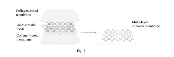
Fig. 1 is a diagram of a multi-layer collagen-based membrane of the invention;
Fig. 2 is a flow chart of a manufacturing process for making the multi-layer
collagen-based membrane;
Fig. 3 shows an exemplary method of preparation and clinical use for the
multi-layer collagen-based membrane of the invention. 1, 3 = collagen-based
membrane; 2 = bioresorbable mesh; 4 = multi-layer collagen membrane assembly;
5 =
ultraviolet cross-linking apparatus; 6 = finished multi-layer collagen-based
membrane; 7 = finished multi-layer collagen-based membrane used to cover a
forearm wound
DETAILED DESCRIPTION
As summarized above, the multi-layer collagen-based membrane of the
invention includes a bioresorbable mesh embedded between a first
decellularized
natural collagen-based membrane and a second decellularized natural collagen-
based
membrane. The bioresorbable mesh, in an exemplary multi-layer collagen-based
membrane, does not extend to the edges of the multi-layer collagen-based
membrane,
leaving a border around the edges that is free of the bioresorbable mesh.
The bioresorbable mesh can be formed of laminar bone that has been
demineralized. The laminar bone can be from a mammal, e.g., human, bovine,
ovine,
equine, and porcine. The demineralized laminar bone is in the form of a mesh
formed, for example, by die-cutting or laser cutting.
CA 03201445 2023-05-10
WO 2022/125728
PCT/US2021/062524
4
In an alternative multi-layer collagen-based membrane, the bioresorbable
mesh is a synthetic polymer mesh that bestows a shape memory on the multi-
layer
collagen-based membrane. The synthetic polymer mesh can be formed of a homo-
polymer including, but not limited to, polylactide ("PLA"), polyglycolide
("PGA"),
polycaprolactone ("PCL"), and trimethylene carbonate ("PTMC"). Alternatively,
the
synthetic polymer mesh can be formed of a co-polymer of monomers included in
the
above-mentioned polymers, e.g., poly(lactic-co-glycolic acid) ("PLGA") and
poly(lactide-co-c-caprolactone) ("PLCL"). In certain embodiments, specific
enantiomers can be used in the homo-polymer or co-polymer. For example,
polymers
such as poly(L-lactide) ("PLLA"), poly(D-lactide)("PDLA"), or poly(DL-
lactide)("PDLLA") can be used in the synthetic polymer mesh.
The synthetic polymer mesh can be manufactured by, e.g., laser cutting, die
cutting, compression molding, 3D printing, and extrusion.
An exemplary multi-layer collagen-based membrane has a synthetic polymer
mesh formed of PLGA having a lactic acid to glycolic acid monomer ratio of
25:75 to
75:25. In a specific multi-layer collagen-based membrane, the lactic acid to
glycolic
acid monomer ratio is 50:50. In another example, the multi-layer collagen-
based
membrane has a synthetic polymer mesh formed of PLCL at a 70:30 ratio of
lactic
acid monomer to caprolactone monomer.
In certain multi-layer collagen-based membranes, the synthetic polymer mesh
also contains a calcium mineral. The calcium mineral can be, but is not
limited to,
calcium phosphate, fl-tricalcium phosphate, calcium sulfate, hydroxyapatite,
and
calcium apatite derived from natural bone mineral. The calcium mineral can
contain
additives such as fluorine (e.g., fluorapatite) and magnesium
In other multi-layer collagen-based membranes, the synthetic polymer mesh
contains a recombinant growth factor, e.g., rhPDGF-BB, rhBMP-2, and FGF.
Alternatively or together, pharmaceuticals such as antibiotics and anti-
inflammatory
agents can be included in the synthetic polymer mesh.
As described above, the multi-layer collagen-based membrane includes a first
decellularized natural collagen-based membrane and a second decellularized
natural
collagen-based membrane. The first decellularized natural collagen-containing
membrane, the second decellularized natural collagen-containing membrane, or
both,
are derived from natural pericardium membranes and have a fibrous side and a
serosal
side. Preferably, the decellularized natural collagen-containing membranes are
CA 03201445 2023-05-10
WO 2022/125728
PCT/US2021/062524
derived from parietal pericardium of a mammal, e.g., human, bovine, ovine,
equine,
and porcine. More preferably, the decellularized natural collagen-containing
membranes are derived from porcine parietal pericardium.
In the multi-layer collagen-based membrane of the invention, the fibrous side
5 of the first decellularized natural collagen-containing membrane can be
in contact
with and cross-linked to (i) the fibrous side of the second decellularized
natural
collagen-containing membrane or (ii) the serosal side of the second
decellularized
natural collagen-containing membrane.
In an alternative multi-layer collagen-based membrane, the serosal side of the
first decellularized natural collagen-containing membrane can be in contact
with and
cross-linked to (i) the fibrous side of the second decellularized natural
collagen-
containing membrane or (ii) the serosal side of the second decellularized
natural
collagen-containing membrane.
The multi-layer collagen-based membrane of the invention can have a dry peel
strength at 90 of 5-250 N/m, e.g., 5-250, 10-250, 20-250, 30-250, 40-250, and
50-
250 N/m. The peel strength is not uniform across the entire multi-layer
collagen-
based membrane. As described above, in certain examples, the bioresorbable
mesh
does not extend to the edges of the multi-layer collagen-based membrane. These
edges, which are free of the bioresorbable mesh, have the strongest dry peel
strength,
i.e., 50-250 N/m, while areas of the multi-layer collagen-based membrane that
include
the bioresorbable mesh have variable peel strengths, e.g., 5-250 N/m,
depending upon
the geometry of the mesh, e.g., mesh size.
Also summarized above are two methods for manufacturing a multi-layer
collagen-based membrane.
The first method is carried out by (i) obtaining a first and a second
decellularized natural collagen-containing membrane, (ii) placing the second
decellularized natural collagen-containing membrane atop the first
decellularized
natural collagen-containing membrane to form a membrane assembly, (iii) drying
the
membrane assembly under a weight distributed uniformly across the membrane
assembly, and (iv) exposing the membrane assembly to a cross-linking agent
such that
cross-links form between layers of the membrane assembly.
The first and second decellularized natural collagen-containing membranes are
derived from natural pericardium membranes and have a fibrous side and a
serosal
side. Preferably, the decellularized natural collagen-containing membranes are
CA 03201445 2023-05-10
WO 2022/125728
PCT/US2021/062524
6
derived from parietal pericardium of a mammal, e.g., human, bovine, ovine,
equine,
and porcine. More preferably, the decellularized natural collagen-containing
membranes are derived from porcine parietal pericardium.
In an exemplary method, the fibrous side of the first decellularized natural
collagen-containing membrane is placed in contact with the fibrous side of the
second
decellularized natural collagen-containing membrane to form a membrane
assembly.
Alternatively, the serosal side of the first decellularized natural collagen-
containing
membrane is placed in contact with the fibrous side of the second
decellularized
natural collagen-containing membrane to form the membrane assembly. In another
example, the serosal side of the first decellularized natural collagen-
containing
membrane is placed in contact with the serosal side of the second
decellularized
natural collagen-containing membrane to form the membrane assembly.
In a particular method, a collagen gel is applied to one or both of the two
decellularized natural collagen-containing membranes before placing them in
contact
with each other. In this method, the decellularized natural collagen-
containing
membranes are first dried briefly to remove excess moisture before application
of the
collagen gel.
The collagen gel can be prepared from human, bovine, ovine, equine, or
porcine pericardium by decellularizing the tissue, followed by hydrolyzing and
micronizing the collagen. The concentration of collagen in the gel can be from
2.5 mg/mL to 10.0 mg/mL. Preferably, the concentration is 10 mg/mL.
Not to be bound by theory, it is believed that a collagen gel aids in assembly
and lamination of decellularized natural collagen-containing membranes by
means of
increasing collagen surface area contact between the membrane layers.
The membrane assembly, after the drying step, is subjected to an exposing
step in which it is exposed to a cross-linking agent such that cross-links
form between
layers of the membrane assembly. The cross-linking agent can be, e.g., a
chemical
cross-linking agent, ultraviolet ("UV") radiation, a cross-linking enzyme, and
plastic
compression.
Chemical cross-linkers that can be used include, but are not limited to,
glutaraldehyde or glutaraldehyde vapor, formaldehyde or formaldehyde vapor,
reducing sugars such as ribose and glucose, genipin, a carbodiimide, e.g., N-
(3-
dimethyl aminopropy1)-N'-ethylcarbodiimide and N-hydroxysuccinimide,
dialdehyde
starch, riboflavin with UVA radiation, an imidoester, e.g., dimethyl
suberimidate,
CA 03201445 2023-05-10
WO 2022/125728
PCT/US2021/062524
7
dimethyl adipimidate, dimethyl primelimidate, and dimethyl
dithiobispropionimidate,
acyl azide, and 4-arm polyethylene glycol succinimidyl glutarate.
Cross-linking can also be carried out enzymatically, for example, using
transglutaminase or lysyl oxidase.
Finally, cross-linking can be carried out in conjunction with plastic
compression where collagen fibers are aligned by applying a physical force to
the
fibers in a single direction prior to being exposed to a cross-linking agent.
When UV radiation is used as the cross-linking agent, the exposing step is
accomplished by irradiating the top side and the bottom side of the dried
membrane
assembly with UV radiation at a total energy level of 1,200 to 216,000 mJ/m2
for 1 to
210 min. In an exemplary method, the UV radiation has an energy level of
12,000 to
48,000 mJ/m2 and the exposure time is 10 to 40 minutes.
In certain methods of the invention in which UV radiation is the cross-linking
agent, no chemical cross-linking agents are employed in the exposing step.
In a particular example, after the exposing step, a step of removing odorant
compounds produced by the UV radiation is included. Odorant compounds that can
be removed are volatile degradation and oxidation bi-products of fatty acids,
amino
acids, and peptides. These compounds can be, but are not limited to, 2-methyl
butanal, 3-methyl butanal, 1-heptene, 1-octene, 1-nonene, hydrogen sulfide,
sulfur
dioxide, mercaptomethane, dimethyl sulfide, methyl thioacetate, dimethyl
disulfide,
and dimethyl trisulfide.
The removing step can be accomplished, e.g., by rinsing the membrane
assembly with H20 and/or shaking the membrane assembly in an H20 bath one or
more times, e.g., once, twice, three, and four times. Prior to rinsing with
H20, the
membrane assembly can be rinsed with a buffer, for example phosphate buffered
saline ("PBS").
The method can also include a final drying step. The drying can be
accomplished by air drying or by drying under vacuum. The drying can be done
at
5 C to 45 C, preferably at room temperature, for 60 min. to 300 min. If drying
under
.. vacuum, the vacuum should be 50 mTorr to 500 mTorr.
In certain embodiments, the method also includes a step of placing a
bioresorbable mesh onto the first decellularized natural collagen-containing
membrane before placing the second decellularized natural collagen-containing
membrane atop the first decellularized natural collagen-containing membrane.
CA 03201445 2023-05-10
WO 2022/125728
PCT/US2021/062524
8
The bioresorbable mesh has been described above in detail. It can be a
synthetic polymer mesh formed of, e.g., PLA, PGA, PCL, PTMC, PLLA, PDLA,
PDLLA, PLGA, PLCL or a mixture of these polymers having the monomer ratios set
forth, supra.
An additional step of adding a calcium-mineral, e.g., calcium phosphate,
calcium sulfate, and hydroxyapatite, to the polymers can be part of the
method. The
calcium-mineral can be added, e.g., by soaking the polymers in a calcium-
mineral
solution.
Alternatively, the bioresorbable mesh can be formed of demineralized laminar
.. bone as described above.
A second method for manufacturing a multi-layer collagen-based membrane is
also summarized above. This process is carried out by (i) obtaining a first
dried
decellularized natural collagen-containing membrane, (ii) obtaining a
bioresorbable
synthetic polymer mesh, (iii) placing the bioresorbable synthetic polymer mesh
atop
the first dried decellularized natural collagen-containing membrane, (iv)
hydrating the
first dried decellularized natural collagen-containing membrane to form a
first
hydrated membrane, (v) obtaining a second dried decellularized natural
collagen-
containing membrane, (vi) placing the second dried decellularized natural
collagen-
containing membrane atop the bioresorbable synthetic polymer mesh such that
the
second dried decellularized natural collagen-containing membrane becomes
hydrated
by drawing moisture from the first hydrated membrane, (vii) drying the
membrane
mesh assembly under a weight distributed uniformly across the membrane mesh
assembly, and (viii) exposing the dried membrane mesh assembly to a cross-
linking
agent such that cross-links form between layers of the membrane mesh assembly.
This second method, like the first method, forms a multi-layer collagen-based
membrane in which each of the layers is resorbed at essentially the same rate
in vivo,
the bioresorbable synthetic polymer mesh affords a shape memory to the multi-
layer
collagen-based membrane, and no adhesives are employed in the process.
The first and second decellularized natural collagen-containing membranes are
as described above for the first method, as is the synthetic bioresorbable
polymer
mesh. The second method, also like the first method, can employ an exposing
step in
which the dried membrane mesh assembly is exposed to UV radiation at the
intensities and times set out above. The membrane mesh assembly formed by the
second method can also be subjected to removing and drying steps included in
the
CA 03201445 2023-05-10
WO 2022/125728 PCT/US2021/062524
9
first method. In a particular example of the second method in which UV
radiation is
the cross-linking agent, no chemical cross-linking agents are employed in the
exposing step.
The hydrating step can be carried out by applying H20 onto the first dried
decellularized natural collagen-containing membrane.
As an alternative, hydration can be accomplished by applying to the first
dried
decellularized natural collagen-containing membrane the collagen gel described
above. Again, the collagen gel, prepared from human, bovine, ovine, equine, or
porcine pericardium, can have a collagen concentration of 2.5 mg/mL to 10.0
mg/mL.
The instant invention encompasses variations of the above two methods for
manufacturing a multi-layer collagen-based membrane in which cross-linking is
achieved by means in addition to or other than exposure to a chemical cross-
linking
agent, to UV radiation, or to a cross-linking enzyme. For example, the drying
step in
the first and second methods can be carried out such that dehydrothermal cross-
.. linking occurs between collagen-containing membranes in the membrane mesh
assembly. In certain methods, dehydrothermal cross-linking is employed in the
absence of exposure to UV radiation.
Without further elaboration, it is believed that one skilled in the art can,
based on the disclosure herein, utilize the present disclosure to its fullest
extent.
The following specific examples are, therefore, to be construed as merely
descriptive, and not limitative of the remainder of the disclosure in any way
whatsoever. All publications and patent documents cited herein are
incorporated by
reference in their entirety.
EXAMPLES
Example 1: Process for manufacturing a multi-layer collagen-based membrane
Layer assembly
A resorbable polymer mesh with a thickness of 0.22 mm (0.0085 in.) formed
of co-polymer PLGA (monomer ratio of lactic acid to glycolic acid of 50:50 or
70:30)
.. was laid atop one lyophilized porcine pericardium membrane. A sufficient
quantity
of reverse-osmosis deionized H20 was applied to the membrane until it became
clear.
A second lyophilized porcine pericardium membrane was placed with its fibrous
side
on top of the fibrous side of the first hydrated porcine pericardium membrane
so that
the second membrane pulled H20 from the first membrane to become hydrated.
CA 03201445 2023-05-10
WO 2022/125728
PCT/US2021/062524
Additional H20 was added to any remaining white areas that were not
sufficiently
hydrated.
Starting from the middle of the membrane, pressure was applied to remove
excess H20 from both membranes. As excess H20 was removed, the membranes
5 __ suctioned together firmly.
Care was taken to avoid excessively wetting the membranes to the point that
H20 pooled around them. The integrity of the interface between the membranes
has
an effect on the clarity and visual uniformity of the finished multi-layer
collagen-
based membrane. Areas with excess H20 between the membranes may not fully dry
10 as the H20 evaporates away. These areas may appear white or hazy upon
drying. Of
note, the amount of pressure applied when pressing H20 out of the membranes
can
have an effect on the dried thickness of the device.
The assembled pericardium layers were left to dry under a uniform flat weight.
The weight contained holes in the form of a grate to allow the assembled
pericardium
layers to dry quickly. The assembled pericardium layers have a propensity to
curl or
wrinkle when dried in open air. Drying under a weighted grate allows the
membranes
to dry flat and helps keep the membrane sheets in contact.
It was also found that the layered membranes will maintain some degree of
memory of the shape it was dried in. Additionally, it was found that drying
under a
weighted grate was unexpectedly superior to drying by pressing the membranes
with a
silicone matting material for up to 24 h, a process that did not allow the
pericardium
layers to dry sufficiently to reduce bioburden upon implantation to an
acceptable
level.
Crosslinking
The dried assembled membranes were placed in an ultraviolet light chamber to
be crosslinked. Crosslinking of the assembled membranes is essential to
prevent
delamination once the multi-layer collagen-based membrane comes into contact
with
H20 during use.
The membrane assembly was placed 6 inches from a 75 watt bulb source of
254 nm light, i.e., UV radiation, for 15 min. The membrane assembly was then
flipped over and exposed for an additional 15 min. to the same level of UV
radiation
on the other side. This exposure duration delivers a functional amount of
energy at
the membrane surface of approximately 14,000-22,000 mJ/cm2.
CA 03201445 2023-05-10
WO 2022/125728 PCT/US2021/062524
11
Not to be bound by theory, it is believed that the UV radiation penetrates
into
the interior of the membrane assembly. Flipping the membrane assembly is
performed to make the crosslinking process as uniform as possible.
It should be noted that adequate crosslinking is attainable at treatment times
less than 15 min. per side. Under the above conditions this treatment time
provides
the maximum amount of UV exposure that promotes crosslinking while minimizing
degradation.
Importantly, UV radiation was used for crosslinking instead of more common
methods such as chemical and dehydrothermal crosslinking. UV radiation is
advantageous as it avoids contamination with residual chemical crosslinkers
and also
avoids denaturation seen in dehydrothermal crosslinking. Moreover, UV
radiation is
a novel method to control and/or extend the resorption time of collagen based
membranes.
Of note, a dedicated regulated 110V power supply between the power source
and the crosslinker cros slinking unit likely results in a more uniform
repeatable output
from the UV-bulbs. This is due to regulation of variations in the power
supplied from
the electrical grid.
Removal of Unwanted Odor
UV radiation in the crosslinking process liberates compounds in the
pericardium that have a strong off-putting caprylic acid-like odor. These
compounds
are polar and can be removed with multiple successive washes with H20. Fresh
H20
was run over the membrane assembly for 30 s, after which it was placed in a
tray with
1 L of H20 and shaken on an orbital shaker for 15-20 min. The membrane
assembly
was washed again with fresh H20 for 30 s.
The washed membrane assembly was placed on a clean silicone surface and
the edges tacked down with a sufficient number of clean stainless steel tacks
such that
the membrane assembly was taught and flat. The membrane assembly was left to
air
dry completely.
Vacuum Drying
The membrane assembly was placed in a vacuum dryer and dried at 18 C for
300 min. at 50 mTorr.
It is known that moisture can contribute to the degradation of the PLGA co-
polymer frame. This drying step preserves the shelf-life of the PLGA frame, as
well
as minimizes the amount H20 in the device for the purpose of lowering
bioburden.
CA 03201445 2023-05-10
WO 2022/125728 PCT/US2021/062524
12
Die Cutting
The multi-layer collagen-based membrane was assembled with pericardium
layers slightly larger than the desired dimensions of the finished product. By
die-
cutting, the polymer mesh can be centered in the finished product by choosing
where
the die is placed. The cutting edge of the die should be mounted on a clear
surface so
that the polymer mesh in the device can be seen during this process.
Die cutting the product at this stage also gives the multi-layer collagen-
based
membrane a clean neat straight edge, as the edges of original pericardium
cannot be
perfectly aligned during assembly.
When die cutting the multi-layer collagen-based membrane, it should be
flipped in an orientation where the die is pressed in the direction opposite
of any
natural curl in the membranes. This is done to counteract the curl and give
the multi-
layer collagen-based membrane as flat an appearance as possible.
Sterilization
The multi-layer collagen-based membrane was sterilized by ethylene oxide
("EO").
The sterilization cycle should operate with the minimal amount of heat and
moisture required to sterilize the multi-layer collagen-based membrane for the
following reasons. First, moisture from the EO cycle will likely remain in the
polymer mesh thereby shortening the shelf-life. Second, heat degrades the
polymer
mesh, also shortening the shelf-life. Third, excessive heat can melt and
possibly
deform or change the structural integrity of the polymer mesh. Finally,
excessive
moisture can cause the collagen in the multi-layer collagen-based membrane to
wrinkle.
The multi-layer collagen-based membrane should not be sterilized by E-Beam.
Radiation of this nature has been shown to make the polymer mesh brittle.
Example 2: Preparation of collagen gel
Porcine pericardium was decellularized by standard techniques to prepare
purified collagen. The collagen was micronized by cryogenic and cyclone
milling,
then digested in citric acid at pH 2.0-3.2. Gels were kept chilled to minimize
denaturation.
For assembling multi-layer collagen-based membranes, the pH of the collagen
gel was normalized back to a range of 6.8-7.2 with sodium hydroxide before
using it
CA 03201445 2023-05-10
WO 2022/125728
PCT/US2021/062524
13
to hydrate the membranes. If needed, phosphate sodium monobasic and sodium
chloride was added.
OTHER EMBODIMENTS
All of the features disclosed in this specification may be combined in any
combination. Each feature disclosed in this specification may be replaced by
an
alternative feature serving the same, equivalent, or similar purpose. Thus,
unless
expressly stated otherwise, each feature disclosed is only an example of a
generic
series of equivalent or similar features.
From the above description, one skilled in the art can easily ascertain the
essential characteristics of the present invention, and without departing from
the spirit
and scope thereof, can make various changes and modifications of the invention
to
adapt it to various usages and conditions. Thus, other embodiments are also
within
the scope of the following claims.