Note: Descriptions are shown in the official language in which they were submitted.
WO 2022/132181
PCT/US2020/066213
METHODS AND APPARATUS FOR RADIOABLATION TREATMENT
FIELD
[0001] Aspects of the present disclosure relate in general to
medical diagnostic and
treatment systems and, more particularly, to providing radioablation
diagnostic, treatment
planning, and delivery systems for diagnosis and treatment of conditions, such
as cardiac
arrhythmias.
BACKGROUND
[0002] Various technologies can be employed to capture or image a
patient's metabolic,
electrical and anatomical information. For example, positron emission
tomography (PET) is a
metabolic imaging technology that produces tomographic images representing the
distribution of
positron emitting isotopes within a body. Computed Tomography (CT) and
Magnetic Resonance
Imaging (MRI) are anatomical imaging technologies that create images using x-
rays and
magnetic fields respectively. Images from these exemplary technologies can be
combined with
one another to generate composite anatomical and functional images. For
example, software
systems, such as VelocityTM software from Varian Medical Systems, Inc. combine
different types
of images using an image fusion process to deform and/or register images to
produce a combined
image.
[0003] In cardiac radioablation, medical professionals work
together to diagnose cardiac
arrhythmias, identify regions for ablation, prescribe radiation treatment, and
create radioablation
treatment plans. Typically, each of the various medical professionals have
complementary
medical training and thus specialize in varying aspects of the treatment
development. For
example, an electrophysiologist may identify one or more regions or targets of
a patient's heart
for treatment of cardiac arrhythmias based on a patient's anatomy and
electrophysiology. The
electrophysiologist may use, for example, combined PET and cardiac CT images
as inputs to
manually define a target region for ablation. Once a target region is defined
by the
electrophysiologist, a radiation oncologist may prescribe radiation treatment
including_ for
example, the number of fractions of radiation to be delivered, radiation dose
to be delivered to a
target region and maximum dose to adjacent organs at risk. Once a radiation
dose is prescribed,
1
CA 03201917 2023- 6-9
WO 2022/132181
PCT/US2020/066213
typically a dosimetrist may create a radioablation treatment plan based on the
prescribed
radiation therapy. The radiation oncologist then typically reviews and
approves the treatment
plan to be delivered. In addition, and prior to finalization of the
radioablation treatment plan, the
electrophysiologist may want to understand the location, size, and shape of a
dose region of the
defined target volume to confirm the target location for the patient as
defined by the
radioablation treatment plan is correct.
[0004] Properly identifying and defining the target region of a
patient's organ for
treatment is essential for developing and optimizing the treatment plan. For
example, an over-
inclusive target region may result in a defined target volume that includes
areas that do not
require treatment, while an under-inclusive target region may result in a
defined target volume
that fails to include areas that should be treated. As such, there are
opportunities to improve
radioablation treatment planning systems used by medical professionals, such
as cardiac
radioablation treatment systems used for cardiac radioablation diagnosis and
radiation treatment
planning.
SUMMARY
[0005] Systems and methods for cardiac radioablation diagnosis
treatment and planning
are disclosed. In some examples, a computing device provides for display a
user interface that
allows a medical professional to define a target region of a patient for
treatment. The user
interface may allow the medical professional to select a treatment area using
interactive target
maps generated for the patient. The computing device also receives image data
from an imaging
system for the patient, such as image data identifying a 3D volume of the
patient's scanned
structure. The computing device may generate for display a 3D image of the
scanned structure
based on the received image data, and may superimpose on the 3D image a target
region map
that the medical professional can manipulate to define the target region of
treatment for the
patient. Once defined, the computing device may transmit the defined target
region to a
treatment system for treating the patient.
[0006] In some examples, a system includes a computing device
that is configured to
receive a first input identifying a treatment target area of an organ of a
patient, and receive a
scanned image of the organ. The computing device is also configured to
generate a first digital
2
CA 03201917 2023- 6-9
WO 2022/132181
PCT/US2020/066213
model of a type of the organ. Further, the computing device is configured to
determine an
alignment of the scanned image to the first digital model. The computing
device is also
configured to generate a second digital model comprising at least a portion of
the scanned image
and the first digital model. The computing device is further configured to
store the second
digital model in a data repository.
[0007] In some examples, a computer-implemented method includes
receiving a first
input identifying a treatment target area of an organ of a patient, and
receiving a scanned image
of the organ. The method also includes generating a first digital model of a
type of the organ.
Further, the method includes determining an alignment of the scanned image to
the first digital
model. The method also includes generating a second digital model comprising
at least a portion
of the scanned image and the first digital model. The method further includes
storing the second
digital model in a data repository.
[0008] In some examples, a non-transitory computer readable
medium storing
instructions that, when executed by at least one processor, cause the at least
one processor to
perform operations including receiving a first input identifying a treatment
target area of an
organ of a patient, and receiving a scanned image of the organ. The operations
also include
generating a first digital model of a type of the organ. Further, the
operations include
determining an alignment of the scanned image to the first digital model. The
operations also
include generating a second digital model comprising at least a portion of the
scanned image and
the first digital model. The operations further include storing the second
digital model in a data
repository.
[0009] In some examples, a method includes a means for receiving
a first input
identifying a treatment target area of an organ of a patient, and receiving a
scanned image of the
organ. The method also includes a means for generating a first digital model
of a type of the
organ. Further, the method includes a means for determining an alignment of
the scanned image
to the first digital model. The method also includes a means for generating a
second digital
model comprising at least a portion of the scanned image and the first digital
model. The method
further includes a means for storing the second digital model in a data
repository.
3
CA 03201917 2023- 6-9
WO 2022/132181
PCT/US2020/066213
BRIEF DESCRIPTION OF THE DRAWINGS
[0010] The features and advantages of the present disclosures
will be more fully
disclosed in, or rendered obvious by the following detailed descriptions of
example
embodiments. The detailed descriptions of the example embodiments are to be
considered
together with the accompanying drawings wherein like numbers refer to like
parts and further
wherein.
[0011] FIG. 1 illustrates a cardiac radioablation diagnosis and
treatment system, in
accordance with some embodiments;
[0012] FIG. 2 illustrates a block diagram of a target definition
computing device, in
accordance with some embodiments;
[0013] FIG. 3 illustrates exemplary portions of the cardiac
radioablation treatment
system of FIG. 1, in accordance with some embodiments;
[0014] FIGs. 4A, 4B, 4C, 4D, 4E, and 4F illustrate portions of a
graphical user interface,
in accordance with some embodiments;
[0015] FIGs. 5A and 5B illustrate portions of a graphical user
interface, in accordance
with some embodiments;
[0016] FIGs. 6A, 6B, 6C, 6D, 6E, and 6F illustrate portions of a
graphical user interface,
in accordance with some embodiments;
[0017] FIG. 7A illustrates a 2-dimensional segment model, in
accordance with some
embodiments;
[0018] FIG. 7B illustrates a 3-dimensional segment model, in
accordance with some
embodiments;
[0019] FIG. 7C illustrates a 3-dimensional segment model with a
septum border, in
accordance with some embodiments;
4
CA 03201917 2023- 6-9
WO 2022/132181
PCT/US2020/066213
[0020] FIG. 8 illustrates editing options for the 2-dimensional
segment model of FIG.
7A, in accordance with some embodiments;
[0021] FIG. 9 illustrates editing options for the 3-dimensional
segment model of FIG.
7B, in accordance with some embodiments;
[0022] FIG. 10A illustrates the selection of a segment within a
segment model, in
accordance with some embodiments;
[0023] FIG. 10B illustrates a 3-dimensional segment model
identifying a selected
segment, in accordance with some embodiments;
[0024] FIG. 11 is a flowchart of an example method to generate a
study for a patient, in
accordance with some embodiments;
[0025] FIG. 12 is a flowchart of an example method to generate an
interactive map for
identifying a treatment target area, in accordance with some embodiments;
[0026] FIG. 13A is a flowchart of an example method to generate a
digital model, in
accordance with some embodiments; and
[0027] FIG. 13B is a flowchart of an example method to adjust an
orientation of the
digital model of FIG. 13A, in accordance with some embodiments.
DETAILED DESCRIPTION
[0028] The description of the preferred embodiments is intended
to be read in connection
with the accompanying drawings, which are to be considered part of the entire
written
description of these disclosures. While the present disclosure is susceptible
to various
modifications and alternative forms, specific embodiments are shown by way of
example in the
drawings and will be described in detail herein. The objectives and advantages
of the claimed
subject matter will become more apparent from the following detailed
description of these
exemplary embodiments in connection with the accompanying drawings.
[0029] It should be understood, however, that the present
disclosure is not intended to be
limited to the particular forms disclosed. Rather, the present disclosure
covers all modifications,
CA 03201917 2023- 6-9
WO 2022/132181
PCT/US2020/066213
equivalents, and alternatives that fall within the spirit and scope of these
exemplary
embodiments. The terms "couple," "coupled," "operatively coupled,"
"operatively connected,"
and the like should be broadly understood to refer to connecting devices or
components together
either mechanically, electrically, wired, wirelessly, or otherwise, such that
the connection allows
the pertinent devices or components to operate (e.g., communicate) with each
other as intended
by virtue of that relationship.
[0030] Turning to the drawings, FIG. 1 illustrates a block
diagram of a cardiac
radioablation diagnosis and treatment system 100 that includes an imaging
device 102, a
treatment planning computing device 106, one or more target definition
computing devices 104,
and a database 116 communicatively coupled over communication network 118.
Imaging device
102 may be, for example, a CT scanner, an MR scanner, a PET scanner, an
electrophysiologic
imaging device, an ECG, or an ECG imager. In some examples, imaging device 102
may be
PET/CT scanner or a PET/MR scanner. In some examples, imaging device 102 and
treatment
planning computing device 106 may be part of a radioablation treatment system
126 that allows
for radioabaltion treatment to a patient. For example, radioablation treatment
system 126 may
allow for the delivery of defined doses to one or more treatment areas of the
patient.
[0031] Each target definition computing device 104 and treatment
planning computing
device 106 can be any suitable computing device that includes any suitable
hardware or
hardware and software combination for processing data. For example, each can
include one or
more processors, one or more field-programmable gate arrays (FPGAs), one or
more application-
specific integrated circuits (ASICs), one or more state machines, digital
circuitry, or any other
suitable circuitry. In addition, each can transmit data to, and receive data
from, communication
network 118. For example, each of target definition computing device 104 and
treatment
planning computing device 106 can be a server such as a cloud-based server, a
computer, a
laptop, a mobile device, a workstation, or any other suitable computing
device.
100321 For example, FIG. 2 illustrates a computing device 200,
which may be an example
of each of target definition computing device 104 and treatment planning
computing device 106.
Computing device 200 includes one or more processors 201, working memory 202,
one or more
input/output devices 203, instruction memory 207, a transceiver 204, one or
more communication
6
CA 03201917 2023- 6-9
WO 2022/132181
PCT/US2020/066213
ports 207, and a display 206, all operatively coupled to one or more data
buses 208. Data buses
208 allow for communication among the various devices. Data buses 208 can
include wired, or
wireless, communication channels.
[0033] Processors 201 can include one or more distinct
processors, each having one or
more cores. Each of the distinct processors can have the same or different
structure. Processors
201 can include one or more central processing units (CPUs), one or more
graphics processing
units (GPUs), application specific integrated circuits (ASICs), digital signal
processors (DSPs),
and the like.
[0034] Instruction memory 207 can store instructions that can be
accessed (e.g., read) and
executed by processors 201. For example, instruction memory 207 can be a non-
transitory,
computer-readable storage medium such as a read-only memory (ROM), an
electrically erasable
programmable read-only memory (EEPROM), flash memory, a removable disk, CD-
ROM, any
non-volatile memory, or any other suitable memory. Processors 201 can be
configured to perform
a certain function or operation by executing code, stored on instruction
memory 207, embodying
the function or operation. For example, processors 201 can be configured to
execute code stored
in instruction memory 207 to perform one or more of any function, method, or
operation disclosed
herein.
[0035] Additionally processors 201 can store data to, and read
data from, working memory
202. For example, processors 201 can store a working set of instructions to
working memory 202,
such as instructions loaded from instruction memory 207. Processors 201 can
also use working
memory 202 to store dynamic data created during the operation of radioablation
diagnosis and
treatment planning computing device 200. Working memory 202 can be a random
access memory
(RAM) such as a static random access memory (SRAM) or dynamic random access
memory
(DRAM), or any other suitable memory.
[0036] Input-output devices 203 can include any suitable device
that allows for data input
or output. For example, input-output devices 203 can include one or more of a
keyboard, a
touchpad, a mouse, a stylus, a touchscreen, a physical button, a speaker, a
microphone, or any
other suitable input or output device.
7
CA 03201917 2023- 6-9
WO 2022/132181
PCT/US2020/066213
[0037] Communication port(s) 209 can include, for example, a
serial port such as a
universal asynchronous receiver/transmitter (UART) connection, a Universal
Serial Bus (USB)
connection, or any other suitable communication port or connection. In some
examples,
communication port(s) 209 allows for the programming of executable
instructions in instruction
memory 207. In some examples, communication port(s) 209 allow for the transfer
(e.g., uploading
or downloading) of data, such as image data.
[0038] Display 206 can be any suitable display, such as a 3D
viewer or a monitor. Display
206 can display user interface 205. User interfaces 205 can enable user
interaction with computing
device 200. For example, user interface 205 can be a user interface for an
application that allows
a user (e.g., a medical professional) to view or manipulate models to define a
target region of
treatment for a patient as described herein. In some examples, the user can
interact with user
interface 205 by engaging input-output devices 203. In some examples, display
206 can be a
touchscreen, where user interface 205 is displayed on the touchscreen. In some
examples, display
206 displays images of scanned image data (e.g., image slices).
[0039] Transceiver 204 allows for communication with a network,
such as the
communication network 118 of FIG. 1. For example, if communication network 118
of FIG. 1 is
a cellular network, transceiver 204 is configured to allow communications with
the cellular
network. In some examples, transceiver 204 is selected based on the type of
communication
network 118 radioablation diagnosis and treatment planning computing device
200 will be
operating in. Processor(s) 201 is operable to receive data from, or send data
to, a network, such
as communication network 118 of FIG. 1, via transceiver 204
[0040] Referring back to FIG. 1, database 116 can be a remote
storage device (e.g.,
including non-volatile memory), such as a cloud-based server, a disk (e.g., a
hard disk), a memory
device on another application server, a networked computer, or any other
suitable remote storage.
In some examples, database 116 can be a local storage device, such as a hard
drive, a non-volatile
memory, or a USB stick, to one or more of target definition computing device
104 and treatment
planning computing device 106.
[0041] Communication network 118 can be a WiFi network, a
cellular network such as a
3GPP network, a Bluetooth network, a satellite network, a wireless local
area network (LAN),
8
CA 03201917 2023- 6-9
WO 2022/132181
PCT/US2020/066213
a network utilizing radio-frequency (RF) communication protocols, a Near Field
Communication
(NFC) network, a wireless Metropolitan Area Network (MAN) connecting multiple
wireless
LANs, a wide area network (WAN), or any other suitable network. Communication
network 118
can provide access to, for example. the Internet.
[0042] Imaging device 102 is operable to scan images, such as
images of a patient's
organs, and provide image data 103 (e.g., measurement data) identifying and
characterizing the
scanned images to communication network 118. Alternatively, imaging device 102
is operable
to acquire electrical imaging such as cardiac ECG images. For example, imaging
device 102
may scan a patient's structure (e.g., organ), and may transmit image data 103
identifying one or
more slices of a 3D volume of the scanned structure over communication network
118 to one or
more of target definition computing device 104 and treatment planning
computing device 106.
In some examples, imaging device 102 stores image data 103 in database 116,
and one or more
of target definition computing device 104 and treatment planning computing
device 106 may
retrieve the image data 103 from database 116.
[0043] In some examples, target definition computing device 104
is operable to
communicate with treatment planning computing device 106 over communication
network 118.
In some examples, target definition computing device 104 and treatment
planning computing
device 106 communicate with each other via database 116 (e.g., by storing and
retrieving data
from database 116). In some examples, one or target definition computing
devices 104 and one
or more treatment planning computing devices 106 are part of a cloud-based
network that allows
for the sharing of resources and communication with each device.
[0044] In some examples, an electrophysiologist (EP) operates
target definition
computing device 104 to define a target region of treatment for a patient as
described herein. In
some examples, target definition computing device 104 generates target data
identifying the
target region for the patient, and transmits the target data to treatment
planning computing device
106. A radiation oncologist may operate treatment planning computing device
106 to deliver
treatment via imaging device 102 to the patient. In some examples, the target
region is
integrated into a radioablation treatment plan for treating the patient.
9
CA 03201917 2023- 6-9
WO 2022/132181
PCT/US2020/066213
[0045] In some examples, one or target definition computing
devices 104 are located in a
first area 122 of a medical facility 120, while one or more target definition
computing devices
104 are located in a second area 124 of the medical facility 120. As such,
cardiac radioablation
diagnosis and treatment system 100 allows multiple EPs to collaborate to
finalize the target area.
For example, one EP may operate a first target definition computing device 104
in a first medical
facility 122, and a second EP may operate a second target definition computing
device 102 in a
second medical facility 124. First target definition computing device 104 and
second target
definition computing device 104 may communicate over communication network
118, such as
by transmitting and receiving data related to (e.g., defining) the target area
(e.g., a proposed
target area). Each EP may operate the corresponding target definition
computing device 104 to
adjust the target area, and may finalize the target area once both EPs are in
agreement of the
target area.
Study Generation
[0046] Target definition computing device 102 may execute an
application that causes
the generation of a user interface (e.g., user interface 205) which may be
displayed to a medical
professional, such as an EP. The executed application may allow the medical
professional to
define a target area of a patient for treatment. For example, the user
interface allow the medical
professional to select a study type (e.g., CT, ECG, MRI, etc.). The study type
may identify a
type of imaging for the patient. For example, the study type may identify a
type of an image
captured for the patient.
[0047] In response to the selection of the study type (e.g., via
a drop down menu), the
executed application automatically provides, via the user interface, a
selection of a study
category for the selected study type. The study category may identify a list
of features (or study
localizations) for a specific study type. For example, and assuming the
medical profession
selects -ECG" for the study type, the user interface may provide for the
selection of one or more
study categories, such as "Electrical." As another example, a study category
of "Structural" may
be provided for "CT," "MR," "PET/SPECT," and "US" study types. Additional
study categories
may include "Metabolic" or any other suitable study category. In some
examples, only one
study category may be available for a study type (e.g., such as "Electrical"
for the "ECG" study
CA 03201917 2023- 6-9
WO 2022/132181
PCT/US2020/066213
type) and, as such, the executed application may automatically select the lone
study category for
the selected study type.
[0048] Once the study category is selected, the executed
application may allow, via the
user interface, for the selection of a study localization. The study
localization may identify a
general target area of a patient's organ to be treated, such as one or more
segments of a heart.
The study localizations displayed for selection may depend on the selected
study category and/or
study type. For example, and assuming a study type of "ECG" and a study
category of
"Electrical," the executed application may provide, via the user interface,
for a selection of one
or more study localizations including "VT exit site," "VT enter site," and -VT
enter and exit
site," among others. As another example, and assuming a study type of "CT" and
a study
category of "Structural," the executed application may provide, via the user
interface, for a
selection of one or more study localizations including "Scar."
[0049] In some examples, once the medical professional has
selected a study type, a
study category, and a study localization, the executed application may provide
for display an
interactive model of an organ or portion thereof, such as a 17 segment model
representing a basal
level, mid-cavity level, and cardiac apex of a heart's ventricle. The
interactive model may allow
the medical professional to select one or more portions of the organ to be
treated. For example,
and assuming the interactive model is the 17 segment model of a heart's
ventricle, the interactive
model may allow the medical professional to select one or more of the 17
segments (e.g.,
segments 1 through 17). The medical professional may select each segment by,
for example,
clicking (e.g., using an input/output device 203) on each segment. As each
segment is selected,
in some examples, the executed application may change a color of each segment,
or provide
some other indication that the segment has been selected. In some examples,
the color of each
selected segment is dependent on the selected study category. For example, the
executed
application may display in grey selected segments for a study category of
"Structural," and may
display in orange selected segments for a study category of "Electrical."
[0050] In some examples, the executed application may display a
name of each portion
of the organ. For example, the executed application may display the name of a
segment of the 17
segment model as the medical professional drags a cursor over the segment.
11
CA 03201917 2023- 6-9
WO 2022/132181
PCT/US2020/066213
[00511 In some examples, the user interface allows the medical
professions to store a
record in a database, such as database 116, where the record identifies the
selected study type,
study category, study localization, and any selected portions (e.g., segments)
of the interactive
model. In some examples, the executed application allows the medical
professional to name the
record, to select a study date, and further to provide notes associated with
the record, all of which
may be stored in the database as part of the record.
Target Selection
[0052] The executed application may further allow the medical
professional to identify a
target area for treatment. For example, the executed application may display
one or more study
category maps, where each study category map (e.g., "heat map") corresponds to
a study
category. Each study category map may identify one or more portions of a
patient's organ, such
as a 17 segment model of a heart's ventricle. Further, each study category map
provides an
indication of features (e.g., study localizations) previously identified for
the patient that
correspond to a study category. For example, an "Electrical map" may provide
an indication of
one or more arrhythmia origins identified on the "Electrical" type studies
performed on the
patient, while a "Structural map" may provide an indication of one or more
scar positions
identified in a "Structural- type studies performed on the patient. Data
identifying previous
studies for a patient may be stored in database 116, for example. Target
definition computing
device 104 may obtain the data to generate the study category maps.
[0053] In some examples, each study category map indicates a
number of corresponding
selections for each of one or more portions of the patient's organ. For
example, and assuming a
17 segment model, the executed application may display each segment in a
particular color based
on a number of times that segment was selected as of clinical interest on that
study category. For
example, and for an -Electrical map," segments that have never been selected
(e.g., during a
previous study) may be displayed in white, segments that have been selected up
to a threshold
amount (e.g., once) may be displayed in light orange, and segments that have
been selected more
than the threshold amount of times may be displayed in dark orange.
[0054] Each study category map may display the segments in
varying colors (e.g.,
varying color shades) based on corresponding selection amount ranges. For
example, and for a
12
CA 03201917 2023- 6-9
WO 2022/132181
PCT/US2020/066213
"Structural map," segments that have never been selected may be displayed in
white, segments
that have been selected up to a threshold amount may be displayed in light
grey, and segments
that have been selected more than the threshold amount of may be displayed in
dark grey. In
some examples, the executed application further provides a bar graph
indicating the ranges and
corresponding colors to each study category map.
[0055] In some examples, the executed application may display
each segment of a study
category map in a particular color based on a percentage of times that segment
was selected as a
clinical interest. Target definition computing device 104 may obtain data for
the patient from
database 116, and may determine, for each study category (e.g., Electrical,
Structural, etc.), a
number of times each segment was selected across all study types. Based on the
number of
selections for each segment, target definition computing device 104 may
determine a total
number of selections for each study category. Further, for each segment,
target definition
computing device 104 may determine a percentage of times that segment was
selected for the
study category based on the number of studies for that particular study
category and the total
number of selections for that segment (e.g., (number of selections for segment
/ total number of
studies) * 100)).
[0056] For example, and for an "Electrical map,- segments with no
previous selections
may be displayed in white, segments with a percentage of -Electrical" study
category selections
up to a threshold amount may be displayed in light orange, and segments with a
percentage of
selections more than the threshold amount of "Electrical" study category
selections may be
displayed in dark orange. Similarly, and for a "Structural map," segments with
no previous
selections may be displayed in white, segments with a percentage of
"Structural" study category
selections up to a threshold amount may be displayed in light grey, and
segments with more than
the threshold amount of -Structural" study category selections may be
displayed in dark grey. In
some examples, the executed application further provides a bar graph
indicating the percentages
and corresponding colors to each study category map.
[0057] The threshold amounts described herein may be
configurable. For example, the
medical professional may provide the threshold amounts to target definition
computing device
13
CA 03201917 2023- 6-9
WO 2022/132181
PCT/US2020/066213
104 via the user interface provided by the executed application, and target
definition computing
device 104 may store the thresholds in database 116.
[0058] In some examples, target definition computing device 104
generates a probability
map which, in some examples, may be in the same form of a study category map.
For example,
if a study category map is a 17 segment model, the probability map may also be
a 17 segment
model. The probability map may indicate a probability of treatment for one or
more portions of
an organ based on those portions of the organ identified by one or more study
category maps. In
one example, target definition computing device 104 determines a number of
selections provided
to each portion (e.g., segment) of the organ regardless of study category
(e.g., a total number of
selections provided to a segment across all study categories). For example,
the probability map
may combine together two or more study category maps, and provide an
indication of how many
times one or more portions of an organ were selected (e.g., as indicated by
the individual study
category maps). Based on the determined number of selections for each portion,
the executed
application displays a corresponding portion of the probability map in a
corresponding color or
uses another suitable indication, such as a corresponding hatching.
[0059] In some examples, target definition computing device 104
determines a
percentage of times that each portion was selected across all study
categories. Based on the
determined percentages for each portion, the executed application displays a
corresponding
portion of the probability map in a corresponding color, or uses any other
suitable indication.
[0060] In some examples, target definition computing device 104
determines an average
amount that each portion was selected across all study categories. For
example, target definition
computing device 104 may determine an average amount for each portion by
determining a
number of times the portion was selected across all study categories, and
dividing by the number
of study categories. Based on the determined averages for each portion, the
executed application
displays a corresponding portion of the probability map in a corresponding
color, or uses any
other suitable indication.
[0061] In some examples, target definition computing device 104
assigns a weight (e.g.,
multiplier) to each study category. For example, target definition computing
device 104 may
determine a number of selections for a first portion of the probability map as
described above,
14
CA 03201917 2023- 6-9
WO 2022/132181
PCT/US2020/066213
and may multiply the total number of selections by a first value to determine
a first weighted
value. Similarly, target definition computing device 104 may determine a
number of selections
for a second portion of the probability map as described above, and may
multiply the total
number of selections by a second value to determine a second weighted value.
The first value
may be less than, or greater than, the second value. Based on the first
weighted value and the
second weighted value, target definition computing device 104 may display the
corresponding
portion of the probability map in a corresponding color, or uses any other
suitable indication.
[0062] In some examples, target definition computing device 104
may weight each study
category map equally, regardless of how many times a corresponding portion of
the organ was
selected in a corresponding study category. For example, target definition
computing device 104
may display study category maps according to a percentage that each portion of
an organ was
selected within that study category as described above. Target definition
computing device 104
may determine a value for each portion based on the percentages for that
segment in each of the
study categories. Based on the determined values for each portion, the
executed application
displays a corresponding portion of the probability map in a corresponding
color, or uses any
other suitable indication. In some examples, target definition computing
device 104 weights the
percentage (e.g., applies a multiplier) for each portion, and determines the
values based on the
weighted percentages. The multipliers may be different for at least two
segments. In some
examples, the executed application allows the medical professional to
configure the multipliers.
Target definition computing device 104 may store the multipliers in database
116.
[0063] In some examples, the executed application may generate a
target definition
model, which may be a 17 segment model of a heart's ventricle, to allow the
medical
professional to identify the target area for treatment (e.g., ablation areas).
In some examples, the
medical professional may select one or more portions of the target definition
model to identify
the target area. For example, and in the example of a 17 segment model, the
medical
professional may select a segment by clicking on the segment (e.g., using an
input/output device
203). In some examples, the executed application changes a color of the
selected segment, or
may otherwise indicate the selected segment to the medical professional.
CA 03201917 2023- 6-9
WO 2022/132181
PCT/US2020/066213
[0064] Further, in some examples, target definition computing
device 104 may determine
if a selected segment is "improbable" or unlikely to be selected based on the
probability map
and/or corresponding study category maps (e.g., the values used to generate
the study category
maps). For example, target definition computing device 104 may apply one or
more rules (e.g.,
algorithms) to the values determined to generate the study category maps to
determine if a
selected portion is improbable. Data identifying and characterizing the rules
may be stored in
database 116, for example. As an example, one rule may specify that a selected
portion (e.g.,
segment) that corresponds to a percentage in the probability map that is below
a threshold is
"improbable." As another example, another rule may specify that a selected
portion that
corresponds to a number of selections as indicated in the probability map that
is below a
threshold is "improbable." The rules are not confined to these examples, and
any suitable rule
may be employed.
[0065] In some examples, one or more trained machine learning
models are applied to
the patient's data to determine if a selected segment is improbable. For
example, a machine
learning model, such as a neural network or one based on decision trees, may
be trained with
historical patient data to determine probable areas of treatment. The trained
machine learning
model may be applied to a particular patient's historical data (e.g.,
treatment data stored in
database 116) and the selected portion for the patient to classify the
selected portion as probable
or improbable. The models may be applied to a wide selection of diagnostic
data such as
medical images and electrical diagnostic studies (e.g., ECG, ECGI, old
catheter maps, etc.).
[0066] For any selected segments determined to be "improbable,"
the executed
application generates a message (e.g., via a pop-up window) with a warning
indicating the
improbability of the selection. The medical professional may consider the
warning, and may
dismiss the warning upon providing an input via the user interface.
Target Alignment
[0067] Based on the target definition model, target definition
computing device 104 may
generate a three dimensional (3D) model of the corresponding structure (e.g.,
organ). For
example, assuming the target definition model is a two dimensional (2D) 17
segment model of a
heart's ventricle, target definition computing device 104 may generate a 3D
representation of the
16
CA 03201917 2023- 6-9
WO 2022/132181
PCT/US2020/066213
17 segment model. The 3D model may identify a basal, mid-cavity, apical, and
apex regions of
the heart's ventricle. For example, the 3D representation of the 17 segment
model may be based
on the shape of a surface mesh of a left ventricle structure.
[0068] For example, FIG. 7A illustrates a 2D heart model 700 that
includes a 2D
ventricle model 702 adjacent a right ventricle model 704. As illustrated, 2D
ventricle model 702
includes 17 segments, each segment identified by a corresponding number. A key
706 identifies
the ventricle portions associated with each segment.
[0069] FIG. 7B illustrates a 3D ventricle model 720 that is a 3D
representation of the 2D
ventricle model 702. 3D ventricle model 720 identifies the basal 724, mid-
cavity 726, apical
728, and apex 730 regions of the heart's ventricle, each portion including
structure along a long
axis 722 of the 3D model 720.
[0070] FIG. 7C illustrates a 3D heart model 750 that includes 3D
ventricle model 720
adjacent a right ventricle model 760. The 3D ventricle model 720 includes a
basal area 724 from
the top of mid-cavity plane 754 to a top of basal plane 752, a mid-cavity area
726 from the top of
apical plane 756 to top of mid-cavity plane 754, an apical area 728 from the
top of apex 730 to
top of apical plane 756. In addition, the 3D heart model 750 includes a septum
border 762
defining an intersection between right ventricle model 760 and 3D ventricle
model 720. Along
the septum border 762 a most superior point 764 is illustrated where top of
basal plane 752
contacts right ventricle 760.
[0071] Target definition computing device 104 may generate model
data identifying and
characterizing one or more of 2D model 702, 3D ventricle model 720, and 3D
heart model 750,
and store the data in database 116.
[0072] In some examples, a medical professional may provide input
e.g., via input/output
device 203) to target definition computing device 104 to adjust any one of 2D
model 702, 3D
ventricle model 720, and 3D heart model 750. The executed application may
receive the input,
and adjust a corresponding model as described herein.
[0073] For example, FIG. 8 illustrates 2D heart model 700 with
drag points 802, 804. A
medical professional may provide input to target definition computing device
104 to adjust the
17
CA 03201917 2023- 6-9
WO 2022/132181
PCT/US2020/066213
location of anterior interventricular groove 803 by adjusting drag point 802.
Similarly, the
medical professional may provide input to target definition computing device
104 to adjust the
location of inferior interventricular groove 805 by adjusting drag point 804.
The drag points
802, 804 are configured to slide along an outer edge of 2D model 702.
[0074] The medical professional may perform adjustments on 3D
model, such as 3D
ventricle 720. For example, FIG. 9 illustrates 3D ventricle model 720 with
drag points 902, 904,
906, 908, 910 that allow for adjustment. The medical professional may adjust
drag point 906 to
adjust a location of the anterior interventricular groove 956. Similarly, the
medical professional
may adjust drag point 908 to adjust a location of the inferior
interventricular groove 954. In this
manner, an alignment with a ventricle, such as right ventricle 760, may be
achieved.
[0075] The medical professional may also adjust an orientation of
3D ventricle model
720 by adjusting drag point 902. For example, if the medical professional
drags drag point 902
to the right, 3D ventricle model 720 will "tilt" to the right (e.g., by a
number of degrees). The
medical professional may also adjust a length 980 by adjusting drag point 902
along long axis
722. For example, the medical professional may cause the elongation of 3D
ventricle model 720
by dragging drag point 902 upwards, and may cause the shortening of 3D
ventricle model 720 by
dragging drag point 902 downwards. In some examples, an adjustment to length
980 causes an
equal or near equal change in lengths 980A, 980B, 980C.
[0076] Dragging drag point 904 may cause the basal area 724 to
elongate (e.g., by
dragging drag point 904 upwards), or to shorten (e.g., by dragging drag point
904 downwards).
For example, dragging drag point 904 may cause a change to length 980A.
Likewise, dragging
drag point 910 may cause the apex area 730 to elongate or shorten, causing a
change to length
940.
[0077] FIGs. 10A and 10B illustrate the generation of an ablation
volume based on a
selected target segment. For example, FIG. 10A illustrates a 2D segment model
1002A, which
may be a target definition model. FIG. 10B illustrates a corresponding 3D
segment model
1002B. 2D segment model 1002A illustrates a left ventricular chamber 1008 with
a particular
wall thickness 1006A (e.g., 10 millimeters) measured from an inner surface
1010A. Inner
surface surrounds a center point 1004A. FIG. 10A further illustrates a
selected segment 1012A
18
CA 03201917 2023- 6-9
WO 2022/132181
PCT/US2020/066213
(e.g., segment 9 of a 17 segment model of a heart ventricle), which the
medical professional may
have selected.
[0078] 3D segment model 1002B includes left ventricular chamber
1008B with wall
thickness 1006B measured from inner surface 1010B. Inner surface 1010B
surrounds lateral line
1004B. Lateral line 1004B corresponds to center point 1004A. FIG. 10B also
illustrates ablation
volume 1012B, which corresponds to selected segment 1012A.
[0079] Thus, if a medical professional selects segment 1012A,
target definition
computing device 104 may automatically generate ablation volume 1012B for 3D
segment
model 1002B, and may display 3D segment model 1002B.
[0080] Referring back to FIG. 1, target definition computing
device 104 may obtain
image data 103 for the patient. The image data 103 includes an image of a
scanned structure of
the patient. For example, the image data 103 may include a 3D volume of a
scanned structure of
the patient. The scanned structure may correspond to the organ or portion
thereof identified by
the 3D representation model. Target definition computing device 104 may map
the 3D model of
the corresponding structure to the image of the scanned structure. For
example, target definition
computing device 104 may determine an initial alignment of the 3D model to the
scanned
structure of the image. To determine the initial alignment, target definition
computing device
104 may execute an alignment algorithm. For example, the following describes
an initial
alignment of a 17-segment model with a left ventricle anatomy based on the
following.
[0081] First, an interventricular septum outline on the left
ventricle surface is identified
by artificially expanding the uploaded left and right ventricles to detect the
intersection of the
surfaces. The long axis is determined based on the geometrical shape of the
left ventricle and the
orientation of the septum plane. The basal, mid-cavity, and apical section
planes are then
identified based on the following steps. The top of the basal plane is placed
in correspondence of
the most superior point of the septum outline, perpendicular to the long axis.
The apex segment
is placed at the extreme tip of the ventricle with a default thickness (e.g.,
10 mm) along the long
axis. The apical, mid-cavity, and basal planes are uniformly distributed along
the long axis.
Further, the segments are located based on the following steps. The position
of the septal
segments are determined by the anterior and posterior interventricular
grooves, which are
19
CA 03201917 2023- 6-9
WO 2022/132181
PCT/US2020/066213
identified in correspondence of the most anterior and most inferior point of
the interventricular
septum outline. The other basal and mid-cavity segments are then uniformly
distributed
throughout the ventricular fee wall, in the basal and mid-cavity sections,
respectively. Four
segments of 90 degrees each are distributed in the apical section. They are
placed such that the
apical septal segment is centrally aligned with the basal and mid
inferolateral and anterolateral
segments.
[0082] Target definition computing device 104 may then
superimpose the 3D model onto
the image according to the determined alignment to generate a 3D structure
image. The
executed application may provide for display the 3D structure image (i.e., the
image of the
scanned structure superimposed with the 3D model).
[0083] Once mapped, the executed application allows the medical
professional to adjust
the alignment and/or the orientation of the 3D model to the image as described
herein. For
example, target definition computing device 104 may determine a long axis
along the 3D model,
and may further determine a border of a target region of treatment on the 3D
model. The
executed application may include one or more "drag points" along the 3D model,
where the
medical professional can drag (e.g., using input/output device 203) each point
to a new location,
thereby adjusting portions of the 3D model with respect to the structure in
the image. The
medical professional may also drag the long axis to a new position to alter an
orientation of the
3D model with respect to the structure in the image.
[0084] In some examples, the 3D model includes a target region
map that the medical
professional can manipulate to define the target region of treatment for the
patient (e.g., ablation
areas). Initially, the target region map corresponds to image portions defined
by the 3D model
that correspond to the selected portions (e.g., segments) of the target
definition model (e.g., a
target region map). For example, if the medical professional selected segments
17 and 16 of a 17
segment model for ablation, target definition computing device 104 determines
the
corresponding segments as defined by the 3D model. In some examples, the
executed
application displays the target region map in a distinct color. Further, those
portions of the
scanned structure within the image that fall within the determined 3D portions
may be displayed
in a distinct color (e.g., red). The medical professional may adjust drag
points to adjust the target
CA 03201917 2023- 6-9
WO 2022/132181
PCT/US2020/066213
region map. For example, the medical professional may adjust one or more drag
points to define
the contour of the target region map of the 3D model.
[0085] In some examples, target definition computing device 104
determines whether
each medical professional adjustment violates one or more predetermined rules.
If an adjustment
violates a rule, the executed application may display a pop-up message with a
warning. A rule
may include, for example, determining whether the current alignment has
strayed from the initial
alignment by more than a threshold amount, such as by more than a threshold
percentage. The
medical professional may view and act on the warning, or may dismiss the
warning. Application
of the rules acts as a "sanity check" on each adjustment.
[0086] In some examples, the executed application allows the
medical professional to
select one or more other organs that may be displayed in conjunction with the
3D structure
image. For example, the executed application may allow the medical
professional to select for
the display of an esophagus or lung adjacent to the 3D structure image of a
heart's ventricle. The
display of the other organs may include the display of 3D models of such
organs. In some
examples, the display includes scanned images of corresponding organs of the
patient. These
features may assist the medical professional during alignment, and may
illustrate how other
organs may be affected by proposed treatment (e.g., as identified by the
ablation areas).
[0087] In some examples, the executed application allows for
panning and zooming
across the 3D structure image. In some examples, the executed application
includes
preconfigured selections (e.g., presets) for specific views of the 3D
structure image). These
preconfigured selections may be configurable by the medical professional.
[0088] Once the medical professional is complete with the
alignment, the medical
professional may provide an input to the executed application (e.g., via
input/output device 203)
to save the 3D structure image to a data repository, such as to database 116.
In some examples,
target definition computing device 104 transmit the 3D structure image to
treatment planning
computing device 106 to provide treatment to the patient based on the
identified ablation areas.
[0089] FIG. 3 illustrates exemplary portions of the cardiac
radioablation diagnosis and
treatment system of FIG. 1. In this example, target definition computing
device 104 includes
21
CA 03201917 2023- 6-9
WO 2022/132181
PCT/US2020/066213
study definition generation engine 302, target selection engine 304, and
alignment determination
engine 306. In some examples, one or more of study definition generation
engine 302, target
selection engine 304, and alignment determination engine 306 may be
implemented in hardware.
In some examples, one or more of study definition generation engine 302,
target selection engine
304, and alignment determination engine 306 may be as an executable program
maintained in a
tangible, non-transitory memory, such as instruction memory 207 of FIG. 2,
that may be
executed by one or processors, such as processor 201 of FIG. 2.
[0090] In this example, each of target definition computing
device 104 includes study
definition generation engine 302, target selection engine 304, and alignment
determination
engine 306 may receive user input(s) 301. For example, a medical professional
may provide
user input(s) 301 via input/output device 203, or via a touchscreen of display
206. User input(s)
301 may be received within a graphical user interface (GUI) provided by an
executed
application. Each of study definition generation engine 302, target selection
engine 304, and
alignment determination engine 306 may receive data from (e.g., user input(s)
301) the GUI, and
may provide data to the GUI, such as data for display.
[0091] Study definition generation engine 302 may generate study
definition data 303
identifying a study data record based on user input(s) 301. The study data
record may identify a
study type, a study category, a study localization, and any selected portions
(e.g., segments) of an
interactive model, as described herein. The study data record may also
identify a name of the
study data record, a date of the study data record, and any notes provided by
a medical
professional, as described herein. Study definition generation engine 302
provides the study
definition data 303 to target selection engine 304. In some examples, study
definition generation
engine 302 stores the study definition data 303 in database 116.
[0092] Target selection engine 304 may perform operations to
identify a target area for
treatment. For example, target selection engine 304 may generate for display
one or more study
category maps, where each study category map (e.g., "heat map") corresponds to
a study
category. Each study category map may identify one or more portions of a
patient's organ, such
as a 17 segment model of a heart's ventricle. In addition, target selection
engine 304 may
generate for display a probability map, which, in some examples, may be in the
same form as a
22
CA 03201917 2023- 6-9
WO 2022/132181
PCT/US2020/066213
study category map. The probability map may indicate a probability of
treatment for each
portion of the organ (e.g., using different colors) based on those portions of
the organ identified
by the study category maps, as described herein. For example, target selection
engine 304 may
obtain patient data 310 from database 116 for a corresponding patient. The
patient data 310 may
identify previous studies the patient has received, as well as any study data
record corresponding
to that treatment. Based on patient data 310, target selection engine 304 may
determine how
probable a treatment for the patient is, as described herein.
[0093] Target selection engine 304 may further generate for
display a target definition
model, such as a 17 segment model of a heart's ventricle, to allow the medical
professional to
identify the target area for treatment (e.g., ablation areas). The medical
professional may provide
user input(s) 301 to select one or more portions of the target definition
model to identify the
target area. In some examples, target selection engine 304 determines if a
selection is
"improbable" as described herein, and provides for display (e.g., via a popup
window) a warning
regarding the selection when the selection is determined to be improbable.
Target selection
engine 304 generates selection target data 305 identifying the selected
portions of the target
definition model, and provides selection target data 305 to alignment
determination engine 306.
[0094] Alignment determination engine 306 may perform operations
to generate and
provide for display a 3D model of the organ or portion thereof corresponding
to the target
definition model. Further, alignment determination engine 306 may obtain image
data 103 for
the patient identifying a corresponding scanned structure, such as a 3D image
of the patient's
heart ventricle. Alignment determination engine 306 may determine an alignment
of the image
to the 3D model, and may superimpose the 3D model onto the image according to
the
determined alignment to generate a 3D structure image. Alignment determination
engine 306
may then provide the 3D structure image for display, such as for displaying on
display 206.
[0095] Further, alignment determination engine 306 may receive
user input(s) 301
identifying and characterizing adjustments to the 3D structure image. In
response to the user
input(s) 301, alignment determination engine 306 may adjust the 3D structure
image
accordingly. For example, alignment determination engine 306 may refine the
alignment of the
3D model to the image, or may adjust drag points to define the target region
map identifying the
23
CA 03201917 2023- 6-9
WO 2022/132181
PCT/US2020/066213
target area of treatment. Alignment determination engine 306 may generate
target definition data
307 identifying and characterizing the 3D structure image, including the
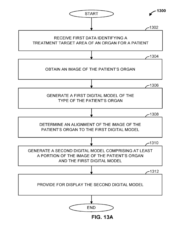
target region map, and
may store target definition data 307 in database 116.
[0096] In some examples, alignment determination engine 306
determines whether each
medical professional adjustment violates one or more predetermined rules. If
an adjustment
violates a rule, alignment determination engine 306 may cause the display of a
pop-up message
with a warning. In some examples, alignment determination engine 306 receives
one or more
user input(s) 301 identifying a selection of one or more other organs that may
be displayed in
conjunction with the 3D structure image. In response, alignment determination
engine 306
provides for display 3D models of such organs. In some examples, alignment
determination
engine 306 provides for display image data 103 of the patient's corresponding
organs.
[0097] In some examples, alignment determination engine 306
receives one or more user
input(s) 301 identifying a pan or zoom action. In response, alignment
determination engine 306
may pan or zoom across the 3D structure image. In some examples, alignment
determination
engine 306 receives one or more user input(s) 301 identifying the selection of
a preconfigured
selections for specific views of the 3D structure image. Alignment
determination engine 306
may adjust the 3D structure image in accordance with the specific view
selected, and may
provide for display the adjusted 3D structure image.
[0098] FIG. 4A illustrates a first portion 402 of a GUI 400 that
allows a medical
professional, such as an EP, to define a target area for treatment (e.g.,
ablation). The GUI 400
may be generated by an application executed by target definition computing
device 104, and may
be displayed to the medical professional on a display, such as display 206.
[0099] GUI 400 facilitates a number of steps to define a target
area for treatment
including generating a study data record, identify a target area for
treatment, and align the target
area to an image of a patient's organ. These steps are represented by studies
icon 406, target
selection icon 408, and alignment icon 410, each of which is illustrated under
the target
definition icon 404. Selecting one of studies icon 406, target selection icon
408, and alignment
icon 410 may present to the user a portion of GUI 400 corresponding to that
step.
24
CA 03201917 2023- 6-9
WO 2022/132181
PCT/US2020/066213
[0100] To begin target definition, first portion 402 includes a
study icon 401 that, if
selected, allows for the generation of a new study data record. Page 402 also
includes a report
icon 411 that, if selected, generates a report based on the corresponding
study data record. The
report may include the study data record, any selected target areas (e.g.,
segments), a scanned
image of the patient (e.g., scanned by image canning device 102), and data
identifying and
characterizing an alignment of the selected target area to the image of the
patient's organ.
[0101] FIG. 4B illustrates a second portion 420 of GUI 400 that
may be displayed when
the medical professional selects the add study icon 401 of FIG. 4A. For
example, the second
portion 420 may be a pop-up window that is displayed upon the medical
professional clicking on
the add study icon 401. Second portion 420 incudes a study type drop-down menu
424, a study
category drop down menu 428, and a study localization drop down menu 430.
[0102] Study type drop-down menu 424 allows the medical
professional to select a study
type for the study type record. For example, and as illustrated in FIG. 4B,
study type drop-down
menu 424 may allow the medical professional to select from a plurality of
study types (e.g.,
imaging types), such as CT, Catheter Mapping, ECG, ECGI, and MRI, among
others.
[0103] Once the medical professional selects a study type, GUI
400 automatically
determines one or more study categories based on the selected study type. Each
study category
may identify a list of features (or study localizations) for a specific study
type. The medical
professional may view the available study categories available using study
category drop-down
menu 426. For example, and as illustrated in FIG. 4D, the medical professional
may select a
study category of "Electrical" when the study type is "ECG."
[0104] Once the study category is selected, GUI 400 automatically
determines one or
more study localizations based on the selected study categories and/or
selected study type. The
study localization may identify a general target area of a patient's organ to
be treated, such as
one or more segments of a heart. For example, and as illustrated in FIG. 4D.
study localization
drop down menu 430, the medical professional may select a study localization
of -VT exit site,"
"VT enter site," and "VT enter and exit site" when the selected study type is
"ECG" and the
selected study category is "Electrical."
CA 03201917 2023- 6-9
WO 2022/132181
PCT/US2020/066213
[0105] Referring back to FIGs. 4B, 4C, and 4D, second portion 420
also includes a study
name text box 426 that allows the medical professional to provide a name for
the study record, a
study date selection box 432 that allows for the selection of a date (e.g., a
current date), and a
notes text box 434 that allows the medical professional to enter in notes
(e.g., treatment notes,
reminders, notes to other medical professionals, etc.).
[0106] In addition, second portion 420 includes an interactive
model 422, which in this
example is a 17 segment model representing segments of a heart's ventricle.
The medical
professional may select one or more portions of the interactive model 422,
which may be areas
for treatment. For example, and as illustrated in FIG. 4E, the medical
professional may select a
first segment 423A (e.g., segment 11), second segment 423B (e.g., segment 16),
and a third
segment 423C (e.g., segment 15). In addition, in some examples, when the
cursor 489 is placed
over a segment (e.g., segment 4), GUI 400 displays the name of the segment
(e.g., via a pop-up
window). In this example, cursor 489 appears over segment 4 of interactive
model 422, and in
response GUI 400 displays name box 425 identifying segment 4 as the "basal
inferior" portion of
a heart's ventricle.
[0107] To create the study data record, the medical professional
may click on add icon
490. In response, 104 generates data identifying and characterizing the
information provided to
GUI 400, and stores the generated data in a data repository, such as within
database 116. If the
medical professional would like to start over and not save the study data
record, the medical
professional may click on the cancel icon 492, which results in the clearance
of any provided
inputs, and, in some examples, the display of first portion 402 as illustrated
in FIG. 4A.
[0108] Referring to FIG. 4F, GUI 400 may include a third portion
478 that displays
summaries of generated study data records. For example, GUI 400 may display
portion 478 in
response to the medical professional clicking on the add icon 490 of FIG. 4E.
In some examples,
GUI 400 displays portion 478 in response to the medical professional clicking
the studies icon
406 of FIG. 4A.
[0109] Third portion 478 includes study category 480A, study name
480B, selected
segments 480C, acquisition date 480D, and notes 480E display areas for each
study data record
generated. The study category 480A corresponds to the selected study category
428 for each
26
CA 03201917 2023- 6-9
WO 2022/132181
PCT/US2020/066213
study data record generated. Similarly, the study name 480B, acquisition date
480D, and notes
480E correspond to the study name 426, study date 432, and notes 434 for each
study data
record.
[0110] In this example, two summaries are illustrated including a
first study summary
495A and a second study summary 495B. First study summary 495A includes a
study category
480A of "Structural," as well as corresponding interactive model 491
illustrating selected
segments 11, 15, and 16. Second study summary 495B includes a study category
480A of
"Electrical," as well as corresponding interactive model 4912 illustrating
selected segments 10
and 15. In some examples, when cursor 489 is placed over a corresponding
portion of an
interactive model, GUI 400 displays the name of the segment (e.g., via a pop-
up window). In
this example, cursor 489 appears over segment 10 of interactive model 492, and
in response GUI
400 displays a name box 493 identifying segment 0 as the "mid inferior"
portion of a heart's
ventricle.
[0111] FIG. 5A illustrates a target selection portion 501 of GUI
400. Once a study data
record is generated, for example, as discussed above with respect to FIGS. 4A
¨ 4F, GUI 400
may display target selection portion 501 to the medical professional. In some
examples, GUI
400 displays target selection portion 501 in response to the medical
professional clicking the
target selection icon 408 of FIG. 4A.
[0112] In this example, target selection portion 501 displays a
first study category map
510, which is based on a study category 428 of "Electrical," and a second
study category map
520, which is based on a study category 428 of "Structural." As described
herein, each study
category map 510, 520 may identify one or more portions of a patient's organ,
such as a 17
segment model of a heart's ventricle. Further, each study category map 510,
520 provides an
indication of previous studies performed on the patient corresponding to the
corresponding study
category. In addition, each study category map 510. 520 is displayed with a
corresponding bar
graph 512, 522, respectively. Each bar graph 512, 522 indicates treatment
amount ranges
determined for each study category as described herein, and their
corresponding hatching used
within the segments of each respective study category map 510, 520.
27
CA 03201917 2023- 6-9
WO 2022/132181
PCT/US2020/066213
[0113] Target selection portion 502 also includes a probability
map 502 that indicates a
probability of treatment for one or more portions of the patient's organ (in
this example, the
patient's heart) based on those portions of the organ identified by study
category maps 510, 520.
Probability map 502 is displayed with a corresponding bar graph 506 that
indicates treatment
segment probability ranges as described herein, and their corresponding
hatching used within
segments of the probability map 502.
[0114] Further, target selection portion 502 includes a target
definition map 530 that, in
this example, is in the faun of a 17 segment model of a heart's ventricle.
Target definition map
530 allows the medical professional to identify a target area for treatment.
For example, the
medical professional may select (e.g., using input/output device 203 to
manipulate cursor 489) a
segment of target definition map 530 to identify the target area 532. In this
example, target area
532 includes segment 17 of the target definition map 530.
[0115] FIG. 5B is similar to FIG. 5A, but the medical
professional may selects segment
16 of target definition map 530 to identify target area 542. Once the medical
professional has
identified the target area 532, 542 by selecting portions of target definition
map 530, the medical
professional may proceed to the next step be clicking on next icon 545.
[0116] FIG. 6A illustrates an alignment portion 601 of GUI 400
that displays a 3D
structure image 602 that includes a 3D segment model 606 superimposed onto
scanned image
604. 3D segment model 606 may be a 3D segment model of a heart's ventricle,
for example.
Scanned image 604 may be an image scanned by image scanning device 102, such
as a 3D
volume of a scanned structure of the patient. 3D structure image 602 also
includes a target
region map 648, which defines a target region for treatment for the patient.
The target region
map 648 may correspond to one or more selected target areas of a target
definition map, such as
target areas 532, 542 of target definition map 530, at least initially (e.g.,
before adjustment by the
EP). In some examples, target region map 648 is displayed in a distinct color.
In some
examples, a distinct hatching is used to display target region map 648, or any
other suitable
mechanism that allows the EP to easily determine the contours of target region
map 648.
Further, as displayed, a longitudinal axis 650 proceeds through an apex 608 of
3D structure
image 602.
28
CA 03201917 2023- 6-9
WO 2022/132181
PCT/US2020/066213
[0117] Alignment portion 601 may, in some examples, also display
a reference character
680. The reference character 680 is displayed from a view according to an
orientation of 3D
structure image 602. For example, if the orientation of 3D structure image 602
is such that it is
being displayed from an overhead view as the corresponding organ is positioned
in the patient,
then reference character 680 is displayed from an overhead view. This allows
the EP to easily
determine from what view and/or orientation 3D structure image 602 is
currently being
displayed.
[0118] Alignment portion 601 may, in some examples, include a
text entry box 640 that
allows for the entry of a value. In this example, the value entered is a
myocardial thickness (e.g.,
left ventricular myocardial thickness). The myocardial thickness may be used
to reconstruct the
inner ventricular myocardium surface, where all identified target segments are
projected, as
described here. For example, target definition computing device 104 may
execute an algorithm
to generate a final 3D target volume by combining all regions bounded by
selected segments and
underlying projections. If a user (e.g., an EP) has not edited the myocardinal
thickness, a default
value, such as 10 mm, is used. For example, target definition computing device
104 may
generate the 3D target volume based on selected segments as described herein.
For example, in
the example of a heart, target definition computing device 104 may take
selected segments (e.g.,
which may be part of the epicaridial wall) and extrude a volume toward the
center of the left
ventricle with a depth based on a wall thickness definition.
[0119] In some examples, alignment portion 601 includes one or
more adjustment icons
655 that allow for an adjustment of 3D structure image 602. For example,
adjustment icons 655
may allow for zoom in, zoom out, panning, and rotating functionalities.
[0120] With reference to FIG. 6B, alignment portion 601 may
display one or more drag
points, such as drag points 670A, 670B, that allow the EP to make adjustments
to 3D structure
image 602. For example, the EP may adjust longitudinal axis 650 by dragging
drag point 670A
to a new location. In response, GUI 400 adjust an orientation of scanned image
604 with respect
to 3D segment model 606. Similarly, the EP may adjust target region map 648 my
dragging drag
point 670B to a new location.
29
CA 03201917 2023- 6-9
WO 2022/132181
PCT/US2020/066213
[01211 In some examples, GUI 400 allows for the creation, or
removal, of drag points.
For example, the EP may right-click on a drag point, such as drag point 670B,
and select a
"remove" option to remove the drag point. Likewise, the EP may right-click on
a portion of 3D
segment model 606, and select an "ad" option to add a drag point.
[0122] FIG. 6C illustrates 3D structure image 602 after the EP
provided input to rotate
3D structure image 602 clockwise around longitudinal axis 650. In this
example, drag point
670C may allow the EP to adjust an anterior interventricular groove 686 of 3D
structure image
602.
[0123] Adjustment icons 655 may also allow the EP to display
images of additional
organs, such as organs that are adjacent to the organ identified by scanned
image 604. For
example, and with reference to FIG. 6D, the EP may select an adjustment icon
655 to display
organ selection box 675, which allows the EP to select from one or more organs
to display.
[0124] For example, and assuming the EP selects "lung" (e.g.,
"lung r p" for right lung,
or "lung 1 p" for left lung) and "esophagus," GUI 400 may display renderings
(e.g., 3D
renderings) of a first organ 685 (e.g., lung) and a second organ 687 (e.g.,
esophagus), as
illustrated in FIG. 6E. The renderings may be 3D models pre-stored in database
116, for
example. In other examples, the renderings are scanned images of the
corresponding structure of
the patient.
[0125] FIG. 11 is a flowchart of an example method 1100 that can
be carried out by, for
example, target definition computing device 104. Beginning at step 1102, a
first input is
received. The first input identifies a selected study type. For example, an EP
may use
input/output device 203 to provide an input to an executed application
displaying a GUI, such as
GUI 400, on display 206. The EP may select a study type 424 displayed within a
portion 420 of
GUI 400.
[0126] At step 1104, a plurality of study categories are provided
for display. The
plurality of categories are determined based on the selected study type. For
example, GUI 400
may display the plurality of study categories within a study category dropdown
menu 428.
Proceeding to step 1106, a second input is received. The second input
identifies a selected study
CA 03201917 2023- 6-9
WO 2022/132181
PCT/US2020/066213
category of the plurality of study categories. For example, the EP may select
one of the plurality
of study categories displayed within study category dropdown menu 428.
[0127] At step 1108, a plurality of study localizations are
provided for display. The
plurality of study localizations are determined based on the selected study
category. For
example, GUI 400 may display the plurality of study localizations within a
study localization
dropdown menu 430. At step 1110, a third input is received. The third input
identifies a selected
study localization of the plurality of study localizations. For example, the
EP may select one of
the plurality of study localizations displayed within study localization
dropdown menu 430.
[0128] Proceeding to step 1112, the selected study type, the
selected study category, and
the selected study localization are stored in a data repository. For example,
104 may generate a
study data record identifying the selected study category, and the selected
study localization, and
may store the study data record in database 116. The method then ends.
[0129] FIG. 12 is a flowchart of an example method 1200 that can
be carried out by, for
example, target definition computing device 104. Beginning at step 1202, study
data records for
a patient are obtained. The study data records identify a plurality of studies
performed on the
patient. For example, target definition computing device 104 may obtain study
definition data
303 from database 116 for the patient. At step 1204, a study category is
determined for each of
the plurality of studies. For example, each of the plurality of studies may be
associated with a
study category, such as "Electrical" or "Structural." At step 1206, a number
of studies for each
different category is determined. Further, at step 1208, a treatment target
area for each of the
plurality of studies is determined. For example, each of the plurality of
studies may be
associated with one or more segments targeted for treatment.
[0130] Proceeding to step 1210, a first map is generated for each
study category based on
the corresponding number of studies and treatment target areas. For example,
target definition
computing device 104 may determine, for each study category, a percentage of
the
corresponding number of studies treating each of a plurality of segments of
the patient's organ.
Each of the first maps may be, for example, study category maps 510, 520.
31
CA 03201917 2023- 6-9
WO 2022/132181
PCT/US2020/066213
[01311 At step 1212, a second map is generated. The second map is
generated based on
the first maps and corresponding treatment target areas. For example, the
second map may
indicate probabilities of studies for one or more portions of the patient's
organ based on those
portions identified in the first maps. The second map may be, for example,
probability map 502
that indicates a probability of treatment for one or more portions of the
patient's organ based on
those portions of the organ identified by study category maps 510, 520.
[0132] At step 1214, the first maps and the second map are
provided for display. For
example, the first maps and second map may be displayed within a target
selection portion 501
of GUI 400. The method then ends.
[0133] FIG. 13A is a flowchart of an example method 1300 that can
be carried out by, for
example, target definition computing device 104. At step 1302, first data is
received. The first
data identifies a treatment target area of an organ for a patient. For
example, target definition
computing device 104 may determine the treatment target area based on a
segment of a target
definition map 530 that an EP has selected to identify target area 532. At
step 1304, an image of
the patient's organ is obtained. For example, target definition computing
device 104 may obtain
an image, such as an image of a 3D volume, of an organ of the patient scanned
by image
scanning device 102.
[0134] Proceeding to step 1306, a first digital model of the type
of the patient's organ is
generated. For example, target definition computing device 104 may generate a
3D model, such
as 3D ventricle model 720 or 3D segment model 1002B, of the patient's organ.
At step 1308, an
alignment of the image of the patient's organ to the first digital model is
determined. Further,
and at step 1310, a second digital model is generated. The second digital
model comprises at
least a portion of the image of the patient's organ and the first digital
model. For example, target
definition computing device 104 may superimpose 3D segment model 606 onto
scanned image
604 to generate 3D structure image 602. Al step 1312, the second digital model
is provided for
display. For example, target definition computing device 104 may display the
second digital
model to the EP. The method then ends.
[0135] FIG. 13B is a flowchart of an example method 1350 that can
be carried out by, for
example, target definition computing device 104. Beginning at step 1352, a
digital model is
32
CA 03201917 2023- 6-9
WO 2022/132181
PCT/US2020/066213
provided for display. The digital model comprises a portion of an image of a
patient's organ and
a second digital model of the type of the organ. For example, the digital
model may be generated
in accordance with method 1300 of FIG. 13A. At step 1354, an input is
received. The input
identifies an alignment adjustment to the digital model. For example. 104 may
receive an input
from an EP using input/output device 203 to make an adjustment to 3D structure
image 602 by
dragging one or more drag points 670 as described herein.
[0136] Proceeding to step 1356, an adjustment to the digital
model is determined based
on the input. For example, the adjustment may be a change to an orientation of
the image of the
patient's organ with respect to the second digital model. The EP may adjust
the orientation by
dragging one or more drag points 670 to move a longitudinal axis 650, for
example. In some
examples, the adjustment may be a change to a target region map of the digital
model. For
example, the adjustment may be to target region map 648 of 3D structure image
602.
[0137] At step 1358, the digital model sis regenerated based on
the determined
adjustment. Further, and at step 1360, the regenerated digital model is
provided for display. In
some examples, 104 transmits the regenerated digital model to radioablation
treatment system
126 for treating the patient. The method then ends.
[0138] In some examples a system includes a computing device. The
computing device
is configured to receive a first input identifying an organ of a patient, and
receive a scanned
image of the organ. The computing device is also configured to generate a
first digital model of
a type of the organ. Further, the computing device is configured to determine
an alignment of
the scanned image to the first digital model. The computing device is also
configured to
generate a second digital model comprising at least a portion of the scanned
image and the first
digital model. The computing device is further configured to store the second
digital model in a
data repository. In some examples, receiving the first input is in response to
a selection of a
portion of a displayed target definition map. In some examples, the organ is a
heart. In some
examples, the computing device is configured to provide the second digital
model for display.
[0139] In some examples, the computing device is configured to
receive a second input
identifying an adjustment to the alignment of the scanned image to the first
digital model. The
computing device is also configured to adjust the second digital model based
on the second
33
CA 03201917 2023- 6-9
WO 2022/132181
PCT/US2020/066213
input. The computing device is further configured to store the adjusted second
digital model in
the data repository.
[0140] In some examples, the computing device is configured to
receive a second input
identifying a treatment target area of the organ. The computing device is also
configured to
determine a corresponding portion of the second digital model based on the
treatment target area
of the organ. Further, the computing device is configured to regenerate the
second digital model
to identify the corresponding portion. In some examples, regenerating the
second digital model
includes associating the corresponding portion with a distinctive feature for
display. In some
examples, the computing device is further configured to transmit treatment
data identifying the
treatment target area of the organ to a radioablation treatment system.
[0141] In some examples, the computing device is configured to
obtain study data
records for the patient, wherein each study data record identifies one of a
plurality of study types
and a study target area of a plurality of study target areas for studies
performed on the patient.
The computing device is also configured to determine a first number of each of
the plurality of
study types performed on the patient based on the study data records. Further,
the computing
device is configured to determine, for each of the plurality of study types, a
second number of
studies performed on the patient in each of the plurality of study target
areas. The computing
device is also configured to generate a first map for each of the plurality of
study types based on
the corresponding first number and second numbers. The computing device is
further configured
to store the first map in the data repository. In some examples, each first
map indicates a
frequency of the corresponding study type on each of the plurality of study
target areas. In some
examples, the computing device is further configured to generate a second map
based on the first
numbers and the second numbers, wherein the second map indicates a probability
of treatment
for each of the plurality of study target areas, and to store the second map
in a data repository.
[0142] In some examples, a computer-implemented method includes
receiving a first
input identifying an organ of a patient, and receive a scanned image of the
organ. The method
also includes generating a first digital model of a type of the organ.
Further, the method includes
determining an alignment of the scanned image to the first digital model. The
method also
includes generating a second digital model comprising at least a portion of
the scanned image
34
CA 03201917 2023- 6-9
WO 2022/132181
PCT/US2020/066213
and the first digital model. The method further includes storing the second
digital model in a
data repository. In some examples, receiving the first input is in response to
a selection of a
portion of a displayed target definition map. In some examples, the organ is a
heart. In some
examples, the method includes providing the second digital model for display.
[0143] In some examples, the method includes receiving a second
input identifying an
adjustment to the alignment of the scanned image to the first digital model.
The method also
includes adjusting the second digital model based on the second input. The
method further
includes storing the adjusted second digital model in the data repository.
[0144] In some examples, the method includes receiving a second
input identifying a
treatment target area of the organ. The method also includes determining a
corresponding
portion of the second digital model based on the treatment target area of the
organ. Further, the
method includes regenerating the second digital model to identify the
corresponding portion. In
some examples, regenerating the second digital model includes associating the
corresponding
portion with a distinctive feature for display. In some examples, the method
includes
transmitting treatment data identifying the treatment target area of the organ
to a radioablation
treatment system.
[0145] In some examples, the method includes obtaining study data
records for the
patient, wherein each study data record identifies one of a plurality of study
types and a study
target area of a plurality of study target areas for studies performed on the
patient. The method
also includes determining a first number of each of the plurality of study
types performed on the
patient based on the study data records. Further, the method includes
determining, for each of
the plurality of study types, a second number of studies performed on the
patient in each of the
plurality of study target areas. The method further includes generating a
first map for each of the
plurality of study types based on the corresponding first number and second
numbers. The
method also includes storing the first map in the data repository. In some
examples, each first
map indicates a frequency of the corresponding study type on each of the
plurality of study target
areas. In some examples, the method includes generating a second map based on
the first
numbers and the second numbers, wherein the second map indicates a probability
of treatment
for each of the plurality of study target areas, and storing the second map in
a data repository.
CA 03201917 2023- 6-9
WO 2022/132181
PCT/US2020/066213
[0146] In some examples, a non-transitory computer readable
medium stores instructions
that, when executed by at least one processor, cause the at least one
processor to perform
operations including receiving a first input identifying an organ of a
patient, and receive a
scanned image of the organ. The operations also include generating a first
digital model of a
type of the organ. Further, the operations include determining an alignment of
the scanned
image to the first digital model. The operations also include generating a
second digital model
comprising at least a portion of the scanned image and the first digital
model. The operations
further include storing the second digital model in a data repository. In some
examples,
receiving the first input is in response to a selection of a portion of a
displayed target definition
map. In some examples, the organ is a heart. In some examples, the operations
include
providing the second digital model for display.
[0147] In some examples, the operations include receiving a
second input identifying an
adjustment to the alignment of the scanned image to the first digital model.
The operations also
include adjusting the second digital model based on the second input. The
operations further
include storing the adjusted second digital model in the data repository.
[0148] In some examples, the operations include receiving a
second input identifying a
treatment target area of the organ. The operations also include determining a
corresponding
portion of the second digital model based on the treatment target area of the
organ. Further, the
operations include regenerating the second digital model to identify the
corresponding portion.
In some examples, regenerating the second digital model includes associating
the corresponding
portion with a distinctive feature for display. In some examples, the
operations include
transmitting treatment data identifying the treatment target area of the organ
to a radioablation
treatment system.
[0149] In some examples, the operations include obtaining study
data records for the
patient, wherein each study data record identifies one of a plurality of study
types and a study
target area of a plurality of study target areas for studies performed on the
patient. The
operations also include determining a first number of each of the plurality of
study types
performed on the patient based on the study data records. Further, the
operations include
determining, for each of the plurality of study types, a second number of
studies performed on
36
CA 03201917 2023- 6-9
WO 2022/132181
PCT/US2020/066213
the patient in each of the plurality of study target areas. The operation
further include generating
a first map for each of the plurality of study types based on the
corresponding first number and
second numbers. The operations also include storing the first map in the data
repository. In
some examples, each first map indicates a frequency of the corresponding study
type on each of
the plurality of study target areas. In some examples, the operations include
generating a second
map based on the first numbers and the second numbers, wherein the second map
indicates a
probability of treatment for each of the plurality of study target areas, and
storing the second map
in a data repository.
[0150] In some examples, a computer-implemented method includes a
means for
receiving a first input identifying an organ of a patient, and receive a
scanned image of the organ.
The method also includes a means for generating a first digital model of a
type of the organ.
Further, the method includes a means for determining an alignment of the
scanned image to the
first digital model. The method also includes a means for generating a second
digital model
comprising at least a portion of the scanned image and the first digital
model. The method
further includes a means for storing the second digital model in a data
repository. In some
examples, receiving the first input is in response to a selection of a portion
of a displayed target
definition map. In some examples. the organ is a heart. In some examples, the
method includes
a means for providing the second digital model for display.
[0151] In some examples, the method includes a means for
receiving a second input
identifying an adjustment to the alignment of the scanned image to the first
digital model. The
method also includes a means for adjusting the second digital model based on
the second input.
The method further includes a means for storing the adjusted second digital
model in the data
repository.
[0152] In some examples, the method includes a means for
receiving a second input
identifying a treatment target area of the organ. The method also includes a
means for
determining a corresponding portion of the second digital model based on the
treatment target
area of the organ. Further, the method includes a means for regenerating the
second digital
model to identify the corresponding portion. In some examples, regenerating
the second digital
model includes associating the corresponding portion with a distinctive
feature for display. In
37
CA 03201917 2023- 6-9
WO 2022/132181
PCT/US2020/066213
some examples, the method includes a means for transmitting treatment data
identifying the
treatment target area of the organ to a radioablation treatment system.
[0153] In some examples, the method includes a means for
obtaining study data records
for the patient, wherein each study data record identifies one of a plurality
of study types and a
study target area of a plurality of study target areas for studies performed
on the patient. The
method also includes a means for determining a first number of each of the
plurality of study
types performed on the patient based on the study data records. Further, the
method includes a
means for determining, for each of the plurality of study types, a second
number of studies
performed on the patient in each of the plurality of study target areas. The
method further
includes a means for generating a first map for each of the plurality of study
types based on the
corresponding first number and second numbers. The method also includes a
means for storing
the first map in the data repository. In some examples, each first map
indicates a frequency of
the corresponding study type on each of the plurality of study target areas.
In some examples,
the method includes a means for generating a second map based on the first
numbers and the
second numbers, wherein the second map indicates a probability of treatment
for each of the
plurality of study target areas, and storing the second map in a data
repository.
[0154] Although the methods described above are with reference to
the illustrated
flowcharts, it will be appreciated that many other ways of performing the acts
associated with the
methods can be used. For example, the order of some operations may be changed,
and some of
the operations described may be optional.
[0155] In addition, the methods and system described herein can
be at least partially
embodied in the form of computer-implemented processes and apparatus for
practicing those
processes. The disclosed methods may also be at least partially embodied in
the form of tangible,
non-transitory machine-readable storage media encoded with computer program
code. For
example, the steps of the methods can be embodied in hardware, in executable
instructions
executed by a processor (e.g., software), or a combination of the two. The
media may include, for
example, RAMs, ROMs, CD-ROMs, DVD-ROMs, BD-ROMs, hard disk drives, flash
memories,
or any other non-transitory machine-readable storage medium. When the computer
program code
is loaded into and executed by a computer, the computer becomes an apparatus
for practicing the
38
CA 03201917 2023- 6-9
WO 2022/132181
PCT/US2020/066213
method. The methods may also be at least partially embodied in the form of a
computer into which
computer program code is loaded or executed, such that, the computer becomes a
special purpose
computer for practicing the methods. When implemented on a general-purpose
processor, the
computer program code segments configure the processor to create specific
logic circuits. The
methods may alternatively be at least partially embodied in application
specific integrated circuits
for performing the methods.
[0156] The foregoing is provided for purposes of illustrating,
explaining, and describing
embodiments of these disclosures. Modifications and adaptations to these
embodiments will be
apparent to those skilled in the art and may be made without departing from
the scope or spirit of
these disclosures.
39
CA 03201917 2023- 6-9