Note: Descriptions are shown in the official language in which they were submitted.
CA 03202089 2023-05-15
WO 2022/109313
PCT/US2021/060161
BOVINE ANTIBODY VARIANTS
CROSS-REFERENCE TO RELATED APPLICATIONS
[own This application claims priority to and the benefit of United States
Provisional Patent
Application 63/116491, filed November 20, 2020, which is incorporated by
reference herein in
its entirety.
FIELD OF THE INVENTION
[0002] The invention relates generally to bovine antibody variants and uses
thereof
Specifically, the invention relates to one or more mutations in the Fc
constant region of bovine
antibody for improving various characteristics.
BACKGROUND OF THE INVENTION
[0003] Bovine IgG monoclonal antibodies (mAbs) can be effective therapeutics
in veterinary
medicine. Several years ago, three bovine IgG subclasses were identified.
However, not much
work has been done to improve the chraceteristics of bovine IgGs.
[0004] Through a recycling mechanism, the neonatal Fc receptor (FcRn) prolongs
the half-life
of an IgG in a pH-dependent interaction with its fragment crystallizable (Fc)
region.
Specifically, the Fc region spanning the interface of CH2 and CH3 domains
interacts with the
FcRn on the surface of cells to regulate IgG homeostasis. This interaction is
favored by an
acidic interaction after IgG pinocytosis and thus IgG is protected from
degradation. The
endocytosed IgG is then recycled back to the cell surface and released into
the blood stream at
a slightly alkaline pH thereby maintaining sufficient serum IgG for proper
function.
Accordingly, the pharmacokinetic profile of IgGs depend on the structural and
functional
properties of their Fc regions.
[0005] Fc regions are also responsible for antibody effector functions, such
as complement-
dependent cytotoxicity (CDC), antibody-dependent cellular cytotoxicity (ADCC)
and
.. antibody-dependent cellular phagocytosis (ADCP). These effector functions
rely on the
interactions of Fc regions with FcyRs. Therefore, engineering Fc regions to
tune their
interactions with FcyRs has emerged as a promising approach for enhancing the
activity of
therapeutic antibodies.
1
CA 03202089 2023-05-15
WO 2022/109313
PCT/US2021/060161
[0006] In human health, decades of Fc engineering have led to the
identification of numerous
mutations that improved pharmacokinetics and effector functions. To date, no
such Fc
engineering study was reported in bovine.
[0007] Accordingly, there exists a need for novel bovine IgG Fc region
mutations to improve
various characteristics of bovine IgGs.
SUMMARY OF THE INVENTION
[0008] The invention relates to mutant bovine IgGs that exhibit desired
characteristics, relative
to wild-type bovine IgGs. Specifically, the inventors of the instant
application have found that
substituting an amino acid residue at position 216, 234, 235, 237, 270, 329,
330, 331, 432, 434,
437, or 433 (numbered according to the Eu index as in Kabat) with another
amino acid
surprisingly and unexpectedly exhibited a desired effect. In an exemplary
embodiment, the
undexpected desired effects include, but not limited to, enhanced affinity to
FcRn;
reduced complement-dependent cytotoxicity (CDC); reduced antibody-dependent
cellular
cytotoxicity (ADCC); reduced antibody-dependent cellular phagocytosis (ADCP);
reduced
binding to Fc gamma receptor (bFcgR); or a combination thereof
[0009] In one aspect, the invention provides a modified IgG comprising: a
bovine IgG constant
domain comprising at least one amino acid substitution relative to a wild-type
bovine IgG
constant domain, wherein said substitution is at amino acid residue 216, 234,
235, 237, 270,
329, 330, 331, 432, 434, 437, or 433.
[Nolo] In one exemplary embodiment, the bovine IgG constant domain is an IgG1
constant
domain that comprises one or more of substitutions of P329S, A3305, P33 1S,
P234A, L235A,
G237A, D216E, D270E, L432A, N434A, and T437A.
[Nom In another exemplary embodiment, the bovine IgG constant domain is an
IgG2 constant
domain that comprises one or more of substitutions of A3305, L432A, N434A, and
M437A.
[00012] In yet another exemplary embodiment, the bovine IgG constant domain is
an IgG3
constant domain that comprises one or more of substitutions of P329S, A3305,
P331S, P234A,
L235A, G237A, D270E, L432A, N434A, T437A, and R433H.
[00013] In another aspect, the invention provides a polypeptide comprising: a
bovine IgG
constant domain comprising one or more amino acid substitutions of the
invention described
herein.
2
CA 03202089 2023-05-15
WO 2022/109313
PCT/US2021/060161
[00014] In yet another aspect, the invention provides an antibody or a
molecule comprising: a
bovine IgG constant domain comprising one or more amino acid substitutions of
the invention
described herein.
[00015] In a further aspect, the invention provides a method for producing or
manufacturing an
antibody or a molecule, the method comprising: providing a vector or a host
cell having a
nucleic acid sequence that encodes an antibody, wherein said antibody
comprises a bovine IgG
constant domain comprising one or more amino acid substitutions of the
invention described
herein.
[00016] Other features and advantages of the present invention will become
apparent from the
following detailed description examples and figures. It should be understood,
however, that the
detailed description and the specific examples while indicating preferred
embodiments of the
invention are given by way of illustration only, since various changes and
modifications within
the spirit and scope of the invention will become apparent to those skilled in
the art from this
detailed description.
BRIEF DESCRIPTION OF THE DRAWINGS
[00017] The patent or application file contains at least one drawing executed
in color. Copies
of this patent or patent application publication with color drawing(s) will be
provided by the
Office upon request and payment of the necessary fee.
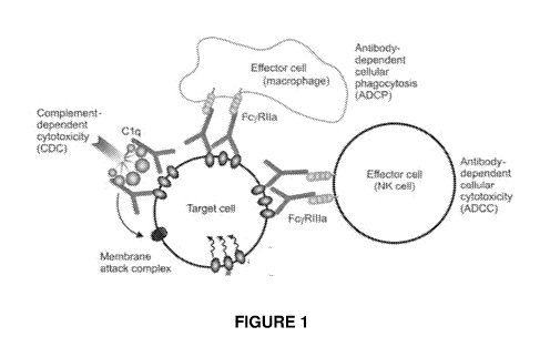
[00018] FIG. 1 illustrates target cell killing functions triggered by IgG Fc
binding to Fc gamma
receptors on the surface of effector cells.
[00019] FIG. 2 shows the sequence alignment bovine IgG subclasses. Three
representative
allotypes are shown. CH1, hinge, CH2, and CH3 domains are as follows: CH1:
residues 1-98;
hinge: 99 to vertical lines; CH2: vertical lines to 243; CH3: 244-351.
Cysteines involved in
Inter-heavy chain disulfide bonds are in bold and underlined. The 3' extension
of CH1 exon in
cow IgG3a is italicized. Bolded amino acids just downstream from the hinge
represent
the "Winter" or "LALA" sites. For bIgGla and bIgG3a, these sites are LPGG and
PLGG,
respectively. This site is absent in bIgG2a. Box includes "PAP" region
in bIgGla and bIgG3a CH2 and "SAS" region in bIgG2a. Double-underlined
arginine (R)
in bIgG3a is residue involved in point mutation to histidine (H).
[00020] FIG. 3 shows the sequence alignment of the three unique alleles of
bovine IgG1
subclass. Bold: amino acid "DP" regions mutated on bIgGla to mitigate protease
clipping.
3
CA 03202089 2023-05-15
WO 2022/109313
PCT/US2021/060161
Underlined: amino acids mutated on bIgGla to knock-out effector function.
Because bIgGld has synonymous amino acid sequence as bIgGlb, it is not
included in
the alignment of bIgG1 allotypes. All mutations made to bIgG1 are identical
for the a and b
allotypes.
[00021] FIG. 4 shows the sequence alignment of two reported alleles of the
bovine IgG2
subclass, bIgG2a (NCBI X16702.1) and bIgG2b (NCBI S82407) with the sequence of
an
IgG2 antibody isolated from cow. Bold: amino acids mutated on bIgG2a to knock-
out effector
function. Inventors have isolated an IgG antibody from a dairy cow and
sequencing revealed
a 2-residue deviation from that reported for bIgG2a, as shown in Figure 4 as
"bIgG2a from
cow seq". This sequence may represent a new allotype for bIgG2, but for the
purposes of this
patent application, it will be referred to as "bIgG2a" due to near-identical
alignment with bIgG2a. All mutations made to bIgG2 are identical for the a and
b allotypes.
[00022] FIG. 5 shows the sequence alignment of the two alleles of
the bovine IgG3 subclass. Bold: amino acids mutated on bIgG3a to knock-out
effector
function. Underlined: the residue mutated on bIgG3a to enhance the affinity to
bFcRn. All
mutations made to bIgG3 are identical for the a and b allotypes.
[00023] FIG. 6 shows bovine IgGla and bIgGla Fc mutants for knock out of
effector function.
Bold: the residues involved in the Winter mutation L234A P235A G237A*. Box:
the
residues involved in the PAP-to-SAS (P329S P331S*), PAP-to-SAP (P329S*)
mutations, and
the PAP-to-PSS (SS mutation A330S P331S*) mutation that is combined with the
Winter
mutation. Vertical line: start of the Fc region in the CTLA4-Fc fusion
proteins. *The amino
acid residues of the mutations are numbered according to the Eu index as in
Kabat.
[00024] FIGs. 7A and 7B show cell-based complement-dependent cytotoxicity
activity of
bovine IgGla, IgGlb and variants of both allotypes.
[00025] FIG. 8 shows antibody homology modeling of bIgGla WINSAS, bIgGlb
WINSAS,
bIgGlc WINSAS and bIgGld WINSAS. A) Overlay of the protein models with a
zoomed-in
sub-panel showing WINSAS residues (arrows). B) Root Mean Square Deviation
(RMSD)
comparisons (in angstroms) of WINSAS mutants of bIgGla WINSAS, bIgGlb WINSAS,
bIgGlc WINSAS and bGld WINSAS allotypes.
[00026] FIG. 9 shows Fc mutations on bovine IgGla subclass for the elimination
of Antibody
Dependent Cellular Phagocytosis in a cell-based assay.
[00027] FIG. 10 shows analytical SEC of wild-type bovine IgGla molecule.
4
CA 03202089 2023-05-15
WO 2022/109313
PCT/US2021/060161
[00028] FIG. 11A shows mass spectrometric analysis of Bovine IgG1 a wild type
(WT)
Fe. FIG. 11B shows mass spectrometric analysis of Bovine IgGla double mutation
DP Site 1
&2.
[00029] FIG. 12 shows bovine IgG1 a and bIgG1 a DP-to-EP Fe mutants for
eliminating
.. cleavage site. Bold: "DP1" (site 1) mutation to EP; Underlined: "DP2" (site
2) mutation to EP.
[00030] FIG. 13 shows antibody homology modeling of bIgGla DP1 DP2,
bIgGlb DP1 DP2, bIgGlc DP1 DP2 and bIgGld DP1 DP2. A) Overlay of the protein
models with a zoomed-in sub-panel showing DP1 and DP2 residues (arrows). B)
Root Mean
Square Deviation (RMSD) comparisons of DP1 DP2 mutants of bovine G1 a, Gib, G1
c and
Gld allotypes.
[00031] FIG. 14 shows bovine IgG2a and bIgG2a Fe mutants for knock out of
effector
function. Bold in first line: the three residues L432*, N434*, and M437*
mutated to alanine as
independent mutations or in combinations. Vertical line: start of the Fe
region in the CTLA4-
Fc fusion proteins. *The amino acid residues of the mutations are numbered
according to the
Eu index as in Kabat.
[00032] FIGs. 15A and 15B show that Fe mutations on bovine IgG2a and IgG2b
subclass
eliminate cell-based complement-dependent cytotoxicity activity.
[00033] FIG. 16 shows antibody homology modeling of bIgG2a L432A N434A M437A
and
bIgG2b L432A N434A M437A. A) Overlay of the protein models with a zoomed-in
sub-
panel showing L432A N434A M437A residues (arrows). B) Root Mean Square
Deviation
(RMSD) comparisons of L432A N434A M437A mutants of bovine IgG2a and IgG2b
allotypes.
[00034] FIG. 17 shows that Fe mutations on bovine IgG2a subclass eliminate
Antibody
Dependent Cellular Phagocytosis in a cell-based assay.
[00035] FIG. 18 shows bovine IgG3a and bIgG3a Fe mutants for knock out of
effector function.
Bold: the residues involved in the Winter mutation (P234A L235A G237A*). Box:
the
residues involved in the PAP-to-SAS (P3295 P3315*) and
PAP-to-
SAP (P329S*) mutations. Vertical line: start of the Fe region in the CTLA4-Fe
fusion
proteins. *The amino acid residues of the mutations are numbered according to
the Eu index
as in Kabat.
5
CA 03202089 2023-05-15
WO 2022/109313
PCT/US2021/060161
[00036] FIGs. 19A and 19B show Fe mutations on bovine IgG3a and IgG3b subclass
for the
elimination of complement-dependent cytotoxicity in a cell-based assay.
[00037] FIG. 20 shows antibody homology modeling of bIgG3a WINSAS and
bIgG3b WINSAS. A) Overlay of the protein models with a zoomed-in sub-panel
showing
WINSAS residues (arrows). B) Root Mean Square Deviation (RMSD) comparisons of
WINSAS mutants of bovine IgG3a and IgG3b allotypes.
[00038] FIG. 21 shows that Fe mutations on bovine IgG3a subclass eliminate
Antibody
Dependent Cellular Phagocytosis in a cell-based assay.
[00039] FIG. 22 shows bovine IgG3a and Fe mutant for improved bFcRn binding.
Bold
arginine (R) is residue involved in point mutation to histidine (H), R433H,
numbered according
to the Eu index as in Kabat.
[00040] FIG. 23 shows the alignment of the amino acid sequences of human IgGl,
bovine
lgGla, bovine IgG2a, and bovine IgG3a. The amino acid residues are numbered
according to
the Eu index as in Kabat. Amino acid residues for the other alleles for IgGl
(b, c, d), IgG2 (b),
and IgG3 (b) were also numbered according to the Eu index shown in Figure 23.
BRIEF DESCRIPTION OF THE SEQUENCE LISTINGS
[00041] SEQ ID NO.: 1 refers to the amino acid sequence of IgGla Wildtype
(NCBI ID number
1S82409).
[00042] SEQ ID NO.: 2 refers to the amino acid sequence of IgGlb Wildtype
(NCBI ID number
X16701).
[00043] SEQ ID NO.: 3 refers to the amino acid sequence of IgGlc Wildtype
(NCBI ID number
DQ452014.1).
[00044] SEQ ID NO.: 4 refers to the amino acid sequence of IgGld Wildtype
(NCBI ID number
X62916.1).
[00045] SEQ ID NO.: 5 refers to the amino acid sequence of IgG2a Wildtype
(NCBI ID number
X16702.1).
[00046] SEQ ID NO.: 6 refers to the amino acid sequence of IgG2b Wildtype
(NCBI ID number
S82407).
[00047] SEQ ID NO.: 7 refers to the amino acid sequence of IgG3a Wildtype.
6
CA 03202089 2023-05-15
WO 2022/109313
PCT/US2021/060161
[00048] SEQ ID NO.: 8 refers to the amino acid sequence of IgG3b Wildtype.
[00049] SEQ ID NO.: 9 refers to the amino acid sequence of IgG2a Wildtype from
Dairy Cow.
[00050] SEQ ID NO.: 10 refers to the amino acid sequence of IgGla Having DP1
Mutation.
[00051] SEQ ID NO.: 11 refers to the amino acid sequence of IgGla Having DP2
Mutation.
[00052] SEQ ID NO.: 12 refers to the amino acid sequence of IgGla Having DP1
and DP2
Mutations.
[00053] SEQ ID NO.: 13 refers to the amino acid sequence of IgGla Wildtype
Fragment
Physical Positions 99-329.
[00054] SEQ ID NO.: 14 refers to the amino acid sequence of IgGla Fragment
Having SAP
Mutation.
[00055] SEQ ID NO.: 15 refers to the amino acid sequence of IgGla Fragment
Having SAS
Mutation.
[00056] SEQ ID NO.: 16 refers to the amino acid sequence of IgGla Fragment
Having Winter
Mutations.
[00057] SEQ ID NO.: 17 refers to the amino acid sequence of IgGla Fragment
Having Winter
and SAS Mutations.
[00058] SEQ ID NO.: 18 refers to the amino acid sequence of IgGla Fragment
Having Winter
and SS Mutations.
[00059] SEQ ID NO.: 19 refers to the amino acid sequence of IgG2a Having
Mutation L432A.
[00060] SEQ ID NO.: 20 refers to the amino acid sequence of IgG2a Having
Mutation N434A.
[00061] SEQ ID NO.: 21 refers to the amino acid sequence of IgG2a Having
Mutation M437A.
[00062] SEQ ID NO.: 22 refers to the amino acid sequence of IgG2a Having
Mutations L432A
and M437A.
[00063] SEQ ID NO.: 23 refers to the amino acid sequence of IgG2a Having
Mutations N434A
and M437A.
[00064] SEQ ID NO.: 24 refers to the amino acid sequence of IgG2a Having
Mutations L432A,
N434A, and M437A.
[00065] SEQ ID NO.: 25 refers to the amino acid sequence of IgG2a Having
Mutations L432A
and N434A.
7
CA 03202089 2023-05-15
WO 2022/109313
PCT/US2021/060161
[00066] SEQ ID NO.: 26 refers to the amino acid sequence of IgG3a Wildtype
Fragment
Physical Positions 99-352.
[00067] SEQ ID NO.: 27 refers to the amino acid sequence of IgG3a Fragment
Having SAP
Mutation.
[00068] SEQ ID NO.: 28 refers to the amino acid sequence of IgG3a Fragment
Having SAS
Mutation.
[00069] SEQ ID NO.: 29 refers to the amino acid sequence of IgG3a Fragment
Having Winter
Mutation.
[00070] SEQ ID NO.: 30 refers to the amino acid sequence of IgG3a Fragment
Having Winter
and SAS Mutations.
[00071] SEQ ID NO.: 31 refers to the amino acid sequence of IgG3a Having
Mutation R433H.
[00072] SEQ ID NO.: 32 refers to the amino acid sequence of human IgG1 .
[00073] SEQ ID NO.: 33 refers to the amino acid sequence of IgGlb Wildtype.
[00074] SEQ ID NO.: 34 refers to the nucleic acid sequence of IgGlb Wildtype.
[00075] SEQ ID NO.: 35 refers to the amino acid sequence of IgGlb having
Winter mutation.
[00076] SEQ ID NO.: 36 refers to the nucleic acid sequence of IgGlb having
Winter mutation.
[00077] SEQ ID NO.: 37 refers to the amino acid sequence of IgGlb having WinSS
mutation.
[00078] SEQ ID NO.: 38 refers to the nucleic acid sequence of IgGlb having
WinSS mutation.
[00079] SEQ ID NO.: 39 refers to the amino acid sequence of IgGlb having
WinSAS mutation.
[00080] SEQ ID NO.: 40 refers to the nucleic acid sequence of IgGlb having
WinSAS mutation.
[00081] SEQ ID NO.: 41 refers to the amino acid sequence of IgGlb having SAS
mutation.
[00082] SEQ ID NO.: 42 refers to the nucleic acid sequence of IgGlb having SAS
mutation.
[00083] SEQ ID NO.: 43 refers to the amino acid sequence of IgGlb having SAP
mutation.
[00084] SEQ ID NO.: 44 refers to the nucleic acid sequence of IgGlb having SAP
mutation.
[00085] SEQ ID NO.: 45 refers to the amino acid sequence of IgGlb having D216E
mutation.
[00086] SEQ ID NO.: 46 refers to the nucleic acid sequence of IgGlb having
D216E mutation.
[00087] SEQ ID NO.: 47 refers to the amino acid sequence of IgGlb having D270E
mutation.
8
CA 03202089 2023-05-15
WO 2022/109313
PCT/US2021/060161
[00088] SEQ ID NO.: 48 refers to the nucleic acid sequence of IgGlb haying
D270E mutation.
[00089] SEQ ID NO.: 49 refers to the amino acid sequence of IgGlb having D216E
and D270E
mutations.
[00090] SEQ ID NO.: 50 refers to the nucleic acid sequence of IgGlb having
D216E and D270E
mutations.
[00091] SEQ ID NO.: 51 refers to the amino acid sequence of IgG2b Wildtype.
[00092] SEQ ID NO.: 52 refers to the nucleic acid sequence of IgG2b Wildtype.
[00093] SEQ ID NO.: 53 refers to the amino acid sequence of IgG2b haying L432A
mutation.
[00094] SEQ ID NO.: 54 refers to the nucleic acid sequence of IgG2b haying
L432A mutation.
.. [00095] SEQ ID NO.: 55 refers to the amino acid sequence of IgG2b haying
N434A mutation.
[00096] SEQ ID NO.: 56 refers to the nucleic acid sequence of IgG2b haying
N434A mutation.
[00097] SEQ ID NO.: 57 refers to the amino acid sequence of IgG2b haying M437A
mutation.
[00098] SEQ ID NO.: 58 refers to the nucleic acid sequence of IgG2b haying
M437A mutation.
[00099] SEQ ID NO.: 59 refers to the amino acid sequence of IgG2b having L432A
and N434A
mutations.
[000100] SEQ ID NO.: 60 refers to the nucleic acid sequence of IgG2b haying
L432A and
N434A mutations.
[000101] SEQ ID NO.: 61 refers to the amino acid sequence of IgG2b haying
L432A and
M437A mutations.
[000102] SEQ ID NO.: 62 refers to the nucleic acid sequence of IgG2b haying
L432A and
M437A mutations.
[000103] SEQ ID NO.: 63 refers to the amino acid sequence of IgG2b haying
N434A and
M437A mutations.
[000104] SEQ ID NO.: 64 refers to the nucleic acid sequence of IgG2b haying
N434A and
M437A mutations.
[000105] SEQ ID NO.: 65 refers to the amino acid sequence of IgG2b having
L432A, N434A
and M437A mutations.
9
CA 03202089 2023-05-15
WO 2022/109313
PCT/US2021/060161
[000106] SEQ ID NO.: 66 refers to the nucleic acid sequence of IgG2b having
L432A, N434A
and M437A mutations.
[000107] SEQ ID NO.: 67 refers to the amino acid sequence of IgG3b Wildtype.
[000108] SEQ ID NO.: 68 refers to the nucleic acid sequence of IgG3b Wildtype.
[000109] SEQ ID NO.: 69 refers to the amino acid sequence of IgG3b haying
Winter mutation.
[000110] SEQ ID NO.: 70 refers to the nucleic acid sequence of IgG3b haying
Winter mutation.
[000111] SEQ ID NO.: 71 refers to the amino acid sequence of IgG3b haying
WinSAS
mutation.
[000112] SEQ ID NO.: 72 refers to the nucleic acid sequence of IgG3b haying
WinSAS
mutation.
[000113] SEQ ID NO.: 73 refers to the amino acid sequence of IgG3b haying SAS
mutation.
[000114] SEQ ID NO.: 74 refers to the nucleic acid sequence of IgG3b haying
SAS mutation.
[000115] SEQ ID NO.: 75 refers to the amino acid sequence of IgG3b haying SAP
mutation.
[000116] SEQ ID NO.: 76 refers to the nucleic acid sequence of IgG3b haying
SAP mutation.
[000117] SEQ ID NO.: 77 refers to the amino acid sequence of IgG3b haying
R433H mutation.
[000118] SEQ ID NO.: 78 refers to the nucleic acid sequence of IgG3b haying
R433H mutation.
[000119] SEQ ID NO.: 79 refers to the flanking amino acid sequence of
bIgGlbWin
(L234A P235A G237A): LPGG to AAGA.
[000120] SEQ ID NO.: 80 refers to the flanking nucleic acid sequence of
bIgGlbWin
(L234A P235A G237A): LPGG to AAGA.
[000121] SEQ ID NO.: 81 refers to the flanking amino acid sequence of
bIgGlbWinSS
(A3305 P3315): PAP to PSS.
[000122] SEQ ID NO.: 82 refers to the flanking nucleic acid sequence of
bIgGlbWinSS
(A3305 P3315): PAP to PSS.
[000123] SEQ ID NO.: 83 refers to the flanking amino acid sequence of
bIgGlbSAS
(P3295 P3315): PAP to SAS.
[000124] SEQ ID NO.: 84 refers to the flanking nucleic acid sequence of
bIgGlbSAS
(P3295 P3315): PAP to SAS.
CA 03202089 2023-05-15
WO 2022/109313
PCT/US2021/060161
[000125] SEQ ID NO.: 85 refers to the flanking amino acid sequence of bIgGlb
SAP (P329S):
PAP to SAP.
[000126] SEQ ID NO.: 86 refers to the flanking nucleic acid sequence of bIgGlb
SAP (P329S):
PAP to SAP.
[000127] SEQ ID NO.: 87 refers to the flanking amino acid sequence of bIgGlb
DP1 (D216E):
DP1 to EP1.
[000128] SEQ ID NO.: 88 refers to the flanking nucleic acid sequence of bIgGlb
DP1
(D216E): DP1 to EP1.
[000129] SEQ ID NO.: 89 refers to the flanking amino acid sequence of bIgGlb
DP2 (D270E):
DP2 to EP2.
[000130] SEQ ID NO.: 90 refers to the flanking nucleic acid sequence of bIgGlb
DP2
(D270E): DP2 to EP2.
[000131] SEQ ID NO.: 91 refers to the flanking amino acid sequence of bIgG2b
L432A:
LHNHYM to AHNHYM.
[000132] SEQ ID NO.: 92 refers to the flanking nucleic acid sequence of bIgG2b
L432A:
LHNHYM to AHNHYM.
[000133] SEQ ID NO.: 93 refers to the flanking amino acid sequence of bIgG2b
N434A:
LHNHYM to LHAHYM.
[000134] SEQ ID NO.: 94 refers to the flanking nucleic acid sequence of bIgG2b
N434A:
LHNHYM to LHAHYM.
[000135] SEQ ID NO.: 95 refers to the flanking amino acid sequence of bIgG2b
M437A:
LHNHYM to LHNHYA.
[000136] SEQ ID NO.: 96 refers to the flanking nucleic acid sequence of bIgG2b
M437A:
LHNHYM to LHNHYA.
[000137] SEQ ID NO.: 97 refers to the flanking amino acid sequence of
bIgG2b L432A N434A: LHNHYM to AHNHYM; LHNHYM to LHAHYM.
[000138] SEQ ID NO.: 98 refers to the flanking nucleic acid sequence of
bIgG2b L432A N434A: LHNHYM to AHNHYM; LHNHYM to LHAHYM.
11
CA 03202089 2023-05-15
WO 2022/109313
PCT/US2021/060161
[000139] SEQ ID NO.: 99 refers to the flanking amino acid sequence of
bIgG2b L432A M437A: LHNHYM to AHNHYM; LHNHYM to LHNHYA.
[000140] SEQ ID NO.: 100 refers to the flanking nucleic acid sequence of
bIgG2b L432A M437A: LHNHYM to AHNHYM; LHNHYM to LHNHYA.
[000141] SEQ ID NO.: 101 refers to the flanking amino acid sequence of
bIgG2b N434A M437A: LHNHYM to LHAHYM; LHNHYM to LHNHYA.
[000142] SEQ ID NO.: 102 refers to the flanking nucleic acid sequence of
bIgG2b N434A M437A: LHNHYM to LHAHYM; LHNHYM to LHNHYA.
[000143] SEQ ID NO.: 103 refers to the flanking amino acid sequence of
bIgG2b L432A N434A M437A: LHNHYM to AHNHYM; LHNHYM to LHAHYM;
LHNHYM to LHNHYA.
[000144] SEQ ID NO.: 104 refers to the flanking nucleic acid sequence of
bIgG2b L432A N434A M437A: LHNHYM to AHNHYM; LHNHYM to LHAHYM;
LHNHYM to LHNHYA.
[000145] SEQ ID NO.: 105 refers to the flanking amino acid sequence of
bIgG3bWin
(P234A L235A G237A): PLGG to AAGA.
[000146] SEQ ID NO.: 106 refers to the flanking nucleic acid sequence of
bIgG3bWin
(P234A L235A G237A): PLGG to AAGA.
[000147] SEQ ID NO.: 107 refers to the flanking amino acid sequence of
bIgG3bSAS
(P3295 P3315): PAP to SAS.
[000148] SEQ ID NO.: 108 refers to the flanking nucleic acid sequence of
bIgG3bSAS
(P3295 P3315): PAP to SAS.
[000149] SEQ ID NO.: 109 refers to the flanking amino acid sequence of
bIgG3bSAP (P329S):
PAP to SAP.
[000150] SEQ ID NO.: 110 refers to the flanking nucleic acid sequence of
bIgG3bSAP
(P329S): PAP to SAP.
[000151] SEQ ID NO.: 111 refers to the flanking amino acid sequence of bIgG3b
R433H:
ALRNH to ALHNH.
[000152] SEQ ID NO.: 112 refers to the flanking nucleic acid sequence of
bIgG3b R433H:
ALRNH to ALHNH.
12
CA 03202089 2023-05-15
WO 2022/109313
PCT/US2021/060161
[000153] SEQ ID NO.:113 refers to the amino acid sequence of Biotin Acceptor
Peptide (BAP).
DETAILED DESCRIPTION OF THE INVENTION
[000154] The present subject matter may be understood more readily by
reference to the
following detailed description which forms a part of this disclosure. It is to
be understood that
this invention is not limited to the specific products, methods, conditions or
parameters
described and/or shown herein, and that the terminology used herein is for the
purpose of
describing particular embodiments by way of example only and is not intended
to be limiting
of the claimed invention.
[000155] Unless otherwise defined herein, scientific and technical terms used
in connection
with the present application shall have the meanings that are commonly
understood by those
of ordinary skill in the art. Further, unless otherwise required by context,
singular terms shall
include pluralities and plural terms shall include the singular.
[000156j As employed above and throughout the disclosure, the following terms
and
abbreviations, unless otherwise indicated, shall be understood to have the
following meanings.
Definitions
[000157] In the present disclosure the singular forms "a," "an," and "the"
include the plural
reference, and reference to a particular numerical value includes at least
that particular value,
unless the context clearly indicates otherwise. Thus, for example, a reference
to "a molecule"
or "a compound" is a reference to one or more of such molecules or compounds
and equivalents
thereof known to those skilled in the art, and so forth. The term "plurality",
as used herein,
means more than one. When a range of values is expressed, another embodiment
incudes from
the one particular and/or to the other particular value. Similarly, when
values are expressed as
approximations, by use of the antecedent "about," it is understood that the
particular value
forms another embodiment. All ranges are inclusive and combinable.
[000158] In the specification and claims, the numbering of the amino acid
residues in an
immunoglobulin heavy chain is that of the Eu index as in Kabat, Sequences of
Proteins of
Immunological Interest, 5th Ed. Public Health Service, National Institutes of
Health, Bethesda,
Md. (1991). The " Eu index as in Kabat " refers to the residue numbering of
the IgG antibody
and is reflected herein in FIG. 23.
13
CA 03202089 2023-05-15
WO 2022/109313
PCT/US2021/060161
[000159] The term "isolated" when used in relation to a nucleic acid is a
nucleic acid that is
identified and separated from at least one contaminant nucleic acid with which
it is ordinarily
associated in its natural source. Isolated nucleic acid is in a form or
setting different from that
in which it is found in nature. Isolated nucleic acid molecules therefore are
distinguished from
the nucleic acid molecule as it exists in natural cells. An isolated nucleic
acid molecule includes
a nucleic acid molecule contained in cells that ordinarily express the
polypeptide encoded
therein where, for example, the nucleic acid molecule is in a plasmid or a
chromosomal location
different from that of natural cells. The isolated nucleic acid may be present
in single-stranded
or double-stranded form. When an isolated nucleic acid molecule is to be
utilized to express a
protein, the oligonucleotide or polynucleotide will contain at a minimum the
sense or coding
strand, but may contain both the sense and anti-sense strands (i.e., may be
double-stranded).
[000160] A nucleic acid molecule is "operably linked" or "operably attached"
when it is placed
into a functional relationship with another nucleic acid molecule. For
example, a promoter or
enhancer is operably linked to a coding sequence of nucleic acid if it affects
the transcription
of the sequence; or a ribosome binding site is operably linked to a coding
sequence of nucleic
acid if it is positioned so as to facilitate translation. A nucleic acid
molecule encoding a variant
Fc region is operably linked to a nucleic acid molecule encoding a
heterologous protein (i.e., a
protein or functional fragment thereof which does not, as it exists in nature,
comprise an Fc
region) if it is positioned such that the expressed fusion protein comprises
the heterologous
protein or functional fragment thereof adjoined either upstream or downstream
to the variant
Fc region polypeptide; the heterologous protein may by immediately adjacent to
the variant Fc
region polypeptide or may be separated therefrom by a linker sequence of any
length and
composition. Likewise, a polypeptide (used synonymously herein with "protein")
molecule is
"operably linked" or "operably attached" when it is placed into a functional
relationship with
another polypeptide.
[000161] As used herein the term "functional fragment" when in reference to a
polypeptide or
protein (e.g., a variant Fc region, or a monoclonal antibody) refers to
fragments of that protein
which retain at least one function of the full-length polypeptide. The
fragments may range in
size from six amino acids to the entire amino acid sequence of the full-length
polypeptide minus
one amino acid. A functional fragment of a variant Fc region polypeptide of
the present
invention retains at least one "amino acid substitution" as herein defined. A
functional fragment
of a variant Fc region polypeptide retains at least one function known in the
art to be associated
with the Fc region (e.g., ADCC, CDC, Fc receptor binding, Clq binding, down
regulation of
14
CA 03202089 2023-05-15
WO 2022/109313
PCT/US2021/060161
cell surface receptors or may, e.g., increase the in vivo or in vitro half-
life of a polypeptide to
which it is operably attached).
[000162] The term "purified" or "purify" refers to the substantial removal of
at least one
contaminant from a sample. For example, an antigen-specific antibody may be
purified by
complete or substantial removal (at least 90%, 91%, 92%, 93%, 94%, 95%, or
more preferably
at least 96%, 97%, 98% or 99%) of at least one contaminating non-
immunoglobulin protein; it
may also be purified by the removal of immunoglobulin protein that does not
bind to the same
antigen. The removal of non-immunoglobulin proteins and/or the removal of
immunoglobulins
that do not bind a particular antigen results in an increase in the percent of
antigen-specific
immunoglobulins in the sample. In another example, a polypeptide (e.g., an
immunoglobulin)
expressed in bacterial host cells is purified by the complete or substantial
removal of host cell
proteins; the percent of the polypeptide is thereby increased in the sample.
[000163] The term "native" as it refers to a polypeptide (e.g., Fc region) is
used herein to
indicate that the polypeptide has an amino acid sequence consisting of the
amino acid sequence
of the polypeptide as it commonly occurs in nature or a naturally occurring
polymorphism
thereof. A native polypeptide (e.g., native Fc region) may be produced by
recombinant means
or may be isolated from a naturally occurring source.
[000164] The term "expression vector" as used herein refers to a recombinant
DNA molecule
containing a desired coding sequence and appropriate nucleic acid sequences
necessary for the
expression of the operably linked coding sequence in a particular host
organism.
[000165] As used herein, the term "host cell" refers to any eukaryotic or
prokaryotic cell (e.g.,
bacterial cells such as E. coli, CHO cells, yeast cells, mammalian cells,
avian cells, amphibian
cells, plant cells, fish cells, and insect cells), whether located in vitro or
in situ, or in vivo
[000166] As used herein, the term "Fc region" refers to a C-terminal region of
an
immunoglobulin heavy chain. The "Fc region" may be a native sequence Fc region
or a variant
Fc region. Although the generally accepted boundaries of the Fc region of an
immunoglobulin
heavy chain might vary, the bovine IgG heavy chain Fc region is usually
defined to stretch, for
example, from the vertical lines to the c-terminus in Figure 2. In some
embodiments, variants
comprise only portions of the Fc region and can include or not include the
carboxy-terminus.
The Fc region of an immunoglobulin generally comprises two constant domains,
CH2 and
CH3. In some embodiments, variants having one or more of the constant domains
are
CA 03202089 2023-05-15
WO 2022/109313
PCT/US2021/060161
contemplated. In other embodiments, variants without such constant domains (or
with only
portions of such constant domains) are contemplated.
[000167] The "CH2 domain" of a bovine IgG Fc region refers to, for example,
the residues
starting at the vertical lines and extending to residue 243 in Figure 2. The
CH2 domain is
unique in that it is not closely paired with another domain. Two N-linked
branched
carbohydrate chains are interposed between the two CH2 domains of an intact
native IgG
molecule.
[000168] The "CH3 domain" of a bovine IgG Fc region generally is the stretch
of residues C-
terminal to a CH2 domain in an Fc region, for example, residues 244 to the c-
terminus in FIG.
2.
[000169] A "functional Fc region" possesses an "effector function" of a native
sequence Fc
region. Examples of effector functions include, but are not limited to: C 1 q
binding;
complement dependent cytotoxicity (CDC); Fc receptor binding; antibody-
dependent cell-
mediated cytotoxicity (ADCC); antibody-dependent cellular phagocytosis (ADCP);
down
regulation of cell surface receptors (e.g., B cell receptor; BCR), etc. Such
effector functions
may require the Fc region to be operably linked to a binding domain (e.g., an
antibody variable
domain) and can be assessed using various assays (e.g., Fc binding assay, ADCC
assays, CDC
assays, ADCP assays, target cell depletion from whole or fractionated blood
samples, etc.).
[000170j A "native sequence Fc region" or "wild type Fc region" refers to an
amino acid
sequence that is identical to the amino acid sequence of an Fc region commonly
found in nature.
Exemplary native sequence bovine Fc regions are from the vertical lines to the
c-terminus in
FIG. 2.
[000171] A "variant Fc region" comprises an amino acid sequence that differs
from that of a
native sequence Fc region (or fragment thereof) by virtue of at least one
"amino acid
substitution" as defined herein. In preferred embodiments, the variant Fc
region has at least one
amino acid substitution compared to a native sequence Fc region or in the Fc
region of a parent
polypeptide, preferably 1, 2, 3, 4 or 5 amino acid substitutions in a native
sequence Fc region
or in the Fc region of the parent polypeptide. In an alternative embodiment, a
variant Fc region
may be generated according to the methods herein disclosed and this variant Fc
region can be
fused to a heterologous polypeptide of choice, such as an antibody variable
domain or a non-
antibody polypeptide, e.g., binding domain of a receptor or ligand.
16
CA 03202089 2023-05-15
WO 2022/109313
PCT/US2021/060161
[000172] As used herein, the term "derivative" in the context of polypeptides
refers to a
polypeptide that comprises and amino acid sequence which has been altered by
introduction of
an amino acid residue substitution. The term "derivative" as used herein also
refers to a
polypeptide which has been modified by the covalent attachment of any type of
molecule to
the polypeptide. For example, but not by way of limitation, an antibody may be
modified, e.g.,
by glycosylation, acetylation, pegylation, phosphorylation, amidation,
derivatization by known
protecting/blocking groups, proteolytic cleavage, linkage to a cellular ligand
or other protein,
etc. A derivative polypeptide may be produced by chemical modifications using
techniques
known to those of skill in the art, including, but not limited to specific
chemical cleavage,
acetylation, formylation, metabolic synthesis of tunicamycin, etc. Further, a
derivative
polypeptide possesses a similar or identical function as the polypeptide from
which it was
derived. It is understood that a polypeptide comprising a variant Fc region of
the present
invention may be a derivative as defined herein, preferably the derivatization
occurs within the
Fc region.
[000173] "Substantially of bovine origin" as used herein in reference to a
polypeptide (e.g., an
Fc region or a monoclonal antibody), indicates the polypeptide has an amino
acid sequence at
least 80%, at least 85%, more preferably at least 90%, 91%, 92%, 93%, 94% or
even more
preferably at least 95%, 96%, 97%, 98% or 99% homologous to that of a native
bovine amino
polypeptide.
[000174] The terms "Fc receptor" or "FcR" are used to describe a receptor that
binds to an Fc
region (e.g., the Fc region of an antibody). The preferred FcR is a native
sequence FcR.
Moreover, a preferred FcR is one which binds an IgG antibody Fc region, an Fc
gamma
receptor or "FcgR", and includes receptors of the Fc gamma RI (FcgR1), Fc
gamma RII (FcgR2), Fc gamma RIII (FcgR3) subclasses, including allelic
variants and
alternatively spliced forms of these receptors as well as the novel bovine Fc
gamma 2R (bFcg2R or bFcg2R). Another preferred FcR includes the neonatal
receptor, FcRn,
which is responsible for the transfer of maternal IgGs to the fetus (Guyer et
al., J. Immunol.
117:587 (1976) and Kim et al., J. Immunol. 24:249 (1994)). Other FcRs,
including those to be
identified in the future, are encompassed by the term "FcR" herein.
[000175] The phrase "antibody-dependent cell-mediated cytotoxicity" and "ADCC"
refer to a
cell-mediated reaction in which nonspecific cytotoxic cells (e.g.,
nonspecific) that
express FcgRs (e.g., Natural Killer ("NK") cells, neutrophils, and
macrophages) recognize
bound antibody on a target cell and subsequently cause lysis of the target
cells. The primary
17
CA 03202089 2023-05-15
WO 2022/109313
PCT/US2021/060161
cells for mediating ADCC in humans, NK cells, express FcgR3 only, whereas
monocytes
express FegRI, Fcg-,R2 and FcgR3. Literature reports that bovine monocytes and
macrophages
express FcgRs for IgG1 and IgG2 isotypes, whereas neutrophils express high
numbers of
receptors for IgG2, Fcg2I? but few or none for bIgGl.
[000176] The phrases "antibody-dependent cell-mediated phagocytosis" and
"ADCP" refer to
a cell-mediated reaction in which phagocytic cells (e.g., macrophages,
monocytes, dendritic
cells) that express FcgRs (e.g., FcgR1, FcgR2a and FcgR3) recognize bound IgG
antibody Fc
region on a target cell and subsequently trigger a signaling cascade leading
to the engulfment
of the IgG-opsonized particle (e.g., bacteria, dead tissue cells).
[000177] As used herein, the phrase "effector cells" refers to leukocytes
(preferably bovine)
which express one or more FcRs and perform effector functions. Preferably, the
cells express
at least FcgR3 and perform ADCC effector function. Examples of leukocytes
which
mediate ADCC include PBMC, NK cells, monocytes, macrophage, cytotoxic T cells
and
neutrophils. The effector cells may be isolated from a native source (e.g.,
from blood or
PBMCs). In one example, the leukocytes express FcgR1, or other relevant Fc
gamma receptor,
and trigger ADCP function.
[000178] A variant polypeptide with "altered" Fc receptor binding affinity is
one which has
either enhanced (i.e., increased, greater or higher) or diminished (i.e.,
reduced, decreased or
lesser) Fc receptor binding affinity compared to the variant's parent
polypeptide or to a
polypeptide comprising a native Fc. A variant polypeptide which displays
increased binding or
increased binding affinity to an Fc receptor binds Fc receptor with greater
affinity than the
parent polypeptide. A variant polypeptide which displays decreased binding or
decreased
binding affinity to an Fc receptor, binds Fc receptor with lower affinity than
its parent
polypeptide. Such variants which display decreased binding to an Fc receptor
may possess
little or no appreciable binding to an Fc receptor, e.g., 0-20% binding to Fc
receptor the Fc
receptor compared to a parent polypeptide. A variant polypeptide which binds
an Fc receptor
with "enhanced affinity" as compared to its parent polypeptide, is one which
binds Fc receptor
with higher binding affinity than the parent polypeptide, when the amounts of
variant
polypeptide and parent polypeptide in a binding assay are essentially the
same, and all other
conditions are identical. For example, a variant polypeptide with enhanced Fc
receptor binding
affinity may display from about 1.10 fold to about 100 fold (more typically
from about 1.2 fold
to about 50 fold) increase in Fc receptor binding affinity compared to the
parent polypeptide,
18
CA 03202089 2023-05-15
WO 2022/109313
PCT/US2021/060161
where Fe receptor binding affinity is determined, for example, in an ELISA
assay or other
method available to one of ordinary skill in the art.
[000179] As used herein, an "amino acid substitution" refers to the
replacement of at least one
existing amino acid residue in a given amino acid sequence with another
different
"replacement" amino acid residue. The replacement residue or residues may be
"naturally
occurring amino acid residues" (i.e., encoded by the genetic code) and
selected from: alanine
(Ala); arginine (Arg); asparagine (Asn); aspartic acid (Asp); cysteine (Cys);
glutamine (Gin);
glutamic acid (Glu); glycine (Gly); histidine (H is); isoleucine (Ile):
leucine (Leu); lysine (Lys);
methionine (Met); phenylalanine (Phe); proline (Pro); serine (Ser); threonine
(Thr); tryptophan
(Trp); tyrosine (Tyr); and valine (Val). Substitution with one or more non-
naturally occurring
amino acid residues is also encompassed by the definition of an amino acid
substitution herein.
A "non-naturally occurring amino acid residue" refers to a residue, other than
those naturally
occurring amino acid residues listed above, which is able to covalently bind
adjacent amino
acid residues (s) in a polypeptide chain. Examples of non-naturally occurring
amino acid
residues include norleucine, ornithine, norvaline, homoserine and other amino
acid residue
analogues such as those described in Ellman et al. Meth. Enzym. 202: 301-336
(1991).
[000180] The term "assay signal" refers to the output from any method of
detecting protein-
protein interactions, including but not limited to, absorbance measurements
from colorimetric
assays, fluorescent intensity, or disintegrations per minute. Assay formats
could include
ELISA, FACS, or other methods. A change in the "assay signal" may reflect a
change in cell
viability and/or a change in the kinetic off-rate, the kinetic on-rate, or
both. A "higher assay
signal" refers to the measured output number being larger than another number
(e.g., a variant
may have a higher (larger) measured number in an ELISA assay as compared to
the parent
polypeptide). A "lower" assay signal refers to the measured output number
being smaller than
another number (e.g., a variant may have a lower (smaller) measured number in
an ELISA
assay as compared to the parent polypeptide).
[000181] The term "binding affinity" refers to the equilibrium dissociation
constant (expressed
in units of concentration) associated with each Fe receptor-Fe binding
interaction. The binding
affinity is directly related to the ratio of the kinetic off-rate (generally
reported in units of
inverse time, e.g., seconds') divided by the kinetic on-rate (generally
reported in units of
concentration per unit time, e.g., molar/second). In general, it is not
possible to unequivocally
state whether changes in equilibrium dissociation constants (KD or KD) are due
to differences
19
CA 03202089 2023-05-15
WO 2022/109313
PCT/US2021/060161
in on-rates, off-rates or both unless each of these parameters are
experimentally determined
(e.g., by BIACORE or SAPIDYNE measurements).
[000182j As used herein, the term "hinge region" refers to the stretch of
amino acids that links
the Fab antigen binding region to the Fc region of an antibody. Hinge regions
of IgG subclasses
may be aligned by placing the first and last cysteine residues forming inter-
heavy chain
disulfide (S -- S) bonds in the same positions. As shown in Figure 2, the
hinge region, for
example, in bovine IgG constant region starts at residue 99 and extends to the
vertical lines
[000183] "Clq" is a polypeptide that includes a binding site for the Fc region
of
an immunoglobulin. Clq together with two serine proteases, Clr and Cis, forms
the
complex Cl, the first component of the CDC pathway.
[000184j As used herein, the term "antibody" is used interchangeably with
"immunoglobulin"
or "Ig, " is used in the broadest sense and specifically covers monoclonal
antibodies (including
full length monoclonal antibodies), polyclonal antibodies, multispecific
antibodies (e.g.,
bispecific antibodies), and antibody fragments so long as they exhibit the
desired biological
activity or functional activity. Single chain antibodies, and chimeric,
bovine, or bovinized
antibodies, as well as chimeric or CDR-grafted single chain antibodies, and
the like, comprising
portions derived from different species, are also encompassed by the present
invention and the
term "antibody". The various portions of these antibodies can be joined
together chemically by
conventional techniques, synthetically, or can be prepared as a contiguous
protein using genetic
engineering techniques. For example, nucleic acids encoding a chimeric or
bovinized chain can
be expressed to produce a contiguous protein. See, e.g., U.S. Pat. No.
4,816,567; U.S. Pat. No.
4,816,397; WO 86/01533; U.S. Pat. No. 5,225,539; and U.S. Pat. Nos. 5,585,089
and
5,698,762. See also, Newman, R. et al. BioTechnology, 10: 1455-1460, 1993,
regarding
primatized antibody, and Ladner et al., U.S. Pat. No. 4,946,778 and Bird, R.
E. et al., Science,
242:423-426, 1988, regarding single chain antibodies. It is understood that
all forms of the
antibodies comprising an Fc region (or portion thereof) are encompassed herein
within the term
"antibody." Furthermore, the antibody may be labeled with a detectable label,
immobilized on
a solid phase and/or conjugated with a heterologous compound (e.g., an enzyme
or toxin)
according to methods known in the art.
[000185] As used herein, the term "antibody fragments" refers to a portion of
an intact antibody.
Examples of antibody fragments include, but are not limited to, linear
antibodies; single-chain
antibody molecules; Fc or Fc' peptides, Fab and Fab fragments, and
multispecific antibodies
CA 03202089 2023-05-15
WO 2022/109313
PCT/US2021/060161
formed from antibody fragments. The antibody fragments preferably retain at
least part of the
hinge and optionally the CH1 region of an IgG heavy chain. In other preferred
embodiments,
the antibody fragments comprise at least a portion of the CH2 region or the
entire CH2 region.
[000186] As used herein, the term "functional fragment", when used in
reference to a
monoclonal antibody, is intended to refer to a portion of the monoclonal
antibody that still
retains a functional activity. A functional activity can be, for example,
antigen binding activity
or specificity, receptor binding activity or specificity, effector function
activity and the like.
Monoclonal antibody functional fragments include, for example, individual
heavy or light
chains and fragments thereof, such as VL, VH and Fd; monovalent fragments,
such as Fv, Fab,
and Fab'; bivalent fragments such as F(ab')2; single chain Fv (scFv); and Fc
fragments. Such
terms are described in, for example, Harlowe and Lane, Antibodies: A
Laboratory Manual,
Cold Spring Harbor Laboratory, New York (1989); Molec. Biology and
Biotechnology: A
Comprehensive Desk Reference (Myers, R. A. (ed.), New York: VCH Publisher,
Inc.); Huston
et al., Cell Biophysics, 22:189-224 (1993); Pluckthun and Skerra, Meth.
Enzymol., 178:497-
515 (1989) and in Day, E. D., Advanced Immunochemistry, Second Ed., Wiley-
Liss, Inc., New
York, N.Y. (1990). The term functional fragment is intended to include, for
example, fragments
produced by protease digestion or reduction of a monoclonal antibody and by
recombinant
DNA methods known to those skilled in the art.
[000187] As used herein, the term "fragment" refers to a polypeptide
comprising an amino acid
sequence of at least 5, 15, 20, 25, 40, 50, 70, 90, 100 or more contiguous
amino acid residues
of the amino acid sequence of another polypeptide. In a preferred embodiment,
a fragment of
a polypeptide retains at least one function of the full-length polypeptide.
[000188] As used herein, the term "chimeric antibody" includes monovalent,
divalent or
polyvalent immunoglobulins. A monovalent chimeric antibody is a dimer formed
by a
chimeric heavy chain associated through disulfide bridges with a chimeric
light chain. A
divalent chimeric antibody is a tetramer formed by two heavy chain-light chain
dimers
associated through at least one disulfide bridge. A chimeric heavy chain of an
antibody for use
in bovine comprises an antigen-binding region derived from the heavy chain of
a non-bovine
antibody, which is linked to at least a portion of a bovine heavy chain
constant region, such as
CH1 or CH2. A chimeric light chain of an antibody for use in bovine comprises
an antigen
binding region derived from the light chain of a non-bovine antibody, linked
to at least a portion
of a bovine light chain constant region (CL). Antibodies, fragments or
derivatives having
chimeric heavy chains and light chains of the same or different variable
region binding
21
CA 03202089 2023-05-15
WO 2022/109313
PCT/US2021/060161
specificity, can also be prepared by appropriate association of the individual
polypeptide
chains, according to known method steps. With this approach, hosts expressing
chimeric heavy
chains are separately cultured from hosts expressing chimeric light chains,
and the
immunoglobulin chains are separately recovered and then associated.
Alternatively, the hosts
can be co-cultured and the chains allowed to associate spontaneously in the
culture medium,
followed by recovery of the assembled immunoglobulin or fragment or both the
heavy and
light chains can be expressed in the same host cell. Methods for producing
chimeric antibodies
are well known in the art (see, e.g., U.S. Pat. Nos. 6,284,471; 5,807,715;
4,816,567; and
4,816,397).
[000189] As used herein, "bovinized" fowls of non-bovine (e.g., murine)
antibodies (i.e.,
bovinized antibodies) are antibodies that contain minimal sequence, or no
sequence, derived
from non-bovine immunoglobulin. For the most part, bovinized antibodies are
bovine
immunoglobulins (recipient antibody) in which residues from a hypervariable
region of the
recipient are replaced by residues from a hypervariable region of a non-bovine
species (donor
antibody) such as mouse, rat, rabbit, human or nonhuman primate having the
desired
specificity, affinity, and capacity. In some instances, framework region (FR)
residues of the
bovine immunoglobulin are replaced by corresponding non-bovine residues.
Furthermore,
bovinized antibodies may comprise residues that are not found in the recipient
antibody or in
the donor antibody. These modifications are generally made to further refine
antibody
performance. In general, the bovinized antibody will comprise substantially
all of at least one,
and typically two, variable domains, in which all or substantially all of the
hypervariable loops
(CDRs) correspond to those of a non-bovine immunoglobulin and all or
substantially all of the
FR residues are those of a bovine immunoglobulin sequence. The bovinized
antibody may also
comprise at least a portion of an immunoglobulin constant region (Fc),
typically that of a
bovine immunoglobulin.
[000190] As used herein, the term "immunoadhesin" designates antibody-like
molecules which
combine the binding domain of a heterologous "adhesin" protein (e.g., a
receptor, ligand or
enzyme) with an immunoglobulin constant domain. Structurally, immunoadhesins
comprise a
fusion of the adhesin amino acid sequence with the desired binding specificity
which is other
than the antigen recognition and binding site (antigen combining site) of an
antibody (i.e., is
"heterologous") with an immunoglobulin constant domain sequence.
[000191] As used herein, the term "ligand binding domain" refers to any native
receptor or any
region or derivative thereof retaining at least a qualitative ligand binding
ability of a
22
CA 03202089 2023-05-15
WO 2022/109313
PCT/US2021/060161
corresponding native receptor. In certain embodiments, the receptor is from a
cell-surface
polypeptide having an extracellular domain that is homologous to a member of
the
immunoglobulin supergene family. Other receptors, which are not members of the
immunoglobulin supergene family but are nonetheless specifically covered by
this definition,
are receptors for cytokines, and in particular receptors with tyrosine kinase
activity (receptor
tyrosine kinases), members of the hematopoietin and nerve growth factor
receptor
superfamilies, and cell adhesion molecules (e.g., E-, L-, and P-selectins).
[000192] As used herein, the term "receptor binding domain" refers to any
native ligand for a
receptor, including, e.g., cell adhesion molecules, or any region or
derivative of such native
ligand retaining at least a qualitative receptor binding ability of a
corresponding native ligand.
[000193] As used herein, an "isolated" polypeptide is one that has been
identified and separated
and/or recovered from a component of its natural environment. Contaminant
components of its
natural environment are materials that would interfere with diagnostic or
therapeutic uses for
the polypeptide, and may include enzymes, hormones, and other proteinaceous or
non-
proteinaceous solutes. In certain embodiments, the isolated polypeptide is
purified (1) to
greater than 95% by weight of polypeptides as determined by the Lowry method,
and
preferably, more than 99% by weight, (2) to a degree sufficient to obtain at
least 15 residues of
N-terminal or internal amino acid sequence by use of a spinning cup
sequenator, or (3) to
homogeneity by SDS-page under reducing or nonreducing conditions using
Coomassie blue or
silver stain. Isolated polypeptide includes the polypeptide in situ within
recombinant cells since
at least one component of the polypeptide's natural environment will not be
present. Ordinarily,
however, isolated polypeptide will be prepared by a least one purification
step.
[000194] As used herein, the term "disorder" and "disease" are used
interchangeably to refer to
any condition that would benefit from treatment with a variant polypeptide (a
polypeptide
comprising a variant Fc region of the invention), including chronic and acute
disorders or
diseases (e.g., pathological conditions that predispose a patient to a
particular disorder).
[000195] As used herein, the term "receptor" refers to a polypeptide capable
of binding at least
one ligand. The preferred receptor is a cell-surface or soluble receptor
having an extracellular
ligand-binding domain and, optionally, other domains (e.g., transmembrane
domain,
intracellular domain and/or membrane anchor). A receptor to be evaluated in an
assay
described herein may be an intact receptor or a fragment or derivative thereof
(e.g. a fusion
protein comprising the binding domain of the receptor fused to one or more
heterologous
23
CA 03202089 2023-05-15
WO 2022/109313
PCT/US2021/060161
polypeptides). Moreover, the receptor to be evaluated for its binding
properties may be present
in a cell or isolated and optionally coated on an assay plate or some other
solid phase or labeled
directly and used as a probe.
[000196] As used herein a variant polypeptide that knocks out, or knocks down,
antibody-
dependent cell-mediated cytotoxicity (ADCC), antibody-dependent cellular
phagocytosis
(ADCP) and complement-dependent cytotoxicity (CDC) in the presence of bovine
effector
cells compared to parent antibody is one which in vitro or in vivo is
substantially less active at
mediating ADCC, ADCP and/or CDC, when the amounts of variant polypeptide and
parent
antibody used in the assay are essentially the same. For example, such a
variant causes a lower,
preferably negligible, amount of target cell lysis or phagocytosis in a given
ADCC, ADCP or
CDC assay than the parent polypeptide in an identical ADCC assay. Such
variants may be
identified, for example, using an ADCC, ADCP or CDC assay, but other assays or
methods for
determining ADCC, ADCP or CDC activity may also be employed (e.g., animal
models). In
preferred embodiments, the variant polypeptide is about 100, 75, 50, or 25
percent less active
at mediating ADCC, ADCP and CDC than the parent polypeptide.
Bovine Wildtype IgG
[000197] Bovine IgGs are well known in the art and fully described in, for
example, Symons
et al., 1989, Mol. Immunol., vol. 26(9), pages 841-850; Kacskovics et al.,
1996, Mol. Immunol.,
vol. 33(2), pages 189-195; Saini et al., 2007, Scand. I Immunol., vol. 65(1),
pages 32-38; and
Rabbani et al., 1997, Immunogenetics, vol. 46(4), pages 326-331.
[000198] In one embodiment, bovine IgG is IgGl. In another embodiment, bovine
IgG is IgG2.
In yet another embodiment, bovine IgG is IgG3. In one example, IgG1 is IgGla,
IgGlb, IgGlc,
or IgGld. In another example, IgG2 is IgG2a or IgG2b. In yet another example,
IgG3 is IgG3a
or IgG3b.
[000199] The amino acid and nucleic acid sequences of IgG1 a, IgGlb, IgGlc,
IgGld, IgG2a,
IgG2b, IgG3a, and IgG3b are also well known in the art.
[000200] In one example, IgG of the invention comprises a constant domain, for
example, CH1,
CH2, or CH3 domains, or a combination thereof. In another example, the
constant domain of
the invention comprises Fc region, including, for example, CH2 or CH3 domains
or a
combination thereof
[000201] In a particular example, the wild-type constant domain comprises any
one of the
amino acid sequences set forth in SEQ ID NOs.: 1-8. In a particular
embodiment, the wild-
24
CA 03202089 2023-05-15
WO 2022/109313
PCT/US2021/060161
type constant domain of IgG1 a, IgGlb, IgGlc, IgGld, IgG2a, IgG2b, IgG3a, and
IgG3b
comprises the amino acid sequence set forth in SEQ ID NO.: 1, 2, 3, 4, 5, 6,
7, and 8,
respectively. In some embodiments, the wild-type IgG constant domain is a
homologue, a
variant, an isomer, or a functional fragment of any one of SEQ ID NOs.: 1-8,
but without any
mutation described herein. Each possibility represents a separate embodiment
of the present
invention. For example, in one embodiment, in a particular embodiment, the
wild-type
constant domain of IgG2a comprises the amino acid sequence set forth in SEQ ID
NO.: 9.
[000202] IgGs contant domains also include polypeptides with amino acid
sequences
substantially similar to the amino acid sequence of the heavy and/or light
chain. Substantially
the same amino acid sequence is defined herein as a sequence with at least
70%, 75%, 80%,
85%, 90%, 95%, or 99% identity to a compared amino acid sequence, as
determined by the
FASTA search method in accordance with Pearson and Lipman, Proc. Natl. Acad.
Sci. USA
85:2444-2448 (1988).
[000203] The present invention also includes nucleic acid molecules that
encode IgGs or
portion thereof, described herein. In one embodiment, the nucleic acids may
encode an
antibody heavy chain comprising, for example, CH1, CH2, CH3 regions, or a
combination
thereof. In another embodiment, the nucleic acids may encode an antibody heavy
chain
comprising, for example, any one of the VH regions or a portion thereof, or
any one of the VH
CDRs, including any variants thereof The invention also includes nucleic acid
molecules that
encode an antibody light chain comprising, for example, any one of the CL
regions or a portion
thereof, any one of the VL regions or a portion thereof or any one of the VL
CDRs, including
any variants thereof. In certain embodiments, the nucleic acid encodes both a
heavy and light
chain, or portions thereof.
[000204] The amino acid sequence of the wild-type constant domain set forth in
SEQ ID NO.:
1, 2, 3, 4, 5, 6, 7, 8, or 9 is encoded by its corresponding nucleic acid
sequence.
Modified Bovine IgG
[000205] The inventors of the instant application have found that substituting
the amino acid
residue at position 216, 234, 235, 237, 270, 329, 330, 331, 432, 434, 437, or
433 with another
amino acid surprisingly and unexpectedly exhibited a desired effect. The term,
position, as
used herein, refers to a position numbered according to the Eu index as in
Kabat (Kabat et al.,
Sequences of Proteins of Immunological Interest, 5th Ed. Public Health
Service, National
Institutes of Health, Bethesda, Md. (1991)).-In one embodiment, the desired
effect is
CA 03202089 2023-05-15
WO 2022/109313
PCT/US2021/060161
eliminating or reducing complement-dependent cytotoxicity, relative to an IgG
having the
wild-type bovine IgG constant domain. In another embodiment, the desired
effect is
eliminating or reducing antibody-dependent cellular phagocytosis, relative to
an IgG having
the wild-type bovine IgG constant domain. In yet another embodiment, the
desired effect is
eliminating or reducing the binding of the IgG to Fc gamma receptor (bFcgR).
[000206] In one embodiment, the invention provides a modified IgG comprising:
a bovine IgG
constant domain comprising at least one amino acid substitution relative to a
wild-type bovine
IgG constant domain, wherein the substitution is at amino acid residue 216,
234, 235, 237, 270,
329, 330, 331, 432, 434, 437, or 433, numbered according to the Eu index as in
Kabat. The
amino acid at these positions can be substituted with any other amino acid.
Examples of
substitution amino acid includes, for example, but not limited to, asparagine,
histidine, serine,
alanine, phenylalanine, glycine, isoleucine, lysine, leucine, methionine,
glutamine, arginine,
threonine, valine, tryptophan, tyrosine, cysteine, aspartic acid, glutamic
acid, and proline. In
some embodiments, the substitution amino acid is a non-natural amino acid.
[000207] The modified bovine IgG of the invention can be any suitable bovine
IgG, known to
one of skilled in the art. Examples of the modified bovine IgG include a
modified variant of
IgG1 (e.g., IgGla, IgGlb, IgGlc, or IgGld), IgG2 (e.g., IgG2a or IgG2b), or
IgG3 (IgG3a or
IgG3b).
12G-1
[000208] In one exemplary embodiment, the modified bovine IgG is a modified
bovine IgGl,
including, for example, a modified IgGla, a modified IgGlb, a modified IgGlc,
or a modified
IgGld.
[000209] In one embodiment, the invention provides a modified IgG1 comprising:
a bovine
IgG1 constant domain comprising at least one amino acid substitution relative
to a wild-type
bovine IgG1 constant domain, wherein the substitution is at amino acid residue
329, 330, 331,
or a combination thereof, and wherein the amino acid residue position is
numbered according
to the Eu index as in Kabat. The amino acid residue at position 329, 330, or
331 can be
substituted with any other amino acid. In a particular embodiment, the
substitution is a
replacement with serine. Specifically, in one example, the substitution is a
substitution of
proline at position 329 with serine (P329S), alanine at position 330 with
serine (A3305), or
proline at position 331 with serine (P33 1S). In some embodiment, the modified
bovine IgG1
constant domain comprises one or more of substitutions P329S, A3305, and P33
1S.
26
CA 03202089 2023-05-15
WO 2022/109313
PCT/US2021/060161
[000210] In one aspect, the modified bovine IgG1 constant domain comprises a
PAP to SAP
mutation, PAP to SAS mutation, SS mutation, Winter site mutation, or a
combination thereof
The PAP to SAP mutation includes a substitution of proline at position 329
with serine
(P329S). The SS mutation includes a substitution of alanine at position 330
with serine
(A330S) and a substitution of proline at position 331 with serine (P331S).
[000211] The Winter site may include a substitution at amino acid residue 234,
235, 237, or a
combination thereof. Accordingly, in another embodiment, the invention
provides a modified
IgG1 comprising: a bovine IgG1 constant domain comprising at least one amino
acid
substitution relative to a wild-type bovine IgG1 constant domain, wherein the
substitution is at
amino acid residue 234, 235, or 237, or a combination thereof, and wherein the
amino acid
residue position is numbered according to the Eu index as in Kabat. The amino
acid residue at
position 234, 235, or 237 can be substituted with any other amino acid. In a
particular
embodiment, the substitution is a replacement with alanine. Specifically, in
one example, the
substitution is a substitution of proline at position 234 with alanine
(P234A), leucine at position
235 with alanine (L235A), or glycine at position 235 with alanine (G237A). In
some
embodiment, the modified bovine IgG1 constant domain comprises one or more of
substitutions P234A, L235A, and G237A.
[000212] In an exemplary embodiment, the bovine IgG1 constant domain comprises
one or
more of substitutions P329S, A3305, P33 1S, P234A, L235A, and G237A.
[000213] In one aspect, the modified bovine IgG1 constant domain comprises a
substitution in
an amino acid residue of the DP site. Accordingly, in another embodiment, the
invention
provides a modified IgG1 comprising: a bovine IgG1 constant domain comprising
at least one
amino acid substitution relative to a wild-type bovine IgG1 constant domain,
wherein the
substitution is at amino acid residue 216, 270, or a combination thereof, and
wherein the amino
acid residue position is numbered according to the Eu index as in Kabat. The
amino acid
residue at position 216 or 270 can be substituted with any other amino acid.
In a particular
embodiment, the substitution is a replacement with glutamic acid.
Specifically, in one
example, the substitution is a substitution of aspartic acid at position 216
with glutamic acid
(D216E) or aspartic acid at position 270 with glutamic acid (D270E). In some
embodiment,
the modified bovine IgG1 constant domain comprises one or more of
substitutions D216E and
D270E.
27
CA 03202089 2023-05-15
WO 2022/109313
PCT/US2021/060161
[000214] In an exemplary embodiment, the bovine IgG1 constant domain comprises
one or
more of substitutions P329S, A330S, P331S, P234A, L235A, G237A, D216E and
D270E.
[000215] In another embodiment, the invention provides a modified IgG1
comprising: a bovine
IgG1 constant domain comprising at least one amino acid substitution relative
to a wild-type
bovine IgG1 constant domain, wherein said substitution is at amino acid
residue 432, 434, 437
or a combination thereof, and wherein the amino acid residue position is
numbered according
to the Eu index as in Kabat. The amino acid residue at position 432, 434, or
437 can be
substituted with any other amino acid. In a particular embodiment, the
substitution is a
replacement with alanine. Specifically, in one example, the substitution is a
substitution of
leucine at position 432 with alanine (L432A), asparagine at position 434 with
alanine (N434A),
threonine at position 437 with alanine (T437A). In some embodiment, the
modified bovine
IgG1 constant domain comprises one or more of substitutions L432A, N434A, and
T437A.
[000216] In another exemplary embodiment, the bovine IgG1 constant domain
comprises one
or more of substitutions P329S, A3305, P331S, P234A, L235A, G237A, D216E,
D270E,
L432A, N434A, and T437A.
12G-2
[000217] In another exemplary embodiment, the modified bovine IgG is a
modified bovine
IgG2, including, for example, a modified IgG2a or a modified IgG2b. The
modified bovine
IgG2 may comprise SS mutation, which includes a substitution of alanine at
position 330 with
serine (A3305) and a substitution of proline at position 331 with serine
(P331S). In one
embodiment, the invention provides a modified IgG2 comprising: a bovine IgG2
constant
domain comprising at least one amino acid substitution relative to a wild-type
bovine IgG2
constant domain, wherein the substitution is at amino acid residue 330, 331,
or a combination
thereof, and wherein the amino acid residue position is numbered according to
the Eu index as
in Kabat. The amino acid residue at position 330, or 331 can be substituted
with any other
amino acid. In a particular embodiment, the substitution is a replacement with
serine.
Specifically, in one example, the substitution is a substitution of alanine at
position 330 with
serine (A3305) or proline at position 331 with serine (P331S). In some
embodiment, the
modified bovine IgG2 constant domain comprises one or more of substitutions
A3305 and
P331S.
[000218] In another embodiment, the invention provides a modified IgG2
comprising: a bovine
IgG2 constant domain comprising at least one amino acid substitution relative
to a wild-type
28
CA 03202089 2023-05-15
WO 2022/109313
PCT/US2021/060161
bovine IgG2 constant domain, wherein the substitution is at amino acid residue
432, 434, 437
or a combination thereof, and wherein the amino acid residue position is
numbered according
to the Eu index as in Kabat. The amino acid residue at position 432, 434, or
437 can be
substituted with any other amino acid. In a particular embodiment, the
substitution is a
replacement with alanine. Specifically, in one example, the substitution is a
substitution of
leucine at position 432 with alanine (L432A), asparagine at position 434 with
alanine (N434A),
or methionine at position 437 with alanine (M437A). In some embodiment, the
modified
bovine IgG2 constant domain comprises one or more of substitutions L432A,
N434A, and
M437A.
[000219] In another exemplary embodiment, the bovine IgG2 constant domain
comprises one
or more of substitutions A3305, L432A, N434A, and M437A.
12G-3
[000220] In another exemplary embodiment, the modified bovine IgG is a
modified bovine
IgG3, including, for example, a modified IgG3a or a modified IgG3b.
[000221] In one embodiment, the invention provides a modified IgG3 comprising:
a bovine
IgG3 constant domain comprising at least one amino acid substitution relative
to a wild-type
bovine IgG3 constant domain, wherein the substitution is at amino acid residue
329, 330, 331,
or a combination thereof, and wherein the amino acid residue position is
numbered according
to the Eu index as in Kabat. The amino acid residue at position 329, 330, or
331 can be
substituted with any other amino acid. In a particular embodiment, the
substitution is a
replacement with serine. Specifically, in one example, the substitution is a
substitution of
proline at position 329 with serine (P329S), alanine at position 330 with
serine (A3305), or
proline at position 331 with serine (P331S). In some embodiment, the modified
bovine IgG3
constant domain comprises one or more of substitutions P329S, A3305, and P33
1S.
[000222] In one aspect, the modified bovine IgG3 constant domain comprises a
PAP to SAP
mutation, PAP to SAS mutation, SS mutation, Winter site mutation, or a
combination thereof
As discussed above, the PAP to SAP mutation includes a substitution of proline
at position 329
with serine (P329S); the PAP to SAS mutation includes a substitution of
proline at position
331 with serine (P33 1S); and the SS mutation includes a substitution of
alanine at position 330
with serine (A3305) in combination with a substitution of proline at position
331 with serine
(P33 1S).
29
CA 03202089 2023-05-15
WO 2022/109313
PCT/US2021/060161
[000223] As discussed above, the Winter site may include a substitution at
amino acid residue
234, 235, 237, or a combination thereof Accordingly, in another embodiment,
the invention
provides a modified IgG3 comprising: a bovine IgG3 constant domain comprising
at least one
amino acid substitution relative to a wild-type bovine IgG3 constant domain,
wherein said
substitution is at amino acid residue 234, 235, or 237, or a combination
thereof, and wherein
the amino acid residue position is numbered according to the Eu index as in
Kabat. The amino
acid residue at position 234, 235, or 237 can be substituted with any other
amino acid. In a
particular embodiment, the substitution is a replacement with alanine.
Specifically, in one
example, the substitution is a substitution of proline at position 234 with
alanine (P234A),
leucine at position 235 with alanine (L235A), or glycine at position 235 with
alanine (G237A).
In some embodiment, the modified bovine IgG3 constant domain comprises one or
more of
substitutions P234A, L235A, and G237A.
[000224] In an exemplary embodiment, the bovine IgG3 constant domain comprises
one or
more of substitutions P329S, A3305, P33 1S, P234A, L235A, and G237A.
[000225] In one aspect, the modified bovine IgG3 constant domain comprises a
substitution in
an amino acid residue of the DP site. Accordingly, in another embodiment, the
invention
provides a modified IgG3 comprising: a bovine IgG3 constant domain comprising
at least one
amino acid substitution relative to a wild-type bovine IgG3 constant domain,
wherein the
substitution is at amino acid residue 270, and wherein the amino acid residue
position is
numbered according to the Eu index as in Kabat. The amino acid residue at
position 270 can
be substituted with any other amino acid. In a particular embodiment, the
substitution is a
replacement with glutamic acid. Specifically, in one example, the substitution
is a substitution
of aspartic acid at position aspartic acid at position 270 with glutamic acid
(D270E).
[000226] In an exemplary embodiment, the bovine IgG3 constant domain comprises
one or
more of substitutions P329S, A3305, P33 1S, P234A, L235A, G237A, and D270E.
[000227] In another embodiment, the invention provides a modified IgG3
comprising: a bovine
IgG3 constant domain comprising at least one amino acid substitution relative
to a wild-type
bovine IgG3 constant domain, wherein the substitution is at amino acid residue
432, 434, 437
or a combination thereof, and wherein the amino acid residue position is
numbered according
to the Eu index as in Kabat. The amino acid residue at position 432, 434, or
437 can be
substituted with any other amino acid. In a particular embodiment, the
substitution is a
replacement with alanine. Specifically, in one example, the substitution is a
substitution of
CA 03202089 2023-05-15
WO 2022/109313
PCT/US2021/060161
leucine at position 432 with alanine (L432A), asparagine at position 434 with
alanine (N434A),
or lysine at position 437 with alanine (K437A). In some embodiment, the
modified bovine
IgG1 constant domain comprises one or more of substitutions L432A, N434A, and
K437A.
[000228] In another exemplary embodiment, the bovine IgG3 constant domain
comprises one
.. or more of substitutions P329S, A330S, P33 1S, P234A, L235A, G237A, D270E,
L432A,
N434A, and K437A.
[000229] In yet another embodiment, the invention provides a modified IgG3
comprising: a
bovine IgG3 constant domain comprising at least one amino acid substitution
relative to a wild-
type bovine IgG3 constant domain, wherein said substitution is at amino acid
residue 433, and
.. wherein the amino acid residue position is numbered according to the Eu
index as in Kabat.
The amino acid residue at position 433 can be substituted with any other amino
acid. In a
particular embodiment, the substitution is a replacement with histidine.
Specifically, in one
example, the substitution is a substitution of arginine at position 433 with
histidine (R433H).
[000230] In yet another exemplary embodiment, the bovine IgG3 constant domain
comprises
one or more of substitutions P329S, A3305, P33 1S, P234A, L235A, G237A, D270E,
L432A,
N434A, K437A, and R433H.
[000231] In a particular example, the mutant IgG1 constant domain of the
invention comprises
any one of the amino acid sequences set forth in SEQ ID NOs.: 10-12 and 14-18.
In some
embodiments, the mutant IgG1 constant domain is a homologue, a variant, an
isomer, or a
functional fragment of any one of SEQ ID NOs.: 10-12 and 14-18, but with
mutation of the
invention described herein. Each possibility represents a separate embodiment
of the present
invention.
[000232] In another example, the mutant IgG2 constant domain of the invention
comprises any
one of the amino acid sequences set forth in SEQ ID NOs.: 19-25. In some
embodiments, the
mutant IgG2 constant domain is a homologue, a variant, an isomer, or a
functional fragment of
any one of SEQ ID NOs.: 19-25, but with mutation of the invention described
herein. Each
possibility represents a separate embodiment of the present invention.
[000233] In a particular example, the mutant IgG3 constant domain of the
invention comprises
any one of the amino acid sequences set forth in SEQ ID NOs.: 27-31. In some
embodiments,
the mutant IgG3 constant domain is a homologue, a variant, an isomer, or a
functional fragment
of any one of SEQ ID NOs.: 27-31, but with mutation of the invention described
herein. Each
possibility represents a separate embodiment of the present invention.
31
CA 03202089 2023-05-15
WO 2022/109313
PCT/US2021/060161
[000234] The amino acid sequence of the mutant constant domain set forth in
SEQ ID NO.: 10-
12, 14-25, and 27-31 is encoded by its corresponding mutant nucleic acid
sequence.
Methods for Making Antibody Molecules of the Invention
[000235] Methods for making antibody molecules are well known in the art and
fully described
in U.S. Patents 8,394,925; 8,088,376; 8,546,543; 10,336,818; and 9,803,023 and
U.S. Patent
Application Publication 20060067930, which are incorporated by reference
herein in their
entirety. Any suitable method, process, or technique, known to one of skilled
in the art, can be
used. An antibody molecule having a variant Fc region of the invention may be
generated
according to the methods well known in the art. In some embodiments, the
variant Fc region
can be fused to a heterologous polypeptide of choice, such as an antibody
variable domain or
binding domain of a receptor or ligand.
[000236] With the advent of methods of molecular biology and recombinant
technology, a
person of skilled in the art can produce antibody and antibody-like molecules
by recombinant
means and thereby generate gene sequences that code for specific amino acid
sequences found
in the polypeptide structure of the antibodies. Such antibodies can be
produced by either
cloning the gene sequences encoding the polypeptide chains of said antibodies
or by direct
synthesis of said polypeptide chains, with assembly of the synthesized chains
to form active
tetrameric (H2L2) structures with affinity for specific epitopes and antigenic
determinants.
This has permitted the ready production of antibodies having sequences
characteristic of
neutralizing antibodies from different species and sources.
[000237] Regardless of the source of the antibodies, or how they are
recombinantly constructed,
or how they are synthesized, in vitro or in vivo, using transgenic animals,
large cell cultures of
laboratory or commercial size, using transgenic plants, or by direct chemical
synthesis
employing no living organisms at any stage of the process, all antibodies have
a similar overall
3 dimensional structure. This structure is often given as H2L2 and refers to
the fact that
antibodies commonly comprise two light (L) amino acid chains and 2 heavy (H)
amino acid
chains. Both chains have regions capable of interacting with a structurally
complementary
antigenic target. The regions interacting with the target are referred to as
"variable" or V"
regions and are characterized by differences in amino acid sequence from
antibodies of
different antigenic specificity. The variable regions of either H or L chains
contain the amino
acid sequences capable of specifically binding to antigenic targets.
32
CA 03202089 2023-05-15
WO 2022/109313
PCT/US2021/060161
[000238] As used herein, the term "antigen binding region" refers to that
portion of an antibody
molecule which contains the amino acid residues that interact with an antigen
and confer on
the antibody its specificity and affinity for the antigen. The antibody
binding region includes
the "framework" amino acid residues necessary to maintain the proper
conformation of the
antigen-binding residues. Within the variable regions of the H or L chains
that provide for the
antigen binding regions are smaller sequences dubbed "hypervariable" because
of their extreme
variability between antibodies of differing specificity. Such hypervariable
regions are also
referred to as "complementarity determining regions" or "CDR" regions. These
CDR regions
account for the basic specificity of the antibody for a particular antigenic
determinant structure.
[000239] The CDRs represent non-contiguous stretches of amino acids within the
variable
regions but, regardless of species, the positional locations of these critical
amino acid sequences
within the variable heavy and light chain regions have been found to have
similar locations
within the amino acid sequences of the variable chains. The variable heavy and
light chains of
all antibodies each have three CDR regions, each non-contiguous with the
others. In all
mammalian species, antibody peptides contain constant (i.e., highly conserved)
and variable
regions, and, within the latter, there are the CDRs and the so-called
"framework regions" made
up of amino acid sequences within the variable region of the heavy or light
chain but outside
the CDRs.
[000240] The present invention further provides a vector including at least
one of the nucleic
acids described above. Because the genetic code is degenerate, more than one
codon can be
used to encode a particular amino acid. Using the genetic code, one or more
different nucleotide
sequences can be identified, each of which would be capable of encoding the
amino acid. The
probability that a particular oligonucleotide will, in fact, constitute the
actual encoding
sequence can be estimated by considering abnormal base pairing relationships
and the
frequency with which a particular codon is actually used (to encode a
particular amino acid) in
eukaryotic or prokaryotic cells expressing an antibody or portion. Such "codon
usage rules"
are disclosed by Lathe, et al., 183 J. Molec. Biol. 1-12 (1985). Using the
"codon usage rules"
of Lathe, a single nucleotide sequence, or a set of nucleotide sequences that
contains a
theoretical "most probable" nucleotide sequence capable of encoding bovine IgG
sequences
can be identified. It is also intended that the antibody coding regions for
use in the present
invention could also be provided by altering existing antibody genes using
standard molecular
biological techniques that result in variants of the antibodies and peptides
described herein.
33
CA 03202089 2023-05-15
WO 2022/109313
PCT/US2021/060161
Such variants include, but are not limited to deletions, additions and
substitutions in the amino
acid sequence of the antibodies or peptides.
[000241] For example, one class of substitutions is conservative amino acid
substitutions. Such
substitutions are those that substitute a given amino acid in a bovine
antibody peptide by
another amino acid of like characteristics. Typically seen as conservative
substitutions are the
replacements, one for another, among the aliphatic amino acids Ala, Val, Leu,
and lie;
interchange of the hydroxyl residues Ser and Thr, exchange of the acidic
residues Asp and Glu,
substitution between the amide residues Asn and Gin, exchange of the basic
residues Lys and
Arg, replacements among the aromatic residues Phe, Tyr, and the like. Guidance
concerning
which amino acid changes are likely to be phenotypically silent is found in
Bowie et at., 247
Science 1306-10 (1990).
[000242] Variant bovine antibodies or peptides may be fully functional or may
lack function
in one or more activities. Fully functional variants typically contain only
conservative
variations or variations in non-critical residues or in non-critical regions.
Functional variants
can also contain substitution of similar amino acids that result in no change
or an insignificant
change in function. Alternatively, such substitutions may positively or
negatively affect
function to some degree. Non-functional variants typically contain one or more
non-
conservative amino acid substitutions, deletions, insertions, inversions, or
truncation or a
substitution, insertion, inversion, or deletion in a critical residue or
critical region.
[000243] Amino acids that are essential for function can be identified by
methods known in the
art, such as site-directed mutagenesis or alanine-scanning mutagenesis.
Cunningham et at., 244
Science 1081-85 (1989). The latter procedure introduces single alanine
mutations at every
residue in the molecule. The resulting mutant molecules are then tested for
biological activity
such as epitope binding or in vitro ADCC activity. Sites that are critical for
ligand-receptor
binding can also be determined by structural analysis such as epitope mapping
(e.g., HDX),
crystallography, nuclear magnetic resonance, or photoaffinity labeling. Smith
et at., 224 1 Mot.
Biol. 899-904 (1992); de Vos et al., 255 Science 306-12 (1992).
[000244] Moreover, polypeptides often contain amino acids other than the
twenty "naturally
occurring" amino acids. Further, many amino acids, including the terminal
amino acids, may
be modified by natural processes, such as processing and other post-
translational
modifications, or by chemical modification techniques well known in the art.
Known
modifications include, but are not limited to, acetylation, acylation, ADP-
ribosylation,
34
CA 03202089 2023-05-15
WO 2022/109313
PCT/US2021/060161
amidation, covalent attachment of flavin, covalent attachment of a heme
moiety, covalent
attachment of a nucleotide or nucleotide derivative, covalent attachment of a
lipid or lipid
derivative, covalent attachment of phosphotidylinositol, cross-linking,
cyclization, disulfide
bond formation, demethylation, formation of covalent crosslinks, formation of
cystine,
formation of pyroglutamate, formylation, gamma carboxylation, glycosylation,
GPI anchor
formation, hydroxylation, iodination, methylation, myristoylation, oxidation,
proteolytic
processing, phosphorylation, prenylation, racemization, selenoylation,
sulfation, transfer-RNA
mediated addition of amino acids to proteins such as arginylation, and
ubiquitination. Such
modifications are well known to those of skill in the art and have been
described in great detail
in the scientific literature. Several particularly common modifications,
glycosylation, lipid
attachment, sulfation, gamma-carboxylation of glutamic acid residues,
hydroxylation and ADP
ribosylation, for instance, are described in most basic texts, such as
Proteins-Structure and
Molecular Properties (2nd ed., T. E. Creighton, W. H. Freeman & Co., N.Y.,
1993). Many
detailed reviews are available on this subject, such as by Wold,
Posttranslational Covalent
Modification of proteins, 1-12 (Johnson, ed., Academic Press, N.Y., 1983);
Seifter et at. 182
Meth. Enzymol. 626-46 (1990); and Rattan et at. 663 Ann. NY Acad. Sci. 48-62
(1992).
[000245] In another aspect, the invention provides antibody derivatives. A
"derivative" of an
antibody contains additional chemical moieties not normally a part of the
protein. Covalent
modifications of the protein are included within the scope of this invention.
Such modifications
may be introduced into the molecule by reacting targeted amino acid residues
of the antibody
with an organic derivatizing agent that is capable of reacting with selected
side chains or
terminal residues. For example, derivatization with bifunctional agents, well-
known in the art,
is useful for cross-linking the antibody or fragment to a water-insoluble
support matrix or to
other macromolecular carriers.
[000246] Derivatives also include radioactively labeled monoclonal antibodies
that are labeled.
For example, with radioactive iodine (251,1311), carbon (4C), sulfur (35S),
indium, tritium
(H3) or the like; conjugates of monoclonal antibodies with biotin or avidin,
with enzymes, such
as horseradish peroxidase, alkaline phosphatase, beta-D-galactosidase, glucose
oxidase,
glucoamylase, carboxylic acid anhydrase, acetylcholine esterase, lysozyme,
malate
dehydrogenase or glucose 6-phosphate dehydrogenase; and also conjugates of
monoclonal
antibodies with bioluminescent agents (such as luciferase), chemoluminescent
agents (such as
acridine esters) or fluorescent agents (such as phycobiliproteins).
CA 03202089 2023-05-15
WO 2022/109313
PCT/US2021/060161
[000247] Another derivative bifunctional antibody of the invention is a
bispecific antibody,
generated by combining parts of two separate antibodies that recognize two
different antigenic
groups. This may be achieved by crosslinking or recombinant techniques.
Additionally,
moieties may be added to the antibody or a portion thereof to increase half-
life in vivo (e.g.,
by lengthening the time to clearance from the blood stream. Such techniques
include, for
example, adding PEG moieties (also termed pegylation), and are well-known in
the art. See
U.S. Patent. Appl. Pub. No. 20030031671.
[000248] In some embodiments, the nucleic acids encoding a subject antibody
are introduced
directly into a host cell, and the cell is incubated under conditions
sufficient to induce
.. expression of the encoded antibody. After the subject nucleic acids have
been introduced into
a cell, the cell is typically incubated, normally at 37 C., sometimes under
selection, for a period
of about 1-24 hours in order to allow for the expression of the antibody. In
one embodiment,
the antibody is secreted into the supernatant of the media in which the cell
is growing.
Traditionally, monoclonal antibodies have been produced as native molecules in
murine
hybridoma lines. In addition to that technology, the present invention
provides for recombinant
DNA expression of the antibodies. This allows the production of antibodies, as
well as a
spectrum of antibody derivatives and fusion proteins in a host species of
choice.
[000249] A nucleic acid sequence encoding at least one antibody, portion or
polypeptide of the
invention may be recombined with vector DNA in accordance with conventional
techniques,
.. including blunt-ended or staggered-ended termini for ligation, restriction
enzyme digestion to
provide appropriate termini, filling in of cohesive ends as appropriate,
alkaline phosphatase
treatment to avoid undesirable joining, and ligation with appropriate ligases.
Techniques for
such manipulations are disclosed, e.g., by Maniatis et al., MOLECULAR CLONING,
LAB.
MANUAL, (Cold Spring Harbor Lab. Press, NY, 1982 and 1989), and Ausubel et al.
1993
supra, may be used to construct nucleic acid sequences which encode an
antibody molecule or
antigen binding region thereof.
[000250] A nucleic acid molecule, such as DNA, is said to be "capable of
expressing" a
polypeptide if it contains nucleotide sequences which contain transcriptional
and translational
regulatory information and such sequences are "operably linked" to nucleotide
sequences
which encode the polypeptide. An operable linkage is a linkage in which the
regulatory DNA
sequences and the DNA sequence sought to be expressed are connected in such a
way as to
permit gene expression as peptides or antibody portions in recoverable
amounts. The precise
nature of the regulatory regions needed for gene expression may vary from
organism to
36
CA 03202089 2023-05-15
WO 2022/109313
PCT/US2021/060161
organism, as is well known in the analogous art. See, e.g., Sambrook et al.,
2001 supra; Ausubel
et at., 1993 supra.
[000251] The present invention accordingly encompasses the expression of an
antibody or
peptide, in either prokaryotic or eukaryotic cells. Suitable hosts include
bacterial or eukaryotic
hosts including bacteria, yeast, insects, fungi, bird and mammalian cells
either in vivo, or in
situ, or host cells of mammalian, insect, bird or yeast origin. The mammalian
cell or tissue may
be of human, primate, hamster, rabbit, rodent, cow, pig, sheep, horse, goat,
dog or cat origin.
Any other suitable mammalian cell, known in the art, may also be used.
[000252] In one embodiment, the nucleotide sequence of the invention will be
incorporated
into a plasmid or viral vector capable of autonomous replication in the
recipient host. Any of a
wide variety of vectors may be employed for this purpose. See, e.g., Ausubel
et al., 1993 supra.
Factors of importance in selecting a particular plasmid or viral vector
include: the ease with
which recipient cells that contain the vector may be recognized and selected
from those
recipient cells which do not contain the vector; the number of copies of the
vector which are
desired in a particular host; and whether it is desirable to be able to
"shuttle" the vector between
host cells of different species.
[000253] Example prokaryotic vectors known in the art include plasmids such as
those capable
of replication in E. coil (such as, for example, pBR322, CoIE1, pSC101, pACYC
184, .pi.vX).
Such plasmids are, for example, disclosed by Maniatis et al., 1989 supra;
Ausubel et al, 1993
supra. Bacillus plasmids include pC194, pC221, pT127, etc. Such plasmids are
disclosed by
Gryczan, in THE MOLEC. BIO. OF THE BACILLI 307-329 (Academic Press, NY, 1982).
Suitable Streptomyces plasmids include p1.1101 (Kendall et al., 169 J.
Bacteriol. 4177-83
(1987), and Streptomyces bacteriophages such as phLC31 (Chater et al., in
SIXTH INT'L
SYMPOSIUM ON ACTINOMYCETALES BIO. 45-54 (Akademiai Kaido, Budapest,
Hungary 1986). Pseudomonas plasmids are reviewed in John et al., 8 Rev.
Infect. Dis. 693-704
(1986); lzaki, 33 Jpn. J. Bacteriol. 729-42 (1978); and Ausubel et al., 1993
supra.
[000254] Alternatively, gene expression elements useful for the expression of
cDNA encoding
antibodies or peptides include, but are not limited to, (a) viral
transcription promoters and their
enhancer elements, such as the 5V40 early promoter (Okayama et al., 3 Mol.
Cell. Biol. 280
(1983), Rous sarcoma virus LTR (Gorman et al., 79 Proc. Natl. Acad. Sci., USA
6777 (1982),
and Moloney murine leukemia virus LTR (Grosschedl et al., 41 Cell 885 (1985);
(b) splice
37
CA 03202089 2023-05-15
WO 2022/109313
PCT/US2021/060161
regions and polyadenylation sites such as those derived from the SV40 late
region (Okayarea
et aI., 1983), and (c) polyadenylation sites such as in SV40 (Okayama et aI.,
1983).
[000255] Immunoglobulin cDNA genes can be expressed as described by Weidle et
at., 51
Gene 21 (1987), using as expression elements the SV40 early promoter and its
enhancer, the
mouse immunoglobulin H chain promoter enhancers, SV40 late region mRNA
splicing, rabbit
S-globin intervening sequence, immunoglobulin and rabbit S-globin
polyadenylation sites, and
SV40 polyadenylation elements. For immunoglobulin genes comprised of part
cDNA, part
genomic DNA (Whittle et aI., 1 Protein Engin. 499 (1987)), the transcriptional
promoter can
be human cytomegalovirus, the promoter enhancers can be cytomegalovirus and
mouse/human
immunoglobulin, and mRNA splicing and polyadenylation regions can be the
native
chromosomal immunoglobulin sequences.
[000256] In one embodiment, for expression of cDNA genes in rodent cells, the
transcriptional
promoter is a viral LTR sequence, the transcriptional promoter enhancers are
either or both the
mouse immunoglobulin heavy chain enhancer and the viral LTR enhancer, the
splice region
contains an intron of greater than 31 bp, and the polyadenylation and
transcription termination
regions are derived from the native chromosomal sequence corresponding to the
immunoglobulin chain being synthesized. In other embodiments, cDNA sequences
encoding
other proteins are combined with the above-recited expression elements to
achieve expression
of the proteins in mammalian cells.
[000257] Each fused gene can be assembled in, or inserted into, an expression
vector. Recipient
cells capable of expressing the immunoglobulin chain gene product are then
transfected singly
with a peptide or H or L chain-encoding gene, or are co-transfected with H and
L chain gene.
The transfected recipient cells are cultured under conditions that permit
expression of the
incorporated genes and the expressed immunoglobulin chains or intact
antibodies or fragments
.. are recovered from the culture.
[000258] In one embodiment, the fused genes encoding the peptide or H and L
chains, or
portions thereof are assembled in separate expression vectors that are then
used to cotransfect
a recipient cell. Alternatively the fused genes encoding the H and L chains
can be assembled
on the same expression vector. For transfection of the expression vectors and
production of the
antibody, the recipient cell line may be a myeloma cell. Myeloma cells can
synthesize,
assemble and secrete immunoglobulins encoded by transfected immunoglobulin
genes and
possess the mechanism for glycosylation of the immunoglobulin. Myeloma cells
can be grown
38
CA 03202089 2023-05-15
WO 2022/109313
PCT/US2021/060161
in culture or in the peritoneal cavity of a mouse, where secreted
immunoglobulin can be
obtained from ascites fluid. Other suitable recipient cells include lymphoid
cells such as B
lymphocytes of bovine or non-bovine origin, hybridoma cells of bovine or non-
bovine origin,
or interspecies heterohybridoma cells.
[000259] The expression vector carrying an antibody construct or polypeptide
of the invention
can be introduced into an appropriate host cell by any of a variety of
suitable means, including
such biochemical means as transformation, transfection, conjugation,
protoplast fusion,
calcium phosphate-precipitation, and application with polycations such as
diethylaminoethyl
(DEAE) dextran, and such mechanical means as electroporation, direct
microinjection, and
microproj ectile bombardment. Johnston et al. , 240 Science 1538 (1988).
[000260] Yeast may provide substantial advantages over bacteria for the
production of
immunoglobulin H and L chains. Yeasts carry out post-translational peptide
modifications
including glycosylation. A number of recombinant DNA strategies now exist
which utilize
strong promoter sequences and high copy number plasmids which can be used for
production
of the desired proteins in yeast. Yeast recognizes leader sequences of cloned
mammalian gene
products and secretes peptides bearing leader sequences (i.e., pre-peptides).
Hitzman et at.,
11th Intl Conference on Yeast, Genetics & Molec. Biol. (Montpelier, France,
1982).
[000261] Yeast gene expression systems can be routinely evaluated for the
levels of production,
secretion and the stability of peptides, antibodies, fragments and regions
thereof Any of a
series of yeast gene expression systems incorporating promoter and termination
elements from
the actively expressed genes coding for glycolytic enzymes produced in large
quantities when
yeasts are grown in media rich in glucose can be utilized. Known glycolytic
genes can also
provide very efficient transcription control signals. For example, the
promoter and terminator
signals of the phosphoglycerate kinase (PGK) gene can be utilized. A number of
approaches
can be taken for evaluating optimal expression plasmids for the expression of
cloned
immunoglobulin cDNAs in yeast. See Vol. II DNA Cloning, 45-66, (Glover, ed.,)
IRL Press,
Oxford, UK 1985).
[000262] Bacterial strains can also be utilized as hosts for the production of
antibody molecules
or peptides described by this invention. Plasmid vectors containing replicon
and control
sequences which are derived from species compatible with a host cell are used
in connection
with these bacterial hosts. The vector carries a replication site, as well as
specific genes which
are capable of providing phenotypic selection in transformed cells. A number
of approaches
39
CA 03202089 2023-05-15
WO 2022/109313
PCT/US2021/060161
can be taken for evaluating the expression plasmids for the production of
antibodies, fragments
and regions or antibody chains encoded by the cloned immunoglobulin cDNAs in
bacteria (see
Glover, 1985 supra; Ausubel, 1993 supra; Sambrook, 2001 supra; Colligan et
al., eds. Current
Protocols in Immunology, John Wiley & Sons, NY, N.Y. (1994-2001); Colligan et
al., eds.
Current Protocols in Protein Science, John Wiley & Sons, NY, N.Y. (1997-2001).
[000263] Host mammalian cells may be grown in vitro or in vivo. Mammalian
cells provide
posttranslational modifications to immunoglobulin protein molecules including
leader peptide
removal, folding and assembly of Hand L chains, glycosylation of the antibody
molecules, and
secretion of functional antibody protein. Mammalian cells which can be useful
as hosts for the
production of antibody proteins, in addition to the cells of lymphoid origin
described above,
include cells of fibroblast origin, such as Vero (ATCC CRL 81) or CHO-Kl (ATCC
CRL 61)
cells. Many vector systems are available for the expression of cloned peptides
Hand L chain
genes in mammalian cells (see Glover, 1985 supra). Different approaches can be
followed to
obtain complete H2L2 antibodies. It is possible to co-express Hand L chains in
the same cells
to achieve intracellular association and linkage of Hand L chains into
complete tetrameric
H2L2 antibodies and/or peptides. The co-expression can occur by using either
the same or
different plasmids in the same host. Genes for both Hand L chains and/or
peptides can be placed
into the same plasmid, which is then transfected into cells, thereby selecting
directly for cells
that express both chains. Alternatively, cells can be transfected first with a
plasmid encoding
one chain, for example the L chain, followed by transfection of the resulting
cell line with an
H chain plasmid containing a second selectable marker. cell lines producing
peptides and/or
H2L2 molecules via either route could be transfected with plasmids encoding
additional copies
of peptides, H, L, or H plus L chains in conjunction with additional
selectable markers to
generate cell lines with enhanced properties, such as higher production of
assembled H2L2
antibody molecules or enhanced stability of the transfected cell lines.
[000264] For long-term, high-yield production of recombinant antibodies,
stable expression
may be used. For example, cell lines, which stably express the antibody
molecule may be
engineered. Rather than using expression vectors which contain viral origins
of replication,
host cells can be transformed with immunoglobulin expression cassettes and a
selectable
marker. Following the introduction of the foreign DNA, engineered cells may be
allowed to
grow for 1-2 days in enriched media, and then are switched to a selective
media. The selectable
marker in the recombinant plasmid confers resistance to the selection and
allows cells to stably
integrate the plasmid into a chromosome and grow to form foci which in turn
can be cloned
CA 03202089 2023-05-15
WO 2022/109313
PCT/US2021/060161
and expanded into cell lines. Such engineered cell lines may be particularly
useful in screening
and evaluation of compounds/components that interact directly or indirectly
with the antibody
molecule.
[000265] Once an antibody of the invention has been produced, it may be
purified by any
.. method known in the art for purification of an immunoglobulin molecule, for
example, by
chromatography (e.g., ion exchange, affinity, particularly affinity for the
specific antigen after
Protein A, and sizing column chromatography), centrifugation, differential
solubility, or by any
other standard technique for the purification of proteins. In many
embodiments, antibodies are
secreted from the cell into culture medium and harvested from the culture
medium.
Pharmaceutical and Veterinary Applications
[000266] The invention also provides a pharmaceutical composition comprising
molecules of
the invention and one or more pharmaceutically acceptable carriers. More
specifically, the
invention provides for a pharmaceutical composition comprising a
pharmaceutically
acceptable carrier or diluent and, as active ingredient, an antibody or
peptide according to the
invention.
[000267] "Pharmaceutically acceptable carriers" include any excipient which is
nontoxic to the
cell or animal being exposed thereto at the dosages and concentrations
employed. The
pharmaceutical composition may include one or additional therapeutic agents.
[000268] "Pharmaceutically acceptable" refers to those compounds, materials,
compositions,
.. and/or dosage forms which are, within the scope of sound medical judgment,
suitable for
contact with the tissues of animals without excessive toxicity, irritation,
allergic response, or
other problem complications commensurate with a reasonable benefit/risk ratio.
[000269] Pharmaceutically acceptable carriers include solvents, dispersion
media, buffers,
coatings, antibacterial and antifungal agents, wetting agents, preservatives,
buggers, chelating
agents, antioxidants, isotonic agents and absorption delaying agents.
[000270] Pharmaceutically acceptable carriers include water; saline; phosphate
buffered saline;
dextrose; glycerol; alcohols such as ethanol and isopropanol; phosphate,
citrate and other
organic acids; ascorbic acid; low molecular weight (less than about 10
residues) polypeptides;
proteins, such as serum albumin, gelatin, or immunoglobulins; hydrophilic
polymers such as
polyvinylpyrrolidone; amino acids such as glycine, glutamine, asparagine,
arginine or lysine;
monosaccharides, disaccharides, and other carbohydrates including glucose,
mannose, or
41
CA 03202089 2023-05-15
WO 2022/109313
PCT/US2021/060161
dextrins; EDTA; salt forming counterions such as sodium; and/or nonionic
surfactants such as
TWEEN, polyethylene glycol (PEG), and PLURONICS; isotonic agents such as
sugars,
polyalcohols such as mannitol and sorbitol, and sodium chloride; as well as
combinations
thereof.
[000271] The pharmaceutical compositions of the invention may be formulated in
a variety of
ways, including for example, liquid, semi-solid, or solid dosage forms, such
as liquid solutions
(e.g., injectable and infusible solutions), dispersions or suspensions,
liposomes, suppositories,
tablets, pills, or powders. In some embodiments, the compositions are in the
form of injectable
or infusible solutions. The composition can be in a form suitable for
intravenous, intraarterial,
intramuscular, subcutaneous, parenteral, transmucosal, oral, topical, or
transdermal
administration. The composition may be formulated as an immediate, controlled,
extended or
delayed release composition.
[000272] The compositions of the invention can be administered either as
individual
therapeutic agents or in combination with other therapeutic agents. They can
be administered
alone, but are generally administered with a pharmaceutical carrier selected
on the basis of the
chosen route of administration and standard pharmaceutical practice.
Administration of the
antibodies disclosed herein may be carried out by any suitable means,
including parenteral
injection (such as intraperitoneal, subcutaneous, or intramuscular injection),
orally, or by
topical administration of the antibodies (typically carried in a
pharmaceutical formulation) to
an airway surface. Topical administration to an airway surface can be carried
out by intranasal
administration (e.g., by use of dropper, swab, or inhaler). Topical
administration of the
antibodies to an airway surface can also be carried out by inhalation
administration, such as by
creating respirable particles of a pharmaceutical formulation (including both
solid and liquid
particles) containing the antibodies as an aerosol suspension, and then
causing the subject to
inhale the respirable particles. Methods and apparatus for administering
respirable particles of
pharmaceutical formulations are well known, and any conventional technique can
be
employed.
[000273] In some desired embodiments, the antibodies are administered by
parenteral injection.
For parenteral administration, antibodies or molecules can be formulated as a
solution,
suspension, emulsion or lyophilized powder in association with a
pharmaceutically acceptable
parenteral vehicle. For example, the vehicle may be a solution of the antibody
or a cocktail
thereof dissolved in an acceptable carrier, such as an aqueous carrier such
vehicles are water,
saline, Ringer's solution, dextrose solution, trehalose or sucrose solution,
or 5% serum albumin,
42
CA 03202089 2023-05-15
WO 2022/109313
PCT/US2021/060161
0.4% saline, 0.3% glycine and the like. Liposomes and nonaqueous vehicles such
as fixed oils
can also be used. These solutions are sterile and generally free of
particulate matter. These
compositions may be sterilized by conventional, well known sterilization
techniques. The
compositions may contain pharmaceutically acceptable auxiliary substances as
required to
approximate physiological conditions such as pH adjusting and buffering
agents, toxicity
adjustment agents and the like, for example sodium acetate, sodium chloride,
potassium
chloride, calcium chloride, sodium lactate, etc. The concentration of antibody
in these
formulations can vary widely, for example from less than about 0.5%, usually
at or at least
about 1% to as much as 15% or 20% by weight and will be selected primarily
based on fluid
volumes, viscosities, etc., in accordance with the particular mode of
administration selected.
The vehicle or lyophilized powder can contain additives that maintain
isotonicity (e.g., sodium
chloride, mannitol) and chemical stability (e.g., buffers and preservatives).
The formulation is
sterilized by commonly used techniques. Actual methods for preparing
parenterally
administrable compositions will be known or apparent to those skilled in the
art and are
described in more detail in, for example, REMINGTON'S PHARMA. SCI. (15th ed.,
Mack
Pub. Co., Easton, Pa., 1980).
[000274] The antibodies or molecules of the invention can be lyophilized for
storage and
reconstituted in a suitable carrier prior to use. This technique has been
shown to be effective
with conventional immune globulins. Any suitable lyophilization and
reconstitution techniques
can be employed. It will be appreciated by those skilled in the art that
lyophilization and
reconstitution can lead to varying degrees of antibody activity loss and that
use levels may have
to be adjusted to compensate. The compositions containing the present
antibodies or a cocktail
thereof can be administered for prevention of recurrence and/or therapeutic
treatments for
existing disease. Suitable pharmaceutical carriers are described in the most
recent edition of
REMINGTON'S PHARMACEUTICAL SCIENCES, a standard reference text in this field
of
art. In therapeutic application, compositions are administered to a subject
already suffering
from a disease, in an amount sufficient to cure or at least partially arrest
or alleviate the disease
and its complications.
[000275] Effective doses of the compositions of the present invention, for
treatment of
conditions or diseases as described herein vary depending upon many different
factors,
including, for example, but not limited to, the pharmacodynamic
characteristics of the
particular agent, and its mode and route of administration; target site;
physiological state of the
animal; other medications administered; whether treatment is prophylactic or
therapeutic; age,
43
CA 03202089 2023-05-15
WO 2022/109313
PCT/US2021/060161
health, and weight of the recipient; nature and extent of symptoms kind of
concurrent treatment,
frequency of treatment, and the effect desired.
[000276] Single or multiple administrations of the compositions can be carried
out with dose
levels and pattern being selected by the treating veterinarian. In any event,
the pharmaceutical
formulations should provide a quantity of the antibody(ies) of this invention
sufficient to
effectively treat the subject.
[000277] Treatment dosages may be titrated using routine methods known to
those of skill in
the art to optimize safety and efficacy.
[000278] The pharmaceutical compositions of the invention may include a
"therapeutically
effective amount." A "therapeutically effective amount" refers to an amount
effective, at
dosages and for periods of time necessary, to achieve the desired therapeutic
result. A
therapeutically effective amount of a molecule may vary according to factors
such as the
disease state, age, sex, and weight of the individual, and the ability of the
molecule to elicit a
desired response in the individual. A therapeutically effective amount is also
one in which any
toxic or detrimental effects of the molecule are outweighed by the
therapeutically beneficial
effects.
[000279] In another aspect, the compositions of the invention can be used, for
example, in the
treatment of various diseases and disorders in bovine. As used herein, the
terms "treat" and
"treatment" refer to therapeutic treatment, including prophylactic or
preventative measures,
wherein the object is to prevent or slow down (lessen) an undesired
physiological change
associated with a disease or condition. Beneficial or desired clinical results
include, but are not
limited to, alleviation of symptoms, diminishment of the extent of a disease
or condition,
stabilization of a disease or condition (i.e., where the disease or condition
does not worsen),
delay or slowing of the progression of a disease or condition, amelioration or
palliation of the
disease or condition, and remission (whether partial or total) of the disease
or condition,
whether detectable or undetectable. Those in need of treatment include those
already with the
disease or condition as well as those prone to having the disease or condition
or those in which
the disease or condition is to be prevented.
[000280] All patents and literature references cited in the present
specification are hereby
incorporated by reference in their entirety.
[000281] The following examples are provided to supplement the prior
disclosure and to
provide a better understanding of the subject matter described herein. These
examples should
44
CA 03202089 2023-05-15
WO 2022/109313
PCT/US2021/060161
not be considered to limit the described subject matter. It is understood that
the examples and
embodiments described herein are for illustrative purposes only and that
various modifications
or changes in light thereof will be apparent to persons skilled in the art and
are to be included
within, and can be made without departing from, the true scope of the
invention.
EXAMPLES
EXAMPLE 1
Bovine IgG Mutants
Methods
In vitro Fc Receptor Binding assays
[000282] Recombinant bovine FcRn (bFcRn), FcgR1, FcgR2, FcgR3, and Fcg2R DNA
were
codon-optimized for mammalian expression and synthesized based on sequences
from NCBI database as in Table 1. DNA was cloned into pcDNA3.1(+) vectors,
engineered
with a c-terminal 6x His + BAP tag (AGLNDIFEAQKIEWHE; SEQ ID NO.:
113). All FcgRs were transfected (FcRn-a subunit and P-microglobulin were co-
transfected) into HEK 293 or Expi-CHO cells and the FcgRs or FcRn complex were
purified
by IMAC affinity purification via the c-terminal His tag.
[000283] The purified FcRs were biotinylated as follows. The purified Fc
receptor proteins
were dialyzed into 10 mM Tris¨HC1, pH 8.0 and concentrated using Amicon Ultra,
10KMWCO (EMD Millipore, Billerica, MA). The Biotin Acceptor Peptide (BAP)
AGLNDIFEAQKIEWHE (SEQ ID NO.: 113) which was expressed at the c-terminus of
the
receptors allowed for transfer of biotin to this stretch of amino acids using
the biotin ligase
BirA. Biotinylation reactions were carried out as described in the
manufacturer protocol
(Avidity, LLC, Aurora, CO). The receptors were then dialyzed into PBS to
remove residual
biotin.
[000284] A Biacore SPR binding assay was designed to test the affinity of
bovine IgG
subclasses and mutants to bFcRn, bFcgR1, bFcgR2, bFcgR3, bFcg2R.
Table 1. Sequence references for recombinant bovine Fc receptors
Bovine FcR Subunits, subtype, alias NCBI accession number
bFcRn bFcRn alpha subunit BC102159.1
J3-microglobulin, beta subunit NM 173893.3
bFcgR1 bFcgRI AF162866
CA 03202089 2023-05-15
WO 2022/109313
PCT/US2021/060161
bFcgR2 bFcgRII X75671.1
bFcgR3 bFcgRIII BC112756.1
bFcg2R Z37506.1
Biacore method for bFcRn:
[000285] Bovine Fe-based antibodies or fusion protein binding affinities to
bovine FcRn were
determined by surface plasmon resonance (SPR). All reported KD's were measured
in
Biacore T200 (Cytiva, Marlborough, MA, USA) using SA sensor. Bovine FcRn was
captured
on the surface of the sensor for a desired surface density. Running buffer of
20 mM IVIES, 150
mM NaCl, 0.005% Tween 20, 0.5 mg/mL BSA, pH 6 and/or PBS, 0.0005% Tween
20, pH7.4 were used. Various concentrations of bovine CTLA4-Fc fusions or mAbs
were
titrated in proper running buffer and flowed over the receptor surface.
Regeneration was
performed with 50 mM Tris-HC1, pH8, 0.005% p20 and 0.5% BSA. Kinetic binding
affinity
was analyzed using Biacore T200 Evaluation software (Cytiva, Marlborough, MA,
USA) with
method of double referencing: the reference flow cell was subtracted from the
flow cell
containing immobilized bovine FcRn and blank runs containing buffer only were
subtracted
out from all runs. The resulting curve was fitted with the 1:1 binding model.
Runs were
performed at 25 C.
Method for biotinylated bFcgR1, bFcgR2 and bFcgR3:
[000286] A Biacore SPR binding assay was designed to test the affinity of
bovine IgG
subclasses to bFcgR1, bFcgR2 and bFcgR3. All reported KD's were measured by
Biacore
(Cytiva, Marlborough, MA, USA) using series S SA sensor. Biotinylated-bovine
FcgR1, R2
and R3 were captured on the sensor surface using a modified SA capture method
to reach the
desired surface density. 10 mM HEPES, 150 mM NaCl, 3mM EDTA, 0.05% v/v
surfactant P20, pH7.4 buffer was used as the running and titration buffer.
Various
concentrations of bovine CTLA4-Fc fusions or mAbs were titrated and flowed
over the
receptor surface and affinities were determined using Biacore T200 Evaluation
software
(Cytiva, Marlborough, MA, USA) with 1:1 binding model. The method of double
referencing
has been applied where the reference flow cell was subtracted from the flow
cell containing
immobilized receptors and blank runs containing buffer only were subtracted
out from all runs.
Flow cells were regenerated using 10 mM Glycine pH1.5. Runs were performed at
15 C.
46
CA 03202089 2023-05-15
WO 2022/109313
PCT/US2021/060161
Method for bFcg2R:
[000287] All reported KD's were measured in Biacore T200 (Cytiva, Marlborough,
MA, USA)
using Series-S CM5 sensor. Bovine Fcg2R was immobilized on the sensor surface
using
immobilization buffer by amine coupling (carboxyl group activation by EDC-NGF
mixture
and deactivate excess reactive groups by Ethanolamine) for a desired surface
density. Running
buffer of 10 mM HEPES, 150 mM NaCl, 3mM EDTA, 0.05% v/v surfactant P20, pH7.4
buffer
was used. Various concentrations of bovine CTLA4-Fc fusions or mAbs were
titrated and
flowed over the receptor surface and affinities were determined using Biacore
T200 Evaluation
software (Cytiva, Marlborough, MA, USA) with 1:1 binding model. The method of
double
referencing has been applied where the reference flow cell was subtracted from
the flow cell
containing immobilized receptors and blank runs containing buffer only were
subtracted out
from all runs. Flow cells were regenerated using 10 mM Glycine pH1.5. Runs
were performed
at 15 C.
Generation of Fc fusion proteins and mAbs
[000288] Recombinant CTLA4-Fc fusions were constructed via insertion of the
canine CTLA4
gene (NCBI NM 001003106.1) into pcDNA3.1(+) mammalian expression vector
containing
the bIgGla (NCBI 1S82409), bIgG2a (sequenced at Zoetis), bIgG3a (NCBI
BTU63638) Fc
starting just upstream of the heavy chain hinge region as shown in Figures 6,
14 and 18,
respectively. Recombinant CTLA4-Fc fusions were also constructed for bIgGlb
(X16701),
bIgG2b (S82407) and or bIgG3b (BTU63639) at identical Fc locations as shown in
Figures 6,
14, and 18. No additional linkers were required.
[000289] Recombinant mAbs with bIgGla, bIgGlb, bIgG2a, bIgG2b, bIgG3a, and
bIgG3b Fc
regions were constructed via insertion of VH sequences upstream and in frame
with the
nucleotides encoding for the constant domains in pcDNA3.1(+) mammalian
expression
vectors. The constant regions were either bIgGla (NCBI 1S82409), bIgGlb
(X16701), bIgG2a
(sequenced at Zoetis), bIgG2b (S82407), bIgG3a (NCBI BTU63638) or bIgG3b
(BTU63639).
Similarly, light chains were constructed via insertion of VL sequences
upstream and in frame
with the bovine kappa allele 1 constant region (NCBI HQ213994.1).
[000290] Mutations were introduced into the three wildtype subclasses in both
the CTLA4 Fc
fusion and full mAb formats to knock out binding to FcgRs, knock out CDC
and/or ADCP,
improve stability, or increase affinity to bFcRn. Mutations were incorporated
into various
positions on each wildtype plasmid using Agilent's QuikChange II Mutagenesis
and
47
CA 03202089 2023-05-15
WO 2022/109313
PCT/US2021/060161
associated Agilent primer design tools for single site directed mutagenesis
(https ://www.agilent. com/store/primerDesignProgramj sp).
[000291] DNA for all CTLA4 fusion and mAb genes was codon-optimized for
mammalian
expression, and constructs were transiently expressed either in HEK 293 cells
using
FectoPRO transfection reagent and protocol (Polyplus Transfection, New York,
NY,
USA) or into CHO cells using the ExpiCHO transient system (ThermoFisher
Scientific) kit
protocols. ExpiCHO expression followed protocols outlined by ThermoFisher for
either mAb
or CTLA4 Fc fusion transfection. For mAbs, plasmid containing gene sequence
encoding for
an IgG kappa light chain was co-transfected with a plasmid encoding for IgG
heavy chain.
For HEK293 expression, equal amounts by weight of heavy chain plasmid and
kappa chain
plasmid were co-transfected. For the Fc fusions, the single plasmid was
transfected. Cells were
allowed to grow for 7 days (HEK293) or 12 days (CHO) after which supernatants
were
collected for protein purification. CTLA4 Fc fusions and mAbs were screened
for binding to
protein A or protein G sensors via Octet QKe quantitation (Pall ForteBio Corp,
Menlo Park,
CA, USA). Expression was quantified on Octet with protein A or protein G
sensors using
standard curves, and mAbs/fusion proteins were purified with protein G or
protein A/G affinity
chromatography. For all protein constructs, Sodium Acetate pH 5.5 was used as
binding and
wash buffer, and elution was performed at pH 3.4. The purified proteins were
neutralized and
dialyzed into 20 mM Na acetate, pH 5.5, 140 mM NaCl for further analysis. The
concentration
of the mAbs and fusion proteins was measured via NanoDrop at 280 nm. Protein
quality was
assessed via analytical SEC and standard coomassie protein gels.
[000292] Table 2 lists the CTLA4 Fc fusions generated for three bovine IgG
wildtype
subclasses and mutations. Mutations were also made to the mAbs at identical
positions as
described in Table 2.
[000293] Additional mutations introduced to bovine IgGs but not made to the Fc
fusions are:
"DP" stabilization mutants for bIgGla; Mutations to increase affinity to
bFcRn;
R433H on bIgG3a.
48
CA 03202089 2023-05-15
WO 2022/109313 PCT/US2021/060161
Table 2. Bovine Fc fusions with CTLA4.
Fe Fusion name Bovine Residues involved in mutation*
IgG
subclass
Fe fusion
partner
CTLA4 Fe bIgGi a bIgGla N/A
CTLA4 Fc *Gib wildtype,
bigGlb
wildtype
CTLA4 Fe bIgGlaWin L234A P235A G23 7A
CTLA4 Fe bIgGlbWin bigGla
CTLA4 Fc bIgGlaWinSS and L234A P235A G237A A330S P331
CTLA4 Fc bIgGlbWinSS bIo-Glb S
CTLA4 Fe bIgGlaWinSAS mutations L234A P235A G237A P329S P331
CTLA4 Fc bIgGlbWinSAS for
CTLA4 Fe bIgGlaSAS effector P329S P331S
CTLA4 Fc bIgGibSAS function
CTLA4 Fc bIgGlaSAP knockout P329S
CTLA4 Fe bigGlbSAP
bIgG2a
CTLA4bG2a wildtype, N/A
CTLA4bG2b bIgG2b
wildtype
CTLA4bG2a L432A
CTLA4bG2b L432A bIgG2a L432A
CTLA4bG2a N434A and
N434A
CTLA4bG2bN434A bigG2b
CTLA4bG2a114437A mutations
M437A
CTLA4bG2b M437A for
CTLA4bG2a L432A N434A effector
CTLA4bG2b L432A N434A function L432A N434A
CTLA4bG2a L432A M437A knockout
11 4'7
CTLA4bG2b L432A M437A
CTLA4bG2a N434A 114437A
N434A M43 7k
CTLA4bG2b L434A M43 7A
CTLA4bG2a L432A N434A M43
7A
CTLA4bG2b L432A 1,434A M43 L432A N434A M437A
7A
bIgG3a
wildtype, N/A
CTLA4 Fc bIgG3a bIgG3b
CTLA4 Fc bIgG3b wildtype
CTLA4 Fe bIgG3aWin P234A L235A G23 7A
CTLA4 Fe bIgG3bWin bigG3a
CTLA4 Fc bIgG3aWinSAS and P234 k L235A G,37A P329S P331
CTLA4 Fe bIgG3bWinSAS bIgG3b S
49
CA 03202089 2023-05-15
WO 2022/109313 PCT/US2021/060161
CTLA4 Fc bIgG3aSAS mutations P329S P331S
CTLA4 Fc bIgG3bSAS for
CTLA4 Fc bIgGlaSAP effector P3295
CTLA4 Fe bIgG3bSAP function
knockout
*Positions are numbered according to the Eu index as in Kabat (see Figure 23)
Table 3 below describes mutations made to bIgGlb, IgG2b and IgG3b, including
changes to
amino acids resulting from nucleotide mutations. Table 3 also shows flanking
sequence to the
mutations. The same Fc mutations in Table 3 were made to bIgGla, bIgG2a and
IgG3a and the
mutations result in identical amino acid sequences for the a and b allotypes.
Table 3. Fc Mutations made to the bovine IgGlb, IgG2b, and IgG3b.
Subclass allotype mutation name: Mutant Amino Acid and nucleotide
wildtype to mutated amino acids sequences
CDCCPPPEAAGAPSVFIFPP (SEQ ID NO.: 79)
TGTGACTGCTGTCCACCTCCAGAGGCCGCCGG
bIgGlbWin (L234A_P235A_G237A): LPGG to
AAGA
AGCCCCATCCGTGTTCATCTTTCCCCCT (SEQ
ID NO.: 80)
CDCCPPPEAAGAPSVFIFPP (SEQ ID NO.: 79);
KVHNEGLPSSIVRTISRTK (SEQ ID NO.: 81)
TGTGACTGCTGTCCACCTCCAGAGGCCGCCGG
bIgG lbWinS S
(L234A P235A G237A; AGCCCCATCCGTGTTCATCTTTCCCCCT (SEQ
A330S_P331S): LPGG to AAGA; PAP to PSS
ID NO.:
80);
AAGGTGCATAACGAGGGCCTGCCATCCTCCAT
CGTGAGAACAATCTCCCGCACCAAG (SEQ ID
NO.: 82)
CDCCPPPEAAGAPSVFIFPP (SEQ ID NO.: 79);
KVHNEGLSASIVRTISRTK (SEQ ID NO.: 83)
TGTGACTGCTGTCCACCTCCAGAGGCCGCCGG
bIgGlbWinSAS
(L234A P235A G237A; AGCCCCATCCGTGTTCATCTTTCCCCCT (SEQ
P329S_P331S): LPGG to AAGA; PAP to SAS
ID NO.:
80);
AAGGTGCATAACGAGGGCCTGTCCGCTTCCAT
CGTGAGAACAATCTCCCGCACCAAG (SEQ ID
NO.: 84)
KVHNEGLSASIVRTISRTK (SEQ ID NO.: 83)
AAGGTGCATAACGAGGGCCTGTCCGCTTCCAT
bIgGlbSAS (P329S_P331S): PAP to SAS
CGTGAGAACAATCTCCCGCACCAAG (SEQ ID
NO.: 84)
CA 03202089 2023-05-15
WO 2022/109313 PCT/US2021/060161
Subclass allotype mutation name: Mutant Amino Acid and nucleotide
wildtype to mutated amino acids sequences
KVHNEGLSAPIVRTISRTK (SEQ ID NO.: 85)
AAGGTGCATAACGAGGGCCTGTCCGCTCCCAT
bIgGlbSAP (P329S): PAP to SAP
CGTGAGAACAATCTCCCGCACCAAG (SEQ ID
NO.: 86)
TKVDKAVEPTCKPSPCDCC (SEQ ID NO.: 87)
ACAAAGGTGGACAAGGCCGTGGAGCCAACCT
bIgGlb_DP1 (D216E): DPI to EPI
GCAAGCCAAGCCCCTGTGACTGCTGT (SEQ ID
NO.: 88)
VVVDVGHDEPEVKFSWF (SEQ ID NO.: 89)
GTGGTGGTGGATGTGGGCCACGACGAGCCTGA
bIgGlb_DP2 (D270E): DP2 to EP2
GGTGAAGTTCTCTTGGTTT (SEQ ID NO.: 90)
TKVDKAVEPTCKPSPCDCC (SEQ ID NO.: 87);
VVVDVGHDEPEVKFSWF (SEQ ID NO.: 89)
ACAAAGGTGGACAAGGCCGTGGAGCCAACCT
(D216ED270E): DP1 to
GCAAGCCAAGCCCCTGTGACTGCTGT (SEQ ID
EP1DP2 to EP2
NO.: 88);
GTGGTGGTGGATGTGGGCCACGACGAGCCTGA
GGTGAAGTTCTCTTGGTTT (SEQ ID NO.: 90)
VMHEAAHNHYMQKSTSK (SEQ ID NO.: 91)
GTCATGCATGAGGCTGCCCACAATCATTATAT
bIgG2b_L432A: LHNHYM to AHNHYM GCAGAAGAGCACATCTAAG (SEQ ID NO.: 92)
VMHEALHAHYMQKSTSK (SEQ ID NO.: 93)
GTCATGCATGAGGCTCTGCACGCCCATTATAT
bIgG2b_N434A: LHNHYM to LHAHYM GCAGAAGAGCACATCTAAG (SEQ ID NO.: 94)
VMHEALHNHYAQKSTSK (SEQ ID NO.: 95)
GTCATGCATGAGGCTCTGCACAATCATTATGC
bIgG2b_M437A: LHNHYM to LHNHYA CCAGAAGAGCACATCTAAG (SEQ ID NO.: 96)
51
CA 03202089 2023-05-15
WO 2022/109313 PCT/US2021/060161
Subclass allotype mutation name: Mutant Amino Acid and nucleotide
wildtype to mutated amino acids sequences
VMHEAAHAHYMQKSTSK (SEQ ID NO.: 97)
bIgG2b_L432A_N434A: LHNHYM to AHNHYM; GTCATGCATGAGGCTGCC CACGCCCATTATAT
LHNHYM to LHAHYM GCAGAAGAGCACATCTAAG (SEQ ID NO.: 98)
VMHEAAHNHYAQKSTSK (SEQ ID NO.: 99)
bIgG2b_L432A_M437A: LHNHYM to AHNHYM; GTCATGCATGAGGCTGCC CACAATCATTATGC
LHNHYM to LHNHYA CCAGAAGAGCACATCTAAG (SEQ ID NO.:
1 0 0)
VMHEALHAHYAQKSTSK (SEQ ID NO.: 101)
bIgG2b_N434A_M437A: LHNHYM to LHAHYM; GTCATGCATGAGGCTCTGCACGCCCATTATGC
LHNHYM to LHNHYA CCAGAAGAGCACATCTAAG (SEQ ID NO.:
102)
VMHEAAHAHYAQKSTSK (SEQ ID NO.: 103)
bIgG2b_L432A_N434A_M437A: LHNHYM to GTCATGCATGAGGCTGCC CACGCCCATTATGC
AHNHYM; LHNHYM to LHAHYM; LHNHYM to CCAGAAGAGCACATCTAAG (SEQ ID NO:
LHNHYA 104)
QCSKCPEAAGALSVFIFPP (SEQ ID NO.: 105)
bIgG3bWin (P234A_L235A_G237A): PL GG to CAGTGTTCCAAGTGCCCAGAGGCCGCCGGAG
AAGA CCCTGAGCGTGTTCATCTTTCCACCC (SEQ ID
NO.: 106)
QCSKCPEAAGALSVFIFPP (SEQ ID NO.: 105);
KVNNKGLSASIVRTISRTK (SEQ ID NO.: 107)
bIgG3bWinSAS (P234A_L235A_G237A.
CAGTGTTCCAAGTGCCCAGAGGCCGCCGGAG
' CC
P329S_P3318): PLGG to AAGA; PAP to SAS
CTGAGCGTGTTCATCTTTCCACCC (SEQ ID
NO.:
106);
AAGGTGAACAATAAGGGCCTGTCCGCCTC CAT
CGTGAGAACAATCTCTCGCACCAAG (SEQ ID
NO.: 108)
KVNNKGLSASIVRTISRTK (SEQ ID NO.: 107)
bigG3bSAS (P329S_P3318): PAP to SAS AAGGTGAACAATAAGGGCCTGTCCGCCTCCAT
CGTGAGAACAATCTCTCGCACCAAG (SEQ ID
NO.: 108)
52
CA 03202089 2023-05-15
WO 2022/109313
PCT/US2021/060161
Subclass allotype mutation name: Mutant Amino Acid and nucleotide
wildtype to mutated amino acids sequences
KVNNKGLSAPIVRTISRTK (SEQ ID NO.: 109)
bIgG3bSAP (P329S): PAP to AP
AAGGTGAACAATAAGGGCCTGTCCGCCCCAAT
S
CGTGAGAACAATCTCTCGCACCAAG (SEQ ID
NO.: 110)
AVMHEALHNHYKEKSISR (SEQ ID NO.: 111)
GCCGTGATGCACGAGGCTCTGCACAATCATTA
bIgG3b_R433H: ALRNH to ALHNH CAAGGAGAAGAGCATCTCTCGC (SEQ ID NO.:
112)
Bold and underlined: amino acid changes and corresponding mutated codons.
Nucleotide sequences
translate to the amino acid sequence shown.
EXAMPLE 2
bIgG mutations knock out effector function
[000294] Several mutations were introduced to the Fe region of the bIgGla
allotype to knock
out effector function: eliminate or reduce-to-negligible 1) the binding of
bIgGla to bFcgR1,
bFcgR2, and bFcgR3, 2) the complement killing activity (CDC) in a cell-based
assay, and 3)
phagocytosis in an ADCP cell-based assay.
[000295] The "Winter" (or "Win") site is just downstream from the hinge as
previously reported
for human IgG1 . This LLGG "Winter" site for human IgG1 varies among species.
For bovine
IgG1 it is LPGG (Fig. 2). A mutation commonly referred to as "LALA" for human
IgG1 is at
Leu234Ala and Leu235Ala. For bovine IgGl, the corresponding Winter mutation is
Leu234Ala, Pro235Ala, although an additional residue is also mutated. This
additional
.. mutation is Gly237Ala (numbered according to the Eu index as in Kabat).
[000296] The "PAP-to-SAS" (Pro329Ser Pro331Ser) and "PAP-to-SAP" (Pro329Ser)
mutations on bIgG1 a were devised based on bIgG2, because bIgG2 does not
trigger
complement activity in a cell-based assay. Bovine IgG2a has a naturally
occurring SAS site
in CH2, while bIgGla is PAP in this region as shown in Figure 2. Thus, it was
postulated
that mutating PAP in bIgGla to either SAS or SAP would eliminate CDC.
[000297] The "SS" mutation (Ala330Ser, Pro331Ser) on bIgGla is based on a
mutation to
human IgG1 to resemble human IgG4 in this same region, PAP-to-PSS, because the
hIgG4
53
CA 03202089 2023-05-15
WO 2022/109313
PCT/US2021/060161
subclass has weak affinity to FcgRs and thus negligible effector function.
Indeed, the "SS"
mutation on hIgG1 knocks out effector function. Bovine IgG1 has the same PAP
sequence in
this region as hIgGl, thus it was hypothesized that a PAP-to-PS S mutation on
bIgG1 would
knock out ADCC, ADCP, CDC and FcgR binding. The SS mutation was added to the
Winter
mutation on bIgGl.
[000298] The mutations described above were introduced on bIgG1 a Fc: Winter
mutation
alone, L234A P235A G237A "Win" (SEQ ID NO. 16); Winter mutation plus SS
mutation, L234A P235A G237A A3305 P3315 "WinSS" (SEQ ID NO. 18); Winter
mutation plus SAS mutation, L234A P235A G237A P329S P331S "WinSAS" (SEQ ID
NO. 17); SAS mutation alone, P3295 P3315 "SAS" (SEQ ID NO. 15); and SAP
mutation
alone, P329S "SAP" (SEQ ID NO. 14). These mutations are aligned in Figure 6.
[000299] The binding affinity of the above mutations for Fc gamma receptors
was compared
to bIgGla wildtype. The mutants were also tested in functional cell-based
assays and data
indicate that both SAS and SAP mutations dramatically reduce CDC activity. SAS
and SAP
mutations also completely knock out ADCP. Unexpectedly, the "Winter" mutation
appears to
have enhanced rather than reduced complement activity in a cell-based CDC
assay, and has
negligible effects on ADCP in a cell-based assay. Also, unexpectedly, the "SS"
mutation
on bIgG1 a doesn't fully knockout binding to bFcgR1 and bFcgR2. The SS
mutation
on bIgG1 has only slight reduction on CDC and only partially knocks down ADCP
in cell-
based assays.
EXAMPLE 3
Effect of bIgG Fc mutations on bovine Fc receptor binding affinities
[000300] The ability to knock out binding to bovine Fc gamma receptors was
evaluated by
comparing SPR Biacore bFcgR affinity of bIgGla and bIgGlb wildtype Fc to the
bIgGla and
bIgGlb mutations, described above. Alignment of the constant region of
the bIgG1 a wildtype recombinant mAb with the five Fc mutants is shown in
Figure 6.
Alignment of bIgGla allotype with bIgGlb allotype is shown in Figure 3. All
mutations made
to bIgG1 are identical for the a and b allotypes. Biacore methods for bFcgR1,
R2, and R3 were
performed as described in Example 1.
[000301] Table 4 shows the result of effector function Fc mutations on bIgG1 a
and bIgGlb
binding to Fc gamma receptors. For bIgG1 wildtype, the two allotypes have
similar binding
affinities to bFcgR1, bFcgR2 and bFcgR3. While the Winter mutation (bIgGlaWin)
knocked
54
CA 03202089 2023-05-15
WO 2022/109313
PCT/US2021/060161
out binding to bFcgR3, it did not significantly affect bFcgR1 or bFcgR2
affinity. Adding the
SS mutation to Winter (bIgGlaWinSS) somewhat reduced binding to bFcgR1 and
bFcgR2 and
retained negligible binding to bFcgR3. It is only with SAS added to Winter
mutation
(bIgGlaWinSAS), SAS mutation alone (bIgGlaSAS), or SAP mutation alone
(bIgGlaSAP)
that binding to all three Fc gamma receptors is knocked out. Bovine bFcgR1 had
quite weak
binding to IgGlaSAP and thus is effectively knocked out. With two exceptions,
Table 4 shows
that the bIgGlb wildtype and bIgGlb mutations had similar binding affinities
to the bIgGla
wildtype and bIgGla mutations, respectively. Thus, the two allotypes compare
quite favorably
except for 1) WinSS on bFcgR2 and 2) SAP on bFcgR1; however, the KDs for both
of these
mutants are significantly lower than for wildtype.
Table 4. Effect of bIgGla and bIgGlb effector function Fc mutations on binding
to bovine Fc
gamma receptors.
CTLA4-bigG1 a bFcgR1 bFcgR2 bFcgR3
Fe fusions KD (Ni) KB (M) KB (M)
bigGla wild-type 1.20E-09 3.37E-09 1.43E-08
bigGib wildtype 4.63E-10 2.48E-09 1.37E-08
bigGlaWin 7.45E-09 2.79E-09 NBO
bIgGibWin 3.73E-09 3.24E-09 LS
bIgGlaWinSS 5.48E-08 5.25E-08 LS
bIgGibWinS S 7.04E-08 LS LS
bIgGlaWinSAS NBO NBO NBO
bIgGibWinSAS NBO NBO LS
bigG1 aSAS NBO NBO NBO
bIgGib SAS NBO NBO LS
bigGlaSAP 1.48E-07 NBO NBO
bigGib SAP NBO NBO LS
NBO= No binding observed. LS = low signal, indicating very weak binding.
CDC Assay:
[000302] The CDC cell-based assay was developed and employed to characterize
the
effectiveness of the five CTLA4 bovine IgG1 a Fc fusion proteins (Figure 6) in
mediating
CDC, and to define key residues in the Fc region that determine CDC activity
of the bovine
IgG subtypes. The assay utilizes CHO target cells engineered to express canine
CD80 which
binds to CTLA4 on the Fc fusion proteins. These target cells have been used in
past canine ADCC assays and were utilized in the CDC assay due to their
dependability.
[000303] Incubation of the fusion protein-bound target cells with complement-
preserved serum
can result in Fc binding on the fusion proteins to complement component Clq
initiating the
CA 03202089 2023-05-15
WO 2022/109313
PCT/US2021/060161
complement cascade, ultimately forming membrane attack complexes. The pore-
forming
complexes mediate cell lysis of the target cells measured by loss of cell
viability. If there is no
Fc binding to Clq there is no resultant cell lysis/death.
[000304] Briefly, CD80-expressing CHO cells (target cells) were plated at
40,000 cells/well in
CD CHO media in round-bottomed 96-well plates. Titrated fusion proteins in CD
CHO media
were added to the target cells and allowed to bind for 60 minutes at 37 C.
Bovine complement preserved serum (30% in CD CHO media) was added to the
plates for 45
minutes at 37 C. Cell viability was then measured using CellTiter-Glo and data
were
expressed as "cell viability % of control" calculated using no fusion protein
+ complement preserved serum controls.
[000305] As shown in Figure 7 and Table 5, the Win mutation alone does not
knockout CDC
activity of bIgGla or bIgGlb Fc. It appears to potentiate the CDC activity.
The WinSS mutation triggers CDC, although quite weakly. WinSAS, SAS alone, and
SAP mutations greatly reduce or completely knockout CDC activity for both
bIgGla Fc and
bIgGlb Fc.
In silico modeling
[000306] The FAb sequence of a bovine antibody was tethered to the sequences
of Fc domains
of various bovine backbone subclasses using their respective hinge regions.
The antibody
homology modeling feature of Molecular Operating Environment 2019.0102 program
(M0E2019.0102) developed by Chemical Computing Group was implemented to model
the
3D structure of each of wild-type (WT) and mutant constructs of each bovine
backbone
allotype. M0E2019.0102 provides a flexible and automated graphical user
interface for
antibody homology modeling and the default parameters were selected for the
modeling runs.
[000307] To model the mAb structures, a knowledge-based approach was employed
using the
built-in PDB database of MOE2019.0102. The sequence-to-profile alignment
algorithm uses a
scoring algorithm to rank the heavy and light chain sequence templates and
scores higher than
85% ensure the selection of antibody templates with physically realistic
structures. Once the
CDR and loop templates were joined, the model was then optimized using the
same pipeline.
The structural stability of the models was verified using Ramachandran Plots,
which checks
the stereochemical quality of a protein structure.
[000308] In order to understand the structure-activity relationship across the
mutated
constructs, the method described above was performed on bIgG1 a WINSAS,
56
CA 03202089 2023-05-15
WO 2022/109313
PCT/US2021/060161
bIgGlb WINSAS, bIgGlc WINSAS and bIgGld WINSAS. The structures were overlayed,
and the mutated residues are indicated by arrows (Fig 8A). An RMSD plot was
generated to
calculate the root mean square deviation of the four structures relative to
each other (Fig 8B).
An RMSD value of 2.0A or lower is considered the standard for considering two
structures to
be alike. Results indicated that bIgGla WINSAS construct was identical to its
other allotype
counterpart, bIgGlb WINSAS since their RMSD was 1.24A. This was also supported
in the
CDC assay (Fig 7) where bIgG1 a WINSAS showed the same loss of CDC activity as
bIgGlb WINSAS. This offers more support to show that the antibody models that
were
generated by the method described above provides an accurate representation of
the bovine
backbone structures. Further, comparing the antibody models of bIgGla WINSAS
and
bIgGlb WINSAS to bIgGlc WINSAS and bIgGld WINSAS, the RMSD values ranged
between 0.94-1.24A and thus we can predict with a high degree of certainty
that
bIgGlc WINSAS and bIgGld WINSAS exhibit the same lack of functional activity
(CDC,
ADCP and ADCC) as bIgGla WINSAS.
ADCP Assay:
0003091 The ADCP cell-based assay was developed and employed to characterize
the
effectiveness of the five CTLA4 bovine IgGla Fc fusion proteins (Figure 6) in
mediating ADCP, and to define key residues in the Fc region that determine
ADCP activity of
the bovine IgG subtypes. The assay utilizes CHO target cells engineered to
express
canine CD80, which binds to canine CTLA4 on the Fc fusion proteins. These
target cells have
been used in past canine ADCC assays and were utilized in the CDC assay due to
their
dependability.
poo3101 For this assay, incubation of the Fc fusion protein/target cell
complexes with bovine
alveolar macrophages can result in Fc binding to Fc gamma receptors on the
macrophages,
bridging the CHO target cells and the macrophage effectors and thereby
initiating phagocytosis
of the target cells. ADCP is measured by signal intensity and frequency of a
pH-sensitive
fluorescent dye within the population of effector macrophages in the co-
culture, wherein
fluorescent cells are indicative of an effector that has successfully
internalized a target cell into
the acidic lysosome.
[000311j Briefly, canine CD80-expressing CHO cells (CD80 target cells) or
parental CHO
cells not expressing CD80 (parental target cells) were pre-stained with pHrodo
red dye for 30
minutes at 37 degrees C. Cells were then incubated with CTLA4-Fc fusion
proteins for 20
57
CA 03202089 2023-05-15
WO 2022/109313
PCT/US2021/060161
minutes to mediate CTLA4:CD80 binding, and subsequently co-cultured with pre-
plated
adherent bovine alveolar macrophages stained with a cell marker (CellTrace
Violet, CTV) to
aid in later identification. 60,000 target cells were plated with 30,000
effector macrophages in
a 96 well plate. Cells were then co-cultured for 6 hours, and subsequently
removed from plates
and analyzed by flow cytometry to identify effector cells (CTV+) that have
successfully
performed ADCP (pHrodo+). As shown in Figure 9 and Table 5, wildtype bIgGla is
a potent
activator of ADCP in bovine alveolar macrophages. The bIgGlaWin mutation alone
does
not significantly affect ADCP activity of bIgGla Fc, and may slightly
potentiate ADCP, as
seen for CDC activity. The WinSS mutation partially knocks down ADCP, while
the WinSAS, SAS alone and SAP mutations on bIgGla completely abrogate ADCP
function.
[000312] Incubation of the wildtype bIgGla with target cells not expressing
canine CD80 (IgG1 parental) also does not trigger ADCP, indicating the
observed target cell
internalization requires the specific activity of the fusion protein and is
not mediated by
alternative mechanisms.
[000313j Phagocytosis (ADCP) is mediated by multiple human Fc gamma receptors
including FcgR1 and DNA sequence analysis indicates motifs of the IgG binding
domain of
the bovine FcgR1 are highly conserved compared to its human and mouse
counterparts. The
cell based ADCP data in Figure 9 and Table 5 compare favorably to the binding
affinities
of bIgGla and mutants to bFcgR1 in Table 4. The Winter mutation (bIgGlaWin)
did not
significantly affect bFcgR1 affinity. Adding the SS mutation to Winter
(bIgGlaWinSS)
knocked down binding to bFcgR1 somewhat but did not completely knock out
binding. It is
only with SAS added to Winter mutation (bIgGlaWinSAS), SAS mutation alone
(bIgGlaSAS), or SAP mutation alone (bIgGlaSAP) that binding to bFcgR1 is
knocked
out. Bovine bFcgR1 had only negligible binding to IgGlaSAP and thus is
effectively knocked
out.
Table 5. CDC and ADCP effects induced by bovine IgGla and IgGlb Fc wildtype
and mutations.
CDC ADCP
CTLA4-bIgGla Fusions EC50 Value EC50 Value
CTLA4 Fc bIgGla wildtype 0.013 i.tg/mL 0.003 i.tg/mL
CTLA4 Fc bIgGlaWin 0.006 i.tg/mL 0.002 i.tg/mL
CTLA4 Fc bIgGlaWin-SS Partial CDC 0.041 i.tg/mL
CTLA4 Fc bIgGlaWin-SAS No CDC No ADCP
58
CA 03202089 2023-05-15
WO 2022/109313
PCT/US2021/060161
CTLA4 Fe bIgGlaSAS No CDC No ADCP
CTLA4 Fe bIgGlaSAP No CDC No ADCP
CDC
CTLA4-bIgGlb Fusions EC50 Value
CTLA4 Fe bIgGla wildtype (control) 0.034 ug/mL
CTLA4 Fe bIgGlb wildtype 0.036 ug/mL
CTLA4 Fe bIgGlbWin 0.015 ug/mL
CTLA4 Fe bIgGlbWin-SS Partial CDC
CTLA4 Fe bIgGlbWin-SAS No CDC
CTLA4 Fe bIgGlbSAS No CDC
CTLA4 Fe bIgGlbSAP No CDC
EXAMPLE 4
bIgG DP mutations for stabilization
[000314] On analytical investigation of the bovine IgGla native wild-type
subclass, lower
molecular weight species (LMWS) were observed upon analysis by Size Exclusion
Chromatography (SEC), as shown in Figure 10. To investigate the possible
cause, the sample
was subjected to Mass Spectral analysis, which identified two clipped sites
located in the
bovine IgGla constant heavy chain between D216 and P217 and between D270 and
P271.
These two sites of chemical cleavage are contained in the native amino acid
sequence of the
bovine IgG1 a Fe (NCBI reference 1S82409). Elimination of cleavage/clipping
sites in the
constant domain of bIgG1 a is desirable to increase the conformational
stability, intact
monomer percentage, and overall developability of the bovine IgG1 subclass.
Mutations were
made to the constant domain of the bovine IgG1 a to eliminate these
cleavage/clipping
sites (Table 6). These identified cleavage sites are dependent on amino acid
sequence, which
can trigger the non-enzymatic breakage of the bond between Asp (D) and Pro (P)
amino acids
in the IgG protein. While intentional cleavage of the DP bond is well
documented in the
literature using acid and heat, not all DP sites are conformationally
accessible and do not cleave
under short exposure to acid, even at elevated temperatures. Therefore, it is
not evident if these
sequence site liabilities would automatically lead to clipping of the bIgGla
Fe.
59
CA 03202089 2023-05-15
WO 2022/109313
PCT/US2021/060161
Table 6. Mutations of the DP sites in the bovine IgGla constant heavy chain
Identification Mutation Made
Single mutation DP site 1 D216E
Single mutation DP site 2 D270E
Double mutation DP site 1 & 2 D216E and D270E
[000315] Mutations of DP-to-EP described above were constructed via quick
change
mutagenesis of the mammalian expression vector containing the bIgGla. The
monoclonal
antibody (mAbs) mutants were expressed in mammalian suspension cell systems,
EXPICHO-
S (Chinese Hamster Ovary) cells, obtained from Thermo Fisher. Suspension
EXPICHO-S cells
were maintained in EXPICHO expression medium (Gibco) between 0.14 and 8.0x10e6
cells/ml. Cells are diluted following the ExpiCHO Protocol user manual on Day -
1 and
transfection day. Diluted cells are transfected as described in the protocol
using reagents
sourced from ExpiFectamine CHO Transfection Kit (Gibco) following Max Titer
conditions.
Following 12-14 days of incubation, the cultures are harvested and clarified.
Conditioned
media was loaded onto Mab Select Sure LX (GE Healthcare) which had been pre-
equilibrated
with PBS. Following sample load, the resin was washed with PBS and then with
20 mM
sodium acetate, pH 5.5. Samples were eluted from the column with 20 mM acetic
acid, pH
3.5. Following elution, pools were made and neutralized with the addition of 1
M sodium
acetate to 4%. Depending on available volume and intended use, samples were
sometimes
exchanged into a final buffer (e.g. PBS, other). Concentration was measured by
absorbance at
280 nm.
[000316] Analytical SEC was conducted using a TSK gel Super 5W3000, 4.6mm,
10x30 cm, 4[tm column from TOSOH BioScience, in 200mM NaPhosphate pH 7.2
running
buffer at 0.25m1/minute.
[000317] Mass spectrometric (mass spec) analysis was performed on the wild-
type native
bovine IgGla and mutants. The samples were deglycosylated using PNGase F (New
England
Biolabs) and reduced with DTT (Thermo). Samples were analyzed using a maXis
plus
ESI instrument (Bruker). Figure 10 confirms that the mass of the identified DP
clip
sites are evident in the native bIgGla heavy chain. The bIgGla double mutation
D216E (site
1) and D270E (site 2) was also analyzed by mass spec in the same manner as the
native Fc. It
is evident from this data that the DP clip is no longer present.
CA 03202089 2023-05-15
WO 2022/109313
PCT/US2021/060161
[000318] The D-P to E-P mutations introduced (Figure 12) removed the
cleavage/clips sites
from the bIgGla Fc as evidenced by mass spectral analysis (Figure 11). The
removal of these
cleavage sites can improve the homogeneity, intact IgG purity and overall
developability of
this subclass for therapeutic uses.
In silico modeling
[000319] The method described previously for bIgGla WINSAS, bIgGlb WINSAS,
bIgGlc WINSAS and bIgGld WINSAS was performed on bIgG1DP1 DP2 allotypes.
Results shown in Figure 13 indicated that bov Gla DP1 DP2 construct was
identical to its
other allotype counterparts, bIgGlb DP1 DP2, bIgGlc DP1 DP2 and bIgGld DP1 DP2
with RMSD values ranging between 0.76-1.14A. Therefore, based on both
experimental and
model results, we predict with a high degree of certainty that DP1 DP2 mutants
in allotypes
bIgGlb, bIgGlc and bIgGld will behave in a similar fashion to bIgGla DP1 DP2.
EXAMPLE 5
bIgG mutations knock out effector function
[000320] Several mutations were introduced to the Fc region of the bIgG2a
allotype to
knock out effector function: eliminate or reduce-to-negligible 1) the binding
of bIgG2a to the
only Fc gamma receptor it engages, bFcg2R, and 2) phagocytosis in an ADCP cell-
based
assay.
[000321] HDX epitope mapping of bovine IgG2a to bFcg2R indicated a
discontinuous epitope
on bIgG2a. In a campaign to eliminate or reduce-to-negligible the binding to
bFcg2R, an
alanine mutation panel in this region of bIgG2a was created. The mutations
that were effective
at dramatically knocking down Fc gamma receptor binding while retaining
neonatal Fc
receptor affinity are illustrated in Fig. 14. These mutations are L432A,
N434A, and M437A.
In addition, the following combinations of L432A, N434A, and M437A
successfully knocked
out binding to bFcg2R: L432A N434A; L432A M437A; N434A M437A; and
L432A N434A M437A.
61
CA 03202089 2023-05-15
WO 2022/109313
PCT/US2021/060161
EXAMPLE 6
Effect of bIgG alanine mutations on bFcg2R and bFcRn binding affinities
[000322] The ability to knock out binding to bFcg2R while retaining affinity
to bFcRn was
evaluated by comparing SPR Biacore affinity differences between the bIgG2a
wildtype Fc
and bIgG2a alanine mutations described above. Alignment of the constant region
of
the bIgG2a wildtype (wt) recombinant mAb with the seven Fc mutants is shown in
Figure 14.
Alignment of bIgG2a allotype with bIgG2b allotype is shown in Figure 4. All
mutations made
to bIgG2 are identical for the a and b allotypes. The bIgG2a wt and mutants
were produced as
mAbs and run on Biacore for binding affinities to the bovine Fc gamma
receptors and bovine
neonatal Fc receptor. Biacore methods for bFcg2R, bFcRn, bFcgR1, bFcgR2, and
bFcgR3
were performed as described in the Example sections above.
[000323] As shown in Table 7, the three single point mutations on bIgG2a,
L432A, N434A,
and M437A dramatically knock down binding to bFcg2R, resulting in nearly total
knockout of
binding. The double mutation L432A N434A reduces binding to bFcg2R by 4-fold
compared
to wildtype binding. The double mutations L432A M437A; N434A M437A and the
triple
mutation L432A N434A M437A completely knock out binding to Fcg2R when compared
to
the wildtype Fc.
[000324] The bovine Fcg2R knockout mutations had a varied effect on binding to
bovine FcRn. Recycling of IgG through the pH-dependent FcRn requires strong
affinity
at pH6 to avoid endosomal degradation and weak affinity at pH7.4 to release
the rescued IgG
back into circulation. The three single-point mutations had the least effect
on bFcRn binding
at pH6, retaining strong affinity similar to wildtype, while the double
mutations and the triple
mutation had the most effect on bFcRn affinity at pH 7.4, reducing affinity
below detection
limit.
Table 7: Binding affinity of bIgG2a wt and alanine mutant mAbs to bFcg2R and
bFcRn.
bIgG2a mAbs bFcg2R Fcg2R KB reduction bFcRn, pH6 bFcRn,
pH7.4
KB (M) from WT KB (M) KB (M)
bIgG2a wildtype 1.33E-08 N/A 2.54E-10 1.47E-07
bIgG2aL432A LS Near total knockout 5.79E-10 LS
bIgG2aN434A LS Near total knockout 1.04E-09 LS
bIgG2a_M437A LS Near total knockout 5.90E-10 LS
bIgG2a_L432AN434A 5.93E-08 4-fold reduction 1.70E-09 LS
62
CA 03202089 2023-05-15
WO 2022/109313
PCT/US2021/060161
bigG2a_L432A_11/1437A NBO total knockout 3.03E-09 NBO
NBO NBO
bigG2a_N=134A_M437A total knockout 2.31E-09
bigG2a_L432A_N434A_114437A NBO total knockout 6.96E-09 NBO
NBO = No binding observed. LS = low signal, indicating very weak binding.
[000325] The bIgG2a wildtype and all bIgG2a Fcg2R knockout mutations had no or
negligible
binding to bFcgR1, bFcgR2 and bFcgR3 as shown in Table 8.
Table 8. Binding affinity of bIgG2a wt and alanine mutants to bFcgR1, bFcgR2,
and bFcgR3.
bIgG2a mAbs bFcgR1 bFcgR2
bFcgR3
KD (M) KD (M) KD (M)
bIgG2a wildtype NBO NBO NBO
bigG2aL432A NBO NBO NBO
bIgG2a_N.134A NBO NBO NBO
bigG2a_M437A NBO NBO NBO
bigG2a_1_,432A_N434A NBO NBO NBO
bigG2a_L432A_N1437A NBO NBO NBO
bigG2aN434AM437A NBO NBO NBO
bigG2a_L432A_N434A_N1437A NBO NBO NBO
NBO = No binding observed.
[000326] In addition to mAb reagents for bIgG2a wildtypes and mutants shown in
Table 7 and
8, CTI_A4-Fc fusion versions for both bIgG2 a and b allotypes were prepared as
described
above. Examination of bFcg2R affinity with wildtype bIgG2a allotype CTLA4-Fc
fusion
compared with wildtype bIgG2b allotype CTLA4-Fc fusion shows nearly identical
KD, 4.17E-
08 vs. 3.14E-08 (Table 9). Comparing the two allotypes for all of the Fe
mutations also shows
very similar bFcg2R binding affinities.
Table 9. Comparison of bIgG2a wt and alanine mutant Fe fusions vs. bIgG2b wt
and alanine
mutant Fe fusions to bFcg2R.
bFcg2R
CTLA4 fusions KD (M)
bIgG2a wildtype 4.17E-08
bIgG2b wildtype 3.14E-08
bIgG2a L432A NBO
bIgG2b L432A LS
bIgG2a N434A NBO
bIgG2b N434A LS
bIgG2a M437A WB
bIgG2b M437A LS
bIgG2a L432A N434A NBO
bIgG2b L432A N434A NBO
63
CA 03202089 2023-05-15
WO 2022/109313
PCT/US2021/060161
bIgG2a L432A M437A NBO
bIgG2b L432A M437A NBO
bIgG2a N434A M437A NBO
bIgG2b N434A M437A NBO
bIgG2a L432A N434A M437A NBO
bIgG2b L432A N434A M437A NBO
NBO = No binding observed. WB = weak binding.
LS = low signal, indicating very weak binding.
CDC Assay:
[000327] The CDC cell-based assay was developed and employed to characterize
the
effectiveness of the seven CTLA4 bovine IgG2a Fc fusion proteins (Figure 14)
in mediating
CDC, and to define key residues in the Fc region that determine CDC activity
of the bovine
IgG subtypes. The assay utilizes CHO target cells engineered to express canine
CD80 which
binds to CTLA4 on the Fc fusion proteins. These target cells have been used in
past
canine ADCC assays and were utilized in the CDC assay due to their
dependability. The
assay method was performed as described for bIgG2a CTLA4 Fc fusions above.
[000328] As shown in Figure 15 and Table 10, neither of the two bovine IgG2
wildtype
allotypes, bIgG2a and bIgG2b, trigger CDC in a cell-based assay, while
the positive control bIgGla has similar CDC activity as previous runs. In
addition, none of
the seven alanine mutations alter bIgGla CDC function.
In silico modeling:
[000329] The method described previously for bIgGla WINSAS, bIgGlb WINSAS,
bIgGlc WINSAS and bIgGld WINSAS was performed on bIgG2a mutants. No changes in
RMSD over 2A were observed for this subclass, indicating that the two
allotypes fold similarly
(Fig. 16B). As with the previous subclass, we rationalized that if having all
possible mutations
in single construct (L432, N434A and M437A) did not make a significant change
in protein
fold, individual mutations should not affect the protein model either. These
models are also
supported by the effector function data observed using CDC assay. These
antibody models
taken together with the SPR binding data (i.e., experimental data) in Table 7
indicate that
bIgG2a L432A N434A M437A, bIgG2b L432A N434A M437A and various combinations
of these mutations (listed in Tables 2 and7) fold and bind in an identical
manner and hence
would have similar lack of ADCP and ADCC effector functions.
64
CA 03202089 2023-05-15
WO 2022/109313
PCT/US2021/060161
ADCP Assay:
[000330] The ADCP cell-based assay was developed and employed to characterize
the
effectiveness of the seven CTLA4 bovine IgG2a Fc fusion proteins (Figure 14)
in
mediating ADCP, and to define key residues in the Fc region that determine
ADCP activity of
the bovine IgG subtypes. The assay utilizes CHO target cells engineered to
express
canine CD80 which binds to CTLA4 on the Fc fusion proteins. These target cells
have been
used in past canine ADCC assays and were utilized in the CDC assay due to
their
dependability.
[000331] For this assay, incubation of the Fc fusion protein/target cell
complexes with bovine
alveolar macrophages can result in Fc binding to Fc gamma receptors on the
macrophages,
bridging the CHO target cells and the macrophage effectors and thereby
initiating
phagocytosis of the target cells. ADCP is measured by signal intensity and
frequency of a pH-
sensitive fluorescent dye within the population of effector macrophages in the
co-culture,
wherein fluorescent cells are indicative of an effector that has successfully
internalized a target
cell into the acidic lysosome.
[000332] Briefly, canine CD80-expressing CHO cells (CD80 target cells) or
parental CHO
cells not expressing CD80 (parental target cells) are pre-stained with pHrodo
red dye for 30
minutes at 37 degrees C. Cells are then incubated with CTLA4-Fc fusion
proteins for 20
minutes to mediate CTLA4:CD80 binding, and subsequently co-cultured with pre-
plated adherent bovine alveolar macrophages stained with a cell marker
(CellTrace Violet,
CTV) to aid in later identification. 60,000 target cells are plated with
30,000 effector
macrophages in a 96 well plate. Cells are co-cultured for 6 hours, and
subsequently removed
from plates and analyzed by flow cytometry to identify effector cells (CTV+)
that
have successfully performed ADCP (pHrodo+).
[000333] As shown in Figure 17 and Table 10, while the bovine IgG2 subclass is
not as potent
at activating ADCP in bovine alveolar macrophages as the bovine IgG1 subclass
(see Figure 9
and Table 5), bIgG2 is capable of triggering ADCP at higher
concentrations.
All bIgG2a alanine mutations eliminate the ADCP activity of wildtype bIgG2a.
CA 03202089 2023-05-15
WO 2022/109313
PCT/US2021/060161
Table 10. CDC and ADCP effects induced by bovine IgG2a and IgG2b Fe wildtype
and
mutations.
CDC ADCP
CTLA4-bIgG2a Fusions EC50 Value EC50 Value
CTLA4 Fe bIgGla wildtype (control) 0.010 [tg/mL 0.003 [tg/mL
CTLA4 Fe bIgG2a wildtype No CDC 0.014 [tg/mL
CTLA4 Fe bIgG2a L432A No CDC No ADCP
CTLA4 Fe bIgG2a N434A No CDC No ADCP
CTLA4 Fe bIgG2a M437A No CDC No ADCP
CTLA4 Fe bIgG2a L432A N434A No CDC No ADCP
CTLA4 Fe bIgG2a L432A M437A No CDC
CTLA4 Fe bIgG2a N434A M437A No CDC
CTLA4 Fe bIgG2a L432A N434A M437A No CDC
CDC
CTLA4-bIgG2b Fusions EC50 Value
CTLA4 Fe bIgGla wildtype (control) 0.015 [tg/mL
CTLA4 Fe bIgG2b wildtype No CDC
CTLA4 Fe bIgG2b L432A No CDC
CTLA4 Fe bIgG2b N434A No CDC
CTLA4 Fe bIgG2b M437A No CDC
CTLA4 Fe bIgG2b L432A N434A No CDC
CTLA4 Fe bIgG2b L432A M437A No CDC
CTLA4 Fe bIgG2b N434A M437A No CDC
CTLA4 Fe bIgG2b L432A N434A M437A No CDC
EXAMPLE 7
bIgG mutations knockout effector function
[000334] Several mutations were introduced to the Fe region of the bIgG3a
allotype to
knockout effector function: eliminate or reduce-to-negligible 1) the binding
of bIgG3a
to bFcgR1, bFcgR2, and bFcgR3, 2) the complement killing activity (CDC) in a
cell-based
assay, and 3) phagocytosis in an ADCP cell-based assay.
[000335] The "Winter" (or "Win") site is just downstream from the hinge as
described above
for human IgGl. This LLGG "Winter" site for human IgG1 varies among species.
For bovine
66
CA 03202089 2023-05-15
WO 2022/109313
PCT/US2021/060161
IgG3 it is PLGG (Figure 2). As discussed above, a mutation commonly referred
to as "LALA"
for human IgG1 is at Leu234Ala and Leu235Ala.
[000336] For bIgG3, the corresponding Winter mutation is Pro234Ala, Leu235Ala,
although
an additional residue is also mutated. This additional mutation is Gly237Ala
(numbered
according to the Eu index as in Kabat).
[000337] The "PAP-to-SAS" (Pro329Ser, Pro331Ser) and "PAP-to-SAP" (Pro329Ser)
mutations on bIgG3a were devised based on bIgG2. Bovine IgG2 does not trigger
complement
activity in a cell-based assay. Bovine IgG2a has a naturally occurring SAS
site in CH2,
while bIgG3a is PAP in this region as shown in Figure 2. Thus, it was
postulated that mutating
PAP in bIgG3a to either SAS or SAP would eliminate CDC.
[000338] The mutations described above were introduced on bIgG3a Fc: Winter
mutation
alone, P234A L235A G237A "Win" (SEQ ID NO. 29); Winter mutation plus SAS
mutation, P234AL235AG237AP329SP331S "WinSAS" (SEQ ID NO. 30); SAS mutation
alone, P329S P33 1S "SAS" (SEQ ID NO. 28); and SAP mutation alone, P329S "SAP"
(SEQ
ID NO. 27). These mutations are aligned in Figure 18 and numbered according to
the Eu index
as in Kabat.
[000339] The binding affinity of the above mutations for Fc gamma receptors
was compared
to bIgG3a wildtype. The mutants were also tested in functional cell-based
assays and data
indicate that while the Win mutation alone slightly weakened CDC, the WinSAS,
SAS-only
and SAP-only mutations completed knocked out CDC activity. All four Win,
WinSAS, SAS
and SAP mutations completely knock out ADCP function. .
EXAMPLE 8
Effect of bIgG Fc mutations on bovine Fc receptor binding affinities
[000340] The ability to knock out binding to bovine Fc gamma receptors was
evaluated by
comparing SPR Biacore bFcgR affinity of bIgG3a and bIgG3b wildtype Fc to the
bIgG3a and
bIgG3b mutations described above. Alignment of the constant region of the
bIgG3a wildtype
recombinant mAb with the four Fc mutants is shown in Figure 18. Alignment of
bIgG3a
allotype with bIgG3b allotype is shown in Figure 5. All mutations made to
bIgG3 are identical
for the a and b allotypes
[000341] Biacore methods for bFcgR1, R2, and R3 were performed as described in
Examples
above. Table 11 shows the result of effector function bIgG3a and bIgG3b Fc
mutations on
67
CA 03202089 2023-05-15
WO 2022/109313
PCT/US2021/060161
binding to Fe gamma receptors. For bIgG3 wildtype, the two allotypes have
similar binding
affinity to bFcgR1, 9.36E-09 compared to 1.25E-08. Although the two allotypes
have different
KDs for bFcgR2 and bFcgR3, both wildtype allotypes do bind. All of the
mutations,
bIgG3Win, bIgG3WinSAS, bIgG3SAS and bIgG3SAP for both the a and b allotypes
knocked
down binding to bFcgR1, bFcgR2 and bFcgR3 to negligible affinities. Thus, for
knocking out
receptor binding, the two allotypes compare similarly.
Table 11. Binding affinity of bIgG3a and bIgG3b wildtype and mutant CTLA4-Fc
fusions to
bovine Fe gamma receptors.
CTLA4-bIgG3a bFegR1 bFegR2 bFeaR3
Fe fusions KD (M) KD (Ni) KD (M)
bIgG3a wildtype 9.36E-09 6.89E-08 7.17E-06
bIgG3b wildtype 1.25E-08 7.06E-07 4.96E-08
bIgG3aWin LS NBO NBO
bIgG3bWin NBO NBO LS
bigG3aWinSAS NBO NBO WB
bIgG3bWinSAS NBO NBO LS
bIgG3aSAS NBO NBO WB
b1gG3bSAS NBO NBO LS
bIgG3aSAP NBO NBO LS
b1gG3bSAP NBO NBO LS
NBO= No binding observed. WB = weak binding. LS = low signal, indicating very
weak binding.
CDC Assay:
[000342] The CDC cell-based assay was developed and employed to characterize
the
effectiveness of the four CTLA4 bovine IgG3a Fe fusion proteins (Figure 18) in
mediating
CDC, and to define key residues in the Fe region that determine CDC activity
of the bovine
IgG subtypes. The assay utilizes CHO target cells engineered to express canine
CD80 which
binds to CTLA4 on the Fe fusion proteins. These target cells have been used in
past
canine ADCC assays and were utilized in the CDC assay due to their
dependability.
[000343] For bIgG3a CTLA4 Fe fusions, the assay methods were performed as
described in
Examples above.
[000344] As shown in Figure 19 and Table 12, the bIgG3a wildtype Fe and the
bIgG3b
wildtype Fe showed slightly less potent CDC activity than the bIgGla wildtype
Fe. The Win
mutation by itself does not knockout CDC activity of bIgG3a Fe or bIgG3b Fe.
The Win-SAS,
SAS-only, or SAP-only mutations all completely knockout CDC activity.
68
CA 03202089 2023-05-15
WO 2022/109313
PCT/US2021/060161
In silico modeling
[000345] Like subclass IgGl, the effect of WINSAS mutations in bovine subclass
3 was studied
using the same methodology as above and overlayed using the same protocol
(Fig. 20A). The
WINSAS mutations in bIgG3a and bIgG3b both completely abrogated CDC function
as shown
in Fig. 19. The in-silico modeling data showed that the two allotypes, bIgG3a
WINSAS and
bIgG3b WINSAS have RMSD of 1.16A and therefore are expected to have similar
protein
folds (Fig 20B). Binding data listed in Table 11 and the in-silico modeling
data in Fig 20
indicate that bIgG3a WINSAS and bIgG 3b fold and bind in an identical manner
and
therefore, based on both experimental and model results, we predict with a
high degree of
certainty that, in addition to CDC function, the bIgG3b WINSAS construct would
also
eliminate ADCP and ADCC functions similar to bIgG3a WINSAS.
ADCP Assay:
[000346] The ADCP cell-based assay was developed and employed to characterize
the
effectiveness of the four CTLA4 bovine IgG3a Fc fusion proteins (Figure 18) in
mediating
ADCP, and to define key residues in the Fc region that determine ADCP activity
of the bovine
IgG subtypes. The assay utilizes CHO target cells engineered to express canine
CD80 which
binds to CTLA4 on the Fc fusion proteins.
[000347] For this assay, incubation of the Fc fusion protein/target cell
complexes with bovine
alveolar macrophages can result in Fc binding to Fc gamma receptors on the
macrophages,
bridging the CHO target cells and the macrophage effectors and thereby
initiating phagocytosis
of the target cells. ADCP is measured by signal intensity and frequency of a
pH-sensitive
fluorescent dye within the population of effector macrophages in the co-
culture, wherein
fluorescent cells are indicative of an effector that has successfully
internalized a target cell into
the acidic lysosome.
[000348] Briefly, canine CD80-expressing CHO cells (CD80 target cells) or
parental CHO
cells not expressing CD80 (parental target cells) are pre-stained with pHrodo
red dye for 30
minutes at 37 degrees C. Cells are then incubated with CTLA4-Fc fusion
proteins for 20
minutes to mediate CTLA4:CD80 binding, and subsequently co-cultured with pre-
plated
adherent bovine alveolar macrophages stained with a cell marker (CellTrace
Violet, CTV) to
aid in later identification. 60,000 target cells are plated with 30,000
effector macrophages in a
96 well plate. Cells are co-cultured for 6 hours, and subsequently removed
from plates and
69
CA 03202089 2023-05-15
WO 2022/109313
PCT/US2021/060161
analyzed by flow cytometry to identify effector cells (CTV+) that have
successfully
performed ADCP (pHrodo+).
[000349] As shown in Figure 21 and Table 12, wildtype bIgG3a is a potent
activator of ADCP
in bovine alveolar macrophages. The bIgG3aWin alone, WinSAS, SAS alone, and
SAP
alone mutations all eliminate ADCP function.
[000350] Notably, although the Win mutation of bIgG3a does show some trace
activity at the
highest concentrations, this mutation is largely ineffective at driving ADCP
in contrast to its
activity in both bIgGla ADCP, as well as bIgG3a CDC. This may indicate
that bIgG3a ADCP activity is only mediated through a subset of bFcgRs in
alveolar
macrophages which requires the Winter site mutation to bind,
whereas bIgGla mediates ADCP through a separate bFcgR that does not require
the Winter
site to bind in these cells. A variable utilization of FcgRs could also
explain the different
observed EC50s of the wildtype bIgGla compared to bIgG3a. Conversely,
divergent
secondary structures between bIgGla and bIgG3a may result in differential
utilization of the
Winter site in binding to the appropriate receptors driving ADCP.
Table 12. CDC and ADCP effects induced by bovine IgG3a Fc wildtype, IgG3b Fc
wildtype
and mutations.
CTLA4-bIgG3a Fusions CDC EC50 Value ADCP EC50 Value
CTLA4 Fc bIgGla wildtype (control) 0.013 [tg/mL 0.003 [tg/mL
CTLA4 Fc bIgG3a wildtype 0.021 [tg/mL 0.029 [tg/mL
CTLA4 Fc bIgG3aWin 0.030 [tg/mL No ADCP
CTLA4 Fc bIgG3aWin-SAS No CDC No ADCP
CTLA4 Fc bIgG3aSAS No CDC No ADCP
CTLA4 Fc bIgG3aSAP No CDC
CTLA4-bIgG3b Fusions CDC EC50 Value
CTLA4 Fc bIgGla wildtype (control) 0.011 [tg/mL
CTLA4 Fc bIgG3a wildtype (control) 0.022 [tg/mL
CTLA4 Fc bIgG3b wildtype 0.028 [tg/mL
CTLA4 Fc bIgG3bWin 0.062 [tg/mL
CTLA4 Fc bIgG3bWin-SAS No CDC
CTLA4 Fc bIgG3bSAS No CDC
CTLA4 Fc bIgG3bSAP No CDC
CA 03202089 2023-05-15
WO 2022/109313
PCT/US2021/060161
EXAMPLE 9
bIgG mutation improves FcRn affinity
[000351] As shown in Table 13, bovine IgG3a has weak binding affinity to bFcRn
at pH6
compared to bIgG2a. Unexpectedly, bIgG2a has a 10x higher affinity than bIgGla
and bIgG3a,
although bIgG2a binding at pH7.4 to bovine FcRn is stronger when compared to
the other two subclasses. However, tighter binding for bIgG2a at the pH
required for releasing
antibody back into circulation (pH7.4) is not a concern because of the
demonstrated long serum
half-life for bIgG2a-based mAbs.
[000352] A bIgG2-based mAb has a serum half-life in calves significantly
longer than most
human therapeutic mAbs, up to 21-days. Bovine IgG2a has stronger affinity to
bFcRn at
pH6 than bIgGla does. Hence bIgG3a subclass may have a shorter serum half-
life than bIgG2a. Thus, it is desirable to increase the affinity of bIgG3a to
bFcRn to extend in
vivo half-life comparable to bIgG2a.
Table 13. Binding affinity of bIgG3a wildtype and bIgG3a R433H mutant to
bFcRn.
Bovine IgG mAbs bFcRn, pII6 bFcRn, pH7.4
KD (M) KD (M)
bIgGla 3.14E-08 NBO
bIgG2a 3.06E-09 1.31E-07
bIgG3a 3.02E-08 NBO
bIgG3 R433H 9.14E-09 NBO
NBO = No Binding Observed.
[000353] In order to improve affinity to bFcRn, a point mutation was
introduced
on bIgG3a (Arg433His). This mutation was based on alignment with bIgGla and
bIgG2a as
shown in Figure 2. In addition, a human IgG3 allotype with His435 has a
dramatically longer
serum half-life than the allotype with Arg435, and this residue position is in
the same region
as an arginine in bIgG3, just two residues upstream from the human arginine:
HEALHNRY for hIgG3 and HEALRNHY for bIgG3 (Figure 2).
[000354] A R433H mutant bIgG3a mAb was generated for improved affinity to
bFcRn, as
described above. Alignment of the constant region of the bIgG3a wildtype
recombinant mAb
with the R433H mutation is shown in Figure 22.
[000355] A Biacore SPR binding assay was designed to test the affinity of
bovine IgG
subclasses to bovine FcRn, as described in Examples above.
71
CA 03202089 2023-05-15
WO 2022/109313 PCT/US2021/060161
[000356] The bIgG3a R433H mutant binds to bFcRn at pH6 with a 5x higher
affinity
than bIgG3a wildtype, and the mutant retains negligible binding to bFcRn at
pH7.4 (Table 13).
Thus, the R433H mutant could improve serum half-life.
[000357] Having described preferred embodiments of the invention, it is to be
understood that
the invention is not limited to the precise embodiments, and that various
changes and
modifications may be effected therein by those skilled in the art without
departing from the
scope or spirit of the invention as defined in the appended claims.
72