Note: Descriptions are shown in the official language in which they were submitted.
WO 2022/129476 1
PCT/EP2021/086409
A COMPOSITION FOR USE IN THE TREATMENT OF INTERVERTEBRAL DISC
HERNIATION
TECHNICAL FIELD
The present invention relates to a composition for use in the treatment of
intervertebral
disc herniation.
BACKGROUND
Low back pain is thought to affect more than 80% of people at some point
during their
lifetime making it one of the most prevalent medical conditions worldwide. Low
back pain is
not a specific disease with known pathophysiology, but rather a symptom with
many causes.
Low back pain is the primary cause of disability in individuals under the age
of 40. The lifetime
prevalence of low back pain in the population is about 70-85% with about 10-
20% experiencing
chronic low back pain, being a major burden on the medical, social, and
economic structures
of essentially all countries.
Many patients who suffer from chronic low back pain have symptomatic bulging
or
herniated intervertebral discs (IVD), a condition known as disc herniation. An
intervertebral
disc is arranged between two adjacent vertebrae. The intervertebral disc is
typically flexible
and allows for motion between the adjacent vertebrae. It is formed by an outer
portion in the
form of a ring of connective tissue that mainly comprises collagen, and a semi-
liquid central
portion comprising e.g. collagen and proteoglycans. The outer portion is
called annulus
fibrosus (AF) and the central portion is called nucleus pulposus (NP). The NP
is highly
gelatinous with a composition of 70-90% water, 25-60% proteoglycan (dry
weight) and 10-20%
collagen (dry weight). The function of the NP is to sustain prolonged
compression during the
day and to resiliently re-inflate and reestablish disc height during the
night. The NP is retained
and surrounded by layers of cartilaginous AF. Together, the NP and the AF act
as a resilient
cushion. In the erect position, the weight of the body constantly compresses
upon a stack of
these cushions alternating between a series of vertebrae. During constant
compression, the
NP in each disc also behaves as a water reservoir, which is slowly and
constantly being
squeezed and drained of its water content through the end plates connected to
the vertebrae.
As a result, the disc height decreases slightly throughout the day. During bed
rest, the weight
of the body no longer compresses the disc. Due to the water absorbing nature
of the NP, the
flow of water then reverses from the vascular vertebrae back into the
proteoglycan and
CA 03202177 2023- 6- 13
WO 2022/129476 2
PCT/EP2021/086409
collagen matrix. As a result, the disc height is reestablished and ready to
provide support and
flexibility for another day.
Disc herniation may be defined as deformation of the nucleus. Depending on the
type
and character of the deformation, herniation may be defined by disc bulging,
disc protrusion,
disc extrusion or disc sequestration. While the nucleus is contained by the
outer annular fibers
in a protrusion, that is not the case in an extrusion. The latter is also
characterized by the neck
of the herniation being narrower than the dome while the shape of a protrusion
is more
triangular. A disc extrusion is often followed by sequestration where free
disc material is found
in the spinal canal. Whereas an extrusion may be treated by intradiscal
injections,
sequestrations must be removed surgically.
Most disc herniations are found in people between 30 and 50 years old but it
can
occur in teenagers and older individuals as well. About 30% of all people in
the Western World
are affected by sciatica, mostly related to disc herniation, at some point of
their lives. In
Sweden, up to 2% of the population have surgery for disc herniation in their
lifetime. Disc
herniation occurs primarily in the lower back, i.e. in discs located between
the lumbar vertebrae
L1-L5. This type of IVD herniation is referred to as lumbar disc herniation
(LDH). The two
lowermost lumbar discs account for 95% of all cases of LDH. However,
herniation may occur
in other regions of the spine as well.
The annual incidence is between 0.5-2% of all adults and the disease is two
times
more common in males compared to females. Prevalence varies between 1-3%.
Many disc herniations are not treated at all since they disappear
spontaneously. First-
line treatment is non-surgical and includes both pharmacological and non-
pharmacological
therapy. The most commonly used drugs are NSAIDs but opioids are also
frequently used.
Antispasmodics such as baclofen are part of the pharmacological toolbox and
while
gabapentin and tricyclic antidepressants sometimes are used, although their
efficacy is
uncertain. Epidural steroid injections have been found effective in disc
herniation. For a long
time, physiotherapy was considered effective but more recent data are not
supportive. Other
non-pharmacological treatments include acupuncture and physical manipulation
but evidence
for their efficacy is weak.
Other therapeutic options are low-invasive and encompass chemonucleolysis or
percutaneous nucleotomy.
CA 03202177 2023- 6- 13
WO 2022/129476 3
PCT/EP2021/086409
During chemonucleolysis procedure an enzyme, ethanol or ozone/oxygen is
injected
into an intervertebral disc in order to dissolve the NP thereby reducing the
pressure exerted by
the NP of the intervertebral disc on e.g. a spinal nerve. To this end,
intradiscal injection of
chymopapain was widely used until the turn of the millennium. This treatment
was found to be
effective and the mechanism of action is enzymatic degradation of the
extracellular matrix
resulting in shrinkage of disc volume. Serious side-effects such as paraplegia
(impairment of
sensory and motor function of the legs) and anaphylactic reactions some of
which were lethal
later led to withdrawal of the product from the market. Another disadvantage
of
chemonucleolysis is that the treated disc may lose too much height, thus
leading to back pain
at a later stage caused by degeneration of the disc. Other enzymes such as
condoliase have
been tested/are under development but they too have the potential to produce
various serious
side-effects. Stem cells and platelet-rich plasma have also been evaluated and
even if they
show some promise, there is a paucity of controlled trials demonstrating their
efficacy.
An alternative to chemonucleolysis is percutaneous nucleotomy, wherein the NP
is
partially removed mechanically or by vacuum such that the volume of the IVD is
reduced.
However, the amount of NP removed cannot be controlled in a precise way,
leading to with
unpredictable results and a low rate of success.
Surgery (discectomy) is the gold standard second-line therapy for disc
herniation.
Discectonny has a very good effect on symptoms, particularly radiating leg
pain. Some 85% of
patients who have had surgery for disc herniation are satisfied with the
outcome. Nevertheless,
discectomy is associated with a 15-25% risk of revision surgery.
Numerous postoperative complications can occur after a back surgery. The major
ones are lumbar scarring and vertebral instability. The scar tissue extends
and encroaches
upon the laminectomy site and intervertebral foramen, leading to return of the
pain, which leads
to additional surgery. In fact, relapse operations are very common, reaching
10-20%.
Unfortunately, the success rates of relapse operations are often below, in
some cases far
below the success rate of the first surgery. Relapse operations lead to more
scarring and thus
more pain. Currently, it is recommended to avoid surgical procedures unless
the pain and
inconvenience is absolutely unbearable. Even in cases of successful surgery
providing long-
term pain relief, the isokinetic test results clearly indicate weaknesses
compared to individuals
that have not been subjected to surgical procedures.
In view of the above-mentioned shortcomings, various less invasive surgical
treatments for disc herniation have been developed. One such procedure is
introduction of
CA 03202177 2023- 6- 13
WO 2022/129476 4
PCT/EP2021/086409
various devices, disclosed in e.g. US 5,800,550, WO 00/40159, WO 01/95818, and
US
2004/097927, designed to fortify and immobilize the disc space between
vertebrae into or in
proximity of the IVD. However, these devices suffer from the disadvantage of
reducing resilient
cushioning, rotation, or mobility of the vertebrae, and of various post-
surgical complications.
Despite extensive research in the area offering an increased amount of
procedures
available for treatment of disc herniation, there is still a need to provide a
low-invasive
procedure that is simple and that provides a long-term effect with minimal or
no side effects.
SUMMARY
In view of the above, the present invention aims to solve at least some of the
problems
of the prior art. To this end, the present invention provides a composition
for use in the
treatment of intervertebral disc herniation, wherein the composition comprises
lactic acid and
wherein the composition is administered into a disc space comprising the NP of
a herniated
intervertebral disc.
The term "treatment" in the context of the present application means both
removal of
cause and symptoms of disc herniation, as well as prevention of a possible
relapse.
The term "intervertebral disc" (IVD) in the context of the present invention
means an
element lying between two adjacent vertebrae in the spine. Each intervertebral
disc forms a
cartilaginous joint to allow slight movement of the vertebrae and acts as a
ligament to hold the
vertebrae together. An intervertebral disc consists of an outer AF, which
surrounds an inner
NP. A human vertebral column comprises 23 intervertebral discs: 6 in the neck
(cervical
region), 12 in the middle back (thoracic region), and 5 in the lower back
(lumbar region). In
addition, intervertebral discs are also arranged between the coccygeal bones.
An intervertebral
disc may also be called a disc.
The term "NP" means the jelly-like substance in the middle of an
intervertebral disc.
The NP comprises chondrocyte-like cells, collagen fibrils, and proteoglycan
aggrecans that
aggregate through hyaluronic chains. Attached to each aggrecan molecule are
the
glycosaminoglycan (GAG) chains of chondroitin sulfate and keratan sulfate. The
NP acts as a
shock absorber and keeps the two adjacent vertebrae separated.
The term "AF" means a lamina of fibrous tissue and fibrocartilage formed as at
the
circumference of the NP. The AF serves to distribute pressure evenly across
the intervertebral
disc.
CA 03202177 2023- 6- 13
WO 2022/129476 5
PCT/EP2021/086409
The term "disc space" means the space of an intervertebral disc which is
filled by the
NP and which has a circumference defined by the AF.
The term "cranial endplate" means the surface of an intervertebral disc facing
towards
the cranium. The cranial endplate is arranged on opposite side of the
intervertebral disc
compared to the caudal endplate.
The term "caudal endplate" means the surface of an intervertebral disc facing
away
from the cranium. The caudal endplate is arranged on opposite side of the
intervertebral disc
compared to the cranial endplate.
The term "facet joint" (also known as zygapophyseal joint) means a paired
articular
structure typically having a joint surface which is covered with articular
cartilage. The facet joint
is typically enclosed by a capsule. The facet joint form an articulation
between the inferior
articular process of the vertebrae and the superior articular process of the
vertebrae. A facet
joint is typically constructed to allow movement and to provide mechanical
support to the
vertebral column.
The term "transverse process" means a bony formation that extends laterally
from the
vertebral arch on both sides. It is also termed processus costarius.
By the term "disc herniation" is herein meant a deformation of the IVD such
that the
normal shape of the IVD is altered. Disc herniation may be nuclear herniation
(disc bulge), disc
protrusion, disc extrusion or sequestration.
By the term "flexion stiffness" is herein meant a characteristic describing
the stiffness
of an intervertebral disc arranged in a segment of a vertebral column. The
flexion stiffness may
be determined by applying a force to the segment of a vertebral column until
it reaches a full
lateral flexion mode, and by, thereafter, measuring the distance between the
transverse
processes of the vertebrae being arranged on the two opposite sides of the
intervertebral disc,
respectively. The full lateral flexion mode is defined as the state where the
intervertebral disc
of the segment of the vertebral column cannot be forced further without
breaking of the
segment of the vertebral column. This characteristic is measured in
millimeter. The flexion
stiffness is a way of characterizing the flexural rigidity of the segment of
the vertebral column,
and more specifically, the flexural rigidity of the intervertebral disc.
Flexural rigidity is generally defined as the force couple required to bend a
non-rigid
structure to a unit curvature. It is a measure of stiffness of a structural
member; the product of
CA 03202177 2023- 6- 13
WO 2022/129476 6
PCT/EP2021/086409
modulus of elasticity and moment of inertia divided by the length of the
member. In other words,
it is the ratio of stress to strain in an elastic material when that material
is being bent.
The concept of the present invention is to provide a composition for the
treatment of
IVD herniation in two steps. First, the herniated disc is dehydrated due to
histological changes
characterized by lysis of the extracellular matrix, which leads to reduction
of both disc volume
and disc height. The volume reduction is accompanied by pressure decrease
which in turn
leads to reduction of deformation of the disc. Thus, the protruding portion of
the disc will be
minimized or removed, leading to reduction of pressure on the nerves
surrounding the disc,
thus alleviating the pain. In the second step, the herniated disc treated by
the composition of
the present invention will undergo accelerated tissue remodeling thereby
rendering the
intervertebral disc stiffer, e.g. by transformation of the intervertebral disc
into solid and dense
connective tissue. The transformation of an intervertebral disc into solid and
dense connective
tissue makes it more stable, and consequently, the risk of a herniation
relapse will be
minimized. An intervertebral disc transformed into solid and dense connective
tissue will
furthermore not allow any nerve-irritating fluid component to leak out from
the disc space, e.g.
onto the outer surface of the AF and onto the spinal nerve roots. Since the
pain associated
with disc herniation is believed to result from a combination of nerve
compression and leakage
of nerve-irritating compounds, both of these symptom-generating factors will
be reduced or
eliminated by treatment of the IVD with the composition of the present
invention.
The inventors of the present invention have surprisingly found that lactic
acid seems
to be successful in treatment of disc herniation. This finding is particularly
surprising in view of
the prior art rather focusing on decreasing the amount of lactic acid inside
an intervertebral
disc causing pain. For instance, US 2012/0022425 Al discloses a method for
reducing lactic
acid within an intervertebral disc by injecting a lactic acid dehydrogenase
inhibitor into the
vertebral disc to inhibit production of lactic acid, and thereby alleviating
back pain from lactic
acid irritation. Further, WO 2013/092753 Al reveals a compound of indole
derivatives for
inhibiting lactate production in the treatment of for example chronic back
pain.
In view of WO 2015/140320, describing using lactic acid or a pharmaceutically
acceptable salt thereof for reducing intervertebral disc-related pain by
accelerating the ageing
of an intervertebral disc thereby rendering the intervertebral disc stiffer,
e.g. by transformation
of the intervertebral disc into solid and dense connective tissue, an
intuitive conclusion would
be that the use of lactic acid in the treatment of disc herniation would be
contraindicated since
formation of connective tissue within the herniation may render it non-
reducible and thus
CA 03202177 2023- 6- 13
WO 2022/129476 7
PCT/EP2021/086409
permanent. However, the inventors surprisingly found that due to stepwise and
dual action of
lactic acid, wherein histological changes characterized by lysis of the
extracellular matrix
accompanied by dehydration occurs initially, thus remedying the deformation,
subsequently
followed by alteration of the nucleus tissue to obtain fibrotic structure,
thus stiffening and
stabilizing the disc and preventing relapse, lactic acid is highly suitable
for use in the treatment
of disc herniation and prevention of re-herniation.
Lactic acid is a carboxylic acid having the following chemical structure:
0
OH (I)
OH
As may be seen in the formula (I) above, lactic acid comprises a chiral center
on C-2.
The two enantiomers of lactic acid are thus (S)-lactic acid (also known as L-
(+)-lactic acid),
and the other, its mirror image, is (R)-lactic acid (also known as D-(-)-
lactic acid). A mixture of
the two enantiomers in equal amounts is called DL-lactic acid, or racemic
lactic acid. The term
"lactic acid" in the context of the present invention means either of the
enantiomers mentioned
above or mixtures thereof. In other words, "lactic acid" in the context of the
present invention
may be in enantiomerically pure form or as a racemate_
Lactic acid may in an aqueous solution undergo deprotonation, i.e. lose a
proton from
its carboxyl group, producing the lactate ion CH3CH(OH)C00- . The mole
fraction of lactic acid
to lactate ion is 1:1.
CH3 CH(OH)000H (aq) <¨> CH3 CH(OH)000- + H+ (I)
Lactic acid and lactate are naturally present in the human body.
The concentration of lactate ion in tissue water of a herniated IVD has been
measured
to be within the range of from 1 mmol/L to nearly 12 mmol/L, typically in the
range of from 2
mmol/L to 6 mmol/L.
As seen in Table 1, the molecular weight of lactate ion is 89.07 g/mol. A
molar
concentration of 1 mmol lactate ion per liter tissue water in the
intervertebral disc thus
corresponds to a mass concentration of 89.07 mg/L. Similarly, a molar
concentration of 12
CA 03202177 2023- 6- 13
WO 2022/129476 8
PCT/EP2021/086409
mmol lactate ion per liter tissue water in the disc corresponds to a mass
concentration of 1067
mg/L.
In a human, the disc space of a lumbar intervertebral disc has a volume
estimated to
be approximately from 1.5 ml to 3.0 ml.
In view of the above, the person skilled in the art could easily calculate the
amount of
lactate, expressed in moles or grams, in the disc. An example is given in
Table 1.
Table 1. Approximate amounts of lactate ion in a lumbar intervertebral disc of
a patient
with IVD herniation
Observed lactate ion concentration in the tissue water of
1 - 12 mmol/L
a lumbar disc (L3-L4) of a patient with disc herniation
Average volume of the disc space of a lumbar disc
1.5 ¨ 3 mL
comprising the tissue water
Calculated moles of lactate ion in the tissue water 0.0015 ¨ 0.036 mmol
Molar weight of lactate ion 89.07 g/mol
Calculated mass of lactate ion in the tissue water 0.134 ¨ 3.21 mg
Naturally occurring lactic acid or lactate ion may interfere negatively with
the function
of the cells of the intervertebral disc, in particular the cells that produce
the proteoglycans
necessary for preventing the disc from ageing. Ageing of an intervertebral
disc is initiated by a
reduced supply of nutrients and oxygen via diffusion from the blood vessels
primarily in the
adjacent cartilaginous endplate. This will gradually induce an accumulation of
metabolic waste
products in the intervertebral disc, such as in the NP. One kind of metabolic
waste product that
may be present is lactic acid or lactate. Lactic acid may contribute to
several mechanisms that
will render cellular death in the intervertebral discs.
Lactic acid triggers events that lead to breakdown of large, water-binding
molecules
such as GAGs. In parallel, lactic acid stimulates liberation of TGF-beta,
which in turn stimulates
fibroblasts to produce collagen. The loss of water-binding capacity
(dehydration) of the NP is
followed by a reduction in volume.
Lactic acid may further liberate PGE2 causing production of connective tissue
such
that stiffness of the disc is increased, which can be expressed as an
accelerated ageing of the
IVD.
CA 03202177 2023- 6- 13
WO 2022/129476 9
PCT/EP2021/086409
Thus, an increase in the concentration of lactic acid in an intervertebral
disc by
administration of a composition comprising lactic acid into the disc space of
the intervertebral
disc would therefore have a dual and stepwise effect on the IVD, wherein
histological changes
characterized by lysis of the extracellular matrix lead to dehydration of the
IVD, thus reducing
the disc height, volume and pressure, and subsequently transformation of the
NP into
connective tissue.
As mentioned above, dehydration of the herniated IVD caused by the composition
for
use according to the present invention decreases the volume of the herniated
IVD, which in
turn decreases deformation and protrusion or extrusion of the herniated IVD.
Subsequent
accelerated controlled remodeling of the intervertebral disc, including
transformation of the NP
into connective tissue, renders the intervertebral disc stiffer, thus
preventing disc herniation
relapse.
Typically, the concentration of lactic acid may be increased in a herniated
intervertebral disc, more specifically in the disc space, in order to
dehydrate the IVD and
subsequently accelerate the formation of fibrosis.
The inventors have found that a composition comprising lactic acid induces
histological changes characterized by lysis of the extracellular matrix
leading to dehydration of
and thus decrease in volume of the herniated IVD, as well as a marked
transformation of the
intervertebral disc into connective tissue, thus making it stiffer. The volume
decrease reduces
the height of the IVD such that the herniation is removed or minimized. The
marked
transformation has been interpreted as an accelerated ageing of the
intervertebral disc by
transformation of the NP to connective tissue. Consequently, improvements for
a patient with
regard to disc herniation are achieved if a composition comprising lactic acid
is administered
into the NP of the herniated intervertebral disc resulting in an increased
concentration of lactic
acid inside the disc space.
Advantages of the composition for use in the treatment of disc herniation
according
to the present invention is a safer and more efficient treatment of disc
herniation, further also
being less expensive and less invasive than most treatments known in the state
of the art.
Further, lactic acid is biocompatible. The body of a vertebrate, such as a
human, is capable of
handling, such as degrading, lactic acid since this compound is naturally
occurring in the body
of the vertebrate.
CA 03202177 2023- 6- 13
WO 2022/129476 10
PCT/EP2021/086409
The inventors suggest that when a composition for use in the treatment of disc
herniation according to the present invention is administered into the NP, a
dual and stepwise
effect is obtained, as mentioned above. First, histological changes
characterized by lysis of the
extracellular matrix set in, at least partially dissolving the NP. As the
extracellular matrix is
composed of molecules providing high osmotic pressure, the IVD is dehydrated,
which results
in decreased volume and thus also decreased height of the IVD. This causes a
pressure drop
which is transmitted to the herniation. As a result, the herniation will
shrink, thus alleviating the
symptoms such as pain and limited range of motion. Finally, the NP in the disc
space of an
intervertebral disc is transformed to solid and dense connective tissue,
similar to the connective
tissue of the AF. The increased stiffness is expected to result in prevention
of relapse of disc
herniation and stabilization of the motion segment.
The composition for use of the present invention may be administered in an
amount
effective to increase the concentration of lactic acid in the disc space of a
herniated IVD to a
concentration higher than the concentration occurring during natural ageing.
The composition
for use according to the present invention is administered in an amount
effective to increase
the concentration of lactic acid in the disc space to at least above 20
mmol/L. The concentration
of lactic acid in the disc space after administration of the composition for
use according to the
present invention may be from 20-25 mmol/L. Further, the concentration of
lactic acid in the
disc space after administration of the composition for use according to the
present invention
should be below 1,3 mol/L.
The composition for use according to the present invention may be administered
in
an amount effective to dehydrate the herniated intervertebral disc. By the
term "dehydrate" is
herein meant to reduce the water content in the NP. The water content may be
decreased from
90% to 70%. As mentioned above, dehydration of the NP may be accompanied by
volume
reduction of the IVD. It should be noted that reduction in IVD height in the
experiments
described below was similar to the results obtained during chemonucleolysis
using
chymopapain or condoliase. Typically, the disc height is reduced by 5-20%,
preferably by 10-
15% as a result of administration of the composition for use according to the
present invention.
It should be noted that reduction in disc height due to natural ageing,
associated with
symptoms such as pain and limited range of motion is normally much higher,
e.g. in the range
of 30-50%. Therefore, the controlled and limited reduction in disc height
caused by the
composition for use according to the present invention being accompanied by
transformation
of the NP in the disc space of an intervertebral disc to solid and dense
connective tissue is
beneficial for treatment of disc herniation. The composition of the present
invention thus
CA 03202177 2023- 6- 13
WO 2022/129476 11
PCT/EP2021/086409
causes a moderate reduction of disc height, thus alleviating discomfort and
pain of the disc
herniation, without being associated with a perspective of developing back
pain at a later stage.
The composition for use according to the present invention may thus be
administered
in an amount effective to decrease the height of the herniated disc, and to
initiate fibrosis of
the previously herniated intervertebral disc.
The composition for use according to the present invention may have the
concentration of lactic acid in the composition of at least 12 mmol/L,
preferably from 50 to
12000 mmol/L, more preferably from 100 to 10000 mmol/L, even more preferably
from 500 to
5000 mmol/L, most preferably from 800 to 2000 mmol/L.
The composition for use according to the present invention may be administered
by
local injection into the disc space comprising the NP of the herniated IVD.
The local injection
may typically be performed using a syringe under local or general anesthesia
or under local
anesthesia combined with sedation.
According to an embodiment, the amount of lactic acid in the composition
administered in a single dosage within the range of from 2 mg to 1000 mg, such
as from 5 mg
to 500 mg, preferably from 10 to 300 mg, more preferably from 20 to 200 mg,
more preferably
from 90 to 180 mg. The single dosage corresponds to the amount of lactic acid
being
administered per disc space.
The composition for use according to the present invention may be administered
at a
single occasion or at repeated occasions in the single dosage.
By the term "single occasion" is herein meant at a single visit at a medical
office, such
as during a visit to the doctor e.g. at a hospital. The visit may be no longer
than 24 hours, such
as from 0.5 to 5 hours. The term typically, but not necessarily, implies that
the single dosage
is administered by only a single injection at the single occasion. However,
the term also covers
cases where the single dosage is administered at a single occasion but by
several injections,
such as from 2 to 10 injections per single occasion, e.g. from 2 to 5
injections per single
occasion.
By the term "repeated occasions" is herein meant at more than one visit, i.e.
a plurality
of visits, at a medical office, such as during more than one visit to the
doctor e.g. at a hospital.
Each visit may be no longer than 24 hours, such as from 0.5 to 5 hours. The
term typically, but
not necessarily, implies that the single dosage is administered by only a
single injection but at
CA 03202177 2023- 6- 13
WO 2022/129476 12
PCT/EP2021/086409
repeated occasions. However, the term also covers cases where the single
dosage is
administered at repeated occasions but by several injections, such as from 2
to 10 injections
per each of said repeated occasions, e.g. from 2 to 5 injections per each of
said repeated
occasions.
The composition for use according to the present invention may be in the form
of an
aqueous solution comprising lactic acid in concentration mentioned above.
The composition for use according to the present invention may have a pH below
4.0,
preferably below 3.5, more preferably below 3Ø Having low pH is beneficial
since the IVD has
a buffering effect, which may counteract the mechanism of the composition for
use according
to the present invention.
Typically, the composition for use in the treatment of disc herniation is
provided in a
formulation suitable for local injection of a therapeutically effective
amount.
The composition for use according to the present invention may further
comprise a
contrast agent. The contrast agent may be an iodine-containing contrast agent,
e.g. Visipaque,
Omnipaque (iohexol) or the like. The contrast agent may be required for
fluoroscopic guidance
during the injection in order to confirm correct placement of the needle, as
well as for a post-
treatment radiologic examination, such as computer tomography (CT). The post-
treatment
radiologic examination may be performed in order to ensure that no leakage of
the composition
that has been administered to the IVD has occurred.
The disc herniation in the context of the present invention is selected from
nuclear
herniation (disc bulge), disc protrusion or disc extrusion.
In some examples, the composition may further comprise at least one agent
selected
from solubilizers, stabilizers, buffers, tonicity modifiers, bulking agents,
viscosity enhancers,
viscosity reducers, surfactants, cheating agents, preservatives, and
adjuvants.
In an alternative example, a derivative of lactic acid may additionally or
alternatively
be administered as a pro-drug, such as ethyl lactate or polymers of lactic
acid.
In a human, the amount of the composition to be administered may be within the
range of from 0.05 mL to 5 mL, such as from 0.1 to 3 mL, e.g. from 0.2 mL to 2
mL. These
amounts correspond more or less to the volume of the NP in a human. For a
lumbar
intervertebral disc, the amount of the composition to be administered may be
approximately
CA 03202177 2023- 6- 13
WO 2022/129476 1 3
PCT/EP2021/086409
from 1.5 mL to 3.0 mL. For a cervical intervertebral disc, the amount of the
composition to be
administered may be approximately 0.5 mL. For a coccygeal intervertebral disc,
the amount of
the composition to be administered may be approximately 0.2 mL.
According to a second aspect, there is provided a method for treatment of disc
herniation by administration of a therapeutically effective amount of lactic
acid into the NP of
an intervertebral disc of a patient in need thereof. Effects and features of
this second aspect
of the present invention are analogous to those described above in relation to
the first aspect
of the present invention.
According to a third aspect, there is provided use of lactic acid in the
manufacture of
a medicament for the treatment of disc herniation. Effects and features of
this third aspect of
the present invention are analogous to those described above in relation to
the previous
aspects of the present invention.
According to a fourth aspect, there is provided lactic acid for use in the
treatment of
disc herniation. Effects and features of this fourth aspect of the present
invention are analogous
to those described above in relation to the previous aspects of the present
invention.
Further features of, and advantages with, the present invention will become
apparent
when studying the appended claims and the following description. The skilled
person realizes
that different features of the present invention may be combined to create
embodiments other
than those described in the following, without departing from the scope of the
present invention.
BRIEF DESCRIPTION OF THE DRAWINGS
These and other aspects of the present invention will now be described in more
detail,
with reference to the appended drawings showing embodiments of the invention,
of which:
Fig. 1 depicts a cross section of a vertebral column of a human;
Fig. 2 shows a side view of two adjacent vertebras of a human vertebral
column;
Fig. 3 illustrates a side view of a lower part of a vertebral column of a
human;
Fig. 4 shows a herniated IVD;
Fig. 5 depicts different types of herniation;
CA 03202177 2023- 6- 13
WO 2022/129476 14
PCT/EP2021/086409
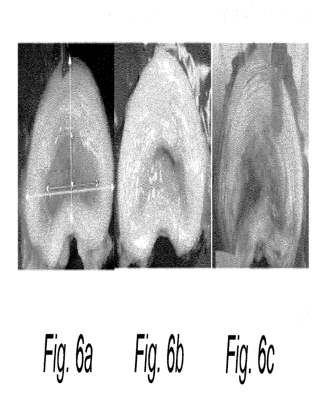
Fig. 6 illustrates macroscopic appearance and quantification of sclerosis of
the
porcine NP after treatment with the composition for use according to the
present invention;
Fig. 7 and 8 depict NP height and width, respectively;
Fig. 9 illustrates effect of the composition for use according to the present
invention
on the AF and NP; macroscopic appearance and quantification of sclerosis of
the porcine NP
after treatment with the composition for use according to the present
invention;
Figs. 10 and 11 illustrate changes in T2-weighted MRI images after treatment
with the
composition for use according to the present invention.
DETAILED DESCRIPTIONOF THE PRESENT INVENTION
The present invention will now be described hereinafter with reference to the
accompanying drawings, in which exemplifying embodiments of the present
invention are
shown. The present invention may, however, be embodied in many different forms
and should
not be construed as limited to the embodiments of the present invention set
forth herein; rather,
these embodiments of the present invention are provided by way of example so
that this
disclosure will convey the scope of the invention to those skilled in the art.
In the drawings,
identical reference numerals denote the same or similar components having a
same or similar
function, unless specifically stated otherwise.
A vertebral column of a vertebrate comprises vertebrae, which surround and
protect a
spinal cord. In humans, the vertebral column is situated in the dorsal aspect
of torso. Between
two adjacent vertebrae, an intermediate intervertebral disc is arranged, i.e.
the vertebrae are
alternated by intervertebral discs forming the vertebral column. The specific
structure and
further parts of the vertebral column are known to a person skilled in the
art.
Fig. 1 schematically shows a cross section of a vertebral column 100 of a
human.
Adjacent to a vertebral body 15 of a vertebra, an intervertebral disc
comprising an AF 10 and
a NP 11 is arranged. The NP 11 fills up the so-called disc space of the
intervertebral disc. The
AF 10 surrounds the NP 11 and defines the border of the NP as well as of the
disc space.
A spinal cord 17 is situated in the centre of the vertebral column, and
adjacent to the
intervertebral disc. Spinal nerves 16, 16', extend out from the spinal cord 17
to opposite sides
of and closely to the intervertebral disc.
CA 03202177 2023- 6- 13
WO 2022/129476 15
PCT/EP2021/086409
A facet joint 14, 14', is situated between an inferior articular process 13,
13' and a
superior articular process 12, 12'. On opposite sides of the spinal cord 17,
two facet joints 14,
14', are arranged, respectively. The facet joints 14, 14', are arranged in
approximately the
same cross-section and plane.
Fig. 2 schematically shows a segment of a vertebral column 200 comprising two
adjacent vertebras 20, 22. A first vertebra 22 and a second vertebra 20 are
arranged on
opposite sides of an intervertebral disc 21. The first vertebra 22 is arranged
relatively closer to
the thorax, and the second vertebra 20 is arranged relatively closer to the
sacrum. The caudal
endplate 23 of the first vertebra 22 and the cranial endplate 25 of the second
vertebra 20 are
shown in Fig. 2. The cranial endplate 25 and the caudal endplate 23 are facing
opposite sides
of the intervertebral disc 21.
Fig. 2 also schematically shows how a facet joint 24 is arranged between the
inferior
articular process of the first vertebra 22 and the superior articular process
of the second
vertebra 20. A transverese process 26 extends laterally from the vertebral
arch.
Fig. 3 schematically shows a lower part of a vertebral column 300. The
coccygeal
vertebrae 36 of the vertebral column is arranged at an end portion of the
lower part of the
vertebral column 300. The sacrum 39 of the vertebral column is arranged
adjacent to the
coccygeal vertebrae 36, closer to the thorax than the coccygeal vertebrae 36.
A fifth lumbar
vertebra, herein called L5, 30 is arranged adjacent to the sacrum 39, closer
to the thorax than
the sacrum 39. In a direction from the sacrum 39 towards the thorax, several
vertebras are
arranged in a row starting with L5, 30. Adjacent to the fifth lumbar vertebra
30, i.e. L5, the
following vertebras are arranged in order: a fourth lumbar vertebra 32, i.e.
L4, a third lumbar
vertebra, i.e. L3, a second lumbar vertebra, i.e. L2, and a first lumbar
vertebra 38, i.e. L1; the
first lumbar vertebra being arranged relatively closest to the thorax. In
between each two
adjacent vertebras, an intermediate disc 31 is arranged. Intervertebral discs
(not shown) are
also imposing the coccygeal vertebrae 36.
Fig. 4 depicts an example of a herniated IVD. As may be seen, during
herniation the
IVD is deformed such that the AF 10 is ruptured or weakened, allowing the NP
11 to protrude
outside its normal boundaries. The protrusion may exert pressure on a spinal
nerve, thus
causing pain and limited range of motion. Fig. 5 illustrates different types
of disc herniation.
CA 03202177 2023- 6- 13
WO 2022/129476 16
PCT/EP2021/086409
EXAMPLES
Example 1
Three series of in vivo experiments were performed. The focus of the series
differed
slightly but the methodology was identical unless otherwise stated. Because of
the different
aims, measurements were not completely overlapping but the most important
endpoint,
sclerotization of the NP was quantified identically in all studies. For this
reason, it was deemed
justified to merge data from the different series.
VVhen appropriate, Student's two-sided unpaired t test was used to compare the
effects of LA with placebo treatment. The null hypothesis (no difference
between LA and
placebo) was rejected at p<0.05. Correlation between NP size measured using
photographs
and MR images was analyzed using Excel in Office 365.
LA was purchased from Merck Emprove (Darmstadt, Germany) and lohexol from
Sigma Aldrich (St. Louis, MO, USA).
The pigs were pre-medicated by intramuscular injection of dexmedetomidin
(DomitorOvet, Orion Pharma, Sollentuna, Sweden) and a commercial mixture of
zolazepam
and tiletamine (Zoletilevet 100, Virbac, Carros, France) in conventional
doses. Anesthesia was
maintained by buprenophine (Vetergesicevet, Orion Pharma) 0.03 mg/kg
intramuscularly,
carpofen (Rimadylevet, Orion Pharma) 4 mg/kg intravenously and isoflurane
(Attane vet, VM
Pharma AB, Stockholm, Sweden) using a Servo 900 respirator (Siemens, Munich,
Germany).
Before recovering from anesthesia, atipamezol (AntisedanOvet, Orion Pharma)
was given
intramuscularly. On the first three days after surgery the pigs received oral
carprofen 2 mg/kg
twice daily.
The pigs were placed on the right side. A 6 cm long incision was made from the
costal
arch to the iliac crest, just lateral to the lateral processes. The lumbar
IVDs were approached
by a retroperitoneal technique. The L3/4 IVD was incised and 0.2 mL of the LA-
formulation
(n=6) or the placebo (n=2) was injected under fluoroscopic guidance. The LA-
formulation was
composed of LA (120 mg/mL) and iohexol (180 mg 1/mL) while placebo consisted
of iohexol
only. The concentration was determined based on the pilot experiments on the
effects of LA
on collagen secretion from human NP cells (as described in WO 2017/046030). In
this set of
in vivo experiments, the pH of the placebo formulation was adjusted to the
same as the active
formulation (about 1.5) using hydrochloric acid. The adjacent IVDs were not
injected and
CA 03202177 2023- 6- 13
WO 2022/129476 17
PCT/EP2021/086409
served as negative controls. The volume of the pig NP is about 1 mL and it was
estimated that
0.2 mL would be an appropriate injection volume. The height and width of
normally appearing
NP (Fig. 6a) in the anteroposterior (short vertical double arrow) and
bilateral (short horizontal
double arrow) directions, respectively, were measured and expressed in
relation to the height
(long vertical double arrow), and width (long horizontal double arrow) of the
IVD. Note the
reduction in NP size 28 (Fig. 6b) and 84 (Fig. 6c) days after LA treatment
compared with the
normally appearing NP (Fig. 6a). The group data for NP height as percent of
total IVD height
are shown in Fig. 7 and the corresponding data for NP width are illustrated in
Fig. 8.
Series 1
In the first pilot series, the objective was to determine if LA has the
potential to
sclerotize (induce fibrosis of) the IVD.
A total of 8 female pigs (mixed background of Yorkshire, Hampshire, and
Landrace)
with an average body weight of approximately 30 kg at surgery were used
according to the
procedure described above.
The animals were sacrificed 28 days after treatment and the spine from L1 to
S2 was
removed. The vertebral arches were removed from L1 to S1 to prevent
interference with disc
flexibility. The distances between the lateral processes of the vertebrae
cranially and caudally
to the injected IVD was measured with a caliper at full ipsi- or contralateral
flexion. The
difference of these distances was used as a measure of flexural rigidity.
These measures were
also documented for the untreated IVDs adjacent to the injected IVD in each
lumbar spine.
"Full flexion" was defined as the degree of flexion when stiffness is markedly
increased at
manual flexion at medium strength. The manual strength was not measured but
the flexion
was performed by two different individuals with identical results, which was
considered
sufficiently reliable to preliminarily demonstrate that the anatomical and
histological changes
seen indeed result in parallel biomechanical alterations.
Further, the IVD was cut in half and photographed. The bilateral width and
anteroposterior height of normally appearing NP was measured and expressed as
percent of
the total IVD width and height, respectively, as depicted in Figs. 7 and 8.
The findings on NP space size from series 1 and 2 (see below) were merged
since
the procedures used, were identical.
CA 03202177 2023- 6- 13
WO 2022/129476 18
PCT/EP2021/086409
There was a marked difference in flexural rigidity between the LA- and the
placebo-
injected IVDs as well as between the LA-injected IVDs and the 16 naïve IVDs
(Table 2). There
did not seem to be an effect on flexural rigidity of placebo injection as
compared to untreated
IVDs but the fact that only 2 pigs were injected with placebo precluded
statistical verification.
Table 2. Effects of LA on flexural rigidity of the isolated porcine lumbar
spine
Treatment Flexural rigidity (mm, mean+SD)
None 8.1+1.4 16
Placebo 8.2+1.8 2
LA, 120 mg/mL 2.7-4-1.1 6 <0.001
(none+placebo vs.
LA)
Series 2
The aim of the second series (including 16 pigs) was to assess the safety and
efficacy
of the treatment and was part of the regulatory documentation required by the
Swedish Medical
Products Agency to initiate studies in humans. All procedures were as
described above, with
the following exceptions. The pH of the placebo formulation was not adjusted
and since
placebo and 2 doses of LA (0.2 nnL of 120 or 240 mg/mL) and 3 survival times
(2, 28 and 84
days) were to be evaluated, injections were done in 3 congruent IVDs in each
pig to keep the
number of animals within reasonable limits. In one group of animals, LA or
placebo was applied
onto the IVD outside the spinal foramen (external application) to assess any
tissue damage
resulting from leakage from the IVD or a misdirected injection. Due to this
experimental design,
spinal flexural stiffness was not measured in these pigs. The only other
difference between the
two series was that exposed tissues were evaluated histologically in the
second but not first
series.
The animals were grouped as shown in Table 3.
CA 03202177 2023- 6- 13
WO 2022/129476 19
PCT/EP2021/086409
Table 3. Assignment of pigs to the 4 groups of series 2
Group Animal Numbers Exposure site LA concentration Lumbar disc
Follow-up period
(mg/mL) localisation
(days)
1 1-4 External 0 L. 2/3
2
External 120 L. 3/4
2
2 5-8 I ntradiscal 0 L. 2/3
2
I ntradiscal 120 L. 1/2
2
I ntradiscal 240 L. 3/4
2
3 9-12 I ntrad iscal 120 L. 1/2
28
External 120 L. 2/3
28
I ntradiscal 240 L. 3/4
28
4 13-16 I ntradiscal 0 L. 2/3
84
I ntradiscal 120 L. 1/2
84
I ntradiscal 240 L. 3/4
84
The lumbar spine was removed en bloc and any pathological changes were
assessed
macroscopically and photographed in extradiscal tissue onto which LA had been
applied.
Specimens of spinal nerves and muscle tissue were collected, fixed in 10%
formalin, and
processed for microscopy with hematoxylin/eosin staining. Injected IVDs were
cut in half,
inspected macroscopically, and photographed. Samples of the IVD were collected
and
processed for microscopy with hematoxylin/eosin. These turned out to include
AF but very little
NP so in order to obtain an improved overview of the IVD, the IVDs with
adhering vertebrae
were decalcified to enable preparation of axial sections of the whole IVD
which were stained
with Massons's trichrome.
Series 3
Since effects of LA on the NP in the subsequent clinical study was planned to
be
assessed using MRI, the third series focused on the effects of LA (0.2 mL, 60
mg/mL) on
sclerotization as measured by T2-weighted MRI. In addition, effects of LA on
the expression
of collagen I and ll were studied by immunohistochemistry (IHC). The
macroscopic changes
after LA at 60 mg/mL were similar to those described above.
MR images were obtained using a 7T small animal MRI system (Bruker Biospec). A
single 50 mm volume coil array was used (Tx/Rx). The dissected spines were
stripped of the
attached muscles, the spinous processes, transverse processes, and posterior
aspects of the
facet joints to allow them to fit in the MRI system. T2-weighted (TR: 2500-
2834 ms, TE: 33-
35.84 ms), 2D TurboRARE transversal and sagittal sequences were used. The TR
and TE
could not be maintained the same for all sequences due to required adjustments
in field of
CA 03202177 2023- 6- 13
WO 2022/129476 20
PCT/EP2021/086409
view the different specimens, leading to an increased pixel matrix which
affected the TR and
TE. For the transverse sequences, a 500 pm slice thickness with 750 pm
interslice distance
and an in-plane resolution of 166x166 pm was used. For the sagittal sequences,
a 750 pm
slice thickness with 2000 pm interslice distance and an in-place resolution of
166x166 pm was
used. In the transverse sequence of one of the pigs, the in-place resolution
was 174x166 pm
due to an increased field of view which could not be compensated for with a
higher matrix size
without significantly affecting TR.
Images were analyzed using Sante DICOM Viewer (version 8.1.5, Santesoft,
Athens,
Greece). The treated IVDs in all animals were assessed, and the IVD one level
above the
treated IVD was scanned as control.
The size of the NP was quantified using both MR images and photographs of the
IVDs
(see Fig. 6). The results from the macroscopic and MR analyses were then
correlated.
Immunohistochemical analysis of collagen I and ll
The tissue was dehydrated in graded ethanol and embedded in paraffin. Sections
were cut on a microtome, dewaxed, and incubated with polyclonal primary
antibodies against
collagen I (Abcam 34710) or II (Abcam 34712). The antibodies were diluted 250
(collagen I) or
200 (collagen II) times in phosphate-buffered saline containing 1% bovine
serum albumin.
Incubation was done at room temperature for 60 min. The sections were then
incubated for 30
min at room temperature with horseradish peroxidase-conjugated secondary
antibodies (Mach
2 Uni HRP, Biocare Medical) and immunoreactivity was visualized using
diaminobenzidine
(Biocare Medical).
Fig. 9 illustrates effects of LA on the AF (A,B) and NP (C-J). LA or placebo
(0.2 mL)
was injected into IVDs of anesthetized pigs. The animals were sacrificed 4 or
12 weeks post
injection and the IVDs were sectioned in half. After fixation in 10% formalin,
decalcification and
paraffin embedding, sections were cut at 5 pm and stained with
hematoxylin&eosin (A,B) or
Masson's trichrome (C-J). Fibrocartilagenous cells can be seen in the AF from
untreated IVDs
(arrows in A) but frequently, multicellular chondrons appeared after injection
of LA (240 mg/mL,
12 weeks survival; arrows in B). NP of an untreated (extradiscal injection)
IVD (C) consists of
islets of notochordal cells (asterisk) embedded in weakly stained
extracellular matrix. In LA-
treated NP, collagenous fibers can be seen (asterisks in D; 120 mg/mL LA, 4
weeks survival
and E; 120 mg/mL LA, 12 weeks survival). Occasionally, dense connective tissue
with
chondrocyte-like cells replaces the normal NP structure (F; 240 mg/mL LA, 12
weeks survival).
CA 03202177 2023- 6- 13
WO 2022/129476 21
PCT/EP2021/086409
Newly formed blood vessels (venules; arrows in F) and osteoid islets (asterisk
in F) can be
observed in the connective tissue. Boxed area in F is shown at higher
magnification in G and
newly formed blood vessels (arterioles, arrows) are shown in H. Cyst-like
structures of varying
size, possibly reflecting the "vacuum phenomenon", can be observed after LA
injection after 4
(1; 240 mg/mL) and 12 (J, 120 mg/mL) weeks. Occasionally, small hemorrhages
appear (arrow
in 1). Scale bar in all photomicrographs=100 pm.
Most histological analysis was done on the whole IVD sections stained with
Masson's
Trichrome both since these included central areas of the IVDs and also because
fibrous tissue
is more clearly visualized with Masson's than with hematoxylin&eosin staining.
At the two-day follow up period, hemorrhage and inflammatory changes were
evident
in occasional animals in which intradiscal injections had been done (not
shown). The
differences were not obviously more marked in animals that were treated with
LA than in those
injected with vehicle which suggests that these changes are associated with
the procedure,
rather than a specific LA effect. Similar findings were made in spinal nerves
and skeletal
muscle exposed to extraspinal LA (not shown).
Two days after injection with LA, there were marked histological changes
characterized by lysis of the extracellular matrix (blue areas) of the NP and
dissolution of
notochordal cells (white areas with dark nuclei), as depicted in Fig. 9K
(control) and 9L (injected
with LA). As may be seen, both cells and matrix are in a state of lysis and
degradation.
At the 28-day follow-up, there was a clear difference between the sites that
had been
treated with LA and those injected with placebo. Dense bundles of fibrotic
tissue were seen in
the animals that had been injected with LA (Fig. 9D). Similar to the
macroscopic analysis, there
did not appear to be any difference associated with the different
concentrations of LA. No
fibrotic changes were seen at the sites of extradiscal administration. In
addition, residual
hemorrhages, and cyst-like structures (Fig. 91) were observed 28 days after LA
injection. The
nature of the latter is unknown, but they may represent the "vacuum
phenomenon" according
to findings in series 3.
At the 84-day follow-up, there were clear differences between placebo and LA
treated
IVDs. The histological changes were also more pronounced after 84 compared to
28 days. In
LA injected IVDs, the following changes were seen in the NP: sclerosis (Fig.
9D-J), cystic
degeneration (Fig. 9J), cartilaginous metaplasia (Fig. 9F, 9G), osteoid
islands (Fig.9F, 9G) and
reduction in the extracellular matrix. At 84 days, blood vessels appeared in
the fibrotic tissue
CA 03202177 2023- 6- 13
WO 2022/129476 22
PCT/EP2021/086409
(Fig. 9H). There was no clear distinction in the degree of the changes between
IVDs treated
with the high or low dose of LA. The changes after LA treatment were not
restricted to the NP
since nnulticellular chondrons were observed in the AF (Fig. 9B).
The macroscopic changes after LA at 60 mg/mL were similar to those described
above. These were reproduced on T2-weighted MRI where, as expected, the
intensity of the
sclerotized NP was much lower (Fig. 9B). The lamellar structure of newly
formed connective
tissue was also visualized by MRI. There was a close correlation (correlation
coefficient=0.97)
between the degree of sclerotization as estimated by analysis of photographs
and MR images.
Dark, mostly rounded areas were observed in 4 out of 5 LA-injected IVDs. They
were
pronounced in 2 IVDs and discrete (Fig. 9B) in the 2 others. These areas
probably reflected
the vacuum phenomenon and might be related to the large cyst-like structures
seen
microscopically.
Analysis of the disc height and disc width of treated and control IVDs showed
that
there was a very pronounced reduction in height but not width of the treated
IVDs, as may be
seen in Table 4. The relative disc height was expressed as a ratio of height
of treated
IVD/height of untreated (control) IVD. Analogously, the relative disc width
was expressed as a
ratio of width of treated IVD/width of untreated (control) IVD.
Table 4. Relative changes in height and width between treated and control IVDs
Animal nr. Relative disc height Relative disc
width
1 0.2 1.01
2 0.33 0.99
3 0.7 0.99
4 0.36 1.04
5 0.55 1
Mean value 0.43 1.01
The effect is also confirmed as illustrated in the MR image shown in Fig. 12,
wherein
arrow A identifies an untreated control IVD, and arrow B identifies an IVD
injected with 60
mg/ml LA 30 days prior to the image. As is evident from Fig. 12, a significant
reduction in height
of the treated IVD is observed.
CA 03202177 2023- 6- 13
WO 2022/129476 23
PCT/EP2021/086409
In untreated IVDs, collagen I immunoreactivity was sparsely expressed in the
NP but
found at high levels in the AF. In contrast, collagen II immunoreactivity was
located both in the
NP and AF. There was no clear difference in collagen II immunoreactivity
between treated and
untreated IVDs but collagen I was strongly induced in the NP after LA
treatment.
Example 2
The study was a randomized, double-blinded, placebo-controlled, single
ascending
dose study with the primary objective to evaluate safety and tolerability
following intradiscal
injection of LA in Omnipaque or placebo (Omnipaque) in 15 patients with
chronic discogenic
low back pain. The secondary objective was to assess effects on the NP and
disc height using
T2-weighted MRI.
Scoring of visual analog scale (VAS) for both back and leg pain and Oswestry
Disability Index (ODI) were used as exploratory objectives. The sample size
was not based on
power calculations but was considered appropriate for a single ascending dose
study to
provide initial safety data on 3 different dose levels of the LA formulation.
The study has been
registered on the ClinicalTrials.gov website and on the EU Clinical Trials
Register. It was
approved by the Stockholm Regional Ethics Committee (approval No. 2016-2323-
31/4) and by
the Swedish Medical Products Agency (approval No. 5.1-2016-86227).
Patients were recruited at Stockholm Spine Center (Upplands Vasby, Sweden)
between April 2017 and August 2018.
Fifteen patients were randomized to either of the 3 dose groups. In each
group, 2
patients were randomized to placebo and 3 to LA at 45, 90 and 180 mg (1.5 mL
at 30,60 and
120 mg/mL. The first 2 patients in each dose group were randomized to LA or
placebo.
Provided that no safety or tolerability concerns were identified within the
first week (up to Visit
3) after administration (confirmed by a safety review committee consisting of
2 medical experts
and a chairman without voting rights), another 3 patients received active
treatment or placebo
(2:1). Prior to dose escalation, the safety review committee assessed all
safety data up to Visit
3 (1 week after treatment). In case of any safety or tolerability concerns,
dosing could be
discontinued, or the planned dose could be lowered.
The patient's total time in the study was up to 14 months. Each patient
performed a
screening visit within 60 days of planned treatment. An independent
biostatistician at LINK
Medical Research AB (Uppsala, Sweden) prepared a list of randomization
numbers.
CA 03202177 2023- 6- 13
WO 2022/129476 24
PCT/EP2021/086409
Randomization was performed via an eCRF system (ViedocTM) at least 5 working
days before
the planned treatment day to allow time for preparation of the investigational
medical product
(IMP). The test and reference IM Ps were identical in appearance. The kits
were labelled with
the randomization number but did not contain any information on the identity
of the formulation.
This ensured that all staff at the clinics as well as the patients were blind
to the treatment code.
Treatment code envelopes were provided for each randomized patient. The code
envelopes
were kept in a secure place with limited access. In case of an emergency
making it crucial for
the investigator, or any other treating physician, to know whether the patient
had received
active formulation or placebo, the code envelope was to be opened. However, no
such
emergencies occurred and no treatment code envelope was opened.
The test product in this study was containing the active ingredient (S)-LA,
which was
provided to the site as a sterile solution in a syringe for single use. The LA
doses administered
were 45, 90 and 180 mg in a volume of 1.5 mL, corresponding to LA
concentrations of 30, 60
and 120 mg/mL. The formulation for administration was prepared extempore and
contained
the contrast agent iohexol (OmnipaqueTM) and water for injection. The final
concentration of
iohexol in the injected solution was 388 mg/mL. Small amounts of tromethamine,
Na2Ca
edetate and HCI were also present. The solution was clear and colourless to
slightly coloured.
LA (batch C16077AA) was manufactured, packed into vials and bulk labelled at
Recipharm, Stockholm, Sweden. Other components for the IMP (Omnipaque [batch
number
13407744] and water for injection) were purchased and released by Recipharm,
Sweden. All
components for the IMP were shipped to an extempore pharmacy laboratory as 1
kit per patient
containing 1 vial of LA, 1 vial of Omnipaque, 1 vial of water for injection
(active and placebo),
together with preparation instructions and patient-specific labels that had
been prepared by
Recipharm. The final solution for injection was prepared aseptically by the
extempore
pharmacy laboratory (Apoteket AB Hospital Pharmacy, Uppsala, Sweden), packed
into
patient-specific syringes and labelled with "blinded" labels for each patient.
The IMP prefilled
and labelled syringes for each patient were sent to the clinic. If 2
injections were planned, 2
separate syringes were prepared (i.e. one syringe per IMP injection).
Matching placebo solution with identical appearance to the test product was
used as
reference treatment. The solution for administration was prepared extempore
and contained
the contrast agent iohexol (Omnipaque, batch number 13407744) diluted in water
to a final
iohexol concentration of 388 mg/mL and small amounts of tromethamine, Na2Ca
edetate and
HCI. An identical volume as for the test product (1.5 mL) was injected.
CA 03202177 2023- 6- 13
WO 2022/129476 25
PCT/EP2021/086409
Since the study was too small to statistically evaluate effects of LA on the
T2-weighted
intensity of the NP, IVD height, VAS and ODI, only descriptive statistics were
applied. All
statistical analyses were performed using SAS version 9.4 (SAS Institute
Inc., Cary, NC,
USA). Results were presented by treatment group and in total where
appropriate.
Continuous data were summarized using descriptive statistics, where the
following
parameters were reported: Number of patients with evaluable and missing
observations,
arithmetic mean, and standard deviation, median, first and third quartiles and
minimum and
maximum. Categorical data were presented as absolute and relative frequencies.
When the
absolute frequency was zero, the percentage was not presented. Unless stated
otherwise, the
denominator for percentage calculations was the total number of patients in
the applicable
analysis set, including patients with missing data. For variables with missing
values, the
number and percentage of patients with missing values was presented.
At the treatment day, patients received premedication with a sedative or
anxiolytic
and antibiotics, given as an i.v. infusion approximately 15 min before the
intradiscal injection.
The patient was placed in the right lateral decubitus position and injection
was done using the
two-needle technique under fluoroscopy guidance. Once correct intradiscal
needle placement
had been confirmed, a small volume (approximately 0.5 mL) of the formulation
was first
injected slowly (during half a minute) to verify that the distribution of the
injectate was confined
to the disc and that there was no leakage. If no leakage had occurred within
half a minute, this
procedure was repeated twice until the entire volume (1.5 mL) had been
injected. If there was
any leakage from the disc during injection, either after injection of a small
volume or the entire
volume (1.5 mL), the injection was interrupted. Immediately after treatment,
patients had to
remain in the prone position (alternatively in the lateral decubitus or supine
position) for as long
as possible (at least 4 hours after the last injection). All patients stayed
overnight at the clinic
after the injection, for observation and safety assessments. After leaving the
clinic, patients
were to be treated with analgesics and/or other measures according to standard
clinical
practice. They were also given advice on restricted physical activity during
the first week.
Physical examination, blood pressure, heart rate, electrocardiogram (ECG),
clinical
chemistry and hematology were assessed using routine clinical methods. Pain
during and 15
min after injection was measured using a VAS scale (0-100 mm) and the patient
reported
whether or not the pain reproduced the pain normally experienced in terms of
location and type
of pain. The distribution of Omnipaque within the IVD was documented according
to the Dallas
discogram scale. Local site reactions to the injection were noted, and AEs
were graded for
CA 03202177 2023- 6- 13
WO 2022/129476 26
PCT/EP2021/086409
seriousness, intensity, and causality to the treatment. SAEs were defined
according to the
International Conference of Harmonisation guideline E2A.
All MRI investigations were performed using the same scanner (Siemens 1.5 T
Avanto) with the following exceptions: Baseline MRI for one placebo-treated
patient and 2
patients given the high dose was acquired in a Siemens Symphony 1.5 T scanner
while all
other scans were done in the Siemens 1.5 T Avanto scanner. Also, baseline and
3 month MRI
were done in the Symphony scanner while the other scans were done in the
Avanto scanner
in 2 placebo-treated patients and one patient treated with the high dose. Ti-
and T2-weighted
sagittal, corona!, and transversal sections were acquired at 4 mm before and
after injection of
gadolinium contrast.
Disc height measured as the maximal distance between 2 adjacent endplates and
Pfirrmann grade were documented at screening and they were followed throughout
the study.
Since the Pfirrmann criteria are not sensitive enough to detect minor changes
in disc hydration,
changes in intensity of the NP at T2-weighted MRI were scored by 4 assessors
blind to the
group assignment of the patients. The assessors were instructed to score no
change as "0"
and obvious reduction in intensity as "1", regardless of time after treatment.
Patients reported their low back and leg pain level using a 0-100 mm VAS at
the times
indicated in Table 5. Disability was evaluated using the ODI.
Table 5. Scoring of pain during injection and 15 min post injection
VAS pain injection site (mm) STA363 STA363 STA363 Placebo
Total
45 mg 90 mg 180 mg (N=6) (N=15)
(N=3) (N=3) (N=3)
Visit 2 (Treatment day), post-dose
n/nm iss 4/0 5/0 5/0 9/0
23/0
Mean (SD) 78 (10) 48 (20) 26 (36)
27 (32) 40 (33)
Median 75 45 10 10
30
Min, Max 70, 90 30, 75 0, 90 5, 100 0, 100
Visit 2 (Treatment day), 15 min
n/nm iss 4/0 5/0 5/0 9/0
23/0
Mean (SD) 20 (13) 25 (27) 9
(13) 23 (22) 20 (20)
Median 20 30 0 10
15
Min, Max 4, 35 0, 65 0, 30
0, 70 0, 70
n/nmiss = number of injections with evaluable/missing data, SD = standard
deviation.
CA 03202177 2023- 6- 13
WO 2022/129476 27
PCT/EP2021/086409
Pain intensity during the injection was higher in the 45 mg LA group compared
to all
other groups, but 15 min after the injection all groups reported similar
levels of pain.
There were in total 24 AEs reported by 11 patients in the study (Table 6). All
AEs were
of mild-to-moderate intensity. The most common AEs were injection site pain
(reported by all
groups) and back pain (reported by all groups except the 60 mg/mL LA group).
There were
16/24 AEs possibly or probably related to the treatment and 13 of those
related to pain during
or immediately after injection. All AEs were resolved within the 3-month
follow-up period. There
were no SAEs and none of the AEs led to patient withdrawal. During the
extension phase, 6
AEs were reported by 4 patients and were judged as mild in intensity and with
an unlikely
relationship to the treatment.
Table 6. Adverse events by severity and causality
LA 30 mg/mL (n=3) LA 60 mg/mL (n=3) LA 120 mg/mL (n=3)
Placebo (n=6) Total (N=15)
n (u/o) m n (u/o) m n (u/o) m n (%) m
n (Y()) m
Any adverse 2 (67%) 4 3 (100%) 3 3 (100%) 8
3 (50%) 9 11(73%) 24
event
Any serious 0 0 0 0 0 0 0 0
0 0
adverse event
Adverse events by severity
Mild 1 (33%) 2 1 (33%) 1 3 (100%) 6
2 (33%) 3 7 (47%) 12
Moderate 1 (33%) 2 2 (67%) 2 1 (33%) 2
3 (50%) 6 7 (47%) 12
Severe 0 0 0 0 0 0 0 0
0 0
Adverse events by causality
Unlikely 2 (67%) 3 0 0 2 (67%) 3
2 (33%) 2 6 (40%) 8
Possible 0 0 0 0 1 (33%) 1 2 (33%) 4
3 (20%) 5
Probable 1(33%) 1 3(100%) 3 2(67%) 4
2(33%) 3 8(53%) 11
n = number of patients, m = number of events.
Percentages are based on the number of patients within each treatment group.
There was a tendency for a dose-response relationship with regard to
posttreatment
dehydration (Table 7). Two representative examples of changes in NP intensity
after treatment
with LA are presented in Figs. 10 and 11. Images in Fig. 10 are obtained from
a patient treated
with 60 mg/mL LA (L4/5 and L5/S1) and images in Fig. 11 are obtained from a
patient injected
with LA at 120 mg/mL (L4/5). The images (left to right) were acquired at
screening and after 3,
6 and 12 months. Like the findings in pig IVDs, loss of intensity of the NP,
probably reflecting
sclerosis, often occurred in the periphery.
CA 03202177 2023- 6- 13
WO 2022/129476 28
PCT/EP2021/086409
Table 7. Changes in intensity of the NP at T2-weighted MR imaging
LA NP intensity Baseline disc Disc
height change from baseline
concentration height (mm) (mm)
(mg/mL) Mean+SD
_
Mean+SD Median Mean+SD
3 months 6 months 12 months
0 0.2+0.4 0 9.8+1.3 -0.2+0.7 -
0.5+1.4 -1.0+1.4
30 0.4+0.5 0 9.5+1.7 0.3+1.3 -
0.5+0.7 -0.5+0.7
60 1.0+0.0 1 9.4+0.9 -1.0+0.7 -
0.8+0.8 -1.2+0.8
120 0.9+0.3 1 11.2+1.6 -1.2+1.1 -
1.4+1.1 -1.8+1.3
Changes in intensity of the NP at T2-weighted MR imaging were scored as 1,
clear
reduction and 0, no change. The scoring was done by 4 assessors blind to the
group
assignment of the patients. Reductions in disc height were observed throughout
the study
(Table 8).
CA 03202177 2023- 6- 13
WO 2022/129476 29
PCT/EP2021/086409
Table 8. Individual scoring of change in NP intensity
Patient No. IVD LA concentration Assessor No.
1 2 3 4
1 L5/S1 0 0 1 1 0
2 L4/51 0 0 0 0 1
2 L5/S1 0 0 0 0 1
3 L4/5 0 0 0 0 0
4 L3/4 0 0 0 0 1
4 L4/5 0 0 0 0 1
L5/S1 0 0 0 0 0
6 L4/5 0 0 0 0 0
6 L5/S1 2 0 0 0 0 0
7 L3/4 30 0 0 0 0
8 L4/53 30 1 1 1 1
8 L5/S1 4 30 1 0 1 1
9 L5/S1 30 0 0 0 0
L3/4 60 1 1 1 1
10 L4/5 60 1 1 1 1
11 L4/5 60 1 1 1 1
11 L5/S1 60 1 1 1 1
12 L5/S1 60 1 1 1 1
13 L4/5 120 1 1 1 1
14 L4/5 120 1 0 1 1
14 L5/S1 120 1 0 1 1
L3/4 120 1 1 1 1
15 L4/5 120 1 1 1 1
As may be seen, a clear reduction in disc height is achieved by administration
of the
composition for use according to the present invention.
It is evident that the composition for use according to the present invention
dose-
5 dependently reduces disc height (Table 9). Although the number of
patients in the study is low,
it is expected that dehydration of the NP is accompanied by deflation of the
disc. The reduction
in disc height is similar to that observed after chemonucleolysis (Table 9).
There was no
change in IVD width (not shown) demonstrating that IVD volume was attenuated.
CA 03202177 2023- 6- 13
WO 2022/129476 30
PCT/EP2021/086409
Table 9. Reduction in disc height provided by chemonucleolysis and the
composition for use according to the present invention
% decrease in disc height
Dose of 3 months 6 months 12 months Chymopapain or
lactic acid condoliase
(mg/m L)
0 2 5 10 16*
30 +3 5 5 30f
60 11 8 13 9$
120 11 12 16
*7 years FU; Leivseth et al., 1999, 5 weeks to one year FU; height loss
correlated with loss
of T2 signal; Szypryt et al., 1987; $ one year FU; Chiba et al, 2018
It should be noted that even the highest dose of LA provides a reduction in
disc height
comparable with the relatively low dose of condoliase.
Finally, changes in exploratory endpoints after treatment were assessed. At
baseline,
no leg pain was reported in the 45 mg group. Mean leg pain at screening was 3
mm in the 90
mg group, 14 mm in the 180 mg group and 26 mm in the placebo group. Overall,
leg pain
remained fairly low throughout the study without any noticeable changes
occurring (data not
shown).
Baseline mean back pain was 19 mm in the 45 mg group, 44 mm in the 90 mg
group,
52 mm in the 180 mg group and 50 mm in the placebo group.
At screening, all treatment groups had a moderate degree of disability in
their
everyday activities, based on the mean ODI values. No particular trends in
changes over time
could be discerned (see below).
CA 03202177 2023- 6- 13
WO 2022/129476 31
PCT/EP2021/086409
Table 10. Changes in VAS (mm) for cLBP in the 4 treatment groups
LA LA LA Placebo
45 mg 90 mg 180 mg
(N=6)
(N=3) (N=3) (N=3)
Visit 1 (Screening)
n/nmiss 3/0 3/0 3/0
6/0
Mean (SD) 19 (10) 44 (25) 52
(18) 50 (22)
Visit 5 (3 months)
n/nmiss 3/0 3/0 3/0
6/0
Mean (SD) 22 (8) 44 (22) 27 (15)
40 (23)
Visit 6 (6 months)
n/nmiss 2/1 3/0 3/0
5/1
Mean (SD) 52 (28) 44 (6) 28 (32)
41(30)
Visit 7 (12 months)
n/nmiss 2/1 3/0 3/0
5/1
Mean (SD) 59 (40) 34 (14) 61(26)
41(24)
n/nmiss = number of patients with evaluable/missing data, SD = standard
deviation
Table 11. Changes in ODI in the 4 treatment groups
ODI
STA363 STA363 STA363 Placebo
45 mg 90 mg 180 mg
(N=6)
(N=3) (N=3) (N=3)
Visit 1 (Screening)
n/nmiss 3/0 3/0 3/0
6/0
Mean (SD) 32 (2) 28 (13) 25 (10)
29 (9)
Visit 5 (3 months)
n/nmiss 3/0 3/0 3/0
6/0
Mean (SD) 35(10) 29(12) 18(11)
26(6)
Visit 6 (6 months)
n/nmiss 2/1 3/0 3/0
5/1
Mean (SD) 46(11) 32(9) 23(16)
27(10)
Visit 7 (12 months)
n/nmiss 2/1 3/0 3/0
5/1
Mean (SD) 61(32) 29(15) 28(11)
21(10)
n/nmiss = number of patients with evaluable/missing data, SD = standard
deviation
Two days after injection of porcine IVDs with LA, lysis of extracellular
matrix
accompanied by cell death and disappearance were observed. These changes are
identical
to those seen after injection of typical chemonucleolytics such as
chymopapain. Therefore, LA
CA 03202177 2023- 6- 13
WO 2022/129476 32
PCT/EP2021/086409
can be labelled as a new chemonucleolytic substance, alongside previously
known
chemonucleolytics such as chymopapain, condoliase, ozone and ethanol.
The present study shows that LA transforms the porcine NP into connective
tissue
within one month, as supported by macroscopic and MRI findings. Rapid onset
lysis of the NP
followed by a more slowly developing fibrosis are typical effects of well-
characterised
chemonucleolytics. Therefore, LA is by definition a newly identified
chemonucleolytic.
LA injection caused a pronounced reduction in lateral flexibility verifying
that the newly
formed connective tissue stabilised the motion segment. There was a high level
of expression
of collagen I immunoreactivity in the sclerotized IVDs which may have
contributed to the
increased stability of the IVD. The sclerosis remained and was even more
advanced after 3
months, so it is reasonable to assume that the increased rigidity persisted
throughout the
experimental period. Remodelling of the AF may also have contributed to
reduced flexibility
since chondrocytic metaplasia was found in the AF.
Histologically, appearance of cartilaginous tissue was evident after 3 months,
and in
some IVDs, osteoid islands were seen. Such a tissue transformation suggests
that the
reduction in flexibility of the lumbar spine may progress over a relatively
long time. Another
finding was that neovascularization occurred in the fibrotic tissue 3 months
after LA
administration. It has been suggested that angiogenesis is accompanied by
neoinnervation
which may generate pain. However, a clear distinction between vasoregulatory
and
nociceptive afferents has rarely been done. Moreover, angiogenesis and nerve
sprouting in
degenerated human IVDs seem to be guided by fissures in the AF. Since such
fissures were
not seen after LA injection, it is possible that the mechanism of invasion of
nerves and blood
vessels, and the composition of nerve fibres (if they indeed sprout at all),
differ from those
occurring spontaneously. VVhile there are similarities, it must be recognized
that the rapid
sclerotization of the IVD produced by LA does not replicate pathologically
degenerated IVDs
of patients suffering from low back pain. The most important difference in
this regard is the
absence in the pig studies of some hallmarks of pathological IVD degeneration
such as annular
fissures and lamellar disorganization.
Although the pig studies include low number of replicates in some groups, this
limitation
is not likely to affect the conclusions.
It was confirmed that LA transformed the NP into connective tissue in pigs,
and MRI
observations suggested that this also occurred in patients.
CA 03202177 2023- 6- 13
WO 2022/129476 33
PCT/EP2021/086409
Sclerotization of the IVD makes it more stable resulting in a reduced range of
motion
which may reduce the probability of herniation relapse. Another effect of the
LA-induced
sclerosis is prevention of leakage of algogenic molecules and sprouting of
nociceptive
afferents.
The treatment was safe and tolerable. Several patients reported a relatively
short-
lasting low back pain (cLBP) after injection but this was also seen in the
placebo group.
Moreover, with the limitation of the low number of patients, the treatment
does not seem to
aggravate the cLBP as indicated by the VAS and ODI results.
The MRI results showed that LA induces a reduction in signal intensity in the
NP. This
may be due to increased breakdown of glycosaminoglycans contributing to
hydration of the
NP followed by proliferation of connective tissue. As in pigs, the loss of
signal intensity in
patients was predominantly seen in the periphery of the NP. There was a
tendency for a
reduction in IVD height after treatment in patients. This effect was more
pronounced in porcine
IVDs injected with LA, while and in both porcine and human IVDs, IVD width was
unchanged.
This indirectly but compellingly demonstrates that IVD volume must have
decreased. A
reduced IVD volume is the goal of all chemonucleolytic treatments since volume
reduction
causes a decrease in IVD pressure which, in turn, reduces the size of the
herniation leading
to disappearance or amelioration of symptoms.
The results from the study thus provide proof of concept that the composition
for use
according to the present invention effectively reduces the disc height and
transforms the disc
space into connective tissue.
The examples in this application show that the administration of lactic acid
results in
volume and height reduction, and also a change of the tissue composition of
the disc and in a
flexion stiffness of the treated IVDs.
While the present invention has been illustrated in the appended drawings and
the
foregoing description, such illustration is to be considered illustrative or
exemplifying and not
restrictive; the present invention is not limited to the disclosed
embodiments. Other variations
to the disclosed embodiments can be understood and effected by those skilled
in the art in
practicing the claimed invention, from a study of the drawings, the
disclosure, and the
appended claims. In the appended claims, the word "comprising" does not
exclude other
elements or steps, and the indefinite article "a" or "an" does not exclude a
plurality. The mere
fact that certain measures are recited in mutually different dependent claims
does not indicate
CA 03202177 2023- 6- 13
WO 2022/129476 34
PCT/EP2021/086409
that a combination of these measures cannot be used to advantage. Any
reference signs in
the claims should not be construed as limiting the scope.
CA 03202177 2023- 6- 13