Note: Descriptions are shown in the official language in which they were submitted.
FEMORAL COMPONENT FOR A KNEE PROSTHESIS
WITH IMPROVED ARTICULAR CHARACTERISTICS
BACKGROUND
1. Technical Field.
[0001] The present disclosure relates to orthopaedic prostheses and,
specifically, to
femoral components in a knee prosthesis.
2. Description of the Related Art.
[0002] Orthopaedic prostheses arc commonly utilized to repair and/or
replace damaged
bone and tissue in the human body. For a damaged knee, a knee prosthesis may
be implanted using
a tibial base plate, a tibial bearing component, and a distal femoral
component. The tibial base
plate is affixed to a proximal end of the patient's tibia, which is typically
resected to accept the base
plate. The femoral component is implanted on a distal end of the patient's
femur, which is also
typically resected to accept the femoral component. The tibial bearing
component is placed
between the tibial base plate and femoral component, and may be fixedly or
slidably coupled to the
tibial base plate.
[0003] The femoral component provides articular surfaces which interact
with the adjacent
tibial bearing component and a natural or prosthetic patella during extension
and flexion of the
knee. The features and geometry of the articular surfaces of the femoral
component influence the
articular characteristics of the knee, such as by cooperating with the tibial
bearing component to
define flexion range, internal/external rotation, femoral rollback and
patellar tracking, for
example. The nonarticular, bone contacting surfaces of the femoral component
define the shape
and geometry of the bone resection on the distal femur, and therefore
influence the amount of bone
resected from the femur.
Date Recue/Date Received 2023-06-13
[0004] Further, the overall shape and geometry of the femoral component,
particularly
around its outer periphery, influences the interaction between the knee
prosthesis and adjacent soft
tissues remaining in place after prosthesis implantation.
[0005] Accordingly, substantial design efforts have focused on providing
knee prosthesis
components which preserve flexion range, promote desirable kinematic motion
profiles, protect
natural soft tissues, and are compatible with the widest possible range of
prospective knee
replacement patients.
SUMMARY
[0006] The present disclosure provides an orthopaedic knee prosthesis
including a femoral
component which exhibits enhanced articular features, minimizes removal of
healthy bone stock
from the distal femur, and minimizes the impact of the prosthesis on adjacent
soft tissues of the
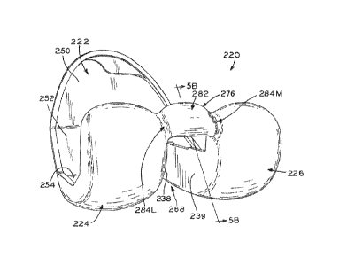
knee.
[0007] Features which operate to enhance articulation include: 1)
bulbous posterior
geometry of the femoral condyles, as viewed in a sagittal cross-section (i.e.,
the "J-curve"),
facilitates deep flexion and low component wear by reconfiguring the J-curve
curvature at flexion
levels above 90-degrees; 2) provision of "standard" and "narrow" femoral
components which
share a common bone-resection sagittal profile but define different peripheral
and articular
geometries designed to accommodate natural variability in patient anatomy; and
3) a lateral
posterior femoral condyle which is shorter (i.e., defines a reduced
proximal/distal dimension) as
compared to the medial posterior condyle, thereby facilitating deep flexion
and the attendant
external rotation of the femur while avoiding impingement between prosthesis
components.
[0008] Features which operate to minimize impact of the prosthesis on
adjacent soft
tissues of the knee include: 1) for posterior-stabilized (PS) designs, a
femoral cam with a generally
cylindrical articular surface, in which the articular surface is flanked at
its medial and lateral ends
by broad, large-radius convex-to-concave transitions to the adjacent medial
and lateral femoral
condyles, thereby ensuring a desired cam/spine articular interaction while
avoiding potential
soft-tissue impingement; 2) for cruciate retaining (CR) designs, an asymmetric
intercondylar
notch which accommodates external rotation of the femur in deep flexion while
avoiding
impingement between intercondylar wall surfaces and the posterior cruciate
ligament; and 3) an
2
Date Recue/Date Received 2023-06-13
anterior flange including a patellofemoral groove or sulcus, in which the
medial and lateral
surfaces near the edge of the flange define broad, large-radius convexity,
thereby accommodating
soft tissues in the anterior portion of the knee.
[0009] Features which allow femoral components made in accordance with
the present
disclosure to be implanted with minimal bone removal include: 1) an anterior
bone contacting
surface, opposite the patellar groove of the anterior flange, which includes
an edged central peak
operable to maintain a desired material thickness throughout the anterior
flange while reducing the
overall average thickness of the anterior flange; 2) for posterior-stabilized
(PS) implant designs, an
intercondylar box with sloped sidewalls which selectively reduce the
proximal/distal height of
portions of the sidewalls, to facilitate preservation of bone near the
anterior end of the anatomic
intercondylar notch; 3) for PS designs, intercondylar box sidewalls which are
configured to
function as a fixation lug, thereby obviating the need for fixation pegs; 4)
consistently small
incremental growth between respective pairs of prosthesis sizes, thereby
allowing minimal bone
resection for a greater majority of patients; and 5) a specially designed
"pocket" on the bone
contacting side of the femoral component for bone cement and/or porous bone-
ingrowth material,
in which the pocket maximizes long-term fixation while also facilitating
potential component
removal in revision surgery.
[0010] According to one embodiment thereof, the present invention
provides a
posterior-stabilized femoral component adapted to articulate with a tibial
bearing component in a
knee prosthesis, the tibial bearing component including a proximally extending
spine, the femoral
component comprising: medial and lateral condyles shaped to articulate with
the tibial bearing
component through a range of motion, in which full extension corresponds to
zero degrees flexion
of the knee prosthesis and positive flexion corresponds to greater than zero
degrees flexion of the
knee prosthesis, the medial and lateral condyles comprising inwardly facing
condylar walls
forming an intercondylar space therebetween, the intercondylar space having a
medial/lateral
width; and a femoral cam spanning the intercondylar space to join the medial
and lateral condyles
to one another, the femoral cam sized and positioned to engage the spine of
the tibial bearing
component in positive flexion through at least a portion of the range of
motion, the femoral cam
having an articular surface comprising: a central articular surface that is
one of cylindrical and
convex across a medial/lateral extent of the central articular surface; a
convex medial transition
3
Date Recue/Date Received 2023-06-13
surface flanking the central articular surface and disposed between the
central articular surface and
the medial condylc; and a convex lateral transition surface flanking the
central articular surface
and disposed between the central articular surface and the lateral condyle,
the central articular
surface, the convex medial transition surface and the convex lateral
transition surface cooperating
to occupy at least 80% of the medial/lateral width of the intercondylar space.
100111 According to another embodiment thereof, the present invention
provides a
posterior-stabilized femoral component adapted to articulate with a tibial
bearing component in a
knee prosthesis, the tibial bearing component including a proximally extending
spine, the femoral
component comprising: medial and lateral condyles shaped to articulate with
the tibial bearing
component through a range of motion, in which full extension corresponds to
zero degrees flexion
of the knee prosthesis and positive flexion corresponds to greater than zero
degrees flexion of the
knee prosthesis, the medial and lateral condyles comprising inwardly facing
condylar walls
forming an intercondylar space therebetween, the intercondylar space having a
medial/lateral
width; and a femoral cam sized and positioned to engage the spine of the
tibial bearing component
in positive flexion through a portion of the range of motion, the femoral cam
comprising a
medial/lateral cam length spanning the intercondylar space such that the
femoral cam joins the
medial and lateral condyles to one another, the femoral cam having an
articular surface
comprising: a central articular surface that is one of cylindrical and convex
across a medial/lateral
extent of the central articular surface; a convex medial transition surface
flanking the central
articular surface and disposed between the central articular surface and the
medial condyle; and a
convex lateral transition surface flanking the central articular surface and
disposed between the
central articular surface and the lateral condyle, the convex medial
transition surface and the
convex lateral transition surface each defining an arc extending in a
medial/lateral direction, the
arc defining a radius equal to between 40% and 60% of the medial/lateral cam
length, whereby the
femoral cam defines widely rounded, convex surfaces.
100121 According to yet another embodiment thereof, the present
invention provides a
posterior-stabilized femoral component adapted to articulate with a tibial
bearing component in a
knee prosthesis, the tibial bearing component including a proximally extending
spine, the femoral
component comprising: a medial condyle comprising: a medial condylar surface
shaped to
articulate with a medial articular compartment of the tibial bearing component
through a range of
4
Date Recue/Date Received 2023-06-13
motion; and a medial posterior bone-contacting surface disposed opposite the
medial condylar
surface and positioned to abut a posterior facet of a resected femur upon
implantation of the
femoral component, the medial posterior bone-contacting surface extending
between a medial
edge of the femoral component and a medial intercondylar wall; a lateral
condyle separated from
the medial condyle by a component sagittal plane, the lateral condyle
comprising: a lateral
condylar surface shaped to articulate with a lateral articular compartment of
the tibial bearing
component through the range of motion; and a lateral posterior bone-contacting
surface disposed
opposite the lateral condylar surface and positioned to abut the posterior
facet of the resected
femur upon implantation of the femoral component, the lateral posterior bone-
contacting surface
extending between a lateral edge of the femoral component and a lateral
intercondylar wall facing
the medial intercondylar wall; and a patellar flange extending anteriorly from
the medial and
lateral condyles and shaped to articulate with a patellar articular surface,
the patellar flange
comprising: a flange articular surface shaped to articulate with the patellar
articular surface; an
anterior bone-contacting surface disposed opposite the flange articular
surface and positioned to
abut an anterior facet of the resected femur upon implantation of the femoral
component; and a
distal bone-contacting surface extending along an anterior/posterior space
between the anterior
bone-contacting surface and the medial and lateral posterior bone-contacting
surfaces, the lateral
and medial intercondylar walls each defining posterior wall portions extending
proximally from
the distal bone-contacting surface to define a proximedistal extent of the
posterior wall portions,
the lateral and medial intercondylar walls comprising angled lateral and
medial anterior wall
portions, respectively, the angled lateral and medial wall portions each
sloping distally toward the
distal bone-contacting surface to define an acute angle therewith, such that
the lateral and medial
anterior wall portions define gradually reducing proximal/distal extents as
compared to the
proximal/distal extent of the posterior wall portions.
[0013] According to still another embodiment thereof, the present
invention provides a
femoral component adapted to articulate with a tibial articular surface and a
patellar articular
surface in a knee prosthesis, the femoral component comprising: a medial
condyle comprising: a
medial condylar surface shaped to articulate with a medial compartment of the
tibial articular
surface through a range of motion; and a medial posterior bone-contacting
surface disposed
opposite the medial condylar surface and positioned to abut a posterior facet
of a resected femur
Date Recue/Date Received 2023-06-13
upon implantation of the femoral component, the medial posterior bone-
contacting surface
extending between a medial edge of the femoral component and a medial
intercondylar wall; a
lateral condyle separated from the medial condyle by a component sagittal
plane, the lateral
condyle comprising: a lateral condylar surface shaped to articulate with a
lateral compartment of
the tibial articular surface through the range of motion; and a lateral
posterior bone-contacting
surface disposed opposite the lateral condylar surface and positioned to abut
the posterior facet of
the resected femur upon implantation of the femoral component, the lateral
posterior
bone-contacting surface extending between a lateral edge of the femoral
component and a lateral
intercondylar wall facing the medial intercondylar wall; and a patellar flange
extending anteriorly
from the medial and lateral condyles, the patellar flange comprising: a flange
articular surface
shaped to articulate with the patellar articular surface; an anterior bone-
contacting surface
disposed opposite the flange articular surface and positioned to abut an
anterior facet of the
resected femur upon implantation of the femoral component, the anterior bone-
contacting surface
extending between the lateral edge of the femoral component and the medial
edge of the femoral
component; and a distal bone-contacting surface extending along an
anterior/posterior space
between the anterior bone-contacting surface and the medial and lateral
posterior bone-contacting
surfaces, the distal bone-contacting surface extending between the lateral
edge of the femoral
component and the medial edge of the femoral component, the medial and lateral
edges of the
femoral component defining an inner sagittal profile, as viewed in the
component sagittal plane
such that the medial edge of the femoral component is superimposed over the
lateral edge of the
femoral component, and the medial and lateral edges comprising medial and
lateral rails
protruding inwardly to define a recessed pocket between the medial and lateral
rails, the femoral
component comprising at least one lateral fixation peg and at least one medial
fixation peg, the
lateral fixation peg extending proximally from the distal bone-contacting
surface and spaced
laterally away from the lateral intercondylar wall such that a lateral portion
of the distal
bone-contacting surface is disposed between the lateral fixation peg and the
lateral intercondylar
wall, the medial fixation peg extending proximally from the distal bone-
contacting surface and
spaced medially away from the medial intercondylar wall such that a medial
portion of the distal
bone-contacting surface is disposed between the medial fixation peg and the
medial intercondylar
wall, at least one of the medial portion and the lateral portion of the distal
bone-contacting surface
6
Date Recue/Date Received 2023-06-13
occupied by a ridge rising above the recessed pocket, the ridge elevated above
the recessed pocket
by substantially the same amount as the medial and lateral rails such that the
ridge is substantially
coincident with the inner sagittal profile as viewed in the component sagittal
plane, whereby the
ridge interrupts any fixation material which may be contained within the
recessed pocket upon
implantation of the femoral component to a distal femur.
BRIEF DESCRIPTION OF THE DRAWINGS
[0014] The above mentioned and other features and advantages of this
disclosure, and the
manner of attaining them, will become more apparent and the invention itself
will be better
understood by reference to the following description of embodiments of the
invention taken in
conjunction with the accompanying drawings, wherein:
[0015] Fig. lA is a bottom perspective view of a femoral component in
accordance with
the present disclosure;
[0016] Fig. 1B is a side, elevation cross-section view of the femoral
component shown in
Fig. 1A, taken along line 1B-1B;
[0017] Fig. 1C is an enlarged view of a portion of the femoral component
shown in Fig.
1B, illustrating posterior condylar geometry as compared with an alternative
design;
[0018] Fig. 1D is a graph plotting the arc length per degree of angular
sweep for portions
of lateral femoral J-curves corresponding to greater than 90-degrees of
flexion, with the illustrated
data pertaining to cruciate-retaining prior art femoral components (where
prior art devices are
listed as "predicate") and cruciate-retaining femoral components made in
accordance with the
present disclosure;
100191 Fig. lE is a graph plotting the arc length per degree of angular
sweep for portions of
medial femoral J-curves corresponding to greater than 90-degrees of flexion,
with the illustrated
data pertaining to cruciate-retaining prior art femoral components (where
prior art devices are
listed as "predicate") and cruciate-retaining femoral components made in
accordance with the
present disclosure;
[0020] Fig. 1F is a graph plotting the arc length per degree of angular
sweep for portions of
femoral J-curves corresponding to greater than 90-degrees of flexion, with the
illustrated data
pertaining to posterior-stabilized prior art femoral components(where prior
art devices are listed as
7
Date Recue/Date Received 2023-06-13
"predicate") and cruciate-retaining femoral components made in accordance with
the present
disclosure;
[0021] Fig. 2A is a side elevation, cross-sectional view of the femoral
component shown in
Fig. 1B, in which the femoral component is articulating with a tibial bearing
component made in
accordance with the present disclosure;
[0022] Fig. 2B is an enlarged view of a portion of the femoral component
and tibial
bearing component shown in Fig. 2A, illustrating a deep-flexion contact point
therebetween;
[0023] Fig. 3A is an anterior, elevation view illustrating a pair of
femoral components
made in accordance with the present disclosure;
[0024] Fig. 3B is a sagittal, elevation view illustrating the pair of
femoral components of
Fig. 3A;
[0025] Fig. 3C is a graph plotting the overall medial/lateral width of
families of regular and
narrow femoral components made in accordance with the present disclosure;
[0026] Fig. 3D is a graph plotting the proximal/distal height of the
anterior flanges of the
families of femoral components shown in Fig. 3C;
[0027] Fig. 3E is a graph plotting the proximal/distal height of the
lateral condyles of the
families of femoral components shown in Fig. 3C;
[0028] Fig. 3F is a graph plotting the proximal/distal height of the
medial condyles of the
families of femoral components shown in Fig. 3C;
[0029] Fig. 4 is a posterior elevation, cross-sectional view of the
femoral component
shown in Fig. 1B, illustrating the coronal articular profile of the femoral
condyles;
[0030] Fig. 5A is a posterior, perspective view of a femoral component
made in
accordance with the present disclosure;
[0031] Fig. 5B is a side elevation, cross-sectional view of a portion of
the femoral
component shown in Fig. 5A;
[0032] Fig. 5C is a posterior elevation, cross-sectional view of the
femoral component
shown in Fig. 5A;
[0033] Fig. 6 is a proximal, perspective view of a tibial bearing
component made in
accordance with the present disclosure;
8
Date Regue/Date Received 2023-06-13
100341 Fig. 7 is a proximal plan view of a femoral component made in
accordance with the
present disclosure;
[0035] Fig. 8 is a proximal plan, cross-sectional view of the anterior
flange of the femoral
component shown in Fig. 1B, taken along line 8-8 shown in Fig. 1B;
[0036] Fig. 9A is a perspective view of the femoral component shown in
Fig. 1B;
[0037] Fig. 9B is a partial, enlarged view of a portion of the femoral
component shown in
Fig. 9A;
[0038] Fig. 10A is a sagittal elevation, cross-sectional view of a
portion of the femoral
component shown in Fig. 9A, taken along line 10A-10A of Fig. 9B;
[0039] Fig. 10B is a sagittal elevation, cross-sectional view of the
femoral component
shown in Fig. 9A, illustrating the femoral component implanted on a femur;
[0040] Fig. 10C is an anterior elevation view of the femur shown in Fig.
10B, prior to
implantation of the femoral component;
100411 Fig. 10D is an anterior elevation view of the femur shown in Fig.
10B, after
implantation of the femoral component;
[0042] Fig. 11A is a sagittal elevation, cross-sectional view of a
femoral component made
in accordance with the present disclosure, shown with a femur resected to
receive the femoral
component;
100431 Fig. 11B is a sagittal elevation, cross-sectional view of the
femoral component of
Fig. 11A, illustrating interaction between an intercondylar box thereof and
the femur after
implantation;
[0044] Fig. 12A is a proximal perspective view of a femoral component
made in
accordance with the present disclosure;
[0045] Fig. 12B is an enlarged view of a portion of the femoral
component shown in Fig.
12A, illustrating an intercondylar box sidewall thereof;
[0046] Fig. 12C is an enlarged view of a portion of the femoral
component shown in Fig.
12A, illustrating an intercondylar box sidewall thereof;
[0047] Fig. 12D is a proximal perspective view of another femoral
component made in
accordance with the present disclosure;
9
Date Regue/Date Received 2023-06-13
[0048] Fig. 13A is a sagittal, elevation view illustrating a pair of
differently sized femoral
components made in accordance with the present disclosure;
[0049] Fig. 13B is a graph plotting the functional anterior/posterior
extents of the
differently sized femoral components of Fig. 13A, as compared to prior art
devices;
[0050] Fig. 14A is a proximal perspective view of the femoral component of
Fig. TB,
illustrating osteotome access thereto; and
[0051] Fig. 14B is a proximal perspective view of the femoral component
shown in Fig.
5A, illustrating osteotome access thereto.
[0052] Corresponding reference characters indicate corresponding parts
throughout the
several views. The exemplifications set out herein illustrate exemplary
embodiments of the
present invention, and such exemplifications are not to be construed as
limiting the scope of the
invention in any manner.
DETAILED DESCRIPTION
[0053] The present disclosure provides a femoral component for a knee
prosthesis which
contributes to preservation of healthy bone stock, enhanced articular
characteristics, and reduced
impact on soft tissues of the knee.
[0054] In order to prepare the tibia and femur for receipt of a knee joint
prosthesis of the
present disclosure, any suitable methods or apparatuses for preparation of the
knee joint may be
used. Exemplary surgical procedures and associated surgical instruments are
disclosed in
"Zimmer LPS-Flex Fixed Bearing Knee, Surgical Technique", "NEXGEN COMPLETE
KNEE
SOLUTION, Surgical Technique for the CR-Flex Fixed Bearing Knee" and "Zimmer
NexGen
Complete Knee Solution Extramedullary/Intramedullary Tibial Resector, Surgical
Technique"
(collectively, the "Zimmer Surgical Techniques"), copies of which are filed in
an information
disclosure statement on even date herewith. A surgeon first provides a
prosthetic component by
procuring an appropriate component (e.g., such as femoral component 20) for
use in the surgical
procedure, such as from a kit or operating-room container or storage
receptacle. The surgeon
then implants the component using suitable methods and apparatuses, such as
the methods and
apparatuses described in the Zimmer Surgical Techniques.
Date Recue/Date Received 2023-06-13
100551 As used herein, "proximal" refers to a direction generally toward
the torso of a
patient, and "distal" refers to the opposite direction of proximal, i.e., away
from the torso of a
patient. "Anterior" refers to a direction generally toward the front of a
patient or knee, and
"posterior" refers to the opposite direction of anterior, i.e., toward the
back of the patient or knee.
In the context of a prosthesis alone, such directions correspond to the
orientation of the prosthesis
after implantation, such that a proximal portion of the prosthesis is that
portion which will
ordinarily be closest to the torso of the patient, the anterior portion
closest to the front of the
patient's knee, etc.
[0056] Similarly, knee prostheses in accordance with the present
disclosure may be
referred to in the context of a coordinate system including transverse,
coronal and sagittal planes of
the component. Upon implantation of the prosthesis and with a patient in a
standing position, a
transverse plane of the knee prosthesis is generally parallel to an anatomic
transverse plane, i.e.,
the transverse plane of the knee prosthesis is inclusive of imaginary vectors
extending along
medial/lateral and anterior/posterior directions. However, it is contemplated
that in some
instances the bearing component transverse plane will be slightly angled with
respect to the
anatomic transverse plane, depending, e.g., on the particular surgical
implantation technique
employed by the surgeon.
[0057] Coronal and sagittal planes of the knee prosthesis are also
generally parallel to the
coronal and sagittal anatomic planes in a similar fashion. Thus, a coronal
plane of the prosthesis is
inclusive of vectors extending along proximal/distal and medial/lateral
directions, and a sagittal
plane is inclusive of vectors extending along anterior/posterior and
proximal/distal directions. As
with the relationship between the anatomic and bearing component transverse
planes discussed
above, it is appreciated that small angles may be formed between the bearing
component sagittal
and coronal planes and the corresponding anatomic sagittal and coronal planes
depending upon the
surgical implantation method.
[0058] As with anatomic planes, the sagittal, coronal and transverse
planes defined by the
knee prosthesis are mutually perpendicular to one another. For purposes of the
present disclosure,
reference to sagittal, coronal and transverse planes is with respect to the
present knee prosthesis
unless otherwise specified.
11
Date Recue/Date Received 2023-06-13
[0059] In the context of the femoral component in some knee prostheses,
a sagittal plane
may be a plane this is equidistant from intercondylar walls bounding the
intercondylar gap formed
by the component condyles. For example, referring to Fig. 5A, femoral
component 220 defines
intercondylar notch or gap 268 formed between lateral and medial intercondylar
walls 238, 239
(Fig. 5C). In this context of component 220, a sagittal plane may the plane
which bisects
intercondylar gap 268 and is equidistant from intercondylar walls 238, 239.
[0060] Where the sagittal plane discussed above forms the basis for the
component
coordinate system, a coronal plane would be defined as a plane perpendicular
to the sagittal plane
and extending along the same proximal/distal direction as the sagittal plane.
A transverse plane is
the plane perpendicular to both the sagittal and coronal planes.
[0061] In other instances, it may be appropriate to define transverse
plane as the plane
perpendicular to one or both of distal most points 30, 32 (Fig. 1B) defined by
lateral and medial
condyles 24, 26. Generally speaking, the "distal-most points" of a femoral
component of a knee
prosthesis are those points which make the distal-most contact with the
corresponding tibial
bearing component or natural tibial articular surface when the knee is fully
extended. Similarly,
the "posterior-most points" of a femoral component of a knee prosthesis are
those points which
make contact with the corresponding tibial bearing component when the knee is
at 90-degrees
flexion, i.e., when the anatomic femoral and tibial axes form an angle of 90
degrees.
[0062] In the illustrative embodiment of Fig. 1A, lateral and medial
condyles 24, 26 each
define bearing surfaces that are three-dimensionally convex at distal-most
points 30, 32. Stated
another way, the lateral and medial articular bearing surfaces have no planar
portions at
distal-most points 30, 32. Recognizing that a three-dimensionally convex
surface can define only
one tangent plane at a particular point, the transverse plane of femoral
component 20 may be
defined as the plane tangent to one or both of distal-most points 30, 32. For
many femoral
components, transverse planes tangent to each of distal-most points 30, 32,
are coplanar or nearly
coplanar, such that a selection of either of distal-most points 30, 32 is
suitable as a reference point
for definition of the component transverse plane.
[0063] Where the above-described transverse plane is the basis for the
component
coordinate system, a coronal plane may be defined as being perpendicular to
the transverse plane
and extending along the same medial/lateral direction as the transverse plane.
Alternatively, the
12
Date Recue/Date Received 2023-06-13
coronal plane may be defined as a plane tangent to one or both of posterior-
most points 34, 36 in
similar fashion to the tangency of the transverse plane to distal-most points
30, 32 as discussed
above. In either instance, the sagittal plane can then be defined as a plane
perpendicular to the
coronal and transverse planes.
[0064] Practically speaking, femoral prostheses are sold with a
particular surgical
procedure envisioned for component implantation. Depending on the particular
geometry and
accompanying surgical procedure, a person having ordinary skill in the art of
orthopaedic
prostheses will be able to define "distal-most points" of a femoral prosthesis
component, and will
be able to identify the sagittal, coronal and transverse component coordinate
planes based on their
relationship to the corresponding anatomic planes upon implantation.
[0065] The embodiments shown and described herein illustrate components
for a left knee
prosthesis. Right and left knee prosthesis configurations are mirror images of
one another about a
sagittal plane. Thus, it will be appreciated that the aspects of the
prosthesis described herein are
equally applicable to a left or right knee configuration.
[0066] Prosthesis designs in accordance with the present disclosure may
include posterior
stabilized (PS) prostheses and mid level constraint (MLC) prostheses, each of
which includes
spine 278 (Fig. 6) on the tibial bearing component and femoral cam 276 (Fig.
5A) on the femoral
component. Spine 278 and cam 276 are designed to cooperate with one another to
stabilize
femoral component 220 with respect to tibial bearing component 240 in lieu of
a resected posterior
cruciate ligament (PCL).
[0067] Another contemplated design includes "cruciate retaining" (CR)
prostheses, such
as those using components configured as shown in Figs. 1A, 2A (shown by solid
lines) and 4. CR
designs omit spine 278 from the tibial bearing component and femoral cam 276
from the femoral
component (e.g., Fig. 9A), such that cruciate-retaining femoral component 20
defines an
intercondylar space between lateral and medial condyles 24, 26 that is
entirely open and
uninterrupted by femoral cam 276. CR tibial components are generally used in
surgical
procedures which retain the PCL.
[0068] Yet another design includes "ultra congruent" (UC) prostheses,
which may use a
femoral component lacking femoral cam 276, and may be similar or identical to
the femoral
component used in a CR prosthesis (i.e., femoral component 20 shown in Fig.
9A). Like CR
13
Date Recue/Date Received 2023-06-13
prostheses, UC prostheses also omit spine 278 (e.g., the solid-line embodiment
of Fig. 2A).
However, UC prostheses arc designed for use with a patient whose PCL is
resected during the knee
replacement surgery. "Congruence," in the context of knee prostheses, refers
to the similarity of
curvature between the convex femoral condyles and the correspondingly concave
tibial articular
compartments. UC designs utilize very high congruence between the tibial
bearing compartments
and femoral condyles to provide prosthesis stability, particularly with
respect to anterior/posterior
relative motion.
[0069] Except as otherwise specified herein, all features described
below may be used with
any potential prosthesis design. While a particular design may include all the
features described
herein, it is contemplated that some prostheses may omit some features
described herein, as
required or desired for a particular application.
1. Articular Features: Bulbous Sagittal Posterior Geometry.
[0070] Referring to Fig. 1B, femoral component 20 includes anterior
flange 22, lateral
condyle 24 and opposing medial condyle 26, and fixation pegs 28. Lateral and
medial condyles
24, 26 define articular surfaces which extend from respective lateral and
medial distal-most
contact points 30, 32 (Fig. 4), through respective lateral and medial
posterior-most contact points
34, 36 (Fig. 7) and terminate at respective deep flexion contact areas as
described in detail below.
The articular surfaces are rounded and convex in shape, and sized and shaped
to articulate with a
tibial articular surface through a full range of motion from full extension of
the knee (i.e., zero
degrees flexion) through mid-flexion and deep-flexion. In an exemplary
embodiment, such tibial
articular surfaces are correspondingly concave dished surfaces of a prosthetic
tibial component
(e.g., tibial bearing component 240 of Fig. 6). However, it is appreciated
that in some instances the
tibial articular surface may be the natural articular compartments of a
patient's tibia.
[0071] Distal-most contact points 30, 32 contact a tibial bearing
component of the knee
prosthesis (such as tibial bearing component 40 shown in Fig. 2A) when the
knee prosthesis is at
zero degrees of flexion, i.e., when the knee is fully extended, as noted
above. As the knee is flexed
from full extension, the lateral and medial contact points between femoral
component 20 and the
adjacent tibial articular surface shift posteriorly and proximally into an
initial-flexion segment
along medial and lateral J-curves 27M, 27L (Fig. 1A), passing through
intermediate levels of
14
Date Recue/Date Received 2023-06-13
flexion to eventually reach posterior most contact points 34, 36 at 90 degrees
flexion. Further
flexion transitions such contact points further proximally, and also
anteriorly (i.e., toward anterior
flange 22) into a deep-flexion segment of J-curves 27M, 27L.
[0072] For convenience, the present discussion refers to "points" or
"lines" of contact
between tibial bearing component 40 and femoral component 20. However, it is
of course
appreciated that each potential point or line of contact is not truly a point
or line, but rather an area
of contact. These areas of contact may be relatively larger or smaller
depending on various factors,
such as prosthesis materials, the amount of pressure applied at the interface
between tibial bearing
component 40 and femoral component 20, and the like. In an exemplary
embodiment, for
example, tibial bearing component 40 is made of a polymeric material such as
polyethylene, while
femoral component 20 is made of a metallic material such as cobalt-chrome-
molybdenum
(CoCrMo).
[0073] Moreover, it is appreciated that some of the factors affecting
the size of the contact
area may change dynamically during prosthesis use, such as the amount of
applied pressure at the
femoral/tibial interface during walking, climbing stairs or crouching, for
example. For purposes of
the present discussion, a "contact point" may be taken as the point at the
geometric center of the
area of contact. The "geometric center", in turn, refers to the intersection
of all straight lines that
divide a given area into two parts of equal moment about each respective line.
Stated another way,
a geometric center may be said to be the "average" (i.e., arithmetic mean) of
all points of the given
area. Similarly, a "contact line" is the central line of contact passing
through and bisecting an
elongate area of contact.
[0074] Taken from the sagittal perspective (Fig. 1B), anterior flange 22
and condyles 24,
26 cooperate to define an overall U-shaped profile of femoral component 20.
The articular surface
of femoral component 20, along the outer surface of this U-shaped profile,
defines medial and
lateral J-curves 27M, 27L respectively (Fig. 1A). More specifically, the
articular surface of lateral
condyle 24 cooperates with the articular surface of anterior flange 22 to
define lateral J-curve 27L,
which is inclusive of distal-most contact point 30 and posterior-most contact
point 34. Similarly,
medial J-curve 27M is defined by the articular surfaces of anterior flange 22
and medial condyle
26, taken in a sagittal cross-section and inclusive of distal-most contact
point 32 and
posterior-most contact point 36.
Date Recue/Date Received 2023-06-13
100751 Where J-curves 27L, 27M define the sagittal articular profile of
femoral component
20, coronal curves 64L, 64M define the corresponding coronal articular
profile. Lateral coronal
curve 64L extends along a generally medial/lateral direction, passing through
lateral distal-most
contact point 30 perpendicular to J-curve 27L. Similarly, medial coronal curve
64M extends along
a generally medial/lateral direction, passing through medial distal-most
contact point 32
perpendicular to J-curve 27M. The articular surfaces of lateral and medial
condyles 24, 26 may be
defined or "built" by sweeping coronal curves 64L, 64M along J-curves 27L, 27M
respectively to
produce convex three-dimensional articular surfaces generally corresponding
with the shape of the
natural femoral condyles. The specific curvatures of coronal curves 64L, 64M
may vary over the
extent of J-curves 27L, 27M, such as by having a generally larger radius at
distal-most points 30,
32 as compared to posterior-most points 34, 36. It is contemplated that
coronal curves 64L, 64M
may have a variety of particular geometrical arrangements as required or
desired for a particular
application.
[0076] The portions of J-curves 27L, 27M which articulate with lateral
and medial
articular compartments 46, 48 (Fig. 6) of tibial bearing component 40 extend
from approximately
distal-most points 30, 32, through posterior-most contact points 34, 36 and
into the portion of
J-curves 27L, 27M including bulbous profile 42, shown in Fig. 1C. Stated
another way, the
condylar articular portions of J-curves 27L, 27M are a collection of the
contact points between
femoral condyles 24, 26 and tibial articular compartments 46, 48 respectively.
The J-curve
geometry illustrated in Fig. 1C is common to both lateral condyle 24 and
medial condyle 26. For
clarity, however, such geometry is described herein only with respect to
lateral condyle 24.
[0077] Condyle 24A of a predicate design is shown schematically in Fig.
1C as dashed
lines, while condyle 24 of femoral component 20 is shown in solid lines. As
compared with
condyle 24A, condyle 24 defines bulbous profile 42 in the portion of lateral J-
curve 27L of
condyle 24 corresponding to greater than 90 degrees of prosthesis flexion.
Medial J-curve 27M of
medial condyle 26 (shown behind lateral condyle 24 in Fig. 1B and extending
further proximally,
as described in detail below) also defines a similar bulbous geometry in the
portion of J-curve 27M
corresponding to greater than 90 degrees flexion. For simplicity, the bulbous
condylar geometry
of condyles 24, 26 is described with reference to lateral condyle 24 only.
16
Date Recue/Date Received 2023-06-13
[0078] As illustrated, bulbous profile 42 extends further posteriorly and
proximally than
the corresponding predicate profile 42A. This bulbous geometry arises from a
reduction in the
average magnitude of radius R defined throughout angular sweep a of profile
42, such that radius
R is less than the corresponding average magnitude of radius RA of profile 42A
through angular
sweep aA. It is contemplated that one or more radii may be defined through
angular sweeps a, aA.
Comparisons of the average radii, rather than individual radius values, are
appropriate where
multiple different radii cooperate to foun profile 42 of J-curve 27L and/or
the corresponding
predicate profile 42A. For example, in certain exemplary embodiments femoral
component 20
may define an average radius R of 10 mm while the average magnitude of radius
RA may be 10.8
mm over a similar angular sweep. As described in detail below, the resulting
bulbous overall
arrangement of profile 42 advantageously influences the articular
characteristics of femoral
component 20 in deep flexion while minimizing bone resection.
[0079] Prior art devices relevant to deep-flexion bulbous sagittal geometry
include the
femoral components of the NexGen CR Flex prosthesis system and the femoral
components
NexGen LPS Flex prosthesis system, all available from Zimmer, Inc. of Warsaw,
Indiana. The
prior art Zimmer NexGen CR Flex prosthesis system is depicted in "NEXGEN
COMPLETE
KNEE SOLUTION, Surgical Technique for the CR-Flex Fixed Bearing Knee,". The
prior art
Zimmer NexGen LPS Flex prosthesis system is depicted in "Zimmer LPS-Flex Fixed
Bearing
Knee, Surgical Technique."
[0080] As noted above, radii R are swept through angular extents a, aA.
Angular extents a,
aA begins in the area of posterior most point 34, such as within 10 degrees of
posterior-most
point 34, and ends at or near the proximal-most point of the articular surface
of lateral condyle
24. Referring to Fig. 1C, this proximal-most point of the articular surface is
at the intersection
between the end of J-curve 27L and posterior bone-contacting surface 58. It is
contemplated that
terminal profile 44 may be disposed between the proximal end of bulbous
profile 42 and
posterior bone contacting surface 58 (As shown in Fig. 1C). If included,
terminal profile 44 is a
nearly flat or very large-radius nonarticular portion of condyle 24 which
bridges the gap between
bulbous profile 42 and posterior bone contacting surface 58. In an exemplary
embodiment,
however, bulbous profiles 42 extend all the way to posterior bone-contacting
surface 58. Further,
this exemplary
17
Date Recue/Date Received 2023-06-13
femoral component 20 has a substantially planar bone-contacting surface 58
which forms obtuse
angle 57 with distal bone-contacting surface 54. Anterior bone-contacting
surface 50 also
diverges proximally from posterior bone-contacting surface 58 in the sagittal
perspective, such
that femoral component 20 is implantable onto a resected distal femur along a
distal-to-proximal
direction.
100811 In the illustrated embodiment, the proximal terminus of angular
extent a (i.e., the
deepest-flexion portion of bulbous profile 42) corresponds with up to 170
degrees of knee flexion.
Because femoral component 20 facilitates this high level flexion of the knee,
component 20 may
be referred to as a "high flexion" type component, though it is appreciated
that any component
which enables flexion of at least 130 degrees would also be considered "high
flexion." In
exemplary embodiments, a high-flexion knee prosthesis may enable a flexion
range of as little as
130 degrees, 135 degrees, or 140 degrees and as large as 150 degrees, 155
degrees or 170 degrees,
or may enable any level of flexion within any range defined by any of the
foregoing values.
[0082] For example, as illustrated in Figs. 2A and 2B, femoral component
20 is illustrated
in a deep flexion orientation, i.e., an orientation in which flexion angle 0
between longitudinal
tibial axis AT and longitudinal femoral axis AF is between 130 degrees and 170
degrees. As best
shown in Fig. 2B, bulbous profile 42 remains in firm contact with lateral
articular compartment 46
of tibial bearing component 40 at this deep flexion configuration, thereby
establishing femoral
component 20 as a component which is deep flexion enabling. As described in
detail below,
femoral component 20 accomplishes this high-flexion facilitation with a
reduced condyle
thickness as compared to prior art high-flexion type components.
[0083] Determination of whether the sagittal profiles 42, 42A are
relatively more or less
"bulbous" within the meaning of the present disclosure can be accomplished by
a comparison of
radii R, RA as described above. However, because angular sweeps a, aA may
differ, a suitable
comparative quantity may be the amount of arc length per degree of angular
sweep referred to
herein as the "bulbousness ratio." A more bulbous geometry, (i.e., one having
a smaller average
radius) defines a shorter arc length per degree of sweep as compared to a
comparable less-bulbous
geometry. That is to say, a lower bulbousness ratio value corresponds to a
more bulbous sagittal
geometry across a given angular sweep. Given the direct correspondence between
bulbousness
18
Date Recue/Date Received 2023-06-13
and radius, a relatively smaller average radius (i.e., radius R as compared to
radius RA, as shown in
Fig. 1C) yields a correspondingly larger bulbousness ratio across a comparable
angular sweep.
[0084] Turning now to Fig 1D, a comparison of bulbousness ratios defined
by profiles 42,
42A arc shown across various prosthesis sizes for lateral condyles 24 and 24A.
For purposes of the
bulbousness comparisons discussed herein, angular sweeps a, aA (Fig. 1C) are
taken from
posterior-most points 34, 36, (i.e., at 90-degrees flexion) through the end of
the corresponding
J-curve (i.e., at the intersection between J-curves 27L, 27M, 27A and
posterior bone-contacting
surface 58, 58A respectively).
[0085] As illustrated in Fig. 1D, a dotted-line data set illustrates
that the lateral condyles of
the femoral components of the prior art Zimmer NexGen CR Flex prosthesis
system define a
bulbousness ratio of between 0.190 mm/degree (for the smallest nominal size)
and 0.254
mm/degree (for the largest nominal size), while the dashed-line data set
illustrates an alternative
subset of lateral condyles within the prior art Zimmer NexGen CR Flex
prosthesis system defining
a bulbousness ratio of between 0.231 mm/degree and 0.246 mm/degree across a
range of sizes.
Femoral components made in accordance with the present disclosure define a
bulbousness ratio of
between 0.177 mm/degree (for the smallest nominal size) and 0.219 mm/degree
(for the largest
nominal size), with each comparable size of the present components having a
bulbousness ratio
below the comparable size of the prior art devices (as shown).
[0086] For purposes of the present disclosure, anteroposterior sizing
extent 340 (Fig. 13A)
can be considered a proxy for nominal sizes of the present femoral component
and prior art
devices. Anteroposterior sizing extent 340 may also be referred to the
"functional"
anterior/posterior extent of femoral component 20, because extent 340
traverses the portion of
femoral component 20 which is most relevant to tibiofemoral articulation (and
excludes the
articular portions of anterior flange 22, which is relevant to patellofemoral
articulation). More
information regarding specific, enumerated definitions of nominal sizes is
provided in Fig. 13B, a
detailed discussion of which appears below.
[0087] Similar to the lateral condylar bulbousness illustrated in Fig
1D, Fig. lE illustrates
a comparison of bulbousness ratios defined by the portions of medial J-curves
27M corresponding
to greater than 90 degrees of prosthesis flexion, shown across various
prosthesis sizes as compared
to prior art devices. As illustrated, a dotted-line data set illustrates that
the medial condyles of the
19
Date Recue/Date Received 2023-06-13
femoral components of the prior art Zimmer NexGen CR Flex prosthesis system
define a
bulbousness ratio of between 0.185 min/degree (for the smallest nominal size)
and 0.252
mm/degree (for the largest nominal size), while the dashed-line data set
illustrates the
above-mentioned alternative subset of medial condyles within the prior art
Zimmer NexGen CR
Flex prosthesis system defining a bulbousness ratio of between 0.209 mm/degree
and 0.259
mm/degree across the same range of sizes depicted in Fig. 1D. Femoral
components made in
accordance with the present disclosure define a bulbousness ratio of between
0.172 mm/degree
(for the smallest nominal size) and 0.219 mm/degree (for the largest nominal
size), with each
comparable size of the present components having a bulbousness ratio below the
comparable size
of the prior art devices (as shown).
[0088] Thus, Figs. 1D and 1E quantify the bulbous geometry for profiles
42 of lateral and
medial condyles 24, 26 of cruciate-retaining type femoral component 20.
Similarly, Fig. 1F
quantifies the corresponding bulbous J-curve geometry for lateral and medial
condyles 224, 226 of
posterior-stabilized type femoral component 220 (shown, for example, in Fig.
2A inclusive of the
dashed lines and Fig. 5A) as compared to the femoral components of the prior
art Zimmer NexGen
LPS Flex prosthesis system, described above. As illustrated, a dotted-line
data set illustrates that
the medial and lateral condyles of the femoral components of the prior art
Zimmer NexGen LPS
Flex prosthesis system define a bulbousness ratio of between 0.209 mm/degree
(for the smallest
and second-smallest nominal sizes) and 0.282 mm/degree (for the second-largest
nominal size).
Femoral components made in accordance with the present disclosure define a
bulbousness ratio of
between 0.208 mm/degree (for the smallest nominal size) and 0.240 mm/degree
(for the largest
nominal size), with each comparable size of the present components having a
bulbousness ratio
below the comparable size of the prior art devices (as shown).
[0089] Advantageously, the above-described bulbous geometry of condyles
24, 26, 224,
226 facilitates a reduced anterior/posterior condylar thickness Tc in such
condyles as compared to
the larger anterior,/posterior condylar thickness TA while also enabling high
flexion (i.e., flexion of
at least 130 degrees, as noted above). For such high-flexion enablement to
exist, angular sweep a
must be sufficiently large such that an articular portion of J-curves is
available at deep-flexion
orientations. Stated another way with reference to lateral condyle 24 shown in
Fig. IC, profile 42
Date Recue/Date Received 2023-06-13
of J-curve 27L must "make the turn" completely from 90-degrees flexion at
posterior-most point
34 through a deep flexion orientation at 130 degrees or greater.
[0090] The reduction in condylar thickness Tc as compared to prior art
condylar thickness
TA is facilitated by the bulbous geometry of the portion of J-curves 27L, 27M
occupied by profile
42, which in turn flows from a reduction in average radius R as compared to
prior art radius RA as
discussed above. More particularly, these geometrical features of the portions
of J-curves 27L,
27M occupied by profile 42 allow J-curves 27L, 27M to "make the turn" required
in a smaller
allotted anterior/posterior space. In an exemplary embodiment, the relatively
greater arc length
per degree of angular sweep and smaller radius R defined by bulbous profile 42
allows the
approximately 80-degree angular sweep a from posterior-most contact point 34
to terminal profile
44 to be completed in a shorter anterior/posterior span, thereby allowing the
overall thickness Tc
of condyle 24 to be reduced relative to thickness TA of predicate condyle 24A.
[0091] Advantageously, this reduced condylar thickness Tc shifts
posterior bone
contacting surface 58 posteriorly with respect to the predicate posterior bone
contacting surface
58A, as illustrated in Fig. 1C, while preserving high-flexion enablement.
Thus, femoral
component 20 satisfies an unmet need by safely allowing very deep flexion
(e.g., between 130 and
170 degrees) while also allowing the posterior portions of lateral and medial
condyles 24, 26 to be
relatively thin, thereby reducing the amount of bone that must be resected as
compared to predicate
devices. For example, the family of femoral component sizes provided by the
prior art Zimmer CR
Flex prior art designs define thickness TA of between 8.5 mm and 8.6 for the
two smallest
prosthesis sizes and in excess of 11 mm for the remaining larger prosthesis
sizes. An alternative
prior art Zimmer CR Flex prior art design, referred to in the present
application as the "CR Flex
Minus" prosthesis system, defines thickness TA of between 9.1 mm and 9.6 mm
across the range of
prosthesis sizes.
[0092] In an exemplary cruciate-retaining embodiment (Figs. 1D and 1E),
bulbous profile
42 facilitates a condylar thickness Tc of 8 mm for the smallest two prosthesis
sizes and 9 mm for
the remaining prosthesis sizes, as measured by the maximum material thickness
between
posterior-most points 34, 36 and posterior bone-contacting surface 58. This
thickness Tc is less
than thickness TA for comparable prosthesis sizes in the above-described prior
art high-flexion
devices.
21
Date Recue/Date Received 2023-06-13
[0093] Thus up to 2.3 mm of bone adjacent posterior bone contacting
surface 58 is
preserved through the use of femoral component 20 as compared to comparably-
sized prior art
high-flexion femoral prostheses. In an exemplary embodiment, the overall
anterior/posterior
space APF (Fig. 1B) between anterior and posterior bone-contacting surfaces
50, 58, which
corresponds to the anterior/posterior extent of the distal femur after
preparation to receive femoral
component 20, is between 33 mm and 56 mm. The numerical value of
anterior/posterior space
APF is relatively smaller or larger in direct correspondence to the size of
component 20 within a
family of component sizes.
[0094] In an exemplary posterior-stabilized embodiment (Fig. 1F and 5A),
bulbous profile
42 facilitates a condylar thickness Tc of 9 mm for the smallest two prosthesis
sizes and 10 mm for
the remaining prosthesis sizes, as measured by the maximum material thickness
between
posterior-most points 34, 36 and posterior bone-contacting surface 258. This
thickness Tc is less
than thickness TA for comparable prosthesis sizes in the prior art high-
flexion devices. For
example, a family of prior art femoral component sizes in the Zimmer NexGen
LPS Flex
prosthesis system, which is a posterior-stabilized design which enables high
flexion, defines
thickness TA of between 10.4 mm and 10.5 for the two smallest prosthesis sizes
and between 12.2
mm and 12.4 for the remaining larger prosthesis sizes.
[0095] Thus between 1.4 mm and 2.4 mm of bone adjacent posterior bone
contacting
surface 258 is preserved through the use of femoral component 220 as compared
to
comparably-sized prior art high-flexion femoral prostheses. In an exemplary
embodiment, the
overall anterior/posterior space APF between anterior and posterior bone-
contacting surfaces 250,
258, which corresponds to the anterior/posterior extent of the distal femur
after preparation to
receive femoral component 220, is between 33 mm and 56 mm. The numerical value
of
anterior/posterior space APF is relatively smaller or larger in direct
correspondence to the size of
component 220 within a family of component sizes.
2. Articular Features: "Standard" and "Narrow" Femoral Components
for Each
Component Size.
[0096] Turning to Fig. 3A, an anterior elevation view of regular femoral
component 20 is
shown juxtaposed against a corresponding narrow component 120. Regular
component 20
22
Date Recue/Date Received 2023-06-13
includes articular geometry in accordance with the present disclosure and
adapted for a particular
subset of potential knee replacement patients, while narrow component 120 has
articular geometry
different from component 20 and adapted for a different subset of patients. As
best seen in Fig.
3B, femoral components 20, 120 share a common sagittal geometry such that
component 120 is
adapted to selectively mount to a femur which has been prepared to accept
femoral component 20.
Advantageously, this common sagittal geometry allows a surgeon to choose
intraoperatively
between components 20, 120.
[0097] As shown in Fig. 3B, regular femoral component 20 has five bone
contacting
surfaces disposed opposite the articular surfaces of anterior flange 22 and
lateral and medial
condyles 24, 26. These five bone contacting surfaces include anterior bone
contacting surface 50,
anterior chamfer surface 52, distal bone contacting surface 54, posterior
chamfer surface 56, and
posterior bone contacting surface 58. Anterior, distal and posterior bone-
contacting surfaces 50,
54, 58 are adapted to abut a resected surface of a femur upon implantation of
femoral component
20. In an exemplary embodiment, anterior chamfer and posterior chamfer
surfaces 52, 56 are sized
and positioned to leave a slight gap between surfaces 52, 56 and the
respective adjacent chamfer
facet of the resected femur upon implantation, such as about 0.38 mm. However,
because this gap
is small and may be filled in with fixation material adhering the resected
chamfer facets to chamfer
surfaces 52, 56, anterior chamfer and posterior chamfer surfaces 52, 56 are
also referred to as
"bone-contacting" surfaces herein.
[0098] As detailed in the Zimmer Surgical Techniques, a surgical
procedure to implant a
femoral component such as component 20 includes resecting the distal end of a
femur to create
five facets corresponding with bone contacting surfaces 50, 54, 58 and
chamfers 52, 56. Relatively
tight tolerances between the distal end of the femur and the five bone-
contacting surfaces of
femoral component 20 ensure a snug fit.
[0099] Femoral component 20 is provided in a family or kit of differing
component sizes,
as graphically portrayed in Figs. 3C-3F and described in detail below.
Consideration in choosing
an appropriately sized femoral component 20 from among the set of components
include the
amount of bone resection necessary to accommodate the component 20, and the
ability for resected
surfaces to make full-area, flush contact with the adjacent bone-contacting
surfaces 50, 52, 54, 56,
58 of femoral component 20 (see, e.g., Fig. 11B showing femoral component 220
implanted upon
23
Date Regue/Date Received 2023-06-13
femur F). To implant femoral component 20, the anterior/posterior distance
defined by the
anterior and posterior facets of the rcsected femur must match the
corresponding anterior/posterior
distance APF (Fig. 1B) between anterior bone contacting surface 50 and
posterior bone contacting
surface 58. An appropriately sized femoral component 20 provides snug abutting
contact between
all five of the bone-contacting surfaces of femoral component 20 and the
distal resected facets,
while also resulting in a desired articular profile in the knee prosthesis.
[0100] In the interest of preserving as much natural bone stock as
practical, it is desirable
to maximize the anterior/posterior distance APF of femoral component 20
provided the articular
profile is acceptable to the surgeon. However, no two patients are exactly
alike. In some cases, for
example, the overall sagittal geometry of bone contacting surfaces 50, 54, 58
and chamfers 52, 56
may represent an ideal match for the femur of a particular patient, but the
peripheral characteristics
of femoral component 20 (described in detail below) may not present an
adequate match to the
other anatomical features of the femur. The present disclosure addresses this
eventuality by
providing alternative femoral component designs sharing a common sagittal
geometry, as
illustrated in Fig. 3B.
[0101] For example, the height HsF and geometry of anterior flange 22 of
regular femoral
component 20 (Figs. 3A, 3B and 3D) may result in "overhang" thereof past the
associated anterior
facet of the resected femur. Similarly, the overall medial/lateral width MLs
of regular femoral
component 20 (Figs. 3A and 3C) may be too large, as indicated by overhang of
one or more
bone-contacting surfaces 50, 52, 54, 56, 58 past the medial and/or lateral
edge of the patient's
femur. Yet another possibility is that the overall proximal/distal heights
Hsm, Hsf, of medial and
lateral condyles 26, 24, respectively (Figs. 3A, 3B, 3E, and 3F) may be too
large, also potentially
resulting in overhang of the component beyond the resected posterior facets of
the femur. In each
of these cases, femoral component 20 would normally be considered too large,
possibly resulting
in the use of a smaller component size with its associate reduction in
anterior/posterior distance
APF (Figs. 1B and 3B).
[0102] Moreover, Applicants have found that for a substantial subset of
knee replacement
candidates, "regular" or standard femoral component sizes may have an
appropriate
anterior/posterior distance APF and spatial arrangement of bone contacting
surfaces 50, 54, 58 and
chamfers 52, 56, but are too large with respect to one or more of the
aforementioned characteristics
24
Date Recue/Date Received 2023-06-13
of the component periphery, and usually all three (i.e., height HsF and
geometry of anterior flange
22, overall width MLs, and condyle heights Hsm, HO.
[0103] To accommodate a wider variety of femoral geometries while
facilitating
maximum preservation of healthy bone stock during the surgical procedure, a
prosthesis system in
accordance with the present disclosure provides a set of "narrow" femoral
components 120 which
share a common spatial arrangement of bone contacting surface geometry with a
corresponding set
of femoral components 20 (i.e., a common anterior/posterior distance APF and
associated sagittal
profile of resected facets), but includes anterior flange 122, lateral condyle
124 and medial condyle
126 which are strategically downsized.
[0104] In the anterior elevation view of Fig. 3A, the periphery of
narrow femoral
component 120 is aligned with the periphery of regular femoral component 20
such that lateral
distal-most contact points 30, 130 and medial distal-most contact points 32,
132 are superimposed
over one another. Moreover, the articular profile and geometry of condyles 24,
26 of femoral
component 20, including medial and lateral J-curves 27M, 27L described above
(Fig. 3B), are
substantially identical to the corresponding profile of condyles 124, 126 of
narrow femoral
component 120, with the exception of the reduction in various peripheral
aspects of femoral
component 120 as compared to component 20 as described below. Taking account
of such
reductions, the articular surfaces of femoral component 120 are subsumed by
the articular surfaces
of femoral component 20 when the articular surfaces of components 20, 120 are
superimposed, as
illustrated in Figs. 3A and 3B. Thus, both of femoral components 20 and 120
may be used
interchangeably with a selected abutting tibial component, such as tibial
bearing component 240
(Fig. 6).
[0105] However, anterior flange 122 of narrow femoral component 120
defines a shorter
overall flange height HcF, as illustrated in Figs. 3A, 3B and 3D. In an
exemplary embodiment,
height HcF may be reduced by 1 mm from the corresponding height HsF of
anterior flange 22 of
regular femoral component 20 for any given prosthesis size. As shown in Fig.
3D, height HsF of
femoral component 20 ranges from 38 mm to 51 mm, and grows progressively
larger across a
range of prosthesis sizes (starting from a nominal size 3 and ending at a
nominal size 12). By
contrast, height HcF of femoral component 120 ranges from 35 mm to 47 across
an overlapping
range of prosthesis sizes (starting from a nominal size 1 and ending at a
nominal size 11). As
Date Recue/Date Received 2023-06-13
illustrated in the lines connecting the data points of Fig. 3D, anterior
flange heights HcF of each
size of femoral component 120 arc consistently less than the corresponding
flange heights Hsi, for
corresponding sizes of femoral component 20. A common nominal size for femoral
components
20, 120 denotes a substantially identical spatial arrangement of bone
contacting surface geometry,
including a common anterior/posterior distance APE, such that either of a
particular size of
component 20, 120 can be implanted onto the same resected femur.
[0106] Medial condyle height Hcm of medial condyle 126 is also shorter
than the
corresponding medial condyle height Hsm of standard medial condyle 26. In an
exemplary
embodiment, height Hcm may be reduced by 1 mm from the corresponding height
Hsm of medial
condyle 26 of regular femoral component 20 for any given prosthesis size. As
shown in Fig. 3F,
height Hsm of medial condyle 26 of regular femoral component 20 ranges from 24
mm to 33 mm,
and grows progressively larger across a range of prosthesis sizes (starting
from a nominal size 3
and ending at a nominal size 12). By contrast, height Hcm of femoral component
120 ranges from
21 mm to 31 mm across an overlapping range of prosthesis sizes (starting from
a nominal size 1
and ending at a nominal size 11). As illustrated in the lines connecting the
data points of Fig. 3F,
medial condyle heights Hcm of femoral component 120 are consistently less than
the
corresponding medial condyle heights Hsm of femoral component 20 across a
range of
corresponding sizes.
[0107] Similarly, lateral condyle height ficõ of lateral condyle 124 is
less than lateral
condyle height HST of lateral condyle 24. In an exemplary embodiment, height
Ho- may be
reduced by 1 mm from the corresponding height Hsi, of lateral condyle 24 of
regular femoral
component 20 for any given prosthesis size. As shown in Fig. 3E, height Hsi,
of lateral condyle 24
of regular femoral component 20 ranges from 22 mm to 31 mm, and grows
progressively larger
across a range of prosthesis sizes (starting from a nominal size 3 and ending
at a nominal size 12).
By contrast, height HtõIõ of lateral condyle 124 of femoral component 120
ranges from 19 mm to 29
mm across an overlapping range of prosthesis sizes (starting from a nominal
size 1 and ending at a
nominal size 11). As illustrated in the lines connecting the data points of
Fig. 3E, lateral condyle
heights Hu of femoral component 120 are consistently less than the
corresponding lateral condyle
heights Hsi, of femoral component 20 across a range of corresponding sizes.
26
Date Recue/Date Received 2023-06-13
[0108] Referring now to Fig. 3A, the overall width MLc of narrow femoral
component 120
is also consistently less than the overall width MLs of femoral component 20
across a range of
prosthesis sizes. In an exemplary embodiment, width MLc may be reduced by
between 1 mm
from the corresponding width MLs of regular femoral component 20 for any given
prosthesis size.
As shown in Fig. 3C, width MLs of regular femoral component 20 ranges from 62
mm to 78 mm,
and grows progressively larger across a range of prosthesis sizes (starting
from a nominal size 3
and ending at a nominal size 12). By contrast, width MLc of femoral component
120 ranges from
55 mm to 70 mm across an overlapping range of prosthesis sizes (starting from
a nominal size 1
and ending at a nominal size 11). As illustrated in the lines connecting the
data points of Fig. 3C,
width MLc of femoral component 120 is consistently less than the corresponding
width MLs of
femoral component 20 across each size in a range of corresponding sizes.
[0109] The above-described changes in peripheral characteristics to
femoral component
120, as compared to femoral component 20, advantageously leave the overall
sagittal profile of
components 20, 120 similar, and with substantially identical
anterior/posterior spaces between
anterior bone-contacting surfaces 50, 150 and posterior bone-contacting
surfaces 58, 158
(including distance APF). However, it is appreciated that the shortening of
anterior flange 122 and
posterior condyles 124, 126 do alter the sagittal profile of component 120 in
that such profile is
"shortened" overall. However, the sagittal profile of component 120 is
subsumed by the
corresponding profile of regular component 20 (as illustrated in Fig. 3B),
such that narrow
component 120 will fit the same resected femur as component 20.
Advantageously, this
shortening prevents potential overhang of component 120 past the resected
portions of some
femurs, as discussed above.
[0110] In addition to the differences in the peripheral characteristics
described above,
articular features of anterior flange 122 also vary as compared to anterior
flange 22 of regular
femoral component 20. Referring to Fig. 3A, standard anterior flange 22
defines flange taper
angle 13s, which is the taper angle defined by the medial and lateral walls
adjoining anterior
bone-contacting surface 50 to the opposed articular surface of flange 22. In
the illustrative
embodiment of Fig. 3A, taper angle 13s angle is measured between lines tangent
to points along the
rounded frontal profile defined by the medial and lateral walls of anterior
flange 22 at the base of
anterior bone-contacting surface 50 (i.e., where anterior bone-contacting
surface 50 meets anterior
27
Date Recue/Date Received 2023-06-13
chamfer surface 52). However, it is appreciated that taper angle I3s may be
defined at any point
along such rounded edges, provided the medial and lateral tangent lines arc
drawn at common
proximal/distal heights for purposes of comparison between femoral components
20, 120.
[0111] In contrast to standard anterior flange 22, narrow anterior
flange 122 defines taper
angle Pc which is different from taper angle Ps for any given nominal
prosthesis size. This
disparity of taper angles facilitates a relatively smaller disparity in
overall heights HSF, Ha' of
anterior flanges 22, 122 as compared to the relatively larger disparity in
overall widths MLc, MLs
thereof (as shown by comparison of Figs. 3C and 3D, and detailed above).
Advantageously, this
differing taper defined by taper angles 13s, I3c in anterior flanges 22, 122
accommodates a wide
range of natural patient anatomies for larger- and smaller-stature patients.
[0112] Yet another difference between regular femoral component 20 and
narrow femoral
component 120 is the angle defined by patellar grooves 60, 160 (also referred
to a patellar sulcus)
formed in anterior flanges 22, 122 respectively. As best illustrated in Fig.
8, anterior flange 22
defines patellar groove 60, which is a longitudinal concavity or trough
extending along the
proximal/distal extent of anterior flange 22, as shown in Fig. 3A. A natural
or prosthetic patella
articulates with groove 60 during normal flexion and extension of the knee.
Turning back to Fig.
3A, the path of the deepest portion of the patellar trough defined by patellar
groove 60 is
represented by the illustrated sulcus axis, which is extrapolated proximally
and distally for clarity.
The sulcus axis of patellar groove 60 defines angle ys with a transverse plane
tangent to distal most
points 30, 32 of lateral and medial condyles 24, 26. In the illustrated
embodiment of Fig. 3A, this
transverse plane appears as an imaginary line connecting distal-most points
30, 32 (and also,
therefore, connecting distal-most points 130, 132 of the superimposed narrow
femoral component
120).
[0113] As illustrated, standard patellar groove angle ys is greater than
the corresponding
groove angle yc defined by patellar groove 160 of anterior flange 122. In an
exemplary
embodiment, standard patellar groove angle ys is 83 degrees, while the narrow-
component patellar
groove angle yc is 80 degrees.
[0114] It is contemplated that for each regular femoral component size
within the range of
available sizes (i.e., for a range of unique, differing anterior distances
APF), one narrow femoral
component including the features described above may be provided. In an
exemplary embodiment,
28
Date Recue/Date Received 2023-06-13
up to twelve or more unique femoral component sizes may be provided, with each
of the 12 sizes
including both regular and narrow femoral components 20, 120. Thus, a surgeon
may
intraoperatively elect to implant narrow femoral component 120 if it becomes
apparent that
regular femoral component 20 is too large in certain respects (as described
above).
[0115] An exemplary surgical technique and apparatus for intraoperatively
choosing
between regular femoral component 20 and narrow femoral component 120 is
described in U.S.
Patent Application Serial No. 13/161,624, filed June 16, 2011 and entitled
FEMORAL
PROSTHESIS SYSTEM (Attorney Docket No. ZIM0896).
[0116] However, it is also contemplated that multiple narrow components may
be provided
corresponding to each standard component size. Each of the plurality of narrow
components may
feature different widths, heights and/or anterior flange arrangements in
accordance with the
principles described above.
3. Articular Features: Differential Condyle Height.
[0117] Referring again to Fig. 1C, medial condyle 26 is taller (i.e.,
defines a greater
proximal/distal extent) as compared to lateral condyle 24 to define height
differential AH. In an
exemplary embodiment, height differential All may be between 1.1 and 2.3 mm
depending on
prosthesis size. As described in detail below, an exemplary family or set of
femoral components
20 may include twelve prosthesis sizes, with the smallest size defining height
differential AH at
1.1 mm and the largest size defining height differential AH at 2.3 mm.
Intermediate sizes define
intermediate height differentials AH within the aforementioned range.
[0118] In an exemplary embodiment, each adjacent pair of prosthesis sizes
have respective
height differentials AH that vary by 0.1 mm, with larger sizes having
proportionally larger
variance in height differentials AH. Thus, for example, a prosthesis having a
nominal size of 1
may have a height differential AH of 1.1 mm, while a prosthesis having nominal
size 2 has a
height differential AH of 1.2 mm.
[0119] By contrast, the femoral components of the prior art Zimmer NexGen
CR Flex
prosthesis system have medial condyles which are taller than the lateral
condyles by between 1.3
mm and 2.1 mm. Further, families of femoral components of the prior art Zimmer
NexGen CR
29
Date Recue/Date Received 2023-06-13
Flex prosthesis system have variability in the condyle height differential
which do not grow
proportionally larger as nominal sizes increase, instead having differentials
which grow at varying
rates across the range of sizes.
[0120] Advantageously, providing a relatively shorter lateral condyle 24
allows such
lateral condyle 24 to roll back and externally rotate when the knee prosthesis
is in deep flexion
(Fig. 2A). This deep-flexion rollback and rotation is permitted by shortened
lateral condyle 24,
while any potential impingement between condyle 24 and adjacent structures
and/or soft tissues is
avoided. This facilitation of femoral roll back is particularly effective in
combination with the
other features of a cruciate-retaining femoral component, such as component
20, which lacks a
femoral cam as described herein.
4. Soft Tissue Accommodation: Femoral Cam Geometry.
[0121] Turning now to Fig. 5A, posterior stabilized (PS) femoral
component 220 having
femoral cam 276 is illustrated. Femoral component 220 is substantially similar
to femoral
component 20 described above, with reference numerals of component 220
corresponding to the
reference numerals used in component 20, except with 200 added thereto.
Structures of femoral
component 220 correspond to similar structures denoted by corresponding
reference numerals of
femoral component 20, except as otherwise noted.
[0122] However, femoral component 220 is specifically adapted for use in
a surgical
procedure wherein the posterior cruciate ligament (PCL) is resected. More
particularly, femoral
component 220 includes femoral cam 276 spanning intercondylar notch 268 formed
between
lateral and medial condyles 224, 226. Intercondylar notch 268 is bounded at
its lateral and medial
sides by lateral and medial condylar walls 238, 239 (Fig. 5C), which face
inwardly toward one
another and each extend proximally from distal bone-contacting surface 254.
Condylar walls 238,
239 are engageable with spine 278 of tibial bearing component 240 (Fig. 6) to
provide
medial/lateral stability to femoral component 220 from full extension to at
least mid-flexion;
therefore, in an exemplary embodiment condylar walls 238, 239 are
substantially parallel to one
another to define a total medial/lateral width MLT which remains constant
across the
anterior/posterior extent of intercondylar notch 268.
Date Recue/Date Received 2023-06-13
[0123] Femoral cam 276 is sized, shaped and positioned to articulate with
spine 278 of
tibial bearing component 240 (Fig. 6) along posterior articular surface 280
thereof (as described
in detail below). Spine 278 extends proximally from the articular surface of
tibial bearing
component 240, and is disposed between lateral and medial articular
compaaments 246, 248
thereof. Additional details of spine 278 and its interaction with femoral cam
276 are described in:
U.S. Provisional Patent Application Serial No. 61/561,657, filed November 18,
2011 and entitled
"TIBIAL BEARING COMPONENT FOR A KNEE PROSTHESIS WITH IMPROVED
ARTICULAR CHARACTERISTICS" (Attorney Docket ZIM0912); U.S. Provisional Patent
Application Serial No. 61/577,293, filed December 19, 2011 and entitled
"TIBIAL BEARING
COMPONENT FOR A KNEE PROSTHESIS WITH IMPROVED ARTICULAR
CHARACTERISTICS" (Attorney Docket ZIM0912-01); U.S. Provisional Patent
Application
Serial No. 61/592,576, filed January 30, 2012 and entitled "TIBIAL BEARING
COMPONENT
FOR A KNEE PROSTHESIS WITH IMPROVED ARTICULAR CHARACTERISTICS"
(Attorney Docket ZIM0912-02); U.S. Provisional Patent Application Serial No.
61/621,361,
filed on April 6, 2012 and entitled "TIBIAL BEARING COMPONENT FOR A KNEE
PROSTHESIS WITH IMPROVED ARTICULAR CHARACTERISTICS" (Attorney Docket
ZIM0912-03); U.S. Provisional Patent Application Serial No. 61/621,363, filed
on April 6, 2012
and entitled "TIBIAL BEARING COMPONENT FOR A KNEE PROSTHESIS WITH
IMPROVED ARTICULAR CHARACTERISTICS" (Attorney Docket ZIM0912-04); U.S.
Provisional Patent Application Serial No. 61/621,364, filed on April 6, 2012
and entitled
"TIBIAL BEARING COMPONENT FOR A KNEE PROSTHESIS WITH IMPROVED
ARTICULAR CHARACTERISTICS" (Attorney Docket ZIM0912-05); U.S. Provisional
Patent
Application Serial No. 61/621,366, filed on April 6, 2012 and entitled "TIBIAL
BEARING
COMPONENT FOR A KNEE PROSTHESIS WITH IMPROVED ARTICULAR
CHARACTERISTICS" (Attorney Docket ZIM0912-06); U.S. Patent Application Serial
No.
13,459,037, filed on even date herewith and entitled "TIBIAL BEARING COMPONENT
FOR
A KNEE PROSTHESIS WITH IMPROVED ARTICULAR CHARACTERISTICS" (Attorney
Docket ZIM0912-07); U.S. Patent Application Serial No. 13/459,041, filed on
even date
herewith and entitled "TIBIAL BEARING COMPONENT FOR A KNEE PROSTHESIS WITH
IMPROVED ARTICULAR CHARACTERISTICS" (Attorney Docket ZIM0912-08); U.S.
Patent Application Serial No. 13/459,048, filed on even date herewith and
entitled "TIBIAL
31
Date Recue/Date Received 2023-06-13
BEARING COMPONENT FOR A KNEE PROSTHESIS WITH IMPROVED ARTICULAR
CHARACTERISTICS" (Attorney Docket ZIM0912-09); and U.S. Patent Application
Serial No.
13/459,056, filed on even date herewith and entitled "TIBIAL BEARING COMPONENT
FOR
A KNEE PROSTHESIS WITH IMPROVED ARTICULAR CHARACTERISTICS" (Attorney
Docket ZIM0912-10).
[0124] Femoral cam 276 includes central articular area 282 defined by a
plurality of
cylindrical surfaces tangent to one another, with the longitudinal axes
defined by such cylindrical
surfaces all substantially parallel to one another and extending in a
medial/lateral direction.
Central articular area 282 is flanked by medial and lateral transition areas
284M, 284L which
provide a rounded transition from the cylindrical central articular area to
lateral and medial
condyles 224, 226, as shown in Fig. 5A and described in detail below.
[0125] More particularly, Fig. 5B illustrates four cylindrical surface
curves 286, 288, 290,
292 as viewed in a sagittal cross-section bisecting femoral cam 276. As
described in detail
below, curves 286, 288, 290, 292 are indicative of surfaces when viewed from a
perspective
other than the sagittal perspective of Fig. 5B. Proximal curve 286 extends
posteriorly from
posterior bone contacting surface 258, and defines a relatively large
curvature radius R1. In an
exemplary embodiment, radius R1 may be as little as 10 mm or as large as 11.5
mm, with larger
values for radius R1 corresponding to larger prosthesis sizes within a family
of different
prosthesis sizes.
[0126] Posterior curve 288 tangentially adjoins proximal curve 286, thereby
creating a
smooth transition between curves 286, 288. As viewed from the sagittal
perspective of Fig. 5B,
posterior curve 288 extends posteriorly and distally from its junction with
proximal curve 286.
Posterior curve 288 defines radius R2 which is smaller than radius R1. In an
exemplary
embodiment, radius R2 may be as little as 2.5 mm, 6.5 mm or 7 mm and large as
8 mm or 12
mm, or may be any size within any range defined by the foregoing values.
Similar to radius R1
discussed above, larger values of radius R1 may correspond to larger
prosthesis sizes within a
family of prostheses.
[0127] Distal curve 290 tangentially adjoins posterior curve 288 to create
another smooth
transition between curves 288, 290. As viewed from the sagittal perspective of
Fig. 5B, distal
curve 290 extends distally and anteriorly from its junction with posterior
curve 288. Distal curve
32
Date Recue/Date Received 2023-06-13
290 defines radius R3 which is smaller than radius R2 of posterior curve 288.
In an exemplary
embodiment, radius R3 may be between 2 mm and 3 mm across all sizes in the
aforementioned
family of prostheses.
[0128] Anterior curve 292 tangentially adjoins distal curve 290, and
extends anteriorly and
proximally therefrom, to rejoin posterior bone contacting surface 258.
Anterior curve 292 defines
a very large radius, or is substantially flat. As noted above, curves 286,
288, 290 each define a
medially/laterally extending cylindrical face, such that centers C1, C2, C3 of
radii R1, R2, R3,
respectively, lie on respective medially/laterally extending longitudinal
cylinder axes. Stated
another way, the cylindrical faces and longitudinal axes of curves 286, 288,
290 extend into and
out of the page of Fig. 5B.
[0129] Although the sagittal curve arrangement described above utilizes
three articular
curves to define central articular area 282, it is contemplated that any
number of mutually tangent
curves may be used. For example, in certain exemplary embodiments posterior
curve 288 may be
broken up into two sections, in which a transitional curve portion between
radii R1, R2 has a
relatively smaller radius than either of radii R1, R2, thereby providing a
decisive transition from the
mid-flexion articular characteristics provided by posterior curve 288 (as
described below) and the
deep-flexion articular characteristics of proximal curve 286 (also described
below).
[0130] As described above with regard to the exemplary embodiment of
femoral
component 220, the articular surfaces defined by curves 286, 288, 290 are
shown and described as
cylindrical and therefore are depicted as straight lines in the coronal cross-
section of Fig. 5C.
However, it is contemplated that central articular area 282 may have a slight
medial/lateral
curvature, such as a slight convex curvature resulting in a slightly curved
coronal profile.
Moreover, for purposes of the present disclosure, a geometric shape defined by
a component of a
knee prosthesis (such as a cylindrical surface) refers to a shape having the
nominal characteristics
of that geometric shape, it being appreciated that manufacturing tolerances
and circumstances of in
vivo use may cause such nominal characteristics to deviate slightly.
[0131] Turning now to Fig. 5C, the cylindrical surfaces including curves
286, 288, 290
define varying medial/lateral extents along the respective longitudinal axes
defined by the curves.
As described in detail below, these varying axial extents cooperate to
accommodate the unique
demands on central articular area 282 through the range of prosthesis flexion.
33
Date Regue/Date Received 2023-06-13
[0132] Medial/lateral extent ML p is defined by proximal cylindrical
surface 286, which
corresponds to a deep-flexion portion of central articular area 282, i.e.,
that part of femoral cam
276 which contacts spine 278 during deep flexion of femoral component 220. In
the context of the
varying widths defined by central articular area 282, medial/lateral extent ML
p is relatively small.
In an exemplary embodiment, medial/lateral extent ML p may be as small as 1.5
mm or 3 mm, and
may be as large as 3.5 mm or 5 mm, or may be any size within any range defined
by the foregoing
values. For example, in an exemplary family of femoral components having
different component
sizes, medial/lateral extent ML p may grow larger as the component sizes
increase. In this
exemplary family of components, medial/lateral extent ML p is between 10% and
25% of total
intercondylar width MLT, which in turn ranges from 14 mm to 22 mm.
[0133] By contrast, medial/lateral extent MLD is defined by distal
cylindrical surface 290,
which corresponds to an initial-flexion portion of central articular area 282.
Medial/lateral extent
MLD of distal cylindrical surface 290 is relatively larger than medial/lateral
extent ML, and
represents the largest medial/lateral extent of central articular area 282. In
an exemplary
embodiment, medial/lateral extent MLD may be as small as 12 mm, 14.8 mm or 15
mm, and may
be as large as 16.1 mm, 19.5 mm or 20 mm, or may be any size within any range
defined by the
foregoing values. As best seen in Fig. 5A, posterior cylindrical surface 288
defines a steadily
expanding medial/lateral extent which smoothly transitions from the narrower
proximal
medial/lateral extent ML p to the wider distal medial/lateral extent MLD. For
example, in the
above-mentioned exemplary family of femoral components having different
component sizes,
medial/lateral extent MLD may grow larger as the component sizes increase. In
this exemplary
family of components, medial/lateral extent MLD is between 85% and 95% of
total intercondylar
width MLT.
[0134] Lateral and medial transition areas 284L, 284M (Fig. 5C) flank
central articular
area 282 and extend laterally and medially to join articular area 282 to the
adjacent lateral and
medial condyles 224, 226, respectively. In an exemplary embodiment, medial and
lateral
transition areas 284M, 284L are mirror images of one another about a sagittal
plane, i.e., the
section plane of Fig. 5B which is parallel to and equidistant from lateral and
medial condylar walls
238, 239. However, it is contemplated that differing transition areas may be
employed as required
or desired for a particular application.
34
Date Recue/Date Received 2023-06-13
[0135] Transition areas 284M, 284L define transition surfaces
corresponding to the
respective central articular surfaces to which they are adjoined. For example,
Fig. 5C illustrates a
representative coronal cross-section of femoral cam 276, in which the
curvature of transitions
areas 284M, 284L is depicted. Convex lateral and medial transition surfaces
defining coronal
radius R4 flank the lateral and medial terminus of proximal central articular
surface 286, forming a
tangent with surface 286 and extending medially and laterally toward lateral
and medial condyles
224, 226 respectively. In an exemplary embodiment, radius R4 may be as small
as 6 mm, 6.5 mm
or 7 mm, and may be as large as 8 mm or 12 mm, or may be any size within any
range defined by
the foregoing values. In an exemplary family of prosthesis sizes, larger
values for radius R4
correspond to larger prosthesis sizes. Across all sizes, however, radius R4
represents a significant
portion of the total medial/lateral width MLT. For example, radius R4 may be
equal to as little as
40%, 41% or 44% of total medial/lateral width MLT, or may be as large as 46%
or 56% thereof, or
may be any percentage within any range defined by the foregoing values.
[0136] Referring still to Fig. 5C, the widely radiused and convex
coronal curvature defined
by radius R4 gives way to a tighter concave curvature having radius R5 as
lateral and medial
transitional areas 284L, 284M approach intersection with lateral and medial
condyles 224, 226
respectively. This concave curvature is tangent to radius R4 and to the
adjacent surfaces of
condyles 224, 226, thereby forming a smooth transition therebetween.
Similarly, the portion of
transition areas 284L, 284M which join distal and anterior surfaces 290, 292
(Fig. 5B) of femoral
cam 276 to condyles 224, 226 are composed only of concave curvature having
radius R6, owing to
the substantial width of surfaces 290, 292 (as discussed above). In an
exemplary embodiment,
both radius R5 and radius R6 are at least 1 mm. As noted above, all other
radii defined by the
surfaces of femoral cam 276 are substantially larger than 1 mm. Thus, femoral
cam 276 defines a
minimum radius of at least 1 mm at all parts subject to articulation with any
adjacent soft tissues or
prosthesis structures (i.e., excluding the portion of posterior bone-
contacting surface 258, which
only abuts the corresponding facet of the bone after implantation).
[0137] Moreover, the concave transitional radii R5, R6 are not generally
considered a
portion of the "articular" surfaces of femoral cam 276, because these concave
surfaces will not
come into contact with spine 278 of tibial bearing component 240 (Fig. 6).
Rather, central
articular area 282 and lateral and medial transitional areas 284L, 284M form
the potential articular
Date Regue/Date Received 2023-06-13
surfaces with regard to spine 278, and these areas combine to occupy a large
proportion of total
medial/lateral width MLD In an exemplary embodiment, the overall portion of
total medial/lateral
width MLT occupied by the combination of central articular area 282 and the
convex portions of
transition areas 284L, 284M is as little as 80%, 85% or 88%, and as much as
89% or 91%, or may
be any percentage within any range defined by the foregoing values. Thus, only
surfaces which
are broadly convex and/or cylindrical are presented to surrounding tissues and
anatomical
structures, thereby maximizing surface area contact (and reducing contact
pressure) between
femoral cam 276 and spine 278 during articulation.
[0138] As illustrated in Figs. 5A and 5B, femoral cam 276 is disposed
between lateral and
medial condyles 224, 226 near the proximal-most portion thereof. In use, the
relative positioning
of femoral cam 276 and tibial spine 278 results in initial contact
therebetween in mid-flexion. As
femoral component 220 as articulates with tibial bearing component 240 through
the range of
flexion, a portion of distal curve 290 initially contacts spine 278 along
proximal contact line 294
(Fig. 6). In an exemplary embodiment, this initial contact occurs at a
prosthesis flexion angle
(Fig. 2A) of between 75 degrees and 93 degrees. In this mid-flexion
configuration, external
rotation of femoral component 220 has not yet begun, and the wide
medial/lateral extent MLD of
the cylindrical distal surface 290 is in articular contact with a comparably
wide medial/lateral
extent of proximal contact line 294 to provide a large contact area and
associated low contact
pressure.
[0139] As femoral component 220 transitions into deeper flexion
orientations (i.e., larger
flexion angles 0 as shown in Fig. 2A), contact between femoral cam 276 and
posterior articular
surface 280 of spine 278 moves distally toward distal contact line 296 (Fig.
6). Simultaneously,
the contact area on cam 276 transitions from distal surface 290, through
posterior surface 288, and
ultimately to proximal surface 286 once in deep flexion (e.g., when angle 0
approaches and
surpasses 155 degrees, as shown in Fig. 2A). In deep flexion, femoral
component 220 also
externally rotates, thereby altering the orientation of cylindrical surfaces
286, 288, 290 of femoral
cam 276 with respect to posterior articular surface 280 of spine 278. To
accommodate this altered
orientation, posterior articular surface 280 angles or "turns" as cam 276
moves from proximal
contact line 294 toward distal contact line 296. Thus, the anterior/posterior
thickness defined by
36
Date Recue/Date Received 2023-06-13
spine 278 along distal contact line 296 is greater near lateral articular
compaitment 246 as
compared to the corresponding thickness near medial articular compaament 248.
[0140] This configuration of posterior articular surface 280 and attendant
change in
thickness is described in detail in: U.S. Provisional Patent Application
Serial No. 61/561,657,
filed November 18, 2011 and entitled "TIBIAL BEARING COMPONENT FOR A KNEE
PROSTHESIS WITH IMPROVED ARTICULAR CHARACTERISTICS" (Attorney Docket
ZIM0912); U.S. Provisional Patent Application Serial No. 61/577,293, filed
December 19, 2011
and entitled "TIBIAL BEARING COMPONENT FOR A KNEE PROSTHESIS WITH
IMPROVED ARTICULAR CHARACTERISTICS" (Attorney Docket ZIM0912-01); U.S.
Provisional Patent Application Serial No. 61/592,576, filed January 30, 2012
and entitled
"TIBIAL BEARING COMPONENT FOR A KNEE PROSTHESIS WITH IMPROVED
ARTICULAR CHARACTERISTICS" (Attorney Docket ZIM0912-02); U.S. Provisional
Patent
Application Serial No. 61/621,361, filed on April 6, 2012 and entitled "TIBIAL
BEARING
COMPONENT FOR A KNEE PROSTHESIS WITH IMPROVED ARTICULAR
CHARACTERISTICS" (Attorney Docket ZIM0912-03); U.S. Provisional Patent
Application
Serial No. 61/621,363, filed on April 6, 2012 and entitled "TIBIAL BEARING
COMPONENT
FOR A KNEE PROSTHESIS WITH IMPROVED ARTICULAR CHARACTERISTICS"
(Attorney Docket ZIM0912-04); U.S. Provisional Patent Application Serial No.
61/621,364,
filed on April 6, 2012 and entitled "TIBIAL BEARING COMPONENT FOR A KNEE
PROSTHESIS WITH IMPROVED ARTICULAR CHARACTERISTICS" (Attorney Docket
ZIM0912-05); U.S. Provisional Patent Application Serial No. 61/621,366, filed
on April 6, 2012
and entitled "TIBIAL BEARING COMPONENT FOR A KNEE PROSTHESIS WITH
IMPROVED ARTICULAR CHARACTERISTICS" (Attorney Docket ZIM0912-06); U.S.
Patent Application Serial No. 13,459,037, filed on even date herewith and
entitled "TIBIAL
BEARING COMPONENT FOR A KNEE PROSTHESIS WITH IMPROVED ARTICULAR
CHARACTERISTICS" (Attorney Docket ZIM0912-07); U.S. Patent Application Serial
No.
13/459,041, filed on even date herewith and entitled "TIBIAL BEARING COMPONENT
FOR
A KNEE PROSTHESIS WITH IMPROVED ARTICULAR CHARACTERISTICS" (Attorney
Docket ZIM0912-08); U.S. Patent Application Serial No. 13/459,048, filed on
even date
herewith and entitled "TIBIAL BEARING COMPONENT FOR A KNEE PROSTHESIS WITH
IMPROVED ARTICULAR CHARACTERISTICS" (Attorney Docket ZIM0912-09); and U.S.
37
Date Recue/Date Received 2023-06-13
Patent Application Serial No. 13/459,056, filed on even date herewith and
entitled "TIBIAL
BEARING COMPONENT FOR A KNEE PROSTHESIS WITH IMPROVED ARTICULAR
CHARACTERISTICS" (Attorney Docket ZIM0912-10).
[0141] As external rotation of femoral component 220 initiates in deep
flexion,
engagement of posterior articular surface 280 of spine 278 shifts from distal
surface 290 to
posterior surface 288 of cam 276. As this shift takes place, the convex
portions of transition areas
284M, 284L (described in detail above) move into position near the medial and
lateral edges of
posterior articular surface 280. As flexion (and external rotation) of femoral
component 220
progresses, contact between femoral cam 276 and posterior articular surface
280 transitions from
posterior surface 288 and to proximal surface 286. Proximal surface 286
defines a smaller
medial/lateral width ML p compared to width MLD of distal surface 290 creating
the
medial/lateral space for the large-radius, broadly convex portions of
transition areas 284M, 284L
flanking proximal surface 286 (fig. 5A). These large portions of transition
areas 284M, 284L
facilitate solid contact between the relatively narrower proximal surface 286
when femoral
component 220 internally or externally rotates in deep flexion, thereby
ensuring that a large area
of contact and concomitantly low contact pressure between femoral cam 276 and
tibial spine 278
is maintained.
[0142] Stated another way, the potential for internal/external rotation of
femoral
component 220 increases with increasingly deep flexion. Such internal/external
rotation also
causes the longitudinal axis of femoral cam 276 to rotate with respect to
posterior surface 280 of
tibial spine 278, thereby potentially misaligning one of cylindrical surfaces
286, 288 with
posterior surface 280 (depending on the level of flexion). This misalignment
is accommodated
by the progressive narrowing of cylindrical surface 288 (and resulting narrow
width ML p of
proximal surface 286), which concomitantly increases the medial/lateral extent
of transition areas
284M, 284L. The narrower cylindrical surfaces 286, 288 present a smaller area
of contact with
posterior surface 280 of spine 278, which in turn allows femoral cam 276 the
requisite rotational
freedom to accommodate internal/external rotation while maintaining area
contact between the
cylindrical surface of proximal surface 286 of femoral cam and the angled
distal contact line 296
along posterior surface 280 of spine 278.
38
Date Recue/Date Received 2023-06-13
[0143] Advantageously, medial and lateral transition areas 284M, 284L
provide a space or
"trough" that is strategically located to accommodate the edges of spine 278
adjacent posterior
articular surface 280, as femoral component 220 rotates externally and/or
internally. This
accommodation prevents any potential for impingement of cam 276 upon spine 278
in deep
flexion. At the same time, radii R4 are relatively large, thereby providing a
widely rounded,
convex and "soft tissue friendly" surface to reduce contact pressure in the
event of soft tissue
impingement upon transition areas 284L, 284M. Convex radii R5 similarly
eliminate any sharp
edges in the vicinity of femoral cam 276, further minimizing potential contact
pressures caused by
impingements thereupon.
[0144] By contrast, predicate femoral components utilize an articular
surface that is
concave along its medial/lateral extent, and includes transition area radii
that are substantially less
than 1 mm. One such prior art femoral component forms a part of the NexGen LPS
Flex prosthesis
system (described above).
5. Soft Tissue Accommodation: Asymmetric Intercondylar Notch.
[0145] Referring to Fig. 7, for cruciate retaining (CR) femoral
component designs, such as
femoral component 20, intercondylar notch 68 is laterally and medially bounded
by lateral inner
sidewall 76 and medial inner sidewall 77, respectively. As described in detail
below, inner
sidewalls 76, 77 define angular orientations with respect to femoral component
20 which operate
to protect the posterior cruciate ligament (PCL) during prosthesis
articulation. As noted above, the
PCL is retained in the surgical procedure implanting cruciate retaining
femoral component 20 and
associated prosthesis components.
[0146] Referring to Fig. 7, femoral component 20 defines bisecting axis
80, which divides
femoral component 20 into medial and lateral halves. In the context of
component 20, bisecting
axis 80 bisects the arcuate anterior terminus 82 of intercondylar notch 68,
and is perpendicular to a
posterior coronal plane defined by posterior bone contacting surface 58.
However, it is
contemplated that bisecting axis 80 may be defined in a number of other ways,
provided that axis
80 generally divides a femoral component made in accordance with the present
disclosure into
medial and lateral halves. In the context of patient anatomy, bisecting axis
80 corresponds to
Whiteside's line when implanted onto a femur. Whiteside's line is defined as
the line joining the
39
Date Recue/Date Received 2023-06-13
deepest part of the anatomic patellar groove, anteriorly, and the center of
the anatomic
intercondylar notch, posteriorly.
[0147] Lateral inner sidewall 76 defines angle csL with respect to
bisecting axis 80, while
medial sidewall 77 defines angle am with respect to bisecting axis 80.
Intercondylar notch 68 may
be said to be "asymmetric" because medial sidewall angle um is greater than
lateral sidewall angle
GL. Advantageously, this asymmetric angular arrangement of sidewalls 76, 77 of
intercondylar
notch 68 facilitates external rotation of femoral component 20 in deep flexion
(described in detail
above) by providing additional space for the posterior cruciate ligament on
the medial side. This
additional medial space avoids potential contact between the PCL and medial
inner sidewall 77
which might otherwise occur when femoral component 20 externally rotates.
6. Soft Tissue Accommodation: Rounded Anterior Flange.
[0148] Fig. 8 illustrates a cross-section of anterior flange 22 of
femoral component 20. As
illustrated in Fig. 1B, the cross-sectional profile of Fig. 8 is taken at the
junction of anterior
bone-contacting surface 50 and anterior chamfer surface 52 (and through the
middle of thickness
ridge 300, as described below). The plane of the Fig. 8 cross section is taken
generally
perpendicular to the adjacent surfaces, i.e., such that the minimum material
thicknesses are shown.
For simplicity, the geometric features of anterior flange 22 are described
with reference to the
cross section of Fig. 8, it being understood that such geometric features also
propagate through the
remainder of anterior flange 22.
[0149] As shown in Fig. 8, anterior flange 22 includes lateral condylar
portion 62 and
medial condylar portion 63, with a concave patellar groove 60 disposed
therebetween. As noted
above, a natural or prosthetic patella articulates with the concave patellar
groove 60 during
prosthesis articulation. During such articulation, lateral and medial condylar
portions 62, 63
provide constraint to medial and lateral movement of the patella. The level of
medial/lateral
constraint depends in part on "jump heights" JHL, Him, defined by condylar
portions 62, 63. Jump
heights JHL, JHm, illustrated in Fig. 8, represent the amount of anterior
travel, i.e., travel outwardly
away from patellar groove 60, that a patella would have to traverse in order
for subluxation of the
patella component from the lateral and medial sides of anterior flange 22,
respectively to occur. In
anterior flange 22, jump heights JHL, JHm are arranged to prevent such
subluxation under normal
Date Recue/Date Received 2023-06-13
operating conditions of the prosthesis. In an exemplary embodiment, medial
jump height JtIm is
between 3.0 mm and 4.6 mm and lateral jump height JHL is between 3.5 mm and
5.7 mm. These
jump height value ranges are comparable to the prior art femoral components of
the Zimmer
NexGen prosthesis series, e.g., the NexGen CR Flex prosthesis system and the
NexGen LPS Flex
prosthesis system.
[0150] Anterior flange 22 defines large-radius, convex lateral and
medial condylar
portions 62, 63 respectively. Lateral edge 98 extends from peak 62P of the
convex lateral condylar
portion 62, to the lateral edge of anterior bone contacting surface 50.
Similarly, medial edge 99
extends from peak 63P of the convex medial condylar portion 63 to the medial
edge of anterior
bone contacting surface 50. Peaks 62P, 63P cooperate with patellar groove 60
to define lateral
jump height JHL, JHm respectively, as illustrated in Fig. 8. As compared with
alternative anterior
flange profiles (schematically illustrated in Fig. 8 using dashed lines),
anterior flange 22 includes
lateral and medial edges 98, 99 which define larger radii of curvature R7, Rg,
respectively. These
large radii of curvature R7, Rs advantageously present a large, convex surface
which minimizes
pressure applied to adjacent soft tissues such as the retinaculum and extensor
mechanism. In an
exemplary embodiment, radius R7 is equal to radius Rs, with each of radii R7,
Rs sized as small as
5.0 mm, 5.3 mm or 5.5 mm and as large as 6.5 mm, 6.8 mm or 7.0 mm, or are any
size within any
range defined by any of the foregoing values.
[0151] In some instances, the radii defined by the cross-sectional
profile of patellar groove
60 are larger than radii R7, R8, such that the smallest radii presented across
the entire medial/lateral
extent MLG of the articular surface of anterior flange 22 are radii R7, Rg. In
these instances, no
small radii are potentially presented to any adjacent soft tissues.
[0152] Moreover, these radii represent a large proportion of the overall
medial/lateral
width MLG (Fig. 8) of anterior flange 22 at any given medial/lateral cross-
section. For example, at
the cross-section of Fig. 8, medial/lateral flange width MLG ranges from 37 to
53 mm across a
family of prosthesis sizes, such that radii R7, Rg each define between 10 %
and 16% of overall
medial/lateral width MLG of anterior flange 22.
By contrast, the corresponding radii defined by the prior art femoral
components of the Zimmer
Nexan CR Flex prosthesis system define medial and lateral flange radii
(analogous to radii R7, Rs
of the present prosthesis) of between 2.0 mm and 2.6 mm across a range of
seven nominal
41
Date Regue/Date Received 2023-06-13
prosthesis sizes. Each of these prior art radii define between 3.5% and 5.9%
of the overall
medial/lateral width (analogous to width MLG of the present prosthesis) of the
respective anterior
flanges of the prior art femoral components.
7. Bone Conservation: Uniform Thickness of Anterior Flange.
[0153] Fig. 9A illustrates femoral component 20 having thickness ridge
300, which is
disposed on the bone-contacting side of anterior flange 22 and spans across
portions of anterior
bone contacting surface 50 and anterior chamfer surface 52. As described in
detail below,
thickness ridge 300 defines a sagittally-oriented peak 302, which
advantageously allows minimum
thicknesses TT (Fig. 8), Ts (Fig. 10A) in anterior flange 22 to be maintained
while preserving a
surgeon's ability to implant femoral component 20 on a distal femur with
planar anterior and
anterior chamfer facet cuts.
[0154] Turning to Fig. 9B, thickness ridge 300 includes ramped lateral
facet 304 and
ramped medial facet 306, which gradually ascend toward one another to meet at
peak 302. By
contrast, a non-peaked thickness ridge may include a single flat surface
(illustrated schematically
as surface 300' in Fig. 8), which extends medially/laterally without any
peaked structure. Viewed
from a sagittal perspective, such as shown in Fig. 10A, such non-peaked
thickness ridge would
follow the inner sagittal profile of anterior bone contacting surface 50 and
anterior chamfer surface
52 (shown in dashed lines). In contrast, as best seen in Figs. 10A and 10B,
peak 302 of thickness
ridge 300 protrudes inwardly from bone contacting surface 50 and anterior
chamfer surface 52. In
an exemplary embodiment, the amount of such inward protrusion may be up to 1.5
mm to allow
for implantation of femoral component 20 upon a bone with planar resected
surfaces, as discussed
below.
[0155] Bone-contacting surfaces 50, 52, 54, 56, 58 (Fig. 9A) each extend
from a lateral
edge to a medial edge of femoral component 20. Posterior surface 58 and
posterior chamfer
surface 56 are each interrupted by intercondylar notch 68, such that surfaces
56, 58 each extend
from the medial edge of condyle 26 to medial condylar wall 39, and from the
lateral edge of lateral
condyle 24 to lateral condylar wall 38. Together, bone-contacting surfaces 50,
52, 54, 56, 58
define the inner sagittal profile of femoral component 20, which is the
profile as it appears when
42
Date Recue/Date Received 2023-06-13
the medial and lateral edges are superimposed over one another (i.e., aligned
as illustrated in Fig.
1B).
[0156] Referring still to Fig. 9A, femoral component 20 includes lateral
and medial rails
59L, 59M which bound recessed pocket 31 adapted to receive bone cement, porous
material, or
other fixation material (e.g., fixation material 33 as shown in Fig. 10B) for
adhering femoral
component 20 to the distal femur upon implantation. Where rails 59L, 59M are
provided, rails
59L, 59M are considered to define the inner sagittal periphery of femoral
component 20 rather
than the recessed profile of pocket 31.
[0157] Advantageously, peaked thickness ridge 300 allows for transverse
thickness TT
(Fig. 8) and sagittal thickness Ts (Fig. 10A) to be maintained above a desired
minimum thickness
by providing extra material following the path of patellar groove 60 (Fig. 8).
Thicknesses TT, Ts
are measured as the shortest distance between the trough of patellar groove 60
(described above)
and peak 302, and are equal when measured between common points. The extra
material provided
by peak 302, corresponds with the profile of the deepest portion of the trough
defined by groove
60. In the exemplary embodiment illustrated in the drawings, this deepest
portion of groove 60 is
also the portion that defines a series of points closest to the adjacent
anterior and anterior-chamfer
bone-contacting surfaces 50, 52 (e.g., Figs. 7 and 8). Thus, what would
normally be the thinnest
portion of anterior flange 22 is made thicker by peak 302. The overall minimum
thickness of
anterior flange 22 may be as little as 1 mm, 1.1 mm or 1.3 mm and may be as
large as 1.8 mm, 1.9
mm or 2 mm, or may be any thickness within any range defined by any of the
foregoing values.
Generally speaking, larger prosthesis sizes have larger minimum thicknesses.
Thicknesses TT, Ts,
are at least as large as, or greater than, the minimum.
[0158] Moreover, as illustrated in Figs. 8 and 10A, the overall
thickness of anterior flange
22 is also more consistent across the medial/lateral and proximal/distal
extent of anterior flange 22,
as compared with a thickness ridge having surface 300 with a flat
medial/lateral profile. This
consistent thickness allows for the overall average thickness of anterior
flange 22 to be reduced to
a value closer to the desired minimum thickness, rather than providing the
minimum thickness
only near patellar groove 60 and excess thickness in the remainder of flange
22. This reduction in
average flange thickness allows for reduced bone resection in the anterior
facet and anterior
chamfer, thereby facilitating preservation of healthy bone stock. Further
maintaining uniformity
43
Date Recue/Date Received 2023-06-13
of thickness across medial/lateral extent MLG facilitates manufacture of
femoral component 20 by
allowing for more even, consistent dissipation of heat, such as after forming,
forging and
machining operations.
[0159] The uniformity of thickness across the medial/lateral cross-
section of anterior
flange 22 may be expressed as the maximum deviation of any given thickness
dimension as a
percentage of the average thickness. In an exemplary embodiment, this
deviation may be as little
as 38%, 39% or 44% and as large as 55%, 58% or 65% of the average thickness,
or may be any
percentage of the average thickness within any range defined by any of the
foregoing values. The
nominal range of average thicknesses across the range of prosthesis sizes is
between 2.2 nun and
3.7 mm. The above-mentioned thicknesses take into account the presence of
recessed pocket 31,
which defines recess depth DR of between 1.1 and 1.2 mm.
[0160] By contrast, the prior art Zimmer NexGen CR Flex prosthesis
system includes
femoral components exhibit a corresponding maximum thickness deviation of
between 35% and
46%, with the nominal range of average thicknesses across a range of
prosthesis sizes being
between 3.4 mm and 4.4 mm.
[0161] Peak 302 defines a relatively sharp edge along its longitudinal
extent (Fig. 9B). In
an exemplary embodiment, this sharp edge is manufactured as an edged surface,
such that the edge
defines no appreciable radius as viewed in the medial/lateral cross section of
Fig. 8. Because peak
302 protrudes inwardly from bone contacting surface 50 and anterior chamfer
surface 52 (as
viewed from the sagittal perspective of Fig. 10A), this sharp edge operates to
compact adjacent
bone of the anterior facet and anterior chamfer facet when femoral component
20 is implanted on a
distal femur. Such compaction is shown in Fig. 10B, where peak 302 is shown
extending into the
anterior and anterior chamfer facets of resected femur F. More particularly,
referring to Fig. 10C,
femur F may be prepared with planar anterior facet AF and planar anterior
chamfer facet ACF.
Once femoral component 20 is implanted upon femur F as shown in Fig. 10B,
indentation I
mimicking thickness ridge is formed by local compaction of bone on facet AF
and planar anterior
chamfer facet ACF, thereby disrupting the planarity of facets AF, ACF in the
region of indentation
I.
[0162] As compared with flat a prior art surface (shown schematically as
surface 300',
shown in Fig. 8 and described above), the additional volume of bone displaced
by the edge defined
44
Date Recue/Date Received 2023-06-13
by peak 302 and the associated elevation of lateral and medial facets 304, 306
is minimal. In an
exemplary embodiment, the displaced volume may be as little as 0.8 mm3, 1.2
mm3 or 1.5 mm3 and
as large as 13.5 mm3, 13.7 mm3 or 13.8 mm3, or may be any volume within any
range defined by
any of the foregoing values. Moreover, the maximum inward protrusion of the
edged peak 302 is
1.5 mm past the sagittal geometry of anterior bone-contacting surface 50 and
anterior chamfer
surface 52, as noted above.
[0163] Thus, the cancellous or cortical bone of the planar resected
anterior and anterior
chamfer facets is easily compacted upon implantation of femoral component 20
to accommodate
such additional volume. A surgeon may make facet cuts in the femur which are
substantially
planar (as shown in Fig. 10C), thereby simplifying the surgical procedure.
These facet cuts may,
for example, include five cuts to create five facets sized to receive
anterior, anterior chamfer,
distal, posterior chamfer and posterior bone-contacting surfaces 50, 52, 54,
56, 58. Femoral
component 20 is provided by the surgeon, who then implants femoral component
20 on the
resected femur along a distal-to-proximal direction, until peaked portion 302
of thickness ridge
300 compresses the adjacent bone fully (as shown in Fig. 10B). When such full
compression has
occurred, indentation I is formed (Fig. 10D) such that the entire periphery of
thickness ridge 300
will be in contact with the adjacent facets of the bone.
[0164] Optionally, to further ease bone compaction to accommodate peak
302, additional
resection of the bone at the intersection of the anterior facet and anterior
chamfer facet may be
performed. For example, a small osteotomy in the vicinity of peak 302 may be
made prior to
implantation, such as with a small saw blade, so that peak 302 sits within the
osteotomy upon
implantation. Similarly, a small hole may be made in this area, such as with a
drill. However,
testing performed by Applicants has revealed that no such osteotomy is
necessary, and peak 302,
lateral facet 304 and medial facet 306 all seat firmly and completely on
cortical and cancellous
bone upon implantation.
[0165] An additional advantage conferred by peak thickness ridge 300 is
additional
medial/lateral fixation of femoral component 20 upon implantation. Once peak
302 has impacted
the abutting bone, such facets are no longer planar but instead include a
ridge-shaped depression
occupied by peak 302. Thus, lateral and medial facets 304, 306 act as barriers
to medial and lateral
Date Regue/Date Received 2023-06-13
translation of femoral component 20, and thereby confer additional
medial/lateral stability. This
additional stability aids in secure component fixation, particularly initially
after implantation.
[0166] It is contemplated that the overall size and geometry of
thickness ridge 300 may be
constant across multiple femoral sizes, or may grow and shrink as femoral
sizes grow larger or
smaller. In an exemplary embodiment, twelve femoral sizes are provided (as
described in detail
below), with the ten largest sizes including thickness ridge 300 having a
common size, shape and
volume across all ten sizes. For the smallest sizes, a reduced-size thickness
ridge 300A (Fig. 12A)
may be used.
[0167] Overall medial/lateral extent MLR (Figs. 8 and 9B) and
proximal/distal height FIR
(Figs. 9B and 10A) are calculated to be as small as possible while maintaining
a minimum desired
thickness across the entirety of anterior flange 22 (as discussed above). In
an exemplary
embodiment, proximal/distal height HR may be as little as 7.4 mm and as large
as 14.5 mm, 14.6 or
15.0 mm, or may be any height within any range defined by any of the foregoing
values.
Medial/lateral extent MLR may be as little as 12.5 mm and as large as 15.0 mm,
15.1 or 15.5 mm,
or may be any volume within any range defined by any of the foregoing values.
Within these
dimensional bounds, the overall peripheral shape of thickness ridge 300 is
designed to follow the
contours of anterior flange 22, advantageously providing visual acuity
therebetween.
[0168] For example, the changes in geometry for narrow anterior flange
122 of narrow
femoral component 120 result in corresponding changes to the overall shape of
the corresponding
thickness ridge (not shown), thereby providing visual acuity with the narrow
shape of component
120. However, the overall coverage area and design principles of thickness
ridge 300 apply to any
femoral component made in accordance with the present disclosure.
[0169] Advantageously, maintaining medial lateral width MLR and
proximal/distal height
HR at minimum values serves to maximize the area on anterior bone contacting
surface 50 and
anterior chamfer surface 52 for fixation material, as described in detail
below.
8. Bone Conservation: Intercondylar Notch with Sloped Sidewalls.
[0170] Figs. 11A and 11B illustrate a sagittal cross-sectional view of
posterior stabilized
femoral component 220, both before and after implantation upon resected femur
F. The cross
section of Figs. 11A and 11B are taken along the outer (i.e., lateral-facing)
surface of lateral wall
46
Date Recue/Date Received 2023-06-13
238 of intercondylar notch 268. A similar cross-sectional view, taken at the
medially-facing side
of medial wall 239 of intercondylar notch 268, would be a mirror image of
Figs. 11A and 11B. As
illustrated, lateral wall 238 extends proximally from distal bone-contacting
surface 254 to define a
height FIlw along the proximal/distal direction (e.g., the direction
perpendicular to distal bone
contacting surface 254).
101711 A posterior portion of wall 238 defines proximal edges (extending
along distance D
of Figs. 11A and 11B) which are substantially parallel with distal bone-
contacting surface from the
sagittal perspective of Fig. 11A, while lateral wall 238 includes a downwardly
sloping (i.e., in a
distal direction) anterior portion 320. In an exemplary embodiment, the
posterior and anterior
portions define an overall anterior/posterior extent of between 35 mm and 54
mm. The downward
sloping anterior portion 320 initiates at a distance D spaced
anteroposteriorly from posterior bone
contacting surface 258, which is between 27 mm and 48 mm in the exemplary
embodiment. Both
distance D and the overall anterior/posterior extent grow as sizes grow within
a family of
prosthesis sizes; across such a family of prosthesis sizes, distance D
represents between 77% and
89% of the overall anterior/posterior extent of wall 238.
[0172] Distance D is calculated to provide sufficient proximal/distal
wall height across the
posterior portion of intercondylar notch 268, such that impingement of femur F
upon spine 278 of
tibial bearing component 240 (Fig. 6) is avoided throughout the prosthesis
range of motion.
[0173] Similarly, the angle 322 of sloped portion 320, taken with
respect to a transverse
plane (which, in the illustrated embodiment, is parallel to distal bone
contacting surface 254), is
also calculated to prevent spine 278 from extending proximally beyond walls
238, 239 throughout
the range of prosthesis motion. In extension, spine 278 sits between the non-
sloped portions of
walls 238, 239 occupied by distance D (Fig. 11A). As flexion progresses, the
proximal tip of spine
278 advances toward sloped portion 320 as femoral component 220 rotates with
respect to tibial
bearing component 240. Angle 322 is calculated to provide space above the
proximal tip of spine
278 in deep flexion, while avoiding unnecessary resection of bone. Depending
on the geometry of
spine 278 and the particular articular characteristics of the prosthesis,
angle 322 may be any acute
angle greater than zero but less than 90 degrees. In an illustrative
embodiment of Figs. 11A and
11B angle 322 is 60 degrees. The anterior location and gentle slope of
anterior portion 320
47
Date Recue/Date Received 2023-06-13
cooperate to position the anterior terminus of sloped potion 320 at anterior
chamfer 252. As
shown in Figs. 11A and 11B, sloped portion 320 terminates into anterior
chamfer 252.
101741 Advantageously, positioning the terminus of sloped portion 320 in
a relatively
anterior location, i.e., at anterior chamfer 252, prevents the junction
between walls 238, 239 and
the adjacent bone-contacting surfaces (252, 254, 256, 258) from interfering
with any portion of
intercondylar notch 268. By contrast, for example, a very steep or vertical
angle 322 for sloped
portion 320 would cause sloped portion 320 to terminate into an area occupied
by intercondylar
notch 268, potentially necessitating a change in the geometry and/or location
of intercondylar
notch 268.
[0175] Advantageously, sloped portion 320 preserves bone stock of femur
F within area A
in the anatomic intercondylar notch, thereby reducing the amount of bone which
must be removed
upon implantation of femoral component 220. By contrast, anterior sagittal
profile 320', which
excludes anterior sloped portion 320 and extends anteriorly along the same
profile as the top of
lateral wall 238, would necessitate the removal of the bone within area A.
Although femur F is
shown in Figs. 11A and 11B as having resection profiles that follow the
sagittal profile of
intercondylar walls 238, 239, it is contemplate that in certain exemplary
procedures the portion of
the bone resection corresponding to sloped portion 320 may be extrapolated to
the posterior facet
(thereby yielding a substantially planar distal facet).
9. Bone Conservation: Intercondylar Fixation Lug.
[0176] For posterior stabilized femoral prosthesis designs, e.g., those
including a femoral
cam which articulates with a tibial bearing component spine during
articulation, fixation pegs 28
(Fig. 1B) may be omitted in favor of utilizing lateral and medial walls 238,
239 of intercondylar
notch 268 for fixation of femoral component 220 to the femur.
[0177] For example, Fig. 12A shows femoral component 220 in a relatively
smaller
component size which omits fixation pegs, instead offering uninterrupted
distal bone contacting
surfaces 254. In order to fix component 220 to femur F (Figs. 11A and 11B), a
function normally
provided in part by pegs 28, walls 238, 239 of intercondylar notch 268 may
double as a fixation
device. For example, a close tolerance between the central lug defined by
walls 238, 239 and the
adjacent resected bone within the anatomic intercondylar notch may result in a
friction-fit
48
Date Recue/Date Received 2023-06-13
therebetween, thereby providing axial fixation of component 220 to femur F. In
an exemplary
embodiment, femoral component 220 including such a central lug is implanted
onto a femur with a
nominal clearance of 0.76 mm, and a range of clearances between 0.43 mm and
1.49 mm. These
clearances may be provided through use of an appropriately sized cut guide
designed for resection of
the anatomic interconylar fossa.
[0178] Advantageously, these exemplary clearances allow walls 238, 239 to
be used as an
axial fixation structure as described above, while maintaining acceptable
stresses on the surrounding
bone upon implantation of femoral component 220. Further, because the natural
intercondylar notch
naturally defines an anatomic void, use of walls 238, 239 for fixation allows
for only minimal
resection of bone around the periphery of the existing void, rather than
creation of an entirely new
void within the bone stock of the distal femur.
[0179] Referring now to Fig. 12B, for example, lateral wall 238 may include
recessed cement
pocket 330 formed therein. Medial wall 239 may include a similar, laterally
facing recessed cement
pocket (not shown). When femoral component 220 is implanted upon femur F, bone
cement or
porous fixation material may be disposed in the lateral and medial cement
pockets 330 for fixation to
the adjacent, resected bone within the intercondylar notch of the femur to
augment the fixation of
femoral component 220 at bone contacting surfaces 250, 254, 258 and chamfers
252, 256.
[0180] For example, pockets 330, bone contacting surfaces 250, 254, 258
and/or chamfers 252,
256 may be at least partially coated with a highly porous biomaterial to
facilitate firm fixation thereof
to the abutting resected surfaces of the distal femur. A highly porous
biomaterial is useful as a bone
substitute and as cell and tissue receptive material. A highly porous
biomaterial may have a porosity
as low as 55%, 65%, or 75% or as high as 80%, 85%, or 90%, or may have any
porosity within any
range defined by any of the foregoing values. An example of such a material is
produced using
Trabecular MetalTM Technology generally available from Zimmer, Inc., of
Warsaw, Indiana.
Trabecular MetalTM is a trademark of Zimmer, Inc. Such a material may be
formed from a reticulated
vitreous carbon foam substrate which is infiltrated and coated with a
biocompatible metal, such as
tantalum, by a chemical vapor deposition ("CVD") process in the manner
disclosed in detail in U.S.
Patent No. 5,282,861 to Kaplan. In addition to tantalum, other metals such as
49
Date Recue/Date Received 2023-06-13
niobium, or alloys of tantalum and niobium with one another or with other
metals may also be
used.
101811 Generally, the porous tantalum structure includes a large
plurality of struts
(sometimes referred to as ligaments) defining open spaces therebetween, with
each strut generally
including a carbon core covered by a thin film of metal such as tantalum, for
example. The open
spaces between the struts form a matrix of continuous channels having no dead
ends, such that
growth of cancellous bone through the porous tantalum structure is
uninhibited. The porous
tantalum may include up to 75%, 85%, or more void space therein. Thus, porous
tantalum is a
lightweight, strong porous structure which is substantially uniform and
consistent in composition,
and closely resembles the structure of natural cancellous bone, thereby
providing a matrix into
which cancellous bone may grow to provide fixation of implant 10 to the
patient's bone.
[0182] The porous tantalum structure may be made in a variety of
densities in order to
selectively tailor the structure for particular applications. In particular,
as discussed in the
above-incorporated U.S. Patent No. 5,282,861, the porous tantalum may be
fabricated to virtually
any desired porosity and pore size, and can thus be matched with the
surrounding natural bone in
order to provide an improved matrix for bone ingrowth and mineralization.
[0183] Alternatively, as shown in Fig. 12C, the laterally facing surface
of lateral wall 238
may include surface texture 332 to aid in initial and long term fixation of
femoral component 220
to bone. Surface texture 332 may include knurling, striations or scales, or
any other suitable
texture. Similar to cement pocket 330, surface texture 332 may also be
provided on the medially
facing surface of medial wall 239, such that surface texture 332 abuts
resected bone in the
intercondylar notch of femur F when femoral component 220 is implanted.
[0184] Omitting fixation pegs 28 and utilizing walls 238, 239 of
intercondylar notch 268 is
particularly advantageous in the context of small component sized for use with
small stature
patients. In these instances, a limited amount of distal bone area is
available for fixation of femoral
component 220, which may leave insufficient fixation space between fixation
pegs 28 and walls
238, 239 of intercondylar notch 268. By omitting femoral fixation pegs 28 and
instead using walls
238, 239 for fixation as described above, additional natural bone may be
preserved to provide
enhanced structural integrity and robustness of the distal femur.
Date Recue/Date Received 2023-06-13
[0185] For small stature patients, the medial/lateral width or gap
between lateral and
medial walls 238, 239 of intercondylar notch 268 may be reduced. This may
allow for walls 238,
239 to have increased contact with cortical bone in a relatively narrower
anatomic intercondylar
notch typical of small stature distal femurs.
[0186] Referring now to Fig. 12D, an optional auxiliary fixation lug 334
may be provided
to further enhance fixation of femoral component 220 to the femur. Auxiliary
lug 334 extends
laterally from the lateral face of lateral wall 238, and spans the angular
corner formed by lateral
wall 238 and the adjacent portion of distal bone contacting surface 254,
thereby forming a fin-like
structure protruding outwardly from wall 238. A similar auxiliary fin (not
shown) may also extend
medially from the medial face of medial wall 239.
[0187] Auxiliary lug 334 increases the bone-contacting surface area
provided by femoral
component 220, thereby enhancing the strength of fixation of component 220 to
the distal resected
femur. The surfaces of auxiliary lug 334 may be affixed to the bone by porous
material, bone
cement or surface texture, for example, in a similar fashion to the lateral
and medial faces of walls
238, 239 as discussed above.
[0188] In use, a slot is resected in the distal resected surface of the
femur, with the slot
sized and positioned to accommodate auxiliary lug 334. Advantageously, the
resected slots in the
femur are clearly visible to the surgeon as femoral component 220 is advanced
toward the femur
upon final implantation. If the anterior and distal facets of the femur (i.e.,
the resected surfaces
created to abut anterior and posterior bone-contacting surfaces 250, 258
respectively) are obscured
during implantation, such as by the adjacent tissues of the knee, the surgeon
will nevertheless be
able to visualize the proper implanted orientation of femoral component 220 by
aligning auxiliary
lug 324 to the visible resected slot in the distal femur, and then verify such
alignment by tactile
feedback as femoral component 220 is seated upon the resected bone surface.
[0189] In the illustrated embodiment, auxiliary lug 334 has a generally
triangular shape
and is substantially perpendicular to lateral wall 238. However, it is
contemplated that auxiliary
lug 334 may have other shapes and/or spatial arrangements. For example, lug
334 may have
rounded corners, squared corners, and/or leading edges that are pointed,
rounded or squared.
10. Bone Conservation: Reduced Incremental Growth Between Sizes.
51
Date Recue/Date Received 2023-06-13
[0190] Referring now to Fig. 13 A, anteroposterior sizing extent 340 of
femoral component
20 is illustrated. Extent 340 is measured beginning from intersection point
342 between anterior
bone contacting surface 50 and distal bone contacting surface 54, with
surfaces 50, 54,
extrapolated distally and anteriorly to foun intersection point 342. The other
end of extent 340 is
posterior-most contact points 34 and/or 36 (discussed in detail above).
[0191] As noted herein, an exemplary knee prosthesis system in accordance
with the
present disclosure includes twelve separate component sizes, each of which
defines a different
and unique anteroposterior sizing extent 340. As between any adjacent pair of
sizes (e.g. sizes 1
and 2, sizes 6 and 7 or sizes 11 and 12), a common difference 344 is defined
between the
respective anteroposterior extents 340 of the pair of sizes, as shown in Fig.
13B. Fig. 13B
illustrates that difference 344 is 2 mm across a range of prosthesis sizes,
while corresponding
prior art size ranges have corresponding differences that are larger than 2 mm
and not consistent
across the range of sizes. In an exemplary embodiment, the associated family
of femoral
prostheses may be as little as 3 sizes and as large as 12 sizes. The prior art
devices shown in Fig.
13B include cruciate-retaining designs, in particular the femoral components
of the prior art
Zimmer NexGen CR Flex prosthesis system, discussed above, and femoral
components of the
prior art Zimmer NexGen CR prosthesis system, shown in the "NexGen Complete
Knee
Solution, Implant Options, Surgeon-Specific," submitted on even date herewith
in an
Infannation Disclosure Statement. Fig. 13B also includes posterior-stabilized
prior art designs,
in particular the femoral components of the prior art Zimmer NexGen LPS Flex
prosthesis
system, and the femoral components of the prior art Zimmer NexGen LPS
prosthesis system,
shown in the "Zimmer NexGen LPS-Flex Mobile and LPS-Mobile Bearing Knees"
product
brochure and "Zimmer NexGen LPS Fixed Knee, Surgical Technique", both
submitted on
even date herewith in an Infounation Disclosure Statement.
[0192] Advantageously, measuring anteroposterior extent 340 from the
virtual intersection
point 342 to posterior most contact point 34 establishes size increments
irrespective of changes
to anterior flange 22 across sizes. For example, as shown in Fig. 13A,
anterior flange 50A of the
next incrementally larger-size femoral component 20A is longer and wider.
Therefore, difference
344,
52
Date Recue/Date Received 2023-06-13
designed to be constant among respective adjacent pairs of sizes, would be
effected by this
changing geometry of flange 22A.
[0193] However, it is desirable to include only incremental anteroposterior
growth/shrinkage of posterior most contact point 34A in selecting size
increments, so that a
change in size has a predictable effect on mid-flexion soft tissue balancing
of the knee. Thus,
incremental size growth having a common anteroposterior difference 344 defined
between any
respective pair of sizes provides a unifoun and consistent effect on soft
tissue balancing as
between any pair of sizes across the size range. This, in turn, promotes
shorter operative times
and allows for implant designers to optimize anterior flange 22 without
impacting the
consistency of growth between sizes. Further, by providing twelve standard
sizes with unique
anteroposterior extents 340, greater patient specificity may be achieved as
compared with
alternative systems having fewer size options.
[0194] In an exemplary embodiment, a surgeon may resect a patient's femur
to accept the
largest of a range of candidate prosthesis sizes identified by the surgeon
(such as, for example,
by pre-operative imaging). If the surgeon subsequently decides to "downsize"
to the next-
smallest size of femoral component 20, the posterior and posterior-chamfer
facets of the resected
bone surface (i.e., the facets corresponding to posterior chamfer surface 56
and posterior surface
58) may be further resected, with 2 mm of bone removed from posterior surface
58 to correspond
to anteroposterior difference 344. To effect such further resection, an
appropriately configured
cutting guide may be used. Alternatively, the surgeon may employ a provisional
femoral
component utilizing appropriately sized resection slots, such as by using the
system and method
disclosed in U.S. Patent Application Publication Serial No. 2012/0078263,
filed September 9,
2011 and entitled BONE PRESERVING INTRAOPERATIVE DOWNSIZING SYSTEM FOR
ORTHOPAEDIC IMPLANTS (Attorney Docket No. ZIM0816-01).
11. Bone Conservation: Revisable Bone Contacting Fixation Area.
[0195] As shown in Fig. 14A, femoral component 20 includes recessed pocket
336 founed
as part of bone contacting surfaces 50, 54 and 58 and chamfers 52, 56.
Recessed pocket 336 is
surrounded by peripheral rail 337, similar to medial and lateral rails 59M,
59L shown in Fig. 9A
53
Date Recue/Date Received 2023-06-13
and discussed in detail above. Recessed pocket 336 is interrupted by fixation
pegs 28 and
thickness ridge 300. Aside from the small areas occupied by rail 337, pegs 28
and ridge 300, the
entirety of bone contacting surfaces 50, 54 and 58 and chamfers 52 and 56 are
available to receive
cement or porous ingrowth material for fixation of femoral component 20 to the
adjacent resected
facets on the distal femur. In an exemplary embodiment, rails 59M, 59L are
elevated above the
surfaces of recessed pocket 336 by between 1.1 and 1.2 mm.
[0196] Advantageously, recessed pocket 336 is larger than alternative
devices by up to
40%, thereby providing a larger fixation area for more robust fixation to the
distal femur. More
particularly, in an exemplary embodiment femoral component 20 may have a total
fixation area
within recessed pocket 336 of as little as 2272 mm3 for a small-size
prosthesis and as much as 5343
mm3 for a large-size prosthesis, representing between 79% and 88% of the total
aggregated surface
area of bone-contacting surfaces 50, 52, 54, 56, 58 across all prosthesis
sizes. Advantageously,
this range of surface area coverage represents an increase in surface area
coverage of at least 14%,
as compared to comparable prosthesis sizes in the above-mentioned prior art
cruciate-retaining
prostheses.
101971 In some instances, it may be necessary to perform a revision
surgery in which
femoral component 20 is removed from the distal femur and replaced with a new
femoral
component. In order to facilitate this process, osteotome 350 having blade 352
may access the
entirety of recessed pocket 336 either from the outer periphery along rail
337, or via intercondylar
notch 68 and the intercondylar portion of rail 337. When blade 352 is worked
around the entirety
of rail 337 in this way, all of the bone cement or porous fixation material
may be dislodged from
the distal femur by osteotome 350. Full dislodging femoral component 20 from
the distal femur
prior to removal in a revision surgery protects the integrity of the remaining
bone.
[0198] Turning now to Fig. 14B, posterior stabilized femoral component
220 includes
recessed pocket 338 surrounded by rail 237, which are generally similar to
recessed pocket 336
and rail 337 described above. In an exemplary embodiment, rail 237 is elevated
above the surfaces
of recessed pocket 338 by between 1.1 and 1.2 mm. However, the proximally
extending lateral
and medial intercondylar walls 238, 239 of intercondylar notch 268 (described
in detail above)
preclude blade 352 of osteotome 350 from accessing the bone-contacting space
between walls 238,
239 and adjacent fixation pegs 28.
54
Date Recue/Date Received 2023-06-13
101991 To facilitate potential revision surgery, femoral component 220
includes recessed
pocket interruptions in the form of lateral and medial ridges 346, 348.
Lateral ridge 346 directly
abuts the distal resected facet on femur F (Fig. 11) when femoral component
220 is implanted
thereon, thereby preventing bone cement or porous ingrowth material from
inhabiting the space
between lateral wall 238 and peg 28. Similarly, medial ridge 348 occupies the
space between
medial wall 239 and peg 28, also preventing bone cement or porous ingrowth
material from
inhabiting this space upon implantation. In an exemplary embodiment, ridges
346, 348 are
elevated above the surrounding surfaces of recessed pocket 338 by the same
amount as rail 337,
i.e., between 1.1 and 1.2 mm.
102001 Referring still to Fig. 14B, lateral and medial ridges 346, 348
define ridge sidewalls
disposed entirely anterior or posterior of the periphery of pegs 28, (i.e., as
viewed "from the side"
in a sagittal plane or "from the top" in a transverse plane). Thus, no portion
of the sidewalls of
ridges 346, 348 is inaccessible to blade 352 of osteotome 350 as blade 352
enters from rail 237 and
sweeps along a medial-to-lateral or lateral-to-medial direction. Accordingly,
blade 352 can reach
every other portion of recessed pocket 338 via rail 237 surrounding outer
periphery of femoral
component 220 in similar fashion as described above. Accordingly, femoral
component 220 may
be fully dislodged from femur F prior to removal therefrom during revision
surgery.
102011 Similar to recessed pocket 336 discussed above, recessed pocket
338 is also larger
than alternative devices by up to 40%, thereby providing a larger fixation
area for more robust
fixation to the distal femur. More particularly, in an exemplary embodiment
femoral component
220 may have a total fixation area within recessed pocket 338 of as little as
2128 mm3 for a
small-size prosthesis and as much as 4780 mm' for a large-size prosthesis,
representing between
77% and 85% of the total aggregated surface area of bone-contacting surfaces
50, 52, 54, 56, 58
across all prosthesis sizes. Advantageously, this range of surface area
coverage represents an
increase in surface area coverage of at least 15%, as compared to comparable
prosthesis sizes in
the above-mentioned prior art posterior-stabilized prostheses.
While the disclosure has been described as having exemplary designs, the
present disclosure can
be further modified within the spirit and scope of this invention. This
application is therefore
intended to cover any variations, uses or adaptations of the disclosure using
its general principles.
Further, this application is intended to cover such departures from the
present disclosure as come
Date Recue/Date Received 2023-06-13
within known or customary practice in the art to which this disclosure
pertains.
56
Date Regue/Date Received 2023-06-13