Note: Descriptions are shown in the official language in which they were submitted.
WO 2022/147307
PCT/US2021/065760
METHODS AND DEVICES FOR INDUCEMENT OF SWEAT FOR MEDICAL
DIAGNOSTICS
CROSS REFERENCE TO RELATED APPLICATIONS
This application claims benefit of U.S. Provisional Application No.
63/132,086, filed
December 30, 2020, which is incorporated herein by reference.
BACKGROUND
This invention is generally in the field of physiological metrics
measurements,
including but not limited to medical diagnostics, and more particularly to
methods for inducing
sweat for diagnostic testing, for example, for cystic fibrosis.
Conventional testing for cystic fibrosis (CF) in patients involves the use of
iontophoresis to deliver pilocarpine into the skin to induce sweating,
followed by collecting
and testing of the sweat. This has been the standard clinical technique since
the 1960s. Since
the 1980s, the technique has included application of an agar disk containing
pilocarpine onto a
patient's arm and using an iontophoresis device to drive the pilocarpine from
the disk into the
skin over the course of about 5 minutes, and then a sweat collector is applied
to the patient's
arm to collect sweat over about 30 minutes.
All newborn infants in the United States are routinely screened for CF, since
early
detection and treatment of CF is beneficial to long-term outcomes of those
affected. For
infants with a positive newborn screening test for CF, the sweat test is the
next step to confirm
the diagnosis, as the measurement of sweat chloride concentration in sweat
remains the gold
standard for the diagnosis of CF. However, in many instances, inadequate
volumes of sweat
are collected, necessitating repeat testing. The failure of adequate sweat
collection is
especially common when the test is performed on infants less than 3 months of
age. These
delays cause significant anxiety for parents of the newborn who are waiting to
learn whether
their child has CF. The delay in diagnosis also undesirably delays initiation
of treatment for
CF for those persons who are determined to have CF.
Accordingly, there is an urgent need to develop more accessible and simple-to-
administer alternatives for inducing and collecting sweat. Such methodology
will facilitate
expedient and accurate diagnosis of CF in infants. There also remains a need
for improved
methods and devices for inducing sweating for medical and non-medical
applications,
including but not limited to screening and diagnoses of diseases, disorders,
and conditions that
may be detectable from a person's sweat.
1
CA 03203391 2023- 6- 23
WO 2022/147307
PCT/US2021/065760
BRIEF SUMMARY
In one aspect, a method for inducing sweat secretion from a patient's skin is
provided.
The method includes applying a microneedle patch, which comprises microneedles
which
comprise a sweat-inducing agent, to the skin of the patient effective to cause
the microneedles
to penetrate across the epidermis and into the dermis; and releasing the sweat-
inducing agent
into the skin in an amount effective to induce secretion of sweat from the
skin.
In another aspect, a diagnostic method is provided that includes inducing
secretion of
sweat from a patient's skin using a microneedle patch; and then analyzing the
sweat for the
presence, absence, or concentration of one or more analytes.
In still another aspect, a microneedle patch is provided. The patch includes a
support
layer; and an array of microneedles extending from the support layer, wherein
the microneedle
patch is configured for application to a patient's skin and the microneedles
comprise a sweat-
inducing agent, such as a cholinergic agonist, such as pilocarpine.
In yet another aspect, a method of diagnosis of cystic fibrosis in a patient
is provided.
The method includes applying a microneedle patch, which comprises microneedles
which
comprise pilocarpine, or another sweat-inducing agent, to the skin of the
patient effective to
cause the microneedles to penetrate across the epidermis and into the dermis;
releasing the
pilocarpine, or other sweat-inducing agent, into the skin in an amount
effective to induce
secretion of sweat from the skin; collecting a volume of the sweat secreted
from the skin; and
analyzing the collected sweat for an analyte indicative of cystic fibrosis.
BRIEF DESCRIPTION OF THE DRAWINGS
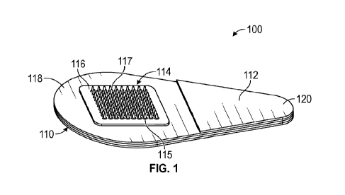
FIG. 1 is a perspective view of a microneedle patch according to one
embodiment of
the present disclosure.
FIGS. 2A-2B are microphotographs of a single microneedle. The microneedle is
shown before (FIG. 2A) and after (FIG. 2B) it is applied to skin. Scale bar
0.5 mm.
FIG. 3 depicts an array of microneedle patch-generated micropores created in
skin after
the application of a microneedle patch. Scale bar 5 mm.
FIG. 4 is a graph showing data from one example, comparing total volume of
sweat
collected after inducement by pilocarpine delivery by microneedle patches as
described herein
or by conventional iontophoresis.
FIG. 5 is a graph showing data from one example, comparing sweat volume
collected
per unit of pilocarpine dose after inducement by pilocarpine delivery by
microneedle patches
as described herein or by conventional iontophoresis.
2
CA 03203391 2023- 6- 23
WO 2022/147307
PCT/US2021/065760
FIG. 6 is a graph showing data from one example, comparing sweat volume
collected
per unit of skin area after inducement by pilocarpine delivery by microneedle
patches as
described herein or by conventional iontophoresis.
FIG. 7 is a graph showing data from one example, comparing chloride content of
collected sweat after inducement by pilocarpine delivery by microneedle
patches as described
herein or by iontophoresis.
DETAILED DESCRIPTION
New and improved methods and devices have been developed for inducing sweat
secretion from skin for medical diagnostics purposes. In particular
embodiments, the method
includes (i) applying a microneedle patch, which comprises microneedles which
comprise a
sweat-inducing agent, such as a cholinergic agonist, to the skin of the
patient effective to cause
the microneedles to penetrate across the epidermis and into the dermis; and
(ii) releasing the
sweat-inducing agent into the skin in an amount effective to induce secretion
of sweat from the
skin. In a preferred embodiment, the cholinergic agonist comprises
pilocarpine.
The microneedle patch enables sweat secretion inducement in a minimally
invasive,
painless, and convenient manner. Thus, the devices and methods herein can make
sweat
testing simpler and more widely available than current iontophoresis-based
methods.
In fact, it is a particular advantage of the present methods that
iontophoresis is not
required. Accordingly, no electrical current is applied to the skin, which
eliminates the risk of
skin burns associated with the conventional iontophoresis-driven
administration of the sweat-
inducing agent into the patient's skin.
Furthermore, in at least some embodiments, the present methods may enable
higher
sweat output per unit area of skin, as compared to methods utilizing
conventional
administration of pilocarpine from agar disks using iontophoresis. In fact, as
detailed in the
examples, the amount of pilocarpine delivered per unit area of skin may be up
to
approximately twice as large after microneedle patch administration compared
to
administration by iontophoresis.
The term "patient" refers to any person (human) to whom the sweat inducement
methods are applied. The term "patient" includes but is not limited to a
person in need of
medical care or a person in need of other physiological assessments. The
patient may be an
infant, child, or adult.
New and improved diagnostic methods are also provided that include (i)
inducing
secretion of sweat from a patient's skin as described herein; and (ii)
analyzing the sweat for the
presence, absence, or content of one or more analytes. That is, the induced
sweat, or the
3
CA 03203391 2023- 6- 23
WO 2022/147307
PCT/US2021/065760
collected sweat, may be analyzed for various analytes in the sweat, e.g., by
detecting,
measuring, and/or determining the presence and/or amounts of an analyte of
interest, for
example, for determining or monitoring of one or more physiological or
pathological
conditions or attributes in the patient.
The Methods
In some embodiments, the methods include applying a microneedle patch, which
comprises microneedles which comprise pilocarpine (or another sweat-inducing
agent) to the
skin of the patient effective to cause the microneedles to penetrate across
the epidermis and
into the dermis; releasing the pilocarpine (or other sweat-inducing agent)
into the skin in an
amount effective to induce secretion of sweat from the skin; and then
analyzing the sweat for a
specific analyte. The method may include collecting a volume of the sweat
secreted from the
skin and the analyzing is carried out on the collected sweat. In a particular
embodiment, the
analyte is one indicative of a disease. In a particular example, the analyte
is chloride
concentration, which is indicative of cystic fibrosis.
In some embodiments, the step of applying a microneedle patch comprises
manually
pressing the microneedle patch against the patient's skin. For example, the
microneedle patch
may be applied to an area of the patient's arm (e.g., forearm) or leg. The
application site
preferably is sanitized prior to application of the microneedle patch, for
example using a
conventional alcohol wipe. If needed, the application site may be allowed to
dry before
application of the microneedle patch. The patch then is applied to the
patient's skin using a
sufficient pressure to have the microneedles penetrate across the epidermis
and into the dermis.
In some embodiments, the methods further include removing the microneedle
patch
from the skin after a period of time effective to release the pilocarpine (or
other sweat-inducing
agent) from the microneedle patch into the patient's skin. In some
embodiments, the methods
include removing the microneedle patch from the skin in a manner effective to
separate the
microneedles from a support layer of the microneedle patch, wherein the
separated
microneedles remain in the patient's skin and dissolve to release the
pilocarpine (or other
sweat-inducing agent). For example, the microneedles may break off the patch
backing
immediately upon application to the skin, so that the patch backing may
promptly thereafter be
removed from the skin. Accordingly, in various embodiments of these methods,
the period of
time may be between 1 second and 15 minutes. The period may be, for example,
between 1
second and 10 minutes, between 1 second and 1 minute, between 10 seconds and
10 minutes,
between 10 seconds and 1 minute, between 1 minute and 15 minutes, between 1
minute and 10
minutes, or about 5 minutes.
4
CA 03203391 2023- 6- 23
WO 2022/147307
PCT/US2021/065760
In some embodiments, the skin-embedded microneedles, whether still connected
to the
backing or separated from it, release the pilocarpine (or other sweat-inducing
agent) by
dissolution of the microneedles in the aqueous fluid of the skin tissues.
Accordingly, in some
preferred embodiments of the methods, the microneedles are dissolvable
microneedles as
described below in the Microneedle Patch section.
In some other embodiments, the pilocarpine (or other sweat-inducing agent) is
associated with, and released from, the microneedles by different mechanisms
than foregoing
dissolvable microneedles. In one such example, the pilocarpine (or other sweat-
inducing
agent) is coated onto microneedles made of essentially any suitable material,
including non-
water soluble materials. In another example, the microneedles are hydrogels
that swell in the
skin and release the pilocarpine (or other sweat-inducing agent) from within
the hydrogel. In
still another example, the microneedles are not hydrogels or water-soluble and
include hollow
or porous structural portions, and the pilocarpine (or other sweat-inducing
agent) is loaded over
the cavities or pores of those hollow or porous structural portions and
released therefrom
following insertion into the skin.
In some embodiments, the method is effective to deliver from 250 l_tg to 1500
lig of
pilocarpine (or other sweat-inducing agent) per cm2 of skin. In some
embodiments, the method
is effective to deliver from 500 lig to 1000 jig of pilocarpine (or other
sweat-inducing agent)
per cm2 of skin. In some embodiments, the method is effective to deliver at
least 250 p.g, at
least 300 ng, at least 400 [is, at least 500 ng, at least 600 ng, at least 700
pg, or at least 800 lig
of pilocarpine (or other sweat-inducing agent) per cm2 of skin.
In some embodiments, a total of more than 1.38 mg pilocarpine is administered
into the
skin. For example, the microneedle patch may deliver 1.4 or 1.5 mg or more of
the pilocarpine
to the skin of the patient. In one non-limiting example, the microneedle patch
delivers from
1.50 mg to 2.50 mg of pilocarpine.
In some embodiments, the collecting of the sweat includes applying an
absorbent
material to the skin or positioning a collection tube at the skin surface to
permit sweat to be
drawn into a bore in the tube, for example, by capillary action. The absorbent
material may be
a woven or non-woven fibrous material, such as a cotton swab or gauze, or
porous structure,
such as a sponge. Capillary collection tubes are known in the art. For
example, the collection
tube may be part of a MacroductTm Sweat Collector. In some embodiments, the
sweat may be
collected in the microneedle patch itself
The amount of sweat collected generally should be any amount of sweat that is
suitable
for the analytical method to be used. In some embodiments, the volume of sweat
collected is
from 5 1 to 150 .1. For example, the collected volume may be from 10 1 to 100
tl. In some
5
CA 03203391 2023- 6- 23
WO 2022/147307
PCT/US2021/065760
embodiments, the volume of sweat collected may be from 15 IA to 30 pl. In one
embodiment,
a total of at least 17 pl of sweat may be induced by the microneedle patch and
collected.
In some embodiments, the volume of sweat collected is between the minimum
volume
that is effective for chloride concentration measurements by a current or
future technique of
chloride measurement and a maximum that collectable from the skin over a 30-
minute
collection period.
In some embodiments, the sweat collected per area of skin into which the
pilocarpine
(or other sweat-inducing agent) is released is from 2 t1 per cm2 to 50 pi per
cm2. In some
embodiments, the sweat collected per area of skin into which the pilocarpine
is released is at
least 2.6 ul per cm2. In some other embodiments, the sweat collected per area
may be from 10
IA per cm2 to 40 .1 per cm2. In some embodiments, the sweat collected per
area is at least 15
ill per cm2, or at least 20 pi per cm2.
The collected sweat can be analysed by any suitable method for any analytes.
For
example, it may undergo chloride analysis with a chloridometer or total
electrolyte analysis for
example, using a Sweat-Chek Analyzer'. Other analyses also are envisioned,
such as skin-
interfacing microfluidic devices known in the art. See, e.g., Ray, et al.,
Science Translational
Medicine, 31 Mar 2021, Vol 13, Issue 587.
The presently disclosed microneedle patch configured to induce sweating can be
used
in clinical settings, in personal health monitoring, or in other applications,
such as non-medical
context, e.g., athletic performance assessment, military readiness assessment,
etc. The
microneedle patch advantageously may replace conventional sweat-inducing
techniques that
involve hypodermic injections and/or iontophoresis, because the microneedle
patch is much
easier to use. Because of the relative simplicity of its use, the microneedle
patch can also be
used by any person after brief training for personal health monitoring, e.g.,
at home.
Cystic Fibrosis Testing
The methods described herein are particularly useful to produce and collect a
sweat
sample that can be used in a better tool in diagnosing cystic fibrosis. In a
preferred
embodiment, the chloride concentration in the collected sweat is quantified
for the diagnosis of
cystic fibrosis using a chloridometer or other conventional instruments. As
known in the art,
elevated chloride levels in sweat are indicative of cystic fibrosis.
The presently disclosed pilocarpine-containing microneedle patches offer a
simple and
more accessible alternative for sweat induction to support efficient and
minimally invasive
cystic fibrosis diagnosis in infants and children. In one particular
embodiment, the
microneedle patch is applied to the skin of an infant, for example on the arm,
after the infant
has a positive CF screening. Pilocarpine then is released from microneedles of
the patch into
6
CA 03203391 2023- 6- 23
WO 2022/147307
PCT/US2021/065760
the infant's skin effective to induce secretion of sweat, and then a volume of
the sweat secreted
is collected from the skin using conventional means, such as the Macroduct
Sweat Collector.
The collected sweat is then analyzed by measuring the chloride concentration
in the collected
volume of sweat using a chloridometer as known in the art.
The larger pilocarpine dose per unit area enabled by the present microneedle
patch
delivery methods compared to conventional iontophoresis methods may facilitate
more
consistently generated amount of sweat required to perform a chloride
measurement, thus
potentially making the sweat test more reliable and avoiding the need for
repeated
measurement attempts experienced with conventional methods.
The Microneedle Patch
In embodiments, the microneedle patch useful in the present methods includes a
support layer, and an array of microneedles extending from the support layer,
wherein the
microneedle patch is configured for application to a patient's skin and the
microneedles
include a sweat-inducing agent. The sweat-inducing agent may be cholinergic
agonist, such as
pilocarpine.
As used herein, the term "pilocarpine" refers to (35,4R)-3-ethy1-44(1-methyl-
11/-
imidazol-5-yOmethypdihydrofuran-2(31/)-one, and pharmaceutically acceptable
salts, and/or
solvates, thereof In the case of sweat collection for measurements of chloride
content, the HC1
or other chloride-containing salt form of pilocarpine would not be used
because chloride from
the pilocarpine salt could affect chloride concentrations measured in the
collected sweat. In
some preferred embodiments, the pilocarpine is pilocarpine nitrate.
In some other embodiments, the sweat-inducing agent may be selected from
suitable
drugs known in the art to cause excess perspiration or sweating as a side
effect. See, e.g.,
https://www.sweathelp.org/pdf/drugs2009.pdf. In one embodiment, the sweat-
inducing agent
is carbachol.
The sweat-inducing agent is part of the microneedle structure. For example,
the sweat-
inducing agent may be dispersed in a matrix material forming at least part of
the microneedle
structure, part of a coating material on the microneedle, or a combination
thereof
In a preferred embodiment, the microneedles are dissolvable. As used herein,
the term
"dissolvable" means that the microneedles include water-soluble materials
which dissolve in
water in the skin, following insertion of the microneedles. The dissolution
should be at rate
useful to release the sweat-inducing agent into tissues of the skin at a
practical, or clinically
useful, rate. In a preferred embodiment, the microneedles are formed of the
sweat-inducing
agent dispersed in one or more water-soluble matrix materials.
7
CA 03203391 2023- 6- 23
WO 2022/147307
PCT/US2021/065760
In some other embodiments, it may be desirable to induce continuous sweating
over an
extended period of time, for sweat collection and analyte measurement over an
extended
period. In such cases, the microneedles may be configured to slowly release
the sweat-
inducing agent into the skin, for example, by using any of the mechanisms
known in the art for
controlled, sustained drug delivery from microneedles. In some embodiments,
this is
accomplished by making the microneedles of a composition that includes the
sweat-inducing
agent (e.g., pilocarpine) and one or more biomaterials selected from
hydrogels, biodegradable
polymers (e.g., PLGA), non-degradable polymers, and the like.
The microneedles may include a variety of suitable biocompatible, water-
soluble
matrix materials. The matrix materials, in combination with the sweat-inducing
agent, should
impart the necessary mechanical strength for reliable insertion of the
microneedles into the
skin. Generally, the sweat-inducing agent is included in a stable composition
(forming the
microneedles) in which the sweat-inducing agent therein essentially retains
its physical
stability and/or chemical stability and/or biological activity upon storage.
The matrix materials
may be selected from pharmaceutically acceptable excipients known in the art.
In some preferred embodiments, the matrix material of the microneedles
comprise two
or more matrix materials. In some embodiments, the matrix material may include
or consist of
a combination of a poly(vinyl alcohol) (PVA) and a disaccharide. Examples of
disaccharide
include sucrose, lactose, and maltose. For example, the matrix material may
include PVA and
sucrose. In some other embodiments, other water soluble polymers are used in
place of or in
combination with PVA.
In some embodiments, the fraction of the sweat-inducing agent in the
microneedles
ranges from 20% to 60% by weight. In some embodiments, the microneedles
comprise from
30% to 50% by weight pilocarpine. In some sub-embodiments, these microneedles
comprise
from 70% to 50% by weight a mixture of a PVA and a disaccharide, such as
sucrose. In some
other embodiments, the microneedles are 20-60% by weight pilocarpine, and the
other
materials are non-water soluble materials that are formed in a porous or
hollow structure,
where the pores or hollow portion(s) of the microneedle contain the
pilocarpine.
In one embodiment, the microneedles comprise about 40% by weight pilocarpine
nitrate. In some sub-embodiments, the microneedles comprise about 60% by
weight a mixture
of a PVA and a disaccharide, such as sucrose.
The microneedles may have any suitable shape. In some embodiments, the
microneedles are conical. In some other embodiments, the microneedles may be
blade-like, or
pyramidal. In some embodiments, the microneedles have a straight proximal
portion and a
8
CA 03203391 2023- 6- 23
WO 2022/147307
PCT/US2021/065760
tapered distal portion. The shaft of the microneedle may have a circular,
oval, or polygonal
cross-sectional shape.
The microneedle patch is constructed to administer to the skin an amount of
the sweat-
inducing agent across an area of skin effective to induce secretion of sweat
in a total volume
that is required for a particular analysis. This may be controlled for example
by
selecting/adjusting the amount of the amount of the sweat-inducing agent
releasable from each
microneedle, the total number of microneedles in the patch array, and/or the
spacing of the
microneedles/size of the patch. In some embodiments, the microneedle patch is
configured to
deliver at least 240 lig of pilocarpine per cm2 of patient's skin. In some
embodiments, the
microneedle patch is configured to deliver at least 250 pyg of pilocarpine per
cm2 of patient's
skin.
The microneedles may have a length between 200 pm and 2,000 pm. In some
embodiments, the microneedles have a length between 500 pm and 1,000 gm. For
example,
the microneedles may have a length of about 600 ?Am, about 700 vim, about 800
?Am, or about
900 ?Am.
The area of the microneedle patch may be any suitable dimensions. In some
embodiments, the area is between 0.5 cm2 and 10 cm2. In some embodiments, the
area is from
2 cm2 to 8 cm2. In some embodiments, the area is from 5 cm2 to 6 cm2. In one
example, the
area is 5.8 cm2. Other dimensions are envisioned.
The microneedles have a base (or proximal) end and an opposing (distal) tip
end. The
base end of each microneedle is attached, directly or indirectly, to the
support layer (or base
substrate) of the microneedle patch. In some preferred embodiments, the
microneedle patch
further includes base pedestals between and connecting the support layer and
each of the
microneedles. The base pedestals may be made of a polymeric material, such as
PVA. In some
embodiments, the base pedestals have a height between 200 pm and 800 pm. In
some
embodiments, these microneedles are coated with a formulation containing
pilocarpine.
In some embodiments, the sweat-inducing agent is located only in the
microneedles,
e.g., predominately at the tip end portion of the microneedle, and not in the
support layer. In
some other embodiments, the sweat-inducing agent may be dispersed in the
support layer too,
for example, wherein the support layer and microneedles are fabricated of the
same materials.
In some embodiments, the pilocarpine is located predominantly or exclusively
in a coating on
the microneedles.
The microneedle array may have a variety of shapes, including circular or
square. In
some embodiments, the size of the microneedle patch is between 1 cm and 10 cm
in its longest
9
CA 03203391 2023- 6- 23
WO 2022/147307
PCT/US2021/065760
dimension. In some embodiments, the microneedle patch includes from 100 to
1000
microneedles.
In some embodiments, the microneedle patch include a handle, or tab, for
manipulating
the patch.
In some embodiments, the microneedle patch comprises a pressure-sensitive
adhesive
suitable for temporarily securing the patch to the skin.
In some embodiments, the microneedle patch includes a feedback indicator
configured
to inform the user that the microneedles have penetrated the skin and/or that
the sweat-
inducing agent has been released into the skin.
One embodiment of a microneedle patch 100 is shown in FIG. 1. The microneedle
patch 100 includes a microneedle array 114 extending from a support layer 116.
The
microneedles 117 extend from a base pedestal 115. The support layer 116 is
affixed to
adhesive layer 118 of a handling structure 110 that includes a tab portion 112
and an adhesive
cover 120. Other configurations of handling structures are envisioned, some of
which are
described in U.S. Patent No. 10,265,511, which is incorporated herein by
reference.
The microneedle patches may be made a process that include molding
microneedles as
described in U.S. Patent No. 10,828,478, which is incorporated herein by
reference.
The present invention may be further understood with reference to the
following non-
limiting examples.
EXAMPLES
Experiments were conducted to evaluate whether microneedle patches could be
used to
perform sweat tests and to evaluate whether microneedle patches can be used as
an alternative
to iontophoresis to administer pilocarpine to induce sweating, including for
use in the diagnosis
of cystic fibrosis.
All data are presented as mean standard deviation. Total sweat volume, sweat
volume/drug dose, sweat volume/skin area, and sweat chloride concentration
were compared
between microneedle and iontophoresis sites with two-tailed unpaired Student's
t-tests.
Statistical significance was set at p < 0.05 for all comparisons.
Example 1: Microneedle patch with microneedles comprising pilocarpine
Pilocarpine-loaded microneedle patches were fabricated by a two-step molding
process
using polydimethylsiloxane (PDMS) molds based on an established method. The
first casting
solution was a mixture of 10% (w/v) pilocarpine nitrate, 10% (w/v) poly(vinyl
alcohol) (PVA)
and 5% (w/v) sucrose, which was prepared in deionized water. This solution was
cast on
PDMS molds under vacuum to facilitate filling the solution into the mold
cavities to form the
CA 03203391 2023- 6- 23
WO 2022/147307
PCT/US2021/065760
microneedles. After 20 min, excess solution was removed, and the filled molds
were
centrifuged at 5000 g for 20 min to dry the drug-loaded microneedles. The
second casting
solution containing 20% (w/w) polystyrene in 1,4-dioxane was then cast on the
filled PDMS
molds under vacuum to form the patch backing. The molds were kept under vacuum
for
another 3 h to dry the solution at room temperature, and then further dried at
40 C overnight
before demolding the microneedle patches using adhesive tapes.
Each microneedle patch consisted of a 10 x 10 array of the microneedles
arranged
within a square with approximately 7 mm sides (i.e., ¨0.5 cm2). As shown by
microscopic
examination (see FIG. 2A), each conical microneedle (base diameter 200 p.m,
height 600
p.m) was mounted atop an wider pedestal (base diameter 600 p.m, height 400
p.m).
The solid microneedles were composed of 40% by weight pilocarpine, 40% by
weight
PVA, and 20% by weight sucrose. The total amount of pilocarpine loaded per
microneedle
patch was measured as 500 48 jig (n=4). The PVA provided the mechanical
strength the
microneedle needs to penetrate the skin, and the sucrose facilitated
microneedle dissolution in
the skin following the insertion into the skin.
Example 2: Ex vivo application of pilocarpine microneedles to porcine skin
Microneedle patches from Example 1 were applied to shaved porcine skin ex vivo
to
study their skin insertion properties before application on horses in vivo. A
microneedle patch
was manually pressed against the porcine skin by thumb for ¨10 s, and then
left in place for 20
min to allow microneedle dissolution and release of drug in the skin. After
being removed
from the skin, the patches were saved for further examination.
After application to porcine skin ex vivo, the microneedles dissolved in the
skin,
leaving only the base pedestals (FIG. 2B), indicating that the pilocarpine
loaded in the
microneedles was successfully delivered into the skin during microneedle patch
application.
Treating the skin with a dye that selectively stains sites of skin puncture
revealed an array of
microneedle-generated micropores with the same 10>< 10 array geometry as the
microneedle
patch (FIG. 3), further indicating the ability of microneedles to penetrate
the skin.
After application to porcine skin ex vivo, the residual pilocarpine content
per
microneedle patch was 237 73 jig (n=6), indicating that the delivered dose
was ¨263 jig (i.e.,
¨526 vig/cm2) and the delivery efficiency was ¨53%.
The pilocarpine dose delivered by microneedle patches was calculated as the
difference
between the pilocarpine contents in patches before and after application to
skin. The
pilocarpine content in microneedle patches was measured by HPLC after
dissolving the patch
in a known volume of deionized water.
11
CA 03203391 2023- 6- 23
WO 2022/147307
PCT/US2021/065760
Example 3: Ex vivo application of iontophoretic Pilogel discs to porcine skin
The commercially available iontophoretic Pilogel discs were circular with a
diameter of
2.72 cm (i.e., ¨5.8 cm2), thereby contacting an area of skin more than 10
times larger than the
microneedle patch. The iontophoretic Pilogel discs therefore had much higher
pilocarpine
loading amount, measured as 15.94 0.27 mg (n=3). After iontophoresis on
porcine skin for
min, the used discs contained 14.56 0.15 mg (n=3) residual pilocarpine,
which indicates
the delivered dose by iontophoresis was ¨1.38 mg (i.e., ¨238 Kg/cm2) and the
delivery
efficiency was 8.7%. The delivery efficiency of iontophoresis was
significantly lower than
that of the microneedle patch.
10 Example 4: hi vivo application of microneedle patches and iontophoresis
to induce
sweating in horse model
Eight healthy outbred adult horses were used as the animal model. Prior to all
testing,
the cervical region of the skin of the horses was shaved to permit good
contact of the
microneedle patches, iontophoretic pilocarpine discs and sweat collection pads
to the skin.
Procedure
Pilocarpine-induced sweat production via microneedle patches and iontophoresis
was
then compared in 4 horses after acclimatization. In each animal, three
microneedle patches
from Example 1 above were applied manually by thumb pressure to the right side
of the neck,
approximately 5 cm apart, and left in place for 20 min. Concurrently, on the
left side of the
neck, Pilogel Iontophoretic Discs were mounted onto electrodes, and
pilocarpine was delivered
via iontophoresis for 10 min (2 machine cycles) in two separate locations
sequentially.
After completion of iontophoresis and removal of the microneedle patches,
sweat was
collected from 25 sites on the neck of 4 horses to quantify volume and
chloride concentration.
In pilot studies, the conventional MacroductO Sweat Collector used for sweat
collection in
humans could not be consistently adhered to the convex cervical region on the
horse, leading to
unreliable and inconsistent sweat collection. Thus, a modified sweat
collection protocol was
developed using single layer cotton gauze pads covered by a similarly-sized
piece of 150 p.m-
thick polypropylene plastic sheeting and secured under a piece of heavy-duty
adhesive tape
(approximately 5 x 15 cm). Gauze pads to collect microneedle-induced and
iontophoresis-
induced sweat were 1 cm2 and 2 cm2, respectively. Plastic sheeting
approximately 1 mm larger
in length and width was applied over the gauze pads. After 30 min, the gauze
pads were
collected and immediately weighed on a microbalance to calculate sweat volume
by
subtracting the dry weight. Gauze pads were then immediately placed inside a 3
ml
polypropylene syringe barrel inserted in a 15 ml conical polypropylene tube
and sealed prior to
12
CA 03203391 2023- 6- 23
WO 2022/147307
PCT/US2021/065760
centrifuging at 1100 x g for 10 min. Sweat recovered after centrifugation was
collected into a
polypropylene microcentrifuge tube and frozen at -80 C until analysis for
chloride
concentration.
Results
From all application sites, at least 10 p1 of sweat was collected. The average
total
sweat volume from an iontophoresis site was 101 49 IA over a pilocarpine
application area of
5.8 cm2, corresponding to a sweat collection density of 17 8 i_i1/cm2. The
average total sweat
collected from a microneedle patch site was 17 8 pi over a pilocarpine
application area of 0.5
cm2, corresponding to a sweat collection density of 34 + 16 p1/cm2 (FIG. 4 and
FIG. 6).
Sweat density is an appropriate basis for comparison between the two
techniques because
sweat production is expected to scale directly with area and because sweat
collection is usually
done over a standard area of skin using a sweat collection device.
While the total amount of sweat collected from the iontophoresis sites was
greater than
that collected from the microneedle patch sites (FIG. 4), when accounting for
the different
pilocarpine application areas, the sweat collection density from the
microneedle patch sites was
2.0-fold greater than that collected from the iontophoresis sites (FIG. 6).
This ratio was
relatively consistent on each of four horses (2.3, 1.6, 2.1 and 1.6-fold
greater). This suggests
that the difference between microneedle patches and iontophoresis on sweat
induction was not
determined by the individual differences between horses. Instead, the
difference in sweat
collection density appears to mainly reflect the different sweat-inducing
abilities of the two
pilocarpine delivery procedures.
The sweat volume per unit of pilocarpine dose delivered to the skin was
calculated.
This analysis revealed no significant difference between iontophoresis (73
361.11/mg) and
microneedle patches (66 34 1.t1/mg) (FIG. 5). However, because the amount of
pilocarpine
delivered per unit area of skin was 2.2-fold greater when administered by
microneedle patches
(-526 gg/cm2) compared to iontophoresis (-238 p..g/cm2), this likely accounts
for the greater
sweat collection density seen after pilocarpine delivery by microneedle patch.
Thus, using
microneedle patches to deliver pilocarpine has a comparable or better sweat-
inducing
capability as the traditional iontophoresis.
It should be noted that although sweat collection density was greater using a
microneedle patch, the microneedle patch induced less total sweat volume than
iontophoresis.
Because the microneedle patch delivered twice as much pilocarpine per unit
area, a larger
microneedle patch with the same area as the pilocarpine disc used for
iontophoresis (i.e., 5.8
cm2) should correspondingly deliver twice as much pilocarpine and thereby
induce more total
13
CA 03203391 2023- 6- 23
WO 2022/147307
PCT/US2021/065760
sweat volume compared to iontophoresis, because sweat production is known to
scale with
pilocarpine dose delivered.
Chloride contents in the iontophoresis-induced sweat (60.0 21.8 mmol/L) and
microneedle patch-induced sweat (50.3 13.8 mmol/L) were not significantly
different (FIG.
7), indicating that the method of pilocarpine administration (iontophoresis
vs. microneedle
patch) did not significantly affect the chloride content in the collected
sweat.
Example 5: Further comparisons of microneedle patches and iontophoresis
When the microneedle patches were applied to the skin on horses in vivo, more
pilocarpine, 407 46 jig (n=13), was dissolved from the microneedle patches
compared with
the ex vivo measurements in porcine skin. An explanation for this difference
may be that the
sweat induced by the microneedle patch in the horse (but not in the porcine
skin ex vivo) might
have further dissolved the microneedles at the skin surface, giving the
appearance of greater
pilocarpine delivery efficiency. The same effect might have also occurred at
the iontophoresis
sites. The analysis of the results was based on the ex vivo data on the
delivered pilocarpine
dose from both microneedle patches (Example 1, square with side length of ¨0.7
cm) and
iontophoresis (Pilocarpine Iontophoresis Disc, round with a diameter of ¨2.72
cm) as 0.26 mg
and 1.38 mg, respectively as shown in Table 1.
Table 1. Comparison of parameters between microneedle patches and
iontophoresis
Pilocarpine
Microneedle patches
Iontophoresis Discs
Application area (cm2) 0.5 5.8
Pilocarpine dose (mg) C 0.26 + 0.07
1.38 0.15
Pilocarpine / application area
0.52 0.14 0.24 0.03
(mg/cm)
The dose was calculated as the difference between unused and used microneedle
patches or
pilocarpine discs.
The Examples demonstrate that microneedle patches are able to deliver
pilocarpine to
skin to induce sweating. The amount of sweat produced per dose of pilocarpine
delivered, and
the chloride concentration of that sweat, were similar for delivery of
pilocarpine via
microneedle patch and iontophoresis. Thus, microneedle patch delivery is a
suitable
alternative to iontophoresis delivery of pilocarpine. The pilocarpine dose
delivered per unit
area doubled with microneedle patch delivery compared to iontophoresis
delivery. Therefore,
a larger microneedle patch could produce larger amounts of sweat and/or
adequate amounts of
sweat in less time compared to current iontophoretic methods.
14
CA 03203391 2023- 6- 23
WO 2022/147307
PCT/US2021/065760
Exemplary Embodiments
Embodiment 1. A method for inducing sweat secretion from a patient's skin,
comprising: applying a microneedle patch, which comprises microneedles which
comprise a
sweat-inducing agent, to the skin of the patient effective to cause the
microneedles to penetrate
across the epidermis and into the dermis; and releasing the sweat-inducing
agent into the skin
in an amount effective to induce secretion of sweat from the skin.
Embodiment 2. The method of embodiment 1, wherein the sweat-inducing agent is
a
cholinergic agonist.
Embodiment 3. The method of embodiment 1 or 2, wherein the sweat-inducing
agent
comprises pilocarpine.
Embodiment 4. The method of any one of embodiments 1 to 3, wherein the
applying a
microneedle patch comprises manually pressing the microneedle patch against
the patient's
skin.
Embodiment 5. The method of any one of embodiments 1 to 4, further comprising
removing the microneedle patch from the skin after a period of time effective
to release the
sweat-inducing agent from the microneedle patch into the patient's skin.
Embodiment 6. The method of any one of embodiments 1 to 5, wherein the
microneedles are dissolvable microneedles, coated microneedles, or
porous/hollow
microneedles.
Embodiment 7. The method of embodiment 5 or 6, wherein the period is between 1
second and 15 minutes, e.g., 5 minutes.
Embodiment 8. The method of any one of embodiments 1 to 7, further comprising
removing the microneedle patch from the skin in a manner effective to separate
the
microneedles from a support layer of the microneedle patch, the separated
microneedles
remaining in the patient's skin and dissolving to release the sweat-inducing
agent.
Embodiment 9. The method of any one of embodiments 1 to 8, wherein at least
250 jag
of the sweat-inducing agent is delivered per cm2 of skin.
Embodiment 10. The method of any one of embodiments 1 to 9, wherein the
microneedle patch comprises a support layer from which an array of the
microneedles extend.
Embodiment 11. The method of any one of embodiments 1 to 10, wherein the
microneedles comprise a water-soluble matrix material in which the sweat-
inducing agent is
dispersed.
Embodiment 12. The method of embodiment 11, wherein the matrix material
comprises a poly(vinyl alcohol) (PVA), a disaccharide, or a combination
thereof.
CA 03203391 2023- 6- 23
WO 2022/147307
PCT/US2021/065760
Embodiment 13. The method of embodiment 11 or 12, wherein the matrix material
comprises PVA and sucrose.
Embodiment 14. The method of any one of embodiments 1 to 13, wherein the
microneedles comprise from 30% to 50% by weight of the sweat-inducing agent.
Embodiment 15. The method of any one of embodiments 1 to 14, further
comprising
collecting and/or analyzing the sweat secreted from the skin.
Embodiment 16. The method of embodiment 15, wherein the collecting of the
sweat
comprises applying an absorbent material to the skin and/or by positioning a
collection tube at
the skin surface to permit sweat to be drawn into a bore in the tube by
capillary action.
Embodiment 17. The method of embodiment 15 or 16, wherein the volume of sweat
collected is at least 15 IA.
Embodiment 18. The method of any one of embodiments 15 to 17, wherein the
sweat
collected per area of skin into which the sweat-inducing agent is released is
at least 2.6 ul per
CM2 .
Embodiment 19. The method of any one of embodiments 15 to 18, wherein the
analyzing comprises measuring the sweat for an analyte indicative of cystic
fibrosis.
Embodiment 20. The method of any one of embodiments 15 to 19, wherein the
analyzing comprises measuring the chloride concentration in the sweat.
Embodiment 21. The method of any one of embodiments 1 to 20, used in the
diagnosis
of cystic fibrosis.
Embodiment 22. A microneedle patch comprising: a support layer; and an array
of
microneedles extending from the support layer, wherein the microneedle patch
is configured
for application to a patient's skin and the microneedles comprise a sweat-
inducing agent.
Embodiment 23. The microneedle patch of embodiment 22, wherein the sweat-
inducing agent comprises a cholinergic agonist.
Embodiment 24. The microneedle patch of embodiment 22 or 23, wherein the sweat-
inducing agent comprises pilocarpine.
Embodiment 25. The microneedle patch of any one of embodiments 22 to 24,
wherein
the microneedles comprise a water-soluble matrix material in which the sweat-
inducing agent
is dispersed.
Embodiment 26. The microneedle patch of embodiment 25, wherein the matrix
material comprises a poly(vinyl alcohol) (PVA), a disaccharide, or a
combination thereof
Embodiment 27. The microneedle patch of embodiment 25 or 26, wherein the
matrix
material comprises PVA and sucrose.
16
CA 03203391 2023- 6- 23
WO 2022/147307
PCT/US2021/065760
Embodiment 28. The microneedle patch of any one of embodiments 22 to 27,
wherein
the microneedles have a length between 200 pm and 2,000 [.im.
Embodiment 29. The microneedle patch of any one of embodiments 22 to 27,
wherein
the microneedles have a length between 500 nm and 1,000 nm.
Embodiment 30. The microneedle patch of any one of embodiments 22 to 29,
wherein
each of the microneedles has a base end and an opposing tip end, and wherein
the microneedle
patch further comprises base pedestals between and connecting the support
layer and each of
the microneedles.
Embodiment 31. The microneedle patch of embodiment 30, wherein the base
pedestals
have a height between 200 nm and 800 nm.
Embodiment 32. The microneedle patch of any one of embodiments 22 to 31,
wherein
the microneedles comprise from 30% to 50% by weight pilocarpine nitrate.
Embodiment 33. The microneedle patch of any one of embodiments 22 to 32, which
is
configured to deliver at least 250 ng of pilocarpine per cm2 of patient's
skin.
Embodiment 34. A diagnostic method comprising: inducing secretion of sweat
from a
patient's skin according to the method of any one of embodiments 1 to 21; and
analyzing the
sweat for the presence, absence, or concentration of one or more analytes.
Embodiment 35. A medicament comprising pilocarpine for use in the inducement
of
sweating by administering pilocarpine to the skin of a patient effective to
induce secretion of
sweat from the skin, wherein the pilocarpine is released into the skin from
microneedles
applied to the skin of the patient to cause the microneedles to penetrate
across the epidermis
and into the dermis.
Embodiment 36. The medicament of embodiment 35, wherein the microneedles are
dissolvable microneedles, coated microneedles, or porous/hollow microneedles.
Embodiment 37. The medicament of embodiment 35 or 36, wherein at least 250 lig
of
pilocarpine is delivered per cm2 of skin.
Embodiment 38. The medicament of any one of embodiments 35 to 37, wherein the
microneedles are in array extending from a support layer of a microneedle
patch.
Embodiment 39. The medicament of any one of embodiments 35 to 38, wherein the
microneedles comprise a water-soluble matrix material in which the pilocarpine
is dispersed.
Embodiment 40. The medicament of embodiment 39, wherein the matrix material
comprises a poly(vinyl alcohol) (PVA), a disaccharide, or a combination
thereof
Embodiment 41. The medicament of embodiment 39 or 40, wherein the matrix
material comprises PVA and sucrose.
17
CA 03203391 2023- 6- 23
WO 2022/147307
PCT/US2021/065760
Embodiment 42. The medicament of any one of embodiments 35 to 41, wherein the
microneedles have a length between 200 nm and 2,000 nm.
Embodiment 43. The medicament of any one of embodiments 35 to 41, wherein the
microneedles have a length between 500 pm and 1,000 nm.
Embodiment 44. The medicament of any one of embodiments 35 to 43, wherein each
of the microneedles has a base end and an opposing tip end, and wherein the
microneedle patch
further comprises base pedestals between and connecting the support layer and
each of the
microneedles.
Embodiment 45. The medicament of embodiment 44, wherein the base pedestals
have
a height between 200 nm and 800 pm.
Embodiment 46. The medicament of any one of embodiments 35 to 45, wherein the
microneedles comprise from 30% to 50% by weight pilocarpine nitrate.
Embodiment 47. A diagnostic method comprising: inducing secretion of sweat
from a
patient's skin using the medicament of any one of embodiments 35 to 46; and
then analyzing
the secreted sweat for the presence, absence, or concentration of one or more
analytes.
Embodiment 48. The microneedle patch of any one of embodiments 22 to 33,
wherein
the microneedles are dissolvable microneedles, coated microneedles, or
porous/hollow
microneedles.
Embodiment 49. The method of any one of embodiments 1 to 21, wherein 1.5 mg or
more of pilocarpine is administered into the skin.
Embodiment 50. The microneedle patch of any one of embodiments 22 to 33 or 48,
which is configured to deliver 1.5 mg or more of pilocarpine into the skin.
Modifications and variations of the methods and devices described herein will
be
obvious to those skilled in the art from the foregoing detailed description.
Such modifications
and variations are intended to come within the scope of the appended claims.
18
CA 03203391 2023- 6- 23