Note: Descriptions are shown in the official language in which they were submitted.
CA 03203589 2023-05-30
WO 2022/119910 PCT/US2021/061385
SPECTROMETRY SYSTEMS, METHODS, AND APPLICATIONS
CROSS-REFERENCE TO RELATED APPLICATIONS
[0001] This application claims the benefit of U.S. Provisional Patent
Application No.
63/120,025 filed December 1, 2020, the disclosure of which, as well as the
references cited
therein, is hereby incorporated by reference.
STATEMENT OF FEDERALLY SPONSORED RESEARCH OR DEVELOPMENT
[0002] N/A.
BACKGROUND OF THE INVENTION
[0003] Healthcare acquired infections are a major problem. These include
infections both in
hospitals or healthcare facilities as well as infections in the home setting
related to patients
managing their own medical related procedures. Prevalence of hospital acquired
infections
(HAIs) in the United States alone is measured at 1.7 million infections per
year with a human
cost that translates into 99,000 deaths. The financial toll in the United
States is measured
somewhere near $45 billion a year. It is clear then, that these infections
pose a serious human
and financial cost to the American healthcare system. Human and financial
costs associated with
infections related to patient delivered, home medical care procedures are less
well quantified but
are estimated to total in the billions of dollars as well. This of course, is
not a problem restricted
to the United States and is seen throughout the developed and developing
world.
[0004] HAIs are exemplified by catheter associated urinary tract infections
(CAUTIs) which
represent one of the most common and most serious forms. They represent
approximately 32%
of all HAIs affecting over 2.5 million patients a year with a high level of
morbidity and a
mortality rate of approximately 13,000 deaths per year. It is estimated that
each of these
infections has an additional cost of $13,731 per case. In aggregate the cost
to the United States
healthcare system for catheter associated urinary tract infection approaches
$7.7 billion per year.
Given this, it is clear that the financial and human cost of this CAUTIs is
significant.
[0005] Infections related to patient delivered, home medical care
procedures are exemplified
by those related to patients utilizing peritoneal dialysis for management of
end stage renal
disease (ESRD). Peritoneal dialysis is a method in which the peritoneum, the
lining of the
1
CA 03203589 2023-05-30
WO 2022/119910 PCT/US2021/061385
abdominal cavity, is used as a natural filtration device. This requires an
indwelling peritoneal
catheter through which daily exchanges of a cleansing fluid called dialysate,
are performed in
cycles to accomplish the goal of fluid management and toxin removal that the
failed kidneys can
no longer perform. Peritoneal dialysis seems to be associated with 48%
lower mortality than hemodialysis over the first 2 years of dialysis therapy
independent of
modality switches or differential transplantation rates and is widely viewed
as a preferable
solution over hemodialysis for patients with ESRD. Despite this fact,
hemodialysis remains the
dominant modality for management of ESRD because of the risk that abdominal
infection,
known as peritonitis, presents to patients using peritoneal dialysis. PD-
associated peritonitis is
the direct or major contributing cause of death in >15% of patients on PD.
[0006] In both of these examples, and others in which body cavities are
instrumented with
indwelling catheters, the need for monitoring and early detection of infection
is critical if the
human and financial costs of these infections are to be avoided.
[0007] In addition to the identification of HAT, access to biosamples
through existing
indwelling patient catheters for the purpose of in vivo and continuous
analysis makes the
identification of biomarkers encountered in any human fluid including, but not
limited to urine
peritoneal fluid, wound drainage, enteral content, etc., of significant value
to early detection of
disease and guidance of therapy.
SUMMARY OF THE INVENTION
[0008] Thus, there is a need to monitor for HAIs, and in particular for
CAUTIs, and for
infections related to patient delivered, home medical care procedures.
[0009] Various embodiments of the disclosed invention provide for and
expand upon
methods, apparatus, and systems for continuous, real time, on-catheter, on-
patient, bacterial
colonization and bacterial infection product detection. In the present
description we demonstrate
a real-time system for continuous screening, detection, and alerting of
clinical personnel as to the
state of a patient's liquid bio sample system relating to infection and to the
identification of
biomarkers associated with the functional status of multiple human systems
including the
cardiac, renal, respiratory, neurologic, endocrine, and immune systems.
[0010] The present invention relates a method of screening a sample for the
presence of one
or more compounds of interest. The method uses a NIR spectrometer to analyze
the fluid.
2
CA 03203589 2023-05-30
WO 2022/119910 PCT/US2021/061385
Additionally, different Machine Learning algorithms were created in order to
classify and
identify different types of bacteria within the fluid and some other
bioproducts.
[0011] The current embodiment includes a system for continuous liquid bio
sample
screening without interrupting the flow of liquid bio sample through the use
of a specific device
design which will allow for liquid bio sample to be without flow long enough
for testing, without
the interruption of liquid bio sample flow without the application of external
blockers.
[0012] In various embodiments, a biofluid monitoring apparatus may be
provided. The
apparatus may include: a spectrometer disposed within a housing, the
spectrometer including: a
light source to illuminate a sample within a catheter tubing, a detector to
detect light returned.
from the sample, a status signal indicator to provide patient status based on
the sample in the
catheter tubing, and a controller in communication with the light source, the
detector, and the
status signal indicator to collect and process data based on the light
returned from the sample to
determine a patient status and indicate the patient status using the status
indicator, wherein the
housing is configured to attach at a low point in the catheter -tubing such
that the sample
accumulates in the low point, and wherein the light source and the detector
are directed towards
the low point to obtain the data from the sample.
[0013] In some embodiments, the spectrometer may further include a power
supply. In
certain embodiments, the power supply may include a battery.
[0014] in various embodiments, the housing may include a slot into which
the catheter tubing
is inserted such that a portion of the catheter tubing is adjacent to the
spectrometer.
[0015] In particular embodiments, the spectrometer may further include a
collimator to focus
light from the light source into the sample. In some embodiments, the
collimator may include a
lens.
[0016] In certain embodiments, the spectrometer may further include a
monochromator to
divide the light from the light source into a plurality of constituent
wavelengths. In various
embodiments, the monochromator may include a prism. In particular embodiments,
the
spectrometer may further include a wavelength selector to select a particular
wavelength to direct
to the sample, where the particular wavelength may be selected based on at
least one of a
bacterial strain or a bacterial product to he identified. In some embodiments,
the wavelength
selector may include a slit
3
CA 03203589 2023-05-30
WO 2022/119910 PCT/US2021/061385
100171 In various embodiments, the detector may include a photocell to
record one or more
wavelengths of light returned from the sample based on the illumination of the
sample. In some
embodiments, the light returned from the sample measured by the detector may
include
absorbance information.
100181 In certain embodiments, the spectrometer may further include a
communication
module to transmit information from the spectrometer. In some embodiments, the
communication module may include a radio communication device including at
least one of a
Bluetooth device, a cellular service device, or a WiFi device for performing
wireless
transmission. In various embodiments, the radio communication device including
at least one of
a Bluetooth device, cellular service device, or WiFi device may perform
wireless transmission to
a computing platform including at least one of an electronic health record or
a mobile computing
device. In particular embodiments, the mobile computing device may include at
least one of a
cell phone, a smart phone, a pager, or a telephone. In some embodiments, the
information from
the spectrometer may be transmitted as at least one of a text message, an
audio message, an
email, or a data file.
10019] In particular embodiments, the controller may determine the patient
status using one
or more machine learning algorithms specifically trained for the apparatus. In
some
embodiments, the one or more machine learning algorithms may identify one or
more
biomarkers indicative of a functional status of a bodily system of the
patient. In various
embodiments, the bodily system of the patient may include at least one of a
cardiac system, a
respiratory system, a renal system, a neurologic system, an endocrine system,
or an immune
system. In certain embodiments, the one or more machine learning algorithms
may identify at
least one condition comprising at least one of a bacterial colony count, a
bacterial colony type,
or a bacterial infection by-product. In some embodiments, the patient status
may be determined
based on the identified at least one condition.
100201 In various embodiments, the status indicator may be configured to
indicate at least one
of a plurality of states of the patient status. In certain embodiments, the
states of the patient
status may include at least one of: no bacteria or infection in the sample,
bacterial colonization
but no infection in the sample, or bacteria and infection in the sample. In
some embodiments,
the status indicator may indicate the patient status using at least one light
coupled to the housing.
4
CA 03203589 2023-05-30
WO 2022/119910 PCT/US2021/061385
100211 In particular embodiments, the controller may determine the patient
status using one
or more machine learning algorithms specifically trained for the apparatus. In
certain
embodiments, the one or more machine learning algorithms may identify
biomarkers related to
the functional status of the patient's cardiac system. In some embodiments,
the patient status
may be determined based on the identified at least one condition.
100221 In particular embodiments, the controller may determine the patient
status using one
or more machine learning algorithms specifically trained for the apparatus. In
certain
embodiments, the one or more machine learning algorithms may identify
biomarkers related to
the functional status of the patient's respiratory system. In some
embodiments, the patient status
may be determined based on the identified at least one condition.
100231 In particular embodiments, the controller may determine the patient
status using one
or more machine learning algorithms specifically trained for the apparatus. In
certain
embodiments, the one or more machine learning algorithms may identify
biomarkers related to
the functional status of the patient's renal system. In some embodiments, the
patient status may
be determined based on the identified at least one condition.
100241 In particular embodiments, the controller may determine the patient
status using one
or more machine learning algorithms specifically trained for the apparatus. In
certain
embodiments, the one or more machine learning algorithms may identify
biomarkers related to
the functional status of the patient's neurologic system. In some embodiments,
the patient status
may be determined based on the identified at least one condition.
100251 In particular embodiments, the controller may determine the patient
status using one
or more machine learning algorithms specifically trained for the apparatus. In
certain
embodiments, the one or more machine learning algorithms may identify
biomarkers related to
the functional status of the patient's endocrine system. In some embodiments,
the patient status
may be determined based on the identified at least one condition.
100261 In particular embodiments, the controller may determine the patient
status using one
or more machine learning algorithms specifically trained for the apparatus. In
certain
embodiments, the one or more machine learning algorithms may identify
biomarkers related to
the functional status of the patient's immune system. In some embodiments, the
patient status
may be determined based on the identified at least one condition.
CA 03203589 2023-05-30
WO 2022/119910 PCT/US2021/061385
100271 In particular embodiments, the low point in the catheter tubing may
include a bend in
the catheter tubing. In various embodiments, the housing may include a curved
face and the
bend in the catheter tubing may be located adjacent to the curved face of the
housing.
100281 In some embodiments, the apparatus may further include a load cell
sensor coupled to
the housing, where the load cell sensor may be coupled to a biofluid
collection container fluidly
coupled to the catheter tubing. The controller may be coupled to the load cell
sensor and may be
configured to: obtain data from the load cell sensor, calculate a weight
change of the biofluid
collection container based on the data obtained from the load cell sensor, and
determine a flow
rate of the sample into the biofluid collection contained based on the
calculated weight change.
100291 In various embodiments, biofluid monitoring method may be provided.
The method
may include: providing a spectrometer disposed within a housing, where the
spectrometer may
include: a light source to illuminate a sample within a catheter tubing, a
detector to detect light
returned from the sample, a status signal indicator to provide patient status
based on the sample
in the catheter tubing, and a controller in communication with the light
source, the detector, and
the status signal indicator; collecting and processing, using the controller,
data based on the light
returned from the sample; determining, using the controller and based on
collecting and
processing the data, a patient status; and indicating, using the controller,
the patient status using
the status indicator, wherein the housing may be configured to attach at a low
point in the
catheter tubing such that the sample accumulates in the low point, and wherein
the light source
and the detector may be directed towards the low point to obtain the data from
the sample.
100301 In some embodiments, the spectrometer may further include a power
supply. In
certain embodiments, the power supply may include a battery.
100311 In certain embodiments, the housing may include a slot into which
the catheter tubing
may be inserted such that a portion of the catheter tubing is adjacent to the
spectrometer.
100321 In particular embodiments, the spectrometer may further include a
collimator and the
method may further include focusing light from the light source into the
sample using the
collimator. In some embodiments, the collimator may include a lens.
100331 In various embodiments, the spectrometer may further include a
monochromator and
the method may further include dividing the light from the light source into a
plurality of
constituent wavelengths using the monochromator. In some embodiments, the
monochromator
may include a prism. In certain embodiments, the spectrometer may further
include a
6
CA 03203589 2023-05-30
WO 2022/119910 PCT/US2021/061385
wavelength selector and the method may further include selecting a particular
wavelength to
direct to the sample using the wavelength selector, wherein the particular
wavelength is selected
based on at least one of a bacterial strain or a bacterial product to be
identified. In some
embodiments, the wavelength selector may include a slit.
100341 In particular embodiments, the detector may include a photocell and
the method may
further include recording one or more wavelengths of light returned from the
sample based on
the illumination of the sample using the photocell. In some embodiments, the
light returned
from the sample measured by the detector may include absorbance information.
100351 In certain embodiments, the spectrometer may further include a
communication
module and the method may further include transmitting information from the
spectrometer
using the communication module. In some embodiments, the communication module
may
include a radio communication device including at least one of a Bluetooth
device, a cellular
service device, or a WiFi device, wherein transmitting information from the
spectrometer using
the communication module may further include transmitting information
wirelessly from the
spectrometer using the radio communication device including at least one of a
Bluetooth device,
cellular service device, or WiFi device. In various embodiments, the radio
communication
device including at least one of a Bluetooth, cellular service, or WiFi device
may perform
wireless transmission to a computing platform including at least one of an
electronic health
record or a mobile computing device. In particular embodiments, the mobile
computing device
may include at least one of a cell phone, a smart phone, a pager, or a
telephone. In some
embodiments, the information from the spectrometer may be transmitted as at
least one of a text
message, an audio message, an email, or a data file.
100361 In some embodiments, determining the patient status may further
include determining
the patient status using one or more machine learning algorithms specifically
trained for the
apparatus. In certain embodiments, determining the patient status using one or
more machine
learning algorithms specifically trained for the apparatus may further include
identifying one or
more biomarkers indicative of a functional status of a bodily system of the
patient using the one
or more machine learning algorithms. In some embodiments, the bodily system of
the patient
comprises at least one of a cardiac system, a respiratory system, a renal
system, a neurologic
system, an endocrine system, or an immune system. In various embodiments, the
one or more
machine learning algorithms may identify at least one condition including at
least one of: a
7
CA 03203589 2023-05-30
WO 2022/119910 PCT/US2021/061385
bacterial colony count, a bacterial colony type, or a bacterial infection by-
product. In particular
embodiments, determining the patient status using one or more machine learning
algorithms may
further include determining the patient status based on the identified at
least one condition.
100371 In certain embodiments, indicating the patient status using the
status indicator may
further include indicating at least one of a plurality of states of the
patient status. In some
embodiments, the states of the patient status may include at least one of: no
bacteria or infection
in the sample, bacterial colonization but no infection in the sample, or
bacteria and infection in
the sample. In particular embodiments, indicating the patient status using the
status indicator
may further include indicating the patient status using at least one light
coupled to the housing.
100381 In some embodiments, the low point in the catheter tubing may
include a bend in the
catheter tubing. In certain embodiments, the housing may include a curved
face, and the bend in
the catheter tubing may be located adjacent to the curved face of the housing.
100391 In various embodiments, the device may calculate an approximation of
flow rate of a
biofluid that passes through the indwelling catheters and therefore calculate
an actual
measurement of amount of biofluid coming from the patient at any given time.
In certain
embodiments, flow rate may be calculated through use of a measurement of
changing weight in a
biofluid repository (e.g. a biofluid collection bag) over time. In particular
embodiments, flow
rate may be calculated on a continuous basis as a measure of weight change and
may be reported
to the user one or more communication mechanism of the device. In various
embodiments, the
calculated flow rate may be based on the following formula: 8 weight/ 8 time.
In some
embodiments, an algorithm may utilize this data to calculate a volume over
time calculation to
yield an approximation of flow rate overtime. This data may be reported to the
user
continuously via one or more communications mechanisms of the device.
100401 Accordingly, in some embodiments the housing ,may include a load
cell sensor
coupled thereto, where the load cell sensor may be coupled to a biofluid
collection container
fluidly coupled to the catheter tubing, and the method may further include:
obtaining data from
the load cell sensor, calculating a weight change of the biofluid collection
container based on
obtaining the data from the load cell sensor, and determining a flow rate of
the sample into the
biofluid collection contained based on calculating the weight change.
8
CA 03203589 2023-05-30
WO 2022/119910 PCT/US2021/061385
BRIEF DESCRIPTION OF THE DRAWINGS
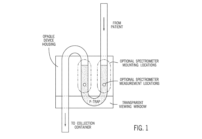
[0041] FIG. 1 shows an on-catheter device design with or without block.
This shows how,
with the design of the device and its housing structure, the catheter can be
shaped in such a way
that continuous readings of the column of liquid bio sample can be carried out
with or without a
catheter blockage device.
[0042] FIG. 2 shows an on-catheter device design in a close up ¨ lateral
view. This
demonstrates how the spectrometer on the device interacts with the catheter
and the liquid bio
sample stream inside of it.
[0043] FIGS. 3A, 3B, and 3C show detailed views of another on-catheter
design including
the micro-spectrometer with its catheter mount.
[0044] FIG. 4 shows the entire clinical setting with the urinary catheter
in its natural position
on a patient, including the incorporated urinary spectrometry screening,
detection and alerting
device (sensor C). This depicts an embodiment of a method for how continuous,
real time, on-
catheter measurements may be performed.
[0045] FIG. 5 shows a diagram of the on-catheter device in which it is
broken down into its
component parts which include power, a micro-spectrometer, a light source, a
collimator (lens), a
monochromator (prism), a wavelength selector (slit), the patient's liquid bio
sample, the detector
(photocell), a Bluetooth device for transmission to a computing platform (e.g.
to an electronic
health record or a mobile computing device where clinicians can see results),
and an on-device
signal so that clinicians can. visualize results without leaving the patient's
setting.
[0046] FIG. 6 provides a demonstration of how the device's spectrum of
emitted light is
transmitted to the computational platform for analysis by machine learning
algorithms to return
the molecular signal of the sample including 1) the -bacterial colony count,
2) the bacterial colony
type, and 3) bacterial infection by-products identifiable in the sample.
[0047] FIG. 7 shows a clinical interface in accordance with certain
embodiments of the
invention.
[0048] FIG. 8 shows a spectrometry-analytics .workflow in accordance with
certain
embodiments of the invention.
9
CA 03203589 2023-05-30
WO 2022/119910 PCT/US2021/061385
100491 FIG. 9 shows raw and processed data which reflect the absorbance
spectra plots for
E.coli scanned with a micro-NIR spectrometer in the wavelength range of 740 nm-
1100 nm, with
the x-axis being wavelength (nm) and the y-axis being absorbance (normalized
from 0.0 to 1.0).
100501 FIG. 10 shows three-dimensional principal component analysis of the
data obtained in
FIG. 9,
100511 FIGS. 11A and 11B show the results of both model types developed,
classification
models and regression models: 1) Random Forest; 2) Extreme Gradient Boosting;
3) Linear
Regression, fits a linear model to minimize the residual sum of squares
between the observed
targets in the dataset, and the targets predicted by the linear approximation;
4) Elastic Net, which
is a combination between lasso and ridge regression and 5) Lasso and Elastic-
Net Regularized
Generalized Linear Models that fits a generalized linear model via penalized
maximum
likelihood. The regularization path is computed for the lasso or elastic
network penalty at a grid
of values for the regularization parameter lambda. R2 also called coefficient
of determination, is
a regression score function. Best possible R2 score is 1.0 and it can be
negative. A constant model
that always predicts the expected value of y, disregarding the input features,
would get a R2 score
of 0Ø RMSE measures the differences between values predicted by a model and
the values
observed. MAE is the sum of absolute differences between our target and
predicted variables, it
measures the average magnitude of errors in a set of predictions, without
considering their
directions. FIG. 11A shows classification models (Median ROC scores) including
dichotomous
analyses for the five classification models: 1) Logistic Regression, 2) Random
forests (RF); 3)
Gradient Boosting Machine (GBM); 4) Support Vector Machine (SVM) and 5)
Extreme Gradient
Boosting (XGB). FIG. 11B shows ML regression results for the first- and second-
best
performance models: 1) Random forests (RF); 2) Extreme Gradient Boosting
(XGB).
100521 FIG. 12 shows Principal Analysis for Area Under the Receiver
Operating Curve
(AUROC) characteristics of the different methods trained classifying waveforms
as absence
(concentration 100) or presence (concentrations 101 to 105) of bacteria. It
should be noted that,
although FIGS. 11A-B, 12, and 13 show data specific to the bacterial species
E. coli, the device
is nevertheless capable of identifying multiple separate bacterial species.
This is the result of
each bacterial species having a specific spectrometric signature that can be
identified by the
algorithms utilized for detection. In other words, the method can distinguish
between different
bacterial species
CA 03203589 2023-05-30
WO 2022/119910 PCT/US2021/061385
[0053] FIG. 13 shows results of the most accurate of the various algorithms
that were tested,
support vector machine (SVM) analysis, for absence results on all selected
metrics
(concentration 100) or presence (concentrations 101 to 105) of bacteria (E,.
col i in this example):
[0054] FIG. 14 shows Principal Analysis AUROC for two biomarkers in a
Liquid bio sample
(Nitrates (Biomarker I) and Leukocyte esterase (LE, Biomarker 2)). The graphs
show Area
Under the Receiver Operating Characteristics (AUROC) of the different methods
trained
classifying waveforms as absence or presence of nitrates or LE in liquid bio
samples, where the
following acronyms apply: LR, Logistic Regression; RE, Random forests; GiBM,
Gradient
Boosting Machine; WM, Support Vector Machine; XGB, Extreme Gradient Boosting.
[0055] FIG. 15 shows support vector machine (SVM) analysis for two
biomarkers in a Liquid
bio sample (Nitrates (Biomarker 1) and Leukocyte esterase (LE, Biomarker 2)).
Presented are the
primary analysis perfoiniance metrics for the best performing method, Support
Vector Machine.
Abbreviations include: AUROC, Area Under the Receiver Operating
Characteristics; Sens,
Sensitivity; Spec, Specificity; F, F-Score.
[0056] FIG, 16 shows determination of flow rate which is calculated by
measuring a weight
change in the catheter bag during biofluid flow as a function of time.
[0057] FIG. 17 shows an example of a system for biofluid monitoring and
analysis in
accordance with some embodiments of the disclosed subject matter.
[0058] FIG. 18 shows an example of hardware that can be used to implement a
computing
device and server in accordance with some embodiments of the disclosed subject
matter.
[0059] FIG. 19 shows an example of a process for biofluid monitoring in
accordance with
some embodiments of the disclosed subject matter.
DETAILED DESCRIPTION
[0060] Healthcare delivery is complicated by the fact that medical
treatments are often
inconsistent and highly dependent on the complexity and the level of attention
that is required for
the delivery of care. It is somewhat surprising to find that often more
complex tasks are
performed with a much higher level of effectiveness than simpler ones. This is
a result of the fact
that complex tasks are part of what is known as a cognitive hypervigilant
state whereas simple
tasks are relegated to an inattentive state. This is part of the reason why
healthcare performs at
higher levels of delivery when dealing with complicated tasks such as
transplant surgery while
11
CA 03203589 2023-05-30
WO 2022/119910 PCT/US2021/061385
simultaneously delivering very poorly on the elimination of simple failures
such as medication
errors and the avoidance of hospital acquired infections (HAIs).
[0061] As noted above, hospital acquired infections are a major problem in
modern
healthcare. Prevalence of these HAIs in the United States alone is measured at
1.7 million
infections per year with a human cost that translates into 99,000 deaths. The
financial toll in the
United States is measured somewhere near $45 billion a year. It is clear then,
that these
infections pose a serious human and financial cost to the American healthcare
system. This of
course, is not a problem restricted to the United States and is seen
throughout the developed and
developing world.
[0062] Catheter associated urinary tract infections (CAUTIs) represent one
of the most
common forms of HAT. They represent approximately 32% of all HAIs affecting
over 2.5
million patients per year with a high level of morbidity and a mortality rate
of approximately
13,000 deaths per year. It is estimated that each of these infections has an
additional cost of
$13,731 per case. In aggregate the cost to the United States healthcare system
for catheter
associated urinary tract infection approaches $7.7 billion per year. Given
this, it is clear that the
financial and human cost of this CAUTIs is significant.
[0063] The reasons why we have not been able to resolve the seemingly
simple problems of
HAIs and specifically that of CAUTIs are both human and technological. The
human factors
include several heuristics that are pervasive in medicine, the most
significant being the "status
quo bias". We believe these infections to be part of the standard, recognize
complications of
practicing medicine - in other words, we believe that this is the "cost of
doing business" and
therefore, unchangeable. The technological problem is that up until now, there
has been no
simple, effective, real time and continuous system to monitor for potential
catheter associated
infections. This combination of human factors and technological deficiencies
has left us in a
position where nobody has truly sought a solution to this truly significant
problem.
[0064] It is clear that what is required to resolve this problem is
something that overcomes
human and technological issues. Any possible real time monitoring solution to
CAUTIs in the
current state is impractical and non-implementable as it would involve too
many steps, too many
people, and too much time that at the end of the day would not deliver
actionable data capable of
preventing negative outcomes. In other words, any current solution imposes too
much of a
cognitive and operational load on the clinical system without providing
patients or providers
12
CA 03203589 2023-05-30
WO 2022/119910 PCT/US2021/061385
benefit. This truth makes the realistic implementation of a solution utilizing
existing tools
extremely unlikely.
[0065] Likewise, it would be impossible to create a monitoring solution for
all the infections
related to patient delivered, home medical care procedures that depends on
active participation of
patients in this process. In this setting, the manipulation of catheters and
bio samples that would
be required of patients would likely increase the risk of infection rather
than decrease it.
[0066] It is evident that what is required to resolve this major health
issue is an innovation
which brings a mobile, continuous, point of care, disposable, and cost-
effective solution to bear.
What is called for is a non-invasive monitor which provides continuous
screening and diagnosis
for decision support and treatment modification. The solution described in
this application makes
this possible by leveraging the analysis of bacteria and biomarkers in liquid
bio sample at the
patient's bedside in real time. We will do this with virtually no additional
clinical workload,
which improves adoption of the technology, and by providing real-time,
actionable data with
guidelines-based decision support. Effectively, this solution will release
much-needed cognitive
and operational bandwidth from healthcare teams. We do this by continuously
monitoring
bacteria, identifying specific strains and their concentrations, identifying
biomarkers associated
with active infection, and translating these results into clear, data-driven
decision support. In
various embodiments, one or more biomarkers related to the functional status
of a particular
bodily system (e.g. one or more of the cardiac, respiratory, renal,
neurologic, endocrine, or
immune system) may be identified and the patient's status may be determined
based on these
biomarkers. In some embodiments, a machine learning system may be trained to
identify these
and other biomarkers related to the functional status of the one or more
bodily systems of the
patient to assist the user (e.g. clinician) with evaluation of the patient's
status. The biomarkers
that have been identified (e.g. using a machine learning algorithm) may assist
the user with the
identification of at least one condition of the patient (e.g. a condition of
one or more of the bodily
systems of the patient) and the patient status may be determined based on the
identified at least
one condition.
[0067] Accordingly, in one embodiment the invention includes a device
including a
spectrometer which attaches to a drainage tube of a medical tube (e.g. a
urinary catheter or a
peritoneal dialysis catheter) and obtains measurements (e.g. measurements of
absorbance in the
IR) continuously without a need to block the flow of fluid in the tube. The
data from the
13
CA 03203589 2023-05-30
WO 2022/119910 PCT/US2021/061385
spectrometer is analyzed in order to identify one or more materials in the
patient's liquid bio
sample including bacteria (e.g. E. coli), leukocyte esterase (LE), and
nitrates. The data, which
may include information pertaining to absorbance as a function of wavelength,
can be analyzed
using principal component analysis or various AT classification models.
[0068] Since the creation and modern usage of indwelling drainage catheters
started in the
1930's we have seen virtually no change to the nature in management of these
catheters.
Therefore, to advance this technology the systems and methods disclosed herein
apply new
sensors based on non-invasive spectrometry techniques and combines this with
artificial
intelligence data analytics to provide a breakthrough development for
continuous infection
surveillance. This ability to detect the existence of bacteria in any biofluid
sample such as liquid
bio sample without interacting or directly manipulating the sample itself has
tremendous value in
modern healthcare. Such a capacity will allow for continuous sampling of
specimens without
altering or adding to the workflow of the clinicians currently caring for
patients. Given this
passive sampling method's incorporation of continuous sampling into the
workflow, we can
guarantee that patients will receive continuous screening for early infection
in any indwelling
catheter, such as a urinary catheter. This ability to detect bacterial
colonization and early
bacterial infection will profoundly affect the delivery of safe care as it
will eliminate many
infections that are currently only identified after the existence of advanced
infections.
[0069] Disclosed herein are embodiments of a continuous, real-time, on-
catheter, on-patient
device which can be used in a clinical setting. Embodiments of the disclosed
on-catheter design
include capability to communicate and interact directly with clinicians and
care givers.
[0070] The following workflow and hardware elements may be used to carry
out various
embodiments of a continuous, on-catheter screening of biofluid for the
presence of one or more
known bioproducts and microorganisms:
[0071] System workflow and implementation:
[0072] In various embodiments, the system includes a spectrometry device
which can be
placed on the drainage tubing of any existing or newly placed indwelling
patient catheter with
external drainage. This is accomplished by including the following elements
(see FIGS. 1-4):
a. Use of a standard drainage catheter tubing made of any common material
used for such drainage catheters including rubber, silicone, latex,
Polyvinyl Chloride (PVC). This is the case because the infrared spectrum
14
CA 03203589 2023-05-30
WO 2022/119910 PCT/US2021/061385
in which the micro-spectrometer functions is not affected by the properties
of these materials.
b. The micro-spectrometer inside of its housing/mounting hardware is
clamped to the outside of the drainage catheter tubing as seen in FIGS. 1-
3.
c. This is accomplished in a way that creates curvatures in the catheter that
creates two impediments or blocks to the free flow of liquid bio sample ¨
the impediments or blocks are located at each of the angulations created at
the point of curvature
d. Given these impediments or blocks, there is created an area of stagnant
liquid bio sample column in which the entire tubing is filled with liquid
bio sample with no air; fluid can still move through the tubing but the
impediments or blocks cause sufficient fluid to accumulate in one location
to permit optical measurements to be made.
e. The mounting is constructed in such a way so that the micro-spectrometer
sampling window is directed precisely toward this created area of liquid
bio sample stagnation.
f. The bio sample can then be analyzed by illuminating the sample with the
micro-spectrometer. This analysis can be performed at any frequency that
is deemed clinically relevant. The time required for the micro-
spectrometer to obtain data from the liquid bio-sample is less than 1
second.
[0073] No alteration to the existing catheter system is required. In
particular, and of critical
importance, no penetration or violation of the existing catheter system is
performed as it is not
required that the biofluid be in contact with any element of the device
testing system. The
spectrometric system uses its light characteristics to penetrate the tubing of
the existing drainage
system to continuously analyze the biofluid included in the tubing.
[0074] On-Catheter Device
[0075] As seen in FIGS. 1 and 3, the on-catheter device may be enclosed in
a housing that is
clamped onto the outside of the drainage catheter system in such a fashion
that it guarantees free
CA 03203589 2023-05-30
WO 2022/119910 PCT/US2021/061385
flow of biofluids through the system while allowing accomplishment of
continuous
spectroscopic analysis of samples within the catheter system tubing.
[0076] In various embodiments (in which the analysis may be accomplished
without using
blocks to the flow of biospecimen for the purposes of creating a temporary non-
flowing
biospecimen sample used for obtaining a spectrometric data set), the analysis
may be
accomplished by using the natural bending properties of the drainage catheter
tubing to collect a
sample of fluid by gravity, for example at a low point in the tubing. Fluid
from the urinary
catheter may move through the tubing in drips or a small trickle, which in a
vertical segment of
tubing would move past the IR absorbance sensor too quickly to obtain a stable
reading.
Therefore, creating a low point (e.g. a horizontal portion or a bend) in the
tubing this ensures that
a small amount of fluid will be retained for a sufficiently long period of
time (e.g. for at least
several seconds or tens of seconds) to allow absorbance readings to be taken.
[0077] The tubing material naturally forms non-occlusive bends through
which the liquid bio
sample regularly flows through vertical segments and stagnates in curved or
horizontal segments.
Using this property, the mounting apparatus demonstrated in FIGS. 1 and 3 will
create a gentle
curvature in the catheter tubing so that an area of biospecimen stagnation, or
accumulation, may
be created. This will allow sampling of the biospecimen with the spectrometer
for the period of
testing which is on the time order of 1 second for testing of a sample. In
this embodiment, the
spectrometer will be positioned in the center of the bend formed by the
mounting device, with
the sampling window facing the area of stagnant bio sample created by the
mounting device (e.g.
in the area of the tubing labeled "p-trap" in FIG. 1). By creating an area
(e.g. a bend) within the
tubing in which fluid is still flowing but is sufficiently slowed or stagnant
to permit
spectroscopic measurements, this helps ensure that a data set of a fresh bio
sample will always be
available for testing with the device, since new fluid material will continue
to enter the
measurement area. In contrast to certain prior systems which employ one or
more blocks to
accumulate sufficient fluid to obtain optical readings of the sample, the
present invention uses a
bend such as a p-trap to collect fluid for analysis without having to block
flow, which simplifies
the design of the device and facilitates continuous measurement of the
patient's sample. A
spectrometer (which may include a light source and detector, as described in
more detail below)
may be mounted in the housing in any location at which fluid will accumulate.
Accordingly to
various embodiments, two possible spectrometer mounting locations are
indicated in FIG. 1 by
16
CA 03203589 2023-05-30
WO 2022/119910 PCT/US2021/061385
the vertical ovals, with circles showing the possible locations at which
spectrometer
measurements may be obtained. In certain embodiments, the housing may be
opaque, which
facilitates spectrometer measurements by reducing potential background light
contamination. A
transparent viewing window may be located near the bottom of the housing to
facilitate insertion
of the tubing as well as to allow a user to confirm that fluid is moving
through the tubing
associated with the housing. In some embodiments, the bend in the tubing at
which fluid
accumulates may be referred to as a p-trap.
[0078] FIGS. 2 and 3 provide a close up view showing how the sensor will
interact with the
drainage catheter tubing in certain embodiments. The embodiment of FIG. 2 in
particular
provides labels showing how the interacting with the drainage catheter tubing
(A) and the area of
the bio sample (e.g. liquid bio sample, (D)) accumulated in the area adjacent
to the sensor. The
left portion of FIG. 2 shows a segment of catheter tubing (F) into which
liquid bio sample is
flowing (B) while the inset on the right shows a liquid bio sample (D)
accumulated inside the
tubing as well as a signal processor (E). In this embodiment, the housing
includes a curved face
which is located adjacent to the bend in the tubing such that the tubing and
the housing are
closely aligned, which optimizes the transmission of light from the light
source to the sample
within the tubing as well as the transmission of light returning from the
sample to the sensor or
detector in the housing. The segment of tubing includes a bend or p-trap,
situated so as to create
a low point in the flow path, and is shown as having an accumulation of fluid
(liquid bio sample,
(D)).
[0079] On the outside of the tubing below the p-trap, a micro-spectrometer
sensor (element C
in FIG. 2) is attached adjacent to the tubing in a location adjacent to the
low point where fluid
accumulates. Infrared (IR) light is emitted from the sensor device into the
tubing and an IR
sensor adjacent to the tubing measures IR light reflected by the sample inside
the tubing. In
addition, the micro-spectrometer sensor (C) measures spectra (e.g. absorbance
spectra) in the IR
range from the sample. The frequency interval of data collection and what will
be considered to
be "continuous" sampling will be determined by clinical needs for a particular
situation and
patient, and the system will be capable of adjusting to these requirements. In
various
embodiments, continuous sampling may include sampling at least once per
minute, once per 30
seconds, once per 10 seconds, once per second, five times per second, or other
more or less
frequent sampling intervals as called for in the particular situation.
17
CA 03203589 2023-05-30
WO 2022/119910 PCT/US2021/061385
[0080] FIGS. 3A-3C disclose an embodiment of an on-catheter sensor system
design. FIG.
3A shows a perspective view (top) of the sensor housing and a cross-sectional
view (through line
A-A' in the top view) showing an accumulation of liquid bio sample (e.g. urine
or peritoneal
fluid) in the catheter tubing in the region below the sensor. The housing in
FIG. 3A includes a
central oval-shaped opening in which a sensor device may be inserted. FIG. 3B
shows a side
view of the sensor housing with a sensor device inserted from the top into the
oval-shaped
opening. The housing also includes openings on the sides through which
catheter tubing may be
inserted, where the inserted tubing traces an approximately U-shaped path
through the housing,
entering on one side and exiting on the other side, to provide a low point at
which liquid bio
sample accumulates and can be monitored. The side view of FIG. 3B shows the
housing and
sensor with a section of catheter tubing running through the housing. The
tubing enters the
housing from the top left, travels through the housing in a U-shaped path, and
exits the housing
from the top right. Upon exiting the housing, the tubing may then complete a
loop and attach to a
clip on the side of the housing (see FIG. 3C) to stabilize the tubing. The
housing may include a
liner (e.g. made from Teflon) to block light from exiting the housing in order
to minimize or
prevent contamination of the light signals originating from the liquid bio
sample with spurious
signals which might arise from nearby materials outside the tubing. In various
embodiments, this
design may include a slot into which the catheter tubing may be inserted such
that a portion of
the catheter tubing is adjacent to the spectrometer (see FIG. 3B).
[0081] The housing may include a window on the side which aligns with an
indicator on the
inserted sensor device. The window on the housing may be just an opening or
may include a lens
that may be flat or curved to permit light signals from the sensor device to
be seen, where the
curved lens allows the light signals from the sensor device to be seen at a
wider range of angles.
In some embodiments, a portion of the side of the housing may be removable
(e.g. along the
dotted "separation line" shown in FIG. 3B) to allow the catheter tubing to be
inserted into the U-
shaped track from the side. The side portion may then be reattached to help
keep the tubing in
place and also to maintain a low-light or light-free background in the
vicinity of the spectrometer
sensor. Permitting the tubing to be attached from the side in this way allows
the sensor housing
to be attached to a catheter tubing that is currently coupled to a patient in
a way that does not
require decoupling the tubing or interrupting drainage.
18
CA 03203589 2023-05-30
WO 2022/119910 PCT/US2021/061385
[0082] FIG. 3C shows a perspective view (upper panel), a top view (center
panel), and a side
view (bottom panel) of the housing. These views depict the clip (on the left
in the upper and
center panels, on the right in the bottom panel) to which the tubing may be
attached to provide
additional stability.
[0083] FIG. 4 provides a diagram of a system such as that of the embodiment
of FIG. 2 in the
case that the bio sample being utilized is a liquid bio sample coming from the
patient's bladder
or peritoneal cavity. The diagram of FIG. 4 shows a Foley catheter (A), a
catheter securing
device (B), a sensor (C) (e.g. shown schematically as a U-shaped bend but
which may be
mounted in a housing such as that shown in FIG. 3) located at a bend in the
drainage tubing (D),
and a drainage collection bag (E). In the case of other bio samples besides
urine, the
arrangement would be similar to that shown in FIG. 4 but would be suitably
adapted based on the
site of origin of the bio sample. Other potential bio sample sources include
peritoneal fluid from
patients undergoing peritoneal dialysis, biliary fluid from patients
undergoing biliary system
diversion, naso/oral enteric tubes in patients undergoing enteral
decompression, and surgical
drainage in patients with surgical wounds drains.
[0084] FIG. 5 provides a detailed view of an embodiment of the on-catheter
device. The
device includes a control system arranged and adapted to carry out the
procedures disclosed
herein and which includes a catheter holder for the device. Also included is a
power source (e.g.
battery power for mobile deployment) and in one particular embodiment the
device may include
a lithium ion battery that will be rechargeable and able to hold a charge for
continuous (24/7)
usage for up to 14 days. In another embodiment, the lithium ion battery may
not be rechargeable
but nevertheless will be able to hold a charge for 24/7 usage for up to 14
days.
[0085] The device also includes a micro-spectrometer to generate data which
can then be
subject to further analysis on or off the device (or both). The spectrometer
may include one or
more light sources such as light-ernitting diodes (LEDs) to emit light into
the sample in order to
obtain data. In one embodiment, the spectrometer may use different LEDs to
select for the ideal
waveform for the identification of specific bacterial strains as well as
separate biomarkers. The
number of LEDs can vary depending on the products (e.g. bacteria and
biomarkers) one wishes
to identify.
[0086] In various embodiments the spectrometer may include, along with the
light source to
illuminate the sample, a collimator (e.g. lens) to concentrate the light
within the sample. The
19
CA 03203589 2023-05-30
WO 2022/119910 PCT/US2021/061385
spectrometer may also include a monochromator (e.g. a prism), to divide the
light sample into its
constituent wavelengths, and a wavelength selector (e.g. a slit), to select
the correct wavelength
for selected bacterial strains or other products of interest.
100871 In general, the patient's liquid bio sample (e.g. urine, peritoneal
fluid, wound drainage,
enteral content, etc.) will remain at all times under the following
conditions: inside of the
drainage catheter tube and completely separate from the device with no element
of the device
coming into contact with the bio sample.
100881 The spectrometer also includes a detector (e.g. a photocell) which
records the
wavelength results of light returning from the sample from each illumination
(e.g. absorbance).
The spectrometer may also include a communication module (e.g. a Bluetooth
device) for
optional transmission of data and other information to remote device such as a
computing
platform, which can include an electronic health record and/or a stationary or
mobile computing
device where clinicians can see results and updates on the patient's status.
This digital result can
be shared with any number of clinicians and administrators who have been
cleared though
concerns relating to patient privacy to manage the patient's clinical status
as well as to manage
broader infection control issues related to the institutional concerns.
100891 The procedures for sharing these digital records will generally be
determined by the
managing team caring for the patients but can include, but are not limited to,
the use of messages
posted in the medical record, text messages, pages, and phone calls to
responding clinicians and
administrators, among various possible means.
100901 The device may also include an on-device signal system so that users
such as
clinicians can visualize results without leaving the patient's setting. The
signaling system may be
located at or visible from the patient's bedside and may include a status of
the patient's bio
sample as well as a recommended management paradigm determined by the local
treating team.
A sample of this management could include the protocol seen in FIGS. 5 and 7,
which show a
simple stoplight type signal system which may include one or more status
indicators. In the
examples shown in FIGS. 5 and 7, the on-device signal may include three
indicator lights which
may be used to indicate that the patient's status is good (e.g. a green
light), questionable (e.g. a
yellow light), or needing attention (e.g. a red light); these conditions may
be related to State 1,
State 2, and State 3 described below for FIG. 7.
CA 03203589 2023-05-30
WO 2022/119910 PCT/US2021/061385
[0091] Data from the on-device indicator may then be transmitted (alone or
along with other
information) to a remote computing platform (e.g. a mobile or cloud-based
computing platform)
to perform further analysis such as spectral analysis and which in some
embodiments may be
analyzed using machine learning algorithms to detect the components of the
spectra that are
emitted and captured.
[0092] The remote computing platform may process the data using machine
learning
algorithms to provide results regarding, bacterial colony count, bacterial
colony type, and
bacterial infection by-products identifia.ble in the sample, among other
information. A.s seen in
FIG. 6, each bacterial species (and even the concentration of each bacterial
species) and
biornarket may have a different spectral signature related to the compound and
the wavelength of
light utilized by the spectrometer. For example, the diagram in FIG. 6 depicts
spectral data being
transmitted from the spectrometer corresponding to E, coli, Klebsiella, and
Proteus bacteria.
[0093] in various embodiments, the combination of the results of analysis
can yield three
clinical entities (see FIG. 7):
[0094] State I: No bacteria or infection in bio-sample
[0095] State 2: Bacterial colonization but no infection in bio-sample
[0096] State 3: Bacteria and infection in bio-sample
[0097] Clinicians according to their experience and particular practice
patterns will determine
their clinical response to these distinct states.
[0098] ML Algorithms
[0099] Machine Learning (ML) algorithms are developed to be used
specifically with the
device disclosed herein rather than independently of the device. The
algorithms will be utilized
to perform the analysis of the sample's waveforms and are constructed as
follows:
[00100] Every sample contains a waveform made of 330 data points. The device
performing
data analytics using machine learning comprises:
[00101] - One module for classification
[00102] - One module for sensitivity analyses
[00103] - An unsupervised learning module configured to generate the final
result based on the
organized data set
21
CA 03203589 2023-05-30
WO 2022/119910 PCT/US2021/061385
[00104] The classification module may perform one or more of the following:
extract data,
which may be performed continuously from the patient's bedside using the
spectrometer
hardware; load the extracted data into a dataset; and generate results based
on the colony forming
units (CFUs) in the sample.
[00105] The extracted data may be classified using one or more of the
following classification
methods: Gradient Boosting Machines, Support Vector Machines, Random Forests,
extreme
Gradient Boosting, Logistic Regression; and Random Hyperparameter Tuning in 10
folds cross-
validation (CV).
[00106] The ML algorithm creates two or three groups based on the CFUs and
assigns the
samples to each of the respective groups. The output of the ML algorithm
includes bacterial
concentrations expressed in a range from 100 to 105, where 100 means the
absence of bacteria,
and from 101 to 105 indicates the amount of concentration of the present
bacteria within the
sample. Predictive performance of the ML algorithm may be assessed by
determining AUROC,
Precision (AP), specificity, sensitivity, and F for each one of them.
[00107] The regression includes a quantification of bacteria metrics using
different datasets in
which 100, 101, 102, 103, 104 and 105 are marked as 0, 1, 2, 3, 4, and 5
respectively, creating a
continuous outcome in which, by using such waveforms, any given concentration
is predicted (0-
5). Regression models addressed included Random Forests, Extreme Gradient
Boosting, Linear
Regression, Elasticnet, Lasso and Elastic-Net Regularized Generalized Linear
Models. A
Random Hyperparameter Tuning in 10-fold cross-validation is also performed for
a greater R-
squared.
[00108] Sensitivity Analysis Module
[00109] The Sensitivity Analysis Module may be used to determine the presence
or absence of
bacteria in a fluid based on a continuous steam of data from the device, data
regarding possible
outcome of the bacteria concentration. To test the sensitivity of the device,
various tests were
performed to compare different concentrations of bacteria. Eight databases
were created based
on these numbers. In dataset number one, 100 was considered to be an absence
of bacteria and
101, 102, 103, 104, and 105 were considered as presence of bacteria in various
concentrations. In
dataset number two, the two groups were split as 100 vs. 101, 102, and 103. In
dataset number
three, the two groups were split as 100 vs. 101, 102, 103, and 104. In dataset
number four, the two
22
CA 03203589 2023-05-30
WO 2022/119910 PCT/US2021/061385
groups were split as 100 vs. 101. In dataset number five, the two groups were
split as 100 vs. 102.
In dataset number six, the two groups were split as 100 vs. 103. In the
dataset number seven, the
two groups were split as 100 vs. 104. And in dataset number eight, the two
groups were split as
vs. 105 (FIG. 12).
[00110] Unsupervised Learning Module
[00111] Provided below is a list of the performance metrics of all the
sensitivity analyses
performed for each dataset obtained. The models were trained using random
hyperparameter
tuning 10 folds CV and validated in the testing split containing 25% of their
observations for the
classification models. For the regression model, 100% of the dataset number
one observations
were used to perform 10 folds CV.
[00112] The primary analysis had outstanding performance achieved using an
SVM. It is
configured to assemble the unstructured data set into multiple versions of the
organized data set.
The module is configured to create training data from the organized data set
and wherein the
supervised learning module is configured to use the training data to generate
one or more groups.
[00113] EXAMPLE
[00114] The following provides details of a non-limiting example according to
embodiments
of the invention, including methods and results of building and using the
device to collect data
and processing the data using a Machine Learning algorithm.
[00115] Under Partners HealthCare Institutional Review Board approval, two
hundred samples
were analyzed from September 2018 to January 2019 at Harvard Medical School
Microbiology
Laboratories and Brigham and Women's Hospital. Statistical analyses were
performed in R
version 4Ø0 and RStudio version 1.2.5019.
[00116] Bacteria Analysis
[00117] Serially-diluted samples were prepared using a culture of Escherichia
coil MG1655
and synthetic urine (Pickering laboratories 1700-0600). Twenty-four hours
before the
experiment, 5 mL of EZRDM media (Teknova) were inoculated with 10 uL of a
saturated E. coil
culture and incubated overnight at 37C with 220 Revolutions Per Minute (RPM).
Dilution series
were created by diluting 500 uL of the culture medium in 4.5 mL of synthetic
urine, vortexed for
5 seconds, and then 500 uL were transferred into 4.5 mL of synthetic urine.
Each subsequent
dilution was created utilizing the same protocol until a total of 10 dilutions
were reached. One
23
CA 03203589 2023-05-30
WO 2022/119910 PCT/US2021/061385
sample in each group was left without any bacterial inoculation as a control.
Spectrometry
samples were prepared by transferring 4 mL of each dilution to glass
spectrometer cuvettes
sealed with Parafilm. Determination of the concentration of bacteria in the
prepared synthetic
urine samples was accomplished by plating 100 uL of each sample in LB agar to
determine
colony forming units (CFUs). Each plate was incubated overnight at 37C, and
colonies were
counted the following morning using a proprietary machine learning algorithm
to automatize and
standardize this process. Both the spectrometry and the microscopic readings
were performed
simultaneously to avoid any discordance in the time from sample preparation to
sample analysis.
[00118] 3-D Printing
[00119] The integrated spectrometer and liquid bio sample holder (see FIG. 3)
were designed
in Solidworks (Dassault Systemes, Velizy-Villacoublay, France) and produced
using an Obj et30
3D printer (Stratasys, Eden Prairie, Minnesota, USA) from a white photopolymer
resin
(RGD835). The design was created to simulate a mount that could be attached to
the outer
surface of a urinary drainage catheter. In order to reduce background signal
contamination, the
cuvette holder had an integrated (opaque) lid and a 3 mm-thick Teflon liner
that blocked the light
path from exiting the cuvette/catheter.
[00120] All of the samples, each having a different concentration, were
analyzed using the
device described in this application and, in parallel, simultaneously
underwent microscopic
colony count analysis to represent the gold standard for bacterial colony
count identification.
[00121] The device was used to obtain spectrometric data, perform chemometric
analysis, and
create calibration models for bacterial detection. The data from both methods
was recorded in
separate databases and correlated with appropriate sample identifiers. Raw
data from the
spectroscopic evaluation was analyzed and incorporated into the Machine
learning algorithms,
using the microbiological colony counts as the representation of the gold
standard accurate
results.
[00122] Data Analysis
[00123] Bacteria concentration ranged from 100 to 105, where the exponent
indicates the
number of bacterial CFU in the sample. Every bacterial colony concentration
has a characteristic
morphologic waveform signature determined by the combination of CFU and the
wavelength
utilized to analyze the sample. This signature waveform is created from 330
separate data points
24
CA 03203589 2023-05-30
WO 2022/119910 PCT/US2021/061385
(see FIG. 9, with wavelength in nm on the x-axis and absorbance on the y-axis)
and from
principal components analysis (FIG. 10). FIG. 9 shows the results of the data
processing from
raw to processed data. In this process we selected the wavelength from 740 to
1070 nm, then
processed and normalized the data.
[00124] Processing: Assumes Beer-Lambert model is valid and transforms the
measured signal
to be linear with concentration by doing a log transform and adjusting the
result for noise and
deviations from the model.
A =I ogi
[00125] FIG. 10 depicts results from a Principal component analysis, or PCA,
which is a
dimensionality-reduction method that is often used to reduce the
dimensionality of large data
sets, by transforming a large set of variables into a smaller one that still
contains most of the
information in the large set. In our setting the PCA approach has taken
information related to
different concentrations of bacteria in a liquid bio sample and classified
them based on the
number of CFUs. It was used it as an exploratory tool for our analysis as it
demonstrates the
grouping of different CFU concentrations into well-defined clusters.
[00126] In addition to bacterial detection, it was felt that the addition of
detection of
biomarkers associated with urinary tract infection (UTI) would add to the
value of the prediction
models. This would allow for the establishment of three separate clinical
states: 1) A catheter
with no bacteria and no infection, 2) a catheter with bacterial colonization
and no evidence of
infection and 3) a catheter with bacterial infection. Based on this concept,
two sensitivity
analyses using urinary nitrates and leukocyte esterase (LE) were performed to
determine how
these target variables can affect the signature waveform for each
concentration.
[00127] Predictive models were created using machine learning algorithms in
order to identify
the smallest absolute amount of change in bacteria concentration that can be
detected by our
spectrometer. To increase accuracy and precision within our models, all the
data used for the
classification algorithms were sampled randomly and had a distribution of 75%
of the data
designated for training and 25% for the testing of the algorithm.
CA 03203589 2023-05-30
WO 2022/119910 PCT/US2021/061385
[00128] Model Training and Validation
[00129] Based on our outcome of interest, we divided the analysis of the
samples in two
groups with ten different models (classification models and regression
models). Classification
models were used to predict the concentration among different concentrations,
whereas the
regression models were used to predict the specific concentration of bacteria
derived from their
waveforms.
[00130] All models were trained using a seed so that the predictions could be
replicated. We
performed Random Hyperparameter Tuning in 10-folds cross-validation (CV)
aiming for the
highest Area under the Receiver Operating Characteristic Curve (AUROC) when
training
classification models and aiming for the highest R2 when addressing the
regression models.
[00131] Classification Models
[00132] We trained five different models in this category: 1) Logistic
Regression; 2) Random
forests (RF); 3) Gradient Boosting Machine (GBM); 4) Support Vector Machine
(SVM), and 5)
Extreme Gradient Boosting (XGB). The models used 75% of each dataset for
training purposes
and 25% for validation to address the most optimally trained classification
model's performance.
[00133] Regression Models
[00134] We used five different models that included 1) Random Forests; 2)
Extreme Gradient
Boosting; 3) Linear Regression; 4) Elastic Net; and 5) Lasso and Elastic-Net.
These models were
trained using 100% of the observations in order to predict the different
bacteria concentration
levels (from 100 to 105), and biomarkers using the different waveforms.
[00135] Results
[00136] FIGS. 8 and 9 show the workflow (FIG. 8) and raw and pretreated
spectra (FIG. 9) of
the bacteria concentration in urine samples using a micro-near infrared
spectrometer. The NIR-
spectrometer scanned through a glass cuvette. The wavelength range of 740-1100
nm was found
to contain the most important peaks in the spectra based on the literature.
[00137] A combination of synthetic urine and five different bacterial
concentrations was
analyzed for a total of two-hundred samples in the main analysis, and with
four-hundred samples
for biomarker analysis (200 samples with nitrates and 200 leukocyte esterase).
[00138] Principal Analysis ¨ Bacteria Only
26
CA 03203589 2023-05-30
WO 2022/119910 PCT/US2021/061385
[00139] To validate our hypothesis, a series of experiments were conducted to
observe how the
CFUs of E.coli affected the waveform data in each concentration. Ten machine
learning models
were used to classify and established a cut-off point between samples.
[00140] Classification ¨ Bacteria Only
[00141] Among the five classification methods, Support Vector Machine (SVM)
achieved the
highest performance with a specificity of 0.99, sensitivity of 1, precision of
0.99, F-score of 0.99
and AUROC of 1. Metrics of the thirty-five different classification models
assessed as part of
the classification sensitivity analysis are reported in FIGS. 11A and 11B.
Principal Analysis for
Area Under the Receiver Operating Curve (AUROC) characteristics of the
different methods
trained classifying waveforms as absence (concentration 100) or presence
(concentrations 101 to
105) of bacteria are shown in FIG. 12. The results on all the metrics selected
for the most
accurate algorithm are shown in FIG. 13.
[00142] Regression ¨ Bacteria Only
[00143] The prediction performance of the regression models was addressed
using R2, Root
Mean Squared Error (RMSE), and Mean Absolute Error (MAE).
[00144] Of the five different models tested using the waveform data by means
of Cross
validation for regressing the bacteria concentration, the best performing one
was obtained using a
Random Forests method; with a MAE of 0.48, RMSE of 0.45, and an R2 of 0.82.
Metrics of the
five different regression models assessed are reported in FIG. 14.
[00145] Sensitivity Analysis ¨ Biomarkers
[00146] Performance metrics of the different models trained as part of the
sensitivity analysis
using three concentrations of nitrates and one of leukocyte esterase were
analyzed as part of the
sensitivity analyses.
[00147] Classification ¨ Biomarkers
[00148] Biomarkers were classified in this work. We selected two biomarkers of
many
available because of their wide acceptance in clinical practice and broad
adoption and
availability. The two biomarkers chosen were Nitrates and Leukocyte esterase
(LE), whose use
in the diagnosis of urinary tract infections is universally accepted.
Nevertheless, in various
embodiments any Biomarker can be characterized through use of this process.
27
CA 03203589 2023-05-30
WO 2022/119910 PCT/US2021/061385
[00149] Nitrates were classified into two groups, presence or absence in
urine, and were
evaluated in 200 samples. All the data obtained sensitivity and specificity
close to 100% in each
test and high AUROC with SVM, GBM, LR mainly (see FIGS. 14 and 15).
[00150] On the other hand, Leukocyte esterase was measured with three
different
concentrations (1 ml of 0.45mg/1 liter of saline solution plus 3 ml of urine,
2 ml of 0.45mg/1
liter of saline solution plus 2 ml of urine, and 3 ml 0.45mg/1 liter of saline
solution plus 1 ml of
urine) in 200 samples in total. With our other sensitivity analysis, the
support vector machine
was the best algorithm with an AUROC of 0.99, followed by LR with 0.98, the
precision of 1, F-
score of 0.99, and AUROC of 0.99. Sensitivity and specificity were 0.99 in all
the samples
analyzed (see FIGS. 14 and 15).
[00151] In certain embodiments, a flow rate of the biofluid may be determined
using the
disclosed apparatus. In such embodiments, the housing of the on-catheter
sensor system (e.g. as
in FIGS. 3A-3C) may include a load cell sensor or other mechanism (FIG. 16) to
monitor the
weight and/or changes in weight of the fluid collection bag (labeled as a
urine collection bag in
FIG. 16, although other fluids may be monitored using the device) attached to
the load cell
sensor. In various embodiments, the device may calculate an approximation of
flow rate of a
biofluid that passes through the indwelling catheters and therefore calculate
an actual
measurement of amount of biofluid coming from the patient at any given time.
In certain
embodiments, flow rate may be calculated through use of a measurement of
changing weight in a
biofluid repository (e.g. a biofluid collection bag) over time. In particular
embodiments, flow
rate may be calculated on a continuous basis as a measure of weight change and
may be reported
to the user one or more communication mechanism of the device. In various
embodiments, the
calculated flow rate may be based on the following formula: 6 weight/ 6 time.
In some
embodiments, an algorithm may utilize this data to calculate a volume over
time calculation to
yield an approximation of flow rate over time. This data may be reported to
the user
continuously via one or more communications mechanisms of the device.
Information about the
biofluid flow rate and total accumulation of biofluid may be used to monitor
the patient's status.
[00152] Turning to FIG. 17, an example 1700 of a system (e.g. a data
collection and
processing system) for biofluid monitoring and analysis is shown in accordance
with some
embodiments of the disclosed subject matter. In some embodiments, a computing
device 1710
can execute at least a portion of a system for biofluid monitoring and
analysis 1704 and provide
28
CA 03203589 2023-05-30
WO 2022/119910 PCT/US2021/061385
control signals to one or more components of a data collection system 1702,
for example a
spectrometer coupled to a liquid bio sample system. Additionally or
alternatively, in some
embodiments, computing device 1710 can communicate information regarding the
control
signals to or from a server 1720 over a communication network 1706, which can
execute at least
a portion of system for biofluid monitoring and analysis 1704. In some such
embodiments, server
1720 can return information to computing device 1710 (and/or any other
suitable computing
device) relating to the control signals for system for biofluid monitoring and
analysis 1704. This
information may be transmitted and/or presented to a user (e.g. a researcher,
an operator, a
clinician, etc.) and/or may be stored (e.g. as part of a research database or
a medical record
associated with a subject).
[00153] In some embodiments, computing device 1710 and/or server 1720 can be
any suitable
computing device or combination of devices, such as a desktop computer, a
laptop computer, a
smartphone, a tablet computer, a wearable computer, a server computer, a
virtual machine being
executed by a physical computing device, etc. As described herein, system for
biofluid
monitoring and analysis 1704 can present information about the control signals
to a user (e.g.,
researcher and/or physician). In some embodiments, data collection system 1702
may include a
light source, a detector, and/or other optical components for collecting data
from a sample
obtained from a subject.
[00154] In some embodiments, communication network 1706 can be any suitable
communication network or combination of communication networks. For example,
communication network 1706 can include a Wi-Fi network (which can include one
or more
wireless routers, one or more switches, etc.), a peer-to-peer network (e.g., a
Bluetooth network),
a cellular network (e.g., a 3G network, a 4G network, a 5G network, etc.,
complying with any
suitable standard, such as CDMA, GSM, LTE, LTE Advanced, WiMAX, etc.), a wired
network,
etc. In some embodiments, communication network 1706 can be a local area
network, a wide
area network, a public network (e.g., the Internet), a private or semi-private
network (e.g., a
corporate or university intranet), any other suitable type of network, or any
suitable combination
of networks. Communications links shown in FIG. 17 can each be any suitable
communications
link or combination of communications links, such as wired links, fiber optic
links, Wi-Fi links,
Bluetooth links, cellular links, etc.
29
CA 03203589 2023-05-30
WO 2022/119910 PCT/US2021/061385
[00155] FIG. 18 shows an example 1800 of hardware that can be used to
implement
computing device 1710 and server 1720 in accordance with some embodiments of
the disclosed
subject matter. As shown in FIG. 18, in some embodiments, computing device
1710 can include
a processor 1802, a display 1804, one or more inputs 1806, one or more
communication systems
1808, and/or memory 1810. In some embodiments, processor 1802 can be any
suitable hardware
processor or combination of processors, such as a central processing unit, a
graphics processing
unit, etc. In some embodiments, display 1804 can include any suitable display
devices, such as a
computer monitor, a touchscreen, a television, etc. In some embodiments,
inputs 1806 can
include any suitable input devices and/or sensors that can be used to receive
user input, such as a
keyboard, a mouse, a touchscreen, a microphone, etc.
[00156] In some embodiments, communications systems 1808 can include any
suitable
hardware, firmware, and/or software for communicating information over
communication
network 1706 and/or any other suitable communication networks. For example,
communications
systems 1808 can include one or more transceivers, one or more communication
chips and/or
chip sets, etc. In a more particular example, communications systems 1808 can
include
hardware, firmware and/or software that can be used to establish a Wi-Fi
connection, a Bluetooth
connection, a cellular connection, an Ethernet connection, etc.
[00157] In some embodiments, memory 1810 can include any suitable storage
device or
devices that can be used to store instructions, values, etc., that can be
used, for example, by
processor 1802 to present content using display 1804, to communicate with
server 1720 via
communications system(s) 1808, etc. Memory 1810 can include any suitable
volatile memory,
non-volatile memory, storage, or any suitable combination thereof. For
example, memory 1810
can include RAM, ROM, EEPROM, one or more flash drives, one or more hard
disks, one or
more solid state drives, one or more optical drives, etc. In some embodiments,
memory 1810 can
have encoded thereon a computer program for controlling operation of computing
device 1710.
In such embodiments, processor 1802 can execute at least a portion of the
computer program to
present content (e.g., images, user interfaces, graphics, tables, etc.),
receive content from server
1720, transmit information to server 1720, etc.
[00158] In some embodiments, server 1720 can include a processor 1812, a
display 1814, one
or more inputs 1816, one or more communications systems 1818, and/or memory
1820. In some
embodiments, processor 1812 can be any suitable hardware processor or
combination of
CA 03203589 2023-05-30
WO 2022/119910 PCT/US2021/061385
processors, such as a central processing unit, a graphics processing unit,
etc. In some
embodiments, display 1814 can include any suitable display devices, such as a
computer
monitor, a touchscreen, a television, etc. In some embodiments, inputs 1816
can include any
suitable input devices and/or sensors that can be used to receive user input,
such as a keyboard, a
mouse, a touchscreen, a microphone, etc.
[00159] In some embodiments, communications systems 1818 can include any
suitable
hardware, firmware, and/or software for communicating information over
communication
network 1706 and/or any other suitable communication networks. For example,
communications
systems 1818 can include one or more transceivers, one or more communication
chips and/or
chip sets, etc. In a more particular example, communications systems 1818 can
include
hardware, firmware and/or software that can be used to establish a Wi-Fi
connection, a Bluetooth
connection, a cellular connection, an Ethernet connection, etc.
[00160] In some embodiments, memory 1820 can include any suitable storage
device or
devices that can be used to store instructions, values, etc., that can be
used, for example, by
processor 1812 to present content using display 1814, to communicate with one
or more
computing devices 1710, etc. Memory 1820 can include any suitable volatile
memory, non-
volatile memory, storage, or any suitable combination thereof. For example,
memory 1820 can
include RAM, ROM, EEPROM, one or more flash drives, one or more hard disks,
one or more
solid state drives, one or more optical drives, etc. In some embodiments,
memory 1820 can have
encoded thereon a server program for controlling operation of server 1720. In
such
embodiments, processor 1812 can execute at least a portion of the server
program to transmit
information and/or content (e.g., results of a tissue identification and/or
classification, a user
interface, etc.) to one or more computing devices 1710, receive information
and/or content from
one or more computing devices 1710, receive instructions from one or more
devices (e.g., a
personal computer, a laptop computer, a tablet computer, a smartphone, etc.),
etc.
[00161] In some embodiments, any suitable computer readable media can be used
for storing
instructions for performing the functions and/or processes described herein.
For example, in
some embodiments, computer readable media can be transitory or non-transitory.
For example,
non-transitory computer readable media can include media such as magnetic
media (such as hard
disks, floppy disks, etc.), optical media (such as compact discs, digital
video discs, Blu-ray discs,
etc.), semiconductor media (such as RAM, Flash memory, electrically
programmable read only
31
CA 03203589 2023-05-30
WO 2022/119910 PCT/US2021/061385
memory (EPROM), electrically erasable programmable read only memory (EEPROM),
etc.), any
suitable media that is not fleeting or devoid of any semblance of permanence
during
transmission, and/or any suitable tangible media. As another example,
transitory computer
readable media can include signals on networks, in wires, conductors, optical
fibers, circuits, or
any suitable media that is fleeting and devoid of any semblance of permanence
during
transmission, and/or any suitable intangible media.
[00162] It should be noted that, as used herein, the term mechanism can
encompass hardware,
software, firmware, or any suitable combination thereof.
[00163] FIG. 19 shows an example 1900 of a process for biofluid monitoring in
accordance
with some embodiments of the disclosed subject matter. As shown in FIG. 19, at
1902, process
1900 can provide a spectrometer disposed within a housing, where the
spectrometer may include
a light source to illuminate a sample within a catheter tubing, a detector to
detect light returned
from the sample, a status signal indicator to provide patient status based on
the sample in the
catheter tubing, and a controller in communication with the light source, the
detector, and the
status signal indicator. At 1904, process 1900 can collect and process data
based on the light
returned from the sample, where the collecting and processing may be carried
out by the
controller. At 1906, process 1900 can determine a patient status based on
collecting and
processing the data, where determining may be carried out by the controller.
Finally, at 1908,
process 1900 can indicate the patient status using the status indicator, where
indicating may be
carried out by the controller. In various embodiments, the housing may be
configured to attach at
a low point in the catheter tubing such that the sample accumulates in the low
point and the light
source and the detector may be directed towards the low point to obtain the
data from the sample.
[00164] It should be understood that the above described steps of the process
of FIG. 19 can be
executed or performed in any order or sequence not limited to the order and
sequence shown and
described in the figures. Also, some of the above steps of the processes of
FIG. 19 can be
executed or performed substantially simultaneously where appropriate or in
parallel to reduce
latency and processing times.
[00165] Thus, while the invention has been described above in connection with
particular
embodiments and examples, the invention is not necessarily so limited, and
that numerous other
embodiments, examples, uses, modifications and departures from the
embodiments, examples
and uses are intended to be encompassed by the claims attached hereto.
32