Note: Descriptions are shown in the official language in which they were submitted.
CA 03203748 2023-05-31
WO 2022/119826 PCT/US2021/061177
PRODUCTS AND METHODS FOR
INHIBITION OF EXPRESSION OF PERIPHERAL MYELIN PROTEIN-22
Field
[1] RNA interference-based methods and products for inhibiting the
expression of a
peripheral myelin protein-22 gene are provided. RNAs that inhibit the
peripheral myelin
protein-22 gene are provided as well as DNAs encoding the RNAs. Delivery
vehicles such
as recombinant adeno-associated viruses deliver DNAs encoding RNAs that
inhibit the
peripheral myelin protein-22 gene. The methods treat Charcot-Marie-Tooth
Disease such as
Charcot-Marie-Tooth Disease Type 1A (CMT1A).
Incorporation by Reference of the Sequence Listing
[2] This application contains, as a separate part of disclosure, a Sequence
Listing in
computer-readable form (Filename: 56204 SeqListing.txt; 6,159,677 bytes ¨
ASCII text file
dated November 30, 2021) which is incorporated by reference herein in its
entirety.
Background
[3] Charcot-Marie-Tooth disease (CMT) refers to a heterogeneous group of
hereditary
peripheral neuropathies that affect 1 in 2500 people. The most common type,
CMT Type 1,
is a demyelinating peripheral neuropathy. The CMT Type 1 subtype that affects
more than
50% of all CMT cases and about 70-80% of CMT Type 1 cases is autosomal
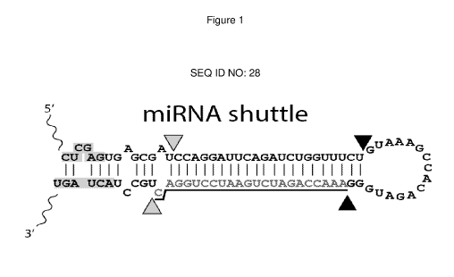
dominant
demyelinating CMT neuropathy type 1A [CMT1A (MIM 118220)].
[4] CMT1A is most frequently caused by a dominantly inherited 1.4 Mb tandem
intra-
chromosomal duplication on chromosome 17p11.2-p12. The duplication results in
three
copies of the peripheral myelin protein-22 (PMP22) gene which are translated
into PMP22
protein [Timmerman etal., Nature Genetics, 1(3): 171-175 (1992) and Valentijn
etal., Nature
Genetics, 1(3): 166-170 (1992)]. In some cases, point mutations in PMP22 may
also lead to
dominant CMT1A and generally present with the most severe phenotype [Matsunami
etal.,
Nature Genetics, 1(3):176-179 (1992), Patel etal., Nature Genetics, 1(3): 159-
165 (1992),
Timmerman etal., supra]. Patients with CMT1A develop slowly progressive distal
muscle
weakness and atrophy, sensory loss, and absent reflexes with a typical onset
at
adolescence. CMT1A shows high variability in disease severity even within the
same family.
Sensory responses are usually absent while motor nerve conduction velocities
(MNCVs) are
slowed, ranging from 5 to 35 m/s in the forearm, but most average around 20
m/s, with
uniform and symmetric findings in different nerves. Although MNCVs do not
change
significantly over decades, motor amplitudes and the number of motor units
decrease slowly,
reflecting axonal loss, which correlates with clinical disability.
1
CA 03203748 2023-05-31
WO 2022/119826
PCT/US2021/061177
[5] The PMP22 protein is a 22-kDa intrinsic tetraspan glycoprotein
primarily produced by
myelinating Schwann cells (SCs) during development and makes 2-5% of
peripheral
nervous system (PNS) compact myelin. This protein is crucial for SC growth and
differentiation, myelogenesis, myelin thickness and in the maintenance of PNS
axons and
myelin. PMP22 is also involved in the linkage of cytoskeletal actin with the
plasma
membrane, serving as a regulator of cholesterol content in lipid rafts.
Despite the fact that
PMP22 mRNA is expressed in almost every tissue, PMP22 protein is only found in
myelinating SCs, suggesting a tissue specific post-transcriptional regulation
of PMP22
mRNA [Maier etal., Mot Celt Neuroscience, 24(3): 803-817 (2003) and Roux
etal., J.
Comp. NeuroL, 474(4): 578-588 (2004)].
[6] The 5'-UTR of the PMP22 gene includes two known promoters, P1 and P2.
Their
respective transcripts differ only at their 5' non-coding region [Bosse etal.,
J. Neuroscience
Res., 37(4): 529-537 (1994) and Suter etal., J. Biol. Chem., 269(41): 25795-
25808 (1994)]
but result in six splice variants which exhibit a tissue-specific expression
pattern [Visigalli et
al., Hum. Mut, 37(1): 98-109 (2015)]. PMP22 regulation is achieved through its
intronic
regions and enhancer elements within them [Jones etal., Hum. Mot Genet.,
21(7): 1581-
1591 (2012); Srinivasan etal., Nuc. Acids Res., 40(14): 6449-6460 (2012); and
Pantera et
al., Hum. Mot Genet., 27(16): 2830-2839 (2018)]. P1 promoter transcripts are
SC-specific,
while the P2 promoter transcripts are expressed in non-PNS tissues. Therefore,
duplication
of the PMP22 gene or of its key transcriptional binding sites shifts the ratio
of splicing
isoforms and alters methylation, microRNA binding and post-translational
modification sites
[Verrier etal., Glia, 57(12): 1265-1279 (2009) and Lee etal., Exp.
Neurobiology, 28(2): 279-
288 (2019)]. In normal myelinating and non-myelinating SCs, approximately 20%
of the
newly synthesized PMP22 is glycosylated while the remaining ¨80% is targeted
for
proteasomal endoplasmic reticulum (ER)-associated degradation (ERAD).
[7] CMT1A is thought to depend on gene dosage effects of PMP22 because
CMT1A
patients have increased PMP22 mRNA [Yoshikawa etal., Ann. NeuroL, 35(4): 445-
450
(1994)] and protein [Gabriel etal., Neurology, 49(6): 1635-1640 (2015)] levels
in their sural
nerve biopsies. Individuals who carry a combination of one deleted and one
duplicated
PMP22 allele do not present CMT1A-like phenotype as they have a balanced gene
dosage.
Some CMT1A phenotypes may also result from a different size or type of
duplication on
chromosome 17p that affects PMP22 expression [Pantera, supra]. CMT1A patients
with 1.4
Mb duplication may have variable PMP22 levels in skin biopsies not necessarily
correlating
with disease severity [Nobbio etal., Brain, 137(Pt 6): 1614-1620 (2014) and
Katona etal.,
Brain, 132(Pt 7): 1734-1740 (2014)]. Nevertheless, supporting the PMP22 gene
dosage
effect as the driving mechanism of CMT1A, rodent models overexpressing PMP22
2
CA 03203748 2023-05-31
WO 2022/119826 PCT/US2021/061177
reproduced a CMT1A-like phenotype [Sereda etal., Neuron, 16(5): 1049-60
(1996); Huxley
etal., Hum. Mol. Genet., 5(5): 563-569 (1996); Huxley etal., Hum. Mol. Genet.,
7(3): 449-
458 (1998); Magyar etal., J. Neuroscience, 16(17): 5351-5360 (1996); Perea
etal., Hum.
Mol. Genet., 10(10): p. 1007-1018 (2001); Robaglia-Schlupp etal., Brain,
125(Pt 10): 2213-
2221 (2002); and Robertson etal., J. Anat., 200(4): 377-390 (2002)], which was
ameliorated
after interrupting PMP22 overexpression [Fledrich etal., Nat. Med., 20(9):
1055-1061 (2014);
Lee, supra; Perea, supra; Sereda etal., Nat. Med., 9(12): 1533-1537 (2003);
Passage etal.,
Nat. Med., 10(4): 396-401 (2004); Meyer etal., Ann. Neurology, 61(1): 61-72
(2007);
Chumakov etal., Orphanet Journal of Rare Diseases, 9(1): 201 (2014); Lee
etal.,
Neurobiology of Disease, 100: 99-107 (2017); Zhao etal., J. Clin. Invest.,
128(1): 359-368
(2018); Prukop etal., PLoS One, 14(1): e0209752 (2019); and Lee etal., Nuc.
Acids Res.,
48(1): 130-140 (2020)].
[8] Overexpressed PMP22 has been shown to saturate the proteasomal capacity
for
degradation, leading to perinuclear or cytoplasmic PMP22 accumulation,
decreased overall
proteasomal activity, and ER stress. PMP22 is also involved in early steps of
myelinogenesis, in the determination of myelin thickness and maintenance.
PMP22
duplication destabilizes the architecture, protein stoichiometry and function
of the myelin
sheath and SC, leading to demyelination, remyelination, the characteristic
onion bulb
formation and SC apoptosis. As a consequence, impairment in SC-axon
interactions and
dysfunctional neurofilament structure result in higher packing density and
less
phosphorylation of neurofilaments accompanied by slower axonal transport and
myelination
rates.
[9] Current treatments for CMT1A remain geared toward general symptom
management
in the form of physical therapy or corrective surgery.
[10] There thus exists a need in the art for products and methods for
treatment of CMT1A.
Summary
[11] The disclosure herein provides methods to specifically induce
silencing of
overexpressed PMP22 by RNA interference (RNAi) using vectors expressing
artificial
inhibitory RNAs targeting the PMP22 mRNA. The artificial inhibitory RNAs
contemplated
include, but are not limited to, small interfering RNAs (siRNAs) (also
referred to as short
interfering RNAs, small inhibitory RNAs or short inhibitory RNAs), short
hairpin RNAs
(shRNAs) and miRNA shuttles (artificial miRNAs) that inhibit expression of the
PMP22 gene.
The artificial inhibitory RNAs are referred to as miPMP22s herein. The
miPMP22s are small
regulatory sequences that act post-transcriptionally by targeting, for
example, the 3'UTR of
PMP22 mRNA in a reverse complementary manner resulting in reduced PMP22 mRNA
and
3
CA 03203748 2023-05-31
WO 2022/119826 PCT/US2021/061177
protein levels. Use of the methods and products is indicated, for example, in
preventing or
treating CMT1A.
[12] PMP22 inhibitory RNAs are provided as well as polynucleotides encoding
one or
more of the miPMP22s. The disclosure provides nucleic acids comprising RNA-
encoding
template nucleotide sequences comprising at least about 70%, 75%, 80%, 81%,
82%, 83%,
84`)/0, 85 /0, 86 /0, 870/0, 880/0, 89 /0, 90`)/0, 91'3/0, 92`)/0, 93`)/0, 94
/0, 95 /0, 96 /0, 97 /0, 98 /0, 99 /0
or 100% identity to the sequence set forth in any one of SEQ ID NOs: 1-8.
[13] Exemplary miPMP22s comprise the full length sequences set out in any one
of SEQ
ID NOs: 9-16 or variants thereof comprising at least about 70%, 75%, 80%, 81%,
82%, 83%,
840/0, 85 /0, 86 /0, 870/0, 880/0, 89 /0, 90`)/0, 91'3/0, 92`)/0, 93`)/0, 94
/0, 95 /0, 96 /0, 970/0, 98 /0, 99 /0
identity to the sequence set forth in any one of SEQ ID NOs: 9-16.
Corresponding final
processed antisense guide strand sequences are respectively set out in SEQ ID
NOs: 17-24,
or are variants thereof comprising at least about 70%, 75%, 80%, 81%, 82%,
83%, 84%,
850/0, 860/0, 870/0, 880/0, 890/0, 900/0, 910/0, 92%, 930/0, 940/0, 950/0,
960/0, 970/0, 980/0, 990/0
identity to the sequence set forth in any one of SEQ ID NOs: 17-24. The
processed
antisense guide strand is the strand of the mature miRNA duplex that becomes
the RNA
component of the RNA induced silencing complex ultimately responsible for
sequence-
specific gene silencing. The disclosure additionally provides the antisense
guide strands set
out in Figure 48 and contemplates variants of each of those antisense guide
strands that are
at least about 70%, 75%, 80%, 81%, 82%, 83%, 84%, 85%, 86%, 87%, 88%, 89%,
90%,
91%, 92%, 93%, 94%, 95%, 96%, 97%, 98%, 99% identical. The disclosure
additionally
provides the antisense guide strands set out in Figure 50 and contemplates
variants of each
of those antisense guide strands that are at least about 70%, 75%, 80%, 81%,
82%, 83%,
840/0, 850/0, 860/0, 870/0, 880/0, 890/0, 900/0, 910/0, 92%, 930/0, 940/0,
950/0, 960/0, 970/0, 980/0, 990/0
identical.
[14] miPMP22s can specifically bind to a segment of a messenger RNA (mRNA)
encoded
by a human PMP22 gene (represented by SEQ ID NO: 25 which is a human PMP22
cDNA),
wherein the segment conserved relative to mRNA encoded by the wild-type mouse
PMP22
gene (represented by SEQ ID NO: 27 which is a mouse PMP22cDNA). For example, a
miPMP22 can specifically bind a mRNA segment that is complementary to a
sequence
within nucleotides 1-2423 of SEQ ID NO: 25. More particularly, a miPMP22 can
specifically
bind a mRNA segment that is complementary to a sequence within nucleotides
1412-1433 of
SEQ ID NO: 25 (the nucleotides bound by, for example, miPMP22-868) or 1415-
1436 of
SEQ ID NO: 25 (the nucleotides bound by, for example, miPMP22-871).
[15] Delivery of DNA encoding miPMP22s can be achieved using a delivery
vehicle that
delivers the DNA(s) to a Schwann cell. For example, recombinant AAV (rAAV)
vectors can
4
CA 03203748 2023-05-31
WO 2022/119826 PCT/US2021/061177
be used to deliver DNA encoding miPMP22s. Other vectors (for example, other
viral vectors
such as lentivirus, adenovirus, retrovirus, equine-associated virus,
alphavirus, pox viruses,
herpes virus, polio virus, sindbis virus and vaccinia viruses) can also be
used to deliver
polynucleotides encoding miPMP22s. Thus, also provided are viral vectors
encoding one or
more miPMP22s. When the viral vector is a rAAV, the rAAV lack AAV rep and cap
genes.
The rAAV can be self-complementary (sc) AAV. As another example, non-viral
vectors such
as lipid nanoparticles can be used for delivery.
[16] Provided herein are rAAV, each encoding a miPMP22. Also provided are rAAV
encoding one or more miPMP22s. A rAAV (with a single-stranded genome, scAAV)
encoding one or more miPMP22s can encode one, two, three, four, five, six,
seven or eight
miPMP22s. A scAAV encoding one or more miPMP22s can encode one, two, three or
four
miPMP22s.
[17] Compositions are provided comprising the nucleic acids or viral vectors
described
herein.
[18] Further provided are methods of preventing or inhibiting expression of
the PMP22
gene in a cell comprising contacting the cell with a delivery vehicle (such as
rAAV) encoding
a miPMP22 wherein, if the delivery vehicle is rAAV, the rAAV lacks rep and cap
genes. In
the methods, expression of the duplicated and/or mutant PMP22 allele is
inhibited by at least
10, at least 20, at least 30, at least 40, at least 50, at least 60, at least
70, at least 80, at
least 90, at least 95, at least 98 percent, at least 99 percent, or 100
percent. In the methods,
expression of the wild-type PMP22 allele is inhibited by at least 10, at least
20, at least 30, at
least 40, at least 50, at least 60, at least 70, at least 80, at least 90, at
least 95, at least 98
percent, at least 99 percent, or 100 percent.
[19] Still further provided are methods of delivering DNA encoding a
miPMP22 to an
subject in need thereof, comprising administering to the subject a delivery
vehicle (such as
rAAV) comprising DNA encoding the miPMP22 wherein, if the delivery vehicle is
rAAV, the
rAAV lacks rep and cap genes. Other methods of delivering DNA encoding the
miPMP22 to
an subject in need thereof, comprise administering to the subject a delivery
vehicle (such as
rAAV) comprising DNA encoding one or more miPMP22 wherein, if the delivery
vehicle is
rAAV, the rAAV lacks rep and cap genes.
[20] Methods are also provided of preventing or treating CMT1A comprising
administering a delivery vehicle (such as rAAV) comprising DNA encoding a
miPMP22
wherein, if the delivery vehicle is rAAV, the rAAV lacks rep and cap genes.
Other methods
of preventing or treating CMT1A comprise administering a delivery vehicle
(such as rAAV)
comprising DNA encoding one or more miPMP22 wherein, if the delivery vehicle
is rAAV, the
rAAV lacks rep and cap genes. The methods result in restoration of PMP22
expression to at
least 25 percent, at least 30, at least 40, at least 50, at least 60, at least
70, at least 80, at
CA 03203748 2023-05-31
WO 2022/119826 PCT/US2021/061177
least 90, at least 95, at least 98 percent, at least 99 percent, or 100
percent or more, of
normal PMP22 expression in an unaffected subject.
[21] The disclosure provides a delivery vehicle that is a viral vector
comprising the nucleic
acids described herein and/or a combination of any one or more thereof. Viral
vectors
provided include, but are not limited to an adeno-associated virus (AAV),
adenovirus,
lentivirus, retrovirus, poxvirus, baculovirus, herpes simplex virus, vaccinia
virus, or a
synthetic virus. The viral vector can be an AAV. The AAV lacks rep and cap
genes. The
AAV can be a recombinant AAV (rAAV) or a self-complementary recombinant AAV
(scAAV).
The AAV is, for example, AAV-1, AAV-2, AAV-3, AAV-4, AAV-5, AAV-6, AAV-7, AAV-
8,
AAV-9, AAV-10, AAV-11, AAV-12, AAV-13, AAV-anc80, or AAV rh.74. The AAV can be
AAV-9. The AAV can be a pseudotyped AAV, for example, an AAV2/8 or AAV2/9.
[22] The disclosure provides a composition comprising the nucleic acids
described herein
and a pharmaceutically acceptable carrier. The disclosure provides a
composition
comprising a viral vector comprising the nucleic acids described herein,
and/or a
combination of any one or more thereof and a pharmaceutically acceptable
carrier.
[23] The disclosure provides a composition comprising a delivery vehicle
capable of
delivering agents to a Schwann cell a nucleic acid encoding a miPMP22, wherein
the
miPMP22 binds a segment of a mRNA encoded by a human PMP22 gene (wherein the
segment either does or does not encode sequence comprising a mutation
associated with
CMT1A); wherein the segment is conserved relative to the wild-type mouse PMP22
gene,
and, optionally, a pharmaceutically acceptable carrier. The human PMP22 gene
can
comprise the sequence of SEQ ID NO: 25, or a variant thereof comprising at
least about
70`)/0, 750/0, 80`)/0, 810/0, 82`)/0, 830/0, 840/0, 85`)/0, 860/0, 870/0,
880/0, 89 /0, 90`)/0, 91 (3/0, 92`)/0, 93`)/0,
94%, 95%, 96%, 97%, 98%, or 99%, identity. The mouse PMP22 gene can comprise
the
sequence of SEQ ID NO: 27, or a variant thereof comprising at least about 70%,
75%, 80%,
810/0, 82`)/0, 830/0, 840/0, 85 /0, 86 /0, 870/0, 880/0, 89 /0, 90`)/0, 91 /0,
92`)/0, 93 /0, 94 /0, 95 /0, 96 /0,
97%, 98%, or 99%, identity. A miPMP22 specifically binds, for example, a mRNA
segment
that is complementary to a sequence within nucleotides 1412-1433 of SEQ ID NO:
25 (the
nucleotides bound by, for example, miPMP22-868) or 1415-1436 of SEQ ID NO: 25
(the
nucleotides bound by, for example, miPMP22-871).
[24] The disclosure provides a delivery vehicle in the compositions that is
a viral vector.
The viral vector in the compositions can be, for example, an adeno-associated
virus (AAV),
adenovirus, lentivirus, retrovirus, poxvirus, baculovirus, herpes simplex
virus, vaccinia virus,
or a synthetic virus. The viral vector can be an AAV. The AAV lacks rep and
cap genes.
The AAV can be a recombinant AAV (rAAV) or a self-complementary recombinant
AAV
(scAAV). The AAV is or has a capsid serotype selected from, for example, AAV-
1, AAV-2,
6
CA 03203748 2023-05-31
WO 2022/119826 PCT/US2021/061177
AAV-3, AAV-4, AAV-5, AAV-6, AAV-7, AAV-8, AAV-9, AAV-10, AAV-11, AAV-12, AAV-
13,
AAV-anc80, and AAV rh.74. The AAV can be or can have a capsid serotype of AAV-
9. The
AAV can be a pseudotyped AAV, such as AAV2/8 or AAV2/9.
[25] The disclosure provides methods of delivering to a Schwann cell
comprising a
duplicated and/or mutant PMP22 gene: (a) a nucleic acid comprising a template
nucleic acid
encoding a miPMP22 comprising at least about 70%, 75%, 80%, 81%, 82%, 83%,
84%,
85 /0, 86 /0, 870/0, 880/0, 89 /0, 90`)/0, 91 /0, 92`)/0, 93 /0, 94%, 95 /0,
96 /0, 97 /0, 98 /0, 99 /0, or
100% identity to the polynucleotide sequence set forth in any one of SEQ ID
NOs: 1-8; a
nucleic acid encoding the full length miPMP22 sequences set out in any one of
SEQ ID NOs:
9-16 or variants thereof comprising at least about 70%, 75%, 80%, 81%, 82%,
83%, 84%,
85 /0, 86 /0, 870/0, 880/0, 89 /0, 90`)/0, 91 /0, 92`)/0, 93 /0, 94%, 95 /0,
96 /0, 970/0, 98 /0, 99 /0
identity to the sequence set forth in any one of SEQ ID NOs: 9-16; a nucleic
acid encoding a
miPMP22 processed antisense guide strand comprising at least about 70%, 75%,
80%,
810/0, 82'3/0, 830/0, 840/0, 850/0, 860/0, 870/0, 880/0, 890/0, 900/0, 910/0,
92%, 93%, 940/0, 950/0, 960/0,
97%, 98%, 99%, or 100% identity to the polynucleotide sequence set forth in
any one of
SEQ ID NOs: 17-24; a nucleic acid encoding one or more antisense guide strands
set out in
Figure 48, or a variant of an antisense guide strand in Figure 48 that is at
least about 70%,
750/0, 800/0, 810/0, 82'3/0, 830/0, 840/0, 850/0, 860/0, 870/0, 880/0, 890/0,
900/0, 910/0, 92%, 930/0, 940/0,
95%, 96%, 97%, 98%, 99% identical; or a nucleic acid encoding one or more
antisense
guide strands set out in Figure 50, or a variant of an antisense guide strand
in Figure 50 that
is at least about 70%, 75%, 80%, 81%, 82%, 83%, 84%, 85%, 86%, 87%, 88%, 89%,
90%,
91%, 92%, 93%, 94%, 95%, 96%, 97%, 98%, 99% identical; (b) a vector comprising
any one
or more of the nucleic acids described herein; or (c) a composition comprising
any one or
more of the nucleic acids or vectors described herein.
[26] The disclosure provides a method of treating a subject suffering from a
duplicated
and/or mutant PMP22 gene, the method comprising administering to the
subject(a) a nucleic
acid comprising a template nucleic acid encoding a miPMP22 comprising at least
about
700/0, 750/0, 800/0, 810/0, 82'3/0, 830/0, 840/0, 850/0, 860/0, 870/0, 880/0,
890/0, 900/0, 910/0, 92%, 930/0,
94%, 95%, 96%, 97%, 98%, 99%, or 100% identity to the polynucleotide sequence
set forth
in any one of SEQ ID NOs: 1-8; a nucleic acid encoding the full length miPMP22
sequences
set out in any one of SEQ ID NOs: 9-16 or variants thereof comprising at least
about 70%,
750/0, 800/0, 810/0, 82'3/0, 830/0, 840/0, 850/0, 860/0, 870/0, 880/0, 890/0,
900/0, 910/0, 92%, 930/0, 940/0,
95%, 96%, 97%, 98%, 99% identity to the sequence set forth in any one of SEQ
ID NOs: 9-
16; a nucleic acid encoding a miPMP22 processed antisense guide strand
comprising at
least about 70%, 75%, 80%, 81%, 82%, 83%, 84%, 85%, 86%, 87%, 88%, 89%, 90%,
91%,
92%, 93%, 94%, 95%, 96%, 97%, 98%, 99%, or 100% identity to the polynucleotide
sequence set forth in any one of SEQ ID NOs: 17-24; a nucleic acid encoding
one or more
7
CA 03203748 2023-05-31
WO 2022/119826 PCT/US2021/061177
antisense guide strands set out in Figure 48, or a variant of an antisense
guide strand in
Figure 48 that is at least about 70%, 75%, 80%, 81%, 82%, 83%, 84%, 85%, 86%,
87%,
88%, 89%, 90%, 91%, 92%, 93%, 94%, 95%, 96%, 97%, 98%, 99% identical; or a
nucleic
acid encoding one or more antisense guide strands set out in Figure 50, or a
variant of an
antisense guide strand in Figure 50 that is at least about 70%, 75%, 80%, 81%,
82%, 83%,
840/0, 850/0, 860/0, 870/0, 880/0, 890/0, 900/0, 910/0, 920/0, 930/0, 940/0,
950/0, 960/0, 970/0, 980/0, 990/0
identical; (b) a vector comprising any one or more of the nucleic acids
described herein; or
(c) a composition comprising any one or more of the nucleic acids or vectors
described
herein.
[27] The disclosure contemplates the subject treated by methods herein suffers
from
CMT1A. The disclosure also contemplates treatment of a subject that is at risk
for CMT1A
due to a duplication or mutation of the PMP22 gene. The subject can be a
mammalian
animal. The subject can be a human subject.
[28] The disclosure also provides uses of at least one nucleic acid as
described herein, at
least one viral vector as described herein, or a composition as described
herein in making a
medicament for, or in treating a subject suffering from, a duplicated and/or
mutant PMP22
gene.
[29] The disclosure also provides uses of at least one nucleic acid as
described herein, at
least one viral vector as described herein, or a composition as described
herein in making a
medicament for or in treating CMT1A in a subject in need thereof.
[30] Other features and advantages of the disclosure will be apparent from the
following
description of the drawing and the detailed description. It should be
understood, however,
that the drawing, detailed description, and the examples, while indicating
embodiments of
the disclosed subject matter, are given by way of illustration only, because
various changes
and modifications within the spirit and scope of the disclosure will become
apparent from the
drawing, detailed description, and the examples.
Brief Description of the Drawings
[31] This patent or application file contains at least one drawing executed
in color. Copies
of this patent or patent application publication with color drawing(s) will be
provided by the
United States Patent and Trademark Office upon request and payment of the
necessary fee.
[32] Figure 1 shows an example of an artificial miRNA shuttle sequence to
demonstrate
folding and processing sites. The mature guide strand is underlined. Grey
arrowheads
indicate Drosha cut sites; black arrowheads indicate Dicer cut sites. Shaded
sequences at
extreme 5' and 3' ends are restriction sites in the template DNA used to clone
the miRNA
shuttles in front of the U6 promoter.
8
CA 03203748 2023-05-31
WO 2022/119826
PCT/US2021/061177
[33] Figure 2 shows a human PMP22 full length cDNA sequence (SEQ ID NO: 25) in
which alternating shading shows exon boundaries (5 exons) and the underlined
sequence is
the longest human PMP22 protein coding open reading frame (ORF).
[34] Figure 3 shows the human PMP22 full-length ORF sequence with a translated
PMP22 protein sequence (SEQ ID NO: 26).
[35] Figure 4 shows a mouse PMP22 cDNA sequence (SEQ ID NO: 27) in which
alternating shading shows exon boundaries (5 exons).
[36] Figure 5A-B shows the human PMP22 cDNA with miPMP22 binding sites. All
miPMP22 target sequences are located in Exon 5 (in the 3' UTR region).
Underlined
sequence is the PMP22 full-length open reading frame.
[37] Figure 6 shows the full-length miPMP22-868 sequence (SEQ ID NO: 9).
[38] Figure 7 shows binding interactions of miPMP22-868 and miPMP22-871 with
mouse
and human PMP22.
[39] Figure 8 shows the full-length miPMP22-871 sequence (SEQ ID NO: 10).
[40] Figure 9 shows the full-length miPMP22-869 sequence (SEQ ID NO: 11).
[41] Figure 10 shows the full-length miPMP22-872 sequence (SEQ ID NO: 12).
[42] Figure 11 shows the full-length miPMP22-1706 sequence (SEQ ID NO: 13).
[43] Figure 12 shows the full-length miPMP22-1740 sequence (SEQ ID NO: 14).
[44] Figure 13 shows the full-length miPMP22-1741 sequence (SEQ ID NO: 15).
[45] Figure 14 shows the full-length miPMP22-1834 sequence (SEQ ID NO: 16).
[46] Figure 15A-D shows qPCR results of in vitro testing of PMP22 knockdown by
miPMP22s.
[47] Figure 16A-D shows in vivo expression of AAV9 in lumbar roots of adult
mice.
Representative images of lumbar root sections of a non-injected mouse (A) and
of mice 4
and 8 weeks after lumbar intrathecal injection of AAV9-U6-miRLacZ-CMV-EGFP (B-
C).
Sections were immunostained for eGFP (red) indicating cells expressing the
reporter gene
along with miRLacZ. eGFP was also auto-fluorescent (green). Cell nuclei were
stained with
DAPI (blue). Arrow heads reveal examples of perinuclear eGFP immunoreactivity
in SCs.
Quantification of the percentage of eGFP positive cells is shown in D (Mean,
SD). Data were
compared using the Student t-test, p<0.0001. Averages: 4 weeks 45.95, 8 weeks
56.82.
[48] Figure 17A-D shows in vivo expression of AAV9 in sciatic nerves of adult
mice.
Representative images of sciatic nerve sections (A-C) and sciatic nerve teased
fibers (E-G)
of a non-injected mouse (A,E) and of mice 4 (B,F) and 8 (C,G) weeks after
lumbar
intrathecal injection with AAV9-U6-miRLacZ-CMV-EGFP (B-C). Sections were
immunostained for eGFP (red) indicating cell expressing the reporter gene
along with
miRLacZ. eGFP was also auto-fluorescent (green). Cell nuclei were stained with
DAPI
9
CA 03203748 2023-05-31
WO 2022/119826 PCT/US2021/061177
(blue). Arrow heads reveal examples of perinuclear eGFP immunoreactivity in
SCs.
Quantification of the percentage of eGFP positive cells is shown in D (Mean,
SD). Data were
compared using the Student t-test, p=0.0460. Averages: 4 weeks 42.07, 8 weeks
45.74.
[49] Figure 18A-D shows in vivo expression of AAV9 in femoral nerves of adult
mice.
Representative images of teased femoral nerve fibers of a non-injected mouse
(A) and of
mice 4 and 8 weeks after lumbar intrathecal injection with AAV9 expressing
AAV9-U6-
miRLacZ-CMV-EGFP (B-C). Fibers were immunostained for eGFP (red) indicating
cell
expressing the reporter gene along with miRLacZ. eGFP was also auto-
fluorescent (green).
Cell nuclei were stained with DAPI (blue). Arrow heads reveal examples of
perinuclear eGFP
immunoreactivity in SCs. Quantification of the percentage of eGFP positive
cells is shown in
D (Mean, SD). Data were compared using the Student t-test, p=0.0336. Averages:
4 weeks
31.17, 8 weeks 41.09.
[50] Figure 19A-F shows immunoblot and VGCN analysis of AAV9-miLacZ eGFP
reporter
gene expression. Representative images of immunoblot analysis of eGFP
expression levels
4 and 8 weeks post intrathecal lumbar injection in lumbar root (A) and sciatic
nerve (D)
lysates with AAV9 expressing miRLacZ along with the EGFP reporter gene. Tissue
samples
from C61-Het non-injected mice were used as a negative control. Tubulin blot
was used as
loading control. For the quantification, eGFP to tubulin optic density ratio
was calculated
(Mean, SD). Data were compared using the Student t-test, p= 0.0272 (B, E).
AAV9-miLacZ
VGCN in lumbar roots (C) and sciatic nerves (F) were calculated at 4 and 8
weeks after
intrathecal lumbar injection. VGCN data were compared using the Student t-test
and no
statistical significance was found between the two time points (Mean, SD).
Averages:
Western blot roots: 4 weeks 0.92, 8 weeks 0.99; Western blot sciatic nerve: 4
weeks 0.48, 8
weeks 0.81; VCN roots: 4 weeks 3.24, 8 weeks 1.09; VCN sciatic nerve: 4 weeks
0.41, 8
weeks 0.38.
[51] Figure 20A-F shows results of in vivo testing of AAV9-miR871 effects
in hu/mu
PMP22 and other myelin-related genes in the lumbar spinal roots of CMT1A mouse
model.
Real Time PCR analysis of hu/mu PMP22 and other myelin-related genes
expression in C61
Het mice six weeks after injection with AAV9-miR871 relative to AAV9-miRLacZ
(non-
targeting control) vector injected littermates (presented as baseline). For
all above
transcripts expression analysis muGAPDH was used as a housekeeping gene to
normalize
for loading and relative change was determined using the 2-CT method (Mean,
SD)..
[52] Figure 21A-F shows results of in vivo testing of AAV9-miR871 effects
in hu/mu
PMP22 and other myelin-related genes in the sciatic nerves of CMT1A mouse
model. Real
Time PCR analysis of hu/mu PMP22 and other myelin-related genes expression in
C61 Het
mice six weeks after injection with AAV9-miR871 relative to AAV9-miRLacZ (non-
targeting
CA 03203748 2023-05-31
WO 2022/119826 PCT/US2021/061177
control) vector injected littermates (presented as baseline). For all above
transcripts
expression analysis muGAPDH was used as a housekeeping gene to normalize for
loading
and relative change was determined using the 2-= CT method (Mean, SD).
[53] Figure 22A-F shows results of in vivo testing of AAV9-miR871 effects in
hu/mu
PMP22 and other myelin-related genes in the femoral nerves of CMT1A mouse
model. Real
Time PCR analysis of hu/mu PMP22 and other myelin-related genes expression in
061 Het
mice six weeks after injection with AAV9-miR871 relative to AAV9-miRLacZ (non-
targeting
control) vector injected littermates (presented as baseline). For all above
transcripts
expression analysis muGAPDH was used as a housekeeping gene to normalize for
loading
and relative change was determined using the 2-= CT method (Mean, SD).
[54] Figure 23A-F shows results of in vivo testing of AAV9-miR868 effects in
hu/mu
PMP22 and other myelin-related genes in the lumbar spinal roots of CMT1A mouse
model.
Real Time PCR analysis of hu/mu PMP22 and other myelin-related genes
expression in 061
Het mice six weeks after injection with AAV9-miR871 relative to AAV9-miRLacZ
(non-
targeting control) vector injected littermates (presented as baseline). For
all above
transcripts expression analysis muGAPDH was used as a housekeeping gene to
normalize
for loading and relative change was determined using the 2-CT method (Mean,
SD).
[55] Figure 24A-F shows results of in vivo testing of AAV9-miR868 effects in
hu/mu
PMP22 and other myelin-related genes in the sciatic nerves of CMT1A mouse
model. Real
Time PCR analysis of hu/mu PMP22 and other myelin-related genes expression in
061 Het
mice six weeks after injection with AAV9-miR871 relative to AAV9-miRLacZ (non-
targeting
control) vector injected littermates (presented as baseline). For all above
transcripts
expression analysis muGAPDH was used as a housekeeping gene to normalize for
loading
and relative change was determined using the 2-= 0T method (Mean, SD).
[56] Figure 25A-F shows results of in vivo testing of AAV9-miR868 effects in
hu/mu
PMP22 and other myelin-related genes in the femoral nerves of CMT1A mouse
model. Real
Time PCR analysis of hu/mu PMP22 and other myelin-related genes expression in
061 Het
mice six weeks after injection with AAV9-miR871 relative to AAV9-miRLacZ (non-
targeting
control) vector injected littermates (presented as baseline). For all above
transcripts
expression analysis muGAPDH was used as a housekeeping gene to normalize for
loading
and relative change was determined using the 2-= 0T method (Mean, SD).
[57] Figure 26A-D shows results of in vivo testing of AAV9-miR871 effects on
HuPMP22
and MPZ proteins in the lumbar roots of a CMT1A mouse model. lmmunoblot
analysis of
HuPMP22 (A) expression levels in lumbar root lysates 6 weeks post lumbar
intrathecal
injection with AAV9 expressing miR871 or miRLacZ. Tissue samples from non-
injected mice
were used as a negative control. Tubulin blot and MPZ gel band were used as
loading
controls (B, C). MPZ expression was altered after treatment (D). For the
quantification,
11
CA 03203748 2023-05-31
WO 2022/119826
PCT/US2021/061177
HuPMP22 to tubulin, HuPMP22 to MPZ gel band and MPZ gel band to tubulin, optic
density
ratios were calculated (Mean, SD). Data were compared using the Student t-
test.
[58] Figure 27A-D shows results of in vivo testing of AAV9-miR871 effects on
HuPMP22
and MPZ proteins in the sciatic nerves of a CMT1A mouse model. lmmunoblot
analysis of
HuPMP22 (A) expression levels in sciatic nerve lysates 6 weeks post lumbar
intrathecal
injection with AAV9 expressing miR871 or miRLacZ. Tissue samples from non-
injected mice
were used as a negative control. Tubulin blot and MPZ gel band were used as
loading
controls (B, C). MPZ expression was altered after treatment (D). For the
quantification,
HuPMP22 to tubulin, HuPMP22 to MPZ gel band and MPZ gel band to tubulin ,optic
density
ratios were calculated (Mean, SD). Data were compared using the Student t-
test.
[59] Figure 28A-D shows results of in vivo testing of AAV9-miR871 effects on
HuPMP22
and MPZ proteins in the femoral nerves of a CMT1A mouse model. lmmunoblot
analysis of
HuPMP22 (A) expression levels in femoral nerve lysates 6 weeks post lumbar
intrathecal
injection with AAV9 expressing miR871 or miRLacZ. Tissue samples from non-
injected mice
were used as a negative control. Tubulin blot and MPZ gel band were used as
loading
controls (B, C). MPZ expression was altered after treatment (D). For the
quantification,
HuPMP22 to tubulin, HuPMP22 to MPZ gel band and MPZ gel band to tubulin ,optic
density
ratios were calculated (Mean, SD). Data were compared using the Student t-
test.
[60] Figure 29 shows Early and Late Treatment trial design in the 061 Het
mouse model
of CMT1A. The early treatment trial design was also employed for WT mice,
expressing only
normal levels of murine PMP22.
[61] Figure 30A-B shows rotarod results at baseline and following early
treatment at 5
rpm. A: AAV9-miR871 treated compared to AAV9-miRLacZ (mock) treated control
mice, as
indicated. Two months old non-injected WT and non-injected 061 Het mice showed
no
significant difference, while 4 and 6 months old WT mice performed better than
their age-
matched AAV9-miRLacZ treated 061 Het mice. At the age of 4 and 6 months (2 and
4
months post-injection) AAV9-miR871-treated 061 Het mice showed improved motor
performance compared to the mock group, and did not differ significantly from
WT mice of
the same age (AAV9-miR871: n=16, AAV9-miRLacZ: n=16, 2m 061 Het: n=32, 2-6
months
old WT: n=10). B: Time course analysis demonstrates the improvement of AAV9-
miR871
treated compared to mock treated 061 Het mice in rotarod 2 and 4 months post-
injection (at
4 and 6 months of age). Values represent mean SD. Data were compared using
Mann-
Whitney Test: 4m WT vs. 4m Het-miRLacZ: p= 0.0017, 4m Het-miRLacZ vs. 4m Het-
miR871: p=0.0003, 6m WT vs. 6m Het-miRLacZ: p= 0.0003, 6m Het-miRLacZ vs. 6m
Het-
miR871: p< 0.0001. Averages: 2mWT: 594.12, 2m Het: 549, 4m WT: 596.1, 4m Het-
miRLacZ: 512.96, 4m Het-miR871: 597.59, 6m WT: 585.42, 6m Het-miRLacZ: 390.43,
6m
Het-miR871: 584.96.
12
CA 03203748 2023-05-31
WO 2022/119826 PCT/US2021/061177
[62] Figure 31A-B shows rotarod results at baseline and following early
treatment at 17.5
rpm: A: At baseline at the age of 2 months, before treatment, all 061 Het mice
performed
worse than WT mice. Likewise WT mice performed better than mock treated 061
Het mice
at 4 and 6 months of age. At the age of 4 and 6 months AAV9-miR871-treated 061
Het mice
showed improved motor performance compared to the control vector treated and
reached
the performance of WT mice (AAV9-miR871: n=16, AAV9-miRLacZ: n=16, 2m 061 Het:
n=32, 2-6 months old WT: n=10). B: Time course analysis demonstrates improved
motor
performance of AAV9-miR871 treated 061 Het mice in rotarod 2 and 4 months post-
injection
(at 4 and 6 months of age). Values represent mean SD. Data were compared
using Mann-
Whitney Test: 2m WT vs. 2m Het-miRLacZ: p< 0.0001, 4m WT vs. 4m Het-miRLacZ:
p<
0.0001, 4m Het-miRLacZ vs. 4m Het-miR871: p< 0.0001, 4m WT vs. 4m Het-miR871:
p<
0.0001, 6m WT vs. 6m Het-miRLacZ: p< 0.0001, 6m Het-miRLacZ vs. 6m Het-miR871:
p<
0.0001, 6m WT vs. 6m Het-miR871: p=0.0106. Averages: 2mWT: 582.21, 2m Het:
235.79,
4m WT: 497.8, 4m Het-miRLacZ: 172.93, 4m Het-miR871: 504.09, 6m WT: 416.16, 6m
Het-
miRLacZ: 69.54, 6m Het-miR871: 470.48.
[63] Figure 32A-B shows foot grip analysis at baseline and following early
treatment: A:
At baseline at the age of 2 months, before treatment, all 061 Het mice
performed worse than
non-injected WT mice. Likewise WT mice performed better than mock treated 061
Het mice
at 4 and 6 months of age. At the age of 4 and 6 months AAV9-miR871-treated 061
Het mice
showed improved grip strength performance compared to AAV9-miRLacZ treated
control
mice (AAV9-miR871: n=16, AAV9-miRLacZ: n=16, 2m 061 Het: n=32, 2-6 months old
WT:
n=10). B:Time course analysis showed improved performance of AAV9-miR871
treated 061
Het mice in foot grip analysis 2 and 4 months post-injection (4 and 6 months
of age). Values
represent mean SD. Data were compared using Mann-Whitney Test: 2m WT vs. 2m
Het-
miRLacZ: p< 0.0001 4m WT vs. 4m Het-miRLacZ: p< 0.0001, 4m Het-miRLacZ vs. 4m
Het-
miR871: p< 0.0001, 4m WT vs. 4m Het-miR871 : p< 0.0001, 6m WT vs. 6m Het-
miRLacZ:
p< 0.0001, 6m Het-miRLacZ vs. 6m Het-miR871: p< 0.0001, 6m WT vs. 6m Het-
miR871:
p=0.0106. Averages: 2mWT: 51.28, 2m Het: 22.69, 4m WT: 42.73, 4m Het-miRLacZ:
17.80,
4m Het-miR871: 29.53, 6m WT: 36.81, 6m Het-miRLacZ: 15.92, 6m Het-miR871:
31.70.
[64] Figure 33A-B shows wire hang test analysis at baseline and following
early treatment:
A: At baseline at the age of 2 months, before treatment, all 061 Het mice
performed worse
than non-injected WT mice. Likewise WT mice performed better than mock treated
061 Het
mice at 4 and 6 months of age. At the age of 4 and 6 months AAV9-miR871-
treated 061 Het
mice showed improved hang test performance compared to AAV9-miRLacZ treated
control
mice (AAV9-miR871: n=16, AAV9-miRLacZ: n=16, 2m 061 Het: n=32, 2-6 months old
WT:
n=10). AAV9-miR871-treated mice at 4 months did not manage to reach WT
performances,
this is in contrast to 6-months old AAV9-miR871 treated mice that did not
differ significantly
13
CA 03203748 2023-05-31
WO 2022/119826 PCT/US2021/061177
to their age matched AAV9-miRLacZ treated B: Time course analysis showed
improved
performance of AAV9-miR871 treated 061 Het mice in hang test analysis 2 and 4
months
post-injection (4 and 6 months of age). Values represent mean SD. Data were
compared
using Mann-Whitney Test: 2m WT vs. 2m Het-miRLacZ: p< 0.0001, 4m WT vs. 4m Het-
miRLacZ: p< 0.0001, 4m Het-miRLacZ vs. 4m Het-miR871: p< 0.0001, 4m WT vs. 4m
Het-
miR871 : p< 0.0001, 6m WT vs. 6m Het-miRLacZ: p< 0.0001, 6m Het-miRLacZ vs. 6m
Het-
miR871: p< 0.0001,6m WT vs. 6m Het-miR871: p=0.0106. Averages: 2mWT: 593.70,
2m
Het: 306.96, 4m WT: 527.28, 4m Het-miRLacZ: 155.64, 4m Het-miR871: 381.53, 6m
WT:
351.30, 6m Het-miRLacZ: 130.81, 6m Het-miR871: 371.65.
[65] Figure 34A-B shows rotarod results at baseline and following late
treatment at 5 rpm.
A: AAV9-miR871 treated compared to AAV9-miRLacZ (mock) treated control mice,
as
indicated. At all examined ages WT mice performed better than age-matched C61-
Het or
C61-Het AAV9-miRLacZ treated mice. At the age of 8 and 10 months (2 and 4
months post-
injection) AAV9-miR871- treated 061 Het mice showed improved motor performance
compared to the mock group, and did not differ significantly from WT mice of
the same age
(8m AAV9-miR871: n=16, 8m AAV9-miRLacZ: n=16, 10 m AAV9-miR871: n=10, 10m AAV9-
miRLacZ: n=8, 6m 061 Het: n=32, 6-8 months old WT: n=10). B: Time course
analysis
demonstrates the improvement of AAV9-miR871 treated compared to mock treated
061 Het
mice. Values represent mean SD. Data were compared using Mann-Whitney Test:
6m WT
vs. 6m Het-miRLacZ: p< 0.0001, 8m WT vs. 8m Het-miRLacZ: p<0.0001, 8m Het-
miRLacZ
vs. 8m Het-miR871: p<0.0001, 10m WT vs. 10m Het-miRLacZ: p<0.0001, 10m Het-
miRLacZ
vs. 10m Het-miR871: p<0.0001. Averages: 6mWT: 583.80, 6m Het: 345.04, 8m WT:
578.49,
8m Het-miRLacZ: 221.93, 8m Het-miR871: 566.01, 10m WT: 584.20, 10m Het-
miRLacZ:
148.57, 10m Het-miR871: 558.40.
[66] Figure 35A-B shows rotarod results at baseline and following late
treatment at 17.5
rpm. A: AAV9-miR871 treated compared to AAV9-miRLacZ (mock) treated control
mice, as
indicated. At all examined ages WT mice performed better than age-matched C61-
Het or
C61-Het AAV9-miRLacZ treated mice. At the age of 8 and 10 months (2 and 4
months post-
injection) AAV9-miR871- treated 061 Het mice showed improved motor performance
compared to the mock group, and did not differ significantly from WT mice of
the same age,
with only exception being 8m WT vs. 8m Het-miR871 (8m AAV9-miR871: n=16, 8m
AAV9-
miRLacZ: n=16, 10 m AAV9-miR871: n=10, 10m AAV9-miRLacZ: n=8, 6m 061 Het:
n=32,
6-8 months old WT: n=10). B: Time course analysis demonstrates the improvement
of
AAV9-miR871 treated compared to mock treated 061 Het mice. Values represent
mean
SD. Data were compared using Mann-Whitney Test: 6m WT vs. 6m Het-miRLacZ: p<
0.0001, 8m WT vs. 8m Het-miRLacZ: p<0.0001, 8m Het-miRLacZ vs. 8m Het-miR871:
p<0.0001, 8m WT vs. 8m Het-miR871: p=0.0015, 10m WT vs. 10m Het-miRLacZ:
p<0.0001,
14
CA 03203748 2023-05-31
WO 2022/119826 PCT/US2021/061177
10m Het-miRLacZ vs. 10m Het-miR871: p<0.0001. Averages: 6mWT: 417.96, 6m Het:
66.12, 8m WT: 453.38, 8m Het-miRLacZ: 19.24, 8m Het-miR871: 293.95, 10m WT:
328.96,
10m Het-miRLacZ: 16.89, 10m Het-miR871: 304.76.
[67] Figure 36A-B shows grip strength results at baseline and following
late treatment. A:
AAV9-miR871 treated compared to AAV9-miRLacZ (mock) treated control mice, as
indicated. At all examined ages WT mice performed better than age-matched C61-
Het or
C61-Het AAV9-miRLacZ treated mice. At the age of 8 and 10 months (2 and 4
months post-
injection) AAV9-miR871- treated 061 Het mice showed improved motor performance
compared to the mock group, and did not differ significantly from WT mice of
the same age
(8m AAV9-miR871: n=16, 8m AAV9-miRLacZ: n=16, 10 m AAV9-miR871: n=10, 10m AAV9-
miRLacZ: n=8, 6m 061 Het: n=32, 6-8 months old WT: n=10). B: Time course
analysis
demonstrates the improvement of AAV9-miR871 treated compared to mock treated
061 Het
mice. Values represent mean SD. Data were compared using Mann-Whitney Test:
6m WT
vs. 6m Het-miRLacZ: p< 0.0001, 8m WT vs. 8m Het-miRLacZ: p<0.0001, 8m Het-
miRLacZ
vs. 8m Het-miR871: p<0.0001, 10m WT vs. 10m Het-miRLacZ: p<0.0001, 10m Het-
miRLacZ
vs. 10m Het-miR871: p<0.0001. Averages: 6mWT: 36.81, 6m Het: 16.59, 8m WT:
27.29, 8m
Het-miRLacZ: 16.20, 8m Het-miR871: 26.82, 10m WT: 25.62, 10m Het-miRLacZ:
14.00,
10m Het-miR871: 22.23.
[68] Figure 37A-B hang test results at baseline and following late
treatment. A: AAV9-
miR871 treated compared to AAV9-miRLacZ (mock) treated control mice, as
indicated. At all
examined ages WT mice performed better than age-matched C61-Het or C61-Het
AAV9-
miRLacZ treated mice. At the age of 8 and 10 months (2 and 4 months post-
injection) AAV9-
miR871- treated 061 Het mice showed improved motor performance compared to the
mock
group, and did not differ significantly from WT mice of the same age (8m AAV9-
miR871:
n=16, 8m AAV9-miRLacZ: n=16, 10 m AAV9-miR871: n=10, 10m AAV9-miRLacZ: n=8, 6m
061 Het: n=32, 6-8 months old WT: n=10). B: Time course analysis demonstrates
the
improvement of AAV9-miR871 treated compared to mock treated 061 Het mice.
Values
represent mean SD. Data were compared using Mann-Whitney Test: 6m WT vs. 6m
Het-
miRLacZ: p< 0.0001, 8m WT vs. 8m Het-miRLacZ: p<0.0001, 8m Het-miRLacZ vs. 8m
Het-
miR871: p=0.0004, 10m WT vs. 10m Het-miRLacZ: p=0.0062, 10m WT vs. 10m Het-
miRLacZ: p=0.0338. Averages: 6mWT: 351.30, 6m Het: 55.23, 8m WT: 238.40, 8m
Het-
miRLacZ: 39.79, 8m Het-miR871: 162.99, 10m WT: 165.20, 10m Het-miRLacZ: 70.90,
10m
Het-miR871: 127.54.
[69] Figure 38A-B shows physiological and phenotypical improvement in AAV9-
miR871
early treated 061 Het mice. A: Motor nerve conduction velocity (MNCV) was
improved in the
6-month old AAV9-miR871 treated 061 Het mice (n=8) compared to the AAV9-
miRLacZ
vector injected littermates (n=8) and approached the values of WT mice (n=6).
Values
CA 03203748 2023-05-31
WO 2022/119826 PCT/US2021/061177
represent mean SD. Data were compared using the Mann-Whitney: 6m WT vs. 6m
Het-
miRLacZ: p=0.0003, 6m Het-miRLacZ vs. 6m Het-miR871: p< 0.0001, 6m WT vs. 6m
Het-
miR871: p= 0.1412. Averages: 6m WT: 41.61, 6m Het-miRLacZ: 25.90, 6m Het-
miR871:
36.88. B: Representative images of peripheral neuropathy phenotype evaluation
of 061 Het
mice early treatment group with either AAV9-miR871 or AAV9-miRLacZ. Six-month-
old 061
Het AAV9-miRLacZ treated mice presented abnormal clenching of toes and
clasping of hind
limb phenotype upon suspension by the tail, suggestive of the presence of a
peripheral
nervous system defect. This phenotype is completely rescued in 061 Het AAV9-
miR871
treated mice that present normal clenching without clasping of hind limbs.
[70] Figure 39A-B shows physiological and phenotypical improvement in AAV9-
miR871
late treated 061 Het mice. A: Motor nerve conduction velocity (MNCV) was
improved in the
6-month old AAV9-miR871 treated 061 Het mice (n=6) compared to the AAV9-
miRLacZ
vector injected littermates (n=5) and without though reaching the values of WT
mice (n=4).
Values represent mean SD. Data were compared using the Mann-Whitney: 10m WT
vs.
10m Het-miRLacZ: p=0.0079, 10m Het-miRLacZ vs. 10m Het-miR871: p=0.0040, 10m
WT
vs. 10m Het-miR871: p=0.0333. Averages: 6m WT: 43.38, 6m Het-miRLacZ: 24.12,
6m Het-
miR871: 37.69. B: Representative images of peripheral neuropathy phenotype
evaluation of
061 Het mice late treatment group with either AAV9-miR871 or AAV9-miRLacZ. Ten-
month-
old 061 Het AAV9-miRLacZ treated mice presented abnormal clenching of toes and
clasping of hind limb phenotype upon suspension by the tail, suggestive of the
presence of a
PNS defect. This phenotype is completely rescued in 061 Het AAV9-miR871
treated mice
that present normal clenching without clasping of hind limbs.
[71] Figure 40A-D shows roots semithin sections of early treated CMT1A mouse
model.
Toluidine blue stained longitudinal (A,B) and transverse (C,D) semithin
sections of lumbar
motor spinal roots of 061 Het mice following early-treatment with either AAV9-
miR871 or
AAV9-miRLacZ vector. Representative images of semithin sections of anterior
lumbar motor
spinal roots attached to the spinal cord at low and higher (A-D)
magnification. Thinly
myelinated (t), demyelinated (*) fibers and onion bulb formations (o).
[72] Figure 41A-0 shows quantification of the percentages of abnormally
myelinated
fibers in multiple early-treated roots (n=16 mice per group) confirms
significant improvement
in the numbers of abnormally myelinated fibers (A-B), as well as significant
reduction in the
numbers of onion bulb formations (C) in the fully treated compared with mock
vector treated
littermates. Values represent mean SD. Data were compared using the Mann-
Whitney.
Averages: A: miRLacZ: 15.35, miR871: 11.74, B: miRLacZ: 49.74, miR871: 25.36,
C:
miRLacZ: 8.06, miR871: 0.69.
[73] Figure 42A-B shows femoral nerve semithin sections of early treated CMT1A
mouse
model. Toluidine blue stained semithin sections of femoral nerves of 061 Het
mice following
16
CA 03203748 2023-05-31
WO 2022/119826 PCT/US2021/061177
early-treatment with either AAV9-miR871 or AAV9-miRLacZ vector. Representative
images
of semithin sections of femoral nerve at low and higher (A-B) magnification.
Thinly
myelinated (t), demyelinated (*) fibers.
[74] Figure 43A-C shows quantification of the percentages of abnormally
myelinated
fibers in multiple early-treated femoral nerves (n=16 mice per group) confirms
significant
improvement in the numbers of abnormally myelinated fibers (A-B), onion bulb
formations
were limited and did not differ significantly when comparing fully and mock
vector treated
littermates. Values represent mean SD. Data were compared using the Mann-
Whitney.
Averages: A: miRLacZ: 19.36, miR871: 6.83, B: miRLacZ: 2.33, miR871: 1.09, C:
miRLacZ:
0.69, miR871: 0.25.
[75] Figure 44A-B shows roots semithin sections of late treated CMT1A mouse
model.
Toluidine blue stained semithin sections of lumbar motor spinal roots of 061
Het mice
following intrathecal delivery of the either AAV9-miR871 or AAV9-miRLacZ
vector.
Representative images of semithin sections of anterior lumbar motor spinal
roots attached to
the spinal cord at low and higher (A-B) magnification. Thinly myelinated (t),
demyelinated (*)
fibers and onion bulb formations (o).
[76] Figure 45A-C shows quantification of the percentages of abnormally
myelinated
fibers in multiple late-treated roots (n=7 mice per group) confirms
significant improvement in
the numbers of abnormally myelinated fibers (A-B), as well as significant
reduction in the
numbers of onion bulb formations (C) in the fully treated compared with mock
vector treated
littermates. Values represent mean SD. Data were compared using the Mann-
Whitney.
Averages: A: miRLacZ: 19.39, miR871: 14.62, B: miRLacZ: 52.25, miR871: 32.65,
C:
miRLacZ: 38.86, miR871: 2.71.
[77] Figure 46A-B shows femoral nerve semithin sections of late treated CMT1A
mouse
model. Toluidine blue stained semithin sections of femoral nerves of 061 Het
mice following
late treatment with either AAV9-miR871 or AAV9-miRLacZ vector. Representative
images of
semithin sections of anterior lumbar motor spinal roots attached to the spinal
cord at low and
higher (A-B) magnification. Thinly myelinated (t), demyelinated (*) fibers.
[78] Figure 47A-C shows quantification of the percentages of abnormally
myelinated
fibers in multiple late-treated femoral nerves (miRLacZ n=7, miR871 n=10)
confirms
significant improvement in the numbers of abnormally myelinated fibers (A-B),
onion bulb
formations were limited and did not differ significantly when comparing fully
and mock vector
treated littermates. Values represent mean SD. Data were compared using the
Mann-
Whitney. Averages: A: miRLacZ: 21.95, miR871: 11.31, B: miRLacZ: 2.08, miR871:
1.37, C:
miRLacZ: 0.57, miR871: 0.20.
[79] Figure 48 shows 19-23 nucleotide miPMP22 antisense guide strands capable
of
targeting the PMP22-215 full length cDNA sequence. Each column in the figure
shows
17
CA 03203748 2023-05-31
WO 2022/119826 PCT/US2021/061177
antisense guide strand sequences of one of the lengths (e.g., the 19-
nucleotide guide
strands) in the 5' to 3' orientation and each column continues onto (spans)
the next pages of
the figure.
[80] Figure 49 shows the PMP22-204 cDNA sequence.
[81] Figure 50 shows 19-23 nucleotide miPMP22 antisense guide strands capable
of
targeting the PMP22-204 cDNA sequence. Each column in the figure shows
antisense
guide strand sequences of one of the lengths (e.g., the 19-nucleotide guide
strands) where
each sequence is shown in the 5' to 3' orientation and each column continues
onto (spans)
the next pages of the figure.
[82] Figure 51 shows results of in vivo testing of AAV9-miR871 effects in mu
PMP22 in
the lumbar spinal roots, sciatic nerve and femoral nerve of wild type (WT)
mice, expressing
only normal levels of murine PMP22, 6 weeks post-injection. Real Time PCR
analysis of mu
PMP22 gene expression in WT mice 6 weeks after injection with AAV9-miR871
relative to
AAV9-miRLacZ (non-targeting control) vector injected littermates (presented as
baseline).
For the above transcripts expression analysis, muGAPDH was used as a
housekeeping
gene to normalize for loading and relative change was determined using the 2-
AAcT method
(Mean, SD). miR871: lumbar roots n=4, sciatic nerve n=4, femoral nerve n=2.
miRLacZ:
lumbar roots n=4, sciatic nerve n=4, femoral nerve n=1. Averages: mu PMP22:
roots: -
0.356856675, sciatic nerve: -0.521269475, femoral nerve: -0.8747207.
[83] Figure 52 shows results of in vivo testing of AAV9-miR871 effects in
myelin-related
genes in the lumbar spinal roots, sciatic nerve and femoral nerve of wild type
(WT) mice,
expressing only normal levels of murine PMP22, 6 weeks post-injection. Real
Time PCR
analysis of myelin-related genes expression in WT mice 6 weeks after injection
with AAV9-
miR871 relative to AAV9-miRLacZ (non-targeting control) vector injected
littermates
(presented as baseline). For the above transcripts expression analysis,
muGAPDH was
used as a housekeeping gene to normalize for loading and relative change was
determined
using the 2-AAcT method (Mean, SD). miR871: lumbar roots n=4, sciatic nerve
n=4, femoral
nerve n=2. miRLacZ: lumbar roots n=4, sciatic nerve n=4, femoral nerve n=1.
Averages: mu
MPZ: roots: 1.307563825, sciatic nerve: 1.475276575, femoral nerve:
2.22841445, mu CNP:
roots: 0.856114775, sciatic nerve: 0.79534705, femoral nerve: 5.025213, mu
Gldn: roots:
1.31614995, sciatic nerve: 1.011103935, femoral nerve: 2.85548965, mu GJB1:
roots:
1.0224301, sciatic nerve: 2.091169, femoral nerve: 4.06045285.
[84] Figure 53A-C shows results of in vivo testing of AAV9-miR871 effects on
muPMP22
and MPZ proteins in the lumbar roots of WT mice, expressing only normal levels
of murine
PMP22 at 6 weeks post injection. lmmunoblot analysis of muPMP22 (A) expression
levels in
lumbar roots lysates 6 weeks post lumbar intrathecal injection with AAV9
expressing miR871
18
CA 03203748 2023-05-31
WO 2022/119826 PCT/US2021/061177
or miRLacZ. Tissue samples (lumbar roots and spinal cord) from non-injected
mice were
used as a control. Tubulin blot was used as a loading control (A-C). MuPMP22
expression
was reduced while MPZ expression was not significantly altered after treatment
(A-C).
lmmunoblot of eGFP reporter gene was used in order to confirm the success of
the injection
(A). For the quantification, muPMP22 to tubulin and MPZ to tubulin, optic
density ratios were
calculated (Mean, SD). Data were compared using the Student t-test: muPMP22:
p=0.0004,
muMPZ: p=0.4465. Normalized averages: miR871-muPMP22: 0.34, miR871-MPZ: 0.98.
[85] Figure 54A-C shows results of in vivo testing of AAV9-miR871 effects on
muPMP22
and MPZ proteins in the sciatic nerves of WT mice, expressing only normal
levels of murine
PMP22 at 6 weeks post injection. lmmunoblot analysis of muPMP22 (A) expression
levels in
sciatic nerves lysates 6 weeks post lumbar intrathecal injection with AAV9
expressing
miR871 or miRLacZ. Tissue samples (sciatic nerve and spinal cord) from non-
injected mice
were used as a control. Tubulin blot was used as a loading control (A-C).
MuPMP22
expression was reduced while MPZ expression was increased after treatment (A-
C).
lmmunoblot of eGFP reporter gene was used in order to confirm the success of
the injection
(A). For the quantification, muPMP22 to tubulin and MPZ to tubulin, optic
density ratios were
calculated (Mean, SD). Data were compared using the Student t-test: muPMP22:
p=0.0024,
muMPZ: p=0.0287. Normalized averages: miR871-muPMP22: 0.31, miR871-MPZ: 1.54.
[86] Figure 55A-C shows results of in vivo testing of AAV9-miR871 effects on
muPMP22
and MPZ proteins in the femoral nerves of WT mice, expressing only normal
levels of murine
PMP22 at 6 weeks post injection. lmmunoblot analysis of muPMP22 (A) expression
levels in
femoral nerves lysates 6 weeks post lumbar intrathecal injection with AAV9
expressing
miR871 or miRLacZ. Tissue samples (femoral nerve and spinal cord) from non-
injected mice
were used as a control. Tubulin blot was used as a loading control (A-C).
MuPMP22
expression was reduced while MPZ expression was not significantly altered
after treatment
(A-C). lmmunoblot of eGFP reporter gene was used in order to confirm the
success of the
injection (A). For the quantification, muPMP22 to tubulin and MPZ to tubulin,
optic density
ratios were calculated (Mean, SD). Data were compared using the Student t-
test: muPMP22:
p=0.0099, muMPZ: p=0.2515. Normalized averages: miR871-muPMP22: 0.15, miR871-
MPZ: 1.17.
[87] Figure 56A-B shows rotarod results at 5 rpm of WT injected mice,
expressing only
normal levels of murine PMP22, at baseline and following injection with AAV9-
miR871 at 2
months of age. A: AAV9-miR871 injected compared to AAV9-miRLacZ (mock)
injected
control mice, as indicated. Prior injection, two months old WT mice did not
differ from non-
injected WT mice. At the age of 4 months (2 months post-injection) AAV9-miR871-
injected
WT mice showed impaired motor performance compared to the WT mock and non-
injected
19
CA 03203748 2023-05-31
WO 2022/119826 PCT/US2021/061177
groups. At the age of 6 months AAV9-miR871-injected WT mice did not differ
from age-
matched non-injected WT or mock mice groups. At all examined ages, mock
injected mice
did not differ from WT non-injected mice (AAV9-miR871: n=10, AAV9-miRLacZ:
n=10, 2m
WT to be injected: n=20, 2-6 months old WT: n=10). B: Time course analysis
demonstrates
the performance of AAV9-miR871 injected compared to mock injected WT mice.
Values
represent mean SD. Data were compared using One way ANOVA with Tukey's
Multiple
Comparison Test. Significance level of all comparisons, P<0.05. Averages:
2mWT: 594.12,
2m WT prior injection: 599.51, 4m WT: 596.1, 4m WT-miRLacZ: 586.78, 4m WT-
miR871:
496.72, 6m WT: 585.42, 6m WT-miRLacZ: 565.78, 6m WT-miR871: 570.76.
[88] Figure 57A-B shows rotarod at 17.5 rpm results of WT injected mice,
expressing only
normal levels of murine PMP22, at baseline and following injection with AAV9-
miR871 at 2
months of age. A: AAV9-miR871 injected compared to AAV9-miRLacZ (mock)
injected
control mice, as indicated. Prior injection, two months old WT mice did not
differ from non-
injected WT mice. At the age of 4 months (2 months post-injection) AAV9-miR871-
injected
WT mice showed impaired motor performance compared to the WT mock and non-
injected
groups. At the age of 6 months AAV9-miR871-injected WT mice did not differ
from age-
matched non-injected WT or mock mice groups. At all examined ages, mock
injected mice
did not differ from WT non-injected mice (AAV9-miR871: n=10, AAV9-miRLacZ:
n=10, 2m
WT to be injected: n=20, 2-6 months old WT: n=10). B: Time course analysis
demonstrates
the performance of AAV9-miR871 injected compared to mock injected WT mice.
Values
represent mean SD. Data were compared using One way ANOVA with Tukey's
Multiple
Comparison Test. Significance level of all comparisons, P<0.05. Averages:
2mWT: 582.22,
2m WT prior injection: 565.06, 4m WT: 497.8, 4m WT-miRLacZ: 461.44, 4m WT-
miR871:
237, 6m WT: 416.16, 6m WT-miRLacZ: 474.32, 6m WT-miR871: 377.38.
[89] Figure 58A-B shows grip strength results of WT injected mice,
expressing only
normal levels of murine PMP22, at baseline and following injection with AAV9-
miR871 at 2
months of age. A: AAV9-miR871 injected compared to AAV9-miRLacZ (mock)
injected
control mice, as indicated. Prior injection, two months old WT mice did not
differ from non-
injected WT mice. At all examined ages, WT non-injected and mock injected mice
performed
better than their age-matched WT-AAV9-miR871 mice. At all examined ages, mock
injected
mice did not differ from WT non-injected mice (AAV9-miR871: n=10, AAV9-
miRLacZ: n=10,
2m WT to be injected: n=20, 2-6 months old WT: n=10). B: Time course analysis
demonstrates the performance of WT AAV9-miR871 injected compared to mock
injected WT
mice. Values represent mean SD. Data were compared using One way ANOVA with
Tukey's Multiple Comparison Test. Significance level of all comparisons,
P<0.05. Averages:
CA 03203748 2023-05-31
WO 2022/119826 PCT/US2021/061177
2mWT: 51.28, 2m WT prior injection: 46.11, 4m WT: 42.73, 4m WT-miRLacZ: 40.09,
4m
WT-miR871: 23.34, 6m WT: 36.81, 6m WT-miRLacZ: 32.98, 6m WT-miR871: 24.48.
[90] Figure 59A-B shows hang test results of WT injected mice, expressing only
normal
levels of murine PMP22, at baseline and following injection with AAV9-miR871
at 2 months
of age. A: AAV9-miR871 injected compared to AAV9-miRLacZ (mock) injected
control mice,
as indicated. Prior injection, two months old WT mice did not differ from non-
injected WT
mice. At the age of 4 months (2 months post injection), non-injected WT, mock
and AAV9-
miR871 injected mice performed similarly. At the age of 6 months (4 months
post-injection)
AAV9-miR871- injected WT mice showed impaired performance compared to the WT
mock
and non-injected groups. At all examined ages, mock injected mice did not
differ from WT
non-injected mice (AAV9-miR871: n=10, AAV9-miRLacZ: n=10, 2m WT to be
injected: n=20,
2-6 months old WT: n=10). B: Time course analysis demonstrates the performance
of WT
AAV9-miR871 injected compared to mock injected WT mice. Values represent mean
SD.
Data were compared using One way ANOVA with Tukey's Multiple Comparison Test.
Significance level of all comparisons, P<0.05. Averages: 2mWT: 593.7, 2m WT
prior
injection: 585.26, 4m WT: 527.28, 4m WT-miRLacZ: 503.92, 4m WT-miR871: 410.2,
6m
WT: 351.3, 6m WT-miRLacZ: 449.06, 6m WT-miR871: 161.94.
[91] Figure 60 shows physiological improvement in AAV9-miR871 early treated
C61 Het
mice. Amplitude of the compound muscle action potential (CMAP) was improved in
the 6-
month old AAV9-miR871 treated C61 Het mice (n=8) compared to the AAV9-miRLacZ
vector injected littermates (n=8) but did not reach the values of WT mice
(n=6). Values
represent mean SD. Data were compared using One way ANOVA with Tukey's
Multiple
Comparison Test. Averages (mV): 6m WT: 6.90, 6m Het-miRLacZ: 1.44, 6m Het-
miR871:
3.51.
[92] Figure 61 shows physiological performance of AAV9-miR871 late treated C61
Het
mice. Amplitude of the compound muscle action potential (CMAP) was not
improved in the
10-month old AAV9-miR871 treated C61 Het mice (n=8) compared to the AAV9-
miRLacZ
vector injected littermates (n=8) and did not reach the values of WT mice
(n=6). Values
represent mean SD. Data were compared using One way ANOVA with Tukey's
Multiple
Comparison Test. Averages (mV): 10m WT: 5.33, 10m Het-miRLacZ: 2.40, 10m Het-
miR871: 2.98.
[93] Figure 62 shows physiological performance of AAV9-miR871 WT-injected
mice.
Amplitude of the compound muscle action potential (CMAP) was reduced in 6-
month old
AAV9-miR871 treated WT mice (n=5) compared to the AAV9-miRLacZ vector injected
littermates (n=4) (B) while motor nerve conduction velocities (MNCV) were not
affected (A),
21
CA 03203748 2023-05-31
WO 2022/119826 PCT/US2021/061177
compared to the values of WT non-injected mice (n=6). Values represent mean
SD. Data
were compared using One way ANOVA with Tukey's Multiple Comparison Test.
Averages:
6m WT: 6.89, 6m WT-miRLacZ: 7.03, 6m WT-miR871: 4.62. Averages for MNCVs: 6m
WT:
41.61, 6m WT-miRLacZ: 42.09, 6m WT-miR871: 40.07.
[94] Figure 63 shows hind limb clasping phenotype of WT injected mice with
either AAV9-
miRLacZ or AAV9-miR871 vectors at four months post injection (6 months of
age).
Quantification of hind limb clasping angle and representative images of
peripheral
neuropathy phenotype evaluation of WT mice group injected with either AAV9-
miR871 or
AAV9-miRLacZ. There was no difference among 6-months old WT, WT injcected with
AAV9-
miRLacZ or WT injected with AAV9-miR871 mice. Averages for CMAPs (mV): 6m WT:
73.19, 6m WT-miRLacZ: 70.54, 6m WT-miR871: 59.09.
[95] Figure 64 shows the immune response analysis 6 weeks and 4 months post
injection
of anterior lumbar roots sections of baseline WT and C61-Het mice as well as
of C61-Het
mice injected with AAV9-miRLacZ. The percentage of B-cell marker CD20,
leukocyte marker
CD45, macrophage marker CD68 and T-cell marker CD3 was calculated in relation
to total
cell number (Mean, SD). Data were compared using One way ANOVA with Tukey's
Multiple
Comparison Test. Significance level of all comparisons, P<0.05 (WT: n=4, C61
Het: n=4,
C61 Het-miRLacZ: n=4). Averages: CD20: 6 weeks: WT: 0.06, Het: 0.55, Het-
miRLacZ:
0.69,4 months: WT: 0.18, Het: 1.94, Het-miRLacZ: 1.37, CD45: WT: 0.23, Het:
7.82, Het-
miRLacZ: 7.43,4 months: WT: 1.49, Het: 8.46, Het-miRLacZ: 8.13, CD68: WT:
2.38, Het:
3.59, Het-miRLacZ: 3.34, 4 months: WT: 1.00, Het: 6.88, Het-miRLacZ: 7.62,
CD3: WT:
0.07, Het: 0.76, Het-miRLacZ: 0.84, 4 months: WT: 0.23, Het: 1.43, Het-
miRLacZ: 1.20.
[96] Figure 65 shows the immune response analysis 6 weeks and 4 months post
injection
of sciatic nerve sections of baseline WT and C61-Het mice as well as of C61-
Het mice
injected with AAV9-miRLacZ. The percentage of B-cell marker CD20, leukocyte
marker
CD45, macrophage marker CD68 and T-cell marker CD3 was calculated in relation
to total
cell number (Mean, SD). Data were compared using One way ANOVA with Tukey's
Multiple
Comparison Test. Significance level of all comparisons, P<0.05 (WT: n=4, C61
Het: n=4,
C61 Het-miRLacZ: n=4). Averages: CD20: 6 weeks: WT: 2.38, Het: 3.59, Het-
miRLacZ:
3.34, 4 months: WT: 1.00, Het: 6.88, Het-miRLacZ: 7.62, CD45: WT: 2.76, Het:
7.03, Het-
miRLacZ: 6.64, 4 months: WT: 1.94, Het: 6.36, Het-miRLacZ: 6.62, CD68: WT:
2.76, Het:
7.03, Het-miRLacZ: 6.64, 4 months: WT: 1.94, Het: 6.36, Het-miRLacZ: 6.62,
CD3: WT:
0.85, Het: 2.27, Het-miRLacZ: 1.86, 4 months: WT: 0.66, Het: 4.57, Het-
miRLacZ: 4.46.
[97] Figure 66 shows the immune response analysis 6 weeks and 4 months post
injection
of liver sections of baseline WT and C61-Het mice as well as of C61-Het mice
injected with
22
CA 03203748 2023-05-31
WO 2022/119826
PCT/US2021/061177
AAV9-miRLacZ. The percentage of B-cell marker CD20, leukocyte marker 0D45,
macrophage marker 0D68 and T-cell marker CD3 was calculated in relation to
total cell
number (Mean, SD). Data were compared using One way ANOVA with Tukey's
Multiple
Comparison Test. Significance level of all comparisons, P<0.05 (WT: n=4, C61
Het: n=4,
C61 Het-miRLacZ: n=4). Averages: CD20: 6 weeks: WT: 0.09, Het: 0.62, Het-
miRLacZ:
0.64, 4 months: WT: 0.16, Het: 0.59, Het-miRLacZ: 0.62, CD45: WT: 0.77, Het:
0.66, Het-
miRLacZ: 2.50, 4 months: WT: 1.36, Het: 1.00, Het-miRLacZ: 1.24, CD68: WT:
1.28, Het:
1.15, Het-miRLacZ: 1.17, 4 months: WT: 0.66, Het: 1.03, Het-miRLacZ: 1.23,
CD3: WT:
0.72, Het: 0.77, Het-miRLacZ: 1.69, 4 months: WT: 1.01, Het: 1.10, Het-
miRLacZ: 0.92.
[98] Figure 67 shows plasma neurofilament light (NfL) concentration (pg/ml)
in 6-months
old baseline WT (n=4), C61 Het (n=4), C61 Het AAV9-miRLacZ-treated (n=6) and
C61 Het
AAV9-miR871-treated (n=6) mice. Nfl concentrations are a dynamic measure of
axonal
damage and serve as a biomarker for CMT disease severity. Six-months old
baseline C61
Het mice presented higher concentrations of Nfl in their plasma compared to
aged matched
baseline WT mice. AAV9-miRLacZ injection to C61 Het mice did not affected Nfl
levels
when compared to aged matched non-injected C61 Het mice. Early treated C61 Het
mice
with AAV9-miR871 presented reduced concentrations of Nfl in their plasma when
compared
to C61 Het mice injected with AAV9-miRLacZ. AAV9-miR871 scores approached WT
levels.
Averages: 6m WT: 131.10, 6m C61 Het: 418.07, C61 Het AAV9-miRLacZ: 540.65, C61
Het
AAV9-miR871: 321.37.
[99] Figure 68 shows plasma neurofilament light (NfL) concentration (pg/ml)
in 10-months
old baseline WT (n=4), C61 Het (n=4), C61 Het AAV9-miRLacZ-treated (n=6) and
C61 Het
AAV9-miR871-treated (n=6) mice. Nfl concentrations are a dynamic measure of
axonal
damage and serve as a biomarker for CMT disease severity. Ten-months old
baseline C61
Het presented higher concentrations of Nfl in their plasma compared to aged
matched
baseline WT mice. AAV9-miRLacZ injection to C61 Het mice did not affected Nfl
levels
when compared to aged matched non-injected C61 Het mice. Late treatment with
AAV9-
miR871 was not sufficient to improve Nfl levels at 10 months old C61 Het mice.
Averages:
10m WT: 88.07, 10m C61 Het: 539.66, C61 Het AAV9-miRLacZ: 471.99, C61 Het AAV9-
miR871: 559.28.
[100] Figure 69 shows plasma neurofilament light (NfL) concentration
(pg/ml) in 6-months
old baseline WT (n=4), WT AAV9-miRLacZ-treated (n=5) and WT AAV9-miR871-
treated
(n=5) mice. Nfl concentrations are a dynamic measure of axonal damage. There
was no
difference among non-injected and injected WT mice in terms of plasma Nfl
concertation.
Averages: 6m WT: 131.10, WT AAV9-miRLacZ: 128.93, WT AAV9-miR871: 104.92.
23
CA 03203748 2023-05-31
WO 2022/119826 PCT/US2021/061177
[101] Figure 70 shows the immune response analysis of early treatment
anterior lumbar
roots immunohistochemistry sections of baseline WT and C61-Het mice as well as
of 061-
Het mice injected with AAV9-miR871 at 4 months post-injection (6 months old
mice). The
percentage of B-cell marker CD20, leukocyte marker 0D45, macrophage marker
0D68 and
T-cell marker CD3 was calculated in relation to total cell number (Mean, SD).
Non-injected 6
months old 061 Het lumbar roots presented elevated scores of all immune
response
markers that were decreased down to WT levels after AAV9-miR871 injection.
Data were
compared using One way ANOVA with Tukey's Multiple Comparison Test.
Significance level
of all comparisons, P<0.05. Averages: CD20: WT: 0.18, 061 Het: 1.94, 061 Het-
AAV9-
miR871: 0.25, 0D45: WT: 1.49, 061 Het: 8.46, 061 Het-AAV9-miR871: 2.39, 0D68:
WT:
1.00, 061 Het: 6.88, 061 Het-AAV9-miR871: 0.98, CD3: WT: 0.23, 061 Het: 1.43,
061 Het-
AAV9-miR871: 0.60.
[102] Figure 71 shows the immune response analysis of early treatment
sciatic nerve
immunohistochemistry sections of baseline WT and C61-Het mice as well as of
C61-Het
mice injected with AAV9-miR871 at 4 months post-injection (6 months old mice).
The
percentage of B-cell marker 0D20, leukocyte marker 0D45, macrophage marker
0D68 and
T-cell marker CD3 was calculated in relation to total cell number (Mean, SD).
Non-injected 6
months old 061 Het sciatic nerves presented elevated scores of all immune
response
markers that were decreased down to WT levels after AAV9-miR871 injection.
Data were
compared using One way ANOVA with Tukey's Multiple Comparison Test.
Significance level
of all comparisons, P<0.05. Averages: CD20: WT: 0.05, 061 Het: 1.25, 061 Het-
AAV9-
miR871: 0.51, 0D45: WT: 1.93, 061 Het: 6.36, 061 Het-AAV9-miR871: 2.68, 0D68:
WT:
0.66, 061 Het: 4.58, 061 Het-AAV9-miR871: 1.22, CD3: WT: 0.16, 061 Het: 0.59,
061 Het-
AAV9-miR871: 0.25.
[103] Figure 72 shows the immune response analysis of early treatment liver
immunohistochemistry sections of baseline WT and C61-Het mice as well as of
C61-Het
mice injected with AAV9-miR871 at 4 months post-injection (6 months old mice).
The
percentage of B-cell marker CD20, leukocyte marker 0D45, macrophage marker
0D68 and
T-cell marker CD3 was calculated in relation to total cell number (Mean, SD).
Non-injected 6
months old WT, 061 Het and 061 Het injected with AAV9-miR871 livers presented
similar
scores of immune response markers. Data were compared using One way ANOVA with
Tukey's Multiple Comparison Test. Significance level of all comparisons,
P<0.05.Averages:
CD20: WT: 1.36, 061 Het: 1.00, 061 Het-AAV9-miR871: 1.19, 0D45: WT: 0.66, 061
Het:
1.03, 061 Het-AAV9-miR871: 0.91, 0D68: WT: 1.99, 061 Het: 2.42, 061 Het-AAV9-
miR871:
2.52, CD3: WT: 1.01, 061 Het: 1.09, 061 Het-AAV9-miR871: 1.11.
24
CA 03203748 2023-05-31
WO 2022/119826 PCT/US2021/061177
[104] Figure 73 shows the immune response analysis of late treatment anterior
lumbar
roots sections of baseline WT and C61-Het mice as well as of C61-Het mice
injected with
AAV9-miR871 at 4 months post-injection (10 months old mice). The percentage of
B-cell
marker CD20, leukocyte marker 0D45, macrophage marker 0D68 and T-cell marker
CD3
was calculated in relation to total cell number (Mean, SD). Non-injected 10
months old 061
Het lumbar roots presented elevated scores of all immune response markers that
were
decreased down to WT levels after AAV9-miR871 injection, with only exception
being 0D45
positive cells that despite their significant decrease they did not reach WT
levels. Data were
compared using One way ANOVA with Tukey's Multiple Comparison Test.
Significance level
of all comparisons, P<0.05. Averages: CD20: WT: 0.39, 061 Het: 1.80, 061 Het-
AAV9-
miR871: 0.45, 0D45: WT: 4.07, 061 Het: 13.45, 061 Het-AAV9-miR871: 7.32, 0D68:
WT:
2.32, 061 Het: 7.81, 061 Het-AAV9-miR871: 1.25, CD3: WT: 0.45, 061 Het: 2.19,
061 Het-
AAV9-miR871: 0.55.
[105] Figure 74 shows the immune response analysis of late treatment sciatic
nerve
sections of baseline WT and 061-Het mice as well as of 061-Het mice injected
with AAV9-
miR871 at 4 months post-injection (10 months old mice). The percentage of B-
cell marker
0D20, leukocyte marker 0D45, macrophage marker 0D68 and T-cell marker CD3 was
calculated in relation to total cell number (Mean, SD). Non-injected 10 months
old 061 Het
sciatic nerves presented elevated scores of all immune response markers that
were
decreased down to WT levels after AAV9-miR871 injection. Data were compared
using One
way ANOVA with Tukey's Multiple Comparison Test. Significance level of all
comparisons,
P<0.05. Averages: CD20: WT: 0.14, 061 Het: 1.29, 061 Het-AAV9-miR871: 0.43,
0D45:
WT: 6.21, 061 Het: 12.34, 061 Het-AAV9-miR871: 8.52, 0D68: WT: 2.17, 061 Het:
5.25,
061 Het-AAV9-miR871: 2.51, CD3: WT: 0.37, 061 Het: 1.65, 061 Het-AAV9-miR871:
0.57.
[106] Figure 75 shows the immune response analysis of late treatment liver
sections of
baseline WT and 061-Het mice as well as of 061-Het mice injected with AAV9-
miR871 at 4
months post-injection (10 months old mice). The percentage of B-cell marker
CD20,
leukocyte marker 0D45, macrophage marker 0D68 and T-cell marker CD3 was
calculated in
relation to total cell number (Mean, SD). Non-injected 10 months old WT, 061
Het and 061
Het injected with AAV9-miR871 livers presented similar scores of immune
response
markers. Data were compared using One way ANOVA with Tukey's Multiple
Comparison
Test. Significance level of all comparisons, P<0.05. Averages: CD20: WT: 4.04,
061 Het:
4.69, 061 Het-AAV9-miR871: 4.89, 0D45: WT: 3.84, 061 Het: 3.03, 061 Het-AAV9-
miR871:
3.98, 0D68: WT: 12.63, 061 Het: 11.86, 061 Het-AAV9-miR871: 10.68, CD3: WT:
1.48, 061
Het: 2.74, 061 Het-AAV9-miR871: 2.84.
CA 03203748 2023-05-31
WO 2022/119826 PCT/US2021/061177
[107] Figure 76 shows VGCN of PNS and non-PNS tissues of early treated mice at
4
months post injection (mice 6 months old). Averages: Roots: 3.57, sciatic
nerve: 3.57,
femoral nerve: 0.55, brain: 0.19, liver: 21.29, kidney: 0.17, lung: 0.17,
quadriceps: 0.09,
heart: 0.48, stomach: 0.05, eye: 0.43. .
[108] Figure 77 shows VGCN of PNS and non-PNS tissues of late treated mice at
4
months post injection (mice 10 months old). Averages: Roots: 1.84, sciatic
nerve: 2.73,
femoral nerve: 1.41, brain: 0.34, liver: 20.74, kidney: 0.63, lung: 0.85,
quadriceps: 0.33,
heart: 3.86, stomach: 0.17, eye: 0.17.
Detailed Description
[109] The products and methods described herein are used in the treatment of
diseases
associated with a duplicated and/or mutant PMP22 gene. Diseases associated
with PMP22
include, for example, CMT1A.
[110] A nucleic acid encoding human PMP22 is set forth in SEQ ID NO: 25.
Various
products and methods of the disclosure can target variants of the human PMP22
nucleotide
sequence set forth in SEQ ID NO: 25. The variants can exhibit 99%, 98%, 97%,
96%, 95%,
94%, 93 /0, 92`)/0, 91 /0, 90`)/0, 89 /0, 880/0, 870/0, 86 /0, 85`)/0, 840/0,
830/0, 82`)/0, 810/0, 80`)/0, 79 /0,
78%, 77%, 76%, 75%, 74%, 73%, 72%, 71%, and 70% identity to the nucleotide
sequence
set forth in SEQ ID NO: 25.
[111] A nucleic acid encoding mouse PMP22 is set forth in SEQ ID NO: 27.
Various
products and methods of the disclosure can target variants of the nucleotide
sequence set
forth in SEQ ID NO: 27. The variants can exhibit 99%, 98%, 97%, 96%, 95%, 94%,
93%,
92'3/0, 910/0, 900/0, 890/0, 880/0, 870/0, 860/0, 850/0, 840/0, 830/0, 82'3/0,
810/0, 800/0, 790/0, 780/0, 770/0,
76%, 75%, 74%, 73%, 72%, 71%, and 70% identity to the nucleotide sequence set
forth in
SEQ ID NO: 27.
[112] The disclosure includes the use of RNA interference to inhibit or
interfere with the
expression of PMP22 to ameliorate and/or treat subjects with diseases or
disorders resulting
from overexpression of PMP22. RNA interference (RNAi) is a mechanism of gene
regulation
in eukaryotic cells that has been considered for the treatment of various
diseases. RNAi
refers to post-transcriptional control of gene expression mediated by
inhibitory RNAs. The
inhibitory RNAs are small (21-25 nucleotides in length), noncoding RNAs that
share
sequence homology and base-pair with cognate messenger RNAs (mRNAs). The
interaction between the inhibitory RNAs and mRNAs directs cellular gene
silencing
machinery to prevent the translation of the mRNAs. The RNAi pathway is
summarized in
Duan (Ed.), Section 7.3 of Chapter 7 in Muscle Gene Therapy, Springer
Science+Business
Media, LLC (2010).
26
CA 03203748 2023-05-31
WO 2022/119826 PCT/US2021/061177
[113] As an understanding of natural RNAi pathways has developed, researchers
have
designed artificial inhibitory RNAs for use in regulating expression of target
genes for
treating disease. Several classes of small RNAs are known to trigger RNAi
processes in
mammalian cells [Davidson etal., Nat. Rev. Genet., 12:329-40 (2011); Harper,
Arch.
NeuroL, 66:933-938 (2009)]. Artificial inhibitory RNAs expressed in vivo from
plasmid- or
virus-based vectors and may achieve long term gene silencing with a single
administration,
for as long as the vector is present within target cell nuclei and the driving
promoter is active
[Davidson etal., Methods EnzymoL, 392:145-73, (2005)]. Importantly, this
vector-expressed
approach leverages the decades-long advancements already made in the muscle
gene
therapy field, but instead of expressing protein coding genes, the vector
cargo in RNAi
therapy strategies are artificial inhibitory RNAs targeting disease genes-of-
interest.
[114] Products and methods are provided herein that comprise shRNA to affect
PMP22
expression (e.g., knockdown or inhibit expression). An shRNA is an artificial
RNA molecule
with a tight hairpin turn that can be used to silence target gene expression
via RNA
interference (RNAi). shRNA is an advantageous mediator of RNAi in that it has
a relatively
low rate of degradation and turnover, but it requires use of an expression
vector. Once the
vector has transduced the host genome, the shRNA is then transcribed in the
nucleus by
polymerase II or polymerase III, depending on the promoter choice. The product
mimics pri-
microRNA (pri-miRNA) and is processed by Drosha. The resulting pre-shRNA is
exported
from the nucleus by Exportin 5. This product is then processed by Dicer and
loaded into the
RNA-induced silencing complex (RISC). The sense (passenger) strand is
degraded. The
antisense (guide) strand directs RISC to mRNA that has a complementary
sequence. In the
case of perfect complementarity, RISC cleaves the mRNA. In the case of
imperfect
complementarity, RISC represses translation of the mRNA. In both of these
cases, the
shRNA leads to target gene silencing. The disclosure includes the production
and
administration of a viral vector expressing PMP22 antisense sequences via
shRNA. The
expression of shRNAs is regulated by the use of various promoters. The
disclosure
contemplates use of polymerase III promoters, such as U6 and H1 promoters, or
polymerase II promoters. U6 shRNAs are exemplified.
[115] The products and methods provided herein can comprise miRNA shuttles to
modify
PMP22 expression (e.g., knockdown or inhibit expression). Like shRNAs, miRNA
shuttles
are expressed intracellularly from DNA transgenes. miRNA shuttles typically
contain natural
miRNA sequences required to direct correct processing, but the natural, mature
miRNA
duplex in the stem is replaced by the sequences specific for the intended
target transcript
(e.g., as shown in Figure 1). Following expression, the artificial miRNA is
cleaved by Drosha
and Dicer to release the embedded siRNA-like region. Polymerase III promoters,
such as
27
CA 03203748 2023-05-31
WO 2022/119826 PCT/US2021/061177
U6 and H1 promoters, and polymerase II promoters are also used to drive
expression of the
miRNA shuttles.
[116] The disclosure provides nucleic acids encoding miPMP22s to inhibit the
expression
of the PMP22 gene. The disclosure provides a nucleic acid encoding a miPMP22
wherein
the nucleic acid comprises at least about 70%, 75%, 80%, 81%, 82%, 83%, 84%,
85%, 86%,
87%, 88%, 89%, 90%, 91%, 92%, 93%, 94%, 95%, 96%, 97%, 98%, 99%, or 100%
identity
to the polynucleotide sequence set forth in any one of SEQ ID NOs: 1-8. The
disclosure
provides a nucleic acid encoding a miPMP22 processed antisense guide strand
comprising
at least about 70%, 75, 80%, 81%, 82%, 83%, 84%, 85%, 86%, 87%, 88%, 89%, 90%,
91%,
92%, 93%, 94%, 95%, 96%, 97%, 98%, 99%, or 100% identity to the miPMP22
processed
antisense guide strand sequence set forth in any one of SEQ ID NOs: 17-24.
[117] Exemplary miPMP22s comprise the RNA sequence set out in any one or more
of
SEQ ID NOs: 9-16, or a variant thereof comprising at least about 70%, 75%,
80%, 81%,
82`)/0, 830/0, 840/0, 85 /0, 86 /0, 870/0, 880/0, 89 /0, 90`)/0, 91 (3/0,
92`)/0, 93`)/0, 94%, 95 /0, 96 /0, 970/0,
98%, or 99% identity to any one of SEQ ID NOs 9-16. Final processed guide
strand
sequences corresponding to SEQ ID NOs: 9-16 are respectively set out in SEQ ID
NOs: 17-
24. The disclosure additionally provides the antisense guide strands set out
in Figure 48
and contemplates variants of each of those antisense guide strands that are at
least about
70`)/0, 750/0, 80`)/0, 810/0, 82`)/0, 830/0, 840/0, 85`)/0, 860/0, 870/0,
880/0, 89 /0, 90`)/0, 91 (3/0, 92`)/0, 93`)/0,
94%, 95%, 96%, 97%, 98%, 99% identical. The disclosure additionally provides
the
antisense guide strands set out in Figure 50 and contemplates variants of each
of those
antisense guide strands that are at least about 70%, 75%, 80%, 81%, 82%, 83%,
84%, 85%,
86%, 87%, 88%, 89%, 90%, 91%, 92%, 93%, 94%, 95%, 96%, 97%, 98%, 99%
identical.
[118] The disclosure contemplates polynucleotides encoding one or more copies
of these
sequences are combined into a single delivery vehicle, such as a vector. Thus,
the
disclosure includes vectors comprising a nucleic acid of the disclosure or a
combination of
nucleic acids of the disclosure. Provided are viral vectors (such as adeno-
associated virus
(AAV), adenovirus, retrovirus, lentivirus, equine-associated virus,
alphavirus, pox virus,
herpes virus, herpes simplex virus, polio virus, sindbis virus, vaccinia virus
or a synthetic
virus, e.g., a chimeric virus, mosaic virus, or pseudotyped virus, and/or a
virus that contains
a foreign protein, synthetic polymer, nanoparticle, or small molecule) to
deliver the nucleic
acids disclosed herein. AAV vectors are exemplified. Non-viral delivery
vehicles are also
contemplated
[119] Adeno-associated virus (AAV) is a replication-deficient parvovirus,
the single-
stranded DNA genome of which is about 4.7 kb in length including 145
nucleotide inverted
terminal repeat (ITRs). There are multiple serotypes of AAV. The nucleotide
sequences of
28
CA 03203748 2023-05-31
WO 2022/119826 PCT/US2021/061177
the genomes of the AAV serotypes are known. For example, the complete genome
of AAV-
1 is provided in GenBank Accession No. NC 002077; the complete genome of AAV-2
is
provided in GenBank Accession No. NC 001401 and Srivastava etal., J. Virol.,
45: 555-564
(1983); the complete genome of AAV-3 is provided in GenBank Accession No. NC
1829;
the complete genome of AAV-4 is provided in GenBank Accession No. NC 001829;
the
AAV-5 genome is provided in GenBank Accession No. AF085716; the complete
genome of
AAV-6 is provided in GenBank Accession No. NC 00 1862; at least portions of
AAV-7 and
AAV-8 genomes are provided in GenBank Accession Nos. AX753246 and AX753249,
respectively; the AAV -9 genome is provided in Gao etal., J. ViroL, 78: 6381-
6388 (2004);
the AAV-10 genome is provided in MoL Ther., 13(1): 67-76 (2006); the AAV-11
genome is
provided in Virology, 330(2): 375-383 (2004); portions of the AAV-12 genome
are provided in
Genbank Accession No. DQ813647; portions of the AAV-13 genome are provided in
Genbank Accession No. EU285562. The sequence of the AAV rh.74 genome is
provided in
see U.S. Patent 9,434,928, incorporated herein by reference. The sequence of
the AAV-B1
genome is provided in Choudhury et aL, MoL Ther., 24(7): 1247-1257 (2016). Cis-
acting
sequences directing viral DNA replication (rep), encapsidation/packaging and
host cell
chromosome integration are contained within the AAV ITRs. Three AAV promoters
(named
p5, p19, and p40 for their relative map locations) drive the expression of the
two AAV
internal open reading frames encoding rep and cap genes. The two rep promoters
(p5 and
p19), coupled with the differential splicing of the single AAV intron (at
nucleotides 2107 and
2227), result in the production of four rep proteins (rep 78, rep 68, rep 52,
and rep 40) from
the rep gene. Rep proteins possess multiple enzymatic properties that are
ultimately
responsible for replicating the viral genome. The cap gene is expressed from
the p40
promoter and it encodes the three capsid proteins VP1, VP2, and VP3.
Alternative splicing
and non-consensus translational start sites are responsible for the production
of the three
related capsid proteins. A single consensus polyadenylation site is located at
map position
95 of the AAV genome. The life cycle and genetics of AAV are reviewed in
Muzyczka,
Current Topics in Microbiology and Immunology, 158: 97-129 (1992).
[120] AAV possesses unique features that make it attractive as a vector for
delivering
foreign DNA to cells, for example, in gene therapy. AAV infection of cells in
culture is
noncytopathic, and natural infection of humans and other animals is silent and
asymptomatic. Moreover, AAV infects many mammalian cells allowing the
possibility of
targeting many different tissues in vivo. Moreover, AAV transduces slowly
dividing and non-
dividing cells, and can persist essentially for the lifetime of those cells as
a transcriptionally
active nuclear episome (extrachromosomal element). The AAV proviral genome is
infectious
as cloned DNA in plasmids which makes construction of recombinant genomes
feasible.
29
CA 03203748 2023-05-31
WO 2022/119826 PCT/US2021/061177
Furthermore, because the signals directing AAV replication, genome
encapsidation and
integration are contained within the ITRs of the AAV genome, some or all of
the internal
approximately 4.3 kb of the genome (encoding replication and structural capsid
proteins,
rep-cap) may be replaced with foreign DNA. The rep and cap proteins may be
provided in
trans. Another significant feature of AAV is that it is an extremely stable
and hearty virus. It
easily withstands the conditions used to inactivate adenovirus (56 to 65 C for
several hours),
making cold preservation of AAV less critical. AAV may even be lyophilized.
Finally, AAV-
infected cells are not resistant to superinfection.
[121] As exemplified herein, the AAV vector lacks rep and cap genes. The AAV
can be a
recombinant AAV (rAAV) or a self-complementary recombinant AAV (scAAV). The
AAV has
a capsid serotype can be from, for example, AAV-1, AAV-2, AAV-3, AAV-4, AAV-5,
AAV-6,
AAV-7, AAV-8, AAV-9, AAV-10, AAV-11, AAV-12, AAV-13, AAV-anc80, AAV rh.74, AAV
rh.8, or AAVrh.10.
[122] Viral vectors provided include, for example, AAV1 (i.e., an AAV
containing AAV1
inverted terminal repeats (ITRs) and AAV1 capsid proteins), AAV2 (i.e., an AAV
containing
AAV2 ITRs and AAV2 capsid proteins), AAV3 (i.e., an AAV containing AAV3 ITRs
and AAV3
capsid proteins), AAV4 (i.e., an AAV containing AAV4 ITRs and AAV4 capsid
proteins),
AAV5 (i.e., an AAV containing AAV5 ITRs and AAV5 capsid proteins), AAV6 (i.e.,
an AAV
containing AAV6 ITRs and AAV6 capsid proteins), AAV7 (i.e., an AAV containing
AAV7 ITRs
and AAV7 capsid proteins), AAV8 (i.e., an AAV containing AAV8 ITRs and AAV8
capsid
proteins), AAV9 (i.e., an AAV containing AAV9 ITRs and AAV9 capsid proteins),
AAVrh74
(i.e., an AAV containing AAVrh74 ITRs and AAVrh74 capsid proteins), AAVrh.8
(i.e., an AAV
containing AAVrh.8 ITRs and AAVrh.8 capsid proteins), AAVrh.10 (i.e., an AAV
containing
AAVrh.10 ITRs and AAVrh.10 capsid proteins), AAV11 (i.e., an AAV containing
AAV11 ITRs
and AAV11 capsid proteins), AAV12 (i.e., an AAV containing AAV12 ITRs and
AAV12
capsid proteins), or AAV13 (i.e., an AAV containing AAV13 ITRs and AAV13
capsid
proteins).
[123] DNA plasmids of the disclosure comprise recombinant AAV (rAAV) genomes
of the
disclosure. The DNA plasmids are transferred to cells permissible for
infection with a helper
virus of AAV (e.g., adenovirus, E1-deleted adenovirus or herpes virus) for
assembly of the
rAAV genome into infectious viral particles. Techniques to produce rAAV
particles, in which
an AAV genome to be packaged, rep and cap genes, and helper virus functions
are
provided to a cell are standard in the art. Production of rAAV requires that
the following
components are present within a single cell (denoted herein as a packaging
cell): a rAAV
genome, AAV rep and cap genes separate from (i.e., not in) the rAAV genome,
and helper
virus functions. The AAV rep genes may be from any AAV serotype for which
recombinant
virus can be derived and may be from a different AAV serotype than the rAAV
genome ITRs,
CA 03203748 2023-05-31
WO 2022/119826 PCT/US2021/061177
including, but not limited to, AAV serotypes AAV-1, AAV-2, AAV-3, AAV-4, AAV-
5, AAV-6,
AAV-7, AAV-8, AAV-9, AAV-10, AAV-11, AAV-12, AAV-13, AAV-anc80, and AAV rh.74.
IAAV DNA in the rAAV genomes can be from any AAV serotype for which a
recombinant
virus can be derived including, but not limited to, AAV serotypes AAV-1, AAV-
2, AAV-3,
AAV-4, AAV-5, AAV-6, AAV-7, AAV-8, AAV-9, AAV-10, AAV-11, AAV-12, AAV-13, AAV-
anc80, and AAV rh.74. Other types of rAAV variants, for example rAAV with
capsid
mutations, are also included in the disclosure. See, for example, Marsic et
al., Molecular
Therapy 22(11): 1900-1909 (2014). As noted above, the nucleotide sequences of
the
genomes of various AAV serotypes are known in the art. Use of cognate
components is
specifically contemplated. Production of pseudotyped rAAV is disclosed in, for
example, WO
01/83692 which is incorporated by reference herein in its entirety.
[124] The AAV vector can be a pseudotyped AAV, containing ITRs from one AAV
serotype and capsid proteins from a different AAV serotype. The pseudo-typed
AAV can be
AAV2/9 (i.e., an AAV containing AAV2 ITRs and AAV9 capsid proteins). The
pseudotyped
AAV can be AAV2/8 (i.e., an AAV containing AAV2 ITRs and AAV8 capsid
proteins). The
pseudotyped AAV can be AAV2/1 (i.e., an AAV containing AAV2 ITRs and AAV1
capsid
proteins).
[125] The AAV vector can contain a recombinant capsid protein, such as a
capsid protein
containing a chimera of one or more of capsid proteins from AAV1, AAV2, AAV3,
AAV4,
AAV5, AAV6, AAV7, AAV8, AAV9, AAVrh74, AAVrh.8, or AAVrh.10, AAV10, AAV1 1,
AAV12, or AAV13. Other types of rAAV variants, for example rAAV with capsid
mutations,
are also contemplated. See, for example, Marsic etal., Molecular Therapy,
22(11): 1900-
1909 (2014). As noted above, the nucleotide sequences of the genomes of
various AAV
serotypes are known in the art.
[126] The disclosure provides AAV to deliver miPMP22s which target PMP22 mRNA
to
inhibit PMP22 expression. AAV can be used to deliver miPMP22s under the
control of an
RNA polymerase III (P01111)-based promoter. AAV is used to deliver miPMP22s
under the
control of a U6 promoter. AAV is used to deliver miPMP22s under the control of
a H1
promoter. AAV is used to deliver miPMP22s under the control of an RNA
polymerase II (Pol
II)-based promoter. AAV is used to deliver miPMP22s under the control of an U7
promoter.
AAV is used to deliver MiPMP22s under the control of a Schwann cell-specific
promoter.
AAV is used to deliver miPMP22s under the control of an MPZ promoter. AAV is
used to
deliver miPMP22s under the control of a PMP22 promoter.
[127] In nature, the U6 promoter controls expression of the U6 RNA, a small
nuclear RNA
(snRNA) involved in splicing, and which has been well-characterized [Kunkel
etal., Nature,
322(6074):73-77 (1986); Kunkel etal., Genes Dev. 2(2):196-204 (1988); Paule
etal., Nuc.
Acids Res., 28(6):1283-1298 (2000)]. The U6 promoter is used to control vector-
based
31
CA 03203748 2023-05-31
WO 2022/119826 PCT/US2021/061177
expression in mammalian cells [Paddison etal., Proc. Natl. Acad. Sci. USA,
99(3):1443-
1448 (2002); Paul etal., Nat. Biotechnol.,20(5):505-518 (2002)] because (1)
the promoter is
recognized by RNA polymerase III (poly III) and controls high-level,
constitutive expression
of RNA; and (2) the promoter is active in most mammalian cell types. The
disclosure
includes use of both murine and human U6 promoters.
[128] AAV vectors herein lack rep and cap genes. The AAV can be a recombinant
AAV, a
recombinant single-stranded AAV (ssAAV), or a recombinant self-complementary
AAV
(scAAV).
[100] rAAV genomes of the disclosure comprise one or more AAV ITRs flanking a
polynucleotide encoding, for example, one or more miPMP22s. Commercial
providers such
as Ambion Inc. (Austin, TX), Darmacon Inc. (Lafayette, CO), InvivoGen (San
Diego, CA),
and Molecular Research Laboratories, LLC (Herndon, VA) generate custom
inhibitory RNA
molecules. In addition, commercial kits are available to produce custom siRNA
molecules,
such as SILENCERTM siRNA Construction Kit (Ambion Inc., Austin, TX) or psiRNA
System
(InvivoGen, San Diego, CA).
[101] A method of generating a packaging cell is to create a cell line that
stably expresses
all the necessary components for AAV particle production. For example, a
plasmid (or
multiple plasmids) comprising a rAAV genome lacking AAV rep and cap genes, AAV
rep and
cap genes separate from the rAAV genome, and a selectable marker, such as a
neomycin
resistance gene, are integrated into the genome of a cell. AAV genomes have
been
introduced into bacterial plasmids by procedures such as GC tailing [Samulski
et al., Proc.
Natl. Acad. S6. USA, 79:2077-2081 (1982)], addition of synthetic linkers
containing
restriction endonuclease cleavage sites [Laughlin etal., Gene, 23:65-73
(1983)] or by direct,
blunt-end ligation [Senapathy & Carter, J. BioL Chem., 259:4661-4666 (1984)].
The
packaging cell line is then infected with a helper virus such as adenovirus.
The advantages
of this method are that the cells are selectable and are suitable for large-
scale production of
rAAV. Other examples of suitable methods employ adenovirus or baculovirus
rather than
plasmids to introduce rAAV genomes and/or rep and cap genes into packaging
cells.
[102] General principles of rAAV production are reviewed in, for example,
Carter, Current
Opinions in Biotechnology, 1533-1539 (1992); and Muzyczka, Curr. Topics in
MicrobioL and
ImmunoL, 158: 97-129 (1992). Various approaches are described in Ratschin
etal., Mol.
Cell. Biol. 4:2072 (1984); Hermonat etal., Proc. Natl. Acad. Sci. USA, 81:
6466 (1984);
Tratschin etal., MoL CelL BioL 5:3251 (1985); McLaughlin etal., J. ViroL, 62:
1963 (1988);
and Lebkowski etal., MoL CelL BioL, 7: 349 (1988). Samulski etal., J. ViroL,
63: 3822-3828
(1989); U.S. Patent No. 5,173,414; WO 95/13365 and corresponding U.S. Patent
No.
5,658.776 ; WO 95/13392; WO 96/17947; PCT/U598/18600; WO 97/09441
32
CA 03203748 2023-05-31
WO 2022/119826 PCT/US2021/061177
(PCT/US96/14423); WO 97/08298 (PCT/US96/13872); WO 97/21825 (PCT/US96/20777);
WO 97/06243 (PCT/FR96/01064); WO 99/11764; Perrin etal., Vaccine, 13:1244-1250
(1995); Paul etal., Hum. Gene Ther., 4:609-615 (1993); Clark etal., Gene
Ther., 3:1124-
1132 (1996); U.S. Patent. No. 5,786,211; U.S. Patent No. 5,871,982; U.S.
Patent. No.
6,258,595; and McCarty, Mol. Ther., 16(10): 1648-1656 (2008). The foregoing
documents
are hereby incorporated by reference in their entirety herein, with particular
emphasis on
those sections of the documents relating to rAAV production. The production
and use of
self-complementary (sc) rAAV are specifically contemplated and exemplified.
[103] The disclosure further provides packaging cells that produce AAV
vectors.
Packaging cells may be stably transformed cancer cells such as HeLa cells, 293
cells and
PerC.6 cells (a cognate 293 line). In another embodiment, packaging cells are
cells that are
not transformed cancer cells, such as low passage 293 cells (human fetal
kidney cells
transformed with El of adenovirus), MRC-5 cells (human fetal fibroblasts), WI-
38 cells
(human fetal fibroblasts), Vero cells (monkey kidney cells) and FRhL-2 cells
(rhesus fetal
lung cells).
[104] Recombinant AAV (rAAV) (i.e., infectious encapsidated rAAV particles)
are thus
provided herein. The genomes of the rAAV lack AAV rep and cap DNA, that is,
there is no
AAV rep or cap DNA between the ITRs of the genomes of the rAAV.
[105] The rAAV may be purified by methods standard in the art such as by
column
chromatography or cesium chloride gradients. Methods for purifying rAAV
vectors from
helper virus are known in the art and include methods disclosed in, for
example, Clark etal.,
Hum. Gene Ther., 10(6): 1031-1039 (1999); Schenpp and Clark, Methods Mol.
Med., 69:
427-443 (2002); U.S. Patent No. 6,566,118 and WO 98/09657.
[106] Compositions comprising the nucleic acids and viral vectors of the
disclosure are
provided. Compositions comprising delivery vehicles (such as rAAV) described
herein are
provided. Such compositions also comprise a pharmaceutically acceptable
carrier. The
compositions may also comprise other ingredients such as diluents and
adjuvants.
Acceptable carriers, diluents and adjuvants are nontoxic to recipients and are
preferably
inert at the dosages and concentrations employed, and include buffers such as
phosphate,
citrate, or other organic acids; antioxidants such as ascorbic acid; low
molecular weight
polypeptides; proteins, such as serum albumin, gelatin, or immunoglobulins;
hydrophilic
polymers such as polyvinylpyrrolidone; amino acids such as glycine, glutamine,
asparagine,
arginine or lysine; monosaccharides, disaccharides, and other carbohydrates
including
glucose, mannose, or dextrins; chelating agents such as EDTA; sugar alcohols
such as
33
CA 03203748 2023-05-31
WO 2022/119826 PCT/US2021/061177
mannitol or sorbitol; salt-forming counterions such as sodium; and/or nonionic
surfactants
such as Tween, pluronics or polyethylene glycol (PEG).
[107] Titers of rAAV to be administered in methods of the invention will vary
depending,
for example, on the particular rAAV, the mode of administration, the treatment
goal, the
individual, and the cell type(s) being targeted, and may be determined by
methods standard
in the art. Titers of rAAV may range from about 1x10⁶, about 1x10⁷, about
1x10⁸, about
1x10⁸, about 1x10¹⁰, about 1x10¹¹, about 1x10¹², about 1x10¹³, about 1x10¹⁴,
about 1x10¹⁶,
or more DNase resistant particles (DRP) [or viral genomes (vg)] per ml.
[108] Methods of transducing a target cell with a delivery vehicle (such as
rAAV), in vivo or
in vitro, are contemplated. The in vivo methods comprise the step of
administering an
effective dose, or effective multiple doses, of a composition comprising a
delivery vehicle
(such as rAAV) to an subject (including a human patient) in need thereof. If
the dose is
administered prior to development of a disorder/disease, the administration is
prophylactic.
If the dose is administered after the development of a disorder/disease, the
administration is
therapeutic. An effective dose is a dose that alleviates (eliminates or
reduces) at least one
symptom associated with the disorder/disease state being treated, that slows
or prevents
progression to a disorder/disease state, that slows or prevents progression of
a
disorder/disease state, that diminishes the extent of disease, that results in
remission (partial
or total) of disease, and/or that prolongs survival. An example of a disease
contemplated for
prevention or treatment with methods of the invention is CMT1A. In families
known to carry
pathological PMP22 gene duplications or mutations, the methods can be carried
out before
the onset of disease. In other patients, the methods are carried out after
diagnosis.
[109] Molecular, biochemical, histological, and functional outcome measures
demonstrate
the therapeutic efficacy of the methods. Outcome measures are described, for
example, in
Chapters 32, 35 and 43 of Dyck and Thomas, Peripheral Neuropathy, Elsevier
Saunders,
Philadelphia, PA, 4th Edition, Volume 1 (2005) and in Burgess etal., Methods
Mol. Biol., 602:
347-393 (2010). Outcome measures include, but are not limited to, one or more
of the
reduction or elimination of mutant PMP22 mRNA or protein in affected tissues,
PMP22 gene
knockdown, increased body weight and improved muscle strength. Others include,
but are
not limited to, nerve histology (axon number, axon size and myelination),
neuromuscular
junction analysis, and muscle weights and/or muscle histology. Others include,
but are not
limited to, nerve conduction velocity-ncv, electromyography-emg, and synaptic
physiology.
[110] In the methods of the disclosure, expression of PMP22 in a subject is
inhibited by at
least 10, at least 20, at least 30, at least 40, at least 50, at least 60, at
least 70, at least 80,
34
CA 03203748 2023-05-31
WO 2022/119826 PCT/US2021/061177
at least 90, at least 95, at least 98 percent, at least 99 percent, or 100
percent compared to
expression in the subject before treatment.
[111] Combination therapies are also contemplated by the invention.
Combination as
used herein includes both simultaneous treatment and sequential treatments.
Combinations
of methods described herein with standard medical treatments and supportive
care are
specifically contemplated.
[112] Administration of an effective dose of a nucleic acid, viral vector, or
composition of
the disclosure may be by routes standard in the art including, but not limited
to,
intramuscular, parenteral, intravascular, intravenous, oral, buccal, nasal,
pulmonary,
intracranial, intracerebroventricular, intrathecal, intraosseous, intraocular,
rectal, or vaginal.
An effective dose can be delivered by a combination of routes. For example, an
effective
dose is delivered intravenously and intramuscularly, or intravenously and
intracerebroventricularly, and the like. An effective dose can be delivered in
sequence or
sequentially. An effective dose can be delivered simultaneously. Route(s) of
administration
and serotype(s) of AAV components of the rAAV (in particular, the AAV ITRs and
capsid
protein) of the invention are chosen and/or matched by those skilled in the
art taking into
account the infection and/or disease state being treated and the target
cells/tissue(s) that are
to express the miRNAs.
[113] In particular, actual administration of delivery vehicle (such as rAAV)
may be
accomplished by using any physical method that will transport the delivery
vehicle (such as
rAAV) into a target cell of an subject. Administration includes, but is not
limited to, injection
into muscle, the bloodstream and/or directly into the nervous system or liver.
Simply
resuspending a rAAV in phosphate buffered saline has been demonstrated to be
sufficient to
provide a vehicle useful for muscle tissue expression, and there are no known
restrictions on
the carriers or other components that can be co-administered with the rAAV
(although
compositions that degrade DNA should be avoided in the normal manner with
rAAV).
Capsid proteins of a rAAV may be modified so that the rAAV is targeted to a
particular target
tissue of interest such as glial cells (e.g., Schwann cells). See, for
example, WO 02/053703,
the disclosure of which is incorporated by reference herein. Pharmaceutical
compositions
can be prepared as injectable formulations or as topical formulations to be
delivered to the
muscles by transdermal transport. Numerous formulations for both intramuscular
injection
and transdermal transport have been previously developed and can be used in
the practice
of the invention. The delivery vehicle (such as rAAV) can be used with any
pharmaceutically
acceptable carrier for ease of administration and handling.
CA 03203748 2023-05-31
WO 2022/119826 PCT/US2021/061177
[114] A dispersion of delivery vehicle (such as rAAV) can also be prepared in
glycerol,
sorbitol, liquid polyethylene glycols and mixtures thereof and in oils. Under
ordinary
conditions of storage and use, these preparations contain a preservative to
prevent the
growth of microorganisms. In this connection, the sterile aqueous media
employed are all
readily obtainable by standard techniques well-known to those skilled in the
art.
[115] The pharmaceutical forms suitable for injectable use include sterile
aqueous
solutions or dispersions and sterile powders for the extemporaneous
preparation of sterile
injectable solutions or dispersions. In all cases the form must be sterile and
must be fluid to
the extent that easy syringeability exists. It must be stable under the
conditions of
manufacture and storage and must be preserved against the contaminating
actions of
microorganisms such as bacteria and fungi. The carrier can be a solvent or
dispersion
medium containing, for example, water, ethanol, polyol (for example, glycerol,
propylene
glycol, liquid polyethylene glycol, sorbitol and the like), suitable mixtures
thereof, and
vegetable oils. The proper fluidity can be maintained, for example, by the use
of a coating
such as lecithin, by the maintenance of the required particle size in the case
of a dispersion
and by the use of surfactants. The prevention of the action of microorganisms
can be
brought about by various antibacterial and antifungal agents, for example,
parabens,
chlorobutanol, phenol, sorbic acid, thimerosal and the like. In many cases it
will be
preferable to include isotonic agents, for example, sugars or sodium chloride.
Prolonged
absorption of the injectable compositions can be brought about by use of
agents delaying
absorption, for example, aluminum monostearate and gelatin.
[116] Sterile injectable solutions are prepared by incorporating rAAV in
the required
amount in the appropriate solvent with various other ingredients enumerated
above, as
required, followed by filter sterilization. Generally, dispersions are
prepared by incorporating
the sterilized active ingredient into a sterile vehicle which contains the
basic dispersion
medium and the required other ingredients from those enumerated above. In the
case of
sterile powders for the preparation of sterile injectable solutions, the
preferred methods of
preparation are vacuum drying and the freeze drying technique that yield a
powder of the
active ingredient plus any additional desired ingredient from the previously
sterile-filtered
solution thereof.
[117] Transduction of cells such as Schwann cells with rAAV provided herein
results in
sustained expression of PMP22 miRNAs. The present invention thus provides
methods of
administering/delivering rAAV which express PMP22 miRNAs to a subject,
preferably a
human being. These methods include transducing cells and tissues (including,
but not
limited to, glial cells such as Schwann cells, peripheral motor neurons,
sensory motor
neurons, tissues such as muscle, and organs such as liver and brain) with one
or more rAAV
36
CA 03203748 2023-05-31
WO 2022/119826
PCT/US2021/061177
described herein. Transduction may be carried out with gene cassettes
comprising cell-
specific control elements.
[118] The term "transduction" is used to refer to, as an example, the
administration/delivery of miPMP22s to a target cell either in vivo or in
vitro, via a replication-
deficient rAAV described herein resulting in the expression of miPMP22s by the
target cell
(e.g., Schwann cells).
[119] Thus, methods are provided of administering an effective dose (or doses,
administered essentially simultaneously or doses given at intervals) of rAAV
described
herein to subject in need thereof.
[129] Other Terminology
[130] As used herein, singular forms "a," "and," and "the" include plural
referents unless
the context clearly indicates otherwise. Thus, for example, reference to "an
antibody"
includes multiple antibodies.
[131] As used herein, all numerical values or numerical ranges include
whole integers
within or encompassing such ranges and fractions of the values or the integers
within or
encompassing ranges unless the context clearly indicates otherwise. Thus, for
example,
reference to a range of 90-100%, includes 91%, 92%, 93%, 94%, 95%, 95%, 97%,
etc., as
well as 91.1%, 91.2%, 91.3%, 91.4%, 91.5%, etc., 92.1%, 92.2%, 92.3%, 92.4%,
92.5%,
etc., and so forth. In another example, reference to a range of 1-5,000-fold
includes 1-, 2-, 3-
4-, 5-, 6-, 7-, 8-, 9-, 10-, 11-, 12-, 13-, 14-, 15-, 16-, 17-, 18-, 19-, or
20-fold, etc., as well as
1.1-, 1.2-, 1.3-, 1.4-, or 1.5-fold, etc., 2.1-, 2.2-, 2.3-, 2.4-, or 2.5-
fold, etc., and so forth.
[132] "About" a number, as used herein, refers to range including the number
and ranging
from 10% below that number to 10% above that number. "About" a range refers to
10%
below the lower limit of the range, spanning to 10% above the upper limit of
the range.
[120] As used herein, "can" or "can be" indicates something contemplated by
the inventors
that is functional and available as part of the subject matter provided.
Examples
[121] Aspects and exemplary embodiments of the invention are illustrated by
the following
examples.
Example 1
Design and in vitro testing of miPMP22 targeting PMP22
[122] Artificial miRNAs are based on the natural mir-30, maintaining important
structural
and sequence elements required for normal miRNA biogenesis but replacing the
mature mir-
37
CA 03203748 2023-05-31
WO 2022/119826 PCT/US2021/061177
30 sequences with 22-nt of complementarity with the PMP22 gene. See Figure 1
which
shows an exemplary general miRNA shuttle structure.
[123] The PMP22 artificial miRNAs (miPMP22s) were designed to target conserved
regions between the mouse PMP22 gene and the human PMP22 gene. A full length
human
PMP22 cDNA sequence is shown in Figure 2 and with a translation in Figure 3,
while a
mouse PMP22 cDNA is shown in Figure 4. All the miPMP22s bind to conserved
regions of
the 3' UTR in Exon 5. Figure 5 shows the binding sites of six miRNAs referred
to herein as
miPMP22-868, miPMP22-871, miPMP22-1706, miPMP22-1740, miPMP22-1741 and
miPMP22-1834 within a human PMP22 cDNA.
[124] The miPMP22-868 DNA template sequence is
5' CTCGAGTGAGCGAGTGGGGGTTGCTGTTGATTGACTGTAAAGCCACAGATGGGTCAATCAACAGCA
AT0000ACCTGCCTACTAGT3' (SEQ ID NO: 1) , which encodes the full length RNA
sequence
5' CUCGAGUGAGCGAGUGGGGGUUGCUGUUGAUUGACUGUAAAGCCACAGAUGGGUCAAUCAACAGCA
AU0000ACCUGCCUACUAGU3' ( SEQ ID NO: 9) . Figure 6 shows the folded full length
RNA sequence generated using Unafold. The processed mature double-stranded
miPMP22-868 is
5' UGGGGGUUGCUGUUGAUUGACU 3' Sense strand(passenger strand) (SEQ ID NO: 43)
11111 11111111111111
3' CCACCCCL1AACGACAACUAACU 5' Antisense strand(guide strand) (SEQ ID NO:
17)
while the processed antisense guide strand is 5' UCAAUCAACAGCAAUCCOCACC
3' (SEQ ID NO: 17) .
[125] The fifteenth nucleotide in the antisense guide strand was changed to a
"U" so that
instead of a traditional Watson-Crick base pair (C:G) at that position in the
duplex, the base
pair is a wobble U:G base pair for two main reasons. See Figure 7. First, the
mouse and
human PMP22 genes have a sequence polymorphism at this binding site. In
humans, the
nucleotide is a G, while in mice it is an A. RNA molecules can form G:U base
pairs (2
hydrogen bonds) as well as G:C base pairs (3 hydrogen bonds). Like DNA, RNA
cannot
form G:A base-pairs. Thus, the nucleotide is changed to a U, so that it can
base pair with
both the mouse and human PMP22 transcripts at this position. Second, changing
this base
also reduced the G:C content of the miRNA duplex from about 55% to 50%. A long
stretch
of five consecutive G:C base pairs at the 3' end of the antisense molecule
could possibly
inhibit unwinding of the duplex and reduce silencing. Because G:U base- pairs
have only two
hydrogen bonds, while GC base-pairs have three hydrogen bonds, inserting the U
at this
position still allows base-pairing of the antisense and sense strands of the
miRNA but with a
slightly weaker interaction.
38
CA 03203748 2023-05-31
WO 2022/119826
PCT/US2021/061177
[126] The miPMP22-871 DNA template sequence is
5' CTCGAGTGAGCGAGGGGTTGCTGTTGATTGAAGACTGTAAAGCCACAGATGGGTCTTCAATCAACA
GCAAT0000TGCCTACTAGT3' (SEQ ID NO: 2 ) , which encodes the full length RNA
sequence
5' CUCGAGUGAGCGAGGGGUUGCUGUUGAUUGAAGACUGUAAAGCCACAGAUGGGUCUUCAAUCAACA
GCAAU0000UGCCUACUAGU3' (SEQ ID NO: 10 ) . Figure 8 shows the folded full
length
RNA sequence generated using Unafold. The processed mature double-stranded
miPMP22-871 is
5' GGGUUGCUGUUGAUUGAAGACU 3' Sense strand(passenger strand) (SEQ ID NO: 44)
11111111111111111111
3' CCCOJAACGACAACUAACUUCU 5' Antisense strand(guide strand)(SEQ ID NO: 18)
while the processed antisense guide strand is 5' UCUUCAAUCAACAGCAAUCCCC
3' (SEQ ID NO: 18) .
[127] In a similar manner to miPMP22-868, the eighteenth nucleotide in the
miPMP22-871
antisense guide strand was changed to a "U". See Figure 7.
[128] The miPMP22-869 DNA template sequence is
5' CTCGAGTGAGCGATGGGGGTTGCTGTTGATTGAACTGTAAAGCCACAGATGGGTTCAATC
AACAGCAAT0000ACTGCCTACTAGT3' (SEQ ID NO: 3), which encodes the full length
RNA sequence
5' CUCGAGUGAGCGAUGGGGGUUGCUGUUGAUUGAACUGUAAAGCCACAGAUGGGUUCAAUC
AACAGCAAU0000ACUGCCUACUAGU3' (SEQ ID NO: 11). Figure 9 shows the folded full
length RNA sequence generated using Unafold. The processed mature double-
stranded
miPMP22-869 is
5' GGGGGUUGCUGUUGAUUGAACU 3' Sense strand (passenger strand) (SEQ ID NO:
45)
1111 111111111111111
3' CACCCC,AACGACAACUAACUU 5' Antisense strand(guide strand) (SEQ ID NO: 19)
while the processed antisense guide strand is 5' UUCAAUCAACAGCAAL:CCCCAC 3'
(SEQ ID NO: 19) . In a similar manner to miPMP22-868, the sixteenth nucleotide
in
the miPMP22-869 antisense guide strand was changed to a "U".
[129] The miPMP22-872 DNA template sequence is
5' CTCGAGTGAGCGAGGGTTGCTGTTGATTGAAGATCTGTAAAGCCACAGATGGGATCTTCA
ATCAACAGCAAT000TGCCTACTAGT3' (SEQ ID NO: 4), which encodes the full length
RNA sequence
5' CUCGAGUGAGCGAGGGUUGCUGUUGAUUGAAGAUCUGUAAAGCCACAGAUGGGAUCUUCA
AUCAACAGCAAU000UGCCUACUAGU3' (SEQ ID NO: 12). Figure 10 shows the folded full
39
CA 03203748 2023-05-31
WO 2022/119826
PCT/US2021/061177
length RNA sequence generated using Unafold. The processed mature double-
stranded
miPMP22-872 is
5' GGUUGCUGUUGAUUGAAGAUCU 3' Sense strand (passenger strand) (SEQ ID NO:
46)
1 111111111111111111
3' CCCIIAACGACAACUAACUUCUA 5'
Antisense strand (guide strand) (SEQ ID NO: 20)
while the processed antisense guide strand is 5' AUCUUCAAUCAACAGCAAUCCC 3'
(SEQ ID NO: 20) . In a similar manner to miPMP22-868, the nineteenth
nucleotide in the
miPMP22-869 antisense guide strand was changed to a "U".
[130] The miPMP22-1706 DNA template sequence is
5' CTCGAGTGAGCGACTCCAAGGACTGTCTGGCAATCTGTAAAGCCACAGATGGGATTGCCAGACAGT
CCTTGGAGGTGCCTACTAGT3' ( SEQ ID NO: 5) , which encodes the full length RNA
sequence
5' CUCGAGUGAGCGACUCCAAGGACUGUCUGGCAAUCUGUAAAGCCACAGAUGGGAUUGCCAGACAGU
CCUUGGAGGUGCCUACUAGU3' ( SEQ ID NO: 13 ) . Figure 11 shows the folded full
length
RNA sequence generated using Unafold. The processed mature double-stranded
miPMP22-1706 is
5' UCCAAGGACUGUCUGGCAAUCU 3' Sense strand(passenger strand) (SEQ ID NO: 47)
11111111111111111111
3' GGAGGUUCCUGACAGACCGUUA 5' Antisense strand(guide strand) (SEQ ID NO: 21)
while the processed antisense guide strand is 5' AUUGCCAGACAGUCCUUGGAGG 3' (
SEQ
ID NO: 21) . The binding of miPMP22-1706 to mouse PMP22 includes a G:U base-
pair
(which has two hydrogen bonds) as shown below.
5' GUUCUGUGCCUCCAAGGACUGUCUGGCAAUGACUUGUA 3' HUMAN PMP22 (SEQ ID NO: 48)
1111111111111111111111
3' GGAGGUUCCUGACAGACCGUUA5' miPMP22-1706 (SEQ ID NO: 21)
1111111111111111111 11
5' GUUCUGUGCCUCCAAGGACUGUCUGGC_1AUGACUUGUA 3' MOUSE PMP22 (SEQ ID NO: 49)
[131] The miPMP22-1740 DNA template sequence is
5' CTCGAGTGAGCGACACCAACTGTAGATGTATATACTGTAAAGCCACAGATGGGTATATACATCTAC
AGTTGGTGGTGCCTACTAGT3' ( SEQ ID NO: 6) , which encodes the full length RNA
sequence
5' CUCGAGUGAGCGACACCAACUGUAGAUGUAUAUACUGUAAAGCCACAGAUGGGUAUAUACAUCUAC
AGUUGGUGGUGCCUACUAGU3' ( SEQ ID NO: 14 ) . Figure 12 shows the folded full
length
RNA sequence generated using Unafold. The processed mature double-stranded
miPMP22-1740 is
5' ACCAACUGUAGAUGUAUAUACU 3' Sense strand(passenger strand) (SEQ ID NO: 50)
11111111111111111111
3' GGUGGUUGACAUCUACAUAUAU 5' Antisense strand(guide strand) (SEQ ID NO: 22)
while the processed antisense guide strand is 5' UAUAUACAUCUACAGUUGGUGG
3' (SEQ ID NO: 22) . The binding of miPMP22-1740 to human and mouse PMP22 is
shown below.
CA 03203748 2023-05-31
WO 2022/119826
PCT/US2021/061177
5' UUGGCCACCAACUGUAGAUGUAUAUAUGGU 3' HUMAN PMP22 ( SEQ ID NO: 51)
1111111111111111111111
3' GGUGGUUGACAUCUACAUAUAU 5' miPMP22-1740 (SEQ
ID NO:22)
1111111111111111111111
5' UUGGCCACCAACUGUAGAUGUAUAUACGGU 3' MOUSE PMP22 (SEQ ID NO: 52)
[132] The miPMP22-1741 DNA template sequence is
5'CTCGAGTGAGCGAACCAACTGTAGATGTATATATCTGTAAAGCCACAGATGGGATATATA
CATCTACAGTTGGTGTGCCTACTAGT3 ' ( SEQ ID NO: 7 ) , which encodes the full
length RNA sequence
5' CUCGAGUGAGCGAACCAACUGUAGAUGUAUAUAUCUGUAAAGCCACAGAUGGGAUAUAUA
CAUCUACAGUUGGUGUGCCUACUAGU3' (SEQ ID NO: 15 ) . Figure 13 shows the
folded full length RNA sequence generated using Unafold. The processed mature
double-
stranded miPMP22-1741 is
5' CCAACUGUAGAUGUAUAUAUCU 3' Sense strand (passenger strand) (SEQ ID NO:
53)
11111111111111111111
3' GUGGUUGACAUCUACAUAUAUA 5' Antisense strand(guide strand) (SEQ ID NO:23)
while the processed antisense guide strand is 5' AUAUAUACAUCUACAGUUGGUG 3'
(SEQ ID NO: 23) . The binding of miPMP22-1741 to human and mouse PMP22 is
shown
below.
5' UUGGCCACCAACUGUAGAUGUAUAUAUGGU 3' HUMAN PMP22 (SEQ ID NO: 51)
1111111111111111111111
3' GUGGUUGACAUCUACAUAUAUA 5' miPMP22-1741 (SEQ ID NO:23)
111111111111111111111
5' UUGGCCACCAACUGUAGAUGUAUAUACGGU 3' MOUSE PMP22 (SEQ ID NO: 52)
[133] The miPMP22-1834 DNA template sequence is
5' CTCGAGTGAGCGATGGACTAAGATGCATTAAAATCTGTAAAGCCACAGATGGGATTTTGATGCATC
TTAGTCCACTGCCTACTAGT 3' (SEQ ID NO: 8) , which encodes the full length RNA
sequence
5' CUCGAGUGAGCGAUGGACUAAGAUGCAUUAAAAUCUGUAAAGCCACAGAUGGGAUUUUGAUGCAUC
UUAGUCCACUGCCUACUAGU3' (SEQ ID NO: 16) . Figure 14 shows the folded full
length
RNA sequence generated using Unafold. The processed mature double-stranded
miPMP22-1834 is
5' GGACUAAGAUGCAUUAAAAUCU 3' Sense strand(passenger strand) (SEQ ID NO: 54)
11111111111111111111
3' CACCUGAUUCUACGUAGUUUUA 5' Antisense strand(guide strand) (SEQ ID NO: 24)
while the processed antisense guide strand is 5' AUUUUGAUGCAUCUUAGUCCAC 3'
(SEQ
ID NO: 24). The binding of miPMP22-1834 to human and mouse PMP22 is shown
below.
5' CUGUGUGGACUAAGAUGCAUUAAAAUAAAC 3' HUMAN
PMP22 (SEQ ID NO: 55)
1111111111111111111111
3' CACCUGAUUCUACGUAGUUUUA 5' miPMP22-1834 (SEQ ID NO: 24)
1111111111111111111111
41
CA 03203748 2023-05-31
WO 2022/119826
PCT/US2021/061177
5' CUGUGUGGACUAAGAUGCAUCAAAAUAAAC 3' MOUSE
PMP22 (SEQ ID NO: 56)
[134] Otherwise, the miPMP22 sequences were generally designed according to
Boudreau etal., Chapter 2 of Harper (Ed.), RNA Interference Techniques,
Neuromethods,
Vol. 58, Springer Science+Business Media, LLC (2011).
[135] This design strategy provides two major advantages: (1) non-allele
specific PMP22
gene silencing and (2) testing for efficacy in mice with direct
translatability in humans.
Example 2
In vitro testing of miPMP22s
[136] The miPMP22 template sequences were cloned into the U6T6 expression
vector
[Boudreau etal., pages 19-37 in RNA Interference Methods, Harper (Ed.), Humana
Springer
Press (2011). The miRNA expression cassette is -500 bp in size. The miPMP22s
were then
co-expressed with Human PMP22 (synthesized by Genscript in pCDNA3.1 expression
vector) in HEK293T cells using a 4:1 miR:target molar ratio. Cells were
transfected using
Lipofectamine2000 and incubated for 24h. Total RNA was collected using Trizol
(lnvitrogen),
random primed cDNA was synthesized (High Capacity cDNA RT Kit, ThermoFisher),
and
PMP22 knock-down was assessed by qRT-PCR using a Taqman probe against human
and
murine PMP22 (Hs00165556 m1, Mm01333393 m1, ThermoFisher).
[137] The qRT-PCR knock-down testing identified three lead candidates: miPMP22-
868,
miMPM22-871, and miPMP22-872. These three miPMP22s were able to significantly
reduce
Human PMP22 transcript level compared to the untreated ("no miR") condition
(Figure 15).
Results are the average of three independent experiments.
[138] Template sequences encoding the two strongest miPMP22s (868 and 871)
were
cloned into a scAAV9 for in vivo delivery as described below.
Example 3
Production of scAAV9 encoding miPMP22s
[139] The miPMP22-868 and -871 template sequences were cloned into the scAAV9
construct generically named "scAAV-NP.U6.miPMP22.CMV.eGFP" for in vivo
delivery. The
scAAV9 also contained a CMV promoter-driven eGFP reporter gene. The scAAV9
comprised a mutant AAV2 inverted terminal repeat (ITR) and a wild type AAV2
ITR that
enable packaging of self-complementary AAV genomes. The resulting scAAV9 are
referred
to as "AAV9-miR868" (short for the scAAV9 construct scAAV-NP.U6.miPMP22-
868.CMV.eGFP) and "AAV9-miR871" (short for the scAAV9 construct scAAV-
NP.U6.miPMP22-871.CMV.eGFP). A non-targeting scAAV referred to as "AAV9-
miRLacZ"
(short for a scAAV construct scAAV-NP.U6.miRLacZ.CMV.eGFP).
42
CA 03203748 2023-05-31
WO 2022/119826 PCT/US2021/061177
[140] The scAAV9 were produced by transient transfection procedures using a
double-
stranded AAV2-ITR-based vector, with a plasmid encoding Rep2Cap9 sequence as
previously described [Gao etal., J. ViroL, 78: 6381-6388 (2004)] along with an
adenoviral
helper plasmid pHelper (Stratagene, Santa Clara, CA) in 293 cells. The scAAV9
were
produced in three separate batches for the experiments and purified by two
cesium chloride
density gradient purification steps, dialyzed against PBS and formulated with
0.001%
Pluronic-F68 to prevent virus aggregation and stored at 4 C. All vector
preparations were
titered by quantitative PCR using Taq-Man technology. Purity of vectors was
assessed by
4-12% sodium dodecyl sulfate-acrylamide gel electrophoresis and silver
staining (lnvitrogen,
Carlsbad, CA). scAAV9 viruses were generated and titered by the Viral Vector
Core at The
Research Institute at Nationwide Children's Hospital.
Example 4
Animal model
[141] A C61-Het mice colony was established starting from two breeding pairs
gifted by
Prof. R. Martini (University Hospital of Wurzburg). This model is known to
express four
copies of the human PMP22 gene in addition to the endogenous mouse gene
resulting to a
two-fold overexpression of human PMP22 transgene when compared to endogenous
wild
type murine PMP22 [Huxley etal., (1996) supra; Huxley etal., (1998) supra; and
Sereda et
al., NeuroMolecular Med., 8(1-2): 205-216 (2006)]. All experimental procedures
were
conducted in accordance with animal care protocols approved by the Cyprus
Government's
Chief Veterinary Officer (project license CY/EXP/PR.L2/2012) according to
national law,
which is harmonized with EU guidelines (EC Directive 86/609/EEC).
Example 5
Intrathecal vector delivery
[142] AAV9-miR871 (targeting), AAV9-miR868 (targeting) and AAV9-miRLacZ
viruses
(non-targeting control) were injected intrathecally into 2-month or 2-month
old C61 Het mice
to examine their effect on human/mouse PMP22 and other myelin-related
proteins. For
intrathecal delivery, a 50-pL Hamilton syringe connected to a 26-gauge needle
was used to
inject 20 pl of AAV9 stock containing an estimated amount of AAV9-miRLacZ:
1.66 X 10¹³
DRP/ml, AAV9-miR871: 3.0 X 10¹³ DRP/ml, or AAV9-miR868: 2.7 X 10¹³ DRP/ml
vectors
into anesthetized mice in the L5-L6 intrathecal space at a slow rate of 5
pl/min. A correct
injection was verified by flick of the tail as previously described [Kagiava
etal., P.N.A.S.
USA, 113(17): E2421-E2429 (2016); Kagiava et al., Hum. MoL Genet., 27(8):1460-
1473
(2018); Kagiava etal., Methods in Molecular Biology, 1791: 277-285 (2018); and
Schiza et
al., Brain, 142(5): 1227-1241 (2019)].
43
CA 03203748 2023-05-31
WO 2022/119826 PCT/US2021/061177
Example 6
Biodistribution and expression
[143] The AAV9 miR871 (targeting) and miRLacZ viruses (non-targeting control)
were
injected intrathecally as described above into the 2-month old 061 Het mice to
examine their
effect on human/mouse PMP22 and other myelin-related proteins. Mice were
analyzed for
immunostaining after 4 and 8 weeks post-injection for the eGFP reporter gene
expression as
well as by Real-Time PCR or immunoblot analysis of the PMP22 expression 6
weeks post-
injection. AAV9 vector biodistribution in PNS tissues was also assessed by
Vector Genome
Copy Number (VGCN) analysis.
[144] For immunohistochemistry, mice were anaesthetized and transcardially
perfused
with phosphate-buffered saline (PBS) followed by fresh 4% paraformaldehyde.
The lumbar
spinal cord with spinal roots attached as well as sciatic and femoral nerves
were dissected
and frozen for cryosections. Part of the sciatic and femoral nerves were also
teased into
fibers under a stereoscope. Sections and fibers were permeabilized in cold
acetone and
incubated at room temperature with a blocking solution of 5% BSA containing
0.5% Triton-X
for 1 h. The slides were incubated overnight at 4 C with the primary antibody
which was
rabbit antisera against eGFP (1:2,000; lnvitrogen) diluted in blocking
solution. Slides were
then washed in PBS and incubated with rabbit cross-affinity purified secondary
antibody
(Jackson ImmunoResearch, diluted 1:500) for 1 h at room temperature. Cell
nuclei were
visualized with DAPI. Slides were mounted with fluorescent mounting medium and
images
were photographed under a fluorescence microscope with a digital camera using
NIS-
elements software (Nikon). eGFP was also visible as an auto-fluorescent signal
without
employing an anti-eGFP antibody. The percentage of eGFP+ SCs of lumbar spinal
roots,
sciatic nerves and femoral nerves was counted as recently described [Kagiava
et al., Hum.
Mol. Genet., 28(21): 3528-3616 (2019)].
[145] For lmmunoblot analysis, fresh lumbar roots, sciatic and femoral nerves
were
collected from groups of 061 Het mice that were intrathecally injected at 2
months of age
with AAV9¨miR871 or AAV9¨miRLacZ vectors and sacrificed either at 4 and 8
weeks post-
injected (biodistribution experiments) or at six weeks post-injection
(silencing experiments),
lysed in ice-cold RIPA buffer (10 mM sodium phosphate, pH 7.0, 150 mM NaCI, 2
mM
EDTA, 50 mM sodium fluoride, 1% Nonidet P-40, 1% sodium deoxycholate, and 0.1%
SDS)
containing a mixture of protease inhibitors (Roche). Proteins (150 pg) from
the lysates were
fractionated by 12% SDS/PAGE and then transferred to a PVDF membrane (GE
Healthcare
Life Sciences) using a semidry transfer unit. Nonspecific sites on the
membrane were
blocked with 5% nonfat milk in PBS with Tween 20 (PBST) for 1 h at room
temperature.
lmmunoblots were incubated with anti-huPMP22 (1:500 Abcam), anti-eGFP (1:1000
Abcom)
44
CA 03203748 2023-05-31
WO 2022/119826 PCT/US2021/061177
and anti-msTubulin (1:3000; Developmental Studies Hybridoma Bank, for loading
control)
antibodies at 4 C overnight. After washing with PBST, the immunoblots were
incubated with
HRP-conjugated secondary antiserum (Jackson ImmunoResearch, diluted 1:3000) in
5%
milk¨PBST for 1 h. Blots were again washed with PBST and the bound antibody
was
visualized by an enhanced chemiluminescence system (GE Healthcare Life
Sciences).
[146] For Real-Time PCR analysis, RNA was isolated from snap-frozen lumbar
roots,
sciatic and femoral nerves from groups of 061 Het mice that were intrathecally
injected at 2
months of age with AAV9¨miR871, AAV9¨miR868 or AAV9¨miRLacZ vectors and
sacrificed
six weeks post-injection using Qiagen RNeasy Lipid Tissue Mini Kit following
the
manufacturer's protocol. After DNase treatment, RNA was quantified by
spectrophotometry
and 0.3 pg of RNA was used to synthesize cDNA employing TaqManTM reverse-
transcription reagents. Then the levels of huPMP22, muPMP22, muMPZ, muCNP,
muGldn
and muGJB1 mRNA were quantified using Taqman gene expression assays (Applied
Biosystems) and muGAPDH assay as an endogenous control. huPMP22, muPMP22,
muMPZ, muCNP muGldn and muGJB1 expression levels in AAV9-miR871 and AAV9-
miR868 treated mice were compared to analogous expression levels of AAV9-
miRLacZ
treated littermates.
[147] For Vector Genome Copy Number (VGCN) analysis, DNA was extracted from
lumbar root and sciatic nerves using Meslo MagPurix Tissue DNA extraction kit
following
manufacturer instructions. For detection and quantification of vector genomes
in extracted
DNA, Droplet Digital PCR analysis was conducted using probes against on eGFP
(reporter
gene) and TRFC (loading control). The average VGCN per cell was calculated as
the total
VGCN divided by the total cell number.
[148] lmmunostaining (percentage of eGFP-expressing cells) and immunoblot
(normalized
ratio of optic densities) data were compared using unpaired Student t-test
Graph Pad Prism5
software. Significance level for all comparisons, P<0.05. Further details of
each statistical
analysis are indicated with each result.
[149] In lumbar roots of adult mice, expression rates analysis showed that
45.96% and
56.82% of all PNS cells were positive for eGFP, 4 and 8 weeks post injection,
respectively
(Figure 16). In sciatic nerves, expression rates analysis showed that 42.06%
and 45.74% of
all cells were positive for eGFP, 4 and 8 weeks post injection, respectively
(Figure 17). In
femoral nerves, expression rates analysis showed that 31.06% and 41.09% of
cells were
positive for eGFP, 4 and 8 weeks post injection, respectively (Figure 18). As
expected from
the fact that expression was driven by ubiquitous promoters, we also detected
eGFP signal
along axons both in the spinal cord as well as in lumbar roots, sciatic and
femoral nerves.
CA 03203748 2023-05-31
WO 2022/119826 PCT/US2021/061177
[150] The expression of eGFP in adult mice was also demonstrated by immunoblot
analysis using the anti-eGFP antibody in lumbar root and sciatic nerve lysates
obtained from
two groups of 2-month old mice which were intrathecally injected with AAV9-U6-
miRLacZ-
CMV-eGFP. For this purpose, fresh lumbar roots and sciatic nerves were
collected at 4 and
8 weeks post-injection and lysed in ice-cold RIPA buffer. The predicted eGFP
protein band
at 30 kDa was detectable in lumbar roots (Average roots eGFP/Tub ratio: 4
weeks: 0.96, 8
weeks: 1.01) as well as in sciatic nerves (Average sciatic nerve eGFP/Tub
ratio: 4 weeks:
0.46, 8 weeks: 1.03) of examined AAV9-miLacZ treated mice compared to the
negative
control, but not at the same level (Figure 19 A, B, D, E).
[151] Furthermore, VGCN analysis performed on the extracted genomic DNA from
lumbar
roots (Average roots VGCN: 4 weeks: 3.25, 8 weeks: 1.9) and sciatic nerves
(Average
sciatic nerve VGCN: 4 weeks: 0.41, 8 weeks: 0.38) of mice 4 and 8 weeks post
injection
showed overall an adequate and stable over time biodistribution although with
variation
among animals (Figure 19 C, F).
Example 7
In vivo AAV-mediated gene silencing of PMP22
[152] After confirming adequate biodistribution and expression of the control
AAV9-
miRLacZ vector in PNS tissues, the effects of AAV9-miR871 and AAV9-miR868 at
selected
transcripts of C61-Het mice were assessed.
[153] The lumbar roots of the C61-Het mice treated with AAV9-miR871 showed
reduced
human and mouse PMP22 mRNA levels by 0.29 and 0.25 fold, respectively, while
mRNA
levels of other myelin related proteins including GJB1, MPZ, CNP and Gldn were
increased
by 0.72, 0.54, 0.36 and 0.61 fold, respectively (Figure 20). In accordance,
the sciatic nerves
of the mice treated with AAV9-miR871 showed reduced levels of human and mouse
PMP22
mRNA levels by 0.46 and 0.49 fold, respectively, while mRNA levels of other
myelin related
proteins including GJB1, MPZ, CNP and Gldn were increased by 0.32, 0.16, 9.16
and 5.06
fold, respectively (Figure 21). The femoral nerves of the AAV9-miR871 treated
mice showed
reduced levels of human and mouse PMP22 mRNA levels by 0.54 and 0.53 fold,
respectively, while mRNA levels of other myelin related proteins including
GJB1, MPZ, CNP
and Gldn were increased by 2.49, 2.3, 2.12 and 1.96 fold, respectively (Figure
22).
[154] The lumbar roots of the C61-Het mice treated with AAV9-miR868 showed
reduced
human and mouse PMP22 mRNA levels by 0.27 and 0.01 fold, respectively, while
mRNA
levels of other myelin related proteins including GJB1, MPZ, CNP and Gldn were
altered by -
0.6, 0.21, -0.24 and 0.78 fold, respectively (Figure 23). The sciatic nerves
of the mice treated
with AAV9-miR868 showed reduced levels of human and mouse PMP22 mRNA levels by
46
CA 03203748 2023-05-31
WO 2022/119826 PCT/US2021/061177
0.49 and 0.38 fold, respectively, while mRNA levels of other myelin related
proteins including
GJB1, MPZ, CNP and Gldn was decreased by 0.56, 0.55, 0.50 and 0.49 fold,
respectively
(Figure 24). The femoral nerves of the AAV9-miR868 treated mice showed reduced
levels of
human and mouse PMP22 mRNA levels by 0.39 and 0.40 fold, respectively, while
mRNA
levels of other myelin related proteins including GJB1, MPZ, CNP and Gldn were
increased
by 1.44, 1.54, 2.28 and 2.30 fold, respectively (Figure 25).
[133] The lumbar roots of WT mice treated with AAV9-miR871 showed reduced
mouse
PMP22 mRNA levels by 0.36 fold, while mRNA levels of other myelin related
proteins
including GJB1, MPZ, CNP and Gldn were increased by 1.02, 1.31, 0.85 and 1.31
fold,
respectively (Figures 51 and 52). In accordance, the sciatic nerves of WT mice
treated with
AAV9-miR871 showed reduced levels of mouse PMP22 mRNA levels by 0.52 fold
while
mRNA levels of other myelin related proteins including GJB1, MPZ, CNP and Gldn
were
increased by 2.09, 1.47, 0.79 and 1.01 fold, respectively (Figures 51 and 52).
The femoral
nerves of WT AAV9-miR871 treated mice showed reduced levels mouse PMP22 mRNA
levels by 0.87 while mRNA levels of other myelin related proteins including
GJB1, MPZ,
CNP and Gldn were increased by 4.06, 2.29, 5.03 and 2.86 fold, respectively
(Figures 51
and 52).
[155] Based on the results, AAV9-miR871 was chosen as the most promising in
silencing
the hu/ms PMP22 mRNA while also resulting in the enhanced transcription of
other myelin
proteins. Therefore, its effect on human PMP22 protein levels of lumbar roots,
sciatic and
femoral nerves was assessed using immunoblot analysis.
[156] PMP22 protein band was normalized using either tubulin immunoblot band
or Myelin
protein zero (MPZ) SDS gel band (Figure 26), in both scenarios root HuPMP22
protein
levels were shown to be significantly reduced after miR871 treatment by more
than 60%
(HuPMP22/Tub: 67% silencing, HuPMP22/MPZ: 64% silencing) when compared to
miRLacZ
treated mice. When normalize to tubulin MPZ protein expression was shown to be
increased
by 24% when compared to miRLacZ treated mice. Similarly, HuPMP22 protein of
sciatic
nerves was significantly reduced by more than 85% (HuPMP22/Tub: 87% silencing,
HuPMP22/MPZ: 85% silencing) while MPZ protein expression remained unchanged
(MPZ/Tub: 0.008% silencing) when compared to miRLacZ treated mice (Figure 27).
In
femoral nerves HuPMP22 protein levels were significantly reduced by more than
60%
(HuPMP22/Tub: 64% silencing, HuPMP22/MPZ: 72% silencing) while MPZ protein
expression was significantly increased by 34% when compared to miRLacZ treated
mice
(Figure 28).
47
CA 03203748 2023-05-31
WO 2022/119826 PCT/US2021/061177
[134] Similarly, AAV9-miR871 injection in WT mice reduced the levels of murine
PMP22
in lumbar roots by 66% while leaving MPZ levels unchanged (Figure 53). In WT
sciatic
nerves, AAV9-miR871 injection resulted in 69% reduction of murine PMP22 while
increasing
MPZ protein levels by 54% (Figure 54). WT femoral nerves responded similarly
to WT
lumbar roots as AAV9-miR871 injection reduced murine PMP22 protein levels by
99% while
leaving MPZ protein levels unchanged (Figure 55). Statistics: lmmunoblot
(ratio of optic
densities) data were compared using Student t-test GraphPad Prism5 software.
Significance level for all comparisons, P<0.05.
Example 8
Therapeutic trials with AAV9-miR871 vector to rescue the mouse model of CMT1A
[157] After confirming adequate silencing effects of AAV9-miR871 vector in
adult 061 Het
mice with already advanced peripheral nerve pathology, early- and late-onset
treatment trials
were conducted according to the trial design shown in Figure 29. Randomized,
non-targeting
vector-controlled treatment trials were conducted in the 061 Het mouse model
of CMT1A
using groups injected either with AAV9-miR871 or AAV9-miRLacZ at the age of 2
months
(early treatment) or at the age of 6 months (late treatment).
[158] 061-Het mice were randomized into four groups according to their age and
the
treatment received, AAV9-miR871 or AAV9-miRLacZ. Outcome analysis was
performed four
months post-injection for both treatment time points and included motor
behavioral
performance, motor nerve conduction studies as well as morphometric analysis
to determine
the degree of demyelination by measuring the percentage of thinly myelinated
and
demyelinated axons, along with the total number of onion bulb formations.
Outcome analysis
also included plasma quantification of Nfl levels as well as 0D20, 0D45, 0D3
and 0D68
marker immune response of 061 Het-AAV9-miR871 early and late treatment mice
groups.
[159] Littermate mice in each age group were randomized to either receiving
the targeting
vector (AAV9-miR871; treatment group) or non-targeting vector (AAV9-miRLacZ;
control
group). Randomization was based on animal numbering after tailing (mice with
odd numbers
will be randomized to treated and mice with even numbers to control
treatment). Mice were
evaluated by clinical testing, at the age of 6 months for early treatment and
at the age of 10
months for late treatment, followed by electrophysiological study, plasma
collection for Nfl
analysis, as well as perfusion and quantitative morphometric and immune
response
analyses. Primary endpoint was considered the rescue of pathological changes
in lumbar
roots and femoral motor nerve. Secondary endpoints were the significant
improvement in
sciatic nerve motor conduction velocities, improvement in clinical motor
behavioral
48
CA 03203748 2023-05-31
WO 2022/119826 PCT/US2021/061177
performance, improved immunological reaction in lumbar roots, femoral motor
nerve and
liver as well as plasma Nfl levels.
[160] Statistics: Behavioral, electrophysiological and morphological analysis
data of the
miRLacZ-, miR871-, early or late, treated mice were compared using the Mann-
Whitney test
GraphPad Prism5 software. Significance level for all comparisons, P<0.05.
Further details of
each statistical analysis are indicated in each result.
Example 9
Behavioral Testing
[161] Strength and coordination were compared in treated and control-treated
animals as
described in [Kagiava (2016), supra; Schiza, supra]. Examiners were blinded to
the
treatment status (treated or control vector-treated) of the animals. Motor
performance was
assessed before the injection at the age of 2 (early treatment) or 6 (late
treatment) months
and again at 2 and 4 months post-injection by rotarod (at 5 and 17.5 rpm),
foot grip and hang
tests.
[162] Rotarod testing: Motor balance and coordination was determined according
to
described protocols [Kagiava, Hum. Mol. Genet. (2018), supra; Savvaki, supra]
using an
accelerating rotarod apparatus (Ugo Basile, Italy). Training of animals was
consisted of three
trials per day with 15 min intervals for resting between trials for three
consecutive days. Mice
were placed on the rod with the speed gradually increasing from 2.5 to 25 rpm.
The trial
ends when the mouse falls from the rod, missteps, or when it remains on the
rod for 600 s.
Testing was performed on the 4th day using two different speeds, Sand 17.5
rpm. Latency
(s) to fall is calculated for each speed.
[163] Grip strength testing: To measure grip strength mice were held by the
tail and
lowered towards the apparatus (Ugo Basile) until they grab the grid with their
hindlimbs, then
gently pulled back until they release the grid. Each session consists of six
consecutive trials.
Measurements of the force in g were indicated on the equipment.
[164] Wire hang testing: The wire hang test seeks to evaluate motor function
and grip
strength. The test begins with the animal hanging from an elevated wire. The
animal was
placed on the wire top, which was then inverted; the latency to when the
animal falls was
recorded. This test was performed once a day for three days consecutive and
then the
average performance was calculated.
[165] For the early treatment group, motor performances were assessed before
the
injection at the age of 2 months and again 2 months post-injection and 4
months post-
injection by rotarod, foot grip and hang tests (Figures 30-33). Comparing 2-
month-old WT
49
CA 03203748 2023-05-31
WO 2022/119826 PCT/US2021/061177
and non-injected 061 Het mice with the above tests proves the significantly
impaired motor
performance of the CMT1A model. The only test that did not show a significant
difference
among these two groups was rotarod at 5 rpm at 2 months of age, but it did
show
progressive deterioration in the CMT1A model over time that was rescued by
treatment
(Figure 30). All other tests employed to assess motor performance showed that
4- and 6-
month-old WT mice differ significantly to their age-matched AAV9-miRLacZ
treated mice
(Figures 30-33).
[166] For the early treatment trial groups, 2-month-old 061 Het mice were
randomly
allocated to each treatment (miR871 or miRLacZ). As expected, 2 months old 061
Het mice
that were either treated with AAV9-miR871 or AAV9-miRLacZ, showed no
significant
differences at baseline before initiating the treatment. Importantly, at 4 and
6 months of age,
2 and 4 months after treatment, all motor performance tests (rotarod 5 & 17.5
rpm, grip
strength, wire hang test) showed a significant improvement of AAV9-miR871
treated mice
when compared to AAV9-miLacZ treated control littermates (Figures 30-33).
[167] For the late treatment group, motor performances were assessed before
the injection
at the age of 6 months and again 2 months post-injection and 4 months post-
injection by
rotarod, foot grip and hang tests (Figures 34-37). Comparing 6-month-old WT
and non-
injected 061 Het mice with the above tests proves the significantly impaired
motor
performance of the model. All other tests employed to assess motor performance
showed
that 6- and 8-month-old WT mice differ significantly to their age-matched AAV9-
miRLacZ
treated mice (Figures 34-37).
[168] For the late treatment trial groups, 6-month-old 061 Het mice were
randomly
allocated to each treatment (miR871 or miRLacZ). As expected, 6 months old 061
Het mice
that were either treated with AAV9-miR871 or AAV9-miRLacZ, showed no
significant
differences at baseline before initiating the treatment. Importantly, at 6 and
8 months of age,
2 and 4 months after treatment, all motor performance tests (rotarod 5 & 17.5
rpm, grip
strength, wire hang test) showed a significant improvement of AAV9-miR871
treated mice
when compared to AAV9-miLacZ treated control littermates (Figures 34-37).
[135] For the WT injected groups, 2-month-WT mice were randomly allocated to
each
treatment (miR871 or miRLacZ). As expected, for all motor performance tests 2
months old
WT mice that were either treated with AAV9-miR871 or AAV9-miRLacZ, showed no
significant differences at baseline before injection. For the WT injected
groups, motor
performances were assessed before the injection at the age of 2 months and
again 2
months post-injection and 4 months post-injection by rotarod, foot grip and
hang tests
(Figures 56-59).
CA 03203748 2023-05-31
WO 2022/119826 PCT/US2021/061177
[169] Rotarod analysis of WT injected groups at 5 and 17.5 rpm showed that
AAV9-
miR871 injection at WT mice negatively affected their motor performance at 2
months post
injection (Figures 56-57). This phenotype was not observed at 4 months post
injection as
injected and non-injected WT mice presented similar performances (Figure 44-
45). Grip
strength analysis, 2 and 4 months post injection, showed significant
impairment of WT mice
injected with AAV9-miR871 when compared to aged-matched non-injected and mock
injected WT mice (Figure 58). Hang test analysis showed that AAV9-miR871
injection at WT
mice negatively affected mice performance only at 4 months post injection time
point. At all
the other time points hang test performances of baseline and injected WT mice
did not
present any statistical significant differences (Figure 59).
Example 10
Motor nerve conduction velocity (MNCV), sciatic nerve amplitude
of the compound muscle action potential (CMAP) and Hindlimb Clasp Observation
[170] C61 Het mice show MNCVs of sciatic nerve around 28 m/s at 2 months of
age, 22
m/s at 6 and 10 months of age [Huxley (1998), supra; Kohl etal., American
Journal of
Pathology, 176(3): 1390-1399 (2010)]. MNCVs properties of the sciatic nerves
were
compared in treatment groups as described previously [Huxley (1998), supra;
Kohl, supra;
Zielasek etal., Muscle & Nerve, 19(8): 946-952 (1996)]. For MNCV and CMAP, the
left and
right sciatic nerves were stimulated in anesthetized animals at the sciatic
notch and distally
at the ankle via bipolar electrodes with supramaximal square-wave pulses (5 V)
of 0.05 ms.
MNCV was calculated by dividing the distance between the stimulating and
recording
electrodes by the result of subtracting the distal latency from the proximal
latency. The
latencies of CMAP were recorded by a bipolar electrode inserted between digits
2 and 3 of
the hind paw and measured from the stimulus artifact to the onset of the
negative M-wave
deflection. A fixed distance was used between distal stimulation and recording
sites for
calculating distal latency to avoid errors arising from variations in ankle-
paw distance in each
mouse. MNCV and CMAP of sciatic nerve was measured for early treated group at
6 months
of age, late treated group at 10 months of age and WT-injected groups at 6
months of age,
with all time points being 4 months after treatment, in order to assess
functional properties in
treated and control mice groups.
[136] Statistics: MNCV and CMAP were compared using One way ANOVA with Tukey's
Multiple Comparison Test Graph Pad Prism5 software. Significance level for all
comparisons, P<0.05.
[171] MNCV of early-treated mice sciatic nerves were measured 4 months after
treatment,
at the age of 6 months, in order to assess functional properties in miR871-
and miRLacZ
51
CA 03203748 2023-05-31
WO 2022/119826 PCT/US2021/061177
treated groups. MNCV and CMAP values were significantly improved in the miR871-
treated
group, reaching the average of 36.88 m/s and 3.51 mV, respectively (n=8) while
MNCV and
CMAP values in the miRLacZ group (n=8) were on average 25.9 m/s and 1.44 mV,
respectively (Figure 38A; p< 0.0001 and Figure 60). MNCV values of the miR871
treated
group were close to those of the WT mice which the average of 41.61 m/s (n=6;
p>0.05).
However, CMAP values of miR871 group did not reach WT levels (WT CMAP score:
6.9
mV). Improved motor performance in early-treated 061-Het mice correlated with
improvement in electrophysiological properties.
[172] Similarly to early-treatment group, MNCV and CMAP of late-treated
sciatic nerve
were measured four months after treatment, at the age of 10 months, in order
to assess
functional properties in miR871 and miRLacZ groups. MNCV values were
significantly
improved in the miR871 treated group, reaching the average of 37.69 m/s (n=6)
while
velocities in the miRLacZ group (n=5) were on average 24.12 m/s (Figure 39A;
p=0.0040
and Figure 61). However, MNCV values of the miR871 late-treated group did not
manage to
reach the values of the WT mice which had an average of 43.38 m/s (n=4;
p=0.0333). CMAP
values of 10-month old WT and miRLacZ mice differ significantly, with their
averages being
5.33 and 2.4 mV, respectively. This phenotype was not improved after late
treatment with
AAV9-miR871 as the miR871 mice group had an average CMAP score of 2.98 mV. As
in
early-treated group, improved motor performance in early-treated C61-Het mice
correlated
with improvement in electrophysiological properties.
[173] MNCV and CMAP of WT mice injected with AAV9-miRLacZ or AAV9-miR871 were
measured four months after treatment, at the age of 6 months, in order to
assess functional
properties in miR871 and miRLacZ groups. MNCV values of WT, miRLacZ and miR871
treated groups did not differ, scoring the averages of 41.61, 42.09 and 40.07
m/s,
respectively (Figure 62A-B). CMAP values of 6 months old WT and miRLacZ mice
did not
differ significantly, with their averages being 6.89 and 7.03 m/V,
respectively. miR871 group
presented decreased CMAP score, with the average being 4.62 m/V.
[174] Hindlimb clasping is a marker of disease progression in a number of
mouse models
of peripheral neuropathy [Arnaud etal., P.N.A.S. USA, 106(41): 17528-17533
(2009)]. The
hindlimb clasping phenotype was observed in 061 Het mice starting from the
first months of
age and progressing until 10 months of age. During this observation, mice were
suspended
by the tail and abnormal clenching of toes and clasping was monitored as an
indication of a
peripheral nervous system defect.
52
CA 03203748 2023-05-31
WO 2022/119826 PCT/US2021/061177
[137] Statistics: Hindlimbs opening angle data were compared using One way
ANOVA
with Tukey's Multiple Comparison Test Graph Pad Prism5 software. Significance
level for all
comparisons, P<0.05.
[175] Six-month-old C61 Het mice that were injected with AAV9-miRLacZ (mock)
when 2
months old presented abnormal clenching of toes and clasping of hind limb
phenotype upon
suspension by the tail, suggestive of the presence of a peripheral nervous
system defect.
This phenotype was completely rescued in 6-months-old C61 Het mice that were
injected
with AAV9-miR871 at 2 months of age as they presented normal clenching without
clasping
of hind limbs (Figure 38B).
[176] 10-month-old C61 Het mice that were injected with AAV9-miRLacZ (mock)
when 6
months old presented abnormal clenching of toes and clasping of hind limb
phenotype upon
suspension by the tail, suggestive of the presence of a peripheral nervous
system PNS
defect. Similarly to the early treatment group, this phenotype was completely
rescued in 10-
months-old C61 Het mice that were injected with AAV9-miR871 at 6 months of age
as they
presented normal clenching without clasping of hind limbs (Figure 39B).
[177] Non-injected WT and injected WT with either AAV9-miRLacZ or AAV9-miR871
did
not presented any statistically significant difference in their hindlimb
clasping phenotype
(Figure 63). Their hindlimb opening with the averages were 73.19, 70.54 and
59.09 degrees,
respectively.
Example 11
Morphometric analysis
[178] Lumbar motor roots and femoral motor nerves of 6-month-old or 10-month-
old
AAV9-miR871 and AAV9-miRLacZ treated C61 Het mice were obtained for
quantitative
analysis of myelination following perfusion with 2.5% glutaraldehyde,
osmication,
dehydration, and embedding in araldite resin (all purchased from Agar
Scientific, Essex,
UK), as previously described [Kagiava (2019), supra]. Transverse semi-thin
sections (1 pm)
of the lumbar spinal cord with roots attached and the middle portion of the
femoral motor
nerves were obtained and stained with alkaline toluidine blue (Sigma-Aldrich,
Munich,
Germany). Sections were used to examine the degree of abnormal myelination in
both
groups. In brief, all demyelinated, thinly myelinated and normally myelinated
axons were
counted using the following criteria: axons larger than 1 pm without a myelin
sheath were
considered demyelinated; axons with myelin sheaths <10% of the axonal diameter
were
considered thinly myelinated; axons surrounded by circumferentially arranged
Schwann cell
processes and extracellular matrix were considered as "onion bulbs"; all other
myelinated
axons were considered normally myelinated. All pathological analyses were
performed
53
CA 03203748 2023-05-31
WO 2022/119826 PCT/US2021/061177
blinded to the treatment condition of each mouse. Morphological analysis was
performed in
multiple motor roots, as well as bilateral femoral motor nerves, and results
were averaged
per mouse. The number of abnormally myelinated fibers, including demyelinated
and thinly
myelinated fibers, were counted and percentage of fibers in each category was
calculated.
For onion bulb formations, the total number of bulbs per mouse was counted and
presented
as such.
[179] Morphological analysis showed reduction in the abnormally myelinated
fibers in all
PNS tissues examined (roots and femoral nerve) of early treated C61-Het mice
with AAV9-
miR871 (Figures 40-43). In the anterior lumbar motor roots of the AAV9-miR871
treated
mice the percentage of abnormally myelinated fibers was reduced compared to
the AAV9-
miRLacZ group (Figures 40-41). In detail, the percentage of thinly myelinated
fibers was
11.74 % in the AAV9-miR871 treated mice (n=16), compared with 15.35 (3/0 in
the AAV9-
miRLacZ treated mice (n=16; p=0.0261, Mann¨Whitney test). Likewise, the
percentage of
demyelinated fibers was 25.36% in the AAV9-miR871 treated mice (n=16),
compared with
49.74% in the AAV9-miRLacZ treated mice (n=16; p<0.0001, Mann¨Whitney test).
Onion
bulb formation was also reduced after treatment with AAV9-miR871 (average of
0.69 onion
bulb formations per section; n=16) compared to AAV9-miRLacZ treated (average
of 8.06
onion bulb formations/section, n=16; p<0.0001, Mann¨Whitney test).
[180] Likewise, in the femoral motor nerves of the AAV9-miR871 early-treated
mice the
percentage of abnormally myelinated fibers was reduced compared to the AAV9-
miRLacZ
group (Figures 42-43).The percentage of thinly myelinated fibers was 6.83 (3/0
in the AAV9-
miR871 treated mice (n=16), compared with 19.36% in the AAV9-miRLacZ treated
mice
(n=16; p<0.0001, Mann¨Whitney test), while the percentage of demyelinated
fibers was 1.09
% in the AAV9-miR871 treated mice (n=16), compared with 2.33% in the AAV9-
miRLacZ
treated mice (n=16; p=0.0004, Mann¨Whitney test). Not enough onion bulb
formations were
observed in miRLacZ femoral motor nerves of the early treatment group,
resulting in no
significant alternation in this formations after miR871 treatment (miRLacZ:
0.69%, miR871:
0.25%).
[181] As in early treatment group, lumbar motor roots and femoral motor nerves
of 10-
month-old AAV9-miR871 or AAV9-miRLacZ treated 061 Het mice were obtained for
quantitative analysis of myelination from groups of mice. Morphological
analysis showed
reduction in the abnormally myelinated fibers in all PNS tissues examined
(roots and femoral
nerve) of late treated 061-Het mice with AAV9-miR871 (Figure 44-47). In the
anterior lumbar
motor roots of the AAV9-miR871 treated mice the percentage of abnormally
myelinated
fibers was reduced compared to the AAV9-miRLacZ group (Figure 44-45). In
detail, the
percentage of thinly myelinated fibers was 14.62 % in the AAV9-miR871 treated
mice (n=7),
54
CA 03203748 2023-05-31
WO 2022/119826 PCT/US2021/061177
compared with 19.39 (3/0 in the AAV9-miRLacZ treated mice (n=7; p=0.0364,
Mann¨Whitney
test). Likewise, the percentage of demyelinated fibers was 32.65 % in the AAV9-
miR871
treated mice (n=7), compared with 52.25 (3/0 in the AAV9-miRLacZ treated mice
(n=7;
p=0.0035, Mann¨Whitney test). Onion bulb formation was also reduced after
treatment with
AAV9-miR871 (average of 38.86 onion bulb formations per section; n=7) compared
to
AAV9-miRLacZ treated (average of 2.71 onion bulb formations/section, n=7;
p=0.0010,
Mann¨Whitney test).
[182] Likewise, in the femoral motor nerves of the AAV9-miR871 late-treated
mice the
percentage of abnormally myelinated fibers was reduced compared to the AAV9-
miRLacZ
group (Figure 46-47).The percentage of thinly myelinated fibers was 11.31 (3/0
in the AAV9-
miR871 treated mice (n=10), compared with 21.95 (3/0 in the AAV9-miRLacZ
treated mice
(n=7; p=0.0004, Mann¨Whitney test). However, late treatment did not manage to
reduce
significantly the percentage of demyelinated fibers in AAV9-miR871 (n=10)
treated mice
when compared to AAV9-miRLacZ (n=7) treated mice (AAV9-miR871: 1.37 %,
miRLacZ:
2.08 (3/0; p=0.0544, Mann¨Whitney test). Not enough onion bulb formations were
observed in
miRLacZ femoral motor nerves of the early treatment group, resulting in no
significant
alternation in this formations after AAV9-miR871 treatment (miRLacZ: 0.57%,
miR871:
0.20%).
[183] Statistics: Morphometric analysis data were compared using Mann¨Whitney
test
GraphPad Prism5 software. Significance level for all comparisons, P<0.05.
Example 12
PMP22 splice forms
[184] PMP22 has several splice variants. Six alternative splice forms were
identified
empirically in three different studies. [Visigalli etal., supra; Suter etal.,
supra; and Huehne
and Rautenstrauss, mt. J. Mot Med., 7(6): 669-675 (2001). In addition,
Ensembl.org shows
twenty-six different human PMP22 splice forms, most of which are predicted in
silico. Three
of these splice forms likely undergo nonsense-mediated decay (NMD) and
therefore do not
encode proteins. In addition, there are four other truncated, non-coding
processed
transcripts that could arise from this locus (Ensembl transcripts 205, 206,
227, 229). Thus,
Ensembl predicts twenty-three possible protein-coding transcripts arising from
the PMP22
locus, producing eight possible protein isoforms.
CA 03203748 2023-05-31
WO 2022/119826
PCT/US2021/061177
=
r Human PMP22 spiice forms
ENISEMBL..org
PM
............... . g
PietWW. "MP Irr.}
IMP:alg4 g . ....
">:8
..................................... 3 31 3
0*,'N-2=42.# :
RW'R 464 '
08,023!-":42N. .. '07
[185] The two longest Ensembl transcripts are PMP22-215 (2,423 bp) and PMP22-
204
(4,161 bp). PMP22-215 encodes the full-length, 160-amino acid PMP22 protein,
while
PMP22-204 produces a shorter, 125-amino acid isoform. Figure 49 shows the
PMP22-204
cDNA sequence.
[186] The miPMP22-868 and miPMP22-871 described above target binding sites in
twenty-two of the twenty-three possible protein-coding PMP22 transcripts
(Figure 50). The
one transcript exception is PMP22-204, which contains a retained intron at the
end of exon
4, thereby producing an alternative 3' untranslated region (3' UTR).
Example 13
Immune response analysis
6 weeks after AAV9-miRLacZ intrathecal injection in 061 Het mice
[138] Two-month old 061 Het mice were intrathecally injected with AAV9-miRLacZ
and
were then sacrificed either at 6 weeks post injection (3.5 months old) or at 4
months post
injection (6 months old). Tissues were collected for immunohistochemistry
analysis as
described before above in Example 6. Lumbar roots, sciatic nerves and liver
sections were
stained against CD20 (1:100 Santa Cruz), 0D45 (1:100 Abcam), 0D68 (1:50
Biorad) and
CD3 (1:100 Abcam). Cell nuclei were visualized with DAPI. The percentage of
0D20, 0D45,
0D68 and 0D3 positive cells was calculated in relation to the total cell
number.
[139] Immunological response to the intrathecal delivery of AAV9-miRLacZ to
2-month old
061 Het mice was analyzed either at 6 weeks or 4 months post-injection by
quantifying B-
cell marker 0D20, leukocyte marker 0D45, macrophage marker 0D68 and T-cell
marker
0D3 at lumbar roots (Figure 64), sciatic nerve (Figure 65) and liver (Figure
66). AAV9-
56
CA 03203748 2023-05-31
WO 2022/119826 PCT/US2021/061177
miRLacz injected 061 Het mice were compared to age matched non-injected 061
Het and
WT (expressing only normal levels of murine PMP22) mice.
[140] CD20, 0D45, CD3 and 0D68 markers immune response analysis of lumbar
roots
(Figure 64) showed that CD20 levels of WT, 061 Het and 061 Het-AAV9-miRLacZ
did not
differ significantly at the 6-week time point. Non-injected 061 Het CD20
levels were
significantly increased when comparing 3.5 with 6 months old values. Increased
levels of
CD20 were presented in non-injected and AAV9-miRLacZ injected 061 Het mice at
4
months post injection, mice were 6 months old, when compared to aged matched
WT mice.
AAV9-miRLacZ injection in 061 Het mice did not affect CD20 levels when
compared to non-
injected 061 Het mice. 0D45, 0D68 and 0D3 levels of lumbar roots at both time
points were
shown to be elevated in non-injected and AAV9-miRLacZ 061 Het mice when
compared to
aged matched non-injected WT mice. 0D68 and 0D3 levels of non-injected 061 Het
mice
were increased as the animals aged. AAV9-miRLacZ injection in 061 Het mice did
not
affected CD markers levels when compared to non-injected 061 Het mice.
[141] 0D20, 0D45, 0D3 and 0D68 markers immune response analysis of sciatic
nerves
(Figure 65) showed that 0D20 levels at the 6-week time point of WT, 061 Het
and 061 Het-
AAV9-miRLacZ did not differ significantly. Non-injected 061 Het 0D20 levels
were
significantly increased when comparing 3.5- with 6-month old values. Increased
levels of
0D20 were presented in non-injected and AAV9-miRLacZ injected 061 Het mice at
4
months post injection, mice were 6 months old, when compared to aged matched
WT mice.
AAV9-miRLacZ injection in 061 Het mice did not affect 0D20 levels when
compared to non-
injected 061 Het mice. 0D45, 0D68 and 0D3 levels of sciatic nerves at both
time points
were shown to be elevated in non-injected and AAV9-miRLacZ 061 Het mice when
compared to aged matched non-injected WT mice. 0D68 levels of baseline 061 Het
mice
were increased as the animals aged. AAV9-miRLacZ injection in 061 Het mice did
not affect
CD markers levels when compared to non-injected 061 Het mice.
[142] 0D20, 0D45, 0D3 and 0D68 marker immune response analysis of liver
(Figure 66)
showed that non-injected WT and 061 Het express similar numbers of immune
response
markers. 0D20 and 0D3 levels of 061 Het-AAV9-miRLacZ mice at the 6-week time
point
were increased when compared to non-injected WT and 061 Het. This increase was
balanced back to baseline levels at the 4-month time point.
[143] According to these data, non-injected 061 Het mice present elevated
immune
response markers in lumbar roots and sciatic nerve sections that increased
with age when
compared to age-matched WT controls. This phenotype was not affected by AAV9-
miRLacZ
injection. Both 3.5- and 6-month old non-injected 061 Het mice presented
normal levels of
57
CA 03203748 2023-05-31
WO 2022/119826
PCT/US2021/061177
immune response markers in their liver when compared to aged matched control.
At 4
weeks post injection time point, 061 Het- AAV9-miRLacZ injected mice presented
increased
the levels of CD20 and CD3 positive cells of the liver, indicating a
systemically immune
response to AAV9 injection. This phenotype was ameliorated as the post
injection time was
progressing as no inflammatory response was detected in 061 Het mice injected
with AAV9-
miRLacZ after 4 months of the injection.
[144] Statistics: lmmunostaining (percentage of CD positive cells) data were
compared
using One way ANOVA with Tukey's Multiple Comparison Test GraphPad Prism5
software.
Significance level for all comparisons, P<0.05.
Example 14
Plasma neurofilament light (Nfl) levels
[145] In order to further evaluate the effectiveness of our treatment, we
performed Nfl
biomarker analysis from blood samples on baseline WT, 061 Het, 061 Het
injected with
AAV9-miRLacZ and 061 Het injected with AAV9-miR871, at early and late
treatment end
time points. Nfl concentrations are a dynamic measure of axonal damage and
serve as a
biomarker for CMT disease severity. Blood was collected prior to sacrificing
the animals
using standard methods [Parasuraman, et al., Journal of pharmacology &
pharmacotherapeutics, 1,(2): 87-93 (2010)].
[146] Blood samples were collected as previously described and processed
within one
hour [Kagiava etal., Gene therapy, Online ahead of print (2021)]. Blood
samples were
collected in EDTA-containing tubes and centrifuged at 20 C at 3500 rpm for 10
min.
Centrifugation separated blood samples in two phases and the top plasma phase
was
collected and stored at -80 C until testing. Plasma Nfl concentration was
measured at
University College London (UCL) using a commercially available NF-Light kit on
a Single
molecule array (Simoa) HD-1 instrument (Quanterix, Billerica, MA) [Rohrer et
aL,Neurology,
87(13): 1329-1336 (2016); Sandelius et al.,Neurology, 90(6): e518-e524
(2018)].
[147] Nfl concentration of non-injected 6 months old 061 Het mice was elevated
(n=4,
418.07 pg/ml) compared to age matched non-injected WT control mice (n=4,
131.10 pg/ml)
(Figure 67). Early treated 061 Het mice with AAV9-miR971 (n=6) presented lower
concentration (321.37 pg/ml) of Nfl compared to AAV9-miRLacZ group (n=6,
540.65 pg/ml),
with AAV9-miR971 scores being close to WT levels (131.10 pg/ml) (Figure 62).
Injection with
AAV9-miRLacZ did not resulted in any alternations to plasma Nfl levels when
compared to
non-injected C61 Het mice (Figure 67).
[148] Nfl concentration of non-injected 10 months old C61 Het mice was
elevated (n=4,
539.66 pg/ml) compared to age matched non-injected WT control mice (n=4, 88.07
pg/ml)
58
CA 03203748 2023-05-31
WO 2022/119826 PCT/US2021/061177
(Figure 68). Late treated 061 Het mice with AAV9-miR971 did not presented
improved
plasma Nfl levels when compared to aged matched non-injected 061 Het or 061
Het
injected with AAV9-miRLacZ (Figure 68). Non injected as well as AAV9-miRLacZ
and AAV9-
miR871 injected 061 Het mice presented similar levels of Nfl (061 Het AAV9-
miRLacZ:
471.99 pg/ml, 061 Het AAV9-miR871: 559.28 pg/ml) (Figure 68).
[149] Nfl concentration of WT-injected (miRLacZ: n=5, miR871: n=5) and non-
injected
(n=4) 6 months old mice was similar score with no statistically significant
difference among
them (6m WT: 131.10 pg/ml, WT AAV9-miRLacZ: 128.93 pg/ml, WT AAV9-miR871:
104.92
pg/ml) (Figure 69).
[150] Statistics: Nfl concentration data were compared using One way ANOVA
with
Tukey's Multiple Comparison Test GraphPad Prism5 software. Significance level
for all
comparisons, P<0.05.
Example 15
Immune response analysis
4 months after AAV9-miR871 intrathecal injection in 061 Het mice
[151] 061 Het mice were intrathecally injected with AAV9-miR871 at either 2
months
(early treatment) or 6 months (late treatment) of age and were then sacrificed
at 4 months
post injection (6 or 10 months of age, respectively). Tissues were collected
for
immunohistochemistry analysis as described before at "Example 6". Lumbar
roots, sciatic
nerves and liver sections were stained against 0D20 (1:100 Santa Cruz), 0D45
(1:100
Abcam), 0D68 (1:50 Biorad) and 0D3 (1:100 Abcam). Cell nuclei were visualized
with DAPI.
The percentage of 0D20, 0D45, 0D68 and 0D3 positive cells was calculated in
relation to
the total cell number.
[152] Lumbar roots and sciatic nerves sections of 6 months old non-injected
061 Het mice
presented higher levels of 0D20, 0D45, 0D68 and 0D3 positive cells compared to
aged
matched non-injected WT mice (Figures 70-71). Lumbar roots and sciatic nerves
of early
treated 061 Het mice injected with AAV9-miR871 showed reduced levels of 0D20,
0D45,
0D68 and 0D3 positive cells, with these scores reaching WT levels (Figures 70-
71).
According to these data, lumbar roots and sciatic nerves of 6 months old 061
Het mice
present elevated scores of immune response markers that were decreased back to
WT
levels after early treatment with AAV9-miR871.
[153] Liver sections of 6 months non-injected WT and 061 Het as well as 061
Het mice
injected with AAV9-miR871 showed similar scores of 0D20, 0D45, 0D68 and 0D3
positive
cells (Figure 72). According to these data, livers of 6 months old 061 Het
mice do not
59
CA 03203748 2023-05-31
WO 2022/119826
PCT/US2021/061177
express extra inflammatory response at baseline or 4 months post-injection
with AAV9-
miR871.
[154] Lumbar roots and sciatic nerves sections of 10 months old non-
injected 061 Het
mice presented higher levels of CD20, 0D45, 0D68 and CD3 positive cells
compared to
aged matched non-injected WT mice (Figures 73-74). Lumbar roots and sciatic
nerves of
late treated 061 Het mice injected with AAV9-miR871 showed reduced levels of
CD20,
0D45, 0D68 and CD3 positive cells (Figures 73-74). 061 Het-AA9-miR871 scores
reached
WT levels, with only exception being 0D45 score of lumbar roots (Figures 73-
74). According
to these data, lumbar roots and sciatic nerves of 10 months old 061 Het mice
present
elevated scores of immune response markers that were decreased to WT levels
after early
treatment with AAV9-miR871, with only exception being 0D45 marker of lumbar
roots.
[155] Liver sections of 10 months non-injected WT and 061 Het as well as 061
Het mice
injected with AAV9-miR871 showed similar scores of 0D20, 0D45, 0D68 and 0D3
positive
cells (Figure 75). According to these data, livers of 6 months old 061 Het
mice do not
express extra inflammatory response at baseline or 4 months post-injection
with AAV9-
miR871.
Example 16
VGCN analysis of PNS and non-PNS tissues of early and late treatment groups,
4 months post injection
[156] 061 Het mice were injected with AAV9-miR871 either at 2 months of age
(early
treatment) or 6 months of age (late treatment) and were sacrificed 4 months
post injection
when mice were 6 or 10 months old, respectively. PNS (lumbar roots, sciatic
and femoral
nerve) as well as non-PNS (brain, liver, kidney, lung, quadriceps, heart,
stomach and eye)
samples were collected and processed for VGCN analysis as described in Example
6".
AAV9 viral vector particles were detectable in all examined tissues at
significantly high
amounts 4 months post injection of both treatment groups (Figures 76-77). For
both
treatment groups, liver was the tissues with the highest VGCN score and
stomach was the
tissue with the lowest VCGN score (Figures 76-77).
[187] While the present invention has been described in terms of specific
embodiments, it
is understood that variations and modifications will occur to those skilled in
the art.
Accordingly, only such limitations as appear in the claims should be placed on
the invention.
[188] All documents referred to in this application are hereby incorporated by
reference in
their entirety.