Note: Descriptions are shown in the official language in which they were submitted.
WO 2022/150663
PCT/US2022/011724
SYSTEMS AND METHODS FOR JOINT LOW-COVERAGE WHOLE GENOME
SEQUENCING AND WHOLE EXOME SEQUENCING INFERENCE OF COPY
NUMBER VARIATION FOR CLINICAL DIAGNOSTICS
CROSS REFERENCE TO RELATED APPLICATIONS
[0001] This application claims priority to U.S. Provisional
Patent Application No.
63/134,913, filed January 7, 2021, the contents of which are hereby
incorporated by
reference, in their entireties, for all purposes.
TECHNICAL FIELD
[0002] The present disclosure relates generally to use of low-
coverage whole genome
sequencing and panel-targeted sequencing to jointly identify copy number
variations in a
genome.
BACKGROUND
[0003] Genomic deletions or insertions affecting the coding
regions of genes, known as
copy number variants (CNVs) are often deleterious. These events can range in
size from very
large (e.g., completely overlapping and/or disrupting one or more genes) or
very small (e.g., a
single exon), and can occur in both the germline and in abnormal cells (e.g.,
cancer cells) as
the product of somatic mutation processes. The pathogenicity of such variants
depends on
the type of event (e.g., deletions are generally more likely to be deleterious
while whole gene
duplications can result in a gain of function) or on the region of the coding
sequence of a
gene that is affected by such variants (e.g., changes in the last exon of a
gene are less likely to
be deleterious). CNV variants, by virtue of their damaging effect, can impact
the inherited
risk to diseases such as cancer or provide a growth advantage in tumors, and
hence can affect
clinical outcomes and/or provide opportunities for targeted therapies.
[0004] However, detecting small CNVs from targeted short-read
sequencing data can be
challenging. Most conventional methods for detecting CNV events from next-
generation
sequencing (NGS) rely on detecting changes in the mean or median depth of
coverage that
such events are expected to cause (e.g., deletions would result in a reduction
in depth of
coverage, and vice versa for duplications). However, it is particularly
difficult to differentiate
actual changes in sequencing depth from several types of technical artifacts
that change the
depth profile irrespective of gene dosage changes, including, but not limited
to, a) sequencing
CA 03204451 2023- 7-6
WO 2022/150663
PCT/US2022/011724
biases due to GC content, b) read mapping biases due to repeats, c) segmental
duplications, e)
paralogous regions, and/or f) systematic capture biases prevalent in targeted
sequencing
chemistries.
SUMMARY
[0005] Given the above background, what is needed in the art are
improved methods and
systems for identifying CNVs. Particularly, methods and systems for
identifying short CNVs
from sequencing data that can also be used to identify disease risk, such as
panel-targeted
sequencing, are desired. The present disclosure solves this and other needs in
the art by
providing improvements to methods, systems, and software for determining a CNV
status of
a subject For example, by combining low-coverage whole genome sequencing
(e.g., at an
average sequencing depth of from 0.5X to 5X) and panel-targeted sequencing
(e.g., whole
exome sequencing) performed at higher sequencing depths (e.g., at least 40X),
the methods
and systems described herein improve detection of CNVs from targeted panel
sequencing
data in an economically viable fashion for integration into disease and
disorder genetic
screening, such as risk panels for cardiovascular disease, neurological
disorders, and cancer.
[0006] Accordingly, one aspect of the present disclosure provides
a method for
determining a copy number variation status of a subject, on a computer system
having one or
more processors, and memory storing one or more programs for execution by the
one or more
processors. The method includes obtaining, in electronic form, a first
plurality of nucleic
acid sequences (e.g., at least 100,000 nucleic acid sequences) for a first
plurality of DNA
molecules from a first biological sample of the subject generated by whole
genome
sequencing at low sequencing depth (e.g., an average sequencing depth of from
0.5X to 5X
across at least 90% of a reference genome for the species of the subject). The
method also
includes obtaining, in electronic form, a second plurality of nucleic acid
sequences (e.g., at
least 10,000 nucleic acid sequences) for a second plurality of DNA molecules
from a second
biological sample of the subject generated by panel-targeted sequencing (e.g.,
at an average
sequencing depth of at least 40X across the panel). A first mapped dataset is
obtained by a
process comprising mapping the first plurality of nucleic acid sequences to
positions within a
reference genome for the species of the subject. A second mapped dataset is
obtained by a
process comprising mapping the second plurality of nucleic acid sequences to
positions
within a reference construct for a plurality of genomic regions targeted by
the panel-targeted
sequencing. A model is applied to (i) all or a portion of the first mapped
dataset and (ii) all or
2
CA 03204451 2023- 7-6
WO 2022/150663
PCT/US2022/011724
a portion of the second mapped dataset, or a plurality of dimensionality
reduction
components thereof, thereby identifying one or more copy number variations, as
output of the
model, that indicate the copy number variation status of the subject.
[0007] In some embodiments, the model comprises a first component
model and a second
component model, where the first component model provides a first respective
copy number
state for a respective genomic region of the one or more respective genomic
regions upon
input to the first component model of all or a portion of the first mapped
dataset, and the
second component model provides a second respective copy number state for the
respective
genomic region of the one or more respective genomic regions upon input to the
second
component model of all or a portion of the second mapped dataset. When both
(i) the first
respective copy number state and (ii) the second respective copy number state
indicates the
presence of a copy number variation at the respective genomic region, the copy
number
variation at the respective genomic region is accepted, and when either (i)
the first respective
copy number state or (ii) the second respective copy number state does not
indicate the
presence of a copy number variation at the respective genomic region, the copy
number
variation at the respective genomic region is rejected.
[0008] In some embodiments, the model comprises a machine-
learning model using (i)
all or a portion of the first mapped dataset and (ii) all or a portion of the
second mapped
dataset as inputs.
[0009] Another aspect of the present disclosure provides a
computer system for
determining a copy number variation status, the computer system comprising one
or more
processors and memory addressable by the one or more processors, the memory
storing at
least one program for execution by the one or more processors, the at least
one program
comprising instructions for performing any of the methods disclosed above.
[0010] Another aspect of the present disclosure provides a non-
transitory computer
readable storage medium, where the non-transitory computer readable storage
medium stores
instructions, which when executed by a computer system, cause the computer
system to
determine a copy number variation status, comprising any of the methods
disclosed above.
[0011] Additional aspects and advantages of the present
disclosure will become readily
apparent to those skilled in this art from the following detailed description,
wherein only
illustrative embodiments of the present disclosure are shown and described. As
will be
realized, the present disclosure is capable of other and different
embodiments, and its several
3
CA 03204451 2023- 7-6
WO 2022/150663
PCT/US2022/011724
details are capable of modifications in various obvious respects, all without
departing from
the disclosure. Accordingly, the drawings and description are to be regarded
as illustrative in
nature, and not as restrictive.
BRIEF DESCRIPTION OF THE DRAWINGS
[0012] Figures 1A, 1B, 1C, 1D, and 1E collectively illustrate a
block diagram of an
example computing device for determining a copy number variation status of a
subject, in
accordance with some embodiments of the present disclosure.
[0013] Figure 2A illustrates an example workflow for generating a
clinical report based
on information generated from analysis of one or more patient specimens, in
accordance with
some embodiments of the present disclosure.
[0014] Figure 2B illustrates an example of a distributed
diagnostic environment for
collecting and evaluating patient data for the purpose of precision oncology,
in accordance
with some embodiments of the present disclosure.
[0015] Figure 3 provides an example flow chart of processes and
features for sample
collection and analysis for use in precision medicine, in accordance with some
embodiments
of the present disclosure.
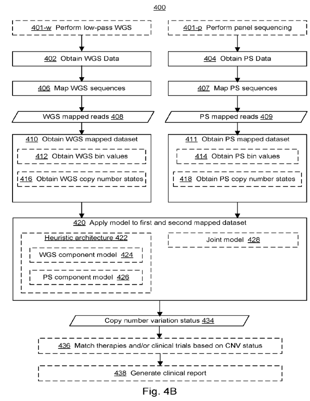
[0016] Figures 4A and 4B collectively illustrate an example
bioinformatics pipeline for
determining a copy number variation status of a subject. Figure 4A provides an
overview
flow chart of processes and features in a bioinformatics pipeline, in
accordance with some
embodiments of the present disclosure. Figure 4B illustrates an example flow
chart of
processes and features for determining a copy number variation status of a
subject, in which
dashed boxes represent optional portions of the method, in accordance with
some
embodiments of the present disclosure.
[0017] Figures 5A, 5B, 5C, and 5D collectively provide a flow
chart of processes and
features for determining a copy number variation status of a subject, in which
dashed boxes
represent optional portions of the method, in accordance with some embodiments
of the
present disclosure.
[0018] Figure 6 illustrates an example workflow of a method for
clinical reporting
combining low-coverage whole genome sequencing (lc-WGS) and whole exome
sequencing
(WES) data, in accordance with various embodiments of the present disclosure.
Steps in
4
CA 03204451 2023- 7-6
WO 2022/150663
PCT/US2022/011724
dotted lines/boxes are optional and can be added to provide genetic disease
risk prediction in
the clinical report.
[0019] Figure 7 illustrates an example analysis of CNVs
(deletions/insertions) from
WGS data, in accordance with an embodiment of the present disclosure.
Deletions,
duplications, and more general structural variants can be detected in WGS data
using
coverage depth analysis as well as identification of split reads, where, due
to breakpoints, a
first end of a respective read maps in one location and a second end of the
respective read
maps in a different location, resulting in discordance in apparent fragment
inset size between
pair-end reads. Because PCR-free WGS libraries result in close to random
shotgun
sequencing of the DNA templates, depth of coverage is fairly uniform in WGS
alignments,
although some systematic biases remain. This may allow more accurate analysis
of depth for
CNV detection.
[0020] Figure 8 illustrates an example analysis of CNVs
(deletions/insertions) from WES
data, in accordance with an embodiment of the present disclosure. In WES,
exons are
captured for sequencing but depth of coverage across these regions is much
more variable
than in WGS due to biases in the capture of DNA fragments by the assay probes.
This makes
it difficult to determine when deletions or duplications occur and creates
false positives and
negatives in CNV detection, as exemplified in the calls shown at the bottom
(FP = false
positive; FN = false negative segments). Single exon events are more
difficult, because only
reads in 150-300 base pairs of the span of the exon are available. In
addition, most
breakpoints of structural variants occur in intronic or intergenic regions, by
chance. Small
events that affect a single exon may actually span several kilobases of
intronic segments but
are only manifested and detectable in the targeted exon region in the WES
assay.
[0021] Figure 9 illustrates an example of joint calling of CNVs
(deletions/insertions)
combining WES and lc-WGS data, in accordance with various embodiments of the
present
disclosure. The pattern of WES sequence depth is quite variable and makes it
difficult for
most algorithms to find the single exon deletion (reads depicted in upper
chart). On the other
hand, the lc-WGS signal is weak and by itself can lead to low sensitivity to
small events and
spurious results (reads depicted in lower chart). By combining both signals, a
properly
trained and/or calibrated algorithm can improve sensitivity and specificity
and be able to
reject false positives and detect single exon deletion (or duplications) which
span more base
pairs than are contained within the exon itself, even if the actual boundaries
predicted are
imprecise (e.g., due to the lack of sequence reads covering a breakpoint).
CA 03204451 2023- 7-6
WO 2022/150663
PCT/US2022/011724
[0022] Figure 10 illustrates an example schema for preparing
training data for model
development and validation, in accordance with various embodiments of the
present
disclosure. For training and validating joint CNV calling by machine learning
(ML) or other
methods, a set of human genomes is sequenced by WGS and WES. Standard depth
WGS
data (e.g., 30-50X) generates a ground truth dataset of CNV calls. This data
can also be
subsampled to simulate lc-WGS for training. A second input to the model
includes WES data
at standard depth (-60X). Full depth WGS and WES data for the 2,500 samples of
the 1,000
Genomes Project (1KGP) were obtained from the New York Genome Center (NYGC)
and
Google Genomics. These data can be used for model training and cross
validation, in
accordance with various embodiments of the present disclosure.
[0023] Figure 11 illustrates an example schema for model
training, testing, and
validation for joint CNV calling, in accordance with various embodiments of
the present
disclosure. Training, testing, and validation steps can be optionally aided by
a panel of
normal samples for a region of interest.
[0024] Figure 12 illustrates an example of an operational phase
using a ML model for
joint CNV calling, in accordance with various embodiments of the present
disclosure. Once a
model is validated, new sample data is used to generate joint CNV calls in
production mode,
optionally using a panel of normal samples sequenced with the same assay.
[0025] Figures 13A, 13B, 13C, and 13D illustrate plots
characterizing CNVs in nine
biological samples. Figure 13A illustrates the frequency of CNV events
overlapping a given
number of exons, for each of the nine samples. Figure 13B illustrates the
frequency of CNV
events overlapping 1 or more exons relative to CNV length, compared to all CNV
events
(overlapping 0 or more exons), using amalgamated counts for all nine samples.
Figure 13C
illustrates the frequency of CNV events overlapping 1, 2, or 3 exons relative
to CNV length,
using amalgamated counts for all nine samples. Figure 13D illustrates the
cumulative count
of CNV events of varying lengths overlapping 1, 2, or 3 exons, using
amalgamated counts for
all nine samples.
[0026] Figures 14A, 14B, and 14C illustrate CN V calling using
the RealTimeGenomics
(RTG) segment CNV caller, using a bin size of 500 base pairs and simulated
coverage of lx
(Figure 14A), 3X (Figure 14B), and 5X (Figure 14C).
[0027] Figures 15A and 15B show examples of CNV calls obtained
using the RTG
segment CNV caller at varying simulated and full coverages (1X to 30X). Shaded
regions of
6
CA 03204451 2023- 7-6
WO 2022/150663
PCT/US2022/011724
the plot indicate locations of nominal deletions, as provided by baseline
calls obtained from a
"truth set." Figure 15A illustrates a deletion event that is discernible in
the 3X to 30X
coverage range. Figure 15B illustrates a deletion event that is not
discernible even at 30X
coverage.
[0028] Figures 16A and 16B show examples of CNV calls obtained
using the RTG
segment CNV caller at varying simulated and full coverages (1X to 30X). Shaded
regions of
the plot indicate locations of nominal duplications, as provided by baseline
calls obtained
from a -truth set." Figure 16A illustrates a duplication event that is not
discernible even at
30X coverage. Figure 16B illustrates a duplication event that is discernible
in the 3X to 30X
coverage range.
[0029] Figures 17A and 17B show examples of CNV calls obtained
using the CNVnator
CNV caller, using five of the nine samples with known CNV events. Figure 17A
shows that
CNV calls obtained using CNVnator show poor concordance with a "truth set."
Figure 17B
shows that CNV calls obtained using CNVnator show moderate concordance with
CNV calls
obtained using the RTG segment CNV caller at 30X coverage.
DETAILED DESCRIPTION
[0030] Introduction.
[0031] Accurate CNV detection is necessary to improve clinical
diagnostics, genetic risk
screening, and Mendelian disease tests. A single exon deletion can be highly
deleterious and,
if missed, patients can be misdiagnosed (false negative). A spurious false
positive test could
lead to unnecessary medical procedures and cost to the healthcare systems.
Advantageously,
the systems and methods described herein facilitate higher sensitivity and
specificity for the
detection of CNVs in disease gene panels, e.g., inherited cancer risk panels
including
BRCA1, BRCA2, and other genes.
[0032] Identification ("calling") of structural variants (SV)
from whole genome
sequencing data from short reads platform is not without its challenges but
can be achieved
for most regions of the genome. These structural variants include large
deletions and
insertions (e.g., by convention, greater than about 50 bp), duplications
(increases in the copy
number of a genomic region over the copy number of a normal diploid genome),
inversions,
and translocations. The collection of deletions and duplications are also
called copy number
variants (CNVs). CNV calling in whole genome sequencing data can be achieved
by analysis
of depth of coverage, mapping of breakpoint reads, and discordance in apparent
insert size for
7
CA 03204451 2023- 7-6
WO 2022/150663
PCT/US2022/011724
paired-end reads and can be quite accurate for events over a few hundred bases
in length to
large segments of the chromosome arms, at least for deletions. Some
problematic regions
include repeats and segmental duplications, but for most coding genes this is
not a major
problem. Long read sequencing and optical maps can be used to identify SVs in
such
complex regions.
[0033] On the other hand, detecting small CNVs from targeted
short-read sequencing
data can be challenging. Most conventional methods for detecting CNV events
from next-
generation sequencing (NGS) rely on detecting changes in the mean or median
depth of
coverage that such events are expected to cause (e.g., deletions would result
in a reduction in
depth of coverage, and vice versa for duplications). However, it is
particularly difficult to
differentiate actual changes in sequencing depth from several types of
technical artifacts that
change the depth profile irrespective of gene dosage changes, including, but
not limited to, a)
sequencing biases due to GC content, b) read mapping biases due to repeats, c)
segmental
duplications, e) paralogous regions, and/or f) systematic capture biases
prevalent in targeted
sequencing chemistries. Methods to overcome such problems include GC and
mappability
normalization across -bins" of arbitrary length (e.g., 100 bp), as well as
comparing
sequencing data to diploid normal samples, bin by bin, sequenced with the same
assay. In
some embodiments, biases are expected to be similar in the control and test
samples, such
that, by calculating the depth ratio in the bins, it is possible to derive
whether an underlying
CNV is present. Adjacent bins deemed to have the same CNV status can be
combined,
resulting in potentially larger CNV "calls- as outputs, which can then be
interpreted for their
impact and pathogenicity. While longer structural variants can be more
reliably detected in
this fashion, small structural variants, particularly those encompassing a
single bin, are
difficult to differentiate from false positives due to random or systematic
read depth
fluctuation.
[0034] While many of these problems affect both WGS and targeted
sequencing,
sequencing biases are further exacerbated in targeted sequencing reactions due
to differences
in probe capture efficiencies and genetic variation under probes, resulting in
a much more
variable -normal" depth profile in gene panels as compared to WGS. An
additional
complexity in targeted sequencing is that it often only targets the exons of
the genes, as well
as a small portion of adjacent upstream and/or downstream intronic sequences
to ensure
coverage of splice regions. Since the average exon length is only about 150
bp, these
sequenced regions may encompass only about 300 bp on average. This means that
a single
8
CA 03204451 2023- 7-6
WO 2022/150663
PCT/US2022/011724
exon deletion is difficult to detect, as sequencing data are typically
available from only one or
two bins whereas conventional CNV calling algorithms generally require a
minimum of 2-3
concordant bins to call a variant. In germline testing, the typical sequencing
depth on
targeted gene panels is about 200-300X. At this sequencing depth the
sensitivity to detect
single exon events drops to 50% or less, which is not reliable enough for use
in clinical
diagnosis. To overcome this loss of sensitivity, the sequencing depth could be
increased.
However, increasing sequencing depth to a level that supports the sensitivity
needed for
clinical diagnostics incurs significant economic and throughput costs. For
instance, it has
been suggested that sensitivity can be substantially increased by sequencing
at a depth of
about 500-1000X. However, this makes the clinical test prohibitively expensive
with current
technology. Similarly, while WGS can be used effectively, it is also too
expensive for
routine gene-panel testing. Yet others have used targeted long read sequencing
of entire
genes to overcome this problem. But this is not an easily generalizable
approach, as custom
targeted sequencing assays are needed for each gene, and WGS uses a different
sequencing
platform than conventionally used for single nucleotide variant
identification, increasing
logistical costs.
[0035] More particularly, the actual genetic variants responsible
for single exon CNVs
are often much larger than the 150 bp average exon length, and in fact are
typically several kb
in length (see, e.g., Figure 7). However, the breakpoints for these deletions
commonly reside
deep in the intronic regions and, thus, a large fraction of such events span
regions that are
likely invisible to targeted sequencing (see, e.g., Figure 8). Accordingly,
WGS is generally
more sensitive to such events, but conventional methodologies for WGS at an
average
sequencing depth of 30-50X are still too expensive to be used as an assay for
gene panel tests.
[0036] On the other hand, low-coverage (or low-pass/depth) WGS
(lc-WGS or LPWGS)
has been proposed as an alternative, inexpensive assay to identify gross level
chromosomal
rearrangements and structural variants (SV), and, through variant imputation,
to genotype
common variants to perform genome-wide wide association studies (GWAS) or to
calculate
polygenic risk scores (PRS) derived from such studies. When lc-WGS is
performed to 0.5-
lx, it adds just $50 in cost per sample and is sufficient to detect large SVs
and perform
GWAS/PRS analysis. Thus, it has been proposed as an assay to replace
genotyping
microarray for such studies and CGC arrays in cytogenetic testing. While
SV/CNV data
derived from lc-WGS data is useful for cytogenetics, it lacks the sensitivity
and specificity
needed for the clinical testing of small events.
9
CA 03204451 2023- 7-6
WO 2022/150663
PCT/US2022/011724
[0037] What is needed in the art are improved methods and systems
for identifying short
CNVs, preferably from sequencing data that can also be used to identify single
nucleotide
variants. Particularly, methods and systems for identifying short CNVs over a
panel-targeted
(e.g., whole exome or a subset of genes thereof) sequencing backbone at medium
to low
sequencing depth are desired.
[0038] Advantageously, the disclosure provides systems and
methods that combine data
obtained from panel-targeted sequencing gene panels with signals from lc-WGS
to improve
sensitivity and specificity of CNV detection for gene-panel testing in a cost-
effective fashion.
By combining signals that alone would be likely to have insufficient
specificity to call small,
exon-level CNVs, the combined assay accomplishes clinical grade variant
calling of CNVs,
with a sensitivity that is at least equivalent to, if not better than,
targeted sequencing
performed at a sequencing depth of 1000X or WGS performed at a sequencing
depth of 30X.
Briefly, the systems and methods provided herein utilize both WES and lc-WGS
data for the
same sample to remove false positives and provide accurate CNV calls down to
the single
exon level. See Figure 9, e.g., in comparison with Figure 8. Lc-WGS data
further provides
other useful readouts, such as disease risk prediction obtained by calculating
polygenic risk
scores from imputed variants from lc-WGS data.
[0039] In one aspect, the disclosure provides a method for
accurately identifying both
small variants (e.g., SNVs and small indels) and CNVs across coding regions
for clinical
diagnosis and assessment of genetic risk. In some embodiments, risk prediction
for common
disease can also be provided through calculation of polygenic risk scores and
combined with
highly penetrant variants for absolute risk predictions.
[0040] In some embodiments, such a method combines data from low-
coverage WGS (1-
3X, which is cost effective) with WES data performed at cost-effective depths
(e.g., 60-80X)
and jointly analyzes the alignments from both assays to provide accurate CNV
calls down to
the single exon level. An example implementation of such a method is
illustrated in Figure 6.
[0041] In some embodiments, methods and systems are provided for
calling CNV using
combined lc-WGS and WES data, e.g., as illustrated in step e of Figure 6. In
some
embodiments, joint CNV calling can be performed using one of a variety of
algorithms,
including machine learning models, Bayesian PCA models, probabilistic methods,
heuristic
methods, etc. For instance, Figures 10-12 illustrate examples of training,
testing, and
CA 03204451 2023- 7-6
WO 2022/150663
PCT/US2022/011724
operating a machine learning method for this task, in accordance with some
implementations
of the present disclosure.
[0042] In some embodiments, the methods described herein further
use reference data
generated for a panel of normal samples, e.g., samples, previously analyzed
using the same
assays, that were determined to be CNV negative for one or more genes of
interest.
[0043] In some embodiments, the systems and methods described
herein facilitate
development of gene panel tests for germline testing of inherited disease
risk, e.g., inherited
breast, ovarian, colon, prostate, or other cancers) derived from rare, highly
penetrant
pathogenic CNV variants, by combining whole-exome sequencing and/or gene-panel
targeted
sequencing at cost effective depths (60-80X for WES; 200-300X for smaller
panels), with an
inexpensive lc-WGS assay (1-3X).
[0044] In some embodiments, the systems and methods described
herein are used to
complement WES to identify small pathogenic CNV events more accurately for
Mendelian
disease diagnostics, newborn screening, carrier screening, CDC Tier-1
condition screening,
and other disease panels screening.
[0045] Definitions.
[0046] The terminology used in the present disclosure is for the
purpose of describing
particular embodiments only and is not intended to be limiting of the
invention. As used in
the description of the invention and the appended claims, the singular forms
"a", -an- and
"the" are intended to include the plural forms as well, unless the context
clearly indicates
otherwise. It will also be understood that the term "and/or" as used herein
refers to and
encompasses any and all possible combinations of one or more of the associated
listed items.
It will be further understood that the terms "comprises" and/or "comprising,-
when used in
this specification, specify the presence of stated features, integers, steps,
operations,
elements, and/or components, but do not preclude the presence or addition of
one or more
other features, integers, steps, operations, elements, components, and/or
groups thereof
Furthermore, to the extent that the terms "including," "includes,- "having,-
"has,- "with,- or
variants thereof are used in either the detailed description and/or the
claims, such terms are
intended to be inclusive in a manner similar to the term "comprising."
100471 As used herein, the term "if' may be construed to mean
"when" or "upon" or "in
response to determining- or -in response to detecting," depending on the
context. Similarly,
the phrase -if it is determined- or -if [a stated condition or event] is
detected" may be
11
CA 03204451 2023- 7-6
WO 2022/150663
PCT/US2022/011724
construed to mean "upon determining" or "in response to determining" or "upon
detecting
[the stated condition or eventl" or "in response to detecting the stated
condition or event],"
depending on the context.
[0048] It will also be understood that, although the terms first,
second, etc. may be used
herein to describe various elements, these elements should not be limited by
these terms.
These terms are only used to distinguish one element from another. For
example, a first
subject could be termed a second subject, and, similarly, a second subject
could be termed a
first subject, without departing from the scope of the present disclosure. The
first subject and
the second subject are both subjects, but they are not the same subject.
Furthermore, the
terms -subject," -user," and -patient" are used interchangeably herein.
[0049] As used herein, the term "subject- refers to any living or
non-living human. In
some embodiments, a subject is a male or female of any stage (e.g., a man, a
woman or a
child).
[0050] As used herein, the terms "control," "control sample,"
"reference," "reference
sample," -normal," and -normal sample" describe a sample from a subject that
does not have
a particular condition or is otherwise healthy. In an example, a method as
disclosed herein
can be performed on a subject having a tumor, where the reference sample is a
sample taken
from a healthy tissue of the subject. A reference sample can be obtained from
the subject, or
from a database. The reference can be, e.g., a reference genome that is used
to map sequence
reads obtained from sequencing a sample from the subject. A reference genome
can refer to
a haploid or diploid genome to which sequence reads from the biological sample
and a
constitutional sample can be aligned and compared. An example of a
constitutional sample
can be DNA of whole blood or blood cells obtained from the subject. For a
haploid genome.
there can be only one nucleotide at each locus. For a diploid genome,
heterozygous loci can
be identified; each heterozygous locus can have two alleles, where either
allele can allow a
match for alignment to the locus.
[0051] As used herein, the term "locus- refers to a position
(e.g., a site) within a genome,
e.g., on a particular chromosome. In some embodiments, a locus refers to a
single nucleotide
position within a genome, i.e., on a particular chromosome. In some
embodiments, a locus
refers to a small group of nucleotide positions within a genome, e.g., as
defined by a mutation
(e.g., substitution, insertion, or deletion) of consecutive nucleotides within
a cancer genome.
Because normal mammalian cells have diploid genomes, a normal mammalian genome
(e.g.,
12
CA 03204451 2023- 7-6
WO 2022/150663
PCT/US2022/011724
a human genome) will generally have two copies of every locus in the genome,
or at least two
copies of every locus located on the autosomal chromosomes, e.g., one copy on
the maternal
autosomal chromosome and one copy on the paternal autosomal chromosome.
[0052] As used herein, the term -allele" refers to a particular
sequence of one or more
nucleotides at a chromosomal locus.
[0053] As used herein, the term -reference allele" refers to the
sequence of one or more
nucleotides at a chromosomal locus that is either the predominant allele
represented at that
chromosomal locus within the population of the species (e.g., the "wild-type"
sequence), or
an allele that is predefined within a reference genome for the species.
[0054] As used herein, the term "variant allele" refers to a
sequence of one or more
nucleotides at a chromosomal locus that is either not the predominant allele
represented at
that chromosomal locus within the population of the species (e.g., not the
"wild-type"
sequence), or not an allele that is predefined within a reference genome for
the species.
[0055] As used herein, the term "single nucleotide variant- or
"SNV- refers to a
substitution of one nucleotide to a different nucleotide at a position (e.g.,
site) of a nucleotide
sequence, e.g., a sequence read from an individual. A substitution from a
first nucleobase X
to a second nucleobase Y may be denoted as "X>Y." For example, a cytosine to
thymine
SNV may be denoted as "C>T.-
[0056] As used herein, the term "mutation- or "variant" refers to
a detectable change in
the genetic material of one or more cells. In a particular example, one or
more mutations can
be found in, and can identify, cancer cells (e.g., driver and passenger
mutations). A mutation
can be transmitted from a parent cell to a daughter cell. A person having
skill in the art will
appreciate that a genetic mutation (e.g., a driver mutation) in a parent cell
can induce
additional, different mutations (e.g., passenger mutations) in a daughter
cell. A mutation
generally occurs in a nucleic acid. In a particular example, a mutation can be
a detectable
change in one or more deoxyribonucleic acids or fragments thereof A mutation
generally
refers to nucleotides that are added, deleted, substituted for, inverted, or
transposed to a new
position in a nucleic acid. A mutation can be a spontaneous mutation or an
experimentally
induced mutation. A mutation in the sequence of a particular tissue is an
example of a
-tissue-specific allele." For example, a tumor can have a mutation that
results in an allele at a
locus that does not occur in normal cells. Another example of a "tissue-
specific allele" is a
fetal-specific allele that occurs in the fetal tissue, but not the maternal
tissue.
13
CA 03204451 2023- 7-6
WO 2022/150663
PCT/US2022/011724
[0057] As used herein, the term "loss of heterozygosity" refers
to the loss of one copy of
a segment (e.g., including part or all of one or more genes) of the genome of
a diploid subject
(e.g., a human) or loss of one copy of a sequence encoding a functional gene
product in the
genome of the diploid subject, in a tissue, e.g, a cancerous tissue, of the
subject. As used
herein, when referring to a metric representing loss of heterozygosity across
the entire
genome of the subject, loss of heterozygosity is caused by the loss of one
copy of various
segments in the genome of the subject. Loss of heterozygosity across the
entire genome may
be estimated without sequencing the entire genome of a subject, and such
methods for such
estimations based on gene panel targeting-based sequencing methodologies are
described in
the art. Accordingly, in some embodiments, a metric representing loss of
heterozygosity
across the entire genome of a tissue of a subject is represented as a single
value, e.g., a
percentage or fraction of the genome. In some cases, a tumor is composed of
various sub-
clonal populations, each of which may have a different degree of loss of
heterozygosity
across their respective genomes. Accordingly, in some embodiments, loss of
heterozygosity
across the entire genome of a cancerous tissue refers to an average loss of
heterozygosity
across a heterogeneous tumor population. As used herein, when referring to a
metric for loss
of heterozygosity in a particular gene, e.g., a DNA repair protein such as a
protein involved in
the homologous DNA recombination pathway (e.g., BRCA1 or BRCA2), loss of
heterozygosity refers to complete or partial loss of one copy of the gene
encoding the protein
in the genome of the tissue and/or a mutation in one copy of the gene that
prevents translation
of a full-length gene product, e.g., a frameshift or truncating (creating a
premature stop codon
in the gene) mutation in the gene of interest. In some cases, a tumor is
composed of various
sub-clonal populations, each of which may have a different mutational status
in a gene of
interest. Accordingly, in some embodiments, loss of heterozygosity for a
particular gene of
interest is represented by an average value for loss of heterozygosity for the
gene across all
sequenced sub-clonal populations of the cancerous tissue. In other
embodiments, loss of
heterozygosity for a particular gene of interest is represented by a count of
the number of
unique incidences of loss of heterozygosity in the gene of interest across all
sequenced sub-
clonal populations of the cancerous tissue (e.g., the number of unique frame-
shift and/or
truncating mutations in the gene identified in the sequencing data).
[0058] As used herein the term "cancer," "cancerous tissue," or
"tumor" refers to an
abnormal mass of tissue in which the growth of the mass surpasses and is not
coordinated
with the growth of normal tissue. A cancer or tumor can be defined as -benign-
or
14
CA 03204451 2023- 7-6
WO 2022/150663
PCT/US2022/011724
"malignant" depending on the following characteristics: degree of cellular
differentiation
including morphology and functionality, rate of growth, local invasion and
metastasis. A
"benign" tumor can be well differentiated, have characteristically slower
growth than a
malignant tumor and remain localized to the site of origin. In addition, in
some cases a
benign tumor does not have the capacity to infiltrate, invade or metastasize
to distant sites. A
"malignant" tumor can be poorly differentiated (anaplasia), have
characteristically rapid
growth accompanied by progressive infiltration, invasion, and destruction of
the surrounding
tissue. Furthermore, a malignant tumor can have the capacity to metastasize to
distant sites.
Accordingly, a cancer cell is a cell found within the abnormal mass of tissue
whose growth is
not coordinated with the growth of normal tissue. Accordingly, a "tumor
sample" refers to a
biological sample obtained or derived from a tumor of a subject, as described
herein. A
cancerous tissue can refer to blood cells if the cancer is a hematological
(blood) cancer.
[0059] As used herein, the terms "sequencing," "sequence
determination," and the like as
used herein refers generally to any and all biochemical processes that may be
used to
determine the order of biological macromolecules such as nucleic acids or
proteins. For
example, sequencing data can include all or a portion of the nucleotide bases
in a nucleic acid
molecule such as an mR_NA transcript or a genomic locus.
[0060] As used herein, the term -sequence reads" or -reads"
refers to nucleotide
sequences produced by any sequencing process described herein or known in the
art. Reads
can be generated from one end of nucleic acid fragments ("single-end reads"),
and sometimes
are generated from both ends of nucleic acids (e.g., paired-end reads, double-
end reads). The
length of the sequence read is often associated with the particular sequencing
technology.
High-throughput methods, for example, provide sequence reads that can vary in
size from
tens to hundreds of base pairs (bp). In some embodiments, the sequence reads
are of a mean,
median or average length of about 15 bp to 900 bp long (e.g., about 20 bp,
about 25 bp, about
30 bp, about 35 bp, about 40 bp, about 45 bp, about 50 bp, about 55 bp, about
60 bp, about 65
bp, about 70 bp, about 75 bp, about 80 bp, about 85 bp, about 90 bp, about 95
bp, about 100
bp, about 110 bp, about 120 bp, about 130, about 140 bp, about 150 bp, about
200 bp, about
250 bp, about 300 bp, about 350 bp, about 400 bp, about 450 bp, or about 500
bp. In some
embodiments, the sequence reads are of a mean, median or average length of
about 1000 bp,
2000 bp, 5000 bp, 10,000 bp, or 50,000 bp or more. Nanopore sequencing, for
example, can
provide sequence reads that can vary in size from tens to hundreds to
thousands of base pairs.
Illumina parallel sequencing can provide sequence reads that do not vary as
much, for
CA 03204451 2023- 7-6
WO 2022/150663
PCT/US2022/011724
example, most of the sequence reads can be smaller than 200 bp. A sequence
read (or
sequencing read) can refer to sequence information corresponding to a nucleic
acid molecule
(e.g., a string of nucleotides). For example, a sequence read can correspond
to a string of
nucleotides (e.g., about 20 to about 150) from part of a nucleic acid
fragment, can correspond
to a string of nucleotides at one or both ends of a nucleic acid fragment, or
can correspond to
nucleotides of the entire nucleic acid fragment. A sequence read can be
obtained in a variety
of ways, e.g., using sequencing techniques or using probes, e.g., in
hybridization arrays or
capture probes, or amplification techniques, such as the polymerase chain
reaction (PCR) or
linear amplification using a single primer or isothermal amplification.
[0061] As used herein, the term "read segment" or "read" refers
to any nucleotide
sequences including sequence reads obtained from an individual and/or
nucleotide sequences
derived from the initial sequence read from a sample obtained from an
individual. For
example, a read segment can refer to an aligned sequence read, a collapsed
sequence read, or
a stitched read. Furthermore, a read segment can refer to an individual
nucleotide base, such
as a single nucleotide variant.
[0062] As used herein, the term, "reference exome" refers to any
particular known,
sequenced or characterized exome, whether partial or complete, of any tissue
from any
organism or pathogen that may be used to reference identified sequences from a
subject.
Example reference exomes used for human subjects as well as many other
organisms are
provided in the on-line genome browser hosted by the National Center for
Biotechnology
Information ("NCBI").
[0063] As used herein, the term "reference genome" refers to any
particular known,
sequenced or characterized genome, whether partial or complete, of any
organism or
pathogen that may be used to reference identified sequences from a subject.
Exemplary
reference genomes used for human subjects as well as many other organisms are
provided in
the on-line genome browser hosted by the National Center for Biotechnology
Information
("NCBI") or the University of California, Santa Cruz (UCSC). A "genome" refers
to the
complete genetic information of an organism or pathogen, expressed in nucleic
acid
sequences. As used herein, a reference sequence or reference genome often is
an assembled
or partially assembled genomic sequence from an individual or multiple
individuals. In some
embodiments, a reference genome is an assembled or partially assembled genomic
sequence
from one or more human individuals. The reference genome can be viewed as a
representative example of a species' set of genes. In some embodiments, a
reference genome
16
CA 03204451 2023- 7-6
WO 2022/150663
PCT/US2022/011724
comprises sequences assigned to chromosomes. Exemplary human reference genomes
include but are not limited to NCBI build 34 (UCSC equivalent: hg16), NCBI
build 35
(UCSC equivalent: hg17), NCBI build 36.1 (UCSC equivalent: hg18), GRCh37 (UCSC
equivalent: hg19), and GRCh38 (UCSC equivalent: hg38).
[0064] As used herein, the term "assay" refers to a technique for
determining a property
of a substance, e.g., a nucleic acid, a protein, a cell, a tissue, or an
organ. An assay (e.g., a
first assay or a second assay) can comprise a technique for determining the
copy number
variation of nucleic acids in a sample, the methylation status of nucleic
acids in a sample, the
fragment size distribution of nucleic acids in a sample, the mutational status
of nucleic acids
in a sample, or the fragmentation pattern of nucleic acids in a sample. Any
assay known to a
person having ordinary skill in the art can be used to detect any of the
properties of nucleic
acids mentioned herein. Properties of a nucleic acids can include a sequence,
genomic
identity, copy number, methylation state at one or more nucleotide positions,
size of the
nucleic acid, presence or absence of a mutation in the nucleic acid at one or
more nucleotide
positions, and pattern of fragmentation of a nucleic acid (e.g., the
nucleotide position(s) at
which a nucleic acid fragments). An assay or method can have a particular
sensitivity and/or
specificity, and their relative usefulness as a diagnostic tool can be
measured using ROC-
AUC statistics.
[0065] The term "classification- can refer to any number(s) or
other characters(s) that are
associated with a particular property of a sample. For example, in some
embodiments, the
term "classification" can refer to a type of cancer in a subject or sample, a
stage of cancer in a
subject or sample, a prognosis for a cancer in a subject or sample, a tumor
load in a subject, a
presence of tumor metastasis in a subject, and the like. The classification
can be binary (e.g.,
positive or negative) or have more levels of classification (e.g., a scale
from 1 to 10 or 0 to 1).
The terms -cutoff' and -threshold" can refer to predetermined numbers used in
an operation.
For example, a cutoff size can refer to a size above which fragments are
excluded. A
threshold value can be a value above or below which a particular
classification applies.
Either of these terms can be used in either of these contexts.
[0066] As used interchangeably herein, the term "classifier" or
"model" refers to a
machine leaming model or algorithm.
[0067] In some embodiments, a classifier is an unsupervised
learning algorithm. One
example of an unsupervised learning algorithm is cluster analysis.
17
CA 03204451 2023- 7-6
WO 2022/150663
PCT/US2022/011724
[0068] In some embodiments, a classifier is supervised machine
learning. Nonlimiting
examples of supervised learning algorithms include, but are not limited to,
logistic regression,
neural networks, support vector machines, Naive Bayes algorithms, nearest
neighbor
algorithms, random forest algorithms, decision tree algorithms, boosted trees
algorithms,
multinomial logistic regression algorithms, linear models, linear regression,
GradientBoosting, mixture models, hidden Markov models, Gaussian NB
algorithms, linear
discriminant analysis, or any combinations thereof In some embodiments, a
classifier is a
multinomial classifier algorithm. In some embodiments, a classifier is a 2-
stage stochastic
gradient descent (SGD) model. In some embodiments, a classifier is a deep
neural network
(e.g., a deep-and-wide sample-level classifier).
[0069] Neural networks. In some embodiments, the classifier is a
neural network (e.g., a
convolutional neural network and/or a residual neural network). Neural network
algorithms,
also known as artificial neural networks (ANNs), include convolutional and/or
residual neural
network algorithms (deep learning algorithms). Neural networks can be machine
learning
algorithms that may be trained to map an input data set to an output data set,
where the neural
network comprises an interconnected group of nodes organized into multiple
layers of nodes.
For example, the neural network architecture may comprise at least an input
layer, one or
more hidden layers, and an output layer. The neural network may comprise any
total number
of layers, and any number of hidden layers, where the hidden layers function
as trainable
feature extractors that allow mapping of a set of input data to an output
value or set of output
values. As used herein, a deep learning algorithm (DNN) can be a neural
network
comprising a plurality of hidden layers, e.g., two or more hidden layers. Each
layer of the
neural network can comprise a number of nodes (or "neurons"). A node can
receive input
that comes either directly from the input data or the output of nodes in
previous layers, and
perform a specific operation, e.g., a summation operation. In some
embodiments, a
connection from an input to a node is associated with a parameter (e.g , a
weight and/or
weighting factor). In some embodiments, the node may sum up the products of
all pairs of
inputs, xi, and their associated parameters. In some embodiments, the weighted
sum is offset
with a bias, b. In some embodiments, the output of a node or neuron may be
gated using a
threshold or activation function, f, which may be a linear or non-linear
function. The
activation function may be, for example, a rectified linear unit (ReLU)
activation function, a
Leaky ReLU activation function, or other function such as a saturating
hyperbolic tangent,
identity, binary step, logistic, arcTan, softsig,n, parametric rectified
linear unit, exponential
18
CA 03204451 2023- 7-6
WO 2022/150663
PCT/US2022/011724
linear unit, softPlus, bent identity, softExponential, Sinusoid, Sine,
Gaussian, or sigmoid
function, or any combination thereof.
[0070] The weighting factors, bias values, and threshold values,
or other computational
parameters of the neural network, may be -taught" or -learned" in a training
phase using one
or more sets of training data. For example, the parameters may be trained
using the input
data from a training data set and a gradient descent or backward propagation
method so that
the output value(s) that the ANN computes are consistent with the examples
included in the
training data set. The parameters may be obtained from a back propagation
neural network
training process.
[0071] Any of a variety of neural networks may be suitable for
use in the present
disclosure. Examples can include, but are not limited to, feedforward neural
networks, radial
basis function networks, recurrent neural networks, residual neural networks,
convolutional
neural networks, residual convolutional neural networks, and the like, or any
combination
thereof In some embodiments, the machine learning makes use of a pre-trained
and/or
transfer-learned ANN or deep learning architecture. Convolutional and/or
residual neural
networks can be used in the present disclosure in accordance with the present
disclosure.
[0072] For instance, a deep neural network classifier comprises
an input layer, a plurality
of individually parameterized (e.g., weighted) convolutional layers, and an
output scorer.
The parameters (e.g., weights) of each of the convolutional layers as well as
the input layer
contribute to the plurality of parameters (e.g., weights) associated with the
deep neural
network classifier. In some embodiments, at least 100 parameters, at least
1000 parameters,
at least 2000 parameters or at least 5000 parameters are associated with the
deep neural
network classifier. As such, deep neural network classifiers require a
computer to be used
because they cannot be mentally solved. In other words, given an input to the
classifier, the
classifier output needs to be determined using a computer rather than mentally
in such
embodiments. See, for example, Krizheysky et al., 2012, "Imagenet
classification with deep
convolutional neural networks," in Advances in Neural Information Processing
Systems 2,
Pereira, Burges, Bottou, Weinberger, eds., pp. 1097-1105, Curran Associates,
Inc.; Zeiler,
2012 "ADADELTA: an adaptive learning rate method," CoRR, vol. abs/1212.5701;
and
Rumelhart et al., 1988, "Neurocomputing: Foundations of research," ch.
Learning
Representations by Back-propagating Errors, pp. 696-699, Cambridge, MA, USA:
MIT
Press, each of which is hereby incorporated by reference.
19
CA 03204451 2023- 7-6
WO 2022/150663
PCT/US2022/011724
[0073] Neural network algorithms, including convolutional neural
network algorithms,
suitable for use as classifiers are disclosed in, for example, Vincent et at.,
2010, "Stacked
denoising autoencoders: Learning useful representations in a deep network with
a local
denoising criterion," J Mach Learn Res 11, pp. 3371-3408; Larochelle et at.,
2009,
"Exploring strategies for training deep neural networks," J Mach Learn Res 10,
pp. 1-40; and
IIassoun, 1995, Fundamentals of Artificial Neural Networks, Massachusetts
Institute of
Technology, each of which is hereby incorporated by reference. Additional
example neural
networks suitable for use as classifiers are disclosed in Duda et at., 2001,
Pattern
Classification, Second Edition, John Wiley & Sons, Inc., New York; and Hastie
et at., 2001,
The Elements of Statistical Learning, Springer-Verlag, New York, each of which
is hereby
incorporated by reference in its entirety. Additional example neural networks
suitable for use
as classifiers are also described in Draghici, 2003, Data Analysis Tools for
DNA Microarrays,
Chapman & Hall/CRC; and Mount, 2001, Bioinformatics: sequence and genome
analysis,
Cold Spring Harbor Laboratory Press, Cold Spring Harbor, New York, each of
which is
hereby incorporated by reference in its entirety.
[0074] Support vector machines. In some embodiments, the
classifier is a support vector
machine (SVM). SVM algorithms suitable for use as classifiers are described
in, for
example, Cristianini and Shawe-Taylor, 2000, "An Introduction to Support
Vector
Machines," Cambridge University Press, Cambridge; Boser et at., 1992, "A
training
algorithm for optimal margin classifiers," in Proceedings of the 5th Annual
ACM Workshop
on Computational Learning Theory, ACM Press, Pittsburgh, Pa., pp. 142-152;
Vapnik, 1998,
Statistical Learning Theory, Wiley, New York; Mount, 2001, Bioinformatics:
sequence and
genome analysis, Cold Spring Harbor Laboratory Press, Cold Spring Harbor,
N.Y.; Duda,
Pattern Classification, Second Edition, 2001, John Wiley & Sons, Inc., pp.
259, 262-265; and
Hastie, 2001, The Elements of Statistical Learning, Springer, New York; and
Furey et at.,
2000, Bioinformatics 16, 906-914, each of which is hereby incorporated by
reference in its
entirety. When used for classification, SVMs separate a given set of binary
labeled data with
a hyper-plane that is maximally distant from the labeled data. For cases in
which no linear
separation is possible, SVMs can work in combination with the technique of
'kernels', which
automatically realizes a non-linear mapping to a feature space. The hyper-
plane found by the
SVM in feature space can correspond to a non-linear decision boundary in the
input space. In
some embodiments, the plurality of parameters (e.g., weights) associated with
the SVM
define the hyper-plane. In some embodiments, the hyper-plane is defined by at
least 10, at
CA 03204451 2023- 7-6
WO 2022/150663
PCT/US2022/011724
least 20, at least 50, or at least 100 parameters and the SVM classifier
requires a computer to
calculate because it cannot be mentally solved.
[0075] Naïve Iktye.v algorithms. In some embodiments, the classi
her is a Naive Bayes
algorithm. Naive Bayes classifiers suitable for use as classifiers are
disclosed, for example,
in Ng et al., 2002, "On discriminative vs. generative classifiers: A
comparison of logistic
regression and naive Bayes," Advances in Neural Information Processing
Systems, 14, which
is hereby incorporated by reference. A Naive Bayes classifier is any
classifier in a family of
-probabilistic classifiers" based on applying Bayes' theorem with strong
(naive)
independence assumptions between the features. In some embodiments, they are
coupled
with Kernel density estimation. See, for example, Hastie etal., 2001, The
elements of
statistical learning: data mining, inference, and prediction, eds. Tibshirani
and Friedman,
Springer, New York, which is hereby incorporated by reference.
[0076] Nearest neighbor algorithms. In some embodiments, a
classifier is a nearest
neighbor algorithm. Nearest neighbor classifiers can be memory-based and
include no
classifier to be fit. For nearest neighbors, given a query point xo (a test
subject), the k training
points xo, r, ,k (here the training subjects) closest in distance to xo are
identified and then
the point xo is classified using the k nearest neighbors. Here, the distance
to these neighbors
is a function of the abundance values of the discriminating gene set. In some
embodiments,
Euclidean distance in feature space is used to determine distance as d() =
iix(i) x(o) =
Typically, when the nearest neighbor algorithm is used, the abundance data
used to compute
the linear discriminant is standardized to have mean zero and variance 1. The
nearest
neighbor rule can be refined to address issues of unequal class priors,
differential
misclassification costs, and feature selection. Many of these refinements
involve some form
of weighted voting for the neighbors. For more information on nearest neighbor
analysis, see
Duda, Pattern Classification, Second Edition, 2001, John Wiley & Sons, Inc;
and Hastie,
2001, The Elements of Statistical Learning, Springer, New York, each of which
is hereby
incorporated by reference.
[0077] A k-nearest neighbor classifier is a non-parametric
machine learning method in
which the input consists of the k closest training examples in feature space.
The output is a
class membership. An object is classified by a plurality vote of its
neighbors, with the object
being assigned to the class most common among its k nearest neighbors (k is a
positive
integer, typically small). If k = 1, then the object is simply assigned to the
class of that single
nearest neighbor. See, Duda et al., 2001, Pattern Classification, Second
Edition, John Wiley
21
CA 03204451 2023- 7-6
WO 2022/150663
PCT/US2022/011724
& Sons, which is hereby incorporated by reference. In some embodiments, the
number of
distance calculations needed to solve the k-nearest neighbor classifier is
such that a computer
is used to solve the classifier for a given input because it cannot be
mentally performed.
[0078] Random forest, decision tree, and boosted tree algorithms.
In some embodiments,
the classifier is a decision tree. Decision trees suitable for use as
classifiers are described
generally by Duda, 2001, Pattern Classification, John Wiley & Sons, Inc., New
York, pp.
395-396, which is hereby incorporated by reference. Tree-based methods
partition the
feature space into a set of rectangles, and then fit a model (like a constant)
in each one. In
some embodiments, the decision tree is random forest regression. One specific
algorithm that
can be used is a classification and regression tree (CART). Other specific
decision tree
algorithms include, but are not limited to, ID3, C4.5, MART, and Random
Forests. CART,
ID3, and C4.5 are described in Duda, 2001, Pattern Classification, John Wiley
& Sons, Inc.,
New York, pp. 396-408 and pp. 411-412, which is hereby incorporated by
reference. CART,
MART, and C4.5 are described in Hastie et al., 2001, The Elements of
Statistical Learning,
Springer-Verlag, New York, Chapter 9, which is hereby incorporated by
reference in its
entirety. Random Forests are described in 13reiman, 1999, "Random
Forests¨Random
Features," Technical Report 567, Statistics Department, U.C. Berkeley,
September 1999,
which is hereby incorporated by reference in its entirety. In some
embodiments, the decision
tree classifier includes at least 10, at least 20, at least 50, or at least
100 parameters (e.g.,
weights and/or decisions) and requires a computer to calculate because it
cannot be mentally
solved.
[0079] Regression. In some embodiments, the classifier uses a
regression algorithm. A
regression algorithm can be any type of regression. For example, in some
embodiments, the
regression algorithm is logistic regression. In some embodiments, the
regression algorithm is
logistic regression with lasso, L2 or elastic net regularization. In some
embodiments, those
extracted features that have a corresponding regression coefficient that fails
to satisfy a
threshold value are pruned (removed from) consideration. In some embodiments,
a
generalization of the logistic regression model that handles multicategory
responses is used as
the classifier. Logistic regression algorithms are disclosed in Agresti, An
Introduction to
Categorical Data Analysis, 1996, Chapter 5, pp. 103-144, John Wiley & Son, New
York,
which is hereby incorporated by reference. In some embodiments, the classifier
makes use of
a regression model disclosed in Hastie et al., 2001, The Elements of
Statistical Learning,
Springer-Verlag, New York. In some embodiments, the logistic regression
classifier includes
22
CA 03204451 2023- 7-6
WO 2022/150663
PCT/US2022/011724
at least 10, at least 20, at least 50, at least 100, or at least 1000
parameters (e.g., weights) and
requires a computer to calculate because it cannot be mentally solved.
[0080] Linear discriminant analysis algorithms. Linear
discriminant analysis (LDA),
normal discriminant analysis (NDA), or discriminant function analysis can be a
generalization of Fisher's linear discriminant, a method used in statistics,
pattern recognition,
and machine learning to find a linear combination of features that
characterizes or separates
two or more classes of objects or events. The resulting combination can be
used as the
classifier (linear classifier) in some embodiments of the present disclosure.
[0081] Mixture model and Hidden Markov model. In some
embodiments, the classifier is
a mixture model, such as that described in McLachlan et al., Bioinformatics
18(3):413-422,
2002. In some embodiments, in particular, those embodiments including a
temporal
component, the classifier is a hidden Markov model such as described by
Schliep et al., 2003,
Bioinformatics 19(1):i255-i263.
[0082] Clustering. In some embodiments, the classifier is an
unsupervised clustering
model. In some embodiments, the classifier is a supervised clustering model.
Clustering
algorithms suitable for use as classifiers are described, for example, at
pages 211-256 of
Duda and Hart, Pattern Classification and Scene Analysis, 1973, John Wiley &
Sons, Inc.,
New York, (hereinafter "Duda 1973") which is hereby incorporated by reference
in its
entirety. The clustering problem can be described as one of finding natural
groupings in a
dataset. To identify natural groupings, two issues can be addressed. First, a
way to measure
similarity (or dissimilarity) between two samples can be determined. This
metric (e.g.,
similarity measure) can be used to ensure that the samples in one cluster are
more like one
another than they are to samples in other clusters. Second, a mechanism for
partitioning the
data into clusters using the similarity measure can be determined. One way to
begin a
clustering investigation can be to define a distance function and to compute
the matrix of
distances between all pairs of samples in the training set. If distance is a
good measure of
similarity, then the distance between reference entities in the same cluster
can be significantly
less than the distance between the reference entities in different clusters.
However, clustering
may not use a distance metric. For example, a nonmetric similarity function
s(x, x') can be
used to compare two vectors x and x'. s(x, x') can be a symmetric function
whose value is
large when x and x' are somehow -similar." Once a method for measuring -
similarity" or
"dissimilarity- between points in a dataset has been selected, clustering can
use a criterion
function that measures the clustering quality of any partition of the data.
Partitions of the
23
CA 03204451 2023- 7-6
WO 2022/150663
PCT/US2022/011724
data set that extremize the criterion function can be used to cluster the
data. Particular
exemplary clustering techniques that can be used in the present disclosure can
include, but
are not limited to, hierarchical clustering (agglomerative clustering using a
nearest-neighbor
algorithm, farthest-neighbor algorithm, the average linkage algorithm, the
centroid algorithm,
or the sum-of-squares algorithm), k-means clustering, fuzzy k-means clustering
algorithm,
and Jarvis-Patrick clustering. In some embodiments, the clustering comprises
unsupervised
clustering (e.g., with no preconceived number of clusters and/or no
predetermination of
cluster assignments).
[0083] Ensembles of classifiers and boosting. In some
embodiments, an ensemble (two
or more) of classifiers is used. In some embodiments, a boosting technique
such as AdaBoost
is used in conjunction with many other types of learning algorithms to improve
the
performance of the classifier. In this approach, the output of any of the
classifiers disclosed
herein, or their equivalents, is combined into a weighted sum that represents
the final output
of the boosted classifier. In some embodiments, the plurality of outputs from
the classifiers is
combined using any measure of central tendency known in the art, including but
not limited
to a mean, median, mode, a weighted mean, weighted median, weighted mode, etc.
In some
embodiments, the plurality of outputs is combined using a voting method. In
some
embodiments, a respective classifier in the ensemble of classifiers is
weighted or unweighted.
[0084] As used herein, the term "parameter- refers to any
coefficient or, similarly, any
value of an internal or external element (e.g., a weight and/or a
hyperparameter) in an
algorithm, model, regressor, and/or classifier that can affect (e.g., modify,
tailor, and/or
adjust) one or more inputs, outputs, and/or functions in the algorithm, model,
regressor and/or
classifier. For example, in some embodiments, a parameter refers to any
coefficient, weight,
and/or hyperparameter that can be used to control, modify, tailor, and/or
adjust the behavior,
learning, and/or performance of an algorithm, model, regressor, and/or
classifier. In some
instances, a parameter is used to increase or decrease the influence of an
input (e.g., a feature)
to an algorithm, model, regressor, and/or classifier. As a nonlimiting
example, in some
embodiments, a parameter is used to increase or decrease the influence of a
node (e.g., of a
neural network), where the node includes one or more activation functions.
Assignment of
parameters to specific inputs, outputs, and/or functions is not limited to any
one paradigm for
a given algorithm, model, regressor, and/or classifier but can be used in any
suitable
algorithm, model, regressor, and/or classifier architecture for a desired
performance. In some
embodiments, a parameter has a fixed value. In some embodiments, a value of a
parameter is
24
CA 03204451 2023- 7-6
WO 2022/150663
PCT/US2022/011724
manually and/or automatically adjustable. In some embodiments, a value of a
parameter is
modified by a validation and/or training process for an algorithm, model,
regressor, and/or
classifier (e.g., by error minimization and/or backpropagation methods). In
some
embodiments, an algorithm, model, regressor, and/or classifier of the present
disclosure
includes a plurality of parameters. In some embodiments, the plurality of
parameters is n
parameters, where: n 2; n 5; n 10; n 25; n 40; n 50; n 75; n 100; n = 125; n
150; n> 200; n >225; n> 250; n> 350; n> 500; n> 600; n> 750; n> 1,000; n
>2000; n>
4,000; n > 5,000; n > 7,500; n > 10,000; n > 20,000; n > 40,000; n > 75,000; n
> 100,000; n >
200,000; n > 500,000, n > 1 x 10, n > 5 x 10⁶, or n > 1 x 10⁷. As such, the
algorithms,
models, regressors, and/or classifiers of the present disclosure cannot be
mentally performed.
In some embodiments n is between 10,000 and 1 x 10⁷, between 100,000 and 5 x
106, or
between 500,000 and 1 x 10⁶. In some embodiments, the algorithms, models,
regressors,
and/or classifier of the present disclosure operate in a k-dimensional space,
where k is a
positive integer of 5 or greater (e.g., 5, 6, 7, 8, 9, 10, etc.). As such, the
algorithms, models,
regressors, and/or classifiers of the present disclosure cannot be mentally
performed.
100851 Several aspects are described herein with reference to
example applications for
illustration. It should be understood that numerous specific details,
relationships, and
methods are set forth to provide a full understanding of the features
described herein. One
having ordinary skill in the relevant art, however, will readily recognize
that the features
described herein can be practiced without one or more of the specific details
or with other
methods. The features described herein are not limited by the illustrated
ordering of acts or
events, as some acts can occur in different orders and/or concurrently with
other acts or
events. Furthermore, not all illustrated acts or events are required to
implement a
methodology in accordance with the features described herein.
100861 Reference is made herein to embodiments, examples of which
are illustrated in the
accompanying drawings. In the present disclosure, numerous specific details
are set forth in
order to provide a thorough understanding of the present disclosure. However,
it will be
apparent to one of ordinary skill in the art that the present disclosure may
be practiced
without these specific details. In other instances, well-known methods,
procedures,
components, circuits, and networks have not been described in detail so as not
to
unnecessarily obscure aspects of the embodiments.
CA 03204451 2023- 7-6
WO 2022/150663
PCT/US2022/011724
[0087] Example System Embodiments.
[0088] Now that an overview of some aspects of the present
disclosure and some
definitions used in the present disclosure have been provided, details of an
exemplary system
for providing clinical support for personalized therapy for various diseases
and disorders
(e. g , cardiovascular conditions, neurological conditions, cancers, etc.) are
now described in
conjunction with Figures 1A, 1B, 1C, 1D, and 1E. Figures 1A, 1B, 1C, 1D, and
lE
collectively illustrate the topology of an example system for providing
clinical support for
personalized therapy, in accordance with some embodiments of the present
disclosure.
Advantageously, the example system illustrated in Figures 1A, 1B, 1C, 1D, and
lE improves
upon conventional methods for providing clinical support for personalized
therapy by
improving detection of copy number variations, and particularly by identifying
CNVs
overlapping with only one or two exons, e.g., from panel sequencing data that
is also useful
for identifying single nucleotide variants.
[0089] Figure 1A is a block diagram illustrating a system in
accordance with some
implementations. The device 100 in some implementations includes one or more
processing
units CPU(s) 102 (also referred to as processors), one or more network
interfaces 104, a user
interface 106, e.g., including a display 108 and/or an input 110 (e.g., a
mouse, touchpad,
keyboard, etc.), a non-persistent memory 111, a persistent memory 112, and one
or more
communication buses 114 for interconnecting these components. The one or more
communication buses 114 optionally include circuitry (sometimes called a
chipset) that
interconnects and controls communications between system components. The non-
persistent
memory 111 typically includes high-speed random access memory, such as DRAM,
SRAM,
DDR RAM, ROM, EEPROM, flash memory, whereas the persistent memory 112
typically
includes CD-ROM, digital versatile disks (DVD) or other optical storage,
magnetic cassettes,
magnetic tape, magnetic disk storage or other magnetic storage devices,
magnetic disk
storage devices, optical disk storage devices, flash memory devices, or other
non-volatile
solid state storage devices. The persistent memory 112 optionally includes one
or more
storage devices remotely located from the CPU(s) 102. The persistent memory
112, and the
non-volatile memory device(s) within the non-persistent memory 112, comprise
non-
transitory computer readable storage medium. In some implementations, the non-
persistent
memory 111 or alternatively the non-transitory computer readable storage
medium stores the
following programs, modules and data structures, or a subset thereof,
sometimes in
conjunction with the persistent memory 112:
26
CA 03204451 2023- 7-6
WO 2022/150663
PCT/US2022/011724
= an operating system 116, which includes procedures for handling various
basic system
services and for performing hardware dependent tasks;
= a network communication module (or instructions) 118 for connecting the
system 100
with other devices and/or a communication network 105;
= a test patient data store 120 for storing one or more collections of
features from
patients (e.g., subjects);
= a bioinformatics module 140 for processing sequencing data and extracting
features
from sequencing data, e.g., from liquid biopsy sequencing assays;
= a feature analysis module 160 for evaluating patient features, e.g.,
genomic
alterations, compound genomic features, and clinical features; and
= a reporting module 180 for generating and transmitting reports that
provide clinical
support for personalized cancer therapy.
[0090] Although Figures 1A, 1B, 1C, in, and 1F, depict various
components of a "system
100," the figures are intended more as a functional description of the various
features that
may be present in computer systems than as a structural schematic of the
implementations
described herein. In practice, items shown separately could be combined and
some items
could be separated. Moreover, although Figure 1 depicts certain data and
modules in non-
persistent memory 111, some or all of these data and modules may be in
persistent memory
112. For example, in various implementations, one or more of the above
identified elements
are stored in one or more of the previously mentioned memory devices and
correspond to a
set of instructions for performing a function described above. The above
identified modules,
data, or programs (e.g., sets of instructions) need not be implemented as
separate software
programs, procedures, datasets, or modules, and thus various subsets of these
modules and
data may be combined or otherwise re-arranged in various implementations.
[0091] In some implementations, the non-persistent memory 111
optionally stores a
subset of the modules and data structures identified above. Furthermore, in
some
embodiments, the memory stores additional modules and data structures not
described above.
In some embodiments, one or more of the above-identified elements is stored in
a computer
system, other than that of system 100, that is addressable by system 100 so
that system 100
may retrieve all or a portion of such data when needed.
[0092] For purposes of illustration in Figure 1A, system 100 is
represented as a single
computer that includes all of the functionality for providing clinical support
for personalized
27
CA 03204451 2023- 7-6
WO 2022/150663
PCT/US2022/011724
cancer therapy. However, while a single machine is illustrated, the term
"system" shall also
be taken to include any collection of machines that individually or jointly
execute a set (or
multiple sets) of instructions to perform any one or more of the methodologies
discussed
herein.
[0093] For example, in some embodiments, system 100 includes one
or more computers.
In some embodiments, the functionality for providing clinical support for
personalized cancer
therapy is spread across any number of networked computers and/or resides on
each of
several networked computers and/or is hosted on one or more virtual machines
at a remote
location accessible across the communications network 105. For example,
different portions
of the various modules and data stores illustrated in Figures 1A, 1B, 1C, 1D,
and IE can be
stored and/or executed on the various instances of a processing device and/or
processing
server/database in the distributed diagnostic environment 210 illustrated in
Figure 2B (e.g.,
processing devices 224, 234, 244, and 254, processing server 262, and database
264).
[0094] The system may operate in the capacity of a server or a
client machine in a client-
server network environment, as a peer machine in a peer-to-peer (or
distributed) network
environment, or as a server or a client machine in a cloud computing
infrastructure or
environment. The system may be a personal computer (PC), a tablet PC, a set-
top box (STB),
a Personal Digital Assistant (PDA), a cellular telephone, a web appliance, a
server, a network
router, a switch or bridge, or any machine capable of executing a set of
instructions
(sequential or otherwise) that specify actions to be taken by that machine.
[0095] In another implementation, the system comprises a virtual
machine that includes a
module for executing instructions for performing any one or more of the
methodologies
disclosed herein. In computing, a virtual machine (VM) is an emulation of a
computer
system that is based on computer architectures and provides functionality of a
physical
computer. Some such implementations may involve specialized hardware,
software, or a
combination of hardware and software.
[0096] One of skill in the art will appreciate that any of a wide
array of different
computer topologies are used for the application and all such topologies are
within the scope
of the present disclosure.
[0097] Test Patient Data Store (120)
[0098] Referring to Figure 1B, in some embodiments, the system
(e.g., system 100)
includes a patient data store 120 that stores data for patients 121-1 to 121-M
including one or
28
CA 03204451 2023- 7-6
WO 2022/150663
PCT/US2022/011724
more sequencing data 122, feature data 125, and clinical assessments 139.
These data are
used and/or generated by the various processes stored in the bioinformatics
module 140 and
feature analysis module 160 of system 100, to ultimately generate a report
providing clinical
support for personalized therapy of a patient. While the feature scope of
patient data 121
across all patients may be informationally dense, an individual patient's
feature set may be
sparsely populated across the entirety of the collective feature scope of all
features across all
patients. That is to say, the data stored for one patient may include a
different set of features
that the data stored for another patient. Further, while illustrated as a
single data construct in
Figure 1B, different sets of patient data may be stored in different databases
or modules
spread across one or more system memories.
[0099] In some embodiments, sequencing data 122 from one or more
sequencing
reactions 122-i, including a plurality of sequence reads 123-1 to 123-K, is
stored in the test
patient data store 120. The data store may include different sets of
sequencing data from a
single subject, corresponding to different samples from the patient, e.g.,
salivary samples,
blood samples, solid tissue samples, tumor samples, and/or to samples acquired
at different
times, e.g., while monitoring the progression, regression, remission, and/or
recurrence of a
disease or disorder in a subject. The sequence reads may be in any suitable
file format, e.g.,
BCL, FASTA, FASTQ, etc. In some embodiments, sequencing data 122 is accessed
by a
sequencing data processing module 141, which performs various pre-processing,
genome
alignment, and demultiplexing operations, as described in detail below with
reference to
bioinformatics module 140. In some embodiments, sequence data that has been
aligned to a
reference construct, e.g., BAM file 124, is stored in test patient data store
120.
[00100] In some embodiments, the test patient data store 120 includes feature
data 125,
e.g., that is useful for identifying clinical support for personalized
therapy. In some
embodiments, the feature data 125 includes personal characteristics 126 of the
patient, such
as patient name, date of birth, gender, ethnicity, physical address, smoking
status, alcohol
consumption characteristic, anthropomorphic data, etc.
[00101] In some embodiments, the feature data 125 includes medical history
data 127 for
the patient, (e.g., date of initial disorder diagnosis, previous treatments
and outcomes, adverse
effects of therapy, therapy group history, clinical trial history, previous
and current
medications, surgical history, etc.), previous or current symptoms, previous
or current
therapies, previous treatment outcomes, previous disease diagnoses, diagnoses
of depression,
diagnoses of other physical or mental maladies, and family medical history. In
some
29
CA 03204451 2023- 7-6
WO 2022/150663
PCT/US2022/011724
embodiments, the feature data 125 includes clinical features 128, such as
pathology data 128-
1, medical imaging data 128-2, and tissue culture and/or tissue organoid
culture data 128-3.
[00102] In some embodiments, yet other clinical features, such as
previous laboratory
testing results, are stored in the test patient data store 120. Medical
history data 127 and
clinical features may be collected from various sources, including at intake
directly from the
patient, from an electronic medical record (EMR) or electronic health record
(EHR) for the
patient, or curated from other sources, such as fields from various testing
records (e.g.,
genetic sequencing reports).
[00103] In some embodiments, the feature data 125 includes genomic features
131 for the
patient. Non-limiting examples of genomic features include allelic states 132
(e.g., the
identity of alleles at one or more loci, support for wild type or variant
alleles at one or more
loci, support for SNVs/MNVs at one or more loci, support for indels at one or
more loci,
and/or support for gene rearrangements at one or more loci), methylation
states 134 (e.g., a
distribution of methylation patterns at one or more loci and/or support for
aberrant
methylation patterns at one or more loci), genomic copy numbers 135 (e.g., a
copy number
value at one or more loci and/or support for an aberrant (increased or
decreased) copy
number at one or more loci). In some embodiments, e.g., when the methods and
systems
described herein are used for precision oncology, the feature data includes
one or more
tumor-specific genomic features, e.g., allelic fractions (e.g., ratios of
variant to reference
alleles (or vice versa), tumor mutational burden (e.g., a measure of the
number of mutations
in the cancer genome of the subject), microsatellite instability status (e.g.,
a measure of the
repeated unit length at one or more microsatellite loci and/or a
classification of the MSI status
for the patient's cancer), tumor ploidy, and homologous recombination
deficiency (HRD)
status.
[00104] In some embodiments, one or more of the genomic features 131 (e.g.,
that are
used to generate the mapped data sets applied to the joint CNV calling models)
are
determined by a nucleic acid bioinformatics pipeline, e.g., as described in
detail below with
reference to Figure 4. For example, in some embodiments, the feature data 125
includes bin
values 135-by derived from sequence reads 123 from low-pass whole genome
sequencing
and/or targeted panel sequencing reactions (e.g., bin values 135-wgs-bv and
135-ps-by, as
illustrated in Figure 1C). Similarly, in some embodiments, the feature data
125 includes bin
copy number states 135-cns, e.g., derived from bin values 135-by, from low-
pass whole
CA 03204451 2023- 7-6
WO 2022/150663
PCT/US2022/011724
genome sequencing and/or targeted panel sequencing reactions (e.g., bin values
135-wgs-cns
and 135-ps-cns, as illustrated in Figure 1C).
[00105] For example, in some embodiments, the feature data 125 include genomic
copy
numbers 135 (e.g., 135-1 for Patient 1 121-1), as determined using a
bioinformatics pipeline
as described in further detail below with reference to Figures 1, 4, and 5. In
some
embodiments, one or more of the genomic features 131 are obtained from an
external source,
e.g., not connected to the bioinformatics pipeline as described below.
[00106] Referring again to Figure 1B, in some embodiments, the feature data
125 further
includes data 138 from other -omics fields of study. Non-limiting examples of -
omics fields
of study that may yield feature data useful for providing clinical support for
personalized
cancer therapy include transcriptomics, epigenomics, proteomics, metabolomics,
metabonomics, microbiomics, lipidomics, glycomics, cellomics, and
organoidomics.
[00107] In sonic embodiments, yet other features may include features derived
from
machine learning approaches, e.g., based at least in part on evaluation of any
relevant
molecular or clinical features, considered alone or in combination, not
limited to those listed
above. For instance, in some embodiments, one or more latent features learned
from
evaluation of cancer patient training datasets improve the diagnostic and
prognostic power of
the various analysis algorithms in the feature analysis module 160.
[00108] The skilled artisan will know of other types of features useful for
providing
clinical support for personalized cancer therapy. The listing of features
above is merely
representative and should not be construed to be limiting.
[00109] In some embodiments, a test patient data store 120 includes clinical
assessment
data 139 for patients, e.g., based on the feature data 125 collected for the
subject. In some
embodiments, the clinical assessment data 139 includes a catalogue of
actionable variants and
characteristics 139-1 (e.g., genomic alterations such as CNV, focal CNV, SNV,
MNV, as
well as compound metrics thereof, known or believed to be targetable by one or
more specific
therapies), matched therapies 139-2 (e.g., the therapies known or believed to
be particularly
beneficial for treatment of subjects having actionable variants), and/or
clinical reports 139-3
generated for the subject, e.g., based on identified actionable variants and
characteristics 139-
1, and/or matched therapies 139-2, and/or matched clinical trials.
[00110] In some embodiments, clinical assessment data 139 is generated by
analysis of
feature data 125 using the various algorithms of feature analysis module 160,
as described in
31
CA 03204451 2023- 7-6
WO 2022/150663
PCT/US2022/011724
further detail below. In some embodiments, clinical assessment data 139 is
generated,
modified, and/or validated by evaluation of feature data 125 by a clinician,
e.g., an
oncologist. For instance, in some embodiments, a clinician (e.g., at clinical
environment 220)
uses feature analysis module 160, or accesses test patient data store 120
directly, to evaluate
feature data 125 to make recommendations for personalized treatment of a
patient. Similarly,
in some embodiments, a clinician (e.g., at clinical environment 220) reviews
recommendations determined using feature analysis module 160 and approves,
rejects, or
modifies the recommendations, e.g., prior to the recommendations being sent to
a medical
professional treating the patient.
1001111 Bioinforrnatics Module (140)
[00112] Referring again to Figure 1A, the system (e.g., system 100) includes a
bioinformatics module 140 that includes a feature extraction module 145 and
optional
ancillary data processing constructs, such as a sequence data processing
module 141 and/or
one or more reference sequence constructs 158 (e.g., a reference genome,
exome, or targeted-
panel construct that includes reference sequences for a plurality of loci
targeted by a
sequencing panel).
[00113] In some embodiments, bioinformatics module 140 includes a sequence
data
processing module 141 that includes instructions for processing sequence
reads, e.g., raw
sequence reads 123 from one or more sequencing reactions 122-i, prior to
analysis by the
various feature extraction algorithms, as described in detail below. In some
embodiments,
sequence data processing module 141 includes one or more pre-processing
algorithms 142
that prepare the data for analysis. In some embodiments, the pre-processing
algorithms 142
include instructions for converting the file format of the sequence reads from
the output of
the sequencer (e.g., a BCL file format) into a file format compatible with
downstream
analysis of the sequences (e.g., a FASTQ or FASTA file format). In some
embodiments, the
pre-processing algorithms 142 include instructions for evaluating the quality
of the sequence
reads (e.g., by interrogating quality metrics like Phred score, base-calling
error probabilities,
Quality (Q) scores, and the like) and/or removing sequence reads that do not
satisfy a
threshold quality (e.g., an inferred base call accuracy of at least 80%, at
least 90%, at least
95%, at least 99%, at least 99.5%, at least 99.9%, or higher). In some
embodiments, the pre-
processing algorithms 142 include instructions for filtering the sequence
reads for one or
more properties, e.g., removing sequences failing to satisfy a lower or upper
size threshold or
removing duplicate sequence reads_
32
CA 03204451 2023- 7-6
WO 2022/150663
PCT/US2022/011724
[00114] In some embodiments, sequence data processing module 141 includes one
or more
alignment algorithms 143, for aligning pre-processed sequence reads 123 to a
reference
sequence construct 158, e.g., a reference genome, exome, or targeted-panel
construct. Many
algorithms for aligning sequencing data to a reference construct are known in
the art, for
example, BWA, Blat, SHRiMP, LastZ, and MAQ. One example of a sequence read
alignment package is the Burrows-Wheeler Alignment tool (BWA), which uses a
Burrows-
Wheeler Transform (BWT) to align short sequence reads against a large
reference construct,
allowing for mismatches and gaps. Li and Durbin, Bioinformatics, 25(14):1754-
60 (2009),
the content of which is incorporated herein by reference, in its entirety, for
all purposes.
Sequence read alignment packages import raw or pre-processed sequence reads
122, e.g., in
BCL, FASTA, or FASTQ file formats, and output aligned sequence reads 124,
e.g., in SAM
or BAM file formats. Generally, any known alignment methodology, including
pseudoalignment methodologies, find use in the methods and systems described
herein.
[00115] In some embodiments, sequence data processing module 141 includes one
or more
demultiplexing algorithms 144, for dividing sequence read or sequence
alignment files
generated from sequencing reactions of pooled nucleic acids into separate
sequence read or
sequence alignment files, each of which corresponds to a different source of
nucleic acids in
the nucleic acid sequencing pool. For instance, because of the cost of
sequencing reactions, it
is common practice to pool nucleic acids from a plurality of samples into a
single sequencing
reaction. The nucleic acids from each sample are tagged with a sample-specific
and/or
molecule-specific sequence tag (e.g., a UMI), which is sequenced along with
the molecule.
In some embodiments, demultiplexing algorithms 144 sort these sequence tags in
the
sequence read or sequence alignment files to demultiplex the sequencing data
into separate
files for each of the samples included in the sequencing reaction.
[00116] Bioinformatics module 140 includes a feature extraction
module 145, which
includes instructions for identifying diagnostic features, e.g., genomic
features 131, from
sequencing data 122 of biological samples from a subject. For instance, in
some
embodiments, a feature extraction algorithm compares the identity of one or
more nucleotides
at a locus from the sequencing data 122 to the identity of the nucleotides at
that locus in a
reference sequence construct (e.g., a reference genome, exome, or targeted-
panel construct)
to determine whether the subject has a variant at that locus. In some
embodiments, a feature
extraction algorithm evaluates data other than the raw sequence, e.g., copy
number, to
identify a genomic alteration in the subject, e.g., a copy number variation
(CNV).
33
CA 03204451 2023- 7-6
WO 2022/150663
PCT/US2022/011724
[00117] For instance, in some embodiments, feature extraction module 145
includes one or
more variant identification modules 146 that include instructions for various
variant calling
processes. In some embodiments, the variant identification module includes
instructions for
identifying one or more of nucleotide variants (e.g., single nucleotide
variants (SNV) and
multi-nucleotide variants (MNV)) using one or more SNV/MNV calling algorithms
(e.g.,
algorithm 147), indels (e.g., insertions or deletions of nucleotides) using
one or more indel
calling algorithms (e.g., algorithm 148), and genomic rearrangements (e.g.,
inversions,
translocation, and fusions of nucleotide sequences) using one or more genomic
rearrangement
calling algorithms (e.g., algorithm 149).
1001181 In some embodiments where the disease or disorder is a cancer,
variants are
identified in both the germline of the subject (e.g., germline variants) and
in a cancer genome
(e.g., somatic variants) of the subject, e.g., using the variant
identification module 146. In
some embodiments, separate germline and somatic variant identification modules
are used,
while in some embodiments they are integrated into a single module.
[00119] A SNV/MNV algorithm 147 may identify a substitution of a single
nucleotide that
occurs at a specific position in the genome. For example, at a specific base
position, or locus,
in the human genome, the C nucleotide may appear in most individuals, but in a
minority of
individuals, the position is occupied by an A. This means that there is a SNP
at this specific
position and the two possible nucleotide variations, C or A, are said to be
alleles for this
position. SNPs underlie differences in human susceptibility to a wide range of
diseases (e.g.,
sickle-cell anemia, 13-thalassemia and cystic fibrosis result from SNPs). The
severity of
illness and the way the body responds to treatments are also manifestations of
genetic
variations. For example, a single-base mutation in the APOE (apolipoprotein E)
gene is
associated with a lower risk for Alzheimer's disease. A single-nucleotide
variant (SNV) is a
variation in a single nucleotide without any limitations of frequency and may
arise in somatic
cells. A somatic single-nucleotide variation (e.g., caused by cancer) may also
be called a
single-nucleotide alteration. An MNP (Multiple-nucleotide polymorphisms)
module may
identify the substitution of consecutive nucleotides at a specific position in
the genome.
[00120] An indel calling algorithm 148 may identify an insertion or deletion
of bases in
the genome of an organism classified among small genetic variations. While
indels usually
measure from 1 to 10 000 base pairs in length, a microindel is defined as an
indel that results
in a net change of 1 to 50 nucleotides. Indels can be contrasted with a SNP or
point mutation.
An indel inserts and/or deletes nucleotides from a sequence, while a point
mutation is a form
34
CA 03204451 2023- 7-6
WO 2022/150663
PCT/US2022/011724
of substitution that replaces one of the nucleotides without changing the
overall number in the
DNA. Indels, being insertions and/or deletions, can be used as genetic markers
in natural
populations, especially in phylogenetic studies. Indel frequency tends to be
markedly lower
than that of single nucleotide polymorphisms (SNP), except near highly
repetitive regions,
including homopolymers and microsatellites.
[00121] A genomic rearrangement algorithm 149 may identify hybrid genes formed
from
two previously separate genes. it can occur as a result of translocation,
interstitial deletion, or
chromosomal inversion. Gene fusion can play an important role in
tumorigenesis. Fusion
genes can contribute to tumor formation because fusion genes can produce much
more active
abnormal protein than non-fusion genes. Often, fusion genes are oncogenes that
cause
cancer; these include BCR-ABL, TEL-AML1 (ALL with t(12 ; 21)), AML1-ETO (M2
AML
with t(8 ; 21)), and TMPRSS2-ERG with an interstitial deletion on chromosome
21, often
occurring in prostate cancer. In the case of TMPRSS2-ERG, by disrupting
androgen receptor
(AR) signaling and inhibiting AR expression by oncogenic ETS transcription
factor, the
fusion product regulates prostate cancer. Most fusion genes are found from
hematological
cancers, sarcomas, and prostate cancer. BCAM-AKT2 is a fusion gene that is
specific and
unique to high-grade serous ovarian cancer. Oncogenic fusion genes may lead to
a gene
product with a new or different function from the two fusion partners.
Alternatively, a proto-
oncogene is fused to a strong promoter, and thereby the oncogenic function is
set to function
by an upregulation caused by the strong promoter of the upstream fusion
partner. The latter
is common in lymphomas, where oncogenes are juxtaposed to the promoters of the
immunoglobulin genes. Oncogenic fusion transcripts may also be caused by trans-
splicing or
read-through events. Since chromosomal translocations play such a significant
role in
neoplasia, a specialized database of chromosomal aberrations and gene fusions
in cancer has
been created. This database is called Mitelman Database of Chromosome
Aberrations and
Gene Fusions in Cancer.
[00122] In some embodiments where the disease or disorder is a cancer, feature
extraction
module 145 includes cancer-specific modules 150 (e.g., as illustrated in
Figure 1E) for
identifying one or more complex genomic alterations (e.g., features that
incorporate more
than a change in the primary sequence of the genome) in a genome of the
subject. For
instance, in some embodiments, feature extraction module 145 includes modules
for
identifying one or more of variant allele fraction (e.g., variant allele
fraction module 151),
methylation status (e.g., methylation analysis module 152), microsatellite
instability status
CA 03204451 2023- 7-6
WO 2022/150663
PCT/US2022/011724
(e.g., microsatellite instability analysis module 154), tumor mutational
burden (e.g., tumor
mutational burden analysis module 155), tumor ploidy (e.g., tumor ploidy
analysis module
156), and homologous recombination pathway deficiencies (e.g.. homologous
recombination
pathway analysis module 157).
1001231 In some embodiments, referring to Figures 1D, the copy number
variation analysis
module 153 determines a copy number variant status of a subject, in accordance
with
embodiments of the present disclosure. In some embodiments, the module obtains
sequencing data for a first plurality of DNA molecules from a first biological
sample of the
subject generated by whole genome sequencing (e.g., low-pass whole genome
sequencing
(LPWGS)) and sequencing data for a second plurality of DNA molecules from a
second
biological sample of the subject generated by targeted-panel sequencing, e.g.,
from a
sequencing data store such as sequence reads 123 or aligned sequences 124
stored in test
patient data store 120 as illustrated in Figure 1B.
[00124] In some embodiments, the copy number variation analysis module 153
generates a
first mapped dataset using the LPWGS sequencing data and a second mapped
dataset using
the targeted-panel sequencing data. In some embodiments, the first and second
datasets are a
single data set. That is, in some embodiments, the copy number variation
analysis module
153 generates a single mapped dataset using both the LPWGS sequencing data and
the
targeted-panel sequencing data. In some embodiments, the mapped dataset(s)
include a
plurality of aligned sequences 124 generated from the LPWGS sequencing data
and/or from
the targeted-panel sequencing data.
[00125] In some embodiments, the copy number variation analysis module 153
bins
sequences from the LPWGS sequencing data according to the positions to which
each
sequence maps to a reference genome in the species of the subject. For
example, in some
embodiments, the copy number variation analysis module 153 bins aligned
sequences 124
from LPWGS sequencing data to generate a first plurality of bin values 135-wgs-
bv, e.g.,
using bin value determination module 153-b, as illustrated in Figure 1. In
some
embodiments, each respective bin in the first plurality of bins represents a
unique segment of
the reference genome, and each respective first bin value is a measure of the
number of
nucleic acid sequences in the first plurality of nucleic acid sequences that
were mapped to the
unique segment of the reference genome corresponding to the respective bin in
the first
plurality of bins.
36
CA 03204451 2023- 7-6
WO 2022/150663
PCT/US2022/011724
[00126] Similarly, in some embodiments, the copy number variation analysis
module 153
bins sequences from the targeted-panel sequencing data according to the
positions to which
each sequence maps to a reference construct for the species of the subject,
e.g., a reference
genome or a construct specific to the enrichment panel used for sequencing.
For example, in
some embodiments, the copy number variation analysis module 153 bins aligned
sequences
124 from targeted-panel sequencing data to generate a second plurality of bin
values 135-ps-
by, e.g., using bin value determination module 153-b, as illustrated in Figure
1. In some
embodiments, each respective bin in the second plurality of bins represents a
unique segment
of the reference construct, and each respective second bin value is a measure
of the number
of nucleic acid sequences in the first plurality of nucleic acid sequences
that were mapped to
the unique segment of the reference genome corresponding to the respective bin
in the first
plurality of bins.
[00127] Accordingly, in some embodiments, the mapped dataset(s) (e.g., WGS
mapped
dataset 153-f-1 and Targeted-panel mapped dataset 1534-2, or a combined mapped
dataset
thereof) include a plurality of bin values 135-by generated from the LPWGS
sequencing data
and/or from the targeted-panel sequencing data.
[00128] In some embodiments, the copy number variation analysis module 153
determines
a copy number state for the genomic location corresponding to each bin from
the LPWGS
sequencing data. For example, in some embodiments, the copy number variation
analysis
module 153 analyzes bin values 135-wgs-bv for the LPWGS sequencing data to
generate bin
copy number states 135-wgs-cns, e.g., using copy number state determination
module 153-d,
as illustrated in Figure 1. Similarly, in some embodiments, the copy number
variation
analysis module 153 determines a copy number state for the genomic location
corresponding
to each bin from the targeted-panel sequencing data. For example, in some
embodiments, the
copy number variation analysis module 153 analyzes bin values 135-ps-by for
the targeted-
panel sequencing data to generate bin copy number states 135-ps-cns, e.g.,
using copy
number state determination module 153-d, as illustrated in Figure 1. Various
methods for
determining copy number state are described herein. Generally, any method of
copy number
state determination can be used in conjunction with the method and systems
described herein.
Accordingly, in some embodiments, the mapped dataset(s) (e.g., WGS mapped
dataset 153-f-
1 and Targeted-panel mapped dataset 1534-2, or a combined mapped dataset
thereof) include
a plurality of bin copy number states 135-cns generated from the LPWGS
sequencing data
and/or from the targeted-panel sequencing data.
37
CA 03204451 2023- 7-6
WO 2022/150663
PCT/US2022/011724
[00129] In some embodiments, the mapped dataset(s) (e.g., WGS mapped dataset
153-f-1
and Targeted-panel mapped dataset 1534-2, or a combined mapped dataset
thereof) include a
plurality of aligned sequences 124-wgs, a plurality of bin values 135-wgs-bv,
and/or a
plurality of bin copy number states 135-wgs-cns for the LPWGS sequencing data
and a
plurality of aligned sequences 124-ps, a plurality of bin values 135-by-ps,
and/or a plurality
of bin copy number states 135-cns-ps for the targeted-panel sequencing data.
[00130] In yet other embodiments, the mapped dataset(s) includes a plurality
of
dimensionality reduced component values prepared from a plurality of aligned
sequences
124-wgs, a plurality of bin values 135-wgs-bv, and/or a plurality of bin copy
number states
135-wgs-cns for the LPWGS sequencing data and/or a plurality of aligned
sequences 124-ps,
a plurality of bin values 135-by-ps, and/or a plurality of bin copy number
states 135-cns-ps
for the targeted-panel sequencing data.
[00131] In some embodiments, copy number variation analysis module 153 applies
a
model 153-h, such as a model implemented by classification construct 153-g in
CNV analysis
module 153, to (i) all or a portion of the first mapped dataset and (ii) all
or a portion of the
second mapped dataset, or a plurality of dimensionality reduction components
thereof The
all or a portion of the first mapped dataset and the all or a portion of the
second mapped
dataset can be stored, for example, in input data store 153-f, comprising one
or more of a bin
value data structure 153-f-1, a copy number state data structure 153-f-2, and
a dimensionality
reduction module 153-f-3. The classification construct 153-g thereby
identifies one or more
copy number variations, as output of the model, thus indicating the copy
number variation
status of the subject.
[00132] Further details and specific embodiments regarding methods for
determining a
copy number variation status of a subject are provided below with reference to
Figures 4F
and 5A-D.
[00133] Feature Analysis Module (160)
[00134] Referring again to Figure 1A, the system (e. g. , system 100) includes
a feature
analysis module 160 that includes one or more genomic alteration
interpretation algorithms
161, one or more optional clinical data analysis algorithms 165, an optional
therapeutic
curation algorithm 165, and an optional recommendation validation module 167.
In some
embodiments, feature analysis module 160 identifies actionable variants and
characteristics
139-1 and corresponding matched therapies 139-2 and/or clinical trials using
one or more
38
CA 03204451 2023- 7-6
WO 2022/150663
PCT/US2022/011724
analysis algorithms (e.g., algorithms 162, 163, 164, and 165) to evaluate
feature data 125.
The identified actionable variants and characteristics 139-1 and corresponding
matched
therapies 139-2. which are optionally stored in test patient data store 120,
are then curated by
feature analysis module 160 to generate a clinical report 139-3, which is
optionally validated
by a user, e.g., a clinician, before being transmitted to a medical
professional, e.g., an
oncologist, treating the patient.
[00135] In some embodiments, the genomic alteration interpretation algorithms
161
include instructions for evaluating the effect that one or more genomic
features 131 of the
subject, e.g., as identified by feature extraction module 145, have on the
characteristics of the
patient's cancer and/or whether one or more targeted cancer therapies may
improve the
clinical outcome for the patient. For example, in some embodiments, one or
more genomic
variant analysis algorithms 163 evaluate various genomic features 131 by
querying a
database, e.g., a look-up-table ("LUT") of actionable genomic alterations,
targeted therapies
associated with the actionable genomic alterations, and any other conditions
that should be
met before administering the targeted therapy to a subject having the
actionable genomic
alteration. For instance, evidence suggests that depatuxizumab mafodotin (an
anti-EGFR
mAb conjugated to monomethyl auristatin F) has improved efficacy for the
treatment of
recurrent glioblastomas having EGFR focal amplifications, van den Bent M. ei
al., Cancer
Chemother Pharmacol., 80(6):1209-17 (2017). Accordingly, the actionable
genomic
alteration LUT would have an entry for the focal amplification of the EGFR
gene indicating
that depatuxizumab mafodotin is a targeted therapy for glioblastomas (e.g.,
recurrent
glioblastomas) having a focal gene amplification. In some instances, the LUT
may also
include counter indications for the associated targeted therapy, e.g., adverse
drug interactions
or personal characteristics that are counter-indicated for administration of
the particular
targeted therapy.
[00136] In some embodiments, a genomic alteration interpretation algorithm 161
determines whether a particular genomic feature 131 should be reported to a
medical
professional treating the cancer patient. In some embodiments, genomic
features 131 (e.g.,
genomic alterations and compound features) are reported when there is clinical
evidence that
the feature significantly impacts the biology of the disease or disorder,
impacts the prognosis
for the disease or disorder, and/or impacts pharmacogenomics, e.g., by
indicating or counter-
indicating particular therapeutic approaches. For instance, a genomic
alteration interpretation
algorithm 161 may classify a particular CNV feature 135 as -Reportable," e.g.,
meaning that
39
CA 03204451 2023- 7-6
WO 2022/150663
PCT/US2022/011724
the CNV has been identified as influencing the character of the disease or
disorder, the
overall disease state, and/or pharmacogenomics, as "Not Reportable," e.g.,
meaning that the
CNV has not been identified as influencing the character of the disease or
disorder, the
overall disease state, and/or pharmacogenomics, as "No Evidence," e.g.,
meaning that no
evidence exists supporting that the CNV is "Reportable" or "Not Reportable,"
or as
"Conflicting Evidence," e.g., meaning that evidence exists supporting both
that the CNV is
"Reportable- and that the CNV is "Not Reportable.-
1001371 In some embodiments, the genomic alteration interpretation algorithms
161
include one or more pathogenic variant analysis algorithms 162, which evaluate
various
genomic features to identify the presence of a pathogen associated with the
patient's disease
or disorder and/or targeted therapies associated with a pathogenic infection
in the disease or
disorder. For instance, RNA expression patterns of some cancers are associated
with the
presence of an oncogenic pathogen that is helping to drive the cancer. See,
for example, U.S.
Patent Application Serial No. 16/802,126, filed February 26, 2020, the content
of which is
hereby incorporated by reference, in its entirety, for all purposes. In some
instances, the
recommended therapy for the disease or disorder is different when the disease
or disorder is
associated with the pathogenic infection than when it is not. Accordingly, in
some
embodiments, e.g., where feature data 125 includes RNA abundance data, one or
more
pathogenic variant analysis algorithms 162 evaluate the RNA abundance data to
determine
whether a signature exists in the data that indicates the presence of the
pathogen in the
disease or disorder. Similarly, in some embodiments, bioinformatics module 140
includes an
algorithm that searches for the presence of pathogenic nucleic acid sequences
in sequencing
data 122. See, for example, U.S. Provisional Patent Application Serial No
62/978,067, filed
February 18, 2020, the content of which is hereby incorporated by reference,
in its entirety,
for all purposes. Accordingly, in some embodiments, one or more pathogenic
variant
analysis algorithms 162 evaluates whether the presence of a pathogen in a
subject is
associated with an actionable therapy. In some embodiments, system 100 queries
a database,
e.g., a look-up-table ("LUT"), of actionable pathogenic infections, targeted
therapies
associated with the actionable infections, and any other conditions that
should be met before
administering the targeted therapy to a subject that is infected with the
pathogen. In some
instances, the LUT may also include counter indications for the associated
targeted therapy,
e.g., adverse drug interactions or personal characteristics that are counter-
indicated for
administration of the particular targeted therapy.
CA 03204451 2023- 7-6
WO 2022/150663
PCT/US2022/011724
[00138] In some embodiments, the genomic alteration interpretation algorithms
161
include one or more multi-feature analysis algorithms 164 that evaluate a
plurality of features
to classify a disease or disorder with respect to the effects of one or more
targeted therapies.
For instance, in some embodiments, feature analysis module 160 includes one or
more
classifiers trained against feature data, one or more clinical therapies, and
their associated
clinical outcomes for a plurality of training subjects to classify a disease
or disorder based on
their predicted clinical outcomes following one or more therapies.
[00139] In some embodiments, the classifier is implemented as an artificial
intelligence
engine and may include gradient boosting models, random forest models, neural
networks
(NN), regression models, Naive Bayes models, and/or machine learning
algorithms (MLA).
An MLA or a NN may be trained from a training data set that includes one or
more features
125, including personal characteristics 126, medical history 127, clinical
features 128,
genomic features 131, and/or other -omic features 138. MLAs include supervised
algorithms
(such as algorithms where the features/classifications in the data set are
annotated) using
linear regression, logistic regression, decision trees, classification and
regression trees, naive
Bayes, nearest neighbor clustering; unsupervised algorithms (such as
algorithms where no
features/classification in the data set are annotated) using Apriori, means
clustering, principal
component analysis, random forest, adaptive boosting; and semi-supervised
algorithms (such
as algorithms where an incomplete number of features/classifications in the
data set are
annotated) using generative approach (such as a mixture of Gaussian
distributions, mixture of
multinomial distributions, hidden Markov models), low density separation,
graph-based
approaches (such as mincut, harmonic function, manifold regularization),
heuristic
approaches, or support vector machines.
[00140] NNs include conditional random fields, convolutional
neural networks, attention
based neural networks, deep learning, long short term memory networks, or
other neural
models where the training data set includes a plurality of tumor samples, RNA
expression
data for each sample, and pathology reports covering imaging data for each
sample.
[00141] While MLA and neural networks identify distinct approaches to machine
learning,
the terms may be used interchangeably herein. Thus, a mention of MLA may
include a
corresponding NN or a mention of NN may include a corresponding MLA unless
explicitly
stated otherwise. Training may include providing optimized datasets, labeling
these traits as
they occur in patient records, and training the MLA to predict or classify
based on new
inputs_ Artificial NNs are efficient computing models which have shown their
strengths in
41
CA 03204451 2023- 7-6
WO 2022/150663
PCT/US2022/011724
solving hard problems in artificial intelligence. They have also been shown to
be universal
approximators, that is, they can represent a wide variety of functions when
given appropriate
parameters.
[00142] In some embodiments, system 100 includes a classifier training module
that
includes instructions for training one or more untrained or partially trained
classifiers based
on feature data from a training dataset. In some embodiments, system 100 also
includes a
database of training data for use in training the one or more classifiers. In
other
embodiments, the classifier training module accesses a remote storage device
hosting training
data. In some embodiments, the training data includes a set of training
features, including but
not limited to, various types of the feature data 125 illustrated in Figure
1B. In some
embodiments, the classifier training module uses patient data 121, e.g., when
test patient data
store 120 also stores a record of treatments administered to the patient and
patient outcomes
following therapy.
[00143] In some embodiments, feature analysis module 160 includes one or more
clinical
data analysis algorithms 165, which evaluate clinical features 128 of a
disease or disorder to
identify targeted therapies which may benefit the subject. For example, in
some
embodiments, e.g., where feature data 125 includes pathology data 128-1, one
or more
clinical data analysis algorithms 165 evaluate the data to determine whether
an actionable
therapy is indicated based on the histopathology of a tumor biopsy from the
subject, e.g.,
which is indicative of a particular cancer type and/or stage of cancer. In
some embodiments,
system 100 queries a database, e.g., a look-up-table ("LUT1, of actionable
clinical features
(e.g., pathology features), targeted therapies associated with the actionable
features, and any
other conditions that should be met before administering the targeted therapy
to a subject
associated with the actionable clinical features 128 (e.g., pathology features
128-1). In some
embodiments, system 100 evaluates the clinical features 128 (e.g, pathology
features 128-1)
directly to determine whether the patient's disease or disorder is sensitive
to a particular
therapeutic agent. Further details on example methods, systems, and algorithms
for
classifying cancer and identifying targeted therapies based on clinical data,
such as pathology
data 128-1, imaging data 138-2, and/or tissue culture/organoid data 128-3 are
discussed, for
example, in U.S. Patent Application No. 16/830,186, filed on March 25, 2020,
U.S. Patent
Application No. 16/789,363, filed on Feb. 12, 2020, and U.S. Patent
Application No.
17/227,120, filed on April 9, 2021, the contents of which are hereby
incorporated by
reference, in their entireties, for all purposes.
42
CA 03204451 2023- 7-6
WO 2022/150663
PCT/US2022/011724
[00144] In some embodiments, feature analysis module 160 includes a clinical
trials
module that evaluates test patient data 121 to determine whether the patient
is eligible for
inclusion in a clinical trial for treatment of a disease or disorder, e.g., a
clinical trial that is
currently recruiting patients, a clinical trial that has not yet begun
recruiting patients, and/or
an ongoing clinical trial that may recruit additional patients in the future.
In some
embodiments, a clinical trial module evaluates test patient data 121 to
determine whether the
results of a clinical trial are relevant for the patient, e.g., the results of
an ongoing clinical
trial and/or the results of a completed clinical trial. For instance, in some
embodiments,
system 100 queries a database, e.g., a look-up-table ("LUT') of clinical
trials, e.g., active
and/or completed clinical trials, and compares patient data 121 with inclusion
criteria for the
clinical trials, stored in the database, to identify clinical trials with
inclusion criteria that
closely match and/or exactly match the patient's data 121. In some
embodiments, a record of
matching clinical trials, e.g., those clinical trials that the patient may be
eligible for and/or
that may inform personalized treatment decisions for the patient, are stored
in clinical
assessment database 139.
[00145] In some embodiments, feature analysis module 160 includes a
therapeutic curation
algorithm 166 that assembles actionable variants and characteristics 139-1,
matched therapies
139-2, and/or relevant clinical trials identified for the patient, as
described above. In some
embodiments, a therapeutic curation algorithm 166 evaluates certain criteria
related to which
actionable variants and characteristics 139-1, matched therapies 139-2, and/or
relevant
clinical trials should be reported and/or whether certain matched therapies,
considered alone
or in combination, may be counter-indicated for the patient, e.g., based on
personal
characteristics 126 of the patient and/or known drug-drug interactions. In
some
embodiments, the therapeutic curation algorithm then generates one or more
clinical reports
139-3 for the patient. In some embodiments, the therapeutic curation algorithm
generates a
first clinical report 139-3-1 that is to be reported to a medical professional
treating the patient
and a second clinical report 139-3-2 that will not be communicated to the
medical
professional but may be used to improve various algorithms within the system.
[00146] In some embodiments, feature analysis module 160 includes a
recommendation
validation module 167 that includes an interface allowing a clinician to
review, modify, and
approve a clinical report 139-3 prior to the report being sent to a medical
professional, e.g.,
an oncologist, treating the patient.
43
CA 03204451 2023- 7-6
WO 2022/150663
PCT/US2022/011724
[00147] In some embodiments, each of the one or more feature collections,
sequencing
modules, bioinformatics modules (including, e.g., alteration module(s),
structural variant
calling and data processing modules), classification modules and outcome
modules are
communicatively coupled to a data bus to transfer data between each module for
processing
and/or storage. In some alternative embodiments, each of the feature
collection, alteration
module(s), structural variant and feature store are communicatively coupled to
each other for
independent communication without sharing the data bus.
1001481 Further details on systems and exemplary embodiments of modules and
feature
collections are discussed in PCT Application PCT/US19/69149, titled "A METHOD
AND
PROCESS FOR PREDICTING AND ANALYZING PATIENT COHORT RESPONSE,
PROGRESSION, AND SURVIVAL," filed December 31, 2019, the content of which is
incorporated herein by reference, in its entirety, for all purposes.
[00149] Example Embodiments
[00150] Now that details of a system 100 for providing clinical support for
personalized
cancer therapy have been disclosed, e.g., with improved determination of copy
number
variation status, details regarding processes and features of the system, in
accordance with
various embodiments of the present disclosure, are provided below.
Specifically, example
processes are described below with reference to Figures 2A-B, 3, 4A-B, and 5A-
D. In some
embodiments, such processes and features of the system are carried out by
modules 118, 120,
140, 160, and/or 180, as illustrated in Figure 1A. Referring to these methods,
the systems
described herein (e.g., system 100) include instructions for determining copy
number
variation status that are improved compared to conventional methods for copy
number
analysis.
1001511 Figure 2B: Distributed Diagnostic and Clinical Environment
[00152] In some aspects, the methods described herein for providing clinical
support for a
disease or disorder are performed across a distributed diagnostic/clinical
environment, e.g., as
illustrated in Figure 2B. However, in some embodiments, the improved methods
described
herein for supporting clinical decisions in personalized (e.g., by determining
a copy number
variation status of a subject, etc.) are performed at a single location, e.g.,
at a single
computing system or environment, although ancillary procedures supporting the
methods
described herein, and/or procedures that make further use of the results of
the methods
described herein, may be performed across a distributed diagnostic/clinical
environment.
44
CA 03204451 2023- 7-6
WO 2022/150663
PCT/US2022/011724
[00153] Figure 2B illustrates an example of a distributed diagnostic/clinical
environment
210. In some embodiments, the distributed diagnostic/clinical environment is
connected via
communication network 105. In some embodiments, one or more biological samples
are
collected from a subject in clinical environment 220, e.g., a doctor's office,
hospital, or
medical clinic, or at a home health care environment (not depicted). In some
embodiments,
one or more biological samples, or portions thereof, are processed within the
clinical
environment 220 where collection occurred, using a processing device 224,
e.g., a nucleic
acid sequencer for obtaining sequencing data, a microscope for obtaining
pathology data, a
mass spectrometer for obtaining proteomic data, etc. In some embodiments, one
or more
biological samples, or portions thereof, are sent to one or more external
environments, e.g.,
sequencing lab 230, pathology lab 240, and/or molecular biology lab 250, each
of which
includes a processing device 234, 244, and 254, respectively, to generate
biological data 121
for the subject. Each environment includes a communications device 222, 232,
242, and 252,
respectively, for communicating biological data 121 about the subject to a
processing server
262 and/or database 264, which may be located in yet another environment,
e.g.,
processing/storage center 260. Thus, in some embodiments, different portions
of the systems
and methods described herein are fulfilled by different processing devices
located in different
physical environments.
[00154] Accordingly, in some embodiments, a method for providing clinical
support for
personalized therapy, e.g., with improved determination of copy number
variation status, is
performed across one or more environments, as illustrated in Figure 2B. For
instance, in
some such embodiments, a sample is collected at clinical environment 220 or in
a home
healthcare environment The sample, or a portion thereof, is sent to sequencing
lab 230
where raw sequence reads 123 of nucleic acids in the sample are generated by
sequencer 234.
The raw sequencing data 123 is communicated, e.g., from communications device
232, to
database 264 at processing/storage center 260, where processing server 262
extracts features
from the sequence reads by executing one or more of the processes in
bioinformatics module
140, thereby generating genomic features 131 for the sample. Processing server
262 may
then analyze the identified features by executing one or more of the processes
in feature
analysis module 160, thereby generating clinical assessment 139, including a
clinical report
139-3. A clinician may access clinical report 139-3, e.g., at
processing/storage center 260 or
through communications network 105, via recommendation validation module 167.
After
final approval, clinical report 139-3 is transmitted to a medical
professional, e.g., an
CA 03204451 2023- 7-6
WO 2022/150663
PCT/US2022/011724
oncologist, at clinical environment 220, who uses the report to support
clinical decision
making for personalized treatment of the patient's cancer.
[00155] Figure 2A: Example Workflow
[00156] Figure 2A is a flowchart of an example workflow 200 for collecting and
analyzing
data in order to generate a clinical report 139 to support clinical decision
making in
personalized medicine. Advantageously, the methods described herein improve
this process,
for example, by improving various stages within feature extraction 206,
including
determining copy number variation status. Workflow 200 is tailored for a
precision oncology
application, but the skilled artisan will know how to tailor such workflows to
provide clinical
support for other diseases and disorders.
[00157] Briefly, the workflow begins with patient intake and sample collection
201, where
one or more liquid biopsy samples, one or more tumor biopsy, and one or more
normal and/or
control tissue samples are collected from the patient (e.g., at a clinical
environment 220 or
home healthcare environment, as illustrated in Figure 2B). In some
embodiments, personal
data 126 corresponding to the patient and a record of the one or more
biological samples
obtained (e.g., patient identifiers, patient clinical data, sample type,
sample identifiers, cancer
conditions, etc.) are entered into a data analysis platform, e.g., test
patient data store 120.
Accordingly, in some embodiments, the methods disclosed herein include
obtaining one or
more biological samples from one or more subjects, e.g., cancer patients. In
some
embodiments, the subject is a human, e.g., a human cancer patient.
[00158] In some embodiments, one or more of the biological samples obtained
from the
patient is a biological liquid sample, also referred to as a liquid biopsy
sample. In some
embodiments, one or more of the biological samples obtained from the patient
are selected
from blood, plasma, serum, urine, vaginal fluid, fluid from a hydrocele (e.g.,
of the testis),
vaginal flushing fluids, pleural fluid, ascitic fluid, cerebrospinal fluid,
saliva, sweat, tears,
sputum, bronchoalveolar lavage fluid, discharge fluid from the nipple,
aspiration fluid from
different parts of the body (e. g , thyroid, breast), etc. In some
embodiments, the liquid biopsy
sample includes blood and/or saliva. In some embodiments, the liquid biopsy
sample is
peripheral blood. In some embodiments, blood samples are collected from
patients in
commercial blood collection containers, e.g., using a PAXgenek Blood DNA
Tubes. In
some embodiments, saliva samples are collected from patients in commercial
saliva
collection containers, e.g., using an Oragene DNA Saliva Kit.
46
CA 03204451 2023- 7-6
WO 2022/150663
PCT/US2022/011724
[00159] In some embodiments, one or more biological samples collected from the
patient
is a solid tissue sample, e.g., a solid tumor sample or a solid normal tissue
sample. Methods
for obtaining solid tissue samples, e.g., of cancerous and/or normal tissue
are known in the art
and are dependent upon the type of tissue being sampled. For example, bone
marrow
biopsies and isolation of circulating tumor cells can be used to obtain
samples of blood
cancers, endoscopic biopsies can be used to obtain samples of cancers of the
digestive tract,
bladder, and lungs, needle biopsies (e.g., fine-needle aspiration, core needle
aspiration,
vacuum-assisted biopsy, and image-guided biopsy, can be used to obtain samples
of
subdermal tumors, skin biopsies, e.g., shave biopsy, punch biopsy, incisional
biopsy, and
excisional biopsy, can be used to obtain samples of dermal cancers, and
surgical biopsies can
be used to obtain samples of cancers affecting internal organs of a patient.
In some
embodiments, a solid tissue sample is a formalin-fixed tissue (FFT). In some
embodiments, a
solid tissue sample is a macro-dissected formalin fixed paraffin embedded
(FFPE) tissue. In
some embodiments, a solid tissue sample is a fresh frozen tissue sample.
[00160] In some embodiments, a dedicated normal sample is collected from the
patient, for
co-processing with a liquid biopsy sample. Generally, the normal sample is of
a non-
cancerous tissue, and can be collected using any tissue collection means
described above. In
some embodiments, buccal cells collected from the inside of a patient's cheeks
are used as a
normal sample. Buccal cells can be collected by placing an absorbent material,
e.g., a swab,
in the subject's mouth and rubbing it against their cheek, e.g., for at least
15 second or for at
least 30 seconds. The swab is then removed from the patient's mouth and
inserted into a
tube, such that the tip of the tube is submerged into a liquid that serves to
extract the buccal
cells off of the absorbent material. An example of buccal cell recovery and
collection devices
is provided in U.S. Patent No. 9,138,205, the content of which is hereby
incorporated by
reference, in its entirety, for all purposes. In some embodiments, the buccal
swab DNA is
used as a source of normal DNA in circulating heme malignancies.
[00161] The biological samples collected from the patient are, optionally,
sent to various
analytical environments (e.g., sequencing lab 230, pathology lab 240, and/or
molecular
biology lab 250) for processing (e.g., data collection) and/or analysis (e.g.,
feature
extraction). Wet lab processing 204 may include cataloguing samples (e.g,
accessioning),
examining clinical features of one or more samples (e.g, pathology review),
and nucleic acid
sequence analysis (e.g., extraction, library prep, capture + hybridize,
pooling, and
sequencing). In some embodiments, the workflow includes clinical analysis of
one or more
47
CA 03204451 2023- 7-6
WO 2022/150663
PCT/US2022/011724
biological samples collected from the subject, e.g., at a pathology lab 240
and/or a molecular
and cellular biology lab 250, to generate clinical features such as pathology
features 128-3,
imaging data 128-3, and/or tissue culture / organoid data 128-3.
[00162] In some embodiments, the pathology data 128-1 collected during
clinical
evaluation includes visual features identified by a pathologist's inspection
of a specimen
(e.g., a solid tumor biopsy), e.g., of stained H&E or IHC slides. In some
embodiments, the
sample is a solid tissue biopsy sample. In some embodiments, the tissue biopsy
sample is a
formalin-fixed tissue (FFT), e.g., a formalin-fixed paraffin-embedded (FFPE)
tissue. In some
embodiments, the tissue biopsy sample is an FFPE or FFT block. In some
embodiments, the
tissue biopsy sample is a fresh-frozen tissue biopsy. The tissue biopsy sample
can be
prepared in thin sections (e.g., by cutting and/or affixing to a slide), to
facilitate pathology
review (e.g., by staining with immunohistochemistry stain for IHC review
and/or with
hematoxylin and eosin stain for H&E pathology review). For instance, analysis
of slides for
H&E staining or IHC staining may reveal features such as tumor infiltration,
programmed
death-ligand 1 (PD-L1) status, human leukocyte antigen (HLA) status, or other
immunological features.
[00163] In some embodiments, a liquid sample (e.g., blood) collected from the
patient
(e.g.. in EDTA-containing collection tubes) is prepared on a slide (e.g., by
smearing) for
pathology review. In some embodiments, macrodissected FFPE tissue sections,
which may
be mounted on a histopathology slide, from solid tissue samples (e.g., tumor
or normal tissue)
are analyzed by pathologists. In some embodiments, tumor samples are evaluated
to
determine, e.g., the tumor purity of the sample, the percent tumor cellularity
as a ratio of
tumor to normal nuclei, etc. For each section, background tissue may be
excluded or
removed such that the section meets a tumor purity threshold, e.g, where at
least 20% of the
nuclei in the section are tumor nuclei, or where at least 25%, 30%, 40%, 50%,
60%, 70%,
80%, 9-0,AD,
v or more of the nuclei in the section are tumor nuclei.
[00164] Further details on methods, systems, and algorithms for using
pathology data to
classify cancer and identify targeted therapies are discussed, for example, in
are discussed,
for example, in U.S. Patent Application No. 16/830,186, filed on March 25,
2020, and U.S.
Patent Application No. 17/227,120, filed on April 9, 2021, the contents of
which are hereby
incorporated by reference, in their entireties, for all purposes.
48
CA 03204451 2023- 7-6
WO 2022/150663
PCT/US2022/011724
[00165] In some embodiments, imaging data 128-2 collected during clinical
evaluation
includes features identified by review of in vitro and/or in vivo imaging
results (e.g., of a
tumor site), for example a size of a tumor, tumor size differentials over time
(such as during
treatment or during other periods of change). In some embodiments, imaging
data 128-2
includes features determined using machine learning algorithms to evaluate
imaging data
collected as described above.
[00166] Further details on methods, systems, and algorithms for using medical
imaging to
classify cancer and identify targeted therapies are discussed, for example, in
are discussed,
for example, in U.S. Patent Application No. 16/830,186, filed on March 25,
2020, and U.S.
Patent Application No. 17/227,120, filed on April 9, 2021, the contents of
which are hereby
incorporated by reference, in their entireties, for all purposes.
[00167] In some embodiments, tissue culture / organoid data 128-3 collected
during
clinical evaluation includes features identified by evaluation of cultured
tissue from the
subject. For instance, in some embodiments, tissue samples obtained from the
patients (e.g.,
tumor tissue, normal tissue, or both) are cultured (e.g., in liquid culture,
solid-phase culture,
and/or organoid culture) and various features, such as cell morphology, growth
characteristics, genomic alterations, and/or drug sensitivity, are evaluated.
In some
embodiments, tissue culture / organoid data 128-3 includes features determined
using
machine learning algorithms to evaluate tissue culture / organoid data
collected as described
above. Examples of tissue organoid (e.g., personal tumor organoid) culturing
and feature
extractions thereof are described in PCT/US20/56930, filed on October 22,
2020, and U.S.
Patent Application Serial No. 16/693,117, filed on November 22, 2019, the
contents of which
are hereby incorporated by reference, in their entireties, for all purposes.
[00168] Nucleic acid sequencing of one or more samples collected from the
subject is
performed, e.g., at sequencing lab 230, during wet lab processing 204. An
example workflow
for nucleic acid sequencing is illustrated in Figure 3. In some embodiments,
the one or more
biological samples obtained at the sequencing lab 230 are accessioned (302),
to track the
sample and data through the sequencing process.
1001691 Next, nucleic acids, e.g , RNA and/or DNA are extracted (304) from the
one or
more biological samples. Methods for isolating nucleic acids from biological
samples are
known in the art and are dependent upon the type of nucleic acid being
isolated (e.g., cfDNA,
DNA, and/or RNA) and the type of sample from which the nucleic acids are being
isolated
49
CA 03204451 2023- 7-6
WO 2022/150663
PCT/US2022/011724
(e.g., liquid biopsy samples, whine blood cell buffy coat preparations,
formalin-fixed paraffin-
embedded (FFPE) solid tissue samples, and fresh frozen solid tissue samples).
The selection
of any particular nucleic acid isolation technique for use in conjunction with
the
embodiments described herein is well within the skill of the person having
ordinary skill in
the art, who will consider the sample type, the state of the sample, the type
of nucleic acid
being sequenced, and the sequencing technology being used.
[00170] For instance, many techniques for DNA isolation, e.g., genomic DNA
isolation,
from a tissue sample are known in the art, such as organic extraction, silica
adsorption, and
anion exchange chromatography. Likewise, many techniques for RNA isolation,
e.g., mRNA
isolation, from a tissue sample are known in the art. For example, acid
guanidinium
thiocyanate-phenol-chloroform extraction (see, for example, Chomczynski and
Sacchi, 2006,
Nat Protoc, 1(2):581-85, which is hereby incorporated by reference herein),
and silica
bead/glass fiber adsorption (see, for example, Poeckh, T. et al., 2008, Anal
Biochem.,
373(2):253-62, which is hereby incorporated by reference herein). The
selection of any
particular DNA or RNA isolation technique for use in conjunction with the
embodiments
described herein is well within the skill of the person having ordinary skill
in the art, who will
consider the tissue type, the state of the tissue, e.g., fresh, frozen,
formalin-fixed, paraffin-
embedded (FFPE), and the type of nucleic acid analysis that is to be
performed.
[00171] In some embodiments, isolated DNA molecules are mechanically sheared
to an
average length using an ultrasonicator (for example, a Covaris
ultrasonicator). In some
embodiments, isolated nucleic acid molecules are analyzed to determine their
fragment size,
e.g., through gel electrophoresis techniques and/or the use of a device such
as a LabChip GX
Touch. The skilled artisan will know of an appropriate range of fragment
sizes, based on the
sequencing technique being employed, as different sequencing techniques have
differing
fragment size requirements for robust sequencing. In some embodiments, quality
control
testing is performed on the extracted nucleic acids (e.g., DNA and/or RNA), e.
g , to assess
the nucleic acid concentration and/or fragment size. For example, sizing of
DNA fragments
provides valuable information used for downstream processing, such as
determining whether
DNA fragments require additional shearing prior to sequencing.
[00172] Wet lab processing 204 then includes preparing a nucleic acid library
from the
isolated nucleic acids (e.g., cfDNA, DNA, and/or RNA). For example, in some
embodiments, DNA libraries (e.g., gDNA and/or cfDNA libraries) are prepared
from isolated
DNA from the one or more biological samples. In some embodiments, the DNA
libraries are
CA 03204451 2023- 7-6
WO 2022/150663
PCT/US2022/011724
prepared using a conunercial library preparation kit, e.g., the KAPA Hyper
Prep Kit, a New
England Biolabs (NEB) kit, or a similar kit.
[00173] In sonic embodiments, during library preparation, adapters
(e.g., UDI adapters,
such as Roche SeqCap dual end adapters, or UM1 adapters such as full length or
stubby Y
adapters) are ligated onto the nucleic acid molecules. In some embodiments,
the adapters
include unique molecular identifiers (UMIs), which are short nucleic acid
sequences (e.g., 3-
base pairs) that are added to ends of DNA fragments during adapter ligation.
In some
embodiments, UMIs are degenerate base pairs that serve as a unique tag that
can be used to
identify sequence reads originating from a specific DNA fragment. In some
embodiments,
e.g., when multiplex sequencing will be used to sequence DNA from a plurality
of samples
(e.g., from the same or different subjects) in a single sequencing reaction, a
patient-specific
index is also added to the nucleic acid molecules. In some embodiments, the
patient specific
index is a short nucleic acid sequence (e.g., 3-20 nucleotides) that are added
to ends of DNA
fragments during library construction, that serve as a unique tag that can be
used to identify
sequence reads originating from a specific patient sample. Examples of
identifier sequences
are described, for example, in Kivioja et al., Nat. Methods 9(1):72-74 (2011)
and Islam etal.,
Nat. Methods 11(2):163-66 (2014), the contents of which are hereby
incorporated by
reference, in their entireties, for all purposes.
[00174] In some embodiments, an adapter includes a PCR primer landing site,
designed
for efficient binding of a PCR or second-strand synthesis primer used during
the sequencing
reaction. In some embodiments, an adapter includes an anchor binding site, to
facilitate
binding of the DNA molecule to anchor oligonucleotide molecules on a sequencer
flow cell,
serving as a seed for the sequencing process by providing a starting point for
the sequencing
reaction. During PCR amplification following adapter ligation, the U M Is,
patient indexes,
and binding sites are replicated along with the attached DNA fragment. This
provides a way
to identify sequence reads that came from the same original fragment in
downstream analysis.
[00175] In some embodiments, DNA libraries are amplified and purified using
commercial
reagents, (e.g., Axygen MAG PCR clean up beads). In some such embodiments, the
concentration and/or quantity of the DNA molecules are then quantified using a
fluorescent
dye and a fluorescence microplate reader, standard spectrofluorometer, or
filter fluorometer.
In some embodiments, library amplification is performed on a device (e.g., an
Illumina C-
Bot2) and the resulting flow cell containing amplified target-captured DNA
libraries is
sequenced on a next generation sequencer (e g , an Illumina HiSeq 4000 or an
Illumina
51
CA 03204451 2023- 7-6
WO 2022/150663
PCT/US2022/011724
NovaSeq 6000) to a unique on-target depth selected by the user. In some
embodiments,
DNA library preparation is performed with an automated system, using a liquid
handling
robot (e.g., a SciClone NGSx).
[00176] In some embodiments, where feature data 125 includes methylation
states 132 for
one or more genomic locations, nucleic acids isolated from the biological
sample (e.g.,
cfDNA and/or DNA) are treated to convert unmethylated cytosines to uracils,
e.g., prior to
generating the sequencing library. Accordingly, when the nucleic acids are
sequenced, all
cytosines called in the sequencing reaction were necessarily methylated, since
the
unmethylated cytosines were converted to uracils and accordingly would have
been called as
thymidines, rather than cytosines, in the sequencing reaction. Commercial kits
are available
for bisulfite-mediated conversion of methylated cytosines to uracils, for
instance, the EZ
DNA MethylationTM-Gold, EZ DNA MethylationTm-Direct, and EZ DNA MethylationTm-
Lightning kit (available from Zymo Research Corp (Irvine, CA)). Commercial
kits are also
available for enzymatic conversion of methylated cytosines to uracils, for
example, the
APOBEC-Seq kit (available from NEBiolabs, Ipswich, MA).
[00177] In some embodiments, wet lab processing 204 includes pooling (308) DNA
molecules from a plurality of libraries, corresponding to different samples
from the same
and/or different patients, to forming a sequencing pool of DNA libraries. When
the pool of
DNA libraries is sequenced, the resulting sequence reads correspond to nucleic
acids isolated
from multiple samples. The sequence reads can be separated into different
sequence read
files, corresponding to the various samples represented in the sequencing read
based on the
unique identifiers present in the added nucleic acid fragments. In this
fashion, a single
sequencing reaction can generate sequence reads from multiple samples.
Advantageously,
this allows for the processing of more samples per sequencing reaction.
[00178] In some embodiments, wet lab processing 204 includes enriching (310) a
sequencing library, or pool of sequencing libraries, for target nucleic acids,
e.g., nucleic acids
encompassing loci that are informative for precision oncology and/or used as
internal controls
for the sequencing or bioinformatics processes. In some embodiments,
enrichment is
achieved by hybridizing target nucleic acids in the sequencing library to
probes that hybridize
to the target sequences, and then isolating the captured nucleic acids away
from off-target
nucleic acids that are not bound by the capture probes. In some embodiments,
one or more
off-target nucleic acids will remain in the final sequencing pool.
52
CA 03204451 2023- 7-6
WO 2022/150663
PCT/US2022/011724
[00179] Advantageously, enriching for target sequences prior to sequencing
nucleic acids
significantly reduces the costs and time associated with sequencing,
facilitates multiplex
sequencing by allowing multiple samples to be mixed together for a single
sequencing
reaction, and significantly reduces the computation burden of aligning the
resulting sequence
reads, as a result of significantly reducing the total amount of nucleic acids
analyzed from
each sample.
[00180] In some embodiments, the enrichment is performed prior to pooling
multiple
nucleic acid sequencing libraries. However, in other embodiments, the
enrichment is
performed after pooling nucleic acid sequencing libraries, which has the
advantage of
reducing the number of enrichment assays that have to be performed.
[00181] In some embodiments, the enrichment is performed prior to generating a
nucleic
acid sequencing library. This has the advantage that fewer reagents are needed
to perform
both the enrichment (because there are fewer target sequences at this point,
prior to library
amplification) and the library production (because there are fewer nucleic
acid molecules to
tag and amplify after the enrichment). However, this raises the possibility of
pull-down bias
and/or that small variations in the enrichment protocol will result in less
consistent results.
[00182] In some embodiments, nucleic acid libraries are pooled (two or more
DNA
libraries may be mixed to create a pool) and treated with reagents to reduce
off-target capture,
for example Human COT-1 and/or IDT xGen Universal Blockers. Pools may be dried
in a
vacufuge and resuspended. DNA libraries or pools may be hybridized to a probe
set (for
example, a probe set specific to a panel that includes loci from at least 100,
600, 1,000,
10,000, etc. of the 19,000 known human genes) and amplified with commercially
available
reagents (for example, the KAPA HiFi HotStart ReadyMix). For example, in some
embodiments, a pool is incubated in an incubator, PCR machine, water bath, or
other
temperature-modulating device to allow probes to hybridize. Pools may then be
mixed with
Streptavidin-coated beads or another means for capturing hybridized DNA-probe
molecules,
such as DNA molecules representing exons of the human genome and/or genes
selected for a
genetic panel.
1001831 Pools may be amplified and purified more than once using commercially
available
reagents, for example, the KAPA HiFi Library Amplification kit and Axygen MAG
PCR
clean up beads, respectively. The pools or DNA libraries may be analyzed to
determine the
concentration or quantity of DNA molecules, for example by using a fluorescent
dye (for
53
CA 03204451 2023- 7-6
WO 2022/150663
PCT/US2022/011724
example, PicoGreen pool quantification) and a fluorescence microplate reader,
standard
spectrofluorometer, or filter fluorometer. In one example, the DNA library
preparation
and/or capture is performed with an automated system, using a liquid handling
robot (for
example, a SciClone NGSx).
1001841 In some embodiments, e.g, where a whole genome sequencing method will
be
used, nucleic acid sequencing libraries are not target-enriched prior to
sequencing, in order to
obtain sequencing data on substantially all of the competent nucleic acids in
the sequencing
library. Similarly, in some embodiments, e.g., where a whole genome sequencing
method
will be used, nucleic acid sequencing libraries are not mixed, because of
bandwidth
limitations related to obtaining significant sequencing depth across an entire
genome.
However, in other embodiments, e.g., where a low-pass whole genome sequencing
(LPWGS)
methodology will be used, nucleic acid sequencing libraries can still be
pooled, because very
low average sequencing coverage is achieved across a respective genome, e.g.,
between about
0.5X and about 5X.
[00185] In some embodiments, a plurality of nucleic acid probes (e.g., a probe
set) is used
to enrich one or more target sequences in a nucleic acid sample (e.g., an
isolated nucleic acid
sample or a nucleic acid sequencing library), e.g., where one or more target
sequences is
informative for precision oncology. For instance, in some embodiments, one or
more of the
target sequences encompasses a locus that is associated with an actionable
allele. That is,
variations of the target sequence are associated with targeted therapeutic
approaches. In
some embodiments, one or more of the target sequences and/or a property of one
or more of
the target sequences is used in a classifier trained to distinguish two or
more cancer states.
[00186] In some embodiments, the probe set includes probes targeting one or
more gene
loci, e.g., exon or intron loci. In some embodiments, the probe set includes
probes targeting
one or more loci not encoding a protein, e.g., regulatory loci, miRNA loci,
and other non-
coding loci, e.g., that have been found to be associated with cancer. In some
embodiments,
the plurality of loci includes at least 25, 50, 100, 150, 200, 250, 300, 350,
400, 500, 750,
1000, 2500, 5000, or more human genomic loci. In some embodiments, the probe
set is a
whole exome sequencing panel.
[00187] Generally, probes for enrichment of nucleic acids include DNA, RNA, or
a
modified nucleic acid structure with a base sequence that is complementary to
a locus of
interest. For instance, a probe designed to hybridize to a locus in a DNA
molecule can
54
CA 03204451 2023- 7-6
WO 2022/150663
PCT/US2022/011724
contain a sequence that is complementary to either strand, because the DNA
molecules are
double stranded. In some embodiments, each probe in the plurality of probes
includes a
nucleic acid sequence that is identical or complementary to at least 10, at
least 11, at least 12,
at least 13, at least 14, or at least 15 consecutive bases of a locus of
interest. In some
embodiments, each probe in the plurality of probes includes a nucleic acid
sequence that is
identical or complementary to at least 20, 25, 30, 40, 50, 75, 100, 150, 200,
or more
consecutive bases of a locus of interest.
[00188] Targeted panels provide several benefits for nucleic acid sequencing.
For
example, in some embodiments, algorithms for discriminating between, e.g., a
first and
second disease or disorder condition can be trained on smaller, more
informative data sets
(e.g., fewer genes), which leads to more computationally efficient training of
classifiers that
discriminate between the first and second cancer states. Such improvements in
computational efficiency, owing to the reduced size of the discriminating gene
set, can
advantageously either be used to speed up classifier training or be used to
improve the
performance of such classifiers (e.g., through more extensive training of the
classifier).
[00189] In some embodiments, the gene panel is a whole-exome panel that
analyzes the
exomes of a biological sample. In some embodiments, the gene panel is a whole
genome
panel that analyzes the genome of a specimen.
[00190] In some embodiments, the probes include additional nucleic acid
sequences that
do not share any homology to the locus of interest. For example, in some
embodiments, the
probes also include nucleic acid sequences containing an identifier sequence,
e.g., a unique
molecular identifier (UMI), e.g., that is unique to a particular sample or
subject. Examples of
identifier sequences are described, for example, in Kivioja et al., 2011, Nat.
Methods 9(1),
pp. 72-74 and Islam etal., 2014, Nat. Methods 11(2), pp. 163-66, which are
incorporated by
reference herein. Similarly, in some embodiments, the probes also include
primer nucleic
acid sequences useful for amplifying the nucleic acid molecule of interest,
e.g., using PCR.
In some embodiments, the probes also include a capture sequence designed to
hybridize to an
anti-capture sequence for recovering the nucleic acid molecule of interest
from the sample.
1001911 Likewise, in some embodiments, the probes each include a non-nucleic
acid
affinity moiety covalently attached to nucleic acid molecule that is
complementary to the
locus of interest, for recovering the nucleic acid molecule of interest. Non-
limited examples
of non-nucleic acid affinity moieties include biotin, digoxigenin, and
dinitrophenol. In some
CA 03204451 2023- 7-6
WO 2022/150663
PCT/US2022/011724
embodiments, the probe is attached to a solid-state surface or particle, e.g.,
a dipstick or
magnetic bead, for recovering the nucleic acid of interest. In some
embodiments, the
methods described herein include amplifying the nucleic acids that bound to
the probe set
prior to further analysis, e.g., sequencing. Methods for amplifying nucleic
acids, e.g., by
PCR, are well known in the art.
[00192] Sequence reads are then generated (312) from the sequencing library or
pool of
sequencing libraries. Sequencing data may be acquired by any methodology known
in the
art. For example, next generation sequencing (NGS) techniques such as
sequencing-by-
synthesis technology (IIlumina), pyrosequencing (454 Life Sciences), ion
semiconductor
technology (Ion Torrent sequencing), single-molecule real-time sequencing
(Pacific
Biosciences), sequencing by ligation (SOLiD sequencing), nanopore sequencing
(Oxford
Nanopore Technologies), or paired-end sequencing. In some embodiments,
massively
parallel sequencing is performed using sequencing-by-synthesis with reversible
dye
terminators. In some embodiments, sequencing is performed using next
generation
sequencing technologies, such as short-read technologies. In other
embodiments, long-read
sequencing or another sequencing method known in the art is used.
[00193] Next-generation sequencing produces millions of short reads (e.g.,
sequence
reads) for each biological sample. Accordingly, in some embodiments, the
plurality of
sequence reads obtained by next-generation sequencing of nucleic acid
molecules are DNA
sequence reads. In some embodiments, the sequence reads have an average length
of at least
fifty nucleotides. In other embodiments, the sequence reads have an average
length of at
least 50, 60, 70, 80, 90, 100, 150, 200, 250, 300, or more nucleotides.
[00194] In some embodiments, sequencing is performed after enriching for
nucleic acids
(e.g., cfDNA, gDNA, and/or RNA) encompassing a plurality of predetermined
target
sequences, e.g., human genes and/or non-coding sequences associated with
cancer.
Advantageously, sequencing a nucleic acid sample that has been enriched for
target nucleic
acids, rather than all nucleic acids isolated from a biological sample,
significantly reduces the
average time and cost of the sequencing reaction. Accordingly, in some
embodiments, the
methods described herein include obtaining a plurality of sequence reads of
nucleic acids that
have been hybridized to a probe set for hybrid-capture enrichment (e.g , of
one or more genes
listed in Table 1, List 1, and/or List 2).
56
CA 03204451 2023- 7-6
WO 2022/150663
PCT/US2022/011724
[00195] In some embodiments, panel-targeting sequencing is performed to an
average on-
target depth of at least 30X, at least 40X, at least 50X, at least 60X, at
least 70X, at least 80X,
at least 90X, at least 100X, at least 500X, at least 750X, at least 1000X, at
least 2500X, at
least 500X, at least 10,000X, or greater depth. In some embodiments, samples
are further
assessed for uniformity above a sequencing depth threshold (e.g., 95% of all
targeted base
pairs at 300X sequencing depth). In some embodiments, the sequencing depth
threshold is a
minimum depth selected by a user or practitioner.
1001961 In some embodiments, the sequence reads are obtained by a whole genome
sequencing methodology. As described herein, the whole genome sequencing is
performed at
lower sequencing depth than smaller target-panel sequencing reactions, because
many more
loci are being sequenced. For example, in some embodiments, whole genome
sequencing is
performed to an average sequencing depth of at least 0.2X, at least 0.5X, at
least IX, at least
1.5X, at least 2X, at least 2.5X, at least 3X, at least 3.5X, at least 4X, at
least 4.5X, or greater.
In some embodiments, whole genome sequencing is performed to an average
sequencing
depth of no more than 7.5X, no more than 7X, no more than 6.5X, no more than
6X, no more
than 5.5X, no more than 5X, no more than 4.5X, no more than 4X, no more than
3.5X, no
more than 3X, no more than 2.5X, no more than 2X, no more than 1.5X, no more
than lx, or
less. In some embodiments, low-pass whole genome sequencing (LPWGS) is
performed to
an average sequencing depth of about 0.25X to about 5X, or to an average
sequencing depth
of about 0.5X to about 5X, or to an average sequencing depth of about lx to
about 5X, or to
an average sequencing depth of about 2X to about 5X, or to an average
sequencing depth of
about 3X to about 5X, or to an average sequencing depth of about 1X to about
4X, or to an
average sequencing depth of about 1X to about 3X, or to an average sequencing
depth of
about 1.5X to about 4X, or to an average sequencing depth of about 1.5X to
about 3X, or to
an average sequencing depth of about 2X to about 3X.
1001971 In some embodiments, the raw sequence reads resulting from the
sequencing
reaction are output from the sequencer in a native file format, e.g., a BCL
file. In some
embodiments, the native file is passed directly to a bioinformatics pipeline
(e.g., variant
analysis 206), components of which are described in detail below. In other
embodiments,
pre-processing is performed prior to passing the sequences to the
bioinformatics platform.
For instance, in some embodiments, the format of the sequence read file is
converted from
the native file format (e.g., BCL) to a file format compatible with one or
more algorithms
used in the bioinformatics pipeline (e.g., FASTQ or FASTA). In some
embodiments, the raw
57
CA 03204451 2023- 7- 6
WO 2022/150663
PCT/US2022/011724
sequence reads are filtered to remove sequences that do not meet one or more
quality
thresholds. In some embodiments, raw sequence reads generated from the same
unique
nucleic acid molecule in the sequencing read are collapsed into a single
sequence read
representing the molecule, e.g., using UMIs as described above. In some
embodiments, one
or more of these pre-processing activities is performed within the
bioinformatics pipeline
itself
[00198] In one example, a sequencer may generate a BCL file. A BCL file may
include
raw image data of a plurality of patient specimens which are sequenced. BCL
image data is
an image of the flow cell across each cycle during sequencing. A cycle may be
implemented
by illuminating a patient specimen with a specific wavelength of
electromagnetic radiation,
generating a plurality of images which may be processed into base calls via
BCL to FASTQ
processing algorithms which identify which base pairs are present at each
cycle. The
resulting FASTQ file includes the entirety of reads for each patient specimen
paired with a
quality metric, e.g., in a range from 0 to 64 where a 64 is the best quality
and a 0 is the worst
quality. In embodiments where both a diseased tissue sample and a non-diseased
tissue
sample are sequenced, sequence reads in the corresponding FASIQ files may be
matched,
such that a diseased-normal analysis may be performed.
[00199] FASTQ format is a text-based format for storing both a biological
sequence, such
as a nucleotide sequence, and its corresponding quality scores. These FASTQ
files are
analyzed to determine what genetic variants or copy number changes are present
in the
sample. Each FASTQ file contains reads that may be paired-end or single reads
and may be
short-reads or long-reads, where each read represents one detected sequence of
nucleotides in
a nucleic acid molecule that was isolated from the patient sample or a copy of
the nucleic
acid molecule, detected by the sequencer. Each read in the FASTQ file is also
associated
with a quality rating. The quality rating may reflect the likelihood that an
error occurred
during the sequencing procedure that affected the associated read. In some
embodiments, the
results of paired-end sequencing of each isolated nucleic acid sample are
contained in a split
pair of FASTQ files, for efficiency. Thus, in some embodiments, forward (Read
1) and
reverse (Read 2) sequences of each isolated nucleic acid sample are stored
separately but in
the same order and under the same identifier.
[00200] In various embodiments, the bioinformatics pipeline may filter FASTQ
data from
the corresponding sequence data file for each respective biological sample.
Such filtering
may include correcting or masking sequencer errors and removing (trimming) low
quality
58
CA 03204451 2023- 7-6
WO 2022/150663
PCT/US2022/011724
sequences or bases, adapter sequences, contaminations, chimeric reads,
overrepresented
sequences, biases caused by library preparation, amplification, or capture,
and other errors.
[00201] While workflow 200 illustrates obtaining a biological
sample, extracting nucleic
acids from the biological sample, and sequencing the isolated nucleic acids,
in some
embodiments, sequencing data used in the improved systems and methods
described herein
(e.g., which include improved methods for determining copy number variation
status) is
obtained by receiving previously generated sequence reads, in electronic form.
[00202] Figure 4A illustrates an example bioinformatics pipeline
206 (e.g., as used for
feature extraction in the various workflows illustrated in the Figures and
described herein) for
providing clinical support for treatment of a disease or disorder. As shown in
Figure 4A,
sequencing data 122 obtained from the wet lab processing 204 (e.g., sequence
reads 314) is
input into the pipeline. The pipeline may detect SNVs, INDELs, copy number
amplifications/deletions and genomic rearrangements (for example, fusions).
The pipeline
may employ unique molecular index (UMI)-based consensus base calling as a
method of
error suppression as well as a Bayesian tri-nucleotide context-based position
level error
suppression. In various embodiments, it is able to detect variants having a
0.1%, 0.15%,
0.2%, 0.25%, 0.3%, 0.4%, or 0.5% variant allele fraction.
[00203] In some embodiments, the sequencing data is processed (e.g., using
sequence data
processing module 141) to prepare it for genomic feature identification 385.
For instance, in
some embodiments as described above, the sequencing data is present in a
native file format
provided by the sequencer. Accordingly, in some embodiments, the system (e.g.,
system
100) applies a pre-processing algorithm 142 to convert the file format (318)
to one that is
recognized by one or more upstream processing algorithms. For example, BCL
file outputs
from a sequencer can be converted to a FASTQ file format using the bc12fastq
or bc12fastq2
conversion software (Illumina ). FASTQ format is a text-based format for
storing both a
biological sequence, such as nucleotide sequence, and its corresponding
quality scores. These
FASTQ files are analyzed to determine what genetic variants, copy number
changes, etc., are
present in the sample.
1002041 In some embodiments, other preprocessing functions are performed,
e.g., filtering
sequence reads 122 based on a desired quality, e.g., size and/or quality of
the base calling. In
some embodiments, quality control checks are performed to ensure the data is
sufficient for
variant calling. For instance, entire reads, individual nucleotides, or
multiple nucleotides that
59
CA 03204451 2023- 7-6
WO 2022/150663
PCT/US2022/011724
are likely to have errors may be discarded based on the quality rating
associated with the read
in the FASTQ file, the known error rate of the sequencer, and/or a comparison
between each
nucleotide in the read and one or more nucleotides in other reads that has
been aligned to the
same location in the reference genome. Filtering may be done in part or in its
entirety by
various software tools, for example, a software tool such as Skewer. See,
Jiang, H. et al.,
DMC Bioinformatics 15(182):1-12 (2014). FASTQ files may be analyzed for rapid
assessment of quality control and reads, for example, by a sequencing data QC
software such
as AfterQC, Kraken, RNA-SeQC, FastQC, or another similar software program. For
paired
end reads, reads may be merged.
1002051 In some embodiments, two FASTQ output files are generated, one for the
WGS
data and one for the targeted-panel sequencing data. If two or more patient
samples are
processed simultaneously on the same sequencer flow cell, e.g., a WGS reaction
and a
targeted panel sequencing reaction, a difference in the sequence of the
adapters used for each
patient sample barcodes nucleic acids extracted from both samples, to
associate each read
with the correct patient sample and facilitate assignment to the correct FASTQ
file.
[00206] For efficiency, in some embodiments, the results of paired-end
sequencing of each
isolate are contained in a split pair of FASTQ files. Forward (Read 1) and
reverse (Read 2)
sequences of each sequencing run are stored separately but in the same order
and under the
same identifier. In various embodiments, the bioinformatics pipeline may
filter FASTQ data
from each isolate. Such filtering may include correcting or masking sequencer
errors and
removing (trimming) low quality sequences or bases, adapter sequences,
contaminations,
chimeric reads, overrepresented sequences, biases caused by library
preparation,
amplification, or capture, and other errors.
[00207] Similarly, in some embodiments, sequencing (312) is performed on a
pool of
nucleic acid sequencing libraries prepared from different biological samples,
e.g., from the
same or different patients. Accordingly, in some embodiments, the system
demultiplexes
(320) the data (e.g., using demultiplexing algorithm 144) to separate sequence
reads into
separate files for each sequencing library included in the sequencing pool,
e.g., based on UMI
or patient identifier sequences added to the nucleic acid fragments during
sequencing library
preparation, as described above. In some embodiments, the demultiplexing
algorithm is part
of the same software package as one or more pre-processing algorithms 142. For
instance,
the bc12fastq or bc12fastq2 conversion software (Illumina0) include
instructions for both
CA 03204451 2023- 7-6
WO 2022/150663
PCT/US2022/011724
converting the native file format output from the sequencer and demultiplexing
sequence
reads 122 output from the reaction.
[00208] In some embodiments, the sequence reads are then aligned (322), e.g.,
using an
alignment algorithm 143, to a reference sequence construct 158, e.g., a
reference genome,
reference exome, or other reference construct prepared for a particular
targeted-panel
sequencing reaction. For example, in some embodiments, individual sequence
reads 123, in
electronic form (e.g., in FASTQ files), are aligned against a reference
sequence construct for
the species of the subject (e.g., a reference human genome) by identifying a
sequence in a
region of the reference sequence construct that best matches the sequence of
nucleotides in
the sequence read. In some embodiments, the sequence reads are aligned to a
reference
exome or reference genome using known methods in the art to determine
alignment position
information. The alignment position information may indicate a beginning
position and an
end position of a region in the reference genome that corresponds to a
beginning nucleotide
base and end nucleotide base of a given sequence read. Alignment position
information may
also include sequence read length, which can be determined from the beginning
position and
end position. A region in the reference genome may be associated with a gene
or a segment
of a gene. Any of a variety of alignment tools can be used for this task.
[00209] For instance, local sequence alignment algorithms compare subsequences
of
different lengths in the query sequence (e.g., sequence read) to subsequences
in the subject
sequence (e.g., reference construct) to create the best alignment for each
portion of the query
sequence. In contrast, global sequence alignment algorithms align the entirety
of the
sequences, e.g., end to end. Examples of local sequence alignment algorithms
include the
Smith-Waterman algorithm (see, for example, Smith and Waterman, J Mol. Biol.,
147(1):195-97 (1981), which is incorporated herein by reference), Lalign (see,
for example,
Huang and Miller, Adv. Appl. Math, 12:337-57 (1991), which is incorporated by
reference
herein), and PatternHunter (see, for example, Ma B. et al., Bioinformatics,
18(3):440-45
(2002), which is incorporated by reference herein).
[00210] In some embodiments, the read mapping process starts by building an
index of
either the reference genome or the reads, which is then used to retrieve the
set of positions in
the reference sequence where the reads are more likely to align. Once this
subset of possible
mapping locations has been identified, alignment is performed in these
candidate regions
with slower and more sensitive algorithms. See, for example, Hatem et al.,
2013,
"Benchmarking short sequence mapping tools," BMC Bioinformatics 14: p. 184;
and Flicek
61
CA 03204451 2023- 7-6
WO 2022/150663
PCT/US2022/011724
and Bimey, 2009, "Sense from sequence reads. methods for alignment and
assembly," Nat
Methods 6(Suppl. 11), S6-S12, each of which is hereby incorporated by
reference. In some
embodiments, the mapping tools methodology makes use of a hash table or a
Burrows¨
Wheeler transform (BWT). See, for example, Li and Homer, 2010, "A survey of
sequence
alignment algorithms for next-generation sequencing," Brief Bioinformatics 11,
pp. 473-483,
which is hereby incorporated by reference.
[00211] Other software programs designed to align reads include, for example,
Novoalign
(Novocraft, Inc.), Bowtie, Burrows Wheeler Aligner (BWA), and/or programs that
use a
Smith-Waterman algorithm. Candidate reference genomes include, for example,
hg19,
GRCh38, hg38, GRCh37, and/or other reference genomes developed by the Genome
Reference Consortium. In some embodiments, the alignment generates a SAM file,
which
stores the locations of the start and end of each read according to
coordinates in the reference
genome and the coverage (number of reads) for each nucleotide in the reference
genome.
[00212] For example, in some embodiments, each read of a FASTQ file is aligned
to a
location in the human genome having a sequence that best matches the sequence
of
nucleotides in the read. There are many software programs designed to align
reads, for
example, Novoalign (Novocraft, Inc.), Bowtie, Burrows Wheeler Aligner (BWA),
programs
that use a Smith-Waterman algorithm, etc. Alignment may be directed using a
reference
genome (for example, hg19, GRCh38, hg38, GRCh37, other reference genomes
developed by
the Genome Reference Consortium, etc.) by comparing the nucleotide sequences
in each read
with portions of the nucleotide sequence in the reference genome to determine
the portion of
the reference genome sequence that is most likely to correspond to the
sequence in the read.
In some embodiments, one or more SAM files are generated for the alignment,
which store
the locations of the start and end of each read according to coordinates in
the reference
genome and the coverage (number of reads) for each nucleotide in the reference
genome.
The SAM files may be converted to BAM files. In some embodiments, the BAM
files are
sorted, and duplicate reads are marked for deletion, resulting in de-
duplicated BAM files.
[00213] In some embodiments, adapter-trimmed FASTQ files are aligned to the
19th
edition of the human reference genome build (HG19) using Burrows-Wheeler
Aligner
(BWA, Li and Durbin, Bioinformatics, 25(14):1754-60 (2009). Following
alignment, reads
are grouped by alignment position and UMI family and collapsed into consensus
sequences,
for example, using fgbio tools (e.g., available on the intemet at
fulcmmgenomics githubio/fgbio/). Bases with insufficient quality or
significant
62
CA 03204451 2023- 7-6
WO 2022/150663
PCT/US2022/011724
disagreement among family members (for example, when it is uncertain whether
the base is
an adenine, cytosine, guanine, etc.) may be replaced by N's to represent a
wildcard nucleotide
type. PHRED scores are then scaled based on initial base calling estimates
combined across
all family members. Following single-strand consensus generation, duplex
consensus
sequences are generated by comparing the forward and reverse oriented PCR
products with
mirrored UMI sequences. In various embodiments, a consensus can be generated
across read
pairs. Otherwise, single-strand consensus calls will be used. Following
consensus calling,
filtering is performed to remove low-quality consensus fragments. The
consensus fragments
are then re-aligned to the human reference genome using BWA. A BAM output file
is
generated after the re-alignment, then sorted by alignment position, and
indexed.
[00214] In some embodiments, this process produces a WGS BAM file (e.g., WGS
BAM
124-1-i-w) and a targeted-panel sequencing BAM file (e.g., Targeted-panel BAM
124-1-i-p),
as illustrated in Figure 4A. In various embodiments, BAM files may be analyzed
to detect
genetic variants and other genetic features, including single nucleotide
variants (SNVs), copy
number variants (CNVs), gene rearrangements, etc.
[00215] In some embodiments, the sequencing data is normalized, e.g., to
account for pull-
down, amplification, and/or sequencing bias (e.g., mappability, GC bias etc.).
See, for
example, Schwartz etal.. PLoS ONE 6(1):e16685 (2011) and Benjamini and Speed,
Nucleic
Acids Research 40(10):e72 (2012), the contents of which are hereby
incorporated by
reference, in their entireties, for all purposes.
[00216] In some embodiments, SAM files generated after alignment are converted
to
BAM files 124. Thus, after preprocessing sequencing data generated for a
pooled sequencing
reaction, BAM files are generated for each of the sequencing libraries present
in the master
sequencing pools. In some embodiments, one or more samples acquired from one
or more
additional subjects at time j (e.g., WGS BAM 124-2-j-w corresponding to
alignments of
sequence reads of nucleic acids isolated from a sample from subject 2). In
some
embodiments, BAM files are sorted, and duplicate reads are marked for
deletion, resulting in
de-duplicated BAM files. For example, tools like SamBAMBA mark and filter
duplicate
alignments in the sorted BAM files.
[00217] Generally, the methods and systems described herein are independent
and, thus,
not reliant upon any particular sequencing data generation methods, e.g.,
sample preparation,
sequencing, and/or data pre-processing methodologies. However, in some
embodiments, the
63
CA 03204451 2023- 7-6
WO 2022/150663
PCT/US2022/011724
methods described below include one or more features 204 of generating
sequencing data, as
illustrated in Figures 2A and 3.
[00218]
Alignment files prepared as described above (e.g., BAM files 124) are then
passed
to a feature extraction module 145, where the sequences are analyzed (324) to
identify
genomic alterations (e.g., SNVs/MNVs, indels, genomic rearrangements, copy
number
variations, etc.) and/or determine various characteristics of the patient's
disease or disorder.
Many software packages for identifying genomic alterations are known in the
art, for
example, freebayes, PolyBayse, samtools, GATK, pindel, SAMtools, Breakdancer,
Cortex,
Crest, Delly, Gridss, Hydra, Lumpy, Manta, and Socrates. For a review of many
of these
variant calling packages see, for example, Cameron, D.L. et al., Nat. Commun.,
10(3240):1-
11 (2019), the content of which is hereby incorporated by reference, in its
entirety, for all
purposes. Generally, these software packages identify variants in sorted SAM
or BAM files
124, relative to one or more reference sequence constructs 158. The software
packages then
output a file e.g., a raw VCF (variant call format), listing the variants
(e.g., genomic features
131) called and identifying their location relevant to the reference sequence
construct (e.g.,
where the sequence of the sample nucleic acids differ from the corresponding
sequence in the
reference construct). In some embodiments, system 100 digests the contents of
the native
output file to populate feature data 125 in test patient data store 120. In
other embodiments,
the native output file serves as the record of these genomic features 131 in
test patient data
store 120.
[00219] Generally, the systems described herein can employ any combination of
available
variant calling software packages and internally developed variant
identification algorithms.
In some embodiments, the output of a particular algorithm of a variant calling
software is
further evaluated, e.g, to improve variant identification. Accordingly, in
some embodiments,
system 100 employs an available variant calling software package to perform
some of all of
the functionality of one or more of the algorithms shown in feature extraction
module 145.
[00220] In various aspects, the detected genetic variants and genetic features
are analyzed
as a form of quality control. For example, a pattern of detected genetic
variants or features
may indicate an issue related to the sample, sequencing procedure, and/or
bioinformatics
pipeline (e.g., example, contamination of the sample, mislabeling of the
sample, a change in
reagents, a change in the sequencing procedure and/or bioinformatics pipeline,
etc.).
64
CA 03204451 2023- 7-6
WO 2022/150663
PCT/US2022/011724
[00221] Generally, any combination of the modules and algorithms of feature
extraction
module 145, e.g., illustrated in Figure 1A, can be used for a bioinformatics
pipeline used in
conjunction with the methods and systems described herein. For instance, in
some
embodiments, an architecture useful for the methods and systems described
herein includes at
least one of the modules or variant calling algorithms shown in feature
extraction module
145. In some embodiments, an architecture includes at least 2, 3, 4, 5, 6, 7,
8, 9, 10, or more
of the modules or variant calling algorithms shown in feature extraction
module 145.
Further, in some embodiments, feature extraction modules and/or algorithms not
illustrated in
Figure lA find use in the methods and systems described herein.
1002221 Variant Identification
[00223] In some embodiments, variant analysis of aligned sequence reads, e.g.,
in SAM or
BAM format, includes identification of single nucleotide variants (SNVs),
multiple
nucleotide variants (MNVs), indels (e.g., nucleotide additions and deletions),
and/or genomic
rearrangements (e.g., inversions, translocations, and gene fusions) using
variant identification
module 146, e.g., which includes a SNV/MNV calling algorithm (e.g., SNV/MNV
calling
algorithm 147), an indel calling algorithm (e.g., indel calling algorithm
148), and/or one or
more genomic rearrangement calling algorithms (e.g., genomic rearrangement
calling
algorithm 149). In some embodiments, the module first identifies a difference
between the
sequence of an aligned sequence read 124 and the reference sequence to which
the sequence
read is aligned (e.g., an SNV/MNV, an indel, or a genomic rearrangement) and
makes a
record of the variant, e.g., in a variant call format (VCF) file. For
instance, software
packages such as freebayes and pindel are used to call variants using sorted
BAM files and
reference BED files as the input. For a review of variant calling packages
see, for example,
Cameron, D.L. et al., Nat. Commun., 10(3240):1-11 (2019). A raw VCF file
(variant call
format) file is output, showing the locations where the nucleotide base in the
sample is not
the same as the nucleotide base in that position in the reference sequence
construct.
[00224] In some embodiments, SNV/INDEL detection is accomplished using VarDict
(available on the intemet at github.com/AstraZeneca-NGSNarDictJava). Both SNVs
and
INDELs are called and then sorted, deduplicated, normalized and annotated. The
annotation
uses SnpEff to add transcript information, 1000 genomes minor allele
frequencies, COSMIC
reference names and counts, ExAC allele frequencies, and Kaviar population
allele
frequencies. The annotated variants are then classified as germline, somatic,
or uncertain
using a Bayesian model based on prior expectations informed by databases of
germline and
CA 03204451 2023- 7-6
WO 2022/150663
PCT/US2022/011724
cancer variants. In some embodiments, uncertain variants are treated as
somatic for filtering
and reporting purposes.
[00225] In some embodiments, genomic rearrangements (e.g., inversions,
translocations,
and gene fusions) are detected following de-multiplexing by aligning tumor
FASTQ files
against a human reference genome using a local alignment algorithm, such as
BWA. In some
embodiments, DNA reads are sorted and duplicates may be marked with a
software, for
example, SAMBlaster. Discordant and split reads may be further identified and
separated.
These data may be read into a software, for example, LUMPY, for structural
variant
detection. In some embodiments, structural alterations are grouped by type,
recurrence, and
presence and stored within a database and displayed through a fusion viewer
software tool.
The fusion viewer software tool may reference a database, for example,
Ensembl, to
determine the gene and proximal exons surrounding the breakpoint for any
possible transcript
generated across the breakpoint. The fusion viewer tool may then place the
breakpoint 5' or
3' to the subsequent exon in the direction of transcription. For inversions,
this orientation
may be reversed for the inverted gene. After positioning of the breakpoint,
the translated
amino acid sequences may be generated for both genes in the chimeric protein,
and a plot
may be generated containing the remaining functional domains for each protein,
as returned
from a database, for example, Uniprot.
[00226] For instance, in an example implementation, gene rearrangements are
detected
using the SpeedSeq analysis pipeline. Chiang et al., 2015, "SpeedSeq: ultra-
fast personal
genome analysis and interpretation," Nat Methods, (12), pg. 966. Briefly,
FASTQ files are
aligned to hg19 using BWA. Split reads mapped to multiple positions and read
pairs mapped
to discordant positions are identified and separated, then utilized to detect
gene
rearrangements by LUMPY. Layer et al., 2014, -L U M PY : a probabilistic
framework for
structural variant discovery," Genome Biol, (15), pg. 84. Fusions can then be
filtered
according to the number of supporting reads.
[00227] Allelic Fraction Determination
1002281 In some embodiments, the analysis of aligned sequence reads, e.g., in
SAM or
BAM format, includes determination of variant allele fractions (133) for one
or more of the
variant alleles 132 identified as described above. In some embodiments, a
variant allele
fraction module 151 tallies the instances that each allele is represented by a
unique sequence
read encompassing the variant locus of interest, generating a count for each
allele represented
66
CA 03204451 2023- 7-6
WO 2022/150663
PCT/US2022/011724
at that locus. In some embodiments, these tallies are used to determine the
ratio of the variant
allele, e.g., an allele other than the most prevalent allele in the subject's
population for a
respective locus, to a reference allele. This variant allele fraction 133 can
be used in several
places in the feature extraction 206 workflow. For instance, in some
embodiments, a variant
allele fraction is used during annotations of identified variants, e.g., when
determining
whether the allele originated from a germline cell or a somatic cell. In other
instances, a
variant allele fraction is used in a process for estimating a tumor fraction
for a liquid biopsy
sample or a tumor purity for a solid tumor fraction. For instance, variant
allele fractions for a
plurality of somatic alleles can be used to estimate the percentage of
sequence reads
originating from one copy of a cancerous chromosome. Assuming a 100% tumor
purity and
that each cancer cell carries one copy of the variant allele, the overall
purity of the tumor can
be estimated. This estimate can be further corrected based on other
information extracted
from the sequencing data, such as copy number alterations, tumor ploidy
aberrations, tumor
heterozygosity, etc.
[00229] Methylation Determination
[00230] In some embodiments, where nucleic acid sequencing library was
processed by bi-
sulfite treatment or enzymatic methyl-cytosine conversion, as described above,
the analysis
of aligned sequence reads, e.g., in SAM or BAM format, includes determination
of
methylation states 132 for one or more loci in the genome of the patient. In
some
embodiments, methylation sequencing data is aligned to a reference sequence
construct 158
in a different fashion than non-methylation sequencing, because non-methylated
cytosines are
converted to uracils, and the resulting uracils are ultimately sequenced as
thymines, whereas
methylated cytosine are not converted and sequenced as cytosine. Different
approaches,
therefore, have to be used to align these modified sequences to a reference
sequence
construct, such as seeding alignments with shorter regions of identity or
converting all
cytosines to thymidines in the sequencing data and then aligning the data to
reference
sequence constructs for both the plus and minus strand of the sequence
construct. For review
of these approaches, see Zhou Q. et al., BMC Bioinformatics, 20(47):1-11
(2019), the content
of which is hereby incorporated by reference, in its entirety, for all
purposes. Algorithms for
calling methylated bases are known in the art. For example, Bismark is able to
distinguish
between cytosines in CpG, CHG, and CHH contexts. Krueger F. and Andrews SR,
Bioinformatics, 27(11):1571-71 (2011), the content of which is hereby
incorporated by
reference, in its entirety, for all purposes.
67
CA 03204451 2023- 7-6
WO 2022/150663
PCT/US2022/011724
[00231] Copy Number Variation
[00232] In some embodiments, the analysis of aligned sequence reads, e.g., in
SAM or
BAM format, includes determination of the copy number 135 for one or more
locus, using a
copy number variation analysis module 153. For example, Figure 4B illustrates
a workflow
of an exemplary method 400 for determining copy number variation status to
support clinical
decision making in treating a disease or disorder, in accordance with some
embodiments of
the present disclosure.
[00233] Referring to Blocks 401-w and 401-p, in sonic embodiments, the methods
described herein include an active step of sequencing one or more biological
samples from a
subject by low-pass whole genome sequencing and targeted panel sequencing.
However, in
some embodiments, systems for performing the methods described herein access
prior
sequencing data, eliminating the need to actively sequence one or more patient
samples.
[00234] Referring to Blocks 402 and 404, the method comprises obtaining a
first dataset
135-wgs-seq) of DNA sequencing data (e.g., from a first biological sample of
the
subject) and a second dataset (e.g., 135-pt-seq) of DNA sequencing data (e.g.,
from a second
biological sample of the subject). The sequencing data can be obtained using
any of the
methods and/or embodiments disclosed herein, including any of the
implementations for wet
lab processing 204.
[00235] Referring to Blocks 406 and 407, sequence reads obtained from the
first and
second datasets of DNA sequencing data are mapped to positions in a reference
human
construct (e.g., a reference genome, exome, or construct corresponding to a
targeted panel),
thus generating a plurality of aligned reads 408 and 409, respectively.
[00236] Referring to Blocks 410 and 411, the method includes obtaining mapped
datasets
for the WGS and targeted-panel sequencing data. As described above with
reference to copy
number variation (CNV) analysis module 153, illustrated in Figure 1D, in
various
embodiments, the mapped datasets include pluralities of mapped sequences,
binned values,
copy number states, and/or dimensionality-reduced component values thereof
Accordingly,
in some embodiments, referring to Blocks 412 and 14, the method further
comprises
obtaining bin values for the first mapped dataset (e.g., 135-wgs-bv) and/or
obtaining bin
values for the second mapped dataset (e.g., 135-pt-by). Similarly, referring
to Blocks 416
and 418, in some embodiments, the method further includes obtaining copy
number states for
the first mapped dataset (e.g., 135-wgs-cns) and/or obtaining copy number
states for the
68
CA 03204451 2023- 7-6
WO 2022/150663
PCT/US2022/011724
second mapped dataset (e.g., 135-pt-ciis), e.g., using one or more of the copy
number
methodologies described herein.
[00237] For instance, in an example implementation, copy number variants
(CNVs) are
analyzed using the CNVkit package. See, Talevich etal., PLoS Comput Biol,
12:1004873
(2016), the content of which is hereby incorporated by reference, in its
entirety, for all
purposes. CNVkit is used for genomic region binning, coverage calculation,
bias correction,
normalization to a reference pool, segmentation and visualization. The 1og2
ratios between the
tumor sample and a pool of process matched healthy samples from the CNVkit
output are
then annotated and filtered using statistical models whereby the amplification
status
(amplified or not-amplified) of each gene is predicted and non-focal
amplifications are
removed.
[00238] In some embodiments, copy number variations (CNVs) are analyzed using
a
combination of an open-source tool, such as CNVkit, and an
annotation/filtering algorithm,
e.g., implemented via a python script. CNVkit is used initially to perform
genomic region
binning, coverage calculation, bias correction, normalization to a reference
pool,
segmentation and, optionally, visualization. The bin-level copy ratios and
segment-level
copy ratios, in addition to their corresponding confidence intervals, from the
CNVkit output
are then used in the annotation and filtering where the copy number state
(amplified, neutral,
deleted) of each segment and bin are determined and non-focal
amplifications/deletions are
filtered out based on a set of acceptance criteria. In some embodiments, one
or more copy
number variations selected from amplifications in the MET, EGFR, ERBB2, CD274,
CCNE1, and MYC genes, and deletions in the BRCA1 and BRCA2 genes are analyzed.
However, the methods described herein is not limited to only these reportable
genes.
[00239] In some embodiments, CNV analysis is performed using a tumor BAM file,
a
target region BED file, a pool of process matched normal samples, and inputs
for initial
reference pool construction. Inputs for initial reference pool construction
include one or
more of normal BAM files, a human reference genome file, mappable regions of
the genome,
and a block list that contains recurrent problematic areas of the genome.
1002401 CNVkit utilizes both targeted captured sequencing reads and non-
specifically
captured off-target reads to infer copy number information. The targeted
genomic regions
specified in the probe target BED file are divided to target bins with an
average size of, e.g.,
100 base pairs, which can be specified by the user. The genomic regions
between the target
69
CA 03204451 2023- 7-6
WO 2022/150663
PCT/US2022/011724
regions, e.g., excluding regions that cannot be mapped reliably, are
automatically divided into
off-target (also referred to as anti-target) bins with an average size of,
e.g., 150 kbp, which
again can be specified by the user. Raw 10g2-transformed depths are then
calculated from the
alignments in the input BAM file and written to two tab-delimited .cnn files,
one for each of
the target and off-target bins.
[00241] A pooled reference is constructed from a panel of process matched
normal
samples. The raw 1og2 depths of target and off-target bins in each normal
sample are
computed as described above, and then each are median-centered and corrected
for bias
including GC content, genome sequence repetitiveness, target size, and/or
spacing. The
corrected target and off-target 10g2 depths are combined, and a weighted
average and spread
are calculated as Tukey's biweight location and midvariance in each bin. These
values are
written to a tab delimited reference .cnn file, which is used to normalize an
input tumor
sample as follows.
[00242] The raw 10g2 depths of an input sample are median-centered and bias-
corrected as
described in the reference construction. The corrected 10g2 depth of each bin
is then
subtracted by the corresponding 1og2 depth in the reference file, resulting in
the 1og2 copy
ratios (also referred to as copy ratios or 10g2 ratios) between the input
tumor sample and the
reference pool. These values are written to a tab-delimited .cnr file.
[00243] The copy ratios are then segmented, e.g., via a circular binary
segmentation (CBS)
algorithm or another suitable segmentation algorithm, whereby adjacent bins
are grouped to
larger genomic regions (segments) of equal copy number. The segment's copy
ratio is
calculated as the weighted mean of all bins within the segment. The confidence
interval of
the segment mean is estimated by bootstrapping the bin-level copy ratios
within the segment.
The segments' genomic ranges, copy ratios and confidence intervals are written
to a tab-
delimited .cns file.
[00244] In some embodiments, copy number analysis includes application of a
circular
binary segmentation algorithm and selection of segments with highly
differential 10g2 ratios
between the cancer sample and its comparator (e.g., a matched normal or normal
pool). In
some embodiments, approximate integer copy number is assessed from a
combination of
differential coverage in segmented regions and an estimate of stromal
admixture (for
example, tumor purity, or the portion of a sample that is cancerous vs. non-
cancerous, such as
a tumor fraction for a liquid biopsy sample) is generated by analysis of
heterozygous
CA 03204451 2023- 7- 6
WO 2022/150663
PCT/US2022/011724
germline SNVs. In some embodiments, the integer copy number of a genomic
segment in a
cancer sample is used to assign a copy number status annotation to the genomic
segment
(e.g., amplified, neutral, deleted) based on a comparison with the integer
copy number of a
corresponding genomic segment in a reference pool.
1002451 Any suitable method for determining copy number state is contemplated
for use in
the present disclosure. For example, in some embodiments, a copy number state
is calculated
across a respective plurality of bins using a stochastic modeling algorithm
and the bin value
for each respective bin in the respective plurality of bins. In some
embodiments, the
stochastic modeling algorithm is a Hidden Markov Model algorithm.
[00246] In some embodiments, a copy number state is determined using a single
sample
approach (e.g., without a reference or a control sample). In some such
embodiments,
sequence reads are mapped to a reference genome to form a plurality of genomic
regions.
Genomic regions are binned into variable-sized bins and read coverage is
determined for each
bin. For coverage normalization, the variable-sized bins are selected to
contain a constant
number of mappable positions (such an approach can smooth stochastic sampling
noise). For
an exemplary reference sequence, the mappability for various sequencing
methodologies
(e.g., fragment or mate pair) and read lengths can be determined. This can be
used to predict,
for each position in the reference sequence, whether it is likely to be
capable of having reads
uniquely map there or not based on the degree of homology or repetitiveness
elsewhere in the
reference sequence. Within these bins, coverage can be further normalized
based on
predicted mappability and GC content of the bins. In various embodiments, a
Hidden
Markov Model (HMM) can be used for segmentation, applying empirically derived
filters to
one or more contiguous bins to call copy number states. In some such
embodiments, the
copy number states of the bins are determined, and any copy number variations
present can
be detected for each genomic region.
[00247] In some embodiments, a copy number state is performed using a paired-
sample
approach. In some such embodiments, rather than comparing to the predicted
mappability of
the reference sequence, the coverage of the sample of the subject can be
normalized by
comparing it to the coverage of a control sample. Using such an approach can,
in some
instances, address systematic issues such as mappability and/or GC content,
which may be
expected to be similar between both samples, thus simplifying normalization.
In some such
embodiments, nucleic acid sequence reads are obtained for the sample of the
subject and a
control sample. For each sample, the plurality of nucleic acid sequence reads
is aligned to a
71
CA 03204451 2023- 7-6
WO 2022/150663
PCT/US2022/011724
reference sequence and the aligned reads form a plurality of genomic regions.
In various
embodiments, the subject sample and the control sample nucleic acid sequence
reads can be
stored in a single nucleic acid sequence data file. Nucleic acid sequence read
coverage is
determined for each base position of the plurality of genomic regions of the
subject sample
and the control sample. Each of the plurality of genomic regions of the
subject sample and
the control sample is binned into one or more non-overlapping fixed-size bins.
In various
embodiments, the bin size can be variable and determined, for example, by
fixing the number
of positions of a control sample with coverage. Nucleic acid sequence read
coverage for each
bin is determined and, to adjust for coverage differences in the samples,
coverage of each bin
is normalized by the mean coverage of the respective sample. Nucleic acid
sequence read
coverage ratios for each bin of the subject sample is determined by dividing
the read
coverage of each bin of the subject sample with the read coverage of a
corresponding bin of
the control sample. In some embodiments, a stochastic modeling algorithm
(e.g., a Hidden
Markov Modeling (HMM) algorithm) is used to convert the normalized nucleic
acid
sequence read coverage ratios for each bin of the subject sample to discrete
copy number
states. In some embodiments, the discrete copy number states of each bin of
the subject
sample is utilized to identify copy number variation in the genomic regions of
the subject
sample. In various embodiments, adjacent bins with the same copy number are
merged into
segments for CNV reporting purposes. In various embodiments, bins are filtered
before they
are merged into a segment to meet minimum segment length requirements and/or
window
region mappability thresholds. See, e g , United States Patent Application
US17/225,833,
filed April 8, 2021, the content of which is incorporated herein by reference,
in its entirety,
for all purposes.
[00248] In some embodiments, the determining the respective copy number state
comprises a read count approach, a paired-end approach, and/or an assembly
approach.
1002491 Read count approaches are generally performed by counting the number
of
nucleic acid sequence reads that are mapped to a genomic region within each
frame of a non-
overlapping sliding window. Read count values are used to identify regions
with copy
number variations. Paired-end approaches are typically used with paired-end
next-generation
sequencing methodologies and identify genomic aberrations based on distances
between
paired reads. For instance, in paired-end sequencing data, sequence reads are
obtained for
each of the two ends of genomic regions. The distance between pairs of paired-
end reads is
used as an indicator of a genomic aberration, such that genomic aberrations
are detected
72
CA 03204451 2023- 7- 6
WO 2022/150663
PCT/US2022/011724
when the distance is significantly different from the predetermined average
insert size.
Assembly approaches assemble genomic regions by connecting overlapping short
reads
(contigs). Copy number variations are detected by comparing the assembled
contigs to the
reference genome. Unlike read count approaches, assembly approaches do not
perform an
alignment of the sequence reads to the reference genome prior to assembly.
[00250] Moreover, any suitable tool for determining copy number state is
contemplated for
use in the present disclosure. For example, in some implementations, a
respective copy
number state is determined using a copy number variation detection tool.
Examples of copy
number variation detection tools contemplated for use in the present
disclosure include, but
are not limited to, ADTEx, CONTRA, cn.MOPS, ExomeCNV, VarScan2, CNVkit, RTG
segment, CNVnator, and/or CoNVEX. See, e.g., Zare et al., "An evaluation of
copy number
variation detection tools for cancer using whole exome sequencing data," BMC
Bioinformatics (2017) 18:286, the content of which is incorporated herein by
reference, in its
entirety, for all purposes.
[00251] Referring to Block 420, the method includes applying a model, such as
copy
number variation model 153-h, as illustrated with respect to Figure 1D, to all
or a portion of
the first mapped dataset and all or a portion of the second mapped dataset, or
a plurality of
dimensionality reduction components thereof, thereby identifying one or more
copy number
variation states 434, as output of the model, that indicate the copy number
variation status of
the subject 424. As described in detail herein, in some embodiments, the model
is applied
within the framework of a heuristic gate (Block 422), where a first component
model 424 is
applied to all or a portion of the WGS mapped dataset and a second component
model 426 is
applied to all or a portion of the targeted panel sequencing mapped dataset,
or dimensionally-
reduced component values thereof. As described in detail herein, in some
embodiments, the
model is a joint model 428, e.g., a machine learning model, that considers all
or a portion of
the targeted panel sequencing mapped dataset and all or a portion of the
targeted panel
sequencing mapped dataset, or dimensionally-reduced component values thereof,
together.
[00252] The status of copy number variation 434 can then be used for variant
analysis 208
and clinical report generation (e.g., as described in further detail below
with reference to
Figure 2A). For example, referring to Block 436, the method optionally
comprises matching
therapies and/or clinical trials based on the copy number variation status
(e.g., accepted or
rejected). In some embodiments, the method optionally comprises generating a
patient report
73
CA 03204451 2023- 7-6
WO 2022/150663
PCT/US2022/011724
indicating the CNV status 438, in addition to matched therapies and/or
clinical trials based on
the CNV status.
[00253] Other methods for determining copy number variation status of a
subject are
disclosed in, e.g., U.S. Patent No. 11,211,144, filed February 18, 2021, the
contents of which
are hereby incorporated by reference, in its entirety, for all purposes.
Specific embodiments
and further details regarding systems and methods for determining copy number
variation
status are provided in following sections with reference to Figures 5A-D.
[00254] Microsatellite Instability (M,S7)
[00255] In some embodiments, analysis of aligned sequence reads, e.g., in SAM
or BAM
format, includes analysis of the microsatellite instability status 137 of a
cancer, using a
microsatellite instability analysis module 154. In some embodiments, an MSI
classification
algorithm classifies a cancer into three categories: microsatellite
instability-high (MSI-H),
microsatellite stable (MSS), or microsatellite equivocal (MSE). Microsatellite
instability is a
clinically actionable genomic indication for cancer immunotherapy. In
microsatellite
instability-high (MSI-H) tumors, defects in DNA mismatch repair (MMR) can
cause a
hypermutated phenotype where alterations accumulate in the repetitive
microsatellite regions
of DNA. MSI detection is conventionally performed by subjecting tumor tissue
("solid
biopsy") to clinical next-generation sequencing or specific assays, such as
MMR IIIC or MSI
PCR.
[00256] Methods for determining the MSI status of a subject are known in the
art. For
example, in some embodiments, microsatellite instability analysis module 154
employs an
MSI evaluation methods described in U.S. Patent Application Serial No.
16/945,588, filed
July 31, 2020, the contents of which are hereby incorporated by reference, in
their entireties,
for all purposes.
[00257] Tumor Mutational Burden (TMB)
[00258] In some embodiments, the analysis of aligned sequence reads, e.g., in
SAM or
BAM format, includes determination of a mutation burden for the cancer (e.g.,
a tumor
mutational burden 136), using a tumor mutational burden analysis module 155.
Generally, a
tumor mutational burden is a measure of the mutations in a cancer per unit of
the patient's
genome. For example, a tumor mutational burden may be expressed as a measure
of central
tendency (e.g., an average) of the number of somatic variants per million base
pairs in the
genome. In some embodiments, a tumor mutational burden refers to only a set of
possible
74
CA 03204451 2023- 7-6
WO 2022/150663
PCT/US2022/011724
mutations, e.g., one or more of SNVs, MNVs, indels, or genomic rearrangements.
In some
embodiments, a tumor mutational burden refers to only a subset of one or more
types of
possible mutations, e.g., non-synonymous mutations, meaning those mutations
that alter the
amino acid sequence of an encoded protein. In other embodiments, for example,
a tumor
mutational burden refers to the number of one or more types of mutations that
occur in
protein coding sequences, e.g., regardless of whether they change the amino
acid sequence of
the encoded protein.
[00259] Methods for calculating tumor mutation burden in liquid biopsy samples
and/or
solid tissue samples are known in the art. See, for example, Fenizia F etal.,
Transl Lung
Cancer Res., 7(6):668-77 (2018) and Georgiadis A et al., Clin. Cancer Res.,
25(23):7024-34
(2019), the disclosures of which are hereby incorporated by reference, in
their entireties, for
all purposes.
[00260] Homologous Recombination Status (HRD)
[00261] In some embodiments, analysis of aligned sequence reads, e.g., in SAM
or BAM
format, includes analysis of whether the cancer is homologous recombination
deficient (HRD
status 137-3), using a homologous recombination pathway analysis module 157.
[00262] Homologous recombination (HR) is a normal, highly conserved DNA repair
process that enables the exchange of genetic information between identical or
closely related
DNA molecules. It is most widely used by cells to accurately repair harmful
breaks (e.g.,
damage) that occur on both strands of DNA. DNA damage may occur from exogenous
(external) sources like UV light, radiation, or chemical damage; or from
endogenous
(internal) sources like errors in DNA replication or other cellular processes
that create DNA
damage. Double strand breaks are a type of DNA damage. Using poly (ADP-ribose)
polymerase (PARP) inhibitors in patients with HRD compromises two pathways of
DNA
repair, resulting in cell death (apoptosis). The efficacy of PARP inhibitors
is improved not
only in ovarian cancers displaying germline or somatic BRCA mutations, but
also in cancers
in which HRD is caused by other underlying etiologies.
[00263] Methods for determining HR status are described in U.S. Patent
Application Serial
No. 16/789,363, filed February 12, 2020, the content of which is hereby
incorporated by
reference, in its entirety, for all purposes.
CA 03204451 2023- 7-6
WO 2022/150663
PCT/US2022/011724
[00264] Circulating Tumor Fraction
[00265] In some embodiments, the analysis of aligned sequence reads, e.g., in
SAM or
BAM format, includes estimation of a circulating tumor fraction for the liquid
biopsy sample.
Tumor fraction or circulating tumor fraction is the fraction of cell free
nucleic acid molecules
in the sample that originates from a cancerous tissue of the subject, rather
than from a non-
cancerous tissue (e.g, a germline or hematopoietic tissue). Several open
source analysis
packages have modules for calculating tumor fraction from solid tumor samples.
For
instance, PureCN (Riester, M., et al., Source Code Biol Med, 11:13 (2016)) is
designed to
estimate tumor purity from targeted short-read sequencing data of solid tumor
samples.
Similarly, FACETS (Shen R, Seshan VE, Nucleic Acids Res., 44(16):e131 (2016))
is
designed to estimate tumor fraction from sequencing data of solid tumor
samples. However,
estimating tumor fraction from a liquid biopsy sample is more difficult
because of the,
generally, lower tumor fraction relative to a solid tumor sample and typically
small size of a
targeted panel used for liquid biopsy sequencing. Indeed, packages such as
PureCN and
FACETS perform poorly at low tumor fractions and with sequencing data
generated using
small targeted-panels.
[00266] Quality Control
[00267] In some embodiments, a positive sensitivity control sample is
processed and
sequenced along with one or more clinical samples. In some embodiments, the
control
sample is included in at least one flow cell of a multi-flow cell reaction and
is processed and
sequenced each time a set of samples is sequenced or periodically throughout
the course of a
plurality of sets of samples. In some embodiments, the control includes a pool
of controls. In
some embodiments, a quality control analysis requires that read metrics of
variants present in
the control sample fall within acceptable criteria. In some embodiments, a
quality control
requires approval by a pathologist before the results are reported. Examples
of criteria used
for such purpose are described, for example, in WO 2021/168146.
[00268] Variant Characterization
[00269] In some embodiments, a predicted functional effect and/or clinical
interpretation
for one or more identified variants is curated by using information from
databases. In some
embodiments, a weighted-heuristic model is used to characterize each variant.
[00270] In some embodiments, identified clinical variants are labeled as -
potentially
actionable," -biologically relevant," -variants of unknown significance
(VUSs)," or
76
CA 03204451 2023- 7-6
WO 2022/150663
PCT/US2022/011724
"benign." Potentially actionable alterations are protein-altering variants
with an associated
therapy based on evidence from the medical literature. Biologically relevant
alterations are
protein-altering variants that may have functional significance or have been
observed in the
medical literature but are not associated with a specific therapy. Variants of
unknown
significance (VUSs) are protein-altering variants exhibiting an unclear effect
on function
and/or without sufficient evidence to determine their pathogenicity. In some
embodiments,
benign variants are not reported. In some embodiments, variants are identified
through
aligning the patient's DNA sequence to the human genome reference sequence
version hg19
(GRCh37).
1002711 For instance, in some embodiments, variant classification and
reporting is
performed, where detected variants are investigated following criteria from
known
evolutionary models, functional data, clinical data, literature, and other
research endeavors,
including tumor organoid experiments. In some embodiments, variants are
prioritized and
classified based on known gene-disease relationships, hotspot regions within
genes, internal
and external somatic databases, primary literature, and other features of
somatic drivers.
Variants can be added to a patient (or sample, for example, organoid sample)
report based on
recommendations from the AMP/ASCO/CAP guidelines. Additional guidelines may be
followed. Briefly, pathogenic variants with therapeutic, diagnostic, or
prognostic
significance may be prioritized in the report. Non-actionable pathogenic
variants may be
included as biologically relevant, followed by variants of uncertain
significance.
Translocations may be reported based on features of known gene fusions,
relevant
breakpoints, and biological relevance. Evidence may be curated from public and
private
databases or research and presented as 1) consensus guidelines 2) clinical
research, or 3) case
studies, with a link to the supporting literature. Germline alterations may be
reported as
secondary findings in a subset of genes for consenting patients. These may
include genes
recommended by the ACMG and additional genes associated with cancer
predisposition or
drug resistance.
[00272] In some embodiments, a clinical report 139-3 includes
information about clinical
trials for which the patient is eligible, therapies that are specific to the
patient's disease or
disorder, and/or possible therapeutic adverse effects associated with the
specific
characteristics of the patient's disease or disorder, e.g., the patient's
genetic variations,
epigenetic abnormalities, associated oncogenic pathogenic infections, and/or
pathology
abnormalities, or other characteristics of the patient's sample and/or
clinical records. For
77
CA 03204451 2023- 7-6
WO 2022/150663
PCT/US2022/011724
example, in some embodiments, the clinical report includes such patient
information and
analysis metrics, including diagnosis, patient demographic and/or institution,
matched
therapies (e.g.. FDA approved and/or investigational), matched clinical
trials, variants of
unknown significance (VUS), genes with low coverage, panel information,
specimen
information, details on reported variants, patient clinical history, status
and/or availability of
previous test results, and/or version of bioinformatics pipeline.
[00273] In some embodiments, the results included in the report, and/or any
additional
results (for example, from the bioinformatics pipeline), are used to query a
database of
clinical data, for example, to determine whether there is a trend showing that
a particular
therapy was effective or ineffective in treating (e.g, slowing or halting
cancer progression),
and/or adverse effects of such treatments in other patients having the same or
similar
characteristics.
[00274] As illustrated in Figure 2A, in some embodiments, a clinical report is
checked for
final validation, review, and sign-off by a medical practitioner. The clinical
report is then
sent for action, to a clinician treating the patient.
1002751 Example Embodiments for Determining Copy Number Variation Status.
[00276] An overview of methods for providing clinical support for personalized
cancer
therapy is described above with reference to Figures 1-4F above. Below,
systems and
methods for improving validation of copy number variation in a test subject,
e.g., within the
context of the methods and systems described above, are described with
reference to Figures
4F and 5A-D.
[00277] Many of the embodiments described below, in conjunction with Figures
4F and
5A-D, relate to analyses performed using sequencing data for nucleic acid
molecules
obtained from samples of a subject. Generally, these embodiments are
independent and, thus,
not reliant upon any particular nucleic acid sequencing methods. However, in
some
embodiments, the methods described below include generating the sequencing
data.
[00278] In one aspect, the disclosure provides a method 500 for determining a
copy
number variation status of a subject. In some embodiments, the method is
performed on a
computer system having one or more processors and memory storing one or more
programs
for execution by the one or more processors.
[00279] Referring to Block 502, in some embodiments, the method includes
obtaining, in
electronic form, a first plurality (e.g., at least 100,000) of nucleic acid
sequences (135-wgs-
78
CA 03204451 2023- 7-6
WO 2022/150663
PCT/US2022/011724
seq) for a first plurality of DNA molecules from a first biological sample of
the subject
generated by whole genome sequencing (e.g., at an average sequencing depth of
from 0.5X to
5X across at least 90% of a reference genome for the species of the subject).
[00280] In some embodiments, the first biological sample of the subject is
obtained from a
solid tumor sample from the subject.
[00281] In some embodiments, the first biological sample of the subject is
obtained from a
liquid biopsy sample from the subject. For instance, in some embodiments, the
first
biological sample is a saliva sample or a blood sample.
[00282] In some embodiments, the first biological sample is a non-cancerous
tissue sample
from the subject For instance, in some embodiments, the systems and methods
described
herein are used to inform identification of small pathogenic CNV events more
accurately for
Mendelian disease diagnostics, newborn screening, carrier screening, CDC Tier-
1 condition
screening, and other disease panels screening.
[00283] In some embodiments, the first biological sample includes, but is not
limited to,
any of the samples disclosed herein, as described, e.g., in the section
entitled "Example
Workflow for Precision Oncology," above.
[00284] In some embodiments, the first plurality of nucleic acid sequences
comprises at
least 10,000, at least 20,000, at least 50,000, at least 100,000, at least
200,000, at least
500,000, at least 800,000, at least 1 million, at least 2 million, at least 3
million, or at least 5
million nucleic acid sequences. In some embodiments, the first plurality of
nucleic acid
sequences comprises no more than 10 million, no more than 5 million, no more
than 2
million, no more than 1 million, no more than 500,000, no more than 200,000,
no more than
100,000, or no more than 50,000 nucleic acid sequences. In some embodiments,
the first
plurality of nucleic acid sequences comprises from 10,000 to 500,000, from
100,000 to 1
million, from 200,000 to 2 million, from 1 million to 5 million, or from 2
million to 10
million nucleic acid sequences. In some embodiments, the first plurality of
nucleic acid
sequences falls within another range starting no lower than 10,000 nucleic
acid sequences and
ending no higher than 10 million nucleic acid sequences.
[00285] In some embodiments, the first plurality of nucleic acid sequences is
at least
100,000 nucleic acid sequences. In some embodiments, the first plurality of at
least 100,000
nucleic acid sequences is at least 1,000,000 sequence reads.
79
CA 03204451 2023- 7-6
WO 2022/150663
PCT/US2022/011724
[00286] In some embodiments, the first plurality of nucleic acid sequences
collectively
provides an average sequencing depth of at least 0.1X, at least 0.2X, at least
0.5X, at least
lx, at least 2X, at least 3X, at least 4X, at least 5X, at least 10X, at least
20X, at least 30X, or
at least 50X. In some embodiments, the first plurality of nucleic acid
sequences collectively
provides an average sequencing depth of no more than 100X, no more than 50X,
no more
than 30X, no more than 10X, or no more than 5X. In some embodiments, the first
plurality
of nucleic acid sequences collectively provides an average sequencing depth of
from 0.1X to
5X, from 1X to 5X, from 2X to 10X, or from 0.5X to 30X. In some embodiments,
the first
plurality of nucleic acid sequences collectively provides an average
sequencing depth that
falls within another range starting no lower than 0.1X and ending no higher
than 100X.
[00287] In some embodiments, the first plurality of nucleic acid sequences
collectively
maps to at least 40%, at least 50%, at least 60%, at least 70%, at least 80%,
at least 85%, at
least 90%, at least 95%, at least 98%, or at least 99% of a reference genome
for the species of
the subject. In some embodiments, the first plurality of nucleic acid
sequences collectively
maps to no more than 99.9%, no more than 99%, no more than 98%, no more than
95%, no
more than 90%, no more than 80%, no more than 70%, or no more than 60% of a
reference
genome for the species of the subject. In some embodiments, the first
plurality of nucleic
acid sequences collectively maps to from 50% to 95%, from 70% to 99%, from 80%
to 99%,
or from 90% to 99.9% of a reference genome for the species of the subject. In
some
embodiments, the first plurality of nucleic acid sequences collectively maps
to another range
of the reference genome, starting no lower than 40% and ending no higher than
99.9%. In
some embodiments, the first plurality of nucleic acid sequences collectively
maps to the
entirety (100%) of the reference genome for the species of the subject.
[00288]
Accordingly, referring to Block 504, in some embodiments, the first
plurality of at
least 100,000 nucleic acid sequences collectively provides an average
sequencing depth of
from lx to 5X across at least 90% of a reference genome for the species of the
subject. In
some embodiments, the first plurality of at least 100,000 nucleic acid
sequences collectively
provides an average sequencing depth of from 2X to 3X across at least 90% of a
reference
genome for the species of the subject.
[00289] Optionally, in some embodiments, the method includes performing a
sequencing
step to obtain the first plurality of nucleic acid sequences. In particular,
in some
embodiments, the method includes isolating nucleic acids from the first
biological sample,
generating a nucleic acid library from the isolated nucleic acids, optionally
amplifying the
CA 03204451 2023- 7-6
WO 2022/150663
PCT/US2022/011724
nucleic acid library, and then sequencing the nucleic acids, as described
elsewhere herein
(see, e.g., the section entitled "Example Workflow for Precision Oncology,"
above).
[00290] In some embodiments, the whole genome sequencing is low-pass whole
genome
sequencing (LPWGS).
[00291] Referring to Block 506, the method includes obtaining, in electronic
form, a
second plurality of (e.g., at least 10,000) nucleic acid sequences (e.g., 135-
pt-seq) for a
second plurality of DNA molecules from a second biological sample of the
subject generated
by panel-targeted sequencing.
[00292] In some embodiments, the second biological sample includes, but is not
limited to,
any of the samples disclosed herein, as described, e.g., in the section
entitled "Example
Workflow for Precision Oncology," above. For example, in some embodiments, the
second
biological sample of the subject is obtained from a solid tumor sample from
the subject. In
some embodiments, the second biological sample of the subject is obtained from
a liquid
biopsy sample from the subject. For instance, in some embodiments, the second
biological
sample is a saliva sample or a blood sample. In some embodiments, the second
biological
sample is a non-cancerous tissue sample from the subject. For instance, in
some
embodiments, the systems and methods described herein are used to inform
identification of
small pathogenic CNV events more accurately for Mendelian disease diagnostics,
newborn
screening, carrier screening, CDC Tier-1 condition screening, and other
disease panels
screening.
[00293] In some embodiments, the first and second samples are the same sample.
For
example, in some implementations, the first biological sample of the subject
and the second
biological sample of the subject are obtained from a common single solid tumor
sample from
the subject. In some such implementations, the first biological sample of the
subject and the
second biological sample of the subject are different slices (e.g., tissue
sections) obtained
from a single solid tissue sample.
[00294] In some embodiments, the first biological sample and the second
biological
sample are different samples. Various embodiments for the first biological
sample and the
second biological sample are disclosed, e.g., in the section entitled
"Additional
Embodiments," below.
[00295] In some embodiments, the first biological sample of the subject and
the second
biological sample of the subject are non-cancerous tissue samples from the
subject. In some
81
CA 03204451 2023- 7- 6
WO 2022/150663
PCT/US2022/011724
embodiments, the first biological sample of the subject and the second
biological sample of
the subject are germline samples from the subject. In particular, in some
embodiments, the
first biological sample and the second biological sample are different
germline samples from
the subject (e.g., a blood sample and a saliva sample). Accordingly, in some
embodiments,
the first biological sample and the second biological sample are independently
selected from
a saliva sample and a blood sample.
[00296] In some embodiments, where the first sample and the second sample are
different
samples, the samples are collected within a certain amount of time as each
other, e.g., less
than 7 days apart, less than 30 days apart, less than 2 months apart, less
than 3 months apart,
less than 6 months apart, less than 1 year apart, etc. This may be
particularly important when
the samples are cancerous samples, as cancer genomes can accumulate genomic
variations
more quickly than non-cancerous genomes. Similarly, this may be less important
when the
disease or disorder is a hereditary disorder, such that the subject's germline
genome will
change less over time than the genomes of cancerous tissues.
[00297] In some embodiments, the second plurality of nucleic acid sequences
comprises at
least 1000, at least 2000, at least 5000, at least 10,000, at least 20,000, at
least 50,000, at least
80,000, at least 100,000, at least 200,000, at least 500,000, at least 1
million, or at least 2
million nucleic acid sequences. In some embodiments, the second plurality of
nucleic acid
sequences comprises no more than 5 million, no more than 2 million, no more
than 1 million,
no more than 500,000, no more than 200,000, no more than 100,000, no more than
50,000, or
no more than 10,000 nucleic acid sequences. In some embodiments, the second
plurality of
nucleic acid sequences comprises from 1000 to 50,000, from 10,000 to 1
million, from
20,000 to 2 million, from 100,000 to 500,000, or from 200,000 to 1 million
nucleic acid
sequences. In some embodiments, the second plurality of nucleic acid sequences
falls within
another range starting no lower than 1000 nucleic acid sequences and ending no
higher than 5
million nucleic acid sequences.
[00298] In some embodiments, the second plurality of nucleic acid sequences
comprises at
least 10,000 nucleic acid sequences. In some such embodiments, the second
plurality of at
least 10,000 nucleic acid sequences is at least 100,000 sequence reads. In
some
embodiments, the number of sequence reads in the second plurality of nucleic
acid sequences
is dependent upon the size of an enrichment panel used for the panel-targeted
sequencing, as
described in further detail below.
82
CA 03204451 2023- 7- 6
WO 2022/150663
PCT/US2022/011724
[00299] In some embodiments, the second plurality of nucleic acid sequences
collectively
provides an average sequencing depth of at least 10X, at least 20X, at least
30X, at least 50X,
at least 100X, at least 200X, at least 300X, or at least 500X. In some
embodiments. the
second plurality of nucleic acid sequences collectively provides an average
sequencing depth
of no more than 1000X, no more than 500X, no more than 300X, no more than
100X, or no
more than 50X. In some embodiments, the second plurality of nucleic acid
sequences
collectively provides an average sequencing depth of from 30X to 500X. from
10X to 100X,
from 40X to 200X, or from 60X to 80X. In some embodiments, the second
plurality of
nucleic acid sequences collectively provides an average sequencing depth that
falls within
another range starting no lower than 10X and ending no higher than 1000X.
[00300] Accordingly, referring to Block 508, in some embodiments, the second
plurality
of at least 10,000 nucleic acid sequences collectively provides an average
sequencing depth
of at least 40X across the genomic regions targeted by the panel-targeted
sequencing. In
some embodiments, the second plurality of at least 10,000 nucleic acid
sequences collectively
provides an average sequencing depth of from 40X to 100X across the genomic
regions
targeted by the panel-targeted sequencing.
[00301] In some embodiments, the first plurality of nucleic acid sequences
collectively
provides an average sequencing depth of at least 0.1X, at least 0.2X, at least
0.5X, at least
1X, at least 2X, at least 3X, at least 4X, at least 5X, at least 10X, at least
20X, at least 30X, or
at least 50X, and the second plurality of nucleic acid sequences collectively
provides an
average sequencing depth of at least 10X, at least 20X, at least 30X, at least
50X, at least
100X, at least 200X, at least 300X, or at least 500X. In some embodiments, the
first plurality
of nucleic acid sequences collectively provides an average sequencing depth of
no more than
100X, no more than 50X, no more than 30X, no more than 1()X, or no more than
5X, and the
second plurality of nucleic acid sequences collectively provides an average
sequencing depth
of no more than 1000X, no more than 500X, no more than 300X, no more than
100X, or no
more than 50X. In some embodiments, the first plurality of nucleic acid
sequences
collectively provides an average sequencing depth of from 0.1X to 5X, from 1X
to 5X, from
2X to 10X, or from 0.5X to 30X, and the second plurality of nucleic acid
sequences
collectively provides an average sequencing depth of from 30X to 500X, from
10X to 100X,
from 40X to 200X, or from 60X to 80X. In some embodiments, the first plurality
of nucleic
acid sequences collectively provides an average sequencing depth that falls
within another
range starting no lower than 0.1X and ending no higher than 100X, and the
second plurality
83
CA 03204451 2023- 7-6
WO 2022/150663
PCT/US2022/011724
of nucleic acid sequences collectively provides an average sequencing depth
that falls within
another range starting no lower than 10X and ending no higher than 1000X.
[00302] In some embodiments, the panel-targeted sequencing is performed to
determine a
genomic characteristic (e.g., a single nucleotide variant (SNV), an indel, a
copy number
variation (CNV), a pseudogene, a CG-rich region, an AT-rich region, a genetic
rearrangement, a splice variant, a gene expression level, aneuploidy, and/or
chromosomal
trisomy) of one or more target regions in a genome (e.g., a short genomic
sequence, an exon,
and intron, a plurality of contiguous exons, a plurality of contiguous exons
and introns, a
gene, a cluster of genes, tens to hundreds of contiguous kilobases of a
chromosome, a
chromosome arm, and/or an entire chromosome) of a subject.
[00303] In some embodiments, the one or more regions targeted by the panel-
targeted
sequencing includes a nucleotide, a portion of an intron, a portion of an
exon, an intron, an
exon, a subset of contiguous exons for a gene, a subset of contiguous exons
and introns for a
gene, a gene, a portion of a chromosome, an arm of a chromosome, and/or an
entire
chromosome.
1003041 For instance, in some embodiments, the panel-targeted sequencing
targets a
plurality of genomic regions (e.g., loci) in a genome of the subject.
[00305] In some embodiments, the plurality of genomic regions comprises at
least 100
regions. In some embodiments, the plurality of genomic regions is at least 10,
at least 15, at
least 25, at least 30, at least 40, at least 50, at least 100, at least 200,
at least 250, at least 400,
at least 500, at least 600, at least 700, at least 800, at least 900, at least
1000, at least 2000, at
least 2500, at least 4000, at least 5000, at least 6000, at least 7000, at
least 8000, at least
9000, at least 10,000, at least 15,000, or at least 20,000 regions. in some
embodiments, the
plurality of genomic regions is no more than 30,000, no more than 20,000, no
more than
10,000, no more than 8000, no more than 7500, no more than 5000, no more than
4000, no
more than 3000, no more than 2000, no more than 1000, no more than 750, no
more than
500, no more than 250, no more than 100, no more than 50, or no more than 25
regions. In
some embodiments, the plurality of genomic regions is from 10 to 50, from 25
to 100, from
100 to 500, from 100 to 1000, from 1000 to 2000, from 10 to 500, from 500 to
2000, from
1000 to 5000, from 5000 to 10,000, or from 10,000 to 20,000 regions. In some
embodiments,
the plurality of genomic regions is from 10 to 100,000 regions, from 100 to
100,000 regions,
from 1000 to 100,000 regions, from 5000 to 100,000 regions, from 10,000 to
100,000
84
CA 03204451 2023- 7-6
WO 2022/150663
PCT/US2022/011724
regions, or from 50,000 to 100,000 regions. In some embodiments, the plurality
of genomic
regions is from 10 to 50,000 regions, from 100 to 50,000 regions, from 1000 to
50,000
regions, from 5000 to 50,000 regions, or from 10,000 to 50,000 regions. In
some
embodiments, the plurality of genomic regions is from 10 to 30,000 regions,
from 100 to
30,000 regions, from 1000 to 30,000 regions, from 5000 to 30,000 regions, or
from 10,000 to
30,000 regions. In some embodiments, the plurality of genomic regions is from
10 to 10,000
regions, from 100 to 10,000 regions, from 1000 to 10,000 regions, or from 5000
to 10,000
regions. In some embodiments, the plurality of genomic regions is from 10 to
1000 regions,
from 100 to 1000 regions, or from 500 to 1000 regions. In some embodiments,
the plurality
of genomic regions falls within another range starting no lower than 10
regions and ending no
higher than 30,000 regions.
[00306] In some embodiments, a genomic region in the plurality of genomic
regions is a
gene. In some embodiments, each genomic region in the plurality of genomic
regions is a
gene. Accordingly; in some embodiments, the panel-targeted sequencing targets
a plurality
of genes. In some such embodiments, the panel-targeted sequencing targets at
least 25 genes.
In some embodiments, the panel-targeted sequencing targets a plurality of
genes selected
from Table 1, List 1, and/or List 2, as described above (see, e.g., the
section entitled
"Example Workflow for Precision Oncology," above). In some embodiments, the
panel-
targeted sequencing targets at least 10, at least 15, at least 20, at least
25, at least 30, at least
50, at least 75, or at least 100 genes selected from Table 1, List 1. and/or
List 2. In some
embodiments, the panel-targeted sequencing targets all of the genes selected
from Table 1. In
some embodiments, the panel-targeted sequencing targets all of the genes
selected from List
1. In some embodiments, the panel-targeted sequencing targets all of the genes
selected from
List 2.
[00307] In some embodiments, the plurality of genes includes one
or more genes selected
from the group consisting of BRCA1, BRCA2, a CYP gene, CYP2D, a PMS2
pseudogene, a
PMSCL pseudogene, DMD, MET, TP53, ALK, IGFI, TLR9, FLT3, and a TCR/BCR gene.
[00308] In some embodiments, the plurality of genomic regions includes a whole
exome.
In some embodiments, the plurality of genomic regions includes a whole human
exome.
[00309] In some embodiments, the panel-targeted sequencing targets one or more
chromosomes of the subject. In some such embodiments, the panel-targeted
sequencing
targets a portion of a chromosome, an arm of a chromosome, or an entire
chromosome. In
CA 03204451 2023- 7-6
WO 2022/150663
PCT/US2022/011724
some embodiments, the plurality of genomic regions includes all, or
substantially all (e.g., at
least 98%, at least 99%, at least 99.5%, or at least 99.9%), of a chromosomal
arm. For
example, in some embodiments, an entire chromosomal arm is targeted by the
panel-targeted
sequencing except for one or more complex genomic regions, such as a telomere,
telomeric
region, kinetochore, kinetochoric region, large nucleotide repeat, and the
like. In some
embodiments, the plurality of genomic regions includes all, or substantially
all (e.g., at least
98%, at least 99%, at least 99.5%, or at least 99.9%), of a chromosome. For
example, in
some embodiments, an entire chromosome is targeted by the panel-targeted
sequencing
except for one or more complex genomic regions, such as a telomere, telomeric
region,
kinetochore, kinetochoric region, large nucleotide repeat, and the like. In
some
embodiments, the plurality of genomic regions includes all, or substantially
all (e.g., at least
98%, at least 99%, at least 99.5%, or at least 99.9%), of a plurality of
chromosomes. In some
embodiments, the plurality of chromosomes comprises at least 2, at least 3, at
least 4, at least
5, at least 6, at least 7, at least 8, at least 9, at least 10, at least 11,
at least 12, at least 13, at
least 20, at least 30, or at least 40 chromosomes. In some embodiments, the
plurality of
chromosomes comprises no more than 50, no more than 40, no more than 30, no
more than
20, no more than 10, or no more than 5 chromosomes. In some embodiments, the
plurality of
chromosomes comprises from 2 to 5, from 3 to 10, from 2 to 20, from 10 to 50,
or another
range of chromosomes starting no lower than 2 chromosomes and ending no higher
than 50
chromosomes.
[00310] In some embodiments, the plurality of genomic regions includes all, or
substantially all, of a genome.
[00311] Optionally, in some embodiments, the method includes performing a
sequencing
step to obtain the second plurality of nucleic acid sequences. For instance,
in some
embodiments, the method includes capturing targeted nucleic acids using a
plurality of
probes. In particular, in some embodiments, the method includes isolating
nucleic acids from
the second biological sample, generating a nucleic acid library from the
isolated nucleic
acids, optionally amplifying the nucleic acid library, capturing targeted
nucleic acids using a
probe set, optionally amplifying the captured nucleic acids, and then
sequencing the
amplified nucleic acids, as described elsewhere herein (see, e.g., the section
entitled
"Example Workflow for Precision Oncology," above). Any suitable embodiment for
a
respective probe in the plurality of probes is contemplated for use in the
present disclosure, as
86
CA 03204451 2023- 7-6
WO 2022/150663
PCT/US2022/011724
described in further herein (see, e.g., the section entitled "Example Workflow
for Precision
Oncology," above).
[00312] For instance, in some embodiments, the plurality of probes
comprises a plurality
of nucleic acid probe species. Each respective nucleic acid probe species
(e.g., all nucleic
acid probes that align to the same subsequence of a respective target region)
in the plurality
of nucleic acid probe species aligns to a different subsequence of a
respective target region of
a reference construct for the species of the subject. For instance, in some
embodiments, a
first respective set of nucleic acid probes tiles (e.g., via overlapping or
non-overlapping
tiling) a respective genomic region, such as a gene. Thus, the nucleic acid
probes in the set of
probes bind to different subsequences of the genomic region.
[00313] As used herein, a "nucleic acid probe species" refers to all nucleic
acid probes in a
composition that align to the same or substantially the same genomic sequence
(e.g., the first
150 nucleotides of a particular exon of a gene). Generally, all probes of a
particular nucleic
acid probe species will have the same nucleotide sequence. However, in some
embodiments,
a particular probe of nucleic acid probe species may have one or a small
number of
nucleotide variations relative to other probes within the nucleic acid probe
species.
Regardless, two probes that differ by one or a small number of nucleotide
variants still belong
to the same nucleic acid probe species because they align to the same position
in the genome.
Similarly, it can be envisioned that, in some embodiments, a probe in a
particular nucleic acid
probe species may be one or a small number of nucleotides longer or shorter
than other
probes in the particular nucleic acid probe species. Furthermore, it can be
envisioned that, in
some embodiments, a probe in a particular nucleic acid probe species may be
shifted by one
or a small number of nucleotides relative to the sequence of other probes in
the particular
nucleic acid probe species. In addition, probes in a particular nucleic acid
probe species may
be differently conjugated to a chemical moiety.
[00314] In some embodiments, the plurality of nucleic acid probe species
comprises at
least 100, at least 500, at least 1000, at least 2000, at least 5000, at least
10,000, at least
50,000, at least 100,000, at least 500,000, at least 1,000,000, at least
2,500,000, or at least
5,000,000 nucleic acid probe species. In some embodiments, the plurality of
nucleic acid
probe species is no more than 10,000,000, no more than 1,000,000, no more than
500,000, no
more than 100,000, no more than 50,000, no more than 10,000, no more than
5000, no more
than 1000, or no more than 500 nucleic acid probe species. In some
embodiments, the
plurality of nucleic acid probe species is from 100 to 500, from 250 to 1000,
from 1000 to
87
CA 03204451 2023- 7-6
WO 2022/150663
PCT/US2022/011724
5000, from 1000 to 10,000,000, from 1,000,000 to 10,000,000, from 100 to
5,000,000, or
from 100,000 to 500,000 nucleic acid probe species. In some embodiments, the
plurality of
nucleic acid probe species falls within another range starting no lower than
100 nucleic acid
probe species and ending no higher than 10,000,000 nucleic acid probe species.
1003151 Additional embodiments for probes suitable for use in the present
disclosure are
further described in U.S. Patent Application Serial No. 17/076,704, filed
October 21, 2020,
and U.S. Provisional Patent Application Serial No. 63/177,811, filed April 21,
2021, the
content of which is hereby incorporated by reference, in its entirety, for all
purposes.
[00316] In some embodiments, the panel-targeted sequencing is used for various
diagnostic applications, e.g., to perform sequencing using an optimized probe
set suitable for
a specific patient or for a particular assay (e.g., to assay for a mutation,
specific cancer type,
or other disease). For example, in some implementations, panel-targeted
sequencing is used
to inform methodologies for characterizing an immune repertoire; monitoring
immune
response, autoimmune disease, cancer progression, minimal residual disease
(MRD), and/or
immunotherapy treatment; designing novel immunotherapies; and/or predicting
susceptibility
to various infectious diseases.
[00317] In various embodiments, the panel-targeted sequencing utilizes multi-
use probes
capable of achieving similar sensitivity of targets across various
applications (e.g., solid
tumor versus liquid biopsy, or targeted panel versus whole exome or whole
genome).
[00318] In various embodiments, the panel-targeted sequencing facilitates the
more
accurate detection of single nucleotide variants (SNVs), small INDELs, large
INDELs,
CNVs, pseudogenes, GC/AT-rich regions of the genome, genetic rearrangements,
splice
variants, gene expression levels, aneuploidy, trisomy, and/or other possible
conclusions based
on genetic sequencing results. In various embodiments, the panel-targeted
sequencing
facilitates genetic analysis of genetic regions of interest of varying sizes,
including point
locations, small regions or elements, individual exon or intron, multiple
exons or multiple
introns, entire gene, partial chromosome, and/or whole chromosome. In various
embodiments, the panel-targeted sequencing is utilized for genetic sequencing
in one or more
of the fields of oncology/somatic, germline, infectious or parasitic disease,
microbiome,
and/or other areas of human healthcare.
[00319] Referring to Block 510, in some embodiments, the panel-targeted
sequencing is
whole exome sequencing.
88
CA 03204451 2023- 7-6
WO 2022/150663
PCT/US2022/011724
[00320] Referring to Block 512, the method further includes obtaining a first
mapped
dataset (e.g., 135-wgs-md) by a process comprising mapping the first plurality
of nucleic acid
sequences (e.g., 135-wgs-seq) to positions within a reference genome for the
species of the
subject.
1003211 In some embodiments, the obtaining the first mapped dataset includes
performing
an alignment, as disclosed elsewhere herein (see, e.g., the sections entitled
"Bioinformatics
Module," "Example Workflow for Precision Oncology," and "Copy Number
Variation,"
above).
[00322] In some embodiments, the first mapped dataset comprises a first
plurality of
mapped nucleic acid sequences 408. In some embodiments, the first mapped
dataset
comprises a first plurality of bin values determined from the first plurality
of mapped nucleic
acid sequences. In some embodiments, the first mapped dataset comprises a
first plurality of
copy number states determined from the first plurality of mapped nucleic acid
sequences. In
some embodiments, the first mapped dataset comprises a first plurality of
dimension
reduction component values, e.g., component values generated from a first
plurality of bin
values and/or from a first plurality of copy number states.
[00323] For instance, referring to Block 514, the obtaining the
first mapped dataset (e.g.,
135-wgs-md) further comprises determining a respective first bin value (e.g.,
135-wgs-bv) for
each respective bin in a first plurality of bins, where each respective bin in
the first plurality
of bins represents a unique segment of the reference genome, and each
respective first bin
value is a measure of the number of nucleic acid sequences (e.g., 135-wgs-seq)
in the first
plurality of nucleic acid sequences that were mapped to the unique segment of
the reference
genome corresponding to the respective bin in the first plurality of bins. In
some such
embodiments, the all or the portion of the first mapped dataset inputted into
the model (as
described below) comprises the respective bin value for each respective bin in
the first
plurality of bins.
[00324] In some embodiments, the first plurality of bins comprises
at least 1000 bins.
[00325] In some embodiments, the first plurality of bins comprises at least
100, at least
1000, at least 2000, at least 5000, at least 10,000, at least 20,000, at least
50,000, at least
80,000, at least 100,000, at least 200,000, at least 500,000, at least 1
million, or at least 2
million bins. In some embodiments, the first plurality of bins comprises no
more than 5
million, no more than 2 million, no more than 1 million, no more than 500,000,
no more than
89
CA 03204451 2023- 7-6
WO 2022/150663
PCT/US2022/011724
200,000,110 more than 100,000, no more than 50,000, or no more than 10,000
bins. In some
embodiments, the first plurality of bins comprises from 1000 to 50,000, from
10,000 to 1
million, from 20,000 to 2 million, from 100,000 to 500,000, or from 200,000 to
1 million
bins. In some embodiments, the first plurality of bins falls within another
range starting no
lower than 100 bins and ending no higher than 5 million bins.
[00326] In some embodiments, the first plurality of bins
collectively represents at least 10
kb of the reference genome.
[00327] In some embodiments, the first plurality of bins
collectively represents at least 1,
at least 10, at least 50, at least 100, at least 1000, at least 2000, at least
5000, at least 10,000,
at least 20,000, at least 50,000, at least 80,000, at least 100,000, at least
200,000, at least
500,000, or at least 1 million kb. In some embodiments, the first plurality of
bins collectively
represents no more than 3 million, no more than 1 million, no more than
500,000, no more
than 200,000, no more than 100,000, no more than 50,000, no more than 10,000,
no more
than 1000, or no more than 100 kb. In some embodiments, the first plurality of
bins
collectively represents from 100 to 5000, from 1000 to 1 million, from 20,000
to 2 million,
from 100,000 to 500,000, or from 200,000 to 1 million kb. In some embodiments,
the first
plurality of bins collectively represents another portion of the reference
genome that falls
within another range starting no lower than 1 kb and ending no higher than 3
million kb.
[00328] In some embodiments, each respective bin in the first plurality of
bins corresponds
to no more than 1 kb of the reference genome.
[00329] In some embodiments, each respective bin in the first plurality of
bins corresponds
to no more than 1000, no more than 500, no more than 300, no more than 200, no
more than
100, no more than 50, no more than 10, no more than 5, no more than 1, or no
more than 0.5
kb of the reference genome. In some embodiments, each respective bin in the
first plurality
of bins corresponds to at least 0.1, at least 0.5, at least 1, at least 10, at
least 50, at least 100,
or at least 500 kb. In some embodiments, each respective bin in the first
plurality of bins
corresponds to from 0.1 to 1, from 0.5 to 100, from 0.2 to 10, from 0.5 to 50,
or from 0.1 to
500 kb. In some embodiments, each respective bin in the first plurality of
bins corresponds to
another range of the reference genome starting no lower than 0.1 kb and ending
no higher
than 1000 kb.
[00330] In some embodiments, the corresponding first bin value for a
respective bin in the
first plurality of bins is the number (e.g., count) of nucleic acid sequences
in the first plurality
CA 03204451 2023- 7-6
WO 2022/150663
PCT/US2022/011724
of nucleic acid sequences that were mapped to the unique segment of the
reference genome
corresponding to the respective bin.
[00331] In some embodiments, the corresponding first bin value for a
respective bin in the
first plurality of bins is a normalized or standardized number of nucleic acid
sequences in the
first plurality of nucleic acid sequences that were mapped to the unique
segment of the
reference genome corresponding to the respective bin.
[00332] For instance, in some embodiments, the corresponding first bin value
for a
respective bin in the first plurality of bins is normalized for GC content of
the respective bin,
across some or all of the first plurality of bins. In some embodiments, the
corresponding first
bin value for a respective bin in the first plurality of bins is normalized
for the size of the
respective bin, across some or all of the first plurality of bins.
[00333] In some embodiments, the corresponding first bin value for a
respective bin in the
first plurality of bins is standardized relative to some or all (e.g., the
total number) of nucleic
acid sequences in the first plurality of nucleic acid sequences. In some such
embodiments,
the corresponding first bin value for a respective bin in the first plurality
of bins is a measure
of central tendency (e.g., a mean, median, mode, a weighted mean, weighted
median,
weighted mode, etc.) for the number of nucleic acid sequences assigned to some
or all of the
first plurality of bins. In some embodiments, the corresponding first bin
value for a
respective bin in the first plurality of bins is a measure of central tendency
for the number of
nucleic acids mapping to a reference region of the reference genome, where the
reference
region of the reference genome includes some or all of the first plurality of
bins.
[00334] In some embodiments, the method further comprises determining a
measure of
dispersion (e.g , variance, standard deviation, standard error, etc.) for some
or all of nucleic
acid sequences in the first plurality of nucleic acid sequences.
[00335] Referring to Block 516, in some embodiments, the obtaining the first
mapped
dataset (e.g., 135-wgs-md) further comprises determining a respective first
bin value (e.g.,
135-wgs-bv) for each respective bin in a first plurality of bins, where each
respective bin in
the first plurality of bins represents a unique segment of the reference
genome, and each
respective first bin value is a measure of the number of nucleic acid
sequences (e.g., 135-
wgs-seq) in the first plurality of nucleic acid sequences that were mapped to
the unique
segment of the reference genome corresponding to the respective bin in the
first plurality of
bins. In some such embodiments, the obtaining the first mapped dataset further
includes
91
CA 03204451 2023- 7-6
WO 2022/150663
PCT/US2022/011724
determining a respective copy number state (e.g., 135-vvgs-cns) for each
respective bin in the
first plurality of bins using the respective first bin value for the
respective bin. In some such
embodiments, the all or the portion of the first mapped dataset inputted into
the model (as
described below) comprises the respective copy number state for each
respective bin in the
first plurality of bins.
[00336] In some such embodiments, the determining the respective first bin
value for each
respective bin in a first plurality of bins includes any of the embodiments
disclosed above. In
some embodiments, the determining the respective copy number state for each
respective bin
in the first plurality of bins using the respective first bin value for the
respective bin
comprises any of the embodiments disclosed herein (see, e.g., the sections
entitled
"Bioinformatics Module" and "Example Workflow for Precision Oncology: Copy
Number
Variation," above).
[00337] Referring to Block 518, the method further includes obtaining a second
mapped
dataset (e.g., 135-pt-md) by a process comprising mapping the second plurality
of nucleic
acid sequences (e.g., 135-pt-seq) to positions within a reference construct
for a plurality of
genomic regions targeted by the panel-targeted sequencing.
[00338] In some embodiments, the obtaining the second mapped dataset includes
performing an alignment, as disclosed elsewhere herein (see, e.g., the
sections entitled
"Bioinformatics Module," -Example Workflow for Precision Oncology," and -Copy
Number
Variation,- above).
[00339] In some embodiments, the second mapped dataset comprises a second
plurality of
mapped nucleic acid sequences 408. In some embodiments, the second mapped
dataset
comprises a second plurality of bin values determined from the second
plurality of mapped
nucleic acid sequences. In some embodiments, the second mapped dataset
comprises a
second plurality of copy number states determined from the second plurality of
mapped
nucleic acid sequences. In some embodiments, the second mapped dataset
comprises a
second plurality of dimension reduction component values, e.g., component
values generated
from a second plurality of bin values and/or from a second plurality of copy
number states.
[00340] For instance, referring to Block 520, in some embodiments, the
obtaining the
second mapped dataset (e.g., 135-pt-md) further comprises determining a
respective second
bin value (e.g., 135-pt-by) for each respective bin in a second plurality of
bins, where each
respective bin in the second plurality of bins represents a unique segment of
the reference
92
CA 03204451 2023- 7-6
WO 2022/150663
PCT/US2022/011724
construct, and each respective second bin value is a measure of the number of
nucleic acid
sequences (e.g., 135-pt-seq) in the first plurality of nucleic acid sequences
that were mapped
to the unique segment of the reference genome corresponding to the respective
bin in the first
plurality of bins. In some such embodiments, the all or the portion of the
second mapped
dataset inputted into the model (as described below) comprises the respective
bin value for
each respective bin in the second plurality of bins.
[00341] In some embodiments, the second plurality of bins comprises at least
1000 bins.
[00342] In some embodiments, the second plurality of bins
comprises at least 100, at least
1000, at least 2000, at least 5000, at least 10,000, at least 20,000, at least
50,000, at least
80,000, at least 100,000, at least 200,000, at least 500,000, at least 1
million, or at least 2
million bins. In some embodiments, the second plurality of bins comprises no
more than 5
million, no more than 2 million, no more than 1 million, no more than 500,000,
no more than
200,000, no more than 100,000, no more than 50,000, or no more than 10,000
bins. In some
embodiments, the second plurality of bins comprises from 1000 to 50,000, from
10,000 to 1
million, from 20,000 to 2 million, from 100,000 to 500,000, or from 200,000 to
1 million
bins. In some embodiments, the second plurality of bins falls within another
range starting
no lower than 100 bins and ending no higher than 5 million bins.
[00343] In some embodiments, the second plurality of bins collectively
represents at least
kb of the reference construct.
[00344] In some embodiments, the second plurality of bins collectively
represents at least
1, at least 10, at least 50, at least 100, at least 1000, at least 2000, at
least 5000, at least
10,000, at least 20,000, at least 50,000, at least 80,000, at least 100,000,
at least 200,000, at
least 500,000, or at least 1 million kb of the reference construct. In some
embodiments, the
second plurality of bins collectively represents no more than 3 million, no
more than 1
million, no more than 500,000, no more than 200,000, no more than 100,000, no
more than
50,000, no more than 10,000, no more than 1000, or no more than 100 kb of the
reference
construct. In some embodiments, the second plurality of bins collectively
represents from
100 to 5000, from 1000 to 1 million, from 20,000 to 2 million, from 100,000 to
500,000, or
from 200,000 to 1 million kb of the reference construct. In some embodiments,
the second
plurality of bins collectively represents another portion of the reference
construct that falls
within another range starting no lower than 1 kb and ending no higher than 3
million kb.
93
CA 03204451 2023- 7-6
WO 2022/150663
PCT/US2022/011724
[00345] In some embodiments, each respective bin in the second plurality of
bins
corresponds to no more than 1 kb of the reference construct.
[00346] In some embodiments, each respective bin in the second plurality of
bins
corresponds to no more than 1000, no more than 500, no more than 300, no more
than 200,
no more than 100, no more than 50, no more than 10, no more than 5, no more
than 1, or no
more than 0.5 kb of the reference construct. In some embodiments, each
respective bin in the
second plurality of bins corresponds to at least 0.1, at least 0.5, at least
1, at least 10, at least
50, at least 100, or at least 500 kb of the reference construct. In some
embodiments, each
respective bin in the second plurality of bins corresponds to from 0.1 to 1,
from 0.5 to 100,
from 0.2 to 10, from 0.5 to 50, or from 0.1 to 500 kb of the reference
construct. In some
embodiments, each respective bin in the second plurality of bins corresponds
to another range
of the reference construct starting no lower than 0.1 kb and ending no higher
than 1000 kb.
[00347] In some implementations, the corresponding second bin value for a
respective bin
in the second plurality of bins is the number (e.g., count) of nucleic acid
sequences in the
second plurality of nucleic acid sequences that were mapped to the unique
segment of the
reference construct corresponding to the respective bin. In some
implementations, the
corresponding second bin value for a respective bin in the second plurality of
bins is a
normalized or standardized number of nucleic acid sequences in the second
plurality of
nucleic acid sequences that were mapped to the unique segment of the reference
construct
corresponding to the respective bin.
[00348] For instance, in some embodiments, the corresponding second bin value
for a
respective bin in the second plurality of bins is normalized for GC content of
the respective
bin, across some or all of the second plurality of bins. In some embodiments,
the
corresponding second bin value for a respective bin in the second plurality of
bins is
normalized for the size of the respective bin, across some or all of the
second plurality of
bins. In some embodiments, the corresponding second bin value for a respective
bin in the
second plurality of bins is standardized relative to some or all (e.g., the
total number) of
nucleic acid sequences in the second plurality of nucleic acid sequences. In
some such
embodiments, the corresponding second bin value for a respective bin in the
second plurality
of bins is a measure of central tendency (e.g., a mean, median, mode, a
weighted mean,
weighted median, weighted mode, etc.) for the number of nucleic acid sequences
assigned to
some or all of the second plurality of bins. In some embodiments, the
corresponding second
bin value for a respective bin in the second plurality of bins is a measure of
central tendency
94
CA 03204451 2023- 7-6
WO 2022/150663
PCT/US2022/011724
for the number of nucleic acids mapping to a reference region of the reference
construct,
where the reference region of the reference construct includes some or all of
the second
plurality of bins. In some embodiments, the method further comprises
determining a measure
of dispersion (e.g., variance, standard deviation, standard error, etc.) for
some or all of
nucleic acid sequences in the second plurality of nucleic acid sequences.
[00349] Referring to Block 522, in some embodiments, the obtaining the second
mapped
dataset (e.g., 135-pt-md) further comprises determining a respective second
bin value (e.g.,
135-pt-by) for each respective bin in a second plurality of bins, where each
respective bin in
the second plurality of bins represents a unique segment of the reference
construct, and each
respective second bin value is a measure of the number of nucleic acid
sequences (e.g., 135-
pt-seq) in the first plurality of nucleic acid sequences that were mapped to
the unique
segment of the reference genome corresponding to the respective bin in the
first plurality of
bins. In some such embodiments, the obtaining the second mapped dataset
further includes
determining a respective copy number state (e.g., 135-pt-cns) for each
respective bin in the
second plurality of bins using the respective second bin value for the
respective bin. In some
such embodiments, the all or the portion of the second mapped dataset inputted
into the
model (as described below) comprises the respective copy number state for each
respective
bin in the second plurality of bins.
[00350] In some such embodiments, the determining the respective second bin
value for
each respective bin in a second plurality of bins includes any of the
embodiments disclosed
above. In some embodiments, the determining the respective copy number state
for each
respective bin in the second plurality of bins using the respective second bin
value for the
respective bin comprises any of the embodiments disclosed herein (see, e.g.,
the sections
entitled -Bioinfonnatics Module" and -Example Workflow for Precision Oncology:
Copy
Number Variation," above).
[00351] In some embodiments, the form of the first mapped dataset and the form
of the
second mapped dataset are selected independently of each other, e.g., the
first dataset and the
second dataset can have different forms (e.g., a plurality of bin values, a
plurality of copy
number states, and/or a plurality of dimensionality reduction components
thereof). In some
embodiments, the form of the first mapped dataset and the form of the second
mapped dataset
are of the same form (e.g., a plurality of bin values, a plurality of copy
number states, and/or
a plurality of dimensionality reduction components thereof). Various
embodiments for the
CA 03204451 2023- 7-6
WO 2022/150663
PCT/US2022/011724
first mapped dataset and the second mapped dataset are disclosed, e.g., in the
section entitled
"Additional Embodiments," below.
[00352] In some embodiments, the portion of the first mapped dataset
collectively
represents respective sequencing depths, present in the first plurality of
nucleic acid
sequences, for at least 10 kb of the reference genome. In some embodiments,
the portion of
the first mapped dataset collectively represents respective sequencing depths,
present in the
first plurality of nucleic acid sequences, for at least 1, at least 10, at
least 50, at least 100, at
least 1000, at least 2000, at least 5000, at least 10,000, at least 20,000, at
least 50,000, at least
80,000, at least 100,000, at least 200,000, at least 500,000, or at least 1
million kb of the
reference genome. In some embodiments, the portion of the first mapped dataset
collectively
represents respective sequencing depths, present in the first plurality of
nucleic acid
sequences, for no more than 3 million, no more than I million, no more than
500,000, no
more than 200,000, no more than 100,000, no more than 50,000, no more than
10,000, no
more than 1000, or no more than 100 kb of the reference genome. In some
embodiments, the
portion of the first mapped dataset collectively represents respective
sequencing depths,
present in the first plurality of nucleic acid sequences, that span from 100
to 5000, from 1000
to 1 million, from 20,000 to 2 million, from 100,000 to 500,000, or from
200,000 to 1 million
kb of the reference genome. In some embodiments, the portion of the first
mapped dataset
collectively represents respective sequencing depths, present in the first
plurality of nucleic
acid sequences, that span another range starting no lower than 1 kb and ending
no higher than
3 million kb of the reference genome.
[00353] In some embodiments, the portion of the second mapped dataset
collectively
represents respective sequencing depths, present in the second plurality of
nucleic acid
sequences, for at least 10 kb of the reference construct. In some embodiments,
the portion of
the second mapped dataset collectively represents respective sequencing
depths, present in
the second plurality of nucleic acid sequences, for at least 1, at least 10,
at least 50, at least
100, at least 1000, at least 2000, at least 5000, at least 10,000, at least
20,000, at least 50,000,
at least 80,000, at least 100,000, at least 200,000, at least 500,000, or at
least 1 million kb of
the reference construct. In some embodiments, the portion of the second mapped
dataset
collectively represents respective sequencing depths, present in the second
plurality of
nucleic acid sequences, for no more than 3 million, no more than 1 million, no
more than
500,000, no more than 200,000, no more than 100,000, no more than 50,000, no
more than
10,000, no more than 1000, or no more than 100 kb of the reference construct.
In some
96
CA 03204451 2023- 7-6
WO 2022/150663
PCT/US2022/011724
embodiments, the portion of the second mapped dataset collectively represents
respective
sequencing depths, present in the second plurality of nucleic acid sequences,
that span from
100 to 5000, from 1000 to 1 million, from 20,000 to 2 million, from 100,000 to
500,000, or
from 200,000 to 1 million kb of the reference construct. In some embodiments,
the portion of
the second mapped dataset collectively represents respective sequencing
depths, present in
the second plurality of nucleic acid sequences, that span another range
starting no lower than
1 kb and ending no higher than 3 million kb of the reference construct.
[00354] Accordingly, in some embodiments, the portion of the first mapped
dataset
collectively represents respective sequencing depths, present in the first
plurality of nucleic
acid sequences, for at least 10 kb of the reference genome, and the portion of
the second
mapped dataset collectively represents respective sequencing depths, present
in the second
plurality of nucleic acid sequences, for at least 10 kb of the reference
construct.
[00355] Referring to Block 524, the method includes applying a model to (i)
all or a
portion of the first mapped dataset (e.g., 135-wgs-md) and (ii) all or a
portion of the second
mapped dataset (e.g., 135-pt-md), or a plurality of dimensionality reduction
components
thereof, thereby identifying one or more copy number variations, as output of
the model that
indicate the copy number variation status of the subject.
[00356] For example, referring to Block 526, in some implementations, the
method further
comprises applying a dimensionality reduction technique to (i) all or a
portion of the first
mapped dataset or (ii) all or a portion of the second mapped dataset, thereby
generating the
plurality of dimensionality reduction components, and the applying comprises
applying the
plurality of dimensionality reduction components to the model.
[00357] A variety of dimensionality reduction techniques can be used. Examples
include,
but are not limited to, principal component analysis (PCA), non-negative
matrix factorization
(NMF), linear discriminant analysis (LDA), diffusion maps, or network (e.g,
neural network)
techniques such as an autoencoder.
[00358] In some embodiments, the dimension reduction is a principal components
algorithm, a random projection algorithm, an independent component analysis
algorithm, a
feature selection method, a factor analysis algorithm, Sammon mapping,
curvilinear
components analysis, a stochastic neighbor embedding (SNE) algorithm. an
Isomap
algorithm, a maximum variance unfolding algorithm, a locally linear embedding
algorithm, a
t-SNE algorithm, a non-negative matrix factorization algorithm, a kernel
principal component
97
CA 03204451 2023- 7-6
WO 2022/150663
PCT/US2022/011724
analysis algorithm, a graph-based kernel principal component analysis
algorithm, a linear
discriminant analysis algorithm, a generalized discriminant analysis
algorithm, a uniform
manifold approximation and projection (UMAP) algorithm, a LargeVis algorithm,
a
Laplacian Eigenmap algorithm, or a Fisher's linear discriminant analysis
algorithm. See, for
example, Fodor, 2002, "A survey of dimension reduction techniques," Center for
Applied
Scientific Computing, Lawrence Livermore National, Technical Report UCRL-ID-
148494;
Cunningham, 2007, "Dimension Reduction,- University College Dublin, Technical
Report
UCD-CSI-2007-7, Zahorian etal.. 2011, -Nonlinear Dimensionality Reduction
Methods for
Use with Automatic Speech Recognition," Speech Technologies.
doi:10.5772/16863. ISBN
978-953-307-996-7; and Lakshmi et al., 2016, "2016 IEEE 6th International
Conference on
Advanced Computing (IACC)," pp. 31-34. doi:10.1109/IACC.2016.16, ISBN 978-1-
4673-
8286-1, the contents of which are hereby incorporated by reference, in their
entireties, for all
purposes. Accordingly, in some embodiments, the dimension reduction is a
principal
component analysis (PCA) algorithm, and each respective extracted dimension
reduction
component comprises a respective principal component derived by the PCA. In
such
embodiments, the number of principal components in the plurality of principal
components
can be limited to a threshold number of principal components calculated by the
PCA
algorithm. The threshold number of principal components can be, for example,
at least 5, at
least 10, at least 20, at least 50, at least 100, at least 1000, at least
1500, or any other number.
[00359] In some embodiments, the method further includes performing manifold
learning
using the (i) all or a portion of the first mapped dataset and/or the (ii) all
or a portion of the
second mapped dataset. Generally, manifold learning is used to describe the
low-dimensional
structure of high-dimensional data by determining maximal variations in a
dataset. Examples
include, but are not limited to, force-directed layout (see, e.g.,
Fruchterman, T. M., &
Reingold, E. M. (1991). Graph drawing by force-directed placement. Software:
Practice and
experience, 21(11), 1129-1164) (e.g., Force Atlas 2), t-distributed stochastic
neighbor
embedding (t-SNE), locally linear embedding (see, e.g., Roweis, S. T., & Saul,
L. K. (2000).
Nonlinear dimensionality reduction by locally linear embedding. Science,
290(5500), 2323-
2326), local linear isometric mapping (ISOMAP; see, e.g., Tenenbaum, J. B., De
Silva, V., &
Langford, J. C. (2000). A global geometric framework for nonlinear
dimensionality
reduction. Science, 290(5500), 2319-2323), kernel PCA, graph-based kernel PCA,
Potential
of Heat-Diffusion for Affinity Based Trajectory Embedding (PHATE), generalized
discriminant analysis (GDA), Uniform Manifold Approximation and Projection
(UMAP), or
98
CA 03204451 2023- 7-6
WO 2022/150663
PCT/US2022/011724
kernel discriminant analysis. In some embodiments, the method includes
performing
discriminant analysis. Force-directed layouts are useful in various particular
embodiments
because of their ability to identify new, lower dimensions that encode non-
linear aspects of
the underlying data which arise from underlying relationships between data
elements. Force
directed layouts use physics-based models as mechanisms for determining a
reduced
dimensionality that best represents the data. As an example, a force directed
layout uses a
form of physics simulation in which, in this embodiment, each input element in
the first
and/or second mapped datasets is assigned a -repulsion- force and there exists
a global
"gravitation force" that, when computed over the plurality of elements,
identifies sectors of
the data that "diffuse" together under these competing "forces." Force
directed layouts make
few assumptions about the structure of the data, and do not impose a de-
noising approach.
Manifold learning is further described, for example, in Wang et al., 2004,
"Adaptive
Manifold Learning," Advances in Neural Information Processing Systems 17, the
content of
which is hereby incorporated by reference, in its entirety, for all purposes.
[00360] In some embodiments, the model comprises a plurality of at least 500
parameters.
[00361] In some embodiments, the plurality of parameters comprises at least
10, at least
50, at least 100, at least 500, at least 1000, at least 2000, at least 5000,
at least 10,000, at least
20,000, at least 50,000, at least 100,000, at least 200,000, at least 500,000,
at least 1 million,
at least 2 million, at least 3 million, at least 4 million or at least 5
million parameters. In
some embodiments, the plurality of parameters comprises no more than 8
million, no more
than 5 million, no more than 4 million, no more than 1 million, no more than
500,000, no
more than 100,000, no more than 50,000, no more than 10,000, no more than
5000, no more
than 1000, or no more than 500 parameters. In some embodiments, the plurality
of
parameters comprises from 10 to 5000, from 500 to 10,000, from 10,000 to
500,000, from
20,000 to 1 million, or from 1 million to 5 million parameters. In some
embodiments, the
plurality of parameters falls within another range starting no lower than 10
parameters and
ending no higher than 8 million parameters.
[00362] Referring to Block 528, in some embodiments, the model comprises a
first
component model and a second component model, where the first component model
provides
a first respective copy number state for a respective genomic region of the
one or more
respective genomic regions upon input to the first component model of all or a
portion of the
first mapped dataset, and the second component model provides a second
respective copy
number state for the respective genomic region of the one or more respective
genomic
99
CA 03204451 2023- 7-6
WO 2022/150663
PCT/US2022/011724
regions upon input to the second component model of all or a portion of the
second mapped
dataset. When both (i) the first respective copy number state and (ii) the
second respective
copy number state indicates the presence of a copy number variation at the
respective
genomic region, the copy number variation at the respective genomic region is
accepted.
When either (i) the first respective copy number state or (ii) the second
respective copy
number state does not indicate the presence of a copy number variation at the
respective
genomic region, the copy number variation at the respective genomic region is
rejected.
[00363] In some embodiments, the first component model or the second component
model
is a statistical inference model. In some embodiments, the first component
model or the
second component model is a machine-learning model. In some embodiments, the
first
component model or the second component model comprises any suitable model
disclosed
herein, and/or any combinations, modifications, substitutions, additions, or
deletions thereof
as will be apparent to one skilled in the art (see, e.g., the section
entitled, "Definitions:
Classifier," above).
[00364] In some embodiments, the component first model indicates the presence
of a copy
number variation with a sensitivity of at least 90% and a specificity of no
more than 90%
when applied to data from a plurality of subjects comprising a first cohort
population that
includes subjects without copy number variations at the respective genomic
region and a
second cohort population that includes subjects with copy number variation at
the respective
genomic region.
[00365] In some embodiments, the component first model indicates the presence
of a copy
number variation with a sensitivity of at least 40%, at least 50%, at least
60%, at least 70%, at
least 80%, at least 90%, or at least 95% and a specificity of no more than
99%, no more than
95%, no more than 90%, no more than 80%, no more than 70%, or no more than
60%, when
applied to data from a plurality of subjects comprising a first cohort
population that includes
subjects without copy number variations at the respective genomic region and a
second
cohort population that includes subjects with copy number variation at the
respective
genomic region. In some embodiments, the component first model indicates the
presence of
a copy number variation with a sensitivity of from 50% to 95% and a
specificity of 40% to
80% when applied to data from a plurality of subjects comprising a first
cohort population
that includes subjects without copy number variations at the respective
genomic region and a
second cohort population that includes subjects with copy number variation at
the respective
genomic region In some embodiments, the component first model indicates the
presence of
100
CA 03204451 2023- 7-6
WO 2022/150663
PCT/US2022/011724
a copy number variation with a sensitivity of from 70% to 99% and a
specificity of 60% to
90% when applied to data from a plurality of subjects comprising a first
cohort population
that includes subjects without copy number variations at the respective
genomic region and a
second cohort population that includes subjects with copy number variation at
the respective
genomic region.
[00366] In some embodiments, the component second model indicates the presence
of a
copy number variation with a sensitivity of at least 90% and a specificity of
no more than
90% when applied to data from a plurality of subjects comprising a first
cohort population
that includes subjects without copy number variations at the respective
genomic region and a
second cohort population that includes subjects with copy number variation at
the respective
genomic region.
[00367] In some embodiments, the component second model indicates the presence
of a
copy number variation with a sensitivity of at least 40%, at least 50%, at
least 60%, at least
70%, at least 80%, at least 90%, or at least 95% and a specificity of no more
than 99%, no
more than 95%, no more than 90%, no more than 80%, no more than 70%, or no
more than
60%, when applied to data from a plurality of subjects comprising a first
cohort population
that includes subjects without copy number variations at the respective
genomic region and a
second cohort population that includes subjects with copy number variation at
the respective
genomic region. In some embodiments, the component second model indicates the
presence
of a copy number variation with a sensitivity of from 50% to 95% and a
specificity of 40% to
80% when applied to data from a plurality of subjects comprising a first
cohort population
that includes subjects without copy number variations at the respective
genomic region and a
second cohort population that includes subjects with copy number variation at
the respective
genomic region. In some embodiments, the component second model indicates the
presence
of a copy number variation with a sensitivity of from 70% to 99% and a
specificity of 60% to
90% when applied to data from a plurality of subjects comprising a first
cohort population
that includes subjects without copy number variations at the respective
genomic region and a
second cohort population that includes subjects with copy number variation at
the respective
genomic region.
1003681 In some embodiments, the first cohort comprises at least
10, at least 50, at least
100, at least 500, at least 1000, at least 2000, at least 5000, at least
10,000, at least 20,000, at
least 50,000, at least 100,000, at least 200,000, at least 500,000, or at
least 1 million subjects.
In some embodiments, the first cohort comprises no more than 5 million, no
more than 1
101
CA 03204451 2023- 7-6
WO 2022/150663
PCT/US2022/011724
million, no more than 500,000, HO more than 100,000, 110 more than 50,000, no
more than
10,000, no more than 5000, no more than 1000, or no more than 500 subjects. In
some
embodiments, the first cohort comprises from 10 to 5000, from 500 to 10,000,
from 10,000 to
500,000, from 20,000 to 1 million, or from 1 million to 5 million subjects. In
some
embodiments, the first cohort falls within another range starting no lower
than 10 subjects
and ending no higher than 5 million subjects.
[00369] In some embodiments, the second cohort comprises at least 10, at least
50, at least
100, at least 500, at least 1000, at least 2000, at least 5000, at least
10,000, at least 20,000, at
least 50,000, at least 100,000, at least 200,000, at least 500,000, or at
least 1 million subjects.
In some embodiments, the second cohort comprises no more than 5 million, no
more than 1
million, no more than 500,000, no more than 100,000, no more than 50,000, no
more than
10,000, no more than 5000, no more than 1000, or no more than 500 subjects. In
some
embodiments, the second cohort comprises from 10 to 5000, from 500 to 10,000,
from
10,000 to 500,000, from 20,000 to 1 million, or from 1 million to 5 million
subjects. In some
embodiments, the second cohort falls within another range starting no lower
than 10 subjects
and ending no higher than 5 million subjects.
[00370] Referring to Block 530, in some embodiments, the model comprises a
machine-
learning model using (i) all or a portion of the first mapped dataset and (ii)
all or a portion of
the second mapped dataset as inputs. In some embodiments, the machine-learning
model is a
support vector regression, a random forest model, an XGBoost model, a Gaussian
process
model, a deep neural network model, a convolutional neural network model, or a
recurrent
neural network model. In some embodiments, the machine learning model
comprises any
suitable machine learning model disclosed herein, and/or any combinations,
modifications,
substitutions, additions, or deletions thereof as will be apparent to one
skilled in the art (see,
e.g., the section entitled, -Definitions: Classifier," above).
[00371] Referring to Block 532, in some embodiments, the model determines the
copy
number variation status of the genome of the tissue of the subject through a
statistical
inference. In some embodiments, the statistical inference is a Bayesian
inference, a
likelihood-based inference, frequentist inference, or an AIC-based inference.
In some
embodiments, the statistical inference comprises any suitable statistical
inference model
disclosed herein, and/or any combinations, modifications, substitutions,
additions, or
deletions thereof as will be apparent to one skilled in the art (see, e.g.,
the section entitled,
"Definitions: Classifier," above).
102
CA 03204451 2023- 7-6
WO 2022/150663
PCT/US2022/011724
[00372] In some embodiments, the model comprises a probabilistic network
(e.g., a
Bayesian classifier and/or a joint Bayesian network). In some embodiments, the
probabilistic
network comprises any suitable probabilistic network model disclosed herein,
and/or any
combinations, modifications, substitutions, additions, or deletions thereof as
will be apparent
to one skilled in the art (see, e.g., the section entitled, "Definitions:
Classifier," above).
[00373] In some embodiments, the model is a statistical inference model, the
method
further comprises applying a dimensionality reduction technique to (i) all or
a portion of the
first mapped dataset or (ii) all or a portion of the second mapped dataset,
thereby generating
the plurality of dimensionality reduction components, and the applying
comprises applying
the plurality of dimensionality reduction components to the model. In some
implementations,
the dimensionality reduction technique is principal component analysis, and
the statistical
inference model is a Bayesian model. Dimensionality reduction techniques
suitable for use in
the present disclosure are further described elsewhere herein (see, e.g., the
foregoing
description and the section entitled "Definitions: Classifier," above).
[00374] In some embodiments, the model processes the (i) all or the portion of
the first
mapped dataset and (ii) all or the portion of the second mapped dataset, or
the plurality of
dimensionality reduction components thereof, to identify the one or more copy
number
variations as output of the model in N-dimensional space in the applying,
wherein N is a
positive integer of 4 or greater. In some embodiments, N is 5, 6, 7, 8, 9, 10,
11, 12, 13, 14,
15, 20, 50, 100, 1000, 10,000, 100,000, 500,000, 1 x 10⁶, 5 x 10⁶, 1 x 10⁷, or
greater.
[00375] Referring to Block 534, in some embodiments, the method further
comprises,
when the model identifies a copy number variation at a respective genomic
region, validating
the copy number variation using an orthogonal validation technique. In some
embodiments,
the orthogonal validation technique is selected from the group consisting of
multiplex
ligation-dependent probe amplification, quantitative PCR analysis, and long-
read nucleic acid
sequencing.
[00376] Digital and Laboratory Health Care Platform
[00377] In some embodiments, the methods and systems described herein are
utilized in
combination with, or as part of, a digital and laboratory health care platform
that is generally
targeted to medical care and research. It should be understood that many uses
of the methods
and systems described above, in combination with such a platform, are
possible. One
example of such a platform is described in U.S. Patent Publication No.
2021/0090694, titled
103
CA 03204451 2023- 7-6
WO 2022/150663
PCT/US2022/011724
"Data Based Cancer Research and Treatment Systems and Methods", and published
March
25, 2021, the content of which is incorporated herein by reference, in its
entirety, for all
purposes.
[00378] For example, an implementation of one or more embodiments of the
methods and
systems as described above may include microservices constituting a digital
and laboratory
health care platform supporting analysis of cancer biopsy samples to provide
clinical support
for personalized cancer therapy. Embodiments may include a single microservice
for
executing and delivering analysis of cancer biopsy samples to clinical support
for
personalized cancer therapy or may include a plurality of microservices each
having a
particular role, which together implement one or more of the embodiments
above. In one
example, a first microservice may execute sequence analysis in order to
deliver genomic
features to a second microservice for curating clinical support for
personalized cancer therapy
based on the identified features. Similarly, the second microservice may
execute therapeutic
analysis of the curated clinical support to deliver recoannended therapeutic
modalities,
according to various embodiments described herein.
[00379] Where embodiments above are executed in one or more micro-services
with or as
part of a digital and laboratory health care platform, one or more of such
micro-services may
be part of an order management system that orchestrates the sequence of events
as needed at
the appropriate time and in the appropriate order necessary to instantiate
embodiments above.
A microservices-based order management system is disclosed, for example, in
U.S. Patent
Publication No. 2020/80365232, titled "Adaptive Order Fulfillment and Tracking
Methods
and Systems", and published November 19, 2020, the content of which is
incorporated herein
by reference, in its entirety, for all purposes.
[00380] For example, continuing with the above first and second microservices,
an order
management system may notify the first microservice that an order for curating
clinical
support for personalized cancer therapy has been received and is ready for
processing. The
first microservice may execute and notify the order management system once the
delivery of
genomic features for the patient is ready for the second microservice.
Furthermore, the order
management system may identify that execution parameters (prerequisites) for
the second
microservice are satisfied, including that the first microservice has
completed, and notify the
second microservice that it may continue processing the order to curate
clinical support for
personalized cancer therapy, according to various embodiments described
herein.
104
CA 03204451 2023- 7- 6
WO 2022/150663
PCT/US2022/011724
[00381] Where the digital and laboratory health care platform further includes
a genetic
analyzer system, the genetic analyzer system may include targeted panels
and/or sequencing
probes. An example of a targeted panel is disclosed, for example, in U.S.
Patent Publication
No. 2021/0090694, titled "Data Based Cancer Research and Treatment Systems and
Methods", and published March 25, 2021, which is incorporated herein by
reference and in
its entirety for all purposes. An example of a targeted panel for sequencing
cell-free (cf)
DNA and determining various characteristics of a specimen based on the
sequencing is
disclosed, for example, in U.S. Patent Application No. 17/179,086, titled -
Methods And
Systems For Dynamic Variant Thresholding In A Liquid Biopsy Assay", and filed
2/18/21,
U.S. Patent Application No. 17/179,267, titled "Estimation Of Circulating
Tumor Fraction
Using Off-Target Reads Of Targeted-Panel Sequencing", and filed 2/18/21, and
U.S. Patent
Application No. 17/179,279, titled "Methods And Systems For Refining Copy
Number
Variation In A Liquid Biopsy Assay", and filed 2/18/21 which is incorporated
herein by
reference and in its entirety for all purposes. In one example, targeted
panels may enable the
delivery of next generation sequencing results for providing clinical support
for personalized
cancer therapy according to various embodiments described herein. An example
of the
design of next-generation sequencing probes is disclosed, for example, in U.S.
Patent
Publication No. 2021/0115511, titled "Systems and Methods for Next Generation
Sequencing
Uniform Probe Design", and published June 22, 2021 and U.S. Patent Application
No.
17/323,986, titled "Systems and Methods for Next Generation Sequencing Uniform
Probe
Design", and filed May 18, 2021, which is incorporated herein by reference and
in its entirety
for all purposes.
[00382] Where the digital and laboratory health care platform further includes
an
epigenetic analyzer system, the epigenetic analyzer system may analyze
specimens to
determine their epigenetic characteristics and may further use that
information for monitoring
a patient over time. An example of an epigenetic analyzer system is disclosed,
for example,
in U.S. Patent Application No. 17/352,231, titled "Molecular Response And
Progression
Detection From Circulating Cell Free DNA", and filed 6/18/21, which is
incorporated herein
by reference and in its entirety for all purposes.
1003831 Where the digital and laboratory health care platform further includes
a
bioinformatics pipeline, the methods and systems described above may be
utilized after
completion or substantial completion of the systems and methods utilized in
the
bioinformatics pipeline. As one example, the bioinformatics pipeline may
receive next-
105
CA 03204451 2023- 7-6
WO 2022/150663
PCT/US2022/011724
generation genetic sequencing results and return a set of binary files, such
as one or more
BAM files, reflecting nucleic acid (e.g., cfDNA, DNA and/or RNA) read counts
aligned to a
reference genome. The methods and systems described above may be utilized, for
example,
to ingest the cfDNA, DNA and/or RNA read counts and produce genomic features
as a result.
1003841 When the digital and laboratory health care platform further includes
an RNA data
normalizer, any RNA read counts may be normalized before processing
embodiments as
described above. An example of an RNA data normalizer is disclosed, for
example, in
Publication No. 2020/0098448, titled -Methods of Normalizing and Correcting
RNA
Expression Data", and published March 26, 2020, which is incorporated herein
by reference
and in its entirety for all purposes.
[00385] When the digital and laboratory health care platform further includes
a genetic
data deconvolver, any system and method for deconvoluting may be utilized for
analyzing
genetic data associated with a specimen having two or more biological
components to
determine the contribution of each component to the genetic data and/or
determine what
genetic data would be associated with any component of the specimen if it were
purified. An
example of a genetic data deconvolver is disclosed, for example, in U.S.
Patent Publication
No. 2020/0210852, published July 2, 2020, and PCT/US19/69161, filed December
31, 2019,
both titled -Transcriptome Deconvolution of Metastatic Tissue Samples"; and
U.S. Patent
Application No. 17/074,984, titled "Calculating Cell-type RNA Profiles for
Diagnosis and
Treatment", and filed October 20, 2020, the content of each of which is
incorporated herein
by reference, in its entirety, for all purposes.
[00386] When the digital and laboratory health care platform further includes
an
automated RNA expression caller, RNA expression levels may be adjusted to be
expressed as
a value relative to a reference expression level, which is often done in order
to prepare
multiple RNA expression data sets for analysis to avoid artifacts caused when
the data sets
have differences because they have not been generated by using the same
methods,
equipment, and/or reagents. An example of an automated RNA expression caller
is disclosed,
for example, in U.S. Patent No. 11,043,283, titled "Systems and Methods for
Automating
RNA Expression Calls in a Cancer Prediction Pipeline", and issued June 22,
2021, which is
incorporated herein by reference and in its entirety for all purposes.
[00387] RNA expression levels may be adjusted to be expressed as a value
relative to a
reference expression level. Furthermore, multiple RNA expression data sets may
be adjusted,
106
CA 03204451 2023- 7-6
WO 2022/150663
PCT/US2022/011724
prepared, and/or combined for analysis and may be adj usted to avoid artifacts
caused when
the data sets have differences because they have not been generated by using
the same
methods, equipment, and/or reagents. An example of RNA data set adjustment,
preparation,
and/or combination is disclosed, for example, in U.S. Patent Application No.
17/405,025,
titled "Systems and Methods for Homogenization of Disparate Datasets", and
filed August
18, 2021.
1003881 The digital and laboratory health care platform may further include
one or more
insight engines to deliver information, characteristics, or determinations
related to a disease
state that may be based on genetic and/or clinical data associated with a
patient and/or
specimen. Exemplary insight engines may include a tumor of unknown origin
engine, a
human leukocyte antigen (HLA) loss of homozygosity (LOH) engine, a tumor
mutational
burden engine, a PD-L1 status engine, a homologous recombination deficiency
engine, a
cellular pathway activation report engine, an immune infiltration engine, a
microsatellite
instability engine, a pathogen infection status engine, a T cell receptor or B
cell receptor
profiling engine, a line of therapy engine, a metastatic prediction engine, an
10 progression
risk prediction engine, and so forth. An example tumor of unknown origin
engine is
disclosed, for example, in U.S. Patent Application No. 15/930,234, titled
"Systems and
Methods for Multi-Label Cancer Classification", and filed May 12, 2020, which
is
incorporated herein by reference and in its entirety for all purposes. An
example of an HLA
LOH engine is disclosed, for example, in U.S. Patent No. 11,081.210, titled -
Detection of
Human Leukocyte Antigen Class I Loss of Heterozygosity in Solid Tumor Types by
NGS
DNA Sequencing", and issued August 3, 2021, which is incorporated herein by
reference and
in its entirety for all purposes. An additional example of an HLA LOH engine
is disclosed,
for example, in U.S. Patent App. No. 17/304,940, titled -Detection of Human
Leukocyte
Antigen Loss of Heterozygosity", and filed June 28, 2021, which is
incorporated herein by
reference and in its entirety for all purposes. An example of a tumor
mutational burden
(TMB) engine is disclosed, for example, in U.S. Patent Publication No.
2020/0258601, titled
"Targeted-Panel Tumor Mutational Burden Calculation Systems and Methods", and
published August 13, 2020, which is incorporated herein by reference and in
its entirety for
all purposes. An example of a PD-Li status engine is disclosed, for example,
in U.S. Patent
Publication No. 2020/0395097, titled "A Pan-Cancer Model to Predict The PD-Li
Status of a
Cancer Cell Sample Using RNA Expression Data and Other Patient Data-, and
published
December 17, 2020, which is incorporated herein by reference and in its
entirety for all
107
CA 03204451 2023- 7-6
WO 2022/150663
PCT/US2022/011724
purposes. An additional example of a PD-Li status engine is disclosed, for
example, in U.S.
Patent No. 10,957,041, titled "Determining Biomarkers from Histopathology
Slide Images",
issued March 23, 2021, which is incorporated herein by reference and in its
entirety for all
purposes. An example of a homologous recombination deficiency engine is
disclosed, for
example, in U.S. Patent No. 10,975,445, titled "An Integrative Machine-
Learning Framework
to Predict homologous Recombination Deficiency", and issued April 13, 2021,
which is
incorporated herein by reference and in its entirety for all purposes. An
additional example of
a homologous recombination deficiency engine is disclosed, for example, in
U.S. Patent App.
No. 17/492,518, titled "Systems and Methods for Predicting Homologous
Recombination
Deficiency Status of a Specimen", filed October 1, 2021, which is incorporated
herein by
reference and in its entirety for all purposes. An example of a cellular
pathway activation
report engine is disclosed, for example, in U.S. Patent Publication No.
2021/0057042, titled
"Systems And Methods For Detecting Cellular Pathway Dysregulation In Cancer
Specimens", and published February 25, 2021, which is incorporated herein by
reference and
in its entirety for all purposes. An example of an immune infiltration engine
is disclosed, for
example, in U.S. Patent Publication No. 2020/0075169, titled "A Multi-Modal
Approach to
Predicting Immune Infiltration Based on Integrated RNA Expression and Imaging
Features",
and published March 5, 2020, which is incorporated herein by reference and in
its entirety for
all purposes. An example of an MSI engine is disclosed, for example, in U.S.
Patent
Publication No. 2020/0118644, titled "Microsatellite Instability Determination
System and
Related Methods", and published April 16, 2020, which is incorporated herein
by reference
and in its entirety for all purposes. An additional example of an MS1 engine
is disclosed, for
example, in U.S. Patent Publication No. 2021/0098078, titled "Systems and
Methods for
Detecting Microsatellite Instability of a Cancer Using a Liquid Biopsy-, and
published April
1, 2021, which is incorporated herein by reference and in its entirety for all
purposes. An
example of a pathogen infection status engine is disclosed, for example, in
U.S. Patent No.
11,043,304, titled "Systems And Methods For Using Sequencing Data For Pathogen
Detection-, and issued June 22, 2021, which is incorporated herein by
reference and in its
entirety for all purposes. Another example of a pathogen infection status
engine is disclosed,
for example, in PCT/US21/18619, titled -Systems And Methods For Detecting
Viral DNA
From Sequencing", and filed February 18, 2021, which is incorporated herein by
reference
and in its entirety for all purposes. An example of a T cell receptor or B
cell receptor
profiling engine is disclosed, for example, in U.S. Patent Application No.
17/302,030, titled
-TCR/BCR Profiling Using Enrichment with Pools of Capture Probes", and filed
April 21,
108
CA 03204451 2023- 7-6
WO 2022/150663
PCT/US2022/011724
2021, which is incorporated herein by reference and in its entirety for all
purposes. An
example of a line of therapy engine is disclosed, for example, in U.S. Patent
Publication No.
2021/0057071, titled -Unsupervised Learning And Prediction Of Lines Of Therapy
From
High-Dimensional Longitudinal Medications Data-, and published February 25,
2021, which
is incorporated herein by reference and in its entirety for all purposes. An
example of a
metastatic prediction engine is disclosed, for example, in U.S. Patent No.
11,145,416, titled
"Predicting likelihood and site of metastasis from patient records-, and
issued October 12,
2021, which is incorporated herein by reference and in its entirety for all
purposes. An
example of an JO progression risk prediction engine is disclosed, for example,
in U.S. Patent
Application No. 17/455,876, titled "Determination of Cytotoxic Gene Signature
and
Associated Systems and Methods For Response Prediction and Treatment", and
filed
November 19, 2021, which is incorporated herein by reference and in its
entirety for all
purposes.
[00389] Any data generated by the systems and methods and/or the digital and
laboratory
health care platform may be downloaded by the user. In one example, the data
may be
downloaded as a CS V file comprising clinical and/or molecular data associated
with tests,
data structuring, and/or other services ordered by the user. In various
embodiments, this may
be accomplished by aggregating clinical data in a system backend, and making
it available
via a portal. This data may include not only variants and RNA expression data,
but also data
associated with immunotherapy markers such as MSI and TIV1B, as well as RNA
fusions.
[00390] When the digital and laboratory health care platform further includes
a device
comprising a microphone and speaker for receiving audible queries or
instructions from a
user and delivering answers or other information, the methods and systems
described above
may be utilized to add data to a database the device can access. An example of
such a device
is disclosed, for example, in U.S. Patent Publication No. 2020/0335102, titled
"Collaborative
Artificial Intelligence Method And System", and published October 22, 2020,
which is
incorporated herein by reference and in its entirety for all purposes.
[00391] When the digital and laboratory health care platform further includes
a mobile
application for ingesting patient records, including genomic sequencing
records and/or results
even if they were not generated by the same digital and laboratory health care
platform, the
methods and systems described above may be utilized to receive ingested
patient records. An
example of such a mobile application is disclosed, for example, in U.S. Patent
No.
10,395,772, titled "Mobile Supplementation, Extraction, And Analysis Of Health
Records",
109
CA 03204451 2023- 7-6
WO 2022/150663
PCT/US2022/011724
and issued August 27, 2019, which is incorporated herein by reference and in
its entirety for
all purposes. Another example of such a mobile application is disclosed, for
example, in U.S.
Patent No. 10,902,952, titled "Mobile Supplementation, Extraction, And
Analysis Of Health
Records", and issued January 26, 2021, which is incorporated herein by
reference and in its
entirety for all purposes. Another example of such a mobile application is
disclosed, for
example, in U.S. Patent Publication No. 2021/0151192, titled "Mobile
Supplementation,
Extraction, And Analysis Of Health Records", and filed May 20, 2021, which is
incorporated
herein by reference and in its entirety for all purposes.
1003921 When the digital and laboratory health care platform further includes
a report
generation engine, the methods and systems described above may be utilized to
create a
summary report of a patient's genetic profile and the results of one or more
insight engines
for presentation to a physician. For instance, the report may provide to the
physician
information about the extent to which the specimen that was sequenced
contained tumor or
normal tissue from a first organ, a second organ, a third organ, and so forth.
For example, the
report may provide a genetic profile for each of the tissue types, tumors, or
organs in the
specimen. The genetic profile may represent genetic sequences present in the
tissue type,
tumor, or organ and may include variants, expression levels, information about
gene
products, or other information that could be derived from genetic analysis of
a tissue, tumor,
or organ.
[00393] The report may include therapies and/or clinical trials matched based
on a portion
or all of the genetic profile or insight engine findings and summaries. For
example, the
therapies may be matched according to the systems and methods disclosed in
U.S. Patent
Application No. 17/546,049, titled "Artificial Intelligence Driven Therapy
Curation and
Prioritization-, filed December 9, 2021, which is incorporated herein by
reference and in its
entirety for all purposes. For example, the clinical trials may be matched
according to the
systems and methods disclosed in U.S. Patent Publication No. 2020/0381087,
titled "Systems
and Methods of Clinical Trial Evaluation-, published December 3, 2020, which
is
incorporated herein by reference and in its entirety for all purposes.
[00394] [00393] The report may include a comparison of the results (for
example,
molecular and/or clinical patient data) to a database of results from many
specimens. An
example of methods and systems for comparing results to a database of results
are disclosed
in U.S. Patent Publication No. 2020/0135303 titled "User Interface, System,
And Method For
Cohort Analysis" and published April 30, 2020, and U.S. Patent Publication No.
110
CA 03204451 2023- 7-6
WO 2022/150663
PCT/US2022/011724
2020/0211716 titled "A Method and Process for Predicting and Analyzing Patient
Cohort
Response, Progression and Survival", and published July 2, 2020, which is
incorporated
herein by reference and in its entirety for all purposes. The information may
be used,
sometimes in conjunction with similar information from additional specimens
and/or clinical
response information, to match therapies likely to be successful in treating a
patient, discover
biomarkers or design a clinical trial.
[00395] [00394] When the digital and laboratory health care
platform further includes
organoids developed in connection with the platform (for example, from the
patient
specimen), the methods and systems may be used to further evaluate genetic
sequencing data
derived from an organoid and/or the organoid sensitivity, especially to
therapies matched
based on a portion or all of the information determined by the systems and
methods,
including predicted cancer type(s), likely tumor origin(s), etc. These
therapies may be tested
on the organoid, derivatives of that organoid, and/or similar organoids to
determine an
organoid's sensitivity to those therapies. Any of the results may be included
in a report. If the
organoid is associated with a patient specimen, any of the results may be
included in a report
associated with that patient and/or delivered to the patient or patient's
physician or clinician.
In various examples, organoids may be cultured and tested according to the
systems and
methods disclosed in U.S. Patent Publication No. 2021/0155989, titled "Tumor
Organoid
Culture Compositions, Systems, and Methods", published May 27, 2021;
PCT/U520/56930,
titled -Systems and Methods for Predicting Therapeutic Sensitivity", filed
10/22/2020; U.S.
Patent Publication No. 2021/0172931, titled "Large Scale Organoid Analysis",
published
June 10, 2021; PCT/US2020/063619, titled "Systems and Methods for High
Throughput
Drug Screening", filed 12/7/2020 and U.S. Patent Application No. 17/301,975,
titled
"Artificial Fluorescent Image Systems and Methods", filed 4/20/2021 which are
each
incorporated herein by reference and in their entirety for all purposes. In
one example, the
drug sensitivity assays may be especially informative if the systems and
methods return
results that match with a variety of therapies, or multiple results (for
example, multiple
equally or similarly likely cancer types or tumor origins), each matching with
at least one
therapy.
1003961 When the digital and laboratory health care platform further includes
application
of one or more of the above in combination with or as part of a medical device
or a laboratory
developed test that is generally targeted to medical care and research, such
laboratory
developed test or medical device results may be enhanced and personalized
through the use
111
CA 03204451 2023- 7-6
WO 2022/150663
PCT/US2022/011724
of artificial intelligence. An example of laboratory developed tests,
especially those that may
be enhanced by artificial intelligence, is disclosed, for example, in U.S.
Patent Publication
No. 2021/0118559, titled -Artificial Intelligence Assisted Precision Medicine
Enhancements
to Standardized Laboratory Diagnostic Testing", and published April 22, 2021,
which is
incorporated herein by reference and in its entirety for all purposes.
[00397] It should be understood that the examples given above are illustrative
and do not
limit the uses of the systems and methods described herein in combination with
a digital and
laboratory health care platform.
[00398] The results of the bioinformatics pipeline may be provided for report
generation
208. Report generation may comprise variant science analysis, including the
interpretation of
variants (including somatic and germline variants as applicable) for
pathogenic and biological
significance. The variant science analysis may also estimate microsatellite
instability (MSI)
or tumor mutational burden. Targeted treatments may be identified based on
gene, variant,
and cancer type, for further consideration and review by the ordering
physician. In some
aspects, clinical trials may be identified for which the patient may be
eligible, based on
mutations, cancer type, and/or clinical history. Subsequent validation may
occur, after which
the report may be finalized for sign-out and delivery. In some embodiments, a
first or second
report may include additional data provided through a clinical dataflow 202,
such as patient
progress notes, pathology reports, imaging reports, and other relevant
documents. Such
clinical data is ingested, reviewed, and abstracted based on a predefined set
of curation rules.
The clinical data is then populated into the patient's clinical history
timeline for report
generation.
[00399] Further details on clinical report generation are disclosed in US
Patent Application
No. 16/789,363 (PCT/US20/180002), filed February 12, 2020, the content of
which is
incorporated herein by reference, in its entirety, for all purposes.
[00400] EXAMPLES.
[00401] Example 1- Copy Number Calling using Low-Pass Whole Genome Sequencing
(LPWGS)
[00402] Calling copy number events spanning one or two exons from targeted
sequencing
has proven difficult. Typical CNV callers work by constructing bins along the
genome and
comparing the observed coverage to either a control sample or reference panel.
To avoid
false positives, callers require two or more contiguous bins to exhibit
inflated or depressed
112
CA 03204451 2023- 7-6
WO 2022/150663
PCT/US2022/011724
coverage before making a call. However, if the bin size is made too small, the
volatility in
the coverage signal leads to many false positives.
[00403] Since individual exons are quite short on average (for example, the
mean length of
the regions targeted by 1DT xGen probes is 173 nucleotides), this makes
calling single exon
events problematic. Existing approaches to solve such problems include
restricting calling to
events spanning multiple exons while ignoring all shorter events; making use
of the full
capture regions of targeted sequencing, thus allowing slightly wider regions;
making use of
off target mappings (anti-targets), albeit with variable success that is
highly dependent on the
reason for off target mappings; or filtering the calls with a machine learning
model (e.g ,
DECoNT) trained for the purpose of distinguishing good (for example, accurate)
calls from
bad (for example, inaccurate).
[00404] The present example investigates the possibility of using low-pass
whole genome
sequencing (LPWGS) as a way to augment calling from targeted sequencing. More
specifically, the example assesses whether it is possible to extract a copy
number signal from
lx, 3X, and 5X WGS samples. As will be discussed below, in some embodiments,
it is
possible to combine this sample with a targeted sequencing signal.
[00405] Truth Set. To evaluate performance, samples with known events were
obtained.
Although truth sets for CNVs are still rather limited in the art, the nine
1000 Genomes
samples characterized by Chaisson et al. (2019) were used for a starting truth
set (hereinafter,
the "Chaisson samples"). See, e.g., Chaisson et al., 2019, -Multi-platform
discovery of
haplotype-resolved structural variation in human genomes," Nature
Communications 10,
1784; doi: 10.1038/s41467-018-08148-z, the content of which is incorporated
herein by
reference, in its entirety, for all purposes. These germline calls are best
described for the
Genome Reference Consortium Human 38 (GRCh38), such that the GRCh38 build was
used
as the basis of the following experiments (e.g, for assembly, etc.).
[00406] The events of the Chaisson samples (including all types of structural
variations)
are summarized below in Table 2. Excluding the "Total- column, all the other
columns are
restricted to duplication (DUP) and/or deletion (DEL) copy number events.
Combined totals
for duplications and deletions are shown in the DEL/DUP column and further
stratified by
CNV length in the following three columns (1 kbp or greater, 10 kbp or
greater, and 100 kbp
or greater).
113
CA 03204451 2023- 7-6
WO 2022/150663
PCT/US2022/011724
[00407] Table 2. Structural variations of the Chaisson samples.
Sample Total DEL/DUP >=1K >=10K >=100K >=1K DUP >=1K DEL
HG00512 13827 7465 1688 222 19 484
1204
HG00513 14137 7684 1687 219 20 510
1177
HG00514 39861 13076 2193 343 48 494
1699
HG00731 13953 7687 1770 226 21 496
1274
HG00732 14212 7708 1630 213 23 480
1150
HG00733 41185 12960 2075 329 44 488
1587
NA19238 16419 8951 1838 237 25 509
1329
NA19239 15732 8564 1844 230 22 497
1347
NA19240 45591 15429 2510 364 46 503
2007
[00408] The CNV events listed in Table 2 were considered to be -baseline"
events. Table
3 further presents the intersection of these baseline events with xGen target
regions obtained
from an exome research panel.
1004091 Table 3. Intersection of truth set CNV events with example exome
target regions.
Sample >=1K >=10K >=100K >=1K DUP >=1K DEL
HG00512 257 96 10 105 152
HG00513 254 88 13 107 147
HG00514 292 127 24 104 188
HG00731 264 93 12 106 158
HG00732 272 97 16 105 167
HG00733 290 119 27 105 185
NA19238 277 96 15 110 167
NA19239 270 92 14 107 163
NA19240 309 124 28 108 201
114
CA 03204451 2023- 7-6
WO 2022/150663
PCT/US2022/011724
[00410] Figure 13A shows the frequency with which baseline CNV events in the
Chaisson
samples overlap a given number of exons. As expected, the majority of events
have no
overlap with exons at all, and events overlapping many exons are comparatively
rare. Similar
profiles were observed for all of the Chaisson samples, and thus amalgamated
counts from all
nine samples were used to plot various views of CNV event frequency relative
to CNV length
in Figures 13B-D.
[00411] For example, Figure 13B illustrates a comparison between the frequency
of all
CNV events in the Chaisson samples to those overlapping one or more exons,
relative to
CNV length. As expected, in the absence of selection pressure, these two
curves are similar.
In Figure 13B, length is indicated in kilobase pairs (knt), such that lengths
of 1000-1999 nt
correspond to 1 on the x-axis, lengths of 2000-2999 nt correspond to 2, and so
on. Figure
13C illustrates events which overlap exactly 1, 2, or 3 exons. It can be seen
that for the 1-
exon events that there are a comparatively high number of short events.
Without being
limited to any one theory of operation, in some instances, such observations
can be explained
because to overlap 2 or 3 exons, the event must be long enough to at least
cover the exons
concerned. In Figure 13C, length is indicated in kilobase pairs (kb), such
that lengths of
1000-1999 nt correspond to 1 on the x-axis, lengths of 2000-2999 nt correspond
to 2, and so
on. Figure 13D provides a cumulative view showing the count of all CNV events
by CNV
length, including those less than 1 kb.
[00412] Simulated LPWGS. Low-pass WGS sequencing of the Chaisson samples was
simulated by subsampling nominally 30X WGS samples downloaded from the EBI
data
portal as CRAM files. In addition to the Chaisson samples, a further 11
samples (NA06985,
NA07000,NA10847, NA12878, NA18501, NA19005, NA19307, HG00190, HG00421,
HG04098, HG04216) were obtained to form the basis of reference normal panels.
The
CRAM files were converted back to BAM using samtools view and the actual
coverage
determined with RealTimeGenomics (RTG) software for coverage detection (RTG
coverage).
RTG sammerge was then used to construct subsamples with lx, 3X, and 5X
coverage. The
resulting BAMs were verified as having the expected coverage using RTG
coverage.
[00413] Copy Number Calling. CNVkit and the RTG segment were used as CNV
callers.
Both these callers operate in a similar manner forming a plurality of bins
along the genome
and using those bins as the basis for detecting duplications and deletions.
The details of the
115
CA 03204451 2023- 7-6
WO 2022/150663
PCT/US2022/011724
"segmentation" differs between the two tools, but in both cases the resolution
of events
depends on the size of bins chosen. Both tools were run requiring at least two
consecutive
bins of similar character before an event was called. In some instances, to
capture events
down to a length of 1 knt, a bin size of at least 500 nt is needed, but such
small bins may be
problematic especially in a low-coverage environment where the coverage will
be erratic.
Experiments were thus conducted using bin sizes of 100,000, 10,000, 7500,
5000, 2500,
1000, and 500. Larger bin sizes are likely to have more reliable coverage
numbers but may
overlook smaller events. Both callers were used in -panel of normals" mode. In
each case,
appropriate panels were constructed from the previously listed 11 normal
samples subject to
the same coverage and bin sizes. A panel built from the samples at full 30X
coverage were
also constructed for a bin size of 500.
[00414] With two callers, 7 bin sizes, and 9 samples, a total of 126 calling
runs were used
to perform the basic parameter sweep. Optimization was also performed by
repeating these
inns multiple times with slight changes in parameters and inputs. The runs
were applied to
the entire genome. Runs were completed using the resources of RTG in New
Zealand and
additional resources, in accordance with some embodiments of the present
disclosure.
[00415] The runtime for RTG segment ranged from seconds to 11 hours depending
on bin
size, with the smallest bin sizes taking the longest time. For instance, at
small bin sizes there
are a large number of bins along the full genome, causing segmentation to run
slowly. For
CNVkit, most runs completed in under an hour. In both cases, similar
segmentation runs
were also performed for each sample contributing to the panel of normals.
[00416] Evaluation. In some implementations, evaluation of copy number calls
is more
difficult than evaluation of small variants. For example, a first issue (e.g.,
a -locus problem")
arises from the limited availability of truth sets for CNV variants. As a
result, precise
breakpoints for copies are often unavailable. Further, when a binning
segmentation strategy
is used, the resolution of position is limited by the positions of the bins
themselves. Often
any kind of overlap between a baseline and called variant is considered to be
a match. This
approach can be especially problematic if a call spans an entire arm or
chromosome, as under
this interpretation any baseline variant on that same chromosome would be
considered a
match. From a clinical perspective, it might be sufficient to ensure that
appropriate exons or
genes are resolved as copy number variants. For the evaluation of callers, a
finer grained
matching taking account of the size of the overlap between a match may be more
appropriate.
116
CA 03204451 2023- 7-6
WO 2022/150663
PCT/US2022/011724
[00417] A second issue (e.g., a "level problem") arises when a copy number
call is made,
but the level of the call (e.g, a deletion or n-fold duplication) differs.
Generally, this is more
of a problem for somatic events where the value of n can reach into the
hundreds. In some
instances, such as for germline events, it is generally sufficient to check
the direction
(deletion or duplication) of the match.
[00418] A third issue (e.g, a "multiplicity problem") occurs when a single
baseline event
matches multiple calls or when a single call matches multiple baseline events.
[00419] With reference to Figures 14A-17B, the 197769 target regions targeted
by the
xGen exome research panel were considered for evaluation. In some instances,
evaluation
was performed by accepting as correct (up to direction of event) any overlap
with an
expected event. For evaluation, no minimum depth threshold was applied, such
that, for each
respective bin in the plurality of bins, a respective caller allowed any count
in the bin to pass
into the segmentation.
[00420] Figure 14A shows CNV calling using RTG segment, using a bin size of
500 bp
and simulated coverage of IX. CNV calls made by RTG segment are presented as
true
positives and false positives, with the percentage of truth set calls shown on
the right-handy-
axis. As illustrated in Figures 14B-C, increasing the coverage to 3X and 5X
resulted in a
modest reduction in false positives, but sensitivity remained poor.
[00421] CNV calling using CNVkit identified a low number of calls, with
correspondingly
worse sensitivity compared to RTG segment (data not shown). Furthermore,
running RTG
segment on the samples at full 30X coverage did not substantially improve
performance and,
in fact, resulted in even lower overall sensitivity compared to the low
coverage runs of 1X-
5X (data not shown). Stratifying the results by baseline CNV events revealed
that a large
proportion of baseline events were poorly called or not called at all (e.g.,
as measured as a
percentage of 0% to 100% indicating that the baseline event was called in none
of the
overlapping target regions to every overlapping target region). For instance,
at 1X coverage
and a bin size of 500, a large proportion of events were not called at all,
whereas increasing
coverage to 3X and 5X further decreased performance such that the number of
uncalled
events encompassed a majority of the events (data not shown). Increasing bin
size also
reduced performance, likely because larger bins prevent resolution of small
events (data not
shown).
117
CA 03204451 2023- 7-6
WO 2022/150663
PCT/US2022/011724
[00422] Figures 15A-B provide plots of CNV calls obtained from RTG segment at
positions corresponding to specific baseline events determined using the truth
set. Shaded
regions of the plot indicate locations of nominal deletions. and CNV calls
were determined at
varying simulated and full coverages (1X to 30X). While Figure 15A illustrates
a deletion
event that is discernible in the 3X to 30X coverage range, in the majority of
cases, no signal
was generated for corresponding baseline events even at 30X coverage. For
instance, Figure
15B shows an example of a deletion event that is not discernible in any of the
simulated or
full coverages.
1004231
Similarly, as illustrated in Figure 16A, the majority of duplication
events were not
detected using RTG segment even at 30X coverage. The shaded region of the plot
indicates
the location of a nominal duplication, as provided by the truth set.
Nevertheless, Figure 16B
illustrates that, in some cases, duplication events could be discerned at a
coverage range of
3X to 30X.
[00424] Another independent set of runs was obtained for five of the nine
Chaisson
samples using the CNVnator CNV caller (see, e.g., Ozden etal., -Polishing Copy
Number
Variant Calls on Exome Sequencing Data via Deep Learning," bioRxiv
2020.05.09.086082;
doi: 10.1101/2020.05.09.086082, the content of which is incorporated herein by
reference, in
its entirety, for all purposes). A comparison of these calls with the Chias
son truth set,
illustrated in Figure 17A, shows that CNV calls made by CNVnator correlate
poorly with the
truth set. For instance, only 20% of the CNV events from the truth set are
accurately called
using CNVnator. However, as illustrated in Figure 178, moderate concordance
was observed
between the CNVnator calls and the RTG segment calls at 30X coverage.
[00425] Conclusions. In general. CNV calling using LPWGS data failed to
exhibit good
concordance with the truth set, even at 5X coverage samples. While the fact
that the 30X
samples and the CNVnator results also failed to show good concordance strongly
suggests
that part of the problem is the truth set itself, ultimately, WGS sequencing
data at simulated
low coverages (e.g., 1X-5X) failed to generate sensitive and specific calling
of CNV events.
These results were consistently observed even across multiple CNV calling
tools (RTG
segment, CNVkit, and CNVnator). The results highlight the need for improved
systems and
methods for determining CNV calls in cases where the available data includes
low-pass WGS
data (e.g., for reasons of cost effectiveness, practicability, and
feasibility, as discussed in the
Introduction section, above). However, plots of specific believable events do
still show some
signal in at least the 3X and 5X coverage samples, as shown, for instance, in
Figures 15A-B
118
CA 03204451 2023- 7-6
WO 2022/150663
PCT/US2022/011724
and 16A-B. These are strong enough to be discerned by the human eye, and thus
should be
amenable for use in an algorithm.
[00426] It has been found that the vast majority of single exon
events are so short that they
will not extend far into the surrounding introns. In combination with the
above data, it is
questionable as to whether low-pass sequencing can, in isolation, be used to
detect short
CNVs. However, in some embodiments, adjacent bins with similar coverage are
combined
during the segmentation step of CNV calling methods. in some instances, these
bins
correspond to exons with large gaps existing between them. In such instances,
CNV callers
such as CNVkit use larger "anti-target" bins to fill these gaps. The signal in
these bins tends
to be very weak and sporadic as they arise from regions not specifically
targeted during
sequencing. Thus, as disclosed herein, low-pass whole genome sequencing can be
used to
directly augment the targeted sequencing data, thereby obtaining more reliable
coverage in
the anti-target bins.
[00427] REFERENCES CITED AND ALTERNATIVE EMBODIMENTS
[00428] All references cited herein are incorporated herein by reference in
their entirety
and for all purposes to the same extent as if each individual publication or
patent or patent
application was specifically and individually indicated to be incorporated by
reference in its
entirety for all purposes.
[00429] Another aspect of the present disclosure provides a computer system
comprising
one or more processors, and a non-transitory computer-readable medium
including computer-
executable instructions that, when executed by the one or more processors,
cause the
processors to perform a method according to any one of the embodiments
disclosed herein,
and/or any combinations, modifications, substitutions, additions, or deletions
thereof as will
be apparent to one skilled in the art.
[00430] Another aspect of the present disclosure provides a non-transitory
computer-
readable storage medium having stored thereon program code instructions that,
when
executed by a processor, cause the processor to perform the method according
to any one of
the embodiments disclosed herein, and/or any combinations, modifications,
substitutions,
additions, or deletions thereof as will be apparent to one skilled in the art.
[00431] The present invention can be implemented as a computer program product
that
comprises a computer program mechanism embedded in a non-transitory computer
readable
storage medium. For instance, the computer program product could contain the
program
119
CA 03204451 2023- 7-6
WO 2022/150663
PCT/US2022/011724
modules shown in any combination in Figure 1 and/or as described elsewhere
within the
application. These program modules can be stored on a CD-ROM, DVD, magnetic
disk
storage product. USB key, or any other non-transitory computer readable data
or program
storage product.
1004321 Many modifications and variations of this disclosure can be made
without
departing from its spirit and scope, as will be apparent to those skilled in
the art. The specific
embodiments described herein are offered by way of example only. The
embodiments were
chosen and described in order to best explain the principles of the invention
and its practical
applications, to thereby enable others skilled in the art to best utilize the
invention and
various embodiments with various modifications as are suited to the particular
use
contemplated. The disclosure is to be limited only by the terms of the
appended claims,
along with the full scope of equivalents to which such claims are entitled.
120
CA 03204451 2023- 7-6