Note: Descriptions are shown in the official language in which they were submitted.
WO 2022/155286
PCT/US2022/012244
MATERIALS AND METHODS FOR DIAGNOSIS
PRIORITY
This application claims the benefit of, and priority to, U.S. Provisional
Application No. 63/137,882, filed January 15,
2021, the contents of which are hereby incorporated by reference in their
entirety.
TECHNICAL FIELD
The present invention relates to, inter alia, materials, methods and kits for
detection of pathogens.
DESCRIPTION OF THE TEXT FILE SUBMITTED ELECTRONICALLY
The contents of the text file submitted electronically herewith are
incorporated herein by reference in their entirety: A
computer readable format copy of the Sequence Listing (filename: QSM-
002PC_Sequence-Listing_ST25; date
created: January 11, 2022; file size: (2,298 bytes).
BACKGROUND
Urinary tract infections (UTIs) are one of the most common health care
acquired infections in the US, with an estimated
93,000 UTIs in acute care hospitals in 2011. See Dudeck etal., National
Healthcare Safety Network (NHSN) Report,
Data Summary for 2011, Device-associated Module, Am J Infect Control. 41(4):
286-300 (2013). Up to 70-80% of
these infections can be attributed to the use of an indwelling urinary
catheter. Magill etal., Multistate Point-Prevalence
Survey of Health Care¨Associated Infections, N Engl J Med. Mar 27; 370(13):
1198-1208 (2014). Complications from
untreated catheter associated urinary tract infections (CAUTI) include more
serious bladder and kidney infections,
which can lead to bacteremia and sepsis. There are over 4 million patients at
long term care (LTC) facilities in the U.S.
(Long Term Care CDC 2019), many of which have asymptomatic bacteriuria
(estimated at 18% to 57% for women and
19% to 38% for men (Nicolle, Urinary tract infections in long-term-care
facilities. Infect Control Hosp Epidemiol.
22(3):167-75 (2001)) that should be monitored for signs of disease progression
(Genao and Buhr, Urinary Tract
Infections in Older Adults Residing in Long-Term Care Facilities, Ann Longterm
Care 20(4): 33-38. (2012)). Many of
these infections and complications could be prevented with appropriate
monitoring of the catheter and urine collected
(Lo, etal., Strategies to prevent catheter-associated urinary tract infections
in acute care hospitals: 2014 update, Infect
Control Hosp Epidemiol 35(5) 464-79 (2014)). However, there is currently no
simple way to quickly determine or monitor
for a catheter associated urinary tract infection or impending catheter
blockage.
More than 1.25 million, and 6.5 million people suffer from burns and chronic
skin ulcers, respectively, every year in the
United States. Chronic skin ulcers are caused by pressure, venous stasis, or
diabetes mellitus. Singer and Clark,
Cutaneous wound healing. N Engl J Med. 341(10):738-46 (1999). Chronic wounds
are increasing with the surge in
CA 03205285 2023-7- 14
WO 2022/155286
PCT/US2022/012244
prevalence diabetes and obesity. Sen et al., Human Skin Wounds: A Major and
Snowballing Threat to Public Health
and the Economy, Wound Repair Regen. 17(6): 763-771 (2009). The prevalence of
pressure ulcers within inpatient
settings has been reported to be 22%, with as many as 50-80% acquired within
the hospital. Shahin et al., Pressure
ulcer prevalence and incidence in intensive care patients: a literature
review, Nurs Crit Care. 13(2):71-9 (2008). In
addition, it is estimated that treated surgical wounds healing by secondary
intention have a point prevalence of 4.1 per
10,000 population (95% confidence interval 3.5 to 4.7 per 10,000 population).
Chetter et al., The epidemiology,
management and impact of surgical wounds healing by secondary intention: a
research programme including the
SWHSI feasibility ROT, Southampton (UK): NIHR Journals Library; 2020 PMID:
32960518. Many of these wounds
undergo treatments like negative pressure would therapy (with or without
instillation). Lima et al., Negative pressure
therapy for the treatment of complex wounds., Rev. Col. Bras. Cir. 44(1): 81-
93 (2017). These wounds are in constant
risk of infections. Clinically, wound assessment and diagnosis are based on
laboratory testing, which is time consuming,
labor intensive, costly, and does not consider the complex, changing wound
environment. There is currently no simple
way to quickly determine or monitor for a wound infection.
SUMMARY
Accordingly, the present disclosure provides, in part, a device capable of
contemporaneously detecting several
pathogens in a matter of minutes. The device comprises electrochemical sensor
array comprising a plurality of
electrochemical sensors. Each electrochemical sensor is capable of detecting a
different pathogen. The device may
be fluidically connected to a biological fluid in an instrument such as a
catheter bag, a urine collection bag, a colostomy
bag, a wound dressing, or a wound exudate collection container to continuously
monitor the presence, the absence of
infection by one of several pathogens. Accordingly, the present disclosure
provides a device, methods for detecting
infection and methods of selecting appropriate therapy for the infection.
In one aspect, the current disclosure relates to a device for detecting an
infection in a subject comprising an
electrochemical sensor array, wherein electrochemical sensor array comprises a
plurality of electrochemical sensors,
wherein each electrochemical sensor comprises a working electrode, a reference
electrode, and a counter electrode,
wherein the electrochemical sensor array is fluidically connected to a wound
exudate in a wound dressing or a wound
exudate collection container, a wound exudate collection container of negative
pressure wound therapy, a fluid
collection container of negative pressure wound therapy with instillation, or
urine in a catheter bag or a urine collection
bag.
In embodiments, the electrochemical sensor array further comprises a sensor
selected from a pH sensor and a
temperature sensor. In embodiments, the electrochemical sensor array detects a
change in pH, a change in
temperature, an electrochemical reaction, binding to an aptamer, a change in
color, or the combination of any two or
more thereof. In embodiments, the device is capable of contemporaneously
detecting at least two, or at least three, or
2
CA 03205285 2023-7- 14
WO 2022/155286
PCT/US2022/012244
at least 4 signals that are associated with individual pathogens. In
embodiments, each electrochemical sensor is
capable of independently performing an electrochemical measurement.
In embodiments, the device is capable of detecting an infection caused by a
pathogen. In embodiments, the pathogen
is selected a bacterium, a fungus and a parasite. In embodiments, the
bacterium is selected from Pseudomonas
aeruginosa, Proteus mirabilis, Escherichia coli, Klebsiella pneumoniae, other
Klebsiella species, Staphylococcus
aureus, Enterococcus faecalis, other Enterococcus species, Acinetobacter
baumannii, Streptococcus group A species,
Streptcoccus group B species, Staphylococcus epidermic/is, Clostridium
difficile, and Salmonella enterica. In
embodiments, the fungus is Candida albicans. In embodiments, the parasite is
Giardia, a fecal float worm, fecal
roundworm and fecal flatworm.
In embodiments, the electrochemical sensor array is capable of
contemporaneously detecting the presence or absence
of at least two, or at least 3, or at least 4, or at least 5, or at least 6,
or at least 7, or at least 8, or at least 9, or at least
pathogens. In embodiments, wherein the device detects a target molecule,
and/or a metabolic activity of the
pathogen. In embodiments, the target molecule is a quorum sensing molecule. In
embodiments, the target molecule is
a redox molecule. In embodiments, the target molecule is selected from quorum
sensing molecules (without limitations,
e.g., pyocyanin, E. coli autoinducer-2 (A1-2), N-Acyl Homoserine Lactones
(AHL)), siderophores (without limitations,
e.g. enterobactin, aerobactin, vibriobactin, salmochelin, pyoverdine, and
pyochelin), cyclic signaling peptides (without
limitations, e.g. Staphylococcus aureus autoinducing peptide (AIP), including
AIP variants 1 to IV, and Enterococcus
faecalis gelatinase biosynthesis activating pheromone (GBAP)), and
autoinducers (without limitations, e.g. acylated
homoserine lactones (AHLs), including N-(3-oxododecanoyI)-homoserine lactone
and N-(butyryI)-homoserine lactone,
2-hepty1-3-hydroxy-4-quinolone (pQs), AIP variants Ito IV). In embodiments,
the target molecule is selected from 30-
012-homoserine lactone (30X0), putrescine, Shiga toxin, aerobactin, auto
inducing peptide-1 (AIP-1), gelatinase
biosynthesis activating peptide (GBAP), EppR (Protein), short hydrophobic
peptide 3 SHP3 (also known as SHP1520),
autoinducing peptide (AIP), autoinducing peptide 2 (AIP 2), pyocyanin,
enterobactin, tyrosol and farnesol. In
embodiments, the presence of the target molecule is indicative of a presence
and/or an amount of and/or a number of
viable cells of the pathogen.
In embodiments, the metabolic activity causes breakdown of a basic molecule.
In embodiments, the metabolic activity
is a urease activity. In embodiments, the metabolic activity changes pH of the
urine or the wound exudate. In
embodiments, the change in pH is an increase in pH.
In embodiments, the device is electrically connected or connectable to a
reader. In embodiments, the reader provides
an output of a presence and/or an amount of and/or a number of viable cells of
a pathogen. In embodiments, the reader
is capable of transmitting the specific signals to a display device. In
embodiments, the signal is wirelessly transmitted.
3
CA 03205285 2023-7- 14
WO 2022/155286
PCT/US2022/012244
In embodiments, the device detects the infection in less than one hour, less
than 45 minutes, or less than 30 minutes,
or less than 15 minutes, or less than 10 minutes, or less than 5 minutes, or
less than 2 minutes, or less than 1 minute.
In one aspect, the current disclosure relates to a method of detecting an
infection in urine of a subject, the method
comprising (i) contacting urine from the subject with a device for detecting
an infection, wherein the device comprises
an electrochemical sensor array; and (ii) measuring a presence, absence or
amount of one or more target molecule
and/or a metabolic activity within the urine sample, wherein the target
molecule and/or the metabolic activity is
associated with the infection, wherein the electrochemical sensor array
performs the measuring. In embodiments, the
urine is collected in a catheter bag or a urine collection bag. In
embodiments, the urinary tract infection is catheter
associated urinary tract infections (CAUTI).
In one aspect, the current disclosure relates to a method of detecting a
urinary tract infection in a subject, the method
comprising (i) contacting urine from the subject with a device for detecting
an infection, wherein the device comprises
an electrochemical sensor array; and (ii) measuring a presence, absence or
amount of one or more target molecule
and/or a metabolic activity within the urine sample, wherein the target
molecule and/or the metabolic activity is
associated with the infection, wherein the electrochemical sensor array
performs the measuring. In embodiments, the
urine is collected in a catheter bag or a urine collection bag. In
embodiments, the urinary tract infection is catheter
associated urinary tract infections (CAUTI).
In one aspect, the current disclosure relates to a method of detecting a wound
infection in a subject, the method
comprising (i) contacting wound exudate from the subject with a device for
detecting an infection, wherein the device
comprises an electrochemical sensor array; and (ii) measuring a presence,
absence or amount of one or more target
molecule and/or a metabolic activity within the wound exudate, wherein the
target molecule and/or the metabolic activity
is associated with the infection, wherein the electrochemical sensor array
performs the measuring. In embodiments,
the wound exudate is collected in a wound dressing or a wound exudate
collection container.
In one aspect, the current disclosure relates to a method of detecting a wound
infection in a subject, the method
comprising: (i) administering a dressing to a wound, optionally wherein the
dressing comprises oxidized regenerated
cellulose (ORC) and/or collagen, (ii) applying a negative pressure to the
wound, (iii) collecting wound exudate in a
wound exudate collection container, (iv) contacting wound exudate from a wound
dressing or a wound exudate
collection container with a device for detecting an infection, wherein the
device comprises an electrochemical sensor
array; and (v) measuring a presence, absence or amount of one or more target
molecule and/or a metabolic activity
within the wound exudate, wherein the target molecule and/or the metabolic
activity is associated with the infection,
wherein the electrochemical sensor array performs the measuring.
4
CA 03205285 2023-7- 14
WO 2022/155286
PCT/US2022/012244
In one aspect, the current disclosure relates to a dressing comprising the
device of any one of the embodiments
disclosed herein, optionally wherein the dressing further comprises oxidized
regenerated cellulose (ORC) and/or
collagen.
In one aspect, the current disclosure relates to a urine collection bag
comprising the device of any one of the
embodiments disclosed herein.
In one aspect, the current disclosure relates to a catheter bag comprising the
device of any one of the embodiments
disclosed herein.
In one aspect, the current disclosure relates to a negative pressure wound
therapy system comprising a wound
dressing, and a negative pressure source and a wound exudate collection
container, wherein wound exudate collection
container comprises the device of any one of the embodiments disclosed herein,
optionally wherein the dressing
comprises oxidized regenerated cellulose (ORC) and/or collagen.
In one aspect, the current disclosure relates to a negative pressure wound
therapy with installation system comprising
a wound dressing, an instillation fluid, an instillation pump, and a negative
pressure source and a wound exudate
collection container, wherein wound exudate collection container comprises the
device of any one of the embodiments
disclosed herein, optionally wherein the dressing comprises oxidized
regenerated cellulose (ORC) and/or collagen.
In one aspect, the current disclosure relates to a method of selecting a
catheterized patient having or suspected as
having a urinary tract infection for therapy, the method comprising: (i)
contacting urine from a catheter bag or urine
collection bag collected from a subject with a device of any one of the
embodiments disclosed herein for detecting an
infection, wherein the device comprises an electrochemical sensor array; (ii)
measuring a presence, absence or amount
of one or more target molecule and/or a metabolic activity within the urine
sample, wherein the target molecule and/or
the metabolic activity is associated with the infection; and (iii) selecting
the patient for therapy with an appropriate
antibiotic for the infection upon a positive test for infection.
In one aspect, the current disclosure relates to a method of selecting a
patient having or suspected as having a wound
infection for therapy, the method comprising: (i) contacting wound exudate
from a wound dressing or a wound exudate
collection container with a device of any one of the embodiments disclosed
herein for detecting an infection, wherein
the device comprises an electrochemical sensor array; (ii) measuring a
presence, absence or amount of one or more
target molecule and/or a metabolic activity within the wound exudate, wherein
the target molecule and/or the metabolic
activity is associated with the infection; and (iii) selecting the patient for
therapy with an appropriate antibiotic for the
infection upon a positive test for infection.
In one aspect, the current disclosure relates to a method of selecting a
patient having or suspected as having a wound
infection for therapy, the method comprising: (i) administering a dressing to
a wound, optionally wherein the dressing
comprises oxidized regenerated cellulose (ORC) and/or collagen, (ii) applying
a negative pressure to the wound, (iii)
CA 03205285 2023-7- 14
WO 2022/155286
PCT/US2022/012244
collecting wound exudate in a wound exudate collection container, (iv)
contacting wound exudate from a wound
dressing or a wound exudate collection container with a device of any one of
the embodiments disclosed herein for
detecting an infection, wherein the device comprises an electrochemical sensor
array; (v) measuring a presence,
absence or amount of one or more target molecule and/or a metabolic activity
within the wound exudate, wherein the
target molecule and/or the metabolic activity is associated with the
infection; and (vi) selecting the patient for therapy
with an appropriate antibiotic for the infection upon a positive test for
infection.
In one aspect, the current disclosure relates to a method for determining
efficacy of a therapy in a catheterized patient
receiving the therapy for a urinary tract infection, the method comprising (i)
contacting urine from the subject receiving
the therapy with a device of any one of the embodiments disclosed herein for
detecting an infection, wherein the device
comprises an electrochemical sensor array; (ii) measuring an amount of one or
more target molecule and/or a metabolic
activity within the urine sample, wherein the target molecule and/or the
metabolic activity is associated with the
infection; and (iii) comparing the amount of the target molecule and/or the
metabolic activity with the amount of the
target molecule and/or the metabolic activity prior to therapy, from another
healthy subject, or a standard.
In one aspect, the current disclosure relates to a method for preventing a
catheter-associated bacteremia or sepsis in
a catheterized patient, the method comprising (i) contacting urine from the
subject with a device of any one of the
embodiments disclosed herein for detecting an infection, wherein the device
comprises an electrochemical sensor
array; (ii) measuring an amount of one or more target molecule and/or a
metabolic activity within the urine sample,
wherein the target molecule and/or the metabolic activity is associated with
the infection; (iii) comparing the amount of
the target molecule and/or the metabolic activity with the amount of the
target molecule and/or the metabolic activity
prior to therapy, from another healthy subject, or a standard and thereby
detecting the presence of an infection; and
(iv) administering therapy of an appropriate antibiotic for the infection.
In one aspect, the current disclosure relates to a method for determining
efficacy of a therapy in a patient receiving the
therapy for a wound infection, the method comprising (i) contacting wound
exudate from the subject receiving the
therapy with a device of any one of the embodiments disclosed herein for
detecting an infection, wherein the device
comprises an electrochemical sensor array; (ii) measuring a presence, absence
or amount of one or more target
molecule and/or a metabolic activity within the wound exudate, wherein the
target molecule and/or the metabolic activity
is associated with the infection; and (iii) comparing the amount of the target
molecule and/or the metabolic activity with
the amount of the target molecule and/or the metabolic activity prior to
therapy, from another healthy subject, or a
standard.
BRIEF DESCRIPTION OF THE DRAWINGS
FIG. 1 shows a schematic representation of an electrochemical sensor array
capable of contemporaneously detecting
a plurality of pathogens. The listed pathogens are non-limiting illustrations.
6
CA 03205285 2023-7- 14
WO 2022/155286
PCT/US2022/012244
FIG. 2A to FIG. 2C show graphical and schematic representations of
illustrative non-limiting electrochemical sensors
and their functioning. FIG. 2A shows an illustrative aptamer-based sensor,
which works by measuring a peak current
decrease upon enterobactin or Shiga toxin binding to the aptamer, due to the
more constricted mobility of the aptamer
and redox probe. FIG. 2B shows an illustrative pH sensor, which works by
measuring the voltage at which a peak
current occurs for a pH-sensitive redox molecule. An exemplary pH-sensitive
redox molecule is methylene blue. The
peak current voltage is observed at more negative values with an increase in
pH. FIG. 2C shows an illustrative redox-
active metabolite sensor, which works by the emergence of a peak current when
a redox-active metabolite is present
in solution. An illustrative redox-active metabolite is pyocyanin.
FIG. 3A shows a sensor array that was constructed with 4 electrodes, each
electrode being specific for an individual
pathogen's electrochemical test. FIG. 3B shows a schematic with a testing
apparatus wired into a 50 mL Falcon tube.
FIG. 4A shows an embodiment of the current technology featuring a sensor array
mounted inside a urine collection
bag. The wires may be connected to a reader, which wirelessly transmits signal
to an external device, such as an app
on a wireless device such as a cellphone. The listed pathogens are non-
limiting illustrations. FIG. 4B shows an
embodiment of the current technology featuring a sensor array mounted on a
wound dressing. FIG. 4C shows an
embodiment of the current technology featuring a sensor array mounted inside a
wound exudate collection container.
FIG. 4D shows an embodiment of the current technology where wires routes out
of a urine collection bag, a bandage
or a wound exudate collection container. The wires may be connected to a
reader. FIG. 4E shows an illustrative
electrochemical sensor array capable of detecting the redox molecules or
quorum sensing molecules (QSM), which
are indicated by squares, diamonds, x and triangles, produced by
Staphylococcus, E. coil, Pseudomonas and
Klebsiella, which are diagrammatically shown in the bubble, representing a
biological fluid in contact with the
electrochemical sensor array. FIG. 4F shows the lack of signal when no redox
label target is present, and a signal
produced by a QSM target. FIG. 4G shows an exemplary readout produced by the
electrochemical sensor array.
FIG. 5 shows the peak current in a Shiga toxin-specific aptamer-based sensor
in the presence of increasing
concentrations of Shiga toxin. As shown, Shiga toxin aptamer-based
electrochemical sensor can detect 1 pM levels of
Shiga toxin in urine samples.
FIG. 6A shows the evidence of successful inkjet printing of DNA on disposable
printed circuit boards. DNA was printed
on the disposable printed circuit boards was detected using UV light. FIG. 6B
shows printing of kanamycin aptamer
onto gold electrodes. FIG. 6C shows the raw data from measurement of current
in the kanamycin aptamer-printed
electrochemical sensors in the absence or presence of kanamycin. Scans in a
blank solution were performed to gain
a baseline measurement prior to introducing a target containing solution to
gain the testing measurement. A
comparison of measurements on each individual electrode are shown. FIG. 6D
shows the normalized data from the
measurements shown in FIG. 6C.
7
CA 03205285 2023-7- 14
WO 2022/155286
PCT/US2022/012244
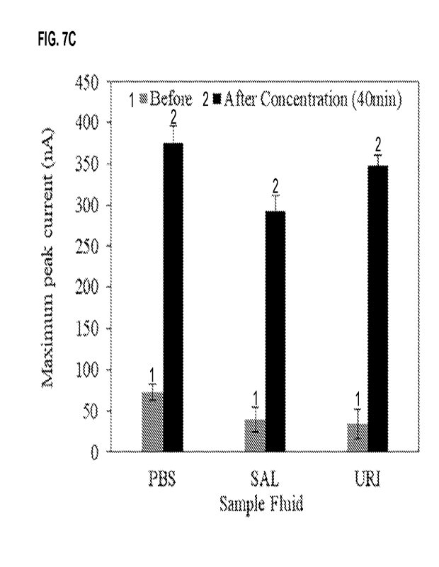
FIG. 7A to FIG. 7C show the detection of pyocyanin, a biomarker for
Pseudomonas aeruginosa infection, in urine
samples spiked with pyocyanin. FIG. 7A shows a photographs of the complete
system of an osmotic system that was
constructed for electrochemical measurements of pyocyanin. FIG. 7B shows a
square-wave voltammetry (SWV) scan
before and after 40 min of osmotic concentration for urine spiked with 1 pM
pyocyanin. FIG. 7C shows that PBS, saliva
(SAL), and urine (URI) spiked with 1 pM pyocyanin showed the pyocyanin peak
current increased by 350-400% after
40 min of forward osmosis.
FIG. 8 shows the detection of Pseudomonas aeruginosa urine samples using the
electrochemical sensors disclosed
herein.
DETAILED DESCRIPTION
The present disclosure provides, in part, a device capable of
contemporaneously detecting several pathogens in a
matter of minutes. The device comprises electrochemical sensor array
comprising a plurality of electrochemical
sensors. Each electrochemical sensor is capable of detecting a different
pathogen. This device is potentially very useful
for identifying urinary tract infection in catheterized individuals, or to
detect a wound infection.
Urinary tract infections (UTIs) are one of the most common health care
acquired infections in the US. Up to 70-80% of
these infections can be attributed to the use of an indwelling urinary
catheter. For patients in long term care (LTC)
facilities with indwelling urinary catheters (I UC), the risk of developing
CAUTI is time-dependent, increasing at a rate
of 3% to 8% each day of IUC use, reaching 100% prevalence at 30 days.
Complications from untreated catheter
associated urinary tract infections (CAUTI) include more serious bladder and
kidney infections, which can lead to
bacteremia and sepsis.
There is currently no simple way to quickly determine or monitor for a
catheter associated urinary tract infection or
impending catheter blockage. A urinalysis can be performed by a trained
technician if the urine appears cloudy, yet
this type of test only indicates the presence of blood cells, which may be
present due to other issues not relevant to a
urinary tract infection. Culturing the urine to determine a bacterial
infection takes multiple days, by which point the
patient is likely suffering from discomfort and potentially further
complications. In many cases, an antimicrobial will be
prescribed before positive diagnosis of a UTI, even though a UTI may not
exist, and is especially prevalent in nursing
home residents with advanced dementia that ultimately do not meet minimum
criteria for antimicrobial initiation. The
over-prescription and misuse of antibiotics is the leading cause of
antimicrobial resistance according to the World
Health Organization (Antibiotic Resistance, 2018). A simple, cheap, and
effective way to continuously monitor for
bacterial infections in catheterized patients would help prevent these issues
and result in better patient outcomes
The device disclosed herein is useful for preventing bacteremia and sepsis
based on quick identification of catheter
associated urinary tract infections (CAUTI).
8
CA 03205285 2023-7- 14
WO 2022/155286
PCT/US2022/012244
The present disclosure is based, in part, on the discovery that multiple
electrochemical sensor arrays may be combined
in a device to contemporaneously, and quickly determine presence of multiple
pathogens in a biological sample.
Provided herein is a device capable of contemporaneously detecting several
pathogens in a matter of minutes. The
device comprises electrochemical sensor array comprising a plurality of
electrochemical sensors. Each electrochemical
sensor is capable of detecting a different pathogen.
Provided herein is a device capable of contemporaneously detecting several
pathogens via an electrochemical sensor
array. In embodiments, the device is capable of analyzing urine in a
collection bag or a catheter bag during use, or
analyzing wound exudate in a dressing or wound exudate collection container
without the need for sample collection
or laboratory tests. In embodiments, the device is capable of
contemporaneously detecting the presence and/or the
amount of and/or the number of viable cells of a plurality of pathogens. In
embodiments, the ability to detect and
quantify the level of bacterial infections provides a guidance for specific
treatment, fewer complications, and limit the
spread of bacterial infections to the bladder and kidneys that can lead to
more extreme complications like sepsis.
In one aspect, the current disclosure relates to a device for detecting an
infection in a subject comprising an
electrochemical sensor array, wherein electrochemical sensor array comprises a
plurality of electrochemical sensors,
wherein each electrochemical sensor comprises a working electrode, a reference
electrode, and a counter electrode,
wherein the electrochemical sensor array is fluidically connected to a wound
exudate in a wound dressing or a wound
exudate collection container, a wound exudate collection container of negative
pressure wound therapy, a fluid
collection container of negative pressure wound therapy with instillation, or
urine in a catheter bag or a urine collection
bag.
Electrochemical Sensor Array
In embodiments, the device for detecting an infection in a subject comprising
an electrochemical sensor array, wherein
electrochemical sensor array comprises a plurality of electrochemical sensors.
A schematic representation of a device
of present disclosure is shown in FIG. 1. In the non-limiting embodiment of
FIG. 1, the device is connected in-line with
a urine collection bag. In this non-limiting example, the sensor reader is
designed to wirelessly transmit the results to
a smart phone app, which depicts which, if any, of the pathogens are present,
along with their infection levels. In
embodiments, the device comprising an electrochemical sensor array is embedded
inside a catheter collection bag. In
embodiments, the device is capable of contemporaneously detecting the presence
and/or the amount of and/or the
number of viable cells of uropathogenic bacteria in human urine samples. In
embodiments, the device comprising an
electrochemical sensor array is embedded inside other instruments such as a
urine collection bag, a catheter bag, a
wound dressing, and a wound exudate collection bag.
In embodiments, the device is capable of continuous and contemporaneous
monitoring of the presence and/or the
amount of and/or the number of viable cells of multiple pathogens. In
embodiments, the device is capable of providing
9
CA 03205285 2023-7- 14
WO 2022/155286
PCT/US2022/012244
rapid, sensitive, simple to use, and inexpensive diagnosis of urinary tract
infection, wound infection, etc. for example,
in embodiments, the device is capable of detecting some of the most common
bacteria found in CAUTI, such as
Escherichia coli, Proteus mirabilis, and Pseudomonas aeruginosa. In
embodiments, these bacterial species may be
rapidly identified by detecting and quantifying specific metabolites that the
bacteria produce using electrochemical
voltammetry methods. In embodiments, the metabolites are secreted. In
embodiments, the detection by the device
disclosed herein is faster and easier than culturing or FOR-based techniques.
In embodiments, the instrumentation for
running the electrochemical tests along with the sensors consists of very
simple and low-cost electronics and materials.
In embodiments, In embodiments, the detection by the device disclosed herein
has less false-negative rates compared
to other rapid UTI detection methods such as dip-sticks.
The secreted metabolites are present in much larger quantities in the urine
compared to the number of actual bacterial
cells.
In embodiments, the device disclosed herein combines several specific and
individual tests into one simple multiplexed
disposable sensor that is capable of specifically detect four different
pathogens simultaneously. In embodiments, the
device disclosed herein is a miniature product. In embodiments, the device
disclosed herein is a battery-powered
sensor array reader with 1/11I-Fl capabilities that powers the sensors in the
urine collection bag of bedridden patients
and transmits warning signals to caregivers. In embodiments, the device
disclosed herein comprises an USB tethered
reader that is capable of transmitting test results. In embodiments, the
device disclosed herein is capable of wirelessly
transmitting test results a cellular phone.
In embodiments, the devise will further comprise a reader. In embodiments, the
devise is calibrated with spiked
samples. In embodiments, the calibration comprises bacterial counts to signal
response. In embodiments, a calibrated
device is capable of quantification the level of infection. In embodiments, a
calibrated device is capable of determining
true infections versus non-threatening asymptomatic or background bacterial
presence.
An illustrative embodiments of the electrochemical sensor array is shown in
FIG. 4A to FIG. 4C. As shown in FIG. 4A,
the device disclosed herein may be mounted inside a urine collection bag. In
embodiments, the wires may be connected
to a reader, which wirelessly transmits signal to an external device, such as
an app on a wireless device such as a
cellphone. FIG. 4B shows an illustrative embodiment of the current technology
featuring a sensor array mounted on a
wound dressing. FIG. 4C shows an illustrative embodiment of the current
technology featuring a sensor array mounted
inside a wound exudate collection container. In any of the embodiments
disclosed here, wires may route out of the
urine collection bag, the bandage or the wound exudate collection container
with a device mounted in it, as shown in
FIG. 4D. in embodiments, the wires may be connected to a reader.
FIG. 4D to FIG. 4G illustrative function of the electrochemical sensor array.
FIG. 4E shows an illustrative
electrochemical sensor array capable of detecting the redox molecules or
quorum sensing molecules (QSM), which
CA 03205285 2023-7- 14
WO 2022/155286
PCT/US2022/012244
are indicated by squares, diamonds, x and triangles, produced by
Staphylococcus, E. coli, Pseudomonas and
Klebsiella, which are diagrammatically shown in the bubble, representing a
biological fluid in contact with the
electrochemical sensor array. QSMs are unique to each bacteria. In
embodiments, the electrochemical sensor-based
test for a given QSM is specific for the bacterium that produces it. QSMs are
produced in huge quantity, and in quantities
proportionate to the cell number of the bacterium producing it. In
embodiments, the electrochemical sensor-based test
for a given QSM detects the presence and/or the amount of and/or the number of
viable cells of the bacterium producing
the QSM. QSMs are only present during infections. In embodiments, the
electrochemical sensor-based test for a given
QSM produces a signal only when infection is present. Thus, in embodiments,
the electrochemical sensor-based test
for a given QSM may be used to determine administration of a treatment,
continuation of the treatment and/or efficacy
of the treatment. In embodiments, QSMs are detected based on the
electrochemical signature of the QSM. In
embodiments, a QSM is detected based on binding of the QSM to an aptamer.
Aptamers are specific to each QSM.
FIG. 4F shows the lack of signal when no redox label target is present, and a
signal produced by a QSM target. FIG.
4G shows an exemplary readout produced by the electrochemical sensor array.
The Table below shows exemplary non-limiting target molecules and pathogens
that may be detected using
electrochemical sensor arrays in a device disclosed here:
Bacteria Target Molecule Chemical Structure
Pseudomonas 30-C12-Homoserine
aeruginosa Lactone (30X0)
Proteus putrescine
taw
Eschenchia coil Shiga toxin
õ
"1-eiN, Rik
Klebsiella aerobactin
pneumonia . N. =
.9,s1
6 k " ,$
11
CA 03205285 2023-7- 14
WO 2022/155286
PCT/US2022/012244
Staphylococcus Auto Inducing Peptide-1
aureus (AIP-1)
Enterococcus Gelatinase Biosynthesis
faecalis Activating Peptide
(GBAP) 0 a
r
Acinetobacter EppR (Protein)
baumannii
Streptococcus Short Hydrophobic DILIIVGG
agalactiae Peptide 3 SHP3 (also
known as SHP1520)
Staphylococcus AIP RIPTSTGFF
pseudintermedius
Staphylococcus AIP 2 DSVCASYF
epidermidis
Uropathogenic Enterobactin
Escherichia coli
8 k
µ4,
The Table below shows additional exemplary non-limiting target molecules and
as applicable, pathogens being
detected, that may be detected using electrochemical sensor arrays in a device
disclosed here:
Target Molecule Notes
calprotectin fecal samples
farnesol Candida alb/cans
tyrosol Candida alb/cans
toxin 13 C. cliff issue
tnf-alpha sub picomolar detection limit needed
auto-inducer 2 general bacterial marker
cholerae autoinducer 1 Vibrio cholerae
12
CA 03205285 2023-7- 14
WO 2022/155286
PCT/US2022/012244
4, 5-d ihyd roxy-2, 3-pentanedione (DPD) Salmonella enterica
In embodiments, the electrochemical sensor comprises a working electrode, a
reference electrode, and a counter
electrode.
In embodiments, the working electrode material of one or more sensors is
selected from gold (Au), silver (Ag), platinum
(Pt), indium tin oxide (ITO), carbon, multi-walled carbon nanotubes, single-
walled carbon nanotubes, carbon
nanofibers, graphene, carbon-platinum composites, multi-walled carbon
nanotubes with gold nanoparticles, and any
combination thereof. In embodiments, the working electrode has a diameter
between about 0.1 mm and about 10 mm,
optionally between about 1 mm and about 5 mm. In embodiments, the working
electrode has a diameter between about
1.5 mm and about 4 mm.
In an illustrative embodiment, the working electrode comprises about 1.5 mm
gold screen-printed at elevated
temperature. In another illustrative embodiment, the working electrode
comprises about 1.5 mm platinum. In another
illustrative embodiment, the working electrode has electrodeposition of gold
to coat copper electrodes exposed on a
printed circuit board. In another illustrative embodiment, the working
electrode has screen-printed carbon paste to coat
copper electrodes exposed on a printed circuit board. In another illustrative
embodiment, the working electrode
comprises about 4 mm gold. In yet another illustrative embodiment, the working
electrode comprises about 1.5 mm Au
screen-printed at low temperature. In yet another illustrative embodiment, the
device may comprise an oxidizing and a
reducing working electrode, for amplifying the signal, and the two working
electrodes consist of gold and or platinum.
In embodiments, the working electrodes may make up a wall or part of a wall of
a channel, such as a microfluidic
channel or a nanofluidic channel, into which the fluid sample is introduced
and within which the redox reaction takes
place. In embodiments, the oxidizing electrode and the reducing electrode are
separated by a distance of about 20 nm
to 1 mm or greater. In embodiments, the distance between the oxidizing
electrode and the reducing electrode is from
20 nm to about 100 nm, or from about 20 nm to about 40 nm, or from about 40 nm
to about 60 nm, or from about 60
nm to about 80 nm, or from about 80 nm to about 100 nm, or from about 100 nm
to about 150 nm, or from or from
about 50 nm to about 500 nm, or from about 100 nm to about 1 pm, or from about
500 nm to about 5 pm, or from
about 1 pm to about 10 pm, or from about 5 pm to about 50 pm, or from about 10
pm to about 100 pm, or from about
50 pm to about 500 pm, or from about 100 pm to about 1 mm, or greater. In
embodiments, the distance between the
oxidizing electrode and the reducing electrode is from 20 nm to about 100 nm,
or from about 20 nm to about 40 nm, or
from about 40 nm to about 60 nm, or from about 60 nm to about 80 nm, or from
about 80 nm to about 100 nm, or from
about 100 nm to about 150 nm.
The surface area of the working electrodes can be selected to accommodate a
desired size of the device. Without
being bound by theory, larger surface area generally improves the signal and
sensitivity of the device. For example, in
different embodiments, the surface area of each working electrode can be about
100, about 200, about 300, about 400,
13
CA 03205285 2023-7- 14
WO 2022/155286
PCT/US2022/012244
about 500, about 800, about 1000, about 2000, about 3000, about 5000, about
10000, about 50000, about 100000,
about 200000, or about 500000 nm2, or about 1, about 2, about 5, about 10,
about 50, about 100, about 200, about
300, about 400, about 500, about 800, about 1000, about 2000, about 3000,
about 5000, about 10000, about 50000,
about 100000, about 200000, or about 500000 pm2, or about 1, about 2, about 4,
about 7 mm2 or greater. In different
embodiments, the surface area of each working electrode can be about 100,
about 200, about 300, about 400, about
500, about 800, about 1000, about 2000, about 3000, about 5000, about 10000,
about 50000, about 100000, about
200000, or about 500000 nm2, or about 1, about 2, about 5, about 10 pm2, or
greater.
Any reference electrode that is compatible with the chosen working electrode
may be used. In embodiments, the
reference electrode material of one or more sensors is selected from silver
(Ag), silver chloride (AgCI), and platinum
(Pt). In embodiments, the reference electrode comprises silver (Ag). In
embodiments, the reference electrode
comprises Ag/AgCl.
In embodiments, the electrochemical sensor further comprises a counter
electrode. In embodiments, the counter
electrode of each sensor is identical to the working electrode.
In some embodiments, the electrochemical sensor is a microfluidic sensor
comprises a working electrode, a counter
electrode and a reference electrode. In these embodiments, the current flows
the current flows through the working
electrode and the counter electrode. In some embodiments, the counter
electrode functions as a cathode and the
working electrode is operating as an anode. In alternative embodiments, the
counter electrode functions as an anode
and the working electrode is operating as a cathode. In some embodiments, the
counter electrode has a surface area
much larger than that of the working electrode.
In embodiments, the electrochemical sensor may be used for measuring an
electrochemical reaction taking place at
the working electrode at a well-defined potential. In embodiments, the
electrochemical reaction taking place at the
working electrode is measured in comparison to the electrochemical reaction
taking place at the reference electrode.
In embodiments, the electrochemical sensor may be used for measuring the redox
peak present at a given potential,
which is sensitive to pH of the solution. In embodiments, the redox peak
present at a given potential at the working
electrode is measured in comparison to the redox peak present at the reference
electrode.
In embodiments, the electrochemical sensor may be used for measuring the
binding of a molecule to an aptamer,
causing a change in electrical current passing through the electrode. In
embodiments, the change in electrical current
passing through the electrode working electrode is measured in comparison to
the electric current at the reference
electrode.
The Table below shows exemplary non-limiting aptamer sequences that may be
used for detecting the presence,
absence or amount of Shiga in the electrode electrochemical sensor arrays in a
device disclosed here. The sequences
of the aptamers are indicated in boldface-underlined font.
14
CA 03205285 2023-7- 14
WO 2022/155286
PCT/US2022/012244
Target Aptamer sequence
Molecul
Shi g a
HACCCCTGCATCCTTTGCTGGGGTAACTAGCATTCATTTCCCACACCCGTCCCGTCCATATAGT
CTAGAGGGCCCCAGAAT (SEQ ID NO: 1)
Shi g a (-
)ATTCTGGGGCCCTCTAGACTATATGGACGGGACGGGTGTGGGAAATGAATGCTAGTTACC
CCAGCAAAGGATGCAGGGGT (SEQ ID NO: 2)
In embodiments, the aptamers are generated using a DNA capture element sensing
system. In embodiments, the
aptamers are selected by conventional SELEX. See e.g. Fan et al., Aptamer
Selection Express: A Novel Method for
Rapid Single-Step Selection and Sensing of Aptamers, Journal of Biomolecular
Techniques 19:311-321(2018).
In embodiments, highly enriched unmodified RNA aptamer pools may be cloned,
and -100 clones from each pool may
be sequenced. In embodiments, the individual clones may be classified into
groups from m-SELEX and groups from
p-SELEX based on the alignments of individual aptamer sequences. For example,
in embodiments, m-SELEX
sequences may be grouped into groups mA, mB, mC, mD, etc., based on sequence
alignment. Similarly, in
embodiments, p-SELEX sequences may be grouped into groups pA, pB, pC, pD,
etc., based on sequence alignment.
The Table below shows exemplary non-limiting m-SELEX sequences and p-SELEX
sequences of RNA aptamers that
are classified as above. See e.g., Challa et al., Selective Evolution of
Ligands by Exponential Enrichment to Identify
RNA Aptamers against Shiga Toxins, Journal of Nucleic Acids, 2014: 214929
(2014). The sequence of 19 bases is
identical among groups mA, mB, pA and pC are indicated by underline. In
embodiments, these sequences may be
used for detecting the presence, absence or amount of Shiga in the electrode
electrochemical sensor arrays in a device
disclosed here.
Group Sequence
mA ATTAGCTATCTTCCACGATTCGATCAGGCAGTACGTCGT (SEQ ID NO: 3)
mB ACAGTTATCCGACTGCTATTCGATCAGGCAGTACGTAGC (SEQ ID NO: 4)
nnC CAGGCTGTTCTGACGCATAAGGAATGCGCTGTTGCAGAG (SEQ ID NO: 5)
nnD TTGGTCCTGCTTTGGATAGTCGCGAAAGGGGTGCCACTG (SEQ ID NO: 6)
m-Singles Orphan sequences
pA ACAGTTATCCGACTGCTATTCGATCAGGCAGTACGTAGC (SEQ ID NO: 7)
pB ACCGAGCGGTTTTACGTCTCAAGTAGTATCCCGTTTTGC (SEQ ID NO: 8)
pC ATTAGCTATCTTCCACGATTCGATCAGGCAGTACGTCGT (SEQ ID NO: 9)
pD TTGCCATCCTGTACTATGCTCTATCGGGCGGTTTAGTGATCCTTCGTCCAACTATC
(SEQ
ID NO: 10)
p-Singles Orphan sequences
Orphan sequences are the sequences that are seen only in one isolate.
In embodiments, the electrochemical sensor thereby facilitates an
electrochemical detection of a predetermined redox-
active compound associated with the infection. In embodiments, the
electrochemical detection of the predetermined
CA 03205285 2023-7- 14
WO 2022/155286
PCT/US2022/012244
redox-active compound associated with the infection (without limitation, e.g.,
pyocyanin) and thereby detect the
presence of a specific pathogen (without limitation, e.g., Pseudomonas
aeruginosa). In embodiments, the defined
potential of the working electrode may be varied, and the response from the
electrochemical reaction is seen from the
current of the working electrode. In embodiments, the electrochemical sensor
comprises a second working electrode.
In embodiments, the second working electrodes with respect to one or more of
surface area, size, material, and coating.
In embodiments, the electrochemical sensor may include an oxidizing working
electrode and a reducing working
electrode. In embodiments, the concentration of a target molecule and/or a
metabolic activity associated with the
infection is measured as current flow through the oxidizing electrode and the
reducing electrode. In embodiments, the
working electrode is one of an oxidizing electrode and a reducing electrode,
and the second working electrode is the
other of the oxidizing electrode and the reducing electrode. A potential
suitable for oxidizing the target molecule and/or
the metabolic activity associated with the infection is applied at the
oxidizing electrode and a potential suitable for
reducing the target molecule and/or the metabolic activity associated with the
infection is applied at the reducing
electrode.
In embodiments, a given target molecule and/or a metabolic activity associated
with the infection electrochemically
reacts differently on different electrode surfaces. Thus, different electrode
materials and geometries used for chemical
detection will give different results. Accordingly, in embodiments, the sensor
array increases the sensitivity and
specificity of the measurement and reduces the noise from other substances
present in a biological sample. In
embodiments, the sensor array may comprise two or more sensors, wherein each
sensor comprises a working
electrode that differs from the other working electrodes with respect to at
least one of the following characteristics:
surface area, size, material, and coating.
The electrochemical measurement can be made in any suitable manner. In
embodiments, the electrochemical
measurement may made by squarewave voltammetry, linear sweep voltammetry,
staircase voltammetry, cyclic
voltammetry, normal pulse voltammetry, differential pulse voltammetry, and
chronoamperometry. In embodiments, the
electrochemical measurement is square wave voltammetry and the current flow is
measured in response to one or
more square wave potentials. In embodiments, optionally cyclic voltammetry is
used and the working electrode potential
is ramped linearly versus time. In embodiments, the potential is ramped
linearly up, and when a set potential is reached,
the potential is ramped in the opposite direction to the initial potential,
and the cycle is repeated. In embodiments, the
working electrode potential include linear sweep voltammetry, staircase
voltammetry, square-wave voltammetry, and
differential pulse voltammetry.
In embodiments, the presence, absence or amount of the compound is measured as
current flow through the working
electrode. In embodiments, the presence, absence or amount of compound is
measured as current flow through the
oxidizing electrode and the reducing electrode.
16
CA 03205285 2023-7- 14
WO 2022/155286
PCT/US2022/012244
In embodiments, the electrochemical sensor array disposable. In embodiments,
the sensor array is integrated inside
of the sterile bag. In embodiments, a single wire exits through the drainage
cap to connect to a reader, optionally, the
reader is battery powered. In embodiments, similarly to currently sensor array
comprises integrated temperature and
conductance sensors.
In one aspect, the current disclosure relates to a device for detecting an
infection in a subject comprising an
electrochemical sensor array, wherein electrochemical sensor array comprises a
plurality of electrochemical sensors,
wherein each electrochemical sensor comprises a working electrode, a reference
electrode, and a counter electrode,
wherein the electrochemical sensor array is fluidically connected to a wound
exudate in a wound dressing or a wound
exudate collection container, a wound exudate collection container of negative
pressure wound therapy, a fluid
collection container of negative pressure wound therapy with instillation, or
urine in a catheter bag or a urine collection
bag.
In embodiments, the electrochemical sensor array further comprises a sensor
selected from a pH sensor and a
temperature sensor. In embodiments, the electrochemical sensor array detects a
change in pH, a change in
temperature, an electrochemical reaction, binding to an aptamer, a change in
color, or the combination of any two or
more thereof. In embodiments, the device is capable of contemporaneously
detecting at least two, or at least three, or
at least 4, or at least 5, oral least 6, or at least 7, or at least 8, or at
least 9, or at least 10, or at least 11, or at least 12,
or at least 13, or at least 14, or at least 15, or at least 16 signals. In
embodiments, the signal is associated with a
pathogen. In embodiments, each electrochemical sensor is capable of
independently performing an electrochemical
measurement. In embodiments, the electrochemical measurement is selected from
square wave voltammetry, linear
sweep voltammetry, staircase voltammetry, cyclic voltammetry, normal pulse
voltammetry, differential pulse
voltammetry, and chronoamperometry. In embodiments, the electrochemical
measurement is square wave
voltammetry. In embodiments, the electrochemical measurement is measurement of
a current flow. In embodiments,
the current flow is measured in response to one or more square wave
potentials.
In embodiments, the working electrode is comprised of gold (Au), silver (Ag),
platinum (Pt), indium tin oxide (ITO),
carbon, carbon nanotubes, carbon nanofibers, graphene, carbon-platinum
composites, carbon nanotubes with gold
nanoparticles, or any combination thereof. In embodiments, the electrochemical
sensors further comprise a reference
electrode, optionally wherein the reference electrode is comprised of silver
(Ag), silver chloride (AgCI), and platinum
(Pt), and any combination thereof. In embodiments, each electrochemical sensor
comprises a second working
electrode. In embodiments, the second working electrode is comprised of gold
(Au), silver (Ag), platinum (Pt), indium
tin oxide (ITO), carbon, carbon nanotubes, carbon nanofibers, graphene, carbon-
platinum composites, carbon
nanotubes with gold nanoparticles, or any combination thereof. In embodiments,
the working electrode is one of an
oxidizing electrode and a reducing electrode, and the second working electrode
is the other of the oxidizing electrode
and the reducing electrode.
17
CA 03205285 2023-7- 14
WO 2022/155286
PCT/US2022/012244
In embodiments, the device is capable of detecting an infection caused by a
pathogen. In embodiments, the pathogen
is selected a bacterium, a fungus and a parasite. In embodiments, the
bacterium is selected from Pseudomonas
aeruginosa, Proteus mirabilis, Escherichia coli, Klebsiella pneumoniae, other
Klebsiella species, Staphylococcus
aureus, Enterococcus faecalis, other Enterococcus species, Acinetobacter
baumannii, Streptococcus group A species,
Streptococcus Group B species, Staphylococcus epidermidis, Pseudomonas
aeruginosa, Clostridium difficile, and
Salmonella enterica. In embodiments, the fungus is Candida albicans. In
embodiments, the parasite is Giardia, a fecal
float worm, fecal roundworm and fecal flatworm.
In embodiments, the electrochemical sensor array is capable of
contemporaneously detecting the presence or absence
of at least two, or at least 3, or at least 4, or at least 5, or at least 6,
or at least 7, or at least 8, or at least 9, or at least
pathogens. In embodiments, wherein the device detects a target molecule,
and/or a metabolic activity of the
pathogen. In embodiments, the target molecule is a quorum sensing molecule. In
embodiments, the target molecule is
a redox molecule.
In embodiments, the metabolic activity causes breakdown of a basic molecule.
In embodiments, the metabolic activity
is a urease activity. In embodiments, the metabolic activity changes pH of the
urine or the wound exudate. In
embodiments, the change in pH is an increase in pH. In embodiments, the
metabolic activity is a bacterial urease
activity. In embodiments, the bacterial urease activity generates ammonia from
the urea, and thereby increasing the
pH. Therefore, in embodiments, the urease activity of a bacterium (without
limitation, e.g. Proteus) makes the biological
fluid (without limitation, e.g. urine) alkaline, thereby allowing the
detection of the presence or absence of an infection
by the bacterium. It is noted that the alkaline conditions stimulate the
formation of crystals of calcium and magnesium
phosphate and the development of a crystalline bio film, which eventually
blocks the flow of urine from the bladder. In
embodiments, the device detects the presence, absence or amount of the urease
activity of the pathogen.
In embodiments, the device detects the presence, absence or amount of the
target molecule and/or the metabolic
activity and/or the metabolic activity of the pathogen. In embodiments, the
presence, absence or amount of the target
molecule and/or the metabolic activity is measured as current flow through the
working electrode. In embodiments, the
presence, absence or amount of the target molecule and/or the metabolic
activity is measured as current flow through
the oxidizing electrode and the reducing electrode.
In embodiments, the presence, absence or amount of the target molecule and/or
the metabolic activity is measured as
a change in pH. . In embodiments, the target molecule is selected from quorum
sensing molecules (without limitations,
e.g., pyocyanin, E. coli autoinducer-2 (AI-2), N-Acyl Homoserine Lactones
(AHL)), siderophores (without limitations,
e.g. enterobactin, aerobactin, vibriobactin, salmochelin, pyoverdine, and
pyochelin), cyclic signaling peptides (without
limitations, e.g. Staphylococcus aureus autoinducing peptide (AIP), including
AIP variants I to IV, and Enterococcus
faecalis gelatinase biosynthesis activating pheromone (GBAP)), and
autoinducers (without limitations, e.g. acylated
homoserine lactones (AHLs), including N-(3-oxododecanoyI)-homoserine lactone
and N-(butyryI)-homoserine lactone,
18
CA 03205285 2023-7- 14
WO 2022/155286
PCT/US2022/012244
2-hepty1-3-hydroxy-4-quinolone (PQS), AIP variants Ito IV). In embodiments,
the target molecule is selected from 30-
C12-homoserine lactone (30X0), putrescine, Shiga toxin, aerobactin, auto
inducing peptide-1 (AIP-1), gelatinase
biosynthesis activating peptide (GBAP), EppR (Protein), short hydrophobic
peptide 3 SHP3 (also known as SHP1520),
autoinducing peptide (AIP), autoinducing peptide 2 (AIP 2), pyocyanin,
enterobactin, tyrosol and farnesol. In
embodiments, the presence of the target molecule is indicative of a presence
and/or an amount of and/or a number of
viable cells of the pathogen. In embodiments, the pathogen is selected from
Pseudomonas aeruginosa, Proteus
mirabilis, Escherichia coli, Klebsiella pneumoniae, other Klebsiella species,
Staphylococcus aureus, Enterococcus
faecalis, other Enterococcus species, Acinetobacter baumannii, Streptococcus
group A species, Streptcoccus group
B species, Staphylococcus epidermidis, Clostridium difficile, Salmonella
enter/ca, Candida albicans, Clyptococcus
neoformans, and Aspergillus species.
In embodiments, the presence, absence or amount of: 30-C12-homoserine lactone
(30X0) is indicative of the
presence and/or the amount of and/or the number of viable cells of Pseudomonas
aeruginosa, putrescine is indicative
of the presence and/or the amount of and/or the number of viable cells of
Proteus mirabilis, Shiga toxin is indicative of
the presence and/or the amount of and/or the number of viable cells of
Escherichia coli, aerobactin is indicative of the
presence and/or the amount of and/or the number of viable cells of Klebsiella
and/or E. coil, auto inducing peptide-1
(AIP-1) is indicative of the presence and/or the amount of and/or the number
of viable cells of Staphylococcus aureus,
gelatinase biosynthesis activating peptide (GBAP) is indicative of the
presence and/or the amount of and/or the number
of viable cells of Enterococcus faecalis, EppR protein is indicative of the
presence and/or the amount of and/or the
number of viable cells of Acinetobacter baumannii, short hydrophobic peptide 3
SHP3 (SHP1520) is indicative of the
presence and/or the amount of and/or the number of viable cells of
Streptococcus agalactiae, autoinducing peptide
(AIP) is indicative of the presence and/or the amount of and/or the number of
viable cells of Staphylococcus
pseudintermedius, autoinducing peptide 2 (AIP 2) is indicative of the presence
and/or the amount of and/or the number
of viable cells of Staphylococcus epidermidis, pyocyanin is indicative of the
presence and/or the amount of and/or the
number of viable cells of Pseudomonas aeruginosa, tyrosol is indicative of the
presence and/or the amount of and/or
the number of viable cells of Candida albicans, farnesol is indicative of the
presence and/or the amount of and/or the
number of viable cells of Candida albicans, and/or enterobactin is indicative
of the presence and/or the amount of
and/or the number of viable cells of uropathogenic Escherichia co/i.
In embodiments, the device is electrically connected or connectable to a
reader. In embodiments, the reader provides
an output of a presence and/or an amount of and/or a number of viable cells of
a pathogen. In embodiments, the reader
is capable of transmitting the specific signals to a display device. In
embodiments, the display device is a hand-held
device. In embodiments, the display device is a portable device. In
embodiments, the signal is wirelessly transmitted.
In embodiments, the pathogen is selected from Pseudomonas aeruginosa, Proteus
mirabilis, Escherichia coil,
Klebsiella pneumoniae, other Klebsiella species, Staphylococcus aureus,
Enterococcus faecalis, other Enterococcus
19
CA 03205285 2023-7- 14
WO 2022/155286
PCT/US2022/012244
species, Acinetobacter baumannii, Streptococcus group A species, Streptcoccus
group B species, Staphylococcus
epidermidis, Clostridium difficile, Salmonella enter/ca, Candida alb/cans,
Cryptococcus neoformans, and Aspergillus
species.
In embodiments, the device detects the infection in less than one hour, less
than 45 minutes, or less than 30 minutes,
or less than 15 minutes, or less than 10 minutes, or less than 5 minutes, or
less than 2 minutes or less than 1 minute.
In one aspect, the current disclosure relates to a dressing comprising the
device of any one of the embodiments
disclosed herein, optionally wherein the dressing further comprises oxidized
regenerated cellulose (ORC) and/or
collagen.
Some current attempts to monitor urine collection bags include the measurement
of pH change via a color change
reaction (Milo 2016). However, color-based tests are limited in use due to the
requirement of a visual inspection and
interpretation of the color change as well as not providing any multiplexed
detection. Other collection bag monitoring
technologies only measure urine flow or urine levels in the bag using
electronic conductance measurements, which do
not provide any specific diagnostic information.
In one aspect, the current disclosure relates to a urine collection bag with
an integrated complete infection detection
and monitoring system comprising the device of any one of the embodiments
disclosed herein. In one aspect, the
current disclosure relates to a urine collection bag comprising the device of
any one of the embodiments disclosed
herein.
In one aspect, the current disclosure relates to a catheter bag comprising the
device of any one of the embodiments
disclosed herein.
In one aspect, the current disclosure relates to a negative pressure wound
therapy system comprising a wound
dressing, and a negative pressure source and a wound exudate collection
container, wherein wound exudate collection
container comprises the device of any one of the embodiments disclosed herein,
optionally wherein the dressing
comprises oxidized regenerated cellulose (ORC) and/or collagen.
In one aspect, the current disclosure relates to a negative pressure wound
therapy with installation system comprising
a wound dressing, an instillation fluid, an instillation pump, and a negative
pressure source and a wound exudate
collection container, wherein wound exudate collection container comprises the
device of any one of the embodiments
disclosed herein, optionally wherein the dressing comprises oxidized
regenerated cellulose (ORC) and/or collagen.
Methods of Detecting an Infection
In one aspect, the current disclosure relates to a method of detecting an
infection in a biological sample of a subject,
the method comprising (i) contacting the biological sample from the subject
with a device for detecting an infection,
wherein the device comprises an electrochemical sensor array; and (ii)
measuring a presence, absence or amount of
CA 03205285 2023-7- 14
WO 2022/155286
PCT/US2022/012244
one or more target molecule and/or a metabolic activity within the biological
sample, wherein the target molecule and/or
the metabolic activity is associated with the infection, wherein the
electrochemical sensor array performs the
measuring. In embodiments, the biological sample is collected in a collection
device. In embodiments, collection device
is selected from a catheter bag, a colostomy bag, a urine collection bag, a
wound dressing, and a wound exudate
collection container. In embodiments, the biological fluid is selected from a
body fluid selected from blood, plasma,
serum, semen, lacrimal fluid, tears, sputum, saliva, sweat, urine,
cerebrospinal fluid, peritoneal fluid, pleural
fluid, biopsy sample, feces, lymph, gynecological fluid, skin swab, vaginal
swab, oral swab, nasal swab, hair,
washing or lavage such as a ducta avage and broncheoalveolar lavage.
In one aspect, the current disclosure relates to a method of detecting an
infection in urine of a subject, the method
comprising (i) contacting urine from the subject with a device for detecting
an infection, wherein the device comprises
an electrochemical sensor array; and (ii) measuring a presence, absence or
amount of one or more target molecule
and/or a metabolic activity within the urine sample, wherein the target
molecule and/or the metabolic activity is
associated with the infection, wherein the electrochemical sensor array
performs the measuring. In embodiments, the
urine is collected in a catheter bag or a urine collection bag. In
embodiments, the urinary tract infection is catheter
associated urinary tract infections (CAUTI).
In one aspect, the current disclosure relates to a method of detecting a
urinary tract infection in a subject, the method
comprising (i) contacting urine from the subject with a device for detecting
an infection, wherein the device comprises
an electrochemical sensor array; and (ii) measuring a presence, absence or
amount of one or more target molecule
and/or a metabolic activity within the urine sample, wherein the target
molecule and/or the metabolic activity is
associated with the infection, wherein the electrochemical sensor array
performs the measuring. In embodiments, the
urine is collected in a catheter bag or a urine collection bag. In
embodiments, the urinary tract infection is catheter
associated urinary tract infections (CAUTI).
In one aspect, the current disclosure relates to a method of detecting a wound
infection in a subject, the method
comprising (i) contacting wound exudate from the subject with a device for
detecting an infection, wherein the device
comprises an electrochemical sensor array; and (ii) measuring a presence,
absence or amount of one or more target
molecule and/or a metabolic activity within the wound exudate, wherein the
target molecule and/or the metabolic activity
is associated with the infection, wherein the electrochemical sensor array
performs the measuring. In embodiments,
the wound exudate is collected in a wound dressing or a wound exudate
collection container.
In one aspect, the current disclosure relates to a method of detecting a wound
infection in a subject, the method
comprising: (i) administering a dressing to a wound, optionally wherein the
dressing comprises oxidized regenerated
cellulose (ORC) and/or collagen, (ii) applying a negative pressure to the
wound, (iii) collecting wound exudate in a
wound exudate collection container, (iv) contacting wound exudate from a wound
dressing or a wound exudate
21
CA 03205285 2023-7- 14
WO 2022/155286
PCT/US2022/012244
collection container with a device for detecting an infection, wherein the
device comprises an electrochemical sensor
array; and (v) measuring a presence, absence or amount of one or more target
molecule and/or a metabolic activity
within the wound dressing or the wound exudate, wherein the target molecule
and/or the metabolic activity is associated
with the infection, wherein the electrochemical sensor array performs the
measuring.
In one aspect, the current disclosure relates to a method of detecting a
gastro-intestinal tract infection in a subject, the
method comprising: (i) contacting stool sample from the subject with a device
for detecting an infection, wherein the
device comprises an electrochemical sensor array; and (ii) measuring a
presence, absence or amount of one or more
target molecule and/or a metabolic activity within the stool sample, wherein
the target molecule and/or the metabolic
activity is associated with the infection, wherein the electrochemical sensor
array performs the measuring.
In one aspect, the current disclosure relates to a method of detecting a
gastro-intestinal tract infection in a subject, the
method comprising: (i) contacting biopsy sample from the subject with a device
for detecting an infection, wherein the
device comprises an electrochemical sensor array; and (ii) measuring a
presence, absence or amount of one or more
target molecule and/or a metabolic activity within the biopsy sample, wherein
the target molecule and/or the metabolic
activity is associated with the infection, wherein the electrochemical sensor
array performs the measuring. In
embodiments, the biopsy sample is obtained using an endoscopic biopsy.
In any of the embodiments disclosed herein the method further comprises
estimating a number of viable cells of a
pathogen associated with the infection based on the presence, absence or
amount of the target molecule and/or the
metabolic activity. In embodiments, the method informs the withholding of one
or more antibiotics upon a negative test
for infection. In embodiments, the method informs the selection of an
appropriate antibiotic for the infection upon a
positive test for infection. In embodiments, the method further comprises
administering an appropriate antibiotic for the
infection upon a positive test for infection.
In embodiments, the electrochemical sensor array further comprises a sensor
selected from a pH sensor and a
temperature sensor. In embodiments, the electrochemical sensor array detects a
change in pH, a change in
temperature, an electrochemical reaction, binding to an aptamer, a change in
color, and the combination of any two or
more thereof. In embodiments, each electrochemical sensor of the
electrochemical sensor array independently
performs an electrochemical measurement. In embodiments, the electrochemical
measurement is selected from
square wave voltammetry, linear sweep voltammetry, staircase voltammetry,
cyclic voltammetry, normal pulse
voltammetry, differential pulse voltammetry, and chronoamperometry. In
embodiments, the electrochemical
measurement is square wave voltammetry and the current flow is measured in
response to one or more square wave
potentials.
In embodiments, the working electrode is comprised of gold (Au), silver (Ag),
platinum (Pt), indium tin oxide (ITO),
carbon, carbon nanotubes, carbon nanofibers, graphene, carbon-platinum
composites, carbon nanotubes with gold
22
CA 03205285 2023-7- 14
WO 2022/155286
PCT/US2022/012244
nanoparticles, and any combination thereof. In embodiments, the
electrochemical sensor further comprise a reference
electrode, optionally wherein the reference electrode is comprised of silver
(Ag), silver chloride (AgCI), and platinum
(Pt), and any combination thereof.
In embodiments, each electrochemical sensor comprises a second working
electrode, wherein the working electrode
is one of an oxidizing electrode and a reducing electrode, and the second
working electrode is the other of the oxidizing
electrode and the reducing electrode. In embodiments, the second working
electrode is comprised of gold (Au), silver
(Ag), platinum (Pt), indium tin oxide (ITO), carbon, carbon nanotubes, carbon
nanofibers, graphene, carbon-platinum
composites, carbon nanotubes with gold nanoparticles, and any combination
thereof.
In embodiments, the infection is caused by a pathogen. In embodiments, the
pathogen is selected from a bacterium
and a fungus. In embodiments, the bacterium is selected from Pseudomonas
aeruginosa, Proteus mirabilis,
Escherichia coif, Klebsiella pneumoniae, other Klebsiella species,
Staphylococcus aureus, Enterococcus faecalis, other
Enterococcus species, Acinetobacter baumannii, Streptococcus group A species,
Streptcoccus group B species,
Staphylococcus epidermidis, Clostridium difficile, and Salmonella enter/ca. In
embodiments, the fungus is Candida
albicans. In embodiments, the parasite is selected from Giardia, a fecal float
worm, fecal roundworm and fecal flatworm.
In embodiments, the device is capable of contemporaneously detecting at least
two, or at least three, or at least 4
signals. In embodiments, the signal is associated with a pathogen. In
embodiments, the device is capable of
contemporaneously detecting the presence or absence of at least two, or at
least 3, or at least 4, or at least 5, or at
least 6, or at least 7, or at least 8, or at least 9, or at least 10
pathogens.
In embodiments, the device detects a target molecule, and/or a metabolic
activity of the pathogen. In embodiments,
the target molecule is a quorum sensing molecule. In embodiments, the target
molecule is a redox molecule.
In embodiments, the metabolic activity causes breakdown of a basic molecule.
In embodiments, the metabolic activity
is a urease activity. In embodiments, the metabolic activity changes pH of the
urine or the wound exudate. In
embodiments, the change in pH is an increase in pH.
In embodiments, the device detects the presence, absence or amount of the
target molecule and/or the metabolic
activity and/or the metabolic activity of the pathogen. In embodiments, the
presence, absence or amount of the target
molecule and/or the metabolic activity is measured as current flow through the
working electrode. In embodiments, the
presence, absence or amount of the target molecule and/or the metabolic
activity is measured as current flow through
the oxidizing electrode and the reducing electrode. In embodiments, the
presence, absence or amount of the target
molecule and/or the metabolic activity is measured as a change in pH.. In
embodiments, the target molecule is selected
from quorum sensing molecules (without limitations, e.g., pyocyanin, E. coli
autoinducer-2 (AI-2), N-Acyl Homoserine
Lactones (AHL)), siderophores (without limitations, e.g. enterobactin,
aerobactin, vibriobactin, salmochelin, pyoverdine,
and pyochelin), cyclic signaling peptides (without limitations, e.g.
Staphylococcus aureus autoinducing peptide (AIP),
23
CA 03205285 2023-7- 14
WO 2022/155286
PCT/US2022/012244
including AIP variants Ito IV, and Enterococcus faecalis gelatinase
biosynthesis activating pheromone (GBAP)), and
autoinducers (without limitations, e.g. acylated homoserine lactones (AHLs),
including N-(3-oxododecanoyI)-
homoserine lactone and N-(butyryI)-homoserine lactone, 2-hepty1-3-hydroxy-4-
quinolone (PQS), AIP variants Ito IV).
In embodiments, the target molecule is selected from 30-012-homoserine lactone
(30X0), putrescine, Shiga toxin,
aerobactin, auto inducing peptide-1 (AIP-1), gelatinase biosynthesis
activating peptide (GBAP), EppR (Protein), short
hydrophobic peptide 3 SHP3 (also known as SHP1520), autoinducing peptide
(AIP), autoinducing peptide 2 (AHD 2),
pyocyanin, enterobactin, tyrosol and farnesol. In embodiments, the presence of
the target molecule is indicative of a
presence and/or an amount of and/or a number of viable cells of the pathogen.
In embodiments, the pathogen is
selected from Pseudomonas aeruginosa, Proteus mirabilis, Escherichia coli,
Kiebsiella pneumoniae, other Klebsiella
species, Staphylococcus aureus, Enterococcus faecalis, other Enterococcus
species, Acinetobacter baumannii,
Streptococcus group A species, Streptcoccus group B species, Staphylococcus
epidermidis, Clostridium difficile,
Salmonella enterica, Candida alb/cans, Cryptococcus neoformans, and
Aspergillus species.
In embodiments, the presence, absence or amount of: 30-C12-homoserine lactone
(30X0) is indicative of the
presence and/or the amount of and/or the number of viable cells of Pseudomonas
aeruginosa, putrescine is indicative
of the presence and/or the amount of and/or the number of viable cells of
Proteus mirabilis, Shiga toxin is indicative of
the presence and/or the amount of and/or the number of viable cells of
Escherichia coli, aerobactin is indicative of the
presence and/or the amount of and/or the number of viable cells of Klebsiella
and/or E. coli, auto inducing peptide-1
(AIP-1) is indicative of the presence and/or the amount of and/or the number
of viable cells of Staphylococcus aureus,
gelatinase biosynthesis activating peptide (GBAP) is indicative of the
presence and/or the amount of and/or the number
of viable cells of Enterococcus faecalis, EppR protein is indicative of the
presence and/or the amount of and/or the
number of viable cells of Acinetobacter baumannii, short hydrophobic peptide 3
SHP3 (SHP1520) is indicative of the
presence and/or the amount of and/or the number of viable cells of
Streptococcus agalactiae, autoinducing peptide
(AIP) is indicative of the presence and/or the amount of and/or the number of
viable cells of Staphylococcus
pseudintermedius, autoinducing peptide 2 (AIP 2) is indicative of the presence
and/or the amount of and/or the number
of viable cells of Staphylococcus epidermidis, pyocyanin is indicative of the
presence and/or the amount of and/or the
number of viable cells of Pseudomonas aeruginosa, tyrosol is indicative of the
presence and/or the amount of and/or
the number of viable cells of Candida alb/cans, farnesol is indicative of the
presence and/or the amount of and/or the
number of viable cells of Candida alb/cans, and/or enterobactin is indicative
of the presence and/or the amount of
and/or the number of viable cells of uropathogenic Escherichia co/i.
In embodiments, the device is electrically connected or connectable to a
reader. In embodiments, the reader provides
an output of a presence and/or an amount of and/or a number of viable cells of
a pathogen. In embodiments, the reader
is capable of transmitting the specific signals to a display device. In
embodiments, the signal is wirelessly transmitted.
In embodiments, the pathogen is selected from Pseudomonas aeruginosa, Proteus
mirabilis, Escherichia coli,
24
CA 03205285 2023-7- 14
WO 2022/155286
PCT/US2022/012244
Klebsiella pneumonia, other Klebsiella species, Staphylococcus aureus,
Enterococcus faecalis, other Enterococcus
species, Acinetobacter baumannfi, Streptococcus group A species, Streptococcus
Group B species, Staphylococcus
epidermidis, Pseudomonas aeruginosa, Clostridium difficile, and Salmonella
enterica, Borrelia burgdorferi, Candida
albicans, Ctyptococcus neoformans, and Aspergillus species.
In embodiments, the device detects the presence and/or the amount of and/or
the number of viable cells of a pathogen
in less than one hour, less than 45 minutes, or less than 30 minutes, or less
than 15 minutes, or less than 10 minutes,
or less than 5 minutes, or less than 2 minutes or less than 1 minute.
Methods of Selecting A Patient For Therapy
In one aspect, the current disclosure relates to a method of selecting a
catheterized patient having or suspected as
having a urinary tract infection for therapy, the method comprising: (i)
contacting urine from a catheter bag or urine
collection bag collected from a subject with a device of any one of the
embodiments disclosed herein for detecting an
infection, wherein the device comprises an electrochemical sensor array; (ii)
measuring a presence, absence or amount
of one or more target molecule and/or a metabolic activity within the urine
sample, wherein the target molecule and/or
the metabolic activity is associated with the infection; and (iii) selecting
the patient for therapy with an appropriate
antibiotic for the infection upon a positive test for infection.
In one aspect, the current disclosure relates to a method of selecting a
patient having or suspected as having a wound
infection for therapy, the method comprising: (i) contacting wound exudate
from a wound dressing or a wound exudate
collection container with a device of any one of the embodiments disclosed
herein for detecting an infection, wherein
the device comprises an electrochemical sensor array; (ii) measuring a
presence, absence or amount of one or more
target molecule and/or a metabolic activity within the wound dressing or the
wound exudate, wherein the target
molecule and/or the metabolic activity is associated with the infection; and
(iii) selecting the patient for therapy with an
appropriate antibiotic for the infection upon a positive test for infection.
In one aspect, the current disclosure relates to a method of selecting a
patient having or suspected as having a wound
infection for therapy, the method comprising: (i) administering a dressing to
a wound, optionally wherein the dressing
comprises oxidized regenerated cellulose (ORC) and/or collagen, (ii) applying
a negative pressure to the wound, (iii)
collecting wound exudate in a wound exudate collection container, (iv)
contacting wound exudate from a wound
dressing or a wound exudate collection container with a device of any one of
the embodiments disclosed herein for
detecting an infection, wherein the device comprises an electrochemical sensor
array; (v) measuring a presence,
absence or amount of one or more target molecule and/or a metabolic activity
within the wound exudate, wherein the
target molecule and/or the metabolic activity is associated with the
infection; and (vi) selecting the patient for therapy
with an appropriate antibiotic for the infection upon a positive test for
infection.
CA 03205285 2023-7- 14
WO 2022/155286
PCT/US2022/012244
In one aspect, the current disclosure relates to a method for preventing a
catheter-associated bacteremia or sepsis in
a catheterized patient, the method comprising (i) contacting urine from the
subject with a device of any one of the
embodiments disclosed herein for detecting an infection, wherein the device
comprises an electrochemical sensor
array; (ii) measuring an amount of one or more target molecule and/or a
metabolic activity within the urine sample,
wherein the target molecule and/or the metabolic activity is associated with
the infection; (iii) comparing the amount of
the target molecule and/or the metabolic activity with the amount of the
target molecule and/or the metabolic activity
prior to therapy, from another healthy subject, or a standard and thereby
detecting the presence of an infection; and
(iv) administering therapy of an appropriate antibiotic for the infection.
Methods of Determining Efficacy of Therapy
In one aspect, the current disclosure relates to a method for determining
efficacy of a therapy in a catheterized patient
receiving the therapy for a urinary tract infection, the method comprising (i)
contacting urine from the subject receiving
the therapy with a device of any one of the embodiments disclosed herein for
detecting an infection, wherein the device
comprises an electrochemical sensor array; (ii) measuring an amount of one or
more target molecule and/or a metabolic
activity within the urine sample, wherein the target molecule and/or the
metabolic activity is associated with the
infection; and (iii) comparing the amount of the target molecule and/or the
metabolic activity with the amount of the
target molecule and/or the metabolic activity prior to therapy, from another
healthy subject, or a standard.
In one aspect, the current disclosure relates to a method for determining
efficacy of a therapy in a patient receiving the
therapy for a wound infection, the method comprising (i) contacting wound
exudate from the subject receiving the
therapy with a device of any one of the embodiments disclosed herein for
detecting an infection, wherein the device
comprises an electrochemical sensor array; (ii) measuring a presence, absence
or amount of one or more target
molecule and/or a metabolic activity within the wound exudate, wherein the
target molecule and/or the metabolic activity
is associated with the infection; and (iii) comparing the amount of the target
molecule and/or the metabolic activity with
the amount of the target molecule and/or the metabolic activity prior to
therapy, from another healthy subject, or a
standard.
Subjects
In embodiments, the subject is a human. In embodiments, the subject is
catheterized. In embodiments, the subject
resides in a long term care facility. In embodiments, the subject is suffering
from or is at risk of suffering from a urinary
tract infection. In embodiments, the subject is suffering from a wound. In
embodiments, the wound is a chronic wound.
In embodiments, the wound is a diabetic wound. In embodiments, the wound is an
acute wound. In embodiments, the
subject is undergoing a wound therapy with a wound dressing. In embodiments,
the subject is undergoing a negative
pressure wound therapy. In embodiments, the subject is undergoing a negative
pressure wound therapy with
instillation.
26
CA 03205285 2023-7- 14
WO 2022/155286
PCT/US2022/012244
SELECT EMBODIMENTS
Embodiment 1. A method of detecting an infection in urine of a subject, the
method comprising
(i) contacting urine from the subject with a device for detecting an
infection, wherein the device
comprises an electrochemical sensor array; and
(ii) measuring a presence, absence or amount of one or more target molecule
and/or a metabolic activity
within the urine sample, wherein the target molecule and/or the metabolic
activity is associated with
the infection, wherein the electrochemical sensor array performs the
measuring.
Embodiment 2. A method of detecting a urinary tract infection in a subject,
the method comprising
(i) contacting urine from the subject with a device for detecting an
infection, wherein the device
comprises an electrochemical sensor array; and
(ii) measuring a presence, absence or amount of one or more target molecule
and/or a metabolic activity
within the urine sample, wherein the target molecule and/or the metabolic
activity is associated with
the infection, wherein the electrochemical sensor array performs the
measuring.
Embodiment 3. The method of Embodiment 1 or Embodiment 2, wherein the urine is
collected in a catheter bag or
a urine collection bag.
Embodiment 4. The method of any one of Embodiments 1 to 3, wherein the urinary
tract infection is catheter
associated urinary tract infections (CAUTI).
Embodiment 5. A method of detecting a wound infection in a subject, the method
comprising
(i) contacting wound exudate from the subject with a device for detecting an
infection, wherein the
device comprises an electrochemical sensor array; and
(ii) measuring a presence, absence or amount of one or more target molecule
and/or a metabolic activity
within the wound exudate, wherein the target molecule and/or the metabolic
activity is associated
with the infection, wherein the electrochemical sensor array performs the
measuring.
Embodiment 6. The method of Embodiment 5, wherein the wound exudate is
collected in a wound dressing or a
wound exudate collection container.
Embodiment 7. A method of detecting a wound infection in a subject, the method
comprising:
(i) administering a dressing to a wound, optionally wherein the dressing
comprises oxidized
regenerated cellulose (ORC) and/or collagen,
(ii) applying a negative pressure to the wound,
(iii) collecting wound exudate in a wound exudate collection container,
27
CA 03205285 2023-7- 14
WO 2022/155286
PCT/US2022/012244
(iv) contacting wound exudate from a wound dressing or a wound exudate
collection container with a
device for detecting an infection, wherein the device comprises an
electrochemical sensor array; and
(v) measuring a presence, absence or amount of one or more target molecule
and/or a metabolic activity
within the wound exudate, wherein the target molecule and/or the metabolic
activity is associated
with the infection, wherein the electrochemical sensor array performs the
measuring.
Embodiment 8. The method of any one of Embodiments 1 to 7, further comprising
estimating a number of viable
cells of a pathogen associated with the infection based on the presence,
absence or amount of the target molecule
and/or the metabolic activity.
Embodiment 9. The method of any one of Embodiments 1 to 8, wherein the method
informs the withholding of one
or more antibiotics upon a negative test for infection.
Embodiment 10. The method of any one of Embodiments 1 to 9, wherein the method
informs the selection of an
appropriate antibiotic for the infection upon a positive test for infection.
Embodiment 11. The method of Embodiment 10, further comprising administering
an appropriate antibiotic for the
infection upon a positive test for infection.
Embodiment 12. The method of any one of Embodiments 1 to 11, wherein the
electrochemical sensor array further
comprises a sensor selected from a pH sensor and a temperature sensor.
Embodiment 13. The method of any one of Embodiments 1 to 12, wherein the
electrochemical sensor array detects
a change in pH, a change in temperature, an electrochemical reaction, binding
to an aptamer, a change in color, and
the combination of any two or more thereof.
Embodiment 14. The method of any one of Embodiments 1 to 13, wherein each
electrochemical sensor of the
electrochemical sensor array independently performs an electrochemical
measurement.
Embodiment 15. The method of Embodiment 14, wherein the electrochemical
measurement is selected from square
wave voltammetry, linear sweep voltammetry, staircase voltammetry, cyclic
voltammetry, normal pulse voltammetry,
differential pulse voltammetry, and chronoamperometry.
Embodiment 16. The method of Embodiment 14 or Embodiment 15, wherein the
electrochemical measurement is
square wave voltammetry and the current flow is measured in response to one or
more square wave potentials.
28
CA 03205285 2023-7- 14
WO 2022/155286
PCT/US2022/012244
Embodiment 17. The method of any one of Embodiments Ito 16, wherein each
electrochemical sensor comprises a
second working electrode, wherein the working electrode is one of an oxidizing
electrode and a reducing electrode,
and the second working electrode is the other of the oxidizing electrode and
the reducing electrode.
Embodiment 18. The method of Embodiment 17, wherein the working electrode is
comprised of gold (Au), silver (Ag),
platinum (Pt), indium tin oxide (ITO), carbon, carbon nanotubes, carbon
nanofibers, graphene, carbon-platinum
composites, carbon nanotubes with gold nanoparticles, and any combination
thereof.
Embodiment 19. The method of Embodiment 17 or Embodiment 18, wherein the
electrochemical sensor further
comprise a reference electrode, optionally wherein the reference electrode is
comprised of silver (Ag), silver chloride
(AgCI), and platinum (Pt), and any combination thereof.
Embodiment 20. The method of any one of Embodiments 1 to 19, wherein the
infection is caused by a pathogen.
Embodiment 21. The method of Embodiment 20, wherein the pathogen is selected
from a bacterium and a fungus.
Embodiment 22. The method of Embodiment 21, wherein the bacterium is selected
from Pseudomonas aeruginosa,
Proteus mirabilis, Escherichia coli, Klebsiella pneumoniae, other Klebsiella
species, Staphylococcus aureus,
Enterococcus faecalis, other Enterococcus species, Acinetobacter baumannii,
Streptococcus group A species,
Streptococcus Group B species, Staphylococcus epidermidis, Pseudomonas
aeruginosa, Clostridium difficile, and
Salmonella enterica.
Embodiment 23. The method of Embodiment 21, wherein the fungus is selected
from Candida alb/cans, Cryptococcus
neoformans, and Aspergillus species.
Embodiment 24. The method of Embodiment 23, wherein the fungus is Candida
alb/cans.
Embodiment 25. The method of any one of Embodiments 1 to 24, wherein the
device is capable of contemporaneously
detecting the presence or absence of at least two, or at least 3, or at least
4, or at least 5, or at least 6, or at least 7, or
at least 8, or at least 9, or at least 10 pathogens.
Embodiment 26. The method of any one of Embodiments 1 to 25, wherein the
device is capable of contemporaneously
detecting at least two, or at least three, or at least 4 signals.
Embodiment 27. The method of Embodiment 26, wherein the signal is associated
with the pathogen.
Embodiment 28. The method of any one of Embodiments 1 to 27, wherein the
device detects a target molecule, and/or
a metabolic activity of the pathogen.
29
CA 03205285 2023-7- 14
WO 2022/155286
PCT/US2022/012244
Embodiment 29. The method of Embodiment 28, wherein the target molecule is a
quorum sensing molecule.
Embodiment 30. The method of Embodiment 28 or Embodiment 29, wherein the
target molecule is a redox molecule.
Embodiment 31. The method of Embodiment 28, wherein the metabolic activity
causes breakdown of a basic
molecule.
Embodiment 32. The method of Embodiment 31, wherein the metabolic activity is
a urease activity.
Embodiment 33. The method of Embodiment 31 or Embodiment 32, wherein the
metabolic activity changes pH of the
urine or the wound exudate.
Embodiment 34. The method of Embodiment 33, wherein the change in pH is an
increase in pH.
Embodiment 35. The method of any one of Embodiments 28 to 34, wherein the
device detects the presence, absence
or amount of the target molecule and/or the metabolic activity and/or the
metabolic activity of the pathogen.
Embodiment 36. The method of Embodiment 35, wherein the presence, absence or
amount of the target molecule
and/or the metabolic activity is measured as current flow through the working
electrode.
Embodiment 37. The method of Embodiment 35 or Embodiment 36, wherein the
presence, absence or amount of the
target molecule and/or the metabolic activity is measured as current flow
through the oxidizing electrode and the
reducing electrode.
Embodiment 38. The method of Embodiment 35 or Embodiment 36, wherein the
presence, absence or amount of the
target molecule and/or the metabolic activity is measured as a change in pH.
Embodiment 39. The method of any one of Embodiments 28 to 30 or 35-38, wherein
the target molecule is selected
from 30-012-homoserine lactone (30X0), putrescine, Shiga toxin, aerobactin,
auto inducing peptide-1 (AIP-1),
gelatinase biosynthesis activating peptide (GBAP), EppR (Protein), short
hydrophobic peptide 3 SHP3 (also known as
SHP1520), autoinducing peptide (AIP), autoinducing peptide 2 (AIP 2),
pyocyanin, enterobactin, tyrosol and farnesol.
Embodiment 40. The method of Embodiment 39, wherein the presence of the target
molecule is indicative of a
presence and/or an amount of and/or a number of viable cells of the pathogen.
Embodiment 41. The method of Embodiment 39 or Embodiment 40, wherein the
pathogen is selected from
Pseudomonas aeruginosa, Proteus mirabilis, Escherichia coli, Klebsiella
pneumoniae, other Klebsiella species,
Staphylococcus aureus, Enterococcus faecalis, other Enterococcus species,
Acinetobacter baumannii, Streptococcus
CA 03205285 2023-7- 14
WO 2022/155286
PCT/US2022/012244
group A species, Streptococcus Group B species, Staphylococcus epidermidis,
Pseudomonas aeruginosa, Clostridium
difficile, Salmonella enterica, Candida albicans, Cryptococcus neoformans, and
Aspergillus species.
Embodiment 42. The method of Embodiment 40, wherein the presence, absence or
amount of:
30-C12-homoserine lactone (30X0) is indicative of the presence and/or the
amount of and/or the
number of viable cells of Pseudomonas aeruginosa,
putrescine is indicative of the presence and/or the amount of and/or the
number of viable cells of
Proteus mirabilis,
Shiga toxin is indicative of the presence and/or the amount of and/or the
number of viable cells of
Escherichia coli,
aerobactin is indicative of the presence and/or the amount of and/or the
number of viable cells of
Klebsiella and/or E. coli,
auto inducing peptide-1 (AIR-1) is indicative of the presence and/or the
amount of and/or the number
of viable cells of Staphylococcus aureus,
gelatinase biosynthesis activating peptide (GBAP) is indicative of the
presence and/or the amount of
and/or the number of viable cells of Enterococcus faecalis,
EppR protein is indicative of the presence and/or the amount of and/or the
number of viable cells of
Acinetobacter baumannii,
short hydrophobic peptide 3 SHP3 (SHP1520) is indicative of the presence
and/or the amount of
and/or the number of viable cells of Streptococcus agalactiae,
autoinducing peptide (AIP) is indicative of the presence and/or the amount of
and/or the number of
viable cells of Staphylococcus pseudintermedius,
autoinducing peptide 2 (AIP 2) is indicative of the presence and/or the amount
of and/or the number
of viable cells of Staphylococcus epidermidis,
pyocyanin is indicative of the presence and/or the amount of and/or the number
of viable cells of
Pseudomonas aeruginosa,
tyrosol is indicative of the presence and/or the amount of and/or the number
of viable cells of Candida
albicans,
farnesol is indicative of the presence and/or the amount of and/or the number
of viable cells of
Candida albicans, and/or
enterobactin is indicative of the presence and/or the amount of and/or the
number of viable cells of
uropathogenic Escherichia co/i.
Embodiment 43. The method of any one of Embodiments 1 to 42, wherein the
device is electrically connected or
connectable to a reader.
31
CA 03205285 2023-7- 14
WO 2022/155286
PCT/US2022/012244
Embodiment 44. The method of Embodiment 43, wherein the reader provides an
output of a presence and/or an
amount of and/or a number of viable cells of a pathogen.
Embodiment 45. The method of Embodiment 43, wherein the pathogen is selected
from a bacterium and a fungus.
Embodiment 46. The method of Embodiment 44, wherein the pathogen is selected
from Pseudomonas aeruginosa,
Proteus mirabilis, Escherichia coli, Klebsiella pneumoniae, Staphylococcus
aureus, Enterococcus faecalis,
Acinetobacter baumannii, Streptococcus Group A species, Streptococcus Group B
species, Staphylococcus
epidermidis, Pseudomonas aeruginosa, Candida alb/cans, Cryptococcus
neoformans, and Aspergillus species.
Embodiment 47. The method of Embodiment 45 or 46, wherein the device detects
the presence and/or the amount of
and/or the number of viable cells of a pathogen in less than one hour, less
than 45 minutes, or less than 30 minutes,
or less than 15 minutes, or less than 10 minutes, or less than 5 minutes, or
less than 2 minutes or less than 1 minute.
Embodiment 48. The method of any one of Embodiments 1 to 44, wherein the
device detects the infection in less than
one hour, less than 45 minutes, or less than 30 minutes, or less than 15
minutes, or less than 10 minutes, or less than
minutes, or less than 2 minutes or less than 1 minute.
Embodiment 49. A device for detecting an infection in a subject comprising an
electrochemical sensor array, wherein
electrochemical sensor array comprises a plurality of electrochemical sensors,
wherein each electrochemical sensor
comprises a working electrode, a reference electrode, and a counter electrode,
wherein the electrochemical sensor
array is fluidically connected to a wound exudate in a wound dressing or a
wound exudate collection container, a wound
exudate collection container of negative pressure wound therapy, a fluid
collection container of negative pressure
wound therapy with instillation, or urine in a catheter bag or a urine
collection bag.
Embodiment 50. The device of Embodiment 49, wherein the electrochemical sensor
array further comprises a sensor
selected from a pH sensor and a temperature sensor.
Embodiment 51. The device of Embodiment 49 or Embodiment 50, wherein the
electrochemical sensor array detects
a change in pH, a change in temperature, an electrochemical reaction, binding
to an aptamer, a change in color, or the
combination of any two or more thereof.
Embodiment 52. The device of any one of Embodiments 49 to 51, wherein the
device is capable of
contemporaneously detecting at least two, or at least three, or at least 4
signals.
Embodiment 53. The device of Embodiment 52, wherein the signal is associated
with a pathogen.
32
CA 03205285 2023-7- 14
WO 2022/155286
PCT/US2022/012244
Embodiment 54. The device of any one of Embodiments 49 to 53, wherein each
electrochemical sensor is capable of
independently performing an electrochemical measurement.
Embodiment 55. The device of Embodiment 52, wherein the electrochemical
measurement is selected from square
wave voltammetry, linear sweep voltammetry, staircase voltammetry, cyclic
voltammetry, normal pulse voltammetry,
differential pulse voltammetry, and chronoamperometry.
Embodiment 56. The device of Embodiment 53 or Embodiment 54, wherein the
electrochemical measurement is
square wave voltammetry.
Embodiment 57. The device of any one of Embodiments 49 to 56, wherein the
electrochemical measurement is
measurement of a current flow.
Embodiment 58. The device of Embodiment 57, wherein the current flow is
measured in response to one or more
square wave potentials.
Embodiment 59. The device of any one of Embodiments 49 to 57, wherein the
working electrode is comprised of gold
(Au), silver (Ag), platinum (Pt), indium tin oxide (ITO), carbon, carbon
nanotubes, carbon nanofibers, graphene, carbon-
platinum composites, carbon nanotubes with gold nanoparticles, or any
combination thereof.
Embodiment 60. The device of any one of Embodiments 49 to 59, wherein the
electrochemical sensors further
comprise a reference electrode, optionally wherein the reference electrode is
comprised of silver (Ag), silver chloride
(AgCI), and platinum (Pt), and any combination thereof.
Embodiment 61. The device of any one of Embodiments 49 to 60, wherein each
electrochemical sensor comprises a
second working electrode.
Embodiment 62. The device of Embodiment 61, wherein the second working
electrode is comprised of gold (Au),
silver (Ag), platinum (Pt), indium tin oxide (ITO), carbon, carbon nanotubes,
carbon nanofibers, graphene, carbon-
platinum composites, carbon nanotubes with gold nanoparticles, or any
combination thereof.
Embodiment 63. The device of Embodiment 61 or Embodiment 62, wherein the
working electrode is one of an
oxidizing electrode and a reducing electrode, and the second working electrode
is the other of the oxidizing electrode
and the reducing electrode.
Embodiment 64. The device of any one of Embodiments 49 to 63, wherein the
device is capable of detecting an
infection caused by a pathogen.
33
CA 03205285 2023-7- 14
WO 2022/155286
PCT/US2022/012244
Embodiment 65. The device of Embodiment 53 or 64, wherein the pathogen is
selected a bacterium, a fungus and a
parasite.
Embodiment 66. The device of Embodiment 65, wherein the bacterium is selected
from Pseudomonas aeruginosa,
Proteus mirabilis, Escherichia coli, Klebsiella pneumoniae, other Klebsiella
species, Staphylococcus aureus,
Enterococcus faecalis, other Enterococcus species, Acinetobacter baumannii,
Streptococcus group A species,
Streptcoccus group B species, Staphylococcus epidermidis, Pseudomonas
aeruginosa, Clostridium difficile, and
Salmonella enterica.
Embodiment 67. The device of Embodiment 65, wherein the fungus is Candida
albicans.
Embodiment 68. The device of Embodiment 65, wherein the parasite is Giardia, a
fecal float worm, fecal roundworm
and fecal flatworm.
Embodiment 69. The device of any one of Embodiments 49 to 68, wherein the
electrochemical sensor array is capable
of contemporaneously detecting the presence or absence of at least two, or at
least 3, or at least 4, or at least 5, or at
least 6, or at least 7, or at least 8, or at least 9, or at least 10
pathogens.
Embodiment 70. The device of any one of Embodiments 49 to 69, wherein the
device detects a target molecule, and/or
a metabolic activity of the pathogen.
Embodiment 71. The device of Embodiment 70, wherein the target molecule is a
quorum sensing molecule.
Embodiment 72. The device of Embodiment 70 or Embodiment 72, wherein the
target molecule is a redox molecule.
Embodiment 73. The device of Embodiment 70, wherein the metabolic activity
causes breakdown of a basic molecule.
Embodiment 74. The device of Embodiment 73, wherein the metabolic activity is
a urease activity.
Embodiment 75. The device of Embodiment 73 or Embodiment 74, wherein the
metabolic activity changes pH of the
urine or the wound exudate.
Embodiment 76. The device of Embodiment 75, wherein the change in pH is an
increase in pH.
Embodiment 77. The device of any one of Embodiments 70 to 76, wherein the
device detects the presence, absence
or amount of the target molecule and/or the metabolic activity and/or the
metabolic activity of the pathogen.
Embodiment 78. The device of Embodiment 77, wherein the presence, absence or
amount of the target molecule
and/or the metabolic activity is measured as current flow through the working
electrode.
34
CA 03205285 2023-7- 14
WO 2022/155286
PCT/US2022/012244
Embodiment 79. The device of Embodiment 77 or Embodiment 78, wherein the
presence, absence or amount of the
target molecule and/or the metabolic activity is measured as current flow
through the oxidizing electrode and the
reducing electrode.
Embodiment 80. The device of Embodiment 77 or Embodiment 78, wherein the
presence, absence or amount of the
target molecule and/or the metabolic activity is measured as a change in pH.
Embodiment 81. The device of any one of Embodiments 70 to 72 or 77 to 80,
wherein the target molecule is selected
from 30-012-homoserine lactone (30X0), putrescine, Shiga toxin, aerobactin,
auto inducing peptide-1 (AIP-1),
gelatinase biosynthesis activating peptide (GBAP), EppR (Protein), short
hydrophobic peptide 3 SHP3 (also known as
SHP1520), autoinducing peptide (AIP), autoinducing peptide 2 (AIP 2),
pyocyanin, enterobactin, tyrosol and farnesol.
Embodiment 82. The device of Embodiment 81, wherein the presence of the target
molecule is indicative of a
presence and/or an amount of and/or a number of viable cells of the pathogen.
Embodiment 83. The device of Embodiment 81 or Embodiment 82, wherein the
pathogen is selected from
Pseudomonas aeruginosa, Proteus mirabilis, Escherichia coli, Klebsiella
pneumoniae, other Klebsiella species,
Staphylococcus aureus, Enterococcus racoons, other Enterococcus species,
Acinetobacter baumannii, Streptococcus
group A species, Streptococcus group B species, Staphylococcus epidermidis,
Pseudomonas aeruginosa, Clostridium
difficile, Salmonella enter/ca, Candida albicans, Cryptococcus neoformans, and
Aspergillus species.
Embodiment 84. The device of Embodiment 83, wherein the presence, absence or
amount of:
30-C12-homoserine lactone (30X0) is indicative of the presence and/or the
amount of and/or the
number of viable cells of Pseudomonas aeruginosa,
putrescine is indicative of the presence and/or the amount of and/or the
number of viable cells of
Proteus mirabilis,
Shiga toxin is indicative of the presence and/or the amount of and/or the
number of viable cells of
Escherichia con,
aerobactin is indicative of the presence and/or the amount of and/or the
number of viable cells of
Klebsiella and/or E. colt,
auto inducing peptide-1 (AIP-1) is indicative of the presence and/or the
amount of and/or the number
of viable cells of Staphylococcus aureus,
gelatinase biosynthesis activating peptide (GBAP) is indicative of the
presence and/or the amount of
and/or the number of viable cells of Enterococcus faecalis,
EppR protein is indicative of the presence and/or the amount of and/or the
number of viable cells of
Acinetobacter baumannii,
CA 03205285 2023-7- 14
WO 2022/155286
PCT/US2022/012244
short hydrophobic peptide 3 SHP3 (SHP1520) is indicative of the presence
and/or the amount of
and/or the number of viable cells of Streptococcus agalactiae,
autoinducing peptide (AIP) is indicative of the presence and/or the amount of
and/or the number of
viable cells of Staphylococcus pseudintermedius,
autoinducing peptide 2 (AIP 2) is indicative of the presence and/or the amount
of and/or the number
of viable cells of Staphylococcus epidermidis,
pyocyanin is indicative of the presence and/or the amount of and/or the number
of viable cells of
Pseudomonas aeruginosa,
tyrosol is indicative of the presence and/or the amount of and/or the number
of viable cells of Candida
albicans,
farnesol is indicative of the presence and/or the amount of and/or the number
of viable cells of
Candida albicans, and/or
enterobactin is indicative of the presence and/or the amount of and/or the
number of viable cells of
uropathogenic Escherichia coll.
Embodiment 85. The device of any one of Embodiments 49 to 84, wherein the
device is electrically connected or
connectable to a reader.
Embodiment 86. The device of Embodiment 85, wherein the reader provides an
output of a presence and/or an
amount of and/or a number of viable cells of a pathogen.
Embodiment 87. The device of Embodiment 86, wherein the pathogen is selected
from a bacterium and a fungus.
Embodiment 88. The device of Embodiment 86, wherein the pathogen is selected
from Pseudomonas aeruginosa,
Proteus mirabilis, Escherichia coli, Klebsiella pneumoniae, other Klebsiella
species, Staphylococcus aureus,
Enterococcus faecalis, other Enterococcus species, Acinetobacter baumannii,
Streptococcus group A species,
Streptcoccus group B species, Staphylococcus epidermidis, Pseudomonas
aeruginosa, Clostridium difficile,
Salmonella enterica, Candida albicans, Cryptococcus neoformans, and
Aspergillus species.
Embodiment 89. The device of Embodiment 85 to Embodiment 86, wherein the
reader is capable of transmitting the
specific signals to a display device.
Embodiment 90. The device of Embodiment 89, wherein the signal is wirelessly
transmitted.
Embodiment 91. The device of Embodiment 86, wherein the pathogen is selected
from Pseudomonas aeruginosa,
Proteus mirabilis, Escherichia coli, Klebsiella pneumoniae, other Klebsiella
species, Staphylococcus aureus,
Enterococcus faecalis, other Enterococcus species, Acinetobacter baumannii,
Streptococcus group A species,
36
CA 03205285 2023-7- 14
WO 2022/155286
PCT/US2022/012244
Streptcoccus group B species, Staphylococcus epidermidis, Pseudomonas
aeruginosa, Clostridium difficile,
Salmonella enterica, Candida albicans, Cryptococcus neoformans, and
Aspergillus species.
Embodiment 92. The device of any one of Embodiments 49 to 91, wherein the
device detects the infection in less than
one hour, less than 45 minutes, or less than 30 minutes, or less than 15
minutes, or less than 10 minutes, or less than
minutes, or less than 2 minutes or less than 1 minute.
Embodiment 93. A dressing comprising the device of any one of Embodiments 49
to 92, optionally wherein the
dressing further comprises oxidized regenerated cellulose (CRC) and/or
collagen.
Embodiment 94. A urine collection bag comprising the device of any one of
Embodiments 49 to 92.
Embodiment 95. A catheter bag comprising the device of any one of Embodiments
49 to 92.
Embodiment 96. A negative pressure wound therapy system comprising a wound
dressing, and a negative pressure
source and a wound exudate collection container, wherein wound exudate
collection container comprises the device
of any one of Embodiments 49 to 92, optionally wherein the dressing comprises
oxidized regenerated cellulose (CRC)
and/or collagen.
Embodiment 97. A negative pressure wound therapy with installation system
comprising a wound dressing, an
instillation fluid, an instillation pump, and a negative pressure source and a
wound exudate collection container, wherein
wound exudate collection container comprises the device of any one of
Embodiments 49 to 92, optionally wherein the
dressing comprises oxidized regenerated cellulose (ORC) and/or collagen.
Embodiment 98. A method of selecting a catheterized patient having or
suspected as having a urinary tract infection
for therapy, the method comprising:
(i) contacting urine from a catheter bag or urine collection bag collected
from a subject with a device for
detecting an infection, wherein the device comprises an electrochemical sensor
array;
(ii) measuring a presence, absence or amount of one or more target molecule
and/or a metabolic activity
within the urine sample, wherein the target molecule and/or the metabolic
activity is associated with the
infection; and
(iii) selecting the patient for therapy with an appropriate antibiotic for
the infection upon a positive test
for infection.
Embodiment 99. A method of selecting a patient having or suspected as having a
wound infection for therapy, the
method comprising:
37
CA 03205285 2023-7- 14
WO 2022/155286
PCT/US2022/012244
(i) contacting wound exudate from a wound dressing or a wound exudate
collection container with a
device for detecting an infection, wherein the device comprises an
electrochemical sensor array;
(ii) measuring a presence, absence or amount of one or more target molecule
and/or a metabolic activity
within the wound dressing or the wound exudate, wherein the target molecule
and/or the metabolic activity is
associated with the infection; and
(iii) selecting the patient for therapy with an appropriate antibiotic for
the infection upon a positive test
for infection.
Embodiment 100.A method of selecting a patient having or suspected as having a
wound infection for therapy, the
method comprising:
(i) administering a dressing to a wound, optionally wherein the dressing
comprises oxidized
regenerated cellulose (ORC) and/or collagen,
(ii) applying a negative pressure to the wound,
(iii) collecting wound exudate in a wound exudate collection container,
(iv) contacting wound exudate from a wound dressing or a wound exudate
collection container with a
device for detecting an infection, wherein the device comprises an
electrochemical sensor array;
(v) measuring a presence, absence or amount of one or more target molecule
and/or a metabolic activity
within the wound exudate, wherein the target molecule and/or the metabolic
activity is associated with the
infection; and
(vi) selecting the patient for therapy with an appropriate antibiotic for
the infection upon a positive test
for infection.
Embodiment 101. A method for determining efficacy of a therapy in a
catheterized patient receiving the therapy for a
urinary tract infection, the method comprising
(i) contacting urine from the subject receiving the therapy with a device
for detecting an infection,
wherein the device comprises an electrochemical sensor array;
(ii) measuring an amount of one or more target molecule and/or a metabolic
activity within the urine
sample, wherein the target molecule and/or the metabolic activity is
associated with the infection; and
(iii) comparing the amount of the target molecule and/or the metabolic
activity with the amount of the
target molecule and/or the metabolic activity prior to therapy, from another
healthy subject, or a standard.
Embodiment 102.A method for preventing a catheter-associated bacteremia or
sepsis in a catheterized patient, the
method comprising
(i) contacting urine from the subject with a device for
detecting an infection, wherein the device
comprises an electrochemical sensor array;
38
CA 03205285 2023-7- 14
WO 2022/155286
PCT/US2022/012244
(ii) measuring an amount of one or more target molecule and/or a metabolic
activity within the urine
sample, wherein the target molecule and/or the metabolic activity is
associated with the infection;
(iii) comparing the amount of the target molecule and/or the metabolic
activity with the amount of the
target molecule and/or the metabolic activity prior to therapy, from another
healthy subject, or a standard and
thereby detecting the presence of an infection; and
(iv) administering therapy of an appropriate antibiotic for the infection.
Embodiment 103. A method for determining efficacy of a therapy in a patient
receiving the therapy for a wound infection,
the method comprising
(i) contacting wound exudate from the subject receiving the therapy with a
device for detecting an
infection, wherein the device comprises an electrochemical sensor array;
(ii) measuring a presence, absence or amount of one or more target molecule
and/or a metabolic activity
within the wound exudate, wherein the target molecule and/or the metabolic
activity is associated with the
infection; and
(iii) comparing the amount of the target molecule and/or the metabolic
activity with the amount of the
target molecule and/or the metabolic activity prior to therapy, from another
healthy subject, or a standard.
EXAMPLES
The examples herein are provided to illustrate advantages and benefits of the
present technology and to further assist
a person of ordinary skill in the art with using the materials, methods and
preparing the kits of the present disclosure.
The examples herein are also presented in order to more fully illustrate the
preferred aspects of the present disclosure.
The examples should in no way be construed as limiting the scope of the
present disclosure, as defined by the
appended claims. The examples can include or incorporate any of the
variations, aspects or embodiments of the
present technology described above. The variations, aspects or embodiments
described above may also further each
include or incorporate the variations of any or all other variations, aspects
or embodiments of the present disclosure.
Example 1. Development of E. coli Aptamer-Based Sensors and Correlation Signal
with Infection Level
Enterobactin (without iron, EMC Microcollections), Shiga toxin (B-subunit,
Sigma-Aldrich), and aptamers that have
already been screened for high affinity binding to the two E. coil target
molecules, enterobactin and Shiga toxin were
obtained from a vendor. 5 aptamers predicted to bind specifically to
enterobactin and 5 aptamers predicted to
specifically bind to Shiga toxin were identified based on a library screening
procedure.
The binding kinetics of the aptamer/target combination is determined using
surface plasmon resonance (SPR). This
test provides a Ka value which is indicative of the metabolite detection
range. The aptamers capable to detect 1 pM for
enterobactin and Shiga toxin is selected since 1 pM for enterobactin and Shiga
toxin has been reported to be a typical
39
CA 03205285 2023-7- 14
WO 2022/155286
PCT/US2022/012244
level of metabolite concentration in urinary tract infections. Each test is
performed in triplicate and with 3 aptamers
running in parallel using a Biacore T200 (GE Healthcare).
The aptamers with the best binding kinetics are then used in electrochemical
tests with enterobactin and Shiga toxin
spiked phosphate buffered saline solutions (PBS). Multiple variables are
optimized to increase the signal-to-noise ratio
and total signal change including (1) the immobilized aptamer density and (2)
the electrochemical scan frequency.
Example 2. Aptamer-Based Detection of E. coli Shiga Toxin
Aptamer immobilization onto the gold electrodes was performed with established
thiol-gold binding chemistry along
with other proprietary functionalization steps at concentrations of 0.2, 1,
and 2 pM. These modified electrodes were
then be electrochemically scanned using a square-wave voltammetry technique.
The square-wave scans were
performed in 50 Hz increments from 50-500 Hz in blank PBS solutions, and
compared with PBS solutions containing
either 1 pM enterobactin or Shiga toxin. Each of these scans are performed in
triplicate with 3 sets of separately
modified electrodes. As shown in FIG. 5, the peak current in a Shiga toxin-
specific aptamer-based sensor in the
presence of increasing concentrations of Shiga toxin. As shown, Shiga toxin
aptamer-based electrochemical sensor
can detect 1 pM levels of Shiga toxin in urine samples (FIG. 5).
These results demonstrate that the electrochemical sensors disclosed here are
capable of detecting Shiga toxin, and
thereby detect E. co/i. These results also demonstrate that testing the
sensors in urine samples does not indicate
biofouling to be a significant issue.
Example 3. Optimization of Aptamer-Based Detection of E. coil Shiga Toxin
The optimized frequency and surface concentration of aptamer is determined,
and the sensors are tested in urine
samples obtained from BiolVT (New York) spiked with known concentrations of
enterobactin or Shiga toxin. These
tests determine the linear range for enterobactin and Shiga toxin detection
and form a calibration standard curve.
Finally, ATCC strains of E. coli and STEC are purchased and grown in clean
urine (BiolVT) in culture tubes at 37 C,
where they will colonize and produce their infection associated metabolites.
The sensor's electrochemical signal will
then be compared to the calibration standard curve and compared to the
bacterial load based on hemocytometer
measurements in order to correlate the level of metabolite production to the
infection level in CFU/mL.
Multiple aptamers are screened and the best aptamer is chosen. Aptamer
libraries from a number of different
companies including SomaLogic (Colorado) and Base Pair Biotechnologies (Texas)
are screened. If biofoulants, in
urine, if any, affect the monitoring capabilities, a filter membrane is
integrated over the electrode.
Example 4, Development of Electrochemical pH Sensor for Proteus and
Correlation Signal with Infection Level
Proteus is detected based on a pH increase in the urine due to the production
of urease. The electrochemical pH
sensor will consist of a short tethered DNA sequence with a redox active
methylene blue end group that is redox active.
CA 03205285 2023-7- 14
WO 2022/155286
PCT/US2022/012244
The redox peak is present at a given potential in known pH solution relative
to a stable reference electrode. For
example, at pH 7 with a Ag/AgCI reference electrode the peak from methylene
blue occurs around -0.3 V when
performing a square-wave voltammetry scan.
The pH of purchased urine samples are manually varied in increments of 0.5 pH
unit from pH 3-11 using small amounts
of HCI or NaOH to make the solution more acidic or basic. These samples are
authenticated with a pH probe and then
used to calibrate the methylene blue peak potential location. 3 sets of 3
electrodes are run to determine the standard
calibration curve of our pH sensor.
Next, ATCC strains of Proteus mirabilis is grown in clean urine purchased from
BiolVT in culture tubes at 37 C where
it will colonize and produce ureases. The urease will produce ammonia and
raise the pH, which will then be measured
with the sensors and compared to the bacterial load measured by a
hemocytometer. The pH will also be verified by a
pH probe. These measurements will help to determine the pH change based off
CFU/mL infection level.
If the urine contain biofoulants that affect the monitoring capabilities, a
filter membrane is integrated over the electrode.
Immediate testing of sensors in urine samples does not indicate biofouling to
be a significant issue. Electrochemical
sensors tested in urine for an hour were not affected by biofouling. Also, the
DNA tether may need to be adjusted if
signal variation is observed.
Example 5, Development of Electrochemical Sensor for Pseudomonas
Pseudomonas aeruginosa was detected based on an electrochemical sensor for
pyocyanin, using an osmotic system
that was constructed as shown in FIG. 7A. Kimani et al., Biosample
Concentration Using Microscale Forward Osmosis
with Electrochemical Monitoring, Analytical Chemistty 91(11): 7487-7494
(2019). Briefly, commercially available
dialysis grade cellulose ester membranes with 100-500 Da MVVCO sizes, low
density polyethene (LDPE) tubing with
a 580 pm inner diameter and 960 pm outer diameter (Smiths Medical Part
800/100/200), polypropylene tubing
connectors with 15 and 23 gauge stainless steel ends (Component supply part NE-
231PL), Luer lock syringes with one
milliliter volumes (VWR part BD-309628) were used. Dow Corning Sylgard 184
silicone elastomer base and curing
agent (0.5 kg kit) were purchased from Ellsworth Adhesives (Germantown, WI) to
make polydimethlsilosane (PDMS).
Electrochemical measurements were carried out using disposable, screen-printed
electrode sensors made by Zensor,
and purchased from CH Instruments (part TE100), that consist of carbon working
(3 mm diameter) and counter
electrodes and a silver/silver-chloride (Ag/AgCI) reference. Copper electrodes
were fabricated in-house.
A PDMS-enclosed microchannel for inline sample analysis was fabricated by
making a mold using 7.5 mm glass slides
and a packaging tape. A layer of the tape was cut into 9 mm long by 9 mm wide
squares and placed onto the glass
slide to create a microchannel covering the entire Zensor sensor area.
Aluminum foil was used to surround the edges
of the slide. A 30 milliliter volume of 10:1 ratio of PDMS base to curing
agent was mixed and poured onto the mold
ensuring that the final PDMS level was above the tape features. The assembly
was then degassed under vacuum for
41
CA 03205285 2023-7- 14
WO 2022/155286
PCT/US2022/012244
15 minutes and subsequently placed in an oven for curing at 80 C for 1 hour.
The cured PDMS was then removed
from the master, and the edges were trimmed using a razor blade. Three access
holes were drilled using 15 gauge
stainless steel needles for inlet/outlet tubing connections and copper
electrode placement in the microchannel. A hot
glue gun was used to seal the copper electrode and tubing onto the PDMS
assembly. Finally, the microchannel was
super-glued onto the disposable carbon electrode. A 23 gauge needle with a
Luer lock was inserted at the end of the
outlet tubing, and a one milliliter syringe was connected to serve as the
sampling device.
A hole was drilled through the membrane compartment cap used to seal the
membrane compartment. The secondary
tubing from the PDMS assembly was inserted into the membrane compartment to
serve as a dip-tube for sample
recovery from the membrane. An outlet tubing for venting gas was also inserted
at the top of the membrane
compartment. A 23 gauge needle with Luer lock was inserted at the other end of
the venting tubing and a one milliliter
syringe was connected to serve as the venting device. The dip-tube and venting
tubing were superglued on the Luer
lock to form a tight air seal.
Voltammetric electrochemical measurements were performed using a potentiostat
from CH Instruments (part CHI
1040C). Impedance spectroscopy measurements were performed using an
electrochemical workstation supplied by
Zahner-Electrik (part I M6ex). Once the device was fabricated, it was tested
with deionized water to ensure all seals
were intact and to measure the total fluid volume in the tubing and
compartments.
Stock solutions of 10 mM PBS were prepared to investigate the extent of sample
concentration. In addition, urine and
saliva samples from three healthy individuals were collected for testing of
relevant biological samples. Urine and saliva
contain complex compounds that can lead to membrane fouling that may further
diminish sample concentration using
the osmotic system. The osmotic system was then coupled with electrochemical
detection to second derivatives of the
faradaic peak response were used to calculate half of the peak height at each
tested pyocyanin concentration. The
output of this calculation is referred to as the maximum peak current. The
three fluid samples (PBS, saliva, and urine)
were each spiked with 1 pM pyocyanin. A 1 mL sample of each test fluid was
then placed in the inside compartment of
the osmotic system, while 5 M sucrose was placed as the draw solution to
concentrate the samples for a duration of
40 min. Triplicate test samples were performed. Initial and final SWV scans
were obtained to measure the pyocyanin
redox peaks (FIG. 7B). All three test fluids were found to yield a 350-400%
peak signal increase after 40 min of
concentration, as shown in FIG. 7C, which corresponds to an up to 5-fold
electrochemical signal increase. The fluid
complexity did not have a considerable impact on the electrochemical signal
amplification, as all three fluids showed
similar results after being concentrated for 40 min.
These results demonstrate that the electrochemical sensors disclosed here are
capable of detecting pyocyanin, and
thereby detect Pseudomonas aeruginosa.
42
CA 03205285 2023-7- 14
WO 2022/155286
PCT/US2022/012244
Example 6. Printing of Four Electrochemical Sensors on a Disposable Printed
Circuit Board
Protocols for hand functionalizing aptamers onto gold electrodes were adjusted
to work with an M2 bioprinter for mass
production of aptamer functionalized sensors. DNA was inkjet printed on
disposable printed circuit boards and detected
using UV light. As shown in FIG. 6A, printed DNA could be visualized on
disposable printed circuit boards.
Printing was further tested and validated with kanamycin aptamer and target.
Kanamycin aptamer was inkjet printed
on disposable printed circuit boards. FIG. 6B shows mass production of
disposable printed circuit boards printed with
kanamycin aptamer onto gold electrodes. To test the kanamycin aptamer
electrochemical sensor, first scans in a blank
solution were performed to gain a baseline measurement prior to introducing a
target containing solution to gain the
testing measurement. Subsequently 500 pM kanamycin was used to analyze the
function of the kanamycin aptamer
electrochemical sensor. All measurements are in comparison to the baseline on
each individual electrode. Raw data
from these measurements are shown in FIG. 6C. As shown in FIG. 6C, each of the
kanamycin aptamer electrochemical
sensor showed an increased current in the presence of kanamycin compared to
the current in absence of kanamycin.
The raw data showed consistency in baseline measurements indicating
reproducible printing. The response signal
increased upon target binding and is statistically above the baseline levels.
FIG. 60 shows the normalized data from
the measurements shown in FIG. 6C.
These results indicate that inkjet printing on disposable printed circuit
boards produced reproducible printing.
Example 7. Integration of the Sensor Array and Testing with Clinical Samples
Once each electrochemical test is developed, the tests are combined onto a
single sensor array. This array of gold
electrodes are electrodeposited onto a single printed circuit board as shown
in FIG. 4A. Each individual test will occupy
one working electrode on the array (the four inner dots in FIG. 4A) and
utilize the same Ag/AgCI reference electrode
and counter electrode to complete the typical 3 electrode system for
electrochemical testing. The electrodes will cover
the test for the E. coil, STEC, and Proteus.
One potential limitation of disposable electrodes are that the reference
electrode can degrade very quickly in solution.
We have identified a company, Zimmer and Peacock (United Kingdom), that
produces long lasting screen-printed
Ag/AgCI reference electrodes using a unique paste mixture, which are
incorporated into our design. Their reference
electrodes can last in solution for up to 2 weeks, which is ideal for
infection monitoring.
An off-the-shelf connector is available for the sensor shown in FIG. 3A. The
sensor strip is permanently attached to
the connector with a water-tight epoxy, and the wire exiting the connector is
sealed with epoxy as well. The sensor,
connector, and wire is placed inside of 50 mL Falcon tube. A hole is made in
the bottle cap and the wire exiting the
connector is pulled through the hole and the interface sealed as schematically
shown in FIG. 3B. 100 prototype setups
are fabricated using this method. The prototype setups are gamma sterilized by
a third-party vendor prior to testing
with urine samples.
43
CA 03205285 2023-7- 14
WO 2022/155286
PCT/US2022/012244
The integrated sensor platform is tested with clean and spiked urine samples
for two-week durations to validate the
sensor stability. While the final product is not intended to be used for more
than 1 week, sensor performance and
variation needs to be characterized in case the product is used longer than
intended. Our sensor array is capable of
identifying and monitoring polymicrobial infections using the individual
sensor combinations. Triplicates of the bacterial
combinations, plus negative controls (clean urine), are tested with the
sterile prototype setups. Once stability testing is
completed, 15 known positive CAUTI urine samples are purchased from BiolVT and
tested in a sterile container
integrated with our sensor array for up to 1 week at room temperature.
Example 8. The Electrochemical Sensor Array
To build an electrochemical sensor array, the three categories of
electrochemical tests that are used to identify the
presence of common bacteria are an aptamer-based metabolite binding test, a pH
test, and an electroactive chemical
species detection test. Individually, any one of these three tests does not
successfully detect all of the common
uropathogenic bacterial species. Table 1 organizes the sensors are created
based on the pathogen and metabolite
produced.
Table 1. Metabolites and electrochemical detection method for pathogens found
in CAUTI.
Pathogen Metabolite Produced Electrochemical Method of
Detection
E. coli Enterobactin Aptamer-based test
STEC Shiga toxin Aptamer-based test
Proteus Urease 4 ammonia pH test
Pseudomonas Pyocyanin Electroactive species
test
An aptamer-based electrochemical sensor is used to detect Escherichia coli
based on two different metabolites,
enterobactin and Shiga toxin. Continuous monitoring of metabolites in urine
using aptamer-based sensors has not
been demonstrated previously. In urinary tract infections, E. coli secrete
large quantities of enterobactin to capture the
limited iron available, which the bacteria require for their normal cellular
function. There are also some strains of E. coil
that produce Shiga toxin as a virulence factor. The infections from Shiga
toxin-producing E. coli (STEC) are known to
be more dangerous to patients. A panel of two different aptamers are used: one
that binds to enterobactin, and one
that binds to Shiga toxin to detect E. coli or the more serious STEC. We have
previously developed aptamers that
specifically bind to enterobactin and Shiga toxin. By modifying these aptamers
with a redox probe and tethering them
to an electrode surface, specific binding of the enterobactin or Shiga toxin
to their corresponding aptamer will result in
a conformational change of the aptamer that alters the peak current generated
from the attached redox molecule. FIG.
2A shows an illustrative aptamer-based sensor, which works by measuring a peak
current decrease upon enterobactin
or Shiga toxin binding to the aptamer, due to the more constricted mobility of
the aptamer and redox probe. Deviations
from the baseline peak height indicate binding and thus the presence of these
secreted molecules, indicative of an E.
coli infection as seen schematically in FIG. 2A. Each aptamer is attached to
their own electrode to distinguish between
44
CA 03205285 2023-7- 14
WO 2022/155286
PCT/US2022/012244
the individual metabolites. We have currently demonstrated one Shiga toxin
aptamer-electrochemical sensor to detect
down to 1 pM Shiga toxin concentrations in urine (physiologically relevant
(See FIG. 5)). The enterobactin aptamer
candidates are fully characterized and optimized.
Next, a pH test is used to detect Proteus mirabilis. Proteus secretes a large
amount of ureases during a urinary tract
infection, which convert urea into ammonia. The build-up of ammonia causes an
increase in pH in the urine, which can
be detected electrochemically by measuring the potential (voltage) difference
of the solution. When combined with a
stable reference electrode, the potential of certain redox molecules shifts in
a predictable manner based on the pH. By
monitoring the current peak potential location, it is possible to determine
the pH of the urine. No other bacteria
commonly found in CAUTI produces as much urease or any other enzyme that may
act to significantly increase the pH
of the urine, making this test a unique identifier of a Proteus infection.
FIG. 2B shows an illustrative pH sensor, which
works by measuring the voltage at which a peak current occurs for a pH-
sensitive redox molecule. An exemplary pH-
sensitive redox molecule is methylene blue. The peak current voltage is
observed at more negative values with an
increase in pH.
Lastly, an electroactive (redox) species detection test is used to detect a
Pseudomonas aeruginosa infection. P.
aeruginosa secretes a unique metabolite, pyocyanin, which is redox active.
Pyocyanin can exchange electrons with an
electrode at a given potential to produce a measurable current, as illustrated
in FIG. 2C. No other bacterial species
produces this molecule, or any molecule that results in a peak current at the
same electrochemical potential as
pyocyanin, making this test a unique identifier of a Pseudomonas aeruginosa
infection. We are the leading experts in
electrochemical detection of pyocyanin, and our results have shown
correlations between pyocyanin levels and
bacteria population (CFUs/mL). We have also shown that this electrochemical
method works in urine and other
biological fluids through testing over 100 clinical isolates.
These results demonstrate that an electrochemical sensor array that comprises
multiple sensors may be assembled.
Such arrays may be used for detection of multiple pathogens in biological
samples, such as urine, stool and wound
exudate.
Example 9. The Electrochemical Detection of Pseudomonas aeruginosa in
Biological Samples
An electroactive (redox) species detection test was used to detect a
Pseudomonas aeruginosa infection in human urine
samples. The detection of Pseudomonas aeruginosa was carried out on the basis
of pyocyanin, a unique metabolite,
which is redox active. Samples were collected directly from patients with
urinary tract infections. No sample preparation
or alteration was performed prior to analysis. A culture negative urine sample
was used as a negative control. The
urine samples were directly applied to the electrochemical sensors for
testing, and the peak current at the same
electrochemical potential as pyocyanin was measured. Peak height was plotted
for all samples. As shown in FIG. 8,
CA 03205285 2023-7- 14
WO 2022/155286
PCT/US2022/012244
six out of seven culture positive Pseudomonas aeruginosa urine samples were
electrochemically detected using the
electrochemical sensors with peaks higher than the background level of a
culture negative urine control.
These results demonstrate that an electrochemical sensor array disclosed
herein can detect a pathogen in a biological
sample, such as urine.
INCORPORATION BY REFERENCE
All patents and publications referenced herein are hereby incorporated by
reference in their entireties.
The publications discussed herein are provided solely for their disclosure
prior to the filing date of the present
application. Nothing herein is to be construed as an admission that the
present technology is not entitled to antedate
such publication by virtue of prior invention.
As used herein, all headings are simply for organization and are not intended
to limit the disclosure in any manner.
The content of any individual section may be equally applicable to all
sections.
EQUIVALENTS
While the invention has been disclosed in connection with specific embodiments
thereof, it will be understood that it is
capable of further modifications and this application is intended to cover any
variations, uses, or adaptations of the
invention following, in general, the principles of the invention and including
such departures from the present disclosure
as come within known or customary practice within the art to which the
invention pertains and as may be applied to the
essential features hereinbefore set forth and as follows in the scope of the
appended claims.
Those skilled in the art will recognize, or be able to ascertain, using no
more than routine experimentation, numerous
equivalents to the specific embodiments disclosed specifically herein. Such
equivalents are intended to be
encompassed in the scope of the following claims.
46
CA 03205285 2023-7- 14