Note: Descriptions are shown in the official language in which they were submitted.
CA 02726812 2016-09-02
A SYSTEM AND METHOD FOR COSMETIC TREATMENT AND IMAGING
BACKGROUND
J0002] Embodiments of the present invention generally relate to
ultrasound
treatment and imaging devices and more specifically relate to ultrasound
devices having a
transducer probe operable to emit and receive ultrasound energy for cosmetic
treatment and
imaging.
100031 In general, a popular cosmetic procedure for reducing
wrinkles on the
brow region of a patient's face is a brow lift, during which portions of
muscle, fat, fascia and
other tissues in the brow region are invasively cut, removed, and/or paralyzed
to help reduce
or eliminute wrinkles from the brow. Traditionally, the brow lift requires an
incision
beginning at one ear and continuing around the forehead at the hair line to
the other ear. A
less invasive brow lift procedure is known as an endoscopic lift during which
smaller
incisions are made along the forehead and an endoscope and surgical cutting
tools are
inserted within the incisions to cut, remove, manipulate, or paralyze tissue
to reduce or
eliminate wrinkles from the brow.
(0004J Even less invasive cosmetic treatments are designed to inject
a neurotoxin
in the brow. This procedure paralyzes muscles within the brow which can assist
in reducing
wrinkles. However, such procedures are temporary, can require chronic usage to
sustain the
intended effects, and can have deleterious effects.
SUMMARY
100051 There is a need for non-invasive cosmetic procedures for
reducing
wrinkles in the head and neck, such as in a brow region, and in other regions.
In addition,
there is a need for non-invasive cosmetic procedures that result in a
tightening of skin in the
head and neck, including the brow region, and other regions. Further, there is
a need to
effectively and efficiently image the region of the skin that is targeted for
treatment. in
-
Date Recue/Date Received 2023-07-11
CA 02726812 2010-12-02
WO 2009/149390 PCT/US2009/046475
several of the embodiments described herein, the procedure is entirely
cosmetic and not a
medical act.
100061 Accordingly, several embodiments of the present invention
provide a
system and method for cosmetic treatment and imaging. In various embodiments
the
treatment system includes a hand wand with at least one finger activated
control, or
controller, and a removable transducer module having at least one ultrasound
transducer. In
one embodiment, the system includes a control module that is coupled to the
hand wand and
has a graphic user interface for controlling the removable transducer module
that has an
interface coupling the hand wand to the control module. In an aspect of the
embodiment, the
interface provides power to the hand wand and/or transfers a signal from the
hand wand to
the control module. In various embodiments of the present invention, the
cosmetic treatment
and imaging system is used in aesthetic procedures on a portion of a head of
patient,
including the face, scalp, neck and/or ears of a patient.
100071 In accordance with one embodiment of an aesthetic imaging
system, the
aesthetic imaging system includes a hand wand, a removable transducer module,
a control
module, and an interface coupling the hand wand and the control module. The
hand wand
includes at least one finger activated controller. The removable transducer
module includes
an ultrasound transducer and at least one interface coupleable to the hand
wand. The control
module is coupled to the hand wand and includes a graphical user interface for
controlling the
removable transducer module. In one embodiment, the interface couples the hand
wand to
the control module, and provides at least power to the hand wand. In one
embodiment, the
interface transfers one or more signals between the hand wand and the control
module. In one
embodiment, at least one signal (e.g., I, 2, 3, 4, 5 or more signals) is
communicated from the
wand to the control module. In another embodiment, at least one signal (e.g..
I, 2, 3, 4, 5 or
more signals) is communicated from the control module to the wand. In several
embodiments, at least one signal (e.g., I, 2, 3, 4, 5 or more signals) is
communicated to,
from, or between the wand and control module. In one embodiment, the aesthetic
imaging
system also includes a printer coupled to the control module and the control
module provides
an output signal and power to the printer. In one embodiment, the aesthetic
imaging system
also includes a key operable to unlock the control module for controlling the
removable
-2-
Date Recue/Date Received 2023-07-11
CA 02726812 2010-12-02
WO 2009/149390 PCT/US2009/046475
transducer module. In one embodiment of an aesthetic imaging system, the hand
wand
includes a movement mechanism, operable to move the ultrasound transducer
within the
transducer module. In one embodiment, the aesthetic imaging system also
includes at least
one sensor coupled to the hand wand and/or the removable transducer module.
100081 hi accordance with one embodiment of a hand wand for use in
cosmetic
treatment, the wand includes a first controlling device operably controlling
an imaging
function, a second controlling device operably controlling a treatment
function, a status
indicator, an input for power, an output for at least one signal, a movement
mechanism and a
removable transducer module operably coupled to at least one of the first
controlling device,
the second controlling device and the movement mechanism. in one embodiment,
the hand
wand includes a latch mechanism removably holding the transducer module in the
wand. In
one embodiment, the hand wand includes a cable for communicating at least one
of the input
and the output. In one embodiment, the hand wand includes a controller
operably interfacing
with a cable, where the controller has a graphical user interface for
controlling the removable
transducer module. In one embodiment, the hand wand includes a first
transducer module
coupled to the first controlling device and a second transducer module coupled
to the second
controlling device.
[0009] In accordance with one embodiment of a device for cosmetic
imaging and
treatment, the device includes a removable transducer module and a controller.
In one
embodiment, the transducer module is not removable, in one embodiment, the
transducer
module is integrated, or permanently attached. The removable transducer module
is
interfaced to a hand enclosure having at least one controller button such that
the transducer
module and button is operable using one hand. The transducer module provides
ultrasound
energy for at least one of an imaging function and a treatment function. The
controller is
coupled to the hand enclosure and is interfaced to the transducer module. The
controller
controls the ultrasound energy and receives at least one signal from the
transducer module.
The controller has a power supply operably providing power for at least the
ultrasound
energy. In one embodiment, the device also includes a graphical user interface
for controlling
the transducer module and for viewing the at least one signal from the
transducer module. In
one embodiment, the device has a hand enclosure that also includes a movement
mechanism
-3-
Date Recue/Date Received 2023-07-11
CA 02726812 2010-12-02
WO 2009/149390 PCT/US2009/046475
operably moving a transducer in the transducer module, where the movement
mechanism is
controlled by the controller. In one embodiment, the device has at least one
controller button
as a first controller button controlling the imaging function and a second
controlling button
controlling the treatment function. In various embodiments, the device has a
treatment
function that is one of face lift, a brow lift, a chin lift, a wrinkle
reduction, a scar reduction, a
tattoo removal, a vein removal, sun spot removal, and pimple removal. In
another
embodiment the device may be used on adipose tissue.
100101 In
accordance with one embodiment of a method of performing cosmetic
treatment on a facial (or other) area of a subject, the method includes
inserting a transducer
module into a hand controller, coupling the transducer module to the subject,
activating a first
switch on the hand controller operably initiating an imaging sequence of a
portion of tissue
below the dermal layer, collecting data from the imaging sequence, calculating
a treatment
sequence from the data, and activating a second switch on the hand controller
operably
initiating the treatment sequence. In one embodiment, the method also includes
emitting a
first ultrasound energy from a first transducer in the transducer module
operably providing a
source for the imaging sequence. In one embodiment, the method also includes
emitting a
second ultrasound energy from a second transducer in the transducer module
operably
providing a source for the treatment sequence. In one embodiment, the method
also includes
tightening a portion of the dermal layer on a facial area of a subject. In one
embodiment, the
method provides for the transducer module to permit the treatment sequence at
a fixed depth
below the dermal layer.
00111 In
accordance with one embodiment of a hand wand for use in cosmetic
treatment, the wand includes a first controlling device operably controlling
an ultrasonic
imaging function, a second controlling device operably controlling an
ultrasonic treatment
function, a movement mechanism configured for travel through a liquid-tight
seal, and a
fluid-filled transducer module. In one embodiment, the fluid-filled transducer
module is
operably coupled to at least one of the first controlling, the second
controlling device and the
movement mechanism. In one
embodiment, the fluid-filled transducer module is
mechanically and electrically separable from at least one of the first
controlling, the second
controlling device and the movement mechanism. hi one embodiment, the fluid-
filled
-4-
Date Recue/Date Received 2023-07-11
CA 02726812 2010-12-02
WO 2009/149390
PCT/US2009/046475
transducer module includes an acoustic liquid. In one embodiment, the fluid-
filled transducer
module includes a gel adapted to enhance transmission of an ultrasonic signal.
In one
embodiment, a gel adapted to enhance transmission of an ultrasonic signal is
placed between
the transducer and the patient's skin.
100121 In accordance with one embodiment of a hand wand for use in
cosmetic
treatment, the wand includes a first controlling device operably controlling
an ultrasonic
imaging function, a second controlling device operably controlling an
ultrasonic treatment
function, and a movement mechanism configured to create a linear sequence of
individual
thermal lesions with the second controlling device. In one embodiment, the
movement
mechanism is configured to be automated and programmable by a user. In one
embodiment,
the wand includes a transducer module operably coupled to at least one of the
first controlling
device, the second controlling device and the movement mechanism. In one
embodiment, the
linear sequence of individual thermal lesions has a treatment spacing in a
range from about
0.01 mm to about 25 mm. In one embodiment, the movement mechanism is
configured to be
programmed to provide variable spacing between the individual thermal lesions.
In one
embodiment the individual thermal lesions are discrete. In one embodiment the
individual
thermal lesions are overlapping.
100131 In accordance with one embodiment of a variable ultrasonic
parameter
ultrasonic system for use in cosmetic treatment, the system includes a first
controlling device,
a second controlling device, a movement mechanism, and one or more removable
transducer
modules. In various embodiments, the one or more removable transducer modules
includes
two, three, four, five, six, or more removable transducer modules. In various
embodiments,
the different numbers of removable transducer modules can be configured for
different or
variable ultrasonic parameters. For example, in various non-limiting
embodiments, the
ultrasonic parameter can relate to transducer geometry, size, timing, spatial
configuration,
frequency, variations in spatial parameters, variations in temporal
parameters, coagulation
formation, depth, width, absorption coefficient, refraction coefficient,
tissue depths, and/or
other tissue characteristics. In various embodiments, a variable ultrasonic
parameter may be
altered, or varied, in order to effect the formation of a lesion for the
desired cosmetic
approach. In various embodiments, a variable ultrasonic parameter may be
altered, or varied,
-5-
Date Recue/Date Received 2023-07-11
CA 02726812 2010-12-02
WO 2009/149390
PCT/US2009/046475
in order to effect the formation of a lesion for the desired clinical
approach. By way of
example, one variable ultrasonic parameter relates to aspects of
configurations associated
with tissue depth. For example, some non-limiting embodiments of removable
transducer
modules can be configured for a tissue depth of 3 mm, 4.5 mm, 6 mm, less than
3 mm,
between 3 mm and 4.5 mm, more than more than 4.5 mm, more than 6 ram, and
anywhere in
the ranges of 0-3 mm, 0-4.5 mm, 0-25 mm, 0-100 mm, and any depths therein. In
one
embodiment, an ultrasonic system is provided with two transducer modules, in
which the first
module applies treatment at a depth of about 4.5 mm and the second module
applies
treatment at a depth of about 3 mm. An optional third module that applies
treatment at a
depth of about 1.5-2 mm is also provided. A combination of two or more
treatment modules
is particularly advantageous because it permits treatment of a patient at
varied tissue depths,
thus providing synergistic results and maximizing the clinical results of a
single treatment
session. For example, treatment at multiple depths under a single surface
region permits a
larger overall volume of tissue treatment, which results in enhanced collagen
formation and
tightening. Additionally, treatment at different depths affects different
types of tissue,
thereby producing different clinical effects that together provide an enhanced
overall
cosmetic result. For example, superficial treatment may reduce the visibility
of wrinkles and
deeper treatment may induce formation of more collagen growth.
100141 Although treatment of a subject at different depths in one
session may be
advantageous in some embodiments, sequential treatment over time may be
beneficial in
other embodiments. For example, a subject may be treated under the same
surface region at
one depth in week 1, a second depth in week 2, etc. The new collagen produced
by the first
treatment may be more sensitive to subsequent treatments, which may be desired
for some
indications. Alternatively, multiple depth treatment under the same surface
region in a single
session may be advantageous because treatment at one depth may synergistically
enhance or
supplement treatment at another depth (due to, for example, enhanced blood
flow,
stimulation of growth factors, hormonal stimulation, etc.).
100.15] In several embodiments, different transducer modules provide
treatment at
different depths. In several embodiments, a system comprising different
transducers, each
having a different depth, is particularly advantageous because it reduces the
risk that a user
-6-
Date Recue/Date Received 2023-07-11
CA 02726812 2010-12-02
WO 2009/149390 PCT/US2009/046475
will inadvertently select an incorrect depth. In one embodiment, a single
transducer module
can be adjusted or controlled for varied depths. Safety features to minimize
the risk that an
incorrect depth will be selected can be used in conjunction with the single
module system.
[0016] In several embodiments, a method of treating the lower face and
neck area
(e.g., the submental area) is provided. In several embodiments, a method of
treating (e.g.,
softening) mentolabial folds is provided. In other embodiments, a method of
treating the eye
region is provided. Upper lid laxity improvement and periorbital lines and
texture
improvement will be achieved by several embodiments by treating at variable
depths. In one
embodiment, a subject is treated with about 40-50 lines at depths of 4.5 and 3
mm. The
subject is optionally treated with about 40-50 lines at a depth of about 1.5-2
mm. The subject
is optionally treated with about 40-50 lines at a depth of about 6 mm. By
treating at varied
depths in a single treatment session, optimal clinical effects (e.g.,
softening, tightening) can
be achieved.
[0017] In several embodiments, the treatment methods described herein
are non-
invasive cosmetic procedures. In some embodiments, the methods can be used in
conjunction with invasive procedures, such as surgical facelifts or
liposuction, where skin
tightening is desired.
100181 In accordance with one embodiment of a variable ultrasonic
parameter
system for use in cosmetic treatment, the system includes a first controlling
device, a second
controlling device, a movement mechanism, a first removable transducer module
and a
second removable transducer module. The first controlling device operably
controls an
ultrasonic imaging function. The second controlling device operably controls
an ultrasonic
treatment function. The movement mechanism is configured to create a linear
sequence of
individual thermal lesions for treatment purposes. The first removable
transducer module is
configured to treat tissue at a first tissue depth. The second removable
transducer module is
configured to treat tissue at a second tissue depth. The first and second
transducer modules
are interchangeably coupled to a hand wand. The first and second transducer
modules are
operably coupled to at least one of the first controlling device, the second
controlling device
and the movement mechanism. Rapid interchangeability and exchange of multiple
modules
on a single unit facilitates treatment in several embodiments. In one
embodiment the
-7-
Date Recue/Date Received 2023-07-11
CA 02726812 2010-12-02
WO 2009/149390
PCT/US2009/046475
individual thermal lesions are discrete. In one embodiment the individual
thermal lesions are
overlapping, merged, etc.
[9019] In accordance with one embodiment of an aesthetic imaging and
treatment
system includes a hand wand, a removable transducer module, a control module
and an
interface coupling the hand wand to the control module. The hand wand includes
at least one
finger activated control]er. The removable transducer module includes an
ultrasound
transducer and at least one interface eoupleable to the hand wand. The control
module is
coupled to the hand wand and includes a graphical user interface for
controlling the
removable transducer module. The interface coupling the hand wand to the
control module
transfers at least a signal between the hand wand and the control module. In
one
embodiment, the system also includes a printer coupled to the control module,
with the
control module providing an output signal and power to the printer. In one
embodiment, the
system also includes a key operable to unlock the control module for
controlling the
removable transducer module. In one embodiment, the hand wand also includes a
movement
mechanism, the movement mechanism operable to move the ultrasound transducer
within the
transducer module. In one embodiment, the system also includes at least one
sensor coupled
to one of the hand wand and the removable transducer module.
1002011 In accordance with one embodiment of a hand wand for use in
cosmetic
treatment, the wand includes a first controlling device operably controlling
an imaging
function, a second controlling device operably controlling a treatment
function, a status
indicator, an input for power, an output for at least one signal, a movement
mechanism, and a
removable transducer module operably coupled to at least one of the first
controlling device,
the second controlling device and the movement mechanism. In one embodiment,
the system
also includes a latch mechanism removably holding the transducer module in the
wand. in
one embodiment, the system also includes a cable for communicating at least
one of the input
and the output. In one embodiment, the system also includes a controller
operably interfacing
with the cable, the controller having a graphical user interface for
controlling the removable
transducer module. In one embodiment, the transducer module has a first
transducer coupled
to the first controlling device and a second transducer coupled to the second
controlling
device.
-8-
Date Recue/Date Received 2023-07-11
CA 02726812 2010-12-02
WO 2009/149390 PCT/US2009/046475
100211 In accordance with one embodiment of a device for cosmetic
treatment,
the device includes a removable transducer module interfaced to a hand
enclosure and a
controller coupled to the hand enclosure and interfaced to the transducer
module. The
removable transducer module has at least one controller button such that the
transducer
module and button are operable using one hand. The transducer module provides
ultrasound
energy for a treatment function. The controller controls the ultrasound energy
and receives at
least one signal from the transducer module. The controller has a power supply
operably
providing power for at least the ultrasound energy. In one embodiment, the
controller also
includes a graphical user interface for controlling the transducer module and
for viewing the
at least one signal from the transducer. In one embodiment, the hand enclosure
also includes
a movement mechanism operably moving a transducer in the transducer module,
the
movement mechanism being controlled by the controller. In one embodiment, the
at least
one controller button includes a first controller button controlling the
imaging function and a
second controlling button controlling the treatment function. In one
embodiment, the
treatment function is at least one of face lift, a brow lift, a chin lift, a
wrinkle reduction, a scar
reduction, a tattoo removal, a vein removal, sun spot removal, and acne
treatment
100221 ln accordance with one embodiment of a method of performing
cosmetic
treatment a facial area of a subject, the method includes inserting a
transducer module into a
hand controller, coupling the transducer module to the facial area of the
subject, activating a
first switch on the hand controller operably initiating an imaging sequence of
a portion of
tissue below the dermal layer, collecting data from the imaging sequence,
calculating a
treatment sequence from the data, and activating a second switch OD the hand
controller
operably initiating the treatment sequence. In one embodiment, the method also
includes
emitting a first ultrasound energy from a first transducer in the transducer
module operably
providing a source for the imaging sequence. In one embodiment, the method
also includes
emitting a second ultrasound energy from a second transducer in the transducer
module
operably providing a source for the treatment sequence. In one embodiment, the
method also
includes tightening a portion of the dermal layer on a facial area of a
subject. In one
embodiment, the transducer module permits the treatment sequence at a fixed
depth below
the dennal layer.
-9-
Date Recue/Date Received 2023-07-11
CA 02726812 2010-12-02
WO 2009/149390
PCT/US2009/046475
100231 In several embodiments, the invention comprises a hand wand for
use in
cosmetic treatment. In one embodiment, the wand comprises a first controlling
device
operably controlling an ultrasonic imaging function for providing ultrasonic
imaging and a
second controlling device operably controlling an ultrasonic treatment
function for providing
ultrasonic treatment. The controlling devices, in some embodiments, are
finger/thumb
operated buttons or keys that communicate with a computer processor. The wand
also
comprises a movement mechanism configured to direct ultrasonic treatment in a
linear
sequence of individual thermal lesions. In one embodiment, the linear sequence
of individual
theimal lesions has a treatment spacing in a range from about 0.01 mm to about
25 mm. In
one embodiment the individual theimal lesions are discrete. In one embodiment
the
individual thermal lesions are overlapping. The movement mechanism is
configured to be
programmed to provide variable spacing between the individual thermal lesions.
First and
second removable transducer modules are also provided. Each of the first and
second
transducer modules are configured for both ultrasonic imaging and ultrasonic
treatment. The
first and second transducer modules are configured for interchangeable
coupling to the hand
wand. The first transducer module is configured to apply ultrasonic therapy to
a first layer of
tissue, while the second transducer module is configured to apply ultrasonic
therapy to a
second layer of tissue. The second layer of tissue is at a different depth
than the first layer of
tissue. The first and second transducer modules are configured to be operably
coupled to at
least one of the first controlling device, the second controlling device and
the movement
mechanism.
10024] In one embodiment, a third transducer module is provided. The
third
transducer module is configured to apply ultrasonic therapy to a third layer
of tissue, wherein
the third layer of tissue is at a different depth than the first or second
layers of tissue. Fourth
and fifth modules are provided in additional embodiments. The transducer
modules are
configured to provide variable depth treatment and the movement mechanism is
configured
to provide variable treatment along a single depth level.
[0025] In one embodiment, at least one of the first controlling device
and the
second controlling device is activated by a control_ The control module
comprises a
-10-
Date Recue/Date Received 2023-07-11
CA 02726812 2010-12-02
WO 2009/149390 PCT/US2009/046475
processor and a graphical user interface for controlling the first and second
transducer
modules.
100261 A method of performing a cosmetic procedure on a subject using
a hand
wand as described herein is provided in several embodiments. In one
embodiment, the
method comprises ultrasonically imaging a first target region on the subject
with the first
transducer module and ultrasonically treating the first target region on the
subject with the
first transducer module at the first tissue depth. The treatment comprises
multiple treatment
lines across the first target region that are automatically selected (e.g.,
programmed, pre-set,
etc.) by the movement mechanism. In one embodiment, the method further
comprises
exchanging the first transducer module with the second transducer module;
ultrasonically
imaging a second target region on the subject with the second transducer
module; and
ultrasonically treating the second target region on the subject with the
second transducer
module at the second tissue depth. The treatment comprises multiple treatment
lines across
the second target region that are automatically selected (e.g., programmed,
pre-set, etc.) by
the movement mechanism. In one embodiment, the first and second target regions
are
located under a single surface of the subject.
100271 In several embodiments, the invention comprises a hand wand for
use in
cosmetic treatment. In accordance with one embodiment, the hand wand comprises
a first
controlling device, a second controlling device, a movement mechanism, and a
transducer
module. The first controlling device operably controls an ultrasonic imaging
function for
providing ultrasonic imaging. The second controlling device operably controls
an ultrasonic
treatment function for providing ultrasonic treatment. The movement mechanism
is
configured to direct ultrasonic treatment in a sequence of individual thennal
lesions. The
'movable transducer module is configured for both ultrasonic imaging and
ultrasonic
treatment. The removable transducer module is configured for interchangeable
coupling to
the hand wand. The removable transducer module is configured to be operably
coupled to at
least one of said first controlling device, said second controlling device and
said movement
mechanism. The removable transducer module is configured to apply ultrasonic
therapy to at
a first variable ultrasonic parameter to tissue.
-11 -
Date Recue/Date Received 2023-07-11
CA 02726812 2010-12-02
WO 2009/149390
PCT/US2009/046475
[0028] In one embodiment, the hand wand is configured to apply
ultrasonic
therapy to at a second variable ultrasonic parameter to tissue. In one
embodiment, the
removable transducer module is configured to apply ultrasonic therapy to at a
second variable
ultrasonic parameter to tissue. In one embodiment, the hand wand further
comprises a
second removable transducer module, wherein the second removable transducer
module is
configured to apply ultrasonic therapy to at the second variable ultrasonic
parameter to tissue.
In one embodiment, the variable ultrasonic parameter is tissue depth. In one
embodiment, the
variable ultrasonic parameter is frequency. In one embodiment, the variable
ultrasonic
parameter is timing. In one embodiment, the variable ultrasonic parameter is
geometry.
[0029] In several embodiments, the invention comprises a hand wand for
use in
cosmetic treatment. In one embodiment, the wand comprises at least one
controlling device,
movement mechanism and transducer module. In one embodiment, the wand
comprises at
least one controlling device operably controlling an ultrasonic imaging
function for providing
ultrasonic imaging and operably controlling an ultrasonic treatment function
for providing
ultrasonic treatment. One, two or more controlling devices may be used. A
movement
mechanism configured to direct ultrasonic treatment in a sequence of
individual thermal
lesions is provided. The transducer module is configured for both ultrasonic
imaging and
ultrasonic treatment and is operably coupled to at least one controlling
device and a
movement mechanism. The transducer module is configured to apply ultrasonic
therapy at a
first ultrasonic parameter and a second ultrasonic parameter. In various
embodiments, the
first and second ultrasonic parameters are selected from the group consisting
of: variable
depth, variable frequency, and variable geometry. For example, in one
embodiment, a single
transducer module delivers ultrasonic therapy at two or more depths. In
another embodiment,
two or more interchangeable transducer modules each provide a different depth
(e.g., one
module treats at 3 mm depth while the other treats at a 4.5 mm depth). In yet
another
embodiment, a single transducer module delivers ultrasonic therapy at two or
more
frequencies, geometries, amplitudes, velocities, wave types, and/or
wavelengths. In other
embodiments, two or more interchangeable transducer modules each provide a
different
parameter value_ In one embodiment, a single transducer may provide at least
two different
depths and at least two different frequencies (or other parameter). Variable
parameter options
-12-
Date Recue/Date Received 2023-07-11
CA 02726812 2010-12-02
WO 2009/149390
PCT/US2009/046475
are particularly advantageous in certain embodiments because they offer
enhanced control of
tissue treatment and optimize lesion formation, tissue coagulation, treatment
volume, etc.
100301 Further areas of applicability will become apparent from the
description
provided herein. It should be understood that the description and specific
examples are
intended for purposes of illustration only and are not intended to limit the
scope of the
embodiments disclosed herein.
BRIEF DESCRIPTION OF THE DRAWINGS
100311 The drawings described herein are for illustration purposes
only and are
not intended to limit the scope of the present disclosure in any way.
Embodiments of the
present invention will become more fully understood from the detailed
description and the
accompanying drawings wherein:
100321 FIG, 1 is an illustration depicting a cosmetic treatment system
according to
various embodiments of the present invention;
100331 FIG. 2 is a top view illustrating a hand wand according to
various
embodiments of the present invention;
100341 FIG. 3 is a side view illustrating a hand wand according to
various
embodiments of the present invention;
100351 FIG. 4 is a side view illustrating an emitter-receiver module
according to
various embodiments of the present invention;
[00361 FIG. 5 is another side view illustrating an emitter-receiver
module
according to various embodiments of the present invention;
100371 FIG. 6 is a block diagram illustrating an emitter-receiver
module according
to various embodiments of the present invention;
100381 FIG. 7 is an illustration depicting a movement mechanism
according to
various embodiments of the present invention;
100391 FIG. 8 is a block diagram illustrating a cosmetic treatment
system
according to various embodiments of the present invention;
100401 FIG. 9 is an electronic block diagram illustrating a cosmetic
treatment
system according to various embodiments of the present invention;
-13-
Date Recue/Date Received 2023-07-11
CA 02726812 2010-12-02
WO 2009/149390
PCT/US2009/046475
[0041] FIG. 10 is a schematic illustration of a hand wand and an
emitter-receiver
module according to various embodiments of the present invention;
[0042] FIG. 11 is an illustration depicting one possible area of
interest of a subject
according to various embodiments of the present invention;
[0043] FIG. 12 is an illustration depicting one possible area of
interest of a subject
according to various embodiments of the present invention;
[0044] FIG. 13 is an illustration depicting an area of interest of a
subject
according to various embodiments of the present invention;
[0045] FIG. 14 is a cross-sectional illustration of a portion of an
area of interest
according to various embodiments of the present invention;
[0046] FIG. 15 is a cross-sectional illustration depicting an
apparatus and a
method according to one embodiment of the present invention;
[0047] FIG. 16 is a cross-sectional illustration depicting a treatment
region
according to various embodiments of the present invention;
[0048] FIG. 17 is an illustration depicting the cosmetic treatment
system coupled
to the region of interest according to various embodiments of the present
invention;
[0049] FIG. 18 is a flow chart depicting a method according to various
embodiments of the present invention;
[0050] FIG. 19 is a flow chart depicting another method according to
various
embodiments of the present invention_
100511 Fla 20 is a front view illustrating a controller according to
various
embodiments of the present invention;
[0052] FIG. 21 is a side view illustrating a controller according to
various
embodiments of the present invention;
[0053] FIG. 22 is a representation of an interactive graphical display
on a
controller according one embodiment of the present invention.
DETAILED DESCRIPTION OF THE PREFERRED EMBODIMENT
[0054] The following description sets forth examples of embodiments,
and is not
intended to limit the present invention or its teachings, applications, or
uses thereof. It should
be understood that throughout the drawings, corresponding reference numerals
indicate like
-14-
Date Recue/Date Received 2023-07-11
CA 02726812 2010-12-02
WO 2009/149390 PCT/US2009/046475
or corresponding parts and features. The description of specific examples
indicated in
various embodiments of the present invention are intended for purposes of
illustration only
and are not intended to limit the scope of the invention disclosed herein.
Moreover,
recitation of multiple embodiments having stated features is not intended to
exclude other
embodiments having additional features or other embodiments incorporating
different
combinations of the stated features. Further, features in one embodiment (such
as in one
figure) may be combined with descriptions (and figures) of other embodiments.
100551 In accordance with on embodiment of the present invention,
methods and
systems for ultrasound treatment of tissue are configured to provide cosmetic
treatment. in
various embodiments of the present invention, tissue below or even at a skin
surface such as
epidermis, dermis, fascia, and superficial muscular aponeurotic system
("SMAS"), are treated
non-invasively with ultrasound energy. The ultrasound energy can be focused,
unfocused or
defocused and applied to a region of interest containing at least one of
epidermis, dermis,
hypodermis, fascia, and SMAS to achieve a therapeutic effect. In one
embodiment, the
present invention provides non-invasive dermatological treatment to produce
eyebrow lift
through tissue coagulation and tightening. In one embodiment, the present
invention
provides imaging of skin and sub-dermal tissue. Ultrasound energy can be
focused,
unfocused or defocused, and applied to any desired region of interest,
including adipose
tissue. In one embodiment, adipose tissue is specifically targeted.
[NW In various embodiments of the present invention, certain
cosmetic
procedures that are traditionally performed through invasive techniques are
accomplished by
targeting energy, such as ultrasound energy, at specific subcutaneous tissues.
In several
embodiments, methods and systems for non-invasively treating subcutaneous
tissues to
perform a brow lift are provided; however, various other cosmetic treatment
applications,
such as face lifts, acne treatment and/or any other cosmetic treatment
application, can also be
performed with the cosmetic treatment system. In one embodiment, a system
integrates the
capabilities of high resolution ultrasound imaging with that of ultrasound
therapy, providing
an imaging feature that allows the user to visualize the skin and sub-dermal
regions of
interest before treatment. In one embodiment, the system allows the user to
place a
transducer module at optimal locations on the skin and provides feedback
information to
-15-
Date Recue/Date Received 2023-07-11
CA 02726812 2010-12-02
WO 2009/149390 PCT/US2009/046475
assure proper skin contact. In one
embodiment, the therapeutic system provides an
ultrasonic transducer module that directs acoustic waves to the treatment
area. This acoustic
energy heats tissue as a result of frictional losses during energy absorption,
producing a
discrete zone of coagulation.
10057] In
various embodiments, the device includes a removable transducer
module interfaced to a hand enclosure having at least one controller button
such that the
transducer module and the controller button is operable using only one hand.
In an aspect of
the embodiments, the transducer module provides ultrasound energy for an
imaging function
and/or a treatment function. In another aspect of the embodiments, the device
includes a
controller coupled to the hand-held enclosure and interfaced to the transducer
module. In a
further aspect of the embodiments, the controller controls the ultrasound
energy and receives
a signal from the transducer module. The controller can have a power supply
and driver
circuits providing power for the ultrasound energy. In still another aspect of
the
embodiments, the device is used in cosmetic imaging and treatment of a
patient, or simply
treatment of the patient, such as on a brow of a patient.
[00581 In
accordance with one embodiment for a method of performing a brow
lift on a patient, the method includes coupling a probe to a brow region of
the patient and
imaging at least a portion of subcutaneous tissue of the brow region to
determine a target area
in the subcutaneous tissue. In one embodiment, the method includes
administering
ultrasound energy into the target area in the subcutaneous tissue to ablate or
coagulate the
subcutaneous tissue in the target area, which causes tightening of a dermal
layer above or
below the subcutaneous tissue of the brow region.
100591
Moreover, several embodiments of the present invention provide a method
of tightening a portion of a dermal layer on a facial area of a patient. In
various
embodiments, the method includes inserting a transducer module into a hand
controller and
then coupling the transducer module to a facial area of the patient. In one
embodiment, the
method includes activating a first switch on the hand to initiate an imaging
sequence of a
portion of tissue below a dermal layer, then collecting data from the imaging
sequence. In
these embodiments, the method includes calculating a treatment sequence from
the collected
data, and then activating a second switch on the hand to initiate the
treatment sequence. In an
-16-
Date Recue/Date Received 2023-07-11
CA 02726812 2010-12-02
WO 2009/149390
PCT/US2009/046475
aspect of the embodiments, the method can be useful on a portion of a face,
head, neck and/or
other part of the body of a patient.
[00601 In some embodiments, the system includes a hand wand with at
least one
finger activated controller, and a removable transducer module having an
ultrasound
transducer. In one embodiment, the system includes a control module that is
coupled to the
hand wand and has a graphic user interface for controlling the removable
transducer module
with an interface coupling the hand wand to the control module. In one
embodiment, the
interface provides power to the hand wand. In one embodiment, the interface
transfers at
least one signal between the hand wand and the control module. In one
embodiment, the
aesthetic imaging system is used in cosmetic procedures on a portion of a
face, head, neck
and/or other part of the body of a patient.
100611 In addition, several embodiments of the present invention
provide a hand
wand for use in aesthetic treatment. In some embodiments, the hand wand
includes a first
controlling device operably controlling an imaging function, a second
controlling device
operably controlling a treatment function, a status indicator, an input for
power, an output for
at least one signal, and a movement mechanism. A removable transducer module
can be
coupled to the hand wand. The removable transducer module can be interfaced
with the first
controlling device, the second controlling device and/or the movement
mechanism. In one
embodiment, the hand wand is used in cosmetic procedures on a face, head, neck
and/or other
part of the body of a patient.
100621 Several embodiments of the present invention may be described
herein in
terms of various components and processing steps. It should be appreciated
that such
components and steps may be realized by any number of hardware components
configured to
perform the specified functions. For example, some embodiments of the present
invention
may employ various medical treatment devices, visual imaging and display
devices, input
terminals and the like, which may carry out a variety of functions under the
control of one or
more control systems or other control devices. Several embodiments of the
present invention
may be practiced in any number of medical contexts. For example, the
principles, features
and methods discussed may be applied to any medical application.
-17-
Date Recue/Date Received 2023-07-11
CA 02726812 2010-12-02
WO 2009/149390
PCT/US2009/046475
100631 To further explain in more detail various aspects of
embodiments of the
present invention, several examples of a cosmetic treatment system as used
with a control
system and an ultrasonic probe system will be provided. However, it should be
noted that the
following embodiments are for illustrative purposes, and that embodiments of
the present
invention can comprise various other configurations for a cosmetic treatment.
In addition,
although not illustrated in the drawing figures, the cosmetic treatment system
can further
include components associated with imaging, diagnostic, and/or treatment
systems, such as
any required power sources, system control electronics, electronic
connections, and/or
additional memory locations.
100641 With reference to the illustration in FIG. I, an embodiment of
the present
invention is depicted as a cosmetic treatment system 20. In various
embodiments of the
present invention, the cosmetic treatment system 20 (hereinafter "CTS 20")
includes a hand
wand 100, an emitter-receiver module 200, and a controller 300. The hand wand
100 can be
coupled to the controller 300 by an interface 130. In one embodiment the
interface is a cord.
In one embodiment, the cord is a two way interface between the hand wand 100
and the
controller 300. In various embodiments the interface 130 can be, for example,
any multi-
conductor cable or wireless interface. In one embodiment, the interface 130 is
coupled to the
hand wand 100 by a flexible connection 145. In one embodiment, the flexible
connection
145 is a strain relief. The distal end of the interface 130 is connected to a
controller
connector on a flex circuit 345. In various embodiments the flexible connector
145 can be
rigid or may be flexible, for example, including a device such as an
elastomeric sleeve, a
spring, a quick connect, a reinforced cord, a combination thereof, and the
like. In one
embodiment, the flexible connection 145 and the controller connection on the
flex circuit 345
can include an antenna and receiver for communications wirelessly between the
hand wand
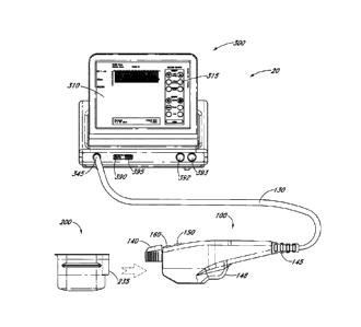
100 and the controller 300. In one embodiment, the interface 130 can transmit
controllable
power from the controller 300 to the hand wand 100.
100651 In various embodiments, the controller 300 can be configured
for
operation with the hand wand 100 and the emitter-receiver module 200, as well
as the overall
CTS 20 functionality. In various embodiments, multiple controllers 300, 300',
300", etc. can
be configured for operation with multiple hand wands 100, 100, I 00", etc. and
or multiple
-18-
Date Recue/Date Received 2023-07-11
CA 02726812 2010-12-02
WO 2009/149390 PCT/US2009/046475
emitter-receiver modules 200, 200', 200", etc. In various embodiments, a
second
embodiment of a reference can be indicated with a reference number with one or
more primes
(1). For example, in one embodiment a first module 200 may be used with or as
an alternative
to a second module 200', third module 200", fourth module 200", etc. Likewise,
in various
embodiments, any part with multiples can have a reference number with one or
more primes
attached to the reference number in order to indicate that embodiment. For
example, in one
embodiment a first transducer 280 can be indicated with the 280 reference
number, and a
second transducer 280' uses the prime. In one embodiment, controller 300
houses an
interactive graphical display 310, which can include a touch screen monitor
and Graphic User
Interface (GUI) that allows the user to interact with the CTS 20. In various
embodiments,
this display 310 sets and displays the operating conditions, including
equipment activation
status, treatment parameters, system messages and prompts and ultrasound
images. In
various embodiments, the controller 300 can be configured to include, for
example, a
microprocessor with software and input/output devices, systems and devices for
controlling
electronic and/or mechanical scanning and/or multiplexing of transducers
and/or
multiplexing of transducer modules, a system for power delivery, systems for
monitoring,
systems for sensing the spatial position of the probe and/or transducers
and/or multiplexing
of transducer modules, and/or systems for handling user input and recording
treatment
results, among others. In various embodiments, the controller 300 can comprise
a system
processor and various digital control logic, such as one or more of
microcontrollers,
microprocessors, field-programmable gate arrays, computer boards, and
associated
components, including firmware and control software, which may be capable of
interfacing
with user controls and interfacing circuits as well as input/output circuits
and systems for
communications, displays, interfacing, storage, documentation, and other
useful functions.
System software may be capable of controlling all initialization, timing,
level setting,
monitoring, safety monitoring, and all other system functions required to
accomplish user-
defined treatment objectives. Further, the controller 300 can include various
control switches
that may also be suitably configured to control operation of the CTS 20. ln
one embodiment,
the controller 300 includes an interactive graphical display 310 for conveying
information to
user. In one embodiment, the controller 300 includes one or more data ports
390. In one
-19-
Date Recue/Date Received 2023-07-11
CA 02726812 2010-12-02
WO 2009/149390
PCT/US2009/046475
embodiment, the data port 390 is a USB port, and can be located on the front,
side, and/or
back of the controller 300 for access to storage, a printer 391, devices, or
be used for other
purposes. In various embodiments the CTS 20 includes a lock 395, and in one
embodiment
the lock 395 can be connectable to the controller 300 via a USB port. In one
embodiment, in
order to operate CTS 20, lock 395 must be unlocked so that power switch 393
may be
activated. In another embodiment lock 395 must be unlocked insertion of USB
access key or
hardware dongle and associated software so that the interactive graphical
display 310 can
execute. In one embodiment, an emergency stop button 392 is readily accessible
for
emergency de-activation.
10066] In various embodiments, an aesthetic imaging system or CTS 20
includes
a hand wand 100 with at least one finger activated controller (150 and/or
160), and a
removable emitter-receiver module 200 having an ultrasound transducer. Other
embodiments
may include non-removable emitter-receiver modules, imaging-only emitter-
receiver
modules, treatment-only emitter-receiver modules, and imaging-and-treatment
emitter-
receiver modules. In one embodiment, the CTS 20 includes a control module 300
that is
coupled to the hand wand 100 and has a graphic user interface 310 for
controlling the
removable transducer module 200 with an interface 130, such as in one
embodiment, a cord
coupling the hand wand 100 to the control module 300. In one embodiment, the
interface
130 provides power to the hand wand 100. In one embodiment, the interface 130
transfers at
least one signal between the hand wand 100 and the control module 300. In an
aspect of this
embodiment, the aesthetic imaging system of CTS 20 is used in aesthetic
procedures on a
portion of a head of a patient. In one embodiment, the CTS 20 is used in
aesthetic procedures
on a portion of a face, head, neck and/or other part of the body of a patient.
100671 In addition, certain embodiments of the present invention
provide a hand
wand 100 for use in aesthetic treatment. In some embodiments, the hand wand
100 includes
a first controlling device 150 operably controlling an imaging function, a
second controlling
device 160 operably controlling a treatment function, a status indicator 155,
an input for
power, an output for at least one signal (for example to a controller 300), a
movement
mechanism 400, and a removable transducer module 200 in communication with the
first
controlling device 150, the second controlling device 160 and/or the movement
mechanism
-20-
Date Recue/Date Received 2023-07-11
CA 02726812 2010-12-02
WO 2009/149390
PCT/US2009/046475
400. In an aspect of the embodiments, the hand wand 100 is used in cosmetic
procedures on
a face, head, neck and/or other part of the body of a patient.
10068) In accordance to various embodiments of the present invention,
an emitter-
receiver module 200 can be coupled to the hand wand 100. In some embodiments
an emitter-
receiver module 200 can emit and receive energy, such as ultrasonic energy. In
one
embodiment, an emitter-receiver module 200 can be configured to only emit
energy, such as
ultrasonic energy. In one embodiment, the emitter-receiver module 200 is
permanently
attachable to the hand wand 100. In one embodiment, the emitter-receiver
module 200 is
attachable to and detachable from the hand wand 100. The emitter-receiver
module 200 can
be mechanically coupled to the hand wand 100 using a latch or coupler 140. An
interface
guide 235 can be useful in assisting the coupling of the emitter-receiver
module 200 to the
hand wand 100. In addition, the emitter-receiver module 200 can be
electronically coupled to
the hand wand 100 and such coupling may include an interface which is in
communication
with the controller 300. In one embodiment, an electric coupler at the
interface guide 235,
located at a proximal end of an emitter-receiver module 200 provides for
electronic
communication between the emitter-receiver module 200 and the hand wand 100,
which can
both be in electric communication with a controller 300. The emitter-receiver
module 200
can comprise various probe and/or transducer configurations. For example, the
emitter-
receiver module 200 can be configured for a combined dual-mode imaging/therapy
transducer, coupled or co-housed imaging/therapy transducers, or simply a
separate therapy
probe and an imaging probe. In one embodiment, the hand wand 100 includes a
handle with
an integrated receptacle for insertion of an emitter-receiver module 200
containing at least a
transducer on one end and an electrical cable for attachment to the controller
200 on the other
end.
100691 With additional reference to the illustrations in FIGS. 2 and
3, the band
wand 100 can be designed for ergonomic considerations to improve comfort,
functionality
and/or ease of use of the hand wand 100 by a user, such as, for example, a
practitioner or
medical professional. The hand wand 100 can be designed to be used
ambidextrously. In
one embodiment, the use of the hand wand 100 is not diminished by whether it
is in a right
hand or a left hand. In one embodiment, of the hand wand 100 includes an
imaging button
-21 -
Date Recue/Date Received 2023-07-11
CA 02726812 2010-12-02
WO 2009/149390
PCT/US2009/046475
150, a treatment button 160, and an indicator 155 on a top portion of the hand
wand 100.
Other arrangements of buttons and/or indicators are possible in various
embodiments. In One
embodiment the hand wand 100 includes a hand rest 148 on a bottom portion and
a coupler
140 distal to the flexible connector 145. In one embodiment, the hand rest 148
includes a
clearance pocket molded into the hand wand 100 housing which allows a magnet-
tipped
clutch rod (433 and 432 of FIG. 7) to move back and forth to drive the
transducer module's
rectilinear motion without hitting the hand wand's housing. According to these
aspects, the
hand wand 100 can be operated by the user either in a right hand or a left
hand. Further to
these aspects, the user can control the imaging button 150 and the treatment
button 160 with
a thumb or finger, such as an index finger. An interior portion of the hand
wand 100 can
include electronics as well as software, connections, and/or couplings for
interfacing to and
from the electronics. In one embodiment, the hand wand 100 contains an
electronic interface
175 (not illustrated here, but see other figures) in communication with at
least one of the
imaging button 150 and the treatment button 160. In accordance with one
embodiment, the
electronic interface 175 can interface with an outside source such as, for
example, the
controller 300. In various embodiments, the indictor 145 can be an LED, a
light, an audio
signal, and combinations thereof. In one aspect of the embodiments, the
indicator 155 is a
LED which can change colors based on different states of the CTS 20. For
example the
indicator 155 can be one color (or off) in a standby mode, a second color in
an imaging mode
and a third color in a treatment mode.
[0070] In one embodiment, the emitter-receiver module 200 is
configured to
removably attach both electronically and mechanically with a hand wand 100. In
one
embodiment, a motion mechanism 400 (see FIG. 7) is configured to move an
ultrasonic
transducer 280 in an emitter-receiver module 200 such as is illustrated in
various
embodiments in FIGS. 4 ¨ 6. A user can remove the indicated transducer module
from its
protective, resealable pouch, setting aside the pouch for storing the
transducer module
between procedures, if necessary. In one embodiment, a hand wand 100 and an
emitter-
receiver module 200 can be connected by pushing the coupler 140 upwards and
sliding the
emitter-receiver module 200 into the hand wand 100 as shown in FIG. 1. In one
embodiment, when the emitter-receiver module 200 is inserted, the controller
300
-22-
Date Recue/Date Received 2023-07-11
CA 02726812 2010-12-02
WO 2009/149390
PCT/US2009/046475
automatically detects it and updates the interactive graphical display 310.
In one
embodiment, the emitter-receiver module 200 locked into the hand wand 100 once
the
emitter-receiver module 200 is fully inserted and the coupler 140 at the tip
of the hand wand
100 is pushed down. To disconnect the emitter-receiver module 200, the user
can lift the
coupler 140 at the tip of the hand wand 100 and slide the emitter-receiver
module 200 out of
the hand wand 100.
100711 FIGS. 4
and 5 illustrate two opposing side views of an embodiment of an
emitter-receiver module 200 comprising a housing 220 and an acoustically
transparent
member 230. In one embodiment, the housing 220 may include a cap 222 that is
removable
or permanently attachable to the housing 220. In one embodiment, the emitter-
receiver
module 200 includes an interface guide 235 and/or one or more side guides 240
that can be
useful in assisting the coupling of the emitter-receiver module 200 to the
hand wand 100.
The emitter-receiver module 200 can include a transducer 280 which can emit
energy through
an acoustically transparent member 230. The acoustically transparent member
230 can be a
window, a filter and/or a lens. The acoustically transparent member 230 can be
made of any
material that is transparent to the energy that is that is emitted by the
transducer 280. In one
embodiment, the acoustically transparent member 230 is transparent to
ultrasound energy.
100721 In
various embodiments, the transducer 280 is in communication with
the controller 300_ In one embodiment, the transducer 280 is electronically
coupled to the
band wand 100 and/or the controller 300. In one embodiment, the housing 220 is
sealed by
the cap 222 and the structure of the combination of the housing 220 and the
cap 222 can hold
a liquid (not shown). As illustrated in FIG. 6, an embodiment of the emitter-
receiver module
200 housing 220 can have a port 275 which allows interfacing from the hand
wand 100 into
the transducer module 200 without affecting the integrity of the sealed
structure of the
housing 220 and the cap 222. Further, the cap 222 can include one or more
ports. For
example, a first port 292, a second port 293 and a third port 294. The ports
in the cap 222
can be useful for electronically coupling the transducer 280 to the hand wand
100 and/or the
controller 300. In one embodiment, at least one of the ports in the cap 222
may be used to
interface a sensor 201 that may be useful in the emitter-receiver module 200.
The sensor 201
-23-
Date Recue/Date Received 2023-07-11
CA 02726812 2010-12-02
WO 2009/149390
PCT/US2009/046475
can be in communication with the controller 300. More than one sensor 201 is
used in some
embodiments.
[0073] In various embodiments, as illustrated in the block diagram of
FIG. 6, the
transducer 280 is movable within the emitter-receiver module 200. The
transducer 280 is
held by a transducer holder 289. In one embodiment, the transducer holder 289
includes a
sleeve 287 which is moved along motion constraining bearings, such as linear
bearings,
namely, a bar (or shaft) 282 to ensure a repeatable linear movement of the
transducer 280. In
one embodiment, sleeve 287 is a spline bushing which prevents rotation about a
spline shaft
282, but any guide to maintain the path of motion is appropriate. In one
embodiment, the
transducer holder 289 is driven by a motion mechanism 400, which may be
located in the
hand wand 100 or in the emitter-receiver module 200. The motion mechanism 400,
as is
discussed below in relation to FIG. 7, includes a scotch yoke 403 with a
movement member
432 and a magnetic coupling 433 on a distal end of the movement member 432.
The magnet
coupling 433 helps move the transducer 280. One benefit of a motion mechanism
such as
motion mechanism 400 is that it provides for a more efficient, accurate and
precise use of an
ultrasound transducer 280, for both imaging and for therapy purposes. One
advantage this
type of motion mechanism has over conventional fixed arrays of multiple
transducers fixed in
space in a housing is that the fixed arrays are a fixed distance apart. By
placing transducer
280 on a linear track under controller 300 control, embodiments of the system
and device
provide for adaptability and flexibility in addition to the previously
mentioned efficiency,
accuracy and precision. Real time and near real time adjustments can be made
to imaging
and treatment positioning along the controlled motion by the motion mechanism
400. In
addition to the ability to select nearly any resolution based on the
incremental adjustments
made possible by the motion mechanism 400, adjustments can be made if imaging
detects
abnormalities or conditions meriting a change in treatment spacing and
targeting.
[0074] In one embodiment, one or more sensors 201 may be included in
the
emitter-receiver module 200. In one embodiment, one or more sensors 201 may be
included
in the emitter-receiver module 200 to ensure that a mechanical coupling
between the
movement member 432 and the transducer holder 289 is indeed coupled. In one
embodiment, an encoder 283 may be positioned on top of the transducer holder
289 and a
-24-
Date Recue/Date Received 2023-07-11
CA 02726812 2010-12-02
WO 2009/149390
PCT/US2009/046475
sensor 2W may be located in a dry portion of the emitter-receiver module 200,
or vice versa
(swapped). In various embodiments the sensor 201 is a magnetic sensor, such as
a giant
magnetoresistive effect (GMR) or Hall Effect sensor, and the encoder a magnet,
collection of
magnets, or multi-pole magnetic strip. The sensor may be positioned as a
transducer module
home position. In one embodiment, the sensor 201 is a contact pressure sensor.
In one
embodiment, the sensor 201 is a contact pressure sensor on a surface of the
device to sense
the position of the device or the transducer on the patient. In various
embodiments, the
sensor 201 can be used to map the position of the device or a component in the
device in one,
two, or threes dimensions. In one embodiment the sensor 201 is configured to
sense the
position, angle, tilt, orientation, placement, elevation, or other
relationship between the
device (or a component therein) and the patient. In one embodiment, the sensor
201
comprises an optical sensor. In one embodiment, the sensor 201 comprises a
roller ball
sensor. In one embodiment, the sensor 201 is configured to map a position in
one, two and/or
three dimensions to compute a distance between areas or lines of treatment on
the skin or
tissue on a patient. Motion mechanism 400 can be any motion mechanism that may
be found
to be useful for movement of the transducer 280. Other embodiments of motion
mechanisms
useful herein can include worm gears and the like. In various embodiments of
the present
invention, the motion mechanism is located in the emitter-receiver module 200.
In various
embodiments, the motion mechanism can provide for linear, rotational, multi-
dimensional
motion or actuation, and the motion can include any collection of points
and/or orientations
in space. Various embodiments for motion can be used in accordance with
several
embodiments, including but not limited to rectilinear, circular, elliptical,
arc-like, spiral, a
collection of one or more points in space, or any other 1-D, 2-D, or 3-D
positional and
attitudinal motional embodiments. The speed of the motion mechanism 400 may be
fixed or
may be adjustably controlled by a user. One embodiment, a speed of the motion
mechanism
400 for an image sequence may be different than that for a treatment sequence.
In one
embodiment, the speed of the motion mechanism 400 is controllable by the
controller 300.
100751 Transducer 280 can have a travel distance 272 such that an
emitted energy
50 is able to be emitted through the acoustically transparent member 230. In
one
embodiment, the travel 272 is described as end-to-end range of travel of the
transducer 280.
-25-
Date Recue/Date Received 2023-07-11
CA 02726812 2010-12-02
WO 2009/149390 PCT/US2009/046475
In one embodiment, the travel 272 of the transducer 280 can be between about
100 mm and
about 1 mm. In one embodiment, the length of the travel 272 can be about 25
mm. In one
embodiment, the length of the travel 272 can be about 15 mm. In one
embodiment, the
length of the travel 272 can be about 10 mm, In various embodiments the length
of the travel
272 can be about between 0-25 mm, 0-15 mm, 0-10 mm.
100761 The
transducer 280 can have an offset distance 270, which is the distance
between the transducer 280 and the acoustically transparent member 230. In
various
embodiments of the present invention, the transducer 280 can image and treat a
region of
interest of about 25 mm and can image a depth less than about 10 mm. In one
embodiment,
the emitter-receiver module 200 has an offset distance 270 for a treatment at
a depth 278 of
about 4.5 mm below the skin surface 501 (see FIG. 15).
100771 in
various embodiments, transducer modules 200 can be configured for
different or variable ultrasonic parameters. For
example, in various non-limiting
embodiments, the ultrasonic parameter can relate to aspects of the transducer
280, such as
geometry, size, timing, spatial configuration, frequency, variations in
spatial parameters,
variations in temporal parameters, coagulation formation, depth, width,
absorption
coefficient, refraction coefficient, tissue depths, and/or other tissue
characteristics. In various
embodiments, a variable ultrasonic parameter may be altered, or varied, in
order to effect the
formation of a lesion for the desired cosmetic approach. In various
embodiments, a variable
ultrasonic parameter may be altered, or varied, in order to effect the
formation of a lesion for
the desired clinical approach. By way of example, one variable ultrasonic
parameter relates
to configurations associated with tissue depth 278. In several embodiments,
the transducer
module 200 is configured for both ultrasonic imaging and ultrasonic treatment
and is
operably coupled to at least one controlling device 150, 160 and a movement
mechanism
400. The transducer module 200 is configured to apply ultrasonic therapy at a
first ultrasonic
parameter and a second ultrasonic parameter. In various embodiments, the first
and second
ultrasonic parameters are selected from the group consisting of: variable
depth, variable
frequency, and variable geometry. For example, in one embodiment, a single
transducer
module 200 delivers ultrasonic therapy at two or more depths 278, 278'. In
another
embodiment, two Or more interchangeable transducer modules 200 each provide a
different
-26-
Date Recue/Date Received 2023-07-11
CA 02726812 2010-12-02
WO 2009/149390 PCT/US2009/046475
depth 278 (e.g., one module treats at 3 mm depth while the other treats at a
4.5 mm depth).
In yet another embodiment, a single transducer module 200 delivers ultrasonic
therapy at two
or more frequencies, geometries, amplitudes, velocities, wave types, and/or
wavelengths. In
other embodiments, two or more interchangeable transducer modules 200 each
provide a
different parameter value. In one embodiment, a single transducer module 200
may provide
at least two different depths 278, 278' and at least two different frequencies
(or other
parameter). Variable parameter options are particularly advantageous in
certain embodiments
because they offer enhanced control of tissue treatment and optimize lesion
formation, tissue
coagulation, treatment volume, etc.
100781 Figure 15 illustrates one embodiment of a depth 278 that
corresponds to a
muscle depth. hi various embodiments, the depth 278 can correspond to any
tissue, tissue
layer, skin, dermis, fat, SMAS, muscle, or other tissue. In some embodiments,
different types
of tissue are treated to provide synergistic effects, thus optimizing clinical
results. In another
embodiment, the emitter-receiver module has an offset distance 270 for a
treatment at a depth
278 of about 3.0 mm below the surface 501. In various embodiments, this offset
distance
may be varied such that the transducer 280 can emit energy to a desired depth
278 below a
surface 501. In various embodiments, in a treatment mode, bursts of acoustic
energy from
the transducer 280 can create a linear sequence of individual thermal lesions
550. In one
embodiment the individual thermal lesions 550 are discrete. In one embodiment
the
individual thermal lesions 550 are overlapping, ha various embodiments, the
transducer 280
can image to a depth roughly between 1 and 100 mm. In one embodiment, the
transducer
imaging depth can be approximately 20 mm. In one embodiment, the transducer
280 can
treat to a depth of between about zero (0) to 25 mm. In one embodiment, the
transducer
treatment depth can be approximately 4.5 mm.
100791 hi any of the embodiments described herein, the transducer
treatment
depth can be approximately 0.5 mm, 1 mm, 1.5 mm, 2mm, 3 mm, 4 mm, 4.5 mm, 5
mm, 6
mm, 10 mm 15 mm, 20 mm, 25 mm, or any other depth in the range of 0¨ 100 mm.
Varied
depth treatment, including treatment of the same tissue at different depths or
treatment of
different tissues, can increase clinical results by providing synergistic
effects.
-27-
Date Recue/Date Received 2023-07-11
CA 02726812 2010-12-02
WO 2009/149390 PCT/US2009/046475
100801 In various embodiments of the present invention, a transducer
280 is
capable of emitting ultrasound energy for imaging, diagnostics, or treating
and combinations
thereof. In one embodiment, the transducer 280 is configured to emit
ultrasound energy at a
specific depth in a region of interest to target a region of interest of a
specific tissue such as a
corrugator supercilii muscle as described below. In this embodiment, the
transducer 280 may
be capable of emitting unfocused or defocused ultrasound energy over a wide
area of the
region of interest 65 for treatment purposes (see FIG. 12 and 22). In one
embodiment, the
emitter-receiver module 200 contains a transducer 280 that can image and treat
a region of
tissue up to 25 mm long and can image a depth of up to 8 millimeters.
Treatment occurs
along a line less than or equal to the transducer's active length, which is
indicated in one
embodiment by guide marks (not illustrated here) on the sides of the emitter-
receiver module
200 near a acoustically transparent member 230 along the surface adjacent to
the patient's
skin. In one embodiment, a marked guide at the front tip of the transducer 280
represents the
center of the treatment line. In one embodiment of a treatment mode, bursts of
sound energy
create a linear sequence of individual thermal coagulation zones. In one
embodiment the
individual thermal coagulation zones are discrete. In one embodiment the
individual thermal
coagulation zones are overlapping. A label (not illustrated here) may be
applied or etched on
a side or top surface of the emitter-receiver module 200 to provide the
transducer 280 type,
expiration date, and other information. In one embodiment, an emitter-receiver
module 200
can be configured with a label for tracking the type transducer 280 used,
treatment frequency
and treatment depth, a unique serial number, a part number, and date of
manufacture. In one
embodiment, the emitter-receiver modules 200 are disposable. In one
embodiment, the
system tracks use of the emitter-receiver modules 200 in order to determine
the remaining life
of the emitter-receiver module 200 as transducer life diminishes over time
and/or usage.
Once a transducer 280 has diminished capacity, the emitter-receiver module 200
may work
less effectively in performing its functions. In one embodiment, the emitter-
receiver module
200 or controller 300 will track usage and prevent additional usage of an
emitter-receiver
module 200 beyond a recommended usage life in order to preserve the safety and
effectiveness of the device. This safety feature can be configured based on
test data.
-28-
Date Recue/Date Received 2023-07-11
CA 02726812 2010-12-02
WO 2009/149390 PCT/US2009/046475
100811 In one embodiment, an emitter-receiver module 200 is configured
with a
treatment frequency of approximately 4 MHz, a treatment depth of approximately
4.5 mm
and an imaging depth range of roughly 0 ¨ 8 mm. In one embodiment, an emitter-
receiver
module 200 is configured with a treatment frequency of approximately 7 MHz, a
treatment
depth of approximately 3.0 mm and an imaging depth range of roughly 0 ¨ 8 ram.
In one
embodiment, an emitter-receiver module 200 is configured with a treatment
frequency of
approximately 7 MHz, a treatment depth of approximately 4.5 mm and an imaging
depth
range of roughly 0¨ 8 mm.
100821 Transducer 280 may comprise one or more transducers for
facilitating
imaging and/or treatment. The transducer 280 may comprise a piezoelectrically
active
material, such as, for example, lead zirconante titanate, or other
piezoelectrically active
materials such as, but not limited to, a piezoelectric ceramic, crystal,
plastic, and/or
composite materials, as well as lithium niobate, lead titanate, barium
titanate, and/or lead
metaniobate, including piezoelectric, electrically conductive, and plastic
film layers deposited
on spherically focused backing material. In addition to, or instead of a
piezoelectrically
active material, the transducer 280 may comprise any other materials
configured for
generating radiation and/or acoustical energy. The transducer 280 may also
comprise one or
more matching and/or backing layers coupled to the piezoelectrically active
material. The
transducer 280 may also be configured with single or multiple damping
elements.
100831 In one embodiment, the thickness of a transduction element of
the
transducer 280 may be configured to be uniform. That is, the transduction
element may be
configured to have a thickness that is generally substantially the same
throughout. In another
embodiment, the transduction element may also be configured with a variable
thickness,
and/or as a multiple damped device. For example, the transduction element of
the transducer
280 may be configured to have a first thickness selected to provide a center
operating
frequency of a lower range, for example from about 1 MHz to about 10 MHz. The
transduction element may also be configured with a second thickness selected
to provide a
center operating frequency of a higher range, for example from about 10 MHz to
greater than
100 MHz.
-29-
Date Recue/Date Received 2023-07-11
CA 02726812 2016-09-02
100841 In yet
another embodiment, the transducer 280 is configured as a single
broadband transducer excited with two or more frequencies to provide an
adequate output for
raising a temperature within a treatment area of the region of interest to the
desired level as
discussed herein. The transducer 280 may be configured as two or more
individual
transducers, such that each transducer 280 may comprise a transduction
element. The
thickness of the transduction elements may be configured to provide center-
operating
frequencies in a desired treatment range. For example, in one embodiment, the
transducer
280 may comprise a first transducer configured with a first transduction
element having a
thickness corresponding to a center frequency range of about 1 MHz to about 10
MHz, and a
second transducer configured with a second transduction clement having a
thickness
corresponding to a center frequency range of about 10 MHz to greater than 100
MHz.
Various other combinations and ranges of thickness for a first and/or second
transduction
element can be designed to focus at specific depths below a surface 501, for
specific
frequency ranges, and/or specific energy emissions.
100851 The
transduction elements of the transducer 280 can be configured to be
concave, convex, and/or planar. In one
embodiment, the transduction elements are
configured to be concave in order to provide focused energy for treatment of
the region of
interest. Additional embodiments of transducers arc disclosed in U.S. Patent
Application No.
10/944,500, entitled "System and Method for Variable Depth Ultrasound
Treatment."
100861 Moreover,
the transducer 280 can be any distance from the surface 501. In
that regard, it can be far away from the surface 501 disposed within a long
transducer or it
can be just a few millimeters from the surface 501. This distance can be
determined by
design using the offset distance 270 as described herein. In certain
embodiments, positioning
the transducer 280 closer to the surface 501 is better for emitting ultrasound
at higher
frequencies. Moreover, both two and three dimensional arrays of elements can
be used in the
present invention. Furthermore, the transducer 280 may comprise a reflective
surface, tip, or
area at the end of the transducer 280 that emits ultrasound energy. This
reflective surface
may enhance, magnify, or otherwise change ultrasound energy emitted from the
CTS 20.
-30-
Date Recue/Date Received 2023-07-11
CA 02726812 2010-12-02
WO 2009/149390 PCT/US2009/046475
10087) In
various embodiments any set of one or more transducers 280 can be
used for various functions, such as separate treat/image or dual-mode (both
treat/image)
transducers or a treat-only version. In various embodiments the imaging
element(s) can be on
the side (adjacent to) or at any relative position, attitude, and/or height,
or even within the
therapy element(s). One or more therapy depths and frequencies can be used and
one or more
imaging elements or one or more dual-mode elements. In
various embodiments any
controllable means of moving the active transduction element(s) within the
emitter-receiver
module 200 housing constitute viable embodiments.
100881 In
various embodiments, the emitter-receiver module 200 can also be
configured in various manners and comprise a number of reusable and/or
disposable
components and parts in various embodiments to facilitate its operation. For
example, the
emitter-receiver module 200 can be configured within any type of transducer
probe housing
or arrangement for facilitating the coupling of the transducer 280 to a tissue
interface, with
such housing comprising various shapes, contours and configurations. The
emitter-receiver
module 200 can comprise any type of matching, such as for example, electric
matching,
which may be electrically switchable, multiplexer circuits and/or
aperture/element selection
circuits, and/or probe identification devices, to certify probe handle,
electric matching,
transducer usage history and calibration, such as one or more serial EEPROM
(memories).
100891 In
various embodiments, the emitter-receiver module 200 may also
comprise cables and connectors, motion mechanisms, motion sensors and
encoders, thermal
monitoring sensors, and/or user control and status related switches, and
indicators such as
LEDs. hi one embodiment, a motion mechanism similar to the motion mechanism
400
described in the hand wand 100 may be used to drive the emitter-receiver
module 200 from
within the emitter-receiver module 200. In one embodiment, a hand wand 100 is
electrically
connectable to the emitter-receiver module 200 to drive the emitter-receiver
module 200 from
within itself. In various embodiments, a motion mechanism (in any of the
embodiments
described herein) may be used to controllably create multiple lesions, or
sensing of probe
motion itself may be used to controllably create multiple lesions and/or stop
creation of
lesions 550, as discussed herein. For example in one embodiment, for safety
reasons if the
emitter-receiver module 200 is suddenly jerked or is dropped, a sensor can
relay this action to .. ,
-31 -
Date Recue/Date Received 2023-07-11
CA 02726812 2010-12-02
WO 2009/149390
PCT/US2009/046475
the controller 300 to initiate a corrective action or shut down the emitter-
receiver module
200. In addition, an external motion encoder arm may be used to hold the probe
during use,
whereby the spatial position and attitude of the emitter-receiver module 200
is sent to the
controller 300 to help controllably create lesions 550. Furthermore, other
sensing
functionality such as profilometers or other imaging modalities may be
integrated into the
emitter-receiver module 200 in accordance with various embodiments. In one
embodiment,
pulse-echo signals to and from the emitter/receiver module 200 are utilized
for tissue
parameter monitoring of the treatment region 550.
100901 Coupling components can comprise various devices to facilitate
coupling
of the emitter-receiver module 200 to a region of interest. For example,
coupling
components can comprise cooling and acoustic coupling system configured for
acoustic
coupling of ultrasound energy and signals. Acoustic cooling/coupling system
with possible
connections such as manifolds may be utilized to couple sound into the region-
of-interest,
control temperature at the interface and deeper into tissue, provide liquid-
filled lens focusing,
and/or to remove transducer waste heat. The coupling system may facilitate
such coupling
through use of one or more coupling mediums, including air, gases, water,
liquids, fluids,
gels, solids, and/or any combination thereof, or any other medium that allows
for signals to
be transmitted between the transducer 280 and a region of interest. In one
embodiment one
or more coupling media is provided inside a transducer. In one embodiment a
fluid-filled
emitter-receiver module 200 contains one or more coupling media inside a
housing. In one
embodiment a fluid-filled emitter-receiver module 200 contains one or more
coupling media
inside a sealed housing, which is separable from a dry portion of an
ultrasonic device.
100911 In addition to providing a coupling function, in accordance
with one
embodiment, the coupling system can also be configured for providing
temperature control
during the treatment application. For example, the coupling system can be
configured for
controlled cooling of an interface surface or region between the emitter-
receiver module 200
and a region of interest and beyond by suitably controlling the temperature of
the coupling
medium. The suitable temperature for such coupling medium can be achieved in
various
manners, and utilize various feedback systems, such as thermocouples,
thermistors or any
other device or system configured for temperature measurement of a coupling
medium. Such
-32-
Date Recue/Date Received 2023-07-11
CA 02726812 2010-12-02
WO 2009/149390
PCT/US2009/046475
controlled cooling can be configured to further facilitate spatial and/or
thermal energy control
of the emitter-receiver module 200.
100921 In one
embodiment, the emitter-receiver module 200 is connected to a
motion mechanism 400 in the hand wand 100. In one embodiment, the motion
mechanism
400 may be in the emitter-receiver module 200. One embodiment of a motion
mechanism
400 is illustrated in FIG. 7, which depicts a two phase stepper motor 402 and
a scotch yoke
403 to produce a linear motion. The stepper motor 402 rotates as indicated by
arrow 405
which moves a pin 404 in a circular path. The pin 404 slides in a slot 406 of
the scotch yoke
403. This causes the scotch yoke 403 to move in a linear fashion. The scotch
yoke 403 is
held by guides 410 and glide members 412 may be between the scotch yoke 403
and guide
410. In one embodiment, a guide 410 is a shoulder screw. Embodiments of the
glide
member 412 may include any material or mechanical device that lowers a
coefficient of
friction between the guide 410 and the scotch yoke 403, or any linear
bearings. For example,
in various embodiments the glide member 412 can be at least one of an
elastomeric material,
a lubricant, ball bearings, a polished surface, a magnetic device, pressurized
gas, or any other
material or device useful for gliding.
100931 A sensor
425 operates as one embodiment of a position sensor by reading
an encoder 430 which is mounted on the scotch yoke 403. In one embodiment, the
encoder
strip 430 is an optical encoder which has a pitch in a range from about 1.0 mm
to about 0.01
mm. In one embodiment, the pitch may be about 0.1 mm. The encoder strip 430
can include
index marks at each end of its travel. The direction of travel of the encoder
strip 430 can be
determined by comparing phases of two separate channels in the optical sensor
425. In one
embodiment, the encoder strip 430 has one, two or more home positions which
may be useful
in calibrating for a position and travel of the scotch yoke 403.
100941 In one
embodiment, the movement of the scotch yoke 403 is transferred
through the movement mechanism 432 such that the transducer 280 moves in a
linear fashion
inside of the emitter-receiver module 200. In one
embodiment, the scotch yoke 403
includes a movement member 432 and a magnetic coupling 433 on a distal end of
the
movement member 432. The movement member 432 can be sized to travel though or
within
a liquid-tight seat
-33-
Date Recue/Date Received 2023-07-11
CA 02726812 2010-12-02
WO 2009/149390 PCT/US2009/046475
10095]
Transducer 280 can have a travel distance 272 The coupling system may
facilitate such coupling With reference to FIG. 8, a block diagram illustrates
various
embodiments of the CTS 20. In one embodiment, the controller 300 includes a
controller
subsystem 340, a therapy subsystem 320, an imaging subsystem 350, an embedded
host 330
(with software) and an interactive graphical display 310. In one embodiment,
the therapy
subsystem 320, the controller subsystem 340, and/or the imaging subsystem 350
is interfaced
with the hand wand 100 and/or the emitter-receiver module 200. In various
embodiments,
the CTS 20 has built into the controller 300 limits as to an amount of energy
50 that can be
emitted from the emitter-receiver module 200. These limits can be determined
by time of
emission, frequency of the energy emitted, power of energy, a temperature,
and/or
combinations thereof. The temperature may be from monitoring the surface 501
and/or
monitoring the emitter-receiver module 200. According to one embodiment the
limits may
be preset and cannot be changed by the user.
100961
According to various embodiments, when the emitter-receiver module 200
is coupled to the surface 501, which may be a skin surface of the subject, the
CTS 20 can
image and/or treat a treatment area 272. In some aspects of these embodiments,
the imaging
by the CTS 20 can be over essentially the entire treatment area 272 at
specified depths 278
below the surface 501. In some aspects of these embodiments, the treatment can
include
discrete energy emissions 50 to create lesion 550 at intervals along the
treatment area 272 and
at specified depths 278. In one embodiment the intervals are discrete. In one
embodiment
the intervals are overlapping.
100971 In
various embodiments the imaging subsystem 350 may be operated in a
B-mode. The imaging subsystem 350 can provide support to the emitter-receiver
module 200
such that the emitter-receiver module 200 can have emission energy 50 from a
frequency of
about 10 MHz to greater than 100 MHz. In one embodiment, the frequency is
about 18 MHz.
In one embodiment, the frequency is about 25 MHz. The imaging subsystem 350
can support
any frame rate that may be useful for the applications. In some embodiments,
the frame rate
may be in a range from about 1 frames per second (hereinafter "FPS") to about
100 FPS, or
from about 5 FPS to about 50 FPS or from about 5 FPS to about 20 FPS nominal.
An image
field of view may be controlled by the image area of the transducer 280 in a
focus of the
-34-
Date Recue/Date Received 2023-07-11
CA 02726812 2010-12-02
WO 2009/149390 PCT/US2009/046475
transducer 280 at a specific depth 278 below the surface 501 as discussed
herein. In various
embodiments, the field of view can be less than 20 mm in depth and 100 mm in
width or less
than 10 mm in depth and less than 50 mm in width. In one embodiment, a
particularly useful
image field of view is about 8 mm in depth by about 25 mm in width.
100981 A resolution of the field of view can be controlled by the
graduation of the
movement mechanism 400. As such, any pitch may be useful based on the
graduation of the
motion mechanism 400. In one embodiment, the resolution of the field of view
may be
controlled by the resolution of an encoder 430 and sensor 425. In one
embodiment the image
field of view can have a pitch in the range of 0.01 mm to 0.5 mm or from about
0.05 mm to
about 0.2 mm. In one embodiment, a particularly useful line pitch for the
image field of view
is about 0.1 mm.
100991 According to various embodiments, the imaging subsystem 350 can
include one or more functions. In one embodiment, the one or more functions
can include
any of the following B-mode, scan image, freeze image, image brightness,
distance calipers,
text annotation for image, save image, print image, and/or combinations
thereof. In various
embodiments of the present invention, the imaging subsystem 350 contains pulse
echo
imaging electronics.
[0] 001 Various embodiments of the therapy subsystem 320 comprise a
radio
frequency (hereinafter "RF") driver circuit which can deliver and/or monitor
power going to
the transducer 280. In one embodiment, the therapy subsystem 320 can control
an acoustic
power of the transducer 280. In one embodiment, the acoustic power can be from
a range of
1 watt (hereinafter -W") to about 100 W in a frequency range from about 1 MHz
to about 10
MHz, or from about JO W to about 50 W at a frequency range from about 3 MHz to
about 8
MHz. In one embodiment, the acoustic power and frequencies are about 40 W at
about 4.3
MHz and about 30 W at about 7.5 MHz. An acoustic energy produced by this
acoustic
power can be between about 0.01 joule (hereinafter "I") to about 10 J or about
2 J to about 5
J. In one embodiment, the acoustic energy is in a range less than about 3 J.
101011 In various embodiments the therapy subsystem 320 can control a
time on
for the transducer 280. In one embodiment, the time on can be from about 1
millisecond
(hereinafter '`ins'") to about 100ms or about lOrns to about 50ms. In one
embodiment, time
-35-
Date Recue/Date Received 2023-07-11
CA 02726812 2010-12-02
WO 2009/149390 PCT/US2009/046475
on periods can be about 30ms for a 4.3 MHz emission and about 30ms for a 7.5
MHz
emission.
101021 In
various embodiments, the therapy subsystem 320 can control the drive
frequency of the transducer 280 moving across the travel 272. In various
embodiments, the
frequency of the transducer 280 is based on the emitter/receiver 200 connected
to the hand
wand 100. According to some embodiments, the frequency of this movement may be
in a
range from about 1 MHz to about 10 MHz, or about 4 MHz to about 8 MHz. In one
embodiment, the frequencies of this movement are about 4.3 MHz or about 7.5
MHz. As
discussed herein, the length of the travel 272 can be varied, and in one
embodiment, the
travel 272 has a length of about 25 mm.
101031
According to various embodiments, the therapy subsystem 320 can
control the line scan along the travel 272 and this line scan can range from 0
to the length of
the distal of the travel 272. In one embodiment, the line scan can be in a
range from about 0
to about 25 mm. According to one embodiment, the line scan can have
incremental energy
emissions 50 having a treatment spacing 295 and this treatment spacing can
range from about
0.01 mm to about 25 mm or from 0.2 mm to about 2.0 mm. In one embodiment,
treatment
spacing 295 is about 1.5 mm. In various embodiments, the treatment spacing 295
can be
predetermined, constant, variable, programmable, and/or changed at any point
before, during
or after a treatment line. The resolution of the line scan is proportional to
the resolution of
the motion mechanism 400. In various embodiments, the resolution that is
controllable by
the therapy subsystem 320 is equivalent to the resolution controllable by the
imaging
subsystem 350 and, as such, can be in the same range as discussed for the
imaging subsystem
350.
101041 In
various embodiments, the therapy subsystem 320 can have one or more
functions. In one embodiment, the one or more functions can include any of the
following:
emission energy control, treatment spacing, travel length, treatment ready,
treatment,
treatment stop, save record, print record, display treatment, and/or
combinations thereof_
101051 In
various embodiments, the control subsystem 340 includes electronic
hardware which mechanically scans the transducer 280 for one or more
functions. In one
embodiment, one or more functions that can be scanned by the controller
subsystem 340 can
-36-
Date Recue/Date Received 2023-07-11
CA 02726812 2010-12-02
WO 2009/149390 PCT/US2009/046475
include scanning the transducer 280 for imaging, a position of the transducer
280 for
imaging, scan slip positions of the transducer 280 at locations for therapy,
controls therapy
hardware settings, provides other control functions, interfacing with the
embedded host 330,
and/or combinations thereof. In one embodiment the locations are discrete. In
one
embodiment the locations are overlapping.
101061 In various embodiments, an embedded host 330 is in two-way
communication with the controller 340 and the graphical interface 310. In one
embodiment,
data from the controller 340 can be converted to a graphical format by the
embedded host 330
and then transferred to the graphical interface 310 for displaying imaging
and/or treatment
data.
101071 In one embodiment, commands can be entered by a user employing
the
graphical interface 310. The commands entered by use of the graphical
interface 310 can be
communicated to embedded host 330 and then communicated to controller 340 for
control
and operation of the therapy subsystem 320, the imaging subsystem 350, the
hand wand 100,
and/or the emitter-receiver module 200. In various embodiments, the embedded
host 330
can include a processing unit, memory, and/or software.
101081 In various embodiments, when the imaging button 150 is pressed
the CTS
20 enters an imaging sequence in which the imaging subsystem 350 acquires scan
lines which
are transferred to the embedded host 330 for data conversion and/or graphical
conversion
which is then communicated to the graphical interface 310. While the system is
operating in
the imaging sequence, the imaging button 150 may be pressed again which puts
the CTS 20
into a ready state. In an aspect of this embodiment, an audio warning or
visual display such
as the indicator 155 may be initiated to alert the user that the CTS 20 is in
the ready state. In
the ready state, the controller subsystem 340 communicates with the embedded
host 330 to
acquire users entered treatment settings. These treatment settings can be
checked and can be
verified and converted to hardware parameter in the controller subsystem 340.
In one
embodiment, such set hardware parameters can include treatment timing,
cadence, time on,
time off, RF driver power, voltage levels, acoustic power output, oscillator
frequency, therapy
transducer frequency, treatment spacing, travel, motion mechanism speed,
and/or
-37-
Date Recue/Date Received 2023-07-11
CA 02726812 2010-12-02
WO 2009/149390 PCT/US2009/046475
combinations thereof. The CTS 20 may remain in the ready state indefinitely or
may be
timed out after a set time period.
101091 In various embodiments of the present invention, when the CTS
20 is in
the ready state, the treatment button 160 may be activated. This activation of
the treatment
button 160 commences a treatment sequence. The treatment sequence is
controllable by the
therapy subsystem 320 which executes the treatment sequence along with the
controller
subsystem 340 and independently of the embedded host 330. The treatment
sequence is
delivered in real time and last one of the length of the activating of the
treatment button 160
or a programmed time downloaded from the embedded host 330 into the controller
subsystem
340 and/or the therapy subsystem 320.
101101 In various embodiments, safety features can be designed in the
CTS 20 to
ensure safe use, imaging, and treatment. In various embodiments, the embedded
host 330 is
in communication with data port 390 which can comprise either one-way or two-
way
communication between the data port 390 and the embedded host 330. The data
port 390 can
interface any electronic storage device, for example, the data port 390 can be
interfaced for
one or more of a USB drive, a compact flash drive, a secured digital card, a
compact disc, and
the like. In one embodiment, a storage device through data port 390 to the
embedded host
330 can download treatment records or software updates. In another aspect of
these
embodiments, the storage device can be a two-way communication through data
port 390 to
the embedded host 330 such that a treatment protocol can be downloaded to the
embedded
host 330 and CTS 20. A treatment protocol can include parameters, imaging
data, treatment
data, date/time, treatment duration, subject information, treatment location,
and combinations
thereof, and the like which can be uploaded by and/or downloaded from the
embedded host
330 to the storage device via the data port 390. In one embodiment, a second
data port (not
shown) may be located on the back of the controller. The second data port may
provide
power and/or data to a printer.
101111 ln various embodiments, the CTS 20 includes a lock 395. In one
embodiment, in order to operate CTS 20, lock 395 must be unlocked so that
power switch
393 may be activated. In one embodiment, the power may remain on as the lock
395 is
unlocked and locked successively and different parameters are entered. A key
396 (not
-38-
Date Recue/Date Received 2023-07-11
CA 02726812 2010-12-02
WO 2009/149390 PCT/US2009/046475
illustrated) may be needed to unlock the lock 395. Examples of keys 396 useful
herein
include a standard metal tooth and groove key, or an electronic key. In some
embodiments,
an electronic key 396 may be digitally encoded to include user infon-nation
and collect data
and/or time usage of CTS 20. In one embodiment, an electronic key is
particularly useful
with CTS 20 may be a USB drive with encryption such that inserting the USB
drive key into
lock 395 the CTS 20 may be activated. In various embodiments, a software key
can be
configured to indicate a condition or status to the user, lock the system,
interrupt the system,
or other feature.
101121 With reference to FiG. 9, a CTS 20 layout block diagram is
illustrated
according to various embodiments of the present invention. In accordance with
the aspects of
these embodiments. the controller 300 can include several electronic sections.
Included in
these electronic sections can be a power supply 350 which provides power to
CTS 20
including the controller 300, the hand wand 100, and/or the emitter-receiver
module 200. In
one embodiment, the power supply 350 can supply power to a printer or other
data output
device. The controller 300 can include the controller subsystem 340 as
described herein, the
host 330, a graphical interface 310, an RF driver 352 and a front panel flex
circuit 345. The
RF driver 352 can provide power to the transducer 280. The embedded host 330
can be a
host computer which may be used collecting user input, transferring it to the
controller
subsystem 340 and for displaying images and system statuses on the graphical
interface 310.
The power supply 350 can be convertible for use internationally based on
different voltage
inputs and typically is a medical grade power supply. The power supply may be
plugged into
a standard wall socket to draw power or may draw power from a battery or any
other
alternative source that may be available.
101131 The graphical interface 310 displays images and systems status
as well as
facilitates the user interface for entering commands to control the CTS 20.
The controller
subsystem 340 can control the imaging subsystem 350, the therapy subsystem
320, as well as
interfacing and communicating treatment protocol to the hand wand 100 and the
emitter-
receiver module 200, as described herein. In one embodiment, the controller
subsystem 340
not only sets treatment parameters but also monitors the status of such
treatment and transfers
such status to the host 330 for display on display/touch screen 310. The front
panel flex
-39-
Date Recue/Date Received 2023-07-11
CA 02726812 2010-12-02
WO 2009/149390 PCT/US2009/046475
circuit 345 can be a printed circuit cable that connects the controller 300 to
the interface cable
130. In one embodiment, the cable 130 can include a quick connect or release,
multi-pin
connector plug which interfaces to the front panel flex circuit 345 as
described herein. The
cable 130 allows for interfacing of the controller 300 with the hand wand 100
and the
emitter-receiver module 200 as described herein.
101141 Now with reference to FIG. 10, the hand wand 100 includes the
hand piece
imaging sub-circuits 110, encoder 420, sensor 425, image 150 and treat 160
switches, motor
402, status light 155, and interconnect and flex interconnect 420. The hand
wand 100
interfaces with spring pin flex 106 and spring pin connector 422 which can be
used for
hardware, software and/or power interface from the hand wand 100 to the
emitter-receiver
module 200.
101151 In various embodiments of the present invention, the emitter-
receiver
module 200 can include a probe ID and connector PCB 224. The probe ID and
connector
PCB can include a secure EEPROM. The probe ID and connector PCB 224 can be
interfaced
with a PCB located in a dry portion of the emitter-receiver module 200 and
interfaced with
the transducer 280 The transducer 280 is typically located in the liquid
portion of the emitter-
receiver module 200. In one embodiment, the emitter-receiver module 200 can be
connected
to the hand wand 100 via the spring pin flex 106 and spring pin connector 422
which can be a
twelve contact spring pin connector that is recessed in the hand wand 100. The
spring pin
flex 106 with its twelve contact spring pin connector can be connected to the
probe ID and
connector PCB 224 which can include gold plated contacts. In one embodiment,
the probe
ID and connector PCB 224 can include a usage counter that disables the emitter-
receiver
module 200 after a pre-set usage. In various embodiments, the pre-set usage
can range from
a single treatment sequence to multiple treatment sequences. In one
embodiment, the pre-set
usage is determined by a pre-set time on of the transducer 280. In one
embodiment, the pre-
set usage is a single cycle of treatment sequences. In this aspect,
essentially the emitter-
receiver module 200 is disposable after each use. In one embodiment, the
system
automatically shuts off or otherwise indicates to a user that the emitter-
receiver module 200
should be replaced. The system may be programmed to shut off or otherwise
indicate
-40-
Date Recue/Date Received 2023-07-11
CA 02726812 2010-12-02
WO 2009/149390
PCT/US2009/046475
replacement based on at least one of usage time, energy delivered, shelf time,
or a
combination thereof.
101161 With further reference to FIG. 10, a block diagram illustrates
an
interconnection of the hand wand 100 and the emitter-receiver module 200. The
hand wand
100 can include a therapy protection switch which can provide a electric
isolation between
treat and image functions. A transducer pulse generated by the controller
subsystem 340 can
be received by matching network 173. In one embodiment, a single transducer
280 can be
used for therapy without imaging. In another embodiment one dual-mode
transducer can be
used for therapy and imaging. In another embodiment, two transducers 280 can
be used for
therapy and imaging. In yet another embodiment, therapy is done at relatively
low
frequencies (such as, in one embodiment, nominally 4 and 7 MHz) with a first
transducer
280, and a second higher frequency transducer for imaging (such as, in one
embodiment, 18 ¨
40 MHz or more).
101171 The imaging sub-circuits 110 can include a time gain control
amplifier and
tunable bypass filter which can receive echoes produced by the imaging portion
of the
transducer 280. The imaging can be controlled by imaging switch 150. Power can
be
transferred from the controller 300 via cable 130. Such power can be directed
to the imaging
sub-circuits 110, the image switch 150 and the treatment switch 160. Such
power can also be
provided to the stepper motor 402, the encoder 425, the probe JO switch 181,
the hand wand
temperature sensor 183, and a hand wand ID EEPROM 169. All of the electronics
described
in FIG. 10 for the hand wand 100 can be mounted on the circuit board with an
interface to
cable 130 and/or an interface to the emitter-receiver module 200.
101181 The emitter-receiver module 200 includes an interface
connectable to the
hand wand 100 as described in FIG. 9. The emitter-receiver module 200 can
include any type
of storage device 249. In one embodiment, the storage device 249 is part of
the electric
interface mating circuit board 224 and electric matching 243 circuit board.
In, one
embodiment, the storage device 249 is a permanent storage device. In one
embodiment, the
storage device 249 is a non-volatile member. In one embodiment, the storage
device 249 is
an EEPROM. In one embodiment, the storage device 249 is a secure EEPROM. In
one
embodiment, a transducer PCB can contain calibration data and information
storage in the
-41 -
Date Recue/Date Received 2023-07-11
CA 02726812 2010-12-02
WO 2009/149390 PCT/US2009/046475
secure EEPROM. Further in this aspect, the emitter-receiver module 200
includes a sensor
which measures a fluid temperature of the fluid portion of the emitter-
receiver module 200, a
matching network 243 interfaced to the treatment portion of the transducer
280. In various
embodiments, the storage device 249 can contain digital security information,
build date,
transducer focus depth, transducer power requirements, and the like. In one
embodiment, the
storage device 249 can include a timer which inactivates the emitter-receiver
module 200 for
use with CTS 20 after a predetermined shelf life has expired. The emitter-
receiver module
200 can include a position encoder 283, such as a magnet, connected to the
transducer 280
and a sensor 241, such as a Hall sensor, connected to the stationary
emitter/receiver housing
220 via circuit board. The position encoder 283 and the position sensor 241
can act as a
sensor for determining a transducer 280 home position and/or movement as
described herein.
The imaging portion of the transducer 280 can receive a transducer RF signal
from the
controller 300.
101191 Since it is possible for a user to potentially touch the spring
pin flex
contacts 422 when an emitter-receiver module 200 is not attached, the current
must be able to
be turned off in this situation to provide safety to the user. To provide such
safety, contact
pins 422 on opposite ends of the spring pin flex 106 can be used to detect an
attachment of
the emitter-receiver module 200 to the hand wand 100. As discussed above,
motion
mechanism 400 can be connected to the transducer 280 to provide linear
movement of the
transducer along the travel 272.
101201 in various embodiments, the CTS 20 can include various safety
features to
provide a safe environment for the user and/or the subject that receives
treatment. One
embodiment, the CTS 20 can include at least one of calibration data, safe
operating area, high
mismatch detect, high current detect, RF driver supply voltage monitoring,
forward and
reverse electric power monitoring, acoustic coupling detection, acoustic
coupling complete,
treatment position sensing, and combinations thereof.
101211 For example, calibration data can include certain
characteristics for a given
emitter-receiver module 200 that reside on the storage device 249. Such
characteristics can
include but are not limited to unique and traceable serial numbers, probe
identification,
frequency setting, acoustic power versus voltage lookup table, electric power
versus voltage
-42-
Date Recue/Date Received 2023-07-11
CA 02726812 2010-12-02
WO 2009/149390 PCT/US2009/046475
lookup table, maximum power levels, date codes, usage, other information,
and/or
combinations thereof. For example, a safe operating area safety feature limits
energy output
for a given emitter-receiver module 200 is limited to a safe operating area.
Such a limitation
may include for a given emitter-receiver module 200, the acoustic power level
supplied by
the power supply voltage and the time On may be limited in the hardware and/or
software of
the controller 300 and/or the emitter-receiver module 200.
101221 An example of a high mismatch detect safety feature can include
if a fault
occurs in reflective power from the load of the emitter-receiver module 200 is
large as
compared a forward power such as the emitter-receiver module 200 failure, open
circuit, or
high reflective energy, then a system Stop state would automatically and
indefinitely be
invoked by comparator circuit latched in the hardware of the controller 300
and a notification
of such fault would appear on the display/touch screen 310 to alert the user.
An example of a
high current detect safety feature can include if a driver fault or toad fault
occurs such that a
large current draw is detected such as for example a short circuit or
electrical component
failure, then a Stop state would be automatically and immediately invoked as
located in the
hardware of the controller 300 and a notice would be displayed on the
display/touch screen
310 to alert the user.
101231 An example of RF driver supply voltage monitoring safety
feature can
include the CTS 20 measuring the RF driver power supply voltage setting
before, during and
after treatment to assure that the voltage is at the correct level. If it is
determined that the
voltage is outside the correct level, then a Stop state would be automatically
and immediately
invoked and a notice would be displayed on the display/touch screen 310 to
alert the user.
An example of a safety feature includes monitoring the stepper motor 402
during treatment
and determining if it is in an acceptable range such that the transducer 280
is properly moving
along the travel 272 at a predetermined rate or frequency. If it is determined
that the stepper
motor 402 is not at an expected position, a notification is issued to alert
the user.
10124J An example of an acoustic coupling safety feature includes an
imaging
sequence that indicates to the user that the emitter-receiver module 200 is
acoustically
coupled to the surface 501 before and after treatment. An image sequence
confirms that the
transducer 280 is scanning a treatment area.
-43-
Date Recue/Date Received 2023-07-11
CA 02726812 2010-12-02
WO 2009/149390 PCT/US2009/046475
[01251 Still further, other safety features may be included such as
thermal
monitoring, use of a stop switch, a probe sensor, or a combination thereof. An
example of
thermal monitoring can include monitoring the temperature of the liquid
portion of the
emitter-receiver module 200, monitoring the temperature of the hand wand 100,
monitoring
the temperature of the controller 300, monitoring the temperature of the
controller subsystem
340 and/or monitoring the temperature of the RF driver 352. Such temperature
monitoring
assures that the devices described operate within temperatures that are
acceptable and will
provide notification if a temperature is outside an acceptable range thus
alerting the user.
101261 A stop switch can be included in CTS 20 such that when a user
hits the
stop switch the system moves to a safe and inactive state upon activation of
the stop switch.
An example of a probe sense fail safe can include immediately stopping imaging
and/or
treatment if the emitter-receiver module 200 is disconnected from the hand
wand 100 while
in use. In one embodiment, the CTS 20 can include a system diagnostic which
can include
software checks for errors, unexpected events and usage. The system
diagnostics may also
include maintenance indicator that tracks the usage of the CTS 20 and notifies
the user that
maintenance is needed for the system. Other safety features may be included in
the CTS 20
that are well known in the art such as fuses, system power supply over voltage
and over
current limiting, as well as standardized protections such as fire safety
ratings, electrical
safety ratings, ISO\EN 60601 compliance and the like.
101271 In various embodiments, the CTS 20 includes a removable
transducer
module 200 interfaced to a hand enclosure 100 having at least one controller
button (150
and/or 160) such that the transducer module 200 and the controller button (150
and/or 160) is
operable using only one hand. In an aspect of the embodiments, the transducer
module 200
provides ultrasound energy for an imaging function and/or a treatment
function. In another
aspect of the embodiments, the device includes a controller 300 coupled to the
hand-held
enclosure 100 and interfaced to the transducer module 200. In a further aspect
of these
embodiments, the controller 300 controls the ultrasound energy of and receives
a signal from
the transducer module 200. The controller 300 can have a power supply
providing power for
the ultrasound energy. In still another aspect of the embodiments, the device
is used in
aesthetic imaging and treatment on a brow of a patient.
-44-
Date Recue/Date Received 2023-07-11
CA 02726812 2010-12-02
WO 2009/149390
PCT/US2009/046475
1012811 FIG. 11 illustrates a schematic drawing of anatomical features
of interest
in the head and face region of a patient 500, including a trigeminal nerve
502, a facial nerve
504, a parotid gland 506 and a facial artery 508. In one embodiment, the
anatomical features
of interest are areas to be treated with care or to be noted, treated with
care, or even avoided
during treatment. FIGS. 12-14 illustrate one region of interest 65
(hereinafter "ROI 65") and
a cross-sectional tissue portion 10 along the line 23-23 of the ROI 65 on a
subject 500, such
as may be used for example when performing a brow lift. This cross-sectional
tissue portion
can be located anywhere in the ROI 65 and can in any direction or of any
length with in
the ROI 65. Of course, the subject 500 can be a patient that may be treated
with a brow lift.
The cross-sectional portion tissue 10 includes a surface 501 in a dermal layer
503, a fat layer
505, a superficial muscular aponeurotic system 507 (hereinafter "SMAS 507"),
and a facial
muscle layer 509. The combination of these layers in total may be known as
subcutaneous
tissue 510. Also illustrated in FIG. 14 is a treatment zone 525 which is below
the surface
501. In one embodiment, the surface 501 can be a surface of the skin of a
subject 500.
Although the term facial muscle may be used herein as an example, the
inventors have
contemplated application of the device to any tissue in the body. In various
embodiments,
the device and/or methods may be used on muscles (or other tissue) of the
face, neck, head,
arms, legs, or any other location in the body.
101291 Facial muscle tissue is capable of contraction and expansion.
Skeletal
muscle is a fibrous tissue used to generate stress and strain. For example,
skeletal muscles in
the forehead region can produce frowning and wrinkles. There are several
facial muscles
within the brow or forehead including the epicranius muscle, the corrugator
supercilii muscle,
and the procerus muscle. These facial muscles are responsible for movement of
the forehead
and various facial expressions. Besides facial muscles, other tissues exist in
the brow region
that also can lead to wrinkles on the brow.
[01301 In accordance with one embodiment of the present invention,
methods for
ultrasound cosmetic treatment of tissue using one cosmetic treatment system
are provided.
The ultrasound energy can be focused, unfocused or defocused and is applied to
a ROI 65
containing one of facial muscle tissue or dermal layers or fascia to achieve a
therapeutic
effect, such as a tighten of a brow of a subject 500.
-45-
Date Recue/Date Received 2023-07-11
CA 02726812 2010-12-02
WO 2009/149390 PCT/US2009/046475
101311 In various embodiments, certain cosmetic procedures that are
traditionally
performed through invasive techniques are accomplished by targeting energy
such as
ultrasound energy at specific subcutaneous tissues 510. In one embodiment,
methods for
non-invasively treating subcutaneous tissues 510 to perform a brow life are
provided. In one
embodiment, a non-invasive brow lift is performed by applying ultrasound
energy at specific
depths 278 along the brow to ablatively cut, cause tissue to be reabsorbed
into the body,
coagulate, remove, manipulate, or paralyze subcutaneous tissue 510 such as the
facial muscle
509, for example, the corrugator supercilii muscle, the epicranius muscle, and
the procents
muscle within the brow to reduce wrinkles.
101321 in some embodiments, ultrasound energy is applied at a ROI 65
along a
patient's forehead. The ultrasound energy can be applied at specific depths
and is capable of
targeting certain subcutaneous tissues within the brow such as with reference
to FIGS. 12-14,
SMAS 507 and/or facial muscle 509. The ultrasound energy targets these tissues
and cuts,
ablates, coagulates, micro-ablates, manipulates and/or causes the subcutaneous
tissue 510 to
be reabsorbed into the subject's body which effectuates a brow lift non-
invasively.
101331 For example, the corrugator supercilii muscle in a target zone
525, can be
targeted and treated by the application of ultrasound energy at specific
depths 278. This
facial muscle 509 or other subcutaneous facial muscles can be ablated,
coagulated, micro-
ablated, shaped or otherwise manipulated by the application of ultrasound
energy in a non-
invasive manner. Specifically, instead of cutting a corrugator supercilii
muscle during a
classic or endoscopic brow lift, the targeted muscle 509 such as the
corrugator supercilii can
be ablated, micro-ablated, or coagulated by applying ultrasound energy at the
forehead
without the need for traditional invasive techniques.
101341 One method is configured for targeted treatment of subcutaneous
tissue
510 in the forehead region 65 in various manners such as through the use of
therapy only,
therapy and monitoring, imaging and therapy, or therapy, imaging and
monitoring. Targeted
therapy of tissue can be provided through ultrasound energy delivered at
desired depths 278
and locations via various spatial and temporal energy settings. In one
embodiment, the
tissues of interest are viewed in motion in real time by utilizing ultrasound
imaging to clearly
view the moving tissue to aid in targeting and treatment of a ROI 65 on the
patient's
-46-
Date Recue/Date Received 2023-07-11
CA 02726812 2010-12-02
WO 2009/149390 PCT/US2009/046475
forehead. Therefore, the practitioner or user performing the non-invasive brow
lift can
visually observe the movement and changes occurring to the subcutaneous tissue
510 during
treatment.
101351 FIGS. 15 - 17 illustrate an embodiment of a method of
administering a
brow lift. Other embodiments include multiple treatment depths, three
dimensional (3-13)
treatment, and use of multiple treatment sessions over time. The CTS 20 can be
coupled to a
tissue portion 10 of the ROI 65 that is to be treated. In one embodiment, a
treatment zone
525 is first imaged and then treated. ln one embodiment, a user activates the
imaging button
150 to initiate the imaging sequence. Imaging can be displayed on the
graphical interface
310. In one embodiment, the imaging sequence can be controlled on a
touchscreen 315 that
is part of the graphical interface 310. After the imaging sequence is started,
the treatment
sequence can be initiated at any time. The user can activate treatment button
160 at any time
to initiate the treatment sequence. Treatment and imaging can occur
simultaneously or occur
sequentially. For example, a user can image, treat, image, treat, etc. As
schematically
illustrated in FIG. 15, the treatment sequence activates the treatment portion
of the transducer
280 to create voids or lesions 550 below the surface 105. Note that Figure 15
illustrates one
embodiment of a depth 278 that corresponds to a muscle depth. In various
embodiments, the
depth 278 can correspond to any tissue, tissue layer, skin, dermis, fat, SMAS,
muscle, or
other tissue. Note that as illustrated, the energy 50 represented is for
illustration purposes
only. Certain figures including FIGS. 15-17 show energy 50 emanating from the
entire
length of the transducer housing (its entire opening such as corresponding to
travel distance
272); however the actual energy is emitted from a sub-length of that, e.g.,
the actual
transduction element of the transducer 280. ln one embodiment, the
transduction element of
the transducer 280 is scanned in a linear motion to cover the region of
interest, such that at
any time the energy is not coming out of the entire transducer housing's
length at once.
[0136] In one embodiment, CTS 20 generates ultrasound energy which is
directed
to and focused below the surface 501. This controlled and focused ultrasound
energy creates
the lesion 550 which may be a thermally coagulated zone or void in
subcutaneous tissue 510.
In one embodiment, the emitted energy 50 raises a temperature of the tissue at
a specified
depth 278 below the surface 501. The temperature of the tissue can be raised
from about I C
-47-
Date Recue/Date Received 2023-07-11
CA 02726812 2010-12-02
WO 2009/149390
PCT/US2009/046475
to about 100 C above an ambient temperature of the tissue, or about 5 C to
about 60 C
above an ambient temperature of the tissue or above 10 C to about 50 C above
the ambient
temperature of the tissue. In some embodiments, the emitted energy 50 targets
the tissue
below the surface 501 which cuts, ablates, coagulates, micro-ablates,
manipulates, and/or
causes a lesion 550 in the tissue portion 10 below the surface 501 at a
specified depth 278. In
one embodiment, during the treatment sequence, the transducer 280 moves in a
direction
denoted by the arrow marked 290 at specified intervals 295 to create a series
of treatment
zones 254 each of which receives an emitted energy 50 to create a lesion 550.
For example,
the emitted energy 50 creates a series of lesions 550 in the facial muscle
layer 509 of tissue
portion 10.
101371 In various embodiments, delivery of emitted energy 50 at a
suitable depth
278, distribution, timing, and energy level is provided by the emitter-
receiver module 200
through controlled operation by the control system 300 to achieve the desired
therapeutic
effect of controlled thermal injury to treat at least one of the dermis layer
503, fat layer 505,
the SMAS layer 507 and the facial muscle layer 509. During operation, the
emitter-receiver
module 200 and/or the transducer 280 can also be mechanically and/or
electronically scanned
along the surface 501 to treat an extended area. In addition, spatial control
of a treatment
depth 278 can be suitably adjusted in various ranges, such as between a wide
range of about 0
mm to about 25 mm, suitably fixed to a few discrete depths, with an adjustment
limited to a
fine range, for example, approximately between about 3 mm to about 9 mm,
and/or
dynamically adjusted during treatment, to treat at least one of the dermis
layer 503, fat layer
505, the SMAS layer 507 and the facial muscle layer 509. Before, during, and
after the
delivery of ultrasound energy 50 to at least one of the dermis layer 503, fat
layer 505, the
SMAS layer 507 and the facial muscle layer 509, monitoring of the treatment
area and
surrounding structures can be provided to plan and assess the results and/or
provide feedback
to the controller 300 and the user via the graphical interface 310.
101381 As to the treatment of the SMAS layer 507 and similar fascia,
connective
tissue can be permanently tightened by thermal treatment to temperatures about
60 C or
higher. Upon ablating, collagen fibers shrink immediately by approximately 30%
of their
length. The shrunken fibers can produce tightening of the tissue, wherein the
shrinkage
-48-
Date Recue/Date Received 2023-07-11
CA 02726812 2010-12-02
WO 2009/149390
PCT/US2009/046475
should occur along the dominant direction of the collagen fibers. Throughout
the body,
collagen fibers are laid down in connective tissues along the lines of chronic
stress (tension).
On the aged face, the collagen fibers of the SMAS 507 region are predominantly
oriented
along the lines of gravitational tension. Shrinkage of these fibers results in
tightening of the
SMAS 507 in the direction desired for correction of laxity and sagging due to
aging. The
treatment includes the ablation of specific regions of the SMAS 507 region and
similar
suspensory connective tissues.
[0139] In addition, the SMAS layer 507 varies in depth and thickness
at different
locations, for example from about 0.5 mm to about 5mm or more. On the face,
important
structures such as nerves, parotid gland, arteries and veins are present over,
under or near the
SMAS 507 region. Treating through localized heating of regions of the SMAS 507
layer or
other suspensory subcutaneous tissue 510 to temperatures of about 60 C to
about 90 C,
without significant damage to overlying or distal/underlying tissue, or
proximal tissue, as
well as the precise delivery of therapeutic energy to the SMAS layer 507, and
obtaining
feedback from the region of interest before, during, and after treatment can
be suitably
accomplished through the CTS 20.
101401 In various embodiments, a method is provided for performing a
brow lift
on a patient. hi some embodiments, the method includes coupling a probe 200 to
a brow
region 65 of the patient 60 and imaging at least a portion of subcutaneous
tissue 510 of the
brow region to determine a target area in the subcutaneous tissue 510. In an
aspect of the
embodiment, the method includes administering ultrasound energy 50 into the
target area 525
in the subcutaneous tissue 510 to ablate the subcutaneous tissue 510 in the
target area 525,
which causes tightening of a dermal layer 503 above the subcutaneous tissue
510 of the brow
region 65.
101411 In various embodiments, a method is provided for tightening a
portion of a
dermal layer 503 on a facial area of a patient 60. In some embodiments, the
method includes
inserting a transducer module 200 into a hand controller 100 and then coupling
the transducer
module 200 to a facial area of the patient 60. In one embodiment, the method
includes
activating a first switch 150 on the hand controller 100 to initiate an
imaging sequence of a
portion of tissue 10 below the dermal layer 503, then collecting data from the
imaging
-49-
Date Recue/Date Received 2023-07-11
CA 02726812 2010-12-02
WO 2009/149390 PCT/US2009/046475
sequence. In this embodiment, the method includes calculating a treatment
sequence from
the collected data, and activating a second switch 160 On the hand controller
100 to initiate
the treatment sequence. In an aspect of the embodiments, the method can be
useful on a
portion of a face, head, neck and/or other part of the body of a patient 60.
10142] With reference to FIG. 16, after the emitted energy has created
lesions 550,
healing and/or tightening of the portion of tissue 10 begins. In one
embodiment, the void or
lesion 550 can dissipate in the facial muscle layer 509 of the portion of
tissue 10. For
example, the facial muscle layer 509 has movement 560 around the lesion 550 to
shrink the
lesion 550. Eventually, the body essentially eliminates the lesion 550 through
resorption, and
can enhance the growth of tissue. This movement 560 causes upper layers such
as the SMAS
507 to have movement 570 above where the lesion 550 was located. This in turn
causes
movement 580 at the surface 501 which tightens surface 501. This surface
movement 580 at
the surface 501 is the goal of any brow lift. The surface movement 580 creates
a tightening
effect across the skin surface 501 which can provide a more youthful look for
the subject 500.
In various embodiments, a rnedicant can be applied during the coupling of the
CTS 20 to the
portion of tissue 10. This rnedicant can be activated in the target zone 525
by the emitted
energy 50 and can assist, accelerate, and/or treat the void or lesion 550
during the dissipation
and/or healing of the void or lesion 550. Medicants include, but are not
limited to,
hyaluronic acid, retinol, vitamins (e.g., vitamin c), minerals (e.g., copper)
and other
compounds or pharmaceuticals that can be activated by energy and/or would
benefit from
deeper penetration into the skin.
101431 Turning to FIG. 18, a flow chart illustrates a method according
to various
embodiments of the present invention. A method 800 can include a first step
801 which is a
coupling of a probe to a brow region. For example, step 801 can include the
coupling of the
emitter-receiver module 200 to a portion of tissue 10 in a ROI 65 of the
subject 500. This
step 801 can include a gel located between the emitter-receiver module 200 and
the portion of
tissue 10 that assists in the coupling of a probe to the brow region. Step 801
can move to step
802 which is imaging subcutaneous tissue 510 in the brow region. Step 802 can
include
imaging the portion of tissue 10 using the CTS 20 as discussed herein.
Optionally, a step 810
can be included between steps 801 and 802. Step 810 is the applying a medicant
to the brow
-50-
Date Recue/Date Received 2023-07-11
CA 02726812 2010-12-02
WO 2009/149390 PCT/US2009/046475
region. The medicant can be any substance or material that has an active
ingredient that may
be helpful in the tightening of the surface 501 and/or in the healing and/or
dissipation of the
void or lesion 550 in a portion of tissue 10 below the surface 501. In one
embodiment, the
medicant can also act as a coupling gel useful in step 801. Step 802 moves to
step 803 which
is determining a target zone 525. Step 803 can include reviewing an image that
was created
in step 802 to help determine the target zone 525.
[0144] Step 803 moves to step 804 which is the administering of energy
to the
target zone 525. For example, step 804 can be illustrated in, for example,
FIG. 15. Note that
Figure 15 illustrates one embodiment of a depth 278 that corresponds to a
muscle depth. In
various embodiments, the depth 278 can correspond to any 'tissue, tissue
layer, skin, dermis,
fat, SMAS, muscle, or other tissue. Step 804 moves to step 805 which is
ablating the tissue
in the target zone 525. In various embodiments, this "ablating" may be
coagulation instead
of ablation. Ablation is more or less instantaneous physical removal,
analogous to
sublimation or vaporization, while thermal coagulation is milder in that it is
killing tissue but
leaving it in place. Step 805 is illustrated in FIG. 15. Note that Figure 15
illustrates one
embodiment of a depth 278 that corresponds to a muscle depth. In various
embodiments, the
depth 278 can correspond to any tissue, tissue layer, skin, dermis, fat, SMAS,
muscle, or
other tissue. In step 805, the void or lesion 550 is created in a portion of
tissue 10 below the
surface 501. Step 805 moves to step 806 which is tightening a dermal layer 503
above or
below the treated tissue. In the illustrated embodiment, step 806 is merely
tightening a
dermal layer above the tissue, but the broader step described is possible in
various
embodiments. Step 806 is illustrated in FIG. 17. For example, one of the
surface 501 in the
dermal layer 503 is tightened due to the void or lesion 505 being dissipated
or healed.
Between step 505 and 506, an optional step 812 may be used. Typically, for
step 812 to be
used, optional step 810 must also be used. In step 812, the medicant is
activated in the target
zone 525. This activation of the medicant can allow active ingredient to
assist in tightening
the dermal layer 503 above the ablate tissue. For example, the active
ingredient may assist in
the healing or dissipating of the void or lesion 550. In another example, the
medicant may be
activated at the surface 501 or in the dermal layer 503 to assist tightening.
-51 -
Date Recue/Date Received 2023-07-11
CA 02726812 2010-12-02
WO 2009/149390 PCT/US2009/046475
101451 With reference to FIG. 19 a method 900 is illustrated according
to various
embodiments of the present invention. Method 900 begins with inserting a
transducer
module to the hand controller. For example, method 900 can include the
inserting of the
emitter-receiver module 200 into the hand wand 100. Step 901 moves to step 902
which is
the coupling of the module to a facial area of the subject. For example, step
902 can include
coupling the emitter-receiver module 200 to a region of interest 65 of a
subject 63. Step 902
moves to step 903 which is activating a First switch on the hand controller.
For example, step
903 can include activating an imaging button 150 on the hand wand 100. Step
903 moves to
step 904 which is initiating the imaging sequence. For example, step 904 can
include
imaging sequence that can be collected by the CTS 20 as discussed herein. Step
904 moves
to step 905 which is collecting imaging data. Step 905 moves to step 906 which
is
calculating a treatment sequence. In various embodiments, "calculating" as
used with respect
to step 906 can be determining, selecting, selecting a predetermined treatment
sequence,
and/or selecting a desired treatment sequence. For example, step 906 can
include the
controller 300 downloading a treatment sequence to the hand wand 100 and the
emitter-
receiver module 200. Step 906 moves to step 907 which is the activating of a
second switch
on the hand controller. For example, step 907 can be the activating of the
treatment button
160 on the hand wand 100. Step 907 moves to step 908 which is executing the
treatment
sequence. For example, step 908 can be any treatment sequence as discussed
herein. In other
embodiments, the illustrated method may be broader to include generalized
activating of
switches anywhere and anyhow, such as with foot switches or switches on the
controller 300,
in various non-limiting embodiments.
101461 FIGS. 20 ¨ 21 illustrate a front and side view of one
embodiment of a
controller 300 as previously described herein. FIG. 22 illustrates one
embodiment of an
interactive graphical display 310, which can include a touch screen monitor
and Graphic User
Interface (GUI) that allows the user to interact with the CTS 20. FIG. 22
illustrates a general
example of an embodiment of an interactive graphical display 310, which may
include
system function tabs 1000, therapy controls 1010, imaging controls 1020,
region control
1030, patient total line count 1040, treat zone line count 1050, system status
1060, probe
information area 1070, header information 1080 and/or image-treat region 1090.
-52-
Date Recue/Date Received 2023-07-11
CA 02726812 2010-12-02
WO 2009/149390 PCT/US2009/046475
101471 The system function tabs 1000 reflect aspects of the system
function. In
one embodiment, the interactive graphical display 310 has one or more general
functions. In
various embodiments the interactive graphical display 310 has two, three, four
or more
general functions. In one embodiment, an interactive graphical display 310 has
three general
functions: a planning function, a imaging/treatment function, and a settings
function_ In one
embodiment, the planning function contains the controls and information
instrumental in
planning a treatment, which can automatically set therapy controls. In one
embodiment, the
planning function can display an overview of the various treatment regions
with
recommended treatment parameters for each. For example, parameters for
treating such
regions as the forehead, left or right temple, left or right preauricular,
left or right neck,
submental, and left or right cheek can show a recommended emitter-receiver
module 200
listing energy levels and recommended numbers of lines of treatment. Certain
areas can
include a protocol listing for selection of treatment protocols, a protocol
allowed treat regions
listing, and disallowed regions that can not be selected due to an incorrect
transducer, which
can be grayed out. In one embodiment, the imaging/treatment function contains
the controls
and protocol information needed for imaging soft tissue and for treating
pertinent soft tissue.
In various embodiments, a start up screen can include patient and/or facility
data. In one
embodiment the imaging/treatment function can include a main startup screen.
In one
embodiment a imaging/treatment function can be configured for a forehead. The
settings
function allows the user to input, track, store and/or print patient treatment
information
outside the scanning function, and can include such information as patient and
facility
information, end treatment, treatment records, images, help, volume, and
system shutdown
controls and dialogs.
101481 The therapy controls 1010 can set acoustic energy level,
spacing for setting
the distance between micro-coagulative zones, and length which can set the
maximum
distance of the treatment line and similar information.
101491 The imaging controls 1020 can include marker (not scanning),
display
(scanning), image and scan information. The marker can include a distance icon
to show
calipers and text for annotation. The display can increase or decrease
brightness or other
-53-
Date Recue/Date Received 2023-07-11
CA 02726812 2010-12-02
WO 2009/149390
PCT/US2009/046475
display related characteristics. The image icon can toggle a treat ruler, or
save an image. The
scan buttons can start or stop scanning for imaging purposes and similar
information.
[0150] The
region control 1030 launches a dialog below the image to select tissue
region. The patient total line count 1040 keeps track of the cumulative number
of treatment
lines delivered and similar information. The treat zone line count 1050
indicates a zone of
treatment, such as forehead or submental, etc. and can display the lines
delivered to a zone or
a protocol for recommended lines and similar information. The system status
1060 can
display that the system is ready, treating, or other mode-dependent system
messages and
similar information. The probe information area 1070 can display the name of
the attached
transducer, the treatment depth of the transducer, and the number of lines
spent / (vs.) total
line capacity of transducer and similar information. The header information
1080 can include
the facility, clinician, patient name and patient identification, date and
time and similar
information. The image-treat region 1090 can include an ultrasound image,
horizontal and
vertical (depth) rulers with 1 mm tick marks or other measuring dimensions, a
treatment ruler
indicating spacing, length and depth of treatment, and other similar
information.
[0151] One
benefit or advantage of using a treatment system that also allows
imaging is that a user can verify that there sufficient coupling between the
transducer arid the
skin (such as by applying coupling gel between the emitter-receiver module 200
and skin) by
ensuring there are not dark, vertical bars, as indicative of air pockets
between the face of the
transducer and patient. A lack of coupling may result in a region that is
improperly treated.
Corrective action might include placing more coupling ultrasound gel to ensure
proper
contact and communication between the device and the patient.
[0152]
Therapeutic treatment can be initiated by pressing the treatment button 160
on the hand wand 100. In one embodiment, an indicator 155 will display a
yellow light to
indicate the system is in the -treating" state. As the energy 50 is delivered
a continuous tone
is sounded and a yellow 'treating' line will advance over the green 'ready'
treatment line on
the screen. To deliver the next line of energy in the same treatment area, the
user can
advance the transducer roughly 1 ¨ 6 mm, or roughly 2 - 3 mm (depending on the
treatment,
region, etc.) to adjacent tissue and press the treatment button 160 again.
In various
embodiments, a time period can elapse between delivering a previous line of
energy 50. In
-54-
Date Recue/Date Received 2023-07-11
CA 02726812 2016-09-02
various embodiments, the time period can be I second, 5 seconds, 10 seconds,
or any other
duration. In one embodiment, if five or ten seconds (or some other duration)
have elapsed
between delivering the previous line of energy 50, the user can press the
imaging button 150
on the hand wand 100 to restore the "ready- state, and then press the
treatment button 160
next to it. Treatment can continue in this fashion until the recommended
number of lines (as
shown on the bottom/center of the screen) has been delivered. In one
embodiment, when the
correct number of lines is delivered, the fine count color turns from orange
to white.
101531 In one
embodiment, the settings function allows a user to export images.
Stored images are listed in the bottom dialog box and the most recently user-
selected image is
displayed above it. If an external storage device and/or printer is attached
then image file
export and/or printing is enabled, respectively. In one embodiment, the
settings function
allows a user to export records.
10154] In certain
embodiments, the interactive graphical display 310 can display
error messages to direct appropriate user responses, such as in one embodiment
of an error
message.
(0155) The citation
of references herein dues not constitute admission that those
references are prior art or have relevance to the patentability of the
teachings disclosed
herein.
10156.1 Some
embodiments and the examples described herein are examples and
not intended to be limiting in describing the full scope of compositions and
methods of these
invention. Equivalent
changes, modifications and variations of some embodiments,
materials, compositions and methods can be made within the scope of the
present invention,
with substantially similar results.
-55-
Date Recue/Date Received 2023-07-11