Note: Descriptions are shown in the official language in which they were submitted.
Description
Title of Invention: yo T CELLS DERIVED FROM INDUCED PLURIPOTENT
STEM CELLS, AND PRODUCTION METHOD THEREFOR
Technical Field
[0001]
The present invention relates to an induced
pluripotent stem cell (iPS cell)-derived yOT cell and a method
of generating the same.
Specifically, the present invention
relates to an iPS cell-derived y6T cell, the T cell acting in a
MHC-unrestricted manner, and a method of generating the same.
The present invention also relates to a cell population including
the generated iPS cell-derived y6T cell.
[0002]
The present application claims priority from
Japanese Patent Application No. 2021-017831, which is
incorporated herein by reference.
Background Art
[0003]
Human mature T cells are broadly classified into two
groups: apT cells having a T cell receptor made up of an a-chain
and a 13-chain; and yOT cells having a T cell receptor made up of
a y-chain and a 6-chain. It is known that the apT cells are
extremely diverse, and apT cells of a single kind can attack few
kinds of cells owing to MHC restriction, whereas in the y6T
cells, y6T cells of a single kind attack many kinds of cancer
cells in a MHC-unrestricted manner. The y6T cells recognize and
1
CA 03206400 2023- 7- 25
directly damage many kinds of cancer cells with a single kind of
T cell receptor (TCR). However, the yoT cells are generally
present at a proportion of only from 1% to 5% in peripheral
blood. Accordingly, there is a problem in that the purity and
number of cells sufficient for the treatment cannot be secured
by collecting a small amount of blood and activating and/or
growing yoT cells. In addition, when the amount of blood to be
collected from a patient is increased in order to secure the
purity and number of cells sufficient for the treatment, there
is also a problem in that a tremendous burden is put on the
patient.
Treatment involving ex vivo expanding yoT cells
separated from peripheral blood of a patient and infusing the
resultant cells into the patient has already been put into
practice.
However, such method has not achieved sufficient
expansion and activation owing to difficulty in securing the
number of cells, and to exhaustion of the cells.
[0004]
There are disclosures of methods of generating iPS
cells having a rearranged y6TCR gene (y6TCR-type iPS cells)
(Patent Literature 1 and Non Patent Literature 1). In each of
Patent Literature 1 and Non Patent Literature 1, there is also
a disclosure that the yoTCR-type iPS cells were induced to
differentiate into hematopoietic progenitor cells.
However,
there is no disclosure that the hematopoietic progenitor cells
were further induced to differentiate into T cells.
[0005]
There is a disclosure of a method of inducing T cell-
2
CA 03206400 2023- 7- 25
derived iPS cells to differentiate into T cells (Patent
Literature 2). In addition, it has been reported that, when iPS
cells are generated from T cells harboring cancer antigen-
specific TCR gene rearrangement, and are induced to
differentiate, T cells harboring the same rearrangement as the
original cells can be obtained. There are reports that: CD8+apT
cell-derived human iPS cells were induced to differentiate, to
thereby regenerate human tumor antigen-specific apT cells (Non
Patent Literature 2); and human tumor antigen-specific apT cells
obtained by differentiation induction showed cytotoxicity in an
antigen-specific manner (Non Patent Literature 3, and Patent
Literatures 3 and 4). There is a report that, when iPS cells
are generated from T cells harboring tumor antigen-specific TCR
gene rearrangement, and are induced to differentiate, T cells
harboring the same rearrangement as the original cells are
obtained. However, each of the reports relates to aPT cells,
and there is no disclosure of yoT cells. The above-mentioned
apT cells are each a cell having a particular apTCR, and hence
patients who can be treated therewith have been limited because
of a small number of kinds of cancers expressing an antigen of
interest, and the presence of MHC restriction.
[0006] There are reports that T cells obtained by inducing
differentiation of stem cells, such as ES cells or iPS cells,
showed a yoT cell-like phenotype (Non Patent Literature 4 and
Patent Literature 5). However, the T cells described in the
3
CA 03206400 2023- 7- 25
above-mentioned literatures, though found to have similarities
to y6T-characteristic phenotypes in a gene expression pattern
and the like, cannot be said to be T cells that actually express
a yoT cell receptor, thereby recognizing an antigen and damaging
target cells, that is, yoT cells.
[0007] There is a demand for a method of effectively
preparing T cells capable of attacking many kinds of cancer cells
in a MHC-unrestricted manner.
Citation List
Patent Literature
[0008] [PTL 1] WO 2018/143243 Al (PCT/JP2018/003120)
[PTL 2] WO 2011/096482 Al
[PTL 3] WO 2016/010153 Al
[PTL 4] WO 2016/010155 Al
[PTL 5] WO 2014/165707 A2
Non Patent Literature
[0009] [NPL 1] Stem cells translational medicine, 7(1), 34-
44 (2018)
[NPL 2] Cell Stem Cell, 12, 31-36 (2013)
[NPL 3] Cancer Research, 76(23), 6839 (2016)
[NPL 4]Nat Biotechnol, 31, 928-3 (2013)
Summary of Invention
Technical Problem
4
CA 03206400 2023- 7- 25
[0010]
yoT cells are generally present at a proportion of
only from 1% to 5% in peripheral blood, and hence have had a
problem in that the purity and number of cells sufficient for
treatment cannot be secured. In addition, there has also been
a problem in that, when the amount of blood to be collected is
increased in order to secure the purity and number of cells
sufficient for treatment, a tremendous burden is put on a person
from which blood is collected.
A method involving ex vivo
expanding yoT cells separated from peripheral blood has not
achieved sufficient expansion and activation owing to difficulty
in securing the number of cells, and to exhaustion of the cells.
[0011]
An object of the present invention is to effectively
generate and provide yoT cells. More specifically, the object
is to provide homogeneous yoT cells excellent in that the y6T
cells are not affected by exhaustion of the cells.
Solution to Problem
[0012]
The inventors of the present invention have made
extensive investigations on a differentiation induction
treatment method with their attention focused on iPS cells in
order to achieve the above-mentioned object, and as a result,
have succeeded in generating excellent yoT cells that retain the
function of r5T cells. Thus, the inventors have completed the
present invention.
[0013]
That is, the present invention includes the
CA 03206400 2023- 7- 25
following.
1. An induced pluripotent stem cell (iPS cell)-derived yoT cell,
which is a T cell derived from an iPS cell, wherein the T cell
has antigen-specific cytotoxic activity in a MHC-unrestricted
manner.
2. The iPS cell-derived y5T cell according to the above-mentioned
item 1, wherein the iPS cell is an iPS cell of non-apT cell
origin.
3. The iPS cell-derived y5T cell according to the above-mentioned
item 1 or 2, wherein the iPS cell is an iPS cell having a
rearranged yoTCR gene.
4. An iPS cell-derived y5T cell, which is generated by subjecting
an iPS cell having a rearranged y5TCR gene to differentiation
induction treatment.
5. A method of generating an iPS cell-derived y5T cell, including
a step of culturing a hematopoietic progenitor cell, which is
obtained by subjecting an iPS cell having a rearranged y5TCR
gene to differentiation induction treatment, using a medium
obtained by supplementing a basal medium with one kind or a
plurality of kinds selected from FMS-like tyrosine kinase 3
ligand (FLT3L), stem cell factor (SCF), IL-2, IL-7,
thrombopoietin (TP0), and L-ascorbic acid.
6. The method of generating an iPS cell-derived y5T cell
according to the above-mentioned item 5, further including,
after the step of culturing a hematopoietic progenitor cell using
6
CA 03206400 2023- 7- 25
a medium obtained by supplementing a basal medium with one kind
or a plurality of kinds selected from FLT3L, SCF, IL-2, IL-7,
TPO, and L-ascorbic acid, a step of culturing the resultant cell
using a medium containing a y5T cell stimulant.
7. The method of generating an iPS cell-derived y5T cell
according to the above-mentioned item 5 or 6, wherein the step
of culturing a hematopoietic progenitor cell using a medium
obtained by supplementing a basal medium with one kind or a
plurality of kinds selected from FLT3L, SCF, IL-2, IL-7, TPO,
and L-ascorbic acid is a step of culturing the hematopoietic
progenitor cell by coculture with a feeder cell.
8. The method of generating an iPS cell-derived y5T cell
according to the above-mentioned item 5 or 6, wherein the step
of culturing a hematopoietic progenitor cell using a medium
obtained by supplementing a basal medium with one kind or a
plurality of kinds selected from FLT3L, SCF, IL-2, IL-7, TPO,
and L-ascorbic acid is a step of culturing the hematopoietic
progenitor cell without coculture with a feeder cell.
9. The method of generating an iPS cell-derived y5T cell
according to the above-mentioned item 8, wherein the step of
culturing the hematopoietic progenitor cell without coculture
with a feeder cell includes a step of culturing the hematopoietic
progenitor cell using a culture substrate coated with: vascular
cell adhesion molecule-1 (VCAM1); and delta-like protein 4
(DLL4) or delta-like protein 1 (DLL1).
7
CA 03206400 2023- 7- 25
10. The method of generating an iPS cell-derived y5T cell
according to the above-mentioned item 8 or 9, wherein the step
of culturing the hematopoietic progenitor cell without coculture
with a feeder cell further includes a step of culturing the
hematopoietic progenitor cell using a medium containing DKK1
and/or azelaic acid (AZA).
11. The method of generating an iPS cell-derived y5T cell
according to any one of the above-mentioned items 6 to 10,
wherein the medium containing a y5T cell stimulant is a medium
containing the y5T cell stimulant and one kind or a plurality of
kinds selected from IL-2 and IL-15.
12. The method of generating an iPS cell-derived y5T cell
according to any one of the above-mentioned items 6 to 11,
wherein the y5T cell stimulant is a phosphoric acid compound or
a derivative thereof, which is a metabolite of an isoprenoid
biosynthesis pathway, or a specific inhibitor of a farnesyl
pyrophosphate (FPP) synthase serving as a rate-limiting enzyme
of the isoprenoid biosynthesis pathway.
13. The method of generating an iPS cell-derived y5T cell
according to any one of the above-mentioned items 6 to 12,
wherein the culturing step is performed under a serum-free
condition.
14. The method of generating an iPS cell-derived y5T cell
according to any one of the above-mentioned items 6 to 13,
wherein the culturing step is performed under a hypoxic condition.
8
CA 03206400 2023- 7- 25
15. An iPS cell-derived yoT cell, which is generated by the
method of generating an iPS cell-derived yoT cell of any one of
the above-mentioned items 5 to 14.
16. A cell population, including the iPS cell-derived yoT cell
of any one of the above-mentioned items 1 to 4 and 15.
17. The cell population according to the above-mentioned item
16, wherein the cell population including the iPS cell-derived
yoT cell has higher cytotoxic activity in an antigen-specific
manner than a cell population of yoT cells separated from
peripheral blood.
18. A cell population including yoT cells, the cell population
including yoT cells, which have base sequences identical to each
other in a CDR3 region of a TCR gene, at a ratio of 90% or more
with respect to the yoT cells that make up the cell population.
19. The cell population according to the above-mentioned item
18, wherein the cell population includes 1x10⁵ or more yoT cells.
20. A cell population including yoT cells, the cell population
including yoT cells, which show a higher expression amount than
yoT cells separated from peripheral blood in terms of expression
amounts of CD7 and CD8a, at a ratio of 90% or more with respect
to the yoT cells that make up the cell population.
21. The cell population including yoT cells according to any one
of the above-mentioned items 18 to 20, wherein 10% or less of
the yoT cells that make up the cell population are
undifferentiated cells.
9
CA 03206400 2023- 7- 25
22. An antigen-specific cellular immunotherapeutic agent,
including the iPS cell-derived yoT cell of any one of the above-
mentioned items 1 to 4 and 15 as an active ingredient.
23. A method of culturing the iPS cell-derived yoT cell of any
one of the above-mentioned items 1 to 4 and 15, including
culturing the iPS cell-derived yoT cell in a liquid medium using
a medium containing a bead-like carrier.
24. A therapeutic agent for a disease, such as cancer, an
infectious disease, or an autoimmune disorder, the therapeutic
agent including the iPS cell-derived yoT cell of any one of the
above-mentioned items 1 to 4 and 15 as an active ingredient.
25. A pharmaceutical composition, including the iPS cell-derived
yoT cell of any one of the above-mentioned items 1 to 4 and 15
as an active ingredient.
26. An antigen-specific cellular immune cell treatment method,
including administering the iPS cell-derived yoT cell of any one
of the above-mentioned items 1 to 4 and 15.
27. A treatment method for a disease, such as cancer, an
infectious disease, or an autoimmune disorder, the method
including administering the iPS cell-derived yoT cell of any one
of the above-mentioned items 1 to 4 and 15.
Advantageous Effects of Invention
[0014] According to the method of generating an iPS cell-
derived yoT cell through iPS cell differentiation induction
CA 03206400 2023- 7- 25
treatment of the present invention, yoT cells can be effectively
generated without a burden on a person from which blood is
collected, and without being affected by exhaustion of the cells.
Further, according to the method of generating an iPS cell-
derived yoT cell of the present invention, excellent yoT cells
can be generated even under a feeder cell- and/or serum-free
condition, or an animal-derived component-free condition. The
yoT cell of the present invention has an excellent function of
having antigen-specific cytotoxic activity in a MHC-unrestricted
manner, and has been able to provide a yoT cell population that
is more homogeneous and has a higher effect than yoT cells
separated from peripheral blood.
Brief Description of Drawings
[0015] FIG. 1A shows results of evaluation of the expression
of 0D34/0D43 by flow cytometry for cells on day 10 of
differentiation induction. FIG. 1B shows results of evaluation
of the expression of CD3/y6TCR by flow cytometry for cells on
day 31 of differentiation induction. (Example 1)
FIG. 2A shows results of evaluation of the expression of
CD7 (T cell differentiation marker) by flow cytometry for cells
on day 17 of differentiation induction. FIG. 2B shows results
of evaluation of the expression of CD3/y5TCR/CD45RA by flow
cytometry for cells on day 54 of differentiation induction.
(Example 2)
11
CA 03206400 2023- 7- 25
FIG. 3A shows results of evaluation of the expression of
CD7 by flow cytometry for cells on day 17 of differentiation
induction. FIG. 3B shows results of evaluation of the expression
of 0D3/y5TCR by flow cytometry for cells on day 55 of
differentiation induction. FIG. 30 shows results of
determination of cytotoxic activity on Jurkat cells for cells on
day 55 of differentiation induction. (Example 3)
FIG. 4 is an illustration of a protocol for differentiation
induction from iPS cells under a condition free from using feeder
cells. (Example 4)
FIG. 5 shows results of evaluation of the expression of
0D3/y5TCR by flow cytometry for cells on day 33, day 35, and day
37 of differentiation induction under a condition free from using
feeder cells. (Example 4)
FIG. 6 is an illustration of a protocol for differentiation
induction from iPS cells under a condition free from using feeder
cells. (Example 5)
FIG. 7A shows results of observation of cells with a phase-
contrast microscope for cells on day 37 of differentiation
induction. FIG. 7B shows results obtained by further evaluating
the expression of 0D3/y5TCR by flow cytometry. (Example 5)
FIG. 8 is an illustration of a protocol for differentiation
induction from iPS cells under a condition free from using feeder
cells. (Example 6)
FIG. 9 shows results of observation of cells with a phase-
12
CA 03206400 2023 7 25
contrast microscope for cells on day 32 of differentiation
induction in the case where culture was performed in various
media under a condition free from using feeder cells. (Example
6)
FIG. 10 shows results of evaluation of the expression of
CD3/yoTCR by flow cytometry for cells on day 32 of
differentiation induction in the case where culture was
performed in various media under a condition free from using
feeder cells. (Example 6)
FIG. 11 shows that cells on day 35 of differentiation
induction have cytotoxicity on Jurkat cells in the case where
culture was performed in various media under a condition free
from using feeder cells. (Example 6)
FIG. 12 shows results of determination of cytotoxic
activity after 1 day and after 4 days from the initiation of
mixed culture with Jurkat cells for cells on day 35 of
differentiation induction in the case where culture was
performed in various media under a condition free from using
feeder cells. (Example 6)
FIG. 13 shows results of evaluation of the expression of
CD7 serving as a T cell differentiation marker by flow cytometry
for cells on day 24 of differentiation induction under a
condition free from using feeder cells. (Example 7)
FIG. 14 shows results obtained by performing
differentiation induction into T cells through mixed culture
13
CA 03206400 2023- 7- 25
with magnetic beads coated with VCAM1 and DLL4 instead of coating
a culture dish under a condition free from using feeder cells,
and evaluating the expression of CD7 serving as a T cell
differentiation marker by flow cytometry for cells on day 24 of
differentiation induction. (Example 8)
FIG. 15 is an illustration of a protocol for
differentiation induction of yoT cells generated in Example 9
from iPS cells. (Example 9)
FIG. 16A shows results of observation of the morphology of
cells in the process of differentiation with a phase-contrast
microscope. FIG. 16B shows results of determination of cell
surface markers by flow cytometry for cells in the process of
differentiation. (Example 9)
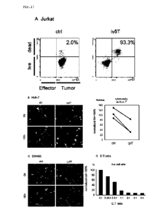
FIGS. 17 show results of determination of antitumor
activity on various tumor cells for yoT cells on day 38 of
differentiation induction. FIG. 17A shows results of
determination of cytotoxic activity on Jurkat cells. FIG. 17B
shows results of determination of cytotoxic activity on Huh-7
cells. FIG. 17C shows results of determination of cytotoxic
activity on SW480 cells. FIG. 17D shows live cell rates in the
case where an E:T ratio was gradually changed in mixed culture
of iPS cell-derived yoT cells (E) and Jurkat cells (T). (Example
9)
FIGS. 18 show results of determination of the retention of
TCR rearrangement and a cytotoxic mechanism for yoT cells on day
14
CA 03206400 2023 7 25
36 of differentiation induction.
FIG. 18A shows results of
evaluation of the expression of aPTCR on cell surfaces for
unpurified y5T cells (igdT) and peripheral blood mononuclear
cells (PB).
FIG. 18B shows results of determination of the
rearrangement of TCR genes (Vy9 and V52) by genomic PCR. FIG.
18C shows results of determination of the expression of granzyme
B and perforin in y5T cells.
FIG. 18D shows results of
determination of cytotoxic activity for purified y5T cells
(igdT). Whether or not the y5T cells were purified did not make
a large difference in dead cell rate. (Example 9)
FIG. 19 shows results of determination of gene expression
patterns in iPS cell-derived y5T cells and y5T cells separated
from peripheral blood by single-cell RNA-seq analysis. (Example
10)
FIG. 20 shows results of analysis of 0D25 among cell
surface expression markers in iPS cell-derived y5T cells and yoT
cells separated from peripheral blood by flow cytometry.
(Example 10)
FIG. 21 is an illustration of a protocol for
differentiation induction of y5T cells from iPS cells, for
investigating a method of activating iPS cell-derived y5T cells.
(Example 11)
FIGS. 22 show results of an investigation about IL-2 and/or
IL-15 in the method of activating iPS cell-derived y5T cells.
FIG. 22A shows results of determination of live cell counts, and
CA 03206400 2023 7 25
FIG. 22B shows results of evaluation of CD3+/y5TCR4 cells by flow
cytometry. (Example 11)
FIGS. 23 show results for y5T cells obtained by
differentiation induction from a 121-3 line of y5T cell-derived
iPS cells.
FIG. 23A shows results of determination of the
rearrangement of TCR genes (Vy9 and Vy2) of undifferentiated iPS
cells (121-3 line) and y5T cells obtained by differentiation
induction therefrom by genomic PCR. FIG. 23B shows results of
determination of the sequences of TCRys and TCRos of the y5T
cells and y5T cells obtained by subjecting peripheral blood
mononuclear cells to expansion culture with a next-generation
sequencer. (Example 12)
FIG. 24 shows results of evaluation by flow cytometry of
the expression of IFNy after iPS cell-derived y5T cells on day
39 of differentiation induction or y5T cells obtained by
subjecting peripheral blood mononuclear cells to expansion
culture were cocultured with Jurkat cells for 4 hours. (Example
13)
FIG. 25 shows results of evaluation by flow cytometry of
the expression of various surface markers in a cell population
including iPS cell-derived y5T cells on day 40 of differentiation
induction obtained by differentiation induction performed by a
method involving using feeder cells and a cell population (CD3-
positive or TCRy9-positive) including y5T cells obtained by
subjecting peripheral blood mononuclear cells to expansion
16
CA 03206400 2023- 7- 25
culture. (Example 14)
FIG. 26A is an illustration of a protocol involving
performing a step of stimulating yoT cells from day 17. FIG.
26B shows results of evaluation of the expression of CD3/yoTCR
by flow cytometry for cells on day 17 of differentiation
induction.
FIG. 260 shows results of evaluation of the
expression of 0D3/CD7 by flow cytometry for cells on day 24 of
differentiation induction. (Example 15)
FIGS. 27 show results of an investigation about IL-2 or
IL-15, or IL-15 or IL-15+HMEPP in a method of activating iPS
cell-derived yoT cells. FIG. 27A shows results of evaluation of
the expression of CD3/y5TCR by flow cytometry for cells on day
37 or day 33 of differentiation induction.
FIG. 27B shows
results of evaluation of the expression of CD3/CD7 by flow
cytometry for cells on day 23 of differentiation induction.
(Example 16)
FIG. 28 shows results of determination of cytotoxic
activity on Jurkat cells after freezing and thawing of cells on
day 24 of differentiation induction under a condition free from
using feeder cells. (Example 17)
FIG. 29A shows results of evaluation of the expression of
0D34/0D43 by flow cytometry for cells on day 10 of
differentiation induction. FIG. 29B shows results obtained by
further freezing and thawing the cells on day 10 of
differentiation induction, and evaluating the expression of
17
CA 03206400 2023 7 25
0D3/yoTCR by flow cytometry for cells on day 37 of
differentiation induction.
FIG. 290 shows results of
determination of cytotoxic activity on Jurkat cells for the cells
on day 37 of differentiation induction. (Example 18)
FIG. 30A is an illustration of a protocol in which iPS
cell-derived hematopoietic progenitor cells are frozen and
thawed, and then subjected to differentiation induction under a
serum-free condition free from using feeder cells. FIG. 30B
shows results of evaluation of the expression of 0D3/yoTCR by
flow cytometry for cells on day 17 of differentiation induction.
FIG. 300 shows results of determination of cytotoxic activity on
Jurkat cells for cells on day 24 of differentiation induction.
(Example 19)
FIG. 31A is an illustration of a protocol for inducing
differentiation of hematopoietic progenitor cells into yoT cells
under a hypoxic condition. FIG. 31B shows results of evaluation
of the expression of CD3/CD7 by flow cytometry for cells on day
17 of differentiation induction.
FIG. 310 shows results of
determination of cytotoxic activity on Jurkat cells for cells on
day 29 of differentiation induction. (Example 20)
FIGS. 32 show that iPS cell-derived yoT cells were induced
to differentiate under an animal-derived component-free
condition.
FIG. 32A shows results of evaluation of the
expression of 0D3/CD7 by flow cytometry for cells on day 17 of
differentiation induction.
FIG. 32B shows results of
18
CA 03206400 2023- 7- 25
determination of cytotoxic activity on Jurkat cells for cells on
day 31 of differentiation induction. (Example 21)
FIGS. 33 show that undifferentiated cells are not present
in a cell population. FIG. 33A shows results of evaluation by
flow cytometry of the expression of an undifferentiation marker
TRA-1-85 in a cell population on day 35 of differentiation
induction under a serum-free condition free from using feeder
cells. FIG. 33B is an illustration of a protocol for determining
whether colonies of undifferentiated cells appear in a cell
population. FIG. 33C shows that colonies of undifferentiated
cells do not appear in a cell population. (Example 22)
FIG. 34A shows the purification of CD3/y5T-positive cells
from a cell population under a serum-free condition free from
using feeder cells. FIG. 34B shows results obtained by further
determining cytotoxic activity on Jurkat cells for purified
cells. (Example 23)
Description of Embodiments
[0016] The present invention relates to an iPS cell-derived
yoT cell, which is a T cell derived from an iPS cell, wherein
the T cell has antigen-specific cytotoxic activity in a MHC-
unrestricted manner.
[0017] Human mature T cells are classified into two groups:
4-type T cells having a T cell receptor (TCR) made up of an a-
chain and a 13-chain; and yó-type T cells having a TCR made up of
19
CA 03206400 2023 7 25
a y-chain and a 5-chain. As used herein, the term "yoT cell"
refers to the yo-type T cell. In blood, the apT cells account
for a vast majority, whereas the y5T cells are a minority of
from 1% to 5% of all T cells. The yoT cells undergo rearrangement
of TCR genes in order to bind to diverse antigens and leave
memory cells, and hence may be regarded as a component of the
acquired immune system. Besides, the y5T cells also have a
function of, for example, attacking tumor cells through antigen
recognition similar to that by NK cells, which are innate immune
cells, without requiring antigen recognition by a TCR.
In
addition, it is considered that the y5T cells have the functions
of both the innate immune system and the acquired immune system.
Meanwhile, against a tumor antigen, ar cell-derived cytotoxic
T cells (CTLs) may be said to be an acquired immune system
requiring antigen information from dendritic cells. Thus, the
y5T cells and the aPT cells completely differ from each other
not merely in ratio of presence in blood, but also in their
functions, and it is known that the processes of differentiation
of the two types of cells also differ from each other (Non Patent
Literature 3).
[0018]
As used herein, the term "iPS cell" refers to an
undifferentiated cell established by reprogramming a somatic
cell by any of various methods. iPS cells serving as a starting
material in the present invention are suitably iPS cells that
are not iPS cells having a rearranged apTCR gene. The iPS cells
CA 03206400 2023- 7- 25
are most suitably iPS cells having a rearranged yoTCR gene. The
iPS cells having a rearranged y6TCR gene are hereinafter
sometimes referred to simply as "yoTCR-type iPS cells". As used
herein, the term "rearranged yoTCR gene" refers to a gene
encoding a TCR in which both of the rearrangement of a TCRG
region and the rearrangement of a TCRD region have occurred.
The TCRG region is made up of Vy-Jy, and the TCRD region is made
up of Vo-Do-J5.
[0019] Herein, the iPS cells may be generated by a method
known per se or any method to be developed in the future. For
example, the iPS cells may be generated on the basis of
descriptions in Patent Literature 1 and Non Patent Literature 1.
[0020] (Method of Generating iPS Cell)
The iPS cell to be used for generating the yoT cell of the
present invention may be generated by a method known per se or
any method to be developed in the future. Specifically, for
example, the iPS cell may be generated by a method described in
Patent Literature 1 or Non Patent Literature 1. For example,
the iPS cell may be generated by a method of generating iPS cells
including the following steps 1) to 3):
1) a step of stimulating collected blood cells with IL-2
and a bisphosphonate (e.g., one kind or a plurality of kinds
selected from zoledronic acid, pamidronic acid, alendronic acid,
risedronic acid, ibandronic acid, incadronic acid, etidronic
acid, minodronic acid, salts thereof, and hydrates thereof,
21
CA 03206400 2023- 7- 25
preferably zoledronic acid);
2) a step of introducing at least four kinds of genes
capable of expressing cell reprogramming factors (e.g., OCT3/4,
SOX2, KLF4, and c-MYC) into the blood cells through use of a
Sendai virus (SeV) vector; and
3) a step of culturing the cells having introduced therein
the genes.
[0021] (Culture of iPS Cells)
As a basal medium that may be used for maintenance culture
of the iPS cells, there may be used any of various stem cell
maintenance media, such as StemFitTM AKO2N (product name),
StemFitTM AKO3N (product name), ReproStem (product name),
iPSellon (product name), Essential 8 (product name), and TeSR-
E8 (product name). In particular, StemFitTmAKO2N (product name)
is preferred. The amount of a substance to be added to each
medium may be appropriately increased or decreased depending on
purposes. As an example of the substance to be added, Y27632,
which is a Rho-Associated Coil Kinase (ROCK) inhibitor, may be
used. In order to promote cell adhesion and growth, for example,
a laminin-511-E8 fragment may be used for a culture substrate
such as a culture dish. Specifically, iMatrix-511 silk (product
name) or iMatrix-511 (product name) may be used.
The
manufacturers/distributors of reagents and the like to be used
are not particularly limited as long as equivalent functions can
be exhibited. At the time of the passage of the iPS cells, a
22
CA 03206400 2023- 7- 25
protease such as trypsin may be used in detaching the cells from
the culture vessel, and for example, TrypLE Select (product name)
may be used.
[0022] (Differentiation Induction from iPS Cells into
Hematopoietic Progenitor Cells)
In the step of differentiation induction treatment from
the iPS cells into the y5T cells, first, the iPS cells are
induced to differentiate into hematopoietic progenitor cells.
In the method of generating an iPS cell-derived y5T cell of the
present invention, the step of differentiation induction
treatment from the hematopoietic progenitor cells into the y5T
cells using as a starting material cells obtained by inducing
the iPS cells to differentiate into the hematopoietic progenitor
cells may be regarded as the method of generating an iPS cell-
derived y5T cell. The method of generating an iPS cell-derived
y5T cell may further include the step from the iPS cells to the
hematopoietic progenitor cells. In addition, cells obtained by
freezing and thawing the iPS cell-derived hematopoietic
progenitor cells may be used in the method of the present
invention. A freezing period is not particularly limited, but
may be, for example, from 2 weeks to 1 year. In any case, the
iPS cells of the present invention are suitably iPS cells that
are not iPS cells having a rearranged apTCR gene. The iPS cells
is most suitably y5TCR-type iPS cells.
[0023] The step of differentiation induction from the iPS
23
CA 03206400 2023- 7- 25
cells into the hematopoietic progenitor cells is not
particularly limited, and a method known per se or any step to
be developed in the future may be adopted.
In the step of
differentiation induction into the hematopoietic progenitor
cells, the medium may be appropriately supplemented with one
kind or a plurality of kinds selected from cytokines, such as
FMS-like tyrosine kinase 3 ligand (FLT3L), stem cell factor (SCF),
bone morphogenetic protein-4 (BMP4), basic fibroblast growth
factor (bFGF), vascular endothelial growth factor (VEGF), IL-6,
insulin-like growth factors (IGF-1), IL-7, IL-11, erythropoietin
(EPO), thrombopoietin (TP0), IL-15, and IL-3. The medium may
also be appropriately supplemented with fetal bovine serum (FBS)
or fetal calf serum (FCS).
[0024]
The differentiation induction treatment from the iPS
cells of the present invention into the hematopoietic progenitor
cells may be performed, for example, under a condition free from
using feeder cells through culture in media described in the
following 1-1) to 1-4). The culture may be performed in the
following manner: until the hematopoietic progenitor cells are
obtained, there may be used Y27632, which is a ROCK inhibitor,
at a final concentration of from 0 pM to 50 pM, preferably from
1 pM to 30 pM, more preferably 10 pM, and a laminin-511 E8
fragment such as iMatrix-511 (product name) at from 0 pl to 50
pl, preferably from 1 pl to 30 pl, more preferably about 5 pl;
and the medium is changed to StemFitTM AKO2N free of the ROCK
24
CA 03206400 2023- 7- 25
inhibitor and laminin-511-E8 the next day, and the medium is
changed once every few days, for example, every 2 days. The
frequency of medium change, medium change amount, and the like
are not particularly limited, and an appropriate frequency and
amount may be appropriately decided. In addition, the number of
cells to be seeded may be appropriately increased or decreased.
In addition, the manufacturers/distributors of reagents and the
like to be used are not particularly limited as long as
equivalent functions can be exhibited. The entire culture may
be performed under the conditions of 37 0.5 C and 5% 002. For
passage, a protease such as trypsin, for example, TrypLE Select
(product name) may be used in detaching the cells from the
culture vessel.
[0025] 1-1) Day 0 of Differentiation Induction
StemFitmAKO2N (product name) may be used as a basal medium.
Culture may be performed in a culture system further including
a GSK-3a/13 inhibitor (OHIR99021, CAS number: 252917-06-9) at
from 0 11M to 20 pM, preferably from 0.5 IIM to 10 IIM, more
preferably 4 pM, BMP4 at from 0 ng/ml to 400 ng/ml, preferably
from 10 ng/ml to 200 ng/ml, more preferably 80 ng/ml, and VEGF
at from 0 ng/ml to 400 ng/ml, preferably from 10 ng/ml to 200
ng/ml, more preferably 80 ng/ml.
[0026] 1-2) Day 2 of Differentiation Induction
Advanced DMEM/F12 (product name) or Essential 6 (product
name) may be used as a basal medium. Culture may be performed
CA 03206400 2023- 7- 25
in a culture system further including a selective ALK5, 4, 7
inhibitor (SB431542) at from 0 pM to 20 pM, preferably from 0.5
pM to 10 pM, more preferably from 2 pM to 4 pM, bFGF at from 0
ng/ml to 200 ng/ml, preferably from 1 ng/ml to 100 ng/ml, more
preferably 50 ng/ml, SCF at from 0 ng/ml to 200 ng/ml, preferably
from 1 ng/ml to 100 ng/ml, more preferably 50 ng/ml, and VEGF at
from 0 ng/ml to 400 ng/ml, preferably from 10 ng/ml to 200 ng/ml,
more preferably 80 ng/ml.
In addition to the foregoing, L-
glutamine, penicillin/streptomycin, a differentiation induction
supplement for iPS/ES cells (e.g., StemFit (product name) For
Differentiation: hereinafter "AS401"), or the like may be
further appropriately selected and added. The optimal addition
amounts thereof may be appropriately decided.
[0027] 1-3) Day 4 of Differentiation Induction
Advanced DMEM/F12 (product name) or StemPro-34 SFM
(product name) may be used as a basal medium. Culture may be
performed in a culture system further including L-glutamine at
from 0 mM to 20 mM, preferably from 0.5 mM to 10 mM, more
preferably 2 mM, IL-3 at from 0 ng/ml to 200 ng/ml, preferably
from 1 ng/ml to 100 ng/ml, more preferably 50 ng/ml, IL-6 at
from 0 ng/ml to 200 ng/ml, preferably from 1 ng/ml to 100 ng/ml,
more preferably 50 ng/ml, FLT3L at from 0 ng/ml to 200 ng/ml,
preferably from 1 ng/ml to 100 ng/ml, more preferably 50 ng/ml,
SCF at from 0 ng/ml to 200 ng/ml, preferably from 1 ng/ml to 100
ng/ml, more preferably 50 ng/ml, VEGF at from 0 ng/ml to 200
26
CA 03206400 2023- 7- 25
ng/ml, preferably from 1 ng/ml to 100 ng/ml, more preferably 20
ng/ml, and EPO at from 0 IU/ml to 100 IU/ml, preferably from 1
IU/m1 to 50 IU/ml, more preferably 10 IU/ml. In addition to the
foregoing, penicillin/streptomycin, a differentiation induction
supplement for iPS/ES cells (e.g., AS401), or the like may be
further appropriately selected and added. The optimal addition
amounts thereof may be appropriately decided.
[0028] 1-4) Day 6 to Day 8 of Differentiation Induction
Advanced DMEM/F12 (product name) or StemPro-34 SFM
(product name) may be used as a basal medium. Culture may be
performed in a culture system further including L-glutamine at
from 0 mM to 50 mM, preferably from 1 mM to 20 mM, more preferably
2 mM, IL-3 at from 0 ng/ml to 200 ng/ml, preferably from 1 ng/ml
to 100 ng/ml, more preferably 50 ng/ml, IL-6 at from 0 ng/ml to
200 ng/ml, preferably from 1 ng/ml to 100 ng/ml, more preferably
50 ng/ml, SCF at from 0 ng/ml to 200 ng/ml, preferably from 1
ng/ml to 100 ng/ml, more preferably 50 ng/ml, and EPO at from 0
IU/ml to 100 IU/ml, preferably from 1 IU/ml to 50 IU/ml, more
preferably 10 IU/ml.
In addition to the foregoing,
penicillin/streptomycin, a differentiation induction supplement
for iPS/ES cells (e.g., AS401), or the like may be further
appropriately selected and added. The optimal addition amounts
thereof may be appropriately decided.
[0029] (Feeder Cells)
Feeder cells may be cocultured in the culture of the iPS
27
CA 03206400 2023- 7- 25
cells or the differentiation induction treatment of the iPS cells.
As the feeder cells, there may be used one kind or a plurality
of kinds of cell lines selected from, for example, mouse
embryonic fibroblasts (MEFs), 0P9, 0P9/DLL1, 0P9-DL4, and
10T1/2/DL4 cells. Meanwhile, when cells obtained by inducing
differentiation of the iPS cells are to be administered to a
human in cell therapy or the like, a stable production method
free of any animal-derived substance is desired. In the present
invention, differentiation induction into the yoT cell of the
present invention may be performed without using feeder cells by
using the above-mentioned laminin-511 E8 fragment and medium
components in a well-designed manner.
[0030]
(Differentiation Induction from iPS Cell-derived
Hematopoietic Progenitor Cells into yoT Cells)
The process of differentiation induction from the iPS cell-
derived hematopoietic progenitor cells into the yoT cells may be
performed as coculture with feeder cells, or may be performed as
culture under a condition free from using feeder cells. Further,
culture may be performed under a serum-free condition, and
culture may be performed under an animal-derived component-free
condition.
In addition, the process of differentiation
induction from the iPS cell-derived hematopoietic progenitor
cells into the r5T cells may involve culture under a hypoxic
condition.
The expression "under a hypoxic condition" means
that an 02 concentration under culture conditions in the process
28
CA 03206400 2023- 7- 25
of differentiation induction from the iPS cell-derived
hematopoietic progenitor cells into the yoT cells is lower than
an 02 concentration at which culture is generally performed. The
02 concentration at which the culture under a hypoxic condition
is performed is not particularly limited, but is, for example,
less than 20% (v/v), preferably less than 10% (v/v).
[0031]
In addition, a yoT cell stimulant may be added in
order to obtain desired yoT cells, or may not be added depending
on culture conditions.
Examples of the yoT cell stimulant
include a phosphoric acid compound that is a metabolite of a
mevalonate pathway or a non-mevalonate pathway serving as an
isoprenoid biosynthesis pathway, or a derivative thereof.
Examples of the phosphoric acid compound that is a metabolite of
the mevalonate pathway or the non-mevalonate pathway serving as
the isoprenoid biosynthesis pathway include (E)-4-hydroxy-3-
methyl-but-2-enyl pyrophosphate (HMBPP) and isopentenyl
diphosphate (IPP). An example of the derivative is bromohydrin
pyrophosphate (BrHPP). Another example of the yoT cell stimulant
is a specific inhibitor of a farnesyl pyrophosphate (FPP)
synthase serving as a rate-limiting enzyme of the biosynthesis
pathway. The specific inhibitor of the FPP synthase promotes
the accumulation of the phosphoric acid compound in cells.
Examples of the FPP synthase-specific inhibitor include
nitrogen-containing bisphosphonates (N-BPs), specifically
zoledronic acid and pamidronate. Further, IL-15 and IL-2 each
29
CA 03206400 2023- 7- 25
also have a function as a yoT cell stimulant.
[0032] A. System involving Coculture with Feeder Cells
A-1) Day 10- of Differentiation Induction
For example, in culture from day 10 (hematopoietic
progenitor cells) onward after the differentiation induction
from the iPS cells by the above-mentioned treatments 1-1) to 1-
4), aMEM (product name) may be used as a basal medium. The
culture may be performed in a culture system further including
PBS at from 0% to 30%, preferably from 0% to 20%, more preferably
from 10% to 20%, SCF at from 0 ng/ml to 100 ng/ml, preferably
from 1 ng/ml to 50 ng/ml, more preferably 10 ng/ml, IL-7 at from
0.1 ng/ml to 20 ng/ml, preferably from 0.5 ng/ml to 10 ng/ml,
more preferably 5 ng/ml, FLT3L at from 0.1 ng/ml to 50 ng/ml,
preferably from 1 ng/ml to 20 ng/ml, more preferably 5 ng/ml,
and L-ascorbic acid at from 1 pg/m1 to 1,000 pg/ml, preferably
from 10 pg/m1 to 500 pg/ml, more preferably 100 pg/ml. Further,
the culture system may include IL-2 at from 0 ng/ml to 200 ng/ml,
preferably from 1 ng/ml to 100 ng/ml, more preferably 10 ng/ml,
or may include TPO at from 0 ng/ml to 200 ng/ml, preferably from
1 ng/ml to 100 ng/ml, more preferably 10 ng/ml. In addition to
the foregoing, penicillin/streptomycin or the like may be
further appropriately selected and added. In addition, a 0.1%
Polyvinyl alcohol+4% B27 (product name) supplement or the like
may be used in place of FBS. The manufacturers/distributors of
reagents and the like to be used are not particularly limited as
CA 03206400 2023- 7- 25
long as equivalent functions can be exhibited.
The optimal
addition amounts thereof may be appropriately decided.
The
culture may be performed by seeding the cells (hematopoietic
progenitor cells) on day 10 after differentiation induction into
a culture substrate such as a culture dish seeded with feeder
cells. The medium is changed, for example, every 2 days, and
the supernatant may be collected on day 12, day 18, and day 24
after differentiation induction by pipetting and transferred
onto fresh feeder cells to continue culture. The frequency of
medium change, medium change amount, and the like are not
particularly limited, and an appropriate frequency and amount
may be appropriately decided.
[0033]
A-2) Day 30 or Day 31- of Differentiation Induction
The cells that have been cultured from day 10 to day 30 or
day 31 after differentiation induction through use of the above-
mentioned medium may be cultured under a condition free from
using feeder cells. In the culture under such condition, RPMI
1640 medium may be used as a basal medium. The culture may be
performed in a medium further containing FBS at from 0% to 30%,
preferably from 0% to 20%, more preferably from 10% to 20%. A
0.1% Polyvinyl alcohol+4% B27 (product name) supplement or the
like may be used in place of FBS. Further, the culture may be
performed in a culture system including IL-2 and/or IL-15 at
from 0 ng/ml to 200 ng/ml, preferably from 1 ng/ml to 100 ng/ml,
more preferably 10 ng/ml, or the culture may be performed in a
31
CA 03206400 2023- 7- 25
culture system including Immunace (product name) at from 0 IU/ml
to 1,000 IU/ml, from 10 IU/ml to 500 IU/ml, preferably 100 IU/ml
and 2-Mercaptoethanol (2-Me) at from 0 pM to 100 pM, from 1 pM
to 50 pM, preferably 10 pM.
In addition to the foregoing,
penicillin/streptomycin or the like may be further appropriately
added.
[0034]
Further, for example, HMBPP may be added as a yoT
cell stimulant. Its addition concentration only needs to be a
concentration at which the y5T cells are stimulated and which
does not cause cytotoxicity, and is not particularly limited,
but may be set to, for example, from 0 nM to 100 nM, preferably
from 0.01 nM to 20 nM, more preferably 1 nM.
[0035]
B. System involving Culture free from using Feeder
Cells
B-1) Day 10- of Differentiation Induction
For example, culture from day 10 (hematopoietic progenitor
cells) onward after the differentiation induction from the iPS
cells by the above-mentioned treatments 1-1) to 1-4) may involve
culture using a culture substrate coated with vascular cell
adhesion molecule-1 (VCAM1), and delta-like protein 4 (DLL4) or
delta-like protein 1 (DLL1).
From day 10 to day 24 of
differentiation induction, culture may be performed in, for
example, Lymphoid progenitor Expansion Medium (product name)
included in a StemSpanTM T cell generation kit (product name).
Medium change was performed in accordance with the protocol of
32
CA 03206400 2023- 7- 25
the StemSpanTM kit. Specifically, it may be appropriate that the
medium be further added on day 13 of differentiation induction,
and the medium be changed on each of day 17 and day 20 of
differentiation induction.
Around day 17 to day 24 of
differentiation induction, the medium may be changed to T cell
progenitor Maturation Medium (product name) included in the
above-mentioned kit.
It may be appropriate that the above-
mentioned medium be further added on day 27 of differentiation
induction, and thereafter, the medium be changed about twice a
week, such as day 31 and day 34 of differentiation induction.
The frequency of medium change, medium change amount, and the
like are not particularly limited, and an appropriate frequency
and amount may be appropriately decided.
[0036]
B-2) Around Day 17 to Day 24- of Differentiation
Induction
Culture may be continued by the method described in B-1),
but culture may be performed in a medium supplemented with a yoT
cell stimulant from around day 17 to day 24 of differentiation
induction. The decreasing tendency of the number of cells, which
is sometimes observed from around day 17 to day 24 of
differentiation induction, is ameliorated by the supplementation
with the yoT cell stimulant. Specifically, the culture may be
performed in the medium described in A-2 that is supplemented
with IL-2 and/or IL-15, and y6T cell stimulants, such as HMBPP
and the FPP synthase-specific inhibitor. The culture may also
33
CA 03206400 2023- 7- 25
be performed in the medium described in A-2 that is free of FES
and is similarly supplemented with HMBPP. The culture may also
be performed in RPMI 1640 medium containing AS401 and being
supplemented with IL-2 and/or IL-15, and HMBPP, instead of the
medium described in A-2.
[0037] B-3) Day 10- of Differentiation Induction
Culture may be continued by a method involving further
incorporating Dickkopf-1 (DKK1) and/or azelaic acid (AZA) into
the medium conditions described in B-1). Further, from around
day 17 to day 24 of differentiation induction, culture may be
performed in a medium supplemented with a yoT cell stimulant.
From around day 17 to day 24 of differentiation induction,
specifically, culture may be performed in the medium described
in A-2 that is supplemented with HMBPP. The culture may also be
performed in the medium described in A-2 that is free of FBS and
is similarly supplemented with HMBPP. The culture may also be
performed in RPMI 1640 medium containing AS401 and being
supplemented with IL-2 and/or IL-15, and a yoT cell stimulant
such as HMBPP, instead of the medium described in A-2.
[0038] C. Culture using Beads
The cells that have been cultured by the differentiation
induction method of the present invention may be cultured using
beads. The size of the beads is not particularly limited, and
may be smaller than the size of cells, or may be equal to or
larger than the size of cells. For example, when the cells on
34
CA 03206400 2023- 7- 25
day 10 of differentiation induction are cultured under the above-
mentioned various conditions, the culture may be performed by
mixing the beads into the medium. The beads only need to be
beads of a material usable for cell culture, and are not
particularly limited, but specifically, Dynabeads Protein G
(product name) may be used. The culture may be performed under
a condition free from using feeder cells by coating the beads
with, for example, VCAM1 and DLL4.
[0039]
D. Culture using Animal-derived Component-free
Medium
D-1) Day 10- of Differentiation Induction
The cells that have been cultured by the differentiation
induction method of the present invention may be cultured under
a condition involving using an animal-derived component-free
medium.
For example, culture from day 10 (hematopoietic
progenitor cells) onward after the differentiation induction
from the iPS cells by the above-mentioned treatments 1-1) to 1-
4) may involve culture using a culture substrate coated with
vascular cell adhesion molecule-1 (VCAM1), and delta-like
protein 4 (DLL4) or delta-like protein 1 (DLL1). Around day 10
to day 24 of differentiation induction, for example, RPMI 1640
containing AS401 may be used as a basal medium for the animal-
derived component-free medium. The medium may further contain,
for example, SCF, IL-7, FLT3L, L-ascorbic acid, IL2, and TPO
described in A-1.
CA 03206400 2023- 7- 25
[0040] D-2) Around Day 17 to Day 24- of Differentiation
Induction
From around day 17 to day 24 of differentiation induction,
culture may be performed in the medium described in A-2 that is
supplemented with IL-2, IL-15, and the y5T cell stimulant. Such
medium may use RPMI 1640 containing A5401 as a basal medium.
From around day 17 to day 24 of differentiation induction,
specifically, culture may be performed in a medium supplemented
with one or a plurality of IL-2, IL-15, and HMEPP.
[0041] (y5T Cells obtained through Differentiation
Induction)
The y5T cells generated by the differentiation induction
method of the present invention are T cells having a peculiar T
cell receptor (TCR) made up of a y-chain and a 5-chain on the
surface thereof. For such cell surface, the expressions of cell
markers, such as CD3, CD7, CD8a, CD45RA, and y5TCR, may be
determined. The yoT cells of the present invention preferably
express, in particular, one or a plurality selected from CD7,
CD8a, and CD45RA, and meanwhile, are preferably free from
expressing one or a plurality selected from CD25, IFNy, CD5, and
CD27. The obtained iPS cell-derived y5T cells have a feature of
having antigen-specific cytotoxic activity in a MHC-unrestricted
manner. Further, a difference is found between the patterns of
cell surface markers in the y5T cells generated by inducing
differentiation of iPS cells of the present invention and y5T
36
CA 03206400 2023- 7- 25
cells separated from peripheral blood. For example, for CD7 and
CD8a, the iPS cell-derived yoT cells show higher expression
tendencies, and for IL2RA (CD25), CD5, and IFNy, the y5T cells
separated from peripheral blood show higher expression
tendencies. In addition, for example, for CD45RA, the iPS cell-
derived yoT cells show a higher expression tendency, and for
0D27, the y5T cells separated from peripheral blood show a higher
expression tendency.
[0042]
The y5T cells thus caused to undergo differentiation
induction may be isolated by appropriately selecting a known
technique. An example of such known technique is such flow
cytometry as described in Examples to be described later,
involving using an antibody against a cell surface marker and a
cell sorter. In the case of isolating "T cells having desired
antigen specificity" from a human, a method involving performing
purification using, for example, an affinity column on which a
desired antigen is immobilized may be adopted.
[0043]
A cell population of the purified yoT cells is made
up of homogeneous cells, and is distinguished from a cell
population made up of y5T cells separated from peripheral blood.
The y5T cell population of the present invention has higher
cytotoxic activity in an antigen-specific manner than a y5T cell
population separated from peripheral blood.
[0044]
The cell population including the y5T cells includes,
for example, many cells having base sequences identical to each
37
CA 03206400 2023- 7- 25
other in a complementarity determining region (CDR) of a TCR
gene. The cell population has a feature in that yoT cells having
base sequences identical to each other particularly in a CDR3
region among CDRs are included in the yoT cells that make up the
cell population at a high ratio, for example, at a ratio of 90%
or more. The cell population including the yoT cells of the
present invention may include 1x10⁵ or more yoT cells.
[0045] Further, the cell population including the yoT cells
of the present invention includes y5T cells, which show a higher
expression amount than yoT cells separated from peripheral blood
in terms of expression amount of CD7 and/or CD8a, at a ratio of
90% or more of the y5T cells that make up the cell population.
Further, in terms of expression amount of one or a plurality
selected from CD25, INFy, and CD5, yoT cells showing a lower
expression amount than yoT cells separated from peripheral blood
are included at a ratio of 90% or more of the yoT cells that
make up the cell population. Further, yoT cells showing a higher
expression amount of CD45RA than yoT cells separated from
peripheral blood and ex vivo expanded, and a lower expression
amount than the yoT cells separated from peripheral blood and ex
vivo expanded in terms of expression amount of CD27 are included
at a ratio of 70% or more of the yoT cells that make up the cell
population.
[0046] In addition, the cell population including the y5T
cells of the present invention has a feature in that 10% or less
38
CA 03206400 2023- 7- 25
of the y5T cells that make up the cell population are
undifferentiated cells, and further, it is suitable that no
undifferentiated cells be present in the y5T cells that make up
the cell population.
Whether or not a given cell is an
undifferentiated cell may be determined, for example, with a
marker indicating undifferentiation such as TRA-1-85.
[0047]
The y5T cells generated through treatment by the
differentiation induction treatment method of the present
invention have an excellent immune function, and hence may be
used for, for example, treatment or prevention of a disease,
such as a tumor, an infectious disease (e.g., viral infectious
disease), or an autoimmune disorder.
Further, the cell
population of the y5T cells produced by the method of the present
invention may be utilized as an antigen-specific cellular
immunotherapeutic agent or a pharmaceutical composition by being
incorporated thereinto as an active ingredient. The y5T cells
generated through treatment by the differentiation induction
treatment method of the present invention can be utilized for
such formulation even after being frozen and thawed. The y5T
cell population is expected to be also applicable to an immune
cell treatment method making use thereof.
The y5T cell
population of the present invention is expected to further
enhance the effect of the y5T cells by being used in combination
with an immune checkpoint inhibitor.
The immune checkpoint
inhibitor is not limited to ones known per se and ones to be
39
CA 03206400 2023- 7- 25
developed in the future, but examples thereof include drugs
targeting immune checkpoints, such as PD-1, PD-L1, and CTLA-4.
Further, like NK cells, the yoT cells are expected to have an
antibody-dependent cellular cytotoxicity (ADCC) action of
enhancing the effect of a molecularly targeted therapeutic
agent/antibody formulation used for the treatment of any of
various cancers (e.g., Herceptin or Rituxan), and hence can be
expected to have a high therapeutic effect when used in
combination with any such antibody formulation.
The
pharmaceutical composition containing the yoT cell population of
the present invention may be prepared through formulation by a
known pharmaceutical method.
[0048]
In such formulation, a pharmacologically acceptable
carrier or medium, specifically, sterile water or physiological
saline, a vegetable oil, a solvent, a base, an emulsifier, a
suspending agent, a surfactant, a stabilizer, a vehicle, an
antiseptic agent, a binder, a diluent, a tonicity agent, a
soothing agent, an extender, a disintegrant, a buffer, a coating
agent, a lubricant, a colorant, a solubilizing agent, other
additives, or the like may be appropriately combined.
In
addition, the pharmaceutical composition may be used in
combination with, for example, a known pharmaceutical
composition or immunostimulator to be used for the treatment or
prevention of the above-mentioned disease.
When the
pharmaceutical composition of the present invention is
CA 03206400 2023- 7- 25
administered, its dose is appropriately selected depending on,
for example, the age, body weight, symptoms, and health status
of a subject, and the kind of the composition.
[0049]
The present invention also encompasses an antigen-
specific cellular immune treatment method, including
administering the iPS cell-derived y5T cell of the present
invention. The present invention also encompasses a treatment
method for a disease, such as cancer, an infectious disease, or
an autoimmune disorder, the method including administering the
iPS cell-derived y5T cell of the present invention.
In the
method of the present invention, the dose of the active
ingredient for a subject varies depending on, for example, the
body weight, age, and symptoms of the subject, and an
administration method, but could be appropriately selected by a
person skilled in the art.
Examples
[0050]
The present invention is specifically described
below by way of Examples for a better understanding of the
present invention.
Needless to say, however, the present
invention is by no means limited to these Examples and the like.
[0051]
(Example 1) Differentiation Induction from iPS Cells
In this Example, a differentiation induction treatment
method for y5T cells generated from y5TCR-type iPS cells
generated by a method of Non Patent Literature 1 is described.
41
CA 03206400 2023- 7- 25
[0052] (1-1) Culture of yoTCR-type iPS Cells (62B3 Line)
y6TCR-type iPS cells (62B3 line) cultured under a condition
free from using feeder cells were passaged into a 6-well plate
at 2x10³/well, and subjected to maintenance culture.
In the
maintenance culture, StemFitTmAKO2N (manufactured by Ajinomoto)
containing 1.6 pg/well of iMatrix-511 (manufactured by Nippi)
was used. 0.5xTrypLETm select (manufactured by Thermo Fisher)
was used for detaching and dispersing the cells at the time of
the passage, and a medium obtained by supplementing StemFitTM
AKO2N with Y27632 (manufactured by Wako Pure Chemical
Industries) at a final concentration of 10 pM and 3.2 pl of
iMatrix-511 was used for passage culture. The next day, the
medium was changed to StemFitTmAKO2N free of Y27632 and iMatrix-
511, and thereafter, the medium was changed every 2 days. The
medium was added at 1.5 ml/well. Culture in all cases, including
the following steps and Examples to be described later, was
performed under the conditions of 37 0.5 C and 5% CO2.
[0053] (1-2) Day 0 of Differentiation Induction
After 7 days from the passage in (1-1) described above,
the medium was changed to a medium shown in Table 1 (Step 1) at
2 ml/well.
42
CA 03206400 2023 7 25
Table 1
Step 1
Manufacturer Product number Concentration
Stem Fit Ajinomoto AKO2N
CHIR99021 TOCRIS 4423 4 pM
rh BMP4 R&D 314-BP 80 ng/ml
rh VEGF R&D 293-VE 80 ng/ml
[0054] (1-3) Day 2 of Differentiation Induction
After 2 days from (1-2) described above, the medium was
changed to a medium shown in Table 2 (Step 2) at 2 ml/well.
Table 2
Step 2
Manufacturer Product number Concentration
Advanced DMEM/ .
F12 gibco 12634-10
AS401 Ajinomoto 20170228A 20% (v/v)
L-Glutamine gibco 25030-081 2 mM
50 Unit/ml
Penicillin- Pen
gibco 15140-122
Streptomycin 50 pg/ml
Strep
FUJIFILM Wako
5E43152 Pure Chemical 033-24631 2 pM
Corporation
rh VEGF R&D 293-VE 80 ng/ml
bFGF Wako 060-04543 50 ng/ml
SCF R&D 255-SC 50 ng/ml
[0055] (1-4) Day 4 of Differentiation Induction
After 2 days from (1-3) described above, the medium was
changed to a medium shown in Table 3 (Step 3) at 2 ml/well.
43
CA 03206400 2023- 7- 25
Table 3
Step 3
Manufacturer Product number Concentration
Advanced DMEM/ .
glbco 12634-10
F12
AS401 Ajinomoto 20170228A 20% (v/v)
L-Glutamine gibco 25030-081 2 mM
50 Unit/m1
Penicillin- Pen
gibco 15140-122
Streptomycin 50 pg/ml
Strep
SCF R&D 255-SC 50 ng/ml
IL3 Reprotech AF-200-03 50 ng/ml
IL6 R&D 206-IL-050 50 ng/ml
Flt3L R&D 308-FK-025 50 ng/ml
rh VEGF R&D 293-VE 20 ng/ml
Kyowa Hakko
EPO 10 IU/m1
Kirin
[0056] (1-5) Day 6 of Differentiation Induction
After 2 days from (1-4) described above, the medium was
changed to a medium shown in Table 4 (Step 4) at 2 ml/well.
Table 4
Step 4
Manufacturer Product number Concentration
Advanced DMEM/ gibco 12634-10
F12
AS401 Ajinomoto 20170228A 20% (v/v)
L-Glutamine gibco 25030-081 2 mM
50 Unit/ml
Penicillin- Pen
gibco 15140-122
Streptomycin 50 pg/ml
Strep
SCF R&D 255-SC 50 ng/ml
IL6 R&D 206-IL-050 50 ng/ml
Kyowa Hakko
EPO 10 IU/m1
Kirin
[0057] (1-6) Day 8 of Differentiation Induction
44
CA 03206400 2023- 7- 25
After 2 days from (1-5) described above, the medium was
changed to the same medium as the medium shown in Table 4 (Step
4) at 2 ml/well.
[0058] (1-7) Evaluation of Cells on Day 10 of
Differentiation Induction
The expression of CD34/CD43 was evaluated by flow cytometry.
CD34+/CD43+ cells and CD34-/CD43+ cells were detected in large
numbers. That is, the cells had differentiated into
hematopoietic progenitor cells (FIG. 1A).
[0059] (1-8) Day 10 of Differentiation Induction
The cells except for those subjected to flow cytometry in
(1-7) described above were seeded into a 12-well culture dish
seeded with 0P9/N-DLL1 cells serving as feeder cells. A medium
having the composition shown in Table 5 was used in a medium
amount of 1 ml/well, and half of the medium was changed every 2
days.
Table 5
Step 5 day 10-
Manufacturer Product number Concentration
20% FES/uMEM gibco 11900-016
50 Unit/ml
Penicillin- Pen
gibco 15140-122
Streptomycin 50 pg/ml
Strep
IL2 Reprotech 200-02 10 ng/ml
IL7 R&D 207-IL-010 5 ng/ml
Flt3L R&D 308-FK-025 5 ng/ml
L-Ascorbic
Nacalai 03420-52 100 pg/ml
acid
CA 03206400 2023- 7- 25
[0060] (1-9) Evaluation of Cells on Day 31 of
Differentiation Induction
The expression of CD3/yoTCR was evaluated by flow cytometry.
As a result, a large number of CD3+/TCR+ cells were detected to
verify differentiation into TCR cells (FIG. 1B). The obtained
cells are hereafter in this Example referred to as "iPS cell-
derived yoT cells."
[0061] (1-10) Evaluation of Cells on Day 31 of
Differentiation Induction
Cytotoxicity assay against Jurkat cells (derived from
human leukemia T cells) was performed. At effector:target (E:T)
ratio=2:1, 5x10⁴ Jurkat cells (T) stained with a fluorescent dye
CFSE were added per well of a 96-well culture dish, and 1x10⁵ of
the iPS cell-derived y6T cells (E) were added thereto, followed
by 16 hours of culture. Dead cells were stained by 7-amino-
actinomycin D (7-AAD) staining. Cell death (7-AAD-positive) was
recognized for many of the Jurkat cells (CFSE-positive cells).
That is, it was recognized that the iPS cell-derived y6T cells
had a cytotoxic function. Even though activating stimulation
culture of the yoT cells was not performed in this Example,
cytotoxic activity was recognized.
[0062] (Example 2) Differentiation Induction from iPS Cells
In this Example, with regard to the yoT cells generated by
differentiation induction treatment from the yoTCR-type iPS
cells generated by the method of Non Patent Literature 1, medium
46
CA 03206400 2023- 7- 25
components from day 10 of differentiation induction onward and
medium components from day 31 of differentiation induction
onward differ from those of Example 1. In particular, the medium
components from day 31 of differentiation induction onward
include HMBPP serving as a yoT cell stimulant.
[0063] (2-1) Until day 10 of differentiation induction
treatment, the same treatments as in (1-1) to (1-6) of Example
1 were performed.
(2-2) Day 10- of Differentiation Induction
The cells generated in (1-6) of Example 1 described above
were seeded into a 12-well culture dish seeded with 0P9/N-DLL1
cells serving as feeder cells. 1 ml/well of a medium having the
composition of a medium shown in Table 6 (Step 5) was entirely
changed every 7 days.
Table 6
Step 5
day 10-
(Example 2)
Manufacturer Product number Concentration
20% FBS/aMEM gibco 11900-016
50 Unit/ml
Penicillin- Pen
gibco 15140-122
Streptomycin 50 pg/ml
Strep
SCF R&D 255-SC 100 ng/ml
Flt3L R&D 308-FK-025 100 ng/ml
TPO Peprotech AF-300-18-10 100 ng/ml
IL-7 R&D 207-IL-010 100 ng/ml
L-Ascorbic
Nacalai 03420-52 100 pg/ml
acid
[0064] (2-3) Evaluation of Cells on Day 17 of
47
CA 03206400 2023- 7- 25
Differentiation Induction
The expression of CD7 (T cell differentiation marker) was
evaluated by flow cytometry. CD7-positive cells were found,
revealing that differentiation had proceeded into T cells (FIG.
2A).
[0065] (2-4) Day 31- of Differentiation Induction
With a yoT cell stimulation medium shown in Table 7, half
of the medium was changed every 2 days. The y5T cell stimulation
medium contains HMBPP.
Table 7
yoT
stimulation day 31-
medium
Manufacturer Product number Concentration
RPMI 1640 Nacalai 30264-85
FES SIGMA F7524 10% (v/v)
50 Unit/ml
Penicillin- Pen
gibco 15140-122
Streptomycin 50 pg/ml
Strep
IL-2 Reprotech 200-02 100 ng/ml
cayman
HMBPP chemical 13580 1 nM
company
[0066] (2-5) Evaluation of Cells on Day 54 of
Differentiation Induction
The expression of CD3/yoTCR was evaluated by flow cytometry.
A large number of CD3+/TCR+ cells were detected to verify
differentiation into TCR cells. That is, it was recognized that
the obtained cells were iPS cell-derived yoT cells. In addition,
48
CA 03206400 2023- 7- 25
the expression CD45RA, generally used as an indicator of the
maturation of T cells, was also evaluated, and as a result, it
was revealed that CD3+ cells included both CDRA+ cells and CDRA-
cells (FIG. 2B).
[0067]
(Example 3) Differentiation Induction from iPS Cells
under Condition involving using Feeder Cells
In this Example, description is made of y5T cells generated
by differentiation induction treatment from y5TCR-type iPS cells
generated by the method of Non Patent Literature 1 in the same
manner as in Example 1. Differentiation induction treatment was
performed in the same manner as in Example 1, and from day 31
onward, half of the y5T cell stimulation medium (containing HMBPP
and FBS) was changed every 2 days in the same manner as in (2-
4) of Example 2.
Then, evaluation of marker expression and
cytotoxicity assay were performed.
[0068]
(3-1) Until day 10 of differentiation induction
treatment, the same treatments as in (1-1) to (1-6) and (1-8)
described in Example 1 were performed.
(3-2) Evaluation of Cells on Day 17 of Differentiation Induction
The expression of CD7 (T cell differentiation marker) was
evaluated by flow cytometry. CD7-positive cells were detected,
revealing that differentiation had proceeded into T cells (FIG.
3A).
(3-3) Evaluation of Cells on Day 55 of Differentiation Induction
The expression of CD3/y5TCR was evaluated by flow cytometry.
49
CA 03206400 2023- 7- 25
A large number of CD3+/TCR4 cells were detected to verify
differentiation into yoT cells (FIG. 3B). The obtained cells
are hereafter in this Example referred to as "iPS cell-derived
yoT cells."
(3-4) Evaluation of Cells on Day 55 of Differentiation Induction
Cytotoxicity assay against Jurkat cells was performed.
5x10⁴ Jurkat cells stained with CFSE were added per well of a
96-well culture dish, and 1x10⁵ of the iPS cell-derived yoT cells
on day 55 of differentiation induction were further added,
followed by 16 hours of culture at E:T ratio=2:1. After that,
7-AAD staining (dead cell staining) was performed. Many of the
Jurkat cells (CFSE-positive cells) were 7-AAD-positive, and thus
many dead cells were recognized. That is, it was recognized
that the iPS cell-derived yoT cells had a cytotoxic function
against tumor cells (FIG. 3C).
[0069] (Example 4) Differentiation Induction from iPS Cells
under Condition free from using Feeder Cells
In this Example, with regard to y6T cells generated by
differentiation induction treatment from yoTCR-type iPS cells
generated by the method of Non Patent Literature 1 in the same
manner as in Example 1, a differentiation induction method under
a condition free from using feeder cells is described. In this
Example, differentiation induction treatment was performed by
the following procedure in accordance with a protocol
illustrated in FIG. 4.
CA 03206400 2023 7 25
[0070]
(4-1) Until day 8 of differentiation induction
treatment, the same treatments as in (1-1) to (1-6) described in
Example 1 were performed.
(4-2) Day 10 of Differentiation Induction
With use of a 48-well culture dish coated with VCAM1 and
DLL4, a suspension of 1.2x10⁴ of cells on day 10 of
differentiation induction in 250 pl of Lymphoid progenitor
Expansion Medium included in the StemSpanTmT cell generation kit
(Stem Cell Technologies) was seeded per well.
PBS(-) having
dissolved therein 5 pg/ml VCAM1 and 10 pg/ml DLL4 was added to
a commercially available 48-well culture dish that had not been
subjected to hydrophilic treatment for cell adhesion (cell
culture-non-treated) at 100 pl per well, and the whole was left
at rest at 4 C overnight. The solution was removed, and the
culture dish was washed with PBS(-) once and used as a culture
dish coated with VCAM1 and DLL4. In the step involving using
Lymphoid progenitor Expansion Medium, culture was performed
under a condition involving using neither feeder cells nor serum.
[0071]
(4-3) Thereafter, medium change was performed in
accordance with the protocol of the StemSpanTM kit. Specifically,
250 pl of the medium was further added on day 13 of
differentiation induction, and half of the medium was changed on
each of day 17 and day 20 of differentiation induction. On day
24 of differentiation induction, the medium was changed to T
cell progenitor Maturation Medium included in the above-
51
CA 03206400 2023- 7- 25
mentioned kit. The above-mentioned medium was further added on
day 27 of differentiation induction, and thereafter, half of the
medium was changed twice a week, such as day 31 and day 34 of
differentiation induction.
[0072]
(4-4) Evaluation of Cells on Day 33, Day 35, and Day
37 of Differentiation Induction
The expression of CD3/yoTCR was evaluated by flow cytometry.
A large number of CD3+/TCR+ cells were detected to verify
differentiation into TCR cells and identify the cells as iPS
cell-derived y6T cells (FIG. 5).
The results shown are the
results of three independent differentiation induction
experiments.
The days on which evaluation was performed
(initiation of differentiation induction was defined as day 0)
are shown in the figure.
[0073]
(Example 5) Differentiation Induction from iPS Cells
under Condition free from using Feeder Cells
In this Example, with regard to yoT cells generated by
differentiation induction treatment from y6TCR-type iPS cells in
the same manner as in Example 4, a differentiation induction
method under a condition free from using feeder cells is
described. In this Example, differentiation induction treatment
was performed by the following procedure in accordance with a
protocol illustrated in FIG. 6.
[0074]
(5-1) The same treatments as in (4-1) to (4-3) of
Example 4 were performed, and from day 10 to day 24 of
52
CA 03206400 2023- 7- 25
differentiation induction, culture was performed under a
condition involving using neither feeder cells nor serum.
(5-2) Day 24 of Differentiation Induction
The medium was changed to the y5T cell stimulation medium
(containing HMBPP and FBS) shown in Table 7 in (2-4) of Example
2, and thereafter, half of the medium was changed every 3 days.
(5-3) Evaluation of Cells on Day 37 of Differentiation Induction
The cells were observed for the number of cells using a
phase-contrast microscope. The cells generated through culture
in the y5T cell stimulant (HMBPP)-free medium in Example 4 were
also similarly observed. As a result, when culture was performed
in the y5T cell stimulation medium, a clearly larger number of
cells were observed (FIG. 7A).
Further, the expression of
CD3/y5TCR was evaluated by flow cytometry, and as a result, a
large number of CD34/TCR+ cells were detected to verify
differentiation into TOR cells (FIG. 7B).
It was recognized
that the obtained cells were iPS cell-derived y5T cells.
[0075]
(Example 6) Differentiation Induction from iPS Cells
under Condition free from using Feeder Cells
In this Example, with regard to y5T cells generated by
differentiation induction treatment from y5TCR-type iPS cells in
the same manner as in Example 4, a differentiation induction
method under a condition free from using feeder cells is
described. In this Example, differentiation induction treatment
was performed by the following procedure in accordance with a
53
CA 03206400 2023- 7- 25
protocol illustrated in FIG. 8.
[0076] (6-1) The same treatments as in (4-1) to (4-3) of
Example 4 were performed, and from day 10 to day 24 of
differentiation induction, culture was performed under a
condition involving using neither feeder cells nor serum.
(6-2) Day 24 of Differentiation Induction
On day 24 of differentiation induction, the medium was
changed to each of a. the yErT cell stimulation medium (containing
HMBPP and FBS) shown in Table 7 in (2-4) of Example 2, b. RPMI
1640 (containing HMBPP) medium containing AS401 in place of the
basal medium (10% FBS/RPMI 1640) of the y5T cell stimulation
medium shown in Table 7, and c. Lymphoid progenitor Expansion
Medium included in the StemSpanTM kit, and the medium was changed
by the same technique as in (5-2) of Example 5.
(6-3) Evaluation 2 of Cells on Day 32 of Differentiation
Induction
Cells on day 32 of differentiation induction were observed
for the number of cells using a phase-contrast microscope. In
c. the medium included in the StemSpanTM kit, the number of cells
is clearly small, whereas in b. the serum-free medium, a cell
density equivalent to that in a. the serum medium was observed
(FIG. 9). Further, for the above-mentioned cells, the expression
of CD3/y5TCR was evaluated by flow cytometry. Under each of the
conditions, a large number of CD3+/TCR4 cells were detected to
identify the cells as iPS cell-derived y5T cells (FIG. 10). The
54
CA 03206400 2023- 7- 25
obtained cells are hereafter in this Example referred to as "iPS
cell-derived yoT cells."
(6-4) Evaluation 1 of Cells on Day 35 of Differentiation
Induction
Among cells on day 35 of differentiation induction, cells
obtained under the a. and b. medium conditions were used and
subjected to cytotoxicity assay against Jurkat cells by the same
technique as in (1-10) of Example 1. 5x10⁴ Jurkat cells stained
with CFSE were added per well of a 96-well culture dish, 1x10⁵
of the iPS cell-derived yoT cells on day 35 of differentiation
induction were further added, and evaluation was performed after
1 day (dl) and after 4 days (d4) from the initiation of mixed
culture at E:T ratio=2:1. As a result, cell aggregates
indicating the activation of T cells were found. As compared to
a control (ctrl) with no effector cells (iPS cell-derived yoT
cells) added, it was observed that the cells obtained under the
a. and b. medium conditions clearly appeared to be fewer (FIG.
11).
After 1 day from the initiation of mixed culture,
cytotoxicity was clear even under the serum-free medium
condition b., though to a lesser extent as compared to the a.
medium condition, and after 4 days, even more evident cytotoxic
activity was recognized (FIG. 12).
[0077]
(Example 7) Differentiation Induction from iPS Cells
under Condition free from using Feeder Cells
In this Example, yoT cells were generated by subjecting
CA 03206400 2023- 7- 25
yoTCR-type iPS cells to differentiation induction treatment in
the same manner as in Example 4.
[0078] (7-1) Culture was performed by performing the same
treatments as in (4-1) and (4-2) of Example 4.
(7-2) However, day 10 of differentiation induction was performed
under: the same condition (i) as in (4-2); (ii) the condition of
adding Dickkopf-1 (DKK1) thereto at a final concentration of 30
ng/ml; (iii) the condition of adding azelaic acid (AZA) thereto
at a final concentration of 5 mM; and (iv) the condition of
adding both Dickkopf-1 (DKK1) and AZA thereto at the same
concentrations as in (ii) and (iii), respectively.
(7-3) Thereafter, medium change was performed in accordance with
the protocol of the StemSpanTM kit. That is, 250 pl of each
medium described in (7-2) was added on day 13 of differentiation
induction, and half of each medium was changed on day 17 and day
20 of differentiation induction.
(7-4) Evaluation of Cells on Day 24 of Differentiation Induction
For cells on day 24 of differentiation induction, the
expression of CD7 serving as a T cell differentiation marker was
evaluated by flow cytometry. As a result, it was found that
DKK1 and AZA each had a positive effect on differentiation
induction efficiency, and treatment with combined use thereof
achieved a higher effect (FIG. 13).
[0079] (Example 8) Differentiation Induction Method
involving using Magnetic Beads
56
CA 03206400 2023- 7- 25
In this Example, differentiation induction into T cells
was performed through mixed culture with magnetic beads coated
with VCAM1 and DLL4 instead of coating a culture dish under a
condition free from using feeder cells.
[0080]
(8-1) Until day 8 of differentiation induction
treatment, the same treatments as in (1-1) to (1-6) of Example
1 were performed.
(8-2) Preparation of Solution of Magnetic Beads coated with VCAM1
and DLL4
Magnetic beads (Dynabeadslm Protein G (manufactured by
Invitrogen)) were vortexed, 5 pl thereof and 1 ml of PBS were
placed into a tube, and the tube was left at rest on a magnetic
stand for magnetic bead capture for 1 minute. PBS was removed,
and the tube was removed from the stand, followed by the addition
of 200 pl of PBS, 4.26 pl of VCAM1 (100 pg/ml solution), and
4.26 pl of DLL4 (100 pg/ml solution). The tube was left at rest
at room temperature for 15 minutes. The tube was left at rest
on the magnetic stand for 1 minute, the solution was removed,
and the tube was removed from the stand. 500 pl of Lymphoid
progenitor Expansion Medium included in the StemSpanTM kit was
added, and pipetting was performed for suspension.
(8-3) Day 10 of Differentiation Induction
4.75x10⁵ of the cells generated in (8-1) described above
were suspended in 500 pl of the magnetic bead solution prepared
in (8-2), seeded into a 24-well low-attachment culture dish
57
CA 03206400 2023- 7- 25
(PrimeSurfaceTm), and cultured.
(8-4) 500 1.11 of the above-mentioned medium was added on day 13
of differentiation induction, and half of the medium was changed
on each of day 17 and day 20 of differentiation induction.
(8-5) Evaluation of Cells on Day 24 of Differentiation Induction
For cells on day 24 of differentiation induction, the
expression of CD7 serving as a T cell differentiation marker was
evaluated by flow cytometry. As a result, it was recognized
that, although at a ratio as low as 0.3%, CD7-positive cells
were clearly present as compared to a control (isotype control).
That is, it was revealed that differentiation into T cells was
also able to be performed by this method involving mixed culture
with magnetic beads (FIG. 14).
[0081] (Example 9) yoT Cells generated from yoTCR-type iPS
Cells
In this Example, the characteristics of yoT cells generated
from yoTCR-type iPS cells (iPS cell-derived yoT cells) were
determined. First, a method of generating iPS cell-derived y6T
cells is described, and then various characteristics of the cells
are described.
[0082] (9-1) Method of generating iPS Cell-derived yoT
Cells
yErT cells were generated by a method illustrated in FIG.
15.
-Establishment of iPS Cells
58
CA 03206400 2023- 7- 25
yoTCR-type iPS cells generated by the method of Non Patent
Literature 1 were used. StemfitTM AKO2N (Ajinomoto) was used for
maintenance culture of the iPS cells.
0.5xTrypLETm select
(manufactured by Thermo Fisher) was used for passage. In each
step of differentiation induction treatment into hematopoietic
progenitor cells, a 6-well culture plate was used, and cells
were seeded at 2x10³ cells/well.
Every day, the medium was
aspirated, and the entire medium of 2.0 ml/well was changed.
[0083] -Day 0 of Differentiation Induction: State of yoTCR-
type iPS Cells (HPC1)
Stemfit AKO2N (Ajinomoto, Tokyo, Japan, AKO2N)
0HIR99021 (Tocris, Bristol, UK, 4423) 4 pM
BMP4 (R&D, Minneapolis, MN, 314-BP) 80 ng/ml
VEGF (R&D, Minneapolis, MN, 293-VE) 80 ng/ml
[0084] -Day 2 of Differentiation Induction: (HPC2)
Essential 6 (Thermofisher, Waltham, MA, A1516501)
SB431542 (WAKO, Osaka, Japan, 033-24631) 2 pM
bFGF (WAKO, Osaka, Japan, 060-04543) 50 ng/ml
SCF (R&D, Minneapolis, MN, 255-SC) 50 ng/ml
VEGF (R&D, Minneapolis, MN, 293-VE) 80 ng/ml
[0085] -Day 4 of Differentiation Induction: (HPC3)
StemPRO34SFM (Thermofisher, Waltham, MA, 10639-011)
L-Glutamine (Life technologies, 25036-081) 2 mM
IL-3 (Peprotech, Cranbury, NJ, AF-200-03) 50 ng/ml
IL-6 (R&D, Minneapolis, MN, 206-IL) 50 ng/ml
59
CA 03206400 2023- 7- 25
FLT3L (R&D, Minneapolis, MN, 308-FK) 50 ng/ml
SCF (R&D, Minneapolis, MN, 255-SC) 50 ng/ml
VEGF (R&D, Minneapolis, MN, 293-VE) 20 ng/ml
EPO (Kyowa Kirin, Tokyo, Japan) 10 IU/m1
[0086] -Day 6 and Day 8 of Differentiation Induction: (HPC4)
StemPRO34SFM (Thermofisher, Waltham, MA, 10639-011)
L-Glutamine (Life technologies, 25036-081) 2 mM
IL-6 (R&D, Minneapolis, MN, 206-IL) 50 ng/ml
SCF (R&D, Minneapolis, MN, 255-SC) 50 ng/ml
EPO (Kyowa Kirin, Tokyo, Japan) 10 IU/m1
[0087] -Day 10- of Differentiation Induction: Culture on
Feeder Cells (0P9/N-DLL1) in T Cell Differentiation Medium
described below
Accutase (Nacalai Tesque, Kyoto, Japan, 12679-54) was used
for the passage of the cells. Thereafter, half of the medium
was changed every 2 days. In addition, on day 12, day 18, and
day 24, the supernatant was collected by pipetting and seeded
onto fresh feeder cells (0P9/N-DLL1).
(T Cell Differentiation Medium)
aMEM (Gibco, 11900-016)
FBS (Sigma-Aldrich, St. Louis, MO, F7524) 20%
SCF (R&D, Minneapolis, MN, 255-SC) 10 ng/ml
TPO (R&D, Minneapolis, MN) 10 ng/ml
IL-7 (R&D, Minneapolis, MN, 207-IL) 5 ng/ml
FLT3L (R&D, Minneapolis, MN, 308-FK) 5 ng/ml
CA 03206400 2023- 7- 25
L-ascorbic acid (Nacalai Tesque, Kyoto, Japan, 30264-56)
100 ug/m1
[0088] -Day 30- of Differentiation Induction: Culture in
y5T Activation Medium
The cells treated with Accutase were suspended in a yoT
activation medium described below, and cultured in a feeder cell-
free medium. Thereafter, half of the medium was changed every
2 days. Cells on days 7 to 14 of activation culture were
subjected to cytotoxicity assay.
[0089] (y5T Activation Medium)
RPMI 1640 (Nacalai Tesque, Kyoto, Japan, 30264-56)
FES (Sigma-Aldrich, St. Louis, MO, F7524) 10%
HMBPP (Cayman chemical, Ann Arbor, MI, 13580) 1 nM
Immunace (Shionogi pharmaceuticals, Osaka, Japan) 100
IU/ml
2-Me (Nacalai Tesque, Kyoto, Japan) 10 11M
[0090] (9-2) Process of Differentiation from y5TCR-type iPS
Cells into y6T Cells
The morphology of cells in the process of differentiation
was observed with a phase-contrast microscope (FIG. 16A), and
cell surface markers were determined by flow cytometry (FIG.
16B).
dO: day 0 of differentiation induction: y5TCR-type iPS cells
d10: day 10 of differentiation induction: cells differentiated
into hematopoietic progenitor cells
61
CA 03206400 2023 7 25
d30: day 30 of differentiation induction: y5T cells before
activating stimulation of NM cells
d51: day 51 of differentiation induction: y5T cells after
activating stimulation of y5T cells
[0091] (9-3) Antitumor Effect
With use of iPS cell-derived y5T cells on day 38 of
differentiation induction, antitumor activity on various tumor
cells was determined (FIGS. 17).
In these experiments,
unpurified y5T cells were used. As a control, the condition of
culturing tumor cells alone without the addition of the y5T cells
was used.
A. Cytotoxicity assay against Jurkat cells (derived from human
leukemia T cells) was performed.
At effector:target (E:T)
ratio=2:1, 5x10⁴ Jurkat cells stained with a fluorescent dye CFSE
were added per well of a 96-well culture dish, and 1x10⁵ of the
iPS cell-derived y5T cells were added thereto, followed by 16
hours of culture. After that, dead cells were stained by 7-AAD
staining. As compared to the control, the yoT cells of the
present invention clearly had higher cytotoxic activity on the
Jurkat cells (FIG. 17A).
[0092]
B. Cytotoxicity assay against Huh-7 cells (derived
from human hepatoma cells) was performed. At effector:target
(E:T) ratio=2:1, 5x10⁴ Huh-7 cells stained with a fluorescent
dye CFSE were added per well of a 96-well culture dish, and 1x10⁵
of the iPS cell-derived y5T cells were added thereto, followed
62
CA 03206400 2023- 7- 25
by 16 hours of culture. After that, observation with a phase-
contrast microscope was performed to measure a tumor area. As
compared to the control, the yoT cells of the present invention
clearly had higher cytotoxic activity on the Huh-7 cells (FIG.
17B).
[0093] C. Cytotoxicity assay against SW480 cells (derived
from human colon cancer) was performed. At effector:target (E:T)
ratio=2:1, 5x10⁴ SW480 cells stained with a fluorescent dye CFSE
were added per well of a 96-well culture dish, and 1x10⁵ of the
iPS cell-derived yoT cells were added thereto, followed by 16
hours of culture. After that, observation with a phase-contrast
microscope was performed to measure a tumor area. As compared
to the control, the y5T cells of the present invention clearly
had higher cytotoxic activity on the SW480 cells (FIG. 17C).
[0094] D. In the mixed culture of the iPS cell-derived yoT
cells (E) and the Jurkat cells (T), the E:T ratio was gradually
changed. A live cell rate at 0:1 was defined as 100%, and live
cell rates were compared (FIG. 17D).
[0095] (9-4) Retention of TCR Rearrangement and Cytotoxic
Mechanism
The retention of TCR rearrangement and a cytotoxic
mechanism were determined using iPS cell-derived y5T cells on
day 36 of differentiation induction (FIGS. 18).
A. For unpurified iPS cell-derived yoT cells (igdT) and
peripheral blood mononuclear cells (PB), the expression of an
63
CA 03206400 2023- 7- 25
aPTCR on the cell surface was evaluated. The expression of the
aPTCR was detected in PB, but the expression of the apTCR was
not detected in the yoT cells (igdT) of the present invention
(FIG. 18A).
B. Genomic PCR for TCR Gene Rearrangement
The rearrangement of TCR genes (Vg9 and Vd2) was determined
by genomic PCR. It was recognized that yoT cells (igdT) sorted
by flow cytometry retained TCR gene rearrangement like the
undifferentiated (undiff) state (FIG. 18B). Peripheral blood
mononuclear cells (PBMC) were used as a positive control.
C. iPS cell-derived yoT cells (igdT) whose CD3 had been labeled
in advance and Jurkat cells were cocultured under 3 pg/m1
Brefeldin A. It was recognized that the iPS cell-derived y6T
cells expressed granzyme B and perforin (FIG. 18C). As granzyme
B and perforin are molecular entities of a cytotoxic function by
T cells, it was recognized that the iPS cell-derived yoT cells
of the present invention had cytotoxicity.
D. yoT cells (igdT) purified by flow cytometry (FACS) were
subjected to cytotoxicity assay. The cytotoxicity assay was
performed under the conditions of the method described in A. of
(9-3). Jurkat cells cultured alone without the addition of the
iPS cell-derived yoT cells were used as a control (ctrl) in FIG.
18D. In addition, the unpurified iPS cell-derived y5T cells are
indicated as bulk, and the purified iPS cell-derived yoT cells
are indicated as sort. Whether or not the iPS cell-derived y5T
64
CA 03206400 2023- 7- 25
cells were purified did not make a large difference in dead cell
rate (FIG. 18D).
[0096] E. HLA Types of iPS Cell Lines and Tumor Cells
The results of determination of the HLA types of the iPS
cells used for the iPS cell-derived y5T cells of the present
invention, and respective tumor cells used in Examples 3 and 6
and this Example are shown in Table 8. The HLA types of the iPS
cells do not coincide with the HLA types of the respective tumor
cells, but antitumor actions were found on the respective tumor
cells (this Example, A. to C.). Thus, it was recognized that
the iPS cell-derived y5T cells of the present invention had
antigen-specific cytotoxic activity in a MHC-unrestricted manner.
Table 8
HLA-A HLA-B HLA-C
HLA-DRB1
62B3* 02:01 24:02 40:01 54:01 01:02 03:04 04:03 04:05
121-3* 24:01 31:01 35:01 52:01 04:01 12:02 09:01 13:02
Jurkat' 03:01 - 07:02 35:03 04:01 07:02 07:01 15:01
Huh-7' 11:011 - 54:01 1 - 01:02 - 08:03 1
-
SW480' 02:01 24:02 07:02 15:18 07:02 07:04 01:03 13:01
*iPS cell line
'tumor cell line
[0097] (Example 10) Comparison of iPS Cell-derived y5T
Cells and y5T Cells separated from Peripheral Blood
In this Example, cell surface expression marker genes in
iPS cell-derived y5T cells (igdT) generated by inducing
differentiation of iPS cells and y5T cells (PR-gdT) present in
peripheral blood were compared. For the iPS cell-derived yoT
CA 03206400 2023- 7- 25
cells of this Example, culture was performed by the method
described in Example 1 and the method described in (9-1) of
Example 9, and cells on day 36 to day 42 of differentiation
induction were used. Cells obtained by culturing mononuclear
cells separated from peripheral blood in the yoT activation
medium described in (9-1) of Example 9 were used as yoT cells
separated from peripheral blood of this Example.
[0098] (10-1) Single-cell RNA-seq Analysis
The iPS cell-derived yoT cells, and the yoT cells separated
from peripheral blood and cells in peripheral blood excluding
the yoT cells were analyzed for differences in marker gene
expression by single-cell RNA-seq analysis.
As a result,
different expression patterns were shown for each of CD7, CD8a,
IL18R1, IL2RA (CD25), IL2RB, and IFNy (FIG. 19, Table 9).
Table 9
igdT>PB-gdT CD7, CD8a
igdT=PB-gdT
CD3E, oTCR, IL2RB, IL18R1, Perforin, Granzyme B,
NKG7
igdT<PB-gdT IL2RA, IFNy
[0099] (10-2) Analysis of CD25 by Flow Cytometry
The expressions of CD25 in the iPS cell-derived yoT cells
and the yoT cells separated from peripheral blood were compared
by flow cytometry. iPS cell-derived TCR-Vy9-positive cells were
mostly 0D25-negative cells, whereas TCR-Vy9-positive cells
separated from peripheral blood were mostly CD25-positive cells
(FIG. 20).
66
CA 03206400 2023- 7- 25
Thus, it was recognized that the iPS cell-derived y5T cells
and the yoT cells separated from peripheral blood had different
patterns of cell surface markers.
[0100] (Example 11) Method of activating iPS Cell-derived
y5T Cells
In this Example, a method of activating iPS cell-derived
y5T cells was investigated. Specifically, for cells on day 30
of differentiation induction in the generation method described
in (9-1) of Example 9, an investigation was performed as to which
of IL-2 and/or IL-15 enabled more effective generation of iPS
cell-derived y5T cells when the following y5T activation medium
was further supplemented therewith (see FIG. 21).
(Activation Medium)
RPMI 1640 (Nacalai Tesque, Kyoto, Japan, 30264-56)
FBS (Sigma-Aldrich, St. Louis, MO, F7524) 10%
HMBPP (Cayman chemical, Ann Arbor, MI, 13580) 1 nM
2-Me (Nacalai Tesque, Kyoto, Japan) 10 11M
[0101] According to the results of evaluations of live cell
counts and CD3+y5T cells, the supplementation with IL-15 was
preferred to IL-2, and IL-15 alone was more effective even when
compared to its combined use with IL-2 (FIGS. 22).
[0102] (Example 12) Characteristics of y5T Cells generated
from y5TCR-type iPS Cells (121-3 Line)
In this Example, the characteristics of y5T cells generated
from y5TCR-type iPS cells (121-3 line) were determined.
67
CA 03206400 2023- 7- 25
[0103] (12-1) In this Example, the 121-3 line was used
instead of the 62B3 line as the y6TCR-type iPS cells, culture
was performed by the method described in (9-1) of Example 9, and
cells on day 36 of differentiation induction were used.
[0104] (12-2) Retention of TCR Rearrangement
A. The rearrangement of TCR genes (Vy9 and Vy2) of the y5T cells
(iy5T) obtained by differentiation induction from the y5TCR-type
iPS cells (121-3 line) was determined by genomic PCR. It was
recognized that iy5T sorted by flow cytometry retained TCR gene
rearrangement like the undifferentiated (undiff) state (FIG.
23A).
B. The sequences of the TCRys and TCR5s of the y5T cells (iy5T)
obtained by differentiation induction from the y5TCR-type iPS
cells (121-3 line) and y5T cells (PBy5T) obtained by subjecting
peripheral blood mononuclear cells to expansion culture were
analyzed with a next-generation sequencer. The base sequences
and amino acid sequences of the CDR3 regions of their respective
TCRys and TCRos were identified, and the frequencies of each
sequence were shown as pie charts (FIG. 23B). It was recognized
that the PBy.5T cell population was made up of cells having
diverse sequences, whereas the iy5T cell population was made up
of cells all harboring a single kind of TCRy and TOM gene
rearrangement.
[0105] (Example 13) Characteristics of iPS-derived y5T
Cells generated from y5TCR-type iPS Cells (62B3 Line)
68
CA 03206400 2023- 7- 25
In this Example, with regard to iPS-derived y5T cells
generated by the generation method described in Example 9, the
expression of IFNy was evaluated by flow cytometry for cells
obtained by coculturing cells on day 39 of differentiation
induction with Jurkat cells for 4 hours.
[0106] The expression of interferon gamma (IFNy) as well as
the expression of granzyme B in iPS cell-derived yoT cells (iy5T)
and y5T cells (PBy5T) obtained by subjecting peripheral blood
mononuclear cells to expansion culture was evaluated with a flow
cytometer. The results showed that granzyme B was expressed in
both the cell populations, whereas IFNy was recognized to be
expressed only in PBy5T and not recognized to be expressed in
iy5T (FIG. 24).
[0107] (Example 14) Comparison of iPS Cell-derived y5T
Cells and y5T Cells obtained by expanding Peripheral Blood
In this Example, cell surface expression markers in iPS
cell-derived y5T cells (igdT) generated by inducing
differentiation of y6TCR-type iPS cells (62B3 line or 121-3 line)
and y5T cells (PB-gdT) obtained by expanding peripheral blood
were compared.
[0108] (14-1) Culture was performed by a method involving
using feeder cells through the same treatment as in (9-1) of
Example 9.
(14-2) The expressions of various cell surface markers (CD25,
CD7, CD5, CD45RA, and CD27) in a cell population (iy5T) including
69
CA 03206400 2023- 7- 25
y5T cells for cells on day 40 of differentiation induction and
a cell population (CD3-positive or TCRy9-positive) including y6T
cells (PBy5T) obtained by subjecting peripheral blood
mononuclear cells to expansion culture were evaluated with a
flow cytometer. It was recognized that, as compared to PBy5T,
the iPS cell-derived y5T cells (CD3-positive or TCRy9-positive
cells among iy5T) had the following features: the ratio of cells
expressing CD7 was high, the ratio of cells expressing CD5 and
0D25 was low, and the ratio of CD45RA+CD27- cells was high (FIG.
25).
[0109]
(Example 15) Investigation of Step of Stimulating
y5T Cells
In this Example, with regard to y5T cells generated by
differentiation induction treatment from y5TCR-type iPS cells in
the same manner as in Example 5, description is made of a
differentiation induction method under a condition involving
using neither feeder cells nor serum and a condition of
performing the step of stimulating yoT cells not from day 24 but
from day 17. In this Example, differentiation induction was
performed by the following procedure in accordance with a
protocol illustrated in FIG. 26A (New protocol).
[0110]
(15-1) Culture was performed by performing the same
treatment as in (5-1) of Example 5.
However, the step of
stimulating y5T cells was performed from day 17 of
differentiation induction.
CA 03206400 2023- 7- 25
[0111] (15-2) Evaluation of Cells on Day 17 of
Differentiation Induction
For cells on day 17 of differentiation induction, the
expression of CD3/yoTCR (gdTCR) was evaluated by flow cytometry.
CD3+/TCR+ cells were detected to verify differentiation into TCR
cells and identify the cells as iPS cell-derived yoT cells (FIG.
26B). The obtained cells are hereafter in this Example referred
to as "iPS cell-derived yoT cells."
[0112] (15-3) Day 17 of Differentiation Induction
The medium was changed to a medium obtained by using RPMI
1640 containing 20% AS401 as a basal medium, and adding 1 nM
HMBPP (Cayman chemical, Ann Arbor, MI, 13580) and 100 ng/ml IL2
(Reprotech, 200-02) thereto, and thereafter, half of the medium
was changed every 3 days.
[0113] (15-4) Day 24 of Differentiation Induction
Further, for cells on day 24 of differentiation induction,
the expression of CD3/CD7 was evaluated by flow cytometry (FIG.
26C). iPS cell-derived yoT cells were obtained even under the
condition of shortening the step of stimulating yoT cells.
[0114] (Example 16) Method of activating iPS Cell-derived
yoT Cells under Condition free from using Feeder Cells
In this Example, a method of activating iPS cell-derived
yoT cells under a condition involving using neither feeder cells
nor serum was investigated.
[0115] A. Culture was performed under a condition involving
71
CA 03206400 2023- 7- 25
using neither feeder cells nor serum through the same treatments
as in (15-1) and (15-3) of Example 15. However, cells on day 17
of differentiation induction were treated under the same
condition as in (15-3) of Example 15 as well as the condition of
changing IL-2 in (15-3) to IL-15.
On day 33 or day 37 of
differentiation induction, the expression of CD3/yoTCR was
evaluated by flow cytometry regarding whether iPS cell-derived
yoT cells were able to be more effectively generated. CD3+/TCR+
cells were detected to verify differentiation into TCR cells and
identify the cells as iPS cell-derived y6T cells (FIG. 27A). It
was able to be recognized that iPS cell-derived yoT cells were
able to be generated by using any one of IL-2 or IL-15 in the
step of stimulating yoT cells. In addition, as compared to IL-
2, the addition of IL-15 provided more iPS cell-derived yoT cells.
B. Differentiation induction treatment was performed using IL-
15 in the step of stimulating yoT cells in A above, and an
investigation was performed as to whether iPS cell-derived yoT
cells were able to be generated with or without the addition of
HMBPP. For cells on day 23 of differentiation induction, the
expression of CD3/0D7 was evaluated by flow cytometry. CD3+/TCR+
cells were detected to verify differentiation into TCR cells and
identify the cells as iPS cell-derived yoT cells. iPS cell-
derived y5T cells were obtained even under the condition of not
adding the yoTCR stimulant HMBPP (FIG. 27B).
[0116]
(Example 17) Cytotoxic Activity after Freezing and
72
CA 03206400 2023- 7- 25
Thawing of iPS Cell-derived yoT Cells
In this Example, iPS cell-derived y6T cells under a
condition involving using neither feeder cells nor serum were
frozen and thawed, and subjected to cytotoxicity assay.
[0117] (17-1) Culture was performed under a condition
involving using neither feeder cells nor serum through the same
treatments as in (15-1) and (15-3) of Example 15. However, IL-
2 in (15-3) of Example 15 was changed to IL-15. On day 24 of
differentiation induction, the cells were frozen using CS10
(manufactured by Cosmo Bio).
[0118] (17-2) Evaluation of Cells on Day 24 of
Differentiation Induction
The frozen cells were thawed 2 weeks later and subjected
to cytotoxicity assay against Jurkat cells. At effector:target
(E:T) ratio=2:1, 5x10⁴ Jurkat cells stained with a fluorescent
dye CFSE were added per well of a 96-well culture dish, and 1x10⁵
of the iPS cell-derived yoT cells on day 24 of differentiation
induction were added thereto, followed by 16 hours of culture.
Dead cells were stained by 7-amino-actinomycin D (7-AAD)
staining. Cell death (7-AAD-positive) was recognized for many
of the Jurkat cells (CFSE-positive cells) (FIG. 28). That is,
it was recognized that the iPS cell-derived yoT cells had a
cytotoxic function even after freezing and thawing.
[0119] (Example 18) Differentiation Induction after
Freezing and Thawing of iPS Cell-derived Hematopoietic
73
CA 03206400 2023 7 25
Progenitor Cells
In this Example, iPS cell-derived hematopoietic progenitor
cells were frozen and thawed, and then subjected to
differentiation induction to generate y5T cells.
[0120] (18-1) Evaluation of Cells on Day 10 of
Differentiation Induction
The same treatments as in (1-1) to (1-6) shown in Example
1 were performed, and cells on day 10 of differentiation
induction were evaluated by flow cytometry and recognized to be
in the stage of hematopoietic progenitor cells (FIG. 29A).
[0121] (18-2) Day 10 of Differentiation Induction
The above-mentioned cells were frozen using CS10
(manufactured by Cosmo Bio) and thawed about 1 year later. After
the thawing, differentiation induction was performed by a method
involving using feeder cells through the same treatment as in
(9-1) of Example 9.
[0122] (18-3) Evaluation 1 of Cells on Day 37 of
Differentiation Induction
For cells on day 37 of differentiation induction (at a
differentiation induction culture period of 37 days including
days before and after the freezing), the expression of CD3/y5TCR
was evaluated by flow cytometry. CD3+/TCR+ cells were detected,
and hence the cells were identified as iPS cell-derived y5T cells
(FIG. 29B). Further, the cells on day 37 of differentiation
induction were subjected to cytotoxicity assay against Jurkat
74
CA 03206400 2023- 7- 25
cells. At effector:target (E:T) ratio=2:1, 5x10⁴ Jurkat cells
stained with a fluorescent dye CFSE were added per well of a 96-
well culture dish, and 1x10⁵ of the iPS cell-derived yoT cells
on day 24 of differentiation induction were added thereto,
followed by 16 hours of culture. Dead cells were stained by 7-
amino-actinomycin D (7-AAD) staining.
Cell death (7-AAD-
positive) was recognized for many of the Jurkat cells (CFSE-
positive cells) (FIG. 29C). That is, it was recognized that the
iPS cell-derived y5T cells had a cytotoxic function even after
freezing and thawing.
[0123]
(Example 19) Differentiation Induction under
Condition involving using neither Feeder Cells nor Serum after
Freezing and Thawing of iPS Cell-derived Hematopoietic
Progenitor Cells
In this Example, iPS cell-derived hematopoietic progenitor
cells were frozen and thawed, and then subjected to
differentiation induction under a condition involving using
neither feeder cells nor serum to generate y6T cells. In this
Example, differentiation induction was performed by the
following procedure in accordance with a protocol illustrated in
FIG. 30A. The freezing in this Example was performed for 18
days.
[0124]
(19-1) Until day 8 of differentiation induction, the
same treatments as in (1-1) to (1-6) shown in Example 1 were
performed. The cells were frozen using CS10 (manufactured by
CA 03206400 2023 7 25
Cosmo Bio) on day 10 of differentiation induction and thawed 18
days later.
[0125] (19-2) Day 10 of Differentiation Induction
For the cells after the thawing, with use of a 48-well
culture dish coated with VCAM1 and DLL4, a suspension of 1.2x10⁴
of the cells on day 10 of differentiation induction in a medium
obtained by supplementing Lymphoid progenitor Expansion Medium
included in the StemSpanTM T cell generation kit (Stem Cell
Technologies) with DKK1 at a final concentration of 30 ng/ml and
azelaic acid (AZA) at a final concentration of 5 mM was seeded
per well. PBS(-) having dissolved therein 5 pg/ml VCAM1 and 10
pg/ml DLL4 was added to a commercially available 48-well culture
dish that had not been subjected to hydrophilic treatment for
cell adhesion (cell culture-non-treated) at 100 pl per well, and
the whole was left at rest at 4 C overnight. The solution was
removed, and the culture dish was washed with PBS(-) once and
used as a culture dish coated with VCAM1 and DLL4.
[0126] (19-3) Thereafter, medium change was performed in
accordance with the protocol of the StemSpanTM kit. Specifically,
250 pl of the medium was further added on day 13 of
differentiation induction.
[0127] (19-4) Day 17 of Differentiation Induction
On day 17 of differentiation induction, differentiation
induction was performed under a condition involving using
neither feeder cells nor serum by the same technique as in (15-
76
CA 03206400 2023- 7- 25
3) of Example 15 except for changing IL-2 in (15-3) of Example
15 to IL-15. Thus, yoT cells were generated.
[0128]
(19-5) Evaluation of Cells on Day 17 of
Differentiation Induction
For cells on day 17 of differentiation induction (at a
differentiation induction culture period of 17 days including
days before and after the freezing), the expression of CD3/y5TCR
was evaluated by flow cytometry. CD3+/TCR+ cells were detected,
and hence the cells were identified as iPS cell-derived y5T cells
(FIG. 30B).
[0129]
(19-6) Evaluation of Cells on Day 24 of
Differentiation Induction
Cells on day 24 of differentiation induction (at a
differentiation induction culture period of 24 days including
days before and after the freezing) were subjected to
cytotoxicity assay against Jurkat cells.
At effector:target
(E:T) ratio=2:1, 5x10⁴ Jurkat cells stained with a fluorescent
dye CFSE were added per well of a 96-well culture dish, and 1x10⁵
of the iPS cell-derived y5T cells on day 24 of differentiation
induction were added thereto, followed by 16 hours of culture.
Dead cells were stained by 7-amino-actinomycin D (7-AAD)
staining. Cell death (7-AAD-positive) was recognized for many
of the Jurkat cells (CFSE-positive cells) (FIG. 30C).
[0130]
(Example 20) Differentiation Induction from
Hematopoietic Progenitor Cells under Hypoxic Condition
77
CA 03206400 2023 7 25
This Example was carried out under a condition involving
using neither feeder cells nor serum. However, y6T cells were
generated by performing differentiation induction from
hematopoietic progenitor cells under a hypoxic condition. In
this Example, differentiation induction was performed by the
following procedure in accordance with a protocol illustrated in
FIG. 31A.
[0131] (20-1) The same treatments as in (4-1) and (4-2) of
Example 4 were performed.
(20-2) However, cells on day 10 of differentiation induction
were cultured under a condition involving using neither feeder
cells nor serum in a medium obtained by supplementing Lymphoid
progenitor Expansion Medium included in the StemSpanTM T cell
generation kit (Stem Cell Technologies) described in (4-2) of
Example 4 with DKK1 at a final concentration of 30 ng/ml and
azelaic acid (AZA) at a final concentration of 5 mM, with the 02
concentration being changed from 20% to 5%.
(20-3) Thereafter, medium change was performed in accordance
with the protocol of the StemSpanTM kit. Specifically, 250 pl
of the medium described in (20-2) was further added on day 13 of
differentiation induction.
[0132] (20-4) Evaluation of Cells on Day 17 of
Differentiation Induction
For cells on day 17 of differentiation induction, the
expression of CD3/D7 was evaluated by flow cytometry (FIG. 31B).
78
CA 03206400 2023- 7- 25
CD3+/CD7+ cells were detected, and hence the cells were
identified as iPS cell-derived yoT cells. It was able to be
recognized that both the ratio and absolute number of iPS cell-
derived yoT cells were high under the hypoxic (5% 02) condition
as compared to 20% 02.
[0133] (20-5) Day 17 of Differentiation Induction
Culture was performed by changing the 02 concentration from
20% to 5% in the same treatment as in (19-4) of Example 19.
[0134] (20-6) Day 29 of Differentiation Induction
Cells on day 29 of differentiation induction were subjected
to cytotoxicity assay against Jurkat cells. At effector:target
(E:T) ratio=2:1, 5x10⁴ Jurkat cells stained with a fluorescent
dye CFSE were added per well of a 96-well culture dish, and 1x10⁵
of the iPS cell-derived yoT cells on day 29 of differentiation
induction were added thereto, followed by 16 hours of culture.
Dead cells were stained by 7-amino-actinomycin D (7-AAD)
staining. Cell death (7-AAD-positive) was recognized for many
of the Jurkat cells (CFSE-positive cells) (FIG. 31C). That is,
the cytotoxic activity under the hypoxic condition was more
effective than that induced under the normoxic condition.
[0135] (Example 21) Differentiation Induction from iPS
Cells under Animal-derived Component-free Medium Condition
In this Example, iPS cell-derived y5T cells were generated
under an animal-derived component-free medium condition.
[0136] (21-1) The same treatments as in (4-1) and (4-2) of
79
CA 03206400 2023- 7- 25
Example 4 were performed.
(21-2) However, on day 10 of differentiation induction,
differentiation induction was performed by the same technique as
in (4-2) of Example 4 using a medium obtained by changing the
basal medium from 20% FBS/aMEM to 20% AS401/RPMI 1640 in Table
6 of Example 2 while omitting the use of feeder cells (resulting
in an animal-derived component-free medium condition) in place
of Lymphoid progenitor Expansion Medium shown in (4-2) of Example
4. Thus, y5T cells were generated. 250 pl of the medium was
added on day 13 of differentiation induction.
[0137] (21-3) Evaluation of Cells on Day 17 of
Differentiation Induction
For cells on day 17 of differentiation induction, the
expression of CD3/CD7 was evaluated by flow cytometry. CD3+/CD7+
cells were detected, and hence the cells were identified as iPS
cell-derived y5T cells (FIG. 32A).
[0138] (21-4) Day 17 of Differentiation Induction
On day 17 of differentiation induction, differentiation
induction and culture were performed by the same technique as in
(2-4) of Example 2 except for changing the basal medium from 20%
FBS/aMEM to 20% AS401/RPMI 1640 and changing IL-2 to IL-15 in
Table 7 in (2-4) of Example 2.
[0139] (21-5) Day 31 of Differentiation Induction
Cells on day 31 of differentiation induction were subjected
to cytotoxicity assay against Jurkat cells. At effector:target
CA 03206400 2023- 7- 25
(E:T) ratio=2:1, 5x10⁴ Jurkat cells stained with a fluorescent
dye CFSE were added per well of a 96-well culture dish, and 1x10⁵
of the iPS cell-derived yoT cells on day 31 of differentiation
induction were added thereto, followed by 16 hours of culture.
Dead cells were stained by 7-amino-actinomycin D (7-AAD)
staining. Cell death (7-AAD-positive) was recognized for many
of the Jurkat cells (CFSE-positive cells) (FIG. 32B). Remarkable
cytotoxic activity was recognized.
[0140]
(Example 22) Identification of Undifferentiated
Cells with respect to iPS Cell-derived yoT Cells
This Example was carried out under a condition involving
using neither feeder cells nor serum.
In this Example,
undifferentiated cells were identified with respect to iPS cell-
derived yoT cells.
[0141]
(22-1) The same treatments as in (4-1) and (4-2) of
Example 4 were performed.
(22-2) However, cells on day 10 of differentiation induction
were cultured under a condition involving using neither feeder
cells nor serum in a medium obtained by supplementing Lymphoid
progenitor Expansion Medium included in the StemSpanTM T cell
generation kit (Stem Cell Technologies) described in (4-2) of
Example 4 with DKK1 at a final concentration of 30 ng/ml and
azelaic acid (AZA) at a final concentration of 5 mM.
[0142]
(22-3) Thereafter, medium change was performed in
accordance with the protocol of the StemSpanTM kit. Specifically,
81
CA 03206400 2023- 7- 25
250 pl of the medium was further added on day 13 of
differentiation induction, and from day 17 of differentiation
induction onward, half of the medium was changed twice a week to
a medium obtained by using RPMI 1640 containing 20% AS401 as a
basal medium, and adding 1 nM HMBPP (Cayman chemical, Ann Arbor,
MI, 13580) and 100 ng/ml IL15 thereto.
[0143]
(22-4) Evaluation 1 of Cell Population on Day 35 of
Differentiation Induction
The expression of an undifferentiation marker TRA-1-85 in
a cell population on day 35 differentiated under a condition
involving using neither feeder cells nor serum was evaluated by
flow cytometry. It was recognized that the cell population on
day 35 did not include TRA-1-85-positive cells at all.
(FIG.
33A)
[0144]
(22-5) Evaluation 2 of Cell Population on Day 35 of
Differentiation Induction
A protocol for determining the appearance of colonies of
undifferentiated cells using the cell population on day 35
differentiated under a condition involving using neither feeder
cells nor serum is illustrated (FIG. 33B). iPS-derived cell yoT
cell population of 1x10⁴ of cells on day 35 were seeded under
the maintenance culture conditions for undifferentiated iPS
cells ((1-1) of Example 1), and whether colonies of
undifferentiated cells appeared was investigated. As a positive
control, 1x10² undifferentiated iPS cells were mixed. After 11
82
CA 03206400 2023- 7- 25
days, alkaline phosphatase staining (AP staining) was performed.
Colonies of undifferentiated cells are stained red by AP staining.
A large number of AP staining-positive colonies were recognized
under the condition of adding iPS cells serving as a positive
control, whereas not a single AP staining-positive colony was
recognized in the cell population after differentiation
induction without the addition of iPS cells (FIG. 330).
[0145]
(Example 23) Cytotoxicity Assay of CD3/y5T-positive
Cells
In this Example, CD3/y5T-positive cells were purified from
a cell population obtained by the same treatment as in Example
22, and were subjected to cytotoxicity assay.
[0146]
Cells on day 35 of differentiation induction were
evaluated by flow cytometry before FACS and after FACS (FIG.
34A). CD3/yoTCR (gdTCR)-positive cells were detected, and hence
it was recognized that purification had been satisfactorily
performed.
[0147]
The purified cells were subjected to cytotoxicity
assay against Jurkat cells.
At effector:target (E:T)
ratio=0.2:1, 5x10⁴ Jurkat cells stained with a fluorescent dye
CFSE were added per well of a 96-well culture dish, and 1x10⁵
iPS cell-derived yoT cells on day 35 of differentiation induction
were added thereto, followed by 16 hours of culture. Dead cells
were stained by 7-amino-actinomycin D (7-AAD) staining and shown
as a graph (FIG. 343). Despite the condition of an E:T ratio of
83
CA 03206400 2023 7 25
0.2:1, where the number of attacker (effector) cells was
extremely small with respect to the tumor cells, strong cytotoxic
activity was shown. It was revealed that it was the CD3/y5T-
positive cells (i.e., y5T cells) serving as the cells of interest
that had had cytotoxic activity in the cytotoxicity assays
previously performed using an unpurified cell population.
Industrial Applicability
[0148] As described in detail above, according to the method
of generating an iPS cell-derived y5T cell of the present
invention, y5T cells can be effectively generated without a
burden on a person from which blood is collected, and without
being affected by exhaustion of the cells. Further, according
to the generation method of the present invention, excellent iPS
cell-derived y5T cells can be generated even by a method free
from using feeder cells. Moreover, according to the generation
method of the present invention, excellent iPS cell-derived yoT
cells can be generated even by a method involving using neither
feeder cells nor serum, or even a method involving using an
animal-derived component-free medium. Further, according to the
generation method of the present invention, excellent iPS cell-
derived y5T cells can be generated even when frozen and thawed
during generation.
[0149] The iPS cell-derived y5T cells of the present
invention can overcome a problem in that y5T cells in peripheral
84
CA 03206400 2023- 7- 25
blood cannot secure the purity and number of cells sufficient
for treatment, and a problem in that, when the amount of blood
to be collected is increased in order to secure the purity and
number of cells sufficient for treatment, a tremendous burden is
put on a person from which blood is collected. Further, the iPS
cell-derived y5T cells of the present invention can overcome a
problem in that the method involving ex vivo expanding y5T cells
separated from peripheral blood cannot achieve sufficient
expansion and activation owing to difficulty in securing the
number of cells, and to exhaustion of the cells, and hence are
extremely useful. The cell population of the y5T cells generated
by the method of the present invention can be a y5T cell
population that is more homogeneous and has a higher effect than
a cell population of y5T cells separated from peripheral blood,
and has an excellent function of having antigen-specific
cytotoxic activity in a MHC-unrestricted manner more effectively.
Further, the cell population of the y5T cells generated by the
method of the present invention can be a yoT cell population
without residual undifferentiated cells, and hence is excellent
in clinical application.
CA 03206400 2023- 7- 25