Note: Descriptions are shown in the official language in which they were submitted.
25195
WO 2022/165001
PCT/US2022/014055
TITLE OF THE INVENTION
COMPOSITIONS OF PROGRAMMED DEATH RECEPTOR 1 (PD-1) ANTIBODIES AND
METHODS OF OBTAINING THE COMPOSITIONS THEREOF
FIELD OF THE INVENTION
The invention provides purified compositions of anti-PD-1 antibodies or
antigen-
binding fragments thereof with less than or equal to about 3.0 % oxidation of
Metl 05 in the
CDRH3 heavy chain region or about 1.0-12.0% acidic species, and methods of
obtaining the
purified compositions of the invention.
REFERENCE TO SEQUENCE LISTING SUBMITTED ELECTRONICALLY
The sequence listing of the present application is submitted electronically
via
EFS-Web as an ASCII formatted sequence listing with a file name 25195-WO-
PCT_SEQLIST-
20JAN2022,txt, creation date of January 20, 2022, and a size of 30 kb. This
sequence listing
submitted via EFS-Web is part of the specification and is herein incorporated
by reference in its
entirety.
BACKGROUND OF THE INVENTION
Immune checkpoint therapies targeting the programmed death receptor-1 (PD-1)
axis have resulted in groundbreaking improvements in clinical response in
multiple human
cancers (Brahmer etal., N Engl J Med 2012, 366: 2455-65; Garon etal. N Engl J
Med 2015,
372: 2018-28; Hamid etal., N Engl Med 2013, 369: 134-44; Robert etal., Lancet
2014, 384:
1109-17; Robert et al., N Engl J Med 2015, 372: 2521-32; Robert etal., N Engl
J Med 2015,
372: 320-30; Topalian et al., N Engl J Med 2012, 366: 2443-54; Topalian etal.,
J Clin Oncol
2014, 32: 1020-30; Wolchok etal., N Engl J Med 2013, 369: 122-33). The
interaction of the
PD-1 receptor on T-cells with its ligands, PD-Li and PD-L2, on tumor and
immune infiltrating
cells regulates T-cell mediated immune responses and may play a role in immune
escape by
human tumors (Pardoll DM. Nat Rev Cancer 2012,12: 252-64). Binding of PD-1 to
either of its
ligands results in delivery of an inhibitory stimulus to the T cell. Immune
therapies targeting the
PD-1 axis include monoclonal antibodies directed to the PD-1 receptor
(KEYTRUDATm
(pembrolizumab), Merck and Co., Inc., Kenilworth, NJ and OPDIVOTM (nivolumab),
Bristol-
Myers Squibb, Princeton, NJ) and also those that bind to the PD-L1 ligand
(MPDL3280A;
TECENTRIQTm (atezolizumab), Genentech, San Francisco, CA). Both therapeutic
approaches
have demonstrated anti-tumor effects in numerous cancer types.
- 1 -
CA 03206669 2023- 7- 27
25195
WO 2022/165001
PCT/ITS2022/014055
Oxidation of methionine is one of the major degradation pathways in many
protein pharmaceuticals. Methionine residues in proteins are susceptible to
oxidation, resulting
in the formation of methionine sulfoxide and, under extreme conditions,
sulfones. An exposed
methionine residue or a methionine residue in the CDR of an antibody has the
potential of
impacting the biological activity of the antibody through oxidation. Major
degradation pathways
of pembrolizumab include oxidation of methionine 105 (Met105) in the heavy
chain CDR and Fe
methionine residues when exposed to light or peroxide stress. It is desirable
to obtain
pembrolizumab compositions with low oxidation, in particular at the Met105
position.
Deamidation of asparagine residues can lead to succinimide formation, which
can then be
converted to aspartate or isoaspartate. Deamidation in antibodies, in
particular in the CDR
regions, has the potential of impacting the biological activity of the
antibody. Therefore, it is
also desirable to obtain pembrolizumab compositions with low deamidation
variants.
SUMMARY OF THE INVENTION
The invention provides compositions of anti-human PD-1 antibodies or
antigen-binding fragments thereof with less than or equal to about 3.0 %
oxidation of Met105 in
the CDRH3 heavy chain region. The invention also provides a composition
comprising an anti-
human PD-1 antibody main species comprising an antibody consisting of two
heavy chains and
two light chains, each heavy chain consisting of the amino acid sequence of
SEQ ID NO: 11, and
each light chain consisting of the amino acid sequence of SEQ ID NO: 5, and
acidic species of
the anti-human PD-1 antibody main species, wherein the amount of acidic
species is about 1.0-
12.0%. The invention also provides a composition comprising an anti-human PD-1
antibody
main species comprising an antibody consisting of two heavy chains and two
light chains, each
heavy chain consisting of the amino acid sequence of SEQ ID NO: 11, and each
light chain
consisting of the amino acid sequence of SEQ ID NO: 5, and acidic and basic
species of the anti-
human PD-1 antibody main species, wherein the amount of main species is about
65-85%.
Surprisingly, a continuous perfusion upstream process provided pembrolizumab
compositions
with lower (N) Met105 oxidation, lower % acidic species and/or higher % main
species compared
to pembrolizumab produced by a fed batch process. The invention also provides
a method of
obtaining the purified compositions by the continuous perfusion process.
Also provided herein are methods of treating cancer in a human patient in need
thereof comprising: administering an effective amount of the compositions of
the invention to
the patient.
- 2 -
CA 03206669 2023- 7- 27
25195
WO 2022/165001
PCT/ITS2022/014055
BRIEF DESCRIPTION OF THE DRAWINGS
Figure 1: Cell culture performance of continuous perfusion culture at 2 L and
50
L scales. Top left: Viable Cell Density (VCD); Top right: Viability; Bottom
left: permeate titer;
and Bottom right: product sieving.
Figure 2: The total ion chromatograms were generated by the reduced peptide
digestion described in Example 3. Both fed batch produced pembrolizumab
formulated drug
product reference standard (top) and continuous perfusion produced
pembrolizumab (bottom)
were loaded similarly and the relative abundance shows identical peptides and
retention times for
both samples. NL refers to normalization level; m/z refers to mass over
charge; FTMS refers to
Fourier transform mass spectrometry, ESI refers to electrospray ionization.
Figure 3:The extracted ion chromatogram of the unmodified M105 peptide for fed
batch produced pembrolizumab reference standard (top) and continuous perfusion
produced
pembrolizumab (bottom) . The retention time of the unmodified peptide
containing M105 is
¨37.8min. NL refers to normalization level; m/z refers to mass over charge;
FTMS refers to
Fourier transform mass spectrometry, ESI refers to electrospray ionization; RT
refers to retention
time.
Figure 4: The extracted ion chromatogram of the modified M105 peptide for fed
batch produced pembrolizumab reference standard (top) and continuous perfusion
produced
pembrolizumab (bottom) . The retention time of the modified peptide containing
M105 is
36.4min. NL refers to normalization level; m/z refers to mass over charge;
FTMS refers to
Fourier transform mass spectrometry, ESI refers to electrospray ionization. RT
refers to retention
time.
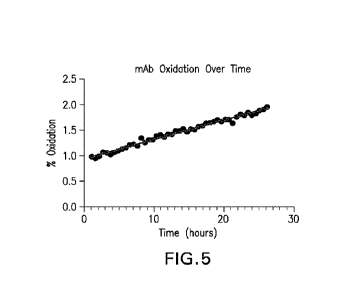
Figure 5: Changes in M105 oxidation of pembrolizumab over time in Harvested
Cell Culture Fluid (HCCF) samples. The black circles are the results of each
sample injection,
and the line is the linear fit of the data and is described by y = 0.038x +
0.94, with an R squared
value of 0.9833.
Figure 6: Ion exchange chromatography analytical diagram of pembrolizumab
after Protein A chromatography purification of HCCF from the continuous
perfusion process.
The retention time of each peak is annotated in the figure.
Figure 7: Ion exchange chromatography analytical diagram of pembrolizumab
after Anion exchange chromatography and Protein A chromatography purification
of HCCF
from the continuous perfusion process. The retention time of each peak is
annotated in the figure.
Figure 8: Overlay of Ion exchange chromatography analytical diagram of
pembrolizumab after Anion exchange step of the invention and pembrolizumab
reference
- 3 -
CA 03206669 2023- 7- 27
25195
WO 2022/165001
PCT/US2022/014055
standard obtained from fed-batch method. The diamonds delineate the start and
end of the integrated
peak.
Figure 9: Total % acidic species (acidicl+acidic variants+pre-main) in the
cell
culture fluid of the bioreactor (BRX), cell-free permeate (PERM), after
Protein A
chromatography step (PAP), after anion exchange chromatography (AEXP) in a
time course by
culture days.
Figure 10: Total % main species (as measured by ion exchange) in the cell-free
permeate (PERM), after Protein A chromatography step (PAP), after anion
exchange
chromatography (AEXP) in a time course by culture days.
Figure 11: Total % basic species (basicl-hbasic2+basic variant A+basic variant
B) in the cell-free permeate (PERM), after Protein A chromatography step
(PAP), after anion
exchange chromatography (AEXP) in a time course by culture days.
Figure 12: % Basicl species in the cell-free permeate (PERM), after Protein A
chromatography step (PAP), after anion exchange chromatography (AEXP) in a
time course by
culture days.
DETAILED DESCRIPTION OF THE INVENTION
I. Definitions and Abbreviations
As used throughout the specification and appended claims, the following
abbreviations apply:
API active pharmaceutical ingredient
CDR complementarity determining region in the
immunoglobulin
variable regions
CHO Chinese hamster ovary
CI confidence interval
DS drug substance
EC50 concentration resulting in 50% efficacy or
binding
ELISA enzyme-linked immunosorbant assay
FFPE formalin-fixed, paraffin-embedded
FR framework region
HC heavy chain
HNSCC head and neck squamous cell carcinoma
HP-HIC high performance hydrophobic interaction
chromatography
HP-IEX high performance ion-exchange chromatography
- 4 -
CA 03206669 2023- 7- 27
25195
WO 2022/165001
PCT/ITS2022/014055
HP-SEC high performance size exclusion
chromatography
IC50 concentration resulting in 50% inhibition
IgG immunoglobulin G
IHC immunohistochemistry or immunohistochemical
mAb monoclonal antibody
NCBI National Center for Biotechnology
Information
NSCLC non-small cell lung cancer
PCR polymerase chain reaction
PD-1 programmed death 1 (a.k.a. programmed cell
death-1 and
programmed death receptor 1)
PD-L1 programmed cell death 1 ligand 1
PD-L2 programmed cell death 1 ligand 2
PS80 or PS-80 polysorbate 80
SWFI sterile water for injection
TNBC triple negative breast cancer
Vu immunoglobulin heavy chain variable region
VK immunoglobulin kappa light chain variable
region
immunoglobulin light chain variable region
v/v volume per volume
WFI water for injection
w/v weight per volume
So that the invention may be more readily understood, certain technical and
scientific terms are specifically defined below. Unless specifically defined
elsewhere in this
document, all other technical and scientific terms used herein have the
meaning commonly
understood by one of ordinary skill in the art to which this invention
belongs.
As used throughout the specification and in the appended claims, the singular
forms "a,- "an,- and "the- include the plural reference unless the context
clearly dictates
otherwise.
Reference to "or" indicates either or both possibilities unless the context
clearly
dictates one of the indicated possibilities. In some cases, "and/or" was
employed to highlight
either or both possibilities.
As used herein, "acidic species" refers to the anti-PD-1 antibody species that
is
more acidic (e.g. as determined by cation exchange chromatography) than the
anti-PD-1
antibody main species. Such acidic species are detected by various
chromatography purification
- 5 -
CA 03206669 2023- 7- 27
25195
WO 2022/165001
PCT/ITS2022/014055
methods for separating molecule variants by charge, such as ion exchange, for
example, cation
exchange chromatography (e.g. the method described in Example 5) or WCX-10
HPLC (a weak
cation exchange chromatography), optionally followed by mass spectroscopy.
Generally, the
acidic species has a lower isoelectric point (pI) than the main species, and
can have a more acidic
character due to for example, methionine oxidation, sialylation of asparagine
residues or
deamidated variants of the antibody, or a combination thereof. Examples of the
acidic species
include but are not limited to the acidic variants, acidic 1 and pre-main
peaks identified in Figure
6 or 7 of the invention. Any of the acidic species may also have one or more
of CHO N-linked
glycans selected from the group consisting of GO-F, G1-F, G2-F, GO, Gl, G2 and
Man5, for
example at N297 in the CH2 domain.
In one embodiment, the anti-PD-1 antibody acidic species is as identified by
peak(s) eluted prior to the main peak according to a cation ion exchange
method. In another
embodiment, the anti-PD-1 antibody acidic species is as identified by peak(s)
eluted prior to the
main peak according to a weak cation ion exchange method. In an ion exchange
chromatography method, the "% acidic species" refers to the total area of
acidic species peaks
divided by the total area of all peaks in the elution chromatogram.
As used herein, "acidic1 species" refers to an acidic species with the
presence of
one or more of deamidation, succinimide, aspartate or isoaspartate formation
in one or more of
N384, N389 and N390 of the heavy chain of the anti-PD-1 antibody main species.
Such acidicl
species are detected by various chromatography purification methods for
separating molecule
variants by charge, such as ion exchange, for example, cation exchange
chromatography (e.g. the
method described in Example 5) or WCX-10 HPLC (a weak cation exchange
chromatography),
followed by mass spectroscopy of the acidic species peaks.
In one embodiment, the anti-PD-1 antibody acidicl species is as identified in
acidic 1 peak of Figure 6 or 7, and eluted according to the cation ion
exchange method described
in Example 5. In an ion exchange chromatography method, the "% acidicl
species" refers to the
total area of acidicl peak divided by the total area of all peaks in the
elution chromatogram.
As used herein, "acidic variants species" refers to an acidic species with the
presence of one or more of deamidation, succinimide, aspartate or isoaspartate
formation in one
or more of N31, N52, N55, N59, and N61 of the heavy chain; or the presence of
M105 oxidation
in the heavy chain; or a combination thereof of the anti-PD-1 antibody main
species. Such acidic
variants species are detected by various chromatography purification methods
for separating
molecule variants by charge, such as ion exchange, for example, cation
exchange
chromatography (e.g. the method described in Example 5) or WCX-10 HPLC (a weak
cation
exchange chromatography), followed by mass spectroscopy of the acidic species
peaks.
- 6 -
CA 03206669 2023- 7- 27
25195
WO 2022/165001
PCT/ITS2022/014055
In one embodiment, the anti-PD-1 antibody acidic variants species is the anti-
PD-
1 antibody species as identified by the acidic variants peak(s) in Figure 6 or
7, and eluted
according to the cation ion exchange method described in Example 5. In an ion
exchange
chromatography method, the "(N) acidic variants species" refers to the total
area of acidic variants
peak(s) divided by the total area of all peaks in the elution chromatogram.
As used herein, -basic species" refers to the anti-PD-1 antibody species that
is
more basic (e.g. as determined by cation exchange chromatography) than the
anti-PD-1 antibody
main species. Such basic species are detected by various chromatography
purification methods
for separating molecule variants by charge, such as ion exchange, for example,
cation exchange
chromatography (e.g. the method described in Example 5) or WCX-10 HPLC (a weak
cation
exchange chromatography), optionally followed by mass spectroscopy. Generally,
the basic
species has a higher pI than the main species, and can have a more basic
character due to
modifications or differences from the main species including but not limited
to the presence of
the C-terminal lysine residue (SEQ ID NO:10 or 12), the presence of N-terminal
glutamine
residue (SEQ ID NO: 10 or 13), or alpha-amidation of a C-terminal leucine
residue (SEQ ID
NO: 14 or 15), truncation of N-terminal amino acid residues in one or both
heavy chains
according to the amino acid sequence in any one of SEQ ID NO: 10-15, or a
combination
thereof. Examples of the basic species include but are not limited to the
basic variant A, basic
variant B, basic 1 and basic 2 peaks identified in Figure 6 or 7 of the
invention. Any of the basic
species may also have one or more of CHO N-linked glycans selected from the
group consisting
of GO-F, Gl-F, G2-F, GO, Gl, G2 and Man5, for example at N297 in the CH2
domain.
In one embodiment, the anti-PD-1 antibody basic species is as identified by
peak(s) eluted after the main peak according to a cation ion exchange method.
In another
embodiment, the anti-PD-1 antibody basic species is as identified by peak(s)
eluted after the
main peak according to a weak cation ion exchange method. In an ion exchange
chromatography method, the "% basic species" refers to the total area of basic
species peak(s)
divided by the total area of all peaks in the elution chromatogram.
As used herein, "main species" refers to the anti-PD-1 antibody species
identified
as the majority of the antibody species in a mixture with one or more acidic
or basic species
thereof Such main species are detected by various chromatography purification
methods for
separating molecule variants by charge, such as ion exchange, for example,
cation exchange
chromatography (e.g. the method described in Example 5) or WCX-10 HPLC (a weak
cation
exchange chromatography), optionally followed by mass spectroscopy. The
mixture can be a
result of for example, antibody' preparations from mammalian cells and post-
translational
modifications thereof, upstream and downstream processing, or storage. The
main species may
- 7 -
CA 03206669 2023- 7- 27
25195
WO 2022/165001
PCT/ITS2022/014055
also have one or more of CHO N-linked glycans selected from the group
consisting of GO-F, Gl-
F, G2-F, GO, Gl, G2 and Man5, for example at N297 in the CH2 domain.
In one embodiment, the main species comprises the anti-PD-1 antibody
consisting
of two heavy chains and two light chains, each heavy chain consisting of the
amino acid
sequence of SEQ ID NO: 11, and each light chain consisting of the amino acid
sequence of SEQ
ID NO: 5. In another embodiment, the anti-PD-1 antibody main species is
produced from a
Chinese Ovary cell that comprises a polynucleotide encoding a light chain that
consists of the
amino acid sequence of SEQ ID NO: 5 and a polynucleotide encoding a heavy
chain that consists
of the amino acid sequence of SEQ ID NO: 10, 13 or 15, or a polynucleotide
encoding the light
chain and the heavy chain.
In one embodiment, the main species is identified as the main peak according
to a
cation ion exchange method. In an ion exchange method, the "% main species"
refers to the total
area of main peak divided by the total area of all peaks in the elution
chromatogram.
As used herein, "basic 1 species- refers to a basic species consisting of two
heavy
chains and two light chains, one heavy chain consisting of the amino acid
sequence of SEQ ID
NO: 11, one heavy chain consisting of the amino acid sequence of SEQ ID NO:
12, and each
light chain consisting of the amino acid sequence of SEQ ID NO: 5; or a basic
species consisting
of two heavy chains and two light chains, one heavy chain consisting of the
amino acid sequence
of SEQ ID NO: 11, one heavy chain consisting of the amino acid sequence of SEQ
ID NO: 14,
wherein the C-terminal leucine is alpha-amidated, and each light chain
consisting of the amino
acid sequence of SEQ ID NO: 5; or a combination thereof Such basic 1 species
are detected by
various chromatography purification methods for separating molecule variants
by charge, such as
ion exchange, for example, cation exchange chromatography (e.g. the method
described in
Example 5) or WCX-10 HPLC (a weak cation exchange chromatography), followed by
mass
spectroscopy of the basic species peaks.
In one embodiment, the anti-PD-1 antibody basic 1 species is as identified by
basic 1 peak in Figure 6 or 7, and eluted according to the cation ion exchange
method described
in Example 5. In an ion exchange chromatography method, the "% basicl species-
refers to the
total area of basic1 peak divided by the total area of all peaks in the
elution chromatogram.
As used herein, "deamidated variant" refers to an antibody wherein one or more
asparagine residue(s) have been deami dated. The deamidated variant can be in
the form of
succinimide, aspartate or isoaspartate. i.e., the neutral amide side chain has
been converted to a
residue with an overall acidic character.
In one aspect of measuring the main species, acidic species or basic species,
a
Thermo Scientific ProPac WCX-10 column is used for the cation ion exchange
method. In
- 8 -
CA 03206669 2023- 7- 27
25195
WO 2022/165001
PCT/ITS2022/014055
another embodiment, a Thermo Scientific ProPac WCX-10 column is used, with a
Mobile Phase
(A) 24 mM MES pH 6.1 with 4% acetonitrile, and mobile phase (B) 20 mM sodium
phosphate,
95 mM NaC1 pH 8.0 with 4% acetonitrile, and a column temperature of 35 C. In
one
embodiment, a non-linear gradient is used with: 22%-22%B for 0-0.6 min; 22%-
29%B for 0.6-
15.0 mM; 29%-70%B for 15.0-30.0 min; 70%-100%B for 30.0-30.5 mM; and 100%-
100%B
from 30.5-33.0 mM. In a further embodiment, the cation ion exchange method is
described in
Example 5.
As used herein, "express" and "expression" refer to allowing or causing the
information in a gene or coding sequence, e.g., an RNA or DNA, to become
manifest; for
example, producing a protein by activating the cellular functions involved in
transcription and
translation of a corresponding gene. A DNA sequence can be expressed in or by
a cell to form
an "expression product" such as an RNA (e.g., mRNA) or a protein. The
expression product
itself may also be said to be "expressed" by the cell.
As used herein, "expression vector" or "expression construct" refer to a
vehicle
(e.g., a plasmid) by which a polynucleotide comprising regulatory sequences
operably linked to a
coding sequence can be introduced into a host cell where the coding sequence
is expressed using
the transcription and translation machinery of the host cell.
"Expression cassette" as used herein, refers to a polynucleotide that
comprises
elements sufficient to control expression of a gene, including but not limited
to, a promoter
operably linked to the gene sequence or operably linked to a multiple cloning
site for inserting
the gene sequence, and a polyA signal. In some embodiments, the expression
cassette further
comprises one or more regulatory elements that can regulate the expression of
the gene at
transcriptional, translational, and/or chromatin levels.
As used herein, "promoter" or "promoter sequence" refer to a segment of DNA
that contains a regulatory region capable of recruiting an RNA polymerase
(e.g., directly or
through other promoter-bound proteins or substances) and initiating
transcription of a coding
sequence. Within the promoter sequence may be found a transcription initiation
site
(conveniently defined, for example, by mapping with nuclease S1), as well as
protein binding
domains (consensus sequences) responsible for the recruiting of RNA
polymerase.
As used herein, "enhancer" or "enhancer sequence" refer to a DNA regulatory
region that enhances transcription of a promoter independently of its
distance, location, or
orientation to the promoter. In certain embodiments, the enhancer is
immediately adjacent to the
promoter. In some embodiments, the enhancer is distant from the promoter. In
other
embodiments, the promoter and the enhancer are one combined sequence, referred
as a "combo
enhancer/promoter- herein.
- 9 -
CA 03206669 2023- 7- 27
25195
WO 2022/165001
PCT/ITS2022/014055
As used herein, "internal ribosome entry site" or "IRES" refer to an RNA
element
or sequence that allows for translation initiation in a cap-independent manner
by recruiting
ribosomes directly. As used herein, the term "internal ribosome entry site" or
"IRES" also
encompasses the DNA sequence that can be transcribed into the RNA sequence
that allows for
translation initiation in a cap-independent manner by recruiting ribosomes
directly. IRES can be
a wild type IRES from any species or a variant or mutant thereof, whether
naturally occurred or
man-made. Examples of IRES that can be used include, but are not limited to,
the nucleotide
sequence of the 5' nontranslated region of encephalomyocarditis virus (EMCV)
(GenBank:
M81861.1; Duke et al., Sequence and structural elements that contribute to
efficient
encephalomyocarditis virus RNA translation. J Virol. 1992 Mar;66(3):1602-9.),
IRES element
described by Bochkov & Palmenberg (Translational efficiency of EMCV IRES in
bicistronic
vectors is dependent upon IRES sequence and gene location. Biotechniques. 2006
Sep;41(3):283-4), IRES element from expression vector pInSRT-GFP (GenBank
LC417349.1),
IRES element from expression vector pCeMM-CTAP(SG) (GenBank EF467048.1), IRES
element described by Jang & Wimmer (Cap-independent translation of
encephalomyocarditis
virus RNA: structural elements of the internal ribosomal entry site and
involvement of a cellular
57-kD RNA-binding protein. Genes Dev. 1990 Sep;4(9):1560-72), IRES element
from
expression vector pIRESneo3 (Clontech/Takara Bio), IRES elements described in
WO
2015/016786, WO 2015/021077, WO 2016/003368, WO 2016/074016, or WO
2013/092743, or
variants thereof
As used herein, "regulatory element," "regulatory region," or "regulatory
sequence" refer to a polynucleotide sequence that has the ability to regulate
(such as, initiate,
activate, enhance, increase, decrease, inhibit, suppress, or silence)
expression of a gene. In some
embodiments, the regulation is achieved by binding of cellular factors to the
polynucleotide
sequence. In other embodiments, the regulation is achieved by interaction
between cellular
factors. The regulation can occur at one or more different levels in the
expression process from
DNA to protein, including but not limited to transcriptional, translational,
or chromatin levels.
As used herein, "insulator" refers to a class of DNA elements or sequences
that
possess an ability to isolate the proximal DNA region by preventing the
positional effect from
the surrounding chromosome area. In certain embodiments, the insulator can
block enhancer
when the insulator is situated between the enhancer and the promoter. In some
embodiments, the
insulator can act as barriers that prevent the advance of nearby condensed
chromatin that might
otherwise silence expression. In other embodiments, the insulator can block
enhancer and act as
barriers.
- 10 -
CA 03206669 2023- 7- 27
25195
WO 2022/165001
PCT/ITS2022/014055
As used herein, "expression augmenting sequence element" or "EASE", refer to a
DNA element or sequence that can increase expression of a protein when the DNA
element or
sequence is placed upstream of the promoter that controls the expression of
the protein.
As used herein, "tripartite leader" or "TPL" refer to an RNA element or
sequence
in the 5'-untranslated region of adenovirus late-expressed mRNA that has an
ability to initiate
translation of the late-expressed mRNA in a cap-independent manner. As used
herein, the term
"tripartite leader- or "TPL- also encompasses the DNA sequence that can be
transcribed into the
RNA sequence in the 5'-untranslated region of adenovirus late-expressed mRNA
that has an
ability to initiate translation of the late-expressed mRNA in a cap-
independent manner.
As used herein, "inverted terminal repeat" or "ITR", in the context of
transposon
technology, refers to a DNA element or sequence and its inverted version at
either end of a
transposon that signals where the breakage and joining should occur.
As used herein, "selectable marker" or "selection marker" refer to a protein
which
allows the specific selection of cells that express this protein by the
addition of a corresponding
selecting agent to the culture medium. In certain embodiments, the selectable
marker is a
eukaryotic selectable marker, which allows selection of eukaryotic cells that
express the marker
protein. In some embodiments, the selectable marker is a bacterial selectable
marker, which
allows selection of bacterial cells that express the marker protein.
A "polynucleotide sequence", "nucleic acid sequence" or "nucleotide sequence",
as used herein, refer to a series of nucleotide bases (also called
"nucleotides") in a nucleic acid,
such as DNA or RNA, and means any chain of two or more nucleotides.
As used herein, a "host cell" refers to any cell of any organism that is used
for the
purpose of producing a recombinant protein encoded by an expression vector or
propagating the
expression vector introduced into the host cell. A "mammalian recombinant host
cell" refers to a
mammalian host cell that comprises a heterologous expression vector, which may
or may not be
integrated into the host cell chromosome. A "bacterial recombinant host cell"
refers to a
bacterial host cell that comprises a heterologous expression vector, which may
or may not be
integrated into the host cell chromosome.
The term "fed-batch culture", as used herein, refers to a method of culturing
cells
in which additional nutrients are provided to the culture during the
cultivation process. A fed-
batch culture is typically stopped at some point and the cells and/or
components in the medium
are harvested. The product accumulates and remains in the bioreactor until the
end of the run.
As used herein, "harvesting" an antibody or antigen-binding fragment involves
separating it from particulate matter that can include host cells, cell
aggregates, and/or lysed cell
- 11 -
CA 03206669 2023- 7- 27
25195
WO 2022/165001
PCT/ITS2022/014055
fragments, into a cell-free fraction that is substantially free of host cells
and cellular debris, i.e., a
cell-free "permeate." Such cells and cellular debris is removed from the cell
culture broth, for
example, by centrifugation, depth filtration and/or microfiltration. For
example, to make the
cell-free permeate, one can employ hollow fiber membranes or a series of
filtration steps such as
depth filtration. "Continuously harvesting" refers to harvesting cell culture
broth while antibody
production takes place in the bioreactor. Secreted protein products in the
bioreactor can be
continuously harvested from the cell culture broth by microfiltration during
the process of
removing medium via the perfusion system, the protein of interest thus being
isolated in a
microfilter permeate exiting the perfusion system. The microfilter can be a
Tangential Flow
Filtration (TFF) unit including a hollow fiber module or Alternating
Tangential Flow Filtration
(ATF) unit. Commercially available TFF units include, but are not limited to,
Microzalt TFF
unit or KrosFlow0Max. Commercially available hollow fiber modules can be
obtained for
example, from Pall or Repligen. In one embodiment, the perfusion bioreactor
has a constant
permeate rate for the cell culture broth comprising the antibody or antigen-
binding fragment to
maintain a consistent flow rate to the affinity chromatography step.
As used herein, "cell culture broth" refers to broth comprising the host
cells,
cellular debris, cell culture medium, antibody or antigen-binding fragment
during the cell growth
and antibody production process.
As used herein, "harvest cell culture fluid" or "HCCF" refers to the cell
culture
fluid comprising the antibody or antigen-binding fragment obtained after
harvesting the cell
culture broth, which is substantially free of host cells and cellular debris.
In one embodiment,
the HCCF is a cell-free permeate.
As used herein, "fluidly connected," "fluidly," or is "fluidly connected to",
or
"fluidly receives material from", refers to another step of the manufacturing
process or from
another system, when material containing the protein of interest flows by
pipe, tubing, or other
closed conduit between steps or systems without manual loading or unloading.
As used herein in the context of HCCF, "continuously purifying" refers to
uninterrupted flow of the HCCF to at least one affinity stationary phase for
at least the loading
step, and optionally an uninterrupted flow for any washing or elution steps.
As used herein, "perfusion" or "perfusing" refer to a method of culturing
cells in
which additional fresh medium is provided continuously over some period of
time, to the culture
(subsequent to the beginning of the culture process), and simultaneously
removing medium
while harvesting the antibody or antigen-binding fragment from the medium
continuously. The
fresh medium typically provides nutritional supplements for the cells that
have been depleted
during the culturing process.
- 1/ -
CA 03206669 2023- 7- 27
25195
WO 2022/165001
PCT/ITS2022/014055
"Perfusion rate-, as used herein, refers to the rate at which fresh medium is
provided and cell culture fluid is removed.
As used herein, a "perfusion bioreactor" refers to a bioreactor for culturing
cells
in which equivalent volumes of culture medium can be added to and concurrently
removed from
the reactor. In one embodiment, the cells are retained in the bioreactor. A
perfusion bioreactor
includes a bioreactor and an operably attached perfusion system, which
provides a steady source
of fresh nutrient medium and removal of cell waste products. The bioreactor
and the perfusion
system of the perfusion bioreactor can be separate mechanical units that
operate in coordination.
Numerous commercially available examples include, but are not limited to, a
variety of
Xcellerex brand single-use bioreactors (SUBs; GE Healthcare Life Sciences)
and KrosFlo
brand perfusion flow-path assemblies and systems (Spectrum; Repligen), which
bioreactors and
perfusion systems can be suitably combined into a perfusion bioreactor by the
skilled
practitioner. Alternatively, the bioreactor and the perfusion system can be
assembled into a
single mechanical unit, for example, but not limited to, a 3D Biotek brand
perfusion bioreactor
(Sigma-Aldrich).
The term "surge vessel", as used herein, refers to a well-mixed (providing
sufficient mixing such that fluid is homogenous) storage reservoir, mixing
vessel, feed tank, or
collection vessel (or interchangeably, a "collection tank"), at the downstream
end of a conduit,
feeder, dam, pipe, or tubing, to absorb discrepant flow rates between two
fluidly connected unit
operations, e.g., the flow rate of a permeate coming from a bioreactor and the
flow rate of a first
chromatography system under automated control in continuous or semi-continuous
format
process embodiments of the invention. The surge vessel absorbs changes or
differences in flow
rates by allowing the volume to surge within pre-set volume range limits
between the fluidly
connected unit operations.
The term -residence time", as used herein, refers to the average time a fluid
solution spends inside a vessel. For perfusion this is the inverse of the
exchange rate (e.g. 2VVD
has a mean residence time of 0.5 day). Sieving of solution components,
retention of solution
components by the membrane is neglected for residence time.
As used herein, "M105", "Met105", or "Methionine105" refers to the methionine
in CDRH3 region of the heavy chain in SEQ ID NO: 8 (RDYRFDMGFDY).
As used herein, "% oxidation of M105" or "% oxidation of Met105" refer to a)
total amount of anti-PD-1 antibody fragment(s) of the invention with oxidized
Met105 versus
total amount of anti-PD-1 antibody fragment(s) of the invention with and
without oxidized
Met105; orb) total amount of anti-PD-1 antibody of the invention with oxidized
Met105 versus
total amount of anti-PD-1 antibody of the invention with and without oxidized
Met105.
- 13 -
CA 03206669 2023- 7- 27
25195
WO 2022/165001
PCT/US2022/014055
Calculation method a) can be used according to the reduced peptide mapping
method provided in
the Examples. Calculation method b) can be used for example, in a Hydrophobic
Interaction
Chromatography (HIC) or Reverse Phase HPLC method as described in
W02018/204368,
incorporated by reference in its entirety.
By "binding" an antibody or antigen-binding fragment to a stationary phase, is
meant exposing the antibody or antigen-binding fragment to the stationary
phase under
appropriate conditions (pH and/or conductivity) such that the antibody or
antigen-binding
fragment is reversibly associated with the stationary phase by interactions
between the antibody
or antigen-binding fragment and the ligand immobilized on the stationary
phase.
As used herein, the term "equilibration solution" refers to a solution used to
equilibrate the stationary phase prior to loading the antibody or antigen-
binding fragment on the
stationary phase. The equilibration solution can comprise one or more of a
salt and buffering
species. In one embodiment, the equilibration solution is the same condition
as the loading
solution comprising the antibody or antigen-binding fragment.
As used herein, the term "loading solution- refers to the solution which is
used to
load the composition comprising the antibody or antigen-binding fragment of
interest and one or
more impurities onto the stationary phase. The loading solution may optionally
further comprise
one or more of a buffering species, and salt.
As used herein, the term "wash solution" refers to a solution used to wash or
re-
equilibrate the stationary phase, prior to eluting the antibody or antigen-
binding fragment of
interest. For washing, the conductivity and/or pH of the wash solution is/are
such that the
impurities are removed from the stationary phase. For re-equilibration, the
wash solution and
equilibration solution may be the same, but this is not required. The wash
solution can comprise
one or more of a salt and buffering species.
As used herein, the "elution solution" refers to the solution used to elute
the
antibody or antigen-binding fragment of interest from the stationary phase.
The elution solution
can comprise one or more of a salt, or buffering species. The presence of one
or more of salt,
buffering species, pH or conductivity of the elution solution is/are such that
the antibody or
antigen-binding fragment is eluted from the stationary phase.
As used herein, the term "conductivity" refers to the ability of an aqueous
solution
to conduct an electric current between two electrodes. In solution, the
current flows by ion
transport. Therefore, with an increasing amount of ions present in the aqueous
solution, the
solution will have a higher conductivity. The unit of measurement for
conductivity is mS/cm,
and can be measured using a conductivity meter sold, e.g., within the GE
Healthcare AktaTM
System. The conductivity of a solution may be altered by changing the
concentration of ions
- 14 -
CA 03206669 2023- 7- 27
25195
WO 2022/165001
PCT/US2022/014055
therein. For example, the concentration of a buffering agent and/or
concentration of a salt (e.g.
NaC1 or KC1) in the solution may be altered in order to achieve the desired
conductivity.
Preferably, the salt concentration of the various buffers is modified to
achieve the desired
conductivity as in the Examples below.
As used herein, "purifying" an antibody or antigen-binding fragment of
interest or
-purified composition" refers to increasing the degree of purity of the
antibody or antigen-
binding fragment in the composition by removing (completely or partially) at
least one impurity
from the composition. The impurity can be host cell components such as serum,
proteins or
nucleic acids, cellular debris, growth medium or antibody aggregates. The term
is not intended
to refer to a complete absence of such biological molecules or to an absence
of water, buffers, or
salts or to components of a pharmaceutical composition that includes the
antibody or antigen-
binding fragment.
As used herein, "continuous multi-column chromatography system" refers to a
chromatography system containing at least two stationary phases with similar
impurity
separation function, which allow at least one stationary phase to load the
sample, and at least one
stationary phase to perform the non-loading steps (one or more of
equilibration, washing, elution
and regeneration).
As used herein, "stationary phase" refers to any surface onto which one or
more
ligands can be immobilized. The stationary phase may be a suspension, a
discontinuous phase of
discrete particles, plate, sensor, chip, capsule, cartridge, resin, beads,
monolith, gel, a membrane,
or membrane adsorber etc. Stationary phases may also be packed into a
purification column (e.g.
packed with resin beads). Examples of materials for forming the stationary
phase include
mechanically stable matrices such as porous or non-porous beads, inorganic
materials (e.g.,
porous silica, controlled pore glass (CPG) and hydroxyapatite), synthetic
organic polymers (e.g.,
poly acryl ami de, p oly methylmethacryl ate, p oly styrene-di vinyl b enzene,
poly(styrenedivinyl)benzene, polyacrylamide, ceramic particles and derivatives
of any of the
above) and polysaccharides (e.g., cellulose, agarose and dextran). See
Jonsson, J. C.; Ryden, L.
Protein Purification; Wiley: New York, 1998.
As used herein, "impurity" refers to a material different from the desired
antibody
or antigen-binding fragment. The impurity can be Host Cell Protein (HCP), Host
Cell DNA
(HC -DN A), protein aggregates or clips and other undesired protein
modifications (i.e., oxidized
species, acid variant species).
"Treat" or "treating" a cancer, as used herein, refers to the administration
of a
composition of the invention to a subject having an immune condition or
cancerous condition, or
diagnosed with a cancer or pathogenic infection (e.g. viral, bacterial,
fungal), to achieve at least
- 15 -
CA 03206669 2023- 7- 27
25195
WO 2022/165001
PCT/ITS2022/014055
one positive therapeutic effect, such as for example, reduced number of cancer
cells, reduced
tumor size, reduced rate of cancer cell infiltration into peripheral organs,
or reduced rate of
tumor metastasis or tumor growth. "Treatment" may include one or more of the
following:
inducing/increasing an antitumor immune response, stimulating an immune
response to a
pathogen, toxin, and/or self-antigen, stimulating an immune response to a
viral infection,
decreasing the number of one or more tumor markers, halting or delaying the
growth of a tumor
or blood cancer or progression of disease associated with PD-1 binding to its
ligands PD-Li
and/or PD-L2 ("PD-1-related disease") such as cancer, stabilization of PD-1-
related disease,
inhibiting the growth or survival of tumor cells, eliminating or reducing the
size of one or more
cancerous lesions or tumors, decreasing the level of one or more tumor
markers, ameliorating,
abrogating the clinical manifestations of PD-1-related disease, reducing the
severity or duration
of the clinical symptoms of PD-1-related disease such as cancer, prolonging
the survival of a
patient relative to the expected survival in a similar untreated patient,
inducing complete or
partial remission of a cancerous condition or other PD-1 related disease.
"Immune condition" or "immune disorder" encompasses, e.g., pathological
inflammation, an inflammatory disorder, and an autoimmtine disorder or
disease. "Immune
condition" also refers to infections, persistent infections, and proliferative
conditions, such as
cancer, tumors, and angiogenesis, including infections, tumors, and cancers
that resist eradication
by the immune system. -Cancerous condition" includes, e.g., cancer, cancer
cells, tumors,
angiogenesis, and precancerous conditions such as dysplasia.
Positive therapeutic effects in cancer can be measured in a number of ways
(See,
W. A. Weber, J Nucl. Med. 50:1S-10S (2009)). For example, with respect to
tumor growth
inhibition, according to NCI standards, a T/C 42% is the minimum level of anti-
tumor activity.
A T/C < 10% is considered a high anti-tumor activity level, with T/C (%) =
Median tumor
volume of the treated/Median tumor volume of the control 100. In some
embodiments, the
treatment achieved by administration of a composition of the invention is any
of progression free
survival (PFS), disease free survival (DFS) or overall survival (OS). PFS,
also referred to as
"Time to Tumor Progression" indicates the length of time during and after
treatment that the
cancer does not grow, and includes the amount of time patients have
experienced a complete
response or a partial response, as well as the amount of time patients have
experienced stable
disease. DFS refers to the length of time during and after treatment that the
patient remains free
of disease. OS refers to a prolongation in life expectancy as compared to
naive or untreated
individuals or patients. While an embodiment of the compositions, treatment
methods, and uses
of the invention may not be effective in achieving a positive therapeutic
effect in every patient, it
should do so in a statistically significant number of subjects as determined
by any statistical test
- 16 -
CA 03206669 2023- 7- 27
25195
WO 2022/165001
PCT/ITS2022/014055
known in the art such as the Student's t-test, the chi2-test, the U-test
according to Mann and
Whitney, the Kruskal-Wallis test (H-test), Jonckheere-Terpstra-test and the
Wilcoxon-test.
As used herein, the term "patient" (alternatively referred to as "subject" or
"individual" herein) refers to a mammal (e.g., rat, mouse, dog, cat, rabbit)
capable of being
treated with the compositions or compositions of the invention, most
preferably a human. In
some embodiments, the patient is an adult patient. In other embodiments, the
patient is a
pediatric patient. Those "in need of treatment" include those patients that
may benefit from
treatment with the compositions or compositions of the invention, e.g. a
patient suffering from
cancer or an immune condition.
As used herein, the term "antibody" refers to any form of antibody that
exhibits
the desired biological activity. Thus, it is used in the broadest sense and
specifically covers, but
is not limited to, monoclonal antibodies (including full length monoclonal
antibodies),
polyclonal antibodies, humanized, fully human antibodies, and chimeric
antibodies.
In general, the basic antibody structural unit comprises a tetramer. Each
tetramer
includes two identical pairs of polypeptide chains, each pair having one
"light" (about 25 kDa)
and one "heavy" chain (about 50-70 kDa). The amino-terminal portion of each
chain includes a
variable region of about 100 to 110 or more amino acids primarily responsible
for antigen
recognition. The variable regions of each light/heavy chain pair form the
antibody binding site.
Thus, in general, an intact antibody has two binding sites. The carboxy-
terminal portion of the
heavy chain may define a constant region primarily responsible for effector
function. Typically,
human light chains are classified as kappa and lambda light chains.
Furthermore, human heavy
chains are typically classified as mu, delta, gamma, alpha, or epsilon, and
define the antibody's
isotype as IgM, IgD, IgG, IgA, and IgE, respectively. Within light and heavy
chains, the
variable and constant regions are joined by a "J" region of about 12 or more
amino acids, with
the heavy chain also including a "D" region of about 10 more amino acids. See
generally,
Fundamental Immunology Ch. 7 (Paul, W., ed., 2nd ed. Raven Press, N.Y. (1989).
Typically, the variable domains of both the heavy and light chains comprise
three
hypervariable regions, also called complementarity determining regions (CDRs),
which are
located within relatively conserved framework regions (FR). The CDRs are
usually aligned by
the framework regions, enabling binding to a specific epitope. In general,
from N-terminal to C-
terminal, both light and heavy chains variable domains comprise FR1, CDR1, FR2
, CDR2, FR3,
CDR3 and FR4. The assignment of amino acids to each domain is, generally, in
accordance with
the definitions of Sequences of Proteins of Immunological Interest, Kabat, et
al.; National
Institutes of Health, Bethesda, Md. ; 51h ed.; NIH Publ. No. 91-3242 (1991);
Kabat (1978) Adv.
- 17 -
CA 03206669 2023- 7- 27
25195
WO 2022/165001
PCT/ITS2022/014055
Prot. Chem. 32:1-75; Kabat, etal., (1977)1 Biol. Chem. 252:6609-6616; Chothia,
etal., (1987)
JMol. Biol. 196:901-917 or Chothia, et al., (1989) Nature 342:878-883.
As used herein, the term "pharmaceutically effective amount" or "effective
amount" refers to an amount whereby sufficient therapeutic composition or
composition is
introduced to a patient to treat a diseased or condition. One skilled in the
art recognizes that this
level may vary according the patient's characteristics such as age, weight,
etc.
The term "about", when modifying the quantity (e.g., mM, or M) of a substance
or composition, the percentage (v/v or w/v) of a composition component, the pH
of a
solution/composition, or the value of a parameter characterizing a step in a
method, or the like
refers to variation in the numerical quantity that can occur, for example,
through typical
measuring, handling and sampling procedures involved in the preparation,
characterization
and/or use of the substance or composition; through instrumental error in
these procedures;
through differences in the manufacture, source, or purity of the ingredients
employed to make or
use the compositions or carry out the procedures; and the like. In certain
embodiments, "about"
can mean a variation of + 0.1%, 0.5%, 1%, 2%, 3%, 4%, 5%, or 10% of the value.
The terms "cancer", "cancerous", or "malignant", as used herein, refer to or
describe the physiological condition in mammals that is typically
characterized by unregulated
cell growth. Examples of cancer include but are not limited to, carcinoma,
lymphoma, leukemia,
blastoma, and sarcoma. More particular examples of such cancers include
squamous cell
carcinoma, myeloma, small-cell lung cancer, non-small cell lung cancer,
glioma, Hodgkin's
lymphoma, non-Hodgkin's lymphoma, gastrointestinal (tract) cancer, renal
cancer, ovarian
cancer, liver cancer, lymphoblastic leukemia, lymphocytic leukemia, colorectal
cancer,
endometrial cancer, kidney cancer, prostate cancer, thyroid cancer, melanoma,
chondrosarcoma,
neuroblastoma, pancreatic cancer, glioblastoma multiforme, cervical cancer,
brain cancer,
stomach cancer, bladder cancer, hepatoma, breast cancer, colon carcinoma, and
head and neck
cancer.
As used herein, the terms "PD-1 binding fragment," "antigen binding fragment
thereof," "binding fragment thereof' or "fragment thereof," encompass a
fragment or a
derivative of an antibody that still substantially retains its biological
activity of binding to
antigen (human PD-1) and inhibiting its activity (e.g., blocking the binding
of PD-1 to PDL1 and
PDL2). Therefore, the term "antibody fragment" or PD-1 binding fragment refers
to a portion of
a full length antibody, generally the antigen binding or variable region
thereof Examples of
antibody fragments include Fab, Fab', F(ab1)2, and FA/ fragments. Typically, a
binding fragment
or derivative retains at least 10% of its PD-1 inhibitory activity. In some
embodiments, a
binding fragment or derivative retains at least 25 4), 50%, 60%, 70%, 80%,
90%, 95%, 99% or
- 18 -
CA 03206669 2023- 7- 27
25195
WO 2022/165001
PCT/ITS2022/014055
100% (or more) of its PD-1 inhibitory activity, although any binding fragment
with sufficient
affinity to exert the desired biological effect will be useful. In some
embodiments, an antigen
binding fragment binds to its antigen with an affinity that is at least two
fold greater, preferably
at least ten times greater, more preferably at least 20-times greater, and
most preferably at least
100-times greater than the affinity with unrelated antigens. In one embodiment
the antibody has
an affinity that is greater than about 109 liters/mol, as determined, e.g., by
Scatchard analysis.
Munsen et al. (1980) Analyt. Biocheni. 107:220-239. It is also intended that a
PD-1 binding
fragment can include variants having conservative amino acid substitutions
that do not
substantially alter its biological activity.
"Humanized antibody," as used herein, refers to forms of antibodies that
contain
sequences from non-human (e.g., murine) antibodies as well as human
antibodies. Such
antibodies contain minimal sequence derived from non-human immunoglobulin. In
general, the
humanized antibody will comprise substantially all of at least one, and
typically two, variable
domains, in which all or substantially all of the hypervariable loops
correspond to those of a non-
human immunoglobulin and all or substantially all of the FR regions are those
of a human
immunoglobulin sequence. The humanized antibody optionally also will comprise
at least a
portion of an immunoglobulin constant region (Fe), typically that of a human
immunoglobulin.
The humanized forms of rodent antibodies will generally comprise the same CDR
sequences of
the parental rodent antibodies, although certain amino acid substitutions may
be included to
increase affinity, increase stability of the humanized antibody, or for other
reasons.
The antibodies of the invention also include antibodies with modified (or
blocked)
Fc regions to provide altered effector functions. See, e.g., U.S. Pat. No.
5,624,821;
W02003/086310; W02005/120571; W02006/0057702; Presta (2006) Adv. Drug Delivery
Rev.
58:640-656. Such modification can be used to enhance or suppress various
reactions of the
immune system, with possible beneficial effects in diagnosis and therapy.
Alterations of the Fc
region include amino acid changes (substitutions, deletions and insertions),
glycosylation or
deglycosylation, and adding multiple Fc. Changes to the Fc can also alter the
half-life of
antibodies in therapeutic antibodies, and a longer half-life would result in
less frequent dosing,
with the concomitant increased convenience and decreased use of material. See
Presta (2005) J
Allergy Clin. Itninuno1.116:731 at 734-35.
"Conservatively modified variants" or "conservative substitution," as used
herein,
refers to substitutions of amino acids that are known to those of skill in
this art and may be made
generally without altering the biological activity of the resulting molecule,
even in essential
regions of the polypeptide. Such exemplary substitutions are preferably made
in accordance
with those set forth in Table 1 as follows:
- 19 -
CA 03206669 2023- 7- 27
25195
WO 2022/165001
PCT/ITS2022/014055
Table 1. Exemplary Conservative Amino Acid Substitutions
Original residue Conservative substitution
Ala (A) Gly; Ser
Arg (R) Lys, His
Asn (N) Gin; His
Asp (D) Glu; Asn
Cys (C) Ser; Ala
Gin (Q) Asn
Glu (E) Asp; Gin
Gly (G) Ala
His (H) Asn; Gin
Ile (I) Leu; Val
Leu (L) Ile: Val
Lys (K) Arg; His
Met (M) Leu; Ile; Tyr
Phe (F) Tyr; Met; Leu
Pro (P) Ala
Ser (S) Thr
Thr (T) Ser
Trp (W) Tyr; Phe
Tyr (Y) Trp; Phe
Val (V) Ile; Leu
In addition, those of skill in this art recognize that, in general, single
amino acid
substitutions in non-essential regions of a polypeptide do not substantially
alter biological
activity. See, e.g., Watson etal. (1987)Molecular Biology of the Gene, The
Benjamin/Cummings Pub. Co., p. 224 (4th Edition).
The phrase "consists essentially of" or variations such as "consist
essentially or'
or "consisting essentially of," as used throughout the specification and
claims, indicate the
inclusion of any recited elements or group of elements, and the optional
inclusion of other
elements, of similar or different nature than the recited elements, that do
not materially change
the basic or novel properties of the specified dosage regimen, method, or
composition. As a non-
limiting example, a binding compound that consists essentially of a recited
amino acid sequence
- 20 -
CA 03206669 2023- 7- 27
25195
WO 2022/165001
PCT/ITS2022/014055
may also include one or more amino acids, including substitutions of one or
more amino acid
residues, that do not materially affect the properties of the binding
compound.
"Comprising" or variations such as "comprise", "comprises" or "comprised of"
are used throughout the specification and claims in an inclusive sense, i.e.,
to specify the
presence of the stated features but not to preclude the presence or addition
of further features that
may materially enhance the operation or utility of any of the embodiments of
the invention,
unless the context requires otherwise due to express language or necessary
implication.
"Monoclonal antibody" or "mAb" or "Mab", as used herein, refers to a
population
of substantially homogeneous antibodies, i.e., the antibody molecules
comprising the population
are identical in amino acid sequence except for possible naturally occurring
mutations that may
be present in minor amounts. In contrast, conventional (polyclonal) antibody
preparations
typically include a multitude of different antibodies having different amino
acid sequences in
their variable domains, particularly their CDRs, which are often specific for
different epitopes.
The modifier "monoclonal" indicates the character of the antibody as being
obtained from a
substantially homogeneous population of antibodies, and is not to be construed
as requiring
production of the antibody by any particular method. For example, the
monoclonal antibodies to
be used in accordance with the invention may be made by the hybridoma method
first described
by Kohler el al. (1975) Nature 256: 495, or may be made by recombinant DNA
methods (see,
e.g., U.S. Pat. No. 4,816,567). The "monoclonal antibodies" may also be
isolated from phage
antibody libraries using the techniques described in Clackson etal.
(1991)Nature 352: 624-628
and Marks etal. (1991)1 Mol. Biol. 222: 581-597, for example. See also Presta
(2005) 1
Allergy Clin. Irninunol. 116:731.
"Tumor" as it applies to a subject diagnosed with, or suspected of having, a
cancer
refers to a malignant or potentially malignant neoplasm or tissue mass of any
size, and includes
primary tumors and secondary neoplasms. A solid tumor is an abnormal growth or
mass of tissue
that usually does not contain cysts or liquid areas. Different types of solid
tumors are named for
the type of cells that form them. Examples of solid tumors are sarcomas,
carcinomas, and
lymphomas. Leukemias (cancers of the blood) generally do not form solid tumors
(National
Cancer Institute, Dictionary of Cancer Terms).
The term "tumor size" refers to the total size of the tumor which can be
measured
as the length and width of a tumor. Tumor size may be determined by a variety
of methods
known in the art, such as, e.g. by measuring the dimensions of tumor(s) upon
removal from the
subject, e.g., using calipers, or while in the body using imaging techniques,
e.g., bone scan,
ultrasound, CT or MRI scans.
- 21 -
CA 03206669 2023- 7- 27
25195
WO 2022/165001
PCT/ITS2022/014055
"Tumor Proportion Score (TPS)" refers to the percentage of tumor cells
expressing PD-L1 on the cell membrane at any intensity (weak, moderate or
strong). Linear
partial or complete cell membrane staining is interpreted as positive for PD-
Li.
"Mononuclear inflammatory density score (MIDS)- refers to the ratio of the
number of PD-Li expressing mononuclear inflammatory cells (MIC) infiltrating
or adjacent to
the tumor (small and large lymphocytes, monocytes, and macrophages within the
tumor nests
and the adjacent supporting stroma) compared to the total number of tumor
cells. The M1DS is
recorded at a scale from 0 to 4 with 0=none; 1=present, but less than one MIC
for every 100
tumor cells (<1%); 2=at least one MIC for every 100 tumor cells, but less than
one MIC per 10
tumor cells (1-9%); 3=at least one MIC for every 10 tumor cells, but fewer
MIC's than tumor
cells (10-99%); 4=at least as many MIC's as tumor cells (>100%).
"Combined positive score (CPS)" refers to the ratio of the number of PD-Li
positive tumor cells and PD-Li positive mononuclear inflammatory cells (MIC)
within the tumor
nests and the adjacent supporting stroma (numerator) compared to the total
number of tumor
cells (denominator; i.e., the number of PD-Li positive and PD-Li negative
tumor cells). PD-Li
expression at any intensity is considered positive, i.e., weak (1+), moderate
(2+), or strong (3+).
"PD-Li expression positive" refers to a Tumor Proportion Score, Mononuclear
Inflammatory Density Score or Combined Positive Score of at least 1%; AIS is >
5; or elevated
level of PD-Li expression (protein and/or mRNA) by malignant cells and/or by
infiltrating
immune cells within a tumor compared to an appropriate control.
"Microsatellite instability (MST)" refers to the form of genomic instability
associated with defective DNA mismatch repair in tumors. See Boland et al.,
Cancer Research
58, 5258-5257, 1998. In one embodiment, MST analysis can be carried out using
the five
National Cancer Institute (NCI) recommended microsatellite markers: BAT25
(GenBank
accession no. 9834508), BAT26 (GenBank accession no. 9834505), D5S346 (GenBank
accession no. 181171), D2S123 (GenBank accession no. 187953), D17S250 (GenBank
accession
no. 177030). Additional markers for example, BAT40, BAT34C4, TGF-I3-RII and
ACTC can be
used. Commercially available kits for MSI analysis include, for example, the
Promega MST
multiplex PCR assay, FoundationOne CDx (F1CDx) next generation sequencing
based in vitro
diagnostic device using DNA isolated from formalin-fixed, paraffin-embedded
(FFPE) tumor
tissue specimens.
- 2/ -
CA 03206669 2023- 7- 27
25195
WO 2022/165001
PCT/ITS2022/014055
"High frequency microsatellite instability" or "microsatellite instability-
high
(MSI-H)" refers to if two or more of the five NCI markers indicated above show
instability or
>30-40% of the total markers demonstrate instability (i.e. have
insertion/deletion mutations).
"Non-MSI-H cancer" as used herein refers to microsatellite stable (MSS) and
low
frequency MSI (MSI-L) cancer.
"Microsatellite Stable (MSS)" refers to if none of the five NCI markers
indicated
above show instability (i.e. have insertion/deletion mutations).
"Proficient mismatch repair (pMMR) cancer" refers to normal expression of
MMR proteins (MLH1, PMS2, MSH2, and MSH6) in tumor specimen by IHC.
Commercially
available kits for MMR analysis include the Ventana MMR IHC assay.
"Mismatch repair deficient (dMMR) cancer" refers to low expression of one or
more MMR protein(s) (MLH1, PMS2, MSH2, and MSH6) in a tumor specimen by IBC.
"Variable regions- or "V region- as used herein means the segment of IgG
chains
which is variable in sequence between different antibodies. It extends to
Kabat residue 109 in
the light chain and 113 in the heavy chain.
The term "buffer" encompasses those agents which maintain the solution pH of
the compositions of the invention in an acceptable range.
The term "pharmaceutical composition" refers to preparations with
pharmaceutically acceptable excipients which are in such form as to permit the
active ingredients
to be effective, and which contains no additional components which are toxic
to the subjects to
which the composition would be administered.
"Pharmaceutically acceptable" refers to excipients (vehicles, additives) and
compositions that can reasonably be administered to a subject to provide an
effective dose of the
active ingredient employed and that are "generally regarded as safe" e.g.,
that are physiologically
tolerable and do not typically produce an allergic or similar untoward
reaction, such as gastric
upset and the like, when administered to a human. In another embodiment, this
term refers to
molecular entities and compositions approved by a regulatory agency of the
federal or a state
government or listed in the U.S. Pharmacopeia or another generally recognized
pharmacopeia for
use in animals, and more particularly in humans.
"Pembrolizumab" (formerly known as MK-3475, SCH 900475 and
lambrolizumab) alternatively referred to herein as "pembro," is a humanized
IgG4 mAb with the
structure described in WHO Drug Information, Vol. 27, No. 2, pages 161-162
(2013) and which
comprises the heavy and light chain amino acid sequences and CDRs described in
Table 2.
Pembrolizumab has been approved by the U.S. FDA as described in the
Prescribing Information
- 23 -
CA 03206669 2023- 7- 27
25195
WO 2022/165001
PCT/ITS2022/014055
for KEYTRUDATm (Merck & Co., Inc., Whitehouse Station, NJ USA; initial U.S.
approval
2014).
As used herein, a "pembrolizumab variant" means a monoclonal antibody that
comprises heavy chain and light chain sequences that are substantially
identical to those in
pembrolizumab, except for having three, two or one conservative amino acid
substitutions at
positions that are located outside of the light chain CDRs and six, five,
four, three, two or one
conservative amino acid substitutions that are located outside of the heavy
chain CDRs, e.g, the
variant positions are located in the FR regions or the constant region, and
optionally has a
deletion of the C-terminal lysine residue of the heavy chain. In other words,
pembrolizumab and
a pembrolizumab variant comprise identical CDR sequences, but differ from each
other due to
having a conservative amino acid substitution at no more than three or six
other positions in their
full length light and heavy chain sequences, respectively. A pembrolizumab
variant is
substantially the same as pembrolizumab with respect to the following
properties: binding
affinity to PD-1 and ability to block the binding of each of PD-L1 and PD-L2
to PD-1.
Anti-PD-1 Antibodies and Antigen-Binding Fragments Thereof
In some embodiments, an anti-human PD-1 antibody or antigen binding fragment
thereof for use in the compositions of the invention comprises a light chain
variable region
comprising three light chain CDRs of CDRL1, CDRL2 and CDRL3 and a heavy chain
variable
region comprising three heavy chain CDRs of CDRH1, CDRH2 and CDRH3.
In one embodiment of the invention, CDRL1 is SEQ ID NO:1, CDRL2 is SEQ ID
NO:2, and CDRL3 is SEQ ID NO:3. In one embodiment, CDRH1 is SEQ ID NO:6, CDRH2
is
SEQ ID NO: 7, and CDRH3 is SEQ ID NO:8. In one embodiment, the three light
chain CDRs
are SEQ ID NO:1, SEQ ID NO:2, and SEQ ID NO:3 and the three heavy chain CDRs
are SEQ
ID NO:6, SEQ ID NO:7 and SEQ ID NO:8.
Anti-PD-1 binding fragments of the compositions of the invention comprise a
light chain variable region and a heavy chain variable region. In one
embodiment of the
compositions of the invention, the antibody or antigen binding fragment
comprises a light chain
variable region comprising or consisting of SEQ ID NO:4 and a heavy chain
variable region
comprising or consisting of SEQ ID NO:9.
In another embodiment, the compositions of the invention comprise an antibody
or antigen binding fragment that has a VL domain and/or a VII domain with at
least 95%, 90%,
85%, 80%, 75% sequence homology to one of the VL domains or V domains
described above,
and exhibits specific binding to PD-I. In another embodiment, the antibody or
antigen binding
- 24 -
CA 03206669 2023- 7- 27
25195
WO 2022/165001
PCT/ITS2022/014055
fragment of the compositions of the invention comprises VL and Vx domains
having up to 1, 2,
3, 4, or 5 or more amino acid substitutions, and exhibits specific binding to
PD-1.
In any of the embodiments above, the anti-PD-1 antibody may be a full-length
anti-PD-1 antibody that specifically binds human PD-1. In certain embodiments,
the full-length
anti-PD-1 antibody is selected from any class of immunoglobulins, including
IgM, IgG, IgD,
IgA, and IgE. Preferably, the antibody is an IgG antibody. Any isotype of IgG
can be used,
including IgGi, IgG2, IgG3, and IgG4. Different constant domains may be
appended to the VL
and VII regions provided herein. For example, if a particular intended use of
an antibody (or
fragment) of the invention were to call for altered effector functions, a
heavy chain constant
domain other than IgG1 may be used. Although IgG1 antibodies provide for long
half-life and
effector functions, such as complement activation and antibody-dependent
cellular cytotoxicity,
such activities may not be desirable for all uses of the antibody. In such
instances an IgG4
constant domain, for example, may be used.
In embodiments of the invention, the anti-PD-1 antibody comprises a light
chain
comprising or consisting of a sequence of amino acid residues as set forth in
SEQ ID NO:5 and a
heavy chain comprising or consisting of a sequence of amino acid residues as
set forth in SEQ
ID NO:10. in some compositions of the invention, the anti-PD-1 antibody is
pembrolizumab, or
pembrolizumab variant.
Ordinarily, amino acid sequence variants of the anti-PD-1 antibodies and
antigen
binding fragments of the invention will have an amino acid sequence having at
least 75% amino
acid sequence identity with the amino acid sequence of a reference antibody or
antigen binding
fragment (e.g. heavy chain, light chain, VIL, VL, or humanized sequence), more
preferably at least
80%, more preferably at least 85%, more preferably at least 90%, and most
preferably at least 95,
98, or 99%. Identity or homology with respect to a sequence is defined herein
as the percentage
of amino acid residues in the candidate sequence that are identical with the
anti-PD-1 residues,
after aligning the sequences and introducing gaps, if necessary, to achieve
the maximum percent
sequence identity, and not considering any conservative substitutions as part
of the sequence
identity. None of N-terminal, C-terminal, or internal extensions, deletions,
or insertions into the
antibody sequence shall be construed as affecting sequence identity or
homology.
Sequence identity refers to the degree to which the amino acids of two
polypeptides are the same at equivalent positions when the two sequences are
optimally aligned.
Sequence identity can be determined using a BLAST algorithm wherein the
parameters of the
algorithm are selected to give the largest match between the respective
sequences over the entire
length of the respective reference sequences. The following references relate
to BLAST
algorithms often used for sequence analysis: BLAST ALGORITHMS: Altschul, S.F.,
et al.,
- 25 -
CA 03206669 2023- 7- 27
25195
WO 2022/165001
PCT/ITS2022/014055
(1990) J. Mol. Biol. 215:403-410; Gish, W., et al., (1993) Nature Genet. 3:266-
272; Madden,
T.L., etal., (1996) Meth. Enzymol. 266:131-141, Altschul, S.F., etal., (1997)
Nucleic Acids
Res. 25:3389-3402; Zhang, J., etal., (1997) Genome Res. 7:649-656; Wootton,
J.C., etal.,
(1993) Comput. Chem. 17:149-163; Hancock, J.M. etal., (1994) Comput. App!.
Biosci. 10:67-
70; ALIGNMENT SCORING SYSTEMS: Dayhoff, M.O., et al., "A model of evolutionary
change in proteins." in Atlas of Protein Sequence and Structure, (1978) vol.
5, suppl. 3. M.O.
Dayhoff (ed.), pp. 345-352, Natl. Biomed. Res. Found., Washington, DC;
Schwartz, R.M., etal.,
"Matrices for detecting distant relationships." in Atlas of Protein Sequence
and Structure, (1978)
vol. 5, suppl. 3." M.O. Dayhoff (ed.), pp. 353-358, Natl. Biomed. Res. Found.,
Washington, DC;
Altschul, S.F., (1991) J. Mol. Biol. 219:555-565; States, DJ., etal., (1991)
Methods 3:66-70;
Henikoff, S., etal., (1992) Proc. Natl. Acad. Sci. USA 89:10915-10919;
Altschul, S.F., etal.,
(1993) J. Mol. Evol. 36:290-300; ALIGNMENT STATISTICS: Karlin, S., etal.,
(1990) Proc.
Natl. Acad. Sci. USA 87:2264-2268; Karlin, S., etal., (1993) Proc. Natl. Acad.
Sci. USA
90:5873-5877; Dembo, A., etal., (1994) Ann. Prob. 22:2022-2039; and Altschul,
S.F.
"Evaluating the statistical significance of multiple distinct local
alignments." in Theoretical and
Computational Methods in Genome Research (S. Suhai, ed.), (1997) pp. 1-14,
Plenum, New
York.
Likewise, either class of light chain can be used in the compositions and
methods
herein. Specifically, kappa, lambda, or variants thereof are useful in the
present compositions
and methods.
Table 2. Exemplary PD-1 Antibody Sequences
Antibody Amino Acid Sequence SEQ ID
Feature NO.
Pembrolizumab Light Chain
CDR1 RASKGVSTSGYSYLH 1
CDR2 LASYLES 2
CDR3 QHSRDLPLT 3
Variable EIVLTQSPATLSLSPGERATLSCRASKGVSTSGYSYLHWY 4
Region QQKPGQAPRLLIYLASYLESGVPARFSGSGSGTDFTLTISS
LEPEDFAVYYCQHSRDLPLTFGGGTKVEIK
Light Chain EIVLTQSPATLSLSPGERATLSCRASKGVSTSGYSYLHWY 5
QQKPGQAPRLLIYLASYLESGVPARFSGSGSGTDFTLTISS
LEPEDFAVYYCQHSRDLPLTFGGGTKVEIKRTVAAPSVFI
- 26 -
CA 03206669 2023- 7- 27
25195
WO 2022/165001
PCT/ITS2022/014055
Antibody Amino Acid Sequence SEQ ID
Feature NO.
FPPSDEQLKSGTASVVCLLNNFYPREAKVQWKVDNALQ
SGNSQESVTEQDSKDSTYSLSSTLTLSKADYEKHKVYAC
EVTHQGLSSPVTKSFNRGEC
Pembrolizumab Heavy Chain
CDR1 NYYMY 6
CDR2 GINPSNGGTNFNEKFKN 7
CDR3 RDYRFDMGFDY 8
Variable QVQLVQSGVEVKKPGASVKVSCKASGYTFTNYYMYWV 9
Region RQAPGQGLEWMGGINPSNGGTNFNEKFKNRVTLTTDSST
TTAYMELKSLQFDDTAVYYCARRDYRFDMGFDYWGQG
TTVTV SS
Heavy QVQLVQSGVEVKKPGASVKVSCKASGYTFTNYYMYWV 10
Chain RQAPGQGLEWMGGINPSNGGTNFNEKFKNRVTLTTDSST
TTAYMELKSLQFDDTAVYYCARRDYRFDMGFDYWGQG
TTVTVSSASTKGPSVFPLAPCSRSTSESTAALGCLVKDYF
PEPVTVSWNSGALTSGVHTFPAVLQSSGLYSLSSVVTVPS
SSLGTKTYTCNVDHKPSNTKVDKRVESKYGPPCPPCPAP
EFLGGPSVFLFPPKPKDTLMISRTPEVTCVVVDVSQEDPE
VQFNWYVDGVEVHNAKTKPREEQFNSTYRVVSVLTVLH
QDWLNGKEYKCKVSNKGLPSSIEKTISKAKGQPREPQVY
TLPPSQEEMTKNQVSLTCLVKGFYPSDIAVEWESNGQPE
NNYKTTPPVLDSDGSFFLYSRLTVDKSRWQEGNVFSCSV
MHEALHNHYTQKSLSLSLGK
Heavy XVQLVQSGVEVKKPGASVKVSCKASGYTFTNYYMYWV 11
Chain RQAPGQGLEWMGGINPSNGGTNFNEKFKNRVTLTTDSST
TTAYMELKSLQFDDTAVYYCARRDYRFDMGFDYWGQG
TTVTVSSASTKGPSVFPLAPCSRSTSESTAALGCLVKDYF
PEPVTVSWNSGALTSGVHTFPAVLQSSGLYSLSSVVTVPS
SSLGTKTYTCNVDHKPSNTKVDKRVESKYGPPCPPCPAP
EFLGGPSVFLFPPKPKDTLMISRTPEVTCVVVDVSQEDPE
VQFNWYVDGVEVHNAKTKPREEQFNSTYRVVSVLTVLH
QDWLNGKEYKCKVSNKGLPSSIEKTISKAKGQPREPQVY
- 27 -
CA 03206669 2023- 7- 27
25195
WO 2022/165001
PCT/ITS2022/014055
Antibody Amino Acid Sequence SEQ ID
Feature NO.
TLPPSQEEMTKNQVSLTCLVKGFYPSDIAVEWESNGQPE
NNYKTTPPVLDSDGSFFLYSRLTVDKSRWQEGNVFSCSV
MHEALHNHYTQKSLSLSLG
X is pyroglutamate
Heavy XVQLVQSGVEVKKPGASVKVSCKASGYTFTNYYMYWV 12
Chain RQAPGQGLEWMGGINPSNGGTNFNEKFKNRVTLTTDSST
TTAYMELKSLQFDDTAVYYCARRDYRFDMGFDYWGQG
TTVTVSSASTKGPSVFPLAPCSRSTSESTAALGCLVKDYF
PEPVTVSWNSGALTSGVHTFPAVLQSSGLYSLSSVVTVPS
SSLGTKTYTCNVDHKPSNTKVDKRVESKYGPPCPPCPAP
EFLGGPSVFLFPPKPKDTLMISRTPEVTCVVVDVSQEDPE
VQFNWYVDGVEVHNAKTKPREEQFNSTYRVVSVLTVLH
QDWLNGKEYKCKVSNKGLPSSIEKTISKAKGQPREPQVY
TLPPSQEEMTKNQVSLTCLVKGFYPSDIAVEWESNGQPE
NNYKTTPPVLD S D GS FFLYS RLTVDKS RWQEGNVFS C SV
MHEALHNHYTQKSLSLSLGK
X is pyroglutamate
Heavy QVQLVQSGVEVKKPGASVKVSCKASGYTFTNYYMYWV 13
Chain RQAPGQGLEWMGGINPSNGGTNFNEKFKNRVTLTTDSST
TTAYMELKSLQFDDTAVYYCARRDYRFDMGFDYWGQG
TTVTVSSASTKGPSVFPLAPCSRSTSESTAALGCLVKDYF
PEPVTVSWNSGALTSGVHTFPAVLQSSGLYSLSSVVTVPS
SSLGTKTYTCNVDHKPSNTKVDKRVESKYGPPCPPCPAP
EFLGGPSVFLFPPKPKDTLMISRTPEVTCVVVDVSQEDPE
VQFNWYVDGVEVHNAKTKPREEQFNSTYRVVSVLTVLH
QDWLNGKEYKCKVSNKGLPSSIEKTISKAKGQPREPQVY
TLPPSQEEMTKNQVSLTCLVKGFYPSDIAVEWESNGQPE
NNYKTTPPVLDSDGSFFLYSRLTVDKSRWQEGNVFSCSV
MHEALHNHYTQKSLSLSLG
Heavy XVQLVQ S GVEVKKPGA SVKV S CK A S GYTFTNYYMYWV 14
Chain RQAPGQGLEWMGGINPSNGGTNFNEKFKNRVTLTTDSST
TTAYMELKSLQFDDTAVYYCARRDYRFDMGFDYWGQG
- 28 -
CA 03206669 2023- 7- 27
25195
WO 2022/165001
PCT/ITS2022/014055
Antibody Amino Acid Sequence SEQ ID
Feature NO.
TTVTVSSASTKGPSVFPLAPCSRSTSESTAALGCLVKDYF
PEPVTVSWNSGALTSGVHTFPAVLQSSGLYSLSSVVTVPS
SSLGTKTYTCNVDHKPSNTKVDKRVESKYGPPCPPCPAP
EFLGGPSVFLFPPKPKDTLMISRTPEVTCVVVDVSQEDPE
VQFNWYVDGVEVHNAKTKPREEQFNSTYRVVSVLTVLH
QDWLNGKEYKCKVSNKGLPSSIEKTISKAKGQPREPQVY
TLPPSQEEMTKNQVSLTCLVKGFYPSDIAVEWESNGQPE
NNYKTTPPVLDSDGSFFLYSRLTVDKSRWQEGNVFSCSV
MHEALHNHYTQKSLSLSL
X is pyroglutamate
Heavy QVQLVQSGVEVKKPGASVKVSCKASGYTFTNYYMYWV 15
Chain RQAPGQGLEWMGGINPSNGGTNFNEKFKNRVTLTTDSST
TTAYMELKSLQFDDTAVYYCARRDYRFDMGFDYWGQG
TTVTVSSASTKGPSVFPLAPCSRSTSESTAALGCLVKDYF
PEPVTVSWNSGALTSGVHTFPAVLQSSGLYSLSSVVTVPS
SSLGTKTYTCNVDHKPSNTKVDKRVESKYGPPCPPCPAP
EFLGGPSVFLFPPKPKDTLMISRTPEVTCVVVDVSQEDPE
VQFNWYVDGVEVHNAKTKPREEQFNSTYRVVSVLTVLH
QDWLNGKEYKCKVSNKGLPSSIEKTISKAKGQPREPQVY
TLPPSQEEMTKNQVSLTCLVKGFYPSDIAVEWESNGQPE
NNYKTTPPVLDSDGSFFLYSRLTVDKSRWQEGNVFSCSV
MHEALHNHYTQKSLSLSL
Protein expression
The methods of the invention for manufacturing the antibody involves culturing
antibody-secreting mammalian cells. Such cultured mammalian cells are
typically made by
recombinant DNA technology involving transient or stable transfection, e.g.,
the pooled plasmid
constructs (expression vectors) from the cloning step can be transfected into
a plurality of host
cells (e.g., mammalian, HEK 293 or CHO, bacterial, insect, yeast cells) for
expression using a
cationic lipid, polyethylenimine. LipofectamineTM, or ExpiFectamineTM, or
electroporation.
The skilled practitioner is aware of numerous suitable means for transfecting
to achieve
expression of recombinant antibodies. Alternatively, methods for stable
genomic integration of
expressions cassettes encoding the protein of interest can be employed to make
a production cell
- 29 -
CA 03206669 2023- 7- 27
25195
WO 2022/165001
PCT/ITS2022/014055
line of protein-secreting mammalian cells. (See, e.g., Zhang, Crispr-Cas
Systems and Methods
for Altering Expression Of Gene Products, W02014093661 A2; Frendewey et al..
Methods and
Compositions for the Targeted Modification of a Genome, US9228208 B2; Church
et al.,
Multiplex Automated Genome Engineering, W02008052101A2, US8153432 B2; Bradley
et al.,
Methods Cells and Organisms, US2015/0079680 Al; Begemann et al., Compositions
and
Methods for Modifying Genomes, W02017141173A2, Gill et al., Nucleic acid-
guided
nucleases, US9982279 Bl; Minshull et al., Enhanced nucleic acid constructs for
eukaryotic gene
expression, US9428767B2, US9580697B2, US9574209B2; Minshull et al., DNA
Vectors,
Transposons And Transposases For Eukaryotic Genome Modification,
US10041077B2).
In other embodiments, expression cassettes that efficiently integrate into
eukaryotic transcriptionally active hot spots and ensure long term stable and
consistent
expression of the gene of interest (GOT) is described in W02020068631. These
expression
cassettes can be transfected into various host CHO cell lines, including
CHOK1SVTM (Lonza;
Slough, U.K.), HD-BIOP1 (Horizon Discovery, U.K.), CHOZNEk (Sigma-Aldrich, St.
Louis,
MO) and GS knock-out CHO host cell lines. In one embodiment, the expression
vector
comprises:
(a) a first expression cassette comprising the following elements in the
order
of upstream to downstream: a first insulator, an EASE, a promoter, a TPL, an
insertion site for a
Gene of Interest (GOT), an IRES, a polynucleotide encoding a eukaryotic
selectable marker, a
polyA signal, and a second insulator;
(b) two ITR sequences flanking the first expression cassette;
(c) a second expression cassette comprising a polynucleotide encoding a
bacterial selectable marker; and
(d) a bacterial plasmid origin of replication.
In one embodiment, the promotor is a S V40 promoter (Nature 273(5658):113-20
(1978), Proc. Natl. Acad. Sci. USA 81 (1):23-27 (1984), GenBank: J02400.1. In
another
embodiment, the promotor is hCMV immediate-early enhancer/promoter (GenBank
X17403.1).
In another embodiment, the ITR is piggyBac ITR. In one embodiment, the
insulator is a
Chicken p-globin HS4 insulator.
In one embodiment, the eukaryotic selectable marker is a neomycin
phosphotransferase, a histidinol dehydrogenase, a hygromycin B
phosphotransferase, a xanthine-
guanine phosphoribosyltransferase, a dihydrofolate reductase, a tryptophan
synthetase, a
puromycin N-acetyl-transferase, a thymidine kinase, an adenine phosphoribosyl
transferase, a
glutamine synthetase, an adenosine deaminase, or metallothionein-1. In one
embodiment, the
eukaryotic selectable marker is a neomycin phosphotransferase. In another
embodiment, the
-30 -
CA 03206669 2023- 7- 27
25195
WO 2022/165001
PCT/ITS2022/014055
eukaryotic selectable marker is a glutamine synthetase. In certain embodiments
of the various
expression vectors provided herein, the bacterial selectable marker is an
ampicillin resistance
gene, a tetracycline resistance gene, a hygromycin resistance gene, a
kanamycin resistance gene,
a blasticidin resistance gene, or the like. In one embodiment, the bacterial
selectable marker is
an ampicillin resistance gene.
Upstream Cell Culture Process
The host cells used to produce the antibody in the invention can be cultured
in a
variety of cell culture media. Commercially available media such as CD-CHO
liquid or CD-
CHO AGTTm Powder (Life Technoogies), Ham's F10 (Sigma), Minimal Essential
Medium
((MEM), (Sigma), RPMI-1640 (Sigma), and Dulbecco's Modified Eagle's Medium
((DMEM),
Sigma) are suitable for culturing the host cells. In addition, any of the
media described in Ham et
al., Meth. Enz. 58: 44 (1979), Barnes etal., Anal. Biochem. 102: 255 (1980),
U.S. Patent Nos.
4,767,704; 4,657,866; 4,927,762; 4,560,655; or 5,122,469; W090103430; WO
87/00195; or
U.S. Patent Re, No. 30,985 may be used as culture media for the host cells.
Any of these media
may be supplemented with other ingredients as necessary at appropriate
concentrations that
would be known to those skilled in the art. For example, hormones and/or other
growth factors
(such as insulin, transferrin, or epidermal growth factor), salts (such as
sodium chloride, sodium
bicarbonate, calcium, iron, potassium, zinc, copper sulfate, manganese,
magnesium, and
phosphate), nucleotides (such as adenosine, adenine, thymidine, cytidine,
guanosine, uridine,
purine), any of the 20 amino acids (tyrosine, cysteine, cystine, glutamic
acid), vitamin or
supplements (choline, inositol, thiamine, folic acid, biotin, calcium,
niacinamide, p-
aminobenzoic acid, pyridoxine, riboflavin, thymidine, cyanocobalamin,
pyruvate, lipoic acid,
linoleic acid, selenite, glycine, putrescine, ethanolamine), selection agents
that confer rei stance
or survival to selectable markers such as antibiotics (such as geneticin,
neomycin, hygromycin B,
puromycin, zeocin, GentamycinTm), trace elements (defined as inorganic
compounds usually
present at final concentrations in the micromolar range), and glucose,
galactose or an equivalent
energy source, such that the physiological conditions of the cell in, or on,
the medium promote
expression of the protein of interest by the host cell. Typically, cell
culture medium for
perfusion is more concentrated to allow for sustained growth of cells at
higher density. In one
embodiment, non-ionic surfactants such as Kolliphor P188 at 2-10 g/L can be
added to prevent
cell death during the perfusion process and maintain high cell density and
viability. See Xu et al.,
Bioprocess Biosyst Eng (2017) 40:1317-1326. In another embodiment, the copper
concentration
in the perfusion cell culture medium is 10-35 ppb. Depending on the cell
density and cell uptake
of copper, the copper concentration in the perfusion bioreactor can range from
1 to 35 ppb. The
- 31 -
CA 03206669 2023- 7- 27
25195
WO 2022/165001
PCT/ITS2022/014055
culture medium is preferably serum-free. In some embodiments, the aqueous
medium is liquid,
such that the host cells are cultured in a cell suspension within the liquid
medium.
The culture conditions, such as temperature (for mammalian cells, typically,
about
370 W 1 C), pH (typically, but not necessarily, the cell culture medium is
maintained within the
range of about pH 6.5-7.5), oxygenation, and the like, will be apparent to the
ordinarily skilled
artisan. In one embodiment, the dissolved oxygen level is about 15-100%, and
the pH is about
6.7-7.3. Clearly, there will be small variations of the temperature, pH, or
other culture condition
over time, and from location to location through the culture vessel (i.e., the
bioreactor) such that
there is an operating range for these parameters. (See, also, e.g., Oguchi et
al., pH Condition in
temperature shift cultivation enhances cell longevity and specific hMab
productivity in CHO
culture, Cytotechnology. 52(3):199-207 (2006); Al-Fageeh et al., The cold-
shock response in
cultured mammalian cells: Harnessing the response for the improvement of
recombinant protein
production, Biotechnol. Bioeng. 93:829-835 (2006); Marchant, R.J. et al.,
Metabolic rates,
growth phase, and mRNA levels influence cell-specific antibody production
levels from in vitro
cultured mammalian cells at sub-physiological temperatures, Mol. Biotechnol.
39:69-77 (2008)).
Upon culturing the transfected or transformed host cells, the antibody is
directly secreted into the
cell culture medium (by employing appropriate secretory-directing signal
peptides) and are
harvested therefrom.
The perfusion bioreactor can be fluidly connected to an affinity
chromatography
step in an uninterrupted flow coming from the bioreactor (directly or
indirectly via intervening
unit operations, for example a surge vessel) to the affinity chromatography
system. Some
embodiments of the invention include a first single-use surge vessel (SUSV1)
adapted to receive
volumes of cell-free permeate removed from the perfusion bioreactor(s). These
permeate
volumes can be automatically and fluidly fed from the one or more perfusion
bioreactor(s) into
the SUSV1.
The invention provides a method of obtaining the purified compositions of the
invention comprising the steps of:
a) perfusing mammalian host cells in cell culture medium in a perfusion
bioreactor by applying a perfusion rate of at least about 0.25-6.0 vessel
volume per day (vvd),
wherein the host cell comprises a polynucleotide encoding the light chain
variable domain and a
polynucleotide encoding the heavy chain variable domain, or a polynucleotide
encoding the light
chain variable domain and the heavy chain variable domain of the antibody or
antigen binding
fragment of the anti-human PD-1 antibody;
b) continuously harvesting the antibody from the cell culture broth to
obtain
a harvest cell culture fluid;
- 3/ -
CA 03206669 2023- 7- 27
25195
WO 2022/165001
PCT/ITS2022/014055
c) optionally, transferring the harvest cell culture fluid (HCCF) to a
surge
vessel for a residence time of about 0.5 to 8 hours;
d) continuously purifying the harvest cell culture fluid with an affinity
chromatography step to obtain the purified composition.
In one embodiment, the above method comprises the following steps before step
a):
(i) inoculating a perfusion bioreactor with mammalian host cells in a cell
culture
medium at a cell density of about 0.2-0.6 x10⁶ cells/ml,
ii) growing the mammalian cells in a cell culture medium to a cell density of
about 1.0 to 6.0 x10⁶ cells/ml in a perfusion bioreactor.
In one embodiment, in step i) the cell density is about 0.20 to 0.6
x106 cells/ml on Day 1. In one embodiment, in step i) the cell density is
about 0.25 to 0.5
x106 cells/ml on Day 1. In another embodiment, in step i) the cell density is
about 0.4 to 0.5
x106 cells/ml on Day 1. In another embodiment, in step a) the perfusion is
initiated on Day
3. In another embodiment, in step a) the perfusion is initiated at a cell
density of about 2 to 10
x106 cells/ml. In another embodiment, in step a) the perfusion is initiated at
a cell density of
about 4 to 8 x10⁶ cells/ml. In another embodiment, step (ii) is growing the
mammalian host cells
in a cell culture medium to a cell density of about 2.0 to 6.0 x10⁶ cells/ml
in a perfusion
bioreactor.
The perfusion rate may initiate at about 0.5 vvd, and can be continuously,
gradually or incrementally increased from about 0.5 vvd to 6 vvd. In one
embodiment, in step a)
the perfusion rate is at about 0.5 vvd to 4 vvd. In one embodiment, in step a)
the perfusion rate is
at about 0.5 vvd to 2 vvd. Preferably, the perfusion rate is kept constant
after reaching the target.
Alternatively, the perfusion rate can be adjusted throughout the continuous
manufacturing
process depending on the continuously measured viable cell density. In one
embodiment, the
perfusion rate is about 0.5 vvd on Day 3, about 1 vvd on Day 4 and about 2 vvd
on Day 5. In a
further embodiment, step d) is performed on or after Day 5. In a further
embodiment, cell
bleeding is performed to maintain a specific cell density, for example about
80 to 100 x106
cells/ml, or capacitance value (about 70-90 or 80 pF/cm). In one embodiment,
the maximum cell
density is at about 80-100 x106 cells/ml during perfusion. In one embodiment,
the maximum cell
density is at about 100 xl 06 cells/ml during perfusion. In one embodiment,
the mammalian host
cell is CHO cell. In another embodiment, the perfusion cell culture medium has
a copper
concentration of 10-35 ppb. In a further embodiment, the copper concentration
in the perfusion
bioreactor ranges from 1 to 35 ppb.
- 33 -
CA 03206669 2023- 7- 27
25195
WO 2022/165001
PCT/ITS2022/014055
In another aspect, continuous harvesting is performed by setting a constant
permeate rate to obtain a cell-free permeate through a hollow fiber membrane
connected to the
perfusion bioreactor. In one embodiment, the perfusion rate is equal to the
sum of the permeate
rate and cell bleed rate. A typical perfusion system with feed stream,
perfusion stream and cell
bleeding stream is described in Goudar, C.T. & Chen, C. & Le, H.. (2015). SBE
special section:
Biopharmaceuticals - Continuous processing in upstream operations. p111.
Downstream process
The compositions of the invention from the upstream process may further
undergo continuous or semi-continuous downstream purification, or a downstream
batch
purification process, to further purify the antibody or antigen-binding
fragment composition.
Affinity chromatography separates molecules based on a highly specific
interaction between the molecule of interest and the functional group of the
resin,
such as interaction between antigen and antibody, enzyme and substrate,
receptor and ligand, or
protein and nucleic acid, etc. Some commonly used affinity chromatographic
resins include
protein A or protein G resin to purify antibodies, avidin biotin resin to
purify biotin/avidin and
their derivatives, glutathione resin to purify GST-tagged recombinant
proteins, heparin resin to
separate plasma coagulation proteins, IMAC resin to purify proteins that
specifically interact
with the metal ions, etc. Operating conditions of each affinity chromatography
depend on the
mechanism of the interaction and factors that affect the interaction.
Commercial affinity
chromatographic resins include but are not limited to MabSelect Sure,
UNOsphere SUPrATM,
Affi-Gel , and Affi-Prep . In one embodiment, the affinity chromatography step
is protein A
chromatography performed in bind and elute mode.
In one embodiment, the harvest cell culture fluid is purified with a Protein A
affinity chromatography comprising the steps of:
a) binding the HCCF to a stationary phase;
b) eluting the antibody or antigen binding fragment from the Protein A
stationary phase with an elution solution;
c) optionally, cleaning and sanitizing the stationary phase for repeated
cycling.
In one embodiment, prior to step (a), equilibrating the stationary phase with
an
equilibration solution is performed. In one embodiment, one or more impurities
are in the flow-
through of step a).
In another aspect of the method, after step a) but prior to step b), the
method
further comprises the step of washing the stationary phase with one or more
wash solutions. In
- 34 -
CA 03206669 2023- 7- 27
25195
WO 2022/165001
PCT/ITS2022/014055
one embodiment, one or more impurities are removed from the wash step. In one
embodiment,
the wash solution or elution solution comprises a salt, preferably a
monovalent metal ion salt,
such as NaC1 or KC1. In one embodiment, the wash solution comprises about 400-
600 mM NaC1
or KCl. In another embodiment, the wash solution comprises about 500 mM NaCl
or KC1. In
another embodiment, the wash solution comprises about 400-500 mM NaCl or KC1.
In a further
embodiment, a first, second and third wash solution comprises about 5-20 mM
Sodium
Phosphate and the second wash solution further comprises about 400-600 mM NaCl
or KC1. In a
further embodiment, a first, second and third wash solution comprises about 10
mM Sodium
Phosphate and the second wash solution further comprises about 500 mM NaCl or
KCl.
In one embodiment, the pH of the wash or elution solution is about 6-7. In one
embodiment, the pH of the wash or elution solution is about 6.5. In another
embodiment, the
elution solution comprises about 5-50 mM Sodium Acetate. In another
embodiment, the elution
solution comprises about 5-30 mM Sodium Acetate. In another embodiment, the
elution solution
comprises about 20 mM Sodium Acetate.
In another aspect, the elution solution comprises about 5-50 mM Sodium
Acetate.
In another embodiment, the elution solution comprises about 5-30 mM Sodium
Acetate. In
another embodiment, the elution solution comprises about 20 mM Sodium Acetate.
In one
embodiment, the elution solution has a pH of about 3.5-3.6. In another
embodiment, the elution
solution has a pH of about 3-4.
In one embodiment, the affinity chromatography is operated in a continuous
multi-column chromatography (at least two, three or four columns) system
allowing for
uninterrupted flow of the HCCF to the chromatography skid.
In one embodiment, the perfusion bioreactor or perfusion system comprising the
HCCF is fluidly connected indirectly or directly to the affinity
chromatography. In one
embodiment, the affinity chromatography is fluidly connected to the perfusion
bioreactor.
In another embodiment, the perfusion bioreactor or system comprising the HCCF
is fluidly connected through a surge vessel to the affinity chromatography.
The surge vessel is
sized in such a manner to control the residence time a fluid control volume
spends in the surge
vessel. In some embodiments of the invention, the mean residence time is 0.5-
30 hours. In some
embodiments of the invention, the mean residence time is 0.5-20 hours. In some
embodiments of
the invention, the mean residence time is 0.5-8 hours. In some embodiments,
the mean residence
time is 2 hrs. In some embodiments, the mean residence time is I hr. In some
embodiments, the
mean residence time is 0.5 hr. The HCCF can be stored at 4-25 C in the surge
vessel. In one
embodiment, the HCCF flow rate to the surge vessel equals the feed rate from
the surge vessel to
the continuous multi-column chromatography system. Alternatively, the HCCF can
be stored in
-35 -
CA 03206669 2023- 7- 27
25195
WO 2022/165001
PCT/ITS2022/014055
a container at -40 to -80 C before being fed to the affinity chromatography.
In one embodiment,
the antibody is protected from light during storage in the surge vessel or
container.
In a further embodiment, the affinity chromatography is followed by one or
more
steps of a viral inactivation, depth filtration, a second chromatography, a
third polishing
chromatography, viral filtration, ultrafiltration, diafiltration, single pass-
tangential flow filtration
and an in-line diafiltration, optionally fluidly connected, or fluidly
connected through a surge
vessel or connected through a holding vessel.
In one aspect, the affinity chromatography is fluidly connected in an
uninterrupted
flow to, a viral inactivation system, and optionally, in an uninterrupted flow
to depth filtration,
fluidly connected to a second chromatography system, an optional third
polishing
chromatography system, viral filtration system, and
ultrafiltration/diafiltration system, all in an
uninterrupted flow to the afore-mentioned upstream processing steps and
successively to each
other, with optional intervening surge vessels. See Figure 1C of W02020168315.
The final
ultrafiltration step may be comprised of a single pass-tangential flow
filtration step and an in-line
diafiltration step operated either fluidly connected or through an optional
surge vessel to
maintain continuity from the affinity chromatography step through the in-line
diafiltration
product.
In another aspect, the affinity chromatography is fluidly connected in an
uninterrupted flow to, a viral inactivation system, and optionally in an
uninterrupted flow to
depth filtration system, a holding vessel (HV1) for temporary storage of
virally inactivated
product pool; a second chromatography system, an optional third polishing
chromatography
system, and ultrafiltration/diafiltration system, successively fluidly
connected to each other in an
uninterrupted flow or in batch mode, with optional intervening surge vessels
or holding vessels
(i.e., holding vessels if there are two or more batch steps or operations), as
the case may be. See
for example Figure 1D of W02020168315. The final ultrafiltration step may be
comprised of a
single pass-tangential flow filtration step and an in-line diafiltration step
operated either directly
connected or through an optional surge vessel to maintain continuity from the
affinity
chromatography step through the in-line diafiltration product.
In some embodiments of the invention, the affinity chromatography step is
followed by a viral inactivation step. For viral inactivation, a collection of
affinity
chromatography elutions are pooled into one of two viral inactivation pooling
vessels. After a
discrete number of elutions have been collected in the pooling vessel, the
pool volume undergoes
an automated pH adjustment from elution pH, to the viral inactivation pH
(pH=3.4-3.7) by
adding an acid solution, followed by an adjustment to a neutral pH through the
addition of a
base. In some embodiments, the neutralization pH is 4.0-7.5. In some
embodiments, the
-36 -
CA 03206669 2023- 7- 27
25195
WO 2022/165001
PCT/ITS2022/014055
neutralization pH is 4-6. In some embodiments, the neutralization pH is 7.2.
During this
automated adjustment cycle, new affinity chromatography elutions are collected
in the second
viral inactivation pooling vessel. Switching between the two viral
inactivation pooling vessel
allows for the unit operation to operate continuously. In some embodiments of
the invention the
viral inactivated product (VIP) is transferred into a surge vessel feeding
other downstream unit
operations continuously.
In some embodiments of the invention, operations downstream of the viral
inactivation system/neutralization system involve continuous processing by
transferring the VIP
into a surge vessel which can feed a second chromatography step or optionally
be filtered by
depth filtration to yield a filtered virally inactivated product pool (FVIP).
Semi-continuous flow
in these unit operations is maintained by cycling either depth filtration
consumables or the
second chromatography column at regular intervals. In these instances, flow is
paused during
flushing and regeneration of the phases between consumable switch outs and
during non-load
steps of the chromatography phase.
In some embodiments of the invention, operations downstream of the viral
inactivation system/neutralization system involve continuous processing of the
virally
inactivated product pool (which can optionally also be filtered by depth
filtration to yield a
filtered virally inactivated product pool (FVIP)); in such embodiments, the
virally inactivated
product pool is collected in a collection vessel, and in subsequent batch-wise
steps or operations,
the purified product pool or virus-free filtrate can optionally be collected
in other collection
vessels between steps. In such discrete operation, batch-wise, or batch mode,
processing, the
collection vessel(s) or interchangeably "collection tank(s)," from one step
(which in certain
embodiments may also be deemed a "feed tank(s)" for the subsequent step) lack
the automated
controls of a surge vessel, and although the collection vessel (or feed tank)
may physically
resemble a surge vessel, such a collection vessel (or interchangeably, -
collection tank") or feed
tank, is called a "holding vessel" or, interchangeably an "HV" (e.g., HV1,
HV2, HV3, HV4, or
HV5). A "holding vessel" can be a single-use holding vessel (SUHV), distinct
from a single-use
collection vessel (SUCV, e.g., SUCV1 or SUCV2) in a continuous or semi-
continuous format set
of manufacturing process steps or operations. See Figure 1D of W02020168315.
The affinity chromatography step can be followed by a second and/or third
chromatography steps to remove for example, protein aggregates, host cell
protein or DNA. IEX
chromatography separates molecules based on net charge of the molecules.
Separation occurs as
a result of competition between the charged molecule of interest and counter
ions for oppositely
charged ligand groups on the IEX chromatographic resin. Strength of the
binding of the
molecule to the IEX resin depends on the net charge of the molecules, which is
affected by
-37 -
CA 03206669 2023- 7- 27
25195
WO 2022/165001
PCT/ITS2022/014055
operating conditions, such as pH and ionic strength. IEX resins include AEX
resins and CEX
resins. AEX resins may contain substituents such as diethylaminoethyl (DEAE),
trimethyalaminoethyl (TMAE), quaternary aminoethyl (QAE) and quaternary amine
(0) groups.
CEX resins may contain substituents such as carboxymethyl (CM), sulfoethyl
(SE), sulfopropyl
(SP), phosphate (P) and sulfonate (S). Cellulosic IEX resins such as DE23,
DE32, DE52, CM-
23, CM-32 and CM-52 are available from Whatman Ltd. Maidstone. Kent, U.K.
Sephadex-
based and cross-linked IEX resins are also known. For example, DEAE-, QAE-, CM-
, and SP-
Sephadex, and DEAE-, Q-, CM- and S-Sepharose, and Sepharose are all available
from GE
Healthcare, Piscataway, NJ. Further, both DEAE and CM derived ethylene glycol-
methacrylate
copolymer such as TOYOPEARLTM DEAE-650S or M and TOYOPEARLTM CM-650S or M
are available from Toso Haas Co., Philadelphia, PA. POROSTM HS, POROSTM HQ,
POROSTM XS are available from Thermo Fisher Scientific, Waltham, MA. In one
embodiment, the second chromatography step is AEX chromatography performed in
flow-
through mode at pH about 6.5-8Ø In one embodiment, the pH is about 6.5 to
7.5.
Following purification through the chromatography steps, the antibody or
antigen-binding fragment may be processed through a series of filtration steps
including
nanofiltration for virus removal and ultrafiltration for concentration and
buffer exchange. The
nanofiltration may be operated batchwise through the use of holding vessels or
continuously by
cycling nanofiltration membranes at an appropriate frequency. The
ultralfiltration steps may be
performed conventionally in batch mode by feeding the unit operation from a
holding vessel
containing virally filtered nano-filtration product. Alternatively, the
ultrafiltration may be
conducted continuously by using single pass ultrafiltration followed by in-
line diafiltration either
directly connected or through the use of an intermediate surge vessel.
Purified Compositions
The invention provides a composition comprising an anti-human PD-1 antibody
or antigen-binding fragment thereof with less than about 3.0% oxidation of
Methionine 105,
wherein the anti-human PD-1 antibody or antigen binding fragment thereof
comprises a light
chain variable region comprising three light chain CDRs comprising CDRL1 of
SEQ ID NO:1,
CDRL2 of SEQ ID NO:2 and CDRL3 of SEQ ID NO:3 and a heavy chain variable
region
comprising three heavy chain CDRs of CDRH1 of SEQ ID NO:6, CDRH2 of SEQ ID
NO:7 and
CDRH3 SEQ ID NO:8. In one embodiment, the anti-human PD-1 antibody or antigen
binding
fragment thereof comprises a light chain variable region which comprises the
amino acid
sequence set forth in SEQ ID NO:4, and a heavy chain variable region
comprising the amino
acid sequence set forth in SEQ ID NO:9. In another embodiment, the anti-human
PD-1 antibody
-38 -
CA 03206669 2023- 7- 27
25195
WO 2022/165001
PCT/US2022/014055
comprises a light chain comprising the amino acid sequence set forth in SEQ ID
NO:5 and a
heavy chain comprising the amino acid sequence set forth in SEQ ID NO:10. In a
further
embodiment, the anti-human PD-1 antibody consists of two light chains and two
heavy chains,
wherein the two light chains consists of the amino acid sequence set forth in
SEQ ID NO:5,
wherein the two heavy chains consist of the amino acid sequence set forth in
any one of SEQ ID
NOs:10-15, or a combination thereof In a further embodiment, the anti-human PD-
1 antibody
consists of two light chains and two heavy chains, wherein the two light
chains consist of the
amino acid sequence set forth in SEQ ID NO: 5, wherein the two heavy chains
consist of the
amino acid sequence set forth in SEQ ID NO: 11.
In one embodiment, the oxidation of Methionine 105 is about 0.2-3.0%. In one
embodiment, the oxidation of Methionine 105 is 0.2-3.0%. In another
embodiment, the
oxidation of Methionine 105 is about 0.5-3.0%. In another embodiment, the
oxidation of
Methionine 105 is 0.5-3.0%. In another embodiment, the oxidation of Methionine
105 is about
0.5-2.5%. In another embodiment, the oxidation of Methionine 105 is 0.5-2.5%.
In another
embodiment, the oxidation of Methionine 105 is about 0.5-2.0%. In another
embodiment, the
oxidation of Methionine 105 is 0.5-2.0%. In another embodiment, the oxidation
of Methionine
105 is about 0.5-1.5%. In another embodiment, the oxidation of Methionine 105
is 0.5-1.5%.
In one embodiment, the % Methionine oxidation is measured by reduced peptide
mapping followed by liquid chromatography and mass spectroscopy. In another
embodiment,
the % Methionine oxidation is measured by hydrophobic interaction
chromatography (HIC). In
one embodiment, the HIC method is performed by an HPLC with a Tosoh Phenyl-5PW
column,
and a mobile phase containing a gradient of the following components (mobile
phase A: 5 mM
sodium phosphate in 2% acetonitrile, pH 7.0; mobile phase B: 400 mM ammonium
sulfate, 5
mM sodium phosphate in 2% acetonitrile, pH 6.9). The % Met 105 oxidation is
determined by
the percentage of pre-main peaks (containing Met105 oxidation) in relation to
the total of pre-
main, main peaks and post peaks.
In a further aspect, the invention provides a composition comprising an anti-
PD-1
antibody main species comprising an antibody consisting of two heavy chains
and two light
chains, each heavy chain consisting of the amino acid sequence of SEQ ID NO:
11, and each
light chain consisting of the amino acid sequence of SEQ ID NO: 5, and acidic
species of the
anti-PD-1 antibody main species, wherein the amount of acidic species is about
1.0-12.0%. In
another aspect, the invention provides a composition comprising an anti-PD-1
antibody main
species comprising an antibody consisting of two heavy chains and two light
chains, each heavy
chain consisting of the amino acid sequence of SEQ ID NO: 11, and each light
chain consisting
of the amino acid sequence of SEQ ID NO: 5, and acidic and basic species of
the anti-PD-1
- 39 -
CA 03206669 2023- 7- 27
25195
WO 2022/165001
PCT/US2022/014055
antibody main species, wherein the amount of main species is about 65-85%. In
a further
aspect, the invention provides a composition comprising an anti-PD-1 antibody
main species
produced from a Chinese Ovary cell that comprises a polynucleotide encoding a
light chain and a
polynucleotide encoding a heavy chain, or a polynucleotide encoding a light
chain and a heavy
chain, wherein the heavy chain consists of the amino acid sequence of SEQ ID
NO: 10, 13 or 15,
and the light chain consists of the amino acid sequence of SEQ ID NO: 5, and
acidic species of
the anti-PD-1 antibody main species thereof, wherein the amount of acidic
species is 1.0-12.0%.
In one embodiment, the amount of main species is about 65-80%. In another
embodiment, the
amount of main species is at about 70-80%. In another embodiment, the amount
of main species
is at about 70-85%. In a further embodiment, the amount of main species is at
about 70-75%. In
a further embodiment, the amount of main species is at least about 65%.
In another aspect, the invention provides a composition comprising an anti-PD-
1
antibody main species comprising an antibody consisting of two heavy chains
and two light
chains, each heavy chain consisting of the amino acid sequence of SEQ ID NO:
11, and each
light chain consisting of the amino acid sequence of SEQ ID NO: 5, and acidic
species of the
anti-PD-1 antibody main species, wherein the amount of acidic species is 1.0-
12.0%. In another
aspect, the invention provides a composition comprising an anti-PD-1 antibody
main species
comprising an antibody consisting of two heavy chains and two light chains,
each heavy chain
consisting of the amino acid sequence of SEQ ID NO: 11, and each light chain
consisting of the
amino acid sequence of SEQ ID NO: 5, and acidic and basic species of the anti-
PD-1 antibody
main species, wherein the amount of main species is 65-85%. In a further
aspect, the invention
provides a composition comprising an anti-PD-1 antibody main species produced
from a Chinese
Ovary cell that comprises a polynucleotide encoding a light chain and a
polynucleotide encoding
a heavy chain, or a polynucleotide encoding a light chain and a heavy chain,
wherein the heavy
chain consists of the amino acid sequence of SEQ ID NO: 10, 13 or 15, and the
light chain
consists of the amino acid sequence of SEQ ID NO: 5, and acidic species of the
anti-PD-1
antibody main species thereof, wherein the amount of acidic species is 1.0-
12.0%. In one
embodiment, the amount of main species is 65-80%. In one embodiment, the
amount of main
species is 65-80%. In another embodiment, the amount of main species is at 70-
80%. In another
embodiment, the amount of main species is at 70-85%. In a further embodiment,
the amount of
main species is at 70-75%. In a further embodiment, the amount of main species
is at least 65%.
In one embodiment, the amount of acidic species is about 6-10%. In one
embodiment, the amount of acidic species is about 7-9%. In another embodiment,
the amount of
acidicl species is about 1-4%. In another embodiment, the amount of acidicl
species is about 2-
4%. In another embodiment, the amount of acidicl species is about 2-3%. In a
further
- 40 -
CA 03206669 2023- 7- 27
25195
WO 2022/165001
PCT/ITS2022/014055
embodiment, the amount of acidic variants species is about 1-5%. In a further
embodiment, the
amount of acidic variants species is about 2-5%. In a further embodiment, the
amount of acidic
variants species is about 2-4%. In yet another embodiment, the composition
further comprises
basic species of about 12-27%. In yet another embodiment, the composition
further comprises
basic species of about 15-20%. In yet a further embodiment, the amount of
basic 1 species is
about 4-12%. In yet a further embodiment, the amount of basic 1 species is
about 8-12%.
In one embodiment, the amount of acidic species is 6-10%. In one embodiment,
the amount of acidic species is 7-9%. In another embodiment, the amount of
acidicl species is
1-4%. In another embodiment, the amount of acidicl species is 2-4%. In another
embodiment,
the amount of acidic] species is 2-3%. In a further embodiment, the amount of
acidic variants
species is 1-5%. In a further embodiment, the amount of acidic variants
species is 2-5%. In a
further embodiment, the amount of acidic variants species is 2-4%. In yet
another embodiment,
the composition further comprises basic species of 12-27%. In yet another
embodiment, the
composition further comprises basic species of 15-20%. In yet a further
embodiment, the
amount of basic 1 species is 4-12%. In yet a further embodiment, the amount of
basic 1 species is
8-12%.
In one aspect of measuring the main species, acidic species or basic species,
a
cation exchange chromatography is used. In one embodiment, the cation exchange
column is
ProPac WCX-10, Sepax Proteomix WCX-NP1.7, Thermo MAbPac SCX-10G or Thermo
MAbPac SCX50G. One skilled in the art would understand to use any industry
equivalent to the
foregoing columns. In another embodiment, a weak cation exchange column with a
carboxylate
functional group is used. In a further embodiment, the weak cation exchange
column with a
carboxylate functional group has a particle size 10 urn, diameter 4 mm, length
250 mm. In one
embodiment, the Thermo Scientific ProPac WCX-10 column is used for the cation
ion exchange
method. In another embodiment, a Thermo Scientific ProPac WCX-10 column is
used at 35 C,
with a Mobile Phase (A) 24 mM MES pH 6.1 with 4% acetonitrile, and mobile
phase (B) 20 mM
sodium phosphate, 95 mM NaCl pH 8.0 with 4% acetonitrile, and the chromatogram
is generated
using detection at 280 nm. In one embodiment, a non-linear gradient is used
with: 22%-22%B
for 0-0.6 min; 22%-29%B for 0.6-15.0 mM; 29%-70%B for 15.0-30.0 min; 70%-100%B
for
30.0-30.5 min; and 100%-100%B from 30.5-33.0 min. In a further embodiment, the
cation ion
exchange method is described in Example 5. In another aspect of measuring the
main species,
acidic species or basic species, an anion exchange chromatography is used.
In one embodiment, the composition is a cell culture fluid from a perfusion
bioreactor. In another embodiment, the composition is a harvested cell culture
fluid. In another
embodiment, the composition is a cell-free permeate. In another embodiment,
the composition is
- 41 -
CA 03206669 2023- 7- 27
25195
WO 2022/165001
PCT/ITS2022/014055
a harvested cell culture fluid that was purified by Protein A and/or Anion
exchange
chromatography. In another embodiment, the antibody was produced from CHO
cells.
In another aspect of the above embodiments, the composition comprises about 5-
200 mg/ml of the antibody. In one embodiment, the composition comprises about
25-165 mg/ml
of the antibody. In one embodiment, the composition comprises about 25 mg/ml
of the antibody.
In one embodiment, the composition comprises about 120 mg/ml of the antibody.
In one
embodiment, the composition comprises about 130 mg/ml of the antibody. In one
embodiment,
the composition comprises about 165 mg/ml of the antibody. In one embodiment,
a
pharmaceutical composition comprises about 165 mg/mL of the anti-human PD-1
antibody,
about 10 mM histidine buffer, about 10 mM L-methionine, or a pharmaceutically
acceptable salt
thereof, about 7% w/v sucrose, and about 0.02 % w/v polysorbate 80. In one
embodiment, a
pharmaceutical composition comprises about 130 mg/mL of the anti-human PD-1
antibody,
about 10 mM histidine buffer, about 10 mM L-methionine, or a pharmaceutically
acceptable salt
thereof, about 7% w/v sucrose, and about 0.02 % w/v polysorbate 80. In another
embodiment, a
pharmaceutical composition comprises about 25 mg/mL of the anti-human PD-1
antibody, about
10 mM histidine buffer, about 7% w/v sucrose, and about 0.02 % w/v polysorbate
80.
In another aspect of the above embodiments, the composition comprises about
200-800 mg of the antibody. In one embodiment, the composition comprises about
200 mg of
the antibody. In one embodiment, the composition comprises about 400 mg of the
antibody.
Methods of Use
The invention also relates to a method of treating cancer in a subject, the
method
comprising administering an effective amount of any of the compositions of the
invention; i.e.,
any composition described herein, to the subject. In some embodiments of this
method, the
composition is administered to the subject by subcutaneous administration.
In any of the methods of the invention, the cancer can be selected from the
group
consisting of: melanoma, lung cancer, head and neck cancer, bladder cancer,
breast cancer,
gastrointestinal cancer, multiple myeloma, hepatocellular cancer, merkel cell
carcinoma,
cutaneous squamous cell carcinoma, lymphoma, renal cancer, mesothelioma,
ovarian cancer,
esophageal cancer, anal cancer, biliary tract cancer, colorectal cancer,
endometrial cancer,
cervical cancer, thyroid cancer, salivary cancer, prostate cancer (e.g.
hormone refractory prostate
adenocarcinoma), pancreatic cancer, colon cancer, liver cancer, thyroid
cancer, glioblastoma,
glioma, and other neoplastic malignancies.
In one embodiment, the cancer is melanoma, non-small cell
- 4/ -
CA 03206669 2023- 7- 27
25195
WO 2022/165001
PCT/ITS2022/014055
lung cancer, head and neck cancer, urothelial cancer, breast cancer, gastric
cancer,
gastroesophageal junction adenocarcinoma, multiple myeloma, hepatocellular
cancer, merkel
cell carcinoma, renal cell carcinoma, endometrial carcinoma, cutaneous
squamous cell
carcinoma, non-Hodgkin lymphoma, Hodgkin lymphoma, mesothelioma, ovarian
cancer, small
cell lung cancer, esophageal cancer, anal cancer, biliary tract cancer,
colorectal cancer, cervical
cancer, thyroid cancer, salivary cancer, prostate cancer, glioblastoma, Tumor
Mutational Burden-
High or MSI-H cancer.
In some embodiments the lung cancer is non-small cell lung cancer (NSCLC). In
some embodiments the lung cancer is squamous non-small cell lung cancer. In
some
embodiments the lung cancer is non-squamous non-small cell lung cancer. In one
embodiment,
the NSCLC is metastatic.
In alternate embodiments, the lung cancer is small-cell lung cancer (SCLC). In
one embodiment, the SCLC is metastatic.
In some embodiments, the lymphoma is Hodgkin lymphoma.
In other embodiments, the lymphoma is non-Hodgkin lymphoma. In particular
embodiments, the lymphoma is mediastinal large B-cell lymphoma. In some
embodiments, the
lymphoma is diffuse large B-cell lymphoma (DLBCL),
In some embodiments, the breast cancer is triple negative breast cancer.
In further embodiments , the breast cancer is ER+/HER2- breast cancer.
In some embodiments, the bladder cancer is urothelial cancer.
In some embodiments, the head and neck cancer is nasopharyngeal cancer. In
some embodiments, the cancer is thyroid cancer. In other embodiments, the
cancer is salivary
cancer. In other embodiments, the cancer is squamous cell carcinoma of the
head and neck.
In some embodiments, the cancer is metastatic colorectal cancer with high
levels
of microsatellite instability (MS1-H).
In some embodiments, the cancer is a solid tumor with a high level of
microsatellite instability (MSI-H).
In some embodiments, the cancer is a solid tumor with a high mutational
burden.
In one embodiment, the Tumor Mutation Burden (TMB) is >10 mutations/megabase
as
determined by an FDA approved test. In one embodiment, the tumor is metastatic
or
mresectable.
In some embodiments, the cancer is selected from the group consisting of:
melanoma, non-small cell lung cancer, relapsed or refractory classical Hodgkin
lymphoma, head
and neck squamous cell carcinoma, urothelial cancer, esophageal cancer,
gastric cancer, DLBCL
and hepatocellular cancer.
- 43 -
CA 03206669 2023- 7- 27
25195
WO 2022/165001
PCT/ITS2022/014055
In other embodiments of the above treatment methods, the cancer is a Heme
malignancy. In certain embodiments, the Heme malignancy is acute lymphoblastic
leukemia
(ALL), acute myeloid leukemia (AML), chronic lymphocytic leukemia (CLL),
chronic myeloid
leukemia (CML), DLBCL, EBV-positive DLBCL, primary mediastinal large B-cell
lymphoma,
T-cell/histiocyte-rich large B-cell lymphoma, follicular lymphoma, Hodgkin's
lymphoma (HL),
mantle cell lymphoma (MCL), multiple myeloma (MM). myeloid cell leukemia-1
protein (Mel-
t), myelodysplastic syndrome (MDS), non-Hodgkin lymphoma (NHL), or small
lymphocytic
lymphoma (SLL).
Malignancies that demonstrate improved disease-free and overall survival in
relation to the presence of tumor-infiltrating lymphocytes in biopsy or
surgical material, e.g.
melanoma, colorectal, liver, kidney, stomach/esophageal, breast, pancreas, and
ovarian cancer
are encompassed in the methods and treatments described herein. Such cancer
subtypes are
known to be susceptible to immune control by T lymphocytes. Additionally,
included are
refractory or recurrent malignancies whose growth may be inhibited using the
antibodies
described herein.
In some embodiments, the compositions of the invention are administered to a
subject having a cancer characterized by elevated expression of PD-Li and/or
PD-L2 in tested
tissue samples, including: ovarian, renal, colorectal, pancreatic, breast,
liver, gastric, esophageal
cancers and melanoma. Additional cancers that can benefit from treatment with
anti-PD-1
antibodies such as humanized anti-PD-1 antibody pembrolizumab include those
associated with
persistent infection with viruses such as human immunodeficiency viruses,
hepatitis viruses
class A, B and C, Epstein Barr virus, human papilloma viruses that are known
to be causally
related to for instance Kaposi's sarcoma, liver cancer, nasopharyngeal cancer,
lymphoma,
cervical, vulval, anal, penile and oral cancers.
In one embodiment, the invention comprises a method of treating cancer in a
human patient comprising administering any composition of the invention to the
patient.
In one embodiment, the invention comprises a method of treating unresectable
or
metastatic melanoma in a human patient comprising administering any
composition of the
invention to the patient.
In one embodiment, the invention comprises a method of treating metastatic non-
small cell lung cancer (NSCLC) in a human patient comprising administering a
composition of
the invention to the patient. In specific embodiments, the patient has a tumor
with high PD-L1
expression [(Tumor Proportion Score (TPS) >50 ./0)1. In other embodiments, the
patient has a
tumor with PD-Li expression (TPS >1%). In still other embodiments, the patient
was or was not
previously treated with platinum-containing chemotherapy. In specific
embodiments, the patient
- 44 -
CA 03206669 2023- 7- 27
25195
WO 2022/165001
PCT/ITS2022/014055
had disease progression on or after receiving platinum-containing
chemotherapy. In one
embodiment, the NSCLC is metastatic or Stage III.
In certain embodiments, the PD-Li TPS is determined by an FDA-approved test.
In certain embodiments, the patient's tumor has no EGFR or ALK genomic
aberrations.
In certain embodiments, the patient's tumor has an EGFR or ALK genomic
aberration and had disease progression on or after receiving treatment for the
EGFR or ALK
aberration(s) prior to receiving the compositions of the invention.
In certain embodiments, the patient has a tumor with PD-Li expression CPS >1%.
In one embodiment, the invention comprises a method of treating nonsquamous
non-small cell lung cancer (NSCLC), wherein the method comprises administering
a
composition of the invention, pemetrexed and platinum chemotherapy to the
patient. In one
embodiment, the invention comprises a method of treating nonsquamous non-small
cell lung
cancer (NSCLC) in a human patient comprising: (1) administering a composition
of the
invention to the patient, and (2) administering pemetrexed and carboplatin to
the patient. In
specific embodiments, the patient was not previously treated with an anti-
cancer therapeutic
prior to starting the combination treatment regimen with the composition of
the invention,
pemetrexed and carboplatin. In a certain embodiments, the patient has
metastatic nonsquamous
non-small cell lung cancer.
In certain embodiments, pemetrexed is administered to the patient in an amount
of
500 mg/m2. In sub-embodiments, pemetrexed is administered to the patient via
intravenous
infusion every 21 days. In specific embodiments, the infusion time is about 10
minutes.
In embodiments of the invention where the patient is treated with a
composition
of the invention in combination with pemetrexed, the invention further
comprises administering
about 400 jig to about 1000 jig of folic acid to the patient once per day,
beginning about 7 days
prior to administering pemetrexed to the patient and continuing until about 21
days after the
patient is administered the last dose of pemetrexed. In certain embodiments
the folic acid is
administered orally. In some embodiments, the invention further comprises
administering about
1 mg of vitamin B12 to the patient about 1 week prior to the first
administration of pemetrexed
and about every three cycles of pemetrexed administration (i.e., approximately
every 9 weeks).
In certain embodiments the vitamin B12 is administered intramuscularly. In
certain
embodiments, the invention further comprises administering about 4 mg of
dexamethasone to the
patient twice a day on the day before, the day of, and the day after
pemetrexed administration.
In certain embodiments the dexamethasone is administered orally.
- 45 -
CA 03206669 2023- 7- 27
25195
WO 2022/165001
PCT/ITS2022/014055
In one embodiment, the invention comprises a method of treating squamous non-
small cell lung cancer (NS CLC) in a human patient comprising: (1)
administering a composition
of the invention to the patient, and (2) administering paclitaxel or protein-
bound pacl itax el and
carboplatin to the patient. In specific embodiments, the patient was not
previously treated with
an anti-cancer therapeutic prior to starting the combination treatment
regimen. In certain
embodiments, the patient has metastatic squamous non-small cell lung cancer.
In one embodiment, the invention comprises a method of treating recurrent or
metastatic head and neck squamous cell cancer (HNSCC) in a human patient
comprising
administering any composition of the invention to the patient. In certain
embodiments, the
patient was previously treated with platinum-containing chemotherapy. In
certain embodiments,
the patient had disease progression on or after platinum-containing
chemotherapy. In specific
embodiments, the patient's tumor expresses PD-Li [Combined Positive Score
(CPS) >I]. In one
embodiment, the treatment is in combination with platinum and FU.
In one embodiment, the invention comprises a method of treating refractory
classical Hodgkin lymphoma (cHL) in a human patient comprising administering a
composition
of the invention to the patient. In certain embodiments, the patient has
relapsed after 2 or more
lines of therapy for cHL. In specific embodiments, the patient is an adult
patient. In alternative
embodiments, the patient is a pediatric patient.
In one embodiment, the invention comprises a method of treating locally
advanced
or metastatic urothelial carcinoma in a human patient comprising administering
a composition of
the invention to the patient. In certain embodiments, the patient is not
eligible for cisplatin-
containing chemotherapy. In certain embodiments, the patient has disease
progression during or
following platinum-containing chemotherapy or within 12 months of neoadjuvant
or adjuvant
treatment with platinum-containing chemotherapy. In specific embodiments, the
patient's tumor
expresses PD-Li [Combined Positive Score (CPS) >1]. In one embodiment, the
patients have
Bacillus Calmette-Guerin (BCG)-unresponsive, high risk, non-muscle invasive
bladder cancer.
In one embodiment, the invention comprises a method of treating unresectable
or
metastatic, microsatellite instability-high (MSI-H) or mismatch repair
deficient solid tumors in a
human patient comprising administering a composition of the invention to the
patient. In
specific embodiments, the patient had disease progression following prior anti-
cancer treatment.
In one embodiment, the invention comprises a method of treating unresectable
or
metastatic, microsatellite instability-high (MSI-H) or mismatch repair
deficient solid tumors or
colorectal cancer in a human patient comprising administering a composition of
the invention.
In specific embodiments, the patient had disease progression following prior
treatment with a
fluoropyrimidine, oxaliplatin, and irinotecan.
- 46 -
CA 03206669 2023- 7- 27
25195
WO 2022/165001
PCT/ITS2022/014055
In one embodiment, the invention comprises a method of treating recurrent
locally
advanced or metastatic gastric cancer in a human patient comprising
administering a composition
of the invention to the patient.
In one embodiment, the invention comprises a method of treating recurrent
locally
advanced or metastatic gastroesophageal junction adenocarcinoma in a human
patient
comprising administering a composition of the invention to the patient. In
specific
embodiments, the patient's tumor expresses PD-Li [Combined Positive Score
(CPS) >1]. In
specific embodiments, the patient has disease progression on or after two or
more prior lines of
therapy including fluoropyrimidine- and platinum-containing chemotherapy. In
specific
embodiments, the patient has disease progression on or after two or more prior
lines of therapy
including HER2/neu-targeted therapy.
In one embodiment, the invention comprises a method of treating recurrent
locally
advanced or metastatic squamous cell carcinoma of the esophagus in a human
patient comprising
administering a composition of the invention to the patient. In specific
embodiments, the
patient's tumor expresses PD-Li [Combined Positive Score (CPS) >1[.
In one embodiment, the invention comprises a method of treating recurrent
locally
advanced or metastatic cervical cancer in a human patient comprising
administering a
composition of the invention to the patient. In specific embodiments, the
patient's tumor
expresses PD-Li [Combined Positive Score (CPS) >1].
In one embodiment, the invention comprises a method of treating hepatocellular
carcinoma in a human patient comprising administering a composition of the
invention to the
patient. In one embodiment, the invention comprises a method of treating
recurrent locally
advanced or metastatic merkel cell carcinoma in a human patient comprising
administering a
composition of the invention to the patient. In one embodiment, the invention
comprises a
method of treating recurrent or metastatic cutaneous squamous cell carcinoma
in a human patient
comprising administering a composition of the invention to the patient.
In one embodiment, the invention comprises a method of treating advanced renal
cell carcinoma in a human patient comprising administering a composition of
the invention to the
patient and axitinib. In one embodiment, the invention comprises a method of
treating advanced
endometrial carcinoma in a human patient comprising administering a
composition of the
invention to the patient and lenyatinib. In one embodiment, the endometrial
carcinoma is not
MS1-H or dMMR.
In one embodiment, the invention comprises a method of treating cancer in a
human patient comprising administering a composition of the invention to the
patient, wherein
the patient has a cancer selected from the group consisting of: melanoma, lung
cancer, head and
- 47 -
CA 03206669 2023- 7- 27
25195
WO 2022/165001
PCT/ITS2022/014055
neck cancer, bladder cancer, breast cancer, gastrointestinal cancer, multiple
myeloma,
hepatocellular cancer, lymphoma, renal cancer, mesothelioma, ovarian cancer,
esophageal
cancer, anal cancer, biliary tract cancer, colorectal cancer, cervical cancer,
thyroid cancer, and
salivary cancer.
In one embodiment, the invention comprises a method of treating small cell
lung
cancer in a human patient comprising administering a composition of the
invention to the patient.
In one embodiment, the invention comprises a method of treating non-Hodgkin
lymphoma in a human patient comprising administering a composition of the
invention to the
patient. In specific embodiments, the non-Hodgkin lymphoma is mediastinal
large B-cell
lymphoma. In specific embodiments, the non-Hodgkin lymphoma is diffuse large B-
cell
lymphoma.
In one embodiment, the invention comprises a method of treating breast cancer
in
a human patient comprising administering a composition of the invention to the
patient. In
certain embodiments, the breast cancer is triple negative breast cancer,
optionally in combination
with chemotherapy. In certain embodiments, the breast cancer is ER+/HER2-
breast cancer. In
specific embodiments, the patient's tumor expresses PD-Li [Combined Positive
Score (CPS)
>1].
In one embodiment, the invention comprises a method of treating nasopharyngeal
cancer in a human patient comprising administering a composition of the
invention to the patient.
In one embodiment, the invention comprises a method of treating thyroid cancer
in a human patient comprising administering a composition of the invention to
the patient.
In one embodiment, the invention comprises a method of treating salivary
cancer
in a human patient comprising administering a composition of the invention to
the patient.
As noted above, in some embodiments of the methods of the invention, the
method further comprises administering an additional therapeutic agent. In
particular
embodiments, the additional therapeutic agent is an anti-LAG3 antibody or
antigen binding
fragment thereof, an anti-GITR antibody, or antigen binding fragment thereof,
an anti-TIGIT
antibody, or antigen binding fragment thereof, an anti-CD27 antibody or
antigen binding
fragment thereof In one embodiment, the additional therapeutic agent is a
Newcastle disease
viral vector expressing IL-12. In a further embodiment, the additional
therapeutic agent is
dinaciclib. In still further embodiments, the additional therapeutic agent is
a STING agonist. In
one embodiment, the additional therapeutic agent is Coxsakievirus CVA21.
Suitable routes of administration for the additional therapeutic agents may,
for
example, include parenteral delivery, including intramuscular, subcutaneous,
as well as
intrathecal, direct intraventricular, intravenous, intraperitoneal. Drugs can
be administered in a
- 48 -
CA 03206669 2023- 7- 27
25195
WO 2022/165001
PCT/ITS2022/014055
variety of conventional ways, such as intraperitoneal, parenteral,
intraarterial or intravenous
injection.
Selecting a dosage of the additional therapeutic agent depends on several
factors,
including the serum or tissue turnover rate of the entity, the level of
symptoms, the
immunogenicity of the entity, and the accessibility of the target cells,
tissue or organ in the
individual being treated. The dosage of the additional therapeutic agent
should be an amount that
provides an acceptable level of side effects. Accordingly, the dose amount and
dosing frequency
of each additional therapeutic agent (e.g. biotherapeutic or chemotherapeutic
agent) will depend
in part on the particular therapeutic agent, the severity of the cancer being
treated, and patient
characteristics. Guidance in selecting appropriate doses of antibodies,
cytokines, and small
molecules are available. See, e.g., Wawrzynczak (1996) Antibody Therapy, Bios
Scientific Pub.
Ltd, Oxfordshire, UK; Kresina (ed.) (1991)Monoclonal Antibodies, Cytokines and
Arthritis,
Marcel Dekker, New York, NY; Bach (ed.) (1993) Monoclonal Antibodies and
Peptide Therapy
in Autoimmune Diseases, Marcel Dekker; New York, NY; Baert et al. (2003) New
Engl. I Med.
348:601-608; Milgrom etal. (1999) New Engl. I Med. 341:1966-1973; Slamon etal.
(2001)
New Engl. I Med 344:783-792; Beniaminovitz etal. (2000) New Engl. I Med.
342:613-619;
Ghosh et al. (2003) New Engl. I Med. 348:24-32; Lipsky et al. (2000) New Engl.
Med.
343:1594-1602; Physicians' Desk Reference 2003 (Physicians' Desk Reference,
57th Ed);
Medical Economics Company; ISBN: 1563634457; 57th edition (November 2002).
Determination of the appropriate dosage regimen may be made by the clinician,
e.g., using
parameters or factors known or suspected in the art to affect treatment or
predicted to affect
treatment, and will depend, for example, the patient's clinical history (e.g.,
previous therapy), the
type and stage of the cancer to be treated and biomarkers of response to one
or more of the
therapeutic agents in the combination therapy.
Various literature references are available to facilitate selection of
pharmaceutically acceptable carriers or excipients for the additional
therapeutic agent. See, e.g.,
Remington ',s Pharmaceutical Sciences and US. Pharmacopeia: National
Formulary, Mack
Publishing Company, Easton, PA (1984); Hardman etal. (2001) Goodman and Gilman
's The
Pharmacological Basis of Therapeutics, McGraw-Hill, New York, NY; Gennaro
(2000)
Remington: The Science and Practice of Pharmacy, Lippincott, Williams, and
Wilkins, New
York, NY; Avis et al. (eds.) (1993) Pharmaceutical Dosage Forms: Parenteral
Medications,
Marcel Dekker, NY; Lieberman, etal. (eds.) (1990) Pharmaceutical Dosage Forms:
Tablets,
Marcel Dekker, NY; Lieberman etal. (eds.) (1990) Pharmaceutical Dosage Forms:
Disperse
Systems, Marcel Dekker, NY; Weiner and Kotkoskie (2000) Excipient Toxicity and
Safety,
Marcel Dekker, Inc., New York, NY.
- 49 -
CA 03206669 2023- 7- 27
25195
WO 2022/165001
PCT/ITS2022/014055
A pharmaceutical antibody composition can be administered by continuous
infusion, or by doses at intervals of, e.g., one day, 1-7 times per week, one
week, two weeks,
three weeks, monthly, bimonthly, etc. A preferred dose protocol is one
involving the maximal
dose or dose frequency that avoids significant undesirable side effects. A
total weekly dose is
generally at least 0.05 pig/kg, 0.2 pig/kg, 0.5 pig/kg, 1 pig/kg, 10 pig/kg,
100 pig/kg, 0.2 mg/kg, 1.0
mg/kg, 2.0 mg/kg, 10 mg/kg, 25 mg/kg, 50 mg/kg body weight or more. See, e.g.,
Yang et al.
(2003) New Engl. I Med. 349:427-434; Herold etal. (2002) New Engl. I Med.
346:1692-1698;
Liu et (1999)1 Neural. Neurosurg. Psych. 67:451-456; Portielji
etal. (20003) Cancer
Immunol. Immunother. 52:133-144. The desired dose of a small molecule
therapeutic, e.g., a
peptide mimetic, natural product, or organic chemical, is about the same as
for an antibody or
polypeptide, on a moles/kg basis.
In certain embodiments, dosing will comprise administering to a subject
escalating doses of 1.0, 3.0, and 10 mg/kg of the pharmaceutical composition
or a composition of
the invention, over the course of treatment. The composition can be a
reconstituted liquid
composition, or it can be a liquid composition not previously lyophilized.
Time courses can
vary, and can continue as long as desired effects are obtained. In certain
embodiments, dose
escalation will continue up to a dose of about 10mg/kg. In certain
embodiments, the subject will
have a histological or cytological diagnosis of melanoma, or other form of
solid tumor, and in
certain instances, a subject may have non-measurable disease. In certain
embodiments, the
subject will have been treated with other chemotherapeutics, while in other
embodiments, the
subject will be treatment naïve.
In yet additional embodiments, the dosing regimen will comprise administering
a
dose of 1, 3, or 10mg/kg of any of the pharmaceutical compositions or
compositions described
herein, throughout the course of treatment. For such a dosing regimen, the
interval between
doses will be about 14 days 2 days). In certain embodiments, the interval
between doses will
be about 21 days ( 2 days).
In certain embodiments, the dosing regimen will comprise administering a dose
of
from about 0.005mg/kg to about 10mg/kg, with intra-patient dose escalation. In
certain
embodiments, a dose of 5 mg/kg or 10 mg/kg will be administered at intervals
of every 3 weeks,
or every 2 weeks. In yet additional embodiments, a dose of 3mg/kg will be
administered at three
week intervals for melanoma patients or patients with other solid tumors. In
these embodiments,
patients should have non-resectable disease; however, patients may have had
previous surgery.
In certain embodiments, a subject will be administered a 30 minute IV infusion
of
any of the pharmaceutical compositions or compositions described herein. In
certain
embodiments for the escalating dose, the dosing interval will be about 28 days
((+ 1 day)
- 50 -
CA 03206669 2023- 7- 27
25195
WO 2022/165001
PCT/ITS2022/014055
between the first and second dose. In certain embodiments, the interval
between the second and
third doses will be about 14 days (+ 2 days). In certain embodiments, the
dosing interval will be
about 14 days (1 2 days), for doses subsequent to the second dose. In certain
embodiments, the
dosing interval will be about 3 weeks, for doses subsequent to the second
dose. In certain
embodiments, the dosing interval will be about 6 weeks, for doses subsequent
to the second
dose.
In certain embodiments, the use of cell surface markers and/or cytokine
markers,
as described in W02012/018538 or W02008/156712 will be used in bioassays for
monitoring,
diagnostic, patient selection, and/or treatment regimens involving blockade of
the PD-1 pathway.
Subcutaneous administration may performed by injected using a syringe, or
using other injection
devices (e.g. the Inject-ease device); injector pens; or needleless devices
(e.g. MediJector and
BioJectort).
Embodiments of the invention also include one or more of the biological
compositions described herein (i) for use in, (ii) for use as a medicament or
composition for, or
(iii) for use in the preparation of a medicament for: (a) therapy (e.g., of
the human body); (b)
medicine; (c) induction of or increasing of an antitumor immune response (d)
decreasing the
number of one or more tumor markers in a patient; (e) halting or delaying the
growth of a tumor
or a blood cancer; (I) halting or delaying the progression of PD-1-related
disease; (g) halting or
delaying the progression of cancer; (h) stabilization of PD-1-related disease;
(i) inhibiting the
growth or survival of tumor cells; (j) eliminating or reducing the size of one
or more cancerous
lesions or tumors; (k) reduction of the progression, onset or severity of PD-1-
related disease; (1)
reducing the severity or duration of the clinical symptoms of PD-1-related
disease such as cancer
(m) prolonging the survival of a patient relative to the expected survival in
a similar untreated
patient n) inducing complete or partial remission of a cancerous condition or
other PD-1 related
disease, or o) treatment of cancer.
GENERAL METHODS
Standard methods in molecular biology are described Sambrook, Fritsch and
Maniatis (1982 & 1989 2nd Edition, 2001 3RIEdition)Molecular Cloning, A
Laboratory Manual,
Cold Spring Harbor Laboratory Press, Cold Spring Harbor, NY; Sambrook and
Russell (2001)
Molecular Cloning, 3' ed., Cold Spring Harbor Laboratory Press, Cold Spring
Harbor, NY; Wu
(1993) _Recombinant DIVA, Vol. 217, Academic Press, San Diego, CA). Standard
methods also
appear in Ausbel, et al. (2001) Current Protocols in Molecular Biology, Vols.
1-4, John Wiley
and Sons, Inc. New York, NY, which describes cloning in bacterial cells and
DNA mutagenesis
-Si -
CA 03206669 2023- 7- 27
25195
WO 2022/165001
PCT/ITS2022/014055
(Vol. 1), cloning in mammalian cells and yeast (Vol. 2), glycoconjugates and
protein expression
(Vol. 3), and bioinformatics (Vol. 4).
Methods for protein purification including immunoprecipitation,
chromatography,
electrophoresis, centrifugation, and crystallization are described (Coligan,
et al. (2000) Current
Protocols in Protein Science, Vol. 1, John Wiley and Sons, Inc., New York).
Chemical analysis,
chemical modification, post-translational modification, production of fusion
proteins,
glycosylation of proteins are described (see, e.g., Coligan, et al. (2000)
Current Protocols in
Protein Science, Vol. 2, John Wiley and Sons, Inc., New York; Ausubel, et al.
(2001) Current
Protocols in Molecular Biology, Vol. 3, John Wiley and Sons, Inc., NY, NY, pp.
16Ø5-
16.22.17; Sigma-Aldrich, Co. (2001) Products ,rbr Life Science Research, St.
Louis, MO; pp. 45-
89; Amersham Pharmacia Biotech (2001) BioDirectory, Piscataway, N.J., pp. 384-
391).
Production, purification, and fragmentation of polyclonal and monoclonal
antibodies are
described (Coligan, et al. (2001) Current Protocols in Immunology, Vol. 1,
John Wiley and Sons,
Inc., New York; Harlow and Lane (1999) Using Antibodies, Cold Spring Harbor
Laboratory
Press, Cold Spring Harbor, NY; Harlow and Lane, supra). Standard techniques
for
characterizing ligand/receptor interactions are available (see, e.g., Coligan,
et al. (2001) Current
Protocols in Immunology, Vol. 4, John Wiley, Inc., New York).
Monoclonal, polyclonal, and humanized antibodies can be prepared (see, e.g.,
Sheperd and Dean (eds.) (2000)Monoclonal Antibodies, Oxford Univ. Press, New
York, NY;
Kontermann and Dubel (eds.) (2001) Antibody Engineering, Springer-Verlag, New
York;
Harlow and Lane (1988)Antibodies A Laboratory Manual, Cold Spring Harbor
Laboratory
Press, Cold Spring Harbor, NY, pp. 139-243; Carpenter, et al. (2000)1 Immunol.
165:6205; He,
et al. (1998)1 Immunol. 160:1029; Tang et al. (1999) J. Biol. Chem. 274:27371-
27378; Baca et
al. (1997) 1 Biol. Chem. 272:10678-10684; Chothia et al. (1989) Nature 342:877-
883; Foote
and Winter (1992) J. Mol. Biol. 224:487-499; U.S. Pat. No, 6,329,511).
Having described different embodiments of the invention herein with reference
to
the accompanying drawings, it is to be understood that the invention is not
limited to those
precise embodiments, and that various changes and modifications may be
effected therein by one
skilled in the art without departing from the scope or spirit of the invention
as defined in the
appended claims.
EXAMPLE 1: Continuous Perfusion Process
Glutamine Synthetase knock-out CHO host cell comprising a polynucleotide
encoding a light chain with the amino acid sequence set forth in SEQ ID NO:5
and a
- 5/ -
CA 03206669 2023- 7- 27
25195
WO 2022/165001
PCT/ITS2022/014055
polynucleotide encoding a heavy chain with the amino acid sequence set forth
in SEQ ID NO:10
(expressing pembrolizumab) was used in the below procedures and Examples.
3 L bioreactor operation
Glass bioreactors (3 L, Sartorius Stedim, Gottingen, Germany) with a marine
impeller (70 mm diameter) and a drilled hole sparger (14 >< holes with 0.5 mm
diameter) were
used. The bioreactors were inoculated at a target cell density of 0.5 x 10⁶
cells/mL. Dissolved
oxygen (DO) was controlled at 30-60 % of air saturation using pure 02 and
agitation speed
which was gradually increased in the range of 260 ¨ 450 rpm to control 02
sparge rate at <0.30
vvm to avoid excess foam formation. EX-CELL antifoam (Sigma-Aldrich, St.
Louis, MO) was
added as needed for foam control. Overlay air was controlled at 0.1 L/min.
Temperature was
maintained at 36.5 C throughout the culture duration. The pH_ of the
bioreactor was controlled at
7.0 0.3 during the cultivation period.
A commercially available basal media was used for the initial bioreactor batch
growth and subsequently a perfusion media was used for medium exchange. The
perfusion
media has a copper concentration of 10-35 ppb. Bioreactors started with a
working volume of
1.6 L and maintained at the same level during perfusion. On day 3, when cell
density reached
between 2 to 4 N 1 06 cells/mL, medium exchange was started and the perfusion
rate ramped up
according to a predetermined schedule starting from 0.5 vvd. On day 4, the 1
vvd perfusion rate
was applied and reached the maximum perfusion rate of 2 vvd between day 5 and
end of the
production. A TFF system with a magnetic levitating pump was used (KrosFlo""
KML System,
Repligen, Waltham, MA) for cell retention and medium exchange. The TFF system
continuously
circulates the cell culture fluid through a hollow fiber module while permeate
was collected at
the pre-determined rates. The hollow fiber module specifications are as
follows: module length:
32 cm; effective filtration surface area: 0.09 m2; fiber lumen ID: 1.4 mm;
pore size: 0.2 p.m (Pall,
Port Washington, NY). A TFF cross flow rate of 1.0 L/min was used in all
experiments. Once
the cell density reaches 100 x 106 cells/mL or capacitance target of 80 pF/cm
, a cell bleed pump
turns on to maintain the cell density or biovolume (capacitance, pF/cm Cell
bleed was
automatically controlled by a capacitance probe (Incvte DN12. Hamilton Bonaduz
AG,
Switzerland). Perfusion culture duration was 28 ¨ 32 days. The copper
concentration in the
bioreactor ranged from 1 to 35 ppb. Continuous harvesting is started after day
5 when 2vv-d
perfusion rate has been achieved for the bioreactor.
50 L bioreactor operation
The Xcellerx XDR 50 single use bioreactor (GE healthcare, Marlborough, MA)
was used as a production bioreactor for this study. The bioreactor was
inoculated at a target cell
- 53 -
CA 03206669 2023- 7- 27
25195
WO 2022/165001
PCT/ITS2022/014055
density of 0.5 x 10⁶ cells/mL which dissolved oxygen (DO) controlled at 30-60
% of air
saturation using pure 02 supply. The agitation of the bioreactor was
controlled at the 86-93 rpm
to ensure proper mixing and mass transfer. EX-CELL antifoam (Sigma-Aldrich,
St. Louis,
MO) was added as needed for foam control. Overlay air was controlled at 0.1-
0.5 L/min.
Temperature was maintained at 36.5 C throughout the culture duration. The pH
of the
bioreactor was controlled at 7.0 0.3 during the cultivation period.
A commercially available basal media was used for the initial bioreactor batch
growth and subsequently a perfusion media was used for medium exchange. The
perfusion
media has a copper concentration of 32.5 ppb. Bioreactors started with a
working volume of 50
L and maintained at the same level during perfusion. On day 3, when cell
density reached
between 2 to 4 x10⁶ cells/mL, medium exchange was started and the perfusion
rate ramped up
according to a predetermined schedule starting from 0.5 vvd at day 3, to 1 vvd
at day 4 and
reached the maximum perfusion rate of 2 vvd (0.5 day mean residence time) at
day 5. A TFF
system with a magnetic levitating pump was used (KrosFlo KML System,
Repligen, Waltham,
MA) for cell retention and medium exchange. The TFF system continuously
circulates the cell
culture fluid through a hollow fiber module while permeate was collected at
the pre-determined
rates. The hollow fiber with pore size of 0.2 um ((Pall, Port Washington, NY)
was used as a cell
retention device for this study. A TFF cross flow rate of 10.5 L/min was used
in this study. Once
the cell density reaches 100-105 x 106 cells/mL or capacitance target of 80-85
pF/cm , a cell
bleed pump turns on to maintain the cell density or biovolume (capacitance,
pF/cm Cell bleed
was automatically controlled by a capacitance probe (Incyte DN12, Hamilton
Bonaduz AG,
Switzerland) Perfusion culture duration was 28 days. Continuous harvesting
started after day 5
when 2 vvd perfusion rate has been achieved for the bioreactor.
Samples were taken from the bioreactor and the permeate line daily. Viable
cell
density (VCD) and viability were measured using the trypan blue exclusion
method on a Cedex
Hi-Res cell counter (Roche Diagnostics GmbH, Mannheim, Germany). Cell diameter
was
determined on the same cell counter and reported. Offline pH, p02, and pCO2
were measured
using an ABL80 blood gas analyzer (Radiometer, Denmark). Glucose, lactate,
ammonium, and
lactate dehydrogenase (LDH) were measured using a RX Daytona+ or a Imola
analyzer (Randox
Laboratories, Ltd., Crumlin, UK). Bioreactor supernatant and permeate antibody
titers were
analyzed using an Agilent 1100 high-performance liquid chromatography (HPLC,
Agilent
Technologies, Santa Clara, CA) equipped with a Protein A column.
EXAMPLE 2: Downstream Purification
- 54 -
CA 03206669 2023- 7- 27
25195
WO 2022/165001
PCT/ITS2022/014055
Protein A affinity chromatography
Protein A affinity chromatography serves as the primary capture step, using
MabSelect SuRe resin from GE HealthcareTM for the bulk of purification. The
product binds to
the resin, while impurities such as media components and host cell proteins
are not bound and
remain in the column flowthrough fraction. The protein A step is operated in a
continuous
manner using a continuous multi-column chromatography skid to allow for
uninterrupted flow of
HCCF from the bioreactor to feed the skid (BioSMB PD, Sartorius Stedim Biotech
GmbH
Goettingen Germany). This is achieved by utilizing 4 comparable columns packed
with the
same Protein A resin on the skid. At any one time, three columns are dedicated
to loading while
the fourth is undergoing the non-loading steps (equilibration step, washing
steps, elution steps,
regeneration steps) steps in the protein A method.
The HCCF from the bioreactor is connected to a continuous multi-column
chromatography skid through a 3L single-use surge vessel (Cercell, Herlev
Denmark). The
HCCF flow rate to the single use surge vessel matched the feed rate from the
surge vessel to the
continuous multi-column chromatography system maintaining a consistent volume
in the vessel.
The operational volume range in the surge vessel gave a 15-45 minute mean
residence time of
the fluid in the vessel with a target value of 30 minutes.
Operationally, the same steps are performed in multi-column Protein A
chromatography as in single-column batch-mode chromatography including phases
for column
equilibration, loading, washing, and regeneration. After equilibrating the
column with 10 mM
Sodium Phosphate, pH 6.5 the loading phase was initiated. The protein was
loaded to a capacity
of 50g/L of Protein A resin. The loading phase of the Protein A step was
followed by three wash
steps including: 10 mM Sodium Phosphate, pH 6.5, and 10 mM Sodium Phosphate
with 0.5 M
Sodium Chloride to elute loosely bound impurities and increase the purity of
the antibody in the
product stream. The product was eluted with 20 mM Sodium Acetate pH 3.6 via
low pH shift
and monitored by on-line spectrophotometry at an absorbance of 280 nm. The
step in the surge
vessel and the Protein A step was performed at ambient temperature (20 5 C).
After Protein A chromatography, the protein was processed through additional
purification and filtration steps to achieve the ultrafiltration product in a
continuous process
connected though intermediate surge vessels including: viral inactivation,
depth filtration, anion
exchange chromatography, virus filtration, single-pass tangential flow
filtration, and in-line
diafiltration.
Viral Inactivation/Depth filtration
- 55 -
CA 03206669 2023- 7- 27
25195
WO 2022/165001
PCT/ITS2022/014055
The Protein A affinity pool was adjusted with 1 M Acetic Acid to a low pH
(target pH 3.6) to inactivate viruses that might be present. This step was
performed at ambient
temperature (20 5 C). The pool after viral inactivation was adjusted with 1
M Tris to a target
pH of 5.5 and is filtered through a charged depth filter (A1HC) and 0.22 um
filter for
clarification (Millipore Sigma, Burlington MA). The filtered neutralized viral
inactivation
product (FNVTP) may he stored at 2-8 C prior to further processing in the
subsequent anion
exchange chromatography step.
Anion Exchange
Anion exchange chromatography (AEX), performed with POROS HQ 50 from
Applied Biosystems, is the first polishing step in the process. The
pembrolizumab antibody flows
through the column whereas residual impurities, such as host cell protein
(HCP), DNA,
aggregates, and virus that may be present are bound to the resin. The column
was equilibrated
and washed with 25 mM sodium phosphate pH 7.2. Prior to the start of the step,
the post viral
inactivation pool was pH adjusted with 1M Tris to a target pH of 7.2. The
column effluent UV
absorbance was monitored online at a wavelength of 280 nm and used to collect
the AEX pool.
The pool was pH adjusted with 1M Acetic Acid to pH 5.5 for continued
processing in the post
step surge vessel.
Viral Filtration
The AEX product was filtered through a 0.1 um pre-filter or equivalent in-line
with a Planova 20N (19 nm mean pore size) virus reduction filter (Asahi Kasei
Bioprocess,
Glenview, IL). The prefilter and filter connections were autoclaved and placed
in line with the
nanofilter in a biosafety cabinet. The filters were then flushed in series
with 10 mM Histidine, 10
mM Methionine, pH 5.4 in a closed manner.
Ultrafiltration Concentration and Diafiltration
The viral filtration product was first concentrated 8x by a single-pass
tangential
flow filtration (SPTFF) module with a 30 kDa molecular weight cutoff (Pall
Corporation
Westboro MA Port Washington, NY). The product from the SPTFF step is then
diafiltered with
10 mM Histidine, 10 mM Methionine, pH 5.4 resulting in a buffer exchange of
the concentrated
protein stream. A six-stage in-line diafiltration module (ILDF) unit
containing 30kDa MW cut-
off membrane (Pall Corporation Westboro MA Port Washington, NY) was utilized
to perform
the buffer exchange. The diafiltration product, also referred to as the
ultrafiltration product was
further filtered through a sterile filter and collected into the storage
containers. Once a sufficient
mass of ultrafiltration product has been accumulated, the ultrafiltration
product was pooled and
was further concentrated by a standard batch ultrafiltration step using an
Ultracel 30kDa
- 56 -
CA 03206669 2023- 7- 27
25195
WO 2022/165001
PCT/ITS2022/014055
molecular weight cutoff membrane (Millipore, Burlington, MA) up to a final
concentration of
190-200g/L. Stock excipients were added to achieve a final drug substance
formulation of
165mg/mL in a 10 mM I4istidine, 10 mM Methionine, 7% (w/v) sucrose, and 0.02%
(w/v) PS-80
buffer pH 5.5.
EXAMPLE 3: Method for determining M105 oxidation levels
The oxidation levels of pembrolizumab was determined using the reduced peptide
mapping mass spectrometry method outlined below.
Sample preparation
In-process or Drug Substance (DS) samples were diluted with water to 5 mg/mL.
A total of 20 p.1_, of the diluted sample (contains 100 jig) was denatured and
reduced in final
solution (100 pL) containing 6 M Guanidine-HC1, 50 M Tris-HC1, 50 laM EDTA
and 200 uM
DTT. The mixed sample was incubated in thermomixer at 37 C with 300 rpm
shaking for 30
min. After mix and spin down, each sample is alkylated with 5 ML of
iodoacetamide (IAM) (1
M) protected from light at 25 C for 30 mM. A total of 5 pL of DTT (200 M)
was added to
block the unreacted iodoacetamide (JAM). Lysyl endopeptidase (Lys-C) (Wako,
125-05061)
enzyme (1:10 (wt: wt)) in 500 ML was added to the protein sample with slowly
pipette up/down 3
times to mix well. The digestion was incubated in a thermomixer at 37 C for 60
mM. The
digestion is quenched with 15 L of 20% TFA. Digested sample was analyzed by
LC-MS within
24 h after sample digestion. Otherwise, digested samples were stored at -80 C
for future
analysis.
LC-MS methods and data analysis
Waters Acquity Liquid Chromatography was used to inject 20 [IL of sample on
the column (UPLC HSS T3 100A, 1.8 pm, 2.1 mm X 150 mm, P/N:186003540).
Autosampler
was set at 5 C. Mobile phase A was 0.02% TFA in water and mobile phase B was
0.02% TFA in
acetonitrile with a gradient of 0.1% B from 0 to 5min, 0.1%-10% B from 5 to 7
mM, followed by
a linear increase to 35% B over the next 38 min, Q Exactive Orbitrap MS
(Thermo) was used to
collect the MS1 data. Chromeleon and Xcalibur was used for data analysis. The
extracted ion
chromatogram (EIC) of M105 non-modified and modified peptides (two charged
states (+3 and
+4) for each peptide and each with 3 isotopic ions) were used to determine the
% M105
oxidation using the formula [peak area of the extracted ion chromatogram (EIC)
of the modified
peptide] / [peak area of EIC of the modified peptide + peak area of EIC of the
unmodified
peptide] 100. See Figures 2-4. The %CV (Coefficient of Variation; calculated
as (standard
deviation/mean) x 100) for the M105 oxidation was less than 20%.
- 57 -
CA 03206669 2023- 7- 27
25195
WO 2022/165001
PCT/ITS2022/014055
Table 3
Sample %M105 oxidation
4.9
Fed batch produced pembrolizumab
formulated drug product reference
standard
0.8
Continuous perfusion produced
pembrolizumab Protein A purified
product (Day 22) Batch 1
Continuous perfusion produced 0.6
pembrolizumab Ultrafiltered product
(Day 22) Batch 1
Continuous perfusion produced 1.2
pembrolizumab Protein A product (Day
16) Batch 2
Continuous perfusion produced 1.3
pembrolizumab formulated Drug
Substance (DS)
The formulation for fed batch produced pembrolizumab drug product reference
standard is 25 mg/mL pembrolizumab in 10 mM histidine buffer containing 7%
(w/v) sucrose
and 0.02% (w/v) polysorbate 80, pH 5.5. The formulation for continuous
perfusion produced
pembrolizumab formulated Drug Substance is 165 mg/mL pembrolizumab, 10 mM L-
histidine,
7% (w/v) sucrose, 0.02% (w/v) PS80 and 10 mM L-Methionine, pH 5.5.
The % oxidized M105 for fed batch produced pembrolizumab formulated drug
product reference standard was about 4.9% while the continuous perfusion
produced
pembrolizumab samples contain about 0.5%-3.0% M105. Some representative
samples from
protein A purification, ultrafiltered product (unformulated DS) and formulated
DS are shown in
Table 3.
EXAMPLE 4: Measure changes in M105 oxidation of pembrolizumab over time in
HCCF
The rate of oxidation was determined by repeatedly measuring the amount of
oxidation in a HCCF sample according to Example 1 by ProA-HIC 2D-LC as
described below.
A sample was taken from the bioreactor and filtered to remove the cells,
giving
HCCF. Under sterile conditions, the HCCF sample was split into 200 p,L
aliquots across 48 wells
of a 96 well plate. The plate was covered with an aluminum foil lid to retain
sterility and shield
the samples from light. The samples were immediately placed into an Agilent
1290 autosampler
- 58 -
CA 03206669 2023- 7- 27
25195
WO 2022/165001
PCT/ITS2022/014055
set at 25 C, with the internal light turned off Each well of sample was
analyzed in sequence by
a two-dimensional liquid chromatography separation according to the time
course,
The resultant chromatogram of each sample was integrated to determine the
percent oxidation, and the summary of the data is shown in Figure 5. The
linear regression of the
data shows the percent oxidation starting 0.94% and increasing at
approximately 0.038% per
hour over the next 26 hours to a maximum of 1.95% oxidation.
EXAMPLE 5: Ion Exchange (IEX) method to measure acidic species of anti-PD-1
antibodies
For the IEX method, using a Waters Alliance LC system (Milford, MA, U.S.A.),
the Thermo Scientific's ProPac WCX-10 (p/n: 054993, particle size 10 um,
diameter 4 mm,
length 250 mm) was chosen with a loading of 80 ug sample. Mobile Phase (A) 24
mM MES pH
6.1 with 4% acetonitrile, and mobile phase (B) 20 mM sodium phosphate, 95 mM
NaCl pH 8,0
with 4% acetonitrile was used as a non-linear, sigmoidal shape, pH gradient,
and the separation
was monitored over 34 mM with a flow rate of 0.5 mL min-1, with the column
temperature being
35 C. The gradient used was: 22%-22%B for 0-0.6 mM; 22%-29%B for 0.6-15.0
min; 29%-
70%B for 15.0-30.0 mM; 70%-100%B for 30.0-30.5 min; and 100%-100%B from 30.5-
33.0
mM. Mobile phase (C) 10 mM CHES pH 8.0, 40 mM Tris, 15 mM EDTA, 200 mM NaCl,
and
4% acetonitrile was used to strip the column at 0.5 mL min 1 from 33.1-34.0
min, followed by
re-equilibration with 22%B from 34.5-44.5 mM at 1.0 mL min-1.. From 44.5-45
mM, the
flowrate was reduced to 0.5 mL min-1. The elution was monitored at 280 nm for
the detection of
peaks. The assay variability was determined to be within 1%.
The chemical composition of each identified peak of the pembrolizumab
reference sample was determined by collecting samples of each peak, and
performing peptide
mapping and reverse phase liquid chromatography followed by mass-spectrometry
according to
methods identical to those in Example 3 with analysis performed by MS/MS. The
main peak
was determined to predominantly contain the antibody with the amino acid
sequence set forth in
SEQ ID NO:5 and SEQ ID NO:11 for both light chains and heavy chains,
respectively.
Oxidation (for example, Methioninel 05) and deamidation of asparagine residues
(for example,
N31, N52, N55, N59 or N61 in the heavy chain of SEQ ID NO: 11) of the
foregoing antibody
was detected in the Acidic Variants peak. Deamidation of asparagine residues
(for example,
N384, N389 or N390 in the heavy chain of SEQ ID NO: 11) of the foregoing
antibody was
detected in the Acidicl peak. In the Basic 1 peak, antibodies with one heavy
chain consisting of
the amino acid sequence of SEQ ID NO: 11, one heavy chain consisting of the
amino acid
sequence of SEQ ID NO: 12, and two light chains consisting of the amino acid
sequence of SEQ
ID NO: 5; or one heavy chain consisting of the amino acid sequence of SEQ ID
NO: 11, one
heavy chain consisting of the amino acid sequence of SEQ ID NO: 14, wherein
the C-terminal
- 59 -
CA 03206669 2023- 7- 27
25195
WO 2022/165001 PCT/ITS2022/014055
leucine is alpha-amidated, and two light chains consisting of the amino acid
sequence of SEQ ID
NO: 5 were detected.
The ion exchange chromatograms and % acidic species, main peak, and basic
species from samples after Protein A chromatography (PAP) and Anion exchange
chromatography (AEXP) according to procedures in Examples 1-2 are illustrated
in Figures 6-7
and Table 4. The sum of the acidic species (acidic variants, Acidic 1 and Pre-
Main) for the PAP
and AEXP samples are 7.86% and 8.76% respectively. The sum of the basic
species (basic 1,
basic variant A, basic2, basic variant B) for the PAP and AEXP samples are
16.95%, 18.19%
respectively. The ion exchange chromatograms and % acidic species, main peak,
and basic
species from pembrolizumab reference obtained by fed-batch method are
illustrated in Figure 8
and Table 5. The sum of the acidic species (acidic variants, Acidic 1 and Pre-
Main) for the
reference sample is 16.56%. The sum of the basic species (basic 1, basic
variant A, basic2, basic
variant B) for the reference sample is 23.76%. The sum of the basic variants
(basic variant A
and basic variant B) for the reference sample is 6.23%. In summary, the
pembrolizumab samples
prepared by the continuous perfusion process of the invention had higher
percentage of main
species due to lower percentage of acidic species in the mixture. In other
batches of
pembrolizumab samples prepared by the continuous perfusion process
substantially similar to
Examples 1-2, the main peak is about 74-80%.
Table 4: Pembrolizumab obtained from the methods of the invention: acidic,
main and basic
species
Basic
Basic Variants
Sample Acidic Pre
Basic Basic Variant (Sum
Name Variants Acidic 1 Main Main Basic 1 Variant A 2
B A+B)
PAP-
D22 3.12 2.67 2.07 75.2 9 1.69
2.55 3.71 5.4
AEXP-
D22 3.77 3 1.99 73.05 9.96 1.86 2.72
3.65 5.51
Table 5: Pembrolizumab reference acidic, main and basic species
Retention
Name Time % Area
Acidic Variants 9.546 8.1
Acidic 1 10.513 6.41
Pre Main 11.79 2.05
- 60 -
CA 03206669 2023- 7- 27
25195
WO 2022/165001
PCT/US2022/014055
Main 13.269 59.68
Basic 1 14.726 8.07
Basic Variant A 16.782 1.36
Basic 2 17.743 9.46
Basic Variant B 21.795 4.87
Figures 9-12 also provide a time course for the % total acidic species, % main
species, % total basic species and % basic 1 species in cell-free permeate
(PERM), after Protein
A chromatography step (PAP), after anion exchange chromatography (AEXP)
prepared
according to procedures in Examples 1-2 in a time course by culture days.
U.S. provisional application No. 63/143,461 is incorporated by reference in
its
entirety. All references cited herein are incorporated by reference to the
same extent as if each
individual publication, database entry (e.g. Genbank sequences or GeneID
entries), patent
application, or patent, was specifically and individually indicated to be
incorporated by
reference' each and every individual publication, database entry (e.g. Genbank
sequences or
GeneID entries), patent application, or patent, each of which is clearly
identified in compliance
with 37 C.F.R. 1.57(b)(2), even if such citation is not immediately adjacent
to a dedicated
statement of incorporation by reference. The inclusion of dedicated statements
of incorporation
by reference, if any, within the specification does not in any way weaken this
general statement
of incorporation by reference. Citation of the references herein is not
intended as an admission
that the reference is pertinent prior art, nor does it constitute any
admission as to the contents or
date of these publications or documents. To the extent that the references
provide a definition
for a claimed term that conflicts with the definitions provided in the instant
specification, the
definitions provided in the instant specification shall be used to interpret
the claimed invention.
-61 -
CA 03206669 2023- 7- 27