Note: Descriptions are shown in the official language in which they were submitted.
DEMANDE OU BREVET VOLUMINEUX
LA PRESENTE PARTIE DE CETTE DEMANDE OU CE BREVET COMPREND
PLUS D'UN TOME.
CECI EST LE TOME 1 DE 4
CONTENANT LES PAGES 1 A 195
NOTE : Pour les tomes additionels, veuillez contacter le Bureau canadien des
brevets
JUMBO APPLICATIONS/PATENTS
THIS SECTION OF THE APPLICATION/PATENT CONTAINS MORE THAN ONE
VOLUME
THIS IS VOLUME 1 OF 4
CONTAINING PAGES 1 TO 195
NOTE: For additional volumes, please contact the Canadian Patent Office
NOM DU FICHIER / FILE NAME:
NOTE POUR LE TOME / VOLUME NOTE:
WO 2022/170219 PCT/US2022/015538
ADJUVANT THERAPY FOR CANCER
CROSS-REFERENCE TO RELATED APPLICATIONS
100011 This application claims priority to U.S. Provisional Application No.
63/146,303, filed
on February 5, 2021, and U.S. Provisional Application No. 63/162,469, filed
March 17, 2021, each
of which is incorporated herein by reference in its entirety.
Field
100021 The present disclosure relates generally to adjuvant therapy for
cancer, and in particular
to adjuvant treatment before, after or before and after infusion of tumor
infiltrating lymphocytes
for treating cancer.
Background
100031 Treatment of bulky, refractory cancers using adoptive transfer of
tumor infiltrating
lymphocytes (TILs) represents a powerful approach to therapy for patients with
poor prognoses.
Gattinoni, et al., Nat. Rev. Iminunol. 2006, 6, 383-393. A large number of
TILs are required for
successful immunotherapy, and a robust and reliable process is needed for
commercialization.
This has been a challenge to achieve because of technical, logistical, and
regulatory issues with
cell expansion. IL-2-based TIL expansion followed by a "rapid expansion
process" (REP) has
become a preferred method for TIL expansion because of its speed and
efficiency. Dudley, et al.,
Science 2002, 298, 850-54; Dudley, et al., J. Clin. Oncol. 2005, 23, 2346-57;
Dudley, et al., J.
Clin. Oncol. 2008, 26, 5233-39; Riddell, et al., Science 1992, 257, 238-41;
Dudley, et al., J.
Immunother. 2003, 26, 332-42. REP can result in a 1,000-fold expansion of TILs
over a 14-day
period, although it requires a large excess (e.g., 200-fold) of irradiated
allogeneic peripheral blood
mononuclear cells (PBMCs, also known as mononuclear cells (MNCs)), often from
multiple
donors, as feeder cells, as well as anti-CD3 antibody (OKT3) and high doses of
IL-2. Dudley, et
al., J. Immunother. 2003, 26, 332-42. TILs that have undergone an REP
procedure have produced
successful adoptive cell therapy following host immunosuppression in patients
with melanoma.
-1-
WO 2022/170219 PCT/US2022/015538
Current infusion acceptance parameters rely on readouts of the composition of
TILs (e.g., CD28,
CD8, or CD4 positivity) and on fold expansion and viability of the REP
product.
100041 Current TIL manufacturing processes are limited by length, cost,
sterility concerns, and
other factors described herein such that the commercializing such processes is
challenging. There
is an urgent need to provide TIL manufacturing processes and therapies based
on such processes
that are appropriate for commercial scale manufacturing and regulatory
approval for use in human
patients at multiple clinical centers. Moreover, there is a strong need for
more effective TIL
therapies that can increase a patient's response rate and response robustness.
Summary
100051 The present invention provides methods for expanding TILs and
producing therapeutic
populations of TILs. According to exemplary embodiments, the methods include
delivery of
expression vectors for immunomodulatory molecules to a tumor in the subject,
wherein the tumor
is subjected to electroporation in situ prior to harvesting the tumor for TIL
production. According
to further embodiments, at least a portion of the therapeutic population of
TILs are gene-edited to
enhance their therapeutic effect. According to yet further embodiments, an
adjuvant therapy for
cancer includes delivery of expression vectors for immunomodulatory molecules
to a tumor in the
subject before, after or before and after infusion of TILs for treating cancer
in the subject.
100061 In some embodiments, the present invention provides a method for
expanding tumor
infiltrating lymphocytes (TILs) into a therapeutic population of TILs, the
method comprising:
(a) receiving a first population of TILs from at least a portion of a
conditioned tumor
resected from a subject by processing a tumor sample from the conditioned
tumor into
multiple tumor fragments, wherein a tumor in the subject is conditioned by
administering
an effective dose of an immunomodulatory molecule to the tumor and/or an
effective dose
of an oncolytic virus to the subject to produce the conditioned tumor prior
resection of the
tumor sample from the conditioned tumor in the subject;
(b) expanding the first population of TILs into a therapeutic population of
Tits by
culturing the first population of TILs in a cell culture medium comprising IL-
2; and
(c) harvesting the therapeutic population of TILs obtained from step (b).
-2-
WO 2022/170219 PCT/US2022/015538
[0007]
In some embodiments, in step (a), the administration of the immunomodulatory
molecule comprises:
(aa)
injecting the tumor with an effective dose of at least one plasmid coding for
at
least one immunostimulatory cytokine; and
(ab)
subjecting the tumor to electroporation in situ to effect delivery of the at
least
one plasmid to a plurality of cells of the tumor.
[0008]
In some embodiments, the electroporation of the tumor comprises delivering to
the
plurality of cells of the tumor at least one voltage pulse over a duration of
about 100 microseconds
to about 1 millisecond.
[0009]
In some embodiments, the at least one voltage pulse delivered to the plurality
of cells
of the tumor has a field strength of about 20 V/cm to about 1500 V/cm.
[0010]
In some embodiments, step (b) is performed in a closed system and the
transition from
step (b) to step (c) occurs without opening the system.
[0011]
In some embodiments, in step (aa) the tumor is intratumorally injected with
the at least
one plasmid.
[0012]
In some embodiments, step (a) further comprises administering an effective
dose of a
checkpoint inhibitor to the subject.
[0013]
In some embodiments, the immunostimulatory cytokine is selected from the group
consisting of: TNFa, IL-1, IL-2, IL-7, IL-10, IL-12, p35, p40, IL-15, IL-15Ra,
IL-21, IFNa, IFNI3,
IFNy, and TGFp.
[0014] In some embodiments, the immunostimulatory cytokine is IL-12.
[0015]
In some embodiments, before step (b) the method further comprises performing
the
steps of:
[0016]
culturing the first population of TILs in a medium comprising IL-2 to obtain
TILs that
egress from the multiple tumor fragments,
separating at least a plurality of TILs that egressed from the multiple tumor
fragments
in step (i) from the multiple tumor fragments to obtain a combination of the
multiple
tumor fragments, TILs remaining in the multiple tumor fragments, and any TILs
that
-3-
WO 2022/170219 PCT/US2022/015538
egressed from the multiple tumor fragments and remained therewith after such
separation, and
optionally digesting the combination of the multiple tumor fragments, TILs
remaining
in the multiple tumor fragments, and any TILs that egressed from the multiple
tumor
fragments and remained therewith after such separation, to produce a digest of
the
combination; and
wherein in step (b) the combination or the digest of the combination is
cultured in the
cell culture medium comprising IL-2 to obtain the therapeutic population of
TILs.
[0017] In some embodiments, expanding the first population of TILs into a
therapeutic
population of TILs in step (b) comprises:
(ba) adding the tumor fragments into a closed system;
(bb) performing a first expansion by culturing the first population of TILs in
a cell
culture medium comprising IL-2, and optionally OKT-3, to produce a second
population of TILs, wherein the first expansion is performed in a closed
container
providing a first gas-permeable surface area, wherein the first expansion is
perfomied
for about 3-14 days to obtain the second population of TILs, and wherein the
transition
from step (ba) to step (bb) occurs without opening the system; and
(bc) performing a second expansion by supplementing the cell culture medium of
the
second population of TILs with additional IL-2, optionally OKT-3, and antigen
presenting cells (APCs), to produce a third population of TIT s, wherein the
second
expansion is performed for about 7-14 days to obtain the third population of
TILs,
wherein the third population of TILs is a therapeutic population of TILs,
wherein the
second expansion is performed in a closed container providing a second gas-
permeable
surface area, and wherein the transition from step (bb) to step (bc) occurs
without
opening the system.
100181 In some embodiments, the method further comprises: (i) at any time
during the method,
gene-editing at least a portion of the TILs.
100191 In some embodiments, the gene-editing is carried out after a 4-1BB
agonist and/or an
0X40 agonist is introduced into the cell culture medium.
-4-
WO 2022/170219 PCT/US2022/015538
[0020] In some embodiments, the gene-editing is carried out before a 4-1BB
agonist and/or an
0X40 agonist is introduced into the cell culture medium.
[00211 In some embodiments, the gene-editing is carried out on Tits from
one or more of the
first population, the second population, and the third population.
[0022] In some embodiments, the gene-editing is carried out on TILs from
the first expansion,
or TILs from the second expansion, or both.
100231 In some embodiments, the gene-editing is carried out after the first
expansion and
before the second expansion.
[0024] In some embodiments, the gene-editing is carried out before step
(bb), before step (bc),
or before step (c).
[0025] In some embodiments, the cell culture medium comprises OKT-3 during
the first
expansion and/or during the second expansion, and the gene-editing is carried
out before the OKT-
3 is introduced into the cell culture medium.
[0026] In some embodiments, the cell culture medium comprises OKT-3 during
the first
expansion and/or during the second expansion, and the gene-editing is carried
out after the OKT-
3 is introduced into the cell culture medium.
[0027] In some embodiments, the cell culture medium comprises OKT-3
beginning on the start
day of the first expansion, and the gene-editing is carried out after the TILs
have been exposed to
the OKT-3.
100281 In some embodiments, the gene-editing causes expression of one or
more immune
checkpoint genes to be silenced or reduced in at least a portion of the
therapeutic population of
TILs,
wherein the one or more immune checkpoint genes is/are selected from the group
comprising PD-1, CTLA-4, LAG-3, HAVCR2 (TIM-3), Cish, TGFI3, PKA, CBL-B,
PPP2CA, PPP2CB, PTPN6, PTPN22, PDCD1, BTLA, CD160, TIGIT, CD96, CRTAM,
LAIR1, SIGLEC7, SIGLEC9, CD244, TNFRSF10B, TNFRSF10A, CASP8, CASP10,
CASP3, CASP6, CASP7, FADD, FAS, SMAD2, SMAD3, SMAD4, SMAD10, SKI,
-5-
WO 2022/170219 PCT/US2022/015538
SKIL, TGIF1, IL1ORA, IL 1 ORB, RMOX2, IL6R, IL6ST, EIF2AK4, CSK, PAG1, SIT1,
FOXP3, PRDM1, BAB-, GUCY1A2, GUCY1A3, GUCY1B2, and GUCY1B3, or
wherein the one or more immune checkpoint genes is/are selected from the group
comprising PD-1, CTLA-4, LAG-3, HAVCR2 (TIM-3), Cish, TIGIT, TGF13, and PKA.
100291 In some embodiments, the gene-editing causes expression of one or
more immune
checkpoint genes to be enhanced in at least a portion of the therapeutic
population of TILs, the
immune checkpoint gene(s) being selected from the group comprising CCR2, CCR4,
CCR5,
CXCR2, CXCR3, CX3CR1, IL-2, IL-4, IL-7, IL-10, IL-15, IL-21, the NOTCH 1/2
intracellular
domain (ICD), and/or the NOTCH ligand mDLL1.
100301 In some embodiments, the gene-editing comprises the use of a
programmable nuclease
that mediates the generation of a double-strand or single-strand break at said
one or more immune
checkpoint genes.
100311 In some embodiments, the gene-editing comprises one or more methods
selected from
a CRISPR method, a TALE method, a zinc finger method, and a combination
thereof.
100321 In some embodiments, the gene-editing comprises a CRISPR method.
[0033] In some embodiments, the CRISPR method is a CRISPR/Cas9 method.
[0034] In some embodiments, the gene-editing comprises a TALE method.
[0035] In some embodiments, the gene-editing comprises a zinc finger
method,
[0036] In some embodiments, the method further comprises cryopreserving of
the therapeutic
population of Tits harvested in step (c), wherein the cryopreservation process
is performed using
a 1:1 (vol/vol) ratio of harvested TIL population in suspension to
cryopreservation media.
[0037] In some embodiments, the cryopreservation media comprises
dimethlysulfoxide
(DMSO).
[0038] In some embodiments, the cryopreservation media comprises 7% to 10%
dimethlysulfoxide (DMSO).
[0039] In some embodiments, the method further comprises: (d) transferring
the harvested TIL
population from step (c) to an infusion bag, wherein the transfer from step
(c) to (d) occurs without
opening the system.
-6-
WO 2022/170219 PCT/US2022/015538
WOO] In some embodiments, before step (bb) the method further comprises
performing the
steps of:
(i) culturing the first population of TILs in a medium comprising 1L-2 to
obtain TILs that
egress from the multiple tumor fragments,
(ii) separating at least a plurality of TILs that egressed from the multiple
tumor fragments
in step (i) from the multiple tumor fragments to obtain a combination of the
multiple tumor
fragments, TILs remaining in the multiple tumor fragments, and any TILs that
egressed
from the multiple tumor fragments and remained therewith after such
separation, and
optionally digesting the combination of the multiple tumor fragments, TILs
remaining in
the multiple tumor fragments, and any TILs that egressed from the multiple
tumor
fragments and remained therewith after such separation, to produce a digest of
the
combination; and
wherein in the first expansion in step (bb) the combination or the digest of
the combination
is cultured in the cell culture medium comprising IL-2, and optionally OKT-3,
to produce
the second population of TILs.
100411 In some embodiments, the culturing of the first population of TILs
in the cell culture
medium comprising IL-2, and optionally OKT-3, to produce the second population
of TILs in step
(bb) comprises:
(i) culturing the first population of TILs in a medium comprising 1L-2 to
obtain TILs that
egress from the tumor fragments,
(ii) separating at least a plurality of TILs that egressed from the tumor
fragments in step (i)
from the tumor fragments to obtain the second population of TILs in a
combination of the
tumor fragments, Tits remaining in the tumor fragments, and any TILs that
egressed from
the tumor fragments and remained therewith after such separation, and
optionally digesting the combination of the tumor fragments, TILs remaining in
the tumor
fragments, and any TILs that egressed from the tumor fragments and remained
therewith
after such separation, to produce a digest of the combination; and
wherein in step (bc) the second expansion is performed by expanding the second
population
of TILs in the combination or the digest of the combination in a culture
medium comprising
-7-
WO 2022/170219 PCT/US2022/015538
IL-2, optionally OKT-3, and antigen presenting cells (APCs), to produce a
third population
of TILs.
100421 In some embodiments, a method for expanding tumor infiltrating
lymphocytes (TILs)
into a therapeutic population of TILs comprises:
(a) conditioning a tumor in a subject by administering an immunomodulatory
molecule to
the tumor and/or an oncolytic virus to the subject to obtain a conditioned
tumor;
(b) obtaining a first population of TILs from at least a portion of the
conditioned tumor by
resecting the conditioned tumor from the subject and processing a sample
obtained from
the resection of the conditioned tumor into multiple tumor fragments,
optionally wherein
the subject has be previously treated with an oncolytic virus prior to the
tumor resection;
(c) adding the tumor fragments into a closed system;
(d) performing a first expansion by culturing the first population of TILs in
a cell culture
medium comprising IL-2, and optionally OKT-3, to produce a second population
of TH ,s,
wherein the first expansion is performed in a closed container providing a
first gas-
permeable surface area, wherein the first expansion is performed for about 3-
14 days to
obtain the second population of TILs, and wherein the transition from step (c)
to step (d)
occurs without opening the system;
(e) performing a second expansion by supplementing the cell culture medium of
the second
population of TILs with additional IL-2, optionally OKT-3, and antigen
presenting cells
(APCs), to produce a third population of TILs, wherein the second expansion is
performed
for about 7-14 days to obtain the third population of TILs, wherein the third
population of
TILs is a therapeutic population of TILs, wherein the second expansion is
performed in a
closed container providing a second gas-permeable surface area, and wherein
the transition
from step (d) to step (e) occurs without opening the system;
(f) harvesting the therapeutic population of TILs obtained from step (e),
wherein the
transition from step (e) to step (f) occurs without opening the system; and
(g) transferring the harvested TIL population from step (f) to an infusion
bag, wherein the
transfer from step (f) to (g) occurs without opening the system.
100431 In some embodiments, step (a) comprises:
-8-
WO 2022/170219 PCT/US2022/015538
(aa)
injecting the tumor with an effective dose of at least one plasmid coding for
at
least one immunostimulatory cytokine; and
(ab) subjecting the tumor to electroporation to effect intracellular delivery
of the at least
one plasmid to a plurality of cells of the tumor.
100441
In some embodiments, the electroporation of the tumor comprises delivering to
the
plurality of the cells of the tumor at least one voltage pulse over a duration
of about 100
microseconds to about 1 millisecond.
[0045]
In some embodiments, the at least one voltage pulse delivered to the plurality
of cells
of the tumor has a field strength of about 20 V/cm to about 1500 V/cm.
[0046]
In some embodiments, the method further comprises administering an effective
dose
of a checkpoint inhibitor to the subject before, after, or before and after
step (a).
[0047]
In some embodiments, the checkpoint inhibitor is administered in situ to the
tumor in
the subject.
[0048]
In some embodiments, the checkpoint inhibitor is encoded on a plasmid and
delivered
to the tumor by electroporation therapy.
[0049]
In some embodiments, the checkpoint inhibitor is encoded on the at least one
plasmid
encoding the at least one immunostimulatory cytokine.
[0050]
In some embodiments, the checkpoint inhibitor is an antagonist of at least one
checkpoint target selected from the group consisting of: Cytotoxic T
Lymphocyte Antigen-4
(CTLA-4), Programmed Death 1 (PD1), Programmed Death Ligand 1 (PDL-1),
Lymphocyte
Activation Gene-3 (LAG-3), T cell Immunoglobulin Mucin-3 (TIM3), TIGIT, Killer
Cell
Imunoglobulin like Receptor (KIR), B- and T Lymphocyte Attenuator (B1LA),
Adenosine A2a
Receptor (A2aR), and Herpes Virus Entry Mediator (HVEM).
[0051]
In some embodiments, the checkpoint inhibitor is selected from the group
consisting
of: nivolumab (ON0-4538/BMS-936558, MDX1106, OPDIVO), pembrolizumab (MK-3475,
KEYWUDA), pidilizumab (CT-011), and MPDL3280A (ROCHE).
[0052]
In some embodiments, the checkpoint inhibitor is administered after
electroporation of
the immunostimulatory cytokine.
-9-
WO 2022/170219 PCT/US2022/015538
[0053] In some embodiments, the immunostimulatory cytokine is selected from
the group
consisting of: TNFa, IL-1, IL-2, IL-7, IL-10, IL-12, p35, p40, IL-15, IL-15Ra,
IL-21, 11-Na, IFNO,
IFNy, and TGF13.
10054] In some embodiments, the immunostimulatory cytokine is 1L-12.
[0055] In some embodiments, the method further comprises cryopreserving the
infusion bag
obtained in step (g) containing the therapeutic population of TILs harvested
in step (0, wherein
the cryopreservation process is perfoimed using a 1:1 (vol/vol) ratio of
harvested TIL population
in suspension to cryopreservation media.
[0056] In some embodiments, the cryopreservation media comprises
dimethlysulfoxide
(DMSO).
100571 The method of claim 46, wherein the cryopreservation media comprises
7% to 10%
dimethly sulfoxi de (DM SO).
[0058] In some embodiments, the antigen-presenting cells are peripheral
blood mononuclear
cells (PBMCs).
[00591 In some embodiments, the PBMCs are irradiated and allogeneic.
[0060] In some embodiments, the PBMCs are added to the cell culture in step
(e) on any of
days 9 through 14 after initiation of the first expansion.
[0061] In some embodiments, the antigen-presenting cells are artificial
antigen-presenting
cells.
[0062] In some embodiments, the harvesting in step (f) is performed using a
membrane-based
cell processing system.
[00631 In some embodiments, the harvesting in step (0 is performed using a
LOVO cell
processing system.
100641 In some embodiments, the multiple fragments comprise about 10, 20,
30, 40, 50, 60,
70, 80, 90, or 100 fragments.
[0065] In some embodiments, the multiple fragments comprise about 50 to
about 100
fragments.
-10-
WO 2022/170219 PCT/US2022/015538
[0066] In some embodiments, the multiple fragments comprise about 4 to
about 50 fragments,
wherein each fragment has a volume of about 27 mm3.
[00671 In some embodiments, the multiple fragments comprise about 50 to
about 100
fragments, wherein each fragment has a volume of about 27 mm3.
[0068] In some embodiments, the multiple fragments comprise about 30 to
about 60 fragments
with a total volume of about 1300 mm3 to about 1500 mm3.
[0069] In some embodiments, the multiple fragments comprise about 50 to
about 100
fragments with a total volume of about 2000 mm3 to about 2500 mm3.
[0070] In some embodiments, the multiple fragments comprise about 50
fragments with a total
volume of about 1350 mm3.
[0071] In some embodiments, the multiple fragments comprise about 100
fragments with a
total volume of about 2700 mm3.
[0072] In some embodiments, the multiple fragments comprise about 50
fragments with a total
mass of about 1 gram to about 1.5 grams.
[0073] In some embodiments, the multiple fragments comprise about 100
fragments with a
total mass of about 2 grams to about 3 grams.
[0074] In some embodiments, the cell culture medium is provided in a
container selected from
the group consisting of a G-container and a Xuri cellbag.
[0075] In some embodiments, the cell culture medium in step (d) and/or step
(e) further
comprises IL-15 and/or IL-21.
[0076] In some embodiments, the IL-2 concentration is about 10,000 IU/mL to
about 5,000
IU/mL.
[0077] In some embodiments, the IL-15 concentration is about 500 IU/mL to
about 100
IU/mL.
[0078] In some embodiments, the IL-21 concentration is about 20 IU/mL to
about 0.5 IU/mL.
[0079] In some embodiments, the infusion bag in step (g) is a HypoThermosol-
containing
infusion bag.
-11-
WO 2022/170219 PCT/US2022/015538
[0080] In some embodiments, the first expansion in step (d) and the second
period in step (e)
are each individually performed within a period of 10 days, 11 days, or 12
days.
[00811 In some embodiments, the first expansion in step (d) and the second
period in step (e)
are each individually performed within a period of 11 days.
[0082] In some embodiments, steps (b) through (g) are performed within a
period of about 10
days to about 22 days.
100831 In some embodiments, steps (b) through (g) are performed within a
period of about 20
days to about 22 days.
[0084] In some embodiments, steps (b) through (g) are performed within a
period of about 15
days to about 20 days.
[0085] In some embodiments, steps (b) through (g) are performed within a
period of about 10
days to about 20 days.
100861 In some embodiments, steps (b) through (g) are performed within a
period of about 10
days to about 15 days.
[0087] In some embodiments, steps (b) through (g) are performed in 22 days
or less.
[0088] In some embodiments, steps (b) through (g) are performed in 20 days
or less.
[0089] In some embodiments, steps (b) through (g) are performed in 15 days
or less.
[0090] In some embodiments, steps (b) through (g) are performed in 10 days
or less.
[0091] In some embodiments, the method further comprises cryopreserving the
infusion bag
obtained in step (g) containing the therapeutic population of TILs harvested
in step (1), wherein
steps (b) through (g) and cryopreservation are performed in 22 days or less.
[0092] In some embodiments, the therapeutic population of TILs harvested in
step (f)
comprises sufficient TILs for a therapeutically effective dosage of the TILs.
[0093] In some embodiments, the number of TILs sufficient for a
therapeutically effective
dosage is from about 23 x1010 to about 13.7x101 ,
-12-
WO 2022/170219 PCT/US2022/015538
[0094] In some embodiments, steps (c) through (f) are performed in a single
container, wherein
performing steps (c) through (f) in a single container results in an increase
in TIL yield per resected
tumor as compared to performing steps (c) through (f) in more than one
container.
[00951 In some embodiments, the antigen-presenting cells are added to the
TILs during the
second expansion in step (e) without opening the system.
100961 In some embodiments, the third population of TILs in step (e)
provides for increased
efficacy, increased interferon-gamma production, increased polyclonality,
increased average IP-
10, and/or increased average MCP-1 when administered to the subject.
[0097] In some embodiments, the third population of TILs in step (e)
provides for at least a
five-fold or more interferon-gamma production when administered to the
subject.
100981 In some embodiments, the third population of TILs in step (e) is a
therapeutic
population of TILs which comprises an increased subpopulation of effector T
cells and/or central
memory T cells relative to the second population of TILs, wherein the effector
T cells and/or
central memory T cells in the therapeutic population of TILs exhibit one or
more characteristics
selected from the group consisting of expressing CD27+, expressing CD28+,
longer telomeres,
increased CD57 expression, and decreased CD56 expression relative to effector
T cells, and/or
central memory T cells obtained from the second population of cells.
[0099] In some embodiments, the effector T cells and/or central memory T
cells obtained from
the third population of TILs exhibit increased CD57 expression and decreased
CD56 expression
relative to effector T cells and/or central memory T cells obtained from the
second population of
cells.
[0100] In some embodiments, the risk of microbial contamination is reduced
as compared to
an open system.
[0101] In some embodiments, the TILs from step (g) are infused into the
subject.
[0102] In some embodiments, the multiple fragments comprise about 50 to
about 100
fragments.
[0103] In some embodiments, the cell culture medium further comprises a 4-
1BB agonist
and/or an 0X40 agonist during the first expansion, the second expansion, or
both.
-13-
WO 2022/170219 PCT/US2022/015538
[0104] In some embodiments, the method further comprises: (i) at any time
during the method,
gene-editing at least a portion of the TILs.
[01051 In some embodiments, the gene-editing is carried out after a 4-1BB
agonist and/or an
0X40 agonist is introduced into the cell culture medium.
[0106] In some embodiments, the gene-editing is carried out before a 4-1BB
agonist and/or an
0X40 agonist is introduced into the cell culture medium.
101071 In some embodiments, the gene-editing is carried out on TILs from
one or more of the
first population, the second population, and the third population.
[0108] In some embodiments, the gene-editing is carried out on TILs from
the first expansion,
or TILs from the second expansion, or both.
[0109] In some embodiments, the gene-editing is carried out after the first
expansion and
before the second expansion.
[0110] In some embodiments, the gene-editing is carried out before step
(d), before step (e),
or before step (f).
[0111] In some embodiments, the cell culture medium comprises OKT-3 during
the first
expansion and/or during the second expansion, and the gene-editing is carried
out before the OKT-
3 is introduced into the cell culture medium.
[0112] In some embodiments, the cell culture medium comprises OKT-3 during
the first
expansion and/or during the second expansion, and the gene-editing is carried
out after the OKT-
3 is introduced into the cell culture medium.
[0113] In some embodiments, the cell culture medium comprises OKT-3
beginning on the start
day of the first expansion, and the gene-editing is carried out after the TILs
have been exposed to
the OKT-3.
[0114] In some embodiments, the gene-editing causes expression of one or
more immune
checkpoint genes to be silenced or reduced in at least a portion of the
therapeutic population of
TILs,
wherein the one or more immune checkpoint genes is/are selected from the group
comprising PD-1, CTLA-4, LAG-3, HAVCR2 (TIM-3), Cish, TGF13, PKA, CBL-B,
-14-
WO 2022/170219 PCT/US2022/015538
PPP2CA, PPP2CB, PTPN6, PTPN22, PDCD1, BTLA, CD160, TIGIT, CD96, CRTAM,
LAIR1, SIGLEC7, SIGLEC9, CD244, TNFRSF10B, TNFRSF10A, CASP8, CASP10,
CASP3, CASP6, CASP7, FADD, FAS, SMAD2, SMAD3, SMAD4, SMAD10, SKI,
SKIL, TGIF1, ILlORA, IL lORB, HMOX2, IL6R, IL6ST, EIF2AK4, CSK, PAG1, SIT1,
FOXF'3, PRDM1, BATF, GUCY1A2, GUCY1A3, GUCY1B2, and GUCY1B3, or
wherein the one or more immune checkpoint genes is/are selected from the group
comprising PD-1, CTLA-4, LAG-3, HAVCR2 (TIM-3), Cish, TGF13, TIGIT, and PKA.
101151 In some embodiments, the gene-editing causes expression of one or
more immune
checkpoint genes to be enhanced in at least a portion of the therapeutic
population of TILs, the
immune checkpoint gene(s) being selected from the group comprising CCR2, CCR4,
CCR5,
CXCR2, CXCR3, CX3CR1, IL-2, IL-4, IL-7, IL-10, IL-12, IL-15, IL-21, the NOTCH
1/2
intracellular domain (ICD), and/or the NOTCH ligand mDLL1.
[01161 In some embodiments, the gene-editing comprises the use of a
programmable nuclease
that mediates the generation of a double-strand or single-strand break at said
one or more immune
checkpoint genes.
[0117] In some embodiments, the gene-editing comprises one or more methods
selected from
a CRISPR method, a TALE method, a zinc finger method, and a combination
thereof.
[01181 In some embodiments, the gene-editing comprises a CRISPR method.
[0119] In some embodiments, the CRISPR method is a CRISPR/Cas9 method.
101201 In some embodiments, the gene-editing comprises a TALE method.
101211 In some embodiments, the gene-editing comprises a zinc finger
method.
101221 In some embodiments, before step (d) the method further comprises
performing the
steps of:
(i) culturing the first population of TILs in a medium comprising IL-2 to
obtain TILs that
egress from the multiple tumor fragments,
(ii) separating at least a plurality of TILs that egressed from the multiple
tumor fragments
in step (i) from the multiple tumor fragments to obtain a combination of the
multiple tumor
fragments, TILs remaining in the multiple tumor fragments, and any TILs that
egressed
from the multiple tumor fragments and remained therewith after such
separation, and
-15-
WO 2022/170219 PCT/US2022/015538
optionally digesting the combination of the multiple tumor fragments, TILs
remaining in
the multiple tumor fragments, and any TILs that egressed from the multiple
tumor
fragments and remained therewith after such separation, to produce a digest of
the
combination; and
wherein in the first expansion in step (d) the combination or the digest of
the combination
is cultured in the cell culture medium comprising IL-2, and optionally OKT-3,
to obtain
the second population of TILs.
101231 In some embodiments, the culturing of the first population of TILs
in the cell culture
medium comprising IL-2, and optionally OKT-3, to produce a second population
of TILs in step
(d) comprises performing the steps of:
(i) culturing the first population of TIT ,s in the cell culture medium
comprising IL-2 to
obtain TILs that egress from the tumor fragments,
(ii) separating at least a plurality of TILs that egressed from the tumor
fragments in step (i)
from the tumor fragments to obtain the second population of TILs in a
combination of the
tumor fragments, TILs remaining in the tumor fragments, and any TILs that
egressed from
the tumor fragments and remained therewith after such separation, and
optionally digesting the combination of the tumor fragments, TILs remaining in
the tumor
fragments, and any TILs that egressed from the tumor fragments and remained
therewith
after such separation, to produce a digest of the combination; and
wherein in step (e) the second expansion is performed by expanding the second
population
of Tits in the combination or the digest of the combination in a culture
medium comprising
IL-2, optionally OKT-3, and antigen presenting cells (APCs), to produce a
third population
of TILs.
101241 In some embodiments, the invention provides a method for treating a
subject with
cancer comprising:
(a) obtaining a first population of tumor infiltrating lymphocytes (TILs) by
processing a
tumor sample obtained from resection of a tumor in the subject into multiple
tumor
fragments;
(b) expanding the first population of TILs into a therapeutic population of
TILs;
(c) harvesting the therapeutic population of TILs obtained from step (b),
-16-
WO 2022/170219 PCT/US2022/015538
(d) administering a therapeutically effective dosage of the therapeutic
population of TILs
from step (c) to the subject; and
(e) administering an immunomodulatory molecule to the tumor and/or an
oncolytic virus
to the subject before, after, or before and after step (a). In some
embodiments, before step
(b) the method further comprises performing the steps of:
(i) culturing the first population of TILs in a medium comprising IL-2 to
obtain TILs that
egress from the multiple tumor fragments,
(ii) separating at least a plurality of TILs that egressed from the multiple
tumor fragments
in step (i) from the multiple tumor fragments to obtain a combination of the
multiple tumor
fragments, Tits remaining in the multiple tumor fragments, and any Tits that
egressed
from the multiple tumor fragments and remained therewith after such
separation, and
optionally digesting the combination of the multiple tumor fragments, TILs
remaining in
the multiple tumor fragments, and any TILs that egressed from the multiple
tumor
fragments and remained therewith after such separation, to produce a digest of
the
combination; and
wherein in step (b) TILs in the combination or the digest of the combination
is cultured in
the cell are expanded to obtain the therapeutic population of TILs.
[0125] In some embodiments, expanding the first population of TILs into a
therapeutic
population of TILs in step (b) comprises:
(ba) adding the tumor fragments into a closed system;
(bb) performing a first expansion by culturing the first population of TILs in
a cell culture
medium comprising IL-2, and optionally OKT-3, to produce a second population
of TILs,
wherein the first expansion is performed in a closed container providing a
first gas-
permeable surface area, wherein the first expansion is perfoimed for about 3-
14 days to
obtain the second population of TILs, and wherein the transition from step
(ba) to step (bb)
occurs without opening the system; and
(bc) performing a second expansion by supplementing the cell culture medium of
the
second population of TILs with additional IL-2, optionally OKT-3, and antigen
presenting
cells (APCs), to produce a third population of TILs, wherein the second
expansion is
performed for about 7-14 days to obtain the third population of TILs, wherein
the third
population of TILs is a therapeutic population of TILs, wherein the second
expansion is
-17-
WO 2022/170219 PCT/US2022/015538
performed in a closed container providing a second gas-permeable surface area,
and
wherein the transition from step (bb) to step (bc) occurs without opening the
system. In
some embodiments, before step (bb) the method further comprises performing the
steps of:
(i) culturing the first population of TILs in a medium comprising IL-2 to
obtain TILs that
egress from the multiple tumor fragments,
(ii) separating at least a plurality of Tits that egressed from the multiple
tumor fragments
in step (i) from the multiple tumor fragments to obtain a combination of the
multiple tumor
fragments, TILs remaining in the multiple tumor fragments, and any TILs that
egressed
from the multiple tumor fragments and remained therewith after such
separation, and
optionally digesting the combination of the multiple tumor fragments, TILs
remaining in
the multiple tumor fragments, and any TILs that egressed from the multiple
tumor
fragments and remained therewith after such separation, to produce a digest of
the
combination; and
wherein in the first expansion in step (bb) the combination or the digest of
the combination
is cultured in the cell culture medium comprising IL-2, and optionally OKT-3,
to obtain
the second population of TILs.
[01261 In some embodiments, the culturing of the first population of TILs
in the cell culture
medium comprising IL-2, and optionally OKT-3, to produce the second population
of TILs in step
(bb) comprises:
(i) culturing the first population of TILs in a medium comprising IL-2 to
obtain TILs that
egress from the tumor fragments,
(ii) separating at least a plurality of TILs that egressed from the tumor
fragments in step (i)
from the tumor fragments to obtain the second population of TILs in a
combination of the
tumor fragments, TILs remaining in the tumor fragments, and any TILs that
egressed from
the tumor fragments and remained therewith after such separation, and
optionally digesting the combination of the tumor fragments, TILs remaining in
the tumor
fragments, and any TILs that egressed from the tumor fragments and remained
therewith
after such separation, to produce a digest of the combination; and
wherein in step (bc) the second expansion is performed by expanding the second
population
of TILs in the combination or the digest of the combination in a culture
medium comprising
-18-
WO 2022/170219 PCT/US2022/015538
IL-2, optionally OKT-3, and antigen presenting cells (APCs), to produce a
third population
of TILs.
101271 In some embodiments, the transition from step (b) to step (c) occurs
without opening
the system, wherein the harvesting of the therapeutic TIL population in step
(c) comprises:
(ca) harvesting the therapeutic TIL population from step (b); and
(cb) transferring the harvested TIL population to an infusion bag, wherein the
transfer from
step (ca) to step (cb) occurs without opening the system.
101281 In some embodiments, the method further comprises cryopreserving the
infusion bag
comprising the harvested TIL population from step (ca) using a
cryopreservation process.
[0129] In some embodiments, the therapeutic population of TILs harvested in
step (c)
comprises sufficient TILs for administering a therapeutically effective dosage
of the TILs in step
(d).
101301 In some embodiments, step (e) comprises conditioning the tumor by
intratumorally
administering the immunomodulatory molecule to the tumor prior to step (a).
[0131] In some embodiments, the administering of the immunomodulatory
molecule to the
tumor in step (e) comprises:
(ea) injecting the tumor with an effective dose of at least one plasmid coding
for at least
one immunostimulatory cytokine;
(eb) subjecting the tumor to electroporation to effect delivery of the at
least one plasmid
into a plurality of cells of the tumor.
[0132] In some embodiments, in step (ea) the tumor is intratumorally
injected with the at least
one plasmid.
[0133] In some embodiments, the electroporation of the tumor comprises
delivering to the
plurality of cells of the tumor at least one voltage pulse over a duration of
about 100 microseconds
to about 1 millisecond.
[0134] In some embodiments, the at least one voltage pulse delivered to the
plurality of cells
of the tumor has a field strength of about 20 V/cm to about 1500 V/cm.
101351 In some embodiments, step (a) further comprises administering an
effective dose of a
checkpoint inhibitor to the subject.
-19-
WO 2022/170219 PCT/US2022/015538
[0136] In some embodiments, the checkpoint inhibitor is administered in
situ to the tumor
sample.
101371 In some embodiments, the checkpoint inhibitor is an antagonist of at
least one
checkpoint target selected from the group consisting of: Cytotoxic T
Lymphocyte Antigen-4
(CTLA-4), Programmed Death 1 (PD1), Programmed Death Ligand 1 (PDL-1),
Lymphocyte
Activation Gene-3 (LAG-3), T cell Immunoglobulin Mucin-3 (TIM3), TIGIT, Killer
Cell
Imunoglobulin like Receptor (KIR), B- and T Lymphocyte Attenuator (BTLA),
Adenosine A2a
Receptor (A2aR), and Herpes Virus Entry Mediator (HVEM).
[0138] In some embodiments, the checkpoint inhibitor is selected from the
group consisting
of: nivolumab (ONO-4538/BMS-936558, MDX1106, OPDIVO), pembrolizumab (MK-3475,
KEYTRUDA), pidilizumab (CT-011), and MPDL3280A (ROCHE).
[0139] In some embodiments, the checkpoint inhibitor is administered after
subjecting the
tumor to electroporation to effect delivery of the at least one plasmid to the
plurality of cells of the
tumor.
[0140] In some embodiments, the immunostimulatory cytokine is selected from
the group
consisting of: TNF'a, H-1, IL-2, IL-7, IL-10, IL-12, p35, p40, IL-15, IL-15Ra,
IL-21, IFNa, IFN13,
IFN7, and TGF13.
[0141] In some embodiments, the immunostimulatory cytokine is IL-12.
[0142] In some embodiments, the number of Tits sufficient for administering
a
therapeutically effective dosage in step (d) is from about 2.3x 10¹⁰ to about
13.7x 10¹⁰.
[0143] In some embodiments, the antigen presenting cells (AF'Cs) are PBMCs.
[0144] In some embodiments, the PBMCs are added to the cell culture in step
(be) on any of
days 9 through 14 after initiation of the first expansion.
[0145] In some embodiments, prior to administering a therapeutically
effective dosage of TIL
cells in step (d), a non-myeloablative lymphodepletion regimen has been
administered to the
subject.
[0146] In some embodiments, the non-myeloablative lymphodepletion regimen
comprises the
steps of administration of cyclophosphamide at a dose of 60 mg/m2/day and
fludarabine at a dose
-20-
WO 2022/170219 PCT/US2022/015538
of 25 mg/m2/day for two days followed by administration of fludarabine at a
dose of 25 mg/m2/day
for three days.
101471 In some embodiments, the method further comprises the step of
treating the subject
with a high-dose IL-2 regimen starting on the day after administration of the
TIL cells to the subject
in step (d).
101481 In some embodiments, the high-dose IL-2 regimen comprises 600,000 or
720,000
IU/kg administered as a 15-minute bolus intravenous infusion every eight hours
until tolerance.
[0149] In some embodiments, the third population of TILs in step (bc) is a
therapeutic
population of TILs which comprises an increased subpopulation of effector T
cells and/or central
memory T cells relative to the second population of TILs, wherein the effector
T cells and/or
central memory T cells in the therapeutic population of TILs exhibit one or
more characteristics
selected from the group consisting of expressing CD27+, expressing CD28+,
longer telomeres,
increased CD57 expression, and decreased CD56 expression relative to effector
T cells, and/or
central memory T cells obtained from the second population of cells.
[01501 In some embodiments, the effector T cells and/or central memory T
cells in the
therapeutic population of Tits exhibit increased CD57 expression and decreased
CD56 expression
relative to effector T cells and/or central memory T cells obtained from the
second population of
cells.
[0151] In some embodiments, the cancer is selected from the group
consisting of melanoma,
ovarian cancer, cervical cancer, non-small-cell lung cancer (NSCLC), lung
cancer, bladder cancer,
breast cancer, triple negative breast cancer, cancer caused by human papilloma
virus, head and
neck cancer (including head and neck squamous cell carcinoma (HNSCC)), renal
cancer, and renal
cell carcinoma.
101521 In some embodiments, the cancer is selected from the group
consisting of melanoma,
HNSCC, cervical cancers, and NSCLC.
[0153] In some embodiments, the cancer is melanoma.
[0154] In some embodiments, the cancer is HNSCC.
[0155] In some embodiments, the cancer is a cervical cancer.
-21-
WO 2022/170219 PCT/US2022/015538
[0156] In some embodiments, the cancer is NSCLC.
[0157] In some embodiments, wherein the cell culture medium further
comprises a 4-1BB
agonist and/or an 0X40 agonist during the first expansion, the second
expansion, or both.
101581 In some embodiments, the method further comprises: (i) at any time
during the method
steps (a)-(d), gene-editing at least a portion of the TILs.
[0159] In some embodiments, the gene-editing is carried out after a 4-1BB
agonist and/or an
0X40 agonist is introduced into the cell culture medium.
[0160] In some embodiments, the gene-editing is carried out before a 4-1BB
agonist and/or an
0X40 agonist is introduced into the cell culture medium.
101611 In some embodiments, the gene-editing is carried out on TILs from
one or more of the
first population, the second population, and the third population.
[0162] In some embodiments, the gene-editing is carried out on TILs from
the first expansion,
or TILs from the second expansion, or both.
[0163] In some embodiments, the gene-editing is carried out after the first
expansion and
before the second expansion.
[0164] In some embodiments, the gene-editing is carried out before step
(bb), before step (bc),
or before step (c).
[0165] In some embodiments, the cell culture medium comprises OKT-3 during
the first
expansion and/or during the second expansion, and the gene-editing is carried
out before the OKT-
3 is introduced into the cell culture medium.
[0166] In some embodiments, the cell culture medium comprises OKT-3 during
the first
expansion and/or during the second expansion, and the gene-editing is carried
out after the OKT-
3 is introduced into the cell culture medium.
101671 In some embodiments, the cell culture medium comprises OKT-3
beginning on the start
day of the first expansion, and the gene-editing is carried out after the TILs
have been exposed to
the OKT-3.
-22-
WO 2022/170219 PCT/US2022/015538
[0168] In some embodiments, the gene-editing causes expression of one or
more immune
checkpoint genes to be silenced or reduced in at least a portion of the
therapeutic population of
Tits,
wherein the one or more immune checkpoint genes is/are selected from the group
comprising PD-1, CTLA-4, LAG-3, HAVCR2 (TIM-3), Cish, TGF13, PKA, CBL-B,
PPP2CA, PPP2CB, PTPN6, PTPN22, PDCD1, BTLA, CD160, TIGIT, CD96,
CRTAM, LAIR1, SIGLEC7, SIGLEC9, CD244, TNFRSFIOB, TNFRSF10A, CASP8,
CASP10, CASP3, CASP6, CASP7, FADD, FAS, SMAD2, SMAD3, SMAD4,
SMAD10, SKI, SKIL, TGIF1, IL I ORA, IL lORB, HMOX2, IL6R, IL6ST, EIF2AK4,
CSK, PAG I, SIT1, FOXP3, PRDM1, BATF, GUCY1A2, GUCY1A3, GUCY1B2, and
GUCY1B3, or
wherein the one or more immune checkpoint genes is/are selected from the group
comprising PD-1, CTLA-4, LAG-3, HAVCR2 (TIM-3), Cish, TGF13, TIGIT, and PKA.
[0169] In some embodiments, the gene-editing causes expression of one or
more immune
checkpoint genes to be enhanced in at least a portion of the therapeutic
population of TILs, the
immune checkpoint gene(s) being selected from the group comprising CCR2, CCR4,
CCR5,
CXCR2, CXCR3, CX3CR1, IL-2, IL-4, IL-7, IL-10, IL-12, IL-15, IL-21, the NOTCH
1/2
intracellular domain (ICD), and/or the NOTCH ligand mDLL1.
[0170] In some embodiments, the gene-editing comprises the use of a
programmable nuclease
that mediates the generation of a double-strand or single-strand break at said
one or more immune
checkpoint genes.
[0171] In some embodiments, the gene-editing comprises one or more methods
selected from
a CRISPR method, a TALE method, a zinc finger method, and a combination
thereof
[01721 In some embodiments, the gene-editing comprises a CRISPR method.
[0173] In some embodiments, the CRISPR method is a CRISPR/Cas9 method.
[0174] In some embodiments, the gene-editing comprises a TALE method.
[0175] In some embodiments, the gene-editing comprises a zinc finger
method.
-23-
WO 2022/170219 PCT/US2022/015538
[0176] In some embodiments, the invention provides a population of
therapeutic TILs that
have been expanded in accordance with any of the expansion methods described
herein, wherein
the population of therapeutic TILs has been permanently gene-edited.
[01771 In some embodiments, the invention proviedes a method for treating a
subject with
cancer, comprising:
(a) obtaining a first population of tumor infiltrating lymphocytes (TILs) from
a subject by
processing a tumor sample obtained from resection of a first tumor mass in the
subject into
multiple tumor fragments;
(b) adding the tumor fragments into a closed system;
(c) performing a first expansion by culturing the first population of Tits in
a cell culture
medium comprising IL-2 and optionally comprising OKT-3 and/or a 4-1BB agonist
antibody for about 3 to 11 days to produce a second population of TILs,
wherein the first
expansion is performed in a closed container providing a first gas-permeable
surface area;
(d) stimulating the second population of TILs by adding OKT-3 and culturing
for about 1
to 3 days, wherein the transition from step (c) to step (d) occurs without
opening the system;
(e) sterile electroporating the second population of TILs to effect transfer
of at least one
gene delivery editor into a plurality of cells in the second population of
TILs;
(f) resting the second population of TILs for about 1 day;
(g) performing a second expansion by supplementing the cell culture medium of
the second
population of TILs with additional IL-2, optionally OKT-3 antibody, optionally
an 0X40
antibody, and antigen presenting cells (APCs), to produce a third population
of Tits,
wherein the second expansion is performed for about 7 to 11 days to obtain the
third
population of TILs, wherein the third population of TILs is a therapeutic
population of
TILs, wherein the second expansion is performed in a closed container
providing a second
gas-permeable surface area, and wherein the transition from step (f) to step
(g) occurs
without opening the system;
(h) harvesting the therapeutic population of TILs obtained from step (g) to
provide a
harvested TIL population, wherein the transition from step (g) to step (h)
occurs without
opening the system;
-24-
WO 2022/170219 PCT/US2022/015538
(i) transferring the harvested Tit population to an infusion bag, wherein the
transfer from
step (h) to (i) occurs without opening the system;
(j) optionally cryopreserving the harvested TIL population using a
cryopreservation
medium;
(k) administering a therapeutically effective dosage of the harvested TIL
population from
the infusion bag in step (i) to the subject; and
(1) administering an immunomodulatory molecule to a second tumor mass in the
subject
and/or oncolytic virus to the subject before, after or before and after step
(a), wherein the
second tumor mass and the first tumor mass are same or different;
wherein electroporating in step (e) comprises the delivery of a Clustered
Regularly
Interspersed Short Palindromic Repeat (CRISPR) system, a Transcription
Activator-Like
Effector (TALE) system, or a zinc finger system for inhibiting the expression
of a molecule
selected from the group consisting of PD-1, LAG-3, TIM-3, CTLA-4, TIGIT, CISH,
TGFf3R2, PRA, CBLB, BAFF (BR3), and combinations thereof.
101781 In some embodiments, the first expansion is performed by culturing
the first population
of TILs in a cell culture medium comprising IL-2, OKT-3 and a 4-1BB agonist
antibody, wherein
the OKT-3 and the 4-1BB agonist antibody are optionally present in the cell
culture medium
beginning on Day 0 or Day 1.
101791 In some embodiments, the administering of the immunomodulatory
molecule to the
second tumor mass in step (1) comprises:
(la) injecting the second tumor mass with an effective dose of at
least one plasmid
coding for at least one immunostimulatory cytokine; and
(lb) subjecting the second tumor mass to electroporation in situ to effect
delivery of the at
least one plasmid to a plurality of cells of the second tumor mass.
101801 In some embodiments, in step (la) the second tumor mass is
intratumorally injected
with the at least one plasmid.
101811 In some embodiments, the method further comprises the step of:
(n) administering an immune checkpoint inhibitor to the subject before, after
or before and
after step (1).
-25-
WO 2022/170219 PCT/US2022/015538
[0182] In some embodiments, the checkpoint inhibitor is administered in
situ to the second
tumor mass.
[01831 In some embodiments, in step (la) the second tumor mass is
intratumorally injected
with the at least one plasmid.
[0184] In some embodiments, step (1) further comprises administering an
effective dose of a
checkpoint inhibitor to the subject before, after or before and after step
(a).
[0185] In some embodiments, the first tumor mass and the second tumor mass
are the same.
[0186] In some embodiments, the first tumor mass and the second tumor mass
are different.
[0187] In some embodiments, the invention provides a method for treating a
subject with
cancer comprising:
(a) obtaining a first population of tumor infiltrating lymphocytes (TILs) from
a subject by
processing a tumor sample obtained from resection of a first tumor mass in the
subject into
multiple tumor fragments;
(b) adding the tumor fragments into a closed system;
(c) performing a first expansion by culturing the first population of TILs in
a cell culture
medium comprising IL-2 and optionally comprising OKT-3 and/or a 4-1BB agonist
antibody for about 3 to 11 days to produce a second population of TILs,
wherein the first
expansion is performed in a closed container providing a first gas permeable
surface area;
(d) stimulating the second population of TILs by adding OKT-3 and culturing
for about 1
to 3 days, wherein the transition from step (c) to step (d) occurs without
opening the system;
(e) contacting the second population of TILs with at least one sd-RNA, wherein
the sd-
RNA is for inhibiting the expression of a molecule selected from the group
consisting of
PD-1, LAG-3, TIM-3, CISH, and CBLB, and combinations thereof;
(f) sterile electroporating the second population of TILs to effect transfer
of the at least
one sd-RNA into a plurality of cells in the second population of TILs;
(g) resting the second population of TILs for about 1 day;
(h) performing a second expansion by culturing the second population of TILs
with
additional IL-2, optionally OKT-3 antibody, optionally an 0X40 antibody, and
antigen
presenting cells (APCs), to produce a third population of TILs, wherein the
second
-26-
WO 2022/170219 PCT/US2022/015538
expansion is performed for about 7 to 11 days to obtain the third population
of TILs,
wherein the third population of TILs is a therapeutic population of
TILs,wherein the second
expansion is performed in a closed container providing a second gas-permeable
surface
area, and wherein the transition from step (e) to step (1) occurs without
opening the system;
(i) harvesting the therapeutic population of TILs obtained from step (h) to
provide a
harvested TIL population, wherein the transition from step (h) to step (i)
occurs without
opening the system;
(j) transferring the harvested TIL population to an infusion bag, wherein the
transfer from
step (i) to (j) occurs without opening the system;
(k) optionally cryopreserving the harvested TIL population using a
cryopreservation
medium;
(1) administering a therapeutically effective dosage of the therapeutic
population of TILs
from the infusion bag in step (j) to the subject; and
(m)
administering an immunomodulatory molecule to a second tumor mass in the
subject and/or an oncolytic virus to the subject before, after or before and
after step (a),
wherein the second tumor mass and the first tumor mass are same or different.
[01881
In some embodiments, the sd-RNA is added at a concentration of 0.1 p.M sd-
RNA/10,000 TILs, 0.5 R1V1 sd-RNA/10,000 TILs, 0.75 [tM sd-RNA/10,000 TILs, 1
pM sd-
RNA/10,000 TILs, 1.25 1.1M sd-RNA/10,000 TILs, 1.5 pM sd-RNA/10,000 TILs, 2
1.1.M sd-
RNA/10,000 TILs, 5 jiM sd-RNA/10,000 TILs, or 10 RM sd-RNA/10,000 TILs,
[0189]
In some embodiments, two sd-RNAs are added for inhibiting the expression of
two
molecules selected from the group consisting of PD-1, LAG-3, TIM-3, CISH,
TIGIT, and CBLB.
[0190]
In some embodiments, two sd-RNAs are added for inhibiting the expression of
two
molecules, wherein the two molecules are selected from the groups consisting
of: PD-1 and LAG-
3, PD-1 and TIM-3, PD-1 and CISH, PD-1 and TIGIT, PD-1 and CBLB, LAG-3 and TIM-
3, LAG-
3 and CISH, LAG-3 and TIGIT, LAG-3 and CBLB, TIM-3 and CISH, TIM-3 and CBLB,
TIM-3
and TIGIT, CISH and TIGIT, TIGIT and CBLB, and CISH and CBLB.
[01911
In some embodiments, more than two sd-RNAs are added for inhibiting the
expression
of more than two molecules selected from the group consisting of PD-1, LAG-3,
TIM-3, CISH,
TIGIT, and CBLB.
-27-
WO 2022/170219 PCT/US2022/015538
[0192] In some embodiments, the expression of at least one molecule
selected from the group
consisting of PD-1, LAG-3, TIM-3, CISH, TIGIT, and CBLB is reduced by at least
80%, 85%,
90%, or 95% in the TILs contacted with the at least one sd-RNA.
[01931 In some embodiments, the expression of at least one molecule
selected from the group
consisting of PD-1, LAG-3, TIM-3, CISH, TIGIT, and CBLB is reduced by at least
80%, 85%,
90%, or 95% for at least 12 hours, at least 24 hours, or at least 48 hours, in
the TILs contacted with
the at least one sd-RNA.
[0194] In some embodiments, the TILs are assayed for viability.
[0195] In some embodiments, the TILs are assayed for viability after
cryopreservation.
[01961 In some embodiments, the TILs are assayed for viability after
cryopreservation and
after step (iv).
101971 In some embodiments, before step (c) the method further comprises
performing the
steps of:
(i) culturing the first population of TILs in a medium comprising IL-2 to
obtain TILs that
egress from the multiple tumor fragments,
(ii) separating at least a plurality of TILs that egressed from the multiple
tumor fragments
in step (i) from the multiple tumor fragments to obtain a combination of the
multiple tumor
fragments, TILs remaining in the multiple tumor fragments, and any TILs that
egressed
from the multiple tumor fragments and remained therewith after such
separation, and
optionally digesting the combination of the multiple tumor fragments, TILs
remaining in
the multiple tumor fragments, and any TILs that egressed from the multiple
tumor
fragments and remained therewith after such separation, to produce a digest of
the
combination; and
wherein in the first expansion in step (c) the combination or the digest of
the combination
is cultured in the cell culture medium comprising 1L-2, and optionally
comprising OKT-3
and/or a 4-1BB agonist antibody, to produce the second population of TILs.
[0198] In some embodiments, the culturing of the first population of TILs
in the cell culture
medium comprising IL-2 and optionally comprising OKT-3 and/or 4-1BB agonist
antibody in step
(c) comprises:
-28-
WO 2022/170219 PCT/US2022/015538
(i) culturing the first population of TILs in the cell culture medium
comprising IL-2 to
obtain TILs that egress from the tumor fragments,
(ii) separating at least a plurality of TILs that egressed from the tumor
fragments in step (i)
from the tumor fragments to obtain the second population of TILs in a
combination of the
tumor fragments, TILs remaining in the tumor fragments, and any TILs that
egressed from
the tumor fragments and remained therewith after such separation, and
(iii) optionally digesting the combination of the tumor fragments, TILs
remaining
in the tumor fragments, and any TILs that egressed from the tumor fragments
and remained
therewith after such separation, to produce a digest of the combination; and
wherein the stimulation of the second population of TILs in step (d) is
performed by
culturing the second population of TILs in the combination or the digest of
the combination
in a culture medium comprising OKT-3 for about 1 to 3 days.
[0199] In some embodiments, the step of culturing of the first population
of TILs in a medium
comprising IL-2 to obtain TILs that egress from the tumor fragments is
performed for a period of
about 1 to about 3 days.
[0200] In some embodiments, the step of culturing of the first population
of TILs in a medium
comprising IL-2 to obtain TILs that egress from the tumor fragments is
performed for a period of
about 1, 2, 3, 4, 5, 6, or 7 days.
[0201] In some embodiments, the step of separating at least a plurality of
TILs that egressed
from the tumor fragments from the multiple tumor fragments to obtain a
combination of the tumor
fragments, TILs remaining in the tumor fragments, and any TILs that egressed
from the tumor
fragments and remained therewith after such separation effects separation of
at least about 1%,
5%, 10%, 15%, 20%, 25%, 30%, 35%, 40%, 45%, 50%, 55%, 6%, 65%, 70%, 75%, 80%,
85%,
90%, 95%, 99% or more of TILs that egressed from the tumor fragments from the
combination.
102021 In some embodiments, the invention provides a method for expanding
tumor
infiltrating lymphocytes (TILs) into a therapeutic population of TILs
comprising: exposing TILs
to transcription factors (TFs) and/or other molecules capable of transiently
altering protein
expression in order to generate a therapeutic population of TILs, wherein the
TFs and/or other
molecules capable of transiently altering protein expression provide for
increased display of tumor
-29-
WO 2022/170219 PCT/US2022/015538
antigens and/or an increase in the number of tumor antigen-specific T cells in
the therapeutic
population of TILs.
[02031 In some embodiments, the transient altering of protein expression
results in induction
of protein expression.
[0204] In some embodiments, the transient altering of protein expression
results in a reduction
of protein expression.
[0205] In some embodiments, one or more sd-RNA(s) is employed to reduce the
transient
protein expression.
[0206] In some embodiments, the Tits are obtained from a conditioned tumor
in a subject,
wherein a tumor in the subject is conditioned by delivering an
immunomodulatory molecule to the
tumor and/or administering an oncolytic virus to the subject to produce the
conditioned tumor prior
to obtaining the TILs from the conditioned tumor in the subject.
[0207] In some embodiments, delivering the immunomodulatory molecule to the
tumor
comprises:
[02081 injecting the tumor with an effective dose of at least one plasmid
coding for at least one
immunostimulatory cytokine; and
[0209] subjecting the tumor to electroporation in situ to effect delivery
of the at least one
plasmid to a plurality of cells of the tumor.
[0210] In some embodiments, the transient altering of protein expression
targets a gene
selected from the group consisting of PD-1, TGFBR2, CBLB (CBL-B), CISH, CCRs
(chimeric
co-stimulatory receptors), IL-2, IL-12, IL-15, IL-21, NOTCH 1/2 ICD, TIM3,
LAG3, TIGIT,
TGFP, CCR2, CCR4, CCR5, CXCR1, CXCR2, CSCR3, CCL2 (MCP-1), CCL3 (MIP-1a), CCL4
(MIP1-13), CCL5 (RANTES), CXCL1/CXCL8, CCL22, CCL17, CXCL1/CXCL8, VHL, CD44,
PIK3CD, SOCS1, and cAMP protein kinase A (PKA).
[0211] In some embodiments, the methods disclosed herein further comprise
the step of
transducing the first population of TILs with an expression vector comprising
a nucleic acid
encoding a high-affinity T cell receptor.
-30-
WO 2022/170219 PCT/US2022/015538
[0212] In some embodiments, the methods disclosed herein further comprise
the step of
transducing the first population of TILs with an expression vector comprising
a nucleic acid
encoding a chimeric antigen receptor (CAR) comprising a single chain variable
fragment antibody
fused with at least one endodomain of a T-cell signaling molecule.
[0213] In some embodiments, the methods disclosed herein comprise
administering an
effective dose of oncolytic virus systemically to the subject prior to the
tumor resection. In some
embodiments, the oncolytic virus is systemically administered to the subject
about 1 day to about
90 days prior to the tumor resection.
[0214] In some embodiments, the methods disclosed herein comprise
administering an
effective dose of oncolytic virus intratumorally prior to the tumor resection.
In some embodiments,
the oncolytic virus is intratumorally administered to the subject about 1 day
to about 90 days prior
to the tumor resection.
Brief Description of the Drawings
192151 Various features of illustrative embodiments of the present
disclosure are described
below with reference to the drawings. The illustrated embodiments are intended
to illustrate, but
not to limit, the present disclosure. The drawings contain the following
figures:
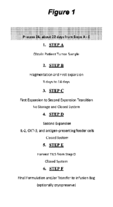
[0216] Figure 1: Exemplary Process 2A chart providing an overview of Steps
A through F.
102171 Figure 2: Process Flow Chart of Process 2A.
[0218] Figure 3: Shows a diagram of an embodiment of a cryopreserved TIL
exemplary
manufacturing process (-22 days).
[0219] Figure 4: Shows a diagram of an embodiment of process 2A, a 22-day
process for TIL
manufacturing.
[0220] Figure 5: Comparison table of Steps A through F from exemplary
embodiments of
process 1C and process 2A.
102211 Figure 6: Detailed comparison of an embodiment of process 1C and an
embodiment of
process 2A.
-31-
WO 2022/170219 PCT/US2022/015538
[0222] Figure 7: Exemplary GEN 3 type process for tumors.
[0223] Figure 8A-8J: A) Shows a comparison between the 2A process
(approximately 22-day
process) and an embodiment of the Gen 3 process for TIL manufacturing
(approximately 14-days
to 16-days process). B) Exemplary Process Gen3 chart providing an overview of
Steps A through
F (approximately 14-days to 16-days process). C) Chart providing three
exemplary Gen 3
processes with an overview of Steps A through F (approximately 14-days to 16-
days process) for
each of the three process variations. D) Exemplary Modified Gen 2-like process
providing an
overview of Steps A through F (approximately 22-days process). E) Chart
providing three
exemplary Gen 3 processes with a pre-treatment with (systemic and/or
intratumoral administration
of) an oncolytic virus (1 day to 3 months prior) for each of the three process
variations. F)
Exemplary Modified Gen 2-like process with a pre-treatment with (systemic
and/or intratumoral
administration of) an oncolytic virus (1 day to 3 months prior). G) Chart
providing three exemplary
Gen 3 processes with a pre-treatment for conditioning the tumor with in situ
electroporation of IL-
12 encoding plasmid (1 day to 3 months prior) for each of the three process
variations. H)
Exemplary Modified Gen 2-like process with a pre-treatment for conditioning
the tumor with in
situ electroporation of IL-12 encoding plasmid (1 day to 3 months prior). I)
Chart providing three
exemplary Gen 3 processes with a pre-treatment for conditioning the tumor with
(systemic and/or
intratumoral administration of) an oncolytic virus and in situ electroporation
of IL-12 encoding
plasmid (1 day to 3 months prior) for each of the three process variations. J)
Exemplary Modified
Gen 2-like process with a pre-treatment for conditioning the tumor with
(systemic and/or
intratumoral administration of) an oncolytic virus and in situ electroporation
of IL-12 encoding
plasmid (1 day to 3 months prior).
[0224] Figure 9: Provides an experimental flow chart for comparability
between GEN 2
(process 2A) versus GEN 3.
[0225] Figure 10: Shows a comparison between various Gen 2 (2A process) and
the Gen 3.1
process embodiment.
[0226] Figure 11: Table describing various features of embodiments of the
Gen 2, Gen 2.1 and
Gen 3.0 process.
-32-
WO 2022/170219 PCT/US2022/015538
[0227] Figure 12: Overview of the media conditions for an embodiment of the
Gen 3 process,
referred to as Gen 3.1.
[02281 Figure 13: Table describing various features of embodiments of the
Gen 2, Gen 2.1 and
Gen 3.0 process.
[0229] Figure 14: Table comparing various features of embodiments of the
Gen 2 and Gen 3.0
processes.
102301 Figure 15: Table providing media uses in the various embodiments of
the described
expansion processes.
[0231] Figure 16: Schematic of an exemplary embodiment of the Gen 3 process
(a 16-day
process).
[0232] Figure 17: Schematic of an exemplary embodiment of a method for
expanding T cells
from hematopoietic malignancies using Gen 3 expansion platfolln.
[0233] Figure 18: Provides the structures I-A and I-B, the cylinders refer
to individual
polypeptide binding domains. Structures I-A and I-B comprise three linearly-
linked TNFRSF
binding domains derived from e.g., 4-1BBL or an antibody that binds 4-1BB,
which fold to form
a trivalent protein, which is then linked to a second trivalent protein
through IgGl-Fc (including
CH3 and CH2 domains) is then used to link two of the trivalent proteins
together through disulfide
bonds (small elongated ovals), stabilizing the structure and providing an
agonists capable of
bringing together the intracellular signaling domains of the six receptors and
signaling proteins to
form a signaling complex. The TNFRSF binding domains denoted as cylinders may
be scFv
domains comprising, e.g., a VH and a VL chain connected by a linker that may
comprise
hydrophilic residues and Gly and Ser sequences for flexibility, as well as Glu
and Lys for
solubility.
[0234] Figure 19: Schematic of an exemplary embodiment of the Gen 3 process
(a 16-day
process).
[0235] Figure 20: Provides a processs overview for an exemplary embodiment
(Gen 3.1 Test)
of the Gen 3.1 process (a 16 day process).
-33-
WO 2022/170219 PCT/US2022/015538
[0236] Figure 21: Schematic of an exemplary embodiment of the Gen 3.1 Test
(Gen 3.1
optimized) process (a 16-17 day process).
[02371 Figure 22: Schematic of an exemplary embodiment of the Gen 3 process
(a 16-day
process).
[0238] Figure 23A-23B: Comparison tables for exemplary Gen 2 and exemplary
Gen 3
processes with exemplary differences highlighted.
102391 Figure 24: Schematic of an exemplary embodiment of the Gen 3 process
(a 16/17 day
process) preparation timeline.
[0240] Figure 25: Schematic of an exemplary embodiment of the Gen 3 process
(a 14-16 day
process).
[0241] Figure 26A-26B: Schematic of an exemplary embodiment of the Gen 3
process (a 16
day process).
[0242] Figure 27: Schematic of an exemplary embodiment of the Gen 3 process
(a 16 day
process).
[0243] Figure 28: Comparison of Gen 2, Gen 2.1 and an embodiment of the Gen
3 process (a
16 day process).
[0244] Figure 29: Comparison of Gen 2, Gen 2.1 and an embodiment of the Gen
3 process (a
16 day process).
[0245] Figure 30: Gen 3 embodiment components.
[0246] Figure 31: Gen 3 embodiment flow chart comparison (Gen 3.0, Gen 3.1
control, Gen
3.1 Test).
[0247] Figure 32: Shown are the components of an exemplary embodiment of
the Gen 3
process (Gen 3-Optimized, a 16-17 day process).
[02481 Figure 33: Acceptance criteria table.
[0249] Figure 34: Shows an overview of chemokines and chemokine receptors
for which
transiently gene expression alteration can be employed to improve TIL
trafficking to the tumor
site.
-34-
WO 2022/170219 PCT/US2022/015538
[0250] Figure 35: Shows a second overview of chemokines and chemokine
receptors for
which transiently gene expression alteration can be employed toimprove TIL
trafficking to the
tumor site.
[02511 Figure 36: Shows a schematic structural representation of an
exemplary self-delivering
ribonucleic acid (sd-RNA) embodiment. See, Ligtenberg, et al., Mol. Therapy,
2018.
102521 Figure 37: Shows a schematic structural representation of an
exemplary sd-RNA
embodiment. See, US Patent Publication No. 2016/0304873.
[0253] Figure 38: Shows an exemplary scheme for mRNA synthesis using a DNA
template
obtained by PCR with use of specially designed primers. The forward primer
contains a
bacteriophage promoter suitable for in vitro transcription and the reverse
primer contains a polyT
stretch. The PCR product is an expression cassette suitable for in vitro
transcription.
Polyadenylates on the 3' end of the nascent mRNA can prevent aberrant RNA
runoff synthesis and
creation of double strand RNA product. After completion of transcription polyA
tail can be
additionally extended with poly(A) polymerase. (See, US Patent No. 8,859,229.)
[0254] Figure 39: Chart showing Sd-rxRNA-mediated silencing of PDCD1, TIM3,
CBLB,
LAG3, and CISH.
[0255] Figure 40: Sd-rxRNA-mediated gene silencing in TIL; exemplary
protocol. Exemplary
tumors include melanoma (fresh or frozen; n=6), breast tumor (fresh or frozen;
n=5), lung tumor
(n=1), sarcoma (n=1), and/or ovarian (n=1).
[02561 Figure 41: Reduction of protein expression was detected in 4 out of
the 5 targets. PD1:
n=9, TIM3: n=8, LAG3/CISH: n=2, Cbl-b n=2. Preps from pre-REP melanoma and
Fresh breast
cancer TILs, 2uM sd-rxRNA. % KD calculated as (100-(100*(gene of
interest/NTC))).
[0257] Figure 42: Sd-rxRNA-induced KD descended with time and stimulation.
n=3, preps
from pre-REP melanoma TILs, 2uM sd-rxRNA.
Brief Description of the Sequence Listing
102581 SEQ ID NO:1 is the amino acid sequence of the heavy chain of
muromonab.
[0259] SEQ ID NO:2 is the amino acid sequence of the light chain of
muromonab.
-35-
WO 2022/170219 PCT/US2022/015538
[0260] SEQ ID NO:3 is the amino acid sequence of a recombinant human IL-2
protein.
[0261] SEQ ID NO:4 is the amino acid sequence of aldesleukin.
[0262] SEQ ID NO:5 is the amino acid sequence of a recombinant human IL-4
protein.
102631 SEQ ID NO:6 is the amino acid sequence of a recombinant human IL-7
protein.
[0264] SEQ ID NO :7 is the amino acid sequence of a recombinant human IL-15
protein.
[0265] SEQ ID NO:8 is the amino acid sequence of a recombinant human IL-21
protein.
102661 SEQ ID NO:9 is the amino acid sequence of human 4-1BB.
[0267] SEQ ID NO:10 is the amino acid sequence of murine 4-1BB.
[0268] SEQ ID NO:11 is the heavy chain for the 4-1BB agonist monoclonal
antibody
utomilumab (PF-05082566).
[0269] SEQ ID NO:12 is the light chain for the 4-1BB agonist monoclonal
antibody
utomilumab (PF-05082566).
[0270] SEQ ID NO:13 is the heavy chain variable region (VH) for the 4-1BB
agonist
monoclonal antibody utomilumab (PF-05082566).
102711 SEQ ID NO:14 is the light chain variable region (VL) for the 4-1BB
agonist
monoclonal antibody utomilumab (PF-05082566).
[0272] SEQ ID NO:15 is the heavy chain CDR1 for the 4-1BB agonist
monoclonal antibody
utomilumab (PF-05082566).
102731 SEQ ID NO:16 is the heavy chain CDR2 for the 4-1BB agonist
monoclonal antibody
utomilumab (PF-05082566).
[0274] SEQ ID NO: 17 is the heavy chain CDR3 for the 4-1BB agonist
monoclonal antibody
utomilumab (PF-05082566).
102751 SEQ ID NO:18 is the light chain CDR1 for the 4-1BB agonist
monoclonal antibody
utomilumab (PF-05082566).
[0276] SEQ ID NO:19 is the light chain CDR2 for the 4-1BB agonist
monoclonal antibody
utomilumab (PF-05082566).
-36-
WO 2022/170219 PCT/US2022/015538
[0277] SEQ ID NO:20 is the light chain CDR3 for the 4-1BB agonist
monoclonal antibody
utomilumab (PF-05082566).
[02781 SEQ ID NO:21 is the heavy chain for the 4-1BB agonist monoclonal
antibody urelumab
(BMS-663513).
[0279] SEQ ID NO:22 is the light chain for the 4-1BB agonist monoclonal
antibody urelumab
(BMS-663513).
102801 SEQ ID NO:23 is the heavy chain variable region (VH) for the 4-1BB
agonist
monoclonal antibody urelumab (BMS-663513).
[0281] SEQ ID NO:24 is the light chain variable region (VL) for the 4-1BB
agonist
monoclonal antibody urelumab (BMS-663513).
[0282] SEQ ID NO:25 is the heavy chain CDR1 for the 4-1BB agonist
monoclonal antibody
urelumab (BMS-663513).
[0283] SEQ ID NO:26 is the heavy chain CDR2 for the 4-1BB agonist
monoclonal antibody
urelumab (BMS-663513).
[0284] SEQ ID NO:27 is the heavy chain CDR3 for the 4-1BB agonist
monoclonal antibody
urelumab (BMS-663513).
[0285] SEQ ID NO:28 is the light chain CDR1 for the 4-1BB agonist
monoclonal antibody
urelumab (BMS-663513).
[0286] SEQ ID NO:29 is the light chain CDR2 for the 4-1BB agonist
monoclonal antibody
urelumab (BMS-663513).
[0287] SEQ ID NO:30 is the light chain CDR3 for the 4-1BB agonist
monoclonal antibody
urelumab (BMS-663513).
[0288] SEQ ID NO:31 is an Fc domain for a TNFRSF agonist fusion protein.
[0289] SEQ ID NO:32 is a linker for a TNFRSF agonist fusion protein.
[0290] SEQ ID NO:33 is a linker for a TNFRSF agonist fusion protein.
[0291] SEQ ID NO:34 is a linker for a TNFRSF agonist fusion protein.
[0292] SEQ ID NO:35 is a linker for a TNFRSF agonist fusion protein.
-37-
WO 2022/170219 PCT/US2022/015538
192931 SEQ ID NO:36 is a linker for a TNFRSF agonist fusion protein.
[0294] SEQ ID NO:37 is a linker for a TNFRSF agonist fusion protein.
[0295] SEQ ID NO:38 is a linker for a TNFRSF agonist fusion protein.
102961 SEQ ID NO:39 is a linker for a TNFRSF agonist fusion protein.
[0297] SEQ ID NO:40 is a linker for a TNFRSF agonist fusion protein.
[0298] SEQ ID NO:41 is a linker for a TNFRSF agonist fusion protein.
102991 SEQ ID NO:42 is an Fc domain for a TNFRSF agonist fusion protein.
103001 SEQ ID NO:43 is a linker for a TNFRSF agonist fusion protein.
103011 SEQ ID NO:44 is a linker for a TNFRSF agonist fusion protein.
[0302] SEQ ID NO:45 is a linker for a TNFRSF agonist fusion protein.
103031 SEQ ID NO:46 is a 4-1BB ligand (4-1BBL) amino acid sequence.
[0304] SEQ ID NO:47 is a soluble portion of 4-1BBL polypeptide.
103051 SEQ ID NO:48 is a heavy chain variable region (VH) for the 4-1BB
agonist antibody
4B4-1-1 version 1.
[0306] SEQ ID NO:49 is a light chain variable region (VL) for the 4-!BB
agonist antibody
4B4-1-1 version 1.
103071 SEQ ID NO:50 is a heavy chain variable region (VI-!) for the 4-1BB
agonist antibody
4B4-1-1 version 2.
[0308] SEQ ID NO:51 is a light chain variable region (VL) for the 4-1BB
agonist antibody
4B4-1-1 version 2.
[0309] SEQ ID NO:52 is a heavy chain variable region (VI-!) for the 4-1BB
agonist antibody
H39E3-2.
[0310] SEQ ID NO:53 is a light chain variable region (VL) for the 4-1BB
agonist antibody
H39E3-2.
[0311] SEQ ID NO:54 is the amino acid sequence of human 0X40.
-38-
WO 2022/170219 PCT/US2022/015538
193121 SEQ ID NO:55 is the amino acid sequence of murine 0X40.
103131 SEQ ID NO:56 is the heavy chain for the 0X40 agonist monoclonal
antibody
tavolixizumab (MEDI-0562).
103141 SEQ ID NO:57 is the light chain for the 0X40 agonist monoclonal
antibody
tavolixizumab (MEDI-0562).
103151 SEQ ID NO:58 is the heavy chain variable region (VH) for the 0X40
agonist
monoclonal antibody tavolixizumab (MEDI-0562).
103161 SEQ ID NO:59 is the light chain variable region (VL) for the 0X40
agonist monoclonal
antibody tavolixizumab (MEDI-0562).
103171 SEQ ID NO:60 is the heavy chain CDR1 for the 0X40 agonist monoclonal
antibody
tavolixizumab (MEDI-0562).
103181 SEQ ID NO:61 is the heavy chain CDR2 for the 0X40 agonist monoclonal
antibody
tavolixizumab (MEDI-0562).
103191 SEQ ID NO:62 is the heavy chain CDR3 for the 0X40 agonist monoclonal
antibody
tavolixizumab (MEDI-0562).
[0320] SEQ ID NO:63 is the light chain CDR1 for the 0X40 agonist monoclonal
antibody
tavolixizumab (MEDI-0562).
[0321] SEQ ID NO:64 is the light chain CDR2 for the 0X40 agonist monoclonal
antibody
tavolixizumab (MEDI-0562).
103221 SEQ ID NO:65 is the light chain CDR3 for the 0X40 agonist monoclonal
antibody
tavolixizumab (MEDI-0562).
[0323] SEQ ID NO:66 is the heavy chain for the 0X40 agonist monoclonal
antibody 11D4.
103241 SEQ ID NO:67 is the light chain for the 0X40 agonist monoclonal
antibody 11D4.
[0325] SEQ ID NO:68 is the heavy chain variable region (VH) for the 0X40
agonist
monoclonal antibody 11D4.
[0326] SEQ ID NO:69 is the light chain variable region (VL) for the 0X40
agonist monoclonal
antibody 11D4.
-39-
WO 2022/170219 PCT/US2022/015538
[0327] SEQ ID NO:70 is the heavy chain CDR1 for the 0X40 agonist monoclonal
antibody
11D4.
[03281 SEQ ID NO:71 is the heavy chain CDR2 for the 0X40 agonist monoclonal
antibody
11D4.
[0329] SEQ ID NO:72 is the heavy chain CDR3 for the 0X40 agonist monoclonal
antibody
11D4.
103301 SEQ ID NO:73 is the light chain CDR1 for the 0X40 agonist monoclonal
antibody
11D4.
[03311 SEQ ID NO:74 is the light chain CDR2 for the 0X40 agonist monoclonal
antibody
11D4.
[03321 SEQ ID NO:75 is the light chain CDR3 for the 0X40 agonist monoclonal
antibody
11D4.
103331 SEQ ID NO:76 is the heavy chain for the 0X40 agonist monoclonal
antibody 18D8.
103341 SEQ ID NO:77 is the light chain for the 0X40 agonist monoclonal
antibody 18D8.
[0335] SEQ ID NO:78 is the heavy chain variable region (VH) for the 0X40
agonist
monoclonal antibody 18D8.
103361 SEQ ID NO:79 is the light chain variable region (VL) for the 0X40
agonist monoclonal
antibody 18D8.
103371 SEQ ID NO:80 is the heavy chain CDR1 for the 0X40 agonist monoclonal
antibody
18D8.
103381 SEQ ID NO:81 is the heavy chain CDR2 for the 0X40 agonist monoclonal
antibody
18D8.
[03391 SEQ ID NO:82 is the heavy chain CDR3 for the 0X40 agonist monoclonal
antibody
18D8.
[0340] SEQ ID NO:83 is the light chain CDR1 for the 0X40 agonist monoclonal
antibody
18D8.
-40-
WO 2022/170219 PCT/US2022/015538
[0341] SEQ ID NO:84 is the light chain CDR2 for the 0X40 agonist monoclonal
antibody
18D8.
[03421 SEQ ID NO:85 is the light chain CDR3 for the 0X40 agonist monoclonal
antibody
18D8.
[0343] SEQ ID NO:86 is the heavy chain variable region (VH) for the 0X40
agonist
monoclonal antibody Hu119-122.
[0344] SEQ ID NO:87 is the light chain variable region (VL) for the 0X40
agonist monoclonal
antibody Hu119-122.
[0345] SEQ ID NO:88 is the heavy chain CDRI for the 0X40 agonist monoclonal
antibody
Hu119-122.
[0346] SEQ ID NO:89 is the heavy chain CDR2 for the 0X40 agonist monoclonal
antibody
Hu119-122.
[0347] SEQ ID NO:90 is the heavy chain CDR3 for the 0X40 agonist monoclonal
antibody
Hu119-122.
[0348] SEQ ID NO:91 is the light chain CDR1 for the 0X40 agonist monoclonal
antibody
Hu119-122.
[0349] SEQ ID NO:92 is the light chain CDR2 for the 0X40 agonist monoclonal
antibody
Hu119-122.
[0350] SEQ ID NO:93 is the light chain CDR3 for the 0X40 agonist monoclonal
antibody
Hu119-122.
[0351] SEQ ID NO:94 is the heavy chain variable region (VH) for the 0X40
agonist
monoclonal antibody Hu106-222.
[0352] SEQ ID NO:95 is the light chain variable region (VL) for the 0X40
agonist monoclonal
antibody Hu106-222.
[0353] SEQ ID NO:96 is the heavy chain CDRI for the 0X40 agonist monoclonal
antibody
Hu106-222.
-41-
WO 2022/170219 PCT/US2022/015538
[0354] SEQ ID NO:97 is the heavy chain CDR2 for the 0X40 agonist monoclonal
antibody
Hu106-222.
[03551 SEQ ID NO:98 is the heavy chain CDR3 for the 0X40 agonist monoclonal
antibody
Hu106-222.
[0356] SEQ ID NO:99 is the light chain CDR1 for the 0X40 agonist monoclonal
antibody
Hu106-222.
103571 SEQ ID NO:100 is the light chain CDR2 for the 0X40 agonist
monoclonal antibody
Hu106-222.
[0358] SEQ ID NO:101 is the light chain CDR3 for the OX40 agonist
monoclonal antibody
Hu106-222.
[0359] SEQ ID NO:102 is an 0X40 ligand (OX4OL) amino acid sequence.
[0360] SEQ ID NO:103 is a soluble portion of OX4OL polypeptide.
[0361] SEQ ID NO:104 is an alternative soluble portion of OX4OL
polypeptide.
[0362] SEQ ID NO:105 is the heavy chain variable region (VH) for the 0X40
agonist
monoclonal antibody 008.
[0363] SEQ ID NO:106 is the light chain variable region (VL) for the 0X40
agonist
monoclonal antibody 008.
[0364] SEQ ID NO:107 is the heavy chain variable region (VH) for the 0X40
agonist
monoclonal antibody 011.
[0365] SEQ ID NO:108 is the light chain variable region (VL) for the OX40
agonist
monoclonal antibody 011.
[0366] SEQ ID NO:109 is the heavy chain variable region (VH) for the 0X40
agonist
monoclonal antibody 021.
[0367] SEQ ID NO:110 is the light chain variable region (VL) for the 0X40
agonist
monoclonal antibody 021.
[0368] SEQ ID NO:111 is the heavy chain variable region (VH) for the 0X40
agonist
monoclonal antibody 023.
-42-
WO 2022/170219 PCT/US2022/015538
[0369] SEQ ID NO:112 is the light chain variable region (VL) for the 0X40
agonist
monoclonal antibody 023.
[03701 SEQ ID NO:113 is the heavy chain variable region (VH) for an OX40
agonist
monoclonal antibody.
[0371] SEQ ID NO:114 is the light chain variable region (VL) for an 0X40
agonist
monoclonal antibody.
103721 SEQ ID NO:115 is the heavy chain variable region (VH) for an OX40
agonist
monoclonal antibody.
[0373] SEQ ID NO:116 is the light chain variable region (VL) for an 0X40
agonist
monoclonal antibody.
[0374] SEQ ID NO:117 is the heavy chain variable region (VH) for a
humanized 0X40 agonist
monoclonal antibody.
[0375] SEQ ID NO:118 is the heavy chain variable region (VH) for a
humanized 0X40 agonist
monoclonal antibody.
[0376] SEQ ID NO:119 is the light chain variable region (VL) for a
humanized 0X40 agonist
monoclonal antibody.
[0377] SEQ ID NO:120 is the light chain variable region (VL) for a
humanized 0X40 agonist
monoclonal antibody.
[0378] SEQ ID NO:121 is the heavy chain variable region (VH) for a
humanized 0X40 agonist
monoclonal antibody.
[0379] SEQ ID NO:122 is the heavy chain variable region (VH) for a
humanized 0X40 agonist
monoclonal antibody.
[0380] SEQ ID NO:123 is the light chain variable region (VL) for a
humanized OX40 agonist
monoclonal antibody.
[0381] SEQ ID NO:124 is the light chain variable region (VL) for a
humanized 0X40 agonist
monoclonal antibody.
-43-
WO 2022/170219 PCT/US2022/015538
[0382] SEQ ID NO:125 is the heavy chain variable region (VH) for an 0X40
agonist
monoclonal antibody.
[03831 SEQ ID NO:126 is the light chain variable region (VL) for an 0X40
agonist
monoclonal antibody.
[0384] SEQ ID NO:127-462 are currently not assigned.
[0385] SEQ ID NO:463 is the heavy chain amino acid sequence of the PD-1
inhibitor
nivolumab.
[0386] SEQ ID NO:464 is the light chain amino acid sequence of the PD-1
inhibitor
nivolumab.
[03871 SEQ ID NO:465 is the heavy chain variable region (VH) amino acid
sequence of the
PD-1 inhibitor nivolumab.
[0388] SEQ ID NO:466 is the light chain variable region (VL) amino acid
sequence of the PD-
1 inhibitor nivolumab.
[0389] SEQ ID NO:467 is the heavy chain CDR1 amino acid sequence of the PD-
1 inhibitor
nivolumab.
[0390] SEQ ID NO:468 is the heavy chain CDR2 amino acid sequence of the PD-
1 inhibitor
nivolumab.
[0391] SEQ ID NO:469 is the heavy chain CDR3 amino acid sequence of the PD-
1 inhibitor
nivolumab.
[0392] SEQ ID NO:470 is the light chain CDR1 amino acid sequence of the PD-
1 inhibitor
nivolumab.
[0393] SEQ ID NO:471 is the light chain CDR2 amino acid sequence of the PD-
1 inhibitor
nivolumab.
[03941 SEQ ID NO:472 is the light chain CDR3 amino acid sequence of the PD-
1 inhibitor
nivolumab.
[0395] SEQ ID NO:473 is the heavy chain amino acid sequence of the PD-1
inhibitor
pembrolizumab.
-44-
WO 2022/170219 PCT/US2022/015538
193961 SEQ ID NO:474 is the light chain amino acid sequence of the PD-1
inhibitor
pembrolizumab.
[03971 SEQ ID NO:475 is the heavy chain variable region (VH) amino acid
sequence of the
PD-1 inhibitor pembrolizumab.
[0398] SEQ ID NO:476 is the light chain variable region (VL) amino acid
sequence of the PD-
1 inhibitor pembrolizumab.
103991 SEQ ID NO:477 is the heavy chain CDR1 amino acid sequence of the PD-
1 inhibitor
pembrolizumab.
[0400] SEQ ID NO:478 is the heavy chain CDR2 amino acid sequence of the PD-
1 inhibitor
pembrolizumab.
[0401] SEQ ID NO:479 is the heavy chain CDR3 amino acid sequence of the PD-
1 inhibitor
pembrolizumab.
[0402] SEQ ID NO:480 is the light chain CDR1 amino acid sequence of the PD-
1 inhibitor
pembrolizumab.
[0403] SEQ ID NO:481 is the light chain CDR2 amino acid sequence of the PD-
1 inhibitor
pembrolizumab.
[0404] SEQ ID NO:482 is the light chain CDR3 amino acid sequence of the PD-
1 inhibitor
pembrolizumab.
[0405] SEQ ID NO:483 is the heavy chain amino acid sequence of the PD-Li
inhibitor
durvalumab.
[0406] SEQ ID NO:484 is the light chain amino acid sequence of the PD-Li
inhibitor
durvalumab.
[0407] SEQ ID NO:485 is the heavy chain variable region (VH) amino acid
sequence of the
PD-Li inhibitor durvalumab.
[0408] SEQ ID NO:486 is the light chain variable region (VL) amino acid
sequence of the PD-
Li inhibitor durvalumab.
-45-
WO 2022/170219 PCT/US2022/015538
[0409] SEQ ID NO:487 is the heavy chain CDR1 amino acid sequence of the PD-
Li inhibitor
durvalumab.
[04101 SEQ ID NO:488 is the heavy chain CDR2 amino acid sequence of the PD-
Li inhibitor
durvalumab.
[0411] SEQ ID NO:489 is the heavy chain CDR3 amino acid sequence of the PD-
Li inhibitor
durvalumab.
104121 SEQ ID NO:490 is the light chain CDR1 amino acid sequence of the PD-
Li inhibitor
durvalumab.
[0413] SEQ ID NO:491 is the light chain CDR2 amino acid sequence of the PD-
Li inhibitor
durvalumab.
[0414] SEQ ID NO:492 is the light chain CDR3 amino acid sequence of the PD-
Li inhibitor
durvalumab.
[0415] SEQ ID NO:493 is the heavy chain amino acid sequence of the PD-Li
inhibitor
avelumab.
[0416] SEQ ID NO:494 is the light chain amino acid sequence of the PD-Li
inhibitor
avelumab.
[0417] SEQ ID NO:495 is the heavy chain variable region (VII) amino acid
sequence of the
PD-Li inhibitor avelumab.
[0418] SEQ ID NO:496 is the light chain variable region (VL) amino acid
sequence of the PD-
Li inhibitor avelumab.
[0419] SEQ ID NO:497 is the heavy chain CDR1 amino acid sequence of the PD-
Li inhibitor
avelumab.
[0420] SEQ ID NO:498 is the heavy chain CDR2 amino acid sequence of the PD-
Li inhibitor
avelumab.
[0421] SEQ ID NO:499 is the heavy chain CDR3 amino acid sequence of the PD-
Li inhibitor
avelumab.
-46-
WO 2022/170219 PCT/US2022/015538
194221 SEQ ID NO:500 is the light chain CDR1 amino acid sequence of the PD-
Li inhibitor
avelumab.
[04231 SEQ ID NO:501 is the light chain CDR2 amino acid sequence of the PD-
Li inhibitor
avelumab.
[0424] SEQ ID NO:502 is the light chain CDR3 amino acid sequence of the PD-
Li inhibitor
avelumab.
104251 SEQ ID NO:503 is the heavy chain amino acid sequence of the PD-Li
inhibitor
atezolizumab.
[0426] SEQ ID NO:504 is the light chain amino acid sequence of the PD-Li
inhibitor
atezolizumab.
[0427] SEQ ID NO:505 is the heavy chain variable region (VH) amino acid
sequence of the
PD-Li inhibitor atezolizumab.
[0428] SEQ ID NO:506 is the light chain variable region (VL) amino acid
sequence of the PD-
Li inhibitor atezolizumab.
[0429] SEQ ID NO:507 is the heavy chain CDR1 amino acid sequence of the PD-
Li inhibitor
atezolizumab.
[0430] SEQ ID NO:508 is the heavy chain CDR2 amino acid sequence of the PD-
Li inhibitor
atezolizumab.
[0431] SEQ ID NO:509 is the heavy chain CDR3 amino acid sequence of the PD-
Li inhibitor
atezolizumab.
[0432] SEQ ID NO:510 is the light chain CDR1 amino acid sequence of the PD-
Li inhibitor
atezolizumab.
[0433] SEQ ID NO:511 is the light chain CDR2 amino acid sequence of the PD-
Li inhibitor
atezolizumab.
[0434] SEQ ID NO:512 is the light chain CDR3 amino acid sequence of the PD-
Li inhibitor
atezolizumab.
-47-
WO 2022/170219 PCT/US2022/015538
[0435] SEQ ID NO:513 is the heavy chain amino acid sequence of the CTLA-4
inhibitor
ipilimumab.
[04361 SEQ ID NO:514 is the light chain amino acid sequence of the CTLA-4
inhibitor
ipilimumab.
[0437] SEQ ID NO:515 is the heavy chain variable region (VET) amino acid
sequence of the
CTLA-4 inhibitor ipilimumab.
104381 SEQ ID NO:516 is the light chain variable region (VL) amino acid
sequence of the
CTLA-4 inhibitor ipilimumab.
[0439] SEQ ID NO:517 is the heavy chain CDR1 amino acid sequence of the
CTLA-4
inhibitor ipilimumab.
[0440] SEQ ID NO:518 is the heavy chain CDR2 amino acid sequence of the
CTLA-4
inhibitor ipilimumab.
[0441] SEQ ID NO:519 is the heavy chain CDR3 amino acid sequence of the
CTLA-4
inhibitor ipilimumab.
[0442] SEQ ID NO:520 is the light chain CDR1 amino acid sequence of the
CTLA-4 inhibitor
ipilimumab.
[0443] SEQ ID NO:521 is the light chain CDR2 amino acid sequence of the
CTLA-4 inhibitor
ipilimumab.
[0444] SEQ ID NO:522 is the light chain CDR3 amino acid sequence of the
CTLA-4 inhibitor
ipilimumab.
[0445] SEQ ID NO:523 is the heavy chain amino acid sequence of the CTLA-4
inhibitor
tremelimumab.
[0446] SEQ ID NO:524 is the light chain amino acid sequence of the CTLA-4
inhibitor
tremelimumab.
[0447] SEQ ID NO:525 is the heavy chain variable region (VH) amino acid
sequence of the
CTLA-4 inhibitor tremelimumab.
-48-
WO 2022/170219 PCT/US2022/015538
194481 SEQ ID NO:526 is the light chain variable region (VL) amino acid
sequence of the
CTLA-4 inhibitor tremelimumab.
[04491 SEQ ID NO:527 is the heavy chain CDR1 amino acid sequence of the
CTLA-4
inhibitor tremelimumab.
[0450] SEQ ID NO:528 is the heavy chain CDR2 amino acid sequence of the
CTLA-4
inhibitor tremelimumab.
104511 SEQ ID NO:529 is the heavy chain CDR3 amino acid sequence of the
CTLA-4
inhibitor tremelimumab.
[0452] SEQ ID NO:530 is the light chain CDR1 amino acid sequence of the
CTLA-4 inhibitor
tremelimumab.
[0453] SEQ ID NO:531 is the light chain CDR2 amino acid sequence of the
CTLA-4 inhibitor
tremelimumab.
[0454] SEQ ID NO:532 is the light chain CDR3 amino acid sequence of the
CTLA-4 inhibitor
tremelimumab.
[0455] SEQ ID NO:533 is the heavy chain amino acid sequence of the CTLA-4
inhibitor
zalifrelimab.
[0456] SEQ ID NO:534 is the light chain amino acid sequence of the CTLA-4
inhibitor
zalifrelimab.
[0457] SEQ ID NO:535 is the heavy chain variable region (VH) amino acid
sequence of the
CTLA-4 inhibitor zalifrelimab.
[0458] SEQ ID NO:536 is the light chain variable region (VL) amino acid
sequence of the
CTLA-4 inhibitor zalifrelimab.
[0459] SEQ ID NO:537 is the heavy chain CDR1 amino acid sequence of the
CTLA-4
inhibitor zalifrelimab.
[0460] SEQ ID NO:538 is the heavy chain CDR2 amino acid sequence of the
CTLA-4
inhibitor zalifrelimab.
-49-
WO 2022/170219 PCT/US2022/015538
194611 SEQ ID NO:539 is the heavy chain CDR3 amino acid sequence of the
CTLA-4
inhibitor zalifrelimab.
[04621 SEQ ID NO:540 is the light chain CDR1 amino acid sequence of the
CTLA-4 inhibitor
zalifrelimab.
[0463] SEQ ID NO:541 is the light chain CDR2 amino acid sequence of the
C1LA-4 inhibitor
zalifrelimab.
104641 SEQ ID NO:542 is the light chain CDR3 amino acid sequence of the
CTLA-4 inhibitor
zalifrelimab.
[0465] SEQ ID NO:543 is the IL-2 sequence.
[04661 SEQ ID NO:544 is an IL-2 mutein sequence.
[0467] SEQ ID NO:545 is an IL-2 mutein sequence.
[0468] SEQ ID NO:546 is the HCDR1 LL-2 for IgG.IL2R67A.H1.
[0469] SEQ ID NO:547 is the HCDR2 for IgG.IL2R67A.H1.
[0470] SEQ ID NO:548 is the HCDR3 for IgG.IL2R67A.H1.
104711 SEQ ID NO:549 is the HCDR1JL-2 kabat for IgG.IL2R67A.H1.
[0472] SEQ ID NO:550 is the HCDR2 kabat for IgG.IL2R67A.H1.
[0473] SEQ ID NO:551 is the HCDR3 kabat for IgG.IL2R67A.H1.
[0474] SEQ ID NO:552 is the HCDR1JL-2 clothia for IgG.IL2R67A.H1.
[0475] SEQ ID NO:553 is the HCDR2 clothia for IgG.IL2R67A.H1.
[04761 SEQ ID NO:554 is the HCDR3 clothia for IgG.IL2R67A.H1.
[0477] SEQ ID NO:555 is the HCDR1JL-2 MGT for IgaIL2R67A.H1.
[0478] SEQ ID NO:556 is the HCDR2 IMGT for IgG.IL2R67A.H1.
[0479] SEQ ID NO:557 is the HCDR3 IMGT for IgG.IL2R67A.H1.
104801 SEQ ID NO:558 is the VH chain for IgG.IL2R67A.H1.
[0481] SEQ ID NO:559 is the heavy chain for IgG.IL2R67A.H1.
-50-
WO 2022/170219 PCT/US2022/015538
194821 SEQ ID NO:560 is the LCDR1 kabat for IgG.IL2R67A.H1.
104831 SEQ ID NO:561 is the LCDR2 kabat for IgG.IL2R67A.H1.
104841 SEQ ID NO:562 is the LCDR3 kabat for IgG.IL2R67A.H1.
104851 SEQ ID NO:563 is the LCDR1 chothia for IgG.IL2R67A.H1.
[0486] SEQ ID NO:564 is the LCDR2 chothia for IgG.IL2R67A.H1.
[0487] SEQ ID NO:565 is the LCDR3 chothia for IgG.IL2R67A.H1.
[04881 SEQ ID NO:566 is the VL chain.
104891 SEQ ID NO:567 is the light chain.
[0490] SEQ ID NO:568 is the light chain.
[0491] SEQ ID NO:569 is the light chain.
104921 SEQ ID NO: 570 is an IL-2 form.
[0493] SEQ ID NO: 571 is an IL-2 form.
[0494] SEQ ID NO: 572 is an IL-2 form.
[0495] SEQ ID NO: 573 is a mucin domain polypeptide.
Detailed Description
Introduction
104961 The present invention provides methods for expanding TILs and
producing therapeutic
populations of TILs. According to exemplary embodiments, the methods include
delivery of
expression vectors for immunomodulatory molecules to a tumor in the subject,
wherein the tumor
is subjected to electroporation in situ prior to harvesting the tumor for TIL
production. According
to further embodiments, at least a portion of the therapeutic population of
TILs are gene-edited to
enhance their therapeutic effect. According to yet further embodiments, an
adjuvant therapy for
cancer includes delivery of expression vectors for immunomodulatory molecules
to a tumor in the
subject before, after or before and after infusion of Tits for treating cancer
in the subject.
-51-
WO 2022/170219 PCT/US2022/015538
[0497] Without intending to be bound by any particular theory, it is
believed that conditioning
of a first tumor mass from a cancer in a subject by delivery of one or more
immunomodulatory
molecules to the first tumor mass before, after or before and after resection
of a sample of a second
tumor mass in the subject (which second tumor mass may be the same as or
different from the first
tumor mass), followed by expansion of TILs obtained from the sample to produce
a therapeutic
population of Tits, will yield phenotypically superior and more tumor-reactive
TILs together with
a tumor microenvironment more favorable to TIL function and tumor killing
(both as effected by
the conditioning of the first tumor mass in the subject), both providing TILs
with greater anti-
cancer potency and conditioning the subject to respond better to TIL therapy,
as further described
herein.
[0498] The present invention relates to a method of treating cancer in a
subject comprising
administering a first therapeutic composition comprising tumor infiltrating
lymphocytes and a
second therapeutic composition comprising oncolytic virus (oncolytic viral
vector) to the subject,
wherein the tumor infiltrating lymphocytes are selected and/or expanded from a
tumor resected
from the subject who has received an oncolytic virus treatment prior to the
tumor resection.
[0499] Without being bound by a particular therapy, the oncolytic virus is
used to
enhance/induce the T cells (e.g., CD4+ T cells and CD8+ T cells) against tumor
epitopes, increase
the T cells in tumors, increase the trafficking of T cells to tumors,
accumulate T cells at the tumors,
expand T cells in the tumor (such as tumor-specific T cells), and/or activate
T cells in the tumor
(such as tumor-specific T cells).
[0500] In another aspect, the invention is directed to a method for
selecting a clinically
effective population of tumor infiltrating lymphocytes (TILs), wherein TILs
are obtained from a
subject receiving oncolytic viral therapy. In another aspect, the invention is
directed to a method
for expanding a clinically effective population of tumor infiltrating
lymphocytes (TILs), wherein
Tits are obtained from a subject receiving oncolytic viral therapy. In yet
another aspect, the
invention is directed to a method for selecting and expanding a clinically
effective population of
tumor infiltrating lymphocytes (TILs), wherein TILs are obtained from a
subject receiving
oncolytic viral therapy.
[0501] Another aspect of the invention provides for a method for treating a
human subject with
cancer, the method comprising: (i) administering to a human subject a
therapeutically effective
-52-
WO 2022/170219 PCT/US2022/015538
amount of an oncolytic virus according to the present disclosure; (ii)
performing any of the
methods described herein for selecting and expanding a therapeutically
effective population of
TILs obtained from a tumor from the human subject; and administering the
expanded TILs
produced according to the method of step (ii), thereby treating the human
subject with cancer. In
some embodiments, the therapeutically effect amount of an oncolytic virus
refers to an amount
that enhances/induces the Tits (e.g., CD4+ T cells and CD8+ T cells) against
tumor epitopes,
increases TILs in tumors, increases the trafficking of TILs to tumors,
accumulates TILs at the
tumors, expands TILs in the tumor (such as tumor-specific TILs), and/or
activates TILs in the
tumor (such as tumor-specific TILs).
[05021
Definitions
[0503] Unless defined otherwise, all technical and scientific terms used
herein have the same
meaning as is commonly understood by one of skill in the art to which this
invention belongs. All
patents and publications referred to herein are incorporated by reference in
their entireties.
[05041 The term "in vivo" refers to an event that takes place in a
subject's body.
[0505] The term "in vitro" refers to an event that takes places outside of
a subject's body. In
vitro assays encompass cell-based assays in which cells alive or dead are
employed and may also
encompass a cell-free assay in which no intact cells are employed.
[0506] The term "ex vivo" refers to an event which involves treating or
performing a procedure
on a cell, tissue and/or organ which has been removed from a subject's body.
Aptly, the cell, tissue
and/or organ may be returned to the subject's body in a method of surgery or
treatment.
[0507] The term "rapid expansion" means an increase in the number of
antigen-specific TILs
of at least about 3-fold (or 4-, 5-, 6-, 7-, 8-, or 9-fold) over a period of a
week, more preferably at
least about 10-fold (or 20-, 30-, 40-, 50-, 60-, 70-, 80-, or 90-fold) over a
period of a week, or most
preferably at least about 100-fold over a period of a week. A number of rapid
expansion protocols
are outlined below.
[0508] By "tumor infiltrating lymphocytes" or "TILs" herein is meant a
population of cells
originally obtained as white blood cells that have left the bloodstream of a
subject and migrated
into a tumor. TILs include, but are not limited to, CD8+ cytotoxic T cells
(lymphocytes), Thl and
-53-
WO 2022/170219 PCT/US2022/015538
Th17 CD4+ T cells, natural killer cells, dendritic cells and M1 macrophages.
TILs include both
primary and secondary TILs. "Primary TILs" are those that are obtained from
patient tissue
samples as outlined herein (sometimes referred to as "freshly harvested"), and
"secondary Tits"
are any TIL cell populations that have been expanded or proliferated as
discussed herein, including,
but not limited to bulk TILs, expanded TILs ("REP TILs") as well as "reREP
TILs" as discussed
herein. reREP Tits can include for example second expansion Tits or second
additional
expansion TILs (such as, for example, those described in Step D of the GEN 3
process of Figure
8, including TILs referred to as reREP TILs). Also, TIL cell populations can
include genetically
modified TILs.
105091 TILs can generally be defined either biochemically, using cell
surface markers, or
functionally, by their ability to infiltrate tumors and effect treatment. TILs
can be generally
categorized by expressing one or more of the following biomarkers: CD4, CD8,
TCR c43, CD27,
CD28, CD56, CCR7, CD45Ra, CD95, PD-1, and CD25. Additionally, and
alternatively, TILs can
be functionally defined by their ability to infiltrate solid tumors upon
reintroduction into a patient.
TILS may further be characterized by potency ¨ for example, TILS may be
considered potent if,
for example, interferon (IFN) release is greater than about 50 pg/mL, greater
than about 100
pg/mL, greater than about 150 pg/mL, or greater than about 200 pg/mL. TILs may
be considered
potent if, for example, interferon (IFN-y) release is greater than about 50
pg/mL, greater than about
100 pg/mL, greater than about 150 pg/mL, or greater than about 200 pg/mL,
greater than about
300 pg/mL, greater than about 400 pg/mL, greater than about 500 pg/mL, greater
than about 600
pg/mL, greater than about 700 pg/mL, greater than about 800 pg/mL, greater
than about 900
pg/mL, greater than about 1000 pg/mL.
[0510] By "population of cells" (including TILs) herein is meant a number
of cells that share
common traits. In general, populations generally range from 1 X 10⁶ to 1 X
1010 in number, with
different TIT populations comprising different numbers. For example, initial
growth of primary
TILs in the presence of IL-2 results in a population of bulk TILs of roughly 1
x 108 cells. REP
expansion is generally done to provide populations of 1.5 x 10⁹ to 1.5 x 10¹⁰
cells for infusion.
105111 By "cryopreserved TILs" herein is meant that Tits, either primary,
bulk, or expanded
(REP Tits), are treated and stored in the range of about -150 C to -60 C.
General methods for
cryopreservation are also described elsewhere herein, including in the
Examples. For clarity,
-54-
WO 2022/170219 PCT/US2022/015538
"cryopreserved TILs" are distinguishable from frozen tissue samples which may
be used as a
source of primary TILs.
105121 By "thawed cryopreserved Tits" herein is meant a population of TILs
that was
previously cryopreserved and then treated to return to room temperature or
higher, including but
not limited to cell culture temperatures or temperatures wherein Tits may be
administered to a
patient.
105131 TILs can generally be defined either biochemically, using cell
surface markers, or
functionally, by their ability to infiltrate tumors and effect treatment. TILs
can be generally
categorized by expressing one or more of the following biomarkers: CD4, CD8,
TCR af3, CD27,
CD28, CD56, CCR7, CD45Ra, CD95, PD-1, and CD25. Additionally and
alternatively, Tits can
be functionally defined by their ability to infiltrate solid tumors upon
reintroduction into a patient.
105141 The term "cryopreservation media" or "cryopreservation medium"
refers to any
medium that can be used for cryopreservation of cells. Such media can include
media comprising
7% to 10% DMSO. Exemplary media include CryoStor CS10, Hyperthermasol, as well
as
combinations thereof. The term "CS10" refers to a cryopreservation medium
which is obtained
from Stemcell Technologies or from Biolife Solutions. The CS10 medium may be
referred to by
the trade name "CryoStorg CS10". The CS10 medium is a serum-free, animal
component-free
medium which comprises DMSO.
105151 The term "central memory T cell" refers to a subset of T cells that
in the human are
CD45R0+ and constitutively express CCR7 (CCR7hi) and CD62L (CD62hi). The
surface
phenotype of central memory T cells also includes TCR, CD3, CD127 (IL-7R), and
IL-15R.
Transcription factors for central memory T cells include BCL-6, BCL-6B, MBD2,
and BMIl.
Central memory T cells primarily secret IL-2 and CD4OL as effector molecules
after TCR
triggering. Central memory T cells are predominant in the CD4 compartment in
blood, and in the
human are proportionally enriched in lymph nodes and tonsils.
105161 The term "effector memory T cell" refers to a subset of human or
mammalian T cells
that, like central memory T cells, are CD45R0+, but have lost the constitutive
expression of CCR7
(CCR71o) and are heterogeneous or low for CD62L expression (CD62L1o). The
surface phenotype
of central memory T cells also includes TCR, CD3, CD127 (IL-7R), and IL-15R.
Transcription
-55-
WO 2022/170219 PCT/US2022/015538
factors for central memory T cells include BLIMP 1. Effector memory T cells
rapidly secret high
levels of inflammatory cytokines following antigenic stimulation, including
interferon-y, IL-4, and
IL-5. Effector memory T cells are predominant in the CD8 compartment in blood,
and in the human
are proportionally enriched in the lung, liver, and gut. CD8+ effector memory
T cells carry large
amounts of perforin.
105171 The term "closed system" refers to a system that is closed to the
outside environment.
Any closed system appropriate for cell culture methods can be employed with
the methods of the
present invention. Closed systems include, for example, but are not limited to
closed G-containers.
Once a tumor segment is added to the closed system, the system is no opened to
the outside
environment until the TILs are ready to be administered to the patient.
105181 The terms "fragmenting," "fragment," and "fragmented," as used
herein to describe
processes for disrupting a tumor, includes mechanical fragmentation methods
such as crushing,
slicing, dividing, and morcellating tumor tissue as well as any other method
for disrupting the
physical structure of tumor tissue.
105191 The terms "peripheral blood mononuclear cells" and "PBMCs" refers to
a peripheral
blood cell having a round nucleus, including lymphocytes (T cells, B cells, NK
cells) and
monocytes. Preferably, the peripheral blood mononuclear cells are irradiated
allogeneic peripheral
blood mononuclear cells. PBMCs are a type of antigen-presenting cell.
105201 The term "anti-CD3 antibody" refers to an antibody or variant
thereof, e.g., a
monoclonal antibody and including human, humanized, chimeric or murine
antibodies which are
directed against the CD3 receptor in the T cell antigen receptor of mature T
cells. Anti-CD3
antibodies include OKT-3, also known as muromonab. Anti-CD3 antibodies also
include the
UHCT1 clone, also known as T3 and CD3e. Other anti-CD3 antibodies include, for
example,
otelixizumab, teplizumab, and visilizumab.
105211 The term "OKT-3" (also referred to herein as "OKT3") refers to a
monoclonal antibody
or biosimilar or variant thereof, including human, humanized, chimeric, or
murine antibodies,
directed against the CD3 receptor in the T cell antigen receptor of mature T
cells, and includes
commercially-available forms such as OKT-3 (30 ng/mL, MACS GMP CD3 pure,
Miltenyi
Biotech, Inc., San Diego, CA, USA) and muromonab or variants, conservative
amino acid
-56-
W02022/170219 PCT/US2022/015538
substitutions, glycoforms, or biosimilars thereof The amino acid sequences of
the heavy and light
chains of muromonab are given in Table 1 (SEQ ID NO:1 and SEQ ID NO:2). A
hybridoma
capable of producing OKT-3 is deposited with the American Type Culture
Collection and assigned
the ATCC accession number CRL 8001. A hybridoma capable of producing OKT-3 is
also
deposited with European Collection of Authenticated Cell Cultures (ECACC) and
assigned
Catalogue No. 86022706.
TABLE 1. Amino acid sequences of muromonab.
Identifier Sequence (One-Letter Amino Acid Symbols)
SEQ ID NO:1 QVQLQQSGAE LARPGASVKM SCKASGYTFT RYTMHWVKQR PGQGLEWIGY
INPSRGYTNY 60
Muromonab heavy NQKFKDKATL TTDKSSSTAY MQLSSLTSED aAVYYCARYY DDHYCLDYWG
QGTTLTVSSA 120
chain KTTAPSVYPL APVCGGTTGS SVTLGCLVKG YFPEPVTLTW NSGSLSSGVH
TFPAVIQSDL 180
YTLSSSVTVT SSTWPSQSIT CNVAHPASST KVDKKIEPRP KSCDKTHTCP PCPAPELLGG
240
PSVFLFPPKP KDTLMISRTP EVTCVVVDVS HEDPEVKFNW YVDGVEVHNA KTKPREEQYN
300
STYRVVSVLT VMHQDWLNGK EYKCKVSNKA LPAPIEKTIS KAKGQPREPQ VYTLPPSRDE
360
LTKNQVSLTC LVKGFYPSDI AVEWESNGQP ENNYKTTPPV LDSDGSFFLY SKLTVDKSRW
420
QQGNVFSCSV MHEAIHNHYT QKSLSLSPGK
450
SEQ ID NO:2 QIVLTQSPAI MSASPGEKVT MTCSASSSVS YMNWYQQKSG TSPKRWIYDT
SKLASGVPAH 60
MuLomonab light FRGSGSGTSY SLTISGMEAE DAATYYCQQW SSNPFTFGSG TKLEINRADT
APTVSIFPPS 120
chain SEQLTSGGAS VVCFLNNFYP KDINVKWKID GSERQNGVMN SWTDQDSKDS
TYSMSSTLTL 180
TKDEYERHNS YTCEATHKTS TSPIVNSFNR NEC
213
105221 The term "IL-2" (also referred to herein as "lL2") refers to the T
cell growth factor
known as interleukin-2, and includes all forms of IL-2 including human and
mammalian forms,
conservative amino acid substitutions, glycoforms, biosimilars, and variants
thereof. IL-2 is
described, e.g., in Nelson, J. Immunol. 2004, 172, 3983-88 and Malek, Annu.
Rev. Immunol. 2008,
26, 453-79, the disclosures of which are incorporated by reference herein. The
amino acid
sequence of recombinant human IL-2 suitable for use in the invention is given
in Table 2 (SEQ ID
NO:3). For example, the term IL-2 encompasses human, recombinant forms of IL-2
such as
aldesleukin (PROLEUKIN, available commercially from multiple suppliers in 22
million IU per
single use vials), as well as the form of recombinant IL-2 commercially
supplied by CellGenix,
Inc., Portsmouth, NH, USA (CELLGRO GMF') or ProSpec-Tany TechnoGene Ltd., East
Brunswick, NJ, USA (Cat. No. CYT-209-b) and other commercial equivalents from
other vendors.
Aldesleukin (des-alanyl-1, serine-125 human IL-2) is a nonglycosylated human
recombinant form
of IL-2 with a molecular weight of approximately 15 kDa. The amino acid
sequence of aldesleukin
suitable for use in the invention is given in Table 2 (SEQ ID NO:4). The term
IL-2 also
encompasses pegylated forms of IL-2, as described herein, including the
pegylated 11,2 prodrug
NKTR-214, available from Nektar Therapeutics, South San Francisco, CA, USA.
NKTR-214 and
-57-
WO 2022/170219 PCT/US2022/015538
pegylated IL-2 suitable for use in the invention is described in U.S. Patent
Application Publication
No. US 2014/0328791 Al and International Patent Application Publication No. WO
2012/065086
Al, the disclosures of which are incorporated by reference herein. Alternative
forms of conjugated
IL-2 suitable for use in the invention are described in U.S. Patent Nos.
4,766,106, 5,206,344,
5,089,261 and 4902,502, the disclosures of which are incorporated by reference
herein.
Formulations of IL-2 suitable for use in the invention are described in U.S.
Patent No. 6,706,289,
the disclosure of which is incorporated by reference herein.
105231 In some embodiments, an IL-2 form suitable for use in the invention
is THOR-707.
Additional alternative forms of IL-2 suitable for use in the invention are
described in U.S. Patent
Application Publication No. 2020/0181220 Al and U.S. Patent Application
Publication No.
2020/0330601 Al, both of which are incorporated by reference herein. In some
embodiments, an
IL-2 form suitable for use in the invention is ALKS-4230. Additional
alternative forms of IL-2
suitable for use in the invention are also described in U.S. Patent
Application Publication No.
2021/0038684 Al and U.S. Patent No. 10,183,979, both of which are incorporated
by reference
herein. In some embodiments, and IL-2 form suitable for use in the invention
is an interleukin 2
(IL-2) conjugate comprising: an isolated and purified IL-2 polypeptide; and a
conjugating moiety
that binds to the isolated and purified IL-2 polypeptide at an amino acid
position selected from
K35, T37, R38, T41, F42, K43, F44, Y45, E61, E62, E68, K64, P65, V69, L72, and
Y107, wherein
the numbering of the amino acid residues corresponds to SEQ ID NO: 1 in U.S.
Patent Application
Publication No. 2020/018122. In some embodiments, the amino acid position is
selected from T37,
R38, T41, F42, F44, Y45, E61, E62, E68, K64, P65, V69, L72, and Y107. In some
embodiments,
the amino acid position is selected from T37, R38, T41, F42, F44, Y45, E61,
E62, E68, P65, V69,
L72, and Y107. In some embodiments, the amino acid position is selected from
T37, T41, F42,
F44, Y45, P65, V69, L72, and Y107. In some embodiments, the amino acid
position is selected
from R38 and K64. In some embodiments, the amino acid position is selected
from E61, E62, and
E68. In some embodiments, the amino acid position is at E62. In some
embodiments, the amino
acid residue selected from K35, T37, R38, T41, F42, K43, F44, Y45, E61, E62,
E68, K64, P65,
V69, L72, and Y107 is further mutated to lysine, cysteine, or histidine. In
some embodiments, the
amino acid residue is mutated to cysteine. In some embodiments, the amino acid
residue is mutated
to lysine. In some embodiments, the amino acid residue selected from K35, T37,
R38, T41, F42,
K43, F44, Y45, E61, E62, E68, K64, P65, V69, L72, and Y107 is further mutated
to an unnatural
-58-
WO 2022/170219 PCT/US2022/015538
amino acid. In some embodiments, the unnatural amino acid comprises N6-
azidoethoxy-L-lysine
(AzK), N6-propargylethoxy-L-lysine (PraK), BCN-L-ly sine, norbornene ly sine,
TCO-lysine,
methyltetrazine lysine, allyloxycarbonyllysine, 2-amino-8-oxononanoic acid, 2-
amino-8-
oxooctanoic acid, p-acetyl-L-phenylalanine, p-azidomethyl-L-phenylalanine
(pAMF), p-iodo-L-
phenyl alanine, m-acetylphenylalanine, 2-amino-8-oxononanoic acid,
p-
propargyloxyphenylalanine, p-propargyl-phenylalanine, 3 -methyl-phenylalanine,
L-Dopa,
fluorinated phenylalanine, isopropyl-L-phenylalanine, p-azido-L-phenylalanine,
p-acyl-L-
phenylalanine, p-benzoyl-L-phenylalanine, p-bromophenylalanine, p-amino-L-
phenylalanine,
isopropyl-L-phenylalanine, 0-allyltyrosine, 0-methyl-L-tyrosine,
4-propyl-
L-tyrosine, phosphonotyrosine, tri-O-acetyl-G1cNAcp-serine, L-phosphoserine,
phosphonoserine,
L-3 -(2-naphthyl)alanine,
2-amino-3-((2-((3-(benzyloxy)-3-
oxopropyl)amino)ethyl)selanyl)propanoic acid,
2-amino-3-(phenyl selanyl)propanoic, or
selenocysteine. In some embodiments, the IL-2 conjugate has a decreased
affinity to IL-2 receptor
a (IL-2Ra) subunit relative to a wild-type IL-2 polypeptide. In some
embodiments, the decreased
affinity is about 10%, 20%, 30%, 40%, 50%, 60%, 70%, 80%, 90%, 95%, 99%, or
greater than
99% decrease in binding affinity to IL-2Ra relative to a wild-type IL-2
polypeptide. In some
embodiments, the decreased affinity is about 1-fold, 2-fold, 3-fold, 4-fold, 5-
fold, 6-fold, 7-fold,
8-fold, 9-fold, 10-fold, 30-fold, 50-fold, 100-fold, 200-fold, 300-fold, 500-
fold, 1000-fold, or more
relative to a wild-type IL-2 polypeptide. In some embodiments, the conjugating
moiety impairs or
blocks the binding of IL-2 with IL-2Ra. In some embodiments, the conjugating
moiety comprises
a water-soluble polymer. In some embodiments, the additional conjugating
moiety comprises a
water-soluble polymer. In some embodiments, each of the water-soluble polymers
independently
comprises polyethylene glycol (PEG), poly(propylene glycol) (PPG), copolymers
of ethylene
glycol and propylene glycol, poly(oxyethylated polyol), poly(olefinic
alcohol),
poly(vinylpyrrolidone), poly(hydroxyalkylmethacrylamide),
poly(hydroxyalkylmethacrylate),
poly(saccharides), poly(a-hydroxy acid), poly(vinyl alcohol), polyphosphazene,
polyoxazolines
(POZ), poly(N-acryloylmorpholine), or a combination thereof In some
embodiments, each of the
water-soluble polymers independently comprises PEG. In some embodiments, the
PEG is a linear
PEG or a branched PEG. In some embodiments, each of the water-soluble polymers
independently
comprises a polysaccharide. In some embodiments, the polysaccharide comprises
dextran,
polysialic acid (PSA), hyaluronic acid (HA), amylose, heparin, heparan sulfate
(HS), dextrin, or
-59-
WO 2022/170219 PCT/US2022/015538
hydroxyethyl-starch (HES). In some embodiments, each of the water-soluble
polymers
independently comprises a glycan. In some embodiments, each of the water-
soluble polymers
independently comprises polyamine. In some embodiments, the conjugating moiety
comprises a
protein. In some embodiments, the additional conjugating moiety comprises a
protein. In some
embodiments, each of the proteins independently comprises an albumin, a
transferrin, or a
transthyretin. In some embodiments, each of the proteins independently
comprises an Fc portion.
In some embodiments, each of the proteins independently comprises an Fc
portion of IgG. In some
embodiments, the conjugating moiety comprises a polypeptide. In some
embodiments, the
additional conjugating moiety comprises a polypeptide. In some embodiments,
each of the
polypeptides independently comprises a XTEN peptide, a glycine-rich homoamino
acid polymer
(HAP), a PAS polypeptide, an elastin-like polypeptide (ELP), a CTP peptide, or
a gelatin-like
protein (GLK) polymer. In some embodiments, the isolated and purified IL-2
polypeptide is
modified by glutamylation. In some embodiments, the conjugating moiety is
directly bound to the
isolated and purified IL-2 polypeptide. In some embodiments, the conjugating
moiety is indirectly
bound to the isolated and purified IL-2 polypeptide through a linker. In some
embodiments, the
linker comprises a homobifunctional linker. In some embodiments, the
homobifunctional linker
comprises Lomant's reagent dithiobis
(succinimidylpropionate) DSP 3' 3' -
dithiobis(sulfosuccinimidyl proprionate)
(DTS SP), disuccinimidyl suberate (DS S),
bis(sulfosuccinimidyl)suberate (BS), di succinimidyl tartrate (DST),
disulfosuccinimidyl tartrate
(sulfo DST), ethylene glycobis(succinimidylsuccinate) (EGS), disuccinimidyl
glutarate (DSG),
N,N' -di succinimidyl carbonate (D SC), dimethyl adipimidate (DMA), dimethyl
pimelimidate
(DMP), dimethyl suberimidate (DMS), dimethyl-3 ,3' -dithiobispropionimidate
(DTBP), 1,4-di-
(3
-(2' -pyridyl dithi o)propi onami do)butane (DPDPB), bi smaleimidohexane
(BMH), aryl
halide-containing compound (DFDNB), such as e.g. 1,5-difluoro-2,4-
dinitrobenzene or 1,3-
difluoro-4,6-dinitrobenzene, 4,4' -difluoro-3,3' -dinitrophenylsulfone
(DFDNPS), bist 13 -(4 -
azidosalicylamido)ethyl]disulfide (BASED), formaldehyde, glutaraldehyde, 1,4-
butanediol
diglycidyl ether, adipic acid dihydrazide, carbohydrazide, o-toluidine, 3,3' -
dimethylbenzidine,
benzidine, a,
-p-diaminodiphenyl, diiodo-p-xylene sulfonic acid, N,N' -ethylene-
bis(iodoacetamide), or N,N' -hexamethylene-bis(iodoacetamide). In some
embodiments, the
linker comprises a heterobifunctional linker. In some embodiments, the
heterobifunctional linker
-60-
WO 2022/170219 PCT/US2022/015538
comprises N-succinimidyl 3 -(2-pyridyldithio)propionate (sPDP), long-chain N-
succinimidyl 3 -(2-
pyridyldithio)propionate (LC-sPDP), water-soluble-long-chain N-succinimidyl 3-
(2-
pyridyldithio) propionate
(sulfo-LC-sPDP), succinimi dyl oxy carbonyl -a-methyl -a-(2-
pyridyldithio)toluene (sMPT),
sulfosuccinimidy1-64a-methyl-a-(2-
pyridyldithio)toluamido]hexanoate (sulfo-LC -sMPT),
succinimidy1-4-(N-
maleimidomethyl)cyclohexane-1-carboxylate (sMCC),
sulfosuccinimidy1-4-(N-
maleimidomethyl)cyclohexane-1-carboxylate (sulfo-sMCC),
m-mal eimi dobenzoyl-N-
hydroxysuccinimide ester (MBs), m-maleimidobenzoyl-N-hydroxysulfosuccinimide
ester (sulfo-
MB s), N-succinimidy1(4-iodoacteyl)aminobenzoate (sIAB),
sulfosuccinimidy1(4-
iodoacteyl)aminobenzoate (sulfo-sIAB), succinimidy1-4-(p-
maleimidophenyl)butyrate (sMPB),
sulfosuccinimidy1-4-(p-maleimidophenyl)butyrate (sulfo-sMPB),
N-(y-
maleimidobutyryloxy)succinimide ester (GMBs), N-(y-
maleimidobutyryloxy)sulfosuccinimide
ester (sulfo-GMBs), succinimidyl 6-((iodoacetyl)amino)hexanoate (sIAX),
succinimidyl 646-
(((i odoacetypamino)hexanoyl)aminoThexanoate (sIAXX),
succinimidyl 4-
(((iodoacetyl)amino)methyl)cyclohexane-1-carboxylate (sIAC), succinimidyl 6-
(((((4-
iodoacetyl)amino)methyl)cyclohexane-1-carbonyl)amino) hexanoate (sIACX), p-
nitrophenyl
iodoacetate (NPIA), carbonyl-reactive and sulfhydryl-reactive cross-linkers
such as 4-(4-N-
maleimidophenyl)butyric acid hydrazide (MPBH), 4-(N-
maleimidomethyl)cyclohexane-1-
carboxyl-hydrazide-8 (M2C2H), 3-(2-pyridyldithio)propionyl hydrazide (PDPH), N-
hydroxysuccinimidy1-4-azidosalicylic acid (NHs-AsA), N-
hydroxysulfosuccinimidy1-4-
azidosalicylic acid (sulfo-NHs-AsA), sulfosuccinimidy1-(4-
azidosalicylamido)hexanoate (sulfo-
NHs-LC-AsA), sulfosuccinimidy1-2-(p-azidosalicylamido)ethy1-1,3' -
dithiopropionate (sAsD),
N-hydroxysuccinimidy1-4-azidobenzoate (HsAB), N-hydroxysulfosuccinimidy1-4-
azidobenzoate
(sulfo-HsAB), N-succinimidyl-6-(4' -azido-2' -nitrophenyl amino)hexanoate
(sANPAH),
sulfosuccinimidy1-6-(4' -azido-2' -nitrophenylamino)hexanoate (sulfo-sANPAH),
N-5-azido-
2-nitrobenzoyloxysuccinimide (ANB-N0s), sulfosuccinimidy1-2-(m-azido-o-
nitrobenzamido)-
ethyl-1,3' -dithiopropionate (sAND), N-succinimidy1-4(4-azidopheny1)1,3' -
dithiopropionate
(sADP), N-sulfosuccinimidy1(4-azi dopheny1)- 1,3' -dithiopropionate
(sulfo-sADP),
sulfosuccinimidyl 4-( p -azidophenyl)butyrate (sulfo-sAPB), sulfosuccinimidyl
2-(7-azido-4-
methyl coumarin-3 -acetamide)ethyl- 1,3' -dithiopropionate (sAED),
sulfosuccinimidyl 7-azido-4-
-6 1-
WO 2022/170219 PCT/US2022/015538
methylcoumain-3-acetate (sulfo-sAMCA), p-nitrophenyl diazopyruvate (pNPDP), p-
nitrophenyl-
2-diazo-3,3,3-trifluoropropionate (PNP-DTP), 1-(p-Azidosalicylamido)-4-
(iodoacetamido)butane
(AsB3), N44-(p-
azidosalicylamido)buty1]-3' -(2' -pyridyldithio)propionamide (APDP),
benzophenone-4-iodoacetamide, p-azidobenzoyl hydrazide (ABH),
4-( p -
azidosalicylamido)butylamine (AsBA), or p-azidophenyl glyoxal (APG). In some
embodiments,
the linker comprises a cleavable linker, optionally comprising a dipeptide
linker. In some
embodiments, the dipeptide linker comprises Val-Cit, Phe-Lys, Val-Ala, or Val-
Lys. In some
embodiments, the linker comprises a non-cleavable linker. In some embodiments,
the linker
comprises a maleimide group, optionally comprising maleimidocaproyl (mc),
succinimidy1-4-(N-
maleimidomethyl)cyclohexane-1-carboxylate (sMC C), or
sulfosuccinimidy1-4-(N-
maleimidomethyl)cyclohexane-l-carboxylate (sulfo-sMCC). In some embodiments,
the linker
further comprises a spacer. In some embodiments, the spacer comprises p-
aminobenzyl alcohol
(PAB), p-aminobenzyoxycarbonyl (PABC), a derivative, or an analog thereof. In
some
embodiments, the conjugating moiety is capable of extending the serum half-
life of the IL-2
conjugate. In some embodiments, the additional conjugating moiety is capable
of extending the
serum half-life of the IL-2 conjugate. In some embodiments, the IL-2 form
suitable for use in the
invention is a fragment of any of the IL-2 forms described herein. In some
embodiments, the IL-2
form suitable for use in the invention is pegylated as disclosed in U.S.
Patent Application
Publication No. 2020/0181220 Al and U.S. Patent Application Publication No.
2020/0330601 Al.
In some embodiments, the IL-2 form suitable for use in the invention is an IL-
2 conjugate
comprising: an IL-2 polypeptide comprising an N6-azidoethoxy-L-lysine (AzK)
covalently
attached to a conjugating moiety comprising a polyethylene glycol (PEG),
wherein: the IL-2
polypeptide comprises an amino acid sequence having at least 80% sequence
identity to SEQ ID
NO: 1 in U.S. Patent Application No. 2020/0330601(listed herein as SEQ ID NO:
570 in Table 2);
and the AzK substitutes for an amino acid at position K35, F42, F44, K43, E62,
P65, R38, T41,
E68, Y45, V69, or L72 in reference to the amino acid positions within SEQ ID
NO: 1 in U.S.
Patent Application No. 2020/0330601 (listed herein as SEQ ID NO: 570 in Table
2). In some
embodiments, the IL-2 polypeptide comprises an N-terminal deletion of one
residue relative to
SEQ ID NO: 1 in U.S. Patent Application No. 2020/0330601(listed herein as SEQ
ID NO: 570 in
Table 2). In some embodiments, the IL-2 form suitable for use in the invention
lacks IL-2R alpha
chain engagement but retains normal binding to the intermediate affinity IL-2R
beta-gamma
-62-
W02022/170219 PCT/US2022/015538
signaling complex. In some embodiments, an IL-2 form suitable for use in the
invention is ALKS-
4230. A form of IL-2 suitable for use in the invention is described in U.S.
Patent Application
Publication No. 2021/0038684 Al as SEQ ID NO: 1 (listed herein as SEQ ID NO:
571 in Table
2). In some embodiments, an IL-2 form suitable for use in the invention is a
fusion protein
comprising amino acids 24-452 of SEQ ID NO: 2 in U.S. Patent No. 10,183,979
(SEQ ID NO: 2
in US U.S. Patent No. 10,183,979 listed herein as SEQ ID NO: 572 in Table 2).
In some
embodiments, an IL-2 form suitable for use in the invention is a fusion
protein comprising amino
acids 24-452 of SEQ ID NO: 2 in U.S. Patent No. 10,183,979 or an amino acid
sequence
homologous to amino acids 24-452 of SEQ ID NO: 2 in U.S. Patent No. 10,183,979
with at least
98% amino acid sequence identity over the entire length of amino acids 24-452
of SEQ ID NO: 2
in U.S. Patent No. 10,183,979 and having the receptor antagonist activity of
amino acids 24-452
of SEQ ID NO: 2 in U.S. Patent No. 10,183,979. Optionally, in some
embodiments, an IL-2 faun
suitable for use in the invention is a fusion protein comprising a first
fusion partner that is linked
to a second fusion partner by a mucin domain polypeptide linker, wherein the
first fusion partner
is IL-1Ra or a protein having at least 98% amino acid sequence identity to IL-
1Ra and having the
receptor antagonist activity of 11 -Ra, and wherein the second fusion partner
comprises all or a
portion of an immunoglobulin comprising an Fc region, wherein the mucin domain
polypeptide
linker comprises SEQ ID NO: 14 in U.S. Patent No. 10,183,979 (listed herein as
SEQ ID NO: 573
in Table 2) or an amino acid sequence having at least 90% sequence identity to
SEQ ID NO: 14 in
U.S. Patent No. 10,183,979 (listed herein as SEQ ID NO: 573 in Table 2) and
wherein the half-
life of the fusion protein is improved as compared to a fusion of the first
fusion partner to the
second fusion partner in the absence of the mucin domain polypeptide linker.
TABLE 2. Amino acid sequences of interleukins.
Identifier Sequence (One-Letter Amino Acid Symbols)
SEQ ID NO3 MAPTSSSTEK TQLQLEHLLL DLQMILNGIN NYKNPKLTRM LTFKFYMPKK
ATELKHLQCL 60
recombinant EEELKPLEEV LNLAQSKNFH LRPRDLISNI NVIVLELKGS ETTFMCEYAD
ETATIVEFLN 120
human IL-2 RWITFCQSII STLT
134
(rhIL-2)
SEQ ID NO:4 PTSSSTKKTQ LQLEHLLLDL QMILNGINNY KNPKLTRMLT FKFYMPKKAT
ELKHLQCLEE 60
Aldesleukin ELKPLEEVLN LAQSKNFHLR PRDLISNINV IVIELKGSET TFMCEYADET
ATIVEFLNRW 120
ITFSQSIIST LT
132
SEQ ID NO:5 MHKCD1TLQE lIKTLNSLTE QKTLCTELTV TDIFAASKNT TEKETFCRAA
TVLRQFYSHH 60
recombinant EKDTRCLGAT AQQFHREKQL IRFLKRLDRN LWGLAGLNSC PVKEANQSTL
ENFLERLKTI 120
human IL-4 MREKYSKCSS
130
(rhIL-4)
SEQ ID NO:6 MDCDIEGKDG KQYESVIMVS IDQLLDSMKE IGSNCLNNEF NFFKRHICDA
NKEGMFLFRA 60
-63-
W02022/170219 PCT/US2022/015538
recombinant ARKLRQFLKM NSTGDFDLHL LKVSEGTT1L LNCTGQVKGR KPAALGEAQP
TKSLEENKSL 120
human IL ....7 KEQKKLNDLC FLKRLLQEIK TCWNKILMGT KEH
153
(rhIL-7)
SEQ ID NO:7 MNWVNVISDL KKIEDLIQSM HIDATLYTES DVHPSCKVTA MKCELLELQV
ISLESGDASI 60
recombinant HDTVENLIIL ANNSLSSNGN VTESGCHECE ELEEKNIKEF LQSFVHIVQM
HINTS 115
human IL-15
(rhIL-15)
SEQ ID NO:8 MQDRHMIRMR QLIDIVDQLK NYVNDLVPEF LPAPEDVETN CEWSAFSCFQ
KAQLKSANTG 60
recombinant NNERIINVSI KKLKRKPPST NAGRRQKHRL TCPSCDSYEK KPPKEFLERF
KSLLQKMIHQ 120
human IL-21 HLSSRTHGSE DS
132
(rhIL-21)
SEQ ID NO: 570 APTSSSTKKT QLQLEHLLLD LQMILNGINN YKNPKLTRML TFKFYMPKKA
TELKHLQCLE 60
IL-2 form EELKPLEEVL NLAQSKNFHL RPRDLISNIN VIVLELKGSE TTFMCEYADE
TATIVEFLNR 120
WITFCQSIIS TLT
133
SEQ ID NO: 571 SKNEHLRPRD LISNINVIVI ELKGSETTFM CEYADETATI VEFLNRWITF
SQSIISTLTG 60
IL-2 form GSSSTKKTQL QLEHLLLDLQ MILNGINNYK NPKLTRMLTF KEYMPKKATE
LKHLQCLEEE 120
LKPLEEVLNL AQGSGGGSEL CDDDPPEIPH ATFKAMAYKE GTMLNCECKR GFRRIKSGSL
180
YMLCTGNSSR SSWDNQCQCT SSATRNTTKQ VTPQPEEQKE RKTTEMQSPM QPVDQASLPG
240
HCREPPPWEN EATERIYHFV VGQMVYYQCV QGYRALHRGP AESVCKMTHG KTRWTQPQLI
300
CTG
303
SEQ ID NO: 572 MDAMKRGLCC VILLCGAVFV SARRPSGRKS SKMQAFRIWD VNQKTFYLRN
NQLVAGYLQG 60
IL-2 form PNVNLEEKID VVPIEPHALF LGIHGGKMCL SCVKSGDETR LQLEAVNITD
LSENRKQDKR 120
FAFIRSDSGP TTSFESAACP GWFLCTAMEA DQPVSLTNMP DEGVMVTKFY FQEDESGSGG
180
ASSESSASSD GPHPVITESR ASSESSASSD GPHPVITESR EPKSSDKTHT CPPCPAPELL
240
GGPSVELFPP KPKDTLMISR TPEVTCVVVD VSHEDPEVKF NWYVDGVEVH NAKTKPREEQ
300
YNSTYRVVSV LTVIHQDWLN GKEYKCKVSN KALPAPIEKT ISKARGQPRE PQVYTLPPSR
360
EEMTKNQVSL TCLVKGFYPS DIAVEWESNG QPENNYKTT PPVIDSDGSF FLYSKLTVDK
420
SRWQQGNVFS CSVMHEALHN HYTQKSLSLS PGK
453
SEQ ID NO: 573 SESSASSDGP HPVITP
16
mucin domain
polypeptide
105241 The term "IL-4" (also referred to herein as "IL4") refers to the
cytokine known as
interleukin 4, which is produced by Th2 T cells and by eosinophils, basophils,
and mast cells. IL-
4 regulates the differentiation of naïve helper T cells (Th0 cells) to Th2 T
cells. Steinke and Borish,
Respir. Res. 2001, 2, 66-70. Upon activation by IL-4, Th2 T cells subsequently
produce additional
IL-4 in a positive feedback loop. IL-4 also stimulates B cell proliferation
and class II MHC
expression, and induces class switching to IgE and IgG1 expression from B
cells. Recombinant
human IL-4 suitable for use in the invention is commercially available from
multiple suppliers,
including ProSpec-Tany TechnoGene Ltd., East Brunswick, NJ, USA (Cat. No. CYT-
211) and
TherrnoFisher Scientific, Inc., Waltham, MA, USA (human IL-15 recombinant
protein, Cat. No.
Gibco CTF'0043). The amino acid sequence of recombinant human IL-4 suitable
for use in the
invention is given in Table 2 (SEQ ID NO:5).
[05251 The term "IL-7" (also referred to herein as "IL7") refers to a
glycosylated tissue-
derived cytokine known as interleukin 7, which may be obtained from stromal
and epithelial cells,
as well as from dendritic cells. Fry and Mackall, Blood 2002, 99, 3892-904. IL-
7 can stimulate the
development of T cells. IL-7 binds to the IL-7 receptor, a heterodimer
consisting of IL-7 receptor
alpha and common gamma chain receptor, which in a series of signals important
for T cell
-64-
WO 2022/170219 PCT/US2022/015538
development within the thymus and survival within the periphery. Recombinant
human IL-7
suitable for use in the invention is commercially available from multiple
suppliers, including
ProSpec-Tany TechnoGene Ltd., East Brunswick, NJ, USA (Cat. No. CYT-254) and
ThermoFisher Scientific, Inc., Waltham, MA, USA (human IL-15 recombinant
protein, Cat. No.
Gibco PHC0071). The amino acid sequence of recombinant human IL-7 suitable for
use in the
invention is given in Table 2 (SEQ ID NO:6).
105261 The term "IL-12" (also referred to herein a "IL12") refers to a
cytokine known as
interleukin-12, that is secreted primarily by macrophages and dendritic cells.
The term includes a
heterodimeric protein comprising a 35 kD subunit (p35) and a 40 kD subunit
(p40) which are both
linked together with a disulfide bridge. The heterodimeric protein is referred
to as a "p70 subunit".
The structure of human IL-12 is described further in, for example, Kobayashi,
et al. (1989) J. Exp
Med. 170:827-845; Seder, et al. (1993) Proc. Natl. Acad. Sci. 90:10188-10192;
Ling, et al. (1995)
J. Exp Med. 154:116-127; Podlaski, et al. (1992) Arch. Biochem. Biophys.
294:230-237. The term
human IL-12 is intended to include recombinant human IL-12 (rh IL-12), which
can be prepared
by standard recombinant expression methods.
105271 The term "IL-15" (also referred to herein as "IL15") refers to the T
cell growth factor
known as interleukin-15, and includes all forms of IL-2 including human and
mammalian forms,
conservative amino acid substitutions, glycoforms, biosimilars, and variants
thereof IL-15 is
described, e.g., in Fehniger and Caligiuri, Blood 2001, 97, 14-32, the
disclosure of which is
incorporated by reference herein. IL-15 shares 13 and 7 signaling receptor
subunits with IL-2.
Recombinant human IL-15 is a single, non-glycosylated polypeptide chain
containing 114 amino
acids (and an N-terminal methionine) with a molecular mass of 12.8 kDa.
Recombinant human
IL-15 is commercially available from multiple suppliers, including ProSpec-
Tany TechnoGene
Ltd., East Brunswick, NJ, USA (Cat. No. CYT-230-b) and ThermoFisher
Scientific, Inc.,
Waltham, MA, USA (human IL-15 recombinant protein, Cat. No. 34-8159-82). The
amino acid
sequence of recombinant human IL-15 suitable for use in the invention is given
in Table 2 (SEQ
ID NO:7).
[05281 The term "IL-21" (also referred to herein as "IL21") refers to the
pleiotropic cytokine
protein known as interleukin-21, and includes all forms of IL-21 including
human and mammalian
forms, conservative amino acid substitutions, glycoforms, biosimilars, and
variants thereof IL-21
-65-
WO 2022/170219 PCT/US2022/015538
is described, e.g., in Spolski and Leonard, Nat. Rev. Drug. Disc. 2014, 13,
379-95, the disclosure
of which is incorporated by reference herein. IL-21 is primarily produced by
natural killer T cells
and activated human CD4+ T cells. Recombinant human IL-21 is a single, non-
glycosylated
polypeptide chain containing 132 amino acids with a molecular mass of 15.4
kDa. Recombinant
human IL-21 is commercially available from multiple suppliers, including
ProSpec-Tany
TechnoGene Ltd., East Brunswick, NJ, USA (Cat. No. CYT-408-b) and ThermoFisher
Scientific,
Inc., Waltham, MA, USA (human IL-21 recombinant protein, Cat. No. 14-8219-80).
The amino
acid sequence of recombinant human IL-21 suitable for use in the invention is
given in Table 2
(SEQ ID NO:8).
105291 When "an anti-tumor effective amount", "an tumor-inhibiting
effective amount", or
"therapeutic amount" is indicated, the precise amount of the compositions of
the present invention
to be administered can be determined by a physician with consideration of
individual differences
in age, weight, tumor size, extent of infection or metastasis, and condition
of the patient (subject).
It can generally be stated that a pharmaceutical composition comprising the
tumor infiltrating
lymphocytes (e.g. secondary TILs or genetically modified cytotoxic
lymphocytes) described
herein may be administered at a dosage of 104 to 1011 cells/kg body weight
(e.g., 105 to 106, 105
to 1010, 105 to 1011, 106 to 1010, 106 to 1011,107 to 1011, 107 to 1010, 108
to 1011, 108 to 1010,
109 to 1011, or 109 to 1010 cells/kg body weight), including all integer
values within those ranges.
Tumor infiltrating lymphocytes (including in some cases, genetically modified
cytotoxic
lymphocytes) compositions may also be administered multiple times at these
dosages. The tumor
infiltrating lymphocytes (inlcuding in some cases, genetically) can be
administered by using
infusion techniques that are commonly known in immunotherapy (see, e.g.,
Rosenberg et al., New
Eng. J. of Med. 319: 1676, 1988). The optimal dosage and treatment regime for
a particular patient
can readily be determined by one skilled in the art of medicine by monitoring
the patient for signs
of disease and adjusting the treatment accordingly.
105301 The term "hematological malignancy" refers to mammalian cancers and
tumors of the
hematopoietic and lymphoid tissues, including but not limited to tissues of
the blood, bone marrow,
lymph nodes, and lymphatic system. Hematological malignancies are also
referred to as "liquid
tumors." Hematological malignancies include, but are not limited to, acute
lymphoblastic leukemia
(ALL), chronic lymphocytic lymphoma (CLL), small lymphocytic lymphoma (SLL),
acute
-66-
WO 2022/170219 PCT/US2022/015538
myelogenous leukemia (AML), chronic myelogenous leukemia (CML), acute
monocytic leukemia
(AMoL), Hodgkin's lymphoma, and non-Hodgkin's lymphomas. The term "B cell
hematological
malignancy" refers to hematological malignancies that affect B cells.
[05311 The term "solid tumor" refers to an abnormal mass of tissue that
usually does not
contain cysts or liquid areas. Solid tumors may be benign or malignant. The
term "solid tumor
cancer refers to malignant, neoplastic, or cancerous solid tumors. Solid tumor
cancers include, but
are not limited to, sarcomas, carcinomas, and lymphomas, such as cancers of
the lung, breast,
prostate, colon, rectum, and bladder. The tissue structure of solid tumors
includes interdependent
tissue compartments including the parenchyma (cancer cells) and the supporting
stromal cells in
which the cancer cells are dispersed and which may provide a supporting
microenvironment.
105321 The term "liquid tumor" refers to an abnormal mass of cells that is
fluid in nature.
Liquid tumor cancers include, but are not limited to, leukemias, myelomas, and
lymphomas, as
well as other hematological malignancies. TILs obtained from liquid tumors may
also be referred
to herein as marrow infiltrating lymphocytes (MILs).
105331 The term "microenvironment," as used herein, may refer to the solid
or hematological
tumor microenvironment as a whole or to an individual subset of cells within
the
microenvironment. The tumor microenvironment, as used herein, refers to a
complex mixture of
"cells, soluble factors, signaling molecules, extracellular matrices, and
mechanical cues that
promote neoplastic transformation, support tumor growth and invasion, protect
the tumor from
host immunity, foster therapeutic resistance, and provide niches for dominant
metastases to
thrive," as described in Swartz, et al., Cancer Res., 2012, 72, 2473. Although
tumors express
antigens that should be recognized by T cells, tumor clearance by the immune
system is rare
because of immune suppression by the microenvironment.
105341 In some embodiments, the invention includes a method of treating a
cancer with
population of TILs, wherein a patient is pre-treated with non-myeloablative
chemotherapy prior to
an infusion of Tits according to the invention. In some embodiments, the
population of TILs may
be provided wherein a patient is pre-treated with nonmyeloablative
chemotherapy prior to an
infusion of TILs according to the present invention. In an embodiment, the non-
myeloablative
chemotherapy is cyclophosphamide 60 mg/kg/d for 2 days (days 27 and 26 prior
to TIL infusion)
and fludarabine 25 mg/m2/d for 5 days (days 27 to 23 prior to TIL infusion).
In some embodiments,
-67-
WO 2022/170219 PCT/US2022/015538
the non-myeloablative chemotherapy is cyclophosphamide 60 mg/kg/d for 2 days
(days 27 and 26
prior to TIL infusion) and fludarabine 25 mg/m2/d for 3 days (days 27 to 25
prior to TIL infusion).
In some embodiments, the non-myeloablative chemotherapy is cyclophosphamide 60
mg/kg/d for
2 days (days 27 and 26 prior to TIL infusion) followed by fludarabine 25
mg/m2/d for 3 days (days
25 to 23 prior to TIL infusion). In an embodiment, after non-myeloablative
chemotherapy and TIL
infusion (at day 0) according to the invention, the patient receives an
intravenous infusion of IL-2
intravenously at 720,000 IU/kg every 8 hours to physiologic tolerance.
[05351 Experimental findings indicate that lymphodepletion prior to
adoptive transfer of
tumor-specific T lymphocytes plays a key role in enhancing treatment efficacy
by eliminating
regulatory T cells and competing elements of the immune system ("cytokine
sinks"). Accordingly,
some embodiments of the invention utilize a lymphodepletion step (sometimes
also referred to as
"immunosuppressive conditioning") on the patient prior to the introduction of
the rTILs of the
invention.
[05361 The terms "co-administration," "co-administering," "administered in
combination
with," "administering in combination with," "simultaneous," and "concurrent,"
as used herein,
encompass administration of two or more active pharmaceutical ingredients (in
a preferred
embodiment of the present invention, for example, at least one potassium
channel agonist in
combination with a plurality of TILs) to a subject so that both active
pharmaceutical ingredients
and/or their metabolites are present in the subject at the same time. Co-
administration includes
simultaneous administration in separate compositions, administration at
different times in separate
compositions, or administration in a composition in which two or more active
pharmaceutical
ingredients are present. Simultaneous administration in separate compositions
and administration
in a composition in which both agents are present are preferred.
105371 The term "effective amount" or "therapeutically effective amount"
refers to that
amount of a compound or combination of compounds as described herein that is
sufficient to effect
the intended application including, but not limited to, disease treatment. A
therapeutically effective
amount may vary depending upon the intended application (in vitro or in vivo),
or the subject and
disease condition being treated (e.g., the weight, age and gender of the
subject), the severity of the
disease condition, or the manner of administration. The term also applies to a
dose that will induce
a particular response in target cells (e.g., the reduction of platelet
adhesion and/or cell migration).
-68-
WO 2022/170219 PCT/US2022/015538
The specific dose will vary depending on the particular compounds chosen, the
dosing regimen to
be followed, whether the compound is administered in combination with other
compounds, timing
of administration, the tissue to which it is administered, and the physical
delivery system in which
the compound is carried.
[0538] The terms "treatment", "treating", "treat", and the like, refer to
obtaining a desired
pharmacologic and/or physiologic effect. The effect may be prophylactic in
terms of completely
or partially preventing a disease or symptom thereof and/or may be therapeutic
in terms of a partial
or complete cure for a disease and/or adverse effect attributable to the
disease. "Treatment", as
used herein, covers any treatment of a disease in a mammal, particularly in a
human, and includes:
(a) preventing the disease from occurring in a subject which may be
predisposed to the disease but
has not yet been diagnosed as having it; (b) inhibiting the disease, i.e.,
arresting its development
or progression; and (c) relieving the disease, i.e., causing regression of the
disease and/or relieving
one or more disease symptoms. "Treatment" is also meant to encompass delivery
of an agent in
order to provide for a pharmacologic effect, even in the absence of a disease
or condition. For
example, "treatment" encompasses delivery of a composition that can elicit an
immune response
or confer immunity in the absence of a disease condition, e.g., in the case of
a vaccine.
[0539] The term "heterologous" when used with reference to portions of a
nucleic acid or
protein indicates that the nucleic acid or protein comprises two or more
subsequences that are not
found in the same relationship to each other in nature. For instance, the
nucleic acid is typically
recombinantly produced, having two or more sequences from unrelated genes
arranged to make a
new functional nucleic acid, e.g., a promoter from one source and a coding
region from another
source, or coding regions from different sources. Similarly, a heterologous
protein indicates that
the protein comprises two or more subsequences that are not found in the same
relationship to each
other in nature (e.g., a fusion protein).
[0540] The terms "sequence identity," "percent identity," and "sequence
percent identity" (or
synonyms thereof, e.g., "99% identical") in the context of two or more nucleic
acids or
polypeptides, refer to two or more sequences or subsequences that are the same
or have a specified
percentage of nucleotides or amino acid residues that are the same, when
compared and aligned
(introducing gaps, if necessary) for maximum correspondence, not considering
any conservative
amino acid substitutions as part of the sequence identity. The percent
identity can be measured
-69-
WO 2022/170219 PCT/US2022/015538
using sequence comparison software or algorithms or by visual inspection.
Various algorithms and
software are known in the art that can be used to obtain alignments of amino
acid or nucleotide
sequences. Suitable programs to determine percent sequence identity include
for example the
BLAST suite of programs available from the U.S. Government's National Center
for
Biotechnology Information BLAST web site. Comparisons between two sequences
can be carried
using either the BLASTN or BLASTP algorithm. BLASTN is used to compare nucleic
acid
sequences, while BLASTP is used to compare amino acid sequences. ALIGN, ALIGN-
2
(Genentech, South San Francisco, California) or MegAlign, available from
DNASTAR, are
additional publicly available software programs that can be used to align
sequences. One skilled
in the art can determine appropriate parameters for maximal alignment by
particular alignment
software. In certain embodiments, the default parameters of the alignment
software are used.
[0541] As used herein, the term "variant" encompasses but is not limited to
antibodies or
fusion proteins which comprise an amino acid sequence which differs from the
amino acid
sequence of a reference antibody by way of one or more substitutions,
deletions and/or additions
at certain positions within or adjacent to the amino acid sequence of the
reference antibody. The
variant may comprise one or more conservative substitutions in its amino acid
sequence as
compared to the amino acid sequence of a reference antibody. Conservative
substitutions may
involve, e.g., the substitution of similarly charged or uncharged amino acids.
The variant retains
the ability to specifically bind to the antigen of the reference antibody. The
tem' variant also
includes pegylated antibodies or proteins.
[0542] The term "deoxyribonucleotide" encompasses natural and synthetic,
unmodified and
modified deoxyribonucleotides. Modifications include changes to the sugar
moiety, to the base
moiety and/or to the linkages between deoxyribonucleotide in the
oligonucleotide.
105431 The term "RNA" defines a molecule comprising at least one
ribonucleotide residue.
The term "ribonucleotide" defines a nucleotide with a hydroxyl group at the 2'
position of a b-D-
ribofuranose moiety. The term RNA includes double-stranded RNA, single-
stranded RNA,
isolated RNA such as partially purified RNA, essentially pure RNA, synthetic
RNA,
recombinantly produced RNA, as well as altered RNA that differs from naturally
occurring RNA
by the addition, deletion, substitution and/or alteration of one or more
nucleotides. Nucleotides of
the RNA molecules described herein may also comprise non-standard nucleotides,
such as non-
-70-
WO 2022/170219 PCT/US2022/015538
naturally occurring nucleotides or chemically synthesized nucleotides or
deoxynucleotides. These
altered RNAs can be referred to as analogs or analogs of naturally-occurring
RNA.
[05441 The terms "modified nucleotide" refer to a nucleotide that has one
or more
modifications to the nucleoside, the nucleobase, pentose ring, or phosphate
group. For example,
modified nucleotides exclude ribonucleotides containing adenosine
monophosphate, guanosine
monophosphate, uridine monophosphate, and cytidine monophosphate and
deoxyribonucleotides
containing deoxyadenosine monophosphate, deoxyguanosine monophosphate,
deoxythymidine
monophosphate, and deoxycytidine monophosphate. Modifications include those
naturally-
occurring that result from modification by enzymes that modify nucleotides,
such as
methyltransferases.
105451 Modified nucleotides also include synthetic or non-naturally
occurring nucleotides.
Synthetic or non-naturally occurring modifications in nucleotides include
those with 2'
modifications, e.g., 21-0-methyl, 2'-methoxyethoxy, 2'-fluoro, 2'-allyl, 2'-0-
[2-(methylamino)-2-
oxoethyl], 4'-thio, 4'-CH2-0-2'-bridge, 4'-(CH2) 2-0-2'-bridge, 2'-LNA, and 2'-
0--(N-
methylcarbamate) or those comprising base analogs. In connection with 21-
modified nucleotides
as described for the present disclosure, by "amino" is meant 2'-NH2 or 2'-0--
NH2, which can be
modified or unmodified. Such modified groups are described, for example, in
U.S. Pat. Nos.
5,672,695 and 6,248,878; incorporated by reference herein.
[05461 The terms "microRNA" or "miRNA" refer to a nucleic acid that forms a
single-stranded
RNA, which single-stranded RNA has the ability to alter the expression (reduce
or inhibit
expression; modulate expression; directly or indirectly enhance expression) of
a gene or target
gene when the miRNA is expressed in the same cell as the gene or target gene.
In some
embodiments, a miRNA refers to a nucleic acid that has substantial or complete
identity to a target
gene and forms a single-stranded miRNA. In some embodiments, miRNA may be in
the form of
pre-miRNA, wherein the pre-miRNA is double-stranded RNA. The sequence of the
miRNA can
correspond to the full length target gene, or a subsequence thereof Typically,
the miRNA is at
least about 15-50 nucleotides in length (e.g., each sequence of the single-
stranded miRNA is 15-
50 nucleotides in length, and the double stranded pre-miRNA is about 15-50
base pairs in length).
In some embodiments, the miRNA is 20-30 base nucleotides. In some embodiments,
the miRNA
-71-
WO 2022/170219 PCT/US2022/015538
is 20-25 nucleotides in length. In some embodiments, the miRNA is 20, 21, 22,
23, 24, 25, 26, 27,
28, 29, or 30 nucleotides in length.
105471
The terms "target gene" include genes known or identified as modulating the
expression of a gene involved in an immune resistance mechanism, and can be
one of several
groups of genes, such as suppressor receptors, for example, CTLA4 and PD1;
cytokine receptors
that inactivate immune cells, for example, TGF-beta receptor, LAG3, and/or
TIM3, and
combinations thereof. In some embodiments, the target gene includes one or
more of PD-1,
TGFBR2, CBLB (CBL-B), CISH, CCRs (chimeric co-stimulatory receptors), IL-2, IL-
4, IL-7, IL-
10, IL-12, IL-15, IL-21, NOTCH 1/2 intracellular domain (ICD), NOTCH ligand
mDLL1, TIM3,
LAG3, TIGIT, TGFP, CCR2, CCR4, CCR5, CXCR1, CXCR2, CSCR3, CCL2 (MCP-1), CCL3
(MIP- 1 ct), CCL4 (MIP1-0), CCL5 (RAN ________________________________________
IBS), CXCL1/CXCL8, CCL22, CCL17,
CXCL1/CXCL8, VHL, CD44, PIK3CD, SOCS1, and/or cAMP protein kinase A (PKA).
105481
The phrases "small interfering RNA" or siRNA" or "short interfering RNA" or
"silencing RNA", define a group of double-stranded RNA molecules, comprising
sense and
antisense RNA strands, each generally of about 1022 nucleotides in length,
optionally including a
3' overhang of 1-3 nucleotides. siRNA is active in the RNA interference (RNAi)
pathway, and
interferes with expression of specific target genes with complementary
nucleotide sequences.
105491
The term sd-RNA refers to "self-deliverable" RNAi agents that are formed as an
asymmetric double-stranded RNA-antisense oligonucleotide hybrid. The double
stranded RNA
includes a guide (sense) strand of about 19-25 nucleotides and a passenger
(antisense) strand of
about 10-19 nucleotides with a duplex formation that results in a single-
stranded
phosphorothiolated tail of about 5-9 nucleotides. In some embodiments, the RNA
sequences may
be modified with stabilizing and hydrophobic modifications such as sterols,
for example,
cholesterol, vitamin D, naphtyl, isobutyl, benzyl, indol, tryptophane, and
phenyl, which confer
stability and efficient cellular uptake in the absence of any transfection
reagent or formulation. In
some embodiments, immune response assays testing for IFN-induced proteins
indicate sd-RNAs
produce a reduced immunostimulatory profile as compared other RNAi agents.
See, for example,
Byrne et al., December 2013, J. Ocular Pharmacology and Therapeutics, 29(10):
855-864,
incorporated by reference. In some embodiments, the sd-RNAs described herein
are commercially
available from Advirna LLC, Worcester, MA, USA.
-72-
WO 2022/170219 PCT/US2022/015538
[0550] As used herein, "immune checkpoint" molecules refers to a group of
immune cell
surface receptor/ligands which induce T cell dysfunction or apoptosis. These
immune inhibitory
targets attenuate excessive immune reactions and ensure self-tolerance. Tumor
cells harness the
suppressive effects of these checkpoint molecules.
105511 The phrase "immune checkpoint inhibitor" includes molecules that
prevent immune
suppression by blocking the effects of immune checkpoint molecules. Checkpoint
inhibitors can
include antibodies and antibody fragments, nanobodies, diabodies, soluble
binding partners of
checkpoint molecules, small molecule therapeutics, peptide antagonists, etc. A
list of immune
checkpoints and immune checkpoint inhibitors can be found in US Patent No.
10,426,847, which
is incorporated herein by reference in its entirety.
105521 The phrase "immunostimulatory cytokine" includes cytokines that
mediate or enhance
the immune response to a foreign antigen, including viral, bacterial, or tumor
antigens. Innate
immunostimulatory cytokines can include, e.g., TNF-a, IL-1, IL-10, IL-12, IL-
15, IL-21, type I
interferons (IFN-ct and IFN-13), IFN-y, and chemokines. Adaptive
immunostimulatory cytokines
include, e.g., IL-2, IL-4, IL-5, TGF-I3, IL-10 and IFN-y. As used herein, the
phrase
"immunostimulatory cytokine" further includes subunits of the cytokines as
well oligonucleotides
encoding the cytokines and/or their subunits. For example, an
immunostimulatory cytokine may
be IL-12, a p35 sububit of IL-12, a p40 subunit of IL-12, or oligonucleotides
encoding IL-12, a
p35 sububit of IL-12, a p40 subunit of IL-12. A list of immunostimulatory
cytokines can be found
in US Patent No. 10,426,847.
[0553] The term "immunomodulatory molecule" includes a molecule, delivery
of which into
a cell results in modulating immune response. Thus, immunomodulatory molecules
may include
small molecules, peptides or proteins that function as immunostimulatory
cytokines or immune
checkpoint inhibitors. Additionally, immunomodulatory molecules may include
oligonucles
encoding such peptides or proteins. The immunomodulatory molecules also
include
oligonucleotides encoding both the immunostimulatory cytokines and the immune
checkpoint
inhibitors. Examples of immunomodulatory molecules can be found in US Patent
Publication No.
2019/0209652, and US Patent Publication No. 2019/0153469, both of which are
incorporated
herein by reference in their entirety.
-73-
WO 2022/170219 PCT/US2022/015538
[0554] The terms "pharmaceutically acceptable carrier" or "pharmaceutically
acceptable
excipient" are intended to include any and all solvents, dispersion media,
coatings, antibacterial
and antifungal agents, isotonic and absorption delaying agents, and inert
ingredients. The use of
such pharmaceutically acceptable carriers or pharmaceutically acceptable
excipients for active
pharmaceutical ingredients is well known in the art. Except insofar as any
conventional
pharmaceutically acceptable carrier or pharmaceutically acceptable excipient
is incompatible with
the active pharmaceutical ingredient, its use in the therapeutic compositions
of the invention is
contemplated. Additional active pharmaceutical ingredients, such as other
drugs, can also be
incorporated into the described compositions and methods.
105551 The terms "electroporation", "electro-permeabilization," or "electro-
kinetic
enhancement" ("EP") as used interchangeably herein refer to the use of a
transmembrane electric
field pulse to induce microscopic pathways (pores) in a bio-membrane; their
presence allows
biomolecules such as plasmids, oligonucleotides, siRNA, drugs, ions, and water
to pass from one
side of the cellular membrane to the other.
[0556] The terms "about" and "approximately" mean within a statistically
meaningful range
of a value. Such a range can be within an order of magnitude, preferably
within 50%, more
preferably within 20%, more preferably still within 10%, and even more
preferably within 5% of
a given value or range. The allowable variation encompassed by the terms
"about" or
"approximately" depends on the particular system under study, and can be
readily appreciated by
one of ordinary skill in the art. Moreover, as used herein, the terms "about"
and "approximately"
mean that dimensions, sizes, formulations, parameters, shapes and other
quantities and
characteristics are not and need not be exact, but may be approximate and/or
larger or smaller, as
desired, reflecting tolerances, conversion factors, rounding off, measurement
error and the like,
and other factors known to those of skill in the art. In general, a dimension,
size, formulation,
parameter, shape or other quantity or characteristic is "about" or
"approximate" whether or not
expressly stated to be such. It is noted that embodiments of very different
sizes, shapes and
dimensions may employ the described arrangements.
[0557] The transitional terms "comprising," "consisting essentially of,"
and "consisting of,"
when used in the appended claims, in original and amended form, define the
claim scope with
respect to what unrecited additional claim elements or steps, if any, are
excluded from the scope
-74-
WO 2022/170219 PCT/US2022/015538
of the claim(s). The term "comprising" is intended to be inclusive or open-
ended and does not
exclude any additional, unrecited element, method, step or material. The term
"consisting of'
excludes any element, step or material other than those specified in the claim
and, in the latter
instance, impurities ordinary associated with the specified material(s). The
term "consisting
essentially of' limits the scope of a claim to the specified elements, steps
or material(s) and those
that do not materially affect the basic and novel characteristic(s) of the
claimed invention. All
compositions, methods, and kits described herein that embody the present
invention can, in
alternate embodiments, be more specifically defined by any of the transitional
terms "comprising,"
"consisting essentially of," and "consisting of"
A. TIL Manufacturing Processes
105581 An exemplary TIL process known as process 2A containing some of
these features is
depicted in Figure 2, and some of the advantages of this embodiment of the
present invention over
process 1C are described in Figures 5 and 6 as well as in International Patent
Publication WO
2018/081473. An embodiment of process 2A is shown Figure 1.
105591 As discussed herein, the present invention can include a step
relating to the
restimulation of cryopreserved TILs to increase their metabolic activity and
thus relative health
prior to transplant into a patient, and methods of testing said metabolic
health. As generally
outlined herein, TILs are generally taken from a patient sample and
manipulated to expand their
number prior to transplant into a patient. In some embodiments, the TILs may
be optionally
genetically manipulated as discussed below.
[05601 .. In some embodiments, the TILs may be cryopreserved. Once thawed,
they may also be
restimulated to increase their metabolism prior to infusion into a patient.
105611 In some embodiments, the first expansion (including processes
referred to as the
preREP as well as processes shown in Figure 1 as Step B) is shortened to 3 to
14 days and the
second expansion (including processes referred to as the REP as well as
processes shown in Figure
1 as Step D) is shorted to 7 to 14 days, as discussed in detail below as well
as in the examples and
figures. In some embodiments, the first expansion (for example, an expansion
described as Step B
in Figure 1) is shortened to 11 days and the second expansion (for example, an
expansion as
described in Step D in Figure 1) is shortened to 11 days. In some embodiments,
the combination
-75-
WO 2022/170219 PCT/US2022/015538
of the first expansion and second expansion (for example, expansions described
as Step B and Step
D in Figure 1) is shortened to 22 days, as discussed in detail below and in
the examples and figures.
[05621 The "Step" Designations A, B, C, etc., below are in reference to
Figure 1 and in
reference to certain embodiments described herein. The ordering of the Steps
below and in Figure
1 is exemplary and any combination or order of steps, as well as additional
steps, repetition of
steps, and/or omission of steps is contemplated by the present application and
the methods
disclosed herein.
1. Pretreatment with Oncolytic Virus
[0563] In some embodiments, the subject may be treated with an oncolytic
virus to promote
infiltration of Tits into the tumor prior to resection of a tumor sample from
the subject. In some
embodiments, the oncolytic virus can be additionally or alternatively
modulated to enable delivery
of immunomodulatory cytokines to the tumor cells.
a. Oncolytic Viruses
105641 In some embodiments, the oncolytic viral therapy induces cell lysis,
cell death, ruptured
tumors, release of a tumor-derived antigen, an anti-tumor immune response, a
change in the tumor
microenvironment, increased immune cell infiltration, upregulation
(overexpression) of immune
checkpoint molecules, enhanced immune activation, localized expression of
specific cytokines,
chemokines, and receptor agonists, and the like.
[0565] Oncolytic viruses are well known in the art. In principle any virus
capable of selective
replication in cancer cells including cells of tumors, neoplasms, carcinomas,
sarcomas, and the
like may be utilized in the invention. In some embodiments, selective
replication in cancer cells
refers to the ability of the virus to replicate at least 1 x 10⁴, preferably 1
x105, especially lx 106 more
efficiently in cells from a tumor compared to cells from a non-tumor tissue.
Oncolytic viruses may
be targeted to specific tissues or tumor tissues. This can be achieved for
example through
transcriptional targeting of viral genes or through modification of viral
proteins that are involved
in the cellular binding and uptake mechanisms during the infection process. In
some embodiments,
the oncolytic viruses infect or replicate in a cancer, kill cancer cells,
and/or spread between cancer
cells in a target tissue. In some embodiments, the oncolytic virus is a
replication-incompetent
virus.
-76-
WO 2022/170219 PCT/US2022/015538
195661
In some embodiments, the oncolytic virus is an attenuated virus. In the
context of the
present invention, the term "attenuated" means that the respective virus is
modified to be less
virulent or ideally non-virulent in normal tissues. In some embodiments, this
modification/attenuation does not or only minimally effect its ability to
replicates in tumor,
especially in neoplastic-cells and therefore increases its usefulness in
therapy.
105671
In some embodiments, the oncolytic virus contemplated in the present invention
includes, but is not limited to, an adenovirus, an adeno-associated virus, a
self-replicating
alphavirus, a vaccinia virus, a Seneca Valley Virus, a Newcastle disease
virus, a Maraba virus, a
vesicular stomatitis virus (VSV), a herpes virus (including herpes simplex
virus type 1 (HSV1),
herpes simplex virus type 2 (HSV2), Epstein-Barr virus (EBV), cytomegalovirus
(CMV), and the
like), a measles virus, a mumps virus, a poliovirus, a poliovirus, a poxvirus,
a picornavirus, a
reovirus, a coxsackie virus, a lentivirus, a morbillivirus, an influenza
virus, a sinbis virus, a sendai
virus (SV), myxoma virus, a retrovirus, and a modified virus thereof (see,
e.g., Twumasi-Boateng
et al., Nature Reviews Cancer, 2018, 18(7):419-432 and Kaufman et al., Cancer
Immunotherapy,
2015, 14:642-662, all of which are incorporated by reference herein their
entireties). Exemplary
embodiments of an oncolytic virus are shown in Tables 1-7 of U.S. Patent
Publication No.
2009/0317456, each of which are incorporated herein by reference in their
entireties.
[05681
In some embodiments, the oncolytic virus is a picornavirus. In some instances,
the
picornavirus is selected from coxsackievirus, echovirus, poliovirus,
unclassified enteroviruses,
rhinovirus, paraechovirus, hepatovirus, or cardiovirus.
In particular embodiments, the
picornavirus is not capable of infecting or inducing apoptosis in a cell in
the absence of intercellular
adhesion molecule-1 (ICAM-1). In some embodiments, the picornavirus utilizes
recognition of
ICAM-1 to infect a target cell. Useful embodiments of such picornaviruses are
described in, e.g.,
U.S. Patent Publication Nos. 2008/0160031, 2009/0123427, 2010/0062020,
2012/0328575,
2013/0164300, 2015/0037287, and 2016/0136211, as well as U.S. Patent Nos.
7,361,354,
7,485,292, 8,114,416, 8,236,298 and 8,722,036, each of which are incorporated
herein by
reference in their entireties.
[05691
The oncolytic virus of the present invention may have the sequence of a viral
genome
modified by nucleic acid substitutions, e.g., from 1, 2, or 3 to 10, 25, 50,
100, or more substitutions.
-77-
WO 2022/170219 PCT/US2022/015538
Optionally, the viral genome may be modified be 1 or more insertions and/or
deletions and/or by
a nucleic acid extension at either or of both ends.
11:1570.1 In some embodiments, the oncolytic virus contains a nucleic acid
sequence having at
least 70% sequence identity, e.g., 70%, 75%, 77%, 79%, 80%, 81%, 82%, 83%,
84%, 85%, 86%,
87%, 88%, 89%, 90%, 91%, 92%, 93%, 94%, 95%, 96%, 97%, 98%, 99% sequence
identity or
more, to a parental viral genome. In some embodiments, the oncolytic virus
contains a nucleic acid
sequence having at least 70% sequence identity, e.g., 70%, 75%, 77%, 79%, 80%,
81%, 82%,
83%, 84%, 85%, 86%, 87%, 88%, 89%, 90%, 91%, 92%, 93%, 94%, 95%, 96%, 97%,
98%, 99%
sequence identity or more, to a parental viral genome, wherein the parental
viral genome is from
an oncolytic virus including but not limited to an adenovirus, an adeno-
associated virus, a self-
replicating alphavirus, a vaccinia virus, a Seneca Valley Virus, a Newcastle
disease virus, a Maraba
virus, a vesicular stomatitis virus (VSV), a herpes virus (including herpes
simplex virus type 1
(HSV1), herpes simplex virus type 2 (HSV2), Epstein-Barr virus (EBV),
cytomegalovirus (CMV),
and the like), a measles virus, a mumps virus, a poliovirus, a poliovirus, a
poxvirus, a picornavirus,
a reovirus, a coxsackie virus, a lentivirus, a morbillivirus, an influenza
virus, a sinbis virus, a sendai
virus (SV), myxoma virus, and a retrovirus. In some embodiments, the oncolytic
virus contains a
nucleic acid sequence having at least 70% sequence identity, e.g., 70%, 75%,
77%, 79%, 80%,
81%, 82%, 83%, 84%, 85%, 86%, 87%, 88%, 89%, 90%, 91%, 92%, 93%, 94%, 95%,
96%, 97%,
98%, 99% sequence identity or more, to a parental viral genome, wherein the
parental viral genome
is selected from the group consisting of an adenovirus, an adeno-associated
virus, a self-replicating
alphavirus, a vaccinia virus, a Seneca Valley Virus, a Newcastle disease
virus, a Maraba virus, a
vesicular stomatitis virus (VSV), a herpes virus (including herpes simplex
virus type 1 (HSV1),
herpes simplex virus type 2 (HSV2), Epstein-Barr virus (EBV), cytomegalovirus
(CMV), and the
like), a measles virus, a mumps virus, a poliovirus, a poliovirus, a poxvirus,
a picornavirus, a
reovirus, a coxsackie virus, a lentivirus, a morbillivirus, an influenza
virus, a sinbis virus, a sendai
virus (SV), myxoma virus, and a retrovirus.For example, the oncolytic virus of
the present
invention contains a nucleic acid sequence having at least 70% sequence
identity, e.g., 70%, 75%,
77%, 79%, 80%, 81%, 82%, 83%, 84%, 85%, 86%, 87%, 88%, 89%, 90%, 91%, 92%,
93%, 94%,
95%, 96%, 97%, 98%, 99% sequence identity or more, to the HSV1 genome. In some
cases, the
oncolytic virus of the present invention contains a nucleic acid sequence
having at least 70%
sequence identity, e.g., 70%,
/o 77%, 79%, 80%, 81%, 82%, 83%, 84%, 85%, 86%, 87%, 88%,
-78-
WO 2022/170219 PCT/US2022/015538
89%, 90%, 91%, 92%, 93%, 94%, 95%, 96%, 97%, 98%, 99% sequence identity or
more, to the
HSV2 genome.
i. Herpes Simplex Viruses and Vectors
[0571] In some embodiments, the oncolytic virus is a herpes virus selected
from the group
consisting of (i) herpes simplex virus type 1 (HSV1), (ii) herpes simplex
virus type 2 (HSV2), (iii)
herpes zoster or varicella zoster virus, (iv) Epstein-Barr virus (EBV), (v)
cytomegalovirus (CMV),
and the like.
105721 Herpes simplex virus 1 virus strains include, but are not limited
to, strain JS 1, strain
17+, strain F, and strain KOS, strain Patton.
[0573] In some embodiments, the oncolytic virus is an attenuated herpes
virus. In some
embodiments, the attenuated HSV1 has a deletion of an inverted repeat region
of the HSV genome
such that the region is rendered incapable of expressing an active gene
product from one copy only
of each of a0, a4, ORFO, ORFP, and 7134.5. In some embodiments, the attenuated
HSV1 is
NV1020. In certain embodiments, the attenuated HSV1 is NV1023 or NV1066.
Useful
embodiments of attenuated herpes viruses are described in US 2009/0317456,
which is
incorporated herein by reference.
[0574] Talimogene laherparepvec (Amgen; IMLYGICS) is a HSV1 [strain JS1]
ICP34.5-
/ICP47-/hGM-C SF. Talimogene laherparepvec is an intratumorally delivered
oncolytic
immunotherapy comprising an immune-enhanced HSV1 that selectively replicates
in solid tumors.
(Lui et al., Gene Therapy, 10:292-303, 2003; U.S. Patent No. 7,223,593 and
U.S. Patent No.
7,537,924). The HSV1 was derived from strain JS1 as deposited at the European
collection of cell
cultures (ECAAC) under accession number 01010209. In talimogene laherparepvec,
the HSV1
viral genes encoding ICP34.5 have been functionally deleted. Functional
deletion of ICP34.5,
which acts as a virulence factor during HSV infection, limits replication in
non-dividing cells and
renders the virus non-pathogenic. The safety of ICP34.5-functionally deleted
HSV has been shown
in multiple clinical studies (MacKie et al., Lancet 357: 525-526, 2001;
Markert et al., Gene Ther
7: 867-874, 2000; Rampling et al., Gene Ther 7:859-866, 2000; Sundaresan et
al., J. Virol 74:
3822-3841, 2000; Hunter et al., J Virol Aug; 73(8): 6319-6326, 1999). In
addition, ICP47 (which
blocks viral antigen presentation to major histocompatibility complex class I
and II molecules) has
been functionally deleted from talimogene laherparepvec. Functional deletion
of ICP47 also leads
-79-
WO 2022/170219 PCT/US2022/015538
to earlier expression of US 11, a gene that promotes virus growth in tumor
cells without decreasing
tumor selectivity. As used herein, the "lacking a functional" viral gene means
that the gene(s) is
partially or completely deleted, replaced, rearranged, or otherwise altered in
the herpes simplex
genome such that a functional viral protein can no longer be expressed from
that gene by the herpes
simplex virus. The coding sequence for human GM-C SF, a cytokine involved in
the stimulation
of immune responses, has been inserted into the viral genome (at the two
former sites of the
ICP34.5 genes) of talimogene laherparepvec. The insertion of the gene encoding
human GM-C SF
is such that it replaces nearly all of the ICP34.5 gene, ensuring that any
potential recombination
event between talimogene laherparepvec and wild-type virus could only result
in a disabled, non-
pathogenic virus and could not result in the generation of wild-type virus
carrying the gene for
human GM-CSF. The HSV thymidine kinase (TK) gene remains intact in talimogene
laherparepvec, which renders the virus sensitive to anti-viral agents such as
acyclovir. Therefore,
acyclovir can be used to block talimogene laherparepvec replication, if
necessary.
105751 NV1020 is a non-selected clonal derivative from R7020, a candidate
HSV1/2 vaccine
strain. The structure of NV1020 is characterized by a 15 kilobase deletion
encompassing the
internal repeat region, leaving only one copy of the following genes, which
are normally diploid
in the HSV1 genome: ICP0, ICP4, the latency associated transcripts (LATs), and
the
neurovirulence gene, 7134.5. A fragment of HSV2 DNA encoding several
glycoprotein genes was
inserted into this deleted region. In addition, a 700 base pair deletion
encompasses the endogenous
thymidine kinase (TK) locus, which also prevents the expression of the
overlapping transcripts of
the UL24 gene. An exogenous copy of the HSV1 TK gene was inserted under
control of the 44
promoter. See, e.g., Kelly et al., Expert Opin Investig Drugs, 2008,
17(7):1105; incorporated by
reference herein in its entirety.
105761 SeprehvirTM (HSV1716) is a strain 17+ of herpes simplex virus type 1
having a deletion
of 759 bp located within each copy of the BamHI s fragment (0 to 0-02 and 0-81
to 0.83 map units)
of the long repeat region of the HSV genome, removing one complete copy of the
18 bp DR
element of the 'a' sequence and terminates 1105 bp upstream of the 5' end of
immediate early (IE)
gene 1. See, e.g., MacLean et al, Journal of General Virology, 1991, 79:631-
639; incorporated by
reference herein in its entirety.
-80-
WO 2022/170219 PCT/US2022/015538
[0577] G207 is an oncolytic HSV1 derived from wild-type HSV1 strain F
having deletions in
both copies of the major determinant of HSV neurovirulence, the ICP 34.5 gene,
and an
inactivating insertion of the E. coli lacZ gene in I5L39, which encodes the
infected-cell protein 6
(ICP6). See, e.g., Mineta et al., Nat Med., 1995, 1:938-943; incorporated by
reference herein in
its entirety.
[0578] RP1 is an oncolytic HSV1 derived from HSV1 RH018A strain having
deletion of the
genes encoding ICP34.5, and gene encoding ICP47 and inserting a gene encoding
a potent
fusogenic g,lycoprotein derived from gibbon ape leukemia virus (GALV-GP-R¨).
See, e.g.,
Thomas, et al., J. Immunother Cancer, 2019, 7(1):214; incorporated by
reference herein in its
entirety.
105791 OrienX-010 is a herpes simplex virus with deletion of both copies of
y34.5 and the
ICP47 genes as well as an interruption of the ICP6 gene and insertion of the
human GM-CSF gene.
See, e.g., Liu et al., World Journal of Gastroenterology, 2013, 19(31):5138-
5143; incorporated by
reference herein in its entirety.
[0580] M032 is a herpes simplex virus with deletion of both copies of the
ICP34.5 genes and
insertion of IL-12. See, e.g., Cassady and Ness Parker, The Open Virology
Journal, 2010, 4: 103-
108; incorporated by reference herein in its entirety.
105811 ImmunoVEX HSV2 is a herpes simplex virus (HSV-2) having functional
deletions of
the genes encoding vhs, ICP47, ICP34.5, UL43 and US 5.
105821 OncoVexGALV/CD is also derived from HSV1 strain JS 1 with the genes
encoding
ICP34.5 and ICP47 having been functionally deleted and the gene encoding
cytosine deaminase
and gibbon ape leukemia fusogenic glycoprotein inserted into the viral genome
in place of the
ICP34.5 genes.
105831 In some embodiments, the methods of the present invention may
utilize any oncolytic
virus described in, e.g., U.S. Patent Nos. 6,641,817; 6,713,067; 6,719,982;
6,821,753; 7,063,835;
7,063,851; 7,118,755; 7,223,593; 7,262,033; 7,537,924; 7,811,582; 981,669;
8,277,818;
8679,830; and 8,680,068, all of which are incorporated by reference herein in
their entireties.
105841 In some embodiments, the HSV-based oncolytic virus is selected from
the group
consisting of G47delta, G47delta IL-12, ONCR-001, OrienX-010, NSC 733972, HF-
10, BV-2711,
-81-
WO 2022/170219 PCT/US2022/015538
JX-594, Myb34.5, AE-618, BrainwelTM, HeapwelTM, and talimogene laherparepvec
(IMLYGICe). In some embodiments, the HSV-based oncolytic virus is G47delta. In
some
embodiments, the HSV-based oncolytic virus is G47delta 1L-12. In some
embodiments, the HSV-
based oncolytic virus is ONCR-001. In some embodiments, the HSV-based
oncolytic virus is
OrienX-010. In some embodiments, the HSV-based oncolytic virus is NSC 733972.
In some
embodiments, the HSV-based oncolytic virus is HF-10. In some embodiments, the
HSV-based
oncolytic virus is BV-2711. In some embodiments, the HSV-based oncolytic virus
is JX-594. In
some embodiments, the HSV-based oncolytic virus is Myb34.5. In some
embodiments, the HSV-
based oncolytic virus is AE-618. In some embodiments, the HSV-based oncolytic
virus is
HeapwelTM. In some embodiments, the HSV-based oncolytic virus is talimogene
laherparepvec
(IMLYGICe).
Vaccinia Viruses and Vectors
[0585] Vaccinia virus is a member of the Orthopoxvirus genus of the
Poxviridae. It has large
double-stranded DNA genome (-200 kb, ¨200 genes) and a complex morphogenic
pathway
produces distinct forms of infectious virions from each infected cell. Viral
particles contain lipid
mem-branes(s) around a core. Virus core contains viral structural proteins,
tightly compacted viral
DNA genome, and transcriptional enzymes. Dimensions of vaccinia virus are ¨
360 x 270 x 250
nm, and weight of ¨ 5-10 fg. Genes are tightly packed with little non-coding
DNA and open-
reading frames (ORFs) lack introns. Three classes of genes (early,
intermediate, late) exists. Early
genes (¨ 100 genes; immediate and delayed) code for proteins mainly related to
immune modula-
tion and virus DNA replication. Intermediate genes code for regulatory
proteins which are re-
quired for the expression of late genes (e.g. transcription factors) and late
genes code for proteins
required to make virus particles and enzymes that are packaged within new
virions to initiate the
next round of infection. Vaccinia virus replicates in the cell cytoplasm.
[0586] Different strains of vaccinia viruses have been identified (as an
example: Copenhagen,
modified virus Ankara (MVA), Lister, Tian Tan, Wyeth (New York City Board of
Health),
Western Re-serve (WR)). The genome of WR vaccinia has been sequenced
(Accession number
AY243312). In some embodiments, the oncolytic vaccinia virus is a Copenhagen,
modified virus
Ankara (MVA), Lister, Tian Tan, Wyeth, or Western Reserve (WR) vaccinia virus.
-82-
WO 2022/170219 PCT/US2022/015538
[0587] Different forms of viral particles have different roles in the virus
life cycle Several
forms of viral particles exist: intracellular mature virus (IMV),
intracellular enveloped virus (IEV),
cell-associated enveloped virus (CEV), extracellular enveloped virus (EEV).
EEV particles have
an extra membrane derived from the trans-Golgi network. This outer membrane
has two important
roles: a) it protects the internal IMV from immune aggression and, b) it
mediates the binding of
the virus onto the cell surface.
[0588] CEVs and EEVs help virus to evade host antibody and complement by
being wrapped
in a host-derived membrane. IMV and EEV particles have several differences in
their biological
properties and they play different roles in the virus life cycle. EEV and IMV
bind to different
(unknown) receptors (1) and they enter cells by different mechanisms. EEV
particles enter the cell
via endo-cytosis and the process is pH sensitive. After internalization, the
outer membrane of EEV
is rup-tured within an acidified endosome and the exposed IMV is fused with
the endosomal mem-
brane and the virus core is released into the cytoplasm. IMV, on the other
hand, enters the cell by
fusion of cell membrane and virus membrane and this process is pH-independent.
In addition to
this, CEV induces the formation of actin tails from the cell surface that
drive virions towards un-
infected neighboring cells.
[0589] Furthermore, EEV is resistant to neutralization by antibodies (NAb)
and complement
toxicity, while IMV is not. Therefore, EEV mediates long range dissemination
in vitro and in vivo.
Com-et-inhibition test has become one way of measuring EEV-specific antibodies
since even if
free EEV cannot be neutralized by EEV NAb, the release of EEV from infected
cells is blocked
by EEV NAb and comet shaped plaques cannot be seen. EEV has higher specific
infectivity in
comparison to IMV particles (lower particle/pfu ratio) which makes EEV an
interesting candidate
for therapeutic use. However, the outer membrane of EEV is an extremely
fragile structure and
EEV particles need to be handled with caution which makes it difficult to
obtain EEV particles in
quantities required for therapeutic applications. EEV outer membrane is
ruptured in low pH (pH
¨6). Once EEV outer membrane is ruptured, the virus particles inside the
envelope retain full
infectivity as an IMV.
[05901 Some host-cell derived proteins co-localize with EEV preparations,
but not with IMV,
and the amount of cell-derived proteins is dependent on the host cell line and
the virus strain. For
in-stance, WR EEV contains more cell-derived proteins in comparison to VV IHD-
J strain. Host
-83-
WO 2022/170219 PCT/US2022/015538
cell derived proteins can modify biological effects of EEV particles. As an
example, incorpora-
tion of the host membrane protein CD55 in the surface of EEV makes it
resistance to comple-ment
toxicity. In the present invention it is shown that human A549 cell derived
proteins in the surface
of EEV particles may target virus towards human cancer cells. Similar
phenomenon has been
demonstrated in the study with human immunodeficiency virus type 1, where host-
derived ICAM-
1 glycoproteins increased viral infectivity. IEV membrane contains at least 9
proteins, two of those
not existing in CEV/EEV. F 12L and A36R proteins are involved in IEV transport
to the cell surface
where they are left behind and are not part of CEV/EEV (9, 11). 7 proteins are
common in
(LEV)/CEV/EEV: F 13L, A33R, A34R, A56R, B5R, E2, (K2L). For Western Reserve
strain of
vaccinia virus, a maximum of 1% of virus particles are normally EEV and
released into the culture
supernatant before oncolysis of the producer cell. 50-fold more EEV particles
are re-leased from
International Health Department (11HD)-J strain of vaccinia. IHD has not been
stud-ied for use in
cancer therapy of humans however. The IHD-W phenotype was attributed largely
to a point
mutation within the A34R EEV lectin-like protein. Also, deletion of A34R
increases the number
of EEVs released. EEV particles can be first detected on cell surface 6 hours
post-infection (as
CEV) and 5 hours later in the supernatant (111D-J strain). Infection with a
low multiplicity of
infection (MO!) results in higher rate of EEV in comparison to high viral
dose. The balance
between CEV and EEV is influenced by the host cell and strain of virus.
105911 Vaccinia has been used for eradication of smallpox and later, as an
expression vector
for foreign genes and as a live recombinant vaccine for infectious diseases
and cancer. Vaccinia
virus is the most widely used pox virus in humans and therefore safety data
for human use is
extensive. During worldwide smallpox vaccination programs, hundreds of
thousands humans have
been vaccinated safety with modified vaccinia virus strains and only very rare
severe adverse
events have been reported. Those are generalized vaccinia (systemic spread of
vaccinia in the
body), erythema multiforme (toxic/allergic reaction), eczema vaccinatum
(widespread infection of
the skin), progressive vaccinia (tissue destruction), and postvaccinia
encephalitis.
105921 Wild-type vaccinia virus has been used also for treatment of bladder
cancer, lung and
kidney cancer, and myeloma and only mild ad-verse events were seen. JX-594, an
oncolytic Wyeth
strain vaccinia virus coding for GM-C SF, has been successfully evaluated in
three phase I studies
and preliminary results from randomized phase II trial has been presented in
the scientific meeting.
-84-
WO 2022/170219 PCT/US2022/015538
[0593] Vaccinia virus is appealing for therapeutic uses due to several
characteristics. It has
natural tropism towards cancer cells and the selectivity can be significantly
enhanced by deleting
some of the viral genes. The present invention relates to the use of double
deleted vaccinia virus
(vvdd) in which two viral genes, viral thymidine kinase (TK) and vaccinia
growth factor (VGF),
are at least partially deleted. TK and VGF genes are needed for virus to
replicate in normal but not
in cancer cells. The partial TK deletion may be engineered in the TK region
conferring activity.
[0594] TK deleted vaccinia viruses are dependent on cellular nucleotide
pool present in
dividing cells for DNA synthesis and replication. In some embodiments, the TK
deletion limits
virus replication significantly in resting cells allowing efficient virus
replication to occur only in
actively dividing cells (e.g., cancer cells). VGF is secreted from infected
cells and has a paracrine
priming effect on surrounding cells by acting as a mitogen. Replication of VGF
deleted vaccinia
viruses is highly attenuated in resting (non-cancer) cells. The effects of TK
and VGF deletions
have been shown to be synergistic. In some embodiments, the oncolytic virus is
an oncolytic
vaccinia virus. In some embodiments, the oncolytic vaccinia virus vector is
characterized in that
the virus particle is of the type intracellular mature virus (INIV),
intracellular enveloped virus
(IEV), cell-associated enveloped virus (CEV), or extracellular enveloped virus
(EEV). In some
embodiments, the oncolytic vaccinia virus particle is of the type EEV or INIV.
In some
embodiments, the oncolytic vaccinia virus particle is of the type EEV.
[0595] In some embodiments, the oncolytic virus is a modified vaccinia
virus vector, a virus
particle, and a pharmaceutical composition wherein the thymidine kinase gene
is inactivated by
either a substitution in the thymidine kinase (TK) gene and/or an open reading
frame ablating
deletion of at least one nucleotide providing a partially deleted thymidine
kinase gene, the vaccinia
growth factor gene is deleted, and the modified vaccinia virus vector
comprises at least one nucleic
acid sequence encoding a non-viral protein. In another aspect is provided the
modified vaccinia
virus vector, the virus particle, or the pharmaceutical composition for a
treatment prior to a TIL
expansion process.
105961 In some embodiments, the oncolytic virus is an attenuated vaccinia
virus. In some
instances, the attenuated vaccinia virus is JX-594, JX-929, JX-970, and the
like as developed by
Sill aJen.
-85-
WO 2022/170219 PCT/US2022/015538
[0597] In some embodiments, the oncolytic virus is CF33 vaccinia (CF33-hNIS-
antiPDL1;
Imugene), which is a genetically engineered chimeric orthopoxvirus, CF33,
armed with the human
Sodium Iodide Symporter (hNIS) and anti-PD-Ll antibody (anti-PD-L1).
Adenoviruses and Vectors
[0598] In some embodiments, the oncolytic virus is an adenovirus.
[0599] Generally, adenovirus is a 36 kb, linear, double-stranded DNA virus
(Grunhaus and
Horwitz, 1992). The term "adenovirus" or "AAV" includes AAV type 1 (AAV1), AAV
type 2
(AAV2), AAV type 3 (AAV3), AAV type 4 (AAV4), AAV type 5 (AAV5), AAV type 6
(AAV6),
AAV type 7 (AAV7), AAV type 8 (AAV8), AAV type 9 (AAV9), AAV 9 hu14, avian
AAV,
bovine AAV, canine AAV, equine AAV, primate AAV, non-primate AAV, and ovine
AAV.
"Primate AAV" refers to AAV capable of infecting primates, "non-primate AAV"
refers to AAV
capable of infecting non-primate mammals, "bovine AAV" refers to AAV capable
of infecting
bovine mammals, etc.
[0600] Adenoviral infection of host cells results in adenoviral DNA being
maintained
episomally, which reduces the potential genotoxicity associated with
integrating vectors. Also,
adenoviruses are structurally stable, and no genome rearrangement has been
detected after
extensive amplification. Adenovirus can infect virtually all epithelial cells
regardless of their cell
cycle stage. (See, for example, U.S. Patent Application No. 2006/0147420,
incorporated by
reference herein in its entirety.) Moreover, the Ela and E4 regions of
adenovirus are essential for
an efficient and productive infection of human cells. The Ela gene is the
first viral gene to be
transcribed in a productive infection, and its transcription is not dependent
on the action of any
other viral gene products. However, the transcription of the remaining early
viral genes requires
Ela gene expression. The Ela promoter, in addition to regulating the
expression of the Ela gene,
also integrates signals for packaging of the viral genome as well as sites
required for the initiation
of viral DNA replication. See, Schmid, S. I., and Hearing, P. Current Topics
in Microbiology and
Immunology, 199:67-80, (1995).
[0601] In some embodiments, the oncolytic virus is an oncolytic adenovirus.
It has been
established that naturally occurring viruses can be engineered to produce an
oncolytic effect in
tumor cells (Wildner et al., Annals of Medicine, 33(5):291-304, 2001; Kim,
Expert Opinion on
Biological Therapy, 1(3):525-538, 2001; Geoerger et at., Cancer Res.,
62(3):764-772, 2002; Yan
-86-
WO 2022/170219 PCT/US2022/015538
et al., J of Virology, 77(4):2640-2650, 2003; Vile et al., Cancer Gene
Therapy, 9:1062-1067, 2002,
each of which is incorporated herein by reference in their entireties). In the
case of adenoviruses,
specific deletions within their adenoviral genome can attenuate their ability
to replicate within
normal quiescent cells, while they retain the ability to replicate in tumor
cells. One such
conditionally replicating adenovirus, A24, has been described by Fueyo et al.,
Oncogene, 19:2-12,
(2000), see also U.S. Patent Application No. 2003/0138405, each of which are
incorporated herein
by reference. The A24 adenovirus is derived from adenovirus type 5 (Ad-5) and
contains a 24-
base-pair deletion within the CR2 portion of the El A gene. See, for example,
International Patent
Publication No. WO 2001/036650A2 (incorporated by reference herein in its
entirety).
106021
Oncolytic adenoviruses include conditionally replicating adenoviruses (CRADs),
such
as Delta 24, which have several properties that make them candidates for use
as biotherapeutic
agents. One such property is the ability to replicate in a pel _______________
missive cell or tissue, which amplifies
the original input dose of the oncolytic virus and helps the agent spread to
adjacent tumor cells
providing a direct antitumor effect.
106031
In some embodiments, the oncolytic component of Delta 24 with a transgene
expression approach to produce an armed Delta 24. Armed Delta 24 adenoviruses
may be used for
producing or enhancing bystander effects within a tumor and/or producing or
enhancing
detection/imaging of an oncolytic adenovirus in a patient, or tumor associated
tissue and/or cell.
In some embodiments, the combination of oncolytic adenovirus with various
transgene strategies
will improve the therapeutic potential, including for example, potential
against a variety of
refractory tumors, as well as provide for improved imaging capabilities. In
certain embodiments,
an oncolytic adenovirus may be administered with a replication defective
adenovirus, another
oncolytic virus, a replication competent adenovirus, and/or a wildtype
adenovirus. Each of which
may be adminstered concurrently, before or after the other adenoviruses.
106041
In some embodiments, an Ela adenoviral vectors involves the replacement of the
basic
adenovirus E 1 a promoter, including the CAAT box, TATA box and start site for
transcription
initiation, with a basic promoter that exhibits tumor specificity, and
preferably is E2F responsive,
and more preferably is the human E2F-1 promoter. Thus, this virus will be
repressed in cells that
lack molecules, or such molecules are non-functional, that activate
transcription from the E2F
responsive promoter. Normal non dividing, or quiescent cells, fall in this
class, as the transcription
-87-
WO 2022/170219 PCT/US2022/015538
factor, E2F, is bound to pRb, or retinoblastoma protein, thus making E2F
unavailable to bind to
and activate the E2F responsive promoter. In contrast, cells that contain free
E2F should support
E2F based transcription. An example of such cells are neoplastic cells that
lack pRb function,
allowing for a productive viral infection to occur.
[0605] Retention of the enhancer sequences, packaging signals, and DNA
replication start sites
which lie in the Ela promoter will ensure that the adenovirus infection
proceeds to wild type levels
in the neoplastic cells that lack pRb function. In essence, the modified Ela
promoter confers tumor
specific transcriptional activation resulting in substantial tumor specific
killing, yet provides for
enhanced safety in normal cells.
[0606] In some embodiments, an Ela adenoviral vector is prepared by
substituting the
endogenous Ela promoter with the E2F responsive promoter, the elements
upstream of nucleotide
375 in the adenoviral 5 genome are kept intact. The nucleotide numbering is as
described by See,
Schmid, S. I., and Hearing, P. Current Topics in Microbiology and Immunology,
199: 67-80
(1995). This includes all of the seven A repeat motifs identified for
packaging of the viral genome.
Sequences from nucleotide 375 to nucleotide 536 are deleted by a BsaAI to
BsrBI restriction start
site, while still retaining 23 base pairs upstream of the translational
initiation codon for the E 1 A
protein. An E2F responsive promoter, preferably human E2F-1 is substituted for
the deleted
endogenous Ela promoter sequences using known materials and methods. The E2F-1
promoter
may be isolated.
[0607] The E4 region has been implicated in many of the events that occur
late in adenoviral
infection, and is required for efficient viral DNA replication, late mRNA
accumulation and protein
synthesis, splicing, and the shutoff of host cell protein synthesis.
Adenoviruses that are deficient
for most of the E4 transcription unit are severely replication defective and,
in general, must be
propagated in E4 complementing cell lines to achieve high titers. The E4
promoter is positioned
near the right end of the viral genome and governs the transcription of
multiple open reading
frames (ORF). A number of regulatory elements have been characterized in this
promoter that are
critical for mediating maximal transcriptional activity. In addition to these
sequences, the E4
promoter region contains regulatory sequences that are required for viral DNA
replication. A
depiction of the E4 promoter and the position of these regulatory sequences
can be seen in FIGS.
2 and 3 of U.S. Patent No. 7,001,596, incorporated by reference herein in its
entirety.
-88-
WO 2022/170219 PCT/US2022/015538
[0608]
In some embodiments, the adenoviral vector that has the E4 basic promoter
substituted
with one that has been demonstrated to show tumor specificity, preferably an
E2F responsive
promoter, and more preferably the human E2F-1 promoter. The reasons for
preferring an E2F
responsive promoter to drive E4 expression are the same as were discussed
above in the context
of an Ela adenoviral vector having the Ela promoter substituted with an E2F
responsive promoter.
The tumor suppressor function of pRb correlates with its ability to repress
E2F-responsive
promoters such as the E2F-1 promoter (Adams, P. D., and W. G. Kaelin, Jr.,
Semin Cancer Biol,
6: 99-108,1995; Sellers, W. R., and W. G. Kaelin. Biochim Biophys Acta
(erratum),1288(3):E-1,
M1-5, 1996; Sellers, et al., PNAS, 92:11544-8 1995, all of which are
incorporated by reference
in their entireties) The human E2F-1 promoter has been extensively
characterized and shown to
be responsive to the pRb signaling pathway, including pRb/p107, E2F-1/-2/-3,
and G1 cyclin/cdk
complexes, and El A (Johnson, et al., Genes Dev. 8:1514-25,1994; Neuman, et
al., Mol Cell Biol.
15:4660, 1995; Neuman, et al., Gene. 173:163-169, 1996; , all of which are
incorporated by
reference in their entireties.) Most, if not all, of this regulation has been
attributed to the presence
of multiple E2F sites present within the E2F-1 promoter. Hence, a virus
carrying this (these)
modification(s) would be expected to be attenuated in normal cells that
contain an intact (wild
type) pRb pathway yet exhibit a normal infection/replication profile in cells
that are deficient for
pRb's repressive function. In order to maintain the normal
infection/replication profile of this
mutant virus we have retained the inverted terminal repeat (I ________________
IR) at the distal end of the E4
promoter as this contains all of the regulatory elements that are required for
viral DNA replication
(Hatfield, L. and P. Hearing, J. Virol., 67:3931-9; Rawlins, 1993; et al.,
Cell, 37:309-19, 1984;
Rosenfeld, et al., Mol Cell Biol, 7:875-86, 1987; Wides, et al., Mol Cell
Biol, 7:864-74, 1987; all
of which are incorporated by reference in their entireties). This facilitates
attaining wild type levels
of virus in pRb pathway deficient tumor cells infected with this virus.
[0609]
In some embodiments, the E4 promoter is positioned near the right end of the
viral
genome and it governs the transcription of multiple open reading frames (ORFs)
(Freyer, et
al.,Nucleic Acids Res, 12:3503-19, 1984,; Tigges, et al., J. Virol., 50:106-
17, 1984; Virtanen, et
al.,. J. Virol., 51:822-31, 1984 all of which are incorporated by reference in
their entireties). A
number of regulatory elements have been characterized in this promoter that
mediate
transcriptional activity (Berk, A. J. JAnnu Rev Genet. 20:45-79, 1986;
Gilardi, P. and M.
Perricaudet, Nucleic Acids Res, 14:9035-49, 1986; Gilardi, P., and M.
Perricaudet. Nucleic Acids
-89-
WO 2022/170219 PCT/US2022/015538
Res, 12:7877-7888, 1984; Hanaka, et al.,. Mol Cell Biol., 7:2578-2587, 1987;
Jones, C., and K.
A. Lee. Mol Cell Biol. 11:4297-4305, 1991; Lee, K. A., and M. R. Green. Embo
J., 6:1345-53,
1987; all of which are incorporated by reference in their entireties). In
addition to these sequences,
the E4 promoter region contains elements that are involved in viral DNA
replication (Hatfield, L.,
and P. Hearing, J Virol., 67:3931-91993,; Rawlins, et al., Cell, 37:309-
319,1984; Rosenfeld, et
al., Mol Cell Biol., 7:875-886, 1987,; Wides, et al., Mol Cell Biol., 7:864-
74, 1987; all of which
are incorporated by reference in their entireties). A depiction of the E4
promoter and the position
of these regulatory sequences can be seen in,for example, also, Jones, C., and
K. A. Lee, Mol Cell
Biol., 11:4297-305 (1991) ; all of which are incorporated by reference in
their entireties. With
these considerations in mind, an E4 promoter shuttle was designed by creating
two novel
restriction endonuclease sites: a XhoI site at nucleotide 35,576 and a SpeI
site at nucleotide 35,815.
Digestion with both XhoI and SpeI removes nucleotides from 35,581 to 35,817.
This effectively
eliminates bases ¨208 to +29 relative to the E4 transcriptional start site,
including all of the
sequences that have been shown to have maximal influence on E4 transcription.
In particular, this
encompasses the two inverted repeats of E4F binding sites that have been
demonstrated to have
the most significant effect on promoter activation. However, all three Spl
binding sites, two of the
five ATF binding sites, and both of the NF1 and NFIII/Oct-1 binding sites that
are critical for viral
DNA replication are retained.
106101 In some embodiments, the E2F responsive promoter is the human E2F-1
promoter. Key
regulatory elements in the E2F-1 promoter that mediate the response to the pRb
pathway have
been mapped both in vitro and in vivo (Johnson, D. G., et al.,Genes Dev.,
8:1514-1525, 1994,;
Neuman, E., etal., Mol Cell Biol., 15:4660, 1995; Parr, etal., Nat Med.,
3:1145-1149,1997,; all
of which are incorporated by reference in their entireties). Thus, we isolated
the human E2F-1
promoter fragment from base pairs ¨218 to +51, relative to the transcriptional
start site, by PCR
with primers that incorporated a SpeI and XhoI site into them. This creates
the same sites present
within the E4 promoter shuttle and allows for direct substitution of the E4
promoter with the E2F-
1 promoter.
106111 ONCOS-102 (Ad5/3-D24-GMCSF; Targovax) is an oncolytic adenovirus
modified to
selectively replicate in P16/Rb-defective cells and encodes GM-CSF. See, e.g.,
Bramante, et al.,
Int. J. Cancer, 135(3):720-730, 2014, incorporated by reference in its
entirety.
-90-
WO 2022/170219 PCT/US2022/015538
[0612] TILT-123 (Ad5/3-E2F-de1ta24-hTNFa-lRES-hIL2; TILT Biotherapeutics)
is a
chimeric adenovirus based on type 5 with a fiber knob from type 3 and has E2F
promoter and the
24-base-pair (bp) deletion in constant region 2 of ElA. The virus codes for
two transgenes: human
Tumor Necrosis Factor alpha (TNFa) and Interleukin-2 (IL-2). See, e.g.,
Havunen, et al., Mol.
Ther. Oncolytics, 4:77-86, 2016, incorporated by reference in its entirety.
[0613] LOAd703 (LOKON) is an oncolytic adenovirus containing E2F binding
sites that
control the expression of an Ela gene deleted at the pRB-binding domain. The
genome was further
altered by removing E3-6.7K and gpl 9K, changing the serotype 5 fiber to a
serotype 35 fiber, as
well as by adding a CMV-driven transgene cassette with the human transgenes
for a trimerized,
membrane-bound (TMZ) CD40 ligand (TMZ-CD4OL) and the full length 4-1BB ligand
(4-1BBL).
106141 AIM001 (also called AdAPT-001; Epicentrx)) is a type 5 adenovirus,
which carries a
TGF-13 trap transgene that neutralizes the immunosuppressive cytokine, TGF-13.
See, e.g., Larson,
et al., Am. J. Cancer Res., 11(10):5184-5189, 2021, incorporated by reference
in its entirety.
10615] In some embodiments, the oncolytic virus is an adenovirus such as a
chimeric oncolytic
adenovirus or enadenotucirev. Useful embodiments of such adenoviruses are
described in, e.g.,
U.S. Patent Publication Nos. 2012/0231524, 2013/0217095, 2013/0217095,
2013/0230902, and
2017/0313990, all of which are incorporated by reference in their entireties.
iv. Rhabdovirus
[0616] In some embodiments, the oncolytic virus is a replication competent
oncolytic
rhabdovirus. Such oncolytic rhabdovirusus include, without limitation, wild
type or genetically
modified Arajas virus, Chandipura virus, Cocal virus, Isfahan virus, Maraba
virus, Piry virus,
Vesicular stomatitis Alagoas virus, BeAn 157575 virus, Boteke virus, Calchaqui
virus, Eel virus
American, Gray Lodge virus, Jurona virus, Klamath virus, Kwatta virus, La Jaya
virus, Malpais
Spring virus, Mount Elgon bat virus, Perinet virus, Tupaia virus, Farmington
virus, Bahia Grande
virus, Muir Springs virus, Reed Ranch virus, Hart Park virus, Flanders virus,
Kamese virus,
Mosqueiro virus, Mossuril virus, Barur virus, Fukuoka.
106171 virus, Kern Canyon virus, Nkolbisson virus, Le Dantec virus,
Keuraliba virus,
Connecticut virus, New Minto virus, Sawgrass virus, Chaco virus, Sena
Madureira virus, Timbo
virus, Almpiwar virus, Aruac virus, Bangoran virus, Bimbo virus, Bivens Arm
virus, Blue crab
virus, Charleville virus, Coastal Plains virus, DakArK 7292 virus, Entamoeba
virus, Garba virus,
-91-
WO 2022/170219 PCT/US2022/015538
Gossas virus, Humpty Doo virus, Joinjakaka virus, Kannamangalam virus, Kolongo
virus,
Koolpinyah virus, Kotonkon virus, Landjia virus, Manitoba virus, Marco virus,
Nasoule virus,
Navarro virus, Ngaingan virus, Oak Vale virus, Obodhiang virus, Oita virus,
Ouango virus, Parry
Creek virus, Rio Grande cichlid virus, Sandjimba virus, Sigma virus, Sripur
virus, Sweetwater
Branch virus, Tibrogargan virus, Xiburema virus, Yata virus, Rhode Island
virus, Adelaide River
virus, Berrimah virus, Kimberley virus, or Bovine ephemeral fever virus. In
some embodiments,
the oncolytic rhabdovirus is a wild type or recombinant vesiculovirus. In
other embodiments, the
oncolytic rhabdovirus is a wild type or recombinant vesicular stomatitis virus
(VSV), Farmington,
Maraba, Carajas, Muir Springs or Bahia grande virus, including variants
thereof. In some
embodiments, the oncolytic rhabdovirus is a VSV or Maraba rhabdovirus
comprising one or more
genetic modifications that increase tumor selectivity and/or oncolytic effect
of the virus. In some
embodiments, the oncolytic virus is VSV, VSVA51 (VSVdelta51), VSV IFN-13,
maraba virus or
MG1 virus (see, for example, U.S. Patent Publication No. 2019/0022203, which
is incorporated
herein by reference in its entirety).
106181 In some embodiments, the oncolytic virus can be engineered to
express one or more
tumor antigens, such as those mentioned in paragraphs [0071140082] of
International Patent
Publication No. WO 2014/127478 and paragraph [0042] of U.S. Patent Publication
No.
2012/0014990, as well as the database summarizing antigenic epitopes provided
by Van der
Bruggen, et al., Cancer Immun., 2013 13:15 (2013) and on the World Wide Web at
cancerimmunity.org/peptide/, the contents all of which are incorporated herein
by reference. In
preferred embodiments, the oncolytic virus is an oncolytic rhabdovirus (e.g.,
VSV or Maraba
strain) that expresses MAGEA3, Human Papilloma Virus E6/E7 fusion protein,
human Six-
Transmembrane Epithelial Antigen of the Prostate protein, or Cancer Testis
Antigen 1, or a variant
thereof. In some embodiments, the oncolytic virus is an oncolytic rhabdovirus
selected from
Maraba MGI and VSVA51 that expresses MAGEA3, Human Papilloma Virus E6/E7
fusion
protein, human Six-Transmembrane Epithelial Antigen of the Prostate protein,
or Cancer Testis
Antigen 1, or a variant thereof. In some embodiments, the one or more tumor
antigens are selected
from the group consisting of Melanoma antigen, family A,3 (MAGEA3), Human
Papilloma Virus
(HPV) oncoproteins E6/E7, six-Transmembrane Epithelial Antigen of the Prostate
(huSTEAP),
Cancer Testis Antigen 1 (NYES01), and Placenta-specific protein 1 (PLAC-1).
-92-
WO 2022/170219 PCT/US2022/015538
[0619] In some embodiments, the oncolytic habdovirus is a pseudotyped
replicative oncolytic
rhabdovirus comprising an arenavirus envelope glycoprotein in place of the
rhabodvirus
glycoprotein. In some embodiments, the pseudotyped replicative oncolytic
rhabdovirus is a wild
type or recombinant vesiculovirus, particularly a wild type or recombinant
vesicular stomatitis
virus (VSV) or Maraba virus (MRB) with an arenavirus glycoprotein replacing
the VSV or MRB
glycoprotein. In some embodiments, the pseudotyped oncolytic rhabdovirus is a
VSV or MRB
comprising one or more genetic modifications that increase tumor selectivity
and/or oncolytic
effect of the virus. In other preferred embodiments, the arenavirus
glycoprotein is a lymphocytic
choriomeningtitis virus (LCMV) glycoprotein, a Lassa virus glycoprotein, a
Junin virus
glycoprotein or a variant thereof. In particularly preferred embodiments, a
pseudotyped oncolytic
VSV or Maraba virus with a Lassa or Junin glycoprotein replacing the VSV or
Maraba
glycoprotein is provided. In some embodiments, the pseudotyped replicative
oncolytic rhabdovirus
exhibits reduced neurotropism compared to a non-pseudotyped replicative
oncolytic rhabodvirus
with the same genetic background. In other embodiments, the pseudotyped
replicative oncolytic
rhabdovirus comprises heterologous nucleic acid sequence encoding one or more
tumor antigens
such as those mentioned in paragraphs [0071]-[0082] of International Patent
Publication No.WO
2014/127478 and paragraph [0042] of U.S. Patent Publication No. 2012/0014990,
the contents of
both of which are incorporated herein by reference and/or comprises
heterologous nucleic acid
sequence encoding one or more cytokines and/or comprises heterologous nucleic
acid sequence
encoding one or more immune checkpoint inhibitors. In other embodiments, the
pseudotyped
replicative oncolytic rhabdovirus comprises heterologous nucleic acid sequence
encoding one or
more tumor antigens selected from the group consisting o Melanoma antigen,
family A,3
(MAGEA3), Human Papilloma Virus (HPV) oncoproteins E6/E7, six-Transmembrane
Epithelial
Antigen of the Prostate (huSTEAP), Cancer Testis Antigen 1 (NYES01 ), and
Placenta-specific
protein 1 (PLAC-1).
106201 In related embodiments, the pseudotyped oncolytic rhabdovirus is
engineered to
express one or more tumor antigens, such as those mentioned in paragraphs
[0071]-[0082] of
International Patent Publication No.WO 2014/127478 and paragraph [0042] of
U.S. Patent
Publication No. 2012/0014990. In some embodiments, the pseudotyped oncolytic
rhabdovirus
(e.g., VSV or Maraba strain) expresses MAGEA3, Human Papilloma Virus E6/E7
fusion protein,
human Six- Transmembrane Epithelial Antigen of the Prostate protein, or Cancer
Testis Antigen
-93-
WO 2022/170219 PCT/US2022/015538
1, or a variant thereof. In some embodiments, the oncolytic virus is an
oncolytic rhadovirus
selected from Maraba and VSVA51 that expresses MAGEA3, Human Papilloma Virus
E6/E7
fusion protein, human Six-Transmembrane Epithelial Antigen of the Prostate
protein, or Cancer
Testis Antigen 1, or a variant thereof.
[0621] In some aspects, a combination therapy for treating and/or
preventing cancer in a
mammal is provided comprising co-administering to the mammal (i) an oncolytic
rhabdovirus
expressing a tumor antigen to which the mammal has a pre-existing immunity
selected from
MAGEA3, Human Papilloma Virus E6/E7 fusion protein, human Six-Transmembrane
Epithelial
Antigen of the Prostate protein, or Cancer Testis Antigen 1, or a variant
thereof and (ii) a
checkpoint inhibitor (e.g., a monoclonal antibody against CTLA4 or PD-1/PD-
L1). In preferred
embodiments, the pre-existing immunity in the mammal is established by
vaccinating the mammal
with the tumor antigen prior to administration of the oncolytic virus. In
related embodiments, a
first dose of checkpoint inhibitor is administered prior to a first dose of
oncolytic rhabdovirus
expressing the tumor antigen and subsequent doses of checkpoint inhibitor may
be administered
after a first (or second, third and so on) of oncolytic rhabdovirus expressing
the tumor antigen.
(a) (1) Maraba Virus
[0622] Maraba is a member of the Rhabdovirus family and is also classified
in the
Vesiculovirus Genus. As used herein, rhabdovirus can be Maraba virus or an
engineered variant
of Maraba virus.
[0623] Maraba virus has been shown to have a potent oncolytic effect on
tumour cells in vitro
and in vivo, for example, in International Patent Publication No. WO
2009/016433, which is
incorporated by reference in its entirety.
[0624] As used herein, a Maraba virus can be a non-VSV rhabdovirus, and
includes one or
more of the following viruses or variants thereof: Arajas virus, Chandipura
virus, Cocal virus,
Isfahan virus, Maraba virus, Pity virus, Vesicular stomatitis Alagoas virus,
BeAn 157575 virus,
Boteke virus, Calchaqui virus, Eel virus American, Gray Lodge virus, Jurona
virus, Klamath virus,
Kwatta virus, La Joya virus, Malpais Spring virus, Mount Elgon bat virus,
Perinet virus, Tupaia
virus, Farmington, Bahia Grande virus, Muir Springs virus, Reed Ranch virus,
Hart Park virus,
Flanders virus, Kamese virus, Mosqueiro virus, Mossuril virus, Barur virus,
Fukuoka virus, Kern
-94-
WO 2022/170219 PCT/US2022/015538
Canyon virus, Nkolbisson virus, Le Dantec virus, Keuraliba virus, Connecticut
virus, New Minto
virus, Sawgrass virus, Chaco virus, Sena Madureira virus, Timbo virus,
Almpiwar virus, Aruac
virus, Bangoran virus, Bimbo virus, Bivens Arm virus, Blue crab virus,
Charleville virus, Coastal
Plains virus, DakArK 7292 virus, Entamoeba virus, Garba virus, Gossas virus,
Humpty Doo virus,
Joinjakaka virus, Kannamangalam virus, Kolongo virus, Koolpinyah virus,
Kotonkon virus,
Landjia virus, Manitoba virus, Marco virus, Nasoule virus, Navarro virus,
Ngaingan virus, Oak-
Vale virus, Obodhiang virus, Oita virus, Ouango virus, Parry Creek virus, Rio
Grande cichlid
virus, Sandjimba virus, Sigma virus, Sripur virus, Sweetwater Branch virus,
Tibrogargan virus,
Xiburema virus, Yata virus, Rhode Island, Adelaide River virus, Berrimah
virus, Kimberley virus,
or Bovine ephemeral fever virus. In certain aspects, non-VSV rhabdovirus can
refer to the
supergroup of Dimarhabdovirus (defined as rhabdovirus capable of infection
both insect and
mammalian cells). In specific embodiments, the rhabdovirus is not VSV. In
particular aspects the
non-VSV rhabdovirus is a Carajas virus, Maraba virus, Farmington, Muir Springs
virus, and/or
Bahia grande virus, including variants thereof
106251 In some embodiments, an oncolytic non-VSV rhabdovirus or a
recombinant oncolytic
non-VSV rhabdovirus encodes one or more of rhabdoviral N, P, M, G and/or L
protein, or variant
thereof (including chimeras and fusion proteins thereof), having an amino acid
identity of at least
or at most 20, 30, 40, 50, 60, 65, 70, 75, 80, 85, 90, 92, 94, 96, 98, 99,
100%, including all ranges
and percentages there between, to the N, P, M, G and/or L protein of Arajas
virus, Chandipura
virus, Cocal virus, Isfahan virus, Maraba virus, Piry virus, Vesicular
stomatitis Alagoas virus,
BeAn 157575 virus, Boteke virus, Calchaqui virus, Eel virus American, Gray
Lodge virus, Jurona
virus, Klamath virus, Kwatta virus, La Joya virus, Malpais Spring virus, Mount
Elgon bat virus,
Perinet virus, Tupaia virus, Farmington, Bahia Grande virus, Muir Springs
virus, Reed Ranch
virus, Hart Park virus, Flanders virus, Kamese virus, Mosqueiro virus,
Mossuril virus, Barur virus,
Fukuoka virus, Kern Canyon virus, Nkolbisson virus, Le Dantec virus, Keuraliba
virus,
Connecticut virus, New Minto virus, Sawgrass virus, Chaco virus, Sena
Madureira virus, Timbo
virus, Almpiwar virus, Aruac virus, Bangoran virus, Bimbo virus, Bivens Arm
virus, Blue crab
virus, Charleville virus, Coastal Plains virus, DakArK 7292 virus, Entamoeba
virus, Garba virus,
Gossas virus, Humpty Doo virus, Joinjakaka virus, Kannamangalam virus, Kolongo
virus,
Koolpinyah virus, Kotonkon virus, Landjia virus, Manitoba virus, Marco virus,
Nasoule virus,
Navarro virus, Ngaingan virus, Oak-Vale virus, Obodhiang virus, Oita virus,
Ouango virus, Parry
-95-
WO 2022/170219 PCT/US2022/015538
Creek virus, Rio Grande cichlid virus, Sandjimba virus, Sigma virus, Sripur
virus, Sweetwater
Branch virus, Tibrogargan virus, Xiburema virus, Yata virus, Rhode Island,
Adelaide River virus,
Berrimah virus, Kimberley virus, or Bovine ephemeral fever virus. VSV or any
non-VSV
rhabdovirus can be the background sequence into which a variant G-protein or
other viral protein
can be integrated.
[0626] In some embodiments, a non-VSV rhabdovirus, or a recombinant there
of, can
comprise a nucleic acid segment encoding at least or at most 10, 20, 30, 40,
45, 50, 60, 65, 70, 80,
90, 100, 125, 175, 250 or more contiguous amino acids, including all value and
ranges there
between, of N, P, M, G or L protein of one or more non-VSV rhabdovirus,
including chimeras and
fusion proteins thereof. In certain embodiments a chimeric G protein will
include a cytoplasmic,
transmembrane, or both cytoplasmic and transmembrane portions of a VSV or non-
VSV G protein.
[0627] As used herein, a heterologous G protein can include that of a non-
VSV rhabdovirus.
Non-VSV rhabdo viruses will include one or more of the following viruses or
variants thereof:
Arajas virus, Chandipura virus, Cocal virus, Isfahan virus, Maraba virus, Piry
virus, Vesicular
stomatitis Alagoas virus, BeAn 157575 virus, Boteke virus, Calchaqui virus,
Eel virus American,
Gray Lodge virus, Jurona virus, Klamath virus, Kwatta virus, La Joya virus,
Malpais Spring virus,
Mount Elgon bat virus, Perinet virus, Tupaia virus, Farmington, Bahia Grande
virus, Muir Springs
virus, Reed Ranch virus, Hart Park virus, Flanders virus, Kamese virus,
Mosqueiro virus, Mossuril
virus, Barur virus, Fukuoka virus, Kern Canyon virus, Nkolbisson virus, Le
Dantec virus,
Keuraliba virus, Connecticut virus, New Minto virus, Sawgrass virus, Chaco
virus, Sena
Madureira virus, Timbo virus, Almpiwar virus, Aruac virus, Bangoran virus,
Bimbo virus, Bivens
Arm virus, Blue crab virus, Charleville virus, Coastal Plains virus, DakArK
7292 virus, Entamoeba
virus, Garba virus, Gossas virus, Humpty Doo virus, Joinjakaka virus,
Kannamangalam virus,
Kolongo virus, Koolpinyah virus, Kotonkon virus, Landjia virus, Manitoba
virus, Marco virus,
Nasoule virus, Navarro virus, Ngaingan virus, Oak-Vale virus, Obodhiang virus,
Oita virus,
Ouango virus, Parry Creek virus, Rio Grande cichlid virus, Sandjimba virus,
Sigma virus, Sripur
virus, Sweetwater Branch virus, Tibrogargan virus, Xiburema virus, Yata virus,
Rhode Island,
Adelaide River virus, Ben-imah virus, Kimberley virus, or Bovine ephemeral
fever virus. In certain
embodiments, non-VSV rhabdovirus can refer to the supergroup of
Dimarhabdovirus (defined as
rhabdovirus capable of infection both insect and mammalian cells). In cetain
embodiments, the
-96-
WO 2022/170219 PCT/US2022/015538
non-VSV rhabdovirus is a Carajas virus, Maraba virus, Muir Springs virus,
and/or Bahia grande
virus, including variants thereof.
[06281 MG1 virus is an engineered maraba virus that includes a
polynucleotide sequence
encoding a mutated matrix (M) protein, a polynucleotide sequence encoding a
mutated G protein,
or both. An exemplary MG1 virus that encodes a mutated M protein and a mutated
G protein is
described in International Patent Publication No. WO/2011/070440, which is
incorporated herein
by reference in its entirety. This MG1 virus is attenuated in normal cells but
hypervirulent in cancer
cells.
[06291 One embodiment of the invention includes an oncolytic Maraba virus
encoding a
variant M and/or G protein having an amino acid identity of at least or at
most 20, 30, 40, 50, 60,
65, 70, 75, 80, 85, 90, 92, 94, 96, 98, 99, 100%, including all rangesand
percentages there between,
to the M or G protein of Maraba virus. In certain aspects amino acid 242 of
the Maraba G protein
is mutated. In further aspects amino acid 123 of the M protein is mutated. In
still further aspects
both amino acid 242 of the G protein and amino acid 123 of the M protein are
mutated. Amino
acid 242 can be substituted with an arginine (Q242R) or other amino acid that
attenuates the virus.
Amino acid 123 can be substituted with a tryptophan (L123W) or other amino
acid that attenuates
the virus. In certain aspects two separate mutations individually attenuate
the virus in normal
healthy cells. Upon combination of the mutants the virus becomes more virulent
in tumor cells
than the wild type virus. Thus, the therapeutic index of the Maraba DM is
increased unexpectedly.
106301 In some embodiments, a Maraba virus as described herein may be
further modified by
association of a heterologous G protein as well. As used herein, a
heterologous G protein includes
rhabdovirus G protein. Rhabdoviruses will include one or more of the following
viruses or variants
thereof: Carajas virus, Chandipura virus, Cocal virus, Isfahan virus, Maraba
virus, Piry virus,
Vesicular stomatitis Alagoas virus, BeAn 157575 virus, Boteke virus, Calchaqui
virus, Eel virus
American, Gray Lodge virus, Jurona virus, Klamath virus, Kwatta virus, La Joya
virus, Malpais
Spring virus, Mount Elgon bat virus, Perinet virus, Tupaia virus, Farmington,
Bahia Grande virus,
Muir Springs virus, Reed Ranch virus, Hart Park virus, Flanders virus, Kamese
virus, Mosqueiro
virus, Mossuril virus, Barur virus, Fukuoka virus, Kern Canyon virus,
Nkolbisson virus, Le Dantec
virus, Keuraliba virus, Connecticut virus, New Minto virus, Sawgrass virus,
Chaco virus, Sena
Madureira virus, Timbo virus, Almpiwar virus, Aruac virus, Bangoran virus,
Bimbo virus, Bivens
-97-
WO 2022/170219 PCT/US2022/015538
Arm virus, Blue crab virus, Charleville virus, Coastal Plains virus, DakArK
7292 virus, Entamoeba
virus, Garba virus, Gossas virus, Humpty Doo virus, Joinjakaka virus,
Kannamangalam virus,
Kolongo virus, Koolpinyah virus, Kotonkon virus, Landjia virus, Manitoba
virus, Marco virus,
Nasoule virus, Navarro virus, Ngaingan virus, Oak-Vale virus, Obodhiang virus,
Oita virus,
Ouango virus, Parry Creek virus, Rio Grande cichlid virus, Sandjimba virus,
Sigma virus, Sripur
virus, Sweetwater Branch virus, Tibrogargan virus, Xiburema virus, Yata virus,
Rhode Island,
Adelaide River virus, Berrimah virus, Kimberley virus, or Bovine ephemeral
fever virus. In certain
aspects, rhabdovirus can refer to the supergroup of Dimarhabdovirus (defined
as rhabdovirus
capable of infection both insect and mammalian cells). In particular aspects
the rhabdovirus is a
Caraj as virus, Maraba virus, Muir Springs virus, and/or Bahia grande virus,
including variants
thereof.
106311 The Maraba viruses described herein can be used in combination with
other
rhabdoviruses. Other rhabdovirus include one or more of the following viruses
or variants thereof:
Caraj as virus, Chandipura virus, Cocal virus, Isfahan virus, Piry virus,
Vesicular stomatitis
Alagoas virus, BeAn 157575 virus, Boteke virus, Calchaqui virus, Eel virus
American, Gray
Lodge virus, Jurona virus, Klamath virus, Kwatta virus, La Joya virus, Malpais
Spring virus,
Mount Elgon bat virus, Perinet virus, Tupaia virus, Farmington, Bahia Grande
virus, Muir Springs
virus, Reed Ranch virus, Hart Park virus, Flanders virus, Kamese virus,
Mosqueiro virus, Mossuril
virus, Barur virus, Fukuoka virus, Kern Canyon virus, Nkolbisson virus, Le
Dantec virus,
Keuraliba virus, Connecticut virus, New Minto virus, Sawgrass virus, Chaco
virus, Sena
Madureira virus, Timbo virus, Almpiwar virus, Aruac virus, Bangoran virus,
Bimbo virus, Bivens
Arm virus, Blue crab virus, Charleville virus, Coastal Plains virus, DakArK
7292 virus, Entamoeba
virus, Garba virus, Gossas virus, Humpty Doo virus, Joinjakaka virus,
Kannamangalam virus,
Kolongo virus, Koolpinyah virus, Kotonkon virus, Landjia virus, Manitoba
virus, Marco virus,
Nasoule virus, Navarro virus, Ngaingan virus, Oak- Vale virus, Obodhiang
virus, Oita virus,
Ouango virus, Parry Creek virus, Rio Grande cichlid virus, Sandjimba virus,
Sigma virus, Sripur
virus, Sweetwater Branch virus, Tibrogargan virus, Xiburema virus, Yata virus,
Rhode Island,
Adelaide River virus, Berrimah virus, Kimberley virus, or Bovine ephemeral
fever virus. In certain
aspects, rhabdovirus can refer to the supergroup of Dimarhabdovirus (defined
as rhabdovirus
capable of infection both insect and mammalian cells). In specific
embodiments, the rhabdovirus
-98-
WO 2022/170219 PCT/US2022/015538
is not VSV. In particular aspects the rhabdovirus is a Carajas virus, Maraba
virus, Farmington,
Muir Springs virus, and/or Bahia grande virus, including variants thereof
[06321 In some embodiments, Maraba viruses is engineered by other ways. For
example,
Maraba viruses can be engineered to be chimeric for BG or Ebola glycoproteins,
which is shown
to be potent and selective oncolytic activity when tested against brain cancer
cell lines; and
alternatively, Maraba virus may be attenuated through replacement of its
glycoprotein (Maraba-G
protein) with LCMV-G protein. A chimeric Maraba virus having LCMV-G protein is
produced by
swapping out the MRB G glycoprotein for the LCMV glycoprotein to create a
chimeric virus,
termed "Maraba LCMV- G" or "Maraba LCMV(G)" as described in International
Patent
Publication No. W02014089668, incorporated by reference herein in its
entirety.
(b) (2) VSV Virus
[06331 Vesicular stomatitis virus (VSV) is a member of the Rhabdovirus
family and is
classified in the Vesiculovirus Genus. VSV has been shown to be a potent
oncolytic virus capable
of inducing cytotoxicity in many types of human tumour cells in vitro and in
vivo (see, for
example, WO 2001/19380; incorporated by refernce herein in its entirety). VSV
infections in
humans are either asymptomatic or manifest as a mild "flu." There have been no
reported cases of
severe illness or death among VSV-infected humans. Other useful
characteristics of VSV include
the fact that it replicates quickly and can be readily concentrated to high
tifres, it is a simple virus
comprising only five genes and is thus readily amenable to genetic
manipulation, and it has a broad
host range and is capable of infecting most types of human cells. In one
embodiment of the present
invention, the mutant virus is a mutant VSV. A number of different strains of
VSV are known in
the art and are suitable for use in the present invention. Examples include,
but are not limited to,
the Indiana and New Jersey strains. A worker skilled in the art will
appreciate that new strains of
VSV will emerge and/or be discovered in the future which are also suitable for
use in the present
invention. Such strains are also considered to fall within the scope of the
invention.
[06341 In some embodiments, VSV is engineered to comprising one or more
mutation in a
gene which encodes a protein that is involved in blocking nuclear fransport of
mRNA or protein
in an infected host cell. As a result, the mutant viruses have a reduced
ability to block nuclear
transport and are attenuated in vivo. Blocking nuclear export of mRNA or
protein cripples the anti-
viral systems within the infected cell, as well as the mechanism by which the
infected cell can
-99-
WO 2022/170219 PCT/US2022/015538
protect surrounding cells from infection (i.e., the early warning system), and
ultimately leads to
cytolysis.
106351 An example of a suitable gene encoding a non-structural protein is
the gene encoding
the matrix, or M, protein of Rhabdoviruses. The M protein from VSV has been
well studied and
has been shown to be a multifunctional protein required for several key viral
functions including:
budding (Jayakar, et al., J Virol., 74(21): 9818-27, 2000), virion assembly
(Newcomb, et al., J
Virol., 41(3):1055-1062, 1982), cytopathic effect (Blonde!, etal., J Virol.,
64(4):1716-25, 1990),
and inhibition of host gene expression (Lyles, et al., Virology, 225(1):172-
180, 1996; all of which
are incorporated herein by reference in their entireties). The latter property
has been shown herein
to be due to inhibition of the nuclear transport of both proteins and mRNAs
into and out of the
host nucleus. Examples of suitable mutations that can be made in the gene
encoding the VSV M
protein include, but are not limited to, insertions of heterologous nucleic
acids into the coding
region, deletions of one or more nucleotide in the coding region, or mutations
that result in the
substitution or deletion of one or more of the amino acid residues at
positions 33, 51, 52, 53, 54,
221, 226 of the M protein, or a combination thereof
106361 The amino terminus of VSV M protein has been shown to target the
protein to the
mitochondria, which may contribute to the cytotoxicity of the protein. A
mutation introduced into
this region of the protein, therefore, could result in increased or decreased
virus toxicity. Examples
of suitable mutations that can be made in the region of the M protein gene
encoding the N-terminus
of the protein include, but are not limited to, those that result in one or
more deletion, insertion or
substitution in the first (N-terminal) 72 amino acids of the protein.
106371 The amino acid numbers referred to above describe positions in the M
protein of the
Indiana strain of VSV. It will be readily apparent to one skilled in the art
that the amino acid
sequence of M proteins from other VSV strains and Rhabdoviridae may be
slightly different to
that of the Indiana VSV M protein due to the presence or absence of some amino
acids resulting
in slightly different numbering of corresponding amino acids. Alignments of
the relevant protein
sequences with the Indiana VSV M protein sequence in order to identify
suitable amino acids for
mutation that correspond to those described herein can be readily carried out
by a worker skilled
in the art using standard techniques and software (such as the BLASTX program
available at the
-100-
WO 2022/170219 PCT/US2022/015538
National Center for Biotechnology Information website). The amino acids thus
identified are
candidates for mutation in accordance with the present invention.
106381 In one embodiment of the present invention, the mutant virus is a
VSV with one or
more of the following mutations introduced into the gene encoding the M
protein (notation is:
wild- type amino acid/amino acid position/mutant amino acid; the symbol A
indicates a deletion
and X indicates any amino acid): M51R, M51A, M51-54A, AM51, AM51-54, AM51-57,
V221F,
S226R, AV221-S226, M51X, V221X, S226X, or combinations thereof. In another
embodiment,
the mutant virus is a VSV with one of the following combinations of mutations
introduced into the
gene encoding the M protein: double mutations - M51R and V221F; M51A and
V221F; M51-54A
and V221F; AM51 and V221F; AM51-54 and V221F; AM51-57 and V221F; M51R and
S226R;
M51A and S226R; M51-54A and S226R; AM51 and S226R; AM51-54 and S226R; AM51-57
and
S226R; triple mutations -M51R, V221F and S226R; M51A, V221F and S226R; M51-
54A, V221F
and S226R; AM51, V221F and S226R; AM51-54, V221F and S226R; AM51-57, V221F and
S226R.
106391 For example, VSVA51 is an engineered attenuated mutant of the
natural wild-type
isolate of VSV. The A51 mutation renders the virus sensitive to IFN signaling
via a mutation of
the Matrix (M) protein. An exemplary VSVA51 is described in WO 2004/085658,
which is
incorporated herein by reference.
[06401 VSV IFN-13 is an engineered VSV that includes a polynucleotide
sequence encoding
interferon-13. An exemplary VSV that encodes interferon-13 is described in
Jenks N, et al., Hum
Gene Ther., (4):451-462, 2010, which is incorporated herein by reference.
106411 In some embodiments, an oncolytic VSV rhabdovirus comprises a
heterologous G
protein. In some embodiments, an oncolytic VSV rhabdovirus is a recombinant
oncolytic VSV
rhabdovirus encoding one or more of non-VSV rhabdoviral N, P, M, G and/or L
protein, or variant
thereof (including chimeras and fusion proteins thereof), having an amino acid
identity of at least
or at most 20, 30, 40, 50, 60, 65, 70, 75, 80, 85, 90, 92, 94, 96, 98, 99,
100%, including all ranges
and percentages there between, to the N, P. M, G, and/or L protein of a non-
VSV rhabdovirus. In
another aspect of the invention, a VSV rhabdovirus comprising a heterologous G
protein or
recombinant thereof, can comprise a nucleic acid comprising a nucleic acid
segment encoding at
least or at most 10, 20, 30, 40, 45, 50, 60, 65, 70, 80, 90, 100, 125, 175,
250 or more contiguous
-101-
WO 2022/170219 PCT/US2022/015538
amino acids, including all value and ranges there between, of N, P, M, G, or L
protein of a non-
VSV rhabdovirus, including chimeras and fusion proteins thereof. In certain
aspects, a chimeric G
protein may comprise a cytoplasmic, transmembrane, or both a cytoplasmic and
transmembrane
portion of VSV or a second non-VSV virus or non-VSV rhabdovirus. In some
embodiments, the
oncolytic virus is Voyager V-1 (Vyriad), which is an oncolytic vesicular
stomatitis virus (VSV)
engineered to express human IFNI3, and the human sodium iodide symporter
(NIS).
v. Rhinovirus
106421 In some embodiments, the oncolytic virus is a chimeric rhinovirus
such as, for example,
PVS-RIPO (Istari). PVS-RIPO is a genetically engineered type 1 (Sabin) live-
attenuated poliovirus
vaccine replicating under control of a heterologous internal ribosomal entry
site of human
rhinovirus type 2.
vi. Armed oncolytic viruses
[0643] In some embodiments, oncolytic viruses described herein can be
employed to delivery
immunomodulatory cytokines described herein using techniques discussed
elsewhere herein.
vii. Gene Inactivations
106441 According to exemplary embodiments of the invention, the oncolytic
virus is rendered
incapable of expressing an active gene product by nucleotide insertion,
deletion, substitution,
inversion and/or duplication. The virus may be altered by random mutagenesis
and selection for
a specific phenotype as well as genetic engineering techniques. Methods for
the construction of
engineered viruses are known in the art and e.g., described in Sambrook et
al., Molecular Cloning
- A laboratory manual: Cold Spring Harbor Press (1989). Virological
considerations are also
reviewed in Coen D. M., Molecular genetics of animal viruses (B. N., Knipe D.,
Chanock R.,
Hirsch M., Melnick J., Monath T., Roizman B. - editors), Virology, 2nd Ed.,
New York, Raven
Press, 123-150 (1990). Examples for mutations rendering a virus incapable of
expressing at least
one active gene product include point mutations (e.g., generation of a stop
codon), nucleotide
insertions, deletions, substitutions, inversions and/or duplications.
106451 In some embodiments, an oncolytic virus is rendered incapable of
expressing an active
gene product from both copies of 7134.5. Specific examples for such viral
mutants are R3616,
1716, G207, MGH-1, SUP, G47A, R47A, JS 1/ICP34.5-/ICP47- and DM33. In certain
embodiments, the virus such as a HSV is mutated in one or more genes selected
from UL2, UL3,
UL4, UL10, UL11, UL12, UL12.5, UL13, UL16, UL20, UL21, 1JL23, UL24, UL39
(large subunit
-102-
WO 2022/170219 PCT/US2022/015538
of ribonucleotide reductase), UL40, UL41, UL43, UL43.5, UL44, UL45, 11L46,
UL47, UL50,
UL51, U1L53, UL55, UL56, a22, US1.5, US2, US3, US4, US5, US7, US8, US8.5, US9,
US10,
US11, A47, Ori STU, and LATU, in some embodiments UL39, 1JL56 and a47.
[06461 In some embodiments, an oncolytic virus is genetically modified to
lack or carry a
deletion in one or more of the genes selected from the group consisting of
thymidine kinase (TK),
glycoprotein H, vaccinia growth factor, ICP4, ICP6, ICP22, ICP27, ICP34.5,
ICP47, ICP0, El,
E3, E3-16K, E1B55KD, CYP2B1, ElA, ElB, E2F, F4, UL43, vhs, vmw65, and the
like.
106471 Such viral genes can be rendered functional inactive by several
techniques well known
in the art. For example, they may be rendered functionally inactive by
deletion(s), substitution(s)
or insertion(s), preferably by deletion. A deletion may remove a portion of
the genes or the entire
gene. For example, deletion of only one nucleotide may be made, resulting in a
frame shift.
However, preferably a larger deletion is made, for example at least 25%, more
preferably at least
50% of the total coding and non-coding sequence (or alternatively, in absolute
terms, at least 10
nucleotides, more preferably at least 100 nucleotides, most preferably at
least 1000 nucleotides).
It is particularly preferred to remove the entire gene and some of the
flanking sequences. An
inserted sequence may include one or more of the heterologous genes described
herein.
106481 Mutations are made in the oncolytic viruses by homologous
recombination methods
well known to those skilled in the art. As an exemplary embodiment, HSV
genomic DNA is
transfected together with a vector, preferably a plasmid vector, comprising
the mutated sequence
flanked by homologous HSV sequences. The mutated sequence may comprise a
deletion(s),
insertion(s) or substitution(s), all of which may be constructed by routine
techniques. Insertions
may include selectable marker genes, for example lacZ or GFP, for screening
recombinant viruses
by, for example 13- galactosidase activity or fluorescence.
106491 In some embodiments, the oncolytic virus lacks one or more viral
proteins. In some
embodiments, the oncolytic virus lacks the viral protein ICP4, ICP6, ICP22,
ICP27, ICP34.5,
ICP47, ICP0, and the like. In some embodiments, the oncolytic virus is
genetically modified to
lack one or more genes encoding ICP6, ICP34.5, ICP47, glycoprotein H, or
thymidine kinase.
[06501 Viruses with any other genes deleted or mutated which provide
oncolytic proteins are
useful in the present invention. One skilled in the art will recognize that
the list provided herein
-103-
WO 2022/170219 PCT/US2022/015538
is not exhaustive and identification of the function of other genes in any of
the viruses described
herein may suggest the construction of new viruses that can be utilized.
106511
Detailed descriptions of useful oncolytic viruses are disclosed in, e.g., U.S.
Patent
Publication No. 2015/0232880, as well as International Patent Publication Nos.
WO 2018/170133
and WO 2018/145033, each of which are incorporated herein by reference herein
in their entireties.
viii. Heterologous genes and promoters
106521
The oncolytic viruses of the invention may be modified to carry one or more
heterologous genes. The term "heterologous gene" refers to any gene. Although
a heterologous
gene is typically a gene not present in the genome of a virus, a viral gene
may be used provided
that the coding sequence is not operably linked to the viral control sequences
with which it is
naturally associated. The heterologous gene may be any allelic variant of a
wild-type gene, or it
may be a mutant gene. The term "gene" is intended to cover nucleic acid
sequences which are
capable of being at least transcribed. Thus, sequences encoding mRNA, tRNA and
rRNA are
included within this definition. However, the present invention is concerned
with the expression
of polypeptides rather than tRNA and rRNA. Sequences encoding mRNA will
optionally include
some or all of 5' and/or 3' transcribed but untranslated flanking sequences
naturally, or otherwise,
associated with the translated coding sequence. It may optionally further
include the associated
transcriptional control sequences normally associated with the transcribed
sequences, for example
transcriptional stop signals, polyadenylation sites and downstream enhancer
elements.
[0653]
The heterologous gene may be inserted into the viral genome by homologous
recombination of a viral strain described herein with, for example plasmid
vectors carrying the
heterologous gene flanked by viral sequences. The heterologous gene may be
introduced into a
suitable plasmid vector comprising specific viral sequences using cloning
techniques well-known
in the art. The heterologous gene may be inserted into the viral genome at any
location provided
that the virus can still be propagated. In some embodiments, the heterologous
gene is inserted into
an essential gene. Heterologous genes may be inserted at multiple sites within
the virus genome.
106541
The transcribed sequence of the heterologous gene is preferably operably
linked to a
control sequence permitting expression of the heterologous gene/genes in
mammalian cells, such
as a cancer cell or a tumor cell. The tel ____________________________________
in "operably linked" refers to a juxtaposition wherein the
components described are in a relationship permitting them to function in
their intended manner.
-104-
WO 2022/170219 PCT/US2022/015538
A control (transcriptional regulatory) sequence "operably linked" to a coding
sequence is ligated
in such a way that expression of the coding sequence is achieved under
conditions compatible with
the control sequence. The control sequence comprises a promoter allowing
expression of the
heterologous gene and a signal for termination of transcription. The promoter
is selected from
promoters which are functional in mammalian cells (e.g., human cells), cancer
cells, tumor cells,
or in cells of the immune system. The promoter may be derived from promoter
sequences of
eukaryotic genes. For example, promoters may be derived from the genome of a
cell in which
expression of the heterologous gene is to occur, preferably a mammalian,
preferably human cell.
With respect to eukaryotic promoters, they may be promoters that function in a
ubiquitous manner
(such as promoters of 0-actin, tubulin) or, a tissue-specific manner, such as
the neuron-specific
enolase (NSE) promoter. They may also be promoters that respond to specific
stimuli, for example
promoters that bind steroid hoinione receptors. Viral promoters may also be
used, for example
the Moloney murine leukemia virus long terminal repeat (MMLV) LTR promoter or
other
retroviral promoters, the human or mouse cytomegalovirus (CMV) IE promoter, or
promoters of
herpes virus genes including those driving expression of the latency
associated transcripts.
Expression cassettes and other suitable constructs comprising the heterologous
gene and control
sequences can be made using routine cloning techniques known to persons
skilled in the art (see,
e.g., Sambrook, et al., Molecular Cloning - A laboratory manual: Cold Spring
Harbor Press, 1989).
106551 It may also be advantageous for the promoters to be inducible so
that the levels of
expression of the heterologous gene can be regulated during the life-time of
the cell. Inducible
means that the levels of expression obtained using the promoter can be
regulated.
106561 The expression of multiple genes may be advantageous for use in the
present invention.
Multiple heterologous genes can be accommodated within a viral genome. For
example, from 2
to 5 genes may be inserted into the viral genome, such as an HSV genome. There
are, for example,
at least two ways in which this could be achieved. For example, more than one
heterologous gene
and associated control sequences could be introduced into a particular viral
strain either at a single
site or at multiple sites in the virus genome. It would also be possible to
use pairs of promoters
(the same or different promoters) facing in opposite orientations away from
each other, these
promoters each driving the expression of a heterologous gene (the same or
different heterologous
gene) as described herein.
-105-
WO 2022/170219 PCT/US2022/015538
[0657] In some embodiments, an oncolytic virus is genetically modified to
express a
heterologous gene encoding an immunostimulatory protein such as, but not
limited to, a checkpoint
inhibitor protein, granulocyte-macrophage colony-stimulating factor (GM-CSF).
[06581 In some embodiments, the oncolytic virus is armed to express a
heterologous tumor
specific gene (e.g., a tumor specific transgene). In some embodiments, an
oncolytic virus is
engineered to use a cancer-associated or tumor-associated transcription factor
for virus replication.
106591 In some embodiments, an oncolytic virus is engineered to use a
heterologous cancer-
selective or tumor-selective transcriptional regulatory element (e.g.,
promoter, enhancer, activator,
and the like) to regulate (control) expression of viral genes. Non-limiting
examples of a cancer-
selective or tumor-selective transcriptional promoter include a p53 promoter,
prostate-specific
antigen (PSA) promoter, uroplakin II promoter, b-myb promoter, DF3 promoter,
AFP
(hepatocellular carcinoma) promoter, E2F1 promoter, and the like,
[0660] In some embodiments, an oncolytic virus is engineered to undergo
cancer-selective
replication.
[0661] In some embodiments, an oncolytic virus is engineered to be active
and replicate in a
tumor cell, In some embodiments, the oncolytic virus is engineered to express
a heterologous
gene(s) encoding one or more selected from the group consisting of granulocyte-
macrophage
colony-stimulating factor (GM-CSF), CD4OL, RANTES, B7.1, B7.2, IL-12,
nitroreductase,
cytochrome P450, and p53.
[0662] In some embodiments, an oncolytic virus is modified to express a
heterologous protein
or molecule that inhibits the induction and/or function of an immunomodulatory
molecule such as,
but not limited to, an interferon (e.g., interferon-alpha, interferon-beta,
interferon-gamma), a tumor
necrosis factor (TNF-alpha), a chemokine, a cytokine, an interleukin (e.g., IL-
2, IL-4, IL-8, IL-10,
IL-12, IL-15, IL-17, and IL-23), and the like. Non-limiting examples of an
immunomodulatory
molecule include GM-CSF, TNF-alpha, B7.1, B7.2, CD4OL, TNF-C, 0X40L, CD70,
CD153,
CD154, FasL, LIGHT, TL1A, Siva, 4-1BB ligand, TRAIL, RANKL, RANTES, TWEAK,
APRIL,
BAFF, CAMLG, MIP-1 alpha, NGF, BDNF, NT-3, NT-4, Flt3 ligand, GITR ligand,
CCL1,
CCL11, CCL12, CCL13, CCL14-1, CCL14-2, CCL14-3, CCL15-1, CCL15-2, CCL16,
CCL17,
CCL18, CCL19, CCL19, CCL2, CCL20, CCL21, CCL22, CCL23-1, CCL23-2, CCL24, CCL25-
-106-
WO 2022/170219 PCT/US2022/015538
1, CCL25-2, CCL26, CCL27, CCL28, CCL3, CCL3L1, CCL4, CCL4L1, CCL5 (RANTES),
CCL6, CCL7, CCL8, CCL9, CCR10, CCR2, CCR5, CCR6, CCR7, CCR8, CCRL1, CCRL2,
CX3CL1, CX3CR, CXCL1, CXCL10, CXCL11, CXCL12, CXCL13, CXCL14, CXCL15,
CXCL16, CXCL2, CXCL3, CXCL4, CXCL5, CXCL6, CXCL7, CXCL8, CXCL9, CXCR1,
CXCR2, CXCR4, CXCR5, CXCR6, CXCR7, XCL2, EDA-A, EDA-A2, any member of the TNF
alpha super family, any member of the TGF-beta superfamily, any member of the
IL-1 family, any
member of the II -2 family, any member of the IL-10 family, any member of the
IL-17 family, any
member of the interferon family, and the like.
[0663] In some embodiments, the oncolytic virus can express an antibody or
a binding
fragment thereof for expression on the surface of a cancer cell or tumor cell.
In some cases, the
antibody or the binding fragment thereof binds an antigen-specific T cell
receptor complex (TCR).
Useful embodiments of such an oncolytic virus are described in, e.g., U.S.
Patent Publication No.
2018/0369304.
[0664] In some embodiments, the oncolytic virus is JS1/34.5-/47-/GM-CSF
which is based on
the HSV strain JS1 and contains a deletion of ICP34.5 and a deletion of ICP47
and expresses a
nucleic acid sequence encoding human GM-CSF.
[0665] In some embodiments, the oncolytic virus of the present invention
comprises
talimogene laherparepvec (T-VEC or Imlygice; Amgen). In some embodiments, the
oncolytic
virus encodes a fusogenic GALV-GP R- protein and GM-CSF (RP1; Replimmune). In
some
embodiments, the oncolytic virus of the present invention comprises
pexastimogene devacirepvec
(Pexa-Vec or JX-594; Transgene). In some embodiments, the oncolytic virus of
the present
invention comprises pelareorep (REOLYSIN , from Oncolytics Biotech Inc.).
[06661 In some embodiments, the oncolytic virus of the present invention
comprises TG6002
(Transgene), aglatimagene besadenovec (Advantagene), LOAd703 (Lokon Pharma),
CGTG-102
(Oncos Therapeutics), Voyager V-1 (Vyriad), ONCOS-102 (Targovax), TILT-123
(TILT Bio),
LOAd703 (LOKON), AIM-001 (Epicentrx), PVSRIPO (Istari), CF33 (Imugene), MV-MS
(Vyriad), PV701 (Wellstat Biologics), GL-ONC1 (Genelux Corp.), CG0070 (Cold
Genesys),
DNX-2401 (DNAtrix), DNX-2440 (DNAtrix), TBI-1401(HF10; Takara Bio), G47A, G207
(MediGene AG), coxsackievirus 13 (CVA13 ; Viralytics), coxsackievirus 15
(CVA15; Viralytics),
coxsackievirus 18 (CVA18; Viralytics), coxsackievirus 21(CVA21 or CAVATAKO;
Viralytics),
-107-
WO 2022/170219 PCT/US2022/015538
enteric cytopathic human orphan virus (ECHOvirus or EVATAKO; Viralytics), HSV-
1716 (Virttu
Biologics), NG-348 (PsiOxus Therapeutics), oncorine (H101; Shanghai Sunway
Biotech),
Seprehvir (Sorrento Therapeutics), Seprehvec (Sorrento Therapeutics),
Temomelysin (OBP-
301; Oncolys Biopharma), Surv.m-CRA, and the like.
b. Methods of Manufacturing Oncolytic Viruses
[0667] Methods for producing and purifying the oncolytic virus used
according to the
invention are described in the publications cited herein. Generally, the virus
may be purified to
render it essentially free of undesirable contaminants, such as defective
interfering viral particles
or endotoxins and other pyrogens, so that it will not cause any undesired
reactions in the cell,
animal, or individual receiving the virus. A preferred means of purifying the
virus involves the use
of buoyant density gradients, such as cesium chloride gradient centrifugation.
c. Administration of Oncolytic Viral Treatment
[0668] A method of treatment according to the invention comprises
administering a
therapeutically effective amount of an oncolytic virus of the invention to a
patient suffering from
cancer. In some embodiments, administering treatment involves combining the
virus with a
pharmaceutically acceptable carrier or diluent to produce a pharmaceutical
composition. Suitable
carriers and diluents include isotonic saline solutions, for example phosphate-
buffered saline.
[06691 In some embodiments, administering treatment involves direct
injection of the virus or
viral composition into the cancer cells, tumor cells, tumor site, or cancerous
tissue. The amount
of virus administered depends, in part, on the strain of oncolytic virus, the
type of cancer or tumor
cells, the location of the tumor, and injection site. For example, the amount
of oncolytic virus,
including for example HSV, administered may range from 104 to le pfu,
preferably from 105 to
108 pfu, more preferably about 106 to 108 pfu. In some embodiments, the amount
of oncolytic
virus administered is 104, 105, 106, 107, 108, 109, or 1019 pfu. In some
embodiments, up to 500 1.11,
typically from 1-200 p1, preferably from 1-10 pl of a pharmaceutical
composition comprising the
virus and a pharmaceutically acceptable suitable carrier or diluent, can be
used for injection. In
some embodiments, larger volumes up to 10 ml may also be used, depending on
the tumor and
injection site. In some embodiments, the oncolytic virus comprises talimogene
laherparepvec (T-
VEC or ImlygicS; Amgen) and is administered at 104, 105, 106, 107, 108, 109,
or 1010 pfu. In some
embodiments, the oncolytic virus encodes a fusogenic GALV-GP R- protein and GM-
C SF (RP1;
Replimmune) and is administered at 104, 105, 106, 107, 108, 109, or 10' pfu.
In some embodiments,
-108-
WO 2022/170219 PCT/US2022/015538
the oncolytic virus comprises pexastimogene devacirepvec (Pexa-Vec or JX-594;
Transgene) and
is administered at 104, 105, 106, 107, 108, 109, or 1010 pfu. In some
embodiments, the oncolytic
virus comprises pelareorep (REOLYSIN , from Oncolytics Biotech Inc.) and is
administered at
104, 105, 106, 107, 108, 109, or 1010 pfu. In some embodiments, the oncolytic
virus comprises
TG6002 (Transgene), aglatimagene besadenovec (Advantagene), LOAd703 (Lokon
Pharma),
CGTG-102 (Oncos Therapeutics), Voyager V-1 (Vyriad), ONCOS-102 (Targovax),
TILT-123
(TILT Bio), LOAd703 (LOKON), AIM-001 (Epicentrx), PVSRIPO (Istari), CF33
(Imugene),
MV-NIS (Vyriad), PV701 (Wellstat Biologics), GL-ONC1 (Genelux Corp.), CG0070
(Cold
Genesys), DNX-2401 (DNAtrix), DNX-2440 (DNAtrix), TBI-1401(HF10; Takara Bio),
G47A,
G207 (MediGene AG), coxsackievirus 13 (CVA13 ; Viralytics), coxsackievirus 15
(CVA15;
Viralytics), coxsackievirus 18 (CVA18; Viralytics), coxsackievirus 21(CVA21 or
CAVATAKS;
Viralytics), enteric cytopathic human orphan virus (ECHOvirus or EVATAKO;
Viralytics), HSV-
1716 (Virttu Biologics), NG-348 (PsiOxus Therapeutics), oncorine (H101;
Shanghai Sunway
Biotech), Seprehvir (Sorrento Therapeutics), Seprehvec (Sorrento
Therapeutics),
Temomelysin (OBP-301; Oncolys Biopharma), Surv.m-CRA, and the like and is
administered at
104, 105, 106, 107, 108, 109, or 10' pfu.
106701 In some embodiments, the oncolytic virus is injected to a tumor
site. In some instances,
the initial dose of the oncolytic virus is administered by local injection to
the tumor site. In other
words, the subject is administered an intratumoral dose of the oncolytic
virus. In some
embodiments, the subject receives a single administration of the virus. In
some embodiments, the
subject receives more than one dose, e.g., 2, 3, or more dose of the oncolytic
virus. In some
instances, one or more subsequent doses are administered systemically. In some
embodiments, a
subsequent dose is administered by intravenous infusion. In some embodiments,
a subsequent dose
is administered by local injection to the tumor site. In some embodiments, the
oncolytic virus
comprises talimogene laherparepvec (T-VEC or Imlygicg; Amgen). In some
embodiments, the
oncolytic virus encodes a fusogenic GALV-GP R- protein and GM-CSF (RP1;
Replimmune). In
some embodiments, the oncolytic virus comprises pexastimogene devacirepvec
(Pexa-Vec or JX-
594; Transgene). In some embodiments, the oncolytic virus pelareorep (REOLYSIN
, from
Oncolytics Biotech Inc.). In some embodiments, the oncolytic virus comprises
TG6002
(Transgene), aglatimagene besadenovec (Advantagene), LOAd703 (Lokon Pharma),
CGTG-102
(Oncos Therapeutics), Voyager V-1 (Vyriad), ONCOS-102 (Targovax), TILT-123
(TILT Bio),
-109-
WO 2022/170219 PCT/US2022/015538
LOAd703 (LOKON), AIM-001 (Epicentrx), PVSRIPO (Istari), CF33 (Imugene), MV-NIS
(Vyriad), PV701 (Wellstat Biologics), GL-ONC1 (Genelux Corp.), CG0070 (Cold
Genesys),
DNX-2401 (DNAtrix), DNX-2440 (DNAtrix), TBI-1401(HF10; Takara Bio), G47A, G207
(MediGene AG), coxsackievirus 13 (CVA13 ; Viralytics), coxsackievirus 15
(CVA15; Viralytics),
coxsackievirus 18 (CVA18; Viralytics), coxsackievirus 21(CVA21 or CAVATAKO;
Viralytics),
enteric cytopathic human orphan virus (ECHOvirus or EVATAKO; Viralytics), HSV-
1716 (Virttu
Biologics), NG-348 (PsiOxus Therapeutics), oncorine (H101; Shanghai Sunway
Biotech),
Seprehvir (Sorrento Therapeutics), Seprehvec (Sorrento Therapeutics),
Temomelysin (OBP-
301; Oncolys Biopharma), Surv.m-CRA, and the like.
106711 In some embodiments, oncolytic viral treatment comprises
administering a single dose
ranging from about 1x10⁸ plaque-forming units (pfu) to about 9x10' pfu by
local injection. In
some embodiments, oncolytic viral treatment comprises administering at least
about 2 doses (e.g.,
2 doses, 3 doses, 4 doses, 5 doses, or more doses) ranging from about 1x10⁸
pfu to about 9x10¹⁰
pfu per dose by local injection. In some embodiments, the doses administered
are escalated in
amount. In some embodiments, the oncolytic virus comprises talimogene
laherparepvec (T-VEC
or Imlygic0; Amgen). In some embodiments, the oncolytic virus encodes a
fusogenic GALV-GP
R- protein and GM-CSF (RP1; Replimmune). In some embodiments, the oncolytic
virus comprises
pexastimogene devacirepvec (Pexa-Vec or JX-594; Transgene). In some
embodiments, the
oncolytic virus comprises pelareorep (REOLYSIN , from Oncolytics Biotech
Inc.). In some
embodiments, the oncolytic virus comprises TG6002 (Transgene), aglatimagene
besadenovec
(Advantagene), LOAd703 (Lokon Pharma), CGTG-102 (Oncos Therapeutics), Voyager
V-1
(Vyriad), ONCOS-102 (Targovax), TILT-123 (TILT Bio), LOAd703 (LOKON), AIM-001
(Epicentrx), PVSRIPO (Istari), CF33 (Imugene), MV-NIS (Vyriad), PV701
(Wellstat Biologics),
GL-ONC1 (Genelux Corp.), CG0070 (Cold Genesys), DNX-2401 (DNAtrix), DNX-2440
(DNAtrix), TBI-1401(HF10; Takara Bio), G47A, G207 (MediGene AG),
coxsackievirus 13
(CVA13 ; Viralytics), coxsackievirus 15 (CVA15; Viralytics), coxsackievirus 18
(CVA18;
Viralytics), coxsackievirus 21(CVA21 or CAVATAKO; Viralytics), enteric
cytopathic human
orphan virus (ECHOvirus or EVATAKS; Viralytics), HSV-1716 (Virttu Biologics),
NG-348
(PsiOxus Therapeutics), oncorine (H101; Shanghai Sunway Biotech), Seprehvir
(Sorrento
Therapeutics), Seprehvec (Sorrento Therapeutics), Temomelysin (OBP-301;
Oncolys
Biopharma), Surv.m-CRA, and the like.
-110-
WO 2022/170219 PCT/US2022/015538
196721 In some instance, the method comprises administering a dose of up to
4 mL at a
concentration of about 1x10⁶ pfu/mL. In some instance, the method comprises
administering a
dose of up to 4 mL at a concentration of about 1 x10⁷ pfu/mL. In other
instances, the method
further comprises administering one or more subsequent doses of up to 4 mL at
a concentration of
about lx108 pfu/mL. In some embodiments, the oncolytic virus comprises
talimogene
laherparepvec (T-VEC or Imlygic8; Amgen). In some embodiments, the oncolytic
virus encodes
a fusogenic GALV-GP R- protein and GM-CSF (RF'1; Replimmune). In some
embodiments, the
oncolytic virus comprises pexastimogene devacirepvec (Pexa-Vec or JX-594;
Transgene). In some
embodiments, the oncolytic virus comprises pelareorep (REOLYSIN , from
Oncolytics Biotech
Inc.). In some embodiments, the oncolytic virus comprises TG6002 (Transgene),
aglatimagene
besadenovec (Advantagene), LOAd703 (Lokon Pharma), CGTG-102 (Oncos
Therapeutics),
Voyager V-1 (Vyriad), ONCOS-102 (Targovax), TILT-123 (TILT Bio), LOAd703
(LOKON),
AIM-001 (Epicentrx), PVSRIPO (Istari), CF33 (Imugene), MV-MS (Vyriad), PV701
(Wellstat
Biologics), GL-ONC1 (Genelux Corp.), CG0070 (Cold Genesys), DNX-2401
(DNAtrix), DNX-
2440 (DNAtrix), TBI-1401(HF10; Takara Bio), G474, G207 (MediGene AG),
coxsackievirus 13
(CVA13 ; Viralytics), coxsackievirus 15 (CVA15; Viralytics), coxsackievirus 18
(CVA18;
Viralytics), coxsackievirus 21(CVA21 or CAVATAKS; Viralytics), enteric
cytopathic human
orphan virus (ECHOvirus or EVATAKS; Viralytics), HSV-1716 (Virttu Biologics),
NG-348
(PsiOxus Therapeutics), oncorine (H101; Shanghai Sunway Biotech), Seprehvir
(Sorrento
Therapeutics), Seprehvec (Sorrento Therapeutics), Temomelysin (OBP-301;
Oncolys
Biopharma), Surv.m-CRA, and the like.
[0673] In some embodiments, oncolytic viral treatment comprises
administering a dose
ranging from about 1x10⁵ pfu/kg to about 5x10⁷ pfu/kg by intravenous infusion.
In some
embodiments, oncolytic viral treatment comprises administering a dose of about
1x10⁵ pfu/kg,
2x10⁵ pfu/kg, 3x10⁵ pfu/kg, 4x10⁵ pfu/kg, 5x10⁵ pfu/kg, 6x10⁵ pfu/kg, 7x10⁵
pfu/kg, 8x10⁵ pfu/kg,
9x10⁵ pfu/kg, 1x10⁶ pfu/kg, 2x10⁶ pfu/kg, 3x10⁶ pfu/kg, 4x10⁶ pfu/kg, 5x10⁶
pfu/kg, 6x10⁶ pfu/kg,
7x10⁶ pfu/kg, 8x10⁶ pfu/kg, 9x10⁶ pfu/kg, 1x10⁷ pfu/kg, 2x10⁷ pfu/kg, 3x10⁷
pfu/kg, 4x10⁷ pfu/kg
or 5x10⁷ pfu/kg by intravenous infusion. In some embodiments, the oncolytic
virus is administered
to the subject up to a dose of 5x10⁷ pfu/kg. In some embodiments, the
oncolytic virus comprises
talimogene laherparepvec (T-VEC or Imlygice; Amgen). In some embodiments, the
oncolytic
virus encodes a fusogenic GALV-GP R- protein and GM-CSF (RP1; Replimmune). In
some
-111-
WO 2022/170219 PCT/US2022/015538
embodiments, the oncolytic virus comprises pexastimogene devacirepvec (Pexa-
Vec or JX-594;
Transgene). In some embodiments, the oncolytic virus comprises pelareorep
(REOLYSIN , from
Oncolytics Biotech Inc.). In some embodiments, the oncolytic virus comprises
TG6002
(Transgene), aglatimagene besadenovec (Advantagene), LOAd703 (Lokon Pharma),
CGTG-102
(Oncos Therapeutics), Voyager V-1 (Vyriad), ONCOS-102 (Targovax), TILT-123
(TILT Bio),
LOAd703 (LOKON), AIM-001 (Epicentre), PVSRIPO (Istari), CF33 (Imugene), MV-MS
(Vyriad), PV701 (Wellstat Biologics), GL-ONC1 (Genelux Corp.), CG0070 (Cold
Genesys),
DNX-2401 (DNAtrix), DNX-2440 (DNAtrix), TB!-1401(HF10; Takara Bio), G47A, G207
(MediGene AG), coxsackievirus 13 (CVA13 ; Viralytics), coxsackievirus 15
(CVA15; Viralytics),
coxsackievirus 18 (CVA18; Viralytics), coxsackievirus 21(CVA21 or CAVATAKO;
Viralytics),
enteric cytopathic human orphan virus (ECHOvirus or EVATAKO; Viralytics), HSV-
1716 (Virttu
Biologics), NG-348 (PsiOxus Therapeutics), oncorine (H101; Shanghai Sunway
Biotech),
Seprehvir (Sorrento Therapeutics), Seprehvec (Sorrento Therapeutics),
Temomelysin (OBP-
301; Oncolys Biopharma), Surv.m-CRA, and the like.
106741 In some embodiments, the oncolytic viral treatment (such as,
pelareorep treatment)
comprises administering a dose ranging from about lx101 tissue culture
infective dose 50
(TCID50)/day to about 5x101 TCID50/day by intravenous infusion. In some
embodiments, the
oncolytic viral treatment comprises administering a dose ranging from about
lx101 tissue culture
infective dose 50 (TCID50)/clay, 2x101 tissue culture infective dose 50
(TCID50)/day, 3x101
tissue culture infective dose 50 (TCID50)/day, 3x101 tissue culture infective
dose 50
(TCID50)/clay, or about 5x101 TCID50/day by intravenous infusion. In some
embodiments, the
oncolytic virus is administered daily on either day 1 and day 2, or days 1 to
5 of a 3-week cycle.
In some embodiments, the oncolytic virus is administered daily on days 1, 2,
8, 9, 15, and 16 of a
4-week cycle. In some embodiments, the oncolytic virus is administered daily
on days 1 and 2 of
cycle 1, and on days 1, 2 8, 9, 15, and 16 of a 4-week cycle. In some
embodiments, the dose of
oncolytic virus administered is escalated over the time. In some embodiments,
the oncolytic virus
is administered daily for up to 1-month, 2-months, or 3-months. In some
embodiments, the
oncolytic virus comprises talimogene laherparepvec (T-VEC or Imlygica; Amgen).
In some
embodiments, the oncolytic virus encodes a fusogenic GALV-GP R- protein and GM-
C SF (RP1;
Replimmune). In some embodiments, the oncolytic virus comprises pexastimogene
devacirepvec
(Pexa-Vec or JX-594; Transgene). In some embodiments, the oncolytic virus
comprises pelareorep
-112-
WO 2022/170219 PCT/US2022/015538
(REOLYSIN , from Oncolytics Biotech Inc.). In some embodiments, the oncolytic
virus
comprises TG6002 (Transgene), aglatimagene besadenovec (Advantagene), LOAd703
(Lokon
Pharma), CGTG-102 (Oncos Therapeutics), Voyager V-1 (Vyriad), ONCOS-102
(Targovax),
TILT-123 (TILT Bio), LOAd703 (LOKON), AIM-001 (Epicentrx), PVSRIPO (Istari),
CF33
(Imugene), MV-NIS (Vyriad), PV701 (Wellstat Biologics), GL-ONC1 (Genelux
Corp.), CG0070
(Cold Genesys), DNX-2401 (DNAtrix), DNX-2440 (DNAtrix), TBI-1401(HF10; Takara
Bio),
G47A, G207 (MediGene AG), coxsackievirus 13 (CVA13 ; Viralytics),
coxsackievirus 15
(CVA15; Viralytics), coxsackievirus 18 (CVA18; Viralytics), coxsackievirus
21(CVA21 or
CAVATAKS; Viralytics), enteric cytopathic human orphan virus (ECHOvirus or
EVATAKS;
Viralytics), HSV-1716 (Virttu Biologics), NG-348 (PsiOxus Therapeutics),
oncorine (H101;
Shanghai Sunway Biotech), Seprehvir (Sorrento Therapeutics), Seprehvec
(Sorrento
Therapeutics), Temomelysin (OBP-301; Oncolys Biopharma), Surv.m-CRA, and the
like.
[06751 The routes of administration and dosages described are intended only
as a guide since
a skilled practitioner will be able to determine readily the optimum route of
administration and
dosage. The dosage may be determined according to various parameters,
especially according to
the age, weight and condition of the patient to be treated, the severity of
the disease or condition
and the route of administration. In some embodiments, the oncolytic virus
comprises talimogene
laherparepvec (T-VEC or Imlygice; Amgen). In some embodiments, the oncolytic
virus encodes
a fusogenic GALV-GP R- protein and GM-CSF (RP1; Replimmune). In some
embodiments, the
oncolytic virus comprises pexastimogene devacirepvec (Pexa-Vec or JX-594;
Transgene). In some
embodiments, the oncolytic virus comprises pelareorep (REOLYSIN , from
Oncolytics Biotech
Inc.). In some embodiments, the oncolytic virus comprises TG6002 (Transgene),
aglatimagene
besadenovec (Advantagene), LOAd703 (Lokon Pharma), CGTG-102 (Oncos
Therapeutics),
Voyager V-1 (Vyriad), ONCOS-102 (Targovax), TILT-123 (TILT Bio), LOAd703
(LOKON),
AIM-001 (Epicentrx), PVSRIPO (Istari), CF33 (Imugene), MV-MS (Vyriad), PV701
(Wellstat
Biologics), GL-ONC1 (Genelux Corp.), CG0070 (Cold Genesys), DNX-2401
(DNAtrix), DNX-
2440 (DNAtrix), 1'13I-1401(I-1F10; Takara Bio), G47A, G207 (MediGene AG),
coxsackievirus 13
(CVA13 ; Viralytics), coxsackievirus 15 (CVA15; Viralytics), coxsackievirus 18
(CVA18;
Viralytics), coxsackievirus 21(CVA21 or CAVATAKO; Viralytics), enteric
cytopathic human
orphan virus (ECHOvirus or EVATAKO; Viralytics), HSV-1716 (Virttu Biologics),
NG-348
(PsiOxus Therapeutics), oncorine (H101; Shanghai Sunway Biotech), Seprehvir
(Sorrento
-113-
WO 2022/170219 PCT/US2022/015538
Therapeutics), Seprehvec (Sorrento Therapeutics), Temomelysin (OBP-301;
Oncolys
Biopharma), Surv.m-CRA, and the like.
106761 In some embodiments, the route of administration to a subject
suffering from cancer is
by direct injection into the tumor. The virus may also be administered
systemically or by injection
into a blood vessel supplying the tumor. The optimum route of administration
will depend on the
location and size of the tumor. The dosage may be determined according to
various parameters,
especially according to the location of the tumor, the size of the tumor, the
age, weight and
condition of the subject to be treated and the route of administration. In
some embodiments, the
oncolytic virus for systemic administration encodes a fusogenic GAL V-GP R-
protein and GM-
CSF (RP1; Replimmune). In some embodiments, the oncolytic virus for systemic
administration
comprises pexastimogene devacirepvec (Pexa-Vec or JX-594; Transgene). In some
embodiments,
the oncolytic virus for systemic administration comprises pelareorep (REOLYSIN
, from
Oncolytics Biotech Inc.). In some embodiments, the oncolytic virus for
systemic administration
comprises TG6002 (Transgene), aglatimagene besadenovec (Advantagene), LOAd703
(Lokon
Pharma), CGTG-102 (Oncos Therapeutics), Voyager V-1 (Vyriad), ONCOS-102
(Targovax),
TILT-123 (TILT Bio), LOAd703 (LOKON), AIM-001 (Epicentrx), PVSRIPO (Istari),
CF33
(Imugene), MV-MS (Vyriad), PV701 (Wellstat Biologics), GL-ONC1 (Genelux
Corp.), CG0070
(Cold Genesys), DNX-2401 (DNAtrix), DNX-2440 (DNAtrix), TBI-1401(11F10; Takara
Bio),
G47A, G207 (MediGene AG), coxsackievirus 13 (CVA13 ; Viralytics),
coxsackievirus 15
(CVA15; Viralytics), coxsackievirus 18 (CVA18; Viralytics), coxsackievirus
21(CVA21 or
CAVATAKC; Viralytics), enteric cytopathic human orphan virus (ECHOvirus or
EVATAKC;
Viralytics), HSV-1716 (Virttu Biologics), NG-348 (PsiOxus Therapeutics),
oncorine (H101;
Shanghai Sunway Biotech), Seprehvir (Sorrento Therapeutics), Seprehvec
(Sorrento
Therapeutics), Temomelysin (OBP-301; Oncolys Biopharma), Surv.m-CRA, and the
like.
106771 In some embodiments, the oncolytic virus is administered in
combination with one or
more other therapeutic compositions such as, for example, antibodies. In some
embodiments, the
oncolytic virus for systemic administration encodes a fusogenic GALV-GP R-
protein and GM-
CSF (RP1; Replimmune). In some embodiments, the oncolytic virus for systemic
administration
comprises pexastimogene devacirepvec (Pexa-Vec or JX-594; Transgene). In some
embodiments,
the oncolytic virus for systemic administration comprises pelareorep
(REOLYSINO, from
-114-
WO 2022/170219 PCT/US2022/015538
Oncolytics Biotech Inc.). In some embodiments, the oncolytic virus for
systemic administration
comprises TG6002 (Transgene), aglatimagene besadenovec (Advantagene), LOAd703
(Lokon
Pharma), CGTG-102 (Oncos Therapeutics), Voyager V-1 (Vyriad), ONCOS-102
(Targovax),
TILT-123 (TILT Bio), LOAd703 (LOKON), AIM-001 (Epicentrx), PVSRIPO (Istari),
CF33
(Imugene), MV-NIS (Vyriad), PV701 (Wellstat Biologics), GL-ONC1 (Genelux
Corp.), CG0070
(Cold Genesys), DNX-2401 (DNAtrix), DNX-2440 (DNAtrix), TBI-1401(HF10; Takara
Bio),
G47A, G207 (MediGene AG), coxsackievirus 13 (CVA13 ; Viralytics),
coxsackievirus 15
(CVA15; Viralytics), coxsackievirus 18 (CVA18; Viralytics), coxsackievirus
21(CVA21 or
CAVATAKS; Viralytics), enteric cytopathic human orphan virus (ECHOvirus or
EVATAKS;
Viralytics), HSV-1716 (Virttu Biologics), NG-348 (PsiOxus Therapeutics),
oncorine (H101;
Shanghai Sunway Biotech), Seprehvir (Sorrento Therapeutics), Seprehvec
(Sorrento
Therapeutics), Temomelysin (OBP-301; Oncolys Biopharma), Surv.m-CRA, and the
like.
[06781 Non-limiting examples of such combinations include systemic
administration of
Voyager-1 in combination with Cemiplimab or Ipilumumab (or both); ONCOS-102 in
combination with one or both of Cyclophosphamide and Pembrolizumab; and LOAd-
703 in
combination with one or more of gemcitabine, nab-paclitaxel, and atezolizumab.
In some
embodiments, the oncolytic virus for systemic administration encodes a
fusogenic GALV-GP R-
protein and GM-CSF (RP1; Replimmune). In some embodiments, the oncolytic virus
for systemic
administration comprises pexastimogene devacirepvec (Pexa-Vec or JX-594;
Transgene). In some
embodiments, the oncolytic virus for systemic administration comprises
pelareorep
(REOLYSIN , from Oncolytics Biotech Inc.). In some embodiments, the oncolytic
virus for
systemic administration comprises TG6002 (Transgene), aglatimagene besadenovec
(Advantagene), LOAd703 (Lokon Pharma), CGTG-102 (Oncos Therapeutics), Voyager
V-1
(Vyriad), ONCOS-102 (Targovax), TILT-123 (TILT Bio), LOAd703 (LOKON), AIM-001
(Epicentrx), PVSRIPO (Istari), CF33 (Imugene), MV-NIS (Vyriad), PV701
(Wellstat Biologics),
GL-ONC1 (Genelux Corp.), CG0070 (Cold Genesys), DNX-2401 (DNAtrix), DNX-2440
(DNAtrix), TBI-1401(HF10; Takara Bio), G47A, G207 (MediGene AG),
coxsackievirus 13
(CVA13 ; Viralytics), coxsackievirus 15 (CVA15; Viralytics), coxsackievirus 18
(CVA18;
Viralytics), coxsackievirus 21(CVA21 or CAVATAKO; Viralytics), enteric
cytopathic human
orphan virus (ECHOvirus or EVATAKO; Viralytics), HSV-1716 (Virttu Biologics),
NG-348
(PsiOxus Therapeutics), oncorine (H101; Shanghai Sunway Biotech), Seprehvir
(Sorrento
-115-
WO 2022/170219 PCT/US2022/015538
Therapeutics), Seprehvec (Sorrento Therapeutics), Temomelysin (OBP-301;
Oncolys
Biopharma), Surv.m-CRA, and the like.
1-06791 In some embodiments, the patient is treated with any of the
oncolytic viruses disclosed
herein (or a combination therapy including the oncolytic virus) prior to
resection of the tumor
sample from the patient. In some embodiments, the patient is treated with any
of the oncolytic
viruses disclosed herein (or a combination therapy including the oncolytic
virus) prior to resection
of the tumor sample from the patient by systemic administration. The
pretreatment using the
oncolytic virus (or a combination therapy including the oncolytic virus) may
be administered 1
day prior to the resection, 2 days prior to the resection, 3 days prior to the
resection, 4 days prior
to the resection, 5 days prior to the resection, 6 days prior to the
resection, 1 week prior to the
resection, 2 weeks prior to the resection, 3 weeks prior to the resection, 4
weeks prior to the
resection, 1 month prior to the resection, 35 days prior to the resection, 40
days prior to the
resection, 45 days prior to the resection, 50 days prior to the resection, 55
days prior to the
resection, 60 days prior to the resection, 65 days prior to the resection, 70
days prior to the
resection, 80 days prior to the resection, 85 days prior to the resection, 90
days prior to the
resection, or any period of time between any two of these periods prior to the
resection of the tumor
sample from the patient. In some embodiments, the oncolytic virus is
administered daily for up to
1-month, 2-months, or 3-months prior to the resection of the tumor sample from
the patient. In
some embodiments, the oncolytic virus comprises talimogene laherparepvec (T-
VEC or ImlygicS;
Amgen). In some embodiments, the oncolytic virus encodes a fusogenic GALV-GP R-
protein
and GM-CSF (RP1; Replimmune). In some embodiments, the oncolytic virus
comprises
pexastimogene devacirepvec (Pexa-Vec or JX-594; Transgene). In some
embodiments, the
oncolytic virus comprises pelareorep (REOLYSINO, from Oncolytics Biotech
Inc.). In some
embodiments, the oncolytic virus comprises TG6002 (Transgene), aglatimagene
besadenovec
(Advantagene), LOAd703 (Lokon Pharma), CGTG-102 (Oncos Therapeutics), Voyager
V-1
(Vyriad), ONCOS-102 (Targovax), TILT-123 (TILT Bio), LOAd703 (LOKON), AIM-001
(Epicentrx), PVSRIPO (Istari), CF33 (Imugene), MV-NIS (Vyriad), PV701
(Wellstat Biologics),
GL-ONC1 (Genelux Corp.), CG0070 (Cold Genesys), DNX-2401 (DNAtrix), DNX-2440
(DNAtrix), TBI-1401(HT10; Takara Bio), G47A, G207 (MediGene AG),
coxsackievirus 13
(CVA13 ; Viralytics), coxsackievirus 15 (CVA15; Viralytics), coxsackievirus 18
(CVA18;
Viralytics), coxsackievirus 21(CVA21 or CAVATAKO; Viralytics), enteric
cytopathic human
-116-
WO 2022/170219 PCT/US2022/015538
orphan virus (ECHOvirus or EVATAKS; Viralytics), HSV-1716 (Virttu Biologics),
NG-348
(PsiOxus Therapeutics), oncorine (H101; Shanghai Sunway Biotech), Seprehvir
(Sorrento
Therapeutics), Seprehvec (Sorrento Therapeutics), Temomelysin (OBP-301;
Oncolys
Biopharma), Surv.m-CRA, and the like.
2. STEP A: Obtain Patient tumor sample
[06801 In general, TILs are initially obtained from a patient tumor sample
("primary TILs")
and then expanded into a larger population for further manipulation as
described herein, optionally
cryopreserved, restimulated as outlined herein and optionally evaluated for
phenotype and
metabolic parameters as an indication of TIL health.
[0681] A patient tumor sample may be obtained using methods known in the
art, generally via
surgical resection, needle biopsy, core biopsy, small biopsy, or other means
for obtaining a sample
that contains a mixture of tumor and TIL cells. In some embodiments,
multilesional sampling is
used. In some embodiments, surgical resection, needle biopsy, core biopsy,
small biopsy, or other
means for obtaining a sample that contains a mixture of tumor and TIL cells
includes multilesional
sampling (i.e., obtaining samples from one or more tumor cites and/or
locations in the patient, as
well as one or more tumors in the same location or in close proximity). In
general, the tumor
sample may be from any solid tumor, including primary tumors, invasive tumors
or metastatic
tumors. The tumor sample may also be a liquid tumor, such as a tumor obtained
from a
hematological malignancy. The solid tumor may be of skin tissue. In some
embodiments, useful
TILs are obtained from a melanoma.
[0682] Once obtained, the tumor sample is generally fragmented using sharp
dissection into
small pieces of between 1 to about 8 mm', with from about 2-3 mm3 being
particularly useful. The
Tits are cultured from these fragments using enzymatic tumor digests. Such
tumor digests may
be produced by incubation in enzymatic media (e.g., Roswell Park Memorial
Institute (RPMI)
1640 buffer, 2 mM glutamate, 10 mcg/mL gentamicine, 30 units/mL of DNase and
1.0 mg/mL of
collagenase) followed by mechanical dissociation (e.g., using a tissue
dissociator). Tumor digests
may be produced by placing the tumor in enzymatic media and mechanically
dissociating the
tumor for approximately 1 minute, followed by incubation for 30 minutes at 37
C in 5% CO2,
followed by repeated cycles of mechanical dissociation and incubation under
the foregoing
conditions until only small tissue pieces are present. At the end of this
process, if the cell
-117-
WO 2022/170219 PCT/US2022/015538
suspension contains a large number of red blood cells or dead cells, a density
gradient separation
using FICOLL branched hydrophilic polysaccharide may be performed to remove
these cells.
Alternative methods known in the art may be used, such as those described in
U.S. Patent
Application Publication No. 2012/0244133 Al, the disclosure of which is
incorporated by
reference herein. Any of the foregoing methods may be used in any of the
embodiments described
herein for methods of expanding TILs or methods treating a cancer.
106831 As indicated above, in some embodiments, the TILs are derived from
solid tumors. In
some embodiments, the solid tumors are not fragmented. In some embodiments,
the solid tumors
are not fragmented and are subjected to enzymatic digestion as whole tumors.
In some
embodiments, the tumors are digested in in an enzyme mixture comprising
collagenase, DNase,
and hyaluronidase. In some embodiments, the tumors are digested in in an
enzyme mixture
comprising collagenase, DNase, and hyaluronidase for 1-2 hours. In some
embodiments, the
tumors are digested in in an enzyme mixture comprising collagenase, DNase, and
hyaluronidase
for 1-2 hours at 37 C, 5% CO2. In some embodiments, the tumors are digested in
an enzyme
mixture comprising collagenase, DNase and neutral protease for 1-2 hours at 37
C, 5% CO2. In
some embodiments, the tumors are digested in in an enzyme mixture comprising
collagenase,
DNase, and hyaluronidase for 1-2 hours at 37 C, 5% CO2 with rotation. In some
embodiments,
the tumors are digested in an enzyme mixture comprising collagenase, DNase and
neutral protease
for 1-2 hours at 37 C, 5% CO2 with rotation. In some embodiments, the tumors
are digested
overnight with constant rotation. In some embodiments, the tumors are digested
overnight at 37 C,
5% CO2 with constant rotation. In some embodiments, the whole tumor is
combined with with the
enzymes to form a tumor digest reaction mixture.
[0684] In some embodiments, the tumor is reconstituted with the lyophilized
enzymes in a
sterile buffer. In some embodiments, the buffer is sterile HBSS.
[0685] In some embodiments, the enxyme mixture comprises collagenase. In
some
embodiments, the collagenase is collagenase IV. In some embodiments, the
working stock for the
collagenase is a 100 mg/m1 10X working stock.
106861 In some embodiments, the enzyme mixture comprises DNAse. In some
embodiments,
the working stock for the DNAse is a 10,0001U/m1 10X working stock.
-118-
WO 2022/170219 PCT/US2022/015538
[0687] In some embodiments, the enzyme mixture comprises hyaluronidase. In
some
embodiments, the working stock for the hyaluronidase is a 10-mg/m1 10X working
stock.
[06881 In some embodiments, the enzyme mixture comprises 10 mg/ml
collagenase, 1000
IU/ml DNAse, and 1 mg/ml hyaluronidase.
[0689] In some embodiments, the enzyme mixture comprises 10 mg/ml
collagenase, 500
IU/ml DNAse, and 1 mg/ml hyaluronidase.
106901 In some embodiments, the enzyme mixture comprises 10 mg/ml
collagenase, 1000
IU/ml DNAse, and 0.36 DMC U/ml neutral protease.
[0691] In some embodiments, the enzyme mixture comprises 10 mg/ml
collagenase, 500
IU/ml DNAse, and 0.36 DMC U/m1 neutral protease.
[0692] In general, the harvested cell suspension is called a "primary cell
population" or a
"freshly harvested" cell population.
[0693] In some embodiments, fragmentation includes physical fragmentation,
including for
example, dissection as well as digestion. In some embodiments, the
fragmentation is physical
fragmentation. In some embodiments, the fragmentation is dissection. In some
embodiments, the
fragmentation is by digestion. In some embodiments, TILs can be initially
cultured from enzymatic
tumor digests and tumor fragments obtained from patients. In some embodiments,
TILs can be
initially cultured from enzymatic tumor digests and tumor fragments obtained
from patients.
[0694] In some embodiments, where the tumor is a solid tumor, the tumor
undergoes physical
fragmentation after the tumor sample is obtained in, for example, Step A (as
provided in Figure
1). In some embodiments, the fragmentation occurs before cryopreservation. In
some
embodiments, the fragmentation occurs after cryopreservation. In some
embodiments, the
fragmentation occurs after obtaining the tumor and in the absence of any
cryopreservation. In some
embodiments, the tumor is fragmented and 10, 20, 30, 40 or more fragments or
pieces are placed
in each container for the first expansion. In some embodiments, the tumor is
fragmented and 10,
20, 30, 40, 50, 60, 70, 80, 90, 100 or more fragments or pieces are placed in
each container for the
first expansion. In some embodiments, the tumor is fragmented and 30 or 40
fragments or pieces
are placed in each container for the first expansion. In some embodiments, the
tumor is fragmented
and about 50 to about 100 fragments or pieces are placed in each container for
the first expansion.
-119-
WO 2022/170219 PCT/US2022/015538
In some embodiments, the tumor is fragmented and 40 fragments or pieces are
placed in each
container for the first expansion. In some embodiments, the multiple fragments
comprise about 4
to about 50 fragments, wherein each fragment has a volume of about 27 mm3. In
some
embodiments, the multiple fragments comprise about 50 to about 100 fragments,
wherein each
fragment has a volume of about 27 mm3. In some embodiments, the multiple
fragments comprise
about 30 to about 60 fragments with a total volume of about 1300 mm3 to about
1500 mm3. In
some embodiments, the multiple fragments comprise about 50 to about 100
fragments with a total
volume of about 2000 mm3 to about 3000 mm3. In some embodiments, the multiple
fragments
comprise about 50 fragments with a total volume of about 1350 mm3. In some
embodiments, the
multiple fragments comprise about 100 fragments with a total volume of about
2700 mm3. In some
embodiments, the multiple fragments comprise about 50 fragments with a total
mass of about 1
gram to about 1.5 grams. In some embodiments, the multiple fragments comprise
about 100
fragments with a total mass of about 2 grams to about 3 grams. In some
embodiments, the multiple
fragments comprise about 4 fragments. In some embodiments, the multiple
fragments comprise
about 10, 20, 30, 40, 50, 60, 70, 80, 90 or 100 fragments.
[0695] In some embodiments, the TILs are obtained from tumor fragments. In
some
embodiments, the tumor fragment is obtained by sharp dissection. In some
embodiments, the
tumor fragment is between about 1 mm3 and 10 mm3. In some embodiments, the
tumor fragment
is between about 1 mm3 and 8 mm3. In some embodiments, the tumor fragment is
about 1 mm3. In
some embodiments, the tumor fragment is about 2 mm3. In some embodiments, the
tumor fragment
is about 3 mm3. In some embodiments, the tumor fragment is about 4 mm3. In
some embodiments,
the tumor fragment is about 5 mm3. In some embodiments, the tumor fragment is
about 6 mm3. In
some embodiments, the tumor fragment is about 7 mm3. In some embodiments, the
tumor fragment
is about 8 mm3. In some embodiments, the tumor fragment is about 9 mm3. In
some embodiments,
the tumor fragment is about 10 mm3. In some embodiments, the tumors are 1-4 mm
x 1-4 mm x
1-4 mm. In some embodiments, the tumors are 1 mm x 1 mm x 1 mm. In some
embodiments, the
tumors are 2 mm x 2 mm x 2 mm. In some embodiments, the tumors are 3 mm x 3 mm
x 3 mm.
In some embodiments, the tumors are 4 mm x 4 mm x 4 mm.
[0696] In some embodiments, the tumors are resected in order to minimize
the amount of
hemorrhagic, necrotic, and/or fatty tissues on each piece. In some
embodiments, the tumors are
-120-
WO 2022/170219 PCT/US2022/015538
resected in order to minimize the amount of hemorrhagic tissue on each piece.
In some
embodiments, the tumors are resected in order to minimize the amount of
necrotic tissue on each
piece. In some embodiments, the tumors are resected in order to minimize the
amount of fatty
tissue on each piece.
[0697] In some embodiments, the tumor fragmentation is performed in order
to maintain the
tumor internal structure. In some embodiments, the tumor fragmentation is
performed without
preforming a sawing motion with a scalpel. In some embodiments, the TILs are
obtained from
tumor digests. In some embodiments, tumor digests were generated by incubation
in enzyme
media, for example but not limited to RPM! 1640, 2 mM GlutaMAX, 10 mg/mL
gentamicin, 30
U/mL DNase, and 1.0 mg/mL collagenase, followed by mechanical dissociation
(GentleMACS,
Miltenyi Biotec, Auburn, CA). After placing the tumor in enzyme media, the
tumor can be
mechanically dissociated for approximately 1 minute. The solution can then be
incubated for 30
minutes at 37 C in 5% CO2 and it then mechanically disrupted again for
approximately 1 minute.
After being incubated again for 30 minutes at 37 C in 5% CO2, the tumor can
be mechanically
disrupted a third time for approximately 1 minute. In some embodiments, after
the third
mechanical disruption if large pieces of tissue were present, 1 or 2
additional mechanical
dissociations were applied to the sample, with or without 30 additional
minutes of incubation at
37 C in 5% CO2. In some embodiments, at the end of the final incubation if
the cell suspension
contained a large number of red blood cells or dead cells, a density gradient
separation using Ficoll
can be performed to remove these cells.
[0698] In some embodiments, the harvested cell suspension prior to the
first expansion step is
called a "primary cell population" or a "freshly harvested" cell population.
10699] In some embodiments, cells can be optionally frozen after sample
harvest and stored
frozen prior to entry into the expansion described in Step B, which is
described in further detail
below, as well as exemplified in Figure 1.
[0700] In some embodiments, the tumor may be conditioned prior to resection
from the
subject. For example, the tumor may be conditioned in situ to express one or
more
immunomodulatory molecules such as, for example, an immunostimulatory
cytokine. Without
wishing to be bound by theory, conditioning the tumor to express an
immunomodulatory molecule
may result in a larger population of TILs within the tumor or in a population
of TILs within the
-121-
WO 2022/170219 PCT/US2022/015538
tumor that has improved therapeutic qualities. Thus, conditioning the tumor
prior to resection of
the tumor from the subject is believed to provide a better harvest of TILs or
a harvest of better
TILs from the tumor.
[07011 For example, in some embodiments, an effective dose of an
immunomodulatory
molecule is administered to the tumor in situ prior to resection of the tumor
from the patient. The
dose of immunomodulatory molecule may be administered 1, 2, 3, 4, 5, 6, 7, 8,
9, 10, 11, 12, 13,
14, 15, or more days before the resection procedure. In some embodiments, more
than one dose of
immunomodulatory molecule may be administered over a period of several days
prior to resection
of the tumor.
107021 The immunomodulatory molecule, in some embodiments, may be an
immunostimulatory cytokine such as, for eample, TNFa, IL-1, IL-2, IL-7, IL-10,
IL-12, p35, p40,
IL-15, IL-15Ra, IL-21, IFNa, 1E1\43, IFNy, and TGF13. Thus, administering the
dose of the
immonomodulatory molecule to the tumor may include delivering an effective
dose of at least one
plasmid encoding for at least one immunostimulatory cytokine to the tumor. The
at least one
plasmid may be intratumorally injected into the tumor in some embodiments. In
some
embodiments, the tumor may be additionally subjected to electroporation to
effect delivery of the
at least one plasmid to a plurality of cells of the tumor. Details of the
electroporation procedure
can be found in US Patent No. 10,426,847, which is incorporated herein by
reference in its entirety,
and are also described elsewhere herein.
107031 In some embodiment, an immune checkpoint inhibitor is also
administered to the
subject. The immune checkpoint inhibitor may be delivered before, after, or
before and after
conditioning the tumor.
[07041 In some embodiments, the immune checkpoint inhibitor may be an
antagonist of at
least one checkpoint target such as, for example, Cytotoxic T Lymphocyte
Antigen-4 (CTLA-4),
Programmed Death 1 (PD1), Programmed Death Ligand 1 (PDL-1), Lymphocyte
Activation
Gene-3 (LAG-3), T cell Immunoglobulin Mucin-3 (TIM3), Killer Cell
Imunoglobulin like
Receptor (KIR), B- and T Lymphocyte Attenuator (BTLA), Adenosine A2a Receptor
(A2aR), and
Herpes Virus Entry Mediator (HVEM). Examples of immune checkpoint inhibitors
include, but
are not limited to, nivolumab (ONO-4538/BMS-936558, MDX1106, OPDIVO),
pembrolizumab
(MK-3475, KEYTRUDA), pidilizumab (CT-011), and MPDL3280A (ROCHE).
-122-
WO 2022/170219 PCT/US2022/015538
[0705] Thus, the term "conditioned tumor" as used herein, refers to a tumor
in the subject that
has been conditioned by administration of an effective dose of an
immunomodulatory molecule,
such as, for example, an immunostimulatory cytokine to the tumor, or refers to
a tumor that has
been conditioned by administration of an effective dose of an oncolytic virus
to the subject. In
some embodiments, the conditioning of the tumor may be performed in situ by
intratumorally
injecting an immunomodulatory molecule or a nucleotide encoding the
immunomodulatory
molecule, followed by administering a procedure to effect delivery the
immunomodulatory
molecule into a plurality of cells of the tumor in the subject. In other
embodiments, the
conditioning of the tumor may be performed by systemically administering an
oncolytic virus to
the subject. In other embodiments, the conditioning of the tumor may be
performed by (a)
systemically administering an oncolytic virus to the subject and (b)
intratumorally injecting an
immunomodulatory molecule or a nucleotide encoding the immunomodulatory
molecule,
followed by administering a procedure to effect delivery the immunomodulatory
molecule into a
plurality of cells of the tumor in the subject
107061 Upon resection, the conditioned tumor may be processed into multiple
tumor fragments
from which a first population of Tits for further expansion can be obtained.
3. STEP B: First Expansion
[0707] In some embodiments, the present methods provide for obtaining young
TILs, which
are capable of increased replication cycles upon administration to a
subject/patient and as such
may provide additional therapeutic benefits over older TILs (i.e., TILs which
have further
undergone more rounds of replication prior to administration to a
subject/patient). Features of
young TILs have been described in the literature, for example Donia, at al.,
Scandinavian Journal
of Immunology, 75:157-167 (2012); Dudley et al., Clin Cancer Res, 16:6122-
6131(2010); Huang
et al., J Immunother, 28(3):258-267 (2005); Besser et al., Clin Cancer Res,
19(17):0F1-0F9
(2013); Besser et al., J Immunother, 32:415-423 (2009); Robbins, et al., J
Immunol 2004;
173:7125-7130; Shen et al., J Immunother, 30:123-129 (2007); Zhou, et al., J
Immunother, 28:53-
62 (2005); and Tran, et al., J Immunother, 31:742-751(2008), all of which are
incorporated herein
by reference in their entireties.
107081 The diverse antigen receptors of T and B lymphocytes are produced by
somatic
recombination of a limited, but large number of gene segments. These gene
segments: V (variable),
-123-
WO 2022/170219 PCT/US2022/015538
D (diversity), J (joining), and C (constant), determine the binding
specificity and downstream
applications of immunoglobulins and T-cell receptors (TCRs). The present
invention provides a
method for generating TILs which exhibit and increase the T-cell repertoire
diversity. In some
embodiments, the TILs obtained by the present method exhibit an increase in
the T-cell repertoire
diversity. In some embodiments, the TILs obtained by the present method
exhibit an increase in
the T-cell repertoire diversity as compared to freshly harvested TILs and/or
TILs prepared using
other methods than those provide herein including for example, methods other
than those
embodied in Figure 1. In some embodiments, the TILs obtained by the present
method exhibit an
increase in the T-cell repertoire diversity as compared to freshly harvested
TILs and/or TILs
prepared using methods referred to as process 1C, as exemplified in Figure 5
and/or Figure 6. In
some embodiments, the TILs obtained in the first expansion exhibit an increase
in the T-cell
repertoire diversity. In some embodiments, the increase in diversity is an
increase in the
immunoglobulin diversity and/or the T-cell receptor diversity. In some
embodiments, the diversity
is in the immunoglobulin is in the immunoglobulin heavy chain. In some
embodiments, the
diversity is in the immunoglobulin is in the immunoglobulin light chain. In
some embodiments,
the diversity is in the T-cell receptor. In some embodiments, the diversity is
in one of the T-cell
receptors selected from the group consisting of alpha, beta, gamma, and delta
receptors. In some
embodiments, there is an increase in the expression of T-cell receptor (TCR)
alpha and/or beta. In
some embodiments, there is an increase in the expression of T-cell receptor
(TCR) alpha. In some
embodiments, there is an increase in the expression of T-cell receptor (TCR)
beta. In some
embodiments, there is an increase in the expression of TCRab , TCRa/13).
107091 After dissection or digestion of tumor fragments, for example such
as described in Step
A of Figure 1, the resulting cells are cultured in serum containing IL-2 under
conditions that favor
the growth of TILs over tumor and other cells. In some embodiments, the tumor
digests are
incubated in 2 mL wells in media comprising inactivated human AB serum with
6000 IU/mL of
IL-2. This primary cell population is cultured for a period of days, generally
from 3 to 14 days,
resulting in a bulk TIL population, generally about 1 x 10⁸ bulk TIL cells. In
some embodiments,
this primary cell population is cultured for a period of 7 to 14 days,
resulting in a bulk TIL
population, generally about 1 x 10⁸ bulk TIL cells. In some embodiments, this
primary cell
population is cultured for a period of 10 to 14 days, resulting in a bulk TIL
population, generally
-124-
WO 2022/170219 PCT/US2022/015538
about 1 x 10⁸ bulk TIL cells. In some embodiments, this primary cell
population is cultured for a
period of about 11 days, resulting in a bulk TIL population, generally about 1
x 108 bulk TIL cells.
[07101 In a preferred embodiment, expansion of TILs may be performed using
an initial bulk
TIL expansion step (for example such as those described in Step B of Figure 1,
which can include
processes referred to as pre-REP) as described below and herein, followed by a
second expansion
(Step D, including processes referred to as rapid expansion protocol (REP)
steps) as described
below under Step D and herein, followed by optional cryopreservation, and
followed by a second
Step D (including processes referred to as restimulation REP steps) as
described below and herein.
The TILs obtained from this process may be optionally characterized for
phenotypic characteristics
and metabolic parameters as described herein.
107111 In embodiments where TIL cultures are initiated in 24-well plates,
for example, using
Costar 24-well cell culture cluster, flat bottom (Corning Incorporated,
Corning, NY, each well can
be seeded with 1 x 10⁶ tumor digest cells or one tumor fragment in 2 mL of
complete medium
(CM) with IL-2 (6000 IU/mL; Chiron Corp., Emeryville, CA). In some
embodiments, the tumor
fragment is between about 1 mm3 and 10 mm3.
107121 In some embodiments, the first expansion culture medium is referred
to as "CM", an
abbreviation for culture media. In some embodiments, CM for Step B consists of
RPMI 1640 with
GlutaMAX, supplemented with 10% human AB serum, 25 mM Hepes, and 10 mg/mL
gentamicin.
In embodiments where cultures are initiated in gas-permeable flasks with a 40
mL capacity and a
cm2 gas-permeable silicon bottom (for example, G-Rex10; Wilson Wolf
Manufacturing, New
Brighton, MN) (Fig. 1), each flask was loaded with 10-40 x 106 viable tumor
digest cells or 5-30
tumor fragments in 10-40 mL of CM with IL-2. Both the G-Rex10 and 24-well
plates were
incubated in a humidified incubator at 37 C in 5% CO2 and 5 days after culture
initiation, half the
media was removed and replaced with fresh CM and IL-2 and after day 5, half
the media was
changed every 2-3 days.
107131 After preparation of the tumor fragments, the resulting cells (i.e.,
fragments) are
cultured in serum containing IL-2 under conditions that favor the growth of
TILs over tumor and
other cells. In some embodiments, the tumor digests are incubated in 2 mL
wells in media
comprising inactivated human AB serum (or, in some cases, as outlined herein,
in the presence of
aAPC cell population) with 6000 IU/mL of 1L-2. This primary cell population is
cultured for a
-125-
WO 2022/170219 PCT/US2022/015538
period of days, generally from 10 to 14 days, resulting in a bulk TIL
population, generally about
1 x10⁸ bulk TIL cells. In some embodiments, the growth media during the first
expansion
comprises IL-2 or a variant thereof In some embodiments, the IL is recombinant
human IL-2
(rhIL-2). In some embodiments, the IL-2 stock solution has a specific activity
of 20-30 x106 IU/mg
for a 1 mg vial. In some embodiments, the IL-2 stock solution has a specific
activity of 20x 106
IU/mg for a 1 mg vial. In some embodiments, the IL-2 stock solution has a
specific activity of
25 x106 IU/mg for a 1 mg vial. In some embodiments, the IL-2 stock solution
has a specific activity
of 30x 106 IU/mg for a 1 mg vial. In some embodiments, the IL- 2 stock
solution has a final
concentration of 4-8x 10⁶ IU/mg of IL-2. In some embodiments, the IL- 2 stock
solution has a final
concentration of 5-7x 10⁶ IU/mg of IL-2. In some embodiments, the IL- 2 stock
solution has a final
concentration of 6 x10⁶ IU/mg of IL-2. In some embodiments, the IL-2 stock
solution is prepare as
described in Example 5. In some embodiments, the first expansion culture media
comprises about
10,000 IU/mL of IL-2, about 9,000 IU/mL of IL-2, about 8,000 IU/mL of IL-2,
about 7,000 IU/mL
of IL-2, about 6000 IU/mL of IL-2 or about 5,000 IU/mL of IL-2. In some
embodiments, the first
expansion culture media comprises about 9,000 IU/mL of IL-2 to about 5,000
IU/mL of IL-2. In
some embodiments, the first expansion culture media comprises about 8,000
IU/mL of IL-2 to
about 6,000 IU/mL of H,-2. In some embodiments, the first expansion culture
media comprises
about 7,000 IU/mL of IL-2 to about 6,000 IU/mL of IL-2. In some embodiments,
the first
expansion culture media comprises about 6,000 IU/mL of IL-2. In some
embodiments, the cell
culture medium further comprises IL-2. In some embodiments, the cell culture
medium comprises
about 3000 IU/mL of IL-2. In some embodiments, the cell culture medium further
comprises IL-
2. In a preferred embodiment, the cell culture medium comprises about 3000
IU/mL of IL-2. In
some embodiments, the cell culture medium comprises about 1000 IU/mL, about
1500 IU/mL,
about 2000 IU/mL, about 2500 IU/mL, about 3000 IU/mL, about 3500 IU/mL, about
4000 IU/mL,
about 4500 IU/mL, about 5000 IU/mL, about 5500 IU/mL, about 6000 IU/mL, about
6500 IU/mL,
about 7000 IU/mL, about 7500 IU/mL, or about 8000 IU/mL of IL-2. In some
embodiments, the
cell culture medium comprises between 1000 and 2000 IU/mL, between 2000 and
3000 IU/mL,
between 3000 and 4000 IU/mL, between 4000 and 5000 IU/mL, between 5000 and
6000 IU/mL,
between 6000 and 7000 IU/mL, between 7000 and 8000 IU/mL, or about 8000 IU/mL
of IL-2.
107141 In some embodiments, first expansion culture media comprises about
500 IU/mL of
IL-15, about 400 IU/mL of IL-15, about 300 IU/mL of IL-15, about 200 IU/mL of
IL-15, about
-126-
WO 2022/170219 PCT/US2022/015538
180 IU/mL of IL-15, about 160 IU/mL of IL-15, about 140 IU/mL of IL-15, about
120 IU/mL of
IL-15, or about 100 IU/mL of IL-15. In some embodiments, the first expansion
culture media
comprises about 500 IU/mL of IL-15 to about 100 IU/mL of IL-15. In some
embodiments, the first
expansion culture media comprises about 400 IU/mL of IL-15 to about 100 IU/mL
of IL-15. In
some embodiments, the first expansion culture media comprises about 300 IU/mL
of IL-15 to
about 100 IU/mL of IL-15. In some embodiments, the first expansion culture
media comprises
about 200 IU/mL of IL-15. In some embodiments, the cell culture medium
comprises about 180
IU/mL of IL-15. In some embodiments, the cell culture medium further comprises
IL-15. In a
preferred embodiment, the cell culture medium comprises about 180 IU/mL of IL-
15.
107151 In some embodiments, first expansion culture media comprises about
20 IU/mL of IL-
21, about 15 IU/mL of H,-21, about 12 IU/mL of IL-21, about 10 IU/mL of IL-21,
about 5 IU/mL
of IL-21, about 4 IU/mL of IL-21, about 3 IU/mL of IL-21, about 2 IU/mL of IL-
21, about 1 IU/mL
of IL-21, or about 0.5 IU/mL of IL-21. In some embodiments, the first
expansion culture media
comprises about 20 IU/mL of IL-21 to about 0.5 IU/mL of IL-21. In some
embodiments, the first
expansion culture media comprises about 15 IU/mL of IL-21 to about 0.5 IU/mL
of IL-21. In some
embodiments, the first expansion culture media comprises about 12 IU/mL of IL-
21 to about 0.5
IU/mL of IL-21. In some embodiments, the first expansion culture media
comprises about 10
IU/mL of IL-21 to about 0.5 IU/mL of IL-21. In some embodiments, the first
expansion culture
media comprises about 5 IU/mL of IL-21 to about 1 IU/mL of IL-21. In some
embodiments, the
first expansion culture media comprises about 2 IU/mL of IL-21. In some
embodiments, the cell
culture medium comprises about 1 IU/mL of IL-21. In some embodiments, the cell
culture medium
comprises about 0.5 IU/mL of IL-21. In some embodiments, the cell culture
medium further
comprises IL-21. In a preferred embodiment, the cell culture medium comprises
about 1 IU/mL of
IL-21.
107161 In some embodiments, the cell culture medium comprises OKT-3
antibody. In some
embodiments, the cell culture medium comprises about 30 ng/mL of OKT-3
antibody. In some
embodiments, the cell culture medium comprises about 0.1 ng/mL, about 0.5
ng/mL, about 1
ng/mL, about 2.5 ng/mL, about 5 ng/mL, about 7.5 ng/mL, about 10 ng/mL, about
15 ng/mL, about
20 ng/mL, about 25 ng/mL, about 30 ng/mL, about 35 ng/mL, about 40 ng/mL,
about 50 ng/mL,
about 60 ng/mL, about 70 ng/mL, about 80 ng/mL, about 90 ng/mL, about 100
ng/mL, about 200
-127-
WO 2022/170219 PCT/US2022/015538
ng/mL, about 500 ng/mL, and about 1 ps/mL of OKT-3 antibody. In some
embodiments, the cell
culture medium comprises between 0.1 ng/mL and 1 ng/mL, between 1 ng/mL and 5
ng/mL,
between 5 ng/mL and 10 ng/mL, between 10 ng/mL and 20 ng/mL, between 20 ng/mL
and 30
ng/mL, between 30 ng/mL and 40 ng/mL, between 40 ng/mL and 50 ng/mL, and
between 50
ng/mL and 100 ng/mL of OKT-3 antibody. In some embodiments, the cell culture
medium does
not comprise OKT-3 antibody. In some embodiments, the OKT-3 antibody is
muromonab. See,
Table 1 above.
107171 In some embodiments, the cell culture medium comprises one or more
TNFRSF
agonists in a cell culture medium. In some embodiments, the TNFRSF agonist
comprises a 4-1BB
agonist. In some embodiments, the TNFRSF agonist is a 4-1BB agonist, and the 4-
1BB agonist is
selected from the group consisting of urelumab, utomilumab, EU-101, a fusion
protein, and
fragments, derivatives, variants, biosimilars, and combinations thereof In
some embodiments, the
TNFRSF agonist is added at a concentration sufficient to achieve a
concentration in the cell culture
medium of between 0.1 ttg/mL and 100 lig/mL. In some embodiments, the TNFRSF
agonist is
added at a concentration sufficient to achieve a concentration in the cell
culture medium of between
20 vtg/mL and 40 ps/mL.
107181 In some embodiments, in addition to one or more TNFRSF agonists, the
cell culture
medium further comprises IL-2 at an initial concentration of about 3000 IU/mL
and OKT-3
antibody at an initial concentration of about 30 ng/mL, and wherein the one or
more TNFRSF
agonists comprises a 4-1BB agonist.
107191 In some embodiments, the first expansion culture medium is referred
to as "CM", an
abbreviation for culture media. In some embodiments, it is referred to as CM1
(culture medium
1). In some embodiments, CM consists of RPMI 1640 with GlutaMAX, supplemented
with 10%
human AB serum, 25 mM Hepes, and 10 mg/mL gentamicin. In embodiments where
cultures are
initiated in gas-permeable flasks with a 40 mL capacity and a 10cm2 gas-
permeable silicon bottom
(for example, G-Rex10; Wilson Wolf Manufacturing, New Brighton, MN) (Fig. 1),
each flask was
loaded with 10-40x106 viable tumor digest cells or 5-30 tumor fragments in 10-
40mL of CM with
IL-2. Both the G-Rex10 and 24-well plates were incubated in a humidified
incubator at 37 C in
5% CO2 and 5 days after culture initiation, half the media was removed and
replaced with fresh
CM and IL-2 and after day 5, half the media was changed every 2-3 days. In
some embodiments,
-128-
WO 2022/170219 PCT/US2022/015538
the CM is the CM1 described in the Examples, see, Example 1. In some
embodiments, the first
expansion occurs in an initial cell culture medium or a first cell culture
medium. In some
embodiments, the initial cell culture medium or the first cell culture medium
comprises IL-2.
[07201 In some embodiments, the first expansion (including processes such
as for example
those described in Step B of Figure 1, which can include those sometimes
referred to as the pre-
REP) process is shortened to 3-14 days, as discussed in the examples and
figures. In some
embodiments, the first expansion (including processes such as for example
those described in Step
B of Figure 1, which can include those sometimes referred to as the pre-REP)
is shortened to 7 to
14 days, as discussed in the Examples and shown in Figures 4 and 5, as well as
including for
example, an expansion as described in Step B of Figure 1. In some embodiments,
the first
expansion of Step B is shortened to 10-14 days. In some embodiments, the first
expansion is
shortened to 11 days, as discussed in, for example, an expansion as described
in Step B of Figure
1.
[0721] In some embodiments, the first TIL expansion can proceed for 1 day,
2 days, 3 days, 4
days, 5 days, 6 days, 7 days, 8 days, 9 days, 10 days, 11 days, 12 days, 13
days, or 14 days. In
some embodiments, the first TIL expansion can proceed for 1 day to 14 days. In
some
embodiments, the first TIL expansion can proceed for 2 days to 14 days. In
some embodiments,
the first T1L expansion can proceed for 3 days to 14 days. In some
embodiments, the first TIL
expansion can proceed for 4 days to 14 days. In some embodiments, the first
TIL expansion can
proceed for 5 days to 14 days. In some embodiments, the first TIL expansion
can proceed for 6
days to 14 days. In some embodiments, the first TIL expansion can proceed for
7 days to 14 days.
In some embodiments, the first TIL expansion can proceed for 8 days to 14
days. In some
embodiments, the first TIL expansion can proceed for 9 days to 14 days. In
some embodiments,
the first TIL expansion can proceed for 10 days to 14 days. In some
embodiments, the first TIL
expansion can proceed for 11 days to 14 days. In some embodiments, the first
TIL expansion can
proceed for 12 days to 14 days. In some embodiments, the first TIL expansion
can proceed for 13
days to 14 days. In some embodiments, the first TIL expansion can proceed for
14 days. In some
embodiments, the first Tit expansion can proceed for 1 day to 11 days. In some
embodiments, the
first TIL expansion can proceed for 2 days to 11 days. In some embodiments,
the first TIL
expansion can proceed for 3 days to 11 days. In some embodiments, the first
TIL expansion can
-129-
WO 2022/170219 PCT/US2022/015538
proceed for 4 days to 11 days. In some embodiments, the first TIL expansion
can proceed for 5
days to 11 days. In some embodiments, the first TIL expansion can proceed for
6 days to 11 days.
In some embodiments, the first TIL expansion can proceed for 7 days to 11
days. In some
embodiments, the first TIL expansion can proceed for 8 days to 11 days. In
some embodiments,
the first TIL expansion can proceed for 9 days to 11 days. In some
embodiments, the first TIL
expansion can proceed for 10 days to 11 days. In some embodiments, the first
TIL expansion can
proceed for 11 days.
107221 In some embodiments, a combination of IL-2, IL-7, IL-15, and/or IL-
21 are employed
as a combination during the first expansion. In some embodiments, IL-2, IL-7,
IL-15, and/or IL-
21 as well as any combinations thereof can be included during the first
expansion, including for
example during a Step B processes according to Figure 1, as well as described
herein. In some
embodiments, a combination of IL-2, IL-15, and IL-21 are employed as a
combination during the
first expansion. In some embodiments, IL-2, IL-15, and IL-21 as well as any
combinations thereof
can be included during Step B processes according to Figure 1 and as described
herein.
107231 In some embodiments, the first expansion (including processes
referred to as the pre-
REP; for example, Step B according to Figure 1) process is shortened to 3 to
14 days, as discussed
in the examples and figures. In some embodiments, the first expansion of Step
B is shortened to 7
to 14 days. In some embodiments, the first expansion of Step B is shortened to
10 to 14 days. In
some embodiments, the first expansion is shortened to 11 days.
107241 In some embodiments, the first expansion, for example, Step B
according to Figure 1,
is performed in a closed system bioreactor. In some embodiments, a closed
system is employed
for the TIL expansion, as described herein. In some embodiments, a single
bioreactor is employed.
In some embodiments, the single bioreactor employed is for example a G-REX -10
or a G-REX -
100. In some embodiments, the closed system bioreactor is a single bioreactor.
4. STEP C: First Expansion to Second Expansion Transition
[07251 In some cases, the bulk TIL population obtained from the first
expansion, including for
example the TIL population obtained from for example, Step B as indicated in
Figure 1, can be
cryopreserved immediately, using the protocols discussed herein below.
Alternatively, the TIL
population obtained from the first expansion, referred to as the second TIL
population, can be
subjected to a second expansion (which can include expansions sometimes
referred to as REP) and
-130-
WO 2022/170219 PCT/US2022/015538
then cryopreserved as discussed below. Similarly, in the case where
genetically modified TILs will
be used in therapy, the first TIL population (sometimes referred to as the
bulk TIL population) or
the second TIL population (which can In some embodiments, include populations
referred to as
the REP TIL populations) can be subjected to genetic modifications for
suitable treatments prior
to expansion or after the first expansion and prior to the second expansion.
[0726] In some embodiments, the TILs obtained from the first expansion (for
example, from
Step B as indicated in Figure 1) are stored until phenotyped for selection. In
some embodiments,
the TILs obtained from the first expansion (for example, from Step B as
indicated in Figure 1) are
not stored and proceed directly to the second expansion. In some embodiments,
the TILs obtained
from the first expansion are not cryopreserved after the first expansion and
prior to the second
expansion. In some embodiments, the transition from the first expansion to the
second expansion
occurs at about 3 days, 4, days, 5 days, 6 days, 7 days, 8 days, 9 days, 10
days, 11 days, 12 days,
13 days, or 14 days from when fragmentation occurs. In some embodiments, the
transition from
the first expansion to the second expansion occurs at about 3 days to 14 days
from when
fragmentation occurs. In some embodiments, the transition from the first
expansion to the second
expansion occurs at about 4 days to 14 days from when fragmentation occurs. In
some
embodiments, the transition from the first expansion to the second expansion
occurs at about 4
days to 10 days from when fragmentation occurs. In some embodiments, the
transition from the
first expansion to the second expansion occurs at about 7 days to 14 days from
when fragmentation
occurs. In some embodiments, the transition from the first expansion to the
second expansion
occurs at about 14 days from when fragmentation occurs.
107271 In some embodiments, the transition from the first expansion to the
second expansion
occurs at 1 day, 2 days, 3 days, 4 days, 5 days, 6 days, 7 days, 8 days, 9
days, 10 days, 11 days, 12
days, 13 days, or 14 days from when fragmentation occurs. In some embodiments,
the transition
from the first expansion to the second expansion occurs 1 day to 14 days from
when fragmentation
occurs. In some embodiments, the first TIL expansion can proceed for 2 days to
14 days. In some
embodiments, the transition from the first expansion to the second expansion
occurs 3 days to 14
days from when fragmentation occurs. In some embodiments, the transition from
the first
expansion to the second expansion occurs 4 days to 14 days from when
fragmentation occurs. In
some embodiments, the transition from the first expansion to the second
expansion occurs 5 days
-131-
WO 2022/170219 PCT/US2022/015538
to 14 days from when fragmentation occurs. In some embodiments, the transition
from the first
expansion to the second expansion occurs 6 days to 14 days from when
fragmentation occurs. In
some embodiments, the transition from the first expansion to the second
expansion occurs 7 days
to 14 days from when fragmentation occurs. In some embodiments, the transition
from the first
expansion to the second expansion occurs 8 days to 14 days from when
fragmentation occurs. In
some embodiments, the transition from the first expansion to the second
expansion occurs 9 days
to 14 days from when fragmentation occurs. In some embodiments, the transition
from the first
expansion to the second expansion occurs 10 days to 14 days from when
fragmentation occurs. In
some embodiments, the transition from the first expansion to the second
expansion occurs 11 days
to 14 days from when fragmentation occurs. In some embodiments, the transition
from the first
expansion to the second expansion occurs 12 days to 14 days from when
fragmentation occurs. In
some embodiments, the transition from the first expansion to the second
expansion occurs 13 days
to 14 days from when fragmentation occurs. In some embodiments, the transition
from the first
expansion to the second expansion occurs 14 days from when fragmentation
occurs. In some
embodiments, the transition from the first expansion to the second expansion
occurs 1 day to 11
days from when fragmentation occurs. In some embodiments, the transition from
the first
expansion to the second expansion occurs 2 days to 11 days from when
fragmentation occurs. In
some embodiments, the transition from the first expansion to the second
expansion occurs 3 days
to 11 days from when fragmentation occurs. In some embodiments, the transition
from the first
expansion to the second expansion occurs 4 days to 11 days from when
fragmentation occurs. In
some embodiments, the transition from the first expansion to the second
expansion occurs 5 days
to 11 days from when fragmentation occurs. In some embodiments, the transition
from the first
expansion to the second expansion occurs 6 days to 11 days from when
fragmentation occurs. In
some embodiments, the transition from the first expansion to the second
expansion occurs 7 days
to 11 days from when fragmentation occurs. In some embodiments, the transition
from the first
expansion to the second expansion occurs 8 days to 11 days from when
fragmentation occurs. In
some embodiments, the transition from the first expansion to the second
expansion occurs 9 days
to 11 days from when fragmentation occurs. In some embodiments, the transition
from the first
expansion to the second expansion occurs 10 days to 11 days from when
fragmentation occurs. In
some embodiments, the transition from the first expansion to the second
expansion occurs 11 days
from when fragmentation occurs.
-132-
WO 2022/170219
PCT/US2022/015538
[0728] In some embodiments, the TILs are not stored after the first
expansion and prior to the
second expansion, and the TILs proceed directly to the second expansion (for
example, in some
embodiments, there is no storage during the transition from Step B to Step D
as shown in Figure
1). In some embodiments, the transition occurs in closed system, as described
herein. In some
embodiments, the TILs from the first expansion, the second population of TILs,
proceeds directly
into the second expansion with no transition period.
[0729] In some embodiments, the transition from the first expansion to
the second expansion,
for example, Step C according to Figure 1, is performed in a closed system
bioreactor. In some
embodiments, a closed system is employed for the TIL expansion, as described
herein. In some
embodiments, a single bioreactor is employed. In some embodiments, the single
bioreactor
employed is for example a G-REX -10 or a G-REX -100. In some embodiments, the
closed system
bioreactor is a single bioreactor.
1. Cvtokines
[0730] The expansion methods described herein generally use culture media
with high doses
of a cytokine, in particular IL-2, as is known in the art.
107311 Alternatively, using combinations of cytokines for the rapid
expansion and or second
expansion of TILS is additionally possible, with combinations of two or more
of IL-2, IL-15 and
IL-21 as is generally outlined in International Publication No. WO 2015/189356
and International
Publication No. WO 2015/189357, hereby expressly incorporated by reference in
their entirety.
Thus, possible combinations include IL-2 and IL-15, IL-2 and IL-21, IL-15 and
IL-21 and IL-2,
IL-15 and IL-21, with the latter finding particular use in many embodiments.
The use of
combinations of cytokines specifically favors the generation of lymphocytes,
and in particular T-
cells as described therein. See, Table 2 above.
5. STEP D: Second Expansion
[0732] In some embodiments, the TIL cell population is expanded in number
after harvest and
initial bulk processing for example, after Step A and Step B, and the
transition referred to as Step
C, as indicated in Figure 1). This further expansion is referred to herein as
the second expansion,
which can include expansion processes generally referred to in the art as a
rapid expansion process
(REP; as well as processes as indicated in Step D of Figure 1). The second
expansion is generally
-133-
WO 2022/170219 PCT/US2022/015538
accomplished using a culture media comprising a number of components,
including feeder cells,
a cytokine source, and an anti-CD3 antibody, in a gas-permeable container.
[07331 In some embodiments, the second expansion or second TIL expansion
(which can
include expansions sometimes referred to as REP; as well as processes as
indicated in Step D of
Figure 1) of TIL can be performed using any TIL flasks or containers known by
those of skill in
the art. In some embodiments, the second TIL expansion can proceed for 7 days,
8 days, 9 days,
days, 11 days, 12 days, 13 days, or 14 days. In some embodiments, the second
TIL expansion
can proceed for about 7 days to about 14 days. In some embodiments, the second
TIL expansion
can proceed for about 8 days to about 14 days. In some embodiments, the second
TIL expansion
can proceed for about 9 days to about 14 days. In some embodiments, the second
TIL expansion
can proceed for about 10 days to about 14 days. In some embodiments, the
second TIL expansion
can proceed for about 11 days to about 14 days. In some embodiments, the
second TIL expansion
can proceed for about 12 days to about 14 days. In some embodiments, the
second TIL expansion
can proceed for about 13 days to about 14 days. In some embodiments, the
second TIL expansion
can proceed for about 14 days.
107341 In some embodiments, the second expansion can be perfornied in a gas
permeable
container using the methods of the present disclosure (including for example,
expansions referred
to as REP; as well as processes as indicated in Step D of Figure 1). For
example, TILs can be
rapidly expanded using non-specific T-cell receptor stimulation in the
presence of interleukin-2
(IL-2) or interleukin-15 (IL-15). The non-specific T-cell receptor stimulus
can include, for
example, an anti-CD3 antibody, such as about 30 ng/ml of OKT3, a mouse
monoclonal anti-CD3
antibody (commercially available from Ortho-McNeil, Raritan, NJ or Miltenyi
Biotech, Auburn,
CA) or UHCT-1 (commercially available from BioLegend, San Diego, CA, USA).
TILs can be
expanded to induce further stimulation of the TILs in vitro by including one
or more antigens
during the second expansion, including antigenic portions thereof, such as
epitope(s), of the cancer,
which can be optionally expressed from a vector, such as a human leukocyte
antigen A2 (HLA-
A2) binding peptide, e.g., 0.3 p,M MART-1 :26-35 (27 L) or gpl 00:209-217
(210M), optionally
in the presence of a T-cell growth factor, such as 300 IU/mL IL-2 or IL-15.
Other suitable antigens
may include, e.g., NY-ESO-1, TRP-1, TRP-2, tyrosinase cancer antigen, MAGE-A3,
SSX-2, and
VEGFR2, or antigenic portions thereof. TIL may also be rapidly expanded by re-
stimulation with
-134-
WO 2022/170219 PCT/US2022/015538
the same antigen(s) of the cancer pulsed onto HLA-A2-expressing antigen-
presenting cells.
Alternatively, the TILs can be further re-stimulated with, e.g., example,
irradiated, autologous
lymphocytes or with irradiated HLA-A2+ allogeneic lymphocytes and IL-2. In
some
embodiments, the re-stimulation occurs as part of the second expansion. In
some embodiments,
the second expansion occurs in the presence of irradiated, autologous
lymphocytes or with
irradiated HLA-A2+ allogeneic lymphocytes and IL-2.
107351 In some embodiments, the cell culture medium further comprises IL-2.
In some
embodiments, the cell culture medium comprises about 3000 IU/mL of IL-2. In
some
embodiments, the cell culture medium comprises about 1000 IU/mL, about 1500
IU/mL, about
2000 IU/mL, about 2500 IU/mL, about 3000 IU/mL, about 3500 IU/mL, about 4000
IU/mL, about
4500 IU/mL, about 5000 IU/mL, about 5500 IU/mL, about 6000 IU/mL, about 6500
IU/mL, about
7000 IU/mL, about 7500 IU/mL, or about 8000 IU/mL of IL-2. In some
embodiments, the cell
culture medium comprises between 1000 and 2000 IU/mL, between 2000 and 3000
IU/mL,
between 3000 and 4000 IU/mL, between 4000 and 5000 IU/mL, between 5000 and
6000 IU/mL,
between 6000 and 7000 IU/mL, between 7000 and 8000 IU/mL, or between 8000
IU/mL of IL-2.
107361 In some embodiments, the cell culture medium comprises OKT-3
antibody. In some
embodiments, the cell culture medium comprises about 30 ng/mL of OKT-3
antibody. In some
embodiments, the cell culture medium comprises about 0.1 ng/mL, about 0.5
ng/mL, about 1
ng/mL, about 2.5 ng/mL, about 5 ng/mL, about 7.5 ng/mL, about 10 ng/mL, about
15 ng/mL, about
20 ng/mL, about 25 ng/mL, about 30 ng/mL, about 35 ng/mL, about 40 ng/mL,
about 50 ng/mL,
about 60 ng/mL, about 70 ng/mL, about 80 ng/mL, about 90 ng/mL, about 100
ng/mL, about 200
ng/mL, about 500 ng/mL, and about 1 ttg/mL of OKT-3 antibody. In some
embodiments, the cell
culture medium comprises between 0.1 ng/mL and 1 ng/mL, between 1 ng/mL and 5
ng/mL,
between 5 ng/mL and 10 ng/mL, between 10 ng/mL and 20 ng/mL, between 20 ng/mL
and 30
ng/mL, between 30 ng/mL and 40 ng/mL, between 40 ng/mL and 50 ng/mL, and
between 50
ng/mL and 100 ng/mL of OKT-3 antibody. In some embodiments, the cell culture
medium does
not comprise OKT-3 antibody. In some embodiments, the OKT-3 antibody is
muromonab.
[07371 In some embodiments, the cell culture medium comprises one or more
TNFRSF
agonists in a cell culture medium. In some embodiments, the TNFRSF agonist
comprises a 4-1BB
agonist. In some embodiments, the TNFRSF agonist is a 4-1BB agonist, and the 4-
1BB agonist is
-135-
WO 2022/170219 PCT/US2022/015538
selected from the group consisting of urelumab, utomilumab, EU-101, a fusion
protein, and
fragments, derivatives, variants, biosimilars, and combinations thereof In
some embodiments, the
TNFRSF agonist is added at a concentration sufficient to achieve a
concentration in the cell culture
medium of between 0.1 tig/mL and 100 lig/mL. In some embodiments, the TNFRSF
agonist is
added at a concentration sufficient to achieve a concentration in the cell
culture medium of between
20 vtg/mL and 40 ps/mL.
107381 In some embodiments, in addition to one or more TNFRSF agonists, the
cell culture
medium further comprises IL-2 at an initial concentration of about 3000 IU/mL
and OKT-3
antibody at an initial concentration of about 30 ng/mL, and wherein the one or
more TNFRSF
agonists comprises a 4-1BB agonist.
107391 In some embodiments, a combination of IL-2, IL-7, IL-15, and/or IL-
21 are employed
as a combination during the second expansion. In some embodiments, IL-2, IL-7,
IL-15, and/or
IL-21 as well as any combinations thereof can be included during the second
expansion, including
for example during a Step D processes according to Figure 1, as well as
described herein. In some
embodiments, a combination of IL-2, IL-15, and IL-21 are employed as a
combination during the
second expansion. In some embodiments, IL-2, IL-15, and IL-21 as well as any
combinations
thereof can be included during Step D processes according to Figure 1 and as
described herein.
107401 In some embodiments, the second expansion can be conducted in a
supplemented cell
culture medium comprising IL-2, OKT-3, antigen-presenting feeder cells, and
optionally a
TNFRSF agonist. In some embodiments, the second expansion occurs in a
supplemented cell
culture medium. In some embodiments, the supplemented cell culture medium
comprises IL-2,
OKT-3, and antigen-presenting feeder cells. In some embodiments, the second
cell culture medium
comprises IL-2, OKT-3, and antigen-presenting cells (APCs; also referred to as
antigen-presenting
feeder cells). In some embodiments, the second expansion occurs in a cell
culture medium
comprising IL-2, OKT-3, and antigen-presenting feeder cells (i.e., antigen
presenting cells).
107411 In some embodiments, the second expansion culture media comprises
about 500 IU/mL
of IL-15, about 400 IU/mL of IL-15, about 300 IU/mL of IL-15, about 200 IU/mL
of IT -15, about
180 IU/mL of IL-15, about 160 IU/mL of IL-15, about 140 IU/mL of IL-15, about
120 IU/mL of
IL-15, or about 100 IU/mL of IL-15. In some embodiments, the second expansion
culture media
comprises about 500 IU/mL of IL-15 to about 100 IU/mL of IL-15. In some
embodiments, the
-136-
WO 2022/170219 PCT/US2022/015538
second expansion culture media comprises about 400 IU/mL of IL-15 to about 100
IU/mL of IL-
15. In some embodiments, the second expansion culture media comprises about
300 IU/mL of IL-
15 to about 100 IU/mL of IL-15. In some embodiments, the second expansion
culture media
comprises about 200 IU/mL of IL-15. In some embodiments, the cell culture
medium comprises
about 180 IU/mL of IL-15. In some embodiments, the cell culture medium further
comprises IL-
15. In a preferred embodiment, the cell culture medium comprises about 180
IU/mL of IL-15.
107421 In some embodiments, the second expansion culture media comprises
about 20 IU/mL
of IL-21, about 15 IU/mL of IL-21, about 12 IU/mL of IL-21, about 10 IU/mL of
IL-21, about 5
IU/mL of IL-21, about 4 IU/mL of IL-21, about 3 IU/mL of IL-21, about 2 IU/mL
of IL-21, about
1 IU/mL of H -21, or about 0.5 IU/mL of IL-21. In some embodiments, the
second expansion
culture media comprises about 20 IU/mL of IL-21 to about 0.5 IU/mL of IL-21.
In some
embodiments, the second expansion culture media comprises about 15 IU/mL of IL-
21 to about
0.5 IU/mL of IL-21. In some embodiments, the second expansion culture media
comprises about
12 IU/mL of IL-21 to about 0.5 IU/mL of IL-21. In some embodiments, the second
expansion
culture media comprises about 10 IU/mL of IL-21 to about 0.5 IU/mL of IL-21.
In some
embodiments, the second expansion culture media comprises about 5 IU/mL of IL-
21 to about 1
IU/mL of IL-21. In some embodiments, the second expansion culture media
comprises about 2
IU/mL of IL-21. In some embodiments, the cell culture medium comprises about 1
IU/mL of IL-
21. In some embodiments, the cell culture medium comprises about 0.5 IU/mL of
IL-21. In some
embodiments, the cell culture medium further comprises IL-21. In a preferred
embodiment, the
cell culture medium comprises about 1 IU/mL of IL-21.
107431 In some embodiments, the antigen-presenting feeder cells (APCs) are
PBMCs. In some
embodiments, the ratio of TILs to PBMCs and/or antigen-presenting cells in the
rapid expansion
and/or the second expansion is about 1 to 25, about 1 to 50, about 1 to 100,
about 1 to 125, about
1 to 150, about 1 to 175, about 1 to 200, about Ito 225, about 1 to 250, about
Ito 275, about 1 to
300, about 1 to 325, about 1 to 350, about 1 to 375, about 1 to 400, or about
1 to 500. In some
embodiments, the ratio of TILs to PBMCs in the rapid expansion and/or the
second expansion is
between 1 to 50 and 1 to 300. In some embodiments, the ratio of TILs to PBMCs
in the rapid
expansion and/or the second expansion is between 1 to 100 and 1 to 200.
-137-
WO 2022/170219 PCT/US2022/015538
[0744] In some embodiments, REP and/or the second expansion is performed in
flasks with
the bulk TILs being mixed with a 100- or 200-fold excess of inactivated feeder
cells, 30 mg/mL
OKT3 anti-CD3 antibody and 3000 IU/mL IL-2 in 150 ml media. Media replacement
is done
(generally 2/3 media replacement via respiration with fresh media) until the
cells are transferred
to an alternative growth chamber. Alternative growth chambers include G-REX
flasks and gas
permeable containers as more fully discussed below.
[0745] In some embodiments, the second expansion (which can include
processes referred to
as the REP process) is shortened to 7-14 days, as discussed in the examples
and figures. In some
embodiments, the second expansion is shortened to 11 days.
[0746] In some embodiments, REP and/or the second expansion may be
performed using T-
175 flasks and gas permeable bags as previously described (Tran, et al., J.
Immunother. 2008, 31,
742-51; Dudley, et al., J. Immunother. 2003, 26, 332-42) or gas permeable
cultureware (G-Rex
flasks). In some embodiments, the second expansion (including expansions
referred to as rapid
expansions) is performed in T-175 flasks, and about 1 x 10⁶ TILs suspended in
150 mL of media
may be added to each T-175 flask. The TILs may be cultured in a 1 to 1 mixture
of CM and AIM-
V medium, supplemented with 3000 IU per mL of IL-2 and 30 ng per ml of anti-
CD3. The T-175
flasks may be incubated at 37 C in 5% CO2. Half the media may be exchanged on
day 5 using
50/50 medium with 3000 IU per mL of IL-2. In some embodiments, on day 7 cells
from two T-
175 flasks may be combined in a 3 L bag and 300 mL of AIM V with 5% human AB
serum and
3000 IU per mL of IL-2 was added to the 300 ml of TIL suspension. The number
of cells in each
bag was counted every day or two and fresh media was added to keep the cell
count between 0.5
and 2.0 x 10⁶ cells/mL.
10747] In some embodiments, the second expansion (which can include
expansions referred
to as REP, as well as those referred to in Step D of Figure 1) may be
performed in 500 mL capacity
gas permeable flasks with 100 cm gas-permeable silicon bottoms (G-Rex 100,
commercially
available from Wilson Wolf Manufacturing Corporation, New Brighton, MN, USA),
5 x 10⁶ or 10
x 106 TIL may be cultured with PBMCs in 400 mL of 50/50 medium, supplemented
with 5%
human AB serum, 3000 IU per mL of IL-2 and 30 ng per ml of anti-CD3 (OKT3).
The G-Rex 100
flasks may be incubated at 37 C in 5% CO2. On day 5, 250 mL of supernatant may
be removed
and placed into centrifuge bottles and centrifuged at 1500 rpm (491 x g) for
10 minutes. The TIL
-138-
WO 2022/170219 PCT/US2022/015538
pellets may be re-suspended with 150 mL of fresh medium with 5% human AB
serum, 3000 IU
per mL of IL-2, and added back to the original G-Rex 100 flasks. When TIL are
expanded serially
in G-Rex 100 flasks, on day 7 the TIL in each G-Rex 100 may be suspended in
the 300 mL of
media present in each flask and the cell suspension may be divided into 3 100
mL aliquots that
may be used to seed 3 G-Rex 100 flasks. Then 150 mL of AIM-V with 5% human AB
serum and
3000 IU per mL of IL-2 may be added to each flask. The G-Rex 100 flasks may be
incubated at
37 C in 5% CO2 and after 4 days 150 mL of AIM-V with 3000 IU per mL of IL-2
may be added
to each G-REX 100 flask. The cells may be harvested on day 14 of culture.
10748) In some embodiments, the second expansion (including expansions
referred to as REP)
is performed in flasks with the bulk TILs being mixed with a 100- or 200-fold
excess of inactivated
feeder cells, 30 mg/mL OKT3 anti-CD3 antibody and 3000 IU/mL IL-2 in 150 ml
media. In some
embodiments, media replacement is done until the cells are transferred to an
alternative growth
chamber. In some embodiments, 2/3 of the media is replaced by respiration with
fresh media. In
some embodiments, alternative growth chambers include G-REX flasks and gas
permeable
containers as more fully discussed below.
107491 In some embodiments, the second expansion (including expansions
referred to as REP)
is performed and further comprises a step wherein TILs are selected for
superior tumor reactivity.
Any selection method known in the art may be used. For example, the methods
described in U.S.
Patent Application Publication No. 2016/0010058 Al, the disclosures of which
are incorporated
herein by reference, may be used for selection of TILs for superior tumor
reactivity.
[0750] Optionally, a cell viability assay can be performed after the second
expansion
(including expansions referred to as the REP expansion), using standard assays
known in the art.
For example, a trypan blue exclusion assay can be done on a sample of the bulk
TILs, which
selectively labels dead cells and allows a viability assessment. In some
embodiments, TIL samples
can be counted and viability determined using a Cellometer K2 automated cell
counter (Nexcelom
Bioscience, Lawrence, MA). In some embodiments, viability is determined
according to the
standard Cellometer K2 Image Cytometer Automatic Cell Counter protocol.
107511 In some embodiments, the second expansion (including expansions
referred to as REP)
of TIL can be performed using T-175 flasks and gas-permeable bags as
previously described (Tran
KQ, Zhou J, Durflinger KH, et al., 2008, J Immunother., 31:742-751, and Dudley
ME, Wunderlich
-139-
WO 2022/170219 PCT/US2022/015538
JR, Shelton TE, et al. 2003, J Immunother., 26:332-342) or gas-permeable G-Rex
flasks. In some
embodiments, the second expansion is performed using flasks. In some
embodiments, the second
expansion is performed using gas-permeable G-Rex flasks. In some embodiments,
the second
expansion is performed in T-175 flasks, and about 1 x 10⁶ TIL are suspended in
about 150 mL of
media and this is added to each T-175 flask. The TIL are cultured with
irradiated (50 Gy)
allogeneic PBMC as "feeder" cells at a ratio of 1 to 100 and the cells were
cultured in a 1 to 1
mixture of CM and AIM-V medium (50/50 medium), supplemented with 3000 IU/mL of
IL-2 and
30 ng/mL of anti-CD3. The T-175 flasks are incubated at 37 C in 5% CO2. In
some embodiments,
half the media is changed on day 5 using 50/50 medium with 3000 IU/mL of IL-2.
In some
embodiments, on day 7, cells from 2 T-175 flasks are combined in a 3 L bag and
300 mL of AIM-
V with 5% human AB serum and 3000 IU/mL of IL-2 is added to the 300 mL of TIL
suspension.
The number of cells in each bag can be counted every day or two and fresh
media can be added to
keep the cell count between about 0.5 and about 2.0 x 10⁶ cells/mL.
107521 In some embodiments, the second expansion (including expansions
referred to as REP)
are performed in 500 mL capacity flasks with 100 cm2 gas-permeable silicon
bottoms (G-Rex 100,
Wilson Wolf) (Fig. 1), about 5x10⁶ or 10x106 TIL are cultured with irradiated
allogeneic PBMC
at a ratio of 1 to 100 in 400 mL of 50/50 medium, supplemented with 3000 IU/mL
of IL-2 and 30
ng/ mL of anti-CD3. The G-Rex 100 flasks are incubated at 37 C in 5% CO2. In
some
embodiments, on day 5, 250mL of supernatant is removed and placed into
centrifuge bottles and
centrifuged at 1500 rpm (491g) for 10 minutes. The TIL pellets can then be
resuspended with 150
mL of fresh 50/50 medium with 3000 IU/ mL of IL-2 and added back to the
original G-Rex 100
flasks. In embodiments where TILs are expanded serially in G-Rex 100 flasks,
on day 7 the TIL
in each G-Rex 100 are suspended in the 300 mL of media present in each flask
and the cell
suspension was divided into three 100 mL aliquots that are used to seed 3 G-
Rex 100 flasks. Then
150 mL of AIM-V with 5% human AB serum and 3000 IU/mL of IL-2 is added to each
flask. The
G-Rex 100 flasks are incubated at 37 C in 5% CO2 and after 4 days 150 mL of
AIM-V with 3000
IU/mL of IL-2 is added to each G-Rex 100 flask. The cells are harvested on day
14 of culture.
107531 The diverse antigen receptors of T and B lymphocytes are produced by
somatic
recombination of a limited, but large number of gene segments. These gene
segments: V (variable),
D (diversity), J (joining), and C (constant), determine the binding
specificity and downstream
-140-
WO 2022/170219 PCT/US2022/015538
applications of immunoglobulins and T-cell receptors (TCRs). The present
invention provides a
method for generating TILs which exhibit and increase the T-cell repertoire
diversity. In some
embodiments, the TILs obtained by the present method exhibit an increase in
the T-cell repertoire
diversity. In some embodiments, the TILs obtained in the second expansion
exhibit an increase in
the T-cell repertoire diversity. In some embodiments, the increase in
diversity is an increase in the
immunoglobulin diversity and/or the T-cell receptor diversity. In some
embodiments, the diversity
is in the immunoglobulin is in the immunoglobulin heavy chain. In some
embodiments, the
diversity is in the immunoglobulin is in the immunoglobulin light chain. In
some embodiments,
the diversity is in the T-cell receptor. In some embodiments, the diversity is
in one of the T-cell
receptors selected from the group consisting of alpha, beta, gamma, and delta
receptors. In some
embodiments, there is an increase in the expression of T-cell receptor (TCR)
alpha and/or beta. In
some embodiments, there is an increase in the expression of T-cell receptor
(TCR) alpha. In some
embodiments, there is an increase in the expression of T-cell receptor (TCR)
beta. In some
embodiments, there is an increase in the expression of TCRab TCRa/13).
107541 In some embodiments, the second expansion culture medium (e.g.,
sometimes referred
to as CM2 or the second cell culture medium), comprises IL-2, OKT-3, as well
as the antigen-
presenting feeder cells (APCs), as discussed in more detail below.
[07551 In some embodiments, the second expansion, for example, Step D
according to Figure
1, is performed in a closed system bioreactor. In some embodiments, a closed
system is employed
for the TIL expansion, as described herein. In some embodiments, a single
bioreactor is employed.
In some embodiments, the single bioreactor employed is for example a G-REX -10
or a G-REX -
100. In some embodiments, the closed system bioreactor is a single bioreactor.
1. Feeder Cells and Antigen Presenting Cells
[07561 In some embodiments, the second expansion procedures described
herein (for example
including expansion such as those described in Step D from Figure 1, as well
as those referred to
as REP) require an excess of feeder cells during REP TIL expansion and/or
during the second
expansion. In many embodiments, the feeder cells are peripheral blood
mononuclear cells
(PBMCs) obtained from standard whole blood units from healthy blood donors.
The PBMCs are
obtained using standard methods such as Ficoll-Paque gradient separation.
-141-
WO 2022/170219 PCT/US2022/015538
[0757] In general, the allogenic PBMCs are inactivated, either via
irradiation or heat treatment,
and used in the REP procedures, as described in the examples, which provides
an exemplary
protocol for evaluating the replication incompetence of irradiate allogeneic
PBMCs.
[07581 In some embodiments, PBMCs are considered replication incompetent
and accepted
for use in the TIL expansion procedures described herein if the total number
of viable cells on day
14 is less than the initial viable cell number put into culture on day 0 of
the REP and/or day 0 of
the second expansion (i.e., the start day of the second expansion).
[0759] In some embodiments, PBMCs are considered replication incompetent
and accepted
for use in the TIL expansion procedures described herein if the total number
of viable cells,
cultured in the presence of OKT3 and IL-2, on day 7 and day 14 has not
increased from the initial
viable cell number put into culture on day 0 of the REP and/or day 0 of the
second expansion (i.e.,
the start day of the second expansion). In some embodiments, the PBMCs are
cultured in the
presence of 30 ng/ml OKT3 antibody and 3000 IU/ml IL-2.
107601 In some embodiments, PBMCs are considered replication incompetent
and accepted
for use in the TIL expansion procedures described herein if the total number
of viable cells,
cultured in the presence of OKT3 and IL-2, on day 7 and day 14 has not
increased from the initial
viable cell number put into culture on day 0 of the REP and/or day 0 of the
second expansion (i.e.,
the start day of the second expansion). In some embodiments, the PBMCs are
cultured in the
presence of 5-60 ng/ml OKT3 antibody and 1000-6000 IU/ml IL-2. In some
embodiments, the
PBMCs are cultured in the presence of 10-50 ng/ml OKT3 antibody and 2000-5000
IU/ml IL-2.
In some embodiments, the PBMCs are cultured in the presence of 20-40 ng/ml
OKT3 antibody
and 2000-4000 III/m1 IL-2. In some embodiments, the PBMCs are cultured in the
presence of 25-
35 ng/ml OKT3 antibody and 2500-3500 IU/ml IL-2.
107611 In some embodiments, the antigen-presenting feeder cells are PBMCs.
In some
embodiments, the antigen-presenting feeder cells are artificial antigen-
presenting feeder cells. In
some embodiments, the ratio of TILs to antigen-presenting feeder cells in the
second expansion is
about Ito 25, about Ito 50, about Ito 100, about Ito 125, about 1 to 150,
about Ito 175, about
1 to 200, about 1 to 225, about 1 to 250, about 1 to 275, about 1 to 300,
about 1 to 325, about 1 to
350, about 1 to 375, about 1 to 400, or about 1 to 500. In some embodiments,
the ratio of TILs to
antigen-presenting feeder cells in the second expansion is between 1 to 50 and
1 to 300. In some
-142-
WO 2022/170219 PCT/US2022/015538
embodiments, the ratio of TILs to antigen-presenting feeder cells in the
second expansion is
between 1 to 100 and 1 to 200.
107621 In some embodiments, the second expansion procedures described
herein require a
ratio of about 2.5x10⁹ feeder cells to about 100x106 TILs. In some
embodiments, the second
expansion procedures described herein require a ratio of about 2.5x10⁹ feeder
cells to about 50x106
TILs. In yet another embodiment, the second expansion procedures described
herein require about
2.5x10⁹ feeder cells to about 25x106 TILs.
[0763] In some embodiments, the second expansion procedures described
herein require an
excess of feeder cells during the second expansion. In many embodiments, the
feeder cells are
peripheral blood mononuclear cells (PBMCs) obtained from standard whole blood
units from
healthy blood donors. The PBMCs are obtained using standard methods such as
Ficoll-Paque
gradient separation. In some embodiments, artificial antigen-presenting (aAPC)
cells are used in
place of PBMCs.
107641 In general, the allogenic PBMCs are inactivated, either via
irradiation or heat treatment,
and used in the TIL expansion procedures described herein, including the
exemplary procedures
described in the figures and examples.
[0765] In some embodiments, artificial antigen presenting cells are used in
the second
expansion as a replacement for, or in combination with, PBMCs.
1. Cvtokines
[0766] The expansion methods described herein generally use culture media
with high doses
of a cytokine, in particular IL-2, as is known in the art.
[0767] Alternatively, using combinations of cytokines for the rapid
expansion and or second
expansion of TILS is additionally possible, with combinations of two or more
of IL-2, IL-15 and
IL-21 as is generally outlined in International Publication No. WO 2015/189356
and W
International Publication No. WO 2015/189357, hereby expressly incorporated by
reference in
their entirety. Thus, possible combinations include IL-2 and IL-15, IL-2 and
IL-21, IL-15 and IL-
21 and IL-2, IL-15 and IL-21, with the latter finding particular use in many
embodiments. The use
of combinations of cytokines specifically favors the generation of
lymphocytes, and in particular
T-cells as described therein.
-143-
WO 2022/170219 PCT/US2022/015538
6. STEP E: Harvest TILS
107681 After the second expansion step, cells can be harvested. In some
embodiments, the TILs
are harvested after one, two, three, four or more expansion steps, for example
as provided in Figure
1. In some embodiments, the TILs are harvested after two expansion steps, for
example as provided
in Figure 1.
[07691 TILs can be harvested in any appropriate and sterile manner,
including for example by
centrifugation. Methods for TIL harvesting are well known in the art and any
such know methods
can be employed with the present process. In some embodiments, TILS are
harvest using an
automated system.
[0770] Cell harvesters and/or cell processing systems are commercially
available from a
variety of sources, including, for example, Fresenius Kabi, Tomtec Life
Science, Perkin Elmer,
and Inotech Biosystems International, Inc. Any cell based harvester can be
employed with the
present methods. In some embodiments, the cell harvester and/or cell
processing systems is a
membrane-based cell harvester. In some embodiments, cell harvesting is via a
cell processing
system, such as the LOVO system (manufactured by Fresenius Kabi). The term
"LOVO cell
processing system" also refers to any instrument or device manufactured by any
vendor that can
pump a solution comprising cells through a membrane or filter such as a
spinning membrane or
spinning filter in a sterile and/or closed system environment, allowing for
continuous flow and cell
processing to remove supernatant or cell culture media without pelletization.
In some
embodiments, the cell harvester and/or cell processing system can perfolln
cell separation,
washing, fluid-exchange, concentration, and/or other cell processing steps in
a closed, sterile
system.
[0771] In some embodiments, the harvest, for example, Step E according to
Figure 1, is
performed from a closed system bioreactor. In some embodiments, a closed
system is employed
for the TIL expansion, as described herein. In some embodiments, a single
bioreactor is employed.
In some embodiments, the single bioreactor employed is for example a G-REX -10
or a G-REX -
100. In some embodiments, the closed system bioreactor is a single bioreactor.
[0772] In some embodiments, Step E according to Figure 1, is performed
according to the
processes described in Example 14. In some embodiments, the closed system is
accessed via
-144-
WO 2022/170219 PCT/US2022/015538
syringes under sterile conditions in order to maintain the sterility and
closed nature of the system.
In some embodiments, a closed system as described in Example 14 is employed.
[07731 In some embodiments, TILs are harvested according to the methods
described in
Example 14. In some embodiments, TILs between days 1 and 11 are harvested
using the methods
as described (referred to as the Day 11 TIL harvest in Example 14). In some
embodiments, Tits
between days 12 and 22 are harvested using the methods as described (referred
to as the Day 22
TIL harvest in Example 14).
7. STEP F: Final Formulation/ Transfer to Infusion Bag
[0774] After Steps A through E as provided in an exemplary order in Figure
1 and as outlined
in detailed above and herein are complete, cells are transferred to a
container for use in
administration to a patient. In some embodiments, once a therapeutically
sufficient number of TILs
are obtained using the expansion methods described above, they are transferred
to a container for
use in administration to a patient.
[0775] In some embodiments, TILs expanded using APCs of the present
disclosure are
administered to a patient as a pharmaceutical composition. In some
embodiments, the
pharmaceutical composition is a suspension of TILs in a sterile buffer. TILs
expanded using
PBMCs of the present disclosure may be administered by any suitable route as
known in the art.
In some embodiments, the T-cells are administered as a single intra-arterial
or intravenous
infusion, which preferably lasts approximately 30 to 60 minutes. Other
suitable routes of
administration include intraperitoneal, intrathecal, and intralymphatic.
107761 In some embodiments, the invention provides the method described in
any of the
preceding paragraphs as applicable above modified such that (a) before the
first expansion (i) the
bulk TILs, or first population of TILs, is cultured in a cell culture medium
containing IL-2 to
produce TILs that egress from the tumor fragments or sample, (ii) at least a
plurality of TILs that
egressed from the tumor fragments or sample is/are separated from the tumor
fragments or sample
to produce a combination of the tumor fragments or sample, TILs remaining in
the tumor fragments
or sample, and any TILs that egressed from the tumor fragments or sample and
remained therewith
after the separation, and (iii) optionally, the combination of the tumor
fragments or sample, Tits
remaining in the tumor fragments or sample, and any TILs that egressed from
the tumor fragments
or sample and remained therewith after the separation, is/are are digested to
produce a digest of
-145-
WO 2022/170219 PCT/US2022/015538
such combination; and (b) the first expansion is performed using the
combination or the digest of
the combination to produce the second population of Tits. In some embodiments,
at least about
1%, 5%, 10%, 15%, 20%, 25%, 30%, 35%, 40%, 45%, 50%, 55%, 60%, 65%, 70%, 75%,
80%,
85%, 90%, 95%, 99% or more of TILs that egressed from the tumor fragments or
sample are
separated from the tumor fragments or sample to produce the combination.
107771 In some embodiments, the invention provides the method described in
any of the
preceding paragraphs as applicable above modified such that the step of
culturing before the first
expansion is performed for a period of about 1 day to about 3 days.
[07781 In some embodiments, the invention provides the method described in
any of the
preceding paragraphs as applicable above modified such that the step of
culturing before the first
expansion is performed for a period of about 1, 2, 3, 4, 5, 6 or 7 days.
[0779] In some embodiments, the invention provides the method described in
any of the
preceding paragraphs as applicable above modified such that (a) the first
expansion comprises (i)
culturing the bulk TILs, or first population of TILs, in a cell culture medium
containing 1L-2 to
produce TILs that egress from the tumor fragments or sample, (ii) separating
at least a plurality of
Tits that egressed from the tumor fragments or sample from the tumor fragments
or sample to
produce a combination of the tumor fragments or sample, TILs remaining in the
tumor fragments
or sample, and any TILs that egressed from the tumor fragments or sample and
remained therewith
after the separation, and (iii) optionally, the combination of the tumor
fragments or sample, TILs
remaining in the tumor fragments or sample, and any TILs that egressed from
the tumor fragments
or sample and remained therewith after the separation, is/are are digested to
produce a digest of
such combination; and (b) the second expansion is performed with the
combination or the digest
of the combination to produce the third population of TILs. In some
embodiments, at least about
1%, 5%, 10%, 15%, 20%, 25%, 30%, 35%, 40%, 45%, 50%, 55%, 60%, 65%, 70%, 75%,
80%,
85%, 90%, 95%, 99% or more of TILs that egressed from the tumor fragments or
sample are
separated from the tumor fragments or sample to produce the combination.
B. TIL Manufacturing Processes (Embodiments of Gen3 Processes, optionally
including Defined Media)
107801 Without being limited to any particular theory, it is believed that
the priming first
expansion that primes an activation of T cells followed by the rapid second
expansion that boosts
-146-
WO 2022/170219 PCT/US2022/015538
the activation of T cells as described in the methods of the invention allows
the preparation of
expanded T cells that retain a "younger" phenotype, and as such the expanded T
cells of the
invention are expected to exhibit greater cytotoxicity against cancer cells
than T cells expanded by
other methods. In particular, it is believed that an activation of T cells
that is primed by exposure
to an anti-CD3 antibody (e.g. OKT-3), IL-2 and optionally antigen-presenting
cells (AF'Cs) and
then boosted by subsequent exposure to additional anti-CD-3 antibody (e.g. OKT-
3), IL-2 and
APCs as taught by the methods of the invention limits or avoids the maturation
of T cells in culture,
yielding a population of T cells with a less mature phenotype, which T cells
are less exhausted by
expansion in culture and exhibit greater cytotoxicity against cancer cells.
Exemplary processes are
shown in Figure 8. In some embodiments, the step of rapid second expansion is
split into a plurality
of steps to achieve a scaling up of the culture by: (a) performing the rapid
second expansion by
culturing T cells in a small scale culture in a first container, e.g., a G-REX
100MCS container, for
a period of about 3 to 4 days, and then (b) effecting the transfer of the T
cells in the small scale
culture to a second container larger than the first container, e.g., a G-REX
500MCS container, and
culturing the T cells from the small scale culture in a larger scale culture
in the second container
for a period of about 4 to 7 days. In some embodiments, the step of rapid
expansion is split into a
plurality of steps to achieve a scaling out of the culture by: (a) performing
the rapid second
expansion by culturing T cells in a first small scale culture in a first
container, e.g., a G-REX
100MCS container, for a period of about 3 to 4 days, and then (b) effecting
the transfer and
apportioning of the T cells from the first small scale culture into and
amongst at least 2, 3, 4, 5, 6,
7, 8, 9, 10, 11, 12, 13, 14, 15, 16, 17, 18, 19, or 20 second containers that
are equal in size to the
first container, wherein in each second container the portion of the T cells
from first small scale
culture transferred to such second container is cultured in a second small
scale culture for a period
of about 4 to 7 days. In some embodiments, the step of rapid expansion is
split into a plurality of
steps to achieve a scaling out and scaling up of the culture by: (a)
performing the rapid second
expansion by culturing T cells in a small scale culture in a first container,
e.g., a G-REX 100MCS
container, for a period of about 3 to 4 days, and then (b) effecting the
transfer and apportioning of
the T cells from the small scale culture into and amongst at least 2, 3, 4, 5,
6, 7, 8, 9, 10, 11, 12,
13, 14, 15, 16, 17, 18, 19, or 20 second containers that are larger in size
than the first container,
e.g., G-REX 500MCS containers, wherein in each second container the portion of
the T cells from
the small scale culture transferred to such second container is cultured in a
larger scale culture for
-147-
WO 2022/170219 PCT/US2022/015538
a period of about 4 to 7 days. In some embodiments, the step of rapid
expansion is split into a
plurality of steps to achieve a scaling out and scaling up of the culture by:
(a) performing the rapid
second expansion by culturing T cells in a small scale culture in a first
container, e.g., a G-REX
100MCS container, for a period of about 4 days, and then (b) effecting the
transfer and
apportioning of the T cells from the small scale culture into and amongst 2, 3
or 4 second containers
that are larger in size than the first container, e.g., G-REX 500MCS
containers, wherein in each
second container the portion of the T cells from the small scale culture
transferred to such second
container is cultured in a larger scale culture for a period of about 5 days.
107811 In some embodiments, the rapid second expansion is performed after
the activation of
T cells effected by the priming first expansion begins to decrease, abate,
decay or subside.
107821 In some embodiments, the rapid second expansion is performed after
the activation of
T cells effected by the priming first expansion has decreased by at or about
1, 2, 3, 4, 5, 6, 7, 8, 9,
10, 11, 12, 13, 14, 15, 16, 17, 18, 19, 20, 21, 22, 23, 24, 25, 26, 27, 28,
29, 30, 31, 32, 33, 34, 35,
36, 37, 38, 39, 40, 41, 42, 43, 44, 45, 46, 47, 48, 49, 50, 51, 52, 53, 54,
55, 56, 57, 58, 59, 60, 61,
62, 63, 64, 65, 66, 67, 68, 69, 70, 71, 72, 73, 74, 75, 76, 77, 78, 79, 80,
81, 82, 83, 84, 85, 86, 87,
88, 89, 90, 91, 92, 93, 94, 95, 96, 97, 98, 99, or 100%.
107831 In some embodiments, the rapid second expansion is performed after
the activation of
T cells effected by the priming first expansion has decreased by a percentage
in the range of at or
about 1% to 100%.
107841 In some embodiments, the rapid second expansion is performed after
the activation of
T cells effected by the priming first expansion has decreased by a percentage
in the range of at or
about 1% to 10%, 10% to 20%, 20% to 30%, 30% to 40%, 40% to 50%, 50% to 60%,
60% to
70%, 70% to 80%, 80% to 90%, or 90% to 100%.
107851 In some embodiments, the rapid second expansion is performed after
the activation of
T cells effected by the priming first expansion has decreased by at least at
or about 1, 2, 3, 4, 5, 6,
7, 8, 9, 10, 11, 12, 13, 14, 15, 16, 17, 18, 19, 20, 21, 22, 23, 24, 25, 26,
27, 28, 29, 30, 31, 32, 33,
34, 35, 36, 37, 38, 39, 40, 41, 42, 43, 44, 45, 46, 47, 48, 49, 50, 51, 52,
53, 54, 55, 56, 57, 58, 59,
60, 61, 62, 63, 64, 65, 66, 67, 68, 69, 70, 71, 72, 73, 74, 75, 76, 77, 78,
79, 80, 81, 82, 83, 84, 85,
86, 87, 88, 89, 90, 91, 92, 93, 94, 95, 96, 97, 98, or 99%.
-148-
WO 2022/170219 PCT/US2022/015538
[0786] In some embodiments, the rapid second expansion is performed after
the activation of
T cells effected by the priming first expansion has decreased by up to at or
about 1, 2, 3, 4, 5, 6, 7,
8,9, 10, 11, 12, 13, 14, 15, 16, 17, 18, 19, 20, 21, 22, 23, 24, 25, 26, 27,
28, 29, 30, 31, 32, 33, 34,
35, 36, 37, 38, 39, 40, 41, 42, 43, 44, 45, 46, 47, 48, 49, 50, 51, 52, 53,
54, 55, 56, 57, 58, 59, 60,
61, 62, 63, 64, 65, 66, 67, 68, 69, 70, 71, 72, 73, 74, 75, 76, 77, 78, 79,
80, 81, 82, 83, 84, 85, 86,
87, 88, 89, 90, 91, 92, 93, 94, 95, 96, 97, 98, 99 or 100%.
[0787] In some embodiments, the decrease in the activation of T cells
effected by the priming
first expansion is deteimined by a reduction in the amount of interferon gamma
released by the T
cells in response to stimulation with antigen.
[0788] In some embodiments, the priming first expansion of T cells is
performed during a
period of up to at or about 7 days or about 8 days.
[0789] In some embodiments, the priming first expansion of T cells is
performed during a
period of up to at or about 1 day, 2 days, 3 days, 4 days, 5 days, 6 days, 7
days, or 8 days.
[0790] In some embodiments, the priming first expansion of T cells is
performed during a
period of 1 day, 2 days, 3 days, 4 days, 5 days, 6 days, 7 days, or 8 days.
[0791] In some embodiments, the rapid second expansion of T cells is
performed during a
period of up to at or about 11 days.
[0792] In some embodiments, the rapid second expansion of T cells is
performed during a
period of up to at or about 1 day, 2 days, 3 days, 4 days, 5 days, 6 days, 7
days, 8 days, 9 days, 10
days or 11 days.
[0793] In some embodiments, the rapid second expansion of T cells is
performed during a
period of 1 day, 2 days, 3 days, 4 days, 5 days, 6 days, 7 days, 8 days, 9
days, 10 days or 11 days.
107941 In some embodiments, the priming first expansion of T cells is
performed during a
period of from at or about 1 day to at or about 7 days and the rapid second
expansion of T cells is
performed during a period of from at or about 1 day to at or about 11 days.
[0795] In some embodiments, the priming first expansion of T cells is
performed during a
period of up to at or about 1 day, 2 days, 3 days, 4 days, 5 days, 6 days, 7
days, or 8 days and the
-149-
WO 2022/170219 PCT/US2022/015538
rapid second expansion of T cells is performed during a period of up to at or
about 1 day, 2 days,
3 days, 4 days, 5 days, 6 days, 7 days, 8 days, 9 days, 10 days or 11 days.
107961 In some embodiments, the priming first expansion of T cells is
performed during a
period of from at or about 1 day to at or about 8 days and the rapid second
expansion of T cells is
performed during a period of from at or about 1 day to at or about 9 days.
107971 In some embodiments, the priming first expansion of T cells is
performed during a
period of 8 days and the rapid second expansion of T cells is performed during
a period of 9 days.
107981 In some embodiments, the priming first expansion of T cells is
performed during a
period of from at or about 1 day to at or about 7 days and the rapid second
expansion of T cells is
performed during a period of from at or about 1 day to at or about 9 days.
107991 In some embodiments, the priming first expansion of T cells is
performed during a
period of 7 days and the rapid second expansion of T cells is performed during
a period of 9 days.
108001 In some embodiments, the T cells are tumor infiltrating lymphocytes
(TILs).
108011 In some embodiments, the T cells are marrow infiltrating lymphocytes
(MILs).
108021 In some embodiments, the T cells are peripheral blood lymphocytes
(PBLs).
108031 In some embodiments, the T cells are obtained from a donor suffering
from a cancer.
108041 In some embodiments, the T cells are TILs obtained from a tumor
excised from a
patient suffering from a cancer.
108051 In some embodiments, the T cells are TILs obtained from a tumor
excised from a
patient suffering from a melanoma.
108061 In some embodiments, the T cells are MILs obtained from bone marrow
of a patient
suffering from a hematologic malignancy.
108071 In some embodiments, the T cells are PBLs obtained from peripheral
blood
mononuclear cells (PBMCs) from a donor. In some embodiments, the donor is
suffering from a
cancer. In some embodiments, the cancer is the cancer is selected from the
group consisting of
melanoma, ovarian cancer, endometrial cancer, thyroid cancer, cervical cancer,
non-small-cell
lung cancer (NSCLC), lung cancer, bladder cancer, breast cancer, cancer caused
by human
-150-
WO 2022/170219 PCT/US2022/015538
papilloma virus, head and neck cancer (including head and neck squamous cell
carcinoma
(HNSCC)), glioblastoma (including GBM), gastrointestinal cancer, renal cancer,
and renal cell
carcinoma. In some embodiments, the cancer is selected from the group
consisting of melanoma,
ovarian cancer, cervical cancer, non-small-cell lung cancer (NSCLC), lung
cancer, bladder cancer,
breast cancer, cancer caused by human papilloma virus, head and neck cancer
(including head and
neck squamous cell carcinoma (HNSCC)), glioblastoma (including GBM),
gastrointestinal cancer,
renal cancer, and renal cell carcinoma. In some embodments, the donor is
suffering from a tumor.
In some embodiments, the tumor is a liquid tumor. In some embodiments, the
tumor is a solid
tumor. In some embodiments, the donor is suffering from a hematologic
malignancy.
108081 In certain aspects of the present disclosure, immune effector cells,
e.g., T cells, can be
obtained from a unit of blood collected from a subject using any number of
techniques known to
the skilled artisan, such as FICOLL separation. In one preferred aspect, cells
from the circulating
blood of an individual are obtained by apheresis. The apheresis product
typically contains
lymphocytes, including T cells, monocytes, granulocytes, B cells, other
nucleated white blood
cells, red blood cells, and platelets. In one aspect, the cells collected by
apheresis may be washed
to remove the plasma fraction and, optionally, to place the cells in an
appropriate buffer or media
for subsequent processing steps. In some embodiments, the cells are washed
with phosphate
buffered saline (PBS). In an alternative embodiment, the wash solution lacks
calcium and may
lack magnesium or may lack many if not all divalent cations. In one aspect, T
cells are isolated
from peripheral blood lymphocytes by lysing the red blood cells and depleting
the monocytes, for
example, by centrifugation through a PERCOLL gradient or by counterflow
centrifugal elutriation.
108091 In some embodiments, the T cells are PBLs separated from whole blood
or apheresis
product enriched for lymphocytes from a donor. In some embodiments, the donor
is suffering from
a cancer. In some embodiments, the cancer is the cancer is selected from the
group consisting of
melanoma, ovarian cancer, endometrial cancer, thyroid cancer, cervical cancer,
non-small-cell
lung cancer (NSCLC), lung cancer, bladder cancer, breast cancer, cancer caused
by human
papilloma virus, head and neck cancer (including head and neck squamous cell
carcinoma
(HNSCC)), glioblastoma (including GBM), gastrointestinal cancer, renal cancer,
and renal cell
carcinoma. In some embodiments, the cancer is selected from the group
consisting of melanoma,
ovarian cancer, cervical cancer, non-small-cell lung cancer (NSCLC), lung
cancer, bladder cancer,
-151-
WO 2022/170219 PCT/US2022/015538
breast cancer, cancer caused by human papilloma virus, head and neck cancer
(including head and
neck squamous cell carcinoma (HNSCC)), glioblastoma (including GBM),
gastrointestinal cancer,
renal cancer, and renal cell carcinoma. In some embodments, the donor is
suffering from a tumor.
In some embodiments, the tumor is a liquid tumor. In some embodiments, the
tumor is a solid
tumor. In some embodiments, the donor is suffering from a hematologic
malignancy. In some
embodiments, the PBLs are isolated from whole blood or apheresis product
enriched for
lymphocytes by using positive or negative selection methods, i.e., removing
the PBLs using a
marker(s), e.g., CD3+ CD45+, for T cell phenotype, or removing non-T cell
phenotype cells,
leaving PBLs. In other embodiments, the PBLs are isolated by gradient
centrifugation. Upon
isolation of PBLs from donor tissue, the priming first expansion of PBLs can
be initiated by
seeding a suitable number of isolated PBLs (in some embodiments, approximately
1>< 107 PBLs)
in the priming first expansion culture according to the priming first
expansion step of any of the
methods described herein.
108101 An exemplary T1L process known as process 3 (also referred to herein
as GEN3)
containing some of these features is depicted in Figure 8 (in particular,
e.g., Figure 8B and/or
Figure 8C), and some of the advantages of this embodiment of the present
invention over process
2A are described in Figures 1, 2, 8, 30, and 31 (in particular, e.g., Figure
8A and/or Figure 8B
and/or Figure 8C and/or Figure 8D and/or Figure 8E and/or Figure 8F and/or
Figure 8G and/or
Figure 8H and/or Figure 81 and/or Figure 8J). Embodiments of process 3 (Gen 3)
are shown in
Figures 8 and 30 (in particular, e.g., Figure 8A and/or Figure 8B and/or
Figure 8C and/or Figure
8D and/or Figure 8E and/or Figure 8F and/or Figure 8G and/or Figure 8H and/or
Figure 81 and/or
Figure 8J). Process 2A or Gen 2 is also described in U.S. Patent Publication
No. 2018/0280436,
incorporated by reference herein in its entirety. The Gen 3 process is also
described in International
Patent Publication WO 2020/096988.
108111 As discussed and generally outlined herein, TILs are taken from a
patient sample and
manipulated to expand their number prior to transplant into a patient using
the TIL expansion
process described herein and referred to as Gen 3. In some embodiments, the
TILs may be
optionally genetically manipulated as discussed below. In some embodiments,
the TILs may be
cryopreserved prior to or after expansion. Once thawed, they may also be
restimulated to increase
their metabolism prior to infusion into a patient.
-152-
WO 2022/170219 PCT/US2022/015538
[0812] In some embodiments, the priming first expansion (including
processes referred herein
as the pre-Rapid Expansion (Pre-REP), as well as processes shown in Figure 8
(in particular, e.g.,
Figure 8A and/or Figure 8B and/or Figure 8C and/or Figure 8D and/or Figure 8E
and/or Figure 8F
and/or Figure 8G and/or Figure 8H and/or Figure 81 and/or Figure 8J) as Step
B) is shortened to 1
to 8 days and the rapid second expansion (including processes referred to
herein as Rapid
Expansion Protocol (REP) as well as processes shown in Figure 1 (in
particular, e.g., Figure 8A
and/or Figure 8B and/or Figure 8C and/or Figure 8D and/or Figure 8E and/or
Figure 8F and/or
Figure 8G and/or Figure 8H and/or Figure 81 and/or Figure 8J) as Step D) is
shortened to 1 to 9
days, as discussed in detail below as well as in the examples and figures. In
some embodiments,
the priming first expansion (including processes referred herein as the pre-
Rapid Expansion (Pre-
REP), as well as processes shown in Figure 8 (in particular, e.g., Figure 8A
and/or Figure 8B
and/or Figure 8C and/or Figure 8D and/or Figure 8E and/or Figure 8F and/or
Figure 8G and/or
Figure 8H and/or Figure 81 and/or Figure 8J) as Step B) is shortened to 1 to 8
days and the rapid
second expansion (including processes referred to herein as Rapid Expansion
Protocol (REP) as
well as processes shown in Figure 8 (in particular, e.g., Figure 8A and/or
Figure 8B and/or Figure
8C and/or Figure 8D and/or Figure 8E and/or Figure 8F and/or Figure 8G and/or
Figure 8H and/or
Figure 81 and/or Figure 8J) as Step D) is shortened to 1 to 8 days, as
discussed in detail below as
well as in the examples and figures. In some embodiments, the priming first
expansion (including
processes referred herein as the pre-Rapid Expansion (Pre-REP), as well as
processes shown in
Figure 8 (in particular, e.g., Figure 8A and/or Figure 8B and/or Figure 8C
and/or Figure 8D and/or
Figure 8E and/or Figure 8F and/or Figure 8G and/or Figure 8H and/or Figure 81
and/or Figure 8J)
as Step B) is shortened to 1 to 7 days and the rapid second expansion
(including processes referred
to herein as Rapid Expansion Protocol (REP) as well as processes shown in
Figure 8 (in particular,
e.g., Figure 8A and/or Figure 8B and/or Figure 8C and/or Figure 8D and/or
Figure 8E and/or
Figure 8F and/or Figure 8G and/or Figure 8H and/or Figure 81 and/or Figure 8J)
as Step D) is
shortened to 1 to 9 days, as discussed in detail below as well as in the
examples and figures. In
some embodiments, the priming first expansion (including processes referred
herein as the pre-
Rapid Expansion (Pre-REP), as well as processes shown in Figure 8 (in
particular, e.g., Figure 8A
and/or Figure 8B and/or Figure 8C and/or Figure 8D and/or Figure 8E and/or
Figure 8F and/or
Figure 8G and/or Figure 81-1 and/or Figure 81 and/or Figure 8J) as Step B) is
Ito 7 days and the
rapid second expansion (including processes referred to herein as Rapid
Expansion Protocol (REP)
-153-
WO 2022/170219 PCT/US2022/015538
as well as processes shown in Figure 8 (in particular, e.g., Figure 8A and/or
Figure 8B and/or
Figure 8C and/or Figure 8D and/or Figure 8E and/or Figure 8F and/or Figure 8G
and/or Figure 8H
and/or Figure 81 and/or Figure 8J) as Step D) is 1 to 10 days, as discussed in
detail below as well
as in the examples and figures. In some embodiments, the priming first
expansion (for example,
an expansion described as Step B in Figure 8 (in particular, e.g., Figure 8A
and/or Figure 8B and/or
Figure 8C and/or Figure 8D and/or Figure 8E and/or Figure 8F and/or Figure 8G
and/or Figure 8H
and/or Figure 81 and/or Figure 8J)) is shortened to 8 days and the rapid
second expansion (for
example, an expansion as described in Step D in Figure 8 (in particular, e.g.,
FFigure 8A and/or
Figure 8B and/or Figure 8C and/or Figure 8D and/or Figure 8E and/or Figure 8F
and/or Figure 8G
and/or Figure 8H and/or Figure 81 and/or Figure 8J)) is 7 to 9 days. In some
embodiments, the
priming first expansion (for example, an expansion described as Step B in
Figure 8 (in particular,
e.g., Figure 8A and/or Figure 8B and/or Figure 8C and/or Figure 8D and/or
Figure 8E and/or
Figure 8F and/or Figure 8G and/or Figure 8H and/or Figure 81 and/or Figure
8J)) is 8 days and the
rapid second expansion (for example, an expansion as described in Step D in
Figure 8 (in
particular, e.g., Figure 8A and/or Figure 8B and/or Figure 8C and/or Figure 8D
and/or Figure 8E
and/or Figure 8F and/or Figure 8G and/or Figure 8H and/or Figure 81 and/or
Figure 8J)) is 8 to 9
days. In some embodiments, the priming first expansion (for example, an
expansion described as
Step B in Figure 8 (in particular, e.g., Figure 8A and/or Figure 8B and/or
Figure 8C and/or Figure
8D and/or Figure 8E and/or Figure 8F and/or Figure 8G and/or Figure 8H and/or
Figure 81 and/or
Figure 8J)) is shortened to 7 days and the rapid second expansion (for
example, an expansion as
described in Step D in Figure 1 (in particular, e.g., Figure 8A and/or Figure
8B and/or Figure 8C
and/or Figure 8D and/or Figure 8E and/or Figure 8F and/or Figure 8G and/or
Figure 8H and/or
Figure 81 and/or Figure 8J)) is 7 to 8 days. In some embodiments, the priming
first expansion (for
example, an expansion described as Step B in Figure 8 (in particular, e.g.,
Figure 8A and/or Figure
8B and/or Figure 8C and/or Figure 8D and/or Figure 8E and/or Figure 8F and/or
Figure 8G and/or
Figure 8H and/or Figure 81 and/or Figure 8J)) is shortened to 8 days and the
rapid second expansion
(for example, an expansion as described in Step D in Figure 8 (in particular,
e.g., Figure 8A and/or
Figure 8B and/or Figure 8C and/or Figure 8D and/or Figure 8E and/or Figure 8F
and/or Figure 8G
and/or Figure 8H and/or Figure 81 and/or Figure 8J)) is 8 days. In some
embodiments, the priming
first expansion (for example, an expansion described as Step B in Figure 8 (in
particular, e.g.,
Figure 8A and/or Figure 8B and/or Figure 8C and/or Figure 8D and/or Figure 8E
and/or Figure 8F
-154-
WO 2022/170219 PCT/US2022/015538
and/or Figure 8G and/or Figure 8H and/or Figure 81 and/or Figure 8J)) is 8
days and the rapid
second expansion (for example, an expansion as described in Step D in Figure 8
(in particular,
e.g., FFigure 8A and/or Figure 8B and/or Figure 8C and/or Figure 8D and/or
Figure 8E and/or
Figure 8F and/or Figure 8G and/or Figure 8H and/or Figure 81 and/or Figure
8J)) is 9 days. In
some embodiments, the priming first expansion (for example, an expansion
described as Step B in
Figure 8 (in particular, e.g., Figure 8A and/or Figure 8B and/or Figure 8C
and/or Figure 8D and/or
Figure 8E and/or Figure 8F and/or Figure 8G and/or Figure 8H and/or Figure 81
and/or Figure 8J))
is 8 days and the rapid second expansion (for example, an expansion as
described in Step D in
Figure 8 (in particular, e.g., Figure 8A and/or Figure 8B and/or Figure 8C
and/or Figure 8D and/or
Figure 8E and/or Figure 8F and/or Figure 8G and/or Figure 8H and/or Figure 81
and/or Figure 8J))
is 10 days. In some embodiments, the priming first expansion (for example, an
expansion described
as Step B in Figure 8 (in particular, e.g., Figure 8A and/or Figure 8B and/or
Figure 8C and/or
Figure 8D and/or Figure 8E and/or Figure 8F and/or Figure 8G and/or Figure 8H
and/or Figure 81
and/or Figure 8J)) is 7 days and the rapid second expansion (for example, an
expansion as
described in Step D in Figure 8 (in particular, e.g., Figure 8A and/or Figure
8B and/or Figure 8C
and/or Figure 8D and/or Figure 8E and/or Figure 8F and/or Figure 8G and/or
Figure 8H and/or
Figure 81 and/or Figure 8J)) is 7 to 10 days. In some embodiments, the priming
first expansion (for
example, an expansion described as Step B in Figure 8 (in particular, e.g.,
Figure 8A and/or Figure
8B and/or Figure 8C and/or Figure 8D and/or Figure 8E and/or Figure 8F and/or
Figure 8G and/or
Figure 8H and/or Figure 81 and/or Figure 8J)) is 7 days and the rapid second
expansion (for
example, an expansion as described in Step D in Figure 8 (in particular, e.g.,
Figure 8A and/or
Figure 8B and/or Figure 8C and/or Figure 8D and/or Figure 8E and/or Figure 8F
and/or Figure 8G
and/or Figure 8H and/or Figure 81 and/or Figure 8J)) is 8 to 10 days. In some
embodiments, the
priming first expansion (for example, an expansion described as Step B in
Figure 8 (in particular,
e.g., Figure 8A and/or Figure 8B and/or Figure 8C and/or Figure 8D and/or
Figure 8E and/or
Figure 8F and/or Figure 8G and/or Figure 8H and/or Figure 81 and/or Figure
8J)) is 7 days and the
rapid second expansion (for example, an expansion as described in Step D in
Figure 8 (in
particular, e.g., Figure 8A and/or Figure 8B and/or Figure 8C and/or Figure 8D
and/or Figure 8E
and/or Figure 8F and/or Figure 8G and/or Figure 8H and/or Figure 81 and/or
Figure 8J)) is 9 to 10
days. In some embodiments, the priming first expansion (for example, an
expansion described as
Step B in Figure 8 (in particular, e.g., Figure 8A and/or Figure 8B and/or
Figure 8C and/or Figure
-155-
WO 2022/170219 PCT/US2022/015538
8D and/or Figure 8E and/or Figure 8F and/or Figure 8G and/or Figure 8H and/or
Figure 81 and/or
Figure 8J)) is shortened to 7 days and the rapid second expansion (for
example, an expansion as
described in Step D in Figure 8 (in particular, e.g., Figure 8A and/or Figure
8B and/or Figure 8C
and/or Figure 8D and/or Figure 8E and/or Figure 8F and/or Figure 8G and/or
Figure 8H and/or
Figure 81 and/or Figure 8J)) is 7 to 9 days. In some embodiments, the
combination of the priming
first expansion and rapid second expansion (for example, expansions described
as Step B and Step
D in Figure 8 (in particular, e.g., Figure 8A and/or Figure 8B and/or Figure
8C and/or Figure 8D
and/or Figure 8E and/or Figure 8F and/or Figure 8G and/or Figure 8H and/or
Figure 81 and/or
Figure 8J)) is 14-16 days, as discussed in detail below and in the examples
and figures. Particularly,
it is considered that certain embodiments of the present invention comprise a
priming first
expansion step in which TILs are activated by exposure to an anti-CD3
antibody, e.g., OKT-3 in
the presence of IL-2 or exposure to an antigen in the presence of at least IL-
2 and an anti-CD3
antibody e.g., OKT-3. In certain embodiments, the TILs which are activated in
the priming first
expansion step as described above are a first population of TILs i.e., which
are a primary cell
population.
108131 The "Step" Designations A, B, C, etc., below are in reference to the
non-limiting
example in Figure 8 (in particular, e.g., Figure 8A and/or Figure 8B and/or
Figure 8C and/or Figure
8D and/or Figure 8E and/or Figure 8F and/or Figure 8G and/or Figure 8H and/or
Figure 81 and/or
Figure 8J) and in reference to certain non-limiting embodiments described
herein. The ordering of
the Steps below and in Figure 8 (in particular, e.g., Figure 8A and/or Figure
8B and/or Figure 8C
and/or Figure 8D and/or Figure 8E and/or Figure 8F and/or Figure 8G and/or
Figure 8H and/or
Figure 81 and/or Figure 8J) is exemplary and any combination or order of
steps, as well as
additional steps, repetition of steps, and/or omission of steps is
contemplated by the present
application and the methods disclosed herein.
1. Pretreatment with Oncolvtic Virus
108141 In some embodiments, the subject may be treated with an oncolytic
virus to promote
infiltration of TILs into the tumor prior to resection of a tumor sample from
the subject, as
described herein. In some embodiments, the oncolytic virus can be additionally
or alternatively
modulated to enable delivery of immunomodulatory cytokines to the tumor cells.
-156-
WO 2022/170219 PCT/US2022/015538
a. Oncolvtic Viruses
108151 In some embodiments, the oncolytic viral therapy induces cell lysis,
cell death, ruptured
tumors, release of a tumor-derived antigen, an anti-tumor immune response, a
change in the tumor
microenvironment, increased immune cell infiltration, upregulation
(overexpression) of immune
checkpoint molecules, enhanced immune activation, localized expression of
specific cytokines,
chemokines, and receptor agonists, and the like.
[08161 Oncolytic viruses are well known in the art. In principle any virus
capable of selective
replication in cancer cells including cells of tumors, neoplasms, carcinomas,
sarcomas, and the
like may be utilized in the invention. In some embodiments, selective
replication in cancer cells
refers to the ability of the virus to replicate at least lx iO4, preferably 1
x105, especially lx 106 more
efficiently in cells from a tumor compared to cells from a non-tumor tissue.
Oncolytic viruses may
be targeted to specific tissues or tumor tissues. This can be achieved for
example through
transcriptional targeting of viral genes or through modification of viral
proteins that are involved
in the cellular binding and uptake mechanisms during the infection process. In
some embodiments,
the oncolytic viruses infect or replicate in a cancer, kill cancer cells,
and/or spread between cancer
cells in a target tissue. In some embodiments, the oncolytic virus is a
replication-incompetent
virus.
108171 In some embodiments, the oncolytic virus is an attenuated virus. In
the context of the
present invention, the term "attenuated" means that the respective virus is
modified to be less
virulent or ideally non-virulent in normal tissues. In a some embodiments this
modification/attenuation does not or only minimally effect its ability to
replicates in tumor,
especially in neoplastic-cells and therefore increases its usefulness in
therapy.
108181 In some embodiments, the oncolytic virus contemplated in the present
invention
includes, but is not limited to, an adenovirus, an adeno-associated virus, a
self-replicating
alphavirus, a vaccinia virus, a Seneca Valley Virus, a Newcastle disease
virus, a Maraba virus, a
vesicular stomatitis virus (VSV), a herpes virus (including herpes simplex
virus type 1 (HSV1),
herpes simplex virus type 2 (HSV2), Epstein-Barr virus (EBV), cytomegalovirus
(CMV), and the
like), a measles virus, a mumps virus, a poliovirus, a poliovirus, a poxvirus,
a picornavirus, a
reovirus, a coxsackie virus, a lentivirus, a morbillivirus, an influenza
virus, a sinbis virus, a sendai
virus (SV), myxoma virus, a retrovirus, and a modified virus thereof (see,
e.g., Twumasi-Boateng
-157-
WO 2022/170219 PCT/US2022/015538
et al., Nature Reviews Cancer, 2018, 18(7):419-432 and Kaufman et al., Cancer
Immunotherapy,
2015, 14:642-662, all of which are incorporated by reference herein their
entireties). Exemplary
embodiments of an oncolytic virus are shown in Tables 1-7 of U.S. Patent
Publication No.
2009/0317456, each of which are incorporated herein by reference in their
entireties.
[0819]
In some embodiments, the oncolytic virus is a picornavirus. In some instances,
the
picornavirus is selected from coxsackievirus, echovirus, poliovirus,
unclassified enteroviruses,
rhinovirus, paraechovirus, hepatovirus, or cardiovirus.
In particular embodiments, the
picornavirus is not capable of infecting or inducing apoptosis in a cell in
the absence of intercellular
adhesion molecule-1 (ICAM-1). In some embodiments, the picornavirus utilizes
recognition of
ICAM-1 to infect a target cell. Useful embodiments of such picornaviruses are
described in, e.g.,
U.S. Patent Publication Nos. 2008/0160031, 2009/0123427, 2010/0062020,
2012/0328575,
2013/0164300, 2015/0037287, and2016/0136211, as well as U.S. Patent Nos.
7,361,354,
7,485,292, 8,114,416, 8,236,298 and 8,722,036, each of which are incorporated
herein by
reference in their entireties.
[0820]
The oncolytic virus of the present invention may have the sequence of a viral
genome
modified by nucleic acid substitutions, e.g., from 1,2, or 3 to 10, 25, 50,
100, or more substitutions.
Optionally, the viral genome may be modified be 1 or more insertions and/or
deletions and/or by
a nucleic acid extension at either or of both ends.
[0821]
In some embodiments, the oncolytic virus contains a nucleic acid sequence
having at
least 70% sequence identity, e.g., 70%, 75%, 77%, 79%, 80%, 81%, 82%, 83%,
84%, 85%, 86%,
87%, 88%, 89%, 90%, 91%, 92%, 93%, 94%, 95%, 96%, 97%, 98%, 99% sequence
identity or
more, to a parental viral genome. In some embodiments, the oncolytic virus
contains a nucleic acid
sequence having at least 70% sequence identity, e.g., 70%, 75%, 77%, 79%, 80%,
81%, 82%,
83%, 84%, 85%, 86%, 87%, 88%, 89%, 90%, 91%, 92%, 93%, 94%, 95%, 96%, 97%,
98%, 99%
sequence identity or more, to a parental viral genome, wherein the parental
viral genome is from
an oncolytic virus including but not limited to an adenovirus, an adeno-
associated virus, a self-
replicating alphavirus, a vaccinia virus, a Seneca Valley Virus, a Newcastle
disease virus, a Maraba
virus, a vesicular stomatitis virus (VSV), a herpes virus (including herpes
simplex virus type 1
(HSV1), herpes simplex virus type 2 (HSV2), Epstein-Barr virus (EBV),
cytomegalovirus (CMV),
and the like), a measles virus, a mumps virus, a poliovirus, a poliovirus, a
poxvirus, a picornavirus,
-158-
WO 2022/170219 PCT/US2022/015538
a reovirus, a coxsackie virus, a lentivirus, a morbillivirus, an influenza
virus, a sinbis virus, a sendai
virus (SV), myxoma virus, and a retrovirus. In some embodiments, the oncolytic
virus contains a
nucleic acid sequence having at least 70% sequence identity, e.g., 70%, 75%,
77%, 79%, 80%,
81%, 82%, 83%, 84%, 85%, 86%, 87%, 88%, 89%, 90%, 91%, 92%, 93%, 94%, 95%,
96%, 97%,
98%, 99% sequence identity or more, to a parental viral genome, wherein the
parental viral genome
is selected from the group consisting of an adenovirus, an adeno-associated
virus, a self-replicating
alphavirus, a vaccinia virus, a Seneca Valley Virus, a Newcastle disease
virus, a Maraba virus, a
vesicular stomatitis virus (VSV), a herpes virus (including herpes simplex
virus type 1 (HSV1),
herpes simplex virus type 2 (HSV2), Epstein-Barr virus (EBV), cytomegalovirus
(CMV), and the
like), a measles virus, a mumps virus, a poliovirus, a poliovirus, a poxvirus,
a picornavirus, a
reovirus, a coxsackie virus, a lentivirus, a morbillivirus, an influenza
virus, a sinbis virus, a sendai
virus (SV), myxoma virus, and a retrovirus.For example, the oncolytic virus of
the present
invention contains a nucleic acid sequence having at least 70% sequence
identity, e.g., 70%, 75%,
77%, 79%, 80%, 81%, 82%, 83%, 84%, 85%, 86%, 87%, 88%, 89%, 90%, 91%, 92%,
93%, 94%,
95%, 96%, 97%, 98%, 99% sequence identity or more, to the HSV1 genome. In some
cases, the
oncolytic virus of the present invention contains a nucleic acid sequence
having at least 70%
sequence identity, e.g., 70%, 75%, 77%, 79%, 80%, 81%, 82%, 83%, 84%, 85%,
86%, 87%, 88%,
89%, 90%, 91%, 92%, 93%, 94%, 95%, 96%, 97%, 98%, 99% sequence identity or
more, to the
HSV2 genome.
I. Herpes Simplex Viruses and Vectors
[0822] In some embodiments, the oncolytic virus is a herpes virus selected
from the group
consisting of (i) herpes simplex virus type 1 (HSV1), (ii) herpes simplex
virus type 2 (HSV2), (iii)
herpes zoster or varicella zoster virus, (iv) Epstein-Barr virus (EBV), (v)
cytomegalovirus (CMV),
and the like.
[0823] Herpes simplex virus 1 virus strains include, but are not limited
to, strain JS 1, strain
17+, strain F, and strain KOS, strain Patton.
[0824] In some embodiments, the oncolytic virus is an attenuated herpes
virus. In some
embodiments, the attenuated HSV1 has a deletion of an inverted repeat region
of the HSV genome
such that the region is rendered incapable of expressing an active gene
product from one copy only
of each of a0, a4, ORFO, ORFP, and 7134.5. In some embodiments, the attenuated
HSV1 is
-159-
WO 2022/170219 PCT/US2022/015538
NV1020. In certain embodiments, the attenuated HSV1 is NV1023 or NV1066.
Useful
embodiments of attenuated herpes viruses are described in US 2009/0317456,
which is
incorporated herein by reference.
[08251
Talimogene laherparepvec (Amgen; IMLYGICS) is a HSV1 [strain JS1] ICP34.5-
/ICP47-/hGM-CSF.
Talimogene laherparepvec is an intratumorally delivered oncolytic
immunotherapy comprising an immune-enhanced HSV1 that selectively replicates
in solid tumors.
(Lui et al., Gene Therapy, 10:292-303, 2003; U.S. Patent No. 7,223,593 and
U.S. Patent No.
7,537,924). The HSV1 was derived from strain JS1 as deposited at the European
collection of cell
cultures (ECAAC) under accession number 01010209. In talimogene laherparepvec,
the HSV1
viral genes encoding ICP34.5 have been functionally deleted. Functional
deletion of ICP34.5,
which acts as a virulence factor during HSV infection, limits replication in
non-dividing cells and
renders the virus non-pathogenic. The safety of ICP34.5-functionally deleted
HSV has been shown
in multiple clinical studies (MacKie et al., Lancet 357: 525-526, 2001;
Markert et al., Gene Ther
7: 867-874, 2000; Rampling et al., Gene Ther 7:859-866, 2000; Sundaresan et
al., J. Virol 74:
3822-3841, 2000; Hunter et al., J Virol Aug; 73(8): 6319-6326, 1999). In
addition, ICP47 (which
blocks viral antigen presentation to major histocompatibility complex class I
and II molecules) has
been functionally deleted from talimogene laherparepvec. Functional deletion
of ICP47 also leads
to earlier expression of US 11, a gene that promotes virus growth in tumor
cells without decreasing
tumor selectivity. As used herein, the "lacking a functional" viral gene means
that the gene(s) is
partially or completely deleted, replaced, rearranged, or otherwise altered in
the herpes simplex
genome such that a functional viral protein can no longer be expressed from
that gene by the herpes
simplex virus. The coding sequence for human GM-C SF, a cytokine involved in
the stimulation
of immune responses, has been inserted into the viral genome (at the two
former sites of the
ICP34.5 genes) of talimogene laherparepvec. The insertion of the gene encoding
human GM-C SF
is such that it replaces nearly all of the ICP34.5 gene, ensuring that any
potential recombination
event between talimogene laherparepvec and wild-type virus could only result
in a disabled, non-
pathogenic virus and could not result in the generation of wild-type virus
carrying the gene for
human GM-CSF. The HSV thymidine kinase (TK) gene remains intact in talimogene
laherparepvec, which renders the virus sensitive to anti-viral agents such as
acyclovir. Therefore,
acyclovir can be used to block talimogene laherparepvec replication, if
necessary.
-160-
WO 2022/170219 PCT/US2022/015538
[0826] NV1020 is a non-selected clonal derivative from R7020, a candidate
HSV1/2 vaccine
strain. The structure of NV1020 is characterized by a 15 kilobase deletion
encompassing the
internal repeat region, leaving only one copy of the following genes, which
are normally diploid
in the HSV1 genome: ICP0, ICP4, the latency associated transcripts (LATs), and
the
neurovirulence gene, 7134.5. A fragment of HSV2 DNA encoding several
glycoprotein genes was
inserted into this deleted region. In addition, a 700 base pair deletion
encompasses the endogenous
thymidine kinase (TK) locus, which also prevents the expression of the
overlapping transcripts of
the UL24 gene. An exogenous copy of the HSV1 TK gene was inserted under
control of the A4
promoter. See, e.g., Kelly etal., Expert Opin Investig Drugs, 2008,
17(7):1105; incorporated by
reference herein in its entirety.
[0827] SeprehvirTM (HSV1716) is a strain 17+ of herpes simplex virus type 1
having a deletion
of 759 bp located within each copy of the BamHI s fragment (0 to 0-02 and 0-81
to 0.83 map units)
of the long repeat region of the HSV genome, removing one complete copy of the
18 bp DR¨
element of the 'a' sequence and tettninates 1105 bp upstream of the 5' end of
immediate early (LE)
gene 1. See, e.g., MacLean et al, Journal of General Virology, 1991, 79:631-
639; incorporated by
reference herein in its entirety.
[0828] G207 is an oncolytic HSV1 derived from wild-type HSV1 strain F
having deletions in
both copies of the major determinant of HSV neurovirulence, the ICP 34.5 gene,
and an
inactivating insertion of the E. coli lacZ gene in UL39, which encodes the
infected-cell protein 6
(ICP6). See, e.g., Mineta et al., Nat Med., 1995, 1:938-943; incorporated by
reference herein in
its entirety.
[0829] RP1 is an oncolytic HSV1 derived from HSV1 RH018A strain having
deletion of the
genes encoding ICP34.5, and gene encoding ICP47 and inserting a gene encoding
a potent
fusogenic glycoprotein derived from gibbon ape leukemia virus (GALV-GP-R¨).
See, e.g.,
Thomas, et al., J. Immunother Cancer, 2019, 7(1):214; incorporated by
reference herein in its
entirety.
[0830] OrienX-010 is a herpes simplex virus with deletion of both copies of
y34.5 and the
ICP47 genes as well as an interruption of the ICP6 gene and insertion of the
human GM-C SF gene.
See, e.g., Liu et al., World Journal of Gastroenterology, 2013, 19(31):5138-
5143; incorporated by
reference herein in its entirety.
-161-
WO 2022/170219 PCT/US2022/015538
[0831] M032 is a herpes simplex virus with deletion of both copies of the
ICP34.5 genes and
insertion of IL-12. See, e.g., Cassady and Ness Parker, The Open Virology
Journal, 2010, 4: 103-
108; incorporated by reference herein in its entirety.
[08321 ImmunoVEX HSV2 is a herpes simplex virus (HSV-2) having functional
deletions of
the genes encoding vhs, ICP47, ICP34.5, UL43 and US 5.
108331 OncoVexGALV/CD is also derived from HSV1 strain JS 1 with the genes
encoding
ICP34.5 and ICP47 having been functionally deleted and the gene encoding
cytosine deaminase
and gibbon ape leukemia fusogenic glycoprotein inserted into the viral genome
in place of the
ICP34.5 genes.
[0834] In some embodiments, the oncolytic virus of the present invention is
described in, e.g.,
U.S. Patent Nos. 6,641,817; 6,713,067; 6,719,982; 6,821,753; 7,063,835;
7,063,851; 7,118,755;
7,223,593; 7,262,033; 7,537,924; 7,811,582; 981,669; 8,277,818; 8679,830; and
8,680,068, all of
which are incorporated by reference herein in their entireties.
[0835] In some embodiments, the HSV-based oncolytic virus is selected from
the group
consisting of G47delta, G47delta IL-12, ONCR-001, OrienX-010, NSC 733972, HF-
10, BV-2711,
JX-594, Myb34.5, AE-618, BrainwelTM, HeapwelTM, and talimogene laherparepvec
(IIVILYGICO). In some embodiments, the HSV-based oncolytic virus is G47delta.
In some
embodiments, the HSV-based oncolytic virus is G47delta IL-12. In some
embodiments, the HSV-
based oncolytic virus is ONCR-001. In some embodiments, the HSV-based
oncolytic virus is
OrienX-010. In some embodiments, the HSV-based oncolytic virus is NSC 733972.
In some
embodiments, the HSV-based oncolytic virus is HF-10. In some embodiments, the
HSV-based
oncolytic virus is BV-2711. In some embodiments, the HSV-based oncolytic virus
is JX-594. In
some embodiments, the HSV-based oncolytic virus is Myb34.5. In some
embodiments, the HSV-
based oncolytic virus is AE-618. In some embodiments, the HSV-based oncolytic
virus is
HeapwelTM. In some embodiments, the HSV-based oncolytic virus is talimogene
laherparepvec
(IMLYGICe).
Vaccinia Viruses and Vectors
[0836] Vaccinia virus is a member of the Orthopoxvirus genus of the
Poxviridae. It has large
double-stranded DNA genome (-200 kb, ¨200 genes) and a complex morphogenic
pathway
produces distinct forms of infectious virions from each infected cell. Viral
particles contain lipid
-162-
WO 2022/170219 PCT/US2022/015538
mem-branes(s) around a core. Virus core contains viral structural proteins,
tightly compacted viral
DNA genome, and transcriptional enzymes. Dimensions of vaccinia virus are ¨
360 x 270 x 250
nm, and weight of ¨ 5-10 fg. Genes are tightly packed with little non-coding
DNA and open-
reading frames (ORFs) lack introns. Three classes of genes (early,
intermediate, late) exists. Early
genes (¨ 100 genes; immediate and delayed) code for proteins mainly related to
immune modula-
tion and virus DNA replication. Intermediate genes code for regulatory
proteins which are re-
quired for the expression of late genes (e.g. transcription factors) and late
genes code for proteins
required to make virus particles and enzymes that are packaged within new
virions to initiate the
next round of infection. Vaccinia virus replicates in the cell cytoplasm.
108371 Different strains of vaccinia viruses have been identified (as an
example: Copenhagen,
modified virus Ankara (MVA), Lister, Tian Tan, Wyeth (New York City Board of
Health),
Western Re-serve (WR)). The genome of WR vaccinia has been sequenced
(Accession number
AY243312). In some embodiments, the oncolytic vaccinia virus is a Copenhagen,
modified virus
Ankara (MVA), Lister, Tian Tan, Wyeth, or Western Reserve (WR) vaccinia virus.
108381 Different forms of viral particles have different roles in the virus
life cycle Several
forms of viral particles exist: intracellular mature virus (IMV),
intracellular enveloped virus (IEV),
cell-associated enveloped virus (CEV), extracellular enveloped virus (EEV).
EEV particles have
an extra membrane derived from the trans-Golgi network. This outer membrane
has two important
roles: a) it protects the internal IMV from immune aggression and, b) it
mediates the binding of
the virus onto the cell surface.
[0839] CEVs and EEVs help virus to evade host antibody and complement by
being wrapped
in a host-derived membrane. IMV and EEV particles have several differences in
their biological
properties and they play different roles in the virus life cycle. EEV and IMV
bind to different
(unknown) receptors (1) and they enter cells by different mechanisms. EEV
particles enter the cell
via endo-cytosis and the process is pH sensitive. After internalization, the
outer membrane of EEV
is rup-tured within an acidified endosome and the exposed IMV is fused with
the endosomal mem-
brane and the virus core is released into the cytoplasm. IMV, on the other
hand, enters the cell by
fusion of cell membrane and virus membrane and this process is pH-independent.
In addition to
this, CEV induces the formation of actin tails from the cell surface that
drive virions towards un-
infected neighboring cells.
-163-
WO 2022/170219 PCT/US2022/015538
198401
Furthermore, EEV is resistant to neutralization by antibodies (NAb) and
complement
toxicity, while IMV is not. Therefore, EEV mediates long range dissemination
in vitro and in vivo.
Com-et-inhibition test has become one way of measuring EEV-specific antibodies
since even if
free EEV cannot be neutralized by EEV NAb, the release of EEV from infected
cells is blocked
by EEV NAb and comet shaped plaques cannot be seen. EEV has higher specific
infectivity in
comparison to IMV particles (lower particle/pfu ratio) which makes EEV an
interesting candidate
for therapeutic use. However, the outer membrane of EEV is an extremely
fragile structure and
EEV particles need to be handled with caution which makes it difficult to
obtain EEV particles in
quantities required for therapeutic applications. EEV outer membrane is
ruptured in low pH (pH
¨6). Once EEV outer membrane is ruptured, the virus particles inside the
envelope retain full
infectivity as an IMV.
108411
Some host-cell derived proteins co-localize with EEV preparations, but not
with IMV,
and the amount of cell-derived proteins is dependent on the host cell line and
the virus strain. For
in-stance, WR EEV contains more cell-derived proteins in comparison to VV IHD-
J strain. Host
cell derived proteins can modify biological effects of EEV particles. As an
example, incorpora-
tion of the host membrane protein CD55 in the surface of EEV makes it
resistance to comple-ment
toxicity. In the present invention it is shown that human A549 cell derived
proteins in the surface
of EEV particles may target virus towards human cancer cells. Similar
phenomenon has been
demonstrated in the study with human immunodeficiency virus type 1, where host-
derived ICAM-
1 glycoproteins increased viral infectivity. I _______________________________
F V membrane contains at least 9 proteins, two of those
not existing in CEV/EEV. Fl2L and A3 6R proteins are involved in IEV transport
to the cell surface
where they are left behind and are not part of CEVIEEV (9, 11). 7 proteins are
common in
(IEV)/CEV/EEV: F 13L, A33R, A34R, A56R, B5R, E2, (K2L). For Western Reserve
strain of
vaccinia virus, a maximum of 1% of virus particles are normally EEV and
released into the culture
supernatant before oncolysis of the producer cell. 50-fold more EEV particles
are re-leased from
International Health Department (IHD)-J strain of vaccinia. II-ID has not been
stud-ied for use in
cancer therapy of humans however. The IHD-W phenotype was attributed largely
to a point
mutation within the A34R EEV lectin-like protein. Also, deletion of A34R
increases the number
of EEVs released. EEV particles can be first detected on cell surface 6 hours
post-infection (as
CEV) and 5 hours later in the supernatant (IHD-J strain). Infection with a low
multiplicity of
-164-
WO 2022/170219 PCT/US2022/015538
infection (MO!) results in higher rate of EEV in comparison to high viral
dose. The balance
between CEV and EEV is influenced by the host cell and strain of virus.
[08421
Vaccinia has been used for eradication of smallpox and later, as an expression
vector
for foreign genes and as a live recombinant vaccine for infectious diseases
and cancer. Vaccinia
virus is the most widely used pox virus in humans and therefore safety data
for human use is
extensive. During worldwide smallpox vaccination programs, hundreds of
thousands humans have
been vaccinated safety with modified vaccinia virus strains and only very rare
severe adverse
events have been reported. Those are generalized vaccinia (systemic spread of
vaccinia in the
body), erythema multiforme (toxic/allergic reaction), eczema vaccinatum
(widespread infection of
the skin), progressive vaccinia (tissue destruction), and postvaccinia
encephalitis.
108431
Wild-type vaccinia virus has been used also for treatment of bladder cancer,
lung and
kidney cancer, and myeloma and only mild ad-verse events were seen. JX-594, an
oncolytic Wyeth
strain vaccinia virus coding for GM-C SF, has been successfully evaluated in
three phase I studies
and preliminary results from randomized phase II trial has been presented in
the scientific meeting.
[0844]
Vaccinia virus is appealing for therapeutic uses due to several
characteristics. It has
natural tropism towards cancer cells and the selectivity can be significantly
enhanced by deleting
some of the viral genes. The present invention relates to the use of double
deleted vaccinia virus
(vvdd) in which two viral genes, viral thymidine kinase (TK) and vaccinia
growth factor (VGF),
are at least partially deleted. TK and VGF genes are needed for virus to
replicate in normal but not
in cancer cells. The partial TK deletion may be engineered in the TK region
conferring activity.
[0845]
TK deleted vaccinia viruses are dependent on cellular nucleotide pool present
in
dividing cells for DNA synthesis and replication. In some embodiments, the TK
deletion limits
virus replication significantly in resting cells allowing efficient virus
replication to occur only in
actively dividing cells (e.g., cancer cells). VGF is secreted from infected
cells and has a paracrine
priming effect on surrounding cells by acting as a mitogen. Replication of VGF
deleted vaccinia
viruses is highly attenuated in resting (non-cancer) cells. The effects of TK
and VGF deletions
have been shown to be synergistic. In some embodiments, the oncolytic virus is
an oncolytic
vaccinia virus. In some embodiments, the oncolytic vaccinia virus vector is
characterized in that
the virus particle is of the type intracellular mature virus (IMV),
intracellular enveloped virus
(I ___________________________________________________________________________
FAT), cell-associated enveloped virus (CEV), or extracellular enveloped virus
(EEV). In some
-165-
WO 2022/170219 PCT/US2022/015538
embodiments, the oncolytic vaccinia virus particle is of the type EEV or IMV.
In some
embodiments, the oncolytic vaccinia virus particle is of the type EEV.
[08461 In some embodiments, the oncolytic virus is a modified vaccinia
virus vector, a virus
particle, and a pharmaceutical composition wherein the thymidine kinase gene
is inactivated by
either a substitution in the thymidine kinase (TK) gene and/or an open reading
frame ablating
deletion of at least one nucleotide providing a partially deleted thymidine
kinase gene, the vaccinia
growth factor gene is deleted, and the modified vaccinia virus vector
comprises at least one nucleic
acid sequence encoding a non-viral protein. In another aspect is provided the
modified vaccinia
virus vector, the virus particle, or the pharmaceutical composition for a
treatment prior to a TIL
expansion process.
[0847] In some embodiments, the oncolytic virus is an attenuated vaccinia
virus. In some
instances, the attenuated vaccinia virus is JX-594, JX-929, JX-970, and the
like as developed by
SillaJen.
[08481 In some embodiments, the oncolytic virus is CF33 vaccinia (CF33-hNIS-
antiPDL1;
Imugene), which is a genetically engineered chimeric orthopoxvirus, CF33,
armed with the human
Sodium Iodide Symporter (hNIS) and anti-PD-Li antibody (anti-PD-L1).
Adenoviruses and Vectors
[0849] In some embodiments, the oncolytic virus is an adenovirus.
[0850] Generally, adenovirus is a 36 kb, linear, double-stranded DNA virus
(Grunhaus and
Horwitz, 1992). The term "adenovirus" or "AAV" includes AAV type 1 (AAV1), AAV
type 2
(AAV2), AAV type 3 (AAV3), AAV type 4 (AAV4), AAV type 5 (AAV5), AAV type 6
(AAV6),
AAV type 7 (AAV7), AAV type 8 (AAV8), AAV type 9 (AAV9), AAV 9_hu14, avian
AAV,
bovine AAV, canine AAV, equine AAV, primate AAV, non-primate AAV, and ovine
AAV.
"Primate AAV" refers to AAV capable of infecting primates, "non-primate AAV"
refers to AAV
capable of infecting non-primate mammals, "bovine AAV" refers to AAV capable
of infecting
bovine mammals, etc.
[08511 Adenoviral infection of host cells results in adenoviral DNA being
maintained
episomally, which reduces the potential genotoxicity associated with
integrating vectors. Also,
adenoviruses are structurally stable, and no genome rearrangement has been
detected after
-166-
WO 2022/170219 PCT/US2022/015538
extensive amplification. Adenovirus can infect virtually all epithelial cells
regardless of their cell
cycle stage. (See, for example, U.S. Patent Application No. 2006/0147420,
incorporated by
reference herein in its entirety.) Moreover, the El a and E4 regions of
adenovirus are essential for
an efficient and productive infection of human cells. The Ela gene is the
first viral gene to be
transcribed in a productive infection, and its transcription is not dependent
on the action of any
other viral gene products. However, the transcription of the remaining early
viral genes requires
Ella gene expression. The Ella promoter, in addition to regulating the
expression of the Ela gene,
also integrates signals for packaging of the viral genome as well as sites
required for the initiation
of viral DNA replication. See, Schmid, S. I., and Hearing, P. Current Topics
in Microbiology and
Immunology, 199:67-80, (1995).
[0852] In some embodiments, the oncolytic virus is an oncolytic adenovirus.
It has been
established that naturally occurring viruses can be engineered to produce an
oncolytic effect in
tumor cells (Wildner et al., Annals of Medicine, 33(5):291-304, 2001; Kim,
Expert Opinion on
Biological Therapy, 1(3):525-538, 2001; Geoerger et at., Cancer Res.,
62(3):764-772, 2002; Yan
et al., J of Virology, 77(4):2640-2650, 2003; Vile et al., Cancer Gene
Therapy, 9:1062-1067, 2002,
each of which is incorporated herein by reference in their entireties). In the
case of adenoviruses,
specific deletions within their adenoviral genome can attenuate their ability
to replicate within
normal quiescent cells, while they retain the ability to replicate in tumor
cells. One such
conditionally replicating adenovirus, A24, has been described by Fueyo et al.,
Oncogene, 19:2-12,
(2000), see also U.S. Patent Application No. 2003/0138405, each of which are
incorporated herein
by reference. The A24 adenovirus is derived from adenovirus type 5 (Ad-5) and
contains a 24-
base-pair deletion within the CR2 portion of the ElA gene. See, for example,
International Patent
Publication No. WO 2001/036650A2 (incorporated by reference herein in its
entirety).
108531 Oncolytic adenoviruses include conditionally replicating
adenoviruses (CRADs), such
as Delta 24, which have several properties that make them candidates for use
as biotherapeutic
agents. One such property is the ability to replicate in a permissive cell or
tissue, which amplifies
the original input dose of the oncolytic virus and helps the agent spread to
adjacent tumor cells
providing a direct antitumor effect.
[0854] In some embodiments, the oncolytic component of Delta 24 with a
transgene
expression approach to produce an armed Delta 24. Armed Delta 24 adenoviruses
may be used for
-167-
WO 2022/170219 PCT/US2022/015538
producing or enhancing bystander effects within a tumor and/or producing or
enhancing
detection/imaging of an oncolytic adenovirus in a patient, or tumor associated
tissue and/or cell.
In some embodiments, the combination of oncolytic adenovirus with various
transgene strategies
will improve the therapeutic potential, including for example, potential
against a variety of
refractory tumors, as well as provide for improved imaging capabilities. In
certain embodiments,
an oncolytic adenovirus may be administered with a replication defective
adenovirus, another
oncolytic virus, a replication competent adenovirus, and/or a wildtype
adenovirus. Each of which
may be administered concurrently, before or after the other adenoviruses.
10855) In some embodiments, an Ela adenoviral vectors involves the
replacement of the basic
adenovirus Ela promoter, including the CAAT box, TATA box and start site for
transcription
initiation, with a basic promoter that exhibits tumor specificity, and
preferably is E2F responsive,
and more preferably is the human E2F-1 promoter. Thus, this virus will be
repressed in cells that
lack molecules, or such molecules are non-functional, that activate
transcription from the E2F
responsive promoter. Normal non dividing, or quiescent cells, fall in this
class, as the transcription
factor, E2F, is bound to pRb, or retinoblastoma protein, thus making E2F
unavailable to bind to
and activate the E2F responsive promoter. In contrast, cells that contain free
E2F should support
E2F based transcription. An example of such cells are neoplastic cells that
lack pRb function,
allowing for a productive viral infection to occur.
108561 Retention of the enhancer sequences, packaging signals, and DNA
replication start sites
which lie in the El a promoter will ensure that the adenovirus infection
proceeds to wild type levels
in the neoplastic cells that lack pRb function. In essence, the modified Ela
promoter confers tumor
specific transcriptional activation resulting in substantial tumor specific
killing, yet provides for
enhanced safety in normal cells.
108571 In some embodiments, an Ela adenoviral vector is prepared by
substituting the
endogenous Ela promoter with the E2F responsive promoter, the elements
upstream of nucleotide
375 in the adenoviral 5 genome are kept intact. The nucleotide numbering is as
described by See,
Schmid, S. I., and Hearing, P. Current Topics in Microbiology and Immunology,
199: 67-80
(1995). This includes all of the seven A repeat motifs identified for
packaging of the viral genome.
Sequences from nucleotide 375 to nucleotide 536 are deleted by a BsaAI to
BsrBI restriction start
site, while still retaining 23 base pairs upstream of the translational
initiation codon for the ElA
-168-
WO 2022/170219 PCT/US2022/015538
protein. An E2F responsive promoter, preferably human E2F-1 is substituted for
the deleted
endogenous Ela promoter sequences using known materials and methods. The E2F-1
promoter
may be isolated.
[08581 The E4 region has been implicated in many of the events that occur
late in adenoviral
infection, and is required for efficient viral DNA replication, late mRNA
accumulation and protein
synthesis, splicing, and the shutoff of host cell protein synthesis.
Adenoviruses that are deficient
for most of the E4 transcription unit are severely replication defective and,
in general, must be
propagated in E4 complementing cell lines to achieve high titers. The E4
promoter is positioned
near the right end of the viral genome and governs the transcription of
multiple open reading
frames (ORF). A number of regulatory elements have been characterized in this
promoter that are
critical for mediating maximal transcriptional activity. In addition to these
sequences, the E4
promoter region contains regulatory sequences that are required for viral DNA
replication. A
depiction of the E4 promoter and the position of these regulatory sequences
can be seen in FIGS.
2 and 3 of U.S. Patent No. 7,001,596, incorporated by reference herein in its
entirety.
108591 In some embodiments, the adenoviral vector that has the E4 basic
promoter substituted
with one that has been demonstrated to show tumor specificity, preferably an
E2F responsive
promoter, and more preferably the human E2F-1 promoter. The reasons for
preferring an E2F
responsive promoter to drive E4 expression are the same as were discussed
above in the context
of an Ela adenoviral vector having the Ela promoter substituted with an E2F
responsive promoter.
The tumor suppressor function of pRb correlates with its ability to repress
E2F-responsive
promoters such as the E2F-1 promoter (Adams, P. D., and W. G. Kaelin, Jr.,
Semin Cancer Biol,
6: 99-108,1995; Sellers, W. R., and W. G. Kaelin. Biochim Biophys Acta
(erratum),1288(3):E-1,
M1-5, 1996; Sellers, et al., PNAS, 92:11544-8 1995, all of which are
incorporated by reference
in their entireties) The human E2F-1 promoter has been extensively
characterized and shown to
be responsive to the pRb signaling pathway, including pRb/p107, E2F-1/-2/-3,
and G1 cyclin/cdk
complexes, and ElA (Johnson, et al., Genes Dev. 8:1514-25,1994; Neuman, et
al., Mol Cell Biol.
15:4660, 1995; Neuman, et al., Gene. 173:163-169, 1996; , all of which are
incorporated by
reference in their entireties.) Most, if not all, of this regulation has been
attributed to the presence
of multiple E2F sites present within the E2F-1 promoter. Hence, a virus
carrying this (these)
modification(s) would be expected to be attenuated in normal cells that
contain an intact (wild
-169-
WO 2022/170219 PCT/US2022/015538
type) pRb pathway yet exhibit a normal infection/replication profile in cells
that are deficient for
pRb's repressive function. In order to maintain the normal
infection/replication profile of this
mutant virus we have retained the inverted terminal repeat (UR) at the distal
end of the E4
promoter as this contains all of the regulatory elements that are required for
viral DNA replication
(Hatfield, L. and P. Hearing, J. Virol., 67:3931-9; Rawlins, 1993; et al.,
Cell, 37:309-19, 1984;
Rosenfeld, et al., Mol Cell Biol, 7:875-86, 1987; Wides, et al., Mol Cell
Biol, 7:864-74, 1987; all
of which are incorporated by reference in their entireties). This facilitates
attaining wild type levels
of virus in pRb pathway deficient tumor cells infected with this virus.
10860) In some embodiments, the E4 promoter is positioned near the right
end of the viral
genome and it governs the transcription of multiple open reading frames (ORFs)
(Freyer, et
al.,Nucleic Acids Res, 12:3503-19, 1984,; Tigges, et al., J. Virol., 50:106-
17, 1984; Virtanen, et
al.,. J. Virol., 51:822-31, 1984 all of which are incorporated by reference in
their entireties). A
number of regulatory elements have been characterized in this promoter that
mediate
transcriptional activity (Berk, A. J. JAnnu Rev Genet. 20:45-79, 1986;
Gilardi, P. and M.
Perricaudet, Nucleic Acids Res, 14:9035-49, 1986; Gilardi, P., and M.
Perricaudet. Nucleic Acids
Res, 12:7877-7888, 1984; Hanaka, et al.,. Mol Cell Biol., 7:2578-2587, 1987;
Jones, C., and K.
A. Lee. Mol Cell Biol. 11:4297-4305, 1991; Lee, K. A., and M. R. Green. Embo
J., 6:1345-53,
1987; all of which are incorporated by reference in their entireties). In
addition to these sequences,
the E4 promoter region contains elements that are involved in viral DNA
replication (Hatfield, L.,
and P. Hearing, J Virol., 67:3931-91993,; Rawlins, et al., Cell, 37:309-
319,1984; Rosenfeld, et
al., Mol Cell Biol., 7:875-886, 1987,; Wides, et al., Mol Cell Biol., 7:864-
74, 1987; all of which
are incorporated by reference in their entireties). A depiction of the E4
promoter and the position
of these regulatory sequences can be seen in,for example, also, Jones, C., and
K. A. Lee, Mol Cell
Biol., 11:4297-305 (1991) ; all of which are incorporated by reference in
their entireties. With
these considerations in mind, an E4 promoter shuttle was designed by creating
two novel
restriction endonuclease sites: a XhoI site at nucleotide 35,576 and a SpeI
site at nucleotide 35,815.
Digestion with both XhoI and SpeI removes nucleotides from 35,581 to 35,817.
This effectively
eliminates bases ¨208 to +29 relative to the E4 transcriptional start site,
including all of the
sequences that have been shown to have maximal influence on E4 transcription.
In particular, this
encompasses the two inverted repeats of E4F binding sites that have been
demonstrated to have
the most significant effect on promoter activation. However, all three Spl
binding sites, two of the
-170-
WO 2022/170219 PCT/US2022/015538
five ATF binding sites, and both of the NF1 and NFIII/Oct-1 binding sites that
are critical for viral
DNA replication are retained.
[08611 In some embodiments, the E2F responsive promoter is the human E2F-1
promoter. Key
regulatory elements in the E2F-1 promoter that mediate the response to the pRb
pathway have
been mapped both in vitro and in vivo (Johnson, D. G., et al., Genes Dev.,
8:1514-1525, 1994,;
Neuman, E., et al., Mol Cell Biol., 15:4660, 1995; Parr, et al., Nat Med.,
3:1145-1149,1997,; all
of which are incorporated by reference in their entireties). Thus, we isolated
the human E2F-1
promoter fragment from base pairs ¨218 to +51, relative to the transcriptional
start site, by PCR
with primers that incorporated a SpeI and XhoI site into them. This creates
the same sites present
within the E4 promoter shuttle and allows for direct substitution of the E4
promoter with the E2F-
1 promoter.
[0862] ONCOS-102 (Ad5/3-D24-GMCSF; Targovax) is an oncolytic adenovirus
modified to
selectively replicate in P16/Rb-defective cells and encodes GM-CSF. See, e.g.,
Bramante, et al.,
Int. J. Cancer, 135(3):720-730, 2014, incorporated by reference in its
entirety.
108631 TILT-123 (Ad5/3-E2F-de1ta24-hTNFct-IRES-11IL2; TILT Biotherapeutics)
is a
chimeric adenovirus based on type 5 with a fiber knob from type 3 and has E2F
promoter and the
24-base-pair (bp) deletion in constant region 2 of ElA. The virus codes for
two transgenes: human
Tumor Necrosis Factor alpha (TNFa) and Interleukin-2 (IL-2). See, e.g.,
Havunen, et al., Mol.
Ther. Oncolytics, 4:77-86, 2016, incorporated by reference in its entirety.
108641 LOAd703 (LOKON) is an oncolytic adenovirus containing E2F binding
sites that
control the expression of an Ela gene deleted at the pRB-binding domain. The
genome was further
altered by removing E3-6.7K and gp19K, changing the serotype 5 fiber to a
serotype 35 fiber, as
well as by adding a CMV-driven transgene cassette with the human transgenes
for a trimerized,
membrane-bound (TMZ) CD40 ligand (TMZ-CD4OL) and the full length 4-1BB ligand
(4-1BBL).
108651 AIM001 (also called AdAPT-001; Epicentrx)) is a type 5 adenovirus,
which carries a
TGF-I3 trap transgene that neutralizes the immunosuppressive cytokine, TGF-13.
See, e.g., Larson,
et al., Am. J. Cancer Res., 11(10):5184-5189, 2021, incorporated by reference
in its entirety.
108661 In some embodiments, the oncolytic virus is an adenovirus such as a
chimeric oncolytic
adenovirus or enadenotucirev. Useful embodiments of such adenoviruses are
described in, e.g.,
-171-
WO 2022/170219 PCT/US2022/015538
U.S. Patent Publication Nos, 2012/0231524, 2013/0217095, 2013/0217095,
2013/0230902, and
2017/0313990, all of which are incorporated by reference in their entireties.
iv. Rhabdovirus
[0867] In some embodiments, the oncolytic virus is a replication competent
oncolytic
rhabdovirus. Such oncolytic rhabdovirusus include, without limitation, wild
type or genetically
modified Arajas virus, Chandipura virus, Cocal virus, Isfahan virus, Maraba
virus, Piry virus,
Vesicular stomatitis Alagoas virus, BeAn 157575 virus, Boteke virus, Calchaqui
virus, Eel virus
American, Gray Lodge virus, Jurona virus, Klamath virus, Kwatta virus, La Joya
virus, Malpais
Spring virus, Mount Elgon bat virus, Perinet virus, Tupaia virus, Farmington
virus, Bahia Grande
virus, Muir Springs virus, Reed Ranch virus, Hart Park virus, Flanders virus,
Kamese virus,
Mosqueiro virus, Mossuril virus, Barur virus, Fukuoka
[0868] virus, Kern Canyon virus, Nkolbisson virus, Le Dantec virus,
Keuraliba virus,
Connecticut virus, New Minto virus, Sawgrass virus, Chaco virus, Sena
Madureira virus, Timbo
virus, Almpiwar virus, Aruac virus, Bangoran virus, Bimbo virus, Bivens Arm
virus, Blue crab
virus, Charleville virus, Coastal Plains virus, DakArK 7292 virus, Entamoeba
virus, Garba virus,
Gossas virus, Humpty Doo virus, Joinjakaka virus, Kannamangalam virus, Kolongo
virus,
Koolpinyah virus, Kotonkon virus, Landjia virus, Manitoba virus, Marco virus,
Nasoule virus,
Navarro virus, Ngaingan virus, Oak Vale virus, Obodhiang virus, Oita virus,
Ouango virus, Parry
Creek virus, Rio Grande cichlid virus, Sandjimba virus, Sigma virus, Sripur
virus, Sweetwater
Branch virus, Tibrogargan virus, Xiburema virus, Yata virus, Rhode
[0869] Island virus, Adelaide River virus, Berrimah virus, Kimberley virus,
or Bovine
ephemeral fever virus. In some embodiments, the oncolytic rhabdovirus is a
wild type or
[0870] recombinant vesiculovirus. In other embodiments, the oncolytic
rhabdovirus is a wild
type or recombinant vesicular stomatitis virus (VSV), Farmington, Maraba,
Carajas, Muir Springs
or Bahia grande virus, including variants thereof. In some embodiments, the
oncolytic rhabdovirus
is a VSV or Maraba rhabdovirus comprising one or more genetic modifications
that increase tumor
selectivity and/or oncolytic effect of the virus. In some embodiments, the
oncolytic virus is VSV,
VSVA51 (VSVdelta51), VSV IFN-13, maraba virus or MG1 virus (see, for example,
U.S. Patent
Publication No. 2019/0022203, which is incorporated herein by reference in its
entirety).
-172-
WO 2022/170219 PCT/US2022/015538
[0871] In some embodiments, the oncolytic virus can be engineered to
express one or more
tumor antigens, such as those mentioned in paragraphs [0071]-[0082] of
International Patent
Publication No. WO 2014/127478 and paragraph [0042] of U.S. Patent Publication
No.
2012/0014990, as well as the database summarizing antigenic epitopes provided
by Van der
Bruggen, et al., Cancer Immun., 2013 13:15 (2013) and on the World Wide Web at
cancerimmunity.org/peptide/, the contents all of which are incorporated herein
by reference. In
preferred embodiments, the oncolytic virus is an oncolytic rhabdovirus (e.g.,
VSV or Maraba
strain) that expresses MAGEA3, Human Papilloma Virus E6/E7 fusion protein,
human Six-
Transmembrane Epithelial Antigen of the Prostate protein, or Cancer Testis
Antigen 1, or a variant
thereof. In some embodiments, the oncolytic virus is an oncolytic rhabdovirus
selected from
Maraba MGI and VSVA51 that expresses MAGEA3, Human Papilloma Virus E6/E7
fusion
protein, human Six-Transmembrane Epithelial Antigen of the Prostate protein,
or Cancer Testis
Antigen 1, or a variant thereof. In some embodiments, the one or more tumor
antigens are selected
from the group consisting of Melanoma antigen, family A,3 (MAGEA3), Human
Papilloma Virus
(HPV) oncoproteins E6/E7, six-Transmembrane Epithelial Antigen of the Prostate
(huSTEAP),
Cancer Testis Antigen 1 (NYES01), and Placenta-specific protein 1 (PLAC-1).
108721 In some embodiments, the oncolytic habdovirus is a pseudotyped
replicative oncolytic
rhabdovirus comprising an arenavirus envelope glycoprotein in place of the
rhabodvirus
glycoprotein. In some embodiments, the pseudotyped replicative oncolytic
rhabdovirus is a wild
type or recombinant vesiculovirus, particularly a wild type or recombinant
vesicular stomatitis
virus (VSV) or Maraba virus (MRB) with an arenavirus glycoprotein replacing
the VSV or MRB
glycoprotein. In some embodiments, the pseudotyped oncolytic rhabdovirus is a
VSV or MRB
comprising one or more genetic modifications that increase tumor selectivity
and/or oncolytic
effect of the virus. In other preferred embodiments, the arenavirus
glycoprotein is a lymphocytic
choriomeningtitis virus (LCMV) glycoprotein, a Lassa virus glycoprotein, a
Junin virus
glycoprotein or a variant thereof. In particularly preferred embodiments, a
pseudotyped oncolytic
VSV or Maraba virus with a Lassa or Junin glycoprotein replacing the VSV or
Maraba
glycoprotein is provided. In some embodiments, the pseudotyped replicative
oncolytic rhabdovirus
exhibits reduced neurotropism compared to a non-pseudotyped replicative
oncolytic rhabodvirus
with the same genetic background. In other embodiments, the pseudotyped
replicative oncolytic
rhabdovirus comprises heterologous nucleic acid sequence encoding one or more
tumor antigens
-173-
WO 2022/170219 PCT/US2022/015538
such as those mentioned in paragraphs [0071]-[0082] of International Patent
Publication No.WO
2014/127478 and paragraph [0042] of U.S. Patent Publication No. 2012/0014990,
the contents of
both of which are incorporated herein by reference and/or comprises
heterologous nucleic acid
sequence encoding one or more cytokines and/or comprises heterologous nucleic
acid sequence
encoding one or more immune checkpoint inhibitors. In other embodiments, the
pseudotyped
replicative oncolytic rhabdovirus comprises heterologous nucleic acid sequence
encoding one or
more tumor antigens selected from the group consisting o Melanoma antigen,
family A,3
(MAGEA3), Human Papilloma Virus (HPV) oncoproteins E6/E7, six-Transmembrane
Epithelial
Antigen of the Prostate (huSTEAP), Cancer Testis Antigen 1 (NYES01 ), and
Placenta-specific
protein 1 (PLAC-1).
[0873] In related embodiments, the pseudotyped oncolytic rhabdovirus is
engineered to
express one or more tumor antigens, such as those mentioned in paragraphs
[0071]-[0082] of
International Patent Publication No.WO 2014/127478 and paragraph [0042] of
U.S. Patent
Publication No. 2012/0014990. In some embodiments, the pseudotyped oncolytic
rhabdovirus
(e.g., VSV or Maraba strain) expresses MAGEA3, Human Papilloma Virus E6/E7
fusion protein,
human Six- Transmembrane Epithelial Antigen of the Prostate protein, or Cancer
Testis Antigen
1, or a variant thereof In some embodiments, the oncolytic virus is an
oncolytic rhadovirus
selected from Maraba and VSVA51 that expresses MAGEA3, Human Papilloma Virus
E6/E7
fusion protein, human Six-Transmembrane Epithelial Antigen of the Prostate
protein, or Cancer
Testis Antigen 1, or a variant thereof.
[0874] In some aspects, a combination therapy for treating and/or
preventing cancer in a
mammal is provided comprising co-administering to the mammal (i) an oncolytic
rhabdovirus
expressing a tumor antigen to which the mammal has a pre-existing immunity
selected from
MAGEA3, Human Papilloma Virus E6/E7 fusion protein, human Six-Transmembrane
Epithelial
Antigen of the Prostate protein, or Cancer Testis Antigen 1, or a variant
thereof and (ii) a
checkpoint inhibitor (e.g., a monoclonal antibody against CTLA4 or PD-1/PD-
L1). In preferred
embodiments, the pre-existing immunity in the mammal is established by
vaccinating the mammal
with the tumor antigen prior to administration of the oncolytic virus. In
related embodiments, a
first dose of checkpoint inhibitor is administered prior to a first dose of
oncolytic rhabdovirus
-174-
WO 2022/170219 PCT/US2022/015538
expressing the tumor antigen and subsequent doses of checkpoint inhibitor may
be administered
after a first (or second, third and so on) of oncolytic rhabdovirus expressing
the tumor antigen.
(a) (1) Maraba Virus
[0875] Maraba is a member of the Rhabdovirus family and is also classified
in the
Vesiculovirus Genus. As used herein, rhabdovirus can be Maraba virus or an
engineered variant
of Maraba virus.
108761 Maraba virus has been shown to have a potent oncolytic effect on
tumor cells in vitro
and in vivo, for example, in International Patent Publication No. WO
2009/016433, which is
incorporated by reference in its entirety.
[0877] As used herein, a Maraba virus can be a non-VSV rhabdovirus, and
includes one or
more of the following viruses or variants thereof: Araj as virus, Chandipura
virus, Cocal virus,
Isfahan virus, Maraba virus, Piry virus, Vesicular stomatitis Alagoas virus,
BeAn 157575 virus,
Boteke virus, Calchaqui virus, Eel virus American, Gray Lodge virus, Jurona
virus, Klamath virus,
Kwatta virus, La Joya virus, Malpais Spring virus, Mount Elgon bat virus,
Perinet virus, Tupaia
virus, Farmington, Bahia Grande virus, Muir Springs virus, Reed Ranch virus,
Hart Park virus,
Flanders virus, Kamese virus, Mosqueiro virus, Mossuril virus, Barur virus,
Fukuoka virus, Kern
Canyon virus, Nkolbisson virus, Le Dantec virus, Keuraliba virus, Connecticut
virus, New Minto
virus, Sawgrass virus, Chaco virus, Sena Madureira virus, Timbo virus,
Almpiwar virus, Aruac
virus, Bangoran virus, Bimbo virus, Bivens Arm virus, Blue crab virus,
Charleville virus, Coastal
Plains virus, DakArK 7292 virus, Entamoeba virus, Garba virus, Gossas virus,
Humpty Doo virus,
Joinjakaka virus, Kannamangalam virus, Kolongo virus, Koolpinyah virus,
Kotonkon virus,
Landjia virus, Manitoba virus, Marco virus, Nasoule virus, Navarro virus,
Ngaingan virus, Oak-
Vale virus, Obodhiang virus, Oita virus, Ouango virus, Parry Creek virus, Rio
Grande cichlid
virus, Sandjimba virus, Sigma virus, Sripur virus, Sweetwater Branch virus,
Tibrogargan virus,
Xiburema virus, Yata virus, Rhode Island, Adelaide River virus, Berrimah
virus, Kimberley virus,
or Bovine ephemeral fever virus. In certain aspects, non-VSV rhabdovirus can
refer to the
supergroup of Dimarhabdovirus (defined as rhabdovirus capable of infection
both insect and
mammalian cells). In specific embodiments, the rhabdovirus is not VSV. In
particular aspects the
non-VSV rhabdovirus is a Carajas virus, Maraba virus, Farmington, Muir Springs
virus, and/or
Bahia grande virus, including variants thereof.
-175-
WO 2022/170219 PCT/US2022/015538
[0878] In some embodiments, an oncolytic non-VSV rhabdovirus or a
recombinant oncolytic
non-VSV rhabdovirus encodes one or more of rhabdoviral N, P, M, G and/or L
protein, or variant
thereof (including chimeras and fusion proteins thereof), having an amino acid
identity of at least
or at most 20, 30, 40, 50, 60, 65, 70, 75, 80, 85, 90, 92, 94, 96, 98, 99,
100%, including all ranges
and percentages there between, to the N, P. M, G and/or L protein of Arajas
virus, Chandipura
virus, Cocal virus, Isfahan virus, Maraba virus, Piry virus, Vesicular
stomatitis Alagoas virus,
BeAn 157575 virus, Boteke virus, Calchaqui virus, Eel virus American, Gray
Lodge virus, Jurona
virus, Klamath virus, Kwatta virus, La Joya virus, Malpais Spring virus, Mount
Elgon bat virus,
Perinet virus, Tupaia virus, Farmington, Bahia Grande virus, Muir Springs
virus, Reed Ranch
virus, Hart Park virus, Flanders virus, Kamese virus, Mosqueiro virus,
Mossuril virus, Barur virus,
Fukuoka virus, Kern Canyon virus, Nkolbisson virus, Le Dantec virus, Keuraliba
virus,
Connecticut virus, New Minto virus, Sawgrass virus, Chaco virus, Sena
Madureira virus, Timbo
virus, Almpiwar virus, Aruac virus, Bangoran virus, Bimbo virus, Bivens Arm
virus, Blue crab
virus, Charleville virus, Coastal Plains virus, DakArK 7292 virus, Entamoeba
virus, Garba virus,
Gossas virus, Humpty Doo virus, Joinjakaka virus, Kannamangalam virus, Kolongo
virus,
Koolpinyah virus, Kotonkon virus, Landjia virus, Manitoba virus, Marco virus,
Nasoule virus,
Navarro virus, Ngaingan virus, Oak-Vale virus, Obodhiang virus, Oita virus,
Ouango virus, Parry
Creek virus, Rio Grande cichlid virus, Sandjimba virus, Sigma virus, Sripur
virus, Sweetwater
Branch virus, Tibrogargan virus, Xiburema virus, Yata virus, Rhode Island,
Adelaide River virus,
Berrimah virus, Kimberley virus, or Bovine ephemeral fever virus. VSV or any
non-VSV
rhabdovirus can be the background sequence into which a variant G-protein or
other viral protein
can be integrated.
108791 In some embodiments, a non-VSV rhabdovirus, or a recombinant there
of, can
comprise a nucleic acid segment encoding at least or at most 10, 20, 30, 40,
45, 50, 60, 65, 70, 80,
90, 100, 125, 175, 250 or more contiguous amino acids, including all value and
ranges there
between, of N, P, M, G or L protein of one or more non-VSV rhabdovirus,
including chimeras and
fusion proteins thereof. In certain embodiments a chimeric G protein will
include a cytoplasmic,
transmembrane, or both cytoplasmic and transmembrane portions of a VSV or non-
VSV G protein.
[0880] As used herein, a heterologous G protein can include that of a non-
VSV rhabdovirus.
Non-VSV rhabdo viruses will include one or more of the following viruses or
variants thereof:
-176-
WO 2022/170219 PCT/US2022/015538
Arajas virus, Chandipura virus, Cocal virus, Isfahan virus, Maraba virus, Piry
virus, Vesicular
stomatitis Alagoas virus, BeAn 157575 virus, Boteke virus, Calchaqui virus,
Eel virus American,
Gray Lodge virus, Jurona virus, Klamath virus, Kwatta virus, La Joya virus,
Malpais Spring virus,
Mount Elgon bat virus, Perinet virus, Tupaia virus, Farmington, Bahia Grande
virus, Muir Springs
virus, Reed Ranch virus, Hart Park virus, Flanders virus, Kamese virus,
Mosqueiro virus, Mossuril
virus, Barur virus, Fukuoka virus, Kern Canyon virus, Nkolbisson virus, Le
Dantec virus,
Keuraliba virus, Connecticut virus, New Minto virus, Sawgrass virus, Chaco
virus, Sena
Madureira virus, Timbo virus, Almpiwar virus, Aruac virus, Bangoran virus,
Bimbo virus, Bivens
Arm virus, Blue crab virus, Charleville virus, Coastal Plains virus, DakArK
7292 virus, Entamoeba
virus, Garba virus, Gossas virus, Humpty Doo virus, Joinjakaka virus,
Kannamangalam virus,
Kolongo virus, Koolpinyah virus, Kotonkon virus, Landjia virus, Manitoba
virus, Marco virus,
Nasoule virus, Navarro virus, Ngaingan virus, Oak-Vale virus, Obodhiang virus,
Oita virus,
Ouango virus, Parry Creek virus, Rio Grande cichlid virus, Sandjimba virus,
Sigma virus, Sripur
virus, Sweetwater Branch virus, Tibrogargan virus, Xiburema virus, Yata virus,
Rhode Island,
Adelaide River virus, Berrimah virus, Kimberley virus, or Bovine ephemeral
fever virus. In certain
embodiments, non-VSV rhabdovirus can refer to the supergroup of
Dimarhabdovirus (defined as
rhabdovirus capable of infection both insect and mammalian cells). In certain
embodiments, the
non-VSV rhabdovirus is a Carajas virus, Maraba virus, Muir Springs virus,
and/or Bahia grande
virus, including variants thereof.
[08811 MG1 virus is an engineered maraba virus that includes a
polynucleotide sequence
encoding a mutated matrix (M) protein, a polynucleotide sequence encoding a
mutated G protein,
or both. An exemplary MG1 virus that encodes a mutated M protein and a mutated
G protein is
described in International Patent Publication No. WO/2011/070440, which is
incorporated herein
by reference in its entirety. This MG1 virus is attenuated in normal cells but
hypervirulent in cancer
cells.
[0882] One embodiment of the invention includes an oncolytic Maraba virus
encoding a
variant M and/or G protein having an amino acid identity of at least or at
most 20, 30, 40, 50, 60,
65, 70, 75, 80, 85, 90, 92, 94, 96, 98, 99, 100%, including all ranges and
percentages there between,
to the M or G protein of Maraba virus. In certain aspects amino acid 242 of
the Maraba G protein
is mutated. In further aspects amino acid 123 of the M protein is mutated. In
still further aspects
-177-
WO 2022/170219 PCT/US2022/015538
both amino acid 242 of the G protein and amino acid 123 of the M protein are
mutated. Amino
acid 242 can be substituted with an arginine (Q242R) or other amino acid that
attenuates the virus.
Amino acid 123 can be substituted with a tryptophan (L123W) or other amino
acid that attenuates
the virus. In certain aspects two separate mutations individually attenuate
the virus in normal
healthy cells. Upon combination of the mutants the virus becomes more virulent
in tumor cells
than the wild type virus. Thus, the therapeutic index of the Maraba DM is
increased unexpectedly.
[0883] In some embodiments, a Maraba virus as described herein may be
further modified by
association of a heterologous G protein as well. As used herein, a
heterologous G protein includes
rhabdovirus G protein. Rhabdoviruses will include one or more of the following
viruses or variants
thereof: Carajas virus, Chandipura virus, Cocal virus, Isfahan virus, Maraba
virus, Piry virus,
Vesicular stomatitis Alagoas virus, BeAn 157575 virus, Boteke virus, Calchaqui
virus, Eel virus
American, Gray Lodge virus, Jurona virus, Klamath virus, Kwatta virus, La Joya
virus, Malpais
Spring virus, Mount Elgon bat virus, Perinet virus, Tupaia virus, Farmington,
Bahia Grande virus,
Muir Springs virus, Reed Ranch virus, Hart Park virus, Flanders virus, Kamese
virus, Mosqueiro
virus, Mossuril virus, Barur virus, Fukuoka virus, Kern Canyon virus,
Nkolbisson virus, Le Dantec
virus, Keuraliba virus, Connecticut virus, New Minto virus, Sawgrass virus,
Chaco virus, Sena
Madureira virus, Timbo virus, Almpiwar virus, Aruac virus, Bangoran virus,
Bimbo virus, Bivens
Arm virus, Blue crab virus, Charleville virus, Coastal Plains virus, DakArK
7292 virus, Entamoeba
virus, Garba virus, Gossas virus, Humpty Doo virus, Joinjakaka virus,
Kannamangalam virus,
Kolongo virus, Koolpinyah virus, Kotonkon virus, Landjia virus, Manitoba
virus, Marco virus,
Nasoule virus, Navarro virus, Ngaingan virus, Oak-Vale virus, Obodhiang virus,
Oita virus,
Ouango virus, Parry Creek virus, Rio Grande cichlid virus, Sandjimba virus,
Sigma virus, Sripur
virus, Sweetwater Branch virus, Tibrogargan virus, Xiburema virus, Yata virus,
Rhode Island,
Adelaide River virus, Berrimah virus, Kimberley virus, or Bovine ephemeral
fever virus. In certain
aspects, rhabdovirus can refer to the supergroup of Dimarhabdovirus (defined
as rhabdovirus
capable of infection both insect and mammalian cells). In particular aspects
the rhabdovirus is a
Carajas virus, Maraba virus, Muir Springs virus, and/or Bahia grande virus,
including variants
thereof.
[0884] The Maraba viruses described herein can be used in combination with
other
rhabdoviruses. Other rhabdovirus include one or more of the following viruses
or variants thereof:
-178-
WO 2022/170219 PCT/US2022/015538
Carajas virus, Chandipura virus, Cocal virus, Isfahan virus, Piry virus,
Vesicular stomatitis
Alagoas virus, BeAn 157575 virus, Boteke virus, Calchaqui virus, Eel virus
American, Gray
Lodge virus, Jurona virus, Klamath virus, Kwatta virus, La Joya virus, Malpais
Spring virus,
Mount Elgon bat virus, Perinet virus, Tupaia virus, Farmington, Bahia Grande
virus, Muir Springs
virus, Reed Ranch virus, Hart Park virus, Flanders virus, Kamese virus,
Mosqueiro virus, Mossuril
virus, Barur virus, Fukuoka virus, Kern Canyon virus, Nkolbisson virus, Le
Dantec virus,
Keuraliba virus, Connecticut virus, New Minto virus, Sawgrass virus, Chaco
virus, Sena
Madureira virus, Timbo virus, Almpiwar virus, Aruac virus, Bangoran virus,
Bimbo virus, Bivens
Arm virus, Blue crab virus, Charleville virus, Coastal Plains virus, DakArK
7292 virus, Entamoeba
virus, Garba virus, Gossas virus, Humpty Doo virus, Joinjakaka virus,
Kannamangalam virus,
Kolongo virus, Koolpinyah virus, Kotonkon virus, Landjia virus, Manitoba
virus, Marco virus,
Nasoule virus, Navarro virus, Ngaingan virus, Oak- Vale virus, Obodhiang
virus, Oita virus,
Ouango virus, Parry Creek virus, Rio Grande cichlid virus, Sandjimba virus,
Sigma virus, Sripur
virus, Sweetwater Branch virus, Tibrogargan virus, Xiburema virus, Yata virus,
Rhode Island,
Adelaide River virus, Berrimah virus, Kimberley virus, or Bovine ephemeral
fever virus. In certain
aspects, rhabdovirus can refer to the supergroup of Dimarhabdovirus (defined
as rhabdovirus
capable of infection both insect and mammalian cells). In specific
embodiments, the rhabdovirus
is not VSV. In particular aspects the rhabdovirus is a Carajas virus, Maraba
virus, Farmington,
Muir Springs virus, and/or Bahia grande virus, including variants thereof.
[0885] In some embodiments, Maraba viruses is engineered by other ways. For
example,
Maraba viruses can be engineered to be chimeric for BG or Ebola glycoproteins,
which is shown
to be potent and selective oncolytic activity when tested against brain cancer
cell lines; and
alternatively, Maraba virus may be attenuated through replacement of its
glycoprotein (Maraba-G
protein) with LCMV-G protein. A chimeric Maraba virus having LCMV-G protein is
produced by
swapping out the MRB G glycoprotein for the LCMV glycoprotein to create a
chimeric virus,
termed "Maraba LCMV- G" or "Maraba LCMV(G)" as described in International
Patent
Publication No. W02014089668, incorporated by reference herein in its
entirety.
(b) (2) VSV Virus
[08861 Vesicular stomatitis virus (VSV) is a member of the Rhabdovirus
family and is
classified in the Vesiculovirus Genus. VSV has been shown to be a potent
oncolytic virus capable
-179-
WO 2022/170219 PCT/US2022/015538
of inducing cytotoxicity in many types of human tumour cells in vitro and in
vivo (see, for
example, WO 2001/19380; incorporated by refernce herein in its entirety). VSV
infections in
humans are either asymptomatic or manifest as a mild "flu." There have been no
reported cases of
severe illness or death among VSV-infected humans. Other useful
characteristics of VSV include
the fact that it replicates quickly and can be readily concentrated to high
tifres, it is a simple virus
comprising only five genes and is thus readily amenable to genetic
manipulation, and it has a broad
host range and is capable of infecting most types of human cells. In one
embodiment of the present
invention, the mutant virus is a mutant VSV. A number of different strains of
VSV are known in
the art and are suitable for use in the present invention. Examples include,
but are not limited to,
the Indiana and New Jersey strains. A worker skilled in the art will
appreciate that new strains of
VSV will emerge and/or be discovered in the future which are also suitable for
use in the present
invention. Such strains are also considered to fall within the scope of the
invention.
[08871 In some embodiments, VSV is engineered to comprising one or more
mutation in a
gene which encodes a protein that is involved in blocking nuclear transport of
mRNA or protein
in an infected host cell. As a result, the mutant viruses have a reduced
ability to block nuclear
transport and are attenuated in vivo. Blocking nuclear export of mRNA or
protein cripples the anti-
viral systems within the infected cell, as well as the mechanism by which the
infected cell can
protect surrounding cells from infection (i.e., the early warning system), and
ultimately leads to
cytolysis.
108881 An example of a suitable gene encoding a non-structural protein is
the gene encoding
the matrix, or M, protein of Rhabdoviruses, The M protein from VSV has been
well studied and
has been shown to be a multifunctional protein required for several key viral
functions including:
budding (Jayakar, et al., J Virol., 74(21): 9818-27, 2000), virion assembly
(Newcomb, et al., J
Virol., 41(3):1055-1062, 1982), cytopathic effect (Blondel, et al., J Virol.,
64(4):1716-25, 1990),
and inhibition of host gene expression (Lyles, et al., Virology, 225(1):172-
180, 1996; all of which
are incorporated herein by reference in their entireties). The latter property
has been shown herein
to be due to inhibition of the nuclear transport of both proteins and mRNAs
into and out of the
host nucleus. Examples of suitable mutations that can be made in the gene
encoding the VSV M
protein include, but are not limited to, insertions of heterologous nucleic
acids into the coding
region, deletions of one or more nucleotide in the coding region, or mutations
that result in the
-180-
WO 2022/170219 PCT/US2022/015538
substitution or deletion of one or more of the amino acid residues at
positions 33, 51, 52, 53, 54,
221, 226 of the M protein, or a combination thereof
[08891 The amino terminus of VSV M protein has been shown to target the
protein to the
mitochondria, which may contribute to the cytotoxicity of the protein. A
mutation introduced into
this region of the protein, therefore, could result in increased or decreased
virus toxicity. Examples
of suitable mutations that can be made in the region of the M protein gene
encoding the N-teiininus
of the protein include, but are not limited to, those that result in one or
more deletion, insertion or
substitution in the first (N-terminal) 72 amino acids of the protein.
[08901 The amino acid numbers referred to above describe positions in the M
protein of the
Indiana strain of VSV. It will be readily apparent to one skilled in the art
that the amino acid
sequence of M proteins from other VSV strains and Rhabdoviridae may be
slightly different to
that of the Indiana VSV M protein due to the presence or absence of some amino
acids resulting
in slightly different numbering of corresponding amino acids. Alignments of
the relevant protein
sequences with the Indiana VSV M protein sequence in order to identify
suitable amino acids for
mutation that correspond to those described herein can be readily carried out
by a worker skilled
in the art using standard techniques and software (such as the BLASTX program
available at the
National Center for Biotechnology Information website). The amino acids thus
identified are
candidates for mutation in accordance with the present invention.
[08911 In one embodiment of the present invention, the mutant virus is a
VSV with one or
more of the following mutations introduced into the gene encoding the M
protein (notation is:
wild- type amino acid/amino acid position/mutant amino acid; the symbol A
indicates a deletion
and X indicates any amino acid): M51R, M51A, M51-54A, AM51, AM51-54, AM51-57,
V221F,
S226R, AV221-S226, M51X, V221X, S226X, or combinations thereof. In another
embodiment,
the mutant virus is a VSV with one of the following combinations of mutations
introduced into the
gene encoding the M protein: double mutations - M51R and V221F; M51A and
V221F; M51-54A
and V221F; AM51 and V221F; AM51-54 and V221F; AM51-57 and V221F; M51R and
S226R;
M51A and S226R; M51-54A and S226R; AM51 and S226R; AM51-54 and S226R; AM51-57
and
S226R; triple mutations - M51R, V221F and S226R; M51A, V221F and S226R; M51-
54A, V221F
and S226R; AM51, V221F and S226R, AM51-54, V221F and S226R; AM51-57, V221F and
S226R.
-181-
WO 2022/170219 PCT/US2022/015538
[0892] For example, VSVA51 is an engineered attenuated mutant of the
natural wild-type
isolate of VSV. The A51 mutation renders the virus sensitive to IFN signaling
via a mutation of
the Matrix (M) protein. An exemplary VSVA51 is described in WO 2004/085658,
which is
incorporated herein by reference.
[0893] VSV IFN-I3 is an engineered VSV that includes a polynucleotide
sequence encoding
interferon43. An exemplary VSV that encodes interferon-13 is described in
Jenks N, et al., Hum
Gene Ther., (4):451-462, 2010, which is incorporated herein by reference.
[0894] In some embodiments, an oncolytic VSV rhabdovirus comprises a
heterologous G
protein. In some embodiments, an oncolytic VSV rhabdovirus is a recombinant
oncolytic VSV
rhabdovirus encoding one or more of non-VSV rhabdoviral N, P, M, G and/or L
protein, or variant
thereof (including chimeras and fusion proteins thereof), having an amino acid
identity of at least
or at most 20, 30, 40, 50, 60, 65, 70, 75, 80, 85, 90, 92, 94, 96, 98, 99,
100%, including all ranges
and percentages there between, to the N, P, M, G, and/or L protein of a non-
VSV rhabdovirus. In
another aspect of the invention, a VSV rhabdovirus comprising a heterologous G
protein or
recombinant thereof, can comprise a nucleic acid comprising a nucleic acid
segment encoding at
least or at most 10, 20, 30, 40, 45, 50, 60, 65, 70, 80, 90, 100, 125, 175,
250 or more contiguous
amino acids, including all value and ranges there between, of N, P, M, G, or L
protein of a non-
VSV rhabdovirus, including chimeras and fusion proteins thereof. In certain
aspects, a chimeric G
protein may comprise a cytoplasmic, transmembrane, or both a cytoplasmic and
transmembrane
portion of VSV or a second non-VSV virus or non-VSV rhabdovirus. In some
embodiments, the
oncolytic virus is Voyager V-1 (Vyriad), which is an oncolytic vesicular
stomatitis virus (VSV)
engineered to express human IFNI3, and the human sodium iodide symporter
(NIS).
v. Rhinovirus
[0895] In some embodiments, the oncolytic virus is a chimeric rhinovirus
such as, for example,
PVS-RIPO (Istari). PVS-RIPO is a genetically engineered type 1 (Sabin) live-
attenuated poliovirus
vaccine replicating under control of a heterologous internal ribosomal entry
site of human
rhinovirus type 2.
vi. Armed oncolvtic viruses
[0896] In some embodiments, oncolytic viruses described herein can be
employed to delivery
immunomodulatory cytokines described herein using techniques discussed
elsewhere herein.
-182-
WO 2022/170219 PCT/US2022/015538
vii. Gene Inactivations
[0897] According to exemplary embodiments of the invention, the oncolytic
virus is rendered
incapable of expressing an active gene product by nucleotide insertion,
deletion, substitution,
inversion and/or duplication. The virus may be altered by random mutagenesis
and selection for
a specific phenotype as well as genetic engineering techniques. Methods for
the construction of
engineered viruses are known in the art and e.g., described in Sambrook et
al., Molecular Cloning
- A laboratory manual: Cold Spring Harbor Press (1989). Virological
considerations are also
reviewed in Coen D. M., Molecular genetics of animal viruses (B. N., Knipe D.,
Chanock R.,
Hirsch M., Melnick J., Monath T., Roizman B. - editors), Virology, 2nd Ed.,
New York, Raven
Press, 123-150 (1990). Examples for mutations rendering a virus incapable of
expressing at least
one active gene product include point mutations (e.g., generation of a stop
codon), nucleotide
insertions, deletions, substitutions, inversions and/or duplications.
[0898] In some embodiments, an oncolytic virus is rendered incapable of
expressing an active
gene product from both copies of 7134.5. Specific examples for such viral
mutants are R3616,
1716, G207, MGH-1, SUP, G47A, R47A, JS1/ICP34.5-/ICP47- and DM33. In certain
embodiments, the virus such as a HSV is mutated in one or more genes selected
from UL2, UL3,
UL4, UL10, UL11, UL12, UL12.5, UL13, UL16, UL20, UL21, UL23, UL24, UL39 (large
subunit
of ribonucleotide reductase), UL40, UL41, UL43, UL43.5, UL44, UL45, UL46,
UL47, UL50,
LTL51, UL53, LTL55, UL56, a22, US1.5, US2, US3, US4, US5, US7, US8, US8.5,
US9, US10,
US11, A47, OriSTU, and LATU, in some embodiments UL39, UL56 and a47.
[0899] In some embodiments, an oncolytic virus is genetically modified to
lack or carry a
deletion in one or more of the genes selected from the group consisting of
thymidine kinase (TK),
glycoprotein H, vaccinia growth factor, ICP4, ICP6, ICP22, ICP27, ICP34.5,
ICP47, ICP0, El,
E3, E3-16K, E1B55KD, CYP2B1, ElA, ElB, E2F, F4, UL43, vhs, vmw65, and the
like.
109001 Such viral genes can be rendered functional inactive by several
techniques well known
in the art. For example, they may be rendered functionally inactive by
deletion(s), substitution(s)
or insertion(s), preferably by deletion. A deletion may remove a portion of
the genes or the entire
gene. For example, deletion of only one nucleotide may be made, resulting in a
frame shift.
However, preferably a larger deletion is made, for example at least 25%, more
preferably at least
50% of the total coding and non-coding sequence (or alternatively, in absolute
tenns, at least 10
-183-
WO 2022/170219 PCT/US2022/015538
nucleotides, more preferably at least 100 nucleotides, most preferably at
least 1000 nucleotides),
It is particularly preferred to remove the entire gene and some of the
flanking sequences. An
inserted sequence may include one or more of the heterologous genes described
herein.
109011 Mutations are made in the oncolytic viruses by homologous
recombination methods
well known to those skilled in the art. As an exemplary embodiment, HSV
genomic DNA is
transfected together with a vector, preferably a plasmid vector, comprising
the mutated sequence
flanked by homologous HSV sequences. The mutated sequence may comprise a
deletion(s),
insertion(s) or substitution(s), all of which may be constructed by routine
techniques. Insertions
may include selectable marker genes, for example lacZ or GFP, for screening
recombinant viruses
by, for example 0- galactosidase activity or fluorescence.
109021 In some embodiments, the oncolytic virus lacks one or more viral
proteins. In some
embodiments, the oncolytic virus lacks the viral protein ICP4, ICP6, ICP22,
ICP27, ICP34,5,
ICP47, ICP0, and the like. In some embodiments, the oncolytic virus is
genetically modified to
lack one or more genes encoding ICP6, ICP34.5, ICP47, glycoprotein H, or
thymidine kinase.
109031 Viruses with any other genes deleted or mutated which provide
oncolytic proteins are
useful in the present invention. One skilled in the art will recognize that
the list provided herein
is not exhaustive and identification of the function of other genes in any of
the viruses described
herein may suggest the construction of new viruses that can be utilized.
109041 Detailed descriptions of useful oncolytic viruses are disclosed in,
e.g., U.S. Patent
Publication No. 2015/0232880, as well as International Patent Publication Nos.
WO 2018/170133
and WO 2018/145033, each of which are incorporated herein by reference herein
in their entireties.
viii. Heterologous genes and promoters
109051 The oncolytic viruses of the invention may be modified to carry one
or more
heterologous genes. The term "heterologous gene" refers to any gene. Although
a heterologous
gene is typically a gene not present in the genome of a virus, a viral gene
may be used provided
that the coding sequence is not operably linked to the viral control sequences
with which it is
naturally associated. The heterologous gene may be any allelic variant of a
wild-type gene, or it
may be a mutant gene. The term "gene" is intended to cover nucleic acid
sequences which are
capable of being at least transcribed. Thus, sequences encoding mRNA, tRNA and
rRNA are
included within this definition. However, the present invention is concerned
with the expression
-184-
WO 2022/170219 PCT/US2022/015538
of polypeptides rather than tRNA and rRNA. Sequences encoding mRNA will
optionally include
some or all of 5' and/or 3' transcribed but untranslated flanking sequences
naturally, or otherwise,
associated with the translated coding sequence. It may optionally further
include the associated
transcriptional control sequences normally associated with the transcribed
sequences, for example
transcriptional stop signals, polyadenylation sites and downstream enhancer
elements.
109061 The heterologous gene may be inserted into the viral genome by
homologous
recombination of a viral strain described herein with, for example plasmid
vectors carrying the
heterologous gene flanked by viral sequences. The heterologous gene may be
introduced into a
suitable plasmid vector comprising specific viral sequences using cloning
techniques well-known
in the art. The heterologous gene may be inserted into the viral genome at any
location provided
that the virus can still be propagated. In some embodiments, the heterologous
gene is inserted into
an essential gene. Heterologous genes may be inserted at multiple sites within
the virus genome.
109071 The transcribed sequence of the heterologous gene is preferably
operably linked to a
control sequence permitting expression of the heterologous gene/genes in
mammalian cells, such
as a cancer cell or a tumor cell. The term "operably linked" refers to a
juxtaposition wherein the
components described are in a relationship permitting them to function in
their intended manner.
A control (transcriptional regulatory) sequence "operably linked" to a coding
sequence is ligated
in such a way that expression of the coding sequence is achieved under
conditions compatible with
the control sequence. The control sequence comprises a promoter allowing
expression of the
heterologous gene and a signal for termination of transcription. The promoter
is selected from
promoters which are functional in mammalian cells (e.g., human cells), cancer
cells, tumor cells,
or in cells of the immune system. The promoter may be derived from promoter
sequences of
eukaryotic genes. For example, promoters may be derived from the genome of a
cell in which
expression of the heterologous gene is to occur, preferably a mammalian,
preferably human cell.
With respect to eukaryotic promoters, they may be promoters that function in a
ubiquitous manner
(such as promoters of 13-actin, tubulin) or, a tissue-specific manner, such as
the neuron-specific
enolase (NSE) promoter. They may also be promoters that respond to specific
stimuli, for example
promoters that bind steroid hormone receptors. Viral promoters may also be
used, for example
the Moloney murine leukemia virus long terminal repeat (MMLV) LTR promoter or
other
retroviral promoters, the human or mouse cytomegalovirus (CMV) IE promoter, or
promoters of
-185-
WO 2022/170219 PCT/US2022/015538
herpes virus genes including those driving expression of the latency
associated transcripts.
Expression cassettes and other suitable constructs comprising the heterologous
gene and control
sequences can be made using routine cloning techniques known to persons
skilled in the art (see,
e.g., Sambrook, et al., Molecular Cloning - A laboratory manual: Cold Spring
Harbor Press, 1989,).
[0908] It may also be advantageous for the promoters to be inducible so
that the levels of
expression of the heterologous gene can be regulated during the life-time of
the cell. Inducible
means that the levels of expression obtained using the promoter can be
regulated.
[0909] The expression of multiple genes may be advantageous for use in the
present invention.
Multiple heterologous genes can be accommodated within a viral genome. For
example, from 2
to 5 genes may be inserted into the viral genome, such as an HSV genome. There
are, for example,
at least two ways in which this could be achieved. For example, more than one
heterologous gene
and associated control sequences could be introduced into a particular viral
strain either at a single
site or at multiple sites in the virus genome. It would also be possible to
use pairs of promoters
(the same or different promoters) facing in opposite orientations away from
each other, these
promoters each driving the expression of a heterologous gene (the same or
different heterologous
gene) as described herein.
[0910] In some embodiments, an oncolytic virus is genetically modified to
express a
heterologous gene encoding an immunostimulatory protein such as, but not
limited to, a checkpoint
inhibitor protein, granulocyte-macrophage colony-stimulating factor (GM-CSF).
[0911] In some embodiments, the oncolytic virus is armed to express a
heterologous tumor
specific gene (e.g., a tumor specific transgene). In some embodiments, an
oncolytic virus is
engineered to use a cancer-associated or tumor-associated transcription factor
for virus replication.
[0912] In some embodiments, an oncolytic virus is engineered to use a
heterologous cancer-
selective or tumor-selective transcriptional regulatory element (e.g.,
promoter, enhancer, activator,
and the like) to regulate (control) expression of viral genes. Non-limiting
examples of a cancer-
selective or tumor-selective transcriptional promoter include a p53 promoter,
prostate-specific
antigen (PSA) promoter, uroplakin II promoter, b-myb promoter, DF3 promoter,
AFP
(hepatocellular carcinoma) promoter, E2F1 promoter, and the like.
-186-
WO 2022/170219 PCT/US2022/015538
[0913] In some embodiments, an oncolytic virus is engineered to undergo
cancer-selective
replication.
109141 In some embodiments, an oncolytic virus is engineered to be active
and replicate in a
tumor cell. In some embodiments, the oncolytic virus is engineered to express
a heterologous
gene(s) encoding one or more selected from the group consisting of granulocyte-
macrophage
colony-stimulating factor (GM-CSF), CD4OL, RANTES, B7.1, B7.2, IL-12,
nitroreductase,
cytochrome P450, and p53.
109151 In some embodiments, an oncolytic virus is modified to express a
heterologous protein
or molecule that inhibits the induction and/or function of an immunomodulatory
molecule such as,
but not limited to, an interferon (e.g., interferon-alpha, interferon-beta,
interferon-gamma), a tumor
necrosis factor (TNF-alpha), a chemokine, a cytokine, an interleukin (e.g., IL-
2, IL-4, IL-8, IL-10,
IL-12, IL-15, IL-17, and IL-23), and the like. Non-limiting examples of an
immunomodulatory
molecule include GM-CSF, TNF-alpha, B7.1, B7.2, CD4OL, TNF-C, OX4OL, CD70,
CD153,
CD154, FasL, LIGHT, TL1A, Siva, 4-1BB ligand, TRAIL, RANKL, RANTES, TWEAK,
APRIL,
BAFF, CAMLG, MIP-1 alpha, NGF, BDNF, NT-3, NT-4, Flt3 ligand, GITR ligand,
CCL1,
CCL11, CCL12, CCL13, CCL14-1, CCL14-2, CCL14-3, CCL15-1, CCL15-2, CCL16,
CCL17,
CCL18, CCL19, CCL19, CCL2, CCL20, CCL21, CCL22, CCL23-1, CCL23-2, CCL24, CCL25-
1, CCL25-2, CCL26, CCL27, CCL28, CCL3, CCL3L1, CCL4, CCL4L1, CCL5 (RANTES),
CCL6, CCL7, CCL8, CCL9, CCR10, CCR2, CCR5, CCR6, CCR7, CCR8, CCRL1, CCRL2,
CX3CL1, CX3CR, CXCL1, CXCL10, CXCL11, CXCL12, CXCL13, CXCL14, CXCL15,
CXCL16, CXCL2, CXCL3, CXCL4, CXCL5, CXCL6, CXCL7, CXCL8, CXCL9õ CXCR1,
CXCR2, CXCR4, CXCR5, CXCR6, CXCR7, XCL2, EDA-A, EDA-A2, any member of the TNF
alpha super family, any member of the TGF-beta superfamily, any member of the
IL-1 family, any
member of the IL-2 family, any member of the IL-10 family, any member of the
IL-17 family, any
member of the interferon family, and the like.
[0916] In some embodiments, the oncolytic virus can express an antibody or
a binding
fragment thereof for expression on the surface of a cancer cell or tumor cell.
In some cases, the
antibody or the binding fragment thereof binds an antigen-specific T cell
receptor complex (TCR).
Useful embodiments of such an oncolytic virus are described in, e.g., U.S.
Patent Publication No.
2018/0369304.
-187-
WO 2022/170219 PCT/US2022/015538
[0917] In some embodiments, the oncolytic virus is JS1/34.5-/47-/GM-CSF
which is based on
the HSV strain JS1 and contains a deletion of ICP34.5 and a deletion of ICP47
and expresses a
nucleic acid sequence encoding human GM-C SF.
[09181 In some embodiments, the oncolytic virus of comprises talimogene
laherparepvec (T-
VEC or Imlygica; Amgen). In some embodiments, the oncolytic virus encodes a
fusogenic
GAL V-GP R- protein and GM-CSF (RP1; Replimmune). In some embodiments, the
oncolytic
virus of the present invention comprises pexastimogene devacirepvec (Pexa-Vec
or JX-594;
Transgene). In some embodiments, the oncolytic virus comprises pelareorep
(REOLYSIN , from
Oncolytics Biotech Inc.).
109191 In some embodiments, the oncolytic virus comprises TG6002
(Transgene),
aglatimagene besadenovec (Advantagene), LOAd703 (Lokon Pharma), CGTG-102
(Oncos
Therapeutics), Voyager V-1 (Vyriad), ONCOS-102 (Targovax), TILT-123 (TILT
Bio), LOAd703
(LOKON), AIM-001 (Epicentrx), PVSRIPO (Istari), CF33 (Imugene), MV-MS
(Vyriad), PV701
(Wellstat Biologics), GL-ONC1 (Genelux Corp.), CG0070 (Cold Genesys), DNX-2401
(DNAtrix), DNX-2440 (DNAtrix), TBI-1401(HF10; Takara Bio), G474, G207
(MediGene AG),
coxsackievirus 13 (CVA13 ; Viralytics), coxsackievirus 15 (CVA15; Viralytics),
coxsackievirus
18 (CVA18; Viralytics), coxsackievirus 21(CVA21 or CAVATAKe; Viralytics),
enteric
cytopathic human orphan virus (ECHOvirus or EVATAKS; Viralytics), HSV-1716
(Virttu
Biologics), NG-348 (PsiOxus Therapeutics), oncorine (H101; Shanghai Sunway
Biotech),
Seprehvir (Sorrento Therapeutics), Seprehvec (Sorrento Therapeutics),
Temomelysin (OBP-
301; Oncolys Biopharma), Surv.m-CRA, and the like.
b. Methods of Manufacturing Oncolvtic Viruses
[0920] Methods for producing and purifying the oncolytic virus used
according to the
invention are described in the publications cited herein. Generally, the virus
may be purified to
render it essentially free of undesirable contaminants, such as defective
interfering viral particles
or endotoxins and other pyrogens, so that it will not cause any undesired
reactions in the cell,
animal, or individual receiving the virus. A preferred means of purifying the
virus involves the use
of buoyant density gradients, such as cesium chloride gradient centrifugation.
-188-
WO 2022/170219 PCT/US2022/015538
C. Administration of Oncolvtic Viral Treatment
109211 A method of treatment according to the invention comprises
administering a
therapeutically effective amount of an oncolytic virus of the invention to a
patient suffering from
cancer. In some embodiments, administering treatment involves combining the
virus with a
pharmaceutically acceptable carrier or diluent to produce a pharmaceutical
composition. Suitable
carriers and diluents include isotonic saline solutions, for example phosphate-
buffered saline.
[0922] In some embodiments, administering treatment involves direct
injection of the virus or
viral composition into the cancer cells, tumor cells, tumor site, or cancerous
tissue. The amount
of virus administered depends, in part, on the strain of oncolytic virus, the
type of cancer or tumor
cells, the location of the tumor, and injection site. For example, the amount
of oncolytic virus,
including for example HSV, administered may range from 104 to 1010 pfu,
preferably from 105 to
108 pfu, more preferably about 106 to 108 pfu. In some embodiments, the amount
of oncolytic
virus administered is 104, 105, 106, 107, 108, 109, or 101 pfu In some
embodiments, up to 500 [11,
typically from 1-200 [1.1, preferably from 1-10 pl of a pharmaceutical
composition comprising the
virus and a pharmaceutically acceptable suitable carrier or diluent, can be
used for injection. In
some embodiments, larger volumes up to 10 ml may also be used, depending on
the tumor and
injection site. In some embodiments, the oncolytic virus comprises talimogene
laherparepvec (T-
VEC or ImlygicS; Amgen) and is administered at 104, 105, 106, 107, 108, 109,
or 10' pfu. In some
embodiments, the oncolytic virus encodes a fusogenic GAL V-GP R- protein and
GM-C SF (RP1;
Replimmune) and is administered at 104, 105, 106, 107, 108, 109, or 10' pfu.
In some embodiments,
the oncolytic virus comprises pexastimogene devacirepvec (Pexa-Vec or JX-594;
Transgene) and
is administered at 104, 105, 106, 107, 108, 109, or 101 pfu. In some
embodiments, the oncolytic
virus comprises pelareorep (REOLYSIN , from Oncolytics Biotech Inc.) and is
administered at
104, 105, 106, 107, 108, 109, or 1010 pfu. In some embodiments, the oncolytic
virus comprises
TG6002 (Transgene), aglatimagene besadenovec (Advantagene), LOAd703 (Lokon
Pharma),
CGTG-102 (Oncos Therapeutics), Voyager V-1 (Vyriad), ONCOS-102 (Targovax),
TILT-123
(TILT Bio), LOAd703 (LOKON), AIM-001 (Epicentrx), PVSRIPO (Istari), CF33
(Imugene),
MV-NIS (Vyriad), PV701 (Wellstat Biologics), GL-ONC1 (Genelux Corp.), CG0070
(Cold
Genesys), DNX-2401 (DNAtrix), DNX-2440 (DNAtrix), TBI-1401(HF10; Takara Bio),
G47A,
G207 (MediGene AG), coxsackievirus 13 (CVA13 ; Viralytics), coxsackievirus 15
(CVA15;
Viralytics), coxsackievirus 18 (CVA18; Viralytics), coxsackievirus 21 (CVA21
or CAVATAKO;
-189-
WO 2022/170219 PCT/US2022/015538
Viralytics), enteric cytopathic human orphan virus (ECHOvirus or EVATAKO;
Viralytics), HSV-
1716 (Virttu Biologics), NG-348 (PsiOxus Therapeutics), oncorine (H101;
Shanghai Sunway
Biotech), Seprehvir (Sorrento Therapeutics), Seprehvec (Sorrento
Therapeutics),
Temomelysin (OBP-301; Oncolys Biopharma), Surv.m-CRA, and the like and is
administered at
104, 105, 106, 107, 108, 109, or 1010 pfu.
[0923] In some embodiments, the oncolytic virus is injected to a tumor
site. In some instances,
the initial dose of the oncolytic virus is administered by local injection to
the tumor site. In other
words, the subject is administered an intratumoral dose of the oncolytic
virus. In some
embodiments, the subject receives a single administration of the virus. In
some embodiments, the
subject receives more than one dose, e.g., 2, 3, or more dose of the oncolytic
virus. In some
instances, one or more subsequent doses are administered systemically. In some
embodiments, a
subsequent dose is administered by intravenous infusion. In some embodiments,
a subsequent dose
is administered by local injection to the tumor site. In some embodiments, the
oncolytic virus
comprises talimogene laherparepvec (T-VEC or Imlygice; Amgen). In some
embodiments, the
oncolytic virus encodes a fusogenic GALV-GP R- protein and GM-CSF (RP1;
Replimmune). In
some embodiments, the oncolytic virus comprises pexastimogene devacirepvec
(Pexa-Vec or JX-
594; Transgene). In some embodiments, the oncolytic virus pelareorep (REOLYSIN
, from
Oncolytics Biotech Inc.). In some embodiments, the oncolytic virus comprises
TG6002
(Transgene), aglatimagene besadenovec (Advantagene), LOAd703 (Lokon Pharma),
CGTG-102
(Oncos Therapeutics), Voyager V-1 (Vyriad), ONCOS-102 (Targovax), TILT-123
(TILT Bio),
LOAd703 (LOKON), AIM-001 (Epicentrx), PVSRIPO (Istari), CF33 (Imugene), MV-NIS
(Vyriad), PV701 (Wellstat Biologics), GL-ONC1 (Genelux Corp.), CG0070 (Cold
Genesys),
DNX-2401 (DNAtrix), DNX-2440 (DNAtrix), TBI-1401(HF10; Takara Bio), G474, G207
(MediGene AG), coxsackievirus 13 (CVA13 ; Viralytics), coxsackievirus 15
(CVA15; Viralytics),
coxsackievirus 18 (CVA18; Viralytics), coxsackievirus 21(CVA21 or CAVATAKO;
Viralytics),
enteric cytopathic human orphan virus (ECHOvirus or EVATAKS; Viralytics), HSV-
1716 (Virttu
Biologics), NG-348 (PsiOxus Therapeutics), oncorine (H101; Shanghai Sunway
Biotech),
Seprehvir (Sorrento Therapeutics), Seprehvec (Sorrento Therapeutics),
Temomelysin (OBP-
301; Oncolys Biopharma), Surv.m-CRA, and the like.
-190-
WO 2022/170219 PCT/US2022/015538
[0924] In some embodiments, oncolytic viral treatment comprises
administering a single dose
ranging from about 1x10⁸ plaque-forming units (pfu) to about 9x101 pfu by
local injection. In
some embodiments, oncolytic viral treatment comprises administering at least
about 2 doses (e.g.,
2 doses, 3 doses, 4 doses, 5 doses, or more doses) ranging from about lx108
pfu to about 9x101
pfu per dose by local injection. In some embodiments, the doses administered
are escalated in
amount. In some embodiments, the oncolytic virus comprises talimogene
laherparepvec (T-VEC
or Imlygic0; Amgen). In some embodiments, the oncolytic virus encodes a
fusogenic GALV-GP
R- protein and GM-CSF (RP1; Replimmune). In some embodiments, the oncolytic
virus comprises
pexastimogene devacirepvec (Pexa-Vec or JX-594; Transgene). In some
embodiments, the
oncolytic virus comprises pelareorep (REOLYSIN , from Oncolytics Biotech
Inc.). In some
embodiments, the oncolytic virus comprises TG6002 (Transgene), aglatimagene
besadenovec
(Advantagene), LOAd703 (Lokon Pharma), CGTG-102 (Oncos Therapeutics), Voyager
V-1
(Vyriad), ONCOS-102 (Targovax), TILT-123 (TILT Bio), LOAd703 (LOKON), AIM-001
(Epicentrx), PVSRIPO (Istari), CF33 (Imugene), MV-NIS (Vyriad), PV701
(Wellstat Biologics),
GL-ONC1 (Genelux Corp.), CG0070 (Cold Genesys), DNX-2401 (DNAtrix), DNX-2440
(DNAtrix), TBI-1401(HF10; Takara Bio), G47A, G207 (MediGene AG),
coxsackievirus 13
(CVA13 ; Viralytics), coxsackievirus 15 (CVA15; Viralytics), coxsackievirus 18
(CVA18;
Viralytics), coxsackievirus 21(CVA21 or CAVATAKS; Viralytics), enteric
cytopathic human
orphan virus (ECHOvirus or EVATAKO; Viralytics), HSV-1716 (Virttu Biologics),
NG-348
(PsiOxus Therapeutics), oncorine (H101; Shanghai Sunway Biotech), Seprehvir
(Sorrento
Therapeutics), Seprehvec (Sorrento Therapeutics), Temomelysin (OBP-301;
Oncolys
Biopharma), Surv.m-CRA, and the like.
109251 In some instance, the method comprises administering a dose of up to
4 mL at a
concentration of about 1x10⁶ pfu/mL. In some instance, the method comprises
administering a
dose of up to 4 mL at a concentration of about 1x10⁷ pfu/mL. In other
instances, the method
further comprises administering one or more subsequent doses of up to 4 mL at
a concentration of
about 1x10⁸ pfu/mL. In some embodiments, the oncolytic virus comprises
talimogene
laherparepvec (T-VEC or Imlygice; Amgen). In some embodiments, the oncolytic
virus encodes
a fusogenic GAL V-GP R- protein and GM-CSF (RP1; Replimmune). In some
embodiments, the
oncolytic virus comprises pexastimogene devacirepvec (Pexa-Vec or JX-594;
Transgene). In some
embodiments, the oncolytic virus comprises pelareorep (REOLYSIN , from
Oncolytics Biotech
-191-
WO 2022/170219 PCT/US2022/015538
Inc.). In some embodiments, the oncolytic virus comprises TG6002 (Transgene),
aglatimagene
besadenovec (Advantagene), LOAd703 (Lokon Pharma), CGTG-102 (Oncos
Therapeutics),
Voyager V-1 (Vyriad), ONCOS-102 (Targovax), TILT-123 (TILT Bio), LOAd703
(LOKON),
AIM-001 (Epicentrx), PVSRIPO (Istari), CF33 (Imugene), MV-MS (Vyriad), PV701
(Wellstat
Biologics), GL-ONC1 (Genelux Corp.), CG0070 (Cold Genesys), DNX-2401
(DNAtrix), DNX-
2440 (DNAtrix), TBI-1401(HF10; Takara Bio), G474, G207 (MediGene AG),
coxsackievirus 13
(CVA13 ; Viralytics), coxsackievirus 15 (CVA15; Viralytics), coxsackievirus 18
(CVA18;
Viralytics), coxsackievirus 21(CVA21 or CAVATAKe; Viralytics), enteric
cytopathic human
orphan virus (ECHOvirus or EVATAKS; Viralytics), HSV-1716 (Virttu Biologics),
NG-348
(PsiOxus Therapeutics), oncorine (H101; Shanghai Sunway Biotech), Seprehvir
(Sorrento
Therapeutics), Seprehvec (Sorrento Therapeutics), Temomelysin (OBP-301;
Oncolys
Biopharma), Surv.m-CRA, and the like.
[09261 In some embodiments, oncolytic viral treatment comprises
administering a dose
ranging from about 1x10⁵ pfu/kg to about 5x10⁷ pfu/kg by intravenous infusion.
In some
embodiments, oncolytic viral treatment comprises administering a dose of about
1x10⁵ pfu/kg,
2x10⁵ pfu/kg, 3x10⁵ pfu/kg, 4x10⁵ pfu/kg, 5x10⁵ pfu/kg, 6x10⁵ pfu/kg, 7x10⁵
pfu/kg, 8x10⁵ pfu/kg,
9x10' pfu/kg, 1x10⁶ pfu/kg, 2x10⁶ pfu/kg, 3x10⁶ pfu/kg, 4x10⁶ pfu/kg, 5x10⁶
pfu/kg, 6x10⁶ pfu/kg,
7x10⁶ pfu/kg, 8x10⁶ pfu/kg, 9x10⁶ pfu/kg, lx107pfu/kg, 2x10⁷ pfu/kg, 3 x10⁷
pfu/kg, 4x10⁷ pfu/kg
or 5x10⁷ pfu/kg by intravenous infusion. In some embodiments, the oncolytic
virus is administered
to the subject up to a dose of 5x10⁷ pfu/kg. In some embodiments, the
oncolytic virus comprises
talimogene laherparepvec (T-VEC or Imlygice; Amgen). In some embodiments, the
oncolytic
virus encodes a fusogenic GALV-GP R- protein and GM-CSF (RP1; Replimmune). In
some
embodiments, the oncolytic virus comprises pexastimogene devacirepvec (Pexa-
Vec or JX-594;
Transgene). In some embodiments, the oncolytic virus comprises pelareorep
(REOLYSIN , from
Oncolytics Biotech Inc.). In some embodiments, the oncolytic virus comprises
TG6002
(Transgene), aglatimagene besadenovec (Advantagene), LOAd703 (Lokon Pharma),
CGTG-102
(Oncos Therapeutics), Voyager V-1 (Vyriad), ONCOS-102 (Targovax), TILT-123
(TILT Bio),
LOAd703 (LOKON), AIM-001 (Epicentrx), PVSRIPO (Istari), CF33 (Imugene), MV-NIS
(Vyriad), PV701 (Wellstat Biologics), GL-ONC1 (Genelux Corp.), CG0070 (Cold
Genesys),
DNX-2401 (DNAtrix), DNX-2440 (DNAtrix), TBI-1401(HF10; Takara Bio), G474, G207
(MediGene AG), coxsackievirus 13 (CVA13 ; Viralytics), coxsackievirus 15
(CVA15; Viralytics),
-192-
WO 2022/170219 PCT/US2022/015538
coxsackievirus 18 (CVA18; Viralytics), coxsackievirus 21(CVA21 or CAVATAKO;
Viralytics),
enteric cytopathic human orphan virus (ECHOvirus or EVATAKS; Viralytics), HSV-
1716 (Virttu
Biologics), NG-348 (PsiOxus Therapeutics), oncorine (H101; Shanghai Sunway
Biotech),
Seprehvir (Sorrento Therapeutics), Seprehvec (Sorrento Therapeutics),
Temomelysin (OBP-
301; Oncolys Biopharma), Surv.m-CRA, and the like.
[0927] In some embodiments, the oncolytic viral treatment (such as,
pelareorep treatment)
comprises administering a dose ranging from about lx101 tissue culture
infective dose 50
(TCID50)/day to about 5x101 TCID50/day by intravenous infusion. In some
embodiments, the
oncolytic viral treatment comprises administering a dose ranging from about
lx101 tissue culture
infective dose 50 (TCID50)/day, 2x101 tissue culture infective dose 50
(TCID50)/day, 3x101
tissue culture infective dose 50 (TCID50)/day, 3x101 tissue culture infective
dose 50
(TCID50)/day, or about 5x101 TCID50/day by intravenous infusion. In some
embodiments, the
oncolytic virus is administered daily on either day 1 and day 2, or days 1 to
5 of a 3-week cycle.
In some embodiments, the oncolytic virus is administered daily on days 1, 2,
8, 9, 15, and 16 of a
4-week cycle. In some embodiments, the oncolytic virus is administered daily
on days 1 and 2 of
cycle 1, and on days 1, 2 8, 9, 15, and 16 of a 4-week cycle. In some
embodiments, the dose of
oncolytic virus administered is escalated over the time. In some embodiments,
the oncolytic virus
is administered daily for up to 1-month, 2-months, or 3-months. In some
embodiments, the
oncolytic virus comprises talimogene laherparepvec (T-VEC or ImlygicS; Amgen).
In some
embodiments, the oncolytic virus encodes a fusogenic GALV-GP R- protein and GM-
C SF (RP1;
Replimmune). In some embodiments, the oncolytic virus comprises pexastimogene
devacirepvec
(Pexa-Vec or JX-594; Transgene). In some embodiments, the oncolytic virus
comprises pelareorep
(REOLYSIN , from Oncolytics Biotech Inc.). In some embodiments, the oncolytic
virus
comprises TG6002 (Transgene), aglatimagene besadenovec (Advantagene), LOAd703
(Lokon
Pharma), CGTG-102 (Oncos Therapeutics), Voyager V-1 (Vyriad), ONCOS-102
(Targovax),
TILT-123 (TILT Bio), LOAd703 (LOKON), AIM-001 (Epicentrx), PVSRIPO (Istari),
CF33
(Imugene), MV-MS (Vyriad), PV701 (Wellstat Biologics), GL-ONC1 (Genelux
Corp.), CG0070
(Cold Genesys), DNX-2401 (DNAtrix), DNX-2440 (DNAtrix), TBI-1401(HF10; Takara
Bio),
G47A, G207 (MediGene AG), coxsackievirus 13 (CVA13; Viralytics),
coxsackievirus 15
(CVA15; Viralytics), coxsackievirus 18 (CVA18; Viralytics), coxsackievirus
21(CVA21 or
CAVATAKO; Viralytics), enteric cytopathic human orphan virus (ECHOvirus or
EVATAKS;
-193-
WO 2022/170219 PCT/US2022/015538
Viralytics), HSV-1716 (Virttu Biologics), NG-348 (PsiOxus Therapeutics),
oncorine (H101;
Shanghai Sunway Biotech), Seprehvir (Sorrento Therapeutics), Seprehvec
(Sorrento
Therapeutics), Temomelysin (OBP-301; Oncolys Biopharma), Surv.m-CRA, and the
like.
[09281 The routes of administration and dosages described are intended only
as a guide since
a skilled practitioner will be able to determine readily the optimum route of
administration and
dosage. The dosage may be determined according to various parameters,
especially according to
the age, weight and condition of the patient to be treated, the severity of
the disease or condition
and the route of administration. In some embodiments, the oncolytic virus
comprises talimogene
laherparepvec (T-VEC or Imlygic0; Amgen). In some embodiments, the oncolytic
virus encodes
a fusogenic GALV-GP R- protein and GM-CSF (RP1; Replimmune). In some
embodiments, the
oncolytic virus comprises pexastimogene devacirepvec (Pexa-Vec or JX-594;
Transgene). In some
embodiments, the oncolytic virus comprises pelareorep (REOLYSIN , from
Oncolytics Biotech
Inc.). In some embodiments, the oncolytic virus comprises TG6002 (Transgene),
aglatimagene
besadenovec (Advantagene), LOAd703 (Lokon Phatnia), CGTG-102 (Oncos
Therapeutics),
Voyager V-1 (Vyriad), ONCOS-102 (Targovax), TILT-123 (TILT Bio), LOAd703
(LOKON),
AIM-001 (Epicentrx), PVSRIPO (Istari), CF33 (Imugene), MV-MS (Vyriad), PV701
(Wellstat
Biologics), GL-ONC1 (Genelux Corp.), CG0070 (Cold Genesys), DNX-2401
(DNAtrix), DNX-
2440 (DNAtrix), 1BI-1401(I-M10; Takara Bio), G47A, G207 (MediGene AG),
coxsackievirus 13
(CVA13 ; Viralytics), coxsackievirus 15 (CVA15; Viralytics), coxsackievirus 18
(CVA18;
Viralytics), coxsackievirus 21(CVA21 or CAVATAKO; Viralytics), enteric
cytopathic human
orphan virus (ECHOvirus or EVATAKC; Viralytics), HSV-1716 (Virttu Biologics),
NG-348
(PsiOxus Therapeutics), oncorine (H101; Shanghai Sunway Biotech), Seprehvir
(Sorrento
Therapeutics), Seprehvec (Sorrento Therapeutics), Temomelysin (OBP-301;
Oncolys
Biopharma), Surv.m-CRA, and the like.
109291 In some embodiments, the route of administration to a subject
suffering from cancer is
by direct injection into the tumor. The virus may also be administered
systemically or by injection
into a blood vessel supplying the tumor. The optimum route of administration
will depend on the
location and size of the tumor. The dosage may be determined according to
various parameters,
especially according to the location of the tumor, the size of the tumor, the
age, weight and
condition of the subject to be treated and the route of administration. In
some embodiments, the
-194-
WO 2022/170219 PCT/US2022/015538
oncolytic virus for systemic administration encodes a fusogenic GAL V-GP R-
protein and GM-
CSF (RP1; Replimmune). In some embodiments, the oncolytic virus for systemic
administration
comprises pexastimogene devacirepvec (Pexa-Vec or JX-594; Transgene). In some
embodiments,
the oncolytic virus for systemic administration comprises pelareorep (REOLYSIN
, from
Oncolytics Biotech Inc.). In some embodiments, the oncolytic virus for
systemic administration
comprises TG6002 (Transgene), aglatimagene besadenovec (Advantagene), LOAd703
(Lokon
Pharma), CGTG-102 (Oncos Therapeutics), Voyager V-1 (Vyriad), ONCOS-102
(Targovax),
TILT-123 (TILT Bio), LOAd703 (LOKON), AIM-001 (Epicentrx), PVSRIPO (Istari),
CF33
(Imugene), MV-MS (Vyriad), PV701 (Wellstat Biologics), GL-ONC1 (Genelux
Corp.), CG0070
(Cold Genesys), DNX-2401 (DNAtrix), DNX-2440 (DNAtrix), TBI-1401(11F10; Takara
Bio),
G47A, G207 (MediGene AG), coxsackievirus 13 (CVA13 ; Viralytics),
coxsackievirus 15
(CVA15; Viralytics), coxsackievirus 18 (CVA18; Viralytics), coxsackievirus
21(CVA21 or
CAVATAKC; Viralytics), enteric cytopathic human orphan virus (ECHOvirus or
EVATAKe;
Viralytics), HSV-1716 (Virttu Biologics), NG-348 (PsiOxus Therapeutics),
oncorine (11101;
Shanghai Sunway Biotech), Seprehvire (Sorrento Therapeutics), Seprehvece
(Sorrento
Therapeutics), Temomelysin (OBP-301; Oncolys Biophauna), Surv.m-CRA, and the
like.
109301 In some embodiments, the oncolytic virus is administered in
combination with one or
more other therapeutic compositions such as, for example, antibodies. In some
embodiments, the
oncolytic virus for systemic administration encodes a fusogenic GALV-GP R-
protein and GM-
CSF (RP1; Replimmune). In some embodiments, the oncolytic virus for systemic
administration
comprises pexastimogene devacirepvec (Pexa-Vec or JX-594; Transgene). In some
embodiments,
the oncolytic virus for systemic administration comprises pelareorep (REOLYSIN
, from
Oncolytics Biotech Inc.). In some embodiments, the oncolytic virus for
systemic administration
comprises TG6002 (Transgene), aglatimagene besadenovec (Advantagene), LOAd703
(Lokon
Pharma), CGTG-102 (Oncos Therapeutics), Voyager V-1 (Vyriad), ONCOS-102
(Targovax),
TILT-123 (TILT Bio), LOAd703 (LOKON), AIM-001 (Epicentrx), PVSRIPO (Istari),
CF33
(Imugene), MV-MS (Vyriad), PV701 (Wellstat Biologics), GL-ONC1 (Genelux
Corp.), CG0070
(Cold Genesys), DNX-2401 (DNAtrix), DNX-2440 (DNAtrix), TBI-1401(HF10; Takara
Bio),
G47A, G207 (MediGene AG), coxsackievirus 13 (CVA13 ; Viralytics),
coxsackievirus 15
(CVA15; Viralytics), coxsackievirus 18 (CVA18; Viralytics), coxsackievirus
21(CVA21 or
CAVATAKO; Viralytics), enteric cytopathic human orphan virus (ECHOvirus or
EVATAKS;
-195-
DEMANDE OU BREVET VOLUMINEUX
LA PRESENTE PARTIE DE CETTE DEMANDE OU CE BREVET COMPREND
PLUS D'UN TOME.
CECI EST LE TOME 1 DE 4
CONTENANT LES PAGES 1 A 195
NOTE : Pour les tomes additionels, veuillez contacter le Bureau canadien des
brevets
JUMBO APPLICATIONS/PATENTS
THIS SECTION OF THE APPLICATION/PATENT CONTAINS MORE THAN ONE
VOLUME
THIS IS VOLUME 1 OF 4
CONTAINING PAGES 1 TO 195
NOTE: For additional volumes, please contact the Canadian Patent Office
NOM DU FICHIER / FILE NAME:
NOTE POUR LE TOME / VOLUME NOTE: