Note: Descriptions are shown in the official language in which they were submitted.
WO 2022/175547
PCT/EP2022/054361
LOCO-REGIONAL PERFUSION OF AN UNARRESTED BEATING HEART
CROSS-REFERENCE TO RELATED APPLICATION(S)
[0001] This application claims the benefit of priority of U.S.
Provisional Patent Application
No. 63/312,029, filed on February 20, 2022, and U.S. Provisional Patent
Application Serial No.
63/151,938, filed on February 22, 2021, the disclosures of which are hereby
incorporated by
reference herein in their entireties.
FIELD OF THE INVENTION
[0002] The present invention relates to treatment of cardiac
diseases, and, in particular, to
localized delivery of therapeutic agents to a patient's heart.
BACKGROUND OF THE INVENTION
100031 Despite pharmacologic advances in the treatment of various
heart conditions, such as
heart failure, mortality, and morbidity remain unacceptably high. Furthermore,
certain therapeutic
approaches are not suitable for many patients (e.g., ones who have an advanced
heart failure
condition associated with other co-morbid diseases). Alternative approaches,
such as gene therapy
and cell therapy, have attracted increased attention due to their potential to
be uniquely tailored
and efficacious in addressing the root cause pathogenesis of many cardiac
diseases.
[0004] Nevertheless, issues related to delivery, including vector
efficiency, dose, specificity,
and safety remain. As such, there is a need for further research directed to
ways of achieving a
more targeted, homogenous delivery of drugs suitable for treatment of various
heart conditions
that are also effective, well tolerated, and minimally invasive.
OBJECTS AND SUMMARY OF THE INVENTION
[0005] It is an object of the present invention to provide methods
for perfusing a drug in an
unarrested beating heart of a patient in a minimally invasive manner.
[0006] It is an object of the present invention to provide methods
for circulating a perfusate
(which may contain one or more of blood or a drug) through an unarrested
beating heart of a patient
such that the perfusate is isolated from the patient's systemic circulation.
[0007] It is an object of the present invention to provide loco-
regional delivery of pharmaco-
gene therapy.
[0008] It is an object of the present invention to reduce the
overall dose of a drug delivered to
a patient for treating a heart condition.
-1-
CA 03208568 2023-8- 15
WO 2022/175547
PCT/EP2022/054361
100091 It is an object of the present invention to reduce risks
and/or adverse immune response
to the administration of a drug suitable for treatment of a heart condition.
100101 It is an object of the present invention to allow for re-
dosing and/or dosing a pharmaco-
gene therapy drug to patients who possess neutralizing antibodies, e.g., to a
gene therapy vector,
that would otherwise be unsuitable candidates for receiving such drugs.
100111 It is an object of the present invention to circulate a
perfusate through an unarrested
beating heart to oxygenate the heart and isolate the coronary circulation from
the patient's systemic
circulation so as to allow a potentially cardiotoxic drug to be introduced
into the systemic
circulation while preventing or reducing exposure of the dnig to the heart
100121 The above objects and others are met by the present
invention which in certain
embodiments are directed to a method of perfusing a drug in an unarrested
beating heart of a
patient. In some embodiments, the method comprises positioning a first drug
delivery catheter in
the right coronary artery of the heart. The method further comprises
positioning a second drug
delivery catheter in the left main coronary artery of the heart. The method
further comprises
positioning a drug recovery catheter in the coronary sinus of the heart. In
some embodiments, the
first drug delivery catheter, the second drug delivery catheter, and the drug
recovery catheter
together with the coronary arteries of the heart, the coronary venous system
of the heart, and a
membrane oxygenation device form a closed circuit. The method further
comprises perfusing the
drug through the closed circuit, which isolates the coronary circulation of
the patient from the
systemic circulation of the patient. In some embodiments, at least about 50%
of the perfused drug
remains in the closed circuit for at least 45 minutes. In some embodiments,
the drug is delivered
to at least 30% of the heart tissue during the perfusion.
100131 In some embodiments, the method further comprises applying
negative pressure at the
drug recovery catheter. In some embodiments, the negative pressure ranges from
about -100
mmHg to 0 mmHg.
100141 In some embodiments, the closed circuit may further include
one or more suction
mechanisms allowing to further apply negative suction pressure to the drug
recovery catheter to
prevent and/or minimize leakage of blood and/or drug circulated through the
closed circuit through
the Thebesian veins.
100151 In some embodiments, one or more of the first drug delivery
catheter, the second drug
delivery catheter, or the drug recovery catheter are introduced
percutaneously. In some
embodiments, the first drug delivery catheter and/or the second drug delivery
catheter are
positioned via antegrade intubation. In some embodiments, first drug delivery
catheter and/or the
second drug delivery catheter are positioned via the aorta of the patient by
accessing the aorta
femoralis and/or the aorta radialis. In some embodiments, the drug recovery
catheter is positioned
-2-
CA 03208568 2023-8- 15
WO 2022/175547
PCT/EP2022/054361
in the coronary sinus via the vena cava of the patient. In some embodiments,
the drug recovery
catheter is positioned via the vena jugularis of the patient or the vena
femoralis. In some
embodiments, the membrane oxygenation device is positioned between the
recovery catheter and
one or more of the first drug delivery catheter and the second drug delivery
catheter. In some
embodiments, one or more of the first drug delivery catheter, the second drug
delivery catheter, or
the drug recovery catheter are sealed by a balloon to reduce or prevent
leakage.
100161 In some embodiments, the method further comprises
circulating blood through the
closed circuit. In some embodiments, the blood comprises autologous blood,
matched blood from
donors, or a combination thereof In some embodiments, blood components such as
serum or
plasma are chosen according to one or more parameters. In some embodiments,
the one or more
parameters comprise presence or absence of selected antibodies. In some
embodiments, about
1000 mL, about 800 mL, about 600 mL, about 400 mL, about 200 mL, about 100 mL,
or about 50
mL of blood is circulated through the closed circuit.
100171 In some embodiments, the perfusing occurs over a duration of
about 5 minutes to about
hours, about 15 minutes to about 4 hours, about 30 minutes to about 3 hours,
or about 1 hour to
about 2 hours. In some embodiments, the perfusing occurs for at least 60
minutes. In some
embodiments, the perfusing occurs at a flow rate of about 75 mL/min to about
750 mL/min, about
150 mL/min to about 500 mL/min, or about 200 mL/min to about 300 mL/min.
100181 In some embodiments, the drug is suitable for treatment of a
heart condition. In some
embodiments, the heart condition is heart failure. In some embodiments, the
heart condition is a
genetically determined heart disease. In some embodiments, the genetically
determined heart
disease is a genetically determined cardiomyopathy.
100191 In some embodiments, the dnig comprises a therapeutic
polynucleotide sequence. In
some embodiments, the therapeutic polynucleotide sequence is present in one or
more viral
vectors. In some embodiments, the one or more viral vectors is selected from
the group consisting
of an adeno-associated virus, an adenovirus, a retrovirus, a herpes simplex
virus, a bovine
papilloma virus, a lentiviral vector, a vaccinia virus, a polyoma virus, a
sendai virus,
orthomyxovirus, paramyxovirus, papovavirus, picornavirus, pox virus,
alphavirus, variations
thereof, and combinations thereof.
100201 In some embodiments, the viral vector is an adeno-associated
virus (AAV). In some
embodiments, the AAV is one or more of AAV1, AAV2, AAV3, AAV4, AAV5, AAV6,
AAV7,
AAV8, AAV9, AAV10, AAV11, AAV12, variations thereof, and combinations thereof.
100211 In some embodiments, the therapeutic polynucleotide sequence
comprises a nucleic
acid sequence encoding to a protein, antisense RNA, ncRNA, or miRNA for
treatment of a heart
condition. In some embodiments, the protein corresponds to a gene expressed in
a human heart.
-3-
CA 03208568 2023-8- 15
WO 2022/175547
PCT/EP2022/054361
In some embodiments, the protein is one or more of SERCA2, MyBPC3, MYH7, PKP2,
dystrophin, FKRP, or a combination or variation thereof In some embodiments,
the therapeutic
polynucleotide sequence comprises a promoter.
100221 In some embodiments, less than about 20% v/v, less than
about 15% v/v, less than
about 10% v/v, less than about 5% v/v, less than about 4% v/v, less than about
3% v/v, less than
about 2% v/v, less than about 1% v/v, less than about 0.5% v/v, or
substantially no (0% v/v) blood
circulated through the closed circuit leaks outside of the closed circuit. In
some embodiments, less
than about 20% v/v, less than about 15%v/v, less than about 10% v/v, less than
about 5% v/v, less
than about 4% v/v, less than about 3% v/v, less than about 2% v/v, less than
about 1% v/v, less
than about 0.5% v/v, or substantially no (0% v/v) drug perfused through the
closed circuit leaks
outside of the closed circuit.
100231 In some embodiments, one or more of the first drug delivery
catheter, the second drug
delivery catheter, or the drug recovery catheter is a balloon catheter.
100241 The above objects and others are further met by the present
invention which in certain
embodiments are directed to a method of maintaining perfusion of a perfusate
through a closed
circuit in a heart of a patient that is unarrested and beating during the
perfusion. In some
embodiments, the method comprises positioning a first catheter in the right
coronary artery of the
heart. In some embodiments, the method further comprises positioning a second
catheter in the
left main coronary artery of the heart. In some embodiments, the method
further comprises
positioning a recovery catheter in the coronary sinus of the heart. In some
embodiments, the first
catheter, the second catheter, and the recovery catheter together with the
coronary arteries, the
coronary venous system, and a membrane oxygenation device form the closed
circuit through the
heart. In some embodiments, the method further comprises flowing the perfusate
through the
closed circuit by introducing the perfusate into the heart via the first
catheter and the second
catheter and collecting the perfusate via the recovery catheter. In some
embodiments, the closed
circuit isolates the coronary circulation of the patient from the systemic
circulation of the patient.
100251 In some embodiments, the perfusion is maintained for at
least 60 minutes. In some
embodiments, the perfusion is maintained for at least 120 minutes.
100261 In some embodiments, the method further comprises applying
negative pressure at the
recovery catheter, such that the negative pressure ranges from about -100 mmHg
to 0 mmHg.
100271 In some embodiments, one or more of the first catheter, the
second catheter, or the
recovery catheter are introduced percutaneously.
100281 In some embodiments, the membrane oxygenation device is
positioned between the
recovery catheter and one or more of the first drug delivery catheter and the
second drug delivery
catheter.
-4-
CA 03208568 2023-8- 15
WO 2022/175547
PCT/EP2022/054361
100291 In some embodiments, the method further comprises
circulating blood through the
closed circuit, such that the blood comprises autologous blood, matched blood
from donors, or a
combination thereof. In some embodiments, about 1000 mL, about 800 mL, about
600 mL, about
400 mL, about 200 mL, about 100 mL, or about 50 mL of blood is circulated
through the closed
circuit.
100301 In some embodiments, the perfusing occurs at a flow rate of
about 75 mL/min to about
750 mL/min, about 150 mL/min to about 500 mL/min, or about 200 mL/min to about
300 mL/min,
n
In some embodiments, less than about 20% v/v, less than about 15% v/v, less
than about 10% v/v,
less than about 5% v/v, less than about 4% v/v, less than about 3% v/v, less
than about 2% v/v,
less than about 1% v/v, less than about 0.5% v/v, or substantially no (0% v/v)
blood circulated
through the closed circuit leaks outside of the closed circuit.
100311 In some embodiments, one or more of the first catheter, the
second catheter, or the
recovery catheter is a balloon catheter.
100321 The above objects and others are further met by the present
invention which in certain
embodiments are directed to a system for performing loco-regional perfusion
within the heart of a
patient when fluidly coupled thereto. In some embodiments, the system
comprises: a first catheter
adapted for insertion into the right coronary artery of the heart; a second
catheter adapted for
insertion into the left main coronary artery of the heart; a recovery catheter
adapted for insertion
into the coronary sinus of the heart; a membrane oxygenation device fluidly
coupled to the first
catheter, the second catheter, the recovery catheter, and an oxygen source;
and a pump configured
to drive fluid flow through the first catheter and the second catheter. In
some embodiments, the
first catheter, the second catheter, the recovery catheter, and the membrane
oxygenation device
together form a closed circuit through the heart that is isolated from the
patient's systemic
circulation when the first catheter is inserted into the right coronary
artery, the second catheter is
inserted into the left main coronary artery, and the recovery catheter is
inserted into the coronary
sinus. In some embodiments, at least about 50% of a perfused drug remains in
the closed circuit
for at least 45 minutes.
100331 The above objects and others are further met by the present
invention which in certain
embodiments are directed to a loco-regional perfusion system comprising: a
first catheter inserted
into the right coronary artery of a heart of a patient; a second catheter
inserted into the left main
coronary artery of the heart; a recovery catheter inserted into the coronary
sinus of the heart; a
membrane oxygenation device fluidly coupled to the first catheter, the second
catheter, the
recovery catheter, and an oxygen source; and a pump configured to drive fluid
flow into the heart
via the first catheter and the second catheter and out of the heart via the
recovery catheter. In some
embodiments, the first catheter, the second catheter, the recovery catheter,
and the membrane
-5-
CA 03208568 2023-8- 15
WO 2022/175547
PCT/EP2022/054361
oxygenation device together with the coronary arteries and the coronary venous
system of the heart
form a closed circuit through the heart that is isolated from the patient's
systemic circulation. In
some embodiments, at least about 50% of a perfused drug remains in the closed
circuit for at least
45 minutes.
100341 In some embodiments, the membrane oxygenation device
comprises a reservoir
configured for injecting a drug into the closed circuit during perfusion.
100351 In some embodiments, the pump is configured to generate
negative pressure ranges
from about -100 mmHg to 0 mmHg.
100361 In some embodiments, one or more of the first catheter, the
second catheter, or the
recovery catheter are introduced percutaneously. In some embodiments, the
first catheter and/or
the second catheter are positioned via antegrade intubation. In some
embodiments, the recovery
catheter is positioned in the coronary sinus via the vena cava of the patient
100371 The above objects and others are further met by the present
invention which in certain
embodiments are directed to a method of isolating a heart of a patient from
the patient's systemic
circulation, the method comprising: positioning a first catheter in the right
coronary artery of the
heart; positioning a second catheter in the left main coronary artery of the
heart; positioning a
recovery catheter in the coronary sinus of the heart, such that the first
catheter, the second catheter,
and the recovery catheter together with the coronary arteries of the heart,
the coronary venous
system of the heart, and a membrane oxygenation device form a closed circuit;
causing oxygenated
blood to flow through the closed circuit; and introducing a drug into the
patient's systemic
circulation. In some embodiments, the closed circuit isolates the coronary
circulation of the patient
from the systemic circulation of the patient. In some embodiments, the drug is
a cardiotoxic drug,
and exposure of the cardiotoxic drug to the heart is prevented or reduced
compared to
administration of the cardiotoxic drug without the presence of the closed
circuit.
100381 The above objects and others are further met by the present
invention which in certain
embodiments are directed to a loco-regional perfusion system configured to
perform any of the
aforementioned methods.
BRIEF DESCRIPTION OF THE DRAWINGS
100391 The above and other features of the present disclosure,
their nature, and various
advantages will become more apparent upon consideration of the following
detailed description,
taken in conjunction with the accompanying drawings, in which:
100401 FIG. 1 illustrates a schematic of a first exemplary recovery
catheter having a single
balloon structure in accordance with at least one embodiment;
-6-
CA 03208568 2023-8- 15
WO 2022/175547
PCT/EP2022/054361
[0041] FIG. 2 is a photograph of a recovery catheter produced
according to an embodiment of
the first exemplary recovery catheter;
[0042] FIG. 3 illustrates deployment of the first exemplary
recovery catheter in accordance
with at least one embodiment;
[0043] FIG. 4 illustrates deployment of a second exemplary recovery
catheter having a single
balloon structure in accordance with at least one embodiment;
[0044] FIG. 5 illustrates deployment of a third exemplary recovery
catheter and a fourth
exemplary recovery catheter each having a single balloon structure in
accordance with at least one
embodiment;
[0045] FIG. 6 illustrates deployment of a fifth exemplary recovery
catheter having a single
balloon structure and a sixth exemplary recovery catheter without a balloon
structure in accordance
with at least one embodiment;
[0046] FIG. 7 illustrates deployment of a seventh exemplary
recovery catheter having multiple
balloon structures in accordance with at least one embodiment;
[0047] FIG. 8 illustrates deployment of an eighth exemplary
recovery catheter having a
partially covered and recapturable stent structure in accordance with at least
one embodiment;
[0048] FIG. 9 illustrates deployment of an ninth exemplary recovery
catheter having a
deployable and retractable stent structure and a balloon structure in
accordance with at least one
embodiment;
[0049] FIG. 10 illustrates deployment of an tenth exemplary
recovery catheter having a
covered disk-shaped stent structure in accordance with at least one
embodiment;
[0050] FIG. 11A is a schematic of a first exemplary perfusion
catheter having a single balloon
structure in accordance with at least one embodiment;
100511 FIG. 11B is a schematic of the balloon structure of the
first exemplary perfusion
catheter in an expanded state in accordance with at least one embodiment;
[0052] FIG. 11C is a schematic of the balloon structure of the
first exemplary perfusion
catheter in a retracted state in accordance with at least one embodiment;
[0053] FIG. 11D illustrates deployment of the first exemplary
perfusion catheter in the aorta
in accordance with at least one embodiment;
[0054] FIG. 12A is a schematic of a second exemplary perfusion
catheter having distal plug
in accordance with at least one embodiment;
[0055] FIG. 12B is a schematic of the plug of the second exemplary
perfusion catheter in
accordance with at least one embodiment;
[0056] FIG. 12C is a schematic of the plug of the second exemplary
perfusion catheter in an
extended state in accordance with at least one embodiment;
-7-
CA 03208568 2023-8- 15
WO 2022/175547
PCT/EP2022/054361
100571 FIG. 12D illustrates deployment of the second exemplary
perfusion catheter in the aorta
in accordance with at least one embodiment;
100581 FIG. 13A is a schematic of a third exemplary perfusion
catheter having a distal wedge
in accordance with at least one embodiment;
100591 FIG. 13B is a schematic of the wedge of the third exemplary
perfusion catheter in
accordance with at least one embodiment;
100601 FIG. 13C is a further schematic of the distal end of the
third exemplary perfusion
catheter in an extended state in accordance with at least one embodiment;
100611 FIG 13D illustrates deployment of the third exemplary
perfiision catheter in the aorta
in accordance with at least one embodiment;
100621 FIG. 14A illustrates deployment of a fourth exemplary
perfusion catheter having a
partially covered and recapturable stent structure in accordance with at least
one embodiment;
100631 FIG. 14B illustrates the stent structure of the fourth
exemplary perfusion catheter in a
retracted state in accordance with at least one embodiment;
100641 FIG. 14C illustrates the stent structure of the fourth
exemplary perfusion catheter in a
deployed state in accordance with at least one embodiment;
100651 FIG. 15A illustrates deployment of a fifth exemplary
perfusion catheter having a
releasable covered braided disk in accordance with at least one embodiment;
100661 FIG. 15B illustrates the braided disk of the fifth exemplary
perfusion catheter in a
deployed state in accordance with at least one embodiment;
100671 FIG. 16A is a schematic of a sixth exemplary perfusion
catheter having a tapered lumen
shaft in accordance with at least one embodiment;
100681 FIG. 16B illustrates deployment of the sixth exemplary
perfusion catheter in
accordance with at least one embodiment;
100691 FIG. 16C illustrates deployment of the sixth exemplary
perfusion catheter in the aorta
in accordance with at least one embodiment;
100701 FIG. 16D illustrates a pre-shaped lumen shaft of the sixth
exemplary perfusion catheter
in accordance with at least one embodiment;
100711 FIG. 17 illustrates exemplary pre-formed lumen shafts for
the exemplary catheters
according to the various embodiments;
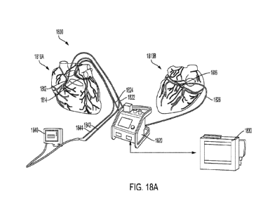
100721 FIG. 18A depicts an exemplary loco-regional perfusion system
in accordance with
embodiments of the present disclosure;
100731 FIG. 18B is a schematic of an exemplary loco-regional
perfusion device in accordance
with embodiments of the present disclosure;
-8-
CA 03208568 2023-8- 15
WO 2022/175547
PCT/EP2022/054361
100741 FIG. 19 is a radiograph captured during loco-regional
perfusion of an unarrested pig
heart showing the locations of a left main coronary artery catheter, a right
coronary artery catheter,
and a coronary sinus balloon; and
100751 FIG. 20 is a plot of pump speed, flow rate, and pressure
measured during loco-regional
perfusion.
DEFINITIONS
100761 As used herein, the singular forms "a," "an," and "the"
include plural references unless
the context clearly indicates otherwise. Thus, for example, reference to "a
drug" includes a single
drug as well as a mixture of two or more different drugs; and reference to a
"viral vector" includes
a single viral vector as well as a mixture of two or more different viral
vectors, and the like.
100771 Also as used herein, "about," when used in connection with a
measured quantity, refers
to the normal variations in that measured quantity, as expected by one of
ordinary skill in the art
in making the measurement and exercising a level of care commensurate with the
objective of
measurement and the precision of the measuring equipment. In certain
embodiments, the term
"about" includes the recited number 10%, such that "about 10" would include
from 9 to 11.
100781 Also as used herein, "polynucleotide" has its ordinary and
customary meaning in the
art and includes any polymeric nucleic acid such as DNA or RNA molecules, as
well as chemical
derivatives known to those skilled in the art. Polynucleotides include not
only those encoding a
therapeutic protein, but also include sequences that can be used to decrease
the expression of a
targeted nucleic acid sequence using techniques known in the art (e.g.,
antisense, interfering, or
small interfering nucleic acids). Polynucleotides can also be used to initiate
or increase the
expression of a targeted nucleic acid sequence or the production of a targeted
protein within cells
of the cardiovascular system. Targeted nucleic acids and proteins include, but
are not limited to,
nucleic acids and proteins normally found in the targeted tissue, derivatives
of such naturally
occurring nucleic acids or proteins, naturally occurring nucleic acids or
proteins not normally
found in the targeted tissue, or synthetic nucleic acids or proteins. One or
more polynucleotides
can be used in combination, administered simultaneously and/or sequentially,
to increase and/or
decrease one or more targeted nucleic acid sequences or proteins.
100791 Also as used herein, "perfusion," "perfused," and
"perfusing" have their ordinary and
customary meaning in the art and refer to administration for a time period
(typically a minute or
more) that is substantially longer than the art recognized term of "injection"
or "bolus injection"
(typically less than a minute). The flow rate of the perfusion will depend at
least in part on the
volume administered.
100801 Also as used herein, -exogenous" nucleic acids or genes are
those that do not occur in
nature in the vector utilized for nucleic acid transfer; e.g., not naturally
found in the viral vector,
-9-
CA 03208568 2023-8- 15
WO 2022/175547
PCT/EP2022/054361
but the term is not intended to exclude nucleic acids encoding a protein or
polypeptide that occurs
naturally in the patient or host.
100811 Also as used herein, "cardiac cell" includes any cell of the
heart that is involved in
maintaining a structure or providing a function of the heart such as a cardiac
muscle cell, a cell of
the cardiac vasculature, or a cell present in a cardiac valve. Cardiac cells
include cardio myocytes
(having both normal and abnormal electrical properties), epithelial cells,
endothelial cells,
fibroblasts, cells of the conducting tissue, cardiac pace making cells, and
neurons.
100821 Also as used herein, "isolated," "substantially isolated,"
"largely isolated," and their
variants are terms that do not require complete or absolute isolation of the
coronary venous,
cardiac, systemic venous, or systemic circulation; rather, they are intended
to mean that a majority,
preferably the major part or even substantially all of the specified
circulation is isolated. Also as
used herein, "partially isolated" refers to any nontrivial portion of the
specified circulation being
isolated.
100831 Also as used herein, "non-naturally restricted" includes any
method of restricting the
flow of fluid through a blood vessel, e.g., balloon catheter, sutures, etc.,
but does not include
naturally occurring restriction, e.g., plaque build-up (stenosis). Non-natural
restriction includes
substantial or total isolation of, for example, the coronary circulation.
100841 Also as used herein, -minimally invasive" is intended to
include any procedure that
does not require open surgical access to the heart or vessels closely
associated with the heart. Such
procedures include the use of endoscopic means to access the heart, and also
catheter-based means
relying on access via large arteries and veins.
100851 Also as used herein, "adeno-associated virus" or "AAV"
encompasses all subtypes,
serotypes, and pseudotypes, as well as naturally occurring and recombinant
forms. A variety of
AAV serotypes and strains are known in the art and are publicly available from
sources, such as
the ATCC and academic or commercial sources. Alternatively, sequences from AAV
serotypes
and strains which are published and/or available from a variety of databases
may be synthesized
using known techniques.
100861 Also as used herein, "serotype" refers to an AAV which is
identified by and
distinguished from other AAVs based on capsid protein reactivity with defined
antisera. There
are at least twelve known serotypes of human AAV, including AAV1 through
AAV12, however
additional serotypes continue to be discovered, and use of newly discovered
serotypes are
contemplated.
100871 Also as used herein, "pseudotyped" AAV refers to an AAV that
contains capsid
proteins from one serotype and a viral genome including 5' and 3' inverted
terminal repeats (ITRs)
of a different or heterologous serotype. A pseudotyped recombinant AAV (rAAV)
would be
CA 03208568 2023-8- 15
WO 2022/175547
PCT/EP2022/054361
expected to have cell surface binding properties of the capsid serotype and
genetic properties
consistent with the ITR serotype. A pseudotyped rAAV may comprise AAV capsid
proteins,
including VP1, VP2, and VP3 capsid proteins, and ITRs from any serotype AAV,
including any
primate AAV serotype from AAV 1 through AAV12, as long as the capsid protein
is of a serotype
heterologous to the serotype(s) of the ITRs. In a pseudotyped rAAV, the 5' and
3' ITRs may be
identical or heterologous. Pseudotyped rAAV are produced using standard
techniques described
in the art.
100881 Also as used herein, a "chimeric" rAAV vector encompasses an
AAV vector
comprising heterologous capsid proteins; that is, a rAAV vector may be
chimeric with respect to
its capsid proteins VP1, VP2, and VP3, such that VP1, VP2, and VP3 are not all
of the same
serotype AAV. A chimeric AAV as used herein encompasses AAV such that the
capsid proteins
VP1, VP2, and VP3 differ in serotypes, including for example but not limited
to capsid proteins
from AAVI and AAV2; are mixtures of other parvo virus capsid proteins or
comprise other virus
proteins or other proteins, such as for example, proteins that target delivery
of the AAV to desired
cells or tissues. A chimeric rAAV as used herein also encompasses an rAAV
comprising chimeric
5' and 3' ITRs.
100891 Also as used herein, a "pharmaceutically acceptable
excipient or carrier" refers to any
inert ingredient in a composition that is combined with an active agent in a
formulation. A
pharmaceutically acceptable excipient can include, but is not limited to,
carbohydrates (such as
glucose, sucrose, or dextrans), antioxidants (such as ascorbic acid or
glutathione), chelating agents,
low-molecular weight proteins, high-molecular weight polymers, gel-forming
agents, or other
stabilizers and additives. Other examples of a pharmaceutically acceptable
carrier include wetting
agents, emulsifying agents, dispersing agents, or preservatives, which are
particularly useful for
preventing the growth or action of microorganisms. Various preservatives are
well known and
include, for example, phenol and ascorbic acid. Examples of carriers,
stabilizers or adjuvants can
be found in Remington's Pharmaceutical Sciences, Mack Publishing Company,
Philadelphia, Pa.,
17th ed. (1985).
100901 Also as used herein, a "patient" refers to a subject,
particularly a human (but could also
encompass a non-human), who has presented a clinical manifestation of a
particular symptom or
symptoms suggesting the need for treatment, who is treated prophylactically
for a condition, or
who has been diagnosed with a condition to be treated.
100911 Also as used herein, a "subject" encompasses the definition
of the term "patient" and
does not exclude individuals who are otherwise healthy.
100921 Also as used herein, -treatment of' and -treating" include
the administration of a drug
with the intent to lessen the severity of or prevent a condition, e.g., heart
disease.
-11-
CA 03208568 2023-8- 15
WO 2022/175547
PCT/EP2022/054361
100931 Also as used herein, "prevention of' and "preventing"
include the avoidance of the
onset of a condition, e.g., heart disease.
100941 Also as used herein, a "condition" or "conditions" refers to
those medical conditions,
such as heart disease, that can be treated, mitigated, or prevented by
administration to a subject of
an effective amount of a drug.
100951 Also as used herein, an -effective amount" refers to the
amount of a drug that is
sufficient to produce a beneficial or desired effect at a level that is
readily detectable by a method
commonly used for detection of such an effect. In some embodiments, such an
effect results in a
change of at least 10% from the value of a basal level where the dnig is not
administered In other
embodiments, the change is at least 20%, 50%, 80%, or an even higher
percentage from the basal
level. As will be described below, the effective amount of a drug may vary
from subject to subject,
depending on age, general condition of the subject, the severity of the
condition being treated, the
particular drug administered, and the like. An appropriate "effective- amount
in any individual
case may be determined by one of ordinary skill in the art by reference to the
pertinent texts and
literature and/or by using routine experimentation.
100961 Also as used herein, an "active agent" refers to any
material that is intended to produce
a therapeutic, prophylactic, or other intended effect, whether or not approved
by a government
agency for that purpose.
100971 Recitation of ranges of values herein are merely intended to
serve as a shorthand
method of referring individually to each separate value falling within the
range, unless otherwise
indicated herein, and each separate value is incorporated into the
specification as if it were
individually recited herein. All methods described herein can be performed in
any suitable order
unless otherwise indicated herein or otherwise clearly contradicted by
context. The use of any and
all examples, or exemplary language (e.g., "such as") provided herein, is
intended merely to
illuminate certain materials and methods and does not pose a limitation on
scope. No language in
the specification should be construed as indicating any non-claimed element as
essential to the
practice of the disclosed materials and methods.
DETAILED DESCRIPTION
100981 The present invention is directed to a method of treating a
heart condition in a
minimally invasive manner. The method may comprise, isolating a patient's
coronary circulation
from the patient's systemic circulation and perfusing a fluid, such as a drug-
containing fluid, into
the patient's isolated or substantially isolated coronary circulation. The
perfusion may be
performed into a patient's unarrested beating heart. The methods may also be
used to isolate the
patient's cardiac circulation to allow administration, for example, of a
cardiotoxic drug (or any
-12-
CA 03208568 2023-8- 15
WO 2022/175547
PCT/EP2022/054361
composition potentially harmful to the patient's heart) to the patient's
systemic circulation in order
to protect the heart from adverse effects. Isolation of the patient's coronary
circulation is described
in more detail below with reference to FIGS. lA and 1B.
100991 The coronary circulation provides blood supply to the tissue
of the heart. There are a
number of coronary arteries. Normally, four main coronary arteries provide
oxygenated blood to
the heart for distribution throughout the heart tissue: the left main and
right coronary arteries, the
left anterior descending artery, and the left circumflex artery. Oxygen
depleted blood flows
through the coronary sinus.
101001 Embodiments disclosed herein contemplate isolating or
substantially isolating the
coronary circulation of a patient from the systemic circulation of the patient
by forming a closed
circuit that comprises (consists of or consists essentially of) a first drug
delivery catheter, a second
drug delivery catheter, a drug recovery catheter, a coronary artery, a
coronary venous system, and
an external membrane oxygenator. The instant disclosure further contemplates
in certain
embodiments perfusing a drug suitable for treatment of a heart condition to
the heart muscle while
substantially isolating the patient's coronary circulation from the patient's
systemic circulation
with the closed circuit described above. In some embodiments, the method
disclosed herein
delivers a drug to the heart muscle in its entirety as opposed to isolated
regions within the heart.
A drug delivered to the heart muscle with the methods disclosed herein may be
distributed
homogenously throughout the heart.
[0101] There are a number of advantages to isolating the coronary
circulation of the patient
from the systemic circulation of the patient when treating a heart condition.
These advantages
include, but are not limited to: (1) loco-regional delivery of the drug,
minimal leakage of the drug
to other organs, and reduced overall drug dose; (2) increased targeted dn.ig
dose; (3) reduced risks
and side-effects; and (4) the possibility to re-dose select patients or to
dose patient populations that
were not suitable therapy candidates for certain therapies (such as gene
therapy with viral vectors
to patients who had antibodies to the viral vectors).
Exemplary Catheter Embodiments
101021 Exemplary recovery catheters and perfusion catheters are now
described. The catheters
can be configured for the anatomy of any target organ (e.g., a heart), for
which LRP is to be
performed, as would be appreciated by those of ordinary skill in the art.
Moreover, it is to be
understood that any of the catheters described as "recovery catheters" could
also be used as
"perfusion catheters," and vice versa. The embodiments described herein are
not limited to LRP
of the heart, but may also be used to isolate the circulation of the heart
from the systemic
circulation, for example, to reduce or prevent exposure of the heart to a drug
or other agent
-13-
CA 03208568 2023-8- 15
WO 2022/175547
PCT/EP2022/054361
introduced into the systemic circulation that may have a deleterious effect on
the heart. Those of
ordinary skill in the art would appreciate other uses of the catheter
embodiments described herein,
for example, in applications for which sealing of a blood vessel is desired.
101031 Embodiments of exemplary catheters for use as recovery
catheters in an LRP system
are now described. In at least one embodiment, the recovery catheters are
designed to support a
liquid suction flow rate of about 400 mL/min or greater (e.g., about 700
mL/min or greater). For
example, in certain embodiments, an exemplary catheter can support an in vitro
suction flow rate
of about 800 mL/min at about -80 mmHg.
101041 Certain embodiments of the recovery catheters are
advantageous for use in the return
line of an LRP system used to form a closed-circuit within an unarrested
beating heart when
inserted into the coronary sinus. The catheters described herein can be
designed to satisfy the
following criteria: capability to access the coronary sinus via the right
internal jugular vein;
compatibility with an introducer sheath having an inner diameter of 24 Fr or
less; compatibility
with a 0.035-inch guidewire or smaller; capability to access, seal, and
occlude a coronary sinus
having a vessel internal diameter of 6 to 20 mm in a human subject or up to 30
mm in a porcine
animal model; the ability to avoid occlusion of prominent side veins (e.g.,
the middle cardiac vein);
and the ability to maintain stable position for at least 60 minutes during an
LRP procedure.
101051 FIGS. 1-10 depict various catheter embodiments suitable for
fluid recovery in an LRP
system. Any of the catheters depicted in FIGS. 1-10 may be configured to
support liquid flow
rates (suction or perfusion) of at least about 400 mL/min, at least about 450
mL/min, at least about
500 mL/min, at least about 550 mL/min, at least about 600 mL/min, at least
about 650 mL/min, at
least about 700 mL/min, at least about 750 mL/min, at least about 800 mL/min,
at least about
850 mL/min, at least about 900 mL/min, at least about 950 mL/min, or at least
about
1000 mL/min. Each catheter may be compatible with a stearable introducer
sheath, which
provides stability and directs the distal end of the catheter, and allows for
the catheter to create a
directed push force. Each catheter may also have a pull wire integrated into
its shaft assembly,
allowing for sections proximal to the occlusion structure to bend at angles of
up to 120 and
achieve better tracking and centering of the occlusion structure.
101061 In certain embodiments, one or more of the catheters may be
multi-lumen catheters,
such as double-lumen catheters. In certain embodiments, the multi-lumen
catheters allow for
liquid flow (e.g., a perfusate) and enable inflation of one or more balloons.
In certain
embodiments, one or more of the catheters may be multi-balloon catheters
having two or more
balloons. In certain embodiments, one or more of the balloons may be deployed
or deflated
independently.
-14-
CA 03208568 2023-8- 15
WO 2022/175547
PCT/EP2022/054361
101071 FIG. 1 illustrates an exemplary catheter 100 having a lumen
shaft 104/106 with a
proximal end 101 and a distal end 102. The lumen shaft 104/106 can be formed
from an outer
lumen shaft 104 that at least partially encompasses an inner lumen shaft 106
to expose a distal
portion of the inner lumen shaft 106 near the distal end 102. The proximal end
101 includes an
outlet structure that can be fluidly coupled to an LRP system. One or more of
the outer lumen
shaft 104 or the inner lumen shaft 106 may be formed from a durable polymer
material such as a
polyether block amide (PEBA) material (e.g., commercially available as
PEBAXR). In at least
one embodiment, an innermost diameter ("inner diameter") of the inner lumen
shaft 106 is at least
about 4 mm to provide a liquid flow path In at least one embodiment, the
catheter 100 may be
designed to include additional lumen shafts.
101081 The catheter 100 includes a tip portion 108 at the distal
end 102 and an expandable
balloon structure 110 disposed along a portion 112 of the inner lumen shaft
106. In at least one
embodiment, the tip portion 108 includes an elongated shaft extending from the
balloon structure
110 to the distal end 102. In at least one embodiment, the length of the
elongated shaft of the tip
portion is from about 2 mm to about 35 mm, about 5 mm to about 30 mm, about 10
mm to about
25 mm, about 15 mm to 25 mm, or within any subrange defined between (e.g.,
about 2 mm to
about 5 mm). In at least one embodiment, the tip portion 108 includes an
opening at the distal end
102 and one or more perforations along the elongated shaft. In at least one
embodiment, the tip
portion is formed from a compliant material that is more flexible than the
material of the inner
lumen shaft 106.
101091 In at least one embodiment, the inner lumen shaft 106
includes a concentric inner flow
path surrounding the liquid flow path. The concentric inner flow path provides
a path for gas flow
from the balloon structure 110 to a port 114, which can be used to inflate or
deflate the balloon
depending on the pressure applied at the port 114. In at least one embodiment,
an outermost
surface of the inner lumen shaft 106 at the portion 112 is removed such that
the portion 112 is
sealed by the balloon structure 110 to isolate gas flow from the concentric
inner flow path to the
balloon structure 110. In at least one embodiment, an expanded diameter of the
balloon structure
is from about 15 mm to about 30 mm, about 15 mm to about 20 mm, about 20 mm to
about 25
mm, about 24 mm to about 28 mm, or about 25 mm to about 30 mm.
101101 FIG. 2 is an image of a catheter having a similar structure
to the catheter 100 with a
balloon in its deployed state. The dimensions of the catheter include: a
crossing profile of 19 Fr
(6.3 mm); an innermost diameter of 12 Fr (4.0 mm); a usable length of 80 cm; a
balloon diameter
(when deployed) of 25 mm; and a tip portion length of 20 mm. The lumen shaft
can be formed
from a polymer material such as PEBAX 63 that is supported by a strong
stainless-steel braid.
The balloon can be formed from a compliant thermoplastic/elastomeric material
such as
-15-
CA 03208568 2023-8- 15
WO 2022/175547
PCT/EP2022/054361
ChronoPreneTM 25A. The tip portion can be formed from a polymer material such
as PEBAX
35 and can be loaded with a radio marker or a radiopaque filler composition,
such as BaSO4.
101111 FIG. 3 illustrates insertion of an exemplary catheter 300
into the coronary sinus 352
via the right atrium 350 according to at least one embodiment. The catheter
300 may be the same
as or similar to the catheter 100, having a proximal end 301, a distal end
302, an inner lumen shaft
304, an outer lumen shaft 306, a tip portion 308, and a balloon structure 310
disposed on a portion
312 of the inner lumen shaft 304. The balloon structure 310 when deployed is
compliant enough
to adapt to the anatomy of the coronary sinus 352 and occlude the blood flow
through the coronary
sinus 352 into the right atrium 350 without creating excessive force on the
tissue As illustrated
in FIG. 3, the catheter 300 is inserted past the middle cardiac vein (MCV) 354
so as to avoid
occluding the flow from the MCV 354 into the atrium 350.
101121 FIGS. 4-10 illustrate other occlusion techniques in
accordance with various
embodiments of the disclosure. The catheters depicted in FIGS. 4-10 may be
similar in certain
aspects to the catheters depicted in FIGS. 1-3, for example, in terms of
dimensions, materials, or
structures.
101131 FIG. 4 illustrates a catheter 400 according to at least one
embodiment that is only
partially inserted into the coronary sinus 352 such that it abuts the ostium
of the coronary sinus
352. The catheter 400 includes a proximal end 401, a distal end 402, an inner
lumen shaft 404, an
outer lumen shaft 406, a tip portion 408, and a balloon structure 410 disposed
on a portion 412 of
the inner lumen shaft 404. In at least one embodiment, a diameter of the
balloon structure 410 is
greater than about 15 mm, greater than about 20 mm, greater than about 25 mm,
or greater than
about 30 mm when deployed. The tip portion 408 may include, in addition to an
opening at the
distal end 402, one or more perforations to facilitate flow of blood from the
coronary sinus 352
and the MCV 354 into the catheter 400.
101141 In at least one embodiment, during deployment, the outer
lumen shaft 406 can be
moved distally to abut against the deployed balloon structure 410, resulting
in additional pressure
by the balloon structure 410 against the ostium of the coronary sinus 352 to
further stabilize the
position of the catheter 400. In at least another embodiment, a wire structure
may be utilized to
apply pressure to the balloon structure 410. The wire structure, for example,
may have a sinusoidal
shape that is deployable to an expanded flower-like structure extending
radially from the outer
lumen shaft 406 or the inner lumen shaft 404. When brought into contact with
the balloon structure
410, the wire structure may produce a more even pressure profile across the
surface of the balloon
structure 410. Prior to deployment, the wire structure may be covered by the
outer lumen shaft
406, or may be covered by an additional lumen outside of the outer lumen shaft
406.
-16-
CA 03208568 2023-8- 15
WO 2022/175547
PCT/EP2022/054361
101151 FIG. 5 illustrates the use of a first catheter 500 and a
second catheter 550 for separately
occluding and draining the coronary sinus 352 and the MCV 354, respectively,
according to at
least one embodiment. The first catheter 500 includes a proximal end 501, a
distal end 502, a
lumen shaft 504, a tip portion 508, and a balloon structure 510 disposed on a
portion 512 of the
lumen shaft 504. Similarly, the second catheter 550 includes a proximal end
551, a distal end 552,
a lumen shaft 554, a tip portion 558, and a balloon structure 560 disposed on
a portion 562 of the
lumen shaft 554. In this configuration, the first catheter 500 is inserted
into the coronary sinus 352
such that the balloon structure 510 does not occlude the MCV 354, while the
second catheter 550
is inserted directly into the MCV 354. The dimensions of the first catheter
500 and the second
catheter 550 may be selected to provide safe and effective occlusion of the
coronary sinus 352 and
the MCV 354, respectively.
101161 FIG. 6 illustrates a variation of FIG. 5, which uses two
catheters with only one having
a balloon structure according to at least one embodiment. A first catheter 600
includes a proximal
end 601, a distal end 602, a lumen shaft 604, a tip portion 608, and a balloon
structure 610 disposed
on a portion 612 of the lumen shaft 604. A second catheter 650 includes a
proximal end 651, a
distal end 652, a lumen shaft 654, and a tip portion 658, and does not include
a balloon structure.
The first catheter 600 is inserted into the coronary sinus 352 such that a
portion of the balloon 610
occludes the MCV 354 and is partially within the atrium 350 and the coronary
sinus 352. The
second catheter 650 is inserted directly into the MCV 354 and is disposed
between the vessel wall
and the balloon 610, which at least partially occludes the MCV 354.
101171 FIG. 7 illustrates the use of a single catheter 700 which
includes multiple balloons
according to at least one embodiment. The catheter 700 includes a proximal end
701, a distal end
702, a lumen shaft 704, a tip portion 708, a first balloon structure 710
disposed on a first portion
712 of the lumen shaft 704, and a second balloon structure 720 disposed on a
second portion 722
of the lumen shaft 704. In at least one embodiment, the catheter 700 is
designed for insertion into
the coronary sinus 352 such that the first balloon structure 710 occludes the
coronary sinus 352,
and the second balloon structure 720 abuts the ostium of the coronary sinus
352 to occlude the
MCV 354 (and further occlude the coronary sinus 352). An intermediate portion
724 of the lumen
shaft 704 between the first balloon structure 710 and the second balloon
structure 720 includes
one or more perforations to allow drainage of the MCV 354. In at least one
embodiment, an
expanded diameter of the second balloon structure 720 is greater than an
expanded diameter of the
first balloon structure 710. In at least one embodiment, the catheter 700 is a
multi-lumen catheter
designed to allow each balloon to be deployed and deflated independently of
each other.
101181 FIG. 8 illustrates a catheter 800 that includes a partially
covered and recapturable stent
structure 810 according to at least one embodiment. The catheter 800 includes
a proximal end 801
-17-
CA 03208568 2023-8- 15
WO 2022/175547
PCT/EP2022/054361
and a distal end 802, an inner lumen shaft 804 coupled to the stent structure
810, and an outer
lumen shaft 806. Part of the outer lumen shaft 806 is depicted as a cutaway
view to illustrate the
inner lumen shaft 804 within. The stent structure 810 is depicted in its
deployed state, but can be
contained within the outer lumen shaft 806 prior to deployment. The stent
structure 810 is further
depicted as having a proximal covered portion 810A, which may be formed from a
flexible and
durable polymer material, and a distal uncovered portion 810B. When inserted
into the coronary
sinus 352, as shown, the covered portion 810A occludes blood flow out of the
coronary sinus 352,
while the uncovered portion 810B provides structural support within the
coronary sinus 352 while
allowing blood flow from both the coronary sinus 352 and the MCV 354 directly
into the catheter
800. In at least one embodiment, the catheter 800 can be used as a perfusion
catheter connected
to a supply line.
101191 FIG. 9 illustrates a catheter 900 that includes a deployable
and retractable stent
structure 920 according to at least one embodiment. The catheter 900 further
includes a proximal
end 901, a distal end 902, a lumen shaft 906, a tip portion 908, and a balloon
structure 910 disposed
on a portion 912 of the lumen shaft 906. The catheter 900 can further include
an outer lumen shaft
(not shown) that substantially encapsulates the stent structure 920 and the
balloon structure 910
prior to deployment. Deployment of the stent structure 920 can be performed by
moving the outer
lumen shaft in a proximal direction, and retraction of the stent structure 920
can be performed by
moving the outer lumen shaft in a distal direction. The stent structure 920
may be formed from,
for example, stainless-steel, and is disposed between the balloon structure
910 and the tip portion
908. In at least one embodiment, the lumen shaft 906 comprises at least one
perforation along a
portion 922 between the balloon structure 910 and the stent structure 920 to
allow drainage of the
MCV 354 into the catheter 900. When inserted into the coronary sinus 352, the
balloon structure
910 abuts the ostium of the coronary sinus 352.
101201 FIG. 10 illustrates a catheter 1000 that includes a covered
disk-shaped stent structure
1010 according to at least one embodiment. The catheter 1000 further includes
a proximal end
1001, a distal end 1002, an outer lumen shaft 1006, an inner lumen shaft 1004,
and a tip portion
1008. The stent structure 1010 may be formed from, for example, a stainless-
steel stent having a
durable polymer covering. The outer lumen shaft 1006 can cover the stent
structure 1010 prior to
deployment. Once the catheter 1000 is properly positioned, the outer lumen
shaft 1006 can be
moved in the proximal direction to enable deployment of the stent structure
1010. In at least one
embodiment, the stent structure 1010 is coupled to the tip portion 1008, which
may be partially
contained within the inner lumen shaft 1004 and can be actuatable (using a
wire) to deploy the
stent structure 1010 when moved in a proximal direction and retract the stent
structure 1010 when
moved in a distal direction. In at least one embodiment, the stent structure
1010, when deployed,
-18-
CA 03208568 2023-8- 15
WO 2022/175547
PCT/EP2022/054361
is large enough to occlude the coronary sinus 352 and the MCV 354 when abutted
to the ostium
of the coronary sinus 352. In at least one embodiment, a diameter of the stent
structure 1010 is
from about 10 mm to about 30 mm.
101211 Embodiments of exemplary catheters for use as perfusion
catheters in an LRP system
are now described. In at least one embodiment, the perfusion catheters are
designed to support a
liquid perfusion flow rate of about 400 mL/min or greater (e.g., about 700
mL/min or greater). In
embodiments that utilize multiple perfusion catheters (e.g., insertion of a
first catheter into the
right coronary artery and insertion of a second catheter into the left
coronary artery) can support a
combined flow capacity of 700 mL/min or greater.
101221 Certain embodiments of the recovery catheters are
advantageous for use in the supply
line of an LRP system used to form a closed-circuit within an unarrested
beating heart when
inserted into the coronary arteries. The catheters described herein can be
designed to satisfy the
following criteria: capability of femoral access to the coronary coronary
arteries; an outer diameter
for coronary artery entry of 8 Fr or less; an outer diameter for occlusion of
about 6 mm to about 8
mm; compatibility with a 0.018-inch guidewire and a 0.014-inch pressure wire;
and the ability to
maintain stable position for at least 60 minutes during an LRP procedure.
101231 FIGS. 11-16 depict various catheter embodiments suitable for
fluid perfusion in an LRP
system. Any of the catheters depicted in FIGS. 11-16 may be configured to
support liquid flow
rates (suction or perfusion) of at least about 400 mL/min, at least about 450
mL/min, at least about
500 mL/min, at least about 550 mL/min, at least about 600 mL/min, at least
about 650 mL/min, at
least about 700 mL/min, at least about 750 mL/min, at least about 800 mL/min,
at least about
850 mL/min, at least about 900 mL/min, at least about 950 mL/min, or at least
about
1000 mL/min. Each catheter can be designed to have a smooth profile from a
proximal catheter
body to a low distal profile, for example, using one or more concentric lumen
shafts.
101241 In certain embodiments, one or more of the catheters may be
multi-lumen catheters,
such as double-lumen catheters. In certain embodiments, the multi-lumen
catheters allow for
liquid flow (e.g., a perfusate) and enable inflation of one or more balloons.
In certain
embodiments, one or more of the catheters may be multi-balloon catheters
having two or more
balloons. In certain embodiments, one or more of the balloons may be deployed
or deflated
independently.
101251 FIGS. 11A-11C illustrate an exemplary catheter 1100 having a
lumen shaft 1104/1106
with a proximal end 1101 and a distal end 1102 having an opening from which a
perfusate can
flow. The lumen shaft 1104/1106 can be formed from an outer lumen shaft 1104
that at least
partially encompasses an inner lumen shaft 1106 to expose a distal portion of
the inner lumen shaft
1106 near the distal end 1102. The proximal end 1101 includes an outlet
structure that can be
-19-
CA 03208568 2023-8- 15
WO 2022/175547
PCT/EP2022/054361
fluidly coupled to an LRP system. One or more of the outer lumen shaft 1104 or
the inner lumen
shaft 1106 may be formed from a durable polymer material such as a polyether
block amide
(PEBA) material (e.g., commercially available as PEBAX ). In at least one
embodiment, an
innermost diameter of the inner lumen shaft 1106 is at least about 2 mm, at
least about 2.5 mm, at
least about 3 mm, at least about 3.5 mm, at least about 4 mm, at least about
4.5 mm, or at least
about 5 mm to provide a liquid flow path.
[0126] The catheter 1100 includes an expandable balloon structure
1110 disposed along a
portion 1112 corresponding to the inner lumen shaft 1106 and a tip portion
formed by an additional
lumen In at least one embodiment, the inner lumen shaft 1106 includes a
concentric inner flow
path surrounding the liquid flow path. The concentric inner flow path provides
a path for gas flow
from the balloon structure 1110 to a port 1114, which can be used to inflate
or deflate the balloon
structure 1110 depending on the pressure applied at the port 1114. In at least
one embodiment, an
outermost surface of the inner lumen shaft 1106 at the portion 1112 is removed
such that the
portion 1112 is sealed by the balloon structure 1110 to isolate gas flow from
the concentric inner
flow path to the balloon structure 1110. In at least one embodiment, an
expanded diameter of the
balloon structure 1110 is from about 15 mm to about 30 mm, about 15 mm to
about 20 mm, about
20 mm to about 25 mm, about 24 mm to about 28 mm, about 25 mm to about 30 mm,
or within
any subrange defined therebetween (e.g., about 20 mm to about 28 mm). FIGS.
11B and 11C
illustrate the balloon structure 1110 in its deployed and deflated states.
[0127] FIG. 11D illustrates deployment of the catheter 1100 in an
aorta 1150 in accordance
with at least one embodiment. As shown, the catheter 1100 is pre-shaped for
insertion into the
aorta 1150 for ease of navigation. Moreover, the shape can leverage back-up
forces from the aortic
wall to further enhance stability during occlusion and perfusion of the
coronary artery.
101281 FIGS. 12 and 13 illustrate catheters that include plug and
wedge occlusion structures,
respectively, that advantageously adapt their shapes to a vessel or ostium,
are formed from highly
compressible and atraumatic materials for safe introduction and deployment,
are shorter in length
in comparison to a balloon structure, and do not require an additional lumen
for inflation as would
a balloon structure.
101291 FIGS. 12A-12C illustrate an exemplary catheter 1200 having a
lumen shaft 1204/1206
with a proximal end 1201 and a distal end 1202 having an opening from which a
perfusate can
flow. The lumen shaft 1204/1206 can be formed from an outer lumen shaft 1204
that at least
partially encompasses an inner lumen shaft 1206 to expose a distal portion of
the inner lumen shaft
1206 near the distal end 1202. The proximal end 1201 includes an outlet
structure that can be
fluidly coupled to an LRP system. One or more of the outer lumen shaft 1204 or
the inner lumen
shaft 1206 may be formed from a durable polymer material such as a polyether
block amide
-20-
CA 03208568 2023-8- 15
WO 2022/175547
PCT/EP2022/054361
(PEBA) material (e.g., commercially available as PEBAX ). In at least one
embodiment, an
innermost diameter of the inner lumen shaft 1206 is at least about 2 mm, at
least about 2.5 mm, at
least about 3 mm, at least about 3.5 mm, at least about 4 mm, at least about
4.5 mm, or at least
about 5 mm to provide a liquid flow path.
101301 The catheter 1200 further includes a plug 1210 near the
distal end 1202. In at least one
embodiment, the plug 1210 is formed from a flexible material, such as silicone
or a foam material.
In at least one embodiment, the plug 1210 includes an inner portion 1210A that
fits onto the inner
lumen shaft 1206 and a flexible outer portion 1210B shaped to be configurable
between a retracted
state (FIG 12A) and an extended state (FIG 12C) for which the outer portion
1210B extends
distally from the distal end 1202. The plug 1210 in FIG. 12A is illustrated as
tapering in a distal
direction. In at least one embodiment, the plug 1210 may be reversed such that
it tapers in a
proximal direction. In at least one embodiment, the outer lumen shaft 1204 may
be configured to
cover the plug 1210 prior to deployment.
101311 FIG. 12D illustrates deployment of the catheter 1200 in an
aorta 1150 in accordance
with at least one embodiment. The pressure of the arterial blood flow into the
hollow space
between the inner portion 1210A and the outer portion 1210B of the plug 1210
can help improve
the sealing of the catheter 1200 within the coronary artery. As shown, the
catheter 1200 is pre-
shaped for insertion into the aorta 1150 for ease of navigation. Moreover, the
shape can leverage
back-up forces from the aortic wall to further enhance stability during
occlusion and perfusion of
the coronary artery.
101321 FIGS. 13A-13C illustrate an exemplary catheter 1300 having a
lumen shaft 1304/1306
with a proximal end 1301 and a distal end 1302 having an opening from which a
perfusate can
flow. The lumen shaft 1304/1306 can be formed from an outer lumen shaft 1304
that at least
partially encompasses an inner lumen shaft 1306 to expose a distal portion of
the inner lumen shaft
1306 near the distal end 1302. The proximal end 1301 includes an outlet
structure that can be
fluidly coupled to an LRP system. One or more of the outer lumen shaft 1304 or
the inner lumen
shaft 1306 may be formed from a durable polymer material such as a polyether
block amide
(PEBA) material (e.g., commercially available as PEBAX ). In at least one
embodiment, an
innermost diameter of the inner lumen shaft 1306 is at least about 2 mm, at
least about 2.5 mm, at
least about 3 mm, at least about 3.5 mm, at least about 4 mm, at least about
4.5 mm, or at least
about 5 mm to provide a liquid flow path.
101331 The catheter 1300 further includes a wedge 1310 near the
distal end 1302, which may
be shaped to adapt to a vessel or ostium. In at least one embodiment, the
wedge 1310 is formed
from a flexible material, such as silicone or a foam material. In at least one
embodiment, the outer
lumen shaft 1304 may be configured to cover the wedge 1310 prior to
deployment.
-21-
CA 03208568 2023-8- 15
WO 2022/175547
PCT/EP2022/054361
101341 FIG. 13D illustrates deployment of the catheter 1300 in an
aorta 1150 in accordance
with at least one embodiment. As shown, the catheter 1300 is pre-shaped for
insertion into the
aorta 1150 for ease of navigation. Moreover, the shape can leverage back-up
forces from the aortic
wall to further enhance stability during occlusion and perfusion of the
coronary artery.
101351 FIGS. 14A-14C illustrate an exemplary catheter 1400 that
includes a partially covered
and recapturable stent structure 1406 in accordance with at least one
embodiment, similar to the
catheter 800 described with respect to FIG. 8. The catheter 1400 is
illustrated as being inserted
into a coronary artery 1452 via the aorta 1450. The catheter 1400 includes an
outer lumen shaft
1402 and an inner lumen shaft 1404 that is coupled to the stent stnicture 1406
in certain
embodiments. The stent structure 1406 is further depicted as having a proximal
covered portion,
which may be formed from a flexible and durable polymer material, and a distal
uncovered portion.
FIGS. 14B and 14C illustrate placement and deployment, respectively, of the
stent structure 1406
when inserted into the coronary artery 1452. Deployment of the stent structure
1406 is performed
by moving the outer lumen shaft 1402 in the proximal direction.
101361 FIGS. 15A and 15B illustrate an exemplary catheter 1500 that
includes a releasable
covered braided disk 1510, in accordance with at least one embodiment. The
catheter 1500
includes an outer lumen shaft 1506 and an inner lumen shaft 1504. The braided
disk 1510 is
contained within the outer lumen shaft 1506 during placement of the catheter
1500, and can be
deployed by moving the outer lumen shaft 1506 in the proximal direction. In
certain embodiments,
when deployed, the braided disk 1510 does not expand past the distal end 1502,
and is used to
stabilize the catheter 1500 against the ostium of the coronary artery 1452 to
reduce the risk of
stenosis during occlusion of the coronary artery 1452, while allowing the
distal end 1502 to extend
into the coronary artery 1452.
101371 FIGS. 16A-16D illustrate an exemplary catheter 1600 having a
lumen shaft 1606 with
a proximal end 1601 and a distal end 1602 having an opening from which a
perfusate can flow.
The proximal end 1601 includes an outlet structure that can be fluidly coupled
to an LRP system.
The lumen shaft 1604 may be formed from a durable polymer material such as a
polyether block
amide (PEBA) material (e.g., commercially available as PEBAXg). In at least
one embodiment,
an innermost diameter of the lumen shaft 1606 is at least about 2 mm, at least
about 2.5 mm, at
least about 3 mm, at least about 3.5 mm, at least about 4 mm, at least about
4.5 mm, or at least
about 5 mm to provide a liquid flow path. In at least one embodiment, a
proximal portion 1606A
of the lumen shaft 1606 may have a larger diameter than a distal portion 1606B
of the lumen shaft
1606, and can taper gradually over a length of the lumen shaft 1606.
101381 FIG. 16C illustrates deployment of the catheter 1600 in an
aorta 1150 in accordance
with at least one embodiment. As shown, the catheter 1600 is pre-shaped for
insertion into the
-22-
CA 03208568 2023-8- 15
WO 2022/175547
PCT/EP2022/054361
aorta 1150 for ease of navigation. Moreover, the shape can leverage back-up
forces from the aortic
wall to further enhance stability during occlusion and perfusion of the
coronary artery. In addition
to the catheter 1600, other catheters described herein can be designed to have
lumen shafts that are
pre-shaped depending on the anatomy in which the LRP procedure is to be
performed, which may
improve overall stability during use. Examples of pre-shaped catheter lumens
are illustrated in
FIG. 17.
Exemplary LRP System Embodiments
101391 FIG 18A depicts an exemplary loco-regional perfusion (LRP)
system MO in
accordance with embodiments of the present disclosure. The LRP system 1800 is
shown in a
closed circuit configuration with a heart 1810 (with both an anterior view
1810A and a posterior
view 1810B being shown for clarity). The LRP system 1800 includes a membrane
oxygenation
device 1820, a blood gas analysis (BGA) monitor 1830, and a pressure monitor
1840. The LRP
system 1800 may be assembled by positioning a first catheter 1822 in the right
coronary artery
1812 of the heart 1810, positioning a second catheter 1824 in the left main
coronary artery 1814
of the heart 1810, and positioning a recovery catheter 1826 in the coronary
sinus 1816 of the heart.
The first catheter 1822, the second catheter 1824, and the recovery catheter
1826, together with
the coronary arteries, the coronary venous system, the membrane oxygenation
device 1820, and
one or more optional additional components form a closed circuit. This closed
circuit may isolate
or substantially isolate the coronary circulation of the patient from the
systemic circulation of the
patient.
101401 The first catheter 1822, the second catheter 1824, and the
recovery catheter 1826 may
be introduced percutaneously and in a minimally invasive manner. In some
embodiments, the first
catheter 1822 and/or the second catheter 1824 may be introduced via antegrade
intubation. In
other embodiments, the first catheter 1822 and/or the second catheter 1824 may
be introduced via
retrograde intubation. The first catheter 1822 and the second catheter 1824
may be referred to
herein as "drug delivery catheters- and the recovery catheter 1826 may be
referred to herein as a
"drug collection catheter" or "drug recovery catheter" when the catheters are
used for drug delivery
to the heart.
101411 The first catheter 1822 and/or the second catheter 1824 may
be a standard infusion
catheter that may optionally include a standard guidewire and infusion pump.
Each catheter is
capable of delivering a perfusate to the heart 1810, which may contain, for
example, a drug to be
delivered to the heart 1810 during loco-regional perfusion. In certain
embodiments, the first
catheter 1822 and the second catheter 1824 may each correspond to an exemplary
perfusion
catheter embodiment described below in the Illustrative Examples.
-23-
CA 03208568 2023-8- 15
WO 2022/175547
PCT/EP2022/054361
101421 The first catheter 1822 and/or the second catheter 1824 may
be positioned via the aorta
of the patient, e.g., by accessing the aorta femoralis and/or the aorta
radialis. In one embodiment,
the first catheter 1822 may be positioned via the aorta of the patient by
accessing the aorta
femoralis. In another embodiment, the first catheter 1822 may be positioned
via the aorta of the
patient by accessing the aorta radialis. In one embodiment, the second
catheter 1824 may be
positioned via the aorta of the patient by accessing the aorta femoralis. In
another embodiment,
the second catheter 1824 may be positioned via the aorta of the patient by
accessing the aorta
radial i s.
101431 The recovery catheter 1826 may be a balloon catheter such
that the balloon may be
inflated within the coronary sinus 1816 to ensure that all the blood
circulated through the closed
circuit flows through the recovery catheter 1826. The balloon catheter may be
a Fogarty catheter,
or any other catheter suitable for the intended purpose discussed herein as
will be appreciated by
one of ordinary skill in the art. In certain embodiments, the recovery
catheter 1826 corresponds
to an exemplary recovery catheter embodiment described below in the
Illustrative Examples. In
certain embodiments, the recovery catheter 1826 may be positioned via the vena
cava of the
patient. In one embodiment, the recovery catheter 1826 may be positioned via
the vena jugularis
of the patient. In another embodiment, the recovery catheter 1826 may be
positioned via the vena
femoralis of the patient. In some embodiments, the first catheter 1822, the
second catheter 1824,
the recovery catheter 1826, or a combination thereof may each be a balloon
catheter to help reduce
leakage. In some embodiments, any of the catheters may be selected from one or
more of the
catheters discussed with respect to FIGS. 1-17.
101441 The LRP system 1800 may further comprise one or more
additional components, such
as, without limitations, one or more pumps, one or more suction mechanisms,
one or more
perfusates, and combinations thereof For example, the LRP system 1800 is
depicted as including
a pressure monitor 1840, which in some embodiments is operatively coupled to
or part of the
membrane oxygenation device 1820. The pressure monitor 1840 may be used to
control the
perfusion rate (i.e., flowrate) an ensure safety by continuously monitoring
the coronary artery
pressure. A first pressure sensor 1842 and a second pressure sensor 1844, for
example, may be
co-inserted with the first catheter 1822 and the second catheter 1824,
respectively, to measure the
pressures within the right coronary artery and the left main coronary artery,
respectively. The LRP
system 1800 is further depicted as including a BGA monitor that is operatively
coupled to the
membrane oxygenation device 1820 to measure, for example, the gas
concentrations in the
perfusate (e.g., when the perfusate contains blood) prior to perfusion via the
first catheter 1822
and the second catheter 1824 and/or after the perfusate is collected by the
recovery catheter 1826.
The membrane oxygenation device 1820 and one or more additional components may
be placed
-24-
CA 03208568 2023-8- 15
WO 2022/175547
PCT/EP2022/054361
between the recovery catheter 1826 and one or more of the first catheter 1822
or the second
catheter 1824.
101451 In some embodiments, while the closed circuit is
established, one or more drugs may
be perfused through the patient's systemic circulation. For example, if the
drug is cardiotoxic or
potentially harmful to the heart but systemic delivery is desirable,
establishing the closed circuit
to isolate the coronary perfusion from the systemic perfusion is advantageous
in preventing or
reducing exposure of the drug to the heart. In such embodiments, the membrane
oxygenation
device 1820 may be used to perfuse the coronary circulation at a physiological
oxygenation level
while it is isolated from the systemic circulation. For example, anthracycline
(e g , doxonthicin)
chemotherapy formulations for treating breast cancer causes irreversible
damage of cardiac
microcirculation, leading to anthracycline-cardiomyopathy.
101461 FIG. 18B is a schematic of the membrane oxygenation device
1820, which may be used
to oxygenate the perfusate, mix the perfusate with other components (e.g., a
drug), remove carbon
dioxide from the perfusate, and/or push the perfusate into one or more of the
first catheter 1822
(in the right coronary artery 1812) and/or the second catheter 1824 (in the
left main coronary artery
1814). The membrane oxygenation device 1820 may be any commercially available
extracorporeal membrane oxygenation (ECMO) device for exchanging oxygen for
carbon dioxide
contained in the blood.
101471 As illustrated in FIG. 18B, the membrane oxygenation device
1820 includes various
components including a heat exchanger 1856 (through which the perfusate passes
prior to leaving
an outlet 1852 and entering the first catheter 1822 and the second catheter
1824), a delivery pump
1858, a reservoir 1860 (for adding a component, such as blood and/or a drug,
to the perfusate
returning through the recovery catheter 1826 through an inlet 1854), sensors
1862 and 1864 at
various stages of the closed circuit (e.g., for measuring pressure and/or
blood gas content), and a
membrane oxygenator 1866. In some embodiments, de-oxygenated blood enters the
membrane
oxygenator 1866 and is mixed with an oxygen-rich gas. The oxygen-rich gas may
be supplied
from a gas blender 1868 that may mix oxygen in various ratios with carbon
dioxide and nitrogen
gas, and is regulated by a gas regulator 1870.
101481 The perfusate may comprise one or more of blood (or its
components such as plasma
or serum) and/or drug suitable for treatment of the heart condition and/or a
vehicle such as saline
or dextrose solutions. The delivery pump 1858 may deliver the perfusate into
the first catheter
1822 and/or the second catheter 1824. In some embodiments, the perfusate may
be contained in
an IV bag or a syringe and may be administered directly to the first catheter
1822 and/or the second
catheter 1824 with or without the delivery pump 1858.
-25-
CA 03208568 2023-8- 15
WO 2022/175547
PCT/EP2022/054361
101491 A suction mechanism may be used to apply negative suction
pressure on the recovery
catheter 1826 to minimize blood and/or drug leakage through the Thebesian
venous system. The
negative suction pressure may be about -150 mmHg, about -100 mmHg, about -50
mmHg, about
-20 mmHg, about -15 mmHg, about -10 mmHg, about -5 mmHg, 0 mmHg, or within a
subrange
defined by any of these points.
101501 Blood circulated through the closed circuit may be
autologous blood, matched blood
from donors, or a combination thereof In some embodiments, blood components,
such as serum
or plasma, are chosen according to one or more parameters. One of the
parameters may be the
presence or absence of selected antibodies For instance, when the dnig is one
or more viral vectors
encompassing a therapeutic nucleic acid sequence, the patient's autologous
blood may be screened
to determine whether antibodies to the one or more viral vectors are present.
Presence of
antibodies in the patient's autologous blood may reduce and/or negate
altogether the effectiveness
of the treatment and/or may result in an undesirable immune response. As such,
it may be possible
to dilute or replace the patient's autologous blood with a seronegative
matched blood from donors,
thereby reducing a patient's immune response to the drug and enhancing the
effectiveness of the
drug.
101511 While the various components illustrated in FIG. 18B show
components that are part
of or separate from the membrane oxygenation device 1820, it is to be
understood that this
schematic is merely illustrative, as one or more of the components may be
included in or separate
(external) from the membrane oxygenation device 1820.
101521 The LRP system 1800 may be set up and operated as follows:
(1) the recovery catheter
1826 is carefully placed and tightly sealed in the coronary sinus 1816 to
enable the collection of
the only cardiac venous (de-oxygenated) blood; (2) the first catheter 1822 and
the second catheter
1824 are placed in the right coronary artery (RCA) and left main coronary
artery (RCA) in a sealed
fashion; (3) the catheters are then connected to arterial and venous lines of
the membrane
oxygenation device 1820 using standard tubes; (4) operation of the LRP system
1800 is started,
and the coronary arteries are antegradely perfused with oxygenated blood,
while the returning de-
oxygenated blood is collected from the venous cardiac system via the recovery
catheter 1826 using
gentle negative pressure; (5) blood is then directed into the reservoir 1860
and is subsequently
oxygenated by the membrane oxygenator 1866 and antegradely re-infused (driven
by the delivery
pump 1858) into the heart via the first catheter 1822 and the second catheter
1824. If a drug (e.g.,
a vector) is administered, this can be added into the perfusate via the
reservoir 1860 after priming
with blood or plasma, and blood samples can be taken, or drugs can be applied
via the reservoir
1860 during the entire perfusion process.
-26-
CA 03208568 2023-8- 15
WO 2022/175547
PCT/EP2022/054361
[0153] In some embodiments, diluting or replacing a patient's
antibody-containing autologous
blood with a seronegative matched blood from donors may result in a reduced
adverse immune
response and/or improved drug efficacy. For instance, the adversity of a
patient's immune
response may be reduced by about 10%, by about 20%, by about 30%, by about
40%, by about
50%, by about 60%, by about 70%, by about 80%, by about 90%, or alleviated
altogether, upon
dilution or replacement of autologous blood with seronegative matched blood
from donors as
compared to a patient's immune response without autologous blood dilution or
replacement. The
efficacy of a drug administered may be increased by about 10%, by about 20%,
by about 30%, by
about 40%, by about 50%, by about 60%, by about 70%, by about 80%, by about
90%, by about
100%, by about 150%, by about 200%, by about 300%, by about 400%, or by about
500%, upon
dilution or replacement of autologous blood with seronegative matched blood
from donors as
compared to the drug's efficacy in a patient without autologous blood dilution
or replacement.
[0154] In some embodiments, the blood portion of the perfusate may
range from about 5 mL
to about 5000 mL, from about 50 mL to about 2500 mL, from about 100 mL to
about 1000 mL,
from about 150 mL to about 500 mL, about 50 mL, about 75 mL, about 100 mL,
about 125 mL,
about 150 mL, about 175 mL, about 200 mL, about 225 mL, about 250 mL, about
275 mL, about
300 mL, about 325 mL, about 350 mL, about 375 mL, about 400 mL, about 425 mL,
about 450
mL, about 475 mL, about 500 mL, about 550 mL, about 600 mL, about 650 mL,
about 700 mL,
about 750 mL, about 800 mL, about 850 mL, about 900 mL, about 950 mL, or about
1000 mL.
[0155] The ratio of autologous blood to blood matched from donors
in the blood that is
circulated through the closed circuit may be adjusted, as needed, to obtain a
blood mixture that
would be most receptive to the drug and would generate the least immune
response upon
introduction of the drug. In some embodiments the ratio may range from about
1:100 to about
100:1, from about 1:80 to about 80:1, from about 1:50 to about 50:1, from
about 1:30 to about
30:1, from about 1:20 to about 20:1, from about 1:10 to about 10:1, from about
1:8 to about 8:1,
from about 1:5 to about 5:1, from about 1:3 to about 3:1, or from about 1:2 to
about 2:1 of (volume
autologous blood) : (volume blood matched from donors).
[0156] The flow rate of the perfusate through the closed circuit
may be adjusted to match the
patient's blood flow rate. As appreciated by one of ordinary skill in the art,
the blood flow rate
varies from patient to patient, and for any given patient, varies throughout
the day. Accordingly,
the flow rate of the perfusate circulated through the closed circuit may be
adjusted in situ. The
flow rate may be measured over the closed circuit. In certain embodiments, the
flow rate may be
measured with a transonic probe (such as a clamp over tubing). In some
embodiments, the flow
rate of the perfusate, at any given time during the perfusion, may be within
about 20%, within
about 15%, within about 10%, within about 8%, within about 5%, within about
3%, within about
-27-
CA 03208568 2023-8- 15
WO 2022/175547
PCT/EP2022/054361
2%, within about 1%, or within about 0.5% of the patient's blood flow rate,
based on mL/min
units. It is important that the flow rate of the perfusate circulated through
the closed circuit does
not deviate significantly from the patient's own blood flow rate in order to
avoid ischemia and/or
under perfusion.
101571 Exemplary flow rates for the perfusate circulated through
the closed circuit may range,
without limitations, from about 75 mL/min to about 750 mL/min, from about 100
mL/min to about
650 mL/min, from about 125 mL/min to about 600 mL/min, from about 150 mL/min
to about
500 mL/min, from about 175 mL/min to about 400 mL/min, from about 200 mL/min
to about
300 mL/min, about 150 mL/min, about 175 mL/min, about 200 mL/min, about 225
mL/min, about
250 mL/min, about 275 mL/min, about 300 mL/min, about 325 mL/min, or about 350
mL/min.
101581 The perfusate may be circulated through the closed circuit
for a duration ranging,
without limitations, from about 5 minutes to about 5 hours, from about 15
minutes to about 4
hours, from about 30 minutes to about 3 hours, or from about 1 hour to about 2
hours. In some
embodiments, the treatment duration may occur over the span of days, e.g., I
day, 2 days, 3 days,
4 days, 5 days, 6 days, 7 days, and so on.
101591 With the system disclosed herein, in some embodiments, a
higher dose of drug than
could otherwise be administered safely through systemic delivery may be
administered directly
and only to the heart. In some embodiments, a lower overall dose of drug may
be required to attain
the same therapeutic effect (as was attained with a larger dose that was
subjected to systemic
circulation or that was subjected to only partial isolation of the coronary
circulation), since there
may be substantially no leakage of the perfusate outside of the heart and/or
to the Thebesian venous
system.
101601 In some embodiments, less than about 50% v/v, less than
about 40% v/v, less than
about 30% v/v, less than about 20% v/v, less than about 15% v/v, less than
about 10% v/v, less
than about 5% v/v, less than about 4% v/v, less than about 3% v/v, less than
about 2% v/v, less
than about 1% v/v, less than about 0.5% v/v, or substantially no (0% v/v)
perfusate (e.g., blood
and/or drug) circulated through the closed circuit leaks outside of the closed
circuit during the
perfusion process.
101611 The reduced perfusate leakage outside of the closed circuit
(as compared to other
methods disclosed in the art) may be due to the tight seal formed within the
closed circuit and each
individual component utilized in the closed circuit.
101621 In certain embodiments, some perfusate leakage from the
closed circuit may remain.
For instance, up to about 0.5% v/v, about 1% v/v, about 2% v/v, about 3% v/v,
about 4% v/v, about
5% v/v, about 10% v/v, about 15% v/v, about 20% v/v, about 30% v/v, about 40%
v/v, or about
50% v/v of the perfusate circulated through the closed circuit may leak
outside of the closed circuit.
-28-
CA 03208568 2023-8- 15
WO 2022/175547
PCT/EP2022/054361
Any drug amount lost through leakage of the perfusate may be replaced in the
perfusate in order
to keep the drug exposure to the heart constant over the calculated exposure
time. The calculated
exposure time may, in certain embodiments, range from about 5 minutes to about
5 hours, from
about 15 minutes to about 4 hours, from about 30 minutes to about 3 hours,
from about 1 hour to
about 2 hours, or any sub-range in between.
THERAPEUTIC COMPOSITIONS
101631 Drugs suitable for treatment of the heart condition (i.e.,
drugs included in the perfusate)
may include therapeutic polynucleotide sequences In some embodiments, the
therapeutic
polynucleotide sequences may encode to a protein for the treatment of a heart
condition. The
protein for treatment of the heart condition may be of human origin or may be
derived from
different species (e.g., without limitations, mouse, cat, pig or monkey) In
some embodiments, the
protein encoded by the therapeutic polynucleotide sequence may correspond to a
gene expressed
in a human heart.
101641 Exemplary proteins may include, without limitations, one or
more of SERCA2,
MYBPC3, MYH7, PKP2, MYL3, MYL2, ACTC1, TPM1, TNNT2, TNNI3, TTN, FHL1, ALPK3,
dystrophin, FKRP, variants thereof, or combinations thereof. The protein or
proteins used may
also be functional variants of the proteins mentioned herein and may exhibit a
significant amino
acid sequence identity compared to the original protein. For instance, the
amino acid identity may
amount to at least about 30%, at least about 35%, at least about 40%, at least
about 45%, at least
about 50%, at least about 55%, at least about 60%, at least about 65%, at
least about 70%, at least
about 75%, at least about 80%, at least about 85%, at least about 90%, at
least about 95%, at least
about 96%, at least about 97%, at least about 98%, or at least about 99%. In
this context, the term
"functional variant" means that the variant of the protein is capable of,
partially or completely,
fulfilling the function of the naturally occurring corresponding protein.
Functional variants of a
protein may include, for example, proteins that differ from their naturally
occurring counterparts
by one or more amino acid substitutions, deletions, or additions.
101651 The amino acid substitutions can be conservative or non-
conservative. It is preferred
that the substitutions are conservative substitutions, i.e., a substitution of
an amino acid residue by
an amino acid of similar polarity, which acts as a functional equivalent.
Preferably, the amino acid
residue used as a substitute is selected from the same group of amino acids as
the amino acid
residue to be substituted. For example, a hydrophobic residue can be
substituted with another
hydrophobic residue, or a polar residue can be substituted with another polar
residue having the
same charge. Functionally homologous amino acids, which may be used for a
conservative
substitution comprise, for example, non-polar amino acids such as glycine,
valine, alanine,
-29-
CA 03208568 2023-8- 15
WO 2022/175547
PCT/EP2022/054361
isoleucine, leucine, methionine, proline, phenylalanine, and tryptophan.
Examples of uncharged
polar amino acids comprise serine, threonine, glutamine, asparagine, tyrosine
and cysteine.
Examples of charged polar (basic) amino acids comprise histidine, arginine,
and lysine. Examples
of charged polar (acidic) amino acids comprise aspartic acid and glutamic
acid.
101661 Also considered as variants are proteins that differ from
their naturally occurring
counterparts by one or more (e.g., 2, 3, 4, 5, 10, or 15) additional amino
acids. These additional
amino acids may be present within the amino acid sequence of the original
protein (i.e., as an
insertion), or they may be added to one or both termini of the protein.
Basically, insertions can
take place at any position if the addition of amino acids does not impair the
capability of the
polypeptide to fulfill the function of the naturally occurring protein in the
treated subject.
Moreover, variants of proteins also comprise proteins in which, compared to
the original
polypepti de, one or more amino acids are lacking. Such deletions may affect
any amino acid
position provided that it does not impair the ability to fulfill the normal
function of the protein.
101671 Finally, variants of the cardiac sarcomeric proteins also
refer to proteins that differ from
the naturally occurring protein by structural modifications, such as modified
amino acids.
Modified amino acids are amino acids which have been modified either by
natural processes, such
as processing or post-translational modifications, or by chemical modification
processes known in
the art. Typical amino acid modifications comprise phosphorylation,
glycosylation, acetylation,
0-linked N-acetylglucosamination, glutathionylation, acylation, branching, ADP
ribosylation,
crosslinking, disulfide bridge formation, formylation, hydroxylation,
carboxylation, methylation,
demethylation, amidation, cyclization, and/or covalent or non-covalent bonding
to
phosphotidylinositol, flavine derivatives, lipoteichonic acids, fatty acids,
or lipids.
101681 The therapeutic polynucleotide sequence encoding the target
protein may be
administered to the subject to be treated in the form of a gene therapy
vector, i.e., a nucleic acid
construct which comprises the coding sequence, including the translation and
termination codons,
next to other sequences required for providing expression of the exogenous
nucleic acid such as
promoters, kozak sequences, polyA signals, and the like.
101691 For example, the gene therapy vector may be part of a
mammalian expression system.
Useful mammalian expression systems and expression constructs are commercially
available.
Also, several mammalian expression systems are distributed by different
manufacturers and can
be employed in the present invention, such as plasmid- or viral vector based
systems, e.g., LENTI-
SmartTm (InvivoGen), GenScriptTM Expression vectors, pAdVAntageTm (Promega),
ViraPowerTM
Lentiviral, Adenoviral Expression Systems (Invitrogen), and adeno-associated
viral expression
systems (Cell Biolabs).
-30-
CA 03208568 2023-8- 15
WO 2022/175547
PCT/EP2022/054361
101701 Gene therapy vectors for expressing an exogenous therapeutic
polynucleotide sequence
of the invention can be, for example, a viral or non-viral expression vector,
which is suitable for
introducing the exogenous therapeutic polynucleotide sequence into a cell for
subsequent
expression of the protein encoded by said nucleic acid. The expression vector
can be an episomal
vector, i.e., one that is capable of self-replicating autonomously within the
host cell, or an
integrating vector, i.e., one which stably incorporates into the genome of the
cell. The expression
in the host cell can be constitutive or regulated (e.g., inducible).
101711 In a certain embodiment, the gene therapy vector is a viral
expression vector. Viral
vectors for use in the present invention may comprise a viral genome in which
a portion of the
native sequence has been deleted in order to introduce a heterogeneous
polynucleotide without
destroying the infectivity of the virus. Due to the specific interaction
between virus components
and host cell receptors, viral vectors are highly suitable for efficient
transfer of genes into target
cells. Suitable viral vectors for facilitating gene transfer into a mammalian
cell can be derived
from different types of viruses, for example, from an AAV, an adenovirus, a
retrovirus, a herpes
simplex virus, a bovine papilloma virus, a lentivirus, a vaccinia virus, a
polyoma virus, a sendai
virus, orthomyxovirus, paramyxovirus, papovavirus, picornavirus, pox virus,
alphavirus, or any
other viral shuttle suitable for gene therapy, variations thereof, and
combinations thereof.
101721 -Adenovirus expression vector" or -adenovirus" is meant to
include those constructs
containing adenovirus sequences sufficient (a) to support packaging of the
therapeutic
polynucleotide sequence construct, and/or (b) to ultimately express a tissue
and/or cell-specific
construct that has been cloned therein. In one embodiment of the invention,
the expression vector
comprises a genetically engineered form of adenovirus. Knowledge of the
genetic organization of
adenovirus, a 36 kilobase (kb), linear, double-stranded DNA virus, allows
substitution of large
pieces of adenoviral DNA with foreign sequences up to 7 kb.
101731 Adenovirus growth and manipulation is known to those of
skill in the art, and exhibits
broad host range in vitro and in vivo. This group of viruses can be obtained
in high titers, e.g.,
109to 1011 plaque-forming units per mL, and they are highly infective. The
life cycle of adenovirus
does not require integration into the host cell genome. The foreign genes
delivered by adenovirus
vectors are episomal and, therefore, have low genotoxicity to host cells. No
side effects have been
reported in studies of vaccination with wild-type adenovirus, demonstrating
their safety and/or
therapeutic potential as in vivo gene transfer vectors.
101741 Retroviruses (also referred to as "retroviral vector") may
be chosen as gene delivery
vectors due to their ability to integrate their genes into the host genome,
transferring a large amount
of foreign genetic material, infecting a broad spectrum of species and cell
types and for being
packaged in special cell-lines.
-31-
CA 03208568 2023-8- 15
WO 2022/175547
PCT/EP2022/054361
101751 The retroviral genome contains three genes, gag, poi, and
env, that encode for capsid
proteins, polymerase enzyme, and envelope components, respectively. A sequence
found
upstream from the gag gene contains a signal for packaging of the genome into
virions. Two long
terminal repeat (LTR) sequences are present at the 5' and 3' ends of the viral
genome. These
contain strong promoter and enhancer sequences and are also required for
integration in the host
cell genome.
101761 In order to construct a retroviral vector, a nucleic acid
encoding a gene of interest is
inserted into the viral genome in the place of certain viral sequences to
produce a virus that is
replication-defective In order to produce virions, a packaging cell line is
constructed containing
the gag, pol, and/or env genes but without the LTR and/or packaging
components. When a
recombinant plasmid containing a cDNA, together with the retroviral LTR and
packaging
sequences is introduced into this cell line (by calcium phosphate
precipitation for example), the
packaging sequence allows the RNA transcript of the recombinant plasmid to be
packaged into
viral particles, which are then secreted into the culture media. The media
containing the
recombinant retroviruses is then collected, optionally concentrated, and used
for gene transfer.
Retroviral vectors are able to infect a broad variety of cell types. However,
integration and stable
expression require the division of host cells.
101771 The retrovirus can be derived from any of the subfamilies.
For example, vectors from
Murine Sarcoma Virus, Bovine Leukemia, Virus Rous Sarcoma Virus, Murine
Leukemia Virus,
Mink-Cell Focus-Inducing Virus, Reticuloendotheliosis Virus, or Avian Leukosis
Virus can be
used. The skilled person will be able to combine portions derived from
different retroviruses, such
as LTRs, tRNA binding sites, and packaging signals to provide a recombinant
retrovirus. These
retroviruses are then normally used for producing transduction competent
retroviral vector
particles. For this purpose, the vectors are introduced into suitable
packaging cell lines.
Retroviruses can also be constructed for site-specific integration into the
DNA of the host cell by
incorporating a chimeric integrase enzyme into the retroviral particle.
101781 Because herpes simplex virus (HSV) is neurotropic, it has
generated considerable
interest in treating nervous system disorders. Moreover, the ability of HSV to
establish latent
infections in non-dividing neuronal cells without integrating into the host
cell chromosome or
otherwise altering the host cell's metabolism, along with the existence of a
promoter that is active
during latency makes HSV an attractive vector. And though much attention has
focused on the
neurotropic applications of HSV, this vector also can be exploited for other
tissues given its wide
host range.
101791 Another factor that makes HSV an attractive vector is the
size and organization of the
genome. Because HSV is large, incorporation of multiple genes or expression
cassettes is less
-32-
CA 03208568 2023-8- 15
WO 2022/175547
PCT/EP2022/054361
problematic than in other smaller viral systems. In addition, the availability
of different viral
control sequences with varying performance (temporal, strength, etc.) makes it
possible to control
expression to a greater extent than in other systems. It also is an advantage
that the virus has
relatively few spliced messages, further easing genetic manipulations.
101801 HSV also is relatively easy to manipulate and can be grown
to high titers. Thus,
delivery is less of a problem, both in terms of volumes needed to attain
sufficient multiplicity of
infection (MOT) and in a lessened need for repeat dosing. Avirulent variants
of IISV have been
developed and are readily available for use in gene therapy contexts.
[0181] Lentiviruses are complex retrovinises, which, in addition to
the common retroviral
genes gag, pol, and env, contain other genes with regulatory or structural
function. The higher
complexity enables the virus to modulate its life cycle, as in the course of
latent infection. Some
examples of lentivirus include the Human Immunodeficiency Viruses (HIV-1, HIV-
2) and the
Simian Immunodeficiency Virus (SW). Lentiviral vectors have been generated by
multiply
attenuating the HIV virulence genes, for example, the genes env, vif, vpr,
vpu, and nef are deleted
making the vector biologically safe.
101821 Lentiviral vectors are plasmid-based or virus-based, and are
configured to carry the
essential sequences for incorporating foreign nucleic acid, for selection and
for transfer of the
nucleic acid into a host cell. The gag, pol, and env genes of the vectors of
interest also are known
in the art. Thus, the relevant genes are cloned into the selected vector and
then used to transform
the target cell of interest.
101831 Vaccinia virus vectors have been used extensively because of
the ease of their
construction, relatively high levels of expression obtained, wide host range
and large capacity for
carrying DNA. Vaccinia contains a linear, double-stranded DNA genome of about
186 kb that
exhibits a marked "A-T" preference. Inverted terminal repeats of about 10.5 kb
flank the genome.
The majority of essential genes appear to map within the central region, which
is most highly
conserved among poxviruses. Estimated open reading frames in vaccinia virus
number from 150
to 200. Although both strands are coding, extensive overlap of reading frames
is not common.
101841 At least 25 kb can be inserted into the vaccinia virus
genome. Prototypical vaccinia
vectors contain transgenes inserted into the viral thymidine kinase gene via
homologous
recombination. Vectors are selected on the basis of a tk-phenotype. Inclusion
of the untranslated
leader sequence of encephalomyocarditis virus results in a level of expression
that is higher than
that of conventional vectors, with the transgenes accumulating at 10% or more
of the infected
cell's protein in 24 hours.
101851 The empty capsids of papovaviruses, such as the mouse
polyoma virus, have received
attention as possible vectors for gene transfer. The use of empty polyoma was
first described when
-33-
CA 03208568 2023-8- 15
WO 2022/175547
PCT/EP2022/054361
polyoma DNA and purified empty capsids were incubated in a cell-free system.
The DNA of the
new particle was protected from the action of pancreatic DNase. The
reconstituted particles were
used for transferring a transforming polyoma DNA fragment to rat FIJI cells.
The empty capsids
and reconstituted particles consist of all three of the polyoma capsid
antigens VP, VP2, and VP3.
101861 AAVs are parvoviruses belonging to the genus Dependovirus.
They are small,
nonenveloped, single-stranded DNA viruses which require a helper virus in
order to replicate. Co-
infection with a helper virus (e.g., adenovirus, herpes virus, or vaccinia
virus) is necessary in order
to form functionally complete AAV virions In vitro, in the absence of co-
infection with a helper
virus, AAV establishes a latent state in which the viral genome exists in an
episomal form, but
infectious virions are not produced. Subsequent infection by a helper virus
"rescues" the genome,
allowing it to be replicated and packaged into viral capsids, thereby
reconstituting the infectious
virion. Recent data indicate that in vivo both wild type AAV and recombinant
AAV
predominantly exist as large episomal concatemers. In one embodiment, the gene
therapy vector
used herein is an AAV vector. The AAV vector may be purified, replication
incompetent,
pseudotyped rAAV particles.
101871 AAV are not associated with any known human diseases, are
generally not considered
pathogenic, and do not appear to alter the physiological properties of the
host cell upon integration.
AAV can infect a wide range of host cells, including non-dividing cells, and
can infect cells from
different species. In contrast to some vectors, which are quickly cleared or
inactivated by both
cellular and humoral responses, AAV vectors have been shown to induce
persistent transgene
expression in various tissues in vivo. The persistence of recombinant AAV-
mediated transgenes
in non-diving cells in vivo may be attributed to the lack of native AAV viral
genes and the vector's
ITR-linked ability to form episomal concatemers.
101881 AAV is an attractive vector system for use in the cell
transduction of the present
invention as it has a high frequency of persistence as an episomal concatemer
and it can infect
non-dividing cells, including cardiomyocytes, thus making it useful for
delivery of genes into
mammalian cells, for example, in tissue culture and in vivo.
101891 Typically, rAAV is made by cotransfecting a plasmid
containing the gene of interest
flanked by the two AAV terminal repeats and/or an expression plasmid
containing the wild-type
AAV coding sequences without the terminal repeats, for example pIM45. The
cells are also
infected and/or transfected with adenovirus and/or plasmids carrying the
adenovirus genes
required for AAV helper function. Stocks of rAAV made in such a fashion are
contaminated with
adenovirus, which must be physically separated from the rAAV particles (for
example, by cesium
chloride density centrifugation or column chromatography). Alternatively,
adenovirus vectors
containing the AAV coding regions and/or cell lines containing the AAV coding
regions and/or
-34-
CA 03208568 2023-8- 15
WO 2022/175547
PCT/EP2022/054361
some or all of the adenovirus helper genes could be used. Cell lines carrying
the rAAV DNA as
an integrated provirus can also be used.
101901 Multiple serotypes of AAV exist in nature, with at least
twelve serotypes (AAV1-
AAV12). Despite the high degree of homology, the different serotypes have
tropisms for different
tissues. Upon transfection, AAV elicits only a minor immune reaction (if any)
in the host.
Therefore, AAV is highly suited for gene therapy approaches.
101911 The present disclosure may be directed in some embodiments
to a drug comprising an
AAV vector that is one or more of AAV1, AAV2, AAV3, AAV4, AAV5, AAV6, AAV7,
AAV8,
AAV9, AAV10, AAV11, AAV12, ANC AAV, chimeric AAV derived thereof, variations
thereof,
and combinations thereof, which will be even better suitable for high
efficiency transduction in
the tissue of interest. In certain embodiments, the gene therapy vector is an
AAV serotype 1 vector.
In certain embodiments, the gene therapy vector is an AAV serotype 2 vector.
In certain
embodiments, the gene therapy vector is an AAV serotype 3 vector. In certain
embodiments, the
gene therapy vector is an AAV serotype 4 vector. In certain embodiments, the
gene therapy vector
is an AAV serotype 5 vector. In certain embodiments, the gene therapy vector
is an AAV serotype
6 vector. In certain embodiments, the gene therapy vector is an AAV serotype 7
vector. In certain
embodiments, the gene therapy vector is an AAV serotype 8 vector. In certain
embodiments, the
gene therapy vector is an AAV serotype 9 vector. In certain embodiments, the
gene therapy vector
is an AAV serotype 10 vector. In certain embodiments, the gene therapy vector
is an AAV
serotype 11 vector. In certain embodiments, the gene therapy vector is an AAV
serotype 12 vector.
101921 A suitable dose of AAV for humans may be in the range of
about lx108 vector genomes
per kilogram of body weight (vg/kg) to about 3x10¹⁴ vg/kg, about 1x10⁸ vg/kg,
about 1x10⁹ vg/kg,
about 1x10¹⁰ vg/kg, about 1x10¹¹ vg/kg, about 1x10¹² vg/kg, about 1x10¹³
vg/kg, or about 1x10¹⁴
vg/kg. The total amount of viral particles or DRP is, is about, is at least,
is at least about, is not
more than, or is not more than about, 5 x 101 vg/kg, 4>1O'5 vg/kg, 3>1O''
vg/kg, 2x10'5 vg/kg,
1 x 10¹⁵ vg/kg, 9x 10¹⁴ vg/kg, 8x 10¹⁴ vg/kg, 7x IU =-=14
vg/kg, 6x 01 14 vg/kg, 5x IV rs14
vg/kg, 4x 10¹⁴
vg/kg, 3x.0"
vg/kg, 2x10'4 vg/kg, ix iO',1 vg/kg, 9x10'3 vg/kg, 8 x 10¹³ vg/kg, 7 x 10¹³
vg/kg,
6x 10¹³ vg/kg, 5x 10¹³ vg/kg, 4x 10¹³ vg/kg, 3 x10¹³ vg/kg, 2x 10¹³ vg/kg, 1
x1013 vg/kg, 9x 10¹²
vg/kg, 8 x 10¹² vg/kg, 7x10¹²
vg/kg, 6x10¹² vg/kg, 5x10¹²
vg/kg, 4x10¹²
vg/kg, 3 x 10¹² vg/kg,
2x10'2 vg/kg, vg/kg, 1 x 10¹² vg/kg, 9 x 10¹¹ vg/kg, 8 x 10¹¹ vg/kg, 7x10"
vg/kg, 6 x 10" vg/kg,
x 1011 vg/kg, 4 x10¹¹ vg/kg, 3 x10¹¹ vg/kg, 2x 10¹¹
vg/kg, 1 x 10¹¹ vg/kg,9 x 1010 vg/kg,
8 x 101 vg/kg, 7 x 101 vg/kg, 6 x 101 vg/kg, 5 x 1 0 to vg/kg, 410b0 vg/kg,
3 x101 vg/kg,
2x-10 u vg/kg, 1 x 101 vg/kg, 9 x 10⁹ vg/kg, 8 x 10⁹ vg/kg, 7 x 10⁹ vg/kg, 6
x 109 vg/kg, 5 x 10⁹ vg/kg,
4x 10⁹ vg/kg, 3 x10⁹ vg/kg, 2x 10⁹ vg/kg, 1 x 10⁹ vg/kg, 9x10⁸ vg/kg, 8 x 10⁸
vg/kg, 7 x 10⁸ vg/kg,
6x 10⁸ vg/kg, 5>< 108 vg/kg, 4x 10⁸ vg/kg, 3 x10⁸ vg/kg, 2x 10⁸ vg/kg, or 1><
108 vg/kg, or falls within
-35-
CA 03208568 2023-8- 15
WO 2022/175547
PCT/EP2022/054361
a range defined by any two of these values. The above listed dosages being in
vg/kg heart tissue
units.
[0193] With the systems and methods disclosed herein, in some
embodiments, a higher dose
of drug than could otherwise be administered safely through systemic delivery
may be
administered directly and only to the heart, since there is substantially no
leakage of the perfusate
outside of the heart and/or to the Thebesian venous system. Without being
construed as limiting,
it is believed that AAV toxicity may be due to systemic effects such as
hepatotoxicity, platelet
activation and loss, and complement activation and loss. All of these
toxicities and others may be
reduced, minimized, or completely avoided via the loco-regional perfusate
application described
in the methods and systems disclosed herein. As such, doses up to about 5x10"
vg/kg heart tissue
may be well tolerated. In certain embodiments, AAV doses to the heart,
expressed as vg/kg heart
tissue, may exceed the highest systemically administered doses by a factor of
about 2 to about 200,
about 5 to about 150, about 10 to about 100, or any sub-range therein.
101941 Apart from viral vectors, non-viral expression constructs
may also be used for
introducing a gene encoding a target protein or a functioning variant or
fragment thereof into a cell
of a patient. Non-viral expression vectors which permit the in vivo expression
of protein in the
target cell include, for example, a plasmid, a modified RNA, an mRNA, a cDNA,
antisense
oligomers, DNA-lipid complexes, nanoparticles, exosomes, any other non-viral
shuttle suitable
for gene therapy, variations thereof, and a combination thereof.
[0195] Apart from viral vectors and non-viral expression vectors,
nuclease systems may also
be used, in conjunction with a vector and/or an electroporation system, to
enter into a cell of a
patient and introduce therein a gene encoding a target protein or a
functioning variant or fragment
thereof. Exemplary nuclease systems may include, without limitations, a
clustered regularly
interspaced short palindromic repeats (CRISPR), a DNA cutting enzyme (e.g.,
Cas9),
meganucleases, TALENs, zinc finger nucleases, any other nuclease system
suitable for gene
therapy, variations thereof, and a combination thereof. For instance, in one
embodiment, one viral
vector (e.g., AAV) may be used for a nuclease (e.g., CRISPR) and another viral
vector (e.g., AAV)
may be used for a DNA cutting enzyme (e.g., Cas9) to introduce both (the
nuclease and the DNA
cutting enzyme) into a target cell.
[0196] Other vector delivery systems which can be employed to
deliver a therapeutic
polynucleotide sequence encoding a therapeutic gene into cells are receptor-
mediated delivery
vehicles. These take advantage of the selective uptake of macromolecules by
receptor-mediated
endocytosis in almost all eukaryotic cells. Because of the cell type-specific
distribution of various
receptors, the delivery can be highly specific. Receptor-mediated gene
targeting vehicles may
include two components: a cell receptor-specific ligand and a DNA-binding
agent.
-36-
CA 03208568 2023-8- 15
WO 2022/175547
PCT/EP2022/054361
101971 Suitable methods for the transfer of non-viral vectors into
target cells are, for example,
the lipofection method, the calcium-phosphate co-precipitation method, the
DEAE-dextran
method and direct DNA introduction methods using micro-glass tubes,
ultrasound,
electroporation, and the like. Prior to the introduction of the vector, the
cardiac muscle cells may
be treated with a permeabilization agent, such as phosphatidylcholine,
streptolysins, sodium
caprate, decanoylcarnitine, tartaric acid, lysolecithin, Triton X-100, and the
like. Exosomes may
also be used to transfer naked DNA or AAV-encapsidated DNA
101981 A gene therapy vector of the invention may comprise a
promoter that is functionally
linked to the nucleic acid sequence encoding to the target protein The
promoter sequence should
be compact and ensure a strong expression. Preferably, the promoter provides
for an expression
of the target protein in the myocardium of the patient that has been treated
with the gene therapy
vector. In some embodiment, the gene therapy vector comprises a cardiac-
specific promoter which
is operably linked to the nucleic acid sequence encoding the target protein.
As used herein, a
cardiac-specific promoter" refers to a promoter whose activity in cardiac
cells is at least 2-fold
higher than in any other non-cardiac cell type. Preferably, a cardiac-specific
promoter suitable for
being used in the vector of the invention has an activity in cardiac cells
which is at least 5-fold, at
least 10-fold, at least 15-fold, at least 20-fold, at least 25-fold, or at
least 50-fold higher compared
to its activity in a non-cardiac cell type.
101991 The cardiac-specific promoter may be a selected human
promoter, or a promoter
comprising a functionally equivalent sequence having at least about 80%, at
least about 90%, at
least about 95%, at least about 96%, at least about 97%, at least about 98%,
or at least about 99%
sequence identity to the selected human promoter. An exemplary non-limiting
promoter that may
be used is a cardiac troponin T promoter (TNNT2) Other non-limiting examples
of promoters
include alpha myosin heavy chain promoter, the myosin light chain 2v promoter,
the alpha myosin
heavy chain promoter, the alpha-cardiac actin promoter, the alpha-tropomyosin
promoter, the
cardiac troponin C promoter, the cardiac troponin I promoter, the cardiac
myosin-binding protein
C promoter, and the sarco/endoplasmic reticulum Ca2+-ATPase (SERCA) promoter
(e.g., isoform
2 of this promoter (SERCA2)).
102001 The vectors useful in the present invention may have varying
transduction efficiencies.
As a result, the viral or non-viral vector transduces more than, equal to, or
at least about 10%,
about 20%, about 30%, about 40%, about 50%, about 55%, about 60%, about 65%,
about 70%,
about 75%, about 80%, about 85%, about 90%, about 95%, about 99%, or 100% of
the cells of the
targeted vascular territory. More than one vector (viral or non-viral, or
combinations thereof) can
be used simultaneously or in sequence. This can be used to transfer more than
one polynucleotide,
-37-
CA 03208568 2023-8- 15
WO 2022/175547
PCT/EP2022/054361
and/or target more than one type of cell. Where multiple vectors or multiple
agents are used, more
than one transduction/transfection efficiency can result.
102011 Pharmaceutical compositions that contain gene therapy
vectors may be prepared either
as liquid solutions or suspensions. The pharmaceutical composition of the
invention can include
commonly used pharmaceutically acceptable excipients, such as diluents and
carriers. In
particular, the composition comprises a pharmaceutically acceptable carrier,
e.g., water, saline,
Ringer's solution, or dextrose solution. In addition to the carrier, the
pharmaceutical composition
may also contain emulsifying agents, pH buffering agents, stabilizers, dyes,
and the like.
102021 In certain embodiments, a pharmaceutical composition will
comprise a therapeutically
effective gene dose, which is a dose that is capable of preventing or treating
cardiomyopathy in a
subject, without being toxic to the subject. Prevention or treatment of
cardiomyopathy may be
assessed as a change in a phenotypic characteristic associated with
cardiomyopathy with such
change being effective to prevent or treat cardiomyopathy. Thus, a
therapeutically effective gene
dose is typically one that, when administered in a physiologically tolerable
composition, is
sufficient to improve or prevent the pathogenic heart phenotype in the treated
subject.
102031 Heart conditions that may be treated by the methods
disclosed herein may include,
without limitations, one or more of a genetically determined heart disease
(e.g., genetically
determined cardiomyopathy), arrhythmic heart disease, heart failure, ischemia,
arrhythmia,
myocardial infarction, congestive heart failure, transplant rejection,
abnormal heart contractility,
non-ischemic cardiomyopathy, mitral valve regurgitation, aortic stenosis or
regurgitation,
abnormal Ca' metabolism, congenital heart disease, primary or secondary
cardiac tumors, and
combinations thereof
ILLUSTRATIVE EXAMPLES
102041 The following examples are set forth to assist in
understanding the disclosure and
should not, of course, be construed as specifically limiting the embodiments
described and claimed
herein. Such variations of the embodiments, including the substitution of all
equivalents now
known or later developed, which would be within the purview of those skilled
in the art, and
changes in formulation or minor changes in experimental design, are to be
considered to fall within
the scope of the embodiments incorporated herein.
Example 1: Feasibility study of loco-regional perfusion in three pigs
102051 Feasibility of the LRP system established by successfully
performing the procedure for
60 minutes in three pigs (sus scrofa domestica). In two pigs, a thoracotomy
was performed for
-38-
CA 03208568 2023-8- 15
WO 2022/175547
PCT/EP2022/054361
surveillance purposes, but all catheters were introduced percutaneously. In
the third pig, no
thoracotomy was performed and the entire LRP procedure was performed
percutaneously.
[0206] LRP was performed on the three animals utilizing the LRP
system 1800 illustrated in
and described with respect to FIGS. 18A and 18B. In all three animals, the LRP
procedure could
be maintained while the heart was spontaneously beating for 60 min without any
technical
problems. During LRP, all animals (n = 3) were hemodynamically stable without
any need for
inotropes. Post-LRP cardiac function was unremarkable and comparable to
baseline for all
animals. Overall occlusion of the coronary arteries was acceptable: in Animal
1, the left coronary
artery (LCA) could not be frilly occluded (leakage was considered mild), in
Animal 2, the right
coronary artery (RCA) could not be fully occluded (leakage was considered to
be trace); and in
Animal 3, both coronary arteries could be occluded.
[0207] The tight occlusion of the coronary sinus (CS) was
technically more challenging due
to the variable anatomy of the pig where, in contrast to humans, the vena
azygos inserts directly
into the coronary sinus and needs to be occluded to simulate the human
situation. Full occlusion
was achieved in Animal 3 (using a Reliant balloon), partial occlusion achieved
in Animal 1 and
Animal 2 (ProPledge catheters). Flow rates during 60 minutes of the LRP
procedure ranging from
166 mL/min up to 244 mL/min could be achieved. Accessory devices that were
used in this
example are listed in Table 1, including their intended uses and the use in
the LRP system in
accordance with the embodiments of the disclosure.
[0208] FIG. 19 is a radiograph of a representative LRP in situ
setup, where coronary artery
catheters are indicated by arrows and the coronary sinus balloon (recovery
catheter) is indicated
by a triangle.
[0209] The mean values of LRP parameters (pump speed, flow, and
pressure) for the three
animals are summarized in Fig. 20 (with the bars representing standard
deviation).
-39-
CA 03208568 2023-8- 15
WO 2022/175547
PCT/EP2022/054361
Table 1: Devices used for LRP procedure
Item CE Intended Use LRP Use Brand
Study
Animal 1
Neurology Coronary artery
FlowGate2 Stryker
Animal 2
thrombectomy perfusion
Animal 3
Animal 1
Retrograde Coronary sinus
ProPledge Edwards
Animal 2
cardioplegia blood return
Animal 3
EndoVent Pulmonary artery Coronary sinus
Animal 1
blood venting blood return and Edwards
Animal 2
(suction) azygos occlusion
Animal 3
Child
D100 Oxygenator
Animal 1
cardiopulmonary Blood oxygenation
Dideco-Livanova
set
Animal 2
bypass (CPB) set and tubing
Animal 3
Centrifugal blood Centrifugal blood
Revolution 5 Sorin-Livanova
pump pump
Animal 1
Centrifugal blood Centrifugal blood
Rotaflow RF-32 Getinge
Animal 2
pump pump
Animal 3
Centrifugal blood Centrifugal blood
BPX-80 Medtronic
Safety study
pump pump
Animal 1
Bio-Medicus 550 ECMO pump
ECMO pump console Medtronic
Animal 2
Bio-Console console
Animal 3
Animal 1
Coronary pressure Coronary pressure Saint Jude
Medical /
PressureWire X
Animal 2
wire wire Abbott
Animal 3
Fractional flow
Animal 1
Coronary pressure Saint Jude Medical /
reserve (FFR) FFR console
Animal 2
console Abbott
console
Animal 3
Coronary sinus
Reliant Aortic endo clamp Medtronic
Animal 3
occlusion
Coronay sinus
Fogarty catheter Thrombectomy Fogarty
occlusion
-40-
CA 03208568 2023-8- 15
WO 2022/175547
PCT/EP2022/054361
Table 2: Flow and pressure characteristics over 60 minutes of the LRP
procedure
Animal 1
Time 0 min 5 min 10 min 15 min 30 min 45
min 60 min
Flow (mL/min) 140 200 190 200 180
220 190
Pressure (mm/Hg) -90 -90 -90 -90 -90 -
60 -60
RPM 2650 3020 3020 3020 890
3060 2930
Coronary pressure 48/28 44/24 44/25 48/28 44/25
47/28 48/20
Mean coronary
pressure 37 32 32 36 32 37
34
Systemic pressure 77/51 70/47 69/47 76/52 69/48
77/53 70/48
Mean systemic
pressure 62 57 56 62 57 60
58
Animal 2
Time 0 min 5 min 10 min 15 min 30 min 45
min 60 min
Flow (mL/min) 180 - 140 150 170
180 180
Pressure (mmHg) -90 -90 -90 -90 -
90 -90
RPM 3020 - 2940 2980 3050
3050 3010
Coronary pressure 65/33 - 61/46 63/47 73/52
70/48 66/41
Mean coronary
pressure 41 - 51 52 60 58
51
Systemic pressure 70/50 - 60/43 60/43 68/55
67/46 59/42
Mean systemic
pressure 61 50 49 62 56
51
Heart rate 71 - 74 75 78 80
81
Animal 3
Time 0 min 5 min 10 min 15 min 30 min 45
min 60 min
Flow (mL/min) 260 240 250 250 240
230 240
Pressure -60 -80 -70 -70 -70 -
70 -70
RPM 3330 3350 3350 3350 3350
3350 3610
Coronary pressure 54/44 58/44 53/4 52/41 50/40
50/40 86/56
Mean coronary
pressure 49 49 47 46 45 42
70
Systemic pressure 71/47 66/43 67/45 67/45 67/39
54/40 57/7
Mean systemic
pressure 57 54 54 54 47 47
30
Heart rate 73 74 73 72 77 97
88
Example 2: Safety study of the loco-regional perfusion system in two pigs
102101 Safety of the LRP system was established by performing the
LRP procedure using a
percutaneous approach for 60 minutes in two pigs (sus scrofa domestica) and
following the animals
for 24 hours after the procedure while the animals were kept under
anaesthesia. Following the 24-
hour period, the animals were sacrificed and a macroscopic and microscopic
examination of their
-41-
CA 03208568 2023-8- 15
WO 2022/175547
PCT/EP2022/054361
hearts was carried out. In addition, blood biomarkers were obtained to
evaluate tissue damage of
the heart.
102111 In both experiments the LRP could be successfully performed
and without any serious
adverse effect. A technical issue occurred in the with Animal 5, where the
pump head tubing
connection failed after 10 minutes. LRP was immediately stopped, and all
catheters were
disengaged and deflated. The pump head was immediately replaced, and the LRP
system was
reconnected, deaired, and restarted. During this maneuver, the animal was
hemodynamically
stable, and no serious adverse effect was observed. The LRP procedure was then
maintained for
60 minutes, thus demonstrating the safety efficacy even with minor equipment
failures
102121 Throughout the procedures, including initiation, re-
initiation of LRP, and up to 24
hours after, the animals were hemodynamically stable without any need for
inotropes.
102131 A mean LRP flow of 173 mL/min could be achieved while the
left main coronary artery
and the right coronary artery were fully occluded and the coronary sinus was
partially occluded
(leakage was moderate) for both animals. The post-operative and 24-hour
cardiac functions were
unremarkable and comparable to baseline. Cardiac biomarkers (myoglobin and
troponin) only
slightly increased during and shortly after the LRP procedure, but then
immediately dropped
towards baseline values during 24-hour follow up. Given the continuous
hemodynamic stability
of the animals throughout the entire procedure, the absence of serious adverse
effects, and only
minor and temporary increases of cardiac biomarkers, as well as only temporary
electrocardiogram
changes with immediate normalization during the 24 hours, the LRP procedure
was demonstrated
to be safe. Table 3 compiles the flow and pressure characteristics over 60
minutes of the LRP
procedure in the Animal 4 and Animal 5 used in the safety study.
Table 3: Parameters for safety study
Animal 4
Time 0 15min 30min 45min 60min
Flow (mL/min) 180-190 200 180 180 190
Suction -70 -70 -70 -70
RPM 3400 3400 3320 3350
Coronary pressure 93/52 90/63 88/37 82/43
Mean 74 77 62 65
Systemic pressure 104/65 100/64 94/61 94/56
Mean 80 78 77 65
Animal 5
Time 5min 15min 30min 45min 60min
Flow (mL/min) 150 150 160 150
170
Suction (mmHg) -80 -80 -80 -80 -
80
RPM
2390 2400 2440 2400 2410
-42-
CA 03208568 2023-8- 15
WO 2022/175547 PCT/EP2022/054361
Coronary pressure 76/44 80/40 81/40 90/45
72/24
Mean 61 59 58 62 43
Systemic 86/52 86/53 80/46 100/62 84/47
Mean 66 66 60 77 63
102141 The hearts of Animal 4 and Animal 5 were macroscopically examined
following
sacrifice.
102151 The heart weight for Animal 4 was 312 grams. No gross pathology was
observed, and
in particular there were no signs of myocardial ischemia or myocardial
infarction On the posterior
side of the heart of Animal 4, a localized hematoma was observed in the area
of the right coronary
artery, most likely due to wire injury during the procedure.
102161 The heart weight for Animal 5 was 293 grams. No gross pathology was
observed, and
in particular there were no signs of myocardial ischemia or myocardial
infarction. On the posterior
side of the heart of Animal 5, localized hematomas were observed in the areas
of the distal right
coronary artery and distal left circumflex artery, most likely due to wire
injury during procedure.
102171 In order to ascertain the biochemical integrity of the heart tissue,
seric cardiac
biomarkers were obtained, and summarized in Table 4. Creatine kinase (CK)
levels remained
stable during the LRP procedure but showed a continuous rise post-LRP most
likely due to the
animal lying in the supine position. Myoglobin levels remained stable during
the LRP procedure
and showed only minimal increase thereafter, still within reference levels for
Animal 4 and only
very slightly above reference levels for Animal 5. Troponin T increased
minimally during the
LRP procedure with a peak at 60 minutes followed by a drop to baseline values
during follow up.
Table 4: Seric biomarkers (creatine kinase (CK), myoglobin (Myo), and troponin
T (Trop)) values
were obtained at baseline, at 30 min and 60 min during LRP, and at various
intervals post-LRP
Animal Ref. Baseline LRP LRP 4h 8h 12h 16h 20h 24h
4 value 30min 60min
CK <190U/ 871 865 883 937 1293 1285 1270 2068 2640
Myo. 25- <21 <21 <21 <21 <21 <21 24 35 58
72ug/L
Trop. <14ng/L 53 85 106 56 45 34 28 32 53
Animal Ref. Baseline LRP LRP 4h 8h 12h 16h 20h 24h
value 30min 60min
CK <190U/L 988 899 931 1946 2943 4064 4929 5496 7655
Myo. 25- <21 <21 <21 <21 26 35 37
43 83
72 g/L
Trop. <14ng/L 37 44 66 138 120 93 72 61 58
-43-
CA 03208568 2023-8- 15
WO 2022/175547
PCT/EP2022/054361
Example 3: Vector biodistribution study
102181 Biodistribution studies were performed in three pigs that
were subjected to the LRP
procedure as described above using similar protocols and equipment. A first
pig ("Pig 1") and a
second pig ("Pig 2") were subjected to LRP for 60 minutes, and their coronary
circulations were
perfused with 1014 vector genome (vg) total dose per kilogram heart weight of
AAV9 containing
a construct encoding for green fluorescent protein (GFP) with a
cytomegalovirus (CMV) promoter
(AAV9 CMV-GFP). A third pig ("Pig 3") received the same does of AAV9 CMV-GFP
via intra-
coronary (IC) infusion. Of the various pigs considered for the study, none
were identified that
were AAV9-antibody negative. Accordingly, pigs exhibiting the lowest antibody
titers were
selected for the study (Pig 1: anti-AAV9 1:20; Pig 2: anti-AAV9 > 1:100; Pig
3: anti-AAV9
>1:100).
102191 Vector shedding from the closed circuit remained low for the
duration of the LRP
procedure (see Tables 5 and 6 below). For Pigs 1 and 2, respectively, 98.7%
and 81.8% of vector
was detected in the plasma samples at 5 minutes into the LRP procedure, and
60.1% and 52.9%
was detected at 30 minutes, demonstrating that the vector was largely
maintained within the closed
circuit early in the LRP procedure for at least 45 minutes. The lower limit of
quantification was
5.33 x 10³ vg/mL.
Table 5: Detected vector in plasma samples taken from the closed circuit and
from the systemic
circulation (periphery) at various time points of the LRP procedure (units in
vector genome per
milliliter of plasma, vg/mL)
Plasma Sample Pig 1 (LRP) Pig 2 (LRP) Pig 3
(IC)
LRP system, + 5 min 2.11 x 10" 7.05x 10¹⁰ N/A
LRP system, + 15 min 2.14 x 10¹¹ 3.81 x 101 N/A
LRP system, + 30 min 4.26 x 101 1.52 x 101 N/A
LRP system, + 45 min 3.33 x 101 1.18 x 101 N/A
LRP system, + 60 min 2.73x 10¹⁰ 8.61x 10⁹ N/A
Periphery, before injection <5.33 x 10³ <5.33 x 16 <5.33 x
103
Periphery, + 5 min 2.70x 10⁹ 1.57x 10¹⁰ 5.26 x
109
Periphery, + 15 min 1.68 x 10¹⁰ 1.81 x 10¹⁰ 6.22 x
109
Periphery, + 30 min 2.83x 10¹⁰ 1.36x 10¹⁰ 5.84x
109
Periphery, + 45 min 2.69 x 10¹⁰ 1.05 x 10¹⁰ 9.07 x
109
Periphery, + 60 min 2.27x 101 1.18x 10¹⁰ 9.62x
109
Table 6: Ratio between vector levels detected in LRP closed circuit versus
periphery
Plasma Sample Time Pig 1 (LRP) Pig 2 (LRP)
LRP system, + 5 min 98.7% / 1.3% 81.8% / 18.2%
LRP system, + 15 min 93.0% / 7.0% 67.7% / 32.3%
LRP system, + 30 min 60.1% / 39.9% 52.9% / 47.1%
LRP system, + 45 min 55.4% / 44.6% 52.9% / 47.1%
LRP system, + 60 min 54.6% / 45.4% 42.2% / 57.8%
-44-
CA 03208568 2023-8- 15
WO 2022/175547
PCT/EP2022/054361
[0220] Vector biodistribution was evaluated by determining vg/g
using quantitative
polymerase chain reaction (qPCR) analysis, and by using immunofluorescence to
visually detect
expression in 26 pre-determined heart sections for each pig. Using qPCR
analysis (as summarized
in Table 7), while overall detection levels were low, vector was detected in
22 out of 26 heart
sections in Pig 1, indicating broad vector distribution. In Pig 2 (which had a
higher anti-AAV Ab
titer), vector was detected in 8 out of 26 sections. In contrast, in Pig 3,
after intra-coronary
injection, vector was only detected in 3 out of 26 heart sections. The lower
limit of quantification
is < 0,004 vector genome per diploid genome (vg/dg).
Table 7: Vector detected in tissue samples via qPCR analysis
Tissue Sample Pig 1 (LRP) Pig 2 (LRP) Pig 3
(IC)
Heart, left ventricle, basal anterior 0.09 <0.004
<0.004
Heart, left ventricle, basal anteroseptal 0.01 <0.004 <0.004
Heart, left ventricle, basal inferoseptal 0.02 <0.004 0.11
Heart, left ventricle, basal inferior 0.01 <0.004
0.21
Heart, left ventricle, basal inferolateral 0.02 <0.004 <0.004
Heart, left ventricle, basal anterolateral 0.01 <0.004 <0.004
Heart, left ventricle, mid anterior 0.01 0.01
<0.004
Heart, left ventricle, mid anteroseptal 0.02 <0.004 <0.004
Heart, left ventricle, mid inferoseptal <0.004 <0.004
<0.004
Heart, left ventricle, mid inferior 0.03 <0.004
<0.004
Heart, left ventricle, mid inferolateral 0.01 0.02 <0.004
Heart, left ventricle, mid anterolateral 0.03 0.01 <0.004
Heart, left ventricle, mid apical anterior 0.01 <0.004 <0.004
Heart, left ventricle, mid apical septum 0.01 0.03 <0.004
Heart, left ventricle, mid apical inferior 0.01 <0.004 <0.004
Heart, left ventricle, mid apical lateral 0.02 <0.004 0.14
Heart, left ventricle, apical anterior <0.004 0.02
<0.004
Heart, left ventricle, apical septum 0.03 0.01
<0.004
Heart, left ventricle, apical inferior 0.01 <0.004
<0.004
Heart, left ventricle, apical lateral <0.004 0.01
<0.004
Heart, left ventricle, apical cap 0.03 <0.004
<0.004
Heart, right ventricle, basal 0.02 <0.004
<0.004
Heart, right ventricle, mid <0.004 <0.004
<0.004
Heart, right ventricle, apical 0.02 0.01
<0.004
Heart, left auricle 0.07 <0.004
<0.004
Heart, right auricle 0.01 <0.004
<0.004
Liver, left lobe 0.64 0.05
<0.004
Liver, right lobe 0.91 0.09
<0.004
Spleen 0.18 0.06
2.10
[0221] Immunofluorescence (IF) analysis was performed using GFP
detection as the method
for quantification. Tissue samples were prepared using frozen tissue
sectioning to obtain 10
millimeter-thick samples. A primary monoclonal antibody was used for GFP
detection, and a
-45-
CA 03208568 2023-8- 15
WO 2022/175547
PCT/EP2022/054361
secondary Alexa-555-coupled antibody was used for mouse antibody detection.
Wheat germ
agglutinin (WGA) was used for detecting connective tissue. Counter staining
was performed with
4',6-diamidino-2-phenylindole (DAPI).
102221 Automated immunofluorescence quantification was performed on
the tissue sections
(whole section scanning using a ZEISS Axio Scan microscope). GFP-positive
cardiomyocytes
were detected in 8 out of 25 sections in Pig 1 (only 25 sections were
investigated), 4 out of 26
sections in Pig 2, and zero sections in Pig 3. The overall number of
transduced cardiomyocytes
was below 1%.
102231 In the foregoing description, numerous specific details are
set forth, such as specific
materials, dimensions, processes parameters, etc., to provide a thorough
understanding of the
present invention. The particular features, structures, materials, or
characteristics may be
combined in any suitable manner in one or more embodiments. The words
"example" or
"exemplary- are used herein to mean serving as an example, instance, or
illustration. Any aspect
or design described herein as "example" or "exemplary" is not necessarily to
be construed as
preferred or advantageous over other aspects or designs. Rather, use of the
words "example" or
"exemplary" is simply intended to present concepts in a concrete fashion. As
used in this
application, the term "or" is intended to mean an inclusive "or" rather than
an exclusive "or". That
is, unless specified otherwise, or clear from context, -X includes A or B" is
intended to mean any
of the natural inclusive permutations. That is, if X includes A; X includes B;
or X includes both
A and B, then "X includes A or B" is satisfied under any of the foregoing
instances. Reference
throughout this specification to -an embodiment", -certain embodiments", or -
one embodiment"
means that a particular feature, structure, or characteristic described in
connection with the
embodiment is included in at least one embodiment. Thus, the appearances of
the phrase "an
embodiment", "certain embodiments", or "one embodiment" in various places
throughout this
specification are not necessarily all referring to the same embodiment.
102241 The present invention has been described with reference to
specific exemplary
embodiments thereof. The specification and drawings are, accordingly, to be
regarded in an
illustrative rather than a restrictive sense. Various modifications of the
invention in addition to
those shown and described herein will become apparent to those skilled in the
art and are intended
to fall within the scope of the appended claims.
-46-
CA 03208568 2023-8- 15