Note: Descriptions are shown in the official language in which they were submitted.
WO 2022/183057 PCT/US2022/018007
1
COMPOSITIONS AND METHODS FOR THERAPEUTIC DELIVERY
CROSS-REFERENCE
[0001] This application claims the benefit of US Provisional Application
Serial Number
63/154,591 filed on February 26, 2021, and US Provisional Application Serial
Number 63/193,949,
filed on May 27, 2021, each of which is hereby incorporated by reference
herein in its entirety.
SEQUENCE LISTING
[0002] The instant application contains a Sequence Listing which has been
submitted electronically
in ASCII format and is hereby incorporated by reference in its entirety. Said
ASCII copy, created on
February 4, 2022, is named 53712-710_601_SL.txt and is 959,536 bytes in size.
SUMMARY
[0003] Described herein, in some aspects, is an enucleated cell, the
enucleated cell comprising: a
single-domain antibody or fragment thereof that binds an immune checkpoint
molecule; and one or
more intracellular organelles configured to translate an exogenous messenger
ribonucleic acid
(mRNA) molecule encoding the single-domain antibody or fragment thereof. In
some
embodiments, the single-domain antibody or fragment thereof is contained in
the enucleated cell. In
some embodiments, the single-domain antibody or fragment thereof is released
by the enucleated
cell. In some embodiments, the enucleated cell further comprises a cell
membrane, wherein the
single-domain antibody or fragment thereof is expressed on an exoplasmic side
of the cell
membrane. In some embodiments, the enucleated cell further comprises a cell
membrane, wherein
the cell membrane comprises a transmembrane moiety that is coupled to the
single-domain
antibody or fragment thereof. In some embodiments, the transmembrane moiety
comprises a
transmembrane polypeptide. In some embodiments, the single-domain antibody or
fragment thereof
is coupled with a N-terminus or a C-terminus of the transmembrane polypeptide.
In some
embodiments, the single-domain antibody or fragment is coupled to an anchor
molecule coupled to
a cell surface of the enucleated cell, wherein the anchor molecule comprises
glycosylphosphatidylinositol, famesyl, palmitate, myristate, or any
combination thereof. In some
embodiments, the enucleated further comprises a fusion protein configured to
transfer the single-
domain antibody or fragment thereof from the enucleated cell to another cell.
In some
embodiments, the single-domain antibody or fragment thereof is coupled to a
cytotoxic drug. In
some embodiments, the immune checkpoint molecule comprises programmed cell
death protein 1
(PD-1 or PDCD-1), programmed death-ligand 1 (PD-L1), cytotoxic T-lymphocyte-
associated
CA 03208619 2023- 8- 16
WO 2022/183057 PCT/US2022/018007
2
protein 4 (CTLA-4, also known as cluster of differentiation 152 or CD152), V-
domain Ig
suppressor of T cell activation (VISTA), Programmed cell death 1 ligand 2
(PDCD1LG2, also
known as cluster of differentiation 273 or CD273), B7 homolog 3 (B7-H3, also
known as cluster of
differentiation 276 or CD276), adenosine A2A receptor (A2AR), cluster of
differentiation 27
(CD27), lymphocyte-activation gene 3 (LAG3), T-cell immunoglobulin and mucin-
domain
containing-3 (TIIVI-3, also known as Hepatitis A virus cellular receptor 2 Or
HAVCR2), T cell
immunoreceptor with Ig and ITIM domains (TIGIT), cluster of differentiation 73
(CD73),
CD94/NK group 2 member A (NKG2A, also known as cluster of differentiation 159
or CD159),
Poliovirus receptor related immunoglobulin domain containing (PVRIG),
Poliovirus receptor-
related 2 (PVRL2), carcinoembryonic antigen-related cell adhesion molecule 1
(CEACA1VI1),
Carcinoembryonic antigen-related cell adhesion molecule 5 (CEACAM5),
carcinoembryonic
antigen-related cell adhesion molecule 6 (CEACAM6), focal adhesion kinasc
(FAK), C-C
chemokine receptor type 2 (CCR-2), chemokine (C-C motif) ligand 2 (CCL-2),
leukemia inhibitory
factor (LIT), cluster of differentiation 47 (CD47), signal-regulatory protein
alpha (SIRPa),
macrophage colony-stimulating factor (M-CSF), colony stimulating factor 1
receptor (CSF-1R),
interleukin 3 (IL-3), Interleukin-1 receptor accessory protein (IL-1RAP),
interleukin 8 (IL-8),
semaphorin-4D (SEMA4D), angiopoietin-2, CLEVER-1, tyrosine-protein kinase
receptor UFO
(Axl), phosphatidylserine or a fragment thereof. In some embodiments, the
immune checkpoint
molecule comprises PD-Ll. In some embodiments, the immune checkpoint molecule
comprises
CTLA-4. In some embodiments, the immune checkpoint molecule comprises an amino
acid
sequence that is greater than or equal to about 80% identical to any one of
SEQ ID NOs: 155-164,
203, 204, 315-322, 511, 531-535, 551-554, 571, 594, 611-619, or 711. In some
embodiments, the
single-domain antibody or fragment thereof is encoded by a deoxyribonucleic
acid (DNA)
sequence that is greater than or equal to about 50%, 55%, 60%, 65%, 70%, 75%,
80%, 85%, 90%,
95%, or 99% identical to SEQ ID NO: 801. In some embodiments, the single-
domain antibody or
fragment thereof comprises an amino acid sequence that is greater than or
equal to about 50%,
55%, 60%, 65%, 70%, 75%, 80%, 85%, 90%, 95%, or 99% identical to SEQ ID NO:
851. In some
embodiments, the single-domain antibody or fragment thereof is encoded from a
DNA sequence
that is greater than or equal to about 50%, 55%, 60%, 65%, 70%, 75%, 80%, 85%,
90%, 95%, or
99% identical to SEQ ID NO: 901. In some embodiments, the single-domain
antibody or fragment
thereof comprises an amino acid sequence that is greater than or equal to
about 50%, 55%, 60%,
65%, 70%, 75%, 80%, 85%, 90%, 95%, or 99% identical to SEQ ID NO: 951. In some
embodiments, the enucleated cell further comprises a targeting moiety. In some
embodiments, the
targeting moiety comprises a homing receptor specific to a ligand expressed by
a cell in lung tissue.
CA 03208619 2023- 8- 16
WO 2022/183057 PCT/US2022/018007
3
In some embodiments, the targeting moiety comprises an antibody or antigen-
binding fragment
thereof, wherein said antibody or antigen binding fragment thereof is
different from the single-
domain antibody or fragment thereof In some embodiments, the targeting moiety
comprises a
chemokine receptor. In some embodiments, the cell is a cancer cell. In some
embodiments, the
cancer cell is a cell of non-small cell lung cancer (NSCLC), small cell lung
cancer (SCLC),
adenocarcinoma, squamous carcinoma, large cell (undifferentiated) carcinoma,
large cell
neuroendocrine carcinoma, adenosquamous carcinoma, or sarcomatoid carcinoma.
In some
embodiments, the cancer cell is a cell of benign lung tumor. In some
embodiments, the cancer cell
is a cell of hamartoma. In some embodiments, the enucleated cell further
comprises a therapeutic
agent. In some embodiments, the therapeutic agent comprises interleukin 12 (IL-
12). In some
embodiments, the enucleated cell further comprises an immune evasion moiety
comprising cluster
of differentiation (CD47), PD-L1, major histocompatibility complex, class I, E
(HLA-E), major
histocompatibility complex, class I, G (HLA-G), a fragment thereof, or a
combination thereof. In
some embodiments, the enucleated cell has a diameter comprising between about
1 micrometers
(um) to about 100 um In some embodiments, the diameter comprises between about
5 um to 25
pm. In some embodiments, the diameter comprises between about 8 um to 12 um.
In some
embodiments, the enucleated cell exhibits a diameter that reduced relative to
an otherwise identical
nucleated cell, wherein the diameter is reduced by great than or equal to
about 50%. In some
embodiments, the enucleated cell further comprises an exogenous tumor necrosis
factor (TNF)
superfamily member polypeptide or a catalytically active fragment thereof. In
some embodiments,
the exogenous TNF superfamily member polypeptide or the catalytically active
fragment thereof is
soluble in aqueous conditions. In some embodiments, the exogenous TNF
superfamily member
polypeptide comprises tumor necrosis factor superfamily member 14 (LIGHT), or
a catalytically
active fragment thereof. In some embodiments, the enucleated cell was obtained
from a parent cell,
wherein the parent cell comprises a stem cell. In some embodiments, the stem
cell comprises an
induced pluripotent stem cell (iPSC), an adult stem cell, a mesenchymal
stromal cell, an embryonic
stem cell, or a fibroblast. In some embodiments, the enucleated cell is
purified. In some
embodiments, the enucleated cell is lyophilized.
[0004] Described herein, in some aspects, is a plurality of cells, comprising:
a plurality of
enucleated cells comprising: a single-domain antibody or fragment thereof that
binds an immune
checkpoint molecule, and one or more intracellular organelles configured to
translate an exogenous
messenger ribonucleic acid (mRNA) molecule encoding the single-domain antibody
or fragment
thereof
CA 03208619 2023- 8- 16
WO 2022/183057 PCT/US2022/018007
4
[0005] Described herein, in some aspects, is a pharmaceutical formulation,
comprising: an
enucleated cell comprising: a single-domain antibody or fragment thereof that
binds an immune
checkpoint molecule, and one or more intracellular organelles configured to
translate an exogenous
messenger ribonucleic acid (mRNA) molecule encoding the single-domain antibody
or fragment
thereof; and a pharmaceutically acceptable excipient, carrier or diluent.
[0006] Described herein, in some aspects, is a method of delivering an
enucleated cell comprising.
a single-domain antibody or fragment thereof that binds an immune checkpoint
molecule; and one
or more intracellular organelles configured to translate an exogenous
messenger ribonucleic acid
(mRNA) molecule encoding the single-domain antibody or fragment thereof to a
subject, the
method comprising: delivering to the subject an enucleated cell described
herein. In some
embodiments, the enucleated cell is an autologous cell. In some embodiments,
the enucleated cell is
an allogcnic cell. In some embodiments, the administering is performed by
systemic administration.
In some embodiments, following the administering, the enucleated cell is
viable in the subject for
fewer than or equal to 5 days.
[0007] Described herein, in some aspects, is a method of treating cancer in a
subject, the method
comprising: administering to the subject a therapeutically effective amount of
an enucleated cell the
enucleated cell comprising: a single-domain antibody or fragment thereof that
binds an immune
checkpoint molecule, and one or more intracellular organelles configured to
translate an exogenous
messenger ribonucleic acid (mRNA) molecule encoding the single-domain antibody
or fragment
thereof; or pharmaceutical formulation described herein, thereby treating the
cancer in the subject.
In some embodiments, the enucleated cell is an autologous cell. In some
embodiments, the
enucleated cell is an allogenic cell. In some embodiments, the administering
is performed by
systemic administration. In some embodiments, following the administering, the
enucleated cell is
viable in the subject for fewer than or equal to 5 days.
[0008] Described herein, in some aspects, an enucleated cell, the enucleated
cell comprising: a
single-domain antibody or fragment thereof that binds a connective tissue
growth factor (CTGF);
and one or more intracellular organdies configured to (i) translate an
exogenous mRNA molecule
encoding the single-domain antibody or fragment thereof, and (ii) release the
single-domain
antibody or fragment thereof from the enucleated cell. In some embodiments,
the single-domain
antibody or fragment thereof comprises a polypeptide sequence that is greater
than or equal to
about 50%, 55%, 60%, 65%, 70%, 75%, 80%, 85%, 90%, 95%, or 99% identical to
SEQ ID NO:
1701. In some embodiments, the single-domain antibody or fragment thereof
binds to an amino
acid sequence of CTGF, wherein the amino acid sequence of CTGF comprises SEQ
ID NO: 1601
or SEQ ID NO: 1602. In some embodiments, the enucleated cell, further
comprises a targeting
CA 03208619 2023- 8- 16
WO 2022/183057 PCT/US2022/018007
moiety specific to a ligand expressed by a cell in lung tissue. In some
embodiments, the targeting
moiety comprises a homing receptor specific to the ligand expressed by the
cell in lung tissue. In
some embodiments, the cell is an alveolar epithelial cell (AEC). In some
embodiments, the cell is a
bronchial cell. In some embodiments, the enucleated cell further comprises an
immune evasion
moiety comprising CD47, PD-L1, HLA-E, HLA-G, a fragment thereof, or any
combination thereof.
In some embodiments, the targeting moiety comprises a chemokine receptor. In
some
embodiments, the targeting moiety comprises an adhesion molecule. In some
embodiments, the
target moiety comprises an antibody or antigen-binding fragment thereof,
wherein said antibody or
antigen binding fragment thereof is different from the single-domain antibody
or fragment thereof
In some embodiments, the enucleated cell has a diameter comprising between
about 1 micrometers
(ttm) to about 100 p.m. In some embodiments, the diameter is between about 5
p.m to 25 pm. In
some embodiments, the diameter comprises between about 8 pm to 12 1.1m. In
some embodiments,
the enucleated cell exhibits a diameter that reduced relative to an otherwise
identical nucleated cell,
wherein the diameter is reduced by great than or equal to about 50%. In some
embodiments, the
enucleated cell further comprises an exogenous tumor necrosis factor (TNF)
superfamily member
polypeptide or a catalytically active fragment thereof. In some embodiments,
the exogenous TNF
superfamily member polypeptide or the catalytically active fragment thereof is
soluble in aqueous
conditions. In some embodiments, the exogenous TNF superfamily member
polypeptide comprises
LIGHT or catalytically active fragment thereof. In some embodiments, the
enucleated cell was
obtained from a parent cell, wherein the parent cell comprises a stem cell. In
some embodiments,
the stem cell comprises an induced pluripotent stem cell (iPSC), an adult stem
cell, a mesenchymal
stromal cell, an embryonic stem cell, or a fibroblast. In some embodiments,
the enucleated cell is
purified. In some embodiments, the enucleated cell is lyophilized.
[0009] Described herein, in some aspects, is a plurality of cells, comprising:
a plurality of the
enucleated cells comprising: a single-domain antibody or fragment thereof that
binds a connective
tissue growth factor (CTGF); and one or more intracellular organelles
configured to (i) translate an
exogenous mRNA molecule encoding the single-domain antibody or fragment
thereof, and (ii)
release the single-domain antibody or fragment thereof from the enucleated
cell
[0010] Described herein, in some aspects, is a pharmaceutical formulation,
comprising: an
enucleated cell comprising: a single-domain antibody or fragment thereof that
binds a connective
tissue growth factor (CTGF); and one or more intracellular organelles
configured to (i) translate an
exogenous mRNA molecule encoding the single-domain antibody or fragment
thereof, and (ii)
release the single-domain antibody or fragment thereof from the enucleated
cell.; and a
pharmaceutically acceptable excipient, carrier, or diluent.
CA 03208619 2023- 8- 16
WO 2022/183057 PCT/US2022/018007
6
[0011] Described herein, in some aspects, is a method of delivering an
enucleated cell to a subject,
the method comprising: delivering to the subject the enucleated cell
comprising- a single-domain
antibody or fragment thereof that binds a connective tissue growth factor
(CTGF); arid one or more
intracellular organelles configured to (i) translate an exogenous mRNA
molecule encoding the
single-domain antibody or fragment thereof, and (ii) release the single-domain
antibody or
fragment thereof from the enucleated cell or the pharmaceutical formulation
described herein In
some embodiments, the enucleated cell is an autologous cell. In some
embodiments, the enucleated
cell is an allogenic cell. In some embodiments, the administering is performed
by systemic
administration. In some embodiments, following the administering, the
enucleated cell is viable in
the subject for fewer than or equal to 5 days.
[0012] Described herein, in some aspects, is a method of treating idiopathic
pulmonary fibrosis
(LPF) in a subject in need thereof, the method comprising: administering to
the subject a
therapeutically effective amount of an enucleated cell comprising: a single-
domain antibody or
fragment thereof that binds a connective tissue growth factor (CTGF); and one
or more intracellular
organelles configured to (i) translate an exogenous mRNA molecule encoding the
single-domain
antibody or fragment thereof, and (ii) release the single-domain antibody or
fragment thereof from
the enucleated cell or the pharmaceutical formulation described herein. In
some embodiments, the
enucleated cell is an autologous cell. In some embodiments, the enucleated
cell is an allogenic cell.
In some embodiments, the administering is performed by systemic
administration. In some
embodiments, following the administering, the enucleated cell is viable in the
subject for fewer
than or equal to 5 days.
[0013] Described herein, in some aspects, is a method of treating a disease or
condition in a subject
in need thereof, the method comprising. administering to the subject having
the disease or condition
associated with a target cell in the subject a therapeutically effective
amount of an enucleated cell
comprising: a single-domain antibody or fragment thereof that binds a
connective tissue growth
factor (CTGF); and one or more intracellular organelles configured to (i)
translate an exogenous
mRNA molecule encoding the single-domain antibody or fragment thereof, and
(ii) release the
single-domain antibody or fragment thereof from the enucleated cell, wherein
the exogenous TNF
superfamily member polypeptide or the catalytically active fragment thereof
normalizes a
vasculature associated with the disease or condition, and wherein normalizing
the vasculature
increases therapeutic efficacy of treating the disease or condition compared
to a therapeutic efficacy
of a comparable method without normalizing the vasculature.
[0014] Described herein, in some aspects, is a method of treating a disease or
condition
characterized, at least in part, by an abnormal vasculature in a subject, the
method comprising:
CA 03208619 2023- 8- 16
WO 2022/183057 PCT/US2022/018007
7
administering to the subject having the disease or the condition an enucleated
cell comprising: a
single-domain antibody or fragment thereof that binds a connective tissue
growth factor (CTGF);
and one or more intracellular organelles configured to (i) translate an
exogenous mRNA molecule
encoding the single-domain antibody or fragment thereof, and (ii) release the
single-domain
antibody or fragment thereof from the enucleated cell, wherein the exogenous
tumor necrosis factor
(TNF) superfamily member polypeptide or the catalytically active fragment
thereof synthesized or
released by the enucleated cell is therapeutically effective to normalize the
abnormal vasculature in
the subject.
[0015] Provided herein, in some embodiments, are enucleated cells comprising
an antibody such as
a single-domain antibody. Also provided are methods of delivering the
enucleated cells described
herein to a subject such as, for example to treat a disease or a condition of
the subject. The
cnucleated cells and pharmaceutical compositions containing such cnucleated
cells, and methods of
their use offer several benefits over previous cell-based therapeutics,
including, safety, defined
lifespan, no risk of nuclear-encoded gene transfer to host, and effective
delivery of therapeutic
cargo to target cells or tissues even when administered systemically.
[0016] Aspects disclosed herein provide enucleated cells obtained from a
parent cell with a
nucleus, the enucleated cell comprising: one or more intracellular organelles
for synthesis of an
exogenous single-domain antibody or fragment thereof in absence of the
nucleus. In some
embodiments, the exogenous single-domain antibody or fragment thereof is
encapsulated in the
enucleated cell. k some embodiments, the exogenous single-domain antibody or
fragment thereof
is expressed on an exoplasmic side of a cell membrane of the enucleated cell
by the one or more
intracellular organelles. In some embodiments, the exogenous single-domain
antibody or fragment
thereof is expressed on a cytosolic side of a cell membrane of the enucleated
cell by the one or
more intracellular organelles. In some embodiments, the exogenous single-
domain antibody or
fragment thereof is complexed with a transmembrane moiety. In some
embodiments, the
transmembrane moiety comprises a transmembrane polypeptide. In some
embodiments, the
exogenous single-domain antibody or fragment thereof is complexed with N-
terminus of the
transmembrane polypeptide. In some embodiments, the exogenous single-domain
antibody or
fragment thereof is complexed with C-terminus of the transmembrane
polypeptide. In some
embodiments, the exogenous single-domain antibody or fragment thereof
comprises a modification
relative to an otherwise identical reference single-domain antibody or
fragment thereof, wherein the
modification anchors the exogenous single-domain antibody or fragment thereof
to an exoplasmic
or a cytosolic side of a cell membrane of the enucleated cell. In some
embodiments, the
modification comprises complexing the exogenous single-domain antibody or
fragment thereof to
CA 03208619 2023- 8- 16
WO 2022/183057 PCT/US2022/018007
8
glycosylphosphatidylinositol, farnesyl, palmitate, myristate, or a combination
thereof. In some
embodiments, the exogenous single-domain antibody or fragment thereof is
released by the
enucleated cell by secreting the exogenous single-domain antibody or fragment
thereof from the
enucleated cell. In some embodiments, the exogenous single-domain antibody or
fragment thereof
is released upon death of the enucleated cell. In some embodiments, the
exogenous single-domain
antibody or fragment thereof is released upon rupture of the enucleated cell.
In some embodiments,
the exogenous single-domain antibody or fragment thereof is transferred from
the enucleated cell to
another cell by fusing the enucleated cell with the another cell. In some
embodiments, the
exogenous single-domain antibody or fragment thereof is conjugated to a
cytotoxic drug. In some
embodiments, the enucleated cell comprises an exogenous nucleotide having a
polypeptide
sequence that encodes the exogenous single-domain antibody or fragment
thereof. In some
embodiments, the polypeptide sequence comprises a sequence provided in SEQ ID
NOs: 1-36,
101-111, 121-123, 165-192, 195, 205, 206, 211-213, 221-231, 241-245, 325-331,
and 401-404.
In some embodiments, the exogenous single-domain antibody or fragment thereof
is specific to an
antigen encoded by at least one nucleic acid in SEQ ID NOs: 131-134, 142-152,
201-202, 301-
312, 501, 521-526, 541-545, 561, 584, 591-601, and 701-705. In some
embodiments, the
exogenous single-domain antibody or fragment thereof is specific to an antigen
comprising a
peptide sequence encoding PD-Li. In some embodiments, the exogenous single-
domain antibody
or fragment thereof is specific to an antigen comprising at least one peptide
sequence in SEQ ID
NOs: 155-164, 203, 204, 315-322, 511, 531-535, 551-554, 571, 594, 611-619, and
711. In some
embodiments, the exogenous single-domain antibody or fragment thereof is
specific to an antigen
associated with at least one pathogen in Table 1. In some embodiments, the
exogenous single-
domain antibody or fragment thereof is specific to an antigen comprising a
peptide sequence
encoding Connective tissue growth factor (CTGF) also known as Cellular
Communication Network
Factor 2 (CCN2). In some embodiments, the exogenous single-domain antibody or
fragment
thereof is specific to an antigen comprising at least one peptide sequence in
SEQ ID NOs: 1601
and 1602.
[0017] In some embodiments, the exogenous single-domain antibody or fragment
thereof is
encoded from a nucleic acid sequence that is greater than or equal to about
50%, 55%, 60%, 65%,
70%, 75%, 80%, 85%, 90%, 95%, or 99% identical to SEQ ID NO: 801. In some
embodiments,
the exogenous single-domain antibody or fragment thereof comprises a
polypeptide sequence that
is greater than or equal to about 50%, 55%, 60%, 65%, 70%, 75%, 80%, 85%, 90%,
95%, or 99%
identical to SEQ ID NO: 851. In some embodiments, the exogenous single-domain
antibody or
fragment thereof is encoded from a nucleic acid sequence that is greater than
or equal to about
CA 03208619 2023- 8- 16
WO 2022/183057
PCT/US2022/018007
9
50%, 55%, 60%, 65%, 70%, 75%, 80%, 85%, 90%, 95%, or 99% identical to SEQ ID
NO: 901. In
some embodiments, the exogenous single-domain antibody or fragment thereof
comprises a
polypeptide sequence that is greater than or equal to about 50%, 55%, 60%,
65%, 70%, 75%, 80%,
85%, 90%, 95%, or 99% identical to SEQ ID NO: 951. In some embodiments. the
exogenous
single-domain antibody or fragment thereof comprises a polypeptide sequence
that is greater than
or equal to about 50%, 55%, 60%, 65%, 70%, 75%, 80%, 85%, 90%, 95%, or 99%
identical to
SEQ ID NO: 1701. In some embodiments, the exogenous single-domain antibody or
fragment
thereof is specific to an antigen expressed by a cancer cell in lung tissue.
In some embodiments, the
antigen expressed by the cancer cell in lung tissue is PD-Li. In some
embodiments, the antigen
expressed by the cancer cell in lung tissue is CTGF. In some embodiments, the
cancer cell is a non-
small cell lung cancer (NSCLC) cell. In some embodiments, the cancer cell is a
cell of
adcnocarcinoma, squamous carcinoma, large cell (undifferentiated) carcinoma,
large cell
neuroendocrine carcinoma, adenosquamous carcinoma, or sarcomatoid carcinoma.
In some
embodiments, the cancer cell is a cell of small cell lung cancer (SCLC). In
some embodiments, the
cancer cell is a cell of lung carcinoid tumor. In some embodiments, the cancer
cell is a cell of
adenoid cystic carcinoma. In some embodiments, the cancer cell is a cell of
lymphoma. In some
embodiments, the cancer cell is a cell of sarcoma. In some embodiments, the
cancer cell is a cell of
benign lung tumor. In some embodiments, the cancer cell is a cell of
hamartoma. In some
embodiments, the exogenous single-domain antibody or fragment thereof is
specific to an antigen
expressed by a cell associated with idiopathic pulmonary fibrosis. In some
embodiments, the
exogenous single-domain antibody or fragment thereof is specific to an antigen
expressed by the
cell associated with idiopathic pulmonary fibrosis is a lung cell. In some
embodiments, the
exogenous single-domain antibody or fragment thereof is specific to an antigen
expressed by the
cell associated with idiopathic pulmonary fibrosis is an immune cell. In some
embodiments, the
exogenous single-domain antibody or fragment thereof is specific to an antigen
expressed by the
cell associated with idiopathic pulmonary fibrosis is an alveolar cell. In
some embodiments, the
exogenous single-domain antibody or fragment thereof is specific to an antigen
expressed by the
cell associated with idiopathic pulmonary fibrosis is an alveolar epithelial
cell (AEC). In some
embodiments, the exogenous single-domain antibody or fragment thereof is
specific to an antigen
expressed by the cell associated with idiopathic pulmonary fibrosis is a
bronchial cell. In some
embodiments, the enucleated cell further comprises at least one additional
exogenous therapeutic
agent. In some embodiments, the enucleated cell further comprises a fusogenic
moiety. In some
embodiments, the fusogenic moiety comprises a viral fusogenic moiety. In some
embodiments, the
fusogenic moiety comprises an eukaryotic fusogenic moiety. In some
embodiments, the enucleated
CA 03208619 2023- 8- 16
WO 2022/183057 PCT/US2022/018007
cell further comprises an immune evasion moiety. In some embodiments, the
immune evasion
moiety comprises CD47, PD-L1, HLA-E, HLA-G, a fragment thereof, or a
combination thereof, In
some embodiments, the enucleated cell further comprises a targeting moiety. In
some
embodiments, the targeting moiety targets a biomarker of the cancer cell. In
some embodiments,
the exogenous single-domain antibody or fragment thereof is specific to an
antigen expressed by a
cancer cell, and wherein the biomarker is a separate and distinct entity from
the antigen targeted by
the exogenous single-domain antibody or fragment thereof In some embodiments,
the targeting
moiety targets a biomarker of an immune cell within the microenvironment of
the tumor. In some
embodiments, the biomarker is expressed on surface of the immune cell. In some
embodiments, the
biomarker is released by the immune cell. In some embodiments, the targeting
moiety comprises a
chemokine. In some embodiments, the targeting moiety comprises a chemokine
receptor. In some
embodiments, the targeting moiety comprises an adhesion molecule. In some
embodiments, the
targeting moiety comprises an antigen. In some embodiments, the targeting
moiety comprises an
antigen that is a separate and distinct entity from an antigen expressed by
the cancer cell. In some
embodiments, the targeting moiety comprises an antibody that is not expressed
by the cancer cell
In some embodiments, the target moiety comprises a membrane-bound antibody. In
some
embodiments, the membrane bound antibody is a membrane-bound single-domain
antibody. In
some embodiments, the enucleated cell has a diameter comprising between about
1 micrometers
(gm) to about 100 gm. In some embodiments, the diameter comprises between
about 1 gm to about
10 gm. In some embodiments, the diameter comprises between about 10 gm to
about 100 gm. In
some embodiments, the diameter is at least or about 1 gm, 5 gm, 8 gm, 10 gm,
20 gm, 30 gm, 40
gm, 50 gm, 60 gm, 70 gm, 80 p.m, 90 gm, or 100 p.m. In some embodiments, the
diameter
comprises about 8 gm. In some embodiments, the enucleated cell exhibits a
diameter that is at least
about 10%, 20%, 30%, 40%, 50%, 60%, 70%, 80%, 90%, 95%, or 99% smaller
compared to the
parent cell that is nucleated. In some embodiments, the parent cell is
selected from the group
consisting of: a stem cell, an induced pluripotent stem cell (iPSC), an adult
stem cell, a
mesenchymal stromal cell, an embryonic stem cell, a fibroblast, and a cell
from a cell line. In some
embodiments, the parent cell is mesenchymal stromal cell. In some embodiments,
the enucleated
cell exhibits viability after cryohibernation. In some embodiments, the
enucleated cell exhibits the
viability following the cryohibernation as measured at 24 hours following the
cryohibernation that
is equal to or greater than the viability of a comparable enucleated cell that
is not cryohibemated. In
some embodiments, the enucleated cell exhibits viability after
cryopreservation. In some
embodiments, the enucleated cell exhibits the viability following the
cryopreservation as measured
at 24 hours following the cryopreservation that is equal to or greater than
the viability of a
CA 03208619 2023- 8- 16
WO 2022/183057 PCT/US2022/018007
11
comparable enucleated cell that is not cryopreserved. In some embodiments, the
enucleated cell is
isolated. In some embodiments, the enucleated cell is purified. In some
embodiments, the
enucleated cell is lyophilized. In some embodiments, the enucleated cell
comprises the exogenous
single-domain antibody or fragment thereof comprising a neutralizing antibody.
In some
embodiments, the exogenous single-domain antibody or fragment thereof binds a
VEGF. In some
embodiments, the exogenous single-domain antibody or fragment thereof binds a
VEGF-A. In
some embodiments, the enucleated cell comprises a targeting moiety that
targets an endothelial cell
biomarker. In some embodiments, the endothelial cell biomarker is expressed by
a vasculature cell
In some embodiments, the endothelial cell biomarker is expressed by a blood
vessel cell. In some
embodiments, the endothelial cell biomarker is expressed by a lymphatic vessel
cell. In some
embodiments, the enucleated cell comprises at least one additional exogenous
agent comprising a
polypeptide comprising a tumor necrosis factor (TNF) superfamily member
polypeptidc or a
catalytically active fragment thereof. In some embodiments, the TNF
superfamily member
polypeptide or the catalytically active fragment thereof soluble in aqueous
conditions, when
solubility is measure in vitro by turbidimetric solubility assay or
thermodynamic solubility assay. In
some embodiments, the TNF superfamily member polypeptide comprises tumor
necrosis factor
superfamily member 14 (TNFSF14, also known as LIGHT). In some embodiments, the
TNF
superfamily member polypeptide comprises soluble LIGHT. In some embodiments,
the at least one
additional exogenous agent comprises a immune checkpoint molecule. In some
embodiments, the
at least one additional exogenous agent comprises a immune checkpoint
inhibitor molecule. In
some embodiments, the at least one additional exogenous agent comprises an
angiogenesis
inhibitor. In some embodiments, the angiogenesis inhibitor comprises a
VEGF/VEGFR inhibitor.
In some embodiments, the VEGF/VEGFR inhibitor comprises a VEGF-A inhibitor.
[0018] Aspects disclosed herein provide a cell line comprising the enucleated
cell described herein.
[0019] Aspects disclosed herein provide a plurality of cells comprising the
enucleated cell
described herein.
[0020] Aspects disclosed herein provide pharmaceutical compositions
comprising: the enucleated
cell described herein, and a pharmaceutically acceptable: excipient, carrier,
or diluent. In some
embodiments, the pharmaceutical composition comprises a unit dose form. In
some embodiments,
the pharmaceutical composition is formulated for administering intrathecally,
intraocularly,
intravitreally, retinally, intravenously, intramuscularly, intraventricularly,
intracerebrally,
intracerebellarly, intracerebroventricularly, intraperenchymally,
subcutaneously, intratumorally,
pulmonarily, endotracheally, intraperitoneally, intravesically,
intravaginally, intrarectally, orally,
sublingually, transdermally, by inhalation, by inhaled nebulized form, by
intraluminal-GI route, or
CA 03208619 2023- 8- 16
WO 2022/183057 PCT/US2022/018007
12
a combination thereof to a subject in need thereof. In some embodiments, the
pharmaceutical
composition is formulated for administering intravenously. In some
embodiments, the
pharmaceutical composition is formulated for administering intratumorally. In
some embodiments,
the pharmaceutical composition is formulated for administering pulmonarily. In
some
embodiments, the pharmaceutical is formulated for administering
endotracheally. In some
embodiments, the pharmaceutical composition is formulated for administering by
inhaled nebulized
form. In some embodiments, the pharmaceutical composition comprises at least
one additional
active agent. In some embodiments, the at least one additional active agent
comprises a cytokine, a
growth factor, a hormone, an antibody, an enzyme, a small molecule, a
compound, or combinations
thereof
[0021] Aspects disclosed herein provide kits comprising: the enucleated cell
described herein, the
cell line described herein, the plurality of cells described herein, or the
pharmaceutical composition
described herein; and a container.
[00221 Aspects disclosed herein provide method of treating a disease or
condition in a subject in
need thereof, the method comprising: administering to the subject having the
disease or the
condition associated with a target cell in the subject a therapeutically
effective amount of cell
disclosed herein or the pharmaceutical composition disclosed herein, wherein
the exogenous single-
domain antibody or fragment thereof binds to an antigen expressed by the
target cell in the subject,
thereby treating the disease or the condition in the subject. In some
embodiments, the enucleated
cell is an autologous cell. In some embodiments, the enucleated cell is an
allogenic cell. In some
embodiments, the antigen comprises tumor-associated antigen (TAA). In some
embodiments, the
antigen comprises tumor-specific antigen (TSA). In some embodiments, the
binding of the
exogenous single-domain antibody or fragment thereof to the antigen directly
kills the cancer cell.
In some embodiments, the binding of the exogenous single-domain antibody or
fragment thereof to
the antigen disrupts cell cycle signaling of the cancer cell. In some
embodiments, the binding of the
exogenous single-domain antibody or fragment thereof to the antigen disrupts
angiogenesis
signaling of the cancer cell. In some embodiments, the binding of the
exogenous single-domain
antibody or fragment thereof to the antigen recruits an immune cell to the
cancer cell. In some
embodiments, the immune cell is a T cell. In some embodiments, the enucleated
cell or the
pharmaceutical composition is administered to the subject intrathecally,
intraocularly, intravitreally,
retinally, intravenously, intramuscularly, intraventricularly,
intracerebrally, intracerebellarly,
intracerebroventricularly, intraperenchymally, subcutaneously, intratumorally,
pulmonarily,
endotracheally, intraperitoneally, intravesically, intravaginally,
intrarectally, orally, sublingually,
transdermally, by inhalation, by inhaled nebulized form, by intraluminal-GI
route, or a combination
CA 03208619 2023- 8- 16
WO 2022/183057 PCT/US2022/018007
13
thereof In some embodiments, the enucleated cell or the pharmaceutical
composition is
administered intravenously. In some embodiments, the enucleated cell or the
pharmaceutical
composition is administered intratumorally. In some embodiments, the
enucleated cell or the
pharmaceutical composition is administered pulmonarily. In some embodiments,
the enucleated
cell or the pharmaceutical composition is administered endotracheally. In some
embodiments, the
enucleated cell or the pharmaceutical composition is administered by inhaled
nebulized form. In
some embodiments, following administration of the enucleated cell or the
pharmaceutical
composition to the subject, the enucleated cell is viable fewer than or equal
to 14 days in the
subject. In some embodiments, following administration of the enucleated or
the pharmaceutical
composition to the subject, the enucleated cell is viable fewer than or equal
to 4 days in the subject.
In some embodiments, the target cell is a cancer cell. In some embodiments the
disease or the
condition is cancer or a neoplasm.
[0023] Aspects described herein provide an enucleated cell obtained from a
parent cell with a
nucleus, the enucleated cell comprising: one or more intracellular organelles
for synthesis of an
exogenous polypeptide comprising a tumor necrosis factor (TNF) superfamily
member polypeptide
or a catalytically active fragment thereof in absence of the nucleus. In some
embodiments, the
enucleated cell comprises at least one exogenous targeting moiety. In some
embodiments, the
exogenous polypeptide comprises a solubility of at least 0.0001 mg/ml, 0.0005
mg/ml, 0.001
mg/ml, 0.005 mg/ml, 0.01 mg/ml, 0.05 mg/ml, 0.1 mg/ml, 0.5 mg/ml, 1.0 mg/ml,
5.0 mg/ml, 10
mg/ml, 50 mg/ml, 100 mg/ml, 500 mg/ml 1,000 mg/ml 5,000 mg/ml, 10,000 mg/ml,
50,000 mg/ml,
or 100,000 mg/ml in aqueous conditions when solubility is measured by
turbidimetric solubility
assay or thermodynamic solubility assay. In some embodiments, the exogenous
polypeptide is
expressed on an exoplasmic side of a cell membrane of the enucleated cell by
the one or more
intracellular organelles. In some embodiments, the exogenous polypeptide is
released by the
enucleated cell. In some embodiments, the enucleated cell further comprises an
exogenous
polynucleotide encoding the exogenous polypeptide. In some embodiments, the
exogenous
polypeptide comprises a sequence that is at least 75%, 80%, 85%, 90%, 95%, or
99% identical to
SEQ ID NOs: 1501-1511. In some embodiments, the exogenous polypeptide
comprises a
sequence that is at least 75%, 80%, 85%, 90%, 95%, or 99% identical to SEQ ID
NO: 1511. In
some embodiments, the TNF superfamily member polypeptide is LIGHT. In some
embodiments,
the enucleated cell further comprises a second exogenous polypeptide. In some
embodiments, the
second exogenous polypeptide comprises an antibody, an immune checkpoint
molecule, or a
fragment thereof. In some embodiments, the second exogenous polypeptide
comprises an antibody
or an antigen-binding fragment thereof, or a single-domain antibody or an
antigen-binding fragment
CA 03208619 2023- 8- 16
WO 2022/183057 PCT/US2022/018007
14
thereof In some embodiments, the antibody or the antigen-binding fragment
thereof, or the single-
domain antibody or the antigen-binding fragment thereof is a neutralizing
antibody or neutralizing
antigen-binding fragment thereof. In some embodiments, the neutralizing
antibody or neutralizing
antigen-binding fragment thereof targets an immune checkpoint molecule. In
some embodiments,
the neutralizing antibody or neutralizing antigen-binding fragment thereof
targets Angiopoitin-1,
Angiopoitin-2, Endostatin, FGF, M_MP, DII4, Class 3 semaphorins, FGF, VEGFR,
NRP-1, PDGF
(BB-homodimer), PDGFR, TGF-P, endoglin, TGF-P receptors, CCL2, Integrins aVP3,
GNPS, or
a5131, VE-cadherin, CD31, ephrin, plasminogen activator, plasminogen activator
inhibitor-1,
eNOS, COX-2, AC133, ID1/1D3, Class 3 semaphorin, or Nogo-A. In some
embodiments, the
neutralizing antibody or neutralizing antigen-binding fragment thereof targets
VEGF. In some
embodiments, the neutralizing antibody or neutralizing antigen-binding
fragment thereof targets
VEGF-A. In some embodiments, the immune checkpoint molecule comprises PD-1, PD-
L1,
CTLA-4, VISTA, PDCD1LG2 (CD273), B7-H3 (also called CD276), A2AR, CD27, LAG3,
TIM-
3, T cell immunoreceptor with Ig and ITIM domains (TIGIT), CD73, NKG2A, PVRIG,
PVRL2,
CEACAM1, CEACAM5, CEACAM6, FAK, CCR-2, CCL-2, LIF, CD47, SIRPa, M-CSF, CSF-1R,
IL-3, IL-1RAP, IL-8, SEMA4D, Angiopoietin-2, CLEVER-1, Ax!, phosphatidylserine
or a
fragment thereof. In some embodiments, the at least one exogenous targeting
moiety comprises an
antibody or an antigen-binding fragment thereof, or a single-domain antibody
or an antigen-binding
fragment thereof. In some embodiments, the antibody or the antigen-binding
fragment thereof, or
the single-domain antibody or the antigen-binding fragment thereof comprises
an exogenous single-
domain antibody or fragment thereof In some embodiments, the antibody or the
antigen-binding
fragment thereof or the single-domain antibody or the antigen binding domain
thereof targets a
cancer cell marker. In some embodiments, the antibody or the antigen-binding
fragment thereof, or
the single-domain antibody or the antigen-binding fragment thereof targets an
endothelial cell
biomarker. In some embodiments, the endothelial cell biomarker is expressed by
a vasculature cell
In some embodiments, the endothelial cell biomarker is expressed by a blood
vessel cell. In some
embodiments, the endothelial cell biomarker is expressed by a lymphatic vessel
cell.
[0024] Aspects provided herein are a method of treating a disease or condition
characterized, at
least in part, by abnormal vasculature in a subject, the method comprising:
administering to the
subject having the disease or the condition an enucleated cell comprising one
or more intracellular
organelles that synthesizes or releases an exogenous polypeptide comprising a
tumor necrosis
factor (TNF) superfamily member polypeptide or a catalytically active fragment
thereof in absence
of the nucleus, wherein the exogenous polypeptide synthesized or released by
the cell is
therapeutically effective to normalize the abnormal vasculature in the
subject. In some
CA 03208619 2023- 8- 16
WO 2022/183057 PCT/US2022/018007
embodiments, the exogenous polypeptide comprises a soluble TNF superfamily
member
polypeptide. In some embodiments, the exogenous polypeptide is released by the
enucleated cell In
some embodiments, the exogenous polypeptide comprises a polypeptide sequence
that is at least
75%, 80%, 85%, 90%, 95%, or 99% identical to SEQ ID NOs: 1501-1511. In some
embodiments,
the exogenous polypeptide comprises a polypeptide sequence that is at least
75%, 80%, 85%, 90%,
95%, or 99% identical to SEQ ID NO: 1508. In some embodiments, the exogenous
polypeptide
comprises a polypeptide sequence that is at least 75%, 80%, 85%, 90%, 95%, or
99% identical to
SEQ ID NO: 1511. In some embodiments, the TNF superfamily member is LIGHT. In
some
embodiments, the enucleated cell further comprises at least one exogenous
targeting moiety
comprising an antibody or an antigen-binding fragment thereof, or a single-
domain antibody or an
antigen-binding fragment thereof. In some embodiments, the antibody or the
antigen-binding
fragment thereof, or the single-domain antibody or the antigen-binding
fragment thereof comprises
an exogenous single-domain antibody or fragment thereof. In some embodiments,
the antibody or
the antigen-binding fragment thereof, or the single-domain antibody or the
antigen-binding
fragment thereof targets a cancer cell marker. In some embodiments, the
antibody or the antigen-
binding fragment thereof, or the single-domain antibody or the antigen-binding
fragment thereof
targets an endothelial cell biomarker. In some embodiments, the endothelial
cell biomarker is
expressed by a vasculature cell. In some embodiments, the endothelial cell
biomarker is expressed
by a blood vessel cell. In some embodiments, the endothelial cell biomarker is
expressed by a
lymphatic vessel cell. In some embodiments, the enucleated cell delivers the
exogenous
polypeptide to a cell within the abnormal vasculature of the subject. In some
embodiments, the
enucleated cell comprises at least one additional exogenous agent. In some
embodiments, the at
least one additional exogenous agent comprises an immune checkpoint molecule.
In some
embodiments, the at least one additional exogenous agent comprises an immune
checkpoint
molecule inhibitor. In some embodiments, the at least one additional exogenous
agent comprises an
angiogenesis inhibitor. In some embodiments, the angiogenesis inhibitor
comprises a
VEGF/VEGFR inhibitor. In some embodiments, the VEGF/VEGFR inhibitor comprises
a VEGF-A
inhibitor. In some embodiments, the at least one exogenous agent kills a
cancer within the abnormal
vasculature. In some embodiments, the at least one exogenous agent recruits an
endogenous
immune cell to the abnormal vasculature to kill a cancer within the abnormal
vasculature. In some
embodiments, the enucleated cell is administered to the subject intrathecally,
intraocularly,
intravitreally, retinally, intravenously, intramuscularly, intraventricularly,
intracerebrally,
intracerebellarly, intracerebroventricularly, intraperenchymally,
subcutaneously, intratumorally,
pulmonarily, endotracheally, intraperitoneally, intravesically,
intravaginally, intrarectally, orally,
CA 03208619 2023- 8- 16
WO 2022/183057 PCT/US2022/018007
16
sublingually, transdermally, by inhalation, by inhaled nebulized form, by
intraluminal-GI route, or
a combination thereof. In some embodiments, following administration of the
enucleated cell to the
subject, the enucleated cell is viable fewer than or equal to 14 days in the
subject. In some
embodiments, following administration of the enucleated cell to the subject,
the enucleated cell is
viable fewer than or equal to 4 days in the subject. In some embodiments, the
disease or the
condition is cancer or a neoplasm. In some embodiments, the abnormal
vasculature is in the lung of
the subject. In some embodiments, the method further comprises administering
to the subject CPI-
006, Monalizumab, COM701, CM24, NEO-201, Defactinib, PF-04136309, MSC-1, Hu5F9-
G4
(5F9), ALX148, TTI-662, RRx-001, Lacnotuzumab (MCS110), LY3022855, SNDX-6352,
Emactuzumab (RG7155), Pexidartinib (PLX3397), CAN04, Canakinumab (ACZ885), BMS-
986253, Pepinemab (VX15/2503), Trebananib, FP-1305, Enapotamab vedotin (EnaV),
Bavituximab, or a combination thereof.
INCORPORATION BY REFERENCE
[0025] All publications, patents, and patent applications mentioned in this
specification are herein
incorporated by reference to the same extent as if each individual
publication, patent, or patent
application was specifically and individually indicated to be incorporated by
reference. To the
extent publications and patents or patent applications incorporated by
reference contradict the
disclosure contained in the specification, the specification is intended to
supersede and/or take
precedence over any such contradictory material.
BRIEF DESCRIPTION OF THE DRAWINGS
[0026] The novel features of the inventive concepts are set forth with
particularity in the appended
claims. A better understanding of the features and advantages of the present
inventive concepts will
be obtained by reference to the following detailed description that sets forth
illustrative
embodiments, in which the principles of the inventive concepts are utilized,
and the accompanying
drawings of which:
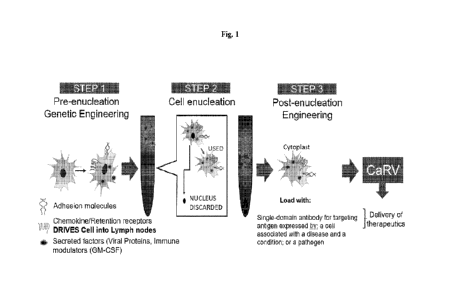
[0027] Fig. 1 illustrates a process for generating the enucleated cells for
the delivery of the single-
domain antibody according to various embodiments described herein.
[00281 Fig. 2 illustrates a timeline for production of the enucleated cells
for the delivery of the
single-domain antibody according to various embodiments, as compared to a
typical biological
drug development timeline.
[0029] Fig. 3A is a representative graph showing the relative fold change in
viable cells or
enucleated cells ("cytoplasts-) over time.
[00301 Fig. 3B is a representative graph showing the viable cells and
cytoplasts after recovery from
frozen storage (cryopreservation).
CA 03208619 2023- 8- 16
WO 2022/183057
PCT/US2022/018007
17
[0031] Fig. 3C is a representative graph showing the relative viability of
cytoplasts 24 hours after
enucleation (fresh cytoplasts) or 24 hours after recovery from frozen storage
(cryopreserved)
following enucleation, where fresh and cryopreserved cytoplasts are compared
to the viability of
cytoplasts 4 hours after enucleation. Mean SEM; n=10.
[0032] Fig. 4A is a representative line graph showing the viability of
mesenchymal stem cell
(MSC) and MSC-derived cytoplasts immediately after recovery from
cryohibemation at 4 degrees
Celsius for the indicated amounts of time. Viability was assessed in an
automated cell count (Cell
Countess) using Trypan blue dye exclusion and displayed as a ratio to the
number of input cells.
[0033] Fig. 4B is a representative bar graph comparing the migrated MSC and
MSC-derived
cytoplasts in a Boyden chamber assay immediately after recovery from
cryohibemation at 4
degrees Celsius for the indicated amounts of time. Cells and cytoplasts were
allowed to migrate for
3 hours with either no scrum (negative control) or 10% premium FBS (P-FBS) as
a chemoattractant
in the bottom chamber, and counts were normalized to loading controls.
[0034] Fig. 5A is a representative flow cytometry graphs showing the number of
events counted
over the signal strength of the cell surface C-X-C Motif Chemokine Receptor 4
(CXCR4)
expression by fluorescent antibody on engineered cytoplasts and engineered
parental MSCs as
analyzed by FlowJo.
[0035] Fig. 5B is a representative bar graph showing the ratio of migrating
cells or cytoplasts that
migrated to the undersurface of the Boyden chamber membrane compared to the
loading control.
Mean SEM; n=10. MSCs and MSC-derived cytoplasts with and without engineered
CXCR4
receptors were allowed to migrate towards the indicated concentrations of
stromal cell-derived
factor la (SDF-1a) for 2 hours in a Boyden chamber assay.
[0036] Fig. 6A is a representative flow cytometry graph showing the number of
events counted over
the signal strength of the cell surface P-selectin glycoprotein ligand-1
(PSGL1) expression by
fluorescent antibody on engineered cytoplasts and engineered parental MSCs as
analyzed by
FlowJo.
[0037] Fig. 6B is a representative graph showing cell surface binding of P-
Selectin with engineered
MSCs and MSC-derived cytoplasts as determined by flow cytometry. MSC control=
parental
MSCs. Engineered MSC= PSGL1/(Fucosyltransferase 7) Fut7 engineered MSC.
Engineered
cytoplast= PSGL1/Fut7 engineered MSC-derived cytoplasts.
[0038] Fig. 7A is a representative flow cytometry graph showing the number of
events counted
over the signal strength of the cell surface of mCD47 expression on engineered
cytoplasts and
MSCs as analyzed by FlowJo.
CA 03208619 2023- 8- 16
WO 2022/183057 PCT/US2022/018007
18
[0039] Fig. 7B is a representative bar graph showing the number of live
cytoplasts (DiD+) that
were not phagocytosed by macrophages (F4/80- and CD11b-), indicating that
cytoplasts escaped
macrophage phagocytosis in the lung. Mean SEM; 11=3. DiD dye-labeled Control
cytoplasts or
engineered cytoplasts (mCD47 Cytoplasts) were retro-orbitally injected into
the vasculature of
mice. After 24 hours, tissues were harvested and stained with two different
pan-macrophage
markers (F4/80 and CD11b).
[0040] Fig. 7C is a representative bar graph showing live cytoplasts (DiD+)
that were not
phagocytosed by macrophages (F4/80- and CD1113), indicating that cytoplasts
escaped macrophage
phagocytosis in the liver. Mean SEM; n=3. DiD dye-labeled Control cytoplasts
or engineered
cytoplasts (mCD47 Cytoplasts) were retro-orbitally injected into the
vasculature of mice. After 24
hours, tissues were harvested and stained with two different pan-macrophage
markers (F4/80 and
CD11b).
[0041] Fig. 8A is a representative scatter plot showing the number of DiD-
labeled MSCs or
cytoplasts detected in the lung. MSCs were cultured under standard adherent
conditions (2D) or in
suspension by the handing drop method (3D) to generate 3D cytoplasts MSCs and
cytoplasts were
labeled with Vybrant DiD dye and retro-orbitally injected into the
vasculature of C57BL/6 mice.
Tissues were harvested after 24 hours and cell suspensions analyzed by flow
cytometry. Mean
SEM; n=2.
[0042] Fig. 8B is a representative scatter plot showing the number of DiD-
labeled MSCs or
cytoplasts detected in the liver. MSCs were cultured under standard adherent
conditions (2D) or in
suspension by the handing drop method (3D) to generate 3D cytoplasts. MSCs and
cytoplasts were
labeled with Vybrant DiD dye and retro-orbitally injected into the
vasculature of C57BL/6 mice.
Tissues were harvested after 24 hours and cell suspensions analyzed by flow
cytometry. Mean
SEM; n=2.
[0043] Fig. 8C is a representative scatter plot showing the number of Vybrant
DiD-labeled MSCs
or cytoplasts detected in the spleen. MSCs were cultured under standard
adherent conditions (2D) or
in suspension by the handing drop method (3D) to generate 3D cytoplasts. MSCs
and cytoplasts
were labeled with DiD dye and retro-orbitally injected into the vasculature of
C57BL/6 mice.
Tissues were harvested after 24 hours and cell suspensions analyzed by flow
cytometry. Mean
SEM; n=2.
[0044] Fig. 9 illustrates cell surface staining of fluorescein isothiocyanate
(FITC) labeled Annexin
V on mesenchymal stromal cells (MSCs) or the cytoplasts analyzed by flow
cytometry for cell
viability analysis.
CA 03208619 2023- 8- 16
WO 2022/183057 PCT/US2022/018007
19
[0045] Fig. 10A is a representative graph showing the abundance of secreted
single-domain
antibody as measured by enzyme-linked immunoassay (ELISA) in conditioned media
of non-
transfected (hTERT) cell or cell transfected with a vector encoding the single-
domain antibody
(scFv). The transfected cell was enucleated and seeded in 6 well plates (0.5 X
106/well) and the
conditioned media was collected after 24 and 48 hours after enucleation for
ELISA detection. Mean
+ SEM; n=3 biological replicates.
[0046] Fig. 10B is a representative graph showing the secreted anti-Programmed
death-ligand 1
(PD-L1) nanobody, single-domain antibody, or scEv (NB) measured by ELISA in
conditioned
media of non-transfected (hTERT-MSCs only) or transfected (enucleated cells +
NB aPD-L1)
cells. Cells were seeded in 6 well plates (0.5 X 10⁶/well) and the conditioned
media was collected
after 24 and 48 hours after enucleation for ELISA detection Mean SEM; n=3
biological
replicates.
[0047] Fig. 10C is a representative graph showing the secreted anti-cytotoxic
T-lymphocyte-
associated protein 4 (CTLA-4) NB measured by ELISA in conditioned media of non-
transfected
(hTERT-MSCs only) and transfected (enucleated cells + NB aCTLA-4) cells. Cells
were seeded in
6 well plates (0.5 X 10⁶/well) and the conditioned media was collected after
24 and 48 hours after
enucleation for ELISA detection. Mean + SEM; n=3 biological replicates.
DETAILED DESCRIPTION
[0048] Disclosed herein are enucleated cells capable of being extensively
engineered to express a
single-domain antibody, or portion thereof, in the absence of a nucleus. Such
enucleated cells may
be used to express and deliver the single-domain antibody or portion thereof
to a target cell or
tissue in 'ivu even when administered systemically. The single-domain antibody
or a portion
thereof or a fragment thereof can exert therapeutic efficacy. For example, the
single-domain
antibody or a portion thereof or a fragment thereof can target and bind to an
immune checkpoint
molecule, thus reducing the expression of the immune checkpoint molecule in a
subject and treating
a disease or condition in the subject. In some aspects, the enucleated cell
can be engineered to
express an exogenous tumor necrosis factor (TNF) superfamily member
polypeptide or a
catalytically active fragment thereof. In some aspects, the enucleated cell
can be engineered to
express both the single-domain antibody and the exogenous TNF. In some
aspects, the exogenous
TNF normalizes abnormal vasculature in the subject. In some aspects,
normalization of the
abnormal vasculature in the subject increases therapeutic efficacy of the
enucleated cell. For
example, the normalized vasculature allows delivery of the single-domain
antibody to a target site
associated with the disease or condition.
CA 03208619 2023- 8- 16
WO 2022/183057 PCT/US2022/018007
[0049] The enucleated cells described herein may be engineered to express one
or more targeting
moieties that, when expressed by the enucleated cell (e.g., on its surface),
guide the enucleated cell
to the target cell or tissue in vivo. In order to avoid unintended clearance
of the enucleated cells in
vivo, the enucleated cell may also include an immune system evading moiety (a
"don't eat me"
signaling polypeptide) such as CD47, PD-L1, HLA-E, or HLA-G. In some
embodiments, the
enucleated cell described herein comprises an additional active agent such as
a therapeutic agent.
[0050] There are two major problems for that arise with the systemic
administration of
conventional therapeutic cells. Firstly, most of the cells may be trapped in
the small capillaries in
the lung or other tissues, negatively impact biodistribution of the
therapeutic cargo and may cause
serious side effects as pulmonary embolism. Secondly, conventional therapeutic
cells lacking the
tissue-specific homing receptors and adhesion molecules (e.g., SDF-lalCXCR4,
CCL2/CCR2,
PSGL-1) to target intended cells or tissue arc unable to home to such intended
cells or tissues
efficiently.
[00511 The enucleated cells of the present disclosure solve such problems by,
in some
embodiments, being much smaller than traditional therapeutic cells and their
parental cells (e.g
about 60% of the diameter of parental cells and 1/8 the volume) and do not
have the rigid nucleus.
Therefore, the enucleated cells may pass better through small capillaries and
vessels than their
conventional counterparts. In addition, the enucleated cells described herein
may be engineered to
express functional targeting moieties (e.g., homing receptors, adhesion
molecules, or membrane-
bound antibody such as membrane-bound single-domain antibody) to facilitate
efficient homing to
target cell or tissue, even when administered to a subject systemically. In
some embodiments, the
target cell or tissue is a cancer cell or tissue. In the case of enucleated
cells that target a cancer cell
or tissue, the enucleated cells may be engineered to express targeting
moieties that recognize an
antigen produced by the cancer cell or tissue. In some embodiments, the
enucleated cells may be
engineered to express other targeting moieties to guide the enucleated cells
to the target tissue,
including chemokines, integrins, adhesion molecules, membrane-bound antibody,
or membrane-
bound single-domain antibody.
[0052] In some embodiments, the single-domain antibody or portion thereof is
exogenous to the
enucleated cell or parent cell thereof. The exogenous single-domain antibody
or portion thereof
may confer a therapeutic effect such as to treat a disease or a condition
described herein. In some
embodiments, the single-domain antibody or portion thereof is fused with a
plurality of antibodies
or a plurality of single-domain antibodies that may target a single epitope or
multiple epitopes (e.g.,
bispecitic). In some embodiments, the single-domain antibody is conjugate to a
small molecule
CA 03208619 2023- 8- 16
WO 2022/183057 PCT/US2022/018007
21
such as an antibody-drug conjugate (ADC). In some embodiments, the small
molecule is a
cytotoxic drug or therapeutically effective portion thereof.
[0053] In some embodiments, the enucleated cell comprises polynucleotide
encoding the single-
domain antibody or portion thereof In some embodiments, the polynucleotide is
exogenous to the
enucleated cell or the parent cell thereof In some embodiments, the
polynucleotide encodes the
single-domain antibody fused with the plurality of antibodies or a plurality
of single-domain
antibodies described herein.
[0054] Also disclosed herein are methods of using the enucleated cells and
compositions
comprising the enucleated cells of the present disclosure. In some
embodiments, methods for
treating a disease or a condition by delivering the enucleated cell or
compositions containing the
enucleated cell to a subject are provided. In some embodiments, the disease or
the condition is
associated with the target cell or target tissue that the enucleated cell is
engineered to target. In
some embodiments, the target cell is a cancer cell. In some embodiments, the
target cell is an
epithelial cell. In some cases, the tissue is a tumor. In some embodiments,
the tissue is lung tissue.
In some embodiments, the disease or the condition comprises cancer. In some
embodiments, the
disease or the condition comprises idiopathic pulmonary fibrosis. In some
embodiments, methods
comprise using the enucleated cells of the present disclosure to transfer
cargo (e.g., single domain
antibody, therapeutic agent) to the target cell, such as, for example, with a
fusogenic moiety or
tunneling nanotubule.
[0055] Disclosed herein are methods of producing the enucleated cells
described herein, by
removing a nucleus from a nucleated parent cell such as, for example, without
undergoing
differentiation of the parent cell. For example, the parent cell containing a
nucleus may be
engineered to express the single-domain antibody, or portion thereof,
targeting moiety or "don't eat
me" signaling peptide; and subsequently, the nucleus of the parent cell may be
removed. In another
example, the parent cell containing the nucleus is enucleated, and the
enucleated cell is engineered
to express the domain antibody, or portion thereof, targeting moiety or "don't
eat me" signaling
peptide. In some embodiments, removal of the nucleus involves mechanically
removing the
nucleus.
[0056] Following enucleation of the parent cell, the enucleated cells
described herein retain one or
more intracellular organelles that are endogenous to the parent cell. In some
embodiments, all of
the one or more intracellular organelles are retained. In some embodiments,
fewer than all of the
one or more intracellular organelles are retained. In some embodiments, the
Golgi apparatus and/or
the endoplasmic reticulum are retained, which are involved in protein
synthesis and secretion.
Retention of the one or more intracellular organelles at least partially
enables the enucleated cells to
CA 03208619 2023- 8- 16
WO 2022/183057 PCT/US2022/018007
22
synthesize or release the biomolecule disclosed herein (e.g., single-domain
antibody, or portion
thereof, targeting moiety, immune-evading moiety, etc.) in the absence of the
nucleus.
[0057] In some embodiments, the parent cell is any one of the nucleated cells
described herein. In
some embodiments, the parent cell is an adult stem cell. In some embodiments.
the parent cell is a
mesenchymal stromal cell (MSC). In some embodiments, the enucleated cell is
derived from an
inducible pluripotent stem cell (iPSC). In some embodiments, the parent cell
is not an erythrocyte
or erythroid precursor cell. In some embodiments, the parent cell is a
platelet cell. In some
embodiments, the parent cell is not an endothelial cell. In some embodiments,
the parent cell is not
an endothelial precursor cell nor an erythroid precursor cell. In some
embodiments, the parent cell
is not a platelet cell. In some embodiments, the parent cell is not an
endothelial cell. In some
embodiments, the parent cell is not an endothelial precursor cell. In some
embodiments, the parent
cell does not express complement receptor one (CR1). In some embodiments, the
parent cell does
not express CD44. In some embodiments, the parent cell does not express VLA-4.
In some
embodiments, the parent cell does not express BCAM. In some embodiments, the
parent cell does
not express ICA_M. In some embodiments, the parent cell does not express a
receptor for collagen.
In some embodiments, the parent cell does not express a receptor for
thrombopoietin. In some
embodiments, the parent cell does not express a receptor for collagen. In some
embodiments, the
parent cell does not express a receptor for von Willebrand factor (VWF). In
some embodiments, the
parent cell does not express a receptor for fibrinogen. In some embodiments,
the parent cell does
not express GP lb-IX-V receptor. In some embodiments, the parent cell does not
express GPIIb/IIIa
receptor. In some embodiments, the parent cell does not express prostanoid
receptor. In some
embodiments, the parent cell does not express purinergic receptor. In some
embodiments, the
parent cell does not express thromboxane receptor.
COMPOSITIONS
[00581 Provided herein are cells comprising a single-domain antibody, or
portion thereof, and
compositions containing such enucleated cells. In some embodiments, the cells
are enucleated. In
some embodiments, the single-domain antibody is a therapeutic agent. In some
embodiments, the
enucleated cells are capable of expressing the single-domain antibody (e.g.,
therapeutic agent) in
absence of a nucleus using one or more intracellular organelles retained by
the enucleated cells
from parent cells. In some embodiments, the single-domain antibody, or portion
thereof (e.g.,
therapeutic agent) is exogenous to the enucleated cell or parent cell thereof.
In some embodiments,
the enucleated cell expresses the single-domain antibody, or portion thereof
(e.g., therapeutic agent)
at the surface of the enucleated cell. In some embodiments, the single-domain
antibody, or portion
CA 03208619 2023- 8- 16
WO 2022/183057 PCT/US2022/018007
23
thereof is secreted by the enucleated cell into extracellular space at a
target tissue (e.g., a
microenvironment). In some aspects, the composition comprises an enucleated
cell described
herein In some aspects, the enucleated cell is obtained or derived from a
nucleated cell (e.g., a
parent cell). In some aspects, the enucleated cell comprises a transmembrane
moiety. In some
embodiments, the enucleated cells comprises a targeting moiety. In some
embodiments, the
enucleated cell comprises a therapeutic agent. In some embodiments, the
enucleated cell comprises
an antibody or antigen binding fragment thereof or a single-domain antibody or
an antigen binding
fragment thereof. In some aspects, the targeting moiety comprises an antibody
or antigen binding
fragment thereof or a single-domain antibody or an antigen binding fragment
thereof. In some
embodiments, the therapeutic agent comprises an antibody or antigen binding
fragment thereof or a
single-domain antibody or an antigen binding fragment thereof. In some
embodiments, the
enucleated cell is formulated into a pharmaceutical formulation described
herein.
Enucleated cells
[00591 The enucleated cells of the present disclosure are obtained or derived
from a corresponding
nucleated cell (referred to herein as a "parent cell"). The parent cell may be
derived from a variety
of different cell types, including eukaryotic cells. For example, an
enucleated cell may be derived
from an adult stem cell, a mesenchymal stromal cell (MSC), a natural killer
(NK) cell, a
macrophage, a myoblast, a neutrophil, endothelial cell, endothelial precursor
cell, and/or a
fibroblast. In some embodiments, an enucleated cell is derived from a
mesenchymal stromal cell. In
some embodiments, the enucleated cell is derived from an inducible pluripotent
stem cell (iPSC). In
some embodiments, the parent cell is derived from a cell is immortalized using
suitable methods.
For example the, parent cell is immortalized by expressing human telomerase
reverse transcriptase
(hTERT), an oncogene, or a viral gene such as simian virus 40 (SV40). In some
embodiments, the
cytoplast is derived from a parent cell using suitable methods provided in
United States Patent No.
10,927,349, which is hereby incorporated by reference in its entirety. In some
embodiments, the
enucleated cell retains one or more intracellular organelles for synthesis of
an exogenous single-
domain antibody or fragment thereof in absence of the nucleus.
[0060] In some embodiments, the cell can originate from any organism having
one or more cells.
Non-limiting examples of cells include: a prokaryotic cell, eukaryotic cell, a
bacterial cell, an
archaeal cell, a cell of a single-cell eukaryotic organism, a protozoa cell, a
cell from a plant (e.g.
cells from plant crops, fruits, vegetables, grains, soy bean, corn, maize,
wheat, seeds, tomatoes,
rice, cassava, sugarcane, pumpkin, hay, potatoes, cotton, cannabis, tobacco,
flowering plants,
conifers, gymnospeinis, ferns, clubmosses, homworts, liverworts, mosses), an
algal cell, (e.g.,
Botryococcus braunii, Chlamydomonas reinhardtii, Nannochloropsis gaditana,
Chlorella
CA 03208619 2023- 8- 16
WO 2022/183057 PCT/US2022/018007
24
pyrenoidosa, Sargassum patens C. Agardh, and the like), seaweeds (e.g. kelp),
a fungal cell (e.g., a
yeast cell, a cell from a mushroom), an animal cell, a cell from an
invertebrate animal (e.g. fruit fly,
cnidarian, echinoderm, nematode, etc.), a cell from a vertebrate animal (e.g.,
fish, amphibian,
reptile, bird, mammal), a cell from a mammal (e.g., a pig, a cow, a goat, a
sheep, a rodent, a rat, a
mouse, a non-human primate, a human, etc.), and etcetera. Sometimes a cell is
not originating from
a natural organism (e.g., a cell can be a synthetically made, sometimes termed
an artificial cell). In
some embodiments, the cell is a somatic cell. In some embodiments, the cell is
a stem cell or a
progenitor cell. In some embodiments, the cell is a mesenchymal stem or
progenitor cell. In some
embodiments, the cell is a hematopoietic stem or progenitor cell. In some
embodiments, the cell is a
muscle cell, a skin cell, a blood cell, or an immune cell. Other non-limiting
example of cells
includes lymphoid cells such as -13 cell, T cell (Cytotoxic T cell, Natural
Killer T cell, Regulatory T
cell, T helper cell), Natural killer cell, cytokine induced killer (OK) cells;
myeloid cells such as
granulocytes (Basophil granulocyte, Eosinophil granulocyte, Neutrophil
granulocyte(Hypersegmented neutrophil), Monocyte/Macrophage, Red blood cell
(Reticulocyte),
Mast cell, Thrombocyte/Megakaryocyte, Dendritic cell, cells from the endocrine
system, including
thyroid (Thyroid epithelial cell, Parafollicular cell), parathyroid
(Parathyroid chief cell, Oxyphil
cell), adrenal (Chromaffin cell), pineal (Pinealocyte) cells; cells of the
nervous system, including
glial cells (Astrocyte, Microglia), Magnocellular neurosecretory cell,
Stellate cell, Boettcher cell,
and pituitary (Gonadotrope, Corticotrope, Thyrotrope, Somatotrope, Lactotroph
); cells of the
Respiratory system, including Pneumocyte (Type I pneumocyte, Type II
pneumocyte), Clara cell,
Goblet cell, Dust cell; cells of the circulatory system, including
Myocardiocyte, Pericyte; cells of
the digestive system, including stomach (Gastric chief cell, Parietal cell),
Goblet cell, Paneth cell, G
cells, D cells, ECL cells, I cells, K cells, S cells, enteroendocrine cells,
including enterochromaffm
cell, APUD cell, liver (Hepatocyte, Kupffer cell), Cartilage/bone/muscle; bone
cells, including
Osteoblast, Osteocyte, Osteoclast, teeth (Cementoblast, Ameloblast); cartilage
cells, including
Chondroblast, Chondrocyte; skin cells, including Trichocyte, Keratinocyte,
Melanocyte (Nevus
cell); muscle cells, including Myocyte; urinary system cells, including
Podocyte, Juxtaglomerular
cell, Intraglomerular mesangial cell/Extraglomerular mesangial cell, Kidney
proximal tubule brush
border cell, Macula densa cell; reproductive system cells, including
Spermatozoon, Sertoli cell,
Leydig cell, Ovum; and other cells, including Adipocyte, Fibroblast, Tendon
cell, Epidermal
keratinocyte (differentiating epidermal cell), Epidermal basal cell (stem
cell), Keratinocyte of
fingernails and toenails, Nail bed basal cell (stem cell), Medullary hair
shaft cell, Cortical hair shaft
cell, Cuticular hair shaft cell, Cuticular hair root sheath cell, Hair root
sheath cell of Huxley's layer,
Hair root sheath cell of Henle's layer, External hair root sheath cell, Hair
matrix cell (stem cell),
CA 03208619 2023- 8- 16
WO 2022/183057 PCT/U52022/018007
Wet stratified barrier epithelial cells, Surface epithelial cell of stratified
squamous epithelium of
cornea, tongue, oral cavity, esophagus, anal canal, distal urethra and vagina,
basal cell (stem cell) of
epithelia of cornea, tongue, oral cavity, esophagus, anal canal, distal
urethra and vagina, Urinary
epithelium cell (lining urinary bladder and urinary ducts), Exocrine secretory
epithelial cells,
Salivary gland mucous cell (polysaccharide-rich secretion), Salivary gland
serous cell (glycoprotein
enzyme -rich secretion), Von Ebner's gland cell in tongue (washes taste buds),
Mammary gland cell
(milk secretion), Lacrimal gland cell (tear secretion), Ceruminous gland cell
in ear (wax secretion),
Eccrine sweat gland dark cell (glycoprotein secretion), Eccrine sweat gland
clear cell (small
molecule secretion). Apocrine sweat gland cell (odoriferous secretion, sex -
hormone sensitive),
Gland of Moll cell in eyelid (specialized sweat gland), Sebaceous gland cell
(lipid-rich sebum
secretion), Bowman's gland cell in nose (washes olfactory epithelium),
Brunner's gland cell in
duodenum (enzymes and alkaline mucus), Seminal vesicle cell (secretes seminal
fluid components,
including fructose for swimming sperm), Prostate gland cell (secretes seminal
fluid components),
Bulbourethral gland cell (mucus secretion), Bartholin's gland cell (vaginal
lubricant secretion),
Gland of Littre cell (mucus secretion), Uterus endometrium cell (carbohydrate
secretion), Isolated
goblet cell of respiratory and digestive tracts (mucus secretion), Stomach
lining mucous cell
(mucus secretion), Gastric gland zymogenic cell (pepsinogen secretion),
Gastric gland oxyntic cell
(hydrochloric acid secretion), Pancreatic acinar cell (bicarbonate and
digestive enzyme secretion),
Paneth cell of small intestine (lysozyme secretion), Type II pneumocyte of
lung (surfactant
secretion), Clara cell of lung, Hormone secreting cells, Anterior pituitary
cells, Somatotropes,
Lactotropes, Thyrotropes, Gonadotropes, Corticotropes, Intermediate pituitary
cell, Magnocellular
neurosecretory cells, Gut and respiratory tract cells, Thyroid gland cells,
thyroid epithelial cell,
parafollicular cell, Parathyroid gland cells, Parathyroid chief cell, Oxyphil
cell, Adrenal gland cells,
chromaffin cells, Ley dig cell of testes, Theca intema cell of ovarian
follicle, Corpus luteum cell of
ruptured ovarian follicle, Granulosa lutein cells, Theca lutein cells,
Juxtaglomerular cell (renin
secretion), Macula densa cell of kidney, Metabolism and storage cells, Barrier
function cells (Lung,
Gut, Exocrine Glands and Urogenital Tract), Kidney, Type I pneumocyte (lining
air space of lung),
Pancreatic duct cell (centroacinar cell), Nonstriated duct cell (of sweat
gland, salivary gland,
mammary gland, etc.), Duct cell (of seminal vesicle, prostate gland, etc.),
Epithelial cells lining
closed internal body cavities, Ciliated cells with propulsive function,
Extracellular matrix secretion
cells, Contractile cells; Skeletal muscle cells, stem cell, Heart muscle
cells, Blood and immune
system cells, Erythrocyte (red blood cell), Megakaryocyte (platelet
precursor), Monocyte,
Connective tissue macrophage (various types), Epidermal Langerhans cell,
Osteoclast (in bone),
Dendritic cell (in lymphoid tissues), Microglial cell (in central nervous
system), Neutrophil
CA 03208619 2023- 8- 16
WO 2022/183057 PCT/US2022/018007
26
granulocyte, Eosinophil granulocyte, Basophil granulocyte, Mast cell, Helper T
cell, Suppressor T
cell, Cytotoxic T cell, Natural Killer T cell, B cell, Natural killer cell,
Reticulocyte, Stem cells and
committed progenitors for the blood and immune system (various types),
Pluripotent stem cells,
Totipotent stem cells, Induced pluripotent stem cells, adult stem cells,
Sensory transducer cells,
Autonomic neuron cells, Sense organ and peripheral neuron supporting cells,
Central nervous
system neurons and glial cells, Lens cells, Pigment cells, Melanocyte, Retinal
pigmented epithelial
cell, Germ cells, Oogonium/Oocyte, Spermatid, Spermatocyte, Spermatogonium
cell (stem cell for
spermatocyte), Spermatozoon, Nurse cells, Ovarian follicle cell, Sertoli cell
(in testis), Thymus
epithelial cell, Interstitial cells, and Interstitial kidney cells.
[0061] In some embodiments, the cell is a eukaryotic cell. Non-limiting
examples of eukaryotic
cells include mammalian (e.g., rodent, non-human primate, or human), non-
mammalian animal
(e.g., fish, bird, reptile, or amphibian), invertebrate, insect, fungal, or
plant cells. In some
embodiments, the eukaryotic cell is a yeast cell such as Saccharornyces
cerevisiae. In some
embodiments, the eukaryotic cell is a higher eukaryote such as mammalian,
avian, plant, or insect
cells. In some embodiments, the nucleated cell is a primary cell. In some
embodiments, the
nucleated cell is an immune cell (e.g., a lymphocyte (e.g., a T cell, a B
cell), a macrophage, a
natural killer cell, a neutrophil, a mast cell, a basophil, a dendritic cell,
a monocyte, a myeloid-
derived suppressor cell, an eosinophil). In some embodiments, the nucleated
cell is a phagocyte or a
leukocyte. In some embodiments, the nucleated cell is a stem cell (e.g., an
adult stem cell (e.g., a
hematopoietic stem cell, a mammary stem cell, an intestinal stem cell,
mesenchymal stem cell, an
endothelial stem cell, a neural stem cell, an olfactory adult stem cell, a
neural crest stem cell, a
testicular cell), an embryonic stem cell, an inducible pluripotent stem cell
(iPS)). In some
embodiments, the nucleated cell is a progenitor cell. In some embodiments, the
nucleated cell is
from a cell line. In some embodiments, the nucleated cell is a suspension
cell. In some
embodiments, the nucleated cell is an adherent cell. In some embodiments, the
nucleated cell is a
cell that has been immortalized by expression of an oncogene. In some
embodiments, the nucleated
cell is immortalized by the expression of human telomerase reverse
transcriptase (hTERT) or any
oncogene. In some embodiments, the nucleated cell is immortalized by the
expression of a viral
gene such as simian virus 40 (SV40). In some embodiments, the nucleated cell
is a patient or
subject derived cell (e.g., an autologous patient-derived cell, or an
allogenic patient-derived cell). In
some embodiments, the nucleated cell is transfected with a vector (e.g., a
viral vector (e.g., a
retrovirus vector (e.g., a lentivirus vector), an adeno-associated virus (AAV)
vector, a vesicular
virus vector (e.g., vesicular stomatitis virus (VSV) vector), or a hybrid
virus vector), a plasmid)
before the nucleated cell is enucleated using any of the enucleation
techniques described herein.
CA 03208619 2023- 8- 16
WO 2022/183057
PCT/US2022/018007
27
[0062] In some embodiments, the cytoplast is derived from a cell autologous to
the subject. In some
embodiments, the cytoplast is derived from a cell allogenic to the subject.
[0063] In some embodiments, the cytoplast is derived from an immune cell. In
some embodiments,
the cytoplast is derived from a natural killer (NK) cell, a neutrophil, a
macrophage, a lymphocyte, a
fibroblast, an adult stem cell (e.g., hematopoietic stem cell, a mammary stem
cell, an intestinal stem
cell, a mesenchymal stein cell, a mesenchymal stromal cell, an endothelial
stem cell, a neural stem
cell, an olfactory adult stem cell, a neural crest stem cell, a skin stem
cell, or a testicular cell), a
mast cell, a basophil, an eosinophil, an endothelial cell, an endothelial cell
precursor cell, or an
inducible pluripotent stem cell.
[0064] In some embodiments, the parent cell is not an erythrocyte or erythroid
precursor cell. In
some embodiments, the parent cell is a platelet cell. In some embodiments, the
parent cell is not an
endothelial cell. In some embodiments, the parent cell is not an endothelial
precursor cell. In some
embodiments, the enucleated cell is not an erythrocyte or erythroid precursor
cell. In some
embodiments, the enucleated cell is not a platelet cell. In some embodiments,
the enucleated cell is
not an endothelial cell. In some embodiments, the enucleated cell is not an
endothelial precursor
cell. In some embodiments, the enucleated cell does not express complement
receptor one (CR1).
In some embodiments, the enucleated cell does not express CD44. In some
embodiments, the
enucleated cell does not express VLA-4. In some embodiments, the enucleated
cell does not
express BCA1VI. In some embodiments, the enucleated cell does not express
ICAM. In some
embodiments, the enucleated cell does not express a receptor for collagen. In
some embodiments,
the enucleated cell does not express a receptor for thrombopoietin. In some
embodiments, the
enucleated cell does not express a receptor for collagen. In some embodiments,
the enucleated cell
does not express a receptor for von Willebrand factor (VWF). In some
embodiments, the
enucleated cell does not express a receptor for fibrinogen. In some
embodiments, the enucleated
cell does not express GP1b-IX-V receptor. In some embodiments, the enucleated
cell does not
express GPIIb/IIIa receptor. In some embodiments, the enucleated cell does not
express prostanoid
receptor. In some embodiments, the enucleated cell does not express purinergic
receptor. In some
embodiments, the enucleated cell does not express thromboxane receptor.
[0065] In some embodiments, the parent cell may be enucleated and engineered
for therapeutic
use. In some embodiments, the parent cell is any one of the nucleated cells
described herein. In
some embodiments, the parent cell is an adult stem cell. In some embodiments,
the parent cell is a
mesenchymal stromal cell (MSC). In some embodiments, the enucleated cell is
derived from an
inducible pluripotent stem cell (iPSC). In some embodiments, the parent cell
is not an erythrocyte
or erythroid precursor cell. In some embodiments, the parent cell is a
platelet cell. In some
CA 03208619 2023- 8- 16
WO 2022/183057 PCT/US2022/018007
28
embodiments, the parent cell is not an endothelial cell. In some embodiments,
the parent cell is not
an endothelial precursor cell. A parent cell may be treated with cytochalasin
to soften the cortical
actin cytoskeleton. The nucleus may then physically extracted from the cell
body by high-speed
centrifugation in gradients of Ficoll to generate an enucleated cell. Because
enucleate cells and
intact nucleated cells sediment to different layers in the Ficoll gradient,
enucleated cells may be
easily isolated and prepared for therapeutic purposes or fusion to other cells
(nucleated or
enucleated). The enucleation process is clinically scalable to process tens of
millions of cells. In
some embodiments, enucleated cells may be used as a disease-homing vehicle to
deliver clinically
relevant cargos/payloads to treat various diseases.
[0066] In some embodiments, the enucleated cells contain one or more
organelles or cytoskeleton
to form a tunneling nanotube (TNT) or membrane nanotube. The inventors of the
present disclosure
discovered that enucleating a cell, can in some cases, increase the TNT in thc
cell, which may
provide therapeutic advantages. In some embodiments, the increase in TNT of
the enucleated cell is
determined by the enucleated cell exhibiting an increased number of the TNT
formation compared
to TNT formation by an otherwise identical cell that has not been enucleated
In some
embodiments, the increase in TNT of the enucleated cell is determined by the
enucleated cell
exhibiting an increased length of the TNT formation compared to a length of
the TNT formation by
an otherwise identical cell that has not been enucleated. In some embodiments,
the increase in TNT
of the enucleated cell is determined by the enucleated cell exhibiting an
increased diameter of the
TNT formation compared to a diameter of the TNT formation by an otherwise
identical cell that
has not been enucleated. In some embodiments, the tunneling nanotube delivers
at least one
exogenous agent described herein to a target cell. In some embodiments, the
increased TNT
formation, increased TNT length, or increased TNT diameter increases the
efficacy of delivering of
the at least one exogenous agent by the enucleated cell to the target cell In
some embodiments, the
exogenous agent is a therapeutic agent. In some embodiments, the exogenous
agent can normalize
vasculature. In some embodiments, the tunneling nanotube is a protrusion that
extend from plasma
membrane of the enucleated cell, where the tunneling nanotube can comprise a
length of about 1
pm to about 1000 pm. In some embodiments, the tunneling nanotube comprises a
diameter of about
0.1 nm to about 10.0 pm. In some embodiments, the tunneling nanotube comprises
actin. In some
embodiments, the tunneling nanotube comprises both actin and microtubule. ht
some embodiments,
the tunneling nanotube comprises content of the cytoplasm of the enucleated
cell. For example, the
tunneling nanotube can contain vesicles, organelles or the at least one
exogenous agent described
herein. In some embodiments, the tunneling nanotube can deliver the at least
one exogenous agent
by contacting with the target cell. In some embodiments, the tunneling
nanotube can deliver the at
CA 03208619 2023- 8- 16
WO 2022/183057 PCT/US2022/018007
29
least one exogenous agent to the target cell by forming a fluid communication
between the
enucleated cell and the target cell In some embodiments, upon delivery of the
at least one
exogenous from the enucleated cell to the target cell, the target cell can
form additional tunneling
nanotube with other non-targeted cells. In such scenario (similar to bystander
effect), the target cell
can pass on the at least one exogenous agent and its effect to other non-
targeted cells, thus
propagating the therapeutic effect of the enucleated cell to other non-target
cells.
[0067] Following enucleation of the parent cell, the enucleated cells
described herein retain one or
more intracellular organelles that are endogenous to the parent cell. In some
embodiments, all of
the one or more intracellular organelles are retained. In some embodiments,
fewer than all of the
one or more intracellular organelles are retained. In some embodiments, the
Golgi apparatus and/or
the endoplasmic reticulum are retained, which are involved in protein
synthesis and secretion
Retention of the one or more intracellular organelles at least partially
enables the enucleatcd cells to
synthesize or release the biomolecule disclosed herein (e.g., single-domain
antibody, or portion
thereof, targeting moiety, immune-evading moiety, etc.) in the absence of the
nucleus. In some
embodiments, the enucleated cell retains cytoskeleton such as filament or
tubules for formation of
TNT.
[0068] Enucleated cells may be smaller than their nucleated counterparts
(e.g., the nucleated parent
cells), and for this reason may migrate better through small openings in the
vasculature and tissue
parenchyma. In addition, removing the large dense nucleus alleviates a major
physical barrier
allowing the cell to move freely through small openings in the vessels and
tissue parenchyma.
Therefore, enucleated cells have improved bio-distribution in the body and
movement into target
tissues. In some embodiments, an enucleated cell comprises at least 1 pm in
diameter. In some
embodiments, an enucleated cell is greater than 1 pm in diameter. In some
embodiments, an
enucleated cell is 1-100 um in diameter (e.g., 1- 90 pm, 1-80 pm, 1-70 pm, 1-
60 pm, 1-50 pm, 1-40
pm, 1-30 pm, 1-20 pm, 1-10 pm, 1-5 pm, 5- 90 pm, 5-80 pm, 5-70 m, 5-60 pm, 5-
50 pm, 5-40
pm, 5-30 pm, 5-20 pm, 5-10 pm, 10-90 pm, 10-80 pm, 10-70 pm, 10-60 um, 10-50
pm, 10-40 pm,
10-30 pm, 10-20 pm, 10-15 gm 15-90 um, 15-80 um, 15-70 pm, 15-60 pm, 15-50 pm,
15-40 pm,
15-30 pm, 15-20 pm). In some embodiments, an enucleated cell is 10-30 pm in
diameter, In some
embodiments, the diameter of an enucleated cell is between 5-25 gm (e.g., 5-20
pm, 5-15 pm, 5-10
pm, 10-25 pm, 10-20 pm, 10-15 pm, 15- 25 um, 15-20 pm, or 20-25 pm). In some
embodiments,
the enucleated cell has a diameter that is about 8 pm. In some embodiments,
some enucleated cells
may advantageously be small enough to allow for better homing or delivery to a
target site. For
examples, the enucleated cells described herein may pass through passages in
narrow lung tissues
CA 03208619 2023- 8- 16
WO 2022/183057 PCT/US2022/018007
or lung structures such as alveolar duct or microcapillary that most cells
such as the parent cells
may not pass through.
[0069] In some embodiments, enucleated cells possess significant therapeutic
value, because they
remain viable, do not differentiate into other cell types, secrete bioactive
molecules, and may
physically migrate/home for fewer than or equal to about 5 days, may be
extensively enucleated ex
vivo to perform specific therapeutic functions, and may be fused to the same
or other cell types to
transfer desirable production, natural or enucleated. Therefore, enucleated
cells have wide utility as
a cellular vehicle to deliver therapeutically important biomolecules and
disease-targeting cargos
including genes, viruses, bacteria, mRNAs, shRNAs, siRNA, polypeptides
(including antibodies
and antigen binding fragments), plasmids, gene-editing machinery, or
nanoparticles. The present
disclosure enables the generation of safe (e.g., no unwanted DNA is
transferred to the subject), and
controllable (e.g., cell death occurs in 3-4 days) cell-based carrier that may
be genetically
enucleated to deliver specific disease-fighting and health promoting cargos to
humans. In some
embodiments, the enucleated cell remains viable and retain the function to
migrate or home for
greater than or equal to about 12 hours, 24 hours, 36 hours, 48 hours, 60
hours, 72 hours, 84 hours,
96 hours, 108 hours, or 5 days after being administered to the subject in need
thereof.
[0070] In some embodiments, the enucleated cell is engineered to express at
least one of an
exogenous DNA molecule, an exogenous RNA molecule, an exogenous protein, or an
exogenous
protein, gene-editing machinery or combinations thereof In some embodiments,
the exogenous
DNA molecule is a single-stranded DNA, a double-stranded DNA, an
oligonucleotide, a plasmid, a
bacterial DNA molecule, a DNA virus, or combinations thereof. In some
embodiments, the
exogenous RNA molecule is messenger RNA (mRNA), small interfering RNA (siRNA),
microRNA (miRNA), short hairpin RNA (shRNA), a RNA virus, or combinations
thereof. In some
embodiments, the exogenous protein is a cytokine, a growth factor, a hormone,
an antibody or the
antigen-binding fragment thereof, an enzyme, or combinations thereof In some
embodiments, the
antibody is a single-domain antibody or antigen-binding fragment thereof. In
some embodiments,
parental cells (e.g., nucleated cells) are genetically enucleated before
enucleation (e.g., pre-
enucleation). In some embodiments, the parent cell is genetically enucleated
after enucleation (e.g.,
post-enucleation).
[0071] In some embodiments, the enucleated cell described herein comprises a
transmembrane
moiety. In some embodiments, the transmembrane moiety is genetically modified
to be fused or
complexed with the single-domain antibody or antigen-binding fragment thereof,
or a therapeutic
agent described herein, or a combination thereof. In some embodiments, the
transmembrane moiety
is genetically modified to fuse to the single-domain antibody or antigen-
binding fragment thereof,
CA 03208619 2023- 8- 16
WO 2022/183057 PCT/US2022/018007
31
or therapeutic agent described herein, or a combination thereof In some
embodiments, the
enucleated cell comprises an immune-evading moiety. In some embodiments, the
immune-evading
comprises a "don't eat me" signaling peptide such as CD47, PD-L1, HLA-E, FILA-
G, a fragment
thereof, or a combination thereof.
[0072] In some embodiments, the enucleated cell or the composition comprising
the enucleated cell
may be cryopreserved (e.g. storing the enucleated cell or the composition
comprising the
enucleated cell at freezing temperature) or cryohibernated (e.g. storing the
enucleated cell or the
composition comprising the enucleated cell at a temperature that is between
the ambient
temperature and freezing temperature). The duration of cryopreservation or
cryohibernation may be
greater than or equal to about one hour, two hours, six hours, 12 hours, one
day, two days, three
days, four days, five days, six days, one week, two weeks, three weeks, four
weeks, one month, two
months, three months, or longer period of time. In some embodiments, the
enucleated cell exhibits
a viability after cryopreservation or cryohibernation that is greater than or
equal to about 50%,
55%, 60%, 65%, 70%, 75%, 80%, 85%, 90%, 95%, or 99% similar to a comparable
cell (e.g., a
parent cell or an enucleated cell described herein that has not been
cryopreserved or cryo-
hibernated) after same the period of time of cryopreservation or
cryohibernation. For example, in
some embodiments, the viability is reduced by 50%, 55%, 60%, 65%, 70%, 75%,
80%, 85%, 90%,
95%, or 99% as compared with the comparable cell. In another example, in some
embodiments, the
viability is increased by 50%, 55%, 60%, 65%, 70%, 75%, 80%, 85%, 90%, 95%, or
99% as
compared with the comparable cell. Viability in this context may be measured
by Trypan blue dye
exclusion as described herein. In some embodiments, the Trypan blue dye
exclusion is performed
by: (a) centrifuging an aliquot of a plurality of the cell without the nucleus
in a suspension to create
a cell pellet; (b) resuspending the cell pellet in serum-free medium to
produce a serum-free cell
suspension; (c) mixing 1 part Trypan blue dye and 1 part of the serum-free
cell suspension; (d)
counting the plurality of the cells without the nucleus within 3-5 minutes of
(c), wherein at least
some of the plurality of cells without the nucleus are unstained with the
Trypan blue dye, which is
indicative of viability. In some embodiments, the viability is measured using
Annexin-V cell
surface staining. In some embodiments, the viability is measured by expression
of the exogenous
polypeptide. For example, the viability of the enucleated cell can be
determined by the expression
of the exogenous antibody or single-domain antibody expressed by the
enucleated cell. In some
embodiments, the viability is measured by expression of cell surface markers
of any one of the cell
surface markers described herein such as CD105, CD90, CD45, CXCR4, PSGL-1, or
CCR2. In
some embodiments, the viability is measured by the cell activity of the
enucleated cell. In some
CA 03208619 2023- 8- 16
WO 2022/183057 PCT/US2022/018007
32
embodiments, the viability is measured by the homing capability of the
enucleated cell as
determined by the chemosen sing or chemokine homing activity described herein
[0073] In some embodiments, the enucleated cell or the composition comprising
the enucleated cell
may be lyophilized. In some embodiments, the enucleated cell exhibits a
viability after being
reconstituted from lyophilization that is greater than or equal to about 50%,
55%, 60%, 65%, 70%,
75%, 80%, 85%, 90%, 95%, or 99% similar to a comparable cell (e.g., a parent
cell or an
enucleated cell described herein that has not been lyophilized).
[0074] In some embodiments, the enucleated cell or the composition comprising
the enucleated cell
may be dehydrated. In some embodiments, the enucleated cell exhibits a
viability after being
rehydrated from lyophilization that is greater than or equal to about 50%,
55%, 60%, 65%, 70%,
75%, 80%, 85%, 90%, 95%, or 99% similar to a comparable cell (e.g., a parent
cell or an
enucleated cell described herein that has not been dehydrated).
[0075] In some embodiments, the enucleated cell or the composition comprising
the enucleated cell
is stable at 4 C for greater than or equal to about one hour, two hours, six
hours, 12 hours, one day,
two days, three days, four days, five days, six days, one week, two weeks,
three weeks, four weeks,
one month, two months, three months, or longer period of time. In some
embodiments, the
composition is stable at room temperature for greater than or equal to about
one hour, two hours,
six hours, 12 hours, one day, two days, three days, four days, five days, six
days, one week, two
weeks, three weeks, four weeks, one month, two months, three months, or longer
period of time. In
some embodiments, the composition is stable at 37 C for greater than or equal
to about one hour,
two hours, six hours, 12 hours, one day, two days, three days, four days, five
days, six days, one
week, two weeks, three weeks, four weeks, one month, two months, three months,
or longer period
of time. In some embodiments, the enucleated cell or the composition
comprising the enucleated
cell may remain viable after being administered to a subject in need thereof
for treating the disease
or condition described herein. In some embodiments, the enucleated cell or the
composition
comprising the enucleated cell may remain viable after being administered to
the subject for greater
than or equal to about one hour, two hours, six hours, 12 hours, one day, two
days, three days, four
days, five days, six days, one week, two weeks, three weeks, four weeks, one
month, two months,
three months, or longer period of time.
[0076] In some embodiments, the enucleated cell may be obtained from a parent
cell that is
autologous to the subject, who is in need of the treatment by the enucleated
cell described herein. In
some embodiments, the enucleated cell may be obtained from a parent cell that
is allogenic to the
subject, who is in need of the treatment by the enucleated cell described
herein
Transmembrane moiety
CA 03208619 2023- 8- 16
WO 2022/183057 PCT/US2022/018007
33
[0077] Described herein, in some embodiments, are cells or compositions
comprising the
enucleated cell comprising at least one transmembrane moiety. In some
embodiments, the cells are
enucleated, such as with the methods of enucleation disclosed herein In some
embodiments,
transmembrane moiety is coupled to a polypeptide, such as a single-domain
antibody or antigen-
binding fragment thereof, a therapeutic agent disclosed here, or a combination
thereof In some
embodiments, the transmembrane moiety is coupled by way of a covalent bond. In
some
embodiments, the transmembrane moiety is a fusion protein comprising the
single-domain antibody
or antigen-binding fragment thereof, a therapeutic agent disclosed here, or a
combination thereof.
In some embodiments, the exogenous polypeptide is complexed to the
transmembrane moiety. In
some embodiments, the transmembrane moiety comprises a full length protein or
a variation
thereof or a fragment thereof. In some embodiments, the transmembrane moiety
is endogenous to
the parent cell that is being enucicated for obtaining the enucleated cell. In
some embodiments, the
transmembrane moiety may be an exogenous transmembrane moiety to the parent
cell or to the
enucleated cell. In some embodiments, the transmembrane moiety comprises a
single
transmembrane a-helix (bitopic membrane protein. The transmembrane moiety
comprises a
polytopic transmembrane a-helical protein. In some embodiments, the
transmembrane moiety
comprises a polytopic transmembrane f3-sheet protein. In some embodiments, the
transmembrane
moiety comprises a Type I, II, III, or IV transmembrane protein. Non-limiting
examples of
transmembrane protein may include CD4, CD14, glycophorin a (GPA), or any
combination of
integrins.
[0078] In some embodiments, the transmembrane moiety is added to the exogenous
polypeptide by
way of a modification. For example, a transmembrane moiety may be added to the
N or C-terminus
of the exogenous polypeptide to insert the exogenous polypeptide into the cell
membrane of the
enucleated cell described herein. Non-limiting examples of modifications that
are made to the
exogenous polypeptide to add the transmembrane moiety may include adding an
anchor molecule.
Anchor molecule can be any molecule (e.g., a glycolipid) that can be inserted
and remain in the
cellular membrane. In some embodiments, the anchor molecules comprises a
glycosylphosphatidylinositol, a farnesyl, a palmitate, a myristate, or a
combination thereof.
Targeting moiety
[0079] Described herein, in some embodiments, are cells comprising a targeting
moiety. In some
embodiments, the cells are enucleated, such as with the methods of enucleation
disclosed herein.
The targeting moiety described herein is designed to guide the enucleated cell
to a target cell or
target environment (e.g., tissue) in a subject following delivery (e.g.,
systemic delivery) of the
enucleated cell to the subject. In some embodiments, the targeting moiety is
expressed on the
CA 03208619 2023- 8- 16
WO 2022/183057 PCT/US2022/018007
34
surface of the enucleated cell. In some embodiments, the targeting moiety is
complexed with a
transmembrane moiety described herein. In some embodiments, the targeting
moiety is secreted by
the enucleated cell. The enucleated cells comprising the targeting moiety
localizes at the target cell
or target environment in a subject with a 2 fold, 5 fold, 10 fold, 50 fold,
100 fold, 200 fold, 500
fold, 1,000 fold, 5,000 fold, or 10,000 fold increase as compared to
localization of a comparable
enucleated cell lacking the targeting moiety. In some embodiments, the
enucleated cell comprising
the targeting moiety localizes at the target cell or target environment in a
subject with at least a 2
fold increased as compared to localization of a comparable enucleated cell
lacking the targeting
moiety. In some embodiments, the enucleated cell comprising the targeting
moiety localizes at the
target cell or target environment in a subject with at least a 5 fold
increased as compared to
localization of a comparable enucleated cell lacking the targeting moiety. In
some embodiments,
the enucleated cell comprising the targeting moiety localizes at the target
cell or target environment
in a subject with at least a 10 fold increased as compared to localization of
a comparable enucleated
cell lacking the targeting moiety. In some embodiments, the enucleated cell
comprising the
targeting moiety localizes at the target cell or target environment in a
subject with at least a 20 fold
increased as compared to localization of a comparable enucleated cell lacking
the targeting moiety.
In some embodiments, the enucleated cell comprising the targeting moiety
localizes at the target
cell or target environment in a subject with at least a 50 fold increased as
compared to localization
of a comparable enucleated cell lacking the targeting moiety. In some
embodiments, the enucleated
cell comprising the targeting moiety localizes at the target cell or target
environment in a subject
with at least a 5% increased as compared to localization of a comparable
enucleated cell lacking the
targeting moiety. In some embodiments, the enucleated cell comprising the
targeting moiety
localizes at the target cell or target environment in a subject with at least
a 10% increased as
compared to localization of a comparable enucleated cell lacking the targeting
moiety. In some
embodiments, the enucleated cell comprising the targeting moiety localizes at
the target cell or
target environment in a subject with at least a 20% increased as compared to
localization of a
comparable enucleated cell lacking the targeting moiety. In some embodiments,
the enucleated cell
comprising the targeting moiety localizes at the target cell or target
environment in a subject with at
least a 30% increased as compared to localization of a comparable enucleated
cell lacking the
targeting moiety. In some embodiments, the enucleated cell comprising the
targeting moiety
localizes at the target cell or target environment in a subject with at least
a 40% increased as
compared to localization of a comparable enucleated cell lacking the targeting
moiety. In some
embodiments, the enucleated cell comprising the targeting moiety localizes at
the target cell or
target environment in a subject with at least a 50% increased as compared to
localization of a
CA 03208619 2023- 8- 16
WO 2022/183057 PCT/US2022/018007
comparable enucleated cell lacking the targeting moiety. In some embodiments,
the enucleated cell
comprising the targeting moiety localizes at the target cell or target
environment in a subject with at
least a 60% increased as compared to localization of a comparable enucleated
cell lacking the
targeting moiety. In some embodiments, the enucleated cell comprising the
targeting moiety
localizes at the target cell or target environment in a subject with at least
a 70% increased as
compared to localization of a comparable enucleated cell lacking the targeting
moiety. In some
embodiments, the enucleated cell comprising the targeting moiety localizes at
the target cell or
target environment in a subject with at least a 80% increased as compared to
localization of a
comparable enucleated cell lacking the targeting moiety. In some embodiments,
the enucleated cell
comprising the targeting moiety localizes at the target cell or target
environment in a subject with at
least a 90% increased as compared to localization of a comparable enucleated
cell lacking the
targeting moiety. In some embodiments, the enucleated cell comprising the
targeting moiety
localizes at the target cell or target environment in a subject with at least
a 100% increased as
compared to localization of a comparable enucleated cell lacking the targeting
moiety.
[0080] In some embodiments, the targeting moiety comprises an exogenous
antibody or an
exogenous antigen-binding fragment for targeting a biomarker described herein.
In some
embodiments, the targeting moiety comprises an exogenous antibody or an
exogenous antigen-
binding fragment for targeting a chemokine receptor or a chemokine ligand, or
portion thereof,
involved in chemokine signaling. In some embodiments, the exogenous antibody
is an exogenous
single-domain antibody or fragment thereof.
[0081] In some embodiments, the targeting moiety targets the biomarker
expressed by, or
associated with, a target cell or with a microenvironment. In some
embodiments, the biomarker
may be released by the target cell. The biomarker may indicate the presence of
the disease or the
condition. In some embodiments, the biomarker is expressed by immune cells
responding to the
target cell or the microenvironment associated with the disease or the
condition. In some
embodiments, the biomarker may be an epitope or antigen. In some embodiments,
the biomarker
comprising the epitope may be bound by an antibody that is different from the
antibody or the
antigen-binding fragment thereof that confers therapeutic property (e.g., the
therapeutic agent).
[0082] In some embodiments, the targeting moiety targets a biomarker expressed
or released by a
lung cell or a lung cancer cell. Non-limiting example of cancer cell
biomarkers includes carbonic
anhydrase 9 (CA9), carbonic anhydrase 12 (CA12), Kita-Kyushu Lung Cancer
Antigen 1
(CXorf61), desmoglein 3 (DSG3), FAT Atypical Cadherin 2 (FAT2), G-protein
coupled receptor
87 (GPR87), Kisspeptin receptor (KISS 1R), LY6/PLAUR Domain Containing 3
(LYPD3), solute
carrier family 7 member 11 (SLC7A11), transmembrane serine protease 4
(TMPRSS4), tissue
CA 03208619 2023- 8- 16
WO 2022/183057 PCT/US2022/018007
36
factor pathway inhibitor (TFPI), midkine (1VIDK), secreted phosphoprotein 1
(OPN), matrix
metallopeptidase 2 (MMP2), TIMP metallopeptidase inhibitor 1 (TIMP1),
Carcinoembryonic
antigen (CEA), Keratin 19 (CYFRA 21-1), SCC, advanced glycosylation end-
product specific
receptor (AGER), adipogenesis regulatory factor (ClOorf116), adducin 2 (ADD2),
periaxin (PRX),
laminin subunit beta 3 (LA1V1B3), synemin (SYNM), spectrin alpha, erythrocytic
1 (SPTA1),
ankyrin 1 (ANK1), hemoglobin subunit epsilon 1 (HBE1), hemoglobin subunit
gamma 1 (HBG1),
carbonic anhydrase 1 (CA1), tenascin XB (TNXB), multimerin 2 (MMRN2),
hemoglobin subunit
alpha 1 (HBA1), caveolin 1 (CAV1), hemoglobin subunit beta (FMB), collagen
type VI alpha 6
chain (COL6A6), chromosome 1 open reading frame 198 (Clorf198), chloride
intracellular channel
2 (CLIC2), serum deprivation-response protein (SDPR), EH domain containing 2
(EHD2),
apolipoprotein A2 (AP0A2), NADHsubiquinone oxidoreductase subunit B7 (NDUFB7),
protein
kinase C delta binding protein (PRKCDBP), laminin subunit alpha 3 (LAMA3), EvC
ciliary
complex subunit 2 (LBN), actin-like protein (ACT), insulin like growth factor
binding protein 3
(IGFBP3), prostaglandin D2 synthase (L-PGDS), hapless (HAP), hepatocyte growth
factor (HGF),
eukaryotic translation initiation factor 4 gamma 2 (AAG1/2), clusterin (CLU),
calreticulin (SSA),
tRNA suppressor anticodon 2-1 (TTA), apolipoprotein A4 (AP0A4), fibrinogen
alpha chain
(FIBA), serum amyloid A cluster (SAA), ceruloplasmin (CP), haptoglobin (HP),
transthyretin
(TTR), keratin 2 (KRT2A), solute carrier family 1 (GLT1B), casein kinase 1
(CK1),
serine/threonine kinase 1 (AKT), MBL2 (mannose binding lectin 2), fibrinogen
alpha chain (FGA),
gelsolin (GSN), ficolin 3 (FCN3), carnosine dipeptidase 1 (CNDP1), calcitonin
related polypeptide
alpha (CALCA), carbamoyl-phosphate synthase 1 (CPS1), chromogranin B (CHGB),
involucrin
(IVL), anterior gradient 2, protein disulphide isomerase family member (AGR2),
nuclear
autoantigenic sperm protein (NASP), phosphofructokinase, platelet (PFKP),
thrombospondin 2
(THBS2), thioredoxin domain containing 17 (TXNDC17), proprotein convertase
subtilisin/kexin
type 1 (PCSK1), cellular retinoic acid binding protein 2 (CRABP2), acyl-CoA
binding domain
containing 3 (ACBD3), desmoglein 2 (DSG2), LPS responsive beige-like anchor
protein (LRBA),
serine/threonine kinase receptor associated protein (STRAP), VGF nerve growth
factor inducible
(VGF), NOP2 nucleolar protein (NOP2), lipocalin 2 (LCN2), creatine kinase,
mitochondri al 1B
(CKMT1B), aldo-keto reductase family 1 member B10 (AKR1B10), carboxypeptidase
D (CPD),
proteasome activator subunit 3 (PSME3), villin 1 (VIL1), serpin family B
member 5 (SERPINB5),
ribosomal protein L5 (RPL5), plakophilin 1 (PKP1), ribosomal protein L10
(R_PL10), aldo-keto
reductase family 1 member B10 (AKR1B10), aldo-keto reductase family 1 member
Cl (AKR1C1),
proliferating cell nuclear antigen (PCNA), ribosomal protein S2 (RPS2), aldo-
keto reductase family
1 member C3 (AKR1C3), acyl-CoA binding domain containing 3 (ACBD3), visinin
like 1
CA 03208619 2023- 8- 16
WO 2022/183057
PCT/US2022/018007
37
(VSNL1), adenosylhomocysteinase (AHCY), stromal interaction molecule 1 (STIM1
or IMMP10),
p21 (RAC1) activated kinase 2 (PAK2), involucrin (IVL), isoleucine-tRNA
synthetase (TARS),
proteasome 26S subunit ubiquitin receptor, non-ATPase 2 (PSVID2), guanylate
binding protein 5
(GBP5), minichromosome maintenance complex component 6 (MCM6), N-myc
downstream
regulated 1 (NDRG1), N0P58 ribonucleoprotein (N0P58), S100 calcium binding
protein A2
(S100A2), neuregulin 1 (NRG1-2), carnosine dipeptidase 1 (CNDP1), ubiquitin
cross-reactive
protein (UCRP), cerberus (CER), plasminogen activator, urokinase (UPA or
PLAU), matrix
metallopeptidase 14 (MT1-MA4P), stratifin (SFN), transferrin (TF), albumin
(ALB), S100 calcium
binding protein A9 (S100A9), stathmin 1 (STMN), enolase (ENO), insulin like
growth factor
binding protein 7 (IGFBP7), matrix metallopeptidase 14 (MIVIP14),
thrombospondin 1 (THBS1),
and thrombospondin 2 (TI-IBS2).
[0083] In some embodiments, the targeting moiety targets a biomarker expressed
or released by a
cancer cell that has metastasized. For example, the cancer cell may arise from
one tissue and
subsequently metastasizes to a different location. In some embodiments, the
metastasized cancer
cell expresses the non-limiting example of cancer biomarker described herein.
In some
embodiments, the metastasized cancer cell expresses cancer biomarker includes
Melanoma
Associated Antigen (MAGE-A3), Membrane associated glycoprotein (1VIUC-1),
glycoproteine-
epithelial cell adhesion molecule (EpCAM), KRAS Proto-Oncogene (KRAS),
Anaplastic
lymphoma kinase (ALK), Cytotoxic T-Lymphocyte Associated Protein 4 (CTLA-4),
Programmed
cell death protein 1 (PD-1), Epidermal growth factor (EGF), Serine protease
easter (EA),
Telomerase reverse transcriptaseh (TERT), PRAME Nuclear Receptor
Transcriptional Regulator
(PRAME), Receptor tyrosine-protein kinase erbB-2 (HER), or Vascular
endothelial growth factor
(VEGF), Carcinoembryonic antigen (CEA), MAGE-Al, MAGE-A4, Survivin, Six
Transmembrane
Epithelial Antigene of the Prostate 1 (STEAP1), SRY (sex determining region Y)-
box 2 (S0X2), or
Cancer/testis antigen 1 (CTAG1B).
[00841 In some embodiments, the targeting moiety targets a biomarker expressed
or released by an
endothelial cell. In some embodiments, the endothelial cells are a blood
vessel cell. In some
embodiments, the endothelial cell is a lymphatic vessel cell. In some
embodiments, the biomarker
is expressed or released by a blood vessel cell. In some embodiments, the
biomarker is expressed or
released by a lymphatic vessel cell. Non-limiting examples of the endothelial
cell biomarker
include angiotensin I converting enzyme (ACE or CD143), C lqR1/CD93, VE-
Cadherin, CC
Chemokine Receptor D6, CD31/PECAM-1, CD34, CD36/SR-B3, CD151, CD160,
CD300g/Nepmucin, CL-Kl/COLEC11, CL-Pl/COLEC12, Coagulation Factor III/Tissue
Factor,
DC-SIGNR/CD299, DCBLD2/ESDN, ECSCR, EMMPRIN/CD147, Endoglin/CD105, Endomucin,
CA 03208619 2023- 8- 16
WO 2022/183057 PCT/US2022/018007
38
Endosialin/CD248, EPCR, Erythropoietin R, endothelial cell adhesion molecule
(ESAM), fatty acid
binding protein 5 (FABP5 or E-FABP), fatty acid binding protein 6 (FABP6),
ICAM-1/CD54,
ICAM-2/CD102, IL-1 RI, IL-13 R alpha 1, Integrin alpha 4/CD49d, Integrin alpha
4 beta 1,
Integrin alpha 4 beta 7/LPAM-1, Integrin beta 2/CD18, Kruppel like factor 4
(KLF4), lymphatic
vessel endothelial hyaluronan receptor 1 (LYVE-1), MCAM/CD146, Nectin-2/CD112,
PD-
ECGF/Thymidine Phosphorylase, Podocalyxin, Podoplanin, sphingosine-l-phosphate
receptor 1
(S1P1 or EDG-1), sphingosine-l-phosphate receptor 2 (S1P2 or EDG-5),
sphingosine-l-phosphate
receptor 3 (S1P3 or EDG-3), sphingosine-l-phosphate receptor 4 (S1P4 or EDG-
6), sphingosine-l-
phosphate receptor 5 (SIPS or EDG-8), E-Selectin/CD62E, P-Selectin/CD62P,
SLAM/CD150,
Stabilin-1, Stabilin-2, plexin domain containing 1 (TEM7 or PLXDC1), ANTXR
cell adhesion
molecule 1 (TEM8 or ANTXR1), Thrombomodulin/BDCA-3, thrombospondin type 1
domain
containing 1 (THSD1), thrombospondin type 1 domain containing 7A (THSD7A), TEK
receptor
tyrosine kinase (Tie-2), TNF RI/TNFRSF IA, TNF RIFTNFRSF1B, TRA-1-85/CD147,
TRAIL
R2/TNFRSF10B, TRAILR1/TNFRSF10A, VCA1\4-1/CD106, VE-Statin, VEGFR1/Flt-
VEGFR2/KDR/Flk-1, VEGFR3/Flt-4, angiogenic factor with G-patch and FHA domains
1
(VG5Q), or von Willebrand factor (vWF-A2).
[0085] In some embodiments, the targeting moiety comprises a chemokine
receptor or a chemokine
ligand, or portion thereof, involved in chemokine signaling such as, for
example, SDF-ln/CXCR4,
CCL2/CCR2, or adhesion molecules such, as for example, PSGL-1. As shown
herein, the
enucleated cell may be enucleated to express functional CXCR4, CCR2 as well as
glycosylated
PSGL-1, which may greatly promote the specific targeting of the enucleated
cell. In some
embodiments, the targeting moiety such as, CXCR4, CCR2 or PSGL-1 may be
expressed on the
surface of the enucleated cell. Non-limiting examples of cell surface proteins
that may be expressed
on the cell surface of the enucleated cell as the targeting moiety include
chemokines such as
CXCR4, CCR2, CCRI, CCR5, CXCR7, CXCR2, and CXCRI. In some embodiments, the
enucleated cell may be enucleated to secrete the targeting moiety or is
tethered to the extracellular
matrix, e.g., SDFla or CCL2. Non-limiting examples of targeting moiety that
may be secreted by
the enucleated cell include SDFla, CCL2, CCL3, CCL5, CCL8, CCL1, CXCL9,
CXCL10, CCL11
and CXCL12. In some embodiments, the enucleated cell comprises cell-matrix
receptors and cell-
cell adhesion molecules include integrins, cadherins, glycoproteins, and
heparin sulfate
proteoglycans.
[0086] In some embodiments, the enucleated cells may further include (e.g., by
engineering or from
the cell from which they were obtained) a surface marker that aids in their
evasion of the subject
immune system. For example, in some embodiments, the enucleated cells may
include a CD47,
CA 03208619 2023- 8- 16
WO 2022/183057 PCT/US2022/018007
39
PD-L1, HLA-E, HLA-G, a fragment thereof, or a combination thereof. Without
being bound by any
particular theory, it is believed that a CD47, PD-L1, HLA-E, HLA-G, a fragment
thereof, or a
combination thereof helps to prevent the enucleated cells from being
phagocytosed by
macrophages. Non-limiting examples of cell-matrix receptors and cell-cell
adhesion molecules
include integrins, cadherins, glycoproteins, or heparin sulfate proteoglycans.
In some embodiments,
the cell-matrix receptors or cell-cell adhesion molecules include PD-L1, HLA-
E, or HLA-G Non-
limiting examples of therapeutic molecules include tumor antigens and
immunomodulatory
peptides, polyamines, and ATP. In some embodiments, the therapeutic molecules
can be
recognized by immune cells and can induce immune response. For example, the
therapeutic
molecules can be 4-1BB or any one of the cytokine described herein to induce
immune response.
Therapeutic agent
[0087] In some embodiments, the cells of the present disclosure comprise at
least one therapeutic
agent. In some embodiments, the cells are enucleated, such as with the methods
of enucleation
disclosed herein. In some embodiments, the therapeutic agent comprises an
active agent In some
embodiments, an active agent comprises at least one of a DNA molecule, a RNA
molecule, a
protein (e.g., an enzyme, an antibody, an antigen, a toxin, cytokine, a
protein hormone, a growth
factor, a cell surface receptor, or a vaccine), a peptide (e.g., a peptide
hormone or an antigen), a
small molecule (e.g., a steroid, a polyketide, an alkaloid, a toxin, an
antibiotic, an antiviral, a
colchicine, a taxol, a mitomycin, or emtansine), a gene editing factor, a
nanoparticle, or another
active agent (e.g., bacteria, bacterial spores, bacteriophages, bacterial
components, viruses (e.g.,
oncolytic viruses), exosomes, lipids, or ions). Non-limiting examples of RNA
molecules include
messenger RNA (mRNA), short hairpin RNA (shRNA), small interfering RNA
(siRNA),
microRNA, long non-coding RNA (lncRNA) and a RNA virus. Non-limiting examples
of DNA
molecules include a single-stranded DNA, double-stranded DNA, an
oligonucleotide, a plasmid, a
bacterial DNA molecule and a DNA virus. Non-limiting examples of proteins
include a cytokine, a
growth factor, a hormone, an antibody or an antigen-binding fragment thereof,
a single-domain
antibody or antigen binding fragment thereof, a small-peptide based drug, oand
an enzyme. Non-
limiting examples of oncolytic viruses include Talimogene laherparepvec, Onyx-
015, GL-ONC 1,
CV706, Voyager-V1, and HSV-1716. Some wild-type viruses also show oncolytic
behavior such as
Vaccinia virus, Vesicular stomatitis virus, Poliovirus, Reovirus, Senecavirus,
ECHO-7, or Semliki
Forest virus.
[0088] In some embodiments, an enucleated cell is engineered to produce (e.g.,
express, and in
some cases, release or secrete) the therapeutic agent. In some embodiments,
the parent cell from
which the enucleated cell was obtained may be engineered to produce the
therapeutic agent prior to
CA 03208619 2023- 8- 16
WO 2022/183057 PCT/US2022/018007
enucleation to produce the enucleated cell. In some embodiments, the
enucleated cell is engineered
to produce the therapeutic agent after enucleation (in absence of the
nucleus). In some
embodiments, the therapeutic agent is exogenous to the enucleated cell or
parent cell thereof. In
some embodiments, the therapeutic agent is endogenous to the enucleated cell
or parent cell
thereof. In some embodiments, the enucleated cell of the present disclosure
comprises at least two,
three, four, Five, six, seven, eight, nine, ten, or more therapeutic agents.
[0089] In some embodiments, the therapeutic agent comprises modified version
of the DNA
molecule, the RNA molecule, the protein, the peptide, the small molecule
active agent, and/or the
gene-editing factor as compared to a naturally occurring version. In some
embodiments, the
therapeutic agent is a corrected, a truncated, or a non-mutated version and/or
copy of the DNA
molecule, the RNA molecule, the protein, the peptide, the small molecule
active agent, and/or the
gene-editing factor. For example, the therapeutic agent can correct a mutated
tumor protein p53
(p53) or epidermal growth factor receptor (EGFR) in the target cell as part of
the treatment for lung
cancer.
[0090] The therapeutic agent may be, or include, a targeting moiety described
herein Non-limiting
example of the targeting moieties that may be produced by or contained in a
enucleated cell
includes chemokine receptors, adhesion molecules, and antigen-binding
polypeptides (e.g. single-
domain antibodies and antigen-binding fragments thereof), or a portion thereof
In some
embodiments, the therapeutic agent may be, or include, a transmembrane moiety
described herein.
[0091] In some embodiments, the therapeutic agent is recombinantly expressed
by the enucleated
cell or parent cell thereof. In some embodiments, the parent cell from which
the enucleated cell is
derived or obtained is engineered to produce or express the therapeutic agent.
In some
embodiments, expression of the therapeutic agent is stable (e.g., permanent).
In some embodiments,
the expression of the therapeutic agent by the parent cell is transient (e.g.,
non-permanent). In some
embodiments, the parent cell is enucleated prior to engineering the enucleated
cell to recombinantly
express the therapeutic agent. In some embodiments, the parent cell is
engineered to recombinantly
express the therapeutic agent prior to enucleation.
[0092] In some embodiments, the therapeutic agent is not naturally expressed
(e.g., in the absence
of engineering) in the cell from which the enucleated cell was derived or
obtained (e.g., the
therapeutic agent is exogenous to the parent cell). In some embodiments, the
therapeutic agent is
not naturally expressed in the subject (e.g., the therapeutic agent is
exogenous to the subject). In
some embodiments, the therapeutic agent is not naturally expressed in the
subject at the intended
site of therapy (e.g., a tumor, or a particular tissue such as the brain, the
intestine, the lungs, the
heart, the liver, the spleen, the pancreas, muscles, eyes, and the like)
(e.g., the therapeutic agent is
CA 03208619 2023- 8- 16
WO 2022/183057
PCT/US2022/018007
41
exogenous to the intended site of therapy). In some embodiments, the level of
the therapeutic agent
is not naturally occurring in the enucleated cell of the parent cell, such as
over expression or under
expression of the therapeutic agent.
[0093] In some embodiments, the therapeutic agent is derived from a synthetic
cell and loaded into
the enucleated cell. For example, the therapeutic agent may be endocytosed
into the cell prior to or
after enucleation of the cell. Alternatively, the therapeutic agent may be
synthesized by the cell and
subsequently delivered to a target cell.
[0094] In some embodiments, therapeutic agent comprises at least 2 (e.g., at
least 2, 3, 4, 5, or
more) different DNA molecules, RNA molecules, proteins, peptides, small
molecule active agents,
or gene-editing factors, in any combination. For example, in some embodiments,
a therapeutic
agent comprises a DNA molecule and a small molecule active agent. For example,
in some
embodiments, the therapeutic agent comprises two different small molecule
active agents. For
example, in some embodiments, the therapeutic agent comprises a chemokine
receptor (e.g., for
targeting) and a small molecule active agent.
[0095] In some embodiments, the therapeutic agent comprises a polypeptide. In
some
embodiments, the polypeptide is exogenous. In some embodiments, the
polypeptide is encoded by
an exogenous polynucleotide delivered into the parent cell or the enucleated
cell. In some
embodiments, the polypeptide is synthesized or released by at least one
intracellular organelle of
the enucleated cell. In some embodiments, the polypeptide is released by the
enucleated cell. In
some embodiments, the polypeptide is expressed on the cell surface or the
enucleated cell. In some
embodiments, the enucleated cell delivers the polypeptide to a target cell. In
some embodiments,
the target cell is a cancer cell. In some embodiments, the cancer cell
expresses the cancer biomarker
of any cancer described herein. In some embodiments, the target cell is an
endothelial cell. In some
embodiments, the endothelial cell expresses an endothelial biomarker described
herein. In some
embodiments, the endothelial cell is a blood vessel cell. In some embodiments,
the endothelial cell
is a lymphatic vessel cell.
[0096] In some embodiments, the exogenous polypeptide comprises a cytokine of
any one of the
cytokine described herein In some embodiments, the exogenous polypeptide
comprises a soluble
cytokine. For example, the exogenous polypeptide can comprise an extracellular
domain or
fragment of the cytokine. In some embodiments, the exogenous polypeptide
comprises a solubility
as determined by turbidimetric solubility assay or thermodynamic solubility
assay by dissolving the
exogenous polypeptide in solvent such as organic solvent, including dimethyl
sulfoxide (DMSO),
dimethylformamide (DMF), acetonitrile, etc., or inorganic solvent, including
water or phosphate-
buffered saline (PBS). In some embodiments, the exogenous polypeptide
comprises a solubility that
CA 03208619 2023- 8- 16
WO 2022/183057 PCT/US2022/018007
42
is at least 0.0001 mg/ml, 0.0005 mg/ml, 0.001 mg/ml, 0.005 mg/ml, 0.01 mg/ml,
0.05 mg/ml, 0.1
mg/ml, 0.5 mg/ml, 1.0 mg/ml, 5.0 mg/ml, 10 mg/ml, 50 mg/ml, 100 mg/ml, 500
mg/ml 1,000
mg/ml 5,000 mg/ml, 10,000 mg,/ml, 50,000 mg/ml, or 100,000 mg/ml.
[0097] In some embodiments, the exogenous polypeptide comprises a tumor
necrosis factor (TNF)
superfamily member polypeptide or a catalytically active fragment thereof. Non-
limiting examples
of the TNF superfamily member polypeptide include Lymphotoxin alpha (TNF),
Tumor necrosis
factor (TNFa), Lymphotoxin beta (TNF7), 0X40 ligand (CD252, Gp34, or CD134L),
CD40 ligand
(CD154, TRAP, Gp39, or T-BAM), Fas ligand (CD178, APTL, or CD95L), CD27 ligand
(CD70),
CD30 ligand (CD153), CD137 ligand (4-1 BBL), TNF-related apoptosis-inducing
ligand (CD253
or APO-2L), Receptor activator of nuclear factor kappa-B ligand (CD254, OPGL,
TRANCE, or
ODF), TNF-related weak inducer of apoptosis (APO-3L or DR3L), a proliferation-
inducing ligand
(CD256, TALL-2, or TRDL1), B-cell activating factor (CD257, BLyS, TALL-1, or
TNFSF20),
LIGHT (CD258 or HVEML), Vascular endothelial growth inhibitor (TL1 or TL-1A),
TNF
superfamily member 18 (GITRL, AITRL, or TL-6), or Ectodysplasin A (ED1-Al or
ED1-A2). In
some embodiments, the exogenous polypeptide comprises a peptide sequence that
is at least 70%,
75%, 80%, 85%, 90%, 95%, 99%, or more identical to SEQ ID NOs: 1501,1504,1505,
or 1508.
In some embodiments, the exogenous polypeptide comprises LIGHT or a
catalytically active
fragment thereof. In some embodiments, the exogenous polypeptide comprises a
peptide sequence
that is at least 70%, 75%, 80%, 85%, 90%, 95%, 99%, or more identical to SEQ
ID NO: 1508. In
some embodiments, the exogenous polypeptide comprises LIGHT or a catalytically
active fragment
thereof In some embodiments, the exogenous polypeptide comprises a peptide
sequence that is at
least 70%, 75%, 80%, 85%, 90%, 95%, 99%, or more identical to SEQ ID NO: 1511.
[0098] In some embodiments, the exogenous polypeptide comprises a soluble
member of the TNF
superfamily or a soluble catalytic active fragment thereof. In some
embodiments, the soluble TNF
superfamily member polypeptide or the catalytic active fragment thereof
comprises a peptide
sequence that is at least 70%, 75%, 80%, 85%, 90%, 95%, 99%, or more identical
to SEQ ID NOs:
1501-1511. In some embodiments, the soluble TNF superfamily member polypeptide
or the
catalytic active fragment thereof comprises a peptide sequence that is at
least 70%, 75%, 80%,
85%, 90%, 95%, 99%, or more identical to SEQ ID NOs: 1508-1510. In some
embodiments, the
soluble TNF superfamily member polypeptide or the catalytic active fragment
thereof comprises a
peptide sequence that is at least 70%, 75%, 80%, 85%, 90%, 95%, 99%, or more
identical to SEQ
ID NO: 1511.
[0099] In some embodiments, the therapeutic agent comprises any one of the
immune checkpoint
molecule described herein or an immune checkpoint molecule inhibitor for
inhibiting any one of
CA 03208619 2023- 8- 16
WO 2022/183057 PCT/US2022/018007
43
the immune checkpoint molecule described herein. Non-limiting examples of the
immune
checkpoint molecule include PD-1, PD-L1, CTLA-4, VISTA, PDCD1LG2 (CD273), B7-
H3 (also
called CD276), A2AR, CD27, LAG3, TIM-3, T cell immunoreceptor with Ig and ITIM
domains
(TIGIT), CD73, NKG2A, PVRIG, PVRL2, CEACAM1, CEACAM5, CEACAM6, FAK, CCR-2,
CCL-2, LW, CD47, SIRPa, M-CSF, CSF-1R, IL-3, IL-1RAP, IL-8, SEMA4D,
Angiopoietin-2,
CLEVER-1, Ax!, phosphatidylserine or a fragment thereof.
[0100] In some embodiments, the therapeutic agent comprises an antibody such
as a single-domain
antibody described herein. In some embodiments, the antibody or the single-
domain antibody binds
to an immune checkpoint molecule. In some embodiments, the single-domain
antibody bind to, and
modulates the expression or the activity of the immune checkpoint molecule. In
some
embodiments, the single-domain antibody is an inhibitor of the activity or
expression of the
immune checkpoint molecule. In some embodiments, the single-domain antibody is
an activator of
the activity or expression of the immune checkpoint molecule. In some
embodiments, the antibody
or single-domain antibody binds to PD-1, PD-L1, CTLA-4, VISTA, PDCD1LG2
(CD273), B7-H3
(also called CD276), A2AR, CD27, LAG3, TIM-3, T cell immunoreceptor with Ig
and ITEM
domains (TIGIT), CD73, NKG2A, PVRIG, PVRL2, CEACAM1, CEACAM5, CEACAM6, FAK,
CCR-2, CCL-2, LIF, CD47, SIRPa, M-CSF, CSF-1R, 1L-3, ILARAP, IL-8, SEMA4D,
Angiopoietin-2, CLEVER-1, Axl, phosphatidylserine or a fragment thereof. In
some embodiments,
the therapeutic agent comprising the antibody or the single-domain antibody
binds to PD-Li. In
some embodiments, the therapeutic agent comprising the antibody or the single-
domain antibody
binds to CTLA-4. In some embodiments, the immune checkpoint molecule comprises
an amino
acid sequence that is greater than or equal to about 80% identical to any one
of SEQ ID NOs: 155-
164, 203, 204, 315-322, 511, 531-535, 551-554, 571, 594, 611-619, or 711. In
some
embodiments, the single-domain antibody or fragment thereof is encoded by a
deoxyribonucleic
acid (DNA) sequence that is greater than or equal to about 50%, 55%, 60%, 65%,
70%, 75%, 80%,
85%, 90%, 95%, or 99% identical to SEQ ID NO: 801. In some embodiments, the
single-domain
antibody or fragment thereof comprises an amino acid sequence that is greater
than or equal to
about 50%, 55%, 60%, 65%, 70%, 75%, 80%, 85%, 90%, 95%, or 99% identical to
SEQ ID NO:
851. In some embodiments, the single-domain antibody or fragment thereof is
encoded from a
DNA sequence that is greater than or equal to about 50%, 55%, 60%, 65%, 70%,
75%, 80%, 85%,
90%, 95%, or 99% identical to SEQ ID NO: 901. In some embodiments, the single-
domain
antibody or fragment thereof comprises an amino acid sequence that is greater
than or equal to
about 50%, 55%, 60%, 65%, 70%, 75%, 80%, 85%, 90%, 95%, or 99% identical to
SEQ ID NO:
951.
CA 03208619 2023- 8- 16
WO 2022/183057 PCT/US2022/018007
44
[0101] In some embodiments, therapeutic agent comprises antibody such as
single-domain
antibody binding a connective tissue growth factor (CTGF). In some
embodiments, the single-
domain antibody or fragment thereof comprises a polypeptide sequence that is
greater than or equal
to about 50%, 55%, 60%, 65%, 70%, 75%, 80%, 85%, 90%, 95%, or 99% identical to
SEQ ID
NO: 1701. In some embodiments, the single-domain antibody or fragment thereof
binds to an
amino acid sequence of CTGF, wherein the amino acid sequence of CTGF comprises
SEQ ID NO:
1601 or SEQ ID NO: 1602.
[0102] In some embodiments, the therapeutic agent comprises an exogenous agent
comprising an
angiogenesis inhibitor. In some embodiments, the angiogenesis inhibitor
comprises an inhibitor that
inhibits vascular endothelial growth factor (VEGF) receptor (VEGFR), or the
combination thereof
(VEGF/VEGFR). In some embodiments, the VEGF/VEGFR inhibitor comprises a
compound,
small molecule, peptide, antibody, or a combination thereof In some
embodiments, the VEGF
inhibitor inhibits VEGF-A, VEGF-B, VEGF-C, VEGF-D, placental growth factor
(PIGF), or a
combination thereof In some embodiments, the VEGF inhibitor is a VEGF-A
inhibitor. In some
embodiments, the VEGF inhibitor comprises an antibody or a single-domain
antibody for targeting
and inhibiting VEGF-A. In some embodiments, the VEGF inhibitor comprises a
peptide such as a
KDR or FLT1 peptide. In some embodiments, the VEGF inhibitor comprises an
antibody or a
single-domain antibody for targeting and inhibiting VEGF-A receptor.
Antibody and single-domain antibodies
[0103] Described herein, in some embodiments, are enucleated cells comprising
an antibody or an
antigen-binding fragment. In some embodiments, the enucleated cells comprise a
polynucleotide
encoding the antibody or the antigen-binding fragment thereof. In a particular
embodiment, the
antibody or the antigen-binding fragment thereof of the disclosure may be
neutralizing antibody,
non-neutralizing antibody, or a combination thereof. In some embodiments, the
antibody or
antigen-binding fragment is a single-domain antibody. The utility and
advantages of single-domain
antibodies (sdAbs) include, but are not limited to, their smaller size, larger
number of accessible
epitopes, relatively low production costs and improved robustness, as compared
with their full-
length antibodies.
[0104] In some embodiments, the antibody or the antigen-binding fragment
thereof described
herein is a humanized antibody, a variant, or a derivative thereof, that may,
for example, be
formulated for administration to a human. In some embodiments, the humanized
antibody is
chimeric humanized antibody or fully human antibody, for example, comprising
an amino acid
sequence from or with similarity to a human antibody amino acid sequence, and
a non-human
amino acid sequence. For example, a portion of the heavy and/or light chain of
a chimeric
CA 03208619 2023- 8- 16
WO 2022/183057 PCT/U52022/018007
humanized antibody may be identical to or similar to a corresponding sequence
in a human
antibody, while the remainder of the chain(s) may be non-human, for example,
identical or similar
to a corresponding sequence in an antibody derived from another species or
belonging to another
antibody class or subclass. The non-human sequence may be humanized to reduce
the likelihood of
immunogenicity while preserving target specificity, for example, by
incorporation of human DNA
to the genetic sequence of the genes that produce the antibodies in the non-
human animal.
Humanized antibody may be fully human antibody, for example, containing an
amino acid
sequence that is a human antibody amino acid sequence.
[0105] The antibody or the antigen-binding fragment thereof of the present
disclosure may
comprise a basic four chain antibody unit. The basic four chain antibody unit
comprises two heavy
chain (H) polypeptide sequences and two light chain (L) polypeptide sequences.
Each of the heavy
chains comprises one N-terminal variable (VH) region and three or four C-
terminal constant (CH1,
CH2, CH3, and CH4) regions. Each of the light chains comprises one N-terminal
variable (VL)
region and one C-terminal constant (CL) region. The light chain variable
region is aligned with the
heavy chain variable region and the light chain constant region is aligned
with first heavy chain
constant region CH1. The pairing of a heavy chain variable region and light
chain variable region
together forms a single antigen-binding site. Each light chain is linked to a
heavy chain by one
covalent disulfide bond. The two heavy chains are linked to each other by one
or more disulfide
bonds depending on the heavy chain isotype. Each heavy and light chain may
also comprise
regularly-spaced intrachain disulfide bridges. The C-terminal constant regions
of the heavy chains
comprise the Fc region of the antibody, which may mediate effector functions,
for example,
through interactions with Fe receptors or complement proteins.
[0106] The light chain may be designated kappa or lambda based on the amino
acid sequence of the
constant region. The heavy chain may be designated alpha, delta, epsilon,
gamma, or mu based on
the amino acid sequence of the constant region. Antibodies are categorized
into five
immunoglobulin classes, or isotypes, based on the heavy chain. IgA comprises
alpha heavy chains,
IgD comprises delta heavy chains, IgE comprises epsilon heavy chains, IgG
comprises gamma
heavy chains, and IgM comprises mu heavy chains. Antibodies of the IgG, IgD,
and IgE classes
comprise monomers of the four chain unit described above (two heavy and two
light chains), while
the IgM and IgA classes comprises multimers of the four chain unit. The alpha
and gamma classes
may further be divided into subclasses on the basis of differences in the
sequence and function of
the heavy chain constant region Subclasses of IgA and IgG expressed by humans
include IgGl,
IgG2, IgG3, IgG4, IgAl and IgA2. An antibody of the disclosure comprises a
human light chain
constant domain sequence, e.g., a kappa (IgK) or lambda (IgL) chain. In some
embodiments, the
CA 03208619 2023- 8- 16
WO 2022/183057 PCT/US2022/018007
46
antibody comprises a human Ig1( constant domain, variant, derivative, or
fragment thereof. In some
embodiments, the antibody comprises a human IgL constant domain, variant,
derivative, or
fragment thereof,
[0107] In some embodiments, an antibody or an antigen-binding fragment
thereof, or a single-
domain antibody or an antigen-binding fragment thereof described herein
comprises a signal
peptidase. Signal peptides may result in higher protein expression and/or
secretion by a cell. Signal
peptidases may cleave a signal peptide off the antibody or the antigen-binding
fragment thereof, or
the single-domain antibody or the antigen-binding fragment thereof, for
example, during a secretion
process, generating a mature antibody that does not comprise the signal
peptide sequence.
[0108] The constant regions of the antibody or the antigen-binding fragment
thereof, or the single-
domain antibody or the antigen-binding fragment thereof may mediate various
effector functions
and may be minimally involved in antigen binding. Different IgG isotypcs or
subclasses may be
associated with different effector functions or therapeutic characteristics,
for example, because of
interactions with different Fc receptors and/or complement proteins.
Antibodies comprising Fc
regions that engage activating Fc receptors can, for example, participate in
antibody-dependent
cell-mediated cytotoxicity (ADCC), antibody-dependent cellular phagocytosis
(ADCP),
complement-dependent cytotoxi city (CDC), induction of signaling through
immunoreceptor
tyrosine-based activation motifs (ITAMs), and induction of cytokine secretion.
Antibodies
comprising Fc regions that engage inhibitory Fc receptors can, for example,
induce signaling
through immunoreceptor tyrosine-based inhibitory motifs (ITIMs).
[0109] Different antibody subclasses comprise different abilities to elicit
immune effector
functions. For example, IgG1 and IgG3 may effectively recruit complement to
activate CDC, IgG2
elicits minimal ADCC. IgG4 has a lesser ability to trigger immune effector
functions.
Modifications to the constant regions may also affect antibody
characteristics, for example,
enhancement or reduction of Fc receptor ligation, enhancement or reduction of
ADCC,
enhancement or reduction of ADCP, enhancement or reduction of CDC, enhancement
or reduction
of signaling through ITAMs, enhancement or reduction of cytokine induction,
enhancement or
reduction of signaling through ITIMs, enhancement or reduction of half-life,
or enhancement or
reduction of co-engagement of antigen with Fe receptors. Modifications may
include, for example,
amino acid mutations, altering post-translational modifications (e.g.,
glycosylation), combining
domains from different isotypes or subclasses, or a combination thereof.
[0110] Antibodies or the antigen-binding fragments thereof of the disclosure
comprises constant
regions or Fc regions that are selected or modified to provide suitable
antibody characteristics, for
example, suitable characteristics for treating a disease or condition as
disclosed herein. In some
CA 03208619 2023- 8- 16
WO 2022/183057
PCT/US2022/018007
47
embodiments, IgG1 may be used, for example, to promote inflammation, immune
activation, and
immune effector functions for the treatment of an infection. In some
embodiments, IgG4 may be
used, for example, in cases where antagonistic property of the antibody with
reduced immune
effector functions are desired (e.g., to neutralize coronavirus antigens and
inhibit viral entry into
cells without promoting inflammation and immune activation).
[0111] The variable (V) regions may mediate antigen binding and define the
specificity of a
particular antibody for an antigen. The variable region comprises relatively
invariant sequences
called framework regions, and hypervariable regions, which differ considerably
in sequence among
antibodies of different binding specificities. The variable region of each
antibody heavy or light
chain comprises four framework regions separated by three hypervariable
regions. The variable
regions of heavy and light chains fold in a manner that brings the
hypervariable regions together in
close proximity to create an antigen binding site. The four framework regions
largely adopt an 3-
sheet configuration, while the three hypervariable regions form loops
connecting, and in some cases
forming part of, the f3-sheet structure.
[0112] Within hypervariable regions are amino acid residues that primarily
determine the binding
specificity of the antibody. Sequences comprising these residues are known as
complementarity
determining regions (CDRs). One antigen binding site of an antibody comprises
six CDRs, three in
the hypervariable regions of the light chain, and three in the hypervariable
regions of the heavy
chain. The CDRs in the light chain may be designated LCDR1, LCDR2, LCDR3,
while the CDRs
in the heavy chain may be designated HCDR1, HCDR2, and HCDR3.
[0113] In some embodiments, antibodies or the antigen-binding fragments
thereof of the disclosure
include variants or derivatives thereof. For example, a non-human animal may
be genetically
modified to produce antibody variants or derivatives. In some embodiments, an
antibody may be a
single-domain antibody (sdAb), for example, a heavy chain only antibody (HCAb)
VHH, or
nanobody. Non-limiting examples of antigen-binding fragments include Fab,
Fab', F(ab')2, dimers
and trimers of Fab IL-6Rs, Fv, scFv, minibodies, dia-, tria-, and tetrabodies,
and linear antibodies.
Fab and Fab' are antigen-binding fragments that comprise the VH and CH1
domains of the heavy
chain linked to the VL and CL domains of the light chain via a disulfide bond
A F(ab')2 comprises
two Fab or Fab' that are joined by disulfide bonds. A FIT comprises the VH and
VL domains held
together by non-covalent interactions. A scFy (single-chain variable fragment)
is a fusion protein
that comprises the VET and VL domains connected by a peptide linker.
Manipulation of the
orientation of the VET and VL domains and the linker length may be used to
create different forms
of molecules that may be monomeric, dimeric (diabody), trimeric (triabody), or
tetrameric
(tetrabody). Minibodies are scFv-CH3fusion proteins that assemble into
bivalent dimers.
CA 03208619 2023- 8- 16
WO 2022/183057 PCT/US2022/018007
48
[0114] In other embodiments, the antibody is a binding fragment thereof. In
some cases, the
antibody is a humanized antibody or binding fragment thereof, a chimeric
antibody or binding
fragment thereof, a monoclonal antibody or binding fragment thereof, a multi-
specific antibody or
binding fragment thereof, a bispecific antibody or binding fragment thereof,
or a single-domain
antibody (e.g. nanobody ) thereof In some cases, the antibody is monovalent
Fab', divalent Fab2,
F(ab)13 fragments, single-chain variable fragment (scFv), bis-scFv, (scFv)2,
diabody, minibody,
nanobody, triabody, tetrabody, disulfide stabilized FIT protein ('dsFv"),
single-domain antibody
(sdAb), Ig NAR, camelid antibody or binding fragment thereof, or a chemically
modified derivative
thereof In some embodiments, the antibody may be a multi-specific antibody. In
some cases, the
multi-specific antibody comprises two or more target binding moieties in which
each of the two or
more target binding moieties binds specifically to an antigen, and the two or
more antigens are
different. In some cases, the multi-specific antibody comprises target binding
moieties that
specifically bind to three or more different antigens, four or more different
antigens, or five or more
different antigens. In some embodiments, the antibody may be a bispecific
antibody. In some cases,
the bispecific antibody or binding fragment includes a Knobs-into-Holes (KiH),
Asymmetric Re-
engineering Technology-immunoglobulin (ART-Ig), Triomab quadroma, bispecific
monoclonal
antibody (BiMAb, BsmAb, BsAb, bsMab, BS-Mab, or Bi-MAb), FcAAdp, XmAb,
Azymetric,
Bispecific Engagement by Antibodies based on the T-cell receptor (BEAT),
Bispecific T-cell
Engager (BiTE), Biclonics, Fab-scFv-Fc, Two-in-one/Dual Action Fab (DAF),
FinomAb, scFv-Fc-
(Fab)-fusion, Dock-aNd-Lock (DNL), Adaptir (previously SCORPION), Tandem
diAbody
(TandAb), Dual-affinity-ReTargeting (DART), or nanobody. In some embodiments,
the bispecific
antibody is a trifunctional antibody or a bispecific mini-antibody. In some
cases, the bispecific
antibody is it trifunetional antibody. The trifunctional antibody may be a
full length monoclonal
antibody comprising binding sites for two different antigens.
[0115] In some cases, the bispecific antibody is a bispecific mini-antibody.
In some cases, the
bispecific mini-antibody comprises divalent Fab2, F(ab)'3 fragments, bis-scFv,
(scFv)2, diabody,
minibody, triabody, tetrabody or a bi-specific T-cell engager (BiTE). In some
embodiments, the bi-
specific T-cell engager is a fusion protein that contains two single-chain
variable fragments (scFvs)
in which the two scFvs target epitopes of two different antigens.
[0116] In some embodiments, the antibody described herein comprises an IgG
framework, an IgA
framework, an IgE framework, or an IgIVI framework. In some cases, the
antibody comprises an
IgG framework (e.g., IgGl, IgG2, IgG3, or IgG4). In some cases, the antibody
comprises an IgG1
framework. In some cases, the antibody comprises an IgG2 (e.g., an IgG2a or
IgG2b) framework.
In some cases, the antibody comprises an IgG2a framework. In some cases, the
antibody comprises
CA 03208619 2023- 8- 16
WO 2022/183057 PCT/US2022/018007
49
an IgG2b framework. In some cases, the antibody comprises an IgG3 framework.
In some cases,
the antibody comprises an IgG4 framework.
[0117] In some cases, the antibody described herein comprises one or more
mutations in a
framework region, e.g., in the CH1 domain, CH2 domain, CH3 domain, hinge
region, or a
combination thereof In some cases, the one or more mutations are to stabilize
the antibody and/or
to increase half-life. In some cases, the one or more mutations are to
modulate Fe receptor
interactions, to reduce or eliminate Fe effector functions such as FcyR,
antibody-dependent cell-
mediated cytotoxicity (ADCC) or complement-dependent cytotoxicity (CDC). In
additional cases,
the one or more mutations are to modulate glycosylation.
[0118] In some embodiments, the antibody comprises a humanized antibody or
binding fragment
thereof or a chimeric antibody or binding fragment thereof. In some
embodiments, the comprises a
multi-specific antibody or binding fragment thereof. In some embodiments, the
antibody comprises
a bispecific antibody or binding fragment thereof. In some embodiments, the
antibody may be an
IgG-seFv, nanobody, BiTE, diabody, DART, TandAb, scDiabody, scDiabody-CH3,
triple body,
mini-antibody, minibody, TriBi minibody, scFv-CH3 KUL Fab-scFv-Fc KIH, Fab-
scFv, scFv-CH-
CL-scFv, F(ab')2, F(ab')2-scFv2. scFv-KIH, Fab-scFv-Fc, tetravalent HCAb,
scDiabody-Fe,
diabody-Fe, tandem scFv-Fc, or intrabody. In some cases, the antibody is
monovalent Fab',
divalent Fab2, F(ab)'3 fragments, single-chain variable fragment (scFv), bis-
scFv, (scFv)2, diabody,
minibody, nanobody, triabody, tetrabody, disulfide stabilized Fy protein
("dsFv"), single-domain
antibody (e.g., a scFv), Ig NAR, camelid antibody, or binding fragment
thereof, or a chemically
modified derivative thereof
[0119] In some embodiments, the antibody or the antigen-binding fragment
thereof, or single-
domain antibody or the antigen-binding fragment thereof binds to an epitope
expressed by the
target cell associated with the disease or condition described herein. In some
embodiments, the
antibody or the antigen-binding fragment thereof, or the single-domain
antibody or the antigen-
binding fragment thereof binds to an epitope associated with the
microenvironment described
herein. Non-limiting examples of epitopes include peptide fragment of
cytokine, immune
checkpoint molecule, or any other protein associated with the disease or the
condition. Non-limiting
examples of cytokine may include 4-1BBL, acylation stimulating protein,
adipokine, albinterferon,
APRIL, Arh, BAFF, Bc1-6, CCL1, CCL1/TCA3, CCL11, CCL12/MCP-5, CCL13/MCP-4,
CCL14,
CCL15, CCL16, CCL17/TARC, CCL18, CCL19, CCL2, CCL2/MCP-1, CCL20, CCL21,
CCL22/MDC, CCL23, CCL24, CCL25, CCL26, CCL27, CCL28, CCL3, CCL3L3, CCL4,
CCL4L1/LAG-1, CCL5, CCL6, CCL7, CCL8, CCL9, CCR10, CCR3, CCR4, CCR5, CCR6,
CCR7, CCR8, CD153, CD154, CD178, CD4OLG, CD70, CD95L/CD178, Cerberus
(protein),
CA 03208619 2023- 8- 16
WO 2022/183057 PCT/US2022/018007
chemokines, CLCF1, CNTF, colony-stimulating factor, common b chain (CD131),
common g
chain (CD132), CX3CL1, CX3CR1, CXCL1, CXCL10, CXCL11, CXCL12, CXCL13, CXCL14,
CXCL15, CXCL16, CXCL17, CXCL2, CXCL2/MIP-2, CXCL3, CXCL4, CXCL5, CXCL6,
CXCL7, CXCL9, CXCR3, CXCR4, CXCR5, EDA-Al, Epo, erythropoietin, FAM19A1,
FAM19A2, FAM19A3, FAM19A4, FAM19A5, Flt-3L, FMS-like tyrosine kinase 3 ligand,
Foxp3,
GATA-3, GcMAF, G-C SF, GITRL, GM-CSF, granulocyte colony-stimulating factor,
granulocyte-
macrophage colony-stimulating factor, hepatocyte growth factor, IFNA1, IFNA10,
IFNA13,
IFNA14, IFNA2, IFNA4, IFNA5/IFNaG, IFNA7, IFNA8, IFNB1, IFNE, IFNG, IFNZ, IFN-
a,
IFN-P, IFN-y, IFNeo/IFNAVI, IL-1, IL-10, IL-10 family, IL-10-like, IL-11, IL-
12, IL-13, IL-14, IL-
15, IL-16, IL-17, IL-17 family, IL-17A-F, IL-18, IL-18BP, IL-19, IL-1A, IL-1B,
IL-1F10, IL-
1F3/IL-1RA, IL-1F5, IL-1F6, IL-1F7, IL-1F8, IL-1F9, 1L-1-like, IL-1RA, IL-
1RL2, IL-la, IL-lp,
IL-2, IL-20, IL-21, IL-22, LL-23, LL-24, IL-28A, IL-28B, IL-29, IL-3, IL-31,
IL-33, IL-35, IL-4,
IL-5, IL-6, IL-6-like, IL-7, IL-8/CXCL8, IL-9, inflammasome, interferome,
interferon, interferon
beta-1a, interferon beta-lb, interferon gamma, interferon type I, interferon
type II, interferon type
III, interferons, interleukin, interleukin 1 receptor antagonist, Interleukin
8, IRF4, Leptin, leukemia
inhibitory factor (LIF), leukocyte-promoting factor, LIGHT, LTA/TNFB, LT-p,
lymphokine,
lymphotoxin, lymphotoxin alpha, lymphotoxin beta, macrophage colony-
stimulating factor,
macrophage inflammatory protein, macrophage-activating factor, M-CSF, MHC
class III,
miscellaneous hematopoietins, monokine, MSP, myokine, myonectin, nicotinamide
phosphoribosyltransferase, oncostatin M (OSM), oprelvekin, OX4OL, platelet
factor 4,
promegapoietin, RANKL, SCF, STAT3, STAT4, STAT6, stromal cell-derived factor
1, TALL-1,
TBX21, TGF-a, TGF-p, TGF-f31, TGF-f32, TGF-I33, TNF, TNFSF10, TNFSF11,
TNFSF12,
TNFSF13, TNFSF14, TNF SF15, TNFSF4, TNFSF8, TNF-a, TNF-I3, Tpo, TRAIL, TRANCE,
TWEAK, vascular endothelial growth inhibitor, XCL1, or XCL2. Example of immune
checkpoint
molecule may include VISTA, PDCD1LG2 (CD273), PD-L1, CTLA-4, PD-L2, B7-1
(CD80), B7-
2 (CD86), B7-H3 (CD276), B7-H2, B7-H4 (VTCN1), HVEM (CD270, TNFRSF14),
Galectin 9,
Galectin3, CEACAM1 (CD66a), OX-2 (CD200), PVR (CD155), PVRL2 (Nectin-2,
CD112), FGL-
1, PECAM-1, TSG-6, CD47, Stabilin-1 (Clever-1), Neuropilin 1, Neuropilin 2,
CD158 (family),
IGSF2 (CD101), CD155, GITRL, CD137L, OX4OL, LIGHT, CD70, PD-1, RGMB, CTLA-4
(CD152), BTLA, CD160, Tim-3, CD200R, TIGIT, CD112R (PVRIG), LAG-3 (CD223),
PECAM-
1, CD44, SIR_P alpha (CD172a), or IGSF11. In some embodiments, the epitope is
a peptide
sequence selected from LAG-3, P2X7, or albumin, or any combination or portions
thereof. In some
embodiments, the antibody or the antigen-binding fragment thereof (e.g.,
single-domain antibody)
binds to an epitope expressed by the target cell associated with idiopathic
pulmonary fibrosis. In
CA 03208619 2023- 8- 16
WO 2022/183057 PCT/US2022/018007
51
some embodiments, the antibody or the antigen-binding fragment thereof (e.g.,
single-domain
antibody) binds to an epitope comprising a peptide sequence encoding CTGF. In
some
embodiments, the exogenous single-domain antibody or fragment thereof is
specific to an antigen
comprising at least one peptide sequence in SEQ ID NOs: 1601-1602. In some
embodiments, the
exogenous single-domain antibody or fragment comprises a peptide sequence that
is greater than or
equal to about 50%, 55%, 60%, 65%, 70%, 75%, 80%, 85%, 90%, 95%, or 99%
identical to SEQ
ID NO: 1701.
[0120] In some embodiments, the exogenous antibody or the single-domain
antibody functions as
an agonist or an antagonist, where upon binding to any one of the epitope
described herein, the
binding of the antibody or the single-domain antibody induces agonist or
antagonist effect. For
example, the exogenous antibody or the single-domain antibody, upon binding to
immune
checkpoint inhibitor such as PD-Ll/PD-1 or CTLA-4, exerts agonistic or
antagonistic effect on the
immune checkpoint signaling pathway (SEQ ID NOs: 801, 351, 901, and 951). In
some
embodiments, the exogenous antibody or the single-domain antibody expressed by
the enucleated
cell is encoded from a nucleic acid sequence that is greater than or equal to
about 50%, 55%, 60%,
65%, 70%, 75%, 80%, 85%, 90%, 95%, or 99% identical to SEQ ID NO: 801. In some
embodiments, the exogenous single-domain antibody or fragment thereof
comprises a polypeptide
sequence that is greater than or equal to about 50%, 55%, 60%, 65%, 70%, 75%,
80%, 85%, 90%,
95%, or 99% identical to SEQ ID NO: 851. In some embodiments, the exogenous
single-domain
antibody or fragment thereof is encoded from a nucleic acid sequence that is
greater than or equal
to about 50%, 55%, 60%, 65%, 70%, 75%, 80%, 85%, 90%, 95%, or 99% identical to
SEQ ID
NO: 901. In some embodiments, the exogenous single-domain antibody or fragment
thereof
comprises a polypeptide sequence that is greater than or equal to about 50%,
55%, 60%, 65%, 70%,
75%, 80%, 85%, 90%, 95%, or 99% identical to SEQ ID NO: 951.
[0121] In some cases, the epitope may include a peptide sequence of a pathogen
protein. For
example, the epitope may be a viral protein or a fragment thereof. In some
embodiments, the
epitope may be a viral protein of a coronavirus. In some embodiments, the
coronavirus may be
severe acute respiratory syndrome-related virus (SARS-CoV). In some
embodiments, the SARS-
CoV is SARS-CoV-2. In some embodiments, the epitope may be a viral protein
selected from
orfl a, orflab, spike protein (S protein), 3a, 3b, envelope protein (E
protein), matrix protein (M
protein), p6, 7a, 7b, 8b, 9b, nucleocapsid protein (N protein), orf14, nspl
(leader protein), nsp2,
nsp3, nsp4, nsp5 (3C-like proteinase), nsp6, nsp7, nsp8, nsp9, nsp10 (growth-
factor-like protein),
nsp12 (RNA-dependent RNA polymerase, or RdRp), nsp13 (RNA 5'-triphosphatase),
nsp14 (3'-to-
CA 03208619 2023- 8- 16
WO 2022/183057 PCT/US2022/018007
52
5' exonuclease), nsp15 (endoRNAse), and nsp16 (2'-0-ribose methyltransferase),
a portion thereof,
or combinations thereof.
[0122] Another example of an epitope may include a peptide sequence of a viral
protein of an
influenza virus. In some embodiments, the influenza virus is selected from the
genera consisting of
Influenza virus A, Influenza virus B, Influenza virus C and Influenza virus D.
In further
embodiments, the influenza A virus is of the subtype H1N1, H2N2, H3N2, H5N1,
H7N7, H1N2,
H9N2, H7N2, H7N3, H1ON7, H7N9, or H6N1. In further embodiments, the influenza
B virus of
the B/Yamagata/16/88-like lineage or the B/Victoria/2/87-like lineage. In some
embodiments, the
influenza may be any strain of the influenza virus or any serotypes within a
stain of influenza virus.
In some cases, the influenza virus comprises any combination viral surface
glycoproteins
haemagglutinin (T-I or HA) and neuraminidase (N or NA).
[0123] In some embodiments, the epitope is encoded from a nucleic acid
sequence provided in
SEQ ID NOs: 131-134, 142-152, 201, 202, 301-312, 501, 521-526, 541-545, 561,
584, 591-601,
and 701-705. In some embodiments, the epitope is encoded from a nucleic acid
sequence that is
greater than or equal to about 50%, 55%, 60%, 65%, 70%, 75%, 80%, 85%, 90%,
91%, 92%, 93%,
94%, 95%, 96%, 97%, 98%, 99%, or more identical to the nucleic acid sequences
found in SEQ ID
NOs: 131-134, 142-152, 201, 202, 301-312, 501, 521-526, 541-545, 561, 584, 591-
601, and
701-705.
[0124] In some embodiments, the epitope comprises a peptide sequence provided
in SEQ ID NOs:
155-164, 203, 204, 315-322, 511, 531-535, 551-554, 571, 594, 611-619, and 711.
In some
embodiments, the epitope comprises a peptide sequence that is greater than or
equal to about 50%,
55%, 60%, 65%, 70%, 75%, 80%, 85%, 90%, 91%, 92%, 93%, 94%, 95%, 96%, 97%,
98%, 99%,
or more identical to the peptide sequences found in SEQ ID NOs: 155-164, 203,
204, 315-322,
511, 531-535, 551-554, 571, 594, 611-619, and 711.
[0125] In some cases, the antibody or the antigen-binding fragment thereof
(e.g., single-domain
antibody) is encoded from polynucleotide sequence described herein. In some
embodiments, the
polynucleotide sequence is exogenous to the enucleated cell or parent cell. In
some embodiments,
the polynucleotide comprises a nucleic acid sequence that is greater than or
equal to about 50%,
55%, 60%, 65%, 70%, 75%, 80%, 85%, 90%, 91%, 92%, 93%, 94%, 95%, 96%, 97%,
98%, 99%,
or more identical to the mRNA or the cDNA sequence encoding the antibody or
the antigen-
binding fragment thereof described herein.
[0126] In some embodiments, the enucleated cell comprises a polynucleotide
comprising a nucleic
acid sequence encoding the antibody or antigen binding fragment thereof (e.g.,
single-domain
antibody). Non-limiting examples of antibody or antigen binding fragment
thereof encoded by the
CA 03208619 2023- 8- 16
WO 2022/183057 PCT/US2022/018007
53
polynucleotide herein may be found in SEQ ID NOs: 1-36,101-111,121-123,165-
192,195,
205,206,211-213,221-231,241-245,325-331, and 401-404.
[0127] In some embodiments, the enucleated cell comprises an antibody or
antigen-binding
fragment (e.g., single-domain antibody) comprising an amino acid sequence
provided in SEQ ID
NOs: 1-36,101-111,121-123,165-192,195,205,206,211-213,221-231,241-245,325-331,
and 401-404. In some embodiments, the single-domain antibody comprises an
amino acid
sequence that is greater than or equal to about 50%, 55%, 60%, 65%, 70%, 75%,
80%, 85%, 90%,
91%, 92%, 93%, 94%, 95%, 96%, 97%, 98%, 99%, or more identical to the amino
acid sequences
listed in SEQ ID NOs: 1-36,101-111,121-123,165-192,195,205,206,211-213,221-
231,
241-245,325-331, and 401-404.
[0128] In some embodiments, the antibody or the antigen-binding fragment
thereof, or the single-
domain antibody, described herein may bind to tumor necrosis factor (TNF).
Examples of the
peptide sequence of the antibody or the antigen-binding fragment thereof or
the single-domain
antibody for TNF may be found in SEQ ID NOs: 1-36.
[0129] In some embodiments, the antibody or the antigen-binding fragment
thereof, or the single-
domain antibody, described herein may bind to albumin. Non-limiting examples
of the peptide
sequence of the antibody or the single-domain antibody for albumin may be
found in SEQ ID
NOs: 101-111. In some embodiments, the exogenous single-domain antibody or
fragment
comprises a peptide sequence that is greater than or equal to about 50%, 55%,
60%, 65%, 70%,
75%, 80%, 85%, 90%, 95%, 96%, 97%, 98% or 99% identical to SEQ ID NOs: 101-
111.
[0130] In some embodiments, the antibody or the antigen-binding fragment
thereof or the single-
domain antibody described herein may bind to purinergic receptor P2X 7 (P2X7).
Examples of the
peptide sequence of the antibody or the antigen-binding fragment thereof or
the single-domain
antibody for P2x7 may be found in SEQ ID NOs: 121-123. In some embodiments,
the exogenous
single-domain antibody or fragment comprises a peptide sequence that is
greater than or equal to
about 50%, 55%, 60%, 65%, 70%, 75%, 80%, 85%, 90%, 95%, 96%, 97%, 98% or 99%
identical
to SEQ ID NOs 121-123.
[0131] In some embodiments, the antibody or the antigen-binding fragment
thereof or the single-
domain antibody described herein may bind to human PD-Ll encoded by a nucleic
acid that is at
least 75%, 80%, 85%, 90%, 95%, 96%, 97%, 98%, or 99% identical to any one of
SEQ ID NOs:
131-152. In some embodiments, the antibody or the antigen-binding fragment
thereof or the single-
domain antibody described herein may bind to human PD-Ll comprising a
polypeptide sequence
that is at least 75%, 80%, 85%, 90%, 95%, 96%, 97%, 98%, or 99% identical to
any one of SEQ
ID NOs: 155-164. Examples of the peptide sequence of the antibody or the
antigen-binding
CA 03208619 2023- 8- 16
WO 2022/183057 PCT/US2022/018007
54
fragment thereof or the single-domain antibody for human PD-Li may be found in
SEQ ID NOs:
165-192. In some embodiments, the exogenous single-domain antibody or fragment
comprises a
peptide sequence that is greater than or equal to about 50%, 55%, 60%, 65%,
70%, 75%, 80%,
85%, 90%, 95%, 96%, 97%, 98% or 99 A identical to SEQ ID NOs: 165-192.
[0132] In some embodiments, the antibody or the antigen-binding fragment
thereof or the single-
domain antibody described herein may bind to mouse CD274 antigen (PD-L1)
Examples of the
peptide sequence of the antibody or the antigen-binding fragment thereof or
the single-domain
antibody for mouse PD-Ll that is at least 75%, 80%, 85%, 90%, 95%, 96%, 97%,
98%, or 99%
identical to SEQ ID NO: 195.
[0133] In some embodiments, the antibody or the antigen-binding fragment
thereof or the single-
domain antibody described herein may bind to human CTLA-4 encoded by a nucleic
acid that is at
least 75%, 80%, 85%, 90%, 95%, 96%, 97%, 98%, or 99% identical to SEQ ID NO:
201 or SEQ
ID NO: 202. In some embodiments, the antibody or the antigen-binding fragment
thereof or the
single-domain antibody described herein may bind to human CTLA-4 comprising a
polypeptide
sequence that is at least 75%, 80%, 85%, 90%, 95%, 96%, 97%, 98%, or 99%
identical to SEQ ID
NO: 203 or SEQ ID NO: 204. Examples of the peptide sequence of the antibody or
the antigen-
binding fragment thereof or the single-domain antibody for human CTLA-4 may be
found in SEQ
ID NO: 205 and 206. In some embodiments, the exogenous single-domain antibody
or fragment
comprises a peptide sequence that is greater than or equal to about 50%, 55%,
60%, 65%, 70%,
75%, 80%, 85%, 90%, 95%, 96%, 97%, 98% or 99% identical to SEQ ID NOs: 205 or
206.
[0134] In some embodiments, the antibody or the antigen-binding fragment
thereof or the single-
domain antibody described herein may bind to mouse CTLA-4. Examples of the
peptide sequence
of the antibody or the antigen-binding fragment thereof or the single-domain
antibody for mouse
CTLA-4 may be found in SEQ ID NOs: 211-213. In some embodiments, the exogenous
single-
domain antibody or fragment comprises a peptide sequence that is greater than
or equal to about
50%, 55%, 60%, 65%, 70%, 75%, 80%, 85%, 90%, 95%, 96%, 97%, 98% or 99%
identical to SEQ
ID NOs: 211-213.
[0135] In some embodiments, the antibody or the antigen-binding fragment
thereof, or the single-
domain antibody, described herein may bind to interleukin-6 receptor (IL-6R).
Examples of the
peptide sequence of the antibody or the antigen-binding fragment thereof or
the single-domain
antibody for IL-6R may be found in SEQ ID NOs: 221-231 and 241-245. In some
embodiments,
the exogenous single-domain antibody or fragment comprises a peptide sequence
that is greater
than or equal to about 50%, 55%, 60%, 65%, 70%, 75%, 80%, 85%, 90%, 95%, 96%,
97%, 98% or
99% identical to SEQ ID NOs: 221-231 and 241-245.
CA 03208619 2023- 8- 16
WO 2022/183057 PCT/US2022/018007
[0136] In some embodiments, the antibody or the antigen-binding fragment
thereof or the single-
domain antibody described herein may bind to human Lymphocyte-activation gene
3 (LAG-3)
encoded by a nucleic acid that is at least 75%, 80%, 85%, 90%, 95%, 96%, 97%,
98%, or 99%
identical to any one of SEQ ID NOs: 301-312. In some embodiments, the antibody
or the antigen-
binding fragment thereof or the single-domain antibody described herein may
bind to human LAG-
3 comprising a polypeptide sequence that is at least 75%, 80%, 85%, 90%, 95%,
96%, 97%, 98%,
or 99% identical to any one of SEQ ID NOs: 315-321. Examples of the peptide
sequence of the
antibody or the antigen-binding fragment thereof or the single-domain antibody
for human LAG-3
may be found in SEQ ID NOs: 325-331. In some embodiments, the exogenous single-
domain
antibody or fragment comprises a peptide sequence that is greater than or
equal to about 50%, 55%,
60%, 65%, 70%, 75%, 80%, 85%, 90%, 95%, 96%, 97%, 98% or 99% identical to SEQ
ID NOs:
325-331.
[0137] In some embodiments, the antibody or the antigen-binding fragment
thereof or the antigen-
binding fragment thereof or the single-domain antibody described herein may
bind to Spike protein
as, for example, the spike glycoprotein of a coronaviras Examples of the
peptide sequence of the
antibody or the antigen-binding fragment thereof or the single-domain antibody
or antigen-binding
fragment thereof for Spike protein may be found in SEQ ID NOs: 401-404. In
some embodiments,
the exogenous single-domain antibody or fragment comprises a peptide sequence
that is greater
than or equal to about 50%, 55%, 60%, 65%, 70%, 75%, 80%, 85%, 90%, 95%, 96%,
97%, 98% or
99% identical to SEQ ID NOs: 401-404.
[0138] In some embodiments, the antibody or the antigen-binding fragment
thereof or the single-
domain antibody or antigen-binding fragment thereof described herein may bind
to an epitope of
any one of the pathogens described herein. Non-limiting example of pathogens
can be found in
Table 1
[0139] In some embodiments, the antibody or the antigen-binding fragment
thereof or the single-
domain antibody or antigen-binding fragment thereof described herein may bind
to programmed
cell death 1 ligand 2 (PDCD1LG2) encoded by a nucleic acid that is at least
75%, 80%, 85%, 90%,
95%, 96%, 97%, 98%, or 99% identical to SEQ ID NO: 501. In some embodiments,
the antibody
or the antigen-binding fragment thereof or the single-domain antibody
described herein may bind to
PDCD1LG2 comprising a polypeptide sequence that is at least 75%, 80%, 85%,
90%, 95%, 96%,
97%, 98%, or 99% identical to SEQ ID NO: 511.
[0140] In some embodiments, the antibody or the antigen-binding fragment
thereof or the single-
domain antibody described herein may bind to Programmed cell death protein 1
(PDCD-1 or PD-1)
encoded by a nucleic acid that is at least 75%, 80%, 85%, 90%, 95%, 96%, 97%,
98%, or 99%
CA 03208619 2023- 8- 16
WO 2022/183057 PCT/US2022/018007
56
identical to any one of SEQ ID NO: 521-526. In some embodiments, the antibody
or the antigen-
binding fragment thereof or the single-domain antibody described herein may
bind to PDCD-1
comprising a polypeptide sequence that is at least 75%, 80%, 85%, 90%, 95%,
96%, 97%, 98%, or
99% identical to any one of SEQ ID NO: 531-535.
[0141] In some embodiments, the antibody or the antigen-binding fragment
thereof or the single-
domain antibody described herein may bind to PD-1 encoded by a nucleic acid
that is at least 75%,
80%, 85%, 90%, 95%, 96%, 97%, 98%, or 99% identical to any one of SEQ ID NO:
541-545. In
some embodiments, the antibody or the antigen-binding fragment thereof or the
single-domain
antibody described herein may bind to PD-1 comprising a polypeptide sequence
that is at least
75%, 80%, 85%, 90%, 95%, 96%, 97%, 98%, or 99% identical to any one of SEQ ID
NO: 551-
554. In some embodiments, the antibody or the antigen-binding fragment thereof
or the single-
domain antibody described herein that may bind to PD-1 encoded from a nucleic
acid sequence that
is at least 75%, 80%, 85%, 90%, 95%, 96%, 97%, 98%, or 99% identical to SEQ ID
NO: 561.
[0142] In some embodiments, the antibody or the antigen-binding fragment
thereof or the single-
domain antibody described herein may bind to LAG3-Associated protein (LAG3P)
encoded by a
nucleic acid that is at least 75%, 80%, 85%, 90%, 95%, 96%, 97%, 98%, or 99%
identical to any
one of SEQ ID NO: 561. In some embodiments, the antibody or the antigen-
binding fragment
thereof or the single-domain antibody described herein may bind to LAG3P
comprising a
polypeptide sequence that is at least 75%, 80%, 85%, 90%, 95%, 96%, 97%, 98%,
or 99% identical
to any one of SEQ ID NO: 571.
[0143] In some embodiments, the antibody or the antigen-binding fragment
thereof or the single-
domain antibody described herein may bind to T-cell immunoglobulin and mucin
domain-
containing protein 3 (TIM3) encoded by a nucleic acid that is at least 75%,
80%, 85%, 90%, 95%,
96%, 97%, 98%, or 99% identical to SEQ ID NO: 581. In some embodiments, the
antibody or the
antigen-binding fragment thereof or the single-domain antibody described
herein may bind to TIIVI3
comprising a polypeptide sequence that is at least 75%, 80%, 85%, 90%, 95%,
96%, 97%, 98%, or
99% identical to any one of SEQ ID NO: 594.
[0144] In some embodiments, the antibody or the antigen-binding fragment
thereof or the single-
domain antibody described herein may bind T cell immunoreceptor with Ig and
ITIM domains
(TIGIT) encoded by a nucleic acid that is at least 75%, 80%, 85%, 90%, 95%,
96%, 97%, 98%, or
99% identical to any one of SEQ ID NO: 591 to SEQ ID NO: 601. In some
embodiments, the
antibody or the antigen-binding fragment thereof or the single-domain antibody
described herein
may bind to TIGIT comprising a polypeptide sequence that is at least 75%, 80%,
85%, 90%, 95%,
96%, 97%, 98%, or 99% identical to any one of SEQ ID NO: 611 to SEQ ID NO:
619.
CA 03208619 2023- 8- 16
WO 2022/183057
PCT/U52022/018007
57
[0145] In some embodiments, the antibody or the antigen-binding fragment
thereof or the single-
domain antibody described herein may bind V-domain Ig suppressor of T cell
activation (VISTA)
encoded by a nucleic acid that is at least 75%, 80%, 85%, 90%, 95%, 96%, 97%,
98%, or 99%
identical to any one of SEQ ID NO: 701 to SEQ ID NO: 705. In some embodiments,
the antibody
or the antigen-binding fragment thereof or the single-domain antibody
described herein may bind to
VISTA comprising a polypeptide sequence that is at least 75%, 80%, 85%, 90%,
95%, 96%, 97%,
98%, or 99% identical to SEQ ID NO: 711.
[0146] In some embodiments, the antibody or the antigen-binding fragment
thereof or the single-
domain antibody described herein may bind to human Brain-Derived Neurotrophic
Factor (BDNF)
to exert an antagonist effect against human BDNF. In some embodiments, the
antibody or the
antigen-binding fragment thereof or the single-domain antibody described
herein may bind to
mouse BDNF to exert an antagonist effect against mouse BDNF. In some
embodiments, the
antibody or the antigen-binding fragment thereof or the single-domain antibody
described herein
may bind to human Tropomyosin receptor kinase B (TrkB) to exert an antagonist
effect against
human TrkB In some embodiments, the antibody or the antigen-binding fragment
thereof or the
single-domain antibody described herein may bind to mouse TrkB to exert an
antagonist effect
against mouse TrkB. In some embodiments, the antibody or the antigen-binding
fragment thereof or
the single-domain antibody described herein may bind to human TrkB to exert an
agonist effect
against human TrkB. In some embodiments, the antibody or the antigen-binding
fragment thereof
or the single-domain antibody described herein may bind to mouse TrkB to exert
an agonist effect
against mouse TrkB. In some embodiments, the antibody or the antigen-binding
fragment thereof or
the single-domain antibody described herein may bind to human PD-1 to exert an
agonist effect
against human PD-1. In some embodiments, the antibody or the antigen-binding
fragment thereof
or the single-domain antibody described herein may bind to mouse PD-1 to exert
an agonist effect
against mouse PD-1. In some embodiments, the antibody or the antigen-binding
fragment thereof or
the single-domain antibody described herein may bind to human PD-Li to exert
an antagonist
effect against human PD-Li. In some embodiments, the antibody or the antigen-
binding fragment
thereof or the single-domain antibody described herein may bind to mouse PD-Li
to exert an
antagonist effect against mouse PD-Li.
[0147] In some embodiments, the antibody or the antigen-binding fragment
thereof or the single-
domain antibody described herein may bind to CTLA-4 such as Yervoy
(ipilimumab). In some
embodiments, the antibody or the antigen-binding fragment thereof or the
single-domain antibody
described herein may bind to PD-1 such as Nivolumab (Opdivo), Pembrolizumab
(Keytruda), or
Cemiplimab (Libtayo). In some embodiments, the antibody or the antigen-binding
fragment thereof
CA 03208619 2023- 8- 16
WO 2022/183057 PCT/US2022/018007
58
or the single-domain antibody described herein may bind to PD-Li such as
Avelumab (Bavencio),
durvalumab (Inifinzi), or atezolizumab (Tecentriq). In some embodiments, the
antibody or the
antigen-binding fragment thereof or the single-domain antibody described
herein may bind to
HER2 such as Herceptin (trastuzumab), margetuximab-cmkb (MARGENZA), or
Pertuzumab
(Perjeta). In some embodiments, the antibody or the antigen-binding fragment
thereof or the single-
domain antibody described herein may bind to TROP-2 such as Sacituzumab
(Trodelvy). In some
embodiments, the antibody or the antigen-binding fragment thereof or the
single-domain antibody
described herein may bind to IL-6 such as Siltuximab (Sylvant). In some
embodiments, the
antibody or the antigen-binding fragment thereof or the single-domain antibody
described herein
may bind to IL-6R such as Tocilizumab (Actemra). In some embodiments, the
antibody or the
antigen-binding fragment thereof or the single-domain antibody described
herein may bind to
CD20 such as Rituximab (MabThcra).
[0148] In some embodiments, the antibody or the antigen-binding fragment
thereof or the single-
domain antibody or antigen-binding fragment thereof described herein may be
conjugated with a
drug to form an antibody-drug conjugate (ADC) Examples of the drug or compound
that may be
part of the ADC may include a chemotherapeutic agent, cytotoxic agent,
cytokine, growth-
inhibitory agent, anti-hormonal agent, anti-angiogenic agent, cardio
protectant, and/or checkpoint
inhibitor. Non-limiting checkpoint inhibitor includes IMP321/Eftilagimod alpha
(Immutep),
Relatlimab BMS-986016, Ipilimumab (Yervoy), Pembrolizumab (Keytruda),
Nivolumab (Opdivo),
Cemiplimab (Libtayo), Atezolizumab (Tecentriq), Avelumab (Bavencio),
Durvalumab (Imfinzi),
Ipilimumab (Yervoy), LAG525, MK-4280, Irinotecan, Oxaliplatin, REGN3767, TSR-
033,
B17541 11, Sym022, F S118 (a bi-specific anti-LAG3/PD-L1 antagonistic mAb),
MGD013 (a bi-
specific anti-LAG3/PD-1 antagonistic mAb), TSR-022, Niraparib, Bevacizumab,
MBG453,
Decitabine, Spartalizumab, Sym023, INCAGN2390, LY3321367, Ramucirumab,
Abemaciclib,
Merestinib, BMS-986258, SI-1R-1702, Camrelizumab, MK-7684, Etigilimab/OlVfP-
313 M32,
Tiragolumab/ MTIG7192A/RG-6058, BMS-986207, AB-154, ASP-8374, JNJ-61610588, CA-
170d, Enoblituzumab /MGA271, MGD009, I-8H9 /omburtamab,Trastuzumab, MGD013
(Anti-PD-
1, anti-LAG-3 dual checkpoint inhibitor), BGB-A1217, CM-24 (MK-6018), BMS
986178,
MEDI6469, PF-04518600, GSK3174998, MOXR0916, Utomilimab (PF-05082566),
Urelumab
(BMS-663513) ES101, BMS-986156, TRX-518, AMG 228, JTX-2011, GSK3359609, BMS-
986226, MEDI-570, or Varlilumab (CDX-1127). Such compounds or drugs may be
present in
combination in amounts that are effective for the purpose intended. In some
embodiments, the
ADC comprises molecule for treating idiopathic pulmonary fibrosis. In some
embodiments, the
ADC comprises nintedanib or pirfenidone.
CA 03208619 2023- 8- 16
WO 2022/183057 PCT/US2022/018007
59
[0149] In some embodiments, the antibody or the antigen-binding fragment
thereof or the single-
domain antibody described herein may bind to VEGF, VEGFR, or a combination
thereof
(VEGF/VEGFR). In some embodiments, the antibody or the antigen-binding
fragment thereof or
the single-domain antibody described herein may bind to VEGF-A, VEGF-B, VEGF-
C, VEGF-D,
placental growth factor (PIGF), Angiopoitin-1, Angiopoitin-2, Endostatin, FGF,
MMP, DII4, Class
3 semaphorins, FGF, VEGFR, NRP-1, PDGF (BB-homodimer), PDGFR, TGF-13,
endoglin, TGF-p
receptors, CCL2, Integrins aV(33, aVP5, or a5131, VE-cadherin, CD31, ephrin,
plasminogen
activator, plasminogen activator inhibitor-1, eNOS, COX-2, AC133, ID1/ID3,
Class 3 semaphorin,
or Nogo-A, or a combination thereof. In some embodiments, the antibody or the
antigen-binding
fragment thereof or the single-domain antibody described herein may bind to
VEGF-A receptor,
VEGF-B receptor, VEGF-C receptor, VEGF-D receptor, placental growth factor
receptor (PIGF),
or a combination thereof.
Pharmaceutical Formulations
[01501 Described herein are pharmaceutical formulations comprising the
enucleated cells or the
compositions described herein In some embodiments, the pharmaceutical
formulations further
comprise a pharmaceutically acceptable: carrier, excipient, diluent, or
nebulized inhalant.
[0151] In some embodiments, the pharmaceutical formulations include two or
more active agents,
or two or more therapeutic agents as disclosed herein. In some embodiments,
the two or more
active agents are contained in a single dosage unit such as, for example, when
the enucleated cell
comprises two or more therapeutic agents. In embodiments, the two or more
active agents are
contained in separate dosage units such as when the enucleated cell is
administered separately from
an additional therapeutic agent or adjuvant. In some embodiments, the active
agents that may be, in
some embodiments, the additional therapeutic agent include a chemotherapeutic
agent, cytotoxic
agent, cytokine, growth-inhibitory agent, anti-hormonal agent, anti-angiogenic
agent, cardio
protectant, and/or checkpoint inhibitor. Non-limiting checkpoint inhibitor
includes
IM1P321/Eftilagimod alpha (Immutep), Relatlimab BMS-986016, Ipilimumab
(Yervoy),
Pembrolizumab (Keytruda), Nivolumab (Opdivo), Cemiplimab (Libtayo),
Atezolizumab
(Tecentriq), Avelumab (Bavencio), Durvalumab (Imfmzi), Ipilimumab (Yervoy),
LAG525, MK-
4280, Irinotecan, Oxaliplatin, REGN3767, TSR-033, B175411 1, Sym022, FS118 (a
bi-specific anti-
LAG3/PD-L1 antagonistic mAb), MGD013 (a bi-specific anti-LAG3/PD-1
antagonistic mAb),
TSR-022, Niraparib, Bevacizumab, MBG453, Decitabine, Spartalizumab, Sym023,
INCAGN2390,
LY3321367, Ramucirumab, Abemaciclib, Merestinib, BMS-986258, SHR-1702,
Camrelizumab,
MK-7684, Etigilimab/OMP-313 M32, Tiragolumab/ MTIG7192A/RG-6058, BMS-986207,
AB-
154, ASP-8374, JNJ-61610588, CA-170d, Enoblituzumab /MGA271, MGD009, I-8H9
CA 03208619 2023- 8- 16
WO 2022/183057 PCT/US2022/018007
/omburtamab,Trastuzumab, MGD013 (Anti-PD-1, anti-LAG-3 dual checkpoint
inhibitor), BGB-
A1217, CM-24 (MK-6018), BMS 986178, MEDI6469, PF-04518600. GSK3174998,
MOXR0916,
Utomilimab (PF-05082566), Urelumab (BMS-663513) ES101, BMS-986156, TRX-518,
AMG
228, JTX-2011, GSK3359609, BMS-986226, IVIEDI-570, or Varlilumab (CDX-1127).
Such
compounds or drugs may be present in combination in amounts that are effective
for the purpose
intended Non-limiting examples of active agents that may be, in some
embodiments, the additional
therapeutic agent include CPI-006 (for inhibiting CD73 and allowing T cell and
APC activation);
Monalizumab (for inhibiting NKG2A); COM701 (for inhibiting PVRIG/PVRL2 and
activating T
cell); CM24 (for inhibiting CEACAM1 and allowing T and NK cells activation);
NEO-201 (for
inhibiting CEACAM5 and CEACA_M6 which allows T cell activation while
interfering with tumor
cell growth); Defactinib (for inhibiting FAK and interfering with tumor
growth); PF-04136309 (for
inhibiting CCR-2 and CCL-2 and allowing T cell recruitment and activation);
MSC-1 (for
inhibiting LIP and allowing T cell and APC activation while interfering with
cancer growth);
Hu5F9-G4 (5F9), ALX148, TTI-662, and RRx-001 (for inhibiting CD47 or SIRPa and
allowing T
cell and APC activation), Lacnotuzumab (MCS-110), LY3022855, SNDX-6352,
Emactuzumab
(RG7155), and Pexidartinib (PLX3397) (for inhibiting M-CSF or C SF-1R and
allowing APC
activation); CANO4 and Canakinumab (ACZ885) (for inhibiting IL-3 or IL-IRAP
and allowing T
cell and APC activation), BMS-986253 (for inhibiting IL-8 and decreasing
immunosuppressive
tumor microenvironment while interfering with tumor growth); Pepinemab
(VX15/2503) (for
inhibiting SEMA4D and decreasing immunosuppressive tumor microenvironment
while interfering
with tumor growth); Trebananib (for inhibiting Angiopoietin-2 and allowing APC
activation while
interfering with cancer growth); FP-1305 (for inhibiting CLEVER-1 and allowing
APC activation);
Enapotamab vedotin (EnaV) (fur inhibiting Axl and allowing APC activation
while interfering with
cancer growth); or Bavituximab (for inhibiting phosphatidylserine and allowing
T cell and APC
activation while interfering with cancer growth).
[01521 In practicing the methods of treatment or use provided herein,
therapeutically effective
amounts of pharmaceutical formulations described herein are administered to a
mammal having a
disease, disorder, or condition to be treated, e.g., cancer. In some
embodiments, the mammal is a
human. A therapeutically effective amount may vary widely depending on the
severity of the
disease, the age and relative health of the subject, the potency of the
therapeutic agent used and
other factors. The therapeutic agents, and in some cases, pharmaceutical
formulations described
herein, may be used singly or in combination with one or more therapeutic
agents as components of
mixtures.
CA 03208619 2023- 8- 16
WO 2022/183057 PCT/US2022/018007
61
[0153] The pharmaceutical formulations described herein may be administered to
a subject by
appropriate administration routes, including but not limited to, intravenous,
intraarteri al, oral,
parenteral, buccal, topical, transdermal, rectal, intramuscular, subcutaneous,
intraosseous,
transmucosal, inhalation, or intraperitoneal administration routes. The
composition described herein
may include, but not limited to, aqueous liquid dispersions, self-emulsifying
dispersions, solid
solutions, liposomal dispersions, aerosols, solid dosage forms, powders,
immediate release
formulations, controlled release formulations, fast melt formulations,
tablets, capsules, pills,
delayed release formulations, extended release formulations, pulsatile release
formulations,
multiparticulate formulations, and mixed immediate and controlled release
formulations.
[0154] The pharmaceutical formulations including a therapeutic agent may be
manufactured in a
conventional manner such as, by way of example only, by means of conventional
mixing,
dissolving, granulating, dragee-making, levigating, emulsifying,
encapsulating, entrapping or
compression processes.
[01551 The pharmaceutical formulations may include at least an exogenous
therapeutic agent as an
active ingredient in free-acid or free-base form, or in a pharmaceutically
acceptable salt form In
addition, the methods and compositions described herein include the use of N-
oxides (if
appropriate), crystalline forms, amorphous phases, as well as active
metabolites of these
compounds having the same type of activity. In some embodiments, therapeutic
agents exist in
unsolvated form or in solvated forms with pharmaceutically acceptable solvents
such as water,
ethanol, and the like. The solvated forms of the therapeutic agents are also
considered to be
disclosed herein.
[0156] In certain embodiments, pharmaceutical formulations provided herein
include one or more
preservatives to inhibit microbial activity. Suitable preservatives include
mercury-containing
substances such as merfen and thiomersal; stabilized chlorine dioxide: and
quaternary ammonium
compounds such as benzalkonium chloride, cetyltrimethylammonium bromide and
cetylpyridinium
chloride.
[0157] In some embodiments, pharmaceutical formulations described herein
benefit from
antioxidants, metal chelating agents, thiol containing compounds and other
general stabilizing
agents. Examples of such stabilizing agents, include, but are not limited to:
(a) about 0.5% to about
2% w/v glycerol, (b) about 0.1% to about 1% w/v methionine, (c) about 0.1% to
about 2% w/v
monothioglycerol, (d) about 1 mM to about 10 mM EDTA, I about 0.01% to about
2% w/v
ascorbic acid, (f) 0.003% to about 0.02% w/v polysorbate 80, (g) 0.001% to
about 0.05% w/v.
polysorbate 20, (h) arginine, (i) heparin, (j) dextran sulfate, (k)
cyclodextrins, (1) pentosan
CA 03208619 2023- 8- 16
WO 2022/183057 PCT/US2022/018007
62
polysulfate and other heparinoids, (m) divalent cations such as magnesium and
zinc; or (n)
combinations thereof.
[0158] The pharmaceutical formulations described herein are formulated into
any suitable dosage
form, including but not limited to, aqueous oral dispersions, liquids, gels,
syrups, elixirs, slurries,
suspensions, solid oral dosage forms, aerosols, controlled release
formulations, fast melt
formulations, effervescent formulations, lyophilized formulations, tablets,
powders, pills, dragees,
capsules, delayed release formulations, extended release formulations,
pulsatile release
formulations, multiparticulate formulations, and mixed immediate release and
controlled release
formulations. In one aspect, a therapeutic agent as discussed herein, e.g.,
therapeutic agent is
formulated into a pharmaceutical composition suitable for intramuscular,
subcutaneous, or
intravenous injection. In one aspect, formulations suitable for intramuscular,
subcutaneous, or
intravenous injection include physiologically acceptable sterile aqueous or
non-aqueous solutions,
dispersions, suspensions or emulsions, and sterile powders for rehydration
into sterile injectable
solutions or dispersions. Examples of suitable aqueous and non-aqueous
carriers, diluents, solvents,
or vehicles include water, ethanol, polyols (propyleneglycol, polyethylene-
glycol, glycerol,
cremophor and the like), suitable mixtures thereof, vegetable oils (such as
olive oil) and injectable
organic esters such as ethyl oleate. Proper fluidity may be maintained, for
example, by the use of a
coating such as lecithin, by the maintenance of the required particle size in
the case of dispersions,
and by the use of surfactants. In some embodiments, formulations suitable for
subcutaneous
injection also contain additives such as preserving, wetting, emulsifying, and
dispensing agents.
Prevention of the growth of microorganisms may be ensured by various
antibacterial and antifungal
agents such as parabens, chlorobutanol, phenol, sorbic acid, and the like. In
some cases, it is
desirable to include isotonic agents such as sugars, sodium chloride, and the
like. Prolonged
absorption of the injectable pharmaceutical form may be brought about by the
use of agents
delaying absorption such as aluminum monostearate and gelatin.
[01591 For intravenous injections or drips or infusions, a pharmaceutical
formulations described
herein is formulated in aqueous solutions, preferably in physiologically
compatible buffers such as
Hank's solution, Ringer's solution, or physiological saline buffer. For
transmucosal administration,
penetrants appropriate to the barrier to be permeated are used in the
formulation. Such penetrants
are generally known in the art. For other parenteral injections, appropriate
formulations include
aqueous or nonaqueous solutions, preferably with physiologically compatible
buffers or excipients
Such excipients are known.
[0160] Parenteral injections may involve bolus injection or continuous
infusion. Pharmaceutical
formulations for injection may be presented in unit dosage form, e.g., in
ampoules or in multi dose
CA 03208619 2023- 8- 16
WO 2022/183057 PCT/US2022/018007
63
containers, with an added preservative. The composition described herein may
be in a form suitable
for parenteral injection as a sterile suspensions, solutions or emulsions in
oily or aqueous vehicles,
and may contain formulatory agents such as suspending, stabilizing and/or
dispersing agents. In one
aspect, the active ingredient is in powder form for constitution with a
suitable vehicle, e.g., sterile
pyrogen-free water, before use.
[0161] For administration by inhalation, a therapeutic agent is formulated for
use as an aerosol, a
mist or a powder. Pharmaceutical formulations described herein are
conveniently delivered in the
form of an aerosol spray presentation from pressurized packs or nebulizers,
with the use of a
suitable propellant, e.g., dichlorodifluoromethane, trichlorofluoromethane,
dichlorotetrafluoroethane, carbon dioxide or other suitable gas. In the case
of a pressurized aerosol,
the dosage unit may be determined by providing a valve to deliver a metered
amount. Capsules and
cartridges of such as, by way of example only, gelatin for usc in an inhaler
or insufflator may be
formulated containing a powder mix of the therapeutic agent described herein
and a suitable
powder base such as lactose or starch. Formulations that include a composition
are prepared as
solutions in saline, employing benzyl alcohol or other suitable preservatives,
fluorocarbons, and/or
other solubilizing or dispersing agents known in the art. Preferably these
compositions and
formulations are prepared with suitable nontoxic pharmaceutically acceptable
ingredients. The
choice of suitable carriers is dependent upon the exact nature of the nasal
dosage form desired, e.g.,
solutions, suspensions, ointments, or gels. Nasal dosage forms generally
contain large amounts of
water in addition to the active ingredient. Minor amounts of other ingredients
such as pH adjusters,
emulsifiers or dispersing agents, preservatives, surfactants, gelling agents,
or buffering and other
stabilizing and solubilizing agents are optionally present. Preferably, the
nasal dosage form should
be isotonic with nasal secretions.
[0162] Pharmaceutical preparations for oral use are obtained by mixing one or
more solid excipient
with one or more of the compositions described herein, optionally grinding the
resulting mixture,
and processing the mixture of granules, after adding suitable auxiliaries, if
desired, to obtain tablets
or dragee cores. Suitable excipients include, for example, fillers such as
sugars, including lactose,
sucrose, mannitol, or sorbitol; cellulose preparations such as, for example,
maize starch, wheat
starch, rice starch, potato starch, gelatin, gum tragacanth, methylcellulose,
microcrystalline
cellulose, hydroxypropylmethylcellulose, sodium carboxymethylcellulose, or
others such as:
polyvinylpyrrolidone (PVP or povidone) or calcium phosphate. If desired,
disintegrating agents are
added such as the cross linked croscarmellose sodium, polyvinylpyrrolidone,
agar, or alginic acid
or a salt thereof such as sodium alginate. In some embodiments, dyestuffs or
pigments are added to
CA 03208619 2023- 8- 16
WO 2022/183057 PCT/US2022/018007
64
the tablets or dragee coatings for identification or to characterize different
combinations of active
therapeutic agent doses.
[0163] In some embodiments, the pharmaceutical formulations of the exogenous
therapeutic agents
are in the form of a capsules, including push fit capsules made of gelatin, as
well as soft, sealed
capsules made of gelatin and a plasticizer such as glycerol or sorbitol. The
push fit capsules contain
the active ingredients in admixture with filler such as lactose, binders such
as starches, and/or
lubricants such as talc or magnesium stearate and, optionally, stabilizers. In
soft capsules, the active
therapeutic agent is dissolved or suspended in suitable liquids such as fatty
oils, liquid paraffin, or
liquid polyethylene glycols. In some embodiments, stabilizers are added. A
capsule may be
prepared, for example, by placing the bulk blend of the formulation of the
therapeutic agent inside
of a capsule. In some embodiments, the formulations (non-aqueous suspensions
and solutions) are
placed in a soft gelatin capsule. In other embodiments, the formulations arc
placed in standard
gelatin capsules or non-gelatin capsules such as capsules comprising HPMC. In
other
embodiments, the formulation is placed in a sprinkle capsule, wherein the
capsule is swallowed
whole or the capsule is opened and the contents sprinkled on food prior to
eating
[0164] Pharmaceutical formulations for oral administration are in dosages
suitable for such
administration. In one aspect, solid oral dosage forms are prepared by mixing
a composition with
one or more of the following: antioxidants, flavoring agents, and carrier
materials such as binders,
suspending agents, disintegration agents, filling agents, surfactants,
solubilizers, stabilizers,
lubricants, wetting agents, and diluents. In some embodiments, the solid
dosage forms disclosed
herein are in the form of a tablet, (including a suspension tablet, a fast-
melt tablet, a bite-
disintegration tablet, a rapid-disintegration tablet, an effervescent tablet,
or a caplet), a pill, a
powder, a capsule, solid dispersion, solid solution, bioerodible dosage form,
controlled release
formulations, pulsatile release dosage forms, particulatemulti .. dosage
forms, beads, pellets,
granules. In other embodiments, the composition is in the form of a powder.
Compressed tablets are
solid dosage forms prepared by compacting the bulk blend of the formulations
described above. In
various embodiments, tablets will include one or more flavoring agents. In
other embodiments, the
tablets will include a film surrounding the final compressed tablet In some
embodiments, the film
coating may provide a delayed release of a therapeutic agent from the
formulation. In other
embodiments, the film coating aids in patient compliance. Film coatings
typically range from about
1% to about 3% of the tablet weight. In some embodiments, solid dosage forms,
e.g., tablets,
effervescent tablets, and capsules, are prepared by mixing particles of a
therapeutic agent with one
or more pharmaceutical excipients to form a bulk blend composition. The bulk
blend is readily
subdivided into equally effective unit dosage forms such as tablets, pills,
and capsules. In some
CA 03208619 2023- 8- 16
WO 2022/183057 PCT/U52022/018007
embodiments, the individual unit dosages include film coatings. These
formulations are
manufactured by conventional formulation techniques.
[0165] In another aspect, dosage forms include microencapsulated formulations.
In some
embodiments, one or more other compatible materials are present in the
microencapsulation
material. Non-limiting example of materials includes pH modifiers, erosion
facilitators, anti-
foaming agents, antioxidants, flavoring agents, and carrier materials such as
binders, suspending
agents, disintegration agents, filling agents, surfactants, solubilizers,
stabilizers, lubricants, wetting
agents, and diluents.
[0166] Liquid formulation dosage forms for oral administration are optionally
aqueous suspensions
selected from the group including, but not limited to, pharmaceutically
acceptable aqueous oral
dispersions, emulsions, solutions, elixirs, gels, and syrups. In addition to
therapeutic agent the
liquid dosage forms optionally include additives such as: (a) disintegrating
agents; (b) dispersing
agents; (c) wetting agents; (d) at least one preservative, (e) viscosity
enhancing agents, (f) at least
one sweetening agent, and (g) at least one flavoring agent. In some
embodiments, the aqueous
dispersions further include a crystal-foiluing inhibitor.
[0167] In some embodiments, the pharmaceutical formulations described herein
are self-
emulsifying drug delivery systems (SEDDS). Emulsions are dispersions of one
immiscible phase in
another, usually in the form of droplets. Generally, emulsions are created by
vigorous mechanical
dispersion. SEDDS, as opposed to emulsions or microemulsions, spontaneously
form emulsions
when added to an excess of water without any external mechanical dispersion or
agitation. An
advantage of SEDDS is that only gentle mixing is required to distribute the
droplets throughout the
solution. Additionally, water or the aqueous phase is optionally added just
prior to administration,
which ensures stability of an unstable or hydrophobic active ingredient. Thus,
the SEDDS provides
an effective delivery system for oral and parenteral delivery of hydrophobic
active ingredients. In
some embodiments, SEDDS provides improvements in the bioavailability of
hydrophobic active
ingredients.
[0168] Buccal formulations are administered using a variety of formulations
known in the art. In
addition, the buccal dosage forms described herein may further include a
bioerodible (hydrolysable)
polymeric carrier that also serves to adhere the dosage form to the buccal
mucosa. For buccal or
sublingual administration, the compositions may take the form of tablets,
lozenges, or gels
formulated in a conventional manner.
[0169] For intravenous injections, a pharmaceutical formulations is optionally
formulated in
aqueous solutions, preferably in physiologically compatible buffers such as
Hank's solution,
Ringer's solution, or physiological saline buffer. For transmucosal
administration, penetrants
CA 03208619 2023- 8- 16
WO 2022/183057 PCT/US2022/018007
66
appropriate to the barrier to be permeated are used in the formulation. For
other parenteral
injections, appropriate formulations include aqueous or nonaqueous solutions,
preferably with
physiologically compatible buffers or excipients.
[0170] Parenteral injections optionally involve bolus injection or continuous
infusion. Formulations
for injection are optionally presented in unit dosage form, e.g., in ampoules
or in multi dose
containers, with an added preservative. In some embodiments, a composition
described herein is in
a form suitable for parenteral injection as a sterile suspensions, solutions
or emulsions in oily or
aqueous vehicles, and contain formulatory agents such as suspending,
stabilizing and/or dispersing
agents. The compositions for parenteral administration include aqueous
solutions of an agent that
modulates the activity of a carotid body in water soluble form. Additionally,
suspensions of an
agent that modulates the activity of a carotid body are optionally prepared as
appropriate, e.g., oily
injection suspensions.
[0171] Conventional formulation techniques include, e.g., one or a combination
of methods. (1) dry
mixing, (2) direct compression, (3) milling, (4) dry or non-aqueous
granulation, (5) wet
granulation, or (6) fusion Other methods include, e g , spray drying, pan
coating, melt granulation,
granulation, fluidized bed spray drying or coating (e.g., wurster coating),
tangential coating, top
spraying, tableting, extruding and the like.
[0172] In some embodiments, the compositions are provided that include
particles of a therapeutic
agent and at least one dispersing agent or suspending agent for oral
administration to a subject. The
formulations may be a powder and/or granules for suspension, and upon
admixture with water, a
substantially uniform suspension is obtained.
[0173] Furthermore, the pharmaceutical formulations optionally include one or
more pH adjusting
agents or buffering agents, including acids such as acetic, boric, citric,
lactic, phosphoric and
hydrochloric acids; bases such as sodium hydroxide, sodium phosphate, sodium
borate, sodium
citrate, sodium acetate, sodium lactate and tris-hydroxymethylaminomethane;
and buffers such as
citrate/dextrose, sodium bicarbonate and ammonium chloride. Such acids, bases
and buffers are
included in an amount required to maintain pH of the composition in an
acceptable range.
[0174] Additionally, the pharmaceutical formulations optionally include one or
more salts in an
amount required to bring osmolality of the composition into an acceptable
range. Such salts include
those having sodium, potassium or ammonium cations and chloride, citrate,
ascorbate, borate,
phosphate, bicarbonate, sulfate, thiosulfate or bisulfite anions; suitable
salts include sodium
chloride, potassium chloride, sodium thiosulfate, sodium bisulfite and
ammonium sulfate.
[0175] Other the pharmaceutical formulations optionally include one or more
preservatives to
inhibit microbial activity. Suitable preservatives include mercury-containing
substances such as
CA 03208619 2023- 8- 16
WO 2022/183057
PCT/US2022/018007
67
merfen and thiomersal; stabilized chlorine dioxide; and quaternary ammonium
compounds such as
benzalkonium chloride, cetyltri methyl am monium bromide and cetylpyridinium
chloride.
[0176] In one embodiment, the aqueous suspensions and dispersions described
herein remain in a
homogenous state for at least 4 hours. In one embodiment, an aqueous
suspension is re-suspended
into a homogenous suspension by physical agitation lasting less than 1 minute.
In still another
embodiment, no agitation is necessary to maintain a homogeneous aqueous
dispersion.
[0177] An aerosol formulation for nasal administration is generally an aqueous
solution designed to
be administered to the nasal passages in drops or sprays. Nasal solutions may
be similar to nasal
secretions in that they are generally isotonic and slightly buffered to
maintain a pH of about 5.5 to
about 6.5, although pH values outside of this range may additionally be used.
Antimicrobial agents
or preservatives may also be included in the formulation.
[0178] An aerosol formulation for inhalations and inhalants may be designed so
that the agent or
combination of agents is carried into the respiratory tree of the subject when
administered by the
nasal or oral respiratory route. Inhalation solutions may be administered, for
example, by a
nebulizer. Inhalations or insufflations, comprising finely powdered or liquid
drugs, may be
delivered to the respiratory system as a pharmaceutical aerosol of a solution
or suspension of the
agent or combination of agents in a propellant, e.g., to aid in disbursement.
Propellants may be
liquefied gases, including halocarbons, for example, fluorocarbons such as
fluorinated chlorinated
hydrocarbons, hydrochlorofluorocarbons, and hydrochlorocarbons, as well as
hydrocarbons and
hydrocarbon ethers.
[0179] Halocarbon propellants may include fluorocarbon propellants in which
all hydrogens are
replaced with fluorine, chlorofluorocarbon propellants in which all hydrogens
are replaced with
chlorine and at least one fluorine, hydrogen-containing fluorocarbon
propellants, and hydrogen-
containing chlorofluorocarbon propellants. Hydrocarbon propellants useful
include, for example,
propane, isobutane, n-butane, pentane, isopentane and neopentane. A blend of
hydrocarbons may
also be used as a propellant. Ether propellants include, for example, dimethyl
ether as well as the
ethers. An aerosol formulation may also comprise more than one propellant. For
example, the
aerosol formulation comprises more than one propellant from the same class
such as two or more
fluorocarbons; or more than one, more than two, more than three propellants
from different classes
such as a fluorohydrocarbon and a hydrocarbon. The compositions of the present
disclosure may
also be dispensed with a compressed gas, e.g., an inert gas such as carbon
dioxide, nitrous oxide or
nitrogen.
[0180] Aerosol formulations may also include other components, for example,
ethanol,
isopropanol, propylene glycol, as well as surfactants or other components such
as oils and
CA 03208619 2023- 8- 16
WO 2022/183057 PCT/US2022/018007
68
detergents. These components may serve to stabilize the formulation and/or
lubricate valve
components.
[0181] The aerosol formulation may be packaged under pressure and may be
formulated as an
aerosol using solutions, suspensions, emulsions, powders and semisolid
preparations. For example,
a solution aerosol formulation comprises a solution of an agent such as a
transporter, carrier, or ion
channel inhibitor in (substantially) pure propellant or as a mixture of
propellant and solvent The
solvent may be used to dissolve the agent and/or retard the evaporation of the
propellant. Solvents
may include, for example, water, ethanol and glycols. Any combination of
suitable solvents may be
use, optionally combined with preservatives, antioxidants, and/or other
aerosol components.
[0182] An aerosol formulation may be a dispersion or suspension. A suspension
aerosol
formulation comprises a suspension of an agent or combination of agents, e.g.,
a transporter,
carrier, or ion channel inhibitor, and a dispersing agent. Dispersing agents
may include, for
example, sorbitan trioleate, oleyl alcohol, oleic acid, lecithin and corn oil.
A suspension aerosol
formulation may also include lubricants, preservatives, antioxidant, and/or
other aerosol
components
[0183] An aerosol formulation may similarly be formulated as an emulsion. An
emulsion aerosol
formulation may include, for example, an alcohol such as ethanol, a
surfactant, water and a
propellant, as well as an agent or combination of agents, e.g., a transporter,
carrier, or ion channel.
The surfactant used may be nonionic, anionic or cationic. One example of an
emulsion aerosol
formulation comprises, for example, ethanol, surfactant, water and propellant.
Another example of
an emulsion aerosol formulation comprises, for example, vegetable oil,
glyceryl monostearate and
propane.
METHODS
[0184] Disclosed herein, in some embodiments, are methods of making and using
the cells of the
present disclosure. Methods disclosed herein comprise methods of producing an
enucleated cell
from a nucleated cell (parent cell) utilizing high-speed centrifugation. In
some embodiments, the
methods of producing an enucleated cell of the present disclosure do not
include differentiation of
the nucleated cell. In some embodiments, the enucleated cell is stored under
conditions that slow or
suspend biological activity of the cell, such as cryopreservation,
lyophilization or cryohybernation.
In some embodiments, the biological activity of the enucleated cell that is
stored under such
conditions may be restored at the point of need, and optionally further
engineered as needed.
Methods of delivering the enucleated cell to a subject disclosed herein are
also provided. In some
embodiments, the delivering comprising administering the enucleated cell or a
composition
comprising the enucleated cell to the subject, such as to treat a disease or a
condition or the subject
CA 03208619 2023- 8- 16
WO 2022/183057 PCT/US2022/018007
69
disclosed herein. In some embodiments, the method described herein is for
treating a disease or
condition characterized, at least in part, by abnormal vasculature in a
subject, the method
comprising: administering to the subject having the disease or the condition
an enucleated cell
comprising one or more intracellular organelles that synthesizes or releases
an exogenous
polypeptide comprising a tumor necrosis factor (TNF) superfamily member
polypeptide or a
catalytically active fragment thereof in absence of the nucleus, wherein the
exogenous polypeptide
synthesized or released by the cell is therapeutically effective to normalize
the abnormal
vasculature in the subject. In some embodiments, the method described herein
comprises treating a
disease or condition by administering the enucleated cell expressing the
single-domain antibody or
the antigen-binding fragment thereof to the subject, where the single-domain
antibody or the
antigen-binding fragment thereof targets an immune checkpoint molecule In some
embodiments,
the method described herein comprises treating a disease or condition by
administering the
enucleated cell expressing the single-domain antibody or the antigen-binding
fragment thereof to
the subject, where the single-domain antibody or the antigen-binding fragment
thereof targets an
immune checkpoint molecule described herein In some embodiments, the method
described herein
comprises treating a disease or condition by administering the enucleated cell
expressing the single-
domain antibody or the antigen-binding fragment thereof to the subject, where
the single-domain
antibody or the antigen-binding fragment thereof targets CTGF.
Methods of producing an enucleated cell
[0185] Disclosed herein are methods of producing an enucleated cell described
herein by removing
a nucleus of a nucleated cell (parent cell). In some embodiments, methods of
producing the
enucleated cell do not require differentiation of the parent cell. In some
embodiments, the parent
cell containing a nucleus is engineered to express the single-domain antibody
or antigen-binding
fragment thereof, therapeutic agent, transinembrane moiety, immune-evading
moiety, and/or
targeting moiety described herein; and subsequently, the nucleus of the parent
cell is removed. In
some embodiments, the parent cell containing the nucleus is enucleated, and
the enucleated cell is
engineered to express single-domain antibody or antigen-binding fragment
thereof, therapeutic
agent, transmembrane moiety, immune-evading moiety, and/or targeting moiety
described herein.
In some embodiments, the parent cell is engineered to express one or more of
the biomolecules
above (e.g., immune-evading moiety and/or targeting moiety), and the resulting
enucleated cell
(e.g., already expressing the immune-evading moiety and/or targeting moiety)
is further engineered
to express a second of the biomolecules above (e.g., a therapeutic agent). In
this manner, the
enucleated cells of the present disclosure can be extensively engineered prior
to enucleation, stored
CA 03208619 2023- 8- 16
WO 2022/183057 PCT/US2022/018007
for long periods of time as needed (through for e.g., lyophilization,
cryohibernation,
cryopreservati on), and quickly engineered to express a therapeutic agent
closer to the time of need.
[0186] As shown in Fig. 1, the parent cell may be engineered prior to
enucleation to express
adhesion molecules, chemokine or retention receptors or both, that target the
lymph tissue (e.g.,
lymph nodes) in a subject. In addition, or alternatively, the resulting
enucleated cell is engineered to
express and, in some cases, secrete the therapeutic agent such as, for
example, an antibody or an
antigen-binding fragment thereof (e.g., single-domain antibody). In some
embodiments, the
enucleated cell may be administered to a subject in need thereof to treat a
disease or a condition in
the subject.
[0187] The ability to extensively engineer the enucleated cells described
herein before and after
enucleation streamlines the manufacturing process considerably as compared
with comparable
biological drug development timelines. The process of manufacturing the
enucleated cells of the
present disclosure is roughly 2 months, as compared with conventional
biological drug
development timelines, which is 12 months or longer. Now referring to Fig. 2,
the enucleated cell
of the present disclosure may be prepared in advance and cryopreserved for a
length of time. This
means, the enucleated cell of the present disclosure (e.g., engineered to
express the homing
receptors, immune activators, etc.) may be rapidly deployed. Such technical
aspect is particular
important when the enucleated cell is used to treat a disease or a condition
stemmed from an
outbreak of pathogen exposure or infection.
[0188] In some embodiments, removal of the nucleus involves mechanically
removing the nucleus.
The parent cell may be treated with cytochalasin to soften the cortical actin
cytoskeleton. The
nucleus is then physically extracted from the cell body by high-speed
centrifugation in gradients of
Ficoll to generate an enucleated cell. Because enueleate cells and intact
nucleated cells sediment to
different layers in the Ficoll gradient, enucleated cells may be easily
isolated and prepared for
therapeutic purposes or fusion to other cells (nucleated or enucleated). The
enucleation process is
clinically scalable to process tens of millions of cells. In some embodiments,
enucleated cells may
be used as a disease-homing vehicle to deliver clinically relevant
cargos/payloads to treat various
diseases.
[0189] Various methods may be used to introduce a biomolecule (e.g., the
therapeutic agent,
transmembrane moiety, immune-evading moiety, and/or targeting moiety described
herein) into the
parent cell or the enucleated cell described herein. Non-limiting examples of
methods that may be
used to introduce a biomolecule into the parent cell or the enucleated cell
include: liposome
mediated transfer, an adenovirus, an adeno-associated virus, a herpes virus, a
retroviral based
vector, a lentiviral vector, electroporation, microinjection, lipofection,
transfection, calcium
CA 03208619 2023- 8- 16
WO 2022/183057 PCT/US2022/018007
71
phosphate transfection, dendrimer-based transfection, cationic polymer
transfection, cell squeezing,
sonoporati on, optical transfection, impalection, hydrodynamic delivery,
magnetofecti on,
nanoparticle transfection, or combinations thereof. In some embodiments of any
of the
compositions and methods provided herein, a therapeutic agent, a virus, an
antibody, or a
nanoparticle may be introduced into the enucleated cells.
[0190] In some embodiments, the enucleated cell is preserved via
cryopreservation,
cryohibernation, or lyophilization. Cryopreservation comprises freezing the
enucleated cell, while
cryohibernation comprises storing the enucleated cell at a temperature that is
below room
temperature but without freezing the enucleated cell. In some embodiments, the
enucleated cell is
lyophilized. In some embodiments, the lyophilized enucleated cell can be
reconstituted, and the
reconstituted enucleated cell exhibits comparable viability to the enucleated
cell that has not been
lyophilized. In some embodiments, the lyophilization comprises components:
freezing the cell;
subjecting the cell to drying under a very low pressure (e.g., <3000 mTorr)
using vacuum. The
drying component can lead to sublimation and dehydrate the cell while
maintaining cellular
viability and biologic function_ In some embodiments, the freezing phase
comprises balancing the
duration and temperature of the freezing to for maintaining cell viability and
stability, appropriate
crystal formation, and the speed of reconstitution. The triple point of a
substance is the temperature
and the pressure at which the sublimation curve, fusion curve and vaporization
curve meet.
Achievement of the triple point which varies for different substances ensures
that sublimation
rather than melting will occur in the following drying steps. To facilitate
faster and more efficient
freeze-drying, larger ice crystals are preferred, because they form a network
within the product that
promotes faster removal of water vapor during sublimation. To produce larger
crystals, the product
should be frozen slowly or the temperature can be cycled up and down in a
process called
annealing Fresh or frozen living tissue or cells do not have a single
homogeneous melting point
(eutectic point) and consequently the freezing stage of the material (cells or
tissue) is cooled below
its triple point which represents the temperature and pressure at which the
solid, liquid and gas
phases of the material can coexist. Living cells do have a critical point on a
phase diagram at which
both the liquid and the gas phase of an object or substance have the same
density and are therefore
indistinguishable. The product critical point temperature must be maintained
to prevent melt-back
or cake collapse occurring during primary and secondary drying which reflects
incomplete
sublimation. In the case of substances where preservation of structure is
required like living cells,
large ice crystals maybe detrimental and may break the cell walls which can
result in increasingly
poor texture and loss of nutritive content. In this case, the freezing should
be done rapidly, in order
to lower the material to below its critical point quickly, thus avoiding the
formation of large ice
CA 03208619 2023- 8- 16
WO 2022/183057 PCT/US2022/018007
72
crystals. The freezing temperatures for cells or tissue can vary but ranges in
general between ¨50
C (-58 F) and ¨80 C (-112 F).
[0191] During the drying phase, the ambient pressure is lowered to the range
of a few millibars,
and then heat is supplied by conduction or radiation to the material for the
ice to sublime. The
amount of heat necessary can be calculated using the sublimating molecules'
latent heat of
sublimation. In this initial drying phase, about 95% of the water in the
material or substance is
sublimated. This phase is often slow and can even last for several days
depending on the substance
and technology employed but if too much heat is added to quickly the
material's structure could be
altered. In this phase, pressure is controlled through the application of a
partial vacuum. The
vacuum speeds up the sublimation, making it useful as a deliberate drying
process. A cold
condenser chamber and/or condenser plates are used as a surface(s) for the
water vapor to re-liquify
and solidify on. It is important to note that in this range of pressure, the
hcat cannot be provided by
a convection effect because of the low air density. The drying phase also aims
to remove remaining
unfrozen water molecules since the ice induced with freezing should be removed
during the
primary drying phase_ This part of the freeze-drying process is governed by
the material's
adsorption isotherms. In this phase, the temperature is raised higher than in
the primary drying
phase and can even be above 0 C (32 F), to break any physico-chemical
interactions that have
formed between the water molecules and the frozen material. Usually, during
this phase the
pressure is also lowered in this stage to encourage desorption. However, there
are products that
benefit from increased pressure as well. After the freeze-drying process is
complete, the vacuum is
usually broken with an inert gas such as nitrogen before the material is
sealed. At the end of the
operation, the residual water content in the product is extremely low and
should range from <1% to
4% of the original concentration.
[0192] In some embodiments, the lyophilizati on of the enucleated cell
comprises the use of
lyoprotectants for retaining cell viability and biologic function.
Lyoprotectant comprises addition of
reagents, salts, or additives that protects cell during the desiccation
process. Common
lyoprotectants include trehalose, DMSO, methylcellulose, sucrose,
antioxidants, human or animal
serum proteins, and cellular stress proteins. Additionally, methods for
increasing the transport of
lyoprotectants inside the cells in suspension can be utilized as a way of
improving the viability and
function of cells after lyophilization. These methods include electroporation,
addition of reagents
that enhance intracellular transport, genetic modification of cells to
upregulate the expression of
pores on cell membranes, and mechanical microfluidic devices that partially
disrupt cell membrane
integrity and potentially promote intracellular transport of lyoprotectants.
Methods of use
CA 03208619 2023- 8- 16
WO 2022/183057 PCT/US2022/018007
73
[0193] Disclosed herein, in some embodiments, are methods of using the
compositions described
herein In some embodiments, the methods include treating a disease or a
condition of a subject by
administering a composition described herein (e.g., a pharmaceutical
composition containing
enucleated cells engineered to express a therapeutic agent) to the subject. In
some embodiments,
the method utilizes an enucleated cell described herein for generating tissues
or organs ex vivo (e.g.,
via 3D bioprinting for whole tissue and organ development for transplantation,
skin grafting,
development of synthetic meat sources, bioscaffolds, etc). In some
embodiments, the method
utilizes an enucleated cell described herein for propagating and manufacturing
cytoplasmic
replicating viruses such as VSV, rabies, etc. In some embodiments, the method
utilizes an
enucleated cell described herein as a diagnostic tool to detect disease or
disease location in the
body. In some embodiments, the method utilizes an enucleated cell described
herein for cosmetic
applications. In some embodiments, the method utilizes an enueleated cell
described herein for as a
source for purification of proteins, membranes, lipids, various RNAs,
organelles, or any cellular
component that needs to be free of nuclear DNA. In some embodiments, the
method utilizes an
enucleated cell described herein as a fusogen, where the enucleated cell can
be an ex vivo source to
transfer gene editing modalities to various cell types in vitro. In some
embodiments, the method
utilizes an enucleated cell described herein for promoting wound healing or as
a regenerative
medicine.
[0194] The present disclosure also provides methods for the use of enucleated
cells (natural or
enucleated) as fusion partners to other cells (therapeutic or natural) to
enhance and/or transfer
biomolecules described herein such as, for example, a single-domain antibody
or antigen-binding
fragment thereof and/or therapeutic agent. In some embodiments, the
biomolecules include,
DNA/genes, RNA (mRNA, shRNA, siRNA, miRNA), nanoparticles, peptides, proteins,
and
plasmids, bacteria, viruses, small molecule drugs, ions, cytokines, growth
factors, and hormones. In
some embodiments, the enucleated cell is engineered to express a fusogenic
moiety. The fusogenic
moiety can be any biomolecule (e.g., sugar, lipid, or protein) that promotes
fusion of the membrane.
In some embodiments, the fusogenic moiety is a fusogenic protein. A fusogenic
protein allows the
enucleated cell expressing the fusogenic protein to fuse with a target cell In
some embodiments,
the fusogenic protein facilitates the merging of an enucleated cell expressing
the fusogenic protein
with a target cell, allowing the contents of the enucleated cell to enter into
the target cell. In some
embodiments, the fusogenic protein is heterotypic such as viral classes I-III
or Hapless 2 precursor
HAP2 or SNARE. In some embodiments, the fusogenic protein is homoleptic such
as EFF-1/AFF-
1. Other non-limiting examples of the fusogenic protein is Izumol or Syncytin.
In some
embodiments, the fusogenic protein is a viral protein. In some embodiments,
the fusogenic protein
CA 03208619 2023- 8- 16
WO 2022/183057 PCT/US2022/018007
74
from a virus is VSV-g, hERV-W-ENV (Syncytin), or MV-Ed-F+MV-Ed-H
(Hemagglutinin).
Unlike nucleated cells, the fusion of enucleated cells to the same or another
cell type of similar or
different origin generates a unique cell hybrid that lacks problematic nuclear
transfer, while
maintaining desirable therapeutic attributes including, but not limited to,
cell surface proteins,
signal transduction molecules, secreted proteins, and epigenetic changes. In
some embodiments, the
fusogenic protein can be expressed on the TNT, thereby facilitating the fusion
between the
enucleated cell and the target cell when the TNT is contacted with the target
cell.
[0195] In some embodiments, the fusogenic protein is encoded from a
polynucleotide that is greater
than or equal to about 50%, 55%, 60%, 65%, 70%, 75%, 80%, 85%, 90%, 95%, or
99% identical to
any one of the nucleic acid sequences in SEQ ID NOs: 1001-1106. In some
embodiments, the
fusogenic protein comprises a polypeptide sequence that is greater than or
equal to about 50%,
55%, 60%, 65%, 70%, 75%, 80%, 85%, 90%, 95%, or 99% identical any one of the
peptide
sequences in SEQ ID NOs: 1201-1205, 1207, 1209-1211, 1213-1229, 1231-1233,
1237-1247,
1249, 1251-1257, and 1259-1305. In some embodiments, the fusogenic protein is
encoded from a
polynucleotide that is greater than or equal to about 50%, 55%, 60%, 65%, 70%,
75%, 80%, 85%,
90%, 95%, or 99% identical to any one of SEQ ID NO: 1001-1106. In some
embodiments, the
fusogenic protein comprises a polypeptide sequence that is greater than or
equal to about 50%,
55%, 60%, 65%, 70%, 75%, 80%, 85%, 90%, 95%, or 99% identical any one of SEQ
ID NO:
1201-1305.
[0196] In some embodiments, immortalized cells, such as Human telomerase
reverse transcriptase
(hTERT) cells or cancer cells, when enucleated, can be administered to a
subject in need thereof
and to vaccinate the subject against a disease or a condition disclosed
herein. Non-limiting
examples of human immortalized cells include HeLa (human epithelial) cells,
293/293T/
HEK-
293T (human embryonic kidney) cells, SH-SY5Y (human neuroblastorna, cloned
from bone
marrow) cells, and the like. Non-limiting examples of mammalian immortalized
cells include 3T3
(mouse embryonic fibroblast) cells, COS (monkey kidney) cells, MDCK (dog
kidney epithelial)
cells, CHO (Chinese hamster ovary) cells, PC12 (Rat pheochrornacytoma
chrornaffin) cells, and
-Neuro-2a/N2a. (mouse neuroblastoma) cells. For example, once enucleated,
cancer cells obtained
from the subject can be administered to the subject to induce immunity against
a tumor without risk
of metastasis. Also, the enucleated cancer cell can be aliogenic, thus does
not trigger other .ilarrnful
immune response to the subject. In some embodiments, the enucleated cell in
this context may not
have a therapeutic payload (e.g., a single-domain antibody or therapeutic
agent), In some
embodiments, the enucleateci cell may comprise one or more of a targeting
moiety, immune system
evading moiety, therapeutic agent (e.g.; inRNA encoding an immune checkpoint
molecule, an
CA 03208619 2023- 8- 16
WO 2022/183057 PCT/U52022/018007
immune checkpoint molecule inhibitor, a single-domain antibody or an antigen
binding fragment
thereof, or an oncolytic virus), or a combination thereof In some embodiments,
the enucleated cell
can activate the innate or adaptive inirntinity of the subject by either
immunizing the subject with a
pathogen or an antigen. In some embodiments, the enucleated cell can activate
the innate or
adaptive immunity of the subject by delivering a therapeutic agent that can
activate the immune
response of the subj ect.
Methods of treatment
[0197] Provided herein are methods of treating a disease or a condition in a
subject by
administering compositions described herein to the subject. In some
embodiments, administration is
by any suitable mode of administration, including systemic administration
(e.g., intravenous,
inhalation, etc.). In some embodiments, the subject is human. In some
embodiments, the disease or
the condition comprises a disease or a condition of lung tissue. In some
embodiments, the disease
or the condition comprises cancer. In some embodiments, the disease or the
condition comprises
idiopathic pulmonary fibrosis. In some embodiments, the composition comprises
an enucleated cell
disclosed herein that has been engineered to express, and in some cases,
secrete a therapeutic agent
comprising a single-domain antibody or antigen-binding fragment that binds to
an immune
checkpoint molecule, such as PD-Li. In some embodiments, the composition
comprises an
enucleated cell disclosed herein that has been engineered to express, and in
some cases, secrete a
therapeutic agent comprising a single-domain antibody or antigen-binding
fragment that binds to an
epithelial biomarker, such as CTGF.
[0198] In some embodiments, the disease or the condition comprises an
infection (e.g., human
immunodeficiency virus (HIV)-infection, Chagas disease, tuberculosis), a
neurological disease
(e.g., Parkinson's Disease, Huntington's Disease, Alzheimer's Disease) an
autoimmune disease
(e.g., diabetes, Crohn's disease, multiple sclerosis, sickle cell anemia), a
cardiovascular disease
(e.g., acute myocardial infarction, heart failure, refractory angina), a
ophthalmologic disease, a
skeletal disease, a metabolic disease (e.g., phenylketonuria, glycogen storage
deficiency type IA,
Gaucher disease), an inflammatory disease (e.g., cancer, inflammatory bowel
disease), or a disease
caused by external pathogen or toxin in a subject. In some embodiments, the
disease or the
condition comprises idiopathic pulmonary fibrosis. In some embodiments, the
subject is in need of,
or has been determined to be in need of such an enucleated cell treatment.
[0199] In some embodiments, the enucleated cell described herein comprises an
antibody or an
antigen-binding fragment thereof (e.g., single-domain antibody) that binds to
an epitope expressed
by a cancer cell or an epitope associated with a tumor microenvironment. In
some embodiments,
the binding of the antibody or the antigen-binding fragment thereof to the
epitope provides a
CA 03208619 2023- 8- 16
WO 2022/183057 PCT/US2022/018007
76
therapeutic effect to treat cancer in a subject. In some embodiments, the
binding of the antibody or
the antigen-binding fragment thereof to the epitope recruits immune cells to
activate immune
response against the cancer. In some embodiments, the cancer comprises a
cancer in lung tissue In
some embodiments, the cancer is lung cancer. Non-limiting examples of cancer
may include
Acanthoma, Acinic cell carcinoma, Acoustic neuroma, Acral lentiginous
melanoma, Acrospiroma,
Acute eosinophilic leukemia, Acute lymphoblastic leukemia, Acute
megakaryoblastic leukemia,
Acute monocytic leukemia, Acute myeloblastic leukemia with maturation, Acute
myeloid dendritic
cell leukemia, Acute myeloid leukemia, Acute promyelocytic leukemia,
Adamantinoma,
Adenocarcinoma, Adenoid cystic carcinoma, Adenoma, Adenomatoid odontogenic
tumor,
Adrenocortical carcinoma, Adult T-cell leukemia, Aggressive NK-cell leukemia,
AIDS-Related
Cancers, AIDS-related lymphoma, Alveolar soft part sarcoma, Ameloblastic
fibroma, Anal cancer,
Anaplastic large cell lymphoma, Anaplastic thyroid cancer, Angioimmunoblastic
T-cell lymphoma,
Angiomyolipoma, Angiosarcoma, Appendix cancer, Astrocytoma, Atypical teratoid
rhabdoid
tumor, Basal cell carcinoma, Basal-like carcinoma, B-cell leukemia, B-cell
lymphoma, Bellini duct
carcinoma, Biliary tract cancer, Bladder cancer, Blastoma, Bone Cancer, Bone
tumor, Brain Stem
Glioma, Brain Tumor, Breast Cancer, Brenner tumor, Bronchial Tumor,
Bronchioloalveolar
carcinoma, Brown tumor, Burkitt's lymphoma, Cancer of Unknown Primary Site,
Carcinoid Tumor,
Carcinoma, Carcinoma in situ, Carcinoma of the penis, Carcinoma of Unknown
Primary Site,
Carcinosarcoma, Castleman's Disease, Central Nervous System Embryonal Tumor,
Cerebellar
Astrocytoma, Cerebral Astrocytoma, Cervical Cancer, Cholangiocarcinoma,
Chondroma,
Chondrosarcoma, Chordoma, Choriocarcinoma, Choroid plexus papilloma, Chronic
Lymphocytic
Leukemia, Chronic monocytic leukemia, Chronic myelogenous leukemia, Chronic
My eloproliferative Disorder, Chronic neutrophilic leukemia, Clear-cell tumor,
Colon Cancer,
Colorectal cancer, Crani opharyngi om a, Cutaneous T-cell lymphoma, Degos
disease,
Dermatofibrosarcoma protuberans, Dermoid cyst, Desmoplastic small round cell
tumor, Diffuse
large B cell lymphoma, Dysembryoplastic neuroepithelial tumor, Embryonal
carcinoma,
Endodermal sinus tumor, Endometrial cancer, Endometrial Uterine Cancer,
Endometrioid tumor,
Enteropathy-associated T-cell lymphoma, Ependymoblastoma, Ependymom a,
Epithelioid sarcoma,
Erythroleukemia,Esophageal cancer, Esthesioneuroblastoma, Ewing Family of
Tumor, Ewing
Family Sarcoma, Ewing's sarcoma, Extracranial Germ Cell Tumor, Extragonadal
Germ Cell
Tumor, Extrahepatic Bile Duct Cancer, Extramammary Paget's disease, Fallopian
tube cancer,
Fetus in fetu, Fibroma, Fibrosarcoma, Follicular lymphoma, Follicular thyroid
cancer, Gallbladder
Cancer, Gallbladder cancer, Ganglioglioma, Ganglioneuroma, Gastric Cancer,
Gastric lymphoma,
Gastrointestinal cancer, Gastrointestinal Carcinoid Tumor, Gastrointestinal
Stromal Tumor,
CA 03208619 2023- 8- 16
WO 2022/183057
PCT/US2022/018007
77
Gastrointestinal stromal tumor, Germ cell tumor, Germinoma, Gestational
choriocarcinoma,
Gestational Trophoblastic Tumor, Giant cell tumor of bone, Glioblastoma
multiforme, Glioma,
Gliomatosis cerebri, Glomus tumor, Glucagonoma, Gonadoblastoma, Granulosa cell
tumor, Hairy
Cell Leukemia, Hairy cell leukemia, Head and Neck Cancer, Head and neck
cancer, Heart cancer,
Hemangioblastoma, Hemangiopericytoma, Hemangiosarcoma, Hematological
malignancy,
Hepatocellular carcinoma, Hepatosplenic T-cell lymphoma, Hereditaiy breast-
ovarian cancer
syndrome, Hodgkin Lymphoma, Hodgkin's lymphoma, Hypopharyngeal Cancer,
Hypothalamic
Glioma, Inflammatory breast cancer, Intraocular Melanoma, Islet cell
carcinoma, Islet Cell Tumor,
Juvenile myelomonocytic leukemia, Kaposi Sarcoma, Kaposi's sarcoma, Kidney
Cancer, Klatskin
tumor, Krukenberg tumor, Laryngeal Cancer, Laryngeal cancer, Lentigo maligna
melanoma,
Leukemia, Leukemia, Lip and Oral Cavity Cancer, Liposarcoma, Lung cancer,
Luteoma,
Lymphangioma, Lymphangiosarcoma, Lymphoepithelioma, Lymphoid leukemia,
Lymphoma,
Macroglobulinemia, Malignant Fibrous Histiocytoma, Malignant fibrous
histiocytoma, Malignant
Fibrous Histiocytoma of Bone, Malignant Glioma, Malignant Mesothelioma,
Malignant peripheral
nerve sheath tumor, Malignant rhabdoid tumor, Malignant triton tumor, MALT
lymphoma, Mantle
cell lymphoma, Mast cell leukemia, Mediastinal germ cell tumor, Mediastinal
tumor, Medullary
thyroid cancer, Medulloblastoma, Medulloblastoma, Medulloepithelioma,
Melanoma, Melanoma,
Meningioma, Merkel Cell Carcinoma, Mesothelioma, Mesothelioma, Metastatic
Squamous Neck
Cancer with Occult Primary, Metastatic urothelial carcinoma, Mixed Mullerian
tumor, Monocytic
leukemia, Mouth Cancer, Mucinous tumor, Multiple Endocrine Neoplasia Syndrome,
Multiple
Myeloma, Multiple myeloma, Mycosis Fungoides, Mycosis fungoides,
Myelodysplastic Disease,
Myelodysplastic Syndromes, Myeloid leukemia, Myeloid sarcoma,
Myeloproliferative Disease,
Myxoma, Nasal Cavity Cancer, Nasopharyngeal Cancer, Nasopharyngeal carcinoma,
Neoplasm,
Neurinoma, Neuroblastoma, Neuroblastoma, Neurofibroma, Neuroma, Nodular
melanoma, Non-
Hodgkin Lymphoma, Non-Hodgkin lymphoma, Nonmelanoma Skin Cancer, Non-Small
Cell Lung
Cancer, Ocular oncology, Oligoastrocytoma, Oligodendroglioma, Oncocytoma,
Optic nerve sheath
meningioma, Oral Cancer, Oral cancer, Oropharyngeal Cancer, Osteosarcoma,
Osteosarcoma,
Ovarian Cancer, Ovarian cancer, Ovarian Epithelial Cancer, Ovarian Germ Cell
Tumor, Ovarian
Low Malignant Potential Tumor, Paget's disease of the breast, Pancoast tumor,
Pancreatic Cancer,
Pancreatic cancer, Papillary thyroid cancer, Papillomatosis, Paraganglioma,
Paranasal Sinus
Cancer, Parathyroid Cancer, Penile Cancer, Perivascular epithelioid cell
tumor, Pharyngeal Cancer,
Pheochromocytoma, Pineal Parenchymal Tumor of Intermediate Differentiation,
Pineoblastoma,
Pituicytoma, Pituitary adenoma, Pituitary tumor, Plasma Cell Neoplasm,
Pleuropulmonary
blastoma, Polyembryoma, Precursor T-lymphoblastic lymphoma, Primary central
nervous system
CA 03208619 2023- 8- 16
WO 2022/183057 PCT/US2022/018007
78
lymphoma, Primary effusion lymphoma, Primary Hepatocellular Cancer, Primary
Liver Cancer,
Primary peritoneal cancer, Primitive neuroectodermal tumor, Prostate cancer,
Pseudomyxoma
peritonei, Rectal Cancer, Renal cell carcinoma, Respiratory Tract Carcinoma
Involving the NUT
Gene on Chromosome 15, Retinoblastoma, Rhabdomyoma, Rhabdomyosarcoma,
Richter's
transformation, Sacrococcygeal teratoma, Salivary Gland Cancer, Sarcoma,
Schwannomatosis,
Sebaceous gland carcinoma, Secondary neoplasm, Seminoma, Serous tumor, Sertoli-
Leydig cell
tumor, Sex cord-stromal tumor, Sezary Syndrome, Signet ring cell carcinoma,
Skin Cancer, Small
blue round cell tumor, Small cell carcinoma, Small Cell Lung Cancer, Small
cell lymphoma, Small
intestine cancer, Soft tissue sarcoma, Somatostatinoma, Soot wart, Spinal Cord
Tumor, Spinal
tumor, Splenic marginal zone lymphoma, Squamous cell carcinoma, Stomach
cancer, Superficial
spreading melanoma, Supratentori al Primitive Neuroectodermal Tumor, Surface
epithelial-stromal
tumor, Synovial sarcoma, T-cell acute lymphoblastic leukemia, T-cell large
granular lymphocyte
leukemia, T-cell leukemia, T-cell lymphoma, T-cell prolymphocytic leukemia,
Teratoma, Terminal
lymphatic cancer, Testicular cancer, Thecoma, Throat Cancer, Thymic Carcinoma,
Thymoma,
Thyroid cancer, Transitional Cell Cancer of Renal Pelvis and Ureter,
Transitional cell carcinoma,
Urachal cancer, Urethral cancer, Urogenital neoplasm, Uterine sarcoma, Uveal
melanoma, Vaginal
Cancer, Verner Morrison syndrome, Verrucous carcinoma, Visual Pathway Glioma,
Vulvar Cancer,
Waldenstrom's macroglobulinemia, Warthin's tumor, Wilms' tumor, and
combinations thereof In
some embodiments, the target cancer cell represents a subpopulation within a
cancer cell
population such as a cancer stem cell.
[0200] In some embodiments, the cancer may be lung cancer, including non-small
cell lung cancer
(NSCLC), small cell lung cancer (SCLC), or any other lung cancer type. For
example, the lung
cancer may include adenocarcinoma, squamous carcinoma, large cell
(undifferentiated) carcinoma,
large cell neuroendocrine carcinoma, adenosquamous carcinoma, sarcomatoid
carcinoma, lung
carcinoid tumor, or adenoid cystic carcinoma. Other non-limiting examples of
lung cancer includes
lymphoma, sarcoma, benign lung tumor, or hamartoma. In some embodiments, the
cancer is
metastatic cancer. In some embodiments, the cancer metastasized to the lung
from a different tissue
or source. For example, the metastatic cancer that may be found in the lung
may include breast
cancer, colon cancer, prostate cancer, sarcoma, bladder cancer, neuroblastoma,
and Wilm's tumor.
[0201] In some embodiments, described herein are enucleated cells and methods
of using these
enucleated cells to treat a lung disease or condition. In some embodiments,
the lung disease or
condition comprises asthma, collapse of part or all of the lung (pneumothorax
or atelectasis),
swelling and inflammation in the main passages (bronchial tubes) that carry
air to the lungs
(bronchitis), chronic obstructive pulmonary disease (COPD), lung cancer
described herein, lung
CA 03208619 2023- 8- 16
WO 2022/183057 PCT/US2022/018007
79
infection (e.g., pneumonia) abnormal buildup of fluid in the lungs (pulmonary
edema), blocked
lung artery (pulmonary embolus), or idiopathic pulmonary fibrosis (IPF).
[0202] In some embodiments, described herein are enucleated cells and methods
of using these
enucleated cells to treat a disease or condition associated with abnormal
vasculature in a subject.
Abnormal vasculature can be associated with disease or condition such as
inflammation (e.g., IPF)
and cancer (e.g., any one of the cancers described herein). In some
embodiments, the enucleated
cells described herein, when contacted with the abnormal vasculature,
increases the normalization
of the abnormal vasculature, where the adhesion between endothelial cells is
increased to prevent
leakage of intravascular factors out of the vasculature. In some embodiments,
the normalization of
the abnormal vasculature includes decreasing of damages such as cell dead of
the endothelial cells
of the vasculature. In some embodiments, the normalization of the abnormal
vasculature includes
angiogcnesis of immature or leaky vessels. In some embodiments, the
normalization exerted by the
enucleated cells can include normalization of blood vessel, lymphatic vessel,
or a combination
thereof
[0203] In some embodiments, the normalization of the blood vessel or lymphatic
vessel allows
delivery of the therapeutics to the target cell. For example, the enucleated
cell described herein can
deliver exogenous agent to first normalize the abnormal blood vessel or
lymphatic vessel, where the
abnormal blood vessel or lymphatic vessels: allows a cell associated with a
disease or condition
(e.g., a cancer cell) to receive nutrient from the blood flow; but are
sufficiently small or leaky to
prevent effectively therapeutic delivery to the cell associated with a disease
or condition. The
normalization of the blood vessel or lymphatic vessel then allows the delivery
of therapeutics for
treating the cell associated with the disease or condition. In some
embodiments, the enucleated cell
described herein comprises an exogenous agent for normalizing the abnormal
blood vessel or
lymphatic vessel In some embodiments, the same enucleated cell comprising the
exogenous agent
for normalizing the abnormal blood vessel or lymphatic vessel can further
comprise a second
exogenous agent to be delivered to the target cell for treatment of a disease
or a condition. In some
embodiments, the disease or the condition comprises cancer or IPF. For
example, an enucleated cell
can first deliver a soluble form of the TNF member (e.g., a soluble form of
LIGHT) to normalize
the abnormal blood vessel or lymphatic vessel. After normalization, the same
enucleated cell can
deliver anticancer therapeutic such as immune checkpoint molecule inhibitor to
treat the cell
associated with the disease or condition.
[0204] In some embodiments, the disease or condition may be caused by a
pathogen. In some
embodiments, the enucleated cell described herein comprises an antibody or an
antigen-binding
fragment thereof or single-domain antibody that binds to an epitope expressed
by the pathogen or
CA 03208619 2023- 8- 16
WO 2022/183057 PCT/US2022/018007
an epitope associated with a microenvironment associated with the pathogen. In
some cases, the
binding of the antibody or the antigen-binding fragment thereof or single-
domain antibody to the
epitope confers therapeutic property against the pathogen. In some
embodiments, the binding of the
antibody or the antigen-binding fragment thereof or single-domain antibody to
the epitope recruits
immune cells to activate immune response to confer therapeutic property
against the pathogen. For
example, the disease or condition may be caused by virus, bacterium, fungus,
parasite, or molecule
resulted from detoxification. In some embodiments, the pathogens: may be
easily disseminated or
transmitted from person to person; result in high mortality rates and have the
potential for major
public health impact; may cause public panic and social disruption; and
require special action for
public health preparedness. Example of these pathogens may include Anthrax
(Bacillus anthracis),
Botulism (Clostridium botulinum toxin), Plague (Yersini a pestis), Smallpox
(vari ol a major),
Tularemia (Francisella tularensis), or Viral hemorrhagic fevers, including
Filoviruses (Ebola,
Marburg) and Arenaviruses (Lassa, Machupo).
[02051 In some embodiments, the pathogen: may be moderately easy to
disseminate; result in
moderate morbidity rates and low mortality rates; and require specific
enhancements of diagnostic
capacity and enhanced disease surveillance. Example of these pathogens may
include Brucellosis
(Brucella species), Epsilon toxin of Clostridium perfringens, Food safety
threats (e.g., Salmonella
species, Escherichia coli 0157:H7, or Shigella), Glanders (Burkholderia
mallei), Melioidosis
(Burkholderia pseudomallei), Psittacosis (Chlamydia psittaci), Q fever
(Coxiella burnetii), Ricin
toxin from Ricinus communis (castor beans), Staphylococcal enterotoxin B,
Typhus fever
(Rickettsia prowazekii), Viral encephalitis (alphaviruses such as eastern
equine encephalitis,
Venezuelan equine encephalitis, and western equine encephalitis), or Water
safety threats (e.g.,
Vibrio cholerae and Cryptosporidium parvum).
[0206] In some embodiments, the pathogen may include emerging pathogen that
has a high
potential for mortality and morbidity, but the extend of which is not fully
understood. Non-limiting
examples of these pathogens may include Nipah virus and hantavirus.
[0207] In some embodiments, the pathogen comprises a toxin. In some cases, the
toxin may be
secreted by any one of the pathogens described herein. Non-limiting example of
pathogens and the
diseases or conditions associated with these pathogens that may be treated
with the composition
described herein may be found in Table 1.
Table 1. Non-Limiting Examples of Pathogen Targets and Associated Disease or
Conditions
Target Disease
Viral Target Associated Disease or Condition
Respiratory sync), tialvirus (RSV) RSV Infection
CA 03208619 2023- 8- 16
WO 2022/183057
PCT/US2022/018007
81
Rhesus monkey rotavints (RV) RV-induced diarrhea
Rhesus monkey RV serotype G3, strain RRV RV-induced diarrhea
P domain VP1 capsid protein Norovirus
H5 hemagglutinin H5N1 influenza
HAI hemagglutinin H5N2 influenza
Nucleoprotein Influenza A
Nsp9 Porcine reproductive and
respiratory syndrome virus
(PRRSV)
Hepatitis C (HCV) E2 glycoprotein HCV
NS3/4A HCV genotype 3a
Retrovints (Rev) HIV-1
CXCR4 HIV-1
Human glycophorin A HIV (diagnostics)
HIV-1 Nef HIV-1
Nucleoprotein Ebolavirus (diagnostics
biothreat assays: MARSA)
Nucleoprotein prNA85 Hantavints (diagnostics)
H5N1 Influenza H5N1 Influenza (diagnostics)
II3N2 1-13N2 Influenza (diagnostics)
HIV-1 virion infectivity factor (VII) HIV monitoring (diagnostics)
Bacterial Target Associated Disease or Condition
Lectin domain F18 rimbriae E'TEC mid S rhe
F4 fimbriae E rEC
FeaGac Adhesin of F4 fimbriae ETEC
Flagella Campylobacter jejuni
Flagella Pseudomonas aeruginosa
litofilm-associated protein Acinetobacter baemanmi
Streptococcus mutans strain HG982 S. mutans
TssM protein of type VI secretion system Gram-negative bacteria
TEM-1 and Bell B-lactamase B-lactamase-resistant bacterial
strains
UreC subunit of urease Helicobacter pylori
Parasite or Fungus Associated Disease or Condition
VSG Trypanosoma bmcei
VSG Human African Trypanosoma
Paraflagellar rod protein Detection of all nypanosoma
species (diagnostics)
Cell wall protein Malfl Malassezia furfur
Myosin tail interaction protein Plasmodium falciparum
Toxin Associated Disease or Condition
Toxic venom fractions: Aahl' and Aahll Androctonus australis hector
(Aah) scorpion venom
HNc Hemiscorpius lepturus scorpion
venom
ct-Cobratoxin Naja kaouthia venom
CA 03208619 2023- 8- 16
WO 2022/183057 PCT/US2022/018007
82
RTA/RTB subunits Richt
CDTa toxin Clostridium difficile
CDTa/CDTb toxin C. difficile
LPS derived from Neisseria meningitidis N. meningitidis
S taphylococcal enterotoxin B Toxin of Cholera
Anthrax Bacillus anthracis
Shiga toxin 1 and 2 Shiga toxin-producing
Escherichia coli
Botulinum neurotoxin A and E Clostridium botulinum
ADP-ribosylating toxin Salmonella typhimurium
Tetanus toxin and (Dl lb/CD1 8 (mac-1) Clostridium tetani
Doses and Frequency of Administration
[0208] The enucleated cells described herein, or the composition containing
such enucleated cells
(referred to in this section as "composition") may be administered to a
subject in a suitable dose,
mod of administration, and frequency, which depends on the intended effect.
[0209] In some embodiments, the composition is administered at least once
during a period of time
(e.g., every 2 days, twice a week, once a week, every week, three times per
month, two times per
month, one time per month, every 2 months, every 3 months, every 4 months,
every 5 months,
every 6 months, every 7 months, every 8 months, every 9 months, every 10
months, every 11
months, once a year). In some embodiments, the composition is administered two
or more times
(e.g_, 2, 3, 4, 5, 6, 7, 8,9, 10, 11, 12, 13, 14, 15, 16, 17, 18, 19, 20, 30,
40, 50, 60,70, 80, 90, 100
times) during a period of time.
[0210] In some embodiments, the composition is administered in a
therapeutically-effective amount
by various forms and routes including, for example, oral, or topical
administration. In some
embodiments, a composition may be administered by parenteral, intravenous,
subcutaneous,
intramuscular, intradermal, intraperitoneal, intracerebral, subarachnoid,
intraocular, intrasternal,
ophthalmic, endothelial, local, intranasal, intrapulmonary, rectal,
intraarterial, intrathecal,
inhalation, intralesional, intradermal, epidural, intracapsular, sub capsular,
intracardiac,
transtracheal, subcuticular, subarachnoid, or intraspinal administration,
e.g., injection or infusion.
In some embodiments, a composition may be administered by absorption through
epithelial or
mucocutaneous linings (e.g., oral mucosa, rectal and intestinal mucosa
administration). In some
embodiments, the composition is delivered via multiple administration routes.
[0211] In some embodiments, the composition is administered by intravenous
infusion. In some
embodiments, the composition is administered by slow continuous infusion over
a long period such
as more than 24 hours. In some embodiments, the composition is administered as
an intravenous
injection or a short infusion.
CA 03208619 2023- 8- 16
WO 2022/183057 PCT/US2022/018007
83
[0212] A composition may be administered in a local manner, for example, via
injection of the
agent directly into an organ, optionally in a depot or sustained release
formulation or implant. A
composition may be provided in the form of a rapid release formulation, in the
form of an extended
release formulation, or in the form of an intermediate release formulation. A
rapid release form may
provide an immediate release. An extended release formulation may provide a
controlled release or
a sustained delayed release. In some embodiments, a pump may he used for
delivety of the
composition. In some embodiments, a pen delivery device may be used, for
example, for
subcutaneous delivery of a composition of the disclosure.
[0213] The composition provided herein may be administered in conjunction with
other therapies
disclosed herein, including, for example, an antiviral therapy, a
chemotherapy, an antibiotic, a cell
therapy, a cytokine therapy, or an anti-inflammatory agent. In some
embodiments, a circular
polyribonucleotidc or the antibody or the antigen-binding fragment thereof
described herein may be
used singly or in combination with one or more therapeutic agents as a
component of mixtures. In
some embodiments, a linear polyribonucleotide or the antibody or the antigen-
binding fragment
thereof described herein may be used singly or in combination with one or more
therapeutic agents
as a component of mixtures.
[0214] The compositions (e.g., enucleated cells or pharmaceutical formulation
comprising the
enucleated cell described herein) may be administered before, during, or after
the occurrence of a
disease or condition, and the timing of administering the composition
containing a therapeutic
agent may vary. In some cases, the composition may be used as a prophylactic
and may be
administered continuously to subjects (e.g., the subject for immunization or
the subject for
treatment) with a susceptibility to a coronavirus or a propensity to a
condition or disease associated
with a coronavirus. Prophylactic administration may lessen a likelihood of the
occurrence of the
infection, disease or condition, or may reduce the severity of the infection,
disease or condition
[0215] The composition may be administered to a subject before the onset of
the symptoms. The
composition may be administered to a subject (e.g., the subject for
immunization or the subject for
treatment) after (e.g., as soon as possible after) a test result, for example,
a test result that provides a
diagnosis, a test that shows the presence of a coronavirus in a subject (e.g.,
the subject for
immunization or the subject for treatment), or a test showing progress of a
condition, e.g.,
decreased blood oxygen levels. A therapeutic agent may be administered after
(e.g., as soon as is
practicable after) the onset of a disease or condition is detected or
suspected. A therapeutic agent
may be administered after (e.g., as soon as is practicable after) a potential
exposure to a
coronavirus, for example, after a subject (e.g., the subject for immunization
or the subject for
CA 03208619 2023- 8- 16
WO 2022/183057 PCT/US2022/018007
84
treatment) has contact with an infected subject, or learns they had contact
with an infected subject
that may be contagious.
[0216] Actual dosage levels or an agent of the disclosure (e.g., antibody or
antigen-binding
fragment thereof, or therapeutic agent) may be varied so as to obtain an
amount of the agent to
achieve the desired therapeutic response for a particular subject,
composition, and mode of
administration, without being toxic to the subject (e.g., the subject for
immunization or the subject
for treatment). The selected dosage level may depend upon a variety of
pharmacokinetic factors
including the activity of the particular compositions of the present invention
employed, the route of
administration, the time of administration, the rate of excretion, the
duration of the treatment, other
drugs, compounds and/or materials used in combination with the particular
compositions employed,
the age, sex, weight, condition, general health and prior medical history of
the patient being treated,
and like factors well known in thc medical arts.
[0217] Dosage regimens may be adjusted to provide the optimum desired response
(e.g., a
therapeutic and/or prophylactic response). For example, a single bolus may be
administered, several
divided doses may be administered over time or the dose may be proportionally
reduced or
increased as indicated by the exigencies of the therapeutic situation. It is
especially advantageous to
formulate parenteral compositions in dosage unit form for ease of
administration and uniformity of
dosage. Dosage unit form as used herein refers to physically discrete units
suited as unitary dosages
for the subjects (e.g., the subjects for immunization or the subjects for
treatment); each unit
contains a predetermined quantity of active agent calculated to produce the
desired therapeutic
effect in association with the required pharmaceutical carrier. The
specification for the dosage unit
forms of the disclosure may be determined by and directly dependent on (a) the
unique
characteristics of the active agent and the particular therapeutic effect to
be achieved, and (b) the
limitations inherent in the art of compounding such an active agent for the
treatment of sensitivity
in individuals. A dose may be determined by reference to a plasma
concentration or a local
concentration of the circular polyribonucleotide or antibody or antigen-
binding fragment thereof A
dose may be determined by reference to a plasma concentration or a local
concentration of the
linear polyribonucleotide or antibody or antigen-binding fragment thereof.
[0218] A composition described herein may be in a unit dosage form suitable
for a single
administration of a precise dosage. In unit dosage form, the formulation may
be divided into unit
doses containing appropriate quantities of the compositions. In unit dosage
form, the formulation
may be divided into unit doses containing appropriate quantities of one or
more linear
polyribonucleotides, antibodies or the antigen-binding fragments thereof,
and/or therapeutic agents.
The unit dosage may be in the form of a package containing discrete quantities
of the formulation.
CA 03208619 2023- 8- 16
WO 2022/183057 PCT/U52022/018007
Non-limiting examples are packaged injectables, vials, and ampoules. An
aqueous suspension
composition disclosed herein may be packaged in a single-dose non-reclosable
container, Multiple-
dose reclosable containers may be used, for example, in combination with or
without a
preservative. A formulation for injection disclosed herein may be present in a
unit dosage form, for
example, in ampoules, or in multi dose containers with a preservative.
[0219] A dose may be based on the amount of the agent per kilogram of body
weight of a subject
(e.g., the subject for immunization or the subject for treatment). A dose of
an agent (e.g., antibody)
is in the range of 10-3000 mg/kg, e.g., 100-2000 mg/kg, e.g., 300-500
mg/kg/day for 1-10 or 1-5
days; e.g., 400 mg/kg/day for 3-6 days; e.g., 1 g/kg/d for 2-3 days In some
embodiments, a dose
may be based on the number of the enucleated cells per kilogram of body weight
of a subject. In
some embodiments, a dose may be is administered in a dosage amount of between
about 1000
cells/kg body weight to about 1,000,000,000,000 cells/kg body weight. about
1,000 cells/kg body
weight to about 1,000,000,000,000 cells/kg body weight. In some embodiments, a
dose may be is
administered in a dosage amount of between about 1000 cells/kg body weight to
about
1,000,000,000,000 cells/kg body weight about 1,000 cells/kg body weight to
about 10,000 cells/kg
body weight, about 1,000 cells/kg body weight to about 100,000 cells/kg body
weight, about 1,000
cells/kg body weight to about 1,000,000 cells/kg body weight, about 1,000
cells/kg body weight to
about 10,000,000 cells/kg body weight, about 1,000 cells/kg body weight to
about 100,000,000
cells/kg body weight, about 1,000 cells/kg body weight to about 1,000,000,000
cells/kg body
weight, about 1,000 cells/kg body weight to about 10,000,000,000 cells/kg body
weight, about
1,000 cells/kg body weight to about 100,000,000,000 cells/kg body weight,
about 1,000 cells/kg
body weight to about 1,000,000,000,000 cells/kg body weight, about 10,000
cells/kg body weight
to about 100,000 cells/kg body weight, about 10,000 cells/kg body weight to
about 1,000,000
cells/kg body weight, about 10,000 cells/kg body weight to about 10,000,000
cells/kg body weight,
about 10,000 cells/kg body weight to about 100,000,000 cells/kg body weight,
about 10,000
cells/kg body weight to about 1,000,000,000 cells/kg body weight, about 10,000
cells/kg body
weight to about 10,000,000,000 cells/kg body weight, about 10,000 cells/kg
body weight to about
100,000,000,000 cells/kg body weight, about 10,000 cells/kg body weight to
about
1,000,000,000,000 cells/kg body weight, about 100,000 cells/kg body weight to
about 1,000,000
cells/kg body weight, about 100,000 cells/kg body weight to about 10,000,000
cells/kg body
weight, about 100,000 cells/kg body weight to about 100,000,000 cells/kg body
weight, about
100,000 cells/kg body weight to about 1,000,000,000 cells/kg body weight,
about 100,000 cells/kg
body weight to about 10,000,000,000 cells/kg body weight, about 100,000
cells/kg body weight to
about 100,000,000,000 cells/kg body weight, about 100,000 cells/kg body weight
to about
CA 03208619 2023- 8- 16
WO 2022/183057 PCT/US2022/018007
86
1,000,000,000,000 cells/kg body weight, about 1,000,000 cells/kg body weight
to about 10,000,000
cells/kg body weight, about 1,000,000 cells/kg body weight to about
100,000,000 cells/kg body
weight, about 1,000,000 cells/kg body weight to about 1,000,000,000 cells/kg
body weight, about
1,000,000 cells/kg body weight to about 10,000,000,000 cells/kg body weight,
about 1,000,000
cells/kg body weight to about 100,000,000,000 cells/kg body weight, about
1,000,000 cells/kg body
weight to about 1,000,000,000,000 cells/kg body weight, about 10,000,000
cells/kg body weight to
about 100,000,000 cells/kg body weight, about 10,000,000 cells/kg body weight
to about
1,000,000,000 cells/kg body weight, about 10,000,000 cells/kg body weight to
about
10,000,000,000 cells/kg body weight, about 10,000,000 cells/kg body weight to
about
100,000,000,000 cells/kg body weight, about 10,000,000 cells/kg body weight to
about
1,000,000,000,000 cells/kg body weight, about 100,000,000 cells/kg body weight
to about
1,000,000,000 cells/kg body weight, about 100,000,000 cells/kg body weight to
about
10,000,000,000 cells/kg body weight, about 100,000,000 cells/kg body weight to
about
100,000,000,000 cells/kg body weight, about 100,000,000 cells/kg body weight
to about
1,000,000,000,000 cells/kg body weight, about 1,000,000,000 cells/kg body
weight to about
10,000,000,000 cells/kg body weight, about 1,000,000,000 cells/kg body weight
to about
100,000,000,000 cells/kg body weight, about 1,000,000,000 cells/kg body weight
to about
1,000,000,000,000 cells/kg body weight, about 10,000,000,000 cells/kg body
weight to about
100,000,000,000 cells/kg body weight, about 10,000,000,000 cells/kg body
weight to about
1,000,000,000,000 cells/kg body weight, or about 100,000,000,000 cells/kg body
weight to about
1,000,000,000,000 cells/kg body weight. In some embodiments, a dose may be is
administered in a
dosage amount of between about 1000 cells/kg body weight to about
1000000000000 cells/kg body
weight. about 1,000 cells/kg body weight, about 10,000 cells/kg body weight,
about 100,000
cells/kg body weight, about 1,000,000 cells/kg body weight, about 10,000,000
cells/kg body
weight, about 100,000,000 cells/kg body weight, about 1,000,000,000 cells/kg
body weight, about
10,000,000,000 cells/kg body weight, about 100,000,000,000 cells/kg body
weight, or about
1,000,000,000,000 cells/kg body weight. In some embodiments, a dose may be is
administered in a
dosage amount of between about 1000 cells/kg body weight to about
1000000000000 cells/kg body
weight. at least about 1,000 cells/kg body weight, about 10,000 cells/kg body
weight, about
100,000 cells/kg body weight, about 1,000,000 cells/kg body weight, about
10,000,000 cells/kg
body weight, about 100,000,000 cells/kg body weight, about 1,000,000,000
cells/kg body weight,
about 10,000,000,000 cells/kg body weight, or about 100,000,000,000 cells/kg
body weight. In
some embodiments, a dose may be is administered in a dosage amount of between
about 1000
cells/kg body weight to about 1000000000000 cells/kg body weight. at most
about 10,000 cells/kg
CA 03208619 2023- 8- 16
WO 2022/183057
PCT/US2022/018007
87
body weight, about 100,000 cells/kg body weight, about 1,000,000 cells/kg body
weight, about
10,000,000 cells/kg body weight, about 100,000,000 cells/kg body weight, about
1,000,000,000
cells/kg body weight, about 10,000,000,000 cells/kg body weight, about
100,000,000,000 cells/kg
body weight, or about 1,000,000,000,000 cells/kg body weight. In some
embodiments, the cell
without the nucleus is administered to the subject twice within at least an
hour, 2 hours, 4 hours, 6
hours, 8 hours, 12 hours, 1 day, 2 days, a week, 2 weeks, 3 weeks, a month, 2
months, 3 months, 4
months, 5 months, 6 months, 7 months, 8 months, 9 months, 10 months, 11
months, a year, 2 years,
3 years, or 4 years.
[0220] In some embodiments, the composition or pharmaceutical composition
comprising the same
enucleated cell can be repeatedly administered to the subject in need thereof.
In some
embodiments, the first administrations of the composition or pharmaceutical
composition
comprising the enucleatcd cell normalizes blood vessel or lymphatic vessel. In
some embodiments,
the composition or pharmaceutical composition comprising the same enucleated
cell can be
subsequently administered to the subject for: maintaining the normalization of
the blood vessel or
lymphatic vessel, and delivering the exogenous agent for treating a disease or
condition described
herein.
KITS
[0221] Disclosed herein, in some embodiments, are kits for using the
compositions described
herein. In some embodiments, the kits disclosed herein may be used to treat a
disease or condition
in a subject. In some embodiments, the kits comprise an assemblage of
materials or components
apart from the composition.
[0222] In some embodiments, the kits described herein comprise a
pharmaceutical formulation
disclosed herein, comprising the enucleated cells engineered to express (and
in some cases secrete)
a single-domain antibody or antigen-binding fragment disclosed herein. In some
embodiments, the
single-domain antibody or antigen binding fragment is specific to an immune
checkpoint molecule,
such as PD-Li. In some embodiments, the enucleated cells are further
engineered to express a
targeting moiety, such as a chemokine receptor, an integrin signaling molecule
or an antibody or
antigen-binding fragment thereof that enables the enucleated cells to
efficiently migrate to the
target tissue in a subject, once administered. In some embodiments, the target
tissue comprises lung
tissue. In some embodiments, the kits further comprise an additional
therapeutic agent, such as
those disclosed herein. In some embodiments, the kit further comprises
instructions for
administering the pharmaceutical formulation and/or additional therapeutic
agent to the subject to
treat a disease or a condition in the subject such as cancer. In some
embodiments, the cancer
comprises cancer of the lung tissue. In some embodiments, the cancer is lung
cancer.
CA 03208619 2023- 8- 16
WO 2022/183057 PCT/US2022/018007
88
[0223] In some embodiments, the kits described herein comprise a
pharmaceutical formulation
disclosed herein, comprising the enucleated cells engineered to express (and
in some cases secrete)
a single-domain antibody or antigen-binding fragment specific to an epithelial
marker, such as
CTGF. In some embodiments, the enucleated cells are further engineered to
express a targeting
moiety, such as a chemokine receptor, an integrin signaling molecule or an
antibody or antigen-
binding fragment thereof that enables the enucleated cells to efficiently
migrate to the target tissue
in a subject, once administered. In some embodiments, the target tissue
comprises lung tissue. In
some embodiments, the kits further comprise an additional therapeutic agent,
such as those
disclosed herein. In some embodiments, the kit further comprises instructions
for administering the
phaunaceutical formulation and/or additional therapeutic agent to the subject
to treat a disease or a
condition in the subject such pulmonary fibrosis. In some embodiments, the
pulmonary fibrosis
comprises idiopathic pulmonary fibrosis.,
[0224] In some embodiments, the kits described herein comprise components for
selecting for a
homogenous population of the enucleated cells. In some embodiments, the kits
described herein
comprise components for selecting for a heterogenous population of the
enucleated cells_ In some
embodiments, the kit comprises the components for assaying the number of units
of a biomolecule
(e.g., a therapeutic agent) synthesized, and/or released or expressed on the
surface by the
enucleated cell. In some embodiments, the kit comprises components for
performing assays such as
enzyme-linked immunosorbent assay (ELISA), single-molecular array (Simoa),
PCR, and ciPCR.
The exact nature of the components configured in the kit depends on its
intended purpose. For
example, some embodiments are configured for the purpose of treating a disease
or condition
disclosed herein (e.g., cancer) in a subject. In some embodiments, the kit is
configured particularly
for the purpose of treating mammalian subjects. In some embodiments, the kit
is configured
particularly for the purpose of treating human subjects.
[0225] Instructions for use may be included in the kit. In some embodiments,
the kit comprises
instructions for administering the composition to a subject in need thereof.
In some embodiments,
the kit comprises instructions for further engineering the composition to
express a biomolecule
(e.g., a therapeutic agent). In some embodiments, the kit comprises
instructions thawing or
otherwise restoring biological activity of the composition, which may have
been cryopreserved,
lyophilized, or cryo-hibernated during storage or transportation. In some
embodiments, the kit
comprises instructions for measure viability of the restored compositions, to
ensure efficacy for its
intended purpose (e.g., therapeutic efficacy if used for treating a subject).
[0226] Optionally, the kit also contains other useful components such as
diluents, buffers,
pharmaceutically acceptable carriers, syringes, catheters, applicators,
pipetting or measuring tools,
CA 03208619 2023- 8- 16
WO 2022/183057 PCT/US2022/018007
89
bandaging materials or other useful paraphernalia. The materials or components
assembled in the
kit may be provided to the practitioner stored in any convenient and suitable
ways that preserve
their operability and utility. For example, the components may be in
dissolved, dehydrated, or
lyophilized form; they may be provided at room, refrigerated or frozen
temperatures. The
components are typically contained in suitable packaging material(s).
Definitions
[0227] Use of absolute or sequential terms, for example, "will," "will not,"
"shall," "shall not,"
"must," "must not," "first," "initially,- "next," "subsequently," "before,"
"after," "lastly," and
"finally," are not meant to limit scope of the present embodiments disclosed
herein but as
exemplary.
[0228] As used herein, the singular forms "a", "an" and "the" are intended to
include the plural
forms as well, unless the context clearly indicates otherwise. Furthermore, to
the extent that the
terms "including", "includes", "having", "has", "with", or variants thereof
are used in either the
detailed description and/or the claims, such terms are intended to be
inclusive in a manner similar
to the term "comprising"
[0229] As used herein, the phrases "at least one", "one or more", and "and/or"
are open-ended
expressions that are both conjunctive and disjunctive in operation. For
example, each of the
expressions "at least one of A, B and C", "at least one of A, B, or C", "one
or more of A, B, and
C", "one or more of A, B, or C" and "A, B, and/or C" means A alone, B alone, C
alone, A and B
together, A and C together, B and C together, or A, B and C together.
[0230] As used herein, "or" may refer to "and", "or," or "and/or" and may be
used both exclusively
and inclusively. For example, the term "A or B" may refer to "A or B", "A but
not B", "B but not
A", and "A and B". In some cases, context may dictate a particular meaning.
[0231] Any systems, methods, software, and platforms described herein are
modular. Accordingly,
terms such as "first" and "second" do not necessarily imply priority, order of
importance, or order
of acts.
[0232] The term "about" when referring to a number or a numerical range means
that the number
or numerical range referred to is an approximation within experimental
variability (or within
statistical experimental error), and the number or numerical range may vary
from, for example,
from 1% to 15% of the stated number or numerical range. In examples, the term
"about" refers to
+10% of a stated number or value.
[0233] The terms "increased", "increasing", or "increase" are used herein to
generally mean an
increase by a statically significant amount. In some aspects, the terms
"increased," or "increase,"
mean an increase of at least 10% as compared to a reference level, for example
an increase of at
CA 03208619 2023- 8- 16
WO 2022/183057
PCT/US2022/018007
least about 10%, at least about 20%, or at least about 30%, or at least about
40%, or at least about
50%, or at least about 60%, or at least about 70%, or at least about 80%, or
at least about 90% or up
to and including a 100% increase or any increase between 10-100% as compared
to a reference
level, standard, or control. Other examples of "increase" include an increase
of at least 2-fold, at
least 5-fold, at least 10-fold, at least 20-fold, at least 50-fold, at least
100-fold, at least 1000-fold or
more as compared to a reference level.
[0234] The terms "decreased", "decreasing", or "decrease" are used herein
generally to mean a
decrease by a statistically significant amount. In some aspects, "decreased"
or "decrease" means a
reduction by at least 10% as compared to a reference level, for example a
decrease by at least about
20%, or at least about 30%, or at least about 40%, or at least about 50%, or
at least about 60%, or at
least about 70%, or at least about 80%, or at least about 90% or up to and
including a 100%
decrease (e.g., absent level or non-detectable level as compared to a
reference level), or any
decrease between 10-100% as compared to a reference level. In the context of a
marker or
symptom, by these terms is meant a statistically significant decrease in such
level. The decrease
may be, for example, at least 10%, at least 20%, at least 30%, at least 40% or
more, and is
preferably down to a level accepted as within the range of normal for an
individual without a given
disease. Other examples of "decrease" include a decrease of at least 2-fold,
at least 5-fold, at least
10-fold, at least 20-fold, at least 50-fold, at least 100-fold, at least 1000-
fold or more as compared
to a reference level.
[0235] The terms "individual" or "subject" are used interchangeably and
encompass mammals.
Non-limiting examples of mammal include any member of the mammalian class:
humans, non-
human primates such as chimpanzees, and other apes and monkey species; farm
animals such as
cattle, horses, sheep, goats, swine, domestic animals such as rabbits, dogs,
and cats; laboratory
animals including rodents such as rats, mice and guinea pigs, and the like The
mammal may be a
human. The term "animal" as used herein comprises human beings and non-human
animals. In one
embodiment, a "non-human animal" is a mammal, for example a rodent such as rat
or a mouse. A
"patient,- as used herein refers to a subject that has, or has been diagnosed
with, a disease or a
condition described herein.
[0236] The term "immune-evading moiety," as used herein, refers to a signaling
peptide, or portion
thereof, that reduces cellular phagocytosis through its interaction with a
signal receptor protein
expressed by phagocytic cells such as macrophages and dendritic cells. In some
embodiments, the
immune-evading moiety blocks immune cell recognition or immune cell
activation.
[0237] The term "targeting moiety," as used herein, refers to an entity that
guides a cell such as for
example, an enucleated cell, to a target tissue or cell. The targeting moiety
can be virtually any
CA 03208619 2023- 8- 16
WO 2022/183057 PCT/US2022/018007
91
biomolecule, including a protein, polypeptide, a sugar, a nucleic acid, or a
small molecule, or
portions thereof. In some embodiments, the targeting moiety disclosed herein
comprise a
chemokine receptor, a chemokine, an integrin signaling molecule, an antibody
or antigen-binding
fragment thereof, or a single-domain antibody or antigen-binding fragment
thereof.
[0238] The term "transmembrane moiety," as used herein, refers to an entity
that spans (at least
partially) the cell membrane of a cell (e.g., enucleated cell).
[0239] The terms "expression" or "expressing" refers to one or more processes
by which a
polynucleotide is transcribed from a DNA template (such as into an mRNA or
other RNA
transcript) and/or the process by which a transcribed mRNA is subsequently
translated into
peptides, polypeptides, or proteins. Transcripts and encoded polypeptides may
be collectively
referred to as "gene product." If the polynucleotide is derived from genomic
DNA, expression may
include splicing of the mRNA in a cukaryotic cell. "Up-regulated," with
reference to expression,
generally refers to an increased expression level of a polynucleotide (e.g.,
RNA such as mRNA)
and/or polypeptide sequence relative to its expression level in a control,
e.g., wild-type state while
"down-regulated" generally refers to a decreased expression level of a
polynucleotide (e.g., RNA
such as mRNA) and/or polypeptide sequence relative to its expression in a
control, e.g., a wild-type
state.
[0240] As used herein, a "cell" generally refers to a biological cell.
[0241] As used herein, "enucleation" is the rendering of a cell to a non-
replicative state such as, for
example, through removal of the nucleus
[0242] As used herein, the term "cytoplast," "cell without a nucleus," or
"enucleated cell" are used
interchangeably to refer to a nucleus-free cell that was obtained from a
previously nucleated cell
(e.g., any cell described herein). In some embodiments, the nucleated cell
comprises cell organelles
and the cytoplast derived from the nucleated cell retains such organelles,
which in some cases,
enables cellular functions such as cell motility, protein synthesis, protein
secretion, formation of
tunneling nanotube, and the like. In some embodiments "obtaining" refers to a
process
encompassing enucleation of a nucleated cell. In some embodiments, enucleation
does not involve
differentiating the nucleated cell into an enucleated cell using natural
processes or otherwise.
[0243] The term "gene," as used herein, refers to a segment of nucleic acid
that encodes an
individual protein or RNA (also referred to as a "coding sequence" or "coding
region"), optionally
together with associated regulatory region such as promoter, operator,
terminator and the like,
which may be located upstream or downstream of the coding sequence. The term
"gene" is to be
interpreted broadly, and may encompass mRNA, cDNA, cRNA and genomic DNA forms
of a
gene. In some uses, the term "gene" encompasses the transcribed sequences,
including 5' and 3'
CA 03208619 2023- 8- 16
WO 2022/183057 PCT/US2022/018007
92
untranslated regions (5'-UTR and 3'-UTR), exons and introns. In some genes,
the transcribed
region may contain "open reading frames" that encode polypeptides. In some
uses of the term, a
"gene" comprises only the coding sequences (e.g., an "open reading frame' or
"coding region")
necessary for encoding a polypeptide. In some aspects, genes do not encode a
polypeptide, for
example, ribosomal RNA genes (rRNA) and transfer RNA (tRNA) genes. In some
aspects, the term
"gene" includes not only the transcribed sequences, but in addition, also
includes non-transcribed
regions including upstream and downstream regulatory regions, enhancers and
promoters. The term
"gene" may encompass mRNA, cDNA and genomic forms of a gene.
[02441 The term "packaging material" refers to one or more physical structures
used to house the
contents of the kit such as compositions and the like. The packaging material
is constructed by
well-known methods, preferably to provide a sterile, contaminant-free
environment. The packaging
materials employed in the kit arc those customarily utilized in gene
expression assays and in the
administration of treatments.
[02451 As used herein, the term "package" refers to a suitable solid matrix or
material such as
glass, plastic, paper, foil, and the like, capable of holding the individual
kit components. For
example, a package may be a glass vial or prefilled syringes used to contain
suitable quantities of
the pharmaceutical. The packaging material has an external label which
indicates the contents
and/or purpose of the kit and its components.
[02461 The terms "polynucleotide," "oligonucleotide," and "nucleic acid" are
used interchangeably
to refer to a polymeric form of nucleotides of any length, either
deoxyribonucleotides or
ribonucleotides, or analogs thereof, either in single-, double-, or multi-
stranded form. A
polynucleotide may be exogenous or endogenous to a cell. A polynucleotide may
exist in a cell-free
environment. A polynucleotide may be a gene or fragment thereof. A
polynucleotide may be DNA.
A polynucleotide may be RNA. A polynucleotide may have any three dimensional
structure, and
may perform any function, known or unknown. A polynucleotide comprises one or
more analogs
(e.g., altered backbone, sugar, or nucleobase). Non-limiting examples of
polynucleotides include
coding or non-coding regions of a gene or gene fragment, loci (locus) defined
from linkage
analysis, exons, introns, messenger RNA (mRNA), transfer RNA (tRNA), ribosomal
RNA (rRNA),
short interfering RNA (siRNA), short-hairpin RNA (shRNA), micro-RNA (miRNA),
ribozymes,
cDNA, recombinant polynucleotides, branched polynucleotides, plasmids,
vectors, isolated DNA of
any sequence, isolated RNA of any sequence, cell-free polynucleotides
including cell-free DNA
(cfDNA) and cell-free RNA (cfRNA), nucleic acid probes, and primers. The
sequence of
nucleotides may be interrupted by non-nucleotide components.
CA 03208619 2023- 8- 16
WO 2022/183057 PCT/US2022/018007
93
[0247] As used herein, the terms "polypeptide," "peptide" and "protein" may be
used
interchangeably herein in reference to a polymer of amino acid residues. A
protein may refer to a
full-length polypeptide as translated from a coding open reading frame, or as
processed to its
mature form, while a polypeptide or peptide may refer to a degradation
fragment or a processing
fragment of a protein that nonetheless uniquely or identifiably maps to a
particular protein. A
polypeptide may be a single linear polymer chain of amino acids bonded
together by peptide bonds
between the carboxyl and amino groups of adjacent amino acid residues.
Polypeptides may be
modified, for example, by the addition of carbohydrate, phosphorylation, etc.
[0248] As used herein, the terms "fragment," or "portion," or equivalent terms
may refer to a
portion of a protein that has less than the full length of the protein and
optionally maintains the
function of the protein.
[0249] The terms "complement," "complements," "complementary," and
"complementarity," as
used herein, generally refer to a sequence that is fully complementary to and
hybridizable to the
given sequence. In some cases, a sequence hybridized with a given nucleic acid
is referred to as the
"complement" or "reverse-complement" of the given molecule if its sequence of
bases over a given
region is capable of complementarily binding those of its binding partner,
such that, for example,
A-T, A-U, G-C, and G-11 base pairs are formed. In general, a first sequence
that is hybridizable to a
second sequence is specifically or selectively hybridizable to the second
sequence, such that
hybridization to the second sequence or set of second sequences is preferred
(e g ,
thermodynamically more stable under a given set of conditions such as
stringent conditions
commonly used in the art) to hybridization with non-target sequences during a
hybridization
reaction. Typically, hybridizable sequences share a degree of sequence
complementarity over all or
a portion of their respective lengths such as between 25%-100%
complementarity, including greater
than or equal to about 25%, 30%, 35%, 40%, 45%, 50%, 55%, 60%, 65%, 70%, 75%,
80%, 85%,
90%, 91%, 92%, 93%, 94%, 95%, 96%, 97%, 98%, 99%, and 100% sequence
complementarity.
Sequence identity such as for the purpose of assessing percent
complementaritv, may be measured
by any suitable alignment algorithm, including but not limited to the
Needleman-Wunsch algorithm
(see e.g., the EMBOSS Needle aligner available at
www.ebi.ac.uk/Tools/psa/emboss needle/nucleotide.html, optionally with default
settings), the
BLAST algorithm. Optimal alignment may be assessed using any suitable
parameters of a chosen
algorithm, including default parameters.
[0250] The term "percent (%) identity," as used herein, generally refers to
the percentage of amino
acid (or nucleic acid) residues of a candidate sequence that are identical to
the amino acid (or
nucleic acid) residues of a reference sequence after aligning the sequences
and introducing gaps, if
CA 03208619 2023- 8- 16
WO 2022/183057 PCT/US2022/018007
94
necessary, to achieve the maximum percent identity (e.g., gaps may be
introduced in one or both of
the candidate and reference sequences for optimal alignment and non-homologous
sequences may
be disregarded for comparison purposes). Alignment, for purposes of
determining percent identity,
may be achieved in various ways that are commonly known. Percent identity of
two sequences may
be calculated by aligning a test sequence with a comparison sequence using
BLAST, determining
the number of amino acids or nucleotides in the aligned test sequence that are
identical to amino
acids or nucleotides in the same position of the comparison sequence, and
dividing the number of
identical amino acids or nucleotides by the number of amino acids or
nucleotides in the comparison
sequence.
[0251] As used herein, the term "in vivo" may be used to describe an event
that takes place in an
organism such as a subject's body.
[0252] As used herein, the term "ex vivo" may be used to describe an event
that takes place outside
of an organism such as subject's body. An "ex vivo" assay cannot be performed
on a subject.
Rather, it may be performed upon a sample separate from a subject. Ex vivo may
be used to
describe an event occurring in an intact cell outside a subject's body.
[0253] As used herein, the term "in vitro" may be used to describe an event
that takes places
contained in a container for holding laboratory reagent such that it is
separated from the living
biological source organism from which the material is obtained. In vitro
assays may encompass
cell-based assays in which cells alive or dead are employed. In vitro assays
may also encompass a
cell-free assay in which no intact cells are employed.
[0254] "Treat, "treating," or "treatment," as used herein, refers to
alleviating or abrogating a
disorder, disease, or condition; or one or more of the symptoms associated
with the disorder,
disease, or condition, or alleviating or eradicating a cause of the disorder,
disease, or condition
itself. Desirable effects of treatment may include, but are not limited to,
preventing occurrence or
recurrence of disease, alleviation of symptoms, diminishing any direct or
indirect pathological
consequences of the disease, preventing metastasis, decreasing the rate of
disease progression,
amelioration or palliation of the disease state and remission or improved
prognosis.
[0255] The term "effective amount" and "therapeutically effective amount," as
used
interchangeably herein, generally refer to the quantity of a composition, for
example a composition
comprising immune cells such as lymphocytes (e.g., T lymphocytes and/or NK
cells) comprising a
system of the present disclosure, that is sufficient to result in a desired
activity upon administration
to a subject in need thereof. Within the context of the present disclosure,
the term "therapeutically
effective" refers to that quantity of a composition that is sufficient to
delay the manifestation, arrest
CA 03208619 2023- 8- 16
WO 2022/183057 PCT/US2022/018007
the progression, relieve or alleviate at least one symptom of a disorder
treated by the methods of the
present disclosure.
[0256] The term "pharmaceutically acceptable carrier," "pharmaceutically
acceptable excipient,"
"physiologically acceptable carrier," or "physiologically acceptable
excipient" refers to a
pharmaceutically-acceptable material, composition, or vehicle such as a liquid
or solid filler,
diluent, excipient, solvent, or encapsulating material. A component may be
"pharmaceutically
acceptable" in the sense of being compatible with the other ingredients of a
pharmaceutical
formulation. It may also be suitable for use in contact with the tissue or
organ of humans and
animals without excessive toxicity, irritation, allergic response,
immunogenicity, or other problems
or complications, commensurate with a reasonable benefit/risk ratio.
[0257] As used herein, the term "administration," "administering" and variants
thereof means
introducing a composition or agent into a subject and includes concurrent and
sequential
introduction of a composition or agent. The introduction of a composition or
agent into a subject is
by any suitable route, including orally, pulmonarily, intranasally,
parenterally (intravenously,
intramuscularly, intraperitoneally, or subcutaneously), rectally,
intralymphatically, or topically.
Administration includes self-administration and administration by another. A
suitable route of
administration allows the composition or the agent to perform its intended
function. For example, if
a suitable route is intravenous, the composition is administered by
introducing the composition or
agent into a vein of the subject. Administration may be carried out by any
suitable route. In some
embodiments, the administering is intravenous administration. In some
embodiments, the
administering is pulmonary administration. In some embodiments, the
administering is inhalation.
[0258] The term "pharmaceutical formulation" refers to a mixture of a
composition disclosed
herein with other chemical components such as diluents or carriers (e.g.,
pharmaceutically
acceptable inactive ingredients) such as carriers, excipients, binders,
filling agents, suspending
agents, flavoring agents, sweetening agents, disintegrating agents, dispersing
agents, surfactants,
lubricants, colorants, diluents, solubilizers, moistening agents,
plasticizers, stabilizers, penetration
enhancers, wetting agents, anti-foaming agents, antioxidants, preservatives,
or one or more
combination thereof. The pharmaceutical formulation may facilitate
administration of the
composition to an organism. Multiple techniques of administering a compound
exist in the art
including, but not limited to, oral, injection, aerosol, parenteral, and
topical administration.
[0259] The term "fusogenic protein", as used herein, refers to a polypeptide
that, when expressed
on the surface of a cell such as the enucleated cell, facilitates fusion of
the cell comprising the
fusogenic protein and a target cell.
CA 03208619 2023- 8- 16
WO 2022/183057 PCT/US2022/018007
96
[0260] The term, "anchor molecule" comprises a molecule that can be inserted
and remaining in a
cellular membrane For example, glycosylphosphatidylinositol (GPI) is an anchor
molecule that can
be complexed with a protein, where the GPI can be inserted and remaining in
the cellular
membrane, thereby anchoring the protein to the cellular membrane.
[0261] While preferred embodiments of the present invention have been shown
and described
herein, it will be obvious to those skilled in the art that such embodiments
are provided by way of
example only. It is not intended that the invention be limited by the specific
examples provided
within the specification. While the invention has been described with
reference to the
aforementioned specification, the descriptions and illustrations of the
embodiments herein are not
meant to be construed in a limiting sense. Numerous variations, changes, and
substitutions will now
occur to those skilled in the art without departing from the invention.
Furthermore, it shall be
understood that all aspects of the invention arc not limited to the specific
depictions, configurations
or relative proportions set forth herein which depend upon a variety of
conditions and variables. It
should be understood that various alternatives to the embodiments of the
invention described herein
may be employed in practicing the invention It is therefore contemplated that
the invention shall
also cover any such alternatives, modifications, variations or equivalents. It
is intended that the
following claims define the scope of the invention and that methods and
structures within the scope
of these claims and their equivalents be covered thereby.
EMBODIMENTS
[0262] Embodiment 1. An enucleated cell obtained from a parent cell with a
nucleus, the
enucleated cell comprising: one or more intracellular organelles for synthesis
of an exogenous
single-domain antibody or fragment thereof in absence of the nucleus.
[0263] Embodiment 2. The enucleated cell of Embodiment 1, wherein the
exogenous single-
domain antibody or fragment thereof is encapsulated in the enucleated cell.
[0264] Embodiment 3. The enucleated cell of Embodiment 1, wherein the
exogenous single-
domain antibody or fragment thereof is expressed on an exoplasmic side of a
cell membrane of the
enucleated cell by the one or more intracellular organelles.
[0265] Embodiment 4. The enucleated cell of Embodiment 1, wherein the
exogenous single-
domain antibody or fragment thereof is expressed on a cytosolic side of a cell
membrane of the
enucleated cell by the one or more intracellular organelles.
[0266] Embodiment 5. The enucleated cell of Embodiment 1, wherein the
exogenous single-
domain antibody or fragment thereof is complexed with a transmembrane moiety.
CA 03208619 2023- 8- 16
WO 2022/183057
PCT/US2022/018007
97
[0267] Embodiment 6. The enucleated cell of Embodiment 5, wherein the
transmembrane moiety
comprises a transmembrane polypeptide.
[0268] Embodiment 7. The enucleated cell of Embodiment 6, wherein the
exogenous single-
domain antibody or fragment thereof is complexed with N-terminus of the
transmembrane
polypeptide.
[0269] Embodiment 8. The enucleated cell of Embodiment 6, wherein the
exogenous single-
domain antibody or fragment thereof is complexed with C-terminus of the
transmembrane
polypeptide.
[0270] Embodiment 9. The enucleated cell of Embodiment 1, wherein the
exogenous single-
domain antibody or fragment thereof comprises a modification relative to an
otherwise identical
reference single-domain antibody or fragment thereof, wherein the modification
anchors the
exogenous single-domain antibody or fragment thereof to an exoplasmic or a
cytosolic side of a cell
membrane of the enucleated cell.
[0271] Embodiment 10. The enucleated cell of Embodiment 9, wherein the
modification comprises
complexing the exogenous single-domain antibody or fragment thereof to an
anchor molecule
comprising glycosylphosphatidylinositol, farnesyl, palmitate, myristate, or a
combination thereof.
[02721 Embodiment 11. The enucleated cell of Embodiment 1, wherein the
exogenous single-
domain antibody or fragment thereof is released by the enucleated cell by
secreting the exogenous
single-domain antibody or fragment thereof from the enucleated cell.
[0273] Embodiment 12. The enucleated cell of Embodiment 10, wherein the
exogenous single-
domain antibody or fragment thereof is released upon death of the enucleated
cell.
[0274] Embodiment 13. The enucleated cell of Embodiment 11, wherein the
exogenous single-
domain antibody or fragment thereof is released upon rupture of the enucleated
cell.
[0275] Embodiment 14. The enucleated cell of Embodiment 11, wherein the
exogenous single-
domain antibody or fragment thereof is transferred from the enucleated cell to
another cell by
fusing the enucleated cell with the another cell.
[0276] Embodiment 15. The enucleated cell of any one of preceding Embodiments,
wherein the
exogenous single-domain antibody or fragment thereof is conjugated to a
cytotoxic drug.
[0277] Embodiment 16. The enucleated cell of Embodiment 1, wherein the
enucleated cell
comprises an exogenous polynucleotide encoding a polypeptide sequence that
corresponds to the
exogenous single-domain antibody or fragment thereof.
[0278] Embodiment 17. The enucleated cell of Embodiment 16, wherein the
polypeptide sequence
comprises a sequence provided in SEQ ID NOs: 1-36, 101-111, 121-123, 165-192,
195, 205, 206,
211-213, 221-231, 241-245, 325-331, and 401-404.
CA 03208619 2023- 8- 16
WO 2022/183057 PCT/US2022/018007
98
[0279] Embodiment 18. The enucleated cell of Embodiment 1, wherein the
exogenous single-
domain antibody or fragment thereof is specific to an antigen encoded by at
least one nucleic acid
in SEQ 1D NOs: 131-134, 142-152, 201-202, 301-312, 501, 521-526, 541-545, 561,
584, 591-
601, and 701-705.
[0280] Embodiment 19. The enucleated cell of Embodiment 1, wherein the
exogenous single-
domain antibody or fragment thereof is specific to an antigen comprising a
peptide sequence
encoding PD-Li.
[0281] Embodiment 20. The enucleated cell of Embodiment 1, wherein the
exogenous single-
domain antibody or fragment thereof is specific to an antigen comprising at
least one peptide
sequence in SEQ ID NOs: 155-164, 203, 204, 315-322, 511, 531-535, 551-554,
571, 594, 611-
619, and 711.
[0282] Embodiment 21. The enucleated cell of Embodiment 1, wherein the
exogenous single-
domain antibody or fragment thereof is specific to an antigen associated with
at least one pathogen
in Table 1.
[0283] Embodiment 22. The enucleated cell of Embodiment 1, wherein the
exogenous single-
domain antibody or fragment thereof is specific to an antigen comprising at
least one peptide
sequence encoding Connective tissue growth factor (CTGF).
[0284] Embodiment 23. The enucleated cell of Embodiment 1, wherein the
exogenous single-
domain antibody or fragment thereof is specific to an antigen comprising at
least one peptide
sequence in SEQ ID NOs: 1601-1602.
[0285] Embodiment 24. The enucleated cell of Embodiment 1, wherein the
exogenous single-
domain antibody or fragment thereof is encoded from a nucleic acid sequence
that is greater than or
equal to about 50%, 55%, 60%, 65%, 70%, 75%, 80%, 85%, 90%, 95%, or 99%
identical to SEQ
ID NO: 801.
[0286] Embodiment 25. The enucleated cell of Embodiment 1, wherein the
exogenous single-
domain antibody or fragment thereof comprises a polypeptide sequence that is
greater than or equal
to about 50%, 55%, 60%, 65%, 70%, 75%, 80%, 85%, 90%, 95%, or 99% identical to
SEQ ID NO:
851.
[0287] Embodiment 26. The enucleated cell of Embodiment 1, wherein the
exogenous single-
domain antibody or fragment thereof is encoded from a nucleic acid sequence
that is greater than or
equal to about 50%, 55%, 60%, 65%, 70%, 75%, 80%, 85%, 90%, 95%, or 99%
identical to SEQ
ID NO: 901.
[0288] Embodiment 27. The enucleated cell of Embodiment 1, wherein the
exogenous single-
domain antibody or fragment thereof comprises a polypeptide sequence that is
greater than or equal
CA 03208619 2023- 8- 16
WO 2022/183057 PCT/US2022/018007
99
to about 50%, 55%, 60%, 65%, 70%, 75%, 80%, 85%, 90%, 95%, or 99% identical to
SEQ ID NO:
951.
[0289] Embodiment 28. The enucleated cell of Embodiment 1, wherein the
exogenous single-
domain antibody or fragment thereof comprises a polypeptide sequence that is
greater than or equal
to about 50%, 55%, 60%, 65%, 70%, 75%, 80%, 85%, 90%, 95%, or 99% identical to
SEQ ID NO:
1701.
[0290] Embodiment 29. The enucleated cell of Embodiment 1, wherein the
exogenous single-
domain antibody or fragment thereof is specific to an antigen expressed by a
cancer cell in lung
tissue.
[0291] Embodiment 30. The enucleated cell of Embodiment 29, wherein the cancer
cell is a non-
small cell lung cancer (NSCLC) cell.
[0292] Embodiment 31. The enucleated cell of Embodiment 29, wherein the cancer
cell is a cell of
adenocarcinoma, squamous carcinoma, large cell (undifferentiated) carcinoma,
large cell
neuroendocrine carcinoma, adenosquamous carcinoma, or sarcomatoid carcinoma.
[0293] Embodiment 32 The enucleated cell of Embodiment 29, wherein the cancer
cell is a cell of
small cell lung cancer (SCLC).
[0294] Embodiment 33. The enucleated cell of Embodiment 29, wherein the cancer
cell is a cell of
lung carcinoid tumor.
[0295] Embodiment 34. The enucleated cell of Embodiment 29, wherein the cancer
cell is a cell of
adenoid cystic carcinoma.
[0296] Embodiment 35. The enucleated cell of Embodiment 29, wherein the cancer
cell is a cell of
lymphoma.
[0297] Embodiment 36. The enucleated cell of Embodiment 29, wherein the cancer
cell is a cell of
sarcoma.
[0298] Embodiment 37. The enucleated cell of Embodiment 29, wherein the cancer
cell is a cell of
benign lung tumor.
[0299] Embodiment 38. The enucleated cell of Embodiment 37, wherein the cancer
cell is a cell of
ham artom a.
[0300] Embodiment 39. The enucleated cell of Embodiment 1, wherein the
exogenous single-
domain antibody or fragment thereof is specific to an antigen expressed by a
cell associated with
idiopathic pulmonary fibrosis.
[0301] Embodiment 40. The enucleated cell of Embodiment 39, wherein the
exogenous single-
domain antibody or fragment thereof is specific to an antigen expressed by the
cell associated with
idiopathic pulmonary fibrosis is a lung cell.
CA 03208619 2023- 8- 16
WO 2022/183057
PCT/US2022/018007
100
[0302] Embodiment 41. The enucleated cell of Embodiment 39, wherein the
exogenous single-
domain antibody or fragment thereof is specific to an antigen expressed by the
cell associated with
idiopathic pulmonary fibrosis is an immune cell.
[0303] Embodiment 42. The enucleated cell of Embodiment 39, wherein the
exogenous single-
domain antibody or fragment thereof is specific to an antigen expressed by the
cell associated with
idiopathic pulmonary fibrosis is an alveolar cell
[0304] Embodiment 43. The enucleated cell of Embodiment 39, wherein the
exogenous single-
domain antibody or fragment thereof is specific to an antigen expressed by the
cell associated with
idiopathic pulmonary fibrosis is an alveolar epithelial cell (AEC).
[0305] Embodiment 44. The enucleated cell of Embodiment 39, wherein the
exogenous single-
domain antibody or fragment thereof is specific to an antigen expressed by the
cell associated with
idiopathic pulmonary fibrosis is a bronchial cell.
[0306] Embodiment 45. The enucleated cell of any one of preceding Embodiments,
wherein the
enucleated cell further comprises at least one additional exogenous agent.
[0307] Embodiment 46 The enucleated cell of Embodiment 1, wherein the
enucleated cell further
comprises a fusogenic moiety.
[0308] Embodiment 47. The enucleated cell of Embodiment 46, wherein the
fusogenic moiety
comprises a viral fusogenic moiety.
[0309] Embodiment 48. The enucleated cell of Embodiment 46, wherein the
fusogenic moiety
comprises an eukaryotic fusogenic moiety.
[0310] Embodiment 49. The enucleated cell of Embodiment 1, wherein the
enucleated cell further
comprises an immune evasion moiety.
[0311] Embodiment 50. The enucleated cell of Embodiment 49, wherein the immune
evasion
moiety comprises CD47, PD-L1, HLA-E, HLA-G, a fragment thereof, or a
combination thereof,
[0312] Embodiment 51. The enucleated cell of Embodiment 1, wherein the
enucleated cell further
comprises a targeting moiety.
[0313] Embodiment 52. The enucleated cell of Embodiment 51, wherein the
targeting moiety
targets a biomarker of the cancer cell
[0314] Embodiment 53. The enucleated cell of Embodiment 52, wherein the
exogenous single-
domain antibody or fragment thereof is specific to an antigen expressed by a
cancer cell, and
wherein the biomarker is a separate and distinct entity from the antigen
targeted by the exogenous
single-domain antibody or fragment thereof.
[0315] Embodiment 54. The enucleated cell of Embodiment 51, wherein the
targeting moiety
targets a biomarker of an immune cell within the microenvironment of the
tumor.
CA 03208619 2023- 8- 16
WO 2022/183057
PCT/US2022/018007
101
[0316] Embodiment 55. The enucleated cell of Embodiment 54, wherein the
biomarker is
expressed on surface of the immune cell.
[0317] Embodiment 56. The enucleated cell of Embodiment 54, wherein the
biomarker is released
by the immune cell.
[0318] Embodiment 57. The enucleated cell of any one of Embodiments 51-56,
wherein the
targeting moiety comprises a chemokine.
[0319] Embodiment 58. The enucleated cell of any one of Embodiments 51-56,
wherein the
targeting moiety comprises a chemokine receptor.
[0320] Embodiment 59. The enucleated cell of any one of Embodiments 41-56,
wherein the
targeting moiety comprises an adhesion molecule.
[0321] Embodiment 60. The enucleated cell of any one of Embodiments 51-56,
wherein the
targeting moiety comprises an antigen.
[0322] Embodiment 61. The enucleated cell of any one of Embodiments 51-56,
wherein the
targeting moiety comprises an antigen that is a separate and distinct entity
from an antigen
expressed by the cancer cell
[0323] Embodiment 62. The enucleated cell of any one of Embodiments 51-56,
wherein the
targeting moiety comprises an antibody that is not expressed by the cancer
cell
[0324] Embodiment 63. The enucleated cell of any one of Embodiments 51-56,
wherein the
targeting moiety comprises a membrane-bound antibody.
[0325] Embodiment 64. The enucleated cell of Embodiment 63, wherein the
membrane bound
antibody is a membrane-bound single-domain antibody.
[0326] Embodiment 65. The enucleated cell of Embodiment 1, wherein the
enucleated cell has a
diameter comprising between about 1 micrometers (gm) to about 100 gm.
[0327] Embodiment 66. The enucleated cell of Embodiment 65, wherein the
diameter comprises
between about 1 gm to about 10 gm.
[0328] Embodiment 67. The enucleated cell of Embodiment 65, wherein the
diameter comprises
between about 10 gm to about 100 gm.
[0329] Embodiment 68. The enucleated cell of Embodiment 65, wherein the
diameter is at least or
about 1 gm, 5 gm, 8 gm, 10 gm, 20 gm, 30 gm, 40 gm, 50 gm, 60 gm, 70 gm, 80
gm, 90 gm, or
100 gm.
[0330] Embodiment 69. The enucleated cell of Embodiment 68, wherein the
diameter comprises
about 8 gm.
CA 03208619 2023- 8- 16
WO 2022/183057
PCT/US2022/018007
102
[0331] Embodiment 70. The enucleated cell of Embodiment 1, wherein the
enucleated cell exhibits
a diameter that is at least about 10%, 20%, 30%, 40%, 50%, 60%, 70%, 80%, 90%,
95%, or 99%
smaller compared to the parent cell that is nucleated.
[0332] Embodiment 71. The enucleated cell of Embodiment 1, wherein the parent
cell is selected
from the group consisting of: a stem cell, an induced pluripotent stem cell
(iPSC), an adult stem
cell, a mesenchymal stromal cell, an embiyonic stem cell, a fibroblast, and a
cell from a cell line.
[0333] Embodiment 72. The enucleated cell of Embodiment 71, wherein the parent
cell is
mesenchymal stromal cell.
[0334] Embodiment 73. The enucleated cell of Embodiment 1, wherein the
enucleated cell exhibits
viability after cryohibernation.
[0335] Embodiment 74. The enucleated cell of Embodiment 73, wherein the
enucleated cell
exhibits the viability following the cryohibernation as measured at 24 hours
following the
cryohibernation that is equal to or greater than the viability of a comparable
enucleated cell that is
not cryohibernated.
[0336] Embodiment 75 The enucleated cell of Embodiment 1, wherein the
enucleated cell exhibits
viability after cryopreservation.
[0337] Embodiment 76. The enucleated cell of Embodiment 75, wherein the
enucleated cell
exhibits the viability following the cryopreservation as measured at 24 hours
following the
cryopreservation that is equal to or greater than the viability of a
comparable enucleated cell that is
not cryopreserved.
[0338] Embodiment 77. The enucleated cell of any one of preceding Embodiments,
wherein the
enucleated cell is isolated.
[0339] Embodiment 78. The enucleated cell of any one of Embodiments 1-77,
wherein the
enucleated cell is purified.
[0340] Embodiment 79. The enucleated cell of any one of Embodiments 1-77,
wherein the
enucleated cell is lyophilized.
[0341] Embodiment 80. A cell line comprising the enucleated cell of any one of
preceding
Embodiments.
[0342] Embodiment 81. A plurality of cells comprising the enucleated cell of
any one of preceding
Embodiments.
[0343] Embodiment 82. A pharmaceutical composition comprising: the enucleated
cell of any one
of Embodiments 1-80; and a pharmaceutically acceptable: excipient, carrier, or
diluent.
[0344] Embodiment 83. The pharmaceutical composition of Embodiment 82
comprises a unit dose
form.
CA 03208619 2023- 8- 16
WO 2022/183057
PCT/U52022/018007
103
[0345] Embodiment 84. The pharmaceutical composition of Embodiment 82 or 83,
which is
formulated for administering intrathecally, intraocularly, intravitreally,
retinally, intravenously,
intramuscularly, intraventricularly, intracerebrally, intracerebellarly,
intracerebroventricularly,
intraperenchymally, subcutaneously, intratumorally, pulmonarily,
endotracheally, intraperitoneally,
intravesically, intravaginally, intrarectally, orally, sublingually,
transdermally, by inhalation, by
inhaled nebulized form, by intraluminal-GI route, or a combination thereof to
a subject in need
thereof.
[0346] Embodiment 85. The pharmaceutical composition of any one of Embodiments
82-84, which
is formulated for administering intravenously.
[0347] Embodiment 86. The pharmaceutical composition of any one of Embodiments
82-84, which
is formulated for administering intratumorally.
[0348] Embodiment 87. The pharmaceutical composition of any one of Embodiments
82-84, which
is formulated for administering pulmonarily.
[0349] Embodiment 88. The pharmaceutical composition of any one of Embodiments
82-84, which
is formulated for administering endotracheally.
[0350] Embodiment 89. The pharmaceutical composition of any one of Embodiments
82-84, which
is formulated for administering by inhaled nebulized form.
[0351] Embodiment 90. The pharmaceutical composition of any one of Embodiments
82-84,
further comprising at least one additional active agent.
[0352] Embodiment 91. The pharmaceutical composition of Embodiment 90, wherein
the at least
one additional active agent comprises a cytokine, a growth factor, a hormone,
an antibody, an
enzyme, a small molecule, a compound, or combinations thereof.
[0353] Embodiment 92. A kit comprising. the enucleated cell of any one of
Embodiments 1-80, the
cell line of Embodiment 81, the plurality of cells of Embodiment 71, or the
pharmaceutical
composition of any one of Embodiments 82-91; and a container.
[0354] Embodiment 93. A method of treating a disease or condition in a subject
in need thereof, the
method comprising: administering to the subject having the disease or the
condition associated with
a target cell in the subject a therapeutically effective amount of cell of any
one of Embodiments 1-
80 or the pharmaceutical composition of any one of Embodiments 82-91, wherein
the exogenous
single-domain antibody or fragment thereof binds to an antigen expressed by
the target cell in the
subject, thereby treating the disease or the condition in the subject.
[0355] Embodiment 94. The method of Embodiment 93, wherein the enucleated cell
is an
autologous cell.
CA 03208619 2023- 8- 16
WO 2022/183057
PCT/U52022/018007
104
[0356] Embodiment 95. The method of Embodiment 93, wherein the enucleated cell
is an allogenic
cell.
[0357] Embodiment 96. The method of Embodiment 93, wherein the antigen
comprises tumor-
associated antigen (TAA).
[0358] Embodiment 97. The method of Embodiment 93, wherein the antigen
comprises tumor-
specific antigen (TSA).
[0359] Embodiment 98. The method of any one of Embodiments 93-97, wherein the
binding of the
exogenous single-domain antibody or fragment thereof to the antigen directly
kills the cancer cell.
[0360] Embodiment 99. The method of any one of Embodiments 93-97, wherein the
binding of the
exogenous single-domain antibody or fragment thereof to the antigen disrupts
cell cycle signaling
of the cancer cell
[0361] Embodiment 100. The method of any one of Embodiments 93-97, wherein the
binding of
the exogenous single-domain antibody or fragment thereof to the antigen
disrupts angiogenesis
signaling of the cancer cell.
[0362] Embodiment 101 The method of any one of Embodiments 93-97, wherein the
binding of
the exogenous single-domain antibody or fragment thereof to the antigen
recruits an immune cell to
the cancer cell.
[0363] Embodiment 102. The method of Embodiment 83, wherein the immune cell is
a T cell.
[0364] Embodiment 103. The method of any one of Embodiments 93-102, wherein
the enucleated
cell of any one of Embodiments 1-80 or the pharmaceutical composition of any
one of
Embodiments 82-91 is administered to the subject intrathecally, intraocularly,
intravitreally,
retinally, intravenously, intramuscularly, intraventricularly,
intracerebrally, intracerebellarly,
intracerebruventricularly, intraperenchymally, subcutaneously, intratumorally,
pulmonarily,
endotracheally, intraperitoneally, intravesically, intravaginally,
intrarectally, orally, sublingually,
transdermally, by inhalation, by inhaled nebulized form, by intraluminal-GI
route, or a combination
thereof
[0365] Embodiment 104. The method of Embodiment 87, wherein the enucleated
cell of any one of
Embodiments 1-80 or the pharmaceutical composition of any one of Embodiments
82-91 is
administered intravenously.
[0366] Embodiment 105. The method of Embodiment 86, wherein the enucleated
cell of any one of
Embodiments 1-80 or the pharmaceutical composition of any one of Embodiments
82-91 is
administered intratumorally.
CA 03208619 2023- 8- 16
WO 2022/183057
PCT/U52022/018007
105
[0367] Embodiment 106. The method of Embodiment 86, wherein the enucleated
cell of any one of
Embodiments 1-80 or the pharmaceutical composition of any one of Embodiments
82-91 is
administered pulmonarily.
[03681 Embodiment 107. The method of Embodiment 86, wherein the enucleated
cell of any one of
Embodiments 1-80 or the pharmaceutical composition of any one of Embodiments
82-91 is
administered endotracheally.
[0369] Embodiment 108. The method of Embodiment 86, wherein the enucleated
cell of any one of
Embodiments 1-80 or the pharmaceutical composition of any one of Embodiments
82-91 is
administered by inhaled nebulized form.
[0370] Embodiment 109. The method of any one of Embodiments 93-108, wherein,
following
administration of the enucleated cell of any one of Embodiments 1-80 or the
pharmaceutical
composition of any one of Embodiments 82-91 to the subject, the enueleatcd
cell is viable fewer
than or equal to 14 days in the subject.
[03711 Embodiment 110. The method of any one of Embodiments 93-109, wherein,
following
administration of the enucleated cell of any one of Embodiments 1-90 or the
pharmaceutical
composition of any one of Embodiments 92-101 to the subject, the enucleated
cell is viable fewer
than or equal to 4 days in the subject.
[0372] Embodiment 111. The method of any one of Embodiments 93-110, wherein
the target cell is
a cancer cell.
[0373] Embodiment 112. The method of any one of Embodiments 93-111, wherein
the disease or
the condition is cancer or a neoplasm.
[0374] Embodiment 113. The enucleated cell of any one of Embodiments 1-79,
wherein the
exogenous single-domain antibody or fragment thereof comprises a neutralizing
antibody.
[0375] Embodiment 114. The enucleated cell of any one of Embodiments 1-79 or
113, wherein the
exogenous single-domain antibody or fragment thereof binds a VEGF.
[03761 Embodiment 115. The enucleated cell of Embodiment 114, wherein the
exogenous single-
domain antibody or fragment thereof binds a VEGF-A.
[0377] Embodiment 116. The enucleated cell of any one of Embodiments 51-64,
wherein the
targeting moiety targets an endothelial cell biomarker.
[0378] Embodiment 117. The enucleated cell of Embodiment 116, wherein the
endothelial cell
biomarker is expressed by a vasculature cell.
[0379] Embodiment 118. The enucleated cell of Embodiment 116 wherein the
endothelial cell
biomarker is expressed by a blood vessel cell.
CA 03208619 2023- 8- 16
WO 2022/183057
PCT/US2022/018007
106
[0380] Embodiment 119. The enucleated cell of Embodiment 116, wherein the
endothelial cell
biomarker is expressed by a lymphatic vessel cell.
[0381] Embodiment 120. The enucleated cell of Embodiment 45, wherein the at
least one
additional exogenous agent comprises a polypeptide comprising a tumor necrosis
factor (TNF)
superfamily member polypeptide or a catalytically active fragment thereof.
[0382] Embodiment 121. The enucleated cell of Embodiment 120, wherein the TNF
superfamily
member polypeptide or the catalytically active fragment thereof soluble in
aqueous conditions,
when solubility is measure in vitro by turbidimetric solubility assay or
thermodynamic solubility
assay.
[0383] Embodiment 122. The enucleated cell of Embodiment 120 or 121, wherein
the TNF
superfamily member polypepti de comprises LIGHT.
[0384] Embodiment 123. The enucleated cell of any one of Embodiments 120-122,
wherein the
TNF superfamily member polypeptide comprises soluble LIGHT.
[0385] Embodiment 124. The enucleated cell of Embodiment 45, wherein the at
least one
additional exogenous agent comprises an immune checkpoint molecule.
[0386] Embodiment 125. The enucleated cell of Embodiment 45, wherein the at
least one
additional exogenous agent comprises an immune checkpoint inhibitor molecule.
[0387] Embodiment 126. The enucleated cell of Embodiment 45, wherein the at
least one
additional exogenous agent comprises an angiogenesis inhibitor.
[0388] Embodiment 127. The enucleated cell of Embodiment 126, wherein the
angiogenesis
inhibitor comprises a VEGF/VEGFR inhibitor.
[0389] Embodiment 128. The enucleated cell of Embodiment 126, wherein the
VEGF/VEGFR
inhibitor comprises a VEGF-A inhibitor.
[0390] Embodiment 129. An enucleated cell obtained from a parent cell with a
nucleus, the
enucleated cell comprising: one or more intracellular organelles for synthesis
of an exogenous
polypeptide comprising a tumor necrosis factor (TNF) superfamily member
polypeptide or a
catalytically active fragment thereof in absence of the nucleus.
[0391] Embodiment 130. The enucleated cell of Embodiment 129, further
comprising at least one
exogenous targeting moiety.
[0392] Embodiment 131. The enucleated cell of Embodiment 129 or 130, wherein
the exogenous
polypeptide comprises a solubility of at least 0.0001 mg/ml, 0.0005 mg/ml,
0.001 mg/ml, 0.005
mg/ml, 0.01 mg/ml, 0.05 mg/ml, 0.1 mg/ml, 0.5 mg/ml, 1.0 mg/ml, 5.0 mg/ml, 10
mg/ml, 50
mg/ml, 100 mg/ml, 500 mg/ml 1,000 mg/ml 5,000 mg/ml, 10,000 mg/ml, 50,000
mg/ml, or
CA 03208619 2023- 8- 16
WO 2022/183057
PCT/US2022/018007
107
100,000 mg/m1 in aqueous conditions when solubility is measured by
turbidimetric solubility assay
or thermodynamic solubility assay.
[0393] Embodiment 132. The enucleated cell of Embodiment 129 or 130, wherein
the exogenous
polypeptide is expressed on an exoplasmic side of a cell membrane of the
enucleated cell by the
one or more intracellular organelles.
[0394] Embodiment 133. The enucleated cell of Embodiment 129 or 130, wherein
the exogenous
polypeptide is released by the enucleated cell.
[0395] Embodiment 134. The enucleated cell of any one of Embodiments 129-133,
further
comprising an exogenous polynucleotide encoding the exogenous polypeptide.
[0396] Embodiment 135. The enucleated cell of any one of Embodiments 129-134,
wherein the
exogenous polypeptide comprises a sequence that is at least 75%, 80%, 85%,
90%, 95%, or 99%
identical to SEQ 11) NOs: 1501-1511.
[0397] Embodiment 136. The enucleated cell of any one of Embodiment 135,
wherein the
exogenous polypeptide comprises a sequence that is at least 75%, 80%, 85%,
90%, 95%, or 99%
identical to SEQ ID NO: 1511_
[0398] Embodiment 137. The enucleated cell of any one of Embodiments 129-136,
wherein the
TNF superfamily member polypeptide is LIGHT.
[0399] Embodiment 138. The enucleated cell of any one of Embodiments 129-136,
further
comprising a second exogenous polypeptide.
[0400] Embodiment 139. The enucleated cell of Embodiment 138, wherein the
second exogenous
polypeptide comprises an antibody, an immune checkpoint molecule, or a
fragment thereof.
[0401] Embodiment 140. The enucleated cell of Embodiment 138, wherein the
second exogenous
polypeptide comprises an antibody, or antigen-binding fragment thereof.
[0402] Embodiment 141. The enucleated cell of Embodiment 140, wherein the
antibody or antigen-
binding fragment thereof is a neutralizing antibody or neutralizing antigen-
binding fragment
thereof
[0403] Embodiment 142. The enucleated cell of Embodiment 141, wherein the
neutralizing
antibody or neutralizing antigen-binding fragment thereof targets.
[0404] Embodiment 143. The enucleated cell of Embodiment 141, wherein the
neutralizing
antibody or neutralizing antigen-binding fragment thereof targets Angiopoitin-
1, Angiopoitin-2,
Endostatin, FGF, M1VIP, DI14, Class 3 semaphorins, FGF, VEGFR, NRP-1, PDGF (BB-
homodimer), PDGFR, TGF-I3, endoglin, TGF-I3 receptors, CCL2, Integrins aV133,
aV135, or a5131,
VE-cadherin, CD31, ephrin, plasminogen activator, plasminogen activator
inhibitor-1, eNOS,
COX-2, AC133, ID1/1D3, Class 3 semaphorin, or Nogo-A.
CA 03208619 2023- 8- 16
WO 2022/183057
PCT/US2022/018007
108
[0405] Embodiment 144. The enucleated cell of Embodiment 141, wherein the
neutralizing
antibody or neutralizing antigen-binding fragment thereof targets VEGF.
[0406] Embodiment 145. The enucleated cell of Embodiment 144, wherein the
neutralizing
antibody or neutralizing antigen-binding fragment thereof targets VEGF-A.
[0407] Embodiment 146. The enucleated cell of Embodiment 139, wherein the
immune checkpoint
molecule comprises PD-1, PD-L1, CTLA-4, VISTA, PDCD1LG2 (CD273), B7-H3 (also
called
CD276), A2AR, CD27, LAG3, T cell immunoreceptor with Ig and ITIM
domains (TIGIT),
CD73, NKG2A, PVRIG, PVRL2, CEACAM1, CEACAM5, CEACAM6, FAK, CCR-2, CCL-2,
LIF, CD47, S1RPa, M-CSF, C SF-1R, IL-3, IL-1RAP, IL-8, SEMA4D, Angiopoietin-2,
CLEVER-
1, Axl, phosphatidylserine or a fragment thereof.
[0408] Embodiment 147. The enucleated cell of Embodiment 130, wherein the at
least one
exogenous targeting moiety comprises an antibody or an antigen-binding
fragment thereof.
[0409] Embodiment 148. The enucleated cell of Embodiment 140, wherein the
antibody or the
antigen-binding fragment thereof comprises an exogenous single-domain antibody
or fragment
thereof.
[0410] Embodiment 149. The enucleated cell of Embodiment 148, wherein the
antibody or the
antigen-binding fragment thereof targets a cancer cell marker.
[0411] Embodiment 150. The enucleated cell of Embodiment 148, wherein the
antibody or the
antigen-binding fragment thereof targets an endothelial cell biomarker.
[0412] Embodiment 151. The enucleated cell of Embodiment 150, wherein the
endothelial cell
biomarker is expressed by a vasculature cell.
[0413] Embodiment 152. The enucleated cell of Embodiment 150, wherein the
endothelial cell
biomarker is expressed by a blood vessel cell.
[04 1 4] Embodiment 153. The enucleated cell of Embodiment 150, wherein the
endothelial cell
biomarker is expressed by a lymphatic vessel cell.
[04151 Embodiment 154. A method of treating a disease or condition
characterized, at least in part,
by abnormal vasculature in a subject, the method comprising: administering to
the subject having
the disease or the condition an enucleated cell comprising one or more
intracellular organelles that
synthesizes or releases an exogenous polypeptide comprising a tumor necrosis
factor (TNF)
superfamily member polypeptide or a catalytically active fragment thereof in
absence of the
nucleus, wherein the exogenous polypeptide synthesized or released by the cell
is therapeutically
effective to normalize the abnormal vasculature in the subject.
[0416] Embodiment 155. The method Embodiment 154, wherein the exogenous
polypeptide
comprises a soluble TNF superfamily member polypeptide.
CA 03208619 2023- 8- 16
WO 2022/183057
PCT/US2022/018007
109
[0417] Embodiment 156. The method Embodiment 154, wherein the exogenous
polypeptide is
released by the enucleated cell.
[0418] Embodiment 157. The method of any one of Embodiments 154-156, wherein
the exogenous
polypeptide comprises a polypeptide sequence that is at least 75%, 80%, 85%,
90%, 95%, or 99%
identical to SEQ ID NOs: 1501-1511.
[0419] Embodiment 158. The method of any one of Embodiment 157 wherein the
exogenous
polypeptide comprises a polypeptide sequence that is at least 75%, 80%, 85%,
90%, 95%, or 99%
identical to SEQ ID NO: 1508.
[0420] Embodiment 159. The method of any one of Embodiment 157, wherein the
exogenous
polypeptide comprises a polypeptide sequence that is at least 75%, 80%, 85%,
90%, 95%, or 99%
identical to SEQ ID NO: 1511.
[0421] Embodiment 160. The method of any one of Embodiments 154-159, wherein
the TNF
superfamily member polypeptide is LIGHT.
[0422] Embodiment 161. The method of Embodiment 154, wherein the enucleated
cell further
comprises at least one exogenous targeting moiety comprising an antibody or an
antigen-binding
fragment.
[0423] Embodiment 162. The method of Embodiment 161, wherein the antibody or
the antigen-
binding fragment comprises an exogenous single-domain antibody or fragment
thereof.
[0424] Embodiment 163. The method of Embodiment 162, wherein the antibody or
the antigen-
binding fragment targets a cancer cell marker.
[0425] Embodiment 164. The method of Embodiment 162, wherein the antibody or
the antigen-
binding fragment targets an endothelial cell biomarker.
[0426] Embodiment 165. The method of Embodiment 164, wherein the endothelial
cell biomarker
is expressed by a vasculature cell.
[0427] Embodiment 166. The method of Embodiment 164, wherein the endothelial
cell biomarker
is expressed by a blood vessel cell.
[0428] Embodiment 167. The method of Embodiment 164, wherein the endothelial
cell biomarker
is expressed by a lymphatic vessel cell.
[0429] Embodiment 168. The method of any one of Embodiments 154-167, wherein
the enucleated
cell delivers the exogenous polypeptide to a cell within the abnormal
vasculature of the subject.
[0430] Embodiment 169. The method of nay one of Embodiments 154-168, wherein
the enucleated
cell comprises at least one additional exogenous agent.
[0431] Embodiment 170. The method of Embodiment 169, wherein the at least one
additional
exogenous agent comprises an immune checkpoint molecule.
CA 03208619 2023- 8- 16
WO 2022/183057
PCT/US2022/018007
110
[0432] Embodiment 171. The method of Embodiment 169, wherein the at least one
additional
exogenous agent comprises an immune checkpoint inhibitor molecule.
[0433] Embodiment 172. The method of Embodiment 169, wherein the at least one
additional
exogenous agent comprises an angiogenesis inhibitor.
[0434] Embodiment 173. The method of Embodiment 172, wherein the angiogenesis
inhibitor
comprises a VEGF/VEGFR inhibitor.
[0435] Embodiment 174. The method of Embodiment 173, wherein the VEGFIVEGFR
inhibitor
comprises a VEGF-A inhibitor.
[0436] Embodiment 175. The method of Embodiment 169, wherein the at least one
exogenous
agent kills a cancer within the abnormal vasculature.
[0437] Embodiment 176. The method of Embodiment 169, wherein the at least one
exogenous
agent recruits an endogenous immune cell to the abnormal vasculature to kill a
cancer within the
abnormal vasculature.
[0438] Embodiment 177. The method of any one of Embodiments 154-176, wherein
the enucleated
cell is administered to the subject intrathecally, intraocularly,
intravitreally, retinally, intravenously,
intramuscularly, intraventricularly, intracerebrally, intracerebellarly,
intracerebroventricularly,
intraperenchymally, subcutaneously, intratumorally, pulmonarily,
endotracheally, intraperitoneally,
intravesically, intravaginally, intrarectally, orally, sublingually,
transdermally, by inhalation, by
inhaled nebulized form, by intraluminal-GI route, or a combination thereof.
[0439] Embodiment 178. The method of any one of Embodiments 154-177, wherein,
following
administration of the enucleated cell to the subject, the enucleated cell is
viable fewer than or equal
to 14 days in the subject.
[0440] Embodiment 179. The method of any one of Embodiments 154-178, wherein,
following
administration of the enucleated cell to the subject, the enucleated cell is
viable fewer than or equal
to 4 days in the subject.
[0441] Embodiment 180. The method of any one of Embodiments 154-179, wherein
the disease or
the condition is cancer or a neoplasm.
[0442] Embodiment 181. The method of any one of Embodiments 154-180, wherein
the abnormal
vasculature is in the lung of the subject.
[0443] Embodiment 182. The method of any one of Embodiments 154-181
furthermore comprises
administering to the subject CPI-006, Monalizumab, COM701, CM24, NE0-201,
Defactinib, PF-
04136309, MSC-1, Hu5F9-G4 (5F9), ALX148, TTI-662, RRx-001, Lacnotuzumab
(MCS110),
LY3022855, SNDX-6352, Emactuzumab (RG7155), Pexidartinib (PLX3397), CAN04,
CA 03208619 2023- 8- 16
WO 2022/183057
PCT/US2022/018007
111
Canakinumab (ACZ885), BMS-986253, Pepinemab (VX15/2503), Trebananib, FP-1305,
Enapotamab vedotin (EnaV), Bavituximab, or a combination thereof.
EXAMPLES
[0444] The following illustrative examples are representative of embodiments
of the stimulation,
systems, and methods described herein and are not meant to be limiting in any
way_
Example 1. Successful Enucleation and Survival of Mammalian Cells
[0445] The enucleation efficiency and recovery rate of various types of
mammalian cells (e.g.,
mesenchymal stem cells, neutrophils, fibroblast, and natural killer cells) was
determined. After
removal of the mammalian cells from the cell culture plates, the mammalian
cells were enucleated
by density gradient centrifugation using discontinuous Ficoll gradients, high-
speed centrifugation
(Fig. 2A-Fig. 2D). Table 2 summarizes the results of enucleation using a
suspension protocol.
Enucleation efficiency and cell viability was the highest in both hTERT
transformed and primary
mesenchymal stem cells (MSCs), as well as in fibroblasts and neutrophils.
Table 3 summarizes the
results of enucleation using an adherent protocol Enucleation efficiency was
greater than 70% in
both mesenchymal stem cells and macrophages. This experiment showed that
various types of
mammalian cells could undergo enucleation using any of the methods described
herein.
Table 2. Enucleation efficiency and viability determinations of mammalian
cells
Cell type Enucleation Recovery Viability after
Yield per
Efficiency Rate 24 hours run
MSC cells AD-MSC (hTERT) 90%-95% 60%-
90% 80%-95% 12-15M
UC-MSC (primary) 85%-90% 60%-80% 80%-95% 10-15M
BM-MSC (primary) 80%-90% 40%-50% 80%-90% ¨8M
NK cells NKL 50%-85% 20%-
50% 50%-75% ¨8M
NK-92 70%-90% 20%-
40% 20%-40% ¨5M
Macrophages RAW 85%-95% 40%-
70% 20%-40% ¨15M
264.7
Neutrophils HL-60 60%-98% 20%-
40% 60%-80% ¨15M
Fibroblasts L929 70%-90% 50%-
70% 70%-90% ¨15M
NIH3 T3 70%-80% 40%-
50% 70%-80% ¨9M
CA 03208619 2023- 8- 16
WO 2022/183057
PCT/US2022/018007
112
Enucleation efficiency = enucleated cells versus total recovered cells;
Recovery rate = recovered cells versus total input cells used for enucleation.
Viability after 24 hours = live cells measured by Trypan blue staining versus
total cells;
Yield per run = the number of cytoplasts harvested for each run, M = million
cells
AD-MSC (hTERT) = human hTERT immortalized adipose-derived mesenchymal stem
cells;
BM-MSC (primary) = human primary bone marrow-derived mesenchymal stem cells;
NK = natural killer cells.
Table 3. Enucleation efficiencies and viability determinations of mammalian
cells
Cell type Enucleation Recovery Viability after Yield
Efficiency Rate 24 hours per run
MSC cells AD-MSC 70%-95% 40%-60% 80%-95% 1M
(hTERT)
Macrophages RAW 264.7 85%-95% 40%-70% 10%-30% ¨1M
Enucleation efficiency = enucleated cells versus total recovered cells;
Recovery rate = recovered cells versus total input cells used for enucleation.
Viability after 24 hours = live cells measured by Trypan blue staining versus
total cells;
Yield per run = the number of cytoplasts harvested for each run; M = million
cells
[0446] Next, the survival of cytoplasts was determined across 96 hours (Fig.
3A). Whereas MSC
proliferated over-time, cytoplasts did not. Instead, the relative fold change
in viable cytoplasts
remained fairly constant for 72 hours before declining at 96 hours. Thus,
cytoplast survival spanned
3-4 days. As most cell-based therapies are not used immediately, the viability
of cytoplasts after
cryopreservation was determined. Surprisingly, the viability of cytoplast
after cryopreservation was
greater than the viability of MSC following cryopreservation (Fig. 3B).
Cytoplasts plated
immediately after enucleation and cytoplasts recovered from cryopreservation
displayed similar
relative cell viability after 24 hours (Figure 3C). This experiment showed
that cytoplasts survival
was not affected by cryopreservation. Additionally, the viability of
cytoplasts after cryohibernation
was similar to the viability of MSC following cryohibernation (Fig. 4A).
Cytoplasts recovered after
cryohibernation for various lengths of time were able to undergo induced
migration in a Boyden
chamber assay similar to MSCs recovered after cryohibernation, (Fig. 4B).
[0447] An additional viability study of the cytoplast was performed. Fig. 9
illustrates a cell surface
staining of FITC-labeled Annexin V on MSCs or cytoplasts analyzed by flow
cytometry. Data were
analyzed in Flowjo and normalized to mode. Parental MSC= Non-engineered MSC;
Isotype
control= MSC stained with isotype matching IgG. 2hr (hour) /24hr/48hr/72hr
Cytoplast= MSC-
CA 03208619 2023- 8- 16
WO 2022/183057
PCT/US2022/018007
113
derived cytoplast analyzed at indicated time-point post-enucleation; Heat-
shocked cells served as a
positive control for apoptotic MSC cell death. Representative results from 3
independent
experiments shown. After 3 Days post enucleation cytoplasts exhibited
apoptosis as indicated by
Annexin V Staining and FAC S.
[0448] Next, a large-scale production of cells was set up ex vivo, followed by
large-capacity density
gradient centrifugation and enucleation, which lead to the generation of a
therapeutic cytoplast. In
one embodiment, the therapeutic cytoplast is loaded with therapeutic cargo
(e.g., mRNA, drugs,
peptides, etc.) for disease treatment. In another embodiment, the therapeutic
cytoplast is prepared
for immediate use (e.g., for intravenous injection (IV), intraperitoneal
injection (IP), tissue, or in
vitro applications).
Example 2. Enucleated Cells Retain Intact and Functional Organelles
[0449] After determining whether cytoplasts could retain viability after
cryoprcservation, flow
cytometry analysis were performed in order to determine whether the cell
surface marker profile of
MSC-derived cytoplasts differed from bone-marrow derived MSC. Both MSC-derived
cytoplasts
and bone-marrow derived MSCs maintained cell surface expression of CD45, CD90,
CD44,
CD146, and CD166. The cytoplasts reorganized the cytoskeleton, spread on
matrix proteins in 2D
and 3D culture systems, and formed tunneling nanotubes, which can transfer
bioproducts between
cells of the same or different origin. Organelle-staining indicated that
Golgi, ER, F-actin
cytoskeleton, lysosomes, endosomes, microtubules, and mitochondria remain
intact in cytoplasts.
Furthermore, cytoplasts exhibited homing potential in vitro. Cytoplasts
readily migrated on
extracellular matrix proteins and migrated directionally towards soluble
chemokine gradients (e.g.,
via chemosensing). Notably, cytoplasts transfected exogenously with purified
mRNAs produced
functional intracellular proteins, which could mimic therapeutic mRNA
applications being
developed for a variety of clinical uses and disease-states. This also
demonstrates that the
machineries for mRNA translation and protein synthesis operate normally in
cytoplasts in the
absence of a nucleus, and thus can be used to produce bioactive molecules with
therapeutic value.
[0450] Cytoplasts transfected exogenously with purified mRNA encoding known
secreted proteins
produce functional extracellular proteins in conditioned culture media,
indicating that the ER/Golgi
and secretory pathways operate normally in cytoplasts in the absence of a
nucleus. In addition,
treatment of macrophages and endothelial cells with cytoplast-conditioned
media containing
secreted proteins activated key signal transduction responses in these cells.
These results show
proof of concept that cytoplasts could be used as novel vehicles to produce
and deliver secreted
proteins and biomolecules with therapeutic value. Cytoplasts can be loaded
with various cargo
CA 03208619 2023- 8- 16
WO 2022/183057
PCT/US2022/018007
114
including, but not limited to, siRNA, shRNA, mRNA, DNA plasmids, peptides, and
chemotherapeutic agents.
Example 3. Enucleated Cells Can Express Functional Cell Surface Proteins
[0451] As shown in Fig. 5A, the engineered MSCs may be engineered to express,
and MSCs
expressing CXCR4 and engineered MSC-derived cytoplasts expressing CXCR4
express comparable
levels of CXCR4, as determined by flow cytometry. To determine whether
engineered cytoplasts
can express functional cell surface proteins, MSCs and MSC-derived cytoplasts
expressing CXCR4
receptors were allowed to migrate towards various concentrations of SDF-la. As
shown in Fig. 5B,
MSC-derived cytoplasts engineered to express functional CXCR4 can migrate
towards SDF-I a,
and cell migration increases with increasing concentrations of SDF-la.
Furthermore, the number of
migrating MSC-derived cytoplasts was greater than the number of migrating MSCs
expressing
CXCR4 (Fig. 5).
[0452] Figs. 6A-B show that MSC-derived cytoplasts can be engineered to
express functional cell
adhesion proteins known to mediate cell adhesion to the inflamed vasculature.
Fig. 7A-C show that
MSC-derived cytoplasts can be engineered to express cell proteins known to
modulate macrophage
interactions and phagocytosis of therapeutic cells.
Example 4. 3D-cultured Enucleated Cells Show Better Biodistribution In Vivo.
[0453] MSCs were cultured in 3D-hanging drops (3D MSCs) then enucleated to
generate 3D
cytoplasts. The 3D culture protocol of MSC by hanging drops is modified from
Curr Protoc Stem
Cell Biol. 2014 Feb 6; 28: Unit-2B.6. (Thomas J. Bartoshl and Joni H.
Ylostalo) .
[0454] Healthy MSCs were harvested from 2D-cultured plates by Trypsin and
resuspended in fresh
a-1V1EM (ThermoFisher 12561056) full medium (16.5% Premium FBS, 1% Antibiotic-
Antimycotic, 1% Glutamax, 1% HEPES) at 1.43 million cells/ml. The lid of a 15
cm plate was
opened completely and 20m1 PBS was added to the plate. A multichannel pipette
was used to make
droplets on the lid of the plate at 35 1 per droplet (approx. 50,000
cells/droplet). About 100- 120
droplets were placed on each lid. The lid was closed and the plate was placed
back into the
incubator. Droplets were cultured for 2 days, then harvested by cell lifter
and collected into 15 ml
tubes (approx 300 droplets per tube). The tubes were centrifuged for 5 minutes
at 1,200 rpm. The
supernatant was removed and the tubes were washed twice with PBS. All PBS was
then removed
and 7.5 ml of freshly thawed 0.25% Trypsin-EDTA (ThermoFisher 25200114) was
added to each
tube. The tubes were incubated in a water bath for 4 minutes. The droplets
were gently pipetted
with 1 ml pipettes with low-retention tips about 10-20 times and incubated in
the water bath for
another 4 minutes. The droplets were again gently pipetted with 1 ml pipettes
with low-retention
tips about 10-20 times until most of the droplets were dissociated. 7.5 ml of
full serum medium
CA 03208619 2023- 8- 16
WO 2022/183057
PCT/US2022/018007
115
(GlutaMAX Supplement (Gibco 35050061); Fetal Bovine Serum ¨ Premium Select
(Atlanta
Biologicals S11550); I-IEPES (1 M) (Gibco 15630080); antibiotic-Antimycotic
(100X) (Gibco
15240062)) was added to each tube and the tubes were centrifuged for 10
minutes at 1,200 rpm.
The dissociated cells were washed with 10 ml of full serum medium and the
cells were resuspended
with 5m1 full serum medium. The cells were passed through a 70 nm cell filter
and then the filter
was washed with 5 ml full serum medium. The cells were counted and resuspended
with pre-treated
12.5% Ficoll at more than 10M/m1. 30-40M cells were used for each enucleation
tube.
Subsequently, the protocol for enucleation described above was followed.
[0455] DiD labeled normal 2D-cultured MSCs (2D MSC), 3D MSCs or 3D cytoplasts
were retro-
orbitally injected into BalB/C mice respectively. Indicated tissues were
harvested 24 hours after
injection and DiD labeled cells analyzed by FACS. Fig. 8A-Fig. 8C show the
successful generation
of 3D-derived cytoplasts from 3D-cultured MSCs and also shows the 3D-derived
cytoplasts have
less lung trapping and better biodistribution to peripheral organs than 2D-
cultured cells after
injection into the circulation. This is expected to greatly improve their
therapeutic ability to locate
and deliver cargo to tissues.
Example 5. Generating the Enucleated Cells
Enucleation of Mesenchymal Stromal Cells (MSC)
[0456] Preparation of 50% Ficoll solution: In a glass beaker shielded from
light, grams of Ficoll
(PM400, GE Healthcare 17-0300-500) were dissolved in an equivalent number of
milliliters
ultrapure water (Invitrogen 10977-015) by continual magnetic stirring for 24
hours at room
temperature. The mixture was then autoclaved for 30 minutes. Once the mixture
was cooled, it was
stirred again to ensure uniform consistency. The refractive index was measured
on a refractometer
(Reichert 13940000), and was in the range of 1.4230-1.4290. Aliquots were
stored at -20 degrees
Celsius.
[0457] Preparation of 2X MEM: For each 50m1 quantity, 10mL 10X MEM (Gibco,
11430-030),
2.94mL exactly Sodium Bicarbonate (7.5%, Gibco, 25080-094), lmL 100X Pen-Strep
(Gibco
15140-122) and 36mL ultrapure water (Invitrogen 10977-015) was used. The
solution was then
filtered through 0.22um membrane flask (Olympus 25-227) and stored at 4
degrees Celsius.
[0458] On the day before enucleation, MSCs were seeded at 2.5 M per 15 cm
plate (Olympus 25-
203) in 20mL MSC medium DMEM 1X (Gibco 12561-056); 16.5% premium FBS (Atlanta
Biologics S1150); 1% HEPES 1M (Gibco 15630-80); 1% Anti-Anti 100X (Gibco 15240-
062); 1%
Glutamax 100X (Gibco 35050-061)]. Next, Cytochalasin B (Sigma Aldrich C6762)
was added to
the 2X 'MEM (2 RIVI/mL final concentration).
CA 03208619 2023- 8- 16
WO 2022/183057
PCT/US2022/018007
116
[0459] Preparation of Ficoll gradients: 2X CytoB was added to 50% Ficoll
aliquots at 1:1 dilution to
make 25% Ficoll stock concentration. Next, 17%, 16%, 15% and 12.5% Ficoll were
made by
diluting 25% Ficoll with the appropriate volume of 1X MEM buffer (2X MEM
containing
Cytochalasin B added to ultrapure water at 1:1 dilution). The dilutions were
equilibrated in a CO2
incubator for at least 1 hour covered with loose cap. The Ficoll gradients
were then poured into
13.2mL ultra-clear tubes (Beckman, 344059), and incubated overnight (6-18
hours) in the CO2
incubator.
[0460] On the day of enucleation, 12-25M MSC (ideally 20M) were collected into
each tube for
enucleation. Media was aspirated, and the cells washed once with phosphate
buffered saline (PBS)
(GIBCO 14190-144). Five mL of TrypLE-Select (Gibco, 12563011) was added to
each plate, and
incubated up to 5 minutes. When 90% of the cells were detached, 5mL full MSC
media was added,
and the cells were collected into 50m1 tubes (3-4 plates/tube). The tubes were
then centrifuged at 1,
200 rpm for 5 minutes. The pellet was resuspended in 10 mL PBS. Cells were
counted, pelleted,
and re-suspended with 12.5% Ficoll. Next, the cell-Ficoll mixture was dropwise
passed through a
40 urn cell strainer (Falcon 352340) into a new 50 mL tube. Using a syringe,
3.2mL of cell
suspension was slowly loaded onto the pre-made gradients. One mL of 1X MEM
buffer was added
at the final (top) layer with syringe. The tubes were then loaded into rotor
buckets, balanced, and
run in the ultracentrifuge (Beckman, L8M) for 60 minutes, 26,000 rpm, 31 C,
Accel 7, Deccel 7. At
the end of the centrifugation, there were three layers: one near the top of
the 12.5% (cytoplasts and
debris), one near the 12.5/15% interface (cytoplasts), and a pellet at the
bottom of the 25%
(karyoplasts). The layers above 15% Ficoll solution were collected into 15 ml
conical tubes. The
collected layers are then diluted with more than 4 volumes warm serum-free MSC
medium (i.e. 3
mL of Ficoll and filled with up to 15mL media). After gently mixing, the
mixture was pelleted for
minutes at 1,200 rpm. Following three washes with warm serum-free MSC medium,
the cells
were resuspended in media according to the experimental protocol, e.g.,
transfection media vs.
migration media vs. serum free media vs. full media. Efficiency of enucleation
was determined in a
12-well plate by adding full MSC media with 1:2000 dilution Vybrante
DyecycleTNI Green
(Molecular Probes V35004) or 1:5000 dilution Hoechst 33342. A small volume of
each layer was
added to each well and allowed to attach/stain for 10 minutes in the
incubator. The percentage of
negative cytoplasts per population was determined by epifluorescent
microscopy.
Cytoplast mRNA transfection
[0461] 1 M cytoplasts were suspended with warm 1 ml amino acid-free a-MEM full
medium
(ThermoFisher 12561056; 16.5% Premium fetal bovine serum (FBS), 1% Glutamax
(Gibco
35050061), 1% HEPES (Gibco 15630080)). 1 tg mRNA was diluted with warm opti-
MEM and
CA 03208619 2023- 8- 16
WO 2022/183057
PCT/US2022/018007
117
mixed with pipet at least 20 times. 4 1.11 lipofectamine-3000 (ThermoFisher
L300015) was added to
46 [11 warm opti-MEM (ThermoFisher 31985062) and mixed with pipet for at least
20 times. The
ratio of mRNA and lipofectamine-3000 was 1:4 (w/v). The mRNA and lipofectamine-
3000
dilutions were mixed with pipet for at least 20 times and incubated at room
temperature for 15
minutes. The mRNA and lipofectamine-3000 mixture was added to the cytoplast
suspension, mixed
well and incubated at 37 'V for 30 minutes. The suspension was shaken every 5
minutes to prevent
cell clumping. After incubation, the cells were centrifuged, and re-suspended
in normal a-MEM
full medium (16.5% Premium FBS, 1% Antibiotic-Antimycotic, 1% Glutamax, 1%
HEPES) or
PBS.
Cytoplast siRNA transfection
[0462] 1 M cytoplasts were suspended with warm 1 ml A/A free a-MEM full medium
(16.5%
Premium FBS, 1% Glutamax, 1% HEPES). Two ul siRNA was diluted with warm opti-
MEM and
mixed with pipet at least 20 times. EightiAl lipofectamine-3000 was diluted
with 92 p,1 warm opti-
MEM and mixed with pipet at least 20 times. The ratio of siRNA and
lipofectamine-3000 was 1:4
(v/v). The siRNA and lipofectamine-3000 dilutions were mixed with pipet at
least 20 times and
incubated at room temperature for 15 minutes. The siRNA and lipofectamine-3000
mixture was
added to the cytoplast suspension, mixed well and incubated at 37 C for 20
minutes. The
suspension was shaken every 5 minutes to prevent cell clumping. After a 20
minute incubation, the
cells were centrifuged, and re-suspended with nounal a-MEM full medium (16.5%
Premium FBS,
1% Antibiotic-Antimycotic, 1% Glutamax, 1% HEPES).
Generation of oncolytic virus infected cytoplasts
[0463] One day before enucleation (usually 18 hrs before enucleation),
2.5*10A6 hTERT-MSCs
were seeded on a 15-cm dish. Roughly two hours after seeding, the cells were
washed once with
PBS Cells were then infected with oHSV-GFP Omani s 0V3001) at different MOIs
(0.05 or 0.5 for
example) with 8 mL serum free opti-MEM. Next, cells were incubated at 37 C
for 2 hours with
occasionally shaking. The virus inoculum was then discarded. 20 mL pre-warmed
full culture
medium (a-MEM, 16.5% Premium FBS, 1% Antibiotic-Antimycotic, 1% Glutamax, 1%
HEPES)
was added to each well. The cells were incubated at 37 C until enucleation.
Lentivirus overexpressing functional proteins in cytoplasts
[0464] Target cells were plated in one well of 6-well plate at density of 1-
2>< 105 cells/well, or 10
cm plate with 0.5-1 M MSCs. The next day, the concentrated recombinant
lentivirus was thawed in
a 37 C water bath and removed from the bath immediately once thawed. The
cells were then
washed with PBS 3 times. 200uL serum free medium or 2mL serum free medium
(1:1250
SureENTRY) was added. The target cells were infected in a 6-well plate with
MO1 10:1. The next
CA 03208619 2023- 8- 16
WO 2022/183057
PCT/US2022/018007
118
day, the viral supernatant was removed and the appropriate complete growth
medium was added to
the cells. After 72 hours incubation, the cells were subcultured into 2 ,< 100
mm dishes. The
appropriate amount of selection drug (i.e., puromycin) was added for stable
cell-line generation. 10-
15 days after selection, clones were picked for expansion and were screened
for positive ones. The
selected positive clones were expanded for enucleation. Engineered cytoplasts
were prepared as
outlined above. The target protein expression on cytoplasts was determined by
ordinary
biochemical methods or functional assays, e.g., fluorescent activated cell
sorting (FACS), western
blot, or Boyden chamber assay.
Peptide loading into cytoplasts
[0465] 1 x 10⁵/m1 per well were plated onto a 4-chamber glass slide (LabTek II
4-chamber glass
slide, 155383) in full MSC media DMEM 1X (Gibco 12561-056); 16.5% premium FIBS
(Atlanta
Biologics S1150); 1% HEPES 1M (Gibco 15630-80); 1% Anti-Anti 100X (Gibco 15240-
062); 1%
Glutamax 100X (Gibco 35050-061)]. Cells were allowed to attach for at least 1
hour or overnight.
Cells were then rinsed with PBS (Gibco 14190-144). Arg9(EA1V1) (10mM, Anaspec,
AS-61207)
was diluted in full media to a total concentration of 1:100 (100uM).
Cytoplasts were then incubated
for 1 to 2 hours and rinsed 3 times with PBS. Hoechst 33342 (Invitrogen) was
added at a 1:5000
dilution in full media for at least 10 minutes. Cells were then washed with
PBS and imaged by
epifluorescent microscopy.
Example 6. Producing a Pharmaceutical Formulation for Treatment of Cancer
[0466] Described herein are pharmaceutical formulations for treatment of
cancer, where the
pharmaceutical formulations comprise an enucleated cell described herein. The
pharmaceutical
formulation can comprise a pharmaceutically acceptable excipient, carrier, or
diluent described
herein. The phaimaceutical formulation can comprise an adjuvant. The
pharmaceutical formulation
can comprise an additional therapeutic agent such as immune checkpoint
inhibitor (e.g.,
IMP321/Eftilagimod alpha (Immutep), Relatlimab BMS-986016, Ipilimumab
(Yervoy),
Pembrolizumab (Keytruda), Nivolumab (Opdivo), Cemiplimab (Libtayo),
Atezolizumab
(Tecentriq), Avelumab (Bavencio), Durvalumab (Imfinzi), Ipilimumab (Yervoy),
LAG525, MK-
4280, Irinotecan, Oxaliplatin, REGN3767, TSR-033, B17541 11, Sym022, FS118 (a
bi-specific anti-
LAG3/PD-L1 antagonistic mAb), MGD013 (a bi-specific anti-LAG3/PD-1
antagonistic mAb),
TSR-022, Niraparib, Bevacizumab, MBG453, Decitabine, Spartalizumab, Sym023,
INCAGN2390,
LY3321367, Ramucirumab, Abemaciclib, Merestinib, BMS-986258, SHR-1702,
Camrelizumab,
MK-7684, Etigilimab/OMP-313 M32, Tiragolumab/ MTIG7192A/RG-6058, BMS-986207,
AB-
154, ASP-8374, JNJ-61610588, CA-170d, Enoblituzumab /MGA271, MGD009, I-8H9
/omburtamab,Trastuzumab, MGD013 (Anti-PD-1, anti-LAG-3 dual checkpoint
inhibitor), BGB-
CA 03208619 2023- 8- 16
WO 2022/183057
PCT/US2022/018007
119
A1217, CM-24 (MK-6018), BMS 986178, 1V1ED16469, PF-04518600. GSK3174998,
MOXR0916,
Utomilimab (PF-05082566), Urelumab (BMS-663513) ES101, BMS-986156, TRX-518,
AMG
228, JTX-2011, GSK3359609, BMS-986226, MEDI-570, or Varlilumab (CDX-1127)).
[0467] The pharmaceutical formulation can be formulated for administration
routes, including but
not limited to, intravenous, intraarterial, oral, parenteral, buccal, topical,
transdermal, rectal,
intramuscular, subcutaneous, intraosseous, transmucosal, inhalation, or
intraperitoneal
administration routes. The composition described herein may include, but not
limited to, aqueous
liquid dispersions, self-emulsifying dispersions, solid solutions, liposomal
dispersions, aerosols,
solid dosage forms, powders, immediate release formulations, controlled
release formulations, fast
melt formulations, tablets, capsules, pills, delayed release formulations,
extended release
formulations, pulsatile release formulations, multiparticulate formulations,
and mixed immediate
and controlled release formulations.
Example 7. Treating Cancer with Enucleated Cell
[04681 A subject diagnosed with cancer such as triple negative breast cancer,
is administered a
pharmaceutical formulation comprising a plurality of cytoplasts engineered to
contain or express an
anti-cancer therapeutic agent such as immune checkpoint inhibitor (e.g., anti-
PD-1/PD-L1
monoclonal antibody or single-domain antigen-binding fragment thereof, or an
antibody-drug
conjugate thereof) or single-domain antibody or antigen binding fragment
thereof. The
phainiaceutical formulation is formulated for intravenous administration or
administration by
inhalation. The pharmaceutical formulation is administered once to the subject
intravenously or by
inhalation (induction phase). The pharmaceutical formulation is thereafter
administered to the
subject with a frequency of at least weekly, once every two weeks, once every
three weeks, once
every four weeks, once every five weeks, once every six weeks, once every
seven weeks, once
every two months, once every six months, or once per any optimal time interval
or cycle. In some
cases, the pharmaceutical formulation is administrated at a time interval or
cycle for maintenance
phase. Reduction in tumor mass and/or reduction in tumor biomarker levels
(carcinoembryonic
antigen (CEA), cytokeratin fragment 19 (CYFRA21-1) and neuron-specific enolase
(NSE)) is
monitored during the treatment course. Following induction of the treatment,
tumor mass and
biomarker levels decrease, and continues to decrease in a dose-dependent
manner, illustrating that
the pharmaceutical formulation is therapeutically effective to treat the
cancer.
[0469] In this example, the plurality of cytoplasts are generated from
mesenchymal stromal cell
(MSC) or inducible pluripotent stem cell (iPSC) and are genetically engineered
to express a homing
receptor specific to a ligand expressed on cancer cells (e.g., Carbonic
Anhydrase 9 (CA9), Carbonic
Anhydrase 12 (CA12), Cancer/Testis Antigen 83 (CT83), Desmoglein (DSG3), FAT
Atypical
CA 03208619 2023- 8- 16
WO 2022/183057
PCT/US2022/018007
120
Cadherin 2 (FAT2), Probable G-protein coupled receptor 87 (GPR87), KISS1
Receptor (KISS1R),
LY6/PLAUR Domain Containing 3 (LYPD3), Solute Carrier Family 7 Member 11
(SLC7A11),
and Transmembrane Serine Protease 4 (TMPRSS4)). The MSC or iPSC are also
engineered to
express a "don't eat me" signaling peptide such as CD47, PD-L1, HLA-E, HLA-G,
a fragment
thereof, or a combination thereof. Engineering of the MSC and iPSC to
expression of the homing
receptor and the "don't eat me" signaling peptide is performed using suitable
methods described
herein before enucleation of the MSC or iPSC to generate the cytoplasts having
the homing
receptors and "don't eat me" signaling peptide expressed at the cell surface
of the MSC or iPSC.
[0470] The MSC or iPSC expressing the homing receptor and the "don't eat me"
signaling peptide
are enucleated using suitable methods described herein, and are optionally,
preserved using
lyophilizati on, cryohibernation, or cryopreservation. At the point of need,
the MSC or iPSC
engineered to express the homing receptor and the "don't cat me" signaling
peptide are further
engineered to express the ant-cancer therapeutic agent using suitable methods
described herein
(e.g., transfection of mRNA encoding the anti-PD-1 antibody or single-domain
antibody). The anti-
cancer therapeutic agent is expressed in the cytoplasts, along with the homing
receptor and the
"don't eat me" signaling peptide. The cytoplasts are formulated for human
administration.
Example 8. Treating Anthrax Infection with Enucleated Cell
[0471] A subject has been exposed to anthrax (Bacillus anthracis) and is
administered a
phaunaceutical formulation comprising a plurality of cytoplasts engineered to
contain or express an
antibody or single-domain antibody, or antigen binding fragment thereof, that
binds to an epitope of
Bacillus anthracis to neutralize the infection. The pharmaceutical formulation
is formulated for
intravenous administration or administration by inhalation. The pharmaceutical
formulation is
administered once to the subject intravenously or by inhalation (induction
phase). The
pharmaceutical formulation is thereafter administered to the subject with a
frequency of at least
weekly, once every two weeks, once every three weeks, once every four weeks,
once every five
weeks, once every six weeks, once every seven weeks, once every two months,
once every six
months, or once per any optimal time interval or cycle. Following
administration of the
pharmaceutical formulation, a reduction in the Bacillus anthracis in the
subject is observed,
demonstrating that the pharmaceutical formulation is therapeutically effective
to treat a disease or a
condition caused by an infection by Bacillus anthracis.
[0472] In this example, the plurality of cytoplasts are generated from
mesenchymal stromal cell
(MSC) or inducible pluripotent stem cell (iPSC) and are genetically engineered
to express antibody
or single-domain antibody, or antigen binding fragment thereof either before,
or after enucleation to
produce the plurality of cytoplasts. The binding of the antibody or the
antigen-binding fragment
CA 03208619 2023- 8- 16
WO 2022/183057
PCT/US2022/018007
121
thereof or the single-domain antibody to the epitope expressed by Bacillus
anthracis or the epitope
of the spore of Bacillus anthracis either directly confers therapeutic
property by targeting the
Bacillus anthracis or the spore of Bacillus anthracis for degradation or
recruits and activates
immune cell to the Bacillus anthracis or the spore of Bacillus anthracis. In
some cases, the
enucleated cell is manufactured and stored in large quantity in anticipation
of anthrax infection
outbreak Storage may be cryopreservation, cryohibernation, or lyophilization
of the cytoplasts.
When there is an urgent need, the biological activity of the cytoplasts may be
revived, and the
cytoplasts may be prepared in a pharmaceutical formulation for administration.
Example 9. Producing Enucleated Cell Expressing a Single-Chain Antibody
[0473] MSCs are genetically engineered to express one or more exogenous single-
chain antibodies.
The nucleic acid encoding the single-chain antibody includes hi stidine tag
(6XHis tag) (SEQ ID
NO: 1702). In some embodiments, the nucleic acid encoding the single-chain
antibody is flanked
by 5'- and 3'-untranslated regions (UTRs) from mouse ct-globin. Full
substitution of pseudouridine
is used to synthesize transcripts. After adding 5' cap structure (CleanCape
AG) and 3' poly(A) tail
(120A) (SEQ ID NO: 1703), the synthesized nucleic acid (e g , mRNA) is
purified with silica
membrane. The pre-made mRNAs from TriLink is directly used for mRNA
transfection of lung
homing cytoplasts (1ug/1 x106) using Lipofectamine 3000 (ThermoFisher, #
L3000008) for 30
minutes. Transfected cytoplasts were rinsed then plated in a 24-well-plate
(25,000 per well, 1 ml
media); the conditioned media is collected at 24, 48, 72, and 96 hours, and
antibody levels are
quantified by ELISA. 96 well ELISA plates are coated with anti 6X1-lis
antibody ("6XHis"
disclosed as SEQ ID NO: 1702) (Cell Signaling, #2365) followed by binding to
cytoplast
conditioned media containing 6XHis-antibodies ("6XHis" disclosed as SEQ ID NO:
1702). An
anti-camelid VH11 conjugated to peroxidase antibody is used as a secondary
antibody (Jackson
Immunoresearch, 128-035-232) followed by TMB incubation, stop solution
(Biolegend) and read
with a Quant plate reader (Biotek) at a wavelength of 450nm. The standard
curve is based on
bacterial purified 6XHis-antibodies ("ea-Es" disclosed as SEQ ID NO: 1702)
[0474] Experiments are conducted in triplicate and results are reported as the
mean ng/ml and
significant differences determined with Students' t test. The ability of
antibodies produced by
cytoplasts to modulate target activity in vitro is tested using cell-based
assays that express the target
antigen and respond to antibody binding by FACS or a detectable cellular
change such as
proliferation, death, immune suppression, cytokine secretion, etc. For
example, macrophage
migration inhibitory factor (MIF) antibody bioactivity experiments are
performed by using the
murine macrophage cell line, RAW264.7 (ATCC) with or without LPS stimulation.
Cells are
incubated in a Nunc Maxisorp 96-well flat-bottom tissue culture plate either
alone or in
CA 03208619 2023- 8- 16
WO 2022/183057
PCT/US2022/018007
122
combination with 10 ng LPS, with or without 125, 250, or 500 nM of cytoplasts
or bacteria purified
anti-MW antibodies. Cells are incubated for 18 h at 37 C, after which the
supernatant is collected
and tested in a mouse TNF'-ot ELISA (R&D Systems). Experiments are conducted
in triplicate and
results are reported as IC50s for condition and significant differences
determined with Students' t
test.
[0475] Figs 10A-C illustrate exemplary experimental observations of the
enucleated cells
engineered cells expressing an antibody described herein (e.g., nanobody,
single-domain antibody,
or scFv). For cell enucleation, human telomerase reverse transcriptase (hTERT)-
immortalized,
adipose-derived mesenchymal stromal cells were loaded onto a Ficoll gradient
(GE Healthcare,
#17-0300-500) with Cytochalasin B (Sigma Aldrich, #C6762) at a final
concentration of 10 .Ls/ml.
Cells were spun down for 60 minutes at 26,000 rpm and 31 C with minimal
braking. Enucleation
efficiency was examined by epifluorescence microscope (Nikon Eclipse Ti) after
staining with
Vybrant Dyecycle Green (Invitrogen, #V35004). For scFv secretion, lentivirus
pLV-EF1A mouse
CTLA-4 scFv was purchased from VectorBuilder, and hTERT MSCs were transduced
at 2 to 5
MOI in Opti-MEM medium (ThermoFisher, #31985088) with 8 tig/m1 SureENTRY
transduction
reagent (Qiagen, #336921). After 4 hours of co-incubation, transduction
complex was replaced with
CCM. Cells were seeded at 5E5 cells per 6 well and the conditioned media was
collected after 24
and 48 hours. scFv detection was done by coating a high absorbing ELISA plate
(Biolegend,
#423501) with anti CH3 (BioRad, #MCA878G) antibody. The Plate was incubated
over night at 4
C Then, blocked and incubated with conditioned media for 2 h at room
temperature with shaking
scFv was detected with purified anti-human IgG Fe (BioLegend, #409302) and the
signals were
developed with TMD substrate (Biolegend, #421101). Absorbance was read on the
Quant plate
reader (Biotek) at 450 rim, and the background was measured at 570 nm.
[0476] For scFv secretion, 1 g of mRNA encoding anti PD-Ll NB (nanobody or
single-domain
antibody or scFv) or anti CTLA-4 NB (nanobody or single-domain antibody or
scFv) were added to
49 1 of pre-warmed opti-MEM (ThermoFisher, #31985088) and separately 4
1Lipofectamine
3000 (ThermoFisher, # L3000008) was added to 46 1 opti-MEM. The Lipofectamine
and mRNA
solutions were mixed together and incubated for 15 minutes at room
temperature. MSCs or
enucleated cells were suspended in aMEM without antibiotics at 1E6 cells/ml.
100 p.1 of mixed
mRNA + lipofectamine-3000 solution was added to lml MSC or enucleated cells
suspension,
mixed thoroughly and incubated at 37 C for 30 minutes. Cells were washed and
2.5E4 cells were
seeded in each 24 well, conditioned media was collected every 24 hours. scFv
levels were
determined by coating a high absorbing ELISA plate (Biolegend, #423501) with
anti-His tag
antibody (ThermoFisher, #MA121315) for detection of anti PD-Li NB and with
anti-FLAG tag
CA 03208619 2023- 8- 16
WO 2022/183057
PCT/US2022/018007
123
antibody (Sigma, #F3165) for detection of anti CLTA4 NB. Plates were incubated
overnight at 4
C Then, plates were blocked and incubated with conditioned medias for 2 h at
room temperature
with shaking. scFv was detected with Peroxidase Goat Anti-Alpaca IgG, VIM
domain (Jackson
Immunoreasearch, #128-035-232) and the signal were developed with TMD
substrate (Biolegend,
#421101). Absorbance was read on the nQuant plate reader (Biotek) at 450 nm
and the background
was measured at 570 nm. Fig. 10A is a representative graph showing the
secreted scFv measured
by ELISA in conditioned media of non-transfected (hTERT) and transfected
(scFv) cells. Cells
were enucleated (enucleated cells only and enucleated cells scFv) and seeded
in 6 well plates (0.5
X 106/well) and the conditioned media was collected after 24 and 48 hours
after enucleation for
ELISA detection. Mean SEM, n=3 biological replicates. Fig. 10B is a
representative graph
showing the secreted anti PD-L1 NB measured by ELISA in conditioned media of
non-transfected
(hTERT-MSCs only) and transfected (enucleated cells + NB aPD-L1) cells. Cells
were seeded in 6
well plates (0.5 X 10⁶/well) and the conditioned media was collected after 24
and 48 hours after
enucleation for ELISA detection. Mean SEM; n=3 biological replicates. Fig.
10C is a
representative graph showing the secreted anti CTLA-4 NB measured by ELISA in
conditioned
media of non-transfected (hTERT-MSCs only) and transfected (enucleated cells
NB aCTLA-4)
cells. Cells were seeded in 6 well plates (0.5 X 10⁶/well) and the conditioned
media was collected
after 24 and 48 hours after enucleation for ELISA detection. Mean SEM; n=3
biological
replicates. Fig. 10A-C demonstrate that the enucleated cells expressed and
secreted (as shown by
detecting the antibodies in the conditioned media) scFv or single-domain
antibody.
Example 10. Producing a Pharmaceutical Formulation For Treatment of Idiopathic
Pulmonary
Fibrosis (1PF)
[0477] Described herein are pharmaceutical formulations for treatment of 1PF,
where the
pharmaceutical formulations comprise an enucleated cell described herein. The
pharmaceutical
formulation can comprise a pharmaceutically acceptable excipient, carrier, or
diluent described
herein. The pharmaceutical formulation can comprise an adjuvant. The
pharmaceutical formulation
can comprise an additional therapeutic agent such as nintedanib or
pirfenidone.
[0478] The pharmaceutical formulation can be formulated for administration
routes, including but
not limited to, intravenous, intraarterial, oral, parenteral, buccal, topical,
transdermal, rectal,
intramuscular, subcutaneous, intraosseous, transmucosal, inhalation, or
intraperitoneal
administration routes. The composition described herein may include, but not
limited to, aqueous
liquid dispersions, self-emulsifying dispersions, solid solutions, liposomal
dispersions, aerosols,
solid dosage forms, powders, immediate release formulations, controlled
release formulations, fast
melt formulations, tablets, capsules, pills, delayed release formulations,
extended release
CA 03208619 2023- 8- 16
WO 2022/183057
PCT/US2022/018007
124
formulations, pulsatile release formulations, multiparticulate formulations,
and mixed immediate
and controlled release formulations.
Example 11. Treating Idiopathic Pulmonary Fibrosis (PE)
[04791 A subject diagnosed with IPF is administered a pharmaceutical
formulation comprising a
plurality of cytoplasts engineered to contain or express a therapeutic agent
such as an antibody-drug
conjugate thereof or a single-domain antibody or an antigen binding fragment
thereof targeting
CTGF. The pharmaceutical formulation can comprise at least one additional
therapeutic such as
nintedanib or pirfenidone. The pharmaceutical formulation is formulated for
intravenous
administration or administration by inhalation. The pharmaceutical formulation
is administered
once to the subject intravenously or by inhalation (induction phase). The
pharmaceutical
formulation is thereafter administered to the subject with a frequency of at
least weekly, once every
two weeks, once every three weeks, once every four weeks, once every five
weeks, once every six
weeks, once every seven weeks, once every two months, once every six months,
or once per any
optimal time interval or cycle. In some cases, the pharmaceutical formulation
is administrated at a
time interval or cycle for maintenance phase.
[0480] In this example, the plurality of cytoplasts are generated from
mesenchymal stromal cell
(MSC) or inducible pluripotent stem cell (iPSC) and are genetically engineered
to express a homing
receptor specific to a ligand expressed by a lung cell described herein. The
MSC or iPSC are also
engineered to express a "don't eat me" signaling peptide such as CD47, PD-L1,
HLA-E, HLA-G, a
fragment thereof, or a combination thereof. Engineering of the MSC and iPSC to
expression of the
homing receptor and the "don't eat me" signaling peptide is performed using
suitable methods
described herein before enucleation of the MSC or iPSC to generate the
cytoplasts having the
homing receptors and "don't eat me" signaling peptide expressed at the cell
surface of the MSC or
iPSC. The MSC or iPSC expressing the homing receptor and the "don't eat me"
signaling peptide
are enucleated using suitable methods described herein, and are optionally,
preserved using
lyophilization, cryohibernation, or cryopreservation. At the point of need,
the MSC or iPSC
engineered to express the homing receptor and the "don't eat me" signaling
peptide are further
engineered to express the ant-cancer therapeutic agent using suitable methods
described herein
(e.g., transfection of mRNA encoding the anti-CTGF antibody or single-domain
antibody). The
anti-cancer therapeutic agent is expressed in the cytoplasts, along with the
homing receptor and the
"don't eat me" signaling peptide. The cytoplasts are formulated for human
administration.
Example 12. Lyophilization of Enucleated Cell
[0481] Mesenchymal stromal cells (MSCs) have been shown to have therapeutic.
The use of MSCs
cell therapy has been limited due to the costly and demanding storage and
transfer cryopreservation
CA 03208619 2023- 8- 16
WO 2022/183057
PCT/US2022/018007
125
conditions. Described herein are lyophilization techniques to ease this
process by preserving cell
stability and viability over time in lower temperatures (e.g., -80 C or -20
C). The optimization of
enucleated cell lyophilization, while keeping the viability, activity, and
protein expression of the
enucleated cell intact, can make therapy with the enucleated more accessible.
Prolonging
enucleated cell viability in higher temperatures compared to liquid nitrogen
can make it an off-the-
shelf product and reduce transport, delivery and storage costs. Moreover,
lyophilized enucleated
cell will be less sensitive to transient warming events than enucleated cell
resuspended in freezing
media and stored in liquid nitrogen. In addition, lyophilization does not
include the defrosting
process to which cell damage is attributed, particularly at large volumes
(e.g., from
recrystallization).
Experimental procedure: Cell viability assay
[0482] In order to determine if and how the lyophilization process effects
cnucleated viability,
human hTERT MSCs are grown in complete aMEM media (16.6% FBS, 1X Glutamax, 1X
AA and
HEPES). 24 hours before enucleation, cells are incubated with 0, 100mM, or
250mM trehalose in
complete media in 37 C. After enucleation, the enucleated cells are
resuspended in PBS containing
appropriate concentrations of trehalose to a final concentration of 5 X 10⁶
cells/m1 in 500 1. The
groups list in Table 4 are tested.
Table 4. Lyophilization of enucleated cell
Cell type Enucleation trehalose Storage temp
Time points
3D hTERT Yes 0, 100 or 250mM -20 C or -80 C
1, 7, 14, 28 days
3D hTERT No 0
3D triple hTERT Yes 0, 100 or 250mM -20 C or -80 C.
1, 7, 14, 28 days
3D triple hTERT No 0
3D triple NB hTERT Yes 0, 100 or 250mM -20 C or -80 C
1, 7, 14, 28 days
3D triple NB hTERT No 0
[0483] Lyophilization can done overnight, and samples can be stored at -20 C
or -80 C for 1, 7,
14, or 28 days. For each time point the cell can be resuspended in 0.5m1 of
PBS with appropriate
trehalose concentration for 5 minutes followed by 4.5m1 complete pre warmed
aMEM. Cells can be
counted and their size measured using automated cell counter comparing cell
number and
morphology before and after lyophilization. 7,000 cells of each condition can
he seeded in a 96-
well plate and analyzed for viability using XTT after 1, 24, 48, 72, and 96
hours comparing to
nucleated cells as controls. The remaining cells can be seeded for cell
surface expression, cargo
expression and cell activity.
CA 03208619 2023- 8- 16
WO 2022/183057
PCT/US2022/018007
126
[0484] The enucleated cell incubated with trehalose and stored at -80 C is
more viable than
enucleated cell that is incubated without trehalose and at -20 C. Longer
storage period can lead to
the less viable enucleated cell with potentially up to 25%-30% dead enucleated
cell.
Experimental procedure: Surface markers expression
[0485] 100,000 cells from each sample that described in Table 4 are analyzed
for cell surface
expression by FACS in different time points (1, 24, 48, and 96 hours). Each
sample is incubated
with antibodies against MSCs surface markers (CD105+, CD90+, or CD45-) and
engineered
overexpressed markers (CXCR4, PSGL-1, or CCR2). The lyophilized enucleated
cell is going to
recover their membranes lipid structure and express native and transfected
receptors.
Experimental procedure: Cargo expression
[0486] 700,000 cells of 3D triple hTERT and 3D triple NB hTERT groups are
seeded on a 6-well
plate. The supernatant from each sample is collected at different time points
(1, 24, 48 and 96 hour)
and analyzed for anti-human PD-Li singe-domain antibody concentration using
ELISA.
Experiments that were done on mRNA stability after lyophilization showed that
the addition of
trehalose maintained mRNA stability and protein expression for up to 3 months
As such, by using
trehalose, the enucleated cell is able to keep the same level of antibody
expression as freshly
prepared enucleated cell.
Experimental procedure: Enucleated cell activity
[0487] Boyden chamber migration assay can be used to assess in vitro
enucleated cell activity.
50,000 cells of each group in Table 4 are seeded on fibronectin coasted 81.1m
pore inserts for 2
hours. The lower chambers are filled with serum free a_MEM with 0.25% BSA as a
negative control
or 10% FBS as a positive control. To test migration towards chemokines, the
lower chambers have
SDFlu (10Ong/m1) or CCL2 (bong/m1). The inserts are removed and stained with
crystal violet.
Migrated cells are imaged and analyzed with ImageJ If the enucleated cell
maintains its viability,
cargo expression, and receptor expression, the enucleated cell should exhibit
homing capabilities
and subsequently migrate.
[0488] While the foregoing disclosure has been described in some detail for
purposes of clarity and
understanding, it will be clear to one skilled in the art from a reading of
this disclosure that various
changes in form and detail may be made without departing from the true scope
of the disclosure.
For example, all the techniques and apparatus described above may be used in
various
combinations. All publications, patents, patent applications, and/or other
documents cited in this
application are incorporated by reference in their entirety for all purposes
to the same extent as if
each individual publication, patent, patent application, and/or other document
were individually and
separately indicated to be incorporated by reference for all purposes.
CA 03208619 2023- 8- 16