Note: Descriptions are shown in the official language in which they were submitted.
WO 2022/182983
PCT/US2022/017894
A METHOD FOR DECREASING DEGENERATION OF RETINAL GANGLION CELLS
CROSS-REFERENCE TO RELATED APPLICATIONS
[0001] This application is an International Application filed
pursuant to the Patent
Cooperation treaty, and claims benefit of priority from U.S. Provisional
Patent Application No.
63/154,432, filed February 26, 2021, and U.S. Provisional Patent Application
No. 63/177,230,
filed April 20, 2021, the entire contents of which are incorporated herein by
reference.
GOVERNMENT RIGHTS STATEMENT
[0002] This disclosure was made with Government support under
grant number
EY028921 awarded by the National Institutes of Health. The Government has
certain rights in
the disclosure.
SEQUENCE LISTING
[0003] The instant application contains a Sequence Listing,
created on February 3, 2022;
the file, in ASCII format, is designated H2257236.txt and is 119.4 KB in size.
The file is hereby
incorporated by reference in its entirety into the instant application. The
sequence listing
submitted herewith is identical to the sequence listing forming part of the
international
application.
BACKGROUND
[0004] Visual information is transmitted from the eye to higher
processing centers in the
brain via the optic nerve, a bundle of axons emerging from the retina's output
neurons: the
retinal ganglion cells (RGCs). The loss of RGCs is a leading cause of visual
impairment and
blindness in a variety of pathological states. Some conditions injure the RGC
soma, including
excitotoxicity and retinal ischemia, whereas others injure the RGC axon,
including optic nerve
transection, compression, papilledema and glaucoma. Indeed, glaucoma is the
leading cause of
irreversible visual impairment worldwide.
[0005] A barrier to restoring vision following RGC injury is
inducing axons to
regenerate. There is unmet clinical needs to develop effective neuroprotective
approaches to
preserve RGCs and their function. Early neuroprotective treatment is required
to prevent acute
and massive RGC loss for high-risk individuals of retinal ischemia and
excitotoxicity. RGC
neuroprotective intervention is also required for a significant proportion of
glaucoma patients
1
CA 03208818 2023- 8- 17
WO 2022/182983
PCT/US2022/017894
who still progress to blindness despite treatment to reduce intraocular
pressure. For patients with
traumatic optic nerve injury, promoting RGC survival is may aid efforts to
regenerate retina-
brain connections.
[0006] The present disclosure is directed to overcoming these and
other deficiencies in
the art.
SUMMARY
[00071 It is therefore desirable to identify a therapeutic
approach for vision impairment
involving RGC degeneration or loss, such as identifying a target that is
effective in protecting
RGC somas and axons from diverse insults in a wide spectrum of pathological.
The present
disclosure includes identification of pharmaceutical compositions that
increase activity of
Ca2+/Calmodulin dependent protein kinase and use thereof in treating RGC
degeneration or loss
and in treating vision impairment, surprising in view of existing literature
demonstrating that
CaMK activity promotes excitotoxic cell death, including in RGCs. The present
disclosure also
includes identification of pharmaceutical compositions that increase activity
of cyclic-AMP
response element-binding protein (CREB) as use in treating RGC degeneration or
loss and in
treating vision impairment
[0008] Herein, in an aspect, provided is a method of decreasing
degeneration of retinal
ganglion cells in a subject, including administering to the subject a
composition to increase
activity of a CaMK, wherein the composition includes the CaMK or a
polynucleotide encoding
the CaMK. In an example, the composition further includes a vector, for
example a viral vector.
The viral vector may include an adeno-associated viral vector (AAV).
[0009] In another example, the CaMK is selected from one or more
of CaMKI, CaMKII,
and CaMKIV. In a further example, the CaMK is selected from one or more of
CaMKTIa,
CaMKIIp, CaMKIIy, and CaMKII6. In still another example, the CaMK is
constitutively active.
In still a further example, the CaMK11 is selected from one or both of a
CaMKIla comprising a
T286D substitution and a CaMKIIp comprising a T287D substitution.
[0010] In another example, the composition includes a
polynucleotide encoding the
CaMK. In a further example, the polynucleotide further includes a retinal
ganglion cell promoter.
In yet another example, the retinal ganglion cell promoter includes a gamma-
Synuclein
promoter, or a Synap sin 1 promoter, or a Neurofilament Heavy promoter, or a
Thy-1 cell surface
2
CA 03208818 2023- 8- 17
WO 2022/182983
PCT/US2022/017894
antigen promoter. In still another example, the retinal ganglion cell promoter
includes a gamma-
Synuclein promoter. In yet another example, the composition includes the CaMK.
[0011] In another example, the administering is selected from
intraocular administration
and systemic administration. In yet another example, the subject has or is at
risk for having one
or more of glaucoma, diabetic retinopathy, retinal ischemia, and optic nerve
injury. In still
another example, preventing degeneration includes preventing reduction of one
or both of retinal
ganglion cell somata and retinal ganglion call axons.
[0012] In another aspect, provided is a method of treating vision
loss in a subject,
including administering to the subject a composition to increase activity of a
CaMK, wherein the
composition includes the CaMK or a polynucleotide encoding the CaMK. In an
example, the
composition further includes a vector, for example a viral vector. The viral
vector may include
an AAV.
[0013] In another example, the CaMK is selected from one or more
of CaMKI, CaMKII,
and CaMKIV. In a further example, the CaMK is selected from one or more of
CaMKIIa,
CaMKII13, CaMKIIy, and CaMKII6. In still another example, the CaMK is
constitutively active.
In still a further example, the CaMKII is selected from one or both of a
CaMKIIa comprising a
T286D substitution and a CaMKII I3 comprising a T287D substitution.
[0014] In another example, the composition includes a
polynucleotide encoding the
CaMK. In a further example, the polynucleotide further includes a retinal
ganglion cell promoter.
In yet another example, the retinal ganglion cell promoter includes a gamma-
Synuclein
promoter, or a Synapsin 1 promoter, or a Neurofilament Heavy promoter, or a
Thy-1 cell surface
antigen promoter. In still another example, the retinal ganglion cell promoter
includes a gamma-
Synuclein promoter. In yet another example, the composition includes the CaMK.
[0015] In another example, the administering is selected from
intraocular administration
and systemic administration. In yet another example, the subject has or is at
risk for having one
or more of glaucoma, diabetic retinopathy, retinal ischemia, and optic nerve
injury. In still
another example, preventing degeneration includes preventing reduction of one
or both of retinal
ganglion cell somata and retinal ganglion call axons. In still another
example, treating includes
preventing vision loss.
[0016] In yet another aspect, provided is a pharmaceutical
composition, including a
polynucleotide and a vector, wherein the polynucleotide includes a retinal
ganglion cell promoter
and encodes a CaMK. In yet another example, the retinal ganglion cell promoter
includes a
3
CA 03208818 2023- 8- 17
WO 2022/182983
PCT/US2022/017894
gamma-Synuclein promoter, or a Synapsin 1 promoter, or a Neurofilament Heavy
promoter, or a
Thy-1 cell surface antigen promoter. In an example, the retinal ganglion cell
promoter includes a
gamma-Synuclein promoter. In another example, the vector includes a viral
vector. In yet
another example, the vector includes an adeno-associated vector. In a further
example, the
CaMK is selected from one or more of CaMKI, CaMKII, and CaMKIV. In yet a
further example,
the CaMK is selected from one or more of CaMKIIa, CaMKIII3, CaMKIIy, and
CaMKIIo.
[0017] In another example, the CaMK is constitutively active. In
still another example,
the CaMK11 is selected from one or both of a CaMK1la comprising a T286D
substitution and a
CaMKIII3 comprising a T287D substitution.
[0018] In still another aspect, provided is a method of
decreasing degeneration of retinal
ganglion cells in a subject, including administering to the subject a
composition to increase
activity of a CREB, wherein the composition includes the CREB or a
polynucleotide encoding
the CREB. In an example, the composition further includes a vector, for
example a viral vector.
The viral vector may include an adeno-associated viral vector (AAV).
[0019] In another example, the CREB is constitutively active. In
still another example,
the CREB includes VP-16 CREB. In yet another example, the composition includes
a
polynucleotide encoding the CREB. In a further example, the polynucleotide
further includes a
retinal ganglion cell promoter. In yet another example, the retinal ganglion
cell promoter
includes a gamma-Synuclein promoter, or a Synapsin 1 promoter, or a
Neurofilament Heavy
promoter, or a Thy-1 cell surface antigen promoter. In still another example,
the retinal ganglion
cell promoter includes a gamma-Synuclein promoter. In yet another example, the
composition
includes the CREB.
[0020] In another example, the administering is selected from
intraocular administration
and systemic administration. In yet another example, the subject has or is at
risk for having one
or more of glaucoma, diabetic retinopathy, retinal ischemia, and optic nerve
injury. In still
another example, preventing degeneration includes preventing reduction of one
or both of retinal
ganglion cell somata and retinal ganglion call axons.
[0021] In another aspect, provided is a method of treating vision
loss in a subject,
including administering to the subject a composition to increase activity of a
CREB, wherein the
composition includes the CREB or a polynucleotide encoding the CREB. In an
example, the
composition further includes a vector, for example a viral vector. The viral
vector may include
an AAV.
4
CA 03208818 2023- 8- 17
WO 2022/182983
PCT/US2022/017894
[0022] In another example, the CREB is constitutively active. In
still another example,
the CREB includes VP-16 CREB. In yet another example, the composition includes
a
polynucleotide encoding the CREB. In a further example, the polynucleotide
further includes a
retinal ganglion cell promoter. In yet another example, the retinal ganglion
cell promoter
includes a gamma-Synuclein promoter, or a Synapsin 1 promoter, or a
Neurofilament Heavy
promoter, or a Thy-1 cell surface antigen promoter. In still another example,
the retinal ganglion
cell promoter includes a gamma-Synuclein promoter. In yet another example, the
composition
includes the CREB.
[0023] In another example, the composition includes a
polynucleotide encoding the
CREB. In a further example, the polynucleotide further includes a retinal
ganglion cell promoter.
In yet another example, the retinal ganglion cell promoter includes a gamma-
Synuclein
promoter, or a Synapsin 1 promoter, or a Neurofilament Heavy promoter, or a
Thy-1 cell surface
antigen promoter. In still another example, the retinal ganglion cell promoter
includes a gamma-
Synuclein promoter. In yet another example, the composition includes the CREB.
[0024] In another example, the administering is selected from
intraocular administration
and systemic administration. In yet another example, the subject has or is at
risk for having one
or more of glaucoma, diabetic retinopathy, retinal ischemia, and optic nerve
injury. In still
another example, preventing degeneration includes preventing reduction of one
or both of retinal
ganglion cell somata and retinal ganglion call axons. In still another
example, treating includes
preventing vision loss.
[0025] In yet another aspect, provided is a pharmaceutical
composition, including a
polynucleotide and a vector, wherein the polynucleotide includes a retinal
ganglion cell promoter
and encodes a CREB. In yet another example, the retinal ganglion cell promoter
includes a
gamma-Synuclein promoter, or a Synapsin 1 promoter, or a Neurofilament Heavy
promoter, or a
Thy-1 cell surface antigen promoter. In an example, the retinal ganglion cell
promoter includes a
gamma-Synuclein promoter. In another example, the vector includes a viral
vector. In yet
another example, the CREB is constitutively active. In still another example,
the CREB includes
VP-16 CREB.
BRIEF DESCRIPTION OF THE DRAWINGS
CA 03208818 2023- 8- 17
WO 2022/182983
PCT/US2022/017894
[0026] These and other features, aspects, and advantages of the
present disclosure will
become better understood when the following detailed description is read with
reference to the
accompanying drawings, wherein:
[0027]
[0028] FIGs. 1A-1Y disclose examples of excitotoxic and optic
nerve injury leading to
loss of CaMKII activity in RGCs, in accordance with aspects of the present
disclosure. (A-F)
Confocal images of retinal whole-mounts showing CaMKII phosphorylation
(CaMKIIa at T286
+ CaMKIII3 at T287) in Tujl-labeled RGCs at 2 hours after PBS (A-C) or NMDA (D-
F)
injection. Arrowheads, Tuj1+ RGCs maintaining (A-C) or losing (D-F) CaMKII
activity. Scale
bar, 20 um. (G-H) Quantification of CaMKII phosphorylation in RGCs after
excitotoxic injury.
(G) The number of total Tujl+ RGCs and pCaMKII+/Tuj1+ RGCs 2 hours after PBS
control or
NMDA injection. Data are presented as mean s.d., n=5 retinas per group. (H)
Percentage of
pCaMKII+/Tuj1+ RGCs 2 hours after PBS control or NMDA injection. Data are
presented as
mean s.d., n=5 retinas per group. Unpaired t-test, *P<0.0001. (I-T) Confocal
images of retinal
whole-mounts showing CaMKII phosphorylation (CaMKIIcc at T286 + CaMKIII3 at
T287) in
Tujl-labeled RGCs, without injury (I-K) or 5 days (L-N), 7 days (0-Q), and 9
days (R-T) after
optic nerve crush (dpc). Arrowheads, Tuj1+ RGCs losing CaMKII activity (L-T).
Scale bar, 20
gm. (U-V) Quantification of CaMKII phosphorylation in RGCs after optic nerve
injury. (U) The
number of total Tujl+ RGCs and pCaMKII+/Tuj1+ RGCs in uninjured retinas and
retinas 5
days, 7days, and 9 days after crush. Data are presented as mean s.d., n=6
retinas per group. (V)
Percentage of pCaMKII+/Tuj1+ RGCs in uninjured retinas and retinas 5 days.
7days, and 9 days
after crush. Data are presented as mean s.d., n=6 retinas per group. One-way
ANOVA with
Tukcy's multiple comparisons test, F:36.22, R2:0.8445, *P<0.0001. (W-X)
Confocal images of
retinal whole-mounts showing surviving RGCs labeled by Tujl immunoreactivity
at 7 days after
daily injection of PBS (W) or ATP (X). Scale bar, 40 pm. (Y) Quantification of
RGC survival,
expressed as numbers of RGCs (left Y-axis), and percentages of RGCs relative
to that in the
uninjured retina (right Y-axis). Data are presented as mean s.d., n=5
retinas per group.
Unpaired 1-test, *P<0.0001.
[0029] FIGs. 2A-2H disclose examples of excitotoxic and optic
nerve injuries leading to
loss of CaMKII activity in RGCs, in accordance with aspects of the present
disclosure. (A-F)
Confocal images of retinal whole-mounts showing pCaMKII immunoreactivity in
Tujl labeled
RGCs without (B) or with (E) blocking peptide phosphorylated at Thr286 for
CaMKIIa (Thr287
6
CA 03208818 2023- 8- 17
WO 2022/182983
PCT/US2022/017894
for CaMKII13). Scale bar, 20 pm. (G) Western blot showing pCaMKII and GAPDH in
purified
RGCs from uninjured and injured retinas 2 hours after NMDA damage. (H)
Relative pCaMKII
levels in purified RGCs from uninjured and injured retinas 2 hours NMDA
damage. Data are
presented as mean s.d., n=3 blots. Unpaired t-test, *P<0.0001.
[0030] FIGs. 3A-3T disclose examples of reactivation of CaMKII
protecting RGCs from
excitotoxic and optic nerve injuries, in accordance with aspects of the
present disclosure. (A-D)
Confocal images of retinal whole-mounts showing surviving RGCs labeled by Tujl
immunoreactivity at 7 days after NMDA injection in control (AAV-EBFP), or AAV-
CaMK1la
WT, AAV-CaMKIIa K42R, and AAV-CaMKIIa T286D treated eyes. Scale bar, 40 pm.
(E)
Quantification of RGC survival after treatment with CaMKIIa variants at 7 days
post NMDA
injection, expressed as numbers of RGCs (left Y-axis), and percentages of RGCs
relative to those
in the uninjured retina (right Y-axis). Data are presented as mean s.d., n=5
retinas per group.
One-way ANOVA with Tukey's multiple comparisons test, F:515.5, R2:0.9898,
*P<0.0001. (F-I)
Confocal images of retinal whole-mounts showing surviving RGCs labeled by Tujl
immunoreactivity at 7 days after NMDA injection in control (AAV-EBFP), or AAV-
CaMKII13
WT, AAV-CaMKIIf3 K43R, and AAV-CaMKIIP T287D treated eyes. Scale bar, 40 gm.
(J)
Quantification of RGC survival after treatment with CaMKII13 variants at 7
days post NMDA
injection, expressed as numbers of RGCs (left Y-axis), and percentages of RGCs
relative to those
in the uninjured retina (right Y-axis). Data are presented as mean s.d., n=5
retinas per group.
One-way ANOVA with Tukey's multiple comparisons test, F:423.3, R2:0.9876,
*P<0.0001. (K-
N) Confocal images of retinal whole-mounts showing surviving RGCs labeled by
Tujl
immunoreactivity at 2 weeks after optic nerve crush in control (AAV-EBFP), or
AAV-CaMK1la
WT, AAV-CaMKIIa K42R, and AAV-CaMKIIa T286D treated eyes. Scale bar, 40 p.m.
(0)
Quantification of RGC survival after treatment with CaMKIIa variants at 2
weeks post optic
nerve crush, expressed as numbers of RGCs (left Y-axis), and percentages of
RGCs relative to
those in the uninjured retina (right Y-axis). Data are presented as mean
s.d., n=5 retinas per
group. One-way ANOVA with Tukey's multiple comparisons test, F:379.0,
R2:0.9861,
*P<0.0001. (P-S) Confocal images of retinal whole-mounts showing surviving
RGCs labeled by
Tujl immunoreactivity at 2 weeks after optic nerve crush in control (AAV-
EBFP), or AAV-
CaMKIII3 WT, AAV-CaMKII13 K43R, and AAV-CaMKII13 T287D treated eyes. Scale
bar, 40
pm. (T) Quantification of RGC survival after treatment with CaMKII13 variants
at 2 weeks post
optic nerve crush, expressed as numbers of RGCs (left Y-axis), and percentages
of RGCs relative
7
CA 03208818 2023- 8- 17
WO 2022/182983
PCT/US2022/017894
to those in the uninjured retina (right Y-axis). Data are presented as mean
s.d., n=5 retinas per
group. One-way ANOVA with Tukey's multiple comparisons test, F:361.3,
R2:0.9855,
*P<0.0001.
[0031] FIGs. 4A-4K disclose examples of AAV-mediated gene
transfer in RGCs, in
accordance with aspects of the present disclosure. (A-C) Confocal images of
retinal whole-
mounts showing GFP expression in Tujl+ RGCs two weeks after intravitreal
injection of AAV-
GFP. Scale bar, 40 tm. (D) Transduction efficiency is expressed as a
percentage of GFP+ RGCs
in total RGCs. Data are presented as mean s.d., n=5 retinas. (E-J) Confocal
images of retinal
whole-mounts showing pan-CaMKII levels in RGCs two weeks after injection in
control (AAV-
EBFP) or AAV-CaMKIIa T286D treated eyes. Scale bar, 20 pm. (K) Quantification
of pan-
CaMKII intensity in RGCs. Data are presented as mean s.d., n=3 retinas per
group. Unpaired t-
test, *P=0.0033.
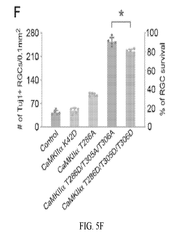
[0032] FIGs. 5A-50 disclose examples of performance of more
CaMKII variants as well
as the RGC-specific promoter mSncg in RGC protections, in accordance with
aspects of the
present disclosure. (A-E) Confocal images of retinal whole-mounts showing
surviving RGCs
labeled by Tujl immunoreactivity at 7 days after NMDA injection in control
(AAV-EBFP), or
AAV-CaMKIIa K42D, AAV-CaMKIIa T2 86A, AAV-CaMKIIa T286D/T305A/T306A, and
CaMKIIa T286D/T305D/T306D treated eyes. Scale bar, 40 .tm. (F) Quantification
of RGC
survival after treatment with CaMKIIa variants at 7 days post NMDA injection,
expressed as
numbers of RGCs (left Y-axis), and percentages of RGCs relative to those in
the uninjured retina
(right Y-axis). Data are presented as mean s.d., n=5 retinas per group. One-
way ANOVA with
Tukey's multiple comparisons test, F:761.4, R2:0.9935, *P=0.0001. (G-H)
Confocal images of
retinal whole-mounts showing surviving RGCs labeled by Tujl immunoreactivity
at 7 days post
NMDA injection in control (AAV-mSncg-EBFP) or AAV-mSncg-CaMKIIa T286D treated
eyes. Scale bar, 40 inn. (I) Quantification of RGC survival at 7 days post
NMDA injection,
expressed as numbers of RGCs (left Y-axis), and percentages of RGCs relative
to those in the
uninjured retina (right Y-axis). Data are presented as mean s.d., n=4
retinas per group.
Unpaired t-test, *P<0.0001. (J-N) Confocal images of retinal whole-mounts
showing surviving
RGCs labeled by Tujl immunoreactivity at 7 days after NMDA injection in
control (AAV-
EBFP), or AAV-CaMKIIP K43D, AAV-CaMKIIP T287A, CaMKIIP T287D/T306A/T307A,
and CaMKIIP T287D/T306D/T307D treated eyes. Scale bar, 40 pm. (0)
Quantification of RGC
survival after treatment with CaMKIIp variants at 7 days post NMDA injection,
expressed as
8
CA 03208818 2023- 8- 17
WO 2022/182983
PCT/US2022/017894
numbers of RGCs (left Y-axis), and percentages of RGCs relative to those in
the uninjured retina
(right Y-axis). Data are presented as mean s.d., n=5 retinas per group. One-
way ANOVA with
Tukey's multiple comparisons test, F:579.0, R2:0.9914, *P=0.0002.
[0033] FIGs. 6A-60 disclose examples of reactivation of CaMMI
providing post-injury
and long-term RGC protection after excitotoxic or axonal injuries, in
accordance with aspects of
the present disclosure. (A-B) Confocal images of retinal whole-mounts showing
surviving RGCs
labeled by Tujl immunoreactivity at 2 weeks after optic nerve crush in control
(AAV-EBFP) or
AAV-CaMK11a T286D post-injury treatment. Scale bar, 40 m. (C) Quantification
of RGC
survival 2 weeks after optic nerve crush, expressed as numbers of RGCs (left Y-
axis), and
percentages of RGCs relative to those in the uninjured retina (right Y-axis).
Data are presented as
mean s.d., n=5 retinas per group. Unpaired t-test, *P<0.0001. (D-G) Confocal
images of retinal
whole-mounts showing surviving RGCs labeled by Tujl immunoreactivity at 2
months and 12
months post NMDA injection in control (AAV-EBFP) and AAV-CaMKIII3 T287D
treated eyes.
Scale bar, 40 m. (H) Quantification of RGC survival 2 months and 12 months
post NMDA
injection, expressed as numbers of RGCs (left Y-axis), and percentages of RGCs
relative to those
in the uninjured retina (right Y-axis). Data are presented as mean s.d., n=4
retinas per group.
One-way ANOVA with Tukey's multiple comparisons test, F:1370, R2:0.9971,
*P<0.0001. (I-N)
Confocal images of retinal whole-mounts showing surviving RGCs labeled by Tujl
immunoreactivity at 1 month, 2 months, and 6 months post optic nerve crush in
control (AAV-
EBFP) or AAV-CaMKII13 T287D treated eyes. Scale bar, 40 pm. (0) Quantification
of RGC
survival 1 month, 2 months and 6 months post optic nerve injury, expressed as
numbers of RGCs
(left Y-axis), and percentages of RGCs relative to those in the uninjured
retina (right Y-axis).
Data are presented as mean s.d., n=4 retinas per group. One-way ANOVA with
Tukey's
multiple comparisons test, F:523.2, R2:0.9932, *P<0.0001.
[0034] FIGs. 7A-7Z disclose examples of CREB acting downstream of
CaMKII to
protect RGCs from excitotoxic and optic nerve injuries, in accordance with
aspects of the present
disclosure. (A-C) Confocal images of retinal whole-mounts showing CREB
phosphorylation in
RGCs, from uninjured eyes (A), and 2 hours after NMDA injection in control
(AAV-EBFP) (B)
or AAV-CaMKIIa T286D (C) treated eyes. Arrowheads, Tuj1+ RGCs maintaining (A)
or losing
(B) CREB activity, which was restored after treatment with CaMKIIa T286D (C).
Scale bar, 20
m. (D-E) Quantification of CREB phosphorylation in RGCs 2 hours after NMDA-
induced
excitotoxic injury. (D) The number of total Tujl+ RGCs and pCREB+/Tuj1+ RGCs
in uninjured
9
CA 03208818 2023- 8- 17
WO 2022/182983
PCT/US2022/017894
or NMDA-damaged eyes. Data are presented as mean s.d., n=6 retinas per
group. (E)
Percentage of pCREB+/Tuj1+ RGCs in uninjured or NMDA damaged eyes. Data are
presented
as mean s.d., n=6 retinas per group. One-way ANOVA with Tukey's multiple
comparisons test,
F:6139, R2:0.9988, *P<0.0001. (F-G) Confocal images of retinal whole-mounts
showing
surviving RGCs labeled by Tujl immunoreactivity at 7 days after NMDA injection
in AAV-
CaMKIIa T286D+ Control (AAV-EBFP), or AAV-CaMKIIa T286D+AAV-A-CREB treated
eyes. Scale bar, 40 m. (H) Quantification of RGC survival, expressed as
numbers of RGCs (left
Y-axis), and percentages of RGCs relative to those in the uninjured retina
(right Y-axis). Data
are presented as mean s.d., n=5 retinas per group. Unpaired t-test,
*P<0.0001. (T-J) Confocal
images of retinal whole-mounts showing surviving RGCs labeled by Tujl
immunoreactivity at 7
days after NMDA injection in control (AAV-EBFP) or AAV-VP16-CREB treated eyes.
Scale
bar, 40 m. (K) Quantification of RGC survival, expressed as numbers of RGCs
(left Y-axis),
and percentages of RGCs relative to those in the uninjured retina (right Y-
axis). Data are
presented as mean s.d., n=5 retinas per group. Unpaired t-test, *P<0.0001.
(L-R) Confocal
images of retinal whole-mounts showing CREB phosphorylation in RGCs, from
uninjured eyes
(L), and 5 days, 7 days and 9 days after optic nerve crush in control (AAV-
EBFP) (M-0) or
AAV-CaMKIIa T286D (P-R) treated eyes. Arrowheads, Tujl+ RGCs losing CREB
activity (M-
O). Scale bar, 20 pm. (S-T) Quantification of CREB phosphorylation in RGCs
after optic nerve
injury. (S) The number of total Tujl+ RGCs and pCREB+/Tuj1+ RGCs in uninjured
and injured
retinas 5 days, 7days. and 9 days after crush. Data are presented as mean
s.d., n=6 retinas per
group. (T) Percentage of pCREB+/Tuj1+ RGCs in uninjured and injured retinas 5
days, 7days,
and 9 days after crush. Data are presented as mean s.d., n=6 retinas per
group. One-way
ANOVA with Tukey's multiple comparisons test, F:89.58, R2:0.9389, *P<0.0001.
(U-V)
Confocal images of retinal whole-mounts showing surviving RGCs labeled by Tujl
immunoreactivity at 2 weeks after optic nerve crush in AAV-CaMKIIa T286D+
Control (AAV-
EBFP), or AAV-CaMK1la T286D+AAV-A-CREB treated eyes. Scale bar, 40 pm. (W)
Quantification of RGC survival, expressed as numbers of RGCs (left Y-axis),
and percentages of
RGCs relative to those in the uninjured retina (right Y-axis). Data are
presented as mean s.d.,
n=5 retinas per group. Unpaired t-test, *P<0.0001. (X-Y) Confocal images of
retinal whole-
mounts showing surviving RGCs labeled by Tujl immunoreactivity at 2 weeks
after optic nerve
crush in control (AAV-EBFP) or AAV-VP16-CREB treated eyes. Scale bar, 40 m.
(Z)
Quantification of RGC survival, expressed as numbers of RGCs (left Y-axis),
and percentages of
CA 03208818 2023- 8- 17
WO 2022/182983
PCT/US2022/017894
RGCs relative to those in the uninjured retina (right Y-axis). Data are
presented as mean s.d.,
n=5 retinas per group. Unpaired t-test, *P<0.0001.
[0035] FIGs. 8A-8V disclose examples of signaling mechanisms
downstream of CaMKII
in RGC protections, in accordance with aspects of the present disclosure. (A-
B) Confocal images
of retinal whole-mounts showing CREB phosphorylation in RGCs 2 hours after
NMDA injection
in AAV-CaMKIIa T286D + control (AAV-EBFP), or AAV-CaMKIIa T286D + AAV-A-CREB
treated eyes. Arrowheads, Tuj1+ RGCs losing CREB activity. Scale bar, 20 um.
(C-D)
Quantification of CREB phosphorylation in RGCs after excitotoxic injury. (C)
The number of
total Tujl+ RGCs and pCREB+/Tuj1+ RGCs 2 hours after NMDA injection. Data are
presented
as mean s.d., n=4 retinas per group. (D) Percentage of pCREB+/Tuj1+ RGCs 2
hours NMDA
injection. Data are presented as mean s.d., n=4 retinas per group. Unpaired
t-test, *P<0.0001.
(E-F) Confocal images of retinal whole-mounts showing CREB phosphorylation in
RGCs 2
hours after NMDA injection in control (AAV-EBFP) or AAV-VP16-CREB treated
eyes.
Arrowheads, Tuj1+ RGCs losing CREB activity. Scale bar, 20 um. (G-H)
Quantification of
CREB phosphorylation in RGCs after excitotoxic injury. (G) The number of total
Tujl+ RGCs
and pCREB+/Tuj1+ RGCs 2 hours after NMDA injection. Data are presented as mean
s.d.,
n=3 retinas per group. (H) Percentage of pCREB+/Tuj1+ RGCs 2 hours NMDA
injection. Data
are presented as mean s.d., n=3 retinas per group. Unpaired t-test,
*P=0.0003. (I) Confocal
images of retinal whole-mounts showing TrkB phosphorylation in RGCs, from
uninjured eyes,
and 2 hours after NMDA injection in control (AAV-EBFP) or AAV-CaMKIIa T286D
treated
eyes. Scale bar, 20 um. (J) Quantification of pTrkB intensity in RGCs. Data
are presented as
mean s.d., n=3 retinas per group. One-way ANOVA with Tukey's multiple
comparisons test,
F:19.26, R2:0.8652. *P=0.0124. (K-L) Confocal images of retinal whole-mounts
showing
CREB phosphorylation in RGCs 5 days after optic nerve crush in AAV-CaMKIIa
T286D +
control (AAV-EBFP), or AAV-CaMKIIa T286D + AAV-A-CREB treated eyes.
Arrowheads,
Tuj1+ RGCs losing CREB activity. Scale bar, 20 um. (M-N) Quantification of
CREB
phosphorylation in RGCs 5 days after nerve injury. (M) The number of total
Tujl+ RGCs and
pCREB+/Tuj1+ RGCs 5 days after optic nerve crush. Data are presented as mean
s.d., n=4
retinas per group. (N) Percentage of pCREB+/Tuj1+ RGCs 5 days after optic
nerve crush. Data
are presented as mean s.d., n=4 retinas per group. Unpaired t-test,
*P<0.0001. (0-P) Confocal
images of retinal whole-mounts showing CREB phosphorylation in RGCs 5 days
after optic
nerve crush in control (AAV-EBFP) or AAV-VP16-CREB treated eyes. Arrowheads,
Tujl+
11
CA 03208818 2023- 8- 17
WO 2022/182983
PCT/US2022/017894
RGCs losing CREB activity. Scale bar, 20 jam. (Q-R) Quantification of CREB
phosphorylation
in RGCs 5 days after nerve injury. (Q) The number of total Tujl+ RGCs and
pCREB+/Tuj1+
RGCs 5 days after optic nerve crush. Data are presented as mean s.d., n=3
retinas per group.
(R) Percentage of pCREB+/Tuj1+ RGCs 5 days after optic nerve crush. Data are
presented as
mean s.d., n=3 retinas per group. Unpaired t-test, *P=0.0002. (S) Confocal
images of retinal
whole-mounts showing DLK staining in RGCs, from uninjured eyes, and 3 days
after optic nerve
crush in control (AAV-EBFP) or AAV-CaMKIIa T286D treated eyes. Scale bar, 20
pm. (T)
Quantification of DLK intensity in RGCs. Data are presented as mean s.d.,
n=3 retinas per
group. One-way ANOVA with Tukey's multiple comparisons test, F:192.3,
R2:0.9846, n.s. (not
significant, P=0.18). (U) Confocal images of retinal whole-mounts showing c-
Jun
phosphorylation in RGCs, from uninjured eyes, and 3 days after optic nerve
crush in control
(AAV-EBFP) or AAV-CaMKIIa T286D treated eyes. Scale bar, 20 lam. (V)
Quantification of p-
c-Jun intensity in RGCs. Data are presented as mean s.d., n=3 retinas per
group. One-way
ANOVA with Tukey's multiple comparisons test, F:87.73, R2:0.9669, n.s. (not
significant.
P=0.09).
[0036] FIGs. 9A-9I disclose examples of CaMKII-mediated
protection of RGCs in
induced and genetic models of glaucoma, in accordance with aspects of the
present disclosure.
(A) Image of magnetic microbeads distributed evenly around the circumference
of the anterior
chamber using magnets after injection. (B) Image of the eye section after H&E
staining shows
microbeads accumulation at the iridocorneal angle. Scale bar, 100 pm. (C)
Quantification of
intraocular pressure (TOP) after injection of PBS (sham) or microbeads. Data
arc presented as
mean s.d., n=6 eyes per group. (D-E) Confocal images of retinal whole-mounts
showing
surviving RGCs labeled by Tujl immunoreactivity at 2 months after induction of
elevated TOP in
Control (AAV-EBFP) or AAV-CaMKIIa T286D treated eyes. Scale bar, 40 p m. (F)
Quantification of RGC survival, expressed as numbers of RGCs (left Y-axis),
and percentages of
RGCs relative to those in the uninjured retina (right Y-axis). Data are
presented as mean s.d.,
n=6 retinas per group. Unpaired t-test, *P<0.0001. (G-H) Confocal images of
retinal whole-
mounts from 2-month-old GLAST-/- mice showing surviving RGCs labeled by Tujl
immunoreactivity in Control (AAV-EBFP) or AAV-CaMKIIa T286D treated eyes.
Scale bar, 40
vim. (I) Quantification of RGC survival in GLAST-/- retinas, expressed as
numbers of RGCs
(left Y-axis), and percentages of RGCs relative to those in the uninjured wild-
type retina (right
Y-axis). Data are presented as mean s.d., n=5 retinas per group. Unpaired t-
test. *P<0.0001.
12
CA 03208818 2023- 8- 17
WO 2022/182983
PCT/US2022/017894
[0037] FIGs. 10A-10P disclose examples of CaMKII-mediated
protection of RGC axons
in induced and genetic models of glaucoma, in accordance with aspects of the
present disclosure.
(A-D) Confocal images of retinal whole-mounts showing pan-CaMKII levels in
RGCs of
uninjured retinas or 2 weeks after microbeads injection in AAV-CaMKIIa T286D
treated retinas.
Scale bar, 20 m. (E) Quantification of pan-CaMKII intensity in RGCs. Data are
presented as
mean s.d., n=3 retinas per group. Unpaired t-test, *P<0.0001. (F-H) Light
microscope images
of semithin sections of optic nerve stained with toluidine blue, from
uninjured eyes, and at 2
months after induction of elevated 10P in Control (AAV-EBFP) or AAV-CaMK11a
T286D
treated eyes. Scale bar, 4 pm. (I) Quantification of axon survival, expressed
as numbers of axons
(left Y-axis), and percentages of axons relative to those in the uninjured
eyes (right Y-axis). Data
are presented as mean s.d., n=4 nerves per group. Unpaired t-test,
*P=0.0056. (J-M) Confocal
images of retinal whole-mounts showing pan-CaMKII levels in RGCs of uninjured
retinas or
AAV-CaMKIIa T286D treated retinas of GLAST-/- mice at 3 weeks after AAV
injection. Scale
bar, 20 ium. (N) Quantification of pan-CaMKII intensity in RGCs. Data are
presented as mean
s.d., n=3 retinas per group. Unpaired t-test, *P=0.0055. (0-P) Images of
sections from 8-month-
old GLAST-/- mice showing optic nerve head morphology (bright light) and
nuclear layers
stained with DAPI (blue) in Control (AAV-EBFP) or AAV-CaMKIIa T286D treated
eyes. Scale
bar, 80 pm.
[0038] FIGs. 11A-11M disclose examples of CaMKII reactivation
protecting RGC axons
and their target projections to the brain, in accordance with aspects of the
present disclosure. (A)
Schematic illustration of anterograde Cholera Toxin Subunit B (CTB) tracing of
the optic nerve,
lateral geniculate nucleus (LGN), and superior colliculus (SC). (B-D) Confocal
images of
anterograde CTB tracing of RGC axons in the optic nerve, from uninjured eyes,
and 7 days after
NMDA injection in control (PBS) or AAV-CaMKIIa T286D treated eyes. Scale bar,
300 pm.
Inserts: whole-mount retinal images showing CTB filling in the retina. (E)
Quantification of
CTB intensity in the optic nerve. Data are presented as mean s.d., n=4
nerves per group. One-
way ANOVA with Tukey's multiple comparisons test, F:281.7, R2:0.9843,
*P<0.0001. (F-H)
Confocal images of anterograde CTB tracing of RGC axons projecting to the
contralateral LGN
from uninjured eyes, and 7 days after NMDA injection in control (PBS) or AAV-
CaMKIIa
T286D treated eyes. Scale bar, 300 pm. (I) Quantification of CTB intensity in
the contralateral
LGN. Data are presented as mean s.d., n=4 brains per group. One-way ANOVA
with Tukey's
multiple comparisons test, F:155.8, R2:0.9719, *P<0.0001. (J-L) Confocal
images of
13
CA 03208818 2023- 8- 17
WO 2022/182983
PCT/US2022/017894
anterograde CTB tracing of RGC axons projecting to the contralateral SC, from
uninjured eyes,
and 7 days after NMDA injection in control (PBS) or AAV-CaMKIIa T286D treated
eyes. Scale
bar, 300 m. (M) Quantification of CTB intensity in the contralateral LGN.
Data are presented
as mean s.d., n=4 brains per group. One-way ANOVA with Tukey's multiple
comparisons test,
F:226.9, R2:0.9805. *P<0.0001.
[0039] FIGs. 12A-12H disclose examples of CaMKII reactivation
protecting RGC axonal
projections to the ipsilateral hemisphere, in accordance with aspects of the
present disclosure.
(A-C) Confocal images of anterograde CTB tracing of RGC axons projecting to
the ipsilateral
LGN, from uninjured eyes, and 7 days after NMDA injection in control (PBS) or
AAV-
CaMKIIa T286D treated eyes. Scale bar, 300 pm. (D) Quantification of CTB
intensity in the
ipsilateral LGN. Data are presented as mean s.d., n=4 brains per group. One-
way ANOVA
with Tukey's multiple comparisons test, F:145.0, R2:0.9699, *P<0.0001. (E-G)
Confocal
images of anterograde CTB tracing of RGC axons projecting to the ipsilateral
SC from uninjured
eyes, and 7 days after NMDA injection in control (PBS) or AAV-CaMKIIa T286D
treated eyes.
Scale bar, 300 pm. (H) Quantification of CTB intensity in the ipsilateral SC.
Data are presented
as mean s.d., n=4 brains per group. One-way ANOVA with Tukey's multiple
comparisons test,
F:162.2, R2:0.9730. *P<0.0001.
[0040] FIGs. 13A-13Q disclose examples of CaMKII reactivation
preserving functional
vision, in accordance with aspects of the present disclosure. (A-C)
Representative responses of
PERG recordings, from uninjured eyes, and 7 days after NMDA injection, in
control (PBS) or
AAV-CaMKIIa T286D treated eyes. (D) Quantification of PERG amplitudes. Data
arc presented
as mean s.d., n=4 mice per group. One-way ANOVA with Tukey's multiple
comparisons test,
F:47.95, R2:0.9142. *P<0.0001. (E-G) Representative responses of PVEP
recordings from
uninjured eyes, and 10 days after NMDA injection, in control (PBS) or AAV-
CaMKTIa T286D
treated eyes. (H) Quantification of PVEP amplitudes. PVEP amplitudes are shown
for each
animal (red) and averaged across the group (blue). Data are presented as mean
s.d., n=4 mice
per group. One-way ANOVA with Tukey's multiple comparisons test, F:40.67,
R2:0.9004,
*P=0.0002. (I) Schematic diagram of the visual water task. (J-L) Visual water
task performance
as a function of spatial frequencies, from uninjured mice, and 4-14 days after
NMDA injection,
in control (PBS) or AAV-CaMKIIa T286D treated (both eyes) mice. For each
column, each row
shows the results from a single mouse. For each animal, a trendline of best
fit was generated, and
the point on the curve that intersected with 70% correct choices was adopted
as the threshold for
14
CA 03208818 2023- 8- 17
WO 2022/182983
PCT/US2022/017894
acuity. (M) Acuity (Spatial frequency thresholds) measured in the visual water
task. Data are
presented as mean s.d., n=4 mice per group. One-way ANOVA with Tukey's
multiple
comparisons test, F:529.8, R2:0.9916, *P<0.0001. (N) Schematic diagram of the
visual cliff test.
(0) Visual cliff performance, from uninjured mice, and 7 days after NMDA
injection in control
(PBS) or AAV-CaMKIIct T286D treated mice. Data show the number (left Y-axis)
and
percentage (right Y-axis) of shallow/deep side choices. Fisher's exact test,
*P=0.0373. (P)
Schematic diagram of the looming response test. (Q) Performance in response to
looming
stimuli, from uninjured mice, and 7 days after NMDA injection in control (PBS)
or AAV-
CaMKIIct T286D treated mice. Data show the number (left Y-axis) and percentage
(right Y-axis)
of responders and non-responders. Fisher's exact test, *P=0.0028.
DETAILED DESCRIPTION
[0041] Reference throughout the specification to -one example", -
another example", -an
example", and so forth, means that a particular element (e.g., feature,
structure, and/or
characteristic) described in connection with the example is included in at
least one example
described herein, and may or may not be present in other examples. In
addition, it is to be
understood that the described elements for any example may be combined in any
suitable manner
in the various examples unless the context clearly dictates otherwise.
[0042] This disclosure relates to a method of decreasing
degeneration of retinal ganglion
cells in a subject, a method of treating vision loss in a subject, and a
pharmaceutical composition.
In an example, the pharmaceutical composition includes one or more components
applicable for
use in the methods disclosed herein.
[0043] CaMK and CREB signaling are disclosed herein to be
severely compromised after
excitotoxic injury to RGC somas or optic nerve injury to RGC axons, and
increasing activity of
these pathways are disclosed herein to robustly protect RGCs from injury. CaMK
is disclosed
herein to protect RGCs in induced and genetic models of glaucoma, a leading
cause of blindness
characterized by loss of RGC somas and axons. Also disclosed herein is that
increasing activity
of CaMK protects long distance RGC axon projections and restores visual
function in the entire
visual pathway from the retina to primary visual cortex in the brain. Also
disclosed herein is that
increasing activity of CREB protects RGCs.
[0044] The present disclosure of a protective effect increasing
CaMK activity has on
RGC and vision is particularly surprising, in view of prior evidence that
inhibiting CaMK blunts
CA 03208818 2023- 8- 17
WO 2022/182983
PCT/US2022/017894
cytotoxicity. Calcium is a highly versatile intracellular signal responsible
for regulating an array
of cellular processes (Berridge et al., 2000). Loss of Ca2+ homeostasis. often
in the form of
cytoplasmic increases, leads to cell injuries (Dong et al., 2006). Aberrant
Ca2+ activation is
known to be involved in RGC death following insults such as excitotoxicity and
optic nerve
injury (Hartwick et al., 2008; Prilloff et al., 2007). CaMKII is a central
coordinator and executor
of Ca2+ signal transduction (Hudmon and Schulman, 2002a). Accordingly,
previous studies
have shown that CaMKII inhibition protects RGCs from excitotoxic cell death,
suggesting that
increased CaMK activity promotes RGC degeneration. On the contrary, as
surprisingly disclosed
herein, promoting CaMK activity is protective for RGC and vision.
[0045] As disclosed herein, excitotoxic insults to RGC somas or
optic nerve injury to
RGC axons led to inactivation of CaMK and its downstream target CREB in RGCs.
Increasing
activity of CaMK or CREB protected RGCs from both injuries. Furthermore, CaMK-
mediated
RGC protection slowed down the disease progression in induced and genetic
animal models of
glaucoma. Increasing CaMK activity not only saves RGC somas, but also protects
long distance
RGC axon projections from the retina to visual relay centers in the brain.
Increasing CaMK
mediated protection of RGCs also restores functional vision in the entire
visual pathway,
evidenced by improved visual responses in the retina and the primary visual
cortex in the brain
as well as visually-guided behavior. Also disclosed is targeting increased
activity of CaMK or
CREB as methods of decreasing degeneration of RGCs and of treating vision
loss, and
pharmaceutical compositions including compositions for increasing CaMK
activity or CREB
activity, including in RGC.
[00461 lsoforms of CaMK include CamK1, CaMK11, and CaMK1V. As
disclosed herein,
increasing activity of any of these CaMKs prevents RGC degeneration. For
example, increasing
activity of CaMKI, CaMKII, or CaMKIV prevents RGC degeneration. CaMKII
includes several
isofonns, including CaMKIIa, CaMKII, CaMKII7, and CaMKII6. Increasing activity
of
CaMK1la or CaMKI1P prevents RGC degeneration. Increasing CaMKII activity also
improves
vision following insults known to impair RGC compared to subjects exposed to
such insults with
exposure to a treatment to increase CaMK activity. Given the known shared
cellular
functionalities among CaMKs, including CaMKI, CaMKII (including without
limitation
CaMKIIa, CaMKIIP, CaMKII7, and CaMKII), and CaMKIV, and ability of various
CaMKs to
prevent RGC degeneration and to treat vision loss as disclosed herein, a
skilled person would
apprehend that increasing activity of any one or more of CaMKI, CaMKII
(including without
16
CA 03208818 2023- 8- 17
WO 2022/182983
PCT/US2022/017894
limitation CaMKIIa, CaMKII13. CaMKIIy, and CaMKII6), and CaMKIV may prevent
RGC
degeneration, may prevent RGC somata loss, may prevent loss of RGC axon
projections in the
brain, may prevent RGC axonal loss, may prevent vision loss, may treat vision
loss, and any one
or more of the foregoing.
[0047] In some examples, increasing activity of a CaMK may
include increasing activity
of a variant of a CaMK with an amino acid sequence that differs from a CaMK
expressed by the
subject, or expressed by the subject in a cell or cells in which activity of
the CaMK is increased
in accordance with the methods disclosed herein. For example, increasing
activity of a CaMK
may include increasing activity of a CaMK that differs from a CaMK as
disclosed in the present
disclosure, or from a CaMK encoded by the subject's genome, or from a CaMK
that would
otherwise be expressed in the subject's cell or cells in which CaMK activity
is increased in
accordance with the methods disclosed herein, by about 1% or more, or by about
2% or more, or
by about 3% or more, or by about 4% or more, or by about 5% or more, or by
about 6% or more,
or by about 7% or more, or by about 8% or more, or by about 9% or more, or by
about 10% or
more, or by about 11% or more, or by about 12% or more, or by about 13% or
more, or by about
14% or more, or by about 15% or more, or by about 16% or more, or by about 17%
or more, or
by about 18% or more, or by about 19% or more, or by about 20% or more, or by
about 21% or
more, or by about 22% or more, or by about 23% or more, or by about 24% or
more, or by about
25% or more, or by about 30% or more, or by about 35% or more.
[0048] In other examples, increasing activity of a CaMK may
include increasing activity
of a CaMK that differs from a CaMK as disclosed in the present disclosure, or
from a CaMK
encoded by the subject's 2enome, or from a CaMK that would otherwise be
expressed in the
subject's cell or cells in which CaMK activity is increased in accordance with
the methods
disclosed herein by including one or more amino acid substitution, insertion,
or deletion of about
1 or more, about 2 or more, about 3 or more, about 4 or more, about 5 or more,
about 10 or more,
about 15 or more, about 20 or more, about 25 or more, about 30 or more, about
25 or more,
about 40 or more, or about 50 or more amino acids relative to a foregoing
CaMK, alone or in
combination.
[0049] In other examples, increasing activity of a CaMK may
include increasing activity
of a CaMK that is constitutively active. By constitutively active is meant a
CaMK whose
activity, or increased activity, or sustained activity, is not dependent on or
diminished by one or
more other cell signaling events otherwise or generally required to increase
or capable of
17
CA 03208818 2023- 8- 17
WO 2022/182983
PCT/US2022/017894
decreasing activity of a CaMK in the subject or in the subject's cell or cells
in which CaMK
activity is increased in accordance with the methods disclosed herein. As a
nonlimiting example,
activation of CaMKII may generally be initiated by Ca2+ influx and subsequent
Ca2+/Calmodulin binding; the resultant conformation change of CaMMT allows its
autophosphorylation at either Threonine 286 (T286) for CaMKIIa or Threonine
287 (T287) for
CaMKIIP, which may enhance activity of such isoform of both isoforms, whereby
if
autophosphorylation occurs, CaMK may remain active after Ca2+ concentration
falls. In another
example, a constitutively active CaMK may include a truncated, N-terminal
catalytic domain of
CaMKIIa, or a truncated, N-terminal catalytic domain of CaMKIIp, which
truncations are
constitutively active.
[0050] In an example, increasing activity of CaMK may include
increasing activity of a
constitutively active variant of CaMKIIa or of CaMKIIP, or of another CaMK,
such as any of the
foregoing variants, without limitation. As a non-limiting example, increasing
activity of a CaMK
in accordance with the present disclosure may include increasing levels,
expression, or activity
of a T286D substituted CaNIKIIa, which, without being limited to any
particular mechanism of
action, may simulate an active, phosphorylated state of CaMK. In another non-
limiting example,
increasing levels, expression, or activity of a CaMK in accordance with the
present disclosure
may include increasing activity of a T287D substituted CaMKIIP, which, without
being limited
to any particular mechanism of action, may simulate an active, phosphorylated
state of CaMK. In
another example, increasing activity of a CaMK may include increasing levels,
expression, or
activity of an N-terminal catalytic domain of CaMKIIa, or an N-terminal
catalytic domain of
CaMKIIP, which are known to be constitutively active.
[0051] In some examples, increasing activity of CREB may include
increasing activity of
a variant of CREB with an amino acid sequence that differs from a CREB
expressed by the
subject, or expressed by the subject in a cell or cells in which activity of
the CREB is increased
in accordance with the methods disclosed herein. For example, increasing
activity of a CREB
may include increasing activity of a CREB that differs from a CREB as
disclosed in the present
disclosure, or from a CREB encoded by the subject's genome, or from a CREB
that would
otherwise be expressed in the subject's cell or cells in which CREB activity
is increased in
accordance with the methods disclosed herein, by about 1% or more, or by about
2% or more, or
by about 3% or more, or by about 4% or more, or by about 5% or more, or by
about 6% or more,
or by about 7% or more, or by about 8% or more, or by about 9% or more, or by
about 10% or
18
CA 03208818 2023- 8- 17
WO 2022/182983
PCT/US2022/017894
more, or by about 11% or more, or by about 12% or more, or by about 13% or
more, or by about
14% or more, or by about 15% or more, or by about 16% or more, or by about 17%
or more, or
by about 18% or more, or by about 19% or more, or by about 20% or more, or by
about 21% or
more, or by about 22% or more, or by about 23% or more, or by about 24% or
more, or by about
25% or more, or by about 30% or more, or by about 35% or more.
[0052] In other examples, increasing activity of a CREB may
include increasing activity
of a CREB that differs from a CREB as disclosed in the present disclosure, or
from a CREB
encoded by the subject's 2enome, or from a CREB that would otherwise be
expressed in the
subject's cell or cells in which CREB activity is increased in accordance with
the methods
disclosed herein by including one or more amino acid substitution, insertion,
or deletion of about
1 or more, about 2 or more, about 3 or more, about 4 or more, about 5 or more,
about 10 or more,
about 15 or more, about 20 or more, about 25 or more, about 30 or more, about
25 or more,
about 40 or more, or about 50 or more amino acids relative to a foregoing
CREB, alone or in
combination.
[0053] In other examples, increasing activity of a CREB may
include increasing activity
of a CREB that is constitutively active. By constitutively active is meant a
CREB whose activity,
or increased activity, or sustained activity, is not dependent on or
diminished by one or more
other cell signaling events otherwise or generally required to increase or
capable of decreasing
activity of a CREB in the subject or in the subject's cell or cells in which
CREB activity is
increased in accordance with the methods disclosed herein. As a nonlimiting
example, increasing
activity of CREB may include increasing expression of a CREB variant known as
VP16-CREB,
a fusion between the activation domain of herpes simplex virus VP16 protein
and the DNA
binding domain of CREB, as disclosed in U.S. Patent No. 9,587.000,
incorporated herein in its
entirety by reference.
[0054] CREB activity may be increased by increasing an amount of
CREB in a subject,
such as in a subject's retina, including in a subject's RGCs. CREB expression
may be increased
by transfecting cells such as RGC with a CREB or with a polynucleotide
sequence encoding a
CREB so as to cause expression of a CREB from the polynucleotide in the
subject or cell or cells
thereof. In some examples including transfecting a cell with a polynucleotide
encoding a CREB,
the polynucleotide may further include a cis-regulatory element operatively
associated with the
portion of the polynucleotide encoding the CREB so as to stimulate, promote,
or enhance
19
CA 03208818 2023- 8- 17
WO 2022/182983
PCT/US2022/017894
expression of CREB from the polynucleotide. Such a cis-regulatory element may
include one or
more of a promoter sequence and an enhancer sequence.
[0055] A cis-regulatory element may include a promotor, an
enhancer, or both. In some
cases, a sequence for a cis-regulatory element may be located within fewer
than 10 nucleotides
from a transcription start site, fewer than 20 nucleotides from a
transcription start site, fewer than
30 nucleotides from a transcription start site, fewer than 40 nucleotides from
a transcription start
site, fewer than 50 nucleotides from a transcription start site, fewer than 60
nucleotides from a
transcription start site, fewer than 70 nucleotides from a transcription start
site, fewer than 80
nucleotides from a transcription start site, fewer than 90 nucleotides from a
transcription start
site, fewer than 100 nucleotides from a transcription start site, fewer than
125 nucleotides from a
transcription start site, fewer than 150 nucleotides from a transcription
start site, fewer than 175
nucleotides from a transcription start site, fewer than 200 nucleotides from a
transcription start
site, fewer than 225 nucleotides from a transcription start site, fewer than
250 nucleotides from a
transcription start site, fewer than 275 nucleotides from a transcription
start site, fewer than 300
nucleotides from a transcription start site, fewer than 325 nucleotides from a
transcription start
site, fewer than 35 nucleotides from a transcription start site, fewer than
375 nucleotides from a
transcription start site, fewer than 400 nucleotides from a transcription
start site, fewer than 425
nucleotides from a transcription start site, fewer than 450 nucleotides from a
transcription start
site, fewer than 475 nucleotides from a transcription start site, fewer than
500 nucleotides from a
transcription start site, or between 500 and 1,000 nucleotides from a
transcription start site
[0056] A promoter is a nucleotide sequence to which RNA
polymerizing enzymes bind
for initiation of transcription of a downstream gene sequence. Many genes that
show tissue- or
cell-type specific expression including a promotor upstream of the DNA
sequence that codes for
the RNA that is particularly active in cells where the gene is expressed. A
promoter may be more
active in some cells than other, such as being active only in specific ell- or
tissue-types, or highly
active in certain cell- or tissue-types relative to others. Promoters include
a sequence where
transcription is initiated. Eukaryotic promoters may and typically do include
features such as a
TATA box, a transcription factor JIB recognition site, and a core promotor
sequence (or an
initiator). Transcription factors bind and RNA polymerase bind to a promoter
for transcription
initiation.
[0057] Also included in a cis-regulatory element may be one or
more enhancer sequence.
An enhancer is part of a cis-regulatory element that enhances transcription
initiated in or by the
CA 03208818 2023- 8- 17
WO 2022/182983
PCT/US2022/017894
promotor. An enhancer may serve to promote an initiation of transcription at a
promoter, for
example, such as through binding of additional transcription factors to the
enhancer that facilitate
or enhance recruitment of other factors and transcriptional machinery to the
promotor. As with
promotors, many genes have enhances that are involved in cell- or tissue-
specific or cell- or
tissue-enhanced expression. In some examples, a cis-regulatory element may
include other
features such as intronic sequences, splice sites, exonic sequences, or any
combinations thereof,
that may influence transcript expression in a given cellular environment. In
an example, a cell-
type specific cis-regulatory element may include features that repress
expression in cell types
other than those in which the cell type-specific cis-regulatory element is
intended to drive
expression.
[0058] In an example, a cis-regulatory element may include a
promiscuous cis-regulatory
element. A promiscuous cis-regulatory element may include one or more
polynucleotide
sequence that may, or be designed to, drive expression without, or with
minimal, regard to cell
type transfected by the polynucleotide. A promiscuous cis-regulatory element
may promote
expression of a polynucleotide encoding a CaMK or CREB in different cell
types, including cells
of different tissues, lineages, ages, etc. Examples of promiscuous cis-
regulatory elements include
a CMV early enhancer/chicken 1 actin (CAG) promoter cis-regulatory element, a
human 13-actin
promoter cis-regulatory element, a human elongation factor-la promoter cis-
regulatory element,
a cytomegalovirus (CMV) promoter cis-regulatory element, a simian virus 40
promoter cis-
regulatory element, and herpes simplex virus thymidine kinase In another
example, a cis-
regulatory element may include a cell-specific cis-regulatory element.
[0059] A cell-specific cis-regulatory element may include one or
more polynucleotide
sequence that may, or be designed to, drive expression only, or mostly, or
preferentially, or
predominantly, in a predetermined cell type or types. A cell-specific cis-
regulatory element may
include one or more polynucleotide sequence that may, or be designed to, drive
expression only,
or mostly, or preferentially, or predominantly, in a predetermined cell type
or types without, or
with minimal, or negligible, or insubstantial, expression in other cell types
that may be
transfected with the polynucleotide, or not so as to increase CaMK or CREB
activity in such
sells or to do so only to a minimal, or negligible, or insubstantial degree
relative to activity
induced in the cell type or types in which the cis-regulatory element is
designed to drive
expression.
21
CA 03208818 2023- 8- 17
WO 2022/182983
PCT/US2022/017894
[0060] A cell-specific cis-regulatory element, in accordance with
the present disclosure,
may increase expression in a transfected cell type in which the cis-regulatory
element is intended
or designed to drive expression by about 0%, about 5% or less, about 10% or
less, 15% or less,
about 20% or less, about 25% or less, about 30% or less, about 35% or less,
about 40% or less,
about 45% or less, about 50% or less, about 55% or less, about 60% or less,
about 65% or less,
about 70% or less, about 75% or less, about 80% or less, about 85% or less,
about 90% or less,
about 95% or less, about 100% or less, about 150% or less, about 200% or less,
about 250% or
less, about 300% or less, about 350% or less, about 400% or less, about 450%
or less, about
500% or less, about 550% or less, about 600% or less, about 650% or less,
about 700% or less,
about 750% or less. about 800% or less, about 850% or less, about 900% or
less, about 1,000%
or less, or more, compared to a level of expression in a transfected cell type
or types other than
that in which the cell-specific cis-regulatory element is intended or designed
to drive expression.
In another example, a cell-specific cis-regulatory element, in accordance with
the present
disclosure, may cause expression in a transfected cell type in which the cis-
regulatory element is
intended or designed to drive expression but no, minimal, negligible, or
undetectable levels of
expression in a transfected cell type or types in which the cell-specific cis-
regulatory element is
not designed or intended to drive expression.
[0061] In an example, the cis-regulatory element may be a cis-
regulatory element that
drives expression of a transcript in RGC. For example, the cis-regulatory
element may be a
promoter that drives expression of a transcript in RGC, referred to herein as
an RGC promoter.
For example, the cis-regulatory element may be a promoter, enhancer, or both,
of a transcript
known to be expressed in RGC. The cis-regulatory element may be a promoter,
enhancer, or both
of a transcript known to be expressed in RGC to a higher degree than the
transcript is expressed
in other cells of the retina or other cells of the eye. In an example, an RGC
promoter may be a
promoter of a transcript whose expression is higher in RGCs relative to other
cells of tissues of
the eye, or relative to other cells of the retina. In an example, an RGC
promoter may drive a level
of expression of a transcript in RGC that is sufficiently higher that a level
of expression of the
transcript in other cells of tissues of the eye, or of other cells of the
retina, sufficient to permit
identification of a cell as an RGC on the basis of a the differentiable level
of expression of the
transcript in the RGC compared to other cells.
[0062] In an example, an RGC promoter may not drive expression of
a transcript in cells
of other tissue of the eye or other retinal cells, other than RGC. For
example, a transcript may be
22
CA 03208818 2023- 8- 17
WO 2022/182983
PCT/US2022/017894
detectable in an RGC (such as by in situ hybridization detection of mRNA of
the transcript) but,
in an example, not be detectable in cells of other tissues of the eye or, in
another example, not be
detectable in other cell types of the retina. In another example, an RGC
promoter may drive
expression of a transcript in an RGC that is at least about 2, or at least
about 3, or at least about
4, or at least about 5, or at least about 6, or at least about 7, or at least
about 8, or at least about 9,
or at least about 10, or at least about 11, or at least about 12, or at least
about 13, or at least about
14, or at least about 15, or at least about 16, or at least about 17, or at
least about 18, or at least
about 19, or at least about 20, or at least about 25, or at least about 50, or
at least about 75, or at
least about 100, or at least about 150, or at least about 200, or at least
about 250, or at least about
300, or at least about 350, or at least about 400, or at least about 450, or
at least about 500, or at
least about 600, or at least about 700, or at least about 800, or at least
about 900, or at least about
1,000, or at least about 5,000, or at least about 10,000, or at least about
25,000, or at least about
50,000, or at least about 75,000, or at least about 100,000 times a level of
expression of the
transcript in, n an example, a cell of another tissue of the eye or, in
another example, another cell
type of the retina. A level of expression may be determined by measurement of
transgene
expression by western blot analysis in purified RGC compared to other cell
type, such as in other
purified cell type of cell from eye tissue.
[0063] In an example, as disclosed herein, an RGC promoter may be
a gamma-Synuclein
promoter, such as a human gamma-Synuclein promoter, a mouse gamma-Synuclein
promoter, or
another gamma-Synuclein promoter that drives expression of a transcript in RGC
of a subject. In
another example, an RGC promoter may be a Synapsin 1 promoter, such as a human
Synapsin 1
promoter, a mouse Synapsin 1 promoter, or another Synapsin 1 promoter that
drives expression
of a transcript in RGC of a subject. In another example, an RGC promoter may
be a Thy-1 cell
surface antigen promoter, such as a human Thy-1 cell surface antigen promoter,
a mouse Thy-1
cell surface antigen promoter, or another Thy-1 cell surface antigen promoter
that drives
expression of a transcript in RGC of a subject. In another example, an RGC
promoter may be a
Neurofilament Heavy promoter, such as a human Neurofilament Heavy promoter, a
mouse
Neurofilament Heavy promoter, or another Neurofilament Heavy promoter that
drives
expression of a transcript in RGC of a subject. A Neurofilament Heavy promoter
may be a long-
form Neurofilament Heavy promoter or a short-form Neurofilament Heavy
promoter. A subject
in these examples may be a mammal, or a human, or a mouse, or a rat, or a dog,
or a cat, or a
horse, or a cow, or a sheep, or a pig. Examples of nucleotide sequences of the
foregoing
23
CA 03208818 2023- 8- 17
WO 2022/182983
PCT/US2022/017894
promoters, any of which is explicitly included as a possible example for all
examples disclosed
herein, are given in Table 1.
Table 1: examples of RGC promoters
SEQ ID NO: Promoter Sequence
1 MOUSE GGTCCCATGCCACTAGTGGGAGCTGTGTTACCTGTTGCA
Gamma GCCCCACCCAAAGCCCCTGCTATAGGTCAAGCAGGAATC
synuclein ACCCTGCC ATCCCC A GCCTGGGGCCTGGA GT ACC A GATC
CAGGAAACTAGCATCCCTTAGCTATAGAGATAGCCACAC
ATCAGCCCATTCCTCAGATGTGTATCTGGGGCTCAGACA
TCATCTCCCGATCTCCGACAAGGGCAGGATTTCCTTACC
GTCTGATGGGGTCTCTGCTGGTATCCTCAGCCCCTAGTCT
CCAGCCTTCAGGCATGCCGGGCCTATTGAGATGGGAGAA
CTTGGTACCGGGGTCCTGTGCCC A GGACCCTA GC A GTCC
CCAGCTCAGGTACACCCCAAAGCCCAGCAGCAGTGTCG
GGATCATGGTGAGGGGCTCCTGTGCTGATGCTCAGCCTT
ACAAGTGACTCTCAAATTTGCTGGTGATGTGGTCTTCAA
GCGAAATGTCAGAAAGAAAAGAAAACACGAGGACAAC
AAAGGGAGGAAGTGGCCTGGTCCGGCCCACCCGGCAAG
TCTCATCCGCCCCCGCCCCCGCCCCTTCCAGCCTGGCCCC
CTTGGAGGCCTCCAACCACTCAGGTCAATTCCTGTGTCC
TGAGGGCACTTGAATCAGGGACACGGGATTTGGTAGAC
ACATAAAGGTGGCCCCATTAAACTTATTTCTCCAGGACT
CTGTCGTGGGCCTGGAGGAGATCTGGTGCCACCCATACT
GTTGGCCAGGAAGTGGGGAACGGGCACATCACACCTGC
TCGGCACCTTGGGCTATGGGAACTAGCAGGTGGGTGGG
AACTCAGAGAAGGAAAGGGACTATGCTAGAATCACACA
GCGGGCAGCCCAGTCTAGGGCATGGGGAGCAGCTTTGG
GTGTTTCTGGCCTC A GCCTTCC A AC A GGTTTGGCTA GA G
CTCCAGGCTCAAGAGCATCCAGGATACAGTGGGGAACT
GGATAACAGGGCAGCCTGCAGGTTGGCCATTCATTGGAT
TGGCCCTGACCCCGGCCCAGCCTGGGACACTGAGGCATC
ATCAGTCAAGGCACTTTTCTTCTGCATATAAGAGCCAGG
GCACGAGACCACCAGGGCTTTCCAAGGATGAATGAGGT
GTAATGATAGATTAGGATATGTCCAGCCTCCAACACGCT
CTCCCTCCCCCAGGGCCAACAAGAGTCAGCAGGGCAGA
ATAGAGCCAGTAGGGGCCCGGGCCCTGCTCGCTGGTATC
CCCGTGAGGCATGCCTTCTCTCTGGCCCGCCCTCCCTGCC
CCCACCCTGGCCCGGGCTGGCTGGGCTCCAGCCAGCAGC
CACAGCATCAATATTTCATCTGCGTCAATAAGAGGCAGT
AGCAGCAGAGACAGCGGCTGCGGCAGCACTCCAGTCCA
TAGCTTGCAGCAGCCAGGTTCCATCCTTGCAAACACCAT
GGACGTCTTCAAG
2 MOUSE TGAGCCTTCTTATTCAGACCACCAAAATTACTTTATTTTT
Synapsin 1 CCACATGAAAGTATTATGTGGCTTCCTGTCTGCAAAGAG
GAAGACATCCATGAACACTAATGACACTGGGTTTGGGCT
ATGTCCGGAGCAGAGGAATGAGGCCATGTAGACTAAAT
24
CA 03208818 2023- 8- 17
WO 2022/182983
PCT/US2022/017894
ATGTGCATGTGGAGGAGGCTGAAAACACATCAGAGCTA
GCGCTGCAGGAAATGCTTCTGCATTGCATACCCAGAGTT
TCCTTGCTCATCTGGGAGTCTGTGTTTTTCCTAGATGTGT
GCACTTGTGTGAGATTCTCTGGGTGTGAGTCAAAGTGTT
ATCTGAATGTGTAATGTGTGCTCAATATGCTCATGTGTGT
TACCCTGAGCTTCTGTGTCTACATATATACCTGGATGCCT
GTGTGTTCTGTGATGTACATATATATTCTGTCTTTCCTTC
CTTTTCTATTTGTGTTATTCCATGTGTTCTTTCAGATTCTC
ACCACCAAGGGCAAGGATATGTTAACTACCCAAGTGTCC
ACCTCCGCCTGTCTGGTGATGTTTACGCC ACCCCCGTGCT
CTTTTCTTTGCCCGACAGAGTTGTTATAGGAGATGTCTCC
CCGGGAACACTGCAGGAAGGAGAATTTCTACATTTATGT
TCCCCTCTGAGTGTGCTTCTATCCCCAAAATGCCTTCAAA
GGTGAAAATCAACACTGGAAACCCAAGTATCTGGGAAG
GGCAAGAGTGTGTAAGTGCAAGTTAGCCTAAGGAATAG
GAAGAGGTTGGTAAACAGGGTAGGATCGTGGGAGGGAG
TTTCGTTACTACAGGTCCGGACCCTCAGGACAAGAACCC
CACCCCCACTCCCCA A ATTGCGCATCCCCCGCCCCCATC
AGAGGGGGAGGGGAAGAGGTTGCGGCGCGGCGCATGCG
CACTGTCGGATTCAGCACCGCGGTCAGAGCCTTCGCCTC
CGCTGCCGGCGCGCACCACCACCTCCCCAGCACCAAAGG
CTGACTGACGTCACTCACTAGCCCTCCCCAAACTCCCCTT
CCTCGCCGCCTTGGTCGCGTCCATGCTGCCGTGAGTCCA
GTCGGACCGCACCACGAGAGGTGCAAGATAGGGAGGTG
CGGGCGCGACCATACGCTCTGCGG
3 HUMAN ACTCAGTCCTTTTTGTGCTGTCTCCTCCTCTTTCCCAGAG
Thy-1 cell TTCCTCTCTCTCTTCTCCCACTAGGCAGGGATGAGCAAG
surface AGGAATGGCTCACCCTTGAGAGCTGGGGTCCATAGCCCA
antigen GGTCAGTTCTCCAGCTCTCCCACTTACCAGCCAAGACAG
GAGGTGAGGATTGAGATGGGATGAACCCAGCAGGCGGC
CATGGGTTAAAGGTCGCCATGAATGTAATGTGCCCAGCA
CAGTGCCTGCTAAAAGGCAACACTCCCTTCCTGGTCTGA
AGACCAAACAAGCAGACTGTACTCAGGAAAGCCAGAAG
AACCTTCCAGCTGTCTGGACCAGAAGGTGCCAGCCCAGG
GGCTGAAGAAGACGTAATGCCCAGAGCAAAAAGCGCCT
GCAGCCCCCTGAAGGGCTGGGTGCTCTGGAATAGATGA
GGGGGCGAAATGGGGCTGGGGACCAGGGACGGACAGG
GTGGGTCCAGCACCTGCCTCGCTTCCGAAGGGCTGCTCC
AACACTGAAAAACACCCAACCAGCTTCCTTTCAGAAAGA
CTGGAATATTCCAAAACTTCTCACTGGAGGCTCCGGAGG
AGGTGGGCTCCAGCTGAAAAGOAAATGTGGAGGCM GG
GCGCTCCCGGCCTGCATCCTGCACCTCTTACACTTTGGTT
TTCCCACAGACTCCTGAAGAATAGGTCAGAAGAAAGGG
TTAAAGCCTTAAAAGGGGAACAACCATTGCGGGGCTCA
GGGAGGAGGATAATGTTCTTTGGGCTGCCGCACCCTGAT
CCCCGGGGTCCCGAACCCTCCCGTCCCTGGCCAGGCCTG
CCAGCCACAGGGTGAGGGCCCCCTTCCGCCGCAACCTGC
CACTCTCACACCAATGCGGGACCGCCTTCTCTTCCTTCCC
CA 03208818 2023- 8- 17
WO 2022/182983
PCT/US2022/017894
CACCCCCCACCCCACCCTGCCGTCCTTTCTCCCCCAATCT
CCGCCTCTGATTGGCTGAGCCCCCGGCTCCCCGCTCCCC
CTCTCCTCCATCCCCGGTGAAAACTGCGGGCTCCGAGCT
GGGTGCAGCAACCGGAGGCGGCGGCGCGTCTGGAGGAG
GCTGCAGCAGCGGAAGACCCCAGTCCAGGTGGGAACTG
GAGCCGGTGGGACCTGGGGCTCGGGGACCGCCGTCAGG
CGCCCATGCAAGACTTCCCAACACTAGGCTTCGGGCCAC
GGTCCGAGGGCGCCCAGGGAAGAAGGGCGCAGAGCTTA
GGGAGGGGCCTGCTTTCCAGGCAGGGGCGGGAGGGGGA
TGCTTCTGCAGGGCAGGGGCCGCGTGGCACCCTGATGTC
TTTCGGGGAAGGCGCTCCCGGGCTTTCGCCCGCTGGGGG
ACTGGTGTCTGGGGCTGGGGCGCTGGAGAACAGGGAGG
AAGGGCACCAAGGACAGCCTGTGGGTCTACATTCCACCC
AGACGTCCCCAAACCCAGCTCGCAGAGGCGGGGAGGAG
GACGGATGAAACTGCGGGGAGAGGATGGAGGATGGCGA
GCTAGAGGGAATCTGCCGGGTGACCTCGCGGCGGGCTG
GGTGCGGGGCACCGGAGGAGAAGGAAGCCGCAGTGCCG
CAGGCGGGGACTGGGTGGAAGGCGGGCGGACGGGGGA
GGGGAGAGCTGGAAAAGGATGAGAGAGGGGGAAGGGG
GACTCATTTGGGAAAGGAGAGGATTGGAATACGGAAAT
GGATTAAGGATGAGGCCCGCCGGGGGCTTGAGAGGGAG
GAAGAGCAGACCTTCTCTGGGTCTGGAGCCGCCTGAGGA
CACAGACCAGAGGAAATGAATACAGACTGCACCTCCCC
AGCCGCTCTCCACCCCTCCCCTGGCTCTTCTACCCTCTCC
AGCCCCAGACCCATTTCTTCCCTTTCTTGCTCTGGCCATT
GCTCCCCCTTCCCCTCCTAGATCCCAAGCCCGCACA ACA
TCTCAAACAAGAGTCCTCGATTCAAAAGCCAGATGCCGA
CCCCCCTTCCTCCTGGATCTGGCTCAGGGCAGCAGCTCC
ACCCCGGGACAGAGAGAGCATTGATTGTAGCTGCAGCC
GCCGCGGGATCCTAGCCTCACCCGTCAAGGGGCTGAGCG
CCAGGGACCCTGAACTCGTCTAGTGGTGCGCCCTGCGCA
CCCGGGCGCACTCAACCGAGGCAATGCCCTGCGCGCTCT
CGCGGGTGCACGCCCCTTCTGTGGCCTCTCCTGGGCGAG
CACTGCTCTGCAGATAGGCTAGACTACCGGCTCCGCGTC
GCCTCGCCAAGGGTTGGTTCAGCCAAGGCTGCAAAAAA
CAAAAAAAGACCAGGCAGACAGCCTATCCAGGGTGGCT
ATTGAAACTGGGCTGGAAAACTGCAGTCCCAGGAACTCC
AGAGAGCTGGACATTGGGAAGCATCCTTGGCTCACATAC
AATCGGAGATCACTATGTCTTTCTCTCCTCCAGGAACAC
GATTAGCTTGTGTCCTATCCAGATAGGAATAGATGCTCC
CTATCTGGGAGCATCCTTAGCTATGGTGAATGGTATCTA
GCCATCCACTGGGGATGGCGAGTGACTTAGGGATTTGTG
TCTCACGTATATGAAGCAGTCATCGCCAGATGTTGGTTG
TTTTTCTTAACCCCCATCATAACCCGGTGGGTATGTAAG
ATTCAGAGAGATTCATTCATTCATTCACAATAAATATCTT
TGGAGTGTATGCTATATGCCAGTAATCTGCAAACGGAAA
CGGTTTTGAGCATTGGGGATTTTCTTCTGA ACAGGA A AT
GGGAAGTCCCTAAATGGGGAGTCTTTGTTTAACAGATAC
26
CA 03208818 2023- 8- 17
WO 2022/182983
PCT/US2022/017894
AGAGTTTTACTTTGAAAGACAAAAAGAGTTCCGGAGATG
GGCTGCATAGCAACGTGAATG
4 HUMAN CATAGGCAGCTTCAATCTGATGGCTGTGGCCCCTTGGCC
Neurofila- TCAACAGAATACATCTTGGAGCCCCCTTTTTACCCCAAA
ment CCCCCATTCCTCCTTGCTGTCAGCTGCTTGTGAGCCTTCT
Heavy CACATCCAGAGAATGTATCAGCATTGTGCAGACTGAAAA
(LONG GACCCAGAGGAACAAGGCTCCAATGGCAAAATTCCAAG
FORM) TAGAATGACAAATAAATGGGGAGCCATCTGAGAGCAAG
GGAGTCCTGCCCAACACCCGCCCCATGCCTTTCTCAGGG
ACCTCAGACCAGCCACTCACCTCCATCCTCCCAGCACCA
CCTGCAACCAGCCCCTTGCCCTCTGCAAACTGGAGCACG
ACTGGATCTTTAGATGGGGGAAAAATGCTTCATCATGTT
CTGCTGCTTCATGCAAAACCAGAAACTCCCTCCCCCTCTT
CCCTCCTCCCAGCGCACTCTCCTTCCAGTAAAAAGTGGT
TA A AGGGACAGCGCCATCA ATTTCCCAGCTCTGAGGGTC
TGCTTAGAACTAGGGGGCTGGAAGGAGACAGAGGGCAA
AGAGAAAGGAACTGGCAGAGGTCTTTCCTGGGGGATAT
GTCTGTTCTGTCCTGGGGATCCTGGAGCAGGAAAACCCG
CGTAAAGTAGGGGTGTAGTGGGTGTTGAGATAACTGCCT
GGGGGAGGTTCAGAGTGGAAGTACGAGTCTACAAACTC
TCAAGGGCGTCTCAGGGCTCCCAGCATCCCCAGGGGTCC
TTTCGCAGGGGTCCCTAAGCAGGAGGGGAACAGCCCAG
AAAACACGGAACTGGACCCCCGACAGGAAGTCCAGGGA
GGGGTCCCTGGCTCACTATGTGACCCTGCTGGATCACTT
GCCTCCCCTCTCGGGTCCCCTCAGCACAGTGTCCCTCCCT
TCCTTCCCCTAAAGTAAAAGCAGAGGGTTAATCTCTTTC
CCCGCCCCACGCCCAACAAAGAGCAGGCCCTGTCCCCGG
TGCTGAAGCGCCAGCCGCAGCACCACCCCCACTCCCACA
GCATAAAACATGAGCCAAAACCAATAAAGAGCCAAATG
TCACAGCCGTTGCAGGGCCCCCTAAATCCTGGGGACCCC
TTCTTCTACCTGACATCCTATTGGGGTGAGGGACTTTGGT
ACTCAGAAAGCATCTCATCACTTCCCTGTAAGAGAGAAG
GGATGCCGACTCAGGCGCCTGCTTGTCTGTTACAGGAGT
GGGGGAAGAGAGGACAAGTTGAGGCTGAGAAGATGGG
GAGGGGGAGGGAGAAAAGAGGACTTCCTAGTGTTGACA
GAACGGCAAGATGTGGGTTCCCCATCCCCAGTTCAGCCA
GAGACCCCTCAAAGTGGAACTTCCTGGGGCAGTCGGGG
GTCAGGAGTTGGAGCTTGTCTCTGGGGCAAGACCCCTTC
GTTGTACAGATGGAAAAACAAGGGTGGGAGGACACAGC
TTGTCCAAGGTCATTCGACCAGCAAACTGCCTAGCTGAC
CCCAGTGTGCAGAAGCTGGCTCGGGTGACACCCATCAT1
TCCCCCCACCCCACACAGGGGCCAGCTCTCTCAACTTCA
TGCCCAAGCCCTCCTACGGTACCCCCACTGTAGGTTCTCT
GCCCCTCAAACTCAGCCCAGCTTTCTCCTGCCTGTTCAGG
GGACCTTCTGCCCGCTTCGCTGAGGGTCCGTCCCCTTTAC
TGGGGCTGGCAGCAGGGTCTCCCATCTCCTCTCTCGGGG
GCCACTGCAGACTTTTTAGAGAACGCCTTGCCTCCCCCC
AACCCCACCCATCCGGGGTTCCCTCTCTCCATCCTCTGCA
27
CA 03208818 2023- 8- 17
WO 2022/182983
PCT/US2022/017894
GTGTCTCCCATACCCCCATTCAGGGTAGCCTTGCTATTCT
CCCCAACTCCAGGTCCCCCTTCATCTATTCCGGGGCTGG
CCGCGGAGTTTCCTGAGCGCTCTCCAAGTGGGTCCTCTA
GATGTTAGGAGAACACTGTACCTCCCCCGGTCAGGGGTC
TCCTGTCTCCGTTCTATGGAGCGTCCATGCTCCCATTCAG
GACTGCCTTGCTCCCTCCTCTGTTCCGGGGCTGGCTGCAC
AGTCTCTGCACCCCCTATCCTGAAAGCCTCTCTTAACTAT
TTGGAAAGCCTCGTGTCCTGTCTCATACAGGGATCCCCT
CATCCTAATGACTGCAATCTTCCATTGCTCCATCCCGAG
GGCATCCTGCCCCTATTCCCATCAGGTTTCTCCTTGTCCT
CTCCCTGTTTCAAGTCCCCTTTCTTATTCCGAACACACTC
GCAGGCTCTTCCGACGCGCACCCGGGGGTCCTCACTGGC
CCACTCCGGGAGTCCTCTGCCCGCTTCCCCGACCTCGAG
GGTCTCCTCTGACGCAGCGTCGATTCCCCTTCCCTCCTCG
GTCCCCTGCCCCGCCCCTCTCACTGCGGCGGAGCCGGTC
GGCCGGGGGGCCGCAGGGGAGGAGGCGGAGAGGGCGG
GGCCCTCCTCCCCACCCTCTCACTGCCAAGGGGTTGGAC
CCGGCCGCGGCGGCTATA A A AGGGCCGGCGCCCTGGTG
CTGCC
MOUSE GCCTTGGCTGTCCTGGAACTCACTCAGGCTGAGTGAGGC
Neurofil a- TGACTTCAGATTCACAGAGATCTGCCTGCCTGCCTCTGC
ment CTCCTGAATGCTGGGATTAAAGGCGTGCGCTACCACTGC
Heavy ACAGCAAAAAGAATCATTCTCAGCTCTCTCTGGGCCATA
(LONG CGTTTTGCTAGAGAGCTGATTAGAATTCATCCATCTATCC
FORM) ACTCACAATGACAAACTGGGAAGCAGCATGCGGGCAAG
GACCACAGAACCCCAGGAGGGGACAAGGCTCAGGGTGA
AGGGAAGGTGAGGCTGAAGGACTGACCAGGGTCTCAGC
ATTCATGACAACCTTACAGCCACAAAAGCCACACCTTTT
ACCTTCACACACACCCCACTCCTATCATTCATGTGTGCTG
TCAACTGCTTGTCAGACTTCTCACCCCCAAGAAGGGCAT
GTGCATTCTGCAGACAACTGAAGAGACTCGAAGGAACA
AGAATCTAATAACAAAAATCCAAGCAGTATGGGAGATA
AATGGGGAAGCCATGTGGGCGTAAGGGGGTAGAGGTCT
GCATCCCAGTCCCCTCCCCATGGCATCTGCAGTGCCTCC
CAGCCTTTCTGACCCCTGCAAAGAGCAGCATGACTGGAC
CTTTAAATTGGGAAAATGCTTCATCATGTTCTGCTCCATC
ATGAAAAACTAGAGTCTCCTCCCCCTCCTCCCTAGTGCA
CTCTCCTGGCCTGCAGCCAGGGGCTGGGAATGAGACACA
GGACAGGAAAGGGATCTCTTTTAGGGAATCTATCAGTTC
TCCTCCTAGGGATCCCTCCAAAAGAGAAAACCACAGCA
AACTGGGGTGCAGTGAGGCTfGAGGTAACTGCCTGGGA
GAAGTTCTGATCTGAAGAAGTCTATACTGGTTTCCAGAG
CTTGTCAGTGGGCATTGGAGTGGGGCTCTCTCTGCTCCG
GGAAGAGGTTTGCAGGGAGAAAGAACTTCACAGAGAGC
CAGGCACTGGACAGGACATGCAGGGGTGGGTCACTTAC
ATACAACCGTAGGTCGTTTCGAGCCCGTCATATGACTCA
TCCAATCCTCCCCTGTACCGCACAGAGGGACTGCTTGGA
AAAGCTATGGAACCTCCCTACTCCGTTAGGCATAGATTT
28
CA 03208818 2023- 8- 17
WO 2022/182983
PCT/US2022/017894
AACCCTTCCCATCCGAGGAGCGGCTGCTGTCCGTGGTGC
TGAAGCGATAGCGGCACGGGCGGCTCCGTCCACTAACA
CCGCTTTTGACCGGAAAACCAAACCAAGAACGAGCCGT
ATAATAAAGCAAGAGCTCCAAGTCTAAGCCCCTCCGCCG
TCCCCGCCCTTTCACCTGAAGCCTCAGTAGGGCTCATGA
TGGAGGTCGGTGGACTTTGGTACTGAAAAACCACTCCAC
CACTTCCTCGGAGCATGAAAGGGGATGCTTACGGCAGTA
CTGGTTCATCTATTCTGGAAAAGGAATGAGATGCCAAGA
TAAAGCAGAAAAATCGGGCAAGGAAGGGAGAAAGACA
AAGTTCTCAGGTGAGAGGAACTGGTTACTATTCCGACTG
GCAATATGTGGGTTCTCCTCCCCAAAATCAGCCAGACAT
TTCCCAAGTTCGAACCTCCTAGGGGCACATGGGAGCTTG
GAGCTGCATCTTGTCTCTTGCACACAAGGGAAAACCAAA
CATAGGAGAACACAATTTGTACAAGGTCATTCAGCTAGC
GAAGCACAGAAGCTAACCCCACCCTGTGGCAGAACTTG
GCTTCGGTGTTGAGGCTCTTGCTGCCTACTGAGGGACCC
CCTGTTCTTCGTAGGCAGTTTTCCTTTCCGGGCAAGAGG
AGACTCCACTTTCCAGTCGTGGCCACTGGAATTTTTAGA
GAGCACCACGTTCCTCTCACCCAGCGCTCCCTTTCTCCGT
CTGCAGTGTTCTCCTTCTCAGGGTAGCTTTGCGGTCCTTT
CAAACTCCACGCCCACCCCAACCCCAACCCCGAAGCCAG
CTGTACAGTTCCTTAAGCCCCTTTGGGTGGCCCAGGGCC
GCTGTAGTATCTGGGGAACACTGCACCGCCAGCTAGAAG
GTCCCCATTTATCATCAGTAGCATCCATCATGCAACCCC
ATACAGAATCCCTTCGTGGGTGACTGCAGTCTGCACTCC
TCATCTCAAGGTCCTCTCTAACTATCAGGGAACCAACCC
TGTGCTGCTTCTCAAGTGGGGGTGTCCTCTCATAGTAATC
ACTGCAGTCTCCCACTGCTTCAACCCGAAGGCGCCCTGA
CCCATCAGTTCTGCAATCCTCTCCCTATTTCCAGTGCCCT
CTCTTATTCTGAGGGTCTTATTCTGACTAATAGGGTCTTC
CGACATGCACCTGGAGGTCTGCACTTGTCCGCTCCGGAA
GTCCTTTACTCCTTGGTCTGACCTCGGGAGGCTCTACTGA
CGATGCGTCGATTCCCCTTCACTCCTGGGTCGTCCCCCCC
AGCCCCGCCCCTCTCACTGCGGAGAAGCCGGTCGGCCCG
GGGCCGCGGGGGAGGAGGTGGAGAGGGTGGGGCCCTCC
TCCCCAGCCC
6 MOUSE CCCTGCCCCGCCCCTCTCACTGCGGCGGAGCCGGTCGGC
Neurofila- CGGGGGGCCGCAGGGGAGGAGGCGGAGAGGGCGGGGC
ment CCTCCTCCCCACCCTCTCACTGCCAAGGGGTTGGACCCG
Heavy GCCGCGGCGGCTATAAAAGGGCCGGCGCCCTGGTGCTG
(SHORT CCGCAGTGCCTCCCGCCCCGTCCCGGCCTCGCGCACCTG
FORM) CTCAGGC
7 HUMAN CCAAGGTCCAGGCCCTCTTACCGTCCTACGGGGTCCTTG
Gamma CCGGTGTCCTCAGCCTCTGCCTTCCAGACCCCAGGTGTC
synuclein CTGGAGCTCTGCAGATCAGAGAGGCTAGTACTGGAAGC
CTGGGCCCATGCCCCCAGCAGCCCCCAGCCCAGGGGGCC
CCCAAGGCTGAACAGCAAGCTCAGGATCATCTTGGTGGT
GGGGCAGGCTCAGCTCACACTCAGCCTTGGCAAGTAGCT
29
CA 03208818 2023- 8- 17
WO 2022/182983
PCT/US2022/017894
CCAGAAACTGCTAGTGACGTTGTCTTCAAGTTAAATCTC
AGGAGGAAAAGAAAATACGAGGACAACAAAGAGAGGA
AGTGGCCTGGGCCGGCCTACCCGGTGGGTCTTGTCCTGC
CCCCCAACTACCCTGGCTGGCCCCACAGGGGCCGCCAAC
CACACAAGCCAGTTCCTGTCCCTGAGGACTTGGCTCAGG
GACTCTGGGAATGTGGTAGACATGGGGTGGCCCCACCA
AATGCATCCTTATGGGAACCTGCTCCCTGGGAGCCATGA
AAAGAGCGTGGACTTCGAGGTGGGGCCACAGGAAGTGG
TCAGGTCCATCTCAGGGGACCTGCTGCCCATCCACACTG
CTGGCCAGGA A ATGGGGGGCAATTCATGCCTCCTCAGCA
CCTTCAGCACTGGGCGGCTCAAAGAAGGGAAGGGACTA
TTCTGGGGTCACACAGCATGCAGCCAGAGGCCAAGGCA
TGAGGAAGTCCTTCATTTCCCCACCCCCACCCACCTCAG
ATCCTCCAACCGGTTTCATGGCAGCCCAGGGTCCAGCGG
CATCCAGGATGCTGGTGGGTAGCTGCACAGCCCAGGCCG
CGGGAGGTTGGCTGCTCTCACCTAACAGGCCTATGTGGC
CCTGACCCCTACCTAGGAAGCTGGGGACAATGGCCAAG
GCGCCTCCCCTCTCTGTGCCTGTCTGTCC AGGTGC AGC AT
AGACACAGCACCCCTGGGGCCAAGAGCACCCAGCCAGG
GCTGCCCCCATGGGTGGGCAGGGCAGTAAATGAATGAG
GGACAGGTTGGGAGGTGGCCAGCCCCCTCCAGCCCATG
GAGGGCACGGGGCAGGAGAGCTGGGCTGAGCCAGCAGG
AGCCCAGGGAGCCTGGTCTCTGCCTTCCTATCCTGGAGG
AAGGTGAGGCTGAACCTCCTTCCCTCCCTCCCTCCCTCCC
CGCCCCCACTGCACGCAGGGCTGGCTGGGCTCCAGCTGG
CCTCCGCATCA ATATTTCATCGGCGTCA AT AGGAGGCAT
CGGGGACAGCCGCTGCGGCAGCACTCGAGCCAGCTCAA
GCCCGCAGCTCGCAGGGAGATCCAGCTCCGTCCTGCCTG
CAGCAGCACAACCCTGCACACCCACCATGGATGT
8 HUMAN ATGAGTGCAAGTGGGTTTTAGGACCAGGATGAGGCGGG
Synapsin 1 GTGGGGGTGCCTACCTGACGACCGACCCCGACCCACTGG
ACAAGCACCCAACCCCCATTCCCCAAATTGCGCATCCCC
TATCAGAGAGGGGGAGGGGAAACAGGATGCGGCGAGGC
GCGTGCGCACTGCCAGCTTCAGCACCGCGGACAGTGCCT
TCGCCCCCGCCTGGCGGCGCGCGCCACCGCCGCCTCAGC
ACTGAAGGCGCGCTGACGTCACTCGCCGGTCCCCCGCAA
ACTCCCCTTCCCGGCCACCTTGGTCGCGTCCGCGCCGCC
GCCGGCCCAGCCGGACCGCACCACGCGAGGCGCGAGAT
AGGGGGGCACGGGCGCGACCATCTGCGCTGCGGCGCCG
GCGACTCAGCGCTGCCTCAGTCTGCGGTGGGCAGCGGAG
GAGTCGTGTCGTGCCTGAGAGCGCAGTC
9 MOUSE GAGCAGATCTCCAGCCAAGAGGCAAAGGAATGGGGGAA
Thy-1 cell GCTGGAGGGCCTCCCTCTGGTTATCCAGGCTTCTGAAGG
surface TTCAAGCAAAGAAAGGGTTACAACCTTAAAAGGAGAGC
antigen GTCCCGGGGTATGGGTAGAAGACTGCTCCACCCCGACCC
CCAGGGTCCCTAACCGTCTTTTCCCTGGGCGAGTCAGCC
CAATCACAGGACTGAGAGTGCCTCTTTAGTAGCAGCAAG
CCACTTCGGACACCCAAATGGAACACCTCCAGTCAGCCC
CA 03208818 2023- 8- 17
WO 2022/182983
PCT/US2022/017894
TCGCCGACCACCCCACCCCCTCCATCCTTTTCCCTCAGCC
TCCGATTGGCTGAATCTAGAGTCCCTCCCTGCTCCCCCCT
CTCTCCCCACCCCTGGTGAAAACTGCGGGCTTCAGCGCT
GGGTGCAGCAACTGGAGGCGTTGGCGCACCAGGAGGAG
GCTGCAGCTAGGGGAGTCCAGGTGAGAGCAGGCCGACG
GGAGGGACCCGCACATGCAAGGACCGCCGCAGGGCGAG
GATGCAAGCCTTCCCCAGCTACAGTTTTGGGAAAGGATA
CCAGGGCGCTCCTATATGGGGGCGCGGGAACTGGGGAA
AGAAGGTGCTCCCAGGTCGAGGTGGGAGAGGAAGGCAG
TGCGGGGTCACGGGCTTTCTCCCTGCTAACGGACGCTTT
CGAAGAGTGGGTGCCGGAGGAGAACCATGAGGAAGGAC
ATCAAGGACAGCCTTTGGTCCCCAAGCTCAAATCGCTTT
AGTGGTGCGAATAGAGGGAGGAGGTGGGTGGCAAACTG
GAGGGAGTCCCCAGCGGGTGACCTCGTGGCTGGCTGGGT
GCGGGGCACCGCAGGTAAGAAAACCGCAATGTTGCGGG
AGGGGACTGGGTGGCAGGCGCGGGGGAGGGGAAAGCTA
GAAAGGATGCGAGGGAGCGGAGGGGGGAGGGAGCGGG
AGAATCTCAACTGGTAGAGGAAGATTAAAATGAGGAAA
TAGCATCAGGGTGGGGTTAGCCAAGCCGGGCCTCAGGG
AAAGGGCGCAAAGTTTGTCTGGGTGTGGGCTTAGGTGGG
CTGGGTATGAGATTCGGGGCGCCGAAAACACTGCTGCGC
CTCTGCCAAATCACGCTACCCCTGTATCTAGTTCTGCCAG
GCTTCTCCAGCCCCAGCCCCAATTCTTTTCTCTAGTGTTC
CCCCTTCCCTCCCCTGAATCTCAAGCCCACACTCCCTCCT
CCATAACCCACTGTTATCAAATCCAAGTCATTTGCCACC
CAACAACCATCAGGAGGCGGAAGCAGACGGGAGGAGTT
TGAGATCAACTTGGGCTACATCACGAGTTCCAGGCTCAC
CAAGGCTTCTTAAGGAGACCTTGTCTCTAAAATTAATTA
ATTAATTAATTAATAGTCCCCTTTCTCTGCCACAGAACCT
TGGGATCTGGCTCCTGGTCGCAGCTCCCCCCACCCCAGG
CTGACATTCACTGCCATAGCCCATCCGGAAATCCTAGTC
TATTTCCCCATGGATCTTGAACTGCAGAGAGAATGGCAG
AGTGGCCCGCCCTGTGCAAAGGATGTTCCTAGCCTAGGT
GGAGCTCGCGAACTCGCAGACTGTGCCTCTCTTGGGCAA
GGACAGGCTAGACAGCCTGCCGGTGTGTTGAGCTAGGG
CACTGTGGGGAAGGCAGAGAACCTGTGCAGGGCAGCAA
TGAACACAGGACCAGAAAACTGCAGCCCTAGGAACACT
CAAGAGCTGGCCATTTGCAAGCATCTCTGGCCTCCGTGC
TTCTCACTCATGTCCCATGTCTTATACAGGCCTCTGTGGC
ACCTCGCTTGCCTGATCTCATCCCTAGCCGTTAAGCTTTC
TGCATGACTTATCACTTGGGGCATAATGCTGGATACCTA
CCATTTTCTTAGACCCCATCAAAATCCTATTTGAGTGTAC
GGTTCGGAGAACCTCATTTATCCGGTAAATGTCTTTTACT
CTGCTCTCAGGGAGCTGAGGCAGGACATCCTGAGATACA
TTGGGAGAGGAGATACAGTTTCAATAAAATAATAGGTTG
GGTGGAGGTACATGCCTATAATGCCACCACTCAGGAAAT
GGTGGCAGCTTCGTGAGTTTGAGGCCA ACCCA AGA A AC
ATAGTGAAACCCTGTCAGTAAATAAGTAAGCAAGTATTT
31
CA 03208818 2023- 8- 17
WO 2022/182983
PCT/US2022/017894
GAGTATCTACTATATGCTAGGGCTGACCTGGACATTAGG
GGTCATCTTCTGAACAAACTAGTGCTTGAGGGAGGTATT
TGGGGTTTTTGTTTGTTTAATGGATCTGAATGAGTTCCAG
AGACTGGCTACACAGCGATATGACTGAGCTTAACACCCC
TAAAGCATACAGTCAGACCAATTAGACAATAAAAGGTA
TGTATAGCTTACCAAATAAAAAAATTGTATTTTCAAGAG
AGTGTCTGTCTGTGTAGCCCTGGCTGTTCTTGAACTCACT
CTGTAGACCAGGCTGGCCTGGAAATCCATCTGCCTGCCT
CTGCCTCTCTGCCTCTCTGCCTCTCTGCCTCTCTCTCTGCC
TCTCTCTGCCTCTCTCTGCCCCTCTCTGCCCCTCTCTGCCC
CTCTCTGCCCCTCTCTGCCGCCCTCTGCCTTCTGCCCTCT
GCCCTCTGGCCTCTGGCCTCTGCCCTCTGCCCTCTGGCCT
CTGGCCTCTGCCTCTGCCTCTTGAGTGCTGGAATCAAAG
GTGTGAGCTCTGTAGGTCTTAAGTTCCAGAAGAAAGTAA
TGAAGTCACCCAGCAGGGAGGTGCTCAGGGACAGCACA
GACACACACCCAGGACATAGGCTCCCACTTCCTTGGCTT
TCTCTGAGTGGCAAAGGACCTTAGGCAGTGTCACTCCCT
AAGAGAAGGGGATAAAGAGAGGGGCTGAGGTATTCATC
ATGTGCTCCGTGGATCTCAAGCCCTCAAGGTAAATGGGG
ACCCACCTGTCCTACCAGCTGGCTGACCTGTAGCTTTCCC
CACCACAGAATCCAAGTCGGAACTCTTGGCACC
[0064] In an example, an RGC promoter may have less than 100%
sequence homology
with a promoter disclosed in Table 1. In an example, an RGC promoter may have
at least about
60% sequence homology with a promoter disclosed in Table 1, at least about 65%
sequence
homology with a promoter disclosed in Table 1, at least about 70% sequence
homology with a
promoter disclosed in Table 1, at least about 75% sequence homology with a
promoter disclosed
in Table 1, at least about 80% sequence homology with a promoter disclosed in
Table 1, at least
about 85% sequence homology with a promoter disclosed in Table 1, at least
about 90%
sequence homology with a promoter disclosed in Table 1, at least about 95%
sequence homology
with a promoter disclosed in Table 1, at least about 97% sequence homology
with a promoter
disclosed in Table 1, or at least about 99% sequence homology with a promoter
disclosed in
Table 1.
[0065] As disclosed herein, a vector may include a polynucleotide
including a cis-
regulatory element and a sequence encoding any CaMK, including without
limitation any CaMK
disclosed herein, including any constitutively active CaMK disclosed herein.
As disclosed
herein, a vector may include a polynucleotide including a cis-regulatory
element and a sequence
encoding any CREB, including without limitation any CREB disclosed herein,
including any
constitutively active CREB disclosed herein. In an example, any such cis-
regulatory element
32
CA 03208818 2023- 8- 17
WO 2022/182983
PCT/US2022/017894
may be a ubiquitous cis-regulatory element, including one or more of one or
more enhancer and
one or more promoter. A cis-regulatory element may be ubiquitously activity or
promiscuous,
meaning in can drive expression of a transcript in multiple different cell
types, including as an
example RGC. In another example, any such cis-regulatory element may be drive
expression of a
transcript in RGB but not in any other cell type or not in another cell type,
or in RGC but not in
other cells of the retina, or in RGC but not in cells of other tissues of the
eye. Such a cis-
regulatory element may be an RGC promoter.
[0066] Any combination of any of the foregoing cis-regulatory
elements and a
polynucleotide encoding any of the CaMK or CREB disclosed herein is explicitly
included in the
present disclosure, as is use thereof, including included in a vector, in any
and all methods
disclosed herein, without restriction. In an example, a polynucleotide
including a cis-regulatory
element and a sequence encoding a CaMK or CREB as disclosed herein may be
recombinant. As
used herein, recombinant means the cis-regulatory element of the
polynucleotide and the
sequence of the polynucleotide encoding the CREB or CaMK were created by
splicing together
sequences that do not occur naturally. For example, any such example may
include a
polynucleotide including a cis-regulatory element and a sequence encoding a
CaMK or a CREB,
wherein the cis-regulatory element is or includes one or more nucleotide
sequence that is from a
naturally occurring gene sequence other than the CaMK or CREB, respectfully.
Ant vector as
disclosed herein may include any of the foregoing examples, and any such
example, including
such vector, may be used in any of the methods as disclosed herein, without
limitation.
[0067] In an example, activity of CaMK or CREB may be increased
by contacting a cell
with a vector, which vector may introduce the CaMK or CREB into the cell, or
which vector
may introduce a polynucleotide encoding a CaMK or CREB into the cell, leading
to expression
of the encoded CaMK or CREB. A vector refers to a macromolecule or complex of
molecules
comprising a protein, polypeptide, gene, or polynucleotide to be delivered
into a cell. A vector
may include, for example, a viral vector such as a retrovirus vector, a
lentivirus vector, an
adenovirus vector, an adeno-associated virus (AAV) vector, an alphavirus
vector, a poxvirus
vector, a herpes simplex virus vector. A vector may include liposome and other
lipid-containing
complexes, and other macromolecular complexes capable of mediating delivery of
a polypeptide
or polynucleotide to a target cell. Some embodiments of the present disclosure
may include a
vector comprising a recombinant nucleic acid.
[0068] In an example, a polynucleotide encoding a CaMK or CREB
may by encapsidated
33
CA 03208818 2023- 8- 17
WO 2022/182983
PCT/US2022/017894
within a recombinant adeno-associated virus. In an example the recombinant
adeno-associated
virus is of a serotype selected from one or more of AAV1, AAV2, AAV3, AAV4,
AAV5,
AAV6, AAV7, AAV8, AAV9, AAV10, AAV11, and AAV12. Other examples include viral
vectors that are hybrids including components from two or more of the
foregoing AAV
serotypes..
[0069] Increasing activity in an RGC may include introducing a
foregoing CaMK or
CREB to an RGC such as by injection to a subject of a vector. Injection may be
intraocular or
systemic. Injection may be intramuscular, intracerebroventricular,
intraperitoneal, subcutaneous,
or by any other route that may result in contacting RGCs with the vector so as
to increase activity
of CaMK or CREB in RGC.
[0070] In an example, a method, incorporating any feature or
features disclosed herein,
alone or in any combination, may include decreasing degeneration of RGC.
Decreasing
degeneration of RGC may include decreasing one or more of a loss of number of
RGC, death of
RGC, loss of number of RGC somata, loss of number of RGC axons, and loss of
RGC axonal
projections including without limitation into the brain. RGC degeneration may
include a loss of
vision, including increased blindness. In an example, decreasing degeneration
of RGC may
include preventing or treating loss of vision. In an example, increasing CaMK
or CREB activity
in RGV prevents decrease in vision or visual ability, such as may be caused by
excitotoxic or
other insult or injury that may cause RGC disfunction or degeneration.
[0071] Degeneration of RGCs is a leading cause of visual
impairment and blindness in a
variety of pathological states. Some conditions injure RGC somata, including
excitotoxicity and
retinal ischemia, whereas others injure the RGC axon, including optic nerve
transection,
compression, papilledema and glaucoma or other instances of pathological or
detrimental
increase in intraocular pressure. Preventing RGC degeneration may prevent,
diminish, or reduce
reduced visual ability or acuity, or blindness, glaucoma patients including
without limitation
glaucoma patients who would otherwise progress to such impairments blindness
even if given
other treatment such as treatment to reduce intraocular pressure. In another
example, CaMK or
CREB activity may be increased in RGC of a subject wherein the subject
possesses a genetic
predisposition for the development or worsening of glaucoma. In some examples,
a method may
include increasing CaMK or CREB activity in RGC before RGC degeneration has
occurred, or
before it is believed to have occurred, or to prevent further RGC degeneration
some degree of
which may believe to have already occurred, or whether any degree of RGC
degeneration is
34
CA 03208818 2023- 8- 17
WO 2022/182983
PCT/US2022/017894
known or believed to have occurred but where the subject is believed to be
susceptible to the
development of such degeneration.
[0072] In some examples, a method may include increasing CaMK or
CREB activity in
RGC in a subject who has not experienced or been diagnosed with an impairment
or decrease in
visual ability or visual acuity or with blindness (which may include partial
blindness or complete
blindness), or in whom some impairment or decrease in visual acuity or ability
or in whom some
degree of blindness has been detected, diagnosed, or experienced by the
subject, wherein the
subject is believed to be at risk for development or worsening of such
impairment, decrease, or
loss of visual ability or acuity or of blindness (which may include partial
blindness or complete
blindness), such as to prevent, decrease, or lessen the development or
worsening of any of the
aforementioned visual impairments. In an example, such subject may have been
diagnosed with
glaucoma, or as susceptible to development of glaucoma, or an ischemic event
or other trauma
known, believed, expected, predicted, or having the potential to cause retinal
degeneration or
reduction in visual ability or acuity or development blindness (which may
include partial
blindness or complete blindness). Any one or more of any of the examples for
increasing CaMK
or CREB activity in RGC disclosed herein may be included in any foregoing
method for
decreasing or preventing RGC degeneration or foregoing method for treating
vision loss in a
subject.
[0073] A pharmaceutical composition as disclosed herein may
include a polynucleotide
and a vector, wherein the polynucleotide includes a retinal ganglion cell
promoter and encodes a
CaMK or a CREB. A CaMK or CREB encoded by such polynucleotide may include any
of the
aforementioned CaMK or CREB, without limitation, including a CaMK1, a CaMK11,
a CaMK1V,
a constitutively active CaMK, a CaMKIIct, a CaMKIIP, a CaMKIIy, a CaMK115, a
T268D
substituted CaMKIIct, a T287D substituted CaMKIIp, a truncated, N-terminal
catalytic domain
of CaMKIIct, a truncated, N-terminal catalytic domain of CaMKIIp, a CREB, a
constitutively
active CREB, or a VP16-CREB. A polynucleotide included in such pharmaceutical
composition
may include a cis-regulatory element, such as a promiscuous cis-regulatory
element, or a cell-
specific cis-regulatory element, an RGC promoter, a gamma-Synuclein promoter,
or other cis-
regulatory element.
[0074] Some examples include a polynucleotide as disclosed
herein, including a
sequence encoding any of the foregoing CaMK or CREB, with or without any of
the foregoing
cis-regulatory elements, including without limitation an RGC promoter, wherein
such
CA 03208818 2023- 8- 17
WO 2022/182983
PCT/US2022/017894
polynucleotide is not part of a pharmaceutical composition. In an example, a
vector, including
any of the foregoing vectors, without limitation, may include such a
polynucleotide, wherein
such vector is not part of a pharmaceutical composition.
[0075] In another example, a composition, compound, agent,
pharmaceutical, or other
substance capable of increasing activity of a CaMK may be used to decrease
degeneration of
RGCs in accordance with the present disclosure. As an example, oleic acid (CAS
112-80-1) is
known to stimulate CaMK activity. Given the present disclosure that increasing
CaMK activity
inhibits RGC degeneration and vision loss, as a CaMK activator, oleic acid
would be expected to
prevent RGC degeneration, and be used as a treatment for vision loss, as do
other methods of
increasing CaMK as disclosed herein. Explicitly included in the present
disclosure are methods
of decreasing degeneration of retinal ganglion cells in a subject, or of
treating vision loss in a
subject, including administering to the subject oleic acid, or a
pharmaceutically acceptable salt
thereof, or another stimulator or activator of CaMK activity or expression, or
in place of a CaMK
polynucleotide or vector in all examples of methods and in any subjects as
disclosed herein,
without limitation.
[0076] A pharmaceutical composition may include a formulation for
administration to a
subject. Such formulation may include any of those suitable for oral,
parenteral (including
subcutaneous, intradermal, intramuscular, intraocular, intravenous and
intraarticular), rectal and
topical (including dermal, buccal, sublingual and intraocular) administration.
The most suitable
route may depend upon the condition and disorder of a recipient or intended
purpose of the
administration. A formulation may conveniently be presented in unit dosage
form and may be
prepared by any of the methods well known in the art of pharmacy. Methods may
include a step
of bringing into association a CaMK or CREB or polynucleotide encoding CaMK or
CREB, or
vector including any of the foregoing, including any of the examples herein
disclosed ("active
ingredient") with a carrier which constitutes one or more accessory
ingredients. In general,
formulations may be prepared by uniformly and intimately bringing into
association an active
ingredient with liquid carriers or finely divided solid carriers or both and
then, if necessary,
shaping the product into the desired formulation.
[0077] Formulations of the present disclosure suitable for
administration to a subject may
be presented as discrete units such as capsules, cachets or tablets each
containing a
predetermined amount of an active ingredient; as a powder or granules; as a
solution or a
suspension in an aqueous liquid or a non-aqueous liquid; or as an oil-in-water
liquid emulsion or
36
CA 03208818 2023- 8- 17
WO 2022/182983
PCT/US2022/017894
a water-in-oil liquid emulsion. Composition including an active ingredient may
also be
presented as a bolus, electuary or paste. For oral or other administration, an
active ingredient
may be suspended in a solution, or dissolved in a solvent, such as alcohol,
DMSO, water, saline,
or other solvent, which may be further diluted or dissolved in another
solution or solvent, and
may or may contain a carrier or other excipient in some examples.
[0078] In certain embodiments, an active ingredient may be
incorporated with one or
more excipients and used in the form of ingestible tablets, buccal tablets,
troches, capsules,
elixirs, suspensions, syrups, wafers, and the like. Tablets, troches, pills,
capsules and the like
may also contain the following: a binder, such as, for example, gum
tragacanth, acacia,
cornstarch, gelatin or combinations thereof; an excipient, such as, for
example, dicalcium
phosphate, mannitol, lactose, starch, magnesium stearate, sodium saccharine,
cellulose,
magnesium carbonate or combinations thereof; a disintegrating agent, such as,
for example, corn
starch, potato starch, alginic acid or combinations thereof; a lubricant, such
as, for example,
magnesium stearate; a sweetening agent, such as, for example, sucrose,
lactose, saccharin or
combinations thereof; a flavoring agent, such as, for example peppermint, oil
of wintergreen,
cherry flavoring, orange flavoring, etc. When the dosage unit form is a
capsule, it may contain,
in addition to materials of the above type, a liquid carrier. Various other
materials may be present
as coatings or to otherwise modify the physical form of the dosage unit. For
instance, tablets,
pills, or capsules may be coated with shellac, sugar, or both. When the dosage
form is a capsule,
it may contain, in addition to materials of the above type, carriers such as a
liquid carrier. Gelatin
capsules, tablets, or pills may be enterically coated. Enteric coatings
prevent denaturation of the
composition in the stomach or upper bowel where the pH is acidic. Upon
reaching the small
intestines, the basic pH therein dissolves the coating and permits the
composition to be released
and absorbed by specialized cells, e.g., epithelial enterocytes and Peyer's
patch M cells. A syrup
of elixir may contain the active compound sucrose as a sweetening agent methyl
and
propylparabens as preservatives, a dye and flavoring, such as cherry or orange
flavor. Of course,
any material used in preparing any dosage unit form should be pharmaceutically
pure and
substantially non-toxic in the amounts employed. In addition, the active
compounds may be
incorporated into sustained-release preparation and formulations.
[0079] A tablet may be made by compression or molding, optionally
with one or more
accessory ingredients. Compressed tablets may be prepared by compressing in a
suitable
machine an active ingredient in a free-flowing form such as a powder or
granules, optionally
37
CA 03208818 2023- 8- 17
WO 2022/182983
PCT/US2022/017894
mixed with a binder, lubricant, inert diluent, lubricating, surface active or
dispersing agent.
Molded tablets may be made by molding in a suitable machine a mixture of the
powdered
compound moistened with an inert liquid diluent. Tablets may optionally be
coated or scored and
may be formulated so as to provide sustained, delayed or controlled release of
an active
ingredient therein.
[0080] Formulations for parenteral or other administration
include aqueous and non-
aqueous sterile injection solutions which may contain anti-oxidants, buffers,
bacteriostats and
solutes which render a formulation isotonic with the blood of the intended
recipient.
Formulations for parenteral or other administration also may include aqueous
and non-aqueous
sterile suspensions, which may include suspending agents and thickening
agents. The
formulations may be presented in unit-dose of multi-dose containers, for
example sealed
ampoules and vials, and may be stored in a freeze-dried (lyophilized)
condition requiring only
the addition of a sterile liquid carrier, for example saline, phosphate-
buffered saline (PBS) or the
like, immediately prior to use. Extemporaneous injection solutions and
suspensions may be
prepared from sterile powders, granules and tablets of the kind previously
described.
[0081] As used herein, the term "pharmaceutically acceptable
carrier" refers to sterile
aqueous or nonaqueous solutions, dispersions, suspensions or emulsions, as
well as sterile
powders for reconstitution into sterile injectable solutions or dispersions
just prior to use.
Examples of suitable aqueous and nonaqueous carriers, diluents. solvents or
vehicles include
water, ethanol, polyols (such as glycerol, propylene glycol, polyethylene
glycol and the like),
carboxymethylcellulose and suitable mixtures thereof, vegetable oils (such as
olive oil) and
injectable organic esters such as ethyl oleate. Proper fluidity can be
maintained, for example, by
the use of coating materials such as lecithin, by the maintenance of the
required particle size in
the case of dispersions and by the use of surfactants. These compositions can
also contain
adjuvants such as preservatives, wetting agents, emulsifying agents and
dispersing agents.
Prevention of the action of microorganisms can be ensured by the inclusion of
various
antibacterial and antifungal agents such as paraben, chlorobutanol, phenol,
sorbic acid and the
like. It can also be desirable to include isotonic agents such as sugars,
sodium chloride and the
like. Prolonged absorption of the injectable pharmaceutical form can be
brought about by the
inclusion of agents, such as aluminum monostearate and gelatin, which delay
absorption.
Injectable depot forms are made by forming microencapsule matrices of the drug
in
biodegradable polymers such as polylactide-polyglycolide, poly(orthoesters)
and
38
CA 03208818 2023- 8- 17
WO 2022/182983
PCT/US2022/017894
poly(anhydrides). Depending upon the ratio of a compound of Formula Ito
polymer and the
nature of the particular polymer employed, the rate of a compound of Formula I
release can be
controlled. Depot injectable formulations are also prepared by entrapping the
drug in liposomes
or microemulsions which are compatible with body tissues. The injectable
formulations can be
sterilized, for example, by filtration through a bacterial-retaining filter or
by incorporating
sterilizing agents in the form of sterile solid compositions which can be
dissolved or dispersed in
sterile water or other sterile injectable media just prior to use. Suitable
inert carriers can include
sugars such as lactose.
[0082] A compound of Formula T formulation may include different
types of carriers
depending on whether it is to be administered in solid, liquid or aerosol
form, and whether it
needs to be sterile for such routes of administration as injection. The
present disclosure can be
administered intravenously, intradermally, transdermally, intrathecally,
intraarterially,
intraperitoneally, intranasally, intravaginally, intrarectally, topically,
intramuscularly,
subcutaneously, mucosally, orally, topically, locally, inhalation (e.g.,
aerosol inhalation),
injection, infusion, continuous infusion, localized perfusion bathing target
cells directly, via a
catheter, via a lavage, in cremes, in lipid compositions (e.g., liposomes), or
by other method or
any combination of the forgoing as would be known to one of ordinary skill in
the art (see, for
example, Remington's Pharmaceutical Sciences, 18th Ed. Mack Printing Company,
1990.
[0083] As used herein, the term "effective amount" means an
amount of active ingredient
or pharmaceutical agent that may elicit a biological or medical response of a
cell, tissue, system,
animal, or human that is being sought, for instance, by a researcher or
clinician. The term
"therapeutically effective amount- means any amount which, as compared to a
corresponding
subject who has not received such amount, results in improved treatment,
healing, prevention, or
amelioration of a disease, disorder, or side effect, or a decrease in the rate
of advancement of a
disease or disorder. The term also includes within its scope amounts effective
to enhance normal
physiological function. For use in therapy, therapeutically effective amounts
of active ingredient,
as well as salts, solvates, and physiological functional derivatives thereof,
may be administered
as the raw chemical. Additionally, the active ingredient may be presented as a
pharmaceutical
composition.
[0084] A pharmaceutical composition of the present disclosure may
include an effective
amount of a compound of Formula I and optionally one or more additional agents
dissolved or
dispersed in a pharmaceutically acceptable carrier. The phrases
"pharmaceutical or
39
CA 03208818 2023- 8- 17
WO 2022/182983
PCT/US2022/017894
pharmacologically acceptable" refers to molecular entities and compositions
that do not produce
an adverse, allergic or other untoward reaction when administered to an
animal, such as, for
example, a human, as appropriate. The preparation of a pharmaceutical
composition that contains
an active ingredient and optionally may include one or more additional active
ingredient will be
known to those of skill in the art in light of the present disclosure, as
exemplified by
Remington's Pharmaceutical Sciences, 18th Ed. Mack Printing Company, 1990.
Moreover, for
animal (e.g., human) administration, it will be understood that preparations
should meet sterility,
pyrogenicity, general safety and purity standards as required by FDA Office of
Biological
Standards.
[0085] Further in accordance with the present disclosure, the
composition of the present
disclosure suitable for administration may be provided in a pharmaceutically
acceptable carrier
with or without an inert diluent. The carrier may be assimilable and may
include liquid, semi-
solid, i.e., pastes, or solid carriers. Except insofar as any conventional
media, agent, diluent or
carrier is detrimental to the recipient or to the therapeutic effectiveness of
the composition
contained therein, its use in administrable composition for use in practicing
the methods of the
present disclosure is appropriate. Examples of carriers or diluents may
include fats, oils, water,
saline solutions, lipids, liposomes, resins, binders, fillers and the like, or
combinations thereof.
The composition may also include various antioxidants to retard oxidation of
one or more
component. Additionally, the prevention of the action of microorganisms can be
brought about
by preservatives such as various antibacterial and antifungal agents,
including but not limited to
parabens (e.g., methylparabens, propylparabens), chlorobutanol, phenol, sorbic
acid, thimerosal
or combinations thereof.
[0086] In accordance with the present disclosure, a compound of
Formula I may be
combined with a carrier in any convenient and practical manner, i.e., by
solution, suspension,
emulsification, admixture, encapsulation, absorption and the like. Such
procedures are routine
for those skilled in the art.
[0087] In an example, the present disclosure may include the use
of pharmaceutical lipid
vehicle or a vector compositions that include an active ingredient and an
aqueous solvent. As
used herein, the term -lipid- will be defined to include any of a broad range
of substances that is
characteristically insoluble in water and extractable with an organic solvent.
This broad class of
compounds are well known to those of skill in the art, and as the term "lipid"
is used herein, it is
not limited to any particular structure. Examples may include compounds which
contain long-
CA 03208818 2023- 8- 17
WO 2022/182983
PCT/US2022/017894
chain aliphatic hydrocarbons and their derivatives. A lipid may be naturally
occurring or
synthetic (i.e., designed or produced by man). However, a lipid is usually a
biological substance.
Biological lipids are well known in the art, and include for example, neutral
fats, phospholipids,
phosphoglycerides, steroids, terpenes, lysolipids, glycosphingolipids,
glycolipids, sulphatides,
lipids with ether and ester-linked fatty acids and polymerizable lipids, and
combinations thereof.
Of course, compounds other than those specifically described herein that arc
understood by one
of skill in the art as lipids are also encompassed by the compositions and
methods of the present
invention.
[0088] One of ordinary skill in the art would be familiar with
the range of techniques that
can be employed for dispersing a composition in a lipid vehicle. For example,
an active
ingredient may be dispersed in a solution containing a lipid, dissolved with a
lipid, emulsified
with a lipid, mixed with a lipid, combined with a lipid, covalently bonded to
a lipid, contained as
a suspension in a lipid, contained or complexed with a micelle or liposome, or
otherwise
associated with a lipid or lipid structure. The dispersion may or may not
result in the formation
of liposomes.
[0089] The actual dosage amount of a composition of the present
disclosure administered
to a subject (e.g., an animal or human patient) can be determined by physical
and physiological
factors such as body weight, severity of condition, the type of disease being
treated, previous or
concurrent therapeutic interventions, idiopathy of the patient and on the
route of administration,
and purpose of treatment. Depending upon the dosage and the route of
administration, the
number of administrations of a preferred dosage and/or an effective amount may
vary according
to the response of the subject or purpose of treatment. The practitioner
responsible for
administration may, in any event, determine the concentration of active
ingredient(s) in a
composition and appropriate dose(s) for the individual subject.
[0090] In certain embodiments, pharmaceutical compositions may
include, for example,
at least about 0.1% of an active compound. In other embodiments, an active
compound may
comprise between about 2% to about 75% of the weight of the unit, or between
about 25% to
about 60%, for example, and any range derivable therein. Naturally, the amount
of active
ingredient in each therapeutically useful composition may be prepared in such
a way that a
suitable dosage will be obtained in any given unit dose of the compound.
Factors such as
solubility, bioavailability, biological half-life, route of administration,
product shelf life, as well
as other pharmacological considerations will be contemplated by one of
ordinary skill in the art
41
CA 03208818 2023- 8- 17
WO 2022/182983
PCT/US2022/017894
of preparing such pharmaceutical formulations, and as such, a variety of
dosages and treatment
regimens may be desirable.
[0091] For parenteral administration in an aqueous solution, for
example, a solution
including an active ingredient may be suitably buffered if necessary and a
liquid diluent first
rendered isotonic with sufficient saline or glucose. These particular aqueous
solutions are
especially suitable for intravenous, intramuscular, subcutaneous, and
intraperitoneal
administration. In this connection, sterile aqueous media that may be
employed. For example,
one dosage may be dissolved in 1 ml of isotonic NaCl solution and either added
to 1000 ml of
hypodermoclysis fluid or injected at the proposed site of infusion (see for
example,
"Remington's Pharmaceutical Sciences" 15th Edition, pages 1035-1038 and 1570-
1580). Some
variation in dosage will necessarily occur depending on the condition of the
subject being
treated. The person responsible for administration will, in any event,
determine the appropriate
dose for the individual subject. Moreover, for human administration,
preparations may meet
sterility, pyrogenicity, general safety and purity standards as required by
FDA Office of
Biologics standards.
[0092] Sterile injectable solutions may be prepared by
incorporating an active ingredient
in a solvent with various other ingredients enumerated above, followed by
filtered sterilization.
Generally, dispersions may be prepared by incorporating various sterilized
active ingredients into
a sterile vehicle which contains the basic dispersion medium and the required
other ingredients
from those enumerated above. In the case of sterile powders for the
preparation of sterile
injectable solutions, preferred methods of preparation include vacuum-drying
and freeze-drying
techniques which yield a powder of the active ingredient plus any additional
desired ingredient
from a previously sterile-filtered solution thereof. A powdered composition
may be combined
with a liquid carrier such as, e.g., water or a saline solution, with or
without a stabilizing agent.
[0093] Examples of amino acid sequences encoding examples of CaMK
and of CREB
disclosed herein are presented in Table I. with examples of nucleotide
sequences encoding
therefor. As would be appreciated, owing to codon degeneracy, amino acid
sequences presented
in Table I may be encoded for by nucleotide sequences other than those
presented in Table 2, and
all such variations of possible nucleotide sequences encoding the amino acid
sequences
presented in Table 2 are explicitly included herein. N-terminal catalytic
domains of CaMK
proteins lack regulatory regions of full CaMK proteins (located C-terminal to
the catalytic N-
42
CA 03208818 2023- 8- 17
WO 2022/182983
PCT/US2022/017894
terminal domains), freeing the catalytic N-terminal domains from inhibitory
control otherwise
exerted thereon by regulatory regions.
Table 2: amino acid and nucleotide sequences
SEQ ID NO: Product Sequence
VP16-CREB ATGGGAGCCCGGGAGATCTGGATCTGGGCCCCC
nucleotide CCGACCGATGTCAGCCTGGGGGACGAGCTCCAC
TTAGACGGCGAGGACGTGGCGATGGCGCATGCC
GACGCGCTAGACGATTTCGATCTGGACATGTTG
GGGGACGGGGATTCCCCGGGTCCGGGATTTACC
CCCCACGACTCCGCCCCCTACGGCGCTCTGGATG
TGGCCGACTTCGAGTTTGAGCAGATGTTTACCGA
TGCCCTTGGCATTGACGACTTTGGGGGGGGGCG
CGCTACACAGCCTGCTGAAGAAGCAGCACGAAA
GAGAGAGGTTCGTCTAATGAAGAACAGGGAAGC
AGCAAGAGAATGTCGTAGAAAGAAGAAAGAAT
ATGTGAAATGTTTAGAGAACAGAGTGGCAGTGC
TTGAAAACCAAAACAAAACATTGATTGAGGAGC
TAAAAGCACTTAAGGACCTTTACTGCCACAAGT
CAGATTAA
11 VP16-CREB MGAREIWIWAPPTDVSLGDELHLDGEDVAMAHA
amino acid DALDDFDLDMLGDGDSPGPGFTPHDSAPYGALDV
ADFEFEQMFTDALGIDDFGGGRATQPAEEAARKR
EVRLMKNREAARECRRKKKEYVKCLENRVAVLE
NQNKTLIEELKALKDLYCHKSD
12 MOUSE CaMKIV ATGCTCAAAGTCACGGTGCCCTCCTGTCCCTCCT
N-terminal catalytic CGCCCTGCTCCTCGGTCACCGCCAGTACTGAGAA
domain CCTCGTCCCGGATTACTGGATCGACGGCTCTAAC
nucleotide CGAGATCCTCTGGGCGATTTCTTCGAGGTGGAGT
CAGAGCTGGGACGGGGTGCTACATCCATTGTGT
ACAGATGCAAACAGAAGGGGACCCAGAAGCCCT
ATGCTCTCAAAGTGTTAAAGAAAACAGTGGACA
AGAAGATTGTGAGAACAGAAATAGGAGTTCTCC
TGCGTCTCTCACACCCGAACATCATAAAACTCAA
GGAAATATTCGAAACCCCCACAGAAATCAGCCT
GGTCCTTGAGCTGGTCACAGGAGGAGAACTGTT
TGACAGGATTGTGGAGAAGGGATACTACAGTGA
GCGCGATGCGGCTGACGCGGTGAAGCAGATCCT
GGAGGCCGTTGCTTACCTGCATGAAAATGGGAT
TGTCCATCGTGACCTCAAACCAGAGAATCTTCTT
TATGCAACTCCAGCCCCTGATGCACCCCTCAAAA
TTGCTGATTTTGGACTTTCAAAAATTGTGGAACA
TCAAGTGCTCATGAAGACAGTGTGTGGAACCCC
GGGGTACTGCGCACCTGAGATTCTCCGAGGCTG
TGCCTACGGACCTGAGGTGGACATGTGGTCTGT
AGGAATAATCACCTACATCCTACTTTGTGGATTT
GAACCATTCTATGACGAGCGAGGTGATCAGTTC
43
CA 03208818 2023- 8- 17
WO 2022/182983
PCT/US2022/017894
ATGTTCAGGAGAATTCTGAATTGTGAATATTACT
TTATCTCCCCCTGGTGGGATGAAGTGTCTTTAAA
TGCCAAGGACTTGGTCAAAAAGCTCATTGTTTTG
GATCCCAAGAAACGGCTGACTACATTTCAAGCC
CTCCAACACCCATGGGTCACAGGTAAAGCGGCC
AACTTTGTTCACATGGACACTGCTCAGAAGAAA
CTTTAA
13 MOUSE CaMKIV MLKVTVPSCPSSPCSSVTASTENLVPDYWIDGSNR
N-terminal catalytic DPLGDFFEVESELGRGATSIVYRCKQKGTQKPYAL
domain KVLKKTVDKKIVRTEIGVLLRLSHPNIIKLKEIFETP
amino acid TEISLVLELVTGGELFDRIVEKGYYSERDAADAVK
QILEAVAYLHENGIVHRDLKPENLLYATPAPDAPL
KIADFGLSKIVEHQVLMKTVCGTPGYCAPEILRGC
AYGPEVDMWSVGIITYILLCGFEPFYDERGDQFMF
RRILNCEYYFISPWWDEVSLNAKDLVKKLIVLDPK
KRLTTFQALQHPWVTGKAANFVHMDTAQKKL
14 MOUSE CaMK1V ATGCTCAAAGTCACGGTGCCCTCCTGTCCCTCCT
nucleotide CGCCCTGCTCCTCGGTCACCGCCAGTACTGAGAA
CCTCGTCCCGGATTACTGGATCGACGGCTCTA AC
CGAGATCCTCTGGGCGATTTCTTCGAGGTGGAGT
CAGAGCTGGGACGGGGTGCTACATCCATTGTGT
ACAGATGCAAACAGAAGGGGACCCAGAAGCCCT
ATGCTCTCAAAGTGTTAAAGAAAACAGTGGACA
AGAAGATTGTGAGAACAGAAATAGGAGTTCTCC
TGCGTCTCTCACACCCGAACATCATAAAACTCAA
GGAAATATTCGAAACCCCCACAGAAATCAGCCT
GGTCCTTGAGCTGGTCACAGGAGGAGAACTGTT
TGACAGGATTGTGGAGAAGGGATACTACAGTGA
GCGCGATGCGGCTGACGCGGTGAAGCAGATCCT
GGAGGCCGTTGCTTACCTGCATGAAAATGGGAT
TGTCCATCGTGACCTCAAACCAGAGAATCTTCTT
TATGCA ACTCCAGCCCCTGATGCACCCCTCA AA A
TTGCTGATTTTGGACTTTCAAAAATTGTGGAACA
TCAAGTGCTCATGAAGACAGTGTGTGGAACCCC
GGGGTACTGCGCACCTGAGATTCTCCGAGGCTG
TGCCTACGGACCTGAGGTGGACATGTGGTCTGT
AGGAATAATCACCTACATCCTACTTTGTGGATTT
GAACCATTCTATGACGAGCGAGGTGATCAGTTC
ATGTTCAGGAGAATTCTGAATTGTGAATATTACT
TTATCTCCCCCTGGTGGGATGAAGTGTCTTTAAA
TGCCAAGGACTTGGTCAAAAAGCTCATTGTTTTG
GATCCCAAGAAACGGCTGACTACATTTCAAGCC
CTCCAACACCCATGGGTCACAGGTAAAGCGGCC
AACTTTGTTCACATGGACACTGCTCAGAAGAAA
CTTCAAGAATTCAATGCTCGGCGCAAGCTTAAG
GCAGCGGTGAAGGCTGTGGTGGCCTCTTCTCGG
CTGGGAAGTGCCAGCAGTAGCCACACCAGCATC
CAAGAGAACCACAAGGCCAGCTC GGATCCACCT
44
CA 03208818 2023- 8- 17
WO 2022/182983
PCT/US2022/017894
TCAACCCAAGATGCCAAGGACAGCACAGATCTT
CTGGGAAAGAAAATGCAAGAGGAGGACCAAGA
GGAGGACCAAGTGGAGGCCGAGGCTTCAGCCGA
TGAGATGAGGAAGCTGCAGTCCGAGGAGGTGGA
GAAAGATGCAGGTGTAAAAGAGGAGGAGACCT
CCAGTATGGTGCCTCAGGATCCAGAGGATGAGC
TGGAAACAGATGACCCAGAGATGAAGAGGGATT
CAGAGGAGAAGCTGAAGAGTGTGGAGGAAGAA
ATGGACCCCATGACTGAGGAGGAAGCCCCTGAC
GCGGGACTTGGGGTTCC AC A GC A GGATGCGATT
CAGCCAGAGTACTAA
15 MOUSE CaMKIV MLKVTVPSCPSSPCSSVTASTENLVPDYWIDGSNR
amino acid DPLGDFFEVESELGRGATSIVYRCKQKGTQKPYAL
KVLKKTVDKKIVRTEIGVLLRLSHPNIIKLKEIFETP
TEISLVLELVTGGELFDRIVEKGYYSERD A A D AVK
QILEAVAYLHENGIVHRDLKPENLLYATPAPDAPL
K1ADFGLSKIVEHQVLMKTVCGTPGYCAPEILRGC
AYGPEVDMWSVGIITYILLCGFEPFYDERGDQFMF
RRILNCEYYFISPWWDEVSLNAKDLVKKLIVLDPK
KRLTTFQALQHPWVTGKAANFVHMDTAQKKLQE
FNARRKLK A AVKAVVASSRLGS A S SSHTSIQENHK
AS SDPPS TQDAKDS TDLLGKKMQEEDQEEDQVEA
EASADEMRKLQSEEVEKDAGVKEEETSSMVPQDP
EDELETDDPEMKRDSEEKLKSVEEEMDPMTEEEAP
DAGLGVPQQDAIQPEY
16 HUMAN CaMKIV ATGCTCAAAGTCACGGTGCCCTCCTGCTCCGCCT
N-terminal catalytic CGTCCTGCTCTTCGGTCACCGCCAGTGCGGCCCC
domain GGGGACCGCGAGCCTCGTCCCGGATTACTGGAT
nucleotide CGACGGCTCCAACAGGGATGCGCTGAGCGATTT
CTTCGAGGTGGAGTCGGAGCTGGGACGGGGTGC
TACATCCATTGTGTACAGATGCAAACAGAAGGG
GACCCAGAAGCCTTATGCTCTCAAAGTGTTAAA
GAAAACAGTGGACAAAAAAATCGTAAGAACTG
AGATAGGAGTTCTTCTTCGCCTCTCACATCCAAA
CATTATAAAACTTAAAGAGATATTTGAAACCCCT
ACAGAAATCAGTCTGGTCCTAGAACTCGTCACA
GGAGGAGAACTGTTTGATAGGATTGTGGAAAAG
GGATATTACAGTGAGCGAGATGCTGCAGATGCC
GTTAAACAAATCCTGGAGGCAGTTGCTTATCTAC
ATGAAAATGGGATTGTCCATCGTGATCTCAAAC
CAGAGAATCTTCTTTATGCAACTCCAGCCCCAGA
TGCACCACTCAAAATCGCTGATTTTGGACTCTCT
AAAATTGTGGAACATCAAGTGCTCATGAAGACA
GTATGTGGAACCCCAGGGTACTGCGCACCTGAA
ATTCTTAGAGGTTGTGCCTATGGACCTGAGGTGG
ACATGTGGTCTGTAGGAATAATCACCTACATCTT
ACTTTGTGGATTTGAACCATTCTATGATGAAAGA
GGCGATCAGTTCATGTTCAGGAGAATTCTGAATT
CA 03208818 2023- 8- 17
WO 2022/182983
PCT/US2022/017894
GTGAATATTACTTTATCTCCCCCTGGTGGGATGA
AGTATCTCTAAATGCCAAGGACTTGGTCAGAAA
ATTAATTGTTTTGGATCCAAAGAAACGGCTGACT
ACATTTCAAGCTCTCCAGCATCCGTGGGTCACAG
GTAAAGCAGCCAATTTTGTACACATGGATACCG
CTCAAAAGAAGCTCTAA
17 I IUMAN CaMKIV MLKVTVPSCS A SSCS SVT A S A
APGTASLVPDYWID
N-terminal catalytic GSNRDALSDFFEVESELGRGATSIVYRCKQKGTQK
domain PYALKVLKKTVDKKIVRTEIGVLLRLSHPNIIKLKEI
amino acid FETPTEISLVLELVTGGELFDRIVEKGYYSERDAAD
AVKQILEAVAYLHENGIVHRDLKPENLLYATPAPD
APLKIADFGLSKIVEHQVLMKTVCGTPGYCAPEILR
GCAYGPEVDMWSVGIITYILLCGFEPFYDERGDQF
MFRRILNCEYYFISPWWDEVSLNAKDLVRKLIVLD
PKKRLTTFQ ALQHPWVTGK A ANFVHMDT A QKKL
18 HUMAN CaMKIV ATGCTCAAAGTCACGGTGCCCTCCTGCTCCGCCT
nucleotide CGTCCTGCTCTTCGGTCACCGCCAGTGCGGCCCC
GGGGACCGCGAGCCTCGTCCCGGATTACTGGAT
CGACGGCTCCAACAGGGATGCGCTGAGCGATTT
CTTCGAGGTGGAGTCGGAGCTGGGACGGGGTGC
TACATCCATTGTGTACAGATGCAAACAGAAGGG
GACCCAGAAGCCTTATGCTCTCAAAGTGTTAAA
GAAAACAGTGGACAAAAAAATCGTAAGAACTG
AGATAGGAGTTCTTCTTCGCCTCTCACATCCAAA
CATTATAAAACTTAAAGAGATATTTGAAACCCCT
ACAGAAATCAGTCTGGTCCTAGAACTCGTCACA
GGAGGAGAACTGTTTGATAGGATTGTGGAAAAG
GGATATTACAGTGAGCGAGATGCTGCAGATGCC
GTTAAACAAATCCTGGAGGCAGTTGCTTATCTAC
ATGAAAATGGGATTGTCCATCGTGATCTCAAAC
CAGAGAATCTTCTTTATGCAACTCCAGCCCCAGA
TGC ACC ACTC A A A ATCGCTGATTTTGGACTCTCT
AAAATTGTGGAACATCAAGTGCTCATGAAGACA
GTATGTGGAACCCCAGGGTACTGCGCACCTGAA
ATTCTTAGAGGTTGTGCCTATGGACCTGAGGTGG
ACATGTGGTCTGTAGGAATAATCACCTACATCTT
ACTTTGTGGATTTGAACCATTCTATGATGAAAGA
GGCGATCAGTTCATGTTCAGGAGAATTCTGAATT
GTGAATATTACTTTATCTCCCCCTGGTGGGATGA
AGTATCTCTAAATGCCAAGGACTTGGTCAGAAA
ATTAATTGTTTTGGATCCAAAGAAACGGCTGACT
ACATTTCAAGCTCTCCAGCATCCGTGGGTCACAG
GTAAAGCAGCCAATTTTGTACACATGGATACCG
CTCAAAAGAAGCTCCAAGAATTCAATGCCCGGC
GTAAGCTTAAGGCAGCGGTGAAGGCTGTGGTGG
CCTCTTCGCGCCTGGGAAGTGCCAGCAGCAGCC
ATGGCAGCATCCAGGAGAGCCACAAGGCTAGCC
GAGACCCTTCTCCAATCCAAGATGGCAACGAGG
46
CA 03208818 2023- 8- 17
WO 2022/182983
PCT/US2022/017894
ACATGAAAGCTATTCCAGAAGGAGAGAAAATTC
AAGGCGATGGGGCCCAAGCC GCAGTTAAGGGGG
CACAGGCTGAGCTGATGAAGGTGCAAGCCTTAG
AGAAAGTTAAAGGTGCAGATATAAATGCTGAAG
AGGCCCCCAAAATGGTGCCCAAGGCAGTGGAGG
ATGGGATAAAGGTGGCTGACCTGGAACTAGAGG
AGGGCCTAGCAGAGGAGAAGCTGAAGACTGTGG
AGGAGGCAGCAGCTCCCAGAGAAGGGCAAGGA
AGCTCTGCTGTGGGTTTTGAAGTTCCACAGCAAG
ATGTGATCCTGCCAGAGTACTAA
19 HUMAN CaMKIV MLKVTVPSCSASSCSSVTASAAPGTASLVPDYWID
amino acid GSNRDALSDFFEVESELGRGATSIVYRCKQKGTQK
PYALKVLKKTVDKKIVRTEIGVLLRLSHPNIIKLKEI
FETPTEISLVLELVTGGELFDRIVEKGYYSERDAAD
AVKQTLEAVAYLHENGIVHRDLKPENLLYATPAPD
APLKIADFGLSKIVEHQVLMKTVCGTPGYCAPEILR
GCAYGPEVDMWS VG11TY1LLCGFEPFYDERGDQF
MFRRILNCEYYFISPWWDEVSLNAKDLVRKLIVLD
PKKRLTTFQALQHPWVTGKAANFVHMDTAQKKL
QEFNARRKLKAAVKAV VAS SRLGSAS SSHGS1QES
HK A SRDPSPIQDGNEDMK AIPEGEKIQGDGA QA AV
KGAQAELMKVQALEKVKGADINAEEAPKMVPKA
VEDGIKVADLELEEGLAEEKLKTVEEAAAPREGQG
SSAVGFEVPQQDVILPEY
20 RAT CaMK1113 ATGGCCACCACGGTGACCTGCACCCGTTTCACG
N-terminal catalytic GACGAGTACCAGCTATACGAGGATATTGGCAAG
domain GGGGCTTTCTCTGTGGTCCGACGCTGTGTCAAGC
nucleotide TCTGCACCGGCCATGAGTATGCAGCTAAGATCA
TTAACACCAAGAAGCTGTCAGCTAGAGATCACC
AGAAGCTGGAGAGGGAGGCTCGGATCTGCCGCC
TGCTGAAGCATTCCAACATTGTACGCCTCCATGA
CAGCATCTCTGAAGAGGGCTTCCACTACCTGGTC
TTCGACCTGGTCACTGGTGGGGAGCTCTTTGAAG
ACATTGTGGCGAGAGAGTACTACAGTGAGGCTG
ACGCCAGTCACTGTATCCAGCAGATCCTGGAGG
CTGTTCTCCATTGTCACCAAATGGGGGTC GTCCA
CAGAGACCTCAAGCCTGAAAACCTGCTCCTGGC
CAGCAAATGCAAAGGGGCCGCAGTGAAACTGGC
AGACTTCGGCCTGGCCATCGAGGTTCAGGGAGA
CCAGCAGGCATGGTTTGGATTTGCGGGAACACC
AGGCTACCTGTCTCCCGAAGTTCTTCGGAAGGA
GGCCTATGGCAAACCAGTGGATATCTGGGCATG
TGGGGTGATCCTGTATATCCTGCTGGTGGGATAC
CCACCTTTCTGGGATGAGGACCAGCACAAGCTG
TACCAGCAGATCAAGGCTGGGGCCTATGACTTC
CCATCCCCCGAGTGGGACACCGTTACCCCTGAA
GCCAAAAACCTCATCAACCAGATGTTGACCATC
AACCCCGCCAAGCGCATCACGGCCCACGAGGCC
47
CA 03208818 2023- 8- 17
WO 2022/182983
PCT/US2022/017894
CTGAAGCACCCATGGGTCTGCCAACGATCCACG
GTGGCCTCCATGATGCACAGACAGGAGACTGTG
GAATGTCTGTGA
21 RAT CaMKIII3 MATTVTCTRFTDEYQLYEDIGKGAFSVVRRCVKL
N-terminal catalytic CTGHEYAAKIINTKKLSARDHQKLEREARICRLLK
domain HS NIVRLHDS ISEEGFHYLVFDLVTGGELFEDIVAR
amino acid EYYSEADASIICTQQTLEAVLIICIIQMGVVIIRDLKP
ENLLLASKCKGAAVKLADFGLAIEVQGDQQAWFG
FAGTPGYLSPEVLRKEAYGKPVDIWACGVILYILL
VGYPPFWDEDQHKLYQQIKAGAYDEPSPEWDTVT
PEAKNLINQMLTINPAKRITAHEALKHPWVCQRST
VASMMHRQETVECL
22 RAT CaMKII13 ATGGCCACCACGGTGACCTGCACCCGTTTCACG
T287D GACGAGTACCAGCTATACGAGGATATTGGCAAG
nucleotide GGGGCTTTCTCTGTGGTCCGACGCTGTGTCAAGC
TCTGCACCGGCCATGAGTATGCAGCTAAGATCA
TTAACACCAAGAAGCTGTCAGCTAGAGATCACC
AGAAGCTGGAGAGGGAGGCTCGGATCTGCCGCC
TGCTGAAGCATTCCAACATTGTACGCCTCCATGA
CAGCATCTCTGAAGAGGGCTTCCACTACCTGGTC
TTCGACCTGGTCACTGGTGGGGAGCTCTTTGAAG
ACATTGTGGCGAGAGAGTACTACAGTGAGGCTG
ACGCCAGTCACTGTATCCAGCAGATCCTGGAGG
CTGTTCTCCATTGTCACCAAATGGGGGTCGTCCA
CAGAGACCTCAAGCCTGAAAACCTGCTCCTGGC
CAGCAAATGCAAAGGGGCCGCAGTGAAACTGGC
AGACTTCGGCCTGGCCATCGAGGTTCAGGGAGA
CCAGCAGGCATGGTTTGGATTTGCGGGAACACC
AGGCTACCTGTCTCCCGAAGTTCTTCGGAAGGA
GGCCTATGGCAAACCAGTGGATATCTGGGCATG
TGGGGTGATCCTGTATATCCTGCTGGTGGGATAC
CCACCTTTCTGGGATGAGGACCAGCACAAGCTG
TACCAGCAGATCAAGGCTGGGGCCTATGACTTC
CCATCCCCCGAGTGGGACACCGTTACCCCTGAA
GCCAAAAACCTCATCAACCAGATGTTGACCATC
AACCCCGCCAAGCGCATCACGGCCCACGAGGCC
CTGAAGCACCCATGGGTCTGCCAACGATCCACG
GTGGCCTCCATGATGCACAGACAGGAGGATGTG
GAATGTCTGAAGAAGTTCAATGCAAGGAGGAAG
CTCAAGGGAGCCATCCTCACCACTATGCTGGCC
ACACGGAATTTCTCAGTGGGCAGACAGACCACC
GCTCCGGCCACAATGTCCACCGCGGCCTCCGGC
ACCACCATGGGGCTGGTGGAACAAGCCAAGAGT
TTACTCAACAAGAAAGCAGACGGAGTCAAGCCC
CAGACAAACAGCACCAAAAACAGCTCGGCCATC
ACCAGCCCCAAAGGATCCCTCCCTCCTGCCGCCC
TGGAGCCTCAAACCACCGTTATCCATAACCCAGT
GGACGGCATTAAGGAATCTTCCGACAGCACCAA
48
CA 03208818 2023- 8- 17
WO 2022/182983
PCT/US2022/017894
CACAACCATAGAGGACGAAGATGCCAAAGCCCG
GAAGCAGGAAATCATCAAGACCACAGAGCAGCT
CATCGAGGCCGTCAACAACGGCGACTTTGAGGC
CTATGCGAAAATCTGTGACCCAGGCCTGACCTC
ATTTGAGCCTGAAGCTCTGGGCAACCTGGTCGA
AGGGATGGATTTCCACAGATTCTACTTTGAGAAC
CTGCTGGCCAAGAACAGCAAGCCGATCCACACC
ACTATCCTGAACCCGCACGTGCACGTCATCGGC
GAGGATGCAGCCTGCATCGCTTACATCCGCCTCA
CACAGTACATCGACGGCCAGGGCAGACCCCGCA
CCAGCCAGTCCGAAGAGACCCGTGTGTGGCACC
GCCGCGACGGCAAGTGGCAGAATGTCCATTTCC
ACTGCTCGGGCGCTCCAGTGGCCCCACTGCAGT
GA
23 RAT CaMKTIO MATTVTCTRFTDEYQLYEDIGKGAFSVVRRCVKL
T287D CTGHEYAAKIINTKKLSARDHQKLEREARICRLLK
amino acid HSNIVRLHDSISEEGFHYLVFDLVTGGELFEDIVAR
EYYSEADASHCIQQILEAVLHCHQMGVVHRDLKP
ENLLLASKCKGAAVKLADFGLAIEVQGDQQAWFG
FAGTPGYLSPEVLRKEAYGKPVDIWACGVILYILL
VGYPPFWDEDQHKLYQQIKAGAYDFPSPEWDTVT
PEAKNLINQMLTINPAKRITAHEALKHPWVCQRST
VASMMHRQEDVECLKKFNARRKLKGAILTTMLAT
RNFSVGRQTTAPATMSTAASGTTMGLVEQAKSLL
NKKADGVKPQTNSTKNSSAITSPKGSLPPAALEPQ
TTVIHNPVDGIKESSDSTNTTIEDEDAKARKQEIIKT
TEQLIEAVNNGDFEAYAKICDPGLTSFEPEALGNLV
EGMDFHRFYFENLLAKNSKPIHTTILNPHVHVIGED
AACIAYIRLTQYIDGQGRPRTSQSEETRVWHRRDG
KWQNVHFHCSGAPVAPLQ
24 RAT CaMKIII3 ATGGCCACCACGGTGACCTGCACCCGTTTCACG
nucleotide GACGAGTACCAGCTATACGAGGATATTGGCAAG
GGGGCTTTCTCTGTGGTCCGACGCTGTGTCAAGC
TCTGCACCGGCCATGAGTATGCAGCTAAGATCA
TTAACACCAAGAAGCTGTCAGCTAGAGATCACC
AGAAGCTGGAGAGGGAGGCTCGGATCTGCCGCC
TGCTGAAGCATTCCAACATTGTACGCCTCCATGA
CAGCATCTCTGAAGAGGGCTTCCACTACCTGGTC
TTCGACCTGGTCACTGGTGGGGAGCTCTTTGAAG
ACATTGTGGCGAGAGAGTACTACAGTGAGGCTG
ACGCCAGTCACTGTATCCAGCAGATCCTGGAGG
CTGTTCTCCATTGTCACCAAATGGGGGTCGTCCA
CAGAGACCTCAAGCCTGAAAACCTGCTCCTGGC
CAGCAAATGCAAAGGGGCCGCAGTGAAACTGGC
AGACTTCGGCCTGGCCATCGAGGTTCAGGGAGA
CCAGCAGGCATGGTTTGGATTTGCGGGAACACC
AGGCTACCTGTCTCCCGAAGTTCTTCGGAAGGA
GGCCTATGGCAAACCAGTGGATATCTGGGCATG
49
CA 03208818 2023- 8- 17
WO 2022/182983
PCT/US2022/017894
TGGGGTGATCCTGTATATCCTGCTGGTGGGATAC
CCACCTTTCTGGGATGAGGACCAGCACAAGCTG
TACCAGCAGATCAAGGCTGGGGCCTATGACTTC
CCATCCCCCGAGTGGGACACCGTTACCCCTGAA
GCCAAAAACCTCATCAACCAGATGTTGACCATC
AACCCCGCCAAGCGCATCACGGCCCACGAGGCC
CTGAAGCACCCATGGGTCTGCCAACGATCCACG
GTGGCCTCCATGATGCACAGACAGGAGACTGTG
GAATGTCTGAAGAAGTTCAATGCAAGGAGGAAG
CTCAAGGGAGCCATCCTCACCACTATGCTGGCC
ACACGGAATTTCTCAGTGGGCAGACAGACCACC
GCTCCGGCCACAATGTCCACCGCGGCCTCCGGC
ACCACCATGGGGCTGGTGGAACAAGCCAAGAGT
TTACTCAACAAGAAAGCAGACGGAGTCAAGCCC
CAGACAAACAGCACCAAAAACAGCTCGGCCATC
ACCAGCCCCAAAGGATCCCTCCCTCCTGCCGCCC
TGGAGCCTCAAACCACCGTTATCCATAACCCAGT
GGACGGCATTAAGGAATCTTCCGACAGCACCAA
CACAACCATAGAGGACGAAGATGCCAAAGCCCG
GAAGCAGGAAATCATCAAGACCACAGAGCAGCT
CATCGAGGCCGTCAACAACGGCGACTTTGAGGC
CTATGCGAAAATCTGTGACCCAGGCCTGACCTC
ATTTGAGCCTGAAGCTCTGGGCAACCTGGTCGA
AGGGATGGATTTCCACAGATTCTACTTTGAGAAC
CTGCTGGCCAAGAACAGCAAGCCGATCCACACC
ACTATCCTGAACCCGCACGTGCACGTCATCGGC
GAGGATGCAGCCTGCATCGCTTACATCCGCCTCA
CACAGTACATCGACGGCCAGGGCAGACCCCGCA
CCAGCCAGTCCGAAGAGACCCGTGTGTGGCACC
GCCGCGACGGCAAGTGGCAGAATGTCCATTTCC
ACTGCTCGGGCGCTCCAGTGGCCCCACTGCAGT
GA
25 RAT CaMKIII3 MATTVTCTRFTDEYQLYEDIGKGAFSVVRRCVKL
amino acid CTGHEYAAKIINTKKLSARDHQKLEREARICRLLK
HSNIVRLHDSISEEGFHYLVFDLVTGGELFEDIVAR
EYYSEADASHCIQQILEAVLHCHQMGVVHRDLKP
ENLLLASKCKGAAVKLADFGLAIEVQGDQQAWFG
FAGTPGYLSPEVLRKEAYGKPVDIWACGVILYILL
VGYPPFWDEDQHKLYQQIKAGAYDFPSPEWDTVT
PEAKNLINQMLTINPAKRITAHEALKHPWVCQRST
VASMMHRQETVECLKKFNARRKLKGAILTEMLAT
RNFSVGRQTTAPATMSTAASGTTMGLVEQAKSLL
NKKADGVKPQTNSTKNSSAITSPKGSLPPAALEPQ
TTVIIINPVDGIKESSDSTNTTIEDEDAKARKQEIIKT
TEQUEAVNNGDFEAYAKICDPGLTSFEPEALGNLV
EGMDFHRFYFENLLAKNSKPIHTTILNPHVHVIGED
AACIAYIRLTQYIDGQGRPRTSQSEETRVWHRRDG
KWQNVHFHCSGAPVAPLQ
CA 03208818 2023- 8- 17
WO 2022/182983
PCT/US2022/017894
26 RAT CaMKIIct ATGGCTACCATCACCTGCACCCGATTCACGGAA
T286D GAGTACCAGCTCTTCGAGGAACTGGGAAAGGGA
nucleotide GCCTTCTCCGTGGTGCGCAGGTGTGTGAAGGTGC
TGGCTGGCCAGGAGTATGCTGCCAAGATTATCA
ACACCAAGAAGCTCTCAGCCAGAGATCACCAGA
AGTTGGAACGCGAGGCCCGCATCTGCCGCTTGTT
GAAGCACCCCAATATCGTCCGACTCCATGACAG
CATCTCCGAGGAGGGGCACCACTACCTTATCTTC
GATCTGGTCACTGGTGGGGAGCTGTTCGAAGAC
ATTGTGGCCCGGGAGTATTACAGTGAGGCTGAT
GCCAGCCACTGTATCCAGCAGATCCTGGAGGCT
GTGCTACACTGTCACCAGATGGGGGTGGTGCAT
CGCGACCTGAAGCCTGAGAATCTGTTGCTGGCTT
CGAAGCTCAAGGGTGCTGCGGTGAAGCTGGCAG
ACTTTGGCCTGGCCATAGAGGTTGAGGGAGAGC
AGCAGGCATGGTTTGGGTTCGCAGGGACACCTG
GATACCTCTCCCCAGAAGTGCTGCGGAAGGACC
CATACGGGAAGCCTGTGGACCTGTGGGCCTGTG
GCGTCATCCTGTATATCTTGCTGGTTGGGTATCC
CCCATTCTGGGATGAGGACCAGCACCGCCTGTA
CCAGCAGATCAAAGCTGGTGCCTACGATTTCCC
ATCACCAGAATGGGACACCGTCACCCCGGAAGC
CAAGGATCTGATCAATAAGATGCTGACCATCAA
CCCGTCCAAACGCATCACGGCCGCTGAGGCTCT
CAAGCACCCCTGGATCTCGCACCGCTCCACTGTG
GCCTCCTGCATGCACAGACAGGAGGACGTGGAC
TGCCTGAAGAAGTTCAATGCCAGGAGGAAACTG
AAGGGAGCCATCCTCACCACTATGCTGGCCACC
AGGAACTTCTCCGGAGGGAAGAGTGGAGGAAAC
AAGAAGAATGATGGCGTGAAGGAATCCTCTGAG
AGCACCAACACCACCATCGAGGATGAAGACACC
AAAGTGCGCAAACAGGAAATTATCAAAGTGACA
GAGCAGCTGATCGAAGCCATAAGCAATGGAGAC
TTTGAATCCTACACGAAGATGTGCGACCCTGGA
ATGACAGCCTTTGAACCGGAGGCCCTGGGGAAC
CTGGTCGAGGGCCTGGACTTTCATCGATTCTATT
TTGAAAACCTGTGGTCCCGGAACAGCAAGCCCG
TGCACACCACCATCCTGAACCCTCACATCCACCT
GATGGGTGACGAGTCAGCCTGCATCGCCTACAT
CCGCATCACTCAGTACCTGGATGCGGGTGGCAT
CCCCCGCACGGCCCAGTCAGAGGAGACCCGTGT
CTGGCACCGCAGGGATGGAAAATGGCAGATCGT
CCACTTCCACAGATCTGGGGCGCCCTCCGTCCTG
CCCCATTGA
27 RAT CaMKTIa MATITCTRFTEEYQLFEELGKGAFSVVRRCVKVLA
T286D GQEYAAKIINTKKLSARDHQKLEREARICRLLKHP
amino acid NIVRLHDSISEEGHHYLIFDLVTGGELFEDIVAREY
YSEADASHCIQQILEAVLHCHQMGVVHRDLKPEN
51
CA 03208818 2023- 8- 17
WO 2022/182983
PCT/US2022/017894
LLLASKLKGAAVKLADFGLAIEVEGEQQAWFGFA
GTPGYLSPEVLRKDPYGKPVDLWACGVILYILLVG
YPPFWDEDQHRLYQQIKAGAYDFPSPEWDTVTPE
AKDLINKMLTINPSKRITAAEALKHPWISHRSTVAS
CMHRQEDVDCLKKFNARRKLKGAILTTMLATRNF
SGGKSGGNKKNDGVKESSESTNTTIEDEDTKVRKQ
ElIKYTEQUEAISNGDFESYTKMCDPGMTAFEPEA
LGNLVEGLDFHRFYFENLWSRNSKPVHTTILNPHI
HLMGDESACIAYIRITQYLDAGGIPRTAQSEETRV
WHRRDGKWQIVHFHRSGAPSVLPH
28 RAT CaMKIIa ATGGCTACCATCACCTGCACCCGATTCACGGAA
nucleotide GAGTACCAGCTCTTCGAGGAACTGGGAAAGGGA
GCCTTCTCCGTGGTGCGCAGGTGTGTGAAGGTGC
TGGCTGGCCAGGAGTATGCTGCCAAGATTATCA
ACACCAAGAAGCTCTCAGCCAGAGATCACCAGA
AGTTGGAACGCGAGGCCCGCATCTGCCGCTTGTT
GAAGCACCCCAATATCGTCCGACTCCATGACAG
CATCTCCGAGGAGGGGCACCACTACCTTATCTTC
GATCTGGTCACTGGTGGGGAGCTGTTCGAAGAC
ATTGTGGCCCGGGAGTATTACAGTGAGGCTGAT
GCCAGCCACTGTATCCAGCAGATCCTGGAGGCT
GTGCTACACTGTCACCAGATGGGGGTGGTGCAT
CGCGACCTGAAGCCTGAGAATCTGTTGCTGGCTT
CGAAGCTCAAGGGTGCTGCGGTGAAGCTGGCAG
ACTTTGGCCTGGCCATAGAGGTTGAGGGAGAGC
AGCAGGCATGGTTTGGGTTCGCAGGGACACCTG
GATACCTCTCCCCAGAAGTGCTGCGGAAGGACC
CATACGGGAAGCCTGTGGACCTGTGGGCCTGTG
GCGTCATCCTGTATATCTTGCTGGTTGGGTATCC
CCCATTCTGGGATGAGGACCAGCACCGCCTGTA
CCAGCAGATCAAAGCTGGTGCCTACGATTTCCC
ATCACCAGAATGGGACACCGTCACCCCGGAAGC
CAAGGATCTGATCAATAAGATGCTGACCATCAA
CCCGTCCAAACGCATCACGGCCGCTGAGGCTCT
CAAGCACCCCTGGATCTCGCACCGCTCCACTGTG
GCCTCCTGCATGCACAGACAGGAGACCGTGGAC
TGCCTGAAGAAGTTCAATGCCAGGAGGAAACTG
AAGGGAGCCATCCTCACCACTATGCTGGCCACC
AGGAACTTCTCCGGAGGGAAGAGTGGAGGAAAC
AAGAAGAATGATGGCGTGAAGGAATCCTCTGAG
AGCACCAACACCACCATCGAGGATGAAGACACC
AAAGTGCGCAAACAGGAAATTATCAAAGTGACA
GAGCAGCTGATCGAAGCCATAAGCAATGGAGAC
TTTGAATCCTACACGAAGATGTGCGACCCTGGA
ATGACAGCCTTTGA ACCGGAGGCCCTGGGGA AC
CTGGTCGAGGGCCTGGACTTTCATCGATTCTATT
TTGAAAACCTGTGGTCCCGGAACAGCAAGCCCG
TGCACACCACCATCCTGAACCCTCACATCCACCT
52
CA 03208818 2023- 8- 17
WO 2022/182983
PCT/US2022/017894
GATGGGTGACGAGTCAGCCTGCATCGCCTACAT
CCGCATCACTCAGTACCTGGATGCGGGTGGCAT
CCCCCGCACGGCCCAGTCAGAGGAGACCCGTGT
CTGGCACCGCAGGGATGGAAAATGGCAGATCGT
CCACTTCCACAGATCTGGGGCGCCCTCCGTCCTG
CCCCATTGA
29 RAT CaMKTIa MATITCTRFTEEYQLFEELGKGAFSVVRRCVKVLA
amino acid GQEYAAKIINTKKLSARDHQKLEREARICRLLKHP
NIVRLHDSISEEGHHYLIFDLVTGGELFEDIVAREY
YSEADASHCIQQILEAVLHCHQMGVVHRDLKPEN
LLLASKLKGAAVKLADFGLAIEVEGEQQAWFGFA
GTPGYLSPEVLRKDPYGKPVDLWACGVILYILLVG
YPPFWDEDQHRLYQQIKAGAYDFPSPEWDTVTPE
AKDLINKMLTINPSKRITAAEALKHPWISHRSTVAS
CMHRQETVDCLKKFNARRKLKGAILTTMLATRNF
SGGKSGGNKKNDGVKESSESTNTTIEDEDTKVRKQ
ElIKVTEQL1EAISNGDFESYTKMCDPGMTAFEPEA
LGNLVEGLDFHRFYFENLWSRNSKPVHTTILNPHI
HLMGDESACIAYIRITQYLDAGGIPRTAQSEETRV
WHRRDGKWQ1VHFHRSGAPSVLPH
30 RAT CaMKIIa. ATGGCTACCATCACCTGCACCCGATTCACGGAA
N-terminal catalytic GAGTACCAGCTCTTCGAGGAACTGGGAAAGGGA
domain GCCTTCTCCGTGGTGCGCAGGTGTGTGAAGGTGC
nucleotide TGGCTGGCCAGGAGTATGCTGCCAAGATTATCA
ACACCAAGAAGCTCTCAGCCAGAGATCACCAGA
AGTTGGAACGCGAGGCCCGCATCTGCCGCTTGTT
GAAGCACCCCAATATCGTCCGACTCCATGACAG
CATCTCCGAGGAGGGGCACCACTACCTTATCTTC
GATCTGGTCACTGGTGGGGAGCTGTTCGAAGAC
ATTGTGGCCCGGGAGTATTACAGTGAGGCTGAT
GCCAGCCACTGTATCCAGCAGATCCTGGAGGCT
GTGCTAC ACTGTC ACC AGATGGGGGTGGTGC AT
CGCGACCTGAAGCCTGAGAATCTGTTGCTGGCTT
CGAAGCTCAAGGGTGCTGCGGTGAAGCTGGCAG
ACTTTGGCCTGGCCATAGAGGTTGAGGGAGAGC
AGCAGGCATGGTTTGGGTTCGCAGGGACACCTG
GATACCTCTCCCCAGAAGTGCTGCGGAAGGACC
CATACGGGAAGCCTGTGGACCTGTGGGCCTGTG
GCGTCATCCTGTATATCTTGCTGGTTGGGTATCC
CCCATTCTGGGATGAGGACCAGCACCGCCTGTA
CCAGCAGATCAAAGCTGGTGCCTACGATTTCCC
ATCACCAGAATGGGACACCGTCACCCCGGAAGC
CAAGGATCTGATCAATAAGATGCTGACCATCAA
CCCGTCCAAACGCATCACGGCCGCTGAGGCTCT
CAAGCACCCCTGGATCTCGCACCGCTCCACTGTG
GCCTCCTGCATGCACAGACAGGAGACCGTGGAC
TGCCTGTGA
53
CA 03208818 2023- 8- 17
WO 2022/182983
PCT/US2022/017894
31 RAT CaMKIIct MATITCTRFTEEYQLFEELGKGAFSVVRRCVKVLA
N-terminal catalytic GQEYAAKIINTKKLSARDHQKLEREARICRLLKHP
domain NIVRLHDS IS EEGHHYLIFDLVT
GGELFEDIVAREY
amino acid YSEADASHCIQQILEAVLHCHQMGVVHRDLKPEN
LLLASKLKGAAVKLADFGLAIEVEGEQQAWFGFA
GTPGYLSPEVLRKDPYGKPVDLWACGVILYILLVG
YPPFWDEDQHRLYQQ1KAGAYDFPSPEWDTVTPE
AKDLINKMLTINPSKRITAAEALKHPWISHRSTVAS
CMHRQETVDCL
32 HUMAN CaMKIIf3 ATGGCCACCACGGTGACCTGCACCCGCTTCACC
T287D GACGAGTACCAGCTCTACGAGGATATTGGCAAG
nucleotide GGGGCTTTCTCTGTGGTCCGACGCTGTGTCAAGC
TCTGCACCGGCCATGAGTATGCAGCCAAGATCA
TCAACACCAAGAAGCTGTCAGCCAGAGATCACC
AGAAGCTGGAGAGAGAGGCTCGGATCTGCCGCC
TTCTGAAGCATTCCAACATCGTGCGTCTCCACGA
CAGCATCTCCGAGGAGGGCTTCCACTACCTGGTC
TTCGATCTGGTCACTGGTGGGGAGCTCTTTGAAG
ACATTGTGGCGAGAGAGTACTACAGCGAGGCTG
ATGCCAGTCACTGTATCCAGCAGATCCTGGAGG
CC GTTCTCC ATTGTC ACC A A AT GGGGGTCGTCC A
CAGAGACCTCAAGCCGGAGAACCTGCTTCTGGC
CAGCAAGTGCAAAGGGGCTGCAGTGAAGCTGGC
AGACTTCGGCCTAGCTATCGAGGTGCAGGGGGA
CCAGCAGGCATGGTTTGGTTTCGCTGGCACACCA
GGCTACCTGTCCCCTGAGGTCCTTCGCAAAGAG
GCGTATGGCAAGCCTGTGGACATCTGGGCATGT
GGGGTGATCCTGTACATCCTGCTCGTGGGCTACC
CACCCTTCTGGGACGAGGACCAGCACAAGCTGT
ACCAGCAGATCAAGGCTGGTGCCTATGACTTCC
CGTCCCCTGAGTGGGACACCGTCACTCCTGAAG
CCAAAAACCTCATCAACCAGATGCTGACCATCA
ACCCTGCCAAGCGCATCACAGCCCATGAGGCCC
TGAAGCACCCGTGGGTCTGCCAACGCTCCACGG
TAGCATCCATGATGCACAGACAGGAGGATGTGG
AGTGTCTGAAAAAGTTCAATGCCAGGAGAAAGC
TCAAGGGAGCCATCCTCACCACCATGCTGGCCA
CACGGAATTTCTCAGTGGGCAGACAGACCACCG
CTCCGGCCACAATGTCCACCGCGGCCTCCGGCA
CCACCATGGGGCTGGTGGAACAAGCCAAGAGTT
TACTCAACAAGAAAGCAGATUGAGTCAAGCCCC
AGACGAATAGCACCAAAAACAGTGCAGCCGCCA
CCAGCCCCAAAGGGACGCTTCCTCCTGCCGCCCT
G G AG CCTCAAACCACC G TCATCCATAACCCAG T
GGACGGGATTA AGGAGTCTTCTGACAGTGCCA A
TACCACCATAGAGGATGAAGACGCTAAAGCCCG
GAAGCAGGAGATCATTAAGACCACGGAGCAGCT
CATCGAGGCCGTCAACAACGGTGACTTTGAGGC
54
CA 03208818 2023- 8- 17
WO 2022/182983
PCT/US2022/017894
CTACGCGAAAATCTGTGACCCAGGGCTGACCTC
GTTTGAGCCTGAAGCACTGGGCAACCTGGTTGA
AGGGATGGACTTCCACAGATTCTACTTCGAGAA
CCTGCTGGCCAAGAACAGCAAGCCGATCCACAC
GACCATCCTGAACCCACACGTGCACGTCATTGG
AGAGGATGCCGCCTGCATCGCTTACATCCGGCTC
ACGCAGTACATTGACGGGCAGGGCCGGCCCCGC
ACCAGCCAGTCTGAGGAGACCCGCGTGTGGCAC
CGCCGCGACGGCAAGTGGCAGAACGTGCACTTC
CACTGCTCGGGCGCGCCTGTGGCCCCGCTGCAGT
GA
33 HUMAN CaMKIII3 MATTVTCTRFTDEYQLYEDIGKGAFSVVRRCVKL
T2 87D CTGHEYAAKIINTKKLSARDHQKLEREARICRLLK
amino acid HSNIVRLHDSISEEGFHYLVFDLVTGGELFEDIVAR
EYYSEADASHCIQQTLEAVLHCHQMGVVHRDLKP
ENLLLASKCKGAAVKLADFGLAIEVQGDQQAWFG
FAGTPGYLSPEVLRKEAYGKPVDIWACGVILYILL
VGYPPEWDEDQHKLYQQIKAGAYDEPSPEWDTVT
PEAKNLINQMLTINPAKRITAHEALKHPWVCQRST
VASMMHRQEDVECLKKFNARRKLKGAILTTMLAT
RNFSVGRQTTAPATMSTAASGTTMGLVEQAKSLL
NKKADGVKPQTNSTKNSAAATSPKGTLPPAALEP
QTTVIHNPVDGIKESSDSANTTIEDEDAKARKQEII
KTTEQLIEAVNNGDFEAYAKICDPGLTSFEPEALGN
LVEGMDFHRFYFENLLAKNSKPIHTTILNPHVHVIG
EDAACIAYIRLTQYIDGQGRPRTSQSEETRVWHRR
DGKWQNVHFHCSGAPVAPLQ
34 HUMAN CaMKITI3 ATGGCCACCACGGTGACCTGCACCCGCTTCACC
nucleotide GACGAGTACCAGCTCTACGAGGATATTGGCAAG
GGGGCTTTCTCTGTGGTCCGACGCTGTGTCAAGC
TCTGCACCGGCCATGAGTATGCAGCCAAGATCA
TCAACACCAAGAAGCTGTCAGCCAGAGATCACC
AGAAGCTGGAGAGAGAGGCTCGGATCTGCCGCC
TTCTGAAGCATTCCAACATCGTGCGTCTCCACGA
CAGCATCTCCGAGGAGGGCTTCCACTACCTGGTC
TTCGATCTGGTCACTGGTGGGGAGCTCTTTGAAG
ACATTGTGGCGAGAGAGTACTACAGCGAGGCTG
ATGCCAGTCACTGTATCCAGCAGATCCTGGAGG
CCGTTCTCCATTGTCACCAAATGGGGGTCGTCCA
CAGAGACCTCAAGCCGGAGAACCTGCTTCTGGC
CAGCAAGTGCAAAGGGGCTGCAGTGAAGCTGGC
AGACTTCGGCCTAGCTATCGAGGTGCAGGGGGA
CCAGCAGGCATGGTTTGGTTTCGCTGGCACACCA
GGCTACCTGTCCCCTGAGGTCCTTCGCAAAGAG
GCGTATGGCAAGCCTGTGGACATCTGGGCATGT
GGGGTGATCCTGTACATCCTGCTCGTGGGCTACC
CACCCTTCTGGGACGAGGACCAGCACAAGCTGT
ACCAGCAGATCAAGGCTGGTGCCTATGACTTCC
CA 03208818 2023- 8- 17
WO 2022/182983
PCT/US2022/017894
CGTCCCCTGAGTGGGACACCGTCACTCCTGAAG
CCAAAAACCTCATCAACCAGATGCTGACCATCA
ACCCTGCCAAGCGCATCACAGCCCATGAGGCCC
TGAAGCACCCGTGGGTCTGCCAACGCTCCACGG
TAGCATCCATGATGCACAGACAGGAGACTGTGG
AGTGTCTGAAAAAGTTCAATGCCAGGAGAAAGC
TCAAGGGAGCCATCCTCACCACCATGCTGGCCA
CACGGAATTTCTCAGTGGGCAGACAGACCACCG
CTCCGGCCACAATGTCCACCGCGGCCTCCGGCA
CCACCATGGGGCTGGTGGAACAAGCCAAGAGTT
TACTCAACAAGAAAGCAGATGGAGTCAAGCCCC
AGACGAATAGCACCAAAAACAGTGCAGCCGCCA
CCAGCCCCAAAGGGACGCTTCCTCCTGCCGCCCT
GGAGCCTCAAACCACCGTCATCCATAACCCAGT
GGACGGGATTAAGGAGTCTTCTGACAGTGCCAA
TACCACCATAGAGGATGAAGACGCTAAAGCCCG
GAAGCAGGAGATCATTAAGACCACGGAGCAGCT
CATCGAGGCCGTCAACAACGGTGACTTTGAGGC
CTACGCGAAAATCTGTGACCCAGGGCTGACCTC
GTTTGAGCCTGAAGCACTGGGCAACCTGGTTGA
AGGGATGGACTTCCACAGATTCTACTTCGAGAA
CCTGCTGGCCAAGAACAGCAAGCCGATCCACAC
GACCATCCTGAACCCACACGTGCACGTCATTGG
AGAGGATGCCGCCTGCATCGCTTACATCCGGCTC
ACGCAGTACATTGACGGGCAGGGCCGGCCCCGC
ACCAGCCAGTCTGAGGAGACCCGCGTGTGGCAC
CGCCGCGACGGCAAGTGGCAGAACGTGCACTTC
CACTGCTCGGGCGCGCCTGTGGCCCCGCTGCAGT
GA
35 HUMAN CaMKIII3 MATTVTCTRFTDEYQLYEDIGKGAFSVVRRCVKL
amino acid CTGHEYAAKIINTKKLSARDHQKLEREARICRLLK
HSNIVRLHDSISEEGFHYLVFDLVTGGELFEDIVAR
EYYSEADASHCIQQILEAVLHCHQMGVVHRDLKP
ENLLLASKCKGAAVKLADFGLAIEVQGDQQAWFG
FAGTPGYLSPEVLRKEAYGKPVDIWACGVILYILL
VGYPPFWDEDQHKLYQQIKAGAYDFPSPEWDTVT
PEAKNLINQMLTINPAKRITAHEALKHPWVCQRST
VASMMHRQETVECLKKFNARRKLKGAILTTMLAT
RNFSVGRQTTAPATMSTAASGTTMGLVEQAKSLL
NKKADGVKPQTNSTKNSAAATSPKGTLPPAALEP
QIIVIHNPVDGIKESSDSANMEDEDAKARKQE11
KTTEQLIEAVNNGDFEAYAKICDPGLTSFEPEALGN
LVEGMDFHRFYFENLLAKNSKPIHTTILNPHVHVIG
EDAACIAYIRLTQYIDGQGRPRTSQSEETRVWIIRR
DGKWQNVHFHCSGAPVAPLQ
36 HUMAN CaMKIIct ATGGCCACCATCACCTGCACCCGCTTCACGGAA
T286D GAGTACCAGCTCTTCGAGGAATTGGGCAAGGGA
nucleotide GCCTTCTCGGTGGTGCGAAGGTGTGTGAAGGTG
56
CA 03208818 2023- 8- 17
WO 2022/182983
PCT/US2022/017894
CTGGCTGGCCAGGAGTATGCTGCCAAGATCATC
AACACAAAGAAGCTGTCAGCCAGAGACCATCAG
AAGCTGGAGCGTGAAGCCCGCATCTGCCGCCTG
CTGAAGCACCCCAACATCGTCCGACTACATGAC
AGCATCTCAGAGGAGGGACACCACTACCTGATC
TTCGACCTGGTCACTGGTGGGGAACTGTTTGAAG
ATATCGTGGCCCGGGAGTATTACAGTGAGGCGG
ATGCCAGTCACTGTATCCAGCAGATCCTGGAGG
CTGTGCTGCACTGCCACCAGATGGGGGTGGTGC
ACCGGGACCTGAAGCCTGAGAATCTGTTGCTGG
CCTCCAAGCTCAAGGGTGCCGCAGTGAAGCTGG
CAGACTTTGGCCTGGCCATAGAGGTGGAGGGGG
AGCAGCAGGCATGGTTTGGGTTTGCAGGGACTC
CTGGATATCTCTCCCCAGAAGTGCTGCGGAAGG
ACCCGTACGGGAAGCCTGTGGACCTGTGGGCTT
GTGGGGTCATCCTGTACATCCTGCTGGTTGGGTA
CCCCCCGTTCTGGGATGAGGACCAGCACCGCCT
GTACCAGCAGATCAAAGCCGGCGCCTATGATTT
CCCATCGCCGGAATGGGACACTGTCACCCCGGA
AGCCAAGGATCTGATCAATAAGATGCTGACCAT
TAACCCATCCAAACGCATCACAGCTGCCGAAGC
CCTTAAGCACCCCTGGATCTCGCACCGCTCCACC
GTGGCATCCTGCATGCACAGACAGGAGGACGTG
GACTGCCTGAAGAAGTTCAATGCCAGGAGGAAA
CTGAAGGGAGCCATTCTCACCACGATGCTGGCC
ACCAGGAACTTCTCCGGAGGGAAGAGTGGGGGA
AACAAGAAGAGCGATGGTGTGAAGGAATCCTCA
GAGAGCACCAACACCACCATCGAGGATGAAGAC
ACCAAAGTGCGGAAACAGGAAATTATAAAAGTG
ACAGAGCAGCTGATTGAAGCCATAAGCAATGGA
GATTTTGAGTCCTACACGAAGATGTGCGACCCTG
GCATGACAGCCTTCGAACCTGAGGCCCTGGGGA
ACCTGGTTGAGGGCCTGGACTTCCATCGATTCTA
TTTTGAAAACCTGTGGTCCCGGAACAGCAAGCC
CGTGCACACCACCATCCTGAATCCCCACATCCAC
CTGATGGGCGACGAGTCAGCCTGCATCGCCTAC
ATCCGCATCACGCAGTACCTGGACGCTGGCGGC
ATCCCACGCACCGCCCAGTCGGAGGAGACCCGT
GTCTGGCACCGCCGGGATGGCAAATGGCAGATC
GTCCACTTCCACAGATCTGGGGCGCCCTCCGTCC
TGCCCCACTGA
37 HUMAN CaMKIIa MATITCTRFTEEYQLFEELGKGAFSVVRRCVKVLA
T286D GQEYAAKIINTKKLSARDHQKLEREARICRLLKHP
amino acid NIVRLI IDS IS EE G I II IYLIFDLVT G
GELFEDIVAREY
YSEADASHCIQQILEAVLHCHQMGVVHRDLKPEN
LLLASKLKGAAVKLADFGLAIEVEGEQQAWFGFA
GTPGYLSPEVLRKDPYGKPVDLWACGVILYILLVG
YPPFWDEDQHRLYQQIKAGAYDFPSPEWDTVTPE
57
CA 03208818 2023- 8- 17
WO 2022/182983
PCT/US2022/017894
AKDLINKMLTINPSKRITAAEALKHPWISHRSTVAS
CMHRQEDVDCLKKFNARRKLKGAILTTMLATRNF
SGGKSGGNKKSDGVKESSESTNTTlEDEDTKVRKQ
EIIKVTEQLIEAISNGDFESYTKMCDPGMTAFEPEA
LGNLVEGLDFHRFYFENLWSRNSKPVHTTILNPHI
HLMGDESACIAYIRITQYLDAGGIPRTAQSEETRV
WHRRDGKWQIVHFHRSGAPSVLPH
38 HUMAN CaMKIIa ATGGCCACCATCACCTGCACCCGCTTCACGGAA
nucleotide GAGTACCAGCTCTTCGAGGAATTGGGCAAGGGA
GCCTTCTCGGTGGTGCGAAGGTGTGTGAAGGTG
CTGGCTGGCCAGGAGTATGCTGCCAAGATCATC
AACACAAAGAAGCTGTCAGCCAGAGACCATCAG
AAGCTGGAGCGTGAAGCCCGCATCTGCCGCCTG
CTGAAGCACCCCAACATCGTCCGACTACATGAC
AGCATCTCAGAGGAGGGACACCACTACCTGATC
TTCGACCTGGTCACTGGTGGGGAACTGTTTGAAG
ATATCGTGGCCCGGGAGTATTACAGTGAGGCGG
ATGCCAGTCACTGTATCCAGCAGATCCTGGAGG
CTGTGCTGCACTGCCACCAGATGGGGGTGGTGC
ACCGGGACCTGAAGCCTGAGAATCTGTTGCTGG
CCTCCAAGCTCAAGGGTGCCGCAGTGAAGCTGG
CAGACTTTGGCCTGGCCATAGAGGTGGAGGGGG
AGCAGCAGGCATGGTTTGGGTTTGCAGGGACTC
CTGGATATCTCTCCCCAGAAGTGCTGCGGAAGG
ACCCGTACGGGAAGCCTGTGGACCTGTGGGCTT
GTGGGGTCATCCTGTACATCCTGCTGGTTGGGTA
CCCCCCGTTCTGGGATGAGGACCAGCACCGCCT
GTACCAGCAGATCAAAGCCGGCGCCTATGATTT
CCCATCGCCGGAATGGGACACTGTCACCCCGGA
AGCCAAGGATCTGATCAATAAGATGCTGACCAT
TAACCCATCCAAACGCATCACAGCTGCCGAAGC
CCTTAAGCACCCCTGGATCTCGCACCGCTCCACC
GTGGCATCCTGCATGCACAGACAGGAGACCGTG
GACTGCCTGAAGAAGTTCAATGCCAGGAGGAAA
CTGAAGGGAGCCATTCTCACCACGATGCTGGCC
ACCAGGAACTTCTCCGGAGGGAAGAGTGGGGGA
AACAAGAAGAGCGATGGTGTGAAGGAATCCTCA
GAGAGCACCAACACCACCATCGAGGATGAAGAC
ACCAAAGTGCGGAAACAGGAAATTATAAAAGTG
ACAGAGCAGCTGATTGAAGCCATAAGCAATGGA
GAr1"1"1"I'GAGTCCTACACGAAGAIGTUCGACCCTG
GCATGACAGCCTTCGAACCTGAGGCCCTGGGGA
ACCTGGTTGAGGGCCTGGACTTCCATCGATTCTA
TTTTGAAAACCTGTGGTCCCGGAACAGCAAGCC
CGTGCACACCACCATCCTGAATCCCCACATCCAC
CTGATGGGCGACGAGTCAGCCTGCATCGCCTAC
ATCCGCATCACGCAGTACCTGGACGCTGGCGGC
ATCCCACGCACCGCCCAGTCGGAGGAGACCCGT
58
CA 03208818 2023- 8- 17
WO 2022/182983
PCT/US2022/017894
GTCTGGCACCGCCGGGATGGCAAATGGCAGATC
GTCCACTTCCACAGATCTGGGGCGCCCTCCGTCC
TGCCCCACTGA
39 HUMAN CaMKIIa MATITCTRFTEEYQLFEELGKGAFSVVRRCVKVLA
amino acid GQEYAAKIINTKKLSARDHQKLEREARICRLLKHP
NIVRLHDSISEEGHHYLIFDLVTGGELFEDIVAREY
YSEADASIICIQQILEAVLIICIIQMGVVIIRDLKPEN
LLLASKLKGAAVKLADFGLAIEVEGEQQAWFGFA
GTPGYLSPEVLRKDPYGKPVDLWACGVILYILLVG
YPPFWDEDQHRLYQQIKAGAYDFPSPEWDTVTPE
AKDLINKMLTINPSKRITAAEALKHPWISHRSTVAS
CMHRQETVDCLKKFNARRKLKGAILTTMLATRNF
SGGKSGGNKKSDGVKESSESTNTTIEDEDTKVRKQ
EIIKVTEQLIEAISNGDFESYTKMCDPGMTAFEPEA
LGNLVEGLDFHRFYFENLWSRNSKPVHTTILNPHI
HLMGDESACIAYIRITQYLDAGGIPRTAQSEETRV
WHRRDGKWQ1VHFHRSGAPSVLPH
40 HUMAN CaMKIIp ATGGCCACCACGGTGACCTGCACCCGCTTCACC
N-terminal catalytic GACGAGTACCAGCTCTACGAGGATATTGGCA AG
domain GGGGCTTTCTCTGTGGTCCGACGCTGTGTCAAGC
nucleotide TCTGCACCGGCCATGAGTATGCAGCCAAGATCA
TCAACACCAAGAAGCTGTCAGCCAGAGATCACC
AGAAGCTGGAGAGAGAGGCTCGGATCTGCCGCC
TTCTGAAGCATTCCAACATCGTGCGTCTCCACGA
CAGCATCTCCGAGGAGGGCTTCCACTACCTGGTC
TTCGATCTGGTCACTGGTGGGGAGCTCTTTGAAG
ACATTGTGGCGAGAGAGTACTACAGCGAGGCTG
ATGCCAGTCACTGTATCCAGCAGATCCTGGAGG
CCGTTCTCCATTGTCACCAAATGGGGGTCGTCCA
CAGAGACCTCAAGCCGGAGAACCTGCTTCTGGC
CAGCAAGTGCAAAGGGGCTGCAGTGAAGCTGGC
AGACTTCGGCCTAGCTATCGAGGTGCAGGGGGA
CCAGCAGGCATGGTTTGGTTTCGCTGGCACACCA
GGCTACCTGTCCCCTGAGGTCCTTCGCAAAGAG
GCGTATGGCAAGCCTGTGGACATCTGGGCATGT
GGGGTGATCCTGTACATCCTGCTCGTGGGCTACC
CACCCTTCTGGGACGAGGACCAGCACAAGCTGT
ACCAGCAGATCAAGGCTGGTGCCTATGACTTCC
CGTCCCCTGAGTGGGACACCGTCACTCCTGAAG
CCAAAAACCTCATCAACCAGATGCTGACCATCA
ACCCTGCCAAGCGCATCACAGCCCATGAGGCCC
TGAAGCACCCGTGGGTCTGCCAACGCTCCACGG
TAGCATCCATGATGCACAGACAGGAGACTGTGG
AGTGTCTGTGA
41 HUMAN CaMKIIP MATTVTCTRFTDEYQLYEDIGKGAFSVVRRCVKL
N-terminal catalytic CTGHEYAAKIINTKKLSARDHQKLEREARICRLLK
domain HSNIVRLHDSISEEGFHYLVFDLVTGGELFEDIVAR
amino acid EYYSEADASHCIQQILEAVLHCHQMGVVHRDLKP
59
CA 03208818 2023- 8- 17
WO 2022/182983
PCT/US2022/017894
ENLLLASKCKGAAVKLADFGLAIEVQGDQQAWFG
FAGTPGYLSPEVLRKEAYGKPVDIWACGVILYILL
VGYPPFWDEDQHKLYQQIKAGAYDFPSPEWDTVT
PEAKNLINQMLTINPAKRITAHEALKHPWVCQRST
VASMMHRQETVECL
42 HUMAN CaMKIIa ATGGCCACCATCACCTGCACCCGCTTCACGGAA
N-terminal catalytic GAGTACCAGCTCTTCGAGGAATTGGGCAAGGGA
domain GCCTTCTCGGTGGTGCGAAGGTGTGTGAAGGTG
nucleotide CTGGCTGGCCAGGAGTATGCTGCCAAGATCATC
AACACAAAGAAGCTGTCAGCCAGAGACCATCAG
AAGCTGGAGCGTGAAGCCCGCATCTGCCGCCTG
CTGAAGCACCCCAACATCGTCCGACTACATGAC
AGCATCTCAGAGGAGGGACACCACTACCTGATC
TTCGACCTGGTCACTGGTGGGGAACTGTTTGAAG
ATATCGTGGCCCGGGAGTATTACAGTGAGGCGG
ATGCCAGTCACTGTATCCAGCAGATCCTGGAGG
CTGTGCTGCACTGCCACCAGATGGGGGTGGTGC
ACCGGGACCTGAAGCCTGAGAATCTGTTGCTGG
CCTCCAAGCTCAAGGGTGCCGCAGTGAAGCTGG
CAGACTTTGGCCTGGCCATAGAGGTGGAGGGGG
AGCAGCAGGCATGGTTTGGGTTTGCAGGGACTC
CTGGATATCTCTCCCCAGAAGTGCTGCGGAAGG
ACCCGTACGGGAAGCCTGTGGACCTGTGGGCTT
GTGGGGTCATCCTGTACATCCTGCTGGTTGGGTA
CCCCCCGTTCTGGGATGAGGACCAGCACCGCCT
GTACCAGCAGATCAAAGCCGGCGCCTATGATTT
CCCATCGCCGGAATGGGACACTGTCACCCCGGA
AGCCAAGGATCTGATCAATAAGATGCTGACCAT
TAACCCATCCAAACGCATCACAGCTGCCGAAGC
CCTTAAGCACCCCTGGATCTCGCACCGCTCCACC
GTGGCATCCTGCATGCACAGACAGGAGACCGTG
GACTGCCTGTGA
43 HUMAN CaMKIIa MATITCTRFTEEYQLFEELGKGAFSVVRRCVKVLA
N-terminal catalytic GQEYAAKIINTKKLSARDHQKLEREARICRLLKHP
domain NIVRLHDSISEEGHHYLIFDLVTGGELFEDIVAREY
amino acid YSEADASHCIQQILEAVLHCHQMGVVHRDLKPEN
LLLASKLKGAAVKLADFGLAIEVEGEQQAWFGFA
GTPGYLSPEVLRKDPYGKPVDLWACGVILYILLVG
YPPFWDEDQHRLYQQIKAGAYDFPSPEWDTVTPE
AKDLINKMLTINPSKRITAAEALKHPWISHRSTVAS
CMHRQETVDCL
44 HUMAN CaMKI ATGCTGGGGGCAGTGGAAGGCCCCAGGTGGAAG
N-terminal catalytic CAGGCGGAGGACATTAGAGACATCTACGACTTC
domain CGAGATGTTCTGGGCACGGGGGCCTTCTCGGAG
nucleotide GTGATCCTGGCAGAAGATAAGAGGACGCAGAAG
CTGGTGGCCATCAAATGCATTGCCAAGGAGGCC
CTGGAGGGCAAGGAAGGCAGCATGGAGAATGA
GATTGCTGTCCTGCACAAGATCAAGCACCCCAA
CA 03208818 2023- 8- 17
WO 2022/182983
PCT/US2022/017894
CATTGTAGCCCTGGATGACATCTATGAGAGTGG
GGGCCACCTCTACCTCATCATGCAGCTGGTGTCG
GGTGGGGAGCTCTTTGACCGTATTGTGGAAAAA
GGCTTCTACACGGAGCGGGACGCCAGCCGCCTC
ATCTTCCAGGTGCTGGATGCTGTGAAATACCTGC
ATGACCTGGGCATTGTACACCGGGATCTCAAGC
CAGAGAATCTGCTGTACTACAGCCTGGATGAAG
ACTCCAAAATCATGATCTCCGACTTTGGCCTCTC
CAAGATGGAGGACCCGGGCAGTGTGCTCTCCAC
CGCCTGTGGAACTCCGGGATACGTGGCCCCTGA
AGTCCTGGCCCAGAAGCCCTACAGCAAGGCTGT
GGATTGCTGGTCCATAGGTGTCATCGCCTACATC
TTGCTCTGCGGTTACCCTCCCTTCTATGACGAGA
ATGATGCCAAACTCTTTGAACAGATTTTGAAGGC
CGAGTACGAGTTTGACTCTCCTTACTGGGACGAC
ATCTCTGACTCTGCCAAAGATTTCATCCGGCACT
TGATGGAGAAGGACCCAGAGAAAAGATTCACCT
GTGAGCAGGCCTTGCAGCACCCATGGATTGCAG
GAGATACAGCTCTAGATAAGAATATCCACCAGT
CGGTGAGTGAGCAGTAG
45 HUMAN CaMKT MLGAVEGPRWKQAEDIRDIYDFRDVLGTGAFSEVI
N-terminal catalytic LAEDKRTQKLVAIKCIAKEALEGKEGSMENEIAVL
domain HKIKHPNIVALDDIYESGGHLYLIMQLVSGGELFDR
amino acid IVEKGFYTERDASRLIFQVLDAVKYLHDLGIVHRD
LKPENLLYYSLDEDSKIMISDFGLSKMEDPGSVLST
ACGTPGYVAPEVLAQKPYSKAVDCWSIGVIAYILL
CGYPPFYDENDAKLFEQILKAEYEFDSPYWDDISD
SAKDFIRHLMEKDPEKRFTCEQALQHPWIAGDTAL
DKNIHQSVSEQ
46 HUMAN CaMKI ATGCTGGGGGCAGTGGAAGGCCCCAGGTGGAAG
nucleotide CAGGCGGAGGACATTAGAGACATCTACGACTTC
CGAGATGTTCTGGGCACGGGGGCCTTCTCGGAG
GTGATCCTGGCAGAAGATAAGAGGACGCAGAAG
CTGGTGGCCATCAAATGCATTGCCAAGGAGGCC
CTGGAGGGCAAGGAAGGCAGCATGGAGAATGA
GATTGCTGTCCTGCACAAGATCAAGCACCCCAA
CATTGTAGCCCTGGATGACATCTATGAGAGTGG
GGGCCACCTCTACCTCATCATGCAGCTGGTGTCG
GGTGGGGAGCTCTTTGACCGTATTGTGGAAAAA
GGCTTCTACACGGAGCGGGACGCCAGCCGCCTC
ATCTTCCAGGTGCTGGATGCTGTGAAATACCTGC
ATGACCTGGGCATTGTACACCGGGATCTCAAGC
CAGAGAATCTGCTGTACTACAGCCTGGATGAAG
ACTCCAAAATCATGATCTCCGACTTTGGCCTCTC
CAAGATGGAGGACCCGGGCAGTGTGCTCTCCAC
CGCCTGTGGAACTCCGGGATACGTGGCCCCTGA
AGTCCTGGCCCAGAAGCCCTACAGCAAGGCTGT
GGATTGCTGGTCCATAGGTGTCATCGCCTACATC
61
CA 03208818 2023- 8- 17
WO 2022/182983
PCT/US2022/017894
TTGCTCTGCGGTTACCCTCCCTTCTATGACGAGA
ATGATGCCAAACTCTTTGAACAGATTTTGAAGGC
CGAGTACGAGTTTGACTCTCCTTACTGGGACGAC
ATCTCTGACTCTGCCAAAGATTTCATCCGGCACT
TGATGGAGAAGGACCCAGAGAAAAGATTCACCT
GTGAGCAGGCCTTGCAGCACCCATGGATTGCAG
GAGATACAGCTCTAGATAAGAATATCCACCAGT
CGGTGAGTGAGCAGATCAAGAAGAACTTTGCCA
AGAGCAAGTGGAAGCAAGCCTTCAATGCCACGG
CTGTGGTGCGGC AC ATGAGGA A ACTGC A GCTGG
GCACCAGCCAGGAGGGGCAGGGGCAGACGGCG
AGCCATGGGGAGCTGCTGACACCAGTGGCTGGG
GGGCCGGCAGCTGGCTGTTGCTGTCGAGACTGC
TGCGTGGAGCCGGGCACAGAACTGTCCCCCACA
CTGCCCCACCAGCTCTAG
47 HUMAN CaMKI MLGAVEGPRWKQAEDIRDIYDFRDVLGTGAFSEVI
amino acid LAEDKRTQKLVAIKCIAKEALEGKEGSMENE1AVL
HKIKHPNIVALDDIYESGGHLYLIMQLVSGGELFDR
IVEKGFYTERDASRLIFQVLDAVKYLHDLGIVHRD
LKPENLLY YSLDEDSKIMISDFGLSKMEDPGS VLST
ACGTPGYVAPEVLAQKPYSKAVDCWSIGVIAYILL
CGYPPFYDENDAKLFEQILKAEYEFDSPYWDDISD
SAKDFIRHLMEKDPEKRFTCEQALQHPWIAGDTAL
DKNIHQS VS EQIKKNFAKS KWKQAFNATAVVRHM
RKLQLGTS QEGQGQTASHGELLTPVAGGPAAGCC
CRDCCVEPGTELSPTLPHQL
[0094] In an example, a CaMK or polynucleotide encoding a CaMK
may have less than
100% sequence homology with a CaMK or polynucleotide encoding a CaMK disclosed
in Table
2. In an example, a CaMK or polynucleotide encoding a CaMK may have at least
about 60%
sequence homology with a CaMK or polynucleotide encoding a CaMK disclosed in
Table 2, at
least about 65% sequence homology with a CaMK or polynucleotide encoding a
CaMK
disclosed in Table 2, at least about 70% sequence homology with a CaMK or
polynucleotide
encoding a CaMK disclosed in Table 2, at least about 75% sequence homology
with a CaMK or
polynucleotide encoding a CaMK disclosed in Table 2, at least about 80%
sequence homology
with a CaMK or polynucleotide encoding a CaMK disclosed in Table 2, at least
about 85%
sequence homology with a CaMK or polynucleotide encoding a CaMK disclosed in
Table 2, at
least about 90% sequence homology with a CaMK or polynucleotide encoding a
CaMK
disclosed in Table 2, at least about 95% sequence homology with a CaMK or
polynucleotide
encoding a CaMK disclosed in Table 2, at least about 97% sequence homology
with a CaMK or
polynucleotide encoding a CaMK disclosed in Table 2, or at least about 99%
sequence homology
62
CA 03208818 2023- 8- 17
WO 2022/182983
PCT/US2022/017894
with a CaMK or polynucleotide encoding a CaMK disclosed in Table 2.
[0095] In an example, a CREB or polynucleotide encoding a CREB
may have less than
100% sequence homology with a CREB or polynucleotide encoding a CREB disclosed
in Table
2. In an example, a CREB or polynucleotide encoding a CREB may have at least
about 60%
sequence homology with a CREB or polynucleotide encoding a CREB disclosed in
Table 2, at
least about 65% sequence homology with a CREB or polynucleotide encoding a
CREB disclosed
in Table 2, at least about 70% sequence homology with a CREB or polynucleotide
encoding a
CREB disclosed in Table 2, at least about 75% sequence homology with a CREB or
polynucleotide encoding a CREB disclosed in Table 2, at least about 80%
sequence homology
with a CREB or polynucleotide encoding a CREB disclosed in Table 2, at least
about 85%
sequence homology with a CREB or polynucleotide encoding a CREB disclosed in
Table 2, at
least about 90% sequence homology with a CREB or polynucleotide encoding a
CREB disclosed
in Table 2, at least about 95% sequence homology with a CREB or polynucleotide
encoding a
CREB disclosed in Table 2, at least about 97% sequence homology with a CREB or
polynucleotide encoding a CREB disclosed in Table 2, or at least about 99%
sequence homology
with a CREB or polynucleotide encoding a CREB disclosed in Table 2.
[0096] Explicitly disclosed herein, without limitation, are
examples including any of the
promoters disclosed in Table 1, or any other RGC promoter, driving expression
of any of the
CaMK or CREB disclosed in Table. Also explicitly included herein, without
limitation, is use of
a compound in the manufacture of a medicament for use of prevention of RGCs,
or in treating
vision loss, in any subject as disclosed herein, wherein the compound
comprises a CaMK or
CREB protein or variations thereof, or nucleotide sequence or variation
thereof, and optionally
any RGC promoter or other promoter disclosed herein, and may include a vector
including any
combination of any of the foregoing, as disclosed herein for use in preventing
degeneration of
RGC or treatment of vision loss herein.
NON-LIMITING WORKING EXAMPLES
[0097] The following examples are intended to illustrate
particular embodiments of the
present disclosure, but are by no means intended to limit the scope thereof.
[0098] It should be appreciated that all combinations of the
foregoing concepts and
additional concepts discussed in greater detail herein (provided such concepts
are not mutually
inconsistent) are contemplated as being part of the inventive subject matter
disclosed herein. In
63
CA 03208818 2023- 8- 17
WO 2022/182983
PCT/US2022/017894
particular, all combinations of claimed subject matter appearing at the end of
this disclosure are
contemplated as being part of the inventive subject matter disclosed herein
and may be used to
achieve the benefits and advantages described herein.
[0099] Materials and methods
[0100] Animals
[0101] C57BL/6 mice were purchased from The Jackson Laboratory
(Bar Harbor,
Maine). GLAST-/- mice were provided by Dr. Kohichi Tanaka, Tokyo Medical &
Dental
University (TMDU). All studies adhered to the procedures consistent with
animal protocols
approved by the IACUC at the Icahn School of Medicine at Mount Sinai. Male
mice were used
for the magnetic microbeads occlusion model; mice of either sex were randomly
assigned to
different groups for other experiments.
[0102] AAV plasmids construction and AAV preparation
[0103] pAAV-GFP plasmids were kindly provided by Dr. Kevin Park
(University of
Miami). For AAV plasmid construction, the protein-coding region of pAAV-GFP
was replaced
by cDNAs of the following plasmids: CaMKIIsa WT (Addgene #21226), CaMKIIcc
K42R
(Addgene #21221), CaMICTIcx T286D (Addgene #16736), CaMKTIP WT (Addgene
#21227),
CaMKIIP K43R (Addgene #21225), CaMKIIP T287D (Addgene #21223) and VP16-CREB
and
A-CREB (both from Dr. Hongbing Wang, Michigan State University). GenScript
(Piscataway, NJ) generated CaMK1loc K42D and CaMK1loc T286A from CaMK1loc WT,
CaMKIIa T286D/T305A/T306A and CaMKIIia T286D/T305D/T306D from CaMKIIa T286D,
CaMKIIP K43D and CaMKIIP T287A from CaMKIIP WT, CaMKIIP T287D/T306A/T307A
and CaMKIIP T287D/T306D/T307D from CaMKIIP T287D. AAV-mSncg-GFP was kindly
provided by Dr. Yang Hu (Stanford University) and used to generate AAV-mSncg-
EBFP and
AAV-mSncg-CaMKIIcc T286D. AAV Rap-Cap and Helper plasmids were used for co-
transfection in AAVpro 293T Cell Line (Takara Bio, 632273). Discontinuous
iodixanol gradient
ultracentrifugation was used to purify AAV. AAV titers, determined by real-
time PCR, were in
thc range of 1-4 x 10¹³ genome copics per milliliter.
[0104] lntravitreal injection and optic nerve crush
[0105] Adult mice were anesthetized with a mixture of ketamine
(100 mg/kg) and
xylazine (10 mg/kg) by intraperitoneal injection. GLAST-/- mouse pups were
anesthetized by
chilling on ice, and a small incision was made in the eyelid with a 30-gauge
needle to expose the
eyeball. For intravitreal injection, the micropipette was inserted just behind
the ora serrata, and
64
CA 03208818 2023- 8- 17
WO 2022/182983
PCT/US2022/017894
AAV or other solution was injected into the vitreous body. N-Methyl-D-aspartic
Acid (Millipore
Sigma, 454575) and Myristoylated Autocamtide-2-Related Inhibitory Peptide
(Millipore Sigma,
189482) was prepared in PBS. For optic nerve crush, the optic nerve was
exposed and crushed
intraorbitally with jeweler's forceps for 5 s approximately 1 mm behind the
globe. Ophthalmic
ointment was applied to protect the cornea after surgery.
[0106] Induction of TOP elevation in mice
[0107] To elevate TOP, 3-month-old mice were anesthetized by a
mixture of ketamine
and xylazine. Proparacaine Hydrochloride eye drops were used before surgery.
Magnetic
microbeads (Dynabeads0 M-450 Epoxy, Thermo Fisher Scientific) were injected
unilaterally
into the anterior chamber and distributed evenly around the circumference of
the anterior
chamber using magnets, as described recently (Ito et al., 2016). The TOP of
both eyes was
monitored using the TonoLab tonometer according to manufacturer's
instructions.
[0108] Histology and microscopy
[0109] For immunohistochemistry, eyes with the attached optic
nerve segment, surgically
removed from perfused mice, were post-fixed in 4% PFA. Retinas were dissected
out for whole-
mount staining. Retinal whole-mounts were blocked in the staining buffer
containing 5% normal
donkey serum and 0.1% Triton X-100 in PBS for 1 hr. Retinas were incubated
with primary
antibodies overnight and washed 3x 15 minutes with PBS before 2 hours of
incubation with
secondary antibodies at room temperature. Retinas were again washed 3x 15
minutes with PBS
and then mounted with Fluoromount-G. Primary antibodies used: Tujl (1:250,
Biolegend,
801202), pCaMKII (1:100, Abeam, ab32678), pan-CaMKII (1:100, Abeam, ab52476),
pCREB
(1:100, Abcam, ab32096), pTrkB (1:100, Thermo Fisher Scientific, MA5-32207),
DLK (1:100,
Thermo Fisher Scientific, PA5-32173), p-c-Jun (1:100, Cell Signaling
Technology, 2361). The
peptide used: CaMKII alpha (phospho T286) peptide (12.5 pg/ml, Abeam,
ab115237).
Secondary antibodies used: Alexa Fluor 488 AffiniPure Donkey Anti-Mouse IgG
(1:500,
Jackson lmmunoResearch Labs, 715-545-151), Alexa Fluor 594 AffiniPure Donkey
Anti-
Mouse IgG (1:500, Jackson ImmunoResearch Labs, 715-585-151), CyTM3 AffiniPure
Donkey
Anti-Rabbit IgG (1:500, Jackson ImmunoResearch Labs, 711-165-152), Alexa Fluor
594
AffiniPure Donkey Anti-Rabbit IgG (1:500, Jackson ImmunoResearch Labs, 711-585-
152),
Alexa Fluor 647 AffiniPure Donkey Anti-Rabbit IgG (1:500, Jackson
ImmunoResearch Labs,
711-605-152). Confocal images were acquired using a Zeiss LSM 800 microscope.
For RGC
counting and signaling pathway studies, squares (320 x 320 gm) were sampled
around the
CA 03208818 2023- 8- 17
WO 2022/182983
PCT/US2022/017894
peripheral region of each retina whole-mount (-500 vim from square center to
retina edge) for
analysis.
[0110] For visual pathway CTB tracing, 1.5 t1 of Cholera Toxin
Subunit B (CTB, Alexa
FluorTM 488 Conjugate, Thermo Fisher Scientific, C22841) (2 p g/pl in PBS) was
injected into
the vitreous. Three days after CTB injection, animals were perfused with 4%
PFA. Optic nerves
were dissected, fixed, and mounted for imaging. Brains were dissected, fixed
and mounted in 3%
agarose. Brain slices (150 pm thickness) were sectioned coronally for LGN or
sagittally for
superior colliculus using a vibratome (1000VT, Leica) and mounted for imaging
as previously
reported (Zhang et al., 2011). Confocal images were acquired using a Zeiss LSM
800
microscope.
[0111] For visualization of microbeads accumulation at the
iridocorneal angle, eyes from
perfused mice were collected and post-fixed in 4% PFA. The anterior part of
each eye was
dissected out, embedded in OCT compound, sectioned using a cryostat, stained
with H&E, and
imaged with Zeiss LSM 800 microscope equipped with a color camera.
[0112] For axon survival examination of microbeads-injected eyes,
optic nerves were
fixed in 2.5% glutaraldehyde and 2% paraformaldehyde in 0.1M sodium cacodylate
buffer for
0.5 hours at room temperature and then 2 hours at 4 deg C. Optic nerve regions
1 mm distal to
the eyeball were embedded in resin. Semithin sections of the optic nerve were
stained
with toluidine blue and imaged with Zeiss LSM 800 microscope equipped with a
color camera
through a 100X lens. Square areas (22 x 22 pm) were sampled around the
peripheral region of
each nerve section (-50 p.m from square center to nerve edge) for analysis.
[0113] For optic nerve head analysis, eyes with the attached
optic nerve segment,
surgically removed from perfused mice, were post-fixed in 4% PFA. After
removal of cornea,
iris, and lens, eyes were embedded in OCT compound for cryosection. Sections
through the optic
nerve were collected, stained with DAPI (Thermo Fisher Scientific, 62248), and
imaged with
Zeiss LSM 800 microscope.
[0114] Images were analyzed and organized using ImageJ
(Schindelin et al., 2012) and
Photoshop.
[0115] RGC purification and immunoblotting
[0116] For RGC purification, dissected retinas were digested in
papain, and dissociated
to single cells by gentle pipetting. Retinal cell suspensions were washed in
HBSS once,
resuspended in HBSS+4% BSA and incubated with PE-Cyanine7 conjugated CD90.2
(Thy-1.2)
66
CA 03208818 2023- 8- 17
WO 2022/182983
PCT/US2022/017894
Antibody (1:2,000, Thermo Fisher Scientific, 25-0902-81) for 15 min to label
RGCs for cell
sorting (Lu et al., 2020). After another wash with excess HBSS and
resuspension in HBSS+4%
BSA, DAPI (1mg/ml, 1:1000, Thermo Fisher Scientific, 62248) was added before
filtering to
label dead cells. Fluorescence activated cell sorting (FACS) were performed on
BD FACSAria II
sorter (BD Biosciences) to collect RGCs.
[0117] For immunoblotting, purified RGCs were lysed by heated at
95 C in
Laemmli Sample Buffer. Proteins were separated by SDS¨PAGE and electro-
transferred onto
PVDF membranes. Antibodies: pCaMKII (1:1000, Abeam, ab32678), WesternSure Goat
anti-
Rabbit HRP (1:50000, LI-CUR Biosciences, 926-80011), Recombinant HRP Anti-
GAPDH
antibody (1:400000, Abcam, ab201822). SuperSignalTM West Atto Ultimate
Sensitivity Substrate
(Thermo Fisher Scientific, A38555) and ChemiDoc Touch Imaging System (Bio-Rad)
were used
for chemiluminescence detection. Images were analyzed using ImageJ (Schindelin
et al., 2012)
and Photoshop.
[0118] Pattern Electroretinography (PERG)
[0119] PERG was recorded as previously reported using the JORVEC
PERG system
(Chou et al., 2014; Williams et al., 2017). Mice were anesthetized using a
mixture of
ketamine/xylazine. The body temperature of the animal was maintained at 37 C
with a
feedback-controlled heating stage and monitored using a rectal probe. A small
drop of balanced
saline was applied topically as necessary to prevent corneal desiccation. The
PERG signals were
recorded from a subcutaneous stainless steel needle placed in the snout in
response to contrast-
reversal of gratings (0.05 cycles/degree, 100% contrast) generated on two LED
tablets
alternating at slightly different frequencies. The reference and ground
electrodes were similar
needles placed medially on the back of the head and at the root of the tail,
respectively. Electrical
signals were amplified 10,000 times and band-pass filtered (1-300 Hz).
Independent PERG
responses were retrieved using an asynchronous averaging method. AAV injection
and NMDA
treatment was performed unilaterally on one eye for each mouse.
[0120] Pattern Visually Evoked Potentials (PVEP)
[0121] For PVEP recordings, stainless-steel microscrews (0.8 mm
OD,120 threads per
inch, NAS7210E80-120, Antrin Miniature Specialties) were implanted in the
mouse skull at 2
mm rostral to bregma for reference electrode) and 2 mm horizontal to lambda
(overlying the
contralateral primary visual cortex, for the active electrodes) 3 days after
intravitreal NMDA
injection. Microscrew advancement was set to 400 tm cortical depth, since at
this depth PVEP
67
CA 03208818 2023- 8- 17
WO 2022/182983
PCT/US2022/017894
has the maximal amplitude (Porciatti et al., 1999). PVEPs were recorded one
week after the
embedding surgery. Mice were anesthetized and kept warm during the recording
process. A
small drop of balanced saline was applied topically as necessary to prevent
corneal desiccation.
Electrodes with an alligator clip (ETL-36RSAF, The Electrode Store) were used
to connect
screws in the skull. The ground electrode was placed at the root of the tail.
The JORVEC system
was used to display patterned stimuli (gratings of 0.05 cycles/degree, 100%
contrast) and collect
PVEP signals. Electrical signals were amplified 10,000 times and band-pass
filtered (1-100 Hz).
Typical PVEP response displays a major negative wave peaking at about 100 ms
(Porciatti et al.,
1999). AAV injection and NMDA treatment was performed unilaterally on one eye
for each
mouse. The contralateral eye was light-blocked, and its display screen was
turned off to
eliminate any possible contribution.
[0122] Acuity measurement in the visual water task
[0123] Visual acuity was measured using the Acumen system
(Cerebral Mechanics) as
previously reported (Ecker et al., 2010; Prusky et al., 2000). The task was
performed in a
trapezoidal-shaped tank containing shallow water. There are two display
screens on either side of
the wide end of the tank. A stationary, vertically oriented sinusoidal grating
was displayed
randomly on one of two screens, while a homogeneous gray image of the same
mean luminance
was displayed on the other screen. Animals are trained to swim from the narrow
end toward the
wide end screens, and at a fixed distance, choose the screen displaying the
grating and escape to
a submerged platform hidden below it. The training grating was set at the
spatial frequency of
0.054 cycles/degree. During testing, the spatial frequency of the grating was
increased slowly at
the interval of 0.018 cycles/degree until a break, where the animal made fewer
than 7 correct
choices in 10 trials. At least 4 breaks close together are required to
determine that the animal has
reached its visual acuity (spatial frequency threshold). The cumulative
percentage of correct
choices at each spatial frequency was calculated for a scatter plot. Trendline
of best fit was
generated and the point on the curve that intersected with 70% correct was
adopted as the acuity
threshold. Mice were trained and their visual acuity was measured before NMDA
injection to
induce damage. From day 1 to day 3 after NMDA injection, mice were tested
daily at low
(training) spatial frequency to maintain their training activity. From day 4
to day 14 after NMDA
injection, we changed the spatial frequency until the threshold (acuity) was
determined for each
mouse.
[0124] Visual cliff test
68
CA 03208818 2023- 8- 17
WO 2022/182983
PCT/US2022/017894
[0125] The visual cliff apparatus was purchased from Conduct
Science (Boston, MA).
The Visual cliff test apparatus consists of a clear plexiglass box, in a
dimension of 62 x 62 x 19
cm, separated by a center platform (1.5 inches high and 2 inches wide) into
two regions, the
shallow side with a checkered pattern immediately under it, and the deep side
with a same
checkered pattern placed 2 feet under it to create the illusion of depth (Fox,
1965; Gu et al.,
2018). Mice were placed onto the center platform, and their choices to step
down were recorded.
Each mouse was subjected to the test once. The box and central platform were
thoroughly
cleaned after each test.
[0126] Looming visual stimulus response test
[0127] The test for looming visual stimulus response was
conducted in an enclosure with
dimensions of 17 inches x 20 inches x 12 inches, built with materials
purchased from 80/20 Inc.
(Columbia City, IN) as described (Koehler et al., 2019). A 5-inch wide board
was placed at one
end of the enclosure at the height of 3 inches to act as a hideout. Food
pieces were placed at the
side opposite the hideout to encourage mice to explore their environment and
remain outside of
the hideout. A monitor was placed on top of the enclosure to display the
looming stimulus, a
video of an expanding black disk on a gray background made using Blender
software. The
stimulus parameters were adapted from a previous study (Yilmaz and Meister,
2013), consisting
of a circle expanding from a radius of 2-degrees to 20-degrees in 250
milliseconds, where it
remained for 250 milliseconds. The stimulus was displayed 15 times, with a 500-
millisecond
interval between presentations. An overhead camera recorded mouse behavior.
Mice were placed
in the enclosure for 10 minutes prior to stimulus onset to allow time to
acclimate. Three
responses were assessed during the looming stimulus: freezing, fleeing, and
tail rattling (Koehler
et al., 2019; Lim et al., 2016; Salay et al., 2018; Yilmaz and Meister, 2013).
If a mouse
demonstrated at least one of these behaviors over the course of the stimulus,
it was tallied as a
positive looming responder. Each mouse was subjected to the test once. The
enclosure was
thoroughly cleaned after each test.
[0128] Quantification and statistical analysis
[0129] Excel and GraphPad Prism 9 were used for statistical
analysis. All of the
statistical details for each experiment were described in the figure legends.
[0130] For behavioral tests where injection and injury treatment
was performed
bilaterally on both eyes for each mouse, the indicated "n" represents
individual mice; for other
tests where injection and injury treatment was performed unilaterally on one
eye for each mouse,
69
CA 03208818 2023- 8- 17
WO 2022/182983
PCT/US2022/017894
the indicated "n" represents individual eyes and thus only one eye per mouse
was used for
statistical analysis.
[0131] An unpaired t-test was used to compare two groups. One-way
ANOVA with
Tukey's multiple comparisons test, with follow-up tests to compare each group
with every other
group, was used to compare multiple groups. Fisher's exact test was used to
compare groups in
visual cliff test and looming visual stimulus response test. A P-value < 0.05
was considered
statistically significant.
[0132] Results
[0133] 1. Excitotoxic and axonal injuries lead to loss of CaMMI
activity in RGCs.
[0134] CaMKII has four isoforms (a, P, y, and 6) in mammals, with
each isoform
expressed from a different gene (Hudmon and Schulman, 2002b). CaMKIIa and
CaMKIIp are
the two major isoforms highly expressed in the rodent retina (Terashima et
al., 1994). Activation
of CaMKII is initiated by Ca2+ influx through NMDARs and subsequent
Ca2+/Calmodulin
binding; the resultant conformation change of CaMKII allows its
autophosphorylation at either
Threonine 286 (T286) for CaMKIIa or Threonine 287 (T287) for CaMKIIP. which is
crucial for
the persistent activation of both isoforms (Miller et al., 1988; Schworer et
al.. 1988; Thiel et al..
1988). If autophosphorylation occurs, CaMKII remains active even after Ca2+
concentration falls
(Lisman et al., 2002).
[0135] Using an antibody specifically recognizing CaMKIIa/P
autophosphorylation
(FIGs. 2A-2F), we examined CaMKII phosphorylation in RGCs after retinal
injuries. First, we
used NMDA-induced excitotoxicity to damage RGC somas after injecting toxic
levels of NMDA
(20 mM, 1.5 IA) into the vitreous chamber of 8-week-old C57BL/6 mice to injure
RGCs
(Manabe and Lipton, 2003; Seitz and Tamm, 2013). In the control retina with
PBS injection,
CaMKII was highly phosphorylated in RGCs labeled by Tuj I immunoreactivity in
retinal flat-
mount preparations (FIGs. 1A-1C); whereas in the retina with NMDA injection,
CaMKII became
massively de-phosphorylated in Tujl+ RGCs 2 hours after injection (FIGs. 1D-
1H).
Immunoblotting using purified RGCs corroborated the significant decrease in
CaMKII
phosphorylation after NMDA-induced excitotoxicity (FIGs. 2G and 2H). The loss
of CaMKII
activity correlated with more than 80% RGC loss at 1 week after NMDA injection
(FIGs. 3A and
3E).
[0136] Next, we used the optic nerve crush (ONC) model to examine
whether CaMKII
activity changes after RGC axonal damage. Axotomy caused by ONC results in
delayed death of
CA 03208818 2023- 8- 17
WO 2022/182983
PCT/US2022/017894
RGCs, with nearly 80% of RGC loss two weeks after injury (Berkelaar et al.,
1994; Hu et al.,
2012). We performed ONC in 8-week-old C57BL/6 mice and analyzed CaMKII
phosphorylation
at 5, 7 and 9 days post crush (dpc) during the course of RGC degeneration. In
comparison to the
uniform phosphorylation of CaMMI in all RGCs in the uninjured retina (FIGs. 1I-
1K), at each
time point after injury, a portion of RGCs (-10%) lost CaMKII activity,
consistent with the
gradual loss of RGCs after axotomy (FIGs. 1L-1V). Overall, these results show
that
excitotoxicity to RGC somas or optic nerve injury to RGC axons resulted in a
loss of CaMKII
activity prior to the loss of RGCs.
[0137] As CaMKIT activity is highly detectable in all RGCs in
uninjured retinas (FIGs.
1B and 1J), we investigated whether CaMKII activity is required for normal RGC
survival.
CaMKII activity was inhibited by daily intravitreal injection (7 injections
over 7 days) of
myristoylated Autocamtide-2-Related Inhibitory Peptide (AIP, 1 mM, 1.50), a
highly potent and
specific substrate competitive inhibitor of CaMKII (Goebel, 2009; Laabich and
Cooper, 2000).
One week after AIP injection, about half of RGCs were lost compared to the
vehicle control
(FIGs. 1W-1Y). These results indicate that CaMKII activity is essential for
the survival of RGCs
in the normal retina.
[0138] 2. Reactivation of CaMKII protects RGCs from excitotoxic
or axonal injuries.
[0139] To investigate whether enhancing the activity of CaMKII is
sufficient to protect
RGCs from excitotoxic or axonal injuries, we performed intravitreal injection
for AAV2-
mediated gene transfer of CaMMI variants into RGCs in 8-week-old C57BL/6 mice
at two
weeks before injury onset through NMDA injection or ONC. AAV2 was effective in
transducing
more than 95% of RGCs (FIGs. 4A-4D) (Park et al., 2008). The expression level
of CaMKII
variants in RGCs was -60% of the endogenous CaMKII based on relative pan-
CaMKII
immunofluorescence intensity two weeks after AAV injection (FIGs. 4E-4K).
[0140] First, we examined the protective effects of CaMKIIa
variants when RGC somas
were damaged by NMDA-induced excitotoxicity. One week after NMDA
administration, the
number of Tuj1+ RGCs nearly doubled (-34%) in the wild-type CaMKIIa-treated
retinas
compared to the control group injected with AAV2-EBFP, in which case only -15%
of RGCs
remained (FIGs. 3A, 3B, and 3E). To examine whether the kinase activity is
essential for
CaMKIIa-mediated RGC protection, we tested CaMKIIa K42R and CaMKIIa K42D, two
kinase-dead mutants by replacing the conserved Lysine residue with Arginine or
Aspartic acid
within the catalytic core of the enzyme to prevent ATP-binding necessary for
its kinase activity
71
CA 03208818 2023- 8- 17
WO 2022/182983
PCT/US2022/017894
(Hanson et al.. 1994). We observed no improvement of RGC survival with either
CaMKTIa
K42R (FIGs. 3A, 3C, and 3E) or CaMKTIa K42D treatment (FIGs. 5A, 5B, and 5F),
indicating
that CaMKIT kinase activity is required for protecting RGCs from NMDA-induced
excitotoxic
RGC death.
[0141] To examine whether further enhancement of CaMKIT activity
could be more
effective in countering excitotoxic injury, we tested CaMKTIa T286D, a
constitutively active
mutant of CaMKTIa simulating its autophosphorylated state (Fong et al., 1989).
Remarkably,
CaMKTIa T286D robustly protected the vast majority of RGCs (-90%) at 1 week
after NMDA
injection compared to a small proportion of surviving RGCs (-15%) in the
control group (FIGs.
3A, 3D, and 3E). However, the autophosphorylation-defective mutant CaMKTIa
T286A (Fong et
al., 1989) was far less effective than CaMKTIa T286D in protecting RGCs (FIGs.
55A and 5F).
[0142] In addition to autophosphorylation of CaMKTIa at T286,
phosphorylation at T305
and T306 within the calmodulin-binding domain also regulates its kinase
activity. Following
T286 phosphorylation and Ca2 /CaM dissociation, autophosphorylation of CaMKTIa
at T305 and
T306 abolishes its sensitivity to Calmodulin binding and thus prevents its
further activation
(Coultrap et al., 2010; Patton et al., 1990). In hippocampal neurons,
phosphorylation at T305 and
T306 participates in CaMKTIa T286D-mediated control of the spine size and
synaptic strength,
as well as long-term potentiation (LTP) or long-term depression (LTD)
induction (Pi et al.,
2010a; Pi et al., 2010b). To examine whether further phosphorylation of
CaMKTIa at T305 and
T306 affects RGC survival, we tested the treatment effects of CaMKTIa
T286D/T305A/T306A
(a non-phosphorylated form of T305/T306) and CaMKTIa T286D/T305D/T306D (a
pseudophosphorylated form of T305/T306) after excitotoxic injury to RGC somas.
CaMKTIa
T286D/T305A/T306A protected -90% of RGCs at 1 week after NMDA injection (FIGs.
5D and
5F); whereas CaMKITa T286D/T305D/T306D protected -80% of RGCs, slightly less
effective
in protecting RGCs compared to T286D/T305A/T306A (FIGs. 5E and 5F). These
results suggest
that phosphorylation at T305 and T306 may interfere modestly with the maximum
effects of
CaMKTIa T286D in protecting RGCs.
[0143] The ubiquitous CAG promoter in our AAV2 expression system
drives transgene
expression in more than 95% of RGCs as well as a few other non-RGC cells (Park
et al., 2008).
To examine whether CaMKTI-mediated RGC protection was indeed due to transgene
expression
in a cell-autonomous manner, we used the mouse y-synuclein promoter (mSncg), a
recently
developed RGC-specific promoter that drives AAV2-mediated transgene expression
in mouse
72
CA 03208818 2023- 8- 17
WO 2022/182983
PCT/US2022/017894
RGCs (Wang et al., 2020), to restrict CaMKII expression to RGCs. One week
after NMDA
injection, AAV2-mSncg-mediated expression of CaMKIIa T286D protected RGCs as
effectively
as that mediated by AAV2-CAG (FIGs. 5G-5I), indicating that cell-autonomous
expression of
CaMKIIa T286D in RGCs is essential for protecting them from NMDA-induced
excitotoxicity.
[0144] As CaMKITP is the other major isoform expressed in the
mouse retina, we
examined the protective effects of CaMKII P variants after NMDA-induced
excitotoxicity.
Similar to the protective effects obtained from CaMKIIoc treatment, the wild-
type CaMKIIP
showed a moderate protective effect, while the constitutively active mutant
CaMKIIP T287D
was far more potent in protecting RGCs a week after NMDA injection (FIGs. 3F,
3G, 31, and
3J). As expected, the autophosphorylation-defective mutant CaMKIIP T287A
protected many
fewer RGCs relative to CaMKIIP T287D (FIGs. 5L and 50). Phosphorylation at
T306 and T307
within the calmodulin-binding domain also slightly reduced the protective
effects of CaMKIIP
T287D, evidenced by treatment effects with the triple mutants CaMKIIP
T287D/T306A/T307A
or CaMKIIP T287D/T306D/T307D (Figures S3M-S30). CaMKIIP-mediated protection of
RGCs apparently also relied on its kinase activity as the kinase-dead mutant
CaMKIIP K43R
(FIGs. 31-1 and 3J) or CaMKIIP K43D (FIGs. 5K and 50) failed to protect RGCs
under the same
experimental conditions.
[0145] Next, we examined the protective effects of CaMKIIa
variants when RGC axons
were damaged by ONC. Two weeks after ONC, the number of surviving RGCs (-49%)
was
more than doubled with the wild-type CaMKIIa treatment compared to surviving
RGCs (-22%)
in the control group injected with AAV-EBFP (FIGs. 3K, 3L, and 30). However,
the kinase-
dead mutant CaMKIIa K42R showed no protection (FIGs. 3M and 30), indicating
that the
kinase activity of CaMKII is also required to protect RGCs from axotomy-
induced cell death.
Remarkably, the constitutively active mutant CaMKIIa T286D exhibited the most
effective
protective effect, with -90% of RGCs surviving two weeks after injury (FIGs.
3N and 30). As
expected, CaMKIIP variants exhibited similar protective effects as their
CaMKIIa counterparts,
confirming the necessity for its kinase activity and the remarkable protective
effect when
CaMKII p activity was further enhanced with the constitutively active mutant
CaMKIIP T287D
(FIGs 3P-3T).
[0146] Using two injury models, our results demonstrate that
excitotoxic damage to RGC
somas or ONC damage to RGC axons inevitably leads to CaMKII inactivation prior
to RGC
73
CA 03208818 2023- 8- 17
WO 2022/182983
PCT/US2022/017894
death. Reactivation of CaMKII via gene transfer of constitutively active
CaMKII mutants
robustly protects RGCs from both injuries. Since CaMKIIa T286D manifested the
most robust
protection for RGCs, it became the focus of our subsequent studies.
[0147] 3. CaMMI reactivation provides post-injury and long-term
protection of RGCs.
[0148] To mimic a clinically relevant setting, we tested whether
CaMKII could protect
RGCs when reactivated following the injury. While RGC degeneration in the
excitotoxicity
model is too fast for AAV-mediated gene expression to take effect, the
relatively slower
degeneration of RGCs in the optic nerve crush model may allow a time window
necessary for
AAV-mediated gene therapy (Sun et al., 2011). Therefore, we performed
intravitreal AAV
injections immediately after ONC. Two weeks later, -70% of RGCs remained in
the CaMKIIa
T286D treatment group, tripling the survival rate relative to the control
group receiving AAV-
mediated gene transfer of EBFP (FIGs. 6A-6C). These results indicate that
delayed reactivation
of CaMKII may robustly protect RGCs after injury onset.
[0149] CaMKII reactivation protected the vast majority of RGCs 1
week after NMDA-
induced excitotoxicity or 2 weeks after ONC. To evaluate the long-term
protective effects of
CaMKIIa T286D treatment, we assayed RGC survival at a much later time after
excitotoxic or
axonal injuries. After the excitotoxic injury to RGC somas, RGC numbers in the
control group
receiving AAV-EBFP continued to decline from -12% at 2 months (FIG. 6D) to -8%
RGCs
remaining at 12 months (FIG. 6E). Remarkably, CaMKIIa T286D treatment resulted
in -84%
and -77% of RGCs surviving 2 and 12 months after injury. respectively (FIG. 6F-
6H). Next, we
examined the long-term RGC survival after ONC to injure RGC axons. While only -
17%, -12%,
and -7% of RGCs remained from the control group receiving AAV-EBFP at 1, 2,
and 6 months
after ONC (FIGs. 6I-6K and 60). CaMKIIa T286D treatment protected -82%, -81%,
and -77%
of RGCs (FIGs. 6L-60) at the same time points. Taken together, these results
demonstrate that
CaMKII reactivation may provide long-term protection of RGCs from excitotoxic
or axonal
injuries.
[0150] 4. CREB acts downstream of CaMKII in protecting RGCs.
[0151] To delineate the downstream signaling of CaMKII-mediated
RGC protection,
CREB (cAMP response element binding protein), a stimulus-induced transcription
factor, was of
particular interest because CREB plays an important role downstream of CaMKII
in regulating
synaptic plasticity and long-term memory formation (Deisseroth et al., 1996;
Ma et al.. 2014).
CREB is also known to regulate the survival of brain neurons (Lonze and Ginty,
2002).
74
CA 03208818 2023- 8- 17
WO 2022/182983
PCT/US2022/017894
However, the role of CREB in regulating the survival of retinal neurons
remains to be
determined. CREB is phosphorylated at Ser133 by CaMKII and other kinases, and
the
phosphorylation event is required for the transcriptional activity of CREB
(Sheng et al., 1991).
[0152] First, we examined whether CREB acts downstream of CaMKII
to protect RGCs
in the NMDA-induced excitotoxicity model. Using an antibody recognizing
phosphorylated
CREB at Ser133, we found that CREB was highly phosphorylated in nearly all
Tuj1+ RGCs in
uninjured retinas (FIGs. 7A, 7D, and 7E). However, a massive loss of CREB
phosphorylation
occurred in RGCs 2 hours after NMDA injection (FIGs. 7B, 7D, and 7E),
indicating that
compromised CREB activity may be downstream of CaMKII inactivation in RGCs
after
excitotoxic injury. Then we examined whether CaMKII reactivation could restore
CREB activity
following NMDA injection. Indeed, AAV2-mediated gene transfer of CaMKIIcc
T286D was
sufficient to maintain CREB phosphorylation in nearly all RGCs exposed to NMDA
insults
(FIGs. 7C, 7D, and 7E).
[0153] To further investigate the role of CREB in protecting RGCs
downstream of
CaMKII, we performed AAV2-mediated gene transfer of CaMKIIa T286D together
with A-
CREB, a dominant negative variant of CREB that binds to endogenous CREB
protein and
prevents CREB from binding to DNA (Ahn et al., 1998). AAV2 delivery was
performed in 8-
week-old C57BL/6 mice at 2 weeks before NMDA injection, and we analyzed RGC
survival 1
week after excitotoxic injury. CaMKIIa T286D-mediated RGC protection was
nearly neutralized
with A-CREB co-treatment (FIGs. 7F-7H), indicating that CREB activation is
required for
CaMKII-mediated RGC protection from excitotoxic damage. Indeed, A-CREB co-
treatment
significantly compromised CREB phosphorylation by CaMKIIa T286D in RGCs 2
hours after
NMDA injection (FIGs. 8A-8D). To test whether activation of CREB alone,
independent of
CaMKII activation, is sufficient to protect RGCs from excitotoxicity, we
performed AAV2-
mediated gene transfer of VP16-CREB, a constitutively active variant of CREB
(Bare et al.,
2002) at 2 weeks before NMDA injection, and analyzed RGC survival 1 week after
injury.
Evidently, VP16-CREB treatment alone protected a majority of RGCs (-65%)
(FIGs. 7I-7K).
Consistent with a similar role in the hippocampus (Zhang et al., 2016), VP16-
CREB maintained
CREB phosphorylation in RGCs 2 hours after NMDA injection (FIGs. 8E-8H). The
protective
effect of VP16-CREB (188.7 25.4 RGCs/0.1mm2) was weaker than that of CaMKIIa
T286D
(251.2 16.9 RGCs/0.1mm2), suggesting that there might be other unidentified
factors
downstream of CaMK11 in protecting RGCs from excitotoxicity.
CA 03208818 2023- 8- 17
WO 2022/182983
PCT/US2022/017894
[0154] BDNF (Brain-derived neurotrophic factor) /TrkB
(tropomyosin-related kinase
receptor type B) signaling contributes to neuroprotection of RGCs, and
excitotoxicity
downregulates this pathway (Chitranshi et al., 2019; Gomes et al., 2012). To
investigate whether
CaMKIIa T286D treatment affects BDNF/TrkB signaling, we examined TrkB
phosphorylation,
which is essential for the downstream signal transduction initiated by BDNF
binding (Nagahara
and Tuszynski, 2011). Phosphorylation of TrkB at Tyr817 was readily detectable
in RGCs of the
uninjured retina; NMDA insult resulted in a significant reduction of phospho-
TrkB in RGCs,
which was rescued by CaMKIIa T286D treatment (FIGs. 81 and 8J). These results
indicate that,
in addition to CREB, CaMKII reactivation may affect other pathways such as
BDNF/TrkB
signaling in promoting RGC survival after excitotoxic injury.
[0155] Next, we examined whether CREB acts downstream of CaMKII
to protect RGCs
in the ONC-induced axonal injury model. We analyzed CREB activity using the
antibody
recognizing CREB phosphorylation at Ser133 at 5, 7, and 9 days after ONC. In
comparison to
uniform CREB phosphorylation in RGCs of the uninjured retina (FIG. 7L), a
proportion (-10%)
of RGCs lost CREB phosphorylation at each time point examined (FIGs. 7M-70,
7S, and 7T).
The progressive loss of CREB activity is consistent with the gradual loss of
CaMKII activity
after ONC (FIGs. 1L-1V), indicating that compromised CREB activity may be
downstream of
CaMKII inactivation after RGC axonal injury. Next, we examined whether CaMKII
reactivation
could rescue CREB phosphorylation after ONC. Indeed, AAV-mediated gene
transfer of
CaMKIIa T286D maintained CREB phosphorylation in nearly all Tuj1+ RGCs after
injury
(FIGs. 7P-7T). Similar to the observation in the excitotoxicity model, CREB
transcriptional
activity was necessary for CaMKII-mediated RGC protection 2 weeks after RGC
axonal injury,
as the dominant negative variant A-CREB neutralized the protective effects
mediated by
CaMKIIa T286D (FIGs. 7U-7W). As expected, A-CREB co-treatment significantly
compromised CaMKIIa T286D-mediated CREB phosphorylation in RGCs 5 days after
ONC
(FIGs. 8K-8N). Conversely, enhancing CREB activity alone by gene transfer of
the
constitutively active variant VP16-CREB maintained CREB phosphorylation in
RGCs 5 days
after ONC (FIGs. 80-8R) and efficiently protected RGCs from axonal injury
(FIGs. 7X-7Z).
[0156] The DLK (dual leucine zipper kinase)/c-Jun pathway is a
prominent mediator of
RGC cell death after optic nerve injury (Watkins et al., 2013; Welsbie et al.,
2013). To
investigate whether CaMKIIa T286D treatment modulates the DLK/c-Jun pathway,
we
examined DLK expression and c-Jun phosphorylation at Serine 63. As previously
reported,
76
CA 03208818 2023- 8- 17
WO 2022/182983
PCT/US2022/017894
DLK and c-Jun phosphorylation levels were very low in uninjured retinas and
dramatically
upregulated 3 days after ONC. However, CaMKIIa T286D treatment did not affect
these
changes (FIGs. 8S-8V), indicating that CaMKIla-mediated RGC protection from
optic nerve
injury may not act by suppressing the DLK/c-Jun pathway.
[0157] Taken together, our results demonstrate that CREB is a key
downstream effector
of CaMKII as it is both necessary and sufficient in protecting RGCs from
excitotoxicity to their
somas or a crush injury to their axons.
[0158] 5. CaMKII-mediated protection of RGCs in induced and
genetic models of
glaucoma.
[0159] Glaucoma, characterized by the progressive degeneration of
RGC axons with
subsequent loss of the respective somas, is a leading cause of irreversible
blindness worldwide.
The pathogenesis of glaucoma is not well understood, but it is typically
associated with elevated
intraocular pressure (TOP), which leads to RGC axonal damage and the resultant
death of RGCs
(Calkins, 2012; Nickells et al., 2012; Weinreb et al., 2016).
[0160] We first tested whether gene transfer of CaMKIIsa T286D
protects RGCs in a
mouse model of hypertension-dependent glaucoma. To induce ocular hypertension.
we injected
magnetic microbeads into the anterior chamber to occlude aqueous outflow
(FIGs. 9A and 9B)
(Ito et al., 2016). As a result, TOP showed a sustained elevation for the
subsequent eight weeks
(FIG. 9C). We performed intravitreal injection of A AV for either the
treatment of CaMKITa
T286D or EBFP as a control 2 weeks prior to microbeads injection, and analyzed
RGC survival
8 weeks following the induction of ocular hypertension. CaMKII expression
levels increased
-60% based on relative pan-CaMKII immunofluorescence intensity at 2 weeks
after microbeads
injection (FIGs. 10A-10E). In comparison to -66% of surviving RGCs in the
control group,
CaMKIIa T286D treatment protected -82% of RGCs (FIGs. 9D-9F), indicating that
CaMKII
augmentation is effective at protecting RGCs when they sustain ongoing damage
from elevated
TOP. We also examined axon survival in optic nerve sections collected at 1 mm
behind the
eyeball (Yang et al., 2016) and found that CaMKIIa T286D treatment provided
significant
protection of RGC axons (FIGs. 10F-10I).
[0161] Although elevated TOP is the best-known risk factor for
glaucoma, a significant
number of patients develop the disease despite normal pressure. Mice deficient
in the glutamate
transporter Glast (GLAST-/-). a model of normal tension glaucoma (Harada et
al., 2007), show
characteristic degeneration of RGCs due to accumulation of glutamate in the
extracellular fluid,
77
CA 03208818 2023- 8- 17
WO 2022/182983
PCT/US2022/017894
as well as increased oxidative stress. We tested whether gene transfer of
CaMKIIcc T286D
protects RGCs in the normal tension glaucoma model of GLAST-/- mice. The onset
of RGC
death in GLAST deficient mice starts at approximately postnatal day 7. Thus we
performed
intravitreal AAV injection for CaMKIIoc T286D treatment at postnatal day 1 and
analyzed the
animals at 2 months of age when the degeneration of RGCs has stabilized
(Harada et al., 2007).
CaMKII expression levels increased -50% based on relative pan-CaMKII
immunofluorescence
intensity at 3 weeks after AAV injection (FIGs. 10J-10N). Significantly,
CaMKIIoc T286D
treatment protected greater than 90% of RGCs compared to -65% remaining in the
control group
(FIGs. 9G-9I). Consistent with RGC loss, optic nerve degeneration and cupping
became apparent
in 8-month-old GLAST-/- mice (Harada et al., 2007). CaMKIIcc T286D treatment
also alleviated
the optic nerve head depletion of RGC axons in these mice (FIGs. 100 and 10P).
[0162] Taken together, our results show that CaMKII could be a
valuable therapeutic
target to slow down the disease progression of glaucoma.
[0163] 6. CaMKII reactivation protects RGC axons and their
projections in the brain.
[0164] RGC axons are the sole pathway transmitting visual
information from the retina to
the brain. As RGC axons are rarely able to regenerate after damage,
degeneration of RGC axons
results in permanent vision loss (Tran et al., 2019). Therefore, protecting
the integrity of RGC
axons is critical for vision preservation. Although we did not expect that
CaMKII reactivation
would make RGC axons resistant against severe mechanical damages such as those
inflicted by
optic nerve crush, it is pivotal to examine whether CaMKII treatment protects
RGC axons from
pathophysiological insults such as excitotoxicity. Indeed, in addition to
damaging RGC somas,
excitotoxic insults lead to Wallerian-like degeneration of RGC axons in the
optic nerve and loss
of target innervation in the brain (Saggu et al., 2010).
[0165[ To investigate whether reactivation of CaMKII protects RGC
axons and their
axonal projections to the brain, we injected Alexa Fluor 488-Conjugated
Cholera Toxin Subunit
B (CTB) into the vitreous to anterogradely trace RGC axons to the lateral
geniculate nucleus
(LGN) and Superior Colliculus (SC), two main projection targets of RGC axons
in the brain
(FIG. 11A). One week after NMDA injection, RGC axons were severely damaged,
and the CTB
labeling intensity decreased to -17% in the optic nerve compared to the
uninjured control (FIGs.
11B, 11C, and 11E). By contrast, a significant majority of axons were
protected from
excitotoxicity after CaMKIIcc T286D treatment with the CTB labeling intensity
recovered to
-84% (FIGs. 11D and 11E).
78
CA 03208818 2023- 8- 17
WO 2022/182983
PCT/US2022/017894
[0166] In mice, RGC axons predominantly project to the
contralateral hemisphere
(Herrera and Mason, 2007). Severe loss of RGC axonal projections in their
brain targets was
observed 1 week after NMDA injection, with only -24% and -9% of the CTB
labeling intensity
remaining in the contralateral LGN (FIGs. 11F, 11G, and 11I) and SC (FIGs.
11J, 11K, and
11M), respectively. By contrast, CaMKTIa T286D treatment protected the
majority of RGC
axonal projections to the contralateral LGN (-73%, Figures 6H and 61) and SC (-
85%, FIGs.
11L and 11M). There was a similar protection of RGC axonal projections to the
ipsilateral LGN
(FlGs. 12A-12D) and SC (FlGs. 12E-12H). Collectively, our results demonstrate
that CaMKII
reactivation not only protects RGC somas, but also robustly preserves the
integrity of RGC
axons in the optic nerve and their distal projection targets in the brain, a
necessity for ultimately
preserving functional vision.
[0167] 7. CaMKII reactivation preserves visual function.
[0168] To evaluate whether CaMKII-mediated protection of RGCs
from excitotoxicity
can preserve vision, first tested was whether treatment of CaMKTIa T286D
maintains RGC
function using pattern electroretinogram (PERG), which measures RGC activity
in response to
contrast modulation of patterned visual stimuli (Porciatti, 2007). PERGs were
derived
simultaneously from each eye in mice using binocular stimulation and a common
snout electrode
(Chou et al., 2014). PERG responses (21.4 V) were readily detectable in
uninjured retinas (FIG.
13A), which were significantly reduced 7 days after NMDA injection (4.0 V)
(FIGs. 13B and
13D), reflecting a severe loss of RGC functionality after NMDA-induced
excitotoxicity.
Significantly, CaMKII reactivation via gene transfer of CaMKIIa T286D
preserved PERG
responses to levels similar to those recorded in the uninjured retina (FIGs.
13C and 13D).
[0169] After leaving the eye, visual information travels through
several relay centers of
the brain such as LGN and SC, and ultimately reaches the primary visual
cortex. Next tested
whether preserved RGC responses could be transmitted to the primary visual
cortex in vivo. We
recorded pattern visually evoked potentials (PVEPs) in primary visual cortices
(Porciatti et al.,
1999) from the uninjured, NMDA injured, and CaMKTIa T286D treatment groups.
Patterned
visual stimuli elicited prominent responses (104.8 V) in the uninjured
animals (Figure 7E),
which were markedly reduced (20.9 p V) after NMDA damage (FIG. 13F).
Remarkably,
CaMKTIa T286D treatment preserved PVEP responses to levels that were
comparable to the
uninjured condition (FIGs. 13G and 13H). Our results demonstrate that CaMKII
reactivation
preserves functionality from excitotoxic damages for the entire visual
pathway, from the retina to
79
CA 03208818 2023- 8- 17
WO 2022/182983
PCT/US2022/017894
the primary visual cortex in the brain.
[0170] To test whether CaMKII-mediated protection of the visual
pathway truly
preserves vision, we carried out multiple vision-based behavioral tests.
First, we used the visual
water task to quantitatively measure visual acuity of mice after treatment
with CaMK11a T286D.
The visual water task, a two alternative forced-choice visual behavioral test
(Ecker et al., 2010;
Prusky et al., 2000), takes advantage of rodents' trained ability to swim to a
submerged hidden
platform, the location of which is associated with visual stimuli displayed on
a computer monitor
(FIG. 131). Mice were trained to swim toward a low spatial frequency (0.05
cycle/degree)
grating. Subsequently, the spatial frequency was gradually increased. The
visual acuity (i.e., the
spatial frequency threshold) is determined when animals make fewer than 70%
correct choices.
The visual acuity was measured at ¨0.515 c/d in the uninjured mice (FIGs. 13J
ad 13M), which
dropped to ¨0.128 c/d following NMDA damage (FIGs. 13K and 13M). Importantly,
CaMKIIa
T286D treatment significantly improved the acuity to ¨0.388 c/d (FIGs. 13L and
13M).
[0171] Then we performed the visual cliff test to assess the
maintained ability to
discriminate visual depth after CaMKIIa T286D treatment. This test is based on
mice's innate
tendency to avoid the deep side and step on to the shallow side of a visual
cliff (Fox, 1965; Gu et
al., 2018). Mice were placed on the center platform between the deep side and
the shallow side
of the cliff, and their choices to step towards either side were recorded
(FIG. 13N). In the
uninjured group, 11 out of 12 mice chose the shallow (safe) side, consistent
with previous reports
(Fox, 1965; Gu et al.. 2018). Significantly worse performance was recorded
after NMDA
damage, with 7 out of 12 mice choosing the shallow side. By contrast, all 12
CaMKIIa T286D-
treated mice choose the shallow side (FIG. 130).
[0172] Lastly, we evaluated innate defensive responses of mice to
looming visual stimuli
representing environmental threats. The looming experiment was conducted in an
enclosure with
a shelter for the mouse to hide, a camera to record the mouse behavior, and an
overhead monitor
to display looming stimuli (FIG. 13P) (Koehler et al., 2019; Lim et al., 2016;
Yilmaz and
Meister, 2013). In response to looming stimuli, mice with normal vision
consistently displayed
one or more of the following behaviors: freezing, fleeing to the shelter, and
tail rattling,
consistent with previous studies (Koehler et al., 2019; Lim et al., 2016;
Salay et al., 2018;
Yilmaz and Meister, 2013). As a result, we recorded the mouse as a responder
to looming stimuli
if it reacted with one or more of these behaviors. In the uninjured group, all
12 mice were
responders. After NMDA damage, only 3 out of 12 mice responded to looming
stimuli.
CA 03208818 2023- 8- 17
WO 2022/182983
PCT/US2022/017894
Remarkably, 11 out of 12 CaMKIIcc T286D-treated mice responded to looming
stimuli (FIG.
13Q).
[0173] Taken together, our results for the first time provide in
vivo evidence that
CaMKII-mediated RGC protection is capable of preserving functional vision.
[0174] Although examples have been depicted and described in
detail herein, it will be
apparent to those skilled in the relevant art that various modifications,
additions, substitutions,
and the like can be made without departing from the spirit of the present
disclosure and these are
therefore considered to be within the scope of the present disclosure as
defined in the claims that
follow.
[0175] References
Ahn, S., Olive, M., Aggarwal, S., Krylov, D., Ginty. D.D., and Vinson, C.
(1998). A dominant-
negative inhibitor of CREB reveals that it is a general mediator of stimulus-
dependent
transcription of c-fos. Mol Cell Biol 18, 967-977.
Almasieh, M., and Levin, L.A. (2017). Neuroprotection in Glaucoma: Animal
Models and
Clinical Trials. Annu Rev Vis Sci 3, 91-120.
Ashpole. N.M.. and Hudmon. A. (2011). Excitotoxic neuroprotection and
vulnerability with
CaMKII inhibition. Mol Cell Neurosci 46, 720-730.
Ashpole, N.M., Song, W., Brustovetsky, T., Engleman, E.A., Brustovetsky, N.,
Cummins, T.R.,
and Hudmon, A. (2012). Calcium/calmodulin-dependent protein kinase II (CaMKII)
inhibition
induces neurotoxicity via dysregulation of glutamate/calcium signaling and
hyperexcitability. J
Biol Chem 287, 8495-8506.
Barco, A., Alarcon, J.M., and Kandel, E.R. (2002). Expression of
constitutively active CREB
protein facilitates the late phase of long-term potentiation by enhancing
synaptic capture. Cell
108, 689-703.
Benowitz, L.I., He, Z., and Goldberg, J.L. (2017). Reaching the brain:
Advances in optic nerve
regeneration. Exp Neurol 287, 365-373.
Berkelaar, M., Clarke, D.B., Wang, Y.C., Bray, G.M.. and Aguayo, A.J. (1994).
Axotomy results
in delayed death and apoptosis of retinal ganglion cells in adult rats. J
Neurosci 14, 4368-4374.
Berridge, M.J., Lipp, P., and Bootman, M.D. (2000). The versatility and
universality of calcium
signalling. Nat Rev Mol Cell Bio 1, 11-21.
81
CA 03208818 2023- 8- 17
WO 2022/182983
PCT/US2022/017894
Berry, M., Ahmed, Z., and Logan, A. (2018). Return of function after CNS axon
regeneration:
Lessons from injury-responsive intrinsically photosensitive and alpha retinal
ganglion cells. Prog
Retin Eye Res.
Calkins, D.J. (2012). Critical pathogenic events underlying progression of
neurodegeneration in
glaucoma. Prog Retin Eye Res 31, 702-719.
Chitranshi, N., Dheer, Y., Mirzaci, M., Wu, Y., Salekdeh, G.H., Abbasi, M.,
Gupta, V., Vander
Wall, R., You, Y., Graham, S.L., et al. (2019). Loss of Shp2 Rescues BDNF/TrkB
Signaling and
Contributes to Improved Retinal Ganglion Cell Neuroprotection. Mol Ther 27,
424-441.
Chou, T.H., Bohorquez, J., Toft-Nielsen, J., Ozdamar, O., and Porciatti, V.
(2014). Robust
Mouse Pattern Electroretinograms Derived Simultaneously From Each Eye Using a
Common
Snout Electrode. Invest Ophth Vis Sci 55, 2469-2475.
Coultrap, S.J., Buard, I., Kulbe, J.R., Dell'Acqua, M.L., and Bayer, K.U.
(2010). CaMKII
autonomy is substrate-dependent and further stimulated by Ca2+/calmodulin. J
Biol Chem 285,
17930-17937.
Deisseroth, K., Bito, H., and Tsien, R.W. (1996). Signaling from synapse to
nucleus:
postsynaptic CREB phosphorylation during multiple forms of hippocampal
synaptic plasticity.
Neuron 16, 89-101.
Dhandc, 0.5., Stafford, B.K., Lim, J.H.A., and Huberman, A.D. (2015).
Contributions of Retinal
Ganglion Cells to Subcortical Visual Processing and Behaviors. Annu Rev Vis Sc
1, 291-328.
Dong, Z., Saikumar, P., Weinberg, J.M., and Venkatachalam, M.A. (2006).
Calcium in cell
injury and death. Annu Rev Pathol-Mcch I, 405-434.
Ecker, J.L., Dumitrescu, O.N., Wong, K.Y., Alam, N.M., Chen, S.K., LeGates,
T., Renna, J.M.,
Prusky, G.T., Berson, D.M., and Hattar, S. (2010). Melanopsin-expressing
retinal ganglion-cell
photoreceptors: cellular diversity and role in pattern vision. Neuron 67, 49-
60.
Fong, Y.L., Taylor, W.L., Means, A.R., and Soderling, T.R. (1989). Studies of
the regulatory
mechanism of Ca2+/calmodulin-dependent protein kinase 11. Mutation of
threonine 286 to
alanine and aspartate. J Biol Chem 264, 16759-16763.
Fox, M.W. (1965). The visual cliff test for the study of visual depth
perception in the mouse.
Anim Behav 13, 232-233.
Goebel, D.J. (2009). Selective blockade of CaMKII-alpha inhibits NMDA-induced
caspase-3-
dependent cell death but does not arrest PARP-1 activation or loss of plasma
membrane
selectivity in rat retinal neurons. Brain Res 1256, 190-204.
82
CA 03208818 2023- 8- 17
WO 2022/182983
PCT/US2022/017894
Gomes, J.R., Costa, J.T., Melo, C.V., Felizzi, F., Monteiro, P., Pinto, M.J.,
Inacio, A.R.,
Wieloch, T., Almeida, R.D., Graos, M., et al. (2012). Excitotoxicity
downregulates TrkB.FL
signaling and upregulates the neuroprotective truncated TrkB receptors in
cultured hippocampal
and striatal neurons. J Neurosci 32. 4610-4622.
Gu, L., Bok, D., Yu, F., Caprioli, J., and Pin, N. (2018). Downregulation of
splicing regulator
RBFOX1 compromises visual depth perception. PLoS One 13, e0200417.
Hanson, P.I., Meyer, T., Stryer, L., and Schulman, H. (1994). Dual Role of
Calmodulin in
Autophosphorylation of Multifunctional Cam Kinase May Underlie Decoding of
Calcium
Signals. Neuron 72, 943-956.
Harada, T., Harada, C., Nakamura, K., Quah, H.M., Okumura, A., Namekata, K.,
Saeki, T.,
Aihara, M., Yoshida, H., Mitani, A., et al. (2007). The potential role of
glutamate transporters in
the pathogenesis of normal tension glaucoma. J Clin Invest 117, 1763-1770.
Hardingham, G.E., and Bading, H. (2010). Synaptic versus extrasynaptic NMDA
receptor
signalling: implications for neurodegenerative disorders. Nat Rev Neurosci 11,
682-696.
Hartwick, A.T.E., Hamilton, C.M., and Baldridge, W.H. (2008). Glutamatergic
calcium
dynamics and deregulation of rat retinal ganglion cells. J Physiol-London 586,
3425-3446.
He, Z., and Jin, Y. (2016). Intrinsic Control of Axon Regeneration. Neuron 90,
437-451.
Herrera, E., and Mason, C.A. (2007). 3.23 - The Evolution of Crossed and
Uncrossed Retinal
Pathways in Mammals. In Evolution of Nervous Systems, J.H. Kaas, ed. (Oxford:
Academic
Press), pp. 307-317.
Hu, Y., Park, K.K., Yang, L., Wei, X., Yang, Q., Cho, K.S., Thielen, P., Lee,
A.H., Cartoni, R.,
Glimcher, L.H., et at. (2012). Differential effects of unfolded protein
response pathways on axon
injury-induced death of retinal ganglion cells. Neuron 73, 445-452.
Hudmon, A., and Schulman, H. (2002a). Neuronal Ca2+/calmodulin-dependent
protein kinase TT:
The role of structure and autoregulation in cellular function. Annu Rev
Biochem 7/, 473-510.
Hudmon, A., and Schulman, H. (2002b). Structure-function of the
multifunctional
Ca2+7calmodulin-dependent protein kinase II. Biochem J 364, 593-611.
Ito, Y.A., Belforte, N., Cueva Vargas, J.L., and Di Polo, A. (2016). A
Magnetic Microbead
Occlusion Model to Induce Ocular Hypertension-Dependent Glaucoma in Mice. J
Vis Exp,
e53731.
Khatib, T.Z., and Martin, K.R. (2017). Protecting retinal ganglion cells. Eye
(Lond) 31, 218-224.
83
CA 03208818 2023- 8- 17
WO 2022/182983
PCT/US2022/017894
Koehler, C.C., Hall, L.M., Hellmer, C.B., and Ichinose, T. (2019). Using
Looming Visual
Stimuli to Evaluate Mouse Vision. J Vis Exp.
Laabich, A., and Cooper, N.G. (2000). Neuroprotective effect of AlP on N-
methyl-D-aspartate-
induced cell death in retinal neurons. Brain Res Mol Brain Res 85, 32-40.
Laabich, A., Li, G., and Cooper, N.G. (2000). Calcium/calmodulin-dependent
protein kinase II
containing a nuclear localizing signal is altered in retinal neurons exposed
to N-methyl-D-
aspartate. Brain Res Mol Brain Res 76, 253-265.
Lafuente, M.P., Villegas-Perez, M.P., Selles-Navarro, 1., Mayor-Torroglosa,
S., De Imperial,
J.M., and Vidal-Sanz, M. (2002). Retinal ganglion cell death after acute
retinal ischemia is an
ongoing process whose severity and duration depends on the duration of the
insult. Neuroscience
109, 157-168.
Laha, B., Stafford, B.K., and Huberman, A.D. (2017). Regenerating optic
pathways from the eye
to the brain. Science 356, 1031-1034.
Levin, L.A., and Gordon, L.K. (2002). Retinal ganglion cell disorders: types
and treatments.
Prog Retin Eye Res 21, 465-484.
Lim, J.H., Stafford, B.K., Nguyen, P.L., Lien, B.V., Wang, C., Zukor, K., He,
Z., and Huberman.
A.D. (2016). Neural activity promotes long-distance, target-specific
regeneration of adult retinal
axons. Nat Neurosci 19, 1073-1084.
Lipton, S.A., and Rosenberg, P.A. (1994). Excitatory amino acids as a final
common pathway for
neurologic disorders. N Engl J Med 330, 613-622.
Lisman, J., Schulman, H., and Cline, H. (2002). The molecular basis of CaMKII
function in
synaptic and behavioural memory. Nat Rev Neurosci 3, 175-190.
Lonze, B.E., and Ginty, D.D. (2002). Function and regulation of CREB family
transcription
factors in the nervous system. Neuron 35, 605-623.
Lucas, D.R., and Newhouse, J.P. (1957). The toxic effect of sodium L-glutamate
on the inner
layers of the retina. AMA Arch Ophthalmol 58, 193-201.
Ma, H., Groth, R.D., Cohen, S.M., Emery, J.F., Li, B., Hoedt, E., Zhang, G.,
Neubert, T.A., and
Tsien, R.W. (2014). gammaCaMKII shuttles Ca(2)(+)/CaM to the nucleus to
trigger CREB
phosphorylation and gene expression. Cell 159, 281-294.
Malihi, M., Moura Filho, E.R., Hodge, D.O., and Sit, A.J. (2014). Long-term
trends in
glaucoma-related blindness in Olmsted County, Minnesota. Ophthalmology 121,
134-141.
84
CA 03208818 2023- 8- 17
WO 2022/182983
PCT/US2022/017894
Manabe, S., and Lipton, S.A. (2003). Divergent NMDA signals leading to
proapoptotic and
antiapoptotic pathways in the rat retina. Invest Ophth Vis Sci 44, 385-392.
Massey, S.C., and Miller, R.F. (1987). Excitatory amino acid receptors of rod-
and cone-driven
horizontal cells in the rabbit retina. J Neurophysiol 57, 645-659.
Massey, S.C., and Miller, R.F. (1990). N-methyl-D-aspartate receptors of
ganglion cells in rabbit
retina. J Neurophysiol 63, 16-30.
Miller, S.G., Patton, B.L., and Kennedy, M.B. (1988). Sequences of
Autophosphorylation Sites
in Neuronal Type-li Cam Kinase That Control Ca-2 -Independent Activity.
Neuron], 593-604.
Nagahara, A.H., and Tuszynski, M.H. (2011). Potential therapeutic uses of BDNF
in
neurological and psychiatric disorders. Nat Rev Drug Discov 10, 209-219.
Nickells, R.W., Howell, G.R., Soto, I., and John, S.W. (2012). Under pressure:
cellular and
molecular responses during glaucoma, a common neurodegeneration with
axonopathy. Annu
Rev Neurosci 35, 153-179.
Olney, J.W. (1982). The toxic effects of glutamate and related compounds in
the retina and the
brain. Retina 2, 341-359.
Park, K.K., Liu, K., Hu, Y., Smith, P.D., Wang, C., Cai, B., Xu, B., Connolly,
L., Kramvis, I.,
Sahin, M., et al. (2008). Promoting axon regeneration in the adult CNS by
modulation of the
PTEN/mTOR pathway. Science 322, 963-966.
Parsons, M.P.. and Raymond, L.A. (2014). Extrasynaptic NMDA receptor
involvement in central
nervous system disorders. Neuron 82, 279-293.
Patton, B.L., Miller, S.G., and Kennedy. M.B. (1990). Activation of type II
calcium/calmodulin-
dependent protein kinase by Ca2-F/calmodulin is inhibited by
autophosphorylation of threonine
within the calmodulin-binding domain. J Biol Chem 265, 11204-11212.
Pi, H.J., Otmakhov, N., El Gaamouch, F., Lemelin, D., De Koninck, P., and
Lisman, J. (2010a).
CaMKII control of spine size and synaptic strength: role of phosphorylation
states and
nonenzymatic action. Proc Natl Acad Sci U S A 107, 14437-14442.
Pi, H.J., Otmakhov, N., Lemelin, D., De Koninck, P., and Lisman, J. (2010b).
Autonomous
CaMKII can promote either long-term potentiation or long-term depression,
depending on the
state of T305/T306 phosphorylation. J Neurosci 30, 8704-8709.
Porciatti, V. (2007). The mouse pattern electroretinogram. Doc Ophthalmol 115,
145-153.
Porciatti, V., Pizzorusso, T., and Maffei, L. (1999). The visual physiology of
the wild type
mouse determined with pattern VEPs. Vision Res 39, 3071-3081.
CA 03208818 2023- 8- 17
WO 2022/182983
PCT/US2022/017894
Prilloff, S., Noblejas, M.I., Chedhomme, V., and Sabel, B.A. (2007). Two faces
of calcium
activation after optic nerve trauma: life or death of retinal ganglion cells
in vivo depends on
calcium dynamics. Eur J Neurosci 25, 3339-3346.
Prusky, G.T., West, P.W.R., and Douglas, R.M. (2000). Behavioral assessment of
visual acuity
in mice and rats. Vision Research 40, 2201-2209.
Saggu, S.K., Chotaliya, H.P., Blumbergs, P.C., and Casson, R.J. (2010).
Wallerian-like axonal
degeneration in the optic nerve after excitotoxic retinal insult: an
ultrastructural study. BMC
Neurosci 97.
Salay, L.D., Ishiko, N., and Huberman, A.D. (2018). A midline thalamic circuit
determines
reactions to visual threat. Nature 557, 183-189.
Schindelin, J., Arganda-Carreras, I., Frise, E., Kaynig, V., Longair, M.,
Pietzsch, T., Preibisch,
S., Rueden, C., Saalfeld, S., Schmid, B., et al. (2012). Fiji: an open-source
platform for
biological-image analysis. Nat Methods 9, 676-682.
Schworer, C.M., Colbran, R.J., Keefer, J.R., and Soderling, T.R. (1988).
Ca2+/calmodulin-
dependent protein kinase II. Identification of a regulatory
autophosphorylation site adjacent to
the inhibitory and calmodulin-binding domains. J Biol Chem 263, 13486-13489.
Seitz, R., and Tamm, E.R. (2013). N-methyl-D-aspartate (NMDA)-mediated
excitotoxic damage:
a mouse model of acute retinal ganglion cell damage. Methods Mol Biol 935, 99-
109.
Sheng, M., Thompson, M.A., and Greenberg, M.E. (1991). Creb - a Ca2+-Regulated
Transcription Factor Phosphorylated by Calmodulin-Dependent Kinases. Science
252, 1427-
1430.
Sisk, D.R., and Kuwabara. T. (1985). Histologic changes in the inner retina of
albino rats
following intravitreal injection of monosodium L-glutamate. Graefes Arch Clin
Exp Ophthalmol
223, 250-258.
Sun, F., Park, K.K., Belin, S., Wang, D., Lu, T., Chen, G., Zhang, K., Yeung,
C., Feng, G.,
Yankner, B.A., et at. (2011). Sustained axon regeneration induced by co-
deletion of PTEN and
SOCS3. Nature 480, 372-375.
Terashima, T., Ochiishi, T., and Yamauchi, T. (1994). Immunocytochemical
Localization of
Calcium/Calmodulin-Dependent Protein-Kinase-Ii Isoforms in the Ganglion-Cells
of the Rat
Retina - Immunofluorescence Histochemistry Combined with a Fluorescent
Retrograde Tracer.
Brain Res 650, 133-139.
86
CA 03208818 2023- 8- 17
WO 2022/182983
PCT/US2022/017894
Tham, Y.C., Li, X., Wong, T.Y., Quigley, H.A., Aung, T., and Cheng, C.Y.
(2014). Global
prevalence of glaucoma and projections of glaucoma burden through 2040: a
systematic review
and meta-analysis. Ophthalmology 121, 2081-2090.
Thiel, G., Czernik, A.J., Gorelick, F., Nairn, A.C., and Greengard. P. (1988).
Ca2+/calmodulin-
dependent protein kinase II: identification of threonine-286 as the
autophosphorylation site in the
alpha subunit associated with the generation of Ca2+-independent activity.
Proc Natl Acad Sci U
S A 85, 6337-6341.
Thoreson, W.B., and Witkovsky, P. (1999). Glutamate receptors and circuits in
the vertebrate
retina. Prog Retin Eye Res 78, 765-810.
Tran, N.M., Shekhar, K., Whitney, I.E., Jacobi, A., Benhar, I., Hong, G., Yan,
W., Adiconis, X.,
Arnold, M.E., Lee, J.M., et al. (2019). Single-Cell Profiles of Retinal
Ganglion Cells Differing in
Resilience to Injury Reveal Neuroprotective Genes. Neuron 104, 1039-1055
e1012.
Vorwerk, C.K., Lipton, S.A., Zurakowski, D., Hyman, B.T., Sabel, B.A., and
Dreyer, E.B.
(1996). Chronic low-dose glutamate is toxic to retinal ganglion cells.
Toxicity blocked by
memantine. Invest Ophthalmol Vis Sci 37, 1618-1624.
Wang, Q., Zhuang, P., Huang, H., Li, L., Liu, L.. Webber, H.C., Dalal, R.,
Siew, L., Fligor,
C.M., Chang, K.C., et al. (2020). Mouse gamma-Synuclein Promoter-Mediated Gene
Expression
and Editing in Mammalian Retinal Ganglion Cells. J Neurosci 40, 3896-3914.
Watkins, T.A., Wang, B., Huntwork-Rodriguez, S., Yang, J., Jiang, Z., Eastham-
Anderson, J.,
Modrusan, Z., Kaminker, J.S., Tessier-Lavigne, M., and Lewcock, J.W. (2013).
DLK initiates a
transcriptional program that couples apoptotic and regenerative responses to
axonal injury. Proc
Natl Acad Sci U S A HO, 4039-4044.
Weinreb, R.N., Leung, C.K., Crowston, J.G., Medeiros, F.A., Friedman, D.S.,
Wiggs, J.L., and
Martin, K.R. (2016). Primary open-angle glaucoma. Nat Rev Di s Primers 2,
16067.
Welsbie, D.S., Yang, Z., Ge, Y., Mitchell, K.L., Zhou, X., Martin, S.E.,
Berlinicke, C.A.,
Hackler, L., Jr., Fuller, J., Fu, J., et at. (2013). Functional genomic
screening identifies dual
leucine zipper kinase as a key mediator of retinal ganglion cell death. Proc
Natl Acad Sci U S A
110, 4045-4050.
Williams, P.A., Harder, J.M., Foxworth, N.E., Cochran. K.E., Philip, V.M.,
Porciatti, V.,
Smithies, 0., and John, S.W. (2017). Vitamin B3 modulates mitochondrial
vulnerability and
prevents glaucoma in aged mice. Science 355, 756-760.
87
CA 03208818 2023- 8- 17
WO 2022/182983
PCT/US2022/017894
Yang, L., Li, S., Miao, L., Huang, H., Liang, F., Teng, X., Xu, L., Wang, Q.,
Xiao, W., Ridder,
W.H., 3rd, et al. (2016). Rescue of Glaucomatous Neurodegeneration by
Differentially
Modulating Neuronal Endoplasmic Reticulum Stress Molecules. J Neurosci 36,
5891-5903.
Yilmaz, M., and Meister, M. (2013). Rapid innate defensive responses of mice
to looming visual
stimuli. Curr Biol 23, 2011-2015.
Zhang, J., Ackman, J.B., Xu, H.P., and Crair, M.C. (2011). Visual map
development depends on
the temporal pattern of binocular activity in mice. Nat Neurosci 15, 298-307.
Zhang, J., Cai, C.Y., Wu, H.Y., Zhu, L.J., Luo, C.X., and Zhu, D.Y. (2016).
CREB-mediated
synaptogenesis and neurogenesis is crucial for the role of 5-HT1a receptors in
modulating
anxiety behaviors. Sci Rep 6, 29551.
88
CA 03208818 2023- 8- 17