Note: Descriptions are shown in the official language in which they were submitted.
WO 2022/178095
PCT/US2022/016748
SYSTEMS AND METHODS FOR CELL ANALYSIS
CROSS-REFERENCE
[0001] This application claims the benefit of U.S. Provisional
Patent Application No.
63/151,394, filed February 19, 2021, and U.S. Provisional Patent Application
No. 63/174,182,
filed April 13, 2021, each of which is entirely incorporated herein by
reference.
BACKGROUND
[0002] Analysis of a cell (e.g., determination of a type or a state
of the cell) can be
accomplished by examining, for example, one or more images of the cell that is
tagged (e.g.,
stained with a polypeptide, such as an antibody, against a target protein of
interest within the
cell; with a polynucleotide against a target gene of interest within the cell;
with probes to analyze
gene expression profile of the cell via polymerase chain reaction; or with a
small molecule
substrate that is modified by the target protein) or sequencing data of the
cell (e.g., gene
fragment analysis, whole-genome sequencing, whole-exome sequencing, RNA-seq,
etc.). Such
methods can be used to identify cell type (e.g., stem cell or differentiated
cell) or cell state (e.g.,
healthy or disease state). Such methods can requite treatment of the cell
(e.g., antibody staining,
cell lysis or sequencing, etc.) that can be time-consuming and/or costly.
SUMMARY
[0003] In view of the foregoing, recognized herein is a need for
alternative methods and
systems for analyzing cells (e.g., previously uncharacterized or unknown
cells). For example,
recognized herein is a need for method for analyzing cells without
pretreatment of the cells to,
e.g., tag a target protein or gene of interest in the cells, obtain sequencing
data of the cells, etc.
[0004] Accordingly, in some embodiments, the present disclosure
provides methods and
systems for analyzing (e.g., automatically classifying) cells based on one or
more morphological
features of the cells. In some embodiments, the present disclosure provides
methods and
systems for sorting the cells into a plurality of sub-populations based on the
one or more
morphological features of the cells. In some embodiments, the present
disclosure provides a
reference database (e.g., a library, an atlas, etc.) of annotated images of
different cells that can be
used to analyze one or more news images of cells, e.g., based on one or more
morphological
features of the cells extracted from the one or more new images.
[0005] An aspect of the present disclosure provides a method
comprising: (a) obtaining image
data of a plurality of cells, wherein the image data comprises tag-free images
of single cells; (b)
processing the image data to generate a cell morphology map, wherein the cell
morphology map
- 1 -
CA 03208830 2023-8- 17
WO 2022/178095
PCT/US2022/016748
comprises a plurality of morphologically-distinct clusters corresponding to
different types or
states of the cells; (c) training a classifier using the cell morphology map;
and (d) using the
classifier to automatically classify a cellular image sample based on its
proximity, correlation, or
commonality with one or more of the morphologically-distinct clusters.
100061 Another aspect of the present disclosure provides a method
comprising: (a) processing
a sample and obtaining cellular image data of the sample; (b) processing the
cellular image data
to identify one or more morphological features that are potentially of
interest to a user; and (c)
displaying, on a graphical user interface (GUI), a visualization of patterns
or profiles associated
with the one or more morphological features
100071 Another aspect of the present disclosure provides a cell
analysis platform comprising:
a cell morphology atlas (CMA) comprising a database having a plurality of
annotated single cell
images that are grouped into morphologically-distinct clusters corresponding
to a plurality of
predefined cell classes; a modeling library comprising a plurality of models
that are trained and
validated using datasets from the CMA, to identify different cell types and/or
states based at
least on morphological features; and an analysis module comprising a
classifier that uses one or
more of the models from the modeling library to (1) classify one or more
images taken from a
sample and/or (2) assess a quality or state of the sample based on the one or
more images.
100081 Another aspect of the present disclosure provides a method
comprising: (a) obtaining
image data of a plurality of cells, wherein the image data comprises images of
single cells
captured using a plurality of different imaging modalities; (b) training a
model using the image
data; and (c) using the model with aid of a focusing tool to automatically
adjust in real-time a
spatial location of one or more of cells in a sample within a flow channel as
the sample is being
processed.
100091 Another aspect of the present disclosure provides a method
comprising: (a) obtaining
image data of a plurality of cells, wherein the image data comprises images of
single cells
captured under a range of focal conditions; (b) training a model using the
image data; (c) using
the model to assess a focus of one or more images of one or more of cells in a
sample within a
flow channel as the sample is being processed; and (d) automatically adjusting
in real-time an
imaging focal plane based on the image focus assessed by the model.
100101 Another aspect of the present disclosure provides a method
comprising: (a) obtaining
image data of a plurality of cells, wherein the image data comprises images of
single cells
captured using a plurality of different imaging modalities; (b) training an
image processing tool
using the image data; and (c) using the image processing tool to automatically
identify, account
for, and/or exclude artifacts from one or more images of one or more cells in
a sample as the
sample is being processed.
- 2 -
CA 03208830 2023-8- 17
WO 2022/178095
PCT/US2022/016748
100111 Another aspect of the present disclosure provides an online
crowdsourcing platform
comprising: a database storing a plurality of single cell images that are
grouped into
morphologically-distinct clusters corresponding to a plurality of predefined
cell classes; a
modeling library comprising one or more models; and a web portal for a
community of users,
wherein the web portal comprises a graphical user interface (GUI) that allows
the users to (1)
upload, download, search, curate, annotate, or edit one or more existing
images or new images
into the database, (2) train or validate the one or more models using datasets
from the database,
and/or (3) upload new models into the modeling library.
100121 Another aspect of the present disclosure provides a non-
transitory computer readable
medium comprising machine executable code that, upon execution by one or more
computer
processors, implements any of the methods above or elsewhere herein.
100131 Another aspect of the present disclosure provides a system
comprising one or more
computer processors and computer memory coupled thereto. The computer memory
comprises
machine executable code that, upon execution by the one or more computer
processors,
implements any of the methods above or elsewhere herein.
100141 Another aspect of the present disclosure provides a method
of identifying a disease
cause in a subject, the method comprising (a) obtaining a biological sample
from the subject; (b)
suspending the sample into a carrier, to effect constituents of the biological
sample to (i) flow in
a single line and (ii) rotate relative to the carrier; (c) sorting the
constituents into at least two
populations based on at least one morphological characteristic that is
identified substantially
concurrently with the sorting of the constituents; and (d) determining a
disease cause of the
subject as indicated by at least one population of the at least two
populations.
100151 Additional aspects and advantages of the present disclosure
will become readily
apparent to those skilled in this art from the following detailed description,
wherein only
illustrative embodiments of the present disclosure are shown and described. As
will be realized,
the present disclosure is capable of other and different embodiments, and its
several details are
capable of modifications in various obvious respects, all without departing
from the disclosure.
Accordingly, the drawings and description are to be regarded as illustrative
in nature, and not as
restrictive.
INCORPORATION BY REFERENCE
100161 All publications, patents, and patent applications, and NCBI
accession numbers
mentioned in this specification are herein incorporated by reference to the
same extent as if each
individual publication, patent, patent application, or NCBI accession number
was specifically
and individually indicated to be incorporated by reference. To the extent
publications and
- 3 -
CA 03208830 2023-8- 17
WO 2022/178095
PCT/US2022/016748
patents, patent applications, or NCBI accession numbers incorporated by
reference contradict the
disclosure contained in the specification, the specification is intended to
supersede and/or take
precedence over any such contradictory material.
BRIEF DESCRIPTION OF THE DRAWINGS
[0017] The novel features of the disclosure are set forth with
particularity in the appended
claims. A better understanding of the features and advantages of the present
disclosure will be
obtained by reference to the following detailed description that sets forth
illustrative
embodiments, in which the principles of the disclosure are utilized, and the
accompanying
drawings (also "Figure" and "FIG." herein), of which.
[0018] FIG. 1 schematically illustrates an example method for
classifying a cell.
[0019] FIG. 2 schematically illustrates different methods of
representing analysis data of
image data of cells.
[0020] FIG. 3 schematically illustrates different representations
of analysis of image data of a
population of cells.
[0021] FIG. 4 schematically illustrates a method for a user to
interact with a method for
analyzing image data of cells.
[0022] FIG. 5 schematically illustrates a cell analysis platform
for analyzing image data of
one or more cells.
[0023] FIG. 6 schematically illustrates an example microfluidic
system for sorting one or
more cells.
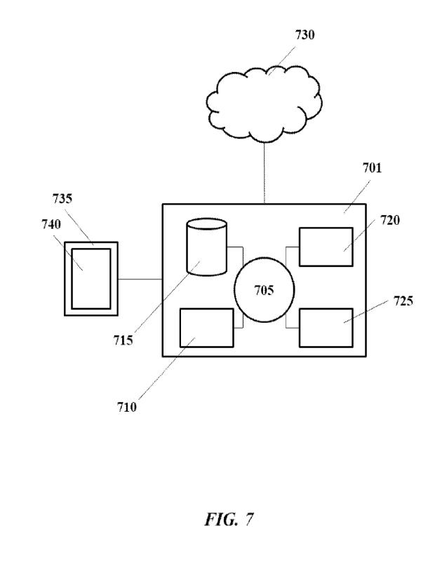
[0024] FIG. 7 shows a computer system that is programmed or otherwise
configured to
implement methods provided herein.
100251 FIGs. 8a-8f schematically illustrate an example system for
classifying and sorting one
or more cells.
[0026] FIGs. 9a-9e show a depiction of the model training,
analysis, and sorting modes.
[0027] FIGs. 10a-10m show performance of the convolutional neural network
(CNN) cell
classifier as disclosed herein.
100281 FIGs. lla-d show example cell morphology plotting and
analysis.
[0029] FIGs. 12a-12e show an additional example cell morphology
plotting and analysis.
[0030] FIG. 13 demonstrates application of integrated gradients
approach on an non-small-
cell lung carcinomas (NSCLC) adenocarcinoma cell demonstrating pixels that
supports inferring
it as NSCLC in addition to pixels that oppose inferring it as other cell
types.
[0031] FIGs. 14a and 14b illustrates results of random sorting of
cells.
- 4 -
CA 03208830 2023-8- 17
WO 2022/178095
PCT/US2022/016748
100321 FIG. 15 shows the proportion of frame-shift mutation c.572
572delC in the TP53
gene in controlled mixtures before and after enrichment. The cell lines H522
and A549 are
homozygous and wildtype respectively for this frame-shift mutation.
100331 FIG. 16 shows accuracy of single nucleotide polymorphisms (SNP)-based
mixture
fraction estimates in control DNA mixtures. Each composite sample contained
250 pg of bulk
DNA drawn from two individuals and the mixture proportion of DNA from the
second
individual was set at 5%, 10%, 20%, 30%, 40%, 60%, 80% and 90%. A close
correspondence
was found between the known and estimated mixture proportions.
100341 FIG. 17 shows determination of purity of A549 cells enriched
using the sorting
platform as disclosed herein, from a 40 cells/ml spike-in into whole blood.
The purity and blood
sample genotypes were estimated with an expectation-maximization (EM)
algorithm. Green
triangles, blue diamonds and red circles denote AA, AB and BB genotypes
respectively in the
blood sample used as a base for the spike-in mixture; dotted lines represent
the expected allele
fractions for the three blood genotypes at the inferred purity of 43%, which
is also the slope of
the lines.
DETAILED DESCRIPTION
100351 While various embodiments of the disclosure have been shown and
described herein,
it will be obvious to those skilled in the art that such embodiments are
provided by way of
example only. Numerous variations, changes, and substitutions may occur to
those skilled in the
art without departing from the disclosure. It should be understood that
various alternatives to the
embodiments of the disclosure described herein may be employed.
100361 Unless defined otherwise, all technical and scientific terms
used herein have the same
meaning as commonly understood by one of ordinary skill in the art to which
the present
disclosure belongs. In case of conflict, the present application including the
definitions will
control. Also, unless otherwise required by context, singular terms shall
include pluralities and
plural terms shall include the singular.
100371 I. Overview
100381 One or more morphological properties of a cell can be used
to, for example, study cell
type and cell state, or to diagnose diseases. In some cases, cell shape can be
one of the markers
of cell cycle. Eukaryotic cells can show physical changes in shape which can
be cell-cycle
dependent, such as a yeast cell undergoing budding or fission. In some cases,
cell shape can be
an indicator of cell state and, thus, can be an indicator used for clinical
diagnostics. In some
cases, shape of a blood cell may change due to many clinical conditions,
diseases, and
medications (e.g., changes in red blood cells' morphologies resulting from
parasitic infections).
- 5 -
CA 03208830 2023-8- 17
WO 2022/178095
PCT/US2022/016748
Additional examples of the morphological properties of the cell that can be
used to analyze the
cell can include, but are not limited to, features of cell membrane, nuclear-
to-cytoplasm ratio,
nuclear envelope morphology, and chromatin structure Methods, systems, and
databases
provided herein can be used analyze cells (e.g., previously uncharacterized or
unknown cells)
based on (e.g., solely on) such morphological properties of the cells.
100391 Analyzing a cell based on one or more images of the cell and one or
more
morphological features of the cells extracted thereform ¨ without the need to
rely on other
utilized methods of analyzing cells (e.g., identifying) cells (e.g., DNA
analysis or genomics,
RNA analysis or transcriptomics, protein analysis or proteomics, metabolite
analysis or
metabolomics, etc.) ¨ can enhance speed and/or scalability of cell analysis
systems and methods
while maintaining or even enhancing accuracy of the analysis. In some cases,
Analysis of a
population of cells based on their morphological features can uncover unique
or new parameters
to define a cell or a collection of cells (e.g., clusters of cells) that would
otherwise not be
identified in other methods.
100401 II. Methods and platforms for cell analysis
100411 The present disclosure describes various methods, e.g., a
method for analyzing or
classifying a cell, and platforms usable for or capable of performing such
methods. The method
can comprise obtaining image data of a plurality of cells, wherein the image
data comprises tag-
free images of single cells. The method can further comprise processing the
image data to
generate a cell morphology map (e.g., one or more cell morphology maps). The
cell
morphology map can comprise a plurality of morphologically-distinct clusters
corresponding to
different types or states of the cells. The method can further comprise
training a classifier (e.g.,
a cell clustering machine learning algorithm or deep learning algorithm) using
the cell
morphology map. In some the classifier can be configured to classify (e.g.,
automatically
classify) a cellular image sample based on its proximity, correlation, or
commonality with one or
more of the morphologically-distinct clusters. Thus, in some cases, the method
can further
comprise using the classifier to classify (e.g., automatically classify) the
cellular image sample
accordingly.
100421 The term "morphology" of a cell as used herein generally
refers to the form, structure,
and/or configuration of the cell. The morphology of a cell can comprise one or
more aspects of
a cell's appearance, such as, for example, shape, size, arrangement, form,
structure, pattern(s) of
one or more internal and/or external parts of the cell, or shade (e.g., color,
greyscale, etc.). Non-
limiting examples of a shape of a cell can include, but are not limited to,
circular, elliptic,
shmoo-like, dumbbell, star-like, flat, scale-like, columnar, invaginated,
having one or more
concavely formed walls, having one or more convexly formed walls, prolongated,
having
- 6 -
CA 03208830 2023-8- 17
WO 2022/178095
PCT/US2022/016748
appendices, having cilia, having angle(s), having comer(s), etc. A
morphological feature of a
cell may be visible with treatment of a cell (e.g., small molecule or antibody
staining).
Alternatively, the morphological feature of the cell may not and need not
require any treatment
to be visualized in an image or video.
100431 The term -tag" as used herein generally refers to a
heterologous composition
detectable by fluorescence, spectroscopic, photochemical, biochemical,
immunochemi cal,
electrical, optical, chemical, or other means. A tag can be, for example, a
polypeptide (e.g., an
antibody or a fragment thereof), a nucleic acid molecule (e.g., a
deoxyribonucleic acid (DNA),
ribonucleic acid (RNA) molecule)) exhibiting at least a partial
complementarity to a target
nucleic acid sequence, or a small molecule configured to bind to a target
epitope (e.g., a
polypeptide sequence, a polynucleotide sequence, one or more polysaccharide
moieties). In
some cases, the tag can be functionalized (e.g., covalently or non-covalently)
with one or more
optically detectable moieties, such as, a dye (e.g., tetramethylrhodamine
isothiocyanate
(TRITC), Quantum Dots, CY3 and CY5), biotin-streptavidin conjugates, magnetic
beads,
fluorescent dyes (e.g., fluorescein, texas red, rhodamine, green fluorescent
protein, and the like),
radiolabels (e.g., 3H, 1251, 35S, 14C, or 32P), enzymes (e.g., horse radish
peroxidase, alkaline
phosphatase and others commonly used in an ELISA), and calorimetric labels
such as colloidal
gold or colored glass or plastic (e.g., polystyrene, polypropylene, latex,
etc.) beads. In some
cases, the tag as disclosed herein, whether with or without the detectable
moiety(ies), can be
detected by, e.g., using photographic film or scintillation counters (e.g.,
for radiolabels), using
photodetectors (e.g., for fluorescent markers), providing enzymes (e.g., for
enzymatically
modifiable substrates), etc. Alternatively or in addition to, a tag can be a
representation of any
data comprising genetic information of a cell of interest, e.g., genetic
information obtained after
capturing one or more images of the cell.
100441 The term "cluster" as used herein generally refers to a
group of datapoints, such that
datapoints in one group (e.g., a first cluster) are more similar to each other
than datapoints of
another group (e.g., a second cluster). A cluster can be a group of like
datapoints (e.g., each
datapoint representing a cell or an image of a cell) that are grouped together
based on the
proximity of the datapoints, to a measure of central tendency of the cluster.
For example, a
population of cells can be analyzed based on one or more morphological
properties of each cell
(e.g., by analyzing one or more images of each cell), and each cell can be
plotted as a datapoint
on a map base on the one or more morphological properties of each cell.
Following, one or more
clusters comprising a plurality of datapoints based on the proximity of the
datapoints. The
central tendency of each cluster can be measured by one or more algorithms
(e.g., hierarchical
clustering models, K-means algorithm, statistical distribution models, etc.).
For instance, the
- 7 -
CA 03208830 2023-8- 17
WO 2022/178095
PCT/US2022/016748
measure of central tendency may be the arithmetic mean of the cluster, in
which case the
datapoints are joined together based on their proximity to the average value
in the cluster (e.g.,
K-means clustering), their correlation, or their commonality.
[0045] The term "classifier" as used herein generally refers to an
analysis model (e.g., a
metamodel) that can be trained by using a learning model and applying learning
algorithms (e.g.,
machine learning algorithms) on a training dataset (e.g., a dataset comprising
examples of
specific classes). In some cases, given a set of training examples/cases, each
marked for
belonging to a specific class (e.g., specific cell type or class), a training
algorithm can build a
classifier model capable of assigning new examples/cases (e g , new datapoints
of a cell or a
group of cells) into one category or the other, e.g., to make the model a non-
probabilistic
classifier. In some cases, the classifier model can be capable of creating a
new category to
assign new examples/cases into the new category. In some cases, a classifier
model can be the
actual trained classifier that is generated based on the training model.
[0046] The term "cell type- as used herein generally refers to a
kind, identity, or
classification of cells according to one or more criteria, such as a tissue
and species of origin, a
differentiation state, whether or not they are healthy/normal or diseased,
cell cycle stage,
viability, etc. In non-limiting examples, the term "cell type" can refer
specifically to any
specific kind of cell, such as an embryonic stem cell, a neural precursor
cell, a myoblast, a
mesodermal cell, etc.
[0047] The term "cell state" as used herein generally refers to a
specific state of the cell, such
as but not limited to an activated cell, such as activated neuron or immune
cell, resting cell, such
as a resting neuron or immune cell, a dividing cell, quiescent cell, or a cell
during any stages of
the cell cycle.
100481 The term "cell cycle" as used herein generally refers to the
physiological and/or
morphological progression of changes that cells undergo when dividing (e.g.,
proliferating).
Examples of different phases of the cell cycle can include "interphase,"
"prophase,"
"metaphase,- "anaphase,- and -telophase-. Additionally, parts of the cell
cycle can be "M
(mitosis)," "S (synthesis)," "GO," "G1 (gap 1)" and "G2 (gap2)". Furthermore,
the cell cycle
can include periods of progression that are intermediate to the above named
phases.
[0049] FIG. 1 schematically illustrates an example method for
classifying a cell. The method
can comprise processing image data 110 comprising tag-free images/videos of
single cells (e.g.,
image data 110 consisting of tag-free images/videos of single cells). Various
clustering analysis
models 120 as disclosed herein can be used to process the image data 110 to
extract one or more
morphological properties of the cells from the image data 110, and generate a
cell morphology
map 130A based on the extracted one or more morphological properties. For
example, the cell
- 8 -
CA 03208830 2023-8- 17
WO 2022/178095
PCT/US2022/016748
morphology map 130A can be generated based on two morphological properties as
dimension 1
and dimension 2. The cell morphology map 130A can comprise one or more
clusters (e.g.,
clusters A, B, and C) of datapoints, each datapoint representing an individual
cell from the image
data 110. The cell morphology map 130A and the clusters A-C therein can be
used to train
classifier(s) 150. Subsequently, a new image 140 of a new cell can be obtained
and processed
by the trained classifier(s) 150 to automatically extract and analyze one or
more morphological
features from the cellular image 140 and plot it as a datapoint on the cell
morphology map 130A.
Based on its proximity, correlation, or commonality with one or more of the
morphologically-
distinct clusters A-C on the cell morphology map 130A, the classifier(s) 150
can automatically
classify the new cell. The classifier(s) 150 can determine a probability that
the cell in the new
image data 140 belongs to cluster C (e.g., the likelihood for the cell in the
new image data 140 to
share one or more commonalities and/or characteristics with cluster C more
than with other
clusters A/B). For example, the classifier(s) 150 can determine and report
that the cell in the
new image data 140 has a 95% probability of belonging to cluster C, 1%
probability of
belonging to cluster B, and 4% probability of belong to cluster A, solely
based on analysis of the
tag-free image 140 and one or more morphological features of the cell
extracted therefrom.
100501 An image and/or video (e.g., a plurality of images and/or
videos) of one or more cells
as disclosed herein (e.g., that of image data 110 in FIG. 1) can be captured
while the cell(s) is
suspended in a fluid (e.g., an aqueous liquid, such as a buffer) and/or while
the cell(s) is moving
(e.g., transported across a microfluidic channel). For example, the cell may
not and need not be
suspended is a gel-like or solid-like medium. The fluid can comprise a liquid
that is
heterologous to the cell(s)'s natural environment. For example, cells from a
subject's blood can
be suspended in a fluid that comprises (i) at least a portion of the blood and
(ii) a buffer that is
heterologous to the blood. The cell(s) may not be immobilized (e.g., embedded
in a solid tissue
or affixed to a microscope slide, such as a glass slide, for histology) or
adhered to a substrate.
The cell(s) may be isolated from its natural environment or niche (e.g., a
part of the tissue the
cell(s) would be in if not retrieved from a subject by human intervention)
when the image and/or
video of the cell(s) is captured. For example, the image and/or video may not
and need not be
from a histological imaging. The cell(s) may not and need not be sliced or
sectioned prior to
obtaining the image and/or video of the cell, and, as such, the cell(s) may
remain substantially
intact as a whole during capturing of the image and/or video.
100511 When the image data is processed, e.g., to extract one or
more morphological features
of a cell, each cell image may be annotated with the extracted one or more
morphological
features and/or with information that the cell image belongs to a particular
cluster (e.g., a
probability).
- 9 -
CA 03208830 2023-8- 17
WO 2022/178095
PCT/US2022/016748
[0052] The cell morphology map can be a visual (e.g., graphical)
representation of one or
more clusters of datapoints. The cell morphology map can be a 1-dimensional
(1D)
representation (e.g., based on one morphological property as one parameter or
dimension) or a
multi-dimensional representation, such as a 2-dimensional (2D) representation
(e.g., based on
two morphological properties as two parameters or dimensions), a 3-dimensional
(3D)
representation (e.g., based on three morphological properties as three
parameters or dimensions),
a 4-dimensional (4D) representation, etc. In some cases, one morphological
properties of a
plurality of morphological properties used for blotting the cell morphology
map can be
represented as a non-axial parameter (e g , non-x, y, or z axis), such as,
distinguishable colors
(e.g., heatmap), numbers, letters (e.g., texts of one or more languages),
and/or symbols (e.g., a
square, oval, triangle, square, etc.). For example, a heatmap can be used as
colorimetric scale to
represent the classifier prediction percentages for each cell against a cell
class, cell type, or cell
state.
[0053] The cell morphology map can be generated based on one or more
morphological
features (e.g., characteristics, profiles, fingerprints ,etc.) from the
processed image data. Non-
limiting examples of one or more morphological properties of a cell, as
disclosed herein, that can
be extracted from one or more images of the cell can include, but are not
limited to (i) shape,
curvature, size (e.g., diameter, length, width, circumference), area, volume,
texture, thickness,
roundness, etc. of the cell or one or more components of the cell (e.g., cell
membrane, nucleus,
mitochondria, etc.), (ii) number or positioning of one or more contents (e.g.,
nucleus,
mitochondria, etc.) of the cell within the cell (e.g., center, off-centered,
etc.), and (iii) optical
characteristics of a region of the image(s) (e.g., unique groups of pixels
within the image(s)) that
correspond to the cell or a portion thereof (e.g., light emission,
transmission, reflectance,
absorbance, fluorescence, luminescence, etc.).
[0054] Non-limiting examples of clustering as disclosed herein can
be hard clustering (e.g.,
determining whether a cell belongs to a cluster or not), soft clustering
(e.g., determining a
likelihood that a cell belongs to each cluster to a certain degree), strict
partitioning clustering
(e.g., determining whether each cell belongs to exactly one cluster), strict
partitioning clustering
with outliers (e.g., determining whether a cell can also belong to no
cluster), overlapping
clustering (e.g., determining whether a cell can belong to more than one
cluster), hierarchical
clustering (e.g., determining whether cells that belong to a child cluster can
also belong to a
parent cluster), and subspace clustering (e.g., determining whether clusters
are not expected to
overlap).
[0055] Cell clustering and/or generation of the cell morphology
map, as disclosed herein, can
be based on a single morphological property of the cells. Alternatively, cell
clustering and/or
- 10 -
CA 03208830 2023-8- 17
WO 2022/178095
PCT/US2022/016748
generation the cell morphology map can be based on a plurality of different
morphological
properties of the cells. In some cases, the plurality of different
morphological properties of the
cells can have the same weight or different weights. A weight can be a value
indicative of the
importance or influence of each morphological property relative to one another
in training the
classifier or using the classifier to (i) generate one or more cell clusters,
(ii) generate the cell
morphology map, or (iii) analyze a new cellular image to classify the cellular
image as disclosed
herein. For example, cell clustering can be performed by having 50% weight on
cell shape, 40%
weight on cell area, and 10% weight on texture (e.g., roughness) of the cell
membrane. In some
cases, the classifier as disclosed herein can be configured to adjust the
weights of the plurality of
different morphological properties of the cells during analysis of new
cellular image data,
thereby to yield a most optimal cell clustering and cell morphology map. The
plurality of
different morphological properties with different weights can be utilized
during the same
analysis step for cell clustering and/or generation of the cell morphology
map.
100561 The plurality of different morphological properties can be
analyzed hierarchically. In
some cases, a first morphological property can be used as a parameter to
analyze image data of a
plurality of cells to generate an initial set of clusters. Subsequently, a
second and different
morphological property can be used as a second parameter to (i) modify the
initial set of clusters
(e.g., optimize arrangement among the initial set of clusters, re-group some
clusters of the initial
set of clusters, etc.) and/or (ii) generate a plurality of sub-clusters within
a cluster of the initial
set of clusters. In some cases, a first morphological property can be used as
a parameter to
analyze image data of a plurality of cells to generate an initial set of
clusters, to generate a 1D
cell morphology map. Subsequently, a second morphological property can be used
as a
parameter to further analyze the clusters of the 1D cell morphology map, to
modify the clusters
and generate a 2D cell morphology map (e.g., a first axis parameter based on
the first
morphological property and a second axis parameter based on the second
morphological
property).
100571 In some cases of the hierarchical clustering as disclosed
herein, an initial set of
clusters can be generated based on an initial morphological feature that is
extracted from the
image data, and one or more clusters of the initial set of clusters can
comprise a plurality of sub-
clusters based on second morphological features or sub-features of the initial
morphological
feature. For example, the initial morphological feature can be stem cells (or
not), and the sub-
features can be different types of stem cells (e.g., embryonic stem cells,
induced pluripotent stem
cells, mesenchymal stem cells, muscle stem cells, etc.). In another example,
the initial can be
cancer cells (or not), and the sub-feature can be different types of cancer
cells (e.g., sarcoma
cells, sarcoma cells, leukemia cells, lymphoma cells, multiple myeloma cells,
melanoma cells,
- 11 -
CA 03208830 2023-8- 17
WO 2022/178095
PCT/US2022/016748
etc.). In a different example, the initial can be cancer cells (or not), and
the sub-feature can be
different stages of the cancer cell (e.g., quiescent, proliferative,
apoptotic, etc.).
100581 Each datapoint can represent an individual cell or a
collection of a plurality of cells
(e.g., at least or up to about 2, 3, 4, 5, 6, 7, 8, 9, or 10 cells). Each
datapoint can represent an
individual image (e.g., of a single cell or a plurality of cells) or a
collection of a plurality of
images (e.g., at least or up to about 2, 3, 4, 5, 6, 7, 8, 9, or 10 images of
the same single cell or
different cells).
[0059] The cell morphology map can comprise at least or up to about
1, at least or up to about
2, at least or up to about 3, at least or up to about 4, at least or up to
about 5, at least or up to
about 6, at least or up to about 7, at least or up to about 8, at least or up
to about 9, at least or up
to about 10, at least or up to about 15, at least or up to about 20, at least
or up to about 30, at
least or up to about 40, at least or up to about 50, at least or up to about
60, at least or up to about
70, at least or up to about 80, at least or up to about 90, at least or up to
about 100, at least or up
to about 150, at least or up to about 200, at least or up to about 300, at
least or up to about 400, at
least or up to about 500 clusters.
100601 Each cluster as disclosed herein can comprise a plurality of
sub-clusters, e.g., at least
or up to about 2, at least or up to about 3, at least or up to about 4, at
least or up to about 5, at
least or up to about 6, at least or up to about 7, at least or up to about 8,
at least or up to about 9,
at least or up to about 10, at least or up to about 15, at least or up to
about 20, at least or up to
about 30, at least or up to about 40, at least or up to about 50, at least or
up to about 60, at least
or up to about 70, at least or up to about 80, at least or up to about 90, at
least or up to about 100,
at least or up to about 150, at least or up to about 200, at least or up to
about 300, at least or up to
about 400, at least or up to about 500 sub-clusters,
100611 A cluster (or sub-cluster) can comprise datapoints
representing cells of the same
type/state. Alternatively, a cluster (or sub-cluster) can comprise datapoints
representing cells of
different types/states.
100621 A cluster (or sub-cluster) can comprise at least or up to
about 1, at least or up to about
2, at least or up to about 3, at least or up to about 4, at least or up to
about 5, at least or up to
about 6, at least or up to about 7, at least or up to about 8, at least or up
to about 9, at least or up
to about 10, at least or up to about 15, at least or up to about 20, at least
or up to about 30, at
least or up to about 40, at least or up to about 50, at least or up to about
60, at least or up to about
70, at least or up to about 80, at least or up to about 90, at least or up to
about 100, at least or up
to about 150, at least or up to about 200, at least or up to about 300, at
least or up to about 400, at
least or up to about 500, at least or up to about 1,000, at least or up to
about 2,000, at least or up
- 12 -
CA 03208830 2023-8- 17
WO 2022/178095
PCT/US2022/016748
to about 3,000, at least or up to about 4,000, at least or up to about 5,000,
at least or up to about
10000, at least or up to about 50,000, or at least or up to about 100,000
datapoints.
100631 Two or more clusters may overlap in a cell morphology map.
Alternatively, no
clusters may not overlap in a cell morphology map. In some cases, an allowable
degree of
overlapping between two or more clusters may be adjustable (e.g., manually or
automatically by
a machine learning algorithm) depending on the quality, condition, or size of
data in the image
data being processed.
100641 A cluster (or sub-cluster) as disclosed herein can be
represented with a boundary (e.g.,
a solid line or a dashed line) Alternatively, a cluster or sub-cluster may not
and need not be
represented with a boundary, and may be distinguishable from other cluster(s)
sub-cluster(s)
based on their proximity to one another.
100651 A cluster (or sub-cluster) or a data comprising information
about the cluster can be
annotated based on one or more annotation schema (e.g., predefined annotation
schema). Such
annotation can be manual (e.g., by a user of the method or system disclosed
herein) or
automatically (e.g., by any of the machine learning algorithms disclosed
herein). The annotation
of the clustering can be related the one or more morphological properties of
the cells that have
been analyzed (e.g., cell shape, cell area, optical characteristic(s), etc.)
to generate the cluster or
assign one or more datapoints to the cluster. Alternatively, the annotation of
the clustering can
be related to information that has not been used or analyzed to generate the
cluster or assign one
or more datapoints to the cluster (e.g., genomics, transcriptomics, or
proteomics, etc.). In such
case, the annotation can be utilized to add additional -layers" of information
to each cluster.
100661 In some cases, an interactive annotation tool can be
provided that permits one or more
users to modify any process of the method described herein. For example, the
interactive
annotation tool can allow a user to curate, verify, edit, and/or annotate the
morphologically-
distinct clusters. In another example, the interactive annotation tool can
process the image data,
extract one or more morphological features from the image data, and allow the
user to select one
or more of the extracted morphological features to be used as a basis to
generate the clusters
and/or the cell morphology map. After the generation of the clusters and/or
the cell morphology
map, the interactive annotation tool can allow the user to annotate each
cluster and/or the cell
morphology map using (i) a predefined annotation schema or (ii) a new, user-
defined annotation
schema. In another example, the interactive annotation tool can allow user to
assign different
weights to different morphological features for the clustering and/or map
plotting. In another
example, the interactive annotation tool can allow user to select with imaging
data (or which
cells) to be used and/or which imaging data (or which cells, cell clumps,
artifacts, or debris) to
be discarded, for the clustering and/or map plotting. A user can manually
identify incorrectly
- 13 -
CA 03208830 2023-8- 17
WO 2022/178095
PCT/US2022/016748
clustered cells, or the machine learning algorithm can provide probability or
correlation value of
cells within each cluster and identify any outlier (e.g., a datapoint that
would change the
outcome of the probability/correlation value of the cluster(s) by a certain
percentage value).
Thus, the user can choose to move the outliers via the interactive annotation
tool to further tune
the cell morphology map, e.g., to yield a -higher resolution" map.
100671 One or more cell morphology maps as disclosed herein can be
used to train one or
more classifiers (e.g., at least or up to about 1, 2, 3, 4, 5, 6, 7, 8, 9, 10,
or more classifiers) as
disclosed herein. Each classifier can be trained to analyze one or more images
of a cell (e.g., to
extract one or more morphological features of the cell) and categorize (or
classify) the cell into
one or more determined class or categories of a cell (e.g., based on a type of
state of the cell).
Alternatively, the classifier can be trained to create a new category to
categorize (or classify) the
cell into the new category, e.g., when determining that the cell is
morphologically distinct than
any pre-existing categories of other cells.
100681 The machine learning algorithm as disclosed herein can be
configured to extract one
or more morphological feature of a cell from the image data of the cell. The
machine learning
algorithm can form a new data set based on the extracted morphological
features, and the new
data set may not and need not contain the original image data of the cell. In
some examples,
replicas of the original images in the image data can be stored in a database
disclosed herein,
e.g., prior to using any of the new images for training, e.g., to keep the
integrity of the images of
the image data. In some examples, processed images of the original images in
the image data
can be stored in a database disclosed herein during or subsequent to the
classifier training. In
some cases, any of the newly extracted morphological features as disclosed
herein can be
utilized as new molecular markers for a cell or population of cells of
interest to the user. As cell
analysis platform as disclosed herein can be operatively coupled to one or
more databases
comprising non-morphological data of cells processed (e.g., genomics data,
transcriptomics data,
proteomics data, metabolomics data), a selected population of cells exhibiting
the newly
extracted morphological feature(s) can be further analyzed by their non-
morphological
properties to identify proteins or genes of interest that are common in the
selected population of
cells but not in other cells, thereby determining such proteins or genes of
interest to be new
molecular markers that can be used to identify such selected population of
cells.
100691 In some cases, a classifier can be trained by applying
machine learning algorithms on
at least a portion of one or more cell morphology maps as disclosed herein as
a training dataset.
Non-limiting examples of machine learning algorithms for training a classifier
can include
supervised learning, unsupervised learning, semi-supervised learning,
reinforcement learning,
self-learning, feature learning, anomaly detection, association rules, etc. In
some cases, a
- 14 -
CA 03208830 2023-8- 17
WO 2022/178095
PCT/US2022/016748
classifier can be trained by using one or more learning models on such
training dataset. Non-
limiting examples of learning models can include artificial neural networks
(e.g., convolutional
neural networks, U-net architecture neural network, etc.), backpropagation,
boosting, decision
trees, support vector machines, regression analysis, Bayesian networks,
genetic algorithms,
kernel estimators, conditional random field, random forest, ensembles of
classifiers, minimum
complexity machines (MCM), probably approximately correct learning (PACT),
etc.
100701 In some cases, the neural networks are designed by the
modification of neural
networks such as Al exNet, VGGNet, GoogLeNet, ResNet (residual networks),
DenseNet, and
Inception networks In some examples, the enhanced neural networks are designed
by
modification of ResNet (e.g. ResNet 18, ResNet 34, ResNet 50, ResNet 101, and
ResNet 152) or
inception networks. In some aspects, the modification comprises a series of
network surgery
operations that are mainly carried out to improve including inference time
and/or inference
accuracy.
100711 The machine learning algorithm as disclosed herein can
utilize one or more clustering
algorithms to determine that objects in the same cluster can be more similar
(in one or more
morphological features) to each other than those in other clusters. Non-
limiting examples of the
clustering algorithms can include, but are not limited to, connectivity models
(e.g., hierarchical
clustering), centroid models (e.g. K-means algorithm), distribution models
(e.g., expectation-
maximization algorithm), density models (e.g., density-based spatial
clustering of applications
with noise (DB SCAN), ordering points to identify the clustering structure
(OPTICS)), subspace
models (e.g., biclustering), group models, graph-based models (e.g., highly
connected subgraphs
(HCS) clustering algorithms), single graph models, and neural models (e.g.,
using unsupervised
neural network). The machine learning algorithm can utilize a plurality of
models, e.g., in equal
weights or in different weights.
100721 In some cases, unsupervised and self-supervised approaches
can be used to expedite
labeling of image data of cells. For the case of unsupervised, an embedding
for a cell image can
be generated. For example, the embedding can be a representation of the image
in a space with
reduced dimensions than the original image data. Such embeddings can be used
to cluster
images that are similar to one another. Thus, the labeler can be configured to
batch-label the
cells and increase the throughput as compared to manually labeling one or more
cells.
100731 In some cases, for the case of self-supervised learning,
additional meta information
(e.g., additional non-morphological information) about the sample (e.g., what
disease is known
or associated with the patient who provided the sample) can be used for
labeling of image data
of cells.
- 15 -
CA 03208830 2023-8- 17
WO 2022/178095
PCT/US2022/016748
100741 In some cases, embedding generation can use a neural net
trained on predefined cell
types. To generate the embeddings described herein, an intermediate layer of
the neural net that
is trained on predetermined image data (e.g., image data of known cell types
and/or states) can
be used. By providing enough diversity in image data/sample data to the
trained
model/classifier, this method can provide an accurate way to cluster future
cells.
100751 In some cases, embedding generation can use neural nets
trained for different tasks.
To generate the embeddings described herein, an intermediate layer of the
neural net that is
trained for a different task (e.g., a neural net that is trained on a
canonical dataset such as
ImageNet) Without wishing to be bound by theory, this can allow to focus on
features that
matter for image classification (e.g., edges and curves) while removing a bias
that may otherwise
be introduced in labeling the image data.
100761 In some cases, autoencoders can be used for embedding
generation. To generate the
embeddings described herein, autoencoders can be used, in which the input and
the output can
be substantially the same image and the squeeze layer can be used to extract
the embeddings.
The squeeze layer can force the model to learn a smaller representation of the
image, which
smaller representation may have sufficient information to recreate the image
(e.g., as the output).
100771 In some cases, for clustering-based labeling of image data
or cells, as disclosed herein,
an expanding training data set can be used. With the expanding training data
set, one or more
revisions of labeling (e.g., manual relabeling) may be needed to, e.g., avoid
the degradation of
model performance due to the accumulated effect of mislabeled images. Such
manual relabeling
may be intractable on a large scale and ineffective when done on a random
subset of the data.
Thus, to systematically surface images for potential relabeling, for example,
similar embedding-
based clustering can be used to identify labeled images that may cluster with
members of other
classes. Such examples are likely to be enriched for incorrect or ambiguous
labels, which can be
removed (e.g., automatically or manually).
100781 In some cases, adaptive image augmentation can be used. In order to
make the models
and classifiers disclosed herein more robust to artifacts in the image data,
(1) one or more
images with artifacts can be identified, and (2) such images identified with
artifacts can be added
to training pipeline (e.g., for training the model/classifier). Identifying
the image(s) with
artifacts can comprise: (la) while imaging cells, one or more additional
sections of the image
frame can be cropped, which frame(s) being expected to contain just the
background without any
cell; (2a) the background image can be checked for any change in one or more
characteristics
(e.g., optical characteristics, such as brightness); and (3a)
flagging/labeling one or more images
that have such change in the characteristic(s). Adding the identified images
to training pipeline
can comprise: (2a) adding the one or more images that have been
flagged/labeled as
- 16 -
CA 03208830 2023-8- 17
WO 2022/178095
PCT/US2022/016748
augmentation by first calculating an average feature of the changed
characteristic(s) (e.g., the
background median color); (2b) creating a delta image by subtracting the
average feature from
the image data (e.g., subtracting the median for each pixel of the image); and
(3c) adding the
delta image to the training pipeline.
100791 One or more dimension of the cell morphology map can be represented by
various
approaches (e.g., dimensionality reduction approaches), such as, for example,
principal
component analysis (PCA), multidimensional scaling (MDS), t-distributed
stochastic neighbor
embedding (t-SNE), and uniform manifold approximation and projection (UMAP).
For
example, TIMAP can be a machine learning technique for dimension reduction
TIMAP can be
constructed from a theoretical framework based in Riemannian geometry and
algebraic
topology. UMAP can be utilized for a practical scalable algorithm that applies
to real world
data, such as morphological properties of one or more cells.
100801 The cell morphology map as disclosed herein can comprise an
ontology of the one or
more morphological features. The ontology can be an alternative medium to
represent a
relationship among various datapoints (e.g., each representing a cell)
analyzed from an image
data. For example, an ontology can be a data structure of information, in
which nodes can be
linked by edges. An edge can be used to define a relationship between two
nodes. For example,
a cell morphology map can comprise a cluster comprising sub-clusters, and the
relationship
between the cluster and the sub-clusters can be represented in an nodes/edges
ontology (e.g., an
edge can be used to describe the relationship as a subclass of, genus of, part
of, stem cell of,
differentiated from, progeny of, diseased state of, targets, recruits,
interacts with, same tissue,
different tissue, etc.).
100811 In some cases, one-to-one morphology to genomics mapping can
be utilized. An
image of a single cell or images of multiple "similar looking" cells can be
mapped to its/their
molecular profile(s) (e.g., genomics, proteomics, transcriptomics, etc.). In
some examples,
classifier-based barcoding can be performed. Each sorting event (e.g.,
positive classifier) can
push the sorted cell(s) into an individual well or droplet with a unique
barcode (e.g., nucleic acid
or small molecule barcode). The exact barcode(s) used for that individual
classifier positive
event can be recorded and tracked. Following, the cells can be lysed and
molecularly analyzed
together with the barcode(s). The result of the molecular analysis can then be
mapped (e.g., one-
to-one) to the image(s) of the individual (or ensemble of) sorted cell(s)
captured while the cell(s)
was/were flowing in the flow channel. In some examples, class-based sorting
can be utilized.
Cells that are classified in the same class based at least on their
morphological features can be
sorted into a single well or droplet with a pre-determined barcoded material,
and the cells can be
- 17 -
CA 03208830 2023-8- 17
WO 2022/178095
PCT/US2022/016748
lysed, molecularly analyzed, then any molecular information can be used for
the one-to-one
mapping as disclosed herein.
100821 FIG. 2 schematically illustrates different ways of
representing analysis data of image
data of cells. Tag-free image data 210 of cells (e.g., circular cells and
square cells) having
different nuclei (e.g., small nucleus and large nucleus) can be analyzed by
any of the methods
disclosed herein (e.g., based on extraction of one or more morphological
features). For example,
any of the classifier(s) disclosed herein can be used to analyze and plot the
image data 210 into a
cell morphology map 220, comprising four distinguishable clusters: cluster A
(circular cell,
small nucleus), cluster B (circular cell, large nucleus), cluster C (square
cell, small nucleus), and
cluster D (square cell, large nucleus). The classifier(s) can also represent
the analysis in a cell
morphological ontology 230, in which a top node ("cell shape") can be
connected to two sub-
nodes ("circular cell" and rectangular cell") via an edge ("is a subclass of")
to define the
relationship between the nodes. Each sub-node can also connected to its own
sub-nodes ("small
nucleus- and "large nucleus-) via an edge ("is a part of') to define their
relationships. The sub-
nodes (e.g., "small nucleus" and "large nucleus") can also be connected via
one or more edges
("are similar") to further define their relationship.
100831 The cell morphology map or cell morphological ontology as
disclosed herein can be
further annotated with one or more non-morphological data of each cell. As
shown in FIG. 3,
the ontology 230 from FIG. 2 can be further annotated with information about
the cells that may
not be extractable from the image data used to classify the cells (e.g.,
molecular profiles obtained
via molecular barcodes, as disclosed herein). Non-limiting examples of such
non-morphological
data can be from additional treatment and/or analysis, including, but not
limited to, cell culture
(e.g., proliferation, differentiation, etc.), cell permeabilization and
fixation, cell staining by a
probe, mass cytometry, multiplexed ion beam imaging (MIBI), confocal imaging,
nucleic acid
(e.g., DNA, RNA) or protein extraction, polymerase chain reaction (PCR),
target nucleic acid
enrichment, sequencing, sequence mapping, etc.
100841 Examples of the probe used for cell staining (or tagging)
may include, but are not
limited to, a fluorescent probe (e.g., for staining chromosomes such as X, Y,
13, 18 and 21 in
fetal cells), a chromogenic probe, a direct immunoagent (e.g. labeled primary
antibody), an
indirect immunoagent (e.g., unlabeled primary antibody coupled to a secondary
enzyme), a
quantum dot, a fluorescent nucleic acid stain (such as DAPI, Ethidium bromide,
Sybr green,
Sybr gold, Sybr blue, Ribogreen, Picogreen, YoPro-1, YoPro-2 YoPro-3, YOYo,
Oligreen
acridine orange, thiazole orange, propidium iodine, or Hoeste), another probe
that emits a
photon, or a radioactive probe.
- 18 -
CA 03208830 2023-8- 17
WO 2022/178095
PCT/US2022/016748
100851 In some cases, the instrument(s) for the additional analysis
may comprise a computer
executable logic that performs karyotyping, in situ hybridization (ISH) (e.g.,
florescence in situ
hybridization (FISH), chromogenic in situ hybridization (CISH), nanogold in
situ hybridization
(NISH)), restriction fragment length polymorphism (RFLP) analysis, polymerase
chain reaction
(PCR) techniques, flow cytometry, electron microscopy, quantum dot analysis,
or detects single
nucleotide polymorphisms (SNPs) or levels of RNA.
100861 Analysis of the image data (e.g., extracting one or more
morphological features form
the image data, determining clustering and/or cell morphology map based on the
image data,
etc) can be performed (e g , automatically) within less than about 1 hour, 50
minutes, 40
minutes, 30 minutes, 25 minutes, 20 minutes, 15 minutes, 10 minutes, 9
minutes, 8 minutes, 7
minutes, 6 minutes, 5 minutes, 4 minutes, 3 minutes, 2 minutes, 1 minute, 50
seconds, 40
seconds, 30 seconds, 20 seconds, 10 seconds, 5 seconds, 1 second, or less. In
some cases, such
analysis can be performed in real-time.
100871 One or more morphological features utilized for generating
the clusters or the cell
morphology map, as disclosed herein, can be selected automatically (e.g., by
one or more
machine learning algorithms) or, alternatively, selected manually by a user
via a user interface
(e.g., graphical user interface (GUI)). The GUI can show visualization of, for
example, (i) the
one or more morphological parameters extracted from the image data (e.g.,
represented as
images, words, symbols, predefined codes, etc.), (ii) the cell morphology map
comprising one or
more clusters, or (iii) the cell morphological ontology. The user can select,
via the GUI, which
morphological parameter(s) to be used to generate the clusters and the cell
morphological map
prior to actual generation of the clusters and the cell morphological map. The
user can, upon
seeing or receiving a report about the generated clusters and the cell
morphological map,
retroactively modify the types of morphological parameter(s) to use, thereby
to (i) modify the
clustering or the cell morphological mapping and/or (ii) create new cluster(s)
or new cell
morphological map(s). In some cases, the user can select one or more regions
to be excluded or
included for further analysis or further processing of the cells (e.g.,
sorting in the future or in
real-time). For example, a microfluidic system as disclosed herein can be
utilized to capture
image(s) of each cell from a population of cells, and any of the methods
disclosed herein can be
utilized to analyze such image data to generate a cell morphology map
comprising clusters
representing the population of cells. The user can select one or more clusters
or sub-clusters to
be sorted, and the input can be provided to the microfluidic system to sort at
least a portion of
the cells into one or more sub-channels of the microfluidic system (e.g., in
real-time)
accordingly. Alternatively, the user can select one or more clusters or sub-
clusters to be
excluded during sorting (e.g., to get rid of artifacts, debris, or dead
cells), and the input can be
- 19 -
CA 03208830 2023-8- 17
WO 2022/178095
PCT/US2022/016748
provided to the microfluidic system to sort at least a portion of the cells
into one or more sub-
channels of the microfluidic system (e.g., in real-time) accordingly without
such artifacts, debris,
or dead cells.
[0088] FIG. 4 schematically illustrates a method for a user to
interact (e.g., via GUI) with any
one of the methods disclosed herein. Image data 410 of a plurality of cells
can be processed, via
any one of the methods disclosed herein, to generate a cell morphology map
420A that
represents the plurality of cells as datapoints in different clusters A, B, C,
and D. The cell
morphology map 420A can be displayed to the user via the GUI 430. The user can
select each
cluster or a datapoint within each cluster to visualize one or more images
450a, b, c, or d of the
cells classified into the cluster. Upon visualization of the images, the user
can draw a box 440
(e.g., via any user-defined shape and/or size) around one or more datapoints
or around a cluster.
For example, the user can draw a box 440 around a cluster of "debris"
datapoints, to, e.g.,
remove the selected cluster and generate a new cell morphology map 420B. The
user input can
be used to update cell classifying algorithms (e.g., one or more classifier(s)
as disclosed herein),
mapping algorithms, cell flowing mechanism (e.g., velocity of cells,
positioning of the cells
within a flow channel, adjusting imaging focal length/plane of one or more
sensors/cameras of
an imaging module (also referred to as an imaging device herein) that captures
one or more
images/videos of cells flowing through the flow cell, etc.), cell sorting
mechanisms in the flow
channel, cell sorting instructions in the flow channel, etc. For example, upon
the user's
selection, the classifier can be trained to identify one or more common
morphological features
within the selected datapoints (e.g., features that distinguish the selected
datapoints from the
unselected data). Features of the selected group can be used to further
identify other cells from
other samples having similar feature(s) for further analysis or discard cells
having similar
feature(s), e.g., for cell sorting.
[0089] The present disclosure also describes a cell analysis
platform, e.g., for analyzing or
classifying a cell. The cell analysis platform can be a product of any one of
the methods
disclosed herein. Alternatively or in addition to, the cell analysis platform
can be used as a basis
to execute any one of the methods disclosed herein. For example, the cell
analysis platform can
be used to process image data comprising tag-free images of single cells to
generate a new cell
morphology map of various cell clusters. In another example, the cell analysis
platform can be
used to process image data comprising tag-free images of single cells to
compare the cell to pre-
determined (e.g., pre-analyzed) images of known cells or cell morphology
map(s), such that the
single cells from the image data can be classified, e.g., for cell sorting.
[0090] FIG. 5 illustrates an example cell analysis platform (e.g.,
machine learning/artificial
intelligence platform) for analyzing image data of one or more cells. The cell
analysis platform
- 20 -
CA 03208830 2023-8- 17
WO 2022/178095
PCT/US2022/016748
500 can comprise a cell morphology atlas (CMA) 505. The CMA 505 can comprise a
database
510 having a plurality of annotated single cell images that are grouped into
morphologically-
distinct clusters (e.g., represented a texts, as cell morphology map(s), or
cell morphological
ontology(ies)) corresponding to a plurality of classifications (e.g.,
predefined cell classes). The
CMA 505 can comprise a modeling unit comprising one or more models (e.g.,
modeling library
520 comprising, such as, one or more machine learning algorithms disclosed
herein) that are
trained and validated using datasets from the CMA 505, to process image data
comprising
images/videos of one or more cells to identify different cell types and/or
states based at least on
morphological features The CMA 505 can comprise an analysis module 530
comprising one or
more classifiers as disclosed herein. The classifier(s) can uses one or more
of the models from
the modeling library 520 to, e.g., (1) classify one or more images taken from
a sample, (2) assess
a quality or state of the sample based on the one or more images, (3) map one
or more datapoints
representing such one or more images onto a cell morphology map (or cell
morphological
ontology) via using a mapping module 540. The CMA 505 can be operatively
coupled to one or
more additional database 570 to receive the image data comprising the
images/videos of one or
more cells. For example, the image data from the database 570 can be obtained
from an imaging
module 592 of a flow cell 590, which can also be operatively coupled to the
CMA 505. The
flow cell can direct flow of a sample comprising or suspected of comprising a
target cell, and
capture one or more images of contents (e.g., cells) within the sample by the
imaging module
592. Any image data obtained by the imaging module 592 can be transmitted
directly to the
CMA 505 and/or to the new image database 570. Alternatively or in addition to,
the CMA 505
can be operatively coupled to one or more additional databases 580 comprising
non-
morphological data of any of the cells (e.g., genomics, transcriptomics, or
proteomics, etc.), e.g.,
to further annotate any of the datapoint, cluster, map, ontology, images, as
disclosed herein. The
CMA 505 can be operatively coupled to a user device 550 (e.g., a computer or a
mobile device
comprising a display) comprising a GUI 560 for the user to receive information
from and/or to
provide input (e.g., instructions to modify or assist any portion of the
method disclosed herein).
Any classification made by the CMA and/or the user can be provided as an input
to the sorting
module 594 of the flow cell 590. Based on the classification, the sorting
module can determine,
for example, (i) when to activate one or more sorting mechanisms at the
sorting junction of the
flow cell 590 to sort one or more cells of interest, (ii) which sub-channel of
a plurality of sub-
channels to direct each single cell for sorting. In some cases, the sorted
cells can be collected for
further analysis, e.g., downstream molecular assessment and/or profiling, such
as genomics,
transcriptomics, proteomics, metabolomics, etc.
- 21 -
CA 03208830 2023-8- 17
WO 2022/178095
PCT/US2022/016748
100911 Any of the methods or platforms disclosed herein can be used
as a tool that permits a
user to train one or more models (e.g., from the modeling library) for cell
clustering and/or cell
classification. For example, a user may provide initial image dataset of a
sample to the platform,
and the platform may process the initial set of image data. Based on the
processing, the platform
can determine a number of labels and/or an amount of data that the user needs
to train the one or
more models, based on the initial image dataset of the sample. In some
examples, the platform
can determine that the initial set of image data can be insufficient to
provide an accurate cell
classification or cell morphology map. For example, the platform can plot an
initial cell
morphology map and recommend to the user the number of labels and/or the
amount of data
needed to for enhanced processing, classification, and/or sorting, based on
proximity (or
separability), correlation, or commonality of the datapoints in the map (e.g.,
whether there is no
distinguishable clusters within the map, whether the clusters within the map
are too close to each
other, etc.). In another example, the platform can allow the user to select
different model (e.g.,
clustering model) or classifier, different combinations of models or
classifiers, to re-analyze the
initial set of image data.
100921 Any of the methods or platforms disclosed herein can be used
to determine quality or
state of the image(s) of the cell, that of the cell, or that of a sample
comprising the cell. The
quality or state of the cell can be determined at a single cell level.
Alternatively, the quality or
state of the cell can be determined at an aggregate level (e.g., as a whole
sample, or as a portion
of the sample). The quality or state can be determined and reported based on,
e.g., a number
system (e.g., a number scale from 1 to 10, a percentage scale from 1% to
100%), a symbolic
system, or a color system. For example, the quality or state can be indicative
of a preparation or
priming condition of the sample (e.g., whether the sample has a sufficient
number of cells,
whether the sample has too much artifacts, debris, etc.) or indicative of a
viability of the sample
(e.g., whether the sample has an amount of "dead" cells above a predetermined
threshold).
100931 Any of the methods or platforms disclosed herein can be used
to sort cells in silico
(e.g., prior to actual sorting of the cells using a microfluidic channel). The
in silico sorting can
be, e.g., to discriminate among and/or between, e.g., multiple different cell
types (e.g., different
types of cancer cells, different types of immune cells, etc.), cell states,
cell qualities. The
methods and platforms disclosed herein can utilize pre-determined
morphological properties
(e.g., provided in the platform) for the discrimination. Alternatively or in
addition to, newly
abstracted morphological properties can be abstracted (e.g., generated) based
on the input data
for the discrimination. In some cases, new model(s) and/or classifier(s) can
be trained or
generated to process the image data. In some cases, the newly abstracted
morphological
properties can be used to discriminate among and/or between, e.g., multiple
different cell types,
- 22 -
CA 03208830 2023-8- 17
WO 2022/178095
PCT/US2022/016748
cell states, cell qualities that are known. Alternatively or in addition to,
the newly abstracted
morphological properties can be used to create new class (or classifications)
to sort the cells
(e.g., in silico or via the microfluidic system). The newly abstracted
morphological properties as
disclosed herein may enhance accuracy or sensitivity of cell sorting (e.g., in
silico or via the
microfluidic system).
100941 Subsequent to the in silico sorting of the cells, the actual
cell sorting of the cells (e.g.,
via the microfluidic system or flow cell) based on the in silico sorting can
be performed within
less than about 1 hours, 50 minutes, 40 minutes, 30 minutes, 25 minutes, 20
minutes, 15
minutes, 10 minutes, 9 minutes, 8 minutes, 7 minutes, 6 minutes, 5 minutes, 4
minutes, 3
minutes, 2 minutes, 1 minute, 50 seconds, 40 seconds, 30 seconds, 20 seconds,
10 seconds, 5
seconds, 1 second, or less. In some cases, the in silico sorting and the
actual sorting can occur in
real-time.
100951 In any of the methods or platforms disclosed herein, the
model(s) and/or classifier(s)
can be validated (e.g., for the ability to demonstrate accurate cell
classification performance).
Non-limiting examples of validation metrics that can be utilized can include,
but are not limited
to, threshold metrics (e.g., accuracy, F-measure, Kappa, Macro-Average
Accuracy, Mean-Class-
Weighted Accuracy, Optimized Precision, Adjusted Geometric Mean, Balanced
Accuracy, etc.),
the ranking methods and metrics (e.g., receiver operating characteristics
(ROC) analysis or
"ROC area under the curve (ROC AUC)"), and the probabilistic metrics (e.g.,
root-mean-
squared error). For example, the model(s) or classifier(s) can be determined
to be balanced or
accurate when the ROC AUC is greater than 0.5, greater than 0.55, greater than
0.6, greater than
0.65, greater than 0.7, greater than 0.75, greater than 0.8, greater than
0.85, greater than 0.9,
greater than 0.91, greater than 0.92, greater than 0.93, greater than 0.94,
greater than 0.95,
greater than 0.96, greater than 0.97, greater than 0.98, greater than 0.99, or
more.
100961 In any of the methods or platforms disclosed herein, the
image(s) of the cell(s) can be
obtained when the cell(s) are prepared and diluted in a sample (e.g., a buffer
sample). The
cell(s) can be diluted, e.g., in comparison to real-life concentrations of the
cell in the tissue (e.g.,
solid tissue, blood, serum, spinal fluid, urine, etc.) to a dilution
concentration. The methods or
platforms disclosed herein can be compatible with a sample (e.g., a biological
sample or
derivative thereof) that is diluted by a factor of about 500 to about
1,000,000. The methods or
platforms disclosed herein can be compatible with a sample that is diluted by
a factor of at least
about 500. The methods or platforms disclosed herein can be compatible with a
sample that is
diluted by a factor of at most about 1,000,000. The methods or platforms
disclosed herein can be
compatible with a sample that is diluted by a factor of about 500 to about
1,000, about 500 to
about 2,000, about 500 to about 5,000, about 500 to about 10,000, about 500 to
about 20,000,
- 23 -
CA 03208830 2023-8- 17
WO 2022/178095
PCT/US2022/016748
about 500 to about 50,000, about 500 to about 100,000, about 500 to about
200,000, about 500
to about 500,000, about 500 to about 1,000,000, about 1,000 to about 2,000,
about 1,000 to
about 5,000, about 1,000 to about 10,000, about 1,000 to about 20,000, about
1,000 to about
50,000, about 1,000 to about 100,000, about 1,000 to about 200,000, about
1,000 to about
500,000, about 1,000 to about 1,000,000, about 2,000 to about 5,000, about
2,000 to about
10,000, about 2,000 to about 20,000, about 2,000 to about 50,000, about 2,000
to about 100,000,
about 2,000 to about 200,000, about 2,000 to about 500,000, about 2,000 to
about 1,000,000,
about 5,000 to about 10,000, about 5,000 to about 20,000, about 5,000 to about
50,000, about
5,000 to about 100,000, about 5,000 to about 200,000, about 5,000 to about
500,000, about
5,000 to about 1,000,000, about 10,000 to about 20,000, about 10,000 to about
50,000, about
10,000 to about 100,000, about 10,000 to about 200,000, about 10,000 to about
500,000, about
10,000 to about 1,000,000, about 20,000 to about 50,000, about 20,000 to about
100,000, about
20,000 to about 200,000, about 20,000 to about 500,000, about 20,000 to about
1,000,000, about
50,000 to about 100,000, about 50,000 to about 200,000, about 50,000 to about
500,000, about
50,000 to about 1,000,000, about 100,000 to about 200,000, about 100,000 to
about 500,000,
about 100,000 to about 1,000,000, about 200,000 to about 500,000, about
200,000 to about
1,000,000, or about 500,000 to about 1,000,000. The methods or platforms
disclosed herein can
be compatible with a sample that is diluted by a factor of about 500, about
1,000, about 2,000,
about 5,000, about 10,000, about 20,000, about 50,000, about 100,000, about
200,000, about
500,000, or about 1,000,000.
[0097] In any of the methods or platforms disclosed herein, the
classifier can generate a
prediction probability (e.g., based on the morphological clustering and
analysis) that an
individual cell or a cluster of cells belongs to a cell class (e.g., within a
predetermined cell class
provided in the CMA as disclosed herein), e.g., via a reporting module. The
reporting module
can communicate with the user via a GUI as disclosed herein. Alternatively or
in addition to, the
classifier can generate a prediction vector that an individual cell or a
cluster of cells belongs to a
plurality of cell classes (e.g., a plurality of all of predetermined cell
classes from the CMA as
disclosed herein). The vector can be 1D (e.g., a single row of different cell
classes), 2D (e.g.,
two dimensions, such as tissue origin vs. cell type), 3D, etc. In some cases,
based on processing
and analysis of image data obtained from a sample, the classifier can generate
a report showing a
composition of the sample, e.g., a distribution of one or more cell types,
each cell type indicated
with a relative proportion within the sample. Each cell of the sample can also
be annotated with
a most probable cell type and one or more less probably cell types.
[0098] Any one of the methods and platforms disclosed herein can be
capable of processing
image data of one or more cells to generate one or more morphometric maps of
the one or more
- 24 -
CA 03208830 2023-8- 17
WO 2022/178095
PCT/US2022/016748
cells. Non-limiting examples of morphometric models can be utilized to analyze
one or more
images of single cells (or cell clusters) can include, e.g., simple
morphometrics (e.g., based on
lengths, widths, masses, angles, ratios, areas, etc.), landmark-based
geometric morphometrics
(e.g., spatial information, intersections, etc. of one or more components of a
cell), procrustes-
based geometric morphometrics (e.g., by removing non-shape information that is
altered by
translation, scaling, and/or rotation from the image data), Euclidean distance
matrix analysis,
diffeomorphometry, and outline analysis. The morphometric map(s) can be multi-
dimensional
(e.g., 2D, 3D, etc.). The morphometric map(s) can be reported to the user via
the GUI.
100991 Any of the methods or platforms disclosed herein (e g , the
analysis module) can be
used to process, analyze, classify, and/or compare two or more samples (e.g.,
at least 2, 3, 4, 5,
6, 7, 8, 9, 10, or more test samples). The two or more samples can each be
analyzed to
determine a morphological profile (e.g., a cell morphology map) of each
sample. For example,
the morphological profiles of the two or more samples can be compared for
identifying a disease
state of a patient's sample in comparison to a health cohort's sample or a
sample of image data
representative of a disease of interest. In another example, the morphological
profiles of the two
or more samples can be compared to monitor a progress of a condition of a
subject, e.g.,
comparing first image data of a first set of cells from a subject before a
treatment (e.g., a test
drug candidate, chemotherapy, surgical resection of solid tumors, etc.) and
second image data of
a second set of cells from the subject after the treatment. The second set of
cells can be obtained
from the subject at least about 1 week, at least about 2 weeks, at least about
3 weeks, at least
about 4 weeks, at least about 2 months, or at least about 3 months subsequent
to obtaining the
first set of cells from the subject. In a different example, the morphological
profiles of the two
or more samples can be compared to monitor effects of two or more different
treatment options
(e.g., different test drugs) in two or more different cohorts (e.g., human
subjects, animal subjects,
or cells being tested in vitro/ex vivo). Accordingly, the systems and methods
disclosed herein
can be utilized (e.g., via sorting or enrichment of a cell type of interest or
a cell exhibiting a
characteristic of interest) to select a drug and/or a therapy that yields a
desired effect (e.g., a
therapeutic effect greater than equal to a threshold value).
101001 Any of the platforms disclosed herein (e.g., cell analysis
platform) can provide an
inline end-to-end pipeline solution for continuous labeling and/or sorting of
multiple different
cell types and/or states based at least in part on (e.g., based solely on)
morphological analysis of
imaging data provided. A modeling library used by the platform can be scalable
for large amount
of data, extensible (e.g., one or more models or classifiers modified), and/or
generalizable (e.g.,
more resistant to data perturbations ¨ such as artifacts, debris, random
objects in the background,
image/video distortions ¨ between samples. Any of the modeling library may be
removed or
- 25 -
CA 03208830 2023-8- 17
WO 2022/178095
PCT/US2022/016748
updated with new model automatically by the machine learning algorithms or
artificial
intelligence, or by the user.
101011 Any of the methods and platforms disclosed herein can adjust one or
more parameters
of the microfluidic system as disclosed herein. As cells are flowing through a
flow channel, an
imaging module (e.g., sensors, cameras) can capture image(s)/video(s) of the
cells and generate
new image data. The image data can be processed and analyzed (e.g., in real-
time) by the
methods and platforms of the present disclosure to train a model (e.g.,
machine learning model)
to determine whether or not one or more parameters of the microfluidic system.
101021 In some cases, the model(s) can determine that the cells are
flowing too fast or too
slow, and send an instruction to the microfluidic system to adjust (i) the
velocity of the cells
(e.g., via adjusting velocity of the fluid medium carrying the cells) and/or
(ii) image recording
rate of a camera that is capturing images/videos of cells flowing through the
flow channel.
101031 In some cases, the model(s) can determine that the cells are
in-focus or out-of-focus in
the images/videos, and send an instruction to the microfluidic system to (i)
adjust a positioning
of the cells within the flow cell (e.g., move the cell towards or away from
the center of the flow
channel via, for example, hydrodynamic focusing and/or inertial focusing)
and/or (ii) adjust a
focal length/plane of the camera that is capturing images/videos of cells
flowing through the
flow channel. Adjusting the focal length/plane can be performed for the same
cell that has been
analyzed (e.g., adjusting focal length/plane of a camera that is downstream)
or a subsequent cell.
Adjusting the focal length/plane can enhance clarity or reduce blurriness in
the images. The
focal length/plane can be adjusted based on a classified type or state of the
cell. In some
examples, adjusting the focal length/plane can allow enhanced focusing/clarity
on all parts of the
cell. In some examples, adjusting the focal length/plane can allow enhanced
focusing/clarity on
different portions (but not all parts) of the cell. Without wishing to be
bound by theory, out-of-
focus images may be usable for any of the methods disclosed herein to extract
morphological
feature(s) of the cell that otherwise may not be abstracted from in-focus
images, or vice versa.
Thus, in some cases, instructing the imaging module to capture both in-focus
and out-of-focus
images of the cells can enhance accuracy of any of the analysis of cells
disclosed herein.
Alternatively or in addition to, the model(s) can send an instruction to the
microfluidic system to
modify the flow and adjust an angle of the cell relative to the camera, to
adjust focus on different
portions of the cell or a subsequent cell. Different portions as disclosed
herein can comprise an
upper portion, a mid portion, a lower portion, membrane, nucleus,
mitochondria, etc. of the cell.
101041 In order to image cells at the right focus (with respect to
height or z dimension), what
is conventionally done is to calculate the "focus measure" of an image using
information
theoretic methods like Fourier Transform or Laplace transform.
- 26 -
CA 03208830 2023-8- 17
WO 2022/178095
PCT/US2022/016748
101051 In some cases, bi-directional out-of-focus (00F) images
cells (e.g., one or more first
images that are OOF in a first direction, and one or more second images that
are OOF in as
second direction that is different __ such as opposite .. from the first
direction). For example,
images that are OOF in two opposite directions may be called "bright 00F"
image(s) and "dark
00F" image(s), which may be obtained by changing the z-focus bi-directionally.
A classifier as
disclosed herein can be trained with a image data comprising both bright OOF
image(s) and dark
OOF image(s). The trained classifiers can be used to run inferences (e.g., in
real-time) on new
image data of cells to classify each image as bright OOF image, dark OOF
image, and optionally
image that is not OOF (e.g., not OOF relative to the bright/dark OOF images).
The classifier can
also measure a percentage of bright OOF image, a percentage of dark OOF image,
or a
percentage of both bright and dark OOF images within the image data. For
example, if any of
the percentage of bright OOF image, the percentage of dark OOF image, or the
percentage of
both bright and dark OOF images is above a threshold value (e.g., a
predetermined threshold
value), then the classifier can determine that the imaging device (e.g., by
the microfluidic system
as disclosed herein) may not be imaging cells at the right focal length/plane.
The classifier can
instruct the user, via GUI of a user device, to adjust the imaging device's
focal length/plane. In
some examples, the classifier can determine, based on analysis of the image
data comprising
OOF images, direction and degree of adjustment of focal length/plane that may
be required to
adjust the imaging device, to yield a reduced amount of OOF imaging. In some
examples, the
classifier and the microfluidic device can be operatively coupled to a machine
learning/artificial
intelligence controller, such that the focal length/plane of the imaging
device can be adjusted
automatically upon determination of the classifier.
101061 A threshold (e.g., a predetermined threshold) of a
percentage of OOF images (e.g.,
bright 00F, dark 00F, or both) can be about 0.1 % to about 20 %. A threshold
(e.g., a
predetermined threshold) of a percentage of OOF images (e.g., bright 00F, dark
00F, or both)
can be at least about 0.1 %. A threshold (e.g., a predetermined threshold) of
a percentage of OOF
images (e.g., bright 00F, dark 00F, or both) can be at most about 20 %. A
threshold (e.g., a
predetermined threshold) of a percentage of OOF images (e.g., bright 00F, dark
00F, or both)
can be about 0.1 % to about 0.5 %, about 0.1 % to about 1 %, about 0.1 % to
about 2%, about
0.1 % to about 4%, about 0.1 % to about 6%, about 0.1 % to about 8 %, about
0.1 % to about
%, about 0.1 % to about 15 %, about 0.1 % to about 20 %, about 0.5 % to about
1 %, about
0.5 % to about 2 %, about 0.5 % to about 4 %, about 0.5 % to about 6 %, about
0.5 % to about 8
%, about 0.5 % to about 10 %, about 0.5 % to about 15 %, about 0.5 % to about
20 %, about 1 %
to about 2 %, about 1 % to about 4 cYci, about 1 % to about 6 %, about 1 % to
about 8 %, about 1
% to about 10 %, about 1 % to about 15 %, about 1 % to about 20 %, about 2 %
to about 4 %,
- 27 -
CA 03208830 2023-8- 17
WO 2022/178095
PCT/US2022/016748
about 2 % to about 6 %, about 2 % to about 8 %, about 2 % to about 10 %, about
2 % to about
15 %, about 2 % to about 20 %, about 4 % to about 6 %, about 4 % to about 8 %,
about 4 % to
about 10 %, about 4 % to about 15 %, about 4 % to about 20 %, about 6 % to
about 8 %, about 6
% to about 10 %, about 6 % to about 15 %, about 6 % to about 20 %, about 8 %
to about 10 %,
about 8 % to about 15 %, about 8 % to about 20 %, about 10 % to about 15 %,
about 10 % to
about 20 %, or about 15 % to about 20 %. A threshold (e.g., a predetermined
threshold) of a
percentage of OOF images (e.g., bright 00F, dark 00F, or both) can be about
0.1 %, about 0.5
%, about 1 %, about 2 %, about 4 %, about 6 %, about 8 %, about 10 %, about 15
%, or about 20
101071 In some cases, the model(s) can determine that images of
different modalities are
needed for any of the analysis disclosed herein. Images of varying modalities
can comprise a
bright field image, a dark field image, a fluorescent image (e.g. of cells
stained with a dye), an
in-focus image, an out-of-focus image, a greyscale image, a monochrome image,
a multi-chrome
image, etc.
101081 Any of the models or classifiers disclosed herein can be
trained on a set of image data
that is annotated with one imaging modality. Alternatively, the
models/classifiers can be trained
on set of image data that is annotated with a plurality of different imaging
modalities (e.g., 2, 3,
4, 5, or more different imaging modalities). Any of the models/classifiers
disclosed herein can
be trained on a set of image data that is annotated with a spatial coordinate
indicative of a
position or location within the flow channel. Any of the models/classifiers
disclosed herein can
be trained on a set of image data that is annotated with a timestamp, such
that a set of images can
be processed based on the time they are taken.
101091 An image of the image data can be processed in various image processing
methods,
such as horizontal or vertical image flips, orthogonal rotation, gaussian
noise, contrast variation,
or noise introduction to mimic microscopic particles or pixel-level
aberrations. One or more of
the processing methods can be used to generate replicas of the image or
analyze the image. In
some cases, the image can be processed into a lower-resolution image or a
lower-dimension
image (e.g., by using one or more deconvolution algorithm).
101101 In any of the methods disclosed herein, processing an image
or video from image data
can comprise identifying, accounting for, and/or excluding one or more
artifacts from the
image/video, either automatically or manually by a user. Upon identification,
the artifact(s) can
be fed into any of the models or classifiers, to train image processing or
image analysis. The
artifact(s) can be accounted for when classifying the type or state of one or
more cells in the
image/video. The artifact(s) can be excluded from any determination of the
type or state of the
cell(s) in the image/video. The artifact(s) can be removed in silico by any of
the
- 28 -
CA 03208830 2023-8- 17
WO 2022/178095
PCT/US2022/016748
models/classifiers disclosed herein, and any new replica or modified variant
of the image/video
excluding the artifact(s) can be stored in a database as disclosed herein. The
artifact(s) can be,
for example, from debris (e.g., dead cells, dust, etc.), optical conditions
during capturing the
image/video of the cells (e.g., lighting variability, over-saturation, under-
exposure, degradation
of the light source, etc.), external factors (e.g., vibrations, misalignment
of the microfluidic chip
relative to the lighting or optical sensor/camera, power surges/fluctuations,
etc.), and changes to
the mi croflui di c system (e.g., deformation/shrinkage/expansion of the
microfluidic channel or
the microfluidic chip as a whole) The artifacts can be known. The artifacts
can be unknown,
and the models or classifiers disclosed herein can be configured to define one
or more
parameters of a new artifact, such that the new artifact can be identified,
accounted for, and/or
excluded in image processing and analysis.
101111 In some cases, a plurality of artifacts disclosed herein can
be identified, accounted for,
and/or excluded during image/video processing or analysis. The plurality of
artifacts can be
weighted the same (e.g., determined to have the same degree of influence on
the image/video
processing or analysis) or can have different weights (e.g., determined to
have different degrees
of influence on the image/video processing or analysis). Weight assignments to
the plurality of
artifacts can be instructed manually by the user or determined automatically
by the
models/classifiers disclosed herein.
101121 In some cases, one or more reference images or videos of the
flow channel (e.g., with
or without any cell) can be stored in a database and used as a frame of
reference to help identify,
account for, and/or exclude any artifact. The reference image(s)/video(s) can
be obtained before
use of the microfluidic system. The reference image(s)/video(s) can be
obtained during the use
of the microfluidic system. The reference image(s)/video(s) can be obtained
periodically during
the use of the microfluidic system, such as, each time the optical
sensor/camera captures at least
or up to about 5, at least or up to about 10, at least or up to about 20, at
least or up to about 50, at
least or up to about 100, at least or up to about 200, at least or up to about
500, at least or up to
about 1,000, at least or up to about 2,000, at least or up to about 5,000, at
least or up to about
10,000, at least or up to about 20,000, at least or up to about 50,000, at
least or up to about
100,000 images. The reference image(s)/video(s) can be obtained periodically
during the use of
the microfluidic system, such as, each time the microfluidic system passes at
least or up to about
5, at least or up to about 10, at least or up to about 20, at least or up to
about 50, at least or up to
about 100, at least or up to about 200, at least or up to about 500, at least
or up to about 1,000, at
least or up to about 2,000, at least or up to about 5,000, at least or up to
about 10,000, at least or
up to about 20,000, at least or up to about 50,000, at least or up to about
100,000 cells. The
reference image(s)/video(s) can be obtained at landmark periods during the use
of the
- 29 -
CA 03208830 2023-8- 17
WO 2022/178095
PCT/US2022/016748
microfluidic system, such as, when the optical sensor/camera captures at least
or up to about 5,
at least or up to about 10, at least or up to about 20, at least or up to
about 50, at least or up to
about 100, at least or up to about 200, at least or up to about 500, at least
or up to about 1,000, at
least or up to about 2,000, at least or up to about 5,000, at least or up to
about 10,000, at least or
up to about 20,000, at least or up to about 50,000, at least or up to about
100,000 images. The
reference image(s)/video(s) can be obtained at landmark periods during the use
of the
microfluidic system, such as, when the microfluidic system passes at least or
up to about 5, at
least or up to about 10, at least or up to about 20, at least or up to about
50, at least or up to about
100, at least or up to about 200, at least or up to about 500, at least or up
to about 1,000, at least
or up to about 2,000, at least or up to about 5,000, at least or up to about
10,000, at least or up to
about 20,000, at least or up to about 50,000, at least or up to about 100,000
images.
101131 The method and the platform as disclosed herein can be
utilized to process (e.g.,
modify, analyze, classify) the image data at a rate of about 1,000
images/second to about
100,000,000 images/second. The rate of image data processing can be at least
about 1,000
images/second. The rate of image data processing can be at most about
100,000,000
images/second. The rate of image data processing can be about 1,000
images/second to about
5,000 images/second, about 1,000 images/second to about 10,000 images/second,
about 1,000
images/second to about 50,000 images/second, about 1,000 images/second to
about 100,000
images/second, about 1,000 images/second to about 500,000 images/second, about
1,000
images/second to about 1,000,000 images/second, about 1,000 images/second to
about 5,000,000
images/second, about 1,000 images/second to about 10,000,000 images/second,
about 1,000
images/second to about 50,000,000 images/second, about 1,000 images/second to
about
100,000,000 images/second, about 5,000 images/second to about 10,000
images/second, about
5,000 images/second to about 50,000 images/second, about 5,000 images/second
to about
100,000 images/second, about 5,000 images/second to about 500,000
images/second, about
5,000 images/second to about 1,000,000 images/second, about 5,000
images/second to about
5,000,000 images/second, about 5,000 images/second to about 10,000,000
images/second, about
5,000 images/second to about 50,000,000 images/second, about 5,000
images/second to about
100,000,000 images/second, about 10,000 images/second to about 50,000
images/second, about
10,000 images/second to about 100,000 images/second, about 10,000
images/second to about
500,000 images/second, about 10,000 images/second to about 1,000,000
images/second, about
10,000 images/second to about 5,000,000 images/second, about 10,000
images/second to about
10,000,000 images/second, about 10,000 images/second to about 50,000,000
images/second,
about 10,000 images/second to about 100,000,000 images/second, about 50,000
images/second
to about 100,000 images/second, about 50,000 images/second to about 500,000
images/second,
- 30 -
CA 03208830 2023-8- 17
WO 2022/178095
PCT/US2022/016748
about 50,000 images/second to about 1,000,000 images/second, about 50,000
images/second to
about 5,000,000 images/second, about 50,000 images/second to about 10,000,000
images/second, about 50,000 images/second to about 50,000,000 images/second,
about 50,000
images/second to about 100,000,000 images/second, about 100,000 images/second
to about
500,000 images/second, about 100,000 images/second to about 1,000,000
images/second, about
100,000 images/second to about 5,000,000 images/second, about 100,000
images/second to
about 10,000,000 images/second, about 100,000 images/second to about
50,000,000
images/second, about 100,000 images/second to about 100,000,000 images/second,
about
500,000 images/second to about 1,000,000 images/second, about 500,000
images/second to
about 5,000,000 images/second, about 500,000 images/second to about 10,000,000
images/second, about 500,000 images/second to about 50,000,000 images/second,
about 500,000
images/second to about 100,000,000 images/second, about 1,000,000
images/second to about
5,000,000 images/second, about 1,000,000 images/second to about 10,000,000
images/second,
about 1,000,000 images/second to about 50,000,000 images/second, about
1,000,000
images/second to about 100,000,000 images/second, about 5,000,000
images/second to about
10,000,000 images/second, about 5,000,000 images/second to about 50,000,000
images/second,
about 5,000,000 images/second to about 100,000,000 images/second, about
10,000,000
images/second to about 50,000,000 images/second, about 10,000,000
images/second to about
100,000,000 images/second, or about 50,000,000 images/second to about
100,000,000
images/second. The rate of image data processing can be about 1,000
images/second, about
5,000 images/second, about 10,000 images/second, about 50,000 images/second,
about 100,000
images/second, about 500,000 images/second, about 1,000,000 images/second,
about 5,000,000
images/second, about 10,000,000 images/second, about 50,000,000 images/second,
or about
100,000,000 images/second.
101141 The method and the platform as disclosed herein can be
utilized to process (e.g.,
modify, analyze, classify) the image data at a rate of about 1,000
cells/second to about
100,000,000 cells/second. The rate of image data processing can be at least
about 1,000
cells/second. The rate of image data processing can be at most about
100,000,000 cells/second.
The rate of image data processing can be about 1,000 cells/second to about
5,000 cells/second,
about 1,000 cells/second to about 10,000 cells/second, about 1,000
cells/second to about 50,000
cells/second, about 1,000 cells/second to about 100,000 cells/second, about
1,000 cells/second to
about 500,000 cells/second, about 1,000 cells/second to about 1,000,000
cells/second, about
1,000 cells/second to about 5,000,000 cells/second, about 1,000 cells/second
to about 10,000,000
cells/second, about 1,000 cells/second to about 50,000,000 cells/second, about
1,000
cells/second to about 100,000,000 cells/second, about 5,000 cells/second to
about 10,000
- 31 -
CA 03208830 2023-8- 17
WO 2022/178095
PCT/US2022/016748
cells/second, about 5,000 cells/second to about 50,000 cells/second, about
5,000 cells/second to
about 100,000 cells/second, about 5,000 cells/second to about 500,000
cells/second, about 5,000
cells/second to about 1,000,000 cells/second, about 5,000 cells/second to
about 5,000,000
cells/second, about 5,000 cells/second to about 10,000,000 cells/second, about
5,000
cells/second to about 50,000,000 cells/second, about 5,000 cells/second to
about 100,000,000
cells/second, about 10,000 cells/second to about 50,000 cells/second, about
10,000 cells/second
to about 100,000 cells/second, about 10,000 cells/second to about 500,000
cells/second, about
10,000 cells/second to about 1,000,000 cells/second, about 10,000 cells/second
to about
5,000,000 cells/second, about 10,000 cells/second to about 10,000,000
cells/second, about
10,000 cells/second to about 50,000,000 cells/second, about 10,000
cells/second to about
100,000,000 cells/second, about 50,000 cells/second to about 100,000
cells/second, about 50,000
cells/second to about 500,000 cells/second, about 50,000 cells/second to about
1,000,000
cells/second, about 50,000 cells/second to about 5,000,000 cells/second, about
50,000
cells/second to about 10,000,000 cells/second, about 50,000 cells/second to
about 50,000,000
cells/second, about 50,000 cells/second to about 100,000,000 cells/second,
about 100,000
cells/second to about 500,000 cells/second, about 100,000 cells/second to
about 1,000,000
cells/second, about 100,000 cells/second to about 5,000,000 cells/second,
about 100,000
cells/second to about 10,000,000 cells/second, about 100,000 cells/second to
about 50,000,000
cells/second, about 100,000 cells/second to about 100,000,000 cells/second,
about 500,000
cells/second to about 1,000,000 cells/second, about 500,000 cells/second to
about 5,000,000
cells/second, about 500,000 cells/second to about 10,000,000 cells/second,
about 500,000
cells/second to about 50,000,000 cells/second, about 500,000 cells/second to
about 100,000,000
cells/second, about 1,000,000 cells/second to about 5,000,000 cells/second,
about 1,000,000
cells/second to about 10,000,000 cells/second, about 1,000,000 cells/second to
about 50,000,000
cells/second, about 1,000,000 cells/second to about 100,000,000 cells/second,
about 5,000,000
cells/second to about 10,000,000 cells/second, about 5,000,000 cells/second to
about 50,000,000
cells/second, about 5,000,000 cells/second to about 100,000,000 cells/second,
about 10,000,000
cells/second to about 50,000,000 cells/second, about 10,000,000 cells/second
to about
100,000,000 cells/second, or about 50,000,000 cells/second to about
100,000,000 cells/second.
The rate of image data processing can be about 1,000 cells/second, about 5,000
cells/second,
about 10,000 cells/second, about 50,000 cells/second, about 100,000
cells/second, about 500,000
cells/second, about 1,000,000 cells/second, about 5,000,000 cells/second,
about 10,000,000
cells/second, about 50,000,000 cells/second, or about 100,000,000
cells/second.
[0115] The method and the platform as disclosed herein can be
utilized to process (e.g.,
modify, analyze, classify) the image data at a rate of about 1,000
datapoints/second to about
- 32 -
CA 03208830 2023-8- 17
WO 2022/178095
PCT/US2022/016748
100,000,000 datapoints/second. The rate of image data processing can be at
least about 1,000
datapoints/second. The rate of image data processing can be at most about
100,000,000
datapoints/second. The rate of image data processing can be about 1,000
datapoints/second to
about 5,000 datapoints/second, about 1,000 datapoints/second to about 10,000
datapoints/second, about 1,000 datapoints/second to about 50,000
datapoints/second, about
1,000 datapoints/second to about 100,000 datapoints/second, about 1,000
datapoints/second to
about 500,000 datapoints/second, about 1,000 datapoints/second to about
1,000,000
datapoints/second, about 1,000 datapoints/second to about 5,000,000
datapoints/second, about
1,000 datapoints/second to about 10,000,000 datapoints/second, about 1,000
datapoints/second
to about 50,000,000 datapoints/second, about 1,000 datapoints/second to about
100,000,000
datapoints/second, about 5,000 datapoints/second to about 10,000
datapoints/second, about
5,000 datapoints/second to about 50,000 datapoints/second, about 5,000
datapoints/second to
about 100,000 datapoints/second, about 5,000 datapoints/second to about
500,000
datapoints/second, about 5,000 datapoints/second to about 1,000,000
datapoints/second, about
5,000 datapoints/second to about 5,000,000 datapoints/second, about 5,000
datapoints/second to
about 10,000,000 datapoints/second, about 5,000 datapoints/second to about
50,000,000
datapoints/second, about 5,000 datapoints/second to about 100,000,000
datapoints/second, about
10,000 datapoints/second to about 50,000 datapoints/second, about 10,000
datapoints/second to
about 100,000 datapoints/second, about 10,000 datapoints/second to about
500,000
datapoints/second, about 10,000 datapoints/second to about 1,000,000
datapoints/second, about
10,000 datapoints/second to about 5,000,000 datapoints/second, about 10,000
datapoints/second
to about 10,000,000 datapoints/second, about 10,000 datapoints/second to about
50,000,000
datapoints/second, about 10,000 datapoints/second to about 100,000,000
datapoints/second,
about 50,000 datapoints/second to about 100,000 datapoints/second, about
50,000
datapoints/second to about 500,000 datapoints/second, about 50,000
datapoints/second to about
1,000,000 datapoints/second, about 50,000 datapoints/second to about 5,000,000
datapoints/second, about 50,000 datapoints/second to about 10,000,000
datapoints/second, about
50,000 datapoints/second to about 50,000,000 datapoints/second, about 50,000
datapoints/second to about 100,000,000 datapoints/second, about 100,000
datapoints/second to
about 500,000 datapoints/second, about 100,000 datapoints/second to about
1,000,000
datapoints/second, about 100,000 datapoints/second to about 5,000,000
datapoints/second, about
100,000 datapoints/second to about 10,000,000 datapoints/second, about 100,000
datapoints/second to about 50,000,000 datapoints/second, about 100,000
datapoints/second to
about 100,000,000 datapoints/second, about 500,000 datapoints/second to about
1,000,000
datapoints/second, about 500,000 datapoints/second to about 5,000,000
datapoints/second, about
- 33 -
CA 03208830 2023-8- 17
WO 2022/178095
PCT/US2022/016748
500,000 datapoints/second to about 10,000,000 datapoints/second, about 500,000
datapoints/second to about 50,000,000 datapoints/second, about 500,000
datapoints/second to
about 100,000,000 datapoints/second, about 1,000,000 datapoints/second to
about 5,000,000
datapoints/second, about 1,000,000 datapoints/second to about 10,000,000
datapoints/second,
about 1,000,000 datapoints/second to about 50,000,000 datapoints/second, about
1,000,000
datapoints/second to about 100,000,000 datapoints/second, about 5,000,000
datapoints/second to
about 10,000,000 datapoints/second, about 5,000,000 datapoints/second to about
50,000,000
datapoints/second, about 5,000,000 datapoints/second to about 100,000,000
datapoints/second,
about 10,000,000 datapoints/second to about 50,000,000 datapoints/second,
about 10,000,000
datapoints/second to about 100,000,000 datapoints/second, or about 50,000,000
datapoints/second to about 100,000,000 datapoints/second. The rate of image
data processing
can be about 1,000 datapoints/second, about 5,000 datapoints/second, about
10,000
datapoints/second, about 50,000 datapoints/second, about 100,000
datapoints/second, about
500,000 datapoints/second, about 1,000,000 datapoints/second, about 5,000,000
datapoints/second, about 10,000,000 datapoints/second, about 50,000,000
datapoints/second, or
about 100,000,000 datapoints/second.
101161 Any of the methods or platforms disclosed herein can be
operatively coupled to an
online crowdsourcing platform. The online crowdsourcing platform can comprise
any of the
database disclosed herein. For example, the database can store a plurality of
single cell images
that are grouped into morphologically-distinct clusters corresponding to a
plurality of cell classes
(e.g., predetermined cell types or states). The online crowdsourcing platform
can comprise one
or more models or classifiers as disclosed herein (e.g., a modeling library
comprising one or
more machine learning models/classifiers as disclosed herein). The online
crowdsourcing
platform can comprise a web portal for a community of users to share contents,
e.g., (1) upload,
download, search, curate, annotate, or edit one or more existing images or new
images into the
database, (2) train or validate the one or more model(s)/classifier(s) using
datasets from the
database, and/or (3) upload new models into the modeling library. In some
cases, the online
crowdsourcing platform can allow users to buy, sell, share, or exchange the
model(s)/classifier(s) with one another.
101171 In some cases, the web portal can be configured to generate
incentives for the users to
update the database with new annotated cell images, model(s), and/or
classifier(s). Incentives
may be monetary. Incentives may be additional access to the global CMA,
model(s), and/or
classifier(s). In some cases, the web portal can be configured to generate
incentives for the users
to download, use, and review (e.g., rate or leave comments) any of the
annotated cell images,
model(s), and/or classifier(s) from, e.g., other users.
- 34 -
CA 03208830 2023-8- 17
WO 2022/178095
PCT/US2022/016748
[0118] In some cases, a global cell morphology atlas (global CMA)
can be generated by
collecting (i) annotated cell images, (ii) cell morphology maps or ontologies,
(iii), and/or (iv)
classifiers from the users via the web portal. The global CMA can then be
shared with the users
via the web portal. All users can have access to the global CMA.
Alternatively, specifically
defined users can have access to specifically defined portions of the global
CMA. For example,
cancer centers can have access to "cancer cells" portion of the global CMA,
e.g., via a
subscription based service. In a similar fashion, global models or classifiers
may be generated
based on the annotated cell images, model(s), and/or classifiers that are
collected from the users
via the web portal
[0119] III. Additional aspects of cell analysis
101201 Any of the systems and methods disclosed can be utilized to
sort the cell. A cell may
be directed through a flow channel, and one or more imaging devices (e.g.,
sensor(s), camera(s))
can be configured to capture one or more images/videos of the cell passing
through.
Subsequently, the image(s)/video(s) of the cell can be analyzed as disclosed
herein (e.g., by the
classifier to plot the cell as a datapoint in a cell morphology map, determine
a most likely cluster
it belongs to, and determine a final classification of the cell based on the
selected cluster) in real-
time, such that a decision can be made in real-time (e.g., automatically by
the machine learning
algorithm) to determine (i) whether to sort the cell or not and/or (ii) which
sub-channel of a
plurality of sub-channels to sort the cell into.
[0121] Any of the systems and methods disclosed herein can be
processed or performed (e.g.,
automatically) in real-time. The term -real time" or -real-time," as used
interchangeably herein,
generally refers to an event (e.g., an operation, a process, a method, a
technique, a computation,
a calculation, an analysis, an optimization, etc.) that is performed using
recently obtained (e.g.,
collected or received) data. Examples of the event may include, but are not
limited to, analysis
of a one or more images of a cell to classify the cell, updating one or more
deep learning
algorithms (e.g., neural networks) for classification and sorting, controlling
one or more process
within the flow channel (e.g., actuation of one or more valves by at a sorting
bifurcation, etc.)
based on any analysis of the imaging of cells or the flow channel, etc. In
some cases, a real time
event may be performed almost immediately or within a short enough time span,
such as within
at least 0.0001 ms, 0.0005 ms, 0.001 ms, 0.005 ms, 0.01 ms, 0.05 ms, 0.1 ms,
0.5 ms, 1 ms, 5
ms, 0.01 seconds, 0.05 seconds, 0.1 seconds, 0.5 seconds, 1 second, or more.
In some cases, a
real time event may be performed almost immediately or within a short enough
time span, such
as within at most 1 second, 0.5 seconds, 0.1 seconds, 0.05 seconds, 0.01
seconds, 5 ms, 1 ms, 0.5
ms, 0.1 ms, 0.05 ms, 0.01 ms, 0.005 ms, 0.001 ms, 0.0005 ms, 0.0001 ms, or
less.
- 35 -
CA 03208830 2023-8- 17
WO 2022/178095
PCT/US2022/016748
101221 The cell sorting system as disclosed herein can comprise a
flow channel configured to
transport a cell through the channel. The cell sorting system can comprise an
imaging device
configured to capture an image of the cell from a plurality of different
angles as the cell is
transported through the flow channel. The cell sorting system can comprise a
processor
configured to analyze the image using a deep learning algorithm to enable
sorting of the cell.
The cell sorting system can be a cell classification system. In some cases,
the flow channel can
be configured to transport a solvent (e.g., liquid, water, media, alcohol,
etc.) without any cell.
The cell sorting system can have one or more mechanisms (e.g., a motor) for
moving the
imaging device relative to the channel Such movement can be relative movement,
and thus the
moving piece can be the imaging device, the channel, or both. The processor
can be further
configured to control such relative movement.
101231 Any of the systems and methods disclosed herein can be
utilized to enrich a target cell
or a target population of cells, e.g., without any cell labeling. As used
herein, the term
"enrichment- refers to a change in relative proportion (e.g., percentage) of
at least one species
(e.g., one type of cell of interest) in a pool of multiple species (e.g., a
pool of multiple types of
cells), in which a proportion of the at least one species increases relative
to one or more other
species from the pool of multiple species. In some cases, the systems and
methods of the present
disclosure can be utilized to effect enrichment of a cell type of interest
(e.g., a diseased cell, a
cancer cell, a healthy cell, etc.) in a pool of multiple cell types by at
least about 0.1-fold, at least
about 0.2-fold, at least about 0.5-fold, at least about 0.8-fold, at least
about 1-fold, at least about
2-fold, at least about 5-fold, at least about 8-fold, at least about 10-fold,
at least about 20-fold, at
least about 50-fold, at least about 80-fold, at least about 100-fold, at least
about 200-fold, at least
about 500-fold, at least about 800-fold, at least about 1,000-fold, at least
about 2,000-fold, at
least about 5,000-fold, at least about 8,000-fold, at least about 10,000-fold,
at least about 20,000-
fold, at least about 50,000-fold, at least about 80,000-fold, at least about
100,000-fold, at least
about 200,000-fold, at least about 500,000-fold, at least about 800,000-fold,
at least about
1,000,000-fold, or higher, as compared to a proportion of another cell type in
the pool.
101241 Without wishing to be bound by theory, the sorting or enrichment of one
or more cells
as disclosed herein (e.g., via cell morphology-based classification) can
effect sorting or
enrichment or cells exhibiting (i) a nucleic acid composition of interest,
(ii) transcriptome
composition of interest, and/or (ii) a protein expression profile of interest.
In some cases, any
one of (i), (ii), and (iii) can result in a cell with a specific morphology
(e.g., a neuronal gene
expression profile leading to a neuronal cell-like morphology, a cancer gene
expression profile
leader to a cancer cell-like morphology, a stemness gene expression profile
leading to a stem
- 36 -
CA 03208830 2023-8- 17
WO 2022/178095
PCT/US2022/016748
cell-like morphology, etc.), and thus cell sorting or enrichment via cell
morphology can
indirectly sort or enrich cells exhibiting any one of (i), (ii), and (iii).
[0125] Any of the systems and methods disclosed herein can be
utilized to generate a sorted
or enriched sample of a cell type of interest, and a purity of such sample
with respect to a
proportion of the cell type of interest can be at least about 70%, at least
about 72%, at least about
75%, at least about 80%, at least about 82%, at least about 85%, at least
about 90%, at least
about 91%, at least about 92%, at least about 93%, at least about 94%, at
least about 95%, at
least about 96%, at least about 97%, at least about 98%, at least about 99%,
or about 100%.
[0126] In some embodiments of any the systems and methods disclosed
herein can, a cell that
is sorted or enriched may not arise from mitosis subsequent to or during the
sorting or
enrichment. For example, the cell that is sorted or enriched may be found in
an original
population of cells that is subjected to the sorting or enrichment.
[0127] some embodiments of any the systems and methods disclosed
herein can, the sorting
or enrichment of a cell from a pool of cells may not substantially change one
or more
characteristics (e.g., expression or activity level of one or more genes, such
as endogenous
genes) of the cell. For example, the cell sorting or enrichment may not
substantially change
(e.g., decrease and/or increase) expression or activity level of a gene of
interest in the cell. In
another example, the cell sorting or enrichment may not substantially change
transcriptional
profile of the cell. In some cases, upon the cell sorting or enrichment as
disclosed herein, a
degree of change of one or more characteristics of the cell (e.g., as compared
to that prior to the
cell sorting or enrichment, or as compared to a control cell that is not
subjected to the cell sorting
or enrichment) may be less than or equal to about 20%, less than or equal to
about 19%, less
than or equal to about 18%, less than or equal to about 17%, less than or
equal to about 16%,
less than or equal to about 15%, less than or equal to about 14%, less than or
equal to about
13%, less than or equal to about 12%, less than or equal to about 11%, less
than or equal to
about 10%, less than or equal to about 9%, less than or equal to about 8%,
less than or equal to
about 7%, less than or equal to about 6%, less than or equal to about 5%, less
than or equal to
about 4%, less than or equal to about 3%, less than or equal to about 2%, less
than or equal to
about 1%, less than or equal to about 0.9%, less than or equal to about 0.8%,
less than or equal
to about 0.7%, less than or equal to about 0.6%, less than or equal to about
0.5%, less than or
equal to about 0.4%, less than or equal to about 0.3%, less than or equal to
about 0.2%, or less
than or equal to about 0.1.
Microfluidic Systems and Methods Thereof
[0128] FIG. 6A shows a schematic illustration of the cell sorting
system, as disclosed herein,
with a flow cell design (e.g., a microfluidic design), with further details
illustrated in FIG. 6B.
- 37 -
CA 03208830 2023-8- 17
WO 2022/178095
PCT/US2022/016748
The cell sorting system can be operatively coupled to a machine learning or
artificial intelligence
controller. Such ML/AI controller can be configured to perform any of the
methods disclosed
herein. Such ML/AI controller can be operatively coupled to any of the
platforms disclosed
herein.
101291 In operation, a sample 1102 is prepared and injected by a
pump 1104 (e.g., a syringe
pump) into a flow cell 1105, or flow-through device. In some embodiments, the
flow cell 1105
is a microfluidic device. Although FIG. 6A illustrates a classification and/or
sorting system
utilizing a syringe pump, any of a number of perfusion systems can be used
such as (but not
limited to) gravity feeds, peristalsis, or any of a number of pressure
systems_ In some
embodiments, the sample is prepared by fixation and staining. In some
examples, the sample
comprises live cells. As can readily be appreciated, the specific manner in
which the sample is
prepared is largely dependent upon the requirements of a specific application.
101301 Examples of the flow unit may be, but are not limited to, a syringe
pump, a vacuum
pump, an actuator (e.g., linear, pneumatic, hydraulic, etc.), a compressor, or
any other suitable
device to exert pressure (positive, negative, alternating thereof, etc.) to a
fluid that may or may
not comprise one or more particles (e.g., one or more cells to be classified,
sorted, and/or
analyzed). The flow unit may be configured to raise, compress, move, and/or
transfer fluid into
or away from the microfluidic channel. In some examples, the flow unit may be
configured to
deliver positive pressure, alternating positive pressure and vacuum pressure,
negative pressure,
alternating negative pressure and vacuum pressure, and/or only vacuum
pressure. The flow cell
of the present disclosure may comprise at least 1, 2, 3, 4, 5, 6, 7, 8, 9, 10,
or more flow units.
The flow cell may comprise at most 10, 9, 8, 7, 6, 5, 4, 3, 2, or 1 flow unit.
101311 Each flow unit may be in fluid communication with at least
1, 2, 3, 4, 5, 6, 7, 8, 9, 10,
or more sources of fluid. Each flow unit may be in fluid communication with at
most 10, 9, 8, 7,
6, 5, 4, 3, 2, or 1 fluid. The fluid may contain the particles (e.g., cells).
Alternatively, the fluid
may be particle-free. The flow unit may be configured to maintain, increase,
and/or decrease a
flow velocity of the fluid within the microfluidic channel of the flow unit.
Thus, the flow unit
may be configured to maintain, increase, and/or decrease a flow velocity
(e.g., downstream of
the microfluidic channel) of the particles. The flow unit may be configured to
accelerate or
decelerate a flow velocity of the fluid within the microfluidic channel of the
flow unit, thereby
accelerating or decelerating a flow velocity of the particles.
101321 The fluid may be liquid or gas (e.g., air, argon, nitrogen,
etc.). The liquid may be an
aqueous solution (e.g., water, buffer, saline, etc.). Alternatively, the
liquid may be oil. In some
cases, only one or more aqueous solutions may be directed through the
microfluidic channels.
Alternatively, only one or more oils may be directed through the microfluidic
channels. In
- 38 -
CA 03208830 2023-8- 17
WO 2022/178095
PCT/US2022/016748
another alternative, both aqueous solution(s) and oil(s) may be directed
through the microfluidic
channels. In some examples, (i) the aqueous solution may form droplets (e.g.,
emulsions
containing the particles) that are suspended in the oil, or (ii) the oil may
form droplets (e.g.,
emulsions containing the particles) that are suspended in the aqueous
solution.
101331 As can readily be appreciated, any perfusion system,
including but not limited to
peristalsis systems and gravity feeds, appropriate to a given classification
and/or sorting system
can be utilized.
101341 As noted above, the flow cell 1105 can be implemented as a
fluidic device that
focuses cells from the sample into a single streamline that is imaged
continuously In the
illustrated embodiment, the cell line is illuminated by a light source 1106
(e.g., a lamp, such as
an arc lamp) and an optical system 1110 that directs light onto an imaging
region 1138 of the
flow cell 1105. An objective lens system 1112 magnifies the cells by directing
light toward the
sensor of a high-speed camera system 114.
101351 In some embodiments, a 10x, 20x, 40x, 60x, 80x, 100x, or
200x objective is used to
magnify the cells. In some embodiments, a 10x, objective is used to magnify
the cells. In some
embodiments, a 20Y objective is used to magnify the cells. In some
embodiments, a 40Y
objective is used to magnify the cells. In some embodiments, a 60x objective
is used to magnify
the cells. In some embodiments, a 80x objective is used to magnify the cells.
In some
embodiments, a 100x objective is used to magnify the cells. In some
embodiments, a 200x
objective is used to magnify the cells. In some embodiments, a 10x to a 200x
objective is used
to magnify the cells, for example a 10x-20x, a 10x-40x, a 10x-60x, a 10x-80x,
or al0x-100x
objective is used to magnify the cells.
101361 As can readily be appreciated by a person having ordinary
skill in the art, the specific
magnification utilized can vary greatly and is largely dependent upon the
requirements of a
given imaging system and cell types of interest.
101371 In some embodiments, one or more imaging devices may be used to capture
images of
the cell. In some aspects, the imaging device is a high-speed camera. In some
aspects, the
imaging device is a high-speed camera with a micro-second exposure time. In
some instances,
the exposure time is 1 millisecond. In some instances, the exposure time is
between 1
millisecond (ms) and 0.75 millisecond. In some instances, the exposure time is
between 1 ms
and 0.50 ms. In some instances, the exposure time is between 1 ms and 0.25 ms.
In some
instances, the exposure time is between 0.75 ms and 0.50 ms. In some
instances, the exposure
time is between 0.75 ms and 0.25 ms. In some instances, the exposure time is
between 0.50 ms
and 0.25 ms. In some instances, the exposure time is between 0.25 ms and 0.1
ms. In some
instances, the exposure time is between 0.1 ms and 0.01 ms. In some instances,
the exposure
- 39 -
CA 03208830 2023-8- 17
WO 2022/178095
PCT/US2022/016748
time is between 0.1 ms and 0.001 ms. In some instances, the exposure time is
between 0.1 ms
and 1 microsecond (its). In some aspects, the exposure time is between 1 tts
and 0.1 its. In
some aspects, the exposure time is between 1 [ts and 0.01 [is. In some
aspects, the exposure
time is between 0.1 ps and 0.01 [ts. In some aspects, the exposure time is
between 1 [is and
0.001 its. In some aspects, the exposure time is between 0.1 its and 0.001
its. In some aspects,
the exposure time is between 0.01 [is and 0.001 its.
[0138] In some cases, the flow cell 1105 may comprise at least 1,
2, 3, 4, 5, 6, 7, 8, 9, 10, or
more imaging devices (e.g., the high-speed camera system 114) on or adjacent
to the imaging
region 113g In some cases, the flow cell may comprise at most 10, 9, g, 7, 6,
5, 4, 3, 2, or 1
imaging device on or adjacent to the imaging region 1138. In some cases, the
flow cell 1105
may comprise a plurality of imaging devices. Each of the plurality of imaging
devices may use
light from a same light source. Alternatively, each of the plurality of
imaging devices may use
light from different light sources. The plurality of imaging devices may be
configured in parallel
and/or in series with respect to one another. The plurality of imaging devices
may be configured
on one or more sides (e.g., two adjacent sides or two opposite sides) of the
flow cell 1105. The
plurality of imaging devices may be configured to view the imaging region 1138
along a same
axis or different axes with respect to (i) a length of the flow cell 1105
(e.g., a length of a straight
channel of the flow cell 1105) or (ii) a direction of migration of one or more
particles (e.g., one
or more cells) in the flow cell 1105.
[0139] One or more imaging devices of the present disclosure may be
stationary while
imaging one or more cells, e.g., at the imaging region 1138. Alternatively,
one or more imaging
devices may move with respect to the flow channel (e.g., along the length of
the flow channel,
towards and/or away from the flow channel, tangentially about the
circumference of the flow
channel, etc.) while imaging the one or more cells. In some examples, the one
or more imaging
devices may be operatively coupled to one or more actuators, such as, for
example, a stepper
actuator, linear actuator, hydraulic actuator, pneumatic actuator, electric
actuator, magnetic
actuator, and mechanical actuator (e.g., rack and pinion, chains, etc.).
[0140] In some cases, the flow cell 1105 may comprise at least 1,
2, 3, 4, 5, 6, 7, 8, 9, 10, or
more imaging regions (e.g., the imaging region 1138). In some cases, the flow
cell 1105 may
comprise at most 10, 9, 8, 7, 6, 5, 4, 3, 2, or 1 imaging region. In some
examples, the flow cell
1115 may comprise a plurality of imaging regions, and the plurality of imaging
regions may be
configured in parallel and/or in series with respect to each another. The
plurality of imaging
regions may or may not be in fluid communication with each other. In an
example, a first
imaging region and a second imaging region may be configured in parallel, such
that a first fluid
that passes through the first imaging region does not pass through a second
imaging region. In
- 40 -
CA 03208830 2023-8- 17
WO 2022/178095
PCT/US2022/016748
another example, a first imaging region and a second imaging region may be
configured in
series, such that a first fluid that passes through the first imaging region
also passes through the
second imaging region.
101411 The imaging device(s) (e.g., the high-speed camera) of the
imaging system can
comprise an electromagnetic radiation sensor (e.g., IR sensor, color sensor,
etc.) that detects at
least a portion of the electromagnetic radiation that is reflected by and/or
transmitted from the
flow cell or any content (e.g., the cell) in the flow cell. The imaging device
can be in operative
communication with one or more sources (e.g., at least 1, 2, 3, 4, 5, or more)
of the
electromagnetic radiation The electromagnetic radiation can comprise one or
more wavelengths
from the electromagnetic spectrum including, but not limited to x-rays (about
0.1 nanometers
(nm) to about 10.0 nm; or about 1018 Hertz (Hz) to about 1016 Hz), ultraviolet
(UV) rays (about
10.0 nm to about 380 nm; or about 81016 Hz to about 10' Hz), visible light
(about 380 nm to
about 750 nm; or about 8x10" Hz to about 4x10" Hz), infrared (IR) light (about
750 nm to
about 0.1 centimeters (cm); or about 4x10" Hz to about 5x10" Hz), and
microwaves (about 0.1
cm to about 100 cm; or about 108 Hz to about 5x101' Hz). In some cases, the
source(s) of the
electromagnetic radiation can be ambient light, and thus the cell sorting
system may not have an
additional source of the electromagnetic radiation.
101421 The imaging device(s) can be configured to take a two-
dimensional image (e.g., one
or more pixels) of the cell and/or a three-dimensional image (e.g., one or
more voxels) of the
cell.
101431 As can readily be appreciated, the exposure times can differ
across different systems
and can largely be dependent upon the requirements of a given application or
the limitations of a
given system such as but not limited to flow rates. Images are acquired and
can be analyzed
using an image analysis algorithm.
101441 In some embodiments, the images are acquired and analyzed
post-capture. In some
aspects, the images are acquired and analyzed in real-time continuously. Using
object tracking
software, single cells can be detected and tracked while in the field of view
of the camera.
Background subtraction can then be performed. In a number of embodiments, the
flow cell 1106
causes the cells to rotate as they are imaged, and multiple images of each
cell are provided to a
computing system 1116 for analysis. In some embodiments, the multiple images
comprise
images from a plurality of cell angles.
101451 The flow rate and channel dimensions can be determined to
obtain multiple images of
the same cell from a plurality of different angles (i.e., a plurality of cell
angles). A degree of
rotation between an angle to the next angle may be uniform or non-uniform. In
some examples,
a full 360 view of the cell is captured. In some embodiments, 4 images are
provided in which
- 41 -
CA 03208830 2023-8- 17
WO 2022/178095
PCT/US2022/016748
the cell rotates 900 between successive frames. In some embodiments, 8 images
are provided in
which the cell rotates 45 between successive frames. In some embodiments, 24
images are
provided in which the cell rotates 15 between successive frames. In some
embodiments, at
least three or more images are provided in which the cell rotates at a first
angle between a first
frame and a second frame, and the cell rotates at a second angle between the
second frame and a
third frame, wherein the first and second angles are different. In some
examples, less than the
full 360 view of the cell may be captured, and a resulting plurality of
images of the same cell
may be sufficient to classify the cell (e.g., determine a specific type of the
cell).
[0146] The cell can have a plurality of sides The plurality of
sides of the cell can be defined
with respect to a direction of the transport (flow) of the cell through the
channel. In some cases,
the cell can comprise a stop side, a bottom side that is opposite the top
side, a front side (e.g., the
side towards the direction of the fl ow of the cell), a rear side opposite the
front side, a left side,
and/or a right side opposite the left side. In some cases, the image of the
cell can comprise a
plurality of images captured from the plurality of angles, wherein the
plurality of images
comprise: (1) an image captured from the top side of the cell, (2) an image
captured from the
bottom side of the cell, (3) an image captured from the front side of the
cell, (4) an image
captured from the rear side of the cell, (5) an image captured from the left
side of the cell, and/or
(6) an image captured from the right side of the cell.
[0147] In some embodiments, a two-dimensional "hologram" of a cell can be
generated by
superimposing the multiple images of the individual cell. The "hologram" can
be analyzed to
automatically classify characteristics of the cell based upon features
including but not limited to
the morphological features of the cell.
[0148] In some embodiments, 1, 2, 3, 4, 5, 6, 7, 8, 9, or 10 images
are captured for each cell
In some embodiments, 5 or more images are captured for each cell. In some
embodiments, from
to 10 images are captured for each cell. In some embodiments, 10 or more
images are
captured for each cell. In some embodiments, from 10 to 20 images are captured
for each cell.
In some embodiments, 20 or more images are captured for each cell. In some
embodiments,
from 20 to 50 images are captured for each cell. In some embodiments, 50 or
more images are
captured for each cell. In some embodiments, from 50 to 100 images are
captured for each cell.
In some embodiments, 100 or more images are captured for each cell. In some
cases, at least 1,
2, 3, 4, 5, 6, 7, 8, 9, 10, 15, 20, 30, 40, 50, or more images may be captured
for each cell at a
plurality of different angles. In some cases, at most 50, 40, 30, 20, 15, 10,
9, 8, 7, 6, 5, 4, 3, or 2
images may be captured for each cell at a plurality of different angles.
[0149] In some embodiments, the imaging device is moved so as to
capture multiple images
of the cell from a plurality of angles. In some aspects, the images are
captured at an angle
- 42 -
CA 03208830 2023-8- 17
WO 2022/178095
PCT/US2022/016748
between 0 and 90 degrees to the horizontal axis. In some aspects, the images
are captured at an
angle between 90 and 180 degrees to the horizontal axis. In some aspects, the
images are
captured at an angle between 180 and 270 degrees to the horizontal axis. In
some aspects, the
images are captured at an angle between 270 and 360 degrees to the horizontal
axis.
101501 In some embodiments, multiple imaging devices (for e.g.
multiple cameras) are used
wherein each device captures an image of the cell from a specific cell angle.
In some aspects, 2,
3, 4, 5, 6, 7, 8, 9, or 10 cameras are used. In some aspects, more than 10
cameras are used,
wherein each camera images the cell from a specific cell angle,
101511 As can readily be appreciated, the number of images that are
captured is dependent
upon the requirements of a given application or the limitations of a given
system. In several
embodiments, the flow cell has different regions to focus, order, and/or
rotate cells. Although
the focusing regions, ordering regions, and cell rotating regions are
discussed as affecting the
sample in a specific sequence, a person having ordinary skill in the art would
appreciate that the
various regions can be arranged differently, where the focusing, ordering,
and/or rotating of the
cells in the sample can be performed in any order. Regions within a
microfluidic device
implemented in accordance with an embodiment of the disclosure are illustrated
in FIG. 6B.
Flow cell 1105 may include a filtration region 1130 to prevent channel
clogging by
aggregates/debris or dust particles. Cells pass through a focusing region 1132
that focuses the
cells into a single streamline of cells that are then spaced by an ordering
region 1134. In some
embodiments, the focusing region utilizes "inertial focusing" to form the
single streamline of
cells. In some embodiments, the focusing region utilizes 'hydrodynamic
focusing" to focus the
cells into the single streamline of cells. Optionally, prior to imaging,
rotation can be imparted
upon the cells by a rotation region 1136. The optionally spinning cells can
then pass through an
imaging region 1138 in which the cells are illuminated for imaging prior to
exiting the flow cell.
These various regions are described and discussed in further detail below. In
some cases, the
rotation region 1136 may precede the imaging region 1138. In some cases, the
rotation region
1136 may be a part (e.g., a beginning portion, a middle portion, and/or an end
portion with
respect to a migration of a cell within the flow cell) of the imaging region
1138. In some cases,
the imaging region 1138 may be a part of the rotation region 1136.
101521 In some embodiments, a single cell is imaged in a field of
view of the imaging device,
e.g. camera. In some embodiments, multiple cells are imaged in the same field
of view of the
imaging device. In some aspects, 1, 2, 3, 4, 5, 6, 7, 8, 9, or 10 cells are
imaged in the same field
of view of the imaging device. In some aspects, up to 100 cells are imaged in
the same field of
view of the imaging device. In some instances, 10 to 100 cells are imaged in
the field of view,
for example, 10 to 20 cells, 10 to 30 cells, 10 to 40 cells, 10 to 50 cells,
10 to 60 cells, 10 to 80
- 43 -
CA 03208830 2023-8- 17
WO 2022/178095
PCT/US2022/016748
cells, 10 to 90 cells, 20 to 30 cells, 20 to 40 cells, 20 to 50 cells, 20 to
60 cells, 20 to 70 cells, 20
to 80 cells, 20 to 90 cells, 30 to 40 cells, 40 to 50 cells, 40 to 60 cells,
40 to 70 cells, 40 to 80
cells, 40 to 90 cells, 50 to 60 cells, 50 to 70 cells, 50 to 80 cells, 50 to
90 cells, 60 to 70 cells, 60
to 80 cells, 60 to 90 cells, 70 to 80 cells, 70 to 90 cells, 90 to 100 cells
are imaged in the same
field of view of the imaging device.
101531 In some cases, only a single cell may be allowed to be
transported across a cross-
section of the flow channel perpendicular to the axis of the flow channel. In
some cases, a
plurality of cells (e.g., at least 2, 3, 4, 5, or more cells; at most 5, 4, 3,
2, or 1 cell) may be
allowed to be transported simultaneously across the cross-section of the flow
channel
perpendicular to the axis of the flow channel. In such a case, the imaging
device (or the
processor operatively linked to the imaging device) may be configured to track
each of the
plurality of cells as they are transported along the flow channel.
101541 The imaging system can include, among other things, a
camera, an objective lens
system and a light source. In a number of embodiments, flow cells similar to
those described
above can be fabricated using standard 2D microfluidic fabrication techniques,
requiring
minimal fabrication time and cost.
101551 Although specific classification and/or sorting systems,
flow cells, and microfluidic
devices are described above with respect to FIGs. 6A and 6B, classification
and/or sorting
systems can be implemented in any of a variety of ways appropriate to the
requirements of
specific applications in accordance with various embodiments of the
disclosure. Specific
elements of microfluidic devices that can be utilized in classification and/or
sorting systems in
accordance with some embodiments of the disclosure are discussed further
below.
101561 In some cases, embodiments, the microfluidic system can
comprise a microfluidic
chip (e.g., comprising one or more microfluidic channels for flowing cells)
operatively coupled
to an imaging device (e.g., one or more cameras). A microfluidic device can
comprise the
imaging device, and the chip can be inserted into the device, to align the
imaging device to an
imaging region of a channel of the chip. To align the chip to the precise
location for the
imaging, the chip can comprise one or more positioning identifiers (e.g.,
pattern(s), such as
numbers, letters, symbols, or other drawings) that can be imaged to determine
the positioning of
the chip (and thus the imaging region of the channel of the chip) relative to
the device as a whole
or relative to the imaging device. For image-based alignment (e.g., auto-
alignment) of the chip
within the device, one or more images of the chip can be capture upon its
coupling to the device,
and the image(s) can be analyzed by any of the methods disclosed herein (e.g.,
using any model
or classifier disclosed herein) to determine a degree or score of chip
alignment. The positioning
- 44 -
CA 03208830 2023-8- 17
WO 2022/178095
PCT/US2022/016748
identifier(s) can be a "guide" to navigate the stage holding the chip within
the device to move
within the device towards a correct position relative to the imaging unit.
101571 In some cases, rule-based image processing can be used to
navigate the stage to a
precise range of location or a precise location relative to the image unit.
101581 In some cases, machine learning/artificial intelligence
methods as disclosed herein can
be modified or trained to identify the pattern on the chip and navigate the
stage to the precise
imaging location for the image unit, to increase resilience.
101591 In some cases, machine learning/artificial intelligence
methods as disclosed herein can
be modified or trained to implement reinforcement learning based alignment and
focusing The
alignment process for the chip to the instrument or the image unit can involve
moving the stage
holding the chip in, e.g., either X or Y axis and/or moving the imaging plane
on the Z axis. In
the training process, (i) the chip can start at a X, Y, and Z position (e.g.,
randomly selected), (ii)
based on one or more image(s) of the chip and/or the stage holding the chip, a
model can
determine a movement vector for the stage and a movement for the imaging
plane, (iii)
depending on whether such movement vector may take the chip closer to the
optimum X, Y, and
Z position relative to the image unit, an error term can be determined as a
loss for the model, and
(iv) the magnitude of the error can be either constant or be proportional to
how far the current X,
Y, and Z position is from an optimal X, Y, and Z position (e.g., may be
predetermined). Such
trained model can be used to determine, for example, the movement vector
and/or movement of
the movement for the imaging plane, to enhance relative alignment between the
chip and the
image unit (e.g., one or more sensors).
101601 The alignment can occur subsequent to capturing of the
image(s). Alternatively or in
addition to, the alignment can occur real-time while capturing images/videos
of the positioning
identifier(s) of the chip.
101611 One or more flow channels of the flow cell of the present
disclosure may have various
shapes and sizes. For example, referring to FIGs. 6A and 6B, at least a
portion of the flow
channel (e.g., the focusing region 1132, the ordering region 1134, the
rotation region 1136, the
imaging region 1138, connecting region therebetween, etc.) may have a cross-
section that is
circular, triangular, square, rectangular, pentagonal, hexagonal, or any
partial shape or
combination of shapes thereof.
101621 In some embodiments, the system of the present disclosure
comprises straight
channels with rectangular or square cross-sections. In some aspects, the
system of the present
disclosure comprises straight channels with round cross-sections. In some
aspects, the system
comprises straight channels with half-ellipsoid cross-sections. In some
aspects, the system
comprises spiral channels. In some aspects, the system comprises round
channels with
- 45 -
CA 03208830 2023-8- 17
WO 2022/178095
PCT/US2022/016748
rectangular cross-sections. In some aspects, the system comprises round
channels with
rectangular channels with round cross-sections. In some aspects, the system
comprises round
channels with half-ellipsoid cross-sections. In some aspects, the system
comprises channels that
are expanding and contracting in width with rectangular cross-sections. In
some aspects, the
system comprises channels that are expanding and contracting in width with
round cross-
sections. In some aspects, the system comprises channels that are expanding
and contracting in
width with half-ellipsoid cross-sections.
Focusing Regions
101631 The flow channel can comprise one or more walls that are
formed to focus one or
more cells into a streamline. The flow channel can comprise a focusing region
comprising the
wall(s) to focus the cell(s) into the streamline. Focusing regions on a
microfluidic device can
take a disorderly stream of cells and utilize a variety of forces (for e.g.
inertial lift forces (wall
effect and shear gradient forces) or hydrodynamic forces) to focus the cells
within the flow into a
streamline of cells. In some embodiments, the cells are focused in a single
streamline. In some
examples, the cells are focused in multiple streamlines, for example at least
2, at least 3, at least
4, at least 5, at least 6, at least 7, at least 8, at least 9, or at least 10
streamlines.
101641 The focusing region receives a flow of randomly arranged
cells via an upstream
section. The cells flow into a region of contracted and expanded sections in
which the randomly
arranged cells are focused into a single streamline of cells. The focusing can
be driven by the
action of inertial lift forces (wall effect and shear gradient forces) acting
on cells.
101651 In some embodiments, the focusing region is formed with
curvilinear walls that form
periodic patterns. In some embodiments, the patterns form a series of square
expansions and
contractions. In other embodiments, the patterns are sinusoidal. In further
embodiments, the
sinusoidal patterns are skewed to form an asymmetric pattern. The focusing
region can be
effective in focusing cells over a wide range of flow rates. In the
illustrated embodiment, an
asymmetrical sinusoidal-like structure is used as opposed to square expansions
and contractions.
This helps prevent the formation of secondary vortices and secondary flows
behind the particle
flow stream. In this way, the illustrated structure allows for faster and more
accurate focusing of
cells to a single lateral equilibrium position. Spiral and curved channels can
also be used in an
inertia regime; however, these can complicate the integration with other
modules. Finally,
straight channels where channel width is greater than channel height can also
be used for
focusing cells onto single lateral position. However, in this case, since
there will be more than
one equilibrium position in the z-plane, imaging can become problematic, as
the imaging focal
plane is preferably fixed. As can readily be appreciated, any of a variety of
structures that
provide a cross section that expands and contracts along the length of the
microfluidic channel or
- 46 -
CA 03208830 2023-8- 17
WO 2022/178095
PCT/US2022/016748
are capable of focusing the cells can be utilized as appropriate to the
requirements of specific
applications.
101661 The cell sorting system can be configured to focus the cell
at a width and/or a height
within the flow channel along an axis of the flow channel. The cell can be
focused to a center or
off the center of the cross-section of the flow channel. The cell can be
focused to a side (e.g., a
wall) of the cross-section of the flow channel. A focused position of the cell
within the cross-
section of the channel may be uniform or non-uniform as the cell is
transported through the
channel.
101671 While specific implementations of focusing regions within
microfluidic channels are
described above, any of a variety of channel configurations that focus cells
into a single
streamline can be utilized as appropriate to the requirements of a specific
application in
accordance with various embodiments of the disclosure.
Ordering Regions
101681 Microfluidic channels can be designed to impose ordering
upon a single streamline of
cells formed by a focusing region in accordance with several embodiments of
the disclosure.
Microfluidic channels in accordance with some embodiments of the disclosure
include an
ordering region having pinching regions and curved channels. The ordering
region orders the
cells and distances single cells from each other to facilitate imaging. In
some embodiments,
ordering is achieved by forming the microfluidic channel to apply inertial
lift forces and Dean
drag forces on the cells.
101691 Different geometries, orders, and/or combinations can be
used. In some embodiments,
pinching regions can be placed downstream from the focusing channels without
the use of
curved channels. Adding the curved channels helps with more rapid and
controlled ordering, as
well as increasing the likelihood that particles follow a single lateral
position as they migrate
downstream. As can readily be appreciated, the specific configuration of an
ordering region is
largely determined based upon the requirements of a given application.
Cell Rotating Regions and Imaging Regions
101701 Architecture of the microfluidic channels of the flow cell
of the present disclosure
may be controlled (e.g., modified, optimized, etc.) to modulate cell flow
along the microfluidic
channels. Examples of the cell flow may include (i) cell focusing (e.g., into
a single streamline)
and (ii) rotation of the one or more cells as the cell(s) are migrating (e.g.,
within the single
streamline) down the length of the microfluidic channels. In some embodiments,
microfluidic
channels can be configured to impart rotation on ordered cells in accordance
with a number of
embodiments of the disclosure. One or more cell rotation regions (e.g., the
cell rotation region
1136) of microfluidic channels in accordance with some embodiments of the
disclosure use co-
- 47 -
CA 03208830 2023-8- 17
WO 2022/178095
PCT/US2022/016748
flow of a particle-free buffer to induce cell rotation by using the co-flow to
apply differential
velocity gradients across the cells. In some cases, a cell rotation region may
introduce co-flow
of at least 1, 2, 3, 4, 5, or more buffers (e.g., particle-free, or containing
one or more particles,
such as polymeric or magnetic particles) to impart rotation on one or more
cells within the
channel. In some cases, a cell rotation region may introduce co-flow of at
most 5, 4, 3, 2, or 1
buffer to impart the rotation of one or more cells within the channel. In some
examples, the
plurality of buffers may be co-flown at a same position along the length of
the cell rotation
region, or sequentially at different positions along the length of the cell
rotation region. In some
examples, the plurality of buffers may be the same or different In several
embodiments, the cell
rotation region of the microfluidic channel is fabricated using a two-layer
fabrication process so
that the axis of rotation is perpendicular to the axis of cell downstream
migration and parallel to
cell lateral migration.
101711 Cells may be imaged in at least a portion of the cell
rotating region, while the cells are
tumbling and/or rotating as they migrate downstream. Alternatively or in
addition to, the cells
may be imaged in an imaging region that is adjacent to or downstream of the
cell rotating region.
In some examples, the cells may be flowing in a single streamline within a
flow channel, and the
cells may be imaged as the cells are rotating within the single streamline. A
rotational speed of
the cells may be constant or varied along the length of the imaging region.
This may allow for
the imaging of a cell at different angles (e.g., from a plurality of images of
the cell taken from a
plurality of angles due to rotation of the cell), which may provide more
accurate information
concerning cellular features than can be captured in a single image or a
sequence of images of a
cell that is not rotating to any significant extent. This also allow a 3D
reconstruction of the cell
using available software since the angles of rotation across the images are
known. Alternatively,
every single image of the sequence of image many be analyzed individually to
analyze (e.g.,
classify) the cell from each image. In some cases, results of the individual
analysis of the
sequence of images may be aggregated to determine a final decision (e.g.,
classification of the
cell).
101721 In some embodiments, a cell rotation region of a
microfluidic channel incorporates an
injected co-flow prior to an imaging region in accordance with an embodiment
of the disclosure.
Co-flow may be introduced in the z plane (perpendicular to the imaging plane)
to spin the cells.
Since the imaging is done in the x-y plane, rotation of cells around an axis
parallel to the y-axis
provides additional information by rotating portions of the cell that may have
been occluded in
previous images into view in each subsequent image. Due to a change in channel
dimensions, at
point xo, a velocity gradient is applied across the cells, which can cause the
cells to spin. The
angular velocity of the cells depends on channel and cell dimensions and the
ratio between Q1
- 48 -
CA 03208830 2023-8- 17
WO 2022/178095
PCT/US2022/016748
(main channel flow rate) and Q2 (co-flow flow rate) and can be configured as
appropriate to the
requirements of a given application. In some embodiments, a cell rotation
region incorporates an
increase in one dimension of the microfluidic channel to initiate a change in
the velocity gradient
across a cell to impart rotation onto the cell. In some aspects, a cell
rotation region of a
microfluidic channel incorporates an increase in the z-axis dimension of the
cross section of the
microfluidic channel prior to an imaging region in accordance with an
embodiment of the
disclosure. The change in channel height can initiate a change in velocity
gradient across the cell
in the z axis of the microfluidic channel, which can cause the cells to rotate
as with using co-
flow
Flowing Cells
101731 In some embodiments, the system and methods of the present
disclosure focuses the
cells in microfluidic channels The term focusing as used herein broadly means
controlling the
trajectory of cell/cells movement and comprises controlling the position
and/or speed at which
the cells travel within the microfluidic channels. In some embodiments
controlling the lateral
position and/or the speed at which the particles travel inside the
microfluidic channels, allows to
accurately predict the time of arrival of the cell at a bifurcation. The cells
may then be
accurately sorted. The parameters critical to the focusing of cells within the
microfluidic
channels include, but are not limited to channel geometry, particle size,
overall system
throughput, sample concentration, imaging throughput, size of field of view,
and method of
sorting.
101741 In some embodiments the focusing is achieved using inertial
forces. In some
embodiments, the system and methods of the present disclosure focus cells to a
certain height
from the bottom of the channel using inertial focusing. In these embodiments,
the distance of
the cells from the objective is equal and images of all the cells will be
clear. As such, cellular
details, such as nuclear shape, structure, and size appear clearly in the
outputted images with
minimal blur. In some aspects, the system disclosed herein has an imaging
focusing plane that is
adjustable. In some aspects, the focusing plane is adjusted by moving the
objective or the stage.
In some aspects, the best focusing plane is found by recording videos at
different planes and the
plane wherein the imaged cells have the highest Fourier magnitude, thus, the
highest level of
detail and highest resolution, is the best plane.
101751 In some embodiments, the system and methods of the present
disclosure utilize a
hydrodynamic-based z focusing system to obtain a consistent z height for the
cells of interests
that are to be imaged. In some aspects, the design comprises hydrodynamic
focusing using
multiple inlets for main flow and side flow. In some aspects, the hydrodynamic-
based z
focusing system is a triple-punch design. In some aspects, the design
comprises hydrodynamic
- 49 -
CA 03208830 2023-8- 17
WO 2022/178095
PCT/US2022/016748
focusing with three inlets, wherein the two side flows pinch cells at the
center. For certain
channel designs, dual z focus points may be created, wherein a double-punch
design similar to
the triple-punch design may be used to send objects to one of the two focus
points to get
consistent focused images. In some aspects, the design comprises hydrodynamic
focusing with
2 inlets, wherein only one side flow channel is used and cells are focused
near channel wall. In
some aspects, the hydrodynamic focusing comprises side flows that do not
contain any cells and
a middle inlet that contains cells. The ratio of the flow rate on the side
channel to the flow rate
on the main channel determines the width of cell focusing region. In some
aspects, the design is
a combination of the above In all aspects, the design is integrable with the
bifurcation and
sorting mechanisms disclosed herein. In some aspects, the hydrodynamic-based z
focusing
system is used in conjunction with inertia-based z focusing.
101761 In some embodiments, the terms "particles", "objects", and
"cells" are used
interchangeably. In some aspects, the cell is a live cell. In some aspects,
the cell is a fixed cell
(e.g., in methanol or paraformaldehyde). In some cases, one or more cells may
be coupled (e.g.,
attached covalently or non-covalently) to a substrate (e.g., a polymeric bead
or a magnetic bead)
while flowing through the flow cell. In some cases, the cell(s) may not be
coupled to any
substrate while flowing through the flow cell.
Imaging and Classification
101771 A variety of techniques can be utilized to classify images
of cells captured by
classification and/or sorting systems in accordance with various embodiments
of the disclosure.
In some embodiments, the image captures are saved for future
analysis/classification either
manually or by image analysis software. Any suitable image analysis software
can be used for
image analysis. In some embodiments, image analysis is performed using OpenCV.
In some
embodiments, analysis and classification is performed in real time.
1017811 In some embodiments, the system and methods of the present
disclosure comprise
collecting a plurality of images of objects in the flow. In some aspects, the
plurality of images
comprises at least 20 images of cells. In some aspects, the plurality of
images comprises at least
19, 18, 17, 16, 15, 14, 13, 12, 11, 10, 9, 8, 7, 6, 5, 4, 3, or 2 images of
cells. In some
embodiments, the plurality of images comprises images from multiple cell
angles. In some
aspects, thethe plurality of images, comprising images from multiple cell
angles, help derive
extra features from the particle which would typically be hidden if the
particle is imaged from a
single point-of-view. In some aspects, without wishing to be bound by theory,
the plurality of
images, comprising images from multiple cell angles, help derive extra
features from the particle
which would typically be hidden if a plurality of images are combined into a
multi-dimensional
reconstruction (e.g., a two-dimensional hologram or a three-dimensional
reconstruction).
- 50 -
CA 03208830 2023-8- 17
WO 2022/178095
PCT/US2022/016748
101791 In some embodiments, the systems and methods of present
disclosure allow for a
tracking ability, wherein the system and methods track a particle (e.g., cell)
under the camera
and maintain the knowledge of which frames belong to the same particle. In
some
embodiments, the particle is tracked until it has been classified and/or
sorted. In some cases, the
particle may be tracked by one or more morphological (e.g., shape, size, area,
volume, texture,
thickness, roundness, etc.) and/or optical (e.g., light emission,
transmission, reflectance,
absorbance, fluorescence, luminescence, etc.) characteristics of the particle.
In some examples,
each particle may be assigned a score (e.g., a characteristic score) based on
the one or more
morphological and/or optical characteristics, thereby to track and confirm the
particle as the
particle travels through the microfluidic channel.
101801 In some embodiments, the systems and methods of the disclosure comprise
imaging a
single particle in a particular field of view of the camera. In some aspects,
the system and
methods of the present disclosure image multiple particles in the same field
of view of camera.
Imaging multiple particles in the same field of view of the camera can provide
additional
advantages, for example it will increase the throughput of the system by
batching the data
collection and transmission of multiple particles. In some instances, at least
about 2, 3, 4, 5, 6, 7,
8, 9, 10, 20, 30, 40, 50, 60, 70, 80, 90, 100, or more particles are imaged in
the same field of
view of the camera. In some instances, 100 to 200 particles are imaged in the
same field of view
of the camera. In some instances, at most about 100, 90, 80, 70, 60, 50, 40,
30, 20, 10, 9, 8, 7, 6,
5, 4, 3, or 2 particles are imaged in the same field of view of the camera. In
some cases, the
number of the particles (e.g., cells) that are imaged in the same field of
view may not be changed
throughout the operation of the flow cell. Alternatively, the number of the
particles (e.g., cells)
that are imaged in the same field of view may be changed in real-time
throughout the operation
of the flow cell, e.g., to increase speed of the classification and/or sorting
process without
negatively affecting quality or accuracy of the classification and/or soring
process.
101811 The imaging region maybe downstream of the focusing region and the
ordering
region. Thus, the imaging region may not be part of the focusing region and
the ordering region.
In an example, the focusing region may not comprise or be operatively coupled
to any imaging
device that is configured to capture one or more images to be used for
particle analysis (e.g., cell
classification).
Sorting
101821 In some embodiments, the systems and the methods of the
present disclosure actively
sorts a stream of particles. The term sort or sorting as used herein refers to
physically separating
particles, for e.g. cells, with one or more desired characteristics. The
desired characteristic(s)
can comprise a feature of the cell(s) analyzed and/or obtained from the
image(s) of the cell.
-51 -
CA 03208830 2023-8- 17
WO 2022/178095
PCT/US2022/016748
Examples of the feature of the cell(s) can comprise a size, shape, volume,
electromagnetic
radiation absorbance and/or transmittance (e.g., fluorescence intensity,
luminescence intensity,
etc.), or viability (e.g., when live cells are used).
101831 The flow channel can branch into a plurality of channels,
and the cell sorting system
can be configured to sort the cell by directing the cell to a selected channel
of the plurality of
channels based on the analyzed image of the cell. The analyzed image may be
indicative of one
or more features of the cell, wherein the feature(s) are used as parameters of
cell sorting. In
some cases, one or more channels of the plurality of channels can have a
plurality of sub-
channels, and the plurality of sub-channels can be used to further sort the
cells that have been
sorted once.
101841 Cell sorting may comprise isolating one or more target cells
from a population of
cells. The target cell(s) may be isolated into a separate reservoir that keeps
the target cell(s)
separate from the other cells of the population. Cell sorting accuracy may be
defined as a
proportion (e.g., a percentage) of the target cells in the population of cells
that have been
identified and sorted into the separate reservoir. In some cases, the cell
sorting accuracy of the
flow cell provided herein may be at least 80 %, 81 %, 82 %, 83 %, 84 %, 85 %,
86 %, 87 %, 88
%, 89 %, 90 %, 91 %, 92 %, 93 %, 94 %, 95 %, 96 %, 97 %, 98 %, 99 %, or more
(e.g., 99.9%
or 100%). In some cases, the cell sorting accuracy of the flow cell provided
herein may be at
most 100%, 99%, 98 %, 7 %, 96%, 95 %, 94%, 93 0,4 92%, %, 91 %, 90%, 89%, 88
%, 87%,
86 %, 85 %, 84 %, 83 %, 82 %, 81 %, 80 %, or less.
101851 In some cases, cell sorting may be performed at a rate of at
least 1 cell/second, 5
cells/second, 10 cells/second, 50 cells/second, 100 cells/second, 500
cells/second, 1,000
cells/second, 5,000 cells/second, 10,000 cells/second, 50,000 cells/second, or
more In some
cases, cell sorting may be performed at a rate of at most 50,000 cells/second,
10,000
cells/second, 5,000 cells/second, 1,000 cells/second, 500 cells/second, 100
cells/second, 50
cells/second, 10 cells/second, 5 cells/second, 1 cell/second, or less.
101861 In some aspects, the systems and methods disclosed herein
use an active sorting
mechanism. In various embodiments, the active sorting is independent from
analysis and
decision making platforms and methods. In various embodiments the sorting is
performed by a
sorter, which receives a signal from the decision making unit (e.g. a
classifier), or any other
external unit, and then sorts cells as they arrive at the bifurcation. The
term bifurcation as used
herein refers to the termination of the flow channel into two or more
channels, such that cells
with the one or more desired characteristics are sorted or directed towards
one of the two or
more channels and cell without the one or more desired characteristics are
directed towards the
remaining channels. In some embodiments, the flow channel terminates into at
least 2, 3, 4, 5, 6,
- 52 -
CA 03208830 2023-8- 17
WO 2022/178095
PCT/US2022/016748
7, 8, 9, 10, or more channels. In some embodiments, the flow channel
terminates into at most
10, 9, 8, 7, 6, 5, 4, 3, or 2 channels. In some embodiments, the flow channel
terminates in two
channels and cells with one or more desired characteristics are directed
towards one of the two
channels (the positive channel), while cells without the one or more desired
characteristics are
directed towards the other channel (the negative channel).. In some
embodiments, the flow
channel terminates in three channels and cells with a first desired
characteristic are directed to
one of the three channels, cells with a second desired characteristic are
directed to another of the
three channels, and cells without the first desired characteristic and the
second desired
characteristic are directed to the remaining of the three channels
[0187] In some embodiments, the sorting is performed by a sorter.
The sorter may function
by predicting the exact time at which the particle will arrive at the
bifurcation. To predict the
time of particle arrival, the sorter can use any applicable method. In some
examples, the sorter
predicts the time of arrival of the particle by using (i) velocity of
particles (e.g., downstream
velocity of a particle along the length of the microfluidic channel) that are
upstream of the
bifurcation and (ii) the distance between velocity measurement/calculation
location and the
bifurcation. In some examples, the sorter predicts the time of arrival of the
particles by using a
constant delay time as an input.
[0188] In some cases, prior to the cell's arrival at the
bifurcation, the sorter may measure the
velocity of a particle (e.g., a cell) at least 1, 2, 3, 4 ,5, or more times.
In some cases, prior to the
cell's arrival at the bifurcation, the sorter may measure the velocity of the
particle at most 5, 4, 3,
2, or 1 time. In some cases, the sorter may use at least 1, 2, 3, 4, 5, or
more sensors. In some
cases, the sorter may use at most 5, 4, 3, 2, or 1 sensor. Example of the
sensor(s) may be an
imaging device (e.g., a camera such as a high-speed camera), one- or multi-
point light (e.g.,
laser) detector, etc. Referring to FIGs. 6A and 6B, the sorter may use any one
of the imaging
devices (e.g., the high-speed camera system 114) disposed at or adjacent to
the imaging region
1138. In some examples, the same imaging device(s) may be used to capture one
or more
images of a cell as the cell is rotating and migrating within the channel, and
the one or more
images may be analyzed to (i) classify the cell and (ii) measure a rotational
and/or lateral
velocity of the cell within the channel and predict the cell's arrival time at
the bifurcation. In
some examples, the sorter may use one or more sensors that are different than
the imaging
devices of the imaging region 1138. The sorter may measure the velocity of the
particle (i)
upstream of the imaging region 1138, (ii) at the imaging region 1138, and/or
(iii) downstream of
the imaging region 1138.
[0189] The sorter may comprise or be operatively coupled to a
processor, such as a computer
processor. Such processor may be the processor 1116 that is operatively
coupled to the imaging
- 53 -
CA 03208830 2023-8- 17
WO 2022/178095
PCT/US2022/016748
device 114 or a different processor. The processor may be configured to
calculate the velocity
of a particle (rotational and/or downstream velocity of the particle) an
predict the time of arrival
of the particle at the bifurcation. The processor may be operatively coupled
to one or more
valves of the bifurcation. The processor may be configured to direct the
valve(s) to open and
close any channel in fluid communication with the bifurcation. The processor
may be
configured to predict and measure when operation of the valve(s) (e.g.,
opening or closing) is
completed.
101901 In some examples, the sorter may comprise a self-included
unit (e.g., comprising the
sensors, such as the imaging device(s)) which is capable of (i) predicting the
time of arrival of
the articles and/or (ii) detecting the particle as it arrives at the
bifurcation. In order to sort the
particles, the order at which the particles arrive at the bifurcation, as
detected by the self-
included unit, may be matched to the order of the received signal from the
decision making unit
(e.g. a classifier). In some aspects, controlled particles are used to align
and update the order as
necessary. In some examples, the decision making unit may classify a first
cell, a second cell,
and a third cell, respectively, and the sorter may confirm that the first
cell, the second cell, and
the third cell are sorted, respectively in the same order. If the order is
confirmed, the
classification and sorting mechanisms (or deep learning algorithms) may remain
the same. If the
order is different between the classifying and the sorting, then the
classification and/or sorting
mechanisms (or deep learning algorithms) may be updated or optimized, either
manually or
automatically. In some aspects, the controlled particles may be cells (e.g.,
live or dead cells).
101911 In some aspects, the controlled particles may be special
calibration beads (e.g., plastic
beads, metallic beads, magnetic beads, etc.). In some embodiments the
calibration beads used
are polystyrene beads with size ranging between about 1 FAM to about 50 M. In
some
embodiments the calibration beads used are polystyrene beads with size of
least about 1 litM. In
some embodiments the calibration beads used are polystyrene beads with size of
at most about
50 p.M. In some embodiments the calibration beads used are polystyrene beads
with size
ranging between about 1 1.M to about 3 1.M, about 1 [tA4 to about 5 tiM, about
1 1,tM to about 6
[tM, about 1 [tM to about 10 [tM, about 1 [tM to about 15 [tM, about 1 [tM to
about 20 04,
about 1 !AM to about 25 [IM, about 1 !AM to about 30 i.tM, about 1 1iM to
about 35 !AM, about 1
[tM to about 40 1.1.M, about 1 1.1.M to about 501.1.M, about 3 iM to about 5
1.1M, about 3 ttM to
about 6 jiM, about 3 jiM to about 10 1.1M, about 3 jiM to about 15 1.1.M,
about 3 ttM to about 20
about 3 !AM to about 25 !AM, about 3 !AM to about 30 p.1\4, about 3 !AM to
about 35 IAM,
about 3 1.1.M to about 40 [IM, about 3 1.1A4 to about 50 i.tM, about 5 1.1.M
to about 6 p.1\4, about 5
[tA4 to about 10 jiM, about 5 jiM to about 15 jiM, about 5 04 to about 20 jiM,
about 5 jiM to
about 25 ?AM, about 5 ?AM to about 30 ?AM, about 5 ?AM to about 35 ?AM, about
5 ?AM to about 40
- 54 -
CA 03208830 2023-8- 17
WO 2022/178095
PCT/US2022/016748
p.M, about 5 uM to about 50 uM, about 6 uM to about 10 uM, about 6 uM to about
15 uM,
about 6 uM to about 20 uM, about 6 uM to about 25 uM, about 6 uM to about 30
uM, about 6
p.M to about 35 p.M, about 6 p.M to about 40 p.M, about 6 p.M to about 50 p.M,
about 10 uM to
about 15 p.M, about 10 p.M to about 20 uM, about 10 p.M to about 25 p.M, about
10 p.M to about
30 uM, about 10 uM to about 35 uM, about 10 uM to about 40 uM, about 10 uM to
about 50
uM, about 15 uM to about 20 M, about 15 uM to about 25 M, about 15 uM to
about 30 uM,
about 15 uM to about 35 uM, about 15 uM to about 40 uM, about 15 uM to about
50 uM, about
20 uM to about 25 uM, about 20 uM to about 30 uM, about 20 uM to about 35 uM,
about 20
uM to about 40 uM, about 20 uM to about 50 uM, about 25 uM to about 30 uM,
about 25 uM
to about 35 p.M, about 25 p.M to about 40 p.M, about 25 p.M to about 50 p.M,
about 30 p.M to
about 35 p.M, about 30 p.M to about 40 p.M, about 30 p.M to about 50 p.M,
about 35 p.M to about
40 uM, about 35 uM to about 50 uM, or about 40 uM to about 50 uM. In some
embodiments
the calibration beads used are polystyrene beads with size of about 1 p.M,
about 3 M, about 5
M, about 6 uM, about 10 uM, about 15 ttM, about 20 uM, about 25 uM, about 30
uM, about
35 uM, about 40 p,M, or about 50 uM.
101921 In some embodiments, the sorter (or an additional sensor
disposed at or adjacent to the
bifurcation) may be configured to validate arrival of the particles (e.g., the
cells) at the
bifurcation. In some examples, the sorter may be configured to measure an
actual arrival time of
the particles (e.g., the cells) at the bifurcation. The sorter may analyze
(e.g., compare) the
predicted arrival time, the actual arrival time, the velocity of the particles
downstream of the
channel prior to any adjustment of the velocity, and/or a velocity of the
particles downstream of
the channel subsequent to such adjustment of the velocity. Based on the
analyzing, the sorter
may modify any operation (e.g., cell focusing, cell rotation, controlling cell
velocity, cell
classification algorithms, valve actuation processes, etc.) of the flow cell.
The validation by the
sorter may be used for closed-loop and real-time update of any operation of
the flow cell.
101931 In some cases, to predict the time of arrival of one or more
cells for sorting, the
systems, methods, and platforms disclosed herein can dynamically adjust a
delay time (e.g., a
constant delay time) based on imaging of the cell(s) or based on tracking of
the cell(s) with light
(e.g., laser). By detecting changes (e.g., flow rates, velocity of aggregate
of multiple cells, the
lateral location of cells in the channel, etc.) the delay time (e.g., time at
which the cells arrive at
the bifurcation) can be predicted and adjusted in real-time (e.g., every few
milliseconds). A
feedback loop can be designed that can constantly read such changes and adjust
the delay time
accordingly. Alternatively or in addition to, the delay time can be adjusted
for each cell/particle.
The delay time can be calculated separately for each individual cell, based
on, e.g., its velocity,
lateral position in the channel, and/or time of arrival at specific locations
along the channel (e.g.,
- 55 -
CA 03208830 2023-8- 17
WO 2022/178095
PCT/US2022/016748
using tracking based on lasers or other methods). The calculated delay time
can then be applied
to the individual cell/particle (e.g., if the cell is a positive cell or a
target cell, the sorting can be
performed according to its specific delay time or a predetermined delay time).
101941 In some embodiments, the sorters used in the systems and
methods disclosed herein
are self-learning cell sorting systems or intelligent cell sorting systems, as
disclosed herein.
These sorting systems can continuously learn based on the outcome of sorting.
For example, a
sample of cells is sorted, the sorted cells are analyzed, and the results of
this analysis are fed
back to the classifier. In some examples, the cells that are sorted as
"positive" (i.e., target cells
or cells of interest) may be analyzed and validated In some examples, the
cells that are sorted as
"negative" (i.e., non-target cells or cells not of interest) may be analyzed
and validated. In some
examples, both positive and negative cells may be validated. Such validation
of sorted cells
(e.g., based on secondary imaging and classification) may be used for closed-
loop and real-time
update of the primary cell classification algorithms.
101951 In some cases, a flush mechanism can be used during sorting.
The flush mechanism
can ensure that the cell which has been determined to be sorted to a specific
bucket or well will
end up there (e.g., not be stuck in various parts of the channel or outlet).
The flush mechanism
can ensure that the channel and outlets stay clean and debris-free for maximum
durability. The
flush mechanism can inject additional solutions/reagents (e.g., cell lysis
buffers, barcoded
reagents, etc.) to the well or droplet that the cell is being sorted into. The
flush mechanism can
be supplied by a separate set of channels and/or valves which are responsible
to flow a fluid at a
predefined cadence in the direction of sorting.
Sorting Techniques
101961 In some embodiments, the methods and systems disclosed
herein can use any sorting
technique to sort particles. At least a portion of the collection reservoir
may or may not be pre-
filled with a fluid, e.g., a buffer. In some embodiments, the sorting
technique comprises closing
a channel on one side of the bifurcation to collect the desired cell on the
other side. In some
aspects, the closing of the channels can be carried out by employing any known
technique. In
some aspects, the closing is carried out by application of a pressure. In some
instances, the
pressure is pneumatic actuation. In some aspects, the pressure can be positive
pressure or
negative pressure. In some embodiments, positive pressure is used. In some
examples, one side
of the bifurcation is closed by applying pressure and deflecting the soft
membrane between top
and bottom layers. Other aspects of systems and methods of particle (e.g.,
cell) imaging,
analysis, and sorting are further described in International Application No.
PCT/US2017/033676
and International Application No. PCT/US2019/046557, each of which is
incorporated herein by
reference.
- 56 -
CA 03208830 2023-8- 17
WO 2022/178095
PCT/US2022/016748
Sample and Data Collection
101971 In various embodiments, the systems and methods of the
present disclosure comprise
one or more reservoirs designed to collect the particles after the particles
have been sorted. In
some embodiments, the number of cells to be sorted is about 1 cell to about
1,000,000 cells. In
some embodiments, the number of cells to be sorted is at least about 1 cell.
In some
embodiments, the number of cells to be sorted is at most about 1,000,000
cells. In some
embodiments, the number of cells to be sorted is about 1 cell to about 100
cells, about 1 cell to
about 500 cells, about 1 cell to about 1,000 cells, about 1 cell to about
5,000 cells, about 1 cell to
about 10,000 cells, about 1 cell to about 50,000 cells, about 1 cell to about
100,000 cells, about 1
cell to about 500,000 cells, about 1 cell to about 1,000,000 cells, about 100
cells to about 500
cells, about 100 cells to about 1,000 cells, about 100 cells to about 5,000
cells, about 100 cells to
about 10,000 cells, about 100 cells to about 50,000 cells, about 100 cells to
about 100,000 cells,
about 100 cells to about 500,000 cells, about 100 cells to about 1,000,000
cells, about 500 cells
to about 1,000 cells, about 500 cells to about 5,000 cells, about 500 cells to
about 10,000 cells,
about 500 cells to about 50,000 cells, about 500 cells to about 100,000 cells,
about 500 cells to
about 500,000 cells, about 500 cells to about 1,000,000 cells, about 1,000
cells to about 5,000
cells, about 1,000 cells to about 10,000 cells, about 1,000 cells to about
50,000 cells, about 1,000
cells to about 100,000 cells, about 1,000 cells to about 500,000 cells, about
1,000 cells to about
1,000,000 cells, about 5,000 cells to about 10,000 cells, about 5,000 cells to
about 50,000 cells,
about 5,000 cells to about 100,000 cells, about 5,000 cells to about 500,000
cells, about 5,000
cells to about 1,000,000 cells, about 10,000 cells to about 50,000 cells,
about 10,000 cells to
about 100,000 cells, about 10,000 cells to about 500,000 cells, about 10,000
cells to about
1,000,000 cells, about 50,000 cells to about 100,000 cells, about 50,000 cells
to about 500,000
cells, about 50,000 cells to about 1,000,000 cells, about 100,000 cells to
about 500,000 cells,
about 100,000 cells to about 1,000,000 cells, or about 500,000 cells to about
1,000,000 cells. In
some embodiments, the number of cells to be sorted is about 1 cell, about 100
cells, about 500
cells, about 1,000 cells, about 5,000 cells, about 10,000 cells, about 50,000
cells, about 100,000
cells, about 500,000 cells, or about 1,000,000 cells.
101981 In some embodiments, the number of cells to be sorted is 100
to 500 cells, 200 to 500
cells, 300 to 500 cells, 350 to 500 cells, 400 to 500 cells, or 450 to 500
cells. In some
embodiments, the reservoirs may be milliliter scale reservoirs. In some
examples, the one or
more reservoirs are pre-filled with a buffer and the sorted cells are stored
in the buffer. Using
the buffer helps to increase the volume of the cells, which can then be easily
handled, for
example a pipetted. In some examples, the buffer is a phosphate buffer, for
example phosphate-
buffered saline (PBS).
- 57 -
CA 03208830 2023-8- 17
WO 2022/178095
PCT/US2022/016748
[0199] In some embodiments, the system and methods of the present
disclosure comprise a
cell sorting technique wherein pockets of buffer solution containing no
negative objects are sent
to the positive output channel in order to push rare objects out of the
collection reservoir. In
some aspects, additional buffer solution is sent to the positive output
channel to flush out all
positive objects at the end of a run, once the channel is flushed clean (e.g.,
using the flush
mechanism as disclosed herein).
[0200] In some embodiments, the system and methods of the present
disclosure comprise a
cell retrieving technique, wherein sorted cells can be retrieved for
downstream analysis (e.g.,
molecular analysis) Non-limiting examples of the cell retrieving technique can
include:
retrieval by centrifugation; direct retrieval by pipetting; direct lysis of
cells in well; sorting in a
detachable tube; feeding into a single cell dispenser to be deposited into 96
or 384 well plates;
etc.
Real-time Integration
[0201] In some embodiments, the system and methods of the present
disclosure comprise a
combination of techniques, wherein a graphics processing unit (GPU) and a
digital signal
processor (DSP) are used to run artificial intelligence (Al) algorithms and
apply classification
results in real-time to the system. In some aspects, the system and methods of
the present
disclosure comprise a hybrid method for real-time cell sorting.
[0202] In some embodiments, the system and methods of the present disclosure
comprise a
feedback loop (e.g., an automatic feedback loop). For example, the system and
methods can be
configured to (i) monitor the vital signals and (ii) finetune one or more
parameters of the system
and methods based on the signals being read. At the beginning or throughout
the run (e.g., the
use of the microfluidic channel for cell imaging, classification, and/or
sorting), a processor (e.g.,
a ML/AI processor as disclosed herein) can specify target values for one or
more selected
parameters (e.g., flow rate, cell rate, etc.). Alternatively or in addition
to, other signals that
reflect (e.g., automatically reflect) the quality of the run (e.g., the number
of cells that are out of
focus within the last 100 imaged cells) can be utilized in the feedback loop.
The feedback loop
can receive (e.g., in real-time) values of the parameters/signals disclosed
herein and, based on
the predetermined target values and/or one or more general mandates (e.g., the
fewer the out-of-
focus cells, the better), the feedback loop can facilitate adjustments (e.g.,
adjustments to pressure
systems, illumination, stage, etc.). In some cases, the feedback loop can be
designed to monitor
and/or handle degenerate scenarios, in which the microfluidic system is not
responsive or mal-
functioning (e.g., outputting a value read that is out of range of acceptable
reads).
[0203] In some embodiments, the system and methods of the present
disclosure can adjust a
cell classification threshold based on expected true positive rate for a
sample type. The expected
- 58 -
CA 03208830 2023-8- 17
WO 2022/178095
PCT/US2022/016748
true positive rate can come from statistics gathered in one or more previous
runs from the same
or other patients with similar conditions. Such approach can help neutralize
run-to-run variations
(e.g., illumination, chip fabrication variation, etc.) that would impact
imaging and hence any
inference therefrom.
Validation
102041 In some embodiments, the systems disclosed herein further
comprise a validation unit
that detects the presence of a particle without getting detailed information,
such as imaging. In
some instances, the validation unit may be used for one or more purposes. In
some examples,
the validation unit detects a particle approaching the bifurcation and enables
precise sorting In
some examples, the validation unit detects a particle after the particle has
been sorted to one of
subchannels in fluid communication with the bifurcation. In some examples, the
validation unit
provides timing information with a plurality of laser spots, e.g., two laser
spots. In some
instances, the validation unit provides timing information by referencing the
imaging time. In
some instances, the validation unit provides precise time delay information
and/or flow speed of
particles.
Samples
102051 In some embodiments, the particles (for e.g. cells) analyzed
by the systems and
methods disclosed herein are comprised in a sample. The sample may be a
biological sample
obtained from a subject. In some embodiments, the biological sample comprises
a biopsy
sample from a subject. In some embodiments, the biological sample comprises a
tissue sample
from a subject. In some embodiments, the biological sample comprises liquid
biopsy from a
subject. In some embodiments, the biological sample can be a solid biological
sample, e.g., a
tumor sample. In some embodiments, a sample from a subject can comprise at
least about 1%, at
least about 5%, at least about 10%, at least about 15%, at least about 20%, at
least about 25%, at
least about 30%, at least about 35%, at least about 40%, at least about 45%,
at least about 50%,
at least about 55%, at least about 60%, at least about 65%, at least about
70%, at least about
75%, at least about 80%, at least about 85%, at least about 90%, at least
about 95%, at least
about 96%, at least about 97%, at least about 98%, at least about 99%, or at
least about 100%
tumor cells from a tumor.
102061 In some embodiments, the sample can be a liquid biological
sample. In some
embodiments, the liquid biological sample can be a blood sample (e.g., whole
blood, plasma, or
serum). A whole blood sample can be subjected to separation of cellular
components (e.g.,
plasma, serum) and cellular components by use of a Ficoll reagent. In some
embodiments, the
liquid biological sample can be a urine sample. In some embodiments, the
liquid biological
sample can be a perilymph sample. In some embodiments, the liquid biological
sample can be a
- 59 -
CA 03208830 2023-8- 17
WO 2022/178095
PCT/US2022/016748
fecal sample. In some embodiments, the liquid biological sample can be saliva.
In some
embodiments, the liquid biological sample can be semen. In some embodiments,
the liquid
biological sample can be amniotic fluid. In some embodiments, the liquid
biological sample can
be cerebrospinal fluid. In some embodiments, the liquid biological sample can
be bile. In some
embodiments, the liquid biological sample can be sweat. In some embodiments,
the liquid
biological sample can be tears. In some embodiments, the liquid biological
sample can be
sputum. In some embodiments, the liquid biological sample can be synovial
fluid. In some
embodiments, the liquid biological sample can be vomit.
102071 In some embodiments, samples can be collected over a period
of time and the samples
may be compared to each other or with a standard sample using the systems and
methods
disclosed herein. In some embodiments the standard sample is a comparable
sample obtained
from a different subject, for example a different subject that is known to be
healthy or a different
subject that is known to be unhealthy. Samples can be collected over regular
time intervals, or
can be collected intermittently over irregular time intervals.
102081 In some embodiments, the subject may be an animal (e.g.,
human, rat, pig, horse, cow,
dog, mouse). In some instances, the subject is a human and the sample is a
human sample. The
sample may be a fetal human sample. The sample may be a placental sample
(e.g., comprising
placental cells). The sample may be from a multicellular tissue (e.g., an
organ (e.g., brain, liver,
lung, kidney, prostate, ovary, spleen, lymph node, thyroid, pancreas, heart,
skeletal muscle,
intestine, larynx, esophagus, and stomach), a blastocyst). The sample may be a
cell from a cell
culture. In some sample the subject is a pregnant human, or a human suspected
to be pregnant.
102091 The sample may comprise a plurality of cells The sample may
comprise a plurality of
the same type of cell. The sample may comprise a plurality of different types
of cells. The
sample may comprise a plurality of cells at the same point in the cell cycle
and/or differentiation
pathway. The sample may comprise a plurality of cells at different points in
the cell cycle and/or
differentiation pathway.
102101 The plurality of samples may comprise one or more malignant
cell. The one or more
malignant cells may be derived from a tumor, sarcoma, or leukemia.
102111 The plurality of samples may comprise at least one bodily
fluid. The bodily fluid may
comprise blood, urine, lymphatic fluid, saliva. The plurality of samples may
comprise at least
one blood sample.
102121 The plurality of samples may comprise at least one cell from
one or more biological
tissues. The one or more biological tissues may be a bone, heart, thymus,
artery, blood vessel,
lung, muscle, stomach, intestine, liver, pancreas, spleen, kidney, gall
bladder, thyroid gland,
adrenal gland, mammary gland, ovary, prostate gland, testicle, skin, adipose,
eye or brain.
- 60 -
CA 03208830 2023-8- 17
WO 2022/178095
PCT/US2022/016748
102131 The biological tissue may comprise an infected tissue,
diseased tissue, malignant
tissue, calcified tissue or healthy tissue.
Non-Invasive Prenatal Testing (NIPT)
102141 Conventional prenatal screening methods for detecting fetal
abnormalities and for sex
determination use fetal samples acquired through invasive techniques, such as
amniocentesis and
chorionic villus sampling (CVS). Ultrasound imaging is also used to detect
structural
malformations such as those involving the neural tube, heart, kidney, limbs
and the like.
Chromosomal aberrations such as the presence of extra chromosomes, such as
Trisomy 21
(Down syndrome), Klinefelter's syndrome, Trisomy 13 (Patau syndrome), Trisomy
1R (Edwards
syndrome), or the absence of chromosomes, such as Turner's syndrome, or
various translocations
and deletions can be currently detected using CVS and/or amniocentesis. Both
techniques
require careful handling and present a degree of risk to the mother and to the
pregnancy.
102151 Prenatal diagnosis is offered to women over the age of 35 and/or women
who are
known to carry genetic diseases, as balanced translocations or microdeletions.
102161 Chorionic villus sampling (CVS) is performed between the 9th
and the 14th week of
gestation. CVS involves the insertion of a catheter through the cervix or the
insertion of a needle
into the abdomen of the subject/patient. The needle or catheter is used to
remove a small sample
of the placenta, known as the chorionic villus. The fetal karyotype is then
determined within one
to two weeks of the CVS procedure. Due to the invasive nature of the CVS
procedure, there is a
2 to 4% procedure-related risk of miscarriage. CVS is also associated with an
increased risk of
fetal abnormalities, such as defective limb development, which are presumably
due to
hemorrhage or embolism from the aspirated placental tissues.
102171 Amniocentesis is performed between the 16th and the 20th
week of gestation.
Amniocentesis involves the insertion of a thin needle through the abdomen into
the uterus of the
patient. This procedure carries a 0.5 to 1% procedure-related risk of
miscarriage. Amniotic fluid
is aspirated by the needle and fetal fibroblast cells are further cultured for
1 to 2 weeks,
following which they are subjected to cytogenetic and/or fluorescence in situ
hybridization
(FISH) analyses.
102181 Recent techniques have been developed to predict fetal
abnormalities and predict
possible complications in pregnancy. These techniques use material blood or
serum samples and
have focused on the use of three specific markers, including alpha-fetoprotein
(AFP), human
chorionic gonadotrophin (hCG), and estriol. These three markers are used to
screen for Down's
syndrome and neural tube defects. Maternal serum is currently being used for
biochemical
screening for chromosomal aneuploidies and neural tube defects.
- 61 -
CA 03208830 2023-8- 17
WO 2022/178095
PCT/US2022/016748
102191 The passage of nucleated cells between the mother and fetus
is a well-studied
phenomenon. Using the fetal cells that are present in maternal blood for non-
invasive prenatal
diagnosis prevents the risks that are usually associated with conventional
invasive techniques.
Fetal cells include fetal trophoblasts, leukocytes, and nucleated erythrocytes
from the maternal
blood during the first trimester of pregnancy. This the, the isolation of
trophoblasts from the
maternal blood is limited by their multinucleated morphology and the
availability of antibodies,
whereas the isolation of leukocytes is limited by the lack of unique cell
markers which
differentiate maternal from fetal leukocytes. Furthermore, since leukocytes
may persist in the
maternal blood for as long as 27 years, residual cells are likely to be
present in the maternal
blood from previous pregnancies.
102201 In some embodiments, the system and methods disclosed herein are used
for non-
invasive prenatal testing (NIPT), wherein the methods are used to analyze
maternal serum or
plasma samples from a pregnant female. In some aspects, the system and methods
are used for
non-invasive prenatal diagnosis. In some aspects, the system and methods
disclosed herein can
be used to analyze maternal serum or plasma samples derived from maternal
blood. In some
aspects, as little as 10 [IL of serum or plasma can be used. In some aspects,
larger samples are
used to increase accuracy, wherein the volume of the sample used is dependent
upon the
condition or characteristic being detected.
102211 In some embodiments, the system and methods disclosed herein are used
for non-
invasive prenatal diagnosis including but not limited to sex determination,
blood typing and
other genotyping, detection of pre-eclampsia in the mother, determination of
any maternal or
fetal condition or characteristic related to either the fetal DNA itself or
the quantity or quality of
the fetal DNA in the maternal serum or plasma, and identification of major or
minor fetal
malformations or genetic diseases present in a fetus. In some aspects, a fetus
is a human fetus.
102221 In some embodiments, the system and methods disclosed herein
are used to analyze
serum or plasma from maternal blood samples, wherein the serum or plasma
preparation is
carried out by standard techniques and subjected to a nucleic acid extraction
process. In some
aspects, the serum or plasma is extracted using a proteinase K treatment
followed by
phenol/chloroform extraction.
102231 In some embodiments, the system and methods disclosed herein are used
to image
cells from maternal serum or plasma acquired from a pregnant female subject.
In some aspects,
the subject is a human. In some aspects, the pregnant female human subject is
over the age of
35. In some aspects, the pregnant female human subject is known to carry a
genetic disease. In
some aspects, the subject is a human. In some aspects, the pregnant female
human subject is
over the age of 35 and is known to carry a genetic disease.
- 62 -
CA 03208830 2023-8- 17
WO 2022/178095
PCT/US2022/016748
102241 In some embodiments, the system and methods disclosed herein
are used to analyze
fetal cells from maternal serum or plasma. In some aspects, the cells that are
used for non-
invasive prenatal testing using the system and methods disclosed herein are
fetal cells such as
fetal trophoblasts, leukocytes, and nucleated erythrocytes. In some aspects,
fetal cells are from
the maternal blood during the first trimester of pregnancy.
102251 In some embodiments, the system and methods disclosed herein are used
for non-
invasive prenatal diagnosis using fetal cells comprising trophoblast cells. In
some aspects,
trophoblast cells using the present disclosure are retrieved from the cervical
canal using
aspiration In some aspects, trophoblast cells using the present disclosure are
retrieved from the
cervical canal using cytobrush or cotton wool swabs. In some aspects,
trophoblast cells using
the present disclosure are retrieved from the cervical canal using
endocervical lavage. In some
aspects, trophoblast cells using the present disclosure are retrieved from the
cervical canal using
intrauterine lavage.
102261 In some embodiments, the system and methods disclosed herein
are used to analyze
fetal cells from maternal serum or plasma, wherein the cell population is
mixed and comprises
fetal cells and maternal cells. In some aspects, the system and methods of the
present disclosure
are used to identify embryonic or fetal cells in a mixed cell population. In
some embodiments,
the system and methods of the present disclosure are used to identify
embryonic or fetal cells in
a mixed cell population, wherein nuclear size and shape are used to identify
embryonic or fetal
cells in a mixed population. In some embodiments, the systems and methods
disclosed herein
are used to sort fetal cells from a cell population.
102271 In some embodiments, the system and methods disclosed herein
are used to measure
the count of fetal nucleated red blood cells (RBCs), wherein an increase in
fetal nucleated RBC
count (or proportion) indicates the presence of fetal aneuploidy. In some
examples, a control
sample (e.g., a known blood or plasma sample from a non-pregnant individual)
may be used for
comparison. In some cases, the system and methods disclosed herein are used to
provide a
likelihood (i.e., probability) of a presence of an abnormal condition in a
fetus.
102281 In some embodiments, the system and methods disclosed herein
are used to identify,
classify, and/or measure the count of trophoblasts. In some cases,
trophoblasts collected from
the mother during a blood draw, can determine fetal genetic abnormalities.
102291 In some embodiments, the system and methods disclosed herein are used
to image
cells from maternal serum or plasma acquired from a pregnant female subject.
In some aspects,
the cells are not labelled. In some aspects, the cells are in a flow. In some
aspects, the cells are
imaged from different angles. In some aspects, the cells are live cells. In
some aspects, the cells
are housed in a flow channel within the system of the present disclosure,
wherein the flow
- 63 -
CA 03208830 2023-8- 17
WO 2022/178095
PCT/US2022/016748
channel has walls formed to space the plurality of cells within a single
streamline. In some
aspects, the cells are housed in a flow channel within the system of the
present disclosure,
wherein the flow channel has walls formed to rotate the plurality of the cells
within a single
streamline.
102301 In some embodiments, the system and methods disclosed herein are used
to image
cells from maternal serum or plasma acquired from a pregnant female subject.
In some aspects,
a plurality of images of the cells is collected using the system and methods
of the present
disclosure. In some aspects, the plurality of images is analyzed to determine
if specific disease
conditions are present in the subject, wherein the cells are in a flow during
the imaging and
wherein the plurality of images comprises images of the cells from a plurality
of angles. In some
aspects, subject is the fetus. In some aspects, subject is pregnant female
subject.
102311 In some embodiments, the system and methods disclosed herein
can classify and sort
maternal or fetal cells, and the sorted material or fetal cells can be further
analyzed for molecular
analysis (e.g., genomics, proteomics, transcriptomics, etc.). In some cases, a
mixture of maternal
and fetal cells can be analyzed (e.g., as sub-pools or single-cells) for
paired molecular analysis as
disclosed herein.
Sperm analysis
102321 In some embodiments, the sample used in the methods and
systems described herein is
a semen sample, and the system and methods of the present disclosure are used
to identify sperm
quality and/or gender. In these embodiments, the methods described herein
comprise imaging
the semen sample from the subject according to the methods described herein
and analyzing the
sperms in the semen sample for one or more features. In some embodiments, the
systems and
methods described herein are used to obtain a sperm count. In some aspects,
the systems and
methods described herein are used to obtain information about sperm viability
and/or health. In
some aspects, the systems and methods described herein are used to obtain
information about
sperm gender. In some embodiments, the sorting systems and methods described
herein are used
for and automated enrichment of sperms with desired morphological features. In
some
embodiment, the enriched sperms obtained according to the methods and systems
described
herein are used for in-vitro fertilization. In some aspects, the features are
associated with health,
motility, and/or gender.
Circulating endometrial cells
102331 In some embodiments, the system and methods disclosed herein
can be utilized to
detect circulating endometrial cells, e.g., for non-invasive diagnosis of
endometriosis as an
alternative or additional approach to other surgical methods (e.g.,
visualization or biopsy under
laparoscopy). Determination of a presence of one or more endometrial cells in
circulation in a
- 64 -
CA 03208830 2023-8- 17
WO 2022/178095
PCT/US2022/016748
provided sample, their count, their isolation, and/or subsequent molecular
analysis (e.g., for gene
expression consistent with endometriosis) can help detection of endometriosis.
Similar
approaches can be utilized for detection/analysis of circulating endometrial
cancer cells, e.g., for
uterine/endometrial cancer detection.
Circulating endothelial cells
102341 In some embodiments, the system and methods disclosed herein
can be utilized to
detect circulating endothelial cells. The endothelium can be involved (e.g.,
directly involved) in
diseases such as, e.g., peripheral vascular disease, stroke, heart disease,
diabetes, insulin
resistance, chronic kidney failure, tumor growth, metastasis, venous
thrombosis, and severe viral
infectious diseases. Thus, dysfunction of the vascular endothelium can be one
of the hallmarks
of human diseases (e.g., preeclampsia (a pregnancy specific disease),
endocarditis, etc.). For
example, detection of circulating endothelial cells can be utilized for
detection of cardiovascular
disease. Sorted endothelial cells can be further analyzed for molecular
profiling, e.g., specific
vascular endothelial cell RNA expression in the presence of various vascular
disease states.
Cancer Cells
102351 Many cancers are diagnosed in later stages of the disease
because of low sensitivity of
existing diagnostic procedures and processes. More than 1.5 million people are
diagnosed with
cancer every year in the USA, of which 600,000 people die. Currently, the
first cancer screening
procedure involves the detection of a tumor. Many cancer tumors, such as
breast cancer are
detected by self- or clinical examination. However, these tumors are typically
detected only
after the tumor reach a volume of 1 mL or 1 cc, when it contains approximately
109 cells.
Routine screening by mammography is more sensitive and allows detection of a
tumor before it
becomes palpable, but only after they reach an inch in diameter. MRI, positron
emission
tomography (PET) and single-photon emission computed tomography (SPECT) can
reveal even
smaller tumors than can be detected by mammograms. However, these imaging
methods present
significant disadvantages. Contrast agents for magnetic resonance imaging
(MRI) are toxic and
radionuclides delivered for SPECT or PET examination are sources of ionizing
radiation.
Because of its relatively poor resolution, ovarian cancer often requires
several follow up scans
with computed tomography (CT) or MRI, while undertaking all precautions to
protect possible
pregnancies, to reveal fine anatomy of developing tumors. Additionally, all of
these diagnostic
techniques require dedicated facilities, expensive equipment, well trained
staff, and financial
coverages.
102361 Cancer is commonly diagnosed in patients by obtaining a
sample of the suspect tissue
and examining the tissue under a microscope for the presence of malignant
cells. While this
process is relatively straightforward when the anatomic location of the
suspect tissue is known, it
- 65 -
CA 03208830 2023-8- 17
WO 2022/178095
PCT/US2022/016748
can become quite challenging when there is no readily identifiable tumor or
pre-cancerous
lesion. For example, to detect the presence of lung cancer from a sputum
sample requires one or
more relatively rare cancer cells to be present in the sample. Therefore,
patients having lung
cancer may not be diagnosed properly if the sample does not perceptively and
accurately reflect
the conditions of the lung.
102371 Conventional light microscopy, which utilizes cells mounted
on glass slides, can only
approximate 2D and 3D measurements because of limitations in focal plane
depth, sampling
angles, and problems with cell preparations that typically cause cells to
overlap in the plane of
the image Another drawback of light microscopy is the inherent limitation of
viewing through
an objective lens where only the area within the narrow focal plane provides
accurate data for
analysis.
102381 Flow cytometry methods generally overcome the cell overlap problem by
causing
cells to flow one-by-one in a fluid stream. Unfortunately, flow cytometry
systems do not
generate images of cells of the same quality as traditional light microscopy,
and, in any case, the
images are not three-dimensional.
102391 In some embodiments, the system and methods disclosed herein
enable the acquisition
of three-dimensional imaging data of individual cells, wherein each individual
cell from a cell
population is imaged from a plurality of angles. In some aspects, the present
disclosure is used
to diagnose cancer, wherein individual cancer cells are identified, tracked,
and grouped together.
In some aspects, the cells are live.
102401 In some embodiments, the system and methods disclosed herein are used
for cancer
diagnosis in a subject, the method comprising imaging a cell in a biological
sample from the
subject to collect a plurality of images of the cell and analyzing the
plurality of images to
determine if cancerous cells are present in the subject, wherein the cancerous
cell is in a flow
during imaging and is spinning, and wherein the plurality of images comprise
images from a
different spinning angles.
102411 In some embodiments, the system and methods disclosed herein
are used for cancer
cell detection, wherein the cancerous cells are from biological samples and
are detected and
tracked as they pass through the system of the present disclosure.
102421 In some embodiments, the system and methods disclosed herein
are used to identify
cancer cells from biological samples acquired from mammalian subjects, wherein
the cell
population is analyzed by nuclear detail, nuclear contour, presence or absence
of nucleoli,
quality of cytoplasm, quantity of cytoplasm, nuclear aspect ratio, cytoplasmic
aspect ratio, or
nuclear to cytoplasmic ratio. In some aspects, the cancer cells that are
identified indicate the
presence of cancer in the mammalian sample, including but not limited to,
lymphoma, myeloma,
- 66 -
CA 03208830 2023-8- 17
WO 2022/178095
PCT/US2022/016748
neuroblastoma, breast cancer, ovarian cancer, lung cancer, rhabdomyosarcoma,
small-cell lung
tumors, primary brain tumors, stomach cancer, colon cancer, pancreatic cancer,
urinary bladder
cancer, testicular cancer, lymphomas, thyroid cancer, neuroblastoma,
esophageal cancer,
genitourinary tract cancer, cervical cancer, endometrial cancer, adrenal
cortical cancer, or
prostate cancer. In some aspects, the the cancer is metastatic cancer. In some
aspects, the the
cancer is an early stage cancer.
102431 In some embodiments, the system and methods disclosed herein
are used to image a
large number of cells from a subject and collect a plurality of images of the
cell, and to then
classify the cells based on an analysis of one or more of the plurality of
images; wherein the
plurality of images comprise images from a plurality of cell angles and
wherein the cell is
tracked until the cell has been classified. In some aspects, the tracked cells
are classified as
cancerous. In some aspects, the subject is a human.
102441 In some embodiments, the cells used in the methods disclosed
herein are live cells. In
some aspects, the cells that are classified as cancerous cells are isolated
and subsequently
cultured for potential drug compound screening, testing of a biologically
active molecule, and/or
further studies.
102451 In some embodiments, the system and methods disclosed herein
are used to identify
cancer cells from a cell population from a mammalian subject. In some aspects,
the subject is a
human. In some aspects, the system and methods disclosed herein are used to
determine the
progression of a cancer, wherein samples from a subject are obtained from two
different time
points and compared using the methods of the present disclosure. In some
aspects, the system
and methods disclosed herein are used to determine the effectiveness of an
anti-cancer treatment,
wherein samples from a subject are obtained before and after anti-cancer
treatment and
comparing the two samples using the methods of the present disclosure.
102461 In some embodiments, the system and methods disclosed herein
comprise a cancer
detection system that uses a rapidly trained neural network, wherein the
neural network detects
cancerous cells by analyzing raw images of the cell and provides imaging
information from the
pixels of the images to a neural network. In some aspects, the neural network
performs
recognition and identification of cancerous cells using information derived
from an image of the
cells, among others, the area, the average intensity, the shape, the texture,
and the DNA
(pgDNA) of the cells. In some aspects, the neural network performs recognition
of cancerous
cells using textural information derived from an image of the cells, among
them angular second
moment, contrast, coefficient of correlation, sum of squares, difference
moment, inverse
difference moment, sum average, sum variance, sum entropy, entry, difference
variance,
difference entropy, information measures, maximal correlation coefficient,
coefficient of
- 67 -
CA 03208830 2023-8- 17
WO 2022/178095
PCT/US2022/016748
variation, peak transition probability, diagonal variance, diagonal moment,
second diagonal
moment, product moment, triangular symmetry and blobness.
102471 Non-limiting examples of cancer of interest can include
Acanthoma, Acinic cell
carcinoma, Acoustic neuroma, Acral lentiginous melanoma, Acrospiroma, Acute
eosinophilic
leukemia, Acute lymphoblastic leukemia, Acute megakaryoblastic leukemia, Acute
monocytic
leukemia, Acute myeloblastic leukemia with maturation, Acute myeloid dendritic
cell leukemia,
Acute myeloid leukemia, Acute promyelocytic leukemia, Adamantinoma,
Adenocarcinoma,
Adenoid cystic carcinoma, Adenoma, Adenomatoid odontogenic tumor, Adrenocorti
cal
carcinoma, Adult T-cell leukemia, Aggressive NK-cell leukemia, AIDS-Related
Cancers, AIDS-
related lymphoma, Alveolar soft part sarcoma, Ameloblastic fibroma, Anal
cancer, Anaplastic
large cell lymphoma, Anaplastic thyroid cancer, Angioimmunoblastic T-cell
lymphoma,
Ansi omyoliporna, Ansi osarcoma, Appendix cancer, Astrocytoma, Atypical
teratoid rhabdoid
tumor, Basal cell carcinoma, Basal-like carcinoma, B-cell leukemia, B-cell
lymphoma, Bellini
duct carcinoma, Biliary tract cancer, Bladder cancer, Blastoma, Bone Cancer,
Bone tumor, Brain
Stem Glioma, Brain Tumor, Breast Cancer, Brenner tumor, Bronchial Tumor,
Bronchioloalveolar carcinoma, Brown tumor, Burkitt's lymphoma, Cancer of
Unknown Primary
Site, Carcinoid Tumor, Carcinoma, Carcinoma in situ, Carcinoma of the penis,
Carcinoma of
Unknown Primary Site, Carcinosarcoma, Castleman's Disease, Central Nervous
System
Embryonal Tumor, Cerebellar Astrocytoma, Cerebral Astrocytoma, Cervical
Cancer,
Cholangiocarcinoma, Chondroma, Chondrosarcoma, Chordoma, Choriocarcinoma,
Choroid
plexus papilloma, Chronic Lymphocytic Leukemia, Chronic monocytic leukemia,
Chronic
myelogenous leukemia, Chronic Myeloproliferative Disorder, Chronic
neutrophilic leukemia,
Clear-cell tumor, Colon Cancer, Colorectal cancer, Craniopharyngioma,
Cutaneous T-cell
lymphoma, Degos disease, Dermatofibrosarcoma protuberans, Dermoid cyst,
Desmoplastic
small round cell tumor, Diffuse large B cell lymphoma, Dysembryoplastic
neuroepithelial tumor,
Embryonal carcinoma, Endodermal sinus tumor, Endometrial cancer, Endometrial
Uterine
Cancer, Endometrioid tumor, Enteropathy-associated T-cell lymphoma,
Ependymoblastoma,
Ependymoma, Epithelioid sarcoma, Erythroleukemia, Esophageal cancer,
Esthesioneuroblastoma, Ewing Family of Tumor, Ewing Family Sarcoma, Ewing's
sarcoma,
Extracranial Germ Cell Tumor, Extragonadal Germ Cell Tumor, Extrahepatic Bile
Duct Cancer,
Extramammary Paget's disease, Fallopian tube cancer, Fetus in fetu, Fibroma,
Fibrosarcoma,
Follicular lymphoma, Follicular thyroid cancer, Gallbladder Cancer,
Gallbladder cancer,
Ganglioglioma, Ganglioneuroma, Gastric Cancer, Gastric lymphoma,
Gastrointestinal cancer,
Gastrointestinal Carcinoid Tumor, Gastrointestinal Stromal Tumor,
Gastrointestinal stromal
tumor, Germ cell tumor, Germinoma, Gestational choriocarcinoma, Gestational
Trophoblastic
- 68 -
CA 03208830 2023-8- 17
WO 2022/178095
PCT/US2022/016748
Tumor, Giant cell tumor of bone, Glioblastoma multiforme, Glioma, Gliomatosis
cerebri,
Glomus tumor, Glucagonoma, Gonadoblastoma, Granulosa cell tumor, Hairy Cell
Leukemia,
Hairy cell leukemia, Head and Neck Cancer, Head and neck cancer, Heart cancer,
Hemangioblastoma, Hemangiopericytoma, Hemangiosarcoma, Hematological
malignancy,
Hepatocellular carcinoma, Hepatosplenic T-cell lymphoma, Hereditary breast-
ovarian cancer
syndrome, Hodgkin Lymphoma, Hodgkin's lymphoma, Hypopharyngeal Cancer,
Hypothalamic
Glioma, Inflammatory breast cancer, Intraocular Melanoma, Islet cell
carcinoma, Islet Cell
Tumor, Juvenile myelomonocytic leukemia, Kaposi Sarcoma, Kaposi's sarcoma,
Kidney Cancer,
Klatskin tumor, Knikenberg tumor, Laryngeal Cancer, Laryngeal cancer, Lentigo
maligna
melanoma, Leukemia, Leukemia, Lip and Oral Cavity Cancer, Liposarcoma, Lung
cancer,
Luteoma, Lymphangioma, Lymphangiosarcoma, Lymphoepithelioma, Lymphoid
leukemia,
Lymphoma, Macroglobulinemi a, Malignant Fibrous Histiocytoma, Malignant
fibrous
histiocytoma, Malignant Fibrous Histiocytoma of Bone, Malignant Glioma,
Malignant
Mesothelioma, Malignant peripheral nerve sheath tumor, Malignant rhabdoid
tumor, Malignant
triton tumor, MALT lymphoma, Mantle cell lymphoma, Mast cell leukemia,
Mediastinal germ
cell tumor, Mediastinal tumor, Medullary thyroid cancer, Medulloblastoma,
Medulloblastoma,
Medulloepithelioma, Melanoma, Melanoma, Meningioma, Merkel Cell Carcinoma,
Mesothelioma, Mesothelioma, Metastatic Squamous Neck Cancer with Occult
Primary,
Metastatic urothelial carcinoma, Mixed Mullerian tumor, Monocytic leukemia,
Mouth Cancer,
Mucinous tumor, Multiple Endocrine Neoplasia Syndrome, Multiple Myeloma,
Multiple
myeloma, Mycosis Fungoides, Mycosis fungoides, Myelodysplastic Disease,
Myelodysplastic
Syndromes, Myeloid leukemia, Myeloid sarcoma, Myeloproliferative Disease,
Myxoma, Nasal
Cavity Cancer, Nasopharyngeal Cancer, Nasopharyngeal carcinoma, Neoplasm,
Neurinoma,
Neuroblastoma, Neuroblastoma, Neurofibroma, Neuroma, Nodular melanoma, Non-
Hodgkin
Lymphoma, Non-Hodgkin lymphoma, Nonmelanoma Skin Cancer, Non-Small Cell Lung
Cancer, Ocular oncology, Oligoastrocytoma, Oligodendroglioma, Oncocytoma,
Optic nerve
sheath meningioma, Oral Cancer, Oral cancer, Oropharyngeal Cancer,
Osteosarcoma,
Osteosarcoma, Ovarian Cancer, Ovarian cancer, Ovarian Epithelial Cancer,
Ovarian Germ Cell
Tumor, Ovarian Low Malignant Potential Tumor, Paget's disease of the breast,
Pancoast tumor,
Pancreatic Cancer, Pancreatic cancer, Papillary thyroid cancer,
Papillomatosis, Paraganglioma,
Paranasal Sinus Cancer, Parathyroid Cancer, Penile Cancer, Perivascular
epithelioid cell tumor,
Pharyngeal Cancer, Pheochromocytoma, Pineal Parenchymal Tumor of Intermediate
Differentiation, Pineoblastoma, Pituicytoma, Pituitary adenoma, Pituitary
tumor, Plasma Cell
Neoplasm, Pleuropulmonary blastoma, Polyembryoma, Precursor T-Iymphoblastic
lymphoma,
Primary central nervous system lymphoma, Primary effusion lymphoma, Primary
Hepatocellular
- 69 -
CA 03208830 2023-8- 17
WO 2022/178095
PCT/US2022/016748
Cancer, Primary Liver Cancer, Primary peritoneal cancer, Primitive
neuroectodermal tumor,
Prostate cancer, Pseudomyxoma peritonei, Rectal Cancer, Renal cell carcinoma,
Respiratory
Tract Carcinoma Involving the NUT Gene on Chromosome 15, Retinoblastoma,
Rhabdomyoma,
Rhabdomyosarcoma, Richter's transformation, Sacrococcygeal teratoma, Salivary
Gland Cancer,
Sarcoma, Schwannomatosis, Sebaceous gland carcinoma, Secondary neoplasm,
Seminoma,
Serous tumor, Sertoli-Leydig cell tumor, Sex cord-stromal tumor, Sezary
Syndrome, Signet ring
cell carcinoma, Skin Cancer, Small blue round cell tumor, Small cell
carcinoma, Small Cell
Lung Cancer, Small cell lymphoma, Small intestine cancer, Soft tissue sarcoma,
Somatostatinoma, Soot wart, Spinal Cord Tumor, Spinal tumor, Splenic marginal
zone
lymphoma, Squamous cell carcinoma, Stomach cancer, Superficial spreading
melanoma,
Supratentorial Primitive Neuroectodermal Tumor, Surface epithelial-stromal
tumor, Synovial
sarcoma, T-cell acute lymphoblastic leukemia, T-cell large granular lymphocyte
leukemia, T-
cell leukemia, T-cell lymphoma, T-cell prolymphocytic leukemia, Teratoma,
Terminal
lymphatic cancer, Testicular cancer, Thecoma, Throat Cancer, Thymic Carcinoma,
Thymoma,
Thyroid cancer, Transitional Cell Cancer of Renal Pelvis and Ureter,
Transitional cell
carcinoma, Urachal cancer, Urethral cancer, Urogenital neoplasm, Uterine
sarcoma, Uveal
melanoma, Vaginal Cancer, Verner Morrison syndrome, Verrucous carcinoma,
Visual Pathway
Glioma, Vulvar Cancer, Waldenstrom's macroglobulinemia, Warthin's tumor, and
Wilms' tumor.
102481 In some embodiments, the system and methods disclosed herein
can detect and/or sort
circulating tumor cells or liquid tumors. In cases where the primary tumor has
been previously
resected or inaccessible for other reasons, a biopsy of the main tissue may
not be a viable option.
As such, disseminated cancer cells can be found at a much lower concentration
and purity in
bodily fluids, such as circulating tumor cells (CTCs) in blood, peritoneal or
pleural fluids, urine,
etc.
Immune cells
102491 some embodiments, the system and methods disclosed herein
can be utilized to isolate
specific types or subtypes of immune cells. Examples of different types of
immune cells can
include, but are not limited to, neutrophils, eosinophils, basophils, mast
cells, monocytes,
macrophages, dendritic cells, natural killer (NK) cells, and lymphocytes
(e.g., B cells, T cells).
Additional examples of different types of immune cells can include, but are
not limited to, native
immune cells and engineered immune cells (e.g., engineered to express a
heterologous cytokine,
cytokine receptor, antigen, antigen receptor (e.g., chimeric antigen receptor
or CAR), etc.).
Examples of different sub-types of immune cells (e.g., T cells) can include,
but are not limited
to, naïve T (TN) cells, effector T cells (TEFF), memory T cells and sub-types
thereof, such as stem
cell memory T (Tscm), central memory T (Tcm), effector memory T (TEm), or
terminally
- 70 -
CA 03208830 2023-8- 17
WO 2022/178095
PCT/US2022/016748
differentiated effector memory T cells, tumor-infiltrating lymphocytes (TIL),
immature T cells,
mature T cells, helper T cells, cytotoxic T cells, mucosa-associated invariant
T (MATT) cells,
naturally occurring and adaptive regulatory T (Treg) cells, helper T cells,
such as TH1 cells,
TH2 cells, TH3 cells, TH17 cells, TH9 cells, TH22 cells, follicular helper T
cells, alpha/beta T
cells, and delta/gamma T cells.. Additional examples of different sub-types of
immune cells can
include, but are not limited to, upregulation or downregulation of one or more
of the following
genes: CD3, CD4, CD8, CCR7, CD45RA, CD38, IILA, CD45RO, CCR4, CD24, CD127,
CCR6, CXCR3, CD24, CD38, CD19, CD19, CD20, CD27, IgD, CD14, CD16, CD56, CD11
c,
and CD123 For example, T cells can comprise CD38+/HLA-DR+CD4+ activated T
cells or
CD38+/HLA-DR+/CD8+ activated T cells. In other examples, monocytes can
comprise CD16+
non-classical monocytes or CD16- classical monocytes. In another example,
dendritic cells can
comprise CD11c+ myeloid dendritic cells or CD123+ plasmacytoid dendritic
cells. In another
example, NK cells can comprise CD16+ NK cells or CD16- NK cells. In some
cases, an
immune cell as disclosed herein may be characterized as an antibody producing
cell.
102501 In some embodiments, the system and methods disclosed herein
can be utilized to
isolate specific types or subtypes of T cells (e.g., CART cells) from a
population of T cells.
CAR T cells can be cells that have been genetically engineered to produce an
artificial T-cell
receptor for use in, e.g., immunotherapy. CAR T cells can be classified and
sorted, using
systems and methods disclosed herein, and further cultured and proliferated
for the applications
for, e.g., drug development.
Bacteria from Human Cells
102511 In some embodiments, the methods disclosed herein are used
for bacterial detection,
wherein the human cells containing bacteria are from biological samples and
are detected and
tracked as they pass through the system of the present disclosure.
102521 In some embodiments, the system and methods disclosed herein
enable the acquisition
of three-dimensional imaging data of bacteria present in a sample, wherein
each individual
bacterium is imaged from a plurality of angles.In some embodiments, the system
and methods
disclosed herein are used for bacterial detection, wherein the bacteria is
from biological samples
and are detected and tracked as they pass through the system of the present
disclosure.
102531 In some embodiments, the system and methods disclosed herein
are used to detect
bacteria in fluids, including blood, platelets, and other blood products for
transfusion, and urine.
In some aspects, the present disclosure provides a method for separating
intact eukaryotic cells
from suspected intact bacterial cells that may be present in the fluid sample.
In some aspects, the
present disclosure identifies certain bacterial species, including but not
limited to: Bacillus
cereus, Bacillus sub this, Clostridium peifringens, Coryne bacterium species,
Escherichia coli,
- 71 -
CA 03208830 2023-8- 17
WO 2022/178095
PCT/US2022/016748
Enterobacter cloacae, Klebsiella oxytoca, Prop/on/bacterium acnes, Pseudomonas
aeruginosa,
Salmonella choleraesuis, ,S'erratia marcesens, Staphylococcus aureus,
Staphylococcus
epidermidis, Streptococcus pyogenes, and Streptococcus viridans.
[0254] In some embodiments, the system and methods disclosed herein
comprise a bacterial
detection system that uses a rapidly trained neural network, wherein the
neural network detects
bacteria by analyzing raw images of the cell and provides imaging information
from the pixels
of the images to a neural network. In some aspects, the neural network
performs recognition and
identification of bacteria using information derived from an image of the
bacteria, among others,
the area, the average intensity, the shape, the texture, and the DNA (pgDNA)
of the cells. In
some aspects, the neural network performs recognition of cancerous cells using
textural
information derived from an image of the cells, among them angular second
moment, contrast,
coefficient of correlation, sum of squares, difference moment, inverse
difference moment, sum
average, sum variance, sum entropy, entry, difference variance, difference
entropy, information
measures, maximal correlation coefficient, coefficient of variation, peak
transition probability,
diagonal variance, diagonal moment, second diagonal moment, product moment,
triangular
symmetry and blobness.
Sepsis
102551 In some embodiments, the system and methods disclosed herein
are used for the
detection and/or identification of sepsis. Without wishing to be bound by
theory, plasma cells
(e.g., myeloid cells such as white blood cells, lymphocytes, etc.) of a
subject with hematologic
bacterial infections, such as sepsis, may exhibit different morphological
features (e.g., geometry,
texture, shape, aspect ratio, area, etc.) than those of a subject without the
hematologic bacterial
infection. Thus, in some examples, the classification and sorting processes,
as provided herein,
may be used to diagnosis sepsis.
Sickle Cell Disease
[0256] In some embodiments, the system and methods disclosed herein
are used for the
detection and/or identification of a sickle cell. In some aspects, the system
and methods
disclosed herein are used to image a cell and to determine if the cell is a
sickle cell. The
methods of the disclosure may be further used to collect the cells determined
to be sickle cells.
In some embodiments the cell is from a biological sample from a subject and
the methods
disclosed herein are used to determine whether the subject suffers from or is
susceptible to a
sickle cell disease. In some embodiments, the sickle cell disease is a sickle
cell anemia.
Crystals in Biological Samples
[0257] Current diagnostic methods used to detect crystals in blood
and/or urine includes
radiological, serological, sonographic, and enzymatic methods.
- 72 -
CA 03208830 2023-8- 17
WO 2022/178095
PCT/US2022/016748
102581 Urine crystals may be of several different types. Most
commonly crystals are formed
of struvite (magnesium-ammonium-phosphate), oxalate, urate, cysteine, or
silicate, but may also
be composed of other materials such as bilirubin, calcium carbonate, or
calcium phosphate.
102591 In some embodiments, the system and methods disclosed herein
are used for the
detection of crystals in biological samples. In some aspects, detected
crystals are formed. In
some aspects, the biological sample from a subject is imaged according to the
methods described
herein to determine whether the biological sample comprises a crystal. In some
aspects, the
biological sample is blood. In some aspects, the blood is venous blood of a
subject. In some
aspects, the biological sample is urine In some aspects, the subject is a
human, horse, rabbit,
guinea pig, or goat. In some aspects, the methods of the disclosure may be
further utilized to
isolate and collect the crystal from the sample. In some aspects, the
biological sample is from a
subject and the system and methods of the present disclosure are used to
determine whether the
subject suffers from or is susceptible to disease or a condition.
102601 In some embodiments, the methods disclosed herein are used
for the analysis of a
crystal from a biological sample. In some aspects, the methods disclosed
herein may be used to
image a crystal, and the crystal images may be analyzed for, including but not
limited to, crystal
shape, size, texture, morphology, and color. In some embodiments, the
biological sample is
from a subject and the methods disclosed herein are used to determine whether
the subject
suffers from a disease or a condition. In some example the subject is a human.
For example, the
methods of the disclosure may be used to analyze crystal in a blood sample of
the human
subject, and the results may be used to determine whether the subject suffers
from pathological
conditions, including but not limited to, chronic or rheumatic leukemia In
some aspects, the
biological sample is a urine sample.
102611 In some embodiments, the system and methods disclosed herein
enable the acquisition
of three-dimensional imaging data of crystals, if found in the biological
sample, wherein each
individual crystal is imaged from a plurality of angles.
102621 In some embodiments, the system and methods disclosed herein
comprise a crystal
detection system that uses a rapidly trained neural network, wherein the
neural network detects
crystals by analyzing raw images of a plurality of crystals and provides
imaging information
from the pixels of the images to a neural network. In some aspects, the neural
network performs
recognition and identification of a plurality of crystals using information
derived from an image
of the crystals, among others, the area, the average intensity, the shape, the
texture. In some
aspects, the neural network performs recognition of crystals using textural
information derived
from an image of the cells, among them angular second moment, contrast,
coefficient of
correlation, sum of squares, difference moment, inverse difference moment, sum
average, sum
- 73 -
CA 03208830 2023-8- 17
WO 2022/178095
PCT/US2022/016748
variance, sum entropy, entry, difference variance, difference entropy,
information measures,
maximal correlation coefficient, coefficient of variation, peak transition
probability, diagonal
variance, diagonal moment, second diagonal moment, product moment, triangular
symmetry and
blobness.
Liquid Biopsy
102631 A liquid biopsy comprises the collection of blood and/or
urine from a cancer patient
with primary or recurrent disease and the analysis of cancer-associated
biomarkers in the blood
and/or urine. A liquid biopsy is a simple and non-invasive alternative to
surgical biopsies that
enables doctors to discover a range of information about a tumor Liquid
biopsies are
increasingly being recognized as a viable, noninvasive method of monitoring a
patient's disease
progression, regression, recurrence, and/or response to treatment.
102641 In some embodiments, the methods disclosed herein are used
for liquid biopsy
diagnostics, wherein the biopsy is a liquid biological sample that is passed
through the system of
the present disclosure. In some aspects, the liquid biological sample that is
used for the liquid
biopsy is less than 5 mL of liquid. In some aspects, the liquid biological
sample that is used for
the liquid biopsy is less than 4 mL of liquid. In some aspects, the liquid
biological sample that is
used for the liquid biopsy is less than 3 mL of liquid. In some aspects, the
liquid biological
sample that is used for the liquid biopsy is less than 2 mL of liquid. In some
aspects, the liquid
biological sample that is used for the liquid biopsy is less than I mL of
liquid. In some aspects,
the liquid biological sample that is used for liquid biopsy is centrifuged to
get plasma.
102651 In some embodiments, the system and methods of the present
disclosure are used for
body fluid sample assessment, wherein cells within a sample are imaged and
analyzed and a
report is generated comprising all the components within the sample, the
existence of
abnormalities in the sample, and a comparison to previously imaged or tested
samples from the
same patient or the baseline of other healthy individuals.
102661 In some embodiments, the system and methods of the present
disclosure are used for
the diagnosis of immune diseases, including but not limited to tuberculosis
(TB) and acquired
immune deficiency disorder (AIDS), wherein white blood cells are imaged in the
system
disclosed herein to examine their capacity to release pro- and anti-
inflammatory cytokines.
102671 In some embodiments, the system and methods of the present
disclosure are used to
assess patient immune responses to immunomodulatory therapies by imaging their
white blood
cells and analyzing the change in their capacity to release pro- and anti-
inflammatory cytokines.
102681 In some embodiments, the system and methods of the present
disclosure are used to
identify the efficacy of therapeutics and/or to guide the selection of agents
or their dosage by
- 74 -
CA 03208830 2023-8- 17
WO 2022/178095
PCT/US2022/016748
isolating patients' white blood cells and analyzing the effect of target
therapeutics on their
capacity to release pro- and anti-inflammatory cytokines.
102691 In some embodiments, the system and methods of the present
disclosure are used to
isolate pure samples of stem cell-derived tissue cells by obtaining images of
cells, and isolating
cells with desired phenotype.
Testing Biologically Active Molecules
102701 In some embodiments, the methods disclosed herein are used
for biologically active
molecule testing, for example drugs. In some embodiments, the methods of the
disclosure are
sued to collect desired cells from a sample and then treating the desired
cells with a biologically
active molecule in order to test the effect of the biologically active
molecule on the collected
cells.
102711 In some embodiments, the methods and systems of the present
disclosure are used for
identifying the efficacy of therapeutics. In some aspects, identifying the
efficacy of therapeutics
using the system disclosed herein is carried out by obtaining images of a cell
before and after
treatment and analyzing the images to determine whether the cell has responded
to the
therapeutic of interest.
102721 In some embodiments, the system and methods disclosed herein
are used for diseased
cell detection, wherein the diseased cells are from biological samples and are
detected and
tracked as they pass through the system of the present disclosure. In some
aspects, the diseased
cells are isolated and grouped together for further studies.
102731 In some embodiments, the cells used in the methods disclosed
herein are live cells. In
some aspects, the cells that are classified as diseased cells are isolated and
subsequently cultured
for potential drug compound screening, testing of a biologically active
molecule, and/or further
studies.
102741 Although the present disclosure has been described in
certain specific aspects, many
additional modifications and variations would be apparent to those skilled in
the art. It is
therefore to be understood that the present disclosure can be practiced
otherwise than
specifically described without departing from the scope and spirit of the
present disclosure.
Thus, some embodiments of the present disclosure should be considered in all
respects as
illustrative and not restrictive.
Point-of-care diagnostics
102751 Any one of the systems and methods disclosed herein (e.g.,
cell morphology-based
classification, such as for sorting or enrichment) can be utilized for point-
of-care diagnostics. A
point-of-care diagnostics or point-of-care diagnostics can encompass analysis
of one or more
samples (e.g., biopsy samples, such as blood samples) of a subject (e.g., a
patient) in a point-of-
- 75 -
CA 03208830 2023-8- 17
WO 2022/178095
PCT/US2022/016748
care environment, such as, for example, hospitals, emergency departments,
intensive care units,
primary care setting, medical centers, patient homes, a physician's office, a
pharmacy or a site of
an emergency. The point-of-care diagnostics as disclosed herein can be
utilized to identify a
pathogen (e.g., any infectious agents, gems, bacteria, virus, etc.), identify
immune response in
the subject (e.g., via classifying and/or sorting specific immune cell types),
generate a count of
cells of interest (e.g., diseased cells, healthy cells, etc.), etc.
Point-of-care complete blood count (CBC)
102761 CBC may provide information about types and numbers of cells
in blood or plasma.
White blood cell (WBC) count may be used as biomarkers for acute infection
and/or
inflammation. While an elevated WBC may be associated with infection,
inflammation, tissue
injury, leukemia and allergy, a low WBC count may be associated with viral
infections,
immunodeficiency, acute leukemia and bone marrow failure. Thus, an efficient
point-of-care
CBC may enhance (e.g., expedite) any clinical decision process that requires
such information.
Thus, a facility (e.g., a hospital, pharmacy, any point-of-care site, etc.)
may comprise any subject
embodiment of the flow cell of the present disclosure to analyze a subject's
blood (or plasma)
and obtain the CBC. Furthermore, the flow cell provided herein may provide CBC
to track the
number of WBCs before and after each treatment for a subject (e.g.,
chemotherapy treatment for
a cancer patient). As such, in some cases, the flow cell provided herein may
negate a need for
hematological analysis-based CBC, which is often performed in a central or
satellite
laboratories.
Computer Systems
102771 The present disclosure provides computer systems that are
programmed to implement
methods of the disclosure. FIG. 7 shows a computer system 701 that is
programmed or
otherwise configured to capture and/or analyze one or more images of the cell.
The computer
system 701 can regulate various aspects of components of the cell sorting
system of the present
disclosure, such as, for example, the pump, the valve, and the imaging device.
The computer
system 701 can be an electronic device of a user or a computer system that is
remotely located
with respect to the electronic device. The electronic device can be a mobile
electronic device.
102781 The computer system 701 includes a central processing unit
(CPU, also "processor"
and "computer processor- herein) 705, which can be a single core or multi core
processor, or a
plurality of processors for parallel processing. The computer system 701 also
includes memory
or memory location 710 (e.g., random-access memory, read-only memory, flash
memory),
electronic storage unit 715 (e.g., hard disk), communication interface 720
(e.g., network adapter)
for communicating with one or more other systems, and peripheral devices 725,
such as cache,
other memory, data storage and/or electronic display adapters. The memory 710,
storage unit
- 76 -
CA 03208830 2023-8- 17
WO 2022/178095
PCT/US2022/016748
715, interface 720 and peripheral devices 725 are in communication with the
CPU 705 through a
communication bus (solid lines), such as a motherboard. The storage unit 715
can be a data
storage unit (or data repository) for storing data. The computer system 701
can be operatively
coupled to a computer network ("network") 730 with the aid of the
communication interface
720. The network 730 can be the Internet, an internet and/or extranet, or an
intranet and/or
extranet that is in communication with the Internet. The network 730 in some
cases is a
telecommunication and/or data network. The network 730 can include one or more
computer
servers, which can enable distributed computing, such as cloud computing. The
network 730, in
some cases with the aid of the computer system 701, can implement a peer-to-
peer network,
which may enable devices coupled to the computer system 701 to behave as a
client or a server.
102791 The CPU 705 can execute a sequence of machine-readable
instructions, which can be
embodied in a program or software. The instructions may be stored in a memory
location, such
as the memory 710. The instructions can be directed to the CPU 705, which can
subsequently
program or otherwise configure the CPU 705 to implement methods of the present
disclosure.
Examples of operations performed by the CPU 705 can include fetch, decode,
execute, and
writeback.
[0280] The CPU 705 can be part of a circuit, such as an integrated
circuit. One or more other
components of the system 701 can be included in the circuit. In some cases,
the circuit is an
application specific integrated circuit (ASIC).
[0281] The storage unit 715 can store files, such as drivers,
libraries and saved programs.
The storage unit 715 can store user data, e.g., user preferences and user
programs. The computer
system 701 in some cases can include one or more additional data storage units
that are external
to the computer system 701, such as located on a remote server that is in
communication with
the computer system 701 through an intranet or the Internet.
[0282] The computer system 701 can communicate with one or more remote
computer
systems through the network 730. For instance, the computer system 701 can
communicate with
a remote computer system of a user. Examples of remote computer systems
include personal
computers (e.g., portable PC), slate or tablet PC's (e.g., Apple iPad,
Samsung Galaxy Tab),
telephones, Smart phones (e.g., Apple iPhone, Android-enabled device,
Blackberry ), or
personal digital assistants. The user can access the computer system 701 via
the network 730.
102831 Methods as described herein can be implemented by way of machine (e.g.,
computer
processor) executable code stored on an electronic storage location of the
computer system 701,
such as, for example, on the memory 710 or electronic storage unit 715. The
machine
executable or machine readable code can be provided in the form of software.
During use, the
code can be executed by the processor 705. In some cases, the code can be
retrieved from the
- 77 -
CA 03208830 2023-8- 17
WO 2022/178095
PCT/US2022/016748
storage unit 715 and stored on the memory 710 for ready access by the
processor 705. In some
situations, the electronic storage unit 715 can be precluded, and machine-
executable instructions
are stored on memory 710.
102841 The code can be pre-compiled and configured for use with a machine
having a
processer adapted to execute the code, or can be compiled during runtime. The
code can be
supplied in a programming language that can be selected to enable the code to
execute in a pre-
compiled or as-compiled fashion.
102851 Aspects of the systems and methods provided herein, such as
the computer system
701, can be embodied in programming Various aspects of the technology may be
thought of as
"products" or "articles of manufacture" typically in the form of machine (or
processor)
executable code and/or associated data that is carried on or embodied in a
type of machine
readable medium. Machine-executable code can be stored on an electronic
storage unit, such as
memory (e.g., read-only memory, random-access memory, flash memory) or a hard
disk.
"Storage- type media can include any or all of the tangible memory of the
computers, processors
or the like, or associated modules thereof, such as various semiconductor
memories, tape drives,
disk drives and the like, which may provide non-transitory storage at any time
for the software
programming. All or portions of the software may at times be communicated
through the
Internet or various other telecommunication networks. Such communications, for
example, may
enable loading of the software from one computer or processor into another,
for example, from a
management server or host computer into the computer platform of an
application server. Thus,
another type of media that may bear the software elements includes optical,
electrical and
electromagnetic waves, such as used across physical interfaces between local
devices, through
wired and optical landline networks and over various air-links. The physical
elements that carry
such waves, such as wired or wireless links, optical links or the like, also
may be considered as
media bearing the software. As used herein, unless restricted to non-
transitory, tangible
"storage" media, terms such as computer or machine "readable medium" refer to
any medium
that participates in providing instructions to a processor for execution.
102861 Hence, a machine readable medium, such as computer-executable code, may
take
many forms, including but not limited to, a tangible storage medium, a carrier
wave medium or
physical transmission medium. Non-volatile storage media include, for example,
optical or
magnetic disks, such as any of the storage devices in any computer(s) or the
like, such as may be
used to implement the databases, etc. shown in the drawings. Volatile storage
media include
dynamic memory, such as main memory of such a computer platform. Tangible
transmission
media include coaxial cables; copper wire and fiber optics, including the
wires that comprise a
bus within a computer system. Carrier-wave transmission media may take the
form of electric or
- 78 -
CA 03208830 2023-8- 17
WO 2022/178095
PCT/US2022/016748
electromagnetic signals, or acoustic or light waves such as those generated
during radio
frequency (RF) and infrared (IR) data communications. Common forms of computer-
readable
media therefore include for example: a floppy disk, a flexible disk, hard
disk, magnetic tape, any
other magnetic medium, a CD-ROM, DVD or DVD-ROM, any other optical medium,
punch
cards paper tape, any other physical storage medium with patterns of holes, a
RAM, a ROM, a
PROM and EPROM, a FLASH-EPROM, any other memory chip or cartridge, a carrier
wave
transporting data or instructions, cables or links transporting such a carrier
wave, or any other
medium from which a computer may read programming code and/or data. Many of
these forms
of computer readable media may be involved in carrying one or more sequences
of one or more
instructions to a processor for execution.
102871 The computer system 701 can include or be in communication
with an electronic
display 735 that comprises a user interface (UT) 740 for providing, for
example, the one or more
images of the cell that is transported through the channel of the cell sorting
system. In some
cases, the computer system 701 can be configured to provide a live feedback of
the images.
Examples of UI's include, without limitation, a graphical user interface (GUI)
and web-based
user interface.
102881 Methods and systems of the present disclosure can be implemented by way
of one or
more algorithms. An algorithm can be implemented by way of software upon
execution by the
central processing unit 705. The algorithm can be, for example, a deep
learning algorithm to
enable sorting of the cell.
EXAMPLES
102891 The following specific examples are illustrative and non-
limiting. The examples
described herein reference and provide non-limiting support to the various
embodiments
described in the preceding sections.
Example 1. Intelligent Morphology-based single cell Analysis and Sorting
(iMAS)
102901 Traditional cell classification and sorting techniques can
be limited by their reliance
on prior knowledge or guesswork (e.g., cell biomarkers or physical
characteristics). Describes
herein are systems (e.g., platforms) and methods platform that combine
microfluidics, high-
resolution imaging for unlabeled single cells in flow, a Convolutional Neural
Network (CNN)
that enables the scalable profiling and accurate classification of cells based
on their morphology,
and a sorting mechanism to isolate and enrich cells of interest. Models and
classifiers are
developed/trained to discriminate among multiple cell types, e.g., fetal
nucleated red blood cells
(fNRBC), non-small-cell lung carcinomas (NSCLC), hepatocellular carcinomas
(HCC), and
- 79 -
CA 03208830 2023-8- 17
WO 2022/178095
PCT/US2022/016748
multiple subtypes of immune cells. Validation results, which include cells not
used in the
training data, demonstrate highly accurate cell classification: the
model/classifier achieved an
area under the ROC curve (AUC) metrics of > 0.999 for the classification of
NSCLC and HCC
cell lines against a background of blood cells. Features extracted from the
model/classifier have
been demonstrated to provide discriminating information on cell classes for
which it has not
been trained, suggesting that the CNN abstracts morphological attributes that
are broadly
informative of the type and state of cells. Models/classifiers were trained
and tuned to specific
problems, and the accuracy of identifying cells of interest improved. The
systems and methods
disclosed herein demonstrated successful isolation of NSCLC cells from spike-
in mixtures with
WBCs or whole blood at concentrations as low as 1:100,000, achieving an
enrichment of >
25,000x on multiple cell lines, and demonstrated the enrichment of tumor-
specific mutations in
the sorted cells. The systems and methods disclosed herein demonstrate that
deep learning
applied to high-resolution cell images collected at scale can accurately
classify cells in flow and
can enable the label-free isolation of rare cells of interest for a wide range
of applications.
Example 2. Introduction to iMAS
102911 High-throughput single-cell multi-omic analysis can be used
to understand normal
development and disease processes at cellular resolution. Single cell
sequencing technologies,
e.g., can allow for understanding genome, epigenome, transcriptome, or protein
profile of single
cells at scale. ThSuch information can provide holistic views of biological
processes free of
inherent biases and limitations of traditional target-based, hypothesis-driven
approaches.
Genotype-phenotype associations, while difficult to map, can help
understanding how biological
models function. However, the abovementioned analysis methods are not without
challenges and
failures, e.g., inadequate and qualitative (as opposed to sufficient and
quantitative) description of
phenotypes of cells of interest. Thus, the systems and methods of the present
disclosure (e.g.,
iMAS) can be utilized to standardize and scale the phenotypic assessment of
cells.
102921 The systems and methods of the present disclosure can expand
analysis and mapping
of cells based on their phenotype. Human understanding of cell morphology can
be confined
within the boundaries of human language that describes it. Traditionally,
"reading" cell
morphology can be dependent on a cytopathologist's ability to recognize and/or
physically
discriminate features of individual cells (e.g., nuclear to cytoplasmic ratio,
nuclear roundness,
nuclear envelope smoothness, chromatin distribution, the presence of nuclear
envelope grooves,
etc.). However, such human-based morphological parameters can lack
quantitation, thus making
it difficult to be standardized. In addition, data collection in a
standardized manner can bee non-
trivial. Different laboratories rely on a variety of different imaging
modalities. Slide preparation,
- 80 -
CA 03208830 2023-8- 17
WO 2022/178095
PCT/US2022/016748
staining and handling procedures can affect the analysis and contribute to
challenges with
standardization and repeatability. Thus, the systems and methods of the
present disclosure can
fulfill the unmet need of a quantitative, scalable method to collect and
analyze cell morphology
data, e.g., in a "big data" approach.
102931 The systems and methods of the present disclosure can
fulfill the challenges for
extracting images of single cells from biological sampels (e.g., smears)
introduces a multitude of
challenges, such as, e.g., overlapping cells in image data that can make image
segmentation
complicated and/or complex, obscure angle at which the cell has been fixed on
an imaging slide,
etc The systems and methods of the present disclosure can fulfill the unmet
need for an image-
based analysis of pathological slides.
102941 The systems and methods of the present disclosure comprise
an AI-powered
morphological cell analysis and sorting platform based on high-resolution
imaging of single cells
in flow. The sorting capability directly connects morphology to molecular
analysis at the cellular
level which enables data annotation at scale in order to train and validate
ultra-accurate machine
learning models that can classify cells based on morphology. Disclosed herein
is a continuous
labeling, training, and sorting pipeline to amass a training dataset of tens
of millions of annotated
cells in high resolution, resulting in highly accurate classification of
various cell types (and cell
states). Demonstrated herein is enrichment of cell types of interest against
PBMC at extreme
spike-in ratios inspired by rare cell capture applications including
circulating tumor cells (e.g.,
oncology) and circulating fetal nucleated red blood cells (e.g., prenatal
diagnosis). Cells flowing
through the microfluidic channel of the system can remain intact and viable at
the end of the
process, owing to label-free brightfield imaging and minimal cellular stress.
The systems and
methods of the present disclosure demonstrate the power of morphology for
clustering various
cell types and the potential to use the tool to profile tissue-level
morphological heterogeneity
akin to state-of-the-art techniques to visualize other `omics' data.
Example 3. Method and materials
102951 A. Micrqfiztidics
102961 Each chip design has a microfluidic channel height between 15 gm and 40
gm, chosen
to be a few micrometers greater than the largest cells to be processed. A
filter region at the input
port prevents large particles, cells or cell aggregates from entering the flow
channel. A buffer
reagent (1X PBS) was introduced into the flow alongside the cell suspension on
either side,
achieving hydrodynamic focusing that keeps cells flowing at a consistent speed
near the center
of the flow horizontally. The flow rate used (-0.1 m/s) is also high enough
that the effects of
- 81 -
CA 03208830 2023-8- 17
WO 2022/178095
PCT/US2022/016748
inertial focusing are realized, confining cells to the vicinity of two
vertically separated planes
close to the center of the flow channel.
102971 B. Bright-field Imaging of cells in flow
102981 The microfluidic chip was mounted on a stage with lateral
(horizontal) XY control and
a fine Z control for focus. The objectives, camera, laser optics and fluidics
components were all
mounted on the same platform. After the microfluidic chip was loaded into the
platform, it was
automatically aligned and a focusing algorithm was used to bring the imaging
region into the
field of view. A super bright LED illumination light (SOLA SE) was directed to
the imaging
region, and multiple images of each cell were captured as it flowed through
Bright-field images
were taken through objectives of high magnification (Leica 40X - 100X) and
projected onto an
ultra high-speed camera. These high-resolution cell images revealed not only
the cell shape and
size but also finer structural features within the cytoplasm and the cell
nucleus useful for
discriminating cell types and states based on their morphology.
102991 C. Software and Machine Learning
103001 The software workload is distributed over a CPU, a GPU, and
a microcontroller
(MCU). The camera was periodically polled for the availability of new images.
Image frames
from the camera were retrieved over a dedicated 1Gbps ethernet connection.
Images were
cropped to center cells within them, and the cropped images were sent to the
GPU for
classification by an optimized convolutional neural network (CNN) that has
been trained on
relevant cell categories. The CNN was based on the Inception V3 model
architecture. It was
written using TensorFlow and was trained using cell images annotated with
their corresponding
cell categories. NVidia TensorRT was used to create an optimized model which
was used for
inference on an NVidia GPU. The classification inference from the CNN was sent
to the
microcontroller, which in turn sent switching signals to synchronize the
toggling of valves with
the arrival of the cell at the sorting location. In order to maximize
throughput, image processing
happened in a parallel pipeline such that multiple cells can be in different
stages of the pipeline
at the same time. The primary use of the GPU was to run the optimized
Convolutional Neural
Network (CNN). Some basic image processing tasks such as cropping cells from
the images
were performed on the CPU. The CPU was also used to control all the hardware
components and
to read in sensor data for monitoring.
103011 D. Data augmentation and model training
103021 Several steps were taken to make the CNN classifier robust
to imaging artifacts by
systematically incorporating variation in cell image characteristics into the
training data. Cells
were imaged under a range of focus conditions to sample the effects of changes
in focus during
instrument runs. Images across four replicas of the instrument were gathered,
to sample
- 82 -
CA 03208830 2023-8- 17
WO 2022/178095
PCT/US2022/016748
instrument-to-instrument variation. Several augmentation methods were
implemented to
generate altered replicas of the cell images used to train the classifier.
These included standard
augmentation techniques, such as horizontal and vertical flips of images,
orthogonal rotation,
gaussian noise, and contrast variation. Also added were salt-and-pepper noise
to images to
mimic microscopic particles and pixel-level aberrations. Systematic variation
in the image
characteristics was studied to develop custom augmentation algorithms that
simulate chip
variability and sample-correlated imaging artifacts on the mi croflui di c
chip.
103031 All cell images were resi zed to 299x299 pixels to make them
compatible with the
Inception architecture A model comprising cell types present in normal adult
blood, cell types
specific to fetal blood, trophoblast cell lines, and multiple cancer cell
lines drawn from NSCLC,
HCC, pancreatic carcinoma, acute lymphoblastic leukemia (ALL), and acute
myeloid leukemia
(AML) was trained. The CNN model was also trained to detect out-of-focus
images, both to use
this information in auto-focusing during instrument runs and to exclude out-of-
focus cell images
from possible misclassification.
103041 E. Al-assisted annotation of cell images
103051 High-resolution images from 25.7 million cells, including
cells from normal adult
blood, fetal blood, trophoblast cell lines, and multiple cell lines derived
from NSCLC, HCC,
pancreatic carcinoma, acute lymphoblastic leukemia (ALL), and acute myeloid
leukemia (AML)
were collected. Images were collected by an ultra high-speed bright-field
camera as cell
suspensions flowed through a narrow, straight channel in a microfluidics chip.
A combination of
techniques were deployed in self-supervised, unsupervised, and semi-supervised
learning to
facilitate cell annotation on this scale. First, subject and sample source
data were used to restrict
the set of class labels permitted for each cell; as an example, fetal cell
class annotations were
disallowed in cells drawn from non-pregnant adult subjects. Next, a 64-
dimensional feature
vector was extracted for each cell image from a hidden layer in one of two pre-
trained
convolutional neural nets (CNNs) with the Inception V3 architecture: one
trained on the
ImageNet dataset and the other on a subset of manually labeled cell images
from different image
data. Following, agglomerative clustering of these feature vectors was used to
divide the dataset
into morphologically similar clusters which were presented for manual
labeling, thereby
facilitating efficient cell annotation at scale.
103061 To further enhance the accuracy of subsequent cell
classification, false positives
identified from the predictions of previous trained models in an iterative
manner were selectively
annotated. Finally, the classes to be discriminated were balanced by feeding
the harder examples
of more abundant classes inspired by an active learning approach. The hard
examples were
identified as those that a model trained on a smaller training set has made a
false inference.
- 83 -
CA 03208830 2023-8- 17
WO 2022/178095
PCT/US2022/016748
103071 F. Cell sorting
103081 Cell sorting was performed using built-in pneumatic
microvalves on both the positive
(targeted) and negative (waste) sides of the flow channel downstream of the
bifurcation point.
Valve timing was controlled by a DSP-based microcontroller circuit with 0.1
millisecond (ms)
time precision. When the CNN inferred that a cell belongs to a targeted
category, switching
signals were timed to synchronize the toggling of valves with the arrival of
the cell at the flow
bifurcation point, and the cell flowed into a reservoir on the microfluidic
chip where targeted
cells are collected (also called the positive well). If the CNN infered that a
cell does not belong
to a targeted category, the cell flowed into a waste tube Elliptical laser
beams were focused onto
both the positive and negative output channels downstream of the sorting flow
bifurcation to
detect passing cells and thereby monitor sorting performance in real time.
103091 G. Blood processing and cell culture
103101 All blood samples were collected at external sites according
to individual institutional
review board (IRB) approved protocols and informed consent was obtained for
each case. For
adult control and maternal blood samples, white blood cells (WBCs) were
isolated from whole
blood by first centrifugation then the buffy coat was lysed with Red Blood
Cell (RBC) Lysis
Buffer (Roche) and then washed with PBS (Thermo Fisher Scientific). Fetal
cells were isolated
from fetal blood by directly lysing with the RBC lysis buffer then washed with
PBS. Cells were
then fixed with 4% paraformaldehyde (Electron Microscopy Sciences) and stored
in PBS at 4 o
C for longer term usage. A549, NCI-H1975, NCI-H23 (H23), NCI-H522 (H522), NCI-
H810,
Hep G2 (1-IEPG2), SNU-182, SNU-449, SNU-387, Hep 3B2.1-7 (1-IEP3B), BxPC-3,
PANC-1,
Kasumi-1, Reh, and HTR-8/SVneo cell lines were purchased (e.g., from ATCC).
103111 For spike-in experiments using WBCs as mixture bases, cancer
cell lines or fetal cells
were first fixed with 4% paraformaldehyde and stored until mixing into WBCs.
For experiments
in which cell lines were spiked into whole blood, live A549 cells were first
stained with
CellTracker Green CMFDA (Thermo Fisher Scientific), then spiked into whole
blood (EDTA) at
predefined ratios (e.g., 400 or 4000 cells in 10 ml blood), followed by buffy
coat RBC lysis and
fixation. Prior to loading into the sorter as disclosed herein, the cell
mixtures were pre-enriched
by selective depletion of CD45 positive WBC cells using magnetic beads
(Miltenyi). Twenty
percent of the samples were saved for flow cytometry analysis to estimate the
number of total
cells and cancer cells before and after CD45 depletion. Based on flow
cytometry analysis, the
CD45 magnetic bead depletion step resulted in ¨11-15 fold enrichment of A549
cells.
103121 For the isolation of human immune cells for subsequent morphological
characterization, human peripheral blood mononuclear cells (PBMCs) were first
isolated from
whole blood by standard density gradient separation. Briefly, whole blood was
diluted 1:1 with
- 84 -
CA 03208830 2023-8- 17
WO 2022/178095
PCT/US2022/016748
1X PBS and layered on top of Ficoll-Paque media. Tubes were centrifuged at 400
x g for 40 min
at room temperature to collect the mononuclear cell fraction. Cells were then
fixed with 4% PFA
for 20 minutes at room temperature and washed with PBS. PBMCs were labeled
with a panel of
primary antibodies (CD45, CD3, CD16, CD19 and CD14) and sorted on a BD AriaII
instrument
for T cells (CD3+CD16/CD56-), B cells (CD3-CD16/CD56-CD14-CD19+), NK cells
(CD3-
CD16/CD56+) and classical monocytes (CD3-CD16/CD56-CD14+CD19-).
[0313] H. Molecular analysis
[0314] Cell lines and WBCs of individual blood donors were
genotyped with Next
Generation Sequencing using a targeted SamplelD panel (Swift Biosciences) that
included 95
assays for exonic single nucleotide polymorphisms (SNPs) and 9 assays for
gender ID. Briefly,
genomic DNA was extracted from bulk cells using QIAGEN DNeasy Blood & Tissue
Kit
(Qiagen) and then lng DNA was used as input to amplify the amplicon panels and
prepare the
sequencing library. For cancer cells, a panel that includes 20 assays for TP53
gene (Swift
Biosciences) was pooled with the SamplelD panel so cells were genotyped on
both common
SNPs and TP53 mutational status. From ATCC and COSMIC annotation, A549 cells
are known
to be TP53 wild type and NCI-H522 are known to carry a homozygous frameshift
mutation
(c.572 572delC). The bulk genotyping results confirmed the relative mutation
status for these
two cell lines.
[0315] In some experiments (e.g., integrated gradients approach),
cells were retrieved from
the positive outlet well of the microfluidic chip into a PCR tube, then
directly lysed using
Extracta DNA Prep for PCR (Quanta Bio). Cell lysates were amplified with the
aforementioned
Swift panels and followed by the same library preparation procedure for NGS.
All libraries were
sequenced on an Illumina MiniSeq instrument using MiniSeq 2x150 bp kit
(Illumina).
103161 I. Primary sequencing analysis and QC
[0317] Sequencing reads were aligned to the reference genome using the BWA-MEM
aligner. SNP allele counts were summarized using bcftools. SNP data were
subjected to quality
control checks: each sample was required to have a mean coverage per SNP of >
200; each SNP
locus needed to have a median coverage across all samples > 0.1x the median
SNP to be
considered; each individual SNP assay for a sample needed to have a depth of
coverage > 50. 89
SNP assays were selected on this basis for further use in mixture analysis.
Samples and
individual SNP assays that failed QC were excluded from genotyping and the
estimation of
mixture proportions.
[0318] J. Mixture proportion estimation by SNP analysis
[0319] Pure diploid samples that formed the base of each mixture
for spike-in experiments
were clustered into the three diploid genotypes (AA, AB, BB) for each SNP
using a maximum
- 85 -
CA 03208830 2023-8- 17
WO 2022/178095
PCT/US2022/016748
likelihood estimation that incorporated an internal estimate of error within
homozygous SN. The
mixture proportion of the component of interest (tumor cell line or fetal
sample) was determined
using maximum likelihood estimation (MILE), in which all discrete mixture
fractions in
increments of 0.005 were considered (0.0, 0.005, 0.01, ..., LO). For each
possible mixture
proportion, expected allele fractions at each SNP were determined by linearly
combining the
allele fractions in the two mixture components. A binomial log likelihood
corresponding to each
individual sample-SNP combination was computed using the expected allele
fraction and an
effective number of independent reads N per SNP estimated from the variance of
allele fraction
in mixture SNPs at which the base genotype is heterozygous (AB) and the spike-
in component
genotype is homozygous (AA or BB). By estimating N from the mixture data
directly and using
SNPs expected to have a shared allele fraction, the procedure is robust to low
input for which the
number of reads might exceed the number of independent molecules sampled. The
overall log
likelihood for each possible mixture proportion is computed as the sum of
contributions from
each SNP, and the mixture proportion is estimated as that at which the highest
overall log
likelihood is obtained. The accuracy of the procedure was verified on DNA
mixtures with
known composition (FIG. 16). Each composite sample contained 250 pg of DNA and
the
mixture proportion of DNA from the second individual was set at 5%, 10%, 20%,
30%, 40%,
60%, 80% and 90%. A close correspondence was obtained between the known
mixture
proportions and the SNP-based purity estimates (FIG. 16).
[0320] K. Joint Estimation of Genotypes and Sample Purity with an Expectation-
Maximization (E114) algorithm
[0321] In two cases, genotypes and mixture fraction were jointly
estimated from the allele
fractions (I) of SNPs in the mixture: (i) to genotype the fetal sample Fetl,
which included some
maternal cells in addition to fetal cells (ii) for the spike-in of A549 cells
into whole blood. In
each case, genotypes for one of the mixture components, designated GO, were
obtained from a
pure sample (from maternal DNA for the former, and from the pure A549 cell
line for the latter),
while the genotypes of the other sample, designated G(corresponding to the
fetal sample in the
former case and to the unrelated blood sample for the latter) were estimated
from the data. The
maternal sample was genotyped as diploid, but for pure A549, the allowed
allele fractions for
genotypes were 0, 1/3, 1/2, 2/3 and 1, in keeping with the known hypotriploidy
of that cell line.
An expectation maximization (EM) procedure was then used to jointly estimate
the purity and
missing genotypes. Briefly, given G and a current estimate of purity f, a
binomial likelihood was
estimated for each allowed missing genotype, and a maximum likelihood estimate
was used to
update G. Given G, a revised estimate of f was obtained by linear regression,
using the
expected linear relationship between the observed allele fraction (I) and GO
over SNPs of
- 86 -
CA 03208830 2023-8- 17
WO 2022/178095
PCT/US2022/016748
identical G. The procedure incorporated an error rate estimate drawn from the
SNPs where both
components are identically homozygous. The procedure was iterated until
convergence, defined
as changes in the purity estimate < 0.0001. Results of the EM procedure for
A549 cells enriched
from a starting concentration of 40 cells/ml are shown in Supplementary FIG.
17. The three
dotted lines depict the linear regression used to estimate the purity given
the genotypes; their
slope is equal to the final purity estimate of 0.43.
103221 L. Materials
103231 50.8 million (M) images were gathered in order to train and
validate the classifier. A
dataset of 25.7M cells was imaged for the purpose of training the deep
convolutional neural net:
WBCs of 44 blood samples of normal adult individuals were collected which
resulted in 22M
cell images. Additionally, 18 fetal blood samples were collected which yielded
2.8M imaged
cells. A total of 156,000 cells from four NSCLC cell lines, a total of 400,000
cells from four
HCC cell lines, and another 440,000 cells from four cell lines of other types
were imaged. A
separate dataset of 25.1M cells from 111 samples of the cell types above were
gathered in order
to validate the results of the classifier. The NCI-H522 (H522) cell line was
used as the sample in
validation for NSCLC and Hep 3B2.1-7 (HEP3B2) for HCC respectively.
Example 4. Platform development
103241 The platform as disclosed herein can allow for the input and
flow of cells in
suspension with confinement along a single lateral trajectory to obtain a
narrow band of focus
across the z-axis (FIGs. 8a-8f).
103251 FIG. 8a shows the microfluidic chip and the inputs and
output of the sorter platform of
the present disclosure. Cells in suspension and sheath fluid are inputted,
along with run
parameters entered by the user: target cell type(s) and a cap on the number of
cells to sort, if
sorting is of interest. Upon run completion, the system generates reports of
the sample
composition (number and types of all of the processed cells) and the
parameters of the run,
including: length of run, number of analyzed cells, quality of imaging,
quality of the sample. If
sorting option is selected, it outputs isolated cells in a reservoir on the
chip as well as a report of
the number of sorted cells, purity of the collected cells and yield of the
sort. Referring to FIG.
8b, a combination of hydrodynamic focusing and inertial focusing is used to
focus the cells on a
single z plane and a single lateral trajectory. Referring to FIGs. 8c and 8d,
the diagram shows the
interplay between different components of the software (FIG. 8c) and hardware
pieces (FIG. 8d).
The classifier is blown up in FIG. 8e, depicting the process of image
collection, and automated
real-time assessment of single cells in flow. After the images are taken,
individual cell images
are cropped using an automated object detection module, the cropped images are
then run
- 87 -
CA 03208830 2023-8- 17
WO 2022/178095
PCT/US2022/016748
through a deep neural networks model trained on the relevant cells. For each
image, the model
generates a prediction vector over the available cell classes and an inference
will be made
according to a selection rule (e.g., argmax). The model may also infer the z
focusing plane of the
image. The percentage of debris and cell clumps may also be predicted by the
neural network
model as a proxy for -sample quality". FIG. 8f shows the performance of
sorting. The tradeoff
between purity and yield is shown in three different modes, for profiling as
sorting of 130,000,
500,000 or 1,000,000 cells within one hour
103261 Using a combination of hydrodynamic and inertial focusing,
the platform can collect
ultra high-speed bright-field images of cells as they pass through the imaging
zone of the
microfluidic chip (FIGs. 8A and 8B). In order to capture the single cell
images for processing, an
automated object detection module was incorporated to crop each image centered
around the
cell, before feeding the cropped images into a deep convolutional neural
network (CNN) based
on Inception architecture, which is trained on images of relevant cell types.
103271 In addition to classifying cells into categories of
interest, the CNN was trained to
assess the focus of each image (in Z plane) and identify debris and cell
clusters, thus providing
information to assess sample quality (FIG. 8E). A feedback loop was engineered
so that the
CNN inferred cell type was used in real time to regulate pneumatic valves for
sorting a cell into
either the positive reservoir (cell collection reservoir) for a targeted
category of interest or a
waste outlet (FIG. 8A). Sorted cells in the reservoir could then be retrieved
for downstream
processing and molecular analysis.
103281 FIG. 9a shows high resolution images of single cells in flow
are stored. Referring to
FIG. 9b, AIAIA (Al Assisted Image Annotation) is used to cluster individual
cell images into
morphologically similar groups of cells. An expert uses the labeling tool to
adjust and batch-
label the cell clusters. In the example shown, one AML cell was mis-clustered
into a group of
WBC cells and an image showing a cell clump (debris) was mis-clustered in a
NSCLC cell
group. These errors are corrected by the "Expert clean-up" step. Referring to
FIG. 9c, the
annotated cells are then integrated into a Cell Morphology Atlas (CMA).
Referring to FIG. 9d,
the CMA is used to generate both training and validation sets of the next
generation of the
models. Referring to FIG. 9e, during a sorting experiment, the pre-trained
model shown in FIG.
9d is used to infer the cell type (class) in real-time. The enriched cells are
retrieved from the
device. The retrieved cells are further processed for molecular profiling.
103291 The platform was run in multiple different modes. In the
training/validation mode
FIGs. 9A-9C), the collected images of a sample were fed to the AI-Assisted
Image Annotation
(AIAIA), configured to use unsupervised learning to group cells into
morphologically distinct
sub-clusters. Using AIAIA, a user can clean up the sub-clusters by removing
cells that are
- 88 -
CA 03208830 2023-8- 17
WO 2022/178095
PCT/US2022/016748
incorrectly clustered and annotates each cluster based on a predefined
annotation schema. The
annotated cell images are then integrated into the Cell Morphology Atlas
(CMA), a growing
database of expert-annotated images of single cells. The CMA is broken down
into training and
validation sets and is used to train and evaluate CNN models aimed at
identifying certain cell
types and/or states. Under the analysis mode (FIG. 9D), the collected images
are fed into models
that had been previously trained using the CMA, and a report is generated
demonstrating the
composition of the sample of interest. A UMAP visualization is used to depict
the morphometric
map of all the single cells within the sample. A set of prediction
probabilities is also generated
showing the classifier prediction of each individual cell within the sample
belonging to every
predefined cell class within the CMA. In the sorting mode (FIG. 9E), the
collected images are
passed to the CNN in real-time and a decision is made on the fly to assign
each single cell to one
of the predefined classes within the CMA. Based on the class of interest, the
target cells are
sorted in real-time and are outputted for downstream molecular assessment.
Example 5. Characterization of cell sorter performance
103301 The performance of the sorter as disclosed herein was
evaluated using homogeneous
cell suspensions, which were prepared at a concentration of one million WBCs
per milliliter.
Each sample was introduced into the microfluidic chip at a flow rate of
¨2.21.11/min which
corresponds to a throughput of 2,160 cells per minute. A side reagent of IX
PBS buffer was
simultaneously introduced with more than twice the sample flow rate to direct
the cells of
interest to the center of the flow stream for imaging and sorting.
103311 A fraction (0.5%) of the cells imaged in flow were randomly
selected to be sorted into
the positive well of the microfluidics chip. Laser spots downstream of the
bifurcation junction on
either side were used to mark the passage of cells and thereby count true
positive (TP), false
positive (FP) and false negative (FN) sorting events. In each experiment, 50
cells out of a total of
¨10,000 imaged cells were selected for sorting, and the yield (sensitivity or
recall) and purity
(precision or positive predictive value) metrics were calculated as TP/(TP +
FN) and TP/(TP +
FP) respectively.
103321 FIGs. 14a and 14b show performance of 0.5% random sorting of WBC
samples using
different window sizes (25, 30, 35 and 40 milliseconds). Total 341 experiments
were run across
4 window sizes in 21 microfluidic devices (3 chips each from 7 photoresist
mold sets) on 2
hardware systems. FIG. 14a: Yield: The theoretical curve assumes a normal
distribution of cell
arrival time with a standard deviation of 5 ms; fitted curve adds a limit of
detection level at 93%.
FIG. 14b: Purity: Solid and dotted lines are theoretical values at various
cell throughput; 3 ms
exclusion zone is assumed around each cell to match measured values with the
theoretical
- 89 -
CA 03208830 2023-8- 17
WO 2022/178095
PCT/US2022/016748
values. The error bars in both graphs represent one standard deviation (2cr
total) of the raw
experimental data in each window size.
103331 A key contributing factor to the trade-off between yield and purity can
be the window
size - the period of time for which flow is diverted toward the positive well
for each sorting
event. Yield and purity metrics for four different window sizes collected from
341 experimental
runs are shown in FIG. 14A. For each window size, data were collected from at
least 77
independent runs, distributed across 21 microfluidic devices, 7 photoresist
mold sets and 2
instruments. The cell flow rate affects the number of false positives observed
at any given
window size and thus influences purity The yield is not affected by the false
positive rate and
thus primarily depends on the window size. Theoretical curves are added to
show the expected
effect of changes in cell flow rate on purity, based on a normally distributed
transit time for the
cells with a standard deviation of 5 ms. The measured purity is closely
consistent with
theoretical expectation, while the yield is about 7% lower than expected. As a
representative
example, these results indicate that with a window size of 25 ms and a flow
rate of 2,160
cells/m, the sorted cells for a rare component that constitutes 0.5% of the
cells would have a
yield of about 90% and a purity of about 60%. The measured data shows
consistency in sorting
performance across multiple microfluidic devices, instruments and runs. At a
given number of
cells of interest to analyze within an hour, one can adjust valve parameters
(FIG. 14B) to achieve
desirable purity vs yield.
Example 6. CNN model of cell morphology classifies diverse cell types with
high accuracy
103341 The performance of the trained CNN classifier was measured
on a validation dataset
that included 206,673 cells from NSCLC cell lines, 76,592 cells from HCC cell
lines, 192,306
cells from adult blood PBMCs, and 12,253 nucleated red blood cells (fnRBC)
from fetal
samples. Further, for all the cancer classes, the specific cell lines assessed
in each class in the
validation dataset were also distinct from those used for training.
103351 FIG. 10a shows receiver operating characteristic (ROC)
curves for the classification of
three cell categories - NSCLC, HCC, and fNRBC. Referring to FIGs. 10a-10c, for
the cancer cell
lines, two ROC curves each are shown: one for the positive selection of each
category, and one
for negative selection, specifically for the selection of non-blood cells.
Insets zoom into the
upper left portions of the ROC curves where false positive rates are very low
to highlight the
differences between modes of classification. AUCs achieved for NSCLC are
0.9842 (positive
selection) and 0.9996 (negative selection); AUCs for HCC are 0.9986 (positive
selection) and
0.9999 (negative selection); the AUC for fNRBC is 0.97 (positive selection).
FIG. 10d-10f show
estimated precision-recall curves at different proportions for each cell
category. Precision
- 90 -
CA 03208830 2023-8- 17
WO 2022/178095
PCT/US2022/016748
corresponds to the estimated purity and recall to the yield of the target
cells. For each cell
category, three curves are shown for different target cell proportions:
1:1000, 1:10,000 and
1:100,000. FIG. lOg shows violin plots illustrating the predicted
probabilities of assigning cells
in each category to its appropriate class. For instance the top left plot
shows the probability
distribution of WBCs as well as NSCLCs being classified as WBCs (P WBC) and so
on.
Referring to FIGs. 10h and 10i, flow cytometry analysis shows the expression
of CD45 and
EpCAM in two NSCLC cell lines (A549 and 11522). FIGs. 10j and 10k show
precision recall
plots show the performance of using EpCAM to identify NSCLC cells against
PBMCs in
hypothetical mixtures of 1:1000, 1:10,000 and 1:100,000 Referring to FIG 101
(or 10L),
assuming a recall of >90% is desirable, the bar graph shows the precision
achievable by the
model as disclosed herein versus EpCAM for identifying H522 or A549 cells
against a
background of WBCs at mixture ratios of 1:1000 to 1:100,000. FIG. 1Orn shows a
heatmap
representation of classifier prediction (y axis) versus actual cell classes (x
axis) shows a high
classifier accuracy distinguishing each pair of cells, including clear
distinction between NSCLC
and HCC purely.
103361 Referring to FIGs. 10a-10c, the receiver operating
characteristic (ROC) curves for
three categories (NSCLC, HCC, and fNRBC) are shown. For each cell category,
the area-under-
curve (AUC) metric for a global assessment of classifier performance was
computed. FIGs. 10d-
10f show predicted precision-recall curves to also assess the expected purity
and yield of the
classifier for mixtures in which the ratio of cells of interest to a
background of WBCs is low
1:1000, 1:10,000, or 1:100,000.
103371 To evaluate the performance of the model on the NSCLC cell
line, NCI-H522, and the
HCC cell line EfEP 3B2.1-7, two different strategies were tested to identify
target cells: (1)
positive selection (selecting the target cell class: NSCLC+ or HCC+) and (2)
negative selection
(selecting all non-blood cells: WBC-). The classifier performance metrics for
these cell lines
yielded an AUC of 0.9842 for positive selection and 0.9996 for negative
selection, respectively,
for the NSCLC class, and an AUC of 0.9986 and 0.9999 for positive and
negative, respectively,
for the HCC class (FIGs. 10a and 10b). In addition, extraordinarily low false
positive rates were
demonstrated for both modes of classification (FIGs. 10a and 10b insets).
Although the AUC in
both cases can be superior for the negative selection strategy, the positive
selection strategy in
both cases can enable higher yields at low false positive rates (FPR <
0.0004). For fnRBCs, only
the mode of positive selection was assessed, which yielded an AUC of 0.97
(FIG. 10c).
103381 To better understand the classifier performance in
supporting the reliable detection of
cells of interest when they are the rare component in an in silico mixture,
precision-recall curves
were generated (FIGs. 10d-10f), each of which was based on positive selection.
The validation
- 91 -
CA 03208830 2023-8- 17
WO 2022/178095
PCT/US2022/016748
results indicate that even at the most extreme dilution considered of
1:100,000, the classifier
supports the detection of half the target cells with a positive predictive
value (PPV) of >70% in
both the fNRBC and HCC samples tested. Even for the NSCLC class, the projected
PPV to
detect half the present target cells is > 15%. Variations in classifier
performance of this
magnitude are likely because cell lines of the same cancer class can have
meaningful
morphological differences from one another. The probability distribution of
each of the classes
as it relates to their identification against WBCs are also shown in FIG. 10g.
103391 Next, the accuracy of the classifier as disclosed herein
with that of the EpCAM
expression was compared in identifying NSCLC and HCC cells against a
background of WBCs.
FIGs. 10h and 10i show the flow cytometry assessment of EpCAM and CD45
expression in the
WBC population as well as A549 and H522 cells. Next, in order to estimate the
performance of
an approach using EpCAM expression to purify NSCLC cells, precision/recall
graphs were
derived from flow cytometry data (FIGs. 10j and 10k). Comparing this to FIG.
10d, one can
estimate for any desirable recall, what precision the two approaches (our
model versus EpCAM
expression) would be able to produce. As an example, if a recall (yield) of
>90% is desirable, the
morphology-based classifier can be demonstrated to outperform EpCAM-based
identification in
different ranges of dilution (1:1000, 1:10,000 and 1:100,000) (FIG. 101).
103401 Also investigated was whether the classifier can identify
different malignant cells
against each other. FIG. 10m is the heatmap representation of classifier
prediction percentages
for each cell class against their actual class. This shows that morphology can
be used to identify
different cancer cell types against each other accurately.
Example 7. Simultaneous classification and sorting for the enrichment of rare
cells
103411 The simultaneous classification and enrichment of cells of
two NSCLC cell lines and
one fetal sample were characterized. In each case, the cells of interest were
spiked into a much
larger set of WBCs from a genetically distinct sample in a precisely known
proportion. The fetal
cells were spiked into WBCs from matched maternal blood; cells from the NSCLC
cell lines
were spiked into WBCs from an unrelated individual. Each mixture was then
introduced into the
platform as disclosed herein. Cells identified by the classifier as belonging
to the class of interest
(fNRBC or NSCLC) were sorted in real-time and subsequently retrieved. The two
NSCLC cell
lines used in these spike-in tests were A549, cell images from which were used
to train the
classifier, and H522, which was not used in classifier training. The two cell
lines also have
differing mutational profiles: A549 is known to be wildtype for TP53, an
essential tumor
suppressor gene, whereas NCI-H522 carries a homozygous frame-shift deletion in
TP53 reported
in the COSMIC database. A549 cells are also characterized by low or
inconsistent EpCAM
- 92 -
CA 03208830 2023-8- 17
WO 2022/178095
PCT/US2022/016748
expression, suggesting that EpCAM surface marker-based enrichment would be
inefficient for
that cell line. EpCAM expression was assessed using flow cytometry (FIGs. 10h,
10i, and 10k).
103421 For each spike-in mixture, the purity of the sorted cells
retrieved from the system was
assessed by analyzing allele fractions in a panel of SNPs. From a comparison
of the known
spike-in mixture proportions and the final purity, the degree of enrichment
achieved for each of
the samples analyzed was computed. The platform was able to achieve similar
enrichment and
purity for the cells of A549 and 11522 (Table 1), even though the former was
used to train the
classifier and the latter was not. For the lowest spike-in proportion
investigated of 1:100,000,
purities of 19.5% and 30% were achieved for A549 and H522, corresponding to
enrichments of
13,900x and 30,000x respectively.
103431 In each of the sorted cell line mixtures, also assayed was a
frame-shifting single-base
deletion in the TP53 gene (c.572 572delC), for which the H522 cell line is
homozygous and the
A549 cell line is wildtype. The proportion of the total sequence reads that
contain this frame-
shift mutation are shown in FIG. 15. The results are broadly consistent with
estimates from the
panel of SNPs depicted in Table 1 . Even at the lowest starting proportion
investigated of
1:100,000, it was observed that the frame-shift present at an allele fraction
of 23% in the DNA
extracted from the enriched cells after sorting at an allele fraction of 23%,
indicating that
functionally important cancer mutations can be detected even when the cells
containing them are
present at proportions significantly lower than the lowest explored here.
Table 1. Enrichment of cells spiked into WBCs at known ratios. Fetl is a fetal
blood sample
spiked into cells from the corresponding maternal sample. Cells from the A549
and H522 cell
lines were spiked into WBCs from an unrelated individual. Purity of the
enriched cells was
estimated by comparing allele fractions for a SNP panel to the known genotypes
of both the cell
lines and the samples that they were spiked into.
Primary
Classifier Sorted C ii
ell Fold
Spike-in Cells
Cell Source Cell Positive
Ratio Imaged PuritY
Enrichment
Class Rate
Fetl fNRBC 1:1304 999,978 0.017% 74%
965
A549 NSCLC 1:1000 69,611 0.150% 62%
348
A549 NSCLC 1:1000 101,180 0.170% 67%
380
A549 NSCLC 1:10,000 1,105,997 0.060% 27% 1978
A549 NSCLC 1:10,000 876,421 0.099% 17%
1201
A549 NSCLC 1:10,000 1,107,669 0.025% 31% 2305
H522 NSCLC 1:10,000 1,050,036 0.030% 26% 2550
A549 NSCLC 1:100,000 1,342,632 0.003% 20% 13,904
- 93 -
CA 03208830 2023-8- 17
WO 2022/178095
PCT/US2022/016748
H522 NSCLC 1:100,000 1,514,263 0.005% 30% 30,000
H522 NSCLC 1:100,000 1,561,847 0.006% 33%
32,500
Example 8. Enrichment of rare cells from whole blood
103441
To mimic a liquid biopsy workflow, fluorescently-labeled live A549
cells were spiked
into whole blood at concentrations of 40 cells/nil and 400 cells/ml. The spike-
in cell
concentrations were chosen to mimic circulating tumor cells in metastatic non-
small cell lung
cancer. Following, the blood samples was processed with standard buffy coat
centrifugation,
RBC lysis, and cell fixation. The cell mixtures were next processed with CD45
magnetic beads
to remove the majority of CD45-positive WBCs for a pre-enrichment of cells of
interest. The
pre-enriched cells were loaded into microfluidic chips for imaging,
classification, and sorting of
the target A549 cells. The ratio of A549 cells to WBCs was estimated from flow
analysis after
the initial RBC lysis and also after CD45 depletion. The purity of the finally
retrieved sorted
cells was estimated by jointly analyzing allele fractions in a SNP panel in
both the A549 cell line
and the enriched cells. Results are shown in Table 2 for two replicates
corresponding to each
initial concentration. The proportion of A549 cells within the sample
following CD45 depletion
increased by 13x and 15x in the mixtures with 400 NSCLC cells/ml, and by 1 lx
and 6.7x in the
mixtures with 40 NSCLC cells/ml respectively. The retrieved sorted samples had
final purities of
55% and 80% for the 400 cells/ml replicates, corresponding to an overall
enrichment of
>10,900x and >29,000x respectively) and purities of 43% and 35% for the 40
cells/ml replicates
(corresponding to an overall enrichment of >33,500x and >27,800x
respectively). Achievement
of these high levels of purity suggests that the limit of detection for this
enrichment process is
likely significantly lower than the range explored
Table 2. Enrichment of cells from the NSCLC cell line A549 spiked into whole
blood in the
concentration 400 cells/ml or 40 cells/ml. In this case, an additional CD45
depletion step was
used to partly enrich the A549 cells prior to microfluidic sorting.
Percentage Percentage Fold
Spike-in Classifier Sorted
Overall
of A549 of A549 Enrichmen Cells
Cell Positive Cell
Fold
after RBC after t Imaged
Concentrati Rate
Purity Enrichme
lysis CD45 by CD45
on
nt
400/m1 0.004% 0.06% 13 1,029,175 0.019% 55% 10,900
400/m1 0.003% 0.06% 16.2 932,665 0.018% 80% 29,000
- 94 -
CA 03208830 2023-8- 17
WO 2022/178095
PCT/US2022/016748
40/m1 0.001% 0.01% 11 949,836 0.007% 43% 33,500
40/m1 0.001% 0.01% 6.7 1,012,315 0.009% 35% 27,800
Example 9. Embeddings in the CNN reveal correlations among cells of related
types
103451 Having established the high degree of sensitivity and
specificity of the CNN model for
cell image classification in a complex mixture of cells, the correlations of
both within and
between cell classes were further studied.
103461 FIG. 11a shows UMAP depiction of cells sampled from classes analyzed by
a CNN
classifier. Each point represents a single labeled cell. Data were extracted
from a 64-node fully-
connected hidden layer within a convolutional neural network (CNN). Hep G2
(HEPG2), Hep
3B2.1-7 (HEP3B2) and SNU-182 (SNU182) are HCC cell lines. H522, H23 and A549
are
NSCLC cell lines. fNRBCs were drawn from a pool of cells from three fetal
samples, and white
blood cells (WBC) were extracted from the blood of three distinct subjects.
The bar chart shows
the number of individual data points for each of the categories in the
training set. FIG. 1 lb
shows a heatmap of the distances of the pixels that are driving the inference
decision from the
center of the cell. As an example, pixels that have the highest contributions
to inferring fnRBCs
fall in the nucleus boundary. FIG. 11c shows heatmap representation of the
fully-connected layer
of the model. Each row is a single cell. Clear patterns are forming,
separating different cell
types. FIG. lid shows UMAP projection of morphology profiles colored by value
for the
indicated dimensions.
103471 Morphological features were extracted from a 64-node fully-
connected hidden layer
within the CNN and represented in UMAP with each point representing a single
cell (FIG 11a)
Hep G2, Hep 3B2.1-7 and SNU-182 are HCC cell lines, of which cells from SNU-
182 and Hep
G2 were used to train the classifier and cells from Hep 3B2.1-7 were used to
validate it. H522,
H23 and A549 are NSCLC cell lines, of which A549 and H23 were used in training
and H522 in
validation. For comparison, fNRBCs drawn from a pool of three fetal samples,
and white blood
cells (WBC) extracted from the blood of three distinct adult subjects were
analyzed. None of the
fnRBC or WBC shown were used to train the model. The UMAP plot indicates that
all of the
HCC cell lines studied cluster close to one another. In contrast, the NSCLC
cell lines also cluster
close to one another but show greater variation, which is also reflected in
the slightly lower
classifier performance on H522, the cell line used in the CNN model
validation. However,
WBCs show a more diverse correlation structure, consistent with the existence
of several
morphologically variant subclasses of white blood cells.
- 95 -
CA 03208830 2023-8- 17
WO 2022/178095
PCT/US2022/016748
[0348] The visualizations of cell similarity demonstrated within
related samples suggest that
the classifier as disclosed herein is capable of abstracting morphological
features characteristic
of cell classes that it has been trained on, and also that using larger and
more morphologically
diverse sets of representative samples for each cell category can improve and
generalize model
performance further,
[0349] In order to get a better understanding on what the
classifier is identifying as important
pixels in the images to drive the classification decisions, an attribution
algorithm based on deep
nets (e.g., integrated gradients algorithm) was implemented. The goal was, for
example, to
demonstrate which image pixels support or oppose an inference of a cell type
As shown in FIG
1 lb, the distance between (i) the pixels that support the inference decision
and (ii) the center of
the cell within a heatmap show the degree of support or concordance over a set
of 400 cells for
each class.
[0350] Next, it was investigated whether there is a strong
correlation between any of the
features within the 64-node fully-connected hidden layer of the model and cell
type. To that aim,
a heatmap representation of the data was generated, as shown in FIG. 11C, with
its rows
showing these 64 nodes and columns being the individual cells within each
sample. There are
clear blocks forming within the heatmap showing signature profiles associated
with PBMCs,
fNRBCs and cancer cells. Within cancer cell populations, there is a clear
distinction between
HCC and NSCLC cell lines. Within a specific cancer cell type, some cell lines
show more
distinct morphological profiles. For instance, A549 shows a more unique
profile compared to
H522 and H23. Similarly, as also seen in the UMAP representation, SNU182
exhibits a slightly
different signature compared to TIEP3B2 and HEPG2 within the HCC category.
[0351] An important driver of morphological changes in cancer cells
can be the epithelial-
mesenchymal transition (EMT), which is an important precursor to metastasis.
Several of the
cell lines analyzed in the current studies have previously been investigated
with respect to their
EMT state. The HCC cell lines HepG2 and Hep3B can be characterized as being
epithelial,
while SNU-182 is seen as displaying some mesenchymal characteristics. The
NSCLC cell lines
H522 and H23 can be characterized as being morphologically "mesenchymal-like"
and
mesenchymal respectively. The EMT can be induced in A549 cells by exposure to
liquids and
aerosols derived from electronic cigarettes. The sampling of cell lines in the
present example
may be too small to firmly establish a firm morphological link to EMT status,
but, without
wishing to be bound by theory, a part of the variation across cell lines of
the same category seen
in FIG. ha may be related to aspects of cell morphology that alter during the
EMT.
[0352] Next, some of the individual features were studied more deeply.
Generating the same
UMAP as seen in FIG. lla , the values of selected single features (nodes) of
the fully-connected
- 96 -
CA 03208830 2023-8- 17
WO 2022/178095
PCT/US2022/016748
layer of the model were highlighted, as shown in FIG. 11d. As shown in FIG.
11d, there were
individual dimensions that highly correlate with NSCLCs (top left), HCCs (top
right), tNRBCs
(bottom left), and WBCs (bottom right).
Example 10. Embeddings in the CNN reveal differences among novel cell classes
103531 To investigate whether the CNN classifier can abstract a
rich enough representation of
cell morphology to generalize beyond the cell classes for which it was
trained, the ability of the
systems and methods disclosed herein to classify and represent immune cells of
known types
was investigated
103541 Each immune cell type investigated - classical monocytes,
natural killer (NK) cells,
CD4 T cells, and B cells and activated CD4 T cells were obtained. For the
isolation of human
immune cells for subsequent morphological characterization, human peripheral
blood
mononuclear cells (PBMCs) were first isolated from whole blood by standard
density gradient
separation. Briefly, whole blood was diluted 1:1 with PBS and layered on top
of Ficoll-Paque
media. Tubes were centrifuged at 400 >< g for 40 min at room temperature to
collect the
mononuclear cell fraction. Cells were then fixed with 4% paraformaldehyde for
20 minutes at
room temperature and washed with PBS. PBMCs were labeled with a panel of
primary
antibodies (e.g., CD45, CD3, CD16, CD19, and CD14) and sorted for T cells
(e.g.,
CD3+CD16/CD56-), B cells (e.g., CD3-CD16/CD56-CD14-CD19+), NK cells (e.g., CD3-
CD16/CD56+), and classical monocytes (e.g., CD3-CD16/CD56-CD14+CD19-).
103551 For T cell activation, Human Naive CD4+ T cells were first
isolated from fresh
PBMCs (e.g., with EasySep Human Naive CD4+ T cell isolation kit), then
cultured in RPMI
medium containing 10% fetal bovine serum and 1% pen-strep, and activated by 30
U/mL
and 25 u1/1M cell CD3/CD28 dynabeads. Activated T cells were resuspended after
3-4 days in
culture and beads were removed with a magnetic stand. The purity of activated
T cells was
measured as the CD25+/CD69+ fraction using flow cytometry and estimated to be
65% to 87%.
The cell suspensions were then introduced into the microfluidic chip and
imaged. Cell images
were processed with a CNN that had been pre-trained on at least a subset of
the CMA, as
disclosed herein, but was not trained on immune cell subtypes. Cells
identified as debris or out
of focus were excluded from further analysis. Following, a 64-dimensional
feature vector was
extracted for each cell image from the penultimate hidden layer of the neural
network (e.g.,
analogous to the procedure used to cluster cells for annotation). The first
component of a
principal components analysis (PCA) of the feature data was used to divide the
cells into the two
planes associated with flow under inertial focusing.
- 97 -
CA 03208830 2023-8- 17
WO 2022/178095
PCT/US2022/016748
[0356] FIG. 12a shows UMAP depiction of immune cells using a CNN untrained on
immune
cells. Each point represents a single cell. Feature vectors were extracted
from a 64-node fully-
connected layer of a CNN untrained on immune cell categories. Cell categories
depicted are
classical monocytes, natural killer (NK) cells, CD4 T Naive cells, CD4 T
Activated cells, and B
cells. FIG. 12b shows heatmap depiction of immune cell subtypes. Y axis shows
the different
features of the fully-connected layer of the CNN. FIG. 12c shows UMAP
projection of the
morphology profiles colored by value for the indicated dimensions. FIG. 12d
shows UMAP
depiction of immune cells using a CNN that was specifically trained on immune
cells. FIG. 12e
shows a heatmap of the values of the CNN's 64 nodes derived from FIG_ 12d.
103571
The immune cell suspensions were then introduced into the microfluidic
chip and
images of the cells were collected. Cell images were processed with a CNN that
had been pre-
trained using the CMA, as disclosed herein, but was not trained on immune cell
annotations.
Cells identified as debris or out of focus were excluded from further
analysis. A 64-dimensional
feature vector for each cell image was then extracted from the penultimate
hidden layer of the
neural network, analogous to the procedure used to cluster cells for
annotation. The first
component of a principal components analysis (PCA) of the feature data was
used to divide the
cells into the two planes associated with flow under inertial focusing. A UMAP
visualization of
these morphological feature vectors for one of the flow planes is depicted in
FIG. 12a. Points
close to one another on the plot indicate a similarity in morphology as viewed
through the lens
of the untrained CNN. In this visualization, classical monocytes, CD4 T Naive
cells and CD4 T
Activated cells are seen to clearly cluster separately from NK cells and B
cells, which are seen to
have overlapping but differing morphological distributions. UMAP projection of
the
morphological features for specific dimensions (FIG. 12c) shows strong
correlation between
single dimensions and specific subtypes.
[0358]
Thus, it was found that CNNs trained on a diverse set of cell types
develop a rich
representation of the space of cell morphologies that allow them to
distinguish between cell
types on which they have not been explicitly trained. This is encouraging for
the development of
unsupervised approaches for the classification of novel cell types and states
by morphological
characteristics in a variety of applications. These data also suggest that
with the collection of
more annotated cell images, a model could likely be trained on immune cells
that would achieve
superior classification performance.
Example 11. CNN tuned to specific problems has improved accuracy
[0359] Next, it was investigated if training a CNN including the immune cell
subtypes can
effect better separation between these subtypes. Similar to as described
above, classical
- 98 -
CA 03208830 2023-8- 17
WO 2022/178095
PCT/US2022/016748
monocytes, natural killer (NK) cells, CD4 T Naive and activated cells, and B
cells were
generated from a specific subset of donor samples. The images were taken with
the microfluidic
system/platform as disclosed herein and used as training set for an immune-
cell CNN. Same cell
samples were created from a separate group of donor samples for validation.
FIG. 12d is the
UMAP projection of the 64-dimensional feature vector of the validation set
from this immune-
cell CNN. Similar to above, the first component of a PCA of the feature data
was used to correct
for the two planes associated with inertial focusing.
[0360] Compared to the model that was never trained on immune cell
subtypes (FIG. 12a),
the pre-trained model generates better separation between the immune cell
subtypes. Since this
model has been purely trained on the immune cells, there are more features in
the 64-
dimensional feature vector that specifically contribute to identifying one
subtype versus another
(FIG. 12e) compared to a model that was trained on all other cell types (FIG.
12b). It is
remarkable that in a model that had never been trained on these subtypes with
minute
morphological differences, still a unique signature is visible as depicted in
the heatmap
representation (FIG. 12b).
Example 112. Integrated Gradients approach
[0361] Also implemented was Integrated Gradients, an approach to demonstrate
which image
pixels can support or oppose an inference of a cell type. The idea was based
on a smart variation
of calculating the gradients of the inferred class probability with respect to
the image pixels in a
way that may preserve several natural axioms. The pixels maximizing the
magnitude of the
gradient were determined to be important pixels and the sign can determine
whether the pixels
may support or oppose the inference. Both pixels that support the inferred
cell type and the
pixels that oppose other cell types were analyzed. FIG. 13 demonstrates an
example pf a
NSCLC cell probed against WBC, fnRBC, and Liver carcinoma cell types. The
model can look
at both nuclear and cytoplasmic features along with the pixels that indicate
size and the shape of
the cell membrane. The cell can have a double nucleoli which seems to be
observed by the
pixels supporting NSCLC inference. The model disclosed herein can perform a
through sweep
of other nuclear features and also cytoplasmic features, e.g., vacuoles and
chromatin patterns.
EMBODIMENTS
[0362] The following non-limiting embodiments provide illustrative
examples of the
invention, but do not limit the scope of the invention.
[0363] Embodiment 1. In an aspect, the present disclosure provides
a method comprising:
- 99 -
CA 03208830 2023-8- 17
WO 2022/178095
PCT/US2022/016748
(a) obtaining image data of a plurality of cells, wherein the image data
comprises tag-free
images of single cells;
(b) processing the image data to generate a cell morphology map, wherein the
cell
morphology map comprises a plurality of morphologically-distinct clusters
corresponding to
different types or states of the cells;
(c) training a classifier using the cell morphology map; and
(d) using the classifier to automatically classify a cellular image sample
based on its
proximity, correlation, or commonality with one or more of the morphologically-
distinct
clusters,
optionally wherein:
(1) each cluster of the morphologically-distinct clusters is annotated based
on a
predefined annotation schema; and/or
(2) the classifier is configured to automatically classify the cellular image
sample,
without requiring prior knowledge or information about a type, state, or
characteristic of one or
more cells in the cellular image sample; and/or
(3) the cell morphology map is generated based on one or more morphological
features from the processed image data; and/or
(4) the cell morphology map comprises an ontology of the one or more
morphological
features; and/or
(5) the one or more morphological features are attributable to unique groups
of pixels
in the image data; and/or
(6) the image data is processed using a machine learning algorithm to group
the single
cell images into the plurality of morphologically-distinct clusters; and/or
(7) the machine learning algorithm is configured to extract the one or more
morphological features from each cell of the single cells; and/or
(8) the machine learning algorithm is based on unsupervised learning; and/or
(9) processing the image data further comprises annotating each cluster of the
morphologically-distinct clusters to generate annotated cell images belonging
to said each
cluster of the morphologically-distinct clusters; and/or
(10) an interactive annotation tool is provided that permits one or more users
to curate,
verify, edit, and/or annotate the morphologically-distinct clusters; and/or
(11) the interactive annotation tool permits the one or more users to annotate
each
cluster using a predefined annotation schema, and/or
(12) the interactive annotation tool permits the one or more users to exclude
cells that
are incorrectly clustered; and/or
- 100 -
CA 03208830 2023-8- 17
WO 2022/178095
PCT/US2022/016748
(13) the interactive annotation tool permits the one or more users to exclude
debris or
cell clumps from the clusters; and/or
(14) the interactive annotation tool permits the one or more users to assign
weights to
the clusters; and/or
(15) the interactive annotation tool is provided on a virtual crowdsourcing
platform to
a community comprising of the one or more users; and/or
(16) the classifier is useable on both known or unknown populations of cells
in a
sample; and/or
(17) one or more of the clusters comprises sub-clusters; and/or
(18) two or more of the clusters overlap.
103641 Embodiment 2. An aspect of the disclosure provides a method
comprising:
(a) processing a sample and obtaining cellular image data of the sample;
(b) processing the cellular image data to identify one or more morphological
features that
are potentially of interest to a user; and
(c) displaying, on a graphical user interface (GUI), a visualization of
patterns or profiles
associated with the one or more morphological features,
optionally wherein:
(1) the image data is processed using a cell morphology map, wherein the cell
morphology map comprises a plurality of morphologically-distinct clusters
corresponding to
different types or states of cells; and/or
(2) the GUI permits the user to select one or more of the morphological
features to
base sorting of the sample; and/or
(3) the GUI permits the user to select one or more regions of the map having
the one
or more morphological features; and/or
(4) the GUI permits the user to select the one or more regions by using an
interactive
tool to draw a bounding box encompassing the one or more regions; and/or
(5) the bounding box is configured having any user-defined shape and/or size;
and/or
(6) the method further comprises receiving an input from the user via the GUI,
wherein the input comprises the user's selection of the morphological
feature(s) or clusters of the
map; and/or
(7) the method further comprises sorting a group of cells from the sample, the
group
of cells possessing the selected morphological feature(s) ; and/or
(8) the one or more morphological features are identified to be potentially of
interest
to the user based on a set of criteria input by the user to the GUI; and/or
- 101 -
CA 03208830 2023-8- 17
WO 2022/178095
PCT/US2022/016748
(9) the one or more morphological features are identified to be potentially of
interest
to the user based on one or more previous sample runs performed by the user;
and/or
(10) the one or more morphological features are identified to be potentially
of interest
to the user based on a research objective of the user; and/or
(11) the one or more morphological features are identified from the cellular
image
data within less than one minute of processing the sample; and/or
(12) the one or more morphological features are identified from the cellular
image
data within less than five minutes of processing the sample; and/or
(13) the one or more morphological features are identified from the cellular
image
data within less than ten minutes of processing the sample.
103651 Embodiment 3. An aspect of the disclosure provides a cell
analysis platform
comprising:
a cell morphology atlas (CMA) comprising a database having a plurality of
annotated
single cell images that are grouped into morphologically-distinct clusters
corresponding to a
plurality of predefined cell classes;
a modeling library comprising a plurality of models that are trained and
validated using
datasets from the CMA, to identify different cell types and/or states based at
least on
morphological features; and
an analysis module comprising a classifier that uses one or more of the models
from the
modeling library to (1) classify one or more images taken from a sample and/or
(2) assess a
quality or state of the sample based on the one or more images,
optionally wherein:
(la) each cluster comprises a population of cells that exhibits one or more
common or
similar morphological features; and/or
(lb) each population of cells is of a same cell type or of different cell
types; and/or
(lc) the one or more images depict individual single cells; and/or
(1d) the one or more images depict clusters of cells; and/or
(le) the sample comprises a mixture of cells; and/or
(if) the quality or state of the sample is assessed at an aggregate level;
and/or
(1g) the quality or state of the sample is indicative of a preparation or
priming
condition of the sample; and/or
(1h) the quality or state of the sample is indicative of a viability of the
sample, and/or
- 102 -
CA 03208830 2023-8- 17
WO 2022/178095
PCT/US2022/016748
(2a) the platform comprises a tool that permits a user to train one or more
models from
the modeling library; and/or
(2b) the tool is configured to determine a number of labels and/or an amount
of data
that the user needs to train the one or more models, based on an initial image
dataset of a sample
provided by the user; and/or
(2c) the number of labels and/or the amount of data are determined based at
least on a
degree of separability between two or more clusters that the user is
interested in differentiating
using the one or more trained models; and/or
(2d) the number of labels and/or the amount of data are further determined
based at
least on a variability or differences in morphological features between the
two or more clusters;
and/or
(2e) the tool is configured to determine and notify the user if additional
labels and/or
additional data is needed to further train the one or more models for
improving cell
classification, or for improving differentiation between two or more cell
types or clusters; and/or
(20 the tool is configured to allow the user to customize the one or more
models to
meet the user's preferences/needs; and/or
(2g) the tool is configured to allow the user to combine or fuse together two
or more
models; and/or
(3a) the plurality of models are configured and used to discriminate among and
between multiple different cell types; and/or
(3b) the multiple different cell types comprise fNRBC, NSCLC, HCC, or multiple
subtypes of immune cells; and/or
(3c) the plurality of models are configured to abstract morphological
attributes/features/characteristics that are associated and indicative of a
type and/or state of the
cells; and/or
(3d) the classifier is capable of providing discriminating information about
new cell
classes that are not present in the CMA and for which the plurality of models
have not been
trained on; and/or
(3e) the plurality of models are validated to demonstrate accurate cell
classification
performance, having a high degree of sensitivity and sensitivity as
characterized by an area
under receiving operating characteristic (ROC) curve (AUC) metric of greater
than about 0.97 in
identifying one or more target cells; and/or
(31) the classifier is capable of identifying and discriminating target cells
at dilution
concentrations ranging from 1:1000 to 1:100,000; and/or
- 103 -
CA 03208830 2023-8- 17
WO 2022/178095
PCT/US2022/016748
(3g) the classifier is capable of distinguishing between different sub-classes
of
malignant cells; and/or
(3h) the classifier is configured to generate a set of prediction
probabilities comprising
a prediction probability of each individual cell within the sample belonging
to each predefined
cell class within the CMA; and/or
(3i) the set of prediction probabilities is provided as a prediction vector
over the
available cell classes within the CMA; and/or
(3j) the analysis module is configured to assign each single cell to one of
the
predefined classes within the CMA based on the set of prediction
probabilities; and/or
(3k) one or more of the models is configured to assess the quality of the
sample based
on an amount of debris or cell clumps detected from the one or more images;
and/or
(31) one or more of the models is configured to assess the quality of the
sample based
on a ratio of live/viable cells to dead/damaged cells; and/or
(3m) the plurality of models comprise one or more deep neural network models;
and/or
(3n) the one or more deep neural network models comprise convolutional neural
networks (CNNs); and/or
(3o) the plurality of models in the modeling database are continuously trained
and
validated as new morphologically-distinct clusters are being identified and
added to the CMA;
and/or
(3p) the clusters in the CMA are mapped to one or more cellular molecular
profiles
based on genomics, proteomics, or transcriptomics; and/or
(3q) the mapping is used to identify or develop new molecular markers; and/or
(4a) the analysis module comprises an interface that permits a user to
customize and
select which model(s) from the modeling database to use in the classifier;
and/or
(4b) the platform further comprises a reporting module that is configured to
generate a
report showing a cellular composition of the sample based on results obtained
by the analysis
module; and/or
(4c) the report comprises a visualization depicting a morphometric map of all
single
cells within the sample; and/or
(4d) the visualization comprises a uniform manifold approximation and
projection
(UMAP) graph; and/or
(4e) the visualization comprises a multi-dimensional morphometric map in three
or
more dimensions; and/or
- 104 -
CA 03208830 2023-8- 17
WO 2022/178095
PCT/US2022/016748
(40 the report comprises a heatmap representation of classifier prediction
percentages
for each cell class against the actual cell class; and/or
(4g) the heatmap representation displays correlations between one or more
extracted
features and individual cell types; and/or
(4h) the plurality of models comprise a neural network, and the extracted
features are
extracted from a hidden layer of the neural network; and/or
(5a) the platform further comprises a sorting module that is configured to
sort the cells
in the sample substantially in real-time, based on one or more classes of
interest input by a user;
and/or
(5b) the sorted cells are collected for downstream molecular
assessment/profiling;
and/or
(6a) the sample comprises two or more test samples, and wherein the analysis
module
is configured to determine a morphological profile for each test sample;
and/or
(6b) the analysis module is further configured to compare the morphological
profiles
between the two or more test samples; and/or
(6c) a comparison of the morphological profiles is used to evaluate a response
of each
test sample after the test samples have been contacted with a drug candidate;
and/or
(6d) a comparison of the morphological profiles is used to differentiate
responses of
the test samples after the test samples have been contacted with different
drug candidates; and/or
(6e) a comparison of the morphological profiles is used to determine a degree
or rate
of cell death in each test sample; and/or
(60 a comparison of the morphological profiles is used to determine a degree
or rate
of cell stress or damage in each test sample; and/or
(6g) a comparison of the morphological profiles is used to determine whether a
test
sample is treated or untreated; and/or
(7a) the platform provides an inline end-to-end pipeline solution for
continuous,
labeling and sorting of multiple different cell types; and/or
(7b) the CMA is scalable, extensible and generalizable to incorporate new
clusters of
morphologically-distinct cells and/or new models; and/or
(7c) the modeling library is scalable, extensible and generalizable to
incorporate new
types of machine learning models; and/or
- 105 -
CA 03208830 2023-8- 17
WO 2022/178095
PCT/US2022/016748
(7d) the analysis module is configured to detect correlations between new
clusters and
existing clusters of cells in the CMA; and/or
(7e) one or more of the models in the modeling library are removable or
replaceable
with new models.
103661 Embodiment 4. An aspect of the disclosure provides a method comprising:
(a) obtaining image data of a plurality of cells, wherein the image data
comprises images of
single cells captured using a plurality of different imaging modalities;
(b) training a model using the image data; and
(c) using the model with aid of a focusing tool to automatically adjust in
real-time a spatial
location of one or more of cells in a sample within a flow channel as the
sample is being
processed,
optionally wherein:
(1) the model is used to classify the one or more cells, and wherein the
spatial location
of the one or more of cells is adjusted based on a cell type; and/or
(2a) the image data comprises in-focus images of the cells; and/or
(2b) the image data comprises out-of-focus images of the cells; and/or
(2c) the in-focus and out-of-focus images are captured under a range of focus
conditions to sample the effects of changes in focus during processing of
samples; and/or
(2d) the image data comprises bright field images of the cells; and/or
(2e) the image data comprises dark field images of the cells; and/or
(2f) the image data comprises fluorescent images of stained cells; and/or
(2g) the image data comprises color images of the cells; and/or
(2h) the image data comprises monochrome images of the cells; and/or
(2i) the model comprises a cell morphology map based on the different imaging
modalities; and/or
(3a) the image data comprises images of the single cells captured at a
plurality of
locations along the flow channel; and/or
(3b) the plurality of locations are located on different planes within the
flow channel;
and/or
(3c) the different planes are located on a vertical axis; and/or
(3d) the different planes are located on a horizontal axis; and/or
(3e) the different planes are located on a longitudinal axis of the flow
channel; and/or
- 106 -
CA 03208830 2023-8- 17
WO 2022/178095
PCT/US2022/016748
(3f) the plurality of locations are located on a same plane within the flow
channel;
and/or
(3g) the image data comprises images of the single cells captured at different
angles;
and/or
(3h) the image data comprises images of the single cells captured from
different
perspectives within the flow channel; and/or
(4a) the image data is annotated with one or more of the different imaging
modalities
prior to training the model; and/or
(4b) each image in the image data is annotated with its corresponding location
in the
flow channel; and/or
(4c) the location in the flow channel is defined as a set of spatial
coordinates; and/or
(4d) each image in the image data is marked with a timestamp; and/or
(4e) each image in the image data is annotated with a cell type or state;
and/or
(5a) the method further comprises generating altered replicas of one or more
images in
the image data prior to training the model, and/or
(5b) the altered replicas are generated using one or more augmentation
techniques
comprising horizontal or vertical image flips, orthogonal rotation, gaussian
noise, contrast
variation, or noise introduction to mimic microscopic particles or pixel-level
aberrations; and/or
(6a) the focusing tool utilizes hydrodynamic focusing and inertial focusing;
and/or
(6b) the model and the focusing tool are used to focus the one or more cells
in the
sample on a single Z-plane and a single lateral trajectory along the flow
channel; and/or
(6c) the method further comprises using the model with aid of one or more
microfluidic elements to automatically adjust in real-time a velocity of the
one or more cells in
the sample within the flow channel as the sample is being processed; and/or
(6d) the one or more microfluidic elements comprise valves and pumps; and/or
(6e) the model is used to classify the one or more cells, and wherein the
velocity of the
one or more of cells is adjusted based on a cell type.
103671 Embodiment 5. An aspect of the disclosure provides a method comprising:
(a) obtaining image data of a plurality of cells, wherein the image data
comprises images of
single cells captured under a range of focal conditions;
(b) training a model using the image data;
- 107 -
CA 03208830 2023-8- 17
WO 2022/178095
PCT/US2022/016748
(c) using the model to assess a focus of one or more images of one or more of
cells in a
sample within a flow channel as the sample is being processed; and
(d) automatically adjusting in real-time an imaging focal plane based on the
image focus
assessed by the model,
optionally wherein:
(1) the model is used to classify the one or more cells, and wherein the
imaging focal
plane is adjusted based on a cell type; and/or
(2) the range of focal conditions comprise in-focus and out-of-focus
conditions; and/or
(3) the imaging focal plane is automatically adjusted to bring subsequent
images of the
one or more cells into focus, and/or
(4) the imaging focal plane is automatically adjusted to enhance a clarity of
subsequent images of the one or more cells; and/or
(5) the imaging focal plane is adjusted to focus on different portions of the
one or
more cells; and/or
(6) the different portions comprise an upper portion, a mid portion, or a
lower portion
of the one or more cells.
103681 Embodiment 6. An aspect of the disclosure provides a method comprising:
(a) obtaining image data of a plurality of cells, wherein the image data
comprises images of
single cells captured using a plurality of different imaging modalities;
(b) training an image processing tool using the image data; and
(c) using the image processing tool to automatically identify, account for,
and/or exclude
artifacts from one or more images of one or more cells in a sample as the
sample is being
processed,
optionally wherein:
(1) the different imaging modalities systematically incorporate or induce
variations in
cell image characteristics into the image data that is used to train the image
processing tool;
and/or
(2) the artifacts are due to non-optimal imaging conditions during capture of
the one
or more images; and/or
(3) the non-optimal imaging conditions include lighting variability and/or
oversaturation; and/or
(4) the non-optimal imaging conditions are induced by external factors
including
vibrations, misalignment or power surges/fluctuations; and/or
(5) the artifacts are due to degradation of an imaging light source; and/or
- 108 -
CA 03208830 2023-8- 17
WO 2022/178095
PCT/US2022/016748
(6) the artifacts are due to debris or defects in an optics system; and/or
(7) the artifacts are due to debris or clumps that are inherent in the sample;
and/or
(8) the artifacts are due to debris or unknown objects within a system that is
processing the sample; and/or
(9) the artifacts are due to deformation changes to a microfluidics chip that
is
processing the sample, wherein the deformation changes comprise shrinkage or
swelling of the
chip; and/or
(10) the image processing tool is configured to compare (a) the one or more
images of
the one or more cells in the sample to (b) a set of reference images of cells
within same or
similar locations within the flow channel, to determine differences between
the one or more
images and the set of reference images; and/or
(11) the image processing tool is configured to edit the one or more images to
account
or correct for the differences; and/or
(12) the image processing tool is configured to assign weights to the
differences.
103691 Embodiment 7. An aspect of the disclosure provides an online
crowdsourcing
platform comprising:
a database storing a plurality of single cell images that are grouped into
morphologically-
distinct clusters corresponding to a plurality of predefined cell classes;
a modeling library comprising one or more models; and
a web portal for a community of users, wherein the web portal comprises a
graphical user
interface (GUI) that allows the users to (1) upload, download, search, curate,
annotate, or edit
one or more existing images or new images into the database, (2) train or
validate the one or
more models using datasets from the database, and/or (3) upload new models
into the modeling
library,
optionally wherein:
(1) the one or more models comprise machine learning models; and/or
(2) the web portal is configured to permit the users to buy, sell, share or
exchange one
or more models with one another; and/or
(3) the web portal is configured to generate incentives for the users to
update the
database with new annotated cell images; and/or
(4) the web portal is configured to generate incentives for the users to
update the
modeling library with new models; and/or
(5) the web portal is configured to permit the users to assign ratings to
annotated
images in the database; and/or
- 109 -
CA 03208830 2023-8- 17
WO 2022/178095
PCT/US2022/016748
(6) the web portal is configured to permit the users to assign ratings to the
models in
the modeling library; and/or
(7) the web portal is configured to permit the users to share cell analysis
data with one
another; and/or
(8) the web portal is configured to permit the users to create an ontology map
of
various cell types and/or states.
103701 Embodiment 8. An aspect of the disclosure provides a method
of identifying a disease
cause in a subject, the method comprising.
(a) obtaining a biological sample from the subject;
(b) suspending the sample into a carrier, to effect constituents of the
biological sample to
(i) flow in a single line and (ii) rotate relative to the carrier;
(c) sorting the constituents into at least two populations based on at least
one
morphological characteristic that is identified substantially concurrently
with the sorting of the
constituents; and
(d) determining a disease cause of the subject as indicated by at least one
population of the
at least two populations,
optionally wherein:
(1) the constituents are regularly spaced in the single line; and/or
(2) the carrier comprises a housing that encloses at least the constituents of
the
biological sample, and wherein the constituents are rotating relative to the
housing; and/or
(3) the disease cause is a pathogen, and wherein the at last one population
comprises
the pathogen; and/or
(4) the method further comprises sequencing at least a portion of a genome of
the
pathogen; and/or
(5) the pathogen is a virus; and/or
(6) the disease cause is indicated by a comparison between (i) a number of the
constituents in the at least one population and (ii) a number of the
constituents in a different
population of the at least two populations; and/or
(7) the disease cause is indicated by sequence information of the at least one
population; and/or
(8) the at least one population comprises antibody producing cells; and/or
(9) the at least one population comprises immune cells; and/or
(10) the constituents comprise a plurality of cells; and/or
(11) the at least one morphological characteristic is identified by analyzing
one or
- 110 -
CA 03208830 2023-8- 17
WO 2022/178095
PCT/US2022/016748
more images of the constituents prior to or substantially concurrently with
the sorting; and/or
(12) the at least one morphological characteristic comprises a plurality of
morphological characteristics; and/or
(13) the constituents of the biological sample are label-free; and/or
(14) the image data is processed using a machine learning algorithm to group
the
single cell images into the plurality of morphologically-distinct clusters.
103711 While preferred embodiments of the present invention have
been shown and described
herein, it will be obvious to those skilled in the art that such embodiments
are provided by way
of example only. It is not intended that the invention be limited by the
specific examples
provided within the specification. While the invention has been described with
reference to the
aforementioned specification, the descriptions and illustrations of the
embodiments herein are
not meant to be construed in a limiting sense. Numerous variations, changes,
and substitutions
will now occur to those skilled in the art without departing from the
invention. Furthermore, it
shall be understood that all aspects of the invention are not limited to the
specific depictions,
configurations or relative proportions set forth herein which depend upon a
variety of conditions
and variables. It should be understood that various alternatives to the
embodiments of the
invention described herein may be employed in practicing the invention. It is
therefore
contemplated that the invention shall also cover any such alternatives,
modifications, variations
or equivalents. It is intended that the following claims define the scope of
the invention and that
methods and structures within the scope of these claims and their equivalents
be covered
thereby.
- 111 -
CA 03208830 2023-8- 17