Note: Descriptions are shown in the official language in which they were submitted.
CA 03208987 2023-07-19
WO 2022/177737
PCT/US2022/014858
IMPLANT DEVICES WITH SHUNT CHANNEL SENSORS
BACKGROUND
Field
[0001] This application claims priority based on United States
Provisional Patent
Application Serial No. 63/150,031, filed February 16, 2022 and entitled
IMPLANT
DEVICES WITH SHUNT CHANNEL SENSORS, the complete disclosure of which is
hereby incorporated herein by reference in its entirety.
Field
[0002] The present disclosure generally relates to the field of medical
implant
devices.
Description of Related Art
[0003] Various medical procedures involve the implantation of a medical
implant
devices within the anatom.y of the heart. Certain physiological parameters
associated with
such anatomy, such as fluid pressure, can have an impact on patient health
prospects.
SUMMARY
[0004] Described herein are one or more methods and/or devices to
facilitate
monitoring of physiological parameter(s) associated with certain chambers
and/or vessels of
the heart, such as the left atrium, using one or more sensor implant devices.
[0005] In some implementations, the present disclosure relates to a
sensor implant
device comprising a shunt body that forms a fluid conduit, the fluid conduit
having an axis, a
first anchor structure associated with a first end of the shunt body, and a
first sensor device
coupled to the first anchor structure such that a sensor transducer of the
first sensor device
projects into a channel area defined by a radial boundary around the axis of
the fluid conduit,
the radial boundary being defined by the fluid conduit.
[0006] The first anchor structure can comprise an arm configured to
extend
radially outward from the axis of the fluid conduit.
[0007] In some embodiments, the first sensor device has a cylindrical
form and,
when the sensor implant device is in a deployed configuration in which the
first anchor
structure projects radially away from the axis of the fluid conduit, an. axis
of the first sensor
device is substantially orthogonal to the axis of the fluid conduit.
1
CA 03208987 2023-07-19
WO 2022/177737
PCT/US2022/014858
[0008] The sensor implant device can further comprise a second anchor
structure
associated with a second end of the shunt body opposite the first end and a
second sensor
device coupled to the second anchor structure such that a sensor transducer of
the second
sensor device projects into the channel area. For example, the second anchor
structure may
emanate from an opposite area of the shunt body from an area of the shunt body
from. which
the first anchor structure emanates. In some embodiments, the sensor
transducer of the
second sensor device faces in a substantially opposite direction from a
direction in which the
sensor transducer of the first sensor device faces.
[0009] The sensor implant device can further comprise a plurality of
sensor
retention fingers configured to hold the first sensor device to the first
anchor structure.
[0010] in some embodiments, the first anchor structure is configured to
extend
axially with respect to the axis of the fluid conduit in a delivery
configuration of the sensor
implant device. For example, when the sensor implant device is in the delivery
configuration,
the sensor transducer of the first sensor device may be disposed within the
fluid conduit. For
example, the sensor transducer of the first sensor device can be disposed
axially outside of
the fluid conduit when the sensor implant device is in a deployed
configuration.
[0011] In some implementations, the present disclosure relates to a
sensor implant
device comprising a shunt body that forms a fluid conduit, the fluid conduit
having an axis, a
first anchor means associated with a first end of the shunt body, and a first
sensor device
coupled to the first anchor means such that a sensor transducer of the first
sensor device
projects into a channel area defined by a radial boundary around the axis of
the fluid conduit,
the radial boundary being defined by the fluid conduit.
[0012] In some embodiments, the first anchor means comprises an arm
configured
to extend radially outward from the axis of the fluid conduit. For example,
the arm can have a
curved clamp form..
[0013] in some implementations, the present disclosure relates to a
sensor implant
device comprising a tubular frame having first and second diametrical sides
and first and
second axial ends, a first anchor arm. associated with the first side and the
first end of the
tubular frame, a second anchor arm. associated with the second side and the
first end of the
tubular frame, a third anchor arm associated with the first side and the
second end of the
tubular frame, a fourth anchor arm associated the second side and the second
end of the
tubular frame, each of the first, second, third, and fourth anchor arms having
a base coupled
to the tubular frame and a distal end, and a first sensor device coupled to
the first anchor arm,
the first sensor device including a sensor transducer associated with a sensor
end of the first
2
CA 03208987 2023-07-19
WO 2022/177737
PCT/US2022/014858
sensor device that is opposite a base end of the first sensor device. The
sensor end of the first
sensor device is associated with the base of the first anchor arm and the base
end of the first
sensor device is associated with the distal end of the first anchor arm..
[0014] in some embodiments, the sensor implant device is configured to
assume a
deployed configuration in which the first, second, third, and fourth anchor
arms project
radially away from the tubular frame. The sensor implant device can further
comprise a
second sensor device coupled to the fourth anchor arm, the second sensor
device, wherein a
sensor end of the second sensor device is associated with the base of the
fourth anchor arm
and the base end of the second sensor device is associated with the distal end
of the fourth
anchor arm.. For example, the sensor end of the second sensor device and the
sensor end of
the first sensor device can both project radially over the tubular frame with
respect to an axis
of the tubular frame. In some embodiments, when the sensor implant device is
in the
deployed configuration, the sensor end of the first sensor device projects
radially past the
base of the first sensor arm with respect to an axis of the tubular frame.
[0015] The sensor implant device can be configured to assume a delivery
configuration in which the first, second, third, and fourth anchor arms
project axially away
from the tubular frame. For example, when the sensor implant device is in the
delivery
configuration, the sensor end of the first sensor device and the sensor end of
the second
sensor device may be disposed within the tubular frame between the first and
second axial
ends of the tubular frame.
[0016] In some implementations, the present disclosure relates to a
method of
shunting fluid. The method comprises advancing a shunt implant device to a
tissue wall
within a delivery catheter, forming an opening in the tissue wall, deploying a
first anchor
structure of the shunt implant device on a distal side of the tissue wall, the
first anchor
structure having coupled thereto a sensor device, deploying a body of the
shunt implant
device in the opening in the tissue wall, and deploying a second anchor
structure of the shunt
implant device on a proximal side of the tissue wall. A sensor transducer of
the sensor device
projects into a channel area defined by a radial boundary around an axis of
the body, the
radial boundary being defined by the body.
[0017] For purposes of summarizing the disclosure, certain aspects,
advantages,
and novel features have been described. It is to be understood that not
necessarily all such
advantages may be achieved in accordance with any particular embodiment. Thus,
the
disclosed embodiments may be carried out in a m.anner that achieves or
optimizes one
3
CA 03208987 2023-07-19
WO 2022/177737
PCT/US2022/014858
advantage or group of advantages as taught herein without necessarily
achieving other
advantages as may be taught or suggested herein.
BRIEF DESCRIPTION OF THE DRAWINGS
[0018] Various embodiments are depicted in the accompanying drawings for
illustrative purposes and should in no way be interpreted as limiting the
scope of the
inventions. In addition, various features of different disclosed embodiments
can be combined
to form additional embodiments, which are part of this disclosure. Throughout
the drawings,
reference numbers may be reused to indicate correspondence between reference
elements.
[0019] Figure 1 illustrates an example representation of a human heart
in
accordance with one or more embodiments.
[0020] Figure 2 illustrates example pressure waveforms associated with
various
chambers and vessels of the heart according to one or more embodiments.
[0021] Figure 3 illustrates a graph showing left atrial pressure ranges.
[0022] Figure 4 is a block diagram representing an implant device in
accordance
with one or more embodiments.
[0023] Figure 5 is a block diagram representing a system for monitoring
one or
more physiological parameters associated with a patient according to one or
more
embodiments.
[0024] Figure 6 illustrates an example shunt structure in accordance
with one or
more embodiments.
[0025] Figures 7 shows a shunt structure implanted in an atrial septum
in
accordance with one or more embodiments.
[0026] Figure 8 shows a sensor implant device implanted in a tissue wall
between
a coronary sinus and a left atrium in accordance with one or more embodiments.
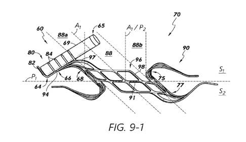
[0027] Figure 9-1 illustrates a side view of a sensor implant device in
accordance
with one or more embodiments.
[0028] Figure 9-2 illustrates a sensor assembly/device in accordance
with one or
more embodiments.
[0029] Figures 10-1, 10-2, and 10-3 show example channel areas
associated with
respective shunt body fluid conduits in accordance with one or more
embodiments.
[0030] Figures 11 and 12 illustrate axial views an embodiment of a shunt-
type
sensor implant device in accordance with one or more embodiments.
4
CA 03208987 2023-07-19
WO 2022/177737
PCT/US2022/014858
[0031] Figure 13 shows a sensor implant device having a suture-wrapped
sensor
device associated therewith in accordance with one or more embodiments.
[0032] Figure 14 shows a sensor implant device having a sensor-retention
pouch
in accordance with one or more embodiments.
[0033] Figure 15 shows a sensor implant device having a sensor-retention
cup in
accordance with one or more embodiments.
[0034] Figures 16-1, 16-2, 16-3, and 16-4 show a sensor implant device
implanted
in a coronary sinus tissue wall in various positions, respectively, in
accordance with one or
more embodiments.
[0035] Figure 17 shows a sensor implant device implanted in an atrial
septum.
with a sensor of the device exposed in a left atrium in accordance with one or
more
embodiments.
[0036] Figures 18 shows a sensor implant device implanted in an atrial
septum.
with a sensor of the device exposed in a right atrium, in accordance with one
or more
embodiments.
[0037] Figure 19 shows a dual-sensor implant device implanted in an
atrial
septum in accordance with one or more embodiments.
[0038] Figure 20 shows a dual-sensor implant device implanted in a wall
separating a coronary sinus from a left atrium in accordance with one or more
embodiments.
[0039] Figure 21 shows a sensor implant device having three sensor
devices
associated therewith in accordance with one or m.ore embodiments.
[0040] Figure 22 shows a sensor implant device having four sensor
devices
associated therewith in accordance with one or more embodiments.
[0041] Figures 23-1, 23-2, 23-3, 23-4, and 23-5 provide a flow diagram
illustrating a process for implanting a sensor implant device in accordance
with one or more
embodiments.
[0042] Figures 24-1, 24-2, 24-3, 24-4, and 24-5 provide images of
cardiac
anatomy and certain devices/systems corresponding to operations of the process
of Figures
23-1, 23-2, 23-3, 23-4, and 23-5 in accordance with one or more embodiments.
[0043] Figure 25 is a cutaway view of a human heart and associated
vasculature
showing certain catheter access paths for pulmonary vein shunting procedures
in accordance
with one or more embodiments.
CA 03208987 2023-07-19
WO 2022/177737
PCT/US2022/014858
DETAILED DESCRIPTION
[0044] The headings provided herein are for convenience only and do not
necessarily affect the scope or meaning of the claimed invention.
[0045] Although certain preferred embodiments and examples are disclosed
below, inventive subject matter extends beyond the specifically disclosed
embodiments to
other alternative embodiments and/or uses and to modifications and equivalents
thereof.
Thus, the scope of the claims that may arise herefrom is not limited by any of
the particular
embodiments described below. For example, in any method or process disclosed
herein, the
acts or operations of the method or process may be performed in any suitable
sequence and
are not necessarily limited to any particular disclosed sequence. Various
operations may be
described as multiple discrete operations in turn, in a manner that may be
helpful in
understanding certain embodiments; however, the order of description should
not be
construed to imply that these operations are order dependent. Additionally,
the structures,
systems, and/or devices described herein may be embodied as integrated
components or as
separate components. For purposes of comparing various embodiments, certain
aspects and
advantages of these embodiments are described. Not necessarily all such
aspects or
advantages are achieved by any particular embodiment. Thus, for example,
various
embodiments may be carried out in a manner that achieves or optimizes one
advantage or
group of advantages as taught herein without necessarily achieving other
aspects or
advantages as may also be taught or suggested herein.
[0046] Certain reference numbers are re-used across different figures of
the figure
set of the present disclosure as a matter of convenience for devices,
components, systems,
features, and/or modules having features that may be similar in one or more
respects.
However, with respect to any of the embodiments disclosed herein, re-use of
common
reference numbers in the drawings does not necessarily indicate that such
features, devices,
components, or modules are identical or similar. Rather, one having ordinary
skill in the art
may be informed by context with respect to the degree to which usage of common
reference
numbers can imply similarity between referenced subject matter. Use of a
particular reference
number in the context of the description of a particular figure can be
understood to relate to
the identified device, component, aspect, feature, module, or system in that
particular figure,
and not necessarily to any devices, components, aspects, features, modules, or
systems
identified by the same reference number in another figure. Furthermore,
aspects of separate
figures identified with common reference numbers can be interpreted to share
characteristics
or to be entirely independent of one another.
6
CA 03208987 2023-07-19
WO 2022/177737
PCT/US2022/014858
[0047] Certain standard anatomical terms of location are used herein to
refer to
the anatomy of animals, and namely humans, with respect to the preferred
embodiments.
Although certain spatially relative terms, such as "outer," "inner," "upper,"
"lower," "below,"
"above," "vertical," "horizontal," "top," "bottom," and similar terms, are
used herein to
describe a spatial relationship of one device/element or anatomical structure
to another
device/element or anatomical structure, it is understood that these terms are
used herein for
ease of description to describe the positional relationship between
element(s)/structures(s), as
illustrated in the drawings. It should be understood that spatially relative
terms are intended
to encompass different orientations of the element(s)/structures(s), in use or
operation, in
addition to the orientations depicted in the drawings. For example, an
element/structure
described as "above" another element/structure may represent a position that
is below or
beside such other element/structure with respect to alternate orientations of
the subject patient
or element/structure, and vice-versa.
[0048] The present disclosure relates to systems, devices, and methods
for
monitoring of one or more physiological parameters of a patient (e.g., blood
pressure) using
sensor-integrated cardiac shunts and/or other medical implant devices. In some
implementations, the present disclosure relates to cardiac shunts and/or other
cardiac implant
devices that incorporate or are associated with pressure sensors or other
sensor devices. The
term "associated with" is used herein according to its broad and ordinary
meaning. For
example, where a first feature, element, component, device, or member is
described as being
"associated with" a second feature, element, component, device, or member,
such description
should be understood as indicating that the first feature, element, component,
device, or
member is physically coupled, attached, or connected to, integrated with,
embedded at least
partially within, or otherwise physically related to the second feature,
element, component,
device, or member, whether directly or indirectly. Certain embodiments are
disclosed herein
in the context of cardiac implant devices. However, although certain
principles disclosed
herein are particularly applicable to the anatomy of the heart, it should be
understood that
sensor implant devices in accordance with the present disclosure may be
implanted in, or
configured for implantation in, any suitable or desirable anatomy.
Cardiac Physiology
[0049] The anatomy of the heart is described below to assist in the
understanding
of certain inventive concepts disclosed herein. In humans and other vertebrate
animals, the
heart generally comprises a muscular organ having four pumping chambers,
wherein the flow
7
CA 03208987 2023-07-19
WO 2022/177737
PCT/US2022/014858
thereof is at least partially controlled by various heart valves, namely, the
aortic, mitral (or
bicuspid), tricuspid, and pulmonary valves. The valves may be configured to
open and close
in response to a pressure gradient present during various stages of the
cardiac cycle (e.g.,
relaxation and contraction) to at least partially control the flow of blood to
a respective region
of the heart and/or to blood vessels (e.g., pulmonary, aorta, etc.).
[0050] Figure 1 illustrates an example representation of a heart I
having various
features relevant to certain embodiments of the present inventive disclosure.
The heart 1
includes four chambers, namely the left atrium 2, the left ventricle 3, the
right ventricle 4, and
the right atrium 5. In terms of blood flow, blood generally flows from the
right ventricle 4
into the pulmonary artery 11 via the pulmonary valve 9, which separates the
right ventricle 4
from the pulmonary artery 11 and is configured to open during systole so that
blood may be
pumped toward the lungs and close during diastole to prevent blood from
leaking back into
the heart from the pulmonary artery ii. The pulmonary artery 11 carries
deoxygenated blood
from the right side of the heart to the lungs.
[0051] In addition to the pulmonary valve 9, the heart 1 includes three
additional
valves for aiding the circulation of blood therein, including the tricuspid
valve 8, the aortic
valve 7, and the mitral valve 6. The tricuspid valve 8 separates the right
atrium 5 from the
right ventricle 4. The tricuspid valve 8 generally has three cusps or leaflets
and may generally
close during ventricular contraction (i.e., systole) and open during
ventricular expansion (i.e.,
diastole). The mitral valve 6 generally has two cusps/leaflets and separates
the left atrium 2
from the left ventricle 3. The mitral valve 6 is configured to open during
diastole so that
blood in the left atrium 2 can flow into the left ventricle 3, and, when
functioning properly,
closes during systole to prevent blood from leaking back into the left atrium
2. The aortic
valve 7 separates the left. ventricle 3 from the aorta 12. The aortic valve 7
is configured to
open during systole to allow blood leaving the left ventricle 3 to enter the
aorta 1.2, and close
during diastole to prevent blood from leaking back into the left ventricle 3.
[0052] The heart valves may generally comprise a relatively dense
fibrous ring,
referred to herein as the annulus, as well as a plurality of leaflets or cusps
attached to the
annulus. Generally, the size of the leaflets or cusps may be such that when
the heart contracts
the resulting increased blood pressure produced within the corresponding heart
chamber
forces the leaflets at least partially open to allow flow from the heart
chamber. As the
pressure in the heart chamber subsides, the pressure in the subsequent chamber
or blood
vessel may become dominant and press back against the leaflets. As a result,
the
leaflets/cusps come in apposition to each other, thereby closing the flow
passage. Disfunction
8
CA 03208987 2023-07-19
WO 2022/177737
PCT/US2022/014858
of a heart valve and/or associated leaflets (e.g., pulmonary valve
disfunction) can result in
valve leakage and/or other health complications.
[0053] The atrioventricular (i.e., mitral and tricuspid) heart valves
may further
comprise a collection of chordae tendineae and papillary muscles (not shown)
for securing
the leaflets of the respective valves to promote and/or facilitate proper
coaptation of the valve
leaflets and prevent prolapse thereof. The papillary m.uscles, for example,
m.ay generally
comprise finger-like projections from the ventricle wall. The valve leaflets
are connected to
the papillary muscles by the chordae tendineae. A wall of muscle, referred to
as the septum,
separates the left-side chambers from the right-side chambers. In particular,
an atrial septum
wall portion 18 (referred to herein as the "atrial septum," "atrial septum,"
or "septum")
separates the left atrium 2 from the right atrium 5, whereas a ventricular
septum wall portion
17 (referred to herein as the "ventricular septum," "interventricular septum,"
or "septum")
separates the left ventricle 3 from the right ventricle 4. The inferior tip of
the heart 1 is
referred to as the apex and is generally located on or near the midclavicular
line, in the fifth
intercostal space.
[0054] The coronary sinus 16 comprises a collection of veins joined
together to
form a large vessel that collects blood from the heart muscle (myocardium).
The ostium of
the coronary sinus, which can be guarded at least in part by a Thebesian valve
in some
patients, is open to the right atrium 5, as shown. The coronary sinus runs
along a posterior
aspect of the left atrium 2 and delivers less-oxygenated blood to the right
atrium 5. The
coronary sinus generally runs transversely in the left atrioventricular groove
on the posterior
side of the heart.
Health Conditions Associated with Cardiac Pressure and Other Parameters
[0055] As referenced above, certain physiological conditions or
parameters
associated with the cardiac anatomy can impact the health of a patient. For
example,
congestive heart failure is a condition associated with the relatively slow
movement of blood
through the heart and/or body, which causes the fluid pressure in one or more
chambers of
the heart to increase. As a result, the heart does not pump sufficient oxygen
to meet the
body's needs. The various chambers of the heart m.ay respond to pressure
increases by
stretching to hold more blood to pump through the body or by becoming
relatively stiff
and/or thickened. The walls of the heart can eventually weaken and become
unable to pump
as efficiently. In some cases, the kidneys may respond to cardiac inefficiency
by causing the
body to retain fluid. Fluid build-up in arms, legs, ankles, feet, lungs,
and/or other organs can
9
CA 03208987 2023-07-19
WO 2022/177737
PCT/US2022/014858
cause the body to become congested, which is referred to as congestive heart
failure. Acute
decompensated congestive heart failure is a leading cause of morbidity and
mortality, and
therefore treatment and/or prevention of congestive heart failure is a
significant concern in
medical care.
[0056] The treatment and/or prevention of heart failure (e.g.,
congestive heart
failure) can advantageously involve the monitoring of pressure in one or more
chambers or
regions of the heart or other anatomy. As described above, pressure buildup in
one or more
chambers or areas of the heart can be associated with congestive heart
failure. Without direct
or indirect monitoring of cardiac pressure, it can be difficult to infer,
determine, or predict the
presence or occurrence of congestive heart failure. For example, treatments or
approaches not
involving direct or indirect pressure monitoring may involve measuring or
observing other
present physiological conditions of the patient, such as measuring body
weight, thoracic
impedance, right heart catheterization, or the like. In some solutions,
pulmonary capillary
wedge pressure can be measured as a surrogate of left atrial pressure. For
example, a pressure
sensor may be disposed or implanted in the pulmonary artery, and readings
associated
therewith may be used as a surrogate for left atrial pressure. However, with
respect to
catheter-based pressure measurement in the pulmonary artery or certain other
chambers or
regions of the heart, use of invasive catheters may be required to maintain
such pressure
sensors, which may be uncomfortable or difficult to implement. Furthermore,
certain lung-
related conditions may affect pressure readings in the pulmonary artery, such
that the
correlation between pulmonary artery pressure and left atrial pressure may be
undesirably
attenuated. As an alternative to pulmonary artery pressure measurement,
pressure
measurements in the right ventricle outflow tract may relate to left atrial
pressure as well.
However, the correlation between such pressure readings and left atrial
pressure may not be
sufficiently strong to be utilized in congestive heart failure diagnostics,
prevention, and/or
treatment.
[0057] Additional solutions may be implemented for deriving or inferring
left
atrial pressure. For example, the E/A ratio, which is a marker of the function
of the left
ventricle of the heart representing the ratio of peak velocity blood flow from
gravity in early
diastole (the E wave) to peak velocity flow in late diastole caused by atrial
contraction (the A
wave), can be used as a surrogate for measuring left atrial pressure. The E/A
ratio may be
determined using echocardiography or other imaging technology; generally,
abnormalities in
the E/A ratio may suggest that the left ventricle cannot fill with blood
properly in the period
between contractions, which may lead to symptoms of heart failure, as
explained above.
CA 03208987 2023-07-19
WO 2022/177737
PCT/US2022/014858
However, E/A ratio determination generally does not provide absolute pressure
measurement
values.
[0058] Various methods for identifying and/or treating congestive heart
failure
involve the observation of worsening congestive heart failure symptoms and/or
changes in
body weight. However, such signs may appear relatively late and/or be
relatively unreliable.
For example, daily bodyweight measurements may vary significantly (e.g., up to
9% or more)
and may be unreliable in signaling heart-related complications. Furthermore,
treatments
guided by monitoring signs, symptoms, weight, and/or other biomarkers have not
been shown
to substantially improve clinical outcomes. in addition, for patients that
have been
discharged, such treatments may necessitate remote telemedicine systems.
[0059] The present disclosure provides systems, devices, and methods for
guiding
the administration of medication relating to the treatment of congestive heart
failure at least
in part by directly monitoring pressure in the left atrium, or other chamber
or vessel for which
pressure measurements are indicative of left atrial pressure and/or pressure
levels in one or
more other vessels/chambers, such as for congestive heart failure patients in
order to reduce
hospital readmissions, morbidity, and/or otherwise improve the health
prospects of the
patient.
Cardiac Pressure Monitoring
[0060] Cardiac pressure monitoring in accordance with embodiments of the
present disclosure may provide a proactive intervention mechanism for
preventing or treating
congestive heart failure and/or other physiological conditions. Generally,
increases in
ventricular filling pressures associated with diastolic and/or systolic heart
failure can occur
prior to the occurrence of symptoms that lead to hospitalization. For example,
cardiac
pressure indicators may present weeks prior to hospitalization with respect to
some patients.
Therefore, pressure monitoring systems in accordance with embodiments of the
present
disclosure may advantageously be implemented to reduce instances of
hospitalization by
guiding the appropriate or desired titration and/or administration of
medications before the
onset of heart failure.
[0061] Dyspnea represents a cardiac pressure indicator characterized by
shortness
of breath or the feeling that one cannot breathe well enough. Dyspnea may
result from
elevated atrial pressure, which may cause fluid buildup in the lungs from
pressure back-up.
Pathological dyspnea can result from congestive heart failure. However, a
significant amount
of time may elapse between the time of initial pressure elevation and the
onset of dyspnea,
11
CA 03208987 2023-07-19
WO 2022/177737
PCT/US2022/014858
and therefore symptoms of dyspnea may not provide sufficiently-early signaling
of elevated
atrial pressure. By monitoring pressure directly according to embodiments of
the present
disclosure, normal ventricular filling pressures m.ay advantageously be
maintained, thereby
preventing or reducing effects of heart failure, such as dyspnea.
[0062] As referenced above, with respect to cardiac pressures, pressure
elevation
in the left atrium may be particularly correlated with heart failure. Figure 2
illustrates
example pressure waveforms associated with various chambers and vessels of the
heart
according to one or more embodiments. The various waveforms illustrated in
Figure 2 may
represent waveforms obtained using right heart catheterization to advance one
or more
pressure sensors to the respective illustrated and labeled chambers or vessels
of the heart. As
illustrated in Figure 2, the waveform 25, which represents left atrial
pressure, may be
considered to provide the best feedback for early detection of congestive
heart failure.
Furthermore, there may generally be a relatively strong correlation between
increases and left
atrial pressure and pulmonary congestion.
[0063] Left atrial pressure may generally correlate well with left
ventricular end-
diastolic pressure. However, although left atrial pressure and end-diastolic
pulmonary artery
pressure can have a significant correlation, such correlation may be weakened
when the
pulmonary vascular resistance becomes elevated. That is, pulmonary artery
pressure
generally fails to correlate adequately with left ventricular end-diastolic
pressure in the
presence of a variety of acute conditions, which may include certain patients
with congestive
heart failure. For example, pulmonary hypertension, which affects
approximately 25% to
83% of patients with heart failure, can affect the reliability of pulmonary
artery pressure
measurement for estimating left-sided filling pressure. Therefore, pulmonary
artery pressure
measurement alone, as represented by the waveform 24, may be an insufficient
or inaccurate
indicator of left ventricular end-diastolic pressure, particularly for
patients with co-
morbidities, such as lung disease and/or thromboembolism. Left atrial pressure
may further
be correlated at least partially with the presence and/or degree of mitral
regurgitation.
[0064] Lett atrial pressure readings may be relatively less likely to be
distorted or
affected by other conditions, such as respiratory conditions or the like,
compared to the other
pressure waveforms shown in Figure 2. Generally, left atrial pressure may be
significantly
predictive of heart failure, such as up two weeks before manifestation of
heart failure. For
example, increases in left atrial pressure, and both diastolic and systolic
heart failure, may
occur weeks prior to hospitalization, and therefore knowledge of such
increases may be used
12
CA 03208987 2023-07-19
WO 2022/177737
PCT/US2022/014858
to predict the onset of congestive heart failure, such as acute debilitating
symptoms of
congestive heart failure.
[0065] Cardiac pressure monitoring, such as left atrial pressure
monitoring, can.
provide a mechanism to guide administration of medication to treat and/or
prevent congestive
heart failure. Such treatments may advantageously reduce hospital readmissions
and
morbidity, as well as provide other benefits. An implanted pressure sensor in
accordance with
embodiments of the present disclosure may be used to predict heart failure up
two weeks or
more before the manifestation of symptoms or markers of heart failure (e.g.,
dyspnea). When
heart failure predictors are recognized using cardiac pressure sensor
embodiments in
accordance with the present disclosure, certain prophylactic measures may be
implemented,
including medication intervention, such as modification to a patient's
medication regimen,
which may help prevent or reduce the effects of cardiac dysfunction. Direct
pressure
measurement in the left atrium can advantageously provide an accurate
indicator of pressure
buildup that may lead to heart failure or other complications. For example,
trends of atrial
pressure elevation may be analyzed or used to determine or predict the onset
of cardiac
dysfunction, wherein drug or other therapy may be augmented to cause reduction
in pressure
and prevent or reduce further complications.
[0066] Figure 3 illustrates a graph 300 showing left atrial pressure
ranges
including a normal range 301 of left atrial pressure that is not generally
associated with
substantial risk of postoperative atrial fibrillation, acute kidney injury,
myocardial injury,
heart failure and/or other health conditions. Embodiments of the present
disclosure provide
systems, devices, and methods for determining whether a patient's left atrial
pressure is
within the normal range 301, above the normal range 303, or below the normal
range 302
through the use of certain sensor implant devices. For detected left atrial
pressure above the
normal range, which m.ay be correlated with an increased risk of heart
failure, embodiments
of the present disclosure as described in detail below can inform efforts to
reduce the left
atrial pressure until it is brought within the normal range 301. Furthermore,
for detected left
atrial pressure that is below the normal range 301, which may be correlated
with increased
risks of acute kidney injury, myocardial injury, and/or other health
complications,
embodiments of the present disclosure as described in detail below can serve
to facilitate
efforts to increase the left atrial pressure to bring the pressure level
within the normal range
301.
13
CA 03208987 2023-07-19
WO 2022/177737
PCT/US2022/014858
Implant Devices with Integrated Sensors
[0067] In some implementations, the present disclosure relates to
sensors
associated or integrated with cardiac shunts or other implant devices. Such
integrated devices
may be used to provide controlled and/or more effective therapies for treating
and preventing
heart failure and/or other health complications related to cardiac function.
Figure 4 is a block
diagram illustrating an implant device 30 comprising a shunt (or other type of
implant)
structure 39. In some embodiments, the shunt structure 39 is physically
integrated with and/or
connected to a sensor device 37. The sensor device 37 may be, for example, a
pressure
sensor, or other type of sensor. In some embodiments, the sensor 37 comprises
a transducer
32, such as a pressure transducer, as well as certain control circuitry 34,
which may be
embodied in, for example, an application-specific integrated circuit (ASIC).
[0068] The control circuitry 34 may be configured to process signals
received
from the transducer 32 and/or comm.unicate signals associated therewith
wirelessly through
biological tissue using the antenna 38. The term "control circuitry" is used
herein according
to its broad and ordinary meaning, and may refer to any collection of
processors, processing
circuitry, processing modules/units, chips, dies (e.g., semiconductor dies
including come or
more active and/or passive devices and/or connectivity circuitry),
microprocessors, micro-
controllers, digital signal processors, microcomputers, central processing
units, field
programmable gate arrays, programmable logic devices, state machines (e.g.,
hardware state
machines), logic circuitry, analog circuitry, digital circuitry, and/or any
device that
manipulates signals (analog and/or digital) based on hard coding of the
circuitry and/or
operational instructions. Control circuitry referenced herein m.ay further
comprise one or
more, storage devices, which may be embodied in a single memory device, a
plurality of
memory devices, and/or embedded circuitry of a device. Such data storage may
comprise
read-only memory, random, access memory, volatile memory, non-volatile memory,
static
memory, dynamic memory, flash memory, cache memory, data storage registers,
and/or any
device that stores digital information. It should be noted that in embodiments
in which
control circuitry comprises a hardware and/or software state machine, analog
circuitry, digital
circuitry, and/or logic circuitry, data storage device(s)/register(s) storing
any associated
operational instructions may be embedded within, or external to, the circuitry
comprising the
state machine, analog circuitry, digital circuitry, and/or logic circuitry.
The transducer(s) 32
and/or antenna(s) 38 can be considered part of the control circuitry 34.
[0069] The antenna 38 may comprise one or more coils or loops of
conductive
material, such as copper wire or the like. In some embodiments, at least a
portion of the
14
CA 03208987 2023-07-19
WO 2022/177737
PCT/US2022/014858
transducer 32, control circuitry 34, and/or the antenna 38 are at least
partially disposed or
contained within a sensor housing 36, which may comprise any type of material,
and may
advantageously be at least partially hermetically sealed. For example, the
housing 36 may
comprise glass or other rigid material in some embodiments, which may provide
mechanical
stability and/or protection for the components housed therein. In some
embodiments, the
housing 36 is at least partially flexible. For example, the housing may
comprise polymer or
other flexible structure/material, which may advantageously allow for folding,
bending, or
collapsing of the sensor 37 to allow for transportation thereof through a
catheter or other
introducing means.
[0070] The transducer 32 may comprise any type of sensor means or
mechanism..
For example, the transducer 32 may be a force-collector-type pressure sensor.
In some
embodiments, the transducer 32 comprises a diaphragm, piston, bourdon tube,
bellows, or
other strain- or deflection-measuring component(s) to measure strain or
deflection applied
over an area/surface thereof. The transducer 32 may be associated with the
housing 36, such
that at least a portion thereof is contained within or attached to the housing
36. With respect
to sensor devices/components being "associated with" a stent or other implant
structure, such
terminology may refer to a sensor device or component being physically
coupled, attached, or
connected to, or integrated with., the implant structure.
[0071] in some embodiments, the transducer 32 comprises or is a
component of a
piezoresistive strain gauge, which may be configured to use a bonded or formed
strain gauge
to detect strain due to applied pressure, wherein resistance increases as
pressure deforms the
component/material. The transducer 32 may incorporate any type of material,
including but
not limited to silicon (e.g., monocrystalline), polysilicon thin film, bonded
metal foil, thick
film, silicon-on-sapphire, sputtered thin film., and/or the like.
[0072] In some embodiments, the transducer 32 comprises or is a
component of a
capacitive pressure sensor including a diaphragm and pressure cavity
configured to form a
variable capacitor to detect strain due to pressure applied to the diaphragm.
The capacitance
of the capacitive pressure sensor may generally decrease as pressure deforms
the diaphragm.
The diaphragm may comprise any material(s), including but not limited to
metal, ceramic,
silicon, and the like. In some embodiments, the transducer 32 comprises or is
a component of
an electromagnetic pressure sensor, which may be configured to measure the
displacement of
a diaphragm by means of changes in inductance, linear variable displacement
transducer
(I,VDT) functionality, Hall Effect, or eddy current sensing. In. some
embodiments, the
transducer 32 comprises or is a component of a piezoelectric strain sensor.
For example, such
CA 03208987 2023-07-19
WO 2022/177737
PCT/US2022/014858
a sensor may determine strain (e.g., pressure) on a sensing mechanism based on
the
piezoelectric effect in certain materials, such as quartz.
[0073] In some embodiments, the transducer 32 comprises or is a
component of a
strain gauge. For example, a strain gauge embodiment may comprise a pressure
sensitive
element on or associated with an exposed surface of the transducer 32. In some
embodiments,
a metal strain gauge is adhered to a surface of the sensor, or a thin-film
gauge may be applied
on the sensor by sputtering or other technique. The measuring element or
mechanism may
comprise a diaphragm or metal foil. The transducer 32 may comprise any other
type of sensor
or pressure sensor, such as optical, potentiometric, resonant, thermal,
ionization, or other
types of strain or pressure sensors.
[0074] Figure 5 shows a system 40 for monitoring one or more
physiological
parameters (e.g., left atrial pressure and/or volume) in a patient 44
according to one or more
embodiments. The patient 44 can have a medical implant device 30 implanted in,
for
example, the heart (not shown), or associated physiology, of the patient 44.
For example, the
implant device 30 can be implanted at least partially within the left atrium
and/or coronary
sinus of the patient's heart. The implant device 30 can include one or more
sensor transducers
32, such as one or more microelectrom.echanical system (MEMS) devices (e.g.,
MEMS
pressure sensors, or other type of sensor transducer).
[0075] in certain embodiments, the monitoring system 40 can comprise at
least
two subsystems, including an implantable internal subsystem or device 30 that
includes the
sensor transducer(s) 32, as well as control circuitry 34 comprising one or
more
microcontroller(s), discrete electronic component(s), and one or more power
and/or data
transmitter(s) 38 (e.g., antennae coil). The monitoring system 40 can further
include an
external (e.g., non-implantable) subsystem that includes an external reader 42
(e.g., coil),
which may include a wireless transceiver that is electrically and/or
communicatively coupled
to certain control circuitry 41. In certain embodiments, both the internal 30
and external 42
subsystems include a corresponding coil antenna for wireless communication
and/or power
delivery through patient tissue disposed therebetween. The sensor implant
device 30 can be
any type of implant device. For example, in some embodiments, the implant
device 30
comprises a pressure sensor integrated with another functional implant
structure 39, such as a
prosthetic shunt or stent device/structure.
[0076] Certain details of the implant device 30 are illustrated in the
enlarged
block 30 shown. The implant device 30 can comprise an implant/anchor structure
39 as
described herein. For example, the implant/anchor structure 39 can include a
percutaneously-
16
CA 03208987 2023-07-19
WO 2022/177737
PCT/US2022/014858
deliverable shunt device configured to be secured to and/or in a tissue wall
to provide a flow
path between two chambers and/or vessels of the heart, as described in detail
throughout the
present disclosure. Although certain components are illustrated in Figure 5 as
part of the
implant device 30, it should be understood that the sensor implant device 30
may only
comprise a subset of the illustrated components/modules and can comprise
additional
components/modules not illustrated. The implant device may represent an
embodiment of the
implant device shown in Figure 4, and vice versa. The implant device 30 can
advantageously
include one or more sensor transducers 32, which can be configured to provide
a response
indicative of one or more physiological parameters of the patient 44, such as
atrial pressure.
Although pressure transducers are described, the sensor transducer(s) 32 can
comprise any
suitable or desirable types of sensor transducer(s) for providing signals
relating to
physiological parameters or conditions associated with the implant device 30
and/or patient
44.
[0077] The sensor transducer(s) 32 can comprise one or more MEMS
sensors,
optical sensors, piezoelectric sensors, electromagnetic sensors, strain
sensors/gauges,
accelerometers, gyroscopes, diaphragm-based sensors, and/or other types of
sensors, which
can be positioned in the patient 44 to sense one or more parameters relevant
to the health of
the patient. The transducer 32 may be a force-collector-type pressure sensor.
In some
embodiments, the transducer 32 comprises a diaphragm, piston, bourdon tube,
bellows, or
other strain- or deflection-measuring component(s) to measure strain or
deflection applied
over an area/surface thereof. The transducer 32 may be associated with the
sensor housing 36,
such that at least a portion thereof is contained within, or attached to, the
housing 36.
[0078] In some embodiments, the transducer 32 comprises or is a
component of a
strain gauge, which may be configured to use a bonded or formed strain gauge
to detect strain
due to applied pressure. For example, the transducer 32 may comprise or be a
component of a
piezoresistive strain gauge, wherein resistance increases as pressure deforms
the
component/material of the strain gauge. The transducer 32 may incorporate any
type of
material, including but not limited to silicone, polymer, silicon (e.g.,
monocrystalline),
polysilicon thin film., bonded metal foil, thick film, silicon-on-sapphire,
sputtered thin film.,
and/or the like. In some embodiments, a metal strain gauge is adhered to the
sensor surface,
or a thin-film gauge may be applied on the sensor by sputtering or other
technique. The
measuring element or mechanism may comprise a diaphragm or metal foil. The
transducer 32
may comprise any other type of sensor or pressure sensor, such as optical,
potentiometric,
resonant, thermal, ionization, or other types of strain or pressure sensors.
17
CA 03208987 2023-07-19
WO 2022/177737
PCT/US2022/014858
[0079] In some embodiments, the transducer 32 comprises or is a
component of a
capacitive pressure sensor including a diaphragm and pressure cavity
configured to form a
variable capacitor to detect strain due to pressure applied to the diaphragm.
The capacitance
of the capacitive pressure sensor may generally decrease as pressure deforms
the diaphragm.
The diaphragm may comprise any material(s), including but not limited to
metal, ceramic,
silicone, silicon or other semiconductor, and the like. In some embodiments,
the transducer
32 comprises or is a component of an electromagnetic pressure sensor, which
may be
configured to measures the displacement of a diaphragm by means of changes in
inductance,
linear variable displacement transducer (LVDT) functionality, Hall Effect, or
eddy current
sensing. In some embodiments, the transducer 32 comprises or is a component of
a
piezoelectric strain sensor. For example, such a sensor may determine strain
(e.g., pressure)
on a sensing mechanism based on the piezoelectric effect in certain materials,
such as quartz.
[0080] In some embodiments, the transducer(s) 32 is/are electrically
and/or
communicatively coupled to the control circuitry 34, which may comprise one or
more
application-specific integrated circuit (ASIC) microcontrollers or chips. The
control circuitry
34 can further include one or more discrete electronic components, such as
tuning capacitors,
resistors, diodes, inductors, or the like.
[0081] In certain embodiments, the sensor transducer(s) 32 can be
configured to
generate electrical signals that can be wirelessly transmitted to a device
outside the patient's
body, such as the illustrated local external monitor system 42. In order to
perform such
wireless data transmission, the implant device 30 can include radio frequency
(RF) (or other
frequency band) transmission circuitry, such as signal processing circuitry
and an antenna 38.
The antenna 38 can comprise an antenna coil implanted within the patient. The
control
circuitry 34 may comprise any type of transceiver circuitry configured to
transmit an
electromagnetic signal, wherein the signal can. be radiated by the antenna 38,
which may
comprise one or more conductive wires, coils, plates, or the like. The control
circuitry 34 of
the implant device 30 can comprise, for example, one or more chips or dies
configured to
perform some amount of processing on signals generated and/or transmitted
using the device
30. However, due to size, cost, and/or other constraints, the im.plant device
30 may not
include independent processing capability in some embodiments.
[0082] The wireless signals generated by the implant device 30 can be
received by
the local external monitor device or subsystem 42, which can include a
reader/antenna-
interface circuitry module 43 configured to receive the wireless signal
transmissions from the
18
CA 03208987 2023-07-19
WO 2022/177737
PCT/US2022/014858
implant device 30, which is disposed at least partially within the patient 44.
For example, the
module 43 may include transceiver device(s)/circuitry.
[0083] The external local monitor 42 can receive the wireless signal
transmissions
from the implant device 30 and/or provide wireless power to the implant device
30 using an
external antenna 48, such as a wand device. The reader/antenna-interface
circuitry 43 can
include radio-frequency (12/7) (or other frequency band) front-end circuitry
configured to
receive and amplify the signals from the implant device 30, wherein such
circuitry can
include one or more filters (e.g., band-pass filters), amplifiers (e.g., low-
noise amplifiers),
analog-to-digital converters (ADC) and/or digital control interface circuitry,
phase-locked
loop (NI) circuitry, signal mixers, or the like. The reader/antenna-interface
circuitry 43 can
further be configured to transmit signals over a network 49 to a remote
monitor subsystem or
device 46. The RF circuitry of the reader/antenna-interface circuitry 43 can
further include
one or more of digital-to-analog convener (DAC) circuitry, power amplifiers,
low-pass
filters, antenna switch modules, antennas or the like for treatment/processing
of transmitted
signals over the network 49 and/or for receiving signals from the implant
device 30. In
certain embodiments, the local monitor 42 includes control circuitry 41 for
performing
processing of the signals received from the implant device 30. The local
monitor 42 can be
configured to communicate with the network 49 according to a known network
protocol, such
as Ethernet, Wi-Fi, or the like. In certain embodiments, the local monitor 42
comprises a
smartphone, laptop computer, or other mobile computing device, or any other
type of
computing device.
[0084] In certain embodiments, the implant device 30 includes some
amount of
volatile and/or non-volatile data storage. For example, such data storage can
comprise solid-
state memory utilizing an array of floating-gate transistors, or the like. The
control circuitry
34 may utilize data storage for storing sensed data collected over a period of
time, wherein
the stored data can be transmitted periodically to the local monitor 42 or
another external
subsystem. In certain embodiments, the implant device 30 does not include any
data storage.
The control circuitry 34 may be configured to facilitate wireless transmission
of data
generated by the sensor transducer(s) 32, or other data associated therewith.
The control
circuitry 34 may further be configured to receive input from one or more
external
subsystems, such as from the local monitor 42, or from a remote monitor 46
over, for
example, the network 49. For example, the implant device 30 may be configured
to receive
signals that at least partially control the operation of the implant device
30, such as by
19
CA 03208987 2023-07-19
WO 2022/177737
PCT/US2022/014858
activating/deactivating one or more components or sensors, or otherwise
affecting operation
or performance of the implant device 30.
[0085] The one or m.ore components of the implant device 30 can be
powered by
one or more power sources 35. Due to size, cost and/or electrical complexity
concerns, it may
be desirable for the power source 35 to be relatively minimalistic in nature.
For example,
high-power driving voltages and/or currents in the implant device 30 may
adversely affect or
interfere with operation of the heart or other body part associated with the
implant device. In
certain embodiments, the power source 35 is at least partially passive in
nature, such that
power can be received from an external source wirelessly by passive circuitry
of the implant
device 30, such as through the use of short-range, or near-field wireless
power transmission,
or other electromagnetic coupling mechanism. For example, the local monitor 42
may serve
as an initiator that actively generates an RF field that can provide power to
the implant device
30, thereby allowing the power circuitry of the implant device to take a
relatively simple form.
factor. In certain embodiments, the power source 35 can. be configured to
harvest energy from
environmental sources, such as fluid flow, motion, or the like. Additionally
or alternatively,
the power source 35 can comprise a battery, which can advantageously be
configured to
provide enough power as needed over the monitoring period (e.g., 3, 5, 10, 20,
30, 40, or 90
days, or other period of time).
[0086] in some embodiments, the local monitor device 42 can serve as an
intermediate communication device between the implant device 30 and the remote
monitor
46. The local monitor device 42 can be a dedicated external unit designed to
communicate
with the implant device 30. For example, the local monitor device 42 can be a
wearable
communication device, or other device that can be readily disposed in
proximity to the
patient 44 and implant device 30. The local monitor device 42 can be
configured to
continuously, periodically, or sporadically interrogate the implant device 30
in order to
extract or request sensor-based information therefrom. In certain embodiments,
the local
monitor 42 comprises a user interface, wherein a user can utilize the
interface to view sensor
data, request sensor data, or otherwise interact with the local monitor
system. 42 and/or
implant device 30.
[0087] The system 40 can include a secondary local monitor 47, which can
be, for
example, a desktop computer or other computing device configured to provide a
monitoring
station or interface .for viewing and/or interacting with the monitored
cardiac pressure data. In
an. embodiment, the local monitor 42 can be a wearable device or other device
or system
configured to be disposed in close physical proximity to the patient and/or
implant device 30,
CA 03208987 2023-07-19
WO 2022/177737
PCT/US2022/014858
wherein the local monitor 42 is primarily designed to receive/transmit signals
to and/or from
the implant device 30 and provide such signals to the secondary local monitor
47 for viewing,
processing, and/or manipulation thereof. The external local monitor system 42
can be
configured to receive and/or process certain metadata from or associated with
the implant
device 30, such as device ID or the like, which can also be provided over the
data coupling
from the implant device 30.
[0088] The remote monitor subsystem 46 can be any type of computing
device or
collection of computing devices configured to receive, process and/or present
monitor data
received over the network 49 from the local monitor device 42, secondary local
monitor 47,
and/or implant device 30. For example, the remote monitor subsystem 46 can
advantageously
be operated and/or controlled by a healthcare entity, such as a hospital,
doctor, or other care
entity associated with the patient 44. Although certain embodiments disclosed
herein describe
communication with the remote monitor subsystem. 46 from the implant device
indirectly
through the local monitor device 42, in certain embodiments, the implant
device 30 can
comprise a transmitter capable of communicating over the network 49 with the
remote
monitor subsystem 46 without the necessity of relaying information through the
local monitor
device 42.
[0089] In some embodiments, at least a portion of the transducer 32,
control
circuitry 34, power source 35 and/or the antenna 38 are at least partially
disposed or
contained within the sensor housing 36, which may comprise any type of
material, and may
advantageously be at least partially hermetically sealed. For example, the
housing 36 may
comprise glass or other rigid material in some embodiments, which may provide
mechanical
stability and/or protection for the components housed therein. In some
embodiments, the
housing 36 is at least partially flexible. For example, the housing may
comprise polymer or
other flexible structure/material, which may advantageously allow for folding,
bending, or
collapsing of the sensor 30 to allow for transportation thereof through a
catheter or other
percutaneous introducing means.
Cardiac Shunt Implants
[0090] Figure 6 illustrates an. example shunt/anchor structure 150 in
accordance
with one or more embodiments. The shunt structure 150 may represent an
embodiment of a
cardiac implant (e.g., anchor and/or cardiac implant structure 39 associated
with Figure 4 or
5) that may be integrated with pressure sensor functionality in accordance
with certain
embodiments disclosed herein. The shunt structure 150 may be an expandable
shunt. When
21
CA 03208987 2023-07-19
WO 2022/177737
PCT/US2022/014858
expanded, a central flow channel 166 of the shunt 150 may define a generally
circular or oval
opening. The channel 1.66 may be configured to hold the sides of a puncture
opening in a
tissue wall to form a blood flow path between chamber(s) or vessel(s) of the
heart that are
separated by the tissue wall. For example, the shunt 150 may be configured to
be implanted
in the wall separating the coronary sinus and the left atrium. The central
flow channel 166
may be partly formed by a pair of side walls I 70a, 170b defined by a
generally parallel
arrangement of thin struts 179 that forms an array of parallelogram-shaped
cells or openings
180. In some embodiments, substantially the entire shunt 150 is formed by
super-elastic struts
that are configured to be compressed and fit into a catheter (not shown) and
subsequently
expanded back to the relaxed shape as shown in Figure 6.
[0091] Formation of the shunt 150 using a plurality of interconnected
struts
forming cells therebetween may serve to at least partially increase the
flexibility of the shunt,
thereby enabling compression thereof and expansion at the implant site. The
interconnected
struts around the central flow channel 166 advantageously provide a cage
having sufficient
rigidity and structure to hold the tissue at the puncture in an open position.
End walls 172a,
172b of the central flow channel 166 can serve to connect the side walls 170a,
170b and
extend between distal and proximal flanges, or arms, 152, 154 on each side.
The side walls
170a, 170b and end walls 172a, 172b together may define a tubular lattice, as
shown. The end
walls 172a, 172b can comprise thin struts 179 extending at a slight angle from
a central flow
axis of the shunt 150.
[0092] Although the illustrated shunt 150 comprises struts that define a
tubular or
circular lattice of open cells forming the central flow channel 166, in some
embodiments, the
structure that makes up the channel forms a substantially contiguous wall
surface through at
least a portion of the channel 166. In the illustrated embodiment, the tilt of
the shunt structure
150 may facilitate collapse of the shunt into a delivery catheter (not shown),
as well as the
expansion of the flanges/arm 152, 154 on both sides of a target tissue wall.
The central flow
channel 166 may remain essentially unchanged between the collapsed and
expanded states of
the shunt 150, whereas the flanges/arms 152, 154 m.ay transition in and out of
alignment with
the angled flow channel.
[0093] Although certain embodiments of shunts disclosed herein comprise
flow
channels having substantially circular cross-sections, in some embodiments,
shunt structures
in accordance with the present disclosure have oval-shaped, rectangular,
diamond-shaped, or
elliptical flow channel configuration. For example, relatively elongated side
walls compared
to the illustrated configuration of Figure 6 may produce a rectangular or oval-
shaped flow
22
CA 03208987 2023-07-19
WO 2022/177737
PCT/US2022/014858
channel. Such shapes of shunt flow channels may be desirable for larger
punctures, while still
being configured to collapse down to a relatively small delivery profile.
[0094] In some embodiments, each of the distal and proximal flanges/anns
152,
154 is configured to curl outward from the end walls 172a, 172b and be set to
point
approximately radially away from. the central flow channel 166 in the expanded
configuration. The expanded flanges/arms may serve to secure the shunt 150 to
a target tissue
wall. Additional aspects and features of shunt, implant, and/or anchor
structures that may be
integrated with sensor devices/functionality of embodiments of the present
disclosure are
disclosed in U.S. Pat. No. 9,789,294, entitled "Expandable Cardiac Shunt,"
issued on October
17, 2017, the disclosure of which is hereby expressly incorporated by
reference in its entirety.
Although certain embodiments are disclosed herein in the context of shunt
structures similar
to that shown in Figure 6 and described above, it should be understood that
shunt structures
or other implant devices integrated with pressure sensor functionality in
accordance with
embodiments of th.e present disclosure may have any type, form, structure,
configuration,
and/or may be used or configured to be used for any purpose, whether for
shunting or other
purpose or functionality.
[0095] Figures 7 shows a shunt implant/anchor device/structure 73
implanted in
an atrial septum. 18 in accordance with one or more embodiments. The
particular position in
the atrial septum wall 18 may be selected or determined to provide a
relatively secure anchor
location for the shunt structure 73. Furthermore, the shunt device/structure
73 may be
implanted at a position that is desirable in consideration of future re-
crossing of the septal
wall 18 for future interventions. Implantation of the shunt device/structure
73 in the atrial
septum wall 18 may advantageously allow for fluid communication between the
left 2 and
rights atria.
[0096] Interatrial shunting using the shunt device/structure 73 may be
well-suited
for patients that are relatively highly sensitive to atrial pressure
increases. For example, as
pressure increases in the ventricles and/or atria and is applied against the
myocardial cells,
the muscles of the heart may generally be prone to contract relatively harder
to process the
excess blood. Therefore, as the ventricle dilates or stretches, for patients
with compromised
contractility of the ventricle, such patients may become more sensitive to
higher pressures in
the ventricle and/or atria because the heart may be unable to adequately
respond or react
thereto. Furthermore, increases in left atrial pressure can results in
dyspnea, and therefore
reduction in left atrial pressure to reduce dyspnea and/or reduce incidences
of hospital
readmission may be desirable through interatrial shunting. For example, when
the ventricle
23
CA 03208987 2023-07-19
WO 2022/177737
PCT/US2022/014858
experiences dysfunction such that is unable to accommodate build-up in fluid
pressure, such
fluid may backup into the atria, thereby increasing atrial pressure. With
respect to heart
failure, minimization of left ventricular end-diastolic pressure may be
paramount. Because
left ventricular end diastolic pressure can be related to left atrial
pressure, backup of fluid in
the atrium, can cause backup of fluid in the lungs, thereby causing
undesirable and/or
dangerous fluid buildup in the lungs. Interatrial shunting, such as using
shunt devices in
accordance with embodiments of the present disclosure, can divert extra fluid
in the left
atrium to the right atrium, which may be able to accommodate the additional
fluid due to the
relatively high compliance in the right atrium.
[0097] In some implementations, shunt devices/structures in accordance
with.
embodiments of the present disclosure may be implanted in a wall separating
the coronary
sinus from the left atrium, such that interatrial shunting may be achieved
through the
coronary sinus. Figure 8 shows a shunt device/structure 83 implanted in a
tissue wall 21
between the coronary sinus 16 and the left atrium. 2. Figure 8, as well as a
number of the
following figures, shows a section of the heart from a top-down, superior
perspective with the
posterior aspect oriented at the top of the page.
[0098] In some cases, left-to-right shunting through implantation of the
shunt
device 83 in the wall 21 between the left atrium 2 and the coronary sinus 16
can be preferable
to shunting through the atrial septum. For example, shunting through the
coronary sinus 16
can provide reduced risk of thrombus and embolism. The coronary sinus is less
likely to have
thrombus/em.boli present for several reasons. First, the blood draining from
the coronary
vasculature into the right atrium 5 has just passed through capillaries, so it
is essentially
filtered blood. Second, the ostium 14 of the coronary sinus in the right
atrium is often
partially covered by a pseudo-valve called the Thebesian Valve (not shown).
The Thebesian
Valve is not always present, but some studies show it is present in most
hearts and can block
thrombus or other emboli from entering in the event of a spike in right atrium
pressure. Third,
the pressure gradient between the coronary sinus and the right atrium into
which it drains is
generally relatively low, such that thrombus or other emboli in the right
atrium is likely to
remain there. Fourth, in the event that thrombus/emboli do enter the coronary
sinus, there will
be a much greater gradient between the right atrium and the coronary
vasculature than
between the right atrium and the left atrium. Most likely, thrombus/emboli
would travel
further down the coronary vasculature until right atrium pressure returned to
normal and then
the emboli would return. directly to the right atrium.
24
CA 03208987 2023-07-19
WO 2022/177737
PCT/US2022/014858
[0099] Some additional advantages to locating the shunt structure 83
between the
left atrium and the coronary sinus is that this anatomy is generally more
stable than the
interatrial septal tissue. By diverting left atrial blood into the coronary
sinus, sinus pressures
may increase by a small amount. This would cause blood in the coronary
vasculature to travel
more slowly through the heart, increasing perfusion and oxygen transfer, which
can be more
efficient and also can help a dying heart muscle to recover. In addition, by
implanting the
shunt device/structure 83 in the wall of the coronary sinus 83, damage to the
atrial septum 18
may be prevented. Therefore, the atrial septum 18 may be preserved for later
transseptal
access for alternate therapies. The preservation of transseptal access may be
advantageous for
various reasons. For example, heart failure patients often have a number of
other
comorbidities, such as atrial fibrillation and/or mitral regurgitation;
certain therapies for
treating these conditions require a transseptal access.
[0100] It. should be noted, that in addition to the various benefits of
placing the
implant/structure 83 between the coronary sinus 16 and the left atrium. 2,
certain drawbacks
may be considered. For example, by shunting blood from the left atrium 2 to
the coronary
sinus 16, oxygenated blood from the left atrium 2 may be passed to the right
atrium 5 and/or
non-oxygenated blood from the right atrium 5 may be passed to the left atrium
2, both of
which may be undesirable with respect to proper functioning of the heart..
Sensor-Integrated Implant Devices
[0101] As referenced above, shunt and/or other implant
devices/structures may be
integrated with sensor, antenna/transceiver, and/or other components to
facilitate in vivo
monitoring of pressure and/or other physiological parameter(s). Sensor devices
in accordance
with embodiments of the present disclosure may be integrated with cardiac
shunt
structures/devices or other implant devices using any suitable or desirable
attachment or
integration mechanism or configuration.
[0102] Figure 9-1 illustrates a side view of a sensor implant device 70
in
accordance with one or more embodiments. Figure 9-2 illustrates an example
sensor
device/assembly 60 that may be used in sensor implant devices, such as in the
sensor implant
device 70 shown in Figure 9-1., in accordance with one or more embodiments of
the present
disclosure.
[0103] In some embodiments, the sensor device/assembly 60 includes a
sensor
transducer component 65 and an antenna component 61.. The sensor transducer
component 65
may comprise any type of sensor transducer as described in detail above. In
some
CA 03208987 2023-07-19
WO 2022/177737
PCT/US2022/014858
embodiments, the sensor device 60 may be attached to or integrated with an arm
member 94
of the shunt structure 90, as shown. For example, the arm 94 with which the
sensor device 60
is associated may be generally associated with a distal or proximal axial
portion/end of the
shunt structure 90. That is, when the shunt structure 90 is implanted, one or
more arms of the
shunt structure 90 may be associated with an inlet/distal portion of the shunt
structure 90,
whereas one or more other anchor arms may be associated with an
outlet/proximal portion of
the shunt structure 90. Although distal and proximal sides/portions are in
some contexts
herein, it should be understood that identified distal portions/sides may be
outlet or inlet sides
of the relevant shunt structure, as with identified proximal portions/sides.
Furthermore, the
terms "distal" and "proximal" are used for convenience and may or may not
refer to relative
orientation with respect to a delivery system/device used to implant the
relevant sensor
implant device and/or shunt structure.
[0104] The sensor transducer component 65 includes a sensor element 67,
such as
a pressure sensor transducer/membrane. Relative to the arm member 94 of the
shunt structure
90, the sensor device 60 may be attached/positioned at/on a distal 64, medial
66, or proximal
68 portion or area of the arm/anchor 94, or any portion therebetween. For
example, the
illustrated embodiment of Figure 9-1 includes the sensor device 60 disposed
primarily on the
medial area 66 and distal area 64 of the arm/anchor 94. In some embodiments,
readings
acquired by the sensor device 60 may be used to guide titration of medication
for treatment of
a patient in whom the implant device 70 is implanted.
[0105] As described herein, the sensor device 60 may be configured to
implement
wireless data and/or power transmission. The sensor device 60 may include the
antenna
component 61 for such purpose. The antenna 61, as well as one or more other
components of
the sensor device 60, may be contained at least partially within a sensor
housing 69, which
may further have disposed therein certain control circuitry 62 configured to
facilitate wireless
data and/or power communication functionality. In some embodiments, the
antenna
component 61 comprises one or more conductive coils 63, which may facilitate
inductive
powering and/or data transmission. In embodiments comprising conductive
coil(s), such
coil(s) may be wrapped/disposed at least partially around a magnetic (e.g.,
ferrite, iron) core
79.
[0106] In some embodiments, the arm 94 includes an elongated strut/arm
feature
to which the sensor device 60 is secured. The sensor device 60 may be secured
to the anchor
arm 94 using any suitable means or mechanism. For example,
securement/attachment
means/mechanisms that may be suitable for attaching the sensor device 60 to
any of the arms
26
CA 03208987 2023-07-19
WO 2022/177737
PCT/US2022/014858
of the shunt structure 90 may be any of the features disclosed in PCT
Application No.
PCT/US20/56746, Filed on October 22, 2020, and entitled "Sensor Integration in
Cardiac
Implant Devices," the contents of which are hereby expressly incorporated by
reference in
their entirety. For example, the shunt structure 90 and/or arm(s) thereof may
include one or
more sensor-retention fingers, clamps, wraps, bands, belts, clips, pouches,
housings,
encasements, and/or the like configured to secure the sensor device 60 to an
arm, strut, or
other structural feature of the shunt structure 90.
[0107] The sensor device 60 may be associated with either axial side/end
of the
shunt structure 90, wherein the different axial sides/ends of the shunt
structure 90 are exposed
on opposite sides (Sr, S2) of a tissue wall when the implant device 70 is
implanted in the
tissue wall. As described herein, references to axial sides of a shunt
structure may refer to
opposite sides of a plane Pr axially (and/or diagonally, as in Figure 9-1)
bisecting the shunt
structure 90 and/or barrel portion thereof 98. The plane Pr may be orthogonal
to an axis of
the barrel portion 98 of the shunt structure 90 and/or may be substantially
parallel with (e.g.,
on/within) a tissue wall in which the shunt structure 90 is implanted. That
is, when the shunt
structure 90 is implanted in a tissue wall (not shown in Figure 9-1; see
Figures 15-22), the
axis Ai of the barrel 98 may be askew/angled with respect to a line/plane A2
that is normal to
the tissue wall surface; it should be understood that description herein of
shunt axes may be
understood to refer to an axis/line that is substantially normal to a tissue-
engagement plane
(e.g., plane Pr shown in Figure 9-1), even in embodiments/cases in which the
shunt barrel has
a true axis Ai that is angled with respect to th.e tissue-engagement plane Pi,
as in Figure 9-1.
Description herein of axial sides of an implant structure can be understood to
refer to
different sides of the tissue-engagement plane Pr. The plane Pr may be aligned
(e.g., within
50 or 10 of exact alignment) with at least some of the struts 91 (e.g.,
circumferentially-
arranged struts) of the barrel/conduit portion 98 of the shunt structure 90.
[0108] Furthermore, description herein of sensor device being disposed
on
different radial sides of a shunt structure may refer to diametrically
opposite sides of a
diametrical plane P2, as shown in Figure 9-1. For example, where a shunt
structure includes
arms on a given axial side of the shunt structure that emanate from
substantially opposite
circumferential portions of a barrel/conduit portion of the shunt structure
and/or project in
substantially opposite radial directions with respect to an axis of the
barrel/conduit of the
shunt structure, such arms may be considered to be on different and/or
opposite radial sides
of the shunt structure.
27
CA 03208987 2023-07-19
WO 2022/177737
PCT/US2022/014858
[0109] The sensor device 60 may advantageously be biocompatible. For
example,
the housing 69 may advantageously be biocompatible, such as a housing
comprising glass or
other biocompatible material. However, at least a portion of the sensor
transducer
element/membrane 67, such as a diaphragm or other component, may be exposed to
the
external environment in some embodiments in order to allow .for pressure
readings, or other
parameter sensing, to be implemented. The housing 69 may comprise an at least
partially
rigid cylindrical or tube-like form, such as a glass cylinder form. In some
embodiments, the
sensor transducer component 65/67 is approximately 3 mm or less in diameter.
The antenna
61 may be approximately 20 mm or less in length.
[0110] The sensor device 60 may be configured to communicate with an
external
system when implanted in a heart or other area of a patient's body. For
example, the antenna
61 may receive power wirelessly from the external system and/or communicate
sensed data
or waveforms to and/or from the external system. The sensor device 60 may be
attached to, or
integrated with, the shunt structure 90 in any suitable or desirable way. For
example, in some
implementations, the sensor device 60 may be attached or integrated with the
shunt structure
90 using mechanical attachment means. In some embodiments, the sensor device
60 may be
contained in a pouch or other receptacle that is attached to the shunt
structure 90.
[0111] The sensor element 67 may comprise a pressure transducer. For
example,
the pressure transducer may be a microelectromechanical system (MEMS)
transducer
comprising a semiconductor diaphragm component. In some embodiments, the
transducer
may include an at least partially flexible or compressible diaphragm
component, which may
be made from silicone or other flexible material. The diaphragm. component may
be
configured to be flexed or compressed in response to changes in environmental
pressure. The
control circuitry 62 may be configured to process signals generated in
response to said
flexing/compression to provide pressure readings. In some embodiments, the
diaphragm
component is associated with a biocompatible layer on the outside surface
thereof, such as
silicon nitride (e.g., doped silicon nitride) or the like. The diaphragm
component and/or other
components of the pressure transducer 67 may advantageously be fused or
otherwise sealed
to/with the housing 69 of the sensor device 60 in order to provide hermetic
sealing of at least
some of the sensor components.
[0112] The control circuitry 62 may comprise one or more electronic
application-
specific integrated circuit (ASIC) chips or die, which may be programmed
and/or customized
or configured to perform monitoring functionality as described herein and/or
facilitate
transmission of sensor signals wirelessly. The antenna 61 may comprise a
ferrite core 79
28
CA 03208987 2023-07-19
WO 2022/177737
PCT/US2022/014858
wrapped with conductive material in the form of a plurality of coils 63 (e.g.,
wire coil). In
some embodiments, the coils comprise copper or other metal. The antenna 61 may
advantageously be configured with coil geometry that does not result in
substantial
displacement or heating in the presence of magnetic resonance imaging. In some
implementations, the sensor implant device 70 may be delivered to a target
implant site using
a delivery catheter (not shown), wherein the delivery catheter includes a
cavity or channel
configured to accommodate the advancement of the sensor device 60 therearough.
[0113] The sensor implant device 70 includes a shunt structure 90, which
may
include a central barrel structure 98, which may comprise one or more struts
91 or other
structural features forming an. oval, circular, oblong, and/or elliptical
cylindrical fluid
conduit. It should be understood that references herein to fluid conduits,
cylinders, and/or
barrel structures of and/or formed by a shunt structure may have any axial
cross-sectional
shape.
[0114] The shunt structure 90 may generally have first 75 and second 77
axial
ends of the barrel/conduit structure 98, wherein certain tissue anchor
features may emanate
therefrom, at least in part, as shown in Figure 9-1. For example, as described
in detail herein,
one or more anchor arms 97 may emanate from the barrel 98, wherein, in a
deployment
configuration as shown in Figure 9-1, the arm(s) extend radially outward from
the fluid
conduit 98. Conversely, in a delivery configuration, as described in greater
detail below, the
arm(s) 97 may extend generally axially with respect to the barrel/conduit axis
Ai.
[0115] In some embodiments, one or more of the arms 97 may include
certain
sensor-retention features configured to hold, secure, or otherwise retain the
sensor device 60,
as shown. For example, the sensor device 60 may comprise a generally
cylindrical housing or
form 69, which may house one or more internal sensor components and may
advantageously
be hermetically sealed at least in pan. In some embodiments, the housing 69
comprises glass
or other at least partially rigid material.
[0116] The sensor retention feature(s) 80 associated with the anchor arm
94 may
have any suitable or desirable form. For example, the sensor-retention
feature(s) 80 may
comprise one or more sensor retention fingers 84 or other bands, straps,
wraps, coils, wires,
adhesives, clamps, clips, apertures, engagement projections or forms, locks,
or other retention
features. In some embodiments, the anchor arm 94 includes a distal stopper
feature 82, such
as a tab or similar form/structure, configured to limit distal movement of the
sensor device 60
beyond the distal end 64 of the shunt arm 94. For example, the stopper feature
82 may be a
tab that is folded to cover the radial profile of the sensor device 60 in a
manner as to restrict
29
CA 03208987 2023-07-19
WO 2022/177737
PCT/US2022/014858
axial movement in at least one direction of the sensor device 60. In some
embodiments, the
sensor device 60 is integrated with the arm 94, such that separate retention
features are not
necessary to secure the sensor device 60 to the shunt structure 90. For
example, the anchor
arm 94 may be integral with the housing 69 of the sensor device 60. In some
embodiments,
the barrel/conduit form/body 98 that defines the shunt orifice may be covered
internally
and/or externally, at least in part, with fabric or other covering, which may
provide sealing
for the device.
[0117] The sensor device 60 may advantageously be disposed, position,
secured,
oriented, and/or otherwise situated in a configuration in which the sensor
transducer
component 65 thereof is disposed within a channel area 88 of the shunt
structure 90. The term
"channel area" is used herein according to its broad and ordinary meaning and
may refer to a
three-dimensional space defined by a radial boundary of a fluid conduit and
extending from
the fluid conduit. For example, with respect to a given fluid conduit
structure, such as the
fluid conduit/barrel structure 98 of the shunt structure 90, a channel area
associated therewith
may be considered to be defined according to any of the illustrated and
described channel
areas 88 shown in Figures 10-1 through 10-3.
[0118] Figure 10-1 shows an example fluid conduit 98 formed by one or
more
outer walls 93, wherein the fluid conduit 98 is associated with a tissue plane
For example,
as described above, the tissue plane Pi may generally represent a plane that
lies in or parallel
to a tissue wall in which the fluid conduit 98 is configured to be
implanted/disposed. For
example, the fluid conduit 98 may represent a conduit structure of a shunt
implant device as
described herein. In the particular embodiment of Figure 10-1, the fluid
conduit 98 has an
axis Al that is generally orthogonal, perpendicular, and/or normal to the
tissue plane Pi. In
such an embodiment/configuration, the channel area 88 associated with the
fluid conduit 98
can be considered to be a three-dimensional projection/extension of the area
of the fluid
conduit around the axis Aj, and bounded by the wall(s) 93, in one or more
directions, as
shown. Therefore, the channel area 88 may be a three-dimensional area enclosed
by a
cylinder having the same axial cross-sectional area as the fluid conduit 98
and being disposed
about the axis Ai of the fluid conduit 98. Therefore, a sensor transducer
disposed within the
channel area 88 of the fluid conduit 98 shown in Figure 10-1 may be considered
to be
disposed in an area defined by the radial boundary of the fluid conduit 98
around the axis Ai
of the fluid conduit 98. Furthermore, the sensor transducer may be disposed in
an area of the
channel area 88 that is axially outside of the fluid conduit structure 98, as
with the illustrated
CA 03208987 2023-07-19
WO 2022/177737
PCT/US2022/014858
sensor implant device 70 shown in Figure 9-1, wherein the sensor transducer 65
is disposed
in the channel area 88 axially outside of the shunt barrel structure 98.
[0119] Figure 10-2 shows another example fluid conduit 98 formed by one
or
more outer walls 93, wherein the fluid conduit 98 is associated with a tissue
plane Pi, which
may be defined/represented in a manner as described in detail above. The fluid
conduit 98 of
the embodiment of Figure 1.0-2 may be configured to be implanted in a tissue
wall (e.g.,
tissue wall coplanar with the tissue plane Pi), wherein an axis Ai of the
fluid conduit 98 is
angled with respect to the tissue plane Pi (i.e., the axis Ai of the conduit
98 is not
perpendicular, orthogonal, or normal to the tissue plane Pi). That is, the
fluid conduit 98 may
be an oblique cylinder, as shown. Therefore, in some embodiments, with respect
to an
oblique/angled fluid conduit 98 as shown in Figure 10-2, a channel area 88
associated
therewith may be considered a three-dimensional area defined by the radial
boundary of the
fluid conduit 98 about the axis Ai of the fluid conduit extending in one or
more directions
axially away from the fluid conduit 98, such that the boundary of the channel
area 88 is
defined by a cylinder that has an axis that is angled with respect to the
tissue plane Pi, as
shown. For example, with respect to the embodiment shown in Figure 9-1, where
the channel
area 88 of the fluid conduit 98 of the shunt structure 90 is defined according
to the scheme
shown in Figure 10-2, the sensor transducer 65 may be considered to be within
the channel
area 88 of the fluid conduit 98 in that it is within the angled channel area
88a that is coaxial
with the oblique/angled conduit/barrel 98.
[0120] Figure 10-3 shows another example fluid conduit 98 formed by one
or
more outer walls 93, wherein the fluid conduit 98 is associated with a tissue
plane Pi, which
may be defined/represented in a manner as described in detail above. The fluid
conduit 98 of
the embodiment of Figure 10-3 may be configured to be implanted in a tissue
wall (e.g.,
tissue wall coplanar with the tissue plane Pi) wherein an. axis Ai of the
conduit 98 is angled
with respect to the tissue plane Pi (i.e., the axis Ai of the conduit 98 is
not perpendicular,
orthogonal, or normal to the tissue plane Pi). However, it may be desirable to
identify the
channel area 88 associated with the fluid conduit 98 as being a channel area
having an axis A2
(with respect to Figures 10-1, 10-2, and 10-3, Ai represents the axis of the
respective fluid
conduit, whereas A2 represents the axis (or axes) of the channel area 88 of
the fluid conduit;
in some cases Ai and A2 may be the same) that is parallel, orthogonal, and/or
normal to the
plane Pi, as shown. Therefore, the channel area 88 of Figure 10-3 may not be
coaxial with the
conduit 98, but rather may be defined on one end by the radial boundary of the
opening 96 of
the conduit 98, wherein the channel area 88 extends therefrom in an
orientation/direction that
31
CA 03208987 2023-07-19
WO 2022/177737
PCT/US2022/014858
is perpendicular, orthogonal, and/or normal to the plane Pi, as shown in
Figure 10-3. For
example, with respect to the embodiment shown in Figure 9-1, where the channel
area 88 of
the fluid conduit 98 of the shunt structure 90 is defined according to the
scheme shown in
Figure 10-3, the sensor transducer 65 may be considered to be within the
channel area 88 of
the fluid conduit in that it is within the orthogonal/normal channel area 88b
that is defined by
the radial boundary of the opening 96 of the conduit/barrel structure 98 that
extends/projects
therefrom in an orthogonal/normal orientation/direction that is not coaxial
with the
oblique/angled conduit structure 98 of the shunt structure 90.
[0121] Figure 11 illustrates an axial view of the implant device 70 of
Figure 9-1 in
accordance with one or more embodiments of the present disclosure.
Specifically, Figure 11
shows an axial view that corresponds to an axial side of the implant device 70
associated with
the sensor device 60. That is, the sensor component 65 is attached to,
integrated with, or
otherwise associated with the arm 94, the side of which is shown facing out of
the page in
Figure 11.. The side shown facing out of the page in Figure 11 may be a distal
or proximal
side.
[0122] The sensor device 60 can be mechanically attached or fastened to
a portion
of the arm 94 by any suitable or desirable attachment means, including
adhesive attachm.ent
or mechanical engagement. For example, the arm 94 may comprise or be
associated with one
or more retention features, which may comprise one or more clamps, straps,
ties, sutures,
collars, clips, tabs, or the like. Such retention features may
circumferentially encase or retain
the sensor device 60, or a portion thereof. In some embodiments, the sensor
device 60 may be
attached to the arm 94 through the application of mechanical force, either
through sliding the
sensor 60 through certain retention features or through clipping, locking, or
otherwise
engaging the sensor 60 with the arm 94 by pressing or applying other
mechanical force
thereto. In some embodiments, the shunt structure 90 may comprise one or more
tabs that
may be configured to pop-up or extend on one or more sides of the sensor
device 60 for
mechanical fastening. Such tabs may comprise memory metal (e.g., Nitinol) or
other at least
partially rigid material. In some embodiments, the sensor device 60 is pre-
attached to the arm
94 and/or integrated therewith prior to implantation. In some embodiments, the
sensor 60
may be built or manufactured into the shunt structure 90 to form a unitary
structure. For
example, in some embodiments, the sensor 60 may be attached to or integrated
with the arm
member 94 of the shunt structure 90.
[0123] Figure 12 illustrates another axial view of the implant device 70
of Figure
9-1 in accordance with one or more embodiments of the present disclosure.
Specifically,
32
CA 03208987 2023-07-19
WO 2022/177737
PCT/US2022/014858
Figure 11 shows an axial view that corresponds to an axial side of the implant
device 70 that
is opposite the sensor device 60. The side shown facing out of the page in
Figure 12 may be a
distal or proximal side.
[0124] Figure 13 shows a sensor implant device 120 having a suture-
wrapped
sensor device 126 associated therewith in accordance with one or more
embodiments. The
device 126 includes one or more wraps of suture 1.28 (e.g., PET stitching, or
cloth strip(s) or
the like) configured to at least partially secure the sensor device 126 to the
anchor arm 124. In
some embodiments, the wrap 128 is wrapped in strands circumferentially and/or
axially on
the sensor cylinder and around the anchor arm 124.
[0125] The suture wrap 128 can may be wrapped around the cylinder/sensor
126
in a circumferential fashion traversing at least a portion of the length of
the sensor 126. in
some embodiments, the suture wrap 128 has a sheet-like cover/wrap pulled or
applied over
the sensor 126 and/or the anchor arm 124. For example, sutures or other type
of line or
stitching may be wrapped around the cover/wrap to secure the cover/wrap to the
sensor 126
and the arm 124. The suture(s)/line 128 may comprise ePTFE, PET, or the like.
It may be
desirable to protect suture features from tissue in-growth using an
appropriate coating,
covering, or similar. As with other embodiments of the present disclosure, the
suture wrap
128 may be configured to hold the sensor device 126 at an orientation in which
the sensor
component 127 thereof is disposed in a channel area of the barrel 129 of the
implant device
120, as shown (i.e., radially-inward with respect to the fluid conduit formed
by the barrel
129).
[0126] Figure 14 shows a sensor implant device 130 having a sensor-
retention
pouch 138 in accordance with one or more embodiments. The pouch 130 may
comprise a
membrane sock- or wrap-type retention means or feature configured to at least
partially
secure the sensor implant device 130 to the sensor-support strut/arm. The
membrane
pouch/wrap can comprise polytetrafluoroethylene (P'FFE) and/or polyurethane
(PU) (e.g.,
electrospun or rotary-jet-spun) membrane. The pouch or sock 138 can be
attached to or
otherwise associated with an anchor arm 134 or another portion of the shunt
structure. For
example, the pouch 138 may be a suture-based or cloth-based (e.g., fibrous
and/or polymer
cloth) pouch, wrapping, or other retention material and/or form.
[0127] The pouch 138 can comprise any suitable or desirable material,
including
polytetrafluoroethylene (PTFE), expanded polytetrafluoroethylene (ePTFE),
polyurethane
(PU), or the like and/or combinations of similar materials. Such material may
be electrospun
onto the sensor 136 in some implementations, or may be applied using rotary
jet spinning.
33
CA 03208987 2023-07-19
WO 2022/177737
PCT/US2022/014858
[0128] In some embodiments, the sensor 136 is configured to be slidingly
disposed within the pouch 138, wherein tension and/or compression of the pouch
138 serves
to retain the sensor 136 in a fixed position within the pouch 138. Although a
pouch/wrap is
illustrated in Figure 14 that envelops at least a portion of the sensor 136 in
a sock-Aube-like
manner, in some embodiments, the pouch 138 comprises a band or other non-
enveloping
retention means. In some embodiments, the sensor 136 may be sutured or
otherwise attached
or fixed to the pouch 138. Furthermore, the pouch 138 may be sutured or
otherwise fixed or
attached to the arm member 134 of the shunt structure 139. The pouch 138 may
advantageously be open on one or both axial ends thereof to allow for fluid
contact with the
sensor element/transducer 137 associated with the sensor 136. That is, the
sensor 136 may be
exposed through an open portion on a distal or proximal end of the arm 134
and/or pouch
138.
[0129] In some embodiments, the pouch 138 comprises cloth. In some
embodiments, the pouch 138 comprises polymer membrane that has certain heat
and voltage
characteristics with respect to application process(es) thereof that are such
as to not result in
undesirable effects/damage with respect to the sensor 137. Wraps, socks,
sleeves,
membranes, coatings, or similar types of features described herein in
connection with the
various disclosed embodiments may be applied to sensor-retention structures
and/or sensors
in any suitable or desirable manner. For example, such materials may be
applied using
electrospinning process(es) in some implementations. Certain methods, devices,
and systems
relating to electrospinning concepts that m.ay be applicable to embodiments of
the present
disclosure are disclosed in U.S. Publication No. 2017/0325976, the disclosure
of which is
hereby incorporated by reference in its entirety. Electrospinning PTFE is
described in U.S.
Patent Publication No. 2010/0193999, which is incorporated herein by
reference. Other
processes that may be implemented to apply wrap, sock, sleeve, membrane, or
similar
features can include rotary jet spinning. Certain methods, devices, and
systems relating to
rotary jet spinning concepts that may be applicable to embodiments of the
present disclosure
are disclosed in U.S. Patent No. 9,410,267, the disclosure of which is hereby
incorporated by
reference herein in its entirety. As with other embodiments of the pre-sent
disclosure, the
pouch 138 may be configured to hold the sensor device 136 at an orientation in
which the
sensor component 127 thereof is disposed in a channel area of the barrel 131
of the implant
device 130, as shown (i.e., radially-inward with respect to the fluid conduit
formed by the
barrel 1.31).
34
CA 03208987 2023-07-19
WO 2022/177737
PCT/US2022/014858
[0130] Figure 15 shows a sensor implant device 140 having a sensor-
retention cup
148 in accordance with one or more embodiments. The cup 148 may comprise an
over-mold
support form. A. sensor 146 is nested at least partially within the cup form
148. The cup 148
may be rigid or flexible. In some embodiments, the cup 148 is bonded to the
sensor 146
and/or anchor arm 144 through heat setting or other process. The sensor 146
may be inserted
into the cup form 148, or the cup 148 may be applied over the sensor 146 and
the anchor arm
144 after placement of the sensor 146 on the anchor arm 144. A polymer wrap
may be
applied over the cup 148 and sensor 146 to further secure the sensor 146
within the cup 148.
As with other embodiments of the present disclosure, the cup 148 may be
configured to hold
the sensor device 146 at an orientation in which the sensor component 147
thereof is disposed
in a channel area of the barrel 149 of the implant device 140, as shown (i.e.,
radially-inward
with respect to the fluid conduit formed by the barrel 149).
[0131] Figures 16-1, 16-2, 16-3, and 16-4 a sensor implant device 70
implanted in
a coronary sinus tissue wall 21 in various positions in accordance with one or
more
embodiments. The coronary sinus 16 is generally contiguous around the left
atrium 2, and
therefore there are a variety of possible acceptable placements for the
implant device 70. The
target site selected for placement of the implant device 70 may be made in an
area where the
tissue of the particular patient is less thick or less dense, as determined
beforehand by non-
invasive diagnostic means, such as a CT scan or radiographic technique, such
as fluoroscopy
or intravascular coronary echo (IVUS).
[0132] As with other embodiments, the sensor implant device 70 includes
a sensor
device 60 including a sensor transducer component 65 and certain connectivity
component(s)
(e.g., an antenna component and/or other control circuitry). In each of the
implementations
shown in Figures 16-1, 16-2, 16-3, and 16-4, the sensor device 60 is disposed,
attached,
and/or otherwise secured to or associated with the implant structure 90 (e.g.,
shunt structure)
of the sensor implant device 70 in a manner such that the sensor transducer 65
is disposed
within or near a channel area associated with the barrel/conduit portion 98 of
the shunt
structure 90. For example, the implant device 70 may be configured such that
the sensor
transducer component 65 is at least partially exposed on the atrial side of
the tissue wall 21,
as shown.
[0133] With respect to the particular implementation of Figure 16-1, the
sensor
device 60 is associated with an arm 93 that is positioned on the atrial side
of the tissue wall
21 and on a side of the shunt structure 90 that is distally positioned with
respect to the right
atrium. That is, the sensor device 60 is secured to an anchor arm 93 that is
positioned
CA 03208987 2023-07-19
WO 2022/177737
PCT/US2022/014858
generally away from the right atrium (e.g., in the generally narrower area of
the coronary
sinus 16). With the sensor transducer component 65 disposed in the channel
area of the shunt
conduit 98, the sensor transducer 65 may advantageously be disposed in an area
of flow that
is relatively high, thereby allowing for sensor readings to be generated
indicating
characteristics of the flow through the conduit 98 of the shunt structure 90.
For example, the
particular placement as shown in Figure 16-1 with the sensor transducer 65
facing radially
inward with respect to the axis of the conduit 98 may provide sensor readings
that are more
indicative of shunt flow characteristics compared to embodiments in which the
sensor
transducer is not disposed within the channel area of the conduit and/or is
oriented/facing
radially away from. the conduit 98.
[0134] With respect to the particular implementation of Figure 16-2, the
sensor
device 60 is associated with an arm 94 that is positioned on the atrial side
of the tissue wall
21 and on a side of the shunt structure 90 that is proximally positioned with
respect to the
right atrium. That is, the sensor device 60 is secured to an anchor arm 94
that is positioned
generally toward the right atrium (e.g., in the generally wider area of the
coronary sinus 16).
With the sensor transducer component 65 disposed in the channel area of the
shunt conduit
98, the sensor transducer 65 may advantageously be disposed in an area of flow
that is
relatively high, thereby allowing for sensor readings to be generated
indicating characteristics
of the flow through the conduit 98 of the shunt structure 90. For example, the
particular
placement as shown in Figure 16-2 with the sensor transducer 65 facing
radially inward with
respect to the axis of the conduit 98 may provide sensor readings that are
more indicative of
shunt flow characteristics compared to embodiments in which the sensor
transducer is not
disposed within the channel area of the conduit and/or is oriented/facing
radially away from
the conduit 98.
[0135] With respect to the particular implementation of Figure 16-3, the
sensor
device 60 is associated with an arm 95 that is positioned on the coronary
sinus side of the
tissue wall 21 and on a side of the shunt structure 90 that is proximally
positioned with
respect to the right atrium. That is, the sensor device 60 is secured to an
anchor arm 95 that is
positioned generally toward the right atrium (e.g., in the generally wider
area of the coronary
sinus 16). With the sensor transducer component 65 disposed in the channel
area of the shunt
conduit 98, the sensor transducer 65 may advantageously be disposed in an area
of flow that
is relatively high, thereby allowing for sensor readings to be generated
indicating
characteristics of the flow through. the conduit 98 of the shunt structure 90.
For example, the
particular placement as shown in Figure 16-3 with the sensor transducer 65
facing radially
36
CA 03208987 2023-07-19
WO 2022/177737
PCT/US2022/014858
inward with respect to the axis of the conduit 98 may provide sensor readings
that are more
indicative of shunt flow characteristics compared to embodiments in which the
sensor
transducer is not disposed within the channel area of the conduit and/or is
oriented/facing
radially away from the conduit 98. In addition, with the sensor 60 disposed in
the coronary
sinus 16, the sensor 60 may be used to generate signals indicative of flow
within the coronary
sinus, including flow distal to the implant device 70 within the coronary
sinus 16. Due to size
constraints within the coronary sinus, it may be preferable for the sensor 60
to be associated
with an arm 95 in the wider area of the coronary sinus (e.g., in area
generally toward the
ostium of the coronary sinus and the right atrium), as shown in Figure 16-3.
[0136] With respect to the particular implementation of Figure 16-4, the
sensor
device 60 is associated with an arm 92 that is positioned on the coronary
sinus side of the
tissue wall 21 and on a side of the shunt structure 90 that is distally
positioned with respect to
the right atrium. That is, the sensor device 60 is secured to an anchor arm 92
that is
positioned generally away from the right atrium (e.g., in the generally
narrower area of the
coronary sinus 16). With the sensor transducer component 65 disposed in the
channel area of
the shunt conduit 98, the sensor transducer 65 may advantageously be disposed
in an area of
flow that is relatively high, thereby allowing for sensor readings to be
generated indicating
characteristics of the flow through. the conduit 98 of the shunt structure 90.
For example, the
particular placement as shown in Figure 16-4 with the sensor transducer 65
facing radially
inward with respect to the axis of the conduit 98 may provide sensor readings
that are more
indicative of shunt flow characteristics compared to embodiments in which the
sensor
transducer is not disposed within the channel area of the conduit and/or is
oriented/facing
radially away from the conduit 98. In addition, with the sensor 60 disposed in
the coronary
sinus 16, the sensor 60 may be used to generate signals indicative of flow
within the coronary
sinus, including flow distal to the implant device 70 within the coronary
sinus 16. Due to size
constraints within the coronary sinus, however, it may be undesirable for the
sensor 60 to be
associated with an arm 92 in the narrower area of the coronary sinus (e.g., in
area generally
away from the ostium of the coronary sinus and the right atrium), as shown in
Figure 16-4.
Therefore, in such implementations, may be desirable for the sensor device 60
to be a.
relatively small device and/or to be oriented relatively close to parallel
with the tissue wall 21
and/or the axis of the coronary sinus 16 to reduce or avoid contact with the
coronary sinus
walls and/or cause blockage or other issues.
[0137] Figure 17 shows a sensor implant device 70 implanted in an atrial
septum
18 with a sensor 60 of the device exposed in a left atrium 2 in accordance
with one or more
37
CA 03208987 2023-07-19
WO 2022/177737
PCT/US2022/014858
embodiments. As with other embodiments, the sensor implant device 70 shown in
Figure 17
includes a sensor device 60 including a sensor transducer component 65 and a
cylindrical
housing. The sensor device is disposed, attached, and/or otherwise secured or
to or associated
with the implant structure 90 (e.g., shunt structure) of the implant device 70
in a manner such
that the sensor transducer 65 is disposed in a channel area associated with
the barrel/conduit
portion 98 of the shunt structure 90, the relevant channel area being within
the left atrium.
[0138] Figure 18 shows a sensor implant device 70 implanted in an atrial
septum
18 with a sensor 60 of the device exposed in a right atrium 2 in accordance
with one or more
embodiments. As with other embodiments, the sensor implant device 70 shown in
Figure 18
includes a sensor device 60 including a sensor transducer component 65 and a
cylindrical
housing. The sensor device is disposed, attached, and/or otherwise secured or
to or associated
with the implant structure 90 (e.g., shunt structure) of the implant device 70
in a manner such
that the sensor transducer 65 is disposed in a channel area associated with
the barrel/conduit
portion 98 of the shunt structure 90, the relevant channel area being within
the right atrium.
[0139] Figure 19 shows a dual-sensor implant device 170 implanted in an
atrial
septum 18 in accordance with one or more embodiments. Although certain
embodiments are
disclosed herein in the context of sensor implant devices including a single
sensor device
associated with a shunt structure, it should be understood that shunt sensor
implant devices in
accordance with aspects of the present disclosure may have any suitable or
desirable number
of sensor devices associated therewith. For example, the sensor implant device
170 shown in
Figure 19 includes two sensor devices 160, 165, wherein one of the sensor
devices 160 is
associated with a first sensor arm. 1.94, whereas the other sensor device 165
is associated with
a second sensor arm 195. The sensors 160, 165 are advantageously positioned,
secured,
and/or configured in a position/orientation such that respective sensor
transducer components
(167, 169) thereof are exposed in respective channel area(s) of the shunt
structure 190 of the
sensor implant device 170 on respective sides of the septum 18, as shown.
Utilizing two or
more sensors, with one or more sensors on each axial side/end of the relevant
shunt structure,
can provide improved shunt flow information in addition to atrial pressure
information.
Furthermore, where the sensor transducers face in opposite/opposing
directions, as in the
embodiment of Figure 19, improved directional flow information may be
derivable.
[0140] Figure 19 shows the sensor implant device 170 implanted in an
atrial
septum wall 18, such that one sensor 160 and associated sensor transducer 1.67
are exposed in
the right atrium. 5, whereas another sensor 1.65 and associated sensor
transducer 169 are
exposed to the left atrium 2. In some embodiments of dual-sensor implant
devices that may
38
CA 03208987 2023-07-19
WO 2022/177737
PCT/US2022/014858
be similar in certain respects to the implant device 170, both the sensors may
be exposed in
the left atrium 2 or in the right atrium 5. With respect to multi-sensor shunt
implant devices in
accordance with aspects of the present disclosure, sensor transducers
associated with at least
one of the sensor devices may advantageously be disposed at least partially
within a channel
area associated with the relevant conduit/barrel structure. Furthermore, it
should be
understood that any description herein relating to the disposition/presence of
a sensor
transducer within a channel area associated with a shunt structure can be
interpreted to mean
that the sensor transducer is disposed wholly within the relevant channel area
or partially
within the channel area.
[0141] Although the illustration of Figure 19 shows the two sensor
devices 160,
165 being associated with retention arms emanating from opposite axial
sides/ends of the
fluid conduit/barrel structure 198, such that the sensor devices are exposed
on opposite sides
of the tissue wall 18, it should be understood that dual-sensor shunt implant
devices in
accordance with aspects of the present disclosure may have sensor devices
associated with
any anchor arm/feature. For example, as an alternative to the particular
illustrated
embodiment of Figure 19, the sensor implant device 170 may include sensor
devices
associated with anchor arms associated with and/or emanating from a common
axial side of
the conduit/barrel structure 198, such that both sensors are exposed on a
common side of a
tissue wall in which the sensor implant device 170 is implanted.
[0142] Furthermore, although the illustrated embodiment of Figure 19
shows the
sensor devices 160, 165 associated with respective anchor arms 1.94, 195 that
emanate from
opposite circumferential sides/portions of the conduit/barrel structure 198,
it should be
understood that embodiments of the present disclosure may include multiple
sensor devices
associated with the same circumferential side/portion of the fluid conduit
formed by the shunt
structure.
[0143] Figure 20 shows a dual-sensor implant device 170 implanted in a
wall 21
separating a coronary sinus 16 from a left atrium 2 in accordance with one or
more
embodiments. As with the implementation of Figure 1.9, the sensor implant
device 170 may
be implanted in the tissue wall 21 separating the coronary sinus 1.6 and the
left atrium 2 in
any configuration. For example, both the sensor devices 160, 165 and
associated sensor
transducers 167, 169 may be disposed on a coronary sinus side of the shunt
structure 190 and
tissue wall 21, the left atrium side of the shunt structure 1.90 and tissue
wall 21, or may be on
opposite axial sides as shown in Figure 20. That is, the sensor devices 160,
165 and
associated sensor transducers 167, 169 may be associated with the shunt
structure 190 in any
39
CA 03208987 2023-07-19
WO 2022/177737
PCT/US2022/014858
of the configurations described above in connection with Figures 16-1 through
16-4 and/or
otherwise contemplated herein.
[0144] Figure 21 shows a sensor implant device 270 having three sensor
devices
associated therewith in accordance with one or more embodiments of the present
disclosure.
The respective sensor devices 260a, 260b, 260c are associated with respective
anchor arms
294a, 2941), 294c. Generally, in three-sensor embodiments, two of the sensor
devices 260a,
260b may be associated with a first axial side/end of the shunt structure 290
of the implant
device 170, whereas the third sensor 260c may be associated with an arm 294c
associated
with an opposite side/end of the shunt structure 290. For example, with
respect to
implementations in which the device 270 is implanted in an atrial septum wall,
two sensor
devices may be disposed on the left atrium side of the septal wall, whereas
the third sensor
device may be disposed on the right atrium side, or vice versa. Similarly, two
sensors may be
disposed on either the coronary sinus side or left atrium side of the wall
separating the
coronary sinus from the left atrium, in implementations in which the device
270 is implanted
in such wall. Although each of the respective sensor transducers is shown as
being disposed
in a channel area associated with the shunt structure 290, it should be
understood that any of
the sensor devices may be oriented in an orientation/configuration outside of
a channel area.
For example, with respect to multiple-sensor embodiments of the present
disclosure, one or
more of the sensor devices may be oriented such that a sensor transducer
associated therewith
faces generally radially outward with respect to the axis of the relevant
conduit/barrel
structure of the shunt structure, whereas at least one other sensor transducer
may be
configured/disposed within a channel area of the shunt structure, as described
herein.
[0145] Figure 22 shows a sensor implant device 370 having four sensor
devices
associated therewith in accordance with one or more embodiments. Specifically,
the device
370 includes sensor devices 360a, 360b, 360c, and 360d associated with
respective anchor
arms 394a, 394b, 384c, and 394d. The sensor implant device 370 may be
implanted in an
atrial septum wall, a wall separating a coronary sinus from a left atrium, or
any other tissue
wall. Although the sensor implant device 370 is shown as having four sensor
devices
associated therewith, it should be understood that sensor implant devices in
accordance with
aspects of the present disclosure may have more than four sensor devices
associated
therewith, wherein each of the sensor transducers of the respective sensor
devices may be
disposed within a channel area and/or without a channel area. That is, the
sensor devices may
be in any suitable or desirable configuration or orientation with respect to
the relevant shunt
structure.
CA 03208987 2023-07-19
WO 2022/177737
PCT/US2022/014858
[0146] Left-to-right shunting in connection with physiological parameter
(e.g.,
pressure) sensing functionality, as achieved in accordance with any of the
devices and/or
implantations associated with Figures 9-22, may advantageously be well-suited
for patients
that are relatively highly sensitive to atrial pressure increases. For
example, as pressure
increases in the ventricles and/or atria and is applied against the myocardial
cells, the muscles
of the heart may generally be prone to contract relatively harder according to
process the
excess blood. Therefore, as the ventricle dilates or stretches, for patients
with compromised
contractility of the ventricle, such patients may become more sensitive to
higher pressures in
the ventricle and/or atria because the heart may be unable to adequately
respond or react
thereto. Furthermore, increases in left-side (e.g., left atrial) pressure can.
results in dyspnea,
and therefore reduction in left-side pressure to reduce dyspnea and/or reduce
incidences of
hospital readmission may be desirable through left-to-right shunting. For
example, when the
ventricle experiences dysfunction such that is unable to accommodate build-up
in fluid
pressure, such fluid may backup into the atria, thereby increasing atrial
pressure. With respect
to heart failure, minimization of left ventricular end-diastolic pressure may
be paramount.
Because left ventricular end-diastolic pressure can be related to left atrial
pressure, backup of
fluid in the atrium can cause backup of fluid in the lungs, thereby causing
undesirable and/or
dangerous fluid buildup in the lungs. Left-to-fight shunting, such as using
shunt devices in
accordance with embodiments of the present disclosure, can divert extra fluid
in the left side
of the heart to the right side of the heart, which may be able to accommodate
the additional
fluid due to the relatively high compliance in the right atrium..
[0147] In some situations, left-to-right shunting may not be
sufficiently effective
due to the patient being subject to a drug regimen designed to control the
patient's fluid
output and/or pressure. For example, diuretic medications may be used to cause
the patient to
expel excess fluid. Therefore, use of pressure-sensor-integrated implants in
accordance with
embodiments of the present disclosure may provide a mechanism to inform
technicians or
doctors/surgeons with respect to how to titrate such medications to
adjust/modify fluid status.
Therefore, embodiments of the present disclosure may advantageously serve to
direct
medication intervention to reduce or prevent the undesirable increase in left
atrial pressure.
[0148] Figures 23-1, 23-2, 23-3, 23-4, and 23-5 provide a flow diagram
illustrating a process 2300 for implanting a sensor implant device in
accordance with one or
more embodiments. Figures 24-1, 24-2, 24-3, 24-4, and 24-5 provide images of
cardiac
anatomy and certain devices/systems corresponding to operations of the process
2300 of
Figures 23-1, 23-2, 23-3, 23-4, and 23-5 in accordance with one or more
embodiments.
41
CA 03208987 2023-07-19
WO 2022/177737
PCT/US2022/014858
[0149] At block 2302, the process 2300 involves providing a delivery
system 51
with a sensor implant device 70 disposed therein in a delivery configuration,
such as a shunt-
type sensor implant device as disclosed in detail herein. Image 2402 of Figure
24-1 shows a
partial cross-sectional view of a delivery system 51 for a sensor implant
device 70 in
accordance with one or more embodiments of the present disclosure. The image
2402 shows
the sensor implant device 70 disposed within an outer sheath 50 of the
delivery system 51.
Although a particular embodiment of a delivery system is shown in Figure 24-1,
it should be
understood that sensor implant devices in accordance with aspects of the
present disclosure
may be delivered and/or implanted using any suitable or desirable delivery
system and/or
delivery system components.
[0150] The illustrated delivery system 51 includes an inner catheter 55,
which
may be disposed at least partially within the outer sheath 50 during one or
more portions of
the process 2300. In some embodiments, the shunt structure 90 of the sensor
implant device
70 may be disposed at least partially around the inner catheter 55, wherein
the shunt structure
90 is disposed at least partially within the outer sheath 50 during one or
more portions of the
process 2300. For example, the inner catheter 55 may be disposed within the
barrel portion
98 of the shunt structure 90, as shown.
[0151] In some embodiments, the delivery system. 51 may be configured
such that
a guidewire 53 may be disposed at least partially therein. For example, the
guidewire 53 may
run in the area of an axis of the sheath 50 and/or inner catheter 55, such as
within the inner
catheter 55, as shown. The delivery system. 51 may be configured to be
advanced over the
guidewire 53 to guide the delivery system 51 to a target implantation site.
[0152] In some embodiments, the delivery system 51 includes a tapered
nosecone
feature 52, which may be associated with a distal end of the sheath 50,
catheter 55, and/or
delivery system 51. In some implementations, the nosecone feature 52 may be
utilized to
dilate the opening in a tissue wall into which the sensor implant device 70 is
to be implanted,
or through which the delivery system is to be advanced. The nosecone feature
52 may
facilitate advancement of the distal end of the delivery system 51 through the
tortuous
anatomy of the patient and/or with an. outer delivery sheath or other
conduit/path. The
nosecone 52 may be a separate component from the catheter 55 or may be
integrated with the
catheter 55. In some embodiments, the nosecone 52 is adjacent to and/or
integrated with a
distal end of the catheter 55. In some embodiments, the nosecone 52 may
comprise and/or be
formed of multiple flap-type forms that can be urged/spread apart when the
sensor implant
42
CA 03208987 2023-07-19
WO 2022/177737
PCT/US2022/014858
device 70 and/or any portions thereof, the interior catheter 55, or other
device(s) are advanced
therethrough.
[0153] In some embodiments, the sensor implant device 70 may be disposed
in
the delivery system 51 with a sensor device 60, as described in detail herein,
attached thereto
or otherwise associated therewith. In some embodiments, the inner catheter 55
includes one
or more cut-outs, indentations, recesses, gaps, openings, apertures, holes,
slits, or other
features configured to accommodate the presence of the sensor device 60 and/or
other
feature(s) or aspect(s) of the implant device 70. For example, the sensor
device 60 may be
disposed at least partially within an inner diameter of the shunt structure 90
in the delivery
configuration shown in Figure 24-1. In such configurations, the sensor
assembly
component(s) may create an interference with respect to the ability of the
shunt structure 90
to be disposed relatively tightly around the inner catheter 55, thereby
potentially increasing
the profile of the delivery system and/or affecting the ability of the sensor
implant device 70
to be delivered using the delivery system 51.. Therefore, as shown in Figure
24-1, the inner
catheter 55 may include one or more sensor device accommodation features, such
as a sensor
cut-out or other accommodation feature 57. In some embodiments, the
accommodation
feature 57 may be longitudinal and circumferential cut-outs of the inner
catheter 55. The
accommodation feature 57 may advantageously be dimensioned to correspond to
the size
and/or profile of the sensor device, as shown, and may allow for the sensor
device to radially
project into an inner diameter/space of the inner catheter 55.
[0154] The sensor implant device 70 can be positioned within the
delivery system
51 with a first end thereof (i.e., distal anchor ann(s) 94) disposed distally
with respect to the
barrel 98 of the shunt structure 90. A second end (i.e., proximal anchor
arm(s)) is positioned
at least partially proximally with respect to the barrel 98 of the shunt
structure 90 and/or the
sensor device 60.
[0155] The outer sheath 50 may be used to transport the sensor implant
device 70
to the target implantation site. That is, the sensor implant device 70 may be
advanced to the
target implantation site at least partially within a lumen of the outer sheath
50, such that the
sensor implant device 70 is held and/or secured at least partially within a
distal portion of the
outer sheath 50.
[0156] At block 2304, the process 2300 involves accessing a right atrium
5 of a
heart of a patient using the delivery system 51 with the sensor implant device
70 disposed
therein. In some implementations, accessing the cardiac anatomy with the
delivery system 51
may be performed following one or more procedures or steps to place the
guidewire 53
43
CA 03208987 2023-07-19
WO 2022/177737
PCT/US2022/014858
and/or form and/or dilate an opening between the left atrium 2 and coronary
sinus 16 of the
patient's heart, the details of which are omitted for convenience and clarity.
[0157] At block 2306, the process 2300 involves advancing the delivery
system
51 into the coronary sinus 16 to a target implantation site adjacent a wall 21
separating the
coronary sinus 16 from the left atrium 2. Access to the target wall 21 and
left atrium 2 via the
coronary sinus 16 may be achieved using any suitable or desirable procedure.
For example,
various access pathways may be utilized in maneuvering guidewires and
catheters in and
around the heart to deploy an expandable shunt integrated or associated with a
pressure
sensor in accordance with embodiments of the present disclosure. In some
embodiments,
access may be achieved through the subclavian or jugular vein into the
superior vena cava
(not shown), right atrium 5, and from there into the coronary sinus 16.
Alternatively, the
access path may start in the femoral vein and through the inferior vena cava
(not shown) into
the heart. Other access routes may also be used, each of which may typically
utilize a
percutaneous incision through which the guidewire and catheter are inserted
into the
vasculature, normally through a sealed introducer, and from there the system
may be
designed or configured to allow the physician to control the distal ends of
the devices from
outside the body.
[0158] In some implementations, the guidewire 53 is introduced through.
the
subclavian or jugular vein, through the superior vena cava 19, and into the
coronary sinus 16
via the right atrium 5. The guidewire 53 can be disposed in a spiral
configuration within the
left atrium 2, as shown in the image 24-6, which may help to secure the
guidewire in place.
Once the guidewire 53 provides a path, an. introducer sheath may be routed
along the
guidewire 53 and into the patient's vasculature, such as with the use of a
dilator. The delivery
catheter may be advanced through the superior vena cava to the coronary sinus
16 of the
heart, wherein the introducer sheath may provide a hemostatic valve to prevent
blood loss. In
some embodiments, a deployment catheter may function to form and prepare an
opening in
the wall 21 of the left atrium, and a separate placement delivery system 51,
as shown, is used
.for delivery of the sensor implant device 70. In other embodiments, the
deployment system
51 may be used as the both the puncture preparation and implant delivery
catheter with full
functionality. In the present application, the term "delivery system" is used
to represent a
catheter or introducer with one or both of these functions.
[0159] At block 2308, the process 2300 involves accessing the left
atrium through
an opening 99 formed in the wall 21. For example, the guidewire 53 may be
disposed as
running through the opening 99 prior to penetration thereof by the nosecone
52. The opening
44
CA 03208987 2023-07-19
WO 2022/177737
PCT/US2022/014858
99 may originally be formed using a needle (not shown) associated with the
delivery system
51 or other delivery system implemented prior to block 2308. In some
implementations, the
nosecone feature 52 may be used to at least partially dilate the opening 99,
which may have
been previously dilated using a balloon dilator or other instrument.
[0160] At block 2310, the process 2300 involves deploying one or more
anchor
arms 94, which may be considered the distal anchor arm(s) of the sensor
implant device 70,
on the atrial side of the wall 21. The distal arm(s) 94 can have associated
therewith a sensor
device 60, such that a sensor transducer 65 of the sensor device 60 is exposed
within the left
atrium 2, such that the sensor transducer 60 can be used to obtain signals
indicating
physiological parameters associated with the left atrium, such as pressure.
[0161] At block 2312, the process 2300 involves deploying one or more
proximal
arms 95 of the sensor implant device 70 on a coronary sinus side of the tissue
wall 21 to
thereby sandwich portions of the wall 21 between the distal and proximal arms
of the shunt
structure 90. At block 2314, the process 2300 involves withdrawing the
delivery system 51,
leaving the sensor implant device 70 implanted in the tissue wall 21, thereby
allowing blood
flow to be shunted through the implant device 70 from the left atrium 2 into
the right side of
the heart via the coronary sinus 1.6.
[0162] Additional aspects and features of processes for delivering shunt
structures
that may be integrated with sensor devices/functionality in accordance with
embodiments of
the present disclosure for implantation in the wall between the coronary sinus
and the left
atrium are disclosed in U.S. Pat. No. 9,789,294, entitled "Expandable Cardiac
Shunt," issued
on October 24, 2017, the disclosure of which is hereby expressly incorporated
by reference in
its entirety. Although the implant device 70 is shown in the left
atrium/coronary sinus wall
21, the implant device 70 may be positioned between other cardiac chambers,
such as
between the left and right atria.
[0163] Figure 25 is a cutaway view of a human heart and associated
vasculature
showing certain catheter access paths for implanting sensor implant devices in
accordance
with one or more embodiments. Figure 25 shows various catheters 11.1. that
m.ay be used to
implant sensor devices in accordance with aspects of the present disclosure.
The catheters
111 can advantageously be steerable and relatively small in cross-sectional
profile to allow
for traversal of the various blood vessels and chambers through which they may
be advanced
en rouse to, for example, the right atrium 5, coronary sinus 16, left atrium 2
or other anatomy
or chamber. Catheter access to the right atrium 5, coronary sinus 1.6, or left
atrium 2 in
accordance with certain transcatheter solutions may be made via the inferior
vena cava 16 (as
CA 03208987 2023-07-19
WO 2022/177737
PCT/US2022/014858
shown by the catheter 111a) or the superior vena cava 19 (as shown by the
catheter 111b).
Further access to the left atrium may involve crossing the atrial septum
(e.g., in the area at or
near the fossa ovalis).
[0164] Although access to the left atrium is illustrated and described
in
connection with certain examples as being via the right atrium and/or vena
cavae, such as
through a transfemoral or other transcatheter procedure, other access
paths/methods may be
implemented in accordance with examples of the present disclosure. For
example, in cases in
which septal crossing through the interatrial septal wall is not possible,
other access routes
may be taken to the left atrium 2. in patients suffering from a weakened
and/or damaged
atrial septum., further engagement with the septal wall can be undesirable and
result in further
damage to the patient. Furthermore, in some patients, the septal wall may be
occupied with
one or more implant devices or other treatments, wherein it is not tenable to
traverse the
septal wall in view of such treatment(s). As alternatives to transseptal
access, transaortic
access m.ay be implemented, wherein a delivery catheter 111.c is passed
through the
descending aorta 32, aortic arch 12, ascending aorta, and aortic valve 7, and
into the left
atrium 2 through the mitral valve 6. Alternatively, transapical access may be
implemented to
access the target anatomy, as shown by delivery catheter 1 lid.
Additional Embodiments
[0165] Depending on the embodiment, certain acts, events, or functions
of any of
the processes or algorithms described herein can be performed in a different
sequence, may
be added, merged, or left out altogether. Thus, in certain embodiments, not
all described acts
or events are necessary for the practice of the processes.
[0166] Conditional language used herein, such as, among others, "can,"
"could,"
"might," "may," "e.g.," and the like, unless specifically stated otherwise, or
otherwise
understood within the context as used, is intended in its ordinary sense and
is generally
intended to convey that certain embodiments include, while other embodiments
do not
include, certain features, elements and/or steps. Thus, such conditional
language is not
generally intended to imply that features, elements and/or steps are in any
way required for
one or more embodiments or that one or more embodiments necessarily include
logic for
deciding, with or without author input or prompting, whether these features,
elements and/or
steps are included or are to be performed in any particular embodiment. The
terms
"comprising," "including," "having," and the like are synonymous, are used in
their ordinary
sense, and are used inclusively, in an open-ended fashion, and do not exclude
additional
46
CA 03208987 2023-07-19
WO 2022/177737
PCT/US2022/014858
elements, features, acts, operations, and so forth. Also, the term "or" is
used in its inclusive
sense (and not in its exclusive sense) so that when used, for example, to
connect a list of
elements, the term "or" means one, some, or all of the elements in the list.
Conjunctive
language such as the phrase "at least one of X, Y and Z," unless specifically
stated otherwise,
is understood with the context as used in general to convey that an item,
term, element, etc.
may be either X, Y or Z. Thus, such conjunctive language is not generally
intended to imply
that certain embodiments require at least one of X, at least one of Y and at
least one of Z to
each be present.
[0167] It should be appreciated that in the above description of
embodiments,
various features are sometimes grouped together in a single embodiment,
Figure, or
description thereof for the purpose of streamlining the disclosure and aiding
in the
understanding of one or more of the various inventive aspects. This method of
disclosure,
however, is not to be interpreted as reflecting an intention that any claim
require more
features than. are expressly recited in that claim. Moreover, any components,
features, or steps
illustrated and/or described in a particular embodiment herein can be applied
to or used with
any other embodiment(s). Further, no component, feature, step, or group of
components,
features, or steps are necessary or indispensable for each embodiment. Thus,
it is intended
that the scope of the inventions herein disclosed and claimed below should not
be limited by
the particular embodiments described above, but should be determined only by a
fair reading
of the claims that follow.
[0168] It should be understood that certain ordinal terms (e.g., "first"
or "second")
may be provided for ease of reference and do not necessarily imply physical
characteristics or
ordering. Therefore, as used herein, an ordinal term (e.g., "first," "second,"
"third," etc.) used
to modify an element, such as a structure, a component, an operation, etc.,
does not
necessarily indicate priority or order of the element with respect to any
other element, but
rather may generally distinguish the element from another element having a
similar or
identical name (but for use of the ordinal term). In addition, as used herein,
indefinite articles
("a" and "an") may indicate "one or more" rather than "one." Further, an
operation performed
"based on" a condition or event may also be performed based on one or more
other
conditions or events not explicitly recited.
[0169] Unless otherwise defined, all terms (including technical and
scientific
terms) used herein have the same meaning as commonly understood by one of
ordinary skill
in the art to which example embodiments belong. It be further understood that
terms, such as
those defined in commonly used dictionaries, should be interpreted as having a
meaning that
47
CA 03208987 2023-07-19
WO 2022/177737
PCT/US2022/014858
is consistent with their meaning in the context of the relevant art and not be
interpreted in an
idealized or overly formal sense unless expressly so defined herein.
[0170] The spatially relative terms "outer," "inner," "upper," "lower,"
"below,"
"above," "vertical," "horizontal," and similar terms, may be used herein for
ease of
description to describe the relations between one element or component and
another element
or component as illustrated in the drawings. It be understood that the
spatially relative terms
are intended to encompass different orientations of the device in use or
operation, in addition
to the orientation depicted in the drawings. For example, in the case where a
device shown in
the drawing is turned over, the device positioned "below" or "beneath" another
device may
be placed "above" another device. Accordingly, the illustrative term "below"
may include
both the lower and upper positions. The device may also be oriented in the
other direction,
and thus the spatially relative terms may be interpreted differently depending
on the
orientations.
[0171] Unless otherwise expressly stated, comparative and/or
quantitative terms,
such as "less," "more," "greater," and the like, are intended to encompass the
concepts of
equality. For example, "less" can mean not only "less" in the strictest
mathematical sense, but
also, "less than or equal to."
48