Note: Descriptions are shown in the official language in which they were submitted.
WO 2022/178398
PCT/US2022/017254
ANATOMICAL HEAD AND NECK SIMULATOR APPARATUS
BACKGROUND OF THE INVENTION
1. Field of the Invention
[0001] The present invention relates to the training of medical professionals
in emergency
medicine and lifesaving procedures. More particularly, the present invention
relates to medical
procedure simulation models. Still more particularly, the present invention
relates to a simulator
apparatus designed to simulate an anatomical head and neck of a human.
2. Description of the Prior Art
[0002] Emergency medicine and other specialties are responsible for performing
several
lifesaving procedures. Many of these procedures are rare, limiting the amount
of skill
maintenance that can be done with patient care. Procedural skill proficiency
and competence can
be maintained through simulation, but most active practicing clinicians do not
have ready access
to a simulation center. The training of medical professionals requires access
to easy-to-use
simulators which allow procedural skillset maintenance, as well as the
training of medical
students, residents, and other learners.
[0003] Many current trainers are complex, expensive, and are, therefore, only
available to
learners/practitioners who have access to a simulation center. Most currently
practicing clinicians
do not have ready access to a simulation center, must pay significant costs
for the use of
simulation center space and resources, and/or do not, given the above noted
obstacles, utilize
such facilities for a variety of reasons. The cost and limited spectrum of use
of current simulators
prohibits the average practicing clinician from accessing them, at least with
a frequency to allow
for skill maintenance. This leads to procedural skill set decay, especially in
uncommon and
infrequent procedures. Access to trainers at practitioner's homes or place of
practice would help
eliminate many of these barriers to procedural training, and therefore,
procedural skill
maintenance.
[0004] A need, therefore, exists for low- to mid-fidelity simulators that can
be utilized outside of
a simulation center. In particular, practitioners would benefit from easy
assembled, simple to use
simulators that maintain the necessary fidelity to practice procedures,
particularly ones that are
1
CA 03209177 2023- 8- 21
WO 2022/178398
PCT/US2022/017254
rarely seen in clinical practice. Trainers or simulators that are deliverable
to the end users' homes
or places of practice would help eliminate many of these barriers to
procedural training.
[0005] The present invention solves the above noted problems by creating a low-
cost medical
simulator that facilitates the practice of several different medical
procedures, including lateral
canthotomy, cricothyrotomy, and esophagogastric balloon placement. The
inventive model may
also be used to practice other procedures associated with head, thyroid,
cricothyroid, and eye
anatomy, such as orogastric tube insertion.
SUMMARY OF THE INVENTION
[0006] The current invention provides a solution to the need for a simulator
that aids in training
users to carry out medical procedures_ The invention is a medical procedure
training simulator
apparatus that facilitates the practice of procedures in the head and neck
area. The simulator is in
the form of an easy-to-ship product that can be utilized anywhere. Prior to
this invention, the
ability to practice these procedures in situ (where they practice medicine, an
ideal location for
training) or in the home environment, was not feasible to the training or
practicing clinician due
to lack of access, expense of materials or a combination of the above.
[0007] The anatomical head and neck simulator may be used to practice a
variety of procedures
including but not limited to lateral canthotomy, cricothyrotomy,
esophagogastric balloon
placement, and other procedures associated with the head,
thyroidicricothyroid, esophagus,
trachea, and eye anatomy. The present invention provides a method in which the
user may utilize
the simulator to train on specific models separate from the other models
listed herein.
Alternatively, the user may choose to utilize the simulator with all models
attached to better
simulate real-life conditions.
[0008] In an embodiment, the simulator is a structure formed to represent the
human neck and
head. The simulator contains an assembly comprising a human head and neck
model, a
cricothyroid model, an eye model, and an esophagus model. One or more of those
components
may be optional, and/or individual assemblies may include a single component
or a subset of
these components. The cricothyroid model fits into the neck portion of the
head and neck model.
The eye model fits into a cylindrical cut out in one of the eye portions of
the head and neck
model. The esophagus model is removably connected to the neck portion of the
head and neck
model to form a tube-like structure connecting from the mouth of the head and
neck model
2
CA 03209177 2023- 8- 21
WO 2022/178398
PCT/US2022/017254
through to the esophagus model and esophagus extension, simulating that of the
human
esophagus.
[0009] The head and neck model may serve as the base to which the cricothyroid
model, eye
model, and esophagus model are removably attached. The head and neck model is
designed to
receive the designated models. The cricothyroid model may be removably
attached to the head
and neck model in a designated area located in the neck portion of the head
and neck model. The
one or two eye models may be removably attached to the head and neck model in
the designated
cylindrical eye cut outs. The esophagus model may be removably inserted to the
head and neck
model in a designated area located inside the neck the portion of the head and
neck model.
[0010] The head and neck model is shaped to simulate that of the human head to
provide an
accurate simulation of the procedures when in use. The size of the head and
neck model is
designed to be of a similar size to that of an average human head. When in
use, the head and
neck model may be placed on a flat surface to utilize optional suction cups.
Alternatively, the
head and neck model may be placed on a surface at an angle to simulate various
conditions. In an
embodiment, the head and neck model may be adhered to a flat surface using the
optional
suction cups located on the back side of the head and neck model.
[0011] The cricothyroid model may be removably attached to the head and neck
model in an area
of the neck portion of the head and neck model. The cricothyroid model is
designed to simulate
the human larynx. The cricothyroid model may be used as a standalone model for
training
procedures involving the larynx. The model may be placed on a flat surface and
utilized without
the head and neck model, eye model, and esophagus model. Alternatively, the
model may be
used in conjunction with the head and neck model, eye model, and esophagus
model to better
simulate real conditions.
[0012] The eye model may be removably attached to the head and neck model in
the cylindrical
cut out of the head and neck model. The eye model is designed to simulate the
human eye. The
head and neck model may be configured to have one or two cylindrical cut outs
to receive the
one or two eye models. The eye model may be used as a standalone model for
training
procedures involving the eye such as lateral canthotomy. The model may be
placed on a flat
surface, adhered with suction cups inserted into the back end, and utilized
without the head and
neck model, cricothyroid model, and esophagus model. Alternatively, the model
may be used in
3
CA 03209177 2023- 8- 21
WO 2022/178398
PCT/US2022/017254
conjunction with the head and neck model, cricothyroid model, and esophagus
model to better
simulate real conditions.
[0013] The esophagus model may be removably inserted inside the neck portion
of the head and
neck model. The esophagus model is designed to simulate the human esophagus.
The removable
esophagus model is comprised of the mouth opening, esophagus, and esophagus
extension. The
esophagus model may be used as a standalone model for training procedures
involving the
esophagus. The model may be placed on a surface and utilized without the head
and neck model,
eye model, or cricothyroid model. Alternatively, the model may be used in
conjunction with the
head and neck model, cricothyroid model, and eye model to better simulate real
conditions.
[0014] The head and neck model is equipped to engage with each of the
cricothyroid model, eye
model(s), and esophagus model uniquely. The head and neck model is designed to
receive the
cricothyroid model in a rectangular cut out in the neck of the head and neck
model to simulate
where a human larynx would be located. The cylindrical shape designed to
receive the eye model
is configured to receive the eye model and stabilize the eye model such that
the conditions would
simulate a user performing a procedure on an actual human eye. The esophagus
model is inserted
inside the neck portion of the head and neck model to simulate that of the
human esophagus. The
head and neck model has an opening in the mouth which traverses through and
within the
esophagus within the head and neck model to an opening at the neck portion of
the head and
neck model. The esophagus model is inserted into to the head and neck model.
The esophagus
extension is connected to the esophagus at the opening at the neck portion of
the head and neck
model. The opening from the mouth of the head and neck model to the esophagus
model is
designed to simulate that of a human mouth, throat, and esophagus. The
simulator may have a
flat back, suction cups, or other optional supplemental features allowing the
simulator to rest flat
or be removably attached to a surface to allow for use of the simulator.
[0015] In another embodiment, the simulator is a structure formed of the head
and neck model,
cricothyroid model, eye model, and esophagus model as one solid structure
wherein each
individual model is built into the simulator. In yet another embodiment, the
simulator is formed
with one or more of the models described herein.
[0016] It is an object of the simulator to be easily portable such that the
user may move the
simulator or models of the simulator in an easy and convenient way to enable
training in almost
any location.
4
CA 03209177 2023- 8- 21
WO 2022/178398
PCT/US2022/017254
BRIEF DECRIPTION OF THE DRAWINGS
[0017] FIG. 1 is a front elevation view of the head and neck model of the
present invention.
[0018] FIG. 2 is a left side elevation view of the head and neck model.
[0019] FIG. 3 is a right side elevation view of the head and neck model.
[0020] FIG. 4 is a bottom plan view of the head and neck model.
[0021] FIG. 5 is a top plan view of the head and neck model.
[0022] FIG. 6 is a rear elevation view of the head and neck model of FIG. 1
showing the
esophagus model of FIG. 19.
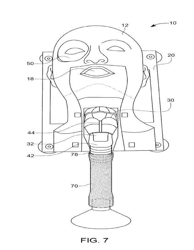
[0023] FIG. 7 is a front elevation view of the training simulator comprising
the head and neck
model of FIG. 1, the cricothyroid model of FIG. 6, the eye model of FIG. 11,
and the esophagus
model of FIG. 19.
[0024] FIG. 8 is a front elevation view of the cricothyroid model of the
present invention.
[0025] FIG. 9 is a left side elevation view of the cricothyroid model.
[0026] FIG. 10 is a right side elevation view of the cricothyroid model.
[0027] FIG. 11 is a bottom plan view of the cricothyroid model.
[0028] FIG. 12 is a top plan view of the cricothyroid model.
[0029] FIG. 13 is a front elevation view of the eye model of the present
invention.
[0030] FIG. 14 is a side perspective view of the eye model.
[0031] FIG. 15 is a rear elevation view of the eye model.
[0032] FIG. 16 is a front elevation view of the esophagus extension.
[0033] FIG. 17 is a bottom perspective view of the esophagus extension.
[0034] FIG. 18 is a top perspective view of the esophagus extension.
[0035] FIG. 19 is a front elevation view of the esophagus model with the
esophagus extension.
[0036] FIG. 20 is a side elevation view of the esophagus model with the
esophagus extension.
DETAILED DESCRIPTION OF THE INVENTION
[0037] The following detailed description is of the best currently
contemplated modes of
carrying out exemplary embodiments of the invention. The description is not to
be taken in a
limiting sense but is made merely for the purpose of illustrating the general
principles of the
invention, since the scope of the invention is best defined by the appended
claims.
CA 03209177 2023- 8- 21
WO 2022/178398
PCT/US2022/017254
[0038] An anatomical head and neck simulator apparatus 10 of the present
invention is shown in
FIGS. 1-20. The simulator apparatus 10 includes a head and neck model 12, a
cricothyroid model
30, an eye model 50, and an esophagus model 100 and an esophagus extension 70.
The head and
neck model 12 is shown alone in FIGS. 1-5. The simulator apparatus 10
comprising the head and
neck model 12, cricothyroid model 30, eye model 50, and esophagus model 100 is
shown in
FIGS. 6 and 7. The cricothyroid model 30 is shown alone in FIGS. 8-12. The eye
model 50 is
shown alone in FIGS. 13-15. The esophagus extension 70 is shown alone in FIGS.
16-18. The
esophagus model 100 and the esophagus extension 70 are shown in FIGS. 19-20.
[0039] The present invention may be used to facilitate training and practicing
of a number of
medical procedures involving the head and neck. The procedures include, but
are not limited to,
esophagogastric balloon insertion, cricothyrotomy, and lateral canthotomy.
Esophagogastric
balloon insertion is the insertion of a balloon into the esophagus and gastric
area of a patient as a
means of tamponading bleeding. Cricothyrotomy is the placement of a breathing
tube through
the cricothyroid membrane. A lateral canthotomv is the cutting of ocular
ligaments to relieve
intraocular pressure.
[0040] The head and neck model 12 is designed to simulate that of the human
head and neck.
The head and neck model 12 has a cylindrical cut out 16, a base 20, a
rectangular cut out 24, and
a mouth cut out 26The head and neck model 12 is configured to receive the eye
model 50,
cricothyroid model 30, and esophagus model 100. The cylindrical cut out 16 of
the head and
neck model 12 is configured to receive the eye model 50. The rectangular cut
out 24 of the head
and neck model 12 is configured to receive the cricothyroid model 30. The
mouth cut out 26 of
the head and neck model 12 is configured to receive the esophagus model 100.
The esophagus
model 100 is configured to receive the esophagus extension 70. The head and
neck model 12
may be used in conjunction with one or more of the cricothyroid model 30, eye
model 50, the
esophagus model 100, and the esophagus extension 70. Additionally, the
cricothyroid model 30,
the eye model 50, and the esophagus model 100 with or without the esophagus
extension 70 may
each be used separately. The simulator apparatus 10 is configured to be used
as a medical
procedures teaching device, allowing the use to perform procedures which
generally involve the
head and neck area. More specifically, the apparatus 10 has the cricothyroid
model 30, eye
model 50, esophagus model 100 and esophagus extension 70 to teach procedures
involving the
human cricothyroid, eye, and esophagus. The base 20 of the head and neck model
12 is
6
CA 03209177 2023- 8- 21
WO 2022/178398
PCT/US2022/017254
configured to allow the head and neck model 12 be placed on a surface to allow
the apparatus 10
to be used.
[0041] The head and neck model 12 is shown in FIGS. 1-6. The head and neck
model 12 has a
cylindrical cut out 16 which is configured to receive the eye model 50. The
mouth cut out 26 of
the head and neck model 12 is configured to receive the esophagus model 100.
The rectangular
cut out 24 of the head and neck model 12 is configured to receive the
cricothyroid model 30. The
base 20 of the head and neck model 12 is configured to be placed on a surface,
providing
stability when the apparatus 10 is in use. The base 20 may be configured with
methods to secure
the apparatus 10 to the working surface or may be configured to rest on the
working surface.
[0042] FIG. 4 depicts the bottom plan view of the head and neck model 12 with
the esophagus
model 100 inserted. The esophagus model 100 has a passage 80 and an esophagus
extension
attachment site 14. The attachment site 14 of the esophagus model 100 may be
threaded or
utilize another method of connection in which the esophagus extension 70 may
be removably
connected to the esophagus model 100.
[0043] FIGS. 6 and 7 depict the apparatus 10 with the eye model 50,
cricothyroid model 30,
esophagus model 100, and the esophagus attachment 70 all engaged with the head
and neck
model 12. The eye base 56 is removably engaged with the cylindrical cut out
16, which
alternatively can receive a suction cup and allow the eye model 50 to be
secured to a flat surface.
The cricothyroid base 46 is removably engaged with the rectangular cut out 24.
The esophagus
model 100 is removably engaged with the mouth cut out 26. The esophagus
extension
attachment 78 is removably engaged with the esophagus extension attachment
site 14. The
apparatus 10 may be used in conjunction with one or more of the head and neck
model 12, the
eye model 50, the cricothyroid model 30, the esophagus model 100, and the
esophagus extension
70. To add fidelity to the training, the eye model 50, the cricothyroid model
30, the esophagus
model 100, and esophagus extension 70 may all be engaged with the head and
neck model 12 to
depict the human anatomy. In FIG. 7, it may appear as though the esophagus
extension 70 is
connected to the cricothyroid model 30; however, the esophagus extension 70 is
connected to the
esophagus extension attachment site 14 of the esophagus model 100, located
beneath the
cricothyroid model 30. The esophagus extension attachment site 14 is depicted
in FIG. 4 beneath
the rectangular cut out 24 in which the cricothyroid model 30 is removably
engaged.
7
CA 03209177 2023- 8- 21
WO 2022/178398
PCT/US2022/017254
[0044] The cricothyroid model 30 shown in FIGS. 8-12 has a procedure site 32,
a cricoid
cartilage 34, an airway 36, a cricothyroid top end 38, a cricothyroid bottom
end 40, a trachea 42,
a thyroid cartilage 44, and a cricothyroid base 46. The cricothyroid model 30
is designed to
simulate that of the human cricothyroid muscle, thyroid, and trachea. The
thyroid cartilage 44,
thyroid protrusion 34, trachea 42, and procedure site 32 are configured to
mirror that of the
human anatomy, allowing the user to practice procedures at the procedure site
32 in conditions to
simulate an actual procedure. The cricothyroid base 46 is configured to be
placed on a surface
while in use. The cricothyroid base 46 may also be configured to be removably
engaged with the
rectangular cut out 24 of the head and neck model 12. The cricothyroid model
30 may be used
individually or in conjunction with the head and neck model 12. The
cricothyroid base 46 may
be configured to be removably engaged with the rectangular cut out 24 of the
head and neck
model 12 with pegs, clips, or other such methods to hold the cricothyroid
model 30 in place
while in use. Subcutaneous fat and skin can be created using off the shelf
materials such as foam
and silicone sheeting, which can be laid over the cricothyroid model 30 and
adhered to the head
and neck model 12 with pegs, clips, or other such adhesion methods.
Additionally, the
cricothyroid base 46 may be configured to be placed on a surface providing
stability when the
cricothyroid model 30 is in use.
[0045] As shown in FIGS. 8-12, the airway 36 of the cricothyroid model 30
traverses from the
cricothyroid top end 38 through the cricothyroid bottom end 40 to simulate
that of the human
airway. The cricoid cartilage 34, the thyroid cartilage 44, and the trachea 42
of the cricothyroid
model 30 simulate that of the human thyroid cartilage, cricothyroid cartilage,
and trachea. The
procedure site 32 of the cricothyroid model 30 may be used to practice the
insertion of a
breathing tube through the cricothyroid membrane.
[0046] Cricothyrotomy, or placement of a breathing tube through the
cricothyroid membrane,
may be practiced directly on the cricothyroid model 30 shown in FIGS. 8-12. To
add to the
fidelity of the training, the cricothyroid model 30 may be placed in the
rectangular cut out 24 of
the head and neck model 12. The cricoid cartilage 34, the thyroid cartilage
44, and the trachea 42
provide an anatomical structure to simulate that of the human cricothyroid.
The cricothyrotomy
procedure may be perfornied on the procedure site 32 of the cricothyroid model
30. The
cricothyroid model 30 may be used in conjunction with the head and neck model
12 or on a
8
CA 03209177 2023- 8- 21
WO 2022/178398
PCT/US2022/017254
standalone basis. Other commercially available or off the shelf material may
be applied over the
entire unit to simulate the cricothyroid membrane, subcutaneous tissue, and
skin on the model.
[0047] The eye model 50 shown in FIGS. 13-15 has an eyeball 52, an eye inset
54, and an eye
base 56, one or more eye base channels 58, and may optionally have one or more
eye pegs 60.
The eye model 50 is designed to simulate that of the human eye and surrounding
environment.
The eyeball 52 may be used to perform procedures involving the eye. The eye
inset 54 provides
structure to hold the eyeball 52 in place while in use. The eye inset 54 may
be used to practice
procedures involving the area surrounding the eye. The eye base 56 is
configured to be placed on
a surface while in use. The eye base 56 is also be configured to be removably
engaged with the
cylindrical cut out 16 of the head and neck model 12. The eye model 50 may be
used
individually or in conjunction with the head and neck model 12. The eye base
56 may be
configured to be removably engaged with the cylindrical cut out 16 of the head
and neck model
12 with pegs, clips, or other such methods to hold the eye model 50 in place
while in use. The
one or more eye base channels 58 are configured to allow for rubber bands or
other off the shelf
material to be attached to the eye model 50. The one or more optional eye pegs
60 are configured
to align the rubber bands or other off the shelf material to simulate the
alignment of human eye
ligaments.
[0048] Lateral canthotomy, or cutting of ocular ligaments to relieve
intraocular pressure, may be
practiced directly on the eye model 50 shown in FIGS. 13-15. To add to the
fidelity of the
training, the eye model 50 may be placed in the cylindrical cut out 16 of the
head and neck
model 12. The eyeball 52 is made of a material suitable for practicing the
procedure on the
eyeball 52 of the eye model 50. The eye inset 54 of the eye model 50 may be
used to practice
procedures involving the surrounding area of the eyeball 52. Additionally, the
eye inset 54
provides support of the eyeball 52 and anatomical simulation of the area
around the eye. In this
configuration, the one or more eye based channels 58 are designed to receive
rubber bands or off
the shelf material to replicate anatomical ligaments that need to be cut
during the lateral
canthotomy procedure. The one or more eye pegs 60 are designed to engage and
hold in place
the materials used as eye ligaments to mirror that of the human eye ligaments.
The eye model 50
may be used in conjunction with the head and neck model 12 or on a standalone
basis.
Practitioners may apply commercially available materials to the eye model 50
to replicate the
upper and lower eyelids with associated ligaments.
9
CA 03209177 2023- 8- 21
WO 2022/178398
PCT/US2022/017254
[0049] The esophagus extension 70 shown in FIGS. 16-18 has an esophagus
extension top end
72, an esophagus extension bottom end 74, an esophagus extension base 76, an
esophagus
extension attachment 78, and a passage 80. The esophagus extension 70 is
designed to simulate
that of the lower portion of the human esophagus. The esophagus extension
attachment 78 is
configured to be removably engaged with the esophagus extension attachment
site 14 of the
esophagus model 100. The esophagus extension base 76 is configured to rest on
or be removably
engaged with a surface. The esophagus extension 70 may be used in conjunction
with the
esophagus model 100.
[0050] The esophagus model 100 shown in FIGS. 19-20 has a mouth opening 18, an
esophagus
90, and an esophagus extension attachment site 14, and the passage 80. The
esophagus model
100 is configured to be removably engaged with the mouth cut out 26 of the
head and neck
model 12. The passage 80 of the esophagus model 100 initiates at the mouth
opening 18 of the
esophagus model 100 and traverses through the esophagus extension attachment
site 14. The
esophagus extension 70 is removably attached to the esophagus extension
attachment site 14
with the esophagus extension attachment 78. The mouth opening 18 of the
esophagus model 100
facilitates the insertion of an esophagogastric balloon or orogastric tube
into the passage 80,
[0051] As shown in FIGS. 16-20, the esophagus model 100 has a passage 80 which
traverses
from the mouth opening 18, through the esophagus 90, the esophagus attachment
site 14 and the
esophagus extension 70. The passage 80 extends from the esophagus extension
top end 72
through the esophagus extension bottom end 74. The passage 80 for the tube
insertion during the
esophagogastric balloon insertion procedure. The passage 80 may also
facilitate the use of
commercially available materials to replicate the lower esophagus to stomach
anatomy. The
esophagus extension bottom end 74 is designed to replicate the anatomical
anatomy of the upper
stomach and its connection to the lower esophagus.
[0052] Esophagogastric balloon insertion is the insertion of a balloon into
the esophagus and
gastric area of a patient as a means of tamponading bleeding. An
esophagogastric balloon
insertion may be practiced on the present invention. In this configuration,
the gastric balloon of
the esophagogastric balloon will sit in the conical shape of the esophagus
extension bottom end
74. To add to the fidelity of the training, the esophagus model 100 may be
removably inserted
into to the head and neck model 12. The esophagus extension 70 may be
connected to the
esophagus extension attachment site 14 of the esophagus model 100 with the
esophagus
CA 03209177 2023- 8- 21
WO 2022/178398
PCT/US2022/017254
extension attachment 78 of the esophagus model 70. A tube (i.e., a Blakemore
Tube) may be
inserted in the mouth opening 18 of the esophagus model 100, continuing
through the passage 80
of the esophagus model 100, through the esophagus extension attachment site 14
and the
esophagus extension attachment 78 into the esophagus extension 70. The
esophagus model 100
and the esophagus extension 70 may be used in conjunction with the head and
neck model 12 or
on a standalone basis. Practitioners may also attach commercially available
materials to replicate
the lower esophagus to stomach anatomy. With the simulator apparatus 10,
practitioners may
train in the procedure of placing an esophagogastric balloon, working on the
mechanics of these
complex devices.
[0053] The present invention has been described with reference to specific
examples and
configurations. It is only intended to be limited to the description set out
in the claims and
equivalents.
11
CA 03209177 2023- 8- 21