Note: Descriptions are shown in the official language in which they were submitted.
CA 03210091 2023-07-27
WO 2022/170071
PCT/US2022/015279
NON-TERMINAL ANTIBODY DISCOVERY METHODS AND SINGLE CELL ASSAYS
CROSS REFERENCE TO RELA1ED APPLICATION
[0001] This application claims priority to U.S. Provisional Application No.
63/146,135, filed
February 5, 2021, the entire contents of which is incorporated herein by
reference.
BACKGROUND
[0002] Traditional animal-based antibody discovery methods involve the
sacrifice of animals based
on polyclonal serum titers against protein targets with varying degrees of
complexity. While some
antibody discovery campaigns have simple design goals (e.g., bind to the
target), most are more
complex and require the desired antibodies to have a variety of features
(e.g., cross reactivity, bind to
a particular epitope, bind with a specific affinity, etc.). Traditional
antibody discovery approaches
rely on the interrogation of the polyclonal secreted antibody (serum) response
to select animals for B-
cell harvest and antibody generation. The "serum titer" approach is less than
ideal since it measures
the total reactivity of all the secreted antibodies (i.e., it is a polyclonal
mixture) and cannot be used to
identify the B-cell source of the detected antibodies (i.e., there is both a
physical and temporal
disconnect from the source B-cell). The lack of a direct connection between
phenotype (the antibody
titer measurement) and genotype (the responsible source B-cell encoding the
antibody) makes
interpreting the quality of the B-cell response difficult. Aside from
determining whether there is
soluble, antigen-specific antibody in the serum, it is difficult to obtain
additional useful information
from this polyclonal analysis that can aid animal selection.
[0003] Additionally, the traditional methodology is terminal with regard to
the animal, and thus
represents a 'one-time' attempt to capture the animal's relevant B-cell
repertoire. Failure to capture
this repertoire, which may be caused by technical problems, selection of
animals with suboptimal
antibody production, and/or the lack of sampling depth of the repertoire
(i.e., poor efficiency of
traditional viral immortalization and hybridoma processes resulting in the
fusion of a very small
fraction of the B cell repertoire (less than 0.1%)), results in the waste of
valuable resources and forces
the use of an alternative immune animal or an entirely new immunization
campaign. Furthermore, the
traditional methods preclude the possibility of a continual process of
leveraging the immune system of
the same animal to evolve the antibody response.
[0004] Despite these limitations, the traditional methods are widely used, in
part, because they
allow the capture of an acceptable fraction of the immune repertoire and
provide a renewable source
of antibody that can easily be scaled to accommodate downstream assays.
[0005] There is an increasing number of situations where these traditional
methods are too slow to
meet project timelines, capture the wrong B-cell population, insufficiently
sample the B-cell
repertoire, or do not allow real-time monitoring of the evolving B-cell
response. In addition, the
1
CA 03210091 2023-07-27
WO 2022/170071
PCT/US2022/015279
challenging nature of many antibody target classes (e.g. complex membrane
proteins, targets with
minimal epitope space, proteins that are highly similar to orthologs, etc.)
can make it difficult to raise
B-cell responses in animals due to a lack of robust immunogenicity. Coupled
with the extreme
complexity of some antibody design goals, generating immune animals with the
desired immune
profiles (i.e., B-cell repertoires) can be difficult.
[0006] In view of the foregoing, more efficient antibody discovery methods are
needed. For
example, antibody discovery methods that can better position traditional
animal immunization and B-
cell methods for successful antibody discovery would greatly enhance animal-
based antibody
discovery.
SUMMARY
[0007] Provided for the first time are the rationale, experimental methods,
and data demonstrating
techniques useful in antibody discovery. In exemplary aspects, the methods
involve the identification
of antigen-specific antibodies directly from the peripheml blood of a living,
non-human animal.
Advantageously, such methods provided herein allow for antibody discovery
without the need for
animal sacrifice, unlike traditional methods which rely on animal euthanasia
followed by immune
organ harvest (e.g., spleen, lymph nodes, and bone marrow). Because such
methods are non-terminal
(e.g., do not involve the euthanasia of the antibody-producing animals), the
methods may be repeated
multiple times in the same animal(s) until, for instance, an antibody of
interest is obtained. The
ability to repeat the method in the same animal(s) has several advantages over
traditional methods.
For example, repeating the method in the same animals(s) reduces the overall
cost of the antibody
discovery process. Also, since the animals are kept alive, the presently
disclosed methods allow for
real-time, in-life sampling of the antibody repertoire, such that if, for
example, the animals do not
produce a B cell expressing the target antibody of interest, strategic
adjustments to the immunization
protocol (used in the next immunization) may be made based on the observed B-
cell response (from
the prior immunization). The methods of the present invention thereby permit
rational repertoire
shaping and/or purposeful steering of the immune response to match antibody
design goals.
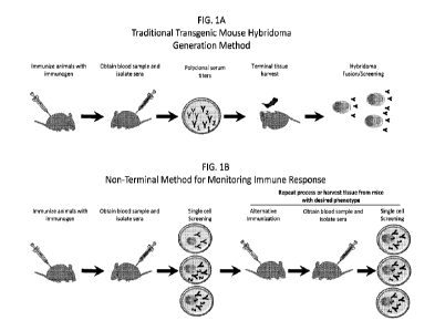
Exemplary processes of the present disclosure are illustrated in Figures 1B-
1E. Figure 1B illustrates
an exemplary non-terminal method for monitoring immune responses comprising
screening of single
cells obtained from a blood sample of immunized animals. Based on the outcome
of the single cell
screening, the animal may be subjected to a repeat round of immunization
(e.g., an alternative
immunization) followed by single cell screening of cells from the blood sample
obtained from the
immunized animal, or may undergo tissue harvest if the screening determines
that the animal exhibits
the desired phenotype. Figure 1C illustrates an exemplary non-terminal method
of monitoring for the
production of select antibodies, wherein antibody secreting cells (ASCs)
purified from a blood sample
obtained from an immunized animal are screened at the single cell level. The
process is repeated until
design goals are met and/or select antibodies are produced. Figure 1D
illustrates an exemplary non-
2
CA 03210091 2023-07-27
WO 2022/170071
PCT/US2022/015279
terminal method of guiding antibody production for select antibody production
wherein a primary
strategy is used to immunize animals, and the ASCs obtained from PBMCs
isolated from the
immunized animal are screened for the desired phenotype. If the screening
determines that the design
goal is not met, then the animal is immunized with an alternative strategy
(e.g., that differs from the
primary strategy) and, the ASCs obtained from PBMCs isolated from the
immunized animal are
screened for the desired phenotype. The process is repeated until the
screening determines that the
design goal is met. When and if the design goal is met, terminal tissue may be
harvested for antibody
rescue using hybridoma, single cell platforms or sequence-based discovery.
Figure lE illustrates an
exemplary non-terminal method of screening animals and B-cell profiling
wherein a series of animals
is immunized with an immunogen and ASCs obtained from a blood sample obtained
from each
animal are screened and a B-cell repertoire is profiled. In various aspects of
the exemplary processes,
antibody secreting cells (ASCs), e.g., plasmablasts, are purified from the
peripheral blood of an
immunized mouse and then screened at single cell resolution for the relevant
activity or phenotype.
Compared to the traditional hybridoma production process (illustrated in
Figure 1A), which typically
requires about 8 weeks and requires a high level of technical skill, the
process of the present
disclosure is less labor-intensive and requires less time.
[0008] Accordingly, the present disclosure provides methods of monitoring for
the production of
select antibodies in a non-human animal. In exemplary embodiments, the method
comprises (a)
immunizing a non-human animal with an immunogen; (b) obtaining a blood sample
comprising
antibody secreting cells (ASCs) from said non-human animal; and (c) assaying,
e.g., individually
assaying, ASCs present in the blood sample, or a fraction thereof, for the
production of select
antibodies. In various instances, the method further comprises repeating (b)
and (c) one or more times
until design goals are met, e.g., until select antibodies are produced. Figure
1C illustrates this
exemplary aspect of the present disclosure. The present disclosure also
provides methods of guiding
antibody production in a non-human animal for the production of select
antibodies. In exemplary
embodiments, the method comprises (a) performing an initial immunization on a
non-human animal
with an immunogen; (b) obtaining a blood sample comprising ASCs from said non-
human animal; (c)
assaying, e.g., individually assaying, ASCs present in the blood sample, or a
fraction thereof, for the
production of select antibodies; and (d) performing a cycle of steps when the
percentage of ASCs
producing select antibodies is below a threshold, wherein the cycle comprises
(i) performing a
subsequent immunization on the non-human animal with an immunogen when the
percentage of
ASCs producing select antibodies is below a threshold, (ii) obtaining a blood
sample comprising
ASCs from said non-human animal, and (iii) assaying, e.g., individually
assaying, ASCs present in the
blood sample, or a fraction thereof, for the production of select antibodies.
[0009] In various aspects, the assaying comprises a single-cell, live-cell
assay. As used herein, the
phrase "individually assaying ASCs" means that the ASCs are assayed or
examined at the single cell
3
CA 03210091 2023-07-27
WO 2022/170071
PCT/US2022/015279
level or at a single cell resolution. In exemplary instances, "individually
assaying ASCs" provide
results relevant to a single ASC. Optionally, multiple ASCs are simultaneously
assayed. In various
aspects, multiple ASCs are simultaneously individually assayed. In exemplary
aspects, the blood
sample is obtained from the non-human animal in a non-terminal manner, e.g.,
the non-human animal
is not killed during the blood sample collection. In exemplary instances, the
method comprises
performing a non-terminal blood draw from the non-human animal. In various
instances, the method
comprises applying the blood sample, or a fraction thereof, to a matrix and
assigning a unique address
of the matrix to each ASC. Optionally, a result of the assaying is the
identification of each ASC
producing select antibodies. In certain aspects, the result of the assaying is
the identification of the
unique address of each ASC producing select antibodies. In exemplary
instances, the method
comprises at least one cycle of (i) performing a subsequent immunization on
the non-human animal
with an immunogen when the percentage of ASCs producing select antibodies is
below a threshold,
(ii) obtaining a blood sample comprising ASCs from said non-human animal,
(iii) assaying, e.g.,
individually assaying ASCs present in the blood sample, or a fraction thereof,
for the production of
select antibodies. Optionally, the cycle is repeated until the percentage of
ASCs producing select
antibodies, as assayed in (iii), is at or above the threshold. In various
instances, the cycle is repeated
at least two times.
[0010] The immunogen of the subsequent immunization may differ from the
immunogen of the
initial immunization in exemplary aspects. For instance, in exemplary aspects,
each subsequent
immunization differs from a prior immunization in that (A) a different
immunogen, adjuvant, and/or
immunomodulatory agent is administered to the non-human animal, (B) a
different dose of the
immunogen is administered to the non-human animal, (C) the time between each
administration of the
immunogen, adjuvant, immunomodulatory agent is different, and/or (D) the route
of administration
for each administration of immunogen, adjuvant, immunomodulatory agent is
different. Optionally, a
different immunogen is used each time the non-human animal is immunized.
Figure 1D illustrates an
exemplary method of guiding antibody production for select antibody
production.
[0011] The present disclosure further provides methods of producing select
antibodies in a non-
human animal. In exemplary embodiments, the method comprises guiding antibody
production in a
non-human animal for the production of select antibodies in accordance with
the presently disclosed
methods of guiding antibody production and then isolating the select
antibodies and/or an ASC
producing the select antibodies. In exemplary embodiments, the method
comprises (a) performing an
initial immunization campaign on a non-human animal with an immunogen; (b)
obtaining a blood
sample comprising antibody secreting cells (ASCs) from said non-human animal;
(c) assaying, e.g.,
individually assaying, ASCs present in the blood sample, or a fraction
thereof, for the production of
select antibodies; (d) performing a cycle of steps when the percentage of ASCs
producing select
antibodies is below a threshold, wherein the cycle comprises (i) performing a
subsequent
4
CA 03210091 2023-07-27
WO 2022/170071
PCT/US2022/015279
immunization on the non-human animal with an immunogen when the percentage of
ASCs producing
select antibodies is below a threshold, (ii) obtaining a blood sample
comprising ASCs from said non-
human animal, and (iii) assaying, e.g., individually assaying, ASCs present in
the blood sample, or a
fraction thereof, for the production of select antibodies; and (e) isolating
the select antibodies and/or
an ASC producing the select antibodies. In various aspects, the method
comprises (f) determining the
nucleotide sequence encoding the heavy chain variable region of the select
antibodies produced by an
ASC (e.g., the isolated ASC producing the select antibodies) and the
nucleotide sequence encoding
the light chain variable region of the select antibodies produced by the ASC,
(g) introducing into a
host cell a first vector comprising the nucleotide sequence encoding the heavy
chain variable region of
the select antibodies and a second vector comprising the nucleotide sequence
encoding the light chain
variable region of the select antibodies, and (h) isolating the antibodies
produced by the host cell.
[0012] In exemplary aspects, the assaying of the presently disclosed methods
comprises (a)
combining the ASCs within the matrix with reagents that bind to the select
antibodies and produce a
detectable signal, e.g., a fluorescent signal, upon binding to the select
antibodies. In various aspects,
the assaying of the presently disclosed methods comprises (a) combining the
ASCs within the matrix
with at least one reagent which binds to the Fc domain of the select
antibodies and at least one reagent
to which select antibodies bind (e.g., a reagent which binds to the antigen-
binding domain of the
select antibodies), wherein at least one of these reagents is attached to a
detectable label. In
exemplary instances, the ASCs are combined with a detection reagent which
binds to the Fc domain
of the select antibodies and comprises a first detectable label and a target
to which select antibodies
bind (e.g., a reagent which binds to the antigen-binding domain of the select
antibodies). Figures 2A-
2C illustrate exemplary assaying in the context of the presently disclosed
methods. In various
instances, the target is labeled by a second detectable label which is
different from the first detectable
label. In some instances, a capture reagent which binds to the Fc domain of
the select antibodies and
comprises a solid support is further combined with the ASCs, detection reagent
and labeled target. In
various instances, the method further comprises (b) assaying for the first
detectable label and the
second detectable label; and (c) identifying the positions within the matrix
at which the first detectable
label and the second detectable label are detected, wherein each identified
position locates an
individual ASC producing select antibodies. Figures 2A and 2B illustrate such
exemplary assaying
with a labeled target and a capture reagent. Figure 2A illustrates the matrix
as a well. Figure 2B
illustrates the matrix as a multi-pen chip or multi-well plate and each ASCs
is positioned into a single
pen or well. In various instances, the target is expressed by cells and the
cells expressing the target
are combined with the ASCs and the detection reagent. In exemplary aspects,
the method further
comprises (b) assaying for the first detectable label; and (c) identifying the
positions within the matrix
at which the first detectable label is detected, wherein each identified
position locates an individual
ASC producing select antibodies. Figure 2C illustrates such exemplary assaying
with a cell
CA 03210091 2023-07-27
WO 2022/170071
PCT/US2022/015279
expressing the target. In exemplary instances, the assaying of the presently
disclosed methods
comprises (a) combining the ASCs within the matrix with (i) a capture reagent
which binds to the
select antibodies and comprises a solid support, (ii) a detection reagent
which binds to the select
antibodies and comprises a first detectable label, and (iii) a labeled target
to which the select
antibodies bind, wherein the labeled target comprises a second detectable
label distinct from the first
detectable label; (b) assaying for the first detectable label and for the
second detectable label; and (c)
identifying the positions within the matrix at which both the first detectable
label and the second
detectable label are detected, wherein each identified position locates an
individual ASC producing
select antibodies. Optionally, the capture agent comprises an antibody that
binds to an antibody Fc
domain attached to a solid support. The detection agent, in exemplary
instances, comprises an
antibody that binds to an antibody Fc domain attached to a first detectable
label. In various aspects,
the antibody that binds to an antibody Fc domain of the capture agent is the
same antibody of the
detection agent. In exemplary instances, the combining takes place in a well
and the capture agent
forms a monolayer in the well. In various aspects, the method comprises
identifying the positions
within the well at which both the first detectable label and the second
detectable label are detected,
wherein each identified position locates an individual ASC producing select
antibodies.
[0013] The present disclosure additionally provides single-cell assays for
identifying ASCs
producing select antibodies. The present disclosure provides methods of
assaying for ASCs
producing select antibodies. In exemplary embodiments, the assay or method
comprises (a)
combining in a well (i) a blood sample obtained from a non-human animal
immunized with an
immunogen, or a fraction thereof, wherein the blood sample comprises ASCs,
(ii) a detection reagent
which binds to the select antibodies and comprises a first detectable label,
and (iii) a target to which
the select antibodies bind, wherein (A) the target is a labeled target
comprising a second detectable
label distinct from the first detectable label and a capture reagent which
binds to the select antibodies
and comprises a solid support is further combined in the well to form a
monolayer in the well or (B)
the target is expressed on the surface of cells and the cells are combined in
the well to form a
monolayer in the well; (b) assaying for the first detectable label and
optionally assaying for the second
detectable label, when the target is a labeled target; and (c) identifying the
positions within the well at
which the first detectable label is detected or the first and second
detectable labels are detected,
wherein each identified position locates an individual ASC producing select
antibodies. In various
aspects, the assay or method comprises (a) combining in a well (i) a blood
sample obtained from a
non-human animal immunized with an immunogen, or a fraction thereof, (ii) a
capture reagent
comprising an antibody that binds to an Fc of an antibody attached to a solid
support, (iii) a detection
reagent comprising an antibody that binds to an Fc of an antibody attached to
a first detectable label,
and (iv) a labeled target comprising the immunogen, or a portion thereof,
attached to a second
detectable label distinct from the first detectable label, wherein the capture
agent forms a monolayer
6
CA 03210091 2023-07-27
WO 2022/170071
PCT/US2022/015279
in the well; (b) assaying for the first detectable label; (c) assaying for the
second detectable label; and
(d) identifying the positions within the well at which both the first
detectable label and the second
detectable label are detected, wherein each identified position locates an
individual ASC producing
select antibodies. In various aspects, the assay or method comprises (a)
combining in a well (i) a
blood sample obtained from a non-human animal immunized with an immunogen, or
a fraction
thereof, (ii) a detection reagent which binds to the select antibodies, and
(iii) cells expressing on the
cell surface a target to which the select antibodies bind, wherein the cells
are combined in the well to
form a monolayer in the well, (b) assaying for the first detectable label; and
(c) identifying the
positions within the well at which both the first detectable label and the
second detectable label are
detected, wherein each identified position locates an individual ASC producing
select antibodies.
[0014] In various aspects of the presently disclosed methods, the non-human
animal is subjected to
neither removal of one or more secondary lymphoid organs nor euthanasia. Also,
in various
instances, ASCs from the blood sample are not used in making hybridomas. In
exemplary aspects, the
non-human animal is one of a series of non-human animals, and a result of the
assaying is the
identification of the non-human animals having a percentage of ASCs producing
select antibodies
below the threshold and/or requiring further immunization. In alternative
aspects, the method
comprises sacrificing the non-human animal and harvesting tissues from the non-
human animal, when
the percentage of ASCs producing select antibodies is at or above a threshold.
In various instances,
the steps of the method are carried out on a series of non-human animals and
the method comprises
profiling the B-cell repertoire of the blood sample for each non-human animal
of the series and
selecting a subset of the series having a target B-cell profile. Figure lE
illustrates such steps.
[0015] Rational immune repertoire generation and selection is a critical
component in animal-based
antibody discovery technologies. Despite the advancement from traditional B-
cell immortalization to
direct B-cell platforms such as (but not limited to) NanOBLAST (an antibody
discovery process on a
nanofluidic Beacon device) and microencapsulation, the diversity and quality
of the input B-cells
continues to be an essential determining factor in meeting antibody design
goals. Traditional
approaches to evaluate immune animals rely on the interrogation of the
polyclonal secreted antibody
(serum) to evaluate immune responses and select animals for B-cell harvest and
antibody generation.
The "serum titer" approach is less than ideal since it measures the total
reactivity of all the secreted
antibodies and not the quality of the individual B-cell source of the detected
antibodies. The lack of a
direct connection between the antibody titer measurement and the responsible B-
cell source makes
interpreting the quality of the B- cell response difficult. Aside from
determining whether there is
soluble, antigen-specific antibody in the serum, it is difficult to obtain
additional useful information
from this polyclonal analysis that can aid animal selection or immune steering
strategies. Provided
herein are ASC assays to interrogate the B-cell response of an immune animal
using samples derived
from non-terminal peripheral blood that would address these challenges.
Accordingly, the present
7
CA 03210091 2023-07-27
WO 2022/170071
PCT/US2022/015279
disclosure provides a method of screening non-human animals for antibody
secreting cells (ASCs)
producing select antibodies. The method in exemplary embodiments comprises (a)
immunizing a
series of non-human animals with an immunogen; (b) obtaining a blood sample
comprising ASCs
from each non-human animal of the series; and (c) individually assaying ASCs
present in the blood
sample, or a fraction thereof, for the production of select antibodies,
wherein, for each non-human
animal of the series, a percentage of ASCs producing select antibodies is
determined. In various
aspects, the screening method further comprises selecting the non-human
animal(s) for sacrifice
and/or tissue harvest, when the percentage of ASCs producing select antibodies
is at or above a
threshold. In various aspects, the screening method further comprises
selecting the non-human
animal(s) for subsequent immunization, when the percentage of ASCs producing
select antibodies is
below a threshold. Accordingly, in various embodiments, the screening method
identifies animals for
sacrifice vs. animals for subsequent immunization based on the percentage of
ASCs producing select
antibodies.
[0016] Consistent with the foregoing, methods of selecting immunized non-human
animals for
subsequent immunization are provided. In exemplary embodiments, the method
comprises
monitoring for the production of select antibodies in a non-human animal in
accordance with any one
of the presently disclosed methods, wherein the method is carried out on a
series of non-human
animals, wherein for each non-human animal of the series the number of ASCs
producing the select
antibodies is identified, and selecting the animal for subsequent immunization
when the percentage of
ASCs producing select antibodies for an animal is below a threshold. Also
provided herein are
methods of selecting immunized non-human animals for euthanasia and secondary
lymphoid harvest.
In exemplary embodiments, the method comprises monitoring for the production
of select antibodies
in a non-human animal in accordance with any one of the presently disclosed
methods, wherein the
method is carried out on a series of non-human animals, wherein for each non-
human animal of the
series the number of ASCs producing the select antibodies is identified, and
selecting the animal for
euthanasia and secondary lymphoid harvest, when the percentage of ASCs
producing select antibodies
for an animal is at or above a threshold.
BRIEF DESCRIPTION OF THE DRAWINGS
[0017] Figure lA is an illustration of a traditional transgenic mouse
hybridoma generation method.
Figure 1B is an illustration of a non-terminal method for monitoring immune
response. Figure 1C is
an illustration of a non-terminal method of monitoring for the production of
select antibodies. Figure
1D is an illustration of a non-terminal method of guiding antibody production
for select antibody
production. Figure lE is an illustration of a non-terminal method of screening
animals and B-cell
profiling.
8
CA 03210091 2023-07-27
WO 2022/170071
PCT/US2022/015279
[0018] Figure 2A is an illustration of an application of an exemplary single
cell assay for
identifying ASCs which produce select antibodies. Figure 2B is an illustration
of another exemplary
single cell assay for identifying ASCs which produce select antibodies. Figure
2C is an illustration of
yet another exemplary single cell assay for identifying ASCs which produce
select antibodies.
[0019] Figure 3 is an illustration of an antibody binding to an anti-idiotope
antibody. Paratopes,
idiotypes and idiotopes are shown.
[0020] Figure 4 is a graph of polyclonal titers for sera obtained from the
indicated mice immunized
with Antibody 1.
[0021] Figure 5A is an illustration of components of an exemplary single cell
screen and Figure 5B
is an illustration of how the components of the single cell assay interact in
the presence of an antibody
that binds antigen. Figure 5C is an illustration of individual pens holding an
ASC secreting antibodies
interacting with polystyrene beads to create a fluorescent "bloom". IgG
secretion and antigen-specific
antibodies are detected by the assay.
[0022] Figure 6 is an illustration of the dual blooms above individual pens
holding a single cell,
export of the cells to a well and PCR analysis for antibody cloning,
expression, purification and
analysis.
[0023] Figure 7A is an illustration of a sandwich ELISA format used to select
the appropriate pairs
of antibodies. Figure 7B is graph of the ELISA signal plotted as a function of
concentration of
Antibody 1. Figure 7C is a graph of the PD1 functional plotted as a function
of antibody
concentration.
[0024] Figure 8A is an image of green fluorescent spots at which ASCs
secreting antibodies are
located in a single well. Figure 8B is an image of red fluorescent spots at
which antigen-specific
antibodies secreted by ASCs are located in a single well. Figure 8C is an
image of colored spots at
which ASCs secreting antibodies are located in a single well, spots at which
antigen-specific
antibodies secreted by ASCs are located in a single well, and spots at which
ASCs secreting antigen-
specific antibodies are located in a single well. Figure 8D is an exemplary
image of transfected cells
labeled with multiple fluorescent spots at which antigen expressed by the 293T
cell is bound to
antibody produced by the B cell and labeled with the goat anti-human Fc
antibody labeled with Alexa
488.
[0025] Figure 9 is a series of images of single cells of the indicated
hybridoma clone (or irrelevant
clone) with RFU on the green channel (top) representing antibody secretion or
the red channel
(bottom) representing antigen (EGFR) binding.
[0026] Figure 10 is a graph of the RFU green/RFU red ratio plotted as a
function of KD of the
hybridoma.
9
CA 03210091 2023-07-27
WO 2022/170071
PCT/US2022/015279
[0027] Figure 11 is a schematic of an immunization protocol used across all
mice. The timing of
bleeds and shifting of antigens is indicated.
[0028] Figure 12 is a graph of the serum titers of the first bleed of Group 1
and 2 mice. The graph
plots the human antigen titers vs the cyno antigen titers.
[0029] Figure 13 is a series of images of single cells with RFU on the red
channel (left)
representing human antigen binding, green channel (middle) representing cyno
antigen binding, and
composite channel (right) representing human antigen and cyno antigen binding.
Data from serum
obtained from Bleed 1.
[0030] Figure 14 is a graph of the percent of antigen positive ASCs of Group 1
(closed circles) and
Group 2 (open circles) mice reacting to human antigen only, cyno antigen only,
or both human and
cyno antigens. Data from cells from Bleed 1 obtained by the single cell
Incucyte screen.
[0031] Figure 15 is a graph of the percent of antigen positive ASCs of Group 1
(closed circles) and
Group 2 (open circles) reacting to human antigen only, cyno antigen only, or
both human and cyno
antigens. Data from cells from Bleed 2 obtained by the single cell Incucyte
screen.
[0032] Figure 16 is a graph of the percent of antigen positive ASCs of Group
lA (Human Boost)
and Group 1B (Cyno boost) reacting to human antigen only, cyno antigen only,
or both human and
cyno antigens. Data from cells from Bleed 2 shown in closed circles and data
from cells from Bleed 3
shown in open squares. Data obtained by the single cell Incucyte screen.
[0033] Figure 17 is a graph of the change in cross-reactive ASC frequency
(relative to Bleed 1) of
Group lA (human boost) and Group 1B (cyno boost). Data obtained by the single
cell Incucyte
screen.
[0034] Figure 18 is a graph of the serum titer reactive to cyno antigen
plotted as a function of
serum titer reactive to human antigen. Percent cross reactive ASCs are noted.
Animals of interest for
selection for harvest are circled in red.
[0035] Figure 19 is a schematic of an immunization campaign with immune
steering toward
production of human cyno cross-reactive antibodies which bind to both human
and cyno subdomain
orthologs of a multi-domain protein (antigen).
[0036] Figure 20 is a graph of serum titer reactive to cyno antigen plotted as
a function of serum
titer reactive to human antigen. Serum from Bleed 1.
[0037] Figure 21 is series of images of single cells at t= 0 hr (bottom) and
t=23 hrs (top) with RFU
on the green and red channels for human only binders, cyno only binders, and
human/cyno cross-
reactive binders. Data from serum obtained from Bleed 1 using the single cell
Incucyte screen.
CA 03210091 2023-07-27
WO 2022/170071
PCT/US2022/015279
[0038] Figure 22 is a graph of the percent of antigen positive ASCs reacting
to human antigen only,
cyno antigen only, or both human and cyno antigens. Data from cells from Bleed
1 shown. Data
obtained by the single cell Incucyte screen.
[0039] Figure 23 is a graph of the change in cross-reactive ASC frequency
(relative to irrelevant
clone) in serum from Bleed 1 and Bleed 3.
[0040] Figure 24 is a graph of the percent of ASCs secreting antibodies
reactive to cyno antigen
only (closed circles) or both cyno and human antigen (open squares) at Bleed 1
and Bleed 3. Data
obtained by the single cell Incucyte screen.
[0041] Figure 25 is a graph of the percent of human-cyno cross-reactive
binders. Animals of
interest for selection for harvest are noted in squares. Data obtained by the
single cell Incucyte
screen.
DETAILED DESCRIPTION
[0042] B-Cell Function and Non-Terminal Monitoring and Steering ofAntibody
Production
[0043] Antigen-specific B-cells that have recently encountered antigen in the
germinal centers
(GCs) of the secondary lymphoid organs (e.g., spleen and lymph nodes) are
stimulated to divide and
commit to differentiate down multiple pathways. See, e.g., Klein and Dalla-
Favera, Nature Reviews
Immunol 8: 22-33 (2008)). The main B-cell lineage responsible for secreting
antibodies into the
serum in response to antigen challenge are plasma cells. Plasma cell
differentiation begins in the
secondary lymphoid organs where cell-cell interactions within the GCs force B-
cells, expressing
antibodies on their surface that are specific to antigen, to differentiate
into immature plasma cells
known as plasmablasts. Plasmablasts are rapidly dividing B-cells that produce
and secrete soluble
antibody. However, plasmablasts are transient in nature and require
significant trophic support to
survive and continue to proliferate. The main survival niche for plasmablasts
is in the secondary
lymphoid organs, but these queues are provisional and depend on the presence
of cognate antigen.
[0044] B-cells use two main strategies to maintain long-term humoral memory to
antigens: the
formation of IgG+ memory B-cells and the formation of long-lived, mature
plasma cells. Memory B-
cells express a cell-surface-bound version of their cognate antibody, known as
the B-cell receptor
(BCR), but do not secrete soluble antibody. These cells take up residence in a
variety of locations
throughout the body and are abundant within the secondary lymphoid organs.
Upon re-encounter with
antigen, memory B-cells can be induced to proliferate (i.e. to generate clones
of themselves) and to
differentiate into antibody-secreting plasma cells. The other route to long-
term memory is via the
formation of long-lived, mature plasma cells. Mature plasma cells require very
specialized survival
niches that provide trophic support and can be found within inflamed tissue,
in specialized structures
associated with the gut (gut-associated lymphoid tissue-GALT) and within the
bone marrow. See,
11
CA 03210091 2023-07-27
WO 2022/170071
PCT/US2022/015279
e.g., Fairfax et al., Semin Immunol 20(1): 49-58 (2008). The local environment
created by niche
stromal cells provides the necessary signals to maintain the longevity of the
terminally differentiated
plasma cells.
[0045] For B-cells to take up residence in long-term stromal niches, they must
migrate to these
destinations via the blood. Indeed, after exposure to antigen in the GCs,
differentiation into
plasmablasts and subsequent proliferation within the secondary lymphoid
organs, a wave of migratory
plasmablasts can be detected in circulation. In mice, this groundswell of
plasmablasts in the blood
occurs 3-7 days post antigen exposure and declines with time as they home to
their appropriate niches
and differentiate into long-lived plasma cells.
[0046] Provided herein are methods involving the capture of recently-antigen-
stimulated
plasmablasts and plasma cells (antibody secreting cells, (ASC)), as they
migrate through the blood
and the identification of those cells producing antibodies of interest, e.g.,
select antibodies. Because
the method of the present disclosure utilizes blood samples and the cellular
milieu of blood is
substantially less complex than the that of secondary lymphoid organs,
particularly from the
perspective of the B-cell lineage, the methods of the present disclosure are
advantageously less
complex. The methods of the present disclosure address the difficulties
accessing this ASC
population, which historically has been difficult due to their relatively low
overall abundance.
[0047] Accordingly, the present disclosure provides methods of monitoring for
the production of
select antibodies in a non-human animal. In exemplary embodiments, the method
comprises (a)
immunizing a non-human animal with an immunogen; (b) obtaining a blood sample
comprising
antibody secreting cells ASCs from said non-human animal; and (c) assaying
(optionally, individually
assaying) ASCs present in the blood sample, or a fraction thereof, for the
production of select
antibodies. The present disclosure also provides methods of guiding antibody
production in a non-
human animal for the production of select antibodies. In exemplary
embodiments, the method
comprises (a) performing an initial immunization campaign on a non-human
animal with an
immunogen; (b) obtaining a blood sample comprising antibody secreting cells
(ASCs) from said non-
human animal; (c) assaying (optionally, individually assaying) ASCs present in
the blood sample, or a
fraction thereof, for the production of select antibodies; and (d) performing
a cycle of steps when the
percentage of ASCs producing select antibodies is below a threshold, wherein
the cycle comprises (i)
performing a subsequent immunization on the non-human animal with an immunogen
when the
percentage of ASCs producing select antibodies is below a threshold, (ii)
obtaining a blood sample
comprising ASCs from said non-human animal, and (iii) individually assaying
ASCs present in the
blood sample, or a fraction thereof, for the production of select antibodies.
In exemplary aspects, the
threshold is about 1% to about 10%, e.g., about 1%, about 2%, about 3%, about
4%, about 5%, about
6%, about 7%, about 8%, about 9%, or about 10%. In exemplary aspects, the
threshold is 10%, 15%,
12
CA 03210091 2023-07-27
WO 2022/170071
PCT/US2022/015279
20%, 25%, 30%, 35%, 40%, 45%, or 50%. In alternative aspects, the threshold is
greater than 50%,
e.g., 55%, 60%, 65%, 70%, 75%, 80%, 85%, 90%, 95%, or higher.
[0048] In exemplary aspects of such methods, the non-human animal is subjected
to neither
euthanasia nor removal of one or more secondary lymphoid organs, or the animal
is euthanized and
harvested for secondary lymphoid organs only after the animal has been deemed
as possessing
sufficient numbers of ASCs producing the antibodies of interest, e.g., select
antibodies. In exemplary
aspects, the methods are carried out with a series of non-human animals. In
various instances, a result
of the assaying is the identification of the non-human animal(s) of the series
having a percentage of
ASCs producing select antibodies which is below a threshold and/or requiring
further immunization.
Such non-human animal(s) can then be subject to a cycle of steps ((d), above)
in order to, e.g.,
increase the production of select antibodies.
[0049] In alternative instances, a result of the assaying is the
identification of the non-human
animal(s) of the series having a percentage of ASCs producing select
antibodies which is at or above a
threshold. Such non-human animal(s) can then be sacrificed and the tissues
harvested from such non-
human animal(s). In alternative aspects, when the percentage of ASCs producing
select antibodies
relative to the total number of ASCs assayed is at or above a threshold, the
method comprises
sacrificing the non-human animal and harvesting tissues from the non-human
animal.
[0050] Accordingly, the presently disclosed methods of monitoring and guiding
or steering of
select antibody production are highly efficient, as fewer animals (e.g., those
with percentage of ASCs
producing select antibodies below a threshold) are unnecessarily killed and a
greater percentage of
immunized animals ultimately yield select antibodies. Also, in various
instances, such presently
disclosed methods do not include generating hybridomas, and, therefore, are
advantageously less
time- and material-consuming.
[0051] Immunization
[0052] In various aspects of the present disclosure, the method comprises
immunizing a non-human
animal with an immunogen. As used herein, the term "immunizing" refers to
performing or carrying
out an "immunization campaign" or "immunization protocol" or "campaign" to
mount an immune
response against said immunogen. In exemplary aspects, the immune response
comprises a B-cell
immune response and/or a humoral immune response against said immunogen. In
exemplary aspects,
the immune response mounted in the non-human animal comprises the production
of antibody-
secreting cells (ASCs), e.g., antibody-secreting plasma cells, plasmablasts,
plasma cells (e.g., rapidly
dividing B cells that produce and secrete high level of soluble antibody). In
various instances, the
immune response comprises migratory ASCs (e.g., plasma cells, plasmablasts)
which migrate through
the blood to secondary lymphoid organs. In various aspects, the secondary
lymphoid organ is a
lymph node (e.g., popliteal, inguinal, mesenteric, and brachial), spleen, a
Peyer's patch, or a mucosal
13
CA 03210091 2023-07-27
WO 2022/170071
PCT/US2022/015279
tissue. In exemplary instances, the ASCs are produced about 1-7 days after
antigen exposure.
Optionally, ASCs, e.g., migratory plasmablasts, are found in the blood about 3
days to about 7 days
(e.g., about 3 days, about 4 days, about 5 days, about 6 days, about 7 days)
after antigen exposure. In
some instances, ASCs e.g., migratory plasmablasts, are found in the blood
about 8 days, about 9 days,
or about 10 days after antigen exposure.
[0053] Suitable techniques for immunizing the non-human animal are known in
the art. See, e.g.,
Goding, Monoclonal Antibodies: Principles and Practice, 3rd ed., Academic
Press Limited, San Diego,
CA, 1996. The gene gun method described in, e.g., Barry et al., Biotechniques.
16(4):616-8, 620
(1994); Tang et al., Nature. 12; 356(6365):152-4 (1992); Bergmann-Leitner and
Leitner, Methods
Mol Biol 1325: 289-302 (2015); Aravindamm and Yang, Methods Mol Biol 542: 167-
178 (2009);
Johnston and Tang, Methods Cell Biol 43 PtA: 353-365 (1994); and Dileo et al.,
Human Gene Ther
14(1): 79-87 (2003), also may be used for immunizing the non-human animal.
Furthermore, as
exemplified herein, the immunizing may comprise administering cells expressing
the antigen to the
non-human animal or administering antigen-loaded dendritic cells, tumor cell
vaccines, or immune-
cell based vaccines. See, e.g., Sabado et al., Cell Res 27(1): 74-95 (2017),
Bot et al., "Cancer
Vaccines" in Plotkin's Vaccines, 7th ed., Editors: Plotkin et al., Elsevier
Inc., 2018, and Lee and Dy,
"The Current Status of Immunotherapy in Thoraic Malignancies" in Immune
Checkpoint Inhibitors in
Cancer, Editors: Ito and Ernstoff, Elsevier Inc., 2019. In various instances,
the immunizing may be
carried out by microneedle delivery (see, e.g., Song et al., Clin Vaccine
Immunol 17(9): 1381-1389
(2010)); with virus-like particles (VLPs) (see, e.g., Temchura et al., Viruses
6(8): 3334-3347 (2014));
or by any means known in the art. See, e.g., Shakya et al., Vaccine 33(33):
4060-4064 (2015) and Cai
et al., Vaccine 31(9): 1353-1356 (2013). Additional strategies for
immunization and immunogen
preparation, including, for example, adding T cell epitopes to antigens, are
described in Chen and
Murawsky, Front Immunol 9: 460 (2018).
[0054] In various aspects, the method comprises immunizing a non-human animal
with an
immunogen and said immunogen is administered to the non-human animal one or
more (e.g., 2, 3, 4,
5, or more) times. In various aspects, the immunogens are administered by
injection, e.g.,
intraperitoneal, subcutaneous, intramuscular, intradermal, or intravenous. In
various aspects, the
method comprises immunizing a non-human animal by administering a series of
injections of the
immunogen. In exemplary aspects, each administration, e.g., injection, is
given to the non-human
animal about 10 days to about 18 days apart, optionally, about 12 to about 16
days apart, or about 14
days apart. In exemplary aspects, each administration, e.g., injection, is
given to the non-human
animal more frequently than about 10 days to about 18 days apart. For
instance, in exemplary
aspects, the timing between administration of the immunogen to the non-human
animal is about 1 to
about 9 days apart, optionally, about 1 day to about 8 days, about 1 day to
about 7 days, about 1 day
to about 6 days, about 1 day to about 5 days, about 1 day to about 4 days,
about 1 day to about 3 days,
14
CA 03210091 2023-07-27
WO 2022/170071
PCT/US2022/015279
about 1 day to about 2 days, about 2 days to about 9 days, about 3 days to
about 9 days, about 4 days
to about 9 days, about 5 days to about 9 days, about 6 days to about 9 days,
about 7 days to about 9
days, about 8 days to about 9 days, about 4 to about 8 days, about 4 days to
about 8 days, or about 6
days to about 8 days. The timing between administration of the immunogen to
the non-human animal
may, in various aspects, be longer. For instance, the timing between
administration of the immunogen
to the non-human animal may be about 1 to about 20 weeks or longer, e.g.,
about 1 to about 20
months. Optionally, the timing between administration of the immunogen to the
non-human animal is
about 1 week to about 19 weeks, about 1 week to about 18 weeks, about 1 week
to about 17 weeks,
about 1 week to about 16 weeks, about 1 week to about 15 weeks, about 1 week
to about 14 weeks,
about 1 week to about 13 weeks, about 1 week to about 12 weeks, about 1 week
to about 11 weeks,
about 1 week to about 10 weeks, about 1 week to about 9 weeks, about 1 week to
about 8 weeks,
about 1 week to about 7 weeks, about 1 week to about 6 weeks, about 1 week to
about 5 weeks, about
1 week to about 4 weeks, about 1 week to about 3 weeks, about 1 week to about
2 weeks, about 2
weeks to about 20 weeks, about 3 weeks to about 20 weeks, about 4 weeks to
about 20 weeks, about 5
weeks to about 20 weeks, about 6 weeks to about 20 weeks, about 7 weeks to
about 20 weeks, about 8
weeks to about 20 weeks, about 9 weeks to about 20 weeks, about 10 weeks to
about 20 weeks, about
11 weeks to about 20 weeks, about 12 weeks to about 20 weeks, about 13 weeks
to about 20 weeks,
about 14 weeks to about 20 weeks, about 15 weeks to about 20 weeks, about 16
weeks to about 20
weeks, about 17 weeks to about 20 weeks, about 18 weeks to about 20 weeks, or
about 19 weeks to
about 20 weeks. In various aspects, the timing between administration of the
immunogen may be
longer than 8 or 9 days. Optionally, the timing between administration of the
immunogen is about 1
month to about 8 months, about 1 month to about 7 months, about 1 month to
about 6 months, about 1
month to about 5 months, about 1 month to about 4 months, about 1 month to
about 3 months, about 1
month to about 2 months, about 2 months to about 9 months, about 3 months to
about 9 months, about
4 months to about 9 months, about 5 months to about 9 months, about 6 months
to about 9 months,
about 7 months to about 9 months, about 8 months to about 9 months, about 4 to
about 8 months,
about 4 months to about 8 months, or about 6 months to about 8 months.
[0055] In various instances, during the immunization, each administration
(e.g., injection) of
immunogen is carried out with the same (A) immunogen, adjuvant,
immunomodulatory agent, or
combination thereof, (B) amount or dose of immunogen, adjuvant,
immunomodulatory agent, or
combination thereof, (C) administration route or method of delivering the
immunogen, (D)
administration site on the non-human animal, or (E) a combination thereof.
Alternatively, one or
more administrations (e.g., injections) of immunogen during the immunization
is performed with a
different (A) immunogen, adjuvant, immunomodulatory agent, or combination
thereof, (B) amount or
dose of immunogen, adjuvant, immunomodulatory agent, or combination thereof,
(C) administration
route or method of delivering the immunogen, (D) administration site on the
non-human animal, or
CA 03210091 2023-07-27
WO 2022/170071
PCT/US2022/015279
(E) a combination thereof. Optionally, the amount of immunogen decreases or
increases with
subsequent administrations, e.g., injections. In some aspects, every other
administration, e.g.,
injection, comprises a decreased or increased amount of immunogen, relative to
the first and third
injections. Exemplary immunizations are described in the examples provided
herein.
[0056] Non-Human Animals
[0057] Advantageously, the presently disclosed methods are not limited to any
particular non-
human animal. The non-human animal in exemplary aspects, is any non-human
mammal. In
exemplary aspects, the non-human animal is a mammal, including, but not
limited to, mammals of the
order Rodentia, such as mice, rats, guinea pigs, gerbils and hamsters, and
mammals of the order
Logomorpha, such as rabbits, mammals from the order Carnivora, including
Felines (cats) and
Canines (dogs), mammals from the order Artiodactyla, including Bovines (cows)
and Swines (pigs) or
of the order Perssodactyla, including Equines (horses). In some aspects, the
non-human mammal is
of the order Primates, Ceboids, or Simoids (monkeys) or of the order
Anthropoids (apes). In various
aspects, the non-human animal is a goat, llama, alpaca, chicken, duck, fish
(e.g., salmon), sheep, or
ram.
[0058] In exemplary instances, the non-human animal(s) used in the presently
disclosed methods
are modified, e.g., genetically modified, such that they produce chimeric or
fully human antibodies.
Such non-human animals are referred to as transgenic animals. The production
of human antibodies
in transgenic animals is described in Bruggemann et al., Arch Immunol Ther Exp
(Warsz) 63(2): 101-
108 (2015). Any transgenic animal can be use in the present invention
including, but not limited to,
transgenic chickens (e.g., OmniChicken0), transgenic rats (e.g., OmniRat0),
transgenic llamas, and
transgenic cows (e.g., Tc BovineTm). In a particular embodiment, the non-human
animal is transgenic
mouse such as XenoMouse0, Alloy mouse, Trianni mouse, OmniMouse0, and HuMAb-
Mouse .
XenoMouse0 is a strain of transgenic mice that produce full-human antibodies.
An overview of
XenoMouse0 is provided by Foltz et al., Immunol Rev 270(1): 51-64 (2016) and
U.S. Patent No.
5,939,598. In exemplary aspects, the non-human animal is a transgenic rat. The
transgenic rat in
various aspects is UniratO or OmniFlic0, which is described in Clarke et al.,
Front Immunol 9:3037
(2019); doi: 10.3389/fimmu.2018.03037 and Harris et al., Front Immunol 9:889
(2018): doi:
10.3389/fimmu.2018.00889, respectively.
[0059] In exemplary instances, the methods of the present disclosure are non-
terminal with regard
to the non-human animal. As used herein, the term "non-terminal" in the
context of a non-human
animal means that the life of the non-human animal is not terminated (e.g.,
not euthanized or
otherwise killed or sacrificed) whilst the method is carried out. In exemplary
aspects, the non-human
animal is subjected to neither removal of one or more secondary lymphoid
organs nor euthanasia,
16
CA 03210091 2023-07-27
WO 2022/170071
PCT/US2022/015279
though the present invention does allow for procedures, such as biopsies and
the like, of such organs
(e.g., the spleen).
[0060] Immunogens
[0061] Advantageously, the presently disclosed methods are not limited to any
particular
immunogen. The immunogen in various aspects may be any antigen, optionally, a
protein, or a
fragment, fusion, or variant thereof. In various instances, the immunogen is a
cytokine, lymphokine,
hormone, growth factor, extmcellular matrix protein, tumor associated antigen,
tumor associated
antigen, checkpoint inhibitor molecule, cell surface receptor, or a ligand
thereof. For purposes of
merely illustrating exemplary immunogens, the immunogen used in immunizing the
non-human
animal may be the target or antigen to which any one of the following
antibodies bind: Muromonab-
CD3 (product marketed with the brand name Orthoclone 0kt30), Abciximab
(product marketed with
the brand name Reopro0), Rituximab (product marketed with the brand name
MabThera0,
Rituxan0), Basiliximab (product marketed with the brand name Simulect0),
Daclizumab (product
marketed with the brand name Zenapax0), Palivizumab (product marketed with the
brand name
Synagis0), Infliximab (product marketed with the brand name Remicade0),
Trastuzumab (product
marketed with the brand name Herceptin0), Alemtuzumab (product marketed with
the brand name
MabCampath0, Campath-H-10), Adalimumab (product marketed with the brand name
Humira0),
Tositumomab-I131 (product marketed with the brand name Bexxar0), Efalizumab
(product marketed
with the brand name Raptiva0), Cetuximab (product marketed with the brand name
Erbitux0),
Ibritumomab tiuxetan (product marketed with the brand name Zevalin0),
Omalizumab (product
marketed with the brand name Xolair0), Bevacizumab (product marketed with the
brand name
Avastin0), Natalizumab (product marketed with the brand name Tysabri0),
Ranibizumab (product
marketed with the brand name Lucentis0), Panitumumab (product marketed with
the brand name
Vectibix0), Eculizumab (product marketed with the brand name Soliris0),
Certolizumab pegol
(product marketed with the brand name Cimzia0), Golimumab (product marketed
with the brand
name Simponi0), Canakinumab (product marketed with the brand name Ilaris0),
Catumaxomab
(product marketed with the brand name Removab0), Ustekinumab (product marketed
with the brand
name Stelara0), Tocilizumab (product marketed with the brand name RoActemra0,
Actemra0),
Ofatumumab (product marketed with the brand name Arzerra0), Denosumab (product
marketed with
the brand name Prolia0), Belimumab (product marketed with the brand name
Benlysta0),
Raxibacumab, Ipilimumab (product marketed with the brand name Yervoy0), and
Pertuzumab
(product marketed with the brand name Perjeta0). In exemplary embodiments, the
antibody is one of
anti-TNF alpha antibodies such as adalimumab, infliximab, etanercept,
golimumab, and certolizumab
pegol; anti-MI[ antibodies such as canakinumab; anti-IL12/23 (p40) antibodies
such as ustekinumab
and briakinumab; and anti-IL2R antibodies, such as daclizumab.
17
CA 03210091 2023-07-27
WO 2022/170071
PCT/US2022/015279
[0062] Methods of preparing an immunogen for use in the immunization step are
known in the art.
See, e.g., Fuller et al., Curr Protoc Mol Biol, Chapter 11, Unit 11.4, (2001);
Monoclonal Antibodies:
Methods and Protocols, 2nd ed., Ossipow et al. (Eds.), Humana Press 2014. In
various instances, the
immunogen is mixed with an adjuvant or other solution prior to administmtion
to the non-human
animal. Many adjuvants are known in the art, and include, in exemplary
instances, comprises an oil,
an alum, aluminum salt, or a lipopolysaccharide. In various aspects, the
adjuvant is inorganic. In
alternative aspects, the adjuvant is organic. In various aspects, the adjuvant
comprises: alum,
aluminum salt (e.g., aluminum phosphate, aluminum hydroxide), Freund's
complete adjuvant,
Freund's incomplete adjuvant, RIBI adjuvant system (RAS), Lipid A, Sigma
Adjuvant System ,
TiterMax Classic, TiterMax Gold, a Montanide vaccine adjuvant (e.g.,
Montanide 103, Montanide
ISA 720, Montanide incomplete Seppic adjuvant, Montanide ISA51), AF03
adjuvant, A503 adjuvant,
Specol, SPT, nanoemulsion, VSA3, oil or lipid-based solution, (e.g., squalene,
MF590, Q521,
saponin, monophosphoryl lipid A (MPL)), trehalose dicorynomycolate (TDM), sTDM
adjuvant,
virosome, and PRR Ligands. See, e.g., "Vaccine Adjuvants Review" at
https://www.invivogen.com/review-vaccine-adjuvants and "Role of Adjuvants in
Antibody
Production", The Protein Man's Blog: A Discussion of Protein Research, posted
on June 2, 2016, at
https://info.gbiosciences.com/blog/role-of-adjuvants-in-antibody-production.
In various instances, the
adjuvant comprises a surface-active substance such as lysolecithin, pluronic
polyols, polyanions,
peptides, oil emulsions, keyhole limpet hemocyanin, and dinitrophenol. BCG
(bacilli Calmette-
Guerin) and Corynebacterium parvum.
[0063] Blood Samples and Fractions Thereof
[0064] Following the immunization of the non-human animal, a blood sample
comprising antibody
secreting cells (ASCs) from said immunized non-human animal is obtained. ASCs
are terminally
differentiated cells of the humoral immune response; ASCs differentiate from
activated B cells in
lymph nodes and transiently circulate in the blood. In exemplary aspects, the
blood sample is
obtained from the non-human animal in a non-terminal manner, e.g., the non-
human animal is not
killed during the blood sample collection. In exemplary instance, the method
comprises performing a
non-terminal blood draw from the non-human animal. In exemplary aspects, the
blood sample is
obtained from the non-human animal about 1 to about 2 days after the non-human
animal is
immunized. In various instances, the blood sample is obtained from the non-
human animal about 3 to
about 7 days (e.g., 3, 4, 5, 6, or 7 days) post-immunization. If more than one
administration of
immunogen is given during the immunizing, the blood sample is obtained from
the animal in some
aspects about 3 day to about 7 days following the last administration of the
immunogen. In various
aspects, the blood sample is obtained from the non-human animal about 8 to
about 12 days after
immunizing the non-human animal, though, in some aspects, less ASCs are
expected to be present in
said blood sample.
18
CA 03210091 2023-07-27
WO 2022/170071
PCT/US2022/015279
[0065] In exemplary aspects, the blood sample comprises peripheral blood
mononuclear cells
(PBMCs). Optionally, the blood sample comprises B-lymphocytes, also known as B-
cells. In various
instances, the blood sample comprises ASCs of the plasma lineage, plasma cells
and/or plasmablasts,
e.g., migratory plasmablasts. In various aspects, the ASCs are CD138+ B cells.
Optionally, the ASCs
comprise migratory plasmablasts.
[0066] The volume of blood sample that can be taken depends on the non-human
animal. In
various instances, the blood sample obtained from the non-human animal is less
than 1 L, 500 mL, or
100 mL, optionally, less than about 50 mL, less than about 25 mL, less than
about 15 mL or 10 mL,
less than or about 5 mL (e.g., about 4 mL, 3 mL, 2 mL, 1 mL or less). In some
instances, the blood
sample obtained from the non-human animal is about 1 L, 500 mL, or 100 mL,
optionally, less than
about 50 mL, less than about 25 mL, less than about 15 mL or 10 mL, less than
or about 5 mL (e.g.,
about 4 mL, 3 mL, 2 mL, 1 mL or less). In some instances, 500 L or less blood
is obtained from the
non-human animal. In embodiments where the non-human animal is a mouse, the
blood sample
obtained is less than 200 L, 190 L, 180 L, 170 L, 160 L, 150 L, 140 L,
130 L, 120 L, 110
L, 100 L, 90 L, 80 L, 70 L, 60 L, 50 L, 40 L, 30 L, 20 L, 10 L, 5
L, 4 L, 3 L, 2
L, or 1 L. In other embodiments where the non-human animal is a mouse, the
blood sample
obtained is about 200 L, 190 L, 180 L, 170 L, 160 L, 150 L, 140 L, 130
L, 120 L, 110
L, 100 L, 90 L, 80 L, 70 L, 60 L, 50 L, 40 L, 30 L, 20 L, 10 L, 5
L, 4 L, 3 L, 2
L, or 1 L. In exemplary instances, the blood sample obtained from the non-
human animal is less
than or about 500 L. Optionally, the volume of the blood sample is about 100
L to about 250 L.
In various instances, the volume of the blood sample is not more than 10% of
the total amount of
blood circulating in the animal. In various aspects, the volume of the blood
sample does not exceed
10% of the blood volume circulating in the animal. In exemplary aspects, not
more than about 10%
of the total volume of the animal's blood is collected. In various instances,
the volume of the blood
sample is about 9% or less, about 8% or less, about 7% or less, about 6% or
less, or about 5% or less
of the blood volume circulating in the animal. In various instances, the blood
sample represents not
more than 10% of the animal's body weight. In various aspects, the blood
sample is not more than
9%, not more than 8% or not more than 7% of the animal's body weight.
[0067] In exemplary aspects, after the blood sample is obtained from the non-
human animal, the
blood sample is processed, e.g., enriched or fractionated. In various
instances, the method comprises
enriching the blood sample for ASCs by, e.g., depleting red blood cells,
plasma and/or platelets from
the blood sample. In certain aspects, the method comprises a depletion step
using an anti-IgM
antibody to remove B-cells comprising a cell surface IgM. In exemplary
instances, the method
comprises a selection step in which cells expressing one or more cell surface
markers which identify
specific B-cell populations of interest is carried out. The cell surface
marker is in some aspects
CD138, CD19, B220, IgG, TACI, SLAM7, BCMA, CD98, SCA-1, Ly6C1/2, and the like.
In
19
CA 03210091 2023-07-27
WO 2022/170071
PCT/US2022/015279
instances where PBMC-derived B-cells are desired, the method comprises
selecting for CD138-
positive cells. In exemplary aspects, the method comprises removing one or
more components of the
blood sample obtained from the non-human animal prior to assaying. Optionally,
red blood cells,
plasma, and/or platelets are removed from the blood sample. In some aspects, a
fraction of the blood
sample is prepared by selecting for CD138+ cells.
[0068] Single-Cell Assays
[0069] In various aspects of the presently disclosed methods, ASCs present in
the blood sample, or
a fraction thereof, are individually assayed for the production of select
antibodies. In various
instances, the assaying comprises a single-cell assay in which one or more
individual cells are
analyzed. In various instances, the assaying comprises a live-cell assay in
which one or more live
cells are analyzed. In exemplary aspects, multiple cells, e.g., ASCs, present
in the blood sample
obtained from the immunized non-human animal are simultaneously assayed. In
exemplary aspects,
greater than about 10, greater than about 100, greater than about 500, greater
than about 1000, greater
than about 2000, greater than about 3000, greater than about 4000, greater
than about 5000, greater
than about 6000, greater than about 7000, greater than about 8000, greater
than about 9000, or greater
than about 10,000 ASCs are simultaneously assayed via a single-cell, live cell
assay.
[0070] In various instances, the method comprises applying the blood sample,
or a fraction thereof,
to a matrix and assigning a unique address of the matrix to each ASC. The
matrix may be two-
dimensional wherein each unique address of the matrix is defined in terms of
position along
horizontal (X) and vertical (Y) axes, or the matrix is a three-dimensional
matrix comprising, e.g., a
porous foam, gel, or polymer, wherein each unique address of the matrix is
defined in terms of
position along width (X), height (Y), and depth (Z) axes. In various aspects,
a result of the assaying is
the identification of each ASC producing select antibodies, and, in certain
aspects, the result is the
identification of the unique address of each ASC producing select antibodies.
[0071] In exemplary aspects, the assaying of the presently disclosed methods
comprises (a)
combining the ASCs within the matrix with reagents that bind to the select
antibodies and produce a
detectable signal, e.g., a fluorescent signal, upon binding to the select
antibodies. In various aspects,
the assaying of the presently disclosed methods comprises (a) combining the
ASCs within the matrix
with at least one reagent which binds to the Fc domain of the select
antibodies and at least one reagent
to which select antibodies bind (e.g., a reagent which binds to the antigen-
binding domain of the
select antibodies), wherein at least one of these reagents is attached to a
detectable label. In
exemplary instances, the ASCs are combined with a detection reagent which
binds to the Fc domain
of the select antibodies and comprises a first detectable label and a target
to which select antibodies
bind (e.g., a reagent which binds to the antigen-binding domain of the select
antibodies). In various
instances, the target is expressed by cells and the cells expressing the
target are combined with the
CA 03210091 2023-07-27
WO 2022/170071
PCT/US2022/015279
ASCs and the detection reagent. In exemplary aspects, the method further
comprises (b) assaying for
the first detectable label; and (c) identifying the positions within the
matrix at which the first
detectable label is detected, wherein each identified position locates an
individual ASC producing
select antibodies.
[0072] In exemplary instances, the assaying of the presently disclosed methods
comprises (a)
combining the ASCs within the matrix with (i) a capture reagent which binds to
the select antibodies
and comprises a solid support, (ii) a detection reagent which binds to the
select antibodies and
comprises a first detectable label, and (iii) a labeled target to which the
select antibodies bind, wherein
the labeled target comprises a second detectable label distinct from the first
detectable label; (b)
assaying for the first detectable label and for the second detectable label;
and (c) identifying the
positions within the matrix at which both the first detectable label and the
second detectable label are
detected, wherein each identified position locates an individual ASC producing
select antibodies.
Optionally, the capture agent comprises an antibody that binds to an antibody
Fc domain attached to a
solid support. The solid support may be any solid supportive material, such as
a polymer bead, a film,
a slide, a well bottom, or the like, which anchors the anti-Fc domain antibody
and the antibody to
which the anti-Fc domain antibody binds. The detection agent, in exemplary
instances, comprises an
antibody that binds to an antibody Fc domain attached to a first detectable
label. In various aspects,
the antibody that binds to an antibody Fc domain of the capture agent is the
same antibody of the
detection agent, though the anti-Fc antibody of the capture reagent is not
attached to a detectable label
and the antibody of the detection reagent is not attached to a solid support.
[0073] In exemplary instances, the combining takes place in a well and the
capture agent forms a
monolayer in the well. In various aspects, the method comprises identifying
the positions within the
well at which both the first detectable label and the second detectable label
are detected, wherein each
identified position locates an individual ASC producing select antibodies.
[0074] In exemplary instances, the combining takes place in a microfluidic or
nanofluidic chamber,
a microwell or nanowell device, a microcapillary or nanocapillary tube, or a
nanopen of a nanofluidic
chip. In exemplary instances, the combining takes place in a nanopen of a
nanofluidic chip. In
exemplary instances, the method comprises identifying the position of each pen
within the nanofluidic
chip at which both the first detectable label and the second detectable label
are detected, wherein each
identified position locates an individual ASC producing select antibodies.
Optionally, a single ASC
of the blood sample is moved into a pen of the nanofluidic chip through
optoelectro positioning
(OEP). Such technique is described in Winters et al., MAbs 11(6): 1025-1035
(2016).
[0075] Antigen-specific B-cells that have recently encountered antigen in the
germinal centers
(GCs) of the secondary lymphoid organs (e.g., spleen and lymph nodes) are
stimulated to divide and
commit to differentiate down multiple pathways. The main B-cell lineage
responsible for secreting
21
CA 03210091 2023-07-27
WO 2022/170071
PCT/US2022/015279
antibodies into the serum in response to antigen challenge are plasma cells.
Plasma cell differentiation
begins in the secondary lymphoid organs where cell-cell interactions within
the GCs force B-cells
expressing antibodies on their surface that are specific to antigen to
differentiate into immature
plasma cells known as plasmablasts. Plasmablasts are rapidly dividing B-cells
that produce and
secrete high levels of soluble antibody. After exposure to antigen in the GCs,
differentiation into
plasmablasts and subsequent proliferation, a wave of migratory plasmablasts
can be detected in
circulation. In mice, plasmablasts in the blood occurs 3-7 days post antigen
exposure and declines
with time as they home to their appropriate niches and differentiate into long-
lived plasma cells. The
migration of recently stimulated, antigen-specific plasmablasts through the
blood can be used to
evaluate animal immune response and characteristics at the single-cell level
rather than interrogation
by polyclonal serum titer. In exemplary embodiments, non-terminal blood draws
are collected from
mice, washed to remove plasma and soluble antibodies, and peripheral blood
mononuclear cells
(PBMCs) assayed directly for ASCs. Small antibody capture beads are added to
capture and localize
secreted antibody from ASCs thereby enabling characterization at the single-
cell level. Red blood cell
(RBC) contaminants interfere with fluorescent plaque formation and can be
mitigated by diluting the
assay to a higher volume. However, this leads to a higher plating volume and
lower assay throughput.
Alternatively, RBCs can be directly removed or desired cells can be isolated
from the blood sample
before plating, thus, decreasing the plating volume and increasing the assay
throughput. Suitable
methods include, but are not limited to, RBC lysis, density gradient
centrifugation (e.g., HetaSep,
Fico110), and the use of negative selection (e.g., anti-mouse 1ER119 RBC
depletion) or positive
selection (e.g., Mouse CD138+ Isolation) cell separation kits, with or without
the use of automatic
washing instruments (e.g., Curiox Laminar WashTm). Cells expressing the target
of interest can also
be used instead of beads. Such techniques can be used to interrogate
strategies for generating species
reactive antibodies in mice.
[0076] In an exemplary embodiment, human-cyno cross-reactive antibodies can be
produced by
immunizing animals with alternating boosts of the human and cyno versions of
the antigen. Reactivity
to each antigen can be easily monitored using simple binding assays using the
polyclonal serum from
the immunized animals. However, since the animals have been immunized with
both antigens, and the
individual antigens harbor both common and unique epitopes, the polyclonal
serum will contain
antibodies reactive to both types of epitopes. Unfortunately, one cannot
determine from this analysis
alone, that the observed serum reactivity to both antigens is due to truly
cross-reactive antibodies or
derives from multiple antibodies with specificities for either antigen alone.
However, interrogation of
the B-cell response using the ASCs derived from the PBMC population overcomes
this problem by
localizing and screening the antibody specificity from that single cell which
could then guide further
immune repertoire shaping or animal selection for antibody discovery. Immune
repertoire shaping can
include modifications to the immunization strategy such as (but are not
limited to) switching to
22
CA 03210091 2023-07-27
WO 2022/170071
PCT/US2022/015279
different forms of the immunogen, adjuvants, immunomodulatory agents, doses of
the antigen, timing
of the immunizations and routes of administration.
[0077] Accordingly, the present disclosure additionally provides single-cell
assays for identifying
ASCs producing select antibodies. The present disclosure provides methods of
assaying for ASCs
producing select antibodies. In exemplary embodiments, the assay or method
comprises (a)
combining in a well (i) a blood sample obtained from a non-human animal
immunized with an
immunogen, or a fraction thereof, wherein the blood sample comprises ASCs,
(ii) a detection reagent
which binds to the select antibodies and comprises a first detectable label,
and (iii) a target to which
the select antibodies bind, wherein (A) the target is a labeled target
comprising a second detectable
label distinct from the first detectable label and a capture reagent which
binds to the select antibodies
and comprises a solid support is further combined in the well to form a
monolayer in the well or (B)
the target is expressed on the surface of cells and the cells are combined in
the well to form a
monolayer in the well; (b) assaying for the first detectable label and
optionally assaying for the second
detectable label, when the target is a labeled target; and (c) identifying the
positions within the well at
which the first detectable label is detected or the first and second
detectable labels are detected,
wherein each identified position locates an individual ASC producing select
antibodies.
[0078] In various aspects, the assay or method comprises (a) combining in a
well (i) a blood sample
obtained from a non-human animal immunized with an immunogen, or a fraction
thereof, (ii) a
capture reagent comprising an antibody that binds to an Fc of an antibody
attached to a solid support,
(iii) a detection reagent comprising an antibody that binds to an Fc of an
antibody attached to a first
detectable label, and (iv) a labeled target comprising the immunogen, or a
portion thereof, attached to
a second detectable label distinct from the first detectable label, wherein
the capture agent forms a
monolayer in the well; (b) assaying for the first detectable label; (c)
assaying for the second detectable
label; and (d) identifying the positions within the well at which both the
first detectable label and the
second detectable label are detected, wherein each identified position locates
an individual ASC
producing select antibodies.
[0079] In various aspects, the assay or method comprises (a) combining in a
well (i) a blood sample
obtained from a non-human animal immunized with an immunogen, or a fraction
thereof, (ii) a
detection reagent which binds to the select antibodies and comprises a first
detectable label, and (iii)
cells expressing on the cell surface a target to which the select antibodies
bind, wherein the cells are
combined in the well to form a monolayer in the well; (b) assaying for the
first detectable label; and
(c) identifying the positions within the well at which the first detectable
label are detected, wherein
each identified position locates an individual ASC producing select
antibodies.
[0080] In exemplary aspects, the first detectable label, the second detectable
and/or label of the
labeled target comprise(s) a chromophore or fluorophore. Optionally, the
fluorophore comprises a
23
CA 03210091 2023-07-27
WO 2022/170071
PCT/US2022/015279
xanthene derivative (e.g., fluorescein, rhodamine, Oregon green, eosin, and
Texas red), a cyanine
derivative (e.g., cyanine, indocarbocyanine, oxacarbocyanine,
thiacarbocyanine, and merocyanine), a
squaraine derivative (e.g., Seta and Square dyes), a squaraine rotaxane
derivative (e.g., Tau dyes), a
naphthalene derivative (e.g., dansyl and prodan derivatives), a coumarin
derivative, an oxadiazole
derivative (e.g., pyridyloxazole, nitrobenzoxadiazole and benzoxadiazole), an
anthracene derivative
(e.g., anthraquinones, including DRAQ5, DRAQ7 and CyTRAK Orange), a pyrene
derivative (e.g.,
cascade blue), an oxazine derivative (e.g., Nile red, Nile blue, cresyl
violet, oxazine 170), an acridine
derivative (e.g., proflavin, acridine orange, acridine yellow), an arylmethine
derivative (e.g.,
auramine, crystal violet, malachite green), a tetmpyrrole derivative (e.g.,
porphin, phthalocyanine,
bilirubin), or a dipyrromethene derivative (e.g., BODIPY, aza-BODIPY). In
various instances, the
first detectable label, the second detectable and/or label of the labeled
target comprises CF dye
(Biotium), DRAQ or CyTRAK probe (BioStatus), BODIPY (Invitrogen), EverFluor
(Setareh
Biotech), Alexa Fluor (Invitrogen), Bella Fluor (Setareh Biotech), CyLight
Fluor (Thermo Scientific,
Pierce), Atto or Tracy (Sigma Aldrich), FluoProbe (Interchim), Abberior Dye
(Abberior), DY or
MegaStokes Dye (Dyomics), Sulfo Cy dye (Cyandye), HiLyte Fluor (AnaSpec),
Seta, SeTau, Square
Dye (SETA BioMedicals), Quasar or Cal Fluor dye (SETA BioMedicals), SureLight
Dye (APC,
RPEPerCP, Phycobiolisome (Columbia Biosciences), APC, APCXL, RPE, BPE (Phyco-
Biotech,
Greensea, Prozyme, Flogen), or a Vio Dyes (Miltenyi Biotec). In exemplary
aspects, the fluorophore
comprises 3-Hydroxyisonicotinaldehyde, Allophycocyanin (APC), Aminocoumarin,
APC-Cy7
conjugates, BODIPY-FL, Cascade Blue, Cy2, Cy3, Cy3.5, Cy3B, Cy5, Cy5.5, Cy7,
Fluorescein,
FluorX, G-Dye100, G-Dye200, G-Dye300, G-Dye400, Hydroxycoumarin, Lissamine
Rhodamine B,
Lucifer yellow, Methoxycoumarin, NBD, Pacific Blue, Pacific Orange, PE-Cy5
conjugates, PE-Cy7
conjugates, PerCP, R-Phycoerythrin (PE), Red 613, Texas Red, TRITC, TruRed, or
X-Rhodamine. In
various aspects, assaying for the first detectable label and/or the second
detectable label comprises
detecting a signal from the first detectable label and/or second detectable
label. In exemplary
instances, the signal is a fluorescent signal. In exemplary aspects, assaying
for the first detectable
label and/or the second detectable label comprises quantifying the signal from
the first detectable
label and/or second detectable label. In various instances, the method
comprises quantifying the
signal from the first detectable label and/or second detectable label and
normalizing the signal by
expressing the signal from the first detectable label and second detectable
label as a ratio. In various
aspects, the ratio is a relative fluorescence unit (RFU) of the signal from
the first detectable label per
RFU of the signal from the second detectable label, or the inverse thereof.
[0081] In exemplary aspects, the ASCs are first exposed to the detection
reagent and/or target in the
well or immediately prior to being added to the well. In various aspects, the
ASCs are incubated with
the detection reagent and the target for at least 30 minutes, at least 60
minutes, at least 90 minutes or
at least 120 minutes. Optionally, the select antibodies bind to a target which
is the same as or similar
24
CA 03210091 2023-07-27
WO 2022/170071
PCT/US2022/015279
to the immunogen used to immunize the non-human animal. In exemplary
instances, the detection
reagent comprises an antibody that binds to an antibody Fc domain attached to
a first detectable label.
Optionally, the antibody that binds to an antibody Fc domain of the capture
agent is the same antibody
of the detection agent. In various aspects, the blood sample is obtained from
the non-human animal
about 3 to about 7 days after the immunizing step. In various instances, the
blood sample obtained
from the non-human animal is less than or about 500 L, optionally, about 100
L to about 250 L.
The ASCs are CD138+ B cells in certain aspects. Optionally, the ASCs comprise
migratory
plasmablasts. In exemplary aspects, the method further comprises removing one
or more components
of the blood sample obtained from the non-human animal prior to combining in
the well. In some
instances, red blood cells, plasma, and/or platelets are removed from the
blood sample. The fraction
of the blood sample is in various aspects prepared by selecting for CD138+
cells. In various instances,
the select antibodies bind to the target in the presence of one or more
competitive binding agents.
Optionally, the competitive binding agents are combined with the ASCs,
detection reagent, and cells
expressing the target during the assaying. In exemplary aspects, the select
antibodies bind to a target
with a target affinity, optionally, wherein the KD of the select antibodies
for the target is about 1011
M to about 10-9 M. Optionally, the assaying is carried out in a first round
with a first amount of cells
expressing the target and a second round with a second amount of the cells
expressing the target,
wherein the first amount is greater than the second amount. In some aspects,
the assaying is further
carried out in a third round with a third amount of the cells expressing the
target and the third amount
is less than the second amount, wherein when the ASC binds to the labeled
target in each round, the
ASC produces select antibodies.
[0082] The assaying of the presently disclosed methods test for the production
of select antibodies
by an individual ASC. The term "select antibodies" refers to antibodies that
meet a design goal
and/or exhibit a target phenotype. In various instances, the select antibodies
bind to a target which
may be the same as or similar to the immunogen used to immunize the non-human
animal. The target
may be any of the immunogens listed herein. In exemplary instances, the select
antibodies are target-
specific antibodies, e.g., antigen-specific antibodies. In various instances,
the select antibodies exhibit
a binding affinity for the target (or antigen) as represented by a KD of at
least about 10-9 M. In
various aspects, the select antibodies exhibit a KD in the picomolar range
(e.g., a KD of about 1 x 10-
12 M to 9.9 x 10¹² M. In various aspects, the select antibodies bind to the
target in the presence of one
or more competitive binding agents. In various instances, the competitive
binding agents are
components within human blood, e.g., human plasma or serum. In such instances,
during the
assaying, the competitive binding agents, e.g., human serum, are combined with
the ASCs, capture
reagent, detection reagent, and labeled target during the assaying. See, e.g.,
Example 1. In various
aspects, the competitive binding agents are native ligands that bind to the
target in a human or non-
human animal body and the select antibodies binding to the target prevent or
inhibit binding of the
CA 03210091 2023-07-27
WO 2022/170071
PCT/US2022/015279
native ligand to the target. For instance, the select antibodies are anti-PD-1
antibodies and the
competitive binding agents are PD-Li and/or PD-L2. In such instances, during
the assaying, the
competitive binding agents, e.g., PD-Li and/or PD-L2, are combined with the
ASCs, capture reagent,
detection reagent, and labeled target during the assaying. See, e.g., Example
7.
[0083] Screening of the disclosed method can be completed in larger wells
(e.g., 4 well plate or
OmniTmyTm) for subsequent molecular rescue of ASCs producing select
antibodies. Unlike ELISpot
or FluoroSpot assays, the disclosed method is a homogenous live cell assay
that is amendable to
micromanipulation that is known in the art or with automated fluorescent
single cell picking systems
(e.g., CellCelectorTm). Confluent monolayer of IgG capture beads locks ASCs in
place enabling well
defined fluorescent plaques and identified position of individual ASC
producing select antibodies.
[0084] The disclosed method can guide selection and harvest of animals
producing select
antibodies for antibody discovery. Traditional approaches to select animals
rely on interrogation of
the polyclonal serum titer that measures the total reactivity and quality of
all the secreted antibodies
rather than the quality of the individual antibodies. Interrogation of the B-
cell response using the
ASCs derived from the PBMC population can overcome this problem by identifying
individual ASCs
producing select antibodies that would otherwise be difficult to interpret or
would be hidden in the
polyclonal serum titer. Exemplary methods of animal selection for terminal
tissue harvest are
described in Figures 1D and lE and Examples 12 and 13.
[0085] In exemplary aspects, the select antibodies bind to the target with a
target affinity,
optionally, wherein the KD of the select antibodies for the target is within
the range of 1011 M to 10-9
M. In various instances, the KD of the select antibodies for the target is
within the picomolar range or
about 1012 M. In various instances, the KD of the select antibodies for the
target is sub-picomolar
range, e.g., <1012 M. In various aspects, the assaying comprises combining in
a first round a first
amount of labeled target comprising the immunogen, or a portion thereof,
attached to a second
detectable label with the ASCs, capture reagent, and detection reagent, and in
a second round, a
second amount of labeled target comprising the immunogen, or a portion
thereof, attached to a second
detectable label is combined with the ASCs, capture reagent, and detection
reagent, wherein the first
amount is greater than the second amount. The assaying may comprise in some
instances a third
round using a third amount of labeled target, wherein the third amount is less
than the second amount.
Those ASCs that are identified by detection of the first detectable label and
the second detectable
label throughout each round may be ASCs producing select antibodies having a
high affinity for the
target. See, e.g., Example 8.
[0086] The select antibodies bind to the target with a target affinity. In
various aspects, the assaying
comprises combining ASCs with a detection reagent which binds to the Fc domain
of the select
antibodies and comprises a first detectable label and a target to which select
antibodies bind as the
26
CA 03210091 2023-07-27
WO 2022/170071
PCT/US2022/015279
second label. This is followed by fluorescence signal quantification and
secretion normalization by
determining the ratio of IgG secretion first label RFU (relative fluorescence
unit) to the target second
label RFU. Those ASCs identified with the smallest ratio (IgG RFU/Target RFU)
may be ASCs
producing select antibodies having a high affinity for the target. ASC IgG
normalized RFUs can be
compared to ASCs of known target affinities from validated hybridomas or
recombinant antibodies
expressed in cell lines to provide rough relative affinity ranking. See, e.g.,
Example 11.
[0087] In various aspects, the select antibodies bind to a target and to an
ortholog or paralog
thereof, optionally, wherein the target is a human protein and the ortholog is
a cynomolgus monkey
protein. In various instances, during the assaying, a second labeled target is
combined with the ASCs,
capture reagent, detection reagent, and labeled target, wherein the second
labeled target comprises the
ortholog attached to a third detectable label which is distinct from the first
detectable label and the
second detectable label, wherein the method further comprises assaying for the
third detectable label
and identifying the position(s) at which the first detectable label, the
second detectable label, and the
third detectable label are detected, wherein each identified position locates
an individual ASC
producing select antibodies.
[0088] In various instances, the select antibodies bind to a target and not to
an ortholog or paralog
thereof. Optionally, during the assaying, a second labeled target is combined
with the ASCs, capture
reagent, detection reagent, and labeled target, wherein the second labeled
target comprises the
ortholog attached to a third detectable label which is distinct from the first
detectable label and the
second detectable label, wherein the method further comprises assaying for the
third detectable label
and identifying the position(s) at which only the first detectable label and
the second detectable label,
but not the third detectable label, are detected, wherein each identified
position locates an individual
ASC producing select antibodies. See, e.g., Example 6.
[0089] In various aspects, the select antibodies bind to a portion of the
target. Optionally, during
the assaying, a second labeled target is combined with the ASCs, capture
reagent, detection reagent,
and labeled target, wherein the second labeled target comprises the portion of
the target attached to a
third detectable label which is distinct from the first detectable label and
the second detectable label,
and wherein the method further comprises assaying for the third detectable
label and identifying the
position(s) at which the first detectable label, the second detectable label,
and the third detectable
label are detected, wherein each identified position locates an individual ASC
producing select
antibodies. In various instances, the target is a protein comprising multiple
domains and the select
antibodies bind to only one domain of the target. In various aspects, during
the assaying, the labeled
target comprises the extracellular domain of the target attached to the second
detectable label and the
second labeled target comprises the one domain attached to a third detectable
label. See, e.g.,
Example 9. In various aspects, the select antibodies bind to a conformational
epitope formed upon
dimerization or multimerization of the target and the target comprises a
dimerization domain or
27
CA 03210091 2023-07-27
WO 2022/170071
PCT/US2022/015279
multimerization domain. Optionally, during the assaying, the labeled target
comprises the
extracellular domain of the immunogen attached to the second detectable label,
wherein a second
labeled target is combined with the ASCs, capture reagent, detection reagent,
and labeled target,
wherein the second labeled target comprises the dimerization domain or
multimerization domain of
the immunogen attached to the third detectable label which is distinct from
the first detectable label
and the second detectable label, and wherein the method further comprises
assaying for the third
detectable label and identifying the position(s) at which the first detectable
label, the second
detectable label, and the third detectable label are detected, wherein each
identified position locates an
individual ASC producing select antibodies. See, e.g., Example 10.
[0090] Guiding Antibody Production by Repeated Immunizations
[0091] In various aspects, the method comprises repeatedly immunizing the non-
human animal. As
exemplified herein, in various aspects, the method comprises immunizing the
non-human animal
more than once. In various aspects, the method comprises performing an initial
immunization and
one or more subsequent immunizations. In various instances, each subsequent
immunization is
repeated after obtaining a blood sample from the non-human animal and assaying
for ASCs producing
select antibodies. In various aspects, the non-human animal is immunized at
least two or more times,
e.g., 2, 3,4, 5, 6, 7, 8, 9, 10, 11, 12, 13, 14, 15, 16, 17, 18, 19, 20, or
more times. In various aspects,
the method comprises performing an initial immunization and 1, 2, 3, 4, 5, 6,
7, 8, 9, or 10 subsequent
immunizations. Optionally, the immunizations are repeated until ASCs producing
select antibodies
are identified or a percentage of the ASCs producing select antibodies is at
or above a threshold. In
various instances, each immunization is repeated with the same (A) immunogen,
adjuvant,
immunomodulatory agent, or combination thereof, (B) amount or dose of
immunogen, adjuvant,
immunomodulatory agent, or combination thereof, (C) administration timing, (D)
administration route
or method of delivering the immunogen, (E) administration site on the non-
human animal, or (F) a
combination thereof, compared to the prior immunization or initial
immunization. Alternatively, each
immunization is repeated with a different (A) immunogen, adjuvant,
immunomodulatory agent, or
combination thereof, (B) amount or dose of immunogen, adjuvant,
immunomodulatory agent, or
combination thereof, (C) administration timing, (D) administration route or
method of delivering the
immunogen, (E) administration site on the non-human animal, or (F) a
combination thereof, compared
to the prior immunization or initial immunization. In exemplary aspects, for
each time the animal is
immunized, a different (A) immunogen, adjuvant, immunomodulatory agent, or
combination thereof,
(B) amount or dose of immunogen, adjuvant, immunomodulatory agent, or
combination thereof, (C)
administration timing, (D) administration route or method of delivering the
immunogen, (E)
administration site on the non-human animal, or (F) a combination thereof,
compared to the prior
immunization or initial immunization, is used. In various aspects, the
immunizing changes with each
occurrence so that the immune response elicited thereby in the non-human
animal is modified, relative
28
CA 03210091 2023-07-27
WO 2022/170071
PCT/US2022/015279
to the prior immune response caused by the prior immunizing. Without being
bound to any particular
theory, performing multiple different immunization campaigns on the same
animal guides the immune
response toward the production of select antibodies.
[0092] In exemplary instances, the method of guiding antibody production in a
non-human animal
for the production of select antibodies comprises performing a cycle of steps
when the percentage of
ASCs producing select antibodies is below a threshold, wherein the cycle
comprises (i) performing a
subsequent immunization on the non-human animal with an immunogen when the
percentage of
ASCs producing select antibodies is below a threshold, (ii) obtaining a blood
sample comprising
ASCs from said non-human animal, and (iii) individually assaying ASCs present
in the blood sample,
or a fraction thereof, for the production of select antibodies.
[0093] In exemplary instances, the method comprises a cycle of (i) performing
a subsequent
immunization on the non-human animal with an immunogen when the percentage of
ASCs producing
select antibodies is below a threshold, (ii) obtaining a blood sample
comprising ASCs from said non-
human animal, (iii) individually assaying ASCs present in the blood sample, or
a fraction thereof, for
the production of select antibodies. In various instances, the cycle is
repeated at least 1, 2, or 3 or
more times. In various aspects, the cycle is repeated until the number of ASCs
producing select
antibodies, as assayed in (iii), is at or above the threshold. In exemplary
aspects, after the repeated
cycles, greater than 10%, 20%, 30%, 40%, or 50% of the immunized non-human
animals yield a
percentage of ASCs producing select antibodies which is at or above a
threshold. In various aspects,
greater than 75% or greater than 85% or greater than 90% of the immunized non-
human animals yield
a percentage of ASCs producing select antibodies which is at or above a
threshold. In exemplary
aspects, the immunogen of the subsequent immunization is different from the
immunogen of the
initial immunization. For instance, in exemplary aspects, each subsequent
immunization differs from
a prior immunization in that (A) a different immunogen, adjuvant, and/or
immunomodulatory agent is
administered to the non-human animal, (B) a different dose of the immunogen of
the initial
immunization is administered to the non-human animal, (C) the time between
each administration of
the immunogen, adjuvant, and/or immunomodulatory agent used in the initial
immunization is
different, and/or (D) the route of administration for each administration of
immunogen, adjuvant,
and/or immunomodulatory agent used in the initial immunization is different.
Optionally, a different
immunogen is used each time the non-human animal is immunized.
[0094] As discussed herein, the immunizing comprises one or more
administrations of the
immunogen (optionally prepared with an adjuvant) to the non-human animal. The
methods of the
present invention may comprise multiple immunization steps ¨ with differing
immunization
conditions ¨ which can be used to steer the immune response such that the
immunized non-human
animal eventually generates antibodies with desired phenotypes. Depending on
the phenotype, or
combination of phenotypes, of interest, the immunization conditions can be
varied during successive
29
CA 03210091 2023-07-27
WO 2022/170071
PCT/US2022/015279
immunization steps such that the immune response is steered to generating
antibodies with desired
phenotypes. In exemplary embodiments, to produce human-cyno cross-reactive
antibodies, non-
human animals may be immunized with alternating boosts of the human and cyno
versions of the
antigen. For instance, the immunization may comprise a total of four
injections: for the first and third
injections, a recombinant human antigen may be used and for the second and
fourth injections,
recombinant cynomolgus monkey antigen may be used. An exemplary immunization
is described
herein in Example 4. Example 5 describes an additional method of making human-
cyno cross-
reactive antibodies, wherein a different immunization is used. In exemplary
embodiments, to produce
antibodies specific for a domain of a multi-domain protein, immunization may
occur with one or more
of three types of immunogens: the full extracellular domain of the multi-
domain protein, the domain,
and/or the full-length protein. See, e.g., Example 9. In exemplary
embodiments, to produce
antibodies specific for an epitope that forms upon dimerization or
multimerization of a dimeric or
multimeric protein, immunization may occur with one or more of the three types
of immunogens: the
full extracellular domain of the dimeric or multimeric protein, the
multimerization domain and/or the
full-length protein. See, e.g., Example 10. Additional exemplary immunizations
are provided herein.
See, EXAMPLES.
[0095] Additional Steps
[0096] The methods disclosed herein may comprise additional steps. In
exemplary instances, the
method comprises assaying for an antibody response against the immunogen after
the blood sample is
obtained. In exemplary instances, after the blood sample is obtained, the
method comprises assaying
the antibody titer of the sample. In exemplary instances, the method comprises
assaying the antigen-
specificity of the antibodies present in the blood sample, optionally, a
binding assay using the
immunogen.
[0097] In various aspects, the method comprises isolating ASCs producing
select antibodies or
isolating the select antibodies. In various instances, the isolating of
antibodies is achieved by isolating
a single ASC producing select antibodies. In various aspects, isolating an ASC
comprises a dilution
step, optionally, a serial dilution step, wherein the cell concentration
decreases so that, statistically,
one cell is present in a given calculated volume, which calculated volume is
placed into a separate
container or well of a multi-well plate. In various aspects, isolating an ASC
of the blood sample
comprises microfluidically moving a single ASC into a well or into a bubble.
There, the ASC is
maintained in culture until select antibodies are secreted into the culture
medium and/or the ASC
undergoes cell division. Optionally, the maintaining occurs for at least or
about 3 minutes to about 30
minutes, 6 hours, 24 hours, or longer. In various aspects, the isolating of
the ASC occurs via
microfluidics, magnetism, capillary action, gravity, FACS or optoelectro
positioning (OEP).
CA 03210091 2023-07-27
WO 2022/170071
PCT/US2022/015279
[0098] In various aspects, the methods of the present disclosure comprise
sequencing the heavy
chain variable region and light chain variable region of the antibodies having
the target phenotype.
Optionally, the sequencing is carried out via RT-PCR. Optionally, the method
further comprises
transfecting cells with nucleic acids encoding the heavy chain variable region
and light chain variable
region of the antibodies having the target phenotype; culturing the
transfected cells; and harvesting
antibodies from the culture. In some aspects, the steps of the method are
carried out on a series of
non-human animals and the method comprises profiling the B-cell repertoire of
the blood sample for
each non-human animal of the series and selecting a subset of the series
having a target B-cell profile.
Such methods are described in Example 1.
[0099] Also, in various aspects, the methods comprise one or more upstream
steps or downstream
steps involved in producing, purifying, and formulating an antibody.
Optionally, the downstream
steps are any one of those downstream processing steps described herein or
known in the art. In
exemplary embodiments, the method comprises steps for generating host cells
that express the
antibody with the target phenotype. The host cells, in some aspects, are
prokaryotic host cells, e.g., E.
coli or Bacillus subtilis, or the host cells, in some aspects, are eukaryotic
host cells, e.g., yeast cells,
filamentous fungi cells, protozoa cells, insect cells, or mammalian cells
(e.g., CHO cells). Such host
cells are described in the art. See, e.g., Frenzel, et al., Front Immunol 4:
217 (2013). For example,
the methods comprise, in some instances, introducing into host cells a vector
comprising a nucleic
acid comprising a nucleotide sequence encoding the antibody, or a light chain
or heavy chain thereof.
In exemplary aspects, the methods comprise maintaining cells in a cell
culture. Optionally, such step
may include maintaining a particular tempemture, pH, osmolality, dissolved
oxygen, humidity, or in a
culture medium comprising one or more of glucose, fucose, lactate, ammonia,
glutamine, and/or
glutamate.
[00100] In exemplary embodiments, the methods disclosed herein comprise steps
for isolating
and/or purifying the ASC producing select antibodies or isolating and/or
purifying the select
antibodies from the culture. In exemplary aspects, the method comprises one or
more
chromatography steps including, but not limited to, e.g., affinity
chromatography (e.g., protein A
affinity chromatography), ion exchange chromatography, and/or hydrophobic
interaction
chromatography. In exemplary aspects, the method comprises steps for producing
crystalline
biomolecules from a solution comprising the recombinant glycosylated proteins.
[00101] The methods of the disclosure, in various aspects, comprise one or
more steps for
preparing a composition, including, in some aspects, a pharmaceutical
composition, comprising the
purified select antibodies.
[00102] In exemplary embodiments, the method comprises (a) immunizing animals
using standard
protocols and (b) evaluating sera for antigen-specific antibody responses. In
exemplary aspects, the
31
CA 03210091 2023-07-27
WO 2022/170071
PCT/US2022/015279
method further comprises selecting immune animals for a boost with antigen and
harvesting blood
from these animals (e.g., about 4 days later after the boost). In various
instances, the method
comprises removing red blood cells, plasma, and platelets from the blood
collected from the animals
to enrich the blood for B-cells. In exemplary instances, the method comprises
identifying antibody
secreting cells (ASCs) and isolating the ASCs as single cells to allow for
elucidation and/or
characterization of the antibody produced by an individual ASC. Optionally,
single cell isolation and
screening are accomplished using approaches known in the art, e.g., NanOBLAST
(e.g., on a
nanofluidic Beacon device), microencapsulation. In various aspect, the
antibody produced by and
secreted from the individual ASC is evaluated for a target phenotype.
Optionally, the evaluation for
the target phenotype is accomplished by using a variety of different screening
strategies, and one or
more antibodies having the target phenotype, as well as the ASC that produces
and secretes the
antibodies, are identified. In various instances, the method further comprises
isolating from the ASCs
(which produce and secrete the antibodies exhibiting the target phenotype)
antibody VH and VL
genes by, e.g., single cell RT-PCR, and cloning sequences of the paired VH and
VL genes into cells
for recombinant production.
[00103] Screening, Selection, and Profiling Methods
[00104] The present disclosure provides a method of screening non-human
animals for antibody
secreting cells (ASCs) producing select antibodies. In exemplary embodiments,
the method
comprises monitoring for the production of select antibodies in a non-human
animal in accordance
with the presently disclosed methods of monitoring, wherein the method is
carried out on a series of
non-human animals, wherein for each non-human animal of the series the number
of ASCs producing
the select antibodies is identified. In exemplary embodiments, the method
comprises (a) immunizing
a series of non-human animals with an immunogen; (b) obtaining a blood sample
comprising ASCs
from each non-human animal of the series; and (c) individually assaying ASCs
present in the blood
sample, or a fraction thereof, for the production of select antibodies,
wherein, for each non-human
animal of the series, a percentage of ASCs producing select antibodies is
determined. In various
aspects, the screening method further comprises selecting the non-human
animal(s) for sacrifice
and/or tissue harvest, e.g., secondary lymphoid tissue harvest, when the
percentage of ASCs
producing select antibodies is at or above a threshold. The present disclosure
further provides
methods of selecting immunized non-human animals for subsequent immunization.
In various
aspects, the screening method further comprises selecting the non-human
animal(s) for subsequent
immunization, when the percentage of ASCs producing select antibodies is below
a threshold.
According, in various embodiments, the screening method identifies animals for
sacrifice and animals
for subsequent immunization based on the percentage of ASCs producing select
antibodies. In
exemplary embodiments, the method comprises monitoring for the production of
select antibodies in a
non-human animal in accordance with the presently disclosed methods of
monitoring, wherein the
32
CA 03210091 2023-07-27
WO 2022/170071
PCT/US2022/015279
method is carried out on a series of non-human animals, wherein for each non-
human animal of the
series the number of ASCs producing the select antibodies is identified, and
selecting the animal for
subsequent immunization when the percentage of ASCs producing select
antibodies for an animal is
below a threshold. Also further provided herein are methods of selecting
immunized non-human
animals producing select antibodies for euthanasia and secondary lymphoid
harvest. In exemplary
embodiments, the method comprises monitoring for the production of select
antibodies in a non-
human animal in accordance with the presently disclosed methods of monitoring,
wherein the method
is carried out on a series of non-human animals, wherein for each non-human
animal of the series the
number of ASCs producing the select antibodies is identified, and selecting
the animal for euthanasia
and secondary lymphoid harvest, when the percentage of ASCs producing select
antibodies for an
animal is at or above a threshold.
[00105] The present disclosure further provides methods of profiling the B-
cell repertoire of a non-
human animal. In exemplary embodiments, the method comprises (a) immunizing a
non-human
animal with an immunogen; (b) obtaining a blood sample comprising antibody
secreting cells (ASCs)
from said non-human animal; and (c) individually assaying ASCs present in the
blood sample, or a
fraction thereof, for the production of select antibodies. In various
instances, the method is carried out
on a series of non-human animals and the method comprises profiling the B-cell
repertoire of the
blood sample for each non-human animal of the series and selecting a subset of
the series having a
target B-cell profile. In exemplary instances, the subset is selected for re-
immunization. In
alternative instances, the subset is selected for euthanasia and harvesting
for secondary lymphoid
organs.
[00106] Accordingly, the screening and selection methods described herein
allow for the
identification of non-human animals that are producing select antibodies. An
exemplary benefit of
such methods is that non-human animals producing such antibodies can be
identified before sacrifice
and B cell harvesting. This enriches the non-human animals, and thus the B
cell pools, for those
producing select antibodies, thus helping to mitigate some of the
inefficiencies of traditional
downstream antibody discovery methods.
[00107] Antibody Production
[00108] The present disclosure further provides methods of producing select
antibodies in a non-
human animal. In exemplary embodiments, the method comprises guiding antibody
production in a
non-human animal for the production of select antibodies in accordance with
the presently disclosed
methods of guiding antibody production and then isolating the select
antibodies and/or an ASC
producing the select antibodies. In exemplary embodiments, the method
comprises (a) performing an
initial immunization campaign on a non-human animal with an immunogen; (b)
obtaining a blood
sample comprising antibody secreting cells (ASCs) from said non-human animal;
(c) individually
33
CA 03210091 2023-07-27
WO 2022/170071
PCT/US2022/015279
assaying ASCs present in the blood sample, or a fraction thereof, for the
production of select
antibodies; (d) performing a subsequent immunization on the non-human animal
with an immunogen
when the percentage of ASCs producing select antibodies is below a threshold;
and (e) isolating the
select antibodies and/or an ASC producing the select antibodies. In various
aspects, the method
comprises repeating a cycle of (i) performing a subsequent immunization on the
non-human animal
with an immunogen when the percentage of ASCs producing select antibodies is
below a threshold,
(ii) obtaining a blood sample comprising ASCs from said non-human animal,
(iii) individually
assaying ASCs present in the blood sample, or a fraction thereof, for the
production of select
antibodies, until the percentage of ASCs producing select antibodies is at or
above a threshold.
Methods of isolating ASCs producing select antibodies or isolating the select
antibodies are described
herein. See, e.g., Additional Steps.
[00109] In various aspects, the method further comprises (f) determining the
nucleotide sequence
encoding the heavy chain variable region of the select antibodies produced by
an ASC (e.g., the
isolated ASC producing the select antibodies) and the nucleotide sequence
encoding the light chain
variable region of the select antibodies produced by the ASC, (g) introducing
into a host cell a first
vector comprising the nucleotide sequence encoding the heavy chain variable
region of the select
antibodies and a second vector comprising the nucleotide sequence encoding the
light chain variable
region of the select antibodies, and (h) isolating the antibodies produced by
the host cell. Methods of
determining the sequences of the heavy chain and light chain variable regions
of antibodies are known
in the art, and include, for instance single-cell PCR. See, e.g., Tiller et
al., J Immunol Methods 350:
189-193 (2009); and Winters et al., 2019, supra. Generating vectors comprising
the nucleotide
sequences are known. See, e.g., Green and Sambrook, Molecular Cloning: A
Laboratory Manual,
Cold Spring Harbor Laboratory Press, 2012. In various aspects, the method of
producing select
antibodies comprises engineering the heavy chain sequence and/or the light
chain sequence to achieve
an engineered select antibody. In various aspects, the engineered select
antibody exhibits higher
stability, e.g., during storage or manufacture, formulation, filling,
transportation or administration or
under in vivo conditions, compared to the non-engineered select antibody. In
various aspects, the
engineered select antibody exhibits higher affinity for the target or an
ortholog or paralog thereof,
compared to the non-engineered select antibody. Suitable techniques for
isolating the antibodies
produced by the host cells are described herein and known in the art. See,
e.g., Additional Steps
herein, and Low et al., J Chromatog B 848(1): 48-63 (2007); Ngo et al., U.S.
Patent No. 4,933,435;
and Ayyar et al., Methods 56(2): 116-129 (2012).
[00110] In exemplary embodiments of the presently disclosed methods of
producing select
antibodies, the method comprises (a) performing an initial immunization on a
non-human animal with
an immunogen; (b) obtaining a blood sample comprising antibody secreting cells
(ASCs) from said
non-human animal; (c) individually assaying ASCs present in the blood sample,
or a fraction thereof,
34
CA 03210091 2023-07-27
WO 2022/170071
PCT/US2022/015279
for the production of select antibodies; and (d) harvesting one or more
secondary lymphoid organs
from the non-human animal when the percentage of ASCs producing select
antibodies is at or above a
threshold. In various aspects, immune cells from the harvested secondary
lymphoid organ(s) are
obtained and at least a portion of those immune cells, e.g., IgG-positive
memory B cells, are used to
generate hybridomas. Methods of generating hybridomas are known in the art and
described herein.
See, e.g., Enhanced Hybridoma Generation herein and Zhang, Methods Mol Ciol
01: 117-135 (2012);
Tomita and Tsumoto, Immunotherapy 3(3): 371-380 (2011); and Hnasko and
Stanker, Methods Mol
Biol 1318: 15-28 (2015) and Zaroff and Tan, Biotechniques 67(3): 90-92 (2019).
The presently
disclosed methods in certain aspects further comprise generating a hybridoma.
[00111] Antibodies
[00112] Although antibody structures vary between species, as used herein, the
term "antibody"
generally refers to a protein having a conventional immunoglobulin format,
typically comprising
heavy and light chains, and comprising variable and constant regions.
Antibodies obtained or isolated
by the present method can have a variety of uses. For example, antibodies
obtained by the present
method can be used as therapeutics. The antibodies obtained by the present
method can also be used
as non-therapeutic antibodies as, for example, reagents used in diagnostic
assays, e.g., diagnostic
imaging assays, and for other in vitro or in vivo immunoassays, e.g., Western
blots,
radioimmunassays, ELISA, EliSpot assay, and the like. In various aspects, the
antibody can be a
monoclonal antibody or a polyclonal antibody. In exemplary instances, the
antibody is a mammalian
antibody, e.g., a mouse antibody, rat antibody, rabbit antibody, goat
antibody, horse antibody, chicken
antibody, hamster antibody, pig antibody, human antibody, alpaca antibody,
camel antibody, llama
antibody, and the like. In some aspects, the antibody can be a monoclonal
antibody or polyclonal
antibodies optionally produced by a transgenic animal. In such embodiments,
the antibodies produced
are chimeric antibodies comprising sequences of two or more species. In
various instances, an
antibody has a human IgG which is a "Y-shaped" structure of two identical
pairs of polypeptide
chains, each pair having one "light" (typically having a molecular weight of
about 25 kDa) and one
"heavy" chain (typically having a molecular weight of about 50-70 kDa). A
human antibody has a
variable region and a constant region. In human IgG formats, the variable
region is generally about
100-110 or more amino acids, comprises three complementarity determining
regions (CDRs), is
primarily responsible for antigen recognition, and substantially varies among
other antibodies that
bind to different antigens. See, e.g., Janeway et al., "Structure of the
Antibody Molecule and the
Immunoglobulin Genes", Immunobiology: The Immune System in Health and Disease,
4th ed.
Elsevier Science Ltd./Garland Publishing, (1999). Briefly, in a human antibody
scaffold, the CDRs
are embedded within a framework in the heavy and light chain variable region
where they constitute
the regions largely responsible for antigen binding and recognition. A human
antibody variable region
comprises at least three heavy or light chain CDRs (Kabat et al., 1991,
Sequences of Proteins of
CA 03210091 2023-07-27
WO 2022/170071
PCT/US2022/015279
Immunological Interest, Public Health Service N.I.H., Bethesda, Md.; see also
Chothia and Lesk,
1987, J. Mol. Biol. 196:901-917; Chothia et al., 1989, Nature 342: 877-883),
within a framework
region (designated framework regions 1-4, FR1, FR2, FR3, and FR4, by Kabat et
al., 1991; see also
Chothia and Lesk, 1987, supra). Human light chains are classified as kappa and
lambda light chains.
Human heavy chains are classified as mu, delta, gamma, alpha, or epsilon, and
define the antibody's
isotype as IgM, IgD, IgG, IgA, and IgE, respectively. IgG has several
subclasses, including, but not
limited to IgGl, IgG2, IgG3, and IgG4. IgM has subclasses, including, but not
limited to, IgM1 and
IgM2. Embodiments of the disclosure include all such classes or isotypes of
human antibodies. The
human light chain constant region can be, for example, a kappa- or lambda-type
light chain constant
region. The heavy chain constant region can be, for example, an alpha-, delta-
, epsilon-, gamma-, or
mu-type heavy chain constant regions. Accordingly, in exemplary embodiments,
the antibody is an
antibody of isotype IgA, IgD, IgE, IgG, or IgM, including any one of IgGl,
IgG2, IgG3 or IgG4.
[00113] Antigen-binding proteins may have structures varying from that of a
human antibody. In
exemplary instances, the antigen-binding protein comprises only heavy chain
fragments, e.g., heavy
chain variable region, heavy chain constant region CH2, heavy chain constant
region CH3. In various
instances, the antigen-binding protein comprises a structure of a nanobody,
such as those made by
dromedary camel, llama, and shark. See, e.g., Leslie, Science, "Mini-
antibodies discovered in sharks
and camels could lead to drugs for cancer and other diseases", 2018, at
https://www. sciencemag.org/news/2018/05/mini-antibodie s-discovered-sharks-
and-camels-could-
lead-drugs-cancer-and-other-diseases.
[00114] The following examples are given merely to illustrate the present
invention and not in any
way to limit its scope.
EXAMPLES
EXAMPLE 1
[00115] This example describes an exemplary method of monitoring for the
production of select
antibodies in mice.
[00116] In this example, the select antibodies were anti-idiotypic antibodies
(anti-ID abs) that bind
to the idiotopes of a therapeutic human IgG antibody specific for PD-1
(hereinafter referred to as
"Antibody 1"). Idiotopes are the unique structures formed by the variable
regions of an antibody that
are usually involved in binding to the antigen (the pamtope). Figure 3
illustrates the idiotopes and
paratope of Antibody 1 as well as an anti-ID antibody.
[00117] Immunization Protocol
[00118] A soluble form of Antibody 1 was emulsified in adjuvant (Complete
Freund's Adjuvant
followed by the Sigma Adjuvant System (SAS, Catalog No. S6322; Sigma-Aldrich,
St. Louis, MO).
36
CA 03210091 2023-07-27
WO 2022/170071
PCT/US2022/015279
The antibody-adjuvant mixture was then delivered into multiple strains of wild-
type mice including
Balb/c, CD1 and B6/129 mice. The complete immunization campaign consisted of
three injections,
delivered 2 weeks apart over the course of 38 days. The first immunization
consisted of 50 jug of
Antibody 1 emulsified in 100 p.1 Complete Freund's Adjuvant injected
subcutaneously over 2 spots on
the dorsal side of each mouse. Fourteen days later, 25 jug of Antibody 1 was
suspended in 200 jul of
Sigma Adjuvant System, and 100 p.1 of the mixture was injected in 2 spots
subcutaneously on the
dorsal side of each mouse and the remaining 100 p.1 was injected
intraperitoneally. The 3rd
immunization was delivered 14 days later and was identical in route and
adjuvant as the second,
except that the total amount of Antibody 1 was reduced to 15 jig.
[00119] Bridging ELISA analysis of serum titers from each mouse was carried
out as essentially
described in Winters et al., mAbs 11(6): 1025-1035 (2019), to confirm antigen
reactivity, and inform
animal selection for final boost. As shown in Figure 4, serum titer levels
were highest among the
CD1 mice, though all immunized mice, including the Balb/c, and B6/129 mice,
exhibited serum titer
levels greater than control (no sera). Four days prior to the non-terminal
peripheral blood
mononuclear cell (PBMC) harvest, 50 jig of Antibody 1 suspended in 150 pl of
Phosphate Buffered
Saline (PBS) was injected into each animal (N=12) via the intraperitoneal
route to stimulate antigen-
specific antibody secreting cells (AS Cs).
[00120] Blood Collection and Enrichment
[00121] Blood from each animal was collected and processed for single-cell
isolation and
screening. Table 1 lists the mouse strain and blood volume harvested from each
mouse.
TABLE 1
Mouse ID Mouse Strain Blood Volume Harvested* ()
1 CD1 180
2 CD1 170
3 CD1 200
4 Balb/c 120
Balb/c 170
6 Balb/c 160
7 Balb/c 140
8 B6/129 180
9 B6/129 100
B6/129 150
37
CA 03210091 2023-07-27
WO 2022/170071
PCT/US2022/015279
11 B6/129 150
12 B6/129 210
*up to 7% of the complete blood volume
[00122] The collected blood was then processed to enrich for a B-cell pool.
First, red blood cells
(RBCs), platelets, and serum plasma were removed from the harvested blood
using a RedSift Cell
Processor instrument (Aviva Systems Biology, Corp., San Diego, CA). The
EasySepTM Mouse B-cell
Isolation Kit (STEMCELL Technologies, Inc., Vancouver, British Columbia) was
then carried out
following the manufacturer's procedure to further enrich for B-cells. An
additional step using an in-
house derived rat anti-murine IgM mAb (clone 8M3.1) was carried out to
effectively remove naïve B
cells expressing cell-surface IgM. This additional step allowed for further
enrichment of antigen-
specific, class-switched IgG secreting cells within the ASC population.
[00123] The enriched B-cell pool was incubated with fluorescently-labeled anti-
CD138 antibodies
to mark cells of the plasma B-cell lineage. High levels of CD138 expression
has proven to be the
most reliable indicator of IgG secretion from PBMC-derived B-cells, though it
is contemplated herein
that other cell surface markers (e.g., B220, CD19, IgG. TACT, SLAM7, BCMA,
CD98, SCA-1,
Ly6C1/2, etc.), or combinations of markers, could be used to identify specific
B cell populations of
interest. Tellier et al., Eur J Immunol 47(8): 1276-1279 (2017).
[00124] Single-Cell Screening Assay
[00125] The fluorescently-labeled cells were then loaded onto an OptoSelectTM
chip (Berkeley
Lights, Inc., Emeryville, CA) using the Beacon Optofluidic Platform (Berkeley
Lights, Inc.,
Emeryville, CA) which manipulates individual B cells into a separate pen of an
OptoSelectTM chip
through optoelectronic positioning (OEP). The chip had 3513 individual pens,
each pen having a
¨740 picolitre capacity and a unique pen identification number. Chip-loaded
ASCs were identified
using the onboard optics of the Beacon Optofluidic Platform through CD138
expression. Using this
technique, individual B cells were sequestered into discrete pens of the chip
such that antibodies
secreted by an individual B-cell were isolated. The antibodies produced and
secreted by one B-cell
were not mixed with the antibodies produced and secreted by another B-cell.
This "one ASC to one
pen" relationship allowed for phenotypic characterization of the antibodies
produced by a single B-
cell, and since the ASC has a particular genotype, a phenotype to genotype
association may be made.
Due to the extremely small volume of the pens and the rapid secretion rate of
plasmablasts and plasma
cells (Wener Faver, et al., Eur J Immunol 23(8): 2038-2040 (1993), ASC-derived
antibody
concentrations within each pen increased quickly. Sufficient levels of
antibody were reached within
5-15 minutes to allow screening for desired characteristics (e.g., phenotypes)
of the select antibodies.
[00126] ASCs expressing relevant, antigen-specific antibodies (select
antibodies) may be identified
using a series of iterative homogenous screens. Depending on the desired
antibody characteristics of
38
CA 03210091 2023-07-27
WO 2022/170071
PCT/US2022/015279
the select antibodies, these screens may be simple binding assays (e.g.
antigen binding, species cross
reactivity, etc.) or designed to identify antibodies that meet additional
design goals (e.g. ligand
blocking, competition, function, etc.). Here, to identify ASCs secreting anti-
ID abs directed against
Antibody 1 (select antibodies), a homogenous, bead-based competition assay was
performed. The
assay is illustrated in Figures 5A-5C. In this assay, a capture reagent
comprising an anti-mouse IgG
antibody (anti-mu IgG) linked to a 3.2 lam polystyrene bead (Spherotech Inc,
Lake Forest, IL) was
mixed with a detection reagent comprising anti-mouse IgG labeled with Fluor A,
a labeled target
comprising Antibody 1 labeled with Fluor-B, and an excess of human sera (10%
normal human sera).
See Figure 5A. The sera were included to provide competitive binding
conditions. Without being
bound to a particular theory, under such competitive binding conditions,
antibodies specific to
epitopes outside of the desired therapeutic IgG paratope do not bind to the
labeled target (Fluor-B-
labeled Antibody 1).
[00127] This assay mixture was then flowed into the chip microfluidic channel
such that the beads
were positioned at the mouth of each pen containing the individually
sequestered ASCs (Figure 5B).
Pens containing cells that secreted antibodies were then detected using the
fluorescent imaging
capabilities of the Beacon Optofluidic Platform (Berkeley Lights, Inc.,
Emeryville, CA) and a filter
that permits detection of Fluor-A. As ASC-derived antibody levels increased,
they diffused out of the
mouth of the pen where they were captured (and concentrated) by the capture
reagent. The increasing
amounts of antibody bound to the beads, in turn, concentrated the anti-mouse
IgG antibody
conjugated to Fluor-A resulting in a characteristic fluorescent "bloom"
pattern focused at the mouth
of pens of interest (pens containing an ASC secreting IgG antibodies; Figure
5C). Eighty-two pens
harboring ASCs secreting IgG antibodies were identified by Fluor A blooms and
their pen-
identification numbers were recorded. To identify the ASCs secreting
antibodies specific to Antibody
1 (select antibodies), a second fluorescent filter cube was used to detect
Fluor-B signals. Twenty-
three ASCs expressing select antibodies specific for Antibody 1 binding to the
labeled target in the
presence of the human sera were marked by Fluor B blooms (Figure 5C).
[00128] Sequencing, Cloning, and Recombinant Expression
[00129] To validate the select antibodies, the 23 ASCs were individually moved
out of the pens of
the OptoSelectTm chip using OEP and exported into separate wells of a standard
96-well plate
containing cell lysis buffer using the integrated microfluidics of the Beacon
Optofluidic Platform
(Figure 6). The sequences of the corresponding antibody heavy (HC) and light
(LC) chain variable
regions for the antibodies produced by the ASC of each well were determined
via single-cell RT-PCR
following the protocol as essentially described in Winters et al., 2019,
supra. The sequences were
then cloned into mammalian expression vectors carrying an antibody constant
region. One vector
carried the HC variable region and an antibody constant region and a second
vector carried the LC
39
CA 03210091 2023-07-27
WO 2022/170071
PCT/US2022/015279
variable region and an antibody constant region. The recombinant antibody
HC/LC pairs were then
transfected into 293T cells and expressed as soluble antibodies into the
culture supernatant.
[00130] Antibodies in the culture supernatant were then tested for binding to
Antibody 1 by
Sandwich ELISA in the presence of human sera as essentially described in
Winters et al., 2019, supra
and as illustrated in Figure 7A. Using this method, nine antibodies that
possessed the desired
characteristics and could act as anti-ID antibodies for Antibody 1 were
identified. Of the 9 anti-ID
candidates, a single antibody (Ab287) had the best profile displaying a
potential lower limit of
quantitation (LLOQ) of 0.5ng/m1 in clinical patient samples. The performance
of Ab287 in the
sandwich ELISA in the presence of serum from different sources is shown in
Figure 7B. As expected
for an anti-ID that binds to the paratope of Antibody 1, the anti-ID antibody,
Ab287, blocked
Antibody 1 from binding to its target (PD-1) with an EC50 of 233.9 nM (Figure
7C). Ab287 was
expected to measure the free and bio-active Antibody 1 in clinical samples and
therefore was selected
for further development.
EXAMPLE 2
[00131] This example describes another exemplary method of monitoring for the
production of
select antibodies in mice.
[00132] Immunization Protocol
[00133] In this example, the select antibodies were anti-human EGFR
antibodies. CD1 mice were
immunized with the soluble extracellular domain of human EGFR (huEGFR) for a
total of four
immunizations spaced two weeks apart. The first immunization consisted of 50
jug of human huEGFR
emulsified in 100 jul Complete Freund's Adjuvant injected subcutaneously over
2 spots on the dorsal
side of each mouse. Fourteen days later, 25 jug of huEGFR was suspended in 200
jul of Sigma
Adjuvant System, and 100 jul of the mixture was injected in 2 spots
subcutaneously on the dorsal side
of each mouse and the remaining 100 jul was injected intraperitoneally. The
third immunization was
delivered 14 days later and was identical in route and adjuvant as the second,
except that the total
amount of huEGFR was reduced to 15 jig. The fourth boost contained 50 ps of
huEGFR in the
absence of adjuvant and was delivered by both subcutaneous and intraperitoneal
route.
[00134] Blood Collection and Enrichment
[00135] Blood was collected 1 to 8 days after the final boost. The ASCs were
enriched using a
magnetic CD138 positive selection kit (StemCell Technologies, Vancouver,
Canada) following the
manufacturer's procedure.
[00136] Single Cell Screening Assay
[00137] The enriched B-cells were mixed with a capture reagent comprising goat
anti-human Fc
linked to a bead, a detection reagent comprising the goat anti-human Fc
antibodies labeled with Alexa
CA 03210091 2023-07-27
WO 2022/170071
PCT/US2022/015279
488 which produces a green fluorescent signal, and a labeled target comprising
EGFR labeled with
Alexa 594 which produces a red fluorescent signal. The mixture was then
transferred to a single well
of a 384-well plate and the components of the mixture were allowed to settle
in the well for about 10
minutes.
[00138] Cellular imaging was carried out to identify specific ASCs using the
Incucyte Live-Cell
Analysis System. Figure 8A provides an exemplary image of the green
fluorescent signal
demonstrating antibody secretion and Figure 8B provides an exemplary image of
the red fluorescent
signal demonstrating antigen specificity of antibodies. Figure 8C provides an
exemplary analyzed
composite image of the same well depicted in Figures 8A-8B wherein the green
fluorescent signal
demonstrating antibody secretion is shown in magenta, the red fluorescent
signal is shown in cyan and
the overlap of green and red signals are shown in royal blue. From this
imaging assay, 10 cells were
found to demonstrate antibody secretion, while only 1 cell was demonstrated as
secreting antibodies
that were antigen (EGFR)-specific.
EXAMPLE 3
[00139] This example describes an alternative single-cell imaging assay to
identify select ASCs,
wherein the target is expressed by a cell in its native conformation.
[00140] Mice were immunized with CB-1 for the production of anti-CB-1
antibodies. Blood
samples were collected from immunized mice and then enriched for IgG secreting
B-cells as
essentially described in Example 2. 293 T-cells transfected with a vector
encoding full-length CB1
using 293fectin were washed with culture medium and then passed through a 40
gm strainer. A
mixture of enriched B-cells, CB-1-expressing 293T cells and Goat anti-human Fc
antibodies labeled
with Alexa 488 were then added to the wells and allowed to settle as a
monolayer. Specific ASCs
were identified using the Incucyte imaging system to detect fluorescent
signals at the surface of
transfected cells. Figure 8D provides an exemplary image of transfected cells
labeled with multiple
fluorescent spots at which antigen expressed by the 293T cell is bound to
antibody produced by the B
cell and labeled with the goat anti-human Fc antibody labeled with Alexa 488.
These results
demonstrated that the enriched B-cell pool contained cells secreting
antibodies specific for CB-1.
EXAMPLE 4
[00141] This example describes an exemplary method of guiding antibody
production in a non-
human animal for the production of select antibodies. In this example, the
select antibodies are
human-cyno cross-reactive IgG antibodies specific for TNF-alpha.
[00142] To produce human-cyno cross-reactive antibodies, animals are immunized
with alternating
boosts of the human and cyno versions of the antigen. This immunization
approach relies on the
assumption that epitopes shared between the human and cyno antigens are
consistently presented to
41
CA 03210091 2023-07-27
WO 2022/170071
PCT/US2022/015279
the immune system during each boost, allowing persistent stimulation of
relevant B-cells encoding
cross-reactive antibodies. Reactivity to each antigen can be easily monitored
using simple binding
assays and the polyclonal serum from the immunized animals. However, since the
animals have been
immunized with both antigens, and the individual antigens harbor both common
and unique epitopes,
the polyclonal serum will contain antibodies reactive human antigen,
antibodies reactive to cyno
antigen and/or antibodies reactive to human and cyno antigens. Assaying the
polyclonal serum does
not allow for the determination that cross-reactive antibodies are present.
Interrogation of isolated
single ASCs derived from the PBMC population overcomes this problem. The
single-cell assay
screens for ASCs secreting truly cross-reactive antibodies.
[00143] Immunization Protocol
[00144] The complete immunization campaign consists of four injections,
delivered 2 weeks apart
over the course of 50 days. For the first and third injections, recombinant
human TNF-alpha (Catalog
No. 300-01A; PeproTech0; Rocky Hill, NJ) emulsified with Complete Freund's
Adjuvant followed
by Sigma Adjuvant System (Catalog No. S6322; Sigma-Aldrich, St. Louis, MO) is
used. For the
second and fourth injections, recombinant cynomolgus monkey TNF-alpha (Catalog
No. RP1021Y-
005, Kingfisher Biotech, Inc., St. Paul, MN) emulsified with Complete Freund's
Adjuvant followed
by Sigma Adjuvant System (Catalog No. S6322; Sigma-Aldrich, St. Louis, MO) is
used. For the
first injection, about 50 jug of human TNF is suspended in adjuvant and
injected subcutaneously over
2 spots on the dorsal side of each mouse. Fourteen days later, a second
injection using 50 jug of
cynomolgus monkey TNF suspended in adjuvant was injected subcutaneously over 2
spots on the
dorsal side of each mouse. Fourteen days after the second injection, a third
injection comprising 25
jig of human TNF was suspended in 200 )11 of Sigma Adjuvant System, and 100 pi
of the mixture was
injected in 2 spots subcutaneously on the dorsal side of each mouse and the
remaining 100 [El was
injected intraperitoneally. Fourteen days later, a fourth injection comprising
25 jig of cyno TNF was
suspended in 200 [El of Sigma Adjuvant System, and 100 [El of the mixture was
injected in 2 spots
subcutaneously on the dorsal side of each mouse and the remaining 100 [El was
injected
intraperitoneally. Bridging ELISA analysis of serum titers from each mouse is
carried out to confirm
antigen reactivity and inform animal selection for non-terminal antibody
discovery. Four days prior
to the non-terminal peripheral blood mononuclear cell (PBMC) harvest, a
solution comprising 25 jig
of human TNF and 25 jig of cyno TNF suspended in 150 [El of PBS is injected
into each animal
(N=12) via the intmperitoneal route to stimulate antigen-specific antibody
secreting cells (ASCs).
[00145] Blood Collection and Enrichment and Single Cell Screening Assay
[00146] Blood is collected from each mouse and enriched for B-cells as
essentially described in
Example 1. The labeled cells are loaded onto an OptoSelectTm chip (Berkeley
Lights, Inc.,
42
CA 03210091 2023-07-27
WO 2022/170071
PCT/US2022/015279
Emeryville, CA) using the Beacon Optofluidic Platform and individual B cells
are sequestered into
discrete pens of the chip so that antibodies secreted by an individual B-cell
are isolated.
[00147] To identify ASCs secreting select antibodies (anti-TNF-antibodies
reactive to human TNF
and cyno TNF), the homogeneous, bead-based competition assay described in
Example 1 is carried
out. Here, a capture reagent comprising beads linked to an anti-mouse IgG are
mixed with a detection
reagent comprising Fluor-A labeled anti-mouse IgG, a labeled target comprising
human TNF-labeled
with Fluor-B, and an excess of human sera. This assay mixture is then flowed
into the chip
microfluidic channel such that the beads were positioned at the mouth of each
pen containing the
individually sequestered ASCs. Fluor-A blooms mark pens harboring ASCs
secreting IgG antibodies,
while Fluor B blooms mark pens harboring ASCs secreting antibodies which bind
to human TNF.
The pen ID numbers for pens marked by each bloom type are identified and
recorded.
[00148] In a second part of the bead-based assay, a detection reagent
comprising cyno TNF labeled
with Fluor-C is added. Fluor-C blooms mark pens harboring ASCs secreting
antibodies which bind to
cyno TNF. The pen ID numbers of pends marked by Fluor C blooms are recorded.
[00149] Pens noted as positive for all three blooms (Fluor A bloom, Fluor B
bloom, and Fluor C
bloom) are selected as candidate ASCs secreting select antibodies. Candidate
ASCs are individually
moved out of the pens of the OptoSelectTm chip using OEP and exported into
separate wells of a
standard 96-well plate containing cell lysis buffer using the integrated
microfluidics of the Beacon
Optofluidic Platform as essentially described in Example 1. HC and LC variable
regions for the
antibodies produced by each candidate ASC are determined via single-cell RT-
PCR. The sequences
are cloned into vectors and then the vectors are transfected into 293T cells.
Antibodies in the culture
supernatant is collected and then tested for cross-reactivity to human and
cyno TNF in a functional
assay.
[00150] If none of the pens are positive for all three blooms, pens that are
double positive for Fluor
A blooms and Fluor B blooms are identified. Alternatively, pens that are
double positive for Fluor A
blooms and Fluor C blooms are identified. The mice from which the blood
containing the ASCs of
the double positive pens are selected for a second immunization campaign. For
those mice from
which Fluor A and Fluor B double positive ASCs were obtained, the second
immunization campaign
comprises the same immunization campaign as the first campaign (described
above) but the first and
third injections are carried out with halved amounts of human TNF.
[00151] For those mice from which Fluor A and Fluor C double positive ASCs
were obtained, the
second immunization campaign comprises the same immunization campaign as the
first campaign
(described above) but the second and fourth injections are carried out with
halved amounts of cyno
TNF.
43
CA 03210091 2023-07-27
WO 2022/170071
PCT/US2022/015279
[00152] All steps following immunization (from blood collection to bead based
assays) are
subsequently carried out as described in this example. Pens noted as positive
for all three blooms
(Fluor A bloom, Fluor B bloom, and Fluor C bloom) are selected as candidate
ASCs secreting
antibodies with the target phenotype. The variable regions are sequenced,
cloned into vectors, vectors
are transfected into cells for recombinant antibody production and the
recombinantly produced
antibodies are tested for the target phenotype.
[00153] If triple positive pens are still not identified, a third immunization
campaign is designed
and carried out on the same mice receiving the second immunization campaign.
In the third
immunization campaign, for mice that are Fluor A/Fluor B double positive, the
second and fourth
injections are carried out with increased amounts of cyno TNF and the first
and third injections are
carried out with halved or quartered amounts of human TNF, and, for mice that
are Fluor A/Fluor C
double positive, the first and third injections are carried out with increased
amounts of human TNF
and the second and fourth injections are carried out with halved or quartered
amounts of cyno TNF.
Following the third campaign, all steps following immunization (from blood
collection to bead based
assays) are subsequently carried out as described in this example. Pens noted
as positive for all three
blooms (Fluor A bloom, Fluor B bloom, and Fluor C bloom) are selected as
candidate ASCs secreting
antibodies with the target phenotype. If triple positive pens are still not
identified after the third
campaign, a fourth immunization campaign is designed and carried out. The
process is repeated until
antibodies having the target phenotype are identified.
[00154] This method advantageously provides the capability of longitudinal
in-life B-cell
profiling to enable repertoire steering. The developing B-cell response of an
immune animal is
monitored in real-time and this information is used to iteratively modify the
immunization strategy.
Because this approach is non-terminal, it allows one to leverage the power of
the immune system to
continue to evolve the B-cell response towards a desired outcome without
sacrificing the animal.
Modifications to the immunization strategy include (but are not limited to)
different forms of the
immunogen, adjuvants, immunomodulatory agents, doses of the antigen, timing of
the immunizations
and routes of administration. In this scenario, the initial immunization
attempts using the human
antigen failed to elicit B-cells that produced antibodies that cross-reacted
to the cyno antigen as
determined by non-terminal ASC screening of PBMCs. Since this approach
provides us with
repertoire quality information, it can then be used to modify the immunization
strategy. In this
example, the immunogen could be switched from the human antigen to the cyno
ortholog and the
immunization campaign continued until B-cells expressing cross-reactive
antibodies were identified.
The animal that has elicited the desired B-cell repertoire could then be used
for antibody generation
using traditional strategies or non-terminal ASC methods as described here.
44
CA 03210091 2023-07-27
WO 2022/170071
PCT/US2022/015279
EXAMPLE 5
[00155] This example describes another exemplary method of guiding antibody
production in a
non-human animal for the production of select antibodies. In this example, the
select antibodies are
human-cyno cross-reactive IgG antibodies specific for Antigen X.
[00156] This example describes the identification of antibodies that cross-
react to both human and
cynomolgus (cyno) orthologs of Antigen X. The orthologs have low sequence
homology and
therefore the generation of cross-reactive antibodies is rare. Immunization
with only one antigen may
produce some cross-reactive antibodies but they would be below the level of
detection of standard
serum titers. Alternatively, co-immunization with both human and cyno antigens
generates antibodies
that predominantly bind to either the human or cyno ortholog, but few will
cross-react. Standard
serum titers do not discriminate between mice that have generated cross-
reactive antibodies from
those that have generated antibodies that independently bind either the human
or cyno antigen. Single
cell screening is therefore necessary to identify true cross-reactive
antibodies in responding mice. This
is coupled with selective amplification of the B cells of interest for
efficient recovery.
[00157] Mice are immunized with the human version of Antigen X for a total of
four injections
spaced two weeks apart. For the first boost, 50 jigs of the human antigen X is
emulsified in 100 id of
Freund's complete adjuvant and the mixture is administered subcutaneously.
Fourteen days later, 25
jig of human Antigen X is suspended in 200 id of Sigma Adjuvant System, and
100 id is injected
subcutaneously and 100 id injected intraperitoneally. For the third injection,
15 jig of human Antigen
X is emulsified in Sigma Adjuvant system and injected both subcutaneously and
intraperitoneally, as
described for the second boost. A final boost of 50 jigs of the human Antigen
X is injected
intraperitoneally in the absence of adjuvant.
[00158] Blood is collected for a final volume of 10% of the rodent body weight
four days after the
final boost. The CD138+ B cells are magnetically isolated and added to a
mixture of capture reagent
comprising anti-human IgG antibodies linked to beads, detection reagents
comprising Alexa488-
labeled anti-human IgG antibodies, and differentially labelled fluorescent
human Antigen X and cyno
Antigen X. The mixture is plated as a monolayer in microtiter plates and then
incubated to allow
antibody and antigen capture. Antigen specific cross-reactive ASCs are
identified as dual staining
fluorescent plaques using cellular imaging as essentially described in Example
2.
[00159] Animals that produced B cells that secrete cross-reactive antibodies
are then immunized
with alternating doses of the cyno and human antigens combined with Sigma
Adjuvant system ,
subcutaneously, once a week for an additional four weeks. Blood is collected
three days after the last
boost and the B cells isolated for screening against the human and cyno
antigen.
[00160] Animals identified as having increased numbers of cross-reactive
antibodies are euthanized
for tissue harvest and antibody generation. Animals that have a low ratio of
cross-reactive antibodies
CA 03210091 2023-07-27
WO 2022/170071
PCT/US2022/015279
to mono-reactive are immunized with alternative doses of human and cyno
orthologs of Antigen X for
an additional 3 weeks followed by single cell screening for cross-reactive
antibodies. This process is
continued until a threshold % of ASCs producing select antibodies (human-cyno
cross-reactive
antibodies) is met.
[00161] The described approach may be applied to any antibody discovery
campaign that requires
cross-reactivity to multiple orthologs or paralogs of a protein. The need to
generate antibodies that
cross-react to different species is frequently required for efficacy and
safety studies. Single B-cell
screening could be applied to programs that require cross-reactivity to rat,
rabbit, guinea pig, dog, cat
or pigs as common examples.
EXAMPLE 6
[00162] This example describes a method of guiding antibody production in a
non-human animal
for the production of select antibodies. In this example, the select
antibodies are antibodies that bind
to only one paralog of a protein, Antigen X, but not to a closely related
family member, Antigen Y.
[00163] Due to similarity between the Antigen X and Antigen Y, animals
immunized with Antigen
X show polyclonal serum cross-reactivity to both proteins. As such, direct
single-cell screening is
required to identify mice with the potential to generate a biased antibody
response to the family
member of interest. Selected animals are further immunized using an
alternative immunization
protocol to steer the immune response towards maximizing the generation of B
cells producing
antibodies reacting exclusively to Antigen X.
[00164] Rodents are immunized subcutaneously with Antigen X twice weekly for
four weeks. The
priming immunogen complex contains 10 lag of antigen combined with Freund's
complete adjuvant
while the boosting complex contains 5 jig of antigen combined with Sigma
Adjuvant System . Four
days after the last boost, blood is collected, and CD138+ B cells separated
from sera.
[00165] The cells are assayed through a single-cell assay as described in
Example 1 and the cells
are screened for binding to Antigen X using Fluor A-tagged Antigen X and/or
screened for binding to
Antigen Y using Fluor B-labeled Antigen Y. Fluor A blooms mark pens that
contain an ASC
secreting antibodies specific for Antigen X and Fluor B blooms mark pens
containing an ASC
secreting antibodies that bind to Antigen Y. Pens that are positive for Fluor
A blooms only (and not
positive for Fluor B blooms) are exported via OEP out of the pen and into a
well for single cell PCR,
as essentially described in Example 1. The antibodies produced by the ASCs
would be assayed for
binding to Antigen X and no binding to Antigen Y.
[00166] If pens are not single positive for Fluor A blooms, conserved domains
of Antigen X are
bioinformatically identified. Animals are immunized subcutaneously with the
conserved Antigen X
domains once weekly in combination with Sigma Adjuvant System for an
additional 3 boosts. The
46
CA 03210091 2023-07-27
WO 2022/170071
PCT/US2022/015279
rodents are bled three days after the last boost and the B cells are enriched
as essentially described in
Example 1. Cells are screened at the single cell level to identify B cells
that secrete antibodies that
only bind to Antigen X and not to Antigen Y. In the single-cell assay, the
conserved Antigen X
domains labeled with Fluor C are used as the labeled target. Fluor C blooms
mark the pens containing
an ASC secreting select antibodies (antibodies specific for Antigen X that do
not cross-react with
Antigen Y).
[00167] Rodents with improved numbers of B cells expressing select antibodies
are euthanized for
tissue harvest. Mice that do not exhibit improved numbers are immunized a
third time.
EXAMPLE 7
[00168] This example describes a method of guiding antibody production in a
non-human animal
for the production of select antibodies. In this example, the select
antibodies are antibodies that out-
compete native human PD-Li and native human PD-L2 for binding to human PD-1.
[00169] Rodents are immunized with twice weekly with decreasing doses of PD-1
antigen as
described in Example 6.
[00170] Individual B-cells are transferred into pens of a chip to achieve a
one cell to one pen ratio.
Bead-based assays are carried out using a capture reagent comprising beads
linked to an anti-mouse
IgG, a detection reagent comprising Fluor A-labeled anti-mouse IgG antibodies,
a labeled target
comprising human PD-1-labeled with Fluor-B, and an excess of human sera. This
assay mixture is
then flowed into the chip microfluidic channel such that the beads were
positioned at the mouth of
each pen containing the individually sequestered ASCs. Fluor-A blooms mark
pens harboring ASCs
secreting IgG antibodies, Fluor-B blooms mark pens harboring ASCs secreting
antibodies which bind
to PD-1. The pen ID numbers of pens marked by Fluor A blooms, Fluor B blooms,
or double positive
Fluor A and Fluor B blooms are recorded.
[00171] The bead-based assay is carried out a second time, only this time,
increasing amounts of
PD-Li are added to the assay. Fluor A/Fluor B double blooms allow for the
identification of pens
containing an ASC which produces a PD-1-specific antibody and a maintained
intensity of the signal
in the presence of the PD-Li indicates that the recombinantly produced
antibodies out-compete PD-
Li for binding to PD-1. The pen number of the pen exhibiting a double bloom at
a maintained
intensity of the signal in the presence of PD-Li is recorded.
[00172] The bead-based assay is carried out a third time, only this time
increasing amounts of PD-
L2 are added to the assay. Fluor A/Fluor B double blooms allow for the
identification of pens
containing an ASC which produces a PD-1-specific antibody and a maintained
intensity of the signal
in the presence of the PD-L2 indicates that the recombinantly produced
antibodies out-compete PD-
L2 for binding to PD-1. The pen number of the pen exhibiting a double bloom at
a maintained
47
CA 03210091 2023-07-27
WO 2022/170071
PCT/US2022/015279
intensity of the signal in the presence of PD-L2 is recorded. Desirably, there
is a pen which is
identified as containing an ASC which produces a PD-1-specific antibody which
can outcompete both
PD-Li and PD-L2 for binding to PD-1.
[00173] Pens that are double positive for Fluor A and Fluor B blooms are noted
and the variable
regions of the HC and LC of the antibodies from these pens are sequenced. The
sequences are cloned
into vectors and the vectors are transfected into 293T cells for recombinant
antibody production.
Antibodies are collected from the supernatants of the 293T cell cultures and
then tested for binding to
recombinant PD-1 in the presence of increasing amounts of PD-Li. Here, the PD-
1 is labeled with a
fluorophore which emits a signal at a given wavelength, and the recombinantly
produced antibodies
are bound to beads as in an immunoprecipitation assay. Labeled PD-1 is mixed
with the antibodies-
bound to the beads. The beads are washed for non-specific binding. The immune
complexes
comprising labeled PD-1 and recombinantly produced antibodies are detected
upon detection of the
signal at the given wavelength. This procedure is then carried out with
increasing amounts of PD-Li
and/or PD-L2. A maintained intensity of the signal in the presence of the PD-
Li and/or PD-L2
indicates that the recombinantly produced antibodies out-compete PD-Li and PD-
L2 for binding to
PD -1 .
[00174] Animals that generate B cells that bind to PD1 but do not fully
compete with PD-Li or
PD-L2 are immunized with 2.5 lig of PD1 in combination with Sigma Adjuvant
System for 3
additional boosts. The mice are bled three days after the last boost and the
isolated B cells are
screened as described above.
EXAMPLE 8
[00175] This example describes a method of guiding antibody production in a
non-human animal
for the production of select antibodies. In this example, the select
antibodies are antibodies with a
particular binding affinity for the Antigen X.
[00176] The goal of this experiment is to identify an antibody that binds to
Antigen X with sub-
picomolar affinity. Affinity can only be measured on a clonal source of
antibody and therefore the
sera cannot be used to identify mice that have generated high affinity
antibodies. Traditionally, mice
are euthanized to obtain the B cells for hybridoma fusion and
characterization, precluding any
additional immune steering. Combining real-time, non-terminal B cell sampling
and interrogation
with an adaptive immunization strategy provides a significant advantage over
traditional methods for
generating high affinity antibodies as it leverages the competitive in vivo
environment to force and
then guide the evolution of higher affinity B cell clones.
[00177] Rodents are immunized subcutaneously with decreasing doses of Antigen
X, every two
weeks, for a total of four boosts. The priming immunogen contains 40 lig of
Antigen X combined
with Freund's complete adjuvant. The subsequent three boosts contain either 20
rig, 10 lig or 5 lig of
48
CA 03210091 2023-07-27
WO 2022/170071
PCT/US2022/015279
Antigen X in combination with Sigma Adjuvant Systems . Four days after the
final boost, blood is
harvested from the rodents and the collected blood samples are enriched for B-
cells as essentially
described in Example 1. The cells are penned using a microfluidic device and
screened for binding to
fluorescently labeled Antigen X, as essentially described in Example 1. ASCs
producing anti-Antigen
X antibodies are identified and subsequently exported out of pens and into
wells for molecular
recovery and the affinity of the recombinant clones are determined as
essentially described in
Example 1.
[00178] Affinity is determined by either KinExA (Sapidyne) or the Carterra
high throughput
screening device (Carterra).
[00179] Animals that generated B cells expressing high affinity antibodies are
then boosted once a
week with 2.5 itg of antigen in combination with Sigma Adjuvant System for 3
additional boosts.
The mice are bled three days after the last boost and the isolated B cells are
screened for binding to
Antigen X as essentially described in Example 1.
[00180] The B cells are then be exported for another round of sequencing,
cloning, expression and
affinity characterization. Rodents with B cells that meet the affinity bar
will be euthanized for tissue
harvest. Rodents that generated B cells that fall short of the affinity
requirement will be boosted with
2.5 itg of antigen in combination with Sigma Adjuvant System , once a week,
for 3 additional boosts.
The animals are screened and boosted until the design goal is met. In one
round of screening, the
enriched B-cells are subjected to the single-cell assay described in Example
2, except that the labeled
target comprises Antigen X labeled with Alexa 594, instead of EGFR. Spots that
demonstrate
overlapping signals identify the ASC secreting antibodies specific for Antigen
X. The single cell
imaging assay is repeated with an amount of Antigen X labeled with Alexa 488
that is about 10-fold
less than the amount of Antigen X labeled with Alexa 594. Those spots that
retain the overlapping
signal (from Alexa594 and Alexa 488) demonstrate higher affinity for Antigen X
and thus display the
desired high affinity for the target.
EXAMPLE 9
[00181] This example describes a method of guiding antibody production in a
non-human animal
for the production of select antibodies. In this example, the select
antibodies are domain specific
antibodies.
[00182] This example describes the identification of antibodies to Domain 1 of
a multidomain
human protein, Protein Z. Immunization with the full extracellular domain of
Protein Z generates an
unbalanced immune response with some domains being highly overrepresented and
Domain 1
antibodies undetectable at the polyclonal serum titer level. Immunization with
Domain 1 alone does
not generate antibodies that can recognize the natural extracellular domain.
Single cell screening is
required to identify mice that have generated the rare Domain 1 antibodies
after immunization with
49
CA 03210091 2023-07-27
WO 2022/170071
PCT/US2022/015279
the natural extracellular domain of the protein. These B cells are amplified
with subsequent boosting
of a Domain 1 peptide and full-length protein in order to expand the Domain 1
reactive B cells but not
generate a de novo response to the peptide alone.
[00183] Mice are immunized with the Protein Z for a total of four boosts
spaced two weeks apart.
The priming boost contains 50 jug of the antigen emulsified in Complete
Freund's Adjuvant (CFA)
and is administered subcutaneously. The subsequent boosts are emulsified in
SAS and administered
half intraperitoneally and half subcutaneously with 25 and 15 ps of antigen
respectively. The mice are
then boosted with 50 jug of the antigen in PBS and blood is collected after 4
days. The CD138+ B
cells are isolated and added to a mixture of IgG capture beads and
differentially labelled fluorescent
Domain 1 peptide and full extracellular domain. The mixture is plated as a
monolayer in microtiter
plates and then incubated to allow antibody and antigen capture, as
essentially described in Example
2. ASCs producing Domain 1-specific antibodies are identified as dual staining
fluorescent plaques
using cellular imaging (Example 2).
[00184] Mice that have generated Domain 1-specific B cells are boosted with a
5 ps of a Domain 1
peptide twice weekly for two weeks. The mice are then boosted with 50 ps of
the full soluble
extracellular domain in PBS and blood collected after 4 days. The mice are
screened for both binding
to Domain 1 as well as full length protein. Animals of interest are euthanized
for tissue processing and
screening. The process is repeated for other mice until the design goal is
met.
[00185] The described approach could be applied to any antibody discovery
campaign where the
immune response is predominantly directed against a region of the protein that
is of low interest. A
common example is a protein with an immunodominant region that is
overrepresented in the antibody
repertoire. The immune response will need to be steered away from that domain
and onto the region
of interest.
EXAMPLE 10
[00186] This example describes a method of guiding antibody production in a
non-human animal
for the production of select antibodies. In this example, the select
antibodies are antibodies that bind
to a multimerization domain of a multimeric antigen.
[00187] This experiment describes the identification of antibodies that bind
to a heterotrimeric
transmembrane protein. Immunization with the native protein fails to elicit an
immune response. Mice
immunized with the soluble domain alone do generate an immune response, but
the antibodies do not
recognize the native conformation of the protein. The mice are immunized with
a series of
immunogens made with increasingly native-like structure: immunogen 1 contains
the proteins
composed of the extracellular domain of antigen X; immunogen 2 contains
proteins linked to a
multimerization domain to form a heterotrimeric complex and immunogen 3
contains DNA encoding
the full complex.
CA 03210091 2023-07-27
WO 2022/170071 PCT/US2022/015279
[00188] Rodents are immunized subcutaneously with 5 jug of immunogen 1 in
complex with Sigma
Adjuvant System . The animals are boosted twice weekly for a total of 6
boosts. Blood is collected
from the mice four days after the last boost and the CD138+ B cells are
magnetically isolated. The
cells are added to a mixture of IgG capture beads and differentially labelled
fluorescent immunogen 1
and immunogen 2. The mixture is plated as a monolayer in microtiter plates and
then incubated to
allow antibody and antigen capture as essentially described in Example 2.
Antigen specific ASCs are
identified as dual staining fluorescent plaques using cellular imaging
(Example 2). The mice that
generate the rare antibodies that naturally cross-react to immunogen 2 are
identified for further
steering. These mice receive an additional twice weekly boosts with 5 jug of
immunogen 2 in complex
with Sigma Adjuvant System for a total of 6 boosts to amplify the response.
The rodents are bled
four days after the last boost and the B cells are isolated for single cell
screening against immunogen
2 and immunogen 3. Animals that carry B cells that encode antibodies that
recognize immunogen 3
are genetically immunized with plasmids encoding the full complex. These
rodents are boosted twice
weekly with gene gun bullets for a total of 6 boosts. Blood is collected from
the mouse for single cell
screening and mice of interest are euthanized and the tissue harvested. Mice
generating a weaker
response are boosted and screened until the design goal is met.
EXAMPLE 11
[00189] This example describes an exemplary application of the single-cell
assay for ranking
ASCs by affinity of the antibodies secreted by the ASCs.
[00190] Hybridoma clones producing EGFR-specific antibodies were isolated and
the EGFR
binding characteristics for each clone were determined on the Octet Bio-Layer
Interferometry
platform (Satorius). Five hybridoma clones producing antibodies ranging in
EGFR binding
affinities (KD 4.7 x 1040 to 1.1 x 10⁻⁸) were selected for evaluation in the
single cell assay. The
selected hybridoma clones and the binding characteristics of the antibodies
produced by each are
listed in Table 2.
TABLE 2
Hybridoma name KD (M) kon(l/ms) Kdissociation(l/s)
7.35.4 4.7 x 1040 3.9 x 10⁵ 1.8 x 10⁻⁴
12B4.1 2.4 x 10⁻⁹ 7.5 x 10⁵ 1.8 x 10⁻³
1C2.1 9.5 x 10⁻⁹ 5.3 x 10⁵ 5.0 x 10⁻³
7C11.1 1.1 x 10⁻⁸ 5.3 x 10⁵ 5.7 x 10⁻³
2G8.1 2.3 x 10⁻⁸ 7.5 x 10⁵ 1.8 x 10⁻²
51
CA 03210091 2023-07-27
WO 2022/170071
PCT/US2022/015279
[00191] Single cell screening assays were carried out with the hybridoma
clones of Table 2 as
described in Example 2. Briefly, clones of hybridoma 12B4.1 were mixed with a
capture reagent
comprising goat anti-human Fc linked to a bead, a detection reagent comprising
the goat anti-
human Fc antibodies labeled with Alexa 488, which produces a green fluorescent
signal, and a
labeled target comprising EGFR labeled with Alexa 594, which produces a red
fluorescent signal.
The mixture comprising capture reagent, detection reagent, labeled target and
12B4.1 clones was
then transferred to a single well of a 384-well plate. These steps were
carried out with clones of
each of the hybridoma clones of Table 2 so that each well of the plate
contained a mixture
comprising clones of a single hybridoma (e.g., one well for Hybridoma 1C2.1
clones, one well for
Hybridoma 7C11.1 clones, one well for Hybridoma 2G8.1 clones, one well for
Hybridoma 12B4.1
clones and one well for Hybridoma 7.35.4 clones). After the components of the
mixture settled
into the well, cellular imaging was carried out using the Incucyte Live-Cell
Analysis System, and
relative fluorescence unit (RFU) values for the green fluorescence and red
fluorescence were
determined for six individual cells (AS Cs) of each well. Example images are
shown in Figure 9.
The RFU values were recorded and the ratio of green RFU (representing IgG
secretion) to red
RFU (representing EGFR binding) were determined for normalization of the data
(Table 3). RFU
normalization is important, as IgG secretion levels can be influenced by cell
health, cell cycle and
other properties of the ASC. The RFU ratios for the six clones of a single
hybridoma were
averaged and recorded as Ratio Avg (Table 3).
TABLE 3
Hybridoma ASC # Avg IgG Avg EGFR Green/Red % CV Ratio Avg
Name RFU (Green RFU (Red RFU
Channel) Channel)
7.35.4 1 15.8 3.5 4.5 0.3 4.0
2 28.7 6.4 4.5
3 29.0 8.0 3.6
4 12.2 3.2 3.8
43.7 11.5 3.8
6 30.7 7.8 3.9
12B4.1 1 16.8 2.7 6.3 0.6 6.0
2 14.4 2.6 5.6
3 28.2 4.0 7.0
4 11.9 2.3 5.3
52
CA 03210091 2023-07-27
WO 2022/170071
PCT/US2022/015279
15.5 2.5 6.3
6 11.6 2.1 5.5
1C2.1 1 22.4 2.1 10.7 1.6 12.4
2 42.0 2.8 15.2
3 25.2 2.2 11.7
4 22.0 2.0 11.2
5 28.4 2.0 13.9
6 25.2 2.1 11.8
7C11.1 1 18.1 1.8 10.1 2.0 13.9
2 25.5 1.7 15.0
3 34.6 2.2 15.7
4 22.4 1.8 12.3
5 31.1 2.1 15.0
6 38.2 2.5 15.4
2G8.1 1 37.2 1.9 19.6 2.6 18.5
2 32.0 2.0 16.3
3 24.5 1.7 14.8
4 36.1 2.1 17.6
5 44.3 2.0 22.6
6 39.0 1.9 20.3
Irrelevant 1 23.3 1.1 21.2 4.8 22.2
Clone
2 20.4 1.2 17.3
3 26.9 1.0 26.9
4 18.4 1.2 14.8
5 34.0 1.3 26.2
6 32.9 1.2 27.0
[00192] The Ratio Avg for each hybridoma was then plotted as a function of its
KD value from
Table 2 (Figure 10). As shown in Figure 10, the Ratio Avg and the KD values
(as determined on the
Octet Bio-Layer Interferometry platform (Satorius)) correlated with
statistical significance
53
CA 03210091 2023-07-27
WO 2022/170071
PCT/US2022/015279
(R2=0.932). Taken together, these results demonstrate that the normalized RFU
of a single clone is
useful in ranking antibody affinity. These results further suggest that the
presently disclosed single
cell assay may be used to rank individual ASCs according to the affinity of
their secreted antibody.
EXAMPLE 12
[00193] This example describes a method of guiding the immune response in mice
to generate
antibodies that cross-react to both human and cynomolgus monkey (cyno)
orthologs of an antigen.
This example explores the impact of different immunization strategies on the
formation of cross-
reactive antibodies and the ability of the single cell screening strategy to
detect those changes. This
example also demonstrates the application of the single cell assay in immune
steering.
[00194] Immunization
[00195] CD1 mice were immunized every 2 weeks with the human ortholog of the
antigen. For the
initial boost, mice were immunized subcutaneously with 25 lag of human antigen
in complete
Freund's adjuvant (CFA). The second boost contained 25 jig of human antigen
combined with 50%
Sigma Adjuvant System (SAS) and was administered half subcutaneously and half
intmperitoneally.
The third dose had 15 jig of human antigen combined with 50% SAS and was
administered half
subcutaneously and half intmperitoneally. The mice were rested for 14 weeks
and then boosted with
25 jig of human antigen (in phosphate buffered saline (PBS)) without adjuvant
into the peritoneal
cavity. Blood was collected four days after the boost for serology and single
cell screening (Figure
11, Bleed 1).
[00196] The mice were then divided into two groups: Group 2 was immune steered
toward
production of human/cyno cross-reactive antibodies by boosting with cyno
antigen subcutaneously
once a week, while Group 1 served as a control group and was boosted with
human antigen. The
mice of both groups were bled four days after the eighth boost (Figure 11,
Bleed 2) and the blood was
prepared for analysis.
[00197] Following the eighth boost, mice from Group 1 were divided into two
subgroups (Groups
lA and 1B) and given a single unadjuvanted boost of either cyno antigen (Group
1B) or human
(Group 1A) antigen. The mice of both subgroups were bled four days after the
boost (Figure 11, Bleed
3) and the blood prepared for analysis.
[00198] Blood Preparation and Cell Enrichment
[00199] Blood was collected at the indicated times in Figure 11 (Bleed 1,
Bleed 2 and Bleed 3). At
each instance, blood was centrifuged to separate sera from the blood cells.
Sera was used in the
Serum Titer Analysis described below and the blood cells were enriched for
ASCs by enriching for
CD138+ B-cells as using a modified version of the standard protocol of a CD138
enrichment kit
((STEMCELL Technologies, Inc., Vancouver, British Columbia)).
54
CA 03210091 2023-07-27
WO 2022/170071
PCT/US2022/015279
[00200] Single cell screening assay
[00201] Single cell screening assays were carried out with an enriched CD138+
B-cell population
as described in Example 2. Briefly, the enriched CD138+ B-cell population was
mixed with a capture
reagent comprising a goat anti-mouse IgG Fc linked to a 3.4 lam polystyrene
bead (Spherotech Inc,
Lake Forest, IL), cyno antigen labeled with Alexa 488, which produces a green
fluorescent signal,
human antigen labeled with Alexa 594, which produces a red fluorescent signal,
and brought to final
concentrations using B-cell media as diluent. The use of the different
fluorescent signals (green for
cyno antigen and red for human antigen) allowed the single cell assay to
distinguish between single
cells binding to only the human ortholog, single cells binding to only the
cyno ortholog, and single
cells binding to both orthologs. The mixture was transferred to single wells
of a 384-well plate, and
the final enriched B-cell concentration was around 2-3 [EL of cell mixture per
well. After the
components of the mixture were allowed to settle in the well for about 10
minutes, cellular imaging
was carried out using the Incucyte Live-Cell Analysis System. RFU values for
the green fluorescence
and red fluorescence were determined.
[00202] Serum Titer Analysis
[00203] Serum from Bleeds 1 and 3 was diluted to final concentrations of
1:100, 1:1000 and
1:10,000 and then added to beads with captured biotinylated antigen that were
plated in a V-bottom
96-well plate. The mixture was incubated for 1 hour at room temperature. The
beads were then
washed and resuspended in 30 jig of goat anti-mouse IgG Fc (Jackson
Immunoresearch) at a final
concentration of 5 lag/mL. After a 15-minute incubation, the beads were washed
with and
resuspended in FACS buffer. The plate was then prepared for flow cytometry.
[00204] Results and Discussion
[00205] Groups 1 and 2 mice were initially boosted in identical manner, and
serum titer analysis
indicated that all mice generated a robust immune response to both human and
cyno orthologs but the
titer data could not distinguish cyno only from human-cyno cross reactive
binders (Figure 12, Bleed
1). The single cell screen was used to identify the percentage of antibodies
that bound to human only,
cyno only or both cyno-human orthologs in each individual mouse (Figure 13 and
14, Bleed 1). While
the dominant immune response was to the human ortholog, cross-reactive
antibodies could be readily
identified in some mice (Figure 14).
[00206] The immunization conditions were then shifted to determine if the
single cell screen could
detect changes in the antibody repertoire after immune steering. Group 1 mice
continued to receive
boosts with the human ortholog while Group 2 mice were boosted with the cyno
ortholog (Figure 11,
Bleed 2). The single cell screening assay was able to detect a strong shift in
the percentage of
antibodies that bound to the cyno vs human ortholog in Group 2 mice (Figure
15) with cyno only
antibodies now dominating the immune response. Group 1 control mice had a
similar response at
CA 03210091 2023-07-27
WO 2022/170071
PCT/US2022/015279
Bleeds 1 and 2 (Figure 14 vs 15). Neither group had a significant change in
the generation of cross-
reactive antibodies. These data demonstrate that the single cell screening
assays have the resolution to
detect changes in the immune response and that small changes in the
immunization strategy could
shape the antibody repertoire.
[00207] Group 1 mice were then boosted one final time to determine if the
immune response could
be steered toward an increase in cross-reactive antibodies with a minimal cyno
only response. The
previous data demonstrated that there would be a strong de novo response to
the cyno antigen and
multiple boosts would not generate the required response. Therefore, Group 1
mice received a single
boost with 25 jig of protein and no adjuvant to minimize a de novo response.
Group lA received a
boost with the human ortholog and Group 1B with the cyno ortholog. Mice that
received the human
boost (Group 1A) had no change in the percentage of cross-reactive antibodies
relative to Bleed 1
(Figure 16-17). The lack of variation in the mice that were only boosted with
human antigen
highlights the reproducibility of the single cell screen. By contrast, the
cyno boosted group (Group
1B) had a significant increase in the percentage of cross-reactive antibodies
(Figure 16) with a range
of 2-16-fold increase in cross-reactive antibodies (Figure 17). There was a
minimal percentage of
antibodies that bound to cyno alone indicating that the single boost minimized
de novo production of
cyno only antibodies. All mice in the cyno boosted group (Group 1B) now met
design goals with the
top animals not discernable using standard polyclonal serology (Figure 18).
This is likely due to the
serum containing a polyclonal mixture of all antibodies generated throughout
the immunization
campaign with the newly formed ASCs only contributing a very small percentage
of the repertoire.
[00208] Taken together, these results suggest that different immune responses
can be achieved by
immune steering, by, e.g., boosting with a different ortholog antigen. These
results further support
that, unlike polyclonal serum titer analyses which cannot differentiate
between antibodies that bind to
human ortholog only vs antibodies that bind to cyno ortholog only vs
antibodies that bind to both
orthologs (cross-react to both human and cyno orthologs), the single cell
screening assays of the
present disclosure have the needed resolution and sensitivity to detect
changes in the immune
response and immune repertoire which allow tracking or monitoring of antibody
production, even
when only small changes in the immunization strategy occur.
EXAMPLE 13
[00209] This example describes a method of steering the immune response to
increase the
proportion of antibodies that cross-react to both human and cyno orthologs of
a multi-domain protein.
[00210] Previous attempts to produce human/cyno cross-reactive antibodies to
an antigen that is a
multi-domain protein were made. The cyno and human orthologs have less than
80% homology. In
the previous attempts, boosts of human antigen alternated with boosts of cyno
antigen, which
56
CA 03210091 2023-07-27
WO 2022/170071
PCT/US2022/015279
approach led to a strong de novo response to each ortholog protein but very
few antibodies were
cross-reactive. Here, in this example, mice were initially boosted with the
full extracellular domain
of the human protein (Antigen 1) and subsequently boosted with a subdomain of
the protein (Antigen
2). The cyno subdomain and the human subdomain have greater than 80% homology.
[00211] Immunizations
[00212] CD-1 mice were immunized with Antigen 1 every two weeks for a total of
four boosts
(Figure 19). The first boost was 50 lag of Antigen 1 emulsified in CFA and was
injected
subcutaneously. The second boost was 25 jig of Antigen 1 combined with 50%
SAS, half delivered
subcutaneously and the other half delivered intraperitoneally. The third boost
was 15 jig of Antigen 1
combined with 50% SAS and was injected half subcutaneously and half
intraperitoneally into the
mice. The fourth boost was 25 jig of Antigen 1 in the absence of adjuvant.
Blood was collected four
days later for single cell screening (Bleed 1). Two additional boosts were
administered following
Bleed 1. Each was with 25 jig of Antigen 2 and four days after each, blood was
collected (Bleed 2
and Bleed 3).
[00213] Blood Preparation and Cell Enrichment
[00214] Blood was collected three times throughout the immunization campaign
as indicted in
Figure 19 (Bleed 1, Bleed 2, Bleed 3). At each instance, blood was centrifuged
to separate sera from
the blood cells. Sera was used in the Serum Titer Analysis described below and
the blood cells were
enriched for ASCs by enriching for CD138+ B-cells as using a modified version
of the standard
protocol of a CD138 enrichment kit ((STEMCELL Technologies, Inc., Vancouver,
British
Columbia)).
[00215] Single Cell Screening Assay
[00216] Single cell screening assays were carried out with an enriched CD138+
B-cell population
as described in Example 2. Briefly, the enriched CD138+ B-cell population was
mixed with a capture
reagent comprising a goat anti-mouse IgG Fc linked to a 3.4 lam polystyrene
bead (Spherotech Inc,
Lake Forest, IL), cyno antigen labeled with Alexa 488 which produces a green
fluorescent signal,
human antigen labeled with Alexa 594 which produces a red fluorescent signal,
with a HexaHis
protein at least 100-fold molar excess to compete out His tag specific titers
(GenScript RP11737), and
brought to final concentrations using B-cell media as diluent. The use of the
different fluorescent
signals (green for cyno antigen and red for human antigen) allowed the single
cell assay to distinguish
between single cells binding to only the human ortholog, single cells binding
to only the cyno
ortholog, and single cells binding to both orthologs. The mixture was
transferred to single wells of a
384-well plate, and the final enriched B-cell concentration was around 2-3 [EL
of cell mixture per
well. After the components of the mixture were allowed to settle in the well
for about 10 minutes,
57
CA 03210091 2023-07-27
WO 2022/170071
PCT/US2022/015279
cellular imaging was carried out using the Incucyte Live-Cell Analysis System.
RFU values for the
green fluorescence and red fluorescence were determined.
[00217] Serum Titer Analysis
[00218] Serum was diluted to final concentrations of 1:100, 1:1000 and
1:10,000 and then added to
beads with captured biotinylated antigen that were plated in a V-bottom 96-
well plate. The mixture
was incubated for 1 hour at room temperature. The beads were then washed and
resuspended in 30 lag
of goat anti-mouse IgG Fc (Jackson Immunoresearch) at a final concentration of
5 mg/mL. After a
15-minute incubation, the beads were washed with and resuspended in FACS
buffer. The plate was
then prepared for flow cytometry.
[00219] Results and Discussion
[00220] The serum titers from Bleed 1 were able to detect a robust immune
response to the human
antigen with low but detectable binding to the cyno antigen (Figure 20).
However, the polyclonal titer
data could not differentiate between antibodies that only bound to the cyno
antigen and those that
could cross-react both human and cyno. By contrast, the single cell screen was
able to detect binding
to human only, cyno only and both human and cyno cross-reactive antibodies
(Figures 21-22). Both
serum titers and the single cell screen demonstrated that the dominant immune
response was restricted
to antibodies that only bound the human ortholog. While some cross-reactive
antibodies were
generated, most of the cyno response could not bind to the human ortholog and
additional
immunization was needed.
[00221] The mice were then boosted with a sub-domain of the human antigen
(Antigen 2) that has
a higher degree of human/cyno homology relative to the full-length protein.
The mice initially
received an injection of 25 lag of Antigen 2 with no adjuvant in the
peritoneal cavity. The mice were
then bled (Bleed 2), and the samples screened for binding to cyno and human
orthologs. This boost
did not produce a strong enough immune response to identify cross-reactive
antibodies with
confidence. Therefore, the mice were boosted one additional time with 25 lag
of Antigen 2 combined
with 50% SAS. The mice were bled four days after the boost (Bleed 3) and the
CD138+ ASCs were
isolated and screened as described above. This experiment highlights the value
of using non-terminal
sampling to allow for adjustments and boost extensions when needed.
[00222] Boosting with Antigen 2 generated a robust increase in serum titers to
the cyno ortholog of
the antigen (Figure 23). Consistent with this observation, the single cell
assay also detected an
increase in both cyno only and cyno-human cross-reactive antibodies (Figure
24) in Bleed 3 relative
to Bleed 1. Animals of interest could then be identified by plotting the
percentage of cyno only
binding versus binding to both human and cyno orthologs. This plot revealed
that some mice
generated a response to the cyno ortholog alone while other had robust cross-
reactivity. This would
not be discernable using standard polyclonal serology.
58
CA 03210091 2023-07-27
WO 2022/170071
PCT/US2022/015279
[00223] All references, including publications, patent applications, and
patents, cited herein are
hereby incorporated by reference to the same extent as if each reference were
individually and
specifically indicated to be incorporated by reference and were set forth in
its entirety herein.
[00224] The use of the terms "a" and "an" and "the" and similar referents in
the context of
describing the disclosure (especially in the context of the following claims)
are to be construed to
cover both the singular and the plural, unless otherwise indicated herein or
clearly contradicted by
context. The terms "comprising," "having," "including," and "containing" are
to be construed as
open-ended terms (i.e., meaning "including, but not limited to,") unless
otherwise noted.
[00225] Recitation of ranges of values herein are merely intended to serve as
a shorthand method of
referring individually to each separate value falling within the range and
each endpoint, unless
otherwise indicated herein, and each separate value and endpoint is
incorporated into the specification
as if it were individually recited herein.
[00226] All methods described herein can be performed in any suitable order
unless otherwise
indicated herein or otherwise clearly contradicted by context. The use of any
and all examples, or
exemplary language (e.g., "such as") provided herein, is intended merely to
better illuminate the
disclosure and does not pose a limitation on the scope of the disclosure
unless otherwise claimed. No
language in the specification should be construed as indicating any non-
claimed element as essential
to the practice of the disclosure.
[00227] Preferred embodiments of this disclosure are described herein,
including the best mode
known to the inventors for carrying out the disclosure. Variations of those
preferred embodiments
may become apparent to those of ordinary skill in the art upon reading the
foregoing description. The
inventors expect skilled artisans to employ such variations as appropriate,
and the inventors intend for
the disclosure to be practiced otherwise than as specifically described
herein. Accordingly, this
disclosure includes all modifications and equivalents of the subject matter
recited in the claims
appended hereto as permitted by applicable law. Moreover, any combination of
the above-described
elements in all possible variations thereof is encompassed by the disclosure
unless otherwise indicated
herein or otherwise clearly contradicted by context.
59