Note: Descriptions are shown in the official language in which they were submitted.
DEMANDE OU BREVET VOLUMINEUX
LA PRESENTE PARTIE DE CETTE DEMANDE OU CE BREVET COMPREND
PLUS D'UN TOME.
CECI EST LE TOME 1 DE 4
CONTENANT LES PAGES 1 A 195
NOTE : Pour les tomes additionels, veuillez contacter le Bureau canadien des
brevets
JUMBO APPLICATIONS/PATENTS
THIS SECTION OF THE APPLICATION/PATENT CONTAINS MORE THAN ONE
VOLUME
THIS IS VOLUME 1 OF 4
CONTAINING PAGES 1 TO 195
NOTE: For additional volumes, please contact the Canadian Patent Office
NOM DU FICHIER / FILE NAME:
NOTE POUR LE TOME / VOLUME NOTE:
WO 2022/187741
PCT/US2022/019161
TUMOR STORAGE AND CELL CULTURE COMPOSITIONS
BACKGROUND OF THE INVENTION
[0001] Adoptive cell therapy utilizing tumor infiltrating lymphocytes (TILs)
cultured ex vivo
by the Rapid Expansion Protocol (REP) has produced successful adoptive cell
therapy
following host immunosuppression in patients with cancer. Current TIL
manufacturing and
treatment processes, however, are limited by length, cost, sterility concerns,
and other factors
described herein such that the potential to treat patients with cancers have
been severely
limited.
[0002] Sterility is an important attribute for successful TIL growth. For
example, the sterility
of the specimen must be carefully maintained through surgical resection to
limit the risk of
microbial contamination. Sterility must also be ensured during the transport
of the tumor
specimen to the TIL processing facility, the storage of the tumor sample prior
to processing,
as well as in the processing of the tumor sample to produce high grade
therapeutic TILs.
Thus, there is a need for reagents that provide sterility assurance in the
manufacturing of TIL
therapeutics.
BRIEF SUMMARY
[0003] Provided herein are tumor storage compositions, cell culture media, and
tumor wash
buffers, useful for the production of TIL therapeutics. The reagents allow for
the production
of high quality TIL therapeutics while reducing microbial bioburden and
providing sterility
assurance in the TIL manufacturing process. In particular, the tumor storage
compositions
provided herein advantageously minimize bacterial (e.g., gram-negative and
gram-positive
bacterial species) and fungal contamination while not significantly affecting
cell viability.
Moreover, lymphocytes cultured in the subjected cell culture media are capable
of
undergoing differentiation, exhaustion and/or activation with minimal
bacterial (e.g., gram-
positive and gram negative bacteria) and/or fungal contamination.
[0004] In one aspect, provided herein is a composition for hypothermic storage
of a tumor
sample. The composition comprises: a) a serum-free, animal component-free
cryopreservation medium; and b) an antibiotic component comprising: 1) a
combination of
antibiotics selected from: i) gentamicin and vancomycin; and ii) gentamicin
and clindamycin;
or 2) an antibiotic that is vancomycin.
1
WO 2022/187741
PCT/US2022/019161
[0005] In some embodiments, the concentration of vancomycin is about 50-600
pg/mL. In
certain embodiments, the concentration of clindamycin is about 400-600 ps/mL.
In some
embodiments, the gentamicin is at a concentration of about 50 ps/mL.
[0006] In exemplary embodiments, the antibiotic component comprises about 50
p.g/mL
gentamicin and about 400-600 ps/mL clindamycin. In certain embodiments, the
antibiotic
component comprises about 50 p.g/mL gentamicin and about 50-600 vtg/mL
vancomycin. In
certain embodiments, the antibiotic component comprises about 50 ttg/mL
gentamicin and
about 100 mg/mL vancomycin.
[0007] In some embodiments, the antibiotic component further comprises an
antifungal
antibiotic. In certain embodiments, the antifungal antibiotic is amphotericin
B. In some
embodiments, the amphotericin B is at a concentration of about 2.5-10
[0008] In exemplary embodiments, the cryopreservation medium comprises: i) one
or more
electrolytes selected from potassium ions, sodium ions, magnesium ions, and
calcium ions;
and ii) a biological pH buffer effective under physiological and hypothermic
conditions. In
some embodiments, the potassium ions are at a concentration ranging from 35-45
mM, the
sodium ions are at a concentration ranging from 80-120 mM, the magnesium ions
are at a
concentration ranging from 2-10 mM, and the calcium ions are at a
concentration ranging
from 0.01-0.1 mM.
[0009] In some embodiments, the composition further comprises a nutritive
effective amount
of at least one simple sugar. In certain embodiments, the composition further
comprises an
impermeant anion impermeable to cell membranes and effective to counteract
cell swelling
during cold exposure, selected from the group consisting of lactobionate,
gluconate, citrate
and glycerophosphate. In some embodiments, the composition further comprises a
substrate
effective for the regeneration of ATP, said substrate being at least one
member selected from
the group consisting of adenosine, fructose, ribose and adenine. In certain
embodiments, the
composition further comprises at least one agent that regulates apoptotic
induced cell death
selected from the group consisting of EDTA or Vitamin E.
[0010] In some embodiments, the cryopreservation medium comprises 10% DMSO.
[0011] In another aspect, provided herein is a tumor sample composition
comprising: a) a
tumor sample comprising a plurality of tumor cells and a plurality of tumor
infiltrating
lymphocytes (TILs); and b) a hypothermic storage medium. The storage medium
includes: i)
a serum-free, animal component-free cryopreservation medium; and ii) an
antibiotic
2
WO 2022/187741
PCT/US2022/019161
comprising: 1) a combination of antibiotics selected from: i) gentamicin and
vancomycin; and
ii) gentamicin and clindamycin; or 2) an antibiotic that is vancomycin.
[0012] In some embodiments,. the tumor sample is a solid tumor sample. In
certain
embodiments, the tumor sample is of one of the following cancer types: breast,
pancreatic,
prostate, colorectal, lung, brain, renal, stomach, skin (including but not
limited to squamous
cell carcinoma, basal cell carcinoma, and melanoma), cervical, head and neck,
glioblastoma,
ovarian, sarcoma, bladder, and glioblastoma.
[0013] In some embodiments, the tumor tissue sample is a liquid tumor sample.
In some
embodiments, the liquid tumor sample is a liquid tumor sample from a
hematological
malignancy.
[0014] In some embodiments, the tumor sample is obtained from a primary tumor.
In certain
embodiments, the tumor sample is obtained from an invasive tumor. In some
embodiments,
the tumor sample is obtained from a metastatic tumor. In certain embodiments,
the tumor
sample is obtained from a malignant melanoma.
[0015] In some embodiments, the plurality of TILs comprises at least 90%
viable cells.
[0016] In certain embodiments, the vancomycin is at a concentration of about
50-600 ia.g/mL.
In certain embodiments, the vancomycin is at a concentration of about 100
p.g/mL. In some
embodiments, the clindamycin is at a concentration of about 400-600 vig/mL. In
certain
embodiments, the gentamicin is at a concentration of about 50 pg/mL. In some
embodiments, the antibiotic component comprises about 50 vig/mL gentamicin and
about
400-600 vtg/mL clindamycin. In some embodiments, the antibiotic component
comprises
about 50 p.g/mL gentamicin and about 50-600 i.ig/mL vancomycin. In some
embodiments, the
antibiotic component comprises about 50 t.igh-nL gentamicin and about 100
vig/mL
vancomycin.
[0017] In some embodiments, the antibiotic component further comprises an
antifungal
antibiotic. In some embodiments, the antifungal antibiotic is amphotericin B.
In certain
embodiments, the amphotericin B is at a concentration of about 2.5-10 lig/mL.
[0018] In some embodiments, the cryopreservation medium comprises: i) one or
more
electrolytes selected from potassium ions, sodium ions, magnesium ions, and
calcium ions;
and ii) a biological pH buffer effective under physiological and hypothermic
conditions.
[0019] In some embodiments, the potassium ions are at a concentration ranging
from about
35-45 mM, the sodium ions are at a concentration ranging from about 80-120 mM,
the
3
WO 2022/187741
PCT/US2022/019161
magnesium ions are at a concentration ranging from about 2-10 mM, and the
calcium ions are
at a concentration ranging from about 0.01-0.1 mIVI.
[0020] In some embodiments, the composition further comprises a nutritive
effective amount
of at least one simple sugar.
[0021] In certain embodiments, the composition further comprises an impermeant
anion
impermeable to cell membranes and effective to counteract cell swelling during
cold
exposure, wherein the anion is selected from the group consisting of
lactobionate, gluconate,
citrate and glycerophosphate.
[0022] In some embodiments, the composition further comprises a substrate
effective for the
regeneration of ATP, said substrate being at least one member selected from
the group
consisting of adenosine, fructose, ribose and adenine.
[0023] In some embodiments, the composition further comprises at least one
agent which
regulates apoptotic induced cell death selected from the group consisting of
EDTA or
Vitamin E.
[0024] In certain embodiments, the cryopreservation medium comprises 10% DMSO.
[0025] In another aspect, provided herein is a cell culture medium composition
that includes
a) a base medium; b) a glutamine or glutamine derivative; c) a serum; and d)
an antibiotic
component. The base medium comprises: i) glucose, ii) a plurality of salts,
and a plurality of
amino acids and vitamins. The antibiotic component is selected from: an
antibiotic
component comprising: 1) a combination of antibiotics selected from: i)
gentamicin and
vancomycin; and ii) gentamicin and clindamycin; or 2) an antibiotic that is
vancomycin.
[0026] In another aspect, provided herein a cell culture medium that includes:
a) a base
medium; b) a serum albumin; c) cholesterol NF; d) an optional glutamine or
glutamine
derivative; and d) an antibiotic component. The base medium comprises: i)
glucose, ii) a
plurality of salts, and iii) a plurality of amino acids and vitamins. The
antibiotic comprises:
1) a combination of antibiotics selected from: i) gentamicin and vancomycin;
and ii)
gentamicin and clindamycin; or 2) an antibiotic that is vancomycin.
[0027] In another aspect, provided herein is a cell culture medium that
comprises: a) a
defined or serum-free medium; b) an optional transferrin; c) an optional
insulin; d) an
optional albumin; e) cholesterol NF; f) an optional glutamine or glutamine
derivative; and g)
an antibiotic component. The defined or serum-free medium comprises: i)
glucose; ii) a
plurality of salts; and iii) a plurality of amino acids and vitamins. The
antibiotic component
4
WO 2022/187741
PCT/US2022/019161
comprises: 1) a combination of antibiotics selected from: i) gentamicin and
vancomycin; and
ii) gentamicin and clindamycin; or 2) an antibiotic that is vancomycin.
[0028] In some embodiments, the cell culture medium comprises (optionally
recombinant)
transferrin, (optionally recombinant) insulin, and (optionally recombinant)
albumin.
[0029] In some embodiments, the defined medium or serum free medium comprises
a base
cell medium and a serum supplement and/or a serum replacement.
[0030] In certain embodiments, the base cell medium comprises CTSTm OpTmizerTm
T-cell
Expansion Basal Medium, CTSTm OpTmizerTm T-Cell Expansion SFM, CTSTm AIM-V
Medium, CTSTm AIM-V SFM, LymphoONETM T-Cell Expansion Xeno-Free Medium,
Dulbecco's Modified Eagle's Medium (DMEM), Minimal Essential Medium (MEM),
Basal
Medium Eagle (BME), RPM! 1640, F-10, F-12, Minimal Essential Medium (aMEM),
Glasgow's Minimal Essential Medium (G-MEM), RPMI growth medium, and Iscove's
Modified Dulbecco's Medium.
[0031] In some embodiments, the serum supplement or serum replacement is
selected from
the group consisting of CTSTm OpTmizer T-Cell Expansion Serum Supplement and
CTSTm
Immune Cell Serum Replacement.
[0032] In certain embodiments, the defined medium or serum free medium
comprises one or
more albumins or albumin substitutes. In some embodiments, the defined medium
or serum
free medium comprises one or more transferrins or transferrin substitutes.
[0033] In certain embodiments, the defined medium or serum free medium
comprises one or
more insulins or insulin substitutes. In some embodiments, the defined medium
or serum free
medium comprises one or more antioxidants. In some embodiments, the defined
medium or
serum free medium comprises one or more collagen precursors, and one or more
trace
elements. In certain embodiments, the defined medium or serum free medium
comprises one
or more ingredients selected from the group consisting of glycine, L-
histidine, L-isoleucine,
L-methionine, L-phenylalanine, L-proline, L- hydroxyproline, L-serine, L-
threonine, L-
tryptophan, L-tyrosine, L-valine, thiamine, reduced glutathione, L-ascorbic
acid-2-phosphate,
iron saturated transferrin, insulin, and compounds containing the trace
element moieties Ag+,
Al3+, Ba2+, Cd2+, Co2+, Cr3+, Ge4+, Se", Br, T, mn2+, P. Si", v5+, mo6+,
Ni2+,
D Sn2+ and
Zr". In certain embodiments, the defined medium or serum free medium further
comprises
L-glutamine, sodium bicarbonate and/or 2-mercaptoethanol.
WO 2022/187741
PCT/US2022/019161
[0034] In some embodiments, the vancomycin is at a concentration of about 50-
600 vtg/mL.
In some embodiments, the vancomycin is at a concentration of about 100
i.tg/mL. In certain
embodiments, the clindamycin is at a concentration of about 400-600 ps/mL. In
some
embodiments, the gentamicin is at a concentration of about 50 p.g/mL. In
certain
embodiments, the antibiotic component comprises about 50 j.tg/mL gentamicin
and about
400-600 g/mL clindamycin. In some embodiments, the antibiotic component
comprises
about 501.tg/mL gentamicin and about 50-600 vig/mL vancomycin. In some
embodiments,
the antibiotic component comprises about 50 ps/mL gentamicin and about 100
mg/mL
vancomycin.
[0035] In certain embodiments, the base medium is RPMI 1640 medium, DMEM
medium or
a combination thereof. In some embodiments, the base medium is DMEM medium. In
some
embodiments, the glutamine derivative is L-alanine-L-glutamine (GutaMAX). In
certain
embodiments, the glutamine is L-glutamine.
[0036] In some embodiments, the serum is human AB serum.
[0037] In some embodiments, the cell culture medium further comprises IL-2. In
certain
embodiments, the IL-2 is at a concentration of 3,000-6,000 IU/mL of IL-2.
[0038] In some embodiments, the cell culture medium further comprises an anti-
CD3
antibody. In certain embodiments, the anti-CD3 antibody is OKT-3 at a
concentration of 30
ng/mL.
[0039] In some embodiments, the cell culture medium further comprises antigen-
presenting
feeder cells.
[0040] In certain embodiments, the cell culture medium further comprises 6,000
IU/mL IL-2.
[0041] In some embodiments, the cell culture medium further comprises 3,000
IU/mL IL-2
and 30 ng/mL of OKT-3. In some embodiments, the cell culture medium further
comprises
3,000 IU/mL IL-2, 30 ng/mL of OKT-3, and antigen-presenting feeder cells.
[0042] In certain embodiments, the cell culture medium further comprises 6,000
IU/mL IL-2,
30 ng/mL of OKT-3, and antigen-presenting feeder cells.
[0043] In some embodiments, the cell culture medium further comprises 3,000
IU/mL IL-2.
[0044] In another aspect provided herein is a tumor infiltrating lymphocyte
composition that
includes a plurality of tumor infiltrating lymphocytes and any of the cell
culture medium
provided herein. In some embodiments, the plurality of TILs exhibit at least
90% viable
6
WO 2022/187741
PCT/US2022/019161
cells. In certain embodiments, the plurality of TILs exhibits a similar
population of memory
TILs as compared to a control tumor infiltrating lymphocyte composition
without
vancomycin and clindamycin. In some embodiments, the plurality of TILs exhibit
a similar
population of differentiated CD3+/CD4+, activated CD3+/CD4+, and exhausted
CD3+/CD4+
TILs as compared to a control tumor infiltrating lymphocyte composition
without
vancomycin and clindamycin. In certain embodiments, the plurality of TILs
exhibit a similar
population of differentiated CD3+/CD8+, activated CD3+/CD8+, and exhausted
CD3+/CD8+
TILs as compared to a control tumor infiltrating lymphocyte composition
without
vancomycin and clindamycin.
[0045] In another aspect, provided herein is a method for expanding T cells
comprising
expanding a first population of T cells from a tumor sample obtained from a
subject by
culturing the first population of T cells in a culture medium comprising an
antibiotic
component to effect growth of the first population of T cells, wherein the
antibiotic
component comprises: 1) a combination of antibiotics selected from: i)
gentamicin and
vancomycin; and ii) gentamicin and clindamycin; or 2) an antibiotic that is
vancomycin.
[0046] In some embodiments, the culture medium comprises IL-2. In some
embodiments,
the first population of T cells is cultured for a period of about 7 to 14
days.
[0047] In another aspect, provided herein is a method for rapid expansion of T
cells,
comprising contacting a first population of T cells with a cell culture medium
comprising IL-
2, OKT-3 (anti-CD3 antibody), antigen-presenting cells (APCs) and an
antibiotic component
to effect rapid growth of the first population of T cells to produce a second
population of T
cells, wherein the rapid expansion is performed for a period of about 7 to 14
days, and
wherein the antibiotic comprises 1) a combination of antibiotics selected
from: i) gentamicin
and vancomycin; and ii) gentamicin and clindamycin; or 2) an antibiotic that
is vancomycin.
In some embodiments, the culture medium further comprises IL-15 and IL-21. In
certain
embodiments, the vancomycin is at a concentration of about 50-600 ps/mL. In
certain
embodiments, the vancomycin is at a concentration of about 100 ps/mL. In some
embodiments, the clindamycin is at a concentration of about 400-600 ps/mL. In
some
embodiments, the gentamicin is at a concentration of about 50 pg/mL. In
certain
embodiments, the antibiotic component comprises about 50 g/mL gentamicin and
about
400-600 ps/mL clindamycin. In some embodiments, the antibiotic component
comprises
about 50 p.g/mL gentamicin and about 50-600 ps/mL vancomycin. In some
embodiments,
7
WO 2022/187741
PCT/US2022/019161
the antibiotic component comprises about 50 ps/mL gentamicin and about 100
lig/mL
vancomycM.
[0048] In another aspect, provided herein is method for expanding tumor
infiltrating
lymphocytes (TILs) into a therapeutic population of TILs comprising: a)
providing a sample
comprising a plurality of tumor cells and TILs obtained from resection of a
tumor in a
subject; b) obtaining a first population of TILs by processing the sample into
multiple
fragments; c) adding the fragments into a closed system; d) performing a first
expansion by
culturing the first population of TILs in a first cell culture medium to
produce a second
population of TILs, wherein the first expansion is performed in a closed
container providing a
first gas-permeable surface area, wherein the first expansion is performed for
about 3-14 days
to obtain the second population of TILs, wherein the transition from step c)
to step d) occurs
without opening the system, wherein the first cell culture medium comprises IL-
2 and a first
antibiotic component; e) performing a second expansion by culturing second
population of
TILs in a second cell culture medium to produce a third population of TILs,
wherein the
second expansion is performed for about 7-14 days to obtain the third
population of TILs,
wherein the third population of TILs is a therapeutic population of TILs,
wherein the second
expansion is performed in a closed container providing a second gas-permeable
surface area,
and wherein the transition from step d) to step e) occurs without opening the
system, wherein
the second cell culture medium comprises IL-2, OKT-3, antigen presenting cells
(APCs), and
optionally a second antibiotic component; 0 harvesting the therapeutic
population of TILs
obtained from step e), wherein the transition from step e) to step f) occurs
without opening
the system; and g) transferring the harvested therapeutic population of TIL
population from
step 0 to an infusion bag, wherein the transfer from step 0 to g) occurs
without opening the
system, wherein the first antibiotic component and optionally the second
antibiotic
component comprise: 1) a combination of antibiotics selected from: i)
gentamicin and
vancomycin; and ii) gentamicin and clindamycin; or 2) an antibiotic that is
vancomycin.
[0049] In some embodiments, before step (d) the method further comprises
performing the
steps of: (i) culturing the first population of TILs in a medium comprising IL-
2 and optionally
the first antibiotic component to obtain TILs that egress from the multiple
tumor fragments;
(ii) separating at least a plurality of TILs that egressed from the multiple
tumor fragments in
step (i) from the multiple tumor fragments to obtain a mixture of the multiple
tumor
fragments, TILs remaining in the multiple tumor fragments, and any TILs that
egressed from
the multiple tumor fragments and remained therewith after such separation,;
and (iii)
8
WO 2022/187741
PCT/US2022/019161
optionally digesting the mixture of the multiple tumor fragments, TILs
remaining in the
multiple tumor fragments, and any TILs that egressed from the multiple tumor
fragments and
remained therewith after such separation, to produce a digest of the mixture,
wherein in step
(d) the mixture or the digest of the mixture is cultured in the first cell
culture medium to
obtain the second population of TILs.
[0050] In some embodiments, the first expansion in step (d) comprises: (i)
culturing the first
population of TILs in the first cell culture medium for about 3-14 days to
obtain TILs that
egress from the tumor fragments; (ii) separating at least a plurality of TILs
that egressed from
the tumor fragments in step (i) from the tumor fragments to obtain the second
population of
TILs in a mixture of the tumor fragments, TILs remaining in the tumor
fragments, and any
TILs that egressed from the tumor fragments and remained therewith after such
separation,
and (iii) optionally digesting the mixture of the tumor fragments, TILs
remaining in the tumor
fragments, and any TILs that egressed from the tumor fragments and remained
therewith after
such separation, to produce a digest of the mixture, wherein in step (e) the
second expansion
is performed by expanding the second population of TILs in the mixture or the
digest of the
mixture in the second culture medium for about 7-14 days to produce the third
population of
TILs.
[0051] In another aspect, provided herein is a method for expanding tumor
infiltrating
lymphocytes (TILs) into a therapeutic population of TILs comprising: a)
providing a first
population of TILs obtained from a surgical resection, needle biopsy, core
biopsy, small
biopsy, or other means for obtaining a sample that contains a first mixture of
tumor and TILs
from a subject; b) performing a priming first expansion of the first
population of TILs in a
first cell culture medium to obtain a second population of TILs, wherein the
first cell culture
medium comprises IL-2, optionally OKT-3 (anti-CD3 antibody), and optionally
comprising
antigen presenting cells (APCs), and a first antibiotic component, wherein the
priming first
expansion occurs for a period of about 1 to 7 or 8 days, wherein the second
population of
TILs is greater in number than the first population of TILs; c) performing a
rapid second
expansion of the second population of TILs in a second cell culture medium to
obtain a
therapeutic population of TILs, wherein the second cell culture medium
comprises IL-2,
OKT-3, optionally a second antibiotic component and APCs; and wherein the
rapid
expansion is performed over a period of about 1 to 11 days; and d) harvesting
the therapeutic
population of TILs, wherein the first and second antibiotic components
comprise: 1) a
9
WO 2022/187741
PCT/US2022/019161
combination of antibiotics selected from: i) gentamicin and vancomycin; and
ii) gentamicin
and clindamycin; or 2) an antibiotic that is vancomycin.
[0052] In some embodiments, the rapid second expansion is performed over a
period of about
1 day, 2 days, 3 days, 4, days, 5 days, 6 days, 7 days, 8 days, 9 days or 10
days. In some
embodiments, the first cell culture medium in step b) further comprises APCs,
and the
number of APCs in the second culture medium in step c) is greater than the
number of APCs
in the first culture medium in step b).
[0053] In some embodiments, wherein before step (b) the method further
comprises
performing the steps of: (i) culturing the first population of TILs in a
medium comprising IL-
2 and optionally the first antibiotic component to obtain TILs that egress
from the sample, (ii)
separating at least a plurality of TILs that egressed from the sample in step
(i) from the
sample to obtain a second mixture of the sample, TILs remaining in the sample,
and any TILs
that egressed from the sample and remained therewith after such separation,
and (iii)
optionally digesting the second mixture of the sample, TILs remaining in the
sample, and any
TILs that egressed from the sample and remained therewith after such
separation, to produce
a digest of the second mixture; wherein step (b) comprises performing the
priming first
expansion of the first population of TILs in the second mixture or the digest
of the second
mixture in the first cell culture medium to obtain the second population of
TILs.
[0054] In some embodiments, step (a) comprises providing the first population
of TILs by
resecting a sample from a tumor in the subject and processing the sample into
multiple tumor
fragments containing the mixture of tumor and TILs from the subject.
[0055] In certain embodiments, before step (b) the method further comprises
performing the
steps of: (i) culturing the first population of TILs in a medium comprising IL-
2 and optionally
the first antibiotic component to obtain TILs that egress from the multiple
tumor fragments,
(ii) separating at least a plurality of TILs that egressed from the sample in
step (i) from the
multiple tumor fragments to obtain a second mixture of the sample, TILs
remaining in the
multiple tumor fragments, and any TILs that egressed from the multiple tumor
fragments and
remained therewith after such separation, and (iii) optionally digesting the
second mixture of
the multiple tumor fragments, TILs remaining in the multiple tumor fragments,
and any TILs
that egressed from the multiple tumor fragments and remained therewith after
such
separation, to produce a digest of the second mixture; and wherein step (b)
comprises
performing the priming first expansion of the first population of TILs in the
second mixture
WO 2022/187741
PCT/US2022/019161
or the digest of the second mixture in the first cell culture medium to
produce the second
population of TILs.
[0056] In another aspect, provided herein is a method of expanding tumor
infiltrating
Lymphocytes (TILs) comprising: a) performing a priming first expansion of a
first population
of TILs obtained from a surgical resection, needle biopsy, core biopsy, small
biopsy, or other
means for obtaining a sample that contains a mixture of tumor and TILs from a
subject by
culturing the first population of TILs in a first culture medium comprising a
first antibiotic
component, to effect growth and to prime an activation of the first population
of TILs; b)
after the activation of the first population of TILs primed in step (a) begins
to decay,
performing a rapid second expansion of the first population of TILs by
culturing the first
population of TILs in a second culture medium optionally comprising a second
antibiotic
component to effect growth and to boost the activation of the first population
of TILs to
obtain a second population of TILs, wherein the second population of TILs is a
therapeutic
population of TILs; and c) harvesting the therapeutic population of TILs,
wherein the first
and second antibiotic components comprise: 1) a combination of antibiotics
selected from: i)
gentamicin and vancomycin; and ii) gentamicin and clindamycin; or 2) an
antibiotic that is
vancomycin.
[0057] In some embodiments, in step (a) the first culture medium further
comprises IL-2 and
OKT-3 (anti-CD3 antibody) and optionally antigen presenting cells (APCs), and
wherein in
step (b) the second culture medium further comprises IL-2, OKT-3 and APCs.
[0058] In another aspect, provided herein is a method for expanding tumor
infiltrating
lymphocytes (TILs) into a therapeutic population of TILs comprising: a)
providing a first
population of TILs obtained from a surgical resection, needle biopsy, core
biopsy, small
biopsy, or other means for obtaining a sample that contains a first mixture of
tumor and TILs
from a subject; b) performing a first expansion of the first population of
TILs in a first cell
culture medium to obtain a second population of TILs, wherein the first cell
culture medium
comprises IL-2 and a first antibiotic component, wherein the first expansion
occurs for a
period of about 3 to 14 days, wherein the second population of TILs is greater
in number than
the first population of TILs; c) performing a second expansion of the second
population of
TILs in a second cell culture medium to obtain a therapeutic population of
TILs, wherein the
second cell culture medium comprises IL-2, OKT-3, optionally a second
antibiotic
component and antigen presenting cells (APCs); and wherein the second
expansion is
performed over a period of about 7 to 14 days; and d) harvesting the
therapeutic population of
11
WO 2022/187741
PCT/US2022/019161
TILs, wherein the first and second antibiotic components comprise: 1) a
combination of
antibiotics selected from: i) gentamicin and vancomycin; and ii) gentamicin
and clindamycin;
or 2) an antibiotic that is vancomycin.
[0059] In some embodiments, the first expansion is performed over a period of
about 11
days. In certain embodiments, the second expansion is performed over a period
of about 11
days. In some embodiments, the first and second expansions are performed over
a period of
about 22 days. In certain embodiments, before step b) the method further
comprises
performing the steps of: (i) culturing the first population of TILs in a
medium comprising IL-
2 and optionally the first antibiotic component to obtain TILs that egress
from the sample, (ii)
separating at least a plurality of TILs that egressed from the sample in step
(i) from the
sample to obtain a second mixture of the sample, TILs remaining in the sample,
and any TILs
that egressed from the sample and remained therewith after such separation,
and (iii)
optionally digesting the second mixture of the sample, TILs remaining in the
sample, and any
TILs that egressed from the sample and remained therewith after such
separation, to produce
a digest of the second mixture; and wherein step b) comprises performing the
priming first
expansion of the first population of TILs in the second mixture or the digest
of the second
mixture in the first cell culture medium to obtain the second population of
TILs.
[0060] In some embodiments, the first expansion in step b) comprises: (i)
culturing the first
population of TILs in the first cell culture medium for about 3-14 days to
obtain TILs that
egress from the sample, (ii) separating at least a plurality of TILs that
egressed from the
sample in step (i) from the sample to obtain the second population of TILs in
a second
mixture of the sample, TILs remaining in the sample, and any TILs that
egressed from the
sample and remained therewith after such separation, and (iii) optionally
digesting the second
mixture of the sample, TILs remaining in the sample, and any TILs that
egressed from the
sample and remained therewith after such separation, to produce a digest of
the second
mixture; and wherein in step c) the second expansion is performed by expanding
the second
population of TILs in the second mixture or the digest of the second mixture
in the second
cell culture medium for about 7-11 days to produce the therapeutic population
of TILs.
[0061] In some embodiments, step a) comprises providing the first population
of TILs by
resecting a sample from a tumor in the subject and processing the sample into
multiple tumor
fragments containing the mixture of tumor and TILs from the subject.
[0062] In some embodiments, wherein before step b), the method further
comprises
performing the steps of: (i) culturing the first population of TILs in a
medium comprising IL-
12
WO 2022/187741
PCT/US2022/019161
2 and optionally the first antibiotic component to obtain TILs that egress
from the multiple
tumor fragments, (ii) separating at least a plurality of TILs that egressed
from the sample in
step (i) from the multiple tumor fragments to obtain a second mixture of the
sample, TILs
remaining in the multiple tumor fragments, and any TILs that egressed from the
multiple
tumor fragments and remained therewith after such separation, and (iii)
optionally digesting
the second mixture of the multiple tumor fragments, TILs remaining in the
multiple tumor
fragments, and any TILs that egressed from the multiple tumor fragments and
remained
therewith after such separation, to produce a digest of the second mixture;
and wherein step
b) comprises performing the first expansion of the first population of TILs in
the second
mixture or the digest of the second mixture in the first cell culture medium
to produce the
second population of TILs.
[0063] In some embodiments, the first expansion in step b) comprises: (i)
culturing the first
population of TILs in the first cell culture medium for about 3-14 days to
obtain TILs that
egress from the tumor fragments, (ii) separating at least a plurality of TILs
that egressed from
the tumor fragments in step (i) from the tumor fragments to obtain the second
population of
TILs in a second mixture of the tumor fragments, TILs remaining in the tumor
fragments, and
any TILs that egressed from the tumor fragments and remained therewith after
such
separation, and (iii) optionally digesting the second mixture of the tumor
fragments, TILs
remaining in the tumor fragments, and any TILs that egressed from the tumor
fragments and
remained therewith after such separation, to produce a digest of the second
mixture; and
wherein in step c) the second expansion is performed by expanding the second
population of
TILs in the second mixture or the digest of the mixture in the second cell
culture medium for
about 7-14 days to produce the therapeutic population of TILs.
[0064] In some embodiments, the first and/or second cell culture medium
further comprises
IL-15 and IL-21.
[0065] In some embodiments, the vancomycin is at a concentration of about 500-
600 p.g/mL.
In some embodiments, the vancomycin is at a concentration of about 100 ps/mL.
In certain
embodiments, the clindamycin is at a concentration of about 400-600 ttg/mL. In
exemplary
embodiments, the gentamicin is at a concentration of about 50 pg/mL. In some
embodiments, the gentamicin is at a concentration of about 50 pg/mL. In
certain
embodiments, the antibiotic component comprises about 50 ug/mL gentamicin and
about
400-600 p.g/mL clindamycin. In some embodiments, the antibiotic component
comprises
about 50 p.g/mL gentamicin and about 50-600 pg/mL vancomycin. In some
embodiments,
13
WO 2022/187741
PCT/US2022/019161
the antibiotic component comprises about 50 ps/mL gentamicin and about 100
ug/mL
vancomycM.
[0066] In some embodiments, the population of TILs obtained from the first
expansion in the
first cell culture medium exhibits at least 90% viable cells.
[0067] In certain embodiments, the population of TILs obtained from the first
expansion in
the first cell culture medium exhibits a similar population of memory TILs as
compared to a
population of TILs obtained from expansion of TILs in a control cell culture
medium without
vancomycin and clindamycin.
[0068] In some embodiments, the population of TILs obtained from the first
expansion in
the first cell culture medium exhibits a similar population of differentiated
CD3+/CD4+,
activated CD3+/CD4+, and exhausted CD3+/CD4+ TILs as compared to a population
of
TILs obtained from expansion of TILs in a control cell culture medium without
vancomycin
and clindamycin. In some embodiments, the population of TILs obtained from the
first
expansion in the first cell culture medium exhibits a similar population of
differentiated
CD3+/CD8+, activated CD3+/CD8+, and exhausted CD3+/CD8+ TILs as compared to a
population of TILs obtained from expansion of TILs in a control cell culture
medium without
vancomycin and clindamycin.
[0069] In certain embodiments, the first cell culture medium comprises 6,000
IU/mL IL-2.
[0070] In some embodiments, the first cell culture medium further comprises
OKT-3 and
antigen-presenting feeder cells. In certain embodiments, the first cell
culture medium
comprises 6,000 IU/mL IL-2, and 30 ng/mL of OKT-3. In some embodiments, the
second
cell culture medium comprises 3,000 IU/mL IL-2 and 30 ng/mL of OKT-3. In
certain
embodiments, the second cell culture medium comprises 6,000 IU/mL IL-2 and 30
ng/mL of
OKT-3.
[0071] In some embodiments, the sample is provided in a hypothermic storage
medium
comprising: a) a serum-free, animal component-free cryopreservation medium;
and b) an
antibiotic component comprising: 1) a combination of antibiotics selected
from: i) gentamicin
and vancomycin; and ii) gentamicin and clindamycin; or 2) an antibiotic that
is vancomycin.
[0072] In certain embodiments, the first population of TILs is obtained from a
sample of the
subject, wherein the sample is provided in a hypothermic storage medium
comprising: a) a
serum-free, animal component-free cryopreservation medium; and b) an
antibiotic
14
WO 2022/187741
PCT/US2022/019161
comprising: 1) a combination of antibiotics selected from: i) gentamicin and
vancomycin; and
ii) gentamicin and clindamycin; or 2) an antibiotic that is vancomycin.
[0073] In some embodiments, the vancomycin is at a concentration of about 50-
600 ps/mL
in the hypothermic storage medium. In some embodiments, the vancomycin is at a
concentration of about 100 pg/mL in the hypothermic storage medium. In some
embodiments, the clindamycin is at a concentration of about 400-600 ps/mL in
the
hypothermic storage medium. In certain embodiments, the gentamicin is at a
concentration
of about 50 ps/mL in the hypothermic storage medium. In some embodiments, the
antibiotic
component comprises about 50 pg/mL gentamicin and about 400-600 ps/mL
clindamycin.
In some embodiments, the antibiotic component comprises about 50 p.g/mL
gentamicin and
about 50-600 p.g/mL vancomycin. In some embodiments, the antibiotic component
comprises about 50 p.g/mL gentamicin and about 100 pg/mL vancomycin. In
certain
embodiments, the amphotericin B is at a concentration of about 2.5-10 p.g/mL
in the
hypothermic storage medium.
[0074] In some embodiments, the antibiotic component in the hypothermic
storage medium
comprises about 50 p.g/mL gentamicin, about 2.5-10 p.g/mL amphotericin B, and
about 400-
600 pM clindamycin. In certain embodiments, the antibiotic component in the
hypothermic
storage medium comprises about 50 pg/mL gentamicin, about 2.5-10 p.g/mL
amphotericin B,
and about 50-600 g/mL vancomycin. In certain embodiments, the antibiotic
component in
the hypothermic storage medium comprises about 50 g/mL gentamicin, about 2.5-
10 ps/mL
amphotericin B, and about 100 pg/mL vancomycin.
[0075] In another aspect, provided herein is a therapeutic population of TILs
produced
according to any of the methods provided herein.
[0076] In one aspect, provided herein is a method for expanding tumor
infiltrating
lymphocytes (TILs) into a therapeutic population of TILs comprising: a)
obtaining and/or
receiving a first population of TILs from a tumor resected from a subject by
digesting a tumor
sample obtained from the subject into a tumor digest; b) selecting PD-1
positive TILs from
the first population of TILs in the tumor digest in step a) to obtain a PD-1
enriched TIL
population; c) performing a priming first expansion by culturing the PD-1
enriched TIL
population in a first cell culture medium comprising IL-2, OKT-3, a first
antibiotic
component and antigen presenting cells (APCs) to produce a second population
of TILs,
wherein the priming first expansion is performed in a container comprising a
first gas-
permeable surface area, wherein the priming first expansion is performed for a
first period of
WO 2022/187741
PCT/US2022/019161
about 1 to 7/8 days to obtain the second population of TILs, wherein the
second population of
TILs is greater in number than the first population of TILs; d) performing a
rapid second
expansion by culturing the second population of TILs in a second culture
medium comprising
IL-2, OKT-3, a second antibiotic component and APCs, to produce a therapeutic
population
of TILs, wherein the number of APCs added in the rapid second expansion is at
least twice
the number of APCs added in step b), wherein the rapid second expansion is
performed for a
second period of about 1 to 11 days to obtain the therapeutic population of
TILs, wherein the
rapid second expansion is performed in a container comprising a second gas-
permeable
surface area; e) harvesting the therapeutic population of TILs obtained from
step d); and 0
transferring the harvested TIL population from step e) to an infusion bag,
wherein the first
and second antibiotic components comprise: 1) a combination of antibiotics
selected from: i)
gentamicin and vancomycin; and ii) gentamicin and clindamycin; or 2) an
antibiotic that is
vancomycin.
[0077] In some embodiments, the vancomycin is at a concentration of about 50-
600 j.tg/mL.
In some embodiments, the vancomycin is at a concentration of about 100 [tg/mL.
In certain
embodiments, the clindamycin is at a concentration of about 400-600 ptg/mL. In
some
embodiments, the antibiotic component comprises about 50 pg/mL gentamicin and
about
400-600 i.ig/mL clindamycin. In some embodiments, the antibiotic component
comprises
about 501.1g/mL gentamicin and about 50-600 p.g/mL vancomycin. In some
embodiments, the
antibiotic component comprises about 50 p.g/mL gentamicin and about 100 p.g/mL
vancomycin. In some embodiments, the gentamicin is at a concentration of about
50 Kg/mL.
In certain embodiments, the second population of TILs exhibit at least 90%
viable cells.
[0078] In some embodiments, the second population of TILs exhibits a similar
population of
memory TILs as compared to a second population of TILs expanded from the first
population
of TILs in a control first cell culture medium without vancomycin and
clindamycin.
[0079] In some embodiments, the second population of TILs exhibits a similar
population of
differentiated CD3+/CD4+, activated CD3+/CD4+, and exhausted CD3+/CD4+ TILs as
compared to a second population of TILs expanded from the first population of
TILs in a
control first cell culture medium without vancomycin and clindamycin.
[0080] In some embodiments, the second population of TILs exhibits a similar
population of
differentiated CD3+/CD8+, activated CD3+/CD8+, and exhausted CD3+/CD8+ TILs as
compared to a second population of TILs expanded from the first population of
TILs in a
control first cell culture medium without vancomycin and clindamycin.
16
WO 2022/187741
PCT/US2022/019161
[0081] In certain embodiments, the first cell culture medium comprises 6,000
IU/mL IL-2.
In some embodiments, the first cell culture medium comprises 6,000 IU/mL IL-2,
and 30
ng/mL of OKT-3.
[0082] In certain embodiments, the second cell culture medium comprises 6,000
IU/mL IL-2
and 30 ng/mL of OKT-3.
[0083] In some embodiments, the tumor sample in step a) is provided in a
hypothermic
storage medium comprising: a) a serum-free, animal component-free
cryopreservation
medium; and b) an antibiotic component comprising: 1) a combination of
antibiotics selected
from: i) gentamicin and vancomycin; and ii) gentamicin and clindamycin; or 2)
an antibiotic
that is vancomycin.
[0084] In some embodiments, the vancomycin is at a concentration of about 50-
600 p.g/mL
in the hypothermic storage medium. In some embodiments, the vancomycin is at a
concentration of about 100 tig/mL in the hypothermic storage medium. In some
embodiments, the clindamycin is at a concentration of about 400-600 vig/mL in
the
hypothermic storage medium. In certain embodiments, the gentamicin is at a
concentration
of about 50 ps/mL in the hypothermic storage medium. In certain embodiments,
the
antibiotic component further comprises amphotericin B. In exemplary
embodiments, the
amphotericin B is at a concentration of about 2.5-10 ps/mL in the hypothermic
storage
medium.
[0085] In some embodiments, the antibiotic component in the hypothermic
storage medium
comprises about 50 g/mL gentamicin, about 2.5-10 p.g/mL amphotericin B, and
about 400-
600 NI clindamycin. In certain embodiments, the antibiotic component in the
hypothermic
storage medium comprises about 50 pg/mL gentamicin, about 2.5-10 p.g/mL
amphotericin B,
and about 50-600 p.g/mL vancomycin. In certain embodiments, the antibiotic
component in
the hypothermic storage medium comprises about 50 p.g/mL gentamicin, about 2.5-
10 1..tg/mL
amphotericin B, and about 100 p.g/mL vancomycin.
[0086] In another aspect, provided herein is a therapeutic population of TILs
produced
according to any of the methods provided herein.
[0087] In one aspect, provided herein is a method for expanding peripheral
blood
lymphocytes (PBLs) from peripheral blood, the method comprising the steps of:
a) obtaining
a sample of peripheral blood mononuclear cells (PBMCs) from peripheral blood
of a patient;
b) culturing said PBMCs in a culture comprising a first cell culture medium
with IL-2, anti-
17
WO 2022/187741
PCT/US2022/019161
CD3/anti-CD28 antibodies and a first antibiotic component, for a period of
time selected
from the group consisting of: about 9 days, about 10 days, about 11 days,
about 12 days,
about 13 days and about 14 days, thereby effecting expansion of peripheral
blood
lymphocytes (PBLs) from said PBMCs; and c) harvesting the PBLs from the
culture in step
b), wherein the first antibiotic component comprises: 1) a combination of
antibiotics selected
from: i) gentamicin and vancomycin; and ii) gentamicin and clindamycin; or 2)
an antibiotic
that is vancomycin
[0088] In some embodiments, the patient is pre-treated with ibrutinib or
another interleukin-2
inducible T cell kinase (ITK) inhibitor. In certain embodiments, the patient
is refractory to
treatment with ibrutinib or such other ITK inhibitor.
[0089] In some embodiments, the vancomycin is at a concentration of about 50-
600 vig/mL
in the hypothermic storage medium. In some embodiments, the vancomycin is at a
concentration of about 100 pg/mL in the hypothermic storage medium. In some
embodiments, the clindamycin is at a concentration of about 400-600 p.g/mL in
the
hypothermic storage medium. In certain embodiments, the gentamicin is at a
concentration
of about 50 tig/mL in the hypothermic storage medium. In certain embodiments,
the
amphotericin B is at a concentration of about 2.5-10 p.g/mL in the hypothermic
storage
medium.
[0090] In some embodiments, the antibiotic component in the hypothermic
storage medium
comprises about 50 [ig/mL gentamicin, about 2.5-10 pg/mL amphotericin B, and
about 400-
600 p.M clindamycin. In certain embodiments, the antibiotic component in the
hypothermic
storage medium comprises about 50 pg/mL gentamicin, about 2.5-10 pig/mL
amphotericin B,
and about 50-600 p.g/mL vancomycin. In certain embodiments, the antibiotic
component in
the hypothermic storage medium comprises about 50 p.g/mL gentamicin, about 2.5-
10 lig/mL
amphotericin B, and about 100 pg/mL vancomycin.
[0091] 'In some embodiments, the PBLs harvested from the culture in step c)
exhibit at least
90% viable cells.
[0092] In certain embodiments, the PBLs harvested from the culture in step c)
exhibit a
similar population of differentiated CD3+/CD4+, activated CD3+/CD4+, and
exhausted
CD3+/CD4+ TILs as compared to a population of PBLs expanded from a population
of
PBMCs in a control cell culture medium without vancomycin and clindamycin. In
some
embodiments, the PBLs harvested from the culture in step c) exhibit a similar
population of
18
WO 2022/187741
PCT/US2022/019161
differentiated CD3+/CD8+, activated CD3+/CD8+, and exhausted CD3+/CD8+ TILs as
compared to a population of PBLs expanded from a population of PBMCs in a
control cell
culture medium without vancomycin and clindamycin.
[0093] In certain embodiments, the first cell culture medium comprises 3,000
IU/mL IL-2.
[0094] In some embodiments, the anti-CD3 antibodies and anti-CD28 antibodies
are
conjugated to beads. In some embodiments, the beads are admixed to the PBMCs
at a ratio
of 3 beads: 1 PBMC cell in the culture.
[0095] In certain embodiments, step (b) comprises seeding the admixture of
PBMCs and
beads at a density of about 25,000 cells per cm2 to about 50,000 cells per cm2
on a gas
permeable surface, culturing in the first cell culture medium for about 4
days, adding IL-2 to
the first cell culture medium, and culturing for about 5 days to about 7 days
to obtain the
expanded PBLs.
[0096] In some embodiments, the PBMCs in step a) is provided in a hypothermic
storage
medium comprising: a) a serum-free, animal component-free cryopreservation
medium; and
b) an antibiotic component comprising: 1) a combination of antibiotics
selected from: i)
gentamicin and vancomycin; and ii) gentamicin and clindamycin; or 2) an
antibiotic that is
vancomycin.
[0097] In certain embodiments, the vancomycin is at a concentration of about
50-600 ps/mL
in the hypothermic storage medium. In certain embodiments, the vancomycin is
at a
concentration of about 100 jig/mL in the hypothermic storage medium. In some
embodiments, the clindamycin is at a concentration of about 400-600 jig/mL in
the
hypothermic storage medium. In certain embodiments, the gentamicin is at a
concentration
of about 50 il.g/mL in the hypothermic storage medium. In some embodiments,
the
hypothermic storage medium further comprises amphotericin B. In exemplary
embodiments,
the amphotericin B is at a concentration of about 2.5-10 p.g/mL in the
hypothermic storage
medium.
[0098] In certain embodiments, the antibiotic component in the hypothermic
storage medium
comprises about 50 jtg/mL gentamicin, about 2.5-10 i.tg/mL amphotericin B, and
about 400-
600 g/mL clindamycin.
[0099] In some embodiments, the antibiotic component in the hypothermic
storage medium
comprises about 50 1.1g/mL gentamicin, about 2.5-10 p.g,/mL amphotericin B,
and about 50-
600 jtg/mL vancomycin. In some embodiments, the antibiotic component in the
19
WO 2022/187741
PCT/US2022/019161
hypothermic storage medium comprises about 50 vig/mL gentamicin, about 2.5-10
ps/mL
amphotericin B, and about 100 ps/mL vancomycin.
[00100] In some embodiments, the culturing of the first population of TILs
the sample
is washed at least once in a tumor wash buffer that includes an antibiotic
component
comprising either: 1) a combination of antibiotics selected from: i)
gentamicin and
vancomycin; and ii) gentamicin and clindamycin; or 2) an antibiotic that is
vancomycin.
[00101] In some embodiments, the antibiotic component comprises vancomycin
at a
concentration of about 100-600 p.g/m1 in the wash buffer. In certain
embodiments, the
antibiotic component comprises clindamycin at a concentration of about 400-600
ps/m1 in
the wash buffer. In some embodiments, antibiotic component comprises
vancomycin at a
concentration of about 100 pg/ml in the wash buffer. In exemplary embodiments,
the
antibiotic component is vancomycin at a concentration of about 100 pg/ml in
the wash buffer.
In exemplary embodiments, the antibiotic component comprises vancomycin at a
concentration of about 50-600 g/m1 in the wash buffer. In some embodiments,
the antibiotic
component comprises gentamicin at a concentration of about 50 pg/ml in the
wash buffer. .
In some embodiments, the antibiotic component comprises amphotericin B at a
concentration
of about 2.5-10 p.g/m1 in the wash buffer. In some embodiments, the antibiotic
component
comprises a combination of antibiotics in the wash buffer comprising about 100
p.g/m1
vancomycin and about 50 g/m1 gentamicin. In some embodiments, the antibiotic
component comprises a combination of antibiotics in the wash buffer comprising
about 50
ps/m1 gentamicin, about 2.5-10 ig/m1 amphotericin B, and about 400-600 pg/ml
clindamycin. In some embodiments, the antibiotic component comprises a
combination of
antibiotics in the wash buffer comprising about 50 ps/mlgentamicin, about 2.5-
10 ps/m1
amphotericin B, and about 100-600 pg/ml vancomycin. In exemplary embodiments,
the
sample is washed at least three times in the wash buffer.
[00102] In some embodiments, the first antibiotic component and the
antibiotic
component of the wash buffer are the same. In some embodiments, the first
antibiotic
component and the antibiotic component of the wash buffer are different. In
some
embodiments, the first antibiotic component and the second antibiotic
component are the
same. In some embodiments, the first antibiotic component and the second
antibiotic
component are different. In some embodiments, the first antibiotic component
and the
antibiotic component of the hypothermic storage medium are the same. In some
WO 2022/187741
PCT/US2022/019161
embodiments, the first antibiotic component and the antibiotic component of
the hypothermic
storage medium are different.
[00103] In another aspect, provided herein is a tumor sample comprising a
plurality of
tumor cells and a plurality of tumor infiltrating lymphocytes (TILs); and a
tumor wash buffer
comprising: i) one or more electrolytes selected from potassium ions, sodium
ions,
magnesium ions, and calcium ions; ii) a pH buffer effective under
physiological conditions;
and iii) an antibiotic component comprising either: 1) a combination of
antibiotics selected
from: i) gentamicin and vancomycin; and ii) gentamicin and clindamycin; or 2)
an antibiotic
that is vancomycin. In some embodiments, the tumor wash buffer is effective at
maintaining
physiological osmotic pressure. In exemplary embodiments, the pH buffer is a
phosphate
buffer. In some embodiments, the tumor wash buffer is Hank's Balanced Salt
Solution
(HBSS).
[00104] In certain embodiments, the tumor wash buffer further comprises a
nutritive
effective amount of at least one simple sugar. In some embodiments, the simple
sugar is
glucose.
[00105] In some embodiments, the tumor sample is a solid tumor sample. In
exemplary embodiments, the tumor sample is of one of the following cancer
types: breast,
pancreatic, prostate, colorectal, lung, brain, renal, stomach, skin (including
but not limited to
squamous cell carcinoma, basal cell carcinoma, and melanoma), cervical, head
and neck,
glioblastoma, ovarian, sarcoma, bladder, and glioblastoma. In some
embodiments, the tumor
sample is a liquid tumor sample. In exemplary embodiments, the liquid tumor
sample is a
liquid tumor sample from a hematological malignancy. In some embodiments, the
tumor
sample is obtained from a primary tumor. In certain embodiments, the tumor
sample is
obtained from an invasive tumor. In some embodiments, the tumor sample is
obtained from a
metastatic tumor. In some embodiments, the tumor sample is obtained from a
malignant
melanoma.
[00106] In certain embodiments, the antibiotic component comprises
vancomycin at a
concentration of about 50-600 g/ml. In some embodiments, the antibiotic
component
comprises vancomycin at a concentration of about 100 Kg/ml. In some
embodiments, the
antibiotic component comprises clindamycin at a concentration of about 400-600
ptg/ml. In
some embodiments, the antibiotic component comprises gentamicin at a
concentration of
about 50 ug/ml. In some embodiments, the antibiotic component is vancomycin at
a
concentration of about 100 g/ml. In some embodiments, the antibiotic
component
21
WO 2022/187741
PCT/US2022/019161
comprises combination of antibiotics comprising about 50 1g/m1 gentamicin and
about 400-
600 g/m1 clindamycin. In some embodiments, the antibiotic component comprises
a
combination of antibiotics comprising about 50 g/m1 gentamicin and about 100-
600 jig/m1
vancomycin. In some embodiments, the antibiotic component comprises a
combination of
antibiotics comprising about 50 jig/m1 gentamicin and about 100 g/m1
vancomycin.
[00107] In some embodiments, the antibiotic component further comprises an
antifungal antibiotic. In some embodiments, the antifungal antibiotic is
amphotericin B. In
some embodiments, the amphotericin B is at a concentration of about 2.5-10
jig/mi.
[00108] In another aspect, provided herein is a composition for washing of
a tumor
sample, the composition comprising: i) one or more electrolytes selected from
potassium
ions, sodium ions, magnesium ions, and calcium ions; ii) a pH buffer effective
under
physiological conditions; and iii) an antibiotic component comprising either:
1) a
combination of antibiotics selected from: i) gentamicin and vancomycin; and
ii) gentamicin
and clindamycin; or 2) an antibiotic that is vancomycin. In some embodiments,
the tumor
wash buffer is effective at maintaining physiological osmotic pressure. In
exemplary
embodiments, the pH buffer is a phosphate buffer. In some embodiments, the
tumor wash
buffer is Hank's Balanced Salt Solution (HBSS).
[00109] In certain embodiments, the tumor wash buffer further comprises a
nutritive
effective amount of at least one simple sugar. In some embodiments, the simple
sugar is
glucose.
[00110] In certain embodiments, the antibiotic component comprises
vancomycin at a
concentration of about 50-600 g/ml. In some embodiments, the antibiotic
component
comprises vancomycin at a concentration of about 100 jig/ml. In some
embodiments, the
antibiotic component comprises clindamycin at a concentration of about 400-600
g/ml. In
some embodiments, the antibiotic component comprises gentamicin at a
concentration of
about 50 g/ml. In some embodiments, the antibiotic component is vancomycin at
a
concentration of about 100 g/ml. In some embodiments, the antibiotic
component
comprises combination of antibiotics comprising about 50 g/m1 gentamicin and
about 400-
600 g/m1 clindamycin. In some embodiments, the antibiotic component comprises
a
combination of antibiotics comprising about 50 jig/m1 gentamicin and about 100-
600 g/m1
vancomycin. In some embodiments, the antibiotic component comprises a
combination of
antibiotics comprising about 50 jig/ml gentamicin and about 100
g/mlvancomycin.
22
WO 2022/187741
PCT/US2022/019161
[00111] In some embodiments, the antibiotic component further comprises an
antifungal antibiotic. In some embodiments, the antifungal antibiotic is
amphotericin B. In
some embodiments, the amphotericin B is at a concentration of about 2.5-10
ps/ml.
[00112] In another aspect, provided herein are PBLs produced according to
any of the
methods provided herein.
BRIEF DESCRIPTION OF THE DRAWINGS
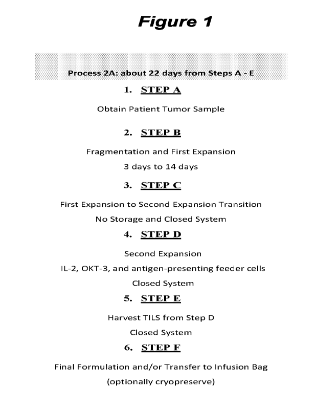
[00113] Figure 1: Exemplary Process 2A chart providing an overview of Steps A
through F.
[00114] Figure 2: Process Flow Chart of Process 2A-2C.
[00115] Figure 3: Shows a diagram of an embodiment of a cryopreserved TIL
exemplary
manufacturing process (-22 days).
[00116] Figure 4: Shows a diagram of an embodiment of process 2A, a 22-day
process for
TIL manufacturing.
[00117] Figure 5: Comparison table of Steps A through F from exemplary
embodiments of
process 1C and process 2A.
[00118] Figure 6: Detailed comparison of an embodiment of process 1C and an
embodiment of process 2A.
[00119] Figure 7: Exemplary GEN 3 type process for tumors.
[00120] Figure 8A-8F: A) Shows a comparison between the 2A process
(approximately 22-
day process) and an embodiment of the Gen 3 process for TIL manufacturing
(approximately
14-days to 16-days process). B) Exemplary Process Gen3 chart providing an
overview of
Steps A through F (approximately 14-days to 16-days process). C) Chart
providing three
exemplary Gen 3 processes with an overview of Steps A through F (approximately
14-days to
16-days process) for each of the three process variations. D) Exemplary
Modified Gen 2-like
process providing an overview of Steps A through F (approximately 22-days
process). E)
Shows a comparison between the 2A process (approximately 22-day process) and
an
embodiment of the Gen 3 process for TIL manufacturing (approximately 14-days
to 22-days
process). F) Exemplary Process PD-1 Gen3 chart providing an overview of Steps
A through
F (approximately 14-days to 22-days process).
[00121] Figure 9: Provides an experimental flow chart for comparability
between GEN 2
(process 2A) versus GEN 3.
23
WO 2022/187741
PCT/US2022/019161
[00122] Figure 10: Shows a comparison between various Gen 2 (2A process) and
the Gen
3.1 process embodiment.
[00123] Figure 11: Table describing various features of embodiments of the Gen
2, Gen 2.1
and Gen 3.0 process.
[00124] Figure 12: Overview of the media conditions for an embodiment of the
Gen 3
process, referred to as Gen 3.1.
[00125] Figure 13: Table describing various features of embodiments of the Gen
2, Gen 2.1
and Gen 3.0 process.
[00126] Figure 14: Table comparing various features of embodiments of the Gen
2 and Gen
3.0 processes.
[00127] Figure 15: Table providing media uses in the various embodiments of
the described
expansion processes.
[00128] Figure 16: Schematic of an exemplary embodiment of the Gen 3 process
(a 16-day
process).
[00129] Figure 17: Schematic of an exemplary embodiment of a method for
expanding T
cells from hematopoietic malignancies using Gen 3 expansion platform.
[00130] Figure 18: Provides the structures I-A and I-B, the cylinders refer to
individual
polypeptide binding domains. Structures I-A and I-B comprise three linearly-
linked TNFRSF
binding domains derived from e.g., 4-1BBL or an antibody that binds 4-1BB,
which fold to
form a trivalent protein, which is then linked to a second trivalent protein
through IgGl-Fc
(including CH3 and CH2 domains) is then used to link two of the trivalent
proteins together
through disulfide bonds (small elongated ovals), stabilizing the structure and
providing an
agonists capable of bringing together the intracellular signaling domains of
the six receptors
and signaling proteins to form a signaling complex. The TNFRSF binding domains
denoted
as cylinders may be scFv domains comprising, e.g., a VH and a VL chain
connected by a
linker that may comprise hydrophilic residues and Gly and Ser sequences for
flexibility, as
well as Glu and Lys for solubility.
[00131] Figure 19: Schematic of an exemplary embodiment of the Gen 3 process
(a 16-day
process).
[00132] Figure 20: Provides a process overview for an exemplary embodiment
(Gen 3.1
Test) of the Gen 3.1 process (a 16 day process).
24
WO 2022/187741
PCT/US2022/019161
1001331 Figure 21: Schematic of an exemplary embodiment of the Gen 3.1 Test
(Gen 3.1
optimized) process (a 16-17 day process).
[00134] Figure 22: Schematic of an exemplary embodiment of the Gen 3 process
(a 16-day
process).
[00135] Figure 23A-23B: Comparison tables for exemplary Gen 2 and exemplary
Gen 3
processes with exemplary differences highlighted.
[00136] Figure 24: Schematic of an exemplary embodiment of the Gen 3 process
(a 16/17
day process) preparation timeline.
[00137] Figure 25: Schematic of an exemplary embodiment of the Gen 3 process
(a 14-16
day process).
[00138] Figure 26A-26B: Schematic of an exemplary embodiment of the Gen 3
process (a
16 day process).
[00139] Figure 27: Schematic of an exemplary embodiment of the Gen 3 process
(a 16 day
process).
[00140] Figure 28: Comparison of Gen 2, Gen 2.1 and an embodiment of the Gen 3
process
(a 16 day process).
[00141] Figure 29: Comparison of Gen 2, Gen 2.1 and an embodiment of the Gen 3
process
(a 16 day process).
[00142] Figure 30: Gen 3 embodiment components.
[00143] Figure 31: Gen 3 embodiment flow chart comparison (Gen 3.0, Gen 3.1
control,
Gen 3.1 Test).
[00144] Figure 32: Shown are the components of an exemplary embodiment of the
Gen 3
process (Gen 3-Optimized, a 16-17 day process).
[00145] Figure 33: Acceptance criteria table.
[00146] Figure 34: Graph summarizing the total viable cells in tumors
incubated overnight
with various antibiotics.
[00147] Figure 35: Graph summarizing the total viable cells of tumors cultured
for 11 day
Pre-REP procedure in the presence of various antibiotics.
WO 2022/187741
PCT/US2022/019161
BRIEF DESCRIPTION OF THE SEQUENCE LISTING
[00148] SEQ ID NO:1 is the amino acid sequence of the heavy chain of
muromonab.
[00149] SEQ ID NO:2 is the amino acid sequence of the light chain of
muromonab.
[00150] SEQ ID NO:3 is the amino acid sequence of a recombinant human IL-2
protein.
[00151] SEQ ID NO:4 is the amino acid sequence of aldesleukin.
[00152] SEQ ID NO:5 is an IL-2 form.
[00153] SEQ ID NO:6 is the amino acid sequence of nemvaleukin alfa.
[00154] SEQ ID NO:7 is an IL-2 form.
[00155] SEQ ID NO:8 is a mucin domain polypeptide.
[00156] SEQ ID NO:9 is the amino acid sequence of a recombinant human IL-4
protein.
[00157] SEQ ID NO:10 is the amino acid sequence of a recombinant human IL-7
protein.
[00158] SEQ ID NO:11 is the amino acid sequence of a recombinant human IL-15
protein.
[00159] SEQ ID NO:12 is the amino acid sequence of a recombinant human IL-21
protein.
[00160] SEQ ID NO:13 is an IL-2 sequence.
[00161] SEQ ID NO:14 is an IL-2 mutein sequence.
[00162] SEQ ID NO:15 is an IL-2 mutein sequence.
[00163] SEQ ID NO:16 is the HCDR1 IL-2 for IgG.IL2R67A.H1.
[00164] SEQ ID NO:17 is the HCDR2 for IgG.IL2R67A.H1.
[00165] SEQ ID NO:18 is the HCDR3 for IgG.IL2R67A,H1.
[00166] SEQ ID NO:19 is the HCDRI IL-2 kabat for IgG.IL2R67A.H1.
[00167] SEQ ID NO:20 is the HCDR2 kabat for IgG.IL2R67A.H1.
[00168] SEQ ID NO:21 is the HCDR3 kabat for IgG.IL2R67A.H1.
[00169] SEQ ID NO:22 is the HCDR1_IL-2 clothia for IgG.IL2R67A.H1.
[00170] SEQ ID NO:23 is the HCDR2 clothia for IgG.IL2R67A.H1.
[00171] SEQ ID NO:24 is the HCDR3 clothia for IgG.IL2R67A.H1.
[00172] SEQ ID NO:25 is the HCDR1 IL-2 IMGT for IgG.IL2R67A,H1.
26
WO 2022/187741
PCT/US2022/019161
[00173] SEQ ID NO:26 is the HCDR2 IMGT for IgG.IL2R67A.H1.
[00174] SEQ ID NO:27 is the HCDR3 IMGT for IgG.IL2R67A.H1.
[00175] SEQ ID NO:28 is the VH chain for IgG.IL2R67A.H1.
[00176] SEQ ID NO:29 is the heavy chain for IgGIL2R67A.H1.
[00177] SEQ ID NO:30 is the LCDR1 kabat for IgG.IL2R67A.H1.
[00178] SEQ ID NO:31 is the LCDR2 kabat for IgG.IL2R67A.H1.
[00179] SEQ ID NO:32 is the LCDR3 kabat for IgG.IL2R67A.H1.
[00180] SEQ ID NO:33 is the LCDR1 chothia for IgG.IL2R67A.H1.
[00181] SEQ ID NO:34 is the LCDR2 chothia for IgG.IL2R67A.H1.
[00182] SEQ ID NO:35 is the LCDR3 chothia for IgG.IL2R67A.H1.
[00183] SEQ ID NO:36 is a VL chain.
[00184] SEQ ID NO:37 is a light chain.
[00185] SEQ ID NO:38 is a light chain.
[00186] SEQ ID NO:39 is a light chain.
[00187] SEQ ID NO:40 is the amino acid sequence of human 4-1BB.
[00188] SEQ ID NO:41 is the amino acid sequence of murine 4-1BB.
[00189] SEQ ID NO:42 is the heavy chain for the 4-1BB agonist monoclonal
antibody
utomilumab (PF-05082566).
[00190] SEQ ID NO:43 is the light chain for the 4-1BB agonist monoclonal
antibody
utomilumab (PF-05082566).
[00191] SEQ ID NO:44 is the heavy chain variable region (VH) for the 4-1BB
agonist
monoclonal antibody utomilumab (PF-05082566).
[00192] SEQ ID NO:45 is the light chain variable region (VL) for the 4-1BB
agonist
monoclonal antibody utomilumab (PF-05082566).
[00193] SEQ ID NO:46 is the heavy chain CDR1 for the 4-1BB agonist monoclonal
antibody utomilumab (PF-05082566).
[00194] SEQ ID NO:47 is the heavy chain CDR2 for the 4-1BB agonist monoclonal
antibody utomilumab (PF-05082566).
27
WO 2022/187741
PCT/US2022/019161
[00195] SEQ ID NO:48 is the heavy chain CDR3 for the 4-1BB agonist monoclonal
antibody utomilumab (PF-05082566).
[00196] SEQ ID NO:49 is the light chain CDR1 for the 4-1BB agonist monoclonal
antibody
utomilumab (PF-05082566).
[00197] SEQ ID NO:50 is the light chain CDR2 for the 4-1BB agonist monoclonal
antibody
utomilumab (PF-05082566).
[00198] SEQ ID NO:51 is the light chain CDR3 for the 4-1BB agonist monoclonal
antibody
utomilumab (PF-05082566).
[00199] SEQ ID NO:52 is the heavy chain for the 4-1BB agonist monoclonal
antibody
urelumab (BMS-663513).
[00200] SEQ ID NO:53 is the light chain for the 4-1BB agonist monoclonal
antibody
urelumab (BMS-663513).
[00201] SEQ ID NO:54 is the heavy chain variable region (VH) for the 4-1BB
agonist
monoclonal antibody urelumab (BMS-663513).
[00202] SEQ ID NO:55 is the light chain variable region (VL) for the 4-1BB
agonist
monoclonal antibody urelumab (BMS-663513).
[00203] SEQ ID NO:56 is the heavy chain CDR1 for the 4-1BB agonist monoclonal
antibody urelumab (BMS-663513).
[00204] SEQ ID NO:57 is the heavy chain CDR2 for the 4-1BB agonist monoclonal
antibody urelumab (BMS-663513).
[00205] SEQ ID NO:58 is the heavy chain CDR3 for the 4-1BB agonist monoclonal
antibody urelumab (BMS-663513).
[00206] SEQ ID NO:59 is the light chain CDR1 for the 4-1BB agonist monoclonal
antibody
urelumab (BMS-663513).
[00207] SEQ ID NO:60 is the light chain CDR2 for the 4-1BB agonist monoclonal
antibody
urelumab (BMS-663513).
[00208] SEQ ID NO:61 is the light chain CDR3 for the 4-1BB agonist monoclonal
antibody
urelumab (BMS-663513).
[00209] SEQ ID NO:62 is an Fc domain for a TNFRSF agonist fusion protein.
[00210] SEQ ID NO:63 is a linker for a TNFRSF agonist fusion protein.
28
WO 2022/187741
PCT/US2022/019161
[00211] SEQ ID NO:64 is a linker for a TNFRSF agonist fusion protein.
[00212] SEQ ID NO:65 is a linker for a TNFRSF agonist fusion protein.
[00213] SEQ ID NO:66 is a linker for a 'TNFRSF agonist fusion protein.
[00214] SEQ ID NO:67 is a linker for a TNFRSF agonist fusion protein.
[00215] SEQ ID NO:68 is a linker for a 'TNFRSF agonist fusion protein.
[00216] SEQ ID NO:69 is a linker for a TNFRSF agonist fusion protein.
[00217] SEQ ID NO:70 is a linker for a TNFRSF agonist fusion protein.
[00218] SEQ ID NO:71 is a linker for a TNFRSF agonist fusion protein.
[00219] SEQ ID NO:72 is a linker for a TNFRSF agonist fusion protein.
[00220] SEQ ID NO:73 is an Fc domain for a TNFRSF agonist fusion protein.
[00221] SEQ ID NO:74 is a linker for a TNFRSF agonist fusion protein.
[00222] SEQ ID NO:75 is a linker for a 'TNFRSF agonist fusion protein.
[00223] SEQ ID NO:76 is a linker for a TNFRSF agonist fusion protein.
[00224] SEQ ID NO:77 is a 4-1BB ligand (4-1BBL) amino acid sequence.
[00225] SEQ ID NO:78 is a soluble portion of 4-1BBL polypeptide.
[00226] SEQ ID NO:79 is a heavy chain variable region (VH) for the 4-1BB
agonist
antibody 4B4-1-1 version 1,
[00227] SEQ ID NO:80 is alight chain variable region (VL) for the 4-1BB
agonist antibody
4B4-1-1 version 1.
[00228] SEQ ID NO:81 is a heavy chain variable region (VH) for the 4-1BB
agonist
antibody 4B4-1-1 version 2.
[00229] SEQ ID NO:82 is a light chain variable region (VL) for the 4-1BB
agonist antibody
4B4-1-1 version 2.
[00230] SEQ ID NO:83 is a heavy chain variable region (VH) for the 4-1BB
agonist
antibody H39E3-2.
[00231] SEQ ID NO:84 is a light chain variable region (VL) for the 4-1BB
agonist antibody
H39E3-2.
[00232] SEQ ID NO:85 is the amino acid sequence of human 0X40.
29
WO 2022/187741
PCT/US2022/019161
1002331 SEQ ID NO:86 is the amino acid sequence of murine 0X40.
[00234] SEQ ID NO:87 is the heavy chain for the 0X40 agonist monoclonal
antibody
tavolixizumab (MEDI-0562).
[00235] SEQ ID NO:88 is the light chain for the 0X40 agonist monoclonal
antibody
tavolixizumab (MEDI-0562).
[00236] SEQ ID NO:89 is the heavy chain variable region (VH) for the 0X40
agonist
monoclonal antibody tavolixizumab (MEDI-0562).
[00237] SEQ ID NO:90 is the light chain variable region (VL) for the 0X40
agonist
monoclonal antibody tavolixizumab (MEDI-0562).
[00238] SEQ ID NO:91 is the heavy chain CDR1 for the 0X40 agonist monoclonal
antibody
tavolixizumab (MEDI-0562).
[00239] SEQ ID NO:92 is the heavy chain CDR2 for the 0X40 agonist monoclonal
antibody
tavolixizumab (MEDI-0562).
[00240] SEQ ID NO:93 is the heavy chain CDR3 for the 0X40 agonist monoclonal
antibody
tavolixizumab (MEDI-0562).
[00241] SEQ ID NO:94 is the light chain CDR1 for the 0X40 agonist monoclonal
antibody
tavolixizumab (MEDI-0562).
[00242] SEQ ID NO:95 is the light chain CDR2 for the 0X40 agonist monoclonal
antibody
tavolixizumab (MEDI-0562).
[00243] SEQ ID NO:96 is the light chain CDR3 for the 0X40 agonist monoclonal
antibody
tavolixizumab (MEDI-0562).
[00244] SEQ ID NO:97 is the heavy chain for the 0X40 agonist monoclonal
antibody 11D4.
[00245] SEQ ID NO:98 is the light chain for the 0X40 agonist monoclonal
antibody 11D4.
[00246] SEQ ID NO:99 is the heavy chain variable region (VH) for the 0X40
agonist
monoclonal antibody 11D4.
[00247] SEQ ID NO:100 is the light chain variable region (VL) for the 0X40
agonist
monoclonal antibody 11D4.
[00248] SEQ ID NO:101 is the heavy chain CDR1 for the 0X40 agonist monoclonal
antibody 11D4.
WO 2022/187741
PCT/US2022/019161
[00249] SEQ ID NO:102 is the heavy chain CDR2 for the 0X40 agonist monoclonal
antibody 11D4.
[00250] SEQ ID NO:103 is the heavy chain CDR3 for the 0X40 agonist monoclonal
antibody 11D4.
[00251] SEQ ID NO: 104 is the light chain CDR1 for the 0X40 agonist monoclonal
antibody
11D4.
[00252] SEQ ID NO:105 is the light chain CDR2 for the OX40 agonist monoclonal
antibody
11D4.
[00253] SEQ ID NO:106 is the light chain CDR3 for the 0X40 agonist monoclonal
antibody
11D4.
[00254] SEQ ID NO:107 is the heavy chain for the OX40 agonist monoclonal
antibody
18D8.
[00255] SEQ ID NO:108 is the light chain for the OX40 agonist monoclonal
antibody 18D8.
[00256] SEQ ID NO:109 is the heavy chain variable region (VH) for the 0X40
agonist
monoclonal antibody 18D8.
[00257] SEQ ID NO:110 is the light chain variable region (VL) for the 0X40
agonist
monoclonal antibody 18D8.
[00258] SEQ ID NO:111 is the heavy chain CDR1 for the 0X40 agonist monoclonal
antibody 18D8.
[00259] SEQ ID NO:112 is the heavy chain CDR2 for the 0X40 agonist monoclonal
antibody 18D8.
[00260] SEQ ID NO:113 is the heavy chain CDR3 for the 0X40 agonist monoclonal
antibody 18D8.
[00261] SEQ ID NO: 114 is the light chain CDR1 for the 0X40 agonist monoclonal
antibody
18D8.
[00262] SEQ ID NO:115 is the light chain CDR2 for the OX40 agonist monoclonal
antibody
18D8.
[00263] SEQ ID NO:116 is the light chain CDR3 for the OX40 agonist monoclonal
antibody
18D8.
31
WO 2022/187741
PCT/US2022/019161
[00264] SEQ ID NO:117 is the heavy chain variable region (VH) for the 0X40
agonist
monoclonal antibody Hu119-122.
[00265] SEQ ID NO:118 is the light chain variable region (VL) for the 0X40
agonist
monoclonal antibody Hu119-122.
[00266] SEQ ID NO:119 is the heavy chain CDR1 for the 0X40 agonist monoclonal
antibody Hu119-122.
[00267] SEQ ID NO:120 is the heavy chain CDR2 for the 0X40 agonist monoclonal
antibody Hu119-122.
[00268] SEQ ID NO:121 is the heavy chain CDR3 for the 0X40 agonist monoclonal
antibody Hu119-122.
[00269] SEQ ID NO:122 is the light chain CDR1 for the 0X40 agonist monoclonal
antibody
Hu119-122.
[00270] SEQ ID NO:123 is the light chain CDR2 for the OX40 agonist monoclonal
antibody
Hu119-122.
[00271] SEQ ID NO:124 is the light chain CDR3 for the OX40 agonist monoclonal
antibody
Hu119-122.
[00272] SEQ ID NO:125 is the heavy chain variable region (VH) for the OX40
agonist
monoclonal antibody Hull 06-222.
[00273] SEQ ID NO:126 is the light chain variable region (VL) for the 0X40
agonist
monoclonal antibody Hu106-222.
[00274] SEQ ID NO:127 is the heavy chain CDR1 for the 0X40 agonist monoclonal
antibody Hu106-222.
[00275] SEQ ID NO:128 is the heavy chain CDR2 for the 0X40 agonist monoclonal
antibody Hu106-222.
[00276] SEQ ID NO:129 is the heavy chain CDR3 for the 0X40 agonist monoclonal
antibody Hu106-222.
[00277] SEQ ID NO:130 is the light chain CDR1 for the OX40 agonist monoclonal
antibody
Hul 06-222.
[00278] SEQ ID NO:131 is the light chain CDR2 for the OX40 agonist monoclonal
antibody
Hu106-222.
32
WO 2022/187741
PCT/US2022/019161
[00279] SEQ ID NO:132 is the light chain CDR3 for the 0X40 agonist monoclonal
antibody
Hu106-222.
[00280] SEQ ID NO:133 is an 0X40 ligand (OX4OL) amino acid sequence.
[00281] SEQ ID NO:134 is a soluble portion of OX4OL polypeptide.
[00282] SEQ ID NO:135 is an alternative soluble portion of OX4OL polypeptide.
[00283] SEQ ID NO: 136 is the heavy chain variable region (VH) for the OX40
agonist
monoclonal antibody 008.
[00284] SEQ ID NO:137 is the light chain variable region (VL) for the 0X40
agonist
monoclonal antibody 008.
[00285] SEQ ID NO:138 is the heavy chain variable region (VH) for the 0X40
agonist
monoclonal antibody 011.
[00286] SEQ ID NO: 139 is the light chain variable region (VL) for the 0X40
agonist
monoclonal antibody 011.
[00287] SEQ ID NO:140 is the heavy chain variable region (VH) for the OX40
agonist
monoclonal antibody 021.
[00288] SEQ ID NO:141 is the light chain variable region (VL) for the 0X40
agonist
monoclonal antibody 021.
[00289] SEQ ID NO:142 is the heavy chain variable region (VH) for the OX40
agonist
monoclonal antibody 023.
[00290] SEQ ID NO:143 is the light chain variable region (VL) for the 0X40
agonist
monoclonal antibody 023.
[00291] SEQ ID NO:144 is the heavy chain variable region (VH) for an 0X40
agonist
monoclonal antibody.
[00292] SEQ ID NO:145 is the light chain variable region (VL) for an OX40
agonist
monoclonal antibody.
[00293] SEQ ID NO:146 is the heavy chain variable region (VH) for an OX40
agonist
monoclonal antibody.
[00294] SEQ ID NO:147 is the light chain variable region (VL) for an 0X40
agonist
monoclonal antibody.
33
WO 2022/187741
PCT/US2022/019161
[00295] SEQ ID NO:148 is the heavy chain variable region (VH) for a humanized
0X40
agonist monoclonal antibody.
[00296] SEQ ID NO:149 is the heavy chain variable region (VH) for a humanized
0X40
agonist monoclonal antibody.
[00297] SEQ ID NO:150 is the light chain variable region (VL) for a humanized
0X40
agonist monoclonal antibody.
[00298] SEQ ID NO:151 is the light chain variable region (VL) for a humanized
0X40
agonist monoclonal antibody.
[00299] SEQ ID NO:152 is the heavy chain variable region (VH) for a humanized
0X40
agonist monoclonal antibody.
[00300] SEQ ID NO:153 is the heavy chain variable region (VH) for a humanized
0X40
agonist monoclonal antibody.
[00301] SEQ ID NO:154 is the light chain variable region (VL) for a humanized
0X40
agonist monoclonal antibody.
[00302] SEQ ID NO:155 is the light chain variable region (VL) for a humanized
0X40
agonist monoclonal antibody.
[00303] SEQ ID NO:156 is the heavy chain variable region (VH) for an 0X40
agonist
monoclonal antibody.
[00304] SEQ ID NO:157 is the light chain variable region (VL) for an 0X40
agonist
monoclonal antibody.
[00305] SEQ ID NO:158 is the heavy chain amino acid sequence of the PD-1
inhibitor
nivolumab.
[00306] SEQ ID NO:159 is the light chain amino acid sequence of the PD-1
inhibitor
nivolumab.
[00307] SEQ ID NO:160 is the heavy chain variable region (VH) amino acid
sequence of
the PD-1 inhibitor nivolumab.
[00308] SEQ ID NO:161 is the light chain variable region (VL) amino acid
sequence of the
PD-1 inhibitor nivolumab.
[00309] SEQ ID NO:162 is the heavy chain CDR1 amino acid sequence of the PD-1
inhibitor nivolumab.
34
WO 2022/187741
PCT/US2022/019161
[00310] SEQ ID NO:163 is the heavy chain CDR2 amino acid sequence of the PD-1
inhibitor nivolumab.
[00311] SEQ ID NO:164 is the heavy chain CDR3 amino acid sequence of the PD-1
inhibitor nivolumab.
[00312] SEQ ID NO:165 is the light chain CDR1 amino acid sequence of the PD-1
inhibitor
nivolumab.
[00313] SEQ ID NO:166 is the light chain CDR2 amino acid sequence of the PD-1
inhibitor
nivolumab.
[00314] SEQ ID NO:167 is the light chain CDR3 amino acid sequence of the PD-1
inhibitor
nivolumab.
[00315] SEQ ID NO:168 is the heavy chain amino acid sequence of the PD-1
inhibitor
pembrolizumab.
[00316] SEQ ID NO:169 is the light chain amino acid sequence of the PD-1
inhibitor
pembrolizumab.
[00317] SEQ ID NO:170 is the heavy chain variable region (VH) amino acid
sequence of
the PD-1 inhibitor pembrolizumab.
[00318] SEQ ID NO:171 is the light chain variable region (VL) amino acid
sequence of the
PD-1 inhibitor pembrolizumab.
[00319] SEQ ID NO:172 is the heavy chain CDR1 amino acid sequence of the PD-1
inhibitor pembrolizumab.
[00320] SEQ ID NO:173 is the heavy chain CDR2 amino acid sequence of the PD-1
inhibitor pembrolizumab.
[00321] SEQ ID NO:174 is the heavy chain CDR3 amino acid sequence of the PD-1
inhibitor pembrolizumab.
[00322] SEQ ID NO:175 is the light chain CDR1 amino acid sequence of the PD-1
inhibitor
pembrolizumab.
[00323] SEQ ID NO:176 is the light chain CDR2 amino acid sequence of the PD-1
inhibitor
pembrolizumab.
[00324] SEQ ID NO:177 is the light chain CDR3 amino acid sequence of the PD-1
inhibitor
pembrolizumab.
WO 2022/187741
PCT/US2022/019161
[00325] SEQ ID NO:178 is the heavy chain amino acid sequence of the PD-Li
inhibitor
durvalumab.
[00326] SEQ ID NO: 179 is the light chain amino acid sequence of the PD-Li
inhibitor
durvalumab.
[00327] SEQ ID NO:180 is the heavy chain variable region (VH) amino acid
sequence of
the PD-L1 inhibitor durvalumab.
[00328] SEQ ID NO:181 is the light chain variable region (VL) amino acid
sequence of the
PD-L1 inhibitor durvalumab.
[00329] SEQ ID NO:182 is the heavy chain CDR1 amino acid sequence of the PD-Ll
inhibitor durvalumab.
[00330] SEQ ID NO:183 is the heavy chain CDR2 amino acid sequence of the PD-L1
inhibitor durvalumab.
[00331] SEQ ID NO:184 is the heavy chain CDR3 amino acid sequence of the PD-Li
inhibitor durvalumab.
[00332] SEQ ID NO:185 is the light chain CDR1 amino acid sequence of the PD-Li
inhibitor durvalumab.
[00333] SEQ ID NO:186 is the light chain CDR2 amino acid sequence of the PD-L1
inhibitor durvalumab.
[00334] SEQ ID NO:187 is the light chain CDR3 amino acid sequence of the PD-Ll
inhibitor durvalumab.
[00335] SEQ ID NO:188 is the heavy chain amino acid sequence of the PD-Li
inhibitor
avelumab.
[00336] SEQ ID NO:189 is the light chain amino acid sequence of the PD-L1
inhibitor
avelumab.
[00337] SEQ ID NO:190 is the heavy chain variable region (VH) amino acid
sequence of
the PD-L1 inhibitor avelumab.
[00338] SEQ ID NO:191 is the light chain variable region (VL) amino acid
sequence of the
PD-Li inhibitor avelumab.
[00339] SEQ ID NO:192 is the heavy chain CDR1 amino acid sequence of the PD-L1
inhibitor avelumab.
36
WO 2022/187741
PCT/US2022/019161
[00340] SEQ ID NO:193 is the heavy chain CDR2 amino acid sequence of the PD-Li
inhibitor avelumab.
[00341] SEQ ID NO:194 is the heavy chain CDR3 amino acid sequence of the PD-Li
inhibitor avelumab.
[00342] SEQ ID NO:195 is the light chain CDR1 amino acid sequence of the PD-L1
inhibitor avelumab.
[00343] SEQ ID NO:196 is the light chain CDR2 amino acid sequence of the PD-Li
inhibitor avelumab.
[00344] SEQ ID NO:197 is the light chain CDR3 amino acid sequence of the PD-Li
inhibitor avelumab.
[00345] SEQ ID NO:198 is the heavy chain amino acid sequence of the PD-Li
inhibitor
atezolizumab.
[00346] SEQ ID NO:199 is the light chain amino acid sequence of the PD-Li
inhibitor
atezolizumab.
[00347] SEQ ID NO:200 is the heavy chain variable region (VH) amino acid
sequence of
the PD-Ll inhibitor atezolizumab.
[00348] SEQ ID NO:201 is the light chain variable region (VL) amino acid
sequence of the
PD-Li inhibitor atezolizumab.
[00349] SEQ ID NO:202 is the heavy chain CDR1 amino acid sequence of the PD-Li
inhibitor atezolizumab.
[00350] SEQ ID NO:203 is the heavy chain CDR2 amino acid sequence of the PD-Ll
inhibitor atezolizumab.
[00351] SEQ ID NO:204 is the heavy chain CDR3 amino acid sequence of the PD-Li
inhibitor atezolizumab.
[00352] SEQ ID NO:205 is the light chain CDR1 amino acid sequence of the PD-Li
inhibitor atezolizumab.
[00353] SEQ ID NO:206 is the light chain CDR2 amino acid sequence of the PD-Li
inhibitor atezolizumab.
[00354] SEQ ID NO:207 is the light chain CDR3 amino acid sequence of the PD-Li
inhibitor atezolizumab.
37
WO 2022/187741
PCT/US2022/019161
[00355] SEQ ID NO:208 is the heavy chain amino acid sequence of the CTLA-4
inhibitor
ipilimumab.
[00356] SEQ ID NO:209 is the light chain amino acid sequence of the CTLA-4
inhibitor
ipilimumab.
[00357] SEQ ID NO:210 is the heavy chain variable region (VH) amino acid
sequence of
the CTLA-4 inhibitor ipilimumab.
[00358] SEQ ID NO:211 is the light chain variable region (VL) amino acid
sequence of the
CTLA-4 inhibitor ipilimumab.
[00359] SEQ ID NO:212 is the heavy chain CDR1 amino acid sequence of the CTLA-
4
inhibitor ipilimumab.
[00360] SEQ ID NO:213 is the heavy chain CDR2 amino acid sequence of the CTLA-
4
inhibitor ipilimumab.
[00361] SEQ ID NO:214 is the heavy chain CDR3 amino acid sequence of the CTLA-
4
inhibitor ipilimumab.
[00362] SEQ ID NO:215 is the light chain CDR1 amino acid sequence of the CTLA-
4
inhibitor ipilimumab.
[00363] SEQ ID NO:216 is the light chain CDR2 amino acid sequence of the CTLA-
4
inhibitor ipilimumab.
[00364] SEQ ID NO:217 is the light chain CDR3 amino acid sequence of the CTLA-
4
inhibitor ipilimumab.
[00365] SEQ ID NO:218 is the heavy chain amino acid sequence of the CTLA-4
inhibitor
tremelimumab,
[00366] SEQ ID NO:219 is the light chain amino acid sequence of the CTLA-4
inhibitor
tremelimumab.
[00367] SEQ ID NO:220 is the heavy chain variable region (VH) amino acid
sequence of
the CTLA-4 inhibitor tremelimumab.
[00368] SEQ ID NO:221 is the light chain variable region (VL) amino acid
sequence of the
CTLA-4 inhibitor tremelimumab.
[00369] SEQ ID NO:222 is the heavy chain CDR1 amino acid sequence of the CTLA-
4
inhibitor tremelimumab.
38
WO 2022/187741
PCT/US2022/019161
[00370] SEQ ID NO:223 is the heavy chain CDR2 amino acid sequence of the CTLA-
4
inhibitor tremelimumab.
[00371] SEQ ID NO:224 is the heavy chain CDR3 amino acid sequence of the CTLA-
4
inhibitor tremelimumab.
[00372] SEQ ID NO:225 is the light chain CDR1 amino acid sequence of the CTLA-
4
inhibitor tremelimumab.
[00373] SEQ ID NO:226 is the light chain CDR2 amino acid sequence of the CTLA-
4
inhibitor tremelimumab.
[00374] SEQ ID NO:227 is the light chain CDR3 amino acid sequence of the CTLA-
4
inhibitor tremelimumab.
[00375] SEQ ID NO:228 is the heavy chain amino acid sequence of the CTLA-4
inhibitor
zalifrelimab.
[00376] SEQ ID NO:229 is the light chain amino acid sequence of the CTLA-4
inhibitor
zalifrelimab.
[00377] SEQ ID NO:230 is the heavy chain variable region (VH) amino acid
sequence of
the CTLA-4 inhibitor zalifrelimab.
[00378] SEQ ID NO:231 is the light chain variable region (VL) amino acid
sequence of the
CTLA-4 inhibitor zalifrelimab.
[00379] SEQ ID NO:232 is the heavy chain CDR1 amino acid sequence of the CTLA-
4
inhibitor zalifrelimab.
[00380] SEQ ID NO:233 is the heavy chain CDR2 amino acid sequence of the CTLA-
4
inhibitor zalifrelimab.
[00381] SEQ ID NO:234 is the heavy chain CDR3 amino acid sequence of the CTLA-
4
inhibitor zalifrelimab.
[00382] SEQ ID NO:235 is the light chain CDR1 amino acid sequence of the CTLA-
4
inhibitor zalifrelimab.
[00383] SEQ ID NO:236 is the light chain CDR2 amino acid sequence of the CTLA-
4
inhibitor zalifrelimab.
[00384] SEQ ID NO:237 is the light chain CDR3 amino acid sequence of the CTLA-
4
inhibitor zalifrelimab.
39
WO 2022/187741
PCT/US2022/019161
DETAILED DESCRIPTION OF THE INVENTION
I. Introduction
[00385] Adoptive cell therapy utilizing TILs cultured ex vivo by the Rapid
Expansion
Protocol (REP) has produced successful adoptive cell therapy following host
immunosuppression in patients with cancer. Current infusion acceptance
parameters rely on
readouts of the composition of TILs (e.g., CD28, CD8, or CD4 positivity) and
on the
numerical folds of expansion and viability of the REP product.
[00386] Current REP protocols give little insight into the health of the TIL
that will be
infused into the patient. T cells undergo a profound metabolic shift during
the course of their
maturation from naive to effector T cells (see Chang, et al., Nat. Irrzmunol.
2016, 17, 364,
hereby expressly incorporated in its entirety, and in particular for the
discussion and markers
of anaerobic and aerobic metabolism). For example, naive T cells rely on
mitochondrial
respiration to produce ATP, while mature, healthy effector T cells such as TIL
are highly
glycolytic, relying on aerobic glycolysis to provide the bioenergetics
substrates they require
for proliferation, migration, activation, and anti-tumor efficacy.
[00387] Current TIL manufacturing and treatment processes are limited by
length, cost,
sterility concerns, and other factors described herein such that the potential
to treat patients
with cancers have been severely limited. There is an urgent need to provide
TIL
manufacturing processes and therapies based on such processes that are
appropriate for use in
treating patients for whom very few or no viable treatment options remain.
[00388] Provided herein are tumor storage compositions and cell culture
media useful
for the production of TIL therapeutics. The reagents allow for the production
of high quality
TIL therapeutics while reducing microbial bioburden and providing sterility
assurance in the
TIL manufacturing process. In particular, the tumor storage compositions
provided herein
advantageously minimize bacterial (e.g., gram-negative and gram-positive
bacterial species)
and fungal contamination while not significantly affecting cell viability.
Moreover,
lymphocytes cultured in the subjected cell culture media are capable of
undergoing
differentiation, exhaustion and/or activation with minimal bacterial (e.g.,
gram-positive and
gram negative bacteria) and/or fungal contamination.
H. Definitions
[00389] Unless defined otherwise, all technical and scientific terms used
herein have the
same meaning as is commonly understood by one of skill in the art to which
this invention
WO 2022/187741
PCT/US2022/019161
belongs. All patents and publications referred to herein are incorporated by
reference in their
entireties.
[00390] The terms "co-administration," "co-administering," "administered in
combination
with," "administering in combination with," "simultaneous," and "concurrent,"
as used
herein, encompass administration of two or more active pharmaceutical
ingredients (in some
embodiments of the present invention, for example, a plurality of TILs) to a
subject so that
both active pharmaceutical ingredients and/or their metabolites are present in
the subject at
the same time. Co-administration includes simultaneous administration in
separate
compositions, administration at different times in separate compositions, or
administration in
a composition in which two or more active pharmaceutical ingredients are
present.
Simultaneous administration in separate compositions and administration in a
composition in
which both agents are present are preferred.
[00391] The term "in vivo" refers to an event that takes place in a subject's
body.
[00392] The term "in vitro" refers to an event that takes places outside of a
subject's body. In
vitro assays encompass cell-based assays in which cells alive or dead are
employed and may
also encompass a cell-free assay in which no intact cells are employed.
[00393] The term "ex vivo" refers to an event which involves treating or
performing a
procedure on a cell, tissue and/or organ which has been removed from a
subject's body.
Aptly, the cell, tissue and/or organ may be returned to the subject's body in
a method of
surgery or treatment.
[00394] The term "rapid expansion" means an increase in the number of antigen-
specific
TILs of at least about 3-fold (or 4-, 5-, 6-, 7-, 8-, or 9-fold) over a period
of a week, more
preferably at least about 10-fold (or 20-, 30-, 40-, 50-, 60-, 70-, 80-, or 90-
fold) over a period
of a week, or most preferably at least about 100-fold over a period of a week.
A number of
rapid expansion protocols are described herein.
[00395] By "tumor infiltrating lymphocytes" or "TILs" herein is meant a
population of cells
originally obtained as white blood cells that have left the bloodstream of a
subject and
migrated into a tumor. TILs include, but are not limited to, CD8+ cytotoxic T
cells
(lymphocytes), Thl and Th17 CD4+ T cells, natural killer cells, dendritic
cells and M1
macrophages. TILs include both primary and secondary TILs. "Primary TILs" are
those that
are obtained from patient tissue samples as outlined herein (sometimes
referred to as "freshly
harvested"), and "secondary TILs" are any TIL cell populations that have been
expanded or
41
WO 2022/187741
PCT/US2022/019161
proliferated as discussed herein, including, but not limited to bulk TILs and
expanded TILs
("REP TILs" or "post-REP TILs") as well as "reREP TILs" as discussed herein.
reREP TILs
can include for example second expansion TILs or second additional expansion
TILs (such
as, for example, those described in Step D of Figure 8, including TILs
referred to as reREP
TILs). TIL cell populations can include genetically modified TILs.
[00396] TILs can generally be defined either biochemically, using cell surface
markers, or
functionally, by their ability to infiltrate tumors and effect treatment. TILs
can be generally
categorized by expressing one or more of the following biomarkers: CD4, CD8,
TCR al3,
CD27, CD28, CD56, CCR7, CD45Ra, CD95, PD-1, and CD25. Additionally, and
alternatively, TILs can be functionally defined by their ability to infiltrate
solid tumors upon
reintroduction into a patient. TILS may further be characterized by potency ¨
for example,
TILS may be considered potent if, for example, interferon (IFN) release is
greater than about
50 pg/mL, greater than about 100 pg/mL, greater than about 150 pg/mL, or
greater than about
200 pg/mL. TILs may be considered potent if, for example, interferon (IFN7)
release is
greater than about 50 pg/mL, greater than about 100 pg/mL, greater than about
150 pg/mL, or
greater than about 200 pg/mL, greater than about 300 pg/mL, greater than about
400 pg/mL,
greater than about 500 pg/mL, greater than about 600 pg/mL, greater than about
700 pg/mL,
greater than about 800 pg/mL, greater than about 900 pg/mL, greater than about
1000 pg/mL.
[00397] By "population of cells" (including TILs) herein is meant a number of
cells that
share common traits. In general, populations generally range from 1 X 10⁶ to 1
X 1019 in
number, with different TIL populations comprising different numbers. For
example, initial
growth of primary TILs in the presence of IL-2 results in a population of bulk
TILs of
roughly 1 x 10⁸ cells. REP expansion is generally done to provide populations
of 1.5 x 10⁹ to
1.5 x 10¹⁰ cells for infusion.
[00398] By "cryopreserved TILs" herein is meant that TILs, either primary,
bulk, or
expanded (REP TILs), are treated and stored in the range of about -150 C to -
60 C. General
methods for cryopreservation are also described elsewhere herein, including in
the Examples.
For clarity, "cryopreserved TILs" are distinguishable from frozen tissue
samples which may
be used as a source of primary TILs.
[00399] By "thawed cryopreserved TILs" herein is meant a population of TILs
that was
previously cryopreserved and then treated to return to room temperature or
higher, including
but not limited to cell culture temperatures or temperatures wherein TILs may
be
administered to a patient.
42
WO 2022/187741
PCT/US2022/019161
[00400] TILs can generally be defined either biochemically, using cell surface
markers, or
functionally, by their ability to infiltrate tumors and effect treatment. TILs
can be generally
categorized by expressing one or more of the following biomarkers: CD4, CD8,
TCR a13,
CD27, CD28, CD56, CCR7, CD45Ra, CD95, PD-1, and CD25. Additionally and
alternatively, TILs can be functionally defined by their ability to infiltrate
solid tumors upon
reintroduction into a patient.
[00401] The term "cryopreservation media" or "cry opreservation medium" refers
to any
medium that can be used for cryopreservation of cells. Such media can include
media
comprising 7% to 10% DMSO. Exemplary media include CryoStor CS10,
Hyperthermasol,
as well as combinations thereof. The term "CS10" refers to a cryopreservation
medium which
is obtained from Stemcell Technologies or from Biolife Solutions. The CS10
medium may be
referred to by the trade name "CryoStork CS10". The CS10 medium is a serum-
free, animal
component-free medium which comprises DMSO.
[00402] The term "central memory T cell" refers to a subset of T cells that in
the human are
CD45R0+ and constitutively express CCR7 (CCRri) and CD62L (CD62hi). The
surface
phenotype of central memory T cells also includes TCR, CD3, CD127 (IL-7R), and
IL-15R.
Transcription factors for central memory T cells include BCL-6, BCL-6B, MBD2,
and BMIl.
Central memory T cells primarily secret IL-2 and CD4OL as effector molecules
after TCR
triggering. Central memory T cells are predominant in the CD4 compartment in
blood, and in
the human are proportionally enriched in lymph nodes and tonsils.
[00403] The term "effector memory T cell" refers to a subset of human or
mammalian T
cells that, like central memory T cells, are CD45R0+, but have lost the
constitutive
expression of CCR7 (CCR710) and are heterogeneous or low for CD62L expression
(CD62L1 ). The surface phenotype of central memory T cells also includes TCR,
CD3,
CD127 (IL-7R), and IL-15R. Transcription factors for central memory T cells
include
BLIMP 1. Effector memory T cells rapidly secret high levels of inflammatory
cytokines
following antigenic stimulation, including interferon-y, IL-4, and IL-5.
Effector memory T
cells are predominant in the CD8 compartment in blood, and in the human are
proportionally
enriched in the lung, liver, and gut. CD8+ effector memory T cells carry large
amounts of
perforin.
[00404] The term "closed system" refers to a system that is closed to the
outside
environment. Any closed system appropriate for cell culture methods can be
employed with
the methods of the present invention. Closed systems include, for example, but
are not
43
WO 2022/187741
PCT/US2022/019161
limited to, closed G-containers. Once a tumor segment is added to the closed
system, the
system is no opened to the outside environment until the TILs are ready to be
administered to
the patient.
[00405] The terms "fragmenting," "fragment," and "fragmented," as used herein
to describe
processes for disrupting a tumor, includes mechanical fragmentation methods
such as
crushing, slicing, dividing, and morcellating tumor tissue as well as any
other method for
disrupting the physical structure of tumor tissue.
[00406] The terms "peripheral blood mononuclear cells" and "PBMCs" refers to a
peripheral
blood cell having a round nucleus, including lymphocytes (T cells, B cells, NK
cells) and
monocytes. When used as an antigen presenting cell (PBMCs are a type of
antigen-presenting
cell), the peripheral blood mononuclear cells are preferably irradiated
allogeneic peripheral
blood mononuclear cells.
[00407] The terms "peripheral blood lymphocytes" and "PBLs" refer to T cells
expanded
from peripheral blood. In some embodiments, PBLs are separated from whole
blood or
apheresis product from a donor. In some embodiments, PBLs are separated from
whole blood
or apheresis product from a donor by positive or negative selection of a T
cell phenotype,
such as the T cell phenotype of CD3+ CD45+.
[00408] The term "anti-CD3 antibody" refers to an antibody or variant
thereof, e.g., a
monoclonal antibody and including human, humanized, chimeric or murine
antibodies which
are directed against the CD3 receptor in the T cell antigen receptor of mature
T cells. Anti-
CD3 antibodies include OKT-3, also known as muromonab. Anti-CD3 antibodies
also
include the UHCT1 clone, also known as T3 and CDR. Other anti-CD3 antibodies
include,
for example, otelixizumab, teplizumab, and visilizumab.
[00409] The term "OKT-3" (also referred to herein as "OKT3") refers to a
monoclonal
antibody or biosimilar or variant thereof, including human, humanized,
chimeric, or murine
antibodies, directed against the CD3 receptor in the T cell antigen receptor
of mature T cells,
and includes commercially-available forms such as OKT-3 (30 ng/mL, MACS GMP
CD3
pure, Miltenyi Biotech, Inc., San Diego, CA, USA) and muromonab or variants,
conservative
amino acid substitutions, glycoforms, or biosimilars thereof. The amino acid
sequences of the
heavy and light chains of muromonab are given in Table 1 (SEQ ID NO:1 and SEQ
ID
NO:2). A hybridoma capable of producing OKT-3 is deposited with the American
Type
Culture Collection and assigned the ATCC accession number CRL 8001. A
hybridoma
44
WO 2022/187741
PCT/US2022/019161
capable of producing OKT-3 is also deposited with European Collection of
Authenticated Cell
Cultures (ECACC) and assigned Catalogue No. 86022706.
TABLE 1. Amino acid sequences of muromonab (exemplary OKT-3 antibody).
Identifier Sequence (One-Letter Amino Acid Symbols)
SEQ ID NO:1 QVQLQQSGAE LARPGASVKM SCKASGYTFT RYTMHWVKQR PGQGLEWIGY
INPSRGYTNY 60
muromonab heavy NQKFKDKATL TTDKSSSTAY MQLSSLTSED SAVYYCARYY DDHYCLDYWG
QGTTLTVSSA 120
chain KTTAPSVYPL APVCGGTTGS SVTLGCLVKG YFPEPVTLTW NSGSLSSGVH
TFPAVLQSDL 180
YTLSSSVTVT SSTWPSQSIT CNVAHPASST KVDKKIEPRP KSCDKTHTCP PCPAPELLGG 240
PSVFLFPPKP KDTLMISRTP EVTCVVVDVS HEDPEVKFNW YVDGVEVHNA KTKPREEQYN 300
STYRVVSVLT VLHQDWLNGK EYKCKVSNKA LPAPIEKTIS KAKGQPREPQ VYTLPPSRDE 360
LTKNQVSLTC LVKGFYPSDI AVEWESNGQP ENNYKTTPPV LDSDGSFFLY SKLTVDKSRW 420
QQGNVFSCSV MHEALHNHYT OKSLSLSPGIC 450
SEQ ID NO:2 QIVLTQSPAI MSASPGEKVT MTCSASSSVS YMNWYQQKSG TSPKRWIYDT
SKLASGVPAH 60
muromonab light FRGSGSGTSY SLTISGMEAE DAATYYCQQW SSNPFTFGSG TKLEINRADT
APTVSIFPPS 120
chain SEQLTSGGAS VVCFLNNFYP KDINVKWKID GSERQNGVLN SWTDQDSKDS
TYSMSSTLTL 180
TKDEYERHNS YTCEATHKTS TSPIVKSFNR NEC 213
[00410] The term "IL-2" (also referred to herein as "IL2") refers to the T
cell growth factor
known as interleukin-2, and includes all forms of IL-2 including human and
mammalian
forms, conservative amino acid substitutions, glycoforms, biosimilars, and
variants thereof
IL-2 is described, e.g., in Nelson, I Immunol. 2004, 172, 3983-88 and Malek,
Annu, Rev.
Immunol. 2008, 26, 453-79, the disclosures of which are incorporated by
reference herein.
The amino acid sequence of recombinant human IL-2 suitable for use in the
invention is
given in Table 2 (SEQ ID NO:3). For example, the term IL-2 encompasses human,
recombinant forms of IL-2 such as aldesleukin (PROLEUKIN, available
commercially from
multiple suppliers in 22 million IU per single use vials), as well as the form
of recombinant
IL-2 commercially supplied by CellGenix, Inc., Portsmouth, NH, USA (CELLGRO
GMP) or
ProSpec-Tany TechnoGene Ltd., East Brunswick, NJ, USA (Cat. No. CYT-209-b) and
other
commercial equivalents from other vendors. Aldesleukin (des-alany1-1, serine-
125 human IL-
2) is a nonglycosylated human recombinant form of IL-2 with a molecular weight
of
approximately 15 kDa. The amino acid sequence of aldesleukin suitable for use
in the
invention is given in Table 2 (SEQ ID NO:4). The term IL-2 also encompasses
pegylated
forms of IL-2, as described herein, including the pegylated IL2 prodrug
bempegaldesleukin
(NKTR-214, pegylated human recombinant IL-2 as in SEQ ID NO:4 in which an
average of
6 lysine residues are N6 substituted with [(2,7-
bisl[methylpoly(oxyethylene)]carbamoyll -9H-
fluoren-9-yl)methoxylcarbonyl), which is available from Nektar Therapeutics,
South San
Francisco, CA, USA, or which may be prepared by methods known in the art, such
as the
methods described in Example 19 of International Patent Application
Publication No. WO
2018/132496 Al or the method described in Example 1 of U.S. Patent Application
WO 2022/187741
PCT/US2022/019161
Publication No. US 2019/0275133 Al, the disclosures of which are incorporated
by reference
herein. Bempegaldesleulcin (NKTR-214) and other pegylated IL-2 molecules
suitable for use
in the invention are described in U.S. Patent Application Publication No. US
2014/0328791
Al and International Patent Application Publication No. WO 2012/065086 Al, the
disclosures of which are incorporated by reference herein. Alternative forms
of conjugated
IL-2 suitable for use in the invention are described in U.S. Patent Nos.
4,766,106, 5,206,344,
5,089,261 and 4,902,502, the disclosures of which are incorporated by
reference herein.
Formulations of IL-2 suitable for use in the invention are described in U.S.
Patent No.
6,706,289, the disclosure of which is incorporated by reference herein.
[00411] In some embodiments, an IL-2 form suitable for use in the present
invention is
THOR-707, available from Synthorx, Inc. The preparation and properties of THOR-
707 and
additional alternative forms of IL-2 suitable for use in the invention are
described in U.S.
Patent Application Publication Nos. US 2020/0181220 Al and US 2020/0330601 Al,
the
disclosures of which are incorporated by reference herein. In some
embodiments, and IL-2
form suitable for use in the invention is an interleulcin 2 (IL-2) conjugate
comprising: an
isolated and purified IL-2 polypeptide; and a conjugating moiety that binds to
the isolated and
purified IL-2 polypeptide at an amino acid position selected from K35, T37,
R38, T41, F42,
K43, F44, Y45, E61, E62, E68, K64, P65, V69, L72, and Y107, wherein the
numbering of
the amino acid residues corresponds to SEQ ID NO:5. In some embodiments, the
amino acid
position is selected from T37, R38, T41, F42, F44, Y45, E61, E62, E68, K64,
P65, V69, L72,
and Y107. In some embodiments, the amino acid position is selected from T37,
R38, T41,
F42, F44, Y45, E61, E62, E68, P65, V69, L72, and Y107. In some embodiments,
the amino
acid position is selected from T37, T41, F42, F44, Y45, P65, V69, L72, and
Y107. In some
embodiments, the amino acid position is selected from R38 and K64. In some
embodiments,
the amino acid position is selected from E61, E62, and E68. In some
embodiments, the amino
acid position is at E62. In some embodiments, the amino acid residue selected
from 1(35,
T37, R38, T41, F42, K43, F44, Y45, E61, E62, E68, K64, P65, V69, L72, and Y107
is
further mutated to lysine, cysteine, or histidine. In some embodiments, the
amino acid residue
is mutated to cysteine. In some embodiments, the amino acid residue is mutated
to lysine. In
some embodiments, the amino acid residue selected from K35, T37, R38, T41,
F42, K43,
F44, Y45, E61, E62, E68, K64, P65, V69, L72, and Y107 is further mutated to an
unnatural
amino acid. In some embodiments, the unnatural amino acid comprises N6-
azidoethoxy-L-
lysine (AzK), N6-propargylethoxy-L-lysine (PraK), BCN-L-lysine, norbornene
lysine, TCO-
lysine, methyltetrazine lysine, allyloxycarbonyllysine, 2-amino-8-oxononanoic
acid, 2-
46
WO 2022/187741
PCT/US2022/019161
amino-8-oxooctanoic acid, p-acetyl-L-phenyla1anine, p-azidomethyl-L-
phenylalanine
(pAMF), p-iodo-L-phenylalanine, m-acetylphenylalanine, 2-amino-8-oxononanoic
acid, p-
propargyloxyphenylalanine, p-propargyl-phenylalanine, 3-methyl-phenylalanine,
L-Dopa,
fluorinated phenylalanine, isopropyl-L-phenylalanine, p-azido-L-phenylalanine,
p-acyl-L-
phenylalanine, p-benzoyl-L-phenylaianine, p-bromophenylalanine, p-amino-L-
phenylalanine,
isopropyl-L-phenylalanine, 0-allyltyrosine, 0-methyl-L-tyrosine, 0-4-allyl-L-
tyrosine, 4-
propyl-L-tyrosine, phosphonotyrosine, tri-O-acetyl-G1cNAcp-serine, L-
phosphoserine,
phosphonoserine, L-3-(2-naphthypaIanine, 2-amino-3-42-43-(benzyloxy)-3-
oxopropyl)amino)ethyl)selanyl)propanoic acid, 2-amino-3-
(phenylselanyl)propanoic, or
selenocysteine. In some embodiments, the IL-2 conjugate has a decreased
affinity to IL-2
receptor a (IL-2Ra) subunit relative to a wild-type IL-2 polypeptide. In some
embodiments,
the decreased affinity is about 10%, 20%, 30%, 40%, 50%, 60%, 70%, 80%, 90%,
95%,
99%, or greater than 99% decrease in binding affinity to IL-2Ra relative to a
wild-type IL-2
polypeptide. In some embodiments, the decreased affinity is about 1-fold, 2-
fold, 3-fold, 4-
fold, 5-fold, 6-fold, 7-fold, 8-fold, 9-fold, 10-fold, 30-fold, 50-fold, 100-
fold, 200-fold, 300-
fold, 500-fold, 1000-fold, or more relative to a wild-type IL-2 polypeptide.
In some
embodiments, the conjugating moiety impairs or blocks the binding of IL-2 with
IL-2Ra. In
some embodiments, the conjugating moiety comprises a water-soluble polymer. In
some
embodiments, the additional conjugating moiety comprises a water-soluble
polymer. In some
embodiments, each of the water-soluble polymers independently comprises
polyethylene
glycol (PEG), poly(propylene glycol) (PPG), copolymers of ethylene glycol and
propylene
glycol, poly(oxyethylated poly ol), poly(olefinic alcohol),
poly(vinylpyrrolidone),
poly(hydroxyalkylmethacrylamide), poly(hydroxyalkylmethacrylate),
poly(saccharides),
poly(a-hydroxy acid), poly(vinyl alcohol), polyphosphazene, polyoxazolines
(POZ), poly(N-
acryloylmorpholine), or a combination thereof. In some embodiments, each of
the water-
soluble polymers independently comprises PEG. In some embodiments, the PEG is
a linear
PEG or a branched PEG. In some embodiments, each of the water-soluble polymers
independently comprises a polysaccharide. In some embodiments, the
polysaccharide
comprises dextran, polysiafic acid (PSA), hya1uronic acid (HA), amylose,
heparin, heparan
sulfate (HS), dextrin, or hydroxyethyl-starch (HES). In some embodiments, each
of the
water-soluble polymers independently comprises a glycan. In some embodiments,
each of the
water-soluble polymers independently comprises polyamine. In some embodiments,
the
conjugating moiety comprises a protein. In some embodiments, the additional
conjugating
moiety comprises a protein. In some embodiments, each of the proteins
independently
47
WO 2022/187741
PCT/US2022/019161
comprises an albumin, a transferrin, or a transthyretin. In some embodiments,
each of the
proteins independently comprises an Fc portion. In some embodiments, each of
the proteins
independently comprises an Fc portion of IgG. In some embodiments, the
conjugating moiety
comprises a polypeptide. In some embodiments, the additional conjugating
moiety comprises
a polypeptide. In some embodiments, each of the polypeptides independently
comprises a
XTEN peptide, a glycine-rich homoamino acid polymer (HAP), a PAS polypeptide,
an
elastin-like polypeptide (ELP), a CTP peptide, or a gelatin-like protein (GLK)
polymer. In
some embodiments, the isolated and purified IL-2 polypeptide is modified by
glutamylation.
In some embodiments, the conjugating moiety is directly bound to the isolated
and purified
IL-2 polypeptide. In some embodiments, the conjugating moiety is indirectly
bound to the
isolated and purified IL-2 polypeptide through a linker. In some embodiments,
the linker
comprises a homobifunctional linker. In some embodiments, the homobifunctiona1
linker
comprises Lomant's reagent dithiobis (succinimidylpropionate) DSP,
3l3'dithiobis(sulfosuccinimidyl proprionate) (DTSSP), disuccinimidyl suberate
(DS S),
bis(sulfosuccinimidyl)suberate (BS), disuccinimidyl tartrate (DST),
disulfosuccinimidyl
tartrate (sulfo DST), ethylene glycobis(succinimidylsuccinate) (EGS),
disuccinimidyl
glutarate (DSG), N,N'-disuccinimidyl carbonate (DSC), dimethyl adipimidate
(DMA),
dimethyl pimelimidate (DMP), dimethyl suberimidate (DMS), dimethy1-3,3'-
dithiobispropionimidate (DTBP), 1,4-di-(3'-(2'-
pyridyldithio)propionamido)butane (DPDPB),
bismaleimidohexane (BMH), aryl halide-containing compound (DFDNB), such as
e.g. 1,5-
difluoro-2,4-dinitrobenzene or 1,3-difluoro-4,6-dinitrobenzene, 4,4'-difluoro-
3,3'-
dinitrophenylsulfone (DFDNPS), bis-H3-(4-azidosalicylamido)ethylidisulfide
(BASED),
formaldehyde, glutaraldehyde, 1,4-butanediol diglycidyl ether, adipic acid
dihydrazide,
carbohydrazide, o-toluidine, 3,3'-dimethylbenzidine, benzidine, a,a'-p-
diaminodiphenyl,
diiodo-p-xylene sulfonic acid, N,N'-ethylene-bis(iodoacetamide), or N,N'-
hexamethylene-
bis(iodoacetamide). In some embodiments, the linker comprises a
heterobifunctional linker.
In some embodiments, the heterobifunctional linker comprises N-succinimidyl 3-
(2-
pyridyldithio)propionate (sPDP), long-chain N-succinimidyl 3-(2-
pyridyldithio)propionate
(LC-sPDP), water-soluble-long-chain N-succinimidyl 3-(2-pyridyldithio)
propionate (sulfo-
LC-sPDP), succinimidyloxycarbonyl-a-methyl-a-(2-pyridyldithio)toluene (sMPT),
sulfosuccinimidy1-64a-methyl-a-(2-pyridyldithio)toluamidoThexanoate (sulfo-LC-
sMPT),
succinimidy1-4-(N-maleimidomethyl)cyclohexane-1-carboxylate (sMCC),
sulfosuccinimidy1-
4-(N-maleimidomethyl)cydohexane-1-carboxylate (sulfo-sMCC), m-maleimidobenzoyl-
N-
hydroxysuccinimide ester (MBs), m-maleimidobenzoyl-N-hydroxysulfosuccinimide
ester
48
WO 2022/187741
PCT/US2022/019161
(sulfo-MBs), N-succinimidy1(4-iodoactey1)aminobenzoate (sIAB),
sulfosuccinimidy1(4-
iodoacteyDaminobenzoate (sulfo-sIAB), succinimidyl-4-(p-
maleimidophenyl)butyrate
(sMPB), sulfosuccinimidyl-4-(p-maleimidophenyl)butyrate (sulfo-sMPB), N-(7-
maleimidobutyryloxy)succinimide ester (GMBs), N-(y-maleimidobutyryloxy)
sulfosuccinimide ester (sulfo-GMBs), succinimidyl 6-
((iodoacetypamino)hexanoate (sIAX),
succinimidyl 6-16-(((iodoacetypamino)hexanoyDaminolhexanoate (slAXX),
succinimidyl 4-
(((iodoacetyl)amino)methyl)cyclohexane-l-carboxylate (sIAC), succinimidyl
64(((4-
iodoacetyl)amino)methyl)cy clohexane-l-carbonyl)amino) hexanoate (sIACX), p-
nitrophenyl
iodoacetate (NPIA), carbonyl-reactive and sulfhydryl-reactive cross-linkers
such as 4-(4-N-
maleimidophenyl)butyric acid hydrazide (MPBH), 4-(N-maleimidomethyl)cy
clohexane-1-
carboxyl-hydrazide-8 (M2C2H), 3-(2-pyridyldithio)propionyl hydrazide (PDPH), N-
hydroxysuccinimidy1-4-azidosalicylic acid (NHs-AsA), N-
hydroxysulfosuccinimidy1-4-
azidosalicylic acid (sulfo-NHs-AsA), sulfosuccinimidyl-(4-
azidosalicylamido)hexanoate
(sulfo-NHs-LC-AsA), sulfosuccinimidyl-2-(p-azidosalicylamido)ethy1-1,3'-
dithiopropionate
(sAsD), N-hydroxysuccinimidy1-4-azidobenzoate (HsAB), N-
hydroxysulfosuccinimidy1-4-
azidobenzoate (sulfo-HsAB), N-succinimidyl-6-(4'-azido-2'-nitrophenyl
amino)hexanoate
(sANPAH), sulfosuccinimidyl-6-(4'-azido-2'-nitrophenylamino)hexanoate (sulfo-
sANPAH),
N-5-azido-2-nitrobenzoyloxysuccinimide (ANB-N0s), sulfosuccinimidy1-2-(m-azido-
o-
nitrobenzamido)-ethy1-1,3'-dithiopropionate (sAND), N-succinimidy1-4(4-
azidopheny1)1,3'-
dithiopropionate (sADP), N-sulfosuccinimidy1(4-azidopheny1)-1,3'-
dithiopropionate (sulfo-
sADP), sulfosuccinimidyl 4-(p-azidophenyl)butyrate (sulfo-sAPB),
sulfosuccinimidyl 2-(7-
azido-4-methylcoumarin-3-acetamide)ethy1-1,3'-dithiopropionate (sAED),
sulfosuccinimidyl
7-azido-4-methylcoumain-3-acetate (sulfo-sAMCA), p-nitrophenyl diazopyruvate
(pNPDP),
p-nitropheny1-2-diazo-3,3,3-trifluoropropionate (PNP-DTP), 1-(p-
azidosalicylamido)-4-
(iodoacetamido)butane (AsIB), N44-(p-azidosalicylamido)buty1]-3/-(2'-
pyridyldithio)
propionamide (APDP), benzophenone-4-iodoacetamide, p-azidobenzoyl hydrazide
(ABH), 4-
(p-azidosalicylamido)butylamine (AsBA), or p-azidophenyl glyoxal (APG). In
some
embodiments, the linker comprises a cleavable linker, optionally comprising a
dipeptide
linker. In some embodiments, the dipeptide linker comprises Val-Cit, Phe-Lys,
Val-Ala, or
Val-Lys. In some embodiments, the linker comprises a non-cleavable linker. In
some
embodiments, the linker comprises a maleimide group, optionally comprising
maleimidocaproyl (mc), succinimidy1-4-(N-maleimidomethyl)cyclohexane-1-
carboxylate
(sMCC), or sulfosuccinimidy1-4-(N-maleimidomethyl)cyclohexane-1-carboxylate
(sulfo-
sMCC). In some embodiments, the linker further comprises a spacer. In some
embodiments,
49
WO 2022/187741
PCT/US2022/019161
the spacer comprises p-aminobenzyl alcohol (PAB), p-aminobenzyoxycarbonyl
(PABC), a
derivative, or an analog thereof In some embodiments, the conjugating moiety
is capable of
extending the serum half-life of the IL-2 conjugate. In some embodiments, the
additional
conjugating moiety is capable of extending the serum half-life of the IL-2
conjugate. In some
embodiments, the IL-2 form suitable for use in the invention is a fragment of
any of the IL-2
forms described herein. In some embodiments, the IL-2 form suitable for use in
the invention
is pegylated as disclosed in U.S. Patent Application Publication No. US
2020/0181220 Al
and U.S. Patent Application Publication No. US 2020/0330601 Al. In some
embodiments,
the IL-2 form suitable for use in the invention is an IL-2 conjugate
comprising: an IL-2
polypeptide comprising an N6-azidoethoxy-L-lysine (AzK) covalently attached to
a
conjugating moiety comprising a polyethylene glycol (PEG), wherein: the IL-2
polypeptide
comprises an amino acid sequence having at least 80% sequence identity to SEQ
ID NO:5;
and the AzK substitutes for an amino acid at position K35, F42, F44, K43, E62,
P65, R38,
T41, E68, Y45, V69, or L72 in reference to the amino acid positions within SEQ
ID NO:5. In
some embodiments, the IL-2 polypeptide comprises an N-terminal deletion of one
residue
relative to SEQ ID NO:5. In some embodiments, the IL-2 form suitable for use
in the
invention lacks IL-2R alpha chain engagement but retains normal binding to the
intermediate
affinity IL-2R beta-gamma signaling complex. In some embodiments, the IL-2
form suitable
for use in the invention is an IL-2 conjugate comprising: an IL-2 polypeptide
comprising an
N6-azidoethoxy-L-lysine (AzK) covalently attached to a conjugating moiety
comprising a
polyethylene glycol (PEG), wherein: the IL-2 polypeptide comprises an amino
acid sequence
having at least 90% sequence identity to SEQ ID NO:5; and the AzK substitutes
for an amino
acid at position K35, F42, F44, K43, E62, P65, R38, T41, E68, Y45, V69, or L72
in reference
to the amino acid positions within SEQ ID NO:5. In some embodiments, the IL-2
thin'
suitable for use in the invention is an IL-2 conjugate comprising: an IL-2
polypeptide
comprising an N6-azidoethoxy-L-lysine (AzK) covalently attached to a
conjugating moiety
comprising a polyethylene glycol (PEG), wherein: the IL-2 polypeptide
comprises an amino
acid sequence having at least 95% sequence identity to SEQ ID NO:5; and the
AzK
substitutes for an amino acid at position K35, F42, F44, K43, E62, P65, R38,
T41, E68, Y45,
V69, or L72 in reference to the amino acid positions within SEQ ID NO:5. In
some
embodiments, the IL-2 form suitable for use in the invention is an IL-2
conjugate comprising:
an IL-2 polypeptide comprising an N6-azidoethoxy-L-lysine (AzK) covalently
attached to a
conjugating moiety comprising a polyethylene glycol (PEG), wherein: the IL-2
polypeptide
comprises an amino acid sequence having at least 98% sequence identity to SEQ
ID NO:5;
WO 2022/187741
PCT/US2022/019161
and the AzK substitutes for an amino acid at position K35, F42, F44, K43, E62,
P65, R38,
T41, E68, Y45, V69, or L72 in reference to the amino acid positions within SEQ
ID NO:570.
[00412] In some embodiments, an IL-2 form suitable for use in the invention is
nemvaleukin
alfa, also known as ALKS-4230 (SEQ ID NO:571), which is available from
Alkermes, Inc.
Nemvaleukin alfa is also known as human interleukin 2 fragment (1-59), variant
(Cys125>Ser51), fused via peptidyl linker (600G61) to human interleukin 2
fragment (62-132),
fused via peptidyl linker (133GSGGGS138) to human interleukin 2 receptor a-
chain fragment
(139-303), produced in Chinese hamster ovary (CHO) cells, glycosylated; human
interleukin
2 (IL-2) (75-133)-peptide [Cys125(51)>Ser[-mutant (1-59), fused via a G2
peptide linker (60-
61) to human interleukin 2 (IL-2) (4-74)-peptide (62-132) and via a GSG3S
peptide linker
(133-138) to human interleukin 2 receptor a-chain (IL2R subunit alpha, IL2Ra,
IL2RA) (1-
165)-peptide (139-303), produced in Chinese hamster ovary (CHO) cells,
glycoform alfa.
The amino acid sequence of nemvaleukin alfa is given in SEQ ID NO:571. In some
embodiments, nemvaleukin alfa exhibits the following post-translational
modifications:
disulfide bridges at positions: 31-116, 141-285, 184-242, 269-301, 166-197 or
166-199, 168-
199 or 168-197 (using the numbering in SEQ ID NO: 571), and glycosylation
sites at
positions: N187, N206, T212 using the numbering in SEQ ID NO:571. The
preparation and
properties of nemvaleukin alfa, as well as additional alternative forms of IL-
2 suitable for use
in the invention, is described in U.S. Patent Application Publication No. US
2021/0038684
Al and U.S. Patent No. 10,183,979, the disclosures of which are incorporated
by reference
herein. In some embodiments, an IL-2 form suitable for use in the invention is
a protein
having at least 80%, at least 90%, at least 95%, or at least 90% sequence
identity to SEQ ID
NO: 571. In some embodiments, an IL-2 form suitable for use in the invention
has the amino
acid sequence given in SEQ ID NO: 571 or conservative amino acid substitutions
thereof In
some embodiments, an IL-2 form suitable for use in the invention is a fusion
protein
comprising amino acids 24-452 of SEQ ID NO:572, or variants, fragments, or
derivatives
thereof In some embodiments, an IL-2 form suitable for use in the invention is
a fusion
protein comprising an amino acid sequence having at least 80%, at least 90%,
at least 95%, or
at least 90% sequence identity to amino acids 24-452 of SEQ ID NO: 572, or
variants,
fragments, or derivatives thereof. Other IL-2 forms suitable for use in the
present invention
are described in U.S. Patent No. 10,183,979, the disclosures of which are
incorporated by
reference herein. Optionally, in some embodiments, an IL-2 form suitable for
use in the
invention is a fusion protein comprising a first fusion partner that is linked
to a second fusion
partner by a mucin domain polypeptide linker, wherein the first fusion partner
is IL-1Ra or a
51
WO 2022/187741
PCT/US2022/019161
protein having at least 98% amino acid sequence identity to IL-1Ra and having
the receptor
antagonist activity of IL-Ra, and wherein the second fusion partner comprises
all or a portion
of an immunoglobulin comprising an Fc region, wherein the mucin domain
polypeptide
linker comprises SEQ ID NO:573 or an amino acid sequence having at least 90%
sequence
identity to SEQ ID NO:573 and wherein the half-life of the fusion protein is
improved as
compared to a fusion of the first fusion partner to the second fusion partner
in the absence of
the mucin domain polypeptide linker.
TABLE 2. Amino acid sequences of interleukins.
Identifier Sequence (One-Letter Amino Acid Symbols)
SEQ ID NO:3 MAPTSSSTKK TQLQLEHLLL DLQMILNGIN NYKNPKLTRM LTFKFYMPKK
ATELKHLQCL 60
recombinant EEELKPLEEV LNLAQSKNFH LRPRDLISNI NVIVLELKGS ETTFMCEYAD
ETATIVEFLN 120
human IL-2 RWITFCQSII STLT 134
(rhIL-2)
SEQ ID NO:4 PTSSSTKKTQ LQLEHLLLDL QMILNGINNY KNPKLTRMLT FKFYMPKKAT
ELKHLQCLEE 60
Aldesleukin ELKPLEEVLN LAQSKNFHLR PRDLISNINV IVLELKGSET TFMCEYADET
ATIVEFLNRW 120
ITFSQSIIST LT 132
SEQ ID NO:5 APTSSSTKKT QLQLSHLLLD LQMILNGINN YKNPKLTRML TFKFYMPKKA
TELKSLQCLE 60
IL-2 form EELKPLEEVL NLAQSKNFHL RPRDLISNIN VIVLELKGSE TTFMCEYADE
TATIVEFLNR 120
WITFCQSIIS TLT 133
SEQ ID NO:6 SKNFHLRPRD LISNINVIVL ELKGSETTFM CEYADETATI VEFLNRWITF
SQSIISTLTG 60
Nemvaleukin alfa GSSSTKKTQL QLEHLLLDLQ MILNGINNYK NPKLTRMLTF KFYMPKKATE
LKHLQCLEEE 120
LKPLEEVLNL AQGSGGGSEL CDDOPPEIPH ATFKAMAYKE GTMLNCECKR GFRRIKSGSL 180
YMLCTGNSSH SSWDNQCQCT SSATRNTTKQ VTPQPEEQKE RKTTEMQSPM QPVDQASLPG 240
HCREPPPWEN EATERIYHFV VGQMVYYQCV QGYRALHRGP AESVCKMTHG KTRWTQPQLI 300
CTG 303
SEQ ID NO:7 MDAMKRGLCC VLLLCGAVFV SARRPSGRKS SKMQAFRIWD VNQKTFYLRN
NQLVAGYLQG 60
IL-2 form PNVNLEEKID VVPIEPHALF LGIHGGKMCL SCVKSGDETR LQLEAVNITD
LSENRKQDKR 120
FAFIRSDSGP TTSFESAACP GWFLCTAMEA DQPVSLTNMP DEGVMVTKFY FQEDESGSGG 180
ASSESSASSD GPHPVITESR ASSESSASSD GPHPVITESR EPKSSDKTHT CPPCPAPELL 240
GGPSVFLFPP KPKDTLMISR TPEVTCVVVD VSHEDPEVKF NWYVDGVEVH NAKTKPREEQ 300
YNSTYRVVSV LTVLHQDWLN GKEYKCKVSN KALPAPIEKT ISKAKGQPRE PQVYTLPPSR 360
EEMTKNQVSL TCLVKGFYPS DIAVEWESNG QPENNYKTTP PVLDSDGSFF LYSKLTVDKS 420
RWQQGNVFSC SVMHEALHNH YTQKSLSLSP GK 452
SEQ ID NO:8 SESSASSDGP HPVITP 16
mucin domain
polypeptide
SEQ ID NO:9 MHKCDITLQE IIKTLNSLTE QKTLCTELTV TDIFAASKNT TEKETFCRAA
TVLRQFYSHH 60
recombinant EKDTRCLGAT AQQFHRHKQL IRFLKRLDRN LWGLAGLNSC PVKEANQSTL
ENFLERLKTI 120
human IL-4 MREKYSKCSS 130
(rhIL-4)
SEQ ID NO:10 MDCDIEGKDG KQYESVLMVS IDQLLDSMKE IGSNCLNNEF NFFKRHICDA
NKEGMFLFRA 60
recombinant ARKLRQFLKM NSTGDFDLHL LKVSEGTTIL LNCTGQVKGR KPAALGEAQP
TKSLEENKSL 120
human IL-7 KEQKKLNDLC FLKRLLQEIK TCWNKILMGT KEH 153
(rhIL-7)
SEQ ID NO:11 MNWVNVISDL KKIEDLIQSM HIDATLYTES DVHPSCKVTA MKCFLLELQV
ISLESGDASI 60
recombinant HDTVENLIIL ANNSLSSNGN VTESGCKECE ELEEKNIKEF LQSFVHIVQM TINTS
115
human IL-15
(rhIL-15)
SEQ ID NO:12 MQDRHMIRMR QLIDIVDQLK NYVNDLVPEF LPAPEDVETN CEWSAFSCFQ
KAQLKSANTG 60
recombinant NNERIINVSI KKLKRKPPST NAGRRQKHRL TCPSCDSYEK KPPKEFLERF
KSLLQKMIHQ 120
human IL-21 HLSSRTHGSE DS 132
(rhIL-21)
[00413] In some embodiments, an IL-2 form suitable for use in the invention
includes an
antibody cytokine engrafted protein that comprises a heavy chain variable
region (VH),
comprising complementarity determining regions HCDR1, HCDR2, HCDR3; a light
chain
52
WO 2022/187741
PCT/US2022/019161
variable region (VL), comprising LCDR1, LCDR2, LCDR3; and an IL-2 molecule or
a
fragment thereof engrafted into a CDR of the VII or the VL, wherein the
antibody cytokine
engrafted protein preferentially expands T effector cells over regulatory T
cells. In some
embodiments, the antibody cytokine engrafted protein comprises a heavy chain
variable
region (VII), comprising complementarity determining regions HCDR1, HCDR2,
HCDR3; a
light chain variable region (VL), comprising LCDR1, LCDR2, LCDR3; and an IL-2
molecule
or a fragment thereof engrafted into a CDR of the VH or the VL, wherein the IL-
2 molecule is
a mutein, and wherein the antibody cytokine engrafted protein preferentially
expands T
effector cells over regulatory T cells. In some embodiments, the IL-2 regimen
comprises
administration of an antibody described in U.S. Patent Application Publication
No. US
2020/0270334 Al, the disclosures of which are incorporated by reference
herein. In some
embodiments, the antibody cytokine engrafted protein comprises a heavy chain
variable
region (VH), comprising complementarity determining regions HCDR1, HCDR2,
HCDR3; a
light chain variable region (VL), comprising LCDR1, LCDR2, LCDR3; and an IL-2
molecule or a fragment thereof engrafted into a CDR of the VH or the VL,
wherein the IL-2
molecule is a mutein, wherein the antibody cytokine engrafted protein
preferentially expands
T effector cells over regulatory T cells, and wherein the antibody further
comprises an IgG
class heavy chain and an IgG class light chain selected from the group
consisting of: a IgG
class light chain comprising SEQ ID NO:39 and a IgG class heavy chain
comprising SEQ ID
NO:38; a IgG class light chain comprising SEQ ID NO:37 and a IgG class heavy
chain
comprising SEQ ID NO:29; a IgG class light chain comprising SEQ ID NO:39 and a
IgG
class heavy chain comprising SEQ ID NO:29; and a IgG class light chain
comprising SEQ ID
NO:37 and a IgG class heavy chain comprising SEQ ID NO:38.
[00414] In some embodiments, an IL-2 molecule or a fragment thereof is
engrafted
into HCDR1 of the VH, wherein the IL-2 molecule is a mutein. In some
embodiments, an IL-
2 molecule or a fragment thereof is engrafted into HCDR2 of the VH, wherein
the IL-2
molecule is a mutein. In some embodiments, an IL-2 molecule or a fragment
thereof is
engrafted into HCDR3 of the VH, wherein the IL-2 molecule is a mutein. In some
embodiments, an IL-2 molecule or a fragment thereof is engrafted into LCDR1 of
the VL,
wherein the IL-2 molecule is a mutein. In some embodiments, an IL-2 molecule
or a
fragment thereof is engrafted into LCDR2 of the VL, wherein the IL-2 molecule
is a mutein.
In some embodiments, an IL-2 molecule or a fragment thereof is engrafted into
LCDR3 of
the VL, wherein the IL-2 molecule is a mutein.
53
WO 2022/187741
PCT/US2022/019161
[00415] The insertion of the IL-2 molecule can be at or near the N-terminal
region of the
CDR, in the middle region of the CDR or at or near the C-terminal region of
the CDR. In
some embodiments, the antibody cytokine engrafted protein comprises an IL-2
molecule
incorporated into a CDR, wherein the IL2 sequence does not frameshift the CDR
sequence.
In some embodiments, the antibody cytokine engrafted protein comprises an IL-2
molecule
incorporated into a CDR, wherein the IL-2 sequence replaces all or part of a
CDR sequence.
The replacement by the IL-2 molecule can be the N-terminal region of the CDR,
in the
middle region of the CDR or at or near the C-terminal region the CDR. A
replacement by the
IL-2 molecule can be as few as one or two amino acids of a CDR sequence, or
the entire
CDR sequences.
[00416] In some embodiments, an IL-2 molecule is engrafted directly into a CDR
without a
peptide linker, with no additional amino acids between the CDR sequence and
the IL-2
sequence. In some embodiments, an IL-2 molecule is engrafted indirectly into a
CDR with a
peptide linker, with one or more additional amino acids between the CDR
sequence and the
IL-2 sequence.
[00417] In some embodiments, the IL-2 molecule described herein is an IL-2
mutein. In
some instances, the IL-2 mutein comprising an R67A substitution. In some
embodiments, the
IL-2 mutein comprises the amino acid sequence SEQ ID NO:14 or SEQ ID NO:15. In
some
embodiments, the IL-2 mutein comprises an amino acid sequence in Table 1 in
U.S. Patent
Application Publication No. US 2020/0270334 Al, the disclosure of which is
incorporated by
reference herein.
[00418] In some embodiments, the antibody cytokine engrafted protein comprises
an
HCDR1 selected from the group consisting of SEQ ID NO:16, SEQ ID NO:19, SEQ ID
NO:22 and SEQ ID NO:25. In some embodiments, the antibody cytokine engrafted
protein
comprises an HCDR1 selected from the group consisting of SEQ ID NO:7, SEQ ID
NO:10,
SEQ ID NO:543 and SEQ ID NO:16. In some embodiments, the antibody cytokine
engrafted
protein comprises an HCDR I selected from the group consisting of HCDR2
selected from
the group consisting of SEQ ID NO:17, SEQ ID NO:20, SEQ ID NO:23, and SEQ ID
NO:26.
In some embodiments, the antibody cytokine engrafted protein comprises an
HCDR3 selected
from the group consisting of SEQ ID NO:18, SEQ ID NO: 21, SEQ ID NO:24, and
SEQ ID
NO:27. In some embodiments, the antibody cytokine engrafted protein comprises
a VH
region comprising the amino acid sequence of SEQ ID NO:28. In some
embodiments, the
antibody cytokine engrafted protein comprises a heavy chain comprising the
amino acid
54
WO 2022/187741
PCT/US2022/019161
sequence of SEQ ID NO:29. In some embodiments, the antibody cytokine engrafted
protein
comprises a VL region comprising the amino acid sequence of SEQ ID NO:36. In
some
embodiments, the antibody cytokine engrafted protein comprises a light chain
comprising the
amino acid sequence of SEQ ID NO:37. In some embodiments, the antibody
cytokine
engrafted protein comprises a VII region comprising the amino acid sequence of
SEQ ID
NO:28 and a VL region comprising the amino acid sequence of SEQ ID NO:36. In
some
embodiments, the antibody cytokine engrafted protein comprises a heavy chain
region
comprising the amino acid sequence of SEQ ID NO:29 and a light chain region
comprising
the amino acid sequence of SEQ ID NO:37. In some embodiments, the antibody
cytokine
engrafted protein comprises a heavy chain region comprising the amino acid
sequence of
SEQ ID NO:29 and a light chain region comprising the amino acid sequence of
SEQ ID
NO:39. In some embodiments, the antibody cytokine engrafted protein comprises
a heavy
chain region comprising the amino acid sequence of SEQ ID NO:38 and a light
chain region
comprising the amino acid sequence of SEQ ID NO:37. In some embodiments, the
antibody
cytokine engrafted protein comprises a heavy chain region comprising the amino
acid
sequence of SEQ ID NO:38 and a light chain region comprising the amino acid
sequence of
SEQ ID NO:39. In some embodiments, the antibody cytokine engrafted protein
comprises
IgG.IL2F71A.H1 or IgG.IL2R67A.H1 of U.S. Patent Application Publication No.
2020/0270334 Al, or variants, derivatives, or fragments thereof, or
conservative amino acid
substitutions thereof, or proteins with at least 80%, at least 90%, at least
95%, or at least 98%
sequence identity thereto. In some embodiments, the antibody components of the
antibody
cytokine engrafted protein described herein comprise immunoglobulin sequences,
framework
sequences, or CDR sequences of palivizumab. In some embodiments, the antibody
cytokine
engrafted protein described herein has a longer serum half-life that a wild-
type IL-2 molecule
such as, but not limited to, aldesleukin or a comparable molecule. In some
embodiments, the
antibody cytokine engrafted protein described herein has a sequence as set
forth in Table 3.
TABLE 3. Sequences of exemplary palivizumab antibody-IL-2 engrafted proteins
Identifier Sequence (One-Letter Amino Acid Symbols)
SEQ ID NO:13 MYRMQLLSCI ALSLALVTNS APTSSSTKKT QLQLEHLLLD LQMILNGINN
YKNPKLTRML 60
IL-2 TFKFYMPKKA TELKHLQCLE EELKPLEEVL NLAQSKNFHL RPRDLISNIN
VIVLELKGSE 120
TTFMCEYADE TATIVEFLNR WITFCQSIIS TLT 153
SEQ ID NO:14 APTSSSTKKT QLQLEHLLLD LQMILNGINN YKNFKLTAML TFKFYMPKKA
TELKHLQCLE 60
IL-2 mutein EELKPLEEVL NLAQSKNFRL RPRDLISNIN VIVLELKGSE TTFMCEYADE
TATIVEFLNR 120
WITFCQSIIS TLT 133
SEQ ID NO:15 APTSSSTKKT QLQLEHLLLD LQMILNGINN YKNPKLTRML TAKFYMPKKA
TELKHLQCLE 60
IL-2 mutein EELKPLEEVL NLAQSKNFHL RPRDLISNIN VIVLELKGSE TTFMCEYADE
TATIVEFLNR 120
WITFCQSIIS TLT 133
SEQ ID NO:16 GFSLAPTSSS TKKTQLQLEH LLLDLQMILN GINNYKNPKL TAMLTFKFYM
PKKATELKHL 60
SCDR1_IL-2 QCLEEELKPL EEVLNLAQSK NFHLRPRDLI SNINVIVLEL KGSETTFMCE
YADETATIVE 120
FLNRWITFCQ SIISTLTSTS GMSVG 145
SEQ ID NO:17 DIWWDDKKDY NPSLKS 16
SCDR2
VVCO 2022/187741
PCT/US2022/019161
SEQ ID NO:18 SMITNWYFDV 10
HCDR3
SEQ ID NO: 15 APTSSSTKKT QLQLEHLLLD LQMILNGINN YKNPKLTAML TFKFYMPKKA
TELKHLQCLE 60
HCDR1_IL-2 kabat EELKPLEEVL NLAQSKNFHL RPRDLISNIN VIVLELKGSE TTFMCEYADE
TATIVEFLNR 120
WITFCQSIIS TLTSTSGMSV G 141
SEQ ID NO:20 DIWWDDKKDY NPSLKS 16
HCDR2 kabat
SEQ ID NO:21 SMITNWYFDV 10
HCDR3 kabat
SEQ ID NO:22 GFSLAPTSSS TKKTQLQLEH LLLDLQMILN GINNYKNPKL TAMLTFKFYM
PKKATELKHL 60
HCDR1_IL-2 QCLEEELKPL EEVLNLAQSK NFHLRPRDLI SNINVIVLEL KGSETTFMCE
YADETATIVE 120
clothia FLNRWITFCQ SIISTLTSTS GM 142
SEQ ID NO:23 WWDDK 5
HCDR2 clothia
SEQ ID NO:24 SMITNWYFDV 10
HCDR3 clothia
SEQ ID NO :25 GFSLAPTSSS TKKTQLQLEH LLLDLQMILN GINNYKNPKL TAMLTFKFYM
PKKATELKHL 60
HCDR1_IL-2 IMGT
QCLEEELKPL EEVLNLAQSK NFHLRPRDLI SNINVIVLEL KGSETTFMCE YADETATIVE 120
FLNRWITFCQ SIISTLTSTS GMS 143
SEQ ID NO:26 IWWDDKK 7
HCDR2 IMGT
SEQ ID NO:27 ARSMITNWYF DV 12
HCDR3 IMGT
SEQ ID NO:28 QVTLRESGPA LVKPTQTLTL TCTFSGFSLA PTSSSTKKTQ LQLEHLLLDL
QMILNGINNY 60
VH KNPKLTAMLT FKFYMPKKAT ELKHLQCLEE ELKPLEEVLN LAQSKNFHLR
PRDLISNINV 120
IVLELKGSET TFMCEYADET ATIVEFLNRW ITFCQSIIST LTSTSGMSVG WIRQPPGKAL 180
EWLADIWWDD KKDYNPSLKS RLTISKDTSK NQVVLKVTNM DPADTATYYC ARSMITNWYF 240
DVWGAGTTVT VSS 253
SEQ ID NO:29 QMILNGINNY KNPKLTAMLT FKFYMPKKAT ELKHLQCLEE ELKPLEEVLN
LAQSKNFHLR 60
Heavy chain PRDLISNINV IVLELKGSET TFMCEYADET AT:VEFLNRW ITPCQSIIST
LTSTSGMSVG 120
WIRQPPGKAL EWLADIWWDD KKDYNPSLKS RLTISKDTSK NQVVLKVTNM DPADTATYYC 180
ARSMITNWYF DVWGAGTTVT VSSASTKGPS VFPLAPSSKS TSGGTAALGC LVKDYFPEPV 240
TVSWNSGALT SGVHTFPAVL QSSGLYSLSS VVTVPSSSLG TQTYICNVNH KPSNTKVDKR 300
VEPKSCDKTH TCPPCPAPEL LGGPSVFLFP PKPKDTLMIS RTPEVTCVVV AVSHEDPEVK 360
FNWYVDGVEV KNAKTKPREE QYNSTYRVVS VLTVLHQDWL NGKEYKCKVS NKALAAPIEK 420
TISKAKGQPR EPQVYTLPPS REEMTKNQVS LTCLVKGFYP SDIAVEWESN GQPENNYKTT 480
PPVLDSDGSF FLYSKLTVDK SRWQQGNVFS CSVMHEALHN HYTQKSLSLS PGK 533
SEQ ID NO:30 KAQLSVGYMH 10
LCD1%1 kabat
SEQ ID NO:31 DTSKLAS 7
LCDR2 kabat
SEQ ID NO:32 FQGSGYPFT 9
LCDR3 kabat
SEQ ID NO:33 QLSVGY 6
LC1JR1 chothia
SEQ ID NO:34 DTS 3
LCDR2 chothia
SEQ ID NO:35 GSGYPF 6
LCDR3 chothia
SEQ ID NO:36 DIQMTQSPST LSASVGDRVT ITCKAQLSVG YMHWYQQKPG KAPKLLIYDT
SKLASGVPSR 60
VL FSGSGSGTEF TLTISSLQPD DFATYYCFQG SGYPFTFGGG TKLEIK 106
SEQ ID NO:37 DIQMTQSPST LSASVGDRVT ITCKAQLSVG YMHWYQQKPG KAPKLLIYDT
SKLASGVPSR 60
Light chain FSGSGSGTEF TLTISSLQPD DFATYYCFQG SGYPFTFGGG TKLEIKRTVA
APSVFIFPPS 120
DEQLKSGTAS VVCLLNNFYP REAKVQWKVD NALQSGNSQE SVTEQDSKDS TYSLSSTLTL 180
SKADYEKEKV YACEVTHQGL SSPVTKSFNR DEC
213
SEQ ID NO:38 QVTLRESGPA LVKPTQTLTL TCTFSGFSLA PTSSSTKKTQ LQLEHLLLDL
QMILNGINNY 60
Light chain KNPKLTRMLT AKFYMPKKAT ELKHLQCLEE ELKPLEEVLN LAQSKNFHLR
PRDLISNINV 120
IVLELKGSET TFMCEYADET ATIVEFLNRW ITFCQSIIST LTSTSGMSVG WIRQPPGKAL 180
EWLADIWWDD KKDYNPSLKS RLTISKDTSK NQVVLKVTNM DPADTATYYC ARSMITNWYF 240
DVWGAGTTVT VSSASTKGPS VFPLAPSSKS TSGGTAALGC LVKDYFPEPV TVSWNSGALT 300
SGVHTFPAVL QSSGLYSLSS VVTVPSSSLG TQTYICNVNH KPSNTKVDKR VEPKSCDKTH 360
TCPPCPAPEL LGGPSVFLFP PKPKDTLMIS RTPEVTCVVV AVSHEDPEVK FNWYVDGVEV 420
KNAKTKPREE QYNSTYRVVS VLTVLHQDWL NGKEYKCKVS NKALAAPIEK TISKAKGQPR 480
EPQVYTLPPS REEMTKNQVS LTCLVKGFYP SDIAVEWESN GQPENNYKTT PPVLDSDGSF 540
FLYSKLTVDK SRWQQGNVFS CSVMHEALHN HYTQKSLSLS PGK 583
SEQ ID NO:39 DIQMTQSPST LSASVGDRVT ITCKAQLSVG YMHWYQQKPG KAPKLLIYDT
SKLASGVPSR 60
Light chain FSGSGSGTEF TLTISSLQPD DFATYYCFQG SGYPFTFGGG TKLEIKRTVA
APSVFIFPPS 120
DEQLKSGTAS VVCLLNNFYP REAKVQWKVD NALQSGNSQE SVTEQDSKDS TYSLSSTLTL 180
SKADYEKHKV YACEVTHQGL SSPVTKSFNR GEC
213
56
WO 2022/187741
PCT/US2022/019161
[00419] The term "IL-4" (also referred to herein as "IL4") refers to the
cytokine known as
interleukin 4, which is produced by Th2 T cells and by eosinophils, basophils,
and mast cells.
IL-4 regulates the differentiation of naive helper T cells (Th0 cells) to Th2
T cells. Steinke
and Borish, Respir. Res. 2001, 2, 66-70. Upon activation by IL-4, Th2 T cells
subsequently
produce additional IL-4 in a positive feedback loop. IL-4 also stimulates B
cell proliferation
and class II MHC expression, and induces class switching to IgE and IgGI
expression from B
cells. Recombinant human IL-4 suitable for use in the invention is
commercially available
from multiple suppliers, including ProSpec-Tany TechnoGene Ltd., East
Brunswick, NJ,
USA (Cat. No. CYT-211) and ThermoFisher Scientific, Inc., Waltham, MA, USA
(human
IL-15 recombinant protein, Cat. No. Gibco CTP0043). The amino acid sequence of
recombinant human IL-4 suitable for use in the invention is given in Table 2
(SEQ ID NO:5).
[00420] The term "IL-7" (also referred to herein as -IL7") refers to a
glycosylated tissue-
derived cytokine known as interleukin 7, which may be obtained from stromal
and epithelial
cells, as well as from dendritic cells. Fry and Mackall, Blood 2002, 99, 3892-
904. IL-7 can
stimulate the development of T cells. IL-7 binds to the IL-7 receptor, a
heterodimer
consisting of IL-7 receptor alpha and common gamma chain receptor, which in a
series of
signals important for T cell development within the thymus and survival within
the periphery.
Recombinant human IL-7 suitable for use in the invention is commercially
available from
multiple suppliers, including ProSpec-Tany TechnoGene Ltd., East Brunswick,
NJ, USA
(Cat. No. CYT-254) and ThermoFisher Scientific, Inc., Waltham, MA, USA (human
IL-15
recombinant protein, Cat. No. Gibco PHC0071). The amino acid sequence of
recombinant
human IL-7 suitable for use in the invention is given in Table 2 (SEQ ID
NO:6).
[00421] The term "IL-15" (also referred to herein as "IL15") refers to the T
cell growth
factor known as interleukin-15, and includes all foims of IL-2 including human
and
mammalian forms, conservative amino acid substitutions, glycoforms,
biosimilars, and
variants thereof. IL-15 is described, e.g., in Fehniger and Caligiuri, Blood
2001, 97, 14-32,
the disclosure of which is incorporated by reference herein. IL-15 shares f3
and y signaling
receptor subunits with IL-2. Recombinant human IL-15 is a single, non-
glycosylated
polypeptide chain containing 114 amino acids (and an N-terminal methionine)
with a
molecular mass of 12.8 kDa. Recombinant human IL-15 is commercially available
from
multiple suppliers, including ProSpec-Tany TechnoGene Ltd., East Brunswick,
NJ, USA
(Cat. No. CYT-230-b) and ThermoFisher Scientific, Inc., Waltham, MA, USA
(human IL-15
57
WO 2022/187741 PCT/US2022/019161
recombinant protein, Cat. No. 34-8159-82). The amino acid sequence of
recombinant human
IL-15 suitable for use in the invention is given in Table 2 (SEQ ID NO:7).
[00422] The term "IL-21" (also referred to herein as "IL21") refers to the
pleiotropic
cytokine protein known as interleukin-21, and includes all forms of IL-21
including human
and mammalian forms, conservative amino acid substitutions, glycoforms,
biosimilars, and
variants thereof. IL-21 is described, e.g, in Spolski and Leonard, Nat. Rev.
Drug. Disc. 2014,
13, 379-95, the disclosure of which is incorporated by reference herein. IL-21
is primarily
produced by natural killer T cells and activated human CD41- T cells.
Recombinant human IL-
21 is a single, non-glycosylated polypeptide chain containing 132 amino acids
with a
molecular mass of 15.4 kDa. Recombinant human IL-21 is commercially available
from
multiple suppliers, including ProSpec-Tany TechnoGene Ltd., East Brunswick,
NJ, USA
(Cat. No. CYT-408-b) and ThermoFisher Scientific, Inc., Waltham, MA, USA
(human IL-21
recombinant protein, Cat. No. 14-8219-80). The amino acid sequence of
recombinant human
IL-21 suitable for use in the invention is given in Table 2 (SEQ ID NO: 8).
[00423] When "an anti-tumor effective amount", "an tumor-inhibiting effective
amount", or
"therapeutic amount" is indicated, the precise amount of the compositions of
the present
invention to be administered can be determined by a physician with
consideration of
individual differences in age, weight, tumor size, extent of infection or
metastasis, and
condition of the patient (subject). It can generally be stated that a
pharmaceutical composition
comprising the tumor infiltrating lymphocytes (e.g. secondary TILs or
genetically modified
cytotoxic lymphocytes) described herein may be administered at a dosage of 104
to 1011
cells/kg body weight (e.g., 105 to 106, 105 to 101 , 105 to 10", 106 to 1-10,
u 106 to 10",107 to
1-11,
u 107 to 1010, 108 to 1011, 108 to 101 , 109 to 1011, or 109 to 1010
cells/kg body weight),
including all integer values within those ranges. Tumor infiltrating
lymphocytes (including in
some cases, genetically modified cytotoxic lymphocytes) compositions may also
be
administered multiple times at these dosages. The tumor infiltrating
lymphocytes (including
in some cases, genetically) can be administered by using infusion techniques
that are
commonly known in immunotherapy (see, e.g., Rosenberg et al., New Eng. J
ofMed. 319:
1676, 1988). The optimal dosage and treatment regime for a particular patient
can readily be
determined by one skilled in the art of medicine by monitoring the patient for
signs of disease
and adjusting the treatment accordingly.
[00424] The term "hematological malignancy", "hematologic malignancy" or terms
of
correlative meaning refer to mammalian cancers and tumors of the hematopoietic
and
58
WO 2022/187741
PCT/US2022/019161
lymphoid tissues, including but not limited to tissues of the blood, bone
marrow, lymph
nodes, and lymphatic system. Hematological malignancies are also referred to
as "liquid
tumors." Hematological malignancies include, but are not limited to, acute
lymphoblastic
leukemia (ALL), chronic lymphocytic lymphoma (CLL), small lymphocytic lymphoma
(SLL), acute myelogenous leukemia (AML), chronic myelogenous leukemia (CML),
acute
monocytic leukemia (AMoL), Hodgkin's lymphoma, and non-Hodgkin's lymphomas.
The
term "B cell hematological malignancy" refers to hematological malignancies
that affect B
cells.
[00425] The term "liquid tumor" refers to an abnormal mass of cells that is
fluid in nature.
Liquid tumor cancers include, but are not limited to, leukemias, myelomas, and
lymphomas,
as well as other hematological malignancies. TILs obtained from liquid tumors
may also be
referred to herein as marrow infiltrating lymphocytes (MILs). TILs obtained
from liquid
tumors, including liquid tumors circulating in peripheral blood, may also be
referred to herein
as PBLs. The terms MIL, TIL, and PBL are used interchangeably herein and
differ only
based on the tissue type from which the cells are derived.
[00426] The term "microenvironment," as used herein, may refer to the solid or
hematological tumor microenvironment as a whole or to an individual subset of
cells within
the microenvironment. The tumor microenvironment, as used herein, refers to a
complex
mixture of "cells, soluble factors, signaling molecules, extracellular
matrices, and mechanical
cues that promote neoplastic transformation, support tumor growth and
invasion, protect the
tumor from host immunity, foster therapeutic resistance, and provide niches
for dominant
metastases to thrive," as described in Swartz, et al., Cancer Res., 2012, 72,
2473. Although
tumors express antigens that should be recognized by T cells, tumor clearance
by the immune
system is rare because of immune suppression by the microenvironment.
[00427] In some embodiments, the invention includes a method of treating a
cancer with a
population of TILs, wherein a patient is pre-treated with non-myeloablative
chemotherapy
prior to an infusion of TILs according to the invention. In some embodiments,
the population
of TILs may be provided wherein a patient is pre-treated with nonmyeloablative
chemotherapy prior to an infusion of TILs according to the present invention.
In some
embodiments, the non-myeloablative chemotherapy is cyclophosphamide 60 mg/kg/d
for 2
days (days 27 and 26 prior to TIL infusion) and fludarabine 25 mg/m2/d for 5
days (days 27
to 23 prior to TIL infusion). In some embodiments, the non-myeloablative
chemotherapy is
cyclophosphamide 60 mg/kg/d for 2 days (days 27 and 26 prior to TIL infusion)
and
59
WO 2022/187741
PCT/US2022/019161
fludarabine 25 mg/m2/d for 3 days (days 27 to 25 prior to TIL infusion). In
some
embodiments, the non-myeloablative chemotherapy is cyclophosphamide 60 mg/kg/d
for 2
days (days 27 and 26 prior to TIL infusion) followed by fludarabine 25 mg/m2/d
for 3 days
(days 25 to 23 prior to TIL infusion). In some embodiments, the non-
myeloablative
chemotherapy is cyclophosphamide 60 mg/kg/d for 2 days (days 27 and 26 prior
to TIL
infusion) and fludarabine 25 mg/m2/d for 3 days (days 27 to 25 prior to TIL
infusion). In
some embodiments, the non-myeloablative chemotherapy is cyclophosphamide 60
mg/kg/d
for 2 days (days 27 and 26 prior to TIL infusion) followed by fludarabine 25
mg/m2/d for 3
days (days 25 to 23 prior to TIL infusion). In some embodiments, after non-
myeloablative
chemotherapy and TIL infusion (at day 0) according to the invention, the
patient receives an
intravenous infusion of IL-2 intravenously at 720,000 IU/kg every 8 hours to
physiologic
tolerance.
[00428] Experimental findings indicate that lymphodepletion prior to adoptive
transfer of
tumor-specific T lymphocytes plays a key role in enhancing treatment efficacy
by eliminating
regulatory T cells and competing elements of the immune system ("cytokine
sinks").
Accordingly, some embodiments of the invention utilize a lymphodepletion step
(sometimes
also referred to as "immunosuppressive conditioning") on the patient prior to
the introduction
of the rTILs of the invention.
[00429] The term "effective amount" or "therapeutically effective amount"
refers to that
amount of a compound or combination of compounds as described herein that is
sufficient to
effect the intended application including, but not limited to, disease
treatment. A
therapeutically effective amount may vary depending upon the intended
application (in vitro
or in vivo), or the subject and disease condition being treated (e.g., the
weight, age and
gender of the subject), the severity of the disease condition, or the manner
of administration.
The term also applies to a dose that will induce a particular response in
target cells (e.g., the
reduction of platelet adhesion and/or cell migration). The specific dose will
vary depending
on the particular compounds chosen, the dosing regimen to be followed, whether
the
compound is administered in combination with other compounds, timing of
administration,
the tissue to which it is administered, and the physical delivery system in
which the
compound is carried.
[00430] The terms "treatment", "treating", "treat", and the like, refer to
obtaining a desired
pharmacologic and/or physiologic effect. The effect may be prophylactic in
terms of
completely or partially preventing a disease or symptom thereof and/or may be
therapeutic in
WO 2022/187741
PCT/US2022/019161
terms of a partial or complete cure for a disease and/or adverse effect
attributable to the
disease. "Treatment", as used herein, covers any treatment of a disease in a
mammal,
particularly in a human, and includes: (a) preventing the disease from
occurring in a subject
which may be predisposed to the disease but has not yet been diagnosed as
having it;
(b) inhibiting the disease, i.e., arresting its development or progression;
and (c) relieving the
disease, i.e., causing regression of the disease and/or relieving one or more
disease
symptoms. "Treatment" is also meant to encompass delivery of an agent in order
to provide
for a pharmacologic effect, even in the absence of a disease or condition. For
example,
"treatment" encompasses delivery of a composition that can elicit an immune
response or
confer immunity in the absence of a disease condition, e.g., in the case of a
vaccine.
[00431] The term "heterologous" when used with reference to portions of a
nucleic acid or
protein indicates that the nucleic acid or protein comprises two or more
subsequences that are
not found in the same relationship to each other in nature. For instance, the
nucleic acid is
typically recombinantly produced, having two or more sequences from unrelated
genes
arranged to make a new functional nucleic acid, e.g., a promoter from one
source and a
coding region from another source, or coding regions from different sources.
Similarly, a
heterologous protein indicates that the protein comprises two or more
subsequences that are
not found in the same relationship to each other in nature (e.g., a fusion
protein).
[00432] The terms "sequence identity," "percent identity," and "sequence
percent identity"
(or synonyms thereof, e.g., "99% identical") in the context of two or more
nucleic acids or
polypeptides, refer to two or more sequences or subsequences that are the same
or have a
specified percentage of nucleotides or amino acid residues that are the same,
when compared
and aligned (introducing gaps, if necessary) for maximum correspondence, not
considering
any conservative amino acid substitutions as part of the sequence identity.
The percent
identity can be measured using sequence comparison software or algorithms or
by visual
inspection. Various algorithms and software are known in the art that can be
used to obtain
alignments of amino acid or nucleotide sequences. Suitable programs to
determine percent
sequence identity include for example the BLAST suite of programs available
from the U.S.
Government's National Center for Biotechnology Information BLAST web site.
Comparisons between two sequences can be carried using either the BLASTN or
BLASTP
algorithm. BLASTN is used to compare nucleic acid sequences, while BLASTP is
used to
compare amino acid sequences. ALIGN, ALIGN-2 (Genentech, South San Francisco,
California) or MegAlign, available from DNASTAR, are additional publicly
available
61
WO 2022/187741
PCT/US2022/019161
software programs that can be used to align sequences. One skilled in the art
can determine
appropriate parameters for maximal alignment by particular alignment software.
In certain
embodiments, the default parameters of the alignment software are used.
[00433] As used herein, the term "variant" encompasses but is not limited to
antibodies or
fusion proteins which comprise an amino acid sequence which differs from the
amino acid
sequence of a reference antibody by way of one or more substitutions,
deletions and/or
additions at certain positions within or adjacent to the amino acid sequence
of the reference
antibody. The variant may comprise one or more conservative substitutions in
its amino acid
sequence as compared to the amino acid sequence of a reference antibody.
Conservative
substitutions may involve, e.g., the substitution of similarly charged or
uncharged amino
acids. The variant retains the ability to specifically bind to the antigen of
the reference
antibody. The term variant also includes pegylated antibodies or proteins.
[00434] The term "deoxyribonudeotide" encompasses natural and synthetic,
unmodified and
modified deoxyribonucleotides. Modifications include changes to the sugar
moiety, to the
base moiety and/or to the linkages between deoxyribonucleotide in the
oligonucleotide.
[00435] The term "RNA" defines a molecule comprising at least one
ribonucleotide residue.
The term "ribonucleotide" defines a nucleotide with a hydroxyl group at the 2'
position of a
b-D-ribofuranose moiety. The term RNA includes double-stranded RNA, single-
stranded
RNA, isolated RNA such as partially purified RNA, essentially pure RNA,
synthetic RNA,
recombinantly produced RNA, as well as altered RNA that differs from naturally
occurring
RNA by the addition, deletion, substitution and/or alteration of one or more
nucleotides.
Nucleotides of the RNA molecules described herein may also comprise non-
standard
nucleotides, such as non-naturally occurring nucleotides or chemically
synthesized
nucleotides or deoxynucleotides. These altered RNAs can be referred to as
analogs or analogs
of naturally-occurring RNA.
[00436] The terms "pharmaceutically acceptable carrier" or "pharmaceutically
acceptable
excipient" are intended to include any and all solvents, dispersion media,
coatings,
antibacterial and antifungal agents, isotonic and absorption delaying agents,
and inert
ingredients. The use of such pharmaceutically acceptable carriers or
pharmaceutically
acceptable excipients for active pharmaceutical ingredients is well known in
the art. Except
insofar as any conventional pharmaceutically acceptable carrier or
pharmaceutically
acceptable excipient is incompatible with the active pharmaceutical
ingredient, its use in
therapeutic compositions of the invention is contemplated. Additional active
pharmaceutical
62
WO 2022/187741
PCT/US2022/019161
ingredients, such as other drugs, can also be incorporated into the described
compositions and
methods.
[00437] The terms "about" and "approximately" mean within a statistically
meaningful
range of a value. Such a range can be within an order of magnitude, preferably
within 50%,
more preferably within 20%, more preferably still within 10%, and even more
preferably
within 5% of a given value or range. The allowable variation encompassed by
the terms
"about" or "approximately" depends on the particular system under study, and
can be readily
appreciated by one of ordinary skill in the art. Moreover, as used herein, the
terms "about"
and "approximately" mean that dimensions, sizes, formulations, parameters,
shapes and other
quantities and characteristics are not and need not be exact, but may be
approximate and/or
larger or smaller, as desired, reflecting tolerances, conversion factors,
rounding off,
measurement error and the like, and other factors known to those of skill in
the art. In
general, a dimension, size, formulation, parameter, shape or other quantity or
characteristic is
"about" or "approximate" whether or not expressly stated to be such. It is
noted that
embodiments of very different sizes, shapes and dimensions may employ the
described
arrangements.
[00438] The transitional terms "comprising," "consisting essentially of," and -
consisting
of," when used in the appended claims, in original and amended form, define
the claim scope
with respect to what unrecited additional claim elements or steps, if any, are
excluded from
the scope of the claim(s). The term "comprising" is intended to be inclusive
or open-ended
and does not exclude any additional, unrecited element, method, step or
material. The term
"consisting of" excludes any element, step or material other than those
specified in the claim
and, in the latter instance, impurities ordinary associated with the specified
material(s). The
term "consisting essentially of" limits the scope of a claim to the specified
elements, steps or
material(s) and those that do not materially affect the basic and novel
characteristic(s) of the
claimed invention. All compositions, methods, and kits described herein that
embody the
present invention can, in alternate embodiments, be more specifically defined
by any of the
transitional terms -comprising," "consisting essentially of," and "consisting
of."
[00439] The terms "antibody" and its plural form "antibodies" refer to whole
immunoglobulins and any antigen-binding fragment ("antigen-binding portion")
or single
chains thereof. An "antibody" further refers to a glycoprotein comprising at
least two heavy
(H) chains and two light (L) chains inter-connected by disulfide bonds, or an
antigen-binding
portion thereof. Each heavy chain is comprised of a heavy chain variable
region (abbreviated
63
WO 2022/187741
PCT/US2022/019161
herein as NTH) and a heavy chain constant region. The heavy chain constant
region is
comprised of three domains, CH1, CH2 and CH3. Each light chain is comprised of
a light
chain variable region (abbreviated herein as VL) and a light chain constant
region. The light
chain constant region is comprised of one domain, CL. The VH and VL regions of
an antibody
may be further subdivided into regions of hypervariability, which are referred
to as
complementarity determining regions (CDR) or hypervariable regions (HVR), and
which can
be interspersed with regions that are more conserved, termed framework regions
(FR). Each
VII and VL is composed of three CDRs and four FRs, arranged from amino-
terminus to
carboxy-terminus in the following order: FR1, CDR1, FR2, CDR2, FR3, CDR3, FR4.
The
variable regions of the heavy and light chains contain a binding domain that
interacts with an
antigen epitope or epitopes. The constant regions of the antibodies may
mediate the binding
of the immunoglobulin to host tissues or factors, including various cells of
the immune
system (e.g., effector cells) and the first component (Clq) of the classical
complement system.
[00440] The term "antigen" refers to a substance that induces an immune
response. In some
embodiments, an antigen is a molecule capable of being bound by an antibody or
a TCR if
presented by major histocompatibility complex (MHC) molecules. The term
"antigen", as
used herein, also encompasses T cell epitopes. An antigen is additionally
capable of being
recognized by the immune system. In some embodiments, an antigen is capable of
inducing a
humoral immune response or a cellular immune response leading to the
activation of B
lymphocytes and/or T lymphocytes. In some cases, this may require that the
antigen contains
or is linked to a Th cell epitope. An antigen can also have one or more
epitopes (e.g., B- and
T-epitopes). In some embodiments, an antigen will preferably react, typically
in a highly
specific and selective manner, with its corresponding antibody or TCR and not
with the
multitude of other antibodies or TCRs which may be induced by other antigens.
[00441] The terms "monoclonal antibody," "mAb," "monoclonal antibody
composition," or
their plural forms refer to a preparation of antibody molecules of single
molecular
composition. A monoclonal antibody composition displays a single binding
specificity and
affinity for a particular epitope. Monoclonal antibodies specific to certain
receptors can be
made using knowledge and skill in the art of injecting test subjects with
suitable antigen and
then isolating hybridomas expressing antibodies having the desired sequence or
functional
characteristics. DNA encoding the monoclonal antibodies is readily isolated
and sequenced
using conventional procedures (e.g., by using oligonucleotide probes that are
capable of
binding specifically to genes encoding the heavy and light chains of the
monoclonal
64
WO 2022/187741
PCT/US2022/019161
antibodies). The hybridoma cells serve as a preferred source of such DNA. Once
isolated, the
DNA may be placed into expression vectors, which are then transfected into
host cells such
as E. coil cells, simian COS cells, Chinese hamster ovary (CHO) cells, or
myeloma cells that
do not otherwise produce immunoglobulin protein, to obtain the synthesis of
monoclonal
antibodies in the recombinant host cells. Recombinant production of antibodies
will be
described in more detail below.
[00442] The terms "antigen-binding portion" or "antigen-binding fragment" of
an antibody
(or simply "antibody portion" or "fragment"), as used herein, refers to one or
more fragments
of an antibody that retain the ability to specifically bind to an antigen. It
has been shown that
the antigen-binding function of an antibody can be performed by fragments of a
full-length
antibody. Examples of binding fragments encompassed within the term "antigen-
binding
portion" of an antibody include (i) a Fab fragment, a monovalent fragment
consisting of the
VL, VH, CL and CH1 domains; (ii) a F(ab')2 fragment, a bivalent fragment
comprising two
Fab fragments linked by a disulfide bridge at the hinge region; (iii) a Fd
fragment consisting
of the VH and CHI domains; (iv) a Fv fragment consisting of the VL and VII
domains of a
single arm of an antibody, (v) a domain antibody (dAb) fragment (Ward, et al.
Nature, 1989,
341, 544-546), which may consist of a Vx or a VL domain; and (vi) an isolated
complementarity determining region (CDR). Furthermore, although the two
domains of the
Fv fragment, VL and VH, are coded for by separate genes, they can be joined,
using
recombinant methods, by a synthetic linker that enables them to be made as a
single protein
chain in which the VL and Vx regions pair to form monovalent molecules known
as single
chain Fv (scFv); see, e.g., Bird, et al., Science 1988, 242, 423-426; and
Huston, et al., Proc.
Natl. Acad. Sci. USA 1988, 85, 5879-5883). Such scFv antibodies are also
intended to be
encompassed within the terms "antigen-binding portion" or "antigen-binding
fragment" of an
antibody. These antibody fragments are obtained using conventional techniques
known to
those with skill in the art, and the fragments are screened for utility in the
same manner as are
intact antibodies.
[00443] The term "human antibody," as used herein, is intended to include
antibodies having
variable regions in which both the framework and CDR regions are derived from
human
germline immunoglobulin sequences. Furthermore, if the antibody contains a
constant region,
the constant region also is derived from human germline immunoglobulin
sequences. The
human antibodies of the invention may include amino acid residues not encoded
by human
germline immunoglobulin sequences (e.g., mutations introduced by random or
site-specific
WO 2022/187741
PCT/US2022/019161
mutagenesis in vitro or by somatic mutation in vivo). The term "human
antibody", as used
herein, is not intended to include antibodies in which CDR sequences derived
from the
germline of another mammalian species, such as a mouse, have been grafted onto
human
framework sequences.
[00444] The term "human monoclonal antibody" refers to antibodies displaying a
single
binding specificity which have variable regions in which both the framework
and CDR
regions are derived from human germline immunoglobulin sequences. In some
embodiments,
the human monoclonal antibodies are produced by a hybridoma which includes a B
cell
obtained from a transgenic nonhuman animal, e.g., a transgenic mouse, having a
genome
comprising a human heavy chain transgene and a light chain transgene fused to
an
immortalized cell.
[00445] The term "recombinant human antibody", as used herein, includes all
human
antibodies that are prepared, expressed, created or isolated by recombinant
means, such as (a)
antibodies isolated from an animal (such as a mouse) that is transgenic or
transchromosomal
for human immunoglobulin genes or a hybridoma prepared therefrom (described
further
below), (b) antibodies isolated from a host cell transformed to express the
human antibody,
e.g., from a transfectoma, (c) antibodies isolated from a recombinant,
combinatorial human
antibody library, and (d) antibodies prepared, expressed, created or isolated
by any other
means that involve splicing of human immunoglobulin gene sequences to other
DNA
sequences. Such recombinant human antibodies have variable regions in which
the
framework and CDR regions are derived from human germline immunoglobulin
sequences.
In certain embodiments, however, such recombinant human antibodies can be
subjected to in
vitro mutagenesis (or, when an animal transgenic for human Ig sequences is
used, in vivo
somatic mutagenesis) and thus the amino acid sequences of the Vii and VL
regions of the
recombinant antibodies are sequences that, while derived from and related to
human germline
VH and VL sequences, may not naturally exist within the human antibody
germline repertoire
in vivo.
[00446] As used herein, "isotype" refers to the antibody class (e.g., IgM or
IgG1) that is
encoded by the heavy chain constant region genes.
[00447] The phrases "an antibody recognizing an antigen" and "an antibody
specific for an
antigen" are used interchangeably herein with the term "an antibody which
binds specifically
to an antigen."
66
WO 2022/187741
PCT/US2022/019161
[00448] The term "human antibody derivatives" refers to any modified form of
the human
antibody, including a conjugate of the antibody and another active
pharmaceutical ingredient
or antibody. The terms "conjugate," "antibody-drug conjugate", "ADC," or
"immunoconjugate" refers to an antibody, or a fragment thereof, conjugated to
another
therapeutic moiety, which can be conjugated to antibodies described herein
using methods
available in the art.
[00449] The terms "humanized antibody," "humanized antibodies," and
"humanized" are
intended to refer to antibodies in which CDR sequences derived from the
germline of another
mammalian species, such as a mouse, have been grafted onto human framework
sequences.
Additional framework region modifications may be made within the human
framework
sequences. Humanized forms of non-human (for example, murine) antibodies are
chimeric
antibodies that contain minimal sequence derived from non-human
immunoglobulin. For the
most part, humanized antibodies are human immunoglobulins (recipient antibody)
in which
residues from a hypervariable region of the recipient are replaced by residues
from a 15
hypervariable region of a non-human species (donor antibody) such as mouse,
rat, rabbit or
nonhuman primate having the desired specificity, affinity, and capacity. In
some instances,
Fv framework region (FR) residues of the human immunoglobulin are replaced by
corresponding non-human residues. Furthermore, humanized antibodies may
comprise
residues that are not found in the recipient antibody or in the donor
antibody. These
modifications are made to further refine antibody performance. In general, the
humanized
antibody will comprise substantially all of at least one, and typically two,
variable domains,
in which all or substantially all of the hypervariable loops correspond to
those of a non-
human immunoglobulin and all or substantially all of the FR regions are those
of a human
immunoglobulin sequence. The humanized antibody optionally also will comprise
at least a
portion of an immunoglobulin constant region (Fc), typically that of a human
immunoglobulin. For further details, see Jones, et al., Nature 1986, 321, 522-
525;
Riechmann, et al., Nature 1988, 332, 323-329; and Presta, Curr. Op. Struct
Biol. 1992, 2,
593-596. The antibodies described herein may also be modified to employ any Fc
variant
which is known to impart an improvement (e.g., reduction) in effector function
and/or FcR
binding. The Fc variants may include, for example, any one of the amino acid
substitutions
disclosed in International Patent Application Publication Nos. WO 1988/07089
Al, WO
1996/14339 Al, WO 1998/05787 Al, WO 1998/23289 Al, WO 1999/51642 Al, WO
99/58572 Al, WO 2000/09560 A2, WO 2000/32767 Al, WO 2000/42072 A2, WO
2002/44215 A2, WO 2002/060919 A2, WO 2003/074569 A2, WO 2004/016750 A2, WO
67
WO 2022/187741
PCT/US2022/019161
2004/029207 A2, WO 2004/035752 A2, WO 2004/063351 A2, WO 2004/074455 A2, WO
2004/099249 A2, WO 2005/040217 A2, WO 2005/070963 Al, WO 2005/077981 A2, WO
2005/092925 A2, WO 2005/123780 A2, WO 2006/019447 Al, WO 2006/047350 A2, and
WO 2006/085967 A2; and U.S. Patent Nos. 5,648,260; 5,739,277; 5,834,250;
5,869,046;
6,096,871; 6,121,022; 6,194,551; 6,242,195; 6,277,375; 6,528,624; 6,538,124;
6,737,056;
6,821,505; 6,998,253; and 7,083,784; the disclosures of which are incorporated
by reference
herein.
[00450] The term "chimeric antibody" is intended to refer to antibodies in
which the variable
region sequences are derived from one species and the constant region
sequences are derived
from another species, such as an antibody in which the variable region
sequences are derived
from a mouse antibody and the constant region sequences are derived from a
human
antibody.
[00451] A "diabody" is a small antibody fragment with two antigen-binding
sites. The
fragments comprises a heavy chain variable domain (VH) connected to a light
chain variable
domain (VL) in the same polypeptide chain (VI-1-W or VL-VH). By using a linker
that is too
short to allow pairing between the two domains on the same chain, the domains
are forced to
pair with the complementary domains of another chain and create two antigen-
binding sites.
Diabodies are described more fully in, e.g., European Patent No. EP 404,097,
International
Patent Publication No. WO 93/11161; and Bolliger, et al., Proc. Natl. Acad.
Sci. USA 1993,
90, 6444-6448.
[00452] The term "glycosylation" refers to a modified derivative of an
antibody. An
aglycoslated antibody lacks glycosylation. Glycosylation can be altered to,
for example,
increase the affinity of the antibody for antigen. Such carbohydrate
modifications can be
accomplished by, for example, altering one or more sites of glycosylation
within the antibody
sequence. For example, one or more amino acid substitutions can be made that
result in
elimination of one or more variable region framework glycosylation sites to
thereby eliminate
glycosylation at that site. Aglycosylation may increase the affinity of the
antibody for
antigen, as described in U.S. Patent Nos. 5,714,350 and 6,350,861.
Additionally or
alternatively, an antibody can be made that has an altered type of
glycosylation, such as a
hypofucosylated antibody having reduced amounts of fucosyl residues or an
antibody having
increased bisecting GlcNac structures. Such altered glycosylation patterns
have been
demonstrated to increase the ability of antibodies. Such carbohydrate
modifications can be
accomplished by, for example, expressing the antibody in a host cell with
altered
68
WO 2022/187741
PCT/US2022/019161
glycosylation machinery. Cells with altered glycosylation machinery have been
described in
the art and can be used as host cells in which to express recombinant
antibodies of the
invention to thereby produce an antibody with altered glycosylation. For
example, the cell
lines Ms704, Ms705, and Ms709 lack the fucosyltransferase gene, FUT8 (alpha
(1,6)
fucosyltransferase), such that antibodies expressed in the Ms704, Ms705, and
Ms709 cell
lines lack fucose on their carbohydrates. The Ms704, Ms705, and Ms709 FUT8¨/¨
cell lines
were created by the targeted disruption of the FUT8 gene in CHO/DG44 cells
using two
replacement vectors (see e.g. U .S . Patent Publication No. 2004/0110704 or
Yamane-Ohnuki,
et al., Biotechnol. Bioeng., 2004, 87, 614-622). As another example, European
Patent No. EP
1,176,195 describes a cell line with a functionally disrupted FUT8 gene, which
encodes a
fucosyl transferase, such that antibodies expressed in such a cell line
exhibit
hypofucosylation by reducing or eliminating the alpha 1,6 bond-related enzyme,
and also
describes cell lines which have a low enzyme activity for adding fucose to the
N-
acetylglucosamine that binds to the Fc region of the antibody or does not have
the enzyme
activity, for example the rat myeloma cell line YB2/0 (ATCC CRL 1662).
International
Patent Publication WO 03/035835 describes a variant CHO cell line, Lec 13
cells, with
reduced ability to attach fucose to Asn(297)-linked carbohydrates, also
resulting in
hypofucosylation of antibodies expressed in that host cell (see also Shields,
et al., I Biol.
Chem. 2002, 277, 26733-26740. International Patent Publication WO 99/54342
describes cell
lines engineered to express glycoprotein-modifying glycosyl transferases
(e.g., beta(1,4)-N-
acetylglucosaminyltransferase III (GnTIII)) such that antibodies expressed in
the engineered
cell lines exhibit increased bisecting GlcNac structures which results in
increased ADCC
activity of the antibodies (see also Umana, etal., Nat. Biotech. 1999, 17, 176-
180).
Alternatively, the fucose residues of the antibody may be cleaved off using a
fucosidase
enzyme. For example, the fucosidase alpha-L-fucosidase removes fucosyl
residues from
antibodies as described in Tarentino, etal., Biochem. 1975, 14, 5516-5523.
[00453] "Pegylation" refers to a modified antibody, or a fragment thereof,
that typically is
reacted with polyethylene glycol (PEG), such as a reactive ester or aldehyde
derivative of
PEG, under conditions in which one or more PEG groups become attached to the
antibody or
antibody fragment. Pegylation may, for example, increase the biological (e.g.,
serum) half-
life of the antibody. Preferably, the pegylation is carried out via an
acylation reaction or an
alkylation reaction with a reactive PEG molecule (or an analogous reactive
water-soluble
polymer). As used herein, the term "polyethylene glycol" is intended to
encompass any of the
forms of PEG that have been used to derivatize other proteins, such as mono
(CI-C io)alkoxy-
69
WO 2022/187741
PCT/US2022/019161
or aryloxy-polyethylene glycol or polyethylene glycol-maleimide. The antibody
to be
pegylated may be an aglycosylated antibody. Methods for pegylation are known
in the art and
can be applied to the antibodies of the invention, as described for example in
European Patent
Nos. EP 0154316 and EP 0401384 and U.S. Patent No. 5,824,778, the disclosures
of each of
which are incorporated by reference herein.
100454] The term "biosimilar" means a biological product, including a
monoclonal antibody
or protein, that is highly similar to a U.S. licensed reference biological
product
notwithstanding minor differences in clinically inactive components, and for
which there are
no clinically meaningful differences between the biological product and the
reference product
in terms of the safety, purity, and potency of the product. Furthermore, a
similar biological or
"biosimilar" medicine is a biological medicine that is similar to another
biological medicine
that has already been authorized for use by the European Medicines Agency. The
term
"biosimilar" is also used synonymously by other national and regional
regulatory agencies.
Biological products or biological medicines are medicines that are made by or
derived from a
biological source, such as a bacterium or yeast. They can consist of
relatively small
molecules such as human insulin or erythropoietin, or complex molecules such
as
monoclonal antibodies. For example, if the reference IL-2 protein is
aldesleukin
(PROLEUKIN), a protein approved by drug regulatory authorities with reference
to
aldesleukin is a "biosimilar to" aldesleukin or is a "biosimilar thereof' of
aldesleukin. In
Europe, a similar biological or "biosimilar" medicine is a biological medicine
that is similar
to another biological medicine that has already been authorized for use by the
European
Medicines Agency (EMA). The relevant legal basis for similar biological
applications in
Europe is Article 6 of Regulation (EC) No 726/2004 and Article 10(4) of
Directive
2001/83/EC, as amended and therefore in Europe, the biosimilar may be
authorized,
approved for authorization or subject of an application for authorization
under Article 6 of
Regulation (EC) No 726/2004 and Article 10(4) of Directive 2001/83/EC. The
already
authorized original biological medicinal product may be referred to as a
"reference medicinal
product" in Europe. Some of the requirements for a product to be considered a
biosimilar are
outlined in the CHMP Guideline on Similar Biological Medicinal Products. In
addition,
product specific guidelines, including guidelines relating to monoclonal
antibody biosimilars,
are provided on a product-by-product basis by the EMA and published on its
website. A
biosimilar as described herein may be similar to the reference medicinal
product by way of
quality characteristics, biological activity, mechanism of action, safety
profiles and/or
efficacy. In addition, the biosimilar may be used or be intended for use to
treat the same
WO 2022/187741
PCT/US2022/019161
conditions as the reference medicinal product. Thus, a biosimilar as described
herein may be
deemed to have similar or highly similar quality characteristics to a
reference medicinal
product. Alternatively, or in addition, a biosimilar as described herein may
be deemed to have
similar or highly similar biological activity to a reference medicinal
product. Alternatively, or
in addition, a biosimilar as described herein may be deemed to have a similar
or highly
similar safety profile to a reference medicinal product. Alternatively, or in
addition, a
biosimilar as described herein may be deemed to have similar or highly similar
efficacy to a
reference medicinal product. As described herein, a biosimilar in Europe is
compared to a
reference medicinal product which has been authorized by the EMA. However, in
some
instances, the biosimilar may be compared to a biological medicinal product
which has been
authorized outside the European Economic Area (a non-EEA authorized
"comparator") in
certain studies. Such studies include for example certain clinical and in vivo
non-clinical
studies. As used herein, the term "biosimilar" also relates to a biological
medicinal product
which has been or may be compared to a non-EEA authorized comparator. Certain
biosimilars are proteins such as antibodies, antibody fragments (for example,
antigen binding
portions) and fusion proteins. A protein biosimilar may have an amino acid
sequence that has
minor modifications in the amino acid structure (including for example
deletions, additions,
and/or substitutions of amino acids) which do not significantly affect the
function of the
polypeptide. The biosimilar may comprise an amino acid sequence having a
sequence
identity of 97% or greater to the amino acid sequence of its reference
medicinal product, e.g.,
97%, 98%, 99% or 100%. The biosimilar may comprise one or more post-
translational
modifications, for example, although not limited to, glycosylation, oxidation,
deamidation,
and/or truncation which is/are different to the post-translational
modifications of the
reference medicinal product, provided that the differences do not result in a
change in safety
and/or efficacy of the medicinal product. The biosimilar may have an identical
or different
glycosylation pattern to the reference medicinal product. Particularly,
although not
exclusively, the biosimilar may have a different glycosylation pattern if the
differences
address or are intended to address safety concerns associated with the
reference medicinal
product. Additionally, the biosimilar may deviate from the reference medicinal
product in for
example its strength, pharmaceutical form, formulation, excipients and/or
presentation,
providing safety and efficacy of the medicinal product is not compromised. The
biosimilar
may comprise differences in for example pharmacokinetic (PK) and/or
pharmacodynamic
(PD) profiles as compared to the reference medicinal product but is still
deemed sufficiently
similar to the reference medicinal product as to be authorized or considered
suitable for
71
WO 2022/187741
PCT/US2022/019161
authorization. In certain circumstances, the biosimilar exhibits different
binding
characteristics as compared to the reference medicinal product, wherein the
different binding
characteristics are considered by a Regulatory Authority such as the EMA not
to be a barrier
for authorization as a similar biological product. The term "biosimilar" is
also used
synonymously by other national and regional regulatory agencies.
III. Tumor Storage Compositions
[00455] In one aspect provided herein are tumor storage compositions that are
useful for the
storage and transport of tumors specimens for tumor infiltrating lymphocytes
(TILs)
production. TILs derived from tumors stored in such compositions can be use in
any suitable
methods, for example, the TIL manufacturing methods provided herein and those
described
for example in US Patent No. 10,166,257; US Patent No. 10,130,659; US Patent
No.
10,272,113; US Patent No. 10,420,799; US Patent No. 10,398,734; US Patent No.
10,463,697; US Patent No. 10,363,273; US Patent Application Pub. No.
2018/0325954, US
Patent Application Pub. No. 2020/0224161; and WO 2020/096986, each of which is
hereby
incorporated by reference in its entirety and in particular for all teachings
related to TIL
manufacturing methods.
[00456] The storage compositions provided herein minimize bacterial (e.g.,
gram-negative
and gram-positive bacterial species) and fungal contamination while not
significantly
affecting TIL viability, thereby advantageously allowing the transport and
hypothermic
storage of the tumor sample for extended periods of time in a sterile
environment prior to TIL
processing. Such tumor storage compositions generally include a serum-free,
animal
component-free cryopreservation medium, and an antibiotic component.
[00457] In some embodiments, the tumor stored in the tumor storage
composition
exhibits at least at or about 50%400% cell viability after 6-48 hour storage
in the tumor
storage composition. In some embodiments, the tumor stored in the tumor
storage
composition exhibits least at or about 50%, 55%, 60%, 65%, 70%, 75%, 80%, 85%,
90%,
95%, 97%, 98%, or 99% cell viability after 6-48 hour storage in the tumor
storage
composition. In some embodiments, the tumor the stored in the tumor storage
composition
exhibits at least at or about 50%400% cell viability after 6, 12, 18, 24, 32,
36 or 48 hour
storage in the tumor storage composition. In certain embodiments, the tumor
stored in the
tumor storage composition exhibits at least about 50%100% cell viability after
6-48 hour
storage in the tumor storage composition at temperature from at or about -10 C
to 10 C, -
72
WO 2022/187741
PCT/US2022/019161
C to 5 C, -5 C to 0 C, 0 C to 5 C, 2 C to 8 C, or 5 C to 10 C. Cell viability
can be
measured using any suitable assay, including, for example, dye exclusion
assays (e.g., trypan
blue, ethidium bromide, propidium iodide, SYTOX, and YO-PRO), DNA condensation
assays (Hoechst 33258 and acridine orange), redox reaction assays (MTT and
X1l, Alamar
Blue), esterase substrate assays (e.g., Calcein AM and Cell Tracker), protease
substrate
assays (e.g., CellTiter-Fluor), ATP measurement (e.g., CellTiter Glo), and
enzyme release
assays (e.g., CytoTox-ONE).
[00458] The sterility of the tumor sample stored in the subject tumor
storage
compositions can be assessed using any suitable method. Exemplary methods
include, but
are not limited to, direct inoculation methods, membrane methods (e.g., open
and closed
membrane filtration systems), ATP-luminescence assays, colorimetric growth
detection
assays, autofluorescence detection assays, and cytometry systems.
[00459] Tumor samples stored in the tumor storage media provided herein can
subsequently undergo processing to derive TILs for basic research or
therapeutic use using
any suitable processing protocol. In some embodiments, the tumor samples
stored in the
subject tumor storage media subsequently are used in the methods for producing
therapeutic
lymphocytes (e.g. TILs, peripheral blood lymphocytes and marrow infiltrating
lymphocytes)
provided herein.
[00460] Aspects of the tumor storage composition are further discussed below.
A. Antibiotics
[00461] The tumor storage compositions disclosed herein include an antibiotic
component.
The antibiotics used in the storage compositions provided herein minimize the
amounts of
bacterial and/or fungal contamination while advantageously exhibiting low
cytotoxic effects
towards TILs. In some embodiments, the antibiotics minimize the amount of gram-
negative
and/or gram-positive bacterial contaminants in the storage medium. Useful
antibiotics
include, but are not limited to, amphotericin B, clindamycin, and vancomycin.
In some
embodiments, the tumor storage composition media further includes gentamicin.
[00462] In some embodiments, the storage composition includes clindamycin. In
some
embodiments, the clindamycin is included at a concentration of at least at or
about 0.1, 0.2,
0.3, 0.4, 0.5, 0.6, 0.7, 0.8, 0.9, 1, 2, 3, 4, 5, 6, 7, 8, 9, 10, 20, 30, 40,
50, 60, 70, 80, 90, 100,
110, 120, 130, 140, 150, 160, 170, 180, 190, 200, 250, 300, 350, 400, 450,
500, 550, 600,
650, 700, 750, 800, 850, 900, 950, or 1,000 ps/mL. In certain embodiments, the
clindamycin
73
WO 2022/187741
PCT/US2022/019161
is included at a concentration of from at or about 0.1-1 ttg/mL, 0.25-1 g/mL,
0.1-0.5 p.g/mL,
0.5-2 pg/mL, 2-8 g/mL, 1-10 ps/mL, 4-12 g/mL, 5-15 pg/mL, 10-20 ps/mL, 20-30
pg/mL, 30-40 pg/mL, 40-50 p.g/mL, 50-60 ps/mL, 60-70 g/mL, 70-80 pg/mL, 80-90
p.g/mL, 90-100 ps/mL, 100-110 p.g/mL, 110-120 p.g/mL, 120-130 ps/mL, 130-140
g/mL,
140-150 pg/mL, 50-150 ii.g/mL, 60-140 pg/mL, 70-130 pg/mL, 80-120 p.g/mL, 90-
110
p.g/mL, 95-105 p.g/mL, 10-90 pg/mL, 20-80 p.g/mL, 30-70 pg/mL, 40-60 p.g/mL,
45-55
pz/mL, 50-100 p.g/mL, 100-150 g/mL, 150-200 pz/mL, 200-250 pg/mL, 250-300
p.g/mL,
300-350 pg/mL, 350-400 pg/mL, 400-450 p.g/mL, 450-500 pg/mL, 500-550 pg/mL,
550-600
p.g/mL, 600-650 ps/mL, 650-700 p.g/mL, 700-750 p.g/mL, 750-800 p.g/mL, 800-850
ttg/mL,
850-900 g/mL, or 950-1,000 g/mL. In some embodiments, the clindamycin is
included at
a concentration of from at or about 0.1-100 g/mL, 1-50 pg/mL, 1-100 pg/mL, 1-
250 ttg/mL,
1-500 p.g/mL, 250-750 jig/mL, 350-450 pg/mL, 450-550 pg/mL, 550-650 g/mL, 400-
600
pg/mL, 350-650 lAg/mL, 300-700 pg/mL, 200-800 pg/mL, 500-1,000 ttg/mL, 750-
1,250
ps/mL, 1,000-1,500 ps/mL, 1,250-1,750 ps/mL, or 1,500-2,000 ps/mL. In
exemplary
embodiments, the clindamycin is at a concentration of at or about 400-600
pg/mL.
[00463] In certain embodiments, the storage composition includes vancomycin.
In some
embodiments, the vancomycin is included at a concentration of at least at or
about 0.1, 0.2,
0.3, 0.4, 0.5, 0.6, 0.7, 0.8, 0.9, 1, 2, 3, 4, 5, 6, 7, 8, 9, 10, 20, 30 , 40,
50, 60, 70, 80, 90, 100,
110, 120, 130, 140, 150, 160, 170, 180, 190, 200, 250, 300, 350, 400, 450,
500, 550, 600,
650, 700, 750, 800, 850, 900, 950, or 1,000 ps/mL. In certain embodiments, the
vancomycin
is included at a concentration of from at or about 0.1-1 ps/mL, 0.25-1 ps/mL,
0.1-0.5 g/mL,
0.5-2 pg/mL, 2-8 p.g/mL, 1-10 p.g/mL, 4-12 pg/mL, 5-15 pg/mL, 10-20 ii.g/mL,
20-30
g/mL, 30-40 pg/mL, 40-50 p.g/mL, 50-60 g/mL, 60-70 g/mL, 70-80 pg/mL, 80-90
pg/mL, 90-100 g/mL, 100-110 p.g/mL, 110-120 pg/mL, 120-130 pg/mL, 130-140
p.g/mL,
140-150 ps/mL, 50-15011g/mL, 60-140 pg/mL, 70-130 ps/mL, 80-120 p.g/mL, 90-110
ps/mL, 95-105 g/mL, 10-90 mg/I-I-IL, 20-80 pg/mL, 30-70 p.g/mL, 40-60 ps/mL,
45-55
[i.g/mL, 50-100 g/mL, 100-150 ps/mL, 150-200 pg/mL, 200-250 pg/mL, 250-300
lig/mL,
300-350 pg/mL, 350-400 pg/mL, 400-450 ttg/mL, 450-500 p.g/mL, 500-550 pg/mL,
550-600
pg/mL, 600-650 p.g/mL, 650-700 pg/mL, 700-750 pg/mL, 750-800 ps/mL, 800-850
ps/mL,
850-900 ps/mL, or 950-1,000 ps/mL. In some embodiments, the vancomycin is
included at
a concentration of from at or about 0.1-100 ps/mL, 1-50 ttg/mL, 1-100 ps/mL, 1-
250 ps/mL,
1-500 g/mL, 100-200 pg/mL, 150-250 pg/mL, 200-400 g/mL, 350-450 i.tg/mL, 400-
600
pz/mL, 550-650 pg/mL, 50-650 p.g/mL, 100-600 pg/mL, 250-750 pg/mL, 500-1,000
pg/mL,
750-1,250 pg/mL, 1,000-1,500 pg,/mL, 1,250-1,750 p.g/mL, or 1,500-2,000
p.g/mL. In
74
WO 2022/187741
PCT/US2022/019161
exemplary embodiments, the vancomycin is at a concentration of at or about 50-
600 gg/mL.
In exemplary embodiments, the vancomycin is at a concentration of at or about
100 g/mL.
[00464] In some embodiments, the storage composition includes vancomycin and
gentamicin. In certain embodiments, the storage composition includes
clindamycin and
gentamicin. In some embodiments, the gentamicin is included at a concentration
of at least at
or about 0.1, 0.2, 0.3, 0.4, 0.5, 0.6, 0.7, 0.8, 0.9, 1, 2, 3, 4, 5, 6, 7, 8,
9, 10, 20, 30 , 40, 50, 60,
70, 80, 90, 100, 110, 120, 130, 140, 150, 160, 170, 180, 190, 200, 250, 300,
350, 400, 450,
500, 550, 600, 650, 700, 750, 800, 850, 900, 950, or 1,000 p.g/mL. In certain
embodiments,
the gentamicin is included at a concentration of from at or about 0.1-1 pg/mL,
0.25-1 pg/mL,
0.1-0.5 lig/mL, 0.5-2 p.g/mL, 2-8 pg/mL, 1-10 p.g/mL, 4-12 lig/mL, 5-15
p.g/mL, 10-20
1.1g/mL, 20-30 p.g/rnL, 30-40 ii.g/mL, 40-50 ji.g/mL, 50-60 p.g/mL, 60-70
pg/mL, 70-80
pg/mL, 80-90 g/mL, 90-100 pg/mL, 100-110 p.g/mL, 110-120 pg/mL, 120-130
pg/mL,
130-140 p.g/mL, 140-150 p.g/mL, 150-160 p.g/mL, 160-170 p.g/mL, 170-180 pg/mL,
180-190
ps/mL, 190-200 p.g/mL, 10-90 p.g/mL, 20-80 p.g/mL, 30-70 ps/mL, 40-60 pg/mL,
45-55
ps/mL, 50-150 g/mL, 60-140 pg/mL, 70-130 ps/mL, 80-120 ps/mL, 90-110 ps/mL, 95-
105 mg/mL, 50-100 ttg/mL, 100-150 mg/mL, 150-200 mg/mL, 200-250 mg/mL, 250-300
g/mL, 300-350 p.g/mL, 350-400 pg/mL, 400-450 ps/mL, 450-500 ttg/mL, 500-550
ps/mL,
550-600 jig/mL, 600-650 g/mL, 650-700 p.g/mL, 700-750 ps/mL, 750-800 p.g/mL,
800-850
g/mL, 850-900 ps/mL, or 950-1,000 p.g/mL. In some embodiments, the gentamicin
is
included at a concentration of from at or about 0.1-100 ps/mL, 1-50 pg/mL, 25-
75 ps/mL, 1-
100 ii.g/mL, 1-250 pg/mL, 1-500 g/mL, 250-750 ps/mL, 500-1,000 p.g/mL, 750-
1,250
p.g/mL, 1,000-1,500 pg/mL, 1,250-1,750 p.g/mL, or 1,500-2,000 ji.g/mL. In
exemplary
embodiments, the gentamicin is at a concentration of at or about 50 p.g/mL.
[00465] In some embodiments, the tumor storage medium further includes one or
more
antifungal antibiotics. Antifungal antibiotics for use in the subject tumor
storage medium
include, but are not limited to polyenes, azoles, imidazoles, triazoles,
thiazoles, allylamines,
and echinocandin. Exemplary polyenes include, but are not limited to:
amphotericin B,
candicidin, filipin, hamycin, natamycin, nystatin, and rimocidin. Exemplary
imidazoles
include, but are not limited to, bifonazole, butoconazole, clotrimazole,
econazole,
fenticonazole, isoconazole, ketoconazole, luliconazole, miconazole,
omoconazole,
oxiconazole, sertaconazole, sulconazole, and tioconazole. Useful triazoles
include, but are
not limited to: albaconazole, efmaconazole, epoxiconazole, fluconazole,
isavuconazole,
itraconazole, posaconazole, propiconazole, ravuconazole, terconazole, and
voriconazole.
WO 2022/187741
PCT/US2022/019161
Exemplary echinocandins include, but are not limited to: anidulafungin,
caspofungin,
micafungin. Additional antifungal antibiotics that can be included in the
tumor storage
compositions disclosed herein include, but are not limited to: aurones,
benzoic acid,
ciclopirox, flucytosine, griseofulvin, haloprogin, tolnaflate, undecyenic
acid, triacetin, crystal
violet, orotomide, milteofosine, potassium iodide, nikkomycin, copper sulfate,
selenium
disulfide, sodium thiosulfate, prioctone olamine, iodoquinol, acrisorcin, zinc
pyrithione, and
sulfur.
[00466] In some embodiments, the tumor storage composition includes
amphotericin B. In
certain embodiments, the amphotericin B is at a concentration of at least at
or about 0.1
p.g/mL, 0.2 p.g/mL, 0.3 p.g/mL, 0.4 p.g/mL, 0.5 lig/mL, 0.6 g/mL, 0.7 lig/mL,
0.8 p.g/mL,
0.9 pg/mL, 1 p.g/mL, 2 p.g/mL, 3 pg/mL, 4 g/mL, 5 pg/mL, 6 p.g/mL, 7 pg/mL, 8
pg/mL, 9
pg/mL, 10 p.g/mL, 15 p.g/mL, 20 p.g/mL, 25 p.g/mL, 30 pg/mL, 35 p.g/mL, 40
p.g/mL, 45
p.g/mL and 50 p.g/mL. In certain embodiments, the amphotericin B is at a
concentration of at
least at or about 0.1-0.5 ps/mL, 0.5-1 p.g/mL, 0.25-2 p.g/mL, 0.1-1 ps/mL, 1-5
p.g/mL, 1-3
ps/mL, 2-4 ps/mL, 3-5 ps/mL, 4-6 pg/mL, 5-7 ps/mL, 6-8 ps/mL, 7-9 tig/mL, 8-10
ps/mL,
9-11 ttg/mL, 1-2 pg/mL, 2-3 ps/mL, 3-4 pg/mL, 4-5 pg/mL, 5-6 tig/mL, 6-7
ps/mL, 7-8
pg/mL, 8-9 tig/mL, 9-10 pg/mL, 10-11 ps/mL, 1-101.1g/mL, 2-10.51.1g/mL, 5-15
p.g/mL, 2-
12 ps/mL, 1-11 ps/mL, 5-10 ttg,/mL, 10-20 pg/mL, 20-30 g/mL, 30-40 pg/mL, or
40-50
pg/mL. In exemplary embodiments, the amphotericin B is at a concentration of
at or about
2.5-10 p.g/mL.
B. Cryopreservation Medium
[00467] The tumor storage composition provided herein includes a
cryopreservation
medium. Any suitable cryopreservation medium can be included in the storage
composition.
In some embodiments, the cryopreservation medium includes one or more
electrolytes; and a
biological pH buffer that is effective under physiological and hypothermic
conditions.
Exemplary cryopreservation media suitable for use in the compositions
described herein
include, for example, those described in US Patent No. 6,045,990, which is
incorporated by
reference in its entirety and particularly in relevant parts related to
cryopreservation media.
[00468] The cryopreservation medium includes one or more electrolytes. In
some
embodiments, the one or more electrolytes include potassium ions, sodium ions
and/or
calcium ions. In some embodiments, the one or more electrolytes include
potassium ions. In
particular embodiments, the potassium ions are included at a concentration of
from at or
about 0.1-1 mM, 1-50 mM, 50-100 mM, 100-150 mM, 150-200 mM. In certain
76
WO 2022/187741
PCT/US2022/019161
embodiments, the potassium ions are included at a concentration of from at or
about 1-20
mM, 20-40 mM, 40-60 mM, 60-80 mM or 80-100 mM. In particular embodiments, the
potassium ions are included at a concentration of from at or about 35-45 mM.
[00469] In some embodiments, the one or more electrolytes include sodium
ions. In
particular embodiments, the potassium ions are included at a concentration of
from at or
about 1-50 mM, 50-100 mM, 100-150 mM, 150-200 mM, 200-250 mM, or 250-300 mM.
In
certain embodiments, the potassium ions are included at a concentration of
from at or about
1-20 mM, 20-40 mM, 40-60 mM, 60-80 mM or 80-100 m1\4, 100-120 m1\4, 120-140
mM,
140-160 mM, 160-180 mM or 180-200 mM. In particular embodiments, the potassium
ions
are included at a concentration of from at or about 80-120 mM.
[00470] In some embodiments, the one or more electrolytes include calcium
ions. In
particular embodiments, the potassium ions are included at a concentration of
from at or
about 0.001-0.005 mM, 0.005-0.01 mM, 0.01-0.05 mM, 0.05-0.10 mM. 0.010-0.15
m1\4,
0.15-0.20 mM, 0.20-0.25 mM, 0.25-0.50 mM, 0.50-1.0 m1\4, 1-5 mM, or 5-10 mM.
In some
embodiments, the calcium ions are included at a concentration of at or about
0.01-0.1 mM.
[00471] The cryopreservation medium includes a biological pH buffer that is
effective
under both physiological and hypothermic conditions. Exemplary biological pH
buffers that
can be used in the cryopreservation include, but are not limited to MES
buffer, Bis-Tris
buffer, ADA buffer, ACES buffer, PIPES buffer, MOPSO buffer, Bis-6 Tris
Propare buffer,
BES buffer, MOPS buffer, TES buffer, HEPES buffer, DIPSO buffer, MOBS buffer,
TAPSO
buffer, HEPPSO buffer, POPSO buffer, EPPS (HEPPS) buffer, Tricine buffer, Gly-
Gly
buffer, Bicine buffer, TAPS buffer, AMPD buffer, TABS buffer, AMPSO buffer,
CHES
buffer, CAPSO buffer, AMP buffer, CAPS buffer and CABS buffer. In exemplary
embodiments, the ph buffer is HEPES buffer.
[00472] In some embodiments, the cryopreservation medium includes an
oncotic
agent. In exemplary embodiments, the oncotic agent is a size sufficiently
large to limit
escape from the circulation system and effective to maintain oncotic pressure
equivalent to
that of blood plasma. In exemplary embodiments, the oncotic agent is a human
serum
albumin, polysaccharide and colloidal starch.
[00473] In some embodiments, the cryopreservation medium includes a
nutritive
effective amount of a simple sugar. In exemplary embodiments, the simple sugar
is fructose,
glucose or lactose.
77
WO 2022/187741
PCT/US2022/019161
[00474] In exemplary embodiments, the cryopreservation medium includes an
impel meant anion that is impermeable to cell membranes and effective to
counteract cell
swelling during cold exposure. In some embodiments, the impermeant anion is
lactobionate,
gluconate, citrate and glycerophosphate.
[00475] In some embodiments, the cryopreservation medium includes a substrate
effective
for the regeneration of ATP. In certain embodiments, the substrate is
adenosine, fructose,
ribose or adenine.
[00476] In some embodiments the cryopreservation medium includes
HYPOTHERMOSOL or a modified HYPOTHERMOSOL . HYPOTHERMOSOL is a
cell-free solution that includes:
(a) one or more electrolytes selected from the group consisting of potassium
ions,
sodium ions and calcium ions. In exemplary embodiments, the potassium ions are
at a
concentration ranging from at or about 35-45 mM, sodium ions are at a
concentration from
about 80-120 mM, magnesium ions are at a concentration ranging from at or
about 2-10 mM,
and calcium ions are at a concentration ranging from at or about 0.01-0.1 mM;
(b) a macromolecular oncotic agent having a size sufficiently large to limit
escape
from the circulation system and effective to maintain oncotic pressure
equivalent to that of
blood plasma and selected from the group consisting of human serum albumin,
polysaccharide and colloidal starch;
(c) a biological pH buffer effective under physiological and hypothermic
conditions;
(d) a nutritive effective amount of at least one simple sugar;
(e) an impermeant and hydroxyl radical scavenging effective amount of
mannitol;
(f) an impermeant anion impermeable to cell membranes and effective to
counteract
cell swelling during cold exposure, said impermeant ion being at least one
member selected
from the group consisting of lactobionate, gluconate, citrate and
glycerophosphate;
(g) a substrate effective for the regeneration of ATP, said substrate being at
least
one member selected from the group consisting of adenosine, fructose, ribose
and adenine;
and
(h) glutathione.
[00477] In some embodiments, the cryopreservation medium includes one or
more
agents that regulate apoptotic induced cell death. In some embodiments, the
agent that
78
WO 2022/187741
PCT/US2022/019161
regulates apoptotic induced cell death is an inhibitor of one or more caspase
proteases. In
some embodiments, the caspase inhibitor is a caspase 1, 2, 3, 4, 5, 6, 7, 8, 9
or 10 inhibitor.
Caspase inhibitors include, but are not limited to, belnacasan (VX-765).
Pralnacasan, and
IDN6556. Other caspase inhibitors that can be used in the subject storage
compositions
disclosed herein are Callas & Vaux, Cell Death & Differentiation 14:73-78
(2007); Poreba et
al.. Cold Spring Harb Perspect Biol. 5(8):a008680 (2013); and Howley &
Fearnhead, J Cell
Mol Med 12(5a):1502-1516 (2008), each incorporated in pertinent parts relating
to caspase
inhibitors. In some embodiments, agent that regulates apoptotic cell death is
vitamin E or
EDTA.
[00478] In some embodiments, the cryopreservation medium includes DMSO. In
some embodiments, the cryopreservation medium includes at least at or about
5%, 10%,
15%, 20%, 25%, or 30% DMSO. In exemplary embodiments, the cryopreservation
medium
includes 10% DMSO.
C. Exemplary Tumor Storage Compositions
[00479] In some embodiments, the tumor storage composition includes:
(a) an antibiotic component selected from the following: (i) vancomycin and
gentamicin; (ii) clindamycin vancomycin and gentamicin; and (iii) vancomycin;
(b) one or more electrolytes selected from potassium ions, sodium ions,
magnesium
ions, and calcium ions;
(c) a macromolecular oncotic agent having a size sufficiently large to limit
escape
from the circulation system and effective to maintain oncotic pressure
equivalent to that of
blood plasma and selected from human serum albumin, polysaccharide and
colloidal starch;
(d) a biological pH buffer effective under physiological and hypothermic
conditions;
(e) a nutritive effective amount of at least one simple sugar;
(0 an impermeant and hydroxyl radical scavenging effective amount of mannitol;
(g) an impermeant anion impermeable to cell membranes and effective to
counteract
cell swelling during cold exposure, said impermeant ion being at least one
member
selected from lactobionate, gluconate, citrate and glycerophosphate;
(h) a substrate effective for the regeneration of ATP, said substrate being at
least one
member selected from the group consisting of adenosine, fructose, ribose and
adenine; and
79
WO 2022/187741
PCT/US2022/019161
(i) glutathione.
[00480] In some embodiments, the tumor storage composition includes:
(a) an antibiotic component selected from the following: 1) a combination of
antibiotics selected from: (i) vancomycin at a concentration of at or about 50-
650 ug/mL and
gentamicin at a concentration of at or about 1 to 100 p.g/mL; (ii) clindamycin
at a
concentration of at or about 450-650 g/mL and gentamicin at a concentration
of at or about
1 to 100 g/mL; or 2) (iii) vancomycin at a concentration of at or about 100
p.g/mL;
(b) one or more electrolytes selected from potassium ions at a concentration
ranging
from at or about 35-45 mM, sodium ions at a concentration ranging from at or
about 80-120
mM, magnesium ions ranging from at or about 2-10 mM, and calcium ions ranging
from at or
about 0.01-0.1 mM;
(c) a macromolecular oncotic agent having a size sufficiently large to limit
escape
from the circulation system and effective to maintain oncotic pressure
equivalent to that of
blood plasma and selected from human serum albumin, polysaccharide and
colloidal starch;
(d) a biological pH buffer effective under physiological and hypothermic
conditions;
(e) a nutritive effective amount of at least one simple sugar;
(f) an impermeant and hydroxyl radical scavenging effective amount of
mannitol;
(g) an impermeant anion impermeable to cell membranes and effective to
counteract
cell swelling during cold exposure, said impermeant ion being at least one
member
selected from lactobionate, gluconate, citrate and glycerophosphate;
(h) a substrate effective for the regeneration of ATP, said substrate being at
least one
member selected from the group consisting of adenosine, fructose, ribose and
adenine; and
(i) glutathione.
[00481] In some embodiments, the tumor storage composition provided includes
amphotericin B at a concentration of at or about 2.0 g/mL-10.5 mg/mL.
[00482] In some embodiments, the tumor storage composition provided herein
includes one
or more agents that regulate apoptotic induced cell death. In some
embodiments, the agent
that regulates apoptotic induced cell death is an inhibitor of one or more
caspase proteases.
In some embodiments, agent that regulates apoptotic cell death is vitamin E or
EDTA.
[00483] In some embodiments, the tumor storage medium includes 10% DMSO.
WO 2022/187741
PCT/US2022/019161
[00484] In some embodiments, the tumor samples stored in the subject tumor
storage media
subsequently are used in the methods for producing therapeutic lymphocytes
(e.g. TILs,
peripheral blood lymphocytes and marrow infiltrating lymphocytes) provided
herein.
[00485] In some embodiments, the invention provides the tumor storage
composition
described in any of the preceding paragraphs modified as applicable above to
include
clindamycin at a concentration of at least at or about 0.1, 0.2, 0.3, 0.4,
0.5, 0.6, 0.7, 0.8, 0.9,
1, 2, 3, 4, 5, 6, 7, 8, 9, 10, 20, 30, 40, 50, 60, 70, 80, 90, 100, 110, 120,
130, 140, 150, 160,
170, 180, 190, 200, 250, 300, 350, 400, 450, 500, 550, 600, 650, 700, 750,
800, 850, 900,
950, or 1,000 jtg/mL. In certain embodiments, the clindamycin is included at a
concentration
of from at or about 0.1-1 jig/mL, 0.25-1 p.g/mL, 0.1-0.5 pg/mL, 0.5-2 p.g/mL,
2-8 p.g/mL, 1-
p.g/mL, 4-12 p.g/mL, 5-15 p.g/mL, 10-20 tig/mL, 20-30 p.g/mL, 30-40 g/mL, 40-
50
g/mL, 50-60 gg/mL, 60-70 p.g/mL, 70-80 p.g/mL, 80-90 p.g/mL, 90-100 j.tg/mL,
100-110
ji.g/mL, 110-120 p.g/mL, 120-130 p.g/mL, 130-140 p.g/mL, 140-150 p.g/mL, 50-
150 p.g/mL,
60-140 p.g/mL, 70-130 ps/mL, 80-120 ps/mL, 90-110 p.g/mL, 95-105 pig/mL, 10-90
p.g/mL,
20-80 p.g/mL, 30-70 p.g/mL, 40-60 ps/mL, 45-55 ps/mL, 50-100 p.g/mL, 100-150
p.g/mL,
150-200 mg/mL, 200-250 g/mL, 250-300 lig/mL, 300-350 p.g/mL, 350-400 ttg/mL,
400-450
ps/mL, 450-500 ps/mL, 500-550 ttg/mL, 550-600 ps/mL, 600-650 tig/mL, 650-700
ps/mL,
700-750 i.ig/mL, 750-800 ps/mL, 800-850 p.g/mL, 850-900 ps/mL, or 950-
1,0001J.g/mL. In
some embodiments, the clindamycin is included at a concentration of from at or
about 0.1-
100 pg/mL, 1-50 ps/mL, 1-100 ps/mL, 1-250 ps/mL, 1-500 ps/mL, 250-750 vtg/mL,
350-
450 ps/mL, 450-550 ps/mL, 550-650 lig/mL, 400-600 jig/mL, 350-650 ps/mL, 300-
700
p.g/mL, 200-800 p.g/mL, 500-1,000 t.tg/mL, 750-1,250 p.g/mL, 1,000-1,500
lig/mL, 1,250-
1,750 p.g/mL, or 1,500-2,000 ptg/mL. In exemplary embodiments, the clindamycin
is at a
concentration of at or about 400-600 i.tg/mL.
[00486] In some embodiments, the invention provides the tumor storage
composition
described in any of the preceding paragraphs modified as applicable above to
include
vancomycin at a concentration of at least at or about 0.1, 0.2, 0.3, 0.4, 0.5,
0.6, 0.7, 0.8, 0.9,
1, 2, 3, 4, 5, 6, 7, 8, 9, 10, 20, 30, 40, 50, 60, 70, 80, 90, 100, 110, 120,
130, 140, 150, 160,
170, 180, 190, 200, 250, 300, 350, 400, 450, 500, 550, 600, 650, 700, 750,
800, 850, 900,
950, or 1,000 ii.g/mL. In certain embodiments, the vancomycin is included at a
concentration
of from at or about 0.1-1 p.g/mL, 0.25-1 p.g/mL, 0.1-0.5 ps/mL, 0.5-2 p.g/mL,
2-8 ps/mL, 1-
10 p.g/mL, 4-12 p.g/mL, 5-15 p.g/mL, 10-20 ptg/mL, 20-30 [i.g/mL, 30-40 mg/mL,
40-50
ps/mL, 50-60 p.g/mL, 60-70 ps/mL, 70-80 pg/mL, 80-90 p.g/mL, 90-100 ps/mL, 100-
110
81
WO 2022/187741
PCT/US2022/019161
pg/mL, 110-120 ps/mL, 120-130 pg/mL, 130-140 pg/mL, 140-150 ps/mL, 50-150
p.g/mL,
60-140 ps/mL, 70-130 lig/mL, 80-120 g/mL, 90-110 ps/mL, 95-105 ps/mL, 10-90
ps/mL,
20-80 ps/mL, 30-70 pg/mL, 40-60 pg/mL, 45-55 ps/mL, 50-100 ps/mL, 100-150
pg/mL,
150-200 p.g/mL, 200-250 ii.g/mL, 250-300 g/mL, 300-350 ps/mL, 350-400 ptg/mL,
400-450
p.g/mL, 450-500 p.g/mL, 500-550 g/mL, 550-600 g/rnL, 600-650 ptg/mL, 650-700
g/mL,
700-750 g/mL, 750-800 pg/mL, 800-850 p.g/mL, 850-900 p.g/mL, or 950-1,000
p.g/mL. In
some embodiments, the vancomycin is included at a concentration of from at or
about 0.1-
100 pg/mL, 1-50 g/mL, 1-100 p.g/mL, 1-250 pg/mL, 1-500 g/mL, 100-200 p.g/mL,
150-
250 pg/mL, 200-400 p.g/mL, 350-450 p.g/mL, 400-600 p.g/mL, 550-650 p.g/mL, 50-
650
ps/mL, 100-600 ja.g/mL, 250-750 ps/mL, 500-1,000 p.g/mL, 750-1,250 ps/mL,
1,000-1,500
pg/mL, 1,250-1,750 pg,/mL, or 1,500-2,000 ps/mL. In exemplary embodiments, the
vancomycin is at a concentration of at or about 50-600 ps/mL. In exemplary
embodiments,
the vancomycin is at a concentration of at or about 100 ps/mL.
[00487] In some embodiments, the invention provides the tumor storage
composition
described in any of the preceding paragraphs modified as applicable above to
include
gentamicin at a concentration of at least at or about 0.1, 0.2, 0.3, 0.4, 0.5,
0.6, 0.7, 0.8, 0.9, 1,
2, 3, 4, 5, 6, 7, 8,9, 10, 20, 30 , 40, 50, 60, 70, 80, 90, 100, 110, 120,
130, 140, 150, 160, 170,
180, 190, 200, 250, 300, 350, 400, 450, 500, 550, 600, 650, 700, 750, 800,
850, 900, 950, or
1,000 lAg/mL. In certain embodiments, the gentamicin is included at a
concentration of from
at or about 0.1-1 ps/mL, 0.25-1 pg/mL, 0.1-0.5 g/mL, 0.5-2 pg/mL, 2-8 ttg/mL,
1-10
p.g/mL, 4-12 p.g/mL, 5-15 ttg/mL, 10-20 p.g/mL, 20-30 ps/mL, 30-40 ps/mL, 40-
50 ttg/mL,
50-60 p.g/mL, 60-70 pg,/mL, 70-80 ii.g/mL, 80-90 g/mL, 90-100 g/mL, 100-110
p.g/mL,
110-120 pg/mL, 120-130 pg/mL, 130-140 p.g/mL, 140-150 pg/mL, 150-160 pg/mL,
160-170
pg/mL, 170-180 p.g/mL, 180-190 pg/mL, 190-200 pg/mL, 10-90 g/mL, 20-80
ii.g/mL,
70 p.g/mL, 40-60 ps/mL, 45-55 p.g/mL, 50-150 ps/mL, 60-140 p.g/mL, 70-130
ptg/mL, 80-
120 pg/mL, 90-110 p.g,/mL, 95-105 pg/mL, 50-100 pg/mL, 100-150 ia.g/mL, 150-
200 ps/mL,
200-250 g/mL, 250-300 pg/mL, 300-350 p.g/mL, 350-400 ps/mL, 400-450 pg/mL,
450-500
pg/mL, 500-550 ps/mL, 550-600 pg/mL, 600-650 ps/mL, 650-700 g/mL, 700-750
ps/mL,
750-800 g/mL, 800-850 pg/mL, 850-900 p.g/mL, or 950-1,000 p.g/mL. In some
embodiments, the gentamicin is included at a concentration of from at or about
0.1-100
pg/mL, 1-50 p.g/mL, 25-75 p.g/mL, 1-100 ps/mL, 1-250 p.g/mL, 1-500 p.g/mL, 250-
750
pg/mL, 500-1,000 g/mL, 750-1,250 ii.g,/mL, 1,000-1,500 p.g/mL, 1,250-1,750
pg/mL, or
1,500-2,000 pg/mL. In exemplary embodiments, the gentamicin is at a
concentration of at or
about 50 p.g/mL.
82
WO 2022/187741
PCT/US2022/019161
[00488] In some embodiments, the invention provides the tumor storage
composition
described in any of the preceding paragraphs modified as applicable above to
include
amphotericin B at a concentration of at least at or about 0.1 ps/mL, 0.2
ng/mL, 0.3 ps/mL,
0.4 ps/mL, 0.5 p.g/mL, 0.6 ps/mL, 0.7 p.g/mL, 0.8 pig/mL, 0.9 g/mL, 1 ps/mL,
2 ps/mL, 3
ng/mL, 4 pig/mL, 5 p.g/mL, 6 ng/mL, 7 g/mL, 8 ng/mL, 9 ng/rnL, 10 ng/mL, 15
ng/mL, 20
ng/mL, 25 ng/mL, 30 p.g/mL, 35 p.g/mL, 40 p.g/mL, 45 ng/mL and 50 p.g/mL. In
certain
embodiments, the amphotericin B is at a concentration of at least at or about
0.1-0.5 ng/mL,
0.5-1 ng/mL, 0.25-2 ng/mL, 0.1-1 ps/mL, 1-5 ps/mL, 1-3 g/mL, 2-4 ng/mL, 3-5
p.g/mL, 4-
6 p.g/mL, 5-7 ps/mL, 6-8 ng/mL, 7-9 p.g/mL, 8-10 p.g/mL, 9-11 p.g/mL, 1-2
p.g/mL, 2-3
ps/mL, 3-4 ps/mL, 4-5 ps/mL, 5-6 ng/mL, 6-7 ps/mL, 7-8 ps/mL, 8-9 ps/mL, 9-10
ps/mL,
10-11 ng/mL, 1-10 pg/mL, 2-10.5 ng/mL, 5-15 ps/mL, 2-12 ps/mL, 1-11 ps/mL, 5-
10
ng/mL, 10-20 ps/mL, 20-30 tig/mL, 30-40 pg/mL, or 40-50 pg/mL. In exemplary
embodiments, the amphotericin B is at a concentration of at or about 2.5-10
ps/mL.
[00489] In some embodiments, the antibiotic component comprises about 50-
600
jig/ml vancomycin. In some embodiments, the antibiotic component comprises
about 100
ps/m1 vancomycin.
[00490] In some embodiments, the antibiotic component comprises about 50
jig/m1
gentamicin and about 400-600 jig/m1 clindamycin.
[00491] In some embodiments, the antibiotic component comprises a combination
of
antibiotics comprising about 50 ps/mlgentamicin and about 50-600
ps/mlvancomycin. In
some embodiments, the antibiotic component comprises a combination of
antibiotics
comprising about 50 g/mlgentamicin and about 100 p.g/m1 vancomycin.
D. Tumor Samples
[00492] In one aspect, provided herein are compositions that include a tumor
sample and
any one of the tumor storage compositions described herein.
[00493] The compositions include any suitable tumor sample, including tumor
samples that
are used to derive TILs for use in cancer therapies as described herein. In
some
embodiments, the tumor sample is one of the following cancer types: breast
(including triple
negative breast cancer), pancreatic, prostate, colorectal, lung, brain, renal,
stomach, skin
(including but not limited to squamous cell carcinoma, basal cell carcinoma,
and melanoma),
cervical, head and neck, ovarian, sarcoma, bladder, thyroid and glioblastoma.
83
WO 2022/187741
PCT/US2022/019161
1004941 In some embodiments, the tumor tissue sample is a liquid tumor sample.
In
particular embodiments, the liquid tumor sample is a liquid tumor sample from
a
hematological malignancy. In some embodiments, the sample is a blood sample or
a bone
marrow sample. In some embodiments, the sample is a PBMC sample from whole
blood or
bone marrow.
[00495] In certain embodiments, the tumor sample is obtained from a primary
tumor. In
some embodiments, the tumor sample is obtained from an invasive tumor. In
certain
embodiments, the tumor sample is obtained from a metastatic tumor. In some
embodiments,
the tumor sample is obtained from a malignant melanoma.
IV. Cell Culture Media
[00496] Provided herein are cell culture media that include an antibiotic
component for use
in methods of making therapeutic lymphocytes provided herein. Lymphocytes
cultured in the
subject cell culture media are capable of undergoing differentiation,
exhaustion and/or
activation with minimal bacterial (e.g., gram-positive and gram-negative
bacteria) and/or
fungal contamination. In some embodiments, the cells in the cell culture
medium exhibit at
least about 50%, 55%, 60%, 65%, 70%, 75%, 80%, 85%, 90%, 95%, 97%, 98%, or 99%
cell
viability after at least 1, 2, 3, 4, 5, 6, 7, 8, 9, 10, 11, 12, 13, 14, 15,
16, 17, 18, 19, 20,21 or 22
days in the culture medium. In some embodiments, the cell culture media are
useful in the
methods of expanding therapeutic T-cells (e.g., peripheral blood lymphocytes
and marrow
infiltrating lymphocytes) in section VI. In certain embodiments, the cell
culture media are
useful in the TIL manufacturing processes disclosed in sections VIII-X.
Aspects of the
culture medium are discussed in further detail below. In some embodiments, the
cell culture
medium is used in the first expansion or second expansion of the Gen 2 and Gen
3 TIL
manufacturing processes provided herein.
A. Antibiotics
[00497] The cell culture medium disclosed herein include an antibiotic
component. The
antibiotics used in the cell culture medium provided herein minimize the
amounts of bacterial
and/or fungal contamination while advantageously exhibiting low cytotoxic
effects towards
TILs. In some embodiments, the antibiotics minimize the amount of gram-
negative and/or
gram-positive bacterial contaminants in the culture medium. Useful antibiotics
include, but
84
WO 2022/187741
PCT/US2022/019161
are not limited to, amphotericin B, clindamycin, and vancomycin. In some
embodiments, the
tumor storage composition media further includes gentamicin.
[00498] In some embodiments, the cell culture medium includes clindamycin. In
some
embodiments, the clindamycin is included at a concentration of at least at or
about 0.1, 0.2,
0,3, 0.4, 0.5, 0.6, 0.7, 0.8, 0.9, 1, 2, 3, 4, 5, 6, 7, 8, 9, 10, 20, 30 , 40,
50, 60, 70, 80, 90, 100,
110, 120, 130, 140, 150, 160, 170, 180, 190, 200, 250, 300, 350, 400, 450,
500, 550, 600,
650, 700, 750, 800, 850, 900, 950, or 1,000 ps/mL. In certain embodiments, the
clindamycin
is included at a concentration of from at or about 0.1-1 ps/mL, 0.25-1 ps/mL,
0.1-0.5 ps/mL,
0.5-2 ps/mL, 2-8 ps/mL, 1-10 ps/mL, 4-12 ps/mL, 5-15 pg/mL, 10-20 ps/mL, 20-30
p.g/mL, 30-40 p.g/mL, 40-50 pg/mL, 50-60 p.g/mL, 60-70 lig/mL, 70-80 pg/mL, 80-
90
g/mL, 90-100 p.g/mL, 100-110 g/mL, 110-120 pg/mL, 120-130 pg/mL, 130-140
p.g/mL,
140-150 p.g/mL, 50-150 p.g/mL, 60-140 p.g/mL, 70-130 pg/mL, 80-120 p.g/mL, 90-
110
p.g/mL, 95-105 p.g/mL, 10-90 p.g/mL, 20-80 p.g/mL, 30-70 p.g/mL, 40-60 g/mL,
45-55
p.g/mL, 50-100 irs/mL, 100-150 ps/mL, 150-200 ps/mL, 200-250 pg/mL, 250-300
ps/mL,
300-350 ps/mL, 350-400 mg/mL, 400-450 p.g/mL, 450-500 ps/mL, 500-550 pg/mL,
550-600
ps/mL, 600-650 p.g/mL, 650-700 pg/mL, 700-750 pg/mL, 750-800 ps/mL, 800-850
ps/mL,
850-900 pg/mL, or 950-1,000 pg/mL. In some embodiments, the clindamycin is
included at
a concentration of from at or about 0.1-100 irs/mL, 1-50 pg/mL, 1-100 pg/mL, 1-
250 ps/mL,
1-500 ps/mL, 250-750 1.1g/mL, 350-450 pg/mL, 450-550 pg/mL, 550-650 ps/mL, 400-
600
pg/mL, 350-650 p.g/mL, 300-700 ttg/mL, 200-800 ps/mL, 250-750 ps/mL, 500-1,000
p.g/mL, 750-1,250 ps/mL, 1,000-1,500 ps/mL, 1,250-1,750 p.g/mL, or 1,500-2,000
lig/mL.
In exemplary embodiments, the clindamycin is at a concentration of at or about
400-600
g/mL.
[00499] In certain embodiments, the cell culture medium includes vancomycin.
In
exemplary embdoimetns, the cell culture medium includes vancomycin and no
additional
antibiotics. In some embodiments, the vancomycin is included at a
concentration of at least
at or about 0.1, 0.2, 0.3, 0.4, 0.5, 0.6, 0.7, 0.8, 0.9, 1, 2, 3, 4, 5, 6, 7,
8, 9, 10, 20, 30 , 40, 50,
60, 70, 80, 90, 100, 110, 120, 130, 140, 150, 160, 170, 180, 190, 200, 250,
300, 350, 400,
450, 500, 550, 600, 650, 700, 750, 800, 850, 900, 950, or 1,000 pg/mL. In
certain
embodiments, the vancomycin is included at a concentration of from at or about
1-10 pg/mL,
10-20 p.g/mL, 20-30 p.g/mL, 30-40 trg/mL, 40-50 ps/mL, 50-60 p.g/mL, 60-70
p.g/mL, 70-80
ps/mL, 80-90 pg/mL, 90-100 g/mL, 100-110 ps/mL, 110-120 p.g/mL, 120-130
ps/mL,
130-140 pg/mL, 140-150 pg/mL, 50-150 ps/mL, 60-140 tig/mL, 70-130 pg/mL, 80-
120
WO 2022/187741
PCT/US2022/019161
g/mL, 90-110 g/mL, 95-105 g/mL, 10-90 ps/mL, 20-80 g/mL, 30-70 ps/mL, 40-60
g/mL, 45-55 g/mL, 50-150 g/mL, 60-140 ps/mL, 70-130 ps/mL, 80-120 g/mL, 90-
110
g/mL, 95-105 g/mL, 50-100 ps/mL, 100-150 ps/mL, 150-200 tig/mL, 200-250
ps/mL,
250-300 p.g/mL, 300-350 g/mL, 350-400 g/mL, 400-450 ps/mL, 450-500 g/mL,
500-550
p.g/mL, 550-600 p.g/mL, 600-650 g/mL, 650-700 g/mL, 700-750 g/mL, 750-800
g/mL,
800-850 g/mL, 850-900 g/mL, or 950-1,000 g/mL. In some embodiments, the
vancomycin is included at a concentration of from at or about 0.1-100 g/mL, 1-
50 g/mL,
1-100 p.g/mL, 1-250 g/mL, 1-500 p.g/mL, 100-200 g/mL, 150-250 g/mL, 250-350
g/mL, 200-400 ps/mL, 350-450 g/mL, 400-600 p.g/mL, 550-650 p.g/mL, 50-650
p.g/mL,
100-600 g/mL, 250-750 g/mL, 500-1,000 g/mL, 750-1,250 ps/mL, 1,000-1,500
g/mL,
1,250-1,750 g/mL, or 1,500-2,000 g/mL. In exemplary embodiments, the
vancomycin is
at a concentration of at or about 50-600 ps/mL. In exemplary embodiments, the
vancomycin
is at a concentration of at or about 100 pg/mL.
[00500] In some embodiments, the cell culture medium includes vancomycin and
gentamicin. In certain embodiments, the storage composition includes
clindamycin and
gentamicin. In some embodiments, the gentamicin is included at a concentration
of at least at
or about 0.1, 0.2, 0.3, 0.4, 0.5, 0.6, 0.7, 0.8, 0.9, 1, 2, 3, 4, 5, 6, 7, 8,
9, 10, 20, 30 , 40, 50, 60,
70, 80, 90, 100, 110, 120, 130, 140, 150, 160, 170, 180, 190, 200, 250, 300,
350, 400, 450,
500, 550, 600, 650, 700, 750, 800, 850, 900, 950, or 1,000 ps/mL. In certain
embodiments,
the gentamicin is included at a concentration of from at or about 1-10 ps/mL,
10-20 g/mL,
20-30 g/mL, 30-40 g/mL, 40-50 g/mL, 50-60 g/mL, 60-70 g/mL, 70-80 g/mL,
80-90
p.g/mL, 90-100 g/mL, 100-110 g/mL, 110-120 g/mL, 120-130 g/mL, 130-140
p.g/mL,
140-150 g/mL, 150-160 g/mL, 160-170 p.g/mL, 170-180 g/mL, 180-190 g/mL,
190-200
g/mL, 10-90 g/mL, 20-80 p.g/mL, 30-70 ps/mL, 40-60 p.g/mL, 45-55 g/mL, 50-
150
g/mL, 60-140 g/mL, 70-130 g/mL, 80-120 g/mL, 90-110 g/mL, 95-105 g/mL, 50-
100 g/rriL, 100-150 ps/mL, 150-200 g/mL, 200-250 ps/mL, 250-300 g/mL, 300-
350
ps/mL, 350-400 p.g/mL, 400-450 g/mL, 450-500 g/mL, 500-550 ps/mL, 550-600
g/mL,
600-650 g/mL, 650-700 g/mL, 700-750 g/mL, 750-800 g/mL, 800-850 g/mL, 850-
900
g/mL, or 950-1,000 pg/mL. In some embodiments, the gentamicin is included at a
concentration of from at or about 0.1-100 ps/mL, 1-50 ps/mL, 25-75 ps/mL, 1-
100 g/mL,
1-250 g/mL, 1-500 tig/mL, 250-750 tig/mL, 500-1,000 ps/mL, 750-1,250 g/mL,
1,000-
1,500 i.tg/mL, 1,250-1,750 g/mL, or 1,500-2,000 pig/mL. In exemplary
embodiments, the
gentamicin is at a concentration of at or about 50 p.g/mL.
86
WO 2022/187741
PCT/US2022/019161
1005011 In some embodiments, the cell culture medium further includes one or
more
antifungal antibiotics. Antifungal antibiotics for use in the subject tumor
storage medium
include, but are not limited to polyenes, azoles, imidazoles, triazoles,
thiazoles, allylamines,
and echinocandin. Exemplary polyenes include, but are not limited to:
amphotericin B,
candicidin, filipin, hamycin, natamycin, nystatin, and rimocidin. Exemplary
imidazoles
include, but are not limited to, bifonazole, butoconazole, clotrimazole,
econazole,
fenticonazole, isoconazole, ketoconazole, luliconazole, miconazole,
omoconazole,
oxiconazole, sertaconazole, sulconazole, and tioconazole. Useful triazoles
include, but are
not limited to: albaconazole, efinaconazole, epoxiconazole, fluconazole,
isavuconazole,
itraconazole, posaconazole, propiconazole, ravuconazole, terconazole, and
voriconazole.
Exemplary echinocandins include, but are not limited to: anidulafungin,
caspofungin,
micafungin. Additional antifungal antibiotics that can be included in the cell
culture media
disclosed herein include, but are not limited to: aurones, benzoic acid,
ciclopirox, flucytosine,
griseofulvin, haloprogin, tolnaflate, undecyenic acid, triacetin, crystal
violet, orotomide,
milteofosine, potassium iodide, nikkomycin, copper sulfate, selenium
disulfide, sodium
thiosulfate, prioctone olamine, iodoquinol, acrisorcin, zinc pyrithione, and
sulfur.
[00502] In some embodiments, the cell culture medium includes amphotericin B.
In certain
embodiments, the amphotericin B is at a concentration of at least at or about
0.1 ps/mL, 0.2
pg/mL, 0.31Lig/mL, 0.4 p.g/mL, 0.5 ttg/mL, 0.6 p.g/mL, 0.7 ttg/mL, 0.8 p.g/mL,
0.9 ttg/mL, 1
pg/mL, 2 ps/mL, 3 vtg/mL, 4 ttg/mL, 5 ttg/mL, 6 ps/mL, 7 pg/mL, 8 ttg/mL, 9
pg/mL, 10
p.g/mL, 15 ps/mL, 20 ps/mL, 25 ps/mL, 30 p.g/mL, 35 ps/mL, 40 ps/mL, 45 ps/mL
and 50
p.g/mL. In certain embodiments, the amphotericin B is at a concentration of at
least at or
about 0.1-0.5 p.g/mL, 0.5-1 p.g/mL, 0.25-2 ptg/mL, 0.1-1 ttg/mL, 1-5 pg/mL, 1-
3 p.g/mL, 2-4
pg/mL, 3-5 p.g/mL, 4-6 i.tg/mL, 5-7 p.g/mL, 6-81.1g/mL, 7-9 g/mL, 8-10 pg/mL,
9-11
vtg/mL, 1-2 p.g/mL, 2-3 p.g/mL, 3-4 p.g/mL, 4-5 ps/mL, 5-6 p.g/mL, 6-7 p.g/mL,
7-8 vtg/mL,
8-9 lig/mL, 9-10 [tg/mL, 10-11 p.g/mL, 1-10 ja.g/mL, 2-10.5 p.g/mL, 5-15
[tg/mL, 2-12
g/mL, 1-11 [tg/mL, 5-10 ttg/mL, 10-20 pg/mL, 20-30 pg/mL, 30-40 ttg/mL, or 40-
50
pg/mL. In exemplary embodiments, the amphotericin B is at a concentration of
at or about
2.5-10 ps/mL.
B. Base Media
[00503] The cell culture media provided herein include a base medium. In
particular
embodiments, the base medium is a defined (i.e., all chemical components are
known) or a
serum free medium. In some embodiments, the base medium includes: a) glucose,
b) a
87
WO 2022/187741
PCT/US2022/019161
plurality of salts; and c) plurality of amino acids and vitamins. In some
embodiments, the
base medium includes one of the following media: CTSTm OpTmizerTm T-cell
Expansion
Basal Medium, CTSTm OpTmizerTm T-Cell Expansion SFM, CTSTm AIM-V Medium, CTSTm
AIM-V SFM, LymphoONETM T-Cell Expansion Xeno-Free Medium, Dulbecco's Modified
Eagle's Medium (DMEM), Minimal Essential Medium (MEM), Basal Medium Eagle
(BME),
RPMI 1640, F-10, F-12, Minimal Essential Medium (aMEM), Glasgow's Minimal
Essential
Medium (G-MEM), RPMI growth medium, and Iscove's Modified Dulbecco's Medium.
[00504] In exemplary embodiments, the base medium is RPMI 1640 medium, a DMEM
medium or a combination thereof. In some embodiments, the base medium includes
RPMI1
640 RPMI. In some embodiments, the base medium includes Basal Medium Eagle
(BME).
In some embodiments, the base medium includes AIM V medium. In some
embodiments,
the base medium includes RPMI1640 and BME. In exemplary embodiments, the base
medium includes RMPI1640, BME and AIM V medium.
C. Additional Components
[00505] In addition to a base medium and antibiotics, the cell culture media
provided herein
may further include one or more of the following components.
[00506] In some embodiments, the cell culture medium includes a glutamine or a
glutamine
derivative. In some embodiments, the glutamine is L-glutamine, In certain
embodiments, the
glutamine is D-glutamine. In certain embodiments, the glutamine derivative is
L-alanine-L-
glutamine (GlutaMax).
[00507] In some embodiments, the cell culture medium includes a transferrin or
a transferrin
substitute. In some embodiments the transferrin ins a recombinant transferrin.
[00508] In some embodiments, the cell culture medium includes one or more
insulins or an
insulins substitutes. In certain embodiments, the insulin is a recombinant
insulin.
[00509] In some embodiments, the cell culture medium includes one or more
albumins or
albumin substitutes. In certain embodiments, the serum is human serum. In
particular
embodiments, the serum is human AB serum.
[00510] In some embodiments, the cell culture medium includes cholesterol NF.
[00511] In some embodiments, the cell culture medium includes one or more
antioxidants.
[00512] In exemplary embodiments, the cell culture medium includes a serum
supplement
and/or serum replacement. In certain embodiments, the serum supplement or
serum
88
WO 2022/187741
PCT/US2022/019161
replacement includes, but is not limited to one or more of CTSTm OpTmizer T-
Cell
Expansion Serum Supplement, CTSTm Immune Cell Serum Replacement, one or more
albumins or albumin substitutes, one or more amino acids, one or more
vitamins, one or more
transferrins or transferrin substitutes, one or more antioxidants, one or more
insulins or
insulin substitutes, one or more collagen precursors, an antibiotic component,
and one or
more trace elements. In some embodiments, the total serum replacement
concentration
(vol%) in the serum-free or defined medium is from about 1%, 2%, 3%, 4%, 5%,
6%, 7%,
8%, 9%, 10%, 11%, 12%, 13%, 14%, 15%, 16%, 17%, 18%, 19%, or 20% by volume of
the
total serum-free or defined medium. In some embodiments, the total serum
replacement
concentration is about 3% of the total volume of the serum-free or defined
medium. In some
embodiments, the total serum replacement concentration is about 5% of the
total volume of
the serum-free or defined medium. In some embodiments, the total serum
replacement
concentration is about 10% of the total volume of the serum-free or defined
medium.
[00513] In some embodiments, the defined medium or serum free medium includes
one or
more ingredients selected from the group consisting of glycine, L- histidine,
L-isoleucine, L-
methionine, L-phenylalanine, L-proline, L- hydroxyproline, L-serine, L-
threonine, L-
tryptophan, L-tyrosine, L-valine, thiamine, reduced glutathione, L-ascorbic
acid-2-phosphate,
iron saturated transferrin, insulin, and compounds containing the trace
element moieties Ag+,
Ba", Cd", Co", Cr", Ge4+, Se", Br, T, mn2+, p, Si", v5+, mo6+, No+, +,
Sn2+ and
Zr".
[00514] In some embodiments, the defined medium or serum free medium further
includes
L-glutamine, sodium bicarbonate and/or 2-mercaptoethanol.
[00515] In some embodiments, the cell culture medium includes IL-2. In
particular
embodiments, the IL-2 is at a concentration of 3,000-6,000 IU/mL.
[00516] In some embodiments, the cell culture medium includes an anti-CD3
antibody. In
particular embodiments, the anti-CD3 antibody is OKT-3 antibody. In some
embodiments,
the OKT is at a concentration of 30 ng/mL.
[00517] In some embodiments, the cell culture medium includes antigen-
presenting feeder
cells.
[00518] In some embodiments, the cell culture medium further includes IL-7
and/or IL-15
and/or IL-12.
D. Exemplary Cell Culture Media
89
WO 2022/187741
PCT/US2022/019161
1005191 In exemplary embodiments, the TIL cell culture medium provided herein
includes a
a) a base medium; b) IL-2; c) an anti-CD3 antibody; d) antigen presenting
cells; and e) an
antibiotic component selected from: i) vancomycin; ii) gentamicin and
vancomycin; and iii)
gentamicin and clindamycin. In some embodiments, the anti-CD3 antibody is OKT-
3.
[00520] In some embodiments, the TIL cell culture medium provided herein is
formulated
for use in TIL manufacturing processes including, for example, any of the TIL
manufacturing
processes described herein.
[00521] In some embodiments the TIL cell culture medium is used for expanding
TILs into
a therapeutic population of TILs. In certain embodiments, the TIL cell culture
medium
includes: a) a base medium; b) IL-2; c) an anti-CD3 antibody; and d) an
antibiotic component
selected from: i) vancomycin; ii) gentamicin and vancomycin; and iii)
gentamicin and
clindamycin. In some embodiments, the anti-CD3 antibody is OKT-3. In exemplary
embodiments, the antibiotic included in the TIL cell culture medium is
vancomycin. In
exemplary embodiments, the vancomycin is at a concentration of at or about 50-
600 [tg/mL.
In exemplary embodiments, the vancomycin is at a concentration of at or about
100 mg/mL.
[00522] In certain embodiments, the TIL cell culture medium includes: a) a
base medium
that includes glucose, a plurality of salts, and an plurality of amino acids
and/or vitamins; b) a
glutamine or glutamine derivative; c) a serum; and d) an antibiotic component
selected from:
i) vancomycin; ii) gentamicin and vancomycin; and iii) gentamicin and
clindamycin. The
base medium can be any of the base mediums described herein. In some
embodiments, the
base medium includes RPMI 1640. In some embodiments, the base medium includes
Basal
Medium Eagle (BME). In some embodiments, the base medium includes AIM V
medium.
In some embodiments, the base medium includes RPMI 1640 and BME. In exemplary
embodiments, the base medium includes RPMI 1640, BME and AIM V medium. In some
embodiments, the serum is human serum (e.g., human AB serum). In some
embodiments, the
glutamine is L-glutamine. In some embodiments, the TIL cell culture medium
includes CM1
medium as described herein (see, e.g., Example 1) and an antibiotic component
selected
from: i) vancomycin; ii) gentamicin and vancomycin; and iii) gentamicin and
clindamycin.
In some embodiments, the TIL cell culture medium includes CM2 medium as
described
herein (see, e.g., Example 1) and an antibiotic component selected from: i)
vancomycin; ii)
gentamicin and vancomycin; and iii) gentamicin and clindamycin. In some
embodiments, the
TIL cell culture medium further includes IL-7 and/or IL-15 and/or IL-12 and/or
IL-21. In
some embodiments, the TIL cell culture medium includes IL-2, feeder cells and
an anti-CD
WO 2022/187741
PCT/US2022/019161
antibody (e.g., OKT-3). In particular embodiments, the cell culture medium
includes a) CM1
or CM2 medium (Example 1); b) an antibiotic component selected from: i)
vancomycin; ii)
gentamicin and vancomycin; and iii) gentamicin and clindamycin; c) IL-2; d)
antigen
presenting feeder cells; and e) an anti-CD3 antibody (e.g., OKT-3). In some
embodiments,
the TIL cell culture medium includes 3,000 IU/mL of IL2 or 6,000 IU/mL IL-2.
In some
embodiments, the TIL cell culture medium includes 30 ng/mL of OKT-3. Such
tissue culture
media can be used, for example, in any of the TIL manufacturing processes
described herein.
[00523] In certain embodiments, the TIL cell culture medium includes: a) a
base medium
that includes glucose, a plurality of salts, and an plurality of amino acids
and/or vitamins; b) a
serum album; c) cholesterol NF; and d) an antibiotic component selected from:
i)
vancomycin; ii) gentamicin and vancomycin; and iii) gentamicin and
clindamycin. In some
embodiments, the TIL cell culture medium also includes glutamine or a
glutamine derivative.
In certain embodiments, the glutamine derivative is GlutaMAXTm. In certain
embodiments,
the cell culture medium includes: a) AIM V medium; and b) an antibiotic
component selected
from: i) vancomycin; ii) gentamicin and vancomycin; and iii) gentamicin and
clindamycin.
In some embodiments, the cell culture medium includes CM3 medium (see Example
1) and
an antibiotic component selected from: i) vancomycin; ii) gentamicin and
vancomycin; and
iii) gentamicin and clindamycin. In some embodiments, the cell culture medium
includes
CM4 medium (see Example 1) and an antibiotic component selected from: i)
vancomycin; ii)
gentamicin and vancomycin; and iii) gentamicin and clindamycin. In exemplary
embodiments, the TIL cell culture medium includes IL-2. In exemplary
embodiments, the
cell culture medium includes IL-2 at a concentration of 3,000 IU/mL. Such
tissue culture
media can be used, for example, in any of the TIL manufacturing processes
described herein.
[00524] In certain embodiments, the TIL cell culture medium includes: a) a
base medium; b)
IL-2; and c) an antibiotic component selected from: i) vancomycin; ii)
gentamicin and
vancomycin; and iii) gentamicin and clindamycin. In some embodiments, the anti-
CD3
antibody is OKT-3. In some embodiments, the TIL cell culture medium includes:
a) a base
medium; b) IL-2; c) an anti-CD3 antibody (e.g., OKT-3); d) an antibiotic
component selected
from: i) vancomycin; ii) gentamicin and vancomycin; and iii) gentamicin and
clindamycin;
and e) peripheral blood mononuclear cells (PBMCs). Such a culture medium can
be used, for
example, for the expansion of TILs into a therapeutic populations of TILs, as
described
herein.
91
WO 2022/187741
PCT/US2022/019161
1005251 In certain embodiments, the TIL cell culture medium includes: a) a
base medium; b)
IL-2; c) anti-CD3/anti-CD28 antibodies; and c) an antibiotic component
selected from: i)
vancomycin; ii) gentamicin and vancomycin; and iii) gentamicin and
clindamycin. Such a
culture medium can be used, for example, for the expansion of peripheral blood
lymphocytes
(PBLs) from peripheral blood, as described herein.
[00526] In some embodiments, the invention provides the cell culture medium
described in
any of the preceding paragraphs modified as applicable above to include
clindamycin at a
concentration of at least at or about 0.1, 0.2, 0.3, 0.4, 0.5, 0.6, 0.7, 0.8,
0.9, 1, 2, 3, 4, 5, 6, 7,
8,9, 10, 20, 30, 40, 50, 60, 70, 80, 90, 100, 110, 120, 130, 140, 150, 160,
170, 180, 190, 200,
250, 300, 350, 400, 450, 500, 550, 600, 650, 700, 750, 800, 850, 900, 950, or
1,000 p.g/mL.
In certain embodiments, the clindamycin is included at a concentration of from
at or about
0.1-1 p.g/mL, 0.25-1 p.g/mL, 0.1-0.5 g/mL, 0.5-2 pig/mL, 2-8 g/mL, 1-10
p.g/mL, 4-12
ug/mL, 5-15 ug/mL, 10-20 p.g/mL, 20-30 p.g/mL, 30-40 g/mL, 40-50 ug/mL, 50-60
p.g/mL,
60-70 p.g/mL, 70-80 p.g/mL, 80-90 pig/mL, 90-100 p.g/mL, 100-110 p.g/mL, 110-
120 p.g/mL,
120-130 ps/mL, 130-140 mg/mL, 140-150 p.g/mL, 50-150 ps/mL, 60-140 p.g/mL, 70-
130
ps/mL, 80-120 ps/mL, 90-110 pg,/mL, 95-105 ps/mL, 10-90 g/mL, 20-80 ps/mL, 30-
70
g/mL, 40-60 pg/mL, 45-55 ps/mL, 50-100 pg/mL, 100-150 p.g/mL, 150-200 ps/mL,
200-
250 g/mL, 250-300 p.g/mL, 300-350 pg/mL, 350-400 pg/mL, 400-4501.1g/mL, 450-
500
g/mL, 500-550 1.1g/mL, 550-600 pg/mL, 600-650 pg/mL, 650-700 ps/mL, 700-750
ps/mL,
750-800 ug/mL, 800-850 ug/mL, 850-900 ug/mL, or 950-1,000 ug/mL. In some
embodiments, the clindamycin is included at a concentration of from at or
about 0.1-100
ug/mL, 1-50 g/mL, 1-100 pg/mL, 1-250 pg/mL, 1-500 pig/mL, 250-750 g/mL, 350-
450
ug/mL, 450-550 p.g/mL, 550-650 pg/mL, 400-600 pg/mL, 350-650 pg/mL, 300-700
pig/mL,
200-800 p.g/mL, 500-1,000 p.g/mL, 750-1,250 pg/mL, 1,000-1,500 p.g/mL, 1,250-
1,750
p.g/mL, or 1,500-2,000 p.g/mL. In exemplary embodiments, the clindamycin is at
a
concentration of at or about 400-600 ps/mL.
[00527] In some embodiments, the invention provides the cell culture medium
described in
any of the preceding paragraphs modified as applicable above to include
vancomycin at a
concentration of at least at or about 0.1, 0.2, 0.3, 0.4, 0.5, 0.6, 0.7, 0.8,
0.9, 1, 2, 3, 4, 5, 6, 7,
8, 9, 10, 20, 30, 40, 50, 60, 70, 80, 90, 100, 110, 120, 130, 140, 150, 160,
170, 180, 190, 200,
250, 300, 350, 400, 450, 500, 550, 600, 650, 700, 750, 800, 850, 900, 950, or
1,000 ps/mL.
In certain embodiments, the vancomycin is included at a concentration of from
at or about
0.1-1 1.1g/mL, 0.25-1 pg/mL, 0.1-0.5 ps/mL, 0.5-2 ps/mL, 2-8 g/mL, 1-10
ug/mL, 4-12
92
WO 2022/187741
PCT/US2022/019161
pg/mL, 5-15 pg/mL, 10-20 ps/mL, 20-30 tig/mL, 30-40 ps/mL, 40-50 pg/mL, 50-60
jig/mL,
60-70 pg/mL, 70-80 pg/mL, 80-90 pg/mL, 90-100 pg/mL, 100-110 ps/mL, 110-120
ps/mL,
120-130 ps/mL, 130-140 pg/mL, 140-150 ps/mL, 50-150 ps/mL, 60-140 ps/mL, 70-
130
p.g/mL, 80-120 ps/mL, 90-110 ps/mL, 95-105 irg/mL, 10-90 pig/mL, 20-80 ps/mL,
30-70
p.g/mL, 40-60 ptg/mL, 45-55 ii.g/mL, 50-100 j.tg/mL, 100-150 trg/mL, 150-200
pig/mL, 200-
250 pg/mL, 250-300 p.g/mL, 300-350 p.g/mL, 350-400 pig/mL, 400-450 ir.g/mL,
450-500
pz/mL, 500-550 p.g/mL, 550-600 pig/mL, 600-650 pig/mL, 650-700 pig/mL, 700-750
pig/mL,
750-800 pg/mL, 800-850 mg/mL, 850-900 p.g/mL, or 950-1,000 p.g/mL. In some
embodiments, the vancomycin is included at a concentration of from at or about
0.1-100
ps/mL, 1-50 ps/mL, 1-100 ja.g/mL, 1-250 ps/mL, 1-500 ps/mL, 100-200 ps/mL, 150-
250
pg/mL, 200-400 ps/mL, 350-450 pg/mL, 400-6001Ag/mL, 550-650 ps/mL, 50-650
ps/mL,
100-600 lig/mL, 250-750 pg/mL, 500-1,000 pg/mL, 750-1,250 t.ig/mL, 1,000-1,500
p.g/mL,
1,250-1,750 ps/mL, or 1,500-2,000 pg/mL. In exemplary embodiments, the
vancomycin is
at a concentration of at or about 50-600 ps/mL. In some embodiments, the
modified cell
culture medium includes vancomycin at a concentration of at or about 100
pg/mL.
[00528] In some embodiments, the invention provides the cell culture medium
described in
any of the preceding paragraphs modified as applicable above to include
gentamicin at a
concentration of at least at or about 0.1, 0.2, 0.3, 0.4, 0.5, 0.6, 0.7, 0.8,
0.9, 1, 2, 3, 4, 5, 6, 7,
8,9, 10, 20, 30 , 40, 50, 60, 70, 80, 90, 100, 110, 120, 130, 140, 150, 160,
170, 180, 190, 200,
250, 300, 350, 400, 450, 500, 550, 600, 650, 700, 750, 800, 850, 900, 950, or
1,000 ps/mL.
In certain embodiments, the gentamicin is included at a concentration of from
at or about 0.1-
1 pg/mL, 0.25-1 p.g/mL, 0.1-0.5 jr.g/mL, 0.5-2 trg/mL, 2-8 pig/mL, 1-10
p.g/mL, 4-12 pig/mL,
5-15 p.g/mL, 10-20 pg/mL, 20-30 p.g/mL, 30-40 pig/mL, 40-50 p.g/mL, 50-60
pg/mL, 60-70
pg/mL, 70-80 p.g/mL, 80-90 p.g/mL, 90-100 ps/mL, 100-110 p.g/mL, 110-120
p.g/mL, 120-
130 ps/mL, 130-140 p.g/mL, 140-150 p.g/mL, 150-160 p.g/mL, 160-170 p.g/mL, 170-
180
ps/mL, 180-190 jig/mL, 190-200 ps/mL, 10-90 p.g/mL, 20-80 ps/mL, 30-70 pg/mL,
40-60
ps/mL, 45-55 ps/mL, 50-150 pg/mL, 60-140 ps/mL, 70-130 ps/mL, 80-120 pg/mL, 90-
110
pg/mL, 95-105 jig/mt. 50-100 ps/mL, 100-150 ps/mL, 150-200 ps/mL, 200-250
ps/mL,
250-300 vtg/mL, 300-350 jig/nit, 350-400 ps/mL, 400-450 ps/mL, 450-500 pg/mL,
500-550
pg/mL, 550-600 ps/mL, 600-650 pg/mL, 650-700 ps/mL, 700-750 ps/mL, 750-800
ps/mL,
800-850 p.g/mL, 850-900 p.g/mL, or 950-1,000 p.g/mL. In some embodiments, the
gentamicin is included at a concentration of from at or about 0.1-100 pig/mL,
1-50 tig/mL,
25-75 p.g/mL, 1-100 trg/mL, 1-250 p.g/mL, 1-500 p.g/mL, 250-750 pg/mL, 500-
1,000 p.g/mL,
93
WO 2022/187741
PCT/US2022/019161
750-1,250 pg/mL, 1,000-1,500 ps/mL, 1,250-1,750 ps/mL, or 1,500-2,000 pg/mL.
In
exemplary embodiments, the gentamicin is at a concentration of at or about 50
pg/mL.
[00529] In some embodiments, the invention provides the cell culture medium
described in
any of the preceding paragraphs modified as applicable above to include
amphotericin B at a
concentration of at least at or about 0.1 ps/mL, 0.2 pg/mL, 0.3 ps/mL, 0.4
ps/mL, 0.5
ii.g/mL, 0.6 pg/mL, 0.7 ps/mL, 0.8 ps/mL, 0.9 ps/mL, 1 ps/mL, 2 mg/mL, 3
pg/mL, 4
pg/mL, 5 ps/mL, 6 ps/mL, 7 ps/mL, 8 pg/mL, 9 pg/mL, 10 ps/mL, 15 ps/mL, 20
pg/mL,
25 ps/mL, 30 ps/mL, 35 pg/mL, 40 pg/mL, 45 i.ig/mL and 50 pg/mL. In certain
embodiments, the amphotericin B is at a concentration of at least at or about
0.1-0.5 [ig/mL,
0.5-1 pg/mL, 0.25-2 ptg/mL, 0.1-1 i.ig/mL, 1-5 ptg/mL, 1-3 tig/mL, 2-4 lig/mL,
3-5 tig/mL, 4-
6 pg/mL, 5-7 p.g/mL, 6-8 pg/mL, 7-9 pg/mL, 8-10 pg/mL, 9-11 p.g/mL, 1-2
p.g/mL, 2-3
pg/mL, 3-4 p.g/mL, 4-5 p.g/mL, 5-6 p.g/mL, 6-7 p.g/mL, 7-8 p.g/mL, 8-9 pig/mL,
9-10 p.g/mL,
10-11 pg/mL, 1-10 p.g/mL, 2-10.5 p.g/mL, 5-15 p.g/mL, 2-12 p.g/mL, 1-11 pg/mL,
5-10
p.g/mL, 10-20 p.g/mL, 20-30 pig/mL, 30-40 ps/mL, or 40-50 ps/mL. In exemplary
embodiments, the amphotericin B is at a concentration of at or about 2.5-10
p.g/mL.
[00530] In some embodiments, the antibiotic component comprises about 50-
600
jig/m1 vancomycin. In some embodiments, the antibiotic component comprises
about 100
jig/m1 vancomycin.
[00531] In some embodiments, the antibiotic component comprises about 50
jig/ml
gentamicin and about 400-600 jig/m1 clindamycin.
[00532] In some embodiments, the antibiotic component comprises a combination
of
antibiotics comprising about 50 jig/m1 gentamicin and about 50-600
ps/mlvancomycin. In
some embodiments, the antibiotic component comprises a combination of
antibiotics
comprising about 50 jig/ml gentamicin and about 100 jig/m1 vancomycin.
E. Cell Compositions
[00533] In another aspect, the invention provides a cell composition that
comprises the cell
culture medium described in any of the preceding paragraphs modified to
include cells. In
some embodiments, the cells are TILs derived from a tumor sample. In some
embodiments,
the TILs are derived from a sample of one of the following cancer types:
breast (including
triple negative breast cancer), pancreatic, prostate, colorectal, lung, brain,
renal, stomach,
skin (including but not limited to squamous cell carcinoma, basal cell
carcinoma, and
melanoma), cervical, head and neck, ovarian, sarcoma, bladder, and
glioblastoma.
94
WO 2022/187741
PCT/US2022/019161
1005341 In some embodiments, the TILs are derived from a liquid tumor sample.
In
particular embodiments, the liquid tumor sample is a liquid tumor sample from
a
hematological malignancy.
[00535] In some embodiments, the cells are derived from a blood sample or a
bone marrow
sample. In some embodiments, the cells include peripheral blood lymphocytes
and/or bone
marrow infiltrating lymphocytes. In some embodiments, the sample is a PBMC
sample from
whole blood or bone marrow.
[00536] In certain embodiments, the cells are obtained from a tumor sample
that is a primary
tumor. In some embodiments, the tumor sample is obtained from an invasive
tumor. In
certain embodiments, the tumor sample is obtained from a metastatic tumor. In
some
embodiments, the tumor sample is obtained from a malignant melanoma.
[00537] In some embodiments, the cells in the cell composition exhibit at
least at or about
50%, 55%, 60%, 65%, 70%, 75%, 80%, 85%, 90%, 95%, 97%, 98%, or 99% cell
viability
after at least 1, 2, 3, 4, 5, 6, 7, 8,9, 10, 11, 12, 13, 14, 15, 16, 17, 18,
19,20 days in the
culture medium.
[00538] In some embodiments, the TILs included in the cell composition include
memory
TILs, CD3+/CD4+ and/or CD3+/CD8+ cells. The cell media provided herein
advantageously allow for the differentiation of CD3+/CD4+ and/or CD3+/CD8+
cells while
minimizing bacterial and/or fungal contaminants. In some embodiments, the TILs
included
in the composition exhibit a similar population of memory TILs as compared to
a control
composition without antibiotics (e.g., vancomycin and clindamycin). In
exemplary
embodiments, the TILs included in the composition exhibit a similar population
of
differentiated CD3+/CD4+, activated CD3+/CD4+, and/or exhausted CD3+/CD4+ TILs
as
compared to a control composition without antibiotics (e.g., vancomycin and
clindamycin).
In certain embodiments, the TILs exhibit a similar population of
differentiated CD3+/CD8+,
activated CD3+/CD8+, and/or exhausted CD3+/CD8+ TILs as compared to a control
composition without antibiotics (e.g., vancomycin and clindamycin).
V. Tumor Wash Buffers
[00539] In another aspect, provided here are tumor wash buffers that include
an antibiotic
component. Such wash buffers are suitable for use in the methods provided
here, particularly
for washing a tumor sample prior to fragmentation or digestion, or washing
tumor fragments
WO 2022/187741
PCT/US2022/019161
prior to obtaining population of T cells and TILs for expansion. The
antibiotics used in the
wash buffers provided herein minimize the amounts of bacterial and/or fungal
contamination
while advantageously exhibiting low cytotoxic effects towards TILs. In some
embodiments,
the antibiotics minimize the amount of gram-negative and/or gram-positive
bacterial
contaminants in tumors and tumor fragments that undergo further process in the
methods
provided herein. Useful antibiotics include, but are not limited to,
amphotericin B,
clindamycin, and vancomycin,
[00540] In some embodiments, the cell culture medium includes clindamycin. In
some
embodiments, the clindamycin is included at a concentration of at least at or
about 0.1, 0.2,
0.3, 0.4, 0.5, 0.6, 0.7, 0.8, 0.9, 1, 2, 3, 4, 5, 6, 7, 8, 9, 10, 20, 30 , 40,
50, 60, 70, 80, 90, 100,
110, 120, 130, 140, 150, 160, 170, 180, 190, 200, 250, 300, 350, 400, 450,
500, 550, 600,
650, 700, 750, 800, 850, 900, 950, or 1,000 p.g/mL. In certain embodiments,
the clindamycin
is included at a concentration of from at or about 0.1-1 mg/mL, 0,25-1 mg/mL,
0,1-0.5 mg/mL,
0.5-2 p.g/mL, 2-8 g/mL, 1-10 p.g/mL, 4-12 mg/mL, 5-15 p.g/mL, 10-20 pig/mL,
20-30
p.g/mL, 30-40 mg/mL, 40-50 mg/mL, 50-60 ps/mL, 60-70 ps/mL, 70-80 mg/mL, 80-90
mg/mL, 90-100 g/mL, 100-110 mg/mL, 110-120 g/mL, 120-130 mg/mL, 130-140
p.g/mL,
140-150 mg/mL, 50-150 mg/mL, 60-140 g/mL, 70-130 mg/mL, 80-120 ps/mL, 90-110
pg/mL, 95-105 ps/mL, 10-90 g/mL, 20-80 mg/mL, 30-70 ps/mL, 40-60 g/mL, 45-55
g/mL, 50-100 ps/mL, 100-150 mg/mL, 150-200 mg/mL, 200-250 g/mL, 250-300
mg/mL,
300-350 mg/mL, 350-400 mg/mL, 400-450 mg/mL, 450-500 ps/mL, 500-550 mg/mL, 550-
600
mg/mL, 600-650 mg/mL, 650-700 g/mL, 700-750 mg/mL, 750-800 ps/mL, 800-850
g/mL,
850-900 g/mL, or 950-1,000 ji.g/mL. In some embodiments, the clindamycin is
included at
a concentration of from at or about 0.1-100 mg/mL, 1-50 p.g/mL, 1-100 mg/mL, 1-
250 mg/mL,
1-500 p.g/mL, 250-750 ps/mL, 350-450 p.g/mL, 450-550 mg/mL, 550-650 mg/mL, 400-
600
mg/mL, 350-650 mg/mL, 300-700 mg/mL, 200-800 p.g/mL, 250-750 mg/mL, 500-1,000
p.g/mL, 750-1,250 mg/mL, 1,000-1,500 g/mL, 1,250-1,750 mg/I-I-IL, or 1,500-
2,000 mg/mL.
In exemplary embodiments, the clindamycin is at a concentration of at or about
400-600
g/mL.
[00541] In certain embodiments, the wash buffer includes vancomycin. In
exemplary
embdoimetns, the wash buffer includes vancomycin and no additional
antibiotics. In some
embodiments, the vancomycin is included at a concentration of at least at or
about 0.1, 0.2,
0.3, 0.4, 0,5, 0,6, 0,7, 0.8, 0.9, 1, 2, 3, 4, 5, 6, 7, 8, 9, 10, 20, 30 , 40,
50, 60, 70, 80, 90, 100,
110, 120, 130, 140, 150, 160, 170, 180, 190, 200, 250, 300, 350, 400, 450,
500, 550, 600,
96
WO 2022/187741
PCT/US2022/019161
650, 700, 750, 800, 850, 900, 950, or 1,000 ps/mL. In certain embodiments, the
vancomycin
is included at a concentration of from at or about 1-10 ps/mL, 10-20 pg/mL, 20-
30 ps/mL,
30-40 ps/mL, 40-50 tig/mL, 50-60 litg/mL, 60-70 ps/mL, 70-80 ps/mL, 80-90
tig/mL, 90-
100 ii.g/mL, 100-110 ps/mL, 110-120 ps/mL, 120-130 ps/mL, 130-140 ps/mL, 140-
150
p.g/mL, 50-150 p.g/mL, 60-140 ii.g/mL, 70-130 p.g/mL, 80-120 p.g/mL, 90-110
pig/mL, 95-
105 p.g/mL, 10-90 j.i.g/mL, 20-80 p.g/mL, 30-70 p.g/mL, 40-60 p.g/mL, 45-55
j.i.g/mL, 50-150
pz/mL, 60-140 p.g/mL, 70-130 pig/mL, 80-120 pz/mL, 90-110 p.g/mL, 95-105
pig/mL, 50-
100 p.g/mL, 100-150 p.g/mL, 150-200 ps/mL, 200-250 p.g/mL, 250-300 p.g/mL, 300-
350
ps/mL, 350-400 ps/mL, 400-450 p.g/mL, 450-500 p.g/mL, 500-550 p.g/mL, 550-600
tig/mL,
600-650 ps/mL, 650-700 mg/mL, 700-750 ps/mL, 750-800 ps/mL, 800-850 ps/mL, 850-
900
pg/mL, or 950-1,000 ps/mL. In some embodiments, the vancomycin is included at
a
concentration of from at or about 0.1-100 tig/mL, 1-50 tig/mL, 1-100 ps/mL, 1-
250 i.ig/mL,
1-500 ps/mL, 100-200 1.1g/mL, 150-250 ps/mL, 250-350 ps/mL, 200-400 ps/mL, 350-
450
ps/mL, 400-600 p.g/mL, 550-650 ps/mL, 50-650 ps/mL, 100-600 mg/mL, 250-750
ps/mL,
500-1,000 ps/mL, 750-1,250 pg/mL, 1,000-1,500 p.g/mL, 1,250-1,750 fig/mL, or
1,500-
2,000 p.g/mL. In exemplary embodiments, the vancomycin is at a concentration
of at or
about 50-600 p.g/mL. In exemplary embodiments, the vancomycin is at a
concentration of at
or about 100 g/mL.
[00542] In some embodiments, the wash buffer includes vancomycin and
gentamicin. In
certain embodiments, the storage composition includes clindamycin and
gentamicin. In some
embodiments, the gentamicin is included at a concentration of at least at or
about 0.1, 0.2,
0.3, 0.4, 0.5, 0.6, 0.7, 0.8, 0.9, 1, 2, 3, 4, 5, 6, 7, 8, 9, 10, 20, 30 , 40,
50, 60, 70, 80, 90, 100,
110, 120, 130, 140, 150, 160, 170, 180, 190, 200, 250, 300, 350, 400, 450,
500, 550, 600,
650, 700, 750, 800, 850, 900, 950, or 1,000 g/mL. In certain embodiments, the
gentamicin
is included at a concentration of from at or about 1-10 ps/mL, 10-20 ps/mL, 20-
30 pig/mL,
30-40 p.g/mL, 40-50 ps/mL, 50-60 p.g/mL, 60-70 ps/mL, 70-80 ps/mL, 80-90
ps/mL, 90-
100 mg/mL, 100-110 ps/mL, 110-120 pg/mL, 120-130 tig/mL, 130-140 ps/mL, 140-
150
pg/mL, 150-160 ps/mL, 160-170 tig/mL, 170-180 ps/mL, 180-190 ps/mL, 190-200
ps/mL,
10-90 ps/mL, 20-80 tig/mL, 30-70 ps/mL, 40-60 [tg/mL, 45-55 ps/mL, 50-150
ps/mL, 60-
140 pg/mL, 70-130 ttg/mL, 80-120 ps/mL, 90-110 pg/mL, 95-105 ttg/mL, 50-100
ps/mL,
100-150 ps/mL, 150-200 p.g/mL, 200-250 [tg/mL, 250-300 p.g/mL, 300-350 tig/mL,
350-400
pg/mL, 400-450 gg/mL, 450-500 p.g/mL, 500-550 ii.g/mL, 550-600 ii.g/mL, 600-
650 p.g/mL,
650-700 lig/mL, 700-750 pg/mL, 750-800 p.g/mL, 800-850 ti.g/mL, 850-900
pig/mL, or 950-
1,000 p.g/mL. In some embodiments, the gentamicin is included at a
concentration of from at
97
WO 2022/187741
PCT/US2022/019161
or about 0.1-100 jig/mL, 1-50 fig/mL, 25-75 ps/mL, 1-100 psimL, 1-250 psimL, 1-
500
ps/mL, 250-7501i.g/mL, 500-1,000 pg,/mL, 750-1,250 psimL, 1,000-1,500 jigimL,
1,250-
1,750 ii.g/mL, or 1,500-2,000 pg/mL. In exemplary embodiments, the gentamicin
is at a
concentration of at or about 50 pig/mL.
[00543] Additional components include in the subject wash buffers electrolytes
(e.g.,
potassium ions, sodium ions, magnesium ions, and calcium ions). In some
embodiments, the
wash buffer includes a pH buffer that is effective under physiological
conditions. In some
embodiments, the wash buffer further comprises a simple sugar (e.g., glucose.
[00544] In some embodiments the tumor wash buffer includes one of the
following buffers:
phosphate-buffered saline (PBS), Dulbecco's Phosphate-Buffered Saline (DPBS),
Eagle's
Minimum Essential Medium (EMEM), Dulbecco's Modified Eagle Medium (DMEM),
Iscove's Modified Eagle Medium (MEM), Roswell Park Memorial Institute (RPMI),
Ham's
F12, 1:1 DMEM/F12, or MI99.
A. Exemplary Tumor Wash Buffers
[00545] In exemplary embodiments, the tumor wash buffers provided herein
include: (i) one
or more electrolytes; (ii) a pH buffer effective under physiological
conditions; (iii) and an
antibiotic component. In some embodiments, the one or more electrolytes is
selected from
potassium ions, sodium ions, magnesium ions, and calcium ions. In some
embodiments, the
pH buffer is a phosphate buffer. In some embodiments, the wash buffer is
effective at
maintaining physiological osmotic pressure. In some embodiments, the wash
buffer further
comprises a simple sugar (e.g., glucose).
[00546] In some embodiments the tumor wash buffer includes one of the
following buffers:
phosphate-buffered saline (PBS), Dulbecco's Phosphate-Buffered Saline (DPBS),
Eagle's
Minimum Essential Medium (EMEM), Dulbecco's Modified Eagle Medium (DMEM),
Iscove's Modified Eagle Medium (MEM), Roswell Park Memorial Institute (RPM!),
Ham's
F12, 1:1 DMEM/F12, or M199 and an antibiotic component.
[00547] In some embodiments, the invention provides the cell culture medium
described in
any of the preceding paragraphs modified as applicable above to include
clindamycin at a
concentration of at least at or about 0.1, 0.2, 0.3, 0.4, 0.5, 0.6, 0.7, 0.8,
0.9, 1, 2, 3, 4, 5, 6, 7,
8, 9, 10, 20, 30, 40, 50, 60, 70, 80, 90, 100, 110, 120, 130, 140, 150, 160,
170, 180, 190, 200,
250, 300, 350, 400, 450, 500, 550, 600, 650, 700, 750, 800, 850, 900, 950, or
1,000 p.g/mL.
In certain embodiments, the clindamycin is included at a concentration of from
at or about
98
WO 2022/187741
PCT/US2022/019161
0.1-1 pg/mL, 0.25-1 pg/mL, 0.1-0.5 pg/mL, 0.5-2 ps/mL, 2-8 pg/mL, 1-10 pg/mL,
4-12
pg/mL, 5-15 g/mL, 10-20jig/mL, 20-30 ps/mL, 30-40 ps/mL, 40-50 g/mL, 50-60
ps/mL,
60-70 ps/mL, 70-80 pg/mL, 80-90 pg/mL, 90-100 pg/mL, 100-110 ps/mL, 110-120
ps/mL,
120-130 p.g/mL, 130-140 p.g/mL, 140-150 lig/mL, 50-150 p.g/mL, 60-140 ps/mL,
70-130
p.g/mL, 80-120 p.g/mL, 90-110 pg/mL, 95-105 p.g/mL, 10-90 pg/mL, 20-80
ii.g/mL, 30-70
g/mL, 40-60 pg/mL, 45-55 p.g/mL, 50-100 pg/mL, 100-150 pg/mL, 150-200 pg/mL,
200-
250 pg/mL, 250-300 p.g/mL, 300-350 p.g/mL, 350-400 pg/mL, 400-450 pg/mL, 450-
500
pg/mL, 500-550 p.g/mL, 550-600 pg/mL, 600-650 pg/mL, 650-700 pg/mL, 700-750
g/mL,
750-800 ps/mL, 800-850 pg/mL, 850-900 p.g/mL, or 950-1,000 p.g/mL. In some
embodiments, the clindamycin is included at a concentration of from at or
about 0.1-100
pg/mL, 1-50 pg/mL, 1-100 ps/mL, 1-2501.tg/mL, 1-500 ps/mL, 250-750 ps/mL, 350-
450
pg/mL, 450-550 jig/mL, 550-650 pg/mL, 400-600 pg/mL, 350-6501Ag/mL, 300-700
pg/mL,
200-800 pg/mL, 500-1,000 ps/mL, 750-1,250 ps/mL, 1,000-1,500 ps/mL, 1,250-
1,750
ps/mL, or 1,500-2,000 ps/mL. In exemplary embodiments, the clindamycin is at a
concentration of at or about 400-600 p.g/mL.
[00548] In some embodiments, the invention provides the wash buffer described
in any of
the preceding paragraphs modified as applicable above to include vancomycin at
a
concentration of at least at or about 0.1, 0.2, 0.3, 0.4, 0.5, 0.6, 0.7, 0.8,
0.9, 1, 2, 3, 4, 5, 6, 7,
8,9, 10, 20, 30 , 40, 50, 60, 70, 80, 90, 100, 110, 120, 130, 140, 150, 160,
170, 180, 190, 200,
250, 300, 350, 400, 450, 500, 550, 600, 650, 700, 750, 800, 850, 900, 950, or
1,000 ps/mL.
In certain embodiments, the vancomycin is included at a concentration of from
at or about
0.1-1 pg/mL, 0.25-1 pg/mL, 0.1-0.5 pg/mL, 0.5-2 ii.g/mL, 2-8 pg/mL, 1-10
pig/mL, 4-12
g/mL, 5-15 pg/mL, 10-20 p.g/mL, 20-30 pg/mL, 30-40 g/mL, 40-50 pg/mL, 50-60
pz/mL,
60-70 p.g/mL, 70-80 pg/mL, 80-90 g/mL, 90-100 pg/mL, 100-110 pg/mL, 110-120
pg/mL,
120-130 ps/mL, 130-140 ps/mL, 140-150 p.g/mL, 50-150 ps/mL, 60-140 p.g/mL, 70-
130
ps/mL, 80-120 g/mL, 90-110 pg/mL, 95-105 p.g/mL, 10-90 pg/mL, 20-80 p.g/mL,
30-70
ps/mL, 40-60 pg/mL, 45-55 ps/mL, 50-100 pg/mL, 100-150 pg,/mL, 150-200 ps/mL,
200-
250 pg/mL, 250-300 ps/mL, 300-350 pg/mL, 350-400 pg/mL, 400-450 ps/mL, 450-500
pg/mL, 500-550 i.tg/mL, 550-600 pg/mL, 600-650 pg/mL, 650-700 pig/mL, 700-750
ps/mL,
750-800 ps/mL, 800-850 pg/mL, 850-900 ps/mL, or 950-1,000 ps/mL. In some
embodiments, the vancomycin is included at a concentration of from at or about
0.1-100
pg/mL, 1-50 p.g/mL, 1-100 i.tg/mL, 1-250 g/mL, 1-500 p.g/mL, 100-200 g/mL,
150-250
g/mL, 200-400 pg/mL, 350-450 pg/mL, 400-600 pg/mL, 550-650 pg/mL, 50-650
p.g/mL,
100-600 pz/mL, 250-750 pg/mL, 500-1,000 pg/mL, 750-1,250 pg/mL, 1,000-1,500
p.g/mL,
99
WO 2022/187741
PCT/US2022/019161
1,250-1,750 pg/mL, or 1,500-2,000 pg/mL. In exemplary embodiments, the
vancomycin is
at a concentration of at or about 50-600 ps/mL. In some embodiments, the
modified cell
culture medium includes vancomycin at a concentration of at or about 100
ps/mL.
[00549] In some embodiments, the invention provides the wash buffer described
in any of
the preceding paragraphs modified as applicable above to include gentamicin at
a
concentration of at least at or about 0.1, 0.2, 0.3, 0.4, 0.5, 0.6, 0.7, 0.8,
0.9, 1, 2, 3, 4, 5, 6, 7,
8,9, 10, 20, 30 , 40, 50, 60, 70, 80, 90, 100, 110, 120, 130, 140, 150, 160,
170, 180, 190, 200,
250, 300, 350, 400, 450, 500, 550, 600, 650, 700, 750, 800, 850, 900, 950, or
1,000 1.1.g/mL.
In certain embodiments, the gentamicin is included at a concentration of from
at or about 0.1-
1 i.i.g/mL, 0.25-1 p.g/mL, 0.1-0.5 p.g/mL, 0.5-2 pg/mL, 2-8 p.g/mL, 1-10
g/mL, 4-12 pg/mL,
5-15 p.g/mL, 10-20 pg/mL, 20-30 p.g/mL, 30-40 p.g/mL, 40-50 p.g/mL, 50-60
pg/mL, 60-70
pg/mL, 70-80 p.g/mL, 80-90 p.g/mL, 90-100 p.g/mL, 100-110 g/mL, 110-120
p.g/mL, 120-
130 pg/mL, 130-140 p.g/mL, 140-150 p.g/mL, 150-160 pg/mL, 160-170 pg/mL, 170-
180
g/mL, 180-190 p.g/mL, 190-200 p.g/mL, 10-90 p.g/mL, 20-80 g/mL, 30-70 p.g/mL,
40-60
ps/mL, 45-55 pg/mL, 50-150 pg/rnL, 60-140 p.g/mL, 70-130 pg/mL, 80-120 pg/mL,
90-110
g/mL, 95-105 g/mL, 50-100 pg,/mL, 100-150 p.g/mL, 150-200 ps/mL, 200-250
ps/mL,
250-300 pg/mL, 300-350 pg/mL, 350-400 p.g/mL, 400-450 p.g/mL, 450-500 pg/mL,
500-550
p.g/mL, 550-600 jig/mL, 600-650 pg/mL, 650-700 pg/mL, 700-750 ps/mL, 750-800
pg/mL,
800-850 g/mL, 850-900 pg/mL, or 950-1,000 pg/mL. In some embodiments, the
gentamicin is included at a concentration of from at or about 0.1-100 ps/mL, 1-
50 ps/mL,
25-75 g/mL, 1-100 pg/mL, 1-250 g/mL, 1-500 p.g/mL, 250-750 p.g/mL, 500-1,000
ps/mL,
750-1,250 pg/mL, 1,000-1,500 pg/mL, 1,250-1,750 p.g/mL, or 1,500-2,000 g/mL.
In
exemplary embodiments, the gentamicin is at a concentration of at or about 50
pg/mL.
[00550] In some embodiments, the invention provides the wash buffer described
in any of
the preceding paragraphs modified as applicable above to include amphotericin
B at a
concentration of at least at or about 0.1 ps/mL, 0.2 i.i.g/mL, 0.3 ps/mL, 0.4
i.i.g/mL, 0.5
p.g/mL, 0.6 ii.g/mL, 0.7 p.g/mL, 0.8 ii.g/mL, 0.9 p.g/mL, 1 pg/mL, 2 p.g/mL, 3
pg/mL, 4
p.g/mL, 5 pg/mL, 6 p.g/mL, 7 p.g/mL, 8 p.g/mL, 9 pg/mL, 10 p.g/mL, 15 p.g/mL,
20 p.g/mL,
25 p.g/mL, 30 pg/mL, 35 pg/mL, 40 pg/mL, 45 p.g/mL and 50 pg/mL. In certain
embodiments, the amphotericin B is at a concentration of at least at or about
0.1-0.5 ps/mL,
0.5-1 p.g/mL, 0.25-2 p.g/mL, 0.1-1 ps/mL, 1-5 p.g/mL, 1-3 p.g/mL, 2-4 p.g/mL,
3-5 p.g/mL, 4-
6 ps/mL, 5-7 p.g/mL, 6-8 pg/mL, 7-9 p.g/mL, 8-10 pg/mL, 9-11 p.g/mL, 1-2
p.g/mL, 2-3
pg/mL, 3-4 ps/mL, 4-5 pg/mL, 5-6 pg/mL, 6-7 ps/mL, 7-8 g/mL, 8-9 ps/mL, 9-10
ps/mL,
100
WO 2022/187741
PCT/US2022/019161
10-11 jtg/mL, 1-10 ug/mL, 2-10.5 g/mL, 5-15 p.g/mL, 2-12 jtg/mL, 1-11 ps/mL,
5-10
us/mL, 10-20 jtg/mL, 20-30 g/mL, 30-40 ug/mL, or 40-50 ug/mL. In exemplary
embodiments, the amphotericin B is at a concentration of at or about 2.5-10
ps/mL.
[00551] In some embodiments, the antibiotic component comprises about 50-
600
u.g/m1 vancomycin. In some embodiments, the antibiotic component comprises
about 100
us/m1 vancomycin.
[00552] In some embodiments, the antibiotic component comprises about 50
g/ml
gentamicin and about 400-600 Kg/m1 clindamycin.
[00553] In some embodiments, the antibiotic component comprises a combination
of
antibiotics comprising about 50 ug/m1 gentamicin and about 50-600
jig/mlyancomycin. In
some embodiments, the antibiotic component comprises a combination of
antibiotics
comprising about 50 g/mlgentamicin and about 100 g/m1 vancomycin.
VI. Exemplary Methods Using Cell Storage, Cell Culture Media, and Wash
Buffer
Compositions
[00554] As disclosed herein, the subject cell storage and cell culture media
compositions
provided herein can be used for any suitable TIL production method. Provided
below are
exemplary TIL production methods using the subject compositions.
[00555] In one aspect, is a method for expanding T cells that include the step
of expanding a
population of T cells from a tumor sample obtained from a subject by culturing
the
population of T cells using the cell culture medium described in any of the
preceding
paragraphs to effect growth of the first population of T cells. In some
embodiments, the cell
culture medium includes an antibiotic component that comprises: 1) a
combination of
antibiotics selected from: i) gentamicin and vancomycin; and ii) gentamicin
and clindamycin;
or 2) an antibiotic that is vancomycin, at any of the concentrations disclosed
herein. In some
embodiments, the culture medium further includes IL-2. In some embodiments,
the culture
medium further comprises IL-7 and/or IL-15 and/or IL-21. In certain
embodiments, the
population of T cells is cultured for a period of about 3 to 14 days. In some
embodiments,
the tumor sample was previously stored in the tumor storage composition
described in any of
the preceding paragraphs.
[00556] In another aspect, provided herein is a method for rapid expansion of
T cells,
comprising contacting a first population of T cells with the cell culture
medium described in
101
WO 2022/187741
PCT/US2022/019161
any of the preceding paragraphs to effect rapid growth of the first population
of T cells to
produce a second population of T cells, wherein the rapid expansion is
performed for a period
of about 7 to 14 days. In some embodiments, the cell culture medium includes
IL-2, OKT-3
(anti-CD3 antibody), antigen-presenting cells (APCs) and an antibiotic
component, and
wherein the antibiotic component includes: 1) a combination of antibiotics
selected from: i)
gentamicin and vancomycin; and ii) gentamicin and clindamycin; or 2) an
antibiotic that is
vancomycin, at any of the concentrations disclosed herein. In some
embodiments, the culture
medium further comprises IL-7 and/or IL-15 and/or IL-21.
[00557] In another aspect, provided herein is a method for expanding TILs into
a therapeutic
population of TILs. In step a) of this method, a sample is provided that
includes a plurality of
tumor cells and TILs from a tumor sample obtained from a surgical resection,
at least one
needle biopsy, at least one core biopsy, at least one small biopsy, or other
means for
obtaining a tumor sample that contains a mixture of tumor and TILs, from a
subject. In some
embodiments, the tumor sample is stored in the tumor storage composition
described in any
of the preceding paragraphs. In step b), a first population of TILs is
obtained by processing
the tumor sample into multiple tumor fragments. In step c) the tumor fragments
are then
introduced into a closed system. In step d), a first expansion is performed by
culturing the
first population of TILs in a first cell culture medium to produce a second
population of TILs,
wherein the first expansion is performed in a closed container providing a
first gas-permeable
surface area, wherein the first expansion is performed for about 3-14 days to
obtain the
second population of TILs, wherein the transition from step c) to step d)
occurs without
opening the system, wherein the first cell culture medium comprises IL-2 and a
first
antibiotic component. In step e), a second expansion is then perfointed by
culturing the
second population of TILs in a second cell culture medium to produce a third
population of
TILs, wherein the second expansion is performed for about 7-14 days to obtain
the third
population of TILs, wherein the third population of TILs is a therapeutic
population of TILs,
wherein the second expansion is performed in a closed container providing a
second gas-
permeable surface area, and wherein the transition from step d) to step e)
occurs without
opening the system. The second cell culture medium includes IL-2, OKT-3,
antigen
presenting cells (APCs), and a second antibiotic component. In step 0, the
therapeutic
population of TILs obtained from step e) is harvested, wherein the transition
from step e) to
step 0 occurs without opening the system. Further, in step g), the therapeutic
population of
TILs harvested from step 0 is transferred to an infusion bag, wherein the
transfer from step 0
to g) occurs without opening the system. In exemplary embodiments, the first
and second
102
WO 2022/187741
PCT/US2022/019161
antibiotic component are the same or different. In some embodiments, the first
and second
antibiotic component independently include: 1) a combination of antibiotics
selected from: i)
gentamicin and vancomycin; and ii) gentamicin and clindamycin; or 2) an
antibiotic that is
vancomycin, at any of the concentrations disclosed herein. In other
embodiments, the first
and second expansions can be performed in a total of about 22 days or less. In
other
embodiments, the first expansion can be performed in about 11 days. In other
embodiments,
the second expansion can be performed in about 11 days. In other embodiments,
the first
expansion can be performed in about 11 days, and the second expansion can be
performed in
about 11 days. In other embodiments, the second expansion can be divided into
a first period
and a second period, wherein the first period of the second expansion is
performed by
culturing the second population of cells in the second culture medium
supplemented with IL-
2, OKT-3, antigen presenting cells (APCs), and the second antibiotic component
for about 5
days, and wherein the second period of the second expansion is performed by
culturing the
second population of cells in additional second culture medium supplemented
with additional
IL-2 for about 6 days. In some embodiments, after the first period of the
second expansion
and before commencement of the second period of the second expansion, the
second
population of cells is transferred from a first container with a first gas
permeable surface area
on which the second population of cells was cultured during first period of
the second
expansion to a second container with a second gas permeable surface area on
which the
second population of cells is cultured for the second period of the second
expansion, wherein
the second gas permeable surface area is larger than the first gas permeable
surface area, and
wherein the transfer of the second population of cells from the first
container to the second
container is performed without opening the system. In some embodiments, the
second gas
permeable surface area is at least about 2-fold, 3-fold, 4-fold, 5-fold, 6-
fold, 7-fold, 8-fold, 9-
fold, 10-fold, or more, greater than the first gas permeable surface area. In
some
embodiments, the first culture medium further comprises IL-7 and/or IL-15
and/or IL-21. In
some embodiments, the second culture medium further comprises IL-7 and/or IL-
15 and/or
IL-21.
[00558] In one aspect, provided herein is a method for expanding TILs into a
therapeutic
population of TILs. In step a) of this method, a first population of TILs
obtained from a
surgical resection, at least one needle biopsy, at least one core biopsy, at
least one small
biopsy, or other means for obtaining a sample that contains a mixture of tumor
and TILs from
a subject is provided. In step b), the first population of TILs is contacted
with a first cell
culture medium. In step c), a first expansion (or priming first expansion) of
the first
103
WO 2022/187741
PCT/US2022/019161
population of TILs is performed in the first cell culture medium to obtain a
second population
of TILs, wherein the first cell culture medium includes IL-2, optionally anti-
CD3 antibody
(e.g., OKT-3), optionally antigen presenting cells (.e.g., irradiated
allogeneic peripheral blood
mononuclear cells (PBMCs)), and a first antibiotic component, optionally,
where the first
expansion occurs for a period of about 8 days or less, optionally the first
TIL expansion can
proceed for 1 day, 2 days, 3 days, 4, days, 5 days, 6 days, 7 days, or 8 days.
In step c) a
second expansion (or rapid second expansion) of the second population of TILs
is performed
in a second cell culture medium to obtain a therapeutic population of TILs,
wherein the
second cell culture medium includes IL-2, anti-CD3 antibody (e. .g, OKT-3), a
second
antibiotic component and optionally antigen presenting cells (e.g., irradiated
allogeneic
peripheral blood mononuclear cells (PBMCs)); and wherein the second expansion
is
performed over a period of 10 days or less, optionally the second expansion
can proceed for 1
day, 2 days, 3 days, 4, days, 5 days, 6 days, 7 days, 8 days, 9 days or 10
days after initiation
of the second expansion. In step e), the therapeutic population of TILs is
harvested. In some
embodiments, the antibiotic(s) included in the first and second medium are the
same or are
different. In some embodiments, the antibiotic(s) included in the first and
second medium
independently include: 1) gentamicin and vancomycin, 2) gentamicin and
clindamycin, 3) or
an antibiotic that is vancomycin, at any of the concentrations disclosed
herein. In some
embodiments, the first expansion can be performed in about 7 days. In some
embodiments,
the second expansion can be performed in about 9 days. In some embodiments,
the first and
second expansions can be performed in a total of about 16 days. In some
embodiments, the
second expansion is divided into a first period and a second period, wherein
the first period of
the second expansion is performed by culturing the second population of cells
in the second
culture medium supplemented with IL-2, OKT-3, antigen presenting cells (APCs),
and the
second antibiotic component for about 3 days, and wherein the second period of
the second
expansion is performed by culturing the second population of cells in
additional second
culture medium supplemented with additional IL-2 for about 6 days. In some
embodiments,
after the first period of the second expansion and before commencement of the
second period
of the second expansion, the second population of cells is transferred from a
first container
with a first gas permeable surface area on which the second population of
cells was cultured
during first period of the second expansion to a second container with a
second gas
permeable surface area on which the second population of cells is cultured for
the second
period of the second expansion, wherein the second gas permeable surface area
is larger than
the first gas permeable surface area, and wherein the transfer of the second
population of cells
104
WO 2022/187741
PCT/US2022/019161
from the first container to the second container is performed without opening
the system. In
some embodiments, the second gas permeable surface area is at least about 2-
fold, 3-fold, 4-
fold, 5-fold, 6-fold, 7-fold, 8-fold, 9-fold, 10-fold, or more, greater than
the first gas
permeable surface area.
[00559] In some embodiments, the invention provides the method for expanding
TILs
described in any of the preceding paragraphs modified as applicable such that
after the first
period of the second expansion and before commencement of the second period of
the second
expansion, the second population of cells is transferred from a first
container with a first gas
permeable surface area on which the second population of cells was cultured
during first
period of the second expansion to a second container with a second gas
permeable surface
area on which the second population of cells is cultured with additional
second culture
medium supplemented with IL-2 and optionally the second antibiotic component
for the
second period of the second expansion, wherein the second gas permeable
surface area is
larger than the first gas permeable surface area, and wherein the transfer of
the second
population of cells from the first container to the second container is
performed without
opening the system. In some embodiments, the second gas permeable surface area
is at least
about 2-fold, 3-fold, 4-fold, 5-fold, 6-fold, 7-fold, 8-fold, 9-fold, 10-fold,
or more, greater
than the first gas permeable surface area.
[00560] In some embodiments, the invention provides the method for expanding
TILs
described in any of the preceding paragraphs modified as applicable such that
on any of days
1 through 3 of the first expansion the first culture medium is supplemented
with OKT-3.
[00561] In another aspect, provided herein is a method of expanding tumor
infiltrating
lymphocytes (TILs). In step a) of this method, a priming first expansion of a
first population
of TILs is performed by culturing the first population of T cells in a first
culture medium that
includes IL-2, optionally anti-CD3 antibody (e.g., OKT-3), optionally antigen
presenting
cells (e.g., irradiated allogeneic peripheral blood mononuclear cells
(PBMCs)), and a first
antibiotic component, to effect growth and to prime an activation of the first
population of
TILs. The TILs are obtained from a surgical resection, at least one needle
biopsy, at least one
core biopsy, at least one small biopsy, or other means for obtaining a sample
that contains a
mixture of tumor and TILs from a subject. In step b) a rapid second expansion
of the first
population of TILs is performed after the activation of the first population
of TILs primed in
step (a) begins to decay. In this expansion step, the first population of
'TILs is cultured in a
second culture medium that includes IL-2, optionally anti-CD3 antibody (e.g.,
OKT-3), a
105
WO 2022/187741
PCT/US2022/019161
second antibiotic component and optionally irradiated allogeneic peripheral
blood
mononuclear cells (PBMCs) to effect growth and to boost the activation of the
first
population of TILs to obtain a second population of TILs, wherein the second
population of
TILs is a therapeutic population of TILs. In step c), the therapeutic
population of TILs are
harvested. In some methods, the antibiotic(s) included in the first and second
medium are the
same or different. In some embodiments, the antibiotic(s) included in the
first and second
medium independently include 1) a combination of antibiotics selected from: i)
gentamicin
and vancomycin; and ii) gentamicin and clindamycin; or 2) an antibiotic that
is vancomycin
at any of the concentrations disclosed herein. In some embodiments, the second
expansion is
divided into a first period and a second period, wherein the first period of
the second
expansion is performed by culturing the second population of cells in the
second culture
medium supplemented with IL-2, OKT-3, antigen presenting cells (APCs), and the
second
antibiotic component for about 3 days, and wherein the second period of the
second
expansion is performed by culturing the second population of cells in
additional second
culture medium supplemented with additional IL-2 for about 6 days.
[00562] In some embodiments, the invention provides the method for expanding
TILs
described in any of the preceding paragraphs modified as applicable above such
that after the
first period of the second expansion and before commencement of the second
period of the
second expansion, the second population of cells is transferred from a first
container with a
first gas permeable surface area on which the second population of cells was
cultured during
first period of the second expansion to a second container with a second gas
permeable
surface area on which the second population of cells is cultured with
additional second
culture medium supplemented with IL-2 and optionally the second antibiotic
component for
the second period of the second expansion, wherein the second gas permeable
surface area is
larger than the first gas permeable surface area, and wherein the transfer of
the second
population of cells from the first container to the second container is
performed without
opening the system. In some embodiments, the second gas permeable surface area
is at least
about 2-fold, 3-fold, 4-fold, 5-fold, 6-fold, 7-fold, 8-fold, 9-fold, 10-fold,
or more, greater
than the first gas permeable surface area.
[00563] In another aspect, the invention provides the method for expanding
TILs described
in any of the preceding paragraphs modified as applicable above such that
before the
initiation of the first expansion PD-1 positive TILs are selected from the
first population of
TILs to obtain a PD-1 enriched TIL population and the first expansion is
performed with the
106
WO 2022/187741
PCT/US2022/019161
PD-1 enriched TIL population. In some embodiments, the first population of
TILs is
obtained from tumor fragments or samples obtained from a surgical resection,
at least one
needle biopsy, at least one core biopsy, at least one small biopsy, or other
means for
obtaining a sample that contains a mixture of tumor and TILs from a subject by
digesting
such tumor fragments or samples, optionally subjecting the digest to
mechanical
disaggregation, and the PD-1 enriched TIL population is obtained by selecting
PD-1 positive
TILs from the digest. In some embodiments, the digest is performed using one
or more
collagenases. In other embodiments, the digest is performed using a
collagenase and a
DNase. In other embodiments, the digest is performed using a collagenase,
DNase I, and
neutral protease. Any suitable PD-1 enrichment methods can be used to obtain
the PD-1
positive TILs, including any of the methods provided herein.
[00564] In another aspect, the invention provides the method for expanding
TILs described
in any of the preceding paragraphs modified as applicable above such that
before the
initiation of the first expansion the first population of TILs is subjected to
selection for PD-1,
CD39, CD38, CD103, LAG3, TIM3 and/or TIGIT positivity to obtain an enriched
TIL
population that is PD-1, CD39, CD38, CD103, LAG3, TIM3 and/or TIGIT positive,
and the
first expansion is performed with the enriched TIL population. In some
embodiments, the
first population of TILs is obtained from tumor fragments or samples obtained
from a
surgical resection, at least one needle biopsy, at least one core biopsy, at
least one small
biopsy, or other means for obtaining a sample that contains a mixture of tumor
and TILs from
a subject by digesting such tumor fragments or samples, optionally subjecting
the digest to
mechanical disaggregation, and the enriched TIL population is obtained by
selecting PD-1,
CD39, CD38, CD103, LAG3, TIM3 and/or TIGIT positive TILs from the digest. In
some
embodiments, the digest is performed using one or more collagenases. In other
embodiments, the digest is performed using a collagenase and a DNase. In other
embodiments, the digest is performed using a collagenase, DNase I, and neutral
protease.
Any suitable PD-1, CD39, CD38, CD103, LAG3, TIM3 and/or TIGIT enrichment
methods
can be used to obtain the PD-1, CD39, CD38, CD103, LAG3, TIM3 and/or TIGIT
positive
TILs, including any of the methods provided herein. In some embodiments, the
enriched TIL
population is obtained by selecting PD-1, LAG3, TIM3 and/or TIGIT positive
TILs from the
digest.
[00565] In yet another aspect, provided herein is a method for expanding
peripheral blood
lymphocytes (PBLs) from peripheral blood. In step a) of this method, a sample
of peripheral
107
WO 2022/187741
PCT/US2022/019161
blood mononuclear cells (PBMCs) is obtained from peripheral blood of a patient
who is
optionally pre-treated with ibrutinib or another interleukin-2 inducible T
cell kinase (ITK)
inhibitor and who is refractory to treatment with ibrutinib or such other ITK
inhibitor. In step
b), the PBMCs are cultured in a culture that includes a first cell culture
medium with IL-2,
anti-CD3/anti-CD28 antibodies and a first antibiotic component, for a period
of time selected
from the group consisting of: about 9 days, about 10 days, about 11 days,
about 12 days,
about 13 days and about 14 days, thereby effecting expansion of peripheral
blood
lymphocytes (PBLs) from said PBMCs. In step c), PBLs from the culture in step
b) are
harvested. In this method, the first antibiotic component includes: 1) a
combination of
antibiotics selected from: i) gentamicin and vancomycin; and ii) gentamicin
and clindamycin;
or 2) an antibiotic that is vancomycin, at any of the concentrations disclosed
herein.
[00566] In yet another aspect, provided herein is a method for expanding
peripheral blood
lymphocytes (PBLs) from peripheral blood of a patient. In some embodiments,
the method
comprises (a) obtaining a sample of peripheral blood mononuclear cells (PBMCs)
from the
peripheral blood of a patient, wherein said sample is optionally cryopreserved
and the patient
is optionally pretreated with an ITK inhibitor; (b) optionally washing the
PBMCs by
centrifugation; (c) adding magnetic beads selective for CD3 and CD28 to the
PBMCs; (d)
seeding PBMCs into a gas-permeable container and co-culturing said PBMCs in a
first cell
culture medium comprising about 3000 IU/mL of IL-2 and a first antibiotic
component in for
about 4 to about 6 days; (e) feeding said PBMCs using the first cell culture
medium
comprising about 3000 IU/mL of IL-2, and co-culturing said PBMCs for about 5
days, such
that the total co-culture period of steps (d) and (e) is about 9 to about 11
days; (f) harvesting
PBMCs from media; (g) removing the magnetic beads selective for CD3 and CD28
using a
magnet; (h) removing residual B-cells using magnetic-activated cell sorting
and CD19+
beads to provide a PBL product; (i) washing and concentrating the PBL product
using a cell
harvester; and (j) formulating and optionally cryopreserving the PBL product.
In this
method, the first antibiotic component includes: 1) a combination of
antibiotics selected
from: i) gentamicin and vancomycin; and ii) gentamicin and clindamycin; or 2)
an antibiotic
that is vancomycin, at any of the concentrations disclosed herein. In some
embodiments, the
ITK inhibitor is optionally an ITK inhibitor that covalently binds to ITK. In
some
embodiments, the ITK inhibitor is ibrutinib.
[00567] In yet another aspect, the invention provides the method of expanding
peripheral
blood lymphocytes (PBLs) from peripheral blood described in any of the
preceding
108
WO 2022/187741
PCT/US2022/019161
paragraphs modified as applicable above such that the sample of PBMCs is
obtained from at
or about 10 mL to at or about 50 mL of peripheral blood of the patient.
[00568] In yet another aspect, the invention provides the method of expanding
peripheral
blood lymphocytes (PBLs) from peripheral blood described in any of the
preceding
paragraphs modified as applicable above such that the seeding density of the
PBMCs seeded
into the gas-permeable container is at or about 2x10⁵/cm2 to at or about
1.6x10³/cm2 relative
to the surface area of the gas-permeable container.
[00569] In yet another aspect, the invention provides a method for preparation
of peripheral
blood lymphocytes (PBLs) from a whole blood sample that comprises the steps of
(a)
obtaining peripheral blood mononuclear cells (PBMCs) from less than or equal
to about 50
mL of whole blood from a patient having a liquid tumor, wherein the patient is
optionally
pretreated with an ITK inhibitor; (b) admixing beads selective for CD3 and
CD28 with the
PBMCs, wherein the beads are added at a ratio of 3 beads:1 cell, to foi in
an admixture of
PBMCs and beads; (c) culturing the admixture of PBMCs and beads at a density
of at or
about 25,000 cells per cm2 to about 50,000 cells per cm2 on a gas-permeable
surface of one or
more containers containing a first cell culture medium, IL-2 and a first
antibiotic component
for a period of about 4 days; (d) adding to each container IL-2, a second cell
culture medium
that is the same as s or different from the first cell culture medium and
optionally a second
antibiotic component that is the same or different from the first antibiotic
component, and
culturing for a period of about 5 days to about 7 days to form an expanded
population of
PBLs; and (e) harvesting from each container the expanded population of PBLs.
In this
method, the first antibiotic component includes: 1) a combination of
antibiotics selected
from: i) gentamicin and vancomycin; and ii) gentamicin and clindamycin; or 2)
an antibiotic
that is vancomycin, at any of the concentrations disclosed herein, and the
optional second
antibiotic component includes: 1) a combination of antibiotics selected from:
i) gentamicin
and vancomycin; and ii) gentamicin and clindamycin; or 2) an antibiotic that
is vancomycin.
In some embodiments, the ITK inhibitor is an ITK inhibitor that binds to ITK.
In some
embodiments, the ITK inhibitor is ibrutinib.
[00570] In some embodiments, the invention provides the method described in
any of the
preceding paragraphs modified as applicable above to include in the first
and/or second cell
culture medium clindamycin at a concentration of at least at or about 0.1,
0.2, 0.3, 0.4, 0.5,
0.6, 0.7, 0.8, 0.9, 1, 2, 3, 4, 5, 6, 7, 8, 9, 10, 20, 30, 40, 50, 60, 70, 80,
90, 100, 110, 120, 130,
140, 150, 160, 170, 180, 190, 200, 250, 300, 350, 400, 450, 500, 550, 600,
650, 700, 750,
109
WO 2022/187741
PCT/US2022/019161
800, 850, 900, 950, or 1,000 ps/mL. In certain embodiments, the clindamycin is
included at
a concentration of from at or about 0.1-1 pg/mL, 0.25-1 lig/mL, 0.1-0.5
tig/mL, 0.5-2 ps/mL,
2-8 ps/mL, 1-10 ps/mL, 4-12 ps/mL, 5-15 ps/mL, 10-20 mg/mL, 20-30 pg/mL, 30-40
p.g/mL, 40-50 ps/mL, 50-60 ps/mL, 60-70 p.g/mL, 70-80 ps/mL, 80-90 ps/mL, 90-
100
ps/mL, 100-110 p.g/inL, 110-120 pg/mL, 120-130 pg/rnL, 130-140 pg/mL, 140-150
i.tg/mL,
50-150 p.g/mL, 60-140 p.g/mL, 70-130 p.g/mL, 80-120 pig/mL, 90-110 p.g/mL, 95-
105
pz/mL, 10-90 pg/mL, 20-80 p.g/mL, 30-70 p.g/mL, 40-60 pz/mL, 45-55 pg/mL, 50-
100
p.g/mL, 100-150 p.g/mL, 150-200 p.g/mL, 200-250 pg/mL, 250-300 pg/mL, 300-350
j.ig/mL,
350-400 ps/mL, 400-450 pg/mL, 450-500 p.g/mL, 500-550 p.g/mL, 550-600 p.g/mL,
600-650
ps/mL, 650-700 jig/mL, 700-750 pg/mL, 750-800 pg/mL, 800-850 p.g/mL, 850-900
ps/mL,
or 950-1,000 ps/mL. In some embodiments, the clindamycin is included at a
concentration
of from at or about 0.1-100 ps/mL, 1-50 pg/mL, 1-100 pg/mL, 1-250 ps/mL, 1-500
ps/mL,
250-750 pg/mL, 350-450 pg/mL, 450-550 ps/mL, 550-650 ps/mL, 400-600 pg/mL, 350-
650
ps/mL, 300-700 ps/mL, 200-800 ttg/mL, 500-1,000 ps/mL, 750-1,250 ps/mL, 1,000-
1,500
p.g/mL, 1,250-1,750 ps/mL, or 1,500-2,000 ps/mL. In exemplary embodiments, the
clindamycin is at a concentration of at or about 400-600 p.g/mL.
[00571] In some embodiments, the invention provides the method described in
any of the
preceding paragraphs modified as applicable above to include in the first
and/or second cell
culture medium vancomycin at a concentration of at least at or about 0.1, 0.2,
0.3, 0.4, 0.5,
0.6, 0.7, 0.8, 0.9, 1, 2, 3, 4, 5, 6, 7, 8, 9, 10, 20, 30 , 40, 50, 60, 70,
80, 90, 100, 110, 120, 130,
140, 150, 160, 170, 180, 190, 200, 250, 300, 350, 400, 450, 500, 550, 600,
650, 700, 750,
800, 850, 900, 950, or 1,000 p.g/mL. In certain embodiments, the vancomycin is
included at
a concentration of from at or about 0.1-1 p.g/mL, 0.25-1 pz/mL, 0.1-0.5
pig/mL, 0.5-2 j.ig/mL,
2-8 p.g/mL, 1-10 pig/mL, 4-12 p.g/mL, 5-15 p.g/mL, 10-20 p.g/mL, 20-30 p.g/mL,
30-40
ps/mL, 40-50 pg/mL, 50-60 ttg/mL, 60-70 j.tgimL, 70-80 [tg/mL, 80-90 ps/mL, 90-
100
ps/mL, 100-110 jig/mL, 110-120 ps/mL, 120-130 ps/mL, 130-140 p.g/mL, 140-150
p.g/mL,
50-150 ps/mL, 60-140 lig/mL, 70-130 ps/mL, 80-120 p.g/mL, 90-110 ps/mL, 95-105
pg/mL, 10-90 pg/mL, 20-80 ps/mL, 30-70 pg/mL, 40-60 ps/mL, 45-55 pg/mL, 50-100
pg/mL, 100-150 iAg/mL, 150-200 pg/mL, 200-250 pg/mL, 250-300 ps/mL, 300-350
ps/mL,
350-400 ps/mL, 400-450 mg/mL, 450-500 ps/mL, 500-550 ps/mL, 550-600 pg,/mL,
600-650
g/mL, 650-700 ps/mL, 700-750 pg/mL, 750-800 ps/mL, 800-850 pg/mL, 850-900
ttg/mL,
or 950-1,000 p.g/mL. In some embodiments, the vancomycin is included at a
concentration
of from at or about 0.1-100 pg/mL, 1-50 pg/mL, 1-100 pg/mL, 1-250 p.g/mL, 1-
500 p.g/mL,
100-200 pz/mL, 150-250 pg/mL, 200-400 g/mL, 350-450 Kg/mL, 400-600 p.g/mL, 550-
650
110
WO 2022/187741
PCT/US2022/019161
g/mL, 50-650 ps/mL, 100-600 g/mL, 250-750 g/mL, 500-1,000 ps/mL, 750-1,250
g/mL, 1,000-1,500 g/mL, 1,250-1,750 g/mL, or 1,500-2,000 g/mL. In exemplary
embodiments, the vancomycin is at a concentration of at or about 50-600 g/mL.
In
exemplary embodiments, the vancomycin is at a concentration of at or about 100
ps/mL.
[00572] In some embodiments, the invention provides the method described in
any of the
preceding paragraphs modified as applicable above to include in the first
and/or second cell
culture medium gentamicin at a concentration of at least at or about 0.1, 0.2,
0.3, 0.4, 0.5, 0.6,
0.7, 0.8, 0.9, 1, 2, 3, 4, 5, 6, 7, 8, 9, 10, 20, 30, 40, 50, 60, 70, 80, 90,
100, 110, 120, 130,
140, 150, 160, 170, 180, 190, 200, 250, 300, 350, 400, 450, 500, 550, 600,
650, 700, 750,
800, 850, 900, 950, or 1,000 g/mL. In certain embodiments, the gentamicin is
included at a
concentration of from at or about 0.1-1 g/mL, 0.25-1 g/mL, 0.1-0.5 g/rnL,
0.5-2 g/mL,
2-8 g/mL, 1-10 g/mL, 4-12 g/mL, 5-15 g/mL, 10-20 g/mL, 20-30 g/mL, 30-40
g/mL, 40-50 g/mL, 50-60 g/mL, 60-70 g/mL, 70-80 g/mL, 80-90 g/mL, 90-100
p.g/mL, 100-110 g/mL, 110-120 ps/mL, 120-130 p.g/mL, 130-140 p.g/mL, 140-150
g/mL,
150-160 ps/mL, 160-170 g/mL, 170-180 p.g/mL, 180-190 ps/mL, 190-200 ps/mL, 10-
90
ps/mL, 20-80 g/mL, 30-70 ps/mL, 40-60 g/mL, 45-55 ps/mL, 50-150 ps/mL, 60-
140
g/mL, 70-130 g/mL, 80-120 g/mL, 90-110 g/mL, 95-105 g/mL, 50-100 g,/mL,
100-
150 g/mL, 150-200 ps/mL, 200-250 g/mL, 250-300 g/mL, 300-3501.1g/mL, 350-
400
g/mL, 400-450 1.1g/mL, 450-500 g/mL, 500-550 ps/mL, 550-600 ps/mL, 600-650
ps/mL,
650-700 g/mL, 700-750 g/mL, 750-800 g/mL, 800-850 ps/mL, 850-900 ps/mL, or
950-
1,000 ug/mL. In some embodiments, the gentamicin is included at a
concentration of from at
or about 0.1-100 p.g/mL, 1-50 g/mL, 25-75 p.g/mL, 1-100 g/mL, 1-250 g/mL, 1-
500
g/mL, 250-750 p.g/mL, 500-1,000 g/mL, 750-1,250 g/mL, 1,000-1,500 g/mL,
1,250-
1,750 ps/mL, or 1,500-2,000 g/mL. In exemplary embodiments, the gentamicin is
at a
concentration of at or about 50 g/mL.
[00573] In some embodiments, the invention provides the method described in
any of the
preceding paragraphs modified as applicable above to include in the first
and/or second cell
culture medium amphotericin B at a concentration of at least at or about 0.1
g/mL, 0.2
g/mL, 0.3 g/mL, 0.4 p.g/mL, 0.5 g/mL, 0.6 p.g/mL, 0.7 g/mL, 0.8 p.g/mL, 0.9
g/mL, 1
g/mL, 2 g/mL, 3 ps/mL, 4 g/mL, 5 ps/mL, 6 ps/mL, 7 g/mL, 8 ps/mL, 9 g/mL,
10
p.g/mL, 15 ps/mL, 20 ps/m.L, 25 g/mL, 30 ps/mL, 35 g/mL, 40 ps/mL, 45 ps/mL
and 50
ps/mL. In certain embodiments, the amphotericin B is at a concentration of at
least at or
about 0.1-0.5 ps/mL, 0.5-1 g/mL, 0.25-2 ps/mL, 0.1-1 ps/mL, 1-5 ps/mL, 1-3
ps/mL, 2-4
111
WO 2022/187741
PCT/US2022/019161
pg/mL, 3-5 p.g,/mL, 4-6 g/mL, 5-7 pg/mL, 6-8 ps/mL, 7-9 p.g/mL, 8-10 pg/mL, 9-
11
pg/mL, 1-2 ps/mL, 2-3 lig/mL, 3-4 pg/mL, 4-5 ps/mL, 5-6 ps/mL, 6-7 ps/mL, 7-8
mg/mL,
8-9 ttg/mL, 9-10 ps/mL, 10-11 pg/mL, 1-10 p.g/mL, 2-10.5 ps/mL, 5-15 ps/mL, 2-
12
ps/mL, 1-11 p.g/mL, 5-10 tig/mL, 10-20 p.g/mL, 20-30 ps/mL, 30-40 ps/mL, or 40-
50
p.g/mL. In exemplary embodiments, the amphotericin B is at a concentration of
at or about
2.5-10 p.g/mL.
[00574] In some embodiments, the tumor sample is washed at least once in a
wash buffer
comprising an antibiotic component prior to dissociation or fragmentation into
tumor
fragments. Any tumor wash buffer described herein can be used to wash the
tumor sample.
In some embodiments, the antibiotic component includes: 1) vancomycin; 2)
gentamicin and
vancomycin; or 3) gentamicin and clindamycin, at any of the concentrations
disclosed herein.
In exemplary embodiments, the wash buffer comprises vancomycin. In exemplary
embodiments, the vancomycin is at a concentration of 50 p.g/mL-6001.1g/mL. In
exemplary
embodiments, the vancomycin is at a concentration of 100 p.g/mL. In exemplary
embodiments, the tumor sample is washed 3 or more times in the wash buffer.
[00575] In some embodiments, the tumor fragments are washed at least once in a
wash
buffer comprising an antibiotic component prior to cryopreservation or first
expansion. Any
tumor wash buffer described herein can be used to wash the tumor fragments. In
some
embodiments, the antibiotic component includes: 1) vancomycin; 2) gentamicin
and
vancomycin; or 3) gentamicin and clindamycin, at any of the concentrations
disclosed herein.
In exemplary embodiments, the wash buffer comprises vancomycin. In exemplary
embodiments, the vancomycin is at a concentration of 50 pg/mL-600 tig/mL. . In
exemplary
embodiments, the vancomycin is at a concentration of 100 ps/mL. In exemplary
embodiments, the tumor sample is washed 3 or more times in the wash buffer.
VII. Embodiments of Methods of Expanding Therapeutic T-Cells Including
Peripheral Blood (PBLs) and/or Bone Marrow (MILs)
A. Methods of Expanding Peripheral Blood Lymphocytes (PBLs) from
Peripheral Blood
[00576] PBL Method 1. In some embodiments of the invention, PBLs are
expanded
using the processes described herein. In some embodiments of the invention,
the method
comprises obtaining a PBMC sample from whole blood. In some embodiments, the
method
comprises enriching T-cells by isolating pure T-cells from PBMCs using
positive selection of
112
WO 2022/187741
PCT/US2022/019161
a CD3+/CD28+ fraction, as follows. Thaw the cryopreserved PBMCs in a 37 C
waterbath.
Transfer the thawed PBMCs into a 50mL conical tube and mix well. Divide the
cell
suspension into two equal portions into the two labelled 15mL polystyrene
conical tubes.
Pellet the cells in the 15 mL tubes via centrifugation 400xg for 5 minutes at
24 C
(acceleration=9, deceleration=9). During centrifugation, mix the CTS Dynabeads
(CD3/CD28) by placing on a rocker for at least 5 minutes. Remove the cells
from the
centrifuge and aspirate all the media. Cap tubes and scrape them along a rough
surface (such
as a tube rack) to help break up cell pellet. Calculate and record the number
of CD3+ viable
cells in the tube labelled Method#1: Number of CD3+ viable cells = %CD3+cells
* TVC
(total viable cells). Resuspend the cells in the tube labelled Method#1 so
that the
concentration of the viable T-cells is 1e7/mL using wash buffer (sterile
phosphate buffered
saline (PBS), 1% Human Serum Albumin, 10 U/mL Dnase). Add the washed CTS
DynaBeads (CD3/28) at 3 beads: 1 T-cell ratio by transferring the volume as
calculated
above. Incubate the sample with the Dynabeads, in a microtube covered with
foil, on a
rocker (1-3 RPM end to end) at room temperature for 30 minutes in the dark.
After 30
minutes of incubation, place the sample in a 15mL conical tube, rinse the
microtube with
lmL of CM2+IL-2 (3000 IU/mL) and transfer to the 15mL tube. Bring the volume
up to
10mL using CM2+IL-2 and mix well using a pipettor. Place the tube on the
DynaMag-15 for
one to two minutes for positive selection of the bead-bound CD3+ cells. Decant
the cell
suspension (negative portion) into a 50mL conical tube labelled (Method#1-no T
cell
fraction). Immediately add 10mL of CM2 media with IL-2 (3000 IU/mL) to the
15mL tube
that contains the bead-bound cells and mix. Place the tube on the Dynamag-15
for one to two
minutes. Decant the cell suspension (residual negative portion) into the 50mL
conical tube
labeled (Method#1-no T cell fraction). Immediately add 5mL of CM2 media with
IL-2 (3000
IU/mL) to the 15mL tube that contains the bead-bound cells and mix. Relabel
the tube as
(Method#1- T cell fraction). Count negative and positive portions. Obtain
about 5e5 cells
from each of the negative and the positive portions for flow analysis
(CD3/4/8/19/14) of the
fresh sample. Cryopreserve the leftover negative portion. Proceed with the
culture of the
positive T-cell enriched portion along with the Dynabeads.
[00577] On Day 0, to each of two G-REX5M flasks, place 1e6 viable T-cells.
Label
the flasks appropriately (for example, "Method#1"). Alternatively, to each G-
REX 10M,
place a minimum of 2e6 viable T cells. Slowly bring up the volume of the media
in each G-
113
WO 2022/187741
PCT/US2022/019161
REX5M flask to 20mL of CM2 supplemented with 3000IU IL-2/mL or to 40mL in each
G-
REX10M. Place the flasks in the incubator (37 C 5% CO2).
[00578] On Day 4, add media. If cultured in G-REX 5M, add 20mL of CM4+IL-2
(3000 IU/mL). If cultured in G-REX 10M, add 40mL of CM4+IL-2 (3000 IU/mL).
[00579] On Day 7, add media. If cultured in G-REX 5M, add 10mL of CM4+IL-2
(3000 IU/mL). If cultured in G-REX 10M, add 20mL of CM4+IL-2 (3000 IU/mL).
[00580] Cells may be harvested on Day 9 or Day 11.
[00581] On the day of harvest, harvest one G-REX flask from each enrichment
condition. Reduce the volume in the media to about 10% without disturbing the
cells. Save
two lmL samples for metabolite analysis at -20 C freezer. Resuspend the cells
and harvest in
a 50mL conical labelled appropriately (for example, "Method#1"). Add about
10mL of
Plasmalyte +1%HSA to each 50mL tube. Place the conical tube in a Dynamag-50
for one to
two minutes for bead removal. Using a 5 or 10mL pipette, remove the cell
suspension into
anther 50mL conical tube labelled Method#1 final. Immediately add 10mL of
Plasmalyte
+1%HSA into the tubes in the Dynamag-50. Remove them from the magnet and mix,
then
return to the magnet. Place the 50mL conicals again on the DynaMag-50 for 2
minutes to
rinse. Using a 5 or 10mL pipette, remove the cell suspension into the 50mL
conical tube
labelled appropriately (for example, "Method#1 final"). Remove a sample for
cell count and
viability and for bead residual count. Cryopreserve the final product in vials
using chilled
freeze media (for example, 49.9% Plasmalyte-A, 0.5% HSA and 50% CS10).
[00582] In some embodiments, the invention provides a method for expanding
peripheral blood lymphocytes (PBLs) from peripheral blood comprising:
a. Obtaining a sample of peripheral blood mononuclear cells (PBMCs) from
the
peripheral blood of a patient, wherein said sample is optionally cryopreserved
and the patient is optionally pretreated with an ITK inhibitor;
b. Optionally washing the PBMCs by centrifugation;
c. Adding magnetic beads selective for CD3 and CD28 to the PBMCs;
d. Seeding PBMCs into a gas-permeable container and co-culturing said PBMCs
in media comprising about 3000 IU/mL of IL-2 and a first antibiotic
component for about 4 to about 6 days;
e. Feeding said PBMCs using media comprising about 3000 IU/mL of IL-2 and
optionally a second antibiotic component, and co-culturing said PBMCs for
114
WO 2022/187741
PCT/US2022/019161
about 5 days, such that the total co-culture period of steps d and e is about
9 to
about 11 days;
f. Harvesting PBMCs from media;
g. Removing the magnetic beads selective for CD3 and CD28 using a magnet;
h. Removing residual B-cells using magnetic-activated cell sorting and CD19-1-
beads to provide a PBL product;
i. Washing and concentrating the PBL product using a cell harvester; and
j. Formulating and optionally cryopreserving the PBL product,
wherein the ITK inhibitor is optionally an ITK inhibitor that covalently binds
to ITK. In
some embodiments, the first and second antibiotic components are the same or
different. In
some embodiments, the first and second antibiotic components independently
include: 1) a
combination of antibiotics selected from: i) gentamicin and vancomycin; and
ii) gentarnicin
and clindamycin; or 2) an antibiotic that is vancomycin, at any of the
concentrations
disclosed herein.
[00583] In some embodiments, PBMCs are isolated from a whole blood sample.
In
some embodiments, the PBMC sample is used as the starting material to expand
the PBLs. In
some embodiments, the sample is cryopreserved prior to the expansion process.
In other
embodiments, a fresh sample is used as the starting material to expand the
PBLs. In some
embodiments of the invention, T-cells are isolated from PBMCs using methods
known in the
art. In some embodiments, the T-cells are isolated using a Human Pan T-cell
isolation kit and
LS columns. In some embodiments of the invention, T-cells are isolated from
PBMCs using
antibody selection methods known in the art, for example, CD19 negative
selection.
[00584] In some embodiments of the invention, the process is performed over
about 7
days, about 8 days, about 9 days, about 10 days, about 11 days, about 12 days,
about 13 days,
or about 14 days. In some embodiments, the process is performed over about 7
days. In
some embodiments, the process is performed over about 14 days.
[00585] In some embodiments of the invention, the PBMCs are cultured with
antiCD3/antiCD28 antibodies. In some embodiments, any available
antiCD3/antiCD28
product is useful in the present invention. In some embodiments of the
invention, the
commercially available product used are DynaBeads . In some embodiments, the
DynaBeads are cultured with the PBMCs in a ratio of 1:1 (beads:cells). In
other
embodiments, the antibodies are DynaBeads cultured with the PBMCs in a ratio
of 1.5:1,
2:1, 2.5:1, 3:1, 3.5:1, 4:1, 4.5:1, or 5:1 (beads:cells). In some embodiments
of the invention,
115
WO 2022/187741
PCT/US2022/019161
the antibody culturing steps and/or the step of restimulating cells with
antibody is performed
over a period of from about 2 to about 6 days, from about 3 to about 5 days,
or for about 4
days. In some embodiments of the invention, the antibody culturing step is
performed over a
period of about 2 days, 3 days, 4 days, 5 days, or 6 days.
[00586] In some embodiments, the PBMC sample is cultured with IL-2. In some
embodiments of the invention, the cell culture medium used for expansion of
the PBLs from
PBMCs comprises IL-2 at a concentration selected from the group consisting of
about 100
IU/mL, about 200 IU/mL, about 300 IU/mL, about 400 IU/mL, about 100 IU/mL,
about 100
IU/mL, about 100 IU/mL, about 100 IU/mL, about 100 IU/mL, about 500 IU/mL,
about 600
IU/mL, about 700 IU/mL, about 800 IU/mL, about 900 IU/mL, about 1,000 IU/mL,
about
1,100 IU/mL, about 1,200 IU/mL, about 1,300 IU/mL, about 1,400 IU/mL, about
1,500
IU/mL, about 1,600 IU/mL, about 1,700 IU/mL, about 1,800 IU/mL, about 1,900
IU/mL,
about 2,000 IU/mL, about 2,100 IU/mL, about 2,200 IU/mL, about 2,300 IU/mL,
about 2,400
IU/mL, about 2,500 IU/mL, about 2,600 IU/mL, about 2,700 IU/mL, about 2,800
IU/mL,
about 2,900 IU/mL, about 3,000 IU/mL, about 3,100 IU/mL, about 3,200 IU/mL,
about 3,300
IU/mL, about 3,400 IU/mL, about 3,500 IU/mL, about 3,600 IU/mL, about 3,700
IU/mL,
about 3,800 IU/mL, about 3,900 IU/mL, about 4,000 IU/mL, about 4,100 IU/mL,
about 4,200
IU/mL, about 4,300 IU/mL, about 4,400 IU/mL, about 4,500 IU/mL, about 4,600
IU/mL,
about 4,700 IU/mL, about 4,800 IU/mL, about 4,900 IU/mL, about 5,000 IU/mL,
about 5,100
IU/mL, about 5,200 IU/mL, about 5,300 IU/mL, about 5,400 IU/mL, about 5,500
IU/mL,
about 5,600 IU/mL, about 5,700 IU/mL, about 5,800 IU/mL, about 5,900 IU/mL,
about 6,000
IU/mL, about 6,500 IU/mL, about 7,000 IU/mL, about 7,500 IU/mL, about 8,000
IU/mL,
about 8,500 IU/mL, about 9,000 IU/mL, about 9,500 IU/mL, and about 10,000
IU/mL.
[00587] In some embodiments of the invention, the starting cell number of
PBMCs for
the expansion process is from about 25,000 to about 1,000,000, from about
30,000 to about
900,000, from about 35,000 to about 850,000, from about 40, 000 to about
800,000, from
about 45,000 to about 800,000, from about 50,000 to about 750,000, from about
55,000 to
about 700,000, from about 60,000 to about 650,000, from about 65,000 to about
600,000,
from about 70,000 to about 550,000, preferably from about 75,000 to about
500,000, from
about 80,000 to about 450,000, from about 85,000 to about 400,000, from about
90,000 to
about 350,000, from about 95,000 to about 300,000, from about 100,000 to about
250,000,
from about 105,000 to about 200,000, or from about 110,000 to about 150,000.
In some
embodiments of the invention, the starting cell number of PBMCs is about
138,000, 140,000,
116
WO 2022/187741
PCT/US2022/019161
145,000, or more. In other embodiments, the starting cell number of PBMCs is
about 28,000.
In other embodiments, the starting cell number of PBMCs is about 62,000. In
other
embodiments, the starting cell number of PBMCs is about 338,000. In other
embodiments,
the starting cell number of PBMCs is about 336,000.
[00588] In some embodiments of the invention, the cells are grown in a GRex
24 well
plate. In some embodiments of the invention, a comparable well plate is used.
In some
embodiments, the starting material for the expansion is about 5x10⁵ T-cells
per well. In some
embodiments of the invention, there are 1x10⁶ cells per well. In some
embodiments of the
invention, the number of cells per well is sufficient to seed the well and
expand the T-cells.
[00589] In some embodiments of the invention, the cells are grown in a
GRex100MCS
container. In some embodiments of the invention, a comparable container is
used. In some
embodiments, the starting material for expansion is seeded at a density of
about 25,000 to
about 50,000 T-cells per square centimeter.
[00590] In some embodiments of the invention, the fold expansion of PBLs is
from
about 20% to about 100%, 25% to about 95%, 30% to about 90%, 35% to about 85%,
40% to
about 80%, 45% to about 75%, 50% to about 100%, or 25% to about 75%. In some
embodiments of the invention, the fold expansion is about 25%. In other
embodiments of the
invention, the fold expansion is about 50%. In other embodiments, the fold
expansion is
about 75%.
[00591] In some embodiments of the invention, additional IL-2 may be added
to the
culture on one or more days throughout the process. In some embodiments of the
invention,
additional IL-2 is added on Day 4. In some embodiments of the invention,
additional IL-2 is
added on Day 7. In some embodiments of the invention, additional IL-2 is added
on Day 11.
In other embodiments, additional IL-2 is added on Day 4, Day 7, and/or Day 11.
In some
embodiments of the invention, the cell culture medium may be changed on one or
more days
through the cell culture process. In some embodiments, the cell culture medium
is changed
on Day 4, Day 7, and/or Day 11 of the process. In some embodiments of the
invention, the
PBLs are cultured with additional IL-2 for a period of 1 day, 2 days, 3 days,
4 days, 5 days, 6
days, 7 days, 8 days, 9 days, 10 days, 11 days, 12 days, 13 days, or 14 days.
In some
embodiments of the invention, PBLs are cultured for a period of 3 days after
each addition of
IL-2.
117
WO 2022/187741
PCT/US2022/019161
[00592] In some embodiments, the cell culture medium is exchanged at least
once time
during the method. In some embodiments, the cell culture medium is exchanged
at the same
time that additional IL-2 is added. In other embodiments the cell culture
medium is
exchanged on at least one of Day 1, Day 2, Day 3, Day 4, Day 5, Day 6, Day 7,
Day 8, Day 9,
Day 10, Day 11, Day 12, Day 13, or Day 14. In some embodiments of the
invention, the cell
culture medium used throughout the method may be the same or different. In
some
embodiments of the invention, the cell culture medium is CM-2, CM-4, or AIM-V.
[00593] In some embodiments of the invention, T-cells may be restimulated
with
antiCD3/antiCD28 antibodies on one or more days throughout the 14-day
expansion process.
In some embodiments, the T-cells are restimulated on Day 7. In some
embodiments, GRex
10M flasks are used for the restimulation step. In some embodiments of the
invention,
comparable flasks are used.
[00594] In some embodiments of the invention, the DynaBeads are removed
using a
DynaMagTm Magnet, the cells are counted, and the cells are analyzed using
phenotypic and
functional analysis as further described in the Examples below. In some
embodiments of the
invention, antibodies are separated from the PBLs or MILs using methods known
in the art.
In any of the foregoing embodiments, magnetic bead-based selection of TILs,
PBLs, or MILs
is used.
[00595] In some embodiments of the invention, the PBMC sample is incubated
for a
period of time at a desired temperature effective to identify the non-adherent
cells. In some
embodiments of the invention, the incubation time is about 3 hours. In some
embodiments of
the invention, the temperature is about 37 Celsius. The non-adherent cells
are then expanded
using the process described above.
[00596] In some embodiments of the invention, the PBMCs are obtained from a
patient
who has been treated with ibrutinib or another ITK or kinase inhibitor, such
ITK and kinase
inhibitors as described elsewhere herein. In some embodiments of the
invention, the ITK
inhibitor is a covalent ITK inhibitor that covalently and irreversibly binds
to ITK. In some
embodiments of the invention, the ITK inhibitor is an allosteric ITK inhibitor
that binds to
ITK. In some embodiments of the invention, the PBMCs are obtained from a
patient who has
been treated with ibrutinib or other ITK inhibitor, including ITK inhibitors
as described
elsewhere herein, prior to obtaining a PBMC sample for use with any of the
foregoing
methods, including PBL Method 1. In some embodiments of the invention, the ITK
inhibitor
treatment has been administered at least 1 time, at least 2, times, or at
least 3 times or more.
118
WO 2022/187741
PCT/US2022/019161
In some embodiments of the invention, PBLs that are expanded from patients
pretreated with
ibrutinib or other ITK inhibitor comprise less LAG3+, PD-1+ cells than those
expanded from
patients not pretreated with ibrutinib or other ITK inhibitor. In some
embodiments of the
invention PBLs that are expanded from patients pretreated with ibrutinib or
other ITK
inhibitor comprise increased levels of IFNy production than those expanded
from patients not
pretreated with ibrutinib or other ITK inhibitor. In some embodiments of the
invention, PBLs
that are expanded from patients pretreated with ibrutinib or other ITK
inhibitor comprise
increased lytic activity at lower Effector:Target cell ratios than those
expanded from patients
not pretreated with ibrutinib or other ITK inhibitor. In some embodiments of
the invention,
patients pretreated with ibrutinib or other ITK inhibitor have higher fold-
expansion as
compared with untreated patients.
[00597] In some embodiments of the invention, the method includes a step of
adding
an ITK inhibitor to the cell culture. In some embodiments, the ITK inhibitor
is added on one
or more of Day 0, Day 1, Day 2, Day 3, Day 4, Day 5, Day 6, Day 7, Day 8, Day
9, Day 10,
Day 11, Day 12, Day 13, or Day 14 of the process. In some embodiments, the ITK
inhibitor
is added on the days during the method when cell culture medium is exchanged.
In some
embodiments, the ITK inhibitor is added on Day 0 and when cell culture medium
is
exchanged. In some embodiments, the ITK inhibitor is added during the method
when IL-2
is added. In some embodiments, the ITK inhibitor is added on Day 0, Day 4, Day
7, and
optionally Day 11 of the method. In some embodiments of the invention, the ITK
inhibitor is
added at Day 0 and at Day 7 of the method. In some embodiments of the
invention, the ITK
inhibitor is one known in the art. In some embodiments of the invention, the
ITK inhibitor is
one described elsewhere herein.
[00598] In some embodiments of the invention, the ITK inhibitor is used in
the method
at a concentration of from about 0.1nM to about 5uM. In some embodiments, the
ITK
inhibitor is used in the method at a concentration of about 0.1nM, 0.5nM, 1nM,
5nM, 1 OnM,
20nM, 30nM, 40nM, 50nM, 60nM, 70nM, 80nM, 90nM, 100nM, 150nM, 200nM, 250nM,
300nM, 350nM, 400nM, 450nM, 500nM, 550nM, 600nM, 650nM, 700nM, 750nM, 800nM,
850nM, 900nM, 950nM, luM, 2uM, 3uM, 4uM, or 5uM.
[00599] In some embodiments of the invention, the method includes a step of
adding
an ITK inhibitor when the PBMCs are derived from a patient who has no prior
exposure to an
ITK inhibitor treatment, such as ibrutinib.
119
WO 2022/187741
PCT/US2022/019161
[00600] In some embodiments, the PBMC sample is from a subject or patient
who has
been optionally pre-treated with a regimen comprising a kinase inhibitor or an
ITK inhibitor.
In some embodiments, the tumor sample is from a subject or patient who has
been pre-treated
with a regimen comprising a kinase inhibitor or an ITK inhibitor. In some
embodiments, the
PBMC sample is from a subject or patient who has been pre-treated with a
regimen
comprising a kinase inhibitor or an ITK inhibitor, has undergone treatment for
at least 1
month, at least 2 months, at least 3 months, at least 4 months, at least 5
months, at least 6
months, or 1 year or more. In other embodiments, the PBMCs are derived from a
patient who
is currently on an ITK inhibitor regimen, such as ibrutinib.
[00601] In some embodiments, the PBMC sample is from a subject or patient
who has
been pre-treated with a regimen comprising a kinase inhibitor or an ITK
inhibitor and is
refractory to treatment with a kinase inhibitor or an ITK inhibitor, such as
ibrutinib.
[00602] In some embodiments, the PBMC sample is from a subject or patient
who has
been pre-treated with a regimen comprising a kinase inhibitor or an ITK
inhibitor but is no
longer undergoing treatment with a kinase inhibitor or an ITK inhibitor. In
some
embodiments, the PBMC sample is from a subject or patient who has been pre-
treated with a
regimen comprising a kinase inhibitor or an ITK inhibitor but is no longer
undergoing
treatment with a kinase inhibitor or an ITK inhibitor and has not undergone
treatment for at
least 1 month, at least 2 months, at least 3 months, at least 4 months, at
least 5 months, at
least 6 months, or at least 1 year or more. In some embodiments, the PBMCs are
derived
from a patient who has prior exposure to an ITK inhibitor, but has not been
treated in at least
3 months, at least 6 months, at least 9 months, or at least 1 year.
[00603] In some embodiments of the invention, at Day 0, cells are selected
for CD19+
and sorted accordingly. In some embodiments of the invention, the selection is
made using
antibody binding beads. In some embodiments of the invention, pure T-cells are
isolated on
Day 0 from the PBMCs. In some embodiments of the invention, at Day 0, the
CD19+ B cells
and pure T cells are co-cultured with antiCD3/antiCD28 antibodies for a
minimum of 4 days.
In some embodiments of the invention, on Day 4, IL-2 is added to the culture.
In some
embodiments of the invention, on Day 7, the culture is restimulated with
antiCD3/antiCD28
antibodies and additional IL-2. In some embodiments of the invention, on Day
14, the PBLs
are harvested.
[00604] In some embodiments of the invention, for patients that are not pre-
treated
with ibrutinib or other ITK inhibitor, 10-15m1 of Buffy Coat will yield about
5x10⁹ PBMC,
120
WO 2022/187741
PCT/US2022/019161
which, in turn, will yield about 5.5x10⁷ starting cell material, and about
11x109 PBLs at the
end of the expansion process. In some embodiments of the invention, about
54x106 PBMCs
will yield about 6x10⁵ starting material, and about 1.2x10⁸ MIL (about a 205-
fold expansion).
[00605] In some embodiments of the invention, for patients that are pre-
treated with
ibrutinib or other ITK inhibitor, the expansion process will yield about
20x109 PBLs. In
some embodiments of the invention, 40.3x10⁶ PBMCs will yield about 4.7x10⁵
starting cell
material, and about 1.6x10⁸ PBLs (about a 338-fold expansion).
[00606] In some embodiments of the invention, the clinical dose of PBLs
useful in the
present invention for patients with chronic lymphocytic leukemia (CLL) is from
about
0.1x10⁹ to about 15x109 PBLs, from about 0.1x10⁹ to about 15x109 PBLs, from
about
0.12x10⁹ to about 12x109 PBLs, from about 0.15x10⁹ to about 11x109 PBLs, from
about
0.2x10⁹ to about 10x109 PBLs, from about 0.3x10⁹ to about 9x10⁹ PBLs, from
about 0.4x10⁹
to about 8x10⁹ PBLs, from about 0.5x10⁹ to about 7x10⁹ PBLs, from about
0.6x10⁹ to about
6x10⁹ PBLs, from about 0.7x10⁹ to about 5x10⁹ PBLs, from about 0.8x10⁹ to
about 4x10⁹
PBLs, from about 0.9x10⁹ to about 3x10⁹ PBLs, or from about 1x10⁹ to about
2x10⁹ PBLs.
[00607] In any of the foregoing embodiments, PBMCs may be derived from a
whole
blood sample, by apheresis, from the buffy coat, or from any other method
known in the art
for obtaining PBMCs.
[00608] In some embodiments, the invention provides a method for the
preparation of
peripheral blood lymphocytes (PBLs) comprising the steps of:
a. Obtaining a sample of peripheral blood mononuclear cells (PBMCs) from
the
peripheral blood of a patient, wherein said sample is optionally cryopreserved
and the patient is optionally pretreated with an ITK inhibitor;
b. Optionally washing the PBMCs by centrifugation;
c. Admixing magnetic beads selective for CD3 and CD28 to the PBMCs to form
an admixture of the beads and the PBMCs;
d. Seeding the admixture of the beads and the PBMCs into a gas-permeable
container and co-culturing said PBMCs in media comprising about 3000
IU/mL of IL-2 and a first antibiotic component for about 4 to about 6 days;
e. Feeding said PBMCs using media comprising about 3000 IU/mL of IL-2 and
optionally a second antibiotic component, and co-culturing said PBMCs for
about 5 days, such that the total co-culture period of steps d and e is about
9 to
about 11 days;
121
WO 2022/187741
PCT/US2022/019161
f. Harvesting PBMCs from media;
g. Removing the magnetic beads selective for CD3 and CD28 from the harvested
PBMCs using a magnet;
h. Removing residual B-cells from the harvested PBMCs using magnetic-
activated cell sorting and magnetic beads selective for CD19 to provide a PBL
product;
i. Washing and concentrating the PBL product using a cell harvester; and
j. Formulating and optionally cryopreserving the PBL product,
wherein the ITK inhibitor is optionally an ITK inhibitor that covalently binds
to ITK. In
some embodiments, the first and second antibiotic components are the same or
different. In
some embodiments, the first and second antibiotic components independently
include: 1) a
combination of antibiotics selected from: i) gentamicin and vancomycin; and
ii) gentarnicin
and clindamycin; or 2) an antibiotic that is vancomycin, at any of the
concentrations
disclosed herein.
[00609] In some embodiments, the invention provides a method for the
preparation of
peripheral blood lymphocytes (PBLs) from a whole blood sample, the method
comprising the
steps of:
(a) obtaining peripheral blood mononuclear cells (PBMCs) from less than or
equal to
about 50 mL of whole blood from a patient having a liquid tumor, wherein the
patient
is optionally pretreated with an ITK inhibitor;
(b) admixing beads selective for CD3 and CD28 with the PBMCs, wherein the
beads are
added at a ratio of 3 beads:1 cell, to form an admixture of the PBMCs and the
beads;
(c) culturing the admixture of the PBMCs and the beads at a density of about
25,000 cells
per cm2 to about 50,000 cells per cm2 on a gas-permeable surface of one or
more
containers containing a first cell culture medium, IL-2 and a first antibiotic
component for a period of about 4 days;
(d) adding to each container of step (c) IL-2, optionally a second antibiotic
component
and a second cell culture medium that is the same as or different from the
first cell
culture medium and culturing for a period of about 5 days to about 7 days to
form an
expanded population of PBLs; and
(e) harvesting from each container the expanded population of PBLs.
122
WO 2022/187741
PCT/US2022/019161
In some embodiments, the first and second antibiotic components are the same
or different.
In some embodiments, the first and second antibiotic components independently
include: 1) a
combination of antibiotics selected from: i) gentamicin and vancomycin; and
ii) gentamicin
and clindamycin; or 2) an antibiotic that is vancomycin, at any of the
concentrations
disclosed herein.
100610] In some embodiments, the invention provides a method for the
preparation of
peripheral blood lymphocytes (PBLs) from a whole blood sample, the method
comprising the
steps of:
(a) obtaining peripheral blood mononuclear cells (PBMCs) from less than or
equal to
about 50 mL of whole blood from a patient having a liquid tumor, wherein the
patient
is optionally pretreated with an ITK inhibitor;
(b) removing B-cells from the PBMCs by selecting against CD19 to provide PBMCs
depleted of B-cells;
(c) admixing beads selective for CD3 and CD28 with the PBMCs, wherein the
beads are
added at a ratio of 3 beads:1 cell, to form an admixture of the PBMCs and the
beads;
(d) culturing the admixture of the PBMCs and the beads at a density of about
25,000 cells
per cm2 to about 50,000 cells per cm2 on a gas-permeable surface of one or
more
containers containing a first cell culture medium, a first antibiotic
component and IL-
2 for a period of about 4 days;
(e) adding to each container of step (d) IL-2, optionally a second antibiotic
component
and a second cell culture medium that is the same as or different from the
first cell
culture medium and culturing for a period of about 5 days to about 7 days to
form an
expanded population of PBLs; and
(f) harvesting from each container the expanded population of PBLs.
In some embodiments, the first and second antibiotic components are the same
or different.
In some embodiments, the first and second antibiotic components independently
include: 1)
vancomycin; 2) gentamicin and vancomycin; or 3) gentamicin and clindamycin at
any of the
concentrations disclosed herein.
1006111 In some embodiments, the invention provides a method for the
preparation of
peripheral blood lymphocytes (PBLs) from a whole blood sample, the method
comprising the
steps of:
(a) obtaining peripheral blood mononuclear cells (PBMCs) from less than or
equal to
123
WO 2022/187741
PCT/US2022/019161
about 50 mL of whole blood from a patient having a liquid tumor, wherein the
patient
is optionally pretreated with an ITK inhibitor;
(b) determining the proportion of the PMBCs constituted by B-cells as a B-cell
percentage;
(c) if the B-cell percentage determined in step (b) is at least about seventy
percent (70%),
removing B-cells from the PBMCs by selecting against CD19 to provide PBMCs
depleted of B-cells;
(d) admixing beads selective for CD3 and CD28 with the PBMCs, wherein the
beads are
added at a ratio of 3 beads:1 cell, to form an admixture of the PBMCs and the
beads;
(e) culturing the admixture of the PBMCs and the beads at a density of about
25,000 cells
per cm2 to about 50,000 cells per cm2 on a gas-permeable surface of one or
more
containers containing a first cell culture medium, a first antibiotic
component and IL-
2 for a period of about 4 days;
(f) adding to each container of step (d) IL-2, optionally a second antibiotic
component
and a second cell culture medium that is the same as or different from the
first cell
culture medium and culturing for a period of about 5 days to about 7 days to
form an
expanded population of PBLs; and
(g) harvesting from each container the expanded population of PBLs.
In some embodiments, the first and second antibiotic components are the same
or different.
In some embodiments, the first and second antibiotic components independently
include: 1)
vancomycin; 2) gentamicin and vancomycin; or 3) gentamicin and clindamycin at
any of the
concentrations disclosed herein.
[00612] In some embodiments of the invention, removal of B-cells, or B-cell
depletion
(BCD), occurs on Day 0 or on Day 9 of a 9-day expansion process. In some
embodiments,
the BCD occurs on both Day 0 and Day 9 of a 9-day expansion process. In some
embodiments of the invention, BCD occurs on Day 0 or Day 11 of an 11-day
expansion
process. In some embodiments, the BCD occurs on both Day 0 and Day 11 of an 11-
day
expansion process.
[00613] In some embodiments of the invention, the BCD step is performed on
a PBMC
sample from a patient having a high initial B-cell count. In some embodiments,
a high initial
B-cell count is about 50%, 55%, 60%, 65%, 70%, 75%, 80%, 85%, 90%, 95%, or
more B-
cells in the initial PBMC sample.
124
WO 2022/187741
PCT/US2022/019161
[00614] In some embodiments, the invention provides any of the methods
described
above modified as applicable such that if the B-cell percentage is at least
about 70% the B-
cell removal step, or BCD step, is performed.
[00615] In some embodiments, the invention provides any of the methods
described
above modified as applicable such that if the B-cell percentage is at least
about 75% the B-
cell removal step is performed.
[00616] In some embodiments, the invention provides any of the methods
described
above modified as applicable such that if the B-cell percentage is at least
about 80% the B-
cell removal step is performed.
[00617] In some embodiments, the invention provides any of the methods
described
above modified as applicable such that if the B-cell percentage is at least
about 85% the B-
cell removal step is performed.
[00618] In some embodiments, the invention provides any of the methods
described
above modified as applicable such that if the B-cell percentage is at least
about 50%, 55%,
60%, 65%, 70%, 75%, 80%, 85%, 90%, 95%, or more the B-cell removal step is
performed.
[00619] In some embodiments, the invention provides any of the methods
described
above modified as applicable such that the PBMCs are obtained from at or about
50 mL of
peripheral blood of the patient.
[00620] In some embodiments, the invention provides any of the methods
described
above modified as applicable such that the PBMCs are obtained from at or about
10 mL to at
or about 50 mL of peripheral blood of the patient,
[00621] In some embodiments, the invention provides any of the methods
described
above modified as applicable such that the PBMCs are obtained from at or about
10 mL, at or
about 20 mL, at or about 30 mL, at or about 40 mL, or at or about 50 mL of
peripheral blood
of the patient.
[00622] In some embodiments, the invention provides any of the methods
described
above modified as applicable such that the PBMCs are obtained from at or about
10 mL to at
or about 100 mL of peripheral blood of the patient.
[00623] In some embodiments, the invention provides any of the methods
described
above modified as applicable such that the PBMCs are obtained from at or about
10 mL, at or
about 20 mL, at or about 30 mL, at or about 40 mL, at or about 50 mL, at or
about 60 mL, at
125
WO 2022/187741
PCT/US2022/019161
or about 70 mL, at or about 80 mL, at or about 90 mL, or at or about 100 mL of
peripheral
blood of the patient.
[00624] In some embodiments, the invention provides any of the methods
described
above modified as applicable such that the PBMCs are seeded at a density of at
or about
12,500 cells per cm2 to at or about 50,000 cells per cm2 in each gas-permeable
container.
[00625] In some embodiments, the invention provides any of the methods
described
above modified as applicable such that the PBMCs are seeded at a density of at
or about
6,250 cells per cm2 to at or about 25,000 cells per cm2 in each gas-permeable
container.
[00626] In some embodiments, the invention provides any of the methods
described
above modified as applicable such that the PBMCs are seeded at a density of at
or about
6,250 cells per cm2 to at or about 50,000 cells per cm2 in each gas-permeable
container.
[00627] In some embodiments, the invention provides any of the methods
described
above modified as applicable such that the PBMCs are seeded at a density of at
or about
25,000 cells per cm2 to at or about 50,000 cells per cm2 in each gas-permeable
container.
[00628] In some embodiments, the invention provides any of the methods
described
above modified as applicable such that the PBMCs are seeded at a density of at
or about
6,250 cells per cm2, at or about 9,375 cells per cm2, at or about 12,500 cells
per cm2, at or
about 15,625 cells per cm2, at or about 18,750 cells per cm2, at or about
21,875 cells per cm2,
at or about 25,000 cells per cm2, at or about 28,125 cells per cm2, at or
about 31,250 cells per
cm2, at or about 34,375 cells per cm2, at or about 37,500 cells per cm2, at or
about 40,625
cells per cm2, at or about 43,750 cells per cm2, at or about 47,875 cells per
cm2, or at or about
at or about 50,000 cells per cm2 in each gas-permeable container.
[00629] In some embodiments, the invention provides any of the methods
described
above modified as applicable such that the step of admixing the beads
selective for CD3 and
CD28 with the PBMCs to form an admixture of the beads and the PBMCs is
replaced with
the step of admixing the beads selective for CD3 and CD28 with the PBMCs to
form
complexes of the beads and the PBMCs in an admixture of the beads and the
PBMCs, and
wherein the step of culturing the admixture is replaced with the step of
separating the
complexes of the beads and the PBMCs from the admixture and culturing the
complexes of
PBMCs and the beads at a density of about 25,000 cells per cm2 to about 50,000
cells per cm2
on a gas-permeable surface in one or more containers containing a first cell
culture medium
and IL-2 for a period of about 4 days. In other embodiments, the beads
selective for CD3 and
126
WO 2022/187741
PCT/US2022/019161
CD28 are magnetic beads, and the step of separating the complexes of the beads
and the
PBMCs from the admixture is performed by using a magnet to remove the
complexes from
the admixture.
[00630] In some embodiments, the invention provides any of the methods
described
above modified as applicable such that the beads selective for CD3 and CD28
are beads
conjugated to anti-CD3 antibodies and anti-CD28 antibodies.
[00631] In some embodiments, the invention provides any of the methods
described
above modified as applicable such that the removal of B-cells from the PBMCs
is performed
by contacting PBMCs with beads selective for CD19 to form bead-CD19+ cell
complexes
and removing the complexes to provide PBMCs depleted of B-cells. In other
embodiments,
the beads selective for CD19 are magnetic beads and a magnet is used to remove
magnetic
bead-CD19+ cell complexes from the PBMCs. In other embodiments, the beads
selective for
CD19 are beads conjugated to anti-CD19 antibodies. In other embodiments, the
beads
conjugated to anti-CD19 antibodies are CliniMACS anti-CD19 beads (Miltenyi).
[00632] In some embodiments, the invention provides any of the methods
described
above modified as applicable such that after the step of harvesting the
expanded population of
PBLs the method comprises the step of performing a selection to remove any
remnant B-cells
from the expanded population of PBLs.
[00633] In some embodiments, the invention provides any of the methods
described
above modified as applicable such that the selection to remove any remnant B-
cells from the
expanded population of PBLs is performed by admixing beads selective for CD19
with the
expanded population of PBLs to form complexes of beads and any remnant B-cells
and
removing the complexes from the expanded population of PBLs.
[00634] In some embodiments, the invention provides any of the methods
described
above modified as applicable such that the selection to remove any remnant B-
cells from the
expanded population of PBLs is performed by admixing magnetic beads selective
for CD19
with the expanded population of PBLs to form complexes of magnetic beads and
any remnant
B-cells and using a magnet to remove the complexes from the expanded
population of PBLs.
[00635] In some embodiments, the invention provides any of the methods
described
above modified as applicable such that the beads selective for CD19 are beads
conjugated to
anti-CD19 antibody.
127
WO 2022/187741
PCT/US2022/019161
1006361 In some embodiments, the invention provides any of the methods
described
above modified as applicable such that the patient is pretreated with an ITK
inhibitor.
[00637] In some embodiments, the invention provides any of the methods
described
above modified as applicable such that the patient is pretreated with an ITK
inhibitor and is
refractory to treatment with the ITK inhibitor.
[00638] In some embodiments, the invention provides any of the methods
described
above modified as applicable such that the patient is pretreated with
ibrutinib.
[00639] In some embodiments, the invention provides any of the methods
described
above modified as applicable such that the patient is pretreated with
ibrutinib and is
refractory to treatment with ibrutinib.
[00640] In some embodiments, the invention provides any of the methods
described
above modified as applicable such that the patient is suffering from a
leukemia.
[00641] In some embodiments, the invention provides any of the methods
described
above modified as applicable such that the patient is suffering from a chronic
lymphocytic
leukemia.
B. Methods of Expanding Marrow Infiltrating Lymphocytes (MILs) from
PBMCs Derived from Bone Marrow
[00642] MIL Method 1. In some embodiments of the invention, a method for
expanding
MILs from PBMCs derived from bone marrow is described. In some embodiments of
the
invention, the method is performed over 14 days. In some embodiments, the
method
comprises obtaining bone marrow PBMCs and cryopreserving the PBMCs. On Day 0,
the
PBMCs are cultured with antiCD3/antiCD28 antibodies (DynaBeads() in a 1:1
ratio
(beads:cells) and IL-2 at 3000 IU/mL. On Day 4, additional IL-2 is added to
the culture at
3000 IU/mL. On Day 7, the culture is again stimulated with antiCD3/antiCD28
antibodies
(DynaBeadsR) in a 1:1 ratio (beads:cells), and additional IL-2 at 3000 IU/mL
is added to the
culture. MILs are harvested on Day 14, beads are removed, and MILs are
optionally counted
and phenotyped.
[00643] In some embodiments of the invention, MIL Method 1 is performed as
follows: On
Day 0, a cryopreserved PBMC sample derived from bone marrow is thawed and the
PBMCs
are counted. The PBMCs are co-cultured in a GRex 24-well plate at 5x10⁵ cells
per well with
anti-CD3/anti-CD28 antibodies (DynaBeads ) at a 1:1 ratio in about 8m1 per
well of CM-2
cell culture medium (comprised of RPMI-1640, human AB serum, 1-glutamine, 2-
128
WO 2022/187741
PCT/US2022/019161
mercaptoethanol, gentamicin sulfate, AIM-V media) in the presence of IL-2 at
3000IU/mL.
On Day 4, the cell culture media is exchanged with AIM-V supplemented with
additional IL-
2 at 3000IU/mL. On Day 7, the expanded MILs are counted. 1x10⁶ cells per well
are
transferred to a new GRex 24-well plate and cultured with anti-CD3/anti-CD28
antibodies
(DynaBeadse) at a 1:1 ratio in about 8m1 per well of AIM-V media in the
presence of IL-2 at
3000IU/mL. On Day 11, the cell culture media is exchanged from AIM-V to CM-4
(comprised of AIM-V media, 2mM Glutamax, and 3000IU/mL IL2). On Day 14, the
DynaBeads are removed using a DynaMag Magnet (DynaMagTm15) and the MILs are
counted.
[00644] MIL Method 2. In some embodiments of the invention, the method is
performed
over 7 days. In some embodiments, the method comprises obtaining PMBCs derived
from
bone marrow and cryopreserving the PBMCs. On Day 0, the PBMCs are cultured
with
antiCD3/antiCD28 antibodies (DynaBeade) in a 3:1 ratio (beads:cells) and IL-2
at 3000
IU/mL. MILs are harvested on Day 7, beads are removed, and MILs are optionally
counted
and phenotyped.
[00645] In some embodiments of the invention, MIL Method 2 is performed as
follows: On
Day 0, a cryopreserved PBMC sample is thawed and the PBMCs are counted. The
PBMCs
are co-cultured in a GRex 24-well plate at 5x10⁵ cells per well with anti-
CD3/anti-CD28
antibodies (DynaBeads ) at a 1:1 ratio in about 8m1 per well of CM-2 cell
culture medium
(comprised of RPMI-1640, human AB serum, 1-glutamine, 2-mercaptoethanol,
gentamicin
sulfate, AIM-V media) in the presence of IL-2 at 3000IU/mL. On Day 7, the
DynaBeads are
removed using a DynaMag Magnet (DynaMagTm15) and the MILs are counted.
[00646] MIL Method 3. In some embodiments of the invention, the method
comprises
obtaining PBMCs from the bone marrow. On Day 0, the PBMCs are selected for
CD3+/CD33+/CD20+/CD14+ and sorted, and the non-CD3+/CD33+/CD20+/CD14+ cell
fraction is sonicated and a portion of the sonicated cell fraction is added
back to the selected
cell fraction. IL-2 is added to the cell culture at 3000 IU/mL, On Day 3, the
PBMCs are
cultured with antiCD3/antiCD28 antibodies (DynaBeads*) in a 1:1 ratio
(beads:cells) and IL-
2 at 3000 IU/mL. On Day 4, additional IL-2 is added to the culture at 3000
IU/mL. On Day
7, the culture is again stimulated with antiCD3/antiCD28 antibodies
(DynaBeadsk) in a 1:1
ratio (beads:cells), and additional IL-2 at 3000 IU/mL is added to the
culture. On Day 11, IL-
2 is added to the culture at 3000 IU/mL. MILs are harvested on Day 14, beads
are removed,
and MILs are optionally counted and phenotyped.
129
WO 2022/187741
PCT/US2022/019161
[00647] In some embodiments of the invention, MIL Method 3 is performed as
follows: On
Day 0, a cryopreserved sample of PBMCs is thawed and PBMCs are counted. The
cells are
stained with CD3, CD33, CD20, and CD14 antibodies and sorted using a S3e cell
sorted
(Bio-Rad). The cells are sorted into two fractions ¨ an immune cell fraction
(or the MIL
fraction) (CD3+CD33+CD20+CD14+) and an AML blast cell fraction (non-
CD3+CD33+CD2O+CD14+). A number of cells from the AML blast cell fraction that
is
about equal to the number of cells from the immune cell fraction (or MIL
fraction) to be
seeded on a Grex 24-well plate is suspended in 100u1 of media and sonicated.
In this
example, about 2.8x10⁴ to about 3.38x10⁵ cells from the AML blast cell
fraction is taken and
suspended in 100u1 of CM2 media and then sonicated for 30 seconds. The 100u1
of sonicated
AML blast cell fraction is added to the immune cell fraction in a Grex 24-well
plate. The
immune cells are present in an amount of about 2.8x10⁴ to about 3.38x10⁵ cells
per well in
about 8m1 per well of CM-2 cell culture medium in the presence of IL-2 at
6000IU/mL and
are cultured with the portion of AML blast cell fraction for about 3 days. On
Day 3, anti-
CD3/anti-CD28 antibodies (DynaBeadsk) at a 1:1 ratio are added to the each
well and
cultured for about 1 day. On Day 4, the cell culture media is exchanged with
AIM-V
supplemented with additional IL-2 at 3000IU/mL. On Day 7, the expanded MILs
are
counted. About 1.5x10⁵ to 4x10⁵ cells per well are transferred to a new GRex
24-well plate
and cultured with anti-CD3/anti-CD28 antibodies (DynaBeadsg) at a 1:1 ratio in
about 8m1
per well of AIM-V medium in the presence of IL-2 at 3000IU/mL. On Day 11, the
cell
culture media is exchanged from AIM-V to CM-4 (supplemented with IL-2 at
3000IU/mL).
On Day 14, the DynaBeads are removed using a DynaMag Magnet (DynaMagTm15) and
the
MILs are optionally counted.
[00648] In some embodiments of the invention, PBMCs are obtained from bone
marrow. In
some embodiments, the PBMCs are obtained from the bone marrow through
apheresis,
aspiration, needle biopsy, or other similar means known in the art. In some
embodiments, the
PBMCs are fresh. In some embodiments, the PBMCs are cryopreserved.
[00649] In some embodiments of the invention, the method is performed over
about 7 days,
about 8 days, about 9 days, about 10 days, about 11 days, about 12 days, about
13 days, or
about 14 days. In some embodiments, the method is performed over about 7 days.
In some
embodiments, the method is performed over about 14 days.
[00650] In some embodiments of the invention, the PBMCs are cultured with
antiCD3/antiCD28 antibodies. In some embodiments, any available
antiCD3/antiCD28
130
WO 2022/187741
PCT/US2022/019161
product is useful in the present invention. In some embodiments of the
invention, the
commercially available product used are DynaBeads . In some embodiments, the
DynaBeads are cultured with the PBMCs in a ratio of 1:1 (beads:cells). In
some
embodiments, the antibodies are DynaBeads cultured with the PBMCs in a ratio
of 1.5:1,
2:1, 2.5:1, 3:1, 3.5:1, 4:1, 4.5:1, or 5:1 (beads:cells). In any of the
foregoing embodiments,
magnetic bead-based selection of an immune cell fraction (or MIL fraction)
(CD3+CD33+CD2O+CD14+) or an AML blast cell fraction (non-
CD3+CD33+CD2O+CD14+) is used. In some embodiments of the invention, the
antibody
culturing steps and/or the step of restimulating cells with antibody is
performed over a period
of from about 2 to about 6 days, from about 3 to about 5 days, or for about 4
days. In some
embodiments of the invention, the antibody culturing step is performed over a
period of about
2 days, 3 days, 4 days, 5 days, or 6 days.
[00651] In some embodiments of the invention, the ratio of the number of cells
from the
AML blast cell fraction to the number of cells from the immune cell fraction
(or MIL
fraction) is about 0.1:1 to about 10:1. In some embodiments, the ratio is
about 0.1:1 to about
5:1, about 0.1:1 to about 2:1, or about 1:1. In some embodiments of the
invention, the AML
blast cell fraction is optionally disrupted to break up cell aggregation. In
some embodiments,
the AML blast cell fraction is disrupted using sonication, homogenization,
cell lysis,
vortexing, or vibration. In some embodiments, the AML blast cell fraction is
disrupted using
sonication. In some embodiments of the invention, the non-CD3+, non-CD33+, non-
CD20+,
non-CD14+ cell fraction (AML blast fraction) is lysed using a suitable lysis
method,
including high temperature lysis, chemical lysis (such as organic alcohols),
enzyme lysis, and
other cell lysis methods known in the art.
[00652] In some embodiments of the invention, the cells from AML blast cell
fraction are
suspended at a concentration of from about 0.2x10⁵ to about 2x10⁵ cells per
100uL and added
to the cell culture with the immune cell fraction. In some embodiments, the
concentration is
from about 0.5x10⁵ to about 2x10⁵ cells per 100uL, from about 0.7x10⁵ to about
2x10⁵ cells
per 100uL, from about 1 x10⁵ to about 2x10⁵ cells per 100uL, or from about
1.5x10⁵ to about
2x10⁵ cells per 100uL.
[00653] In some embodiments, the PBMC sample is cultured with IL-2. In some
embodiments of the invention, the cell culture medium used for expansion of
the MILs
comprises IL-2 at a concentration selected from the group consisting of about
100 IU/mL,
about 200 IU/mL, about 300 IU/mL, about 400 IU/mL, about 100 IU/mL, about 100
IU/mL,
131
WO 2022/187741
PCT/US2022/019161
about 100 IU/mL, about 100 IU/mL, about 100 IU/mL, about 500 IU/mL, about 600
IU/mL,
about 700 IU/mL, about 800 IU/mL, about 900 IU/mL, about 1,000 IU/mL, about
1,100
IU/mL, about 1,200 IU/mL, about 1,300 IU/mL, about 1,400 IU/mL, about 1,500
IU/mL,
about 1,600 IU/mL, about 1,700 IU/mL, about 1,800 IU/mL, about 1,900 IU/mL,
about 2,000
IU/mL, about 2,100 IU/mL, about 2,200 IU/mL, about 2,300 IU/mL, about 2,400
IU/mL,
about 2,500 IU/mL, about 2,600 IU/mL, about 2,700 IU/mL, about 2,800 IU/mL,
about 2,900
IU/mL, about 3,000 IU/mL, about 3,100 IU/mL, about 3,200 IU/mL, about 3,300
IU/mL,
about 3,400 IU/mL, about 3,500 IU/mL, about 3,600 IU/mL, about 3,700 IU/mL,
about 3,800
IU/mL, about 3,900 IU/mL, about 4,000 IU/mL, about 4,100 IU/mL, about 4,200
IU/mL,
about 4,300 IU/mL, about 4,400 IU/mL, about 4,500 IU/mL, about 4,600 IU/mL,
about 4,700
IU/mL, about 4,800 IU/mL, about 4,900 IU/mL, about 5,000 IU/mL, about 5,100
IU/mL,
about 5,200 IU/mL, about 5,300 IU/mL, about 5,400 IU/mL, about 5,500 IU/mL,
about 5,600
IU/mL, about 5,700 IU/mL, about 5,800 IU/mL, about 5,900 IU/mL, about 6,000
IU/mL,
about 6,500 IU/mL, about 7,000 IU/mL, about 7,500 IU/mL, about 8,000 IU/mL,
about 8,500
IU/mL, about 9,000 IU/mL, about 9,500 IU/mL, and about 10,000 IU/mL.
[00654] In some embodiments of the invention, additional IL-2 may be added to
the culture
on one or more days throughout the method. In some embodiments of the
invention,
additional IL-2 is added on Day 4. In some embodiments of the invention,
additional IL-2 is
added on Day 7. In some embodiments of the invention, additional IL-2 is added
on Day 11.
In some embodiments, additional IL-2 is added on Day 4, Day 7, and/or Day 11.
In some
embodiments of the invention, the MILs are cultured with additional IL-2 for a
period of 1
day, 2 days, 3 days, 4 days, 5 days, 6 days, 7 days, 8 days, 9 days, 10 days,
11 days, 12 days,
13 days, or 14 days. In some embodiments of the invention, MILs are cultured
for a period
of 3 days after each addition of IL-2.
[00655] In some embodiments, the cell culture medium is exchanged at least
once time
during the method. In some embodiments, the cell culture medium is exchanged
at the same
time that additional IL-2 is added. In some embodiments the cell culture
medium is
exchanged on at least one of Day 1, Day 2, Day 3, Day 4, Day 5, Day 6, Day 7,
Day 8, Day 9,
Day 10, Day 11, Day 12, Day 13, or Day 14. In some embodiments of the
invention, the cell
culture medium used throughout the method may be the same or different. In
some
embodiments of the invention, the cell culture medium is CM-2, CM-4, or AIM-V.
In some
embodiments of the invention, the cell culture medium exchange step on Day 11
is optional.
In some embodiments of the invention, the starting cell number of PBMCs for
the expansion
132
WO 2022/187741
PCT/US2022/019161
process is from about 25,000 to about 1,000,000, from about 30,000 to about
900,000, from
about 35,000 to about 850,000, from about 40,000 to about 800,000, from about
45,000 to
about 800,000, from about 50,000 to about 750,000, from about 55,000 to about
700,000,
from about 60,000 to about 650,000, from about 65,000 to about 600,000, from
about 70,000
to about 550,000, preferably from about 75,000 to about 500,000, from about
80,000 to about
450,000, from about 85,000 to about 400,000, from about 90,000 to about
350,000, from
about 95,000 to about 300,000, from about 100,000 to about 250,000, from about
105,000 to
about 200,000, or from about 110,000 to about 150,000. In some embodiments of
the
invention, the starting cell number of PBMCs is about 138,000, 140,000,
145,000, or more.
In some embodiments, the starting cell number of PBMCs is about 28,000. In
some
embodiments, the starting cell number of PBMCs is about 62,000. In some
embodiments, the
starting cell number of PBMCs is about 338,000. In some embodiments, the
starting cell
number of PBMCs is about 336,000.
[00656] In some embodiments of the invention, the fold expansion of MILs is
from about
20% to about 100%, 25% to about 95%, 30% to about 90%, 35% to about 85%, 40%
to about
80%, 45% to about 75%, 50% to about 100%, or 25% to about 75%. In some
embodiments
of the invention, the fold expansion is about 25%. In some embodiments of the
invention, the
fold expansion is about 50%. In some embodiments, the fold expansion is about
75%.
[00657] In some embodiments of the invention, MILs are expanded from 10-50 mL
of bone
marrow aspirate. In some embodiments of the invention, 10mL of bone marrow
aspirate is
obtained from the patient. In some embodiments, 20mL of bone marrow aspirate
is obtained
from the patient. In some embodiments, 30mL of bone marrow aspirate is
obtained from the
patient. In some embodiments, 40mL of bone marrow aspirate is obtained from
the patient.
In some embodiments, 50mL of bone marrow aspirate is obtained from the
patient.
[00658] In some embodiments of the invention, the number of PBMCs yielded from
about
10-50m1 of bone marrow aspirate is about 5x10⁷ to about 10x107 PBMCs. In some
embodiments, the number of PMBCs yielded is about 7x10⁷PBMCs.
[00659] In some embodiments of the invention, about 5x10⁷ to about 10x107
PBMCs, yields
about 0.5x10⁶ to about 1.5x10⁶ expansion starting cell material. In some
embodiments of the
invention, about 1x10⁶ expansion starting cell material is yielded.
[00660] In some embodiments of the invention, the total number of MILs
harvested at the
end of the expansion period is from about 0.01x10⁹ to about 1x10⁹, from about
0.05x10⁹ to
133
WO 2022/187741
PCT/US2022/019161
about 0.9x10⁹, from about 0.1x10⁹ to about 0.85x10⁹, from about 0.15x10⁹ to
about 0.7x10⁹,
from about 0.2x10⁹ to about 0.65x10⁹, from about 0.25x10⁹ to about 0.6x10⁹,
from about
0.3x10⁹ to about 0.55x10⁹, from about 0.35x10⁹ to about 0.5x10⁹, or from about
0.4x10⁹ to
about 0.45x10⁹.
[00661] In some embodiments of the invention, 12x106 PBMC derived from bone
marrow
aspirate yields approximately 1.4x10⁵ starting cell material, which yields
about 1.1x10⁷ MILs
at the end of the expansion process.
[00662] In some embodiments of the invention, the MILs expanded from bone
marrow
PBMCs using MIL Method 3 described above comprise a high proportion of CD8+
cells and
lower number of LAG3+ and PD1+ cells as compared with MILs expanded using MIL
Method 1 or MIL Method 2. In some embodiments of the invention, PBLs expanded
from
blood PBMC using MIL Method 3 described above comprise a high proportion of
CD8+ cells
and increased levels of IFN7 production as compared with PBLs expanded using
MIL
Method 1 or MIL Method 2.
[00663] In some embodiments of the invention, the clinical dose of MILs useful
for patients
with acute myeloid leukemia (AML) is in the range of from about 4x10⁸ to about
2.5x10⁹
MILs. In some embodiments, the number of MILs provided in the pharmaceutical
compositions of the invention is 9.5x10⁸ MILs. In some embodiments, the number
of MILs
provided in the pharmaceutical compositions of the invention is 4.1x10⁸. In
some
embodiments, the number of MILs provided in the pharmaceutical compositions of
the
invention is 2.2x10⁹.
[00664] In any of the foregoing embodiments, PBMCs may be derived from a whole
blood
sample, from bone marrow, by apheresis, from the buffy coat, or from any other
method
known in the art for obtaining PBMCs.
VIII. Gen 2 TIL Manufacturing Processes ¨ 2A
[00665] An exemplary family of TIL processes known as Gen 2 (also known as
process 2A)
containing some of these features is depicted in Figures 1 and 2. An
embodiment of Gen 2 is
shown in Figure 2.
[00666] As discussed herein, the present invention can include a step relating
to the
restimulation of cryopreserved TILs to increase their metabolic activity and
thus relative
health prior to transplant into a patient, and methods of testing said
metabolic health. As
134
WO 2022/187741
PCT/US2022/019161
generally outlined herein, TILs are generally taken from a patient sample and
manipulated to
expand their number prior to transplant into a patient. In some embodiments,
the TILs may be
optionally genetically manipulated as discussed below.
[00667] In some embodiments, the TILs may be cryopreserved. Once thawed, they
may also
be restimulated to increase their metabolism prior to infusion into a patient.
[00668] In some embodiments, the first expansion (including processes referred
to as the
pre-REP as well as processes shown in Figure 1 as Step A) is shortened to 3 to
14 days and
the second expansion (including processes referred to as the REP as well as
processes shown
in Figure 1 as Step B) is shorted to 7 to 14 days, as discussed in detail
below as well as in the
examples and figures. In some embodiments, the first expansion (for example,
an expansion
described as Step B in Figure 1) is shortened to 11 days and the second
expansion (for
example, an expansion as described in Step D in Figure 1) is shortened to 11
days. In some
embodiments, the combination of the first expansion and second expansion (for
example,
expansions described as Step B and Step D in Figure 1) is shortened to 22
days, as discussed
in detail below and in the examples and figures. In some emboidments, the pre-
REP and/or
REP step is performed using a culture medium that includes a first antibiotic
component. In
exemplary embodiments, the one or more antibiotics is vancomycin. In exemplary
embodiments, the culture medium used in the pre-REP and/or REP step includes
vancomycin
and no additional antibiotics.
[00669] The "Step" Designations A, B, C, etc., below are in reference to
Figure 1 and in
reference to certain embodiments described herein. The ordering of the Steps
below and in
Figure 1 is exemplary and any combination or order of steps, as well as
additional steps,
repetition of steps, and/or omission of steps is contemplated by the present
application and
the methods disclosed herein.
A. STEP A: Obtain Patient Tumor Sample
[00670] In general, TILs are initially obtained from a patient tumor sample
and then
expanded into a larger population for further manipulation as described
herein, optionally
cryopreserved, restimulated as outlined herein and optionally evaluated for
phenotype and
metabolic parameters as an indication of TIL health.
[00671] A patient tumor sample may be obtained using methods known in the art,
generally
via surgical resection, needle biopsy, core biopsy, small biopsy, or other
means for obtaining
a sample that contains a mixture of tumor and TIL cells. In some embodiments,
multilesional
135
WO 2022/187741
PCT/US2022/019161
sampling is used. In some embodiments, surgical resection, needle biopsy, core
biopsy, small
biopsy, or other means for obtaining a sample that contains a mixture of tumor
and TIL cells
includes multilesional sampling (i.e., obtaining samples from one or more
tumor sites and/or
locations in the patient, as well as one or more tumors in the same location
or in close
proximity). In general, the tumor sample may be from any solid tumor,
including primary
tumors, invasive tumors or metastatic tumors. The tumor sample may also be a
liquid tumor,
such as a tumor obtained from a hematological malignancy. The solid tumor may
be of lung
tissue. In some embodiments, useful TILs are obtained from non-small cell lung
carcinoma
(NSCLC). The solid tumor may be of skin tissue. In some embodiments, useful
TILs are
obtained from a melanoma.
[00672] Once harvested, the tumor sample may be stored in a storage
composition
containing an antibiotic component. In some embodiments, the antibiotic
component is
vancomycin. In some embodiments, the antibiotic included in the storage medium
consists of
vancomycin. In some embodiments, the antibiotic component includes: 1) a
combination of
antibiotics selected from: i) gentamicin and vancomycin; and ii) gentamicin
and clindamycin;
or 2) an antibiotic that is vancomycin, at any of the concentrations disclosed
herein. In some
embodiments, the storage composition is any of the hypothermic storage
compositions
described herein.
[00673] Once obtained, the tumor sample is generally fragmented using sharp
dissection into
small pieces of between 1 to about 8 min', with from about 2-3 mm3 being
particularly
useful. In some embodiments, the TILs are cultured from these fragments using
enzymatic
tumor digests. Such tumor digests may be produced by incubation in enzymatic
media (e.g.,
Roswell Park Memorial Institute (RPMI) 1640 buffer, 2 mM glutamate, 10 mcg/mL
gentamicine, 30 units/mL of DNase and 1.0 mg/mL of collagenase) followed by
mechanical
dissociation (e.g., using a tissue dissociator). Tumor digests may be produced
by placing the
tumor in enzymatic media and mechanically dissociating the tumor for
approximately 1
minute, followed by incubation for 30 minutes at 37 C in 5% CO2, followed by
repeated
cycles of mechanical dissociation and incubation under the foregoing
conditions until only
small tissue pieces are present. At the end of this process, if the cell
suspension contains a
large number of red blood cells or dead cells, a density gradient separation
using FICOLL
branched hydrophilic polysaccharide may be performed to remove these cells.
Alternative
methods known in the art may be used, such as those described in U.S. Patent
Application
Publication No. 2012/0244133 Al, the disclosure of which is incorporated by
reference
136
WO 2022/187741
PCT/US2022/019161
herein. Any of the foregoing methods may be used in any of the embodiments
described
herein for methods of expanding TILs or methods treating a cancer.
[00674] Tumor dissociating enzyme mixtures can include one or more
dissociating
(digesting) enzymes such as, but not limited to, collagenase (including any
blend or type of
collagenase), AccutaseTM, AccumaxTM, hyaluronidase, neutral protease
(dispase),
chymotrypsin, chymopapain, trypsin, caseinase, elastase, papain, protease type
XIV
(pronase), deoxyribonuclease I (DNase), trypsin inhibitor, any other
dissociating or
proteolytic enzyme, and any combination thereof.
[00675] In some embodiments, the dissociating enzymes are reconstituted
from
lyophilized enzymes. In some embodiments, lyophilized enzymes are
reconstituted in an
amount of sterile buffer such as HBSS.
[00676] In some instances, collagenase (such as animal free- type 1
collagenase) is
reconstituted in 10 mL of sterile HBSS or another buffer. The lyophilized
stock enzyme may
be at a concentration of 2892 PZ U/vial. In some embodiments, collagenase is
reconstituted
in 5 mL to 15 mL buffer. In some embodiments, after reconstitution the
collagenase stock
ranges from about 100 PZ U/mL-about 400 PZ U/mL, e.g., about 100 PZ U/mL-about
400
PZ U/mL, about 100 PZ U/mL-about 350 PZ U/mL, about 100 PZ U/mL-about 300 PZ
U/mL, about 150 PZ U/mL-about 400 PZ U/mL, about 100 PZ U/mL, about 150 PZ
U/mL, about 200 PZ U/mL, about 210 PZ U/mL, about 220 PZ U/mL, about 230 PZ
U/mL,
about 240 PZ U/mL, about 250 PZ U/mL, about 260 PZ U/mL, about 270 PZ U/mL,
about
280 PZ U/mL, about 289.2 PZ U/mL, about 300 PZ U/mL, about 350 PZ U/mL, or
about 400
PZ U/mL.
[00677] In some embodiments, neutral protease is reconstituted in 1 mL of
sterile HBSS or
another buffer. The lyophilized stock enzyme may be at a concentration of 175
DMC U/vial.
In some embodiments, after reconstitution the neutral protease stock ranges
from about 100
DMC/mL-about 400 DMC/mL, e.g., about 100 DMC/mL-about 400 DMC/mL, about 100
DMC/mL-about 350 DMC/mL, about 100 DMC/mL-about 300 DMC/mL, about 150
DMC/mL-about 400 DMC/mL, about 100 DMC/mL, about 110 DMC/mL, about 120
DMC/mL, about 130 DMC/mL, about 140 DMC/mL, about 150 DMC/mL, about 160
DMC/mL, about 170 DMC/mL, about 175 DMC/mL, about 180 DMC/mL, about 190
DMC/mL, about 200 DMC/mL, about 250 DMC/mL, about 300 DMC/mL, about 350
DMC/mL, or about 400 DMC/mL.
137
WO 2022/187741
PCT/US2022/019161
[00678] In some embodiments, DNAse I is reconstituted in 1 mL of sterile
HBSS or
another buffer. The lyophilized stock enzyme was at a concentration of 4
KU/vial. In some
embodiments, after reconstitution the DNase I stock ranges from about 1 KU/mL-
10 KU/mL,
e.g., about 1 KU/mL, about 2 KU/mL, about 3 KU/mL, about 4 KU/mL, about 5
KU/mL,
about 6 KU/mL, about 7 KU/mL, about 8 KU/mL, about 9 KU/mL, or about 10 KU/mL.
[00679] In some embodiments, the stock of enzymes is variable and the
concentrations
may need to be determined. In some embodiments, the concentration of the
lyophilized stock
can be verified. In some embodiments, the final amount of enzyme added to the
digest
cocktail is adjusted based on the determined stock concentration.
[00680] In some embodiments, the enzyme mixture includes about 10.2-ul of
neutral
protease (0.36 DMC U/mL), 21.3 !IL of collagenase (1.2 PZ/mL) and 250-ul of
DNAse I
(200 U/mL) in about 4.7 mL of sterile HBSS.
[00681] As indicated above, in some embodiments, the TILs are derived from
solid
tumors. In some embodiments, the solid tumors are not fragmented. In some
embodiments,
the solid tumors are not fragmented and are subjected to enzymatic digestion
as whole
tumors. In some embodiments, the tumors are digested in in an enzyme mixture
comprising
collagenase, DNase, and hyaluronidase. In some embodiments, the tumors are
digested in in
an enzyme mixture comprising collagenase, DNase, and hyaluronidase for 1-2
hours. In some
embodiments, the tumors are digested in in an enzyme mixture comprising
collagenase,
DNase, and hyaluronidase for 1-2 hours at 37 C, 5% CO2. In some embodiments,
the tumors
are digested in in an enzyme mixture comprising collagenase, DNase, and
hyaluronidase for
1-2 hours at 37 C, 5% CO2 with rotation. In some embodiments, the tumors are
digested
overnight with constant rotation. In some embodiments, the tumors are digested
overnight at
37 C, 5% CO2 with constant rotation. In some embodiments, the whole tumor is
combined
with the enzymes to form a tumor digest reaction mixture.
[00682] In some embodiments, the tumor is reconstituted with the
lyophilized enzymes
in a sterile buffer. In some embodiments, the buffer is sterile HBSS.
[00683] In some embodiments, the enzyme mixture comprises collagenase. In
some
embodiments, the collagenase is collagenase IV. In some embodiments, the
working stock for
the collagenase is a 100 mg/mL 10X working stock.
[00684] In some embodiments, the enzyme mixture comprises DNAse. In some
embodiments, the working stock for the DNAse is a 10,000 IU/mL 10X working
stock.
138
WO 2022/187741
PCT/US2022/019161
[00685] In some embodiments, the enzyme mixture comprises hyaluronidase. In
some
embodiments, the working stock for the hyaluronidase is a 10 mg/mL 10X working
stock.
[00686] In some embodiments, the enzyme mixture comprises 10 mg/mL
collagenase,
1000 IU/mL DNAse, and 1 mg/mL hyaluronidase.
[00687] In some embodiments, the enzyme mixture comprises 10 mg/mL
collagenase,
500 IU/mL DNAse, and 1 mg/mL hyaluronidase.
[00688] In general, the harvested cell suspension is called a "primary cell
population" or a
"freshly harvested" cell population.
[00689] In some embodiments, fragmentation includes physical fragmentation,
including for example, dissection as well as digestion. In some embodiments,
the
fragmentation is physical fragmentation. In some embodiments, the
fragmentation is
dissection. In some embodiments, the fragmentation is by digestion. In some
embodiments.
TILs can be initially cultured from enzymatic tumor digests and tumor
fragments obtained
from digesting or fragmenting a tumor sample obtained from a patient.
[00690] In some embodiments, where the tumor is a solid tumor, the tumor
undergoes
physical fragmentation after the tumor sample is obtained in, for example,
Step A (as
provided in Figure 1). In some embodiments, the fragmentation occurs before
cryopreservation. In some embodiments, the fragmentation occurs after
cryopreservation. In
some embodiments, the fragmentation occurs after obtaining the tumor and in
the absence of
any cryopreservation. In some embodiments, the tumor is fragmented and 10, 20,
30, 40 or
more fragments or pieces are placed in each container for the first expansion.
In some
embodiments, the tumor is fragmented and 30 or 40 fragments or pieces are
placed in each
container for the first expansion. In some embodiments, the tumor is
fragmented and 40
fragments or pieces are placed in each container for the first expansion. In
some
embodiments, the multiple fragments comprise about 4 to about 50 fragments,
wherein each
fragment has a volume of about 27 mm3. In some embodiments, the multiple
fragments
comprise about 30 to about 60 fragments with a total volume of about 1300 mm3
to about
1500 mm3. In some embodiments, the multiple fragments comprise about 50
fragments with
a total volume of about 1350 mm3. In some embodiments, the multiple fragments
comprise
about 50 fragments with a total mass of about I gram to about 1.5 grams. In
some
embodiments, the multiple fragments comprise about 4 fragments.
139
WO 2022/187741
PCT/US2022/019161
[00691] [0029][0026] In some embodiments, the TILs are obtained from tumor
fragments. In some embodiments, the tumor fragment is obtained by sharp
dissection. In
some embodiments, the tumor fragment is between about 1 mm3 and 10 mm3. In
some
embodiments, the tumor fragment is between about 1 mm3 and 8 mm3. In some
embodiments, the tumor fragment is about 1 mm3. In some embodiments, the tumor
fragment
is about 2 mm3. In some embodiments, the tumor fragment is about 3 mm3. In
some
embodiments, the tumor fragment is about 4 mm3. In some embodiments, the tumor
fragment
is about 5 mm3. In some embodiments, the tumor fragment is about 6 mm3. In
some
embodiments, the tumor fragment is about 7 mm3. In some embodiments, the tumor
fragment
is about 8 mm3. In some embodiments, the tumor fragment is about 9 mm3. In
some
embodiments, the tumor fragment is about 10 mm3. In some embodiments, the
tumors are 1-4
mm 1-4 mm >< 1-4 mm. In some embodiments, the tumors are 1 mm x 1 mm x I
mm. In
some embodiments, the tumors are 2 mm x 2 mm x 2 mm. In some embodiments, the
tumors
are 3 mm x 3 mm x 3 mm. In some embodiments, the tumors are 4 mm >< 4 mm x 4
mm.
[00692] In some embodiments, the tumors are resected in order to minimize the
amount of
hemorrhagic, necrotic, and/or fatty tissues on each piece. In some
embodiments, the tumors
are resected in order to minimize the amount of hemorrhagic tissue on each
piece. In some
embodiments, the tumors are resected in order to minimize the amount of
necrotic tissue on
each piece. In some embodiments, the tumors are resected in order to minimize
the amount of
fatty tissue on each piece.
[00693] In some embodiments, the tumor fragmentation is performed in order to
maintain
the tumor internal structure. In some embodiments, the tumor fragmentation is
performed
without performing a sawing motion with a scalpel. In some embodiments, the
TILs are
obtained from tumor digests. In some embodiments, tumor digests were generated
by
incubation in enzyme media, for example but not limited to RPMI 1640, 2 mM
GlutaMAX,
mg/mL gentamicin, 30 U/mL DNase, and 1.0 mg/mL collagenase, followed by
mechanical dissociation (GentleMACS, Miltenyi Biotec, Auburn, CA). After
placing the
tumor in enzyme media, the tumor can be mechanically dissociated for
approximately 1
minute. The solution can then be incubated for 30 minutes at 37 C in 5% CO2
and it then
mechanically disrupted again for approximately 1 minute. After being incubated
again for
30 minutes at 37 C in 5% CO2, the tumor can be mechanically disrupted a third
time for
approximately 1 minute. In some embodiments, after the third mechanical
disruption if
large pieces of tissue were present, 1 or 2 additional mechanical
dissociations were applied
140
WO 2022/187741
PCT/US2022/019161
to the sample, with or without 30 additional minutes of incubation at 37 C in
5% CO2. In
some embodiments, at the end of the final incubation if the cell suspension
contains a large
number of red blood cells or dead cells, a density gradient separation using
Ficoll can be
performed to remove these cells.
[00694] In some embodiments, the harvested cell suspension prior to the first
expansion step
is called a "primary cell population" or a "freshly harvested" cell
population.
[00695] In some embodiments, cells can be optionally frozen after sample
harvest and stored
frozen prior to entry into the expansion described in Step B, which is
described in further
detail below, as well as exemplified in Figure 1, as well as Figure 8.
[00696] In some embodiments, the tumor sample is washed at least once in a
wash buffer
comprising an antibiotic component prior to dissociation or fragmentation into
tumor
fragments. Any tumor wash buffer described herein can be used to wash the
tumor sample.
In some embodiments, the antibiotic component includes: 1) vancomycin; 2)
gentamicin and
vancomycin; or 3) gentamicin and clindamycin, at any of the concentrations
disclosed herein.
In exemplary embodiments, the wash buffer comprises vancomycin. In exemplary
embodiments, the vancomycin is at a concentration of 50 pg/mL-600 ttg/mL. In
exemplary
embodiments, the vancomycin is at a concentration of 100 ttg/mL. In exemplary
embodiments, the tumor sample is washed 3 or more times in the wash buffer.
[00697] In some embodiments, the tumor fragments are washed at least once in a
wash
buffer comprising an antibiotic component prior to cryopreservation or first
expansion. Any
tumor wash buffer described herein can be used to wash the tumor fragments. In
some
embodiments, the antibiotic component includes: 1) vancomycin; 2) gentamicin
and
vancomycin; or 3) gentamicin and clindamycin, at any of the concentrations
disclosed herein.
In exemplary embodiments, the wash buffer comprises vancomycin. In exemplary
embodiments, the vancomycin is at a concentration of 50 tig/mL-600 pg/mL. . In
exemplary
embodiments, the vancomycin is at a concentration of 100 ttg/mL. In exemplary
embodiments, the tumor sample is washed 3 or more times in the wash buffer.
1. Pleural Effusion TILs
[00698] In some embodiments, the sample is a pleural fluid sample. In some
embodiments, the source of the T-cells or TILs for expansion according to the
processes
described herein is a pleural fluid sample. In some embodiments, the sample is
a pleural
effusion derived sample. In some embodiments, the source of the T-cells or
TILs for
141
WO 2022/187741
PCT/US2022/019161
expansion according to the processes described herein is a pleural effusion
derived sample.
See, for example, methods described in U.S. Patent Publication US
2014/0295426,
incorporated herein by reference in its entirety for all purposes.
[00699] In some embodiments, any pleural fluid or pleural effusion
suspected of and/or
containing TILs can be employed. Such a sample may be derived from a primary
or
metastatic lung cancer, such as NSCLC or SCLC. In some embodiments, the sample
may be
derived from secondary metastatic cancer cells which originated from another
organ, e.g.,
breast, ovary, colon or prostate. In some embodiments, the sample for use in
the expansion
methods described herein is a pleural exudate. In some embodiments, the sample
for use in
the expansion methods described herein is a pleural transudate. Other
biological samples may
include other serous fluids containing TILs, including, e.g., ascites fluid
from the abdomen or
pancreatic cyst fluid. Ascites fluid and pleural fluids involve very similar
chemical systems;
both the abdomen and lung have mesothelial lines and fluid forms in the
pleural space and
abdominal spaces in the same matter in malignancies and such fluids in some
embodiments
contain TILs. In some embodiments, wherein the disclosed methods utilize
pleural fluid, the
same methods may be performed with similar results using ascites or other cyst
fluids
containing TILs.
[00700] In some embodiments, the pleural fluid is in unprocessed form,
directly as
removed from the patient. In some embodiments, the unprocessed pleural fluid
is placed in a
standard blood collection tube, such as an EDTA or Heparin tube, prior to
further processing
steps. In some embodiments, the unprocessed pleural fluid is placed in a
standard CellSave*
tube (Veridex) prior to further processing steps. In some embodiments, the
sample is placed
in the CellSave tube immediately after collection from the patient to avoid a
decrease in the
number of viable TILs. The number of viable TILs can decrease to a significant
extent
within 24 hours, if left in the untreated pleural fluid, even at 4 C. In some
embodiments, the
sample is placed in the appropriate collection tube within 1 hour, 5 hours, 10
hours, 15 hours,
or up to 24 hours after removal from the patient. In some embodiments, the
sample is placed
in the appropriate collection tube within 1 hour, 5 hours, 10 hours, 15 hours,
or up to 24 hours
after removal from the patient at 4 C.
[00701] In some embodiments, the pleural fluid sample from the chosen
subject may
be diluted. In some embodiments, the dilution is 1:10 pleural fluid to
diluent. In other
embodiments, the dilution is 1:9 pleural fluid to diluent. In other
embodiments, the dilution is
1:8 pleural fluid to diluent. In other embodiments, the dilution is 1:5
pleural fluid to diluent.
142
WO 2022/187741
PCT/US2022/019161
In other embodiments, the dilution is 1:2 pleural fluid to diluent. In other
embodiments, the
dilution is 1:1 pleural fluid to diluent. In some embodiments, diluents
include saline,
phosphate buffered saline, another buffer or a physiologically acceptable
diluent. In some
embodiments, the sample is placed in the CellSave tube immediately after
collection from the
patient and dilution to avoid a decrease in the viable TILs, which may occur
to a significant
extent within 24-48 hours, if left in the untreated pleural fluid, even at 4
C. In some
embodiments, the pleural fluid sample is placed in the appropriate collection
tube within 1
hour, 5 hours, 10 hours, 15 hours, 24 hours, 36 hours, up to 48 hours after
removal from the
patient, and dilution. In some embodiments, the pleural fluid sample is placed
in the
appropriate collection tube within 1 hour, 5 hours, 10 hours, 15 hours, 24
hours, 36 hours, up
to 48 hours after removal from the patient, and dilution at 4 C.
[00702] In still other embodiments, pleural fluid samples are concentrated
by
conventional means prior to further processing steps. In some embodiments,
this pre-
treatment of the pleural fluid is preferable in circumstances in which the
pleural fluid must be
cryopreserved for shipment to a laboratory performing the method or for later
analysis (e.g.,
later than 24-48 hours post-collection). In some embodiments, the pleural
fluid sample is
prepared by centrifuging the pleural fluid sample after its withdrawal from
the subject and
resuspending the centrifugate or pellet in buffer. In some embodiments, the
pleural fluid
sample is subjected to multiple centrifugations and resuspensions, before it
is cry opreserved
for transport or later analysis and/or processing.
[00703] In some embodiments, pleural fluid samples are concentrated prior
to further
processing steps by using a filtration method. In some embodiments, the
pleural fluid sample
used in further processing is prepared by filtering the fluid through a filter
containing a
known and essentially uniform pore size that allows for passage of the pleural
fluid through
the membrane but retains the tumor cells. In some embodiments, the diameter of
the pores in
the membrane may be at least 4 t.t.M. In other embodiments the pore diameter
may be 5 p.IVI or
more, and in other embodiment, any of 6, 7, 8, 9, or 10 tiM. After filtration,
the cells,
including TILs, retained by the membrane may be rinsed off the membrane into a
suitable
physiologically acceptable buffer. Cells, including TILs, concentrated in this
way may then
be used in the further processing steps of the method.
[00704] In some embodiment, pleural fluid sample (including, for example,
the
untreated pleural fluid), diluted pleural fluid, or the resuspended cell
pellet, is contacted with
a lytic reagent that differentially lyses non-nucleated red blood cells
present in the sample. In
143
WO 2022/187741
PCT/US2022/019161
some embodiments, this step is performed prior to further processing steps in
circumstances
in which the pleural fluid contains substantial numbers of RBCs. Suitable
lysing reagents
include a single lytic reagent or a lytic reagent and a quench reagent, or a
lytic agent, a
quench reagent and a fixation reagent. Suitable lytic systems are marketed
commercially and
include the BD Pharm LyseTM system (Becton Dickenson). Other lytic systems
include the
VersalyseTM system, the FACSlyseTM system (Becton Dickenson), the ImmunoprepTM
system
or Erythrolyse II system (Beckman Coulter, Inc.), or an ammonium chloride
system. In some
embodiments, the lytic reagent can vary with the primary requirements being
efficient lysis of
the red blood cells, and the conservation of the TILs and phenotypic
properties of the TILs in
the pleural fluid. In addition to employing a single reagent for lysis, the
lytic systems useful
in methods described herein can include a second reagent, e.g., one that
quenches or retards
the effect of the lytic reagent during the remaining steps of the method,
e.g., StabilyseTM
reagent (Beckman Coulter, Inc.). A conventional fixation reagent may also be
employed
depending upon the choice of lytic reagents or the preferred implementation of
the method.
[00705] In some embodiments, the pleural fluid sample, unprocessed, diluted
or
multiply centrifuged or processed as described herein above is cryopreserved
at a temperature
of about ¨140 C prior to being further processed and/or expanded as provided
herein.
B. STEP B: First Expansion
[00706] In some embodiments, the present methods provide for obtaining
young TILs,
which are capable of increased replication cycles upon administration to a
subject/patient and
as such may provide additional therapeutic benefits over older TILs (i.e.,
TILs which have
further undergone more rounds of replication prior to administration to a
subject/patient).
Features of young TILs have been described in the literature, for example in
Donia, et al.,
Scand. I Immunol. 2012, 75, 157-167; Dudley, et al., Cl/n. Cancer Res. 2010,
16, 6122-
6131; Huang, etal., I Immunother. 2005, 28, 258-267; Besser, etal., Cl/n.
Cancer Res.
2013, 19, OF1-0F9; Besser, et al., I Immunother. 2009, 32:415-423; Robbins,
etal., J.
Immunol. 2004, 173, 7125-7130; Shen, et at., I. Imrnunother., 2007, 30, 123-
129; Zhou, et
al., I Immunother. . 2005, 28, 53-62; and Tran, et at., I Immunother., 2008,
31, 742-751,
each of which is incorporated herein by reference.
[00707] The diverse antigen receptors of T and B lymphocytes are produced by
somatic
recombination of a limited, but large number of gene segments. These gene
segments: V
(variable), D (diversity), J (joining), and C (constant), determine the
binding specificity and
downstream applications of immunoglobulins and T-cell receptors (TCRs). The
present
144
WO 2022/187741
PCT/US2022/019161
invention provides a method for generating TILs which exhibit and increase the
T-cell
repertoire diversity. In some embodiments, the TILs obtained by the present
method exhibit
an increase in the T-cell repertoire diversity. In some embodiments, the TILs
obtained by the
present method exhibit an increase in the T-cell repertoire diversity as
compared to freshly
harvested TILs and/or TILs prepared using other methods than those provide
herein including
for example, methods other than those embodied in Figure 1. In some
embodiments, the TILs
obtained by the present method exhibit an increase in the T-cell repertoire
diversity as
compared to freshly harvested TILs and/or TILs prepared using methods referred
to as
process 1C, as exemplified in Figure 5 and/or Figure 6. In some embodiments,
the TILs
obtained in the first expansion exhibit an increase in the T-cell repertoire
diversity. In some
embodiments, the increase in diversity is an increase in the immunoglobulin
diversity and/or
the T-cell receptor diversity. In some embodiments, the diversity is in the
immunoglobulin is
in the immunoglobulin heavy chain. In some embodiments, the diversity is in
the
immunoglobulin is in the immunoglobulin light chain. In some embodiments, the
diversity is
in the T-cell receptor. In some embodiments, the diversity is in one of the T-
cell receptors
selected from the group consisting of alpha, beta, gamma, and delta receptors.
In some
embodiments, there is an increase in the expression of T-cell receptor (TCR)
alpha and/or
beta. In some embodiments, there is an increase in the expression of T-cell
receptor (TCR)
alpha. In some embodiments, there is an increase in the expression of T-cell
receptor (TCR)
beta. In some embodiments, there is an increase in the expression of TCRab
(i.e., TCRa/f3).
[00708] After dissection or digestion of tumor fragments, for example such
as
described in Step A of Figure 1, the resulting cells are cultured in serum
containing IL-2
under conditions that favor the growth of TILs over tumor and other cells. In
some
embodiments, the tumor digests are incubated in 2 mL wells in media comprising
inactivated
human AB serum with 6000 IU/mL of IL-2. This primary cell population is
cultured for a
period of days, generally from 3 to 14 days, resulting in a bulk TIL
population, generally
about 1 x 10⁸ bulk TIL cells. In some embodiments, this primary cell
population is cultured
for a period of 7 to 14 days, resulting in a bulk TIL population, generally
about 1 x 10⁸ bulk
TIL cells. In some embodiments, this primary cell population is cultured for a
period of 10 to
14 days, resulting in a bulk TIL population, generally about 1 x 10⁸ bulk TIL
cells. In some
embodiments, this primary cell population is cultured for a period of about 11
days, resulting
in a bulk TIL population, generally about 1 x 10⁸ bulk TIL cells..
145
WO 2022/187741
PCT/US2022/019161
1007091 In some embodiments, expansion of TILs may be performed using an
initial bulk
TIL expansion step (for example such as those described in Step B of Figure 1,
which can
include processes referred to as pre-REP) as described below and herein,
followed by a
second expansion (Step D, including processes referred to as rapid expansion
protocol (REP)
steps) as described below under Step D and herein, followed by optional
cryopreservation,
and followed by a second Step D (including processes referred to as
restimulation REP steps)
as described below and herein. The TILs obtained from this process may be
optionally
characterized for phenotypic characteristics and metabolic parameters as
described herein.
[00710] In embodiments where TIL cultures are initiated in 24-well plates, for
example,
using Costar 24-well cell culture cluster, flat bottom (Coming Incorporated,
Coming, NY,
each well can be seeded with 1 x 10⁶ tumor digest cells or one tumor fragment
in 2 mL of
complete medium (CM) with IL-2 (6000 IU/mL; Chiron Corp., Emeryville, CA). In
some
embodiments, the tumor fragment is between about 1 rnm3 and 10 mm3.
[00711] In some embodiments, the first expansion culture medium is referred
to as
"CM", an abbreviation for culture media. In some embodiments, CM for Step B
consists of
RPMI 1640 with GlutaMAX, supplemented with 10% human AB serum, 25 mM Hepes,
and
mg/mL gentamicin. In embodiments where cultures are initiated in gas-permeable
flasks
with a 40 mL capacity and a 10 cm2 gas-permeable silicon bottom (for example,
G-REX10;
Wilson Wolf Manufacturing, New Brighton, MN), each flask was loaded with 10-40
x 106
viable tumor digest cells or 5-30 tumor fragments in 10-40 mL of CM with IL-2.
Both the G-
REX10 and 24-well plates were incubated in a humidified incubator at 37 C in
5% CO2 and 5
days after culture initiation, half the media was removed and replaced with
fresh CM and IL-
2 and after day 5, half the media was changed every 2-3 days.
[00712] In some embodiments, the culture medium used in the expansion
processes
disclosed herein is a serum-free medium or a defined medium. In some
embodiments, the
serum-free or defined medium comprises a basal cell medium and a serum
supplement and/or
a serum replacement. In some embodiments, the serum-free or defined medium is
used to
prevent and/or decrease experimental variation due in part to the lot-to-lot
variation of serum-
containing media.
[00713] In some embodiments, the serum-free or defined medium comprises a
basal
cell medium and a serum supplement and/or serum replacement. In some
embodiments, the
basal cell medium includes, but is not limited to CTSTm OpTmizerTm T-cell
Expansion Basal
Medium, CTSTm OpTmizerTm T-Cell Expansion SFM, CTSTm AIM-V Medium, CTSTm
146
WO 2022/187741
PCT/US2022/019161
AIM-V SFM, LymphoONETM T-Cell Expansion Xeno-Free Medium, Dulbecco's Modified
Eagle's Medium (DMEM), Minimal Essential Medium (MEM), Basal Medium Eagle
(BME),
RPMI 1640, F-10, F-12, Minimal Essential Medium (aMEM), Glasgow's Minimal
Essential
Medium (G-MEM), RPMI growth medium, and Iscove's Modified Dulbecco's Medium.
[00714] In some embodiments, the serum supplement or serum replacement
includes,
but is not limited to one or more of CTSTm OpTmizer T-Cell Expansion Serum
Supplement,
CTSTm Immune Cell Serum Replacement, one or more albumins or albumin
substitutes, one
or more amino acids, one or more vitamins, one or more transferrins or
transferrin substitutes,
one or more antioxidants, one or more insulins or insulin substitutes, one or
more collagen
precursors, an antibiotic component, and one or more trace elements. In some
embodiments,
the defined medium comprises albumin and one or more ingredients selected from
the group
consisting of glycine, L- histidine, L-isoleucine, L-methionine, L-
phenylalanine, L-proline,
L- hydroxyproline, L-serine, L-threonine, L-tryptophan, L-tyrosine, L-valine,
thiamine,
reduced glutathione, L-ascorbic acid-2-phosphate, iron saturated transferrin,
insulin, and
compounds containing the trace element moieties Ag+, Al", Ba2+, Cd2+, Co2T,
Cr", Ge4+,
Se", Br, T, mn2+, p, 5i4+, v5+, mo6+, Ni", R,
Sn2 , and Zr". In some embodiments, the
defined medium further comprises L-glutamine, sodium bicarbonate and/or 2-
mercaptoethanol.
[00715] In some embodiments, the CTSTmOpTmizerTm T-cell Immune Cell Serum
Replacement is used with conventional growth media, including but not limited
to CTSTm
OpTmizerTm T-cell Expansion Basal Medium, CTSTm OpTmizerTm T-cell Expansion
SFM,
CTSTm AIM-V Medium, CSTTm AIM-V SFM, LymphoONETM T-Cell Expansion Xeno-Free
Medium, Dulbecco's Modified Eagle's Medium (DMEM), Minimal Essential Medium
(MEM), Basal Medium Eagle (BME), RPMI 1640, F-10, F-12, Minimal Essential
Medium
(aMEM), Glasgow's Minimal Essential Medium (G-MEM), RPMI growth medium, and
Iscove's Modified Dulbecco's Medium.
[00716] In some embodiments, the total serum replacement concentration
(vol%) in
the serum-free or defined medium is from about 1%, 2%, 3%, 4%, 5%, 6%, 7%, 8%,
9%,
10%, 11%, 12%, 13%, 14%, 15%, 16%, 17%, 18%, 19%, or 20% by volume of the
total
serum-free or defined medium. In some embodiments, the total serum replacement
concentration is about 3% of the total volume of the serum-free or defined
medium. In some
embodiments, the total serum replacement concentration is about 5% of the
total volume of
147
WO 2022/187741
PCT/US2022/019161
the serum-free or defined medium. In some embodiments, the total serum
replacement
concentration is about 10% of the total volume of the serum-free or defined
medium.
[00717] In some embodiments, the serum-free or defined medium is CTSTm
OpTmizerTm T-cell Expansion SFM (ThermoFisher Scientific). Any formulation of
CTSTm
OpTmizerTm is useful in the present invention. CTSTm OpTmizerTm T-cell
Expansion SFM is
a combination of IL CTSTm OpTmizerTm T-cell Expansion Basal Medium and 26 mL
CTSTm
OpTmizerTm T-Cell Expansion Supplement, which are mixed together prior to use.
In some
embodiments, the CTSTm OpTmizerTm T-cell Expansion SFM is supplemented with
about
3% of the CTSTm Immune Cell Serum Replacement (SR) (ThermoFisher Scientific).
In some
embodiments, the CTSTm OpTmizerTm T-cell Expansion SFM is supplemented with
about
3% of the CTSTm Immune Cell Serum Replacement (SR) (ThermoFisher Scientific),
along
with 2-mercaptoethanol at 55mM. In some embodiments, the CTSTm OpTmizerTm T-
cell
Expansion SFM is supplemented with about 3% of the CTSTm Immune Cell Serum
Replacement (SR) (ThermoFisher Scientific) and the final concentration of 2-
mercaptoethanol in the media is 551jM.
[00718] [0054][0051] In some embodiments, the defined medium is CTSTm
OpTmizerTm T-cell Expansion SFM (ThermoFisher Scientific). Any formulation of
CTSTm
OpTmizerTm is useful in the present invention. CTSTm OpTmizerTm T-cell
Expansion SFM is
a combination of IL CTSTm OpTmizerTm T-cell Expansion Basal Medium and 26 mL
CTSTm
OpTmizerTm T-Cell Expansion Supplement, which are mixed together prior to use.
In some
embodiments, the CTSTm OpTmizerTm T-cell Expansion SFM is supplemented with
about
3% of the CTSTm Immune Cell Serum Replacement (SR) (ThermoFisher Scientific),
along
with 2-mercaptoethanol at 55mM. In some embodiments, the CTSTmOpTmizerTm T-
cell
Expansion SFM is supplemented with about 3% of the CTSTm Immune Cell Serum
Replacement (SR) (ThermoFisher Scientific), 55mM of 2-mercaptoethanol, and 2mM
of L-
glutamine. In some embodiments, the CTSTmOpTmizerTm T-cell Expansion SFM is
supplemented with about 3% of the CTSTm Immune Cell Serum Replacement (SR)
(ThermoFisher Scientific), 55mM of 2-mercaptoethanol, and 2mM of L-glutamine,
and
further comprises about 1000 IU/mL to about 8000 IU/mL of IL-2. In some
embodiments, the
CTSTmOpTmizerTm T-cell Expansion SFM is supplemented with about 3% of the
CTSTm
Immune Cell Serum Replacement (SR) (ThermoFisher Scientific), 55mM of 2-
mercaptoethanol, and 2mM of L-glutamine, and further comprises about 3000
IU/mL of IL-2.
In some embodiments, the CTSTmOpTmizerTm T-cell Expansion SFM is supplemented
with
148
WO 2022/187741
PCT/US2022/019161
about 3% of the CTSTm Immune Cell Serum Replacement (SR) (ThermoFisher
Scientific),
55mM of 2-mercaptoethanol, and 2mM of L-glutamine, and further comprises about
6000
IU/mL of IL-2. In some embodiments, the CTSTmOpTmizerTm T-cell Expansion SFM
is
supplemented with about 3% of the CTSTm Immune Cell Serum Replacement (SR)
(ThermoFisher Scientific) and 55mM of 2-mercaptoethanol, and further comprises
about
1000 IU/mL to about 8000 IU/mL of IL-2. In some embodiments, the
CTSTmOpTmizerTm T-
cell Expansion SFM is supplemented with about 3% of the CTSTm Immune Cell
Serum
Replacement (SR) (ThermoFisher Scientific) and 55mM of 2-mercaptoethanol, and
further
comprises about 3000 IU/mL of IL-2. In some embodiments, the CTSTmOpTmizerTm T-
cell
Expansion SFM is supplemented with about 3% of the CTSTm Immune Cell Serum
Replacement (SR) (ThermoFisher Scientific) and 55mM of 2-mercaptoethanol, and
further
comprises about 1000 IU/mL to about 6000 IU/mL of IL-2. In some embodiments,
the
CTSTmOpTmizerTm T-cell Expansion SFM is supplemented with about 3% of the
CTSTm
Immune Cell Serum Replacement (SR) (ThermoFisher Scientific) and about 2mM
glutamine,
and further comprises about 1000 IU/mL to about 8000 IU/mL of IL-2. In some
embodiments, the CTSTmOpTmizerTm T-cell Expansion SFM is supplemented with
about 3%
of the CTSTm Immune Cell Serum Replacement (SR) (ThermoFisher Scientific) and
about
2mM glutamine, and further comprises about 3000 IU/mL of IL-2. In some
embodiments, the
CTSTmOpTmizerTm T-cell Expansion SFM is supplemented with about 3% of the
CTSTm
Immune Cell Serum Replacement (SR) (ThermoFisher Scientific) and about 2mM
glutamine,
and further comprises about 6000 IU/mL of IL-2. In some embodiments, the CTSTm
OpTmizerTm T-cell Expansion SFM is supplemented with about 3% of the CTSTm
Immune
Cell Serum Replacement (SR) (ThermoFisher Scientific) and the final
concentration of 2-
mercaptoethanol in the media is 551.M.
[00719] In some embodiments, the serum-free medium or defined medium is
supplemented with glutamine (i.e., GlutaMAXX) at a concentration of from about
0.1mM to
about 10mM, 0.5mM to about 9mM, 1mM to about 8mM, 2mM to about 7mM, 3mM to
about 6mM, or 4m1V1 to about 5 mM. In some embodiments, the serum-free medium
or
defined medium is supplemented with glutamine (i.e., GlutaMAX(t) at a
concentration of
about 2mM.
[00720] In some embodiments, the serum-free medium or defined medium is
supplemented with 2-mercaptoethanol at a concentration of from about 5mM to
about
150mM, 10mM to about 140mM, 15mM to about 130mM, 20mM to about 120mM, 25mM to
149
WO 2022/187741
PCT/US2022/019161
about 110mM, 30mM to about 100mM, 35mM to about 95mM, 40mM to about 90mM,
45mM to about 85mM, 50mM to about 80mM, 55mM to about 75mM, 60mM to about
70mM, or about 65mM. In some embodiments, the serum-free medium or defined
medium is
supplemented with 2-mercaptoethanol at a concentration of about 55mM. In some
embodiments, the final concentration of 2-mercaptoethanol in the media is
551.1M.
100721] In some embodiments, the defined media described in International
PCT
Publication No. WO/1998/030679, which is herein incorporated by reference, are
useful in
the present invention. In that publication, serum-free eukaryotic cell culture
media are
described. The serum-free, eukaryotic cell culture medium includes a basal
cell culture
medium supplemented with a serum-free supplement capable of supporting the
growth of
cells in serum- free culture. The serum-free eukaryotic cell culture medium
supplement
comprises or is obtained by combining one or more ingredients selected from
the group
consisting of one or more albumins or albumin substitutes, one or more amino
acids, one or
more vitamins, one or more transferrins or transferrin substitutes, one or
more antioxidants,
one or more insulins or insulin substitutes, one or more collagen precursors,
one or more
trace elements, and an antibiotic component. In some embodiments, the defined
medium
further comprises L-glutamine, sodium bicarbonate and/or beta-mercaptoethanol.
In some
embodiments, the defined medium comprises an albumin or an albumin substitute
and one or
more ingredients selected from group consisting of one or more amino acids,
one or more
vitamins, one or more transferrins or transferrin substitutes, one or more
antioxidants, one or
more insulins or insulin substitutes, one or more collagen precursors, and one
or more trace
elements. In some embodiments, the defined medium comprises albumin and one or
more
ingredients selected from the group consisting of glycine, L- histidine, L-
isoleucine, L-
methionine, L-phenylalanine, L-proline, L- hydroxyproline, L-serine, L-
threonine, L-
tryptophan, L-tyrosine, L-valine, thiamine, reduced glutathione, L-ascorbic
acid-2-phosphate,
iron saturated transferrin, insulin, and compounds containing the trace
element moieties Ag+,
Al", Ba2+, Cd2 , Co2 , Cr", Ge4 , Se", Br, T, mn2+, p, Si", v5+, mo6+, Ni2+,
R, +,
Sn2+, and
Zr". In some embodiments, the basal cell media is selected from the group
consisting of
Dulbecco's Modified Eagle's Medium (DMEM), Minimal Essential Medium (MEM),
Basal
Medium Eagle (BME), RPMI 1640, F-10, F-12, Minimal Essential Medium (aMEM),
Glasgow's Minimal Essential Medium (G-MEM), RPMI growth medium, and Iscove's
Modified Dulbecco's Medium.
150
WO 2022/187741
PCT/US2022/019161
[00722] In some embodiments, the concentration of glycine in the defined
medium is
in the range of from about 5-200 mg/L, the concentration of L- histidine is
about 5-250 mg/L,
the concentration of L-isoleucine is about 5-300 mg/L, the concentration of L-
methionine is
about 5-200 mg/L, the concentration of L-phenylalanine is about 5-400 mg/L,
the
concentration of L-proline is about 1-1000 mg/L, the concentration of L-
hydroxyproline is
about 1-45 mg/L, the concentration of L-serine is about 1-250 mg/L, the
concentration of L-
threonine is about 10-500 mg/L, the concentration of L-tryptophan is about 2-
110 mg/L, the
concentration of L-tyrosine is about 3-175 mg/L, the concentration of L-valine
is about 5-500
mg/L, the concentration of thiamine is about 1-20 mg/L, the concentration of
reduced
glutathione is about 1-20 mg/L, the concentration of L-ascorbic acid-2-
phosphate is about 1-
200 mg/L, the concentration of iron saturated transferrin is about 1-50 mg/L,
the
concentration of insulin is about 1-100 mg/L, the concentration of sodium
selenite is about
0.000001-0.0001 mg/L, and the concentration of albumin (e.g., AlbuMAX I) is
about 5000-
50,000 mg/L.
[00723] In some embodiments, the concentration of glycine in the defined
medium is
in the range of from about 5-200 mg/L, the concentration of L- histidine is
about 5-250 mg/L,
the concentration of L-isoleucine is about 5-300 mg/L, the concentration of L-
methionine is
about 5-200 mg/L, the concentration of L-phenylalanine is about 5-400 mg/L,
the
concentration of L-proline is about 1-1000 mg/L, the concentration of L-
hydroxyproline is
about 1-45 mg/L, the concentration of L-serine is about 1-250 mg/L, the
concentration of L-
threonine is about 10-500 mg/L, the concentration of L-tryptophan is about 2-
110 mg/L, the
concentration of L-tyrosine is about 3-175 mg/L, the concentration of L-valine
is about 5-500
mg/L, the concentration of thiamine is about 1-20 mg/L, the concentration of
reduced
glutathione is about 1-20 mg/L, the concentration of L-ascorbic acid-2-
phosphate is about 1-
200 mg/L, the concentration of iron saturated transferrin is about 1-50 mg/L,
the
concentration of insulin is about 1-100 mg/L, the concentration of sodium
selenite is about
0.000001-0.0001 mg/L, and the concentration of albumin (e.g., AlbuMAX I) is
about 5000-
50,000 mg,/L.
[00724] In some embodiments, the non-trace element moiety ingredients in
the defined
medium are present in the concentration ranges listed in the column under the
heading
"Concentration Range in 1X Medium" in Table 4 below. In other embodiments, the
non-trace
element moiety ingredients in the defined medium are present in the final
concentrations
listed in the column under the heading "A Preferred Embodiment of the 1X
Medium" in
151
WO 2022/187741
PCT/US2022/019161
Table 4. In other embodiments, the defined medium is a basal cell medium
comprising a
serum free supplement. In some of these embodiments, the serum free supplement
comprises
non-trace moiety ingredients of the type and in the concentrations listed in
the column under
the heading "A Preferred Embodiment in Supplement" in Table 4 below.
TABLE 4: Concentrations of Non-Trace Element Moiety Ingredients
Ingredient A preferred Concentration range A preferred
embodiment in in 1X medium embodiment in lx
supplement (mg/L) (mg/L) medium (mg/L)
(About) (About) (About)
Glycine 150 5-200 53
L-Histidine 940 5-250 183
L-Isoleucine 3400 5-300 615
L-Methionine 90 5-200 44
L-Phenylalanine 1800 _ 5-400 336
L-Proline 4000 1-1000 600
L-Hydroxyproline 100 _ 1-45 15
L-Serine 800 1-250 162
L-Threonine 2200 10-500 425
L-Tryptophan 440 2-110 82
L-Tyrosine 77 3-175 84
L-Valine 2400 5-500 454
Thiamine 33 1-20 9
Reduced Glutathione 10 1-20 1.5
Ascorbic Acid-2- 330 1-200 50
PO4 (Mg Salt)
Transferrin (iron 55 1-50 8
saturated)
Insulin 100 1-100 10
Sodium Selenite 0.07 0.000001-0.0001 0.00001
AlbuMAX9 83,000 5000-50,000 12,500
[00725] In some embodiments, the osmolarity of the defined medium is
between about
260 and 350 mOsmol. In some embodiments, the osmolarity is between about 280
and 310
mOsmol. In some embodiments, the defined medium is supplemented with up to
about 3.7
g/L, or about 2.2 g/L sodium bicarbonate. The defined medium can be further
supplemented
with L-glutamine (final concentration of about 2 mM), an antibiotic component,
non-essential
amino acids (NEAA; final concentration of about 100 2-mercaptoethanol
(final
concentration of about 100 pM).
[00726] In some embodiments, the defined media described in Smith, et al.,
Clin
Trans/Immunology, 4(1) 2015 (doi: 10.1038/cti.2014.31) are useful in the
present invention.
152
WO 2022/187741
PCT/US2022/019161
Briefly, RPMI or CTSTm OpTmizerTm was used as the basal cell medium, and
supplemented
with either 0, 2%, 5%, or 10% CTSTm Immune Cell Serum Replacement.
[00727] In some embodiments, the cell medium in the first and/or second gas
permeable container is unfiltered. The use of unfiltered cell medium may
simplify the
procedures necessary to expand the number of cells. In some embodiments, the
cell medium
in the first and/or second gas permeable container lacks beta-mercaptoethanol
(BME or PME;
also known as 2-mercaptoethanol, CAS 60-24-2).
[00728] In some embodiments, the first expansion culture medium further
includes an
antibiotic. In some embodiments, the antibiotic comprises vancomycin. In
exemplary
embodiments, the antibiotic included in the culture medium consists of
vancomycin.
[00729] After preparation of the tumor fragments, the resulting cells (i.e.,
fragments) are
cultured in serum containing IL-2 and an antibiotic component under conditions
that favor
the growth of TILs over tumor and other cells. In exemplary embodiments, the
antibiotic
component includes vancomycin. In some embodiments, the antibiotic component
consists
of vancomycin and no additional antibiotics. In some embodiments, the
antibiotic component
includes: 1) a combination of antibiotics selected from: i) gentamicin and
vancomycin; and ii)
gentamicin and clindamycin; or 2) an antibiotic that is vancomycin, at any of
the
concentrations disclosed herein. In some embodiments, the resulting cells are
incubated in 2
mL wells in media comprising inactivated human AB serum (or, in some cases, as
outlined
herein, in the presence of an APC cell population) with 6000 IU/mL of IL-2.
This primary
cell population is cultured for a period of days, generally from 10 to 14
days, resulting in a
bulk TIL population, generally about lx108 bulk TIL cells. In some
embodiments, the growth
media during the first expansion comprises IL-2 or a variant thereof In some
embodiments,
the IL-2 is recombinant human IL-2 (rhIL-2). In some embodiments the IL-2
stock solution
has a specific activity of 20-30x106 IU/mg for a 1 mg vial. In some
embodiments the IL-2
stock solution has a specific activity of 20x106 IU/mg for a 1 mg vial. In
some embodiments
the IL-2 stock solution has a specific activity of 25 x106 IU/mg for a 1 mg
vial. In some
embodiments the IL-2 stock solution has a specific activity of 30x106 IU/mg
for a 1 mg vial.
In some embodiments, the IL- 2 stock solution has a final concentration of 4-
8x10⁶IU/mg of
IL-2. In some embodiments, the IL- 2 stock solution has a final concentration
of 5-7x10⁶
IU/mg of IL-2. In some embodiments, the IL- 2 stock solution has a final
concentration of
6x10⁶ IU/mg of IL-2. In some embodiments, the IL-2 stock solution is prepare
as described
in Example 5. In some embodiments, the first expansion culture media comprises
about
153
WO 2022/187741
PCT/US2022/019161
10,000 IU/mL of IL-2, about 9,000 IU/mL of IL-2, about 8,000 IU/mL of IL-2,
about 7,000
IU/mL of IL-2, about 6000 IU/mL of IL-2 or about 5,000 IU/mL of IL-2. In some
embodiments, the first expansion culture media comprises about 9,000 IU/mL of
IL-2 to
about 5,000 IU/mL of IL-2. In some embodiments, the first expansion culture
media
comprises about 8,000 IU/mL of IL-2 to about 6,000 IU/mL of IL-2. In some
embodiments,
the first expansion culture media comprises about 7,000 IU/mL of IL-2 to about
6,000 IU/mL
of IL-2. In some embodiments, the first expansion culture media comprises
about 6,000
IU/mL of IL-2. In some embodiments, the cell culture medium further comprises
IL-2. In
some embodiments, the cell culture medium comprises about 3000 IU/mL of IL-2.
In some
embodiments, the cell culture medium further comprises IL-2. In some
embodiments, the cell
culture medium comprises about 3000 IU/mL of IL-2. In some embodiments, the
cell culture
medium comprises about 1000 IU/mL, about 1500 IU/mL, about 2000 IU/mL, about
2500
IU/mL, about 3000 IU/mL, about 3500 IU/mL, about 4000 IU/mL, about 4500 IU/mL,
about
5000 IU/mL, about 5500 IU/mL, about 6000 IU/mL, about 6500 IU/mL, about 7000
IU/mL,
about 7500 IU/mL, or about 8000 IU/mL of IL-2. In some embodiments, the cell
culture
medium comprises between 1000 and 2000 IU/mL, between 2000 and 3000 IU/mL,
between
3000 and 4000 IU/mL, between 4000 and 5000 IU/mL, between 5000 and 6000 IU/mL,
between 6000 and 7000 IU/mL, between 7000 and 8000 IU/mL, or about 8000 IU/mL
of IL-
2.
[00730] In some embodiments, first expansion culture media comprises about 500
IU/mL of
IL-15, about 400 IU/mL of IL-15, about 300 IU/mL of IL-15, about 200 IU/mL of
IL-15,
about 180 IU/mL of IL-15, about 160 IU/mL of IL-15, about 140 IU/mL of IL-15,
about 120
IU/mL of IL-15, or about 100 IU/mL of IL-15. In some embodiments, the first
expansion
culture media comprises about 500 IU/mL of IL-15 to about 100 IU/mL of IL-15.
In some
embodiments, the first expansion culture media comprises about 400 IU/mL of IL-
15 to about
100 IU/mL of IL-15. In some embodiments, the first expansion culture media
comprises
about 300 IU/mL of IL-15 to about 100 IU/mL of IL-15. In some embodiments, the
first
expansion culture media comprises about 200 IU/mL of IL-15. In some
embodiments, the
cell culture medium comprises about 180 IU/mL of IL-15. In some embodiments,
the cell
culture medium further comprises IL-15. In some embodiments, the cell culture
medium
comprises about 180 IU/mL of IL-15.
[00731] In some embodiments, first expansion culture media comprises about 20
IU/mL of
IL-21, about 15 IU/mL of IL-21, about 12 IU/mL of IL-21, about 10 IU/mL of IL-
21, about 5
154
WO 2022/187741
PCT/US2022/019161
IU/mL of IL-21, about 4 IU/mL of IL-21, about 3 IU/mL of IL-21, about 2 IU/mL
of IL-21,
about 1 IU/mL of IL-21, or about 0.5 IU/mL of IL-21. In some embodiments, the
first
expansion culture media comprises about 20 IU/mL of IL-21 to about 0.5 IU/mL
of IL-21. In
some embodiments, the first expansion culture media comprises about 15 IU/mL
of IL-21 to
about 0.5 IU/mL of IL-21. In some embodiments, the first expansion culture
media comprises
about 12 IU/mL of IL-21 to about 0.5 IU/mL of IL-21. In some embodiments, the
first
expansion culture media comprises about 10 IU/mL of IL-21 to about 0.5 IU/mL
of IL-21. In
some embodiments, the first expansion culture media comprises about 5 IU/mL of
IL-21 to
about 1 IU/mL of IL-21. In some embodiments, the first expansion culture media
comprises
about 2 IU/mL of IL-21. In some embodiments, the cell culture medium comprises
about 1
IU/mL of IL-21. In some embodiments, the cell culture medium comprises about
0.5 IU/mL
of IL-21. In some embodiments, the cell culture medium further comprises IL-
21. In some
embodiments, the cell culture medium comprises about 1 IU/mL of IL-21.
[00732] In some embodiments, the cell culture medium comprises an anti-CD3
agonist, e.g.,
OKT-3 antibody. In some embodiments, the cell culture medium comprises about
30 ng/mL
of OKT-3 antibody. In some embodiments, the cell culture medium comprises
about 0.1
ng/mL, about 0.5 ng/mL, about 1 ng/mL, about 2.5 ng/mL, about 5 ng/mL, about
7.5 ng/mL,
about 10 ng/mL, about 15 ng/mL, about 20 ng/mL, about 25 ng/mL, about 30
ng/mL, about
35 ng/mL, about 40 ng/mL, about 50 ng/mL, about 60 ng/mL, about 70 ng/mL,
about 80
ng/mL, about 90 ng/mL, about 100 ng/mL, about 200 ng/mL, about 500 ng/mL, and
about 1
ii.g/mL of OKT-3 antibody. In some embodiments, the cell culture medium
comprises
between 0.1 ng/mL and 1 ng/mL, between 1 ng/mL and 5 ng/mL, between 5 ng/mL
and 10
ng/mL, between 10 ng/mL and 20 ng/mL, between 20 ng/mL and 30 ng/mL, between
30
ng/mL and 40 ng/mL, between 40 ng/mL and 50 ng/mL, and between 50 ng/mL and
100
ng/mL of OKT-3 antibody. In some embodiments, the cell culture medium does not
comprise
OKT-3 antibody. In some embodiments, the OKT-3 antibody is muromonab. See,
e.g., Table
1.
[00733] In some embodiments, the cell culture medium comprises one or more
TNFRSF
agonists in a cell culture medium. In some embodiments, the 'TNFRSF agonist
comprises a 4-
1BB agonist. In some embodiments, the TNFRSF agonist is a 4-1BB agonist, and
the 4-1BB
agonist is selected from the group consisting of urelumab, utomilumab, EU-101,
a fusion
protein, and fragments, derivatives, variants, biosimilars, and combinations
thereof. In some
embodiments, the TNFRSF agonist is added at a concentration sufficient to
achieve a
155
WO 2022/187741
PCT/US2022/019161
concentration in the cell culture medium of between 0.1 pg/mL and 100 Kg/mL.
In some
embodiments, the TNFRSF agonist is added at a concentration sufficient to
achieve a
concentration in the cell culture medium of between 20 i.tg/mL and 40 pg/mL.
[00734] In some embodiments, in addition to one or more TNFRSF agonists, the
cell culture
medium further comprises IL-2 at an initial concentration of about 3000 IU/mL
and OKT-3
antibody at an initial concentration of about 30 ng/mL, and wherein the one or
more TNFRSF
agonists comprises a 4-1BB agonist.
[00735] In some embodiments, the first expansion culture medium is referred
to as
"CM", an abbreviation for culture media. In some embodiments, it is referred
to as CM1
(culture medium 1). In some embodiments, CM consists of RPMI 1640 with
GlutaMAX,
supplemented with 10% human AB serum, 25 mM Hepes, and 10 mg/mL gentamicin. In
embodiments where cultures are initiated in gas-permeable flasks with a 40 mL
capacity and
a 10cm2 gas-permeable silicon bottom (for example, G-REX10; Wilson Wolf
Manufacturing,
New Brighton, MN), each flask was loaded with 10-40 x 106 viable tumor digest
cells or 5-
30 tumor fragments in 10-40mL of CM with IL-2. Both the G-REXIO and 24-well
plates
were incubated in a humidified incubator at 37 C in 5% CO2 and 5 days after
culture
initiation, half the media was removed and replaced with fresh CM and IL-2 and
after day 5,
half the media was changed every 2-3 days. In some embodiments, the CM is the
CM1
described in the Examples, see, Example 1. In some embodiments, CM (e.g., CM1)
includes
an antibiotic component. In certain exemplary embodiments, the antibiotic
component
comprises vancomycin. In exemplary embodiments, the CM (e.g., CM1) consists of
vancomycin. In some embodiments, the first expansion occurs in an initial cell
culture
medium or a first cell culture medium. In some embodiments, the initial cell
culture medium
or the first cell culture medium comprises IL-2. In some emboidments, the
initial cell culture
medium or the first cell culture medium comprises an antibiotic component. In
exemplary
embodiments, the one or antibiotics consists of vancomycin.
[00736] In some embodiments, the first expansion media includes an antibiotic
component.
In some embodiments, the antibiotic component includes: 1) a combination of
antibiotics
selected from: i) gentamicin and vancomycin; and ii) gentamicin and
clindamycin; or 2) an
antibiotic that is vancomycin, at any of the concentrations disclosed herein.
[00737] In some embodiments, the initial cell culture medium or first cell
culture medium
includes an antibiotic component. In some embodiments, the antibiotic
component includes:
1) a combination of antibiotics selected from: i) gentamicin and vancomycin;
and ii)
156
WO 2022/187741
PCT/US2022/019161
gentamicin and clindamycin; or 2) an antibiotic that is vancomycin, at any of
the
concentrations disclosed herein.
[00738] In some embodiments, the antibiotic component includes about 100
p.g/mL to about
600 pg/mL vancomycin. In exemplary embodiments, the antibiotic component
consists of
about 50 ps/mL to about 600 ps/mL vancomycin. In exemplary embodiments, the
antibiotic
component consists of about 100 pg/mL vancomycin.
[00739] In some embodiments, the antibiotic component includes about 400 pg/mL
to about
600 p.g/mL clindamycin.
[00740] In some embodiments, the antibiotic component includes about 50 g/mL
gentamicin.
[00741] In some embodiments, the antibiotic component includes about 2.5
p.g/mL to about
ps/mL amphotericin B.
[00742] In some embodiments, the antibiotic component includes about 50 p.g/mL
gentamicin and about 50 pg/mL to about 600 p.g/mL vancomycin. In some
embodiments, the
antibiotic component includes about 50 p.g/mL gentamicin and about 100 ps/mL
vancomycin.
[00743] In some embodiments, the antibiotic component includes about 50 p.g/mL
gentamicin and about 400 p.g/mL to about 600 pg/mL clindamycin.
[00744] In some embodiments, the antibiotic component includes about 50 ps/mL
gentamicin about 100 pg/mL to about 600 ps/mL vancomycin, and about
2.51..tg/mL to about
10 ps/mL amphotericin B.
[00745] In some embodiments, the antibiotic component includes about 50 ps/mL
gentamicin, about 400 ps/mL to about 600 [tg/mL clindamycin, and about 2.5 to
about 10
ps/mL amphotericin B.
[00746] In some embodiments, the first expansion (including processes such as
for example
those described in Step B of Figure 1, which can include those sometimes
referred to as the
pre-REP) process is shortened to 3-14 days, as discussed in the examples and
figures. In
some embodiments, the first expansion (including processes such as for example
those
described in Step B of Figure 1, which can include those sometimes referred to
as the pre-
REP) is shortened to 7 to 14 days, as discussed in the Examples and shown in
Figures 4 and
5, as well as including for example, an expansion as described in Step B of
Figure 1. In some
157
WO 2022/187741
PCT/US2022/019161
embodiments, the first expansion of Step B is shortened to 10-14 days. In some
embodiments, the first expansion is shortened to 11 days, as discussed in, for
example, an
expansion as described in Step B of Figure 1.
[00747] In some embodiments, the first TIL expansion can proceed for 1 day, 2
days, 3 days,
4 days, 5 days, 6 days, 7 days, 8 days, 9 days, 10 days, 11 days, 12 days, 13
days, or 14 days.
In some embodiments, the first TIL expansion can proceed for 1 day to 14 days.
In some
embodiments, the first TIL expansion can proceed for 2 days to 14 days. In
some
embodiments, the first TIL expansion can proceed for 3 days to 14 days. In
some
embodiments, the first TIL expansion can proceed for 4 days to 14 days. In
some
embodiments, the first TIL expansion can proceed for 5 days to 14 days. In
some
embodiments, the first TIL expansion can proceed for 6 days to 14 days. In
some
embodiments, the first TIL expansion can proceed for 7 days to 14 days. In
some
embodiments, the first TIL expansion can proceed for 8 days to 14 days. In
some
embodiments, the first TIL expansion can proceed for 9 days to 14 days. In
some
embodiments, the first TIL expansion can proceed for 10 days to 14 days. In
some
embodiments, the first TIL expansion can proceed for 11 days to 14 days. In
some
embodiments, the first TIL expansion can proceed for 12 days to 14 days. In
some
embodiments, the first TIL expansion can proceed for 13 days to 14 days. In
some
embodiments, the first TIL expansion can proceed for 14 days. In some
embodiments, the
first TIL expansion can proceed for 1 day to 11 days. In some embodiments, the
first TIL
expansion can proceed for 2 days to 11 days. In some embodiments, the first
TIL expansion
can proceed for 3 days to 11 days. In some embodiments, the first TIL
expansion can proceed
for 4 days to 11 days. In some embodiments, the first TIL expansion can
proceed for 5 days
to 11 days. In some embodiments, the first TIL expansion can proceed for 6
days to 11 days.
In some embodiments, the first TIL expansion can proceed for 7 days to 11
days. In some
embodiments, the first TIL expansion can proceed for 8 days to 11 days. In
some
embodiments, the first TIL expansion can proceed for 9 days to 11 days. In
some
embodiments, the first TIL expansion can proceed for 10 days to 11 days. In
some
embodiments, the first TIL expansion can proceed for 11 days.
[00748] In some embodiments, a combination of IL-2, IL-7, IL-15, and/or IL-21
are
employed as a combination during the first expansion. In some embodiments, IL-
2, IL-7, IL-
15, and/or IL-21 as well as any combinations thereof can be included during
the first
expansion, including for example during a Step B processes according to Figure
1, as well as
158
WO 2022/187741
PCT/US2022/019161
described herein. In some embodiments, a combination of IL-2, IL-15, and IL-21
are
employed as a combination during the first expansion. In some embodiments, IL-
2, IL-15,
and IL-21 as well as any combinations thereof can be included during Step B
processes
according to Figure 1 and as described herein.
[00749] In some embodiments, the first expansion (including processes referred
to as the
pre-REP; for example, Step B according to Figure 1) process is shortened to 3
to 14 days, as
discussed in the examples and figures. In some embodiments, the first
expansion of Step B is
shortened to 7 to 14 days. In some embodiments, the first expansion of Step B
is shortened to
to 14 days. In some embodiments, the first expansion is shortened to 11 days.
[00750] In some embodiments, the first expansion, for example, Step B
according to Figure
1, is performed in a closed system bioreactor. In some embodiments, a closed
system is
employed for the TIL expansion, as described herein. In some embodiments, a
single
bioreactor is employed. In some embodiments, the single bioreactor employed is
for example
a G-REX -10 or a G-REX -100. In some embodiments, the closed system bioreactor
is a
single bioreactor.
1. Cytokines and Other Additives
[00751] The expansion methods described herein generally use culture media
with high
doses of a cytokine, in particular IL-2, as is known in the art.
[00752] Alternatively, using combinations of cytokines for the rapid expansion
and or
second expansion of TILs is additionally possible, with combinations of two or
more of IL-2,
IL-15 and IL-21 as is described in U.S. Patent Application Publication No. US
2017/0107490
Al, the disclosure of which is incorporated by reference herein. Thus,
possible combinations
include IL-2 and IL-15, IL-2 and IL-21, IL-15 and IL-21 and IL-2, or IL-15 and
IL-21, with
the latter finding particular use in many embodiments. The use of combinations
of cytokines
specifically favors the generation of lymphocytes, and in particular T-cells
as described
therein.
[00753] In some embodiments, Step B may also include the addition of OKT-3
antibody or
muromonab to the culture media, as described elsewhere herein. In some
embodiments, Step
B may also include the addition of a 4-i BB agonist to the culture media, as
described
elsewhere herein. In some embodiments, Step B may also include the addition of
an OX-40
agonist to the culture media, as described elsewhere herein. In other
embodiments, additives
159
WO 2022/187741
PCT/US2022/019161
such as peroxisome proliferator-activated receptor gamma coactivator I-alphan
agonists,
including proliferator-activated receptor (PPAR)-gamma agonists such as a
thiazolidinedione
compound, may be used in the culture media during Step B, as described in U.S.
Patent
Application Publication No. US 2019/0307796 Al, the disclosure of which is
incorporated by
reference herein.
C. STEP C: First Expansion to Second Expansion Transition
[00754] In some cases, the bulk TIL population obtained from the first
expansion, including
for example the TIL population obtained from for example, Step B as indicated
in Figure 1,
can be cryopreserved immediately, using the protocols discussed herein below.
Alternatively,
the TIL population obtained from the first expansion, referred to as the
second TIL
population, can be subjected to a second expansion (which can include
expansions sometimes
referred to as REP) and then cryopreserved as discussed below. Similarly, in
the case where
genetically modified TILs will be used in therapy, the first TIL population
(sometimes
referred to as the bulk TIL population) or the second TIL population (which
can in some
embodiments include populations referred to as the REP TIL populations) can be
subjected to
genetic modifications for suitable treatments prior to expansion or after the
first expansion
and prior to the second expansion.
[00755] In some embodiments, the TILs obtained from the first expansion (for
example,
from Step B as indicated in Figure 1) are stored until phenotyped for
selection. In some
embodiments, the TILs obtained from the first expansion (for example, from
Step B as
indicated in Figure 1) are not stored and proceed directly to the second
expansion. In some
embodiments, the TILs obtained from the first expansion are not cryopreserved
after the first
expansion and prior to the second expansion. In some embodiments, the
transition from the
first expansion to the second expansion occurs at about 3 days, 4, days, 5
days, 6 days, 7
days, 8 days, 9 days, 10 days, 11 days, 12 days, 13 days, or 14 days from when
fragmentation
occurs. In some embodiments, the transition from the first expansion to the
second expansion
occurs at about 3 days to 14 days from when fragmentation occurs. In some
embodiments,
the transition from the first expansion to the second expansion occurs at
about 4 days to 14
days from when fragmentation occurs. In some embodiments, the transition from
the first
expansion to the second expansion occurs at about 4 days to 10 days from when
fragmentation occurs. In some embodiments, the transition from the first
expansion to the
second expansion occurs at about 7 days to 14 days from when fragmentation
occurs. In some
160
WO 2022/187741
PCT/US2022/019161
embodiments, the transition from the first expansion to the second expansion
occurs at about
14 days from when fragmentation occurs.
[00756] In some embodiments, the transition from the first expansion to the
second
expansion occurs at 1 day, 2 days, 3 days, 4 days, 5 days, 6 days, 7 days, 8
days, 9 days, 10
days, 11 days, 12 days, 13 days, or 14 days from when fragmentation occurs. In
some
embodiments, the transition from the first expansion to the second expansion
occurs 1 day to
14 days from when fragmentation occurs. In some embodiments, the first TIL
expansion can
proceed for 2 days to 14 days. In some embodiments, the transition from the
first expansion
to the second expansion occurs 3 days to 14 days from when fragmentation
occurs. In some
embodiments, the transition from the first expansion to the second expansion
occurs 4 days to
14 days from when fragmentation occurs. In some embodiments, the transition
from the first
expansion to the second expansion occurs 5 days to 14 days from when
fragmentation occurs.
In some embodiments, the transition from the first expansion to the second
expansion occurs
6 days to 14 days from when fragmentation occurs. In some embodiments, the
transition from
the first expansion to the second expansion occurs 7 days to 14 days from when
fragmentation occurs. In some embodiments, the transition from the first
expansion to the
second expansion occurs 8 days to 14 days from when fragmentation occurs. In
some
embodiments, the transition from the first expansion to the second expansion
occurs 9 days to
14 days from when fragmentation occurs. In some embodiments, the transition
from the first
expansion to the second expansion occurs 10 days to 14 days from when
fragmentation
occurs. In some embodiments, the transition from the first expansion to the
second expansion
occurs 11 days to 14 days from when fragmentation occurs. In some embodiments,
the
transition from the first expansion to the second expansion occurs 12 days to
14 days from
when fragmentation occurs. In some embodiments, the transition from the first
expansion to
the second expansion occurs 13 days to 14 days from when fragmentation occurs.
In some
embodiments, the transition from the first expansion to the second expansion
occurs 14 days
from when fragmentation occurs. In some embodiments, the transition from the
first
expansion to the second expansion occurs 1 day to 11 days from when
fragmentation occurs.
In some embodiments, the transition from the first expansion to the second
expansion occurs
2 days to 11 days from when fragmentation occurs. In some embodiments, the
transition from
the first expansion to the second expansion occurs 3 days to 11 days from when
fragmentation occurs. In some embodiments, the transition from the first
expansion to the
second expansion occurs 4 days to 11 days from when fragmentation occurs. In
some
embodiments, the transition from the first expansion to the second expansion
occurs 5 days to
161
WO 2022/187741
PCT/US2022/019161
11 days from when fragmentation occurs. In some embodiments, the transition
from the first
expansion to the second expansion occurs 6 days to 11 days from when
fragmentation occurs.
In some embodiments, the transition from the first expansion to the second
expansion occurs
7 days to 11 days from when fragmentation occurs. In some embodiments, the
transition from
the first expansion to the second expansion occurs 8 days to 11 days from when
fragmentation occurs. In some embodiments, the transition from the first
expansion to the
second expansion occurs 9 days to 11 days from when fragmentation occurs. In
some
embodiments, the transition from the first expansion to the second expansion
occurs 10 days
to 11 days from when fragmentation occurs. In some embodiments, the transition
from the
first expansion to the second expansion occurs 11 days from when fragmentation
occurs.
[00757] In some embodiments, the TILs are not stored after the first expansion
and prior to
the second expansion, and the TILs proceed directly to the second expansion
(for example, in
some embodiments, there is no storage during the transition from Step B to
Step D as shown
in Figure 1). In some embodiments, the transition occurs in closed system, as
described
herein. In some embodiments, the TILs from the first expansion, the second
population of
TILs, proceeds directly into the second expansion with no transition period.
[00758] In some embodiments, the transition from the first expansion to the
second
expansion, for example, Step C according to Figure 1, is performed in a closed
system
bioreactor. In some embodiments, a closed system is employed for the TIL
expansion, as
described herein. In some embodiments, a single bioreactor is employed. In
some
embodiments, the single bioreactor employed is for example a G-REX -10 or a G-
REX -100.
In some embodiments, the closed system bioreactor is a single bioreactor.
1. Cytokines
[00759] The expansion methods described herein generally use culture media
with high
doses of a cytokine, in particular IL-2, as is known in the art.
[00760] Alternatively, using combinations of cytokines for the rapid expansion
and or
second expansion of TILs is additionally possible, with combinations of two or
more of IL-2,
IL-15 and IL-21 as is generally outlined in International Publication No. WO
2015/189356
and W International Publication No. WO 2015/189357, hereby expressly
incorporated by
reference in their entirety. Thus, possible combinations include IL-2 and IL-
15, IL-2 and IL-
21, IL-15 and IL-21 and IL-2, IL-15 and IL-21, with the latter finding
particular use in many
162
WO 2022/187741
PCT/US2022/019161
embodiments. The use of combinations of cytokines specifically favors the
generation of
lymphocytes, and in particular T-cells as described therein. See, Table 2.
2. Antibiotics
[00761] The first or initial expansion methods described herein generally use
culture media
including an antibiotic component.
[00762] In some embodiments, the antibiotic component includes: 1) vancomycin;
2)
gentamicin and vancomycin; or 3) gentamicin and clindamycin at any of the
concentrations
disclosed herein.
[00763] In some embodiments, the antibiotic component includes about 50 ps/mL
to about
600 jig/nil. vancomycin. In some embodiments, the antibiotic component
includes about 100
pg/mL vancomycin.
[00764] In some embodiments, the antibiotic component includes about 400 pg/mL
to about
600 pg/mL clindamycin.
[00765] In some embodiments, the antibiotic component includes about 50 g/mL
gentamicin.
[00766] In some embodiments, the antibiotic component includes about 2.5 pg/mL
to about
pg/mL amphotericin B.
[00767] In some embodiments, the antibiotic component includes about 50 p.g/mL
gentamicin and about 50 p.g/mL to about 600 ps/mL vancomycin. In some
embodiments, the
antibiotic component includes about 50 ps/mL gentamicin and about 100 p.g/mL
vancomycin.
[00768] In some embodiments, the antibiotic component includes about 50 p.g/mL
gentamicin and about 400 p.g/mL to about 600 p.g/mL clindamycin.
[00769] In some embodiments, the antibiotic component includes about 50
ti.g/mL
gentamicin about 100 ttg/mL to about 600 pg/mL vancomycin, and about 2.5 ug/mL
to about
10 p.g/mL amphotericin B.
[00770] In some embodiments, the antibiotic component includes about 50 ps/mL
gentamicin, about 400 p.g,/mL to about 600 ug/mL clindamycin, and about 2.5 to
about 10
pg/mL amphotericin B.
D. STEP D: Second Expansion
163
WO 2022/187741
PCT/US2022/019161
[00771] In some embodiments, the TIL cell population is expanded in number
after harvest
and initial bulk processing for example, after Step A and Step B, and the
transition referred to
as Step C, as indicated in Figure 1). This further expansion is referred to
herein as the second
expansion, which can include expansion processes generally referred to in the
art as a rapid
expansion process (REP; as well as processes as indicated in Step D of Figure
1). The second
expansion is generally accomplished using a culture media comprising a number
of
components, including feeder cells, a cytokine source, optionally an
antibiotic component,
and an anti-CD3 antibody, in a gas-permeable container. In some embodiments,
the optional
antibiotic component includes: 1) vancomycin; 2) gentarnicin and vancomycin;
or 3)
gentamicin and clindamycin at any of the concentrations described herein.
[00772] In some embodiments, the second expansion or second TIL expansion
(which can
include expansions sometimes referred to as REP; as well as processes as
indicated in Step D
of Figure 1) of TIL can be performed using any TIL flasks or containers known
by those of
skill in the art. In some embodiments, the second TIL expansion can proceed
for 7 days, 8
days, 9 days, 10 days, 11 days, 12 days, 13 days, or 14 days. In some
embodiments, the
second TIL expansion can proceed for about 7 days to about 14 days. In some
embodiments,
the second TIL expansion can proceed for about 8 days to about 14 days. In
some
embodiments, the second TIL expansion can proceed for about 9 days to about 14
days. In
some embodiments, the second TIL expansion can proceed for about 10 days to
about 14
days. In some embodiments, the second TIL expansion can proceed for about 11
days to
about 14 days. In some embodiments, the second TIL expansion can proceed for
about 12
days to about 14 days. In some embodiments, the second TIL expansion can
proceed for
about 13 days to about 14 days. In some embodiments, the second TIL expansion
can
proceed for about 14 days.
[00773] In some embodiments, the second expansion can be performed in a gas
permeable
container using the methods of the present disclosure (including for example,
expansions
referred to as REP; as well as processes as indicated in Step D of Figure 1).
For example,
TILs can be rapidly expanded using non-specific T-cell receptor stimulation in
the presence
of interleukin-2 (IL-2) or interleukin-15 (IL-15). The non-specific T-cell
receptor stimulus
can include, for example, an anti-CD3 antibody, such as about 30 ng/mL of
OKT3, a mouse
monoclonal anti-CD3 antibody (commercially available from Ortho-McNeil,
Raritan, NJ or
Miltenyi Biotech, Auburn, CA) or UHCT-1 (commercially available from
BioLegend, San
Diego, CA, USA). TILs can be expanded to induce further stimulation of the
TILs in vitro by
164
WO 2022/187741
PCT/US2022/019161
including one or more antigens during the second expansion, including
antigenic portions
thereof, such as epitope(s), of the cancer, which can be optionally expressed
from a vector,
such as a human leukocyte antigen A2 (HLA-A2) binding peptide, e.g., 0.3 pM
MART-1 :26-
35 (27 L) or gpl 00:209-217 (210M), optionally in the presence of a T-cell
growth factor,
such as 300 IU/mL IL-2 or IL-15. Other suitable antigens may include, e.g., NY-
ESO-1,
TRP-1, TRP-2, tyrosinase cancer antigen, MAGE-A3, SSX-2, and VEGFR2, or
antigenic
portions thereof TIL may also be rapidly expanded by re-stimulation with the
same
antigen(s) of the cancer pulsed onto HLA-A2-expressing antigen-presenting
cells.
Alternatively, the TILs can be further re-stimulated with, e.g., example,
irradiated, autologous
lymphocytes or with irradiated HLA-A2+ allogeneic lymphocytes and IL-2. In
some
embodiments, the re-stimulation occurs as part of the second expansion. In
some
embodiments, the second expansion occurs in the presence of irradiated,
autologous
lymphocytes or with irradiated HLA-A2+ allogeneic lymphocytes and IL-2.
[00774] In some embodiments, the cell culture medium in the second expansion
optionally
includes an antibiotic component. In some embodiments, the antibiotic
component includes:
1) vancomycin; 2) gentamicin and vancomycin; or 3) gentamicin and clindamycin
at any of
the concentrations disclosed herein.
[00775] In some embodiments, the antibiotic component includes about 100 mg/mL
to about
600 pig/mL vancomycin.
[00776] In some embodiments, the antibiotic component includes about 400 ps/mL
to about
600 mg/mL clindamycin.
[00777] In some embodiments, the antibiotic component includes about 50 tig/mL
gentamicin.
[00778] In some embodiments, the antibiotic component includes about 2.5
p.g/mL to about
,g/mL amphotericin B.
[00779] In some embodiments, the antibiotic component includes about 50 tig/mL
gentamicin and about 100 g/mL to about 600 tig/mL vancomycin.
[00780] In some embodiments, the antibiotic component includes about 50 ttg/mL
gentamicin and about 400 lig/mL to about 600 tig/mL clindamycin.
[00781] In some embodiments, the antibiotic component includes about 50 pg/mL
gentamicin about 100 p.g/mL to about 600 pg/mL vancomycin, and about 2.5
lig/mL to about
10 tig/mL amphotericin B.
165
WO 2022/187741
PCT/US2022/019161
[00782] In some embodiments, the antibiotic component includes about 50 ps/mL
gentamicin, about 400 ps/mL to about 600 ng/mL clindamycin, and about 2.5 to
about 10
ng/mL amphotericin B.
[00783] In some embodiments, the cell culture medium further comprises IL-2.
In some
embodiments, the cell culture medium comprises about 3000 IU/mL of IL-2. In
some
embodiments, the cell culture medium comprises about 1000 IU/mL, about 1500
IU/mL,
about 2000 IU/mL, about 2500 IU/mL, about 3000 IU/mL, about 3500 IU/mL, about
4000
IU/mL, about 4500 IU/mL, about 5000 IU/mL, about 5500 IU/mL, about 6000 IU/mL,
about
6500 IU/mL, about 7000 IU/mL, about 7500 IU/mL, or about 8000 IU/mL of IL-2.
In some
embodiments, the cell culture medium comprises between 1000 and 2000 IU/mL,
between
2000 and 3000 IU/mL, between 3000 and 4000 IU/mL, between 4000 and 5000 IU/mL,
between 5000 and 6000 IU/mL, between 6000 and 7000 IU/mL, between 7000 and
8000
IU/mL, or between 8000 IU/mL of IL-2.
[00784] In some embodiments, the cell culture medium comprises OKT-3 antibody.
In some
embodiments, the cell culture medium comprises about 30 ng/mL of OKT-3
antibody. In
some embodiments, the cell culture medium comprises about 0.1 ng/mL, about 0.5
ng/mL,
about 1 ng/mL, about 2.5 ng/mL, about 5 ng/mL, about 7.5 ng/mL, about 10
ng/mL, about 15
ng/mL, about 20 ng/mL, about 25 ng/mL, about 30 ng/mL, about 35 ng/mL, about
40 ng/mL,
about 50 ng/mL, about 60 ng/mL, about 70 ng/mL, about 80 ng/mL, about 90
ng/mL, about
100 ng/mL, about 200 ng/mL, about 500 ng/mL, and about 1 p.g/mL of OKT-3
antibody. In
some embodiments, the cell culture medium comprises between 0.1 ng/mL and 1
ng/mL,
between 1 ng/mL and 5 ng/mL, between 5 ng/mL and 10 ng/mL, between 10 ng/mL
and 20
ng/mL, between 20 ng/mL and 30 ng/mL, between 30 ng/mL and 40 ng/mL, between
40
ng/mL and 50 ng/mL, and between 50 ng/mL and 100 ng/mL of OKT-3 antibody. In
some
embodiments, the cell culture medium does not comprise OKT-3 antibody. In some
embodiments, the OKT-3 antibody is muromonab.
[00785] In some embodiments, the cell culture medium comprises one or more
TNFRSF
agonists in a cell culture medium. In some embodiments, the TNFRSF agonist
comprises a 4-
1BB agonist. In some embodiments, the TNFRSF agonist is a 4-1BB agonist, and
the 4-1BB
agonist is selected from the group consisting of urelumab, utomilumab, EU-101,
a fusion
protein, and fragments, derivatives, variants, biosimilars, and combinations
thereof. In some
embodiments, the TNFRSF agonist is added at a concentration sufficient to
achieve a
concentration in the cell culture medium of between 0.1 p.g/mL and 100
ptg,/mL. In some
166
WO 2022/187741
PCT/US2022/019161
embodiments, the TNFRSF agonist is added at a concentration sufficient to
achieve a
concentration in the cell culture medium of between 20 lig/mL and 40 pg/mL.
[00786] In some embodiments, in addition to one or more TNFRSF agonists, the
cell culture
medium further comprises IL-2 at an initial concentration of about 3000 IU/mL
and OKT-3
antibody at an initial concentration of about 30 ng/mL, and wherein the one or
more TNFRSF
agonists comprises a 4-1BB agonist.
[00787] In some embodiments, a combination of IL-2, IL-7, IL-15, and/or IL-21
are
employed as a combination during the second expansion. In some embodiments, IL-
2, IL-7,
IL-15, and/or IL-21 as well as any combinations thereof can be included during
the second
expansion, including for example during a Step D processes according to Figure
1, as well as
described herein. In some embodiments, a combination of IL-2, IL-15, and IL-21
are
employed as a combination during the second expansion. In some embodiments, IL-
2, IL-15,
and IL-21 as well as any combinations thereof can be included during Step D
processes
according to Figure 1 and as described herein.
[00788] In some embodiments, the second expansion can be conducted in a
supplemented cell culture medium comprising IL-2, OKT-3, antigen-presenting
feeder cells,
and optionally a TNFRSF agonist. In some embodiments, the second expansion
occurs in a
supplemented cell culture medium. In some embodiments, the supplemented cell
culture
medium comprises IL-2, OKT-3, and antigen-presenting feeder cells. In some
embodiments,
the second cell culture medium comprises IL-2, OKT-3, and antigen-presenting
cells (APCs;
also referred to as antigen-presenting feeder cells). In some embodiments, the
second
expansion occurs in a cell culture medium comprising IL-2, OKT-3, and antigen-
presenting
feeder cells (i.e., antigen presenting cells).
[00789] In some embodiments, the second expansion culture media comprises
about 500
IU/mL of IL-15, about 400 IU/mL of IL-15, about 300 IU/mL of IL-15, about 200
IU/mL of
IL-15, about 180 IU/mL of IL-15, about 160 IU/mL of IL-15, about 140 IU/mL of
IL-15,
about 120 IU/mL of IL-15, or about 100 IU/mL of IL-15. In some embodiments,
the second
expansion culture media comprises about 500 IU/mL of IL-15 to about 100 IU/mL
of IL-15.
In some embodiments, the second expansion culture media comprises about 400
IU/mL of
IL-15 to about 100 IU/mL of IL-15. In some embodiments, the second expansion
culture
media comprises about 300 IU/mL of IL-15 to about 100 IU/mL of IL-15. In some
embodiments, the second expansion culture media comprises about 200 IU/mL of
IL-15. In
some embodiments, the cell culture medium comprises about 180 IU/mL of IL-15.
In some
167
WO 2022/187741
PCT/US2022/019161
embodiments, the cell culture medium further comprises IL-15. In some
embodiments, the
cell culture medium comprises about 180 IU/mL of IL-15.
[00790] In some embodiments, the second expansion culture media comprises
about 20
IU/mL of IL-21, about 15 IU/mL of IL-21, about 12 IU/mL of IL-21, about 10
IU/mL of IL-
21, about 5 IU/mL of IL-21, about 4 IU/mL of IL-21, about 3 IU/mL of IL-21,
about 2 IU/mL
of IL-21, about 1 IU/mL of IL-21, or about 0.5 IU/mL of IL-21. In some
embodiments, the
second expansion culture media comprises about 20 IU/mL of IL-21 to about 0.5
IU/mL of
IL-21. In some embodiments, the second expansion culture media comprises about
15 IU/mL
of IL-21 to about 0.5 IU/mL of IL-21. In some embodiments, the second
expansion culture
media comprises about 12 IU/mL of IL-21 to about 0.5 IU/mL of IL-21. In some
embodiments, the second expansion culture media comprises about 10 IU/mL of IL-
21 to
about 0.5 IU/mL of IL-21. In some embodiments, the second expansion culture
media
comprises about 5 IU/mL of IL-21 to about 1 IU/mL of IL-21. In some
embodiments, the
second expansion culture media comprises about 2 IU/mL of IL-21. In some
embodiments,
the cell culture medium comprises about 1 IU/mL of IL-21. In some embodiments,
the cell
culture medium comprises about 0.5 IU/mL of IL-21. In some embodiments, the
cell culture
medium further comprises IL-21. In some embodiments, the cell culture medium
comprises
about 1 IU/mL of IL-21.
[00791] In some embodiments the antigen-presenting feeder cells (APCs) are
PBMCs. In some embodiments, the ratio of TILs to PBMCs and/or antigen-
presenting
cells in the rapid expansion and/or the second expansion is about 1 to 25,
about 1 to 50, about
1 to 100, about Ito 125, about 1 to 150, about 1 to 175, about 1 to 200, about
Ito 225, about
1 to 250, about 1 to 275, about 1 to 300, about 1 to 325, about 1 to 350,
about 1 to 375, about
1 to 400, or about Ito 500. In some embodiments, the ratio of TILs to PBMCs in
the rapid
expansion and/or the second expansion is between 1 to 50 and 1 to 300. In some
embodiments, the ratio of TILs to PBMCs in the rapid expansion and/or the
second
expansion is between 1 to 100 and 1 to 200.
[00792] In some embodiments, REP and/or the second expansion is performed in
flasks with
the bulk TILs being mixed with a 100- or 200-fold excess of inactivated feeder
cells, 30
mg/mL OKT3 anti-CD3 antibody and 3000 IU/mL IL-2 in 150 mL media. Media
replacement is done (generally 2/3 media replacement via respiration with
fresh media) until
the cells are transferred to an alternative growth chamber. Alternative growth
chambers
include G-REX flasks and gas permeable containers as more fully discussed
below.
168
WO 2022/187741
PCT/US2022/019161
[00793] In some embodiments, the second expansion (which can include processes
referred
to as the REP process) is shortened to 7-14 days, as discussed in the examples
and figures. In
some embodiments, the second expansion is shortened to 11 days.
[00794] In some embodiments, REP and/or the second expansion may be performed
using
T-175 flasks and gas permeable bags as previously described (Tran, et al., I
Immunother.
2008, 3/, 742-51; Dudley, et al., J Immunother. 2003, 26, 332-42) or gas
permeable
cultureware (G-Rex flasks). In some embodiments, the second expansion
(including
expansions referred to as rapid expansions) is performed in T-175 flasks, and
about 1 x 10⁶
TILs suspended in 150 mL of media may be added to each T-175 flask. The TILs
may be
cultured in a 1 to 1 mixture of CM and AIM-V medium, supplemented with 3000 IU
per mL
of IL-2 and 30 ng per mL of anti-CD3. The T-175 flasks may be incubated at 37
C in 5%
CO2. Half the media may be exchanged on day 5 using 50/50 medium with 3000 IU
per mL
of IL-2. In some embodiments, on day 7 cells from two T-175 flasks may be
combined in a 3
L bag and 300 mL of AIM V with 5% human AB serum and 3000 IU per mL of IL-2
was
added to the 300 mL of TIL suspension. The number of cells in each bag was
counted every
day or two and fresh media was added to keep the cell count between 0.5 and
2.0 x 10⁶
cells/mL.
[00795] In some embodiments, the second expansion (which can include
expansions referred
to as REP, as well as those referred to in Step D of Figure 1) may be
performed in 500 mL
capacity gas permeable flasks with 100 cm gas-permeable silicon bottoms (G-Rex
100,
commercially available from Wilson Wolf Manufacturing Corporation, New
Brighton, MN,
USA), 5 x 10⁶ or 10 x 106 TIL may be cultured with PBMCs in 400 mL of 50/50
medium,
supplemented with 5% human AB serum, 3000 IU per mL of IL-2 and 30 ng per mL
of anti-
CD3 (OKT3). The G-Rex 100 flasks may be incubated at 37 C in 5% CO2. On day 5,
250
mL of supernatant may be removed and placed into centrifuge bottles and
centrifuged at 1500
rpm (491 x g) for 10 minutes. The TIL pellets may be re-suspended with 150 mL
of fresh
medium with 5% human AB serum, 3000 IU per mL of IL-2, and added back to the
original
G-Rex 100 flasks. When TIL are expanded serially in G-Rex 100 flasks, on day 7
the TIL in
each G-Rex 100 may be suspended in the 300 mL of media present in each flask
and the cell
suspension may be divided into 3 100 mL aliquots that may be used to seed 3 G-
Rex 100
flasks. Then 150 mL of AIM-V with 5% human AB serum and 3000 IU per mL of IL-2
may
be added to each flask. The G-Rex 100 flasks may be incubated at 37 C in 5%
CO2 and after
169
WO 2022/187741
PCT/US2022/019161
4 days 150 mL of AIM-V with 3000 IU per mL of IL-2 may be added to each G-REX
100
flask. The cells may be harvested on day 14 of culture.
[00796] In some embodiments, the second expansion (including expansions
referred to as
REP) is performed in flasks with the bulk TILs being mixed with a 100- or 200-
fold excess of
inactivated feeder cells, 30 mg/mL OKT3 anti-CD3 antibody and 3000 IU/mL IL-2
in 150
mL media. In some embodiments, media replacement is done until the cells are
transferred to
an alternative growth chamber. In some embodiments, 2/3 of the media is
replaced by
respiration with fresh media. In some embodiments, alternative growth chambers
include G-
REX flasks and gas permeable containers as more fully discussed below.
[00797] In some embodiments, the second expansion (including expansions
referred to as
REP) is performed and further comprises a step wherein TILs are selected for
superior tumor
reactivity. Any selection method known in the art may be used. For example,
the methods
described in U.S. Patent Application Publication No. 2016/0010058 Al, the
disclosures of
which are incorporated herein by reference, may be used for selection of TILs
for superior
tumor reactivity.
[00798] Optionally, a cell viability assay can be performed after the second
expansion
(including expansions referred to as the REP expansion), using standard assays
known in the
art. For example, a trypan blue exclusion assay can be done on a sample of the
bulk TILs,
which selectively labels dead cells and allows a viability assessment. In some
embodiments,
TIL samples can be counted and viability determined using a Cellometer K2
automated cell
counter (Nexcelom Bioscience, Lawrence, MA). In some embodiments, viability is
determined according to the standard Cellometer K2 Image Cytometer Automatic
Cell
Counter protocol.
[00799] In some embodiments, the second expansion (including expansions
referred to as
REP) of TIL can be performed using T-175 flasks and gas-permeable bags as
previously
described (Tran KQ, Zhou J, Durflinger KH, et al., 2008, J Immunother, 31:742-
751, and
Dudley ME, Wunderlich JR, Shelton TE, et al. 2003, J Immunother., 26:332-342)
or gas-per-
meable G-Rex flasks. In some embodiments, the second expansion is performed
using flasks.
In some embodiments, the second expansion is performed using gas-permeable G-
Rex flasks.
In some embodiments, the second expansion is performed in T-175 flasks, and
about 1 x 10⁶
TIL are suspended in about 150 mL of media and this is added to each T-175
flask. The TIL
are cultured with irradiated (50 Gy) allogeneic PBMC as "feeder" cells at a
ratio of 1 to 100
and the cells were cultured in a 1 to 1 mixture of CM and AIM-V medium (50/50
170
WO 2022/187741
PCT/US2022/019161
medium), supplemented with 3000 IU/mL of IL-2 and 30 ng/mL of anti-CD3. The T-
175
flasks are incubated at 37 C in 5% CO2. In some embodiments, half the media is
changed
on day 5 using 50/50 medium with 3000 IU/mL of IL-2. In some embodiments, on
day 7,
cells from 2 T-175 flasks are combined in a 3 L bag and 300 mL of AIM-V with
5%
human AB serum and 3000 IU/mL of IL-2 is added to the 300 mL of TIL
suspension. The
number of cells in each bag can be counted every day or two and fresh media
can be added
to keep the cell count between about 0.5 and about 2.0 x 10⁶ cells/mL.
[00800] In some embodiments, the second expansion (including expansions
referred to as
REP) are performed in 500 mL capacity flasks with 100 cm2 gas-permeable
silicon bottoms
(G-Rex 100, Wilson Wolf) (Fig. 1), about 5x10⁶ or 10x106 TIL are cultured with
irradiated
allogeneic PBMC at a ratio of 1 to 100 in 400 mL of 50/50 medium, supplemented
with 3000
IU/mL of IL-2 and 30 ng/ mL of anti-CD3. The G-Rex 100 flasks are incubated at
37 C in
5% CO2. In some embodiments, on day 5, 250mL of supernatant is removed and
placed into
centrifuge bottles and centrifuged at 1500 rpm (491g) for 10 minutes. The TIL
pellets can
then be resuspended with 150 mL of fresh 50/50 medium with 3000 IU/ mL of IL-2
and
added back to the original G-Rex 100 flasks. In embodiments where TILs are
expanded
serially in G-Rex 100 flasks, on day 7 the TIL in each G-Rex 100 are suspended
in the 300
mL of media present in each flask and the cell suspension was divided into
three 100 mL
aliquots that are used to seed 3 G-Rex 100 flasks. Then 150 mL of AIM-V with
5% human
AB serum and 3000 IU/mL of IL-2 is added to each flask. The G-Rex 100 flasks
are
incubated at 37 C in 5% CO2 and after 4 days 150 mL of AIM-V with 3000 IU/mL
of IL-2 is
added to each G-Rex 100 flask. The cells are harvested on day 14 of culture.
[00801] The diverse antigen receptors of T and B lymphocytes are produced by
somatic
recombination of a limited, but large number of gene segments. These gene
segments: V
(variable), D (diversity), J (joining), and C (constant), determine the
binding specificity and
downstream applications of immunoglobulins and T-cell receptors (TCRs). The
present
invention provides a method for generating TILs which exhibit and increase the
T-cell
repertoire diversity. In some embodiments, the TILs obtained by the present
method exhibit
an increase in the T-cell repertoire diversity. In some embodiments, the TILs
obtained in the
second expansion exhibit an increase in the T-cell repertoire diversity. In
some embodiments,
the increase in diversity is an increase in the immunoglobulin diversity
and/or the T-cell
receptor diversity. In some embodiments, the diversity is in the
immunoglobulin is in the
immunoglobulin heavy chain. In some embodiments, the diversity is in the
immunoglobulin
171
WO 2022/187741
PCT/US2022/019161
is in the immunoglobulin light chain. In some embodiments, the diversity is in
the T-cell
receptor. In some embodiments, the diversity is in one of the T-cell receptors
selected from
the group consisting of alpha, beta, gamma, and delta receptors. In some
embodiments, there
is an increase in the expression of T-cell receptor (TCR) alpha and/or beta.
In some
embodiments, there is an increase in the expression of T-cell receptor (TCR)
alpha. In some
embodiments, there is an increase in the expression of T-cell receptor (TCR)
beta. In some
embodiments, there is an increase in the expression of TCRab (i.e., TCRa/1-1).
[00802] In some embodiments, the second expansion culture medium (e.g.,
sometimes
referred to as CM2 or the second cell culture medium), comprises IL-2, OKT-3,
as well as
the antigen-presenting feeder cells (APCs), as discussed in more detail below.
[00803] In some embodiments, the second expansion, for example, Step D
according to
Figure 1, is performed in a closed system bioreactor. In some embodiments, a
closed system
is employed for the TIL expansion, as described herein. In some embodiments, a
single
bioreactor is employed. In some embodiments, the single bioreactor employed is
for example
a G-REX -10 or a G-REX -100. In some embodiments, the closed system bioreactor
is a
single bioreactor.
1. Feeder Cells and Antigen Presenting Cells
[00804] In some embodiments, the second expansion procedures described herein
(for
example including expansion such as those described in Step D from Figure 1,
as well as
those referred to as REP) require an excess of feeder cells during REP TIL
expansion and/or
during the second expansion. In many embodiments, the feeder cells are
peripheral blood
mononuclear cells (PBMCs) obtained from standard whole blood units from
healthy blood
donors. The PBMCs are obtained using standard methods such as Ficoll-Paque
gradient
separation.
[00805] In general, the allogeneic PBMCs are inactivated, either via
irradiation or heat
treatment, and used in the REP procedures, as described in the examples, which
provides an
exemplary protocol for evaluating the replication incompetence of irradiate
allogeneic
PBMCs.
[00806] In some embodiments, PBMCs are considered replication incompetent and
accepted
for use in the TIL expansion procedures described herein if the total number
of viable cells on
day 14 is less than the initial viable cell number put into culture on day 0
of the REP and/or
day 0 of the second expansion (i.e., the start day of the second expansion).
172
WO 2022/187741
PCT/US2022/019161
[00807] In some embodiments, PBMCs are considered replication incompetent and
accepted
for use in the TIL expansion procedures described herein if the total number
of viable cells,
cultured in the presence of OKT3 and IL-2, on day 7 and day 14 has not
increased from the
initial viable cell number put into culture on day 0 of the REP and/or day 0
of the second
expansion (i.e., the start day of the second expansion). In some embodiments,
the PBMCs are
cultured in the presence of 30 ng/mL OKT3 antibody and 3000 IU/mL IL-2.
[00808] In some embodiments, PBMCs are considered replication incompetent and
accepted
for use in the TIL expansion procedures described herein if the total number
of viable cells,
cultured in the presence of OKT3 and IL-2, on day 7 and day 14 has not
increased from the
initial viable cell number put into culture on day 0 of the REP and/or day 0
of the second
expansion (i.e., the start day of the second expansion). In some embodiments,
the PBMCs are
cultured in the presence of 5-60 ng/mL OKT3 antibody and 1000-6000 IU/mL IL-2.
In some
embodiments, the PBMCs are cultured in the presence of 10-50 ng/mL OKT3
antibody and
2000-5000 IU/mL IL-2. In some embodiments, the PBMCs are cultured in the
presence of
20-40 ng/mL OKT3 antibody and 2000-4000 IU/mL IL-2. In some embodiments, the
PBMCs
are cultured in the presence of 25-35 ng/mL OKT3 antibody and 2500-3500 IU/mL
IL-2.
[00809] In some embodiments, the antigen-presenting feeder cells are PBMCs. In
some
embodiments, the antigen-presenting feeder cells are artificial antigen-
presenting feeder
cells. In some embodiments, the ratio of TILs to antigen-presenting feeder
cells in the
second expansion is about 1 to 25, about 1 to 50, about 1 to 100, about 1 to
125, about 1 to
150, about 1 to 175, about 1 to 200, about 1 to 225, about 1 to 250, about 1
to 275, about 1 to
300, about 1 to 325, about 1 to 350, about 1 to 375, about 1 to 400, or about
1 to 500. In
some embodiments, the ratio of TILs to antigen-presenting feeder cells in the
second
expansion is between 1 to 50 and 1 to 300. In some embodiments, the ratio of
TILs to
antigen-presenting feeder cells in the second expansion is between 1 to 100
and 1 to 200.
[00810] In some embodiments, the second expansion procedures described herein
require a
ratio of about 2.5x10⁹ feeder cells to about 100x106 TILs. In some
embodiments, the second
expansion procedures described herein require a ratio of about 2.5x10⁹ feeder
cells to about
50x106 TILs. In yet another embodiment, the second expansion procedures
described herein
require about 2.5x10⁹ feeder cells to about 25x106 TILs.
[00811] In some embodiments, the second expansion procedures described herein
require an
excess of feeder cells during the second expansion. In many embodiments, the
feeder cells
are peripheral blood mononuclear cells (PBMCs) obtained from standard whole
blood units
173
WO 2022/187741
PCT/US2022/019161
from healthy blood donors. The PBMCs are obtained using standard methods such
as Ficoll-
Paque gradient separation. In some embodiments, artificial antigen-presenting
(aAPC) cells
are used in place of PBMCs.
[00812] In general, the allogeneic PBMCs are inactivated, either via
irradiation or heat
treatment, and used in the TIL expansion procedures described herein,
including the
exemplary procedures described in the figures and examples.
[00813] In some embodiments, artificial antigen presenting cells are used in
the second
expansion as a replacement for, or in combination with, PBMCs.
2. Cytokines
[00814] The expansion methods described herein generally use culture media
with high
doses of a cytokine, in particular IL-2, as is known in the art.
[00815] Alternatively, using combinations of cytokines for the rapid expansion
and or
second expansion of TILs is additionally possible, with combinations of two or
more of IL-2,
IL-15 and IL-21 as is generally outlined in International Publication No. WO
2015/189356
and W International Publication No. WO 2015/189357, hereby expressly
incorporated by
reference in their entirety. Thus, possible combinations include IL-2 and IL-
15, IL-2 and IL-
21, IL-15 and IL-21 and IL-2, IL-15 and IL-21, with the latter finding
particular use in many
embodiments. The use of combinations of cytokines specifically favors the
generation of
lymphocytes, and in particular T-cells as described therein.
3. Antibiotics
[00816] The second expansion methods described herein generally use culture
media that
include an antibiotic component.
[00817] In some embodiments, the antibiotic component includes: 1) vancomycin;
2)
gentamicin and vancomycin; or 3) gentamicin and clindamycin at any of the
concentrations
disclosed herein.
[00818] In some embodiments, the one or more of antibiotics includes about 100
ug/mL to
about 600 ps/mL vancomycin. In some embodiments, the one or more of
antibiotics consists
of about 100 ug/mL to about 600 pig/mL vancomycin and no additional
antibiotics.
[00819] In some embodiments, the one or more of antibiotics includes about 400
ug/mL to
about 600 ps/mL clindamycin.
174
WO 2022/187741
PCT/US2022/019161
[00820] In some embodiments, the one or more of antibiotics includes about 50
ps/mL
gentamicin.
[00821] In some embodiments, the one or more of antibiotics includes about 2.5
ps/mL to
about 10 ug/mL amphotericin B.
[00822] In some embodiments, the one or more of antibiotics includes about 50
p.g/mL
gentamicin and about 100 p.g/mL to about 600 g/mL vancomycin.
[00823] In some embodiments, the one or more of antibiotics includes about 50
ps/mL
gentamicin and about 400 g/mL to about 600 tig/mL dindamycin.
[00824] In some embodiments, the one or more of antibiotics includes about 50
p.g/mL
gentamicin about 100 ps/mL to about 600 ps/mL vancomycin, and about 2.5 [tg/mL
to about
ttg/mL amphotericin B.
[00825] In some embodiments, the one or more of antibiotics includes about 50
i.ig/mL
gentamicin, about 400 p.g/mL to about 600 g/mL clindamycin, and about 2.5 to
about 10
mg/mL amphotericin B.
E. STEP E: Harvest TILs
[00826] After the second expansion step, cells can be harvested. In some
embodiments the
TILs are harvested after one, two, three, four or more expansion steps, for
example as
provided in Figure 1. In some embodiments the TILs are harvested after two
expansion steps,
for example as provided in Figure 1.
[00827] TILs can be harvested in any appropriate and sterile manner, including
for example
by centrifugation. Methods for TIL harvesting are well known in the art and
any such know
methods can be employed with the present process. In some embodiments, TILs
are harvest
using an automated system.
[00828] Cell harvesters and/or cell processing systems are commercially
available from a
variety of sources, including, for example, Fresenius Kabi, Tomtec Life
Science, Perkin
Elmer, and Inotech Biosystems International, Inc. Any cell based harvester can
be employed
with the present methods. In some embodiments, the cell harvester and/or cell
processing
systems is a membrane-based cell harvester. In some embodiments, cell
harvesting is via a
cell processing system, such as the LOVO system (manufactured by Fresenius
Kabi). The
term "LOVO cell processing system" also refers to any instrument or device
manufactured by
any vendor that can pump a solution comprising cells through a membrane or
filter such as a
175
WO 2022/187741
PCT/US2022/019161
spinning membrane or spinning filter in a sterile and/or closed system
environment, allowing
for continuous flow and cell processing to remove supernatant or cell culture
media without
pelletization. In some embodiments, the cell harvester and/or cell processing
system can
perform cell separation, washing, fluid-exchange, concentration, and/or other
cell processing
steps in a closed, sterile system.
[00829] In some embodiments, the harvest, for example, Step E according to
Figure 1, is
performed from a closed system bioreactor. In some embodiments, a closed
system is
employed for the TIL expansion, as described herein. In some embodiments, a
single
bioreactor is employed. In some embodiments, the single bioreactor employed is
for example
a G-REX -10 or a G-REX -100. In some embodiments, the closed system bioreactor
is a
single bioreactor.
[00830] In some embodiments, Step E according to Figure 1, is performed
according to the
processes described herein. In some embodiments, the closed system is accessed
via syringes
under sterile conditions in order to maintain the sterility and closed nature
of the system. In
some embodiments, a closed system as described in the Examples employed.
[00831] In some embodiments, TILs are harvested according to the methods
described
in the Examples. In some embodiments, TILs between days 1 and 11 are harvested
using the
methods as described in the steps referred herein, such as in the day 11 TIL
harvest in the
Examples. In some embodiments, TILs between days 12 and 24 are harvested using
the
methods as described in the steps referred herein, such as in the Day 22 TIL
harvest in the
Examples. In some embodiments, TILs between days 12 and 22 are harvested using
the
methods as described in the steps referred herein, such as in the Day 22 TIL
harvest in the
Examples.
F. STEP F: Final Formulation and Transfer to Infusion Container
[00832] After Steps A through E as provided in an exemplary order in Figure 1
and as
outlined in detailed above and herein are complete, cells are transferred to a
container for use
in administration to a patient ., such as an infusion bag or sterile vial. In
some embodiments,
once a therapeutically sufficient number of TILs are obtained using the
expansion methods
described above, they are transferred to a container for use in administration
to a patient.
[00833] In some embodiments, TILs expanded using APCs of the present
disclosure are
administered to a patient as a pharmaceutical composition. In some
embodiments, the
pharmaceutical composition is a suspension of TILs in a sterile buffer. TILs
expanded using
176
WO 2022/187741
PCT/US2022/019161
PBMCs of the present disclosure may be administered by any suitable route as
known in the
art. In some embodiments, the T-cells are administered as a single intra-
arterial or
intravenous infusion, which preferably lasts approximately 30 to 60 minutes.
Other suitable
routes of administration include intraperitoneal, intrathecal, and
intralymphatic
administration.
IX. Gen 3 TIL Manufacturing Processes
[00834] Without being limited to any particular theory, it is believed that
the priming
first expansion that primes an activation of T cells followed by the rapid
second expansion
that boosts the activation of T cells as described in the methods of the
invention allows the
preparation of expanded T cells that retain a "younger" phenotype, and as such
the expanded
T cells of the invention are expected to exhibit greater cytotoxicity against
cancer cells than T
cells expanded by other methods. In particular, it is believed that an
activation of T cells that
is primed by exposure to an anti-CD3 antibody (e.g. OKT-3), IL-2 and
optionally antigen-
presenting cells (APCs) and then boosted by subsequent exposure to additional
anti-CD-3
antibody (e.g. OKT-3), IL-2 and APCs as taught by the methods of the invention
limits or
avoids the maturation of T cells in culture, yielding a population of T cells
with a less mature
phenotype, which T cells are less exhausted by expansion in culture and
exhibit greater
cytotoxicity against cancer cells. In some embodiments, the step of rapid
second expansion is
split into a plurality of steps to achieve a scaling up of the culture by: (a)
performing the rapid
second expansion by culturing T cells in a small scale culture in a first
container, e.g., a G-
REX-100 MCS container, for a period of about 3 to 4 days, and then (b)
effecting the transfer
of the T cells in the small scale culture to a second container larger than
the first container,
e.g., a G-REX-500 MCS container, and culturing the T cells from the small
scale culture in a
larger scale culture in the second container for a period of about 4 to 7
days. In some
embodiments, the step of rapid expansion is split into a plurality of steps to
achieve a scaling
out of the culture by: (a) performing the rapid second expansion by culturing
T cells in a first
small scale culture in a first container, e.g., a G-REX-100 MCS container, for
a period of
about 3 to 4 days, and then (b) effecting the transfer and apportioning of the
T cells from the
first small scale culture into and amongst at least 2, 3, 4, 5, 6, 7, 8, 9,
10, 11, 12, 13, 14, 15,
16, 17, 18, 19, or 20 second containers that are equal in size to the first
container, wherein in
each second container the portion of the T cells from first small scale
culture transferred to
such second container is cultured in a second small scale culture for a period
of about 4 to 7
177
WO 2022/187741
PCT/US2022/019161
days. In some embodiments, the step of rapid expansion is split into a
plurality of steps to
achieve a scaling out and scaling up of the culture by: (a) performing the
rapid second
expansion by culturing T cells in a small scale culture in a first container,
e.g., a G-REX-100
MCS container, for a period of about 3 to 4 days, and then (b) effecting the
transfer and
apportioning of the T cells from the small scale culture into and amongst at
least 2, 3, 4, 5, 6,
7, 8, 9, 10, 11, 12, 13, 14, 15, 16, 17, 18, 19, or 20 second containers that
are larger in size
than the first container, e.g., G-REX-500MCS containers, wherein in each
second container
the portion of the T cells from the small scale culture transferred to such
second container is
cultured in a larger scale culture for a period of about 4 to 7 days. In some
embodiments, the
step of rapid expansion is split into a plurality of steps to achieve a
scaling out and scaling up
of the culture by: (a) performing the rapid second expansion by culturing T
cells in a small
scale culture in a first container, e.g., a G-REX-100 MCS container, for a
period of about 4
days, and then (b) effecting the transfer and apportioning of the T cells from
the small scale
culture into and amongst 2, 3 or 4 second containers that are larger in size
than the first
container, e.g., G-REX-500 MCS containers, wherein in each second container
the portion of
the T cells from the small scale culture transferred to such second container
is cultured in a
larger scale culture for a period of about 5 days.
[00835] In some embodiments, upon the splitting of the rapid expansion,
each second
container comprises at least 108 TILs. In some embodiments, upon the splitting
of the rapid
expansion, each second container comprises at least 108 TILs, at least 109
TILs, or at least
1010 TILs. In one exemplary embodiment, each second container comprises at
least 1010
TILs.
[00836] In some embodiments, the first small scale TIL culture is
apportioned into a
plurality of subpopulations. In some embodiments, the first small scale TIL
culture is
apportioned into a plurality of about 2 to 5 subpopulations. In some
embodiments, the first
small scale TIL culture is apportioned into a plurality of about 2, 3, 4, or 5
subpopulations.
[00837] In some embodiments, after the completion of the rapid expansion,
the
plurality of subpopulations comprises a therapeutically effective amount of
TILs. In some
embodiments, after the completion of the rapid expansion, one or more
subpopulations of
TILs are pooled together to produce a therapeutically effective amount of
TILs. In some
embodiments, after the completion of the rapid expansion, each subpopulation
of TILs
comprises a therapeutically effective amount of TILs.
178
WO 2022/187741
PCT/US2022/019161
[00838] In some embodiments, the rapid expansion is performed for a period
of about
1 to 5 days before being split into a plurality of steps. In some embodiments,
the splitting of
the rapid expansion occurs at about day 1, day 2, day 3, day 4, or day 5 after
the initiation of
the rapid expansion.
[00839] In some embodiments, the splitting of the rapid expansion occurs at
about day
8, day 9, day 10, day 11, day 12, or day 13 after the initiation of the first
expansion (i.e., pre-
REP expansion). In one exemplary embodiment, the splitting of the rapid
expansion occurs
at about day 10 after the initiation of the priming first expansion. In
another exemplary
embodiment, the splitting of the rapid expansion occurs at about day 11 after
the initiation of
the priming first expansion.
[00840] In some embodiments, the rapid expansion is further performed for a
period of
about 4 to 11 days after the splitting. In some embodiments, the rapid
expansion is further
performed for a period of about 3 days, 4 days, 5 days, 6 days, 7 days, 8
days, 9 days, 10
days, or 11 days after the splitting.
[00841] In some embodiments, the cell culture medium used for the rapid
expansion
before the splitting comprises the same components as the cell culture medium
used for the
rapid expansion after the splitting. In some embodiments, the cell culture
medium used for
the rapid expansion before the splitting comprises different components from
the cell culture
medium used for the rapid expansion after the splitting.
[00842] In some embodiments, the cell culture medium used for the rapid
expansion
before the splitting comprises IL-2, optionally OKT-3 and further optionally
APCs. In some
embodiments, the cell culture medium used for the rapid expansion before the
splitting
comprises IL-2, OKT-3, and further optionally APCs. In some embodiments, the
cell culture
medium used for the rapid expansion before the splitting comprises IL-2, OKT-3
and APCs.
[00843] In some embodiments, the cell culture medium used for the rapid
expansion
before the splitting is generated by supplementing the cell culture medium in
the first
expansion with fresh culture medium comprising IL-2, optionally OKT-3 and
further
optionally APCs. In some embodiments, the cell culture medium used for the
rapid
expansion before the splitting is generated by supplementing the cell culture
medium in the
first expansion with fresh culture medium comprising IL-2, OKT-3 and APCs. In
some
embodiments, the cell culture medium used for the rapid expansion before the
splitting is
generated by replacing the cell culture medium in the first expansion with
fresh cell culture
179
WO 2022/187741
PCT/US2022/019161
medium comprising IL-2, optionally OKT-3 and further optionally APCs. In some
embodiments, the cell culture medium used for the rapid expansion before the
splitting is
generated by replacing the cell culture medium in the first expansion with
fresh cell culture
medium comprising IL-2, OKT-3 and APCs.
[00844] In some embodiments, the cell culture medium used for the rapid
expansion
after the splitting comprises IL-2, and optionally OKT-3. In some embodiments,
the cell
culture medium used for the rapid expansion after the splitting comprises IL-
2, and OKT-3.
In some embodiments, the cell culture medium used for the rapid expansion
after the splitting
is generated by replacing the cell culture medium used for the rapid expansion
before the
splitting with fresh culture medium comprising IL-2 and optionally OKT-3. In
some
embodiments, the cell culture medium used for the rapid expansion after the
splitting is
generated by replacing the cell culture medium used for the rapid expansion
before the
splitting with fresh culture medium comprising IL-2 and OKT-3.
[00845] In some embodiments, the splitting of the rapid expansion occurs in
a closed
system.
[00846] In some embodiments, the scaling up of the TIL culture during the
rapid
expansion comprises adding fresh cell culture medium to the TIL culture (also
referred to as
feeding the TILs). In some embodiments, the feeding comprises adding fresh
cell culture
medium to the TIL culture frequently. In some embodiments, the feeding
comprises adding
fresh cell culture medium to the TIL culture at a regular interval. In some
embodiments, the
fresh cell culture medium is supplied to the TILs via a constant flow. In some
embodiments,
an automated cell expansion system such as Xuri W25 is used for the rapid
expansion and
feeding.
[00847] In some embodiments, the rapid second expansion is performed after
the
activation of T cells effected by the priming first expansion begins to
decrease, abate, decay
or subside.
[00848] In some embodiments, the rapid second expansion is performed after
the
activation of T cells effected by the priming first expansion has decreased by
at or about 1, 2,
3,4, 5, 6,7, 8,9, 10, 11, 12, 13, 14, 15, 16, 17, 18, 19, 20, 21,22, 23, 24,
25, 26, 27, 28, 29,
30, 31, 32, 33, 34, 35, 36, 37, 38, 39, 40, 41, 42, 43, 44, 45, 46, 47, 48,
49, 50, 51, 52, 53, 54,
55, 56, 57, 58, 59, 60, 61, 62, 63, 64, 65, 66, 67, 68, 69, 70, 71, 72, 73,
74, 75, 76, 77, 78, 79,
80, 81, 82, 83, 84, 85, 86, 87, 88, 89, 90, 91, 92, 93, 94, 95, 96, 97, 98,
99, or 100%.
180
WO 2022/187741
PCT/US2022/019161
[00849] In some embodiments, the rapid second expansion is performed after
the
activation of T cells effected by the priming first expansion has decreased by
a percentage in
the range of at or about 1% to 100%.
[00850] In some embodiments, the rapid second expansion is performed after
the
activation of T cells effected by the priming first expansion has decreased by
a percentage in
the range of at or about 1% to 10%, 10% to 20%, 20% to 30%, 30% to 40%, 40% to
50%,
50% to 60%, 60% to 70%, 70% to 80%, 80% to 90%, or 90% to 100%.
[00851] In some embodiments, the rapid second expansion is performed after
the
activation of T cells effected by the priming first expansion has decreased by
at least at or
about 1, 2, 3, 4, 5, 6, 7, 8, 9, 10, 11, 12, 13, 14, 15, 16, 17, 18, 19, 20,
21, 22, 23, 24, 25, 26,
27, 28, 29, 30, 31, 32, 33, 34, 35, 36, 37, 38, 39, 40, 41, 42, 43, 44, 45,
46, 47, 48, 49, 50, 51,
52, 53, 54, 55, 56, 57, 58, 59, 60, 61, 62, 63, 64, 65, 66, 67, 68, 69, 70,
71, 72, 73, 74, 75, 76,
77, 78, 79, 80, 81, 82, 83, 84, 85, 86, 87, 88, 89, 90, 91, 92, 93, 94, 95,
96, 97, 98, or 99%.
[00852] In some embodiments, the rapid second expansion is performed after
the
activation of T cells effected by the priming first expansion has decreased by
up to at or about
1, 2, 3, 4, 5, 6, 7, 8, 9, 10, 11, 12, 13, 14, 15, 16, 17, 18, 19, 20, 21, 22,
23, 24, 25, 26, 27, 28,
29, 30, 31, 32, 33, 34, 35, 36, 37, 38, 39, 40, 41, 42, 43, 44, 45, 46, 47,
48, 49, 50, 51, 52, 53,
54, 55, 56, 57, 58, 59, 60, 61, 62, 63, 64, 65, 66, 67, 68, 69, 70, 71, 72,
73, 74, 75, 76, 77, 78,
79, 80, 81, 82, 83, 84, 85, 86, 87, 88, 89, 90, 91, 92, 93, 94, 95, 96, 97,
98, 99 or 100%.
[00853] In some embodiments, the decrease in the activation of T cells
effected by the
priming first expansion is determined by a reduction in the amount of
interferon gamma
released by the T cells in response to stimulation with antigen.
[00854] In some embodiments, the priming first expansion of T cells is
performed
during a period of up to at or about 7 days or about 8 days.
[00855] In some embodiments, the priming first expansion of T cells is
performed
during a period of up to at or about 1 day, 2 days, 3 days, 4 days, 5 days, 6
days, 7 days, or 8
days.
[00856] In some embodiments, the priming first expansion of T cells is
performed
during a period of 1 day, 2 days, 3 days, 4 days, 5 days, 6 days, 7 days, or 8
days.
[00857] In some embodiments, the rapid second expansion of T cells is
performed
during a period of up to at or about 11 days.
181
WO 2022/187741
PCT/US2022/019161
[00858] In some embodiments, the rapid second expansion of T cells is
performed
during a period of up to at or about 1 day, 2 days, 3 days, 4 days, 5 days, 6
days, 7 days, 8
days, 9 days, 10 days or 11 days.
[00859] In some embodiments, the rapid second expansion of T cells is
performed
during a period of 1 day, 2 days, 3 days, 4 days, 5 days, 6 days, 7 days, 8
days, 9 days, 10
days or 11 days.
[00860] In some embodiments, the priming first expansion of T cells is
performed
during a period of from at or about 1 day to at or about 7 days and the rapid
second expansion
of T cells is performed during a period of from at or about 1 day to at or
about 11 days.
[00861] In some embodiments, the priming first expansion of T cells is
performed
during a period of up to at or about 1 day, 2 days, 3 days, 4 days, 5 days, 6
days, 7 days, or 8
days and the rapid second expansion of T cells is performed during a period of
up to at or
about 1 day, 2 days, 3 days, 4 days, 5 days, 6 days, 7 days, 8 days, 9 days,
10 days or 11 days.
[00862] In some embodiments, the priming first expansion of T cells is
performed
during a period of from at or about 1 day to at or about 8 days and the rapid
second expansion
of T cells is performed during a period of from at or about 1 day to at or
about 9 days.
[00863] In some embodiments, the priming first expansion of T cells is
performed
during a period of 8 days and the rapid second expansion of T cells is
performed during a
period of 9 days.
[00864] In some embodiments, the priming first expansion of T cells is
performed
during a period of from at or about 1 day to at or about 7 days and the rapid
second expansion
of T cells is performed during a period of from at or about 1 day to at or
about 9 days.
[00865] In some embodiments, the priming first expansion of T cells is
performed
during a period of 7 days and the rapid second expansion of T cells is
performed during a
period of 9 days.
[00866] In some embodiments, the T cells are tumor infiltrating lymphocytes
(TILs).
[00867] In some embodiments, the T cells are marrow infiltrating
lymphocytes (MILs).
[00868] In some embodiments, the T cells are peripheral blood lymphocytes
(PBLs).
[00869] In some embodiments, the T cells are obtained from a donor
suffering from a
cancer.
182
WO 2022/187741
PCT/US2022/019161
[00870] In some embodiments, the T cells are TILs obtained from a tumor
excised
from a patient suffering from a cancer.
[00871] In some embodiments, the T cells are MILs obtained from bone marrow
of a
patient suffering from a hematologic malignancy.
[00872] In some embodiments, the T cells are PBLs obtained from peripheral
blood
mononuclear cells (PBMCs) from a donor. In some embodiments, the donor is
suffering from
a cancer. In some embodiments, the cancer is the cancer is selected from the
group consisting
of melanoma, ovarian cancer, endometrial cancer, thyroid cancer, cervical
cancer, non-small-
cell lung cancer (NSCLC), lung cancer, bladder cancer, breast cancer, cancer
caused by
human papillorna virus, head and neck cancer (including head and neck squamous
cell
carcinoma (I-INSCC)), glioblastoma (including GBM), gastrointestinal cancer,
renal cancer,
and renal cell carcinoma. In some embodiments, the cancer is selected from the
group
consisting of melanoma, ovarian cancer, cervical cancer, non-small-cell lung
cancer
(NSCLC), lung cancer, bladder cancer, breast cancer, cancer caused by human
papilloma
virus, head and neck cancer (including head and neck squamous cell carcinoma
(FINSCC)),
glioblastoma (including GBM), gastrointestinal cancer, renal cancer, and renal
cell
carcinoma. In some embodiments, the donor is suffering from a tumor. In some
embodiments, the tumor is a liquid tumor. In some embodiments, the tumor is a
solid tumor.
In some embodiments, the donor is suffering from a hematologic malignancy.
[00873] In certain aspects of the present disclosure, immune effector
cells, e.g., T cells,
can be obtained from a unit of blood collected from a subject using any number
of techniques
known to the skilled artisan, such as FICOLL separation. In one preferred
aspect, cells from
the circulating blood of an individual are obtained by apheresis. The
apheresis product
typically contains lymphocytes, including T cells, monocytes, granulocytes, B
cells, other
nucleated white blood cells, red blood cells, and platelets. In one aspect,
the cells collected by
apheresis may be washed to remove the plasma fraction and, optionally, to
place the cells in
an appropriate buffer or media for subsequent processing steps. In some
embodiments, the
cells are washed with phosphate buffered saline (PBS). In an alternative
embodiment, the
wash solution lacks calcium and may lack magnesium or may lack many if not all
divalent
cations. In one aspect, T cells are isolated from peripheral blood lymphocytes
by lysing the
red blood cells and depleting the monocytes, for example, by centrifugation
through a
PERCOLL gradient or by counterflow centrifugal elutriation.
183
WO 2022/187741
PCT/US2022/019161
[00874] In some embodiments, the T cells are PBLs separated from whole
blood or
apheresis product enriched for lymphocytes from a donor. In some embodiments,
the donor is
suffering from a cancer. In some embodiments, the cancer is the cancer is
selected from the
group consisting of melanoma, ovarian cancer, endometrial cancer, thyroid
cancer, cervical
cancer, non-small-cell lung cancer (NSCLC), lung cancer, bladder cancer,
breast cancer,
cancer caused by human papilloma virus, head and neck cancer (including head
and neck
squamous cell carcinoma (HNSCC)), glioblastoma (including GBM),
gastrointestinal cancer,
renal cancer, and renal cell carcinoma. In some embodiments, the cancer is
selected from the
group consisting of melanoma, ovarian cancer, cervical cancer, non-small-cell
lung cancer
(NSCLC), lung cancer, bladder cancer, breast cancer, cancer caused by human
papilloma
virus, head and neck cancer (including head and neck squamous cell carcinoma
(I-INSCC)),
glioblastoma (including GBM), gastrointestinal cancer, renal cancer, and renal
cell
carcinoma. In some embodiments, the donor is suffering from a tumor. In some
embodiments, the tumor is a liquid tumor. In some embodiments, the tumor is a
solid tumor.
In some embodiments, the donor is suffering from a hematologic malignancy. In
some
embodiments, the PBLs are isolated from whole blood or apheresis product
enriched for
lymphocytes by using positive or negative selection methods, i.e., removing
the PBLs using a
marker(s), e.g., CD3+ CD45+, for T cell phenotype, or removing non-T cell
phenotype cells,
leaving PBLs. In other embodiments, the PBLs are isolated by gradient
centrifugation. Upon
isolation of PBLs from donor tissue, the priming first expansion of PBLs can
be initiated by
seeding a suitable number of isolated PBLs (in some embodiments, approximately
1x10⁷
PBLs) in the priming first expansion culture according to the priming first
expansion step of
any of the methods described herein.
[00875] An exemplary TIL process known as process 3 (also referred to
herein as Gen
3) containing some of these features is depicted in Figure 8 (in particular,
e.g., Figure 8B
and/or Figure 8C and/or Figure 8D), and some of the advantages of this
embodiment of the
present invention over Gen 2 are described in Figures 1, 2, 8, 30, and 31 (in
particular, e.g.,
Figure 8A and/or Figure 8B and/or Figure 8C and/or Figure 8D). Embodiments of
Gen 3 are
shown in Figures 1, 8, and 30 (in particular, e.g., Figure 8A and/or Figure 8B
and/or Figure
8C and/or Figure 8D). Process 2A or Gen 2 or Gen 2A is also described in U.S.
Patent
Publication No. 2018/0280436, incorporated by reference herein in its
entirety. The Gen 3
process is also described in International Patent Publication WO 2020/096988.
184
WO 2022/187741
PCT/US2022/019161
[00876] As discussed and generally outlined herein, TILs are taken from a
patient
sample and manipulated to expand their number prior to transplant into a
patient using the
TIL expansion process described herein and referred to as Gen 3. In some
embodiments, the
TILs may be optionally genetically manipulated as discussed below. In some
embodiments,
the TILs may be cryopreserved prior to or after expansion. Once thawed, they
may also be
restimulated to increase their metabolism prior to infusion into a patient.
[00877] In some embodiments, the priming first expansion (including
processes
referred herein as the pre-Rapid Expansion (Pre-REP), as well as processes
shown in Figure 8
(in particular, e.g., Figure 8A and/or Figure 8B and/or Figure 8C and/or
Figure 8D) as Step
B) is shortened to 1 to 8 days and the rapid second expansion (including
processes referred to
herein as Rapid Expansion Protocol (REP) as well as processes shown in Figure
8 (in
particular, e.g., Figure 8A and/or Figure 8B and/or Figure 8C and/or Figure
8D) as Step D) is
shortened to 1 to 9 days, as discussed in detail below as well as in the
examples and figures.
In some embodiments, the priming first expansion (including processes referred
herein as the
pre-Rapid Expansion (Pre-REP), as well as processes shown in Figure 8 (in
particular, e.g.,
Figure 8A and/or Figure 8B and/or Figure 8C and/or Figure 8D) as Step B) is
shortened to 1
to 8 days and the rapid second expansion (including processes referred to
herein as Rapid
Expansion Protocol (REP) as well as processes shown in Figure 8 (in
particular, e.g., Figure
8A and/or Figure 8B and/or Figure 8C and/or Figure 8D) as Step D) is shortened
to 1 to 8
days, as discussed in detail below as well as in the examples and figures. In
some
embodiments, the priming first expansion (including processes referred herein
as the pre-
Rapid Expansion (Pre-REP), as well as processes shown in Figure 8 (in
particular, e.g.,
Figure 8A and/or Figure 8B and/or Figure 8C and/or Figure 8D) as Step B) is
shortened to 1
to 7 days and the rapid second expansion (including processes referred to
herein as Rapid
Expansion Protocol (REP) as well as processes shown in Figure 8 (in
particular, e.g., Figure
8A and/or Figure 8B and/or Figure 8C and/or Figure 8D) as Step D) is shortened
to 1 to 9
days, as discussed in detail below as well as in the examples and figures. In
some
embodiments, the priming first expansion (including processes referred herein
as the pre-
Rapid Expansion (Pre-REP), as well as processes shown in Figure 8 (in
particular, e.g.,
Figure 1B and/or Figure 8C) as Step B) is 1 to 7 days and the rapid second
expansion
(including processes referred to herein as Rapid Expansion Protocol (REP) as
well as
processes shown in Figure 8 (in particular, e.g., Figure 8A and/or Figure 8B
and/or Figure 8C
and/or Figure 8D) as Step D) is 1 to 10 days, as discussed in detail below as
well as in the
examples and figures. In some embodiments, the priming first expansion (for
example, an
185
WO 2022/187741
PCT/US2022/019161
expansion described as Step B in Figure 8 (in particular, e.g., Figure 8A
and/or Figure 8B
and/or Figure 8C and/or Figure 8D) is shortened to 8 days and the rapid second
expansion
(for example, an expansion as described in Step D in Figure 8 (in particular,
e.g., Figure 8A
and/or Figure 8B and/or Figure 8C and/or Figure 8D)) is 7 to 9 days. In some
embodiments,
the priming first expansion (for example, an expansion described as Step B in
Figure 8 (in
particular, e.g., Figure 8A and/or Figure 8B and/or Figure 8C and/or Figure
8D)) is 8 days
and the rapid second expansion (for example, an expansion as described in Step
D in Figure 8
(in particular, e.g., Figure 8A and/or Figure 8B and/or Figure 8C and/or
Figure 8D)) is 8 to 9
days. In some embodiments, the priming first expansion (for example, an
expansion
described as Step B in Figure 8 (in particular, e.g., Figure 8A and/or Figure
8B and/or Figure
8C and/or Figure 8D)) is shortened to 7 days and the rapid second expansion
(for example, an
expansion as described in Step D in Figure 8 (in particular, e.g., Figure 8A
and/or Figure 8B
and/or Figure 8C and/or Figure 8D)) is 7 to 8 days. In some embodiments, the
priming first
expansion (for example, an expansion described as Step B in Figure 8 (in
particular, e.g.,
Figure 8A and/or Figure 8B and/or Figure 8C and/or Figure 8D)) is shortened to
8 days and
the rapid second expansion (for example, an expansion as described in Step D
in Figure 8 (in
particular, e.g., Figure 8A and/or Figure 8B and/or Figure 8C and/or Figure
8D)) is 8 days. In
some embodiments, the priming first expansion (for example, an expansion
described as Step
B in Figure 8 (in particular, e.g., Figure 8A and/or Figure 8B and/or Figure
8C and/or Figure
8D)) is 8 days and the rapid second expansion (for example, an expansion as
described in
Step D in Figure 8 (in particular, e.g., Figure 8A and/or Figure 8B and/or
Figure 8C and/or
Figure 8D)) is 9 days. In some embodiments, the priming first expansion (for
example, an
expansion described as Step B in Figure 8 (in particular, e.g., Figure 8A
and/or Figure 8B
and/or Figure 8C and/or Figure 8D)) is 8 days and the rapid second expansion
(for example,
an expansion as described in Step D in Figure 8 (in particular, e.g., Figure
8A and/or Figure
8B and/or Figure 8C and/or Figure 8D)) is 10 days. In some embodiments, the
priming first
expansion (for example, an expansion described as Step B in Figure 8 (in
particular, e.g.,
Figure 8A and/or Figure 8B and/or Figure 8C and/or Figure 8D)) is 7 days and
the rapid
second expansion (for example, an expansion as described in Step D in Figure 8
(in
particular, e.g., Figure 8A and/or Figure 8B and/or Figure 8C and/or Figure
8D)) is 7 to 10
days. In some embodiments, the priming first expansion (for example, an
expansion
described as Step B in Figure 8 (in particular, e.g., Figure 8A and/or Figure
8B and/or Figure
8C and/or Figure 8D)) is 7 days and the rapid second expansion (for example,
an expansion
as described in Step D in Figure 8 (in particular, e.g., Figure 8A and/or
Figure 8B and/or
186
WO 2022/187741
PCT/US2022/019161
Figure 8C and/or Figure 8D)) is 8 to 10 days. In some embodiments, the priming
first
expansion (for example, an expansion described as Step B in Figure 8 (in
particular, e.g.,
Figure 8A and/or Figure 8B and/or Figure 8C and/or Figure 8D)) is 7 days and
the rapid
second expansion (for example, an expansion as described in Step D in Figure 8
(in
particular, e.g., Figure 8A and/or Figure 8B and/or Figure 8C and/or Figure
8D)) is 9 to 10
days. In some embodiments, the priming first expansion (for example, an
expansion
described as Step B in Figure 8 (in particular, e.g., Figure 8A and/or Figure
8B and/or Figure
8C and/or Figure 8D) is shortened to 7 days and the rapid second expansion
(for example, an
expansion as described in Step D in Figure 8 (in particular, e.g., Figure 8A
and/or Figure 8B
and/or Figure 8C and/or Figure 8D) is 7 to 9 days. In some embodiments, the
combination of
the priming first expansion and rapid second expansion (for example,
expansions described
as Step B and Step D in Figure 8 (in particular, e.g., Figure 1B and/or Figure
8C) is 14-16
days, as discussed in detail below and in the examples and figures.
Particularly, it is
considered that certain embodiments of the present invention comprise a
priming first
expansion step in which TILs are activated by exposure to an anti-CD3
antibody, e.g., OKT-3
in the presence of IL-2 or exposure to an antigen in the presence of at least
IL-2 and an anti-
CD3 antibody e.g. OKT-3. In certain embodiments, the TILs which are activated
in the
priming first expansion step as described above are a first population of TILs
i.e., which are a
primary cell population.
[00878] The "Step" Designations A, B, C, etc., below are in reference to
the non-
limiting example in Figure 8 (in particular, e.g., Figure 8A and/or Figure 8B
and/or Figure 8C
and/or Figure 8D) and in reference to certain non-limiting embodiments
described herein.
The ordering of the Steps below and in Figure 8 (in particular, e.g., Figure
8A and/or Figure
8B and/or Figure 8C and/or Figure 8D) is exemplary and any combination or
order of steps,
as well as additional steps, repetition of steps, and/or omission of steps is
contemplated by the
present application and the methods disclosed herein.
A. STEP A: Obtain Patient Tumor Sample
[00879] In general, TILs are initially obtained from a patient tumor sample
("primary TILs")
or from circulating lymphocytes, such as peripheral blood lymphocytes,
including peripheral
blood lymphocytes having TIL-like characteristics, and are then expanded into
a larger
population for further manipulation as described herein, optionally
cryopreserved, and
optionally evaluated for phenotype and metabolic parameters as an indication
of TIL health.
187
WO 2022/187741
PCT/US2022/019161
[00880] A patient tumor sample may be obtained using methods known in the art,
generally
via surgical resection, needle biopsy or other means for obtaining a sample
that contains a
mixture of tumor and TIL cells. In general, the tumor sample may be from any
solid tumor,
including primary tumors, invasive tumors or metastatic tumors. The tumor
sample may also
be a liquid tumor, such as a tumor obtained from a hematological malignancy.
The solid
tumor may be of any cancer type, including, but not limited to, breast,
pancreatic, prostate,
colorectal, lung, brain, renal, stomach, and skin (including but not limited
to squamous cell
carcinoma, basal cell carcinoma, and melanoma). In some embodiments, the
cancer is
selected from cervical cancer, head and neck cancer (including, for example,
head and neck
squamous cell carcinoma (HNSCC)), glioblastoma (GBM), gastrointestinal cancer,
ovarian
cancer, sarcoma, pancreatic cancer, bladder cancer, breast cancer, triple
negative breast
cancer, and non-small cell lung carcinoma. In some embodiments, the cancer is
melanoma. In
some embodiments, useful TILs are obtained from malignant melanoma tumors, as
these
have been reported to have particularly high levels of TILs.
[00881] Once harvested, the tumor sample may be stored in a storage
composition
containing an antibiotic component. In some embodiments, the antibiotic
component
includes; 1) vancomycin; 2) gentamicin and vancomycin; or 3) gentamicin and
clindamycin
at any of the concentrations disclosed herein. In some embodiments, the
storage composition
is any of the hypothermic storage compositions described herein.
[00882] Once obtained, the tumor sample is generally fragmented using sharp
dissection into
small pieces of between 1 to about 8 mm', with from about 2-3 mrn3 being
particularly
useful. The TILs are cultured from these fragments using enzymatic tumor
digests. Such
tumor digests may be produced by incubation in enzymatic media (e.g., Roswell
Park
Memorial Institute (RPM!) 1640 buffer, 2 mM glutamate, 10 mcg/mL gentamicine,
30
units/mL of DNase and 1.0 mg/mL of collagenase) followed by mechanical
dissociation (e.g.,
using a tissue dissociator). Tumor digests may be produced by placing the
tumor in
enzymatic media and mechanically dissociating the tumor for approximately 1
minute,
followed by incubation for 30 minutes at 37 C in 5% CO2, followed by repeated
cycles of
mechanical dissociation and incubation under the foregoing conditions until
only small tissue
pieces are present. At the end of this process, if the cell suspension
contains a large number
of red blood cells or dead cells, a density gradient separation using FICOLL
branched
hydrophilic polysaccharide may be performed to remove these cells. Alternative
methods
known in the art may be used, such as those described in U.S. Patent
Application Publication
188
WO 2022/187741
PCT/US2022/019161
No. 2012/0244133 Al, the disclosure of which is incorporated by reference
herein. Any of
the foregoing methods may be used in any of the embodiments described herein
for methods
of expanding TILs or methods treating a cancer.
[00883] Tumor dissociating enzyme mixtures can include one or more
dissociating
(digesting) enzymes such as, but not limited to, collagenase (including any
blend or type of
collagenase), AccutaseTM, AccumaxTM, hyaluronidase, neutral protease
(dispase),
chymotrypsin, chymopapain, trypsin, caseinase, elastase, papain, protease type
XIV
(pronase), deoxyribonuclease I (DNase), trypsin inhibitor, any other
dissociating or
proteolytic enzyme, and any combination thereof.
[00884] In some embodiments, the dissociating enzymes are reconstituted
from
lyophilized enzymes. In some embodiments, lyophilized enzymes are
reconstituted in an
amount of sterile buffer such as HBSS.
[00885] In some instances, collagenase (such as animal free- type 1
collagenase) is
reconstituted in 10 mL of sterile HBSS or another buffer. The lyophilized
stock enzyme may
be at a concentration of 2892 PZ U/vial. In some embodiments, collagenase is
reconstituted
in 5 mL to 15 mL buffer. In some embodiment, after reconstitution the
collagenase stock
ranges from about 100 PZ U/mL-about 400 PZ U/mL, e.g., about 100 PZ U/mL-about
400
PZ U/mL, about 100 PZ U/mL-about 350 PZ U/mL, about 100 PZ U/mL-about 300 PZ
U/mL, about 150 PZ U/mL-about 400 PZ U/mL, about 100 PZ U/mL, about 150 PZ
U/mL, about 200 PZ U/mL, about 210 PZ U/mL, about 220 PZ U/mL, about 230 PZ
U/mL,
about 240 PZ U/mL, about 250 PZ U/mL, about 260 PZ U/mL, about 270 PZ U/mL,
about
280 PZ U/mL, about 289.2 PZ U/mL, about 300 PZ U/mL, about 350 PZ U/mL, or
about 400
PZ U/mL.
[00886] In some embodiments, neutral protease is reconstituted in 1-mL of
sterile
HBSS or another buffer. The lyophilized stock enzyme may be at a concentration
of 175
DMC U/vial. The lyophilized stock enzyme may be at a concentration of 175
DMC/mL. In
some embodiments, after reconstitution the neutral protease stock ranges from
about 100
DMC/mL-about 400 DMC/mL, e.g., about 100 DMC/mL-about 400 DMC/mL, about 100
DMC/mL-about 350 DMC/mL, about 100 DMC/mL-about 300 DMC/mL, about 150
DMC/mL-about 400 DMC/mL, about 100 DMC/mL, about 110 DMC/mL, about 120
DMC/mL, about 130 DMC/mL, about 140 DMC/mL, about 150 DMC/mL, about 160
DMC/mL, about 170 DMC/mL, about 175 DMC/mL, about 180 DMC/mL, about 190
189
WO 2022/187741
PCT/US2022/019161
DMC/mL, about 200 DMC/mL, about 250 DMC/mL, about 300 DMC/mL, about 350
DMC/mL, or about 400 DMC/mL.
[00887] In some embodiments, DNAse I is reconstituted in 1-mL of sterile
HBSS or
another buffer. The lyophilized stock enzyme was at a concentration of 4
KU/vial. In some
embodiments, after reconstitution the DNase I stock ranges from about 1 KU/mL-
10 KU/mL,
e.g., about 1 KU/mL, about 2 KU/mL, about 3 KU/mL, about 4 KU/mL, about 5
KU/mL,
about 6 KU/mL, about 7 KU/mL, about 8 KU/mL, about 9 KU/mL, or about 10 KU/mL.
[00888] In some embodiments, the stock of enzymes could change so verify
the
concentration of the lyophilized stock and amend the final amount of enzyme
added to the
digest cocktail accordingly
[00889] In some embodiments, the enzyme mixture includes about 10.2-ul of
neutral
protease (0.36 DMC U/mL), 21.3-ul of collagenase (1.2 PZ/mL) and 250-ul of
DNAse 1(200
U/mL) in about 4.7-mL of sterile HBSS.
[00890] As indicated above, in some embodiments, the TILs are derived from
solid tumors.
In some embodiments, the solid tumors are not fragmented. In some embodiments,
the solid
tumors are not fragmented and are subjected to enzymatic digestion as whole
tumors. In some
embodiments, the tumors are digested in in an enzyme mixture comprising
collagenase,
DNase, and hyaluronidase. In some embodiments, the tumors are digested in in
an enzyme
mixture comprising collagenase, DNase, and hyaluronidase for 1-2 hours. In
some
embodiments, the tumors are digested in an enzyme mixture comprising
collagenase, DNase,
and hyaluronidase for 1-2 hours at 37 C, 5% CO2. In some embodiments, the
tumors are
digested in an enzyme mixture comprising collagenase, DNase, and hyaluronidase
for 1-2
hours at 37 C, 5% CO2 with rotation. In some embodiments, the tumors are
digested
overnight with constant rotation. In some embodiments, the tumors are digested
overnight at
37 C, 5% CO2 with constant rotation. In some embodiments, the whole tumor is
combined
with the enzymes to form a tumor digest reaction mixture.
[00891] In some embodiments, the tumor is reconstituted with the lyophilized
enzymes in a
sterile buffer. In some embodiments, the buffer is sterile HBSS.
[00892] In some embodiments, the enzyme mixture comprises collagenase. In some
embodiments, the collagenase is collagenase IV. In some embodiments, the
working stock for
the collagenase is a 100 mg/mL 10X working stock.
190
WO 2022/187741
PCT/US2022/019161
[00893] In some embodiments, the enzyme mixture comprises DNAse. In some
embodiments, the working stock for the DNAse is a 10,000 IU/mL 10X working
stock.
[00894] In some embodiments, the enzyme mixture comprises hyaluronidase. In
some
embodiments, the working stock for the hyaluronidase is a 10-mg/mL 10X working
stock.
[00895] In some embodiments, the enzyme mixture comprises 10 mg/mL
collagenase, 1000
IU/mL DNAse, and 1 mg/mL hyaluronidase.
[00896] In some embodiments, the enzyme mixture comprises 10 mg/mL
collagenase, 500
IU/mL DNAse, and 1 mg/mL hyaluronidase.
[00897] In general, the cell suspension obtained from the tumor is called a
"primary cell
population" or a "freshly obtained" or a "freshly isolated" cell population.
In certain
embodiments, the freshly obtained cell population of TILs is exposed to a cell
culture
medium comprising antigen presenting cells, IL-12 and OKT-3.
[00898] In some embodiments, fragmentation includes physical fragmentation,
including,
for example, dissection as well as digestion. In some embodiments, the
fragmentation is
physical fragmentation. In some embodiments, the fragmentation is dissection.
In some
embodiments, the fragmentation is by digestion. In some embodiments, TILs can
be initially
cultured from enzymatic tumor digests and tumor fragments obtained from
patients. In some
embodiments, TILs can be initially cultured from enzymatic tumor digests and
tumor
fragments obtained from patients.
[00899] In some embodiments, where the tumor is a solid tumor, the tumor
undergoes
physical fragmentation after the tumor sample is obtained in, for example,
Step A (as
provided in Figure 8 (in particular, e.g., Figure 8A and/or Figure 8B and/or
Figure 8C and/or
Figure 8D)). In some embodiments, the fragmentation occurs before
cryopreservation. In
some embodiments, the fragmentation occurs after cryopreservation. In some
embodiments,
the fragmentation occurs after obtaining the tumor and in the absence of any
cryopreservation. In some embodiments, the step of fragmentation is an in
vitro or ex-vivo
process. In some embodiments, the tumor is fragmented and 10, 20, 30, 40 or
more fragments
or pieces are placed in each container for the priming first expansion. In
some embodiments,
the tumor is fragmented and 30 or 40 fragments or pieces are placed in each
container for the
priming first expansion. In some embodiments, the tumor is fragmented and 40
fragments or
pieces are placed in each container for the priming first expansion. In some
embodiments, the
multiple fragments comprise about 4 to about 50 fragments, wherein each
fragment has a
191
WO 2022/187741
PCT/US2022/019161
volume of about 27 mm3. In some embodiments, the multiple fragments comprise
about 30
to about 60 fragments with a total volume of about 1300 mm3 to about 1500 mm3.
In some
embodiments, the multiple fragments comprise about 50 fragments with a total
volume of
about 1350 mm3. In some embodiments, the multiple fragments comprise about 50
fragments
with a total mass of about 1 gram to about 1.5 grams. In some embodiments, the
multiple
fragments comprise about 4 fragments.
[00900] In some embodiments, the TILs are obtained from tumor fragments. In
some
embodiments, the tumor fragment is obtained by sharp dissection. In some
embodiments, the
tumor fragment is between about 1 mm3 and 10 mm3. In some embodiments, the
tumor
fragment is between about 1 mm3 and 8 mm3. In some embodiments, the tumor
fragment is
about 1 mm3. In some embodiments, the tumor fragment is about 2 mm3. In some
embodiments, the tumor fragment is about 3 mm3. In some embodiments, the tumor
fragment
is about 4 mm3. In some embodiments, the tumor fragment is about 5 mm3. In
some
embodiments, the tumor fragment is about 6 mm3. In some embodiments, the tumor
fragment
is about 7 mm3. In some embodiments, the tumor fragment is about 8 mm3. In
some
embodiments, the tumor fragment is about 9 mm3. In some embodiments, the tumor
fragment
is about 10 mm3. In some embodiments, the tumor fragments are 1-4 mmx 1-4 mm x
1-4
mm. In some embodiments, the tumor fragments are 1 mmx 1 mm x 1 mm. In some
embodiments, the tumor fragments are 2 mmx 2 mm x 2 mm. In some embodiments,
the
tumor fragments are 3 mm x 3 mm x 3 mm. In some embodiments, the tumor
fragments are 4
mmx 4 mm x 4 mm.
[00901] In some embodiments, the tumors are fragmented in order to minimize
the amount
of hemorrhagic, necrotic, and/or fatty tissues on each piece. In some
embodiments, the
tumors are fragmented in order to minimize the amount of hemorrhagic tissue on
each piece.
In some embodiments, the tumors are fragmented in order to minimize the amount
of necrotic
tissue on each piece. In some embodiments, the tumors are fragmented in order
to minimize
the amount of fatty tissue on each piece. In certain embodiments, the step of
fragmentation of
the tumor is an in vitro or ex-vivo method.
[00902] In some embodiments, the tumor fragmentation is performed in order to
maintain
the tumor internal structure. In some embodiments, the tumor fragmentation is
performed
without preforming a sawing motion with a scalpel. In some embodiments, the
TILs are
obtained from tumor digests. In some embodiments, tumor digests were generated
by
incubation in enzyme media, for example but not limited to RPMI 1640, 2 mM
GlutaMAX,
192
WO 2022/187741
PCT/US2022/019161
mg/mL gentamicin, 30 U/mL DNase, and 1.0 mg/mL collagenase, followed by
mechanical dissociation (GentleMACS, Miltenyi Biotec, Auburn, CA). After
placing the
tumor in enzyme media, the tumor can be mechanically dissociated for
approximately 1
minute. The solution can then be incubated for 30 minutes at 37 C in 5% CO2
and it then
mechanically disrupted again for approximately 1 minute. After being incubated
again for
30 minutes at 37 C in 5% CO2, the tumor can be mechanically disrupted a third
time for
approximately 1 minute. In some embodiments, after the third mechanical
disruption if
large pieces of tissue were present, 1 or 2 additional mechanical
dissociations were applied
to the sample, with or without 30 additional minutes of incubation at 37 C in
5% CO2. In
some embodiments, at the end of the final incubation if the cell suspension
contained a
large number of red blood cells or dead cells, a density gradient separation
using Ficoll can
be performed to remove these cells.
[00903] In some embodiments, the cell suspension prior to the priming first
expansion step
is called a "primary cell population" or a "freshly obtained" or "freshly
isolated" cell
population.
[00904] In some embodiments, cells can be optionally frozen after sample
isolation (e.g.,
after obtaining the tumor sample and/or after obtaining the cell suspension
from the tumor
sample) and stored frozen prior to entry into the expansion described in Step
B, which is
described in further detail below, as well as exemplified in Figure 8 (in
particular, e.g., Figure
8B).
[00905] In some embodiments, the tumor sample is washed at least once in a
wash buffer
comprising an antibiotic component prior to dissociation or fragmentation into
tumor
fragments. Any tumor wash buffer described herein can be used to wash the
tumor sample.
In some embodiments, the antibiotic component includes: 1) vancomycin; 2)
gentamicin and
vancomycin; or 3) gentamicin and clindamycin, at any of the concentrations
disclosed herein.
In exemplary embodiments, the wash buffer comprises vancomycin. In exemplary
embodiments, the vancomycin is at a concentration of 50 t1g/mL-600 ps/mL. In
exemplary
embodiments, the vancomycin is at a concentration of 100 tig/mL. In exemplary
embodiments, the tumor sample is washed 3 or more times in the wash buffer.
[00906] In some embodiments, the tumor fragments are washed at least once in a
wash
buffer comprising an antibiotic component prior to cryopreservation or first
expansion. Any
tumor wash buffer described herein can be used to wash the tumor fragments. In
some
embodiments, the antibiotic component includes: 1) vancomycin; 2) gentamicin
and
193
WO 2022/187741
PCT/US2022/019161
vancomycin; or 3) gentamicin and clindamycin, at any of the concentrations
disclosed herein.
In exemplary embodiments, the wash buffer comprises vancomycin. In exemplary
embodiments, the vancomycin is at a concentration of 50 tig/mL-600 tig/mL. .
In exemplary
embodiments, the vancomycin is at a concentration of 100 tig/mL. In exemplary
embodiments, the tumor sample is washed 3 or more times in the wash buffer.
1. Core/Small Biopsy Derived TILs
[00907] In some embodiments, TILs are initially obtained from a patient tumor
sample
("primary TILs") obtained by a core biopsy or similar procedure and then
expanded into a
larger population for further manipulation as described herein, optionally
cryopreserved, and
optionally evaluated for phenotype and metabolic parameters.
[00908] In some embodiments, a patient tumor sample may be obtained using
methods
known in the art, generally via small biopsy, core biopsy, needle biopsy or
other means for
obtaining a sample that contains a mixture of tumor and TIL cells. In general,
the tumor
sample may be from any solid tumor, including primary tumors, invasive tumors
or
metastatic tumors. The tumor sample may also be a liquid tumor, such as a
tumor obtained
from a hematological malignancy. In some embodiments, the sample can be from
multiple
small tumor samples or biopsies. In some embodiments, the sample can comprise
multiple
tumor samples from a single tumor from the same patient. In some embodiments,
the sample
can comprise multiple tumor samples from one, two, three, or four tumors from
the same
patient. In some embodiments, the sample can comprise multiple tumor samples
from
multiple tumors from the same patient. The solid tumor may be a lung and/or
non-small cell
lung carcinoma (NSCLC).
[00909] In general, the cell suspension obtained from the tumor core or
fragment is called a
"primary cell population" or a "freshly obtained" or a "freshly isolated" cell
population. In
certain embodiments, the freshly obtained cell population of TILs is exposed
to a cell culture
medium comprising antigen presenting cells, IL-2 and OKT-3.
[00910] In some embodiments, if the tumor is metastatic and the primary lesion
has been
efficiently treated/removed in the past, removal of one of the metastatic
lesions may be
needed. In some embodiments, the least invasive approach is to remove a skin
lesion, or a
lymph node on the neck or axillary area when available. In some embodiments, a
skin lesion
is removed or small biopsy thereof is removed. In some embodiments, a lymph
node or small
194
WO 2022/187741
PCT/US2022/019161
biopsy thereof is removed. In some embodiments, the tumor is a melanoma. In
some
embodiments, the small biopsy for a melanoma comprises a mole or portion
thereof
[00911] In some embodiments, the small biopsy is a punch biopsy. In some
embodiments,
the punch biopsy is obtained with a circular blade pressed into the skin. In
some
embodiments, the punch biopsy is obtained with a circular blade pressed into
the skin around
a suspicious mole. In some embodiments, the punch biopsy is obtained with a
circular blade
pressed into the skin, and a round piece of skin is removed. In some
embodiments, the small
biopsy is a punch biopsy and round portion of the tumor is removed.
[00912] In some embodiments, the small biopsy is an excisional biopsy. In some
embodiments, the small biopsy is an excisional biopsy and the entire mole or
growth is
removed. In some embodiments, the small biopsy is an excisional biopsy and the
entire mole
or growth is removed along with a small border of normal-appearing skin.
[00913] In some embodiments, the small biopsy is an incisional biopsy. In some
embodiments, the small biopsy is an incisional biopsy and only the most
irregular part of a
mole or growth is taken. In some embodiments, the small biopsy is an
incisional biopsy and
the incisional biopsy is used when other techniques can't be completed, such
as if a suspicious
mole is very large.
[00914] In some embodiments, the small biopsy is a lung biopsy. In some
embodiments, the small biopsy is obtained by bronchoscopy. Generally,
bronchoscopy, the
patient is put under anesthesia, and a small tool goes through the nose or
mouth, down the
throat, and into the bronchial passages, where small tools are used to remove
some tissue. In
some embodiments, where the tumor or growth cannot be reached via
bronchoscopy, a
transthoracic needle biopsy can be employed. Generally, for a transthoracic
needle biopsy,
the patient is also under anesthesia and a needle is inserted through the skin
directly into the
suspicious spot to remove a small sample of tissue. In some embodiments, a
transthoracic
needle biopsy may require interventional radiology (for example, the use of x-
rays or CT
scan to guide the needle). In some embodiments, the small biopsy is obtained
by needle
biopsy. In some embodiments, the small biopsy is obtained endoscopic
ultrasound (for
example, an endoscope with a light and is placed through the mouth into the
esophagus). In
some embodiments, the small biopsy is obtained surgically.
[00915] In some embodiments, the small biopsy is a head and neck biopsy. In
some
embodiments, the small biopsy is an incisional biopsy. In some embodiments,
the small
195
DEMANDE OU BREVET VOLUMINEUX
LA PRESENTE PARTIE DE CETTE DEMANDE OU CE BREVET COMPREND
PLUS D'UN TOME.
CECI EST LE TOME 1 DE 4
CONTENANT LES PAGES 1 A 195
NOTE : Pour les tomes additionels, veuillez contacter le Bureau canadien des
brevets
JUMBO APPLICATIONS/PATENTS
THIS SECTION OF THE APPLICATION/PATENT CONTAINS MORE THAN ONE
VOLUME
THIS IS VOLUME 1 OF 4
CONTAINING PAGES 1 TO 195
NOTE: For additional volumes, please contact the Canadian Patent Office
NOM DU FICHIER / FILE NAME:
NOTE POUR LE TOME / VOLUME NOTE: