Note: Descriptions are shown in the official language in which they were submitted.
CA 03211006 2023-08-08
WO 2022/178243 PCT/US2022/016961
GENE MARKERS FOR SELLECTING IMMUNOTHERAPIES
CROSS REFERENCE TO RELATED APPLICATIONS
[0001] This application claims priority to U.S. Provisional Patent
Application No.
63/151,710 filed on February 20, 2021, U.S. Provisional Patent Application No.
63/196,620 filed
on June 3, 2021, U.S. Provisional Patent Application No. 63/210,962 filed on
June 15, 2021, U.S.
Provisional Patent Application No. 63/215,838 filed on June 28, 2021, U.S.
Provisional Patent
Application No. 63/227,733 filed on July 30, 2021, U.S. Provisional Patent
Application No.
63/250,634 filed on September 30, 2021, and U.S. Provisional Patent
Application No. 63/274,342
filed on November 1, 2021, each of which is hereby incorporated by reference
in its entirety.
FIELD
[0002] The disclosure relates to methods of diagnosis and prognosis,
compositions for
immunotherapies, methods of improving said compositions, and immunotherapies
using the same.
BACKGROUND
[0003] Human cancers are by their nature comprised of normal cells that
have undergone
a genetic or epigenetic conversion to become abnormal cancer cells. In doing
so, cancer cells
begin to express proteins (including, but not limited to, antigens) that are
distinct from those
expressed by normal cells. These aberrant tumor antigens may be used by the
body's innate
immune system to specifically target and kill cancer cells. However, cancer
cells employ various
mechanisms to prevent immune cells, such as T and B lymphocytes, from
successfully targeting
cancer cells.
[0004] Human T cell therapies rely on enriched or modified human T cells
to target and
kill cancer cells in a patient. To increase the ability of T cells to target
and kill a particular cancer
cell, methods have been developed to engineer T cells to express constructs
which direct T cells
to a particular target cancer cell. For example, chimeric antigen receptors
(CARs) and T Cell
Receptors (TCRs), which comprise binding domains capable of interacting with a
particular tumor
antigen, allow T cells to target and kill cancer cells that express the
particular tumor antigen.
However, a major obstacle for adequate activity of CAR-T cells is the hostile
tumor
microenvironment that is comprised of immunosuppressive modulators.
[0005] There is a need to understand how attributes of CAR-positive T
cells, TCR-positive
T cells and other cell-based immunotherapies, patients' immunological status,
and the tumor
microenvironment correlate with clinical outcomes.
1
CA 03211006 2023-08-08
WO 2022/178243 PCT/US2022/016961
SUMMARY
[0006] It is to be understood that the disclosure is not limited in its
application to the details
set forth in the following embodiments, claims, description and figures. The
disclosure is capable
of other embodiments and of being practiced or carried out in numerous other
ways.
[0007] Provided herein are immunotherapies (e.g., T cells, non-T cells,
TCR-based
therapies, CAR-based therapies, bispecific T-cell engagers (BiTEs), and/or
immune checkpoint
blockade), including methods and uses of cells (e.g., engineered T cells)
and/or compositions
thereof, for the treatment of subj ects having a disease or condition, which
generally is or includes
a cancer or a tumor, such as a leukemia or a lymphoma. In some aspects, the
methods and uses
provide for or achieve improved response and/or more durable responses or
efficacy and/or a
reduced risk of toxicity or other side effects, in subjects treated with some
methods, as compared
to certain alternative methods. In some embodiments, the methods comprise the
administration of
specified numbers or relative numbers of the engineered cells, the
administration of defined ratios
of particular types of the cells, treatment of particular patient populations,
such as those having a
particular risk profile, staging, and/or prior treatment history,
administration of additional
therapeutic agents and/or combinations thereof
[0008] Also provided are methods that involve assessing particular
parameters, e.g.,
expression of specific biomarkers or analytes, that can be correlated with an
outcome, such as a
therapeutic outcome, including a response, such as a complete response (CR) or
a partial response
(PR); or a safety outcome, such as a development of a toxicity, for example,
neurotoxicity or CRS,
after administration of a cell therapy. Also provided are methods to assess
the likelihood of
response and/or likelihood of risk of toxicity, based on assessment of the
parameters, such as
expression of biomarkers or analytes in the patient and in the tumor
microenvironment.
[0009] In one embodiment, the disclosure provides that myeloid associated
gene signature
is upregulated in relapsed and nonresponders compared with ongoing responders.
In one
embodiment, the disclosure provides that patients with higher ARG2 expression
(determined by
the median of 30 patients) in pretreatment tumors have worse overall and
progression free survival
than those with lower ARG2 expression. The boxplots show ongoing responders
expressing lower
level of ARG2 in pretreatment tumor than relapsed and/or non-responders. In
one embodiment,
the disclosure provides that patients with higher TREM2 expression (determined
by the median
of 30 patients) in pretreatment tumors have worse overall and progression free
survival than those
with lower TREM2 expression. The boxplots show ongoing responders expressing
lower level of
TREM2 in pretreatment tumor than relapsed and/or non-responders. In one
embodiment, the
disclosure provides that patients with higher IL8 expression (determined by
the median of 30
patients) in pretreatment tumors have worse overall and progression free
survival than those with
2
CA 03211006 2023-08-08
WO 2022/178243 PCT/US2022/016961
lower IL8 expression. The boxplots show ongoing responders expressing lower
level of IL8
pretreatment tumor than relapsed and/or non-responders. In one embodiment, the
disclosure
provides that patients with higher IL13 expression (determined by the median
of 30 patients) in
pretreatment tumors have worse overall and progression free survival than
those with lower IL13
expression. The boxplots show ongoing responders expressing lower level of
IL13 pretreatment
tumor than relapsed and/or non-responders. In one embodiment, the disclosure
provides that
patients with higher CCL20 expression (determined by the median of 30
patients) in pretreatment
tumors have worse overall and progression free survival than those with lower
CCL20 expression.
The boxplots show ongoing responders expressing lower level of CCL20 in
pretreatment tumor
than relapsed and/or non-responders. In one embodiment, the disclosure
provides that patients in
durable response show lower expression of ARG2 and TREM2 while relapsed and
nonresponders
show higher expression of ARG2 and TREM2, particularly in patients with higher
baseline tumor
burden. In one embodiment, the disclosure provides that CAR-T peak expansion
is positively
associated with ongoing response, particularly in patients with large baseline
tumor burden. In
one embodiment, the disclosure provides that the ratio of T/Myeloid Index is
positively associated
with ongoing response, particularly in patients with large baseline tumor
burden. In one
embodiment, the disclosure provides that CAR-T peak expansion is positively
associated with T
cell index and T/Myeloid ratio. In one embodiment, the disclosure provides
that peak level of
CAR-T cells relative to baseline tumor burden is positively associated with T
cell index and
T/Myeloid ratio.
[0010] The following are non-limiting embodiments of the disclosure.
[0011] An embodiment of the disclosure relates to a method for treating a
malignancy in
a patient including: assessing a level of myeloid inflammation in a tumor of
the patient;
determining whether the patient should be administered an effective dose of
engineered
lymphocytes, or an effective dose of engineered lymphocytes and a combination
therapy at least
in part from the level of myeloid inflammation; and administering the
effective dose of engineered
lymphocytes, or the effective dose of engineered lymphocytes and the
combination therapy based
on the determining step. In such an embodiment, the patient is administered
the effective dose of
engineered lymphocytes if the level of myeloid inflammation is below a
reference value, and
where the patient is administered the effective dose of engineered lymphocytes
and the
combination therapy if the level of myeloid inflammation is above the
reference value.
[0012] An embodiment of the disclosure related to the method above, where
assessing the
level of myeloid inflammation in a tumor of the patient includes measuring a
gene expression
level of at least one gene selected from the group consisting of Arginase 2
(ARG2), triggering
receptor expressed on myeloid cells 2 (TREM2), interleukin 8 (IL8),
interleukin 13 (IL13),
3
CA 03211006 2023-08-08
WO 2022/178243 PCT/US2022/016961
Complement C8 Gamma Chain (C8G), C-C Motif Chemokine Ligand 20 (CCL20),
Interferon
Lambda 2 (IFNL2), Oncostatin M (OSM), interleukin 11 receptor alpha (IL11RA),
C-C Motif
Chemokine Ligand 11 (CCL11), Melanoma Cell Adhesion Molecule (MCAM),
Prostaglandin D2
Receptor 2 (PTGDR2), and C-C Motif Chemokine Ligand 16 (CCL16), and where the
level of
myeloid inflammation is related to the level of gene expression. An embodiment
of the disclosure
is related to a method for treating a malignancy in a patient including:
assessing a level of myeloid
inflammation in a tumor of the patient by measuring a gene expression level of
at least one gene
selected from the group consisting of ARG2, TREM2, IL8, IL13, C8G, CCL20,
IFNL2, OSM,
IL11RA, CCL11, MCAM, PTGDR2, and CCL16; determining whether the patient should
be
administered an effective dose of engineered lymphocytes, or an effective dose
of engineered
lymphocytes and a combination therapy at least in part from the measuring the
gene expression
level of at least one gene; and administering the effective dose of engineered
lymphocytes, or the
effective dose of engineered lymphocytes and the combination therapy based on
the determining
step. In such an embodiment, the patient is administered the effective dose of
engineered
lymphocytes if the gene expression level of the at least one gene is below a
predetermined level,
and the patient is administered the effective dose of engineered lymphocytes
and the combination
therapy if the gene expression level of the at least one gene is above the
predetermined level.
[0013] An embodiment of the disclosure is related to the method above,
where the
predetermined level is a median expression level of the at least one gene in a
representative tumor
population.
[0014] An embodiment of the disclosure related to the method above, where
the
combination therapy includes at least one of an agent that enhances T-cell
proliferation, and an
agent that reduces a myeloid population in the tumor.
[0015] An embodiment of the disclosure related to the method above, where
the at least
one agent includes an anti-CD47 antagonist, a stimulator of interferon genes
(STING) agonist, an
ARG1/2 inhibitor, a CD73xTGFP mAb, a CD40 agonist, a FLT3 agonist, a C SF/C
SF1R inhibitor,
an IDO1 inhibitor, a TLR agonist, a PD-1 inhibitor, an immunomodulatory imide
drug, a
CD20xCD3 bispecific antibody, an agent that targets an epigenetic landscape
within the tumor or
a T-cell costimulatory agonist, or combinations thereof.
[0016] An embodiment of the disclosure related to the method above,
further including:
determining a tumor burden in the patient; and administering the effective
dose of engineered
lymphocytes, or the effective dose of engineered lymphocytes and the
combination therapy based
on the determining the tumor burden in the patient. In such an embodiment, the
patient is
administered the effective dose of engineered lymphocytes if the tumor burden
is below a
reference tumor burden value, and where the patient is administered the
effective dose of
4
CA 03211006 2023-08-08
WO 2022/178243 PCT/US2022/016961
engineered lymphocytes and the combination therapy if the tumor burden is
above the reference
tumor burden value.
[0017] An embodiment of the disclosure related to the method above, where
the reference
tumor burden value includes a baseline tumor burden (SPD) of greater than 2500
mm2 or a tumor
metabolic volume above a median for a representative tumor population.
[0018] An embodiment of the disclosure related to the method above, where
the
combination therapy includes at least one of an agent that enhances T-cell
proliferation, and an
agent that reduces a myeloid population in the tumor.
[0019] An embodiment of the disclosure related to the method above,
further including:
quantifying a tumor myeloid cell density in the tumor; and administering the
effective dose of
engineered lymphocytes, or the effective dose of engineered lymphocytes and
the combination
therapy based on the quantifying a tumor myeloid cell density in the tumor. In
such an
embodiment, the patient is administered the effective dose of engineered
lymphocytes if the tumor
myeloid cell density in the tumor is below a predetermined myeloid cell
density level, and the
patient is administered the effective dose of engineered lymphocytes and the
combination therapy
if the tumor myeloid cell density in the tumor is above the predetermined
myeloid cell density
level.
[0020] An embodiment of the disclosure related to the method above, where
the tumor
myeloid cell density is quantified including measuring levels of CD14+ cells,
CD68+ cells,
CD68+CD163+ cells, CD68+CD206+ cells, CD1 lb+ CD15+ CD14- LOX-1+ cells, or CD1
lb+
CD15- CD14+ S100A9+ CD68- cells.
[0021] An embodiment of the disclosure related to the method above, where
the reference
value is a median value for a representative tumor population.
[0022] An embodiment of the disclosure related to the method above, where
the
engineered lymphocytes are chimeric antigen receptor T-cells.
[0023] An embodiment of the disclosure related to the method above, where
the effective
dose of engineered lymphocytes or the effective dose of engineered lymphocytes
and a
combination therapy are administered as a first line therapy or as a second
line therapy.
[0024] An embodiment of the disclosure related to the method above, where
the
malignancy is a solid tumor, sarcoma, carcinoma, lymphoma, multiple myeloma,
Hodgkin's
Disease, non-Hodgkin's lymphoma (NHL), primary mediastinal large B cell
lymphoma
(PMBCL), diffuse large B cell lymphoma (DLBCL), follicular lymphoma (FL),
transformed
follicular lymphoma, splenic marginal zone lymphoma (SMZL), chronic or acute
leukemia, acute
myeloid leukemia, chronic myeloid leukemia, acute lymphoblastic leukemia (ALL)
(including
non T cell ALL), chronic lymphocytic leukemia (CLL), T-cell lymphoma, one or
more of B-cell
CA 03211006 2023-08-08
WO 2022/178243 PCT/US2022/016961
acute lymphoid leukemia ("BALL"), T-cell acute lymphoid leukemia ("TALL"),
acute lymphoid
leukemia (ALL), chronic myelogenous leukemia (CIVIL), B cell prolymphocytic
leukemia, blastic
plasmacytoid dendritic cell neoplasm, Burkitt's lymphoma, diffuse large B cell
lymphoma,
follicular lymphoma, hairy cell leukemia, small cell- or a large cell-
follicular lymphoma,
malignant lymphoproliferative conditions, MALT lymphoma, mantle cell lymphoma,
Marginal
zone lymphoma, myelodysplasia and myelodysplastic syndrome, plasmablastic
lymphoma,
plasmacytoid dendritic cell neoplasm, Waldenstrom macroglobulinemia, a plasma
cell
proliferative disorder, monoclonal gammapathy of undetermined significance
(MGUS),
plasmacytomas, systemic amyloid light chain amyloidosis, POEMS syndrome, head
and neck
cancers, cervical cancers, ovarian cancers, non-small cell lung carcinomas,
hepatocellular
carcinomas, prostate cancers, breast cancers, or a combination thereof.
[0025] An embodiment of the disclosure related to a method of predicting
a clinical
efficacy of an immunotherapy in a patient in need thereof including: assessing
a level of myeloid
inflammation in a tumor of the patient including measuring a gene expression
level of at least one
gene selected from the group consisting of ARG2, TREM2, IL8, IL13, C8G, CCL20,
IFNL2,
OSM, IL11RA, CCL11, MCAM, PTGDR2, and CCL16; and determining a likelihood of
clinical
efficacy of the immunotherapy in the patient at least in part from the gene
expression level. In
such an embodiment, the likelihood of clinical efficacy is inversely related
to the gene expression
level.
[0026] An embodiment of the disclosure related to the method above,
further including
measuring a ratio of activated T-cells to suppressive myeloid cells in the
tumor. In such an
embodiment, the likelihood of clinical efficacy is related to the ratio of
activated T cells to
suppressive myeloid cells in the tumor such that a higher ratio of an
activated T cells index to a
suppressive myeloid cells index in the tumor is indicative of an increased
likelihood of clinical
efficacy.
[0027] An embodiment of the disclosure related to the method above, where
the activated
T-cell index is determined including measuring a gene expression level of one
or more of CD3D,
CD8A, CTLA4, and TIGIT in the tumor.
[0028] An embodiment of the disclosure related to the method above,
further including
determining a tumor burden of the patient. In such an embodiment, the
likelihood of clinical
efficacy is related to the tumor burden of the patient such that a tumor
burden above a reference
tumor burden value is indicative of a reduced likelihood of clinical efficacy
and a tumor burden
below a reference tumor burden value is indicative of an increased likelihood
of clinical efficacy,
and where the reference tumor burden is 2500 mm2.
6
CA 03211006 2023-08-08
WO 2022/178243 PCT/US2022/016961
[0029]
An embodiment of the disclosure related to the method above, where the
clinical
efficacy is assessed including evaluating a complete response rate, an
objective response rate, an
ongoing response rate, a median durability of response, a median progression-
free survival, a
median overall survival, or any combination thereof
[0030]
An embodiment of the disclosure related to a method of predicting a
suppressive
tumor microenvironment (TME) in a patient including: assessing a level of
myeloid inflammation
in a tumor of the patient including measuring a gene expression level of at
least one gene selected
from the group consisting of ARG2, TREM2, IL8, IL13, C8G, CCL20, IFNL2, OSM,
IL11RA,
CCL11, MCAM, PTGDR2, and CCL16; and determining a level of the tumor
suppressive
microenvironment at least in part from the gene expression level. In such an
embodiment, the level
of the tumor suppressive microenvironment is related to the gene expression
level such that a
higher gene expression level is indicative of a higher suppressive tumor
microenvironment.
[0031]
An embodiment of the disclosure related to the method above, further
including:
quantifying a tumor myeloid cell density in the tumor. In such an embodiment,
the level of the
tumor suppressive microenvironment is related to the tumor myeloid cell
density, such that a
higher tumor myeloid cell density is indicative of a higher suppressive tumor
microenvironment.
[0032]
An embodiment of the disclosure is related to the method above, further
including
measuring a ratio of activated T-cells to suppressive myeloid cells in the
tumor, where the level
of the tumor suppressive microenvironment is related to the ratio of activated
T-cells to
suppressive myeloid cells in the tumor, such that a lower ratio of an
activated T-cells index to a
suppressive myeloid cells index in the tumor is indicative of a higher
suppressive tumor
microenvironment.
[0033] Additional non-limiting embodiments include:
1. A
method of predicting a suppressive tumor microenvironment (TME) induced by
myeloid
cells in a tumor of a cancer patient and/or predicting the clinical efficacy
of
immunotherapy for treating the patient's cancer, the method comprising
quantifying
myeloid inflammation in the TME in the tumor; wherein:
(i) the higher the tumor level of myeloid inflammation, the more suppressive
the tumor
microenvironment is; and
(ii) the higher the level of tumor myeloid inflammation the lower the clinical
efficacy of
the immunotherapy.
2. The
method of embodiment 1, wherein the tumor myeloid inflammation level is
estimated
by measuring the gene expression level of one or more ofARG2, TREM2, IL8,
ILI3, C8G,
CCL20, IFNL2, OSM, ILIIRA, CCLII, MCAM PTGDR2, and CCLI6 in the tumor;
7
CA 03211006 2023-08-08
WO 2022/178243 PCT/US2022/016961
wherein the higher expression of one or more of these genes, the higher the
myeloid
inflammation level.
3. A method of treating cancer with immunotherapy in a cancer patient in need
thereof,
wherein the patient is selected for treatment when the level of myeloid
inflammation in a
patient's tumor microenvironment, as measured by the gene expression level of
one or
more of ARG2, TREM2, IL8, ILI3, C8G, CCL20, IFNL2, OSM, IL] ]RA, CCLI I, MCAM
PTGDR2, and CCLI6:
(i) below the median for a representative tumor population; and/or
(ii) within the following values for each of the respective genes: 0-27
(ARG2),
0-10 (TRE1112), 0-42 (IL8), 0-9 (IL] 3), 0-11 (C8G), 0-1 (CCL20), 0-11
(IFNL2), 0-8 (OSM), 0-77 (IL I IRA), 0-27 (CCL 1 1), 59-132 (MCA/V/), 0-1
(PTGDR2), and 0-1 (CCLI6), preferably as measured by Nanostring, plus
or minus standard deviation or plus or minus 20%.
4. A method to stratify patients having a tumor with a TlViE for combination
therapy
including immunotherapy, the method comprising administering immunotherapy in
combination with an agent that enhances the proliferation of T cells, wherein
the
combination therapy enhances the proliferation of the T cells and/or wherein
the
combination therapy reduces the suppressive myeloid population in the TME,
wherein the
patient is selected for combination therapy when the patient has high tumor
burden, low
T-cell to suppressive myeloid cell markers (T/M) ratio, and/or high level of
TME myeloid
inflammation, preferably wherein the TME myeloid inflammation level is
estimated by
measuring the gene expression level of one or more of ARG2, TREM2, IL8, ILI3,
C8G,
CCL20, IFNL2, OSM, IL] ]RA, CCLI I, MCAM PTGDR2, and CCLI6 in the tumor;
optionally, wherein agent is administered to the patient prior to CAR-T
infusion, at the
peak of CAR-T expansion (e.g., Day 7 - 14 post infusion), and/or after peak
CAR-T
expansion (e.g., Day 14 - 28).
5. The method of embodiment 4, wherein the agent is selected from anti-CD47
antagonist
(e.g., magrolimab), a STING agonist (e.g., GSK3745417), an ARG1/2 inhibitor
(e.g.,
INCB001158), a CD73xTGFP mAb (e.g., GS-1423), a CD40 agonist (e.g.,
Selicrelumab),
a FLT3 agonist (e.g., GS3583), a CSF/CSF1R inhibitor (e.g., Pexidartinib), an
IDO1
inhibitor (e.g., epacadostat), a TLR agonist (e.g., GS9620), a PD-1 inhibitor
(e.g.,
pembrolizumab), Immunomodulatory imide drug, (e.g., lenalidomide), CD20xCD3
bispecific antibody (e.g., epcoritamab), and T Cell costimulatory agonists
(e.g.,
utoliumab).
8
CA 03211006 2023-08-08
WO 2022/178243 PCT/US2022/016961
6. A method of treating a tumor in a subject with a high tumor burden, wherein
the high
tumor burden in the subject is reduced by administering one or more agents or
treatments
that result in a favorable immune TME (e.g., higher T/M ratio and/or lower TME
myeloid
inflammation) and/or by increasing CAR T cell expansion.
7. The method of embodiment 6, wherein the immune TlViE is favorable with
respect to
favorable for treatment with immunotherapy.
8. The method of any one of embodiments 6 and 7, wherein the subject has a
high tumor
burden (as assessed by SPD and/or tumor metabolic volume) when the baseline
tumor
burden (SPD) is greater than 2500, 3000, 3500, or 4000, preferably greater
than 3000 mm2
and/or the tumor metabolic volume is above the median for a representative
tumor
population (e.g., above 100, or above 150 m1).
9. The method of any one of embodiments 6 through 8, wherein the immune TME is
favorable when the TME presents reduced suppressive myeloid cell activity
(e.g., low
ARG2 and TREM2 expression) and increased T cell/Myeloid cell ratio (e.g., 1-
4), relative
to those values prior to administration of the agent.
10. The method of embodiment 9, wherein the reduced suppressive myeloid
activity is present
when the TME shows low ARG2 and/or low TREM2 expression, preferably wherein
low
means below the median for a representative tumor population.
11. The method of embodiment 9, wherein ARG2 and/or TREM2 gene expression are
low
when the expression levels are between 0 and 27, as measured by NanoString,
plus or
minus standard deviation or plus or minus 20%.
12. The method of any one of embodiments 6 through 11, wherein the agent
reduces tumor
myeloid suppressive activity and/or reduces tumor myeloid cell density.
13. The method of embodiment 12, wherein tumor myeloid cell density is
quantified by
measuring CD14+ cells, CD68+ cells, CD68+CD163+ cells, CD68+CD206+ cells,
CD1 lb+ CD15+ CD14- LOX-1+ cells, and/or CD1 lb+ CD15- CD14+ S100A9+ CD68-
cells by immunohistochemistry in a tumor biopsy.
14. The method of any one of embodiments 6 through 13, wherein the agent is
selected from
an anti-CD47 antagonist (e.g., magrolimab), a STING agonist (e.g.,
GSK3745417), an
ARG1/2 inhibitor (e.g., INCB001158), a CD73xTGFP mAb (e.g., GS-1423), a CD40
agonist (e.g., Selicrelumab), a FLT3 agonist (e.g., GS3583), a CSF/CSF1R
inhibitor (e.g.,
Pexidartinib), an IDO1 inhibitor (e.g., epacadostat), a TLR agonist (e.g.,
GS9620) and
combinations of the same.
15. The method of any one of embodiments 6 through 13, wherein the agent or
treatment is
selected from low dose radiation, promotion of T cell activity through
checkpoint
9
CA 03211006 2023-08-08
WO 2022/178243 PCT/US2022/016961
blockade, T cell agonists (e.g., pembrolizumab, lenalidomide, epcoritamab, and
utoliumab), and combinations of the same.
16. The method of any one of embodiments 6 through 15, wherein the agent or
treatment is
administered prior to, during, and/or after immunotherapy.
17. The method of embodiment 16, wherein the immunotherapy is CAR T-cell
therapy.
18. The method of embodiment 17, wherein CAR T cell expansion is increased
relative to
representative CAR T cell expansion levels without the agent or treatment.
19. A method for quantifying TME myeloid inflammation comprising measuring
gene
expression of one or more of ARG2, TREM2, IL8, IL 13, C8G, CCL20, IFNL2, OSM,
ILI IRA, CCLI I, MCAM PTGDR2, and CCLI6 in the tumor, wherein the higher the
expression of one or more of these genes, the higher the TME myeloid
inflammation level.
20. A method of predicting response/clinical efficacy of immunotherapy of a
tumor in a
subject in need thereof, comprising measuring gene expression of one or more
of ARG2,
TREM2, IL8, ILI3, C8G, CCL20, IFNL2, OSM, ILI IRA, CCLI I, MCAM PTGDR2, and
CCLI6 in the TME, wherein the higher the expression of one or more of these
genes the
lower the clinical efficacy.
21. A method of predicting response/clinical efficacy to immunotherapy in a
patient with high
tumor burden, comprising measuring the ratio of activated T cells to
suppressive myeloid
cells in the TME prior to immunotherapy, the T/M ratio, wherein the higher the
ratio of
activated T cells index to suppressive myeloid cells index in the TlViE, the
better the
response.
22. The method of embodiment 21, wherein T cell activation is measured by
measuring the
gene expression levels of one or more of CD3D, CD8A, CTLA4, and TIGIT in the
TME,
preferably wherein the activated T cell index is estimated as the root mean
square of
CD3D, CD8A, CTLA4, TIGIT gene expression levels, preferably by NanoString.
23. The method of embodiment 21 or 22, wherein the myeloid index is estimated
as root mean
square of ARG2, TREM2 gene expression levels, preferably by NanoString.
24. The method of embodiment 22 or 23, wherein the T/M ratio is estimated as
Log2((T-cell
Index +1)/(Myeloid Index +1)).
25. The method of any one of embodiments 21 through 24, wherein when the ratio
of activated
T cells to suppressive myeloid cells in the TME is low, the patient is
administered myeloid
conditioning prior to immunotherapy, preferably wherein low means below the
median for
a representative tumor population.
26. The method of embodiment 25, wherein a low TME ratio of activated T cells
to
suppressive myeloid cells (T/M) is a ratio within 1-4.
CA 03211006 2023-08-08
WO 2022/178243 PCT/US2022/016961
27. The method of embodiments 25 or 26, wherein myeloid conditioning comprises
inhibition
of suppressive myeloid TME.
28. The method of embodiment 27, wherein myeloid conditioning is achieved by
administration of an anti-CD47 antagonist (e.g., magrolimab), a STING agonist
(e.g.,
GSK3745417), an ARG1/2 inhibitor (e.g., INCB001158), a CD73xTGFP mAb (e.g., GS-
1423), a CD40 agonist (e.g., Selicrelumab), a FLT3 agonist (e.g., GS3583), a
CSF/CSF1R
inhibitor (e.g., Pexidartinib), an ID 0 1 inhibitor (e.g., epacadostat), a TLR
agonist (e.g.,
GS9620), or combinations of the same.
29. The method of any one of embodiments 21 through 28, wherein the tumor
burden is high
if the baseline tumor burden (SPD) is above the median for a representative
tumor
population, optionally from 2000 to 3700 mm2.
30. A method of predicting CAR or TCR peak T cell expansion and or CAR or TCR
peak T
cell expansion normalized by tumor burden, the method comprising measuring
TNI,
wherein the higher the TNI ratio the higher the CAR or TCR peak T cell
expansion
normalized by tumor burden.
31. The method of any one of embodiments 1 through 30, wherein the
response/clinical
efficacy is assessed by complete response rates, objective response rates,
ongoing response
rates, median durability of response, median PFS, and/or median OS.
32. The method of any one of embodiments 1 through 31, wherein the
immunotherapy is CAR
T cell therapy, TCR T cell therapy, tumor infiltrating lymphocytes (TIL) cell
therapy,
and/or administration of immune checkpoint inhibitors.
33. The method of embodiment 32, wherein the immune checkpoint inhibitor is
selected from
agents that block immune checkpoint receptors on the surface of T cells, such
as cytotoxic
T lymphocyte antigen 4 (CTLA-4), lymphocyte activation gene-3 (LAG-3), T-cell
immunoglobulin mucin domain 3 (TIM-3), B- and T-lymphocyte attenuator (BTLA),
T -
cell immunoglobulin and T-cell immunoreceptor tyrosine-based inhibitory motif
(ITIM)
domain, and programmed cell death 1 (PD-1/PDL-1).
34. The method of embodiment 33, comprising administering to the patient an
agonist of
41BB, 0X40, and/or TLR.
35. The method of any one of embodiments 1 through 34, wherein the agent,
combination
agent and/or treatment, are administered before, during, and/or after
immunotherapy.
36. The method of any one of embodiments 1 through 35, wherein the
immunotherapy is
autologous or allogeneic.
37. The method of any one of embodiments 1 through 36, wherein the
immunotherapy is CAR
T or TCR T cell therapy that recognizes a target antigen.
11
CA 03211006 2023-08-08
WO 2022/178243 PCT/US2022/016961
38. The method of embodiment 37, wherein the target antigen is a tumor
antigen, preferably,
selected from a tumor-associated surface antigen, such as 5T4,
alphafetoprotein (AFP),
B7-1 (CD80), B7-2 (CD86), BCMA, B-human chorionic gonadotropin, CA-125,
carcinoembryonic antigen (CEA), CD123, CD133, CD138, CD19, CD20, CD22, CD23,
CD24, CD25, CD30, CD33, CD34, CD4, CD40, CD44, CD56, CD79a, CD79b, CD123,
FLT3, BCMA, SLAMF7, CD8, CLL-1, c-Met, CMV-specific antigen, CS-1, CSPG4,
CTLA-4, DLL3, disialoganglioside GD2, ductal-epithelial mucine, EBV-specific
antigen,
EGFR variant III (EGFRvIII), ELF2M, endoglin, ephrin B2, epidermal growth
factor
receptor (EGFR), epithelial cell adhesion molecule (EpCAM), epithelial tumor
antigen,
ErbB2 (HER2/neu), fibroblast associated protein (fap), FLT3, folate binding
protein, GD2,
GD3, glioma-associated antigen, glycosphingolipids, gp36, HBV- specific
antigen, HCV-
specific antigen, HER1-HER2, HER2-HER3 in combination, HERV-K, high molecular
weight-melanoma associated antigen (HMW-MAA), HIV-1 envelope glycoprotein
gp41,
HPV-specific antigen, human telomerase reverse transcriptase, IGFI receptor,
IGF-II, IL-
11Ralpha, IL-13R-a2, Influenza Virus-specific antigen; CD38, insulin growth
factor
(IGF1)-1, intestinal carboxyl esterase, kappa chain, LAGA-la, lambda chain,
Lassa Virus-
specific antigen, lectin-reactive AFP, lineage-specific or tissue specific
antigen such as
CD3, MAGE, MAGE-Al, major histocompatibility complex (MHC) molecule, major
histocompatibility complex (MHC) molecule presenting a tumor-specific peptide
epitope,
M-CSF, melanoma-associated antigen, mesothelin, MN-CA IX, MUC-1, mut hsp70-2,
mutated p53, mutated ras, neutrophil elastase, NKG2D, Nkp30, NY-ESO-1, p53,
PAP,
prostase, prostate specific antigen (PSA), prostate-carcinoma tumor antigen-1
(PCTA-1),
prostate-specific antigen protein, STEAP1, STEAP2, PSMA, RAGE-1, ROR1, RU1,
RU2
(AS), surface adhesion molecule, survivin and telomerase, TAG-72, the extra
domain A
(EDA) and extra domain B (EDB) of fibronectin and the Al domain of tenascin-C
(TnC
Al), thyroglobulin, tumor stromal antigens, vascular endothelial growth factor
receptor-2
(VEGFR2), virus-specific surface antigen such as an HIV-specific antigen (such
as HIV
gp120), GPC3 (Glypican 3), as well as any derivate or variant of these
antigens.
39. The method of any one of embodiments 1 through 38, wherein the
cancer/tumor is selected
from a solid tumor, sarcoma, carcinoma, lymphoma, multiple myeloma, Hodgkin's
Disease, non-Hodgkin's lymphoma (NHL), primary mediastinal large B cell
lymphoma
(PMBCL), diffuse large B cell lymphoma (DLBCL) (not otherwise specified),
follicular
lymphoma (FL), transformed follicular lymphoma, splenic marginal zone lymphoma
(SMZL), chronic or acute leukemia, acute myeloid leukemia, chronic myeloid
leukemia,
acute lymphoblastic leukemia (ALL) (including non T cell ALL), chronic
lymphocytic
12
CA 03211006 2023-08-08
WO 2022/178243 PCT/US2022/016961
leukemia (CLL), T-cell lymphoma, one or more of B-cell acute lymphoid leukemia
("BALL"), T-cell acute lymphoid leukemia ("TALL"), acute lymphoid leukemia
(ALL),
chronic myelogenous leukemia (CIVIL), B cell prolymphocytic leukemia, blastic
plasmacytoid dendritic cell neoplasm, Burkitt's lymphoma, diffuse large B cell
lymphoma,
follicular lymphoma, hairy cell leukemia, small cell- or a large cell-
follicular lymphoma,
malignant lymphoproliferative conditions, MALT lymphoma, mantle cell lymphoma,
Marginal zone lymphoma, myelodysplasia and myelodysplastic syndrome,
plasmablastic
lymphoma, plasmacytoid dendritic cell neoplasm, Waldenstrom macroglobulinemia,
a
plasma cell proliferative disorder (e.g., asymptomatic myeloma (smoldering
multiple
myeloma or indolent myeloma), monoclonal gammapathy of undetermined
significance
(MGUS), plasmacytomas (e.g., plasma cell dyscrasia, solitary myeloma, solitary
plasmacytoma, extramedullary plasmacytoma, and multiple plasmacytoma),
systemic
amyloid light chain amyloidosis, POEMS syndrome (also known as Crow-Fukase
syndrome, Takatsuki disease, and PEP syndrome), head and neck cancers,
cervical
cancers, ovarian cancers, non-small cell lung carcinomas, hepatocellular
carcinomas,
prostate cancers, breast cancers, or a combination thereof
40. The method of embodiment 39, wherein the cancer is (relapsed or
refractory) diffuse large
B-cell lymphoma (DLBCL) not otherwise specified, primary mediastinal large B-
cell
lymphoma, high grade B-cell lymphoma, DLBCL arising from follicular lymphoma,
or
mantle cell lymphoma.
41. The method of any one of embodiments 1 through 40, wherein the
immunotherapy is
selected from axicabtagene ciloleucel, brexucabtagene autoleucel,
tisagenlecleucel,
lisocabtagene maraleucel, and bb2121.
BRIEF DESCRIPTION OF THE DRAWINGS
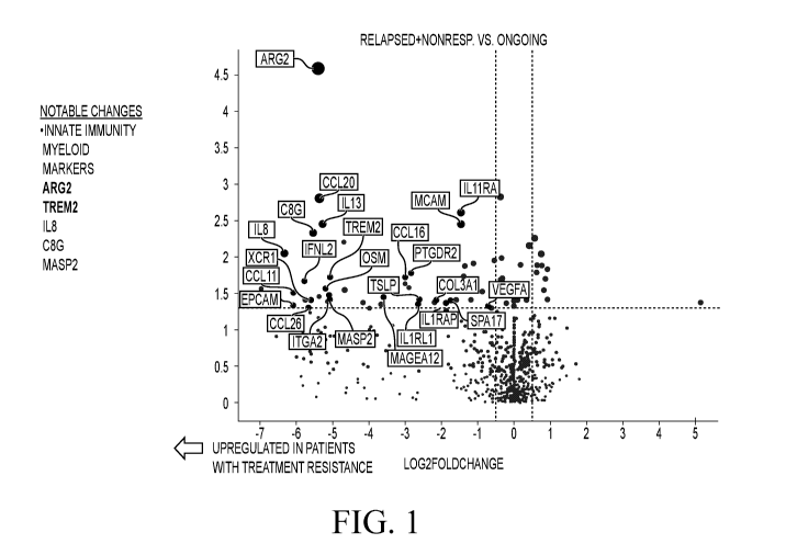
[0034] FIG. 1. Volcano plot of differentially expressed genes comparing
ongoing
responders with relapsed and nonresponders. Fold change was determined by the
ratio of median
value in each ongoing response group, and the p-value was derived from
Wilcoxon test. A small
constant, 1, was added to the medians to avoid zero in logarithmic
transformation. Top
differentially expressed gene in relapsed and nonresponder group, including
ARG2, TREM2, IL8,
C8G, and MASP2, are related to myeloid inflammation. Gene counts are
normalized using a ratio
of the expression value to the geometric mean of all housekeeping genes on the
panel.
Housekeeper-normalized gene counts are additionally normalized using a panel
standard run on
the same cartridge as the observed data.
13
CA 03211006 2023-08-08
WO 2022/178243 PCT/US2022/016961
[0035] FIG. 2. Overall and progression-free survival curves of CLINICAL
TRIAL-1
subjects grouped by ARG2 gene counts. Kaplan-Meier overall and progression-
free survival
curves with a median cut-off selection for ARG2 gene counts in pretreatment
tumor samples with
significance determined by the Log-Rank test. The boxplots show ARG2 gene
counts by ongoing
response groups. Nonparametric Wilcoxon tests and Kruskal-Wallis tests are
conducted for
comparisons of 2 or 3 groups, respectively.
[0036] FIG. 3. Overall and progression-free survival curves of CLINICAL
TRIAL-1
subjects grouped by TREM2 gene counts. Kaplan-Meier overall and progression-
free survival
curves with a median cut-off selection for TREM2 gene counts in pretreatment
tumor samples
with significance determined by the Log-Rank test. The boxplots show TREM2
gene counts by
ongoing response groups. Nonparametric Wilcoxon tests and Kruskal-Wallis tests
are conducted
for comparisons of 2 or 3 groups, respectively.
[0037] FIG. 4. Overall and progression-free survival curves of CLINICAL
TRIAL-1
subjects grouped by IL8 gene counts. Kaplan-Meier overall progression-free
survival curves with
a median cut-off selection for IL8 gene counts in pretreatment tumor samples
with significance
determined by the Log-Rank test. The boxplots show IL8 gene counts by ongoing
response
groups. Nonparametric Wilcoxon tests and Kruskal-Wallis tests are conducted
for comparisons of
2 or 3 groups, respectively.
[0038] FIG. 5. Overall and progression-free survival curves of CLINICAL
TRIAL-1
subjects grouped by IL13 gene counts. Kaplan-Meier overall and progression-
free survival curves
with a median cut-off selection for IL13 gene counts in pretreatment tumor
samples with
significance determined by the Log-Rank test. The boxplots show IL13 gene
counts by ongoing
response groups. Nonparametric Wilcoxon tests and Kruskal-Wallis tests are
conducted for
comparisons of 2 or 3 groups, respectively.
[0039] FIG. 6. Overall and progression-free survival curve of CLINICAL
TRIAL-1
subjects grouped by CCL20 gene counts. Kaplan-Meier overall and progression-
free survival
curves with a median cut-off selection for CCL20 gene counts in pretreatment
tumor samples with
significance determined by the Log-Rank test. The boxplots show CCL20 gene
counts by ongoing
response groups. Nonparametric Wilcoxon tests and Kruskal-Wallis tests are
conducted for
comparisons of 2 or 3 groups, respectively.
[0040] FIG. 7. Associations between pretreatment T cell and Myeloid cell
gene signature
with ongoing response within patients with high (SPDhi)(above the median level
for a
representative tumor population) or low (SPD10) (below the median level for a
representative
tumor population) baseline tumor burden. Values in red are representative of a
value greater the
mean expression while those in blue are representative of a value less than
mean expression of the
14
CA 03211006 2023-08-08
WO 2022/178243 PCT/US2022/016961
corresponding gene. Total number of infused CD8 (NCD8), total number of
infused naïve
products (NNV), peak level of CAR-T cells and its value relative to baseline
tumor burden (CAR-
T peak/SPD) are included as a comparison.
[0041] FIG. 8. Association between peak CAR-T levels (cells/ L) by
ongoing response
groups within patients with high (SPDhi) or low (SPD10) baseline tumor burden.
Ongoing
responders are shown in green, relapsed patients are shown in orange, and non-
responders are
shown in blue. Nonparametric Kruskal-Wallis tests are conducted for
comparisons of 3 groups.
[0042] FIG. 9. Ratio of T cell to myeloid inflammation by ongoing
response groups within
patients with high (SPDhi) or low (SPD10) baseline tumor burden. Selected
genes were used to
derive T cell (CD3D, CD8A, CTLA4, TIGIT) and myeloid inflammation (ARG2 and
TREM2)
indices. Ongoing responders are shown in green, relapsed patients are shown in
orange, and non-
responders are shown in blue. Nonparametric Kruskal-Wallis tests are conducted
for comparisons
of 3 groups.
[0043] FIG. 10. Associations between peak level of CAR-T cells with T
cell, myeloid
inflammation indices, and ratio of T cell to myeloid inflammation. Spearman
rank coefficient (R)
and p values are shown.
[0044] FIG. 11. Associations between peak levels of CAR-T cells relative
to baseline
tumor burden with T cell, myeloid inflammation indices, and ratio of T cell to
myeloid
inflammation. Spearman rank coefficient (R) and p values are shown.
[0045] FIG. 12. Genes negatively associated with ongoing response were
positively
associated with the myeloid population in the TME. Data are included for 12
patients from
ZUMA-1 Cohorts 1-3 with evaluable samples for both gene expression analyses
and multiplex
immunohistochemistry. The genes presented in the heatmap were selected based
on findings from
FIG. 1; specifically, these genes were upregulated in patients with treatment
resistance versus
ongoing responders. Cell values represent the Spearman rank correlation value
(R) between the
covariates shown. Shading indicate positive and negative associations,
respectively, between
covariates.ARG2, arginase 2; C8G, complement C8 gamma chain; CCL, chemokine
ligand;
FoxP3, forkhead box protein P3; IL, interleukin; LAG-3, lymphocyte-activation
gene 3; LOX-1,
lectin-type oxidized low-density lipoprotein receptor 1;max, maximum; min,
minimum;
M-MDSC, monocyte myeloid-derived suppressor cell; PD-1, programmed cell death
protein 1;
PMN-MDSC, polymorphonuclear myeloid-derived suppressor cell; 5100A9, S100
calcium-binding protein A9;TIIVI-3, T-cell immunoglobulin and mucin domain-
containing protein
3; TME, tumor microenvironment; TREM2, triggering receptor expressed on
myeloid cells 2.
[0046] FIG. 13. The suppressive myeloid gene signature was positively
associated with
gene expression of cancer testis antigens. Data are included for 30 patients
from ZUMA-1 Cohorts
CA 03211006 2023-08-08
WO 2022/178243 PCT/US2022/016961
1-3 with evaluable samples for gene expression analyses. The genes presented
in the heatmap
were selected based on findings from Figure 1; specifically, these genes were
upregulated in
patients with treatment resistance versus ongoing responders. Cell values
represent the Spearman
rank correlation value (R) between the covariates shown. Shading indicate
positive and negative
associations, respectively, between covariates.ARG2, arginase 2; BTK, Burton
tyrosine kinase;
C8G, complement C8 gamma chain; CCL, chemokine ligand; DDX43, DEAD-box
helicase 43;
IL, interleukin; IRF, interferon-regulatory factor; ITK, interleukin-
2¨includible T-cell kinase;
MAGE, melanoma antigen gene; MAP2K, mitogen-activated protein kinase kinase;
MAP3K,
mitogen-activated protein kinase kinase kinase; MAPK, mitogen-activated
protein kinase;
MAPKAPK, mitogen-activated protein kinase-activatedprotein kinase; max,
maximum; min,
minimum; PRAME, preferentially expressed antigen of melanoma; SPA17, sperm
surface protein
Sp17; STAT, signal transducer and activator of transcription; SYK, spleen
associated tyrosine
kinase; TREM2, triggering receptor expressed on myeloid cells 2.
[0047] FIG. 14. Protocol-specified AE management in cohorts 1+2 and
cohort 4 of
CLINICAL TRIAL-1. "Yes" or "No" indicates whether tocilizumab or
corticosteroid was or was
not administered, respectively. *Only in case of comorbidities or older age.
tOnly if no
improvement with tocilizumab; use standard dose. Jf no improvement after 3
days. AE, adverse
event; CRS, cytokine release syndrome; HD, high dose; NE, neurologic event;
Mgmt,
management.
[0048] FIG. 15. Patient disposition diagram. The figure summarizes the
disposition of
patients enrolled in CLINICAL TRIAL-1 cohort 4. A total of 57 patients were
screened according
to institutional protocols. There were 11 screen failures. *Due to suicide
(n=1) and disease
progression (n=1). axicabtagene ciloleucel, axicabtagene ciloleucel.
[0049] FIGs. 16A and 16B. ORR and duration of response. (16A) ORR of
patients in
cohort 4 and rates of SD and PD. Response could not be evaluated in 2
patients: 1 patient died of
pneumonia before the first assessment, and 1 patient had a positive result
from positron emission
tomography with suspected inflammation. (16B) Kaplan-Meier curve of duration
of response. CR,
complete response; NE, not estimable; NR, not reached; ORR, objective response
rate; PD,
progressive disease; PR, partial response; SD, stable disease.
[0050] FIG. 17. Best response by corticosteroid use. The figure shows the
percentages of
patients who did or did not receive steroids, with corresponding ORR, CR, and
ongoing response
at 12 months. CR, complete response; ORR, objective response rate.
[0051] FIG. 18. Progression-free survival in cohort 4.
[0052] FIGs. 19A and 19B. CAR T-cell expansion and key soluble serum
biomarker levels
overtime. (19A) Median (Q1, Q3) blood levels of CART cells over time. (19B)
Median (Q1, Q3)
16
CA 03211006 2023-08-08
WO 2022/178243 PCT/US2022/016961
levels of key soluble serum inflammatory biomarkers plotted against time. BL,
baseline; CAR,
chimeric antigen receptor; CRP, C-reactive protein; GM-C SF, granulocyte-
macrophage colony¨
stimulating factor; IFN, interferon; IL, interleukin.
[0053] FIG. 20. Selected CSF analysis at baseline and day 5 and
association with
neurologic events. The figure shows levels of inflammatory markers in CSF
samples from cohort
4 at baseline (dots) and day 5 (triangles) by severity of the neurologic
event. The grade of the
neurologic event (0 to 5) and number of cases are indicated in the upper and
lower rows of text,
respectively. The middle line represents the median, and the box represents
the interquartile range;
whiskers show minimum and maximum values. CRP, C-reactive protein; CSF,
cerebrospinal
fluid; IFN, interferon; IL, interleukin; R, receptor.
[0054] FIG. 21. Selected serum analysis at baseline and day 5 and
association with
neurologic events. The figure shows levels of inflammatory markers in blood
serum samples from
cohort 4 at baseline (dots) and day 5 (triangles) by severity of the
neurologic event. The grade of
the neurologic event (0 to 5) and number of cases are indicated in the upper
and lower rows of
text, respectively. The middle line represents the median, and the box
represents the interquartile
range; whiskers show minimum and maximum values. CRP, C-reactive protein; IFN,
interferon;
IL, interleukin; R, receptor.
DETAILED DESCRIPTION
[0055] The present disclosure is based in part on the discovery that pre-
infusion attributes
(e.g., T cell fitness) of apheresis material and engineered CAR T cells, as
well as pre-treatment
characteristics of patients' immune factors and tumor burden may be associated
with clinical
efficacy and toxicity including durable responses, grade A cytokine release
syndrome, and grade
3 neurologic events.
DEFINITIONS
[0056] In order for the present disclosure to be more readily understood,
certain terms are
first defined below. Additional definitions for the following terms and other
terms are set forth
throughout the Specification.
[0057] As used in this Specification and the appended claims, the
singular forms "a," "an"
and "the" include plural referents unless the context clearly dictates
otherwise.
[0058] Unless specifically stated or obvious from context, as used
herein, the term "or" is
understood to be inclusive and covers both "or" and "and".
[0059] The term "and/or" where used herein is to be taken as specific
disclosure of each
of the two specified features or components with or without the other. Thus,
the term "and/or" as
used in a phrase such as "A and/or B" herein is intended to include A and B; A
or B; A (alone);
and B (alone). Likewise, the term "and/or" as used in a phrase such as "A, B,
and/or C" is intended
17
CA 03211006 2023-08-08
WO 2022/178243 PCT/US2022/016961
to encompass each of the following aspects: A, B, and C; A, B, or C; A or C; A
or B; B or C; A
and C; A and B; B and C; A (alone); B (alone); and C (alone).
[0060] The terms "e.g.," and "i.e." as used herein, are used merely by
way of example,
without limitation intended, and should not be construed as referring only
those items explicitly
enumerated in the specification.
[0061] The terms "or more", "at least", "more than", and the like, e.g.,
"at least one" are
understood to include but not be limited to at least 1, 2, 3, 4, 5, 6, 7, 8,
9, 10, 11, 12, 13, 14, 15,
16, 17, 18, 1920, 21, 22, 23, 24, 25, 26, 27, 28, 29, 30, 31, 32, 33, 34, 35,
36, 37, 38, 39, 40, 41,
42, 43, 44, 45, 46, 47, 48, 49, 50, 51, 52, 53, 54, 55, 56, 57, 58, 59, 60,
61, 62, 63, 64, 65, 66, 67,
68, 69, 70, 71, 72, 73, 74, 75, 76, 77, 78, 79, 80, 81, 82, 83, 84, 85, 86,
87, 88, 89, 90, 91, 92, 93,
94, 95, 96, 97, 98, 99, 100, 101, 102, 103, 104, 105, 106, 107, 108, 109, 110,
111, 112, 113, 114,
115, 116, 117, 118, 119, 120, 121, 122, 123, 124, 125, 126, 127, 128, 129,
130, 131, 132, 133,
134, 135, 136, 137, 138, 139, 140, 141, 142, 143, 144, 145, 146, 147, 148, 149
or 150, 200, 300,
400, 500, 600, 700, 800, 900, 1000, 2000, 3000, 4000, 5000 or more than the
stated value. Also
included is any greater number or fraction in between.
[0062] Conversely, the term "no more than" includes each value less than
the stated value.
For example, "no more than 100 nucleotides" includes 100, 99, 98, 97, 96, 95,
94, 93, 92, 91, 90,
89, 88, 87, 86, 85, 84, 83, 82, 81, 80, 79, 78, 77, 76, 75, 74, 73, 72, 71,
70, 69, 68, 67, 66, 65, 64,
63, 62, 61, 60, 59, 58, 57, 56, 55, 54, 53, 52, 51, 50, 49, 48, 47, 46, 45,
44, 43, 42, 41, 40, 39, 38,
37, 36, 35, 34, 33, 32, 31, 30, 29, 28, 27, 26, 25, 24, 23, 22, 21, 20, 19,
18, 17, 16, 15, 14, 13, 12,
11, 10, 9, 8, 7, 6, 5, 4, 3, 2, 1, and 0 nucleotides. Also included is any
lesser number or fraction in
between.
[0063] The terms "plurality", "at least two", "two or more", "at least
second", and the like,
are understood to include but not limited to at least 2, 3, 4, 5, 6, 7, 8, 9,
10, 11, 12, 13, 14, 15, 16,
17, 18, 1920, 21, 22, 23, 24, 25, 26, 27, 28, 29, 30, 31, 32, 33, 34, 35, 36,
37, 38, 39, 40, 41, 42,
43, 44, 45, 46, 47, 48, 49, 50, 51, 52, 53, 54, 55, 56, 57, 58, 59, 60, 61,
62, 63, 64, 65, 66, 67, 68,
69, 70, 71, 72, 73, 74, 75, 76, 77, 78, 79, 80, 81, 82, 83, 84, 85, 86, 87,
88, 89, 90, 91, 92, 93, 94,
95, 96, 97, 98, 99, 100, 101, 102, 103, 104, 105, 106, 107, 108, 109, 110,
111, 112, 113, 114, 115,
116, 117, 118, 119, 120, 121, 122, 123, 124, 125, 126, 127, 128, 129, 130,
131, 132, 133, 134,
135, 136, 137, 138, 139, 140, 141, 142, 143, 144, 145, 146, 147, 148, 149 or
150, 200, 300, 400,
500, 600, 700, 800, 900, 1000, 2000, 3000, 4000, 5000 or more. Also included
is any greater
number or fraction in between.
[0064] Throughout the specification the word "comprising," or variations
such as
"comprises" or "comprising," will be understood to imply the inclusion of a
stated element, integer
or step, or group of elements, integers or steps, but not the exclusion of any
other element, integer
18
CA 03211006 2023-08-08
WO 2022/178243 PCT/US2022/016961
or step, or group of elements, integers or steps. It is understood that
wherever aspects are described
herein with the language "comprising," otherwise analogous aspects described
in terms of
"consisting of' and/or "consisting essentially of' are also provided. The term
"consisting of'
excludes any element, step, or ingredient not specified in the claim. In re
Gray, 53 F.2d 520, 11
USPQ 255 (CCPA 1931); Ex parte Davis, 80 USPQ 448, 450 (Bd. App. 1948)
("consisting of'
defined as "closing the claim to the inclusion of materials other than those
recited except for
impurities ordinarily associated therewith"). The term "consisting essentially
of' limits the scope
of a claim to the specified materials or steps "and those that do not
materially affect
the basic and novel characteristic(s)" of the claimed disclosure.
[0065] Unless specifically stated or evident from context, as used
herein, the term "about"
refers to a value or composition that is within an acceptable error range for
the particular value or
composition as determined by one of ordinary skill in the art, which will
depend in part on how
the value or composition is measured or determined, i.e., the limitations of
the measurement
system. For example, "about" or "approximately" may mean within one or more
than one standard
deviation per the practice in the art. "About" or "approximately" may mean a
range of up to 10%
(i.e., 10%). Thus, "about" may be understood to be within 10%, 9%, 8%, 7%,
6%, 5%, 4%, 3%,
2%, 1%, 0.5%, 0.1%, 0.05%, 0.01%, or 0.001% greater or less than the stated
value. For example,
about 5 mg may include any amount between 4.5 mg and 5.5 mg. Furthermore,
particularly with
respect to biological systems or processes, the terms may mean up to an order
of magnitude or up
to 5-fold of a value. When particular values or compositions are provided in
the instant disclosure,
unless otherwise stated, the meaning of "about" or "approximately" should be
assumed to be
within an acceptable error range for that particular value or composition.
[0066] As described herein, any concentration range, percentage range,
ratio range or
integer range is to be understood to be inclusive of the value of any integer
within the recited range
and, when appropriate, fractions thereof (such as one-tenth and one-hundredth
of an integer),
unless otherwise indicated.
[0067] Units, prefixes, and symbols used herein are provided using their
Systeme
International de Unites (SI) accepted form. Numeric ranges are inclusive of
the numbers defining
the range.
[0068] Unless defined otherwise, all technical and scientific terms used
herein have the
same meaning as commonly understood by one of ordinary skill in the art to
which this disclosure
is related. For example, Juo, "The Concise Dictionary of Biomedicine and
Molecular Biology",
2nd ed., (2001), CRC Press; "The Dictionary of Cell & Molecular Biology", 5th
ed., (2013),
Academic Press; and "The Oxford Dictionary Of Biochemistry And Molecular
Biology",
19
CA 03211006 2023-08-08
WO 2022/178243 PCT/US2022/016961
Cammack et al. eds., 2nd ed, (2006), Oxford University Press, provide those of
skill in the art with
a general dictionary for many of the terms used in this disclosure.
[0069] "Administering" refers to the physical introduction of an agent to
a subj ect, using
any of the various methods and delivery systems known to those skilled in the
art. Exemplary
routes of administration for the formulations disclosed herein include
intravenous, intramuscular,
subcutaneous, intraperitoneal, spinal or other parenteral routes of
administration, for example by
injection or infusion. Exemplary routes of administration for the compositions
disclosed herein
include intravenous, intramuscular, subcutaneous, intraperitoneal, spinal or
other parenteral routes
of administration, for example by injection or infusion. The phrase
"parenteral administration" as
used herein means modes of administration other than enteral and topical
administration, usually
by injection, and includes, without limitation, intravenous, intramuscular,
intraarterial, intrathecal,
intralymphatic, intralesional, intracapsular, intraorbital, intracardiac,
intradermal, intraperitoneal,
transtracheal, subcutaneous, sub cuti cul ar, intraarticular, sub c ap sular,
sub arachnoi d, i ntraspi nal,
epidural and intrasternal injection and infusion, as well as in vivo
electroporation. In some
embodiments, the formulation is administered via a non-parenteral route, e.g.,
orally. Other non-
parenteral routes include a topical, epidermal or mucosal route of
administration, for example,
intranasally, vaginally, rectally, sublingually or topically. Administering
may also be performed,
for example, once, a plurality of times, and/or over one or more extended
periods. In one
embodiment, the CAR T cell treatment is administered via an "infusion product"
comprising CAR
T cells.
[0070] The term "antibody" (Ab) includes, without limitation, a
glycoprotein
immunoglobulin which binds specifically to an antigen. In general, an antibody
may comprise at
least two heavy (H) chains and two light (L) chains interconnected by
disulfide bonds, or an
antigen-binding molecule thereof. Each H chain comprises a heavy chain
variable region
(abbreviated herein as VH) and a heavy chain constant region. The heavy chain
constant region
comprises three constant domains, CHL CH2 and CH3. Each light chain comprises
a light chain
variable region (abbreviated herein as VL) and a light chain constant region.
The light chain
constant region comprises one constant domain, CL. The VH and VL regions may
be further
subdivided into regions of hypervariability, termed complementarity
determining regions (CDRs),
interspersed with regions that are more conserved, termed framework regions
(FR). Each VH and
VL comprises three CDRs and four FRs, arranged from amino-terminus to carboxy-
terminus in
the following order: FR1, CDR1, FR2, CDR2, FR3, CDR3, and FR4. The variable
regions of the
heavy and light chains contain a binding domain that interacts with an
antigen. The constant
regions of the Abs may mediate the binding of the immunoglobulin to host
tissues or factors,
CA 03211006 2023-08-08
WO 2022/178243 PCT/US2022/016961
including various cells of the immune system (e.g., effector cells) and the
first component (Clq)
of the classical complement system.
[0071] Antibodies may include, for example, monoclonal antibodies,
recombinantly
produced antibodies, monospecific antibodies, multispecific antibodies
(including bispecific
antibodies), human antibodies, engineered antibodies, humanized antibodies,
chimeric antibodies,
immunoglobulins, synthetic antibodies, tetrameric antibodies comprising two
heavy chain and
two light chain molecules, an antibody light chain monomer, an antibody heavy
chain monomer,
an antibody light chain dimer, an antibody heavy chain dimer, an antibody
light chain- antibody
heavy chain pair, intrabodies, antibody fusions (sometimes referred to herein
as "antibody
conjugates"), heteroconjugate antibodies, single domain antibodies, monovalent
antibodies, single
chain antibodies or single-chain Fvs (scFv), camelized antibodies, affybodies,
Fab fragments,
F(ab')2 fragments, disulfide-linked Fvs (sdFv), anti-idiotypic (anti-Id)
antibodies (including, e.g.,
anti-anti-Id antibodies), minibodies, domain antibodies, synthetic antibodies
(sometimes referred
to herein as "antibody mimetics"), and antigen-binding fragments of any of the
above. In some
embodiments, antibodies described herein refer to polyclonal antibody
populations.
[0072] An "antigen binding molecule," "antigen binding portion," or
"antibody fragment"
refers to any molecule that comprises the antigen binding parts (e.g., CDRs)
of the antibody from
which the molecule is derived. An antigen binding molecule may include the
antigenic
complementarity determining regions (CDRs). Examples of antibody fragments
include, but are
not limited to, Fab, Fab', F(ab')2, and Fv fragments, dAb, linear antibodies,
scFv antibodies, and
multispecific antibodies formed from antigen binding molecules. Peptibodies
(i.e., Fc fusion
molecules comprising peptide binding domains) are another example of suitable
antigen binding
molecules. In some embodiments, the antigen binding molecule binds to an
antigen on a tumor
cell. In some embodiments, the antigen binding molecule binds to an antigen on
a cell involved in
a hyperproliferative disease or to a viral or bacterial antigen. In some
embodiments, the antigen
binding molecule binds to CD19. In further embodiments, the antigen binding
molecule is an
antibody fragment that specifically binds to the antigen, including one or
more of the
complementarity determining regions (CDRs) thereof In further embodiments, the
antigen
binding molecule is a single chain variable fragment (scFv). In some
embodiments, the antigen
binding molecule comprises or consists of avimers.
[0073] An "antigen" refers to any molecule that provokes an immune
response or is
capable of being bound by an antibody or an antigen binding molecule. The
immune response
may involve either antibody production, or the activation of specific
immunologically-competent
cells, or both. A person of skill in the art would readily understand that any
macromolecule,
including virtually all proteins or peptides, may serve as an antigen. An
antigen may be
21
CA 03211006 2023-08-08
WO 2022/178243 PCT/US2022/016961
endogenously expressed, i.e. expressed by genomic DNA, or may be recombinantly
expressed.
An antigen may be specific to a certain tissue, such as a cancer cell, or it
may be broadly expressed.
In addition, fragments of larger molecules may act as antigens. In some
embodiments, antigens
are tumor antigens.
[0074] The term "neutralizing" refers to an antigen binding molecule,
scFv, antibody, or
a fragment thereof, that binds to a ligand and prevents or reduces the
biological effect of that
ligand. In some embodiments, the antigen binding molecule, scFv, antibody, or
a fragment thereof,
directly blocks a binding site on the ligand or otherwise alters the ligand's
ability to bind through
indirect means (such as structural or energetic alterations in the ligand). In
some embodiments,
the antigen binding molecule, scFv, antibody, or a fragment thereof prevents
the protein to which
it is bound from performing a biological function.
[0075] The term "autologous" refers to any material derived from the same
individual to
which it is later to be re-introduced. For example, the engineered autologous
cell therapy
(eACTTm) method described herein involves collection of lymphocytes from a
patient, which are
then engineered to express, e.g., a CAR construct, and then administered back
to the same patient.
[0076] The term "allogeneic" refers to any material derived from one
individual which is
then introduced to another individual of the same species, e.g., allogeneic T
cell transplantation.
[0077] In one embodiment, the CAR T cell treatment comprises
"axicabtagene ciloleucel
treatment". "Axicabtagene ciloleucel treatment" consists of a single infusion
of anti-CD19 CAR
transduced autologous T cells administered intravenously at a target dose of 2
x 106 anti-CD19
CART cells/kg. For subjects weighing greater than 100 kg, a maximum flat dose
of 2 x 10⁸ anti-
CD19 CAR T cells may be administered. The anti-CD19 CAR T cells are autologous
human T
cells that have been engineered to express an extracellular single-chain
variable fragment (scFv)
with specificity for CD19 linked to an intracellular signaling part comprised
of signaling domains
from CD28 and CD3 (CD3-zeta) molecules arranged in tandem anti-CD19 CAR vector
construct
has been designed, optimized and initially tested at the Surgery Branch of the
National Cancer
Institute (NCI, IND 13871) (Kochenderfer et al, J Immunother. 2009;32(7):689-
702;
Kochenderfer et al, Blood. 2010;116(19):3875-86). The scFv is derived from the
variable region
of the anti-CD19 monoclonal antibody FMC63 (Nicholson et al, Molecular
Immunology.
1997;34(16-17):1157-65). A portion of the CD28 costimulatory molecule is
added, as murine
models suggest this is important for the anti-tumor effect and persistence of
anti-CD19 CAR T
cells (Kowolik et al, Cancer Res. 2006;66(22):10995-1004). The signaling
domain of the CD3-
zeta chain is used for T cell activation. These fragments were cloned into the
murine stem cell
virus-based (MSGV1) vector, utilized to genetically engineer the autologous T
cells. The CAR
construct is inserted into the T cells' genome by retroviral vector
transduction. Briefly, peripheral
22
CA 03211006 2023-08-08
WO 2022/178243 PCT/US2022/016961
blood mononuclear cells (PBMCs) are obtained by leukapheresis and Ficoll
separation. Peripheral
blood mononuclear cells are activated by culturing with an anti-CD3 antibody
in the presence of
recombinant interleukin 2 (IL-2). Stimulated cells are transduced with a
retroviral vector
containing an anti-CD19 CAR gene and propagated in culture to generate
sufficient engineered T
cells for administration. Axicabtagene ciloleucel is a subject-specific
product.
[0078] The terms "transduction" and "transduced" refer to the process
whereby foreign
DNA is introduced into a cell via viral vector (see Jones et al., "Genetics:
principles and analysis,"
Boston: Jones & Bartlett Publ. (1998)). In some embodiments, the vector is a
retroviral vector, a
DNA vector, a RNA vector, an adenoviral vector, a baculoviral vector, an
Epstein Barr viral
vector, a papovaviral vector, a vaccinia viral vector, a herpes simplex viral
vector, an adenovirus
associated vector, a lentiviral vector, or any combination thereof
[0079] A "cancer" refers to a broad group of various diseases
characterized by the
uncontrolled growth of abnormal cells in the body. Unregulated cell division
and growth results
in the formation of malignant tumors that invade neighboring tissues and may
also metastasize to
distant parts of the body through the lymphatic system or bloodstream. A
"cancer" or "cancer
tissue" may include a tumor. In this application, the term cancer is
synonymous with malignancy.
Examples of cancers that may be treated by the methods disclosed herein
include, but are not
limited to, cancers of the immune system including lymphoma, leukemia,
myeloma, and other
leukocyte malignancies. In some embodiments, the methods disclosed herein may
be used to
reduce the tumor size of a tumor derived from, for example, bone cancer,
pancreatic cancer, skin
cancer, cancer of the head or neck, cutaneous or intraocular malignant
melanoma, uterine cancer,
ovarian cancer, rectal cancer, cancer of the anal region, stomach cancer,
testicular cancer, uterine
cancer, carcinoma of the fallopian tubes, carcinoma of the endometrium,
carcinoma of the cervix,
carcinoma of the vagina, carcinoma of the vulva, [add other solid tumors]
multiple myeloma,
Hodgkin's Disease, non-Hodgkin's lymphoma (NHL), primary mediastinal large B
cell lymphoma
(PMBC), diffuse large B cell lymphoma (DLBCL), follicular lymphoma (FL),
transformed
follicular lymphoma, splenic marginal zone lymphoma (SMZL), cancer of the
esophagus, cancer
of the small intestine, cancer of the endocrine system, cancer of the thyroid
gland, cancer of the
parathyroid gland, cancer of the adrenal gland, sarcoma of soft tissue, cancer
of the urethra, cancer
of the penis, chronic or acute leukemia, acute myeloid leukemia, chronic
myeloid leukemia, acute
lymphoblastic leukemia (ALL) (including non T cell ALL), chronic lymphocytic
leukemia (CLL),
solid tumors of childhood, lymphocytic lymphoma, cancer of the bladder, cancer
of the kidney or
ureter, carcinoma of the renal pelvis, neoplasm of the central nervous system
(CNS), primary CNS
lymphoma, tumor angiogenesis, spinal axis tumor, brain stem glioma, pituitary
adenoma, Kaposi's
sarcoma, epidermoid cancer, squamous cell cancer, T cell lymphoma,
environmentally induced
23
CA 03211006 2023-08-08
WO 2022/178243 PCT/US2022/016961
cancers including those induced by asbestos, other B cell malignancies, and
combinations of said
cancers. In some embodiments, the cancer is multiple myeloma. In some
embodiments, the cancer
is NHL. The particular cancer may be responsive to chemo- or radiation therapy
or the cancer may
be refractory. A refractory cancer refers to a cancer that is not amenable to
surgical intervention
and the cancer is either initially unresponsive to chemo- or radiation therapy
or the cancer becomes
unresponsive over time.
[0080] An "anti-tumor effect" as used herein, refers to a biological
effect that may present
as a decrease in tumor volume, a decrease in the number of tumor cells, a
decrease in tumor cell
proliferation, a decrease in the number of metastases, an increase in overall
or progression-free
survival, an increase in life expectancy, or amelioration of various
physiological symptoms
associated with the tumor. An anti-tumor effect may also refer to the
prevention of the occurrence
of a tumor, e.g., a vaccine.
[0081] A "cytokine," as used herein, refers to a non-antibody protein
that is released by
one cell in response to contact with a specific antigen, wherein the cytokine
interacts with a second
cell to mediate a response in the second cell. "Cytokine" as used herein is
meant to refer to proteins
released by one cell population that act on another cell as intercellular
mediators. A cytokine may
be endogenously expressed by a cell or administered to a subject. Cytokines
may be released by
immune cells, including macrophages, B cells, T cells, and mast cells to
propagate an immune
response. Cytokines may induce various responses in the recipient cell.
Cytokines may include
homeostatic cytokines, chemokines, pro-inflammatory cytokines, effectors, and
acute-phase
proteins. For example, homeostatic cytokines, including interleukin (IL) 7 and
IL-15, promote
immune cell survival and proliferation, and pro-inflammatory cytokines may
promote an
inflammatory response. Examples of homeostatic cytokines include, but are not
limited to, IL-2,
IL-4, IL-5, IL-7, IL-10, IL-12p40, IL-12p70, IL-15, and interferon (IFN)
gamma. Examples of
pro-inflammatory cytokines include, but are not limited to, IL-la, IL-lb, IL-
6, IL-13, IL-17a,
tumor necrosis factor (TNF)-alpha, TNF-beta, fibroblast growth factor (FGF) 2,
granulocyte
macrophage colony-stimulating factor (GM-CSF), soluble intercellular adhesion
molecule 1
(sICAM-1), soluble vascular adhesion molecule 1 (sVCAM-1), vascular
endothelial growth factor
(VEGF), VEGF-C, VEGF-D, and placental growth factor (PLGF). Examples of
effectors include,
but are not limited to, granzyme A, granzyme B, soluble Fas ligand (sFasL),
and perforin.
Examples of acute phase-proteins include, but are not limited to, C-reactive
protein (CRP) and
serum amyloid A (SAA).
[0082] "Chemokines" are a type of cytokine that mediates cell chemotaxis,
or directional
movement. Examples of chemokines include, but are not limited to, IL-8, IL-16,
eotaxin, eotaxin-
3, macrophage-derived chemokine (MDC or CCL22), monocyte chemotactic protein 1
(MCP-1
24
CA 03211006 2023-08-08
WO 2022/178243 PCT/US2022/016961
or CCL2), MCP-4, macrophage inflammatory protein la (MIP- 1 a, MIP-1a), MIP-10
(MIP-1b),
gamma-induced protein 10 (IP-10), and thymus and activation regulated
chemokine (TARC or
CCL17).
[0083] As used herein, "chimeric receptor" refers to an engineered
surface expressed
molecule capable of recognizing a particular molecule. Chimeric antigen
receptors (CARs) and
engineered T cell receptors (TCRs), which comprise binding domains capable of
interacting with
a particular tumor antigen, allow T cells to target and kill cancer cells that
express the particular
tumor antigen. In one embodiment, the T cell treatment is based on T cells
engineered to express
a chimeric antigen receptor (CAR) or a T cell receptor (TCR), which comprises
(i) an antigen
binding molecule, (ii) a costimulatory domain, and (iii) an activating domain.
The costimulatory
domain may comprise an extracellular domain, a transmembrane domain, and an
intracellular
domain, wherein the extracellular domain comprises a hinge domain, which may
be truncated.
[0084] A "therapeutically effective amount," "effective dose," "effective
amount," or
"therapeutically effective dosage" of a therapeutic agent, e.g., engineered
CAR T cells, small
molecules, "agents" described in the specification, is any amount that, when
used alone or in
combination with another therapeutic agent, protects a subject against the
onset of a disease or
promotes disease regression evidenced by a decrease in severity of disease
symptoms, an increase
in frequency and duration of disease symptom-free periods, or a prevention of
impairment or
disability due to the disease affliction. Such terms may be used
interchangeably. The ability of a
therapeutic agent to promote disease regression may be evaluated using a
variety of methods
known to the skilled practitioner, such as in human subjects during clinical
trials, in animal model
systems predictive of efficacy in humans, or by assaying the activity of the
agent in in vitro assays.
Therapeutically effective amounts and dosage regimens can be determined
empirically by testing
in known in vitro or in vivo (e.g. animal model) systems.
[0085] The term "combination" refers to either a fixed combination in one
dosage unit
form, or a combined administration where a compound of the present disclosure
and a combination
partner (e.g. another drug as explained below, also referred to as
"therapeutic agent" or "agent")
may be administered independently at the same time or separately within time
intervals, especially
where these time intervals allow that the combination partners show a
cooperative, e.g. synergistic
effect. The single components may be packaged in a kit or separately. One or
both of the
components (e.g., powders or liquids) may be reconstituted or diluted to a
desired dose prior to
administration. The terms "co-administration" or "combined administration" or
the like as utilized
herein are meant to encompass administration of the selected combination
partner to a single
subject in need thereof (e.g. a patient), and are intended to include
treatment regimens in which
CA 03211006 2023-08-08
WO 2022/178243 PCT/US2022/016961
the agents are not necessarily administered by the same route of
administration or at the same
time.
[0086] The terms "product" or "infusion product" are used interchangeably
herein and
refer to the T cell composition that is administered to the subject in need
thereof Typically, in
CAR T-cell therapy, the T cell composition is administered as an infusion
product.
[0087] The term "lymphocyte" as used herein includes natural killer (NK)
cells, T cells,
or B cells. NK cells are a type of cytotoxic (cell toxic) lymphocyte that
represent a major
component of the inherent immune system. NK cells reject tumors and cells
infected by viruses.
It works through the process of apoptosis or programmed cell death. They were
termed "natural
killers" because they do not require activation in order to kill cells. T
cells play a major role in
cell-mediated-immunity (no antibody involvement). Its T cell receptors (TCR)
differentiate
themselves from other lymphocyte types. The thymus, a specialized organ of the
immune system,
is primarily responsible for the T cell's maturation. There are six types of T
cells, namely: Helper
T cells (e.g., CD4+ cells), Cytotoxic T cells (also known as TC, cytotoxic T
lymphocyte, CTL, T-
killer cell, cytolytic T cell, CD8+ T cells or killer T cell), Memory T cells
((i) stem memory TSCM
cells, like naive cells, are CD45R0¨, CCR7+, CD45RA+, CD62L+ (L-selectin),
CD27+, CD28+
and IL-7Ra+, but they also express large amounts of CD95, IL-2R13, CXCR3, and
LFA-1, and
show numerous functional attributes distinctive of memory cells); (ii) central
memory TCM cells
express L-selectin and the CCR7, they secrete IL-2, but not IFNy or IL-4, and
(iii) effector memory
TEM cells, however, do not express L-selectin or CCR7 but produce effector
cytokines like IFNy
and IL-4), Regulatory T cells (Tregs, suppressor T cells, or CD4+CD25+
regulatory T cells),
Natural Killer T cells (NKT) and Gamma Delta T cells. B-cells, on the other
hand, play a principal
role in humoral immunity (with antibody involvement). It makes antibodies and
antigens and
performs the role of antigen-presenting cells (APCs) and turns into memory B-
cells after
activation by antigen interaction. In mammals, immature B-cells are formed in
the bone marrow,
where its name is derived from.
[0088] In the context of this disclosure, the term "TN," "T naive-like",
and
CCR7+CD45RA+ actually refers to cells that are more like stem-like memory
cells than like
canonical naive T cells. Accordingly, all references in the Examples and
Claims to TN refers to
cells that were experimentally selected only by their characterization as
CCR7+CD45RA+ cells
and should be interpreted as such. Their better name in the context of this
disclosure is stem-like
memory cells, but they shall be referred to as CCR7+CD45RA+ cells. Further
characterization
into stem-like memory cells may be done for example using the methods
described in Arihara Y,
Jacobsen CA, Armand P, et al. Journal for ImmunoTherapy of Cancer.
2019;7(1):P210.
26
CA 03211006 2023-08-08
WO 2022/178243 PCT/US2022/016961
[0089] The term "genetically engineered" or "engineered" refers to a
method of modifying
the genome of a cell, including, but not limited to, deleting a coding or non-
coding region or a
portion thereof or inserting a coding region or a portion thereof In some
embodiments, the cell
that is modified is a lymphocyte, e.g., a T cell, which may either be obtained
from a patient or a
donor. The cell may be modified to express an exogenous construct, such as,
e.g., a chimeric
antigen receptor (CAR) or a T cell receptor (TCR), which is incorporated into
the cell's genome.
[0090] An "immune response" refers to the action of a cell of the immune
system (for
example, T lymphocytes, B lymphocytes, natural killer (NK) cells, macrophages,
eosinophils,
mast cells, dendritic cells and neutrophils) and soluble macromolecules
produced by any of these
cells or the liver (including Abs, cytokines, and complement) that results in
selective targeting,
binding to, damage to, destruction of, and/or elimination from a vertebrate's
body of invading
pathogens, cells or tissues infected with pathogens, cancerous or other
abnormal cells, or, in cases
of autoimmunity or pathological inflammation, normal human cells or tissues.
[0091] The term "immunotherapy" refers to the treatment of a subject
afflicted with, or at
risk of contracting or suffering a recurrence of, a disease by a method
comprising inducing,
enhancing, suppressing or otherwise modifying an immune response. Examples of
immunotherapy include, but are not limited to, T cell therapies. T cell
therapy may include
adoptive T cell therapy, tumor-infiltrating lymphocyte (TIL) immunotherapy,
autologous cell
therapy, engineered autologous cell therapy (eACTTm), and allogeneic T cell
transplantation.
However, one of skill in the art would recognize that the conditioning methods
disclosed herein
would enhance the effectiveness of any transplanted T cell therapy. Examples
of T cell therapies
are described in U.S. Patent Publication Nos. 2014/0154228 and 2002/0006409,
U.S. Patent No.
7,741,465, U.S. Patent No. 6,319,494, U.S. Patent No. 5,728,388, and
International Publication
No. WO 2008/081035. In some embodiments, the immunotherapy comprises CAR T
cell
treatment. In some embodiments, the CAR T cell treatment product is
administered via infusion.
[0092] The T cells of the immunotherapy may come from any source known in
the art.
For example, T cells may be differentiated in vitro from a hematopoietic stem
cell population, or
T cells may be obtained from a subject. T cells may be obtained from, e.g.,
peripheral blood
mononuclear cells (PBMCs), bone marrow, lymph node tissue, cord blood, thymus
tissue, tissue
from a site of infection, ascites, pleural effusion, spleen tissue, and
tumors. In addition, the T cells
may be derived from one or more T cell lines available in the art. T cells may
also be obtained
from a unit of blood collected from a subject using any number of techniques
known to the skilled
artisan, such as FICOLLTM separation and/or apheresis. Additional methods of
isolating T cells
for a T cell therapy are disclosed in U.S. Patent Publication No.
2013/0287748, which is herein
incorporated by reference in its entirety.
27
CA 03211006 2023-08-08
WO 2022/178243 PCT/US2022/016961
[0093] The term "engineered Autologous Cell Therapy," or "eACTTm," also
known as
adoptive cell transfer, is a process by which a patient's own T cells are
collected and subsequently
genetically altered to recognize and target one or more antigens expressed on
the cell surface of
one or more specific tumor cells or malignancies. T cells may be engineered to
express, for
example, chimeric antigen receptors (CAR). CAR positive (+) T cells are
engineered to express
an extracellular single chain variable fragment (scFv) with specificity for a
particular tumor
antigen linked to an intracellular signaling part comprising at least one
costimulatory domain and
at least one activating domain. The CAR scFv may be designed to target, for
example, CD19,
which is a transmembrane protein expressed by cells in the B cell lineage,
including all normal B
cells and B cell malignances, including but not limited to diffuse large B-
cell lymphoma (DLBCL)
not otherwise specified, primary mediastinal large B-cell lymphoma, high grade
B-cell lymphoma,
and DLBCL arising from follicular lymphoma, NHL, CLL, and non-T cell ALL.
Example CAR
T cell therapies and constructs are described in U.S. Patent Publication Nos.
2013/0287748,
2014/0227237, 2014/0099309, and 2014/0050708, and these references are
incorporated by
reference in their entirety.
[0094] A "patient" or a "subject" as used herein includes any human who
is afflicted with
a cancer (e.g., a lymphoma or a leukemia). The terms "subject" and "patient"
are used
interchangeably herein.
[0095] As used herein, the term "in vitro cell" refers to any cell which
is cultured ex vivo.
In particular, an in vitro cell may include a T cell. The term "in vivo" means
within the patient.
[0096] The terms "peptide," "polypeptide," and "protein" are used
interchangeably, and
refer to a compound comprised of amino acid residues covalently linked by
peptide bonds. A
protein or peptide contains at least two amino acids, and no limitation is
placed on the maximum
number of amino acids that may comprise a protein's or peptide's sequence.
Polypeptides include
any peptide or protein comprising two or more amino acids joined to each other
by peptide bonds.
As used herein, the term refers to both short chains, which also commonly are
referred to in the
art as peptides, oligopeptides and oligomers, for example, and to longer
chains, which generally
are referred to in the art as proteins, of which there are many types.
"Polypeptides" include, for
example, biologically active fragments, substantially homologous polypeptides,
oligopepti des,
homodimers, heterodimers, variants of polypeptides, modified polypeptides,
derivatives, analogs,
fusion proteins, among others. The polypeptides include natural peptides,
recombinant peptides,
synthetic peptides, or a combination thereof
[0097] "Stimulation," as used herein, refers to a primary response
induced by binding of
a stimulatory molecule with its cognate ligand, wherein the binding mediates a
signal transduction
event. A "stimulatory molecule" is a molecule on a T cell, e.g., the T cell
receptor (TCR)/CD3
28
CA 03211006 2023-08-08
WO 2022/178243 PCT/US2022/016961
complex that specifically binds with a cognate stimulatory ligand present on
an antigen present
cell. A "stimulatory ligand" is a ligand that when present on an antigen
presenting cell (e.g., an
APC, a dendritic cell, a B-cell, and the like) may specifically bind with a
stimulatory molecule on
a T cell, thereby mediating a primary response by the T cell, including, but
not limited to,
activation, initiation of an immune response, proliferation, and the like.
Stimulatory ligands
include, but are not limited to, an anti-CD3 antibody, an MHC Class I molecule
loaded with a
peptide, a superagonist anti-CD2 antibody, and a superagonist anti-CD28
antibody.
[0098] A "costimulatory signal," as used herein, refers to a signal,
which in combination
with a primary signal, such as TCR/CD3 ligation, leads to a T cell response,
such as, but not
limited to, proliferation and/or upregulation or down regulation of key
molecules.
[0099] A "costimulatory ligand," as used herein, includes a molecule on
an antigen
presenting cell that specifically binds a cognate co-stimulatory molecule on a
T cell. Binding of
the costimulatory ligand provides a signal that mediates a T cell response,
including, but not
limited to, proliferation, activation, differentiation, and the like. A
costimulatory ligand induces a
signal that is in addition to the primary signal provided by a stimulatory
molecule, for instance,
by binding of a T cell receptor (TCR)/CD3 complex with a major
histocompatibility complex
(MHC) molecule loaded with peptide. A co-stimulatory ligand may include, but
is not limited to,
3/TR6, 4-1BB ligand, agonist or antibody that binds Toll ligand receptor, B7-1
(CD80), B7-2
(CD86), CD30 ligand, CD40, CD7, CD70, CD83, herpes virus entry mediator
(HVEM), human
leukocyte antigen G (HLA-G), ILT4, immunoglobulin-like transcript (ILT) 3,
inducible
costimulatory ligand (ICOS-L), intercellular adhesion molecule (ICAM), ligand
that specifically
binds with B7-H3, lymphotoxin beta receptor, MHC class I chain-related protein
A (MICA), MHC
class I chain-related protein B (MICB), 0X40 ligand, PD-L2, or programmed
death (PD) Ll. In
certain embodiments, a co-stimulatory ligand includes, without limitation, an
antibody that
specifically binds with a co-stimulatory molecule present on a T cell, such
as, but not limited to,
4-1BB, B7-H3, CD2, CD27, CD28, CD30, CD40, CD7, ICOS, ligand that specifically
binds with
CD83, lymphocyte function-associated antigen-1 (LFA-1), natural killer cell
receptor C
(NKG2C), 0X40, PD-1, or tumor necrosis factor superfamily member 14 (TNFSF14
or LIGHT).
[0100] A "costimulatory molecule" is a cognate binding partner on a T
cell that
specifically binds with a costimulatory ligand, thereby mediating a
costimulatory response by the
T cell, such as, but not limited to, proliferation. Costimulatory molecules
include, but are not
limited to, 4-1BB/CD137, B7-H3, BAFFR, BLAME (SLAMF8), BTLA, CD33, CD45, CD100
(SEMA4D), CD103, CD134, CD137, CD154, CD16, CD160 (BY55), CD18, CD19, CD19a,
CD2, CD22, CD247, CD27, CD276 (B7-H3), CD28, CD29, CD3 (alpha; beta; delta;
epsilon;
gamma; zeta), CD30, CD37, CD4, CD4, CD40, CD49a, CD49D, CD49f, CD5, CD64,
CD69,
29
CA 03211006 2023-08-08
WO 2022/178243 PCT/US2022/016961
CD7, CD80, CD83 ligand, CD84, CD86, CD8alpha, CD8beta, CD9, CD96 (Tactile),
CD11a,
CD1 lb, CD1 1 c, CD1 1 d, CDS, CEACAM1, CRT AM, DAP-10, DNAM1 (CD226), Fc
gamma
receptor, GADS, GITR, HVEM (LIGHTR), IA4, ICAM-1, ICOS, Ig alpha (CD79a), IL2R
beta,
IL2R gamma, IL7R alpha, integrin, ITGA4, ITGA6, ITGAD, ITGAE, ITGAL, ITGAM,
ITGAX,
ITGB2, ITGB7, ITGB1, KIRDS2, LAT, LFA-1, LIGHT (tumor necrosis factor
superfamily
member 14; TNFSF14), LTBR, Ly9 (CD229), lymphocyte function-associated antigen-
1 (LFA-1
(CD1 1 a/CD18), MHC class I molecule, NKG2C, NKG2D, NKp30, NKp44, NKp46, NKp80
(KLRF1), 0X40, PAG/Cbp, PD-1, PSGL1, SELPLG (CD162), signaling lymphocytic
activation
molecule, SLAM (SLAMF1; CD150; IP0-3), SLAMF4 (CD244; 2B4), SLAMF6 (NTB-A;
Ly108), SLAMF7, SLP-76, TNF, TNFr, TNFR2, Toll ligand receptor, TRANCE/RANKL,
VLA1,
or VLA-6, or fragments, truncations, or combinations thereof
[0101] The terms "reducing" and "decreasing" are used interchangeably
herein and
indicate any change that is less than the original. "Reducing" and
"decreasing" are relative terms,
requiring a comparison between pre- and post- measurements. "Reducing" and
"decreasing"
include complete depletions. Similarly, the term "increasing" indicates any
change that is higher
than the original value. "Increasing," "higher," and "lower" are relative
terms, requiring a
comparison between pre- and post- measurements and/or between reference
standards. In some
embodiments, the reference values are obtained from those of a general
population, which could
be a general population of patients. In some embodiments, the reference values
come quartile
analysis of a general patient population.
[0102] "Treatment" or "treating" of a subject refers to any type of
intervention or process
performed on, or the administration of an active agent to, the subject with
the objective of
reversing, alleviating, ameliorating, inhibiting, slowing down or preventing
the onset, progression,
development, severity or recurrence of a symptom, complication or condition,
or biochemical
indicia associated with a disease. In some embodiments, "treatment" or
"treating" includes a
partial remission. In another embodiment, "treatment" or "treating" includes a
complete
remission. In some embodiments, the treatment may be prophylactic, in which
case the treatment
is administered before any symptoms of the condition are observed. The term
"prophylaxis" as
used herein means the prevention of or protective treatment for a disease or
disease state.
Prevention of a symptom, disease, or disease state may include reduction
(e.g., mitigation) of one
or more symptoms of the disease or disease state, e.g., relative to a
reference level (e.g., the
symptom(s) in a similar subject not administered the treatment). Prevention
may also include
delaying onset of one or more symptoms of the disease or disease state, e.g.,
relative to a reference
level (e.g., the onset of the symptom(s) in a similar subject not administered
the treatment). In
embodiments, a disease is a disease described herein. In some embodiments, the
disease is cancer.
CA 03211006 2023-08-08
WO 2022/178243 PCT/US2022/016961
In some embodiments, the diseased state is CRS or neurotoxicity. In some
embodiments,
indicators of improvement or successful treatment include determination of the
failure to manifest
a relevant score on toxicity grading scale (e.g. CRS or neurotoxicity grading
scale), such as a score
of less than 3, or a change in grading or severity on the grading scale as
discussed herein, such as
a change from a score of 4 to a score of 3, or a change from a score of 4 to a
score of 2, 1 or 0.
[0103] As used herein, "myeloid cells" are a subgroup of leukocytes that
includes
granulocytes, monocytes, macrophages, and dendritic cells.
[0104] In one embodiment, the terms "high" and "low" mean "above" and
"below" the
median value for a representative population of tumors. In some embodiments
(for example, in
the context of using NanoString for gene expression analysis), the medians may
be as follows:
Parameter Median High Low
ARG2 26.77 Above median Below median
(above 27) (below 27)
TREM2 10.32 Above median Below median
(above 10) (below 10)
Above median
CCL20 0
(above 0) equal to 0
IL8 41.55 Above median Below median
(above 42) (below 42)
IL13 8.95 Above median Below median
(above 9) (below 9)
Baseline
Tumor
3721
Burden Above median Below median
(SPD) (above 3700) (below 3700)
[0105] As used herein, the term "quartile" is a statistical term
describing a division of
observations into four defined intervals based upon the values of the data and
how they compare
to the entire set of observations.
31
CA 03211006 2023-08-08
WO 2022/178243 PCT/US2022/016961
[0106] As used herein, the term "Study day 0" is defined as the day the
subject received
the first CAR T cell infusion. The day prior to study day 0 will be study day -
1. Any days after
enrollment and prior to study day -1 will be sequential and negative integer-
valued.
[0107] As used herein, the term "durable response" refers to the subjects
who were in
ongoing response at least by one year follow up post CAR T cell infusion. In
one embodiment,
"duration of response" is defined as the time from the first objective
response to disease
progression or to death due to disease relapse.
[0108] As used herein, the term "relapse" refers to the subjects who
achieved a complete
response (CR) or partial response (PR) and subsequently experienced disease
progression.
[0109] As used herein, the term "non-response" refers to the subjects who
had never
experienced CR or PR post CAR T cell infusion, including subjects that with
stable disease (SD)
and progressive disease (PD).
[0110] As used herein, the term "objective response" refers to complete
response (CR),
partial response (PR), or non-response. It may be assessed per revised IWG
Response Criteria for
Malignant Lymphoma (Cheson et al., J Clin Oncol. 2007;25(5):579-86).
[0111] As used herein, the term "complete response" refers to complete
resolution of
disease, which becomes not detectable by radio-imaging and clinical laboratory
evaluation. No
evidence of cancer at a given time.
[0112] As used herein, the term "partial response" refers to a reduction
of greater than
30% of tumor without complete resolution.
[0113] As used herein "objective response rate" (ORR) is determine per
International
Working Group (IWG) 2007 criteria (Cheson et al. J Clin Oncol. 2007;25(5):579-
86).
[0114] As used herein "progression-free survival (PFS)" may be defined as
the time from
the T cell infusion date to the date of disease progression or death from any
cause. Progression is
defined per investigator's assessment of response as defined by IWG criteria
(Cheson et al., J Clin
Oncol. 2007;25(5):579-86).
[0115] The term "overall survival (OS)" may be defined as the time from
the T cell
infusion date to the date of death from any cause.
[0116] As used herein, the expansion and persistence of CART cells in
peripheral blood
may be monitored by qPCR analysis, for example using CAR -specific primers for
the scFv
portion of the CAR (e.g., heavy chain of a CD19 binding domain) and its hinge/
CD28
transmembrane domain. Alternatively, it may be measured by enumerating CAR
cells/unit of
blood volume.
32
CA 03211006 2023-08-08
WO 2022/178243 PCT/US2022/016961
[0117] As used herein, the scheduled blood draw for CAR T cells may be
before CAR T
cell infusion, Day 7, Week 2 (Day 14), Week 4 (Day 28), Month 3 (Day 90),
Month 6 (Day 180),
Month 12 (Day 360), and Month 24 (Day 720).
[0118] As used herein, the "peak of CAR T cell" is defined as the maximum
absolute
number of CAR+ PBMC/ 1_, in serum attained after Day 0.
[0119] As used herein, the "time to Peak of CART cell" is defined as the
number of days
from Day 0 to the day when the peak of CAR T cell is attained.
[0120] As used herein, the "Area Under Curve (AUC) of level of CAR T cell
from Day 0
to Day 28" is defined as the area under the curve in a plot of levels of CAR T
cells against
scheduled visits from Day 0 to Day 28. This AUC measures the total levels of
CAR T cells
overtime.
[0121] As used herein, the scheduled blood draw for cytokines is before
or on the day of
conditioning chemotherapy (Day -5), Day 0, Day 1, Day 3, Day 5, Day 7, every
other day if any
through hospitalization, Week 2 (Day 14), and Week 4 (Day 28).
[0122] As used herein, the "baseline" of cytokines is defined as the last
value measured
prior to conditioning chemotherapy.
[0123] As used herein, the fold change from baseline at Day X is defined
as
Cytokine level at Day X ¨ Baseline
Baseline
[0124] As used herein, the "peak of cytokine post baseline" is defined as
the maximum
level of cytokine in serum attained after baseline (Day -5) up to Day 28.
[0125] As used herein, the "time to peak of cytokine" post CART cell
infusion is defined
as the number of days from Day 0 to the day when the peak of cytokine was
attained.
[0126] As used herein, the "Area Under Curve (AUC) of cytokine levels"
from Day -5 to
Day 28 is defined as the area under the curve in a plot of levels of cytokine
against scheduled
visits from Day -5 to Day 28. This AUC measures the total levels of cytokine
overtime. Given the
cytokine and CAR+ T cell are measured at certain discrete time points, the
trapezoidal rule may
be used to estimate the AUCs.
[0127] As used herein, treatment-emergent adverse events (TEAEs) are
defined as adverse
events (AE) with onset on or after the first dose of conditioning
chemotherapy. Adverse events
may be coded with the Medical Dictionary for Regulatory Activities (MedDRA)
version 22.0 and
graded using the National Cancer Institute (NCI) Common Terminology Criteria
for Adverse
Events (CTCAE) version 4.03. Cytokine Release Syndrome (CRS) events may be
graded on the
syndrome level per Lee and colleagues (Lee et al, 2014 Blood. 2014;124(2):188-
95. Individual
CRS symptoms may be graded per CTCAE 4.03. Neurologic events may be identified
with a
33
CA 03211006 2023-08-08
WO 2022/178243 PCT/US2022/016961
search strategy based on known neurologic toxicities associated with CAR T
immunotherapy, as
described in, for example, Topp, MS et al. Lancet Oncology. 2015;16(1):57-66.
[0128] Various aspects of the disclosure are described in further detail
in the following
subsections.
Characterization of the Tumor Microenvironment (TME)
[0129] In some embodiments, the present disclosure provides methods to
characterize the
tumor microenvironment (TME) using gene expression profiling and/or
intratumoral T cell
density and/or TlViE myeloid cell density/myeloid inflammation status
measurements prior to
treatment with immunotherapy. In one embodiment, these measurements are
normalized to tumor
burden (TB). In one embodiment, immunotherapy is selected from treatment with
a chimeric
receptor therapy (e.g., YESCARTATm axicabtagene ciloleucel (axicabtagene
ciloleucel),
TECARTUSTm - brexucabtagene autoleucel/KTE-X19, KYIVIRIAHTM
(tisagenlecleucel), etc),
TCR, TIL, immune check point inhibitors, among others. In one embodiment, the
immunotherapy
product comprises autologous or allogeneic CAR T cells. In one embodiment, the
immunotherapy
comprises T-Cell Receptor-modified T cells. In one embodiment, the
immunotherapy comprises
tumor infiltrating lymphocytes (TILs). In one embodiment, the immunotherapy
product comprises
Induced Pluripotent Stem Cells (iPSCs). As described herein, the TlViE
characteristics utilizing
pre-specified gene sets (e.g., Immunosign 21, Pan Cancer) and immune scores
(e.g.,
Immunosign 21), intratumoral T cell density measurements or indices (e.g.,
Immunoscoreg),
TME myeloid cell density, and/or TME myeloid inflammation associate with
clinical outcomes
of chimeric receptor therapy (e.g., axicabtagene ciloleucel (axicabtagene
ciloleucel)) may be used
to predict clinical outcomes of all immunotherapies (e.g., T cells, non-T
cells, TCR-based
therapies, CAR-based therapies, bispecific T-cell engagers (BiTEs), and/or
immune checkpoint
blockade).
[0130] Patient tumor biopsies may be used as starting material to analyze
the tumor
microenvironment using gene expression profiling (e.g., digital gene
expression using
NanoStringTm) and immunohistochemistry (IHC). In some embodiments, the patient
biopsy is
obtained prior to treatment with a chimeric receptor therapy (e.g.,
axicabtagene ciloleucel
(axicabtagene ciloleucel)) or other immunotherapy. In some embodiments, the
biopsy is
obtained just prior to the beginning of conditioning therapy.
[0131] A bioinformatics and/or data science-based methods may be used to
generate an
immune score or scores to characterize the TME. In some embodiments, the
immune score is a
measure of immune related genes that provides information regarding adaptive
immunity
including T cell cytotoxicity, T cell differentiation, T cell attraction, T
cell adhesion and immune
suppression including immune orientation, angiogenesis suppression, immune co-
inhibition, and
34
CA 03211006 2023-08-08
WO 2022/178243 PCT/US2022/016961
cancer stem cells. The bioinformatics method may also include T cell-specific
(effector T cell,
Thl) genes, interferon pathway-related genes, chemokines, and immune
checkpoints.
[0132] An expression profiling assay (e.g., The Immunosigng Clinical
Research assay
utilizes the nCounter technology (NanoString)) may be used to measure the
gene expression
level of multiple immune genes in a multiplex format. In some embodiments, a
high/low
immune score (e.g., Immunosign 21 score) cut-off may be defined as the 25th
percentile of the
observed scores among samples. In some embodiments, the high score indicates
expression of
immune-related genes potentially associated with tumor response.
[0133] In some embodiments, the immune score is a measure of intratumoral
T cell
density. Intratumoral T cell density may be determined by, for example,
detecting and
quantifying T cells, such as CD3+ T cells and/or CD8+ T cells, in the tumor
microenvironment.
For example, tumor biopsies may be sectioned and stained or labeled for T cell
markers such as
CD3 and/or CD8, and the relative or absolute abundance of T cells may be
quantified by a
pathologist or determined using dedicated digital pathology software. In some
embodiments, a
high/low immune score (e.g., Immunoscoreg) is assigned based on intratumoral T
cell density.
A high/low immune score threshold may be defined, for example, as the median
score observed
among samples. In some embodiments, intratumoral T cell density is determined
using flow
cytometry and/or protein-based assays such as western blotting and ELISA.
[0134] TME myeloid cell density and TME myeloid inflammation levels,
expression and
tumor-infiltrating T lymphocyte analysis and scoring may be used to examine
associations
between TME features and response. In some embodiments, objective response
(OR) is
determined per the revised IWG Response Criteria for Malignant Lymphoma
(Cheson, 2007)
and determined by IWG Response Criteria for Malignant Lymphoma (Cheson et al.
Journal of
Clinical Oncology 32, no. 27 (September 2014) 3059-3067). In some embodiments,
Duration of
Response is assessed. In some embodiments, Progression-Free Survival (PFS) by
investigator
assessment per Lugano Response Classification Criteria is evaluated.
[0135] In some embodiments, CAR T cells are quantified using a TaqMan-
based
quantitative polymerase chain reaction (qPCR; Thermo Fisher Scientific) as
previously
described (Locke FL et al. Lancet Oncol. 2019;20(1):31-42; Neelapu SS et al. N
Engl J Med.
2017;377(26):2531-2544; Locke FL et al. Mol Ther. 2017;25(1):285-295). To
report frequencies
of CAR-positive cells in blood, CAR T cells per microliter are calculated by
normalizing CAR
gene expression to actin expression in peripheral blood mononuclear cells,
followed by
normalization to absolute lymphocyte counts (Kochenderfer IN et al. J Clin
Oncol.
2017;35(16):1803-1813). Peak CART expansion, defined by maximum level of CART,
measured per tL of blood is used for analysis.
CA 03211006 2023-08-08
WO 2022/178243 PCT/US2022/016961
[0136] In one embodiment, gene expression analysis is done by NanoString.
In one
embodiment, RNA extraction from frozen or fixed biopsies is performed using
QIAGEN
RNeasy kit and QIAGEN FFPE RNeasy Extraction kit, respectively. Annotations
from the
pathologist performing H&E staining are used to guide removal of normal tissue
from the slides
by macrodissection prior to RNA extraction, and after tissue deparaffinization
and lysis. After
extraction, RNA quantification is performed with Nanodrop and qualification is
performed with
the Agilent Bioanalyser. One RNA QC sample is included in each testing run as
a positive
control for extraction. RNA expression profiling is performed using 3
NanoString datasets.
[0137] In one embodiment, the results are subjected to statistical
analysis. In one
embodiment, a volcano plot, heatmap of transcript expression are generated
using Spotfire
7.12.0 (TIBCO Software). Kaplan-Meier survival curves (Overall survival and
Progression free
survival), boxplots and regression curves are plotted using R Studio 3.4.1. In
one embodiment,
[0138] In some embodiments, the present disclosure provides a predictive
tool for clinical
efficacy of immunotherapy (e.g., T cell therapy), by analyzing tumor
microenvironment prior to
treatment (e.g., pre-conditioning) and changes occurring after T cell therapy
administration (e.g.,
two weeks after, four weeks after).
[0139] In one aspect, the disclosure provides that pre-treatment immune
TME features
related to suppressive myeloid-related activity (i.e., myeloid cell activity
that reduces the effects
of or impairs the effects of treatment, e.g., immunotherapy; reduces response
to treatment), most
notably (but not solely) ARG2, TRE1112, and IL-8 gene expression, were
elevated in patients who
failed to respond or relapsed without documented loss of CD19 expression. In
one aspect, the
disclosure provides that ARG2 and TRE1112 levels in pre-treatment biopsies
were negatively
associated with CD8+ T-cell density. In one aspect, patients with high TB who
achieved durable
response had low pre-treatment ARG2 and TREM2 levels in TME and enhanced CAR T-
cell
expansion after axicabtagene ciloleucel compared with patients with high TB
who relapsed. In
one aspect, a high ratio of T-cell to suppressive myeloid cell markers (T/M
ratio) in pre-treatment
biopsies associated positively with CAR T-cell expansion (peak and peak
normalized to TB) and
durable response in patients with high TB.
[0140] Accordingly, in one embodiment, the disclosure provides a method
of predicting a
suppressive tumor microenvironment (TME) induced by myeloid cells of in a
cancer patient
and/or the clinical efficacy of immunotherapy for treating the patient's
cancer by quantifying
myeloid inflammation in the TME, in a tumor of the cancer patient. In one
embodiment, the higher
the tumor level of myeloid inflammation, the more treatment-suppressive the
TME of the cancer
patient. In one embodiment, the higher the level of tumor myeloid inflammation
the lower the
clinical efficacy of the immunotherapy. In one embodiment, the immunotherapy
is selected from
36
CA 03211006 2023-08-08
WO 2022/178243 PCT/US2022/016961
CAR-T cells, TCR-T cells, tumor infiltrating lymphocytes, checkpoint
inhibitors, and
combinations thereof. In one embodiment, the TME myeloid inflammation level is
estimated by
measuring the gene expression of one or more of ARG2, TREM2, IL8, ILI3, C8G,
CCL20, IFNL2,
OSM, ILI IRA, CCLI I, MCAM PTGDR2, and CCLI6 in the tumor. In one embodiment,
the
higher the expression of one or more of these genes in the TME, the higher the
myeloid
inflammation level in the TME. In one embodiment, the clinical efficacy is
assessed by complete
response rates, objective response rates, ongoing response rates, median
durability of response,
median PFS, and/or median OS.
[0141] In another embodiment, the disclosure provides that immunotherapy
(e.g.,
axicabtagene ciloleucel) may overcome high TB in patients with a favorable
immune TME
(favorable with respect to favorable to respond to treatment, e.g., respond to
immunotherapy)
alongside robust CAR T-cell expansion. In one embodiment, robust CAR T-cell
expansion
comprises the median level of CAR T cell expansion in the general CAR T cell
treatment
population, where the median is between 0-10, 10-20, 20-30, 30-40, 40-50, 50-
60, 60-70, 70-80,
80-90, 90-100, preferably between 40-50). Accordingly, the disclosure provides
actionable
strategies to overcome high TB in the context of CAR T-cell therapy. In one
embodiment, a
favorable immune TME is characterized by reduced suppressive myeloid cell
activity (low ARG2
and TREM2 expression) and increased T/M ratio. In one embodiment, the
disclosure provides a
method of treating cancer with immunotherapy (e.g., CAR or TCR-T) in a cancer
patient in need
thereof, wherein the patient is selected for treatment when the level of TME
myeloid inflammation
is above/within a reference level. In one embodiment, the patient is selected
for treatment when
the level of TME myeloid inflammation is the following, using the recited
genes as a surrogate
for TME myeloid inflammation: 0-27 (ARG2), 0-10 (TREM2), 0-42 (IL8), 0-9
(ILI3), 0-11 (C8G),
0 (CCL20), 0-11 (IFNL2), 0-8 (OSM), 0-77 (IL] ]RA), 0-27 (CCM), 59-132 (MCAM),
0
(PTGDR2), and/or 0 (CCLI6), as measured by NanoString unit methods. A table of
ranges and
quartile distributions is provided below. In one embodiment, ARG2: 0-27, 27-
40, 40-75, 75-120,
preferably 0-27; TREM2: 0-10, 10-35, 35-100, 100-500, preferably 0-10; IL8: 0-
40, 40-100, 100-
200, 200-3000, preferably 0-40; IL13: 0-10, 10-40, 40-90, 90-400, preferably 0-
10; CCL20: 0-44,
44-100, 100-500, preferably 0-44.
[0142] In one embodiment, increased T/M ratio is a ratio above -0.5-0.02,
0.02-1, 1-4, 4-
8, 8-15, preferably above 1-4. In one embodiment, the T cell index is
estimated as the root mean
square of selected genes (CD3D, CD8A, CTLA4, TIGH), per NanoString. In other
embodiments,
other equivalent methods may be used by one of ordinary skill in the art. In
some embodiments,
the myeloid index is estimated as root mean square of selected genes (ARG2,
TREM2). In other
embodiments, other equivalent methods may be used by one of ordinary skill in
the art. In some
37
CA 03211006 2023-08-08
WO 2022/178243 PCT/US2022/016961
embodiments, the TIM ratio is estimated as Log2((T-cell Index +1)/(Myeloid
Index +1)). In other
embodiments, other equivalent methods may be used by one of ordinary skill in
the art.
[0143] In one embodiment, the disclosure provides a method to stratify
patients having a
tumor (with a TME) for combination therapy including immunotherapy (e.g., CAR
or TCR-T)
and another agent, the method comprising administering immunotherapy (e.g.,
CAR or TCR-T)
in combination with an agent to the patient prior to CAR-T infusion, at the
peak of CAR-T
expansion, and/or after peak CAR-T expansion. In one embodiment, the peak of
CAR-T
expansion is Day 7-14 post infusion. In one embodiment, the peak of CAR-T
expansion is Day 1,
Day 2, Day 3, Day 4, Day 5, Day 6, Day 7, Day 8, Day 9, Day 10, Day 11, Day
12, Day 13, Day
14, Day 15, Day 16, Day 17, Day 18, Day 19, or Day 20 post-infusion. In one
embodiment, the
period after peak CAR-T expansion is the period between Day 14-28 post-
infusion. In one
embodiment, the period after peak CAR-T expansion is Day 1-Day 5, Day 5-Day
10, Day 10-Day
15, Day 15-Day 20, Day 20-Day 25; after Day 1, Day 2, Day 3, Day 4, Day 5, Day
6, Day 7, Day
8, Day 9, Day 10, Day 11, Day 12, Day 13, Day 14, Day 15, Day 16, Day 17, Day
18, Day 19,
Day 20, Day 25, Day 30, Day 35, Day 40, Day 45, Day 50, any day after peak
expansion. In one
embodiment, the combination therapy enhances the proliferation of the T cells.
In one
embodiment, said combination therapy comprises treatment with pembrolizumab,
lenalidomide,
epcoritamab, and utoliumab. In one embodiment, the combination therapy reduces
the suppressive
myeloid population in the TME. In one embodiment, said therapy comprises
magrolimab (anti-
CD47 antagonist), GSK3745417 (STING agonist), INCB001158 (ARG1/2 inhibitor),
GS-1423
(CD73xTGFP mAb), Selicrelumab (CD40 agonist), GS3583 (FLT3 agonist),
Pexidartinib
(CSF1R inhibitor, epacadostat (IDO1 inhibitor), GS9620 (TLR agonist).
[0144] In one embodiment, the disclosure provides a method of treating a
tumor in a
subject with a high tumor burden, wherein the high tumor burden in the subject
is reduced by
administering one or more agents that result in a favorable immune TME and/or
by increasing
CAR T cell expansion. In one embodiment, the subject has a high tumor burden
when baseline
tumor burden (longest perpendicular diameters, SPD) is greater than 3000 mm2.
In one
embodiment, a high tumor burden is a baseline tumor burden between 100-2000,
2000-3000,
3000-6000, 6000-40000, preferably above 2000-3000 mm2- In one embodiment, the
immune TME
is favorable when the TME presents reduced suppressive myeloid cell activity
and/or increased T
cell/Myeloid cell ratio. In one embodiment, increased TIM ratio is 1-4, 1, 2,
3, or 4. In one
embodiment, increased TIM is a ratio between 1-4. In one embodiment, increased
TIM is a ratio
between 2-5, 3-6, 7-10, 11-14, 15-18, or 19-20. In one embodiment, increased
TIM is a ratio
between higher than 10, 20, 30, 40, 50, 60, 70, 80, 90, 100. In one
embodiment, reduced myeloid
cell activity is low ARG2 and/or low TRE1112 gene expression. In one
embodiment, low ARG2
38
CA 03211006 2023-08-08
WO 2022/178243 PCT/US2022/016961
and/or TREM2 gene expression is when the gene expression levels fall within 0-
27, as measured
by Nanostring (see EXAMPLES), or an equivalent value as measured by other gene
expression
measuring method. In one embodiment, the levels are low when they fall within
the first quartile
of levels among those in a representative tumor population, as assessed by one
of ordinary skill in
the art. In one embodiment, the agent reduces tumor myeloid suppressive
activity and/or reduces
tumoral myeloid cell density as assessed by measuring CD14+ cells, CD68+
cells,
CD68+CD163+ cells, CD68+CD206+ cells, CD11b+ CD15+ CD14- LOX-1+ cells, and/or
CD1 lb+ CD15- CD14+ S100A9+ CD68- cells by immunohistochemistry. In one
embodiment,
the agent is selected from anti-CD47 antagonists, CSF/CSF-1R inhibitors, TLR
agonists, CD40
agonists, arginase inhibitors, IDO inhibitors, and TGF-beta inhibitors. In one
embodiment, the
agent is selected from magrolimab (anti-CD47 antagonist), G5K3745417 (STING
agonist),
INCB001158 (ARG1/2 inhibitor), GS-1423 (CD73xTGFP mAb), Selicrelumab (CD40
agonist),
G53583 (FLT3 agonist), Pexidartinib (CSF1R inhibitor), epacadostat (IDO1
inhibitor), and/or
G59620 (TLR agonist).
[0145] In one embodiment, the agent is selected from (i) a GM-CSF
inhibitor selected
from lenzilumab; namilumab (AMG203); G5K3196165/M0R103/ otilimab
(GSK/MorphoSys);
KB002 and KB003 (KaloBios); MT203 (Micromet and Nycomed); MORAb-022/gimsilumab
(Morphotek); or a biosimilar of any one of the same; E21R; and a small
molecule; (ii) a CSF1
inhibitor selected from RG7155, PD-0360324, MCS110/lacnotuzumab), or a
biosimilar version
of any one of the same; and a small molecule; and/or (iii) a GM-CSFR inhibitor
and the CSF1R
inhibitor selected from Mavrilimumab (formerly CAM-3001; MedImmune, Inc.);
cabiralizumab
(Five Prime Therapeutics); LY3022855 (IIVIC-054)(Eli Lilly), Emactuzumab, also
known as
RG7155 or R05509554; FPA008 (Five Prime/BMS); AMG820 (Amgen); ARRY-382 (Array
Biopharma); MCS110 (Novartis); PLX3397 (Plexxikon); ELB041/AF598/TG3003
(ElsaLys Bio,
Transgene), SNDX-6352 (Syndax); a biosimilar version of any one of the same;
and a small
molecule.
[0146] In one embodiment, the immunotherapy is combined with low dose
radiation,
promotion of T cell activity through immune checkpoint blockade, and/or T cell
agonists. In one
embodiment, the T cell agonist is selected from pembrolizumab, lenalidomide,
epcoritamab, and
utoliumab. In one embodiment, the combination agent is selected from check-
point inhibitors
(e.g., anti-PD1 antibodies, pembrolizumab (Keytruda), Cemiplimab (Libtayo),
nivolumab
(Opdivo); anti-PD-Li antibodies, Atezolizumab (Tecentriq), Avelumab
(Bavencio), Durvalumab
(Imfinzi); and/or anti-CTLA-4 antibodies, Ipilimumab (Yervoy)).
[0147] In one embodiment, the disclosure provides a method for
quantifying TME
myeloid inflammation comprising measuring gene expression of one or more of
ARG2, TRE1112,
39
CA 03211006 2023-08-08
WO 2022/178243 PCT/US2022/016961
IL8, ILI3, C8G, CCL20, IFNL2, OSM, ILIIRA, CCLII, MCAM PTGDR2, and CCLI6 in
the
tumor. In one embodiment, the higher the expression of one or more of these
genes, the higher the
TME myeloid inflammation level.
[0148] In one embodiment, the disclosure provides a method of predicting
clinical efficacy
of immunotherapy (e.g., CAR or TCR-T) of a tumor in a subject in need thereof,
comprising
measuring gene expression of one or more of ARG2, TREM2, IL8, ILI3, C8G,
CCL20, IFNL2,
OSM, ILIIRA, CCLI I, MCAM PTGDR2, and CCLI6 in the TME, wherein the higher the
expression of one or more of these genes the lower the clinical efficacy. In
one embodiment,
clinical efficacy is measured by PFS and/or OS, ongoing response rates,
complete response rates,
and/or objective response rates. In one embodiment, the T/M ratio may be used
to differentiate
between high and low tumor burden subjects, based on its influence on ongoing
response rate.
[0149] In one embodiment, the disclosure provides a method of predicting
response to
immunotherapy (e.g., CAR or TCR-T) in a patient with large tumor burden,
comprising measuring
the ratio of activated T cells to suppressive myeloid cells in the TME. In one
embodiment, the
higher the ratio of activated T cells to suppressive myeloid cells in the
TlViE, the better the
response. In one embodiment, T cell activation is measured by measuring the
gene expression
levels of one or more of CD3D, CD8A, CTLA4, and TIGIT in the TME. In one
embodiment, the
level of suppressive myeloid cells in the TME is measured by measuring the
ratio of T cell to
myeloid cell index (root mean square of selected genes) with 1og2
transformation. In one
embodiment, the level of suppressive myeloid cells is measured by measuring
the gene expression
levels of ARG2 and/or TREM2 in the TME. In one embodiment, the disclosure
provides a method
of selecting cancer patients for treatment, wherein when the ratio of
activated T cells to
suppressive myeloid cells in the TME is low, the patient is administered
myeloid conditioning
prior to immunotherapy. In some embodiments, myeloid conditioning comprises
inhibition of
suppressive myeloid TME. In one embodiment, myeloid conditioning therapy is
selected from
agents that target specific myeloid genes (e.g., ARG2, TREM2, IL8, CDI63,
MRCI, MSRI) and
costimulatory genes/pathways (e.g. TLRs, CD40, STING) such as magrolimab (anti-
CD47
antagonist), G5K3745417 (STING agonist), INCB001158 (ARG1/2 inhibitor), GS-
1423
(CD73xTGFP mAb), Selicrelumab (CD40 agonist), G53583 (FLT3 agonist),
Pexidartinib
(CSF1R inhibitor), epacadostat (IDO1 inhibitor), and/or G59620 (TLR agonist).
Other useful
CSF/CSF1R inhibitors are mentioned above. In some embodiments, large tumor
burden (longest
perpendicular diameters, SPD) is a tumor burden within 3000-40000 mm2. In some
embodiments,
a low T/M ratio within -0.5-4 of activated T cells to suppressive myeloid
cells is a ratio within -
0.5-4. In one embodiment, increased T/M ratio is above 1-4. In one embodiment,
increased T/M
is a ratio between 2-5, 3-6, 7-10, 11-14, 15-18, or 19-20. In one embodiment,
increased T/M is a
CA 03211006 2023-08-08
WO 2022/178243 PCT/US2022/016961
ratio between higher than 10, 20, 30, 40, 50, 60, 70, 80, 90, 100. In one
embodiment, response is
objective response rates, complete response rates, ongoing response rates,
median durability of
response, median PFS, or median OS.
[0150] In one embodiment, the terms low, high, increased, decreased and
other relative
terms in the previous embodiments are relative to the general distribution in
a representative group
of tumors of the same kind. In one embodiment, the terms are relative to the
distribution of
quartiles, median, average, min, max, and range values of the table below.
[0151] In one embodiment, the TIM ratio, myeloid signature, baseline
tumor burden
(SPD), and biomarker gene expression in the TME has a distribution as follows
41
Parameter Min P10 Q1 Median Q3 P90 Max Range 1 Range
2 Range 3 Range 4 Range 5 Range 6
0
i..)
o
ARG2 0 0 0 26.77 39.57 73.88 101.14 0-0 0-0
0-26.77 26.77- 39.57- 73.88- t..)
t..)
1-
39.57 73.88 101.14 --.1
oe
t..)
.6.
TREM2 0 0 0 10.32 34.11 101.15 195.69 0-0 0-0
0-10.32 10.32- 34.11- 101.15- c,.)
34.11 101.15 195.69
CCL20 0 0 0 0 44.11 100.89 390.6 0-0 0-0
0-0 0-44.11 44.11- 100.89-
100.89
390.6
IL8 0 0 0 41.55 97.93 203.99 2637.78 0-0 0-0
0-41.55 41.55- 97.93- 203.99-
97.93 203.99 2637.78 P
r.,
IL13 0 0 0 8.95 39.18 88.17 193.07 0-0 0-0
0-8.95 8.95-39.18 39.18- 88.17- ,
,
.6.
.
t..)
.
88.17
193.07
r.,
,
IFNL2 0 0 0 10.71 72.36 152.45 633.04 0-0 0-0
0-10.71 10.71- 72.36- 152.45- .
.3
,
72.36 152.45 633.04 .3
OSM 0 0 0 7.93 38.52 121.9 354.61 0-0 0-0
0-7.93 7.93-38.52 38.52- 121.9-
121.9
354.61
IL11RA 0 0 0 76.56 96.36 121.57 172.05 0-0 0-0
0-76.56 76.56- 96.36- 121.57-
96.36 121.57 172.05 1-d
n
CCL11 0 0 0 26.67 85.47 201.78 317.84 0-0 0-0
0-26.67 26.67- 85.47- 201.78-
cp
85.47 201.78 317.84 t..)
o
t..)
i..)
MCAM 0 0 59.37 132.31 201.27 313.65 409.77 0-0 0-
59.37 59.37- 132.31- 201.27- 313.65-
c.,
132.31
201.27 313.65 409.77 o
o
1-
PTGDR2 0 0 0 0 21.58 39.29 181.25 0-0 0-0
0-0 0-21.58 21.58- 39.29-
0
39.29
181.25
CCL16 0 0 0 0 19.17 49.22 194.38 0-0 0-0
0-0 0-19.17 19.17- 49.22-
4
9.22
194.38
C8G 0 0 0 11.35 48.58 102.64 130.02 0-0 0-0
0-11.35 11.35- 48.58- 102.64-
48.58
102.64 130.02
Myeloid 0 0 0 27.45 48.38 87.29 152.49 0-0 0-0
0-27.45 27.45- 48.38- 87.29-
Signature
48.38 87.29 152.49
-0.47 -0.02 0.86 4 7.78 9.25 10.68 -0.47-- -
0.02- 0.86-4 4-7.78 7.78-9.25 9.25-10.68
Cell/Myelo 0.02 0.86
.6. id Ratio
Baseline 171 485 1922 3689 6533 9940 39658 171-485 485-
1922- 3689-6533 6533- 9940-
Tumor 1922
3689 9940 39658
Burden
(SPD)
Q1 refers to the data point at the mark of 25% percentile. Five values may be
used (min, Ql, median, Q3, max) to find the 4 interquartile ranges. Min -
1-d
Ql: first quartile; Q1 - median: 211d quartile; median - Q3, 3rd quartile; Q3 -
Max: last quartile.
CA 03211006 2023-08-08
WO 2022/178243 PCT/US2022/016961
[0152] The disclosure provides that the ratio of activated T cell to
suppressive myeloid
cell signature is positively associated with response and also positively
associated with CAR-T
peak cell expansion/tumor burden. Accordingly, the disclosure provides a
method to estimate
CAR-T peak cell expansion/tumor burden comprising measuring T/M. Patients who
have a lower
activated T/myeloid ratio may benefit from myeloid conditioning (inhibition of
suppressive
myeloid TME by targeting specific myeloid genes for example Arg2) before
treatment with
immunotherapy.
[0153] In one embodiment, these methods are applied in immunotherapy,
wherein
immunotherapy is CAR-T cell therapy. In one embodiment, immunotherapy is
selected from
TCR-T cells, iPSCs, tumor infiltrating lymphocytes, and checkpoint inhibitors.
In one
embodiment, the immunotherapy is autologous immunotherapy. In one embodiment,
the
immunotherapy is allogeneic. Examples of target tumor antigens are listed
elsewhere in the
specification. Examples of cancers that may be treated by the methods of the
disclosure are also
provided elsewhere in the specification.
[0154] Methods of the present disclosure may also be used in companion
testing to inform
on whether additional therapies, in combination or used sequentially, will be
more effective in
subjects with certain tumor microenvironment characteristics. In some
embodiments, additional
treatments may be cytokines (e.g., IL-2, IL-15), stimulating antibodies (e.g.,
anti-41BB, OX-40),
checkpoint blockade (e.g., CTLA4, PD-1), or innate immune stimulators (e.g.,
TLR, STING
agonists). In some embodiments, additional treatments may be T cell-recruiting
chemokines (e.g.,
CCL2, CCL1, CCL22, CCL17, and combinations thereof) and/or T cells. In some
embodiments,
the additional therapy or therapies are administered systemically or
intratumorally.
[0155] One aspect of the present disclosure relates to methods of
treating malignancy
comprising measuring immune-related gene expression and/or T cell density at
one or more site(s)
of malignancy (i.e., the tumor microenvironment) prior to administration
(e.g., at least one
infusion) of CAR-T cells or T cells expressing an exogenous TCR. In some
embodiments, said
measurement is performed prior to chemotherapeutic conditioning and engineered
T cell (e.g.,
CAR-T cell) administration.
[0156] In some embodiments, said measurement comprises determining a
composite
immune score based on immune-related gene expression, such as an ImmunoSign 21
or
Immunosign 15 score. In some embodiments, said measurement comprises
determining an
immune score based on intratumoral density of T cells, including CD3+ and/or
CD8+ T cells,
such as Immunoscoreg. In some embodiments, said measurement further comprises
determining
and assigning relative score(s), such as High or Low, based on comparison of a
subject's immune
score(s) to a predetermined threshold. In some embodiments, such predetermined
threshold is or
44
CA 03211006 2023-08-08
WO 2022/178243 PCT/US2022/016961
has been determined to have prognostic value with respect to the treatment of
the malignancy with
the engineered T cell.
[0157] In some embodiments, the disclosed methods further comprise a step
of treatment
optimization based on said measurement(s). For example, in some embodiments,
the dose and/or
schedule of engineered T cell (e.g., CAR-T cell) administration is optimized
based on the myeloid
activity/inflammation and the T/M ratio in the TME. In one embodiment, a
favorable immune
TME is characterized by reduced suppressive myeloid cell activity (low ARG2
and TREAI2
expression) and increased T/M ratio. In exemplary embodiments, a subject with
higher level of
suppressive myeloid activity and/or decreased T/M ratio, is administered a
higher dose of CAR-
T cells than a subject with a lower level of suppressive myeloid activity
and/or increased T/M
ratio. In some embodiments, a subject with a higher level of suppressive
myeloid activity and/or
decreased T/M ratio is administered a dose that is about 25% higher, or about
50% higher, or
about 100% higher, than a subject with a subject with a lower level of
suppressive myeloid activity
and/or increased T/M ratio. In additional and alternative exemplary
embodiments, a subject with
a subject with higher level of suppressive myeloid activity and/or decreased
T/M ratio receives
one or more additional CAR-T cell infusions. In some embodiments, a subject
with higher level
of suppressive myeloid activity and/or decreased T/M ratio is administered a
first dose of
immunotherapy (e.g., CAR-T cells), the treatment response is assessed, and, if
incomplete
response is observed, an additional measurement of the level of suppressive
myeloid activity
and/or T/M ratio is conducted. In some embodiments, an additional
administration of
immunotherapy (e.g., CAR-T cells) is performed if the subject still has a
higher level of
suppressive myeloid activity and/or decreased T/M ratio following the first
administration.
[0158] In some embodiments, the disclosed methods additionally or
alternatively
comprise a 'pre-treatment' step in which subjects with higher level of
suppressive myeloid activity
and/or decreased T/M ratio are treated with the objective of improving their
TME prior to CAR-
T administration. For example, in some embodiments, a subject with higher
level of suppressive
myeloid activity and/or decreased T/M ratio is administered one or more
immunostimulants, such
as cytokines, chemokines, immune agonists, or immune checkpoint inhibitors. In
some
embodiments, an additional measurement of suppressive myeloid activity and/or
T/M ratio is
performed prior to treatment.
[0159] In some embodiments, the prognostic value of the suppressive
myeloid activity
and/or T/M ratio with respect to complete response based on immunotherapy
(e.g., CAR-T
therapy) is considered when evaluating treatment options. For example, in some
embodiments, a
subject with a higher suppressive myeloid activity and/or decreased T/M ratio
receives CAR-T
CA 03211006 2023-08-08
WO 2022/178243 PCT/US2022/016961
administration as an earlier line of therapy than a subject with a lower
suppressive myeloid activity
and/or higher T/M ratio.
[0160] In one embodiment, the disclosure provides a method of decreasing
primary
resistance to immunotherapy (e.g., CAR-T cell treatment) comprising
administering to a subject
having a tumor in need thereof myeloid conditioning prior to the
immunotherapy. In some
embodiments, myeloid conditioning comprises inhibition of suppressive myeloid
TME. In one
embodiment, myeloid conditioning therapy is selected from agents that target
specific myeloid
genes (e.g., ARG2, TREM2, IL8, CD163, MRC1, MSR1) and costimulatory
genes/pathways (e.g.
TLRs, CD40, STING) such as magrolimab (anti-CD47 antagonist), GSK3745417
(STING
agonist), INCB001158 (ARG1/2 inhibitor), GS-1423 (CD73xTGFP mAb), Selicrelumab
(CD40
agonist), GS3583 (FLT3 agonist), Pexidartinib (CSF1R inhibitor), epacadostat
(IDO1 inhibitor),
and/or GS9620 (TLR agonist). Other useful CSF/CSF1R inhibitors are mentioned
above. In one
embodiment, the subject has a high tumor burden.
[0161] In one embodiment, the disclosure provides a method of decreasing
primary
resistance to immunotherapy (e.g.CAR T cell treatment) comprising
administering to a subject
having a tumor in need thereof an agent that modulates the methylation state
of the tumor (e.g.
DNA demethylating inhibitors (DDMTi) 5-aza-2'-deoxycytidine (decitabine) and 5-
azacytidine
or other cytosine analogs), and/or the acetylation state of the tumor (e.g.,
HDAC inhibitors) prior
to, during, or after administration of CAR T cell treatment.
[0162] In one embodiment, the disclosure provides a method of decreasing
primary
resistance to immunotherapy (e.g.CAR T cell treatment) comprising
administering to a subject
having a tumor in need thereof a checkpoint blocking agent such as agents that
block immune
checkpoint receptors on the surface of T cells, such as cytotoxic T lymphocyte
antigen 4 (CTLA-
4), lymphocyte activation gene-3 (LAG-3), T-cell immunoglobulin mucin domain 3
(TIM-3), B-
and T-lymphocyte attenuator (BTLA), T -cell immunoglobulin and T-cell
immunoreceptor
tyrosine-based inhibitory motif (ITIIVI) domain, and programmed cell death 1
(PD-1/PDL-1) prior
to, during, or after administration of CAR T cell treatment. In one
embodiment, the checkpoint
inhibitor is selected from Pembrolizumab (Keytruda), Nivolumab (Opdivo),
Cemiplimab
(Libtayo), Atezolizumab (Tecentriq), Avelumab (Bavencio), Durvalumab
(Imfinzi), and
Ipilimumab (Yervoy),In one embodiment, the disclosure provides a method of
decreasing primary
resistance to CAR T cell treatment comprising administering to a subject
having a tumor in need
thereof an agonist of 41BB, 0X40, and/or TLR prior to, during, or after
administration of CAR T
cell treatment.
46
CA 03211006 2023-08-08
WO 2022/178243 PCT/US2022/016961
[0163] In one embodiment, the disclosure provides a method of decreasing
or overcoming
primary resistance to immunotherapy (e.g.CAR T cell treatment) comprising
improving CAR T
cells by co-expressing gamma chain receptor cytokines under constitutive or
inducible promoters.
[0164] In one embodiment, the disclosure provides a method of improving
immunotherapy (e.g.CAR T cell treatment) by optimization of bridging therapy
to modulate the
tumor microenvironment to a more favorable immune permissive state. In one
embodiment, the
optimization comprises administering bridging therapy with Immunomodulatory
imide drugs
(IIVIIDs)/cereblon modulators (e.g., lenoalidomide, pomalidomide, iberdomide,
and apremilast).
In one embodiment, the optimization comprises administering bridging therapy
with local
radiation.
[0165] In one embodiment, the disclosure provides a method of improving
immunotherapy (e.g.CAR T cell treatment) by optimization of bridging therapy
to diminish tumor
burden prior immunotherapy (e.g.CAR T cell treatment) administration. In one
embodiment, the
optimization comprises administering bridging therapy with R-CHOP,
bendamustine, alkylating
agents, and/or platinum-based agents. Other exemplary bridging therapies are
described elsewhere
in this application.
[0166] In one embodiment, the disclosure provides a method of improving
immunotherapy (e.g.CAR T cell treatment) by optimization of conditioning
treatment to modulate
the tumor microenvironment to a more favorable immune permissive state (e.g.,
less myeloid
inflammation in the TME). In one embodiment, the optimization comprises
addition of local
irradiation to cyclophosphamide/fludarabine conditioning. In one embodiment,
the optimization
comprises administration of platinum-based agents as conditioning agents.
[0167] In one embodiment, the disclosure provides a method of improving
immunotherapy (e.g.CAR T cell treatment) by coadministration of biological
response modifiers
together or post- immunotherapy (e.g.CAR T cell treatment) administration to
enable CAR T cell
activity. In one embodiment, the method comprises administration of gamma
chain cytokines
(e.g., IL-2, IL-4, IL-7, IL-9, IL-15, and IL-21). In one embodiment, the
method comprises
administration of checkpoint blocking agents (e.g. anti-CTLA-4).
[0168] In one embodiment, the disclosure provides a method of improving
immunotherapy (e.g.CAR T cell treatment) by reprogramming of T cells to
overcome detrimental
tumor microenvironments, including low T/M ratio, high tumor burden, high TME
myeloid cell
density and/or high TME myeloid inflammation levels. In one embodiment, the T
cells are
engineered to express gamma chain receptor cytokines. In one embodiment, the
gamma chain
receptor cytokines are expressed under constitutive or inducible promoters.
47
CA 03211006 2023-08-08
WO 2022/178243 PCT/US2022/016961
[0169] In one embodiment, the disclosure provides a method of improving
CAR T cell
treatment by optimizing T cell manufacturing to help CAR T cells overcome
detrimental tumor
microenvironments, wherein the characteristics of the tumor microenvironment
that may be
detrimental comprise low T/M ratio, high tumor burden, high TlViE myeloid cell
density and/or
high TME myeloid inflammation levels. In one embodiment, the characteristics
of the TlViE that
may be detrimental comprise low T/M ratio (within -0.5-4), high tumor burden
(within 3000-
40000 mm2), high myeloid cell density (within 1000-4000 cells/mm2) and/or high
TME myeloid
inflammation levels (within 27-2000). In one embodiment, the method comprises
engineering
CAR T cells to express gamma chain receptor cytokines. In one embodiment, the
gamma chain
receptor cytokines are expressed under constitutive or inducible promoters. In
one embodiment,
the method comprises growing the T cells in the presence of gamma chain
cytokines such as IL-
15.
[0170] In one embodiment, the disclosure provides a method of treating a
malignancy in
a patient comprising:
(a) analyzing a tumor biopsy from the patient to characterize the tumor
microenvironment; and
(b) administering an effective dose of T cells comprising one or more
chimeric
receptors to the patient, wherein the effective dose is determined using the
characteristics of the tumor microenvironment, wherein the characteristics of
the
tumor microenvironment comprise T/M ratio, tumor burden, TME myeloid cell
density and/or TlViE myeloid inflammation levels, such as low T/M ratio
(within -
0.5-4), high tumor burden (within 3000-40000 mm2), high myeloid cell density
(within 1000-4000 cells/mm2) and/or high myeloid inflammation levels (within
27-
2000).
[0171] In one embodiment, the tumor microenvironment is characterized
using gene
expression profiling, intratumoral T cell density measurement, or a
combination thereof
[0172] In one embodiment, the gene expression profiling comprises
determining the
expression level of a specified panel of genes (herein used as biomarkers)
and/or a specific subset
of T cells, many of which are exemplified in this section of the disclosure
and in the Examples.
[0173] In one embodiment, the disclosure provides method of determining
whether a
patient will respond to chimeric receptor treatment comprising:
(a) analyzing a tumor biopsy (before and/or after treatment) from the
patient to
characterize the tumor microenvironment using a gene expression profile or a T
cell profile that is reflective of T/M ratio, tumor burden, TlViE myeloid cell
density
and/or TlViE myeloid inflammation levels, such as low T/M ratio (within -0.5-
4),
48
CA 03211006 2023-08-08
WO 2022/178243 PCT/US2022/016961
high tumor burden (within 3000-40000 mm2), high TME myeloid cell density
(within 1000-4000 cells/mm2) and/or high TME myeloid inflammation levels
(within 27-2000);
(b) determining an immune score based on the gene expression profile; and
(c) determining if the patient will respond to chimeric receptor treatment
based on the
immune score.
[0174] In one embodiment, the disclosure provides a method of determining
whether a
patient will respond to chimeric receptor treatment comprising:
(a) obtaining a tumor biopsy from a patient prior to treatment and after
treatment;
(b) analyzing the tumor biopsy to characterize the tumor microenvironment;
and
(c) determining if the patient will respond to chimeric receptor treatment
based on the
characteristics of the tumor microenvironment, wherein the characteristics of
the
tumor microenvironment comprise T/M ratio, tumor burden, TME myeloid cell
density and/or TlViE myeloid inflammation levels, such as low T/M ratio
(within -
0.5-4), high tumor burden (within 3000-40000 mm2), high TME myeloid cell
density (within 1000-4000 cells/mm2) and/or high TME myeloid inflammation
levels (within 27-2000).
[0175] In one embodiment, the disclosure provides a method of treating a
malignancy in
a patient comprising:
(a) analyzing a tumor biopsy from the patient prior to chimeric receptor
treatment to
characterize the tumor microenvironment;
(b) determining if the patient will respond to chimeric receptor treatment
based on the
characteristics of the tumor microenvironment; and
(c) administering an effective dose of T cells comprising one or more
chimeric
receptors to the patient, wherein the effective dose is determined using the
characteristics of the tumor microenvironment, wherein the characteristics of
the
tumor microenvironment comprise T/M ratio, tumor burden, TME myeloid cell
density and/or high TME myeloid inflammation levels, such as low T/M ratio
(within -0.5-4), high tumor burden (within 3000-40000 mm2), high TME myeloid
cell density (within 1000-4000 cells/mm2) and/or high TME myeloid inflammation
levels (within 27-2000).
[0176] In one embodiment, the characteristics of the tumor
microenvironment are any of
the characteristics analyzed and described in the Examples and in this section
of the disclosure.
49
CA 03211006 2023-08-08
WO 2022/178243 PCT/US2022/016961
Combination of methods of treatment that are adjusted based on T/M ratio,
tumor burden, TME
myeloid cell density and/or high TME myeloid inflammation levels with Measures
of Pre-
treatment Attributes
[0177] Pre-treatment attributes of the apheresis and engineered cells (T
cell attributes) and
patient immune factors measured from a patient sample may be used to assess
the probability of
clinical outcomes including response and toxicity. Attributes associated with
clinical outcomes
may be tumor related parameters (e.g., tumor burden, serum LDH as hypoxic /
cell death marker,
inflammatory markers associated with tumor burden and myeloid cell activity),
T cell attributes
(e.g., T cell fitness, functionality especially Ti related IFNgamma
production, and the total
number of CD8 T cells infused) and CART cell engraftment measured by peak CAR
T cell levels
in blood at early time points.
[0178] Information extrapolated from T cell attributes and patient pre-
treatment attributes
may be used to determine, refine or prepare a therapeutically effective dose
suitable for treating a
malignancy (e.g., cancer). Furthermore, some T cell attributes and patient pre-
treatment attributes
may be used to determine whether a patient will develop adverse events after
treatment with an
engineered chimeric antigen receptor (CAR) immunotherapy (e.g., neurotoxicity
(NT), cytokine
release syndrome (CRS)). Accordingly, an effective adverse event management
strategy may be
determined (e.g., administration of tocilizumab, a corticosteroid therapy, or
an anti-seizure
medicine for toxicity prophylaxis based on the measured levels of the one or
more attributes).
[0179] In some embodiments, the pre-treatment attributes are attributes
of the engineered
T cells comprising one or more chimeric antigen receptors. In some
embodiments, the pre-
treatment attributes are T cell transduction rate, major T cell phenotype,
numbers of CAR T cells
and T cell subsets, fitness of CAR T cells, T cell functionality, T cell
polyfunctionality, number
of differentiated CAR+CD8+ T cells.
[0180] In some embodiments, the pre-treatment attributes are measured
from a sample
obtained from the patient (e.g., cerebrospinal fluid (C SF), blood, serum, or
tissue biopsy). In some
embodiments, the one or more pre-treatment attributes is tumor burden, levels
of IL-6, or levels
of LDH.
T cell phenotypes
[0181] As described herein, the T cell phenotypes in manufacturing
starting material
(apheresis) may be associated with T cell fitness (DT). Total % of Tn-like and
Tcm cells (CCR7+
cells) is inversely related to DT. The % of Tem (CCR7- CD45RA-) cells is
directly associated
with DT. Accordingly, in some embodiments, the pre-treatment attribute is the
% of Tn-like and
Tcm cells. In some embodiments, the % of Tn-like and Tcm cells is determined
by the percentage
CA 03211006 2023-08-08
WO 2022/178243 PCT/US2022/016961
of CCR7+ cells. In some embodiments, the percentage of CCR7+ cells is measured
by flow
cytometry.
[0182] In some embodiments, the pre-treatment attribute is the % of Tem
(CCR7-
CD45RA-) cells. In some embodiments, the % of Tem cells is determined by the
percentage of
CCR7- CD45RA- cells. In some embodiments, the percentage of CCR7- CD45RA-
cells is
measured by flow cytometry.
[0183] As described herein, manufacturing doubling time and product T-
cell fitness
associate directly with the differentiation state of patients' T cells prior
to enrollment in CAR T
cell treatment. Accordingly, the disclosure provides a method of predicting
the T-cell fitness of
the manufactured product comprising determining the differentiation state of
the patients' T cells
prior to CAR T cell treatment (e.g., in the apheresis product) and predicting
T-cell fitness during
manufacturing based on the differentiation state.
[0184] As described herein, the greater the proportions of effector
memory T cells in the
apheresis product, within total CD3+ T cells or CD4 and CD8 subsets, the
higher the product
doubling time. As described herein, the more juvenile the T-cell phenotype in
the starting material
but better the product T-cell fitness. As described herein, CD27+CD28+ TN
cells, which represent
immunologically competent subset of TN cells that express key costimulatory
molecules, associate
positively with product doubling time. As described herein, there is a direct
association across all
major phenotypic groups, including proportions of T-cell subsets defined by
differentiation
markers in CD3, CD4, and CD8 subpopulations, in the apheresis product relative
to the final
product phenotype. As described herein, the proportion of T cells with CD25h1
CD4 expression,
possibly representing regulatory T cells in the apheresis material, negatively
correlates with the
CD8 T-cell output in the product. As described herein, tumor burden after CAR
T cell treatment
is positively associated with the differentiation phenotype of the final
product.
[0185] As described herein, the number of infused CD8+ T cells normalized
to tumor
burden is associated with durable response and expansion of CART cells
relative to tumor burden.
More specifically, quartile analysis of the number of infused CD8 T
cells/pretreatment tumor
burden, showed a durable response rate of 16% in the lowest quartile vs. 58%
in the top quartile.
[0186] As described herein, the number of infused specialized T cells,
primarily the CD8+
TN-cell population, has a positive influence on durable clinical efficacy with
CAR T-cell therapy.
As described herein, higher numbers of product CD8+ T cells are needed to
achieve complete
tumor resolution and establish a durable response in patients with higher
tumor burden. As
described herein, in patients with high tumor burden, durable response is
associated with
significantly higher number of infused CD8 T cells compared with patients who
respond and then
relapse. As described herein, the number of infused TN cells normalized to
tumor burden
51
CA 03211006 2023-08-08
WO 2022/178243 PCT/US2022/016961
positively associates with durable response. As described herein, the CD4:CD8
ratio positively
associates with durable response. As described herein, the total number of CD8
T cells in the
product normalized to pretreatment tumor burden positively associates with
durable response.
Among CD8 T cells, the number of TN cells is most significantly associated
with durable response.
In one embodiment (e.g., axicabtagene ciloleucel), the TN cells that are
identified as
CCR7+CD45RA+ cells are actually stem-like memory cells and not canonical naive
T cells. The
disclosure provides some additional associations, which may be used for one or
more of methods
of improvement of CART cell infusion product, determination of effective dose,
and/or predicting
durable response based on one or more of these associations. See Table 1.
[0187] Table 1: Association between product phenotypes and ongoing
response or peak
CAR T-cell levels. P values were calculated using logistic regression for
durable response and by
Spearman correlation for CAR T-cell levels.
Association With
Association With
Peak CAR T-cell
Durable Response
Parameter Levels
Direction of
Direction of
P value P value
association
association
CD3 infused (%) 0.201 Negative 0.762 Positive
Number of CD3 infused' 0.654 Positive 0.441 Positive
Number of CD3 infused/ tumor
0.030 Positive 0.443 Positive
burden'
-In infused (%) 0.454 Positive 0.099 Positive
Number of +Tn infused' 0.182 Positive 0.091 Positive
Number of +Tn infused/ tumor
0.025 Positive 0.114 Positive
burden'
% CD8 infused 0.21 Positive 0.126 Positive
Number of CD8' 0.116 Positive 0.154 Positive
Number of CD8 infused/ tumor
0.009 Positive 0.273 Positive
burden'
52
CA 03211006 2023-08-08
WO 2022/178243 PCT/US2022/016961
CD4 infused (%) 0.21 Negative 0.124
Negative
Number of CD4 infused' 0.930 Negative 0.257
Negative
Number of CD4 infused/ tumor
0.059 Positive 0.841 Positive
burden'
'Denote analytes in LOG2 transformation. + The cells referred to as TN in the
EXAMPLES
were identified simply as CCR7+ CD45RA+ T-cells and have been further
characterized as
stem-like memory cells.
[0188] Accordingly, the disclosure provides a method of improving durable
clinical
efficacy (e.g., durable response) of CAR T-cell therapy in a patient
comprising preparing and/or
administering to the patient an effective dose of CAR T cell treatment,
wherein the effective dose
is determined based on a combination of T/M ratio, tumor burden, TME myeloid
cell density
and/or high TME myeloid inflammation levels and the number of specialized T
cells in the
infusion product and/or the CD4:CD8 ratio. In some embodiments, the
specialized T cells are
CD8+ T cells, preferably TN cells. In one embodiment (e.g., axicabtagene
ciloleucel), the cells
referred to as TN are identified as CCR7+ CD45RA+ T-cells and have been
further characterized
as stem-like memory cells.
[0189] In another embodiment, the disclosure provides a method of
determining how a
patient will respond to treatment comprising (a) characterizing T/M ratio,
tumor burden, TME
myeloid cell density and/or high TME myeloid inflammation levels and the
number of specialized
T cells in the infusion product to obtain one or more values and (b)
determining how the patient
will respond based on the one or more values. In another embodiment, the
present disclosure
provides a method of treating a malignancy in a patient comprising measuring
the T cell
phenotypes in a population of T cells obtained from a patient (e.g., apheresis
material) in
combination with measurements of T/M ratio, tumor burden, TlViE myeloid cell
density and/or
high TME myeloid inflammation levels and. In some embodiments, the method
further comprises
determining whether the patient will respond to chimeric antigen receptor
treatment based on the
measured percentage of specific T cell types. In some embodiments, the T cell
phenotype is
measured prior to engineering the cells to express a chimeric antigen receptor
(CAR) (e.g.,
apheresis material). In some embodiments, the T cell phenotype is measured
after engineering the
cells to express a chimeric antigen receptor (CAR) (e.g., engineered T cells
comprising a CAR).
[0190] As described herein, the number of CCR7+CD45RA+ cells in the
product infusion
bag associates positively with a ("rapid") response (approximately two weeks)
to axicabtagene
53
CA 03211006 2023-08-08
WO 2022/178243 PCT/US2022/016961
ciloleucel treatment. Accordingly, the percentage or total number of these
cells in the T cell
product may be manipulated to improve response to T cell therapy.
[0191] As described herein, the higher the frequency of CCR7+CD45RA+ T
cells in the
product infusion bag, the higher the product T-cell fitness. As described
herein, the higher the
frequency of CCR7+CD45RA+ T cells in the product infusion bag, the lower the
product doubling
time. Accordingly, the percentage or total number of these cells in the T cell
product may be
manipulated to decrease DT and improve response to T cell therapy.
[0192] As described herein, the majority of CCR7+ CD45RA+ T cells in the
axicabtagene
ciloleucel product infusion bag were stem-like memory cells, not canonical
naive T cells. As
described herein, CCR7+ CD45RA+ T cells from peripheral blood may
differentiate in vitro into
stem-like memory cells.
[0193] As described herein, the T cell subpopulation that best associates
with DT was
CCR7+CD45RA+CD27+CD28+ T cells. Accordingly, the percentage or total number of
these
cells in the T cell product may be manipulated to decrease DT and improve
response to T cell
therapy.
[0194] As described herein, CCR7+ CD45RA+ T cells are drivers of anti-
tumor activity
in the context of T-cell therapies. Accordingly, the percentage or total
number of these cells in the
T cell product may be manipulated to improve response to T cell therapy.
[0195] As described herein, the total number of specialized T cells
normalized to
pretreatment tumor burden associates better with clinical efficacy than the
number of product T
cells of CAR T cells. Accordingly, the percentage or total number of these
cells in the T cell
product may be manipulated to improve response to T cell therapy.
Ti Functionality
[0196] Engineered T cells may be characterized by their immune function
characteristics.
Methods of the present disclosure provide measuring T/M ratio, tumor burden,
TME myeloid cell
density and/or TME myeloid inflammation levels in combination with levels of
cytokine
production ex vivo. In some embodiments, the cytokines are selected from the
group consisting of
IFNgamma, TNFa, IL-12, MIP1f3, MIPla, IL-2, IL-4, IL-5, and IL-13. In some
embodiments, the
T cell functionality is measured by levels of Thl cytokines.
[0197] In some embodiments, the Thl cytokines are selected from the group
consisting of
IFNgamma, TNFa, and IL-12. In some embodiments, T cell functionality is
measured by levels
of IFNgamma production. In some embodiments, excess T cell IFNgamma (pre-
treatment
attribute), and post-treatment Ti activity, are attributes that may be used to
determine whether a
patient will develop adverse events (e.g., neurotoxicity). In some
embodiments, IFNgamma levels
54
CA 03211006 2023-08-08
WO 2022/178243 PCT/US2022/016961
produced by engineered CAR T cells are measured by co-culture prior to
administration of
engineered CAR T cells.
[0198] In some embodiments, engineered CAR T cells with lower co-culture
IFNgamma
result in positive clinical efficacy outcome and reduced grade 3+
neurotoxicity. In one aspect, the
present disclosure provides a method of treating a malignancy in a patient
comprising measuring
the levels of IFNgamma produced by a population of engineered T cells
comprising a chimeric
antigen receptor (CAR). In some embodiments, the method further comprises
determining
whether the patient will respond to chimeric antigen receptor treatment based
on the measured
levels of IFNgamma compared to a reference level. In some embodiments, the
reference level is
less than about 1 ng/ml, about 2 ng/ml, about 3 ng/ml, about 4 ng/ml, about 5
ng/ml, about 6
ng/ml, about 7 ng/ml, or about 8 ng/ml.
[0199] In some embodiments, engineered CAR T cells with excess IFNgamma
production
show rapidly elevating rate of grade 3+ neurotoxicity and diminution of
objective response rate.
In one aspect, the present disclosure provides a method of treating a
malignancy in a patient
comprising measuring the levels of IFNgamma produced by a population of
engineered T cells
comprising a chimeric antigen receptor (CAR). In some embodiments, the method
further
comprises determining whether the patient will develop an adverse event to
chimeric antigen
receptor treatment based on the measured levels of IFNgamma compared to a
reference level. In
some embodiments, the reference level is greater than about 5 ng/ml, about 6
ng/ml, about 7 ng/ml,
or about 8 ng/ml, about 9 ng/ml, about 10 ng/ml, or about 11 ng/ml.
[0200] As described herein, there is a direct association of early
elevation of IFNgamma
in serum after CAR T cell infusion and rate of grade 3+ toxicities. In some
embodiments,
IFNgamma elevation in serum post CAR T cell infusion (day 1/day 0 fold change)
is measured.
In some embodiments, day 1/day 0 serum IFNgamma fold change greater than about
25 results in
grade 3+ neurotoxicity. In some embodiments, day 1/day 0 serum IFNgamma fold
change greater
than about 30, about 35, about 40, about 45, or about 50 results in grade 3+
neurotoxicity.
[0201] There is a direct association of early elevation of IFNgamma
related CXCL10 (IP-
10) elevation in serum after CAR T cell infusion and rate of grade 3+
toxicities. In some
embodiments, IFNgamma related CXCL10 (IP-10) elevation in serum post CAR T
cell infusion
(day 1/day 0 fold change) is measured. In some embodiments, day 1/day 0 serum
IFNgamma
related CXCL10 (IP-10) fold change a greater than about 2.5 results in grade
3+ neurotoxicity. In
some embodiments, day 1/day 0 serum IFNgamma related CXCL10 (IP-10) fold
change greater
than about 3.0, about 3.5, about 4.0, about 4.5, or about 5.0 results in grade
3+ neurotoxicity.
[0202] As described herein, pretreatment product T-cell IFNy production
is linked to the
more differentiated T cells in the infusion bag and associated positively with
severe neurologic
CA 03211006 2023-08-08
WO 2022/178243 PCT/US2022/016961
toxicities and to a lesser degree with decreased efficacy. Accordingly, in one
embodiment, the
disclosure provides a method of predicting neurologic toxicities comprising
measuring the
pretreatment product T-cell IFNy production level and predicting neurologic
toxicities based on
that level. In one embodiment, the method further comprises modulating the
pretreatment product
T-cell IFNy production level to improve the effectiveness and/or toxicity of
the CAR T cell
treatment. In some embodiments, the method further comprises administering an
effective dose
of CAR T cell treatment wherein the effective dose is determined based on the
product T-cell
IFNy production level.
[0203] Systemic inflammatory conditions have been associated with
elevated serum
ferritin, C-reactive protein (CRP), IL6, IL8, CCL2, as well as decreased serum
albumin and
indicate a generalized myeloid activation state. Myeloid-derived suppressor
cells are known to be
induced by IL8 and CCL2 within tumors and mobilized by IL6 from the bone
marrow.
[0204] As described herein, low T/M ratio, high tumor burden, high TME
myeloid cell
density and/or high TME myeloid inflammation levels in combination with pro-
inflammatory and
myeloid activation markers (e.g., IL6, ferritin, CCL2) in the serum measured
prior to conditioning
(at baseline) correlate with impaired in vivo CAR T-cell expansion and
decreased rate of durable
response. Accordingly, in one embodiment, the disclosure provides a method of
increasing the
rate of durable response after CAR T cell treatment comprising decreasing the
baseline levels of
pro-inflammatory and myeloid activation markers in the patient serum and/or
TlViE prior to CAR
T cell treatment administration. The disclosure also provides a method of
determining whether or
not a patient will have a durable response to CAR T cell treatment comprising
measuring T/M
ratio, tumor burden, TME myeloid cell density and/or TME myeloid inflammation
levels in
combination with the baseline levels of pro-inflammatory and myeloid
activation markers and
making the determination based on those levels. In some embodiments, the
method further
comprises administering an effective dose of CAR T cell treatment wherein the
effective dose is
determined based on the baseline levels of pro-inflammatory and myeloid
activation markers. As
described herein, persisting systemic inflammation after CAR T-cell infusion
associates with a
failure of the CAR T cells to completely eliminate the tumor.
[0205] As described herein, pretreatment levels measured prior
conditioning (at baseline)
of pro-inflammatory markers associated positively with each other and
negatively with
hemoglobin and platelet levels. As described herein, pretreatment tumor burden
correlates with
baseline serum LDH, ferritin, and IL6 but not with CCL2. As described herein,
pretreatment
ferritin and LDH negatively associate with CAR T-cell expansion normalized to
pretreatment
tumor burden (peak CAR T-cell expansion/tumor burden). As described herein,
pretreatment
tumor burden and systemic inflammation negatively associate with the rate of
durable responses;
56
CA 03211006 2023-08-08
WO 2022/178243 PCT/US2022/016961
this effect may be mediated by decreased CAR-T-cell expansion relative to the
pretreatment tumor
burden. Accordingly, in one embodiment, the disclosure provides a method of
increasing the rate
of durable response after CAR T cell treatment comprising decreasing the
systemic inflammation
in the patient prior to CAR T cell treatment administration. The disclosure
also provides a method
of determining whether or not a patient will have a durable response to CAR T
cell treatment
comprising measuring pretreatment tumor burden and inflammation to obtain
their levels and
making the determination based on those levels. In some embodiments, the
method further
comprises administering an effective dose of CAR T cell treatment wherein the
effective dose is
calculated based on those levels.
[0206] As described herein, elevated LDH associates with decreased
durable response.
Accordingly, the disclosure also provides a method of determining whether or
not a patient will
have a durable response to CAR T cell treatment comprising measuring the
baseline level of LDH
and making the determination based on those levels. In some embodiments, the
method further
comprises administering an effective dose of CAR T cell treatment wherein the
effective dose is
determined based on the baseline levels of LDH.
[0207] As described herein, baseline IL6 elevation associates with both
decreased
response rates and durable response rates. Accordingly, the disclosure
provides a method of
increasing the response and durable response after CAR T cell treatment
comprising decreasing
the baseline levels of IL6 prior to CAR T cell treatment administration. The
disclosure also
provides a method of determining whether or not a patient will have a durable
response to CAR
T cell treatment comprising measuring the baseline levels of IL6 and making
the determination
based on those levels. In some embodiments, the method further comprises
administering an
effective dose of CAR T cell treatment wherein the effective dose is
determined based on the
baseline levels of IL6. In one embodiment, baseline IL6 activation or levels
are decreased with an
agent like tocilizumab (or another anti-IL6/IL6R agent/antagonist).
[0208] As described herein, high peak and cumulative ferritin levels
within the first 28
days after infusion associate with lower in vivo CAR T-cell expansion and
lower rates of durable
response. Accordingly, the disclosure provides a method of increasing the
response and durable
response after CAR T cell treatment comprising decreasing the high peak and
cumulative ferritin
levels after CAR T cell treatment administration during the first 28 days. The
disclosure also
provides a method of determining whether or not a patient will have a durable
response to CAR
T cell treatment comprising measuring the high peak and cumulative ferritin
levels within the first
28 days after infusion and making the determination based on those levels.
[0209] As described herein, there is an association between ferritin
levels over the first 28
days, and peak CAR T-cell levels normalized to tumor burden. As described
herein, higher levels
57
CA 03211006 2023-08-08
WO 2022/178243 PCT/US2022/016961
of serum ferritin at most time points after CAR T-cell infusion are seen in
patients who relapse or
have no response compared with those who have durable response. Accordingly,
the disclosure
also provides a method of determining whether or not a patient will relapse or
have no response
to CAR T cell treatment comprising measuring the levels of serum ferritin at a
time point after
CAR T-cell infusion and making the determination based on those levels (e.g.,
relative to a
reference value).
[0210] As described herein, elevated pretreatment or posttreatment pro-
inflammatory,
myeloid-related cytokines (IL6, ferritin, CCL2), as well as LDH, are
positively associated with
grade >3 NE or CRS. Accordingly, the disclosure provides a method of
decreasing grade >3 NE
and/or CRS comprising decreasing the pretreatment and/or posttreatment levels
of one or more
pro-inflammatory, myeloid-related cytokines (e.g., IL6, ferritin, CCL2) and/or
LDH. The
disclosure also provides a method of determining whether or not a patient will
have >3 NE or CRS
after administration of CAR T cell treatment comprising measuring the baseline
levels of pro-
inflammatory, myeloid-related cytokines (IL6, ferritin, CCL2), and/or LDH and
making the
determination based on those levels. In some embodiments, the method further
comprises
administering an effective dose of CAR T cell treatment wherein the effective
dose is determined
based on the baseline levels of pro-inflammatory, myeloid-related cytokines
(IL6, ferritin, CCL2),
as well as LDH.
[0211] As described herein, serum levels of IFNy, CXCL10, and IL15,
measured early
posttreatment, associate positively with neurotoxicity but are not associated
with durable response
rate. Accordingly, the disclosure provides a method of decreasing
neurotoxicity comprising
decreasing the early posttreatment serum levels of IFNy, CXCL10, and/or IL15.
As described
herein, day 0 IL15 serum levels significantly associate with day 1 IFNy serum
levels, rather than
product co-culture IFNy.
[0212] The disclosure also provides a method of determining whether or
not a patient will
show neurotoxicity after administration of CAR T cell treatment comprising
measuring the serum
levels of IFNy, CXCL10, and IL15, measured early posttreatment and making the
determination
based on those levels. In some embodiments, the method further comprises
administering an
effective dose of agents that decrease neurotoxicity wherein the effective
dose is determined based
on the baseline levels of IFNy, CXCL10, and IL15. In some embodiments, the
levels are measured
at day 0 and/or day 1, posttreatment. In some embodiments, the agents are
selected from agents
that decrease the levels or activity of IFNy, CXCL10, and IL15 and/or other
cytokines.
[0213] Tumor related parameters (e.g., tumor burden, serum LDH as hypoxic
/ cell death
marker, inflammatory markers associated with tumor burden and myeloid cell
activity) may be
associated with clinical outcomes. In one aspect, the present disclosure
provides a method of
58
CA 03211006 2023-08-08
WO 2022/178243 PCT/US2022/016961
treating a malignancy in a patient comprising measuring the tumor burden in a
patient prior to
administration of a CART cell treatment, in combination with measuring T/M
ratio, TME myeloid
cell density and/or TlViE myeloid inflammation levels. In some embodiments,
the method further
comprises determining whether the patient will respond to CAR T cell treatment
based on the
levels of tumor burden compared to a reference level. In some embodiments, the
reference level
is less than about 1,000 mm2, about 2,000 mm2, about 3,000 mm2, about 4,000
mm2.
[0214] As described herein, the higher the tumor burden, the higher the
probability of
relapse within 1 year post treatment in subjects who achieved an OR, and the
higher the probability
of grade 3+ neurotoxicity. In some embodiments, tumor burden may be used to
assess the
probability of relapse in patients who respond, if the pre-treatment tumor
burden is greater than
about 4,000 mm2, about 5,000 mm2, about 6,000 mm2, about 7,000 mm2, or about
8,000 mm2.
[0215] As described herein, low tumor burden pre-CAR T-cell therapy is a
positive
predictor of durable response. As described herein, in the highest tumor
burden quartile, patients
who achieved a durable response had a greater than 3-fold higher peak CAR T-
cell expansion
compared with patients who relapsed or had no response. As described herein,
there is a lower
durable response rate at comparable peak CAR T-cell levels in patients with
higher tumor burden
compared with patients who had lower tumor burden. As described herein,
durable responders
had a higher peak CAR T-cell/tumor burden ratio compared with nonresponders or
responders
who subsequently relapsed within one year posttreatment. As described herein,
complete
responders had a higher peak CAR T-cell/tumor burden ratio compared with
partial responders or
nonresponders. Accordingly, the disclosure also provides a method of
determining whether or not
a patient will be a nonresponder, have a durable response, or relapse within
one year after
administration of CAR T cell treatment comprising measuring the peak CAR T-
cell/tumor burden
ratio and making the determination based on those levels. As described herein,
objective and
durable response rate correlate with increasing peak CAR T-cell levels. As
described herein, there
is a lower durable response rate (12%) in patients within the lowest quartile
of peak CAR T-cell
/tumor burden ratio than in the top quartiles (>50%). As described herein,
durable response in
refractory large cell lymphoma treated with anti-CD19 CAR T-cell therapy
containing a CD28
costimulatory domain, benefits from early CAR T cell expansion, commensurate
with tumor
burden.
[0216] As described herein, tumor burden positively associates with
severe neurotoxicity:
while rates increase from quartile 1 to quartile 3, they decline in the
highest quartile, generally
mirroring the association between CAR T-cell expansion and tumor burden in the
overall
population.
59
CA 03211006 2023-08-08
WO 2022/178243 PCT/US2022/016961
[0217] As described herein, peak CAR T-cell levels that are normalized to
either
pretreatment tumor burden or body weight associate strongly with efficacy, and
the latter associate
with grade >3 NE. Accordingly, the disclosure also provides a method of
determining whether or
not a patient will show durable response after administration of CAR T cell
treatment comprising
measuring the peak CAR T-cell levels normalized to either pretreatment tumor
burden or body
weight and making the determination based on those levels. Also, the
disclosure also provides a
method of determining whether or not a patient will show grade >3 NE after
administration of
CAR T cell treatment comprising measuring the peak CAR T-cell levels
normalized to
pretreatment tumor body weight and making the determination based on those
levels.
[0218] As described herein, in vivo CAR T-cell expansion commensurate
with
pretreatment tumor burden and influenced by intrinsic product T-cell fitness,
dose of specialized
T-cell subsets, and host systemic inflammation, were determining factors for
durable response.
Accordingly, these parameters may be used as biomarkers for durable response
and may also be
manipulated experimentally to improve response to T cell therapy.
[0219] As described herein, suboptimal product T-cell fitness was a major
factor related
to primary treatment resistance, and limited numbers of CCR7+CD45RA+ or CD8 T
cells in
proportion to tumor burden were associated with a failure to achieve durable
response.
Accordingly, these parameters may be used as biomarkers for durable response
and may also be
manipulated experimentally to improve response to T cell therapy.
[0220] As described herein, high tumor burden, pronounced inflammatory
status
(reflected by myeloid activation markers pre- and post-CAR T-cell infusion),
and excess type-1
cytokines associated negatively with durable efficacy and positively with
severe toxicities.
Accordingly, these parameters may be used as biomarkers for durable response
and may also be
manipulated experimentally to improve response to T cell therapy.
Clinical Outcomes
[0221] In some embodiments, the clinical outcome is complete response. In
some
embodiments, the clinical outcome is durable response. In some embodiments,
the clinical
outcome is complete response. In some embodiments, the clinical outcome is no
response. In some
embodiments, the clinical outcome is partial response. In some embodiments,
the clinical outcome
is objective response. In some embodiments, the clinical outcome is survival.
In some
embodiments, the clinical outcome is relapse.
[0222] In some embodiments, objective response (OR) is determined per the
revised IWG
Response Criteria for Malignant Lymphoma (Cheson, 2007) and determined by IWG
Response
Criteria for Malignant Lymphoma (Cheson et al. Journal of Clinical Oncology
32, no. 27
CA 03211006 2023-08-08
WO 2022/178243 PCT/US2022/016961
(September 2014) 3059-3067). Duration of Response is assessed. The Progression-
Free Survival
(PFS) by investigator assessment per Lugano Response Classification Criteria
is evaluated.
[0223] In some embodiments, response, levels of CART cells in blood, or
immune related
factors is determined by follow up at about 1 day, about 2 days, about 3 days,
about 4 days, about
days, about 6 days, or about 7 days after administration of engineered CAR T
cells. In some
embodiments, response, levels of CAR T cells in blood, or immune related
factors is determined
by follow up at about 1 week, about 2 weeks, about 3 weeks, or about 4 weeks
after administration
of engineered CART cells. In some embodiments, response, levels of CART cells
in blood and/or
immune related factors are determined by follow up at about 1 month, about 2
months, about 3
months, about 4 months, about 5 months, about 6 months, about 7 months, about
8 months, about
9 months, about 10 months, about 11 months, about 12 months, about 13 months,
about 14 months,
about 15 months, about 16 months, about 17 months, about 18 months, about 19
months, about
20 months, about 21 months, about 22 months, about 23 months, or about 24
months after
administration of a engineered CAR T cells. In some embodiments, response,
levels of CAR T
cells in blood and/or immune related factors are determined by follow up at
about 1 year, about
1.5 years, about 2 years, about 2.5 years, about 3 years, about 4 years, or
about 5 years after
administration of engineered CAR T cells.
[0224] In some embodiments, methods described herein may provide a
clinical benefit to
a subject. In some embodiments, at least 1%, 2%, 3%, 4%, 5%, 6%, 7%, 8%, 9%,
10%, 15%,
20%, 25%, 30%, 35%, 40%, 50%, 55%, 60%, 65%, 70%, 75%, 80%, 85%, 90% or 95% of
patients
achieve a clinical benefit. In some embodiments, approximately 1%, 2%, 3%, 4%,
5%, 6%, 7%,
8%, 9%, 10%, 15%, 20%, 25%, 30%, 35%, 40%, 45%, 0%, 55%, 60%, 65%, 70%, 75%,
80%,
85%, 90% or 95% and any unenumerated % in between of patients achieve a
clinical benefit. In
some embodiments, the response rate is 1%, 2%, 3%, 4%, 5%, 6%, 7%, 8%, 9%,
9.5%, 10.5%,
11%, 12%, 13%, 14%, 15%, 16%, 17%, 18%, 19%, 20%, 21%, 22%, 23%, 24%, 25%,
26%, 27%,
28%, 29%, 30%, 31%, 32%, 33%, 34%, 35%, 36%, 37%, 38%, 39%, 40%, 41%, 42%,
43%, 44%,
45%, 46%, 47%, 48%, 49%, 50%, 51%, 52%, 53%, 54%, 25 55%, 56%, 57%, 58%, 59%,
60%,
61%, 62%, 63%, 64%, 65%, 66%, 67%, 68%, 69%, 70%, 71%, 72%, 73%, 74%, 75%,
76%, 77%,
78%, 79%, 80%, 81, 82, 83, 84, 85, 86, 87, 88, 89, 90, 91, 92, 93, 94, 95, 96,
97, 98, 99, or 1000/o
or some other unenumerated percentage and range in between 1% and 100%. In
some
embodiments, the response rate is between 0%-10%, 10%-20%, 20%-30%, 30%-40%,
40%-50%,
50%-60%, 60%-70%, 70%-80%, 80%-90%, or 90%-100%. In some embodiments, the
response
rate is between 0%-1.%, 1%-1.5%, 1.5%-2%, 2%-3%, 3%-4%, 4%-5%, 5%-6%, 6%-7%,
7%-8%,
8%-9%, 9%-10%, 10%-15%, 15%-20%, 20-25%, 25%-30%, 35-40%, and so one and so
forth,
through 95%-100%.
61
CA 03211006 2023-08-08
WO 2022/178243 PCT/US2022/016961
[0225] In one embodiment, the immunotherapy is CAR-T cell immunotherapy.
Chimeric
antigen receptors (CARs) are genetically engineered receptors. These
engineered receptors may
be inserted into and expressed by immune cells, including T cells and other
lymphocytes in
accordance with techniques known in the art. With a CAR, a single receptor may
be programmed
to both recognize a specific antigen and, when bound to that antigen, activate
the immune cell to
attack and destroy the cell bearing that antigen. When these antigens exist on
tumor cells, an
immune cell that expresses the CAR may target and kill the tumor cell.
Chimeric antigen receptors
may incorporate costimulatory (signaling) domains to increase their potency.
See U.S. Patent Nos.
7,741,465, and 6,319,494, as well as Krause et at. and Finney et at. (supra),
Song et at., Blood
119:696-706 (2012); Kalos et at., Sci. Transl. Med. 3:95 (2011); Porter et
at., N. Engl. J. Med.
365:725-33 (2011), and Gross et al., Annu. Rev. Pharmacol. Toxicol. 56:59-83
(2016).
[0226] In some embodiments, a costimulatory domain which includes a
truncated hinge
domain ("THD") further comprises some or all of a member of the immunoglobulin
family such
as IgGl, IgG2, IgG3, IgG4, IgA, IgD, IgE, IgM, or fragment thereof
[0227] In some embodiments, the THD is derived from a human complete
hinge domain
("CHD"). In other embodiments, the THD is derived from a rodent, murine, or
primate (e.g., non-
human primate) CHD of a costimulatory protein. In some embodiments, the THD is
derived from
a chimeric CHD of a costimulatory protein.
[0228] The costimulatory domain for the CAR of the disclosure may further
comprise a
transmembrane domain and/or an intracellular signaling domain. The
transmembrane domain may
be fused to the extracellular domain of the CAR. The costimulatory domain may
similarly be
fused to the intracellular domain of the CAR. In some embodiments, the
transmembrane domain
that naturally is associated with one of the domains in a CAR is used. In some
instances, the
transmembrane domain is selected or modified by amino acid substitution to
avoid binding of
such domains to the transmembrane domains of the same or different surface
membrane proteins
to minimize interactions with other members of the receptor complex. The
transmembrane
domain may be derived either from a natural or from a synthetic source. Where
the source is
natural, the domain may be derived from any membrane-bound or transmembrane
protein.
Transmembrane regions of particular use in this disclosure may be derived from
(i.e., comprise)
4-1BB/CD137, activating NK cell receptors, an Immunoglobulin protein, B7-H3,
BAFFR,
BLAME (SLAMF8), BTLA, CD100 (SEMA4D), CD103, CD160 (BY55), CD18, CD19, CD19a,
CD2, CD247, CD27, CD276 (B7-H3), CD28, CD29, CD3 delta, CD3 epsilon, CD3
gamma, CD3
zeta, CD30, CD4, CD40, CD49a, CD49D, CD49f, CD69, CD7, CD84, CD8, CD8alpha,
CD8beta,
CD96 (Tactile), CD1 la, CD1 lb, CD1 1 c, CD11d, CDS, CEACAM1, CRT AM, cytokine
receptor,
DAP-10, DNAM1 (CD226), Fc gamma receptor, GADS, GITR, HVEM (LIGHTR), IA4, ICAM-
62
CA 03211006 2023-08-08
WO 2022/178243 PCT/US2022/016961
1, Ig alpha (CD79a), IL-2R beta, IL-2R gamma, IL-7R alpha, inducible T cell
costimulator
(ICOS), integrins, ITGA4, ITGA6, ITGAD, ITGAE, ITGAL, ITGAM, ITGAX, ITGB2,
ITGB7,
ITGB1, KIRDS2, LAT, LFA-1, a ligand that specifically binds with CD83, LIGHT,
LTBR, Ly9
(CD229), lymphocyte function-associated antigen-1 (LFA-1; CD11a/CD18), MHC
class 1
molecule, NKG2C, NKG2D, NKp30, NKp44, NKp46, NKp80 (KLRF1), OX-40, PAG/Cbp,
programmed death-1 (PD-1), PSGL1, SELPLG (CD162), Signaling Lymphocytic
Activation
Molecules (SLAM proteins), SLAM (SLAMF1; CD150; IP0-3), SLAMF4 (CD244; 2B4),
SLAMF6 (NTB-A; Ly108), SLAMF7, SLP-76, TNF receptor proteins, TNFR2, TNFSF14,
a Toll
ligand receptor, TRANCE/RANKL, VLA1, or VLA-6, or a fragment, truncation, or a
combination
thereof
[0229] Optionally, short linkers may form linkages between any or some of
the
extracellular, transmembrane, and intracellular domains of the CAR. In some
embodiments, the
linker may be derived from repeats of glycine-glycine-glycine-glycine-serine
(SEQ ID NO: 2)
(G45)n or GSTSGSGKPGSGEGSTKG (SEQ ID NO: 1). In some embodiments, the linker
comprises 3-20 amino acids and an amino acid sequence at least 80%, 81%, 82%,
83%, 84%,
85%, 86%, 87%, 88%, 89%, 90%, 91%, 92%, 93%, 94%, 95%, 96%, 97%, 98%, 99% or
100%
identical to GSTSGSGKPGSGEGSTKG (SEQ ID NO: 1).
[0230] The linkers described herein, may also be used as a peptide tag.
The linker peptide
sequence may be of any appropriate length to connect one or more proteins of
interest and is
preferably designed to be sufficiently flexible so as to allow the proper
folding and/or function
and/or activity of one or both of the peptides it connects. Thus, the linker
peptide may have a
length of no more than 10, no more than 11, no more than 12, no more than 13,
no more than 14,
no more than is, no more than 16, no more than 17, no more than 18, no more
than 19, or no more
than 20 amino acids. In some embodiments, the linker peptide comprises a
length of at least 3, at
least 4, at least 5, at least 6, at least 7, at least 8, at least 9, at least
10, at least 11, at least 12, at
least 13, at least 14, at least is, at least 16, at least 17, at least 18, at
least 19, or at least 20 amino
acids. In some embodiments, the linker comprises at least 7 and no more than
20 amino acids, at
least 7 and no more than 19 amino acids, at least 7 and no more than 18 amino
acids, at least 7
and no more than 17 amino acids, at least 7 and no more than 16 amino acids,
at least 7 and no
more 15 amino acids, at least 7 and no more than 14 amino acids, at least 7
and no more than 13
amino acids, at least 7 and no more than 12 amino acids or at least 7 and no
more than 11 amino
acids. In certain embodiments, the linker comprises 15-17 amino acids, and in
particular
embodiments, comprises 16 amino acids. In some embodiments, the linker
comprises 10-20 amino
acids. In some embodiments, the linker comprises 14-19 amino acids. In some
embodiments, the
linker comprises 15-17 amino acids. In some embodiments, the linker comprises
15-16 amino
63
CA 03211006 2023-08-08
WO 2022/178243 PCT/US2022/016961
acids. In some embodiments, the linker comprises 16 amino acids. In some
embodiments, the
linker comprises 3, 4, 5, 6, 7, 8, 9, 10, 11, 12, 13, 14, 15, 16, 17, 18, 19
or 20 amino acids.
[0231] In some embodiments, a spacer domain is used. In some embodiments,
the spacer
domain is derived from CD4, CD8a, CD8b, CD28, CD28T, 4-1BB, or other molecule
described
herein. In some embodiments, the spacer domains may include a chemically
induced dimerizer to
control expression upon addition of a small molecule. In some embodiments, a
spacer is not used.
[0232] The intracellular (signaling) domain of the engineered T cells of
the disclosure
may provide signaling to an activating domain, which then activates at least
one of the normal
effector functions of the immune cell. Effector function of a T cell, for
example, may be cytolytic
activity or helper activity including the secretion of cytokines.
[0233] In certain embodiments, suitable intracellular signaling domain
include (i.e.,
comprise), but are not limited to 4-1BB/CD137, activating NK cell receptors,
an Immunoglobulin
protein, B7-H3, BAFFR, BLAME (SLAMF8), BTLA, CD100 (SEMA4D), CD103, CD160
(BY55), CD18, CD19, CD19a, CD2, CD247, CD27, CD276 (B7-H3), CD28, CD29, CD3
delta,
CD3 epsilon, CD3 gamma, CD30, CD4, CD40, CD49a, CD49D, CD49f, CD69, CD7, CD84,
CD8, CD8alpha, CD8beta, CD96 (Tactile), CD1 la, CD1 lb, CD1 lc, CD1 1 d, CDS,
CEACAM1,
CRT AM, cytokine receptor, DAP-10, DNAM1 (CD226), Fc gamma receptor, GADS,
GITR,
HVEM (LIGHTR), IA4, ICAM-1, Ig alpha (CD79a), IL-2R beta, IL-2R gamma, IL-7R
alpha,
inducible T cell costimulator (ICOS), integrins, ITGA4, ITGA6, ITGAD, ITGAE,
ITGAL,
ITGAM, ITGAX, ITGB2, ITGB7, ITGB1, KIRDS2, LAT, ligand that specifically binds
with
CD83, LIGHT, LTBR, Ly9 (CD229), Ly108), lymphocyte function-associated antigen-
1 (LFA-1;
CD1 la/CD18), MHC class 1 molecule, NKG2C, NKG2D, NKp30, NKp44, NKp46, NKp80
(KLRF1), OX-40, PAG/Cbp, programmed death-1 (PD-1), PSGL1, SELPLG (CD162),
Signaling
Lymphocytic Activation Molecules (SLAM proteins), SLAM (SLAMF1; CD150; IP0-3),
SLAMF4 (CD244; 2B4), SLAMF6 (NTB-A, SLAMF7, SLP-76, TNF receptor proteins,
TNFR2,
TNFSF14, a Toll ligand receptor, TRANCE/RANKL, VLA1, or VLA-6, or a fragment,
truncation, or a combination thereof
Antigen Binding Molecules
[0234] Suitable CARs and TCRs may bind to an antigen (such as a cell-
surface antigen)
by incorporating an antigen binding molecule that interacts with that targeted
antigen. In some
embodiments, the antigen binding molecule is an antibody fragment thereof,
e.g., one or more
single chain antibody fragment ("scFv"). A scFv is a single chain antibody
fragment having the
variable regions of the heavy and light chains of an antibody linked together.
See U.S. Patent Nos.
7,741,465 and 6,319,494, as well as Eshhar et al., Cancer Immunol
Immunotherapy (1997) 45:
131-136. A scFv retains the parent antibody's ability to interact specifically
with target antigen.
64
CA 03211006 2023-08-08
WO 2022/178243 PCT/US2022/016961
scFv's are useful in chimeric antigen receptors because they may be engineered
to be expressed
as part of a single chain along with the other CAR components. Id. See also
Krause et at., J. Exp.
Med., Volume 188, No. 4, 1998 (619-626); Finney et at., Journal of Immunology,
1998, 161:
2791-2797. It will be appreciated that the antigen binding molecule is
typically contained within
the extracellular portion of the CAR or TCR such that it is capable of
recognizing and binding to
the antigen of interest. Bispecific and multi specific CARs and TCRs are
contemplated within the
scope of the disclosure, with specificity to more than one target of interest.
[0235] In some embodiments, the polynucleotide encodes a CAR or TCR
comprising a
(truncated) hinge domain and an antigen binding molecule that specifically
binds to a target
antigen. In some embodiments, the target antigen is a tumor antigen. In some
embodiments, the
antigen is selected from a tumor-associated surface antigen, such as 5T4,
alphafetoprotein (AFP),
B7-1 (CD80), B7-2 (CD86), BCMA, B-human chorionic gonadotropin, CA-125,
carcinoembryonic antigen (CEA), CD123, CD133, CD138, CD19, CD20, CD22, CD23,
CD24,
CD25, CD30, CD33, CD34, CD4, CD40, CD44, CD56, CD8, CLL-1, c-Met, CMV-specific
antigen, CS-1, CSPG4, CTLA-4, DLL3, disialoganglioside GD2, ductal-epithelial
mucine, EBV-
specific antigen, EGFR variant III (EGFRvIII), ELF2M, endoglin, ephrin B2,
epidermal growth
factor receptor (EGFR), epithelial cell adhesion molecule (EpCAM), epithelial
tumor antigen,
ErbB2 (HER2/neu), fibroblast associated protein (fap), FLT3, folate binding
protein, GD2, GD3,
glioma-associated antigen, glycosphingolipids, gp36, HBV- specific antigen,
HCV-specific
antigen, HER1-HER2, HER2-HER3 in combination, HERV-K, high molecular weight-
melanoma
associated antigen (HMW-MAA), HIV-1 envelope glycoprotein gp41, HPV-specific
antigen,
human telomerase reverse transcriptase, IGFI receptor, IGF-II, IL-11Ralpha, IL-
13R-a2,
Influenza Virus-specific antigen; CD38, insulin growth factor (IGF1)-1,
intestinal carboxyl
esterase, kappa chain, LAGA-la, lambda chain, Lassa Virus-specific antigen,
lectin-reactive AFP,
lineage-specific or tissue specific antigen such as CD3, MAGE, MAGE-Al, major
histocompatibility complex (MHC) molecule, major histocompatibility complex
(MHC) molecule
presenting a tumor-specific peptide epitope, M-CSF, melanoma-associated
antigen, mesothelin,
MN-CA IX, MUC-1, mut hsp70-2, mutated p53, mutated ras, neutrophil elastase,
NKG2D,
Nkp30, NY-ESO-1, p53, PAP, prostase, prostate specific antigen (PSA), prostate-
carcinoma
tumor antigen-1 (PCTA-1), prostate-specific antigen protein, STEAP1, STEAP2,
PSMA, RAGE-
1, ROR1, RU1, RU2 (AS), surface adhesion molecule, surviving and telomerase,
TAG-72, the
extra domain A (EDA) and extra domain B (EDB) of fibronectin and the Al domain
of tenascin-
C (TnC Al), thyroglobulin, tumor stromal antigens, vascular endothelial growth
factor receptor-2
(VEGFR2), virus-specific surface antigen such as an HIV-specific antigen (such
as HIV gp120),
as well as any derivate or variant of these surface antigens.
CA 03211006 2023-08-08
WO 2022/178243 PCT/US2022/016961
[0236] In one embodiment, the immunotherapy is T cell therapy. In one
embodiment, the
cells from a subject. In one embodiment, the cells are Induced Pluripotent
Stem Cells (iPSCs). T
cells may be obtained from, e.g., peripheral blood mononuclear cells, bone
marrow, lymph node
tissue, cord blood, thymus tissue, tissue from a site of infection, ascites,
pleural effusion, spleen
tissue, tumors, or differentiated in vitro. In addition, the T cells may be
derived from one or more
T cell lines available in the art. T cells may also be obtained from a unit of
blood collected from
a subject using any number of techniques known to the skilled artisan, such as
FICOLLTM
separation and/or apheresis. In some embodiments, the cells collected by
apheresis are washed to
remove the plasma fraction, and placed in an appropriate buffer or media for
subsequent
processing. In some embodiments, the cells are washed with PBS. As will be
appreciated, a
washing step may be used, such as by using a semi-automated flow through
centrifuge, e.g., the
CobeTM 2991 cell processor, the Baxter CytoMateTm, or the like. In some
embodiments, the
washed cells are resuspended in one or more biocompatible buffers, or other
saline solution with
or without buffer. In some embodiments, the undesired components of the
apheresis sample are
removed. Additional methods of isolating T cells for a T cell therapy are
disclosed in U.S. Patent
Pub. No. 2013/0287748, which is herein incorporated by references in its
entirety.
[0237] In some embodiments, T cells are isolated from PBMCs by lysing the
red blood
cells and depleting the monocytes, e.g., by using centrifugation through a
PERCOLLTm gradient.
In some embodiments, a specific subpopulation of T cells, such as CD4+, CD8+,
CD28+,
CD45RA+, and CD45R0+ T cells is further isolated by positive or negative
selection techniques
known in the art. For example, enrichment of a T cell population by negative
selection may be
accomplished with a combination of antibodies directed to surface markers
unique to the
negatively selected cells. In some embodiments, cell sorting and/or selection
via negative
magnetic immunoadherence or flow cytometry that uses a cocktail of monoclonal
antibodies
directed to cell surface markers present on the cells negatively selected may
be used. For example,
to enrich for CD4+ cells by negative selection, a monoclonal antibody cocktail
typically includes
antibodies to CD8, CD1 lb, CD14, CD16, CD20, and HLA-DR. In some embodiments,
flow
cytometry and cell sorting are used to isolate cell populations of interest
for use in the present
disclosure.
[0238] In some embodiments, PBMCs are used directly for genetic
modification with the
immune cells (such as CARs) using methods as described herein. In some
embodiments, after
isolating the PBMCs, T lymphocytes are further isolated, and both cytotoxic
and helper T
lymphocytes are sorted into naive, memory, and effector T cell subpopulations
either before or
after genetic modification and/or expansion.
66
CA 03211006 2023-08-08
WO 2022/178243 PCT/US2022/016961
[0239] In some embodiments, CD8+ cells are further sorted into naive,
central memory,
and effector cells by identifying cell surface antigens that are associated
with each of these types
of CD8+ cells. In some embodiments, the expression of phenotypic markers of
central memory T
cells includes expression of CCR7, CD3, CD28, CD45RO, CD62L, and CD127 and
negative for
granzyme B. In some embodiments, central memory T cells are CD8+, CD45R0+, and
CD62L+
T cells. In some embodiments, effector T cells are negative for CCR7, CD28,
CD62L, and CD127
and positive for granzyme B and perforin. In some embodiments, CD4+ T cells
are further sorted
into subpopulations. For example, CD4+ T helper cells may be sorted into
naive, central memory,
and effector cells by identifying cell populations that have cell surface
antigens.
[0240] In some embodiments, the immune cells, e.g., T cells, are
genetically modified
following isolation using known methods, or the immune cells are activated and
expanded (or
differentiated in the case of progenitors) in vitro prior to being genetically
modified. In another
embodiment, the immune cells, e.g., T cells, are genetically modified with the
chimeric antigen
receptors described herein (e.g., transduced with a viral vector comprising
one or more nucleotide
sequences encoding a CAR) and then are activated and/or expanded in vitro.
Methods for
activating and expanding T cells are known in the art and are described, e.g.,
in U.S. Patent Nos.
6,905,874; 6,867,041; and 6,797,514; and PCT Publication No. WO 2012/079000,
the contents of
which are hereby incorporated by reference in their entirety. Generally, such
methods include
contacting PBMC or isolated T cells with a stimulatory agent and costimulatory
agent, such as
anti-CD3 and anti-CD28 antibodies, generally attached to a bead or other
surface, in a culture
medium with appropriate cytokines, such as IL-2. Anti-CD3 and anti-CD28
antibodies attached
to the same bead serve as a "surrogate" antigen presenting cell (APC). One
example is the
Dynabeads system, a CD3/CD28 activator/stimulator system for physiological
activation of
human T cells. In other embodiments, the T cells are activated and stimulated
to proliferate with
feeder cells and appropriate antibodies and cytokines using methods such as
those described in
U.S. Patent Nos. 6,040,177 and 5,827,642 and PCT Publication No. WO
2012/129514, the
contents of which are hereby incorporated by reference in their entirety.
[0241] In some embodiments, the T cells are obtained from a donor
subject. In some
embodiments, the donor subject is human patient afflicted with a cancer or a
tumor. In some
embodiments, the donor subject is a human patient not afflicted with a cancer
or a tumor.
[0242] In some embodiments, a composition comprising engineered T cells
comprises a
pharmaceutically acceptable carrier, diluent, solubilizer, emulsifier,
preservative and/or adjuvant.
In some embodiments, the composition comprises an excipient.
A"pharmaceutically acceptable
carrier" refers to an ingredient in a pharmaceutical formulation, other than
an active ingredient,
67
CA 03211006 2023-08-08
WO 2022/178243 PCT/US2022/016961
which is nontoxic to a subject. A pharmaceutically acceptable carrier
includes, but is not limited
to, a buffer, excipient, stabilizer, or preservative.
[0243] In some embodiments, the composition is selected for parenteral
delivery, for
inhalation, or for delivery through the digestive tract, such as orally. The
preparation of such
pharmaceutically acceptable compositions is within the ability of one skilled
in the art. In some
embodiments, buffers are used to maintain the composition at physiological pH
or at a slightly
lower pH, typically within a pH range of from about 5 to about 8. In some
embodiments, when
parenteral administration is contemplated, the composition is in the form of a
pyrogen-free,
parenterally acceptable aqueous solution comprising a composition described
herein, with or
without additional therapeutic agents, in a pharmaceutically acceptable
vehicle. In some
embodiments, the vehicle for parenteral injection is sterile distilled water
in which composition
described herein, with or without at least one additional therapeutic agent,
is formulated as a
sterile, isotonic solution, properly preserved. In some embodiments, the
preparation involves the
formulation of the desired molecule with polymeric compounds (such as
polylactic acid or
polyglycolic acid), beads or liposomes, that provide for the controlled or
sustained release of the
product, which are then be delivered via a depot injection. In some
embodiments, implantable
drug delivery devices are used to introduce the desired molecule.
[0244] In some embodiments, the methods of treating a cancer in a subject
in need thereof
comprise a T cell therapy. In some embodiments, the T cell therapy disclosed
herein is engineered
Autologous Cell Therapy (eACTTm). According to this embodiment, the method may
include
collecting blood cells from the patient. The isolated blood cells (e.g., T
cells) may then be
engineered to express a CAR disclosed herein. In a particular embodiment, the
CAR T cells are
administered to the patient. In some embodiments, the CAR T cells treat a
tumor or a cancer in
the patient. In some embodiments the CAR T cells reduce the size of a tumor or
a cancer.
[0245] In some embodiments, the donor T cells for use in the T cell
therapy are obtained
from the patient (e.g., for an autologous T cell therapy). In other
embodiments, the donor T cells
for use in the T cell therapy are obtained from a subject that is not the
patient. In certain
embodiments, the T cell is a tumor-infiltrating lymphocyte (TIL), engineered
autologous T cell
(eACTTm), an allogeneic T cell, a heterologous T cell, or any combination
thereof
[0246] In some embodiments, the engineered T cells are administered at a
therapeutically
effective amount. For example, a therapeutically effective amount of the
engineered T cells may
be at least about 104 cells, at least about 105 cells, at least about 106
cells, at least about 107 cells,
at least about 108 cells, at least about 109, or at least about 1010. In
another embodiment, the
therapeutically effective amount of the T cells is about 104 cells, about 105
cells, about 106 cells,
about 107 cells, or about 108 cells. In some embodiments, the therapeutically
effective amount of
68
CA 03211006 2023-08-08
WO 2022/178243 PCT/US2022/016961
the T cells is about 2 X 10⁶ cells/kg, about 3 X 10⁶ cells/kg, about 4 X 10⁶
cells/kg, about 5 X 10⁶
cells/kg, about 6 X 10⁶ cells/kg, about 7 X 10⁶ cells/kg, about 8 X 10⁶
cells/kg, about 9 X 10⁶
cells/kg, about 1 X 10⁷ cells/kg, about 2 X 10⁷ cells/kg, about 3 X 10⁷
cells/kg, about 4 X 10⁷
cells/kg, about 5 X 10⁷ cells/kg, about 6 X 10⁷ cells/kg, about 7 X 10⁷
cells/kg, about 8 X 10⁷
cells/kg, or about 9 X 10⁷ cells/kg.
[0247] In some embodiments, the therapeutically effective amount of the
engineered
viable T cells is between about 1 x 10⁶ and about 2 x 10⁶ engineered viable T
cells per kg body
weight up to a maximum dose of about 1 x 10⁸ engineered viable T cells.
[0248] In some embodiments, the engineered T cells are anti-CD19 CART T
cells. In
some embodiments, the anti-CD19 CAR T cells are the axicabtagene ciloleucel
product,
YESCARTATm axicabtagene ciloleucel (axicabtagene ciloleucel), TECARTUSTm -
brexucabtagene autoleucel/KTE-X19, KYIVIRIAHTM (tisagenlecleucel), etc, In
some
embodiments, the product meets commercial specifications. In some embodiments,
the product
does not meet commercial specifications (out-of-specification product, 00S).
In some
embodiments, the 00S product comprises fewer, less differentiated CCR7+ TN and
Tcm and a
greater proportion of more differentiated CCR7¨ TEm + TEFF cells than the
axicabtagene ciloleucel
product that meets commercial specifications. In some embodiments, the 00S
product results in
a median peak CAR T cell level after administration that is lower than that of
the commercial
product. In some embodiments, the 00S product still showed a manageable safety
profile and
meaningful clinical benefit.
[0249] The methods disclosed herein may be used to treat a cancer in a
subject, reduce the
size of a tumor, kill tumor cells, prevent tumor cell proliferation, prevent
growth of a tumor,
eliminate a tumor from a patient, prevent relapse of a tumor, prevent tumor
metastasis, induce
remission in a patient, or any combination thereof. In some embodiments, the
methods induce a
complete response. In other embodiments, the methods induce a partial
response.
[0250] Cancers that may be treated include tumors that are not
vascularized, not yet
substantially vascularized, or vascularized. The cancer may also include solid
or non-solid tumors.
In some embodiments, the cancer is a hematologic cancer. In some embodiments,
the cancer is of
the white blood cells. In other embodiments, the cancer is of the plasma
cells. In some
embodiments, the cancer is leukemia, lymphoma, or myeloma. In some
embodiments, the cancer
is acute lymphoblastic leukemia (ALL) (including non T cell ALL), acute
lymphoid leukemia
(ALL), and hemophagocytic lymphohistocytosis (HLH)), B cell prolymphocytic
leukemia, B-cell
acute lymphoid leukemia ("BALL"), blastic plasmacytoid dendritic cell
neoplasm, Burkitt's
lymphoma, chronic lymphocytic leukemia (CLL), chronic myelogenous leukemia
(CIVIL), chronic
myeloid leukemia (CML), chronic or acute granulomatous disease, chronic or
acute leukemia,
69
CA 03211006 2023-08-08
WO 2022/178243 PCT/US2022/016961
diffuse large B cell lymphoma, diffuse large B cell lymphoma (DLBCL),
follicular lymphoma,
follicular lymphoma (FL), hairy cell leukemia, hemophagocytic syndrome
(Macrophage
Activating Syndrome (MAS), Hodgkin's Disease, large cell granuloma, leukocyte
adhesion
deficiency, malignant lymphoproliferative conditions, MALT lymphoma, mantle
cell lymphoma,
Marginal zone lymphoma, monoclonal gammapathy of undetermined significance
(MGUS),
multiple myeloma, myelodysplasia and myelodysplastic syndrome (MD 5), myeloid
diseases
including but not limited to acute myeloid leukemia (AML), non-Hodgkin's
lymphoma (NHL),
plasma cell proliferative disorders (e.g., asymptomatic myeloma (smoldering
multiple myeloma
or indolent myeloma), plasmablastic lymphoma, plasmacytoid dendritic cell
neoplasm,
plasmacytomas (e.g., plasma cell dyscrasia; solitary myeloma; solitary
plasmacytoma;
extramedullary plasmacytoma; and multiple plasmacytoma), POEMS syndrome (Crow-
Fukase
syndrome; Takatsuki disease; PEP syndrome), primary mediastinal large B cell
lymphoma
(PMBC), small cell- or a large cell-follicular lymphoma, splenic marginal zone
lymphoma
(SMZL), systemic amyloid light chain amyloidosis, T cell acute lymphoid
leukemia ("TALL"), T
cell lymphoma, transformed follicular lymphoma, Waldenstrom macroglobulinemia,
or a
combination thereof.
[0251] In some embodiments, the cancer is a myeloma. In some embodiments,
the cancer
is multiple myeloma. In some embodiments, the cancer is leukemia. In some
embodiments, the
cancer is acute myeloid leukemia.
[0252] In some embodiments, the cancer is Non-Hodgking lymphoma. In some
embodiments, the cancer is relapsed/refractory NHL. In some embodiments, the
cancer is mantle
cell lymphoma.
[0253] In some embodiments, the cancer is advanced-stage indolent non-
Hodgkin
lymphoma (iNHL), including follicular lymphoma (FL) and marginal zone lymphoma
(MZL). In
some embodiments, the patient has had relapsed/refractory disease after >2
prior lines of therapy,
including an anti-CD20 monoclonal antibody with an alkylating agent. In some
embodiments, the
patient may have received a PI3K inhibitor. In some embodiments, the patient
may (also) have
received autologous stem cell transplantation. In some embodiments, the
patient undergoes
leukapheresis to obtain T cells for CAR T cell manufacturing, followed by
conditioning
chemotherapy with cyclophosphamide at 500 mg/m2/day and fludarabine at 30
mg/m2/day
administered on days ¨5, ¨4, and ¨3; on day 0, the patient may receive a
single intravenous
infusion of CAR T cell therapy (e.g., axicabtagene ciloleucel) at a target
dose of 2x 10⁶ CAR T
cells/kg. In some embodiments, additional infusions may be given at a later
period. In some
embodiments, if the patient progresses after responding at the month 3
assessment after initial
administration, the patient may receive retreatment with CAR T cell treatment
(e.g., axicabtagene
CA 03211006 2023-08-08
WO 2022/178243 PCT/US2022/016961
ciloleucel). In some embodiments, the patient may receive bridging therapy.
Examples of bridging
therapies are provided elsewhere in the specification, including the Examples.
In some
embodiments, the patient experiences CRS. In some embodiments, CRS is managed
using any
one of the protocols described in this application, including the Examples. In
some embodiments,
CRS is managed with tocilizumab, corticosteroids and/or vasopressor.
[0254] In some embodiments, the cancer is relapsed/refractory indolent
Non-Hodgkin
Lymphoma and the method of treating a subject in need thereof comprises
administering to the
subject a therapeutically effective amount of CAR T cells as a retreatment,
wherein the subject
has previously received a first treatment with CAR T cells. In some
embodiments, the first
treatment with CAR T cells may have been administered as a first line therapy
or a second line
therapy, optionally wherein the lymphoma is R/R follicular lymphoma (FL) or
marginal zone
lymphoma (MZL) and optionally wherein the previous prior lines of therapy
included anti-CD20
monoclonal antibody combined with an alkylating agent. In some embodiments,
the conditioning
therapy comprises fludarabine 30 mg/m2 IV and cyclophosphamide 500 mg/m2 IV on
Days ¨5,
¨4, and ¨3. In some embodiments, the CAR T cell treatment comprises single IV
infusion of 2 x
106 CAR T cells/kg on Day 0. In some embodiments, at least about 104 cells, at
least about 105
cells, at least about 106 cells, at least about 107 cells, at least about 108
cells, at least about 109, or
at least about 101 CAR T cells are administered. In another embodiment, the
therapeutically
effective amount of the T cells is about 104 cells, about 105 cells, about 106
cells, about 107 cells,
or about 108 cells. In some embodiments, the therapeutically effective amount
of the T cells is
about 2 X 10⁶ cells/kg, about 3 X 10⁶ cells/kg, about 4 X 10⁶ cells/kg, about
5 X 10⁶ cells/kg,
about 6 X 10⁶ cells/kg, about 7 X 10⁶ cells/kg, about 8 X 10⁶ cells/kg, about
9 X 10⁶ cells/kg,
about 1 X 10⁷ cells/kg, about 2 X 10⁷ cells/kg, about 3 X 10⁷ cells/kg, about
4 X 10⁷ cells/kg,
about 5 X 10⁷ cells/kg, about 6 X 10⁷ cells/kg, about 7 X 10⁷ cells/kg, about
8 X 10⁷ cells/kg, or
about 9 X 10⁷ cells/kg In some embodiments, the CAR T cells are anti-CD19 CAR
T cells. In
some embodiments, the CAR T cells are axicabtagene ciloleucel CAR T cells. In
some
embodiments, the retreatment eligibility criteria include response of a CR or
PR at the month 3
disease assessment with subsequent progression; no evidence of CD19 loss in
progression biopsy
by local review; and/or no Grade 4 CRS or neurologic events, or life-
threatening toxicities with
the first treatment with CAR T cells. In some embodiments, the method of
treatment is that
followed by the CLINICAL TRIAL-5 clinical trial (NCT03105336).
[0255] In some embodiments, the cancer is NHL and the immunotherapy (e.g,
CAR T or
TCR T cell treatment) is administered as a first line therapy. In some
embodiments, the cancer is
LBCL. In some embodiments, the LBCL is high risk/high grade LBCL with MYC and
BCL2
and/or BCL6 translocations or DLBCL with IPI score > 3 any time before
enrollment. In some
71
CA 03211006 2023-08-08
WO 2022/178243 PCT/US2022/016961
embodiments, the first line therapy comprises CAR T cell treatment in
combination with an anti-
CD20 monoclonal antibody and anthracycline-containing regimen. In some
embodiments, the
CAR T cell treatment is administered first. In some embodiments, the anti-CD20
monoclonal
antibody/anthracycline-containing regimen is administered first. In some
embodiments, the
treatments are administered at least 2 weeks, at least 4 weeks, at least 6
weeks, at least 1 month,
at least 2 months, at least 3 months, at least 4 months, at least 5 months,
less than a year apart, etc.
In some embodiments, the method further comprises bridging therapy
administered after
leukapheresis and completed prior to initiating conditioning chemotherapy. In
some
embodiments, additional inclusion criteria include age > 18 years and ECOG PS
0 ¨ 1. In some
embodiments, the conditioning therapy comprises fludarabine 30 mg/m2 IV and
cyclophosphamide 500 mg/m2 IV on Days ¨5, ¨4, and ¨3. Other exemplary
beneficial
preconditioning treatment regimens are described in U.S. Provisional Patent
Applications
62/262,143 and 62/167,750 and U.S. Patent Nos. 9,855,298 and 10,322,146, which
are hereby
incorporated by reference in their entirety herein. These describe, e.g.,
methods of conditioning a
patient in need of a T cell therapy comprising administering to the patient
specified beneficial
doses of cyclophosphamide (between 200 mg/m2/day and 2000 mg/m2/day) and
specified doses
of fludarabine (between 20 mg/m2/day and 900 mg/m2/day). One such dose regimen
involves
treating a patient comprising administering daily to the patient about 500
mg/m2/day of
cyclophosphamide and about 60 mg/m2/day of fludarabine for three days prior to
administration
of a therapeutically effective amount of engineered T cells to the patient.
Another embodiment
comprises serum cyciophosphamide and fludarabine at days -4, -3, and -2 prior
to T cell
administration at a dose of 500 mg/m2 of body surface area of cyclophosphamide
per day and a
dose of 30 mg/m2 of body surface area per day of fludarabine during that
period of time. Another
embodiment comprises cyclophosphamide at day -2 and fludarabine at days -4, -
3, and -2 prior to
T cell administration, at a dose of 900 mg/m2 of body surface area of
cyclophosphamide and a
dose of 25 mg/m2 of body surface area per day of fludarabine during that
period of time. In another
embodiment, the conditioning comprises cyclophosphamide and fludarabine at
days -5, -4 and -3
prior to I cell administration at a dose of 500 mg/m2 of body surface area of
cyclophosphamide
per day and a dose of 30 mg/m2 of body surface area of fludarabine per day
during that period of
time. Other preconditioning embodiments comprise 200-300 mg/m2 of body surface
area of
cyclophosphamide per day and a dose of 20-50 mgr/m2 of body surface area per
day of fludarabine
for three days. In some embodiments, the CAR T cell treatment comprises single
IV infusion of 2
x 106 CAR T cells/kg on Day 0. In some embodiments, at least about 104 cells,
at least about 105
cells, at least about 106 cells, at least about 107 cells, at least about 108
cells, at least about 109, or
at least about 101 CAR T cells are administered. In another embodiment, the
therapeutically
72
CA 03211006 2023-08-08
WO 2022/178243 PCT/US2022/016961
effective amount of the T cells is about 104 cells, about 105 cells, about 106
cells, about 107 cells,
or about 108 cells. In some embodiments, the therapeutically effective amount
of the T cells is
about 2 X 10⁶ cells/kg, about 3 X 10⁶ cells/kg, about 4 X 10⁶ cells/kg, about
5 X 10⁶ cells/kg,
about 6 X 10⁶ cells/kg, about 7 X 10⁶ cells/kg, about 8 X 10⁶ cells/kg, about
9 X 10⁶ cells/kg,
about 1 X 10⁷ cells/kg, about 2 X 10⁷ cells/kg, about 3 X 10⁷ cells/kg, about
4 X 10⁷ cells/kg,
about 5 X 10⁷ cells/kg, about 6 X 10⁷ cells/kg, about 7 X 10⁷ cells/kg, about
8 X 10⁷ cells/kg, or
about 9 X 10⁷ cells/kg In some embodiments, the CAR T cells are anti-CD19 CAR
T cells. In
some embodiments, the CAR T cell treatment comprises anti-CD19 CAR T cells. In
some
embodiments, the CAR T cell treatment comprises axicabtagene ciloleucel or
YESCARTATm. In
some embodiments, the CAR T cell treatment comprises TECARTUSTm -
brexucabtagene
autoleucel/KTE-X19 or KYIVIRIAHTM (tisagenlecleucel), etc), In some
embodiments, the method
of treatment is the method used in any one of the ZUMA-1 through ZUMA-19, KITE-
585, KITE-
222, KITE-037, KITE-363, KITE-439, or KITE-718 clinical trials, which are well-
described in
the art.
[0256] In another embodiment, the disclosure provides a method of
treating cancer in a
subject in need thereof, comprising administering a therapeutically effective
amount of CD19
CAR-T treatment to a subject in which the number of lines of prior therapy are
1-2; 3; 4; or 5.
In one embodiment, the disclosure provides a method of treating cancer in a
subject in need
thereof, comprising administering a therapeutically effective amount of CD19
CAR-T treatment
to a subject in which the number of lines of prior therapy are 1-2. The cancer
may be any one of
the above listed cancers. The CD19 CAR-T treatment may be any one of the above
listed CD19
CAR-T treatments. In some embodiments, the CD19 CAR-T treatment is used as
first line of
treatment. In some embodiments, the CD19 CAR-T treatment is used as a second
line of treatment.
[0257] In one embodiment, the CD19 CAR-T treatment is any of the of CD19
CAR-T
treatments described above. In one embodiment, the CD19 CAR-T treatment
comprises
axicabtagene ciloleucel treatment. In embodiments, the cancer is refractory
DLBCL not otherwise
specified (ABC/GCB), HGBL with or without MYC and BCL2 and/or BCL6
rearrangement,
DLBCL arising from FL, T-cell/histiocyte rich large B-cell lymphoma, DLBCL
associated with
chronic inflammation, Primary cutaneous DLBCL, leg type, and/or Epstein-Barr
virus (EBV) +
DLBCL. In one embodiment, a subject selected for axicabtagene ciloleucel
treatment has
refractory DLBCL not otherwise specified (ABC/GCB), HGBL with or without MYC
and BCL2
and/or BCL6 rearrangement, DLBCL arising from FL, T-cell/histiocyte rich large
B-cell
lymphoma, DLBCL associated with chronic inflammation, Primary cutaneous DLBCL,
leg type,
and/or Epstein-Barr virus (EBV) + DLBCL. In some embodiments, axicabtagene
ciloleucel
treatment is used as a second line of treatment, where the first line therapy
is CHOP, i.e.,
73
CA 03211006 2023-08-08
WO 2022/178243 PCT/US2022/016961
Cyclophosphamide (Cytoxang), Doxorubicin (hydroxydoxorubicin), Vincristine
(Oncoving),
and Prednisone. In some embodiments, axicabtagene ciloleucel treatment is used
as a second line
of treatment, where the first line therapy is R-CHOP (CHOP plus Rituximab).
[0258]
In embodiments, a patient is selected for second-line axicabtagene ciloleucel
treatment that has relapsed or refractory disease after first-line
chemoimmunotherapy. In
embodiments, refractory disease defined as no complete remission to first-line
therapy;
individuals who are intolerant to first-line therapy are excluded, progressive
disease (PD) as best
response to first-line therapy, stable disease (SD) as best response after at
least 4 cycles of first-
line therapy (eg, 4 cycles of R-CHOP), partial response (PR) as best response
after at least 6 cycles
and biopsy-proven residual disease or disease progression < 12 months of
therapy, and/or relapsed
disease defined as complete remission to first-line therapy followed by biopsy-
proven relapse <
12 months of first-line therapy. In some embodiments, first-line therapy
comprises R-GDP
(Rituximab 375 mg/m2 day 1 (or day 8), Gemcitabine 1 g/m2 on days 1 and 8,
Dexamethasone
40 mg on days 1-4, Cisplatin 75 mg/m2 on day 1 (or carboplatin AUC=5)), R-ICE
(Rituximab
375 mg/m2 before chemotherapy, Ifosfamide 5 g/m2 24h-CI on day 2 with mesna,
Carboplatin
AUC=5 on day 2, maximum dose 800 mg, Etoposide 100 mg/m2/d on days 1-3), or R-
ESHAP
(Rituximab 375 mg/m2 day 1, Etoposide 40 mg/m2/d IV on days 1-4,
Methylprednisolone 500
mg/d IV on days 1-4 or 5, Cisplatin at 25 mg/m2/d CI days 1-4, Cytarabine 2
g/m2 on day 5).
[0259]
In some embodiments, a patient selected for second-line axicabtagene
ciloleucel
treatment is provided conditioning therapy comprising fludarabine 30 mg/m2 IV
and
cyclophosphamide 500 mg/m2 IV on Days ¨5, ¨4, and ¨3. In some embodiments,
axicabtagene
ciloleucel treatment is used as a second line of treatment, where the first
line therapy mbodiments,
compositions comprising CAR-expressing immune effector cells disclosed herein
may be
administered in conjunction (before, after, and/or concurrently with T cell
administration) with
any number of chemotherapeutic agents. In some embodiments, the antigen
binding molecule,
transduced (or otherwise engineered) cells (such as CARs), and the
chemotherapeutic agent are
administered each in an amount effective to treat the disease or condition in
the subject. Examples
of chemotherapeutic agents include alkylating agents such as thiotepa and
cyclophosphamide
(CYTOXANTm); alkyl sulfonates such as busulfan, improsulfan and piposulfan;
aziridines such
as benzodopa, carboquone, meturedopa, and uredopa; ethylenimines and
methylamelamines
including altretamine, triethylenemelamine,
trietylenephosphorami de,
triethylenethiophosphaoramide and trimethylol melamine; nitrogen mustards such
as
chlorambucil, chlornaphazine, cholophosphamide, estramustine, ifosfamide,
mechlorethamine,
mechlorethamine oxide hydrochloride, melphalan, novembichin, phenesterine,
prednimustine,
trofosfamide, uracil mustard; nitrosureas such as carmustine, chlorozotocin,
fotemustine,
74
CA 03211006 2023-08-08
WO 2022/178243 PCT/US2022/016961
lomustine, nimustine, ranimustine; antibiotics such as aclacinomysins,
actinomycin, authramycin,
azaserine, bleomycins, cactinomycin, calicheamicin, carabicin, carminomycin,
carzinophilin,
chromomycins, dactinomycin, daunorubicin, detorubicin, 6-diazo-5-oxo-L-
norleucine,
doxorubicin, epirubicin, esorubicin, idarubicin, marcellomycin, mitomycins,
mycophenolic acid,
nogalamycin, olivomycins, peplomycin, potfiromycin, puromycin, quelamycin,
rodorubicin,
streptonigrin, streptozocin, tubercidin, ubenimex, zinostatin, zorubicin; anti-
metabolites such as
methotrexate and 5-fluorouracil (5-FU); folic acid analogues such as
denopterin, methotrexate,
pteropterin, trimetrexate; purine analogs such as fludarabine, 6-
mercaptopurine, thiamiprine,
thioguanine; pyrimidine analogs such as ancitabine, azacitidine, 6-azauridine,
carmofur,
cytarabine, dideoxyuridine, doxifluridine, enocitabine, floxuridine, 5-FU;
androgens such as
calusterone, dromostanol one propionate, epitiostanol, mepitiostane,
testolactone; anti-adrenals
such as aminoglutethimide, mitotane, trilostane; folic acid replenisher such
as frolinic acid;
aceglatone; aldophosphamide glycoside; aminolevulinic acid; amsacrine;
bestrabucil; bisantrene;
edatraxate; defofamine; demecolcine; diaziquone; elformithine; elliptinium
acetate; etoglucid;
gallium nitrate; hydroxyurea; lentinan; lonidamine; mitoguazone; mitoxantrone;
mopidamol;
nitracrine; pentostatin; phenamet; pirarubicin; podophyllinic acid; 2-
ethylhydrazide;
procarbazine; Polysaccharide K (PSK); razoxane; sizofiran; spirogermanium;
tenuazonic acid;
triaziquone; 2,2',2"-trichlorotriethylamine; urethan; vindesine; dacarbazine;
mannomustine;
mitobronitol; mitolactol; pipobroman; gacytosine; arabinoside ("Ara-C");
cyclophosphamide;
thiotepa; taxoids, e.g. paclitaxel (TAXOLTm, Bristol-Myers Squibb) and
doxetaxel
(TAXOTERE , Rhone-Poulenc Rorer); chlorambucil; gemcitabine; 6-thioguanine;
mercaptopurine; methotrexate; platinum analogs such as cisplatin and
carboplatin; vinblastine;
platinum; etoposide (VP-16); ifosfamide; mitomycin C; mitoxantrone;
vincristine; vinorelbine;
navelbine; novantrone; teniposide; daunomycin; aminopterin; xeloda;
ibandronate; CPT-11;
topoisomerase inhibitor RFS2000; difluoromethylomithine (D1VIF0); retinoic
acid derivatives
such as TargretinTm (bexarotene), PanretinTm, (alitretinoin); ONTAKTm
(denileukin diftitox);
esperamicins; capecitabine; and pharmaceutically acceptable salts, acids or
derivatives of any of
the above. In some embodiments, compositions comprising CAR-expressing immune
effector
cells disclosed herein may be administered in conjunction with an anti-
hormonal agent that acts
to regulate or inhibit hormone action on tumors such as anti-estrogens
including for example
tamoxifen, raloxifene, aromatase inhibiting 4(5)-imidazoles, 4-
hydroxytamoxifen, trioxifene,
keoxifene, LY117018, onapristone, and toremifene (Fareston); and anti-
androgens such as
flutamide, nilutamide, bicalutamide, leuprolide, and goserelin; and
pharmaceutically acceptable
salts, acids or derivatives of any of the above. Combinations of
chemotherapeutic agents are also
administered where appropriate, including, but not limited to CHOP, i.e.,
Cyclophosphamide
CA 03211006 2023-08-08
WO 2022/178243 PCT/US2022/016961
(Cytoxang), Doxorubicin (hydroxydoxorubicin), Vincristine (Oncoving), and
Prednisone, R-
CHOP (CHOP plus Rituximab), and G-CHOP (CHOP plus obinutuzumab).
[0260] In some embodiments, the chemotherapeutic agent is administered at
the same time
or within one week after the administration of the engineered cell. In other
embodiments, the
chemotherapeutic agent is administered from 1 to 4 weeks or from 1 week to 1
month, 1 week to
2 months, 1 week to 3 months, 1 week to 6 months, 1 week to 9 months, or 1
week to 12 months
after the administration of the engineered cell or nucleic acid. In some
embodiments, the
chemotherapeutic agent is administered at least 1 month before administering
the cell or nucleic
acid. In some embodiments, the methods further comprise administering two or
more
chemotherapeutic agents.
[0261] A variety of additional therapeutic agents may be used in
conjunction with the
compositions described herein (before, after, and/or concurrently with T cell
administration). For
example, potentially useful additional therapeutic agents include PD-1
inhibitors such as
nivolumab (OPDIVO ), pembrolizumab (KEYTRUDA ), Cemiplimab (Libtayo),
pidilizumab
(CureTech), and atezolizumab (Roche), and PD-Li inhibitors such as
atezolizumab, durvalumab,
and avelumab.
[0262] Additional therapeutic agents suitable for use in combination
(before, after, and/or
concurrently with T cell administration) with the compositions and methods
disclosed herein
include, but are not limited to, ibrutinib (IMBRUVICA ), ofatumumab (ARZERRA
),
rituximab (RITUXAN ), bevacizumab (AVASTINg), trastuzumab (HERCEPTINg),
trastuzumab emtansine (KADCYLA ), imatinib (GLEEVEC ), cetuximab (ERBITUX ),
panitumumab (VECTIBIX ), catumaxomab, ibritumomab, ofatumumab, tositumomab,
brentuximab, alemtuzumab, gemtuzumab, erlotinib, gefitinib, vandetanib,
afatinib, lapatinib,
neratinib, axitinib, masitinib, pazopanib, sunitinib, sorafenib, toceranib,
lestaurtinib, axitinib,
cediranib, lenvatinib, nintedanib, pazopanib, regorafenib, semaxanib,
sorafenib, sunitinib,
tivozanib, toceranib, vandetanib, entrectinib, cabozantinib, imatinib,
dasatinib, nilotinib,
ponatinib, radotinib, bosutinib, lestaurtinib, ruxolitinib, pacritinib,
cobimetinib, selumetinib,
trametinib, binimetinib, alectinib, ceritinib, crizotinib,
aflibercept,adipotide, denileukin diftitox,
mTOR inhibitors such as Everolimus and Temsirolimus, hedgehog inhibitors such
as sonidegib
and vismodegib, CDK inhibitors such as CDK inhibitor (palbociclib), inhibitors
of GM-CSF,
CSF1, GM-CSFR, or CSF1R, in addition to anti-thymocyte globulin, lenzilumab
and
mavrilimumab.
76
CA 03211006 2023-08-08
WO 2022/178243 PCT/US2022/016961
[0263] In one embodiment, the GM-CSF inhibitor is selected from
lenzilumab;
namilumab (AMG203); GSK3196165/MOR103/ otilimab (GSK/MorphoSys); KB002 and
KB003 (KaloBios); MT203 (Micromet and Nycomed); MORAb-022/gimsilumab
(Morphotek);
or a biosimilar of any one of the same; E21R; and a small molecule. In one
embodiment, the CSF1
inhibitor is selected from RG7155, PD-0360324, MCS110/lacnotuzumab), or a
biosimilar version
of any one of the same; and a small molecule. In one embodiment, the GM-CSFR
inhibitor and
the CSF1R inhibitor is/are selected from Mavrilimumab (formerly CAM-3001;
MedImmune,
Inc.); cabiralizumab (Five Prime Therapeutics); LY3022855 (IMC-CS4)(Eli
Lilly),
Emactuzumab, also known as RG7155 or R05509554; FPA008 (Five Prime/BMS);
AMG820
(Amgen); ARRY-382 (Array Biopharma); MC S110 (Novartis); PLX3397 (Plexxikon);
ELB041/AFS98/TG3003 (ElsaLys Bio, Transgene), SNDX-6352 (Syndax); a biosimilar
version
of any one of the same; and a small molecule.
[0264] In some embodiments, the agent is administered by injection, e.g.,
intravenous or
subcutaneous injections, intraocular injection, periocular injection,
subretinal injection,
intravitreal injection, trans-septal injection, subscleral injection,
intrachoroidal injection,
intracameral inj ecti on, sub conj ectval inj ecti on, sub c onj untival inj
ecti on, sub-Tenon' s inj ecti on,
retrobulbar injection, peribulbar injection, or posterior juxtascleral
delivery. In some
embodiments, they are administered by parenteral, intrapulmonary, and
intranasal, and, if desired
for local treatment, intralesional administration. Parenteral infusions
include intramuscular,
intravenous, intraarterial, intraperitoneal, or subcutaneous administration.
[0265] In some embodiments, the treatment further comprises therapy,
which is therapy
between conditioning and the compositions disclosed herein or therapy
administered after
leukapheresis and completed prior to initiating conditioning chemotherapy. In
some
embodiments, the bridging therapy comprises, CHOP, G-CHOP, R-CHOP (rituximab,
cyclophosphamide, doxorubicin, vincristine, and prednisolone),
corticosteroids, bendamustine,
platinum compounds, anthracyclines, and/or phosphoinositide 3-kinase (PI3K)
inhibitors. In some
embodiments, the PI3K inhibitor is selected from duvelisib, idelalisib,
venetoclax, pictilisib
(GDC-0941), copanlisib, PX-866, buparlisib (BKM120), pilaralisib (XL-147), GNE-
317,
Alpelisib (BYL719), INK1117, GSK2636771, AZD8186, SAR260301, and Taselisib
(GDC-
0032). In some embodiments, the AKT inhibitor is perifosine, MK-2206. In one
embodiment, the
mTOR inhibitor is selected from everolimus, sirolimus, temsirolimus,
ridaforolimus. In some
embodiments, the dual PI3K/mTOR inhibitor is selected from BEZ235, XL765, and
GDC-0980.
In some embodiments, the PI3K inhibitor is selected from duvelisib,
idelalisib, venetoclax,
pictilisib (GDC-0941), copanlisib, PX-866, buparlisib (BKM120), pilaralisib
(XL-147), GNE-
77
CA 03211006 2023-08-08
WO 2022/178243 PCT/US2022/016961
317, Alpelisib (BYL719), INK1117, GSK2636771, AZD8186, SAR260301, and
Taselisib (GDC-
0032).
[0266]
In some embodiments, the bridging therapy comprises acalabrutinib, brentuximab
vedotin, copanli sib hydrochloride, nelarabine, belinostat, bendamustine
hydrochloride,
carmustine, bleomycin sulfate, bortezomib, zanubrutinib, carmustine,
chlorambucil, copanlisib
hydrochloride, denileukin diftitox, dexamethasone, doxorubicin hydrochloride,
duveli sib,
pralatrexate, obinutuzumab, ibritumomab tiuxetan, ibrutinib, idelalisib,
recombinant interferon
alfa-2b, romidep sin, lenalidomide, mechloretamine
hydrochloride, methotrexate,
mogamulizumab-kpc, prerixafor, nelarabine, obinutuzumab, denileukin diftitox,
pembrolizumab,
plerixafor, polatuzumab vedotin-piiq, mogamulizumab-kpc, prednisone,
rituximab,
hyaluronidase, romidepsin, bortezomib, venetoclax, vinblastine sulfate,
vorinostat, zanubrutinib,
CHOP, COPP, CVP, EPOCH, R-EPOCH, HYPER-CVAD, ICE, R-ICE, R-CHOP, R-CVP, and
combinations of the same.
[0267]
In some embodiments, the cell immunotherapy is administered in conjunction
with
debulking therapy, which is used with the aim of reducing tumor burden. In one
embodiment,
debulking therapy is to be administered after leukapheresis and prior to
administration of
conditioning chemotherapy or cell infusion. Examples of debulking therapy
include the following:
Type Proposed Regimen' Timing/Washout
R-CHOP Rituximab 375 mg/m2 Day 1 Should be administered
Doxorubicin 50 mg/m2 Day after
1 Prednisone 100 mg Day 1 leukap here si
s/enrollm ent
through Day 5 and should be completed
Cycl ophosphami de
750 at least 14 days prior to the start
mg/m2 Day 1 Vincristine 1.4 of conditioning chemotherapy
mg/m2 Day 1
R-ICE Rituximab 375 mg/m2
Day 1
Ifosfamide 5 g/m2 24h-
CI Day 2
C arb opl atin AUC 5 Day 2
maximum dose 800 mg
Etoposide 100 mg/m2 /d
Days 1 through Day 3
78
CA 03211006 2023-08-08
WO 2022/178243 PCT/US2022/016961
R-GEMOX Rituximab 375 mg/m2 Day 1
Gemcitabine 1000 mg/m2
Day 2 Oxaliplatin 100
mg/m2 Day 2
R-GDP Rituximab 375 mg/m2
Day 1 (or Day 8)
Gemcitabine 1 g/m2 on Day
1 and Day 8 Dexamethasone
40 mg on Day 1 through Day
4 Cisplatin 75 mg/m2 on Day
1 (or carboplatin AUC5 on
Day 1)
RADIOTHERAPYb Per local standard up to 20 to Should be
administered
30 Gy after
leukap here si s/enrollment
and should be completed
at least 5 days prior to the start
of conditioning chemotherapy
Abbreviations: AUC, area under the curve
a Other debulking treatment options may be used, to be discussed with the
medical monitor.
Supportive care with hydration, anti-emesis, mesna, growth factor support, and
tumor lysis
prophylaxis according to local standard may be used. More than 1 cycle
allowed.
b At least 1 target lesion should remain outside of the radiation field to
allow for tumor
measurements
[0268] In some embodiments, a composition comprising an immunotherapy
(e.g.,
engineered CAR T cells) is administered with an anti-inflammatory agent
(before, after, and/or
concurrently with T cell administration). Anti-inflammatory agents or drugs
include, but are not
limited to, steroids and glucocorticoids (including betamethasone, budesonide,
dexamethasone,
hydrocortisone acetate, hydrocortisone, hydrocortisone, methylprednisolone,
prednisolone,
predni sone, triamcinolone), nonsteroidal anti-inflammatory drugs (NSAIDS)
including aspirin,
ibuprofen, naproxen, methotrexate, sulfasalazine, leflunomide, anti-TNF
medications,
cyclophosphamide and mycophenolate. Exemplary NSAIDs include ibuprofen,
naproxen,
naproxen sodium, Cox-2 inhibitors, and sialylates. Exemplary analgesics
include acetaminophen,
oxycodone, tramadol of proporxyphene hydrochloride. Exemplary glucocorticoids
include
79
CA 03211006 2023-08-08
WO 2022/178243 PCT/US2022/016961
cortisone, dexamethasone, hydrocortisone, methylprednisolone, prednisolone, or
prednisone.
Exemplary biological response modifiers include molecules directed against
cell surface markers
(e.g., CD4, CD5, etc.), cytokine inhibitors, such as the TNF antagonists,
(e.g., etanercept
(ENBRELg), adalimumab (HUMIRAg) and infliximab (REMICADEg), chemokine
inhibitors
and adhesion molecule inhibitors. The biological response modifiers include
monoclonal
antibodies as well as recombinant forms of molecules. Exemplary DMARDs include
azathioprine,
cyclophosphamide, cyclosporine, methotrexate, penicillamine, leflunomide,
sulfasalazine,
hydroxychloroquine, Gold (oral (auranofin) and intramuscular), and
minocycline.
[0269] In some embodiments, the compositions described herein are
administered in
conjunction with a cytokine (before, after, or concurrently with T cell
administration). Examples
of cytokines are lymphokines, monokines, and traditional polypeptide hormones.
Included among
the cytokines are growth hormones such as human growth hormone, N-methionyl
human growth
hormone, and bovine growth hormone; parathyroid hormone; thyroxine; insulin;
proinsulin;
relaxin; prorelaxin; glycoprotein hormones such as follicle stimulating
hormone (FSH), thyroid
stimulating hormone (TSH), and luteinizing hormone (LH); hepatic growth factor
(HGF);
fibroblast growth factor (FGF); prolactin; placental lactogen; mullerian-
inhibiting substance;
mouse gonadotropin-associated peptide; inhibin; activin; vascular endothelial
growth factor;
integrin; thrombopoietin (TP0); nerve growth factors (NGFs) such as NGF-beta;
platelet-growth
factor; transforming growth factors (TGFs) such as TGF-alpha and TGF-beta;
insulin-like growth
factor-I and -II; erythropoietin (EPO, Epogeng, Procritg); osteoinductive
factors; interferons
such as interferon-alpha, beta, and -gamma; colony stimulating factors (CSFs)
such as
m acrop hage-C SF (M-C SF); granul ocyte-m acrophage-C SF (GM-C SF); and
granulocyte-C SF (G-
C SF); interleukins (ILs) such as IL-1, IL-lalpha, IL-2, IL-3, IL-4, IL-5, IL-
6, IL-7, IL-8, IL-9, IL-
10, IL-11, IL-12; IL-15, a tumor necrosis factor such as TNF-alpha or TNF-
beta; and other
polypeptide factors including LIF and kit ligand (KL). As used herein, the
term cytokine includes
proteins from natural sources or from recombinant cell culture, and
biologically active equivalents
of the native sequence cytokines.
[0270] In some embodiments, the administration of the cells and the
administration of the
additional therapeutic agent are carried out on the same day, are carried out
no more than 36 hours
apart, no more than 24 hours apart, no more than 12 hours apart, no more than
6 hours apart, no
more than 4 hours apart, no more than 2 hours apart, or no more than 1 hour
apart or no more than
30 minutes apart. In some embodiments, the administration of the cells and the
administration of
the additional therapeutic agent are carried out between at or about 0 and at
or about 48 hours,
between at or about 0 and at or about 36 hours, between at or about 0 and at
or about 24 hours,
between at or about 0 and at or about 12 hours, between at or about 0 and at
or about 6 hours,
CA 03211006 2023-08-08
WO 2022/178243 PCT/US2022/016961
between at or about 0 and at or about 2 hours, between at or about 0 and at or
about 1 hours,
between at or about 0 and at or about 30 minutes, between at or about 30
minutes and at or about
48 hours, between at or about 30 minutes and at or about 36 hours, between at
or about 30 minutes
and at or about 24 hours, between at or about 30 minutes and at or about 12
hours, between at or
about 30 minutes and at or about 6 hours, between at or about 30 minutes and
at or about 4 hours,
between at or about 30 minutes and at or about 2 hours, between at or about 30
minutes and at or
about 1 hour, between at or about 1 hours and at or about 48 hours, between at
or about 1 hour
and at or about 36 hours, between at or about 1 hour and at or about 24 hours,
between at or about
1 hour and at or about 12 hours, between at or about 1 hour and at or about 6
hours, between at or
about 1 hour and at or about 4 hours, between at or about 1 hour and at or
about 2 hours, between
at or about 2 hours and at or about 48 hours, between at or about 2 hours and
at or about 36 hours,
between at or about 2 hours and at or about 24 hours, between at or about 2
hours and at or about
12 hours, between at or about 2 hours and at or about 6 hours, between at or
about 2 hours and at
or about 4 hours, between at or about 4 hours and at or about 48 hours,
between at or about 4 hours
and at or about 36 hours, between at or about 4 hours and at or about 24
hours, between at or about
4 hours and at or about 12 hours, between at or about 4 hours and at or about
6 hours, between at
or about 6 hours and at or about 48 hours, between at or about 6 hours and at
or about 36 hours,
between at or about 6 hours and at or about 24 hours, between at or about 6
hours and at or about
12 hours, between at or about 12 hours and at or about 48 hours, between at or
about 12 hours and
at or about 36 hours, between at or about 12 hours and at or about 24 hours,
between at or about
24 hours and at or about 48 hours, between at or about 24 hours and at or
about 36 hours or
between at or about 36 hours and at or about 48 hours. In some embodiments,
the cells and the
additional therapeutic agent are administered at the same time.
[0271] In some embodiments, the agent is administered in a dosage amount
of from or
from about 30 mg to 5000 mg, such as 50 mg to 1000 mg, 50 mg to 500 mg, 50 mg
to 200 mg, 50
mg to 100 mg, 100 mg to 1000 mg, 100 mg to 500 mg, 100 mg to 200 mg, 200 mg to
1000 mg,
200 mg to 500 mg or 500 mg to 1000 mg.
[0272] In some embodiments, the agent is administered in a dosage amount
from 0.5
mg/kg to 100 mg/kg, 1 mg/kg to 50 mg/kg, 1 mg kg to 25 mg/kg, 1 mg/kg to 10
mg/kg, 1 mg/kg
to 5 mg/kg, 5 mg/kg to 100 mg/kg, 5 mg/kg to 50 mg/kg, 5 mg/kg to 25 mg/kg, 5
mg/kg to 10
mg/kg, 10 mg/kg to 100 mg/kg, 10 mg/kg to 50 mg/kg, 10 mg/kg to 25 mg/kg, 25
mg/kg to 100
mg/kg, 25 mg/kg to 50 mg/kg to 50 mg/kg to 100 mg/kg. In some embodiments, the
agent is
administered in a dosage amount from 1 mg/kg to 10 mg/kg, 2 mg kg/to 8 mg/kg,
2 mg/kg to 6
mg/kg, 2 mg/kg to 4 mg/kg or 6 mg/kg to 8 mg/kg, each In some aspects, the
agent is administered
in a dosage amount of at least 1 mg/kg, 2 mg/kg, 4 mg/kg, 6 mg/kg, 8 mg/kg, 10
mg/kg or more.
81
CA 03211006 2023-08-08
WO 2022/178243 PCT/US2022/016961
[0273]
In some embodiments, administration of chimeric receptor T cell immunotherapy
occurs at a certified healthcare facility.
[0274]
In some embodiments, the methods disclosed herein comprise monitoring patients
at least daily for 7 days at the certified healthcare facility following
infusion for signs and
symptoms of CRS and neurologic toxicities and other adverse reactions to CAR T
cell treatment.
In some embodiments, the symptom of neurologic toxicity is selected from
encephalopathy,
headache, tremor, dizziness, aphasia, delirium, insomnia, and anxiety. In some
embodiments, the
symptom of adverse reaction is selected from the group consisting of fever,
hypotension,
tachycardia, hypoxia, and chills, include cardiac arrhythmias (including
atrial fibrillation and
ventricular tachycardia), cardiac arrest, cardiac failure, renal
insufficiency, capillary leak
syndrome, hypotension, hypoxia, organ toxicity,
hemophagocyti c
lymphohistiocytosis/macrophage activation syndrome (HLH/MAS), seizure,
encephalopathy,
headache, tremor, dizziness, aphasia, delirium, insomnia anxiety, anaphylaxis,
febrile
neutropenia, thrombocytopenia, neutropenia, and anemia. In some embodiments,
patients are
instructed to remain within proximity of the certified healthcare facility for
at least 4 weeks
following infusion.
[0275]
In some embodiments, the present disclosure provides methods of preventing the
development or reducing the severity of adverse reactions based on the levels
of one or more
attributes. In some embodiments, the cell therapy is administered in with one
or more agents that
prevents, delays the onset of, reduces the symptoms of, treats the adverse
events, which include
cytokine release syndromes and neurologic toxicity. In one embodiment, the
agent has been
described above. In other embodiments, the agent is described below. In some
embodiments, the
agent is administered by one of the methods and doses described elsewhere in
the specification,
before, after, or concurrently with the administration of the cells. In one
embodiment, the agent(s)
are administered to a subject that may be predisposed to the disease but has
not yet been diagnosed
with the disease.
[0276]
In this respect, the disclosed method may comprise administering a
"prophylactically effective amount" of tocilizumab, of a corticosteroid
therapy, and/or of an anti-
seizure medicine for toxicity prophylaxis. In some embodiments, the method
comprises
administering inhibitors of GM-CSF, CSF1, GM-CSFR, or CSF1R, lenzilumab,
mavrilimumab,
cytokines, and/or anti-inflammatory agents. The pharmacologic and/or
physiologic effect may be
prophylactic, i.e., the effect completely or partially prevents a disease or
symptom thereof A
"prophylactically effective amount" may refer to an amount effective, at
dosages and for periods
of time necessary, to achieve a desired prophylactic result (e.g., prevention
of onset of adverse
reactions).
82
CA 03211006 2023-08-08
WO 2022/178243 PCT/US2022/016961
[0277] In some embodiments, the method comprises management of adverse
reactions in
any subject. In some embodiments, the adverse reaction is selected from the
group consisting of
cytokine release syndrome (CRS), a neurologic toxicity, a hypersensitivity
reaction, a serious
infection, a cytopenia and hypogammaglobulinemia.
[0278] In some embodiments, the signs and symptoms of adverse reactions
are selected
from the group consisting of fever, hypotension, tachycardia, hypoxia, and
chills, include cardiac
arrhythmias (including atrial fibrillation and ventricular tachycardia),
cardiac arrest, cardiac
failure, renal insufficiency, capillary leak syndrome, hypotension, hypoxia,
organ toxicity,
hemophagocytic lymphohistiocytosis/macrophage activation syndrome (HLH/MAS),
seizure,
encephalopathy, headache, tremor, dizziness, aphasia, delirium, insomnia
anxiety, anaphylaxis,
febrile neutropenia, thrombocytopenia, neutropenia, and anemia.
[0279] In some embodiments, the patient has been identified and selected
based on one or
more of the biomarkers described in this application. In some embodiments, the
patient has been
identified and selected simply by the clinical presentation (e.g., presence
and grade of toxicity
symptom).
[0280] In some embodiments, the method comprises preventing or reducing
the severity
of CRS in a chimeric receptor treatment. In some embodiments, the engineered
CAR T cells are
deactivated after administration to the patient.
[0281] In some embodiments, the method comprises identifying CRS based on
clinical
presentation. In some embodiments, the method comprises evaluating for and
treating other causes
of fever, hypoxia, and hypotension. Patients who experience > Grade 2 CRS
(e.g., hypotension,
not responsive to fluids, or hypoxia requiring supplemental oxygenation)
should be monitored
with continuous cardiac telemetry and pulse oximetry. In some embodiments, for
patients
experiencing severe CRS, consider performing an echocardiogram to assess
cardiac function. For
severe or life-threatening CRS, intensive care supportive therapy may be
considered.
[0282] In some embodiments, the method comprises monitoring patients at
least daily for
7 days at the certified healthcare facility following infusion for signs and
symptoms of CRS. In
some embodiments, the method comprises monitoring patients for signs or
symptoms of CRS for
4 weeks after infusion. In some embodiments, the method comprises counseling
patients to seek
immediate medical attention should signs or symptoms of CRS occur at any time.
In some
embodiments, the method comprises instituting treatment with supportive care,
tocilizumab or
tocilizumab and corticosteroids as indicated at the first sign of CRS.
83
CA 03211006 2023-08-08
WO 2022/178243 PCT/US2022/016961
[0283] In some embodiments, the method comprises monitoring patients for
signs and
symptoms of neurologic toxicities. In some embodiments, the method comprises
ruling out other
causes of neurologic symptoms. Patients who experience > Grade 2 neurologic
toxicities should
be monitored with continuous cardiac telemetry and pulse oximetry. Provide
intensive care
supportive therapy for severe or life-threatening neurologic toxicities. In
some embodiments, the
symptom of neurologic toxicity is selected from encephalopathy, headache,
tremor, dizziness,
aphasia, delirium, insomnia, and anxiety.
[0284] In some embodiments, the cell treatment is administered before,
during/concurrently, and/or after the administration of one or more agents
(e.g., steroids) or
treatments (e.g., debulking) that treat and or prevent (are prophylactic) one
or more symptoms of
adverse events. A "prophylactically effective amount" refers to an amount
effective, at dosages
and for periods of time necessary, to achieve the desired prophylactic result.
In one embodiment,
a prophylactically effective amount is used in subjects prior to or at an
earlier stage of disease. In
one embodiment, the prophylactically effective amount will be less than the
therapeutically
effective amount. In some embodiments, the patient is selected for management
of adverse events
based on the expression of one of more of the markers described herein in this
specification. In
one embodiment, the adverse event treatment or prophylaxis is administered to
any patient that
will receive, is receiving, or has received cell therapy.
[0285] In some embodiments, the method of managing adverse events
comprises
monitoring patients at least daily for 7 days at the certified healthcare
facility following infusion
for signs and symptoms of neurologic toxicities. In some embodiments, the
method comprises
monitoring patients for signs or symptoms of neurologic toxicities and/or CRS
for 4 weeks after
infusion.
[0286] In some embodiments, the disclosure provides two methods of
managing adverse
events in subjects receiving CAR T cell treatment with steroids and anti-
IL6/anti-IL-6R
antibody/ies. In one embodiment, the disclosure provides that early steroid
intervention in Cohort
4 is associated with lower rates of severe CRS and neurologic events than what
was observed in
Cohorts 1+2. In one embodiment, the disclosure provides that earlier use of
steroids in Cohort 4
was associated with a median cumulative cortisone-equivalent dose
approximately 15% of that in
Cohorts 1+2, suggesting that earlier steroid use may allow reduction of
overall steroid exposure.
Accordingly, in one embodiment, the disclosure provides a method of adverse
event management
whereby corticosteroid therapy is initiated for management of all cases of
grade 1 CRS if there
was no improvement after 3 days and for all grade >1 neurologic events. In one
embodiment,
tocilizumab is initiated for all cases of grade 1 CRS if there is no
improvement after 3 days and
for all grade >2 neurologic events. In one embodiment, the disclosure provides
a method of
84
CA 03211006 2023-08-08
WO 2022/178243 PCT/US2022/016961
reducing overall steroid exposure in patients receiving adverse event
management after CAR T
cell administration, the method comprising initiation of corticosteroid
therapy for management of
all cases of grade 1 CRS if there was no improvement after 3 days and for all
grade >1 neurologic
events and/or initiation of tocilizumab for all cases of grade 1 CRS if there
is no improvement
after 3 days and for all grade >2 neurologic events. In one embodiment, the
corticosteroid and
tocilizumab are administering in a regimen selected from those exemplified in
protocols A through
C. In one embodiment, the disclosure provides that earlier steroid use is not
associated with
increased risk for severe infection, decreased CAR T-cell expansion, or
decreased tumor response.
[0287] In one embodiment, the disclosure supports the safety of
levetiracetam prophylaxis
in CAR T cell cancer treatment. In one embodiment, the cancer is NHL. In one
embodiment, the
cancer is R/R LBCL and the patients receive axicabtagene ciloleucel.
Accordingly, in one
embodiment, the disclosure provides a method of managing adverse events in
patients treated with
CAR T cells comprising administering to the patient a prophylactic dosage of
an anti-seizure
medication. In some embodiments, the patients receive levetiracetam (for
example, 750 mg orally
or intravenous twice daily) starting on day 0 of the CAR T cell treatment
(after conditioning) and
also at the onset of grade >2 neurologic toxicities, if neurologic events
occur after the
discontinuation of prophylactic levetiracetam. In one embodiment, if a patient
does not experience
any grade >2 neurologic toxicities, levetiracetam is tapered and discontinued
as clinically
indicated. In one embodiment, levetiracetam prophylaxis is combined with any
other adverse
event management protocol.
[0288] In one embodiment, the disclosure provides that CAR T-cell levels
in the patients
subject to the adverse management protocol of Cohort 4 were comparable to
those of Cohorts 1+2.
In one embodiment, the disclosure provides that the numerical levels of key
inflammatory
cytokines associated with CAR-related inflammatory events (e.g, IFNy, IL-2 and
GM-CSF) are
lower in Cohort 4 than in Cohorts 1+2. Accordingly, the disclosure provides a
method of reducing
CAR T cell treatment-related inflammatory events without impact on CAR T cell
levels
comprising administering to the patient the adverse event management protocol
of Cohort 4. The
disclosure also provides a method of reducing cytokine production by immune
cells after CAR T
cell therapy comprising administering to the patient the adverse event
management protocol of
Cohort 4. In one embodiment, this effect is obtained without affecting CAR T-
cell expansion and
response rates. In one embodiment, the patient has R/R LBCL. In one
embodiment, the CAR T
cell treatment is anti-CD19 CAR T cell treatment. In one embodiment, the CAR T
cell treatment
comprises axicabtagene ciloleucel.
CA 03211006 2023-08-08
WO 2022/178243 PCT/US2022/016961
[0289] In one embodiment, the disclosure provides that early or
prophylactic use of
tocilizumab following axicabtagene ciloleucel for adverse event management
decreased grade >3
cytokine release syndrome but increased grade >3 neurologic events.
Accordingly, the disclosure
provides a method for adverse event management in CAR T-cell therapy. In one
embodiment,
patients receive levetiracetam (750 mg oral or intravenous twice daily)
starting on day 0. At the
onset of grade >2 neurologic events, levetiracetam dose is increased to 1000
mg twice daily. If a
patient did not experience any grade >2 neurologic event, levetiracetam is
tapered and
discontinued as clinically indicated. Patients also receive tocilizumab (8
mg/kg IV over 1 hour
[not to exceed 800 mg]) on day 2. Further tocilizumab ( corticosteroids) may
be recommended at
the onset of grade 2 CRS in patients with comorbidities or older age, or
otherwise in case of grade
>3 CRS. For patients experiencing grade >2 neurologic events, tocilizumab is
initiated, and
corticosteroids are added for patients with comorbidities or older age, or if
there is any occurrence
of a grade >3 neurologic event with worsening symptoms despite tocilizumab
use.
[0290] In one embodiment, the disclosure provides that prophylactic
steroid use appears
to reduce the rate of severe CRS and NEs to a similar extent as early steroid
use following
axicabtagene ciloleucel administration. Accordingly, the disclosure provides a
method for adverse
event management in CAR T-cell therapy wherein patients receive dexamethasone
10 mg PO on
Days 0 (prior to axicabtagene ciloleucel infusion), 1, and 2. Steroids are
also administered starting
at Grade 1 NE, and for Grade 1 CRS when no improvement is observed after 3
days of supportive
care. Tocilizumab is also administered for Grade > 1 CRS if no improvement is
observed after 24
hours of supportive care.
[0291] In one embodiment, the disclosure provides that adverse event
management of
CAR T-cell therapy with an antibody that neutralizes and/or depletes GM-CSF
prevents or reduces
treatment-related CRS and/or NEs in treated patients. In one embodiment, the
antibody is
lenzilumab.
[0292] In some embodiments, the adverse events are managed by the
administration of an
agent/agents that is/are an antagonist or inhibitor of IL-6 or the IL-6
receptor (IL-6R). In some
embodiments, the agent is an antibody that neutralizes IL-6 activity, such as
an antibody or
antigen-binding fragment that binds to IL-6 or IL-6R. For example, in some
embodiments, the
agent is or comprises tocilizumab (atlizumab) or sarilumab, anti-IL-6R
antibodies. In some
embodiments, the agent is an anti-IL-6R antibody described in U.S. Patent No:
8,562,991. In some
cases, the agent that targets IL-6 is an anti-TL-6 antibody, such as
siltuximab, elsilimomab,
ALD518/BMS-945429, sirukumab (CNTO 136), CPSI-2634, ARGX 109, FE301, FM101, or
olokizumab (CDP6038), and combinations thereof. In some embodiments, the agent
may
neutralize IL-6 activity by inhibiting the ligand-receptor interactions. In
some embodiments, the
86
CA 03211006 2023-08-08
WO 2022/178243 PCT/US2022/016961
IL-6/IL-6R antagonist or inhibitor is an IL-6 mutein, such as one described in
U.S. Patent No.
5591827. In some embodiments, the agent that is an antagonist or inhibitor of
IL-6/IL-6R is a
small molecule, a protein or peptide, or a nucleic acid.
[0293] In some embodiments, other agents that may be used to manage
adverse reactions
and their symptoms include an antagonist or inhibitor of a cytokine receptor
or cytokine. In some
embodiments, the cytokine or receptor is IL-10, TL-6, TL-6 receptor, IFNy,
IFNGR, IL-2, IL-
2R/CD25, MCP-1, CCR2, CCR4, MIP13, CCR5, TNFalpha, TNFR1, such as TL-6
receptor (IL-
6R), IL-2 receptor (IL-2R/CD25), MCP-1 (CCL2) receptor (CCR2 or CCR4), a TGF-
beta receptor
(TGF-beta I, II, or III), IFN-gamma receptor (IFNGR), MIP1P receptor (e.g.,
CCR5), TNF alpha
receptor (e.g., TNFR1), IL-1 receptor (IL1-Ra/IL-1RP), or IL-10 receptor (IL-
10R), IL-1, and IL-
1Ralpha/IL-lbeta. In some embodiments, the agent comprises situximab,
sarilumab, olokizumab
(CDP6038), elsilimomab, ALD518/BMS-945429, sirukumab (CNTO 136), CPSI-2634,
ARGX
109, FE301, or FM101. In some embodiments, the agent, is an antagonist or
inhibitor of a
cytokine, such as transforming growth factor beta (TGF-beta), interleukin 6
(TL-6), interleukin
(IL-10), IL-2, MIP13 (CCL4), TNF alpha, IL-1, interferon gamma (IFN-gamma), or
monocyte
chemoattractant protein-I (MCP-1). In some embodiments, the is one that
targets (e.g. inhibits or
is an antagonist of) a cytokine receptor, such as TL-6 receptor (IL-6R), IL-2
receptor (IL-
2R/CD25), MCP-1 (CCL2) receptor (CCR2 or CCR4), a TGF-beta receptor (TGF-beta
I, II, or
III), IFN-gamma receptor (IFNGR), MIP1P receptor (e.g., CCR5), TNF alpha
receptor (e.g.,
TNFR1), IL-1 receptor (ILI-Ran-1RP), or IL-10 receptor (IL-10R) and
combinations thereof.
In some embodiments, the agent is administered by one of the methods and doses
described
elsewhere in the specification, before, after, or concurrently with the
administration of the cells.
[0294] In some embodiments, the agent is administered in a dosage amount
of from or
from about 1 mg/kg to 10 mg/kg, 2 mg/kg to 8 mg/kg, 2 mg/kg to 6 mg/kg, 2
mg/kg to 4 mg/kg
or 6 mg/kg to 8 mg/kg, each inclusive, or the agent is administered in a
dosage amount of at least
or at least about or about 2 mg/kg, 4 mg/kg, 6 mg/kg or 8 mg/kg. In some
embodiments, is
administered in a dosage amount from about 1 mg/kg to 12 mg/kg, such as at or
about 10 mg/kg.
In some embodiments, the agent is administered by intravenous infusion. In one
embodiment, the
agent is tocilizumab. In some embodiments, the (agent(s), e.g, specifically
tocilizumab) is/are
administered by one of the methods and doses described elsewhere in the
specification, before,
after, or concurrently with the administration of the cells.
87
CA 03211006 2023-08-08
WO 2022/178243 PCT/US2022/016961
[0295] In some embodiments, the method comprises identifying CRS based on
clinical
presentation. In some embodiments, the method comprises evaluating for and
treating other causes
of fever, hypoxia, and hypotension. If CRS is observed or suspected, it may be
managed according
to the recommendations in protocol A, which may also be used in combination
with the other
treatments of this disclosure, including Neutralization or Reduction of the
CSF/CSFR1 Axis.
Patients who experience > Grade 2 CRS (e.g., hypotension, not responsive to
fluids, or hypoxia
requiring supplemental oxygenation) should be monitored with continuous
cardiac telemetry and
pulse oximetry. In some embodiments, for patients experiencing severe CRS,
consider performing
an echocardiogram to assess cardiac function. For severe or life-threatening
CRS, intensive care
supportive therapy may be considered. In some embodiments, a biosimilar or
equivalent of
tocilizumab may be used instead of tocilizumab in the methods disclosed
herein. In other
embodiments, another anti-IL6R may be used instead of tocilizumab.
[0296] In some embodiments, adverse events are managed according to the
following
protocol (protocol A):
88
CA 03211006 2023-08-08
WO 2022/178243 PCT/US2022/016961
CRS Grade (a) Tocilizumab Corticosteroids
Grade 1 N/A N/A
Symptoms require
symptomatic treatment only
(e.g., fever, nausea, fatigue,
headache, myalgia, malaise).
Grade 2 Administer tocilizumab (c) 8 Manage per Grade 3 if no
Symptoms require and mg/kg IV over 1 hour (not to improvement within 24
hours
respond to moderate exceed 800 mg). after starting tocilizumab.
intervention. Repeat tocilizumab every 8
Oxygen requirement less hours as needed if not
than 40% Fi02 or responsive to IV fluids or
hypotension responsive to increasing supplemental
fluids or low-dose of one oxygen.
vasopressor or Grade 2 organ Limit to a maximum of 3
toxicity (b). doses in a 24-hour period;
maximum total of 4 doses if
no clinical improvement in
the signs and symptoms of
CRS.
Grade 3 Per Grade 2 Administer
Symptoms require and methylprednisolone 1 mg/kg
respond to aggressive IV twice daily or equivalent
intervention. dexamethasone (e.g., 10 mg
Oxygen requirement greater IV every 6 hours).
than or equal to 40% Fi02 or Continue corticosteroids use
hypotension requiring high- until the event is Grade 1
or
dose or multiple vasopressors less, then taper over 3
days.
or Grade 3 organ toxicity or If not improving, manage as
Grade 4 transaminitis. Grade 4.
Grade 4 Per Grade 2 Administer
Life-threatening symptoms. methylprednisolone 1000 mg
Requirements for ventilator IV per day for 3 days; if
support, continuous veno- improves, then manage as
89
CA 03211006 2023-08-08
WO 2022/178243 PCT/US2022/016961
venous hemodialysis above.
(CVVHD) or Consider alternate
Grade 4 organ toxicity immunosuppressants if no
(excluding transaminitis). improvement or if condition
worsens.
(a) Lee DW et al., (2014). Current concepts in the diagnosis and management of
cytokine
release syndrome. Blood. 2014 Jul 10; 124(2): 188-195.
(b) Refer to Procotocol B for management of neurologic toxicity.
(c) Refer to ACEMTRA (tocilizumab) Prescribing Information for details,
https://www.gene.com/download/pdf/actemra_prescribing.pdf (last accessed Oct.
18, 2017).
Initial U.S. approval is indicated to be in 2010.
[0297] In some embodiments, the method comprises monitoring patients for
signs and
symptoms of neurologic toxicities. In some embodiments, the method comprises
ruling out other
causes of neurologic symptoms. Patients who experience > Grade 2 neurologic
toxicities should
be monitored with continuous cardiac telemetry and pulse oximetry. Provide
intensive care
supportive therapy for severe or life-threatening neurologic toxicities.
Consider non-sedating,
anti-seizure medicines (e.g., levetiracetam) for seizure prophylaxis for any >
Grade 2 neurologic
toxicities. The following treatments may be used in combination with the other
treatments of this
disclosure, including Neutralization or Reduction of the CSF/CSFR1 Axis.
[0298] In some embodiments, adverse events are managed according to the
following
protocol (protocol B):
Grading Concurrent CRS No concurrent CRS
Assessment
Grade 2 Administer tocilizumab per table above Administer
dexamethasone 10
(protocol A) for management of Grade 2 mg IV every 6 hours.
CRS. Continue dexamethasone use
If no improvement within 24 hours after until the event is Grade 1
or
starting tocilizumab, administer less, then taper over 3
days.
dexamethasone 10 mg IV every 6 hours if
not already taking other steroids.
Continue dexamethasone use until the
event is Grade 1 or less, then taper over 3
days.
CA 03211006 2023-08-08
WO 2022/178243 PCT/US2022/016961
Grading Concurrent CRS No concurrent CRS
Assessment
Consider non-sedating, anti-seizure medicines (e.g., levetiracetam) for
seizure prophylaxis.
Grade 3 Administer tocilizumab per (protocol A) Administer
dexamethasone 10
for management of Grade 2 CRS. mg IV every 6 hours.
In addition, administer dexamethasone 10 Continue dexamethasone use
mg IV with the first dose of tocilizumab until the event is Grade 1
or
and repeat dose every 6 hours. Continue less, then taper over 3
days.
dexamethasone use until the event is
Grade 1 or less, then taper over 3 days.
Consider non-sedating, anti-seizure medicines (e.g., levetiracetam) for
seizure prophylaxis.
Grade 4 Administer tocilizumab per (protocol A) Administer
methylprednisolone
for management of Grade 2 CRS. 1000 mg IV per day for 3
days;
Administer methylprednisolone 1000 mg if improves, then manage as
IV per day with first dose of tocilizumab above.
and continue methylprednisolone 1000
mg IV per day for 2 more days; if
improves, then manage as above.
Consider non-sedating, anti-seizure medicines (e.g., levetiracetam) for
seizure prophylaxis.
[0299] Additional Safety Management Strategies with Corticosteroids
[0300] Administration of corticosteroids and/or tocilizumab at Grade 1
may be considered
prophylactic. Supportive care may be provided in all protocols at all CRS and
NE severity grades.
[0301] In one embodiment of a protocol for management of adverse events
related to CRS,
tocilizumab and/or corticosteroids are administered as follows: Grade 1 CRS:
no tocilizumab; no
corticosteroids;Grade 2 CRS: tocilizumab (only in case of comorbidities or
older age); and/or
corticosteroids (only in case of comorbidities or older age);Grade 3 CRS:
tocilizumab; and/or
corticosteroids; Grade 4 CRS: tocilizumab; and/or corticosteroids. In another
embodiment of a
protocol for management of adverse events related to CRS, tocilizumab and/or
corticosteroids are
administered as follows: Grade 1 CRS: tocilizumab (if no improvement after 3
days); and/or
corticosteroids (if no improvement after 3 days); Grade 2 CRS: tocilizumab;
and/or
91
CA 03211006 2023-08-08
WO 2022/178243 PCT/US2022/016961
corticosteroids; Grade 3 CRS: tocilizumab; and/or corticosteroids; Grade 4
CRS: tocilizumab;
and/or corticosteroids, high dose.
[0302] In one embodiment of a protocol for management of adverse events
related to NE,
tocilizumab and/or corticosteroids are administered as follows: Grade 1 NE: no
tocilizumab; no
corticosteroids;
[0303] Grade 2 NE: no tocilizumab; no corticosteroids; Grade 3 NE:
tocilizumab; and/or
corticosteroids (only if no improvement to tocilizumab, standard dose); Grade
4 NE: tocilizumab;
and/or corticosteroids.
[0304] In another embodiment of a protocol for management of adverse
events related to
NE, tocilizumab and/or corticosteroids are administered as follows: Grade 1
NE: no tocilizumab;
and/or corticosteroids; Grade 2 NE: tocilizumab; and/or corticosteroids; Grade
3 NE: tocilizumab;
and/or corticosteroids, high dose; Grade 4 NE: tocilizumab; and/or
corticosteroids, high dose.
[0305] In one embodiment, corticosteroid treatment is initiated at CRS
grade > 2 and
tocilizumab is initiated at CRS grade > 2. In one embodiment, corticosteroid
treatment is initiated
at CRS grade > 1 and tocilizumab is initiated at CRS grade > 1. In one
embodiment, corticosteroid
treatment is initiated at NE grade > 3 and tocilizumab is initiated at CRS
grade > 3. In one
embodiment, corticosteroid treatment is initiated at CRS grade > 1 and
tocilizumab is initiated at
CRS grade > 2. In some embodiments, prophylactic use of tocilizumab
administered on Day 2
may decrease the rates of Grade > 3 CRS.
[0306] In one embodiment, the protocol for treatment of adverse events
comprises
Protocol C, as follows:
CRS Grade Tocilizumab Dose'
Corticosteroid Dose'
8 mg/kg over 1 hourb if no
improvement after 24 hours of Dexamethasone 10 mg x 1
1
supportive care; repeat every 4-6 if no improvement after 3 days
hours as needed
8 mg/kg over 1 hourb; repeat
2 Dexamethasone 10 mg x 1
every 4-6 hours as needed
Methylprednisolone 1 mg/kg IV twice
3 Per Grade 2
daily or equivalent dexamethasone dose
Methylprednisolone 1000 mg/d IV for
4 Per Grade 2
3 days
NE Grade Tocilizumab Dose Corticosteroid Dose
92
CA 03211006 2023-08-08
WO 2022/178243 PCT/US2022/016961
1 N/A Dexamethasone 10 mg x 1
Only in the case of concurrent
2 CRS; 8 mg/kg over 1 hour; Dexamethasone 10 mg 4x/day
repeat every 4-6 hours as needed
3 Per Grade 2 Methylprednisolone 1 g once
daily
4 Per Grade 2 Methylprednisolone 1 g twice
daily
a Therapy to be tapered on improvement of symptoms at investigator's
discretion; 'Not to exceed
800 mg; AE, adverse event; CRS, cytokine release syndrome; IV, intravenous;
N/A, not
applicable; NE, neurologic event
[0307]
Any corticosteroid may be appropriate for this use. In one embodiment, the
corticosteroid is dexamethasone. In some embodiments, the corticosteroid is
methylprednisolone.
In some embodiments, the two are administered in combination. In some
embodiments,
glucocorticoids include synthetic and non-synthetic glucocorticoids. Exemplary
glucocorticoids
include, but are not limited to: alclomethasones, algestones, beclomethasones
(e.g.
beclomethasone dipropionate), betamethasones (e.g. betamethasone 17 valerate,
betamethasone
sodium acetate, betamethasone sodium phosphate, betamethasone valerate),
budesonides,
clobetasols (e.g. clobetasol propionate), clobetasones, clocortol ones (e.g.
clocortolone pivalate),
cloprednols, corticosterones, cortisones and hydrocortisones (e.g.
hydrocortisone acetate),
cortivazols, deflazacorts, desonides, desoximethasones, dexamethasones (e.g.
dexamethasone 21-
phosphate, dexamethasone acetate, dexamethasone sodium phosphate),
diflorasones (e.g.
diflorasone diacetate), diflucortolones, difluprednates, enoxolones,
fluazacorts, flucloronides,
fludrocortisones (e.g., fludrocortisone acetate), flumethasones (e.g.
flumethasone pivalate),
flunisolides, fluocinolones (e.g. fluocinolone acetonide), fluocinonides,
fluocortins,
fluocortolones, fluorometholones (e.g. fluorometholone acetate), fluperolones
(e.g., fluperolone
acetate), fluprednidenes, flupredni solones, flurandrenolides, fluticasones
(e.g. fluticasone
propionate), formocortals, halcinonides, halobetasols, halometasones,
halopredones,
hydrocortamates, hydrocortisones (e.g. hydrocortisone 21-butyrate,
hydrocortisone aceponate,
hydrocortisone acetate, hydrocortisone buteprate, hydrocortisone butyrate,
hydrocortisone
cypionate, hydrocortisone hemisuccinate, hydrocortisone probutate,
hydrocortisone sodium
phosphate, hydrocortisone sodium succinate, hydrocortisone valerate),
loteprednol etabonate,
mazipredones, medrysones, meprednisones, methylpredni solones
(methylprednisolone
aceponate, methylprednisolone acetate, methylprednisolone hemi succinate,
methylprednisolone
sodium succinate), mometasones (e.g., mometasone furoate), paramethasones
(e.g.,
paramethasone acetate), prednicarbates, predni s ol one s
(e.g. predni sol one 25 -
93
CA 03211006 2023-08-08
WO 2022/178243 PCT/US2022/016961
diethylaminoacetate, prednisolone sodium phosphate, predni solone 21-
hemisuccinate,
prednisolone acetate; prednisolone farnesylate, prednisolone hemisuccinate,
prednisolone-21
(b eta-D-glucuroni de), predni sol one metasulphobenzoate, predni sol one
steagl ate, predni sol one
tebutate, prednisolone tetrahydrophthalate), prednisones, prednivals,
prednylidenes, rimexolones,
tixocortols, triamcinolones (e.g. triamcinolone acetonide, triamcinolone
benetonide,
triamcinolone hexacetonide, triamcinolone acetonide 21 palmitate,
triamcinolone diacetate).
These glucocorticoids and the salts thereof are discussed in detail, for
example, in Remington's
Pharmaceutical Sciences, A. Osol, ed., Mack Pub. Co., Easton, Pa. (16th ed.
1980) and
Remington: The Science and Practice of Pharmacy, 22nd Edition, Lippincott
Williams & Wilkins,
Philadelphia, Pa. (2013) and any other editions, which are hereby incorporated
by reference. In
some embodiments, the glucocorticoid is selected from among cortisones,
dexamethasones,
hydrocortisones, methylprednisolones, prednisolones and prednisones. In an
embodiment, the
glucocorticoid is dexamethasone. In other embodiments, the steroid is a
mineralcorticoid. Any
other steroid may be used in the methods provided herein.
[0308] The one or more corticosteroids may be administered at any dose
and frequency of
administration, which may be adjusted to the severity/grade of the adverse
event (e.g., CRS and
NE). Tables 1 and 2 provide examples of dosage regimens for management of CRS
and NE,
respectively. In another embodiment, corticosteroid administration comprises
oral or IV
dexamethasone 10 mg, 1 ¨4 times per day. Another embodiment, sometimes
referred to as "high-
dose" corticosteroids, comprises administration of IV methylprednisone 1 g per
day alone, or in
combination with dexamethasone. In some embodiments, the one or more cortico
steroids are
administered at doses of 1-2 mg/kg per day.
[0309] The corticosteroid may be administered in any amount that is
effective to
ameliorate one or more symptoms associated with the adverse events, such as
with the CRS or
neurotoxicity. The corticosteroid, e.g., glucocorticoid, may be administered,
for example, at an
amount between at or about 0.1 and 100 mg, per dose, 0.1 to 80 mg, 0.1 to 60
mg, 0.1 to 40 mg,
0.1 to 30 mg, 0.1 to 20 mg, 0.1 to 15 mg, 0.1 to 10 mg, 0.1 to 5 mg, 0.2 to 40
mg, 0.2 to 30 mg,
0.2 to 20 mg, 0.2 to 15 mg, 0.2 to 10 mg, 0.2 to 5 mg, 0.4 to 40 mg, 0.4 to 30
mg, 0.4 to 20 mg,
0.4 to 15 mg, 0.4 to 10 mg, 0.4 to 5 mg, 0.4 to 4 mg, 1 to 20 mg, 1 to 15 mg
or 1 to 10 mg, to a 70
kg adult human subject. Typically, the corticosteroid, such as a
glucocorticoid is administered at
an amount between at or about 0.4 and 20 mg, for example, at or about 0.4 mg,
0.5 mg, 0.6 mg,
0.7 mg, 0.75 mg, 0.8 mg, 0.9 mg, 1 mg, 2 mg, 3 mg, 4 mg, 5 mg, 6 mg, 7 mg, 8
mg, 9 mg, 10 mg,
11 mg, 12 mg, 13 mg, 14 mg, 15 mg, 16 mg, 17 mg, 18 mg, 19 mg or 20 mg per
dose, to an
average adult human subject.
94
CA 03211006 2023-08-08
WO 2022/178243 PCT/US2022/016961
[0310] In some embodiments, the corticosteroid may be administered, for
example, at a
dosage of at or about 0.001 mg/kg (of the subject), 0.002 mg/kg, 0.003 mg/kg,
0.004 mg/kg, 0.005
mg/kg, 0.006 mg/kg, 0.007 mg/kg, 0.008 mg/kg, 0.009 mg/kg, 0.01 mg/kg, 0.015
mg/kg, 0.02
mg/kg, 0.025 mg/kg, 0.03 mg/kg, 0.035 mg/kg, 0.04 mg/kg, 0.045 mg/kg, 0.05
mg/kg, 0.055
mg/kg, 0.06 mg/kg, 0.065 mg/kg, 0.07 mg/kg, 0.075 mg/kg, 0.08 mg/kg, 0.085
mg/kg, 0.09
mg/kg, 0.095 mg/kg, 0.1 mg/kg, 0.15 mg/kg, 0.2 mg/kg, 0.25 mg/kg, 0.30 mg/kg,
0.35 mg/kg,
0.40 mg/kg, 0.45 mg/kg, 0.50 mg/kg, 0.55 mg/kg, 0.60 mg/kg, 0.65 mg/kg, 0.70
mg/kg, 0.75
mg/kg, 0.80 mg/kg, 0.85 mg/kg, 0.90 mg/kg, 0.95 mg/kg, 1 mg/kg, 1.05 mg/kg,
1.1 mg/kg, 1.15
mg/kg, 1.20 mg/kg, 1.25 mg/kg, 1.3 mg/kg, 1.35 mg/kg or 1.4 mg/kg, to an
average adult human
subject, typically weighing about 70 kg to 75 kg.
[0311] Generally, the dose of corticosteroid administered is dependent
upon the specific
corticosteroid, as a difference in potency exists between different
corticosteroids. It is typically
understood that drugs vary in potency, and that doses may therefore vary, in
order to obtain
equivalent effects. Equivalence in terms of potency for various
glucocorticoids and routes of
administration, is well known. Information relating to equivalent steroid
dosing (in a non-
chronotherapeutic manner) may be found in the British National Formulary (BNF)
37, March
1999.
[0312] In some embodiments, the adverse events are managed by the
following protocol:
patients receive levetiracetam (750 mg oral or intravenous twice daily)
starting on day 0 of
administration of T cell therapy; at the onset of grade >2 neurologic events,
levetiracetam dose is
increased to 1000 mg twice daily; if a patient did not experience any grade >2
neurologic event,
levetiracetam is tapered and discontinued as clinically indicated; patients
also receive tocilizumab
(8 mg/kg IV over 1 hour [not to exceed 800 mg]) on day 2; further tocilizumab
( corticosteroids)
may be recommended at the onset of grade 2 CRS in patients with comorbidities
or older age, or
otherwise in case of grade >3 CRS; for patients experiencing grade >2
neurologic events,
tocilizumab is initiated, and corticosteroids are added for patients with
comorbidities or older age,
or if there is any occurrence of a grade >3 neurologic event with worsening
symptoms despite
tocilizumab use. In sone embodiments, levetiracetam is administered for
prophylaxis and at the
onset of grade >2 neurologic toxicities, if neurologic events occur after the
discontinuation of
prophylactic levetiracetam and/or levetiracetam is tapered and discontinued if
the patient does not
experience any grade >2 neurologic toxicities.
[0313] In some embodiments, the adverse events are managed by the
following protocol:
patients receive dexamethasone 10 mg PO on Days 0 (prior to T cell therapy
infusion), 1, and 2;
steroids are also administered starting at Grade 1 NE, and for Grade 1 CRS
when no improvement
CA 03211006 2023-08-08
WO 2022/178243 PCT/US2022/016961
is observed after 3 days of supportive care; tocilizumab is also administered
for Grade > 1 CRS if
no improvement is observed after 24 hours of supportive care.
[0314] In some embodiments, patients treated with CAR T cells (e.g., CD19-
directed) or
other genetically modified autologous T cell immunotherapy may develop
secondary
malignancies. In certain embodiments, patients treated with CAR T cells (.e.g,
CD19-directed) or
other genetically modified allogeneic T cell immunotherapy may develop
secondary
malignancies. In some embodiments, the method comprises monitoring life-long
for secondary
malignancies.
[0315] All publications, patents, and patent applications mentioned in
this specification
are herein incorporated by reference to the same extent as if each individual
publication, patent,
or patent application was specifically and individually indicated to be
incorporated by reference.
However, the citation of a reference herein should not be construed as an
acknowledgement that
such reference is prior art to the present disclosure. To the extent that any
of the definitions or
terms provided in the references incorporated by reference differ from the
terms and discussion
provided herein, the present terms and definitions control.
[0316] The present disclosure is further illustrated by the following
examples, which
should not be construed as further limiting. The contents of all references
cited throughout this
application are expressly incorporated herein by reference.
[0317] The disclosures provided by this application may be used in a
variety of methods
in additional to, or as a combination of, the methods described above. The
following is a
compilation of exemplary methods that may be derived from the disclosures
provided in this
application.
[0318] In one embodiment, the disclosure provides a method of
manufacturing an
immunotherapy product with improved clinical efficacy and/or decreased
toxicity. In some
embodiments, the immunotherapy product comprises blood cells. In some
embodiments, blood
cells collected from the subject are washed, e.g., to remove the plasma
fraction and to place the
cells in an appropriate buffer or media for subsequent processing steps. In
some embodiments, the
cells are washed with phosphate buffered saline (PBS). In some embodiments,
the wash solution
lacks calcium and/or magnesium and/or many or all divalent cations. In some
embodiments, a
washing step is accomplished a semi-automated"flow-through" centrifuge (for
example, the Cobe
2991 cell processor, Baxter) according to the manufacturer's instructions. In
some embodiments,
a washing step is accomplished by tangential flow filtration (TFF) according
to the manufacturer's
instructions. In some embodiments, the cells are resuspended in a variety of
biocompatible buffers
after washing, such as, for example, Ca++Mg++free PBS. In certain embodiments,
components
of a blood cell sample are removed and the cells directly resuspended in
culture media.
96
CA 03211006 2023-08-08
WO 2022/178243 PCT/US2022/016961
[0319] In some embodiments, the methods include density-based cell
separation methods,
such as the preparation of white blood cells from peripheral blood by lysing
the red blood cells
and centrifugation through a Percoll or Ficoll gradient. In some embodiments,
the methods include
leukapheresis.
[0320] In some embodiments, at least a portion of the selection step
includes incubation
of cells with a selection reagent. The incubation with a selection reagent or
reagents, e.g., as part
of selection methods which may be performed using one or more selection
reagents for selection
of one or more different cell types based on the expression or presence in or
on the cell of one or
more specific molecules, such as surface markers, e.g., surface proteins,
intracellular markers, or
nucleic acid. In some embodiments, any known method using a selection reagent
or reagents for
separation based on such markers may be used. In some embodiments, the
selection reagent or
reagents result in a separation that is affinity- or immunoaffinity-based
separation. For example,
the selection in some embodiments includes incubation with a reagent or
reagents for separation
of cells and cell populations based on the cells' expression or expression
level of one or more
markers, typically cell surface markers, for example, by incubation with an
antibody or binding
partner that specifically binds to such markers, followed generally by washing
steps and separation
of cells having bound the antibody or binding partner, from those cells having
not bound to the
antibody or binding partner.
[0321] In some embodiments of such processes, a volume of cells is mixed
with an amount
of a desired affinity-based selection reagent. The immunoaffinity-based
selection may be carried
out using any system or method that results in a favorable energetic
interaction between the cells
being separated and the molecule specifically binding to the marker on the
cell, e.g., the antibody
or other binding partner on the solid surface, e.g., particle. In some
embodiments, methods are
carried out using particles such as beads, e.g. magnetic beads, that are
coated with a selection
agent (e.g. antibody) specific to the marker of the cells. The particles (e.g.
beads) may be incubated
or mixed with cells in a container, such as a tube or bag, while shaking or
mixing, with a constant
cell density-to-particle (e.g., bead) ratio to aid in promoting energetically
favored interactions. In
other cases, the methods include selection of cells in which all or a portion
of the selection is
carried out in the internal cavity of a chamber, for example, under
centrifugal rotation. In some
embodiments, incubation of cells with selection reagents, such as
immunoaffinity-based selection
reagents, is performed in a chamber.
[0322] In some embodiments, by conducting such selection steps or
portions thereof (e.g.,
incubation with antibody-coated particles, e.g., magnetic beads) in the cavity
of a chamber, the
user is able to control certain parameters, such as volume of various
solutions, addition of solution
during processing and timing thereof, which may provide advantages compared to
other available
97
CA 03211006 2023-08-08
WO 2022/178243 PCT/US2022/016961
methods. For example, the ability to decrease the liquid volume in the cavity
during the incubation
may increase the concentration of the particles (e.g. bead reagent) used in
the selection, and thus
the chemical potential of the solution, without affecting the total number of
cells in the cavity.
This in turn may enhance the pairwise interactions between the cells being
processed and the
particles used for selection.
[0323] In some embodiments, carrying out the incubation step in the
chamber, e.g., when
associated with the systems, circuitry, and control as described herein,
permits the user to effect
agitation of the solution at desired time(s) during the incubation, which also
may improve the
interaction.
[0324] In some embodiments, at least a portion of the selection step is
performed in a
chamber, which includes incubation of cells with a selection reagent. In some
embodiments of
such processes, a volume of cells is mixed with an amount of a desired
affinity-based selection
reagent that is far less than is normally employed when performing similar
selections in a tube or
container for selection of the same number of cells and/or volume of cells
according to
manufacturer's instructions. In some embodiments, an amount of selection
reagent or reagents
that is/are no more than 5%, no more than 10%, no more than 15%, no more than
20%, no more
than 25%, no more than 50%, no more than 60%, no more than 70% or no more than
80% of the
amount of the same selection reagent(s) employed for selection of cells in a
tube or container-
based incubation for the same number of cells and/or the same volume of cells
according to
manufacturer's instructions is employed.
[0325] In some embodiments, for selection, e.g., immunoaffinity-based
selection of the
cells, the cells are incubated in the chamber in a composition that also
contains the selection buffer
with a selection reagent, such as a molecule that specifically binds to a
surface marker on a cell
that it desired to enrich and/or deplete, but not on other cells in the
composition, such as an
antibody, which optionally is coupled to a scaffold such as a polymer or
surface, e.g., bead, e.g.,
magnetic bead, such as magnetic beads coupled to monoclonal antibodies
specific for CD4 and
CD8. In some embodiments, as described, the selection reagent is added to
cells in the cavity of
the chamber in an amount that is substantially less than (e.g. is no more than
5%, 10%, 20%, 30%,
40%, 50%, 60%, 70% or 80% of the amount) as compared to the amount of the
selection reagent
that is typically used or would be necessary to achieve about the same or
similar efficiency of
selection of the same number of cells or the same volume of cells when
selection is performed in
a tube with shaking or rotation. In some embodiments, the incubation is
performed with the
addition of a selection buffer to the cells and selection reagent to achieve a
target volume with
incubation of the reagent of, for example, 10 mL to 200 mL, such as at least
or about at least 10
mL, 20 mL, 30 mL, 40 mL, 50 mL, 60 mL, 70 mL, 80 mL, 90 mL, 100 mL, 150 mL or
200 mL.
98
CA 03211006 2023-08-08
WO 2022/178243 PCT/US2022/016961
In some embodiments, the selection buffer and selection reagent are pre-mixed
before addition to
the cells. In some embodiments, the selection buffer and selection reagent are
separately added to
the cells. In some embodiments, the selection incubation is carried out with
periodic gentle mixing
condition, which may aid in promoting energetically favored interactions and
thereby permit the
use of less overall selection reagent while achieving a high selection
efficiency.
[0326] In some embodiments, the total duration of the incubation with the
selection
reagent is from or from about 5 minutes to 6 hours, such as 30 minutes to 3
hours, for example, at
least or about at least 30 minutes, 60 minutes, 120 minutes or 180 minutes.
[0327] In some embodiments, the incubation generally is carried out under
mixing
conditions, such as in the presence of spinning, generally at relatively low
force or speed, such as
speed lower than that used to pellet the cells, such as from or from about 600
rpm to 1700 rpm
(e.g. at or about or at least 600 rpm, 1000 rpm, or 1500 rpm or 1700 rpm),
such as at an RCF at
the sample or wall of the chamber or other container of from or from about 80g
to 100g (e.g. at or
about or at least 80 g, 85 g, 90 g, 95 g, or 100 g). In some embodiments, the
spin is carried out
using repeated intervals of a spin at such low speed followed by a rest
period, such as a spin and/or
rest for 1, 2, 3, 4, 5, 6, 7, 8, 9, or 10 seconds, such as a spin at
approximately 1 or 2 seconds
followed by a rest for approximately 5, 6, 7, or 8 seconds.
[0328] In some embodiments, such process is carried out within the
entirely closed system
to which the chamber is integral. In some embodiments, this process (and in
some embodiments
also one or more additional step, such as a previous wash step washing a
sample containing the
cells, such as an apheresis sample) is carried out in an automated fashion,
such that the cells,
reagent, and other components are drawn into and pushed out of the chamber at
appropriate times
and centrifugation effected, so as to complete the wash and binding step in a
single closed system
using an automated program.
[0329] In some embodiments, after the incubation and/or mixing of the
cells and selection
reagent and/or reagents, the incubated cells are subjected to a separation to
select for cells based
on the presence or absence of the particular reagent or reagents. In some
embodiments, the
separation is performed in the same closed system in which the incubation of
cells with the
selection reagent was performed. In some embodiments, after incubation with
the selection
reagents, incubated cells, including cells in which the selection reagent has
bound are transferred
into a system for immunoaffinity-based separation of the cells. In some
embodiments, the system
for immunoaffinity-based separation is or contains a magnetic separation
column.
[0330] In some embodiments, the isolation methods include the separation
of different cell
types based on the expression or presence in the cell of one or more specific
molecules, such as
surface markers, e.g., surface proteins, intracellular markers, or nucleic
acid. In some
99
CA 03211006 2023-08-08
WO 2022/178243 PCT/US2022/016961
embodiments, any known method for separation based on such markers may be
used. In some
embodiments, the separation is affinity- or immunoaffinity-based separation.
For example, the
isolation in some embodiments includes separation of cells and cell
populations based on the cells'
expression or expression level of one or more markers, typically cell surface
markers, for example,
by incubation with an antibody or binding partner that specifically binds to
such markers, followed
generally by washing steps and separation of cells having bound the antibody
or binding partner,
from those cells having not bound to the antibody or binding partner.
[0331] Such separation steps may be based on positive selection, in which
the cells having
bound the reagents are retained for further use, and/or negative selection, in
which the cells having
not bound to the antibody or binding partner are retained. In some examples,
both fractions are
retained for further use.
[0332] In some embodiments, negative selection may be particularly useful
where no
antibody is available that specifically identifies a cell type in a
heterogeneous population, such
that separation is best carried out based on markers expressed by cells other
than the desired
population.
[0333] The separation need not result in 100% enrichment or removal of a
particular cell
population or cells expressing a particular marker. For example, positive
selection of or
enrichment for cells of a particular type, such as those expressing a marker,
refers to increasing
the number or percentage of such cells, but need not result in a complete
absence of cells not
expressing the marker. Likewise, negative selection, removal, or depletion of
cells of a particular
type, such as those expressing a marker, refers to decreasing the number or
percentage of such
cells, but need not result in a complete removal of all such cells.
[0334] In some examples, multiple rounds of separation steps are carried
out, where the
positively or negatively selected fraction from one step is subjected to
another separation step,
such as a subsequent positive or negative selection. In some examples, a
single separation step
may deplete cells expressing multiple markers simultaneously, such as by
incubating cells with a
plurality of antibodies or binding partners, each specific for a marker
targeted for negative
selection. Likewise, multiple cell types may simultaneously be positively
selected by incubating
cells with a plurality of antibodies or binding partners expressed on the
various cell types.
[0335] For example, in some embodiments, specific subpopulations of T
cells, such as
cells positive or expressing high levels of one or more surface markers, e.g.,
CD28+, CD62L+,
CCR7+, CD27+, CD127+, CD4+, CD8+, CD45RA+, and/or CD45R0+T cells, are isolated
by
positive or negative selection techniques. For example, CD3+, CD28+T cells may
be positively
selected using anti-CD3/anti-CD28 conjugated magnetic beads (e.g., DYNABEADS
M-450
100
CA 03211006 2023-08-08
WO 2022/178243 PCT/US2022/016961
CD3/CD28 T Cell Expander). In some embodiments, the population of cells is
enriched for T cells
with naive phenotype (CD45RA+ CCR7+).
[0336] In some embodiments, isolation is carried out by enrichment for a
particular cell
population by positive selection, or depletion of a particular cell
population, by negative selection.
In some embodiments, positive or negative selection is accomplished by
incubating cells with one
or more antibodies or other binding agent that specifically bind to one or
more surface markers
expressed or expressed (marker+) at a relatively higher level (markerhlgh) on
the positively or
negatively selected cells, respectively.
[0337] In particular embodiments, a biological sample, e.g., a sample of
PBMCs or other
white blood cells, are subjected to selection of CD4+ T cells, where both the
negative and positive
fractions are retained. In certain embodiments, CD8+ T cells are selected from
the negative
fraction. In some embodiments, a biological sample is subjected to selection
of CD8+ T cells,
where both the negative and positive fractions are retained. In certain
embodiments, CD4+ T cells
are selected from the negative fraction.
[0338] In some embodiments, T cells are separated from a PBMC sample by
negative
selection of markers expressed on non-T cells, such as B cells, monocytes, or
other white blood
cells, such as CD14. In some embodiments, a CD4+or CD8+selection step is used
to separate
CD4+helper and CD8+cytotoxic T cells. Such CD4+and CD8+populations may be
further sorted
into sub-populations by positive or negative selection for markers expressed
or expressed to a
relatively higher degree on one or more naive, memory, and/or effector T cell
subpopulations.
[0339] In some embodiments, CD8+cells are further enriched for or
depleted of naive,
central memory, effector memory, and/or central memory stem cells, such as by
positive or
negative selection based on surface antigens associated with the respective
subpopulation. In some
embodiments, enrichment for central memory T (TCM) cells is carried out to
increase efficacy,
such as to improve long term survival, expansion, and/or engraftment following
administration,
which in some embodiments is particularly robust in such sub-populations. In
some embodiments,
combining TcM-enriched CD8+T cells and CD4+T cells further enhances efficacy.
In some
embodiments, enriching for T cells with naïve phenotype (CD45RA+ CCR7+)
enhances efficacy.
[0340] In embodiments, memory T cells are present in both CD62L+and CD62L
subsets
of CD8+peripheral blood lymphocytes. PBMC may be enriched for or depleted of
CD62L
CD8+and/or CD62L+CD8+fractions, such as using anti-CD8 and anti-CD62L
antibodies.
[0341] In some embodiments, the enrichment for central memory T (TCM)
cells is based
on positive or high surface expression of CD45RO, CD62L, CCR7, CD28, CD3,
and/or CD127;
in some embodiments, it is based on negative selection for cells expressing or
highly expressing
CD45RA and/or granzyme B. In some embodiments, isolation of a CD8+population
enriched for
101
CA 03211006 2023-08-08
WO 2022/178243 PCT/US2022/016961
TCM cells is carried out by depletion of cells expressing CD4, CD 14, CD45RA,
and positive
selection or enrichment for cells expressing CD62L. In one embodiment,
enrichment for central
memory T (TCM) cells is carried out starting with a negative fraction of cells
selected based on
CD4 expression, which is subjected to a negative selection based on expression
of CD 14 and
CD45RA, and a positive selection based on CD62L. Such selections in some
embodiments are
carried out simultaneously and in other embodiments are carried out
sequentially, in either order.
In some embodiments, the same CD4 expression-based selection step used in
preparing the
CD8+cell population or subpopulation, also is used to generate the CD4+cell
population or sub-
population, such that both the positive and negative fractions from the CD4-
based separation are
retained and used in subsequent steps of the methods, optionally following one
or more further
positive or negative selection steps.
[0342] In a particular example, a sample of PBMCs or other white blood
cell sample is
subjected to selection of CD4+cells, where both the negative and positive
fractions are retained.
The negative fraction then is subjected to negative selection based on
expression of CD14 and
CD45RA or CD19, and positive selection based on a marker characteristic of
central memory T
cells, such as CD62L or CCR7, where the positive and negative selections are
carried out in either
order.
[0343] CD4+T helper cells are sorted into naive, central memory, and
effector cells by
identifying cell populations that have cell surface antigens. CD4+1ymphocytes
may be obtained
by standard methods. In some embodiments, naive CD4+T lymphocytes are CD45RO,
CD45RA+, CD62L+, CD4+T cells. In some embodiments, central memory CD4+cells
are
CD62L+and CD45R0+. In some embodiments, effector CD4+cells are CD62L and
CD45RO. In
some embodiments, T cells with naive phenotype are CD45RA+ CCR7+.
[0344] In one example, to enrich for CD4+cells by negative selection, a
monoclonal
antibody cocktail typically includes antibodies to CD14, CD20, CD1 lb, CD16,
HLA-DR, and
CD8. In some embodiments, the antibody or binding partner is bound to a solid
support or matrix,
such as a magnetic bead or paramagnetic bead, to allow for separation of cells
for positive and/or
negative selection. For example, in some embodiments, the cells and cell
populations are separated
or isolated using immunomagnetic (or affinity magnetic) separation techniques.
[0345] In some embodiments, the sample or composition of cells to be
separated is
incubated with small, magnetizable or magnetically responsive material, such
as magnetically
responsive particles or microparticles, such as paramagnetic beads (e.g., such
as Dynalbeads or
MACS beads). The magnetically responsive material, e.g., particle, generally
is directly or
indirectly attached to a binding partner, e.g., an antibody, that specifically
binds to a molecule,
102
CA 03211006 2023-08-08
WO 2022/178243 PCT/US2022/016961
e.g., surface marker, present on the cell, cells, or population of cells that
it is desired to separate,
e.g., that it is desired to negatively or positively select.
[0346] In some embodiments, the magnetic particle or bead comprises a
magnetically
responsive material bound to a specific binding member, such as an antibody or
other binding
partner. There are many well-known magnetically responsive materials used in
magnetic
separation methods.
[0347] The incubation generally is carried out under conditions whereby
the antibodies or
binding partners, or molecules, such as secondary antibodies or other
reagents, which specifically
bind to such antibodies or binding partners, which are attached to the
magnetic particle or bead,
specifically bind to cell surface molecules if present on cells within the
sample.
[0348] In some embodiments, the sample is placed in a magnetic field, and
those cells
having magnetically responsive or magnetizable particles attached thereto will
be attracted to the
magnet and separated from the unlabeled cells. For positive selection, cells
that are attracted to
the magnet are retained; for negative selection, cells that are not attracted
(unlabeled cells) are
retained. In some embodiments, a combination of positive and negative
selection is performed
during the same selection step, where the positive and negative fractions are
retained and further
processed or subject to further separation steps.
[0349] In some embodiments, the magnetically responsive particles are
coated in primary
antibodies or other binding partners, secondary antibodies, lectins, enzymes,
or streptavidin. In
certain embodiments, the magnetic particles are attached to cells via a
coating of primary
antibodies specific for one or more markers. In certain embodiments, the
cells, rather than the
beads, are labeled with a primary antibody or binding partner, and then cell-
type specific
secondary antibody- or other binding partner (e.g., streptavidin)-coated
magnetic particles, are
added. In certain embodiments, streptavidin-coated magnetic particles are used
in conjunction
with biotinylated primary or secondary antibodies.
[0350] In some embodiments, the magnetically responsive particles are
left attached to the
cells that are to be subsequently incubated, cultured and/or engineered; in
some embodiments, the
particles are left attached to the cells for administration to a patient. In
some embodiments, the
magnetizable or magnetically responsive particles are removed from the cells.
Methods for
removing magnetizable particles from cells are known and include, e.g., the
use of competing
non-labeled antibodies, and magnetizable particles or antibodies conjugated to
cleavable linkers.
In some embodiments, the magnetizable particles are biodegradable.
[0351] In some embodiments, the affinity-based selection is via magnetic-
activated cell
sorting (MACS) (Miltenyi Biotec, Auburn, CA). Magnetic Activated Cell Sorting
(MACS)
systems are capable of high-purity selection of cells having magnetized
particles attached thereto.
103
CA 03211006 2023-08-08
WO 2022/178243 PCT/US2022/016961
In certain embodiments, MACS operates in a mode wherein the non-target and
target species are
sequentially eluted after the application of the external magnetic field. That
is, the cells attached
to magnetized particles are held in place while the unattached species are
eluted. Then, after this
first elution step is completed, the species that were trapped in the magnetic
field and were
prevented from being eluted are freed in some manner such that they may be
eluted and recovered.
In certain embodiments, the non-target cells are labelled and depleted from
the heterogeneous
population of cells.
[0352] In some embodiments, the isolation or separation is carried out
using a system,
device, or apparatus that carries out one or more of the isolation, cell
preparation, separation,
processing, incubation, culture, and/or formulation steps of the methods. In
some embodiments,
the system is used to carry out each of these steps in a closed or sterile
environment, for example,
to minimize error, user handling and/or contamination. In one example, the
system is a system as
described in International Patent Application, Publication Number
W02009/072003, or US
20110003380 Al.
[0353] In some embodiments, the system or apparatus carries out one or
more, e.g., ah, of
the isolation, processing, engineering, and formulation steps in an integrated
or self-contained
system, and/or in an automated or programmable fashion. In some embodiments,
the system or
apparatus includes a computer and/or computer program in communication with
the system or
apparatus, which allows a user to program, control, assess the outcome of,
and/or adjust various
embodiments of the processing, isolation, engineering, and formulation steps.
[0354] In some embodiments, the separation and/or other steps is carried
out using
CliniMACS system (Miltenyi B i otec), for example, for automated separation of
cells on a clinical-
scale level in a closed and sterile system. Components may include an
integrated microcomputer,
magnetic separation unit, peristaltic pump, and various pinch valves. The
integrated computer in
some embodiments controls ah components of the instrument and directs the
system to perform
repeated procedures in a standardized sequence. The magnetic separation unit
in some
embodiments includes a movable permanent magnet and a holder for the selection
column. The
peristaltic pump controls the flow rate throughout the tubing set and,
together with the pinch
valves, ensures the controlled flow of buffer through the system and continual
suspension of cells.
[0355] The CliniMACS system in some embodiments uses antibody-coupled
magnetizable particles that are supplied in a sterile, non-pyrogenic solution.
In some
embodiments, after labelling of cells with magnetic particles the cells are
washed to remove excess
particles. A cell preparation bag is then connected to the tubing set, which
in turn is connected to
a bag containing buffer and a cell collection bag. The tubing set consists of
pre-assembled sterile
tubing, including a pre-column and a separation column, and are for single use
only. After
104
CA 03211006 2023-08-08
WO 2022/178243 PCT/US2022/016961
initiation of the separation program, the system automatically applies the
cell sample onto the
separation column. Labelled cells are retained within the column, while
unlabeled cells are
removed by a series of washing steps. In some embodiments, the cell
populations for use with the
methods described herein are unlabeled and are not retained in the column. In
some embodiments,
the cell populations for use with the methods described herein are labeled and
are retained in the
column. In some embodiments, the cell populations for use with the methods
described herein are
eluted from the column after removal of the magnetic field,and are collected
within the cell
collection bag.
[0356] In certain embodiments, separation and/or other steps are carried
out using the
CliniMACS Prodigy system (Miltenyi Biotec). The CliniMACS Prodigy system in
some
embodiments is equipped with a cell processing unity that permits automated
washing and
fractionation of cells by centrifugation. The CliniMACS Prodigy system may
also include an
onboard camera and image recognition software that determines the optimal cell
fractionation
endpoint by discerning the macroscopic layers of the source cell product. For
example, peripheral
blood is automatically separated into erythrocytes, white blood cells and
plasma layers. The
CliniMACS Prodigy system may also include an integrated cell cultivation
chamber which
accomplishes cell culture protocols such as, e.g., cell differentiation and
expansion, antigen
loading, and long-term cell culture. Input ports may allow for the sterile
removal and
replenishment of media and cells may be monitored using an integrated
microscope.
[0357] In some embodiments, a cell population described herein is
collected and enriched
(or depleted) via flow cytometry, in which cells stained for multiple cell
surface markers are
carried in a fluidic stream. In some embodiments, a cell population described
herein is collected
and enriched (or depleted) via preparative scale (FACS)-sorting. In certain
embodiments, a cell
population described herein is collected and enriched (or depleted) by use of
microelectromechanical systems (1ViEMS) chips in combination with a FACS-based
detection
system (see, e.g., WO 2010/033140, Cho et al. (2010) Lab Chip 10, 1567-1573;
and Godin et al.
(2008) J Biophoton. 1(5):355-376. In both cases, cells may be labeled with
multiple markers,
allowing for the isolation of well-defined T cell subsets at high purity.
[0358] In some embodiments, the antibodies or binding partners are
labeled with one or
more detectable marker, to facilitate separation for positive and/or negative
selection. For
example, separation may be based on binding to fluorescently labeled
antibodies. In some
examples, separation of cells based on binding of antibodies or other binding
partners specific for
one or more cell surface markers are carried in a fluidic stream, such as by
fluorescence-activated
cell sorting (FACS), including preparative scale (FACS) and/or
microelectromechanical systems
105
CA 03211006 2023-08-08
WO 2022/178243 PCT/US2022/016961
(MEMS) chips, e.g., in combination with a flow-cytometric detection system.
Such methods allow
for positive and negative selection based on multiple markers simultaneously.
[0359] In some embodiments, the preparation methods include steps for
freezing, e.g.,
cryopreserving, the cells, either before or after isolation, incubation,
and/or engineering. In some
embodiments, the freeze and subsequent thaw step removes granulocytes and, to
some extent,
monocytes in the cell population. In some embodiments, the cells are suspended
in a freezing
solution, e.g., following a washing step to remove plasma and platelets. Any
of a variety of known
freezing solutions and parameters in some embodiments may be used. One example
involves using
PBS containing 20% DMSO and 8% human serum albumin (HSA), or other suitable
cell freezing
media. This is then diluted 1:1 with media so that the final concentration of
DMSO and HSA are
10% and 4%, respectively. The cells are generally then frozen to -80 C. at a
rate of 1 per minute
and stored in the vapor phase of a liquid nitrogen storage tank.
[0360] In some embodiments, the isolation and/or selection results in one
or more input
compositions of enriched T cells, e.g., CD3+ T cells, CD4+ T cells, and/or
CD8+ T cells. In some
embodiments, two or more separate input composition are isolated, selected,
enriched, or obtained
from a single biological sample. In some embodiments, separate input
compositions are isolated,
selected, enriched, and/or obtained from separate biological samples
collected, taken, and/or
obtained from the same subject.
[0361] In certain embodiments, the one or more input compositions is or
includes a
composition of enriched T cells that includes at least 60%, at least 65%, at
least 70%, at least 75%,
at least 80%, at least 85%, at least 90%, at least 95%, at least 98%, at least
99%, at least 99.5%,
at least 99.9%, or at or at about 100% CD3+ T cells. In one embodiment, the
input composition
of enriched T cells consists essentially of CD3+ T cells.
[0362] In certain embodiments, the one or more input compositions is or
includes a
composition of enriched CD4+ T cells that includes at least 60%, at least 65%,
at least 70%, at
least 75%, at least 80%, at least 85%, at least 90%, at least 95%, at least
98%, at least 99%, at
least 99.5%, at least 99.9%, or at or at about 100% CD4+ T cells. In certain
embodiments, the
input composition of CD4+ T cells includes less than 40%, less than 35%, less
than 30%, less than
25%, less than 20%, less than 15%, less than 10%, less than 5%, less than 1%,
less than 0.1%, or
less than 0.01% CD8+ T cells, and/or contains no CD8+ T cells, and/or is free
or substantially
free of CD8+ T cells. In some embodiments, the composition of enriched T cells
consists
essentially of CD4+ T cells.
[0363] In certain embodiments, the one or more compositions is or
includes a composition
of CD8+ T cells that is or includes at least 60%, at least 65%, at least 70%,
at least 75%, at least
80%, at least 85%, at least 90%, at least 95%, at least 98%, at least 99%, at
least 99.5%, at least
106
CA 03211006 2023-08-08
WO 2022/178243 PCT/US2022/016961
99.9%, or at or at about 100% CD8+ T cells. In certain embodiments, the
composition of CD8+
T cells contains less than 40%, less than 35%, less than 30%, less than 25%,
less than 20%, less
than 15%, less than 10%, less than 5%, less than 1%, less than 0.1%, or less
than 0.01% CD4+ T
cells, and/or contains no CD4+ T cells, and/or is free of or substantially
free of CD4+ T cells. In
some embodiments, the composition of enriched T cells consists essentially of
CD8+ T cells.
[0364] In some embodiments, the cells are incubated and/or cultured prior
to or in
connection with genetic engineering. The incubation steps may include culture,
cultivation,
stimulation, activation, and/or propagation. The incubation and/or engineering
may be carried out
in a culture vessel, such as a unit, chamber, well, column, tube, tubing set,
valve, vial, culture dish,
bag, or other container for culture or cultivating cells. In some embodiments,
the compositions or
cells are incubated in the presence of stimulating conditions or a stimulatory
agent. Such
conditions include those designed to induce proliferation, expansion,
activation, and/or survival
of cells in the population, to mimic antigen exposure, and/or to prime the
cells for genetic
engineering, such as for the introduction of a recombinant antigen receptor.
The conditions may
include one or more of particular media, temperature, oxygen content, carbon
dioxide content,
time, agents, e.g., nutrients, amino acids, antibiotics, ions, and/or
stimulatory factors, such as
cytokines, chemokines, antigens, binding partners, fusion proteins,
recombinant soluble receptors,
and any other agents designed to activate the cells.
[0365] In some embodiments, the stimulating conditions or agents include
one or more
agent, e.g., ligand, which is capable of stimulating or activating an
intracellular signaling domain
of a TCR complex. In some embodiments, the agent turns on or initiates TCR/CD3
intracellular
signaling cascade in a T cell. Such agents may include antibodies, such as
those specific for a
TCR, e.g. anti-CD3. In some embodiments, the stimulating conditions include
one or more agent,
e.g. ligand, which is capable of stimulating a costimulatory receptor, e.g.,
anti-CD28. In some
embodiments, such agents and/or ligands may be, bound to solid support such as
a bead, and/or
one or more cytokines. Optionally, the expansion method may further comprise
the step of adding
anti-CD3 and/or anti CD28 antibody to the culture medium (e.g., at a
concentration of at least
about 0.5 ng/mL). In some embodiments, the stimulating agents include IL-2, IL-
15 and/or IL-7.
In some embodiments, the IL-2 concentration is at least about 10 units/mL. In
some embodiments,
incubation is carried out in accordance with techniques such as those
described in US Patent No.
6,040,177 to Riddell et al., Klebanoff et al.(2012) J Immunother. 35(9): 651¨
660, Terakura et
al. (2012) Blood.1:72-82, and/or Wang et al. (2012) J Immunother. 35(9):689-
701.
[0366] In some embodiments, the T cells are expanded by adding to a
culture-initiating
composition feeder cells, such as non-dividing peripheral blood mononuclear
cells (PBMC), (e.g.,
such that the resulting population of cells contains at least about 5, 10, 20,
or 40 or more PBMC
107
CA 03211006 2023-08-08
WO 2022/178243 PCT/US2022/016961
feeder cells for each T lymphocyte in the initial population to be expanded);
and incubating the
culture (e.g. for a time sufficient to expand the numbers of T cells). In some
embodiments, the
non-dividing feeder cells may comprise gamma- irradiated PBMC feeder cells. In
some
embodiments, the PBMC are irradiated with gamma rays in the range of about
3000 to 3600 rads
to prevent cell division. In some embodiments, the feeder cells are added to
culture medium prior
to the addition of the populations of T cells.
[0367] In some embodiments, the stimulating conditions include
temperature suitable for
the growth of human T lymphocytes, for example, at least about 25 degrees
Celsius, generally at
least about 30 degrees, and generally at or about 37 degrees Celsius.
Optionally, the incubation
may further comprise adding non-dividing EBV-transformed lymphoblastoid cells
(LCL) as
feeder cells. LCL may be irradiated with gamma rays in the range of about 6000
to 10,000 rads.
The LCL feeder cells in some embodiments is provided in any suitable amount,
such as a ratio of
LCL feeder cells to initial T lymphocytes of at least about 10:1.
[0368] In embodiments, antigen-specific T cells, such as antigen-specific
CD4+and/or
CD8+T cells, are obtained by stimulating naive or antigen specific T
lymphocytes with antigen.
Lor example, antigen- specific T cell lines or clones may be generated to
cytomegalovirus antigens
by isolating T cells from infected subjects and stimulating the cells in vitro
with the same antigen.
[0369] In some embodiments, at least a portion of the incubation in the
presence of one or
more stimulating conditions or stimulatory agents is carried out in the
internal cavity of a
centrifugal chamber, for example, under centrifugal rotation, such as
described in International
Publication Number W02016/073602. In some embodiments, at least a portion of
the incubation
performed in a centrifugal chamber includes mixing with a reagent or reagents
to induce
stimulation and/or activation. In some embodiments, cells, such as selected
cells, are mixed with
a stimulating condition or stimulatory agent in the centrifugal chamber. In
some embodiments of
such processes, a volume of cells is mixed with an amount of one or more
stimulating conditions
or agents that is far less than is normally employed when performing similar
stimulations in a cell
culture plate or other system.
[0370] In some embodiments, the stimulating agent is added to cells in
the cavity of the
chamber in an amount that is substantially less than (e.g. is no more than 5%,
10%, 20%, 30%,
40%, 50%, 60%, 70% or 80% of the amount) as compared to the amount of the
stimulating agent
that is typically used or would be necessary to achieve about the same or
similar efficiency of
selection of the same number of cells or the same volume of cells when
selection is performed
without mixing in a chamber, e.g. in a tube or bag with periodic shaking or
rotation. In some
embodiments, the incubation is performed with the addition of an incubation
buffer to the cells
and stimulating agent to achieve a target volume with incubation of the
reagent of, for example,
108
CA 03211006 2023-08-08
WO 2022/178243 PCT/US2022/016961
mL to 200 mL, such as at least or about at least or about or 10 mL, 20 mL, 30
mL, 40 mL, 50
mL, 60 mL, 70 mL, 80 mL, 90 mL, 100 mL, 150 mL or 200 mL. In some embodiments,
the
incubation buffer and stimulating agent are pre-mixed before addition to the
cells. In some
embodiments, the incubation buffer and stimulating agent are separately added
to the cells. In
some embodiments, the stimulating incubation is carried out with periodic
gentle mixing
condition, which may aid in promoting energetically favored interactions and
thereby permit the
use of less overall stimulating agent while achieving stimulating and
activation of cells.
[0371] In some embodiments, the incubation generally is carried out under
mixing
conditions, such as in the presence of spinning, generally at relatively low
force or speed, such as
speed lower than that used to pellet the cells, such as from or from about 600
rpm to 1700 rpm
(e.g. at or about or at least 600 rpm, 1000 rpm, or 1500 rpm or 1700 rpm),
such as at an RCF at
the sample or wall of the chamber or other container of from or from about 80g
to 100g (e.g. at or
about or at least 80 g, 85 g, 90 g, 95 g, or 100 g). In some embodiments, the
spin is carried out
using repeated intervals of a spin at such low speed followed by a rest
period, such as a spin and/or
rest for 1, 2, 3, 4, 5, 6, 7, 8, 9, or 10 seconds, such as a spin at
approximately 1 or 2 seconds
followed by a rest for approximately 5, 6, 7, or 8 seconds.
[0372] In some embodiments, the total duration of the incubation, e.g.
with the stimulating
agent, is between or between about 1 hour and 96 hours, 1 hour and 72 hours, 1
hour and 48 hours,
4 hours and 36 hours, 8 hours and 30 hours or 12 hours and 24 hours, such as
at least or about at
least 6 hours, 12 hours, 18 hours, 24 hours, 36 hours or 72 hours. In some
embodiments, the further
incubation is for a time between or about between 1 hour and 48 hours, 4 hours
and 36 hours, 8
hours and 30 hours or 12 hours and 24 hours, inclusive.
[0373] In some embodiments, the stimulating conditions include
incubating, culturing,
and/or cultivating a composition of enriched T cells with and/or in the
presence of one or more
cytokines. In particular embodiments, the one or more cytokines are
recombinant cytokines. In
some embodiments, the one or more cytokines are human recombinant cytokines.
In certain
embodiments, the one or more cytokines bind to and/or are capable of binding
to receptors that
are expressed by and/or are endogenous to T cells. In particular embodiments,
the one or more
cytokines is or includes a member of the 4-alpha- helix bundle family of
cytokines. In some
embodiments, members of the 4-alpha-helix bundle family of cytokines include,
but are not
limited to, interleukin-2 (IL-2), interleukin-4 (IL-4), interleukin-7 (IL-7),
interleukin-9 (IL-9),
interleukin 12 (IL-12), interleukin 15 (IL-15), granulocyte colony-stimulating
factor (G-CSF), and
granulocyte -macrophage colony-stimulating factor (GM-CSF). In some
embodiments, the
stimulation results in activation and/or proliferation of the cells, for
example, prior to transduction.
109
CA 03211006 2023-08-08
WO 2022/178243 PCT/US2022/016961
[0374] In some embodiments, engineered cells, such as T cells, used in
connection with
the provided methods, uses, articles of manufacture or compositions are cells
have been
genetically engineered to express a recombinant receptor, e.g., a CAR or a TCR
described herein.
In some embodiments, the cells are engineered by introduction, delivery or
transfer of nucleic acid
sequences that encode the recombinant receptor and/or other molecules.
[0375] In some embodiments, methods for producing engineered cells
includes the
introduction of a polynucleotide encoding a recombinant receptor (e.g. anti-
CD19 CAR) into a
cell, e.g., such as a stimulated or activated cell. In particular embodiments,
the recombinant
proteins are recombinant receptors, such as any described. Introduction of the
nucleic acid
molecules encoding the recombinant protein, such as recombinant receptor, in
the cell may be
carried out using any of a number of known vectors. Such vectors include viral
and non-viral
systems, including lentiviral and gammaretroviral systems, as well as
transposon-based systems
such as PiggyBac or Sleeping Beauty-based gene transfer systems. Exemplary
methods include
those for transfer of nucleic acids encoding the receptors, including via
viral, e.g. retroviral or
lentiviral, transduction, transposons, and electroporation. In some
embodiments, the engineering
produces one or more engineered compositions of enriched T cells.
[0376] In certain embodiments, the one or more compositions of stimulated
T cells are or
include two separate stimulated compositions of enriched T cells. In some
embodiments, two
separate compositions of enriched T cells, e.g., two separate compositions of
enriched T cells that
have been selected, isolated, and/or enriched from the same biological sample,
are separately
engineered. In certain embodiments, the two separate compositions include a
composition of
enriched CD4+ T cells. In some embodiments, the two separate compositions
include a
composition of enriched CD8+ T cells. In some embodiments, two separate
compositions of
enriched CD4+ T cells and enriched CD8+ T cells are genetically engineered
separately. In some
embodiments, the same composition is enriched for both CD4+ T cells and CD8+ T
cells and
these are genetically engineered together.
[0377] In one embodiment, the sample of T lymphocytes is prepared by
leukapheresis of
PBMCs from the subject. In one embodiment, the leukapheresis sample is further
subject to T
lymphocyte enrichment through positive selection for CD4+ and/or CD8+ cells.
In one
embodiment, the lymphocytes are further engineered to comprise a CAR or an
exogenous TCR.
Examples of CARs and TCRs and methods of engineering lymphocytes are described
elsewhere
in the disclosure. In one embodiment, the method comprises expanding the
engineered
lymphocytes to produce a T cell infusion product in the presence of IL-2. In
one embodiment, the
engineered lymphocytes are expanded for about 2-7 days in the presence of IL-
2.
110
CA 03211006 2023-08-08
WO 2022/178243 PCT/US2022/016961
[0378] Under circumstances where subjects initially respond and
subsequently relapse,
subjects may be eligible for a second course of conditioning chemotherapy and
axicabtagene
ciloleucel. Retreatment may be administered under conditions such as: subject
has a PR or CR;
subject's disease subsequently progresses; CD19 tumor expression confirmed
locally by biopsy
after disease progression and prior to re-treatment; Subject continues to meet
the original study
eligibility criteria with exception of prior axicabtagene ciloleucel use.
Screening assessments
should be repeated if clinically indicated, as determined by the investigator,
to confirm eligibility;
Subject has not received subsequent therapy for the treatment of lymphoma;
Toxicities related to
conditioning chemotherapy (fludarabine and cyclophosphamide), with the
exception of alopecia,
have resolved to < Grade 1 or returned to baseline prior to retreatment; and
Subject does not have
known neutralizing antibodies (exception: if a non-neutralizing antibody
develops subject may be
retreated if they meet the original study eligibility criteria).
EXAMPLES
EXAMPLE 1
[0379] CLINICAL TRIAL-1 is a clinical study wherein patients with
relapsed/refractory
NHL have been treated with axicabtagene ciloleucel. Axicabtagene ciloleucel is
a CD19-directed
genetically modified autologous T cell immunotherapy, comprising the patient's
own T cells
harvested and genetically modified ex vivo by retroviral transduction to
express a chimeric antigen
receptor (CAR) comprising an anti-CD19 single chain variable fragment (scFv)
linked to CD28
and CD3-zeta co-stimulatory domains. Patients may have had diffuse large B-
cell lymphoma,
primary mediastinal B-cell lymphoma, or transformed follicular lymphoma with
refractory
disease despite undergoing recommended prior therapy. Patients received a
target dose of 2x 10⁶
anti-CD19 CAR T cells per kilogram of body weight after receiving a
conditioning regimen of
low-dose cyclophosphamide and fludarabine. (Neelapu, SS et al. 2017, N Engl J
Med
2017;377(26):2531-44.
[0380] Biomarker data from CLINICAL TRIAL-1 patients were analyzed
according to an
expanded statistical analysis plan for correlates of response and parameters
differentially
associated with treatment efficacy and toxicities, as well as product fitness.
Several correlations
were revealed. Available samples from patients in CLINICAL TRIAL-1
(NCT02348216) were
analyzed. Safety and efficacy results were previously reported. (Neelapu, SS
et al. 2017, N Engl
JMed 2017;377(26):2531-44; Locke FL et al. 2019; Lancet Oncol. 2019
Jan;20(1):31-42.
doi:10.1016/51470-2045(18)30864-7. Epub 2018 Dec 2). Durable response refers
to those
patients who were in ongoing response at time of data cut-off Relapse refers
to those patients who
111
CA 03211006 2023-08-08
WO 2022/178243 PCT/US2022/016961
achieved a CR or PR and subsequently experienced disease progression. Patients
who achieved
stable or progressive disease as best response are included in no response
category.
[0381] While conventional prognostic factors for LBCL were not associated
with
outcomes in the pivotal CLINICAL TRIAL-1 study (Neelapu et al. NEJM. 2017),
other attributes
like chimeric antigen receptor (CAR) T-cell fitness and composition
(CCR7+CD45RA+ T cells),
reduced pretreatment tumor burden, immune tumor microenvironment (TME) with
presence of
activated CD8+PD-1+LAG-3+/¨TIM-3¨T cells were associated with efficacy (Locke
et al., Blood
Advances, 2020https://doi.org/10.1182/bloodadvances.2020002394 and Galon et
al., ASCO,
2020https://ascopubs. org/doi/ab s/10.1200/JC0.2020.38.15 supp1.3022). By
further interrogating
the tumor immune contexture (TIC)(e.g. density, composition, and function of
immune cells) in
patients with larger baseline tumor burden (SPD >= 3721 mm2) and comparing to
those with small
baseline tumor burden (SPD < 3721 mm2), an association was uncovered between
myeloid
inflammation in pretreatment TIC and CAR-T expansion that influences
durability of response,
particularly in the patients that are with larger tumors and noticeably harder
to treat.
[0382] The analysis of pretreatment TIC was performed by multiplex
immunohistochemistry (n=18) and gene expression analysis (N=30) as previously
described
(Rossi et al, Cancer Res July 1 2018 (78) (13 Supplement) LB-016; DOI:
10.1158/1538-
7445.AM2018-LB-016, Galon et al, Journal of Clinical Oncology 2020 (38) (15
suppl), 3022-
3022 DOI: 10.1200/JC0.2020.38.15 supp1.3022 Journal of Clinical Oncology 38,
no. 15 suppl
(May 20, 2020) 3022-3022. To further interrogate the activated T cell and
suppressive myeloid
signatures, the indices were derived with root mean square of selected genes
for T cell (CD3D,
CD8A, CTLA4, TIGIT) and myeloid cell (ARG2, TREM2). THe ratio between
activated T cell
and suppressive myeloid cell index was determined by Log2 ((T-cell index
+1)/Myeloid Index
+1)).
[0383] Pretreatment immune TME features related to suppressive myeloid-
related
activity, most notably ARG2, TREM2, and IL-8 gene expression, were elevated in
patients who
failed to respond or relapsed without documented loss of CD19 expression. ARG2
and TREM2
levels in pretreatment biopsies were negatively associated with CD8+ T-cell
density. Patients with
high tumor burden who achieved durable response had low pretreatment ARG2 and
TREM2
levels in TME and enhanced CAR T-cell expansion after axicabtagene ciloleucel
compared with
patients with high tumor burden who relapsed. High ratio of T cell to
suppressive myeloid cell
markers (T/M ratio) in pretreatment biopsies associated positively with CAR T-
cell expansion
(peak and peak normalized to tumor burden) and durable response in patients
with high tumor
burden.
112
CA 03211006 2023-08-08
WO 2022/178243 PCT/US2022/016961
[0384] Axicabtagene ciloleucel may overcome high tumor burden in patients
with a
favorable immune TIC alongside robust CAR T-cell expansion. Favorable immune
TME is
characterized by reduced suppressive myeloid cell activity (low ARG2 and TREM2
expression)
and increased TIM ratio. These data suggest possible actionable strategies to
overcome high TB
in the context of CAR T-cell therapy.
[0385] Myeloid associated gene signature is upregulated in relapsed and
nonresponders
compared with ongoing responders. FIG. 1. Volcano plot of differentially
expressed genes
comparing ongoing responders with relapsed and nonresponders. Fold change was
determined by
the ratio of median value in each ongoing response group, and the p-value was
derived from
Wilcoxon test. A small constant, 1, was added to the medians to avoid zero in
logarithmic
transformation. Top differentially expressed gene in relapsed and nonresponder
group, including
ARG2, TREM2, IL8, C8G, and MASP2, are related to TME myeloid inflammation.
Gene counts
are normalized using a ratio of the expression value to the geometric mean of
all housekeeping
genes on the panel. Housekeeper-normalized gene counts are additionally
normalized using a
panel standard run on the same cartridge as the observed data.
[0386] Patients with higher ARG2 expression (determined by the median of
30 patients)
in pretreatment tumors have worse overall and progression free survival than
those with lower
ARG2 expression. The boxplots show ongoing responders expressing lower level
of ARG2 in
pretreatment tumor than relapsed and/or non-responders. FIG. 2. Overall and
progression-free
survival curves of CLINICAL TRIAL-1 subjects grouped by ARG2 gene counts.
Kaplan-Meier
overall and progression-free survival curves with a median cut-off selection
for ARG2 gene counts
in pretreatment tumor samples with significance determined by the Log-Rank
test. The boxplots
show ARG2 gene counts by ongoing response groups. Ongoing responders are shown
in green,
relapsed patients are shown in orange, non-responders are shown in blue, while
relapsed with
nonresponders (others) are show in yellow. Nonparametric Wilcoxon tests and
Kruskal-Wallis
tests are conducted for comparisons of 2 or 3 groups, respectively.
[0387] Patients with higher TREM2 expression (determined by the median of
30 patients)
in pretreatment tumors have worse overall and progression free survival than
those with lower
TREM2 expression. The boxplots show ongoing responders expressing lower level
of TREM2in
pretreatment tumor than relapsed and/or non-responders. FIG. 3. Overall and
progression-free
survival curves of CLINICAL TRIAL-1 subjects grouped by TREM2 gene counts.
Kaplan-Meier
overall and progression-free survival curves with a median cut-off selection
for TREM2 gene
counts in pretreatment tumor sampless with significance determined by the Log-
Rank test. The
boxplots show TREM2 gene counts by ongoing response groups. Ongoing responders
are shown
in green, relapsed patients are shown in orange, non-responders are shown in
blue, while relapsed
113
CA 03211006 2023-08-08
WO 2022/178243 PCT/US2022/016961
with nonresponders (others) are show in yellow. Nonparametric Wilcoxon tests
and Kruskal-
Wallis tests are conducted for comparisons of 2 or 3 groups, respectively.
[0388] Patients with higher IL8 expression (determined by the median of
30 patients) in
pretreatment tumors have worse overall and progression free survival than
those with lower IL8
expression. The boxplots show ongoing responders expressing lower level of IL8
pretreatment
tumor than relapsed and/or non-responders. FIG. 4. Overall and progression-
free survival curves
of CLINICAL TRIAL-1 subjects grouped by IL8 gene counts. Kaplan-Meier overall
progression-
free survival curves with a median cut-off selection for IL8 gene counts in
pretreatment tumor
samples with significance determined by the Log-Rank test. The boxplots show
IL8 gene counts
by ongoing response groups. Ongoing responders are shown in green, relapsed
patients are shown
in orange, non-responders are shown in blue, while relapsed with nonresponders
(others) are show
in yellow. Nonparametric Wilcoxon tests and Kruskal-Wallis tests are conducted
for comparisons
of 2 or 3 groups, respectively.
[0389] Patients with higher IL13 expression (determined by the median of
30 patients) in
pretreatment tumors have worse overall and progression free survival than
those with lower IL13
expression. The boxplots show ongoing responders expressing lower level of
IL13 pretreatment
tumor than relapsed and/or non-responders. FIG. 5. Overall and progression-
free survival curves
of CLINICAL TRIAL-1 subjects grouped by IL13 gene counts. Kaplan-Meier overall
and
progression-free survival curves with a median cut-off selection for IL13 gene
counts in
pretreatment tumor samples with significance determined by the Log-Rank test.
The boxplots
show IL13 gene counts by ongoing response groups. Ongoing responders are shown
in green,
relapsed patients are shown in orange, non-responders are shown in blue, while
relapsed with
nonresponders (others) are show in yellow. Nonparametric Wilcoxon tests and
Kruskal-Wallis
tests are conducted for comparisons of 2 or 3 groups, respectively.
[0390] Patients with higher CCL20 expression (determined by the median of
30 patients)
in pretreatment tumors have worse overall and progression free survival than
those with lower
CCL20 expression. The boxplots show ongoing responders expressing lower level
of CCL20 in
pretreatment tumor than relapsed and/or non-responders. FIG. 6. Overall and
progression-free
survival curve of CLINICAL TRIAL-1 subjects grouped by CCL20 gene counts.
Kaplan-Meier
overall and progression-free survival curves with a median cut-off selection
for CCL20 gene
counts in pretreatment tumor samples with significance determined by the Log-
Rank test. The
boxplots show CCL20 gene counts by ongoing response groups. Ongoing responders
are shown
in green, relapsed patients are shown in orange, non-responders are shown in
blue, while relapsed
with nonresponders (others) are show in yellow. Nonparametric Wilcoxon tests
and Kruskal-
Wallis tests are conducted for comparisons of 2 or 3 groups, respectively.
114
CA 03211006 2023-08-08
WO 2022/178243 PCT/US2022/016961
[0391] Patients in durable response show lower expression of ARG2 and
TREM2 while
relapsed and nonresponders show higher expression of ARG2 and TREM2,
particularly in patients
with higher baseline tumor burden. FIG. 7. Associations between pretreatment T
cell and Myeloid
cell gene signature with ongoing response within patients with high (SPDhi) or
low (SPDlow)
baseline tumor burden. Values in red are representative of a value greater the
mean expression
while those in blue are representative of a value less than mean expression of
the corresponding
gene. Total number of infused CD8 (NCD8), total number of infused naive
products (NNV), peak
level of CAR-T cells and its value relative to baseline tumor burden (CAR-T
peak/SPD) are
included as a comparison.
[0392] CAR-T peak expansion is positively associated with ongoing
response, particularly
in patients with large baseline tumor burden. FIG. 8. Association between peak
CAR-T levels
(cells/ L) by ongoing response groups within patients with high (SPDhi) or low
(SPDlow)
baseline tumor burden. Ongoing responders are shown in green, relapsed
patients are shown in
orange, and non-responders are shown in blue. Nonparametric Kruskal-Wallis
tests are conducted
for comparisons of 3 groups.
[0393] Ratio of T/Myeloid Index is positively associated with ongoing
response,
particularly in patients with large baseline tumor burden. FIG. 9. Ratio of T
cell to TME myeloid
inflammation by ongoing response groups within patients with high (SPDhi) or
low (SPDlow)
baseline tumor burden. Selected genes were used to derive T cell (CD3D, CD8A,
CTLA4, TIGIT)
and TME myeloid inflammation (ARG2 and TREM2) indices. Ongoing responders are
shown in
green, relapsed patients are shown in orange, and non-responders are shown in
blue.
Nonparametric Kruskal-Wallis tests are conducted for comparisons of 3 groups.
[0394] CAR-T peak expansion is positively associated with T cell index
and T/Myeloid
ratio. FIG. 10. Associations between peak level of CAR-T cells with T cell,
TME myeloid
inflammation indices, and ratio of T cell to TME myeloid inflammation.
Spearman rank
coefficient (R) and p values are shown.
[0395] Peak level of CAR-T cells relative to baseline tumor burden is
positively associated
with T cell index and T/Myeloid ratio. FIG. 11. Associations between peak
levels of CAR-T cells
relative to baseline tumor burden with T cell, TME myeloid inflammation
indices, and ratio of T
cell to TME myeloid inflammation. Spearman rank coefficient (R) and p values
are shown.
115
[0396] Table 2. Representative Results
0
Parameter Min P10 Q1 Median Q3 P90 Max
Range 1 Range 2 Range 3 Range 4 Range 5 Range
6
ARG2 0 0 0 26.77 39.57 73.88 101.14 0-0 0-0 0-
26.77 26.77- 39.57- 73.88-
39.57 73.88 101.14
TREM2 0 0 0 10.32 34.11 101.15 195.69 0-0 0-0
0-10.32 10.32- 34.11- 101.15-
34.11 101.15 195.69
CCL20 0 0 0 0 44.11 100.89 390.6 0-0 0-0 0-
0 0-44.11 44.11- 100.89-
100.89
390.6
IL8 0 0 0 41.55 97.93 203.99 2637.78 0-0 0-0
0-41.55 41.55- 97.93- 203.99-
97.93 203.99 2637.78
IL13 0 0 0 8.95 39.18 88.17 193.07 0-0 0-0 0-
8.95 8.95- 39.18- 88.17-
39.18 88.17 193.07
IFNL2 0 0 0 10.71 72.36 152.45 633.04 0-0 0-0
0-10.71 10.71- 72.36- 152.45-
72.36 152.45 633.04
OSM
0 0 0 7.93 38.52 121.9 354.61 0-0 0-0 0-7.93 7.93-
38.52- 121.9-
38.52 121.9 354.61
IL11RA 0 0 0 76.56 96.36 121.57 172.05 0-0 0-0
0-76.56 76.56- 96.36- 121.57- 1-d
96.36 121.57 172.05
CCL11 0 0 0 26.67 85.47 201.78 317.84 0-0 0-0
0-26.67 26.67- 85.47- 201.78-
85.47 201.78 317.84
MCAM 0 0 59.3 132.31 201.27
313.65 409.77 0-0 0-59.37 59.37- 132.31- 201.27- 313.65-
0
7
132.31 201.27 313.65 409.77
PTGDR2 0 0 0 0 21.58 39.29 181.25 0-0
0-0 0-0 0-21.58 21.58- 39.29-
3
9.29
181.25
CCL16 0 0 0 0 19.17 49.22 194.38 0-0
0-0 0-0 0-19.17 1917-. 49.22-
49.22
194.38
C8G 0 0 0 11.35 48.58 102.64
130.02 0-0 0-0 0-11.35 11.35- 48.58- 102.64-
48.58
102.64 130.02
Myeloid 0 0 0 27.45 48.38 87.29 152.49 0-0 0-0 0-
27.45 27.45- 48.38- 87.29-
Signature
48.38 87.29 152.49
T -0.47 -0.02 0.86 4 7.78 9.25 10.68 -0.47-- -0.02-
0.86-4 4-7.78 7.78-9.25 9.25-10.68
0.02 0.86
id Ratio
Baseline 171 485 1922 3689 6533 9940 39658 171-485 485-
1922- 3689- 6533- 9940-
Tumor 1922
3689 6533 9940 39658
Burden
(SPD)
1-d
CA 03211006 2023-08-08
WO 2022/178243 PCT/US2022/016961
EXAMPLE 2
[0397] This Example is a continuation of Example 1 and the data were
obtained from the
same patient populations and by the same methods. The goal was to
systematically analyze
pretreatment tumor microenvironment (TME) characteristics that may influence
CAR T-cell
performance in patients with LBCL from Clinical Trial-1, particularly those
with higher tumor
burden and lower ongoing response rate. In this post-hoc analysis, evaluable
samples from patients
in clinical trial-1 Phase 1 and Phase 2 Cohorts 1-3 were analyzed. As such, n
values vary by assay
type Cohorts 1 and 2 represent the pivotal cohorts. (Locke FL, et al. Lancet
Oncol. 2019;20:31-42;
Neelapu SS, et al. N Engl J Med. 2017;377:2531-2544). Cohort 3, one of several
exploratory
safety management cohorts added to ZUMA-1, evaluated the prophylactic use of
the
anticonvulsant levetiracetam and the anti¨interleukin-6 receptor antibody
tocilizumab to minimize
CAR T-cell treatment-related toxicities. (Locke FL, et al. Blood.
2017;130(suppl, abstr):1547).
Patients in Phase 1 and Phase 2 Cohorts 1 and 2 had >2 years of follow-up
(median, 27.1 months).
Patients in Cohort 3 had >6 months of follow-up (median, 9.8 months). The
pretreatment immune
TME was analyzed by multiplex immunohistochemistry and gene expression
profiling
(NanoString), as previously described. (Galon J, et al. J Clin Oncol.
2020;38(suppl, abstr):3022;
Rossi JIM, et al. Cancer Res. 2018;78(suppl, abstr):LB-016). The baseline
tumor burden (by SPD)
was evaluated as previously described. (Locke FL, et al. Blood Adv.
2020;4:4898-4911).
Correlative analyses of the above covariates with clinical outcomes were
performed by Spearman
rank correlation or Wilcoxon or Kruskal-Wallis test. The median tumor burden
(by SPD) from
clinical trial-1 Phase 1 and Phase 2 Cohorts 1+2 was used as a cutoff for high
(>3721 mm2) versus
low (<3721 mm2) tumor burden. Response definitions were according to response
at the time of
data cutoff and were as follows: ongoing/durable responders were patients who
achieved a
complete or partial response and remained in response; nonresponders were
patients who
experienced stable or progressive disease as best response; and relapsed were
patients who
achieved a complete or partial response and subsequently experienced disease
progression.
[0398] The myeloid signature obtained from FIG. 1 (see Example 1), which
was generated
by Nanostring, was associated with key TME immune cell subsets, which was
shown using data
generated utilizing multiplex IHC. FIG. 12. Genes negatively associated with
ongoing response
(e.g., ARG2, IL13, IL8, C8G, CCL20, and TREM2) were positively associated with
the myeloid
cell population within the TME. Conversely, top genes differentially expressed
in relapsed
patients and non-responders showed positive association with myeloid cells
(granulocytes,
neutrophils, and M-MDSC) and negative association with T cells (e.g., CD8+ T
cells;
FoxP3+CD9+ T ce;;s) within the TME. FIG. 12. The suppressive myeloid gene
signature was also
shown to be positively associated with cancer testis antigens (CTA). FIG. 13.
CTA genes have
118
CA 03211006 2023-08-08
WO 2022/178243 PCT/US2022/016961
previously been shown to be negatively associated with best response (Rossi
JM, et al. Cancer
Res. 2018;78(suppl, abstr):LB-016). A favorable immune TlViE comprised a more
pronounced
T-cell gene expression signature relative to suppressive myeloid cell gene
expression signature.
Patients with low ARG2 and TREM2 gene expression in the pretreatment TlViE who
showed
relatively higher CAR T-cell expansion commensurate with tumor burden achieved
durable
response. These data suggest that overcoming a dysregulated myeloid-related
TlViE in conjunction
with utilizing highly functional CAR T-cell products maximizing the durable
clinical benefit in
patients with high tumor burden. Axicabtagene ciloleucel may overcome high
pretreatment tumor
burden in patients with a favorable immune TME and high CAR T-cell expansion.
EXAMPLE 3
[0399] Axicabtagene ciloleucel, an autologous anti-CD19 chimeric antigen
receptor
(CAR) T-cell therapy, is approved for treatment of relapsed/refractory large B-
cell lymphoma
(R/R LBCL) after >2 prior systemic therapies (YESCARTA (axicbatagene
ciloleucel)
[summary of product characteristics]. Amsterdam, the Netherlands: Kite Pharma
EU B.V.; 2018;
YESCARTA (axicabtagene ciloleucel) [package insert]. Santa Monica, CA: Kite
Pharma, Inc;
2017). To reduce axicabtagene ciloleucel ¨related toxicity, several
exploratory safety management
cohorts were added to CLINICAL TRIAL-1 (NCT02348216), the pivotal phase 1/2
study of
axicabtagene ciloleucel in refractory LBCL. Cohort 4 evaluated the rates and
severity of cytokine
release syndrome (CRS) and neurologic events (NEs) with earlier corticosteroid
and tocilizumab
use. Primary endpoints were incidence and severity of CRS and NEs. Patients
received 2 x10⁶ anti-
CD19 CAR T cells/kg after conditioning therapy. Forty-one patients received
axicabtagene
ciloleucel. Incidences of any-grade CRS and NEs were 93% and 61%, respectively
(grade >3, 2%
and 17%). There was no grade 4 or 5 CRS or NE. Despite earlier dosing, the
cumulative cortisone-
equivalent corticosteroid dose in patients requiring corticosteroid therapy
was lower than that
reported in the pivotal CLINICAL TRIAL-1 cohorts. With a median follow-up of
14.8 months,
objective and complete response rates were 73% and 51%, respectively, and 51%
of treated
patients were in ongoing response. Earlier and measured use of corticosteroids
and/or tocilizumab
has the potential to reduce the incidence of grade >3 CRS and NEs in patients
with R/R LBCL
receiving axicabtagene ciloleucel.
[0400] CLINICAL TRIAL-1 is a single-arm, multicenter, registrational
study of
axicabtagene ciloleucel in R/R LBCL being conducted in the United States,
Europe, Canada, and
Israel. Cohort 4 procedures were similar to those described for cohorts 1+2.
(Neelapu et al., N
Engl J Med. 2017;377(26):2531-44) The primary differences in cohort 4 were use
of levetiracetam
prophylaxis and earlier corticosteroid and tocilizumab intervention for
managing CRS and NEs
(FIG. 14).
119
CA 03211006 2023-08-08
WO 2022/178243 PCT/US2022/016961
[0401] Eligible patients in cohort 4 had R/R LBCL after >2 systemic lines
of therapy or
were refractory to first-line therapy (i.e., best response of progressive
disease (PD) or stable
disease (to >4 cycles of first-line therapy with stable disease duration no
longer than 6 months).
Prior therapy must have included an anti-CD20 monoclonal antibody (unless the
tumour was
CD20-negative) and an anthracycline-containing chemotherapy regimen. Patients
were required
to have an Eastern Cooperative Oncology Group performance status of 0 or 1.
Additional
inclusion criteria were absolute neutrophil count >1,000 cells/pL, absolute
lymphocyte count
>100 cells/pL, platelet count >75,000 cells/pL, adequate organ function, no
central nervous
system involvement, and no active infection.
[0402] Cohort 4 patients received a conditioning regimen of
cyclophosphamide (500
mg/m2/day) and fludarabine (30 mg/m2/day) on days ¨5 to ¨3, and 1 dose of
axicabtagene
ciloleucel (target dose, 2x10⁶ CAR T cells/kg) on day 0. Bridging therapy
prior to initiation of
conditioning chemotherapy (Table 3) was allowed per investigator's discretion
(e.g., bulky disease
or rapidly progressing disease at screening or baseline).
[0403] Table 3. Bridging therapy regimens.*
Type Therapy regimens t Timing and washout
requirements
Corticosteroid Dexamethasone at a dose of May be administered after
20 mg to 40 mg or equivalent, apheresis/enrollment and must
be
either PO or IV daily for 1 to 4 completed before the start of
days conditioning chemotherapy
Choice of corticosteroid and dose
may be adjusted for
age/comorbidities or per local or
institutional guidelines
HDMP + 1 g/m2 of HDMP for 3 days in May be administered after
rituximab combination with rituximab at enrollment and completed
>7 days
375 mg/m2 weekly for 3 weeks before the start of
conditioning
chemotherapy
Combination B-R: bendamustine (90 mg/m2, May be administered after
chemotherapy days 1 and 2); rituximab enrollment and completed >14
days
(375 mg/m2, day 1) before the start of
conditioning
chemotherapy
120
CA 03211006 2023-08-08
WO 2022/178243 PCT/US2022/016961
HDMP, high-dose methylprednisolone; IV, intravenously; PET-CT, positron
emission
tomography¨computed tomography; PO, orally.
*A new baseline PET-CT was performed post-bridging therapy.
ilhe bridging therapy regimen may be chosen at the investigator's discretion.
[0404] Patients received levetiracetam (750 mg orally or intravenously
twice daily)
starting on day 0 and at the onset of grade >2 neurologic toxicities if NEs
occurred after the
discontinuation of prophylactic levetiracetam. If a patient did not experience
any grade >2
neurologic toxicities, levetiracetam was tapered and discontinued as
clinically indicated.
Corticosteroid therapy was initiated to manage all grade 1 CRS if there was no
improvement after
3 days and for all grade >1 NEs (FIG. 14; Table 4). Tocilizumab was initiated
at grade 1 CRS if
there was no improvement after 3 days, at grade >2 CRS, and at grade >2 NE
(Table 4).
[0405] Table 4. Tocilizumab and corticosteroid guidelines for adverse
event management
in CLINICAL TRIAL-1 cohort 4.
CRS grade Tocilizumab dose* Corticosteroid dose*
If no improvement after 3 days, 8
If no improvement after 3 days,
1 mg/kg over 1 hourt; repeat every 4-6
dexamethasone 10 mg x 1
hours as needed
8 mg/kg over 1 hourt; repeat every
2 Dexamethasone 10 mg x 1
4-6 hours as needed
Methylprednisolone 1 mg/kg IV twice
3 Per grade 2
daily or equivalent dexamethasone dose
Methylprednisolone 1000 mg/day IV x
4 Per grade 2
3 days
NE grade Tocilizumab dose Corticosteroid dose
1 N/A Dexamethasone 10 mg x 1
8 mg/kg over 1 hour; repeat every
2 Dexamethasone 10 mg 4 times/day
4-6 hours as needed
3 As per grade 2 Methylprednisolone 1 g once daily
4 As per grade 2 Methylprednisolone 1 g twice daily
CRS, cytokine release syndrome; IV, intravenously; N/A, not applicable; NE,
neurologic event.
*Therapy to be tapered upon improvement of symptoms at investigator's
discretion.
Not to exceed 800 mg.
121
CA 03211006 2023-08-08
WO 2022/178243 PCT/US2022/016961
[0406] No formal hypothesis was tested, and all endpoints were analyzed
descriptively.
The primary endpoint in cohort 4 was the incidence and severity of CRS and
NEs. CRS was graded
according to modified Lee et al criteria (Lee et al., Blood. 2014;124(2):188-
95) and NEs were
graded per Common Terminology Criteria for Adverse Events version 4.03 (U.S.
Department of
Health and Human Services. Common Terminology Criteria for Adverse Events
(CTCAE)
Version 4.03. 2010). Key safety-related secondary endpoints included the
incidence of other
adverse events and clinically significant changes in safety laboratory values.
Key efficacy-related
secondary endpoints included ORR per investigator assessment, duration of
response, PFS, OS,
anti-CD19 CAR T-cell levels in the blood, and cytokine levels in the serum.
[0407] The modified intent-to-treat population included patients enrolled
and treated with
an axicabtagene ciloleucel dose of >1 x 10⁶ anti-CD19 CAR T cells/kg. This
analysis set was used
for all objective response analyses and endpoints based on objective response.
The safety analysis
set included all patients treated with any dose of axicabtagene ciloleucel.
Tumour burden in cohort
4 was measured after bridging and before conditioning chemotherapy. The
cumulative
corticosteroid dose was calculated by conversion to systemic cortisone-
equivalent dose during the
initial hospitalization period.
[0408] Pharmacokinetic analysis was performed using a validated polymerase
chain
reaction enumerating the gene-marked CAR T cells in blood (Neelapu et al., N
Engl J Med.
2017;377(26):2531-44; Kochenderfer et al., J Clin Oncol. 2015;33(6):540-9).
Serum was obtained
at multiple timepoints for quantification of soluble markers, including
cytokines. Cerebrospinal
fluid (C SF) was collected after confirmation of eligibility, before
conditioning chemotherapy, on
day 5 ( 3 days) after axicabtagene ciloleucel infusion, and at the Week 4
visit ( 3 days). Up to 46
soluble markers were measured in serum and CSF using multiplex assay kits from
Meso Scale
Discovery or Luminex, the ProteinSimple Simple Plex, or the R&D Systems
Quantikine enzyme-
linked immunosorbent assay kit. Product cells were characterized by flow
cytometry and coculture
with CD19-expressing target cells followed by enzyme-linked immunosorbent
assay or Meso
Scale Discovery.
[0409] Exploratory (Propensity score matching analysis) PSM analysis
(Rosenbaum and
Rubin, Biometriks. 1983;70(1):41-55; Austin, Multivariate Behav Res.
2011;46(3):399-424) was
performed to allow descriptive comparison of results for patients in cohort 4
versus cohorts 1+2
(median follow-up, 15.4 months) after balancing for the following baseline
characteristics: age,
Eastern Cooperative Oncology Group (ECOG) performance status, tumour burden,
International
Prognostic Index score, number of prior lines of chemotherapy, prior platinum
use, disease stage,
and lactate dehydrogenase (LDH) level (Supplemental Methods). Standardised
mean difference
(Austin, Stat Med. 2008;27(12):2037-49; Imai et al., J R Statist Soc A.
2008;171:481-502) within
122
CA 03211006 2023-08-08
WO 2022/178243 PCT/US2022/016961
0.2 between cohort 4 and matched cohorts 1+2 was used as a criterion to assess
the balance of
covariates after PSM. PSM analysis represents a statistical method to reduces
bias in comparisons
between two groups by minimizing potential confounding effects of measured or
unmeasured
baseline characteristics that may be present between groups when using
observational data
(Rosenbaum and Rubin, Biometriks. 1983;70(1):41-55; Austin, Multivariate Behav
Res.
2011;46(3):399-424). Using this approach, the effects of treatment on outcomes
between two
distinct groups may be estimated in the absence of a randomized trial
(Rosenbaum and Rubin,
Biometriks. 1983;70(1):41-55; Austin, Multivariate Behav Res. 2011;46(3):399-
424). Here, a
post hoc propensity score matching analysis was performed to descriptively
compare cohort 4 and
pivotal cohorts 1+2 of CLINICAL TRIAL-1. Covariate balance before and after
matching was
assessed by standardized mean difference (SMD), or the calculated difference
in means between
the 2 groups divided by the standard deviation (Austin, Stat Med.
2008;27(12):2037-49; Imai et
al., J R Statist Soc A. 2008;171:481-502). This statistical method is the most
widely used
diagnostic metric for propensity score matching analysis and is not influenced
by factors beyond
improved balance (eg, sample size of matched subgroups) (Austin, Stat Med.
2008;27(12):2037-
49; Imai et al., J R Statist Soc A. 2008;171:481-502). For this reason, the
validity of propensity
score matching comparisons is established through SMD covariate balance
diagnosis after
matching.
[0410] Cohort 4 enrollment commenced in February 2018. Forty-six patients
were
enrolled and leukapheresed in cohort 4, and 41 patients received the minimum
target dose of
axicabtagene ciloleucel. The latter group comprised both the modified intent-
to-treat and safety
analysis sets (FIG. 15). Sixty-eight percent of patients (n=28/41) received
bridging therapy before
receiving axicabtagene ciloleucel with a median reduction in tumour burden
among the 17
evaluable patients of 10%. As of the November 6, 2019 data cutoff, the median
follow-up was
14.8 months (range, 8.9-19.9 months). Among treated patients, the median age
was 61 years
(range, 19-77; Table 5).
[0411] Table 5. Baseline characteristics
123
CA 03211006 2023-08-08
WO 2022/178243 PCT/US2022/016961
Characteristic Cohort 4
(N=41)
Disease type, n (%)
DLBCL 26(63)
PMBCL 2(5)
TFL 10 (24)
HGB CL 3 (7)
Age
Median (range), years 61.0 (19-77)
?65 y, n (%) 13 (32)
Male sex, n (%) 28 (68)
ECOG performance status score of 1, ii (%) 20 (49)
Disease stage, n (%)
I or II 11(27)
III or IV 29(71)
WI score, n (%)
0-2 21(51)
3-4 20 (49)
CD19 positivity, n/N (%)*
Yes 22/24 (92)
No 2/24 (8)
Number of prior lines of chemotherapy, n (%)
1 0
2 15 (37)
3 15 (37)
4 8(20)
>5 3(7)
Prior SCT, n (%) 14 (34)
PD as best response to most recent chemotherapy, n (%)1. 15 (37)
Median (range) tumour burden by SPD,1 mm2 2100
(204-24,758)
Median (range) LDH, U/1 263 (145-4735)
Median (range) ferritin, ng/ml 393 (23-3457)
Refractory subgroup, n (%)
Primary refractory 0 (0)
124
CA 03211006 2023-08-08
WO 2022/178243 PCT/US2022/016961
Refractory >2n1-line therapy 28 (68)
Relapsed >2n1-line therapy 5 (12)
Relapsed post-ASCT 8 (20)
ASCT, autologous stem cell transplant; DLBCL, diffuse large B-cell lymphoma;
ECOG, Eastern
Cooperative Oncology Group; HGBCL, high-grade B-cell lymphoma; IPI,
International
Prognostic Index; LDH, lactate dehydrogenase; PD, progressive disease; PMBCL,
primary
mediastinal B-cell lymphoma; SCT, stem cell transplant; SPD, sum of the
products of diameters;
TFL, transformed follicular lymphoma.
*Archival and on-study pretreatment tumour biopsy ascertainment rate was 59%
(24/41) by
central confirmation of diagnosis. Two additional subjects had missing
confirmatory diagnosis
due to absence of tumour tissue within the biopsy specimen sent for central
assessment.
Tor patients who had not relapsed post-ASCT.
1At last observation before conditioning chemotherapy; may have been measured
before or after
bridging in patients who received bridging.
[0412] The most common disease subtype was diffuse LBCL (63%). Most
patients (71%)
had disease stage III or IV, 63% had >3 prior therapies, and 37% had a best
response of progressive
disease to their most recent chemotherapy. Product characteristics were
largely comparable with
those previously reported in CLINICAL TRIAL-1 (Table 6).
125
CA 03211006 2023-08-08
WO 2022/178243
PCT/US2022/016961
[0413] Table 6.
Parameter Cohort 4
Median (min-max) (N=41)
275.4
Total number of T cells per pi,
(176.4-487.8)
155.0
Total number of CAR T cells per pi,
(100.0-200.0)
55.0
Percent transduction, %
(33.0-73.0)
8141.0
IFN-y level, pg/ml
(1086.0-1.9x10⁴)
92.0
Viability, %
(72.0-96.0)
1.53
CD4/CD8 ratio
(0.5-7.2)
20.35
Naive (CCR7+CD45RA+) T cells, %
(2.5-53.5)
35.25
Central memory (CCR7+CD45RA¨) T cells, %
(16.4-44.9)
CAR, chimeric antigen receptor; IFN, interferon; max, maximum; min, minimum.
[0414] All patients who received axicabtagene ciloleucel experienced AEs,
with 98%
experiencing at least 1 grade >3 event¨most frequently neutropenia (39%),
decreased neutrophil
count (29%), anemia (24%), and pyrexia (24%; Table 7). Any-grade infection was
reported in 25
(61%) patients, with worst grade 3, 4, and 5 occurring in 8 (20%), 1 (2%), and
1 (2%) patient,
respectively.
[0415] Table 7. Incidence and severity of TEAEs.*
Cohort 4 (N=41)
Any grade
Worst grade 3 Worst grade 4
Any, n (%) 41 (100) 12(29) 22(54)
Pyrexia 39 (95) 10 (24) 0 (0)
Diarrhea 25 (61) 4 (10) 0 (0)
Hypotension 25 (61) 4 (10) 0 (0)
Anemia 19 (46) 10 (24) 0 (0)
126
CA 03211006 2023-08-08
WO 2022/178243
PCT/US2022/016961
Fatigue 19 (46) 3 (7) 0 (0)
Headache 16 (39) 1 (2) 0 (0)
Neutropenia 16 (39) 4 (10) 12
(29)
Nausea 12 (29) 0 (0) 0 (0)
Neutrophil count decreased 12 (29) 1 (2)
11(27)
Chills 11(27) 0(0) 0(0)
Cough 10 (24) 0 (0) 0 (0)
Platelet count decreased 10 (24) 2 (5) 2 (5)
Somnolence 8 (20) 3 (7) 0 (0)
Dizziness 7 (17) 0 (0) 0 (0)
Encephalopathy 7 (17) 2 (5) 0 (0)
Leukopenia 7 (17) 1 (2) 5
(12)
Tachycardia 7 (17) 1 (2) 0 (0)
Thrombocytopenia 7 (17) 4 (10) 1 (2)
Back pain 6(15) 0(0) 0(0)
Constipation 6(15) 0(0) 0(0)
Hypokalemia 6 (15) 1 (2) 0 (0)
Hypophosphatemia 6 (15) 4 (10) 0 (0)
Hypoxia 6(15) 3 (7) 0 (0)
Tremor 6(15) 0(0) 0(0)
Vomiting 6 (15) 1 (2) 0 (0)
White blood cell count decreased 6 (15) 1(2) 5
(12)
TEAE, treatment-emergent adverse
event.
*TEAEs that occurred in >15% of patients and includes all grade >3 events that
occurred in >10%
of patients.
[0416]
There were 2 deaths due to AEs and both were reported as related to
conditioning
chemotherapy (day 13 pneumonia) or prior chemotherapy (day 354 acute myeloid
leukemia;
shown by retrospective analysis to have transformed from underlying
myelodysplastic syndrome
present at leukapheresis). Grade >3 cytopenias present on or after day 30 were
reported in 39% of
patients (Table 8).
127
CA 03211006 2023-08-08
WO 2022/178243 PCT/US2022/016961
[0417] Table 8. Incidence of worst grade >3 neutropenia,
thrombocytopenia, and anemia
present on or after day 30 following axicabtagene ciloleucel infusion
Cohort 4
TEAE, n (%)
(N=41)
Any 16(39)
Neutropenia 13 (32)
Thrombocytopenia 4 (10)
Anemia 3 (7)
[0418] The overall incidence of CRS was 93%, grade 3 CRS occurred in 2%
of patients
(Table 9), and there were no grade 4 CRS events or deaths in the setting of
CRS. The most
common grade >3 symptoms of CRS were pyrexia (24%), hypotension (8%) and
hypoxia (5%).
The median time to onset of CRS was 2 days, with a median duration of 6.5
days, and all CRS
events resolved by the data cutoff NEs occurred in 61% of patients, with
incidences of grade >3
NEs of 17% (Table 9).
[0419] Table 9. Incidence, severity, onset, and duration of CRS and NEs
TEAE Cohort 4
(N=41)
CRS
Any, n (%) 38 (93)
Worst grade 1, n (%) 13 (32)
Worst grade 2, n (%) 24 (59)
Worst grade 3, n (%) 1 (2)
Worst grade 4, n (%) 0
Worst grade 5, n (%) 0
Median (range) time to onset of any grade CRS, days 2.0 (1.0-8.0)
Median (range) duration, days 6.5 (2.0-16.0)
NEs
Any, n (%) 25 (61)
Worst grade 1, n (%) 14 (34)
Worst grade 2, n (%) 4 (10)
Worst grade 3, n (%) 7 (17)
Worst grade 4, n (%) 0
128
CA 03211006 2023-08-08
WO 2022/178243 PCT/US2022/016961
Worst grade 5, n (%) 0
Median (range) time to onset of any grade NE, days 6.0 (1.0-
-93.0)
Median (range) duration, days 8.0 (1.0-
144.0)
CRS, cytokine release syndrome; NE, neurologic event; TEAE, treatment-emergent
adverse
event.
[0420] The most common grade >3 NEs in cohort 4 were somnolence (7%),
confusional
state (7%), and encephalopathy (5%). There were no grade 4 or 5 NEs. Notably,
grade >3 NEs
were limited to patients who received bridging therapy. The median time to
onset of NEs was 6
days, with a median duration of 8 days. Three patients had ongoing NEs as of
the data cutoff
(Table 10).
[0421] Table 10. Summary of neurologic events unresolved at data cutoff
Neurologic event Related to Duration as of
Patient Grade
(preferred term) axicabtagene ciloleucel data cutoff
Memory
1 1 Related 345 days
impairment
2 Dysesthesia 1 Not related 77 days
3 Myelitis 1 Related 252 days
Disorientation 3 Not related
4 N/A*
Somnolence 2 Not related
Disorientation 1 Related
N/Al.
Somnolence 1 Related
axicabtagene ciloleucel, axicabtagene ciloleucel; N/A, not applicable.
*Neurologic events were ongoing at time of death due to pneumonia on day 13.
Neurologic events were ongoing at time of death due to disease progression on
day 6.
[0422] Bridging therapy did not contribute to a reduction in the
incidence of grade >3 CRS
(bridging, 1/28 [4%]; no bridging, 0/13 [0%]) or NEs (bridging, 7/28 [25%]; no
bridging, 0/13
[0%]) in cohort 4. A total of 73% patients received corticosteroids in cohort
4. Among those who
received corticosteroids, the cumulative cortisone-equivalent corticosteroid
dose was 939 mg, and
43% received >5 doses (Table 11). Tocilizumab was administered to 76% of
patients.
129
CA 03211006 2023-08-08
WO 2022/178243 PCT/US2022/016961
[0423] Table 11. Cumulative dose and frequency of corticosteroid use.
Cohort 4
(N=30)
Patients receiving corticosteroids, n (%)*
1 dose 7 (23)
2 doses 7 (23)
3 doses 3 (10)
>5 doses 13 (43)
Cumulative corticosteroid dose, mgt
Median (min-max) 939 (313-33,463)
Mean (SD) 5152 (7654)
max, maximum; min, minimum.
*Corticosteroid use includes those doses that started on or after the start
date of the first dose of
axicabtagene ciloleucel but before or on the hospital discharge date.
tCumulative systemic cortisone-equivalent dose between infusion and hospital
discharge date.
[0424] The investigator-assessed objective response rate (ORR) in cohort
4 was 73%, with
a CR rate of 51% (FIG. 16). While the study was not designed to evaluate the
effect of bridging
therapy, comparable ORRs were observed in cohort 4 patients who did and did
not receive
bridging therapy (71% vs 77%, respectively), although the CR rate was
numerically lower in
patients who received bridging therapy (46% vs 62%). The KM estimate of the 12-
month duration
of response rate was 71%, and 51% of treated patients remained in response as
of the data cutoff
date. Response did not appear to be affected by corticosteroid use (FIG. 17).
Neither median PFS
(FIG. 18) nor median OS was reached with a minimum of 1 year of follow-up in
cohort 4 (PFS:
95% CI, 3.0 months¨not estimable; OS: 95% CI, 15.8 months¨not estimable). KM
estimates of
12-month PFS and OS rates were 57% and 68%, respectively.
[0425] Median peak CAR T-cell expansion for cohort 4 was 52.9 cells/1AL
blood and was
observed within 14 days after axicabtagene ciloleucel infusion (FIG. 19A).
Post-treatment median
levels of key inflammatory serum biomarkers associated with CRS and/or
NEs¨including IFN-
y, IL-2, IL-6, IL-15, GM-CSF, and ferritin¨peaked during the first week after
axicabtagene
ciloleucel infusion (FIG. 19B; Table 12).
130
CA 03211006 2023-08-08
WO 2022/178243
PCT/US2022/016961
[0426] Table 12. Summary of serum biomarkers
Cohort 4
(N=41)*
AUCo-28
Peak
Biomarker Median (min-max), pg/ml x
Median (min-max), pg/mr
dayt
CRP 126.5 (18.2-496.0) mg/1
852.8 (209.5-5698.2) mg/1 x day
Eotaxin-1 206.7 (93.4-638.1) 4822.2
(1047.9-15,619.8)
Eotaxin-3 10.2 (10.2-318.7) 336.6
(81.6-3884.4)
22.7 (1.3-336.5) x 103ng/m1 x
Ferritin 1086.4 (95.5-23,869.6) ng/ml
day
GM-CSF 4.4 (1.9-47.0) 62.7 (39.9-
177.2)
Granzyme A 20.0 (20.0-3396.4) 660.0
(160.0-46,773.3)
20,147.4
ICAM-1 938.7 (359.5-5141.6) ng/ml
(10,002.8-64,670.3) ng/ml x day
IFN-y 334.5 (24.9-1876.0) 1758.7
(429.6-16,408.0)
16397.4 (3278.4-41,090.6)
IL-1 RA 1093.7 (193.3-4493.1) (n=31)
(n=27)
IL-1 alpha 2.9 (2.9-2.9) 95.7 (23.2-
95.7)
IL-1 beta 2.1 (2.1-6.4) 69.3 (16.8-
69.3)
IL-10 19.6 (1.4-466.0) 142.5
(25.2-6032.4)
IL-12P40 160.5 (5.7-756.1) 3425.6
(218.3-13,023.2)
IL-12 P70 1.2 (1.2-6.4) 39.6 (9.6-
48.7)
IL-13 4.2 (4.2-8.5) 138.6
(33.6-138.6)
IL-15 45.8 (22.3-272.7) 463.3
(223.6-2783.9)
IL-16 216.8 (98.9-3740.0) 5309.4
(2003.9-61,679.4)
IL-17 9.3 (9.3-314.1) 306.9
(126.5-1193.1)
IL-2 11.2 (0.9-79.4) 56.9 (29.7-
244.3)
IL-2 R alpha 10.8 (2.8-94.6) x 103 184.5 (70.8-
1063.9) x 103
131
CA 03211006 2023-08-08
WO 2022/178243
PCT/US2022/016961
IL-4 0.5 (0.5-4.1) 16.5 (4.0-
40.3)
IL-5 34.4 (6.3-853.7) 274.4 (178.9-
8978.1)
IL-6 136.7 (1.6-976.0) 952.8 (56.6-
9322.4)
IL-7 33.1 (18.0-67.5) 689.8 (353.6-
1307.8)
IL-8 67.4 (8.5-750.0) 687.5 (214.2-
9972.8)
CXCL10 1571.7 (469.2-2000.0) 21961.7 (4013.2-51,730.6)
MCP-1 1221.8 (510.2-1500.0) 14412.0 (8259.1-37,739.2)
MCP-4 129.7 (47.3-741.6)
2709.1(558.6-14,063.6)
MDC 852.3 (88.3-18,936.9) 19171.7 (1833.7-33,8618.7)
MIP-1 alpha 13.8 (13.8-434.3) 455.4 (262.2-
2146.6)
MIP-1 beta 235.4 (67.3-1689.2)
3827.8 (1600.2-7533.5)
163.2 (45.1-1136.6) 4248.6 (422.3-8979.7)
PDL1
(n=27) (n=22)
Perforin 17.2 (3.9-44.4) x 103 348.5
(66.5-744.5) x 103
SAA 408.8 (4.1-1380.0) x 106
1459.4 (363.5-13,278.9)
SFASL 10.0 (10.0-543.2) 330.0 (190.0-
1547.4) x 106
CCL17 (TARC) 871.8 (82.7-4480.0) 18808.2
(834.6-12,7561.0)
TNF alpha 5.7 (2.0-54.6) 92.6 (35.1-286.1)
TNF beta 1.2 (1.2-19.5) 39.6 (9.6-
76.2)
VCAM-1 12.6 (5.9-39.3) x 105 27.5 (7.1-
62.) x 106
AUC0.28, area under the curve from day 0 to 28; CCL, chemokine (C-C motif)
ligand; CRP, C-
reactive protein; CXCL, chemokine (C-X-C motif) ligand; GM-CSF, granulocyte-
macrophage
colony-stimulating factor; ICAM, intercellular adhesion molecule; IFN,
interferon; IL,
interleukin; max, maximum; MCP, monocyte chemotactic protein; MDC, macrophage-
derived
chemokine; min, minimum; MIP, macrophage inflammatory protein; N/A, not
applicable; PD-
L1, programmed death-ligand 1; R, receptor; RA, receptor antagonist; SAA,
serum amyloid A;
SFASL, serum soluble Fas ligand; TARC, thymus- and activation-regulated
chemokine; TNF,
tumour necrosis factor; VCAM, vascular cell adhesion molecule.
*N is specified in cell if it differs from that of the overall group.
1Specified units unless otherwise noted.
132
CA 03211006 2023-08-08
WO 2022/178243 PCT/US2022/016961
[0427] Cohort 4 patients with evaluable samples and grade >3 NEs had
numerically
greater post-infusion (day 5) cerebrospinal fluid levels of IFN-y, IL-15, IL-
2Ra, IL-6, and IL-8
than did those with grade 0 to 1 NEs, despite low and comparable baseline
levels across cohort 4
(FIG. 20). A similar pattern was observed for serum biomarkers (FIG. 21).
[0428] The incidence of grade >3 CRS and grade >3 NEs observed in cohort 4
(2% and
17%, respectively) was numerically lower than in cohorts 1+2 (12% and 29%,
respectively).3
Because cohort 4 was not designed for statistical comparison with cohorts 1+2,
an exploratory
PSM analysis was used to matched these cohorts with respect to key baseline
characteristics. After
PSM, baseline disease and product characteristics were generally similar
between patients in
cohort 4 and cohorts 1+2, although fewer cohort 4 patients had baseline ECOG
performance status
of 1(49% vs 68%; Table 13).
[0429] Table 13. Comparison of baseline and product characteristics between
patients in
cohorts 1+2 and cohort 4 before and after propensity score matching.
Cohorts 1+2 Cohorts 1+2
Cohort 4
Overall After matching
(N=41)
Characteristic (N=101) (N=41)
Median tumour burden by 3723.0 2035.0 2100.0
SPD* (Q1-Q3), mm2 (2200.0-7138.0) (792.0-3719.0)
(810.0-5526.0)
Median age (Q1-Q3), 58.0 60.0 61.0
years (51.0-64.0) (54.0-68.0) (52.0-65.0)
Disease stage III or IV, n 86 (85.1) 28 (68.3) 29
(70.7)
(A)
ECOG performance status 59 (58.4) 28 (68.3) 20
(48.8)
of 1, n (%)
WI score 3-4, n (%) 46 (45.5) 16 (39.0) 20
(48.8)
Number of prior lines of
chemotherapy, %
<2 31 (30.7) 15 (36.6) 15 (36.6)
3 29 (28.7) 19 (46.3) 15
(36.6)
>4 41 (40.6) 7(17.1) 11 (26.8)
Prior platinum use, n (%) 90 (89.1) 39 (95.1) 39
(95.1)
Median LDH (Q1-Q3), 356.0 241.0 262.0
U/1 (219.0-743.0) (190.0-425.0) (197.0-
401.0)
133
CA 03211006 2023-08-08
WO 2022/178243 PCT/US2022/016961
Product characteristics,t median (Q1¨Q3)
CD8+ T cells, % 53.6 47.8 40.8
(35.0-65.0) (38.3-65.2) (31.0-
51.4)
Naive T cells, % 13.8 15.8 13.4
(7.7-24.3) (8.1-25.7) (8.3-
22.6)
Percent transduction, % 52.6 52.4 55.0
(44.3-63.6) (37.2-62.4) (48.0-
64.0)
CAR, chimeric antigen receptor; ECOG, Eastern Cooperative Oncology Group; IPI,
International Prognostic Index; LDH, lactate dehydrogenase; Q, quartile; SPD,
sum of the
products of diameters.
*Measured before conditioning therapy. For cohort 4, who received bridging
therapy, baseline
tumour burden was measured after bridging but before conditioning therapy.
1Product characteristic parameters were not used for propensity score matching
and are presented
descriptively here in before matching and after matching subgroups.
[0430] Notably, the differences in grade >3 CRS and NEs observed between
patients in
cohorts 1+2 and cohort 4 before PSM were maintained after matching. Although
CR rates after
PSM were numerically lower in cohort 4 versus cohorts 1+2, ongoing response
rates remained
comparable. Clinical outcomes were corroborated by lower levels of key
inflammatory soluble
biomarkers associated with CAR-related inflammatory events (e.g., IFN-y, IL-2,
IL-8, C-reactive
protein, ferritin, GM-CSF),3' 1 and by generally comparable peak CAR T-cell
levels in cohort 4
versus cohorts 1+2 both before and after PSM. The median cumulative cortisone-
equivalent
corticosteroid dose required to manage CRS or NEs remained lower in cohort 4
(939 mg) than in
matched cohorts 1+2 (6886 mg; Table 14).
134
CA 03211006 2023-08-08
WO 2022/178243 PCT/US2022/016961
[0431] Table 14. Comparison of efficacy and safety outcomes and CAR T-cell
and soluble
serum biomarker levels between patients in cohorts 1+2 and cohort 4 before and
after propensity
score matching.
Characteristic Cohorts 1+2 Cohorts 1+2
Cohort 4
overall after matching
(N=41)
(N=101) (N=41)
Efficacy
Response
Objective 84 (83.2) 38 (92.7) 30 (73.2)
response, n (%)
Complete 59 (58.4) 31 (75.6) 21 (51.2)
response, n (%)
Ongoing response 42 (41.6) 21 (51.2) 21 (51.2)
at data cutoff, n
(A)
Safety
CRS
Worst grade 2 45 (44.6) 16 (39.0) 24 (58.5)
Worst grade >3, n 12 (11.9) 6(14.6) 1(2.4)
(%)
Median (Q1¨Q3) 2 (2-3) 2 (2-3) 2 (1-4)
time to onset of
any grade CRS,
days
NEs
Worst grade 2 14 (13.9) 5(12.2) 4(9.8)
Worst grade >3, n 29 (28.7) 11 (26.8) 7(17.1)
(A)
Median (Q1¨Q3) 5 (3-7) 6 (3-7) 6 (2-9)
time to onset of
any grade NE,
days
135
CA 03211006 2023-08-08
WO 2022/178243
PCT/US2022/016961
Corticosteroid use
Patients receiving 26 (25.7) 8 (19.5) 30 (73.2)
corticosteroids, n
(A)
Median (Q1¨Q3) 6387 6886 939
cumulative (3051-15,862) (1565-15,963)
(626-8138)
corticosteroid
dose, mg
Tocilizumab use
Patients receiving 43 (42.6) 12 (29.3) 31 (75.6)
tocilizumab, n
(A)
Pharmacokinetics
and
pharmacodynamics
Peak CAR T-cell
levels, median (Q1¨
Q3)
CAR T-cell 38.3 33.8 52.9
expansion, (14.7-83.0) (17.1-106.9) (27.3-92.8)
cells/u1
AUC0-28, cells/u1 453.5 450.0 511.2
x day (148.7-920.3) (231.9-975.6) (216.0-973.5)
Peak cytokine
levels, median (Q1¨
Q3)
IFN-y, pg/ml 477.4 452.0 334.5
(196.3-1096.7) (137.3-1094.3) (136.1-737.3)
IL-15, pg/ml 52.9 56.5 45.8
(34.7-72.1) (36.1-74.4) (31.2-59.5)
IL-2, pg/ml 21.7 29.7 11.2
(10.2-37.8) (10.2-45.9) (5.2-20.9)
IL-6, pg/ml 83.3 63.90 136.70
(23.3-347.5) (15.9-261.0) (14.9-366.3)
136
CA 03211006 2023-08-08
WO 2022/178243 PCT/US2022/016961
IL-8, pg/ml 93.6 124.9 67.4
(46.6-329.3) (37.0-329.9) (31.6-175.2)
MCP-1 (CCL2), 1500.0 1500.0 1221.8
pg/ml (900.1-1500.0) (879.5-1500.0) (748.9-
1500.0)
CRP, mg/1 214.2 185.2 (141.4¨ 126.5 (60.9-
275.6)
(141.4-353.4) 382.1)
Ferritin, ng/ml 3001.4 2461.1 1086.4
(1325.6-6683.5) (1154.9-5819.1) (481.0-
1586.6)
GM-CSF, pg/ml 7.3 (1.9-16.1) 9.5 (1.9-22.5) 4.4 (1.9-
6.9)
AUC0-28, area under the curve from day 0 to day 28; CAR, chimeric antigen
receptor;
CRP, C-reactive protein; CRS, cytokine release syndrome; GM-CSF, granulocyte-
macrophage
colony-stimulating factor; IFN, interferon; IL, interleukin; MCP-1, monocyte
chemoattractant
protein-1; NE, neurologic event; Q, quartile.
*Corticosteroid use includes those doses that started on or after the start
date of axicabtagene
ciloleucel but before the hospital discharge date.
[0432] AE management in CAR T-cell therapy is an evolving field with
ongoing efforts
to improve the safety profile of this treatment modality without compromising
durable clinical
benefit. To this end, CLINICAL TRIAL-1 cohort 4 patients received
corticosteroid and/or
tocilizumab intervention earlier than did the pivotal cohorts 1+2 (Neelapu et
al., N Engl J Med.
2017;377(26):2531-44; Locke FL, Ghobadi A, Jacobson CA, Miklos DB, Lekakis LJ,
Oluwole
00, et al., Lancet Oncol. 2019;20(1):31-42). Numerically lower rates of grade
>3 CRS and NEs
were observed in cohort 4 (2% and 17%, respectively) than in cohorts 1+2 (12%
and 29%),
suggesting that earlier intervention with corticosteroids and/or tocilizumab
may have the potential
to change the safety profile of axicabtagene ciloleucel in patients with R/R
LBCL. In patients
treated with corticosteroids, the median cumulative cortisone-equivalent dose
was 939 mg in
cohort 4 versus 6388 mg reported in cohorts 1+2, suggesting that earlier
corticosteroid use does
not increase cumulative corticosteroid dose. Furthermore, this revised safety
management regimen
did not appear to negatively affect the ongoing response rate at 1 year
(cohort 4: 51%; cohorts
1+2: 42%).
[0433] Differences in baseline characteristics and cohort sizes should be
considered when
comparing cohort 4 with pivotal cohorts 1+2. Cohort 4 patients had lower
levels of inflammatory
serum biomarkers (e.g., ferritin or LDH) at baseline, and a lower proportion
of patients had
progressive disease in response to the most recent line of therapy (Locke et
al., Lancet Oncol.
2019;20(1):31-42; Topp et al., Blood. 2019;134(Suppl 1):243-) Cohort 4 also
had lower tumour
137
CA 03211006 2023-08-08
WO 2022/178243 PCT/US2022/016961
burden, which was previously associated with lower rates of NEs, and increased
efficacy (Locke
et al., Blood Adv. 2020;4(19):4898-911; Dean et al., Blood Adv.
2020;4(14):3268-76). To
overcome these limitations and reduce bias in the absence of a randomized
trial, PSM (Rosenbaum
and Rubin, Biometriks. 1983;70(1):41-55; Austin, Multivariate Behav Res.
2011;46(3):399-424)
was applied to cohorts 1+2 and cohort 4. This statistical method adjusts for
potential imbalances
in baseline disease characteristics between cohorts, thereby providing a more
balanced and robust
comparison (Austin, Stat Med. 2008;27(12):2037-49; Zhang et al., Ann Transl
Med.
2019;7(1):16). Although minor differences in pretreatment characteristics
remained after
matching, the aforementioned differences in toxicity outcomes observed between
patients in
cohort 4 and cohorts 1+2 before PSM were maintained after matching, supporting
the benefit of
earlier corticosteroid and/or tocilizumab. PSM also had little effect on peak
CAR T-cell levels,
and ongoing response rates at 1 year remained comparable.
[0434] The results presented here are consistent with the primary
analysis of CLINICAL
TRIAL-1 (cohorts 1+2), which suggested no substantial effect of corticosteroid
use on ORR
(corticosteroid, 78% [58-91%]; no corticosteroid, 84% [73-91%]). Retrospective
analyses of real-
world data have delivered varying results regarding the impact of
corticosteroid use on clinical
outcomes after axicabtagene ciloleucel in R/R LBCL (Strati et al., Blood.
2021, Nastoupil et al.,
J Clin Oncol. 2020:[online ahead of print]). However, in the larger of these 2
studies (N=298),
multivariate analysis demonstrated no significant difference in PFS, CR rates,
or OS in patients
treated with versus without corticosteroids (Nastoupil et al., J Clin Oncol.
2020:[online ahead of
print]). It is important to note that the clinical applicability of these
studies is unclear given their
retrospective nature and potential imbalances in baseline characteristics
(e.g., tumour burden)
(Locke et al., Blood Adv. 2020;4(19):4898-911; Dean et al., Blood Adv.
2020;4(14):3268-76;
Gauthier et al., J Clin Oncol. 2018;36(15 suppl):7567-; Jacobson et al.,
Blood. 2018;132:abstract
92) in patients requiring corticosteroids versus not requiring
corticosteroids. Although studies of
other CAR T-cell products in B-cell acute lymphoblastic leukemia have not been
designed to
assess the impact of corticosteroid use, published analyses have shown no
substantial effect of
corticosteroid use on CAR T-cell expansion or tumour response (Gardner et al.,
Blood.
2019;134(24):2149-58; Liu et al., Blood Cancer J. 2020;10(2):15).
EXAMPLE 4
[0435] An open-label, global, multicenter, Phase 3 study was conducted to
evaluate the
safety and efficacy of axicabtagene ciloleucel versus current standard of care
for second-line
therapy (platinum-based salvage combination chemotherapy regimen followed by
high-dose
therapy and autologous stem cell transplant in those who respond to salvage
chemotherapy) in
adult patients with relapsed or refractory Diffuse Large B-Cell Lymphoma
(DLBCL). In this
138
CA 03211006 2023-08-08
WO 2022/178243 PCT/US2022/016961
study, 359 patients were randomized (1:1) to receive a single infusion of
axicabtagene ciloleucel
or the current standard of care second-line therapy. The primary endpoint was
event-free survival
(EFS), defined as the time from randomization to the earliest date of disease
progression per
Lugano Classification (see Cheson et al, J Clin Oncol. 2014 Sep 20;32(27):3059-
68.),
commencement of new lymphoma therapy, or death from any cause. Key secondary
endpoints
include objective response rate (ORR) and overall survival (OS). Other
secondary endpoints
include modified event-free survival, progression-free survival (PFS) and
duration of response
(DOR). Patients enrolled in the study ranged in age from 22 to 81, with 30% of
the patients over
the age of 65. The study described in this example evaluated a one-time
infusion of the cell therapy
axicabtagene ciloleucel compared to second-line standard of care (SOC) in
adult patients with
relapsed or refractory LBCL. The study SOC arm was a 2-step process: following
initial relapse,
immunochemotherapy was reintroduced and if the patient responded and can
tolerate further
treatment, then they move on to high-dose chemotherapy plus stem cell
transplant.
Key Inclusion Criteria:
1. Histologically proven large B-cell lymphoma including the following types
defined by
WHO 2016 (see Swerdlow et al Blood. 2016 May 19;127(20):2375-90. doi:
10.1182/blood-2016-01-643569. Epub 2016 Mar 15. Review.)
DLBCL not otherwise specified (ABC/GCB)
HGBL with or without MYC and BCL2 and/or BCL6 rearrangement
DLBCL arising from FL
T-cell/histiocyte rich large B-cell lymphoma
DLBCL associated with chronic inflammation
Primary cutaneous DLBCL, leg type
Epstein-Barr virus (EBV) + DLBCL
2. Relapsed or refractory disease after first-line chemoimmunotherapy
Refractory disease defined as no complete remission to first-line therapy;
individuals
who are intolerant to first-line therapy are excluded.
Progressive disease (PD) as best response to first-line therapy
Stable disease (SD) as best response after at least 4 cycles of first-line
therapy (eg, 4
cycles of R-CHOP)
Partial response (PR) as best response after at least 6 cycles and biopsy-
proven residual
disease or disease progression < 12 months of therapy
Relapsed disease defined as complete remission to first-line therapy followed
by biopsy-
proven relapse < 12 months of first-line therapy
139
CA 03211006 2023-08-08
WO 2022/178243 PCT/US2022/016961
3. Individuals must have received adequate first-line therapy including at a
minimum:
Anti-CD20 monoclonal antibody unless investigator determines that tumor is
CD20
negative, and
An anthracycline containing chemotherapy regimen
4. No known history or suspicion of central nervous system involvement by
lymphoma
5. Eastern cooperative oncology group (ECOG) performance status of 0 or 1
6. Adequate bone marrow function as evidenced by:
Absolute neutrophil count (ANC) > 1000/uL
Platelet > 75,000/uL
Absolute lymphocyte count > 100/uL
7. Adequate renal, hepatic, cardiac, and pulmonary function as evidenced by:
Creatinine clearance (Cockcroft Gault) > 60 mL/min
Serum Alanine aminotransferase/Aspartate aminotransferase (ALT/AST) < 2.5
Upper
limit of normal (ULN)
Total bilirubin < 1.5 mg/di
Cardiac ejection fraction > 50%, no evidence of pericardial effusion as
determined by an
Echocardiogram (ECHO), and no clinically significant Electrocardiogram (ECG)
findings
No clinically significant pleural effusion
Baseline oxygen saturation > 92% on room air
Key Exclusion Criteria were:
1. History of malignancy other than nonmelanoma skin cancer or carcinoma in
situ (eg
cervix, bladder, breast) unless disease free for at least 3 years
2. Received more than one line of therapy for DLBCL
3. History of autologous or allogeneic stem cell transplant
4. Presence of fungal, bacterial, viral, or other infection that is
uncontrolled or requiring
intravenous antimicrobials for management.
5. Known history of infection with human immunodeficiency virus (HIV) or
hepatitis B
(HBsAg positive) or hepatitis C virus (anti-HCV positive). If there is a
positive history of
treated hepatitis B or hepatitis C, the viral load must be undetectable per
quantitative
polymerase chain reaction (PCR) and/or nucleic acid testing.
6. Individuals with detectable cerebrospinal fluid malignant cells or known
brain
metastases, or with a history of cerebrospinal fluid malignant cells or brain
metastases.
140
CA 03211006 2023-08-08
WO 2022/178243 PCT/US2022/016961
7. History or presence of non-malignant central nervous system (CNS) disorder
such as
seizure disorder, cerebrovascular ischemia/hemorrhage, dementia, cerebellar
disease, or
any autoimmune disease with CNS involvement
8. Presence of any indwelling line or drain. Dedicated central venous access
catheter such
as a Port-a-Cath or Hickman catheter are permitted.
9. History of myocardial infarction, cardiac angioplasty or stenting,
unstable angina, New
York Heart Association Class II or greater congestive heart failure, or other
clinically
significant cardiac diseases within 12 months of enrollment
10. History of symptomatic deep vein thrombosis or pulmonary embolism within 6
months
of enrollment
11. History of autoimmune disease, requiring systemic immunosuppression and/or
systemic
disease modifying agents within the last 2 years
12. History of anti-CD19 or CAR-T therapy or history of prior randomization
[0436] A primary analysis of the study showed superiority of axicabtagene
ciloleucel
compared to standard of care (SOC) in second-line relapsed or refractory large
B-cell lymphoma
(LBCL). The study met the primary endpoint of event free survival (EFS; hazard
ratio 0.398, p
<0.0001), and the key secondary endpoint of objective response rate (ORR). The
interim analysis
of overall survival (OS) showed a trend favoring axicabtagene ciloleucel but
the data is immature
and additional analysis and/or studies may be warranted.
[0437] Safety results from the study were consistent with the known
safety profile of
axicabtagene ciloleucel for the treatment of LBCL in the third-line setting.
Six percent of patients
experienced CRS grade 3 or higher, and 21% experienced neurological events
grade 3 or higher.
No new safety concerns were identified in this second-line setting.
EXAMPLE 5
[0438] This example relates to and expands upon Example 4. An open-label,
global,
multicenter, Phase 3 study was conducted to evaluate the safety and efficacy
of axicabtagene
ciloleucel versus current standard of care (SOC) for second-line therapy
(platinum-based salvage
combination chemotherapy regimen followed by high-dose therapy and autologous
stem cell
transplant in those who respond to salvage chemotherapy) in adult patients
with relapsed or
refractory Diffuse Large B-Cell Lymphoma (DLBCL). Common regimens included
rituximab +
gemcitabine, dexamethasone and cisplatin/carboplatin (R-GDP), rituximab +
dexamethasone,
high-dose cytarabine and cisplatin (R-DHAP), rituximab + ifosfamide,
carboplatin, and etoposide
(R-ICE), and rituximab + etoposide, methylprednisolone, cytarabine, cisplatin
(R-ESHAP). As no
single salvage regimen has demonstrated superiority, (Crump, et al. J Clin
Oncol. 2014;32:3490-
6; Gisselbrecht, et al. J Clin Oncol. 2012;30:4462-9) institutional preference
and toxicity profile
141
CA 03211006 2023-08-08
WO 2022/178243 PCT/US2022/016961
was considered when selected SOC regimen for patients. Suggested dosing of
common regimens
for SO C is shown in table 15.
[0439] Table 15: SO C chemotherapy
SOC chemotherapy Dosing
R-GDP = Rituximab 375 mg/m2 day 1 (or day 8)
= Gemcitabine 1 g/m2 on days 1 and 8
= Dexamethasone 40 mg on days 1-4
= Cisplatin 75 mg/m2 on day 1 (or carboplatin AUC=5)
R-DHAP = Rituximab 375 mg/m2 before chemotherapy
= Dexamethasone 40 mg/day on days 1-4
= High-dose cytarabine 2 g/m2 every 12 hours for 2 doses on
day 2 following platinum
= Cisplatin 100 mg/m2 24h-CI on day 1 (or oxaliplatin 100
mg/m2) (Lignon, et al. Clin Lymphoma Myeloma Leuk.
2010;10:262-9.)
R-ICE = Rituximab 375 mg/m2 before chemotherapy
= Ifosfamide 5 g/m2 24h-CI on day 2 with mesna
= carboplatin AUC=5 on day 2, maximum dose 800 mg
= Etoposide 100 mg/m2/d on days 1-3
R-ESHAP = Rituximab 375 mg/m2 day 1
= Etoposide 40 mg/m2/d IV on days 1-4
= Methylprednisolone 500 mg/d IV on days 1-4 or 5
= Cisplatin at 25 mg/m2/d CI days 1-4
= cytarabine 2 g/m2 on day 5
24h-CI, 24 hour continuous infusion; AUG, area under the curve; CI, continuous
infusion; IV,
intravenous; R-GDP, rituximab + gemcitabine, dexamethasone and
cisplatin/carboplatin; R-
DHAP, rituximab + dexamethasone, high-dose cytarabine and cisplatin; R-ICE,
rituximab +
ifosfamide, carboplatin, and etoposide; R-ESHAP, and rituximab + etoposide,
methylprednisolone, cytarabine, cisplatin.
[0440] This study was conducted at 77 sites worldwide. Eligible patients
were aged >18
years with histologically confirmed LBCL per World Health Organization 2016
classification
criteria (Swerdlow, et al. Blood. 2016;127:2375-90.) that was R/R <12 months
of first-line
chemoimmunotherapy, including an anti-CD20 monoclonal antibody and
anthracycline-
containing regimen, and intended to proceed to HDT-ASCT. Refractory disease
was defined as
142
CA 03211006 2023-08-08
WO 2022/178243 PCT/US2022/016961
no CR to first-line therapy; relapsed disease was defined as CR followed by
biopsy-proven disease
relapse <12 months of first-line therapy. Enrollment was open to any patient
deemed eligible by
the investigator for inclusion in the study.
Additional inclusion criteria:
= Histologically proven large B-cell lymphoma including the following types
defined by
World Health Organization 2016 (Swerdlow, et al. Blood. 2016;127:2375-90.)
o Diffuse large B-cell lymphoma (DLBCL) not otherwise specified (including
activated B-cell like [ABC]/ germinal center B-cell like [GCB])
o High grade B-cell lymphoma with or without MYC Proto-Oncogene, BHLH
Transcription Factor (MYC) and BCL2 apoptosis regulator and/or BCL6
transcription repressor rearrangement
o DLBCL arising from follicular lymphoma
o T-cell/histiocyte rich large B-cell lymphoma
o DLBCL associated with chronic inflammation
o Primary cutaneous DLBCL, leg type
o Epstein-Barr virus + DLBCL
= Relapsed or refractory disease after first-line chemoimmunotherapy
o Refractory disease defined as no complete remission to first-line
therapy; patients
who are intolerant to first-line therapy are excluded
= Progressive disease (PD) as best response to first-line therapy
= Stable disease (SD) as best response after at least 4 cycles of first-
line
therapy (eg, 4 cycles of
cyclophosphamide/doxorubicin/prednisone/rituximab/vincristine)
= Partial response (PR) as best response after at least 6 cycles and biopsy-
proven residual disease or disease progression <12 months of therapy
o Relapsed disease defined as complete remission to first-line therapy
followed by
biopsy-proven disease relapse <12 months of first-line therapy
= Patients must have had received adequate first-line therapy including at
a minimum:
o Anti-CD20 monoclonal antibody unless investigator determines that tumor
is
CD20 negative, and
o An anthracycline containing chemotherapy regimen
= Intended to proceed to high-dose therapy with autologous stem cell rescue
(HDT-ASCT)
if response to second-line therapy
= Patients must have had radiographically documented disease
143
CA 03211006 2023-08-08
WO 2022/178243 PCT/US2022/016961
= No known history or suspicion of central nervous system (CNS) involvement
by
lymphoma
= At least 2 weeks or 5 half-lives, whichever is shorter, must have had
elapsed since any
prior systemic cancer therapy at the time the patient provides consent
= Age 18 years or older at the time of informed consent
= Eastern Cooperative Oncology Group (ECOG) performance status of 0 or 1
= Adequate bone marrow, renal, hepatic, pulmonary and cardiac function
defined as:
o Absolute neutrophil count >1000/pL
o Platelet count >75,000/pL
o Absolute lymphocyte count >100/pL
o Creatinine clearance (as estimated by Cockcroft Gault) > 60 mL/min
o Serum alanine aminotransferase/aspartate aminotransferase <2.5 upper
limit of
normal
o Total bilirubin <1.5 mg/di, except in patients with Gilbert's syndrome
o Cardiac ejection fraction >50%, no evidence of pericardial effusion as
determined
by an echocardiogram, and no clinically significant electrocardiogram findings
o No clinically significant pleural effusion
o Baseline oxygen saturation >92% on room air
= Females of childbearing potential must have had a negative serum or urine
pregnancy
test (females who have undergone surgical sterilization or who have been
postmenopausal for at least 2 years are not considered to be of childbearing
potential)
Additional exclusion criteria:
= History of malignancy other than nonmelanoma skin cancer or carcinoma in
situ (eg,
cervix, bladder, breast) unless disease free for at least 3 years
= History of Richter's transformation of chronic lymphocytic leukemia or
primary
mediastinal large B-cell lymphoma
= History of autologous or allogeneic stem cell transplant
= Received more than one line of therapy for DLBCL
= Prior CD19 targeted therapy
= Treatment with systemic immunostimulatory agents (including, but not
limited to,
interferon and IL-2) within 6 weeks or 5 half-lives of the drug, whichever is
shorter,
prior to the first dose of axicabtagene ciloleucel (axicabtagene ciloleucel)
or standard-of-
care (SOC)
144
CA 03211006 2023-08-08
WO 2022/178243 PCT/US2022/016961
= Prior chimeric antigen receptor (CAR) therapy or other genetically
modified T-cell
therapy or prior randomization
= History of severe, immediate hypersensitivity reaction attributed to
aminoglycosides
= Presence of fungal, bacterial, viral, or other infection that is
uncontrolled or requiring
intravenous (IV) antimicrobials for management. Simple urinary tract infection
and
uncomplicated bacterial pharyngitis are permitted if responding to active
treatment
= Known history of infection with human immunodeficiency virus (HIV) or
hepatitis B
(HBsAg positive) or hepatitis C virus (anti-HCV positive). If there is a
positive history of
treated hepatitis B or hepatitis C, the viral load must be undetectable per
quantitative
polymerase chain reaction (PCR) and/or nucleic acid testing
= Active tuberculosis
= Presence of any indwelling line or drain (eg, percutaneous nephrostomy
tube, indwelling
Foley catheter, biliary drain, or pleural/peritoneal/pericardial catheter).
Dedicated central
venous access catheters, such as a Port-a-Cath or Hickman catheter, are
permitted.
= Patients with detectable cerebrospinal fluid malignant cells or known
brain metastases or
with a history of cerebrospinal fluid malignant cells or brain metastases
= History or presence of non-malignant CNS disorder, such as seizure
disorder,
cerebrovascular ischemia/hemorrhage, dementia, cerebellar disease, or any
autoimmune
disease with CNS involvement
= Patients with cardiac atrial or cardiac ventricular lymphoma involvement
= History of myocardial infarction, cardiac angioplasty or stenting,
unstable angina, New
York Heart Association Class II or greater congestive heart failure, or other
clinically
significant cardiac disease within 12 months of enrollment
= Requirement for urgent therapy due to tumor mass effects, such as bowel
obstruction or
blood vessel compression
= History of autoimmune disease requiring systemic immunosuppression and/or
systemic
disease modifying agents within the last 2 years
= History of idiopathic pulmonary fibrosis, organizing pneumonia (eg,
bronchiolitis
obliterans), drug-induced pneumonitis, idiopathic pneumonitis, or evidence of
active
pneumonitis per chest computed tomography (CT) scan at screening. History of
radiation
pneumonitis in the radiation field (fibrosis) is allowed.
= History of symptomatic deep vein thrombosis or pulmonary embolism within
6 months
of enrollment
145
CA 03211006 2023-08-08
WO 2022/178243 PCT/US2022/016961
= Any medical condition likely to interfere with assessment of safety or
efficacy of study
treatment
= History of severe immediate hypersensitivity reaction to tocilizumab or
any of the agents
used in this study
= Treatment with a live, attenuated vaccine within 6 weeks prior to
initiation of study
treatment or anticipation of need for such a vaccine during the course of the
study
= Women of childbearing potential who were pregnant or breastfeeding
because of the
potentially dangerous effects of chemotherapy on the fetus or infant. Patients
of either
sex who were not willing to practice birth control from the time of consent
and at least 6
months after the last dose of axicabtagene ciloleucel or SOC chemotherapy
= In the investigator's judgment, the patient was unlikely to complete all
protocol-required
study visits or procedures, including follow-up visits, or comply with the
study
requirements for participation
[0441] Per the original protocol, the timeframe for relapsed disease of
CR to first-line
therapy followed by biopsy-proven disease relapse was <12 months of initiating
first-line therapy.
This was broadened to <12 months of first-line therapy. Per the original
protocol, randomization
was stratified by relapse <6 months of initiating first-line therapy and
relapse >6 and <12 months
of initiating first-line therapy. This was broadened to relapse <6 months of
first-line therapy and
relapse >6 and <12 months of first-line therapy. Randomization was stratified
by response to first-
line therapy (primary refractory, versus relapse <6 months of first-line
therapy, versus relapse >6
and <12 months of first-line therapy) and second-line age-adjusted IPI
(sAAIPI; 0-1 versus 2-3)
as assessed at screening. Patients initiated either leukapheresis (for
axicabtagene ciloleucel cohort)
or SO C therapy (for SO C cohort) within approximately 5 days of
randomization.
[0442] Following screening, patients were randomized 1:1 to axicabtagene
ciloleucel or
investigator-selected SOC chemotherapy, stratified by response to first-line
therapy and second-
line age-adjusted IPI (sAAIPI) at screening. Axicabtagene ciloleucel patients
underwent
leukapheresis followed by conditioning chemotherapy. On day 0, patients
received a single
axicabtagene ciloleucel infusion. Bridging therapy was limited to
corticosteroids only per
investigator's discretion. SOC patients received 2-3 cycles of a protocol-
defined, investigator-
selected platinum-based chemoimmunotherapy regimen supplied by the site.
Patients who
achieved a CR or a partial response (PR) proceeded to HDT-ASCT. Although there
was no
planned crossover between arms, patients unresponsive to SO C could receive
cellular
immunotherapy off protocol (treatment switching). Toxicity management followed
that of
Neelapu, et al. N Engl J Med. 2017;377:2531-2544. Cytokine release syndrome
(CRS) was graded
146
CA 03211006 2023-08-08
WO 2022/178243 PCT/US2022/016961
per modified Lee criteria. (Lee, et al. Blood. 2014;124:188-95.) Adverse
events (AEs) and CRS
and neurologic event symptoms were graded per National Cancer Institute Common
Terminology
Criteria for Adverse Events version 4.03.
[0443] The primary endpoint was event-free survival (EFS; time from
randomization to
the earliest date of disease progression per Lugano Classification (Cheson, et
al. J Clin Oncol.
2014;32:3059-68.), commencement of new lymphoma therapy, or death from any
cause) by
blinded central review. Key secondary endpoints were ORR and OS. Secondary
endpoints
included investigator-assessed EFS, progression-free survival (PFS), and
incidence of AEs.
[0444] Disease assessments were evaluated per Lugano Classification
Response Criteria.
(Cheson, et al. J Clin Oncol. 2014;32:3059-68.) Screening fluorodeoxyglucose
(FDG)-positron
emission tomography (PET) from skull base to mid-thighs and diagnostic quality
contrasted-
enhanced computed tomography (CT) from skull base through lesser trochanters
(PET-CT), along
with appropriate imaging of all other disease sites were required to confirm
eligibility and to
establish baseline within 28 days prior to randomization. Patients had their
first post-treatment
planned PET-CT tumor assessment within the day 50 assessment period
(calculated from
randomization date). Disease assessments were conducted at day 50, 100, and
150 from
randomization. PET-CTs continued through month 9 or until change in lymphoma
therapy or
disease progression, whichever came first. If the patient's disease did not
progress by month 9,
disease assessments were evaluated per CT scans where complete response was
suspected and per
PET-CTs where a PR was suspected. Patients with symptoms suggestive of disease
progression
were evaluated for progression at time of symptoms. PET-CT could be performed
at any time
disease progression was suspected. FDG-PET assessment took precedence over CT
assessments
for time points when both were available. If only CT was available for a time
point, assessment
may have been affected by the PET-CT assessment at the prior time point. In
addition to
investigator's assessment, PET-CT scans were submitted to and reviewed by an
independent
central reviewer blinded to treatment cohort. A patient's bone marrow
involvement was confirmed
by PET-CT or bone marrow biopsy and aspirate prior to randomization.
[0445] Efficacy analyses included all randomized patients on an intent-to-
treat basis.
Safety analyses included all randomized patients who received >1 dose of
axicabtagene ciloleucel
or SOC on protocol; patients were analyzed by the protocol therapy received.
Kaplan-Meier
estimates were provided for time-to-event endpoints. Two-sided 95% CIs and
estimated hazard
ratios (HRs) were calculated from a Cox proportional hazards model stratified
by the
randomization stratification factors. Stratified log-rank P-values were
calculated for time to event
endpoints. A stratified Cochran-Mantel-Haenszel test was performed for ORR.
147
CA 03211006 2023-08-08
WO 2022/178243 PCT/US2022/016961
[0446] Of 437 patients screened, 359 were randomized to axicabtagene
ciloleucel (N=180)
or SOC (N=179). The median follow-up time from randomization to data cutoff
was 24.9 months.
Overall, the median age was 59 years, with 30% aged >65 years, 74% of patients
had primary
refractory disease, 46% had high sAAIPI (2-3), and 19% had HGBL (including
double/triple-hit
lymphoma) per investigator-assessment (Table 16). Baseline characteristics
were balanced
between the 2 treatment cohorts.
[0447] Table 16. Baseline Patient Characteristics in All Treated
Patients.
Characteristic Axicabtag SOC
Overall
ene N=179
N=359
ciloleucel
N=180
Age, median (range), years 58 (21-80) 60 (26-81) 59
(21-
81)
?65 years, n (%) 51(28) 58
(32) 109 (30)
Male sex, n (%) 110 (61)
127 (71) 237 (66)
ECOG PS of 1, n (%) 85 (47) 79
(44) 164 (46)
Disease stage, n (%)
41(23) 33 (18) 74 (21)
139 (77) 146 (82) 285 (79)
sAAIPI of 2-3, n (%) 86 (48) 79
(44) 165 (46)
Molecular subgroup per central laboratory, n (%)*
Germinal center B-cell like 109 (61) 99
(55) 208 (58)
Activated B-cell like 16 (9) 9 (5)
25 (7)
Unclassified 17 (9) 14(8)
31(9)
Not applicable 10 (6) 16 (9)
26 (7)
Missing 28 (16)
41(23) 69 (19)
Response to 1L therapy at randomization, n (%)
Primary refractory 133 (74)
132 (74) 265 (74)
Relapse <6 months of initiation or completion of 26(14) 22
(12) 48 (13)
1L therapy
Relapse >6 and <12 months of initiation or 20 (11) 24
(13) 44 (12)
completion of 1L therapy
Missing 1(1) 1(1) 2 (1)
148
CA 03211006 2023-08-08
WO 2022/178243 PCT/US2022/016961
Disease type per central laboratory, n (%)
DLBCL 126 (70) 120 (67)
246 (69)
HGBL, NOS 0 (0) 1(1)
1(0)
HGBL, with MYC/BCL2/BCL6 rearrangement 301 (17) 25 (14) 56
(16)
Not confirmed/missing 18 (10) 28 (16) 46
(13)
Other 5 (3) 5 (3) 10
(3)
Disease type per investigator, n (%)
LBCL not otherwise specified 110(61) 116 (65)
226 (63)
T cell/histiocyte rich LBCL 5 (3) 6 (3)
11(3)
Epstein-Barr virus+ DLBCL 2 (1) 0 (0) 2
(1)
Large cell transformation from follicular 19 (11) 27 (15) 46
(13)
lymphoma 43 (24) 27 (15) 70
(19)
HGBL with or without MYC and BCL2 and/or
BCL6 rearrangement 1 (1) 0 (0) 1
(0)
Primary cutaneous DLBCL (leg type) 0 (0) 3 (2) 3
(1)
Other
Prognostic marker per central laboratory, n (%)
HGBL ¨ double-/triple-hit 31(17) 25 (14) 56
(16)
Double expressor lymphoma 57 (32) 62 (35)
119 (33)
MYC rearrangement 15 (8) 7 (4) 22
(6)
N/A 74 (41) 70 (39)
144 (40)
Missing 3 (2) 15 (8) 18
(5)
Positive CD19 status by IHC per central laboratory, 144 (80)
134 (75) 278 (77)
n(%)
Lymphoma present in bone marrow, n (%) 17 (9) 14(8)
31(9)
Tumor burden per central laboratory, median 2123 2069
2118
(range), mm2 (181- (252-
(181-
22,538) 20,117)
22,538)
*Molecular subgroup assessed per investigator (n [%]) was 96 (53%), 84 (47%),
and 180 (50%)
for germinal center B-cell like; 47 (26%), 54 (30%), and 101 (28%) for non-
germinal center B-
cell like; and 37 (21%), 41(23%), and 78 (22%) for not tested in the
axicabtagene ciloleucel
cohort, SOC cohort, and overall patient population, respectively. 1Definition
of DLBCL per
central laboratory included cases of incomplete evaluation due to inadequate
sample amount or
sample type, for which further classification of DLBCL subtype was not
possible. DLBCL NOS,
149
CA 03211006 2023-08-08
WO 2022/178243 PCT/US2022/016961
per World Health Organization 2016 definition, (Swerdlow, et al. Blood.
2016;127:2375-90.) is
also included. ICDI9 staining was not required for participation in the study.
Tumor burden
was measured by sum of product diameters of target lesions per Cheson criteria
(Cheson, et al. J
Clin Oncol. 2007;25:579-586.) and assessed by central laboratory. Data shown
are from 180,
179, and 359 patients in the axicabtagene ciloleucel cohort, SOC cohort, and
overall patient
population, respectively. 1L, first-line; BCL, B-cell lymphoma; DLBCL, diffuse
large B-cell
lymphoma; ECOG PS, Eastern Cooperative Oncology Group performance status;
HGBL, high
grade B-cell lymphoma; IHC, immunohistochemistry; LBCL, large B-cell lymphoma;
NOS, not
otherwise specified; sAAIPI, second-line age-adjusted International Prognostic
Index; SOC,
standard of care.
[0448] Among axicabtagene ciloleucel patients, 178/180 (99%) underwent
leukapheresis
and 170/180 (94%) received axicabtagene ciloleucel; 60/180 (33%) patients
received bridging
corticosteroids. Axicabtagene ciloleucel was successfully manufactured for all
patients who
underwent leukapheresis. The median time from leukapheresis to product release
(when product
passed quality testing and was made available to investigator) was 13 days
(range, 10-24). Among
SOC patients, 168/179 (94%) received platinum-based SOC chemotherapy, and
64/179 (36%)
received HDT-ASCT (including 2 patients who received ASCT off protocol; Table
17).
[0449] Table 17 Baseline Characteristics of SOC Patients Who Proceeded to
ASCT.
SOC
Characteristic n=62
ECOG PS of 1, n (%) 20 (32)
Disease stage, n (%)
11(18)
51(82)
sAAIPI of 2-3, n (%) 23 (37)
Molecular subgroup per central laboratory, n (%)
Germinal center B-cell like 39 (63)
Activated B-cell like 3 (5)
Unclassified 2 (3)
Not applicable 7 (11)
Missing 11(18)
Response to 1L at randomization, n (%)
Primary refractory 38 (61)
Relapse <6 months of initiation or completion of 1L therapy 1 (2)
150
CA 03211006 2023-08-08
WO 2022/178243 PCT/US2022/016961
Relapse >6 and <12 months of initiation or completion of 1L therapy 23 (37)
Disease type per central laboratory, n (%)
DLBCL* 47 (76)
HGBL, NOS 1(2)
HGBL, with MYC/BCL2/BCL6 rearrangement 8 (13)
Not confirmed/missing 3 (5)
Other 2 (3)
Disease type per investigator, n (%)
LBCL not otherwise specified 36 (58)
T cell/histiocyte rich LBCL 5 (8)
Large cell transformation from follicular lymphoma 11(18)
HGBL with or without MYC and BCL2 and/or BCL6 rearrangement 10 (16)
Prognostic marker per central laboratory, n (%)*
HGBL ¨ double/triple-hit 8 (13)
Double expressor lymphoma 28 (45)
MYC rearrangement 1 (2)
N/A 23 (37)
Missing 2 (3)
Positive CD19 status by IHC per central laboratory, n (%) 50 (81)
Lymphoma present in bone marrow, n (%) 5 (8)
*Definition of DLBCL per central laboratory included cases of incomplete
evaluation due to
inadequate sample amount or sample type, for which further classification of
DLBCL subtype
was not possible. DLBCL NOS, per World Health Organization 2016 definition
(Swerdlow, et
al. Blood. 2016;127:2375-90.), is also included. 1.CD19 staining was not
required for
participation in the study.
1L, first-line; ASCT, autologous stem cell transplant; DLBCL, diffuse large B-
cell lymphoma;
ECOG PS, Eastern Cooperative Oncology Group performance status; HGBL, high
grade B-cell
lymphoma; IHC, immunohistochemistry; LBCL, large B-cell lymphoma; NOS, not
otherwise
specified; sAAIPI, second-line age-adjusted International Prognostic Index;
SOC, standard of
care.
[0450] The primary endpoint of EFS was met, demonstrating treatment with
axicabtagene
ciloleucel was superior to SOC (HR, 0.398; 95% CI, 0.308-0.514; P<.0001).
Median EFS by
blinded central review was significantly longer in the axicabtagene ciloleucel
versus SOC cohort
(8.3 months [95% CI, 4.5-15.8] versus 2.0 [95% CI, 1.6-2.8], respectively).
The 24-month
151
CA 03211006 2023-08-08
WO 2022/178243 PCT/US2022/016961
estimated EFS rates were 40.5% (95% CI, 33.2-47.7) versus 16.3% (95% CI, 11.1-
22.2) in the
axicabtagene ciloleucel versus SOC cohorts, respectively (Table 18). EFS
improvements with
axicabtagene ciloleucel versus SOC were consistent among all key patient
subgroups (Table 19).
Investigator-assessed EFS was similar to EFS by blinded central review.
[0451] Table 18. Kaplan-Meier Estimates of Event-free Survival in
axicabtagene
ciloleucel and SOC Cohorts.
axicabtagene SOC
ciloleucel N=179
% (95% CI) N=180
3 month 80.6 (74.0, 85.6)
40.5 (33.2, 47.8)
6 month 51.1 (43.6, 58.1)
26.6 (20.2, 33.3)
9 month 49.4 (42.0, 56.5)
19.4 (13.8, 25.6)
12 month 47.2 (39.8, 54.3)
17.6 (12.3, 23.6)
15 month 43.9 (36.5, 50.9)
17.0 (11.8, 23.0)
18 month 41.5 (34.2, 48.6)
17.0 (11.8, 23.0)
21 month 41.5 (34.2, 48.6)
16.3 (11.1,22.2)
24 month 40.5 (33.2, 47.7)
16.3 (11.1,22.2)
27 month 40.5 (33.2, 47.7)
16.3 (11.1,22.2)
Event-free survival was assessed by blinded central review.
SOC, standard of care.
152
[0452] Table 19
0
Axi-cel No. of % SOC No. of patients with a %
HR (95% CI) t..)
o
t..)
t..)
patients with a Response / No. of Patients
0.0¨ 1.0 = Axi-cel Better; 1.0 ¨ 5.0 = SOC Better 1-
--4
oe
Response / No.
t..)
.6.
of Patients
Overall 108/180 60 144/179 80
0.398 (0.308 ¨ 0.514)
Age, Years
<65 81/129 63 96/121 79
0.490 (0.361-0.666)
>65 27/51 53 48/58 83
0.276 (0.164-0.465)
_
P
Response to 1L therapy at
r.,
,
,
randomization
.
vi
.
Primary refractory 85/133 64 106/131 81
0.426 (0.319-0.570) .
r.,
,
Relapse <12 months of 23/47 49 38/48 79
0.342 (0.202-0.579) . 37
. 3
initiation or completion of
1L therapy
sAAIPI
0-1 54/98 55 73/100 73
0.407 (0.285-0.582)
2-3 54/82 66 71/79 90
0.388 (0.269-0.561) 1-d
n
,-i
Prognostic marker per
cp
t..)
o
central laboratory
t..)
t..)
-a-,
HGBL-double/triple hit 15/31 48 21/25 84
0.285 (0.137-0.593) 1¨
o
o
o
1¨
Double expressor lymphoma 35/57 61 50/62 81
0.424 (0.268-0.671)
0
t..)
Molecular subgroup per
o
t..)
t..)
central laboratory
--4
cio
Germinal center B-cell like 64/109 59 80/99 81
0.407 (0.290-0.570) t..)
.6.
Activated B-cell like 11/16 69 9/9 100
0.182 (0.046-0.720)
Unclassified 8/17 47 12/14 86
0.000 (0.000-NE)
P
.
,õ
N)
,
,
.
,-,
.
.6.
rõ
.
N)
,õ
,
.
0
,
.
0
1-d
n
,-i
cp
t..,
=
t..,
t..,
CA 03211006 2023-08-08
WO 2022/178243 PCT/US2022/016961
[0453] ORR was significantly greater in axicabtagene ciloleucel versus
SOC patients
(83% versus 50%, respectively; odds ratio, 5.31 [95% CI, 3.1-8.9; P<.0001]),
with CR rates of
65% versus 32%. The interim analysis of OS favored axicabtagene ciloleucel
(median not reached
[NR]) versus SO C (median, 35.1 months [HR, 0.730; P=.0270]). The proportion
of SO C patients
who received subsequent cellular immunotherapy was 56% (HR, 0.695; 95% CI,
0.461-1.049). A
preplanned OS sensitivity analysis, conducted to address the confounding
effects of treatment
switching to subsequent cellular immunotherapy in the SO C cohort,
demonstrated a statistically
significant difference in OS in favor of axicabtagene ciloleucel with a
stratified HR of 0.580 (95%
CI, 0.416-0.809; descriptive log-rank P=.0006 using the Rank Preserving
Structural Failure Time
(RPSFT) model. The validated and commonly-used RPSFT model preserves
randomization,
(Danner and Sarkar. PharmaSUG. 2018;EP-04.) revealing the difference in
treatment effect if
SOC patients did not receive subsequent cellular immunotherapy.
[0454] Median PFS was longer in axicabtagene ciloleucel versus SO C
patients (14.7
months [95% CI, 5.4-NE] versus 3.7 months [95% CI, 2.9-5.3]); HR, 0.490;
P<.0001). Estimated
24-month PFS rates were 45.7% (95% CI, 38.1-53.0) in the axicabtagene
ciloleucel cohort and
27.4% (95% CI, 20.0-35.3) in the SO C cohort. Median duration of response
(DOR) numerically
favored axicabtagene ciloleucel over SO C but did not reach statistical
significance (26.9 months
[95% CI, 13.6-NE] versus 8.9 months [95% CI, 5.7-NE]; HR, 0.769; P=.0695).
[0455] Due to risks associated with axicabtagene ciloleucel treatment,
infusion was
delayed, and an appropriate assessment performed if a patient had any of the
following conditions:
= Unresolved serious adverse reactions (especially pulmonary reactions,
cardiac reactions,
or hypotension), including those from previous chemotherapies
= Active uncontrolled infection
= Active graft versus host disease
[0456] Cytokine release syndrome (CRS) management in anti-CD19 CAR T-cell
therapy
was intended to prevent life-threatening conditions while preserving the
benefits of antitumor
effects. Patients were monitored for signs and symptoms of CRS. Diagnosis of
CRS required
excluding alternate causes of systemic inflammatory response, particularly
infection. Patients who
experienced grade >2 CRS were monitored with continuous cardiac telemetry and
pulse oximetry.
For patients experiencing severe CRS, an echocardiograph was considered to
assess cardiac
function. For severe or life-threatening CRS, intensive care supportive
therapy was considered.
Table 20 outlines the recommended management of CRS associated with treatment
with
axicabtagene ciloleucel.
155
CA 03211006 2023-08-08
WO 2022/178243
PCT/US2022/016961
[0457] Table 20: recommended management of CRS associated with treatment
with
axicabtagene ciloleucel
CRS Grade* Supportive Tocilizumab Corticosteroids
Follow-up
Care
Grade 1
Symptoms Supportive N/A N/A Not improving after
require care per 24 hours
symptomatic institutional Tocilizumab
treatment only SOC 8 mg/kg IV over 1
(eg, fever, hour (not to exceed
nausea, Closely 800 mg)
fatigue, monitor
headache, neurologic
myalgia, status
malaise)
Grade 2
Symptoms Continuous Tocilizumab If no improvement Improving
require and cardiac 8 mg/kg IV within 24 hours Manage as above
respond to telemetry and over 1 hour after starting
moderate pulse (not to tocilizumab, If corticosteroids
intervention oximetry as exceed 800 manage per Grade were started:
indicated mg) 3 continue
Oxygen corticosteroids use
requirement IV fluids Repeat until the event is
<40% Fi02 or bolus for tocilizumab Grade 1 or less,
then
hypotension hypotension every 8 taper over 3 days
responsive to with 0.5 to 1.0 hours as
fluids or low L isotonic needed if not Not improving
dose of 1 fluids responsive to Manage as Grade 3
vasopressor or IV fluids or (below)
Grade 2 organ Vasopressor increasing
toxicity support for supplemental
hypotension oxygen;
maximum of
156
CA 03211006 2023-08-08
WO 2022/178243 PCT/US2022/016961
not responsive 3 doses/24
to IV fluids hours.
Maximum
Supplemental total of 4
oxygen as doses if no
indicated clinical
improvement
in the signs
and
symptoms of
CRS
Grade 3
Symptoms Management Per Grade 2 Methylprednisolone Improving
require and in monitored 1 mg/kg IV BID or Manage as Grade 2
respond to care or equivalent (above)
aggressive intensive care dexamethasone (eg,
intervention unit 10 mg IV every Continue
6 hours) corticosteroids use
Oxygen until the event is
requirement? Grade 1 or less,
then
40% Fi02 or taper over 3 days
hypotension
requiring high- Not improving
dose or Manage as Grade 4
multiple (below)
vasopressors
or Grade 3
organ toxicity
or Grade 4
transaminitis
Grade 4
Life- Per Grade 3 Per Grade 2 High-dose Improving
threatening corticosteroids: Manage as above
symptoms methylprednisolone
157
CA 03211006 2023-08-08
WO 2022/178243 PCT/US2022/016961
Requirements Mechanical 1000 mg/day IV x 3 Continue
for ventilator ventilation days corticosteroids
use
support or and/or renal until the event is
continuous replacement Grade 1 or less,
then
veno-venous therapy may taper over 3 days
hemodialysis be required
(CVVHD) Not improving
Consider alternate
Grade 4 organ immunosuppressants
toxicity
(excluding Contact Medical
transaminitis) Monitor
BID, twice daily; IV, intravenous; CRS, cytokine release syndrome; Fi02,
fraction of inspired
oxygen; SOC, standard of care. *Modified Lee et al 2014. (Lee, et al. Blood.
2014;124:188-95.)
[0458] Patients were carefully monitored for signs and symptoms of
neurologic events.
Patients who experienced grade >2 neurologic events had brain imaging, a
lumbar puncture (with
opening pressure assessment), regular neurologic exams, and were monitored
with continuous
cardiac telemetry and pulse oximetry. Transfer to intensive care was
considered for potentially
severe or life-threatening neurologic events. Non-sedating, anti-seizure
medicines (eg,
levetiracetam) for prophylaxis against seizures were considered for grade >2
neurologic events in
the absence of contraindications. Tapering for levetiracetam was only done
when the neurologic
event was grade <1. Endotracheal intubation may have been required for airway
protection in
severe cases. In some cases, multiple anti-epileptic medications may have been
needed to control
seizures. Medications with sedative properties were avoided unless required.
Leukoencephalopathy cases were managed based on clinical symptoms and follow-
up magnetic
resonance imaging was recommended for monitoring. Table 21 outlines the
recommended
management of neurologic events associated with treatment with axicabtagene
ciloleuce.
158
[0459] Table 21 recommended management of neurologic events associated with
treatment with axicabtagene ciloleucel.
0
Neurologic Event Grade Supportive Care Concurrent CRS No
Concurrent CRS Follow-up
cio
Grade 1
Examples include: Supportive care per N/A
N/A Not improving
Somnolence-mild institutional SOC
Continue supportive care
drowsiness or sleepiness
closely monitor neurologic
Confusion-mild status
disorientation
Consider prophylactic non-
Encephalopathy-mild sedating anti-seizure
limiting of ADLs medication
Dysphasia-not impairing
ability to communicate
Grade 2
Examples include: Continuous cardiac Tocilizumab
Tocilizumab not indicated Improving 1-d
Somnolence-moderate, telemetry and pulse 8 mg/kg IV over
Manage as above
limiting instrumental oximetry as indicated 1 hour (not to exceed 800
Dexamethasone at 10 mg
ADLs mg) IV every
Continue dexamethasone
6 hours
use until the event is Grade
Confusion-moderate Closely monitor neurologic Repeat tocilizumab every 8
1 or less, then taper over 3
0
disorientation status with serial neuro hours as needed if not
days
exams to include responsive to IV fluids or
cio
Encephalopathy-limiting fundoscopy and Glasgow
increasing supplemental Not improving
instrumental ADLs Coma Score. Consider oxygen; maximum of 3 doses
Manage as Grade 3
neurology consult. in a 24-hour period
(below)
Dysphasia-moderate Maximum total of 4 doses if
impairing ability to Perform brain imaging (eg, no clinical improvement
in
communicate MRI), EEG, and lumbar the signs and symptoms of
spontaneously puncture (with opening CRS
pressure) if no = If no improvement within
Seizure(s) contraindications 24 hours after starting
tocilizumab, give
Consider prophylactic dexamethasone 10 mg IV
nonsedating, antiseizure every
medication 6 hours*, if not already
taking other
corticosteroids.
1-d
Continue dexamethasone use
until the event is Grade 1 or
less, then taper over 3 days
Grade 3
0
Examples include: Management in monitored Administer tocilizumab per
Dexamethasone at 10 mg Improving
Somnolence-obtundation care or intensive care unit Grade 2
IV every 6 hours. Manage as above
cio
or stupor Continue
dexamethasone
In addition, administer use until
the event is Continue dexamethasone
Confusion-severe dexamethasone 10 mg IV Grade 1
or less, then taper use until the event is Grade
disorientation with the first dose of over 3
days 1 or less, then taper over 3
tocilizumab and repeat dose
days
Encephalopathy-limiting every 6 hours. Continue
self-care ADLs dexamethasone use until the
Not improving
event is Grade 1 or less, then
Manage as Grade 4
Dysphasia-severe taper over 3 days.
(below)
receptive or expressive
characteristics,
impairing ability to
read, write, or
communicate intelligibly
Grade 4
1-d
Life-threatening Per Grade 3 Administer tocilizumab per
High-dose corticosteroids: Improving
consequences Grade 2
methylprednisolonet 1000 Manage as Grade 3 (above)
Mechanical ventilation may mg/day IV
x 3 days; if it
be required
Urgent intervention In addition, administer
improves, then manage as Continue
0
indicated methylprednisolone 1000 mg
above. methylprednisolone use
IV per day with first dose of
until the event is Grade 1
cio
Requirement for tocilizumab and continue
or less, then taper over 3
mechanical ventilation methylprednisolone 1000 mg
days
intravenously per day for 2
Consider cerebral more days; if improves, then
Not improving
edema (refer to table manage as above
Consider alternate
below for management
immunosuppressants
of suspected cerebral
edema)
Contact Medical Monitor
ADL, activities of daily life; CRS, cytokine release syndrome; CTCAE, Common
Terminology Criteria for Adverse Events; EEG,
electroencephalogram; MRI, magnetic resonance imaging; NA, not application;
SOC, standard of care. *Or equivalent methylprednisolone dose (1
mg/kg). TEquivalent dose of dexamethasone is 188 mg/day.
1-d
CA 03211006 2023-08-08
WO 2022/178243
PCT/US2022/016961
[0460] Cerebral edema was considered in patients with progressive
neurologic symptoms
at any grade of neurologic event. Diagnostics included serial neurologic
exams. Guidelines for
management of suspected cerebral edema are included Table 22.
[0461] Table 22 recommended management of suspected cerebral edema.
Supportive Therapy Tocilizumab Corticosteroids
Follow-up
As above for neurologic Tocilizumab as High-dose Improving:
events Grade 4, to above in Grade 4 corticosteroids: Very slow
include: neurologic event methylprednisolone corticosteroid
taper
management 1000 mg/day x recommended
Intensive care unit (tocilizumab should 3 days
supportive therapy be given only if Serial neurologic
concurrent CRS) exams as indicated
Neuro-Intensivist
consult Consider early
neuro-rehabilitation
If cerebral edema
documented or strongly Not improving:
suspected, recommend Repeat neuro-
neurosurgical consult imaging as
indicated
Optimal head position Consider alternate
with elevation of head of immunosuppressants
bed and straight neck
positioning Consult medical
monitor
Administration of
diuretics and
osmotherapy per
institutional practice
guidelines
Early tracheal
intubation with
163
CA 03211006 2023-08-08
WO 2022/178243 PCT/US2022/016961
controlled mechanical
mild hyperventilation
and good oxygenation
Maintain cerebral
perfusion pressure with
mild hypervolemia
Avoid hypertension with
use of anti-hypertensives
(labetalol, nicardipine)
Avoid potent
vasodilators
Pharmacological
cerebral metabolic
suppression
(barbiturates, sedation,
analgesia, and
neuromuscular
paralysis, as indicated)
Maintain rigorous
glycemic control
CRS, cytokine release syndrome. Note: Information is based on a review of
treatment for
cerebral edema by Rabinstein, 2006. (Rabinstein. Neurologist. 2006;12:59-73.)
[0462] Cytopenias, including prolonged cytopenias, were managed with a
thorough
evaluation for a source of infection and administration of prophylactic broad-
spectrum antibiotics
per institutional practice guidelines. Granulocyte colony-stimulating factor
(G-CSF) was given
according to published guidelines. Fevers were treated with supportive
measures and antipyretics.
Euvolemia was maintained with addition of isotonic intravenous fluids (eg,
crystalloids) as
clinically indicated and per institutional practice guidelines. Prolonged
cytopenias beyond 30 days
164
CA 03211006 2023-08-08
WO 2022/178243 PCT/US2022/016961
following axicabtagene ciloleucel administration may have required clinical
investigation,
including bone marrow biopsy. Patients received platelets and packed red blood
cells as needed
for anemia and thrombocytopenia.
[0463] Patients were monitored for signs and symptoms of infection, and
treatment with
antibiotics for suspected or confirmed infections was recommended. Patients
received prophylaxis
for infection with pneumocystis pneumonia, herpes virus, and fungal infections
according to
National Comprehensive Cancer Network guidelines or standard institutional
practice guidelines.
Fevers were treated with acetaminophen and comfort measures, and
corticosteroids were avoided.
Patients who were neutropenic and febrile received broad-spectrum antibiotics
and maintenance
intravenous fluids were started on most patients with high fevers. G-CSF was
given according to
published guidelines (eg, Infectious Disease Society of America). Patients
with B-cell aplasia
leading to hypogammaglobulinemia received intravenous immunoglobulin per
institutional
practice guidelines. Screening for hepatitis B virus, hepatitis C virus, and
HIV were performed in
accordance with clinical guidelines before collection of cells for
manufacturing.
[0464] All patients experienced >1 any-grade AE. Grade >3 AEs occurred in
91%
(155/170) and 83% (140/168) of patients who received axicabtagene ciloleucel
and SOC
therapies, respectively. The most commonly reported grade >3 AEs was
neutropenia (69%
axicabtagene ciloleucel; 41% SOC; Table 23). Serious AEs of any grade occurred
in 50% and
46% of patients in the axicabtagene ciloleucel and SOC cohorts, respectively
(Table 24); any-
grade infections occurred in 41% and 30% of patients with grade >3 infections
occurring in 14%
and 11%.
[0465] Table 23. Most Common Adverse Events, Cytokine Release Syndrome,
and
Neurologic Events.
Axicabtagene ciloleucel SOC
N=170 N=168
n (%)* Any Grade Grade >3 Any Grade Grade >3
Any adverse event 170 (100) 155 (91) 168
(100) 140 (83)
Pyrexia 158 (93) 15 (9) 43 (26) 1(1)
Neutropenia 121 (71) 118 (69)
70(42) 69(41)
Hypotension 75 (44) 19(11) 25 (15) 5(3)
Fatigue 71(42) 11(6) 87 (52) 4 (2)
Anemia 71(42) 51(30) 91(54) 65
(39)
Diarrhea 71(42) 4 (2) 66 (39) 7 (4)
165
CA 03211006 2023-08-08
WO 2022/178243 PCT/US2022/016961
Headache 70 (41) 5 (3) 43 (26) 2 (1)
Nausea 69(41) 3 (2) 116 (69) 9(5)
Sinus tachycardia 58 (34) 3 (2) 17 (10) 1 (1)
Leukopenia 55 (32) 50 (29) 43 (26) 37
(22)
Thrombocytopenia 50 (29) 25 (15) 101 (60) 95
(57)
Chills 47 (28) 1 (1) 14 (8) 0 (0)
Hypokalemia 44 (26) 10 (6) 49 (29) 11(7)
Hypophosphatemia 45 (26) 31(18) 29 (17) 21(13)
Cough 42(25) 1(1) 18(11) 0(0)
Decreased appetite 42 (25) 7 (4) 42 (25) 6 (4)
Hypoxia 37 (22) 16 (9) 13 (8) 7 (4)
Dizziness 36 (21) 2 (1) 21(13) 1(1)
Constipation 34 (20) 0 (0) 58 (35) 0 (0)
Vomiting 33 (19) 0 (0) 55 (33) 1(1)
Febrile neutropenia 4 (2) 4 (2) 46 (27) 46
(27)
CRS 157 (92) 11(6) - -
Pyrexia 155 (99) 14 (9) - -
Hypotension 68(43) 18(11) - -
Sinus tachycardia 49 (31) 3 (2) - -
Chills 38 (24) 0 (0) - -
Hypoxia 31(20) 13 (8) - -
Headache 32(20) 2(1) - -
Neurologic events 102 (60) 36 (21) 33 (20)1. 1
(1)
Tremor 44 (26) 2 (1) 1(1) 0 (0)
Confusional state 40 (24) 9 (5) 4 (2) 0 (0)
Aphasia 36(21) 12(7) 0(0) 0(0)
Encephalopathy 29 (17) 20 (12) 2 (1) 0 (0)
Paresthesia 8 (5) 1 (1) 14 (8) 0 (0)
Delirium 3 (2) 3 (2) 5 (3) 1 (1)
CRS, cytokine release syndrome; SOC, standard of care.
*Included are any adverse events of any grade occurring in >20% of patients in
either the
axicabtagene ciloleucel or SOC cohort, and CRS and neurologic events of any
grade occurring in
>15% of patients in the axicabtagene ciloleucel cohort or >3% in the SOC
cohort. CRS was graded
166
CA 03211006 2023-08-08
WO 2022/178243
PCT/US2022/016961
according to Lee et al. (Lee, et al. Blood. 2014;124:188-95.) Neurologic
events were identified
per prespecified search list of Medical Dictionary for Regulatory Activities
preferred terms, based
on known neurotoxicities associated with anti-CD19 immunotherapy and were
specifically
identified using methods based on the blinatumomab registrational study.
(Topp, et al. Lancet
Oncol. 2015;16:57-66.) The severity of all adverse events, including
neurologic events and
symptoms of CRS, was graded with the use of the National Cancer Institute
Common
Terminology Criteria for Adverse Events, version 4.03. tOther preferred terms
reported in the
SOC cohort (in <2 patients) included somnolence, agitation, hypoesthesia,
lethargy, depressed
level of consciousness, cognitive disorder, memory impairment, bradyphrenia,
taste disorder,
hallucination, nystagmus, head discomfort, and neuralgia.
[0466] Table 24. Serious Adverse Events Occurring in at Least 3 Patients
in the Overall
Population.
Axicabtagene ciloleucel SOC
N=170 N=168
n (%) Any Grade Grade >3 Any Grade Grade >3
Any serious adverse event 85 (50) 72 (42) 77 (46) 67 (40)
Pyrexia 27 (16) 1 (1) 8 (5) 0 (0)
Encephalopathy 17 (10) 15 (9) 1(1) 0 (0)
Hypotension 15 (9) 7 (4) 3 (2) 3 (2)
Pneumonia 8 (5) 6 (4) 4 (2) 3 (2)
Aphasia 9 (5) 8 (5) 0 (0) 0 (0)
B-cell lymphoma 7 (4) 7 (4) 5 (3) 5 (3)
Confusional state 6 (4) 4 (2) 0 (0) 0 (0)
Neutropenia 6 (4) 5 (3) 4 (2) 4 (2)
Somnolence 5 (3) 3 (2) 0 (0) 0 (0)
Tremor 5 (3) 1 (1) 0 (0) 0 (0)
Acute kidney injury 3 (2) 2 (1) 8 (5) 4 (2)
Atrial fibrillation 4 (2) 3 (2) 2 (1) 0 (0)
Febrile neutropenia 4 (2) 4 (2) 22 (13) 22 (13)
Abdominal pain 3 (2) 2 (1) 2 (1) 1 (1)
Hypoxia 3 (2) 1 (1) 2 (1) 2 (1)
Dyspnea 3 (2) 3 (2) 1 (1) 1 (1)
Headache 4 (2) 3 (2) 0 (0) 0 (0)
167
CA 03211006 2023-08-08
WO 2022/178243
PCT/US2022/016961
Fatigue 3 (2) 2 (1) 0 (0) 0 (0)
COVID-19 3 (2) 3 (2) 0 (0) 0 (0)
Muscular weakness 3 (2) 2 (1) 0 (0) 0 (0)
Anemia 1 (1) 1 (1) 3 (2) 3 (2)
Decreased appetite 1 (1) 1 (1) 3 (2) 3 (2)
Hyponatremia 2 (1) 2 (1) 1 (1) 1 (1)
Malaise 2 (1) 0 (0) 1 (1) 0 (0)
Sinus tachycardia 2 (1) 1 (1) 2 (1) 1 (1)
Syncope 1 (1) 1 (1) 3 (2) 3 (2)
Back pain 1(1) 0(0) 2(1) 2(1)
Sepsis 2 (1) 2 (1) 4 (2) 4 (2)
Nausea 1 (1) 0 (0) 2 (1) 2 (1)
Dehydration 0 (0) 0 (0) 3 (2) 3 (2)
Thrombocytopenia 0 (0) 0 (0) 6 (4) 6 (4)
Axicabtagene ciloleucel, axicabtagene ciloleucel; SOC, standard of care.
[0467] Frequency of cytopenias is summarized in Table 22. Prolonged grade
>3 cytopenia
present on or after day 30 from initiation of therapy occurred in 49 (29%) and
101 (60%) patients
in the axicabtagene ciloleucel and SOC cohorts, respectively (Table 25). There
were no cases of
replication-competent retrovirus or axicabtagene ciloleucel treatment-related
secondary
malignancies reported.
[0468] Table 25. Summary of Cytopenias Present on or After Day 30 After
Treatment
Initiation*
Axicabtagene ciloleucel SOC
N=170 N=168
n (%) Any Grade Grade >3 Any Grade Grade >3
Any prolonged cytopenia 70 (41) 49 (29) 117 (70)
101 (60)
Prolonged 32(19) 11(6) 85 (51) 78(46)
thrombocytopeniat
Platelet count decreased 17 (10) 5 (3) 53 (32) 47 (28)
Thrombocytopenia 16 (9) 6 (4) 35 (21) 33 (20)
Prolonged neutropenia* 56 (33) 44 (26) 61(36) 60 (36)
Neutrophil count 26 (15) 20 (12) 28 (17) 28 (17)
decreased
168
CA 03211006 2023-08-08
WO 2022/178243
PCT/US2022/016961
Neutropenia 29 (17) 22 (13) 21(13) 20 (12)
Febrile neutropenia 4 (2) 4 (2) 36 (21) 36 (21)
Prolonged anemia 23 (14) 5 (3) 84 (50) 57 (34)
Anemia 22 (13) 5 (3) 83 (49) 57 (34)
Anemia macrocytic 1 (1) 0 (0) 0 (0) 0 (0)
Hematocrit decreased 1 (1) 0 (0) 0 (0) 0 (0)
Hemoglobin decreased 0 (0) 0 (0) 1 (1) 0 (0)
*Day 0 is defined as the day the patient received axicabtagene ciloleucel
infusion or the first
dose of salvage chemoimmunotherapy.
tThrombocytopenia was identified with SMQ hematopoietic thrombocytopenia
(narrow).
INeutropenia was identified using the MedDRA preferred terms of neutropenia,
neutrophil count
decreased, and febrile neutropenia.
Anemia was identified using the SMQ hematopoietic erythropenia (broad).
Multiple instances of the same adverse event in 1 patient are counted once at
the worst grade for
each patient. Adverse events were coded using MedDRA version 23.1 and graded
per Common
Terminology Criteria for Adverse Events version 4.03.
Axicabtagene ciloleucel, axicabtagene ciloleucel; MedDRA, Medical Dictionary
for Regulatory
Activities; SMQ, Standardized MedDRA Queries; SOC, standard of care.
[0469] Sixty-four (38%) and 78 (46%) patients died in the axicabtagene
ciloleucel and
SOC cohorts, respectively. Of those, 47 (28%) and 64 (38%) patients died from
progressive
disease. Grade 5 AEs occurred in 7 (4%) patients in the axicabtagene
ciloleucel cohort (of which
only 1 was axicabtagene ciloleucel¨related: hepatitis B reactivation), and 2
(1%) patients in the
SOC cohort (both of which were SOC-related: cardiac arrest and acute
respiratory distress
syndrome; Table 26).
[0470] Table 26. Deaths in Axicabtagene ciloleucel and SOC Cohorts.
Axicabtagene SOC
ciloleucel n=78
Reason for death, n n=64
Progressive disease 47 64
Grade 5 adverse event 7 2
COVID-19 2 0
Lung adenocarcinoma 1 0
Myocardial infarction 1 0
169
CA 03211006 2023-08-08
WO 2022/178243 PCT/US2022/016961
Progressive multifocal 1 0
leukoencephalopathy 1 0
Sepsis 1* 0
Hepatitis B reactivation 0 11.
Cardiac arrest 0 11.
Acute respiratory distress syndrome
Other reason for death 10 12
COVID-19 2 2
Stroke 1 0
Ischemic colitis 1 0
Progression from prior subdural 1 0
hematoma 1 0
Respiratory failure 1 0
Euthanasia due to progressive disease 1 0
Pulmonary infection 1 3
Unexplained/unknown 1 1
Septic shock 0 1
Cardiopulmonary arrest 0 1
Cryptogenic organizing pneumonia 0 2
Sepsis 0 1
Urosepsis 0 1
Hyperinflammation
*Axicabtagene ciloleucel¨related grade 5 adverse event; tHDT-related grade 5
adverse event.
Grade 5 adverse events are those that occurred during the protocol-specified
adverse event
reporting period.
HDT, high-dose therapy; SOC, standard of care.
[0471] CRS occurred in 92% (157/170) of axicabtagene ciloleucel patients
(Table 22).
Grade >3 CRS occurred in 6% (11/170) of patients. No grade 5 CRS events
occurred.
Tocilizumab, corticosteroids, and vasopressors were administered to 65%, 24%,
and 6% of
patients, respectively, for CRS management. Median cumulative tocilizumab use,
regardless of
indication, was 1396 mg (range, 430-7200); most patients received <4 doses of
tocilizumab
(102/170; 60%). The median time to onset of CRS was 3 days post-infusion
(range, 1-10) and the
median duration of CRS was 7 days (range, 2-43). All events in the setting of
CRS resolved.
170
CA 03211006 2023-08-08
WO 2022/178243
PCT/US2022/016961
[0472] Neurologic events occurred in 60% (102/170) and 20% (33/168) of
patients in the
axicabtagene ciloleucel and SOC cohorts, respectively; grade >3 neurologic
events occurred in
21% (36/170) and 1% (1/168) of patients, respectively. No grade 5 neurologic
events occurred. In
the axicabtagene ciloleucel cohort, corticosteroids were used in 32% of
patients for management
of neurologic events. The median time to onset of neurologic events was 5 days
(range, 1-133)
and 10 days (range, 1-146) in the axicabtagene ciloleucel and SO C cohorts,
respectively. The
median duration of neurologic events was 14 (range, 1-817) and 26 days (range,
1-588) in the
axicabtagene ciloleucel and SO C cohorts, respectively. At data cutoff, 2
patients had ongoing
neurologic events (1 axicabtagene ciloleucel patient with grade 2 paresthesia
and grade 1 memory
impairment; 1 SOC patient with grade 1 paresthesia).
[0473] The median time to peak CAR T-cell levels post¨axicabtagene
ciloleucel infusion
was 8 days (range, 2-233; Table 27). The median peak CAR T-cell level was
25.84 cells/0_,
(range, 0.04-1173), with CAR T cells remaining detectable in 12/30 (40%)
evaluable patients by
24 months. CAR T-cell peak and area under the curve within the first 28 days
after treatment
correlated with objective response (not shown), consistent with Locke, et al.
Mol Ther.
2017;25:285-295. No occurrence of anti¨axicabtagene ciloleucel antibodies were
detected.
[0474] Table 27. CAR T-Cell Levels.
Axicabtagene ciloleucel
CAR T-cell levels (cells/uL)
N=170
Baseline, median (Q1, Q3) 0 (0, 0)
Treatment day 1, median (Q1, Q3)
4.06x10 (4.12x10, 0.01)
Treatment day 3, median (Q1, Q3) 0.01 (0.00,
0.08)
Treatment day 7, median (Q1, Q3) 21.37 (5.16, 57.04)
2 weeks post-treatment, median (Q1, Q3) 6.28 (2.31,
24.10)
4 weeks post-treatment, median (Q1, Q3) 1.57 (0.72,
5.40)
3 months post-treatment, median (Q1, Q3) 0.35 (0.05,
1.02)
6 months post-treatment, median (Q1, Q3) 0.17 (0.00,
0.47)
9 months post-treatment, median (Q1, Q3) 0.14 (0.00,
0.49)
12 months post-treatment, median (Q1, Q3) 0.08 (0.00,
0.37)
171
CA 03211006 2023-08-08
WO 2022/178243 PCT/US2022/016961
18 months post-treatment, median (Q1, Q3) 0.03
(0.00, 0.27)
24 months post-treatment, median (Q1, Q3) 0.00
(0.00, 0.14)
Peak, median (range) 25.84
(0.04-1173)
AUG-28, cells/pLxdays, median (range) 236.23 (0.00-1.65 x 10⁴)
Time to peak, days, median (range) 8* (2-233)
Axicabtagene ciloleucel, axicabtagene ciloleucel; AUC0.28; area under the
curve from days 0 to
28; CAR, chimeric antigen receptor.
*Day 8 equals 7 days after the day of axicabtagene ciloleucel infusion
(axicabtagene ciloleucel
infusion day is day 1 for the purpose of calculating time to peak).
[0475] Summary statistics were provided for anti-CD19 CAR T cells
measured in blood.
The presence, expansion, and persistence of CAR T cells were measured in
peripheral blood
mononuclear cells as previously reported. (Locke, et al. Mol Ther. 2017;25:285-
295.) Briefly,
blood-derived and cryopreserved peripheral blood mononuclear cells were
analyzed by
quantitative PCR (qPCR) to assess the levels of anti-CD19 CAR-T cell levels
over time. qPCR
values were converted into cells/uL of blood. Post-infusion peak, time to
peak, area under the
curve (AUC) from day 0 to day 28 (AUC0.28), and the persistence of anti-CD19
CART cells up
to 24 months in patients with evaluable samples are presented herein.
[0476] Potential immunogenicity was initially identified by the
development of antibodies
that tested positive for reactivity against the murine monoclonal antibody
FMC63 (parent antibody
for the single-chain variable region fragment [scFv] used for production of
the anti-CD19 CAR in
axicabtagene ciloleucel), as measured by a traditional sandwich-based enzyme-
linked
immunosorbent assay (ELISA). Positive samples underwent further testing with a
confirmatory
flow cytometry cell-based assay to determine whether the signal observed in
the initial screening
assay (ELISA) was due to the antibody binding to a properly folded scFv
expressed on the surface
of an anti-CD19 CART cell.
[0477] Although OS outcomes in the current study are immature, interim
analysis trended
toward favoring axicabtagene ciloleucel. Patients who progressed in the SOC
cohort could receive
CAR T-cell therapy off protocol, which may have blunted the survival
difference as traditional
intent-to-treat analysis can underestimate the treatment effect on OS
following treatment
switching. (Danner and Sarkar. PharmaSUG. 2018;EP-04) After adjusting for the
survival benefit
from subsequent cellular immunotherapy among SOC patients using the
randomization-based
172
CA 03211006 2023-08-08
WO 2022/178243 PCT/US2022/016961
RPSFT model, (Danner and Sarkar. PharmaSUG. 2018;EP-04) axicabtagene
ciloleucel
demonstrated a statistically significant improvement in OS versus SOC.
[0478] The safety profile of axicabtagene ciloleucel in this study was
manageable and
consistent with previous studies in refractory LBCL. (Neelapu, et al. N Engl J
Med.
2017;377:2531-2544; Locke, et al. Blood. 2017;130:2826-2826.) Grade >3 AEs
were numerically
similar between patients in the axicabtagene ciloleucel and SOC cohorts (91%
and 83%,
respectively), with the exception of CRS and neurologic events, as expected.
Grade >3 CRS and
neurologic events were generally consistent with those reported in third-line,
(Neelapu, et al. N
Engl J Med. 2017;377:2531-2544.) though notably there were no grade 5 CRS or
neurologic
events in this study.
[0479] Importantly, nearly three times the number of axicabtagene
ciloleucel patients
received definitive therapy compared to SOC patients. While nearly all
patients randomized to
axicabtagene ciloleucel were infused with axicabtagene ciloleucel, (Neelapu,
et al. N Engl J Med.
2017;377:2531-2544.) only a minority of patients in the SOC cohort received
protocol-defined
HDT-ASCT (36%), consistent with historical studies. (Gisselbrecht, et al. J
Clin Oncol.
2010;28:4184-90; van Imhoff, et al. J Clin Oncol. 2017;35:544-551; Crump, et
al. J Clin Oncol.
2014;32:3490-6.) Given that it is not known a priori which patients will
respond to salvage
therapy, and since the majority of patients never reach HDT-ASCT definitive
therapy, outcomes
with the current SOC therapy are sub-optimal.
[0480] In this study, bridging therapy was limited to corticosteroids,
such as
dexamethasone at a dose of 20-40 mg or equivalent, either per os or IV daily
for 1-4 days, at the
investigator's discretion for patients with high disease burden at screening,
administered after
leukapheresis, and completed >5 days before axicabtagene ciloleucel. Choice of
corticosteroid
and dosing was adjusted for age/comorbidities or per clinical judgement.
Although this potentially
limited enrollment of patients requiring emergent therapy, 74% of patients
were primary
refractory. Prohibiting the use of chemotherapy bridging, which could alone
result in a response
rate of 40-50%, (Gisselbrecht, et al. J Clin Oncol. 2010;28:4184-90; van
Imhoff, et al. J Clin
Oncol. 2017;35:544-551; Crump, et al. J Clin Oncol. 2014;32:3490-6.) ensured
that results in the
axicabtagene ciloleucel cohort were not confounded. In some cases, however,
bridging
chemotherapy must be started emergently. If a patient has received and
responded to salvage
chemoimmunotherapy, the improvement in outcomes with axicabtagene ciloleucel
over SOC in
this study may not apply. This is suggested by the fact that the DOR, while
numerically different,
was not statistically significant. Once a response with salvage chemotherapy
is achieved, a patient
proceeding to HDT-ASCT could be expected to have similar benefit as a patient
that proceeded
173
CA 03211006 2023-08-08
WO 2022/178243 PCT/US2022/016961
directly to axicabtagene ciloleucel without salvage. However, as
chemosensitivity is unknown
prior to treatment initiation, use of second-line axicabtagene ciloleucel may
avoid additional
chemotherapy in patients who would ultimately not receive transplant, shorten
the time to
definitive therapy, and avoid the potential impact on CAR T-cell fitness with
greater prior lines
of therapy. (Neelapu, et al. ASH Annual Meeting. 2020.).
[0481] While the majority of patients with LBCL relapse <12 months after
induction in
the post-rituximab era, (Vannata, et al. Br J Haematol. 2019;187:478-487;
Hamadani, et al.
Biology of Blood and Marrow Transplantation. 2014;20:1729-1736.) Patients with
LBCL
relapses occurring >12 months after induction were not enrolled. However, the
2-year EFS with
axicabtagene ciloleucel of 40.5% compares favorably with that of patients who
received SOC in
CORAL following prior rituximab and with relapsed disease >12 months from
diagnosis,
(Gisselbrecht, et al. J Clin Oncol. 2010;28:4184-90.) which is generally
associated with a greater
probability of second-line response. Hence, patients who relapse >12 months of
first-line therapy
may also benefit from axicabtagene ciloleucel as a therapeutic option
regardless of the timing of
relapse after first-line therapy.
EXAMPLE 6
[0482] This study is related to previous Examples as the results were
obtained from the
same CLINICAL TRIAL-1 registrational Phase 1/2 study of axicabtagene
ciloleucel, in patients
with refractory LBCL. In CLINICAL TRIAL-1 Cohorts 1+2 (C1+2; N=101), rates of
Grade (Gr)
>3 cytokine release syndrome (CRS) and neurologic events (NEs) were 13% and
28%,
respectively, at the 6-month primary analysis; the ORR was 82% (54% CR;
Neelapu et al. NEJM.
2017). CLINICAL TRIAL-1 safety management cohort 6 (C6) assessed whether
prophylactic and
earlier corticosteroids and/or tocilizumab could reduce incidence and severity
of CRS and NEs.
With a median follow-up of 8.9 months (N=40) for C6, there were no Gr >3 CRS,
a low rate of
Gr >3 NEs (13%), and high response rates (Oluwole et al. BJH. 2021). Here, the
results of a 1-yr
updated analysis of C6 supported by propensity score matching (PSM) analysis
to compare
outcomes for patients in C6 vs C1+2 are presented. Eligible patients could
receive optional
bridging therapy after leukapheresis. Patients received conditioning
chemotherapy for 3 days prior
to a single axicabtagene ciloleucel infusion. Patients received once-daily
oral dexamethasone 10
mg on Days 0 (before axicabtagene ciloleucel), 1, and 2, and earlier
corticosteroids and/or
tocilizumab for AE management. The primary endpoints were incidence and
severity of CRS and
NEs. Other endpoints included efficacy outcomes and biomarker analyses. To
accurately compare
results for patients in C6 and C1+2, an exploratory PSM analysis was performed
after balancing
174
CA 03211006 2023-08-08
WO 2022/178243 PCT/US2022/016961
for key baseline disease characteristics (tumor burden, IPI score, no. of
prior lines of
chemotherapy, disease stage, and LDH level).
[0483] As of December 16, 2020, the median follow-up time was 14.9
months. Median
cumulative cortisone-equivalent corticosteroid dose was 1252 mg including
prophylaxis (N=40)
and 2504 mg excluding prophylaxis (n=25; 15 patients did not receive
corticosteroids for AE
management). Gr >3 AEs were reported in all 40 treated patients, and the most
common were
neutropenia (45%), neutrophil count decreased (33%), and white blood cell
count decreased
(23%). No Gr >3 CRS occurred. Gr >3 NEs were reported in 15% of patients.
Median time to
CRS and NE onset was 5 and 6 days, respectively, after axicabtagene ciloleucel
infusion.
Infections of any grade occurred in 50% of patients (20% Gr >3). Since the 6-
month analysis, no
new cases of CRS were observed. Four new axicabtagene ciloleucel¨related NEs
occurred in
2 patients (patient 1: Gr 2 mental status changes and seizure-like phenomena;
patient 2: Gr 1
dementia [occurred on Day 93 but was reported late] and Gr 5 toxic
encephalopathy). Two new
infections of Gr 2 pneumonia and Gr 1 bronchitis were observed; the latter was
axicabtagene
ciloleucel¨related. One death due to progressive disease occurred. The
investigator-assessed ORR
was 95% (80% CR). Median DOR, PFS, and OS were not reached. Kaplan-Meier
estimates of the
12-mo DOR, PFS, and OS rates were 60%, 63%, and 82%, respectively. At data
cutoff, 53% of
patients were in ongoing response. Median peak CAR T-cell levels were
comparably high in
patients with ongoing response and relapse (64 cells/[tL [n=21] and 66
cells/[tL [n=15],
respectively) at 12 months and considerably lower in nonresponders (18
cells/[tL [n=2]).
[0484] In all, 32 patients each were identified in C6 and matched C1+2
during PSM
analysis. Lower incidence and longer median time to onset of Gr >3 CRS was
observed in C6 (0%
and not applicable, respectively) vs C1+2 (13% and 6d). Incidence and median
time to onset of
Gr >3 NEs were 19% and 12 days, respectively, in C6 vs 22% and 7 days in C1+2.
The ORR was
94% in both C6 and matched C1+2 (75% and 78% CR rates, respectively); 47% and
59% of
patients were in ongoing response, respectively. Median peak CAR T-cell levels
were 65 and 43
cells/ L, respectively, in C6 and C1+2. Serum levels of inflammatory
biomarkers associated with
CAR T-cell treatment-related AEs (IFN-y, IL-2, GM-CSF, and ferritin) were
lower in C6 vs C1+2.
Median cumulative corticosteroid dose including prophylaxis was 1252 mg in C6
(n=32) and 7418
mg in C1+2 (n=6).
[0485] With >1-y follow-up, prophylactic and earlier corticosteroid
and/or tocilizumab
intervention continued to demonstrate a manageable safety profile, no new
safety signals, and
high, durable response rates, which was corroborated by PSM analysis. Although
fewer patients
175
CA 03211006 2023-08-08
WO 2022/178243 PCT/US2022/016961
in C1+2 received corticosteroids after matching, the median cumulative
corticosteroid dose was
4-fold lower in C6 vs C1+2.
EXAMPLE 7
[0486] As described in previous Examples, axicabtagene ciloleucel, an
autologous anti-
CD19 CAR T-cell therapy, approved for the treatment of patients with
relapsed/refractory LBCL
with > 2 prior systemic therapies. In the 2-year analysis of CLINICAL TRIAL-1
(NCT02348216),
the multicenter, single-arm phase 1/2 study evaluating axicabtagene ciloleucel
in patients with
refractory LBCL, the ORR was 83%, including a CR rate of 58%, and 39% of
patients had ongoing
response with a median follow-up of 27.1 months (Locke et al. Lancet Oncol.
2019). Event-Free
Survival (EFS) is emerging as a robust surrogate endpoint for OS in
hematologic malignancies. A
recent systematic analysis demonstrated a linear correlation between EFS and
OS in patients with
diffuse LBCL after immunochemotherapy (Zhu et al. Leukemia. 2020). Here,
updated survival
findings from CLINICAL TRIAL-1 after 4-years of follow-up, including an
evaluation of the
association of OS with EFS are provided. Eligible patients had refractory LBCL
(diffuse LBCL,
primary mediastinal B cell lymphoma, transformed follicular lymphoma). After
leukapheresis at
enrollment, patients received low-dose conditioning chemotherapy (fludarabine
and
cyclophosphamide) followed by a target dose of 2x 10⁶ anti-CD19 CAR T cells/kg
(Neelapu et al.
N Engl J Med. 2017). The primary endpoint was ORR, with the first response
assessment
occurring 4 weeks following infusion. Additional endpoints included safety and
translational
evaluations. An exploratory analysis of OS by EFS at 12 and 24 months was
performed. EFS was
defined as the time from axicabtagene ciloleucel infusion until disease
progression, initiation of
new lymphoma therapy (excluding stem cell transplant), or death from any
cause. Comparisons
of OS by EFS outcomes were analyzed via Kaplan-Meier estimates.
[0487] Since the 2-year analysis (Locke et al. Lancet Oncol. 2019), there
have been no
new safety signals reported, including no new serious adverse events, no
axicabtagene ciloleucel¨
related secondary malignancy, and no confirmed cases of replication-competent
retrovirus.
Twenty-six patients received subsequent anti-cancer therapy; median time to
next therapy was 8.7
months (range, 0.3 ¨ 53.8). Two patients in axicabtagene ciloleucel¨induced
remission received
allogeneic stem cell transplant. Overall, 66 patients have died (59%),
primarily due to progressive
disease (47%; n=52), followed by other reasons (7%; n=8), adverse events (5%;
n=5), and
secondary malignancy unrelated to axicabtagene ciloleucel (1%; n=1).
EXAMPLE 8
[0488] CLINICAL TRIAL-5 is a Phase 2, multicenter, single-arm study
evaluating
axicabtagene ciloleucel in patients with R/R iNHL (including FL and marginal
zone lymphoma
176
CA 03211006 2023-08-08
WO 2022/178243 PCT/US2022/016961
[MZL]). In the primary analysis of CLINICAL TRIAL-5 (N=104), the ORR was 92%
(76% CR
rate), and median peak CAR T-cell levels were numerically greater in patients
with FL who were
in ongoing response at 12 months than in those who relapsed (Jacobson et al.
ASH 2020. Abstract
700). Here, updated clinical and pharmacologic outcomes from CLINICAL TRIAL-5
are
presented. Eligible adults with FL or MZL and R/R disease after >2 lines of
therapy (including an
anti-CD20 mAb plus an alkylating agent) underwent leukapheresis and
conditioning
chemotherapy followed by a single axicabtagene ciloleucel infusion at 2 x10⁶
CART cells/kg. The
primary endpoint was centrally assessed ORR per Lugano classification (Cheson,
et al. J Clin
Oncol. 2014). The updated efficacy analysis occurred when >80 consecutively
treated patients
with FL had >2 years of follow-up post-infusion and included patients with MZL
who had >4
weeks of follow-up post-infusion.
[0489] As of March 31, 2021, 149 patients with iNHL (124 FL; 25 MZL) were
treated
with axicabtagene ciloleucel. Of those, 110 patients (86 FL; 24 MZL) were
eligible for efficacy
analyses, with a median follow-up of 29.7 months (range, 7.4-44.3). The ORR
was consistent
with the primary analysis (Jacobson et al. ASH 2020. Abstract 700), with a 94%
ORR in patients
with FL (79% CR rate) and an 83% ORR in those with MZL (63% CR rate). At data
cutoff, 57%
of efficacy eligible patients with FL and 50% with MZL had ongoing responses;
among those who
achieved a CR, 68% with FL and 73% with MZL had ongoing responses. The median
DOR was
38.6 months in patients with FL and not reached in those with MZL. Among
patients with FL,
those who progressed <2 years after initial chemoimmunotherapy (P0D24; n=62)
had a median
DOR of 38.6 months, while median DOR was not reached for those without P0D24
(n=37).
Median progression-free survival was 39.6 months in FL and 17.3 months in MZL;
median time
to next treatment was 39.6 months in FL and not reached in MZL. Median OS was
not reached in
either disease type, with an estimated OS at 24 months of 81% in FL and 70% in
MZL,
respectively. Common Grade >3 AEs in all treated patients with iNHL were
consistent with prior
reporting: neutropenia (33%), decreased neutrophil count (28%), and anemia
(25%). Grade >3
cytopenias present >30 days post-infusion were reported in 34% of patients
with iNHL (33% FL;
36% MZL). Consistent with previous reports, Grade >3 cytokine release syndrome
(CRS) and
neurologic events (NEs) occurred in 7% of patients with iNHL (6% FL; 8% MZL)
and 19% of
patients (15% FL; 36% MZL), respectively. Most CRS cases (120/121) and NEs
(82/87) of any
grade resolved by data cutoff. Among patients with FL who had evaluable
samples, 76% (65/86)
had detectable CAR gene-marked cells at low levels by 12 months post-infusion;
53% (23/43) had
detectable cells 24 months post-infusion. Among evaluable patients with MZL,
67% (8/12) had
detectable CAR gene-marked cells 12 months post-infusion; 60% (3/5) had
detectable cells 24
177
CA 03211006 2023-08-08
WO 2022/178243 PCT/US2022/016961
months post-infusion. B cells were detectable in 59% of evaluable patients
with FL (49/83) and
71% of those with MZL (5/7) by 12 months post-infusion.
[0490] With nearly 30 months of median follow-up in CLINICAL TRIAL-5,
axicabtagene
ciloleucel demonstrated substantial and continued long-term benefit in
patients with iNHL. In FL,
high response rates translated to durability, with a median DOR of 38.6 months
and 57% responses
ongoing at data cutoff. In MZL, efficacy outcomes appeared to improve with
longer follow-up,
with the median DOR and OS not yet reached.
EXAMPLE 9
[0491] The standard of care (SOC) treatment (Tx) in the curative setting
for patients with
relapsed/refractory (R/R) large B-cell lymphoma (LBCL) after 1st-line (1L)
chemoimmunotherapy (CIT) is high-dose therapy with autologous stem cell rescue
(HDT-ASCT)
if responsive to second line (2L) CIT; however, as many patients do not
respond to or cannot
tolerate 2L CIT, or are not intended for HDT-ASCT, outcomes remain poor.
Axicabtagene
ciloleucel has been approved for R/R LBCL after >2 prior systemic therapies.
Since CAR T-cell
therapy may benefit patients in earlier lines of therapy, a global,
randomized, Phase 3 trial of
axicabtagene ciloleucel vs SOC in patients with 2L R/R LBCL was conducted, and
the results of
the primary analysis (PA) are reported here. Eligible patients were >18 y with
LBCL, ECOG PS
0-1, R/R disease <12 mo of adequate 1L CIT (including anti-CD20 monoclonal
antibody and an
anthracycline), and intended to proceed to HDT-ASCT. Patients were randomized
1:1 to
axicabtagene ciloleucel or SOC, stratified by 1L Tx response and 2L age-
adjusted IPI (sAAIPI).
In the axicabtagene ciloleucel arm, patients received a single infusion of 2
x106 CAR T cells/kg
after conditioning (3 days; cyclophosphamide 500 mg/m2/day and fludarabine 30
mg/m2/day).
Optional bridging Tx was limited to corticosteroids (CIT was not allowed). In
the SOC arm,
patients received 2-3 cycles of an investigator-selected, protocol defined,
platinum-based CIT
regimen; patients with partial response or CR proceeded to HDT-ASCT. Disease
assessments by
PET-CT per Lugano Classification occurred at timepoints specified from
randomization.
Although there was no planned trial crossover between arms, patients not
responding to SOC
could receive CAR T-cell therapy off protocol. Axicabtagene ciloleucel was
hypothesized to result
in a 50% improvement in event-free survival (EFS: time to earliest date of
disease progression,
death from any cause, or new lymphoma Tx) vs SOC. The PA was event-driven, and
the primary
endpoint was EFS by blinded central review. Key secondary endpoints, tested
hierarchically, were
objective response rate (ORR) and overall survival (OS; interim analysis);
safety was also a
secondary endpoint.
178
CA 03211006 2023-08-08
WO 2022/178243 PCT/US2022/016961
[0492] As of 3/18/21, 359 patients were enrolled globally. The median age
of patients was
59 years (range, 21-81; 30% >65 y). Overall, 74% of patients had primary
refractory disease and
46% had high sAAIPI (2-3). Of 180 patients randomized to axicabtagene
ciloleucel, 170 (94%)
were infused. Among 179 patients randomized to SOC, 168 (94%) initiated 2L
CIT, 90 (50%)
responded, and 64 (36%) reached HDT-ASCT. At 24.9 months median follow-up,
median EFS
was significantly longer with axicabtagene ciloleucel vs SOC (8.3 mo [95% CI:
4.5-15.8] vs 2
mo [95% CI: 1.6-2.8], respectively; HR: 0.398; P.0001), and Kaplan-Meier
estimates of the 24-
mo EFS rates were significantly higher with axicabtagene ciloleucel (41% vs
16%). Among
randomized patients, ORR and CR rates were higher with axicabtagene ciloleucel
vs SOC (ORR:
83% vs 50%, odds ratio: 5.31 [95% CI: 3.1-8.9; P<.0001]; CR: 65% vs 32%).
Median OS,
evaluated here as a preplanned interim analysis, favored axicabtagene
ciloleucel vs SOC, though
it did not meet statistical significance (not reached vs 35.1 months,
respectively; HR: 0.730;
P=.027). For SOC patients, 100 (56%) received commercially available or
investigational CAR
T-cell therapy off protocol as subsequent Tx. Grade >3 treatment-emergent
adverse events
occurred in 155 (91%) and 140 (83%) patients, and Tx-related deaths occurred
in 1 and 2 patients
in the axicabtagene ciloleucel and SOC arms, respectively. In patients treated
with axicabtagene
ciloleucel, Grade >3 cytokine release syndrome (CRS) occurred in 11(6%)
patients (median time
to onset 3 days; median duration 7 days) and Grade >3 neurologic events (NEs)
occurred in 36
(21%) patients (median time to onset 7 days; median duration 8.5 days). No
Grade 5 CRS or NEs
occurred. Median peak CAR T-cell levels were 25.8 cells/ L; median time to
peak was 8 days
after infusion.
EXAMPLE 10
[0493] High-risk LBCL is associated with poor prognosis after first-line
anti-CD20 mAb-
containing regimens, highlighting the need for novel treatments. Axicabtagene
ciloleucel is
approved for treatment of relapsed/refractory (R/R) LBCL after >2 lines of
systemic therapy. Here
the primary analysis of a Phase 2, multicenter, single-arm study of
axicabtagene ciloleucel as part
of first-line therapy in patients with high-risk R/R LBCL after >2 lines of
systemic therapy is
reported. Eligible adults had high-risk LBCL, defined by histology (double- or
triple-hit status
[MYC and BCL2 and/or BCL6 translocations] per investigator) or an IPI score
>3, plus a positive
interim PET per Lugano Classification (Deauville score [DS] 4/5) after 2
cycles of an anti-CD20
mAb and anthracycline-containing regimen. Patients were leukapheresed and
received
conditioning chemotherapy (cyclophosphamide and fludarabine) followed by a
single
axicabtagene ciloleucel infusion at 2x 10⁶ CAR T cells/kg. Non-chemotherapy
bridging could be
administered before conditioning per investigator discretion. The primary
endpoint was
179
CA 03211006 2023-08-08
WO 2022/178243 PCT/US2022/016961
investigator-assessed complete response (CR) rate per Lugano. Secondary
endpoints included
objective response rate (ORR; CR + partial response), duration of response
(DOR), event-free
survival (EFS), progression-free survival (PFS), overall survival (OS),
incidence of adverse events
(AEs), and levels of CAR T cells in blood and cytokines in serum. The primary
analysis occurred
after all treated patients had >6 months of follow-up.
[0494] As of May 17, 2021, 42 patients were enrolled and 40 were treated
with
axicabtagene ciloleucel. Median age was 61 years (range, 23-86); 68% of
patients were male,
63% had ECOG 1, 95% had stage III/IV disease, 48%/53% had DS 4/5; 25% had
double- or triple-
hit status per central assessment, and 78% had IPI score >3. A total of 37
patients had centrally
confirmed double- or triple-hit histology or an IPI score >3 and were
evaluable for response, with
15.9 months of median follow-up (range, 6.0-26.7). The CR rate was 78% (n=29;
95% CI, 62-
90); 89% had an objective response, and median time to initial response was 1
month. Among all
40 treated patients, 90% had an objective response (80% CR rate). At data
cutoff, 73% of
response-evaluable patients had ongoing responses. Medians for DOR, EFS, and
PFS were not
reached; 12-month estimates were 81%, 73%, and 75%, respectively. The
estimated OS at 12
months was 91%. All 40 treated patients had AEs of any grade; 85% of patients
had Grade >3
AEs, most commonly cytopenias (68%). Grade >3 cytokine release syndrome (CRS)
and
neurologic events (NEs) occurred in 3 patients (8%) and 9 patients (23%),
respectively. Median
times to onset of CRS and NEs were 4 days (range, 1-10) and 9 days (range, 2-
44), respectively,
with median durations of 6 days and 7 days. All CRS and most NEs (28/29) of
any grade resolved
by data cutoff (1 ongoing Grade 1 tremor); 39/40 CRS events resolved by 14
days post-infusion
and 19/29 NEs resolved by 21 days post-infusion. Tocilizumab was administered
to 63% and 3%
of patients for management of CRS or NEs, respectively; corticosteroids were
administered to
35% and 33% of patients for CRS and NE management. One Grade 5 event of COVID-
19
occurred (Day 350). Median peak CAR T-cell level in all treated patients was
36 cells/0_, (range,
7-560) and median expansion by AUC0_28 was 495 cells/0_, x days (range, 74-
4288). CAR T-
cell levels peaked at a median of 8 days post-infusion (range, 8-37). Higher
frequency of
CCR7+CD45RA+ T cells in axicabtagene ciloleucel product, previously associated
with greater
expansion of CAR T cells (Locke et al. Blood Adv. 2020), was observed,
compared with the
CLINICAL TRIAL-1 study in R/R LBCL (Neelapu et al, New Engl J Med. 2017).
[0495] In the primary analysis axicabtagene ciloleucel showed a high rate
of rapid and
complete responses in patients with high-risk LBCL, a population with high
unmet need. With
15.9 months of median follow-up, responses were durable as medians for DOR,
EFS, and PFS
180
CA 03211006 2023-08-08
WO 2022/178243
PCT/US2022/016961
were not yet reached and over 70% of patients remained in response at data
cutoff No new safety
signals were reported with axicabtagene ciloleucel in an earlier line.
EXAMPLE 11
[0496] This example relates to and expands upon Example 10. Between
February 6, 2019
and October 22, 2020, a total of 42 patients were enrolled and underwent
leukapheresis (Table
28). Axicabtagene ciloleucel was manufactured for all 42 patients and
administered to 40. One
patient did not receive treatment at their request, and one patient was
withdrawn from the study
prior to treatment due to the discovery of a second primary malignancy. The
median time from
leukapheresis to delivery of axicabtagene ciloleucel product to the treatment
facility was 18 days
(range, 14-32; Table 29). The date of data cutoff for the primary analysis was
May 17, 2021. The
median follow-up time among patients included in the primary efficacy analysis
(N=37) was
15.9 months (range, 6.0-26.7), and the median follow-up time among all
patients treated with
axicabtagene ciloleucel (N=40) was 17.4 months (range, 6.0-26.7).
Table 28: Patient Enrollment by Country and Study Site (N=42)
Number of
Patients
Site n (%)
United States 33
(79)
City of Hope National Medical Center 1 (2)
Moffitt Cancer Center 16
(38)
The University of Texas MD Anderson Cancer Center
11(26)
Vanderbilt - Ingram Cancer Center 1 (2)
Banner MD Anderson Cancer Center 4
(10)
Australia 7
(17)
Peter MacCallum Cancer Centre 7
(17)
France 2 (5)
Hopital Saint-Louis (AP-HP) - Service Hematologie Seniors 2 (5)
Table 29: Axicabtagene ciloleucel Product Characteristics in All Treated
Patients (N=40)
All Patients
Parameter, Median (Range) (N=40)
Total no. of T cells infused x i06, n 304
(165-603)
Total no. of CART cells infused x i06, n 165 (95-200)
181
CA 03211006 2023-08-08
WO 2022/178243 PCT/US2022/016961
Total no. of CCR7+CD45RA+ T cells* infused x i06, n 105 (33-254)
CCR7+CD45RA+ T cells*, % 35 (7-80)
Doubling time, days 1.6 (1.3-3.4)
Time from leukapheresis to delivery to study site, days 18 (14-32)
* Data are reported based on the total number of T cells infused and not the
CAR+ T-cell
population. Axicabtagene ciloleucel, axicabtagene ciloleucel; CAR, chimeric
antigen receptor;
CCR7, C-C chemokine receptor type 7.
[0497] Among the 40 patients treated with axicabtagene ciloleucel, the
median age was
61 years (range, 23-86; Table 30). Patients included 23 (58%) with diffuse
LBCL (DLBCL), 12
(30%) with double- or triple-hit lymphomas, 2 (5%) with high-grade B-cell
lymphoma-not
otherwise specified, and 3 (8%) with their disease classified as other (Table
30). Most of the
patients (95%) had stage III or IV disease and 78% had an IPI score >3 (Table
30). All patients
received 2 cycles of 1 prior systemic therapy, most commonly R-CHOP (48%) or
DA-EPOCH-R
(45%). The median time from the last dose of prior therapy to leukapheresis
was 1 month. All
patients were considered high risk either by double- or triple-hit status
and/or if they had an IPI
score >3 anytime between initial diagnosis and enrollment, and all patients
were PET2+ per local
review with a Deauville PET score of 4 (48%) or 5 (53%). Seven patients
received non-
chemotherapy bridging therapy after leukapheresis and before conditioning
chemotherapy. Five
patients received central nervous system (CNS) prophylaxis.
182
CA 03211006 2023-08-08
WO 2022/178243
PCT/US2022/016961
[0498] Table 30.
Baseline patient characteristics for all treated patients (N=40)
Baseline Characteristic
Patients (N=40)
Age, median (range), years 61(23-86)
>65 years, n (%) 15 (38)
Male sex, n (%) 27 (68)
Histological disease type per investigator, n (%)
DLBCL not otherwise specified 23 (58)
HGBL-NOS 2(5)
Double- or triple-hit lymphomas 12 (30)
Other' 3 (8)
ECOG performance status score of lb, n (%) 25 (63)
Disease stage, n (%)
I or II 2(5)
III or IV 38(95)
IPI total score', n (%)
1 or 2 9(23)
3 or 4 31(78)
Deauville five-point Scale, n (%)
4 19(48)
21(53)
Bone marrow assessment at enrollment', n (%)
Lymphoma present 10 (25)
Double/triple hit status by FISH per central lab and IPI total score, n
(A)
Double-/Triple-hit and IPI >3 4 (10)
Double-/Triple-hit only 6 (15)
IPI >3 only 20 (50)
Neither Double-/Triple-hit nor IPI >3 2 (5)
Double-/Triple-hit not done and IPI >3 7 (18)
Double-/Triple-hit not done and non-IPI >3 1 (3)
Double expression per central lab, n (%) 13 (33)
c-Myc expression per central lab, n (%) 21(53)
Alterations by FISH, per investigator, n (%)
183
CA 03211006 2023-08-08
WO 2022/178243 PCT/US2022/016961
MYC 20(50)
BCL-2 16 (40)
BCL-6 11(28)
Prior systemic therapy regimen (2 cycles)e, n (%)
R-CHOP 19(48)
DA-EPOCH-R 18(45)
Neither R-CHOP nor DA-EPOCH-R 6 (15)
Best response to 2 cycles of prior systemic therapy, n (%)
PR 21(53)
SD 2(5)
PD 16(40)
NE 1(3)
Prior radiotherapy, n (%) 2 (5)
Received bridging therapy, n (%) 7 (17.5)
'Other disease types included non-GCB subtype, germinal center DLBCL, and high
grade B cell
lymphoma. bFour patients had ECOG >2 at the time of diagnosis, which was
changed to ECOG
<1 before enrollment. 'IP' measured at initial diagnosis or anytime between
initial diagnosis and
enrollment. dBone marrow assessment at baseline is the last assessment based
on biopsy or
PET/CT on or before first dose of conditioning chemotherapy. 'Three patients
received both R-
CHOP and DA-EPOCH-R. Of the 6 patients who did not receive R-CHOP or DA-EPOCH-
R, 2
received EPOCH-R, 1 received EPOCH, 1 received EPOCH-R and intrathecal
chemotherapy, 1
received R-mini-CHOP, and 1 received CODOX-M. CODOX-M, cyclophosphamide,
vincristine,
doxorubicin, high-dose methotrexate; DA-EPOCH-R, dose-adjusted etoposide,
prednisone,
vincristine, cyclophosphamide, doxorubicin, and rituximab; DLBCL, diffuse
large B-cell
lymphoma; ECOG, Eastern Cooperative Oncology Group; EPOCH, etoposide,
prednisone,
vincristine, cyclophosphamide, and doxorubicin; EPOCH-R, etoposide,
prednisone, vincristine,
cyclophosphamide, doxorubicin, and rituximab; FISH, fluorescent in situ
hybridization; GCB,
germinal center B-cell; HGBL-NOS, high grade B-cell lymphoma-not otherwise
specified; IPI,
International Prognostic Index; NE, not evaluable; PD, progressive disease;
PR, partial response;
R-CHOP, rituximab, cyclophosphamide, doxorubicin, vincristine, and prednisone;
R-mini-
CHOP, rituximab and reduced dose cyclophosphamide, doxorubicin, vincristine,
and prednisone;
SD, stable disease.
184
CA 03211006 2023-08-08
WO 2022/178243
PCT/US2022/016961
[0499] Per protocol, the primary efficacy analysis included patients with
centrally
confirmed disease type (double- or triple-hit lymphomas) or IPI score >3 who
received >1 x 10⁶
CAR T cells/kg. Among the 37 patients included in the primary efficacy
analysis, the complete
response rate was 78% (95% CI, 62-90). The median time to first complete
response was 30 days
(range, 27-207). The objective response rate was 89% (95% CI, 75-97), and the
median time to
first objective response was 29 days (range, 27-207). As of the data cutoff
date, 25 patients (86%
of complete responders; 68% of patients in the primary efficacy analysis) had
an ongoing complete
response and 27 patients (82% of objective responders; 73% of patients in the
primary efficacy
analysis) had an ongoing objective response.
[0500] Complete response rates and objective response rates among key
subgroups
generally aligned with the overall patient population. All 4 patients with
double-hit or triple-hit
lymphoma and an IPI score >3 achieved a complete response; and all 13 patients
aged >65 years
achieved an objective response. The complete response rate for the 6 patients
with double-hit or
triple-hit lymphoma and an IPI score <2 was lower than that of the overall
population (50% vs
78%), though sample size was small.
[0501] With a median follow-up of 15.9 months at the time of data cutoff,
the medians for
duration of response, progression-free survival, and event-free survival had
not yet been reached.
The estimated rates for duration of response, progression-free survival, and
event-free survival at
12 months were 81%, 75%, and 73% respectively. The 12-month estimated overall
survival rate
was 91%. Of the 37 patients included in the primary efficacy analysis, 32
(86%) were still alive
at the time of data cutoff. Efficacy outcomes were similar among all patients
treated with
axicabtagene ciloleucel (N=40; Table 31).
[0502] Table 31. Key Efficacy Results for Both Patients Included in the
Primary Analysis
and All Treated Patients
Included in Primary
Efficacy Analysis All Treated
Efficacy analysis (N=37)
(N=40)
Complete response, n (%) 29 (78) 32
(80)
Objective response, n (%) 33 (89) 36
(90)
Ongoing complete response at data cutoff, n (%) 25 (68) 27
(68)
Ongoing objective response at data cutoff, n (%) 27 (73) 29
(73)
Alive at data cutoff, n (%) 32 (86) 34
(85)
185
CA 03211006 2023-08-08
WO 2022/178243
PCT/US2022/016961
Included in Primary
Efficacy Analysis All Treated
Efficacy analysis (N=37)
(N=40)
Duration of response rate % by Kaplan-Meier
estimate at:
6 months 89.7 90.7
9 months 85.3 86.8
12 months 80.8 78.9
Overall survival rate % by Kaplan-Meier estimate
at:
6 months 97.3 97.5
9 months 97.3 97.5
12 months 90.6 87.9
[0503] Five patients experienced disease progression after an initial
response to
axicabtagene ciloleucel at the time of data cutoff: one patient was retreated
with axicabtagene
ciloleucel and achieved a partial response; two patients received subsequent
therapies and did not
respond; one patient was screened for axicabtagene ciloleucel retreatment and
awaits treatment;
and one patient is still alive as of the data cutoff date with subsequent
therapies unknown. No
patients experienced CNS relapse. One patient achieved a partial response as
best response to
axicabtagene ciloleucel and then proceeded to subsequent therapy which
included autologous
stem cell transplantation, after which the patient achieved a complete
response. Three patients
achieved a best response of stable disease to axicabtagene ciloleucel. At the
time of data cutoff,
one patient had not received subsequent therapy but was still alive, and two
patients had received
subsequent therapy but died of progressive disease. The one patient who had
progressive disease
as their best response to axicabtagene ciloleucel went on to receive
subsequent therapies but died
of progressive disease.
[0504] All 40 treated patients experienced at least one adverse event of
any grade, with
grade >3 adverse events experienced by 34 patients (85%). The most common
treatment-emergent
adverse events of any grade were pyrexia (100%), headache (70%), and decreased
neutrophil
count (55%). The most common treatment-emergent adverse events of grade >3
were decreased
neutrophil count (53%), leukopenia (43%), and anemia (30%; Table 32).
186
CA 03211006 2023-08-08
WO 2022/178243
PCT/US2022/016961
[0505] Table 32. Adverse events occurring in >15% of all treated patients
(N=40) by worst
grade
Adverse Event', n (%) Grade 1 Grade 2 Grade
>3c Total
Any adverse eventb 1 (3) 5 (13) 34 (85) 40 (100)
Pyrexia 8(20) 28(70) 4(10) 40 (100)
Headache 19 (48) 9 (23) 0 (0) 28 (70)
Neutrophil count decreased 0 (0) 1 (3) 21(53) 22 (55)
Nausea 9(23) 11(28) 1(3) 21(53)
Diarrhoea 14 (35) 6 (15) 0 (0) 20 (50)
Fatigue 8 (20) 12 (30) 0 (0) 20 (50)
White blood cell count
0 (0) 1(3) 17 (43) 18 (45)
decreased
Hypotension 8 (20) 5 (13) 1 (3) 14 (35)
Anaemia 0 (0) 1 (3) 12 (30) 13 (33)
Chills 10(25) 1(3) 0(0) 11(28)
Confusional state 7(18) 2(5) 2(5) 11(28)
Hypokalaemia 8 (20) 2 (5) 1 (3) 11(28)
Hypoxia 3 (8) 3 (8) 5(13) 11(28)
Encephalopathy 2 (5) 2 (5) 6 (15) 10 (25)
Sinus tachycardia 9 (23) 1 (3) 0 (0) 10 (25)
Tremor 8 (20) 2 (5) 0 (0) 10 (25)
Constipation 6 (15) 2 (5) 0 (0) 8 (20)
Decreased appetite 3 (8) 5 (13) 0 (0) 8 (20)
Platelet count decreased 1(3) 1 (3) 6 (15) 8 (20)
Vomiting 3 (8) 5 (13) 0 (0) 8 (20)
Alanine aminotransferase
1(3) 3(8) 3(8) 7(18)
increased
Hypophosphataemia 0 (0) 5 (13) 2 (5) 7 (18)
Muscular weakness 4(10) 2(5) 1(3) 7(18)
Insomnia 5 (13) 1(3) 0 (0) 6 (15)
Neutropenia 0 (0) 1(3) 5 (13) 6 (15)
'Adverse events include those with onset on or after the axicabtagene
ciloleucel infusion date and
coded using MedDRA Version 23.1 and graded per CTCAE 5Ø bThe first row,
showing any
187
CA 03211006 2023-08-08
WO 2022/178243 PCT/US2022/016961
adverse event, displays the worst grade event experienced by each of the 40
treated patients. 'One
grade 5 event occurred and was reported as COVID-19.
[0506] Cytokine release syndrome (CRS) of any grade occurred in all 40
patients (Table
3). Most cases of CRS were grade 1 or 2 (93%), with 3 (8%) being grade >3 and
no patient died
from CRS. The most common CRS symptoms of any grade were pyrexia (100%),
hypotension
(30%), chills (25%), and hypoxia (23%). The median time to onset for CRS after
infusion with
axicabtagene ciloleucel was 4 days (range, 1-10; Table 33). All 40 patients
(100%) had their CRS
resolve by data cutoff, with a median event duration of 6 days. CRS was
managed with
tocilizumab in 25 patients (63%), steroids in 14 patients (35%), and
vasopressors in 1 patient
(3%).
[0507] Table 33. Adverse events of interest occurring in >15% of all
treated patients
(N=40) by worst grade
Adverse Event', n (%) Grade 1 Grade 2 Grade >3
Total
Subjects with any TE CRS 27 (68) 10 (25) 3 (8) 40
(100)
Pyrexia 8 (20) 28 (70) 4 (10) 40
(100)
Hypotension 7 (18) 5 (13) 0 (0) 12
(30)
Chills 9 (23) 1 (3) 0 (0) 10
(25)
Hypoxia 2 (5) 2 (5) 5 (13) 9
(23)
Sinus tachycardia 6(15) 0(0) 0(0) 6(15)
Subjects with any TE neurologic
14(35) 6 (15) 9(23)
29(73)
events
Confusional state 7(18) 2(5) 2(5)
11(28)
Encephalopathy 2(5) 2(5) 6(15)
10(25)
Tremor 8 (20) 2 (5) 0 (0) 10
(25)
'Adverse events include those with onset on or after the axicabtagene
ciloleucel infusion date and
coded using MedDRA Version 23.1. Neurologic events were identified using the
modified
blinatumomab registrational study. Cytokine release syndrome was graded
according to Lee et
al.31 The severity of all adverse events, including neurologic events and
symptoms of cytokine
release syndrome was graded per CTCAE 5Ø CRS, cytokine release syndrome, TE,
treatment
emergent.
[0508] Neurologic events of any grade were experienced by 29 (73%)
patients, with 9
(23%) cases being grade >3. No patient died from a neurologic event. The most
common
neurologic events of any grade were confusional state (28%), encephalopathy
(25%) and tremor
188
CA 03211006 2023-08-08
WO 2022/178243 PCT/US2022/016961
(25%). Grade 4 serious adverse events of encephalopathy were experienced by 2
patients (5%);
both events fully resolved by data cutoff. The median time to onset for
neurologic events was 9
days (range, 2-44) and the median event duration was 7 days. As of the data
cutoff, neurologic
events had resolved in 28 patients, with 1 patient experiencing an ongoing
neurologic event of
grade 1 tremor. Neurologic events were managed with steroids in 13 patients
(33%) and
tocilizumab in 1 patient (3%). Additionally, no patient required mechanical
ventilation for the
management of neurologic events and no patient died of neurological toxicity.
[0509] Serious adverse events of any grade were experienced by 18
patients (45%; Table
34). A total of 13 patients (33%) experienced infection of any grade (Table
35); 3 of these events
were COVID-19 infection, including one each grade 2 and grade 5 COVID-19
infections (patients
did not report receiving a vaccination against COVID-19) and one grade 3 COVID-
19 pneumonia
(patient was fully vaccinated against COVID-19). The remaining 10 adverse
events of infection
were grade 3 (n=4), grade 2 (n=3), or grade 1 (n=3) and included a grade 1
event of
cytomegalovirus infection. A total of 4 patients (10%) had adverse events of
hypogammaglobulinemia; all 4 events were grade 2. Grade >3 cytopenias were
present in 68% of
patients (n=27). Grade >3 cytopenias present on or after day 30 were
experienced by 8 patients
(20%). All cytopenias of any grade resolved by the data cutoff, with a median
duration of 0.5
months. No cases of tumor lysis syndrome, replication-competent retrovirus, or
secondary
malignancies related to axicabtagene ciloleucel were reported.
[0510] Table 34. Serious Adverse Events Occurring in at Least 2 Treated
Patients (N=40)
MedDRA Any Worst Worst Worst Worst Worst
Preferred Term, n (%) Grade Grade 1 Grade 2 Grade 3 Grade 4 Grade 5
Patients with any serious 18 (45) 3 (8) 1(3) 10 (25) 3 (8)
1(3)
TEAEs
Encephalopathy 5 (13) 0 (0) 0 (0) 3 (8) 2 (5) 0
(0)
Confusional state 4 (10) 1 (3) 1 (3) 2 (5) 0 (0) 0
(0)
Pyrexia 3 (8) 3 (8) 0 (0) 0 (0) 0 (0) 0
(0)
Back pain 2(5) 0(0) 1(3) 1(3) 0(0)
0(0)
Non-cardiac chest pain 2 (5) 0 (0) 1 (3) 1 (3) 0 (0) 0
(0)
TEAE include all AEs with onset on or after axicabtagene ciloleucel infusion
date. AEs with ons
et during retreatment period are excluded. Multiple incidences of the same AE
in one patient are
counted once at the worst grade for tht patient.
Preferred terms are sorted in descending order of frequency count in any
grade.
189
CA 03211006 2023-08-08
WO 2022/178243 PCT/US2022/016961
AEs are coded using MedDRA Version 23.1 and graded per CTCAE 5ØAE, adverse
event; CT
CAE, Common Terminology Criteria for Adverse Event; MedDRA, Medical Dictionary
for Reg
ulatory Activities; TEAE, treatment-emergent adverse event.
[0511] Table 35. Infections Occurring Among All Treated Patients (N=40)
Any Worst Worst Worst Worst Worst
Preferred Term, n (%) Grade Grade 1 Grade 2 Grade 3 Grade 4 Grade 5
Infections 13 (33) 3 (8) 4 (10) 5 (13) 0 (0)
1(3)
Urinary tract infection 3 (8) 2 (5) 0 (0) 1 (3) 0 (0)
0 (0)
COVID-19 2 (5) 0 (0) 1 (3) 0 (0) 0 (0)
1 (3)
Bronchitis 1 (3) 0 (0) 1 (3) 0 (0) 0 (0)
0 (0)
COVID-19 pneumonia 1 (3) 0 (0) 0 (0) 1 (3) 0 (0)
0 (0)
Cytomegalovirus infection 1 (3) 0 (0) 0 (0) 1 (3) 0 (0)
0 (0)
Reactivation
Lower respiratory tract 1 (3) 1 (3) 0 (0) 0 (0) 0 (0)
0 (0)
infection
Periorbital infection 1 (3) 0 (0) 0 (0) 1 (3) 0 (0)
0 (0)
Sinusitis 1(3) 0 (0) 1(3) 0 (0) 0 (0)
0 (0)
Skin infection 1 (3) 0 (0) 0 (0) 1 (3) 0 (0)
0 (0)
Urethritis 1 (3) 0 (0) 1 (3) 0 (0) 0 (0)
0 (0)
Wound infection 1 (3) 0 (0) 1 (3) 0 (0) 0 (0)
0 (0)
Wound infection 1 (3) 0 (0) 1 (3) 0 (0) 0 (0)
0 (0)
staphylococcal
[0512] A total of 6 patients (15%) among those treated with axicabtagene
ciloleucel died,
four of whom died from progressive disease after proceeding to subsequent
therapies (10%). The
other 2 deaths were due to COVID-19 (day 350 postinfusion) and septic shock
(day 287
postinfusion). Only the death from COVID-19 was reported as an adverse event.
The septic shock
was reported after the patient had proceeded to subsequent therapy.
[0513] CAR T-cell expansion was observed in peripheral blood in all 40
patients. Median
peak CAR T cell levels was 36.27 cells/pL, and median area under the curve in
a plot of CAR T
cells in blood against scheduled visit from Day 0 to Day 28 (AUC0.28) was
495.38 cells/pL x days.
Median time to peak anti-CD19 CAR T-cell levels in blood was 8 days (range, 8-
37; Table S6).
Pharmacokinetic profiles were similar across patients of different diagnostic
categories, including
190
CA 03211006 2023-08-08
WO 2022/178243 PCT/US2022/016961
patients with double- or triple-hit lymphoma and IPI score >3 (Table 36). At 6
months after
infusion, 13 of 21 patients (62%) with evaluable samples maintained low, but
detectable levels
CAR gene-marked cells in blood. Three patients had samples evaluable at the
approximate time
of their relapse, 2 of whom had detectable CAR gene-marked cells in the blood.
Two additional
patients who relapsed did not have evaluable samples at the time of relapse;
however, they had
detectable CAR gene-marked cells in blood at the last time point assessed
prior to relapse (days
85 and 145).
[0514]
Table 36. Number of Anti-CD19 CAR T Cells in Blood Over Time by Double-
/Triple-hit Status Per Central Lab
Double- Double-
Double- /Triple-hit /Triple-hit Non-double-/
/Triple-hit with IPI score with IPI score Triple-hit with
Parameter, Median lymphomas >3 <3
IPI score >3 Overall'
(Range) (N=10) (N=4) (N=6) (N=20)
(N=40)
AUC0-28 516.58 508.45 516.58 400.69
495.38
(cell s/pL* day s) (151.42- (355.17- (151.42- (249.01-
(74.46-
1374.34) 1374.34) 1168.76) 1133.99)
4287.97)
Peak (cells/pL) 44.24 36.80 50.26 35.81
36.27
(10.40-139.19) (19.90-130.65) (10.40-139.19) (12.60-560.33)
(6.79-
560.33)
Time to Peak (Days) 8 (8-15) 12 (8-15) 8 (8-14) 8 (8-37)
8 (8-37)
All data have units of cells/pL except AUC0.28 is measured in cells/pL*days
and time to peak is
measured in days.
AUC0.28 is defined as the AUC in a plot of number of CART cells in blood
against scheduled
visit from Day 0 to Day 28. Peak is defined as the maximum number of CART
cells in blood
measured after infusion. Time to peak is defined as the number of days from
axicabtagene
ciloleucel infusion to the date when the number of CAR T cells in blood first
reached the
maximum post-baseline level. a. All patients in the analysis set including 2
patients in Non-
double-/Triple-hit with IPI score <3 and 8 patients in Double-/Triple-hit
Status Not Done. AUC,
area under the curve; CAR, chimeric antigen receptor; IPI, International
Prognostic Index.
[0515]
The median peak levels of CART cells and AUCO-28 among patients who relapsed
or did not respond trended higher but were not significantly different from
those who had an
ongoing response as of the data cutoff date. CAR T-cell persistence declined
similarly among
191
CA 03211006 2023-08-08
WO 2022/178243 PCT/US2022/016961
patients who had an ongoing response compared with those who had relapsed
disease or did not
respond to axicabtagene ciloleucel. Additionally, no trend was found between
peak or AUC0-28
and response.
[0516] Patients with a tumor burden per sum of product diameters below
the median
baseline tumor burden value (2778 mm2) had a lower median peak level of CAR T
cells, a lower
AUC0.28, and a lower average time to peak compared with patients who had a
baseline tumor
burden above 2778 mm2 (though differences were not statistically significant).
Patients who
experienced grade >3 CRS (n=3) had peak levels of CAR T cells in blood and
AUC0-28 that had a
median ratio of 4.0x and 2.2x that of patients who experienced grade 2, grade
1, or no CRS.
Patients who experienced grade >3 neurologic events had peak levels of CART
cells in blood and
AUCO-28 that had a median ration of 2.1x and 2.3x that of patients who
experienced grade 2,
grade 1, or no neurologic event, although the difference between the 2 groups
was not significantly
different.
[0517] Median time to peak of most serum cytokines was within 8 days.
Several serum
analytes were elevated in patients experiencing grade >3 CRS or neurologic
events, compared
with those who had grade 2, grade 1, or no CRS or neurologic events. Among the
serum analytes
that were at least twice as high at peak among patients who experienced grade
>3 neurologic
events compared with those who did not, interleukin (IL)-5, MIP- 1 a, IFN-y,
granulocyte-macrophage colony-stimulating factor (GM-C SF), ferritin, TNF-a,
IL-10, IL-8, and
PDL1 were all determined to be significantly higher (P <0.05). Serum cytokine
peak values that
were at least four times as high among patients who experienced grade >3 CRS
compared with
those who did not were analyzed but not assessed for significance due to the
small patient
population size who experienced grade >3 CRS (n=3). The most highly elevated
serum cytokines
among those experiencing grade >3 CRS were IL-6, IL-8, and GM-CSF.
EXAMPLE 12
[0518] A Phase 3 randomized clinical trial in 2L R/R LBCL demonstrated
axicabtagene
ciloleucel superiority over standard of care (SOC) salvage chemotherapy and
high-dose
chemotherapy with autologous transplant in event-free survival (EFS; hazard
ratio [Hit], 0.398;
P<.0001; Locke et al. N Eng JMed. 2021). Disclosed herein are the exploratory
endpoint of tumor
characteristics, including preTx tumor burden (TB), tissue hypoxia-related
lactate dehydrogenase
(LDH) level, and tumor microenvironment (TME).
[0519] Methods: TB was calculated as the sum of product diameters (SPD)
of <6
reference lesions. Serum LDH was assessed. PreTx tumor samples in both
treatment arms were
192
CA 03211006 2023-08-08
WO 2022/178243 PCT/US2022/016961
used for molecular assessments. Tumor RNA expression was analyzed by the
NanoString TO
360TM panel and prespecified immune contexture indexes related to T-cell
involvement
(Immunosign 15 [IS15] and 21 [IS21]). Tumor RNA expression data from a
previous clinical
study were used for comparison to pts with 3L R/R LBCL. H-score of CD19
protein expression
was assessed by immunohistochemistry. Associations between tumor-related
molecular
signatures and clinical outcomes were assessed. Descriptive statistics were
performed (P<.05
indicates significance).
[0520] Results: EFS in axicabtagene ciloleucel pts was not associated
with preTx TB
(HR, 1.01 [95% CI, 0.88-1.16]; P=.89) or LDH (HR, 0.98 [95% CI, 0.74-1.29];
P=.86), but was
worse in SOC pts with higher preTx TB (HR, 1.17 [95% CI, 1.03-1.32]; P=.01) or
higher LDH
(HR, 1.29 [95% CI, 1.02-1.63], P=.03). PreTx TB was lower in SOC pts with
ongoing response
versus nonresponders or those who relapsed (P=.16), but not in axicabtagene
ciloleucel pts (P=1).
Non-GCB cell-of-origin subtypes is a poor prognostic factor for EFS in SOC but
not in
axicabtagene ciloleucel. EFS was significantly worse in SOC pts with non-GCB
versus GCB (HR,
1.82 [95% CI, 1.12-2.96]; P=.02). 10360 analysis showed that gene expression
of B-cell lineage
antigens (CD19, CD20, and BCMA) and markers highly expressed by tumor cells
(CD45RA,
IRF8, and BTLA) positively associated with objective and durable responses to
axicabtagene
ciloleucel. Although axicabtagene ciloleucel remained superior to SOC
regardless of CD19
expression level, the probability of an ongoing response increased with a
higher CD19 H-score.
PreTx TME 1515 and I521 scores were generally higher in 2L versus 3L.
[0521] Conclusions: In pts with R/R LBCL, axicabtagene ciloleucel was
superior to SOC
across major prognostic groups, like higher TB and LDH. Axicabtagene
ciloleucel showed
greatest potential for durable response in tumors with prominent B-cell
features but was superior
to SOC regardless of these features. Earlier intervention with axicabtagene
ciloleucel is further
supported by a TME with higher immune infiltration in the 2L versus 3L
setting, suggesting that
a deeper response to 2L axicabtagene ciloleucel in pts with high TB may be
attributed to a more
favorable immune contexture.
EXAMPLE 13
[0522] Background: Elderly pts with R/R LBCL are at risk of inferior
outcomes,
increased toxicity, and inability to tolerate second-line (2L) SOC treatment
(Tx). Further 2L SOC
Tx is often associated with poor health-related quality of life. In a clinical
study, we assessed
outcomes, including PROs, of 2L axicabtagene ciloleucel vs SOC in elderly pts
with R/R LBCL.
[0523] Methods: Pts aged >65 y were assessed in a planned subgroup
analysis. Pts with
ECOG PS 0-1 and R/R LBCL <12 mo after 1L chemoimmunotherapy (CIT) were
randomized 1:1
193
CA 03211006 2023-08-08
WO 2022/178243 PCT/US2022/016961
to axicabtagene ciloleucel or SOC (2-3 cycles of platinum-based CIT; pts with
partial or complete
response (CR) proceeded to HDT-ASCT). PRO instruments, including the EORTC QLQ-
C30
(Global Health [GH] and Physical Functioning [PF]) and the EQ-5D-5L VAS, were
administered
at timepoints including baseline (BL; prior to Tx), Day (D) 50, D100, D150,
and Month (M)9,
then every 3 mo up to 24 mo or time of event-free survival event (EFS),
whichever occurred first.
The QoL analysis set included all pts who had a BL PRO and >1 completed
measure at D50,
D100, or D150. A clinically meaningful change was defined as 10 points for
each EORTC QLQ-
C30 score, 7 points for EQ-5D-5L VAS score.
[0524] Results: 51 and 58 elderly pts were randomized to the axicabtagene
ciloleucel and
SOC arms, respectively, with median ages (range) of 70 y (65-80) and 69 y (65-
81). At BL, more
axicabtagene ciloleucel vs SOC pts had high-risk features, including 2L age-
adjusted IPI 2-3 (53%
vs 31%) and elevated LDH (61% vs 41%). EFS was superior with axicabtagene
ciloleucel vs SOC
(HR, 0.276, P<0.0001), with higher CR rates (75% vs 33%). Grade >3 Tx-emergent
adverse
events (AEs) occurred in 94% and 82% of axicabtagene ciloleucel and SOC pts,
respectively, and
Grade 5 Tx-related AEs occurred in 0 and 1 pt. In the QoL analysis set
comprising 46 axicabtagene
ciloleucel and 42 SOC pts, there were statistically significant and clinically
meaningful
differences in mean change of scores from BL at D100 favoring axicabtagene
ciloleucel for
EORTC QLQ-C30 GH (P<0.0001) and PF (P=0.0019) and EQ-5D-5L VAS (P<0.0001). For
all
3 domains, scores also favored (P<0.05) axicabtagene ciloleucel over SOC at
D150. The mean
estimated scores numerically returned to or exceeded BL scores earlier in the
axicabtagene
ciloleucel arm (by D150) but never equaled or exceed BL scores by M15 in the
SOC arm.
[0525] Conclusions: Axicabtagene ciloleucel demonstrated superiority over
2L SOC in
pts >65 y with significantly improved EFS and a manageable safety profile.
Compared with SOC,
axicabtagene ciloleucel also showed meaningful improvement in QoL over SOC,
measured by
multiple validated PRO instruments, with suggested faster recovery to pre-Tx
QoL. The superior
clinical outcomes and pt experience with axicabtagene ciloleucel over SOC
should help inform
Tx choices in 2L R/R LBCL for pts >65 y.
[0526] All publications, patents, patent applications and other documents
cited in this
application are hereby incorporated by reference in their entireties for all
purposes to the same
extent as if each individual publication, patent, patent application or other
document were
individually indicated to be incorporated by reference for all purposes.
[0527] While various specific embodiments/aspects have been illustrated
and described,
it will be appreciated that various changes can be made without departing from
the spirit and scope
of the disclosure.
194