Note: Descriptions are shown in the official language in which they were submitted.
1
[DESCRIPTION]
[Title of Invention] PHARMACEUTICAL COMPOSITION FOR TREATMENT OR
PREVENTION OF MYASTHENIA GRAVIS
[Technical Field]
[0001]
The present invention relates to pharmaceutical compositions for use in
treatment or
prevention of myasthenia gravis.
[Background Art]
[0002]
Myasthenia gravis (MG) is an autoimmune disease in which receptors on the
muscle
side of neuromuscular junctions are destroyed by autoantibodies. Currently
there is no therapy
for complete cure of the disease, and in Japan, it is designated as a
designated intractable disease
(NPL1). Existing therapies for generalized myasthenia gravis (generalized MG,
gMG) are
based on drug treatment (NPL1).
[0003]
Therapeutic drugs currently used for long-term treatment of MG, such as
steroids and
immunosuppressants, may cause various side effects including deterioration in
physical
appearance, osteoporosis, impaired glucose tolerance, diarrhea, muscle spasm,
and infections.
In addition, as many of the existing therapeutic drugs have not undergone
randomized controlled
trials to test their efficacy on MG patients, their evidence is limited. For
intravenous
immunoglobulin therapy (IVIg) and blood purification/replacement therapy,
their effects are
temporary, and therefore regular treatment including hospitalization is
required for long-term
control of the symptoms, which may place huge physical, mental, and economic
burdens on
patients. Therefore, there is a high unmet need for treatment or prevention of
MG.
[0004]
Recently, the biological agent eculizumab has become covered by insurance for
anti-acetylcholine receptor (AChR) antibody-positive generalized MG patients
only when the
symptoms are difficult to control by IVIg or blood purification therapy.
However, it has not
been actively prescribed because at present it is not indicated for anti-
muscle-specific tyrosine
kinase (MuSK) antibody-positive MG, anti-low-density lipoprotein (LDL)
receptor related
protein 4 (Lrp4) antibody-positive MG, or autoantibody-negative MG, and
moreover, it requires
vaccination to prevent meningococcal infection, and is very expensive.
Accordingly, both in and outside Japan, there is a demand for a new treatment
or
prevention with established efficacy and safety for MG.
[0005]
CA 03211328 2023- 9-7
2
In studies of MG, increased IL-6 levels have been observed in the blood and
muscle
tissues of non-clinical animal models and MG patients, and the administration
of anti-IL-6
antibodies has been shown to be therapeutically effective on MG rats (NPL2 to
NPL8),
suggesting the possible involvement of IL-6 in the pathological mechanism of
MG.
Tocilizumab, a blocker of IL-6 signaling, has also been shown to be effective
in two patients
with moderate and severe AChR antibody-positive MG and insufficient response
to rituximab
(NPL2). Moreover, it has also been shown that IL-6 antagonists are beneficial
to the treatment
of immune disorders (PTL1). Thus, the inhibition of IL-6 signaling may serve
as a therapeutic
option for MG patients.
[0006]
Humanized antibodies like tocilizumab are first-generation antibody drugs. By
improving first-generation antibody drugs, second-generation antibody drugs
with improved
efficacy, convenience, and cost are being developed. Among the second-
generation antibody
drugs is satralizumab (SA237), which is a novel anti-IL-6 receptor antibody to
which
improvement technologies such as enhancement of antigen-binding ability,
pharmacokinetics,
and stability, and reduction of immunogenicity risk, have been applied (PTL2
and PTL3).
[0007]
Satralizumab is a pH-dependent binding humanized anti-IL-6 receptor monoclonal
antibody. It specifically targets the human IL-6 receptor (IL-6R), and
suppresses IL-6 signaling
by inhibiting the binding of IL-6 to membrane-bound IL-6R and soluble IL-6R.
Satralizumab
was constructed by modifying the amino acid sequence of tocilizumab to prolong
its plasma
half-life. Satralizumab also has pH-dependent binding characteristics to its
antigen, IL-6R, and
shows a decreased antibody molecule isoelectric point and stronger binding to
FcRn. Moreover,
its Fc region has been modified to minimize the antibody-dependent cellular
cytotoxicity and
complement-dependent cytotoxic effector activity.
Prior-art literature information related to the invention of the present
application is
shown below.
[Citation List]
[Non-Patent Literature]
[0008]
[NPL1] Practical Guideline for Myasthenia Gravis (MG) 2014 (editorial
supervisor:
Societas Neurologica Japonica), Nankodo Co., Ltd.
[NPL2] D I Jonsson, et al., Beneficial effect of tocilizumab in myasthenia
gravis
refractory to rituximab. Neuromuscular Disorders 27 (2017) 565-568
[NPL3] Deng C, Goluszko E, Tuzun E, et al., Resistance to experimental
autoimmune
myasthenia gravis in IL-6-deficient mice is associated with reduced germinal
center formation
CA 03211328 2023- 9-7
3
and C3 production. J Immunol. 2002;169(2):1077-83.
[NPL4] Hu Y, Wang J, Rao J, et al., Comparison of peripheral blood B cell
subset ratios
and B cell-related cytokine levels between ocular and generalized myasthenia
gravis.
International Immunopharmacology 2020;80:106130.
[NPL5] Zhang CJ, et al., Augmentation of Circulating Follicular Helper T Cells
and
Their Impact on Autoreactive B Cells in Myasthenia Gravis. J Immunol. 2016 Oct
1;197(7):2610-7.
[NPL6] Mocchegiani E, et al., Different age-related effects of thymectomy in
myasthenia gravis: role of thymoma, zinc, thymulin, IL-2 and IL-6. Mech Ageing
Dev. 2000 Aug
15;117(1-3):79-91.
[NPL7] Maurer, M., Bougoin, S., Feferman, T. et al., IL-6 and Akt are involved
in
muscular pathogenesis in myasthenia gravis. acta neuropathol commun 3, 1
(2015).
[NPL8] Miriam C. Souroujon et al., Regulatory T cell-based immunotherapies in
experimental autoimmune myasthenia gravis. Annals of the New York Academy of
Science,
2012 1274 120-126.
[Patent Literature]
[0009]
[PTL1] W02005/028514
[PTL2] W02010/035769
[PTL3] W02016/136933
[Summary of Invention]
[Technical Problem]
[0010]
Satralizumab has a mechanism of action different from that of existing
therapeutic drugs
for MG. Specifically, satralizumab specifically targets human IL-6R, and
suppresses IL-6
signaling by blocking the binding of IL-6 to membrane-bound and soluble IL-6R
(sIL-6R). It is
expected to be a new therapeutic option for MG.
[0011]
The present invention has been made in view of these circumstances. An
objective of
the present invention is to apply satralizumab, which is an anti-IL-6 receptor
antibody as a
second-generation antibody drug to which improvement technologies such as
enhancement of
antigen-binding ability, pharmacokinetics, and stability, and reduction of
immunogenicity risk,
have been applied, and which is also an antibody with a mechanism of action
different from that
of existing therapeutic drugs, i.e. suppressing IL-6 signaling, to the
treatment or prevention of
MG.
[Solution to Problem]
CA 03211328 2023- 9-7
4
[0012]
To solve the above-mentioned problem, the present inventors focused on the
effect of
the second-generation antibody drug satralizumab in suppressing IL-6
signaling, and discovered
that it can be used for treating MG, thereby completing the present invention.
[0013]
The present invention specifically includes the following:
[1]
A pharmaceutical composition for treatment or prevention for a patient with
myasthenia
gravis comprising, as an active ingredient:
(i) an antibody comprising heavy chain CDR1 comprising the amino acid sequence
of
SEQ ID NO: 5, heavy chain CDR2 comprising the amino acid sequence of SEQ ID
NO: 6, heavy
chain CDR3 comprising the amino acid sequence of SEQ ID NO: 7, light chain
CDR1
comprising the amino acid sequence of SEQ ID NO: 8, light chain CDR2
comprising the amino
acid sequence of SEQ ID NO: 9, and light chain CDR3 comprising the amino acid
sequence of
SEQ ID NO: 10;
(ii) an antibody comprising a heavy chain variable region comprising the amino
acid
sequence of SEQ ID NO: 1 and a light chain variable region comprising the
amino acid sequence
of SEQ ID NO: 2; or
(iii) an antibody comprising a heavy chain comprising the amino acid sequence
of SEQ
ID NO: 3 and a light chain comprising the amino acid sequence of SEQ ID NO: 4,
wherein the patient is anti-acetylcholine receptor (AChR) antibody-positive,
anti-muscle-specific
tyrosine kinase (MuSK) antibody-positive, or anti-low density lipoprotein
receptor-related
protein 4 (Lrp4) antibody-positive.
[2]
The pharmaceutical composition of [1], wherein the antibody is satralizumab.
[3]
The pharmaceutical composition of [1] or [2], wherein the patient with
myasthenia
gravis is anti-MuSK antibody-positive or anti-Lrp4 antibody-positive.
[4]
The pharmaceutical composition of any one of [1] to [3], wherein the patient
with
myasthenia gravis is a patient diagnosed with myasthenia gravis exhibiting
generalized muscle
weakness that falls under Class II, III, or IV of the Myasthenia Gravis
Foundation of America
(MGFA) Clinical Classification.
[5]
The pharmaceutical composition of any one of [1] to [3], wherein the patient
with
myasthenia gravis is a patient with a total MG Activities of Daily Living (MG-
ADL) score of 5
CA 03211328 2023- 9-7
5
or higher, and wherein half or more of the score is related to non-ocular
symptoms.
[6]
The pharmaceutical composition of any one of [1] to [3], wherein the
myasthenia gravis
is generalized myasthenia gravis.
[7]
The pharmaceutical composition of any one of [1] to [3], which reduces the MG-
ADL
score.
[8]
The pharmaceutical composition of any one of [1] to [3], which reduces the
Quantitative
Myasthenia Gravis (QMG) score, 15-item Myasthenia Gravis (MG) Quality of Life
scale
(revised)(MG-Q0L 15r) score, Neurology Quality-of-Life Fatigue Short Form
(Neuro-QOL
Fatigue) score, or Myasthenia Gravis Composite (MGC) score.
[9]
The pharmaceutical composition of any one of [1] to [8], wherein the dose of
the
antibody for a patient with a body weight of 100 kg or less is 120
mg/administration, and the
dose of the antibody for a patient with a body weight of more than 100 kg is
180
mg/administration.
[10]
The pharmaceutical composition of any one of [1] to [8], wherein the dose of
the
antibody for a patient with a body weight of 100 kg or less is 120
mg/administration, and the
dose of the antibody for a patient with a body weight of more than 100 kg is
240
mg/administration.
[11]
The pharmaceutical composition of any one of [1] to [8], wherein the dose of
the
antibody for a patient with a body weight of 100 kg or less is 180
mg/administration, and the
dose of the antibody for a patient with a body weight of more than 100 kg is
240
mg/administration.
[12]
The pharmaceutical composition of any one of [1] to [11], wherein the
composition is
administered at a standard dosing interval after a short-interval dosing
period during which the
composition is administered at the same dose as a standard dose multiple times
at a dosing
interval that is shorter than the standard dosing interval.
[13]
An agent for treatment or prevention for a patient with myasthenia gravis,
comprising as
an active ingredient an antibody comprising the following, wherein the patient
is
anti-acetylcholine receptor (AChR) antibody-positive, anti-muscle-specific
tyrosine kinase
CA 03211328 2023- 9-7
6
(MuSK) antibody-positive, or anti-low density lipoprotein receptor-related
protein 4 (Lrp4)
antibody-positive:
(i) an antibody comprising heavy chain CDR1 comprising the amino acid sequence
of
SEQ ID NO: 5, heavy chain CDR2 comprising the amino acid sequence of SEQ ID
NO: 6, heavy
chain CDR3 comprising the amino acid sequence of SEQ ID NO: 7, light chain
CDR1
comprising the amino acid sequence of SEQ ID NO: 8, light chain CDR2
comprising the amino
acid sequence of SEQ ID NO: 9, and light chain CDR3 comprising the amino acid
sequence of
SEQ ID NO: 10;
(ii) an antibody comprising a heavy chain variable region comprising the amino
acid
sequence of SEQ ID NO: 1 and a light chain variable region comprising the
amino acid sequence
of SEQ ID NO: 2; or
(iii) an antibody comprising a heavy chain comprising the amino acid sequence
of SEQ
ID NO: 3 and a light chain comprising the amino acid sequence of SEQ ID NO: 4.
[14]
The agent of [13], wherein the patient with myasthenia gravis is a patient
diagnosed
with myasthenia gravis exhibiting generalized muscle weakness that falls under
Class II, III, or
IV of the Myasthenia Gravis Foundation of America (MGFA) Clinical
Classification.
[15]
The agent of [13] or [14], wherein the dose of the antibody for a patient with
a body
weight of 100 kg or less is 120 mg/administration, and the dose of the
antibody for a patient with
a body weight of more than 100 kg is 180 mg/administration.
[16]
The agent of [13] or [14], wherein the dose of the antibody for a patient with
a body
weight of 100 kg or less is 120 mg/administration, and the dose of the
antibody for a patient with
a body weight of more than 100 kg is 240 mg/administration.
[17]
The agent of [13] or [14], wherein the dosage of the antibody for a patient
with a body
weight of 100 kg or less is 180 mg/administration, and the dosage of the
antibody for a patient
with a body weight of more than 100 kg is 240 mg/administration.
[18]
A method for treating or preventing myasthenia gravis, comprising
administering an
antibody comprising the following to a patient in need thereof, wherein the
patient is
anti-acetylcholine receptor (AChR) antibody-positive, anti-muscle-specific
tyrosine kinase
(MuSK) antibody-positive, or anti-low density lipoprotein receptor-related
protein 4 (Lrp4)
antibody-positive:
(i) an antibody comprising heavy chain CDR1 comprising the amino acid sequence
of
CA 03211328 2023- 9-7
7
SEQ ID NO: 5, heavy chain CDR2 comprising the amino acid sequence of SEQ ID
NO: 6, heavy
chain CDR3 comprising the amino acid sequence of SEQ ID NO: 7, light chain
CDR1
comprising the amino acid sequence of SEQ ID NO: 8, light chain CDR2
comprising the amino
acid sequence of SEQ ID NO: 9, and light chain CDR3 comprising the amino acid
sequence of
SEQ ID NO: 10;
(ii) an antibody comprising a heavy chain variable region comprising the amino
acid
sequence of SEQ ID NO: 1 and a light chain variable region comprising the
amino acid sequence
of SEQ ID NO: 2; or
(iii) an antibody comprising a heavy chain comprising the amino acid sequence
of SEQ
ID NO: 3 and a light chain comprising the amino acid sequence of SEQ ID NO: 4.
[19]
The method of [18], wherein the patient with myasthenia gravis is a patient
diagnosed
with myasthenia gravis exhibiting generalized muscle weakness that falls under
Class II, III, or
IV of the Myasthenia Gravis Foundation of America (MGFA) Clinical
Classification.
[20]
The method of [18] or [19], wherein the dose of the antibody for a patient
with a body
weight of 100 kg or less is 120 mg/administration, and the dose of the
antibody for a patient with
a body weight of more than 100 kg is 180 mg/administration.
[21]
The method of [18] or [19], wherein the dose of the antibody for a patient
with a body
weight of 100 kg or less is 120 mg/administration, and the dose of the
antibody for a patient with
a body weight of more than 100 kg is 240 mg/administration.
[22]
The method of [18] or [19], wherein the dose of the antibody for a patient
with a body
weight of 100 kg or less is 180 mg/administration, and the dose of the
antibody for a patient with
a body weight of more than 100 kg is 240 mg/administration.
[23]
Use of an antibody comprising the following in the manufacture of an agent for
treating
or preventing myasthenia gravis, wherein the patient is anti-acetylcholine
receptor (AChR)
antibody-positive, anti-muscle-specific tyrosine kinase (MuSK) antibody-
positive, or anti-low
density lipoprotein receptor-related protein 4 (Lrp4) antibody-positive:
(i) an antibody comprising heavy chain CDR1 comprising the amino acid sequence
of
SEQ ID NO: 5, heavy chain CDR2 comprising the amino acid sequence of SEQ ID
NO: 6, heavy
chain CDR3 comprising the amino acid sequence of SEQ ID NO: 7, light chain
CDR1
comprising the amino acid sequence of SEQ ID NO: 8, light chain CDR2
comprising the amino
acid sequence of SEQ ID NO: 9, and light chain CDR3 comprising the amino acid
sequence of
CA 03211328 2023- 9-7
8
SEQ ID NO: 10;
(ii) an antibody comprising a heavy chain variable region comprising the amino
acid
sequence of SEQ ID NO: 1 and a light chain variable region comprising the
amino acid sequence
of SEQ ID NO: 2; or
(iii) an antibody comprising a heavy chain comprising the amino acid sequence
of SEQ
ID NO: 3 and a light chain comprising the amino acid sequence of SEQ ID NO: 4.
[24]
The use of [23], wherein the patient with myasthenia gravis is a patient
diagnosed with
myasthenia gravis exhibiting generalized muscle weakness that falls under
Class II, III, or IV of
the Myasthenia Gravis Foundation of America (MGFA) Clinical Classification.
[25]
The use of [23] or [24], wherein the dose of the antibody for a patient with a
body
weight of 100 kg or less is 120 mg/administration, and the dose of the
antibody for a patient with
a body weight of more than 100 kg is 180 mg/administration.
[26]
The use of [23] or [24], wherein the dose of the antibody for a patient with a
body
weight of 100 kg or less is 120 mg/administration, and the dose of the
antibody for a patient with
a body weight of more than 100 kg is 240 mg/administration.
[27]
The use of [23] or [24], wherein the dose of the antibody for a patient with a
body
weight of 100 kg or less is 180 mg/administration, and the dose of the
antibody for a patient with
a body weight of more than 100 kg is 240 mg/administration.
[28]
An antibody for use in treatment or prevention for a patient with myasthenia
gravis,
wherein the antibody comprises the following, and wherein the patient is anti-
acetylcholine
receptor (AChR) antibody-positive, anti-muscle-specific tyrosine kinase (MuSK)
antibody-positive, or anti-low density lipoprotein receptor-related protein 4
(Lrp4)
antibody-positive:
(i) an antibody comprising heavy chain CDR1 comprising the amino acid sequence
of
SEQ ID NO: 5, heavy chain CDR2 comprising the amino acid sequence of SEQ ID
NO: 6, heavy
chain CDR3 comprising the amino acid sequence of SEQ ID NO: 7, light chain
CDR1
comprising the amino acid sequence of SEQ ID NO: 8, light chain CDR2
comprising the amino
acid sequence of SEQ ID NO: 9, and light chain CDR3 comprising the amino acid
sequence of
SEQ ID NO: 10;
(ii) an antibody comprising a heavy chain variable region comprising the amino
acid
sequence of SEQ ID NO: 1 and a light chain variable region comprising the
amino acid sequence
CA 03211328 2023- 9-7
9
of SEQ ID NO: 2; or
(iii) an antibody comprising a heavy chain comprising the amino acid sequence
of SEQ
ID NO: 3 and a light chain comprising the amino acid sequence of SEQ ID NO: 4.
[29]
The antibody of [28], wherein the patient with myasthenia gravis is a patient
diagnosed
with myasthenia gravis exhibiting generalized muscle weakness that falls under
Class II, III, or
IV of the Myasthenia Gravis Foundation of America (MGFA) Clinical
Classification.
[30]
The antibody of [28] or [29], wherein the dose of the antibody for a patient
with a body
weight of 100 kg or less is 120 mg/administration, and the dose of the
antibody for a patient with
a body weight of more than 100 kg is 180 mg/administration.
[31]
The antibody of [28] or [29], wherein the dose of the antibody for a patient
with a body
weight of 100 kg or less is 120 mg/administration, and the dose of the
antibody for a patient with
a body weight of more than 100 kg is 240 mg/administration.
[32]
The antibody of [28] or [29], wherein the dose of the antibody for a patient
with a body
weight of 100 kg or less is 180 mg/administration, and the dose of the
antibody for a patient with
a body weight of more than 100 kg is 240 mg/administration.
[33]
A medicament comprising a fixed dose of 180 mg of satralizumab as an active
ingredient.
[34]
A medicament comprising a fixed dose of 240 mg of satralizumab as an active
ingredient.
[35]
An article of manufacture comprising a fixed dose of 180 mg of satralizumab in
a
pharmaceutically acceptable excipient.
[36]
An article of manufacture comprising a fixed dose of 240 mg of satralizumab in
a
pharmaceutically acceptable excipient.
[37]
An article of manufacture comprising:
(i) a container;
(ii) a pharmaceutical composition comprising a fixed dose of 180 mg of
satralizumab in the
container; and
CA 03211328 2023- 9-7
10
(iii) optionally, a label on or a package insert accompanying the container.
[38]
An article of manufacture comprising:
(i) a container;
(ii) a pharmaceutical composition comprising a fixed dose of 240 mg of
satralizumab in the
container; and
(iii) optionally, a label on or a package insert accompanying the container.
[39]
The article of manufacture of any one of [35]-[38], wherein the article of
manufacture is
a subcutaneous administration device.
[40]
The article of manufacture of any one of [35]-[38], wherein the article of
manufacture is
a prefilled syringe.
[41]
The article of manufacture of any one of [35]-[38], wherein the article of
manufacture is
an autoinjector.
Effects of the Invention
[0014]
The present invention can provide pharmaceutical compositions for treating or
preventing myasthenia gravis with a mechanism of action different from that of
existing
therapeutic drugs.
[Brief Description of Drawings]
[0015]
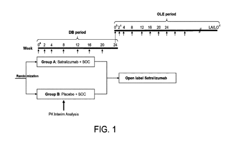
Figure 1 shows the study design of this Phase III, randomized, double-blinded,
placebo-controlled, multicenter study. DB represents double blind, LA
represents last
assessment, LO represents last observation, OLE represents open-label
extension, PK represents
pharmacokinetics, and SOC represents standard of care. a: Week 0 baseline
assessments will be
collected pre-dose. b: Week 0 of OLE period coincides with Week 24 of DB
period. c:
Patients treated with active drug in DB period will be administered a placebo
dose at Week 2 of
the OLE period to maintain blinding of treatment assignment in the DB period.
d: The total
length of study, from screening of first patient to the end of OLE period, is
estimated to be
approximately 4 years.
Figure 2 shows the predicted steady-state exposure parameters (maximum
concentration
(Cmax), trough concentration (Ctrough)) and receptor occupancy (RO) values in
serum following
administration of 120 mg and 180 mg every 4 weeks in patients weighing 100 kg
or less (40-100
CA 03211328 2023- 9-7
11
kg) and patients weighing more than 100 kg (100-160 kg), respectively. Cmax
represents
steady-state maximum concentration, Ctr represents steady-state trough
concentration, and RO
represents steady-state receptor occupancy. Plots show the results of
simulation for 2000
individuals. Cmax predicted values are shown in Figure 2A, Ctrough predicted
values in Figure 2B,
and RO predicted values in Figure 2C. Points are simulated data based on an
assumption that
the percentage of patients who are ADA (anti-drug antibody)-positive is the
same as that seen in
NMOSD (neuromyelitis optica spectrum disorder) studies. Dotted horizontal
lines have been
added for reference.
Figure 3 shows that a dosing regimen of 180 mg and 240 mg every 4 weeks for
patients
weighing 100 kg or less (40-100 kg) and more than 100 kg (100-160 kg),
respectively, would be
expected to maintain the target level of RO across the body weight ranges. Ctr
represents
steady-state trough concentration, and RO represents steady-state receptor
occupancy.
Figure 4 shows the MGFA (MG Foundation of America) classification system.
Figure 5 shows a sample of the MG Activities of Daily Living (MG-ADL)
questionnaire.
Figure 6 shows a sample of the Quantitative Myasthenia Gravis (QMG)
questionnaire.
Figure 7 shows a sample of the Myasthenia Gravis Composite (MGC)
questionnaire.
Figure 8 shows a sample of the 15-item Myasthenia Gravis (MG) Quality of Life
scale
(revised) (MG-QOL 15r) questionnaire.
Figure 9 shows a sample of the Neurology Quality-of-Life Fatigue (Neuro-QoL
Fatigue)
Short Form scale.
Figure 10 shows the fractions of patients with a clearance outside the range
of 0.8-1.25
of the population mean and patients with an RSE of more than 0.2, for each
number of assessed
cases, in simulations performed for HV (healthy adults) and NMOSD
(neuromyelitis optica
spectrum disorder) patients. RSE represents residual standard error.
[Description of Embodiments]
[0016]
Herein below, the present invention will be described in detail.
The present invention relates to pharmaceutical compositions for use in the
treatment or
prevention of myasthenia gravis (MG).
[0017]
An "antibody" in the present invention is an antibody that blocks signal
transduction by
IL-6 and inhibits the biological activities of IL-6. An antibody is preferably
an antibody that
has an inhibitory effect against the binding of IL-6, IL-6 receptor, or gp130.
[0018]
CA 03211328 2023- 9-7
12
Antibodies in the present invention include, but are not particularly limited
to, for
example, anti-IL-6 antibodies, anti-IL-6 receptor antibodies, and anti-gp130
antibodies.
Preferred antibodies in the present invention include anti-IL-6 receptor
antibodies that recognize
an IL-6 receptor.
[0019]
An anti-IL-6 receptor antibody used in the present invention can be obtained
as either a
polyclonal or monoclonal antibody using known methods. In particular, an anti-
IL-6 receptor
antibody used in the present invention is preferably a monoclonal antibody
derived from a
mammal. Monoclonal antibodies derived from a mammal include those produced by
a
hybridoma and those produced by a host that has been transformed with an
expression vector
containing an antibody gene using genetic engineering methods. By binding to
an IL-6 receptor,
this antibody inhibits the binding of IL-6 to an IL-6 receptor, and blocks
transduction of the
biological activity of IL-6 into cells.
Examples of such an antibody include the MR16-1 antibody (Tamura, T. et al.
Proc.
Natl. Acad. Sci. USA (1993) 90, 11924-11928), PM-1 antibody (Hirata, Y. et
al., J. Immunol.
(1989) 143, 2900-2906), AUK12-20 antibody, AUK64-7 antibody, and AUK146-15
antibody
(International Patent Application Publication No. WO 92-19759). Among them,
the PM-1
antibody is an example of a preferred monoclonal antibody against the human IL-
6 receptor, and
the MR16-1 antibody is an example of a preferred monoclonal antibody against
the mouse IL-6
receptor.
[0020]
Basically, hybridomas that produce an anti-IL-6 receptor monoclonal antibody
can be
produced using known techniques as follows: an IL-6 receptor is used as a
sensitizing antigen to
perform immunization by a conventional immunization method, the resulting
immune cells are
fused with known parent cells by a conventional cell fusion method, and then
the cells are
screened for monoclonal antibody-producing cells by a conventional screening
method.
Specifically, anti-IL-6 receptor antibodies can be produced as below. A human
IL-6
receptor or mouse IL-6 receptor to be used as a sensitizing antigen for
obtaining antibodies can
be obtained by, for example, using the IL-6 receptor gene and/or amino acid
sequences
respectively disclosed in European Patent Application Publication No. EP
325474 and Japanese
Patent Application Kokai Publication No. (JP-A) 1103-155795.
[0021]
There are two types of IL-6 receptor proteins: one expressed on the cell
membrane and
the other separated from the cell membrane (soluble IL-6 receptor) (Yasukawa,
K. et al., J.
Biochem. (1990) 108, 673-676). The soluble IL-6 receptor is essentially
composed of the
extracellular region of the cell membrane-bound IL-6 receptor, and differs
from the
CA 03211328 2023- 9-7
13
membrane-bound IL-6 receptor in that it lacks the transmembrane region or both
the
transmembrane and intracellular regions. Any IL-6 receptor may be employed as
the IL-6
receptor protein, as long as it can be used as a sensitizing antigen for
producing an anti-IL-6
receptor antibody to be used in the present invention.
An IL-6 receptor gene sequence is inserted into a known expression vector
system and
an appropriate host cell is transformed. Then the target IL-6 receptor protein
is purified from
the inside of the host cell or from the culture supernatant using a known
method. This purified
IL-6 receptor protein may be used as a sensitizing antigen. Alternatively, a
cell expressing the
IL-6 receptor or a fusion protein of the IL-6 receptor protein with another
protein may be used as
a sensitizing antigen.
[0022]
Mammals to be immunized with a sensitizing antigen are not particularly
limited, but
are preferably selected in consideration of the compatibility with parent
cells used for cell fusion.
Typically, rodents such as mice, rats, and hamsters are used.
Animals are immunized with a sensitizing antigen according to known methods.
Typically, immunization is performed by, for example, intraperitoneal or
subcutaneous injection
of the sensitizing antigen to a mammal. Specifically, it is preferable to
dilute or suspend the
sensitizing antigen in phosphate-buffered saline (PBS), physiological saline,
and such, to an
appropriate volume, and mix it with an appropriate amount of a conventional
adjuvant such as
Freund's complete adjuvant if desired and emulsify, and then administer to the
mammal every
four to 21 days for several times. An appropriate carrier may also be used for
immunization
with the sensitizing antigen.
After immunizing the mammal in this manner, and confirming that the serum
level of a
desired antibody has increased, immunized cells are removed from the mammal
and subjected to
cell fusion. Spleen cells are particularly preferred as the immunized cells to
be subjected to cell
fusion.
[0023]
Myeloma cells from mammals are used as parent cells to be fused with the
immunized
cells. So far, various known cell lines such as P3X63Ag8.653 (Kearney, J. F.
et al., J. Immunol
(1979) 123, 1548-1550), P3X63Ag8U.1 (Current Topics in Microbiology and
Immunology
(1978) 81, 1-7), NS-1 (Kohler, G. and Milstein, C., Eur. J. Immunol. (1976) 6,
511-519),
MPC-11 (Margulies, D. H. et al., Cell (1976) 8, 405-415), 5P2/0 (Shulman, M.
et al., Nature
(1978) 276, 269-270), FO (de St. Groth, S. F. et al., J. Immunol. Methods
(1980) 35, 1-21), S194
(Trowbridge, I. S., J. Exp. Med. (1978) 148, 313-323), and R210 (Galfre, G. et
al., Nature (1979)
277, 131-133) are suitably used.
[0024]
CA 03211328 2023- 9-7
14
Basically, cell fusion of the aforementioned immune cells with myeloma cells
can be
performed according to known methods such as the method of Milstein et al.
(Kohler, G. and
Milstein, C., Methods Enzymol. (1981) 73, 3-46).
More specifically, the cell fusion is performed, for example, in a
conventional nutrient
culture medium in the presence of a cell fusion promoter. For example,
polyethylene glycol
(PEG) or Sendai virus (HVJ) is used as the fusion promoter, and if desired, an
auxiliary agent
such as dimethyl sulfoxide can be further added for use in improving the
fusion efficiency.
[0025]
The ratio of immune cells to myeloma cells used is preferably, for example, 1
to 10
immune cells for each myeloma cell. The culture medium used for the cell
fusion is, for
example, an RPMI1640 or MEM culture medium suitable for the proliferation of
the myeloma
cell lines. Other conventional culture media used for this type of cell
culture can also be used.
Furthermore, serum supplements such as fetal calf serum (FCS) can also be used
in combination.
[0026]
For cell fusion, the fusion cells (hybridomas) of interest are formed by
thoroughly
mixing predetermined amounts of the aforementioned immune cell and myeloma
cell in the
aforementioned culture medium, adding a PEG solution (for example, a solution
of PEG with an
average molecular weight of about 1,000 to 6,000) pre-heated to about 37 C,
usually at a
concentration of 30% to 60% (w/v), and then mixing them. Then, cell fusion
agents and such
that are unsuitable for the growth of hybridomas can be removed by repeating
the operation of
sequentially adding an appropriate culture medium and removing the supernatant
by
centrifugation.
The hybridomas are selected by culturing in a general selection culture
medium, for
example, the HAT culture medium (a culture medium containing hypoxanthine,
aminopterin, and
thymidine). Culturing in the HAT culture medium is continued for a sufficient
period, generally
from several days to several weeks, to kill cells other than the hybridomas of
interest (unfused
cells). Then, a standard limiting dilution method is performed to screen for
and clone
hybridomas that produce an antibody of interest.
[0027]
Besides obtaining the hybridomas by immunizing non-human animals with an
antigen,
desired human antibodies having a binding activity to a desired antigen or
antigen-expressing
cell can be obtained by sensitizing a human lymphocyte with a desired antigen
protein or
antigen-expressing cell in vitro, and fusing the sensitized B lymphocyte with
a human myeloma
cell such as U266 (see, Japanese Patent Application Kokoku Publication No. (JP-
B) H01-59878).
Further, an antigen or antigen-expressing cell may be administered to a
transgenic animal having
a repertoire of human antibody genes, and then a desired human antibody may be
obtained
CA 03211328 2023- 9-7
15
following the aforementioned method (see, International Patent Application
Publication Nos.
WO 93/12227, WO 92/03918, WO 94/02602, WO 94/25585, WO 96/34096, and WO
96/33735).
The hybridomas prepared as such that produce monoclonal antibodies can be
passaged
in a conventional culture medium and stored in liquid nitrogen for a long
period.
[0028]
To obtain monoclonal antibodies from the hybridomas, the following methods may
be
employed: culturing the hybridomas according to conventional methods and
obtaining the
antibodies as a culture supernatant or proliferating the hybridomas by
administering them to a
compatible mammal and obtaining the antibodies from ascites; and so on. The
former method
is suitable for obtaining antibodies with high purity, and the latter is
suitable for large-scale
antibody production.
For example, hybridomas that produce anti-IL-6 receptor antibodies can be
prepared by
the method disclosed in JP-A (Kokai) 1103-139293. Such a preparation can be
carried out by
injecting hybridomas that produce PM-1 antibodies into the abdominal cavity of
a BALB/c
mouse, obtaining ascites, and then purifying the PM-1 antibodies from the
ascites; or by
culturing the hybridomas in an appropriate medium (such as an RPMI 1640 medium
containing
10% fetal bovine serum, and 5% BM-Condimed H1 (Boehringer Mannheim); the
hybridoma
SFM medium (GIBCO-BRL); or the PFHM-II medium (GIBCO-BRL)) and then purifying
the
PM-1 antibodies from the culture supernatant.
[0029]
Recombinant antibodies can be used as the monoclonal antibodies of the present
invention, wherein the recombinant antibodies are produced using genetic
recombination
techniques by cloning an antibody gene from a hybridoma, inserting the gene
into an appropriate
vector, and then introducing the vector into a host (see, for example,
Borrebaeck, C. A. K. and
Larrick, J. W., THERAPEUTIC MONOCLONAL ANTIBODIES, Published in the United
Kingdom by MACMILLAN PUBLISHERS LTD, 1990).
More specifically, mRNAs coding for antibody variable (V) regions are isolated
from
cells that produce antibodies of interest, such as hybridomas. mRNAs can be
isolated by
preparing total RNAs according to known methods, such as the guanidine
ultracentrifugation
method (Chirgwin, J. M. et al., Biochemistry (1979) 18, 5294-5299) and the
AGPC method
(Chomczynski, P. et al., Anal. Biochem. (1987) 162, 156-159), and preparing
mRNAs using an
mRNA Purification Kit (Pharmacia) and such. Alternatively, mRNAs can be
directly prepared
using the QuickPrep mRNA Purification Kit (Pharmacia).
[0030]
cDNAs of the antibody V regions are synthesized from the obtained mRNAs using
reverse transcriptase. cDNAs may be synthesized using the AMY Reverse
Transcriptase
CA 03211328 2023- 9-7
16
First-strand cDNA Synthesis Kit and such. Further, to synthesize and amplify
the cDNAs, the
5'-RACE method (Frohman, M. A. et al., Proc. Natl. Acad. Sci. USA (1988) 85,
8998-9002;
Belyaysky, A. et al., Nucleic Acids Res. (1989) 17, 2919-2932) using 5'-Ampli
FINDER RACE
Kit (Clontech) and PCR may be used. A DNA fragment of interest is purified
from the
obtained PCR products and then ligated with a vector DNA. Then, a recombinant
vector is
prepared by using the above, and introduced into Escherichia coli and such,
and then its colonies
are selected to prepare a desired recombinant vector. The nucleotide sequence
of the DNA of
interest is confirmed by a known method such as the dideoxy method.
When a DNA encoding the V region of the antibody of interest is obtained, the
DNA is
ligated with a DNA encoding the constant region (C region) of a desired
antibody, and inserted
into an expression vector. Alternatively, a DNA encoding an antibody V region
may be inserted
into an expression vector comprising a DNA of an antibody C region.
[0031]
To produce an antibody to be used in the present invention, an antibody gene
is inserted
into an expression vector such that it is expressed under the control of an
expression-regulating
region such as an enhancer and promoter, as described below. Then, the
antibody can be
expressed by transforming a host cell with this expression vector.
[0032]
In the present invention, artificially modified recombinant antibodies, for
example,
chimeric antibodies, humanized antibodies, or human antibodies can be used,
for example, to
reduce heteroantigenicity against humans. These modified antibodies can be
prepared using
known methods.
[0033]
A chimeric antibody can be obtained by ligating a DNA encoding an antibody V
region
obtained as above with a DNA encoding a human antibody C region, inserting it
into an
expression vector, and introducing the vector into a host to produce the
chimeric antibody (see,
European Patent Application Publication No. EP 125023; International Patent
Application
Publication No. WO 92-19759). This known method can be used to obtain chimeric
antibodies
useful for the present invention.
[0034]
Humanized antibodies are also referred to as reshaped human antibodies or
antibodies
made into the human type. They are produced by transplanting the
complementarity
determining regions (CDRs) of an antibody from a non-human mammal (for
example, a mouse)
into the CDRs of a human antibody. General methods for this gene recombination
are also
known (see, European Patent Application Publication No. EP 125023,
International Patent
Application Publication No. WO 92-19759).
CA 03211328 2023- 9-7
17
More specifically, DNA sequences designed to ligate the CDRs of a mouse
antibody
with the framework regions (FRs) of a human antibody are synthesized by PCR
from several
oligonucleotides produced to contain overlapping portions at their termini.
The obtained DNA
is ligated with a DNA encoding a human antibody C region and inserted into an
expression
vector, and the expression vector is introduced into a host to produce the
humanized antibody
(see, European Patent Application Publication No. EP 239400, International
Patent Application
Publication No. WO 92-19759).
Human antibody FRs to be ligated via the CDRs are selected so that the CDRs
form
satisfactory antigen binding sites. The amino acid(s) within the framework
regions of the
antibody variable regions may be substituted as necessary so that the CDRs of
the reshaped
human antibody form appropriate antigen binding sites (Sato, K. et al., Cancer
Res. (1993) 53,
851-856).
[0035]
Human antibody constant regions (C regions) are used for the chimeric and
humanized
antibodies. Examples of human antibody C regions include Cy, and for example,
Cy 1 , Cy2,
Cy3, or Cy4 may be used. Furthermore, to improve the stability of the
antibodies or their
production, the human antibody C regions may be modified.
Chimeric antibodies are composed of the variable region of an antibody derived
from a
non-human mammal and the C region derived from a human antibody; and humanized
antibodies are composed of the CDRs of an antibody derived from a non-human
mammal and
the framework regions and C regions derived from a human antibody. Their
antigenicity in the
human body is reduced, and thus they are useful as antibodies for use in the
present invention.
[0036]
Preferred specific examples of humanized antibodies for use in the present
invention
include a humanized PM-1 antibody (see, International Patent Application
Publication No. WO
92-19759).
Furthermore, in addition to the aforementioned methods for obtaining human
antibodies,
techniques for obtaining human antibodies by panning using a human antibody
library are also
known. For example, the variable region of a human antibody can be expressed
on a phage
surface as a single chain antibody (scFv) by using the phage display method,
and antigen-binding
phages can then be selected. By analyzing the genes of the selected phages,
the DNA sequence
encoding the variable region of the human antibody which binds to the antigen
can be
determined. Once the DNA sequence of an scFv which binds to the antigen is
revealed, an
appropriate expression vector comprising the sequence can be prepared to
obtain a human
antibody. These methods are already known, and reference can be made to WO
92/01047, WO
92/20791, W093/06213, WO 93/11236, WO 93/19172, WO 95/01438, and WO 95/15388.
CA 03211328 2023- 9-7
18
[0037]
The antibody gene constructed as described above can be expressed according to
known
methods. When a mammalian cell is used, the antibody gene can be expressed by
using a DNA
in which a commonly used effective promoter gene, the antibody gene to be
expressed, and a
poly A signal on the 3' side (downstream) of the antibody gene are operatively
linked together, or
by using a vector comprising the DNA. Examples of a promoter/enhancer include
the human
cytomegalovirus immediate early promoter/enhancer.
Furthermore, other promoters/enhancers that can be used for expressing the
antibodies
for use in the present invention include viral promoters/enhancers from
retroviruses, polyoma
viruses, adenoviruses, simian virus 40 (SV40), and such; and mammalian cell-
derived
promoters/enhancers such as human elongation factor la (HEF1a).
The expression can be easily performed, for example, by following the method
in
Mulligan et al. (Mulligan, R. C. et al., Nature (1979) 277, 108-114) when
using the SV40
promoter/enhancer, or by following the method in Mizushima et al. (Mizushima,
S. and Nagata
S., Nucleic Acids Res. (1990) 18, 5322) when using the HEFla
promoter/enhancer.
[0038]
When E. coli is used, the antibody gene can be expressed by operatively
linking a
commonly used effective promoter gene, a signal sequence for antibody
secretion, and the
antibody gene to be expressed. Examples of the promoter include a lacZ
promoter and an araB
promoter. A lacZ promoter can be used according to the method of Ward et al.
(Ward, E. S. et
al., Nature (1989) 341, 544-546; Ward, E. S. et al., FASEB J. (1992) 6, 2422-
2427); and an araB
promoter can be used according to the method of Better et al. (Better, M. et
al., Science (1988)
240, 1041-1043).
When the antibody is produced into the periplasm of E. coli, the pel B signal
sequence
(Lei, S. P. et al., J. Bacteriol. (1987) 169, 4379-4383) may be used as a
signal sequence for
antibody secretion. The antibody produced into the periplasm is isolated, and
then
appropriately refolded the antibody structure to be used (see, for example, WO
96/30394).
[0039]
As the replication origin, those derived from 5V40, polyoma virus, adenovirus,
bovine
papilloma virus (BPV) and such may be used. In addition, to increase the gene
copy number in
a host cell system, the expression vector may comprise the aminoglycoside
phosphotransferase
(APH) gene, thymidine kinase (TK) gene, E. coli xanthine-guanine
phosphoribosyltransferase
(Ecogpt) gene, dihydrofolate reductase (dhfr) gene, and such, as a selection
marker.
[0040]
Any production system may be used to prepare the antibodies for use in the
present
invention. The production systems for antibody preparation include in vitro
and in vivo
CA 03211328 2023- 9-7
19
production systems. In vitro production systems include those using eukaryotic
cells or those
using prokaryotic cells.
[0041]
When eukaryotic cells are used, the production systems include those using
animal cells,
plant cells, or fungal cells. Such animal cells include (1) mammalian cells
such as CHO, COS,
myeloma, baby hamster kidney (BHK), HeLa, and Vero; (2) amphibian cells such
as Xenopus
oocytes; and (3) insect cells such as sf9, sf21, and Tn5. Known plant cells
include cells derived
from Nicotiana tabacum, which may be cultured in callus. Known fungal cells
include yeasts
such as Saccharomyces (e.g., Saccharomyces cerevisiae) and mold fungi such as
Aspergillus
(e.g., Aspergillus niger).
[0042]
When prokaryotic cells are used, production systems include those using
bacterial cells.
Known bacterial cells include E. coli and Bacillus subtilis.
[0043]
Antibodies can be obtained by introducing the antibody gene of interest into
these cells
by transformation, and then culturing the transformed cells in vitro. Cells
are cultured
according to known methods. For example, DMEM, MEM, RPMI 1640, or IMDM may be
used as the culture medium, and serum supplements such as fetal calf serum
(FCS) may be used
in combination. Alternatively, cells introduced with the antibody gene may be
transferred into
the abdominal cavity and such of an animal to produce the antibodies in vivo.
[0044]
Meanwhile, in vivo production systems include those using animals or those
using
plants. When using animals, production systems include those using mammals or
insects.
Mammals that can be used include goats, pigs, sheep, mice, and bovines (Vicki
Glaser,
SPECTRUM Biotechnology Applications, 1993). Further, insects that can be used
include
silkworms. When using plants, tobacco and such may be used.
An antibody gene is introduced into these animals or plants, and the
antibodies are
produced in the body of the animals or plants and then recovered. For example,
an antibody
gene can be prepared as a fusion gene by inserting it into the middle of a
gene encoding a protein
uniquely produced into milk, such as goat J3 casein. DNA fragments comprising
the fusion gene,
which includes the inserted antibody gene, are injected into goat embryos, and
the embryos are
introduced into female goats. The desired antibodies are obtained from milk
produced by
transgenic goats born from the goats that received the embryos, or their
progenies. When
appropriate, the transgenic goats may be given hormones to increase the volume
of milk
containing the desired antibodies that they produce (Ebert, K. M. et al.,
Bio/Technology (1994)
12, 699-702).
CA 03211328 2023- 9-7
20
When silkworms are used, the silkworms are infected with a baculovirus
inserted with
the antibody gene of interest, and the desired antibodies are obtained from
the body fluids of
these silkworms (Maeda, S. et al., Nature (1985) 315, 592-594). Moreover, when
tobacco is
used, the antibody gene of interest is inserted into a plant expression vector
such as pMON530,
and the vector is introduced into bacteria such as Agrobacterium tumefaciens.
This bacterium
is used to infect tobacco such as Nicotiana tabacum, and then the desired
antibody is obtained
from the leaves of this tobacco (Julian, K.-C. Ma et al., Eur. J. Immunol.
(1994) 24, 131-138).
[0045]
When producing antibodies using in vitro or in vivo production systems as
described
above, DNAs encoding an antibody heavy chain (H chain) and light chain (L
chain) may be
inserted into separate expression vectors, and a host is then co-transformed
with the vectors.
Alternatively, the H chain-encoding DNA and L chain-encoding DNA may be
inserted into a
single expression vector for transforming a host (see International Patent
Application Publication
No. WO 94-11523).
[0046]
The antibodies used in the present invention may be antibody fragments or
modified
products thereof, as long as they can be suitably used in the present
invention. For example,
antibody fragments include Fab, F(ab')2, Fv, and single chain Fv (scFv) in
which the Fvs of the
H and L chains are linked via an appropriate linker.
Specifically, the antibody fragments are produced by treating antibodies with
enzymes
such as papain or pepsin, or alternatively, by constructing genes encoding
these antibody
fragments and introducing them into expression vectors, and then expressing
the vectors in
appropriate host cells (see, for example, Co, M. S. et al., J. Immunol. (1994)
152, 2968-2976;
Better, M. & Horwitz, A. H., Methods in Enzymology (1989) 178, 476-496;
Plueckthun, A. &
Skerra, A., Methods in Enzymology (1989) 178, 497-515; Lamoyi, E., Methods in
Enzymology
(1989) 121, 652-663; Rousseaux, J. et al., Methods in Enzymology (1989) 121,
663-666; and
Bird, R. E. et al., TIBTECH (1991) 9, 132-137).
[0047]
An scFv can be obtained by linking the H-chain V region and the L-chain V
region of an
antibody. In this scFv, the H-chain V region and the L-chain V region are
linked via a linker,
preferably via a peptide linker (Huston, J. S. et al., Proc. Natl. Acad. Sci.
USA (1988) 85,
5879-5883). The V regions of the H and L chains in an scFv may be derived from
any of the
antibodies described above. Peptide linkers for linking the V regions include,
for example, an
arbitrary single chain peptide consisting of 12 to 19 amino acid residues.
[0048]
A DNA encoding an scFv can be obtained by amplifying a DNA portion that
encodes
CA 03211328 2023- 9-7
21
the desired amino acid sequence in template sequences with PCR using a primer
pair which
defines the termini of the portion, wherein a DNA encoding an H chain or an H-
chain V region
and a DNA encoding an L chain or an L-chain V region of the aforementioned
antibodies are
used as the templates, and then further amplifying the amplified DNA portion
with a DNA that
encodes a peptide linker portion and a primer pair that defines both ends of
the linker so that it
may be linked to each of the H and L chains.
Once an scFv-encoding DNA has been prepared, an expression vector comprising
the
DNA and a host transformed with the expression vector can be obtained
according to
conventional methods. In addition, an scFv can be obtained according to
conventional methods
by using the host.
Similar to the above, the antibody fragments can be produced by obtaining
their genes,
expressing them, and then using a host. An "antibody" as used herein
encompasses such
antibody fragments.
[0049]
Antibodies bound to various molecules such as polyethylene glycol (PEG) may
also be
used as modified antibodies. An "antibody" as used herein encompasses such
modified
antibodies. These modified antibodies can be obtained by chemically modifying
the obtained
antibodies. Such methods are already established in the art.
[0050]
Antibodies produced and expressed as above can be isolated from the inside or
outside
of the cells or from the hosts, and then purified to homogeneity. The
antibodies for use in the
present invention can be isolated and purified by affinity chromatography.
Columns used for
the affinity chromatography include protein A columns and protein G columns.
Carriers used
for the protein A columns include HyperD, POROS, and Sepharose F.F. Other
methods used
for the isolation and/or purification of ordinary proteins may be used without
limitation.
For example, the antibodies used for the present invention may be isolated and
purified
by appropriately selecting and combining chromatographies other than the above-
described
affinity chromatography, filtration, ultrafiltration, salting-out, dialysis,
and such. Examples of
chromatographies include ion-exchange chromatography, hydrophobic
chromatography, and gel
filtration. These chromatographies can be applied to high performance liquid
chromatography
(HPLC). Alternatively, reverse phase HPLC may be used.
[0051]
The concentration of the antibodies obtained as above can be determined by
absorbance
measurement, ELISA, and such. Specifically, when using absorbance measurement,
the
concentration can be determined by appropriately diluting the antibody
solution with PBS(-),
measuring its absorbance at 280 nm, and calculating the concentration by using
the conversion
CA 03211328 2023- 9-7
22
factor 1.35 OD / 1 mg/ml. Alternatively, when using ELISA, the concentration
can be
determined as below. Specifically, 100 IA of goat anti-human IgG (TAG) diluted
to 1 pg/ml
with 0.1 M bicarbonate buffer (pH 9.6) is added to a 96-well plate (Nunc) and
incubated
overnight at 4 C to immobilize the antibody. After blocking, 100 IA of an
appropriately diluted
antibody to be used in the present invention or an appropriately diluted
sample comprising the
antibody, or human IgG (CAPPEL) as a standard is added, and the plate is
incubated for one
hour at room temperature.
[0052]
After washing, 100 IA of 5,000 x diluted alkaline phosphatase-labeled anti-
human IgG
(BIO SOURCE) is added, and the plate is incubated for one hour at room
temperature. After
another wash, the substrate solution is added, the plate is incubated, and
absorbance at 405 nm is
measured using Microplate Reader Model 3550 (Bio-Rad) to calculate the
concentration of the
antibody of interest.
[0053]
IL-6 is an inflammatory cytokine produced by T cells, monocytes, macrophages,
and
fibroblasts. As a regulator of B-cell and T-cell functions, IL-6 acts
pleiotropically on the
immune system through its specific receptor, IL-6R. Increased IL-6 levels have
been observed
in various inflammatory autoimmune diseases including rheumatoid arthritis
(RA), systemic
lupus erythematosus (SLE), NMOSD (Icoz S. et al., Int J Neurosci.
2010;120(1):71-5, Uzawa A.
et al., J Neurol. 2009;256(12):2082-4, Uzawa A. et al., Mult Scler.
2010;16(12):1443-52) and
Castleman's disease (Ishihara K and Hirano T. Cytokine & Growth Factor Reviews
2002;13(4-5):357-68). In studies of MG, which also involves autoimmune
abnormality,
increased IL-6 levels have been observed in the blood and muscle tissues of
non-clinical animal
models and MG patients. Therefore, IL-6 may be involved in the pathological
mechanism of
MG. For example, it may (i) stimulate the maturation of B cells into
autoantibody-producing
cells (promote autoantibody production) and (ii) induce the differentiation of
CD4-positive T
cells into Th17 pro-inflammatory T cells (induce chronic inflammation). As IL-
6 is not only
directly involved in the production of autoantibodies but also has
autoantibody-independent
functions, it may be implicated in the pathogenesis of MG regardless of the
presence or absence
of autoantibodies.
[0054]
Regulatory T cells (Treg), which suppress excessive immune response, and
pathogenic
helper T17 (Th17) cells are two lymphocyte subsets with opposite activities in
autoimmune
diseases such as MG. IL-6, a pro-inflammatory cytokine, is a potent factor to
switch immune
responses from the induction of Tregs to pathogenic Th17 cells in vivo. A
study in
experimental autoimmune myasthenia gravis (EAMG) model and healthy control
rats (Aricha R.
CA 03211328 2023- 9-7
23
et al., J Autoimmun. 2011;36(2):135-41) reported that the equilibrium between
Treg and Th17
cells was perturbed in the disease. The upregulated Th17 cell-related genes
and downregulated
Treg-related genes in the EAMG model showed a tendency of recovery after
administration of
anti-IL-6 antibodies. In addition, administration of anti-IL-6 antibodies for
EAMG suppressed
the progression of EAMG and reduced the overall IgG antibody titers and B
cells. These data
indicate the importance of IL-6 as a factor for modulating autoimmune response
in MG.
[0055]
Furthermore, tocilizumab, a blocker of IL-6 signaling, has also been shown to
be
effective in two patients with moderate and severe AChR antibody-positive MG
and insufficient
response to rituximab (Jonsson DI. et al., Neuromuscular Disorders
2017;27(6):565-8). Thus,
the inhibition of IL-6 signaling serves as a therapeutic option for MG
patients.
[0056]
Preferred examples of an "IL-6 receptor antibody" in the present invention
include
tocilizumab which is a humanized anti-IL-6 receptor IgG1 antibody, and
humanized anti-IL-6
receptor antibodies produced by modifying the variable and constant regions of
tocilizumab,
specifically, antibodies that comprise heavy-chain CDR1 comprising the amino
acid sequence of
SEQ ID NO: 5, heavy-chain CDR2 comprising the amino acid sequence of SEQ ID
NO: 6,
heavy-chain CDR3 comprising the amino acid sequence of SEQ ID NO: 7, light-
chain CDR1
comprising the amino acid sequence of SEQ ID NO: 8, light-chain CDR2
comprising the amino
acid sequence of SEQ ID NO: 9, and light-chain CDR3 comprising the amino acid
sequence of
SEQ ID NO: 10. More preferred antibodies include antibodies that comprise a
heavy-chain
variable region comprising the amino acid sequence of SEQ ID NO: 1 and a light-
chain variable
region comprising the amino acid sequence of SEQ ID NO: 2. Still more
preferred are
antibodies that comprise a heavy chain comprising the amino acid sequence of
SEQ ID NO: 3
(heavy chain of 5A237) and a light chain comprising the amino acid sequence of
SEQ ID NO: 4
(light chain of 5A237). 5A237 (satralizumab) is particularly preferred.
[0057]
Such antibodies can be obtained according to the methods described in
W02010/035769, W02010/107108, W02010/106812, and such. Specifically,
antibodies can
be produced using genetic recombination techniques known to those skilled in
the art, based on
the sequence of the above-mentioned IL-6 receptor antibody (see, for example,
Borrebaeck CAK
and Larrick JW, THERAPEUTIC MONOCLONAL ANTIBODIES, Published in the United
Kingdom by MACMILLAN PUBLISHERS LTD, 1990). A recombinant antibody can be
obtained by cloning a DNA encoding the antibody from a hybridoma or an
antibody-producing
cell such as an antibody-producing sensitized lymphocyte, inserting the DNA
into an appropriate
vector, and introducing the vector into a host (host cell) to produce the
antibody.
CA 03211328 2023- 9-7
24
[0058]
Such antibodies can be isolated and purified using isolation and purification
methods
conventionally used for antibody purification, without limitation. For
example, the antibodies
can be isolated and purified by appropriately selecting and combining column
chromatography,
filtration, ultrafiltration, salting-out, solvent precipitation, solvent
extraction, distillation,
immunoprecipitation, SDS-polyacrylamide gel electrophoresis, isoelectric
focusing, dialysis,
recrystallization, and such.
[0059]
The antibodies used in the present invention may be conjugate antibodies that
are bound
to various molecules such as polyethylene glycol (PEG), radioactive
substances, and toxins.
Such conjugate antibodies can be obtained by chemically modifying the obtained
antibodies.
Methods for antibody modification have been already established in this field.
Accordingly, the
term "antibody" in the present invention encompasses such conjugate
antibodies.
[0060]
In Japan, marketing of satralizumab was approved in June 2020, on the
indication
"prevention of relapses of neuromyelitis optica spectrum disorder (including
neuromyelitis
optica)". The safety profiles identified during the international joint phase
III clinical trials
(SA-307JG test and SA-309JG test) targeting a population of patients with
neuromyelitis optica
spectrum disorder (NMOSD) and/or neuromyelitis optica (NMO) were mostly
favorable. No
death cases were reported. The percentage of patients who expressed severe
adverse events in
the satralizumab group was about the same as that in the placebo group. There
was no big
difference between the two groups on the frequency of adverse events that led
to discontinuation
of administration of the test drug, or on the frequency of adverse events that
led to drug
withdrawal. Safety profiles were similar between the SA-309JG test, which was
a single-agent
test, and the SA-307JG test, which was a combined test with preexisting
therapy (oral steroids
and/or immunosuppressive agents).
[0061]
Myasthenia gravis (MG) is an autoimmune disease caused by impaired stimulus
transmission at the neuromuscular junction, due to disruption of the receptors
on the muscle side
by autoantibodies at the neuromuscular junction.
As it stands, there is no therapy for complete cure and it is designated as a
designated
intractable disease in Japan. The disease is characterized by indolent
fatigable weakness in
skeletal muscles, and it worsens by repeating exercise and improves by resting
(Practical
Guideline for Myasthenia Gravis (MG) 2014 (editorial supervisor: Societas
Neurologica
Japonica), Nankodo Co., Ltd.). Permanent muscle damage rarely occurs and
maximal strength
is often good. Meanwhile, decrease in muscular force varies among individual
muscles and
CA 03211328 2023- 9-7
25
muscle groups. According to statistics in Japan (Practical Guideline for
Myasthenia Gravis
(MG) 2014 (editorial supervisor: Societas Neurologica Japonica), Nankodo Co.,
Ltd.), 71.9% of
the patients experience drooping of the eyelid (ptosis) and 47.3% experience
double vision
(diplopia) as initial symptoms. At the time of diagnosis, 81.9% present ptosis
and 59.1%
present diplopia. About 20% of the symptoms that were ocular MG at the time of
diagnosis
progress to generalized disease, and as a result, 85% of MG patients present
generalized MG
(gMG) symptoms (Practical Guideline for Myasthenia Gravis (MG) 2014 (editorial
supervisor:
Societas Neurologica Japonica), Nankodo Co., Ltd.; Kerty E. et al., European
Journal of
Neurology 2014;21:687-93). Following ocular symptoms, frequently affected
muscles are the
skeletal muscles of the limb. In the statistics of 2006, it is reported that
23.1% of the patients
experience weakness of neck and limb muscles at initial onset and 44.1% at the
time of diagnosis.
Frequency of incidence decreases starting from difficulties in talking
(dysarthria), difficulties in
swallowing (dysphagia), difficulties in chewing, weakening of facial muscle,
and difficulty in
breathing, in this order. In the early stage of the disease, symptoms are
often transient and may
be ameliorated in a few weeks or more. However, symptoms are progressive and
persistent,
and usually peaks within a few years from disease onset in individual patients
(Practical
Guideline for Myasthenia gravis (MG) 2014 (editorial supervisor: Societas
Neurologica
Japonica), Nankodo Co., Ltd.).
[0062]
In the present invention, "ocular MG" refers to MG that presents only ocular
symptoms
such as ptosis and diplopia, and is classified as MGFA I according to MGFA
classification.
In the present invention, "generalized MG (gMG)" refers to MG that presents
systemic
symptoms other than ocular symptoms such as ptosis and diplopia. Ocular
symptoms such as
ptosis and diplopia may also be present in gMG. Ocular MG may include gMG,
since there are
cases where it is difficult to distinguish between ocular MG (MGFA I) and mild
gMG (MGFA
Ha) among gMG.
[0063]
Myasthenia gravis (MG) is caused by autoantibodies against functionally
important
molecules present in the postsynaptic membrane of neuromuscular junctions.
Autoantibodies related to MG include autoantibodies against acetylcholine
receptors
(AChR), autoantibodies against muscle-specific tyrosine kinase (MuSK),
autoantibodies against
LDL-receptor related protein 4 (Lrp4), and the like.
[0064]
While there are some differences among reports, regarding autoantibody
positivity rate,
about 80% of total MG are AChR-antibody positive and about 7% are MuSK-
antibody positive
(Gilhus N. et al., Nat Rev Dis Primers 2019;5(30)). Recently, autoantibodies
against Lrp4
CA 03211328 2023- 9-7
26
which forms a complex with MuSK are considered to be promising candidates as
new
pathogenic autoantibodies of MG, and there are reports that about 3% of total
MG are
Lrp4-antibody positive (Gilhus N. et al., Nat Rev Dis Primers 2019;5(30)).
Further, a few
percent of total MG are autoantibody negative MG cases where none of the above
antibodies are
detected while presenting MG symptoms.
[0065]
Preexisting therapies for generalized MG are based on drug treatment according
to the
Practical Guideline for Myasthenia gravis (MG) 2014 (editorial supervisor:
Societas Neurologica
Japonica, Nankodo Co., Ltd). Therapeutic methods vary depending on expression
of
autoantibodies. For AChR-antibody positive generalized MG, use of
anticholinesterase agents
(pyridostigmine bromide, ambenonium chloride, and such) is recommended.
Thereafter,
treatment is performed regardless of the presence or absence of
autoantibodies. Oral steroids
are the first-line choice, and when symptom control is insufficient, as a
second-line choice,
switching to immunosuppressive agents (cyclosporin, tacrolimus hydrate, and
such), and/or
increasing the dose of oral steroids, and/or combining oral steroids and
immunosuppressive
agents is/are recommended, and in the advanced stage, steroid pulse therapy,
intravenous
immunoglobulin (IVIg), and blood purification therapy are recommended.
Eculizumab was
approved in 2017 for AChR-antibody positive gMG, and administration is carried
out targeting
intractable gMG. There is an international consensus guidance for treatments
(Sanders DB. et
al., Neurology 2016;87:419-25), and while the preference in the use of
immunosuppressive
agents may vary due to difference in the agents that are covered by insurance,
there is no great
difference in the treatment policy between Japan and the Western countries.
[0066]
The pharmaceutical composition of the present invention for treatment or
prevention for
MG patients can be used to exert therapeutic effects such as obviate the onset
of MG, or alleviate
or improve clinical symptoms of patients affected with MG. Myasthenia gravis
(MG) is a
disease that progresses over a long time frame. The Practical Guideline for
Myasthenia gravis
(MG) 2014 (editorial supervisor: Societas Neurologica Japonica, Nankodo Co.,
Ltd)
recommends treating patients "with the aim of maintaining long-term favorable
QOL
considering that MG is a long-lasting disease" and also recommends actively
introducing
relatively strong immunotherapy from the early stage of treatment. By starting
treatments early,
therapeutic effects such as alleviation and improvement of symptoms can be
obtained and effects
such as prevention or preclusion of symptom progression may be obtained as
well.
[0067]
Myasthenia gravis (MG) is an autoimmune disease which is difficult to achieve
complete remission. Thus, even if complete remission is not achieved,
alleviating or improving
CA 03211328 2023- 9-7
27
symptoms to a level at which minimal manifestations (MM) can be maintained, or
maintaining
such a state, is also included in "treatment or prevention for MG patients".
For example, the
Practical Guideline for Myasthenia gravis (MG) 2014 (editorial supervisor:
Societas Neurologica
Japonica, Nankodo Co., Ltd.) sets the goal to be achieved in MG treatment as
"a level at which
minimal manifestations (MM) can be maintained with 5 mg/day or less of oral
prednisolone, by
determining the goal of treatment considering that complete remission is
difficult to achieve in
MG". By achieving this treatment goal, together with improvement in social
activities, clear
improvement in QOL can be seen. However, even in specialized outpatient
clinics in Japan, the
achievement rate under current circumstances is still 40% to 50% of the entire
MG cases. In
order to achieve this goal, further therapeutic alternatives and therapeutic
methods are desired.
[0068]
Further, by administering the pharmaceutical composition of the present
invention to
patients that potentially have a genetic background susceptible to MG before
presenting MG
symptoms, the pharmaceutical composition may be used for preventing MG onset.
Since MG
is a disease that progresses over a long time frame, by administering the
medicament of the
present invention to patients that have already developed MG, progression of
MG symptoms can
be prevented. Further, by administering the pharmaceutical composition of the
present
invention to patients that have developed MG but before increase in severity
(e.g., before crisis),
it may be used for preventing increase in severity of MG.
More specifically, the pharmaceutical composition of the present invention for
treatment
or prevention for MG patients has the effect of preventing MG onset,
preventing or precluding
the progression of symptoms, preventing increase in severity of MG, or in
therapies such as
alleviation or improvement of symptoms.
[0069]
Criteria for diagnosing myasthenia gravis (MG) and methods for evaluating MG
are
being standardized worldwide, and in the year 2000, the MG Foundation of
America (MGFA)
proposed the MGFA Classification for classifying MG symptoms. MGFA
Classification can
also be referred to as MGFA categorization, MGFA Clinical Classification, or
simply as MGFA.
MGFA Classification is a classification approach that classifies MG patients
by the condition at
the most severe state up to the present date.
[0070]
As severity scores for quantitatively evaluating severity of MG, standards
recommended
for clinical research of MG, such as MG Activities of Daily Living (MG-ADL)
scale and
Quantitative Myasthenia Gravis (QMG) scores are reported (Jaretzki A. et al.,
Neurology
2000;55(1):16-23). MG-ADL scale is relatively easy to record as a severity
score. The scores
are recorded based mainly on the patient's statements, and the score of each
item is added to
CA 03211328 2023- 9-7
28
obtain the total score. The total score can also be referred to as the MG-ADL
score. The
QMG score is an index that evaluates severity. The score of each item is added
to obtain the
total score. About 20 minutes is required to record the QMG score, and thus,
it is not a brief
test. Meanwhile, it has high detectability of fatigable muscles in that it can
capture fatigability
of extraocular muscles and muscles that seemingly have normal strength. The
Myasthenia
Gravis Composite (MGC) scale is used to evaluate symptoms after therapeutic
intervention,
which was devised by considering the advantages and disadvantages of MG-ADL
scale and
QMG score. The MGC scale balances well in correlation to QMG score and such,
even though
it allows some degree of subjective judgments by medical doctors and is thus
not complicated.
In addition to each of the above-mentioned scales, MG-QOL 15r and Neuro-QOL
Fatigue scales
will be discussed in detail in the Examples.
[0071]
Efficacy of a pharmaceutical composition on MG can be evaluated using the
above-mentioned evaluation items and by quantitatively measuring severity of
MG before and
after administration of the pharmaceutical composition to a patient and
confirming whether the
change in severity is statistically significant. Alternatively, the change or
difference between
the group administered with the pharmaceutical composition and the
unadministered group
(placebo) may be compared. For example, MG severity of a patient before
administering the
pharmaceutical composition may be determined as the baseline, and after
administering the
pharmaceutical composition for a certain period of time, MG severity of the
patient may be
quantitatively evaluated again. Alternatively, in a group of patients to be
administered with the
pharmaceutical composition and in a group not receiving the pharmaceutical
composition, MG
severity of a patient before administering the pharmaceutical composition may
be determined as
the baseline, and after administering the pharmaceutical composition for a
certain period of time,
MG severity of patients in each group may be quantitatively evaluated again.
The
above-mentioned evaluation criteria MG-ADL, QMG, MGC, MG-QOL 15r, and/or Neuro-
QOL
Fatigue may be used as standards for quantitative evaluation. In any of the
above evaluation
standards, when change in scores or points after administration compared to
those before
administration of the pharmaceutical composition (base line), or when change
or difference in
scores or points between groups of patients administered with or not
administered with the
pharmaceutical composition is statistically significant, it can be said that
the pharmaceutical
composition is effective against myasthenia gravis (MG). When the degree of MG
of a patient
is severe, MG evaluation scores or points will be high, and when the degree is
mild they become
low. Thus, it is desirable that the change or difference in scores or points
of evaluation
standards decreases.
[0072]
CA 03211328 2023- 9-7
29
The pharmaceutical composition of the present invention is a pharmaceutical
composition for treatment or prevention for myasthenia gravis (MG) patients.
Accordingly, the
pharmaceutical composition of the present invention can be referred to as a
pharmaceutical
composition that reduces the scores or points of MG-ADL, QMG, MG-QOL 15r,
Neuro-QOL
Fatigue, and/or MGC in patients administered with the pharmaceutical
composition of the
present invention. Further, the pharmaceutical composition of the present
invention can be
referred to as a pharmaceutical composition that reduces the scores or points
of MG-ADL, QMG,
MG-QOL 15r, Neuro-QOL Fatigue, and/or MGC in patients administered with the
pharmaceutical composition of the present invention, compared to the values
(scores or points)
measured using the same scale or scoring method obtained from the patient
before administering
the pharmaceutical composition of the present invention. Further, the
pharmaceutical
composition of the present invention can be referred to as a pharmaceutical
composition that
reduces the scores or points of MG-ADL, QMG, MG-QOL 15r, Neuro-QOL Fatigue,
and/or
MGC in patients administered with the pharmaceutical composition of the
present invention
compared to those of patients not administered with the pharmaceutical
composition of the
present invention.
[0073]
The given period of administering the pharmaceutical composition for
evaluating
efficacy of the pharmaceutical composition on MG is not particularly limited
and includes 1
week, 2 weeks, 4 weeks, 8 weeks, 12 weeks, 24 weeks, 48 weeks, 1 year, 2
years, 3 years, 4
years, and 5 years, and the period may be shorter or longer than the
exemplified period.
Efficacy of the pharmaceutical composition on MG that is administered to a
patient can
be confirmed when the change or difference in scores or points of any of MG-
ADL, QMG,
MG-QOL 15r, Neuro-QOL Fatigue, and/or MGC is statistically significant. For
example, the
scores or points of any of MG-ADL, QMG, MG-QOL 15r, Neuro-QOL Fatigue, and/or
MGC
after treatment with the pharmaceutical composition may be statistically
significantly reduced
compared to the scores or points of the same measurement method obtained from
the patient
before administering the pharmaceutical composition or from a patient not
administered with the
pharmaceutical composition, and for example includes reduction of scores or
points of 1, 2, 3, 4,
5, or 6, but may be other scores or points.
[0074]
In the present invention, "as an active ingredient" means that the ingredient
is contained
in the pharmaceutical composition as a primal active ingredient, and the
content thereof is not
limited unless specifically indicated, as long as the antibodies used for the
present invention are
included as medicinal ingredients.
[0075]
CA 03211328 2023- 9-7
30
In the present invention, "standard dosing interval" refers to a dosing
interval generally
used for the pharmaceuticals (pharmaceutical compositions of the present
invention), and
includes for example, a dosing interval for constant administration that may
be described in a
package insert as "subcutaneous injections at four-week intervals" and such.
The standard
dosing interval in the present invention is not particularly limited, but
examples include 1 day to
24 weeks, preferably 2 weeks to 8 weeks, more preferably 3 to 5 weeks, and
even more
preferably 4 weeks. The standard dosing intervals may have a certain range.
[0076]
In the present invention, "dosing interval that is shorter than the standard
dosing
interval" refers to a dosing interval that is shorter than the dosing interval
generally used
(standard dosing interval) for the pharmaceuticals (pharmaceutical
compositions of the present
invention). The dosing interval that is shorter than the standard dosing
interval is not
particularly limited as long as it is shorter than the standard dosing
interval, and for example,
when the standard dosing interval is 24 weeks, the dosing interval that is
shorter than the
standard dosing interval may be shorter than 24 weeks, for example, 20 weeks.
The dosing
interval that is shorter than the standard dosing interval is preferably an
interval that is half the
standard dosing interval. For example, preferably when the standard dosing
interval is 8 weeks,
the dosing interval that is shorter than the standard dosing interval is 4
weeks, and more
preferably when the standard dosing interval is 4 weeks, the dosing interval
that is shorter than
the standard dosing interval is 2 weeks. The dosing intervals that are shorter
than the standard
dosing intervals may have a certain range.
[0077]
In the present invention, "standard dose" refers to a dose of the antibody,
which is the
active ingredient, generally used for the pharmaceuticals (pharmaceutical
compositions of the
present invention). Examples include doses of antibodies for standard
administration that may
be described in a package insert as "generally administered at 100 mg per dose
as antibody",
"generally subcutaneously injected at 100 mg per dose of antibody", and such.
The standard
dose in the present invention is not particularly limited, and examples
include 50 to 800 mg of
antibody per administration, preferably 120 to 240 mg of antibody, and more
preferably 120 mg,
180 mg, or 240 mg of antibody per administration.
[0078]
The "standard dose" in the present invention may vary depending on the
patient's body
weight. For example, the standard dose to a patient with a body weight of 100
kg or less per
administration may be 120 mg of antibody and the standard dose to a patient
with a body weight
of more than 100 kg per administration may be 180 mg of antibody; the standard
dose to a
patient with a body weight of 100 kg or less per administration may be 120 mg
of antibody and
CA 03211328 2023- 9-7
31
the standard dose to a patient with a body weight of more than 100 kg per
administration may be
240 mg of antibody; and the standard dose to a patient with a body weight of
100 kg or less per
administration may be 180 mg of antibody and the standard dose to a patient
with a body weight
of more than 100 kg per administration may be 240 mg of antibody. Further, the
standard dose
to a patient with a body weight of less than 100 kg per administration may be
120 mg of
antibody and the standard dose to a patient with a body weight of 100 kg or
more per
administration may be 180 mg of antibody; the standard dose to a patient with
a body weight of
less than 100 kg per administration may be 120 mg of antibody and the standard
dose to a patient
with a body weight of 100 kg or more per administration may be 240 mg of
antibody; and the
standard dose to a patient with a body weight of less than 100 kg per
administration may be 180
mg of antibody and the standard dose to a patient with a body weight of 100 kg
or more per
administration may be 240 mg of antibody. More specifically, the standard dose
to a patient
with a body weight of 100 kg or less (patient with a body weight of 40 kg or
more and 100 kg or
less) per administration may be 120 mg of antibody and the standard dose to a
patient with a
body weight of more than 100 kg (patient with a body weight of more than 100
kg and 160 kg or
less) per administration may be 180 mg of antibody; the standard dose to a
patient with a body
weight of 100 kg or less (patient with a body weight of 40 kg or more and 100
kg or less) per
administration may be 120 mg of antibody and the standard dose to a patient
with a body weight
of more than 100 kg (patient with a body weight of more than 100 kg and 160 kg
or less) per
administration may be 240 mg of antibody; and the standard dose to a patient
with a body weight
of 100 kg or less (patient with a body weight of 40 kg or more and 100 kg or
less) per
administration may be 180 mg of antibody and the standard dose to a patient
with a body weight
of more than 100 kg (patient with a body weight of more than 100 kg and 160 kg
or less) per
administration may be 240 mg of antibody. Further, the standard dose to a
patient with a body
weight of less than 100 kg (patient with a body weight of 40 kg or more and
less than 100 kg)
per administration may be 120 mg of antibody and the standard dose to a
patient with a body
weight of 100 kg or more (patient with a body weight of 100 kg or more and 160
kg or less) per
administration may be 180 mg of antibody; the standard dose to a patient with
a body weight of
less than 100 kg (patient with a body weight of 40 kg or more and less than
100 kg) per
administration may be 120 mg of antibody and the standard dose to a patient
with a body weight
of 100 kg or more (patient with a body weight of 100 kg or more and 160 kg or
less) per
administration may be 240 mg of antibody; and the standard dose to a patient
with a body weight
of less than 100 kg (patient with a body weight of 40 kg or more and less than
100 kg) per
administration may be 180 mg of antibody and the standard dose to a patient
with a body weight
of 100 kg or more (patient with a body weight of 100 kg or more and 160 kg or
less) per
administration may be 240 mg of antibody.
CA 03211328 2023- 9-7
32
[0079]
The term "standard administration" in the present invention refers to an
administration
commonly used for the above-mentioned pharmaceuticals (pharmaceutical
compositions of the
present invention), for example, an administration at the above-described
"standard dose" and
"standard dosing interval".
[0080]
In the present invention, "short-interval dosing period" refers to a period
where multiple
administrations are carried out prior to administering with standard dosing
intervals, in which the
dose is the same as the standard dose and the dosing interval is shorter than
the standard dosing
interval, and the period is preferably 1 to 12 weeks from the initial
administration, more
preferably 2 to 8 weeks, and even more preferably 4 weeks from the initial
administration. A
dose that is the same as the standard dose includes the case where blood
concentration of the
antibody is comparable to the case where standard dose is administered. The
short-interval
dosing period may have a certain range.
[0081]
In the present invention, "(being) administered multiple times" refers to two
or more
administrations including the initial administration, and is preferably 2 to 5
times including the
initial administration, and more preferably 3 times including the initial
administration.
[0082]
In the present invention, the standard administration period starts from the
last
administration of the short-interval dosing period. More specifically, after
one standard dosing
interval has passed from the last administration in the short-interval dosing
period, the first
administration in the standard administration period is carried out. For
example, in the case of
performing 3 administrations including the initial dose in a short-interval
dosing period, after the
3rd administration, one standard dosing interval lapses, and the period at
which administration is
performed at standard dosing intervals from the 4th administration and onward
will become the
standard administration period. For example, in the case where the short-
interval dosing period
is 4 weeks, administration including the initial administration in the short-
interval dosing period
is 3 times, the standard dosing intervals is 4 weeks, and the dosing interval
that is shorter than
the standard dosing intervals is 2 weeks, the initial, 2nd, and 3rd
administrations are performed
with 2 weeks intervals within the 4 weeks which is the short-interval dosing
period, and then the
4th administration is performed after 4 weeks has passed from the 3rd
administration, which is
the 1st standard dosing interval, and thereafter, administration will be
performed with the
standard dosing intervals of 4 weeks. Accordingly, after the 3rd
administration which is the last
administration of the short-interval dosing period, the period at which
administration is carried
out with 4 weeks intervals, which is the standard dosing interval, will become
the standard
CA 03211328 2023- 9-7
33
administration period.
[0083]
The pharmaceutical composition of the present invention is preferably a
pharmaceutical
composition the administration of which is carried out by administering an
antibody for 2 to 5
times in a short-interval dosing period from the initial administration with
an interval of 1 to 3
weeks at the same dose as a standard dose, and then from the last
administration of the
short-interval dosing period, performing standard administration with an
interval of 2 to 8 weeks
of the antibody at 50 to 800 mg per administration, which is the standard
dose. More preferably,
the pharmaceutical composition is such that its administration includes
administering SA237 for
3 times in a short-interval dosing period from the initial administration with
an interval of 2
weeks (i.e., administration at week 0, week 2, and week 4) at the same dose as
a standard dose,
and then from the last administration of the short-interval dosing period,
performing standard
administration with an interval of 4 weeks (i.e., continued with a 4 weeks
interval counting from
the initial administration of the short-interval period at week 8, week 12,
week 16) of SA237 at
120 mg, 180 mg, or 240 mg per administration, which is the standard dose.
[0084]
The preferred administration schedule for the antibody of the present
invention can be
adjusted, for example, by appropriately extending the administration interval
by monitoring the
conditions of the disease and changes in the blood test values.
[0085]
The present invention also provides an article of manufacture (such as a kit,
a device,
and the like) for use in a method of the present invention, which contains a
pharmaceutical
composition or a medicament of the present invention. The pharmaceutical
composition or
medicament of the present invention comprises an IL-6 inhibitor as described
herein. The
article of manufacture may be packaged with an additional pharmaceutically
acceptable carrier
or medium, or an instruction manual describing how to use the kit, the device,
or the like, etc.
[0086]
In one embodiment, the article of manufacture comprises a container and a
label on or a
package insert accompanying the container. Suitable containers include, for
example, bottles,
vials, syringes (including a prefilled syringe and an autoinjector), IV
solution bags, etc. The
containers may be formed from a variety of materials such as glass or plastic.
In one embodiment,
the container may hold a composition alone or in combination with another
composition
effective for treating, preventing and/or diagnosing the condition, and may
also have a sterile
access port (for example, the container may be a syringe, an autoinjector, an
intravenous solution
bag or a vial having a stopper pierceable by a hypodermic injection needle).
At least one active
ingredient in the composition is an IL-6 inhibitor as described in the present
disclosure,
CA 03211328 2023- 9-7
34
preferably an anti-IL-6 receptor antibody, and more preferably satralizumab.
[0087]
In one embodiment, a device as the article of manufacture of the present
invention as
described above may be a prefilled syringe for injection via any
administration route (such as
intravenous, subcutaneous, or the like) which comprises a fixed dose of an IL-
6 inhibitor as
described in the present disclosure, preferably an anti-IL-6 receptor
antibody, and more
preferably satralizumab, in a pharmaceutically acceptable excipient. In
another embodiment,
the device may be an autoinjector for subcutaneous administration which
comprises a fixed dose
of an IL-6 inhibitor as described in the present disclosure, preferably an
anti-IL-6 receptor
antibody, and more preferably satralizumab, in a pharmaceutically acceptable
excipient. In
certain embodiments, the device (such as a prefilled syringe and autoinjector)
may comprise 120
mg, 180 mg, or 240 mg of satralizumab.
[0088]
In one embodiment, the label or package insert indicates that the
pharmaceutical
composition or medicament is used for treating the condition of choice.
Moreover, the article
of manufacture may comprise (a) a first container with a composition contained
therein, wherein
the composition comprises an IL-6 inhibitor as described above, preferably an
anti-IL-6 receptor
antibody, and more preferably satralizumab; and (b) a second container with a
composition
contained therein, wherein the composition comprises a further therapeutic
agent. The article
of manufacture in this embodiment of the invention may further comprise a
package insert
indicating that the compositions can be used to treat a particular condition.
Alternatively, or
additionally, the article of manufacture may further comprise a second (or
third) container
comprising a pharmaceutically-acceptable buffer, such as bacteriostatic water
for injection
(BWFI), phosphate-buffered saline, Ringer's solution and dextrose solution. It
may further
include other materials desirable from a commercial and user standpoint,
including other buffers,
diluents, filters, needles, and syringes.
[0089]
The term "package insert" is used to refer to instructions customarily
included in
commercial packages of therapeutic products, that contain information about
the indications,
usage, dosage, administration, combination therapy, contraindications and/or
warnings
concerning the use of such therapeutic products.
[0090]
Pharmaceutical compositions of the present invention used for therapeutic or
preventive
purposes can be formulated to produce freeze-dried formulations or solution
formulations by
mixing, if necessary, with suitable pharmaceutically acceptable carriers,
vehicles, and such.
The suitable pharmaceutically acceptable carriers and vehicles include, for
example, sterilized
CA 03211328 2023- 9-7
35
water, physiological saline, stabilizers, excipients, antioxidants (such as
ascorbic acid), buffers
(such as phosphate, citrate, histidine, and other organic acids), antiseptics,
surfactants (such as
PEG and Tween), chelating agents (such as EDTA), and binders. Other low-
molecular-weight
polypeptides, proteins such as serum albumin, gelatin, and immunoglobulins,
amino acids such
as glycine, glutamine, asparagine, glutamic acid, aspartic acid, methionine,
arginine, and lysine,
sugars and carbohydrates such as polysaccharides and monosaccharides, and
sugar alcohols such
as mannitol and sorbitol may also be contained. When preparing an aqueous
solution for
injection, physiological saline and isotonic solutions comprising glucose and
other adjuvants
such as D-sorbitol, D-mannose, D-mannitol, and sodium chloride may be used;
and appropriate
solubilizers such as alcohol (for example, ethanol), polyalcohols (such as
propylene glycol and
PEG), and nonionic surfactants (such as polysorbate 80, polysorbate 20,
poloxamer 188, and
HCO-50) may be used in combination. By mixing hyaluronidase into the
formulation, a larger
fluid volume can be administered subcutaneously (Expert Opin. Drug Deliv. 2007
Jul; 4(4):
427-40). Furthermore, syringes may be prefilled with the pharmaceutical
composition of the
present invention. Solution formulations can be prepared according to the
method described in
W02011/090088.
[0091]
If necessary, the pharmaceutical compositions of the present invention may be
encapsulated in microcapsules (e.g., those made of hydroxymethylcellulose,
gelatin, and
poly(methylmethacrylate)), or incorporated into colloidal drug delivery
systems (e.g., liposomes,
albumin microspheres, microemulsions, nanoparticles, and nanocapsules) (see,
for example,
"Remington's Pharmaceutical Science 16th edition", Oslo Ed. (1980)). Methods
for preparing
the pharmaceutical agents as controlled-release pharmaceutical agents are also
known, and such
methods may be applied to the pharmaceutical compositions of the present
invention (Langer et
al., J. Biomed. Mater. Res. 15: 267-277 (1981); Langer, Chemtech. 12: 98-105
(1982); U.S.
Patent No. 3,773,919; European Patent Application Publication No. EP 58,481;
Sidman et al.,
Biopolymers 22: 547-556 (1983); and EP 133,988).
[0092]
The pharmaceutical composition of the present invention can be administered to
a
patient via any appropriate route. For example, it can be administered to a
patient intravenously
by bolus injection or by continuous infusion, intramuscularly,
intraperitoneally,
intracerebrospinally, transdermally, subcutaneously, intraarticularly,
sublingually, intrasynovially,
orally, by inhalation, locally, or externally, for a certain period of time.
Intravenous
administration or subcutaneous administration is preferred.
[0093]
The antibody of the present invention may be used in an agent comprising the
antibody
CA 03211328 2023- 9-7
36
for treating or preventing MG, or may be used in a method of treating or
preventing MG which
comprises administering the antibody to a patient with MG. In addition, the
antibody of the
present invention may be used in the manufacture of an agent for treating or
preventing MG.
Further, the antibody of the present invention may be referred to as an
antibody for use in the
treatment of a patient with MG or for use in the prevention of MG. Further,
the antibody of the
present invention is preferably satralizumab, and is specifically an antibody
comprising a heavy
chain variable region comprising the amino acid sequence of SEQ ID NO: 1 and a
light chain
variable region comprising the amino acid sequence of SEQ ID NO: 2.
[0094]
All prior art references cited herein are incorporated by reference into the
present
specification.
[Example]
[0095]
Herein below, the present invention will be specifically described with
reference to the
Examples, but it is not to be construed as being limited thereto.
[0096]
Example 1: Preparation of satralizumab (5A237)
5A237, which is an IL-6 receptor antibody described in the patent document WO
2010/035769 (an antibody comprising a heavy chain having the amino acid
sequence of SEQ ID
NO: 26 (SEQ ID NO: 3 in the present specification) and a light chain having
the amino acid
sequence of SEQ ID NO: 29 (SEQ ID NO: 4 in the present specification) in the
patent document
WO 2010/035769), was prepared according to the description of the patent
document. The
amino acid sequence of the heavy chain variable region is shown in SEQ ID NO:
1, and the
amino acid sequence of the light chain variable region is shown in SEQ ID NO:
2. Using the
prepared antibody, a subcutaneous administration preparation was prepared by
the method
described in the patent document WO 2011/090088.
[0097]
Example 2: Phase III, randomized, double-blinded, placebo-controlled,
multicenter study
Study Design
1. Description of the Study
The Phase III, randomized, double-blinded, placebo-controlled, multicenter
study is
designed to evaluate the efficacy, safety, pharmacokinetics, and
pharmacodynamics of
satralizumab compared with placebo as add-on therapy to standard of care (SOC)
for the
treatment of generalized myasthenia gravis (generalized MG or gMG). The study
includes a
24-week double blind (DB) period, and an open-label extension (OLE) period.
A summary of the study design is illustrated in Figure 1.
CA 03211328 2023- 9-7
37
[0098]
1.1 Double Blind Period
During the double blind treatment period, patients are randomized in a 1:1
ratio to
receive 120 mg, 180 mg, or 240 mg satralizumab based on the body weight (Group
A), or
placebo (Group B) for 24 weeks. Randomization will be stratified based on the
baseline
standard-of-care therapy (SOC), auto-antibody type, and the region as the
stratification factors.
[0099]
A blinded drug is administered subcutaneously to patients at Weeks 0, 2, 4,
and every
four weeks thereafter until the end of the DB period, in addition to SOC at a
stable dose.
During the DB period, an interim analysis of pharmacokinetic (PK) data is
performed. Based
on the results from the interim PK analysis and pre-specified criteria,
whether the study drug
dose is increased is determined. If it is determined to be increased,
thereafter, Group A is
administered with either 180 mg or 240 mg satralizumab.
[0100]
The background therapies permitted in this study are anticholinesterase
inhibitors
(AChEI) alone or the following treatment methods (with or without AChEI): oral
corticosteroids
(OCSs), one immunosuppressant therapy (1ST), and an OCS in combination with
one 1ST. The
permitted 1ST drugs are, for example, azathioprine, mycophenolate mofetil,
cyclosporin A, and
tacrolimus, but are not limited thereto.
[0101]
1.2 Open-Label Extension Period
Patients who completed the DB period can enter the OLE period. Upon completion
of
the Week 24 assessments, all patients receive open-label treatment with
satralizumab.
[0102]
In the OLE period, all patients receive open-label treatment with 120 mg, 180
mg, or
240 mg satralizumab.
Based on the investigator's judgment, dose reduction (taper) of background
therapy
(OCS, 1ST, and/or AChEI) can be started at Week 12 or later in the OLE period.
[0103]
1.3 Unscheduled Visits and Rescue Therapy
Unscheduled visits can be performed at the discretion of the treating
neurologist. If a
suspected gMG exacerbation occurs during the study, the participant needs to
return to the study
site and undergo an evaluation of gMG severity before or immediately after
receiving rescue
therapy.
The rescue therapy includes intravenous immunoglobulin therapy (IVIg), and
plasma
exchange (PE) therapy with or without high-dose corticosteroids. The choice of
rescue therapy
CA 03211328 2023- 9-7
38
is determined by the investigator on the basis of the overall clinical
assessment.
Patients who received rescue therapy during the DB period may receive OLE
satralizumab administration after completion of the DB period.
[0104]
1.4 Subject Patients and Study Regions
The study is conducted in countries including, but not limited to, North
America,
Europe, Latin America, and Asia.
This study enrolls approximately 240 patients, including approximately 20
adolescents
aged 12 to 17 years with gMG. The main selection/exclusion criteria are shown
below.
[Selection criteria]
- Patients aged 12 years or older at the time of consent acquisition
- Patients diagnosed with myasthenia gravis with generalized muscle
weakness that falls under
Class II, III, or IV severity classification by the Myasthenia Gravis
Foundation of America
(MGFA). Diagnostic confirmation needs to be demonstrated and supported by a
positive serum
test for one of the three antibody types, AChR antibody, MuSK antibody, and
Lrp4 antibody at
the time of screening.
- Patients with a total MG-ADL score of 5 or higher, and no less than half
of the scores
associated with non-ocular symptoms.
- Patients that can receive the rescue therapies (immunoglobulin therapy
(IVIg), plasma
exchange (PE) therapy, and high-dose corticosteroid therapy).
- Patients having received, prior to enrollment, stable doses of treatment
for severe myasthenia
gravis (azathiopurine, mofetil mycophenolate, cyclosporin A, tachlorimus, oral
corticosteroids
(OCS), or anticholinesterase inhibitors (AChEI)).
[Exclusion criteria]
- Patients with a history of thymic cyst, thymoma, or other thymic neoplasm
(as defined by the
WHO classification for thymic tumor (2015)) are excluded, except where they
are considered to
have been cured by appropriate treatment, and there is no observation of
recurrence for five
years or more prior to screening.
- Patients after less than twelve months of thymectomy are excluded.
- Patients with a history of myasthenia crisis (MGFA class V) within the last
three months are
excluded.
[0105]
2. Length of Study
The total length of study, from the first patient screening to the end of the
OLE period,
is estimated to be approximately four years.
[0106]
CA 03211328 2023- 9-7
39
3 Rationale for Study Design
3.1 Rationale for Satralizumab Dose and Schedule
Weight-tiered dosing via SC injection is used in this study for the
investigation of
efficacy and safety of satralizumab in gMG as shown in Table 1.
[0107]
[Table 1]
Dosing Regimen in Phase III Study of Satralizumab for the Treatment of
generalized Myasthenia
Gravis
Body weight at Baselinea Dose and Regimen
100 kg or less 120 mg administered at Weeks
0, 2, 4, and
Q4W thereafter by subcutaneous injection
More than 100 kg 180 mg administered at Weeks
0, 2, 4, and
Q4W thereafter by subcutaneous injection
Q4W: Every four weeks.
a: In case of change in the body weight of an individual patient,
recommendation is made to
include him/her in the other dosing group. See Table 2.
[0108]
The dosing regimen is based on a combination of sources of information
including the
following:
PK, PD, and safety data for satralizumab for the initial development in
neuromyelitis
optica spectrum disorder (NMOSD); and
Consideration of differences in population demographics.
[0109]
The 120-mg fixed dose regimen investigated in the Phase III studies in NMOSD
was
associated with high predicted median trough receptor occupancy (95% or more)
at steady-state
(ROtr,ss) values in most patients, and was shown to be safe and efficacious in
all body weight
groups. The few patients with predicted ROtr,ss values of less than 80%
generally had baseline
body weights of more than 100 kg.
[0110]
Real-world data from a large U.S. emergency room-based dataset of 58,860
patients
having an MG code suggest that approximately 30% of the patients in this
population may have
body weights of more than 100 kg. Exposure similar to that in NMOSD is
expected to be
effective also in gMG, and therefore simulations were performed using the
existing
population-PK (pop-PK) model to estimate the dose required for achieving the
same maximal
ROtr,ss values achieved in NMOSD patients throughout the dose interval for gMG
patients across
the expected body weight range.
CA 03211328 2023- 9-7
40
[0111]
The predicted maximum concentration observed (Cmax), trough concentration
(Ctrough),
and RO values in serum following administration of 120 mg every four weeks and
180 mg every
four weeks in patients weighing 100 kg or less and more than 100 kg,
respectively, are shown in
Figure 2. It was suggested that similar RO can be achieved by administering
180 mg to the
patients weighing more than 100 kg compared to the administration of 120 mg to
the patients
weighing 100 kg or less. Furthermore, the exposures in administering 180 mg to
the patients
weighing more than 100 kg were within the range of exposures by 120 mg
administration that
has been confirmed to be safe in the Phase III studies in NMOSD patients. The
study design
includes an PK interim analysis to confirm whether the target exposures and RO
are achieved.
[0112]
A favorable safety profile was demonstrated in the NMOSD program for
satralizumab,
supporting the targeting of similar exposures in this study. However,
exposures below the
target range may warrant an increase in dose to optimize IL-6 blockade
throughout the dose
interval across the expected body weight range. Therefore, an option for dose
increase is
included (for all body weight ranges, if necessary) in the study design.
Details are provided in
Section 3.11.
[0113]
While most PK parameter estimates for satralizumab were shown to be very
similar
between healthy adults (HV) and patients with NMOSD, population differences in
the total
clearance of drug (CL) were observed (covariate value [95%CI] in HV 95.8%[67.5
to 124.1]).
Therefore, PK simulation has been used to explore potentially useful regimens,
presuming the
case where CL in the gMG population was similar to CL in HV. The simulations
presented in
Figure 3 indicate that, if that is the case, a dosing regimen of 180 mg and
240 mg (180/240 mg
regimen) for patients weighing 100 kg or less and more than 100 kg,
respectively, would be
expected to maintain the target level of RO across the body weight ranges. The
dose is adapted
to this higher dosing regimen if CL in gMG is reflective of CL in HV.
[0114]
PK simulation is used as part of the PK interim analysis to confirm whether
the initially
proposed doses achieved target exposures, or the dose should be adapted to 180
mg or 240 mg
regimen. In either case, the chosen dosing regimen is expected not to
significantly exceed the
exposure confirmed to be safe in the Phase III studies in NMOSD patients.
[0115]
3.2 Rationale for Choice of Background Treatment
Patients on a stable dose of background therapy are enrolled. The background
SOC
therapies permitted in the study are the following:
CA 03211328 2023- 9-7
41
AChEI
OCS
one 1ST
OCS in combination with one 1ST
Concomitant use of AChEI is permitted for patients on a stable dose with OCS,
one 1ST,
and OCS in combination with one 1ST.
The combination treatment regimen of 1ST and/or OCS was chosen on the basis of
being the most common treatment choice for gMG worldwide and in accordance
with the
international consensus guidance for management of MG (Sanders et al.,
Neurology 2016; 87:
419-25).
[0116]
3.3 Rationale for Primary Outcome Measure: Myasthenia Gravis Activities of
Daily Living
(MG-ADL)
The primary objective for this study is to compare the efficacy of
satralizumab added to
SOC versus placebo added to SOC using the MG-ADL as a primary outcome measure.
A
sample of MG-ADL questionnaire is shown in Figure 5.
[0117]
The MG-ADL was developed by Wolfe and colleagues (Neurology 1999; 52: 1487-9)
to
assess the degree of gMG symptoms (six items: diplopia, ptosis, difficulties
with chewing,
swallowing, talking, and respiratory problems) and functional limitations in
carrying out
activities of daily living (two items: disability to brush teeth or comb hair
and impairment in the
ability to arise from a chair) that have been shown to be present and
clinically relevant in gMG
patients. Each of the eight items is ranked on a 0 to 3 scale yielding a total
score that ranges
from 0 to 24, and higher scores indicate greater disease severity (Figure 5).
The items of the
MG-ADL were all derived from the original 13-item symptom list that comprises
the
clinician-rated QMG scale.
[0118]
The psychometric properties of the MG-ADL have been characterized. Construct
validity has been demonstrated by showing correlations with the QMG (r=0.58;
Wolfe et al.,
Neurology 1999; 52: 1487-9), MGC measure (r=0.85), and the MG-QOL 15r measure
(r=0.76)
(Muppidi et al., Muscle Nerve 2011; 44: 727-31). Test/retest reliability was
demonstrated in a
small sample (n=26) of patients with two repeated assessments separated by 2
to 4 days, showing
a high degree of reproducibility (r=0.94) (Muppidi et al., Muscle Nerve 2011;
44: 727-31).
[0119]
Because of the importance of patients' subjective feedback due to the
fluctuating nature
CA 03211328 2023- 9-7
42
of the disease and the established psychometric properties including good
content validity
associated with patients' functioning in daily life, MG-ADL is an appropriate
primary outcome
measure. MG-ADL scale has been used as the primary outcome measure in the
recently
completed (Howard et al., Lancet Neurol 2017; 16: 976-86) and in the ongoing
Phase III gMG
randomized, placebo-controlled trials (NCT03669588, NCT03971422, NCT03920293,
NCT04115293, and NCT03304054).
[0120]
3.4 Rationale for Secondary Outcome Measures
The QMG, MG-QOL 15r, NeuroQ0L Fatigue scale, and the MGC were selected as
secondary endpoints in order to compare the efficacy of satralizumab added to
SOC versus
placebo added to SOC.
[0121]
The QMG is a 13-item assessment of gMG symptom severity based on clinical
examination (Tindall et al., N Engl J Med 1987; 316: 719-24). It has been used
in clinical trials
since 1983, when it was first developed to study the relationship between AChR-
Ab binding and
disease severity (Besinger et al., Neurology 1983; 33: 1316-21). Recently, the
QMG has been
used as a key secondary outcome measure in the recently completed study of
eculizumab for
gMG (Howard et al., Lancet Neurol 2017; 16: 976-86) and all ongoing Phase III
gMG
randomized, placebo-controlled trials (NCT03669588, NCT03971422, NCT03920293,
NCT04115293, and NCT03304054). A sample of QMB questionnaire is shown in
Figure 6.
[0122]
The QMG assesses severity of symptoms ranging from 0 to 3 for ptosis,
diplopia,
orbicularis oculi weakness, swallowing, speech disruption, percent forced
vital capacity, arm and
leg endurance (four items), grip strength (two items), and neck flexion
strength, resulting in a
total score that ranges from 0 to 39, and higher values indicate severer
symptoms (Tindall et al.,
N Engl J Med 1987; 316: 719-24).
The psychometric properties of the QMG have been studied using both
observational
and clinical trial data. The QMG has acceptable internal consistency
(Cronbach's a=0.74) and
test/retest reliability (intra-class correlation coefficient [ICC] = 0.88) in
clinically stable patients
(Barnett et al., J Neuromuscular Disease 2015; 2: 301-11). Construct validity
studies of the
QMG found correlations with the MGFA score (r2=0.54) and the MG-QOL 15
(r=0.41) (Barnett
et al., J Clin Neuromuscular Dis. 2012; 13: 201-5).
This study evaluates the efficacy of satralizumab added to SOC versus placebo
added to
SOC by comparing the change from baseline in the QMG score at Week 24 (the end
of the DB
period).
[0123]
CA 03211328 2023- 9-7
43
The MGC is an additional secondary efficacy outcome measure in this trial. As
the
name implies, the MGC is a composite measure consisting of items drawn from
the MG-ADL
(chewing, swallowing, speech, and breathing), QMG (diplopia and ptosis), and
Manual Muscle
Test (hip, neck, facial, and deltoid strength) to include both clinician- and
patient-reported
elements in a single measure (Burns et al., Muscle and Nerve 2008; 38: 957-
63). The ten items
compose a total score ranging from 0 to 50, and higher values indicate
increased symptom
severity. A sample of MGC questionnaire is shown in Figure 7. The psychometric
properties
of the MGC were evaluated in a primary care setting in 175 patients spread
across 11 sites
(Burns et al., Neurology 2010; 74: 1434-40). Test/retest reliability was found
to be high (0.98
correlation coefficient) in a small sample of patients from a single site
(n=38). The MGC
exhibited moderate-to-strong convergent validity with the MG-ADL total score
(r=0.85),
MG-QOL 15 total score (r=0.68), and the Manual Muscle Test (r=0.8).
[0124]
Since the MGC has fair psychometric properties and is drawn from both
physician- and
patient-reported items, the Medical Scientific Advisory Board of the
Myasthenia Gravis
Foundation of America has recommended the use of MGC in randomized clinical
trials of
potential new gMG therapies (Benatar et al. 2012).
As one of the secondary efficacy objectives, this study evaluates the efficacy
of satralizumab
added to SOC versus placebo added to SOC by comparing the change from baseline
in the MGC
score at Week 24 (the end of the DB study period).
[0125]
The MG-QOL 15 is a disease-specific health-related QOL measure that consists
of 15
items: mobility (9 items), symptoms (3 items), and contentment and emotional
well-being (3
items). Items are scored on a Likert scale from 0 to 4 with the total score
ranging from 0 to 60,
and higher total scores indicate lower HRQoL (Burns et al., 2008). The MG-QOL
15 has
strong internal consistency reliability (Cronbach's a=0.89) and test/retest
reliability (ICC=0.98)
(Burns et al., 2011). In a recent clinical trial of mycophenolate mofetil in
gMG, the MG-QOL
15 correlated well with the physical and mental summary components of the 36-
Item Short Form
Survey, and with MG-specific measures (QMG, MG-ADL) (Burns et al., Muscle and
Nerve
2008; 38: 957-63).
[0126]
The MG-QOL 15 also demonstrated to be reliable in a randomized, controlled
study of
IVIg versus PE. In the study, responders to treatment improved on average by
nine points
compared with non-responders who changed by two points, suggesting that a
decrease in
MG-QOL 15 by seven or more points is correlated with improvement in the
subgroup with
moderate-to-severe MG (QMG score of less than 11) (Barnett et al., 2013). The
minimal
CA 03211328 2023- 9-7
44
important difference has not been fully determined. Due to its extensive use,
the MG-QOL has
been recently modified to improve the performance of certain items (Burns et
al., 2016). In the
modified version (MG-QOL 15r), the score ranges of the 15 items were altered
from 0-4 to 0-2
based on Rasch analysis. The resulting measure scores range from 0 to 30, and
higher scores
indicate lower health-related quality of life. When compared with the original
scale, the
modified version has better psychometric properties than the original and it
is very easy to use
(Burns et al., 2016). A sample of MG-QOL 15r questionnaire is shown in Figure
8.
[0127]
The Neuro-QOL Fatigue scale is a short form that is part of a collection of
instruments
and item banks that was developed through a National Institute of Neurological
Disorders and
Stroke-sponsored initiative to evaluate the HRQoL of adults and children
diagnosed with
neurological disorders (Cella et al., Neurology 2012; 78: 1860-7). It consists
of eight items,
each using a 5-level Likert scale ranging from 1 (never) to 5 (always), with a
7-day recall period.
The measure has demonstrated strong internal consistency reliability
(Chronbach's a
coefficient=0.97) and construct validity (see, e.g., Cella et al., Neurology
2012; 78: 1860-7). A
sample of the Neuro-QOL Fatigue scale is shown in Figure 9.
[0128]
The scale has recently been assessed in patients with MG using an
observational cohort
of 257 patients receiving either IVIg/PE or prednisone (Tran et al., Muscle
Nerve 2018; 58:
197-203). The results demonstrated a positive relationship between fatigue and
MG severity.
Patients in MGFA Classes II and III exhibited mild-to-moderate fatigue, while
those in Class IV
experienced severe fatigue. Fatigue scores were shown to correlate positively
with MG
Impairment Index, QMG, MGC, and MG-ADL (Pearson's r=0.52-0.69), indicating
acceptable
convergent validity (Tran et al., Muscle Nerve 2018; 58: 197-203). All
treatment groups
showed a significant decrease in fatigue at Week 4 compared to baseline, a
standardized response
mean (SRM) for the overall population was 0.49, suggesting that the measure
has good
responsiveness (Tran et al., 2018, Muscle Nerve 2018; 58: 197-203).
[0129]
The Neuro-QOL Fatigue scale was also used in the recent REGAIN study of
eculizumab in MG patients (Andersen et al., Qual Life Res. 2019; 28: 2247-54).
[0130]
3.5 Secondary Responder Analysis
As part of the secondary efficacy objectives, supportive responder analyses
for the
MG-ADL, QMG, and MGC total scores are performed.
They include the following, but are not limited thereto:
Variation from baseline of total MG-ADL score, and proportion of patients with
a
CA 03211328 2023- 9-7
45
decrease by certain points or more from the baseline, in the overall
population or population with
no rescue therapy received
Variation from baseline of total QMG score, and proportion of patients with a
decrease
by certain points or more from the baseline, in the overall population or
population with no
rescue therapy received
Variation from baseline of total MGC score, and proportion of patients with a
decrease
by certain points or more from the baseline, in the overall population or
population with no
rescue therapy received
Variation from baseline of total MG-QOL 15r score, and proportion of patients
with a
decrease by certain points or more from the baseline, in the overall
population or population with
no rescue therapy received
Variation from baseline of Neuro-QOL Fatigue Subscale total score, and
proportion of
patients with a decrease by certain points or more from the baseline, in the
overall population or
population with no rescue therapy received
[0131]
3.6 Rationale for Open-Label Extension
Following the 24-week DB period, the study includes an OLE period of
approximately
2 years from the last patient in the global study. An objective of the OLE is
to evaluate
satralizumab long-term safety and efficacy, including steroid/IST reduction,
in gMG patients.
[0132]
This study enrolls patients receiving stable background therapy, including OCS
and 1ST
indicated in "1.1 Double-Blind Period".
[0133]
Long-term steroid treatment is associated with numerous adverse effects on
many organ
systems, including bone (osteoporosis, avascular necrosis), muscle (myopathy),
metabolism and
endocrine organs (weight gain, impaired glucose tolerance, hypothalamic-
pituitary-adrenal
suppression), skin (skin atrophy, acne, striae), eyes (glaucoma, cataract),
behavior and mood
(mood disorders, insomnia), cardiovascular system (hypertension, fluid
retention, perturbations
of serum lipoproteins), gastrointestinal system (gastritis, peptic ulcer
disease), and immune
system (increased risk of infection). One of the key treatment goals in MG is
to reduce
exposure to steroids (Sanders et al., Neurology 2016; 87: 419-25; Jaretzki et
al., 2000 Neurology
2000; 55: 16-23). The adverse event profile of the steroid-sparing 1ST that
constitutes the
background therapy in this study includes renal toxicity with the use of
cyclosporine,
myelosuppression, infections, liver toxicity, and malignancies (Vodopivec et
al., 2014 Semin
Nerol 2014; 34: 467-78; Collins et al., 2019 Dermatol Clin 2019; 37: 83-94).
[0134]
CA 03211328 2023- 9-7
46
At or after Week 12 of the OLE, tapering of steroids and ISTs background
treatment is
allowed based on clinical judgement.
[0135]
The ability of patients to successfully taper steroids or ISTs while
concurrently taking
satralizumab is evaluated in the OLE period based on analyses that are pre-
specified in the
Statistical Analysis Plan (SAP).
[0136]
3.7 Rationale for Biomarker Assessments
This study assesses whether biomarkers measured at baseline can be used to
identify
patients who can receive clinical benefit when treated with satralizumab, or
identify differential
disease progression. The study also assesses whether biomarkers can aid in
characterizing the
mechanism of action of satralizumab in gMG, provide evidence of satralizumab
activity in gMG,
or increase the knowledge and understanding of gMG disease biology.
[0137]
Exploratory biomarker samples are used for research purposes to identify
pathway
and/or disease biomarkers (including, but not be limited to, biomarkers
reflective of
inflammation, B-cell and T-cell subsets, activities, and products (e.g., serum
IL-17 and B-cell
subsets in blood)).
[0138]
PD biomarker samples are collected for the assessment of target binding (e.g.,
IL-6 and
sIL-6R) in response to satralizumab treatment.
[0139]
3.8 Rationale for PK Sample Collection Schedule
Samples for assessing serum satralizumab concentration are obtained prior to
each
administration during the DB period and up to Week 24 of the OLE period, and
every 12 weeks
for the remaining duration of treatment, to explore the pharmacokinetics of
satralizumab in the
gMG population following longer term treatment. This assessment includes the
impact of a
range of covariates on exposure (e.g., gender, race, age, and body weight),
and relationship
between exposure and PD, efficacy, immunogenicity, and safety endpoints.
[0140]
3.9 Rationale for PK Interim analysis
The dosage and administration of the Phase III studies conducted on NMOSD
patients
were determined from the Phase I study, and a good efficacy/safety profile was
shown in the
Phase III studies. While a similar dose selection approach is proposed for
this Phase III study
in gMG, the sponsor is mindful that population differences in the
pharmacokinetics for
satralizumab exist, and therefore, the proposed Phase III study design in gMG
includes
CA 03211328 2023- 9-7
47
performing a PK interim analysis. The PK interim analysis is performed to
ensure that patients
are achieving target exposures while the sponsor remains blinded, maintaining
the integrity of
this pivotal study.
[0141]
An interim analysis of PK data is performed when approximately 60 patients
have
completed at least 8 weeks of DB treatment (including approximately 30
patients from the
satralizumab group). The purpose of the interim analysis is to assess whether
the achieved
exposure to satralizumab (and predicted RO) is within the predicted range. The
use of the
existing RO model for prediction of RO in this interim analysis is considered
appropriate since
the target is the same in both indications, and similar target expression is
expected for gMG.
[0142]
The number of cases proposed for this PK interim analysis is supported by the
PK
simulation shown in Figure 10. Assuming that the CL in gMG patients is similar
to CL
observed in either healthy adults or NMOSD patients, it was considered that a
PK interim
analysis on approximately 30 patients can detect an increase in CL.
[0143]
Throughout the study, and therefore for the PK interim analysis, patients of a
range of
bodyweights in proportions approximately representative of the overall gMG
population are
enrolled.
The procedure of decision of dosage and administration is defined in a
statistical
analysis plan prior to study start. PK data obtained from gMG patients are
incorporated into an
existing population pharmacokinetic model to update the model, and the
obtained predicted
exposure and RO are compared with the preset criteria to determine the optimal
dose. If target
exposures are not achieved, the dose is increased to the pre-defined dose
regimen of 180 mg and
240 mg for patients weighing 100 kg or less and more than 100 kg,
respectively. The rationale
for the dosage and administration in the case that CL values in gMG are
reflective of those in HV
(which are higher than those in the NMOSD population) is described in Section
3.1.
[0144]
3.10 Rationale for Immunogenicity Sample Collection
Anti-satralizumab antibodies (ADAs) were detected in a large proportion of the
NMOSD patients enrolled in the Phase III studies, and data indicating that the
probability of
developing ADAs was positively correlated with low exposure and higher body
weight were
obtained. However, similar clinically important efficacy was demonstrated in
all body weight
groups in these Phase III studies.
Serum samples for ADAs are taken in parallel to PK samples to assess the
incidence and
titer-time profile of ADAs in the gMG population, and the impact on exposure
to satralizumab.
CA 03211328 2023- 9-7
48
ADA data are included in the blinded review of PK data at Week 8 for the
purpose of
interpretation of the satralizumab concentration data, and also included in
the subsequent
analysis based on the full study data set.
[Industrial Applicability]
[0145]
The present invention can provide pharmaceutical compositions for treating or
preventing myasthenia gravis with a mechanism of action different from that of
existing
therapeutic drugs.
CA 03211328 2023- 9-7