Note: Descriptions are shown in the official language in which they were submitted.
WO 2022/192898
PCT/US2022/071077
IMMUNOMODULATORY MOLECULES AND USES THEREOF
CROSS REFERENCE TO RELATED APPLICATIONS
10001.1 This application claims priority benefits of U.S. Provisional Patent
Application No.
63/159,441 filed March 10, 2021, and International Patent Applicaion No.
PCT/US2021/073107
filed December 23, 2021, the contents of each of which are incorporated herein
by reference in
their entirety.
SUBMISSION OF SEQUENCE LISTING ON ASCII TEXT FILE
100021 The content of the following submission on ASCII text file is
incorporated herein by
reference in its entirety: a computer readable form (CRF) of the Sequence
Listing (file name:
754392000740SEQLIST.TXT, date recorded: March 10, 2022, size: 789,099 bytes).
FIELD OF THE INVENTION
100031 The present invention relates to immunomodulatory molecules that both
up-regulate an
immune response and down-regulate the immune response, methods of making, and
uses thereof.
BACKGROUND OF THE INVENTION
100041 Current immunotherapy often triggers too much undesired immune response
such as
immune cell over-activation, cytokine stomt etc.
100051 Cytokines are key regulators of the innate and adaptive immune system
that enable
immune cells to communicate with each other. Cytokine therapy for activating
the immune
system of cancer patients continue to be a key area of interest for clinical
cancer research. A
significant challenge for cytokine monotherapy is to achieve effective anti-
tumor responses
without causing treatment-limiting toxicities. This dilemma is well
exemplified by the low
response rates and notorious toxicities of IL-2 and IL-12 therapy. High doses
of IL-2 are found
to induce vascular leak syndrome (VLS), tumor tolerance caused by activation-
induced cell
death (AICD), and immunosuppression caused by the activation of regulatory T
cells (Tregs).
These severe side effects often restrict optimal 1L-2 dosing, which limits the
number of patients
who successfully respond to the therapy. IL-12 has demonstrated modest anti-
tumor responses in
clinical trials, but often accompanied by significant issues with toxicity
(Lasek et al., Cancer
1
CA 03211581 2023- 9- 8
WO 2022/192898
PCT/US2022/071077
Immunol immunother, 2014). 1L-12 treatment was found to associate with
systemic flu-like
symptoms (e.g., fever, chills, fatigue, erythromelalgia, and headache) and
toxic effects on bone
marrow and liver. Dosing studies showed that patients could only tolerate IL-
12 under 1 ig/kg,
far below therapeutically effective dose. Either used as monotherapy or in
combination with
other agents, IL-12 failed to demonstrate potent sustained therapeutic
efficacy in clinical trials (
Lasek etal., 2014).
100061 Several approaches have been taken to overcome issues with cytokine
monotherapy.
Recently, NKTR-214, a recombinant human IL-2 conjugated with polyethylene
glycol (PEG;
"1L-2-PEG"), has shown promising results in animal models. IL-2-PEG offers two
benefits.
First, steric hindrance of PEG masks the region on 1L-2 that interacts with 1L-
2 receptor a (IL-
2Ra) subunit responsible for activating immunosuppressive Tregs, biasing
activity towards
tumor killing CD8+ T cells (Charych etal., Clin Cancer Res., 2016). Second,
the conjugation of
PEG greatly improves plasma half-life and inproteolytic-stability and
decreases irnmunogenicity
and hepatic uptake (Chaffee et al., J Clin invest., 1992; Pyatak et al., Res
Commun Chem Padhol
Pharmacol., 1980). Targeted delivery of cytokines (e.g., IL-12) to tumor sites
by localized
injection or by use of immunocytokines (cytokines fused to antibodies,
antibody fragments, or
ligand/receptor-Fc fusion protein) have also been developed to overcome side
effects of cytokine
therapy. Immunocytokines can target cytokines to cells or tissues of interest,
such as tumor cells
or immune effector cells (Klein etal., Oncoinnnunology, 2017; King etal., J
Clin Oncol., 2004).
100071 The disclosures of all publications, patents, patent applications and
published patent
applications referred to herein are hereby incorporated herein by reference in
their entirety.
BRIEF SUMMARY OF THE INVENTION
[0008] One aspect of the present application provides an immunomodulatory
molecule
comprising a first binding domain (e.g., imtnunostimulatory cytokine such as
1L-2 or 1L-12 or
variant thereof) specifically recognizing a first target molecule (e.g.,
receptor of
immunostimulatory cytokine) and a second binding domain (e.g., agonist ligand
such as PD-Li or
PD-L2 or variant thereof, or agonist antigen-binding fragment such as an ti-PD-
1 agonist Fab, scFv,
Vial, or full-length antibody) specifically recognizing a second target
molecule (e.g., inhibitory
checkpoint molecule such as PD-1), wherein the first binding domain upon
binding to the first
target molecule (e.g., 1L-2 or 1L-12 receptor) up-regulates an immune
response, and wherein the
2
CA 03211581 2023- 9- 8
WO 2022/192898
PCT/US2022/071077
second binding domain upon binding to the second target molecule (e.g., PD-1)
down-regulates
the immune response
100091 Another aspect of the present application provides a method of
modulating an immune
response in an individual, comprising administering to the individual an
effective amount of any
of the immunomodulatory molecules described herein.
[0010] Further provided are isolated nucleic acids encoding any one of the
immunomodulatory
molecules described herein, vectors (e.g., ientiviral vector) comprising such
nucleic acids, host
cells (e.g., CHO cell) comprising such nucleic acids or vectors, and methods
of producing any one
of the immunomodulatory molecules described herein.
[0011] Also provided are compositions (e.g, pharmaceutical compositions),
kits, and articles of
manufacture comprising any of the immunomodulatory molecules described herein.
Methods of
treating a disease or disorder (e.g., cancer, infection, autoimmune disease,
allergy, graft rejection,
or graft-versus-host disease (GvHD)) in an individual using an effective
amount of any of the
immunomodulatory molecules or compositions (e.g., pharmaceutical compositions)
described
herein are also provided.
BRIEF DESCRIPTION OF THE DRAWINGS
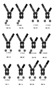
[0012] FIGs. 1A-1W depict exemplary immunomodulatory molecule structures of
the present
invention. FIG. 1A depicts an exemplary immunomodulatory structure comprising
a cytokine or
variant thereof fused to the N-terminus of a subunit of the Fc fragment of a
parental full-length
antibody. FIG. 1B depicts a dimeric. (homodimeric or heterodimeric) cytokine
or variant thereof
(e.g., IFN-y, IL-10, IL-12, or IL-23) expressed in a single chain and
positioned at the hinge region
of one heavy chain of a parental full-length antibody. FIGs. 1A-1B depict
exemplary
immunomodulatory structures of the present invention irnmunostimulatory, in
which an
immunostimulatory cytokine or variant thereof (e g., TFN-y,
IL-12, or IL-23) expressed in a
single chain and positioned at the hinge region of one heavy chain of a
dimeric parental
liganclireceptor/Fab-hinge-Fc fusion protein. FIG. IA shows that the Fab of
the dimeric parental
ligand/receptoriFab-hinge-Fc fusion protein can be an agonist. FIG. 1B shows
that the Fab of the
dimeric parental ligand/receptor/Fab-hinge-Fc fusion protein can be an agonist
or non-agonist.
FIG. 1C depicts an exemplary immunomodulatory structures of the present
invention
immunostimulatory, in which an immunostimulatory cytokine or variant thereof
(e.g., IIFN-y, IL-
3
CA 03211381 2023- 9- 8
WO 2022/192898
PCT/US2022/071077
2, 1L-12, or IL-23) expressed in a single chain and positioned at the hinge
region of one heavy
chain of a parental full-length, agonist antibody (e.g., anti- PD-1 agonist).
FIG. 1D depict
alternative exemplary immunomodulatory structures of the present invention, in
which an
immunostimulatory cytokine or variant thereof is positioned between a VH
(e.g., within a Fab of
an agonist antibody) and a subunit of an Fe fragment. FIG. 1F. depicts an
exemplary
immunomodulatory structure comprising an immunostimulatory cytokine or variant
thereof (e.g.,
]FN-7, IL-2, IL-12, or IL-23) positioned at the hinge region of one
polypeptide of a dimeric
parental ligand/receptor-hinge-Fc fusion protein. FIG. IF depicts an exemplary
immunomodulatory structure comprising an immunostimulatory cytokine or variant
thereof (e.g.,
IFN-1, IL-2, 1L-12, or IL-23) fused to the N-terminus of a subunit of the Fe
fragment of a parental
full-length agonist antibody (e.g., anti-PD agonist). FIG. 1G depicts an
exemplary
immunomodulatory structure comprising of cytokine or variant thereof (e.g.,
IFN-7, IL-2, IL- I 2,
or IL-23) positioned at the hinge region of one polypeptide of a parental
ligandheceptor-hinge-Fc
fusion protein. FIG. 1H depicts an exemplary immunomodulatory structure
comprising of
immunostimulatory cytokine or variant thereof (e.g., IFN-7, IL-2, 11,- 12, or
IL-23) positioned at
the hinge region of one polypeptide of a dimeric parental ligand/receptor-
hinge-Fc fusion protein.
FIG. 11 depicts an exemplary immunomodulatory structures of the present
invention
immunostimulatory, in which an immunostimulatory cytokine or variant thereof
(e.g., IFN-7, IL-
2, IL-12, or IL-23) positioned at the C-terminus of the Fe domain of a
parental ligand/receptor-
hinge-Fc fusion protein. FIG. 1J depicts an exemplary immunomodulatory
structure comprising
of an immunostimulatory cytokine or variant thereof (e.g., 1E1'4-7,11,-2, IL-
12, or IL-23) positioned
at the C-terminus of the Fe domain of a parental full-length, agonist antibody
(e.g., anti- PD-1
agonist). FIG. 1K depicts an exemplary immunomodulatory structure comprising
of an
immunostimulatory cytokine or variant thereof (e.g., 1FN-7, 1L-2, 1L-12, or 1L-
23) positioned at
the C-terminus of the Fe domain of a dimeric parental ligand/receptor/agonist
Fab-hinge-Fc fusion
protein. FIG. 1L depicts two cytokines or variants thereof each positioned at
the hinge region of
one polypeptide of a parental ligandheceptor-hinge-Fc fusion protein, or a
dimeric (homodimeric
or heterodimeric) cytokine or variant thereof with each subunit positioned at
the hinge region of
one polypeptide of a parental ligandlreceptor-hinge-Fc fusion protein. FIG. 1M
depicts two
cytokines or variants thereof each positioned at the hinge region of one
polypeptide of a parental
ligand/receptor-hinge-Fc fusion protein, or a dimeric (homodimeric or
heterodimeric) cytokine or
4
CA 03211581 2023- 9- 8
WO 2022/192898
PCT/US2022/071077
variant thereof with each subunit positioned at the hinge region of one
polypeptide of a dimeric
parental ligand/receptor-hinge-Fc fusion protein. FIGs. 1N-10 depicts two
cytokines or variants
thereof each positioned at the hinge region of one polypeptide of a parental
full-length, agonist
antibody, or a dimeric (homodimeric or heterodimeric) cytokine or variant
thereof with each
subunit positioned at the hinge region of one polypeptide of a parental full-
length, agonist
antibody. FIG. IN shows that the Fab is the same, wherein they are both from
an agonist antibody.
FIG. 10 shows that the Fab can be different, wherein one is the Fab of an
agonist antibody and the
other is a different Fab (can be non-agonist or agonist). FIG. I P depicts two
cytokines or variants
thereof each positioned at the C-terminus of the Fe domain of a parental
ligand/receptor-hinge-Fc
fusion protein, or a dimeric (homodimeric or heterodimeric) cytokine or
variant thereof with each
subunit positioned at the C-terminus of one polypeptide of a parental
ligandheceptor-hinge-Fc
fusion protein and another at the hinge region of one polypeptide of a
parental ligand/receptor-
hinge-Fe fusion protein. FIGs. I Q- I R depicts two cytokines or variants
thereof each positioned at
the C-terminus of one polypeptide of a parental full-length, antibody, or a
dimeric (homodimeric
or heterodimeric) cytokine or variant thereof with each subunit positioned at
the C-terminus of one
polypeptide of Fc domain of agonist antibody and another at the C-terminus of
one polypeptide of
the Fe domain. FIG. I Q shows that the Fabs can be the same, wherein they are
both from an agonist
antibody. FIG. IR shows that the Fab can be different, wherein one is the Fab
of an agonist
antibody and the other is a different Fab (can be non-agonist or agonist).
FIG. 1S depicts an
exemplary immunomodulatory structure comprising an immunostimulatory cy tokine
or variant
thereof fused to the C-terminus of a light chain constant region (CL) of a
parental full-length
agonist antibody. FIG. 1 T depicts an exemplary immunomodulatory structure
comprising an
immunostimulatory cytokine or variant thereof fused to the N-terminus of a
heavy chain variable
domain (VH) of a parental full-length antibody. FIG. 1U depicts an exemplary
immunomodulatory
structure comprising an immunostimulatory cytokine or variant thereof fused to
the N-terminus of
one polypeptide of a dimeric parental ligand/receptor-hinge-Fc fusion protein.
FIG. IV depicts an
exemplary immunomodulatory structure comprising an immunostimulatory cytokine
or variant
thereof fused to the N-terminus of one polypeptide of a parental
ligand/receptorlagonist Fab-hinge-
Fe fusion protein. FIG. 1W depicts an exemplary immunomodulatory structure
comprising an
immunostimulatory cytokine or variant thereof fused to the N-terminus of a
heavy chain variable
domain (VH) of a parental liganci/receptor/agonist Fab-hinge-Fe fusion
protein.
CA 03211581 2023- 9- 8
WO 2022/192898
PCT/US2022/071077
[00131 FIGs. 2A-2C depict tumor volume in CT26 syngeneic tumor mice treated
with IL-
12(F60A)/PD-L2-Fc, hinge (IW-#30) immunomodulatory molecule, IL-12(F60A)/PD-L2-
Fc, C-
terminus of HC (IW-#34) immunomodulatory molecule, or PBS (negative control).
Black arrows
indicate injection days. The individual mice responses in each group are given
in FIGs. 2B-2C.
[00141 FIGs. 3A-3B depict CT26 and EMT6 tumor volume growth overtime in cured
CT26 mice
(previously cured in FIGs. 2A-2C).
[00151 FIG. 4A depicts tumor volume in CT26 syngeneic tumor mice treated with
IL-
12(E59A/F60A)/PD-L2-Fc, hinge (IW-#29) immunomodulatory molecule, IL-
12(F60A)/PD-L2-
Fc, hinge (IW-#30) immunomodulatory molecule, or PBS (negative control). Black
arrows
indicate injection days. FIG. 4B depicts a series of pictures taken of one
mouse over the course of
treatment with L-12(F60A)/PD-L2-Fc, hinge (IW-#30) immunomodulatory molecule.
[00161 FIGS. 5A-5D depict tumor volume in EMT6 syngeneic tumor mice treated
with IL-
12(E59A11760A)/PD-L2-17c, hinge (IW-#29) immunomodulatory molecule, IL-
12(1760A)/PD-L2-
Fc, hinge (IW-#30) immunomodulatory molecule, IL-12(E59A/F60A)/anti-PD-1,
hinge (IW-#48)
immunomodulatory molecule, or PBS (negative control). Black arrows indicate
injection days.
The individual mice responses in each group are given in FIGs. 5B-5D.
[00171 FIGs. 6A-6C depict CT26 and EMT6 tumor volume growth overtime in cured
EMT6
mice (previously cured in FIGs. 5A-5D).
[00181 FIGs. 7A-7D depict tumor volume in 4T1 syngeneic tumor mice treated
with increasing
concentrations (0, 1, 3, 10, and 50 mg/kg) of IL-12(F60A)/PDI-2-Fc, hinge (IW-
#30)
immunomodulatory molecule, 11,12(1760A)/PD-L2-Fc, C-terminus of HC (IW-#34)
immunomodulatory molecule, IL-12(E60A)/ariti-PD-1, hinge (IW-#46)
immunomodulatory
molecule, IL-12(E59A/F60A.)/anti-PD-1, hinge (IW448) immunomodulatory
molecule, or PBS
(negative control). Black arrows indicate injection days.
[00191 FIGs. 8A-8C depict tumor volume in B16-F10 syngeneic tumor mice treated
with IL-
12(F60A)/PD-1-2-Fe, hinge (IVV-#30) immunomodulatory molecule, PD-L2-Fc/IL-
12(F60A.)
(IW-#34; C-terminal fusion) immunomodulatory molecule, or PBS (negative
control). Black
arrows indicate injection days. FIG. 8A. shows the average tumor volume of all
mouse groups,
with the average tumor size ( STD) when the first treatment was administered
shown in
6
CA 03211581 2023- 9- 8
WO 2022/192898
PCT/US2022/071077
parenthesis. FIGs. 8B-8C show tumor volumes for individual mouse receiving the
indicated 1L-12
immunomodulatory molecules.
100201 FIGs. 9A-9C depict tumor volume in LL2 syngeneic tumor mice treated
with IL-
12(F60A)/PD-L2-Fc, hinge (IW-#30) immunomodulatory molecule, IL-12(F60A)/PD-L2-
Fc, C-
terminus of HC (IW-#34) immunomodulatory molecule, or PBS (negative control).
Black arrows
indicate injection days. The individual mice responses in each group are given
in FIGs. 9B-9C.
100211 FIG. 10 shows two approaches for activating the immune system against a
disease (e.g.,
cancer). The left panel shows a target-independent activation mechanism
("trans-activation"),
wherein an immunomodulatory molecule can bind to a target antigen on an immune
cell (e.g., a T
cell), and can bind to a target antigen on a target cell (e.g., tumor cell),
thereby bringing the immune
cell into proximity with the target cell for therapeutic effect. This
approach, however, can be
associated with systemic toxicities, as the binding domain (e.g., wildtype
immunostimunlatory
cytokine, such as IL-12 or IL-2) targeting immune cells can stimulate immune
response even in
the absence of target cells. The right panel shows target cell-antigen (e.g.,
tumor antigen)
independent activation mechanism ("cis-activation"), in which the
immunomodulatory molecules
can both up-regulate and down-regulate immune responses, which more closely
mimics the natural
regulation and balance of the immune system. In a further aspect,
immunomodulatory molecules
in both left and right panels can further have a "restricted activation
mechanism", in which the first
binding domain upon binding to an immune cell upregulating an immune response
(e.g.,
immunostimunlatory cytokine, such as IL-12 or H.,-2) is modified to reduce
activity (binding
and/or biological activity), and/or in a "masked" configuration (e.g.,
positioned at hinge region)
until binding of the second binding domain to the second target antigen (e.g.,
tumor antigen, or
immune cell surface molecule) occurs. Exemplary immunomodulatory molecules of
the present
invention can function via restricted activation, cis-activation, trans-
activation, or all mechanisms.
[0022] Wis. Ii A-I IL depict exemplary multispecific immunomodulatoiy
molecules of the
present invention. The immunomodulatory molecules may comprise a variety of
combinations of
binding domain types: i) a first binding domain, labeled as "1" in the
figures, which upon binding
to a first target molecule up-regulates an immune response; ii) a second
binding domain, labeled
as "2" in the figures, which upon binding to a second target molecule down-
regulates the immune
response; and id) optionally, a third binding domain, labeled as "3" in the
figures, which helps
7
CA 03211581 2023- 9- 8
WO 2022/192898
PCT/US2022/071077
localize the immunomodulatory molecule to a target site (e.g., the tumor
microenvironment) by
targeting a third target molecule (e.g., marker of exhausted T-cells, T cell
surface marker, or tumor
antigens). The immunomodulatory molecules can comprise one or more of any of
first, second,
and/or third binding domain. The multiple first binding domains can be the
same or different from
each other. The multiple second binding domains can be the same or different
from each other.
The multiple third binding domains can be the same or different from each
other. The various
binding domains within the immunomodulatory- molecules can be constructed in
various
configurations, not limited to those shown in FIGs. ii A-1 IL. As exemplified
in FIGs. 11 A-1 IL,
Ithe IL-12 moiety (such as a mutant IL-12 moiety with reduced IL-12 activity;
either constructed
as a single chain fusion, or as two separate subunits) positioned at the C' of
one or both Fe subunits
can be a type of first binding domain which upon binding to 1L-12R on immune
cells up-regulates
an immune response. Hence, in FIGs. 11G, 111, and I I J, the I1-12 moiety
functions as "the first
binding domain". The first binding domain (e.g., immunostimulatory cytokine
moiety or variant
thereof) can be placed at the hinge region between an Fe subunit and the
second binding domain
or the third binding domain, such as exemplary configurations shown in FIGs.
11A-11F, I 1H,
11K, and I I L. Such "restricted access" configurations for first binding
domain to its first target
molecule can allow i) reduced, minimal or no binding/activity between the
first binding domain to
its first target molecule in the absence of the binding between the second
binding domain to the
second target molecule and/or the binding between the third binding domain to
the third target
molecule (whichever domain that is at N' of the first binding domain); and ii)
rescued/recovered
binding/activity of the first binding domain in the presence of the binding
between the second
binding domain to the second target molecule and/or the binding between the
third binding domain
to the third target molecule (whichever domain that is at N' of the first
binding domain). The first
binding domain (e.g., immunostimulatory cytokine moiety or variant thereof)
can also be placed
at C' of one or both Fe subunits of an Fe-fusion protein, such as the 1L-12
moiety (either
constructed as a single chain fusion and fused to one Fe subunit, or as two
separate subunits each
fused to one Fc subunit of an Fe domain) exemplified in FIGs. 11A-11L. Such
configurations do
not or barely restrict binding/activity of the first binding domain.
[00231 FIGs. 12A-12D depict exemplary immunomodulatory molecules with a first
binding
domain (e.g., immunostimulatory cytokines such as 1L-12 or variant thereof,
for example
constructed as a single chain fusion) positioned at the hinge region of one
polypeptide chain of a
8
CA 03211581 2023- 9- 8
WO 2022/192898
PCT/US2022/071077
parental (ligand/receptor/antigen binding domain)-hinge-Fc fusion protein,
which can be
homodimeric or heterodimeric. FIG. 12A depicts an exemplary immunomodulatory
molecule,
wherein PD-L1 or PD-L2 extracellular domain (wildtype or mutant) is fused to N-
terminus of an
Fc domain via hinge, and an immunostimulatory cytokine moiety (e.g., 1L-12 or
variant
constructed as a single chain fusion) is positioned at the hinge region of one
of the (PD-L1 or PD-
L2)-hinge-Fc poly-peptide chains. Can be referred to as IL-12/PD-L1-Fc or IL-
12/PD-L2-Fc. FIG.
12B depicts an exemplary immunomodulatory molecule wherein PD-Li or PD-L2
extracellular
domain (wildtype or mutant) is fused to the N-terminus of a first Fc subunit
via a first hinge, a
CD155 extracellular domain (wildtype or mutant) is fused to the N-terminus of
a second Fe subunit
via a second hinge, and an immunostimulatory cytokine moiety (e.g., IL-12 or
variant constructed
as a single chain fusion) is positioned at the hinge region of one of the
pairing polypeptide chains
(such as the PD-L1 /PD-L2-hinge-Fc chain). Can be referred to as IL-1 2/PD-L 1-
Fc/CD155-Fc or
IL-12/PD-L2-17c/CD I 55-Fe. FIG. 12C depicts an exemplary immunomodulatory
molecule,
wherein PD-Li or PD-L2 extracellular domain (wildtype or mutant) is fused to
the N-terminus of
a first Fc subunit via a first hinge, an antibody moiety (e.g., sdAb or scFv)
specifically recognizing
a target molecule (can be agonist, antagonist, or neutral Ab, regulating or
not-regulating immune
response) is fused to the N-terminus of a second Fc subunit via a second
hinge, and an
immunostimulatory cytokine moiety (e.g., IL-12 or variant constructed as a
single chain fusion) is
positioned at the hinge region of one of the pairing polypeptide chains (such
as the PD-LI/PD-L2-
hinge-Fe chain). Can be referred to as sdAb/IL-12/PD-LI-Fc or sdA.b/IL-12/PD-
L2-Fc. FIG. 12D
depicts an exemplary immunomodulatory molecule, wherein PD-Li or PD-L2
extracellular
domain (wildtype or mutant) is fused to the N-terminus of a first Fc subunit
via a first hinge, a Fab
specifically recognizing a target molecule (can be agonist, antagonist, or
neutral Ab, regulating or
not-regulating immune response) is fused through its CHI to the N-terminus of
a second Fc subunit
via a second hinge, and an immunostimulatory cytokine moiety (e.g., IL-12 or
variant constructed
as a single chain fusion) is positioned at the hinge region of one of the
pairing polypeptide chains
(such as the PD-Ll/PD-L2-hinge-Fc chain). Can be referred to as Fab/IL-12/PD-
Ll-Fc or Fab/IL-
1 2/1'D-L2-Fc.
(00241 FIGs. 13A-13D depict exemplary immunomodulatory molecules with a first
binding
domain (e.g., immunostimulatory cytokines such as 1L-12 or variant thereof,
for example
constructed as a single chain fusion) positioned at the C-terminus of the Fc
domain (one or both
9
CA 03211581 2023- 9- 8
WO 2022/192898
PCT/US2022/071077
Fc subunits) of a parental (ligand/receptor/antigen binding domain)-hinge-Fc
fusion protein, which
can be homodimeric or heterodimeric. FIG. 13A depicts an exemplary
immunomodulatory
molecule wherein PD-Ll or PD-L2 extracellular domain (wildtype or mutant) is
fused to N-
terminus of an Fc domain via an optional hinge, and an immunostimulatory
cytokine moiety (e.g.,
IL-12 or variant constructed as a single chain fusion) is positioned at the C-
terminus one or both
subunits of the Fc domain. Can be referred to as PD-L1-Fc/IL-12 or PD-L2-Fc/IL-
12. FIG. 13B
depicts an exemplary immunomodulatory molecule wherein PD-L1 or PD-L2
extracellular domain
(wildtype or mutant) is fused to the N-terminus of a first Fc subunit via a
first optional hinge, a
CD155 extracellular domain (wildtype or mutant) is fused to the N-terminus of
a second Fe subunit
via a second optional hinge, and an immunostimulatory cytokine moiety (e.g.,
IL-12 or variant
constructed as a single chain fusion) is positioned at the C-terminus one or
both subunits of the Fc
domain (such as C' of the PD-LI/PD-L2-hinge-Fc chain). Can be referred to as
PD-LI-Fc/CD155-
Feat,- I 2 or PD-L2-Fc/CD155-Fal, I 2.. FIG. I3C depicts an exemplary
immunomodulatory
molecule, wherein PD-Li or PD-L2 extracellular domain (wildtype or mutant) is
fused to the N-
terminus of a first Fc subunit via a first optional hinge, an antibody moiety
(e.g., sdAb or soFv)
specifically recognizing a target molecule (can be agonist, antagonist, or
neutral Ab, regulating or
not-regulating immune response) is fused to the N-terminus of a second Fc
subunit via a second
optional binge, and an immunostimulatory cytokine moiety (e.g., IL-12 or
variant constructed as
a single chain fusion) is positioned at the C-terminus one or both subunits of
the Fc domain (such
as C' of the PD-Ll /PD-L2-hinge-Fc chain). Can be referred to as sdAb/PD-L1 -
Fe/IL-12 or
sdAb/PD-L2-Fc/IL-12.. FIG. 13D depicts an exemplary multi-target
immunomodulatory molecule
wherein PD-L1 or PD-L2 extracellular domain (wildtype or mutant) is fused to
the N-terminus of
a first Fe subunit via a first optional hinge, a Fab specifically recognizing
a target molecule (can
be agonist, antagonist, or neutral Ab, regulating or not-regulating immune
response) is fused
through its CHI to the N-terminus of a second Fc subunit via a second optional
hinge, and an
immunostimulatory cytokine moiety (e.g., 1L-12 or variant constructed as a
single chain fusion) is
positioned at the C-terminus one or both subunits of the Fc domain (such as C'
of the PD-Li/I'D-
L2-hinge-Fc chain). Can be referred to as Fab/PD-L1-Fc/1L-12 or Fab/PD-L2-
Fc/IL-12.
[00251 FIGs. 14A-14D depict exemplary immunomodulatory molecules with two
first binding
domains (e.g., immunostimulatory cytokines such as 1L-12, 1L-2 or variant
thereof, for example
constructed as a single chain fusion) each positioned at the hinge region of
one polypeptide chain
CA 03211581 2023- 9- 8
WO 2022/192898
PCT/US2022/071077
of a parental (ligand/receptorlantigen binding domain)-hinge-Fc fusion
protein, which can be
homodimeric or heterodimeric. FIG. 14A depicts an exemplary immunomodulatory
molecule,
wherein PD-L1 or PD-L2 extracellular domain (wildtype or mutant) is fused to N-
terminus of an
Fc domain via hinge, a first immunostimulatory cytokine moiety (e.g., 1L-12 or
variant constructed
as a single chain fusion) is positioned at the hinge region of one of the (PD-
Ll or PD-L2)-hinge-
Fe polypeptide chains, and a second immunostimulatory cytokine moiety (e.g.,
IL-2 or variant
thereof) is positioned at the hinge region of the other chain of the (PD-L1 or
PD-L2)-hinge-Fc
polypeptide chains. Can be referred to as IL-12/EL-2/PD-1,1-Fc or IL-12/11L-
2/PD-L2-Fc.. FIG.
14B depicts an exemplary immunomodulatory molecule, wherein PD-Ll or PD-L2
extracellular
domain (wildtype or mutant) is fused to the N-terminus of a first Fc subunit
via a first hinge, a
CD155 extracellular domain (wildtype or mutant) is fused to the N-terminus of
a second Fe subunit
via a second hinge, a first immunostimulatory cytokine moiety (e.g., 11,-12 or
variant constructed
as a single chain fusion) is positioned at the hinge region of one of the
pairing polypeptide chains
(such as the PD-Li/PD-L2-hinge-Fc chain), and a second immunostimulatory
cytokine moiety
(e.g., IL-2 or variant thereof) is positioned at the hinge region of the other
chain of the pairing
polypeptide chains (such as the CD155-hinge-Fc chain). Can be referred to as
11,-12/11,-2/PD-L1-
Fe/CM 55-Fe or IL-.12/11L-2/PD-L2-Fc/Cal 55-Fe.. FIG. 14C depicts an exemplary
immunomodulatory molecule, wherein PD-L1 or PD-L2 extracellular domain
(wildtype or
mutant) is fused to the N-terminus of a first Fc subunit via a first hinge, an
antibody moiety (e.g.,
sdAb or scFv) specifically recognizing a target molecule (can be agonist,
antagonist, or neutral
Ab, regulating or not-regulating immune response) is fused to the N-terminus
of a second Fc
subunit via a second hinge, a first immunostimulatory cytokine moiety (e.g.,
IL-12 or variant
constructed as a single chain fusion) is positioned at the hinge region of one
of the pairing
polypeptide chains (such as the PD-Ll/PD-L2-hinge-Fc chain), and a second
immunostimulatory
cytokine moiety (e.g., 1L-2 or variant thereof) is positioned at the hinge
region of the other chain
of the pairing polypeptide chains (such as the CD155-hinge-Fc chain). Can be
referred to as
sdAb/IL-12/IL-2/PD-L1-Fc or sdAbilL-12/IL-2/PD-L2-Fc. FIG. 14D depicts an
exemplary
immunomodulatory molecule, wherein PD-Ll or PD-L2 lextracellular domain
(wildtype or
mutant) is fused to the N-terminus of a first Fc subunit via a first hinge, a
Fab specifically
recognizing a target molecule (can be agonist, antagonist, or neutral Ab,
regulating or not-
regulating immune response) is fused through its CH1 to the N-terminus of a
second Fc subunit
11
CA 03211581 2023- 9- 8
WO 2022/192898
PCT/US2022/071077
via a second hinge, a first immunostimulatory cytokine moiety (e.g., IL-12 or
variant constructed
as a single chain fusion) is positioned at the hinge region of one of the
pairing polypeptide chains
(such as the PD-L1/PD-L2-hinge-Fc chain), and a second immunostimulatory
cytokine moiety
(e.g., IL-2 or variant thereof) is positioned at the hinge region of the other
chain of the pairing
polypeptide chains (such as the CD155-hinge-Fc chain). Can be referred to as
Fab/11,12/1L-2/PD-
Ll-Fc or Fab/IL-1211L-2/PD-L2-Fc.
10026] FIGs. 15A-15D depict exemplary immunomodulatory molecules with two
first binding
domains (e.g., immunostimulatory cytokines such as IL-12, IL-2 or variant
thereof, for example
constructed as a single chain fusion), one is positioned at the hinge region
of one polypeptide chain
of a parental (ligand/receptor/antigen binding domain)-hinge-Fc fusion
protein, and the other one
is positioned at the C-terminus of one or both Fc subunits of the parental
(ligand/receptor/antigen
binding domain)-hinge-Fc fusion protein. FIG. 15A depicts an exemplary
immunomodulatory
molecule, wherein PD-L1 or PD-L2 extracellular domain (wildtype or mutant) is
fused to N-
terminus of an Fc domain via hinge, a first immunostimulatory cytokine moiety
(e.g., 1L-2 or
variant) is positioned at the hinge region of one of the (PD-L1 or PD-L2)-
hinge-Fc polypeptide
chains, and a second immunostimulatory cytokine moiety (e.g., IL-12 or variant
constructed as a
single chain fusion) is positioned at the C' of Fc subunit of the other chain
of the (PD-Li or PD-
L2)-hinge-Fc polypeptide chains. Can be referred to as IL-2/PD-LI-Fc/IL-12 or
IL-2/PD-L2-
Fc/IL-12. FIG. 15B depicts an exemplary immunomodulatory molecule, wherein PD-
L1 or PD-
L2 extracellular domain (wildtype or mutant) is fused to the N-terminus of a
first Fc subunit via
a first hinge, a CD155 extracellular domain (wildtype or mutant) is fused to
the N-terminus of a
second Fc subunit via a second hinge, a first immunostimulatory cytokine
moiety (e.g., IL-12 or
variant constructed as a single chain fusion) is positioned at the C of Fc
subunit of one of the
pairing polypeptide chains (such as the PD-L1/PD-L2-hinge-Fc chain), and a
second
immunostimulatory cytokine moiety (e.g., IL-2 or variant thereof) is
positioned at the hinge region
of the other chain of the pairing polypeptide chains (such as the CD155-hinge-
Fc chain). Can be
referred to as EL-2/1)13-L1-Fc/CD155-Fc/IL-12 or IL-2/PD-L2-Fc/C7D155-Fc/IL-
12. FIG. 15C
depicts an exemplary immunomodulatory molecule, wherein PD-L1 or PD-L2
extracellular
domain (wildtype or mutant) is fused to the N-terminus of a first Fe subunit
via a first hinge, an
antibody moiety (e.g., sdAb or scFv) specifically recognizing a target
molecule (can be agonist,
antagonist, or neutral Ab, regulating or not-regulating immune response) is
fused to the N-terminus
12
CA 03211581 2023- 9- 8
WO 2022/192898
PCT/US2022/071077
of a second Fc subunit via a second hinge, a first immunostimulatory cytokine
moiety (e.g., IL-12
or variant constructed as a single chain fusion) is positioned at the C' of Fc
subunit of one of the
pairing polypeptide chains (such as the PD-Ll/PD-L2-hinge-Fc chain), and a
second
immunostimulatory cytokine moiety (e.g., 1L-2 or variant thereof) is
positioned at the hinge region
of the other chain of the pairing polypeptide chains (such as the CD155-hinge-
Fe chain). Can be
referred to as sdAb/IL-2/PD-L1-Fc/IL-12 or sdAML-2/PD-L2-Fc/IL-12. FIG. 15D
depicts an
exemplary immunomodulatory molecule, wherein PD-L1 or PD-L2 (wildtype or
mutant) is fused
to the N-terminus of a first Fc subunit via a first hinge, a Fab specifically
recognizing a target
molecule (can be agonist, antagonist, or neutral Ab, regulating or not-
regulating immune response)
is fused through its CH1 to the N-terminus of a second Fc subunit via a second
hinge, a first
immunostimulatory cytokine moiety (e.g., IL-12 or variant constructed as a
single chain fusion) is
positioned at the C' of Fe subunit of one of the pairing polypeptide chains
(such as the PD-LI/PD-
I.2-hinge-Fc chain), and a second immunostimulatory cytokine moiety (e.g., IL-
2 or variant
thereof) is positioned at the hinge region of the other chain of the pairing
polypeptide chains (such
as the CD155-hinge-Fc chain). Can be referred to as Fab/IL-2/PD-Li-Fc/IL-12 or
Fab/IL-2/PD-
L2-Fc/II_,-12.
[00271 FIG. 16 shows 4T1 murine breast cancer tumors extracted from mammary
gland fat pad
of mice treated with IL-12(E59A/F60A)/PD-L2-Fc (rw-#29), IL-12(F60A)/PD-L2-Fc
(FW-#30),
a combination of anti-PD-1 and anti-CTLA-4 antibodies, or PBS (negative
control).
[00281 FIG. 17 depicts 4T1 rnurine breast cancer cells metastasized to lungs
in mice injected
with 4T1 cells at mammary gland fat pad and treated with IL-12(E59A/F60A)/PD-
L2-Fc (IW-
429), IL-12(F60A)/PD-L2-Fc (1W-#30), a combination of anti-PD-1 and an ti-CTLA-
4 antibodies,
or PBS (negative control).
[00291 FIG. 18 depicts tumor volume in 4T1 syngeneic tumor mice treated with
IL-
1. 2(E59A/F60 A)/anti-PD-1 (IW-#48), IL-12(E59A/F60A)/PD-L2-Fc (IW-
#29),
IL-
1
(IW-#54) immunomodulatory molecules, or
PBS (negative control). Black arrows indicate injection days.
[00301 FIG. 19 depicts tumor volume in EMT6 syngeneic tumor mice treated with
IL-
1. 2(E59A/F60A)/anti-PD-1 (IW-#48), IL-12(E59A1F60A)/PD-L2-Fc (IW-
#29), IL-
13
CA 03211581 2023- 9- 8
WO 2022/192898
PCT/US2022/071077
2(R38D/K43E/E61R)/PD-L2-Fc (IW-#11) immunomodulatory molecules, or PBS
(negative
control). Black arrows indicate injection days.
DETAILED DESCRIPTION OF THE INVENTION
[00311 Current immunotherapy often triggers too much undesired immune response
such as
immune cell over-activation, cytokine storm, etc. For example, cytokine
therapy (e.g., for treating
cancer) have shown limited success due to severe toxicity, which limits the
dosing far below
therapeutically effective dose. Immunocytokines, which are constructs with
cytokines fused to
antibodies, antigen-binding fragments, ligand-Fc fusion protein, or receptor-
Fe fusion protein
(hereinafter collectively referred to as "ligand/receptor-Fc fusion protein"
or "ligand/receptor-
h i nge-Fc fusion protein") can deliver cytokines to target cells (e.g., tumor
cells, or immune effector
cells) or tissues with the recognition of target antigens by the antibodies or
antigen-binding
fragments (e.g., antibody fragments, ligands, or receptors) within
immunomodulatory molecules,
which can both reduce non-specific (off-target) cytokine activities and/or
associated toxicities
(e.g., toxicities on healthy cells or tissues), and concentrate cytokine
therapeutic effects at target
sites (e.g., disease sites). The activation of immunomodulatory molecules can
occur via trans-
activation, which requires specific binding of the antibody or antigen-binding
fragment to target
antigens on tumor cells; or cis-activation, which requires specific binding of
the antibody or
antigen-binding fragment to target antigens on immune cells (see FIG. 10).
Most immunocytokines
developed nowadays have the cytokine moiety fused to the N-terminus or the C-
terminus of the
heavy chain or the light chain of a full-length antibody (such as Hu14.8-1L2,
NHS-IL2LT, NHS-
IL12, BC1-IL12; see, e.g., FIGs. 1C-1E) or fused to the N-terminus or the C-
terminus of an
antigen-binding fragment (e.g., diabody, scFv, such as L19-IL2 or F16-IL2), so
cytokine-receptor
binding/activation can still occur even in the absence of antibody-antigen
recognition, leading to
off-target toxicities. Immune checkpoint inhibitors developed in recent years
(e.g., anti-PD-1, anti-
CTLA-4 Abs), although have shown some great clinical success in cancer
patients, also focused
on up-regulating immune response, which can worsen systemic toxicity if
further used together
with pro-inflammatory cytokines.
[0032j The present invention provides iminunomodulatory molecules with
opposing effects in
regulating immune responses, demonstrated significantly better toxicity
profile and therapeutic
efficacy. The immunomodulatory molecules comprise a first binding domain
(e.g.,
14
CA 03211581 2023- 9- 8
WO 2022/192898
PCT/US2022/071077
immunostimulatory cytokine or variant thereof, such as IL-12, 1L-2, IFN-y)
specifically
recognizing a first target molecule (e.g., receptor of immunostimulatory
cytokine or variant
thereof) and a second binding domain (e.g., ligand such as PD-L1, PD-L2, CD155
extracellular
domain or variant thereof) specifically recognizing a second target molecule
(e.g., PD-1 or TIGIT
on immune effector cell), wherein the first binding domain upon binding to the
first target molecule
up-regulates an immune response, and wherein the second binding domain upon
binding to the
second target molecule down-regulates the inamune response. For example, when
positioning an
IL-12 cytokine (pro-inflammatory) at the hinge region of a PD-L2 extracellular
domain-hinge-Fc
fusion protein, the resulting IL-12/PD-L2-Fc immunomodulatory molecule not
only specifically
targeted IL-12 activity (e.g., activity of binding to IL-12 receptor, and/or
IL-12 pro-inflammatory
activity) to PD-1+ target cells, but also stimulated PD-1 inhibitory immune
checkpoint signaling
via PD-L2-PD-1 binding, thus creating an immunosuppression signal that
"balances against" or
"counteracts" the immunostimulating activity of IL-12. Any agonist antibodies
or ligands (e.g.,
PD-L2, PD-L1, CD80, or CD86) that can activate or stimulate an
immunosuppressive signaling
pathway (e.g., by binding to an inhibitory immune checkpoint molecule such as
PD-1 or CTLA-
4), or any antagonist antibodies, ligands, or receptors that can reduce or
block an
immunostimulatory signaling pathway (e.g., by binding to a stimulatory immune
checkpoint
molecule such as CD27 or CD28 or an immunostimulatory receptor such as IL-2R)
can be used in
combination with an immunostimulating cytokine or variant thereof (e.g., IL-2,
IL-12, IFN-y, or
IL-23) to construct an immunomodulatory molecule with any of the
immunomodulatory molecule
configurations described herein. Any antagonist antibodies, ligands, or
receptors that can reduce
or block an immunosuppressive signaling pathway (e.g., by binding to an
inhibitory immune
checkpoint molecule such as PD-1 or CTLA-4), or any agonist antibodies or
ligands (e.g., CD70,
CD80, CD86, or IL-2) that can activate or stimulate an immunostimulatory
signaling pathway
(e.g., by binding to a stimulatory immune checkpoint molecule such as CD27 or
CD28 or an
immunostimulatory receptor such as IL-2R) can be used in combination with an
immunosuppressive cytokine or variant thereof (e.g., 1L-10, IL-27, 1L-35, TGF-
13) to construct an
immunomodulatory molecule with any of the immunomodulatory molecule
configurations
described herein. The immunomodulatory molecules described herein can comprise
one or more
of first binding domains, and/or one or more of second binding domains, in
order to achieve
multiple immune response regulation. The multiple first binding domains can be
the same or
CA 03211581 2023- 9- 8
WO 2022/192898
PCT/US2022/071077
different. The multiple second binding domains can be the same or different.
See FIGs. 1A-1W
and 11 A-15D for examples.
100331 The first binding domain can include molecules such as
immunostimulatory cytokines,
ligands, or agonist antibodies (e.g., ligand or agonist Ab that stimulate
stimulatory checkpoint
molecules such as 0X40), that target immune cells such as T cells, NK cells,
DC cells,
macrophages, and B cells. The present invention in some embodiments provide
first binding
domains with reduced activities (e.g., reduced binding or redurine stimulating
activity to its target),
such as compared to unmodified parental first binding domain. For example, see
cytokine variants
described herein, which exhibit drastically reduced activity compared to
wildtype cytokines.
Reducing the binding affinity of the first binding domain can skew the
mechanism of action
towards target-dependent activation (cis-activation) and away from target-
independent activation
(trans-activation).
10034) The second binding domain can include molecules such as
immunosuppressive
cytokines, ligands, or agonist antibodies (e.g., ligand (such as PD-L1, PD-L2,
CD155) or agonist
Ab that stimulate inhibitory checkpoint molecules such as PD-1 or TIGIT), for
down-regulating
immune response. The present invention in some embodiments provide anti-PD-1
antibody
(antagonist Ab) with reduced binding affinity to PD-1, hence reducing the
immune response that
could have been induced by a wild-type anti-PD-1 antibody (antagonist Ab, such
as Ili volumab)
(see Example 22). The present invention in some embodiments also provide
ligands with increased
binding affinity to inhibitory checkpoint molecules such as PD-1, which can
further down-regulate
immune response compared to wildtype ligands. For example, see mutant PD-LI
and PD-L2
molecules generated in Example 23. Immunomodulatory molecules comprising
mutant PD-Ll or
PD-L2 extracellular domain as the second binding domain reduced adverse events
compared to
those with wildtype I or PD-L2 extracellular domain. The low-
binding affinity of PD-
L2(inut) or PD-L1(mut) to PD-1 (more than 104 M Ka) compared to wildtype
ligand, or the low-
binding affinity of the mutant anti-PD-1 antibody (antagonist Ab; more than 10-
8 M Ka) compared
to wildtype anti-PD-1 antibody (less than 10-9M IQ), allow immunomodulatory
molecules thereof
to target cancer cells expressing much higher level of PD-1, such as exhausted
T-cells and tumor
microenvironments trying the bypass anti-tumor activity, rather than any PD-1
positive cells.
16
CA 03211581 2023- 9- 8
WO 2022/192898
PCT/US2022/071077
[0035] For example, IL-12(E59A/F60A)/PD-L2(S58V)-Fc immunomodulatory molecule
described herein provides both positive (1L-12/IL-12R signaling) and negative
signals (PD-1/PD-
L2 signaling). Immunomodulatory molecules with opposing effects described
herein allow
mimicking the native T-cell activation process, regulating the T cell
activation process, and
overcoming over-activation of the immune system.
[00361 The immunomodulatory molecules comprising the first and second binding
domains
described herein can further comprise a third binding domain specifically
recognizing a third target
molecule. The third binding domain can help localize the immunomodulatory
molecule to a target
site (e.g., the tumor microenvironment) by binding to the third target
molecule (e.g., marker of
exhausted T-cells, T cell surface marker, or tumor antigens). The third
binding domain upon
binding to the third target molecule can i) up-regulate the above mentioned or
other immune
response, or ii) down-regulate the above mentioned or other immune response;
or iii) does not
regulate any immune response by its own binding. For example, the third
binding domain can
function solely as a tumor antigen-targeting domain to bring the
immunomodulatory molecule to
tumor site, or as an immune effector cell-targeting domain to bring the
immunomodulatory
molecule to immune effector cells or strengthen its binding to immune effector
cells. The
intratumoral microenvironment contains a relatively high level of the
exhausted T cells expressing
several markers, such as TIGIT, TIM3, LAG3, and PD-1. Since the expression
pattern. and level
of exhausted markers in the tumor microenvironment (T.ME) vary greatly, the
third binding
domain can be used to target additional exhausted markers to broadly target
the TME.
Alternatively, the third binding domain can be used to target specific cancers
against specific tumor
antigen, including but not limited to Her2, CEACAM, Her3, EGFR, Trop2,
CLDN18.2, prostate-
specific antigen, MIX], EpCAM, GPC3, mesothelin (MSLN), Nectin4, Folate
receptor alpha,
tissue factor, etc. The third binding domain may also target T cell markers,
including but not
limited to CD4, CD8, CD3, CD2, CD5, CD7, CD4OL, CD25, CD137, CD69, CTLA.4,
CD127,
1COS, etc. The third binding domain may also target dendritic cell markers,
including but not
limited to CD1c, CD11 c, CD141, CD123, BDCA-2, BDCA-4, CLEC9A, XCI'121, CD80,
CD86,
PD-L1, PD-L2, etc. The third binding domain may also target
monocyte/macrophage markers,
including but not limited to CSF1R, CD80, Cd86, CD11, CD14, CD68, CD163, CD16,
CD32,
CD64, etc. The third binding domain may also target neutrophil cell markers,
including but not
limited to CD11, CD16, CD32, etc. The immunomodulatory molecules described
herein can
17
CA 03211581 2023- 9- 8
WO 2022/192898
PCT/US2022/071077
comprise one or more of third binding domains, in order to achieve multiple
immune response
regulation or for enhanced targeting. The multiple third binding domains can
be the same or
different.
[00371 Further, the present invention also provides immunomodulatory molecules
with certain
unique configurations that address the issues faced by current
cytokine/immunocytokine therapy.
Particularly, some immunomodulatory molecules of the present invention
decrease non-specific
activities (i.e., antibody or antigen-binding fragment-independent binding)
and increase specific
activities (i.e., antibody or antigen-binding fragment-dependent binding) of a
first binding domain
(e.g., immunostimulatory cytokines) by positioning the first binding domain
(e.g., cytokine or
variant thereof) at a hinge region in between a second binding domain (e.g.,
ligand, receptor, VH1-1,
scFv, or Fab) and an Fe domain subunit or portion thereof (e.g., CH2-CH3
fragment, or CH2 only,
or CH3 only), for example, at a hinge region in between an say and an Fe
domain subunit (e.g.,
an antigen-binding polypeptide comprising VH-VI..-cytokine-Fe subunit, or VL-
VH-cytokine-Fc
subunit), at a hinge region in between the Fab and the Fe domain of a full-
length antibody (e.g.,
an antigen-binding polypeptide comprising VH-CHI -cytokine-Fc subunit), or at
a hinge region in
between a ligand (or a receptor) and an Fe domain subunit (e.g., an antigen-
binding polypeptide
comprising ligand-cytokine-Fe subunit, or receptor-cytokine-Fe subunit).
Without being bound by
theory, it is believed that steric hindrance of the second binding domain
(e.g., ligand, receptor,
scFv, :Fab) and the Fe domain or portion thereof reduces accessibility of the
first binding
domain (e.g., immunomodulatory cytokine or variant thereof) to its target
molecule (e.g., receptor
of immunomodulatory cytokine), or "masks- the first binding domain from
binding to its first
target molecule, in the absence of binding by the second binding domain to the
second target
molecule. Upon binding of the second binding domain to the second target
molecule, on the other
hand, the first binding domain becomes activated. Surprisingly, unlike other
immunocytokine
designs which "expose" the cytokine moiety at its N-terminus or C-terminus,
the unique
immunomodulatory molecule configuration of the present invention requires
binding of the second
binding domain (e.g., ligand, receptor, VHH, scFv, or Fab) to its second
target molecule first
before binding of the first binding domain (e.g., immunomodulatory cytokine
moiety) to its first
target molecule (e.g., receptor) can occur, thus ensuring that the up-
regulation of the immune
response (e.g., cytokine signaling activation) is entirely second binding
domain-binding dependent
(on-target). With this enhanced targeting specificity design, and optionally
further in combination
18
CA 03211581 2023- 9- 8
WO 2022/192898
PCT/US2022/071077
with reduced activities of the first binding domain discussed above (e.g.,
cytokine variants
described herein), a desired immune response (e.g., cytokine signaling
activation) can be safely
delivered to target sites (e.g., tumor cells, or immune cells) to achieve
therapeutic effects. Such
unique targeting specificity design adds an additional regulatory layer to the
current "balancing"
or "counteracting" of immune response design, further fine-tuning the
bioactivity and toxicity of
immunomodulatory molecules described herein.
100381 Accordingly, one aspect of the present application provides an
immunomodulatory
molecule comprising a first binding domain (e.g., ligand, VIiH, scFv, or VH,
for example
immunostimulatory cytokine such as IL-2 or IL-12) specifically recognizing a
first target molecule
(e.g., cell surface antigen or receptor, such as receptor of immunostimulatory
cytokine) and a
second binding domain (e.g., ligand, V.HH, scFv, or VH, for example agonist
ligand such as PD-
L1 or PD-L2, or agonist antigen-binding fragment such as anti-PD-1 agonist
Fab, scFv, VH, VHEI,
or full-length antibody) specifically recognizing a second target molecule
(e.g., cell surface antigen
or receptor, for example inhibitory checkpoint molecule such as PD-1), wherein
the first binding
domain upon binding to the first target molecule up-regulates an immune
response, and wherein
the second binding domain upon binding to the second target molecule down-
regulates the immune
response.
[00391 Also provided are isolated nucleic acids encoding such
iminunomodulatory molecules,
vectors comprising such nucleic acids, host cells comprising such nucleic
acids or vectors, methods
of producing such immunomodulatory molecules, pharmaceutical compositions and
articles of
manufacture comprising such immunomodulatory molecules, methods of modulating
an immune
response with such immunomodulatory molecules or pharmaceutical compositions
thereof, and
methods of treating diseases (e.g., cancer, viral infection, autoimmune
diseases) with such
immunomodulatory molecules or pharmaceutical compositions thereof.
I. Definitions
[00401 The practice of the present invention will employ, unless indicated
specifically to the
contrary, conventional methods of virology, immunology, microbiology,
molecular biology and
recombinant DNA techniques within the skill of the art, many of which are
described below for
the purpose of illustration. Such techniques are explained fully in the
literature. See, e.g., Current
Protocols in Molecular Biology or Current Protocols in Immunology, John Wiley
& Sons, New
19
CA 03211581 2023- 9- 8
WO 2022/192898
PCT/US2022/071077
York, N.Y. (2009); Ausubel etal., Short Protocols in Molecular Biology, 3rd
ed., John Wiley &
Sons, 1995; Sambrook and Russell, Molecular Cloning: A Laboratory Manual (3rd
Edition, 2001);
Maniatis et at, Molecular Cloning: A Laboratory Manual (1982); DNA Cloning: A
Practical
Approach, vol. 1&11 D. Glover, ed.); Oligonucleotide Synthesis (N. Gait, ed.,
1984); Nucleic Acid
Hybridization (B. Names & S. Higgins, eds., 1985); Transcription and
Translation (B. Harries &
S. Higgins, eds., 1984); Animal Cell Culture (R Freshney, ed., 1986); Perbal,
A Practical Guide
to Molecular Cloning (1984) and other like references.
100411 The term "immunocytokine", as used herein refers to an antigen-binding
protein (e.g.,
antibody, or antigen-binding fragment (e.g., ligand, receptor, or antibody
fragment)) format, which
is fused to a cytokine molecule. The antigen-binding protein (e.g., antibody,
or antigen-binding
fragment (e.g., ligand, receptor, or antibody fragment)) format may be any of
those described
herein, and the cytokine may be fused directly, or by means of a linker or
chemical conjugation to
the antigen-binding protein format.
100421 The term "cytokine storm," also known as a "cytokine cascade" or
"hypercytokinemia,"
is a potentially fatal immune reaction typically consisting of a positive
feedback loop between
cytokines and immune cells, with highly elevated levels of various cytokines
(e.g., INF-7, 1L-10,
1L-6, CCL2, etc.).
[0043] As used herein, when a binding domain (e.g., antibody, antigen-binding
fragment, or
ligand) is referred to as an "antagonist" of a target molecule (e.g., a
receptor, or an immu.ne
checkpoint molecule), it means that upon target antigen binding, the binding
domain (e.g.,
antibody, antigen-binding fragment, or ligand) blocks, suppresses, or reduces
(e.g., reduces at least
about any of 10%, 20%, 30%, 40%, 50%, 60%, 70%, 80%, 90%, or 100%) the
biological activity
of the target molecule (e.g., blocks receptor signaling). For example, an anti-
PD-1 antagonist
antibody is an antibody that reduces or blocks PD-1 signaling; an antagonist
ligand of 1L-12
receptor reduces or blocks IL-12 receptor signaling. When a binding domain
(e.g., antibody,
antigen-binding fragment, or ligand) is referred to as an "agonise of a target
molecule (e.g., a
receptor, or an immune checkpoint molecule), it means that upon target
molecule binding, the
binding domain (e.g., antibody, antigen-binding fragment, or ligand)
stimulates, activates, or
enhances (e.g., enhances at least about any of 10%, 20%, 30%, 40%, 50%, 60%,
70%, 80%, 90%,
100%, or more) the biological activity of the target molecule (e.g., activates
receptor signaling).
CA 03211581 2023- 9- 8
WO 2022/192898
PCT/US2022/071077
For example, a wildtype PD-L2 ligand (e.g., extracellular domain) is an
agonist that activates PD-
1 signaling. For example, an anti-PD-1 agonist antibody is an antibody that
induces or enhances
PD-1 signaling.
[00441 As used herein, "treatment" or "treating" is an approach for obtaining
beneficial or
desired results including clinical results. For purposes of this invention,
beneficial or desired
clinical results include, but are not limited to, one or more of the
following: alleviating one or more
symptoms resulting from the disease, diminishing the extent of the disease,
stabilizing the disease
(e.g., preventing or delaying the worsening of the disease), preventing or
delaying the spread (e.g.,
metastasis) of the disease, preventing or delaying the recurrence of the
disease, delay or slowing
the progression of the disease, ameliorating the disease state, providing a
remission (partial or total)
of the disease, decreasing the dose of one or more other medications required
to treat the disease,
delaying the progression of the disease, increasing the quality of life,
and/or prolonging survival.
Also encompassed by "treatment" is a reduction of pathological consequence of
the disease. The
methods of the invention contemplate any one or more of these aspects of
treatment. For example,
an individual is successfully "treated" if one or more symptoms associated
with viral infection are
mitigated or eliminated, including, but are not limited to, reducing the
proliferation of (or
destroying) infectious virus, decreasing symptoms resulting from the disease
(e.g., cytokine
storm), increasing the quality of life of those suffering from the disease,
decreasing the dose of
other medications required to treat the disease, and/or prolonging survival of
individuals.
[00451 The term "prevent," and similar words such as "prevented," "preventing"
etc., indicate
an approach for preventing, inhibiting, or reducing the likelihood of the
recurrence of, a disease or
condition, e.g., cancer. It also refers to delaying the recurrence of a
disease or condition or delaying
the recurrence of the symptoms of a disease or condition. As used herein,
"prevention" and similar
words also includes reducing the intensity, effect, symptoms and/or burden of
a disease or
condition prior to recurrence of the disease or condition.
[0046] As used herein, "delaying" the development of a disease means to defer,
hinder, slow,
retard, stabilize, and/or postpone development of the disease. This delay can
be of varying lengths
of time, depending on the history of the disease and/or individual being
treated. A method that
"delays" development of a disease is a method that reduces probability of
disease development in
a given time frame and/or reduces the extent of the disease in a given time
frame, when compared
21
CA 03211581 2023- 9- 8
WO 2022/192898
PCT/US2022/071077
to not using the method. Such comparisons are typically based on clinical
studies, using a
statistically significant number of individuals. Cancer development can be
detectable using
standard methods, including, but not limited to, computerized axial tomography
(CAT Scan),
Magnetic Resonance Imaging (MRI), abdominal ultrasound, clotting tests,
arteriography, or
biopsy. Development may also refer to disease (e.g., cancer) progression that
may be initially
undetectable and includes occurrence, recurrence, and onset.
10047] The term "effective amount" used herein refers to an amount of an agent
or a combination
of agents, sufficient to treat a specified disorder, condition or disease such
as ameliorate, palliate,
lessen, and/or delay one or more of its symptoms. In reference to cancer, an
effective amount
comprises an amount sufficient to cause a tumor to shrink and/or to decrease
the growth rate of
the tumor (such as to suppress tumor growth) or to prevent or delay other
unwanted cell
proliferation. In some embodiments, an effective amount is an amount
sufficient to delay
development. In some embodiments, an effective amount is an amount sufficient
to prevent or
delay recurrence. An effective amount can be administered in one or more
administrations. The
effective amount of the drug or composition may 7(i) reduce the number of
cancer cells; (ii) reduce
tumor size; (iii) inhibit, retard, slow to some extent and preferably stop
cancer cell infiltration into
peripheral organs; (iv) inhibit (i.e., slow to some extent and preferably
stop) tumor metastasis; (v)
inhibit tumor growth; (vi) prevent or delay occurrence and/or recurrence of
tumor; (vii) relieve to
some extent one or more of the symptoms associated with the cancer; (viii)
stimulate or activate
immune cells (e.g., immune effector cells), e.g. for immune response, such as
to produce
cytokine(s), or for immune cell proliferation and/or differentiation; and/or
(ix) prevent, reduce, or
eliminate inflammation or autoimmune response, such as inhibiting pro-
inflammatory cytokine
secretion. In the case of viral infection, the effective amount of the agent
may inhibit (i.e., reduce
to some extent and preferably abolish) virus activity; control and/or
attenuate and/or inhibit
inflammation or a cytokine storm induced by said viral pathogen; prevent
worsening, arrest and/or
ameliorate at least one symptom of said viral infection or damage to said
subject or an organ or
tissue of said subject, emanating from or associated with said viral
infection; control, reduce,
and/or inhibit cell necrosis in infected and/or non-infected tissue and/or
organ; control, ameliorate,
and/or prevent the infiltration of inflammatory cells (e.g., NK cells,
cytotoxic T cells, neutrophils)
in infected or non-infected tissues and/or organs; and/or stimulate or
activate immune cells (e.g.,
22
CA 03211581 2023- 9- 8
WO 2022/192898
PCT/US2022/071077
immune effector cells), e.g., for immune response, such as to produce
cytokine(s), or for immune
cell proliferation and/or differentiation.
100481 As used herein, an "individual" or a "subject" refers to a mammal,
including, but not
limited to, human, bovine, horse, feline, canine, rodent, or primate. In some
embodiments, the
individual is a human.
[0049] The term "antibody" is used in its broadest sense and encompasses
various antibody
structures, including but not limited to monoclonal antibodies, polyclonal
antibodies, multispecific
antibodies (e.g., bispecific antibodies), full-length antibodies and antigen-
binding fragments
thereof, so long as they exhibit the desired antigen-binding activity. The
term "antibody" includes
conventional 4-chain antibodies, single-domain antibodies, and antigen-binding
fragments thereof.
100501 The basic 4-chain antibody unit is a heterotetrameric glycoprotein
composed of two
identical light (L) chains and two identical heavy (H) chains. An Ig,M
antibody consists of 5 of the
basic heterotetramer units along with an additional polypeptide called a J
chain, and contains 10
antigen-binding sites, while IgA antibodies comprise from 2-5 of the basic 4-
chain units which
can polymerize to form polyvalent assemblages in combination with the .1
chain. In the case of
IgGs, the 4-chain unit is generally about 150,000 Daltons. Each L chain is
linked to an H chain by
one covalent disulfide bond, while the two H chains are linked to each other
by one or more
disulfide bonds depending on the H chain isotype. Each H and L chain also has
regularly spaced
intrachain disulfide bridges. Each H chain has at the N-terminus, a variable
domain (Va) followed
by three constant domains (Cu) for each of the a and y chains and four Cu
domains for jt and s
isotypes. Each I. chain has at the N-terminus, a variable domain (VL) followed
by a constant
domain at its other end. The VL is aligned with the Wand the CL is aligned
with the first constant
domain of the heavy chain (Cal). Particular amino acid residues are believed
to form an interface
between the light chain and heavy chain variable domains. The pairing of a VH
and VL together
forms a single antigen-binding site. For the structure and properties of the
different classes of
antibodies, see e.g., Basic and Clinical Immunology, 8th Edition, Daniel P.
Sties, Abba I. Terr and
Tristram G. Parsolw (eds), Appleton & Lange, Norwalk, Conn., 1994, page 71 and
Chapter 6. The
L chain from any vertebrate species can be assigned to one of two clearly
distinct types, called
kappa and lambda, based on the amino acid sequences of their constant domains.
Depending on
the amino acid sequence of the constant domain of their heavy chains (Ca),
immunoglobulins can
23
CA 03211581 2023- 9- 8
WO 2022/192898
PCT/US2022/071077
be assigned to different classes or isotypes. There are five classes of
immunoglobulins: IgA, IgD,
IgE, IgG and 1gM, having heavy chains designated a, 5, c, y and respectively.
They and a classes
are further divided into subclasses on the basis of relatively minor
differences in the CH sequence
and function, e.g., humans express the following subclasses: IgGI, IgG2A,
IgG2B, IgG3, IgG4,
IgA1 and 1gA2.
[00511 An "isolated" antibody (or construct) is one that has been identified,
separated and/or
recovered from a component of its production environment (e.g., natural or
recombinant).
Preferably, the isolated polypeptide is free of association with all other
components from its
production environment. Contaminant components of its production environment,
such as that
resulting from recombinant transfected cells, are materials that would
typically interfere with
research, diagnostic or therapeutic uses for the antibody, and may include
enzymes, hormones, and
other proteinaceous or non-proteinaceous solutes. In preferred embodiments,
the polypeptide will
be purified: (1) to greater than 95% by weight of antibody as determined by,
for example, the
Lowry method, and in some embodiments, to greater than 99% by weight; (2) to a
degree sufficient
to obtain at least 15 residues of N-terminal or internal amino acid sequence
by use of a spinning
cup sequenator; or (3) to homogeneity by SDS-PAGE under non-reducing or
reducing conditions
using Coomassie Blue or, preferably, silver stain. Isolated antibody (or
construct) includes the
antibody in situ within recombinant cells since at least one component of the
antibody's natural
environment will not be present. Ordinarily, however, an isolated polypeptide,
antibody, or
construct will be prepared by at least one purification step.
[0052] The "variable region" or "variable domain" of an antibody refers to the
amino-terminal
domains of the heavy or light chain of the antibody. The variable domains of
the heavy chain and
light chain may be referred to as "Nix" and "VC, respectively. These domains
are generally the
most variable parts of the antibody (relative to other antibodies of the same
class) and contain the
antigen binding sites. Heavy-chain only antibodies from the Game/id species
have a single heavy
chain variable region, which is referred to as "WTI". Vull is thus a special
type of Vii.
[0053] The term "variable" refers to the fact that certain segments of the
variable domains differ
extensively in sequence among antibodies. The V domain mediates antigen
binding and defines
the specificity of a particular antibody for its particular antigen. However,
the variability is not
evenly distributed across the entire span of the variable domains. Instead, it
is concentrated in three
24
CA 03211581 2023- 9- 8
WO 2022/192898
PCT/US2022/071077
segments called complementary determining regions (CDRs) or hypervariable
regions (HN/Rs)
both in the heavy chain and light chain variable domains. The more highly
conserved portions of
variable domains are called the framework regions (FR). The variable domains
of native heavy
and light chains each comprise four FR regions, largely adopting a beta-sheet
configuration,
connected by three CDRs, which form loops connecting, and in some cases
forming part of, the
beta-sheet structure. The CDRs in each chain are held together in close
proximity by the FR regions
and, with the CDRs from the other chain, contribute to the formation of the
antigen-binding site
of antibodies (see Kabat et al., Sequences of Immunological Interest, Fifth
Edition, National
Institute of Health, Bethesda, Md. (1991)). The constant domains are not
involved directly in the
binding of antibody to an antigen, but exhibit various effector functions,
such as participation of
the antibody in antibody-dependent cellular toxicity.
[00541 The term "monoclonal antibody" as used herein refers to an antibody
obtained from a
population of substantially homogeneous antibodies, i.e., the individual
antibodies comprising the
population are identical except for possible naturally occurring mutations
and/or post-translation
modifications (e.g., isomerizations, amidations) that may be present in minor
amounts.
Monoclonal antibodies are highly specific, being directed against a single
antigenic site. In contrast
to polyclonal antibody preparations, which typically include different
antibodies directed against
different determinants (epitopes), each monoclonal antibody is directed
against a single
determinant on the antigen. In addition to their specificity, the monoclonal
antibodies are
advantageous in that they are synthesized by the hybridoma culture,
uncontaminated by other
immunoglobulins. The modifier "monoclonal" indicates the character of the
antibody as being
obtained from a substantially homogeneous population of antibodies and is not
to be construed as
requiring production of the antibody by any particular method. For example,
the monoclonal
antibodies to be used in accordance with the present invention may be made by
a variety of
techniques, including, for example, the hybridoma method (e.g., Kohler and
Milstein., Nature,
256:495-97 (1975); Hongo etal., Hybridoma, 14 (3): 253-260 (1995), Harlow
etal., Antibodies:
A Laboratory Manual, (Cold Spring Harbor Laboratory Press, 2ixt ed. 1988);
Hammerling et al.,
in: Monoclonal Antibodies and T-Cell Hybridomas 563-681 (Elsevier, N.Y.,
1981)), recombinant
DNA methods (see, e.g., U.S. Pat. No. 4,816,567), phage-display technologies
(see, e.g., Clackson
et at., Nature, 352: 624-628 (1991); Marks etal., J. Mot Biol. 222: 581-597
(1992); Sidhu etal.,
Mal. Biol. 338(2): 299-310(2004); Lee et al., J. MoL BioL 340(5): 1073-1093
(2004); Fellouse,
CA 03211581 2023- 9- 8
WO 2022/192898
PCT/US2022/071077
Proc. Natl. Acad. Sci. USA 101(34): 12467-12472 (2004); and Lee etal., J.
Immunol. Methods
284(1-2): 119-132 (2004), and technologies for producing human or human-like
antibodies in
animals that have parts or all of the human immunoglobulin loci or genes
encoding human
immunoglobulin sequences (see, e.g., WO 1998/24893; WO 1996/34096; WO
1996/33735; WO
1991/10741; Jakobovits etal., Proc. Natl. Acad. Sci. USA 90: 2551 (1 993);
Jakobovits etal.,
Nature 362: 255-258 (1993); Bruggemann et al., Year in Immunol. 7:33 (1993);
U.S. Pat. Nos.
5,545,807; 5,545,806; 5,569,825; 5,625,126; 5,633,425; and 5,661,016; Marks et
al.,
Rio/Technology 10: 779-783 (1992); Lonberg et al., Nature 368: 856-859 (1994);
Morrison,
Nature 368: 812-813 (1994); Fishwild etal., Nature Biotechnol. 14: 845-851
(1996); Neuberger,
Nature Biotechnol. 14: 826 (1996); and Lonberg and Huszar, Intern. Rev.
Immunol. 13: 65-93
(1995).
[00551 The terms "full-length antibody", "intact antibody", or "whole
antibody" are used
interchangeably to refer to an antibody in its substantially intact form, as
opposed to an antibody
fragment. Specifically, full-length 4-chain antibodies include those with
heavy and light chains
including an Fe region. Full-length heavy-chain only antibodies include the
heavy chain variable
domain (such as VHTI) and an Fe region. The constant domains may be native
sequence constant
domains (e.g., human native sequence constant domains) or amino acid sequence
variants thereof.
In some cases, the intact antibody may have one or more effector functions. It
is to be understood
that for the present invention, reference to a "full-length antibody" also
includes a full-length
antibody backbone or parental full-length antibody (e.g., full-length 4-chain
antibody, or full-
length heavy-chain only antibody) whose hinge region has a first binding
domain (e.g., cytokine
moiety) positioned therein (see, e.g., FICrs. 1C, 1D, IN, 10).
[0056] An "antibody fragment", "antigen-binding domain", or "antigen-binding
fragment"
comprises a portion of an intact antibody, preferably the antigen binding
and/or the variable region
of the intact antibody. Examples of antibody fragments include, but are not
limited to Fab, Fab',
F(a11)2 and Fv fragments; diabodies; linear antibodies (see U.S. Pat. No.
5,641,870, Example 2;
Zapata et al., Protein Eng. 8(10): 1057-1062 (1995)); single-chain antibody
(scFv) molecules;
single-domain antibodies (such as VHH), and multispecific antibodies formed
from antibody
fragments. Papain digestion of antibodies produced two identical antigen-
binding fragments,
called "Fab" fragments, and a residual "Fe" fragment, a designation reflecting
the ability to
crystallize readily. The Fab fragment consists of an entire L chain along with
the variable domain
26
CA 03211581 2023- 9- 8
WO 2022/192898
PCT/US2022/071077
of the H chain (VH), and the first constant domain of one heavy chain (041).
Each Fab fragment
is monovalent with respect to antigen binding, i.e.. it has a single antigen-
binding site. Pepsin
treatment of an antibody yields a single large F(a.13')2 fragment which
roughly corresponds to two
disulfide linked Fab fragments having different antigen-binding activity and
is still capable of
cross-linking antigen. Fab' fragments differ from Fab fragments by having a
few additional
residues at the carboxy-terminus of the CHI domain including one or more
cysteines from the
antibody hinge region. Fab'-SH is the designation herein for Fab' in which the
cysteine residue(s)
of the constant domains bear a free thiol group. F(a1:02 antibody fragments
originally were
produced as pairs of Fab' fragments which have hinge cysteines between them.
Other chemical
couplings of antibody fragments are also known. It is to be understood that
for the present
invention, reference to an "antigen-binding domain" or "antigen-binding
fragment" also includes
a ligand that can specifically recognizes a target receptor, or a receptor
that can specifically
recognizes a target ligand.
[0057] The term "constant domain" refers to the portion of an immunoglobulin
molecule having
a more conserved amino acid sequence relative to the other portion of the
immunoglobulin, the
variable domain, which contains the antigen-binding site. The constant domain
contains the CH1,
CH2 and CH3 domains (collectively, CH) of the heavy chain and the Cl-IL (or CO
domain of the
light chain.
[0058] The "heavy chain" of antibodies (immunoglobulins) can be divided into
three functional
regions: the Fd region, the hinge region, and the Fc region (fragment
crystallizable). The Pd region
comprises the VH and CHI domains and, in combination with the light chain,
forms Fab -- the
antigen-binding fragment. The Fc fragment is responsible for the
immunoglobulin effector
functions, which include, for example, complement fixation and binding to
cognate Fe receptors
of effector cells. The hinge region, found in IgG, IgA, and IgD immunoglobulin
classes, acts as a
flexible spacer that allows the Fab portion to move freely in space relative
to the Fc region. In
contrast to the constant regions, the hinge domains are structurally diverse,
varying in both
sequence and length among immunoglobulin classes and subclasses. For heavy-
chain only
antibody, "heavy chain" includes the heavy chain variable domain (such as
VHH), a hinge region,
and an Fe region. It is to be understood that for the present invention,
reference to a "heavy chain"
also includes a heavy chain comprising a VH domain, a hinge region, and an Fc
domain or portion
thereof (e.g., VL-VH-hinge-Fc domain subunit, or VH-VL-hinge-Fc domain
subunit), and a heavy
27
CA 03211581 2023- 9- 8
WO 2022/192898
PCT/US2022/071077
chain (e.g., heavy chain of a full-length 4-chain antibody, an VH-hinge-Fc-
containing antibody,
or heavy chain of a heavy-chain only antibody) comprising a first binding
domain (e.g., cytokine
moiety) positioned at the hinge region (see, e.g., FIGS. 1C, 1D, 1N, 10).
[00591 The "light chains" of antibodies (immunoglobulins) from any mammalian
species can be
assigned to one of two clearly distinct types, called kappa ("ic") and lambda
("k"), based on the
amino acid sequences of their constant domains.
10060j "Fv" is the minimum antibody fragment which contains a complete antigen-
recognition
and -binding site. This fragment consists of a dimer of one heavy- and one
light-chain variable
region domain in tight, non-covalent association. From the folding of these
two domains emanate
six hypervariable loops (3 loops each from the H and L chain) that contribute
the amino acid
residues for antigen binding and confer antigen binding specificity to the
antibody. However, even
a single variable domain (or half of an Fv comprising only three CDRs specific
for an antigen) has
the ability to recognize and bind antigen, although at a lower affinity than
the entire binding site.
[00611 "Single-chain Fv" also abbreviated as "sFv" or "scFv" are antibody
fragments that
comprise the VH and VL antibody domains connected into a single polypeptide
chain. Preferably,
the scFv polypeptide further comprises a polypeptide linker between the Vii
and VL domains which
enables the scFv to form the desired structure for antigen binding. For a
review of the say, see
Pluckthun in The Pharmacology of Monoclonal Antibodies, vol. 113, Rosenburg
and Moore eds.,
Springer-Verlag, New York, pp. 269-315 (1994).
[00621 The term "diabodies" refers to small antibody fragments prepared by
constructing sFy
fragments (see preceding paragraph) with short linkers (about 5-10 residues)
between the Vi and
VL domains such that inter-chain but not intra-chain pairing of the V domains
is achieved, thereby
resulting in a bivalent fragment, i.e., a fragment having two antigen-binding
sites. Bispecific
diabodies are heterodimers of two "crossover" sFy fragments in which the Vi
and Vi. domains of
the two antibodies are present on different polypeptide chains. Diabodies are
described in greater
detail in, for example, EP 404,097; WO 93/11161; Hollinger etal., Proc. Natl.
Acad. Sci. 1-.A.S'A 90:
6444-6448 (1993).
[00631 The monoclonal antibodies herein specifically include "chimeric"
antibodies
(immunoglobulins) in which a portion of the heavy and/or light chain is
identical with or
homologous to corresponding sequences in antibodies derived from a particular
species or
28
CA 03211581 2023- 9- 8
WO 2022/192898
PCT/US2022/071077
belonging to a particular antibody class or subclass, while the remainder of
the chain(s) is(are)
identical with or homologous to corresponding sequences in antibodies derived
from another
species or belonging to another antibody class or subclass, as well as
fragments of such antibodies,
so long as they exhibit the desired biological activity (U.S. Pat. No.
4,816,567; Morrison et al.,
Proc. Natl. Acad. Sci. (ISA, 81:6851-6855 (1984)). "Humanized antibody" is
used as a subset of
"chimeric antibodies".
100641 "Humanized" forms of non-human (e.g., llama or camelid) antibodies are
chimeric
antibodies that contain minimal sequence derived from non-human
immunoglobulin. In some
embodiments, a humanized antibody is a human immunoglobulin (recipient
antibody) in which
residues from an CDR (hereinafter defined) of the recipient are replaced by
residues from an CDR
of a non-human species (donor antibody) such as mouse, rat, rabbit, camel,
llama, alpaca, or non-
human primate having the desired specificity, affinity, and/or capacity. In
some instances,
framework ("FR") residues of the human immunoglobulin are replaced by
corresponding non-
human residues. Furthermore, humanized antibodies may comprise residin.s that
are not found in
the recipient antibody or in the donor antibody. These modifications may be
made to further refine
antibody performance, such as binding affinity. In general, a humanized
antibody will comprise
substantially all of at least one, and typically two, variable domains, in
which all or substantially
all of the hypervariable loops correspond to those of a non-human
immunoglobulin sequence, and
all or substantially all of the FR regions are those of a human immunoglobulin
sequence, although
the FR regions may include one or more individual FR residue substitutions
that improve antibody
performance, such as binding affinity, isomerization, immunogenicity, etc. The
number of these
amino acid substitutions in the FR is typically no more than 6 in the H chain,
and in the L chain,
no more than 3. The humanized antibody optionally will also comprise at least
a portion of an
immunoglobulin constant region (Fc), typically that of a human immunoglobulin.
For further
details, see, e.g., Jones et al., Nature 321:522-525 (1986); Riechmann et aL,
Nature 332:323-329
(1988); and Presta, Cum Op. Struct. Biol. 2:593-596 (1992). See also, for
example, Vaswani and
Hamilton, Ann. Allergy, Asthma & immunoL 1:105-115 (1998); Harris, Biochem.
Soc.
Transactions 23:1035-1038 (1995); Elude and Gross, Curr. Op. Biotech. 5:428-
433 (1994); and
U.S. Pat. Nos. 6,982,321 and 7,087,409.
100651 A "human antibody" is an antibody that possesses an amino-acid sequence
corresponding
to that of an antibody produced by a human and/or has been made using any of
the techniques for
29
CA 03211581 2023- 9- 8
WO 2022/192898
PCT/US2022/071077
making human antibodies as disclosed herein. This definition of a human
antibody specifically
excludes a humanized antibody comprising non-human antigen-binding residues.
Human
antibodies can be produced using various techniques known in the art,
including phage-display
libraries. Hoogenboom and Winter, J. MoL BloL, 227:381 (1991); Marks et al.,
J. MoL Biol.,
222:581 (1991). Also available for the preparation of human monoclonal
antibodies are methods
described in Cole etal., Monoclonal Antibodies and Cancer Therapy, Alan R.
Liss, p. 77 (1985);
Boerner et al. , J. Immunol, 147(1): 86-95 (1991). See also van Dijk and van
de Winkel, CUM Opin.
PharmacoL 5: 368-74 (2001). Human antibodies can be prepared by administering
the antigen to
a transgenic animal that has been modified to produce such antibodies in
response to antigenic
challenge, but whose endogenous loci have been disabled, e.g., immunized
xenomice (see, e.g.,
U.S. Pat. Nos. 6,075,181 and 6,150,584 regarding XENOMOUSErm technology). See
also, for
example, Li etal., Proc. Natl. Acad. Set. USA, 103:3557-3562 (2006) regarding
human antibodies
generated via a human B-cell hybridoma technology.
[0066] The term "hypervariable region," "HVR," or "HV," when used herein
refers to the
regions of an antibody variable domain which are hypervariable in sequence
and/or form
structurally defined loops. Generally, single-domain antibodies comprise three
HVRs (or CDRs):
HVR1 (or CDR1), HVR2 (or CDR2), and HVR3 (or CDR3). HVII3 (or CDR3) displays
the most
diversity of the three HVRs and is believed to play a unique role in
conferring fine specificity to
antibodies. See, e.g., Hamers-Casterman etal.. Nature 363:446-448 (1993);
Sheriff et al., Nature
S'iruct. Biol. 3:733-736 (1996).
[0067] The term "Complementarity Determining Region" or "CDR" are used to
refer to
hypervariable regions as defined by the Kabat system. See Kabat et al.,
Sequences of Proteins qf
Immunological Interest, 5th Ed. Public Health Service, National Institutes of
Health, Bethesda,
Md. (1991).
[0068] A number of TIVR delineations are in use and are encompassed herein.
The Kabat
Complementarity Determining Regions (CDRs) are based on sequence variability
and are the most
commonly used (Kabat et al., Sequences of Proteins of Immunological Interest,
5th Ed. Public
Health Service, National Institutes of Health, Bethesda, Md. (1991)). Chothia
refers instead to the
location of the structural loops (Chothia and Lesk, J. Mol. BioL 196:901-917
(1987)). The AbM
HVRs represent a compromise between the Kabat HVRs and Chothia structural
loops, and are
CA 03211581 2023- 9- 8
WO 2022/192898
PCT/US2022/071077
used by Oxford Molecular's AbM antibody modeling software. The "contact" HVRs
are based on
an analysis of the available complex crystal structures. The residues from
each of these HVRs are
noted below in Table A.
Table A. HAIR delineations
Loop Kabat AbM Chothia Contact
L I L24-L34 L24-L34 L26-L32 L30-L36
L2 L50-L56 L50-L56 L50-L52 L46-L55
L3 L89-L97 L89-L97 L91-L96 L89-L96
Hi H31-H35B H26413 5B H26-H32 H30-1I35B
(Kabat Numbering)
III 1131-1135 1126-1135 1126-1132 1130-1135
(Chothia Numbering)
H2 1150-H65 H50-H58 H53-1155 H47-1158
H3 H95-H102 1495-H102 H96-H I 01 H93-H101
[00691 IIVRs may comprise "extended IIVIts" as follows: 24-36 or 24-34 (L1),
46-56 or 50-56
(L2) and 89-97 or 89-96 (L3) in the Vt. and 26-35 (HI), 50-65 or 49-65 (H2)
and 93-102, 94-102,
or 95-102 (H3) in the V. The variable domain residues are numbered according
to Kabat et al.,
supra, for each of these definitions.
[00701 The expression "variable-domain residue-numbering as in Kabat" or
"amino-acid-
position numbering as in Kabat," and variations thereof, refers to the
numbering system used for
heavy-chain variable domains or light-chain variable domains of the
compilation of antibodies in
Kabat et al., supra. Using this numbering system, the actual linear amino acid
sequence may
contain fewer or additional amino acids corresponding to a shortening of, or
insertion into, a FR
or HVR of the variable domain. For example, a heavy-chain variable domain may
include a single
amino acid insert (residue 52a according to Kabat) after residue 52 of H2 and
inserted residues
(e.g. residues 82a, 82b, and 82cõ etc. according to Kabat) after heavy-chain
FR residue 82. The
Kabat numbering of residues may be determined for a given antibody by
alignment at regions of
homology of the sequence of the antibody with a "standard" Kabat numbered
sequence.
[00711 Unless indicated otherwise herein, the numbering of the residues in an
immunoglobulin
heavy chain is that of the EU index as in Kabat et al., supra. The "EU index
as in Kabat" refers to
the residue numbering of the human IgGi EU antibody.
31
CA 03211581 2023- 9- 8
WO 2022/192898
PCT/US2022/071077
[00721 "Framework" or "FR" residues are those variable-domain residues other
than the HVR
residues as herein defined.
100731 A "human consensus framework" or "acceptor human framework" is a
framework that
represents the most commonly occurring amino acid residues in a selection of
human
immunoglobulin VI. or VH framework sequences. Generally, the selection of
human
immunoglobulin Vt. or VH sequences is from a subgroup of variable domain
sequences. Generally,
the subgroup of sequences is a subgroup as in Kabat etal., Sequences of
Proteins of Immunological
Interest, 5th Ed. Public Health Service, National Institutes of Health,
Bethesda, Md. (1991).
Examples include for the VL, the subgroup may be subgroup kappa I, kappa II,
kappa Ill or kappa
IV as in Kabat et al., supra. Additionally, for the VH, the subgroup may be
subgroup I, subgroup
or subgroup III as in Kabat et al. Alternatively, a human consensus framework
can be derived
from the above in which particular residues, such as when a human framework
residue is selected
based on its homology to the donor framework by aligning the donor framework
sequence with a
collection of various human framework sequences. An acceptor human framework
"derived from"
a human immunoglobulin framework or a human consensus framework may comprise
the same
amino acid sequence thereof, or it may contain pre-existing amino acid
sequence changes. In some
embodiments, the number of pre-existing amino acid changes are 10 or less, 9
or less, 8 or less, 7
or less, 6 or less, 5 or less, 4 or less, 3 or less, or 2 or less.
[00741 An "affinity-matured" antibody is one with one or more alterations in
one or more CDRs
thereof that result in an improvement in the affinity of the antibody for
antigen, compared to a
parent antibody that does not possess those alteration(s). In some
embodiments, an affinity-
matured antibody has nanomolar or even picomolar affinities for the target
antigen. Affinity-
matured antibodies are produced by procedures known in the art. For example,
Marks et al.,
Bio/Technology 10:779-783 (1992) describes affinity maturation by VH- and VL-
domain shuffling.
Random mutagenesis of CDR and/or framework residues is described by, for
example: Barbas et
al. Proc Nat. Acad. Sci. USA 91:3809-3813 (1994); Schier et al. Gene 169:147-
155 (1995); Yelton
et al. J. Immunal. 155:1994-2004 (1995); Jackson et al., J. Immunot
154(7):3310-9 (1995); and
Hawk ins et al,.1. Mot Biol. 226:889-896 (1992).
[00751 The term "epitope" means a protein determinant capable of specific
binding to
an antibody or antigen-binding fragment (e.g., ligand, receptor, VHH, scFv,
Fab, etc.). Epitopes
32
CA 03211581 2023- 9- 8
WO 2022/192898 PCT/US2022/071077
usually consist of chemically active surface groupings of molecules such as
amino acids or sugar
side chains and usually have specific three-dimensional structural
characteristics, as well as
specific charge characteristics. Conformational and non-conformational
epitopes are distinguished
in that the binding to the former but not the latter is lost in the presence
of denaturing solvents.
[0076i As used herein, the term "specifically binds", "specifically
recognizes", or is -specific
for" refers to measurable and reproducible interactions such as binding
between a target molecule
and a binding domain (or cytokine and cytokine receptor), which is
determinative of the presence
of the target molecule (or cytokine) in the presence of a heterogeneous
population of molecules
including biological molecules. For example, an antigen binding protein (such
as a Fab) that
specifically binds a target molecule (which can be an epitope) is an antigen
binding protein that
binds this target with greater affinity, avidity, more readily, and/or with
greater duration than it
binds other target molecules. A cytokine that specifically binds a cytokine
receptor is a cytokine
that binds this cytokine receptor with greater affinity, avidity, more
readily, and/or with greater
duration than it binds other cytokine receptors. In some embodiments, the
extent of binding of a
binding domain (or cytokine) to an unrelated target molecule (or unrelated
cytokine receptor) is
less than about 10% of the binding of the binding domain (or cytokine) to the
target molecule as
measured (or the cytokine receptor as measured), e.g., by a radioimmunoassay
(RIA). In some
embodiments, an antigen binding protein that specifically binds a target (or a
cytokine that
specifically binds a cytokine receptor) has a dissociation constant ((D) of
<10-5 M, <10-6 M, <I 0'
m, <1O-8 <1 O-9 <10-10 NI, <10-11M, or <1042M. In some
embodiments, an antigen binding
protein (or cytokine receptor) specifically binds an epitope on a protein (or
cytokine) that is
conserved among the protein from different species. In some embodiments,
specific binding can
include, but does not require exclusive binding. Binding specificity of the
antigen-binding protein
or binding domain (or cytokine and cytokine receptor) can be determined
experimentally by any
protein binding methods known in the art Such methods comprise, but are not
limited to Western
blots, ELISA-, RIA-, ECL-, IRMA-, EIA-, BIACORETM -tests and peptide scans.
[0077] The term "specificity" refers to selective recognition of a binding
domain for a particular
epitope of a target molecule. Natural antibodies, for example, are
monospecific. The term
"multispecific" as used herein denotes that an antigen binding protein has
polyepitopic specificity
(i.e., is capable of specifically binding to two, three, or more, different
epitopes on one biological
molecule or is capable of specifically binding to epitopes on two, three, or
more, different
33
CA 03211581 2023- 9- 8
WO 2022/192898
PCT/US2022/071077
biological molecules). "Bispecific" as used herein denotes that an antigen
binding protein has two
different antigen-binding specificities. Unless otherwise indicated, the order
in which the antigens
bound by a bispecific antibody listed is arbitrary. That is, for example, the
terms "anti-
CD3/HER2," "anti-HER2/CD3," "CD3 HER2" and "HER2xCD3" may be used
interchangeably
to refer to bispecific antibodies that specifically bind to both CD3 and HER2.
The term
"monospecific" as used herein denotes an antigen binding protein that has one
or more binding
sites each of which bind the same epitope of the same antigen.
100781 The term "valent" as used herein denotes the presence of a specified
number of binding
sites in an antigen binding protein. A natural antibody for example or a full-
length antibody has
two binding sites and is bivalent. As such, the terms "trivalent",
"tetravalent", "pentavalent" and
"hexavalent" denote the presence of two binding site, three binding sites,
four binding sites, five
binding sites, and six binding sites, respectively, in an antigen binding
protein.
10079) "Antibody effector functions" refer to those biological activities
attributable to the Fe
region (a native sequence Fe region or amino acid sequence variant Fe region)
of an antibody and
vary with the antibody isotype. Examples of antibody effector functions
include: Clq binding and
complement dependent cytotoxicity; Fe receptor binding; antibody-dependent
cell-mediated
cytotoxicity (ADCC); phagocytosis; down regulation of cell surface receptors
(e.g., B cell
receptors); and B cell activation. "Reduced or minimized" antibody effector
function means that
which is reduced by at least 50% (alternatively 60%, 65%, 70%, 75%, 80%, 85%,
90%, 95%, 96%,
97%, 98%, or 99%) from the wild type or unmodified antibody. The determination
of antibody
effector function is readily determinable and measurable by one of ordinary
skill in the art. In a
preferred embodiment, the antibody effector functions of complement binding,
complement
dependent cytotoxicity and antibody dependent cytotoxicity are affected. In
some embodiments,
effector function is eliminated through a mutation in the constant region that
eliminated
glycosylation, e.g., "effectorless mutation." In some embodiments, the
effectorless mutation is an
N297A or DANA mutation (D265A+N297A) in the 012 region. Shields etal.. J.
Biol. Chem. 276
(9): 6591-6604 (2001). Alternatively, additional mutations resulting in
reduced or eliminated
effector function include: K322A and L234A4,235A (LALA). Alternatively,
effector function can
be reduced or eliminated through production techniques, such as expression in
host cells that do
not glycosylate (e.g., E. coll.) or in which result in an altered
glycosylation pattern that is
34
CA 03211581 2023- 9- 8
WO 2022/192898
PCT/US2022/071077
ineffective or less effective at promoting effector function (e.g., Shinkawa
et al., .1. Biol. Chem.
278(5): 3466-3473 (2003).
100801 "Antibody-dependent cell-mediated cytotoxicity" or ADCC refers to a
form of
cytotoxicity in which secreted Ig bound onto Fe receptors (Felts) present on
certain cytotoxic cells
(e.g, natural killer (NK) cells, neutrophils and macrophages) enable these
cytotoxic effector cells
to bind specifically to an antigen-bearing target cell and subsequently kill
the target cell with
cytotoxins. The antibodies "arm" the cytotoxic cells and are required for
killing of the target cell
by this mechanism. The primary cells for mediating ADCC, NK cells, express
FeyRIII only,
whereas monocy-tes express FcyRI, FeyRII, and FeyRIII. Fc expression on
hematopoietic cells is
summarized in Table 2 on page 464 of Ravetch and Kinet,./Innu. Rev. Immunol.
9: 457-92(1991).
To assess ADCC activity of a molecule of interest, an in vitro ADCC assay,
such as that described
in U.S. Pat. No. 5,500,362 or 5,821,337 may be performed. Useful effector
cells for such assays
include peripheral blood mononuclear cells (PBMC) and natural killer (NK)
cells. Alternatively,
or additionally, ADCC activity of the molecule of interest may be assessed in
vivo, e.g., in an
animal model such as that disclosed in Clynes etal., PNAS USA 95:652-656
(1998).
[0081.] "Complement dependent cytotoxicity" or "CDC" refers to the lysis of a
target cell in the
presence of complement. Activation of the classical complement pathway is
initiated by the
binding of the first component of (he complement system (Cl.q) to antibodies
(of the appropriate
subclass) which are bound to their cognate antigen. To assess complement
activation, a CDC assay,
e.g., as described in Gazzano-Santoro et al., .1. Immunol. Methods 202: 163
((996), may be
performed. Antibody variants with altered Fe region amino acid sequences and
increased or
decreased C,1q binding capability are described in U.S. Pat. No. 6,194,551B1
and W099/51642.
The contents of those patent publications are specifically incorporated herein
by reference. See,
also, Idusogie et al..1. Immunol. 164: 4178-4184 (2000).
[0082] The term "Fe region," "fragment crystallizable region," "Fe fragment,"
or "Fe domain"
herein is used to define a C-terminal region of an immu.noglobulin heavy
chain, including native-
sequence Fe regions and variant Fe regions. Although the boundaries of the Fe
region of an
immunoglobulin heavy chain might vary, the human IgG heavy-chain Fe region is
usually defined
to stretch from an amino acid residue at position Cys226, or from Pro230, to
the carboxyl-terminus
thereof. The C-terminal lysine (residue 447 according to the EU numbering
system) of the Fe
CA 03211581 2023- 9- 8
WO 2022/192898
PCT/US2022/071077
region may be removed, for example, during production or purification of the
antibody or Fc-
fusion protein, or by recombinantly engineering the nucleic acid encoding a
heavy chain of the
antibody or Fc-fusion protein. Accordingly, a composition of intact antibodies
may comprise
antibody populations with all K447 residues removed, antibody populations with
no K447 residues
removed, and antibody populations having a mixture of antibodies with and
without the K447
residue. Suitable native-sequence Fc regions for use in the immunomodulatory
molecules
described herein include human IgG1 IgG2 (IgG2A, IgG2B), IgG3 and IgG4.
100831 The term IgG "isotype" or "subclass" as used herein is meant any of the
subclasses of
immunoglobulins defined by the chemical and antigenic characteristics of their
constant regions.
There are five major classes of immunoglobulins: IgA, IgD, IgE, IgG, and IgM,
and several of
these may be further divided into subclasses (isotypes), e.g., IgGl, IgG2,
IgG3, IgG4, IgAl, and
IgA2. The heavy chain constant domains that correspond to the different
classes of
immunoglobulins are called a, 7, e, 7, and p, respectively. The subunit
structures and three-
dimensional configurations of different classes of immunoglobulins are well
known and described
generally in, for example, Abbas et al. Cellular and Mol. Immunology, 4th ed.
(W.B. Saunders,
Co., 2000).
[00841 "Fe receptor" or "FcR" describes a receptor that binds the Fe region of
an antibody or
Fe-fusion protein. The preferred FcR is a native sequence human FcR. Moreover,
a preferred FeR
is one which binds an IgG antibody (a gamma receptor) and includes receptors
of the FcyRI,
FayRIT, and FcyRIII subclasses, including allelic variants and alternatively
spliced forms of these
receptors, FcyRIT receptors include FcyRIIA (an "activating receptor") and
FcyRIIB (an
"inhibiting receptor"), which have similar amino acid sequences that differ
primarily in the
cytoplasmic domains thereof. Activating receptor FcyRIIA contains an
immunoreceptor tyrosine-
based activation motif (ITAM) in its cytoplasmic domain. Inhibiting receptor
FcyRIIB contains an
immunoreceptor tyrosine-based inhibition motif (ITIM) in its cytoplasmic
domain. (See M.
Datron, Amu. Rev. Immunol. 15:203-234 (1997). FeRs are reviewed in Ravetch and
Kinetõ4nnu.
Rev. Immunol. 9: 457-92(1991); Cape] etal., Immunomethods 4: 25-34(1994); and
de Haas etal.,
.7. Lab. Clin. /VIM. 126: 330-41 (1995). Other FcRs, including those to be
identified in the future,
are encompassed by the term "FcR" herein.
36
CA 03211581 2023- 9- 8
WO 2022/192898
PCT/US2022/071077
[0085] The term "Pc receptor" or "FcR" also includes the neonatal receptor,
FcRn, which is
responsible for the transfer of maternal IgGs to the fetus. Guyer et al.,J.
Immunol. 117: 587 (1976)
and Kim et al., j. Immunot 24: 249 (1994). Methods of measuring binding to
FcRn are known
(see, e.g., Ghetie and Ward, Immunol. Today 18: (12): 592-8 (1997); Ghetie et
al., Nature
Biotechnology 15 (7): 637-40 (1997); Hinton eta!, J. Biol. Chem. 279 (8): 6213-
6 (2004); WO
2004/92219 (Hinton et al.). Binding to FcRn in vivo and serum half-life of
human FcRn high-
affinity binding polypeptides can be assayed, e.g., in transgenic mice or
transfected human cell
lines expressing human FcRn, or in primates to which the poly-peptides having
a variant Pc region
are administered. WO 2004/42072 (Presta) describes antibody variants which
improved or
diminished binding to FcRs. See also, e.g., Shields et al., J. Biol. Chem.
9(2): 6591-6604 (2001).
[0086] "Binding affinity" generally refers to the strength of the sum total of
non-covalent
interactions between a single binding site of a molecule (e.g., an antibody,
antigen-binding
fragment (such as ligand, receptor, VHH, say, etc.), or cytokine) and its
binding partner (e.g., an
antigen (such as cell surface molecule, receptor, ligand, etc.), or cytokine
receptor). Unless
indicated otherwise, as used herein, "binding affinity" refers to intrinsic
binding affinity that
reflects a 1:1 interaction between members of a binding pair. Binding affinity
can be indicated by
KJ, KAT, 1(011, or Ka. The term "Koff", as used herein, is intended to refer
to the off-
rate constant for dissociation of an antibody (or antigen-binding fragment)
from the antibody (or
antigen-binding fragment)/antigen complex (e.g., ligand-receptor complex), or
the off
rate constant for dissociation of a cytokine from the cytokine/cytokine
receptor complex,
as determined from a kinetic selection set up, expressed in units of s-1. The
term
"Kon", as used herein, is intended to refer to the on-rate constant for
association of an antibody (or
antigen-binding fragment) to the antigen to form the antibody (or antigen-
binding
fragment)/antigen complex, or the on rate constant for association of a
cytokine to the cytokine
receptor to form the cytokine/cytokine receptor complex, expressed in units of
Nes-I. The term
equilibrium dissociation constant "Kn" or "Ka", as used herein, refers to the
dissociation constant of a particular antibody (or antigen-binding fragment)-
antigen interaction (or
cytokine-cytokine receptor interaction), and describes the concentration of
antigen (or cytokine)
required to occupy one half of all of the antibody-binding domains (or antigen-
binding fragment)
present in a solution of antibody (or antigen-binding fragment) molecules (or
cytokine receptor)
at equilibrium, and is equal to Koff/Kon, expressed in units of M. The
measurement of
37
CA 03211581 2023- 9- 8
WO 2022/192898
PCT/US2022/071077
Kd presupposes that all binding agents are in solution. In the case where the
antibody (or antigen-
binding fragment) is tethered to a cell wall, e.g., in a yeast expression
system, the corresponding
equilibrium rate constant is expressed as EC50, which gives a good
approximation of Ka. The
affinity constant, Ka, is the inverse of the dissociation constant, Ka,
expressed in units of M.
The dissociation constant (Kn. or Ka) is used as an indicator showing affinity
of antibodies (or
antigen-binding fragments) to antigens (or cytokines to cytokine receptors).
For example, easy
analysis is possible by the Scatchard method using antibodies (or antigen-
binding fragments)
marked with a variety of marker agents, as well as by using BIACORETM X (made
by Amersharn
Biosciences), which is an over-the-counter, measuring kit, or similar kit,
according to the user's
manual and experiment operation method attached with the kit. The KD value
that can be derived
using these methods is expressed in units of M (Mols). An antibody or antigen-
binding fragment
thereof (or cytokine) that specifically binds to a target (or cytokine
receptor) may have a
dissociation constant (Ka) of, for example, .5;10-5 M, f.;1 0-6 M, :510-7 M,
510-8 M, 0-9 M, f:10-10
M, f:10-11 M, or 1,s10-12 m
[00871 Half maximal inhibitory concentration (IC5o) is a measure of the
effectiveness of a
substance (such as an antibody or antigen-binding fragment) in inhibiting a
specific biological or
biochemical function. It indicates how much of a particular drug or other
substance (inhibitor, such
as an antibody or antigen-binding fragment) is needed to inhibit a given
biological process by half.
The values are typically expressed as molar concentration. IC5o is comparable
to an "ECso" for
agonist drug or other substance (such as an antibody, antigen-binding
fragment, or a cytokine).
EC5o also represents the plasma concentration required for obtaining 50% of a
maximum effect in
vivo. As used herein, an "IC5o" is used to indicate the effective
concentration of an antibody or
antigen-binding fragment needed to neutralize 50% of the antigen bioactivity
in vitro. IC5o or ECso
can be measured by bioassays such as inhibition of ligand binding by FACS
analysis (competition
binding assay), cell-based cytokine release assay, or amplified luminescent
proximity
homogeneous assay (AlphaLISA).
[00881 "Covalent bond" as used herein refers to a stable bond between two
atoms sharing one
or more electrons. Examples of covalent bonds include, but are not limited to,
peptide bonds and
disulfide bonds. As used herein, "peptide bond" refers to a covalent bond
formed between a
carboxyl group of an amino acid and an amine group of an adjacent amino acid.
A "disulfide bond"
as used herein refers to a covalent bond formed between two sulfur atoms, such
as a combination
38
CA 03211581 2023- 9- 8
WO 2022/192898
PCT/US2022/071077
of two Fe fragments (or cytokine subunits) by one or more disulfide bonds. One
or more disulfide
bonds may be formed between the two fragments by linking the thiol groups in
the two fragments.
In some embodiments, one or more disulfide bonds can be formed between one or
more cysteines
of two Fe fragments. Disulfide bonds can be formed by oxidation of two thiol
groups. In some
embodiments, the covalent linkage is directly linked by a covalent bond. In
some embodiments,
the covalent linkage is directly linked by a peptide bond or a disulfide bond.
10089] "Percent (%) amino acid sequence identity" and "homology" with respect
to a peptide,
polypeptide or antibody sequence are defined as the percentage of amino acid
residues in a
candidate sequence that are identical with the amino acid residues in the
specific peptide or
polypeptide sequence, after aligning the sequences and introducing gaps, if
necessary, to achieve
the maximum percent sequence identity, and not considering any conservative
substitutions as part
of the sequence identity. Alignment for purposes of determining percent amino
acid sequence
identity can be achieved in various ways that are within the skill in the art,
for instance, using
publicly available computer software such as BLAST, BLAST-2, ALIGN or
TVIEGAUGNTm
(DNASTAR) software. Those skilled in the art can determine appropriate
parameters for
measuring alignment, including any algorithms needed to achieve maximal
alignment over the
full-length of the sequences being compared.
[00901 As used herein, the "C terminus" of a polypeptide refers to the last
amino acid residue of
the polypeptide which donates its amine group to form a peptide bond with the
carboxyl group of
its adjacent amino acid residue. "N terminus" of a polypeptide as used herein
refers to the first
amino acid of the polypeptide which donates its carboxyl group to form a
peptide bond with the
amine group of its adjacent amino acid residue.
100911 An "isolated" nucleic acid molecule encoding a construct, antibody, or
antigen-binding
fragment thereof described herein is a nucleic acid molecule that is
identified and separated from
at least one contaminant nucleic acid molecule with which it is ordinarily
associated in the
environment in which it was produced. Preferably, the isolated nucleic acid is
free of association
with all components associated with the production environment. The isolated
nucleic acid
molecules encoding the constructs, polypeptides, and antibodies described
herein is in a form other
than in the form or setting in which it is found in nature. Isolated nucleic
acid molecules therefore
are distinguished from nucleic acid encoding the constructs, polypeptides and
antibodies described
39
CA 03211581 2023- 9- 8
WO 2022/192898
PCT/US2022/071077
herein existing naturally in cells. An isolated nucleic acid includes a
nucleic acid molecule
contained in cells that ordinarily contain the nucleic acid molecule, but the
nucleic acid molecule
is present extrachromosomally or at a chromosomal location that is different
from its natural
chromosomal location.
[0092] The term "control sequences" refers to DNA sequences necessary for the
expression of
an operably linked coding sequence in a particular host organism. The control
sequences that are
suitable for prokaryotes, for example, include a promoter, optionally an
operator sequence, and a
ribosome binding site. Eukaryotic cells are known to utilize promoters,
polyadenylation signals,
and enhancers.
[0093] Nucleic acid is "operably linked" when it is placed into a functional
relationship with
another nucleic acid sequence. For example, DNA for a presequence or secretory
leader is operably
linked to DNA for a polypeptide if it is expressed as a preprotein that
participates in the secretion
of the polypeptide; a promoter or enhancer is operably linked to a coding
sequence if it affects the
transcription of the sequence; or a ribosome binding site is operably linked
to a coding sequence
if it is positioned so as to facilitate translation. Generally, "operably
linked" means that the DNA
sequences being linked are contiguous, and, in the case of a secretory leader,
contiguous and in
reading phase. However, enhancers do not have to be contiguous. Linking is
accomplished by
ligation at convenient restriction sites. If such sites do not exist, the
synthetic oligonucleotide
adaptors or linkers are used in accordance with conventional practice.
[0094] The term "vector," as used herein, refers to a nucleic acid molecule
capable of
propagating another nucleic acid to which it is linked. The term includes the
vector as a self-
replicating nucleic acid structure as well as the vector incorporated into the
genome of a host cell
into which it has been introduced. Certain vectors are capable of directing
the expression of nucleic
acids to which they are operatively linked. Such vectors are referred to
herein as "expression
vectors."
[0095] The term "transfected" or "transformed" or "transduced" as used herein
refers to a
process by which exogenous nucleic acid is transferred or introduced into the
host cell. A
"transfected" or "transformed" or "transduced" cell is one which has been
transfected, transformed
or transduced with exogenous nucleic acid. The cell includes the primary
subject cell and its
progeny.
CA 03211581 2023- 9- 8
WO 2022/192898
PCT/US2022/071077
[00961 The terms "host cell," "host cell line," and "host cell culture" are
used interchangeably
and refer to cells into which exogenous nucleic acid has been introduced,
including the progeny of
such cells. Host cells include "transformants" and "transformed cells," which
include the primary
transformed cell and progeny derived therefrom without regard to the number of
passages. Progeny
may not be completely identical in nucleic acid content to a parent cell, but
may contain mutations.
Mutant progeny that have the same function or biological activity as screened
or selected for in
the originally transformed cell are included herein.
100971 The term "pharmaceutical formulation" of "pharmaceutical composition"
refers to a
preparation that is in such form as to permit the biological activity of the
active ingredient to be
effective, and that contains no additional components that are unacceptably
toxic to a subject to
which the formulation would be administered. Such formulations are sterile. A
"sterile"
formulation is aseptic or free from all living microorganisms and their
spores.
100981 It is understood that embodiments of the invention described herein
include "consisting"
and/or "consisting essentially of" embodiments.
100991 Reference to "about" a value or parameter herein includes (and
describes) variations that
are directed to that value or parameter per se. For example, description
referring to "about X"
includes description of "X".
101001 As used herein, reference to "not" a value or parameter generally means
and describes
"other than" a value or parameter. For example, the method is not used to
treat cancer of type X
means the method is used to treat cancer of types other than X.
[01011 The term "about X-Y" used herein has the same meaning as "about X to
about Y."
[01021 As used herein and in the appended claims, the singular forms "a,"
"or," and "the" include
plural referents unless the context clearly dictates otherwise.
H. Immunomodulatory molecules
[0103] The present invention in one aspect provides an immunomodulatory
molecule
comprising a first binding domain (e.g., immunostimulatory cy-tokine such as
1L-2 or 1L-12 or
variant thereof) specifically recognizing a first target molecule (e.g.,
receptor of
immunostimulatory cytokine) and a second binding domain (e.g., agonist ligand
such as PD-L1 or
PD-L2 or variant thereof, or agonist antigen-binding fragment such as anti-PD-
1 agonist Fab, say,
41
CA 03211581 2023- 9- 8
WO 2022/192898
PCT/US2022/071077
VIM, or full-length antibody) specifically recognizing a second target
molecule (e.g., inhibitory
checkpoint molecule such as PD-1), wherein the first binding domain upon
binding to the first
target molecule up-regulates an immune response, and wherein the second
binding domain upon
binding to the second target molecule down-regulates the immune response. In
some
embodiments, the immunomodulatory molecule futher comprises a third binding
domain (e.g.,
antigen-binding fragment) specifically recognizing a third target molecule,
such as a cell surface
antigen on an immune effector cell (e.g., CD3, PD-1, CTLA-4) or a cancer cell
(e.g., tumor
antigen). In some embodiments, the third binding domain upon binding to the
third target molecule
up-regulate or down-regulate the immune response. In some embodiments, the
third binding
domain upon binding to the third target molecule does not regulate the immune
response.
10104] In some embodiments, the first binding domain and/or the second binding
domain and/or
the third binding domain is a VHH. In some embodiments, the first binding
domain and/or the
second binding domain and/or the third binding domain is an scFv. In some
embodiments, the first
binding domain and/or the second binding domain and/or the third binding
domain is a Fab. In
some embodiments, the first binding domain and/or the second binding domain
and/or the third
binding domain is a single chain ligand (e.g., PD-L2 extracellular domain, or
cytokine) or receptor.
For example, the first domain can be a dimeric cytokine moiety formed by a
first cytokine subunit
recombinantly linked to a second cytokine subunit via an optional linker. In
some embodiments,
the first binding domain and/or the second binding domain and/or the third
binding domain is a
ligand or a receptor formed by two polypeptide chains. For example, the first
domain can be a
dimeric cytokine moiety formed by a first cytokine subunit in one polypeptide
chain and a second
cytokine subunit in another polypeptide chain. In some embodiments, the first
binding domain or
portion thereof is fused to the N-terminus of the second binding domain or
portion thereof. In some
embodiments, the first binding domain or portion thereof is fused to the C-
terminus of the second
binding domain or portion thereof. In some embodiments, the first binding
domain or portion
thereof is fused to the N-terminus of the third binding domain or portion
thereof. In some
embodiments, the first binding domain or portion thereof is fused to the C-
terminus of the third
binding domain or portion thereof. In some embodiments, the third binding
domain or portion
thereof is fused to the N-terminus of the second binding domain or portion
thereof. In some
embodiments, the third binding domain or portion thereof is fused to the C-
terminus of the second
binding domain or portion thereof. The immunomodulatory molecules can have any
42
CA 03211381 2023- 9- 8
WO 2022/192898
PCT/US2022/071077
configuration/components exemplified in FIGs. 1A-1W and 11A-15:D, and
described in any
Example and Sequence Listing herein.
101051 In some embodiments, the first binding domain is a VHH. In some
embodiments, the
first binding domain is an scFv. In some embodiments, the first binding domain
is a single chain
ligand (e.g., PD-L2, or cytokine) or receptor. In some embodiments, the second
binding domain is
a Fab. In some embodiments, the first binding domain is fused to the N-
terminus of the VH of the
Fab. In some embodiments, the first binding domain is fused to the N-terminus
of the VL of the
Fab. In some embodiments, the first binding domain is fused to the C-terminus
of the CH of the
Fab. In some embodiments, the first binding domain is fused to the C-terminus
of the CL of the
Fab. In some embodiments, the first binding domain is a Fab.
101061 In some embodiments, the second binding domain is a VHH. In some
embodiments, the
second binding domain is an scFv. In some embodiments, the second binding
domain is a single
chain ligand (e.g., PD-L2, or cytokine) or receptor. In some embodiments, the
first binding domain
is a Fab. In some embodiments, the second binding domain is fused to the N-
terminus of the VH
of the Fab. In some embodiments, the second binding domain is fused to the N-
terminus of the VL
of the Fab. In some embodiments, the second binding domain is fused to the C-
terminus of the CH
of the Fab. In some embodiments, the second binding domain is fused to the C-
terminus of the CL
of the Fab. In some embodiments, the second binding domain is a Fab.
[01071 In some embodiments, the third binding domain is a VI-1H. In some
embodiments, the
third binding domain is an scFv. In some embodiments, the third binding domain
is a Fab. In some
embodiments, the third binding domain is a ligand or a receptor (e.g.,
extracellular domain of a
ligand or a receptor).
[01081 In some embodiments, the first binding domain is positioned at a hinge
region of the
immunomodulatory molecule, such as at a hinge region between the second
binding domain and
an Fe domain subunit or portion thereof. In some embodiments, the first
binding domain is not
positioned at a hinge region of the immunomodulatory molecule, such as is
positioned at C' of one
or both Fe subunits of a parental Fe-fusion protein or an Fe-containing
parental antibody.
[01091 In some embodiments, the immunomodulatory molecule comprises: i) an
antigen-
binding protein comprising an antigen-binding polypeptide; and ii) the first
binding domain (e.g.,
immunostimulatory cytokine such as 1L-2 or 1L-12 or variant thereof), wherein
the antigen-binding
43
CA 03211581 2023- 9- 8
WO 2022/192898
PCT/US2022/071077
polypeptide comprises from N-terminus to C-terminus: the second binding domain
or portion
thereof (e.g., agonist ligand such as PD-Ll or PD-1.2 or variant thereof, or
agonist antigen-binding
fragment such as anti-PD-1 agonist Fab, scFv, VHH), a hinge region, and an Fe
domain subunit
or portion thereof, and wherein the first binding domain is positioned at the
hinge region. Thus in
some embodiments, there is provided an immunomodulatory molecule comprising i)
an antigen-
binding protein comprising an antigen-binding polypeptide; and ii) a first
binding domain (e.g.,
immunostimulatory cytokine such as IL-2 or M-12 or variant thereof)
specifically recognizing a
first target molecule (e.g., receptor of immunostimulatory cytokine), wherein
the antigen-binding
polypeptide comprises from N-terminus to C-terminus: a second binding domain
or portion thereof
(e.g., agonist ligand such as PD-Li or PD-L2 or variant thereof, or agonist
antigen-binding
fragment such as anti-PD-1 agonist Fab, scFv, VHH) specifically recognizing a
second target
molecule (e.g., inhibitory checkpoint molecule such as PD-1), a hinge region,
and an Fe domain
subunit or portion thereof, wherein the first binding domain is positioned at
the hinge region,
wherein the first binding domain upon binding to the first target molecule up-
regulates an immune
response, and wherein the second binding domain upon binding to the second
target molecule
down-regulates the immune response. In some embodiments, in the presence of
binding of the
second binding domain to the second target molecule, the activity of the first
binding domain
increases at least about 20% (such as at least about any of 30%, 40%, 50%,
60%, 70%, 80%, 90%,
100%, 200"/o, 300%, 400%, 500%, or more) compared to that in the absence of
binding of the
second binding domain to the second target molecule. In some embodiments, in
the absence of
binding of the second binding domain to the second target molecule, the
activity of the first binding
domain positioned at the hinge region is no more than about 70% (such as no
more than about any
of 60%, 50%, 40%, 30"/0, 20%, 10%, 9%, 8%, 7%, 6%, 5%, 4%, 3%, 2%, 1%, 0.9"/o
,0.8%, 0.7%,
0.6%, 0.5%, 0.4%, 0.3%, 0.2%, 0.1%, or WO of that of a corresponding first
binding domain in a
free state. In some embodiments, the antigen-binding protein comprises two
antigen-binding
polypeptides each comprising a hinge region, and wherein only one antigen-
binding polypeptide
comprises the first binding domain positioned at the hinge region. In some
embodiments, the
antigen-binding protein comprises two antigen-binding polypeptides each
comprising a hinge
region, and wherein each antigen-binding polypeptide comprises a first binding
domain positioned
at the hinge region. In some embodiments, the immunomodulatory molecule
comprises two or
more first binding domains, wherein the two or more first binding domains are
positioned in
44
CA 03211581 2023- 9- 8
WO 2022/192898
PCT/US2022/071077
tandem at the hinge region of the antigen-binding polypeptide. In some
embodiments, the first
binding domain is an immunostimulatory cytokine or variant thereof. In some
embodiments, the
immunostimulatory cytokine is selected from the group consisting of IL-1, IL-
2, 1L-3, 1L-4, IL-5,
IL-6, IL-7, 1L-8, 1L-9, IL-12, IL-15, 1L-17, 1L-18, IL-21, IL-22, IL-23, 1L-
27, 1FN-a, IFN-
y, TNF-a, erythropoietin, thrombopoietin, G-CSF, M-CSF, SOF, and GM-CSF. In
some
embodiments, the first binding domain is an immunostimulatory cytokine
variant, and wherein the
activity of the imrnunostimulatory cytokine variant in a free state is no more
than about 80% (such
as no more than about any of 70%, 60%, 50%, 40%, 30%, 20%, 10%, or 5%) of that
of a
corresponding wildtype immunostimulatory cytokine in a free state. In some
embodiments, the
immunostimulatory cytokine or variant thereof is a monomeric immunostimulatory
cytokine or
variant thereof. In some embodiments, the immunostimulatory cytokine or
variant thereof is a
dimeric immunostimulatory cytokine or variant thereof. In some embodiments,
both subunits of
the dimeric immunostimulatory cytokine or variant thereof are positioned in
tandem at the hinge
region of the antigen-binding polypeptide. In some embodiments, the antigen-
binding protein
comprises two antigen-binding polypeptides each comprising a hinge region,
wherein one subunit
of the dimeric immunostimulatory cytokine or variant thereof is positioned at
the hinge region of
one antigen-binding polypeptide, and wherein the other subunit of the dimeric
immunostimulatory
cytokine or variant thereof is positioned at the hinge region of the other
antigen-binding
polypeptide. In some embodiments, the immunostimulatory cytokine or variant
thereof is IL-2 or
variant thereof. In some embodiments, the 1L-2 variant comprises one or more
mutations at a
position selected from the group consisting of F24, K35, R38, F42, K43, E61,
and P65 relative to
a wildtype IL-2. In some embodiments, the 1L-2 variant comprises one or more
mutations selected
from the group consisting of F24A, R38D, K43E, E61R, and P65L relative to a
wildtype IL-2. In
some embodiments, the IL-2 variant comprises an R38113/1(43E/E61R mutation
relative to a
wildtype IL-2. In some embodiments, the immunostimulatory cytokine or variant
thereof is 1L-12
or variant thereof. In some embodiments, the 1L-12 variant comprises one or
more mutations
within the p40 subunit at a position selected from the group consisting of
E45, Q56, V57, K58,
E59, F60, G61, D62, A63, G64, Q65, and C177 relative to a wildtype p40
subunit. In some
embodiments, the IL-12 variant comprises one or more mutations within the p40
subunit selected
from the group consisting of Q56A, V57A, K58A, E59A, F60A, G61A, D62A, A63S,
G64A, and
Q65A relative to a wildtype p40 subunit. In some embodiments, the IL-12
variant comprises an
CA 03211581 2023- 9- 8
WO 2022/192898
PCT/US2022/071077
E59A/F60A mutation within the p40 subunit relative to a wildtype p40 subunit.
ln some
embodiments, the IL-12 variant comprises an F60A mutation within the p40
subunit relative to a
wildtype p40 subunit. In some embodiments, the p40 subunit and the p35 subunit
of the IL-12 or
variant thereof are connected by a linker. In some embodiments, the two or
more first binding
domains are the same. In some embodiments, the two or more first binding
domains are different.
In some embodiments, the second binding domain is an agonist ligand or variant
thereof of an
inhibitory checkpoint molecule. In some embodiments, the inhibitory checkpoint
molecule is
selected from the group consisting of PD- I , PD-L I , PD-L2, CTLA-4, LAG-3,
TIM-3, HHLA2,
CD47, CXCR4, CD160, CD7:3, BLTA, B7-114, TIGIT, Siglec7, Siglec9, and VISTA.
In some
embodiments, the second binding domain is PD-L1 or variant thereof. In some
embodiments, the
PD-L1 variant has increased binding affinity to PD-1 compared to a wildtype PD-
Li. In some
embodiments, the PD-L1 variant comprises one or more mutations at a position
selected from the
group consisting of 154, Y56, E58, RI 13, MI 15, S I 17, and 01 19 relative to
a wildtype PD-Li. In
some embodiments, the PD-Li variant comprises one or more mutations selected
from the group
consisting of I54Q, Y56F, E58M, R1 13T, M1 15L, Si 17A., and 0119K relative to
a wildtype PD-
L I . In some embodiments, the PD-Li variant
comprises an
154Q/Y56F/E58M/R113T/M I 15L/S 1 I 7A/G119K mutation relative to a wildtype PD-
Li. In some
embodiments, the second binding domain is PD-L2 or variant thereof. In some
embodiments, the
PD-L2 variant has increased binding affinity to PD-1 compared to a wildtype PD-
L2. In some
embodiments, the second binding domain is an agonist antibody or antigen-
binding fragment
thereof of an inhibitory checkpoint molecule. In some embodiments, the
inhibitory checkpoint
molecule is selected from the group consisting of PD- I , PD-L1, PD-L2, CTLA-
4, LAG-3, T1M-3,
IITILA2, CD47, CXCR4, CD160, CD73, BLTA, B7-1I4, 'TIGIT, Siglec7, Siglec9, and
VISTA. In
some embodiments, the agonist antibody or antigen-binding fragment thereof
specifically
recognizes PD-1 ("anti-PD-1 agonist antibody or antigen-binding fragment
thereof'). In some
embodiments, the agonist antibody or antigen-binding fragment thereof is a
Fab. in some
embodiments, the agonist antibody or antigen-binding fragment thereof is an
scFv. in some
embodiments, the antigen-binding protein comprises two or more second binding
domains. In
some embodiments, two or more second binding domains or portions thereof are
positioned in
tandem at the N-terminus of the antigen-binding polypeptide. In some
embodiments, the antigen-
binding protein comprises two antigen-binding poly-peptides each comprising a
hinge region, and
46
CA 03211581 2023- 9- 8
WO 2022/192898
PCT/US2022/071077
wherein only one antigen-binding polypepticle comprises the two or more second
binding domains
or portions thereof positioned in tandem at the N-terminus of the antigen-
binding polypeptide. In
some embodiments, the antigen-binding protein comprises two antigen-binding
polypeptides each
comprising a hinge region, and wherein each antigen-binding polypeptide
comprises one or more
second binding domains or portions thereof at the N-terminus of each antigen-
binding polypeptide.
In some embodiments, the antigen-binding protein comprises two antigen-binding
poly-peptides
each comprising a hinge region, wherein the first antigen-binding polypeptide
comprises one or
more second binding domains or portions thereof at the N-terminus of the first
antigen-binding
polypeptide, wherein the second antigen-binding polypeptide comprises a third
binding domain or
portion thereof at the N-terminus of the second antigen-binding polypeptide,
and wherein the third
binding domain specifically recognizing a third target molecule. In some
embodiments, the third
binding domain and the second binding domain are the same. In some
embodiments, the third
binding domain and the second binding domain are different. in some
embodiments, the third
target molecule and the second target molecule are the same. In some
embodiments, the third target
molecule and the second target molecule are different.
[011.01 In some embodiments, there is provided an immunomodulatory molecule
comprising: i)
a first antigen-binding polypeptide comprising from. N-terminus to C-terminus:
a first second
binding domain (e.g., PD-L2 or PD-Li or variant thereof), a second second
binding domain (e.g.,
PD-L2 or PD-L1 or variant thereof), a first binding domain (e.g., a p35
subunit and a p40 subunit
of an IL-12 or variant thereof (e.g., E59A/F60A or F60A. in p40) connected in
tandem) positioned
at a first hinge region, and a first subunit of an Fc domain or portion
thereof; ii) a second antigen-
binding polypeptide comprising from N-terminus to C-terminus: a VH, an
optional CHI, a second
hinge region, and a second subunit of the Fc domain or portion thereof; and
iii) a third antigen-
binding polypeptide comprising from N-terminus to C-terminus: a VL, and an
optional CL;
wherein the VII and the VL and optionally the CH1 and the CL form a third
binding domain
specifically recognizing a third target molecule, wherein the first binding
domain specifically
recognizes a first target molecule, wherein the second binding domain
specifically recognizes a
second target molecule (e.g., PD-1), wherein the first binding domain upon
binding to the first
target molecule (e.g., 1L-12 receptor) up-regulates an immune response, and
wherein the first
and/or second second binding domain (e.g.. PD-L2 or PD-L1 or variant thereof)
upon binding to
the second target molecule down-regulates the immune response. See, e.g., FIG.
1B. In some
47
CA 03211581 2023- 9- 8
WO 2022/192898
PCT/US2022/071077
embodiments, the third binding domain is an agonist antigen-binding fragment
specifically
recognizing PD-1. See, e.g., FIG. IA. In some embodiments, the first and
second second binding
domains are the same. In some embodiments, the first and second second binding
domains are
different. In some embodiments, the first and second second binding domain
specifically recognize
the same epitope. In some embodiments, the first and second second binding
domain specifically
recognize different epitopes.
101111 In some embodiments, there is provided an immunomodulatory molecule
comprising: i)
a first antigen-binding polypeptide comprising from N-terminus to C-terminus:
a first VH, an
optional first CH1, a first binding domain (e.g., a p35 subunit and a p40
subunit of an 1L-12 or
variant thereof connected in tandem) positioned at a first hinge region, and a
first subunit of an Fc
domain or portion thereof; ii) a second antigen-binding polypeptide comprising
from N-terminus
to C-terminus: a second VH, an optional second CH1, a second hinge region, and
a second subunit
of the Fc domain or portion thereof; iii) a third antigen-binding polypeptide
comprising from N-
terminus to C-terminus: a first VI,, and an optional first CL; and iv) a
fourth antigen-binding
polypeptide comprising from N-terminus to C-terminus: a second VIõ and an
optional second CL,
wherein the first VII and the first VI, and optionally the first CH1 and the
first CL form a second
binding domain (e.g., an agonist antigen-binding fragment specifically
recognizing PD-1)
specifically recognizing a second target molecule, wherein the first binding
domain specifically
recognizes a first target molecule (e.g., IL-12 receptor), wherein the second
VII and the second
VL and optionally the second CHI and the second CL form a third binding domain
specifically
recognizing a third target molecule, wherein the first binding domain upon
binding to the first
target molecule (e.g., IL-12 receptor) up-regulates an immune response, and
wherein the second
binding domain upon binding to the second target molecule (e.g., PD-1) down-
regulates the
immune response. See, e.g., FIG. ID. In some embodiments, the third binding
domain is an agonist
antigen-binding fragment specifically recognizing PD-1. Thus in some
embodiments, there is
provided an inununomodulatory molecule comprising: i) a first antigen-binding
polypeptide
comprising from N-terminus to C-terminus: a first VH, an optional first CH1, a
p35 subunit and a
p40 subunit of an IL-12 or variant thereof positioned in tandem at a first
hinge region, and a first
subunit of an Fc domain or portion thereof; ii) a second antigen-binding
polypeptide comprising
from N-terminus to C-terminus: a second VH, an optional second CH1, a second
hinge region, and
a second subunit of the Fc domain or portion thereof; iii) a third antigen-
binding polypeptide
48
CA 03211581 2023- 9- 8
WO 2022/192898
PCT/US2022/071077
comprising from N-terminus to C-terminus: a first VL, and an optional first
CL; and iv) a fourth
antigen-binding polypeptide comprising from N-terminus to C-terminus: a second
VL, and an
optional second CL, wherein the first VH and the first VL and optionally the
first CH1 and the
first CL form the second binding domain which is an agonist antigen-binding
fragment specifically
recognizing PD-1, and wherein the second VH and the second VL and optionally
the second CHI
and the second CL form a third binding domain which is an agonist antigen-
binding fragment
specifically recognizing PD-1, wherein the IL-12 or variant upon binding to IL-
12 receptor up-
regulates an immune response, and wherein the second binding domain upon
and/or the third
binding domain upon binding to PD-1 down-regulates the immune response. See,
e.g., FIG. 1C.
In some embodiments, the third binding domain and the second binding domain
are the same. In
some embodiments, the third binding domain and the second binding domain are
different. In some
embodiments, the third binding domain and the second binding domain
specifically recognize the
same epitope. in some embodiments, the third binding domain and the second
binding domain
specifically recognize different epitopes.
[01121 In some embodiments, there is provided an immtmomodulatory molecule
comprising i)
a first antigen-binding polypeptide comprising from N-terminus to C-terminus:
a first second
binding domain (e.g., PD-L2 or PD-L1 or variant thereof), a first binding
domain (e.g., a p35
subunit and a p40 subunit of an IL-12 or variant thereof connected in tandem)
positioned at a first
hinge region, and a first subunit of an Fe domain or portion thereof; and ii)
a second antigen-
binding polypeptide comprising from N-terminus to C-terminus: a second second
binding domain
(e.g., PD-L2 or PD-Ll or variant thereof), a second hinge region, and a second
subunit of an Fe
domain or portion thereof, wherein the first binding domain specifically
recognizes a first target
molecule (e.g., 1L-12 receptor), wherein the first binding domain upon binding
to the first target
molecule (e.g., IL-I 2 receptor) up-regulates an immune response, and wherein
the second binding
domain upon binding to the second target molecule (e.g., PD-1) down-regulates
the immune
response. See, e.g., FIG. 1G. In some embodiments, the first and second second
binding domains
are the same. In some embodiments, the first and second second binding domains
are different. In
some embodiments, the first and second second binding domain specifically
recognize the same
epitope. In some embodiments, the first and second second binding domain
specifically recognize
different epitopes.
49
CA 03211581 2023- 9- 8
WO 2022/192898
PCT/US2022/071077
[0113] In some embodiments, there is provided an immunomodulatory molecule
comprising: i)
i) a first antigen-binding polypeptide comprising from N-terminus to C-
terminus: a first second
binding domain (e.g., PD-L2 or PD-Ll or variant thereof), a second second
binding domain (e.g.,
PD-L2 or PD-L1 or variant thereof), a first binding domain (e.g., a p35
subunit and a p40 subunit
of an IL-12 or variant thereof connected in tandem) positioned at a first
hinge region, and a first
subunit of an Fe domain or portion thereof; and ii) a second antigen-binding
polypeptide
comprising from N-terminus to C-terminus: a third second binding domain (e.g.,
PD-L2 or PD-L1
or variant thereof), a fourth second binding domain (e.g., PD-L2 or PD-L1 or
variant thereof), a
second hinge region, and a second subunit of the Fe domain or portion thereof,
wherein the first
binding domain specifically recognizes a first target molecule (e.g., IL-12
receptor), wherein the
first, second, third, and/or fourth second binding domain specifically
recognizes a second target
molecule (e.g., PD-1), wherein the first binding domain upon binding to the
first target molecule
(e.g., IL-12 receptor) up-regulates an immune response, and wherein the first,
second, third, and/or
fourth second binding domain upon binding to the second target molecule (e.g.,
PD-1) down-
regulates the immune response. See, e.g., FIG. 1H. In some embodiments, the
first, second, third,
and/or fourth second binding domains are the same. In some embodiments, the
first, second, third,
and/or fourth second binding domains are different. In some embodiments, the
first, second, third,
and/or fourth second binding domain specifically recognize the same epitope.
In some
embodiments, the first, second, third, and/or fourth second binding domain
specifically recognize
different epi topes.
[01141 In some embodiments, there is provided an immunomodulatory molecule
comprising: i)
a first antigen-binding polypeptide comprising from N-terminus to C-terminus:
a first second
binding domain (e.g., PD-L2 or PD-L I or variant thereof), a portion of a
first binding domain (e.g.,
a p35 subunit of an 1L-12 or variant thereof) positioned at a first hinge
region, and a first subunit
of an Fe domain or portion thereof; and ii) a second antigen-binding
polypeptide comprising from
N-terminus to C-terminus: a second second binding domain (e.g., 1'D-L2 or PD-
Ll or variant
thereof), another potion of the first binding domain (e.g., a p40 subunit of
an IL-12 or variant
thereof) positioned at a second hinge region, and a second subunit of the Fe
domain or portion
thereof, wherein the first binding domain specifically recognizes a first
target molecule (e.g., IL-
12 receptor), wherein the first and second second binding domain specifically
recognize a second
target molecule (e.g., PD-1), wherein the first binding domain upon binding to
the first target
CA 03211581 2023- 9- 8
WO 2022/192898
PCT/US2022/071077
molecule (e.g., IL-12 receptor) up-regulates an immune response, and wherein
the first and/or
second second binding domain upon binding to the second target molecule (e.g.,
PD-I) down-
regulates the immune response. See, e.g., FIG. IL. In some embodiments, the
first and second
second binding domains are the same. In some embodiments, the first and second
second binding
domains are different. In some embodiments, the first and second second
binding domain
specifically recognize the same epitope. In some embodiments, the first and
second second binding
domain specifically recognize different epitopes.
101151 In some embodiments, there is provided an immunomodulatory molecule
comprising: i)
a first antigen-binding polypeptide comprising from N-terminus to C-terminus:
a portion of a first
binding domain (e.g., a p35 subunit or a p40 subunit of an IL-12 or variant
thereof) positioned at
a first hinge region, and a first subunit of an Fc domain or portion thereof;
and ii) a second antigen-
binding polypeptide comprising from N-terminus to C-terminus: a first second
binding domain
(e.g., PD-L2 or PD-L1 or variant thereof), a second second binding domain
(e.g., PD-L2 or PD-
LI or variant thereof), another potion of a first binding domain (e.g., a p40
subunit or a p35 subunit
of an IL-12 or variant thereof) positioned at a second hinge region, and a
second subunit of the Fe
domain or portion thereof, wherein the first binding domain specifically
recognizes a first target
molecule (e.g., IL-12 receptor), wherein the first and second second binding
domain specifically
recognize a second target molecule (e.g., PD-1), wherein the first binding
domain upon binding to
the first target molecule (e.g., IL-12 receptor) up-regulates an immune
response, and wherein the
first and/or second second binding domain upon binding to the second target
molecule (e.g., PD-
1) down-regulates the immune response. See, e.g., FIG. I M. In some
embodiments, the first and
second second binding domains are the same. In some embodiments, the first and
second second
binding domains are different. In some embodiments, the first and second
second binding domain
specifically recognize the same epi tope. In some embodiments, the first and
second second binding
domain specifically recognize different epitopes.
[01161 In some embodiments, there is provided an imnriunomodulatoly molecule
comprising: i)
a first antigen-binding polypeptide comprising from N-terminus to C-terminus:
a first VH, an
optional first CH1, a portion of a first binding domain (e.g., a p35 subunit
or a p40 subunit of an
1L-12 or variant thereof) positioned at a first hinge region, and a first
subunit of an Fc domain or
portion thereof; ii) a second antigen-binding polypeptide comprising from N-
terminus to C-
terminus: a second VH, an optional second CHI, another potion of the first
binding domain (e.g.,
51
CA 03211581 2023- 9- 8
WO 2022/192898
PCT/US2022/071077
a p40 subunit or a p35 subunit of an IL-12 or variant thereof) positioned at a
second hinge region,
and a second subunit of the Fc domain or portion thereof; iii) a third antigen-
binding polypeptide
comprising from N-terminus to C-terminus: a first VL, and an optional first
CL; and iv) a fourth
antigen-binding polypeptide comprising from N-terminus to C-terminus: a second
VL, and an
optional second CL, wherein the first VH and the first VT., and optionally the
first CH1 and the
first CL form the second binding domain specifically recognizes a second
target molecule (e.g., an
agonist antigen-binding fragment specifically recognizing PD-1), wherein the
second VH and the
second VI, and optionally the second CH I and the second CL form a third
binding domain
specifically recognizing a third target molecule, wherein the first binding
domain specifically
recognizes a first target molecule (e.g., IL-12 receptor), wherein the first
binding domain upon
binding to the first target molecule (e.g., IL-12 receptor) up-regulates an
immune response, and
wherein the second binding domain upon binding to the second target molecule
(e.g., PD-1) down-
regulates the immune response. See, e.g., FIG. 10. Tri some embodiments, the
third binding domain
is an agonist antigen-binding fragment specifically recognizing PD-1. See,
e.g., FIG. IN. In some
embodiments, the third binding domain and the second binding domain are the
same. In some
embodiments, the third binding domain and the second binding domain are
different. In. some
embodiments, the third binding domain and the second binding domain
specifically recognize the
same epitope. In some embodiments, the third binding domain and the second
binding domain
specifically recognize different epitopes.
[0117] In some embodiments, the immunamodulatory molecule comprises an antigen-
binding
protein comprising an antigen-binding polypeptide, wherein the antigen-binding
polypeptide
comprises from N' to C': the first binding domain or portion thereof, the
second binding domain
or portion thereof, an optional hinge region, and an Fe domain subunit or
portion thereof. Thus in
some embodiments, there is provided an immunomodulatory molecule comprising an
antigen-
binding protein comprising an antigen-binding polypeptide, wherein the antigen-
binding
polypeptide comprises from N' to C': the first binding domain or portion
thereof (e.g.,
immunostirnulatory cytokine such as IL-2 or EL-12 or variant thereof), the
second binding domain
or portion thereof (e.g., agonist ligand such as PD-L1 or PD-L2 or variant
thereof, or agonist
antigen-binding fragment such as anti-PD-1 agonist Fab, scFv, VIM), an
optional hinge region,
and an Fe domain subunit or portion thereof, wherein the first binding domain
specifically
recognizes a first target molecule (e.g., IL-12 receptor), wherein the second
binding domain
52
CA 03211581 2023- 9- 8
WO 2022/192898
PCT/US2022/071077
specifically recognizes a second target molecule (e.g., PD-1), wherein the
first binding domain
upon binding to the first target molecule (e.g., 1L-12 receptor) up-regulates
an immune response,
and wherein the second binding domain upon binding to the second target
molecule (e.g., PD-1)
down-regulates the immune response. In some embodiments, the second binding
domain is an
agonist Fab or an agonist seFy that specifically recognizes an inhibitory
checkpoint molecule. In
some embodiments, the second binding domain is an agonist ligand or variant
thereof of an
inhibitory checkpoint molecule. In some embodiments, the second binding domain
is PD-L1 or
PD-L2 or variant thereof. In some embodiments, the first binding domain is an
immunostimulatory
cytokine or variant thereof. In some embodiments, the immunostimulatory
cytokine or variant
thereof is IL-2 or 1L-12 or variant thereof In some embodiments, wherein the
antigen-binding
protein comprises two antigen-binding polypeptides each comprising a hinge
region, wherein the
first antigen-binding polypeptide comprises from N' to C': the first binding
domain or portion
thereof, the second binding domain or portion thereof, a first hinge region,
and a first subunit of
an Fe domain or portion thereof; wherein the second antigen-binding
polypeptide comprises from
N' to C': a third binding domain or portion thereof, a second hinge region,
and a second subunit
of the Fe domain or portion thereof; and wherein the third binding domain
specifically recognizing
a third target molecule. In some embodiments, the third binding domain and the
second binding
domain are the same. In some embodiments, the third binding domain and the
second binding
domain are different. In some embodiments, the third target molecule and the
second target
molecule are the same. In some embodiments, the third target molecule and the
second target
molecule are different.
1011.8j In some embodiments, there is provided an immunomodulatory molecule
comprising: i)
a first antigen-binding polypeptide comprising from N-terminus to C-terminus:
a first binding
domain (e.g., a p35 subunit and a p40 subunit of an IL-12 or variant thereof
fused in tandem)
specifically recognizing a first target molecule, a first 'VII, an optional
first CH1, a first hinge
region, and a first subunit of an Fe domain or portion thereof; ii) a second
antigen-binding
polypeptide comprising from N-terminus to C-terminus: a second VH, an optional
second CHI, a
second hinge region, and a second subunit of the Fe domain or portion thereof;
iii) a third antigen-
binding polypeptide comprising from N-terminus to C-terminus: a first VL, and
an optional first
CL; and iv) a fourth antigen-binding polypeptide comprising from N-terminus to
C-terminus: a
second VL, and an optional second CL, wherein the first VH and the first VL
and optionally the
53
CA 03211581 2023- 9- 8
WO 2022/192898
PCT/US2022/071077
first CHI and the first CL form a second binding domain specifically
recognizing a second target
molecule (e.g., an agonist antigen-binding fragment specifically recognizing
PD-1), wherein the
second VH and the second VL and optionally the second CHI and the second CL
form a third
binding domain specifically recognizing a third target molecule, wherein the
first binding domain
upon binding to the first target molecule (e g., 1L-1 2 receptor) up-regulates
an immune response,
and wherein the second binding domain upon binding to the second target
molecule (e.g., PD-1)
down-regulates the immune response. In some embodiments, the third binding
domain is an
agonist antigen-binding fragment specifically recognizing PD- I . See, e.g.,
FIG. 1 T. In some
embodiments, the third binding domain and the second binding domain are the
same. In some
embodiments, the third binding domain and the second binding domain are
different. In some
embodiments, the third target molecule and the second target molecule are the
same. In some
embodiments, the third target molecule and the second target molecule are
different.
10119) In some embodiments, there is provided an immunomodulatory molecule
comprising: i)
a first antigen-binding polypeptide comprising from N-terminus to C-terminus:
a first binding
domain (e.g., a p35 subunit and a p4.0 subunit of an 11,-12 or variant thereof
fused in tandem)
specifically recognizing a first target molecule, a first second binding
domain (e.g., PD-L2 or PD-
Li or variant thereof), a second second binding domain (e.g., PD-L2 or PD-L1
or variant thereof),
a first hinge region, and a first subunit of an Fe domain or portion thereof;
and ii) a second antigen-
binding polypeptide comprising from N-terminus to C-terminus: a third second
binding domain
(e.g., PD-L2 or PD-L1 or variant thereof), a fourth second binding domain
(e.g., PD-L2 or PD-L1
or variant thereof), a second hinge region, and a second subunit of the Fc
domain or portion thereof,
wherein the first, second, third, and/or fourth second binding domain
specifically recognizes a
second target molecule (e.g., PD-1), wherein the first binding domain upon
binding to the first
target molecule (e.g., IL-12 receptor) up-regulates an immune response, and
wherein the first,
second, third, and/or fourth second binding domain upon binding to the second
target molecule
(e.g., PD-1) down-regulates the immune response. See, e.g., FIG. 1U. In some
embodiments, the
first, second, third, and/or fourth second binding domains are the same. In
some embodiments, the
first, second, third, and/or fourth second binding domains are different. In
some embodiments, the
first, second, third, and/or fourth second binding domain specifically
recognize the same epitope.
In some embodiments, the first, second, third, and/or fourth second binding
domain specifically
recognize different epitopes.
54
CA 03211581 2023- 9- 8
WO 2022/192898
PCT/US2022/071077
[0120] In some embodiments, there is provided an immunomodulatory molecule
comprising: i)
a first antigen-binding polypeptide comprising from N-terminus to C-terminus:
a first binding
domain (e.g., a p35 subunit and a p40 subunit of an IL-12 or variant thereof
fused in tandem)
specifically recognizing a first target molecule, a first second binding
domain (e.g., PD-L2 or PD-
Li or variant thereof), a second second binding domain (e.g., PD-L2 or PD-Li
or variant thereof),
a first hinge region, and a first subunit of an Fc domain or portion thereof;
ii) a second antigen-
binding polypeptide comprising from N-terminus to C-terminus: a VH, an
optional CHI, a second
hinge region, and a second subunit of the Fc domain or portion thereof; and
iii) a third antigen-
binding poly-peptide comprising from N-terminus to C-terminus: a VL, and an
optional CL,
wherein the VII and the VL and optionally the CHI and the CL form a third
binding domain
specifically recognizing a third target molecule, wherein the first and/or
second second binding
domain specifically recognizes a second target molecule (e.g., PD-1), wherein
the first binding
domain upon binding to the first target molecule (e.g., IL-12 receptor) up-
regulates an immune
response, and wherein the first and/or second second binding domain upon
binding to the second
target molecule (e.g., PD-1) down-regulates the immune response. In some
embodiments, the third
binding domain is an agonist antigen-binding fragment specifically recognizing
PD-1. See, e.g.,
FIG. I V. In sonic embodiments, the first and second second binding domains
are the same. In
some embodiments, the first and second second binding domains are different.
In some
embodiments, the first and second second binding domain specifically recognize
the same epitope.
In some embodiments, the first and second second binding domain specifically
recognize different
epitopes.
101211 In some embodiments, there is provided an immunomodulatory molecule
comprising: i)
a first antigen-binding polypeptide comprising from N-terminus to C-terminus:
a first binding
domain (e.g., a p35 subunit and a p40 subunit of an IL-12 or variant thereof
fused in tandem)
specifically recognizing a first target molecule, a VII, an optional CII1, a
first hinge region, and a
first subunit of an Fc domain or portion thereof; ii) a second antigen-binding
polypeptide
comprising from N-terminus to C-terminus: a first third binding domain (e.g.,
PD-L2 or PD-L1 or
variant thereof), a second third binding domain (e.g., PD-L2 or PD-L I or
variant thereof), a second
hinge region, and a second subunit of the Fc domain or portion thereof.; and
iii) a third antigen-
binding polypeptide comprising from N-terminus to C-terminus: a VL, and an
optional CL,
wherein the VII and the VL and optionally the CHI and the CL form a second
binding domain
CA 03211581 2023- 9- 8
WO 2022/192898
PCT/US2022/071077
specifically recognizing a second target molecule (e.g., an agonist antigen-
binding fragment
specifically recognizing PD-1), wherein the first and/or second third binding
domain specifically
recognizes a third target molecule (e.g., PD-1), wherein the first binding
domain upon binding to
the first target molecule (e.g., 1L-12 receptor) up-regulates an immune
response, and wherein the
second binding domain upon binding to the second target molecule (e.g., PD-1)
down-regulates
the immune response. See, e.g., FIG. 1W. In some embodiments, the first and
second third binding
domains are the same. In some embodiments, the first and second third binding
domains are
different. In some embodiments, the first and second third binding domain
specifically recognize
the same epitope. In some embodiments, the first and second third binding
domain specifically
recognize different epitopes.
10122] In some embodiments, there is provided an immunomodulatory molecule
comprising: i)
a first antigen-binding polypeptide comprising from N-terminus to C-terminus:
a first VH, an
optional first CH1, a first hinge region, and a first subunit of an Fc domain
or portion thereof; ii)
a second antigen-binding polypeptide comprising from N-terminus to C-terminus:
a second VII,
an optional second CHI, a second hinge region, and a second subunit of the Fc
domain or portion
thereof; iii) a third antigen-binding polypeptide comprising from N-terminus
to C-terminus: a first
binding domain (e.g., a p35 subunit and a p40 subunit of an IL-12 or variant
thereof fused in
tandem) specifically recognizes a first target molecule, a first VL, and an
optional first CL; and iv)
a fourth antigen-binding polypeptide comprising from N-terminus to C-terminus:
a second VL,
and an optional second CL, wherein the first VII and the first VL and
optionally the first CHI and
the first CL form a second binding domain specifically recognizes a second
target molecule (e.g.,
an agonist antigen-binding fragment specifically recognizing PD-1), and
wherein the second VII
and the second VL and optionally the second CH I and the second CL form a
third binding domain
specifically recognizing a third target molecule, wherein the first binding
domain upon binding to
the first target molecule (e.g., 1L-12 receptor) up-regulates an immune
response, and wherein the
second binding domain upon binding to the second target molecule (e.g., PD-1)
down-regulates
the immune response. In some embodiments, the third binding domain is an
agonist antigen-
binding fragment specifically recognizing PD-1. In some embodiments, the third
binding domain
and the second binding domain are the same. In some embodiments, the third
binding domain and
the second binding domain are different. In some embodiments, the third target
molecule and the
56
CA 03211581 2023- 9- 8
WO 2022/192898
PCT/US2022/071077
second target molecule are the same. In some embodiments, the third target
molecule and the
second target molecule are different.
101231 In some embodiments, there is provided an immunomodulatory molecule
comprising: i)
a first antigen-binding polypeptide comprising from N-terminus to C-terminus:
a VU, an optional
CHI, a first hinge region, and a first subunit of an Fc domain or portion
thereof; ii) a second
antigen-binding polypeptide comprising from N-terminus to C-terminus: a first
third binding
domain (e.g., PD-L2 or PD-Li or variant thereof), a second third binding
domain (e.g., PD-L2 or
PD-L1 or variant thereof), a second hinge region, and a second subunit of the
Fc domain or portion
thereof; and iii) a third antigen-binding polypeptide comprising from N-
terminus to C-terminus: a
first binding domain (e.g., a p35 subunit and a p40 subunit of an IL-12 or
variant thereof fused in
tandem) specifically recognizing a first target molecule, a VL, and an
optional CL, wherein the
VH and the VL and optionally the CH1 and the CL form a second binding domain
specifically
recognizing a second target molecule (e.g., an agonist antigen-binding
fragment specifically
recognizing PD- I ), wherein the first and/or second third binding domain
specifically recognizes a
third target molecule (e.g., PD-1), wherein the first binding domain upon
binding to the first target
molecule (e.g., 1L-12 receptor) up-regulates an immune response, and wherein
the second binding
domain upon binding to the second target molecule (e.g., PD-1) down-regulates
the immune
response. In some embodiments, the first and second third binding domains are
the same. In some
embodiments, the first and second third binding domains are different. In some
embodiments, the
first and second third binding domain specifically recognize the same epitope.
In some
embodiments, the first and second third binding domain specifically recognize
different epitopes.
101241 In some embodiments, the immunomodulatory molecule comprises an antigen-
binding
protein comprising a first antigen-binding polypeptide and a second antigen-
binding polypeptide,
wherein the first antigen-binding polypeptide comprises from N-terminus to C-
terminus: the
second antigen binding domain or portion thereof, a first hinge domain, and a
first subunit of an
Fc domain or portion thereof; wherein the second antigen-binding polypeptide
comprises from N-
terminus to C-terminus: the first antigen binding domain or portion thereof, a
second hinge
domain, and a second subunit of the Fc domain or portion thereof Thus in some
embodiments,
there is provided an immunomodulatory molecule comprising an antigen-binding
protein
comprising a first antigen-binding polypeptide and a second antigen-binding
polypeptide, wherein
the first antigen-binding polypeptide comprises from N-terminus to C-terminus:
the second antigen
57
CA 03211581 2023- 9- 8
WO 2022/192898
PCT/US2022/071077
binding domain or portion thereof, a first hinge domain, and a first subunit
of an Fc domain or
portion thereof; wherein the second antigen-binding polypeptide comprises from
N.-terminus to C-
terminus: the first antigen binding domain or portion thereof, a second hinge
domain, and a second
subunit of the Fc domain or portion thereof, wherein the first binding domain
specifically
recognizes a first target molecule, wherein the second binding domain
specifically recognizes a
second target molecule, wherein the first binding domain upon binding to the
first target molecule
(e.g., IL-12 receptor) up-regulates an immune response, and wherein the second
binding domain
upon binding to the second target molecule (e.g., PD- I ) down-regulates the
immune response. in
some embodiments, the second binding domain is an agonist Fab or an agonist
scFv that
specifically recognizes an inhibitory checkpoint molecule. In some
embodiments, the second
binding domain is an agonist ligand or variant thereof of an inhibitory
checkpoint molecule. In
some embodiments, the second binding domain is PD-L1 or PD-L2 or variant
thereof. In some
embodiments, the first binding domain is an immunostimulatory cytokine or
variant thereof. In
some embodiments, the immunostimulatory cytokine or variant thereof is IL-2 or
ll.-12 or variant
thereof
[01251 In some embodiments, there is provided an immunornodulatory molecule
comprising: i)
a first antigen-binding polypeptide comprising from N-terminus to C-terminus:
a VH, an optional
CHI , a first hinge region, and a first subunit of an Fc domain or portion
thereof; ii) a second
antigen-binding polypeptide comprising from N-terminus to C-terminus: a first
binding domain
(e.g., a p35 subunit and a p40 subunit of an IL-12 or variant thereof fused in
tandem) specifically
recognizes a first target molecule, a second hinge region, and a second
subunit of the Fc domain
or portion thereof; and iii) a third antigen-binding polypeptide comprising
from N-terminus to C-
terminus: a 'VL, and an optional CL, wherein the VII and the VL and optionally
the CH1 and the
CL form a second binding domain specifically recognizing a second target
molecule (e.g., an
agonist antigen-binding fragment specifically recognizing PD-1), wherein the
first binding domain
upon binding to the first target molecule (e.g., IL-12 receptor) up-regulates
an immune response,
and wherein the second binding domain upon binding to the second target
molecule (e.g., PD-1)
down-regulates the immune response. See, e.g., FIG. 1F.
[01261 In some embodiments, there is provided an immunomodulatory molecule
comprising: i)
a first antigen-binding polypeptide comprising from N-terminus to C-terminus:
a first second
binding domain (e.g., PD-L2 or PD-Ll or variant thereof), a second second
binding domain (e.g.,
58
CA 03211581 2023- 9- 8
WO 2022/192898
PCT/US2022/071077
PD-L2 or PD-L1 or variant thereof), a first hinge region, and a first subunit
of an Fe domain or
portion thereof; and ii) a second antigen-binding polypeptide comprising from
N-terminus to C-
terminus: a first binding domain (e.g., a p35 subunit and a p40 subunit of an
1L-12 or variant
thereof fused in tandem) specifically recognizing a first target molecule, a
second hinge region,
and a second subunit of the Fe domain or portion thereof, wherein the first
and/or second second
binding domain specifically recognizes a second target molecule (e.g., PD-1),
wherein the first
binding domain upon binding to the first target molecule (e.g., IL-12
receptor) up-regulates an
immune response, and wherein the first and/or second second binding domain
upon binding to the
second target molecule (e.g., PD-1) down-regulates the immune response. See,
e.g., FIG. 1E. In
some embodiments, the first and second second binding domains are the same. In
some
embodiments, the first and second second binding domains are different. In
some embodiments,
the first and second second binding domain specifically recognize the same
epitope. In some
embodiments, the first and second second binding domain specifically recognize
different
epitopes.
[01271 In some embodiments, the immunomodulatory molecule comprises an antigen-
binding
protein comprising an antigen-binding polypeptide, wherein the antigen-binding
polypeptide
comprises from N-terminus to C-terminus: the second binding domain or portion
thereof, an
optional hinge region, an Fe domain subunit or portion thereof, and the first
binding domain or
portion thereof. Thus in some embodiments, there is provided an
immunomodulatoiy molecule
comprising an antigen-binding protein comprising an antigen-binding
polypeptide, wherein the
antigen-binding polypeptide comprises from N-terminus to C-terminus: a second
binding domain
or portion thereof, an optional hinge region, an Fc domain subunit or portion
thereof, and a first
binding domain or portion thereof; wherein the first binding domain
specifically recognizes a first
target molecule, wherein the second binding domain specifically recognizes a
second target
molecule (e.g., PD-1), wherein the first binding domain upon binding to the
first target molecule
(e.g., 1L-12 receptor) up-regulates an immune response, and wherein the second
binding domain
upon binding to the second target molecule (e.g., PD-1) down-regulates the
immune response. In
some embodiments, the second binding domain is an agonist Fab or an agonist
scFv that
specifically recognizes an inhibitory checkpoint molecule. In some
embodiments, the second
binding domain is an agonist ligand or variant thereof of an inhibitory
checkpoint molecule. In
some embodiments, the second binding domain is PD-Li or PD-L2 or variant
thereof. In some
59
CA 03211581 2023- 9- 8
WO 2022/192898
PCT/US2022/071077
embodiments, the first binding domain is an immunostimulatory cytokine or
variant thereof In
some embodiments, the immunostimulatory cytokine or variant thereof is IL-2 or
IL-12 or variant
thereof. In some embodiments, the immunostimulatory cytokine or variant
thereof is a monomeric
immunostimulatory cytokine or variant thereof. In some embodiments, the
immunostimulatory
cytokine or variant thereof is a dimeric immunostimulatory cytokine or variant
thereof. In some
embodiments, both subunits of the dimeric immunostimulatory cytokine or
variant thereof are
positioned in tandem at the C-terminus of the antigen-binding polypeptide. In
some embodiments,
the antigen-binding protein comprises two antigen-binding poly-peptides each
comprising a hinge
region and an Fc domain subunit or portion thereof, wherein one subunit of the
dimeric
immunostimulatory cytokine or variant thereof is fused to the C-terminus of
the Fe domain subunit
or portion thereof of one antigen-binding polypeptide, and wherein the other
subunit of the dimeric
immunostimulatory cytokine or variant thereof is fused to the C-terminus of
the Fe domain subunit
or portion thereof of the other antigen-binding polypeptide. In some
embodiments, wherein the
antigen-binding polypeptide not comprising the second binding domain or
portion thereof
comprises from N-terminus to C-terminus: a third binding domain or portion
thereof specifically
recognizing a third target molecule, the hinge region, the subunit of the Fe
domain or portion
thereof, and the subunit of the dimeric immunostimulatory cytokine or variant
thereof. In some
embodiments, the antigen-binding protein comprises a first antigen-binding
polypeptide and a
second antigen-binding polypeptide, wherein the first antigen-binding
polypeptide comprises from
N-terminus to C-terminus: the second binding domain or portion thereof, a
first hinge region, a
first subunit of an Fe domain or portion thereof, and the first binding domain
or portion thereof;
wherein the second antigen-binding polypeptide comprises from N' to C': a
third binding domain
or portion thereof specifically recognizing a third target molecule, a second
hinge region, and a
second subunit of the Fe domain or portion thereof. In some embodiments, the
third binding
domain and the second binding domain are the same. In some embodiments, the
third binding
domain and the second binding domain are different. In some embodiments, the
third target
molecule and the second target molecule are the same. In some embodiments, the
third target
molecule and the second target molecule are different.
(01281 In some embodiments, there is provided an immunomodulatory molecule
comprising: i)
a first antigen-binding polypeptide comprising from N-terminus to C-terminus:
a first second
binding domain (e.g., PD-L2 or PD-L1 or variant thereof), a first hinge
region, a first subunit of
CA 03211581 2023- 9- 8
WO 2022/192898
PCT/US2022/071077
an Fe domain or portion thereof, and a first binding domain (e.g., a p35
subunit and a p40 subunit
of an1L-12 or variant thereof fused in tandem) specifically recognizing a
first target molecule; and
ii) a second antigen-binding polypeptide comprising from N-terminus to C-
terminus: a second
second binding domain (e.g., PD-L2 or I'D-L1 or variant thereof), a second
hinge region, and a
second subunit of the Fe domain or portion thereof, wherein the first and/or
second second binding
domain specifically recognizes a second target molecule (e.g., PD-1), wherein
the first binding
domain upon binding to the first target molecule (e.g., IL-12 receptor) up-
regulates an immune
response, and wherein the first and/or second second binding domain upon
binding to the second
target molecule (e.g., PD-1) down-regulates the immune response. See, e.g.,
FIG. 11.
(01291 In some embodiments, there is provided an immunomodulatory molecule
comprising: i)
a first antigen-binding polypeptide comprising from N-terminus to C-terminus:
a first VU, an
optional first CH1, a first hinge region, a first subunit of an Fe domain or
portion thereof, and a
first binding domain (e.g., a p35 subunit and a p40 subunit of an 1L-12 or
variant thereof fused in
tandem) specifically recognizing a first target molecule; ii) a second antigen-
binding polypeptide
comprising from N-terminus to C-terminus: a second VII, an optional second
CU!, a second hinge
region, and a second subunit of the Fe domain or portion thereof; iii) a third
antigen-binding
polypeptide comprising from N-terminus to C-terminus:, a first VIõ and an
optional first CL; and
iv) a fourth antigen-binding polypeptide comprising from N-tern-tinus to C-
terminus: a second VIõ
and an optional second CL, wherein the first VII and the first VL and
optionally the first CH1 and
the first CL form a second binding domain specifically recognizing a second
target molecule (e.g.,
an agonist antigen-binding fragment specifically recognizing PD-1), wherein
the second VII and
the second VL and optionally the second CII1 and the second CL form a third
binding domain
specifically recognizing a third target molecule, wherein the first binding
domain upon binding to
the first target molecule (e.g., IL-12 receptor) up-regulates an immune
response, and wherein the
second second binding domain upon binding to the second target molecule (e.g.,
PD-1) down-
regulates the immune response. In some embodiments, the third binding domain
is an agonist
antigen-binding fragment specifically recognizing PD-1. See, e.g., FIG. 1J. In
some embodiments,
the third binding domain and the second binding domain are the same. In some
embodiments, the
third binding domain and the second binding domain are different. In some
embodiments, the third
target molecule and the second target molecule are the same. In some
embodiments, the third target
molecule and the second target molecule are different.
61
CA 03211581 2023- 9- 8
WO 2022/192898
PCT/US2022/071077
[0130] In some embodiments, there is provided an immunomodulatory molecule
comprising: i)
a first antigen-binding polypeptide comprising from N-terminus to C-terminus:
a VH, an optional
CHI, a first hinge region, a first subunit of an Fc domain or portion thereof,
and a first binding
domain (e.g., a p35 subunit and a p40 subunit of an 1L-12 or variant thereof
fused in tandem)
specifically recognizing a first target molecule; ii) a second antigen-binding
polypeptide
comprising from N-terminus to C-terminus: a first third binding domain (e.g.,
PD-L2 or PD-Li or
variant thereof), a second third binding domain (e.g., PD-L2 or PD-Li or
variant thereof), a second
hinge region, and a second subunit of the Fc domain or portion thereof., and
iii) a third antigen-
binding poly-peptide comprising from N-terminus to C-terminus: a VL, and an
optional CL,
wherein the VH and the VL and optionally the CH1 and the CL form a second
binding domain
specifically recognizing a second target molecule (e.g., an agonist antigen-
binding fragment
specifically recognizing PD-1), wherein the first binding domain upon binding
to the first target
molecule (e.g., IL- I 2 receptor) up-regulates an immune response, and wherein
the second second
binding domain upon binding to the second target molecule (e.g., PD-1) down-
regulates the
immune response. See, e.g., FIG. 1K. In some embodiments, the first and second
third binding
domains are the same. In some embodiments, the first and second third binding
domains are
different. in some embodiments, the first and second third binding domain
specifically recognize
the same epitope. In some embodiments, the first and second third binding
domain specifically
recognize different epitopes.
[0131] In some embodiments, there is provided an immunomodulatory molecule
comprising: i)
a first antigen-binding polypeptide comprising from N-terminus to C-terminus:
a first second
binding domain (e.g., PD-L2 or PD-L1 or variant thereof), a first hinge
region, a first subunit of
an Fc domain or portion thereof, and a portion of a first binding domain
(e.g., a p35 subunit or a
p40 subunit of an IL-12 or variant thereof); and ii) a second antigen-binding
polypeptide
comprising from N-terminus to C-terminus: a second second binding domain
(e.g., PD-L2 or PD-
Li or variant thereof), a second hinge region, and a second subunit of the Fc
domain or portion
thereof, and another portion of the first binding domain (e.g., a p40 subunit
or a p35 subunit of an
1L-12 or variant thereof), wherein the first binding domain specifically
recognizes a first target
molecule (e.g., IL-12 receptor), wherein the first and/or second second
binding domain specifically
recognizes a second target molecule, wherein the first binding domain upon
binding to the first
target molecule (e.g., 1L-12 receptor) up-regulates an immune response, and
wherein the first
62
CA 03211581 2023- 9- 8
WO 2022/192898
PCT/US2022/071077
and/or second second second binding domain upon binding to the second target
molecule (e.g.,
PD-1) down-regulates the immune response. See, e.g., FIG. IP. In some
embodiments, the first
and second second binding domains are the same. In some embodiments, the first
and second
second binding domains are different. In some embodiments, the first and
second second binding
domain specifically recognize the same epitope. In some embodiments, the first
and second second
binding domain specifically recognize different epitopes.
10132] In some embodiments, there is provided an immunomodulatory molecule
comprising: i)
a first antigen-binding polypeptide comprising from N-terminus to C-terminus:
a first VH, an
optional first CHI, a first hinge region, a first subunit of an Fe domain or
portion thereof, and a
portion of a first binding domain (e.g., a p35 subunit or a p40 subunit of an
IL-12 or variant
thereof); ii) a second antigen-binding polypeptide comprising from N-terminus
to C-terminus: a
second VH, an optional second CH I, a second hinge region, a second subunit of
the Fe domain or
portion thereof, and another portion of the first binding domain (e.g., a p40
subunit or a p35 subunit
of an IL- I 2 or variant thereof); iii) a third antigen-binding polypeptide
comprising from N-
terminus to C-terminus:, a first VIõ and an optional first CL; and iv) a
fourth antigen-binding
polypeptide comprising from N-terminus to C-terminus: a second VL, and an
optional second CI,
wherein the first VH and the first VL and optionally the first CHI and the
first CL form a second
binding domain specifically recognizing a second target molecule (e.g., an
agonist antigen-binding
fragment specifically recognizing PD-1), and wherein the second VII and the
second VL and
optionally the second CHI and the second CL form a third binding domain
specifically recognizing
a third target molecule, wherein the first binding domain specifically
recognizes a first target
molecule (e.g., 1L-12 receptor), wherein the first binding domain upon binding
to the first target
molecule (e.g., 1L-12 receptor) up-regulates an immune response, and wherein
the first and/or
second second second binding domain upon binding to the second target molecule
(e.g., PD-1)
down-regulates the immune response. See, e.g., FIG. 1R. In some embodiments,
the third binding
domain is an agonist antigen-binding fragment specifically recognizing PD-1.
See, e.g., FIG. 1Q.
In some embodiments, the third binding domain and the second binding domain
are the same. In
some embodiments, the third binding domain and the second binding domain are
different. In some
embodiments, the third target molecule and the second target molecule are the
same. In some
embodiments, the third target molecule and the second target molecule are
different.
63
CA 03211581 2023- 9- 8
WO 2022/192898
PCT/US2022/071077
[0133] In some embodiments, the immunomodulatory molecule comprises an antigen-
binding
protein comprising a first antigen-binding polypeptide and a second antigen-
binding polypeptide,
wherein the first antigen-binding polypeptide comprises from N-terminus to C-
terminus: a VH, a
CH1, an optional hinge region, an Fe domain subunit or portion thereof;
wherein the second
antigen-binding polypeptide comprises from N-terminus to C-terminus: a VL, a
CL, and the first
binding domain or portion thereof; and wherein the VII and the VL and
optionally the CH1 and
the CL form the second binding domain. Thus in some embodiments, there is
provided an
immunomodulatory molecule comprising: i) a first antigen-binding polypeptide
and a second
antigen-binding polypeptide, wherein the first antigen-binding polypeptide
comprises from N-
terminus to C-terminus: a VII, a CH1, an optional hinge region, an Fe domain
subunit or portion
thereof; wherein the second antigen-binding polypeptide comprises from N-
terminus to C-
terminus: a VIõ a CI, and a first binding domain or portion thereof; and
wherein the VH and the
VT., and optionally the CHI and the CL form a second binding domain
specifically recognizes a
second target molecule, wherein the first binding domain specifically
recognizes a first target
molecule (e.g., 1L-1 2 receptor), wherein the first binding domain upon
binding to the first target
molecule (e.g., 1L-12 receptor) up-regulates an immune response, and wherein
the second binding
domain upon binding to the second target molecule (e.g., PD- I ) down-
regulates the immune
response. In some embodiments, the first antigen-binding polypeptide comprises
from N-terminus
to C-terminus: a VII, a Cl-TI, a first hinge region, a first subunit of an Fc
domain or portion thereof;
wherein the antigen-binding protein further comprises a third antigen-binding
polypeptide
comprising from N-terminus to C-terminus: a third binding domain or portion
thereof specifically
recognizing a third target molecule, a second hinge region, and a second
subunit of the Fe domain
or portion thereof In some embodiments, the third binding domain and the
second binding domain
are the same. In some embodiments, the third binding domain and the second
binding domain are
different. In some embodiments, the third target molecule and the second
target molecule are the
same. In some embodiments, the third target molecule and the second target
molecule are different.
In some embodiments, the immunomodulatory molecule comprises an antigen-
binding protein
comprising four antigen-binding polypeptides, wherein the first antigen-
binding polypeptide
comprises from N-terminus to C-terminus: a first VH, a first CH1, a first
hinge region, a first
subunit of an Fe domain or portion thereof; wherein the second antigen-binding
polypeptide
comprises from N-terminus to C-terminus: a first VL, a first CL, and the first
binding domain or
64
CA 03211581 2023- 9- 8
WO 2022/192898
PCT/US2022/071077
portion thereof; wherein the third antigen-binding polypeptide comprising from
N-terminus to C-
terminus: a second VH, a second CH1, a second hinge region, and a second
subunit of the Fe
domain or portion thereof; wherein the fourth antigen-binding polypeptide
comprises from N-
terminus to C-terminus: a second VL, and a second CL; wherein the first VH and
the first VL and
the first CH1 and the first CL form the second binding domain; and wherein the
second VH and
the second VL and the second CH1 and the second CL form a third binding domain
specifically
recognizing a third target molecule. In some embodiments, the first binding
domain is an
immunostimulatory cytokine or variant thereof. In some embodiments, the
immunostimulatory
cytokine or variant thereof is IL-2 or IL-12 or variant thereof. In some
embodiments, the
immunostimulatory cytokine or variant thereof is a monomeric immunostimulatory
cytokine or
variant thereof. In some embodiments, the immunostimulatory cytokine or
variant thereof is a
dimeric immunostimulatory cytokine or variant thereof. In some embodiments,
the dimeric
immunostimulatory cytokine or variant thereof are positioned in tandem at the
C-terminus of the
second antigen-binding polypeptide and/or the fourth antigen-binding
polypeptide. In some
embodiments, one subunit of the dimeric immunostimulatory cytokine or variant
thereof is fused
to the C-terminus of the first CL of the second antigen-binding polypeptide,
and wherein the other
subunit of the dimeric immunostimulatory cytokine or variant thereof is fused
to the second CL of
the fourth antigen-binding polypeptide.
[0134] In some embodiments, there is provided an immunomodulatory molecule
comprising: i)
a first antigen-binding polypeptide comprising from N-terminus to C-terminus:
a first VII, a first
Cl-IL a first hinge region, and a first subunit of an Pc domain or portion
thereof; ii) a second
antigen-binding polypeptide comprising from N-terminus to C-,terminus: a first
VL, a first CL, and
a first first binding domain (e.g., a p35 subunit and a p40 subunit of an IL-
12 or variant thereof
fused in tandem); iii) a third antigen-binding polypeptide comprising from N-
terminus to C-
terminus: a second 'VII, a second CH1, a second hinge region, and a second
subunit of the Fe
domain or portion thereof; and iv) a fourth antigen-binding polypeptide
comprising from N-
terminus to C-terminus: a second VL, a second CL, and a second first binding
domain (e.g., a p35
subunit and a p40 subunit of an 1L-12 or variant thereof fused in tandem);
wherein the first VII
and the first VL and the first CHI and the first CL form a second binding
domain specifically
recognizing a second target molecule (e.g., an agonist antigen-binding
fragment specifically
recognizing PD-1), and wherein the second VII and the second VL and the second
CH1 and the
CA 03211581 2023- 9- 8
WO 2022/192898
PCT/US2022/071077
second CL form a third binding domain specifically recognizing a third target
molecule, wherein
the first and/or second first binding domain specifically recognizes a first
target molecule (e.g., IL-
12 receptor), wherein the first and/or second first binding domain upon
binding to the first target
molecule (e.g., 1L-I 2 receptor) up-regulates an immune response, and wherein
the second binding
domain upon binding to the second target molecule (e.g., PD-1) down-regulates
the immune
response. In some embodiments, the third binding domain is an agonist antigen-
binding fragment
specifically recognizing PD-1. See, e.g., FIG. 1S. In some embodiments, the
third binding domain
and the second binding domain are the same. In some embodiments, the third
binding domain and
the second binding domain are different. In some embodiments, the third target
molecule and the
second target molecule are the same. In some embodiments, the third target
molecule and the
second target molecule are different. In some embodiments, the first and
second first binding
domains are the same. In some embodiments, the first and second first binding
domains are
different. in some embodiments, the first and second first binding domains
specifically recognize
the same epitope. In some embodiments, the first and second first binding
domains specifically
recognize different epitopes.
[01351 In some embodiments, there is provided an immunomodulatory molecule
comprising: I)
a first antigen-binding polypeptide comprising from N-terminus to C-terminus:
a second binding
domain, a first first binding domain, a first hinge region, a first subunit of
an Fe domain or portion
thereof, and a second first binding domain (e.g., a p35 subunit and a p40
subunit of an IL-12 or
variant thereof connected in tandem; and ii) a second antigen-binding
polypeptide comprising from
N-terminus to C-terminus: a third binding domain, optionally a third first
binding domain, a second
hinge region, and a second subunit of the Fc domain or portion thereof,
wherein the first first
binding domain specifically recognizes a first target molecule, wherein the
second first binding
domain specifically recognizes a second target molecule (e.g., IL-12
receptor), wherein the second
binding domain specifically recognizes a third target molecule, wherein the
third binding domain
specifically recognizes a fourth target molecule, optionally wherein the
optional third first binding
domain recognizes a fifth target molecule, wherein the first first binding
domain upon binding to
the first target molecule up-regulates an immune response, wherein the second
first binding
domain upon binding to the second target molecule up-regulates an immune
response, wherein the
second binding domain upon binding to the third target molecule down-regulates
the immune
response, wherein the third binding domain upon binding to the fourth target
molecule localize the
66
CA 03211581 2023- 9- 8
WO 2022/192898
PCT/US2022/071077
immunomodulatory molecule to a tumor microenvironment, and optionally wherein
the third first
binding domain upon binding to the fifth target molecule up-regulates an
immune response. See,
e.g., FIG. 11A-11B. In some embodiments, the first, second, and/or third first
binding domains are
different. In some embodiments, the first, second, and/or third first binding
domain specifically
recognize the same epitope. In some embodiments, the first, second, and/or
third first binding
domain specifically recognize different epitopes.
101361 In some embodiments, there is provided an immunomodulatory molecule
comprising: i)
a first antigen-binding polypeptide comprising from N-terminus to C-terminus:
a second binding
domain, a first first binding domain, a first hinge region, a first subunit of
an Fe domain or portion
thereof, and a second first binding domain subunit (e.g., a p35 subunit or a
p40 subunit of an IL-
12 or variant thereof); and ii) a second antigen-binding polypeptide
comprising from N-terminus
to C-terminus: a third binding domain, optionally a third first binding
domain, a second hinge
region, a second subunit of the Fe domain or portion thereof, and a second
first binding domain
subunit (e.g., a p35 subunit or a p40 subunit of an IL-12 or variant thereof),
wherein the first first
binding domain specifically recognizes a first target molecule, wherein the
second first binding
domain specifically recognizes a second target molecule (e.g., IL-12
receptor), wherein the second
binding domain specifically recognizes a third target molecule, wherein the
third binding domain
specifically recognizes a fourth target molecule, optionally wherein the
optional third first binding
domain recognizes a fifth target molecule, wherein the first first binding
domain upon binding to
the first target molecule up-regulates an immune response, wherein the second
first binding
domain upon binding to the second target molecule up-regulates an immune
response, wherein the
second binding domain upon binding to the third target molecule down-regulates
the immune
response, wherein the third binding domain upon binding to the fourth target
molecule localize the
immunomodulatory molecule to a tumor microenvironment, and optionally wherein
the third first
binding domain upon binding to the fifth target molecule up-regulates an
immune response. See,
e.g., FIG. 11C-11F. In some embodiments, the first, second, and/or third first
binding domains are
different. In some embodiments, the first, second, and/or third first binding
domain specifically
recognize the same epitope. In some embodiments, the first, second, and/or
third first binding
domain specifically recognize different epitopes.
i0137] In some embodiments, there is provided an immunomodulatory molecule
comprising i)
a first antigen-binding polypeptide comprising from N-terminus to C-terminus:
a second binding
67
CA 03211581 2023- 9- 8
WO 2022/192898
PCT/US2022/071077
domain, a first hinge region, a first subunit of an Fe domain or portion
thereof, and a first first
binding domain subunit (e.g., a p35 subunit or a p40 subunit of an IL-12 or
variant thereof); and
ii) a second antigen-binding polypeptide comprising from N-terminus to C-
terminus: a third
binding domain, optionally a second first binding domain, a second hinge
region, a second subunit
of the Fc domain or portion thereof, and a first first binding domain subunit
(e.g., a p35 subunit or
a p40 subunit of an IL-12 or variant thereof), wherein the first first binding
domain specifically
recognizes a first target molecule (e.g., IL-12 receptor), wherein the second
binding domain
specifically recognizes a second target molecule, wherein the third binding
domain specifically
recognizes a third target molecule, optionally wherein the optional second
first binding domain
recognizes a fourth target molecule, wherein the first first binding domain
upon binding to the first
target molecule up-regulates an immune response, wherein the second binding
domain upon
binding to the second target molecule down-regulates the immune response,
wherein the third
binding domain upon binding to the third target molecule localize the
immunomodulatory
molecule to a tumor microenvironment, and optionally wherein the second first
binding domain
upon binding to the fourth target molecule up-regulates an immune response.
See, e.g., FIG. 11 I-
I IL. In some embodiments, the first and/or second first binding domains are
different In some
embodiments, the first and/or second first binding domain specifically
recognize the same epitope.
In some embodiments, the first and/or second first binding domain specifically
recognize different
epitopes.
[0138] In some embodiments, there is provided an immunomodulatory molecule
comprising: i)
a first antigen-binding polypeptide comprising from N-terminus to C-terminus:
a first second
binding domain (e.g., PD-L2 or PD-L1 or variant thereof) positioned at the
first hinge region, and
a first subunit of an Fe domain or portion thereof; and ii) a second antigen-
binding polypeptide
comprising from N-terminus to C-terminus: a second second binding domain
(e.g., PD-L2 or PD-
Li or variant thereof), a first binding domain (e.g., a p35 subunit and a p40
subunit of an IL-12 or
variant thereof connected in tandem), a second hinge region, and a second
subunit of the Fc domain
or portion thereof, wherein the first binding domain specifically recognizes a
first target molecule
(e.g., 1L-12 receptor), wherein the first and/or second second binding domain
specifically
recognizes a second target molecule (e.g., PD-1), wherein the first binding
domain upon binding
to the first target molecule (e.g., IL-12 receptor) up-regulates an immune
response, and wherein
the first and/or second second binding domain upon binding to the second
target molecule (e.g.,
68
CA 03211581 2023- 9- 8
WO 2022/192898
PCT/US2022/071077
PD-1) down-regulates the immune response. See, e.g., FIG. 12A. In some
embodiments, the first
and/or second second binding domains are the same. In some embodiments, the
first and/or second
second binding domains are different. In some embodiments, the first and/or
second second
binding domain specifically recognize the same epitope. In some embodiments,
the first and/or
second second binding domain specifically recognize different epitopes.
[01391 In some embodiments, there is provided an immunomodulatory molecule
comprising: i)
a first antigen-binding polypeptide comprising from N-terminus to C-terminus:
a first second
binding domain (e.g., CD155 or variant thereof) positioned at the first hinge
region, and a first
subunit of an Fc domain or portion thereof; and ii) a second antigen-binding
polypeptide
comprising from N-terminus to C-terminus: a second second binding domain
(e.g., PD-L2 or PD-
Li or variant thereof), a first binding domain (e.g., a p35 subunit and a p40
subunit of an IL-12 or
variant thereof connected in tandem), a second hinge region, and a second
subunit of the Fc domain
or portion thereof, wherein the first binding domain specifically recognizes a
first target molecule
(e.g., 1L-12 receptor), wherein the first second binding domain specifically
recognizes a second
target molecule (e.g., TIGIT), wherein the second second binding domain
specifically recognizes
a third target molecule (e.g. PD-1), wherein the first binding domain upon
binding to the first target
molecule (e.g., IL-12 receptor) up-regulates an immune response, and wherein
the first and/or
second second binding domain upon binding to the second target molecule (e.g.,
TIGIT and/or
PD-1) down-regulates the immune response. See, e.g., FIG. 12B.
[0140] In some embodiments, there is provided an immunomodulatory molecule
comprising: i)
a first antigen-binding polypeptide comprising from N-terminus to C-terminus:
a third binding
domain (e.g. sdAb), a first hinge region, and a first subunit of an Fc domain
or portion thereof; and
ii) a second antigen-binding polypeptide comprising from N-terminus to C-
terminus: a second
binding domain (e.g., PD-I.2 or PD-I,1 or variant thereof), a first binding
domain (e.g., a p35
subunit and a p40 subunit of an 1L-12 or variant thereof connected in tandem),
a second hinge
region, and a second subunit of the Fc domain or portion thereof, wherein the
first binding domain
upon binding to the first target molecule (e.g., 1L-12 receptor) up-regulates
an immune response,
wherein the second binding domain specifically recognizes a second target
molecule (e.g., PD-1),
wherein the third binding domain specifically recognizes a third target
molecule (e.g. mu,
TIM3, LAG3, CTLA4, CD16A, HER2, Nectin-4, Trop2, or CLDN18.2, or variants
thereof),
wherein the first binding domain upon binding to the first target molecule
(e.g., IL-12 receptor)
69
CA 03211581 2023- 9- 8
WO 2022/192898
PCT/US2022/071077
up-regulates an immune response, and wherein the second binding domain upon
binding to the
second target molecule (e.g., PD-1) down-regulates the immune response, and
wherein the third
binding domain upon binding to the third target molecule (e.g., TIGIT, TIM),
LAG3, CTLA4,
CD16A, HER2, Nectin-4, Trop2, or CLDN18.2, or variants thereof) localize the
immunomodulatory molecule to a tumor microenvironment. See, e.g., FIG. 12C.
[01411 In some embodiments, there is provided an immunomodulatory molecule
comprising: i)
a first antigen-binding polypeptide comprising from N-terminus to C-terminus:
a third binding
domain (e.g., Fab comprising a VH and an optional CH1), a first hinge region,
and a first subunit
of an Fc domain or portion thereof, and ii) a second antigen-binding
polypeptide comprising from
N-terminus to C-terminus: a second binding domain (e.g., PD-L2 or PD-Ll or
variant thereof), a
first binding domain (e.g., a p35 subunit and a p40 subunit of an IL-12 or
variant thereof connected
in tandem), a second hinge region, and a second subunit of the Fc domain or
portion thereof,
wherein the first binding domain upon binding to the first target molecule
(e.g., IL-12 receptor)
up-regulates an immune response, wherein the second binding domain
specifically recognizes a
second target molecule (e.g., PD-1), wherein the third binding domain
specifically recognizes a
third target molecule (e.g. TIGIT, TIM3, LA.G3,
CD16A, 11ER2, Nectin-4, Trop2, or
CLDN18.2, or variants thereof), wherein the first binding domain upon binding
to the first target
molecule (e.g., IL-12 receptor) up-regulates an immune response, and wherein
the second binding
domain upon binding to the second target molecule (e.g., PD-1) down-regulates
the immune
response, and wherein the third binding domain upon binding to the third
target molecule (e.g.,
TIGIT, TIM3, LAG3, CTLA4, CD16A, HER2, Nectin-4, Trop2, or CLDN18.2) localize
the
immunomodulatory molecule to a tumor microenvironment See, e.g., FIG. 12D.
[0142] In some embodiments, there is provided an immunomodulatory molecule
comprising: i)
a first antigen-binding polypeptide comprising from N-terminus to C-terminus:
a first second
binding domain (e.g., PD-L2 or PD-L1 or variant thereof) positioned at the
first hinge region, and
a first subunit of an Fc domain or portion thereof, and a first binding domain
(e.g., a p35 subunit
and a p40 subunit of an IL-12 or variant thereof connected in tandem); and ii)
a second antigen-
binding polypeptide comprising from N-terminus to C-terminus: a second second
binding domain
(e.g., PD-L2 or PD-L1 or variant thereof), a second hinge region, and a second
subunit of the Fc
domain or portion thereof, wherein the first binding domain specifically
recognizes a first target
molecule (e.g., 1L-12 receptor), wherein the first and second second binding
domain specifically
CA 03211581 2023- 9- 8
WO 2022/192898
PCT/US2022/071077
recognizes a second target molecule (e.g., PD-1), wherein the first binding
domain upon binding
to the first target molecule (e.g., 1L-12 receptor) up-regulates an immune
response, and wherein
the first and/or second second binding domain upon binding to the second
target molecule (e.g.,
I'D-i) down-regulates the immune response. See, e.g., FIG. 13A. In some
embodiments, the first
and/or second second binding domains are the same. In some embodiments, the
first and/or second
second binding domains are different. In some embodiments, the first and/or
second second
binding domain specifically recognize the same epitope. In some embodiments,
the first and/or
second second binding domain specifically recognize different epitopes
10143] In some embodiments, there is provided an immunomodulatory molecule
comprising: i)
a first antigen-binding polypeptide comprising from N-terminus to C-terminus:
a first second
binding domain (e.g., CD155 or variant thereof) positioned at the first hinge
region, and a first
subunit of an Fe domain or portion thereof; and ii) a second antigen-binding
polypeptide
comprising from N-terminus to C-terminus: a second second binding domain
(e.g., PD-L2 or PD-
IA or variant thereof), a second hinge region, and a second subunit of the Fc
domain or portion
thereof, and a first binding domain (e.g., a p35 subunit and a p40 subunit of
an 11,-12 or variant
thereof connected in tandem), wherein the first binding domain specifically
recognizes a first target
molecule (e.g., IL-12 receptor), wherein the first second binding domain
specifically recognizes a
second target molecule (e.g., TIGIT), wherein the second second binding domain
specifically
recognizes a third target molecule (e.g. PD-1), wherein the first binding
domain upon binding to
the first target molecule (e.g., IL-12 receptor) up-regulates an immune
response, and wherein the
first and/or second second binding domain upon binding to the second target
molecule (e.g., TIGIT
and/or PD-1) down-regulates the immune response. See, e.g., FIG. 13B.
[0144] In some embodiments, there is provided an immunomodulatory molecule
comprising: i)
a first antigen-binding polypeptide comprising from N-terminus to C-terminus:
a second binding
domain (e.g., PD-L2 or PD-L1 or variant thereof) positioned at the first hinge
region, and a first
subunit of an Fe domain or portion thereof, and a first binding domain (e.g.,
a p35 subunit and a
p40 subunit of an IL-12 or variant thereof connected in tandem); and ii) a
second antigen-binding
polypeptide comprising from N-terminus to C-terminus: a third binding domain
(e.g. a sdAb), a
second hinge region, and a second subunit of an Fe domain or portion thereof,
wherein the first
binding domain upon binding to the first target molecule (e.g., 1L-12
receptor) up-regulates an
immune response, wherein the second binding domain specifically recognizes a
second target
71
CA 03211581 2023- 9- 8
WO 2022/192898
PCT/US2022/071077
molecule (e.g., PD-1), wherein the third binding domain specifically
recognizes a third target
molecule (e.g. TIGIT, TIM3, LAG3, CTLA4, CD16A, HER2, Nectin-4, Trop2, or
CLDN18.2, or
variants thereof), wherein the first binding domain upon binding to the first
target molecule (e.g.,
IL-12 receptor) up-regulates an immune response, and wherein the second
binding domain upon
binding to the second target molecule (e.g., PD-1) down-regulates the immune
response, and
wherein the third binding domain upon binding to the third target molecule
(e.g., TIGIT, 11M3,
LAG3, CTLA4, CD16A, HER2, Nectin-4, Trop2, or CLDN18.2) localize the
immunomodulatory
molecule to a tumor rnicroenvi ronment See, e.g., FTG. 13C.
10145] In some embodiments, there is provided an immunomodulatory molecule
comprising: i)
a first antigen-binding polypeptide comprising from N-terminus to C-terminus:
a second binding
domain (e.g., PD-L2 or PD-L1 or variant thereof) positioned at the first hinge
region, and a first
subunit of an Fe domain or portion thereof, and a first binding domain (e.g.,
a p35 subunit and a
p40 subunit of an IL-12 or variant thereof connected in tandem); and ii) a
second antigen-binding
polypeptide comprising from N-terminus to C-terminus: a third binding domain
(e.g., Fab
comprising a VII and an optional CHI), a second hinge region, and a second
subunit of an Fe
domain or portion thereof, wherein the first binding domain upon binding to
the first target
molecule (e.g., 1L-12 receptor) up-regulates an immune response, wherein the
second binding
domain specifically recognizes a second target molecule (e.g., PD-1), wherein
the third binding
domain specifically recognizes a third target molecule (e.g. TIGIT, TIM3,
LAG3, CTLA4,
CD16A, 1-IER2, Nectin-4, Trop2, or CLDN18.2, or variants thereof), wherein the
first binding
domain upon binding to the first target molecule (e.g., IL-12 receptor) up-
regulates an immune
response, and wherein the second binding domain upon binding to the second
target molecule (e.g.,
PD-1) down-regulates the immune response, and wherein the third binding domain
upon binding
to the third target molecule (e.g., TIGIT, TEM3, LAG3, CTLA4, CD16A, HER2,
Nectin-4, Trop2,
or CLDN18.2) localize the immunomodulatory molecule to a tumor
microenvironment. See, e.g.,
FIG. 13D.
[01461 In some embodiments, there is provided an immunomodulatory molecule
comprising: i)
a first antigen-binding polypeptide comprising from N-terminus to C-terminus:
a first second
binding domain (e.g., PD-L2 or PD-L1 or variant thereof), a first first
binding domain (e.g. 1L-2
or variant thereof), a first hinge region, a first subunit of an Fe domain or
portion hereoff, and ii) a
second antigen-binding polypeptide comprising from N-terminus to C-terminus: a
second second
72
CA 03211581 2023- 9- 8
WO 2022/192898
PCT/US2022/071077
binding domain (e.g., PD-L2 or PD-L1 or variant thereof), a second first
binding domain (e.g., a
p35 subunit and a p40 subunit of an IL-12 or variant thereof connected in
tandem), a second hinge
region, and a second subunit of the Fc domain or portion thereof, wherein the
first first binding
domain specifically recognizes a first target molecule (e.g., 1L-2 receptor),
wherein the second first
binding domain specifically recognizes a second target molecule (e.g. IL-12
receptor), wherein the
first and second second binding domain specifically recognizes a third target
molecule (e.g., PD-
1), wherein the first first binding domain upon binding to the first or target
molecule (e.g., IL-2
receptor) up-regulates an immune response, wherein the second first binding
domain upon binding
to the second target molecule (e.g., IL-12 receptor) up-regulates an immune
response, and wherein
the first and/or second second binding domain upon binding to the third target
molecule (e.g., PD-
1) down-regulates the immune response. See, e.g., FIG. 14A. In some
embodiments, the first
and/or second first binding domains are the same. In some embodiments, the
first and/or second
first binding domains are different. In some embodiments, the first and/or
second first binding
domain specifically recognize the same epitope. In some embodiments, the first
and/or second first
binding domain specifically recognize different epitopes. In some embodiments,
the first and/or
second second binding domains are the same. In some embodiments, the first
and/or second second
binding domains are different. In some embodiments, the first and/or second
second binding
domain specifically recognize the same epitope. In some embodiments, the first
and/or second
second binding domain specifically recognize different epitopes.
[0147] In some embodiments, there is provided an immunomodulatory molecule
comprising: i)
a first antigen-binding polypeptide comprising from N-terminus to C-terminus:
a first second
binding domain (e.g., CD155 or variant thereof), a first first binding domain
(e.g. 1L-2 or variant
thereof), a first hinge region, a first subunit of an Fe domain or portion
thereof., and ii) a second
antigen-binding polypeptide comprising from N-terminus to C-terminus: a second
second binding
domain (e.g., PD-L2 or PD-L1 or variant thereof), a second first binding
domain (e.g., a p35
subunit and a p40 subunit of an 1L-12 or variant thereof connected in tandem),
a second hinge
region, and a second subunit of the Fc domain or portion thereof, wherein the
first first binding
domain specifically recognizes a first target molecule (e.g.,11.-2 receptor),
wherein the second first
binding domain specifically recognizes a second target molecule (e.g. 1L-12
receptor), wherein the
first second binding domain specifically recognizes a third target molecule
(e.g., TIGI1), wherein
the second second binding domain specifically recognizes a fourth target
molecule (e.g. PD-1),
73
CA 03211581 2023- 9- 8
WO 2022/192898
PCT/US2022/071077
wherein the first first binding domain upon binding to the first or target
molecule (e.g., 1L-2
receptor) up-regulates an immune response, wherein the second first binding
domain upon binding
to the second target molecule (e.g., IL-12 receptor) up-regulates an immune
response, wherein the
first second binding domain upon binding to the third target molecule (e.g.
`11GIT) down-regulates
the immune response, and wherein the second second binding domain upon binding
to the fourth
target molecule (e.g., PD-1) down-regulates the immune response. See, e.g.,
FIG. 14B.
10148) In some embodiments, there is provided an immunomodulatory molecule
comprising: i)
a first antigen-binding polypeptide comprising from N-terminus to C-terminus:
a third binding
domain (e.g., a sdAb), a first first binding domain (e.g. IL-2 or variant
thereof), a first hinge region,
a first subunit of an Fc domain or portion thereof; and ii) a second antigen-
binding polypeptide
comprising from N-terminus to C-terminus: a second binding domain (e.g., PD-L2
or PD-L1 or
variant thereof), a second first binding domain (e.g., a p35 subunit and a p40
subunit of an 1L-12
or variant thereof connected in tandem), a second hinge region, and a second
subunit of the Fc
domain or portion thereof, wherein the first first binding domain specifically
recognizes a first
target molecule (e.g., IL-2 receptor), wherein the second first binding domain
specifically
recognizes a second target molecule (e.g. IL-12 receptor), wherein the second
binding domain
specifically recognizes a third target molecule (e.g., PD-1), wherein the
third binding domain
specifically recognizes a fourth target molecule (e.g., TIGIT, TIM3, LAG3,
CTLA4, CD16A,
HER2, Nectin-4, Trop2, or CLDN18.2) wherein the first first binding domain
upon binding to the
first or target molecule (e.g., IL-2 receptor) up-regulates an immune
response, wherein the second
first binding domain upon binding to the second target molecule (e.g., IL-12
receptor) up-regulates
an immune response, wherein the second binding domain upon binding to the
third target molecule
(e.g., PD-1) down-regulates the immune response, and wherein the third binding
domain upon
binding to the fourth target molecule (e.g., MIT, TIM3, LAG3, CTLA4, CD16A,
HER2, Nectin-
4, Trop2, or CLDN18.2) localize the immunomodulatory molecule to a tumor
microenvironment
See, e.g., FIG. 14C.
[0149] In some embodiments, there is provided an immunomodulatory molecule
comprising: i)
a first antigen-binding polypeptide comprising from N-term inus to C-terminus:
a third binding
domain (e.g., a Fab comprising a VH and an optional CH1), a first first
binding domain (e.g. 1L-2
or variant thereof), a first hinge region, a first subunit of an Fc domain or
portion hereoff, and ii) a
second antigen-binding polypeptide comprising from N-terminus to C-terminus: a
second binding
74
CA 03211581 2023- 9- 8
WO 2022/192898
PCT/US2022/071077
domain (e.g., PD-L2 or PD-Ll or variant thereof), a second first binding
domain (e.g., a p35
subunit and a p40 subunit of an 1L-12 or variant thereof connected in tandem),
a second hinge
region, and a second subunit of the Fc domain or portion thereof, wherein the
first first binding
domain specifically recognizes a first target molecule (e.g., IL-2 receptor),
wherein the second first
binding domain specifically recognizes a second target molecule (e.g. IL-12
receptor), wherein the
second binding domain specifically recognizes a third target molecule (e.g.,
PD-1), wherein the
third binding domain specifically recognizes a fourth target molecule (e.g.,
'TIGIT, TIM3, LAG3,
CTLA4, CD16A, HER2, Nectin-4, Trop2, or CLDNI 8.2) wherein the first first
binding domain
upon binding to the first or target molecule (e.g., IL-2 receptor) up-
regulates an immune response,
wherein the second first binding domain upon binding to the second target
molecule (e.g., IL-12
receptor) up-regulates an immune response, wherein the second binding domain
upon binding to
the third target molecule (e.g., PD-1) down-regulates the immune response, and
wherein the third
binding domain upon binding to the fourth target molecule (e.g., TIGTT, TIM3,
LAG3, CTLA4,
CD16A., HER2, Nectin-4, Trop2, or CLDN18.2) localize the immunomodulatory
molecule to a
tumor microenviromnent. See, e.g., FIG. 14D.
[0150] In some embodiments, there is provided an immunomodulatory molecule
comprising: i)
a first antigen-binding polypeptide comprising from N-terminus to C-terminus:
a first second
binding domain (e.g., PD-L2 or PD-Li or variant thereof) positioned at the
first hinge region, and
a first subunit of an Fc domain or portion thereof, and a first first binding
domain (e.g., a p35
subunit and a p40 subunit of an IL-1 2 or variant thereof connected in
tandem); and ii) a second
antigen-binding polypeptide comprising from N-terminus to C-terminus: a second
second binding
domain (e.g., PD-L2 or PD-L I or variant thereof), a second first binding
domain (e.g., IL-2 or
variant thereof), a second hinge region, and a second subunit of the Fc domain
or portion thereof,
wherein the first first binding domain specifically recognizes a first target
molecule (e.g., 1L-12
receptor), wherein the second first binding domain specifically recognizes a
second target
molecule (e.g. 1L-2 receptor), wherein the first and second second binding
domain specifically
recognizes a third target molecule (e.g., PD-1), wherein the first first
binding domain upon binding
to the first target molecule (e.g., 1L-12 receptor) up-regulates an immune
response, wherein the
second first binding domain upon binding to the second target molecule (e.g.,
1L-2 receptor) up-
regulates an immune response, and wherein the first and/or second second
binding domain upon
binding to the third target molecule (e.g., PD-1) down-regulates the immune
response. See, e.g.,
CA 03211581 2023- 9- 8
WO 2022/192898
PCT/US2022/071077
FIG. 15A. In some embodiments, the first and/or second first binding domains
are the same. In
some embodiments, the first and/or second first binding domains are different.
In some
embodiments, the first and/or second first binding domain specifically
recognize the same epitope.
In some embodiments, the first and/or second first binding domain specifically
recognize different
epitopes. In some embodiments, the first and/or second second binding domains
are the same. In
some embodiments, the first and/or second second binding domains are
different. In some
embodiments, the first and/or second second binding domain specifically
recognize the same
epitope. In some embodiments, the first and/or second second binding domain
specifically
recognize different epitopes.
(01511 In some embodiments, there is provided an immunomodulatory molecule
comprising: i)
a first antigen-binding polypeptide comprising from N-terminus to C-terminus:
a first second
binding domain (e.g., PD-L2 or PD-L1 or variant thereof) positioned at the
first hinge region, and
a first subunit of an Fc domain or portion thereof, and a first first binding
domain (e.g., a p35
subunit and a p40 subunit of an IL-12 or variant thereof connected in tandem);
and ii) a second
antigen-binding polypeptide comprising from N-terminus to C-terminus: a second
second binding
domain (e.g., CD155 or variant thereof), a second first binding domain (e.g.,
IL-2 or variant
thereof), a second hinge region, and a second subunit of the Fe domain or
portion thereof, wherein
the first first binding domain specifically recognizes a first target molecule
(e.g., IL-12 receptor),
wherein the second first binding domain specifically recognizes a second
target molecule (e.g. IL-
2 receptor), wherein the first second binding domain specifically recognizes a
third target molecule
(e.g., PD-1), wherein the second second binding domain recognizes a fourth
target molecule (e.g.,
TIGIT), wherein the first first binding domain upon binding to the first
target molecule (e.g., IL-
12 receptor) up-regulates an immune response, wherein the second first binding
domain upon
binding to the second target molecule (e.g., IL-2 receptor) up-regulates an
immune response,
wherein the first second binding domain upon binding to the third target
molecule (e.g., PD-1)
down-regulates the immune response, and wherein the second second binding
domain upon
binding to the third target molecule (e.g., TEGIT) down regulates the immune
response. See, e.g.,
FIG. 15B.
(01521 In some embodiments, there is provided an immunomodulatory molecule
comprising: i)
a first antigen-binding polypeptide comprising from N-terminus to C-terminus:
a first second
binding domain (e.g., PD-L2 or PD-L1 or variant thereof) positioned at the
first hinge region, and
76
CA 03211581 2023- 9- 8
WO 2022/192898
PCT/US2022/071077
a first subunit of an Fc domain or portion thereof, and a first first binding
domain (e.g., a p35
subunit and a p40 subunit of an 1L-12 or variant thereof connected in tandem);
and ii) a second
antigen-binding polypeptide comprising from N-terminus to C-terminus: a third
binding domain
(e.g., a sdAb), a second first binding domain (e.g., 1L-2 or variant thereof),
a second hinge region,
and a second subunit of an Fe domain or portion thereof, wherein the first
first binding domain
specifically recognizes a first target molecule (e.g., IL-12 receptor),
wherein the second first
binding domain specifically recognizes a second target molecule (e.g. 1L-2
receptor), wherein the
second binding domain specifically recognizes a third target molecule (e.g.,
PD-1), wherein the
third binding domain specifically recognizes a fourth target molecule (e.g.,
'TIGIT, TIM3, LAG3,
CTLA4, CD16A, HER2, Nectin-4, Trop2, or CLDN18.2) wherein the first first
binding domain
upon binding to the first or target molecule (e.g., 1L-12 receptor) up-
regulates an immune response,
wherein the second first binding domain upon binding to the second target
molecule (e.g., IL-2
receptor) up-regulates an immune response, wherein the second binding domain
upon binding to
the third target molecule (e.g., PD-1) down-regulates the immune response, and
wherein the third
binding domain upon binding to the fourth target molecule (e.g., TIGIT, TIM3,
LAG3,
CD16A., HER2, Nectin-4, Trop2, or CLDNI8.2) localize the immunomodulatory
molecule to a
tumor microenvironment. See, e.g., FIG. 15C.
[01531 In some embodiments, there is provided an immunomodulatory molecule
comprising: i)
a first antigen-binding polypeptide comprising from N-terminus to C-terminus:
a first second
binding domain (e.g., PD-L2 or PD-L1 or variant thereof) positioned at the
first hinge region, and
a first subunit of an Fc domain or portion thereof, and a first first binding
domain (e.g., a p35
subunit and a p40 subunit of an IL-I 2 or variant thereof connected in
tandem); and ii) a second
antigen-binding polypeptide comprising from N-terminus to C-terminus: a third
binding domain
(e.g., a Fab comprising a VII and an optional CHI), a second first binding
domain (e.g., 1L-2 or
variant thereof), a second hinge region, and a second subunit of an Fe domain
or portion thereof,
wherein the first first binding domain specifically recognizes a first target
molecule (e.g., 1L-12
receptor), wherein the second first binding domain specifically recognizes a
second target
molecule (e.g. 1L-2 receptor), wherein the second binding domain specifically
recognizes a third
target molecule (e.g., PD-1), wherein the third binding domain specifically
recognizes a fourth
target molecule (e.g., TIG1T, TIM3, LAG3, CTLA4, CD16A, HER2, Nectin-4, Trop2,
or
CLDN18.2) wherein the first first binding domain upon binding to the first or
target molecule (e.g.,
77
CA 03211581 2023- 9- 8
WO 2022/192898
PCT/US2022/071077
1L-12 receptor) up-regulates an immune response, wherein the second first
binding domain upon
binding to the second target molecule (e.g., 1L-2 receptor) up-regulates an
immune response,
wherein the second binding domain upon binding to the third target molecule
(e.g., PD-1) down-
regulates the immune response, and wherein the third binding domain upon
binding to the fourth
target molecule (e.g., TWAT, Tl1vI3, LAG3, CTLA4, CD16A, HER2, Nectin-4,
Trop2, or
CLDN18.2) localize the immunomodulatory molecule to a tumor microenvironment.
See, e.g.,
FIG. 15D.
101541 In some embodiments, there is provided an immunomodulatory molecule as
described in
any of FIGs. 1A-1W and 11A-15D, Examples, and Sequence Listing herein.
Binding domains specifically recognizing target molecules
101551 The immunomodulatory molecules described herein comprise a first
binding domain
specifically recognizing a first target molecule and a second binding domain
specifically
recognizing a second target molecule, wherein the first binding domain upon
binding to the first
target molecule up-regulates an immune response, and wherein the second
binding domain upon
binding to the second target molecule down-regulates the immune response.
101561 In some embodiments, the first binding domain upon binding to the first
target molecule
up-regulates the immune response by an activity ("up-regulated activity")
selected from one or
more of up-regulating release of an immunostimulatory cytokine, down-
regulating release of an
immunosuppressive cytokine, up-regulating immune cell proliferation, up-
regulating immune cell
differentiation, up-regulating immune cell activation, up-regulating
cytotoxicity against a tumor
cell, and up-regulating elimination of an infectious agent.
[0157] In some embodiments, the second binding domain upon binding to the
second target
molecule down-regulates the immune response by an activity ("down-regulated
activity") selected
from one or more of down-regulating release of an irnmunostimulatory cytokine,
up-regulating
release of an immunosuppressive cytokine, down-regulating immune cell
proliferation, down-
regulating immune cell differentiation, down-regulating immune cell
activation, down-regulating
cytotoxicity against a tumor cell, and down-regulating elimination of an
infectious agent.
[0158] In sonic embodiments, the first binding domain upon binding to the
first target molecule,
and the second binding domain upon binding to the second target molecule,
modulate (e.g.,
78
CA 03211581 2023- 9- 8
WO 2022/192898
PCT/US2022/071077
modulate at least about any of 10%, 20%, 30%, 40%, 50%, 60%, 70%, 80%, 90%,
100%, or more)
the immune response by an activity independently selected from one or more of
cytokine release,
immune cell proliferation, immune cell differentiation, immune cell
activation, cytotoxicity
against a tumor cell, and up-regulating elimination of an infectious agent.
For example, in some
embodiments, the first binding domain upon binding to the first target
molecule up-regulates (e.g.,
up-regulates (or down-regulates in the case of release of an immunosuppressive
cytokine) at least
about any of 10%, 20%, 30%, 40%, 50%, 60%, 70%, 80%, 90%, 100%, or more) the
immune
response by an activity ("up-regulated activity") selected from one or more of
up-regulating release
of an irnmunostimulatory cytokine, down-regulating release of an
immunosuppressive cytokine,
up-regulating immune cell proliferation, up-regulating immune cell
differentiation, up-regulating
immune cell activation, up-regulating cytotoxicity against a tumor cell, and
up-regulating
elimination of an infectious agent. In some embodiments, the second binding
domain upon binding
to the second target molecule down-regulates (e.g., down-regulates (or up-
regulates in the case of
release of an immunosuppressive cytokine) at least about any of 10%, 20%, 30%,
40%, 50%, 60%,
70%, 80%, 90%, or 100%) the immune response by an activity ("down-regulated
activity")
selected from one or more of down-regulating release of an immunostimulatory
cytokine, up-
regulating release of an immunosuppressive cytokine, down-regulating immune
cell proliferation,
down-regulating immune cell differentiation, down-regulating immune cell
activation, down-
regulating cytotoxicity against a tumor cell, and down-regulating elimination
of an infectious
agent. In some embodiments, the immunostimulatory cytokine is selected from
the group
consisting of 1L-1, 1L-2, IL-3, IL-4, IL-5, IL-6, 1L-7, 1L-8, IL-9, IL-12, IL-
15, 1L-17, IL-18, IL-
21, IL-22, 1L-23, 1L-27, IFN-13,
T1=117-a, erythropoietin, thrombopoietin, G-CST,
SCF, and GM-CSE In some embodiments, the immunosuppressive cytokine is
selected from
the group consisting of IL-1Ra, 1L-4, 1L-5, 1L-6, IL-10, IL-11, 1L-13, IL-27,
IL-33, 1L-35, 1L-37,
IL-39, IFN-a, LIF, and TGF-13.
1.0159j In some embodiments, the first target molecule and/or the second
target molecule is a
stimulatory checkpoint molecule. In some embodiments, the stimulatory
checkpoint molecule is
selected from the group consisting of CD27, CD28, CD40, CD122, CD137, 0X40,
GITR, and
ICOS. In some embodiments, the first binding domain is an agonist antibody or
antigen-binding
fragment thereof. In some embodiments, the agonist ligand is selected from the
group consisting
of CD27L ('I'NFSF7, CD70), CD4OL (CD154), CD80, CD86, CD137L, OX4OL (CD252),
GITRL,
79
CA 03211581 2023- 9- 8
WO 2022/192898
PCT/US2022/071077
and ICOSLG (CD275). In some embodiments, the first binding domain is a variant
of an agonist
ligand, and wherein the variant of the agonist ligand has increased (e.g.,
increase at least about any
of 10%, 20%, 30%, 40%, 50%, 60%, 70%, 80%, 90%, 100% or more) activity (e.g.,
binding
affinity and/or biological activity) to the first target molecule compared to
the agonist ligand. In
some embodiments, the first binding domain is a variant of an agonist ligand,
and wherein the
variant of the agonist ligand has decreased (e.g., decrease at least about any
of 10%, 20%, 30%,
40%, 50%, 60%, 70%, 80%, 90%, or 100%) activity (e.g., binding affinity and/or
biological
activity) to the first target molecule compared to the agonist ligand. In some
embodiments, the
second binding domain is an antagonist antibody or antigen-binding fragment
thereof (e.g., VII,
scFv, Fab, full-length Ab). In some embodiments, the second binding domain is
an
antagonist ligand or variant thereof. In some embodiments, the second binding
domain is a variant
of an antagonist ligand, and wherein the variant of the antagonist ligand has
increased (e.g.,
increase at least about any of 10%, 20%, 30%, 40%, 50%, 60%, 70%, 80%, 90%,
100% or more)
activity (e.g., binding affinity and/or biological activity) to the second
target molecule compared
to the antagonist ligand. In some embodiments, the second binding domain is a
variant of an
antagonist ligand, and wherein the variant of the antagonist ligand has
decreased (e.g., decrease at
least about any of 10%, 20%, 30%, 40%, 50%, 60%, 70%, 80%, 90%, or 100%)
activity (e.g.,
binding affinity and/or biological activity) to the second target molecule
compared to the
antagonist ligand.
[0160] In some embodiments, the first target molecule and/or the second target
molecule is a
receptor of an immunostimulatory cytokine. In some embodiments, the
immunostimulatory
cytokine is selected from the group consisting of EL-1, IL-2, IL-3, IL-4, EL-
5, IL-6, IL-7, IL-8, IL-
9, IL-12, IL-15, IL-17, IL-18, EL-21, IL-22, IL-23, IL-27, IFN-a, IFN43,
TNF-a,
erythropoietin, thrombopoietin, G-CSF, M-CSF, SCF, and GM-CSF. In some
embodiments, the
first binding domain is the imrnunostimulatory cytokine or variant thereof. In
some embodiments,
the first binding domain is a variant of an immunostimulatory cytokine, and
wherein the variant
of the immunostimulatory cytokine has increased (e.g., increase at least about
any of 10%, 20%,
30%, 40%, 50%, 60%, 70%, 80%, 90%, 100% or more) or decreased (e.g., decrease
at least about
any of 10%, 20%, 30%, 40%, 50%, 60%, 70%, 80%, 90%, or 100%) activity (e.g.,
binding affinity
and/or biological activity) to the first target molecule compared to the
immunostimulatory
cytokine. In some embodiments, the first binding domain is 1L-2 or variant
thereof. In some
CA 03211581 2023- 9- 8
WO 2022/192898
PCT/US2022/071077
embodiments, the first binding domain is an IL-2 variant that has decreased
(e.g., decrease at least
about any of 10%, 20%, 30%, 40%, 50%, 60%, 70%, 80%, 90%, or 100%) activity
(e.g., binding
affinity and/or biological activity) to IL-2 receptor compared to a wild-type
IL-2. In some
embodiments, the first binding domain is 1L-12 or variant thereof. In some
embodiments, the first
binding domain is an 1L-12 variant that has decreased (e.g., decrease at least
about any of 10%,
20%, 30%, 40%, 50%, 60%, 70%, 80%, 90%, or 100%) activity (e.g., binding
affinity and/or
biological activity) to 1L-12 receptor compared to a wild-type IL-12. In some
embodiments, the
first binding domain is an agonist antibody or antigen-binding fragment
thereof (e.g., VH, VHH,
scFv, Fab, full-length Ab, such as an agonist of IL-12 receptor signaling). In
some embodiments,
the second binding domain is an antagonist antibody or antigen-binding
fragment thereof (e.g.,
VH, VHH, say, Fab, full-length Ab). In some embodiments, the second binding
domain is
antagonist ligand or variant thereof (e.g., blocks or reduces IL-12 receptor
signaling). In some
embodiments, the second binding domain is a variant of an antagonist ligand,
and wherein the
variant of the antagonist ligand has increased (e.g., increase at least about
any of 10%, 20%, 30%,
40%, 50%, 60%, 70%, 80%, 90%, 100% or more) or decreased (e.g., decrease at
least about any
of 10%, 20%, 30%, 40%, 50%, 60%, 70%, 80%, 90%, or 100%) activity (e.g.,
binding affinity
and/or biological activity) to the second target molecule compared to the
antagonist ligand.
[01611 The receptor of IL-2, interleukin-2 receptor (IL-2R), is a
heterotrimeric protein expressed
on the surface of certain immune cells, such as lymphocytes. IL-2R has three
forms generated by
different combinations of a chain (IL-2Ra,, CD25, Tac antigen), 13 chain (IL-
2R13, CD122), and y
chain (IL-2Ry, ye, common gamma chain, or CD132). IL-2Ra. binds 1L-2 with low
affinity, and
the complex of IL-2R13 and IL-2R7 binds 1L-2 with intermediate affinity,
primarily on memory T
cells and NK cells. The complex of all a, 0, and y chains bind IL-2 with high
affinity on activated
T cells and regulatory T cells (Tregs). CD25 (IL-2Ra) plays a critical role in
the development and
maintenance of Tregs, and may play a role in Treg expression of CD62L, which
is required for the
entry of Tregs into lymph nodes (Malek and Bayer, 2004). CD25 is a marker for
activated T cells
and Treg. Experimental data suggested an immunosuppressive capacity of
antagonist anti-CD25
that significantly delayed rejection of heart allografts in the mouse (Kirkman
et al., 1985) and of
renal allografts in nonhuman primates (Reed et al., 1989). Exemplary
antagonist anti-CD25
antibodies include, but are not limited to basiliximab (e.g., Simulect3D),
daclizumab (e.g.,
Zinbryta0).
81
CA 03211581 2023- 9- 8
WO 2022/192898
PCT/US2022/071077
[0162] In some embodiments, the first target molecule and/or the second target
molecule is an
activating immune cell surface receptor. In some embodiments, the activating
immune cell surface
receptor is selected from the group consisting of CD2, CD3, CD4, CD8, CD16,
CD56, CD96,
CD161, CD226, NKG2C, NKG2D, NKG2E, NKG2F, NKG2H, NKp30, NKp44, NKp46, CD1 1 c,
CD11b, CD13, CD45RO, CD33, CD123, CD621.õ CD45RA, CD36, CD163, and CD206. In
some
embodiments, the first binding domain is an agonist antibody or antigen-
binding fragment thereof
(e.g., VH, VHH, scFv, Fab, full-length Ab). In some embodiments, the first
binding domain is an
agonist ligand or variant thereof In some embodiments, the first binding
domain is a variant of an
agonist ligand, and wherein the variant of the agonist ligand has increased
(e.g., increase at least
about any of 10%, 20%, 30%, 40%, 50%, 60%, 70%, 80%, 90%, 100% or more) or
decreased (e.g.,
decrease at least about any of 10%, 20%, 30%, 40%, 50%, 60%, 70%, 80%, 90%, or
100%) activity
(e.g., binding affinity and/or biological activity) to the first target
molecule compared to the agonist
ligand. In some embodiments, the second binding domain is an antagonist
antibody or antigen-
binding fragment thereof (e.g., VH, VHH, say, Fab, full-length Ab). In some
embodiments, the
second binding domain is an antagonist ligand or variant thereof In some
embodiments, the second
binding domain is a variant of an antagonist ligand, and wherein the variant
of the antagonist ligand
has increased (e.g., increase at least about any of 10%, 20%, 30%, 40%, 50%,
60%, 70%, 80%,
90%, 100% or more) or decreased (e.g., decrease at least about any of 10%,
20%, 30%, 40%, 50%,
60"/o, 70%, 80"/o, 90%, or 100%) activity (e.g., binding affinity and/or
biological activity) to the
second target molecule compared to the antagonist ligand.
[01631 In some embodiments, the first target molecule and/or the second target
molecule is an
inhibitory checkpoint molecule. In some embodiments, the inhibitory checkpoint
molecule is
selected from the group consisting of PD-1., PD-L1, PD-L2, CTLA-4, LAG-3, TIM-
3, IIFILA2,
CD47, CXCR4, CD160, CD73, BLTA, B7-1-14, TIGT.T, Siglec7, Siglec9, and VISTA.
In some
embodiments, the first binding domain is an antagonist ligand or variant
thereof (e.g., blocks or
reduces PD-1 signaling). In some embodiments, the first binding domain is an
antagonist ligand
or variant thereof of PD-1. In some embodiments, the first binding domain is a
variant of an
antagonist ligand, and wherein the variant of the antagonist ligand has
increased (e.g., increase at
least about any of 10%, 20%, 30%, 40%, 50%, 60%, 70%, 80%, 90%, 100% or more)
or decreased
(e.g., decrease at least about any of 10%, 20%, 30%, 40%, 50%, 60%, 70%, 80%,
90%, or 100%)
activity (e.g., binding affinity and/or biological activity) to the first
target molecule compared to
82
CA 03211581 2023- 9- 8
WO 2022/192898
PCT/US2022/071077
the antagonist ligand. In some embodiments, the first binding domain is an
antagonist antibody or
antigen-binding fragment thereof (e.g., VH, VHH, scFv, Fab, full-length Ab).
In some
embodiments, the first binding domain is an antagonist anti-PD-1 antibody or
antigen-binding
fragment thereof. In some embodiments, the second binding domain is an agonist
antibody or
antigen-binding fragment thereof (e.g., VH, VHH, say., Fab, full-length Ab).
In some
embodiments, the agonist antibody or antigen-binding fragment thereof
specifically recognizes
PD-1, TIGIT, LAG-3, TIM-3, or CTLA-4. In some embodiments, the second binding
domain is
an agonist ligand or variant thereof. In some embodiments, the second target
molecule is PD-1,
and the second binding domain is PD-L1, PD-L2, or variant thereof. In some
embodiments, the
second target molecule is TIGIT, and the second binding domain is CD112
(PVRL2, nectin-2),
CD155 (PVR), or variant thereof. In some embodiments, the second target
molecule is LAG-3,
and wherein the second binding domain is MHC II, LSECtin, or variant thereof.
In some
embodiments, the second target molecule is TRW-3, and wherein the second
binding domain is
Galectin-9, Caecam-1, HMGB-1, phosphatidylserine, or variant thereof. In some
embodiments,
the second target molecule is CTLA-4, and wherein the second binding domain is
CD80, CD86,
or variant thereof. In some embodiments, the second binding domain is a
variant of an agonist
ligand, and wherein the variant of the agonist ligand has increased (e.g.,
increase at least about any
of 10%, 20%, 30%, 40%, 50%, 60%, 70%, 80%, 90%, 100% or more) or decreased
(e.g., decrease
at least about any of 10%, 20%, 30"/o, 40%, 50%, 60"/o, 70%, 80%, 90"/o, or
100%) activity (e.g.,
binding affinity and/or biological activity) to the second target molecule
compared to the agonist
ligand. In some embodiments, the second binding domain is a variant of PD-LI
(or PD-L2), that
has increased (e.g., increase at least about any of 10%, 20%, 30%, 40%, 50%,
60%, 70%, 80%,
90"/o, 100% or more) or decreased (e.g., decrease at least about any of 10%,
20%, 30%, 40%, 50%,
60%, 70%, 80%, 90%, or 100%) activity (e.g., binding affinity and/or
biological activity) to PD-1
compared to the wild-type PD-L1 (or PD-L2). In some embodiments, the second
binding domain
comprises an extracellular domain of the agonist ligand or variant thereof.
[01641 PD-1 (programmed cell death protein 1) is a part of the B7/CD28 family
of co-
stimulatory molecules that regulate T-cell activation and tolerance, and thus
antagonistic anti-PD-
1 antibodies or PD-1 ligand-Fc fusion protein can be useful for overcoming
tolerance. PD-1 has
been defined as a receptor for B7-4. B7-4 can inhibit immune cell activation
upon binding to an
inhibitory receptor on an immune cell. Engagement of the PD-1/PD-L1 pathway
results in
83
CA 03211581 2023- 9- 8
WO 2022/192898
PCT/US2022/071077
inhibition of T-cell effector function, cytokine secretion and proliferation.
(Turnis et al.,
OncoImmunology 1(7):1172-1174, 2012). High levels of PD-1 are associated with
exhausted or
chronically stimulated T cells. Moreover, increased PD-1 expression correlates
with reduced
survival in cancer patients. Agents for down modulating PD-1, B7-4, and the
interaction between
B7-4 and PD-1 inhibitory signal in an immune cell can result in enhancement of
the immune
response. Exemplary antagonist anti-PD-1 antibodies include, but are not
limited to,
pembrolizumab (e.g., Keytrudae), cemiplimab (Libtayoe), and nivolumab (e.g.,
Opdivoe).
101651 In some embodiments, the second binding domain comprises an anti-PD-1
antibody
fragment derived from nivolumab (antagonist). In some embodiments, the anti-PD-
1 antibody
fragment comprises VH-CDR1 and VH-CDR2, and VH-CDR3 of a VH comprising the
sequence
of SEQ ID NO: 48, and VL-CDR1, VL-CDR2, and VL-CDR3 of a VL comprising the
sequence
of SEQ ID NO: 49. In some embodiments, VH-CDR3 further comprises any one of
the following
mutations relative to SEQ ID NO: 48: DlOON, D100G, MOOR, N99G, N99A, or N99M.
In some
embodiments, the anti-PD-.I antibody fragment comprising such VH-CDR3
mutations have
reduced binding affinity (e.g., reduced at least about 2, 3, 4, 5, 6, 7, 8, 9,
10, 20, 50, 100, 1000, or
more fold) to PD-1 compared to nivolumab.
[01661 In some embodiments, the second binding domain is an agonist antibody
or antigen-
binding fragment thereof specifically recognizes PD-1 ("anti-PD-1 agonist
antibody or antigen-
binding fragment thereof').
[01671 PD-Li (programmed cell death-ligand 1) is also known as cluster of
differentiation 274
(CD274) or B7 homolog 1 (B7-H1). PD-Li serves as a ligand for PD-1 to play a
major role in
suppressing the immune system. during particular events such as pregnancy,
tissue allographs,
autoimmune disease and other disease states such as hepatitis and cancer. The
formation of PD-1
receptor/PD-Li ligand complex transmits an inhibitory signal, which reduces
the proliferation of
CD8+ T cells at the lymph nodes. Exemplary antagonist anti-PD-Li antibodies
include, but are not
limited to, atezolizumab (e.g., Tecentriqe), avelumab (e.g., Bavenci", and
durvalumab (e.g.,
IMFINZITm).
[01681 In some embodiments, the second binding domain is PD-Li or variant
thereof. In some
embodiments, the wt PD-L1 extracellular domain comprises the sequence of SEQ
ID NO: 121. In
some embodiments, the second binding domain is a PD-Li variant, and the PD-L1
variant has
84
CA 03211581 2023- 9- 8
WO 2022/192898
PCT/US2022/071077
increased (e.g., increase at least about any of 10%, 20%, 30%, 40%, 50%, 60%,
70%, 80%, 90%,
100% or more) activity (e.g., binding affinity and/or biological activity) to
PD-1 compared to a
wildtype PD-Ll. In some embodiments, the PD-L1 variant comprises one or more
mutations at a
position selected from the group consisting of 154, Y56, E58, R113, M115,
S117, and G119
relative to a wildtype PD-L1 (SEQ ID NO: 120). In some embodiments, the PD-1.1
variant
comprises one or more mutations selected from the group consisting of 154Q,
Y56F, E58M,
R113T, M115L, S117A, and G119K relative to a wildtype PD-L1 (SEQ ID NO: 120).
In some
embodiments, the PD-L1 variant comprises an 154Q/Y56F/E58M/R1 I 3T/M I I 5L/S
I I 7A/G119K
mutation relative to a wildtype PD-Ll (SEQ ID NO: 120). In some embodiments,
the mutant PD-
L1 extracellular domain comprises the sequence of any one of SEQ ID NOs: 122-
129.
10169] PD-L2 (programmed cell death 1 ligand 2, B7-DC, CD273) is another
immune
checkpoint receptor ligand of PD-1. PD-L2 plays a role in negative regulation
of the adaptive
immune response. Engagement of PD-1 by PD-L2 dramatically inhibits T cell
receptor (TCR)-
mediated proliferation and cytokine production by T cells. At low antigen
concentrations, PD-1,2-
PD-1 interactions inhibit strong B7-CD28 signals. In contrast, at high
antigen. concentrations, PD-
L2-PD-i interactions reduce cytokine production but do not inhibit T cell
proliferation.
[0170] In some embodiments, the second binding domain is PD-L2 or variant
thereof. In some
embodiments, the second binding domain is a PD-L2 variant, and the PD-L2
variant has increased
(e.g., increase at least about any of 10%, 20%, 30%, 40%, 50%, 60%, 70%, 80%,
90%, 100% or
more) activity (e.g., binding affinity and/or biological activity) to PD-1
compared to a wildtype
PD-L2.
[0171] In some embodiments, the PD-L2 extracellular domain comprises the
sequence of SEQ
ID NO: 106. In some embodiments, the PD-L2 extracellular domain or portion
thereof is derived
from wildtype (e.g., wildtype human) PD-L2. In som.e embodiments, the PD-L2
extracellular
domain or portion thereof comprises one or more mutations (e.g., deletion,
insertion, or
replacement). In some embodiments, the PD-L2 variant comprises one or more
mutations at a
position selected from the group consisting of T56, S58, and Q60 (e.g., T56V,
S58V, Q60L/T56V,
S58V/Q60L) relative to a wildtype PD-L2 (SEQ Ill NO: 105). In some
embodiments, the mutated
PD-L2 extracellular domain or portion thereof has increased (such as any of
about 2, 3, 4, 5, 10,
50, 100, 100-fold higher) binding affinity to PD-1 compared to wildtype PD-L2
extracellular
CA 03211581 2023- 9- 8
WO 2022/192898
PCT/US2022/071077
domain or portion thereof. In some embodiments, the mutant PD-L2 extracellular
domain
comprises the sequence of any one of SEQ ID NOs: 107-110. In some embodiments,
the mutated
PD-L2 extracellular domain or portion thereof has reduced (such as any of
about 2, 3, 4, 5, 10, 50,
100, 100-fold lower) binding affinity to PD-1 compared to wildtype PD-L2
extracellular domain
or portion thereof.
[01721 Cytotoxic T-Lymphocyte-Associated protein 4 (CTLA-4, or CD152) is a
homolog of
CD28, and is known as an inhibitory immune checkpoint molecule upregulated on
activated T-
cells. CTLA-4 also binds to B7-1 and B7-2, but with greater affinity than
CD28. The interaction
between B7 and CTLA-4 dampens T cell activation, which constitutes an
important mechanism of
tumor immune escape. Antagonist anti-CTLA-4 antibody therapy has shown promise
in a number
of cancers, such as melanoma. Exemplary antagonist anti-CTLA-4 antibodies
include, but are not
limited to, ipilimumab (e.g., Yervoye).
10173) In some embodiments, the second binding domain is CD155 (e.g.,
extracellular domain)
or variant thereof. In some embodiments, the extracellular domain of wildtype
human CD155
comprises the sequence of SEQ ID NO: 137. CD155 can bind to TIGIT, and down-
regulates
immune response.
10174) In some embodiments, the first target molecule and/or the second target
molecule is a
receptor of an immunosuppressive cytokine. In some
embodiments, the
immunosuppressive cytokine is selected from the group consisting of IL-1Ra, 1L-
4, IL-5, 1L-6, IL-
10, IL-11, IL-13, 11,-27, 1L-33, 1L-35, IFN-a, LIF, and TGF-13. In some
embodiments, the second
binding domain is the immunosuppressive cytokine or variant thereof. In some
embodiments, the
second binding domain is a variant of the immunosuppressive cytokine, and the
variant of the
immunosuppressive cytokine has increased (e.g., increase at least about any of
10%, 20%, 30%,
40%, 50%, 60%, 70%, 80%, 90%, 100% or more) or decreased (e.g., decrease at
least about any
of 10%, 20%, 30%, 40%, 50 /0, 60%, 70%, 80%, 90%, or 100%) activity (e.g.,
binding affinity
and/or biological activity) to the second target molecule compared to the
immunosuppressive cytokine. In some embodiments, the second binding domain is
IL-10 or
variant thereof. In some embodiments, the second binding domain is TGF-I3 or
variant thereof. In
some embodiments, the second binding domain is an agonist antibody or antigen-
binding fragment
thereof (e.g., VH, VHH, scFv, Fab, full-length Ab). In some embodiments, the
first binding domain
86
CA 03211581 2023- 9- 8
WO 2022/192898
PCT/US2022/071077
is an antagonist antibody or antigen-binding fragment thereof (e.g., VH, VHH,
scFv, Fab, full-
length Ab). In some embodiments, the first binding domain is antagonist ligand
or variant thereof.
In some embodiments, the first binding domain is a variant of an antagonist
ligand, and wherein
the variant of the antagonist ligand has increased (e.g., increase at least
about any of 10%, 20%,
30%, 40%, 50%, 60%, 70%, 80%, 90%, 100% or more) or decreased (e.g., decrease
at least about
any of 10%, 20%, 30%, 40%, 50%, 60%, 70%, 80%, 90%, or 100%) activity (e.g.,
binding affinity
and/or biological activity) to the first target molecule compared to the
antagonist ligand.
101751 In some embodiments, the first target molecule and/or the second target
molecule is an
inhibitory immune cell surface receptor. In some embodiments, the inhibitory
immune cell surface
receptor is selected from the group consisting of CD5, NKG2A, NKG2B, KLRG1,
FCRL4,
Siglec2, CD72, CD244, GP49B, Lair-1, PirB, PECAM-1, CD200R, ILT2, and KIR2DL.
In some
embodiments, the second binding domain is an agonist antibody or antigen-
binding fragment
thereof (e.g., VH, VHH, say, Fab, full-length Ab). In some embodiments, the
second binding
domain is an agonist ligand or variant thereof. In some embodiments, the
second binding domain
is a variant of an agonist ligand, wherein the variant of the agonist ligand
has increased (e.g.,
increase at least about any of 10%, 20%, 30%, 40%, 50%, 60%, 70%, 80%, 90%,
100% or more)
or decreased (e.g., decrease at least about any of 10%, 20%, 30%, 40%, 50%,
60%, 70%, 80%,
90%, or 100%) activity (e.g., binding affinity and/or biological activity) to
the second target
molecule compared to the agonist ligand. In some embodiments, the first
binding domain is an
antagonist antibody or antigen-binding fragment thereof (e.g., VII, VIER,
scFv, Fab, full-length
Ab, such as blocks or reduces NKG2B signaling). In some embodiments, the first
binding domain
is an antagonist ligand or variant thereof. In some embodiments, the first
binding domain is a
variant of an antagonist ligand, and wherein the variant of the antagonist
ligand has increased (e.g.,
increase at least about any of 10%, 20%, 30%, 40%, 50%, 60%, 70%, 80%, 90%,
100% or more)
or decreased (e.g., decrease at least about any of 10%, 20%, 30%, 40%, 50%,
60%, 70%, 80%,
90%, or 100%) activity (e.g., binding affinity and/or biological activity) to
the first target molecule
compared to the antagonist ligand.
[01761 In some embodiments, the first binding domain is IL-12 or variant
thereof, and the second
binding domain is an agonist antibody or antigen-binding fragment thereof
(e.g., VH, VHH, scFv,
Fab, full-length Ab) specifically recognizing PD-1. Such iminunomodulatory
molecule is
hereinafter also referred to as "IL-12/anti-PD-1 agonist Ab." In some
embodiments, the first
87
CA 03211581 2023- 9- 8
WO 2022/192898
PCT/US2022/071077
binding domain is IL-12 or variant thereof, and wherein the second binding
domain is PD-Ll (or
extracellular domain thereof) or variant thereof. Such immunomodulatory
molecule is hereinafter
also referred to as "1L-12/PD-L1 immunomodulatory molecule" or "IL-12/PD-L1
immunocytokine." In some embodiments, the second binding domain is a variant
of PD-L1, and
wherein the variant of PD-1..1 has increased (e.g., increase at least about
any of 10%, 20%, 30%,
40%, 50%, 60%, 70%, 80%, 90%, 100% or more) or decreased (e.g., decrease at
least about any
of 10%, 20%, 30%, 40%, 50%, 60%, 70%, 80%, 90%, or 100%) activity (e.g.,
binding affinity
andlor biological activity) to PD- I compared to a wild-type PD-T,1. In some
embodiments, the
first binding domain is IL-12 or variant thereof, and wherein the second
binding domain is PD-L2
(or extracellular domain thereof) or variant thereof Such immunomodulatory
molecule is
hereinafter also referred to as "1L-12/PD-L2 immunomodulatory molecule" or "IL-
12/PD-L2
immunocytokine." In some embodiments, the second binding domain is a variant
of PD-L2, and
wherein the variant of PD-L2 has increased (e.g., increase at least about any
of 10%, 20%, 30%,
40 /0, 50%, 60%, 70%, 80%, 90%, 100% or more) or decreased (e.g., decrease at
least about any
of 10%, 20%, 30%, 40%, 50%, 60%, 70%, 80%, 90%, or 100%) activity (e.g.,
binding affinity
and/or biological activity) to PD-I compared to a wild-type PD-L2. In some
embodiments, the
first binding domain is an IL-12 variant, wherein the IL-12 variant has
increased (e.g., increase at
least about any of 10%, 20%, 30%, 40%, 50%, 60%, 70%, 80%, 90%, 100% or more)
or decreased
(e.g., decrease at least about any of 10%, 20"/o, 30%, 40%, 50%, 60%, 70%,
80%, 90%, or 100"/o)
activity (e.g., binding affinity and/or biological activity) to 1L-12 receptor
compared to a wild-type
IL-12.
10177j In some embodiments, the first binding domain is IL-2 or variant
thereof, and the second
binding domain is an agonist antibody or antigen-binding fragment thereof
(e.g., VII, scFv,
Fab, full-length Ab) specifically recognizing PD-1. Such immunomodulatory
molecule is
hereinafter also referred to as "IL-2/anti-PD-1 agonist Ab." In some
embodiments, the first binding
domain is 1L-2 or variant thereof, and wherein the second binding domain is PD-
Li (or
extracellular domain thereof) or variant thereof. Such immunomodulatory
molecule is hereinafter
also referred to as "IL-2/PD-L1 immunomodulatory molecule" or "IL-VIM-Li
immunocytokine."
In some embodiments, the second binding domain is a variant of PD-L1, and
wherein the variant
of PD-Li has increased (e.g., increase at least about any of 10%, 20%, 30%,
40%, 50%, 60%, 70%,
80%, 90%, 100% or more) or decreased (e.g., decrease at least about any of
10%, 20%, 30%, 40%,
88
CA 03211581 2023- 9- 8
WO 2022/192898
PCT/US2022/071077
50%, 60%, 70%, 80%, 90%, or 100%) activity (e.g., binding affinity and/or
biological activity) to
PD-1 compared to a wild-type PD-Ll. In some embodiments, the first binding
domain is 1L-2 or
variant thereof, and wherein the second binding domain is PD-L2 (or
extracellular domain thereof)
or variant thereof. Such immunomodulatory molecule is hereinafter also
referred to as "IL-2/PD-
immunomodulatory molecule" or "II,-2/PD-1,2 immunocytokine." In some
embodiments, the
second binding domain is a variant of PD-L2, and wherein the variant of PD-L2
has increased (e.g.,
increase at least about any of 10%, 20%, 30%, 40%, 50%, 60%, 70%, 80%, 90%,
100% or more)
or decreased (e.g., decrease at least about any of 10%, 20%, 30%, 40%, 50%,
60%, 70%, 80%,
90%, or 100%) activity (e.g., binding affinity and/or biological activity) to
PD-1 compared to a
wild-type PD-L2. In some embodiments, the first binding domain is an 1L-2
variant, wherein the
1L-2 variant has increased (e.g., increase at least about any of 10%, 20%,
30%, 40%, 50%, 60%,
70%, 80%, 90%, 100 /0 or more) or decreased (e.g., decrease at least about any
of 10%, 20%, 30%,
40%, 50%, 60%, 70%, 80%, 90%, or 100%) activity (e.g., binding affinity and/or
biological
activity) to IL-2 receptor compared to a wild-type IL-2.
[01781 In some embodiments, the immunomodulatory molecule further comprises a
third
binding domain specifically recognizing a third target molecule. In some
embodiments, the third
binding domain and the second binding domain are the same. In some
embodiments, the third
binding domain and the second binding domain are different. In some
embodiments, the third
target molecule and the second target molecule are the same. In some
embodiments, the third target
molecule and the second target molecule are different.
[01791 In some embodiments, the target molecule is a cell surface molecule
(e.g., extracellular
domain of a receptor/ligand). In some embodiments, the target molecule acts as
a cell surface
marker on a target cell (e.g., immune cell) associated with a special disease
state. The target
molecules specifically recognized by the binding domains may be directly or
indirectly involved
in the diseases.
[0180] The binding domains described herein can be of any format known in the
art or derived
from any suitable antibodies or molecules. In some embodiments, the first
binding domain is
positioned at a hinge region between a second binding domain and an Fc domain
subunit or portion
thereof of the immunomodulatory molecule, and the second binding domain is in
a format which
ensures that the binding of the first binding domain (e.g., immunostipulatory
cytokine moiety) to
89
CA 03211581 2023- 9- 8
WO 2022/192898
PCT/US2022/071077
its first target molecule (e.g., cytokine receptor) is reduced in the absence
of second target molecule
binding of the second binding domain, for example, without second target
molecule binding,
reducing the activity (e.g., binding affinity to cytokine receptor and/or
biological activity) of the
first binding domain positioned at the hinge region to be no more than about
70% of that of a
corresponding first binding domain (e.g., cytokine or variant thereof) in a
free state. For example,
the binding domain can be an antigen-binding fragment selected from an scFv, a
VH, a VL, an
scFv-scFv, an Fv, a Fab, a Fab', a (Fab')2, a minibody, a diabody, a domain
antibody variant
(dAb), a single domain antibody (sdA b) such as a camel i d antibody (VHF!) or
a VNAR, a fibronectin
3 domain variant, an ankyrin repeat variant, and other target molecule-
specific binding domains
derived from other protein scaffolds. In some embodiments, the antigen-binding
fragment is an
scFv. In some embodiments, the antigen-binding fragment is a Fab. In some
embodiments, the
antigen-binding fragment is formed by a VH from a first polypeptide chain and
a VL from a second
polypeptide chain. In sonic embodiments, the antigen-binding fragment is
human. In some
embodiments, the antigen-binding fragment is humanized. In some embodiments,
the antigen-
binding fragment is chimeric. In some embodiments, the antigen-binding
fragment is derived from
a monoclonal antibody of mouse, rat, monkey, or rabbit, etc.
[01811 In some embodiments, the immunomodulatory molecule comprises two or
more first
binding domains (e.g., immunostimulatory cytokine moiety). In some
embodiments, the
immunomodulatory molecule comprises two or more second binding domains (e.g.,
PD-L I or PD-
L2 extracellular domain, or anti-PD- I agonist Fab, say, sdAb, etc.). In some
embodiments, the
immunomodulatory molecule further comprises one or more third binding domains.
In some
embodiments, two or more first binding domains (e.g., antigen-binding
fragments or cytokine
moieties) are connected in tandem via optional linker(s). In some embodiments,
the two or more
first binding domains are on different antigen-binding polypeptides. In some
embodiments, two or
more second binding domains (e.g., antigen-binding fragments or cytokine
moieties) are connected
in tandem via optional linker(s). In some embodiments, the two or more second
binding domains
are on different antigen-binding polypeptides. In some embodiments, two or
more third binding
domains (e.g., antigen-binding fragments or cytokine moieties) are connected
in tandem via
optional linker(s). In some embodiments, the two or more third binding domains
are on different
antigen-binding polypeptides. In some embodiments, the two or more first
binding domains are
the same. In some embodiments, the two or more first binding domains are
different. In some
CA 03211581 2023- 9- 8
WO 2022/192898
PCT/US2022/071077
embodiments, the target molecule epitopes specifically recognized by the two
or more first binding
domains are the same. In some embodiments, the target molecule epitopes
specifically recognized
by the two or more first binding domains are different. In some embodiments,
the two or more
second binding domains are the same. In some embodiments, the two or more
second binding
domains are different. In some embodiments, the target molecule epitopes
specifically recognized
by the two or more second binding domains are the same. In some embodiments,
the target
molecule epitopes specifically recognized by the two or more second binding
domains are
different. In some embodiments, the two or more third binding domains are the
same. In some
embodiments, the two or more third binding domains are different. In some
embodiments, the
target molecule epitopes specifically recognized by the two or more third
binding domains are the
same. In some embodiments, the target molecule epitopes specifically
recognized by the two or
more third binding domains are different. For example, in some embodiments,
the
immunomodulatory molecule comprises from N' to C': Fab I optional linker] --
Fab2 -- optional
linker2 - (optional hinge or portion thereof - first binding domain (e.g.,
immunostimulatory
cytokine moiety) - optional hinge or portion thereof) - Fc subunit For
example, CH1 or CL of
Fabi is linked to VH or VL of Fab2 via the optional linker 1. In some
embodiments, the
immunomodulatory molecule comprises from N' to C: scFv I (or sdAbl ) -
optional linker' -
scEv2 (or sdAb2) - optional linker 2 - (optional hinge or portion thereof -
first binding domain
(e.g., immunostimulatory cytokine moiety) optional hinge or portion thereof) --
- Fc subunit. In
some embodiments, the immunomodulatory molecule comprises from N' to C':
ligandl (e.g., PD-
L2) --- optional linkerl 1igand2 (e.g., PD-L2) --- optional linker 2 ---
(optional hinge or portion
thereof --- first binding domain (e.g., immunostimulatory cytokine moiety)
optional hinge or
portion thereof) --- Fc subunit. The first binding domain (e.g.,
immunostimulatory cytokine moiety)
in parenthesis can be absent for the other pairing immunomodulatory molecule
chain. For example,
the immunomodulatory molecule can comprise a first polypeptide chain
comprising from N' to C':
scFv1 (or sdAbl) -- optional linker! scFv2 (or sdAb2) -- optional 1inker2 --
first binding domain
(e.g., immunostimulatory cytokine moiety) - hinge or portion thereof- Fc
subunit I ; and a second
polypeptide chain from N to C': scFv3 (or sdAb3) - optional 1inker3 - scFv4
(or sdAb4) -
optional 1inker4 - hinge or portion thereof - Fe subunit2.
101821 Binding affinity of a binding domain (e.g., scFv, Fab, VHH, ligand, or
receptor) and its
target molecule can be determined experimentally by any suitable
antibody/antigen binding assays
91
CA 03211581 2023- 9- 8
WO 2022/192898
PCT/US2022/071077
or other protein binding assays (e.g., ligand-receptor binding) known in the
art, e.g., Western blots,
ELISA, MSD electrochemiluminescence, bead based MIA, RIA, SPR, ECL, IRMA, EIA,
Biacore
assay, Octet analysis, peptide scans, PACS, etc. Also see "binding affinity"
subsection below for
exemplary methods. In some embodiments, the Kd of the binding between the
antibody or antigen-
binding fragment and its target molecule is about any of < iO M, < 1 M, < 1O
m, 1 0-8
10-9 m, < 10-1" m, icy" M, or < 10-12 M.
101831 Amino acid sequence variants of an antigen-binding protein or binding
domain (e.g.,
antigen-binding fragment) may be prepared by introducing appropriate
modifications into the
nucleic acid sequence encoding the antigen-binding protein or binding domain,
or by peptide
synthesis. Such modifications include, for example, deletions from, and/or
insertions into and/or
substitutions of residues within the amino acid sequences of the antigen-
binding protein or binding
domain. Any combination of deletion, insertion, and substitution can be made
to arrive at the final
construct, provided that the final construct possesses the desired
characteristics, e.g., target
molecule-binding.
101841 In some embodiments, the antigen-binding protein (e.g., antibody or
ligandlreceptor-
hinge-Fc fusion protein) or binding domain (e.g., say, Fab, VHH, ligand, or
receptor) has one or
more amino acid substitutions. Sites of interest for substitutional
mutagenesis include the HVRs
(or CDRs) and FRs of antibodies or antigen-binding fragments. Conservative
substitutions are
shown in Table B under the heading of "Preferred substitutions." More
substantial changes are
provided in Table B under the heading of "exemplary substitutions," and as
further described
below in reference to amino acid side chain classes. Amino acid substitutions
may be introduced
into a binding domain of interest and the products screened for a desired
activity, e.g.,
retained/improved target molecule binding, decreased immunogenicity.
Table B. Amino acid substitutions
Original Exemplary Preferred Original
Preferred
1 Exemplary Substitutions
. .
Residue Substitutions Substitutions Residue
Substitutions
Norieucine; Ile. Val. Met.
Ala (A) Val; Leu; He Val Lou (L) . He
Ala; Phe
Are, (R) Lys; Gin; Asn Lys Lys (ic) Arg., Gin; Asn Are.
Gin; His; Asp' Gin Asn (N) Met (M) Leu; Phe; Ile Lou
Lys; Art;
Asp D) Gin; Asn Gin Phe (F) Tip: Lett; Val; Ile;
Ala; Tyr Tyr
Cys (C) Ser; Ala Ser Pro (P) Ala Ala
Gin (Q) Asa; Glu As11 Ser (S) Thr Thr
92
CA 03211581 2023- 9- 8
WO 2022/192898
PCT/US2022/071077
Original Exemplary Preferred Original
Preferred
Exemplary Substitutions
Residue Substitutions Substitutions Residue
Substitutions
Glu (E) Asp:, Gin Asp Thr (T) Val:, Ser Ser
Gly (G) Ala Ala Trp (W) Tyr; Phc Tyr
Asn; Gin; Lys:.
His (H) Arg Tyr (Y) Trp; Pim; Thr; Scr
Phc
Arg
Len; Val; Met:
Ile: Len; Met; Phe- Ala-
.
Ile (I) Ala; Phe; Len Val (V) Len
Norleucine
Norleucine
[0185] Amino acids may be grouped according to common side-chain properties:
(1)
hydrophobic: Norleucine, Met, Ala, Val, Leu, Ile; (2) neutral hydrophilic:
Cys, Ser, Thr, Asn, Gin;
(3) acidic: Asp, Glu; (4) basic: His, Lys, Arg; (5) residues that influence
chain orientation: Gly,
Pro; and (6) aromatic: Trp, Tyr, Phe. Non-conservative substitutions will
entail exchanging a
member of one of these classes for another class.
101861 One type of substitutional variant involves substituting one or more
HVR residues of a
parent antibody or antigen-binding fragment thereof. Generally, the resulting
variant(s) selected
for further study will have modifications (e.g., improvements) in certain
biological properties (e.g.,
increased affinity, reduced immunogenicity) relative to the parent antibody or
antigen-binding
fragment thereof, and/or will have substantially retained certain biological
properties of the parent
antibody or antigen-binding fragment thereof. An exemplary substitutional
variant is an affinity-
matured antibody, which may be conveniently generated, e.g., using phage
display-based affinity
maturation techniques such as those described herein. Briefly, one or more HVR
residues are
mutated and the variant antibodies displayed on phage and screened for a
particular biological
activity (e.g., binding affinity).
[0187) In some embodiments, substitutions, insertions, or deletions may occur
within one or
more HVRs so long as such alterations do not substantially reduce the ability
of the antibody or
antigen-binding fragment thereof to bind antigen. For example, conservative
alterations (e.g.,
conservative substitutions as provided herein) that do not substantially
reduce binding affinity may
be made in HVRs. Such alterations may be outside of HVR. "hotspots" or CDRs.
[01881 Alterations (e.g., substitutions) may be made in HVRs, e.g., to improve
antibody affinity.
Such alterations may be made in HVR "hotspots," i.e., residues encoded by
codons that undergo
mutation at high frequency during the somatic maturation process (see, e.g.,
Chowdbury, Methods
Mol. Biol. 207:179-196 (2008)), and/or SDRs (a-CDRs), with the resulting
variant VH or VI, being
93
CA 03211581 2023- 9- 8
WO 2022/192898
PCT/US2022/071077
tested for binding affinity. Affinity maturation by constructing and
reselecting from secondary
libraries has been described, e.g., in Hoogenboom et al. in Methods in
Molecular Biology 178:1-
37 (O'Brien et al., ed., Human Press, Totowa, Klf, (2001)). In some
embodiments of affinity
maturation, diversity is introduced into the variable genes chosen for
maturation by any of a variety
of methods (e.g., error-prone PCR, chain shuffling, or oligonucleotide-
directed mutagenesis). A
secondary library is then created. The library is then screened to identify
any antibody variants
with the desired affinity. Another method to introduce diversity involves HVR-
directed
approaches, in which several IIVR residues (e.g., 4-6 residues at a time) are
randomized. HVR
residues involved in antigen binding may be specifically identified, e.g.,
using alanine scanning
mutagenesis or modeling. CDR-I13 and CDR-L3 in particular are often targeted.
101891 A useful method for identification of residues or regions of an
antibody that may be
targeted for mutagenesis is called "alanine scanning mutagenesis" as described
by Cunningham
and Wells (1989) Science, 244:1081-1085. In this method, a residue or group of
target residues
(e.g., charged residues such as Arg, Asp, His, Lys, and GI ti) are identified
and replaced by a neutral
or negatively charged amino acid (e.g., alanine or polyalanine) to determine
whether the
interaction of the antibody with antigen is affected. Further substitutions
may be introduced at the
amino acid locations demonstrating functional sensitivity to the initial
substitutions. Alternatively,
or additionally, a crystal structure of an antigen-antibody complex to
identify contact points
between the antibody and antigen. Such contact residues and neighboring
residues may be targeted
or eliminated as candidates for substitution. Variants may be screened to
determine whether they
contain the desired properties.
101901 In some embodiments, the first binding domain is an immunostitnulatory
cytokine
moiety or variant thereof, such as any of the cytokine moieties described
herein (for example, any
of SEQ ID NOs: 26-30, 41, 63-65, and 140). In some embodiments, the
immunostirnulatory
cytokine moiety or variant thereof is selected from the group consisting of 1L-
1, IL-2, IL-3, IL-4,
IL-5, IL-6, IL-7, IL-8, IL-9, IL-12, IL-15, IL-17, 11,1 8, 1L-21, 1L-22, IL-
23, IL-27, IFN-a, IFN-
ii, IFN-y, TNF-a G-CSF, M-CSF, SCF, and GM-CSF. In some embodiments, the first
binding
domain is an agonist antibody against T cell surface antigen, including but
not limited to, CD3s,
CD38, or CD3y; or CD2, CD4, CD8, CD27, CD28, CD40, CD134, CD137, and CD278. In
some
embodiments, the first binding domain is an agonist antibody against NK cell
surface antigen,
including but not limited to, CD16a, CD56 (NCAM), NKp46, Nhp44, CD244, CD226,
narr,
94
CA 03211581 2023- 9- 8
WO 2022/192898
PCT/US2022/071077
CD96, LAG3, TEVI3, PD-1, KLRG1, CD161, CD94/NKG2, KJR, NKG2D, and NKp30. In
some
embodiments, the first binding domain is an agonist antibody against any of
CD27, CD28, CD137,
0X40, GITR, and HVEM. In some embodiments, the first binding domain is an
agonist ligand,
such as CD80, CD86, or 4-1BB.
[01911 In some embodiments, the second binding domain is an agonist antibody
against an
inhibitory checkpoint molecule, such as PD-1, TIGIT, or CTLT-4. In some
embodiments, the
second binding domain is a ligand of an inhibitory checkpoint molecule, such
as PD-L1, PD-L2,
CD155, or variant thereof. In some embodiments, the second binding domain
comprises the
sequence of any of SEQ ID NOs: 106-110, 121-128, and 137.
[01921 In some embodiments, the third binding domain is an antibody (agonist,
antagonist, or
neutral) against T cell surface antigen, including but not limited to, CD3c,
CD3o, or CD3r, or
CD2, CD4, CD8, CD27, CD28, CD40, CD134, CD137, and CD278. In some embodiments,
the
third binding domain is an antibody (agonist, antagonist, or neutral) against
NK cell surface
antigen, including but not limited to, CD16a, CD56 (NCAM), NKp46, NKp44,
CD244, CD226,
TIGIT, CD96, LAG3, TIM3, PD-1, KLRG1, CD161, CD94/NKG2, KIR, NKG2D, and NKp30.
In some embodiments, the third binding domain is an antibody (agonist,
antagonist, or neutral)
against T cell exhausted marker, including but not limited to PD-1, TIGIT,
CTLA-4, LAG3, and
1IM3. In some embodiments, the third binding domain is an antibody (agonist,
antagonist, or
neutral) against tumor antigen, including but not limited to Her2, Her3, CEA,
Trop2, CLDN18.2.
In some embodiments, the third binding domain is a ligand to an immune cell
surface antigen (e.g.,
PD-1 or TIGIT as the antigen), such as PD-L1, PD-L2, CD155, or variant
thereof. In some
embodiments, the third binding domain comprises the sequence of any of SEQ
NOs: 106-110,
121-128, and 137.
C)7tokines or variants thereof
[01931 Cytokines (also referred to as "cytokine molecule" or "cytokine
protein"
interchangeably) are secreted proteins that modulate the activity of cells of
the immune system.
Examples of cytokines include the interleukins, interferons, chemokines,
lymphokines, tumor
necrosis factors, colony-stimulating factors for immune cell precursors, and
so on. In some
embodiments, the cytokine is a wildtype cytokine. In sonic embodiments, the
cytokine is a
naturally existing cytokine species variant. In some embodiments, the cytokine
is a naturally
CA 03211581 2023- 9- 8
WO 2022/192898
PCT/US2022/071077
existing cytokine subtype. A "cytokine variant" herein refers to any cytokine
molecule that is not
naturally existing, such as a cytokine active fragment (e.g., a cytokine
fragment that retains at least
about 10% biological activity or cytokine receptor binding activity of a full-
length cytokine), a
mutant, or a derivative thereof. A "cytokine or variant thereof' is also
interchangeably referred to
herein as a "cytokine moiety," which can be a cytokine molecule, or a species
variant, subtype,
active fragment, mutant, or derivative thereof.
[0194) As used herein, "heterodimeric cytokine" or "cytokine heterodimer"
refers to
a cytokine consisting of two distinct protein subunits. At present, IL-12
family (includes IL-12, IL-
23, IL-27, and IL-35) is the only naturally occurring heterodimeric cytokine
family that is known.
However, artificial heterodimeric cytokines can be constructed. For example,
IL-6 and a soluble
fragment of IL-6R can be combined to form a heterodimeric cytokine, as can
CNTF and CNTF-R
alpha (Trinchieri (1994) Blood 84:4008). "Homodimeric cytokine" or "cytokine
homodimer"
herein refers to a cytokine consisting of two identical protein subunits, such
as IFN-y or IL-10.
"Monomeric cytokine" or "cytokine monomer" refers to a cytokine that consists
of one unit of
cytokine molecule. In some embodiments, the cytokine or variant thereof is a
monomeric cytokine
or variant thereof. In some embodiments, the cytokine or variant thereof is a
homodimeric cytokine
or variant thereof. In some embodiments, the cytokine or variant thereof is a
heterodimeric
cytokine or variant thereof.
[01951 In some embodiments, the cytokine moiety is a full-length cytokine
molecule. In some
embodiments, the cytokine moiety is a functional fragment of the cytokine
molecule that is capable
of producing some (e.g., at least about 10%, 20%, 30%, 40%, 50%, 60"/o, 70%,
80%, 90%, or 95%)
or full biological activity and/or cytokine receptor binding activity of a
full-length cytokine
molecule. In some embodiments, the cytokine moiety is a precursor cytokine
molecule. In some
embodiments, the cytokine moiety is a mature cytokine molecule (e.g., no
signal peptide). In some
embodiments, the cytokine moiety is a wild-type cytokine. In some embodiments,
the cytokine
moiety is a naturally existing cytokine species variant. In some embodiments,
the cytokine moiety
is a naturally existing cytokine subtype. In some embodiments, the cytokine
moiety is a cytokine
variant, such as a mutant cytokine capable of producing some (e.g., at least
about 10%, 20%, 30%,
40%, 50%, 60%, 70%, 80%, 90%, or 95%) or full biological activity and/or
cytokine receptor
binding activity of a wild-type cytokine. In some embodiments, the cytokine
variant is a modified
cytokine, such as glycosylated cytokine. The cytokine or variant thereof
described herein can be a
96
CA 03211581 2023- 9- 8
WO 2022/192898
PCT/US2022/071077
cytokine isolated from a variety of sources, such as from human tissue types
or from another
source, or prepared by recombinant or synthetic methods. In some embodiments,
the cytokine
moiety is a recombinant cytokine. In some embodiments, the cytokine moiety
described herein can
be a cytokine derived from any organism, such as mammals, including, but are
not limited to,
livestock animals (e.g, cows, sheep, goats, cats, dogs, donkeys, and horses),
primates
(e.g., human and non-human primates such as monkeys or chimpanzees), rabbits,
and rodents
(e.g., mice, rats, gerbils, and hamsters). In some embodiments, the cytokine
moiety is a human
cytokine, such as recombinant human cytokine. In some embodiments, the
cytokine moiety is a
murine cytokine, such as recombinant murine cytokine. In some embodiments, the
cytokine moiety
is a mature human cytokine. In some embodiments, the cytokine moiety comprises
a signal peptide
at the N-terminus of the cytokine molecule, the signal peptide is either from
a different molecule
or from the same cytokine molecule.
101961 Cytokine variants can be of truncated versions, post-translationally
modified versions,
hybrid variants, peptide mimetics, biologically active fragments, deletion
variants, substitution
variants, or addition variants that maintain at least some degree (e.g., at
least about 10%) of the
parental cytokine activity (cytokine receptor binding activity and/or
biological activity). "Parental
cytokine" or "parent cytokine" described herein refers to the cytokine
reference sequence from
which the cytokine variant is engineered, modified, or derived from.
[01971 When immunomodulatory molecule of the subject invention is described to
contain two
or more different cytokimN (and optionally including additional protein
moieties), it means that
the immunomodulatory molecule contains two or more different cytokine
molecules (rather than
two or more different cytokine subunits). For example, a homoditneric cytokine
(e.g. 1FN-a, 1FN-
1FN-y, 1L-5, 1L-8, or the like) is referred to herein a single cytokine
molecule. For example, an
immunomodulatory molecule comprising two 1L-5 monomers/subunits (either on the
same
polypeptide chain as a single-chain fusion or on different polypeptide
chains), is considered to
contain only one cytokine molecule, i.e., 1L-5. Similarly, a heterodimeric
cytokine such as 1L-12,
although it contains different subunits, is a single cytokine. For example, an
immunomodulatory
molecule comprising a p35 subunit and a p40 subunit (either on the same
polypeptide chain as a
single-chain fusion or on different polypeptide chains), is considered to
contain only one cytokine
molecule, i.e., 1L-12. Furthermore, a heterodimeric form of normally
homodimeric cytokines, such
as a MCP-1/MCP-2 heterodimer, or of two alleles of a normally homodimeric
cytokine (e.g.,
97
CA 03211581 2023- 9- 8
WO 2022/192898
PCT/US2022/071077
Zhang, ./. Biol. (hem. [1994] 269:15918-24) is a single cytokine. In some
embodiments, the
cytokine subunit (e.g., p35 of IL-12) on one polypeptide chain of an
immunomodulatory molecule
can dimerize with the pairing cytokine subunit (e.g., p40) either on the same
polypeptide chain or
on a different polypeptide chain within the same immunomodulatory molecule. In
some
embodiments, the cytokine subunit (e.g., p35 of IL-12) of an immunomodulatory
molecule can
dimerize with the pairing cytokine subunit (e.g., p40) of a nearby
immunomodulatory molecule.
101981 In some embodiments, the cytokine variant comprises a mutation or
modification (e.g.,
post-translational modification) that results in selectivity against a first
type of receptor (e.g.,
trimeric receptor, or higher affinity receptor) versus a second type of
receptor of the corresponding
cytokine molecule (e.g., dimeric receptor, or weaker affinity receptor),
measured as a ratio of
activation of cells expressing the first type of receptor relative to
activation of cells expressing the
second type of receptor. For example, in some embodiments, the cytokine
variant is a mutant IL-
2 (or post-translationally modified IL-2), which binds IL-21tr3y with stronger
affinity (e.g., at least
about any of 2, 3, 4, 5, 6, 7, 8, 9, or I 0-fold stronger affinity) compared
to TL-2RaPy, or activates
cells expressing IL-2R.137 more than (e.g., at least about any of 2, 3, 4, 5,
6, 7, 8, 9, or 10-fold
activation) those expressing IL-21141; or vice versa. In some embodiments,
depending on the
disease types to be treated, the preferred mutations or alterations increase
cytokine moiety's
activation of immune effector cells (e.g., CD8+ T cells for treating cancer).
For example, in some
embodiments, the IL-2 variant has a mutation (or post-translationally
modification) that reduces
the 1L-2 variant's activation of cells expressing 1L-2Rfr1 receptor relative
to the 1L-2 variant's
activation of cells expressing IL-2RaPy receptor.
10199] In some embodiments, the mutation or modification of the cytokine
variant leads to a
differential effect (e.g., such as reduced binding or cell activation),
compared to an
immunomodulatory molecule without mutation or modification to such cytokine
moiety. In one
aspect, the differential effect is measured by the proliferative response of
cells or cell lines that
depend on the cytokine (e.g., 1L-2) for growth. This response to the
immunomodulatory molecule
is expressed as an EC50 value, which is obtained from plotting a dose response
curve and
determining the protein concentration that results in a half-maximal response.
In some
embodiments, the ratio of the EC50 values obtained for cells expressing the
first receptor type
(e.g., 1L-2RPy receptor) to cells expressing the second receptor type (e.g.,
1L-2RaPy receptor) for
an immunomodulatory molecule of the invention (e.g., IL-2 variant
immunomodulatory molecule)
98
CA 03211581 2023- 9- 8
WO 2022/192898
PCT/US2022/071077
relative to the ratio of EC50 values for a reference immunomodulatory molecule
(e.g., IL-2
wildtype immunomodulatory molecule of the same configuration) gives a measure
of the
differential effect for the immunomodulatory molecule. In some embodiments,
the EC50 value
obtained for an immunomodulatory molecule of the invention (e.g., IL-2 variant
immunomodulatory molecule) relative to the EC50 value for a reference
immunomodulatory
molecule (e.g., IL-2 wildtype immunomodulatory molecule of the same
configuration) gives a
measure of the differential effect for the immunomodulatory molecule.
102001 In some embodiments, the cytokine variant includes a mutation in one or
more amino
acids of the parental cytokine molecule (e.g., mature wildtype cytokine). In
one embodiment, the
cytokine variant includes an amino acid substitution at one or more amino acid
positions in the
cytokine. In another embodiment, the cytokine variant includes deletions or
insertions of amino
acids at one or more amino acid positions in the cytokine. In some
embodiments, the cytokine
variant includes modifications of one or more amino acids in the cytokine.
102011 In some embodiments, the cytokine or variant thereof is selected from
the group
consisting of IL-1, IL-2, 1L-3, IL-4, 1L-5, IL-6, IL-7, IL-8, IL-9, IL-10, IL-
12, IL-15, IL-17, IL-
18, IL-21, 1L-22, IL-23, IL-27, IL-35, MN-a, IFN-f3,1FN-y, TNF-a, TGF-13,
VEGF, erythropoietin,
thrombopoietin, G-CSF, M-CSF, SCF, and GM-CSF, or natural variants or subtypes
thereof. In
sonic embodiments, the cytokine or variant thereof is an anti-inflammatory or
inimunosuppressive
cytokine or variant thereof, such as IL-1Ra, IL-4, IL-5, IL-6, IL-10, IL-11,
IL-13, IL-27, IL-33,
11,35, TL-37, IL-39, TFN-a, LIT, or TGF-I3. In some embodiments, the cytokine
or variant thereof
is a pro-inflammatory or immunostimulatoty cytokine or variant thereof, such
as IL-1, 1L-2, 1L-3,
IL-4, IL-5, IL-6, IL-7, IL-8, IL-9, IL-12, IL-15, IL-17, IL-18, IL-21, IL-22,
IL-23, 1L-27,
IFN-13, IFN-y, TNF-a, erythropoietin, thrombopoietin, G-CSF, M-CSF, SCF, or
CiM-CSF, or
variant or subtype thereof. In some embodiments, the cytokine or variant
thereof is selected from
the group consisting of IL-2, IL-10, IL-12, 1L-23, IFN-a (e.g., IFN-a2 or IFN-
a2b). IFN-(3, and
IFN-y. In some embodiments, the imnriunostitnulatory cytokine is IL-12, and
the c,-ytokine subunits
are p35 and p40. In some embodiments, the immunostimulatory cytokine is IL-23,
and the cytokine
subunits are p40 and p19. In some embodiments, the cytokine is 1L-27, and the
cytokine subunits
are Epstein-Barr virus-induced gene 3 (EBI3) and IL-27p28. In some
embodiments, the
immunosuppressive cytokine is IL-35, and the cytokine subunits are IL-12a
(p35) and IL-2713. In
99
CA 03211581 2023- 9- 8
WO 2022/192898
PCT/US2022/071077
some embodiments, the cytokine variant is a single chain fusion of two or more
subunits from
different cytokines.
IL-2
(02021 In some embodiments, the immunostimulatory cytokine or variant thereof
is IL-2 or
variant thereof. Interleukin-2 (IL-2), also known as T cell growth factor
(TCGF), is a 15.5 kDa
monomeric protein that plays a key role in lymphocyte generation, survival,
and homeostasis. It is
involved in body's natural response to microbial infection and discriminating
between "self' and
"non-self." 1L-2 is an interleukin, it belongs to a cytokine family that
includes IL-4, 1L-7, 1L-9, IL-
15, and IL-21. IL-2 mediates its effects by binding to IL-2 receptors (IL-2R)
expressed on
lymphocytes. Activated CD4+ T cells and activated CDS+ T cells are the major
sources of IL-2. Its
ability to expand lymphocyte populations and increase effector functions of
these cells makes IL-
2 an attractive therapy against cancer. IL-2 has been suggested for treating
acute myeloid leukemia
(AML), non-Hodgkin's lymphoma (NHL), cutaneous T-cell lymphoma (CTCL), breast
cancer,
and bladder cancer.
102031 1L-2 receptor (IL-2R) is a complex consisting of three chains, a (CD25,
p55), (CD122,
p75), and y (CD132, p65). They chain is shared by all 1L-2 cytokine family
members. IL-2 binding
to either intermediate-affinity dimeric CD122/CD132 1L-2R (1L-21Vry, Kd 10-9
M) or high-
affinity trimeric CD25/CD122/CD132 1L-2R (1L-2Rct(3y, Kd iO M) can lead to
signal
transduction, while binding to CD25 alone cannot. The 13 chain is complexed
with Janus kinase
1 (JAK1). They chain is complexed with JAK3. Upon IL-2 binding to 1L-2R, JAK1
and JAK3 are
activated and capable of adding phosphate groups to molecules, thus initiating
three intracellular
signaling pathways: the MAP kinase pathway, the Phosphoinositide 3-kinase
(PI3K) pathway, and
the JAK-STAT pathway. Dimeric IL-2Rf3y is expressed by memory CDS+ T cells, NK
cells, and
B cells, whereas high levels of trimeric IL-2Ral3y is expressed by regulatory
T cells (Tregs) and
activated T cells.
[0204] Aldesleukin (Proleukin0), recombinant human IL-2, was the first cancer
immunotherapy, and one of the first recombinant proteins, approved by the FDA
in 1992.
Currently, Aldesleukin is used for the treatment of metastatic renal cell
carcinoma (rriRCC) and
metastatic melanoma (mM) by IV infusion. Due to the requirement of frequent
intravenous
infusion over multiple doses, administration of Aldesleukin occurs within a
clinical setting.
100
CA 03211581 2023- 9- 8
WO 2022/192898
PCT/US2022/071077
Aldesleukin has demonstrated complete cancer regression in about 10% of
patients treated for
metastatic melanoma and renal cancer (Klapper et al., Cancer, 2008; Rosenberg,
Sci Trans! Med.,
2012; Smith et al., Clin Cancer Res., 2008). Approximately 70% of patients
with complete
responses have been cured, maintaining complete regression for more than 25
years after initial
treatment (Atkins etal., Clin Oncol., 1999; Klapper etal., (ancer, 2008;
Rosenberg, S'ci Trans!
Med., 2012; Rosenberg etal., Ann Surg., 1998; Smith etal., Clin Cancer Res.,
2008). However,
high doses of 1L-2 can induce vascular leak syndrome (VLS), tumor tolerance
caused by
activation-induced cell death (AICD), and immunosuppression caused by the
activation of Tregs.
An additional concern of systemic IL-2 treatment is related to severe side
effects upon intravenous
administration, which include severe cardiovascular, pulmonary edema, hepatic,
gastrointestinal
(GI), neurological, and hematological events (Proleukin (aldesleukin) Summary
of Product
Characteristics [SmPC]: hftp://wvvw. medicines. org. uk/emc/rnedicine/l
9322/SPC). The severe
side effects often restrict optimal IL-2 dosing, which limits the number of
patients who
successfully respond to therapy. For more prevalent application in the future,
toxicity and short
half-life concerns of IL-2 need to be addressed.
102051 Native human 1L-2 precursor polypeptide consists of 153 amino acid
residues (amino
acids 1-20 are signal peptide), while the mature polypeptide consists of 133
amino acid residues
(SEQ ID NO: 25). In some embodiments, the 1L-2 moiety is a human mature 1L-2.
In some
embodiments, the IL-2 moiety is a polypeptide substantially homologous to
amino acid sequence
of a wild-type human 1L-2 (SEQ ID NO: 25), e.g., having at least about 85%
(such as at least about
any of 90%, 91%, 92%, 93%, 94%, 95%, 96%, 97%, 98%, 99%, or 100%) amino acid
sequence
identity to a wild-type human IL-2 (SEQ ID NO: 25). In some embodiments, the
1L-2 moiety is
not glycosylated. In some embodiments, the 1L-2 moiety is glycosylated.
i0206j In some embodiments, the IL-2 moiety is (or consists essentially of)
Aldesleukin (e.g.,
Proleuking; see, e.g., https://www.drugbank.ca/dnigs/DB00041). Aldesleukin
(desalanyl-1,
serine-125 human interleukin-2) is an antineoplastic (anticancer) biologic
response modifier
approved by the FDA. It has a molecular weight of approximately 15.3 kDa, and
synonym
recombinant interleuki n-2 human, Interleukin-2 aldesleukin, 125-L-serine-2-
133-interleukin 2
(human reduced), or Interleukin-2(2-133),125-ser. Aldesleukin is a recombinant
1L-2, it differs
from native IL-2 in the following ways: a) Aldesleukin is not glycosylated
because it is produced
from E. co/i; b) Aldesleukin has no N-terminal alanine (A); c) Aldesleukin has
a cysteine to serine
101
CA 03211581 2023- 9- 8
WO 2022/192898
PCT/US2022/071077
substitution at position 125 (Cl 25S); and d) the aggregation state of
Aldesleukin is likely different
from that of native IL-2. Thus in some embodiments, the IL-2 variant comprises
a cysteine to
serine substitution at position 125 (C125S) from the human IL-2 mature form.
[02071 K. Sauve et al. (Proc Nall Acad Sci U S A. 1991; 88(11):4636-4640)
found that amino
acid residues K35, R38, F42, and K43 of wildtype IL-2 were found to be crucial
for IL-2 receptor
binding (IL-2Ra, low affinity form), and R38A and F24A mutations retained
substantial IL-2
biological activity. R. Vazquez-Lombardi et al. (Nat Commun. 2017;8:15373)
discovered that
R38D, K43E, and E61R mutations in IL-2 drove strong expansion of CD25-
cytotoxic subsets with
minimal expansion of Tregs compared to wildtype IL-2. P65L mutation in IL-2
was found to have
reduced systemic toxicity and greater antitumor efficacy compared to wildtype
IL-2 (Chen et al.,
Cell Death Dis. 2018;9(10):989).
[02081 In some embodiments, the IL-2 variant comprises one or more mutations
at a position
selected from the group consisting of L18, Q22, F24, K35, R38, F42, K43, E61,
P65, Q126, and
S130 relative to a wildtype IL-2 (SEQ ID NO: 25). In some embodiments, the IL-
2 variant
comprises one or more mutations selected from the group consisting of Li 8R,
Q22E, F24A, R38D,
K43E, E61R, P65L, Q126T, and S1.30R. relative to a wildtype IL-2 (SEQ ID NO:
25). In some
embodiments, the 11.-2 variant comprises an R38D/K43E/E61R. mutation relative
to a wildtype IL-
2 (SEQ ID NO: 25). In some embodiments, the 1L-2 variant, comprises the
sequence of SEQ ID
NO: 26. In some embodiments, the IL-2 variant comprises an LI
8R/Q22E/R38D/K43E/E6 I R
mutation relative to SEQ ID NO: 25. In some embodiments, the IL-2 variant
comprises the
sequence of SEQ ID NO: 27. In some embodiments, the IL-2 variant comprises an
R38D/K43E/E61.PJQ126T mutation relative to SEQ ID NO: 25. In some embodiments,
the 1L-2
variant comprises the sequence of SEQ ID NO: 28. In some embodiments, the IL-2
variant
comprises an L 1 8R/Q22E/R3813/K43F/E61.R/Q126T mutation relative to SEQ ID
NO: 25. In
some embodiments, the IL-2 variant comprises the sequence of SEQ ID NO: 29. In
some
embodiments, the IL-2 variant comprises an L I
8R/Q22E/R38D/K43E/E61R/Q126T/S130R
mutation relative to SEQ ID NO: 25. In some embodiments, the IL-2 variant
comprises the
sequence of SEQ ID NO: 30.
102
CA 03211581 2023- 9- 8
WO 2022/192898
PCT/US2022/071077
IFN-a
[02091 In some embodiments, the immunostimulatory cytokine or variant thereof
is IFN-ct or
variant thereof, such as IFN-a2 or variant thereof, or IFN-cab or variant
thereof. Human type I
interferons (IFNs) are a large group of IFNs that help regulate the activity
of the immune system.
They bind to a specific cell surface receptor complex known as the IFN-a
receptor (IFNAR)
consisting of IFNARI and IFNAR2 chains. Mammalian type I IFNs contain IFN-a,
IFN-
IFN-8, IFN-s, IFN-T, IFN-co, and IFN-4 (a.k.a. limitin).
[02101 IFN-a proteins are mainly produced by plasmacytoid dendritic cells
(pDCs), and mainly
involved in innate immunity against viral infection. IFN-a proteins are 19-26
kDa monomeric
proteins that have been extensively used for the treatment of cancer and viral
diseases, such as
Hepatitis B and C. There are 13 genes responsible for synthesis of 13 IFN-a
subtypes: IFNAL
IFNA2, IFNA4, IFNA5,1FNA6, IFNA7, IFNA8, IFNA10, IFNA13, IFNA14, IFNA16,
IFNA17,
IFNA21.
[02111 Human IFN-a2a, IFN-a2b, and IFN-cac represent allelic variants of the
same gene.
IFN-a2a and IFN-a2b have a lysine and an arginine at position 23 of the mature
protein,
respectively. Human IFN-a2a and IFN-a2b are the only I FN-a subtypes with an 0-
glycosylation
site (on 'Fhr106). Interferon alfa-2a (IFN-a2a; marketed by Hoffmann-La Roche
as Roferon-AC)
and interferon alfa-2b (IFN-a2b, recombinant form of IFN-a2; marketed by
Schering-Plough as
Intron-A0) have been approved for the treatment of hairy cell leukemia,
melanoma, follicular
lymphoma, renal cell carcinoma, AIDS-related Kaposi's sarcoma, and chronic
myelogenous
leukemia (M. Ferrantini et al., Biochimie. Jun-Jul 2007;89(6-7):884-893).
Recent studies have
underscored new immunomodulatory effects ofIFN-a, including activities on 1'
cells and dendritic
cells, which may lead to generation of a durable antitumor response. However,
the use of IFN-a
in clinical oncology is still generally based on exploiting the anti-
proliferative and anti-angiogenic
activities of these cytokines. Full exploit of the role of IFN-a as a
regulator of immune response
and tumor immunity would require novel approaches in the use of these
cytokines.
102121 hIFN-cab is a glycoprotein consisting of 166 amino acids with 0-
glycosylated threonine
at position 106. Each rhiFN-2b consists of five a helices (called helix A to
E) connected by a loop
AB, BC, CD, and DE. Residues that are important in receptor binding are the AB
loop (Arg22,
Leu26, Phe27, Leu30, Lys31, Arg33, and His34), helix B (Ser68), helix C
(Thr79, Lys83, Tyr85,
103
CA 03211581 2023- 9- 8
WO 2022/192898
PCT/US2022/071077
and Tyr89), D helix (Arg120, lys121, Gln124, Lys131, and Glu132), and helix E
(Arg144 and
Glu146). Amino acid residues that are important in the biological activity are
Leu30, Lys31,
Arg33, His34, Phe36, Arg120, Lys121, Gln124, Tyr] 22, Ty r129, Lys131, GI
u132, Arg144, and
Glu146 (Ratih Asmana Ningrum, Scientifica (Cairo). 2014; 2014:970315).
[02131 In some embodiments, the IFN-a. moiety is IFN-a2. In some embodiments,
the IFN-a
moiety is IFN-a2a. In some embodiments, the 1FN-a moiety is IFN-a2b. In some
embodiments.
the IFN-a moiety is IFN-a2c. In some embodiments, the IFN-a moiety is a mature
IFN-a. Native
human IFN-a2b precursor polypeptide consists of 188 amino acid residues (amino
acids 1-23 are
signal peptide), while the mature polypeptide consists of 165 amino acid
residues (SEQ ID NO:
31). In some embodiments, the IFN-a moiety is a polypeptide substantially
homologous to a wild-
type IFN-a (SEQ ID NO: 31), e.g., having at least about 85% (such as at least
about any of 90%,
91%, 92%, 93%, 94%, 95%, 96%, 97%, 98%, 99%, or 100%) amino acid sequence
identity to a
wild-type IFN-a (SEQ ID NO: 31). In some embodiments, the IFN-ct moiety is not
glycosylated.
In some embodiments, the TFN-a moiety is glycosylated.
[02141 In some embodiments, the IFINT-a variant (e.g., IFN-a2b variant)
comprises one or more
mutations at a position selected from the group consisting of R22, L26, F27,
L30, KM, D32, R33,
H34, D35, F36, S68, T79, K83, Y85, Y89, R120, K121, Y122, Q124, Y129, K131,
E132, R144,
and E146 relative to an IFN-a (e.g., IFN-a2b; SEQ ID NO: 31). In sonic
embodiments, the IFN-a
variant (e.g., IFN-a2b variant) comprises one or more mutations selected from
the group consisting
of L30A, K31 A, D32A, R33 A , H34A, and D35A relative to an IFN-a (e.g., IFN-
a2b; SEQ M NO:
31). In some embodiments, the IFN-a variant (e.g., IFN-a2b variant) comprises
an L30A mutation
relative to an IFN-a (e.g., IFN-a2b; SEQ ID NO: 31). In some embodiments, the
IFN-a variant
comprises an amino acid sequene of SEQ ID NO: 32. In some embodiments, the IFN-
a variant
(e.g., IFN-a2b variant) comprises the sequence of any of SEQ ID NOs: 32-37.
IFN-B
[021.5] Two types of IFN43 have been described, IFN-131 and IFN-133. In some
embodiments, the
immunostimulatory cytokine or variant thereof is IFN-13 or variant thereof,
such as IFN-131,
133,
IFN-
or variant thereof. In some embodiments, the immunostimulatory cytokine or
variant thereof
is IFN-01a or variant thereof. In some embodiments, the IFN-13 moiety is a
mature IFN-13. In some
embodiments, the IFN-13 moiety is a wildtype (e.g., vvildtype human) IFN-13.
In some embodiments,
104
CA 03211581 2023- 9- 8
WO 2022/192898
PCT/US2022/071077
the IFN-f3 moiety is a mutant (e.g., mutant human) IFN-f3. In some
embodiments, the IFN-fi moiety
is not glycosylated. In some embodiments, the IFN-I3 moiety is glycosylated.
IFN-v
[02161 In some embodiments, the irnmunostimulatory cytokine or variant thereof
is IFN-y or
variant thereof. Interferon gamma (IFNy) is a disulfide-linked dimerized
soluble cytokine that is
the only member of the type II class of interferons. IFN-y is a homodimer of-
25 kDa with a tertiary
fold built around an unusual pattern of interdigitating a helices. It is
produced predominantly by 'I'
cells and NK cells in response to a variety of inflammatory or immune stimuli.
IFN-y can serve
both as an immune system activator and suppressor. Studies showed that cancer
immunotherapy
(checkpoint inhibitors) acts partially through an increase of IFN-y
expression, leading to the
elimination of cancer cells. Resistance to immunotherapy is attributed to
defects in IFN-y
signaling. However, IFN-y can also contribute to cancer evasion by promoting
tumorigenesis and
angiogenesis, eliciting expression of tolerant molecules such as PD-L1, and
inducing homeostasis
program. Due to its opposite and competing effects on the immune system, IFN-y
has not been
approved by FDA to treat cancer patients except in the case of malignant
osteoporosis (L. Ni and
J. Lu, cancer Med. 2018; 7(9): 4509-4516).
102171 Monomeric native human IFN-y (hIFN-y) pre-pro-polypeptide consists of
166 amino
acid residues (amino acids 1-23 are signal peptide); the monomeric mature
polypeptide consists of
138 amino acid residues (SEQ ID NO: 38), corresponding to amino acids 24-161
of the pre-pro-
polypeptide; amino acids 162-166 are propeptide sequence of the pre-pro-
polypeptide. In some
embodiments, the monomeric IFN-y moiety is a monomeric mature IFN-y. In some
embodiments,
the monomeric IFN-y moiety is a polypeptide substantially homologous to a wild-
type IFN-y (SEQ
ID NO: 38), e.g., having at least about 85% (such as at least about any of
90%, 91%, 92%, 93%,
94%, 95%, 96%, 97%, 98%, 99%, or 100%) amino acid sequence identity to a wild-
type IFN-y
(SEQ ID NO: 38). In some embodiments, the IF N'y moiety (or subunit) is not
glycosylated. In
some embodiments, the IFN-y moiety (or subunit) is glycosylated. In some
embodiments, the IFN-
y moiety comprises two identical IFN-y monomers/subunits. In some embodiments,
the IFN-7
moiety comprises two different IFN-y monomers/subunits. For example, in some
embodiments,
the IFN-y moiety comprises one wildtype IFN-y monomer and one 1FN-y variant
monomer. In
some embodiments, the WN-y moiety comprises two IFN-y monomers (e.g., two IFN-
y variant or
105
CA 03211581 2023- 9- 8
WO 2022/192898
PCT/US2022/071077
wildtype monomers) linked together, such as via a peptide linker (e. g. , any
of SEQ ID NOs: 227-
229, 245, and 246) or a chemical linker.
102181 IFN-y amino acid residues S20, A23, H111, and Q115 are important for
receptor binding;
amino acid residues V5, S20, A23, G26, and 11111 are important for IFN-y
biological activity (M.
Randal and A. A. Kossiakoff, Structure. 2001;9(2):155-63). Lander et al. (J
MO!
Biol. 2000;299(1):169-79) developed a biologically active single chain variant
of hiFN-y (IFN-
TSC1), by linking two monomeric IFN-y with a 7-amino acid residue linker and
changing His111
in the first IFN-y monomer to an aspartic acid residue. Due to the H111D
mutation, IFN-TSC1 can
only bind one IFN- yRa but can fully retain its biological activity in cell
proliferation, MHC class
I induction, and anti-viral assays.
[02191 In some embodiments, the monomeric IFN-y comprises the sequence of SEQ
ID NO: 38.
In some embodiments, the IFN-y variant comprises one or more mutations within
one or both IFN-
y subunits at a position selected from the group consisting of V5, S20, D21,
V22, A23, D24, N25,
G26, Hill, and Q115 relative to a wildtype IFN-y subunit (SEQ ID NO: 38). In
some
embodiments, the IFN-y variant comprises one or more mutations within one or
both IFN-y
subunits selected from the group consisting of S20A, D21A, D21K, V22A, A23S,
A23E, A23Q,
A23V, D24A, D24E, N25A, N25K, and HIlID relative to a wildtype IFN-y subunit
(SEQ ID NO:
38). In some embodiments, the IFN-y variant comprises one or more mutations
within one or both
IFN-y subunits selected from the group consisting of S20/1,021A, D21K,
V22A/A23S,
D24A/N25A, A23E/D24E/N25K, A23Q, and A23V relative to a wildtype IFN-y subunit
(SEQ ID
NO: 38). In some embodiments, one or both subunits of the 1FN-y variant
comprises the sequence
of any of SEQ ID NOs: 39-45. In some embodiments, the IFINI-y variant
comprises an A23V
mutation within one or both 1FN-y subunits relative to a wildtype IFN-y
subunit (SEQ ID NO: 38).
In some embodiments, the one or both subunits of the IFN-y variant comprises
the sequence of
SEQ ID NO: 41. In some embodiments, the two subunits of the IFN-y or variant
thereof are
connected by a linker (e.g., any of SEQ ID NOs: 227-229, 245, and 246). In
some embodiments,
the IFN-y variant comprises the sequence of SEQ ID NO: 47 or 252. In some
embodiments, both
subunits of the 117N-y comprises the sequence of SEQ ID NO: 38. In some
embodiments, the IFN-
y moiety is a recombinant "wildtype" IFN-y comprising two wildtype IFN-1
subunits connected
106
CA 03211581 2023- 9- 8
WO 2022/192898
PCT/US2022/071077
by a linker (e.g., any of SEQ ID NOs: 227-229, 245, and 246), such as
comprising the sequence
of SEQ ID NO: 46 or 251.
IL-10
[0220] In some embodiments, the irnmunosuppressive cytokine or variant thereof
is IL-10 or
variant thereof. Interleukin 10 (IL-10) is an a-helical cytokine that is
expressed as a non-covalently
linked homodimer of ¨37 kDa, also known as human cytokine synthesis inhibitory
factor (CSIF).
It plays a key role in the induction and maintenance of tolerance. IL-I 0
signals through a JAK-
STAT complex. The IL-10 receptor (IL-10R) has two subunits, an a subunit that
is primarily
expressed on immune cells, particularly monocytes and macrophages with the
highest expression,
and an ubiquitously expressed 13 subunit. IL-10 is mainly produced by
monocytes and, to a lesser
extent, lymphocytes, including type-II T helper cells (TI12), mast cells,
CD4TD25+Foxp3'
regulatory T cells, and subsets of activated T cells and B cells. Dendritic
cells and NK cells can
also produce IL-10. IL-10 suppresses the secretion of pro-inflammatory
cytokines like TNFa., IL-
1, IL-6, 1L-12 as well as Thl cytokines such as 1L-2 and 11-1=1-7 and controls
differentiation and
proliferation of macrophages, B-cells and T-cells (Glocker, E. 0. etal., Ann.
IVY Acad. Set 1246,
102-107 (2011); Moore, K. W. et al., Annu. Rev. Immunol. 19, 683-765 (2001);
R. de Waal Malefyt
etal., J. Exp. Med. 174, 915-924 (1991); Williams, L. M. et al., lmmtmology
113, 281-292(2004)).
Moreover, it is a potent inhibitor of antigen presentation, inhibiting MHC II
expression as well as
upregulation of co-stimulatory molecules CD80 and CD86 (Mosser, D. IV1. &
Zhang, X.
Immunological Reviews 226, 205-218 (2008)). If IL-10 is not present or not
functional,
inflammation cannot be controlled. This makes IL-10 an attractive therapeutic
candidate for
autoimmune diseases. However, clinical trials using IL-10 and the development
of a recombinant
IL-10 (ilodecakin, TENOVIL01), Schering-Plough Research Institue, Kenilworth,
N.J.) have been
discontinued due to lack of efficacy. Recent studies have shed light on IL-
10's potential role in
tumor treatment (F'ujii et al., (October 2001). "Interleukin-10 promotes the
maintenance of
antitumor CD8( ) T-cell effector function in situ". Blood. 98(7):2143-51).
[0221] Monomeric native human 11,-10 precursor polypeptide consists of 178
amino acid
residues (amino acids 1-18 are signal peptide), while the monomeric mature IL-
10 polypeptide
consists of 160 amino acid residues (SEQ IT) NO: 52). In some embodiments, the
monomeric IL-
moiety is a monomeric mature TL-10. In some embodiments, the monomeric 11,10
moiety is a
107
CA 03211581 2023- 9- 8
WO 2022/192898
PCT/US2022/071077
polypeptide substantially homologous to a wild-type IL-10 (SEQ ID NO: 52),
e.g., having at least
about 85% (such as at least about any of 90%, 91%, 92%, 93%, 94%, 95%, 96%,
97%, 98%, 99%,
or 100%) amino acid sequence identity to a wild-type IL-10 (SEQ ID NO: 52). in
some
embodiments, the IL-10 moiety (or subunit) is not glycosylated. In some
embodiments, the IL-10
moiety (or subunit) is glycosylated. In some embodiments, the IL-10 moiety
comprises two
identical IL-10 monomers/subunits. In some embodiments, the IL-10 moiety
comprises two
different 1L-10 monomers/subunits. For example, in some embodiments, the IL-10
moiety
comprises one wildtype IL-10 monomer and one 11,-.10 variant monomer. In some
embodiments,
the IL-10 moiety comprises two IL-10 monomers (e.g., two IL-10 variant or
wildtype monomers)
linked together, such as via a peptide linker (e.g., any of SEQ ID NOs: 227-
229, 245, and 246) or
a chemical linker, see e.g., a biologically active single chain IL-10 in
US20130316404, the content
of which is incorporated herein by reference in its entirety.
10222) IL-10 amino acid residues N21, M22, R24, R32, H90, S31, and S93 are
important in IL-
receptor binding; residue R24 is crucial for IL-.10 biological activity (Yoon
et al., .1 Rio!
Chem. 2006;281(46):35088-35096; E. S. Acuner-Ozbabacan et al. BMC Genomies.
2014;15
Suppl 4(Suppl 4):S2).
[02231 In some embodiments, the monomeric IL-10 comprises the sequence of SEQ
ID NO: 52.
In some embodiments, the IL-10 variant comprises one or more mutations within
one or both IL-
10 subunits at a position selected from the group consisting of N21, M22, R24,
D25, L26, R27,
D28, A29, E30, S31, R32, H90, and S93 relative to a wildtype IL-10 subunit
(SEQ ID NO: 52). In
some embodiments, the 1L-10 variant comprises one or more mutations within one
or both IL-10
subunits selected from the group consisting of R24A, D25A., L26A, R27A, D28A,
A29S, F3OA,
S31A, and R32A. relative to a wildtype IL-10 subunit (SEQ ID NO: 52). In some
embodiments,
the 11,10 variant comprises one or more mutations within one or both 1L-10
subunits selected from
the group consisting of R24A, D25A/L26A, R27A, D28A/A29S, F30A/S31A, and R32A.
relative
to a wildtype IL-10 subunit (SEQ ID NO: 52). In some embodiments, the one or
both subunits of
the IL-10 variant comprises the sequence of any of SEQ ID NOs: 53-58. In some
embodiments,
the IL-10 variant comprises an R27A mutation within one or both IL-10 subunits
relative to a
wildtype IL-10 subunit (SEQ ID NO: 52). In some embodiments, the one or both
subunits of the
IL-10 variant comprises the sequence of SEQ ID NO: 55. In some embodiments,
the IL-10 variant
comprises the sequence of SEQ ID NO: 60. In some embodiments, both subunits of
IL-10
108
CA 03211581 2023- 9- 8
WO 2022/192898
PCT/US2022/071077
comprises the sequence of SEQ ID NO: 52. In some embodiments, the two subunits
of the IL-10
or variant thereof are connected by a linker. In some embodiments, the IL-10
moiety is a
recombinant "wildtype" IL-10 comprising two wildtype IL-10 monomers connected
by a linker
(e.g., any of SEQ ID NOs: 227-229, 245, and 246), such as comprising the
sequence of SEQ ID
NO: 59.
IL-12
[02241 in some embodiments, the immunostimulatory cytokine or variant thereof
is IL-12 or
variant thereof. 1L-12 is a 70 kDa heterodimeric protein consisting of two
covalently (disulfide
bond) linked p35 (IL-12A) and p40 (1L-12B) subunits. P40 subunit is shared
between 1L-12 and
IL-23. The active heterodimer (referred to as "p70"), and a homodimer of p40
are formed
following protein synthesis. IL-12 is an interleukin belonging to the IL-12
family, which is the
only family comprising heterodimeric cytokines, including 1L-12, IL-23, IL-27,
and 1L-35. 1L-12
is produced by dendritic cells, macrophages, neutrophils, and human B-
lymphoblastoid cells (NC-
37) in response to antigenic stimulation. IL-12 functions by binding to the 1L-
12 receptor (IL-
12R), which is a heterodimeric receptor formed by IL-121431 and IL- I 2R02,
and in turn leading
to JAK-STA.T pathway activation. IL-12 promotes the development of Thl
responses and greatly
induces IFN'y production by T and NK cells. IL-12's ability to activate both
innate (NI( cells) and
adaptive (cytotoxic T lymphocytes) immunities has made it a promising
candidate for cancer
immunotherapy. Despite positive results from animal trials, 1L-12 has only
showed modest anti-
tumor responses in clinical trials and was often accompanied by significant
issues with toxicity
(Lasek et aL, Cancer Immunol Immunother., 2014). Treatment with IL-12 was
associated with
systemic flu-like symptoms (fever, chills, fatigue, erythrotnelalgia, or
headache) and toxic effects
on the bone marrow and liver. Dosing studies showed that patients could only
tolerate doses under
1 rig/kg, far below the therapeutic dose. The result is that clinical trials
with IL-12 --- used either as
monotherapy or combined with other agents ¨ failed to demonstrate potent
sustained therapeutic
efficacy ((Lasek etal., Cancer Immunol Immunother., 2014).
102251 Native human p35 (IL-12A) precursor polypeptide consists of 219 amino
acid residues
(amino acids 1-22 are signal peptide), while the mature polypeptide consists
of 197 amino acid
residues (SEQ ID NO: 61). Native human p40 (1L-12B) precursor polypeptide
consists of 328
amino acid residues (amino acids 1-22 are signal peptide), while the mature
polypeptide consists
109
CA 03211581 2023- 9- 8
WO 2022/192898
PCT/US2022/071077
of 306 amino acid residues (SEQ ID NO: 62). In some embodiments, the IL-12
moiety (or 1L-12
subunit) is a mature IL-12 (or mature subunit). In some embodiments, the IL-
12A (p35) subunit
or variant thereof is a polypeptide substantially homologous to amino acid
sequence of a wild-type
IL-12A (p35) (SEQ. ID NO: 61), e.g., having at least about 85% (such as at
least about any of 90%,
91%, 92%, 93%, 94%, 95%, 96%, 97%, 98%, 99%, or 100%) amino acid sequence
identity to a
wild-type IL-12A (p35) (SEQ 1D NO: 61). In some embodiments, the IL-12B (p40)
subunit or
variant thereof is a polypeptide substantially homologous to amino acid
sequence of a wild-type
IL-12B (p40) subunit (SEQ ID NO: 62), e.g, having at least about 85% (such as
at least about any
of 90%, 91%, 92%, 93%, 94%, 95%, 96%, 97%, 98%, 99%, or 100%) amino acid
sequence
identity to a wild-type IL-12B (p40) subunit (SEQ ID NO: 62). In some
embodiments, the IL-12
(or subunit) or variant thereof is not glycosylated. In some embodiments, the
IL-12 (or subunit) or
variant thereof is glycosylatexl. In some embodiments, the IL-12 variant
comprises one wildtype
subunit (e.g., wt p35) and one mutant subunit (e.g., variant p40). In some
embodiments, the IL-12
variant comprises two variant subunits (p35 variant and p40 variant). In some
embodiments, the
IL-12 variant comprises two wildtype subunits (e.g., wt p35 and p40) that are
linked together via
a synthetic peptide linker (e.g., any of SEQ ID NOs: 227-229, 245, and 246) or
a chemical linker.
102261 Within the p40 subunit, amino acid residues that are important for 1L-
12 receptor binding
are CI 77, F45, E59, and D62 (Luo et al. JMol Biol. 2010;402(5):797-812).
Studies suggested that
an accessible N terminus of the p40 subunit is important for IL-12
bioactivity. Lieschke et al.
constructed a single chain IL-12 (scIL-12) and noted that the order of the
subunits affected 1L-12
biologic activity: when the p35 subunit was at the N-terminus of p40 subunit,
1L-12 activity greatly
decreased; when p40 subunit was at the N-terminus of the p35 subunit, sc1L-12
had biological
activity comparable to rIL-12 (Lieschke et al. Nat Bioteehnol. 1997;15(1):35-
40).
[02271 In some embodiments, the 1L-12 moiety comprises a wildtype p35 subunit
(SEQ ID NO:
61). In some embodiments, the IL-12 moiety comprises a variant p35 subunit. In
some
embodiments, the 1L-12 moiety comprises a wildtype p40 subunit (SEQ ID NO:
62). In some
embodiments, the 1L-12 moiety comprises a variant p40 subunit. In some
embodiments, the IL-12
moiety comprises a wildtype or variant p35 subunit and a wildtype or variant
p40 subunit
connected by a peptide linker (e.g., any of SEQ ID NOs: 227-229, 245, and
246). In some
embodiments, the 1L-12 moiety comprises from N-terminus to C-terminus:
wildtype or variant
p40 subunit - linker (e.g., any of SEQ ID NOs: 227-229, 245, and 246) -
wildtype or variant p35
110
CA 03211581 2023- 9- 8
WO 2022/192898
PCT/US2022/071077
subunit. In some embodiments, the IL-12 moiety comprises from N-terminus to C-
terminus:
wildtype or variant p35 subunit ¨ linker (e.g., any of SEQ ID NOs: 227-229,
245, and 246) ¨
wildtype or variant p40 subunit. In some embodiments, the 1L-12 variant
comprises one or more
mutations within the p40 subunit at a position selected from the group
consisting of E45, Q56,
V57, K58, E59, F60, G61, D62, A63, G64, Q65, and C177 relative to a wildtype
p40 subunit (SEQ
ID NO: 62). In some embodiments, the IL-12 variant comprises one or more
mutations within the
p40 subunit selected from the group consisting of Q56A, V57A, K58A, E59A,
F60A, F60D,
G61 A, D62A, A63S, G64A, and Q65A relative to a wildtype p40 subunit (SEQ ID
NO: 62). In
some embodiments, the p40 subunit of the IL-12 variant comprises the sequence
of any of SEQ
ID NOs: 63-66 and 140. In some embodiments, the IL-12 variant comprises an
E59A/F60A
mutation within the p40 subunit relative to a wildtype p40 subunit (SEQ ID NO:
62). In some
embodiments, the p40 subunit of the IL-12 variant comprises the sequence of
SEQ ID NO: 63. In
some embodiments, the IL-12 variant comprises an 1760A mutation within the p40
subunit relative
to a wildtype p40 subunit (SEQ ID NO: 62). In some embodiments, the p40
subunit of the 1L-12
variant comprises the sequence of SEQ ID NO: 65. In some embodiments, the IL-
12 variant
comprises an F6OD mutation within the p40 subunit relative to a wildtype p40
subunit (SEQ ID
NO: 62). In some embodiments, the p40 subunit of the IL-12 variant comprises
the sequence of
SEQ ID NO: 140. In some embodiments, the p40 subunit and the p35 subunit of
the 11.-12 or
variant thereof are connected by a linker (e.g., any of SEQ ID NOs: 227-229,
245, and 246). In
some embodiments, the IL-1 2 variant comprises the sequence of any one of SEQ
ID NOs: 68-71
and 254. In some embodiments, the 11,-12 moiety is a recombinant "wildtype" 1L-
12 comprising
a wildtype p35 subunit and a wildtype p40 subunit connected by a linker
(e.g.., any of SEQ ID NOs:
227-229, 245, and 246), such as comprising the sequence of SEQ ID NO: 67 or
253.
[0228] In some embodiments, the IL-12 moiety is derived from mouse IL-12. The
mouse p35
subunit and/or the p40 subunit can be wildtype or variant. In some
embodiments, the mouse IL-12
variant comprises one or two mutations within the p40 subunit at one or both
positions of E59 and
F60 relative to a mouse wildtype p40 subunit. In some embodiments, the p40
subunit and the p35
subunit of the mouse 1L-12 or variant thereof are connected by a linker (e.g.,
any of SEQ ID NOs:
227-229, 245, and 246). In some embodiments, the mouse IL-12 variant comprises
the sequence
of SEQ ID NO: 72.
111
CA 03211581 2023- 9- 8
WO 2022/192898
PCT/US2022/071077
IL-23
[02291 In some embodiments, the immunostimulatory cytokine or variant thereof
is IL-23 or
variant thereof. Interleukin-23 (1L-23) belongs to the 1L-12 cytokine family,
is a heterodimeric
cytokine consisting of an 1L12B (p40) subunit (shared with IL-12) and the
IL23A (p19) subunit.
IL-23 functions through binding to 1L-23 receptor composed of IL-12R 131 and
IL-23R (p19
subunit binds IL-23R while p40 subunit binds IL-12R131), resulting in Janus
kinase 2 and Tyrosine
kinase 2 kinases recruitment and phosphorylation of STAT3 and STAT4, leading
to gene
activation. STAT3 is responsible for key Th17 development characteristics such
as RORyt
expression, or transcription of Th17 cytokines such as IL-17, IL-21, IL-22,
and GM-CSF which
mediate protection against fungi and bacteria and participate in barrier
immunity. IL-23 is mainly
secreted by activated dendritic cells, macrophages or monocytes stimulated by
antigen stimulus.
IL-23 receptor is expressed on Th17 and NK cells. It was found that autoimmune
and cancerous
diseases are associated with IL-23 imbalance and increase. The most important
function of IL-23
is its role in the development and differentiation of effector 'Th17 cells. In
the context of chronic
inflammation, activated DCs and macrophages produce 1L-23, which promotes the
development
of Th17 cells. Autoimmune diseases such as psoriasis, Crohn's disease,
rheumatoid arthritis, or
multiple sclerosis have recently been found to be associated with IL-23-
mediated signaling
promoted by IL-23 receptor-expressing TH-17 and other lymphocyte subsets.
[02301 Native human p19 (IL-23A) precursor polypeptide consists of 189 amino
acid residues
(amino acids 1-19 are signal peptide), while the mature polypeptide consists
of 170 amino acid
residues (SEQ. ID NO: 73). Native human p40 (IL-12B) precursor polypeptide
consists of 328
amino acid residues (amino acids 1-22 are signal peptide), while the mature
polypeptide consists
of 306 amino acid residues (SEQ ID NO: 62). In some embodiments, the 1L-23
moiety (or 1L-23
subunit) is a mature IL-23 (or 1L-23 mature subunit). In some embodiments, the
1L-23A (p19) or
variant thereof is a polypeptide substantially homologous to amino acid
sequence of a wild-type
IL-23A (p19) (SEQ. ID NO: 73), e.g., having at least about 85% (such as at
least about any of 90%,
91%, 92%, 93%, 94%, 95%, 96%, 97%, 98%, 99%, or 100%) amino acid sequence
identity to a
wild-type IL-23A (p19) (SEQ ID NO: 73). In some embodiments, the 1L-12B (p40)
subunit or
variant thereof is a polypeptide substantially homologous to amino acid
sequence of a wild-type
of IL-12B (p40) (SEQ ID NO: 62), e.g., having at least about 85% (such as at
least about any of
90%, 91%, 92%, 93%, 94%, 95%, 96%, 97%, 98%, 99%, or 100%) amino acid sequence
identity
112
CA 03211581 2023- 9- 8
WO 2022/192898
PCT/US2022/071077
to a wild-type of IL-12B (p40) (SEQ ID NO: 62). In some embodiments, the IL-23
(or subunit) or
variant thereof is not glycosylated. In some embodiments, the IL-23 (or
subunit) or variant thereof
is glycosylated. In some embodiments, the 1L-23 variant comprises one wildtype
subunit (e.g., wt
p1 9) and one mutant subunit (e.g., variant p40). In some embodiments, the IL-
23 variant comprises
two variant subunits (p19 variant and p40 variant). In some embodiments, the
IL-23 variant
comprises two wildtype subunits (e.g., wt p19 and p40) that are linked
together via a synthetic
peptide linker (e.g., any of SEQ ID NOs: 227-229, 245, and 246) or a chemical
linker. Within the
p40 subunit, amino acid residues that are important for IL-23 receptor binding
are C177, E45, E59,
and D62 (Luo etal. .1 Mod Biol. 2010402(5): 797-812).
[02311 In some embodiments, the 1L-23 moiety comprises a wildtype p19 subunit
(SEQ ID NO:
73). In some embodiments, the 1L-23 moiety comprises a variant p19 subunit. In
some
embodiments, the IL-23 moiety comprises a wildtype p40 subunit (SEQ ID NO:
62). In some
embodiments, the 1L-23 moiety comprises a variant p40 subunit. In some
embodiments, the IL-23
moiety comprises a wildtype or variant p19 subunit and a wildtype or variant
p40 subunit
connected by a peptide linker (e.g., any of SEQ ID NOs: 227-229, 245, and
246). In some
embodiments, the IL-23 moiety comprises from N-terminus to C-terminus:
wildtype or variant
p40 subunit - linker (e.g., any of SEQ. ID NOs: 227-229, 245, and 246) -
wildtype or variant p19
subunit. In some embodiments, the 11,-23 moiety comprises from N-terminus to C-
terminus:
wildtype or variant p19 subunit linker (e.g., any of SEQ ID NOs: 227-229, 245,
and 246) --
wildtype or variant p40 subunit. In some embodiments, the 1L-23 variant
comprises one or more
mutations within the p40 subunit at a position selected from the group
consisting of E45, Q56,
V57, K58, E59, F60, G61, D62, A63, G64, Q65, and C177 relative to a wildtype
p40 subunit (SEQ
ID NO: 62). In some embodiments, the 1L-23 variant comprises one or more
mutations within the
p40 subunit selected from the group consisting of Q56A, V57A, K58A, E59A,
F60A, F60D,
G61 A, D62A, A63S, G64A, and Q65A relative to a wildtype p40 subunit (SEQ ID
NO: 62). In
some embodiments, the p40 subunit of the 1L-23 variant comprises the sequence
of any of SEQ
ID NOs: 63-66 and 140. In some embodiments, the IL-23 variant comprises an
E59A/1-760A
mutation within the p40 subunit relative to a wildtype p40 subunit (SEQ ID NO:
62). In some
embodiments, the p40 subunit of the 1L-23 variant comprises the sequence of
SEQ ID NO: 63. In
some embodiments, the 1L-23 variant comprises an F60A mutation within the p40
subunit relative
to a wildtype p40 subunit. In some embodiments, the p40 subunit of the 1L-23
variant comprises
113
CA 03211581 2023- 9- 8
WO 2022/192898
PCT/US2022/071077
the sequence of SEQ ID NO: 65. In some embodiments, the IL-23 variant
comprises an F6OD
mutation within the p40 subunit relative to a wildtype p40 subunit (SEQ ID NO:
62). In some
embodiments, the p40 subunit of the 1L-23 variant comprises the sequence of
SEQ 1D NO: 140.
In some embodiments, the p40 subunit and the p19 subunit of the IL-23 or
variant thereof are
connected by a linker (e.g., any of SEQ ID NOs: 227-229, 245, and 246). In
some embodiments,
the 1L-23 variant comprises the sequence of SEQ ID NO: 75. In some
embodiments, the IL-23
moiety is a recombinant "wildtype" IL-23 comprising a wildtype p35 subunit and
a wildtype p40
subunit connected by a linker (e.g, any of SEQ ID NOs: 227-229, 245, and 246),
such as
comprising the sequence of SEQ ID NO: 74.
1L-17
10232) In some embodiments, the immunostimulatory cytokine or variant thereof
is 1L-17 or
variant thereof. The IL-17 family comprises IL17A, IL-17B, IL-17C, IL-17D, IL-
17E (a.k.a. IL-
25) and IL-17F. Interleukin 17A (IL-17A or IL-17) is a disulfide-linked,
homodimeric, secreted
glycoprotein with a molecular mass of about 35 kDa. Each subunit of the
homodimer is
approximately 15-20 KDa. IL-17A is a pro-inflammatory cytokine produced by T
helper 17 (Th17)
cells in response to their stimulation with IL-23. IL-17 interacts with IL-17R
and activates several
signaling cascades that, in turn, lead to the induction of chemokines. These
chemokines act as
chemoattractant to recruit immune cells, such as nionocytes and neutrophils to
the site of
inflammation.
Target molecules or target antigens
[0233] "Target antigen" or "target epitope" used herein can refer to any
protein or polypeptide
that can be specifically recognized by the antigen-binding protein, antigen-
binding polypeptide, or
antigen-binding fragment/domain described herein (can be used
interchangeably), such as tumor
antigen or epitope, pathogen antigen or epitope, antigen or epitope involved
in autoimmune
diseases, allergy, and/or graft rejection, ligand or receptor or portion
thereof (e.g., extracellular
domain of a ligand/receptor), immune cell surface antigen or epitope, etc. In
some embodiments,
the antigen-binding protein is monovalent and monospecific. In some
embodiments, the antigen-
binding protein is multivalent (e.g., bivalent) and monospecific. In some
embodiments, the
antigen-binding protein is multivalent (e.g., bivalent) and multispecific
(e.g., bispecific). The
valency and specificity of the antigen-binding protein herein is referring to
valency and specificity
114
CA 03211581 2023- 9- 8
WO 2022/192898
PCT/US2022/071077
of the antigen-binding fragment(s) (e.g., ligand, receptor, VHH, scFv, or Fab)
of the
immunocytokine, not including valency or specificity of the cytokine or
variant thereof.
102341 In some embodiments, the target antigen is a cell surface molecule
(e.g., extracellular
domain of a receptor/ligand). In some embodiments, the target antigen acts as
a cell surface marker
on a target cell (e.g., tumor cell, immune cell) associated with a special
disease state. The target
antigens (e.g., tumor antigen, extracellular domain of a receptorlligand)
specifically recognized by
the antigen-binding domain may be antigens on a single diseased cell or
antigens that are expressed
on different cells that each contribute to the disease. The target antigens
specifically recognized
by the antigen-binding domain(s) may be directly or indirectly involved in the
diseases.
Tumor antigen
102351 In some embodiments, the target antigen or epitope (such as the third
target molecule) is
a tumor antigen or epitope.
10236] Tumor antigens are proteins that are produced by tumor cells that can
elicit an immune
response, particularly T cell mediated immune responses. The selection of the
targeted antigen of
the invention will depend on the particular type of cancer to be treated.
Exemplary tumor antigens
include, for example, a glioma-associated antigen, BCMA (B-cell maturation
antigen),
carcinoembryonic antigen (CEA), 13-human chorionic gonadotropin, alpha-
fetoprotein (AFP),
lectin-reactive A FP, thyroglobulin, RAGE-1, MN-CA IX, human telornerase
reverse transcriptase,
RU!, RU2 (AS), intestinal carboxyl esterase, mut hsp70-2, M-CSF, prostase,
prostate-specific
antigen (PSA), PAP, NY-ESO-1, LAGE-la, p53, prostein, PSMA, .HER2/n.eu,
survivin and
telomerase, prostate-carcinoma tumor antigen-1 (PCTA-1.), MAGE, ELF2M,
neutrophil elastase,
ephrinB2, C.D22, insulin growth factor (IGF)-I, IGF-II, IGF-I receptor, and
mesotbelin. In some
embodiments, the tumor antigen comprises one or more antigenic cancer epitopes
associated with
a malignant tumor. Malignant tumors express a number of proteins that can
serve as target antigens
for an immune attack. These molecules include but are not limited to tissue-
specific antigens such
as MART-1, tyrosinase and gp100 in melanoma and prostatic acid phosphatase
(PAP) and
prostate-specific antigen (PSA) in prostate cancer. Other target molecules
belong to the group of
transformation-related molecules such as the oncogene HER2/NetarbB-2. Yet
another group of
target antigens is onco-fetal antigens such as carcinoembryonic antigen (CEA).
In B-cell
lymphoma, the tumor-specific idiotype immunoglobulin constitutes a truly tumor-
specific
115
CA 03211581 2023- 9- 8
WO 2022/192898
PCT/US2022/071077
immunoglobulin antigen that is unique to the individual tumor. B-cell
differentiation antigens such
as CD19, CD20 and CD37 are other candidates for target antigens in B-cell
lymphoma.
1.02371 In some embodiments, the tumor antigen is a tumor-specific antigen
(TSA) or a tumor-
associated antigen (TAA). A TSA is unique to tumor cells and does not occur on
other cells in the
body. A TAA is not unique to a tumor cell, and instead is also expressed on a
normal cell under
conditions that fail to induce a state of immunologic tolerance to the
antigen. The expression of
the antigen on the tumor may occur under conditions that enable the immune
system to respond to
the antigen. TAAs may be antigens that are expressed on normal cells during
fetal development,
when the immune system is immature, and unable to respond or they may be
antigens that are
normally present at extremely low levels on normal cells, but which are
expressed at much higher
levels on tumor cells. Non-limiting examples of TSA or TAA antigens include
the following:
differentiation antigens such as MART-1/MelanA (MART-I), gp 100 (Pmel 17),
tyrosinase, TRP-
1, TRP-2 and tumor-specific multilineage antigens such as MAGE-1, MAGE-3,
BAGE, GAGE-
], GAGE-2, p15; overexpressed embryonic antigens such as CEA; overexpressed
oncogenes and
mutated tumor-suppressor genes such as p53, Ras, HER2lneu; unique tumor
antigens resulting
from chromosomal translomtions; such as BCR.-ABL, E2A-PRIõ 114-RET, IGII-IGK,
MYL-
RAR; and viral antigens, such as the Epstein Barr virus antigens EBVA and the
human
papillomavirus (HPV) antigens E6 and E7. Other large, protein-based antigens
include TSP- 1 80,
MAGE-4, MAGE-5, MAGE-6, RAGE, NY-ESO, p185erbB2, p180erbB-3, c-met, nm-23H1,
PSA,
TAG-72, CA 19-9, CA 72-4, CAM 17.1, NuMa, K-ras, beta-Catenin, CDK4, Mum-1,
p15, p 16,
43-9F, 5T4 (TPBG), 791Tgp72, alpha-fetoprotein, beta-HCCi, BCA225, BTAA, CA
125, CA 15-
3\CA 27.29\BCAA, CA 195, CA 242, CA-50, CAM43, CD68\P1, CO-029, FGF-5, G250,
Cia733\EpCAM, FITgp-175, M344, MA-50, MG7-Ag, MOV18, NB/70K, NY-00- 1, RCAS 1,
SDCCAG16, TA-90\Mac-2 binding protein\cyclophilin C-associated protein, TAAL6,
TAG72,
TLP, and TPS.
[02381 In some embodiments, the tumor antigen is selected from the group
consisting of FIXa,
FX, DLL3, DLL4, Ang-2, Nectin-4, FOLRot, GPNMB, CD56 (NCAM), TACSTD2 (TROP-2),
tissue factor, ENPP3, P-cadherin, STEAP1, CEACA M5, Wein 1 (Si aloglycotope
CA6), Guanylyl
cyclase C (GCC), SLC44A4, LIV1 (ZIP6), NaPi2b, SLITRK6, SC-16, fibronectin,
extra-domain
B (EDB), Endothelium receptor E1'I3, ROB04, Collagen IV, Periostin, Tenascin
c, CD74, CD98,
Mesothelin, TSHR, CD19, CD123, CD22, CD30, CD171, CS-1, CLL-1, CD33, EGFRvIll,
GD2,
116
CA 03211581 2023- 9- 8
WO 2022/192898
PCT/US2022/071077
GD3, BCMA, Tn Ag, prostate specific membrane antigen (PSMA), ROR1, FLT3, PAP,
TAG72,
CD38, CD44v6, CEA, EPCAM, B7H3, KIT, IL-13Ra2, interleukin-11 receptor a (IL-
11Ra),
PSCA, PRSS21, VEGFR2 (CD309), LewisY, CD24, platelet-derived growth factor
receptor-beta
(PDGFR-beta), SSEA-4, CD20, Folate receptor alpha, ERBB2 (HER2/neu), MUC1,
epidermal
growth factor receptor (EGFR), NCAM, Prostase, l'AP, ELF2M, Ephrin B2, IGF-I
receptor,
CAIX, LMP2, gp100, bcr-abl, tyrosinase, EphA2, Fucosyl GM1, sLe, GM3, TGS5,
FLMWMAA,
o-acetyl-GD2, Folate receptor beta, TEM1/CD248, TEM7R, CLDN6, CLDN18.2,
GPRC5D,
CXORP61, CD97, CM 79a, ALK, Polysialic acid, PLAC1, GloboH, NY-BR-1, ITPK2,
HAVCR1,
ADRB3, PANX3, GPR20, LY6K, 0R51E2, TARP, WTI, NY-ESO-1, LAGE-la, MAGE-AL
legumain, HPV E6,E7, MAGE Al, ETV6-AML, sperm protein 17, XAGE1, Tie 2, MAD-CT-
1,
MAD-CT-2, Fos-related antigen 1, p53, p53 mutant, prostein, survivin and
telomerase, PCTA-
1/Galectin 8, MelanA/MART1, Ras mutant, hT.ERT, sarcoma translocation
breakpoints, ML-IAP,
ERG (TMPRSS2 ETS fusion gene), NA17, PAX3, Androgen receptor, Cyclin B1, MYCN,
RhoC,
1RP-2, CYP1B1, BORIS, SART3, PAX5, 0Y-TES1, LCK, AKAP-4, SSX2, RAGE-1, human
telomerase reverse transcriptase, RU1, RU2, intestinal carboxyl esterase, mut
hsp70-2, CD79a,
CD79b, CD72, LAIR!, FCAR, LILRA2, CD300LP, CLEC12A, BST2, EMR2, LY75, GPC3,
FCRL5, and IGLU!. In some embodiments, the tumor antigen is selected from the
group consisting
of BCMA, EphA2, HER2, GD2, Glypican-3, 5T4, 81-19, av136 integrin, B7-H3, B7-
H6, CAIX, CA9,
CD19, CD20, CD22, CD30, CD33, CD38, CD44, CD44v6, CD44v7/8, CD70 (TNFSF7),
CD123,
CD138, CD171, CEA, CSPG4, EGFR, EGFRvIII, EGP2, EGP40, EpCAM, ERBB3, ERBB4,
ErbB3/4, PAP, FAR, PBP, fetal AchR, Palate Receptor a, GD2, GD3,
MAGE Al, }ILA-
A2, IL11Ra, IL13Ra2, KDR, Lewis-Y, MCSP, Mesothelin, Mud, Mucl 6, NCAM, NKG2D
ligands, NY-ES0-1, PRAME, PSCA, PSC1, PSMA, ROR1, SURVIVIN, TAG72, TEM1, TEM8,
VEGFR2, carcinoembryonic antigen, and HMW-MAA. Also see exemplary tumor
antigens
described in Shim H. (Biomolecules. 2020 Mar; 10(3): 360), and Diamantis N.
and Batted' U. Br
J Cancer. 2016; 114(4): 362-367, the contents of which are incorporated herein
by reference in
their entirety.
[02391 In some embodiments, the tumor antigen is HER2. In some embodiments,
the third
binding domain specifically recognizing HER2 is derived from trastuzumab
(e.g., Herceptin0),
pertuzumab (e.g., Perjetae), margetuximab, or 7C2. In some embodiments, the
third binding
domain specifically recognizing HER2 comprises heavy chain CDRs, light chain
CDRs, or all 6
117
CA 03211581 2023- 9- 8
WO 2022/192898
PCT/US2022/071077
CDRs of any of trasturtuna.b, pertuzumab, margetuximab, or 7C2. in some
embodiments, the third
binding domain specifically recognizing HER2 comprises VH and/or VL of
trastuzumab,
pertuzumab, margetuximab, or 7C2. In some embodiments, the immunocytokine
comprises a
parental anti-HER2 antibody (e.g., full-length antibody).
Pathogen antigen
[0240] In some embodiments, the target antigen or epitope (e.g., the third
target molecule) is a
pathogen antigen or epitope, such as a fungal, viral, bacterial, protozoal or
other parasitic antigen
or epitope.
i02411 in some embodiments, the fungal antigen is from Aspergillus or Candida.
Fungal antigens for use with compositions and methods of the invention
include, but are not
limited to, e.g., candida fungal antigen components;
aspergillus fungal
antigens; histoplasma fungal antigens such as heat shock protein 60 (HSP60)
and
other histopla,sma fungal antigen components; cryptococcal fungal antigens
such as capsular
polysaccharides and other cryptococcal fungal antigen components; coccidiodes
fungal antigens
such as spherule antigens and other coccidiodes fungal antigen components; and
tinea fungal antigens such as trichophytin and other coccidiodes fungal
antigen components.
102421 Bacterial antigens for use with the immunocytokine disclosed herein
include, but are not
limited to, e.g., bacterial antigens such as pertussis toxin, filamentous
hemagglutinin, pertactin,
FIM2, FE43, adenylate cyclase and other pertussis bacterial antigen
components; diphtheria
bacterial antigens such as diphtheria toxin or toxoid and other diphtheria
bacterial antigen components; tetanus bacterial antigens such as tetanus toxin
or toxoid and other
tetanus bacterial antigen components; streptococcal bacterial antigens such as
.M proteins and
other streptococcal bacterial antigen components; gram-negative bacilli
bacterial antigens such as
lipopolysaccharides and other gram-negative bacterial antigen components,
Mycobacterium
tuberculosis bacterial antigens such as mycolic acid, heat shock protein 65
(HSP65), the 30 kDa
major secreted protein, antigen 85A and other mycobacterial antigen
components; Helicobacter
pylori bacterial antigen components; pneumococcal bacterial antigens such as
pneumolysin,
pneumococcal capsular polysaccharides and other
pneumococcal
bacterial antigen components; haemophilus influenza bacterial antigens such as
capsular
polysaccharides and other haemophilus influenza bacterial antigen components;
anthrax bacterial
118
CA 03211581 2023- 9- 8
WO 2022/192898
PCT/US2022/071077
antigens such as anthrax protective antigen and other anthrax bacterial
antigen components;
rickettsiae bacterial antigens such as rompA and other rickettsiae bacterial
antigen component.
Also included with the bacterial antigens described herein are any other
bacterial, mycobacterial,
mycoplasmal, rickettsia!, or chlamydial antigens. Partial or whole pathogens
may also
be: haemophilia influenza; Plasmodium .falciparum; neisseria meningitidis;
streptococcus
.pneumoniae; neisseria gonorrhoeae; salmonella serotype typhi; shigella;
vibrio cholerae;
Dengue Fever; Encephalitides; Japanese Encephalitis; lyme disease; Yersinia
pestis; west nile
virus; yellow fever; tularemia; hepatitis (viral; bacterial); RSV (respiratory
syncytial virus); HPIV
1 and HPIV 3; adenovirus; smallpox; allergies and cancers.
[02431 Examples of protozoal and other parasitic antigens include, but are not
limited to,
e.g., plasmodium falciparian antigens such as merozoite surface antigens,
sporozoite surface
antigens, circumsporozoite antigens, gametocyte/gamete surface antigens, blood-
stage antigen pf
155/RESA and other plasmodial antigen components; taroplasma antigens such as
SAG-1, p30
and other toxoplasmal antigen components; schistosomae antigens such as
glutathione-S-
transferase, paramyosin, and other schistosomal antigen components; kishmania
major and other
leishmaniae antigens such as gp63, lipophosphoglycan and its associated
protein and other
leishmanial antigen components; and trypanosoma cruzi antigens such as the 75-
77 kDa antigen,
the 56 kDa antigen and other trypanosomal antigen components.
[02441 In some embodiments, the viral antigen is from Herpes simplex virus
(HSV), respiratory
syncytial virus (RSV), metapneumovirus (hivIPV), rhinovirus, parainfluenza
(PTV), Epstein¨Barr
virus (EBV), Cytomegalovirus (CMV), JC virus (John Cunningham virus), BK
virus, HIV, Zika
virus, human coronavirus, norovirus, encephalitis virus, or Ebola. In some
embodiments, the virus
is an Orthomyroviridae virus selected from the group consisting of Influenza A
virus, Influenza B
virus, Influenza C virus, and any subtype or reassortant thereof. In some
embodiments, the virus
is an Influenza A virus or any subtype or reassortant thereof, such as
Influenza A virus subtype
H1N1 (HIN1) or Influenza A virus subtype II5N1 (1-15N1). In some embodiments,
the virus is a
Coronaviridae virus selected from the group consisting of alpha coronaviruses
229E (HCoV-
229E), New Haven coronavirus NL63 (HCoV-NL63), beta coronaviruses 0C43 (HCoV-
0C43),
coronavirus HKU1 (14CoV-HKU1), Severe Acute Respiratory Syndrome coronavirus
(SARS-
CoV), Middle East Respiratory Syndrome coronavirus (MERS-CoV), and Severe
Acute
Respiratory Syndrome Coronavirus 2 (SARS-CoV-2). In some embodiments, the
virus is SARS-
119
CA 03211581 2023- 9- 8
WO 2022/192898
PCT/US2022/071077
CoV, MERS-CoV, or SARS-CoV-2. In some embodiments, the virus is a Filoviridae
virus
selected from Ebola virus (EBOV) and Marburg virus (MARV). In some
embodiments, the virus
is a Flaviviridae virus selected from the group consisting of Zika virus
(ZIKV), West Nile virus
(WNV), Dengue virus (DENV), and Yellow Fever virus (YFV).
Antigens involved in autoimmune diseases, allergy. and graft rejection
[0245] In some embodiments, the target antigen or epitope (e.g., the third
target molecule) is an
antigen or epitope involved in autoimmune diseases, allergy, and/or graft
rejection. For example,
an antigen involved in any one or more of the following autoimmune diseases or
disorders can be
used in the present invention: diabetes, diabetes mellitus, arthritis
(including rheumatoid arthritis,
juvenile rheumatoid arthritis, osteoarthritis, psoriatic arthritis), multiple
sclerosis, myasthenia
gravis, systemic lupus erythematosis, autoimmune thyroiditis, dermatitis
(including atopic
dermatitis and eczematous dermatitis), psoriasis, Sjogren's Syndrome,
including
keratoconjunctivitis sicca secondary to Sjogren's Syndrome, alopecia greata,
allergic responses
due to arthropod bite reactions, Crohn's disease, aphthous ulcer, iritis,
conjunctivitis,
keratoconjunctivitis, ulcerative colitis, asthma, allergic asthma, cutaneous
lupus erythematosus,
scleroderma, vaginitis, proctitis, drug eruptions, leprosy reversal reactions,
erythema nodosum
leprosum, autoimmune uveitis, allergic encephalomyelitis, acute necrotizing
hemorrhagic
encephalopathy, idiopathic bilateral progressive sensorineural hearing loss,
aplastic anemia, pure
red cell anemia, idiopathic thrombocytopenia, polychondritis, Wegener's
granulomatosis, chronic
active hepatitis, Stevens-Johnson syndrome, idiopathic sprue, lichen planus,
Crohn's disease,
inflammatory bowel disease (IBD), Graves ophthalmopathy, sarcoidosis, primary
biliary cirrhosis,
uveitis posterior, and interstitial lung fibrosis. Examples of antigens
involved in autoimmune
disease include glutamic acid decarbox-ylase 65 (GAD 65), native DNA, myelin
basic protein,
myelin proteolipid protein, acetylcholine receptor components, thyroglobulin,
and the thyroid
stimulating hormone (TSI1) receptor. Examples of antigens involved in allergy
include pollen
antigens such as Japanese cedar pollen antigens, ragweed pollen antigens, rye
grass pollen
antigens, animal derived antigens such as dust mite antigens and feline
antigens, histocompatibility
antigens, and penicillin and other therapeutic drugs. Examples of antigens
involved in graft
rejection include antigenic components of the graft to be transplanted into
the graft recipient such
as heart, lung, liver, pancreas, kidney, and neural graft components. The
antigen may be an altered
120
CA 03211581 2023- 9- 8
WO 2022/192898
PCT/US2022/071077
peptide ligand useful in treating an autoimmune disease. In some embodiments,
the target antigen
is CD3, CD4, CD123, or CD8.
Immune checkpoint molecule
[02461 In some embodiments, the target antigen or epitope (e.g., the first,
second, and/or third
target molecule) is an immune checkpoint molecule. Immune checkpoints are
regulators of the
immune system.
[0247) In some embodiments, the immune checkpoint molecule is a stimulatory
immune
checkpoint molecule. In some embodiments, the stimulatory immune checkpoint
molecule is
selected from the group consisting of CD27, CD28, 0X40, ICOS, GITR, 4-1BB,
CD27, CD40,
CD3, and HVEM. Thus, in some embodiments, the first binding domain described
herein is an
activator of a stimulatory immune checkpoint molecule, which can stimulate,
activate, or increase
the intensity of an immune response mediated by a stimulatory immune
checkpoint molecule. The
antibody or antigen-binding fragment described herein can be derived from any
antibody known
in the art that activates a stimulatory immune checkpoint molecule. In some
embodiments, the first
binding domain is a ligand or receptor of a stimulatory immune checkpoint
molecule, e.g., can
activate stimulatory immune checkpoint signaling. In some embodiments, the
second binding
domain (e.g., antibody, antigen-binding domain, or ligand/receptor-Fe fusion
protein) described
herein is an antagonist of a stimulatory immune checkpoint molecule, which can
reduce or block
the intensity of an immune response mediated by a stimulatory immune
checkpoint molecule.
102481 In some embodiments, the immune checkpoint molecule is an inhibitory
immune
checkpoint molecule. In some embodiments, the inhibitory immune checkpoint
molecule is
selected from the group consisting of PD-1, PD-L1, P.D-L2, CTLA-4, LAG-3, TIM-
3, H.HLA2,
CD47, CXCR4, CD! 60, CD73, BLTA, B7-H4, TIGIT, and VISTA. In some embodiments,
the
inhibitory immune checkpoint molecule is PD-1, PD-L2, or PD-L1.. In some
embodiments, the
inhibitory immune checkpoint molecule is CTLA-4. In some embodiments, the
inhibitory immune
checkpoint molecule is "IIGIT. Thus, in some embodiments, the antigen-binding
protein (e.g.,
antibody, antigen-binding domain, or ligand/receptor-Fc fusion protein)
described herein is an
immune checkpoint inhibitor, which totally or partially reduces, inhibits, or
interferes with one or
more inhibitory immune checkpoint molecules. The antibody or antigen-binding
domain described
herein can be derived from any antibody known in the art that serves as an
immune checkpoint
121
CA 03211581 2023- 9- 8
WO 2022/192898
PCT/US2022/071077
inhibitor. In some embodiments, the antigen-binding fragment is a ligand
(e.g., CD155, PD-L2 or
PD-L1) or receptor of an inhibitory immune checkpoint molecule (e.g., TIGIT or
PD-1), e.g., can
activate or stimulate an inhibitory immune checkpoint signaling (e.g., Tiarr
or PD-1 signaling).
In some embodiments, the antigen-binding protein (e.g., antibody, antigen-
binding domain, or
ligand/receptor-Fc fusion protein) described herein is an agonist of an
inhibitory immune
checkpoint molecule, which can stimulate, activate, or increase the intensity
of an immune
response mediated by an inhibitory immune checkpoint molecule.
Cell surface ligand or receptor
102491 In some embodiments, the target antigen or epitope (e.g., the third
target molecule) is a
ligand or receptor or portion thereof, such as extracellular domain of a
ligand/receptor. In some
embodiments, the ligand or receptor is derived from a molecule selected from
the group consisting
of IL-2, IL-2Ra (CD25), IL-3Ra (CD123), PD-1, PD-L1, PD-L2, CD155, NKG2A,
NKG2C,
NKG2F, NKG2D, BCMA, APRIL, BAFF, 1L-3, 1L-13, LLT1, AICL, DNAM-1, and NKp80.
In
some embodiments, the ligand is derived from APRIL and/or BAFF, which can bind
to BCMA.
In some embodiments, the receptor is an FcR and the ligand is an Fc-containing
molecule. In some
embodiments, the FcR is an Fey receptor (Fc7R). In some embodiments, the Fc7R
is selected from
the group consisting of FcyR1A (CD64A), Fc7RIB (CD64B), FcyRIC (CD64C),
FcyRITA
(CD32A.), FcyRIIB (CD32B), Fc7RITIA (CD16a), and Fc7RIIIB (CD16b).
[02501 The receptor of IL-2, interleukin-2 receptor (IL-2R), is a
heterotrimeric protein expressed
on the surface of certain immune cells, such as lymphocytes. IL-2R has three
forms generated by
different combinations of a chain
CD25, Tac antigen), f3 chain (H.:2RO, CDI22), and 7
chain (1L-2R7, 7c, common gamma chain, or CD132). IL-2Ra binds IL-2 with low
affinity, and
the complex of IL-21213 and IL-2R7 binds IL-2 with intermediate affinity,
primarily on memory T
cells and NK cells. The complex of all a, 13, and 7 chains bind IL-2 with high
affinity on activated
T cells and regulatory T cells (Tregs). CD25 (IL-2Ra) plays a critical role in
the development and
maintenance of Tregs, and may play a role in Treg expression of CD62L, which
is required for the
entry of Tregs into lymph nodes (Malek and Bayer, 2004). CD25 is a marker for
activated T cells
and Treg.
122
CA 03211581 2023- 9- 8
WO 2022/192898
PCT/US2022/071077
Immune cell surface antigen
[02511 In some embodiments, the target antigen or epitope (e.g., the third
target molecule) is
an immune cell surface antigen or epitope. Immune cells have different cell
surface molecules. For
example CD3 is a cell surface molecule on T-cells, whereas CD16, NKG2D, or
NKp30 are cell
surface molecules on NK cells, and CD3 or an invariant T-cell receptor (TCR)
are the cell surface
molecules on NKT-cells. In some embodiments, wherein the immune cell is a T-
cell, the activation
molecule is one or more of CD3, e.g., CD3e, CD36, or CD37; or CD2, CD4, CD8,
CD27, CD28,
CD40, CD134, CD137, CD278, inhibitory immune checkpoint molecules (e.g., C'TLA-
4, PD-1,
1IM3, BTLA, VISTA, LARG-3, or TIGIT), and stimulatory immune checkpoint
molecules
(CD27, CD28, CD137, 0X40, GITR, or HVEM). In some embodiments, wherein the
immune cell
is a B cell, the cell surface molecule is CD19, CD20, or CD138. In other some
embodiments,
wherein the immune cell is a NK cell, the cell surface molecule is CD16, CD56
(NCAM), NKp46,
NKp44, CD244, CD226, TIGIT, CD96, LAG3, TIM3, PD-1, KLRG1, CD161, CD94INKG2,
KIR,
NKG2D, or NKp30. In some embodiments, wherein the immune cell is a NKT-cell,
the cell surface
molecule is CD3 or an invariant TCR. In some embodiments, wherein the immune
cell is a myeloid
dendritic cell (mDC), the cell surface molecule is CD1 1 c, CDI lb. CD13,
CD45RO, or CD33. In
some embodiments, wherein the immune cell is a plasma dendritic cell (pDC),
the cell surface
molecule is CD123, CD621õ CD45RA., or CD36. In some embodiments, wherein the
immune cell
is a macrophage, the cell surface molecule is CD1.63 or CD206. In some
embodiments, the immune
cell is selected from the group consisting of a monocyte, a dendritic cell, a
macrophage, a B cell,
a killer T cell (Tc, cytotoxic T lymphocyte, or CTL), a helper T cell (Th), a
regulatory T cells
(Treg), a 76 T cell, a natural killer T (NKT) cell, and a natural killer (NK)
cell.
[0252] In some embodiments, the immune cell surface antigen is selected from
the group
consisting of CD3 (e.g., CD3e, CD3, CD37), CD4, CD5, CD8, CDI 6, CD27, CD28,
CD40,
CD64, CD89, CD134, CD137, CD278, NKp46, NKp30, NKG2D, TCRa, TCR13, TCR7, and
TCR6. In some embodiments, the immune cell surface antigen is CD3, CD4, or
CD8.
[0253] Exemplary anti-CD4 antibodies include, but are not limited to,
Ibalizumab (e.g.,
TrogarzoOD), MAX.1.6H5, and IT1.208. Exemplary anti-CD3 antibodies include,
but are not limited
to OKT3. Exemplary anti-CD8 antibodies include, but are not limited to, G10-1,
OKT8,
YTC182.20, 4B11, and DK25.
123
CA 03211581 2023- 9- 8
WO 2022/192898
PCT/US2022/071077
Activities of binding domains or cytokines or variants thereof
[02541 The "activity" of a binding domain (e.g., to its target molecule)
described herein
comprises the binding affinity of the binding domain to corresponding target
molecule; and/or the
biological activity (or bioactivity) of the binding domain (e.g., cytokine or
a variant thereof), such
as inducing or inhibiting signal transduction, inducing or inhibiting cell
proliferation,
differentiation, and/or activation, inducing or inhibiting the secretion of
effecting cytokine(s) (e.g.,
pro-inflammatory cytokines), inducing or inhibiting cytotoxicity against a
tumor cell, inducing or
inhibiting infectious agent elimination etc., upon binding domain/its target
molecule binding.
These biological activities are also referred to herein as direct biological
activities. In some
embodiments, the biological activity of a binding domain (e.g., to its target
molecule) also
comprises indirect biological activities, such as any biological activity
resulting from the direct
biological activities.
102551 The "activity" of a cytokine or a variant thereof described herein
comprises the binding
affinity of the cytokine or a variant thereof to corresponding cytokine
receptor; and/or the
biological activity (or bioactivity) of the cytokine or a variant thereof,
such as inducing or
inhibiting signal transduction, inducing or inhibiting cell proliferation,
differentiation, and/or
activation, inducing or inhibiting the secretion of effecting cytokine(s)
(e.g., pro-inflammatory
cytokines), etc., upon cytokine/cytokine receptor binding. These biological
activities are also
referred to herein as direct biological activities. In some embodiments, the
biological activity of a
cytokine or a variant thereof also comprises indirect biological activities,
such as any biological
activity resulting from the direct biological activities. For example, in some
embodiments, the
biological activity also comprises cancer cell killing by immune cells
attracted to the tumor site
due to the secreted effecting cytokines, such as inflammatory markers IL-6,
MEP-2 (CiR0-
(3)/CXCL2, G-CSF/CSF3, TIMP- I , KC (GRO-OCXCI., I , etc.
[0256] In some embodiments, the first binding domain or portion thereof is
positioned at a hinge
region (at N' of hinge, C' of hinge, or within hinge) between the second
binding domain or portion
thereof and an Fc domain subunit or portion thereof of the immunomodulatory
molecule. In some
embodiments, in the presence of binding of the second binding domain (e.g.,
ligand, receptor,
VHIL say, or Fab) of the irnmunomodulatory molecule described herein to the
second target
antigen, the activity (binding affinity to first target molecule such as
cytokine receptor, and/or
124
CA 03211581 2023- 9- 8
WO 2022/192898
PCT/US2022/071077
biological activity) of the first binding domain (e.g., immunostimulatory
cytokine or variant
thereof) increases at least about 20% (such as at least about any of 30%, 40%,
50%, 60%, 70%,
80%, 90%, 100%, 200%, 300%, 400%, 500%, or more) compared to that in the
absence of binding
of the second binding domain to the second target molecule. In some
embodiments, in the presence
of binding of the second binding domain (e.g., ligand, receptor, VHH, scFv, or
Fab) of the
immunomodulatory molecule described herein to the second target molecule, the
activity (binding
affinity to the first target molecule such as cytokine receptor, and/or
biological activity) of the first
binding domain (e.g., immunostimulatory cytokine or variant thereof) increases
to at least about
2-fold (such as at least about any of 3, 4, 5, 6, 7, 8, 9, 10, 20, 30, 40, 50,
60, 70, 80, 90, or 100-
fold) of that in the absence of binding of the second binding domain to the
second target molecule.
10257] In some embodiments, in the absence of binding of the second binding
domain (e.g.,
ligand, receptor, VHH, scFv, or Fab) of the immunomodulatory molecule
described herein to the
second target antigen, the activity (binding affinity to the first target
molecule such as cytokine
receptor, and/or biological activity) of the first binding domain (e.g.,
irnmunostimulatory cytokine
or variant thereof) positioned at the hinge region of the antigen-binding
polypeptide (such as
positioned at the hinge region of a heavy chain of an antibody (e.g., full-
length antibody), or
positioned at the hinge region between the second binding domain (e.g.,
ligand, receptor, VHH,
say, or Fab) and an Fe domain subunit (or portion thereof), see FIGs. IA-ID,
1G, 1H, 1L-10) is
no more than about 70% (such as no more than about any of 60%, 50%, 40%, 30%,
20%, 10%,
9%, 8%, 7%, 6%, 5%, 4%, 3%, 2%, 1%, 0.9% ,0.8%, 0.7%, 0.6%, 0.5%, 0.4%, 0.3%,
0.2%, 0.1%,
or 0%) of that of a corresponding first binding domain (e.g.,
itnmunostimulatory cytokine or
variant thereof) in a free state.
[0258] In some embodiments, the "corresponding first binding domain" (e.g.,
"corresponding
cytokine or variant thereof') is the same as the first binding domain (e.g.,
cytokine or variant
thereof) positioned at the hinge region, but expressed under a different state
or at a different
position. A first binding domain (e.g., cytokine or variant thereof) "in a
free state" herein refers to
a first binding domain (e.g., cytokine or variant thereof) in a soluble form,
without attaching to
any moiety such as cell membrane or another molecule (e.g., Fe fragment, or N-
terminus or C-
terminus of a full-length antibody or antigen binding fragment (e.g., ligand,
receptor, VIIH, scFv,
or Fab)).
125
CA 03211581 2023- 9- 8
WO 2022/192898
PCT/US2022/071077
[0259] In some embodiments, in the absence of binding of the second binding
domain of a full-
length antibody to the second target antigen, the activity (binding affinity
to first target molecule
such as cytokine receptor or subunit thereof, and/or biological activity) of
the first binding domain
(e.g., cytokine or variant thereof) positioned at the hinge region of a heavy
chain of the full-length
antibody is no more than about 50% (such as no more than about any of 40%,
30%, 20%, 10%,
9%, 8%, 7%, 6%, 5%, 4%, 3%, 2%, 1%, 0.9% ,0.8%, 0.7%, 0.6%, 0.5%, 0.4%, 0.3%,
0.2%, 0.1%,
or 0%) of that of a corresponding first binding domain (e.g., cytokine or
variant thereof) expressed
at any of: i) the N-terminus of a VH of the full-length antibody, ii) the N-
terminus of a VI., of the
full-length antibody, iii) the C-terminus of a heavy chain of the full-length
antibody, iv) the C-
terminus of a CL of the full-length antibody, and v) the N-terminus of an Fe
domain subunit of the
full-length antibody. In some embodiments, in the absence of binding of the
second binding
domain (e.g., say or Fab) to the second target molecule, the activity (binding
affinity to first target
molecule such as cytokine receptor, and/or biological activity) of the first
binding domain (e.g.,
cytokine or variant thereof) positioned at the hinge region between the second
binding domain
(e.g., say or Fab) and an Fe domain subunit (or portion thereof) is no more
than about 50% (such
as no more than about any of 40%, 30%, 20%, 10%, 9%, 8%, 7%, 6%, 5%, 4%, 3%,
2%, 1%, 0.9%
,0.8%, 0.7%, 0.6%, 0.5%, 0.4%, 0.3%, 0.2%, 0.1%, or 0%) of that of a
corresponding first binding
domain (e.g., cytokine or variant thereof) expressed at any of: i) the N-
terminus of a VH of the
second binding domain (e.g., scFv or Fab), ii) the N-terminus of a VL of the
second binding
domain (e.g., scFv or Fab), iii) the C-terminus of the Fe domain subunit (or
portion thereof), iv)
the C-terminus of a CL of the second binding domain (Fab), and v) the N-
terminus of the Fc
domain subunit. In some embodiments, in the absence of binding of the second
binding domain
(e.g., VIM, ligand, or receptor) to the second target antigen, the activity
(binding affinity to the
first target molecule such as cytokine receptor or subunit thereof, and/or
biological activity) of the
first binding domain (e.g., cytokine or variant thereof) positioned at the
hinge region between the
second binding domain (e.g., VHH, ligand, or receptor) and an Fe domain
subunit (or portion
thereof) is no more than about 50% (such as no more than about any of 40%,
30%, 20%, 10%, 9%,
8%, 7%, 6%, 5%, 4%, 3%, 2%, 1%, 0.9% ,0.8%, 0.7%, 0.6%, 0.5%, 0.4%, 0.3%,
0.2%, 0.1%, or
0 /o) of that of a corresponding first binding domain (e.g., cytokine or
variant thereof) expressed at
any of: i) the N-terminus of the second binding domain (e.g., VHH, ligand, or
receptor), ii) the C-
126
CA 03211581 2023- 9- 8
WO 2022/192898
PCT/US2022/071077
terminus of the Fc domain subunit (or portion thereof), and hi) the N-terminus
of the Fc domain
subunit.
102601 In some embodiments, in the presence of binding of the second binding
domain of a full-
length antibody to the second target molecule, the activity (binding affinity
to first target molecule
such as cytokine receptor or subunit thereof, and/or biological activity) of
the first binding domain
(e.g., cytokine or variant thereof) positioned at the hinge region of a heavy
chain of the full-length
antibody is at least about 70% (such as at least about any of 80%, 90%, 100%,
110%, 120%, 130%,
140%, 150%, 160%, 170%, 180%, 190%, 200%, 300%, 400%, 500%, or more) of that
of a
corresponding first binding domain (e.g., cytokine or variant thereof)
expressed at any of: i) the
N-terminus of a VH of the full-length antibody, ii) the N-terminus of a VL of
the full-length
antibody, iii) the C-terminus of a heavy chain of the full-length antibody,
iv) the C-terminus of a
CL of the full-length antibody, and v) the N-terminus of an Fc subunit of the
full-length antibody.
In some embodiments, in the presence of binding of the second binding domain
(e.g., scFv or Fab)
to the second target molecule, the activity (binding affinity to first target
molecule such as cytokine
receptor or subunit thereof, and/or biological activity) of the first binding
domain (e.g., cytokine
or variant thereof) positioned at the hinge region between the second binding
domain (e.g., scFv
or Fab) and an Fc domain subunit (or portion thereof) is at least about 70%
(such as at least about
any of 80%, 90%, 100%, 110%, 120%, 130%, 140%, 150%, 160%, 170%, 180%, 190%,
200%,
300%, 400%, 500%, or more) of that of a corresponding first binding domain
(e.g., cytokine or
variant thereof) expressed at any of: i) the N-terminus of a VII of the second
binding domain (e.g.,
scFv or Fab), ii) the N-terminus of a NIL of the second binding domain (e.g.,
scFv or Fab), iii) the
C-terminus of the Pc domain subunit (or portion thereof), iv) the C-terminus
of a CL of the second
binding domain (Fab), and v) the N-terminus of the Fc domain subunit. In some
embodiments, in
the presence of binding of the second binding domain (e.g., VIM, ligand, or
receptor) to the second
target molecule, the activity (binding affinity to first target molecule such
as cytokine receptor or
subunit thereof, and/or biological activity) of the first binding domain
(e.g., cytokine or variant
thereof) positioned at the hinge region between the second binding domain
(e.g., VHH, ligand, or
receptor) and an Fe domain subunit (or portion thereof) is at least about 70%
(such as at least about
any of 80%, 90%, 100%, 110%, 120%, 130%, 140%, 150%, 160%, 170%, 180%, 190%,
200%,
300%, 400%, 500%, or more) of that of a corresponding first binding domain
(e.g., cytokine or
variant thereof) expressed at any of: i) the N-terminus of the second binding
domain (e.g., VHH,
127
CA 03211581 2023- 9- 8
WO 2022/192898
PCT/US2022/071077
ligand, or receptor), ii) the C-terminus of the Fc domain subunit (or portion
thereof), and iii) the
N-terminus of the Fe domain subunit.
102611 In some embodiments, the first binding domain is a ligand or variand
thereof. In some
embodiments, the first binding domain is a cytokine (e.g., immunostimulatory
cytokine) or variand
thereof. In some embodiments, the immunostimulatory cytokine is selected from
the group
consisting of 1L-1, 1L-2, 1L-3, 1L-4, 1L-5, IL-6, 1L-7, 1L-8, 1L-9, 1L-12, IL-
15, 1L-17, 1L-18, IL-
21, IL-22, IL-23, IL-27, IFN-a,1FN-0, IFN-y, TNF-a, erythropoietin,
thrombopoietin, G-CSF, M-
CSF, SCF, and GM-CSF. In some embodiments, the activity (binding affinity to
corresponding
cytokine receptor or subunit thereof, and/or biological activity) of the
cytokine variant in a free
state is no more than about 80% (such as no more than about any of 70%, 60%,
50%, 40%, 30%,
20%, 10%, or 5%) of that of a corresponding wildtype cytokine in a free state.
In some
embodiments, the activity (binding affinity to corresponding cytokine receptor
or subunit thereof,
and/or biological activity) of the cytokine variant in a free state is the
same or similar (such as
within about 20% difference) of that of a corresponding wildtype cytokine in
a free state. In some
embodiments, the cytokine or variant thereof is a cytokine variant. In some
embodiments, the first
binding domain is an immunostimulatory cytokine variant, and wherein the
activity (binding
affinity to first target molecule such as corresponding cytokine receptor or
subunit thereof, and/or
biological activity) of the immunostimulatory cytokine variant in a free state
is no more than about
80% (such as no more than about any of 70%, 60%, 50%, 40%, 30%, 20%, 10%, or
5%) of that of
a corresponding wildtype immunostimulatory cytokine in a free state.
[02621 In some embodiments, in the absence of binding of the second binding
domain (e.g.,
ligand, receptor, VHH, scFv, or Fab) of the immunomodulatory molecule
described herein to the
second target molecule, the activity (binding affinity to first target
molecule such as corresponding
cytokine receptor or subunit thereof, and/or biological activity) of the first
binding domain (e.g.,
cytokine variant) positioned at the hinge region of the antigen-binding
polypeptide (such as
positioned at the hinge region of a heavy chain of the antibody (e.g., full-
length antibody), or
positioned at the hinge region between an second binding domain (e.g., ligand,
receptor, VIM,
scFv, or Fab) and an Fe domain subunit (or portion thereof)) is no more than
about 80% (such as
no more than about any of 70%, 60%, 50%, 40%, 30%, 20%, 10%, 9%, 8%, 7%, 6%,
5%, 4%,
3%, 2%, 1%, 0.9% ,0.8%, 0.7%, 0.6%, 0.5%, 0.4%, 0.3%, 0.2%, 0.1%, or 0%) of
that of a
corresponding wildtype or non-variant first binding domain (e.g., wildtype
cytokine, or a
128
CA 03211581 2023- 9- 8
WO 2022/192898
PCT/US2022/071077
corresponding recombinant "wildtype" cytokine expressed in the same format but
comprising
wildtype subunits) positioned at the same region. For example, in some
embodiments, the 1L-12
variant comprises from N-terminus to C-terminus: variant p40 subunit - linker-
wildtype p35
subunit, and the corresponding recombinant "wildtype" 1L-12 comprises from N-
terminus to C-
terminus: wildtype p40 subunit - linker - wildtype p35 subunit. In some
embodiments, the
cytokine variant is an IL-2 variant, and the corresponding wildtype cytokine
is a "wildtype" IL-2.
10263] In some embodiments, in the presence of binding of the second binding
domain (e.g.,
ligand, receptor, VHH, scFv, or Fab) of the immunomodulatory molecule
described herein to the
second target molecule, the activity (binding affinity to first target
molecule such as corresponding
cytokine receptor or subunit thereof, and/or biological activity) of the first
binding domain (e.g.,
cytokine variant) positioned at the hinge region of the antigen-binding
polypeptide (such as
positioned at the hinge region a heavy chain of an antibody (e.g., full-length
antibody), or
positioned at the hinge region between an second binding domain (e.g., ligand,
receptor, VIM,
scFv, or Fab) and an Fc domain subunit (or portion thereof)) is at least about
1% (such as at least
about any of 2%, 3%, 4%, 5%, 6%, 7%, 8%, 9%, 10%, 20%, 30%, 40%, 50%, 60%,
70%, 80%,
90%, 100%, 110%, 120%, 130%, 140%, 150%, 160%, 170%, 180%, 190%, 200%, or
more) of
that of a corresponding wildtype or non-variant first binding domain (e.g.,
wildtype cytokine, or a
corresponding recombinant "wildtype" cytokine expressed in the same format but
comprising
wildtype subunits) positioned at the same region.
[0264] In some embodiments, in the absence of binding of the second binding
domain (e.g.,
ligand, receptor, VIIH, scFv, or Fab) of the immunomodulatory molecule
described herein to the
second target molecule, the activity (binding affinity to first target
molecule such as corresponding
cytokine receptor or subunit thereof, and/or biological activity) of the first
binding domain (e.g.,
cytokine variant) positioned at the hinge region of the antigen-binding
polypeptide (such as
positioned at the hinge region of a heavy chain of an antibody (e.g., full-
length antibody), or
positioned at the hinge region between an second binding domain (e.g., ligand,
receptor, 'VI-111,
scFv, or Fab) and an Fe domain subunit (or portion thereof)) is no more than
about 80% (such as
no more than about any of 70%, 60%, 50%, 40%, 30%, 20%, 10%, 9%, 8%, 7%, 6%,
5%, 4%,
3%, 2%, 1%, 0.9% ,0.8%, 0.7%, 0.6%, 0.5%, 0.4%, 0.3%, 0.2%, 0.1%, or 0%) of
that of a
corresponding wildtype or non-variant first binding domain (e.g., wildtype
cytokine, or a
corresponding recombinant "wildtype" cytokine in the same format but
comprising wildtype
129
CA 03211581 2023- 9- 8
WO 2022/192898
PCT/US2022/071077
subunits) positioned at the same region; and in the presence of binding of the
second binding
domain (e.g., ligand, receptor, VHH, scFv, or Fab) of the immunomodulatory
molecule described
herein to the second target molecule, the activity (binding affinity to first
target molecule such as
corresponding cytokine receptor or subunit thereof, and/or biological
activity) of the first binding
domain (e.g., cytokine variant) positioned at the hinge region of the antigen-
binding polypeptide
(such as positioned at the hinge region of a heavy chain of an antibody (e.g.,
full-length antibody),
or positioned at the hinge region between an second binding domain (e.g.,
ligand, receptor. VHH,
scFv, or Fab) and an Fc domain subunit (or portion thereof)) is at least about
1% (such as at least
about any of 2%, 3%, 4%, 5%, 6%, 7%, 8%, 9%, 10%, 20%, 30%, 40%, 50%, 60%,
70%, 80%,
90%, 100%, 110%, 120%, 130%, 140%, 150%, 160%, 170%, 180%, 190%, 200%, or
more) of
that of a corresponding wildtype or non-variant first binding domain (e.g.,
wildtype cytokine, or a
corresponding recombinant "wildtype" cytokine in the sam.e format but
comprising wildtype
subunits) positioned at the same region.
Bindirm affinity
[02651 Binding affinity of a molecule (e.g., cytokine moiety, immunomodulatory
molecule
comprising a cytokine moiety, or binding domain) and its binding partner
(e.g., cytokine receptor
or subunits thereof, or target molecule) can be determined experimentally by
any suitable ligand
binding assays or antibody/antigen binding assays known in the art, e.g.,
Western blots, sandwich
enzyme-linked immunosorbent assay (ELISA), Meso Scale Discovery (MSD)
el ectrochem ilum i nescence, bead based multiplex immunoassays (MIA), MA,
Surface Plasma
Resonance (SPR), ECL, IRMA, FACS, EIA, Biacore assay, Octet analysis, peptide
scans, etc. For
example, easy analysis is possible by using the cytokine or variant thereof,
immunomodulatory
molecule comprising the cytokine or variant thereof, or its corresponding
receptor or subunits
thereof marked with a variety of marker agents, as well as by using BiacoreX
(made by Amersharn
Biosciences), which is an over-the-counter, measuring kit, or similar kit,
according to the user's
manual and experiment operation method attached with the kit.
[0266] In some embodiments, protein microarray is used for analyzing the
interaction, function
and activity of the binding domain (e.g., cytokine moiety) described herein to
its corresponding
target molecule (e.g., cytokine receptor), on a large scale. The protein chip
has a support surface
bound with a range of capture proteins (e.g., cytokine receptor or subunits
thereof). Fluorescently
130
CA 03211581 2023- 9- 8
WO 2022/192898
PCT/US2022/071077
labeled probe molecules (e.g., cytokine moiety or immunomodulatory molecule
described herein)
are then added to the array and upon interaction with the bound capture
protein, a fluorescent signal
is released and read by a laser scanner.
[02671 In some embodiments, the binding affinity of a binding domain (e.g.,
cytokine moiety)
or immunomodulatory molecule described herein and its corresponding target
molecule (e.g.,
cytokine receptor or subunit thereof) is measured using SPR (Biacore T-200).
For example, anti-
human antibody is coupled to the surface of a CM-5 sensor chip (e.g., using
EDC/NHS chemistry).
Then a human cytokine receptor-Fc fusion protein (e.g., IL-2Ra-Fc, IL-21213-
Tc, IL-2Ry-Fc) is
used as the captured ligand over this surface. Serial dilutions of
immunomodulatory molecule
comprising a cytokine moiety (e.g., IL-2 variant) are allowed to bind to the
captured ligands (free
state IL-2 variant serves as control), and the response units (RU) can be
plotted against
immunomodulatory molecule concentration to determine EC50 values, or plotted
against time to
monitor the binding and dissociation of immunomodulatory molecule to cytokine
receptor-Fc in
real time. Equilibrium dissociation constant (Kn) and dissociation rate
constant can be determined
by performing kinetic analysis using Biacore evaluation software. The binding
affinity of each test
immunomodulatory molecule to the cytokine receptor can be calculated as
percentage relative to
that of a corresponding free state cytokine moiety. In some embodiments, a
cell line expressing a
cytokine receptor (e.g., IL-2R) on the cell surface is incubated with an
immunomodulatory
molecule comprising a cytokine moiety (e.g., IL-2 variant) described herein,
after incubation, the
cells are washed, then an anti-IgG-conjugated with fluorescent protein (e.g.,
APC) is added to
detect binding affinity of the immunomodulatory molecule to the cells, such as
by }PACS.
102681 In some embodiments, the Kr, of the binding between the binding domain
(e.g., cytokine
or variant thereof) in free state and its corresponding target molecule (e.g.,
cytokine receptor or
subunits thereof) is about any of <iO M, < 10-6 M , < M, <104 M, < l0-9 M,
< M, <
10-11 M, or 10-12 M. In some embodiments, in the absence of binding of the
second binding
domain (e.g., ligand, receptor, VIM, scFv, or Fab) of the immunomodulatoty
molecule described
herein to the second target molecule, the Kr) of the binding between the first
binding domain (e.g.,
cytokine or variant thereof) positioned at the hinge region of the antigen-
binding polypeptide (such
as positioned at the hinge region of a heavy chain of the antibody (e.g., full-
length antibody), or
positioned at the hinge region between an second binding domain (e.g., ligand,
receptor, VHH,
scFv, or Fab) and an Fc domain subunit (or portion thereof)) and its
corresponding first target
131
CA 03211581 2023- 9- 8
WO 2022/192898
PCT/US2022/071077
molecule (e.g., cytokine receptor or subunits thereof) is undetectable (e.g.,
no binding), or the Kt
is higher than (i.e., binds weaker than) that in the presence of binding of
the second binding domain
(e.g., ligand, receptor, VHH, scFv, or Fab) of the immunomodulatory molecule
described herein
to the second target molecule.
Biological activity
[0269] Various methods for determining the biological activities (or
bioactivities) of binding
domains (e.g., cytokines or variants thereof), or immunomodulatory molecules
described herein
are described in the art, such as bioassays. Any antigen/antibody binding,
liganci/receptor binding,
or cytokine assays known in the art can be adapted to test bioactivities of
binding domains (e.g.,
cytokine moieties) or immunomodulatory molecules described herein.
[02701 For example, a bioassay focuses on biological activity of cytokines or
ligands/receptors
and using it as a read out. In a bioassay, the activity of a sample is tested
on a sensitive cell line
(e.g., primary cell cultures or in vitro adapted cell lines that are dependent
and/or responsive to the
test sample) and the results of this activity (e.g., cellular proliferation)
are compared to a standard
cytokine preparation. Other aspects of biological activity of cytokines
include induction of further
cytokine secretion, induction of killing, antiviral activity, degranulation,
cytotoxicity, chemotaxis,
and promotion of colony formation. In vitro assays to measure all of these
activities are available.
See, e.g., "Cytokine Bioassays" of Bioassays ¨ BestProtocolse, from
eBiosciencee
(http://tools.thermofisher.corn/content/sfs/manuals/cytokine-bioassays.pclf),
the content of which
is incorporated herein by reference in its entirety.
[0271] For example, in a cytokine-induced proliferation assay, samples (e.g.,
IL-2 moiety or IL-
2 immunomodulatory molecule) and standard (e.g. ,11.-2 in free state) are
diluted via serial dilution
in an assay plate filled with culture medium, indicator cells (e.g., CTLI.,-2,
or PBMC stimulated
with anti-CD3 Ab) are washed and re:suspend in culture medium then added into
each well. The
cells are incubated for sufficient time (e.g., 24 hours or longer) at 37 C, 5%
CO2 in a humidified
incubator. Then cell viability test agents (e.g., resazurin, MTF assay agents)
can be added to the
plate and allow for sufficient incubation, then read with spectrophotometer.
The EC50 values
(concentration of test sample required to exhibit 50% of maximal response) for
cell proliferation
can then be obtained from non-linear regression analysis of dose-response
curves. Cell number
can also be counted under microscope, and compare to that treated with
standard or control. For
132
CA 03211581 2023- 9- 8
WO 2022/192898
PCT/US2022/071077
another example, in a cytokine-induced cytokine production assay, samples
(e.g., 1L-12 or 1L-23
moiety, or 1L-12 or IL-23 immunomodulatory molecule) and standard (e.g., IL-12
or 1L-23 in free
state) are diluted via serial dilution in an assay plate filled with culture
medium, indicator cells
(e.g., splenocytes, activated CD4+ T cells, or activated CD8+ T cells) are
washed and resuspend
in culture medium then added into each well. Cells are incubated for
sufficient time (e.g., 24-48
hours) at 37 C, 5% CO2 in a humidified incubator, then supernatants are
harvest for determination
of cytokine expression by ELISA, following ELISA protocol for target cytokine
of interest (e.g.,
IFN-y). For another example, in a cytokine-induced cell surface marker
expression assay, samples
(e.g., IFN-7 moiety, or IFN-T immunomodulatory molecule) and standard (e.g.,
1FN-y in free state)
are diluted via serial dilution in an assay plate filled with culture medium,
indicator cells (e.g.,
HEK-Bluem 1FN-1 cells) are washed and resuspend in culture medium then added
into each well.
Cells are incubated for sufficient time (e.g., 24-48 hours) at 37 C, 5% CO2 in
a humidified
incubator, then cell surface expression of biomarker (e.g., PD-L I ) can be
detected (e.g., using anti-
human PD-L1 APC-conjugated antibody) and measured by ELISA or 17 ACS. Also see
Example
for exemplary method.
[0272] Bioactivities of binding domains (e.g., cytokine moieties) or
immunomodulatory
molecules described herein can also be reflected by in vivo or ex vivo
experiments, for example,
by measuring the proliferation of indicator cells (e.g., after administering
1L-2 moieties or 1L-2
immunomodulatory molecules, the proliferation of CDS+ cells, NK cells, or
Tregs); by measuring
the induction or inhibition of cytokine secretion; by measuring tumor volume
reduction in tumor
xenograft mice after injecting the test cytokine moieties or immunomodulatory
molecules
described herein; or by measuring autoimmune score.
[0273] Cell signaling assays can also be used to test bioactivities of binding
domains (e.g.,
cytokine moieties) or immunomodulatory molecules described herein. Various
cell signaling assay
kits are commercially available, for example, to detect analytes produced
during enzymatic
reactions involved in signaling such as ADP, AMP, IMP, GDP, and growth
factors, or phosphatase
assays, to quantify both total and phosphorylated forms of signaling proteins.
For example, after
incubating the cells with cytokine moieties or immunomodulatory molecules
described herein, to
determine whether a particular kinase is active, the cell lysate is exposed to
a known substrate for
the enzyme in the presence of radioactive phosphate. The products are
separated by electrophoresis
(with or without immunoprecipitation), then the gel is exposed to x-ray film
to determine whether
133
CA 03211581 2023- 9- 8
WO 2022/192898
PCT/US2022/071077
the proteins incorporated the isotope. In some embodiments, the bioactivities
of binding domains
(e.g., cytokine moieties) or immunomodulatory molecules described herein on
cells are measured
by immunohistochemistry to locate signaling proteins. For example, antibodies
to the signal
proteins themselves or signal proteins in their activated state can be used.
These antibodies have
recognition epitopes that include the phosphate or other activating
conformation. In some
embodiments, movement of specific signaling proteins (e.g., nuclear
translocation of signaling
molecules) can be tracked by incorporating a fluorescent protein gene, e.g.,
green fluorescent
protein (GFP), into genetic vectors encoding the protein to be studied. In
some embodiments,
bioactivities of binding domains (e.g., cytokine moieties) or immunomodulatory
molecules
described herein on cells are tested by western blots. For example, all
tyrosine-phosphorylated
proteins (or other phosphorylated amino acids, e.g., serine or threonine) can
be detected with an
anti-phosphotyrosine antibody (or antibodies against other phosphorylated
amino acids) on a
Western blot of cell lysates obtained after stimulation in a temporal
sequence. In some
embodiments, the bioactivities of binding domains (e.g., cytokine moieties) or
immunomodulatory
molecules described herein on cells can be measured by immunoprecipitation.
For example,
primary antibodies to a specific signaling protein or all tyrosine-
phosphorylated proteins are cross-
linked to the beads. The cells after incubating with cytokine moieties or
immunomodulatory
molecules described herein are lysed in buffer containing protease inhibitors
and then incubated
with the antibody-coated beads. The proteins are separated by using SDS
electrophoresis, and then
the proteins are identified by using the procedures described for Western
blots. In some
embodiments, glutathione S-transferase (GST) binding, or "pull-down" assay,
can also be used,
which determines direct protein--protein (e.g., signaling protein)
interactions. Cell -based signal
transduction assays can also be used. Briefly, a reporter cell line (e.g.,
IIEK-Blue) stably
expressing the corresponding receptor of the test cytokine moiety or
immunomodulatory molecule,
corresponding signaling factors of the cytokine signaling pathway (e.g., STAT,
JAK), and cytokine
signaling pathway-inducible reporter (e.g., fluorescent protein, or secreted
embryonic alkaline
phosphatase) can be cultured in the presence of the test cytokine moiety or
immunomodulatory
molecule at 37 C in a CO2 incubator for sufficient time (e.g., 24-48 hours),
then the reporter can
be detected, such as using microscopy or FACS for fluorescent protein, or to
detect secreted
embryonic alkaline phosphatase in cell culture medium using colorimetric
enzyme assay for
alkaline phosphatase activity (e.g., QUANTI-Blue).
134
CA 03211581 2023- 9- 8
WO 2022/192898
PCT/US2022/071077
[0274] Using 1L-2 as an example of the first binding domain, STAT5 and ERK1/2
signaling can
be measured to reflect IL-2 moiety or immunomodulatory molecule bioactivity,
for example, by
measuring phosphorylation of STAT5 and ERK1/2 using any suitable method known
in the art.
For example, STAT5 and ERK1/2 phosphorylation can be measured using antibodies
specific for
the phosphorylated version of these molecules in combination with flow
cytometry analysis. For
example, freshly isolated PBMCs are incubated at 37 C with IL-2 or variant
thereof, or 1L-2
immunomodulatory- molecule. After incubation, cells are immediately fixed
(e.g., with Cytofix
buffer) to preserve the phosphorylation status and permeabilized (e.g., with
Phosflow Perm buffer
BI). The cells are stained with fluorophore-labeled antibodies against
phosphorylated STAT5 or
ERK1/2, and analyzed by flow cytometry. Alternatively, test samples (e.g., IL-
2 cytokine moieties
or 1L-2 immunomodulatory molecules described herein) can be injected i.p. into
mice, then total
splenocytes can be isolated, immediately fixed (e.g., PhosphoflowTm Lyse/Fix
buffer), washed
with ice cold PBS, stained using anti-CD4 and anti-CD25 antibodies, and then
permvabilized (e.g.,
PhosFlow Perm Buffer 111). Cells are then washed with ice-cold FACS buffer,
stained with anti-
FoxP3, washed with ice-cold FACS buffer, and stained with fluorophore-labeled
anti-phospho-
STAT5 at room temperature. Cells are washed with FACS butler, then data can be
acquired on a
FACS cytometer and analyzed. PT 3-kinase signaling can be measured using any
suitable method
known in the art to reflect 1L-2 bioactivity, too. For example, PI 3-kinase
signaling can be
measured using antibodies that are specific for phospho-S6 ribosomal protein
in conjunction with
flow cytometry analysis.
[02751 In some embodiments, the first binding domain (e.g., immunostimulatory
cytokine
moieties) or immunomodulatory molecules described herein is capable of
activating an immune
cell, such as inducing test cytokine (e.g., 1L-2 moiety or 1L-2
immunomodulatory molecule
described herein) dependent immune cell (e.g., PBMC, NK cell, CDS+ T cell,
Th17 cell)
proliferation, differentiation, and/or activation, cytokine secretion,
activating signaling
transduction (e.g., inducing STAT5 phosphorylation, ERK1/2 phosphorylation, or
stimulating P1
3-kinase signaling), and/or inducing immune cells to kill tumor cells or
infected cells. In some
embodiments, the second binding domain (e.g., immunosuppressive cytokine
moieties) or
immunomodulatory molecules described herein is capable of inhibiting an immune
cell, such as
inhibiting cytokine (e.g., pro-inflammatory cytokine) production, antigen
presentation, or MIIC
molecule expression from the immune cell, or inhibiting or ameliorating
signaling transduction. In
135
CA 03211581 2023- 9- 8
WO 2022/192898
PCT/US2022/071077
some embodiments, the immune cell is selected from the group consisting of a
monocyte, a
dendritic cell, a macrophage, a B cell, a killer T cell (rc, cytotoxic T
lymphocyte, or CTL), a helper
T cell (Th), a regulatory T cells (Treg), a 76 T cell, a natural killer T
(NKT) cell, and a natural killer
(NK) cell.
[0276i In some embodiments, the activity in activating/inhibiting (or up-
regulating/down-
regulating) an immune response of the variant binding domain (e.g., cytokine
variant) in a free
state is the same or similar (such as within about 20% difference) of that of
a corresponding
wildtype or non-variant binding domain (e.g.., wildtype cytokine) in a free
state. In some
embodiments, the variant binding domain (e.g., cytokine variant) comprises a
mutation or a
modification (e.g., post-translational modification), which reduces its
activity in
activating/inhibiting (or up-regulating/down-regulating) an immune response
compared to the
wildtype or non-variant binding domain (e.g.., wildtype cytokine) (e.g., no
more than about any of
80%, 70%, 60%, 50%, 40%, 30%, 20%, 10%, 9%, 8%, 7%, 6%, 5%, 4%, 3%, 2%, 1%,
0.9%
,0.8%, 0.7%, 0.6%, 0.5%, 0.4%, 0.3%, 0.2%, 0.1%, or 0% of the bioactivity of
wildtype or non-
variant binding domain (e.g.., wildtype cytokine)), when in a free state or in
the absence of second
target molecule-second binding domain binding of the immunomodulatory molecule
described
herein. In some embodiments, in the presence of second target molecule-second
binding domain
binding of the immunomodulatory molecule described herein, the activity in
activating/inhibiting
(or up-regulating/down-regulating) an immune response of the variant binding
domain (e.g.,
cytokine variant) is at least about 1% (such as at least about any of 2%, 3%,
4%, 5%, 6%, 7%, 8%,
9%, 10%, 20%, 30%, 40%, 50%, 60%, 70%, 80%, 90%, 100%, 110%, 120%, 130%, 140%,
150%,
160%, 170%, 180%, 190%, or 200%) of that of a corresponding wildtype or non-
variant binding
domain (e.g.., wildtype cytokine).
Hinge
[0277] Hinge connects the Fd region (VH and CH1 domains) and the Fe region of
a heavy chain
of an immunoglobulin. In some embodiments, a hinge region connects a binding
domain (e.g.,
ligand, receptor, VI II I, scFv, or Fab) and an Fe domain subunit or portion
thereof (e.g., CI 12 I CI 13,
or CE12 only). The hinge region, found in IgG, IgA, and IgD immunoglobulin
classes, acts as a
flexible spacer that allows the Fab portion of an immunoglobulin to move
freely in space relative
to the Fe region. The hinge domains are structurally diverse, varying in both
sequence and length
136
CA 03211581 2023- 9- 8
WO 2022/192898
PCT/US2022/071077
among immunoglobulin classes and subclasses. The heavy chains are inter-
connected via disulfide
bonds in the hinge region. According to crystallographic studies, the
immunoglobulin hinge region
can be further subdivided structurally and functionally into three regions:
the upper hinge, the core,
and the lower hinge. See Shin et al., Immunological Reviews 130:87 (1992). The
upper hinge
includes amino acids from the carboxyl end of CH1 to the first residue in the
hinge that restricts
motion, generally the first cysteine residue that forms an interchain
disulfide bond between the
two heavy chains. The length of the upper hinge region correlates with the
segmental flexibility of
the antibody. The core hinge region contains the inter-heavy chain disulfide
bridges. The lower
hinge region joins the amino terminal end of, and includes residues in, the
CH2 domain. Id. The
hinge region of a human IgGi antibody corresponds to amino acids 216-230
according to the EU
numbering as set forth in Kabat. The core hinge region of human igGt contains
the sequence Cys-
Pro-Pro-Cys that, when dimerized by disulfide bond formation, results in a
cyclic octapeptide
believed to act as a pivot, thus conferring flexibility. Conformational
changes permitted by the
structure and flexibility of the immunoglobulin hinge region polypeptide
sequence may affect the
effector functions of the Fc portion of the antibody.
[02781 In some embodiments, the hinge region may contain one or more
glycosylation site(s),
which include a number of structurally distinct types of sites for
carbohydrate attachment. For
example, IgAi contains five glycosylation sites within a 17 amino acid segment
of the hinge region,
conferring exceptional resistance of the hinge region polypeptide to
intestinal proteases,
considered an advantageous property for a secretory immunoglobulin.
[02791 In some embodiments, the immunomodulatory molecule comprises a hinge
region that
is present in a naturally occurring parental antibody. For example, the
parental antibody is an IgG I
antibody, and the hinge region of the antibody or antigen-binding fragment
within the
immunomodulatory molecule described herein is an IgGI -type hinge region. In
some
embodiments, the immunomodulatory molecule contains a modification of the
antibody heavy
chain hinge region. For example, the hinge region or a portion thereof has
been modified, e.g., by
deletion, insertion, or replacement, e.g., with a hinge region or a portion
thereof which differs from
the hinge region present in a naturally occurring antibody of the same class
(e.g., IgG, IgA, or IgE)
and subclass (e.g., IgGt, IgG2, lgG3, and IgG4, etc.). For example, an IgGl,
IgG2, or IgG3 antibody
may contain an IgG4-type hinge region. in some embodiments, the hinge region
or a portion
thereof comprises a mutation, e.g., deletion, insertion, or replacement, at
one or more of the upper
137
CA 03211581 2023- 9- 8
WO 2022/192898
PCT/US2022/071077
hinge, the core, and the lower hinge of the hinge region, as long as inter-
chain disulfide bond(s)
can still be formed, the immunomodulatory molecule has flexibility to ensure
target antigen-
antigen binding fragment binding, masking cytokine activity in the absence of
target antigen-
antibody binding, while unmasking cytokine activity in the presence of target
antigen-antibody
binding, providing flexibility and/or sufficient space between two cytokine
subunits or two
cytokine moieties to ensure proper cytokine activity (binding affinity and/or
bioactivity), and/or
optionally does not abolish effector function(s) of the Fc portion. In some
embodiments, the hinge
region is or is derived from a human IgGl, IgG2, IgG3, or IgG4 hinge. In some
embodiments, the
hinge region is a mutated human IgGl, IgG2, IgG3, or IgG4 hinge. In some
embodiments, one or
more mutations, e.g., deletion, insertion, or replacement, are introduced at
one or more of the upper
hinge, the core, and the lower hinge of the hinge region in order to reduce or
eliminate effector
function (e.g., ADCC, and/or CDC) of the Fe domain, such as L234 and/or L235
mutations in the
IgGI lower hinge region, e.g., one or two of 1,234A, 1234K, 1234D, 1235E,
L235K, and 1.235A
mutations. In some embodiments, the hinge region comprises L234K and L235K
mutations. In
some embodiments, the hinge region comprises 1,234D and 1.235E mutations. In
some
embodiments, the hinge region is truncated or mutated with less cysteines in
order to reduce
disulfide bond mis-pairing during dimerization of the Fc domain. In some
embodiments, one or
more asymmetric charged mutation(s) is introduced into the lower hinge to
facilitate heterodimer
formation, e.g., one polypeptide comprises L234K-1-L235K in the IgG1 lower
hinge region, while
the pairing polypeptide comprises L234D+L235E in the IgG1 lower hinge region.
In some
embodiments, the hinge region comprises the amino acid sequence of
EPKSCDKTI-ITCPPCPAPELLCX3P (SEQ ID NO: 76). In some embodiments, the hinge
region is
an IgG1 hinge comprising L234K and L235K mutations. In some embodiments, the
hinge region
comprises the amino acid sequence of EPKSCDKTIEITCPPCPAPEICKGGP (SEQ ID NO:
77). In
some embodiments, the hinge region is an IgG1 hinge comprising L234D and L235E
mutations.
In some embodiments, the hinge region comprises the amino acid sequence of
EPKSCDKTHTCPPCPAPEDECrGP (SEQ ID NO: 78). In some embodiments, the hinge
region
comprises the amino acid sequence of ERKCCVECPPCPAPPVAGP (SEQ ID NO: 82). In
some
embodiments, the hinge region comprises the amino acid sequence of
ESKYGPPCPSCPAPEFLGGP (SEQ ID NO: 83). In some embodiments, the hinge region
comprises the amino acid sequence of ESKYGPPCPPCPAPEFLGGP (SEQ ID NO: 94). In
some
138
CA 03211581 2023- 9- 8
WO 2022/192898
PCT/US2022/071077
embodiments, the hinge region comprises the amino acid sequence of any of
EPKSCDKDKTHTCPPCPAPELLGGP (SEQ ID NO:
79),
EPKSCDKDKTHTCPPCPAPEKKGGP (SEQ ID NO:
80), or
EPKSCDKDKTHTCPPCPAPEDEGGP (SEQ ID NO: 81). In some embodiments, the hinge
region comprises the amino acid sequence of any of EPKSCDKPDKTHTCPPCPAPELLGGP
(SEQ ID NO: 91), EPKSCDKPDKTHTCPPCPAPEKKGGP (SEQ ID NO: 92),
EPKSCDKPDKTHTCPPCPAPEDEGGP (SEQ ID NO:
93), Or
EPPKSCDKTHTCPPCPAPELLGGP (SEQ ID NO: 95). In some embodiments, the hinge
region,
such as the hinge N' portion, comprises the amino acid sequence of any of
EPKSCDKP (SEQ ID
NO: 90), EPKSCDK (SEQ ID NO: 84), or EPKSC (SEQ ID NO: 85). In some
embodiments, the
hinge region comprises the amino acid sequence of DKTHT (SEQ ID NO: 89). In
some
embodiments, the hinge region, such as the hinge C' portion, comprises the
amino acid sequence
of any of DKTHTCPPCPAPELLGGP (SEQ ID NO: 86), DKTHTCPPCPAPEKKGGP (SEQ ID
NO: 87), or DKTHTCPPCPAPEDEGGP (SEQ ID NO: 88). In some embodiments, the hinge
comprises the sequence of any of SEQ ID NO: 76-95.
[02801 In some embodiments, the first binding domain (e.g., cytokine or
variant thereof)
described herein is positioned at the N-terminus of the hinge region of a
heavy chain of a full-
length antibody comprising the second binding domain, i.e., positioned between
the C-terminus of
the Cl-I1 and the N-terminus of the hinge region of the heavy chain of the
full-length antibody. In
some embodiments, the heavy chain fusion polypeptide comprises from N' to C':
VII-CH1- first
binding domain (e.g., cytokine moiety)-hinge-CIT2-CH3. In some embodiments,
the first binding
domain (e.g., cytokine or variant thereof) is positioned at the N-terminus of
the hinge region
between a second binding domain (e.g., ligand, receptor, \'H11, say, or Fab)
and an Fe domain
subunit or portion thereof (e.g., CH2-C113, or CII2). For example, in some
embodiments, the
irnmunomodulatory molecule comprises a polypeptide of any of from N' to C':
(1) VH-first
binding domain (e.g., cytokine moiety)-hinge-CH2-CH3; (2) VL-first binding
domain (e.g.,
cytokine moiety)-hinge-CH2-CH3; (3) VH-optional linker-VL-first binding domain
(e.g.,
cytokine moiety)-hinge-C1-12-013; (4) VL-optional linker-VII-first binding
domain (e.g.,
cytokine moiety)-hinge-CH2-CH3; (5) VII-CHI-first binding domain (e.g.,
cytokine moiety)-
hinge-C112-CH3; (6) VH-first binding domain (e.g., cytokine moiety)-hinge-
C112; (7) VL-
cytokine moiety-hinge-CH2; (8) VU-optional linker-VL-first binding domain
(e.g., cytokine
139
CA 03211581 2023- 9- 8
WO 2022/192898
PCT/US2022/071077
moiety)-hinge-CH2; (9) VL-optional linker-VH-first binding domain (e.g.,
cytokine moiety-
hinge-H2; (10) VH-CHI-first binding domain (e.g., cytokine moiety)-hinge-CH2;
(11) ligand-
optional linker-first binding domain (e.g., cytokine moiety)-hinge-CH2-CH3;
(12) ligand-optional
linker-first binding domain (e.g., cytokine moiety)-hinge-CH2; (13) receptor-
optional linker-first
binding domain (e.g., cytokine moiety)-hinge-CH2-CH3; or (14) receptor-
optional linker-first
binding domain (e.g., cytokine moiety)-hinge-CH2.
102811 In some embodiments, the first binding domain (e.g., cytokine or
variant thereof)
described herein is positioned at the C-terminus of the hinge region of a
heavy chain of a full-
length antibody comprising the second binding domain, i.e., the heavy chain
fusion polypeptide
comprises from N' to C': VH-CH1-hinge- first binding domain (e.g., cytokine
moiety)-CH2-CH3.
In some embodiments, the first binding domain (e.g., cytokine or variant
thereof) is positioned at
the C-terminus of the hinge region between a second binding domain (e.g.,
ligand, receptor, VHH,
scFv, or Fab) and an Fe domain subunit or portion thereof (e.g., CH2). For
example, in some
embodiments, the immunomodulatory molecule comprises a polypeptide of any of
from N' to C':
(1) VII-hinge-first binding domain (e.g., cytokine moiety)-CH2-CH3; (2) VL-
hinge-first binding
domain (e.g., cytokine moiety)-C112-C113; (3) VU-optional linker-VL-hinge-
first binding domain
(e.g., cytokine moiety)-CH2-CH3; (4) VL-optional linker-WI-hinge-first binding
domain (e.g.,
cytokine moiety)-CH2-CH3; (5) VH-CH1-hinge-first binding domain (e.g.,
cytokine moiety)-
CH2-CII3; (6) VII-hinge-first binding domain (e.g., cytokine moiety)-CI12; (7)
\'L-hinge-first
binding domain (e.g., cytokine moiety)-C112; (8) VII-optional linker-VL-hinge-
first binding
domain (e.g., cytokine moiety)-012; (9) VL-optional linker-VH-hinge-first
binding domain (e.g.,
cytokine moiety)-CII2; (10) VII-CH1-hinge-first binding domain (e.g., cytokine
moiety)-CI-12;
(11) ligand-hinge-first binding domain (e.g., cytokine moiety)-Cl2-013; (12)
ligand-hinge-first
binding domain (e.g., cytokine moiety)-CII2; (13) receptor-hinge-first binding
domain (e.g.,
cytokine rnoiety)-CH2-CII3; or (14) receptor-hinge-first binding domain (e.g.,
cytokine moiety)-
CH2.
[02821 In some embodiments, the first binding domain (e.g., cytokine or
variant thereof)
described herein is positioned within the hinge region of a heavy chain of a
full-length antibody
comprising the second binding domain, i.e., the heavy chain fusion polypeptide
comprises from
N' to C': VH-CH1-hinge N' portion-first binding domain (e.g., cytokine moiety)-
hinge C' portion-
CH2-CH3. In some embodiments, the cytokine or variant thereof replaces a
portion of the hinge
140
CA 03211581 2023- 9- 8
WO 2022/192898
PCT/US2022/071077
region. In some embodiments, the cytokine or variant thereof is inserted
within the hinge region,
without deleting any hinge amino acid. In some embodiments, the cytokine or
variant thereof with
a peptide linker fused to the N' of the cytokine or variant thereof is
inserted within the hinge region.
In some embodiments, the cytokine or variant thereof with a peptide linker
fused to the C' of the
cytokine or variant thereof is inserted within the hinge region. For example,
in some embodiments,
the hinge-cytokine portion comprises a structure of from N' to C': hinge N'
portion-optional N'
peptide linker-first binding domain (e.g., cytokine moiety)-optional C'
peptide linker-hinge C'
portion. In some embodiments, the hinge region is an IgG1 hinge, and the
cytokine or variant
thereof is inserted between "EPKSC" (SEQ ID NO: 85) and "DKTHT" (SEQ ID NO:
89). In some
embodiments, the N' peptide linker comprises the amino acid sequence of DKP
(SEQ ID NO: 231)
or P (SEQ ID NO: 242). Hence in some embodiments, the cytokine or variant
thereof is inserted
between an additionally introduced "DKP" and the "DKTHT" sequence. In some
embodiments,
the N' peptide linker comprises the amino acid sequence of DKPGS (SEQ ID NO:
232), PGS
(SEQ ID NO: 233), or GS (SEQ ID NO: 234). In some embodiments, the N' peptide
linker
comprises the amino acid sequence of DKPGSG (SEQ ID NO: 235), PGSG (SEQ ID NO:
236),
or GSG (SEQ ID NO: 203). In some embodiments, the N' peptide linker comprises
the amino acid
sequence of DKPGSGS (SEQ ID NO: 237), PGSGS (SEQ ID NO: 238), or GSGS (SEQ ID
NO:
239). In some embodiments, the N' peptide linker comprises the amino acid
sequence of
DKPGSGEIGGG (SEQ ID NO: 240), PGSGGGGG (SEQ ID NO: 241), GSGEIGGG (SEQ ID NO:
206),In some embodiments, the cytokine or variant thereof is positioned within
the hinge region
between an antigen-binding fragment (e.g., ligand, receptor, V111-1, say, or
Fab) and an Fc domain
subunit or portion thereof (e.g., CI-I2). For example, in some embodiments,
the immunomodulatory
molecule comprises a polypeptide of any of from N' to C': (1) VH-hinge N'
portion-optional N'
peptide linker-first binding domain (e.g., cytokine moiety)-optional C'
peptide linker-hinge C'
portion-CH2-CH3; (2) \'L-hinge N' portion-optional N' peptide linker-first
binding domain (e.g.,
cytokine moiety)-optional C' peptide linker-hinge C' portion-CH2-CH3; (3) VH-
optional linker-
VL-hinge N' portion-optional N' peptide linker-first binding domain (e.g.,
cytokine moiety)-
optional C' peptide linker-hinge C' portion-CH2-CH3; (4) VL-optional linker-VH-
hinge N'
portion-optional N' peptide linker-first binding domain (e.g., cytokine
moiety)-optional C' peptide
linker-hinge C' portion-CH2-CH3; (5) VH-CHI-hinge N' portion-optional N'
peptide linker-first
binding domain (e.g., cytokine moiety)-optional C' peptide linker-hinge C'
portion-CH2-CH3; (6)
141
CA 03211581 2023- 9- 8
WO 2022/192898
PCT/US2022/071077
VH-hinge N' portion-optional N' peptide linker-first binding domain (e.g.,
cytokine moiety)-
optional C' peptide linker-hinge C' portion-CH2; (7) VL-hinge N' portion-
optional N' peptide
linker-first binding domain (e.g., cytokine moiety)-optional C' peptide linker-
hinge C' portion-
CH2; (8) VH-optional linker-VL-hinge N' portion-optional N' peptide linker-
first binding domain
(e.g., cytokine moiety)-optional C' peptide linker-hinge C' portion-CH2; (9)
VT.--optional linker-
VH-hinge N' portion-optional N' peptide linker-first binding domain (e.g.,
cytokine moiety)-
optional C' peptide linker-hinge C' portion-CH2; (10) VH-CHI-hinge N' portion-
optional N'
peptide linker-first binding domain (e.g., cytokine moiety)-optional C'
peptide linker-hinge C'
portion-CH2; (11) ligand-optional linker-hinge N' portion-optional N' peptide
linker-first binding
domain (e.g., cytokine moiety)-optional C' peptide linker-hinge C' portion-CH2-
CH3; (12)
ligand-optional linker-hinge N' portion-optional N' peptide linker-first
binding domain (e.g.,
cytokine moiety)-optional C' peptide linker-hinge C' portion-CH2; (13)
receptor-optional linker-
hinge N' portion-optional N' peptide linker-first binding domain (e.g.,
cytokine moiety)-optional
C' peptide linker-hinge C' portion-CH2-CH3; or (14) receptor-optional linker-
hinge N' portion-
optional N' peptide linker-first binding domain (e.g., cytokine moiety)-
optional C' peptide linker-
hinge C' portion-CH2.
Fe domains
[02831 In some embodiments, the immunomodulatory molecule descried herein
comprises an
Fc domain or portion thereof Fc domain comprises a CH2 domain and a CH3
domain. In some
embodiments, the Fc domain portion comprises (consists essentially of or
consists of) a CH2
domain. In some embodiments, the Fc domain portion comprises (consists
essentially of or consists
of) a CH3 domain.
[0284] In some embodiments, the Fc domain is derived from any of IgA, IgD,
IgE, IgG, and
T.gM, and subtypes thereof. In some embodiments, the Fc domain comprises CH2
and C1-13. In
some embodiments, the Fc domain is derived from an IgG (e.g., IgGI, IgG2,
IgG3, or IgG4). In
some embodiments, the Fc domain is derived from a human IgG. In some
embodiments, the Fc
domain is derived from a human IgG1 or human IgG4. In some embodiments, the
two subunits of
the Fc domain dimerize via one or more (e.g., 1, 2, 3, 4, or more) disulfide
bonds. In some
embodiments, each subunit of the Fe domain comprises a full-length Fe
sequence. In some
embodiments, each subunit of the Fc domain comprises an N-terminus truncated
Fe sequence. In
some embodiments, the Fc domain is truncated at the N-terminus, e.g., lacks
the first 1, 2, 3, 4, 5,
142
CA 03211581 2023- 9- 8
WO 2022/192898
PCT/US2022/071077
6, 7, 8, 9, or 10 amino acids of a complete immunoglobulin Fc domain. In some
embodiments, the
Fc domain comprises the amino acid sequence of any of SEQ ID NOs: 96-102.
1.0285] Via the Fc domain, immunomodulatory molecules can activate complement
and interact
with Fc receptors. This inherent immunoglobulin feature has been viewed
unfavorably because
immunomodulatory molecules may be targeted to cells expressing Fc receptors
rather than the
preferred antigen-bearing cells. Moreover, the simultaneous activation of
cytokine receptors
and Fc receptor signaling pathways leading to cytokine release, especially in
combination with the
long half-life of immunoglobulin fusion proteins, make their application in a
therapeutic setting
difficult due to systemic toxicity. Thus in some embodiments, the Fc domain is
engineered to have
altered binding to an Fc receptor (FcR), specifically altered binding to an
Fey receptor, and/or
altered effector function, such as altered (e.g., reduced or eliminated)
antibody-dependent cell-
mediated cytotoxicity (ADCC), Antibody-dependent Cellular Phagocytosis (ADCP),
and/or
Complement-dependent cytotoxi city (CDC).
[0286] Although the presence of an Fc domain is essential for prolonging the
half-life of the
immunomodulatory molecule, in some situations it will be beneficial to
eliminate effector
functions associated with engagement of Fc receptors by the Fc domain. Hence,
in some
embodiments the altered binding to an Fc receptor and/or effector function is
reduced binding
and/or effector function. In some embodiments, the Fe domain comprises one or
more amino acid
mutation that reduces the binding of the Fe domain to an Fe receptor,
particularly an Fey receptor
(responsible for ADCC). Preferably, such an amino acid mutation does not
reduce binding to FcRn
receptors (responsible for half-life). In some embodiments, the Fc domain is
derived from human
IgG1 and comprises the amino acid substitution N295A. In some embodiments, the
Fc domain is
derived from human T.gG4 and comprises the amino acid substitutions S228P and
L235E at the
hinge region. In some embodiments, the Fe domain is derived from human IgG1
and comprises
the amino acid substitutions L234A and L235A ("LALA") at the hinge region. In
some
embodiments, the Fc domain is derived from human IgGI and comprises the amino
acid
substitutions L234A and L235A at the hinge region, and P329G, e.g., in each of
its subunits. See,
e.g., Lo M. et al. J Biol Chem. 2017 Mar 3;292(9):3900-3908; Schlothauer T. et
al. Protein Eng
Des Se!. 2016 Oct;29(1 0):457-466.
102871 In some embodiments, the Fe domain (e.g., human IgG1) is mutated to
remove one or
more effector functions such as ADCC, ADCP, or CDC, namely, an "effectorless"
or "almost
143
CA 03211581 2023- 9- 8
WO 2022/192898
PCT/US2022/071077
effectorless" Fe domain. For example, in some embodiments, the Fe domain is
an effectorless IgG1 Fe comprising one or more of the following mutations
(such as in each of its
subunits): L234A, L235E, G237A, A330S, and P33 IS. The combinations of K32A,
L234A, and
L235A in IgG1 are sufficient to almost completely abolish FeyR and Ciq binding
(Hezareh et al.
J Virol 75, 12161-12168, 2001). MedImmune identified that a set of three
mutations
L234F/L235E/P331S have a very similar effect (Oganesyan et al., Acta
Crystallographica 64, 700-
704, 2008). In some embodiments, the Fe domain comprises a modification of the
glycosylation
on N297 of the IgGI Fe domain, which is known to be required for optimal FcR
interaction. The
Fe domain modification can be any suitable IgG Fe engineering mentioned in
Wang et al. ("IgG
Fe engineering to modulate antibody effector functions," Protein Cell. 2018
Jan; 9(1): 63-73), the
content of which is incorporated herein by reference in its entirety.
[02881 In some embodiments, the Fe domain comprises two identical polypeptide
chains
(identical Fe subunits). Such Fe domains are herein also referred to as
"homodimeric Fe domains."
In some embodiments, each subunit of the homodimeric Fe domain comprises the
amino acid
sequence of any of SEQ ID NOs: 96, and 99-102.
[02891 In some embodiments, the Fe domain comprises a modification promoting
heterodimeriz.ation of two non-identical polypeptide chains. Such Fe domains
are herein also
referred to as "lieterodimeric Fe domains." In some embodiments, the Fe domain
comprises a
knob-into-hole (KIH) modification, comprising a knob modification in one of
the subunits of
the Fe domain and a hole modification in the other one of the two subunits of
the Fe domain. Any
suitable knob-into-hole modifications can be applied to the immunomodulatory
molecule
described herein, such as amino acid changes of T22>Y (creating the knob) in
strand B of the first
CH3 domain and Y86>T (creating the hole) in strand E of the partner CH3
domain. Also see
US20200087414, the content of which is incorporated herein by reference in its
entirety. In some
embodiments, one subunit of the Fe domain comprises one or more of T350V,
L351Y, S400E,
F405A, and Y407V mutations relative to a wildlype human IgG1 Fe, and the other
subunit of the
Fe domain comprises one or more of T350V, T366L, N390R, K392M, T394W mutations
relative
to a wildtype human IgG1 Fe. In some embodiments, one subunit of the Fe domain
comprises the
sequence of SEQ ID NO: 97, and the other subunit of the Fe domain comprises
the sequence of
SEQ ID NO: 98.
144
CA 03211581 2023- 9- 8
WO 2022/192898
PCT/US2022/071077
[0290] In some embodiments, the Fe domain is a single chain Fe domain as
described in
W02017134140, the content of which is incorporated herein by reference in its
entirety.
Linkers
(02911 In some embodiments, within the immunoinodulatory molecule described
herein,
between the two or more binding domains connected in tandem, the second
binding domain (e.g.,
ligand, receptor, VHH, scFv, or Fab) and the first binding domain (e.g.,
cytokine moiety), the first
binding domain (e.g., cytokine moiety) and CL, the first binding domain (e.g.,
cytokine moiety)
and VII, the first binding domain (e.g., cytokine moiety) and VL, the CH1
domain and the first
binding domain (e.g., cytokine moiety), the two or more first binding domains
(e.g., cytokine
moiety) connected in tandem, the two or more subunits of a cytokine or variant
thereof connected
in tandem, the first binding domain (e.g., cytokine moiety) and the Fc domain
subunit or portion
thereof, the hinge region and the CHI domain, the hinge region and the CH2
domain, the hinge
region and the first binding domain (e.g., cytokine moiety), the Fc domain
subunit or portion
thereof and the antigen-binding fragment, and/or the CHI domain and the Fc
domain subunit or
portion thereof, are connected via one or more optional linkers (e.g., peptide
linker, non-peptide
linker). In some embodiments, the one or more linkers are the same. In some
embodiments, the
one or more linkers are different (e.g., different from each other). In some
embodiments, the one
or more linkers are flexible linkers. In some embodiments, the one or more
linkers are stable
linkers. In some embodiments, some of the linkers are flexible, while others
are stable. In general,
a linker does not affect or significantly affect the proper fold and
conformation formed by the
configuration of the immunomodulatory molecule. In some embodiments, the
linker confers
flexibility and spatial space for each portion of the immunomodulatory
molecule, such as allows
target antigen-antigen binding fragment binding, allows ligand-receptor
binding, masking first
binding domain (e g., cytokine) activity in the absence of second target
molecule-second binding
domain binding, while unmasking first binding domain (e.g., cytokine) activity
in the presence of
second target molecule-second binding domain binding, providing flexibility
and/or sufficient
space between two binding domains or domain subunits (e.g., cytokine subunits
or two cytokine
moieties) to ensure proper binding domain (e.g., cytokine) activity (binding
affinity and/or
bioactivity), etc.
145
CA 03211581 2023- 9- 8
WO 2022/192898
PCT/US2022/071077
[02921 The linkers can be peptide linkers of any length. In some embodiments,
the peptide linker
is from about 1 amino acid (aa) to about 10 aa long, from about 2 an to about
15 aa long, from
about 3 an to about 12 an long, from about 4 aa to about 10 aa long, from
about 5 aa to about 9 aa
long, from about 6 aa to about 8 aa long, from about 1 amino acid to about 20
aa long, from about
21 an to about 30 aa long, from about 1 amino acid to about 30 aa long, from
about 2 an to about
20 aa long, from about 10 an to about 30 aa long, from about 1 amino acid to
about 50 an long,
from about 2 an to about 19 aa long, from about 2 an to about 18 an long, from
about 2 aa to about
17 aa long, from about 2 an to about 16 an. long, from about 2 aa to about 10
aa long, from about
2 an to about 14 an long, from about 2 aa to about 13 an long, from about 2 aa
to about 12 an long,
from about 2 an to about 11 an long, from about 2 an to about 9 an long, from
about 2 an to about
8 an long, from about 2 aa to about 7 aa long, from about 2 an to about 6 aa
long, from about 2 aa
to about 5 an long, or from about 6 an to about 30 an long. In some
embodiments, the peptide linker
is about any of 1, 2, 3, 4, 5,6, 7, 8, 9, 10, 11, 12, 13, 14, 15, 16, 17, 18,
19 or 20 amino acids long.
In some embodiments, the peptide linker is about any of 21, 22, 23, 24, 25,
26, 27, 28, 29, or 30
amino acids long. In some embodiments, the peptide linker is about any of 31,
32, 33, 34, 35, 36,
37, 38, 39, 40, 41, 42, 43, 44, 45, 46, 47, 48, 49, or 50 amino acids long. In
some embodiments,
the linker is about 10 to about 20 amino acids in length.
[02931 A peptide linker can have a naturally occurring sequence or a non-
naturally occurring
sequence. :For example, a sequence derived from the hinge region of a heavy
chain only antibody
can be used as a linker. See, for example, W01996/34103. In some embodiments,
the peptide
linker is a human IgG1, IgG2, IgG3, or IgG4 hinge or portion thereof. In some
embodiments, the
peptide linker is a mutated human IgG1, IgG2, IgG3, or IgG4 hinge or portion
thereof. In some
embodiments, the linker is a flexible linker. Exemplary flexible linkers
include, but are not limited
to, glycine polymers (G)n (SEQ ID NO: 194), glycine-serine polymers
(including, for example,
(GS) n (SEQ ID NO: 195), (GGS)n (SEQ. ID NO: 196), (GGGS)n (SEQ. ID NO: 197),
(GGS)n(GGGS)n (SEQ ID NO: 198), (GSGGS)n (SEQ ID NO: 199), (GGSGS)ti (SEQ ID
NO:
200), or (GGGGS)r) (SEQ ID NO: 201), where n is an integer of at least one,
glycine-alanine
polymers, alanine-serine polymers, and other flexible linkers known in the
art. Glycine and
glycine-serine polymers are relatively unstructured, and therefore may be able
to serve as a neutral
tether between components. Cilycine accesses significantly more phi-psi space
than even alanine
and is much less restricted than residues with longer side chains (see
Scheraga, Rev.
146
CA 03211581 2023- 9- 8
WO 2022/192898
PCT/US2022/071077
Computational Chem. 11 173-142 (1992)). Exemplary flexible linkers include,
but are not limited
to GG (SEQ ID NO: 202), GSG (SEQ ID NO: 203), GGSG (SEQ ID NO: 204), GGSGG
(SEQ ID
NO: 205), GSGGGGG (SEQ ID NO: 206), GSGSG (SEQ ID NO: 207), GSGGG (SEQ ID NO:
208), GGGSG (SEQ ID NO: 209), GSSSG (SEQ ID NO: 210), GGSGGS (SEQ ID NO: 211),
SGGGGS (SEQ ID NO: 212), GGGGS (SEQ ID NO: 213), (GA) n (SEQ ID NO: 214, n is
an
integer of at least 1), GRAGGGGAGGGG (SEQ ID NO: 215), GRAGGG (SEQ ID NO:
216),
GSGGGSGGGGSGGGGS (SEQ ID NO: 217), GGGSGGGGSGGGGS (SEQ ID NO: 218),
GGGSGGSGGS (SEQ ID NO: 219), GGSGGSGGSGGSGGG (SEQ ID NO: 220),
GGSGGSGGGGSGGGGS (SEQ ID NO: 221), GGSGGSGGSGGSGGSGGS (SEQ ED NO: 222),
GGGGSGGGGSGGGGS (SEQ ID NO: 229), GGGGGGSGGGGSGGGGSA (SEQ ID NO: 223),
GSGGGSGGGGSGGGGSGGGGS (SEQ ID NO: 224), KTGGGSGGGS (SEQ ID NO: 225),
GGPGGGGSGGGSGGGGS (SEQ ID NO: 226), GGGSGGGGSGGGGSGGGGS (SEQ ID NO:
227), GGGGSGGGGSGGGGSGGGGSG (SEQ TD NO: 228), and the like. In some
embodiments,
the linker comprises the sequence of ASTKGP (SEQ ID NO: 230). In some
embodiments, the
linker comprises the sequence of any one of SEQ ID NOs: 194-246.The ordinarily
skilled artisan
will recognize that design of an immunomodulatory molecule can include linkers
that are all or
partially flexible, such that the linker can include a flexible linker portion
as well as one or more
portions that confer less flexible structure to provide a desired
imrnunomodulatory molecule
structure and function (e.g., masking cytokine activity in the absence of
target antigen-antibody
binding, while unmasking cytokine activity in the presence of target antigen-
antibody binding; or
providing flexibility and/or sufficient space between two cytokine subunits to
ensure proper
cytokine activity (binding affinity and/or bioactivity)). In some embodiments,
the peptide linker is
enriched in serine-glycine. In some embodiments, the cytokine moiety described
herein comprises
two cytokine subunits (wildtype or mutant) connected by a linker, such as a
peptide linker
comprising any of SEQ ID NOs: 227-229, 245, and 246.
102941 In some embodiments, the linker is a stable linker (e.g., not cleavable
by protease,
especially MMPs).
[02951 Any one or all of the linkers described herein can be accomplished by
any chemical
reaction that will connect the two or more binding domains connected in
tandem, between the
second binding domain (e.g., ligand, receptor, VHH, scFv, or Fab) and the
first binding domain
(e.g., cytokine moiety), between the first binding domain (e.g., cytokine
moiety) and CL, the first
147
CA 03211581 2023- 9- 8
WO 2022/192898
PCT/US2022/071077
binding domain (e.g., cytokine moiety) and VH, the first binding domain (e.g.,
cytokine moiety)
and VL, the CHI domain and the first binding domain (e.g., cytokine moiety),
the two or more
first binding domains (e.g., cytokine moiety) connected in tandem, the two or
more subunits of a
cytokine or variant thereof connected in tandem, the first binding domain
(e.g., cytokine moiety)
and the Fc domain subunit or portion thereof, the hinge region and the CHI
domain, the hinge
region and the CH2 domain, the hinge region and the first binding domain
(e.g., cytokine
moiety), the Fc domain subunit or portion thereof and the antigen-binding
fragment, and/or the
CH1 domain and the Fe domain subunit or portion thereof, so long as the
components or
fragments retain their respective activities, i.e. binding to cytokine
receptor, binding to target
antigen(s), binding to ligand or receptor, binding to FcR, or ADCC. This
linkage can include
many chemical mechanisms, for instance covalent binding, affinity binding,
intercalation,
coordinate binding and complexation. In some embodiments, the binding is
covalent binding.
Covalent binding can be achieved either by direct condensation of existing
side chains or by the
incorporation of external bridging molecules. Many bivalent or polyvalent
linking agents are
useful in coupling protein molecules. For example, representative coupling
agents can include
organic compounds such as thioesters, carbodiimides, succinimide esters,
diisocyanates,
glutaraldehyde, diazobenzenes and hexamethylene diamines. This listing is not
intended to be
exhaustive of the various classes of coupling agents known in the art but,
rather, is exemplary of
the more common coupling agents (see Killen and Lindstrom, Jour. Immun.
133:1335-2549
(1984); Jansen et aL, Immunological Reviews 62:185-216 (1982); and Vitetta
etal., Science
238:1098 (1987)).
I0296 Linkers that can be applied in the present application are described in
the literature (see,
for example, Ramakrishnan, S. et aL, Cancer Res. 44:201-208 (1984) describing
use of MBS (M-
maleimidobenzoyl-N-hydroxysuccinimide ester)). In some embodiments, non-
peptide linkers
used herein include: (i) EDC (1-ethyl-3-(3-dimethylamino-propyl) carbodiimide
hydrochloride;
(ii) SMPT (4-succinimidyloxycarbonyl-alpha-methyl-alpha-(2-pridyl-dithio)-
toluene (Pierce
Chem. Co., Cat. (21558G); (iii) SPDP (succinimidy1-6 [3(2-pyridyldithio)
propionamido]
hexanoate (Pierce Chem. Co., Cat #21651G); (iv) Sulfo-LC-SPDP
(sulfosuccinimidyl 6 [3-(2-
pyridyldithio)-propianamidel hexanoate (Pierce Chem. Co. Cat #2165-G); and (v)
sulfo-NIIS
(N-hydroxysulfo-suc,cinimide: Pierce Chem. Co., Cat. #24510) conjugated to
EDC.
148
CA 03211581 2023- 9- 8
WO 2022/192898
PCT/US2022/071077
[0297] The linkers described above can contain components that have different
attributes, thus
leading to immunomodulatory molecules with differing physio-chemical
properties. For
example, sulfo-NHS esters of alkyl carboxylates are more stable than sulfo-NHS
esters of
aromatic carboxylates. NHS-ester containing linkers are less soluble than
sulfo-NHS esters.
Further, the linker SMPT contains a sterically hindered disulfide bond, and
ca.n form fusion
protein with increased stability. Disulfide linkages, are in general, less
stable than other linkages
because the disulfide linkage is cleaved in vitro, resulting in less fusion
protein available. Sulfo-
NHS, in particular, can enhance the stability of carbodiimide couplings.
Carbodiimide couplings
(such as EDC) when used in conjunction with sulfo-NHS, forms esters that are
more resistant to
hydrolysis than the carbodiimide coupling reaction alone.
10298] Other linker considerations include the effect on physical or
pharmacokinetic properties
of the resulting immunomodulatory molecule, such as solubility, lipophilicity,
hydrophilicity,
hydrophobicity, stability (more or less stable as well as planned
degradation), rigidity, flexibility,
imrnunogenicity, modulation of cytokine moiety/cytokine receptor binding,
modulation of
antigen-binding domain/target antigen binding, modulation of ligand-receptor
binding, the ability
to be incorporated into a micelle or liposome, and the like.
Inununomodulatory molecule variants
Glycosylation variants
[0299] In some embodiments, the immunomodulatory molecule is altered to
increase or decrease
the extent to which the construct is glycosylated. Addition or deletion of
glycosylation sites to an
Fc domain may be conveniently accomplished by altering the amino acid sequence
such that one
or more glycosylation sites is created or removed.
[0300] Native antibodies produced by mammalian cells typically comprise a
branched,
biantennary oligosaccharide that is generally attached by an N-linkage to
Asn297 of the Cii2
domain of the Fc region. See, e.g., Wright et al. TIBTECH 15:26-32 (1997). The
oligosaccharide
may include various carbohydrates, e.g., mannose, N-acetyl glucosamine
(GlcNAc), galactose,
and sialic acid, as well as a fucose attached to a GIcNAc in the "stem" of the
biantennary
oligosaccharide structure. In some embodiments, modifications of the
oligosaccharide in an Fc
domain may be made in order to create certain improved properties.
149
CA 03211581 2023- 9- 8
WO 2022/192898
PCT/US2022/071077
[03011 In some embodiments, the immunomodulatory molecule described herein is
provided
having a carbohydrate structure that lacks fucose attached (directly or
indirectly) to the Fe domain.
For example, the amount of fucose in such immunomodulatory molecule may be
from 1% to 80%,
from 1% to 65%, from 5% to 65% or from 20P/0 to 40%. The amount of fucose is
determined by
calculating the average amount of fucose within the sugar chain at Asn297,
relative to the sum of
all glycostructures attached to Asn 297 (e.g., complex, hybrid and high
mannose structures) as
measured by MALDI-TOF mass spectrometry-, as described in WO 2008/077546, for
example.
Asn297 refers to the asparagine residue located at about position 297 in the
Fe domain (EU
numbering of Fe region residues); however, Asn297 may also be located about
3 amino acids
upstream or downstream of position 297, i.e., between positions 294 and 300,
due to minor
sequence variations in antibodies. Such fucosylation variants may have
improved ADCC function.
See, e.g., US Patent Publication Nos. US 2003/0157108 (Presta, L.); US
2004/0093621. (Kyowa
Hakko Kogyo Co., Ltd). Examples of publications related to "defucosylated" or
"fucose-deficient"
antibody variants include: US 2003/0157108; WO 2000/61739; WO 2001/29246; US
2003/0115614; US 2002/0164328; US 2004/0093621; US 2004/0132140; US
2004/0110704; US
2004/0110282; US 2004/0109865; WO 2003/085119; WO 2003/084570; WO 2005/035586;
WO
2005/035778; W02005/053742; W02002/031140; Okazaki et al. .1. Mol. Biol.
336:1239-1249
(2004); Yamane-Ohnuki. et al. Biotech. Bioeng. 87: 614 (2004). Examples of
cell lines capable of
producing defucosylaterl antibodies include Led l 3 CFI() cells deficient in
protein fucosylation
(Ripka et al. Arch. Biochem. Biophys. 249:533-545 (1986); US Patent
Application No. US
2003/0157108 Al, Presta, L; and WO 2004/056312 Al, Adams ei al., especially at
Example 11),
and knockout cell lines, such as alpha-1,6-fucosyltTansferase gene, PUTS,
knockout CI-10 cells
(see, e.g.., Yamane-Ohnuki et al. Biotech. Bioeng. 87: 614 (2004); Kanda, Y.
et al., Blotechnol
Bioeng., 94(4): 680-688 (2006); and W02003/085107).
Effector function variants
[03021 In some embodiments, the present application contemplates an
immunomodulatory
molecule that possesses some but not all Fe effector functions, which makes it
a desirable candidate
for applications in which the half-life of the immunomodulatory molecule in
vivo is important yet
certain effector functions (such as CDC and ADCC) are unnecessary or
deleterious. Some of the
Fe domain variants have been discussed above. In vitro and/or in vivo
cytotoxicity assays can be
conducted to confirm the reduction/depletion of CDC and/or ADCC activities.
For example, Fe
150
CA 03211581 2023- 9- 8
WO 2022/192898
PCT/US2022/071077
receptor (FcR) binding assays can be conducted to ensure that the antibody
lacks Fe-IR binding
(hence likely lacking ADCC activity), but retains FcRn binding ability. The
primary cells for
mediating ADCC, NK cells, express FayR111 only, whereas monocytes express
FcyRI, FeyRII and
FeyRIII. FcR expression on hematopoietic cells is summarized in Table 2 on
page 464 of Ravetch
and Kinet, Annu. Rev. lmmunoL 9:457-492 (1991). Non-limiting examples of in
vitro assays to
assess ADCC activity of a molecule of interest is described in U.S. Patent No.
5,500,362 (see, e.g.
Hellstrom, I. et al. Proc. Nat'l Acad. Sc!. USA 83:7059-7063 (1986)) and
Hellstrom, I et al., Proc.
Nat'l Acad. Sci. USA 82:1499-1502 (1985); 5,821,337 (see Bruggemann, M. et
al., J. Exp. Med.
166:1351-1361(1987)). Alternatively, non-radioactive assays methods may be
employed (see, for
example, ACTITm non-radioactive cytotoxicity assay for flow cytoinetry
(CellTechnology, Inc.
Mountain View, CA; and CytoTox 960P non-radioactive cytotoxicity assay
(Promega, Madison,
WI). Useful effector cells for such assays include peripheral blood
mononuclear cells (PBMC) and
Natural Killer (NK) cells. Alternatively, or additionally, ADCC activity of
the molecule of interest
may be assessed in vivo, e.g., in an animal model such as that disclosed in
Clynes et al. Proc. Nat '1
Acad. Set USA 95:652-656(1998). Clq binding assays may also be carried out to
confirm that the
antibody is unable to bind Cl q and hence lacks CDC activity. See, e.g.. CI q
and C3c binding
ELISA in WO 2006/029879 and WO 2005/100402. To assess complement activation, a
CDC
assay may be performed (see, for example, Gazzano-Santoro et al., J. Immunot
Methods 202:163
(1996); Cragg, M. S. et al., Blood 101:1045-1052(2003); and Cragg, M. S. and
M.J. Glennie, Blood
103:2738-2743 (2004)). FeRn binding and in vivo clearance/half-life
determinations can also be
performed using methods known in the art (see, e.g., Petkova, S.B. et al.,
intl. hnmunoL
18(12): 1759-1769 (2006)).
[03031 Fc domains with reduced effector function include those with
substitution of one or more
of Fe region residues 238, 265, 269, 270, 297, 327 and 329 (U.S. Patent No.
6,737,056). Such Fc
mutants include substitutions at two or more of amino acid positions 265, 269,
270, 297 and 327,
including the so-called "DANA" Fe mutant with substitution of residues 265 and
297 to alanine
(US Patent No. 7,332,581). Certain antibody variants with improved or
diminished binding to FcRs
are described (see, e.g., U.S. Patent No. 6,737,056; WO 2004/056312, and
Shields et al., .1. Biol.
Chem. 9(2): 6591-6604 (2001)). In some embodiments, alterations are made in
the Fc domain that
result in altered (i.e., either improved or diminished) Cl q binding and/or
CDC, e.g., as described
in US Patent No. 6,194,551, WO 99/51642, and Idusogie et al. J. Immunol. 164:
4178-4184(2000).
151
CA 03211581 2023- 9- 8
WO 2022/192898
PCT/US2022/071077
[0304] In some embodiments, the Fc domain comprises one or more amino acid
substitutions,
which increase half-life and/or improve binding to the neonatal Fc receptor
(FcRn). Antibodies
with increased half-lives and improved binding to the neonatal Fc receptor
(FcRn), which is
responsible for the transfer of maternal IgGs to the fetus (Guyer et al., J.
Immunol. 117:587 (1976)
and Kim et al., J. Immunol. 24:249 (1994)), are described in 1JS2005/0014934A
1 (Hinton et
al.). Those antibodies comprise an Fc domain with one or more substitutions
therein which
improve binding of the Fc region to FcRn. Such Fc variants include those with
substitutions at one
or more of Fc region residues, e.g., substitution of Fc region residue 434 (US
Patent No.
7,371,826).
[03051 See also Duncan & Winter, Nature 322:738-40 (1988); U.S. Patent No.
5,648,260; U.S.
Patent No. 5,624,821; and WO 94/29351 concerning other examples of Fc domain
variants.
Cvsteine engineered variants
10306] In some embodiments, it may be desirable to create cysteine-engineered
immunomodulatory molecules, e.g., "thioMAbs," in which one or more residues of
an
immunomodulatory molecule are substituted with cysteine residues. In
particular, embodiments,
the substituted residues occur at accessible sites of the immunomodulatory
molecule. By
substituting those residues with cysteine, reactive thiol groups are thereby
positioned at accessible
sites of the immunomodulatory molecule and may be used to conjugate the
immu.nonnodulatory
molecule to other moieties, such as drug moieties or linker-drug moieties, to
create an
immunomodulatory molecule-conjugate. In some embodiments, any one or more of
the following
residues may be substituted with cysteine: A118 (EU numbering) of the heavy
chain; and S400
(EU numbering) of the heavy chain Fc domain. Cysteine engineered
immunomodulatory
molecules may be generated as described, e.g., in U.S. Patent No. 7,521,541.
Immunomodulatoiy molecule derivatives
[0307] In some embodiments, immunomodulatory molecules provided herein may
further
comprise an additional therapeutic compound, such as any therapeutic compounds
known in the
art. For example, the parental antibody in some embodiments can be an antibody
drug conjugate
(ADC). See, e.g., any ADC described in Shim H. (Biomolecules. 2020 Mar; 10(3):
360), and
Diamantis N. and Banerji U. Br J cancer. 2016; 114(4): 362-367, the contents
of which are
incorporated herein by reference in their entirety. In some embodiments, the
therapeutic compound
152
CA 03211581 2023- 9- 8
WO 2022/192898
PCT/US2022/071077
is conjugated to the Fe domain or portion thereof In some embodiments, the
therapeutic compound
is a cytotoxic agent, a chemotherapeutic agent, a growth inhibitory agent, or
an antibiotic.
103081 In some embodiments, the immunomodulatory molecule further comprises a
label
selected from the group consisting of a chromophore, a fluorophore (e.g.,
coumarin, a xanthene, a
cyanine, a pyrene, a borapolyazaindac,ene, an oxazine, and derivatives
thereof), a
fluorescent protein (e.g., GFP, phycobiliproteins, and derivatives thereof), a
phosphorescent dye
(e.g., dioxetanes, xanthene, or carbocyanine dyes, lanthanide chelates), a
tandem dye (e.g.,
cyanine-phycobiliprotein derivative and xanthene-phycobiliprotein derivative),
a particle (e.g.,
gold clusters, colloidal gold, microspheres, quantum dots), a hapten, an
enzyme (e.g., peroxidase,
a phosphatase, a glycosidase, a luciferase), and a radioisotope (e.g., 1251,
14C, 32/)).
111. Vectors encoding immunomodulatory molecules
[03091 The present invention also provides isolated nucleic acids encoding any
of the
immunomodulatory molecules described herein (such as described in any of FIGs.
IA-1W and
I IA-15D, Examples, and Sequence Listing herein, e.g., IL-2/anti-PD-1 agonist
Ab
immunomodulatory molecule, IL-12/anti-PD-1 agonist Ab immunomodulatory
molecule, IL-
2/PD-L1 immunomodulatory molecule, IL-12/PD-L1 immunomodulatory molecule, IL-
2/PD-L2
immunomodulatory molecule, IL-12/PD-L2 immunomodulatory molecule), vectors
comprising
nucleic acids encoding any of the immunomodulatory molecules described herein.
Also provided
are isolated host cells (e.g., CHO cells, HEK 293 cells, Hela cells, COS
cells) comprising nucleic
acids encoding any of the immunomodulatory molecules described herein, or
vectors comprising
nucleic acids encoding any of the immunomodulatory molecules described herein.
103101 In some embodiments, the vector comprising a nucleic acid encoding any
of the
immunomodulatory molecules described herein is suitable for replication and
integration in
eukaryotic cells, such as mammalian cells (e.g., CHO cells, HEK 293 cells,
Hela cells, COS cells).
In some embodiments, the vector is a viral vector. Examples of viral vectors
include, but are not
limited to, adenoviral vectors, adeno-associated virus vectors, lentiviral
vector, retroviral vectors,
herpes simplex viral vector, and derivatives thereof. Viral vector technology
is well known in the
art and is described, for example, in Sambrook et al. (2001, Molecular
Cloning: A Laboratory
Manual, Cold Spring Harbor Laboratory, New York), and in other virology and
molecular biology
manuals.
153
CA 03211581 2023- 9- 8
WO 2022/192898
PCT/US2022/071077
[03111 A number of viral based systems have been developed for gene transfer
into mammalian
cells. For example, retroviruses provide a convenient platform for gene
delivery systems. The
heterologous nucleic acid can be inserted into a vector and packaged in
retroviral particles using
techniques known in the art. The recombinant virus can then be isolated and
delivered to the
engineered mammalian cell in vitro or ex vivo. A number of retroviral systems
are known in the
art. In some embodiments, adenovirus vectors are used. A number of adenovirus
vectors are known
in the art. In some embodiments, lentivirus vectors are used. In some
embodiments, self-
inactivating lentiviral vectors are used. For example, self-inactivating
lentiviral vectors carrying
the immunomodulatory molecule coding sequence(s) can be packaged with
protocols known in
the art. The resulting lentiviral vectors can be used to transduce a mammalian
cell using methods
known in the art. Vectors derived from retroviruses such as lentivirus are
suitable tools to achieve
long-term gene transfer, because they allow long-term, stable integration of a
transgene and its
propagation in progeny cells. Lentiviral vectors also have low immunogenicity,
and can transduce
non-proliferating cells.
[03121 In some embodiments, the vector is a non-viral vector. In some
embodiments, the vector
is a transposon, such as a Sleeping Beauty (SB) transposon system, or a
PiggyBac transposon
system. In some embodiments, the vector is a polymer-based non-viral vector,
including for
example, poly (lactic-co-glycolic acid) (PLGA) and poly lactic acid (PLA),
poly (ethylene imine)
(PEI), and dendrimers. In some embodiments, the vector is a cationic-lipid
based non-viral vector,
such as cationic liposome, lipid nanoemulsion, and solid lipid nanoparticle
(SLN). In some
embodiments, the vector is a peptide-based gene non-viral vector, such as poly-
L-lysine. Any of
the known non-viral vectors suitable for genome editing can be used for
introducing the
immunomodulatory molecule-encoding nucleic acid(s) to the host cells. See, for
example, Yin H.
et al. Nature Rev. Genetics (2014) 15:521-555; Aronovich EL et al. "The
Sleeping Beauty
transposon system: a non-viral vector for gene therapy." Hum. Mu!. Genet.
(2011) R1: R14-20;
and Zhao S. et al. "PiggyBac transposon vectors: the tools of the human gene
editing." Transl.
Lung cancer Res. (2016) 5(1): 120-125, which are incorporated herein by
reference. In some
embodiments, any one or more of the nucleic acids or vectors encoding the
immunomodulatory
molecules described herein is introduced to the host cells (e.g., CHO, HEK
293, Hela, or COS) by
a physical method, including, but not limited to electroporation,
sonoporation, photoporation,
magnetofection, hydroporation.
154
CA 03211581 2023- 9- 8
WO 2022/192898
PCT/US2022/071077
[0313] In some embodiments, the vector contains a selectable marker gene or a
reporter gene to
select cells expressing the immunomodulatory molecules described herein from
the population of
host cells transfected through vectors (e.g., lentiviral vectors). Both
selectable markers and reporter
genes may be flanked by appropriate regulatory sequences to enable expression
in the host cells.
For example, the vector may contain transcription and translation terminators,
initiation sequences,
and promoters useful for regulation of the expression of the nucleic acid
sequences.
10314] In some embodiments, the vector (e.g., viral vector) comprises any one
of the nucleic
acids encoding the immunomodulatory molecules described herein. The nucleic
acid can be cloned
into the vector using any known molecular cloning methods in the art,
including, for example,
using restriction endonuclease sites and one or more selectable markers. In
some embodiments,
the nucleic acid is operably linked to a promoter. Varieties of promoters have
been explored for
gene expression in mammalian cells, and any of the promoters known in the art
may be used in
the present invention. Promoters may be roughly categorized as constitutive
promoters or regulated
promoters, such as inducible promoters.
103151 In some embodiments, the nucleic acid encoding the immunomodulatory
molecules
described herein is operably linked to a constitutive promoter. Constitutive
promoters allow
heterologous genes (also referred to as transgenes) to be expressed
constitutively in the host cells.
Exemplary promoters contemplated herein include, but are not limited to,
cytomegalovirus
immediate-early promoter (CMV), human elongation factors- lalpha (hEF I cc),
ubiquitin C
promoter (UbiC), phosphoglycerokinase promoter (PGK), simian virus 40 early
promoter (SV40),
chicken 13-Actin promoter coupled with CMV early enhancer (CAGG), a Rous
Sarcoma Virus
(RSV) promoter, a polyoma enhancer/herpes simplex thymidine kinase (MCI)
promoter, a beta
actin (fi-ACT) promoter, a "myeloproliferative sarcoma virus enhancer,
negative control region
deleted, dI587rev primer-binding site substituted (MND)" promoter. The
efficiencies of such
constitutive promoters on driving transgene expression have been widely
compared in a huge
number of studies. In some embodiments, the nucleic acid encoding the
immunomodulatory
molecules described herein is operably linked to a CMV promoter.
103161 In some embodiments, the nucleic acid encoding the immunomodulatory
molecules
described herein is operably linked to an inducible promoter. Inducible
promoters belong to the
category of regulated promoters. The inducible promoter can be induced by one
or more
155
CA 03211581 2023- 9- 8
WO 2022/192898
PCT/US2022/071077
conditions, such as a physical condition, microenvironrnent of the host cells,
or the physiological
state of the host cells, an inducer (i.e., an inducing agent), or a
combination thereof. In some
embodiments, the inducing condition does not induce the expression of
endogenous genes in the
host cell. In some embodiments, the inducing condition is selected from the
group consisting of:
inducer, irradiation (such as ionizing radiation, light), temperature (such as
heat), redox state, and
the activation state of the host cell. In some embodiments, the inducible
promoter can be an NEAT
promoter, a TETONIa promoter, or an NFKB promoter. In some embodiments, the
inducible
promoter is a tet-inducible promoter.
10317) In some embodiments, the vector comprises more than one nucleic acids
encoding the
immunomodulatory molecules described herein, e.g., different polypeptides of
the
immunomodulatory molecule. In some embodiments, each vector comprises 2
nucleic acids
encoding 2 polypeptides of the immunomodulatory molecules described herein.
10318) In some embodiments, the two or more nucleic acids encoding the
immunomodulatory
molecules described herein are operably regulated under the same promoter in
the vector. In some
embodiments, the two or more nucleic acids are linked in tandem via a linking
sequence (e.g.,
TRES) or a nucleic acid sequence encoding a self-cleaving 2A. peptide, such as
P2A, T2A., E2A,
F2A, BmCPV 2A, Bm.IFV 2A. In some embodiments, the nucleic acid encoding two
or more
polypeptides of the immunomodulatory molecules comprises linking sequence(s)
(e.g., TRES) or
nucleic acid sequence(s) encoding self-cleaving 2A peptide(s) (such as P2A,
T2A, E2A, F2A,
BmCPV 2A, BmITV 2A) between the polypeptide encoding sequences. In some
embodiments,
the two or more nucleic acids encoding the immunomodulatory molecules
described herein are
operably regulated under separate promoters in the vector. In some
embodiments, the promoters
operably linked to each nucleic acid are different. In some embodiments, the
promoters operably
linked to each nucleic acid are the same. In some embodiments, the
immunomodulatory molecule
described herein is encoded by two or more vectors, e.g., each vector encodes
one heavy chain (or
one polypeptide comprising WI and cytokine moiety) and one pairing light
chain, or each vector
encodes one polypeptide of the immunomodulatory molecule.
IV. Methods of preparation
[0319] Also provided are methods of preparing any of the immunomodulatory
molecules
described herein (such as described in any of FIGs. 1A-1W and 11 A-1. 513,
Examples, and
156
CA 03211581 2023- 9- 8
WO 2022/192898
PCT/US2022/071077
Sequence Listing herein, e.g., IL-2/anti-PD-1 agonist Ab immunomodulatory
molecule, IL-
12/anti-PD-1 agonist Ab immunomodulatory molecule, IL-2,TD-L1 immunomodulatory
molecule, IL-12/PD-L1 immunomodulatory molecule, IL-2/PD-L2 immunomodulatory
molecule,
IL-12/PD-L2 immunomodulatory molecule). Thus, in some embodiments, there is
provided a
method of producing an immunomodulatory molecule, comprising: (a) culturing a
host cell (e.g.,
CHO cell, HEK 293 cell, Hela cell, or COS cell) comprising any of the nucleic
acids or vectors
encoding the immunomodulatory molecules described herein under a condition
effective to
express the encoded immunomodulatory molecule; and (b) obtaining the expressed
immunomodulatory molecule from said host cell. In some embodiments, the method
of step (a)
further comprises producing a host cell comprising the nucleic acid or vector
encoding the
immunomodulatory molecule described herein. 'rhe immunomodulatory molecule
described
herein may be prepared using any methods known in the art or as described
herein. Also see
Examples 1, 4, 5, 7, 9, 10, and 12 for exemplary methods. In some embodiments,
the
immunomodulatory molecules is expressed with eukaryotic cells, such as
mammalian cells. In
some embodiments, the immunomodulatory molecules is expressed with prokaryotic
cells.
1. Recombinant produdion in prokaryotic cells
a) Vector construction
(03201 Polynucleic acid sequences encoding the immunomodulatory molecules of
the present
application can be obtained using standard recombinant techniques. Desired
polynucleic acid
sequences may be isolated and sequenced from antibody or immunomodulatory
molecule
producing cells such as hybridoma cells. Alternatively, polynucleotides can be
synthesized using
nucleotide synthesizer or PCR techniques. Once obtained, sequences encoding
the polypeptides
are inserted into a recombinant vector capable of replicating and expressing
heterologous
polynucleotides in prokaryotic hosts. Many vectors that are available and
known in the art can be
used for the purpose of the present invention. Selection of an appropriate
vector will depend mainly
on the size of the nucleic acids to be inserted into the vector and the
particular host cell to be
transformed with the vector. Each vector contains various components,
depending on its function
(amplification or expression of heterologous polynucleotide, or both) and its
compatibility with
the particular host cell in which it resides. The vector components generally
include, but are not
limited to: an origin of replication, a selection marker gene, a promoter, a
ribosome binding site
157
CA 03211581 2023- 9- 8
WO 2022/192898
PCT/US2022/071077
(RBS), a signal sequence, the heterologous nucleic acid insert and a
transcription termination
sequence.
103211 In general, plasmid vectors containing replicon and control sequences
which are derived
from species compatible with the host cell are used in connection with these
hosts. The vector
ordinarily carries a replication site, as well as marking sequences which are
capable of providing
phenotypic selection in transformed cells. For example, E. coil is typically
transformed using
pBR322, a plasmid derived from an E. coil species. pBR322 contains genes
encoding ampicillin
(Amp) and tetracycline (Tet) resistance and thus provides easy means for
identifying transformed
cells. pBR322, its derivatives, or other microbial plastnids or bacteriophage
may also contain, or
be modified to contain, promoters which can be used by the microbial organism
for expression of
endogenous proteins. Examples of pBR322 derivatives used for expression of
particular antibodies
are described in detail in Carter et al., U.S. Pat. No. 5,648,237.
10322) In addition, phage vectors containing replicon and control sequences
that are compatible
with the host microorganism can be used as transforming vectors in connection
with these hosts.
For example, bacteriophage such as GEMTN1-1 I may be utilized in making a
recombinant vector,
which can be used to transform susceptible host cells such as E. coil' LE392.
10323) The expression vector of the present application may comprise two or
more promoter-
cistron pairs, encoding each of the polypeptide components. A promoter is an
untranslated
regulatory sequence located upstream (5') to a cistron that modulates its
expression. Prokaryotic
promoters typically fall into two classes, inducible and constitutive.
Inducible promoter is a
promoter that initiates increased levels of transcription of the cistron under
its control in response
to changes in the culture condition, e.g., the presence or absence of a
nutrient or a change in
temperature.
103241 A large number of promoters recognized by a variety of potential host
cells are well
known. The selected promoter can be operably linked to cistron DNA encoding
the polypeptide
by removing the promoter from the source DNA via restriction enzyme digestion
and inserting the
isolated promoter sequence into the vector of the present application. Both
the native promoter
sequence and many heterologous promoters may be used to direct amplification
and/or expression
of the target genes. In some embodiments, heterologous promoters are utilized,
as they generally
158
CA 03211581 2023- 9- 8
WO 2022/192898
PCT/US2022/071077
permit greater transcription and higher yields of expressed target gene as
compared to the native
target polypeptide promoter.
103251 Promoters suitable for use with prokaryotic hosts include the PhoA
promoter, the -
galactamase and lactose promoter systems, a tryptophan (trp) promoter system
and hybrid
promoters such as the tac or the trc promoter. However, other promoters that
are functional in
bacteria (such as other known bacterial or phage promoters) are suitable as
well. Their nucleic acid
sequences have been published, thereby enabling a skilled worker operably to
ligate them to
cistrons encoding the target light and heavy chains (Siebenlist et al. (1980)
Cell 20: 269) using
linkers or adaptors to supply any required restriction sites.
[03261 In some embodiments, each cistron within the recombinant vector
comprises a secretion
signal sequence component that directs translocation of the expressed
polypeptides across a
membrane. In general, the signal sequence may be a component of the vector, or
it may be a part
of the target polypeptide DNA that is inserted into the vector. The signal
sequence selected for the
purpose of this invention should be one that is recognized and processed
(i.e., cleaved by a signal
peptidase) by the host cell. For prokaryotic host cells that do not recognize
and process the signal
sequences native to the heterologous polypeptides, the signal sequence is
substituted by a
prokaryotic signal sequence selected, for example, from the group consisting
of the alkaline
phosphatase, peuicillinase, Ipp, or heat-stable enterotoxin II (STII) leaders,
I..amB, PhoE, PelB,
OmpA and MBP. In some embodiments of the present application, the signal
sequences used in
both cistrons of the expression system are STET signal sequences or variants
thereof.
[03271 In some embodiments, the production of the immunomodulatory molecule
according to
the present application can occur in the cytoplasm of the host cell, and
therefore does not require
the presence of secretion signal sequences within each cistron. In some
embodiments, polypeptide
components are expressed, folded, and assembled to form an immunomodulatory
molecule (or
portion of the immunomodulatory molecule) within the cytoplasm. Certain host
strains (e.g., the
E. call trx.13- strains) provide cytoplasm conditions that are favorable for
disulfide bond formation,
thereby permitting proper folding and assembly of expressed protein subunits.
See Proba and
Pluckthun, Gene, 159:203 (1995).
[03281 The present invention provides an expression system in which the
quantitative ratio of
expressed polypeptide components can be modulated in order to maximize the
yield of secreted
159
CA 03211581 2023- 9- 8
WO 2022/192898
PCT/US2022/071077
and properly assembled immunomodulatory molecules of the present application.
Such
modulation is accomplished at least in part by simultaneously modulating
translational strengths
for the polypeptide components. One technique for modulating translational
strength is disclosed
in Simmons et al.,U.S. Pat. No. 5,840,523. It utilizes variants of the
translational initiation region
(TIR) within a cistron. For a given TIR, a series of amino acid or nucleic
acid sequence variants
can be created with a range of translational strengths, thereby providing a
convenient means by
which to adjust this factor for the desired expression level of the specific
chain. TIR variants can
be generated by convention& mutagenesis techniques that result in codon
changes which can alter
the amino acid sequence, although silent changes in the nucleic acid sequence
are preferred.
Alterations in the TIR can include, for example, alterations in the number or
spacing of Shine-
Dalgarno sequences, along with alterations in the signal sequence. One method
for generating
mutant signal sequences is the generation of a "codon bank" at the beginning
of a coding sequence
that does not change the amino acid sequence of the signal sequence (i.e., the
changes are silent).
This can be accomplished by changing the third nucleotide position of each
codon; additionally,
some amino acids, such as leucine, serine, and arginine, have multiple first
and second positions
that can add complexity in making the bank. This method of mutagenesis is
described in detail in
Yansura et ( 1 992) METHODS: A Companion to Methods in Enzymol. 47 1 51-158.
[03291 Preferably, a set of vectors is generated with a range of TIR strengths
for each cistron
therein. This limited set provides a comparison of expression levels of each
chain as well as the
yield of the desired protein products under various TIR strength combinations.
TIR strengths can
be determined by quantifying the expression level of a reporter gene as
described in detail in
Simmons et al. U.S. Pat No. 5,840,523. Based on the translational strength
comparison, the
desired individual TIRs are selected to be combined in the expression vector
constructs of the
present application.
b) Prokaryotic host cells
[0330] Prokaryotic host cells suitable for expressing the immunomodulatory
molecules of the
present application include Archaebacteria and Eubacteria, such as Gram-
negative or Gram-
positive organisms. Examples of useful bacteria include Escherichia (e.g.., E.
coli), Bacilli (e.g.,
B. subtilis), :Enterobacteria, Pseudomonas species (e.g., P. aerttginosa),
Salmonella typhimurium,
Serratia marcescans, Klebsiella, Proteus, Shigella, Rhizobia, Vitreoscilla, or
Paracoccus. In some
160
CA 03211581 2023- 9- 8
WO 2022/192898
PCT/US2022/071077
embodiments, gram-negative cells are used. In some embodiments, E. coli cells
are used as hosts
for the invention. Examples of E. coli strains include strain W3110 (Bachmann,
Cellular and
Molecular Biology, vol. 2 (Washington, D.C.: American Society for
Microbiology, 1987), pp.
1190-1219; ATCC Deposit No. 27,325) and derivatives thereof, including strain
33D3 having
genotype W3110 AfhuA (A ton A) ptr3 lac Iq 1acL8 AompT A(nmpc-fepE) degP41
kali' (U.S. Pat.
No. 5,639,635). Other strains and derivatives thereof, such as E. coli 294
(ATCC 31,446), E. coli
B, E. coli 1776 (ATCC 31,537) and E. coli RV308 (ATCC 31,608) are also
suitable. These
examples are illustrative rather than limiting. Methods for constructing
derivatives of any of the
above-mentioned bacteria having defined genotypes are known in the art and
described in, for
example, Bass et al., Proteins, 8:309-314 (1990). It is generally necessary to
select the appropriate
bacteria taking into consideration replicability of the replicon in the cells
of a bacterium. For
example, .E. coli, Serraiia, or Salmonella species can be suitably used as the
host when well-known
plasmids such as pBR322, pBR325, pACYC177, or pKN410 are used to supply the
replicon.
[0331] Typically, the host cell should secrete minimal amounts of proteolytic
enzymes, and
additional protease inhibitors may desirably be incorporated in the cell
culture.
c) Protein production
103321 Host cells are transformed with the above-described expression vectors
and cultured in
conventional nutrient media modified as appropriate for inducing promoters,
selecting
transformants, or amplifying the genes encoding the desired sequences.
Transformation means
introducing DNA. into the prokaryotic host so that the DNA is replicable,
either as an
extrachromosomal element or by chromosomal intewant. Depending on the host
cell used,
transformation is done using standard techniques appropriate to such cells.
The calcium treatment
employing calcium chloride is generally used for bacterial cells that contain
substantial cell-wall
barriers. Another method for transformation employs polyethylene glycol/DMSO.
Yet another
technique used is electroporation.
[0333] Host cells are transformed with the above-described expression vectors
and cultured in
conventional nutrient media modified as appropriate for inducing promoters,
selecting
transformants, or amplifying the genes encoding the desired sequences.
Transformation means
introducing DNA into the prokaryotic host so that the DNA is replicable,
either as an
extrachromosomal element or by chromosomal integrant. Depending on the host
cell used,
161
CA 03211581 2023- 9- 8
WO 2022/192898
PCT/US2022/071077
transformation is done using standard techniques appropriate to such cells.
The calcium treatment
employing calcium chloride is generally used for bacterial cells that contain
substantial cell-wall
barriers. Another method for transformation employs polyethylene glycol/DMSO.
Yet another
technique used is electroporation.
[03341 Prokaryotic cells used to produce the immunomodulatory molecules of the
present
application are grown in media known in the art and suitable for culture of
the selected host cells.
Examples of suitable media include luria broth (LB) plus necessary nutrient
supplements. In some
embodiments, the media also contains a selection agent, chosen based on the
construction of the
expression vector, to selectively permit growth of prokaryotic cells
containing the expression
vector. For example, ampicillin is added to media for growth of cells
expressing ampicillin
resistant gene.
[03351 Any necessary supplements besides carbon, nitrogen, and inorganic
phosphate sources
may also be included at appropriate concentrations introduced alone or as a
mixture with another
supplement or medium such as a complex nitrogen source. Optionally the culture
medium may
contain one or more reducing agents selected from the group consisting of
glutathione, cysteine,
cystamine, thioglycollate, dithioerythritol and dithiothreitol. The
prokaryotic host cells are
cultured at suitable temperatures. For E. colt growth, for example, the
preferred temperature ranges
from about 20 C to about 39 C, more preferably from about 25 C to about 37 C,
even more
preferably at about 30 C. The pH of the medium may be any pH ranging from
about 5 to about 9,
depending mainly on the host organism. For F. co/i, the pH is preferably from
about 6.8 to about
7.4, and more preferably about 7Ø
[03361 If an inducible promoter is used in the expression vector of the
present application,
protein expression is induced under conditions suitable for the activation of
the promoter. In one
aspect of the present application, PlioA promoters are used for controlling
transcription of the
polypeptides. Accordingly, the transformed host cells are cultured in a
phosphate-limiting medium
for induction. Preferably, the phosphate-limiting medium is the C.R.A.P medium
(see, e.g.,
Simmons et al., .1. Immunol. Methods (2002), 263:133-147). A variety of other
inducers may be
used, according to the vector construct employed, as is known in the art.
[03371 The expressed imm unomoclulatory molecules of the present application
are secreted into
and recovered from the periplasm of the host cells. Protein recovery typically
involves disrupting
162
CA 03211581 2023- 9- 8
WO 2022/192898
PCT/US2022/071077
the microorganism, generally by such means as osmotic shock, sonication or
lysis. Once cells are
disrupted, cell debris or whole cells may be removed by centrifugation or
filtration. The proteins
may be further purified, for example, by affinity resin chromatography.
Alternatively, proteins can
be transported into the culture media and isolated therein. Cells may be
removed from the culture
and the culture supernatant being filtered and concentrated for further
purification of the proteins
produced. The expressed polypeptides can be further isolated and identified
using commonly
known methods such as poly-acrylamide eel electrophoresis (PAGE) and Western
blot assay.
103381 Alternatively, protein production is conducted in large quantity by a
fermentation
process. Various large-scale fed-batch fermentation procedures are available
for production of
recombinant proteins. Large-scale fermentations have at least 1000 liters of
capacity, preferably
about 1,000 to 100,000 liters of capacity. These fermentors use agitator
impellers to distribute
oxygen and nutrients, especially glucose (the preferred carbon/energy source).
Small-scale
fermentation refers generally to fermentation in a fermentor that is no more
than approximately
100 liters in volumetric capacity, and can range from about 1 liter to about
100 liters.
[03391 During the fermentation process, induction of protein expression is
typically initiated
after the cells have been grown under suitable conditions to a desired
density, e.g, an ODsso of
about 180-220, at which stage the cells are in the early stationary phase. A
variety of inducers may
be used, according to the vector construct employed, as is known in the art
and described above.
Cells may be grown for shorter periods prior to induction. Cells are usually
induced for about 12-
50 hours, although longer or shorter induction time may be used.
[03401 To improve the production yield and quality of the immummodulatory
molecules of the
present application, various fermentation conditions can be modified. For
example, to improve the
proper assembly and folding of the secreted polypeptides, additional vectors
overexpressing
chaperone proteins, such as Dsb proteins (DsbA, DsbB, DsbC, DsbD, or DsbG) or
FkpA (a
peptidylprolyl cis, trans-isomerase with chaperone activity) can be used to co-
transform the host
prokaryotic cells. The chaperone proteins have been demonstrated to facilitate
the proper folding
and solubility of heterologous proteins produced in bacterial host cells. Chen
et al. (1999) .1 Bio
Chem 274:19601-19605; G-'eorgiou etal., U .S . Pat. No. 6,083,715; Georgiou
etal., U.S. Pat. No.
6,027,888; Bothmann and Pluckthun 2000) J. Biol. Chem. 275:17100-17105; Ramm
and
Pluckthun (2000) J. Biol. Chem. 275:17106-17113; Arie etal. (2001) Ma
Microbiol. 39:199-210.
163
CA 03211581 2023- 9- 8
WO 2022/192898
PCT/US2022/071077
[03411 To minimize proteolysis of expressed heterologous proteins (especially
those that are
proteolytically sensitive), certain host strains deficient for proteolytic
enzymes can be used for the
present invention. For example, host cell strains may be modified to effect
genetic mutation(s) in
the genes encoding known bacterial proteases such as Protease ifi, OmpT, DegP,
Tsp, Protease I,
Protease Mi, Protease V, Protease VI and combinations thereof. Some K coh
protease-deficient
strains are available and described in, for example, Joly et al. (1998),
supra; Georgiou et al., U.S.
Pat. No. 5,264,365; Georgiou et al., U.S. Pat. No. 5,508,192; Hara et al.,
Microbial Drug
Resistance, 2:63-72 (1996).
10342] E. colt strains deficient for proteolytic enzymes and transformed with
plasmids
overexpressing one or more chaperone proteins may be used as host cells in the
expression system
encoding the immunomodulatory molecules of the present application.
d) Protein purification
[0343] The immunomodulatory molecules produced herein are further purified to
obtain
preparations that are substantially homogeneous for further assays and uses.
Standard protein
purification methods known in the art can be employed. The following
procedures are exemplary
of suitable purification procedures: fractionation on immunoaffinity or ion-
exchange columns,
ethanol precipitation, reverse phase HPLC, chromatography on silica or on a
cation-exchange resin
such as DEAE, cbromatofocusing, SDS-PAGE, ammonium sulfate precipitation, and
gel filtration
using, for example, Sephadex G-75.
103441 In some embodiments, Protein A immobilized on a solid phase is used for
immunoaffinity purification of the immunomodulatory molecules comprising an Fc
region of the
present application. Protein A is a 42 kDa surface protein from Staphylococcus
aureas which binds
with a high affinity to Fe-containing constructs, e.g., antigen-binding
fragment-hinge-Fc fusion
proteins, antibodies, or immunomodulatory molecules described herein. Lindmark
eta! (1983) J.
Inununol. Meth. 62:1-13. The solid phase to which Protein A is immobilized is
preferably a column
comprising a glass or silica surface, more preferably a controlled pore glass
column or a silicic
acid column. In some applications, the column has 13f..n coated with a
reagent, such as glycerol,
in an attempt to prevent nonspecific adherence of contaminants. The solid
phase is then washed to
remove contaminants non-specifically bound to the solid phase. Finally, the
immunomodulatory
molecules of interest are recovered from the solid phase by elution.
164
CA 03211581 2023- 9- 8
WO 2022/192898
PCT/US2022/071077
2. Recombinant production in eukaryotic cells
(03451 For eukaryotic expression, the vector components generally include, but
are not limited
to, one or more of the following, a signal sequence, an origin of replication,
one or more marker
genes, and enhancer element, a promoter, and a transcription termination
sequence.
a) Signal sequence component
103461 A vector for use in a eukaryotic host may also an insert that encodes a
signal sequence
or other polypeptide having a specific cleavage site at the N-terminus of the
mature protein or
polypeptide. The heterologous signal sequence selected preferably is one that
is recognized and
processed (i.e., cleaved by a signal peptidase) by the host cell. In mammalian
cell expression,
mammalian signal sequences as well as viral secretory leaders, for example,
the herpes simplex
gD signal, are available. The DNA for such precursor region is ligated in
reading frame to DNA
encoding the immunomodulatory molecules of the present application.
b) Origin of replication
(03471 Generally, the origin of replication component is not needed for
mammalian expression
vectors (the SV40 origin may typically be used only because it contains the
early promoter).
c) Selection gene component
i03481 Expression and cloning vectors may contain a selection gene, also
termed a selectable
marker. Typical selection genes encode proteins that (a) confer resistance to
antibiotics or other
toxins, e.g., ampicillin, neomycin, methotrexate, or tetracycline, (b)
complement auxotrophic
deficiencies, or (c) supply critical nutrients not available from complex
media, e.g., the gene
encoding D-alanine racemase for Bacilli.
103491 One example of a selection scheme utilizes a drug to arrest growth of a
host cell. Those
cells that are successfully transformed with a heterologous gene produce a
protein conferring drug
resistance and thus survive the selection regimen. Examples of such dominant
selection use the
drugs neomycin, mycophenolic acid and hygromycin.
103501 Another example of suitable selectable markers for mammalian cells are
those that enable
the identification of cells competent to take up nucleic acid encoding the
immunomodulatory
molecules of the present application, such as DHFR, thymidine kinase,
metallothionein-I and -II,
preferably primate metallothionein genes, adenosine deaminase, omithine
decarboxylase, etc.
165
CA 03211581 2023- 9- 8
WO 2022/192898
PCT/US2022/071077
[0351] For example, cells transformed with the DHFR selection gene are first
identified by
culturing all of the transformants in a culture medium that contains
methotrexate (Mtx), a
competitive antagonist of DHFR. An appropriate host cell when wild-type DHFR
is employed is
the Chinese hamster ovary (CHO) cell line deficient in DHFR activity (e.g.,
A'TCC CRL-9096).
[0352] Alternatively, host cells (particularly wild-type hosts that contain
endogenous DHFR)
transformed or co-transformed with the polypeptide encoding-DNA sequences,
wild-type DHFR
protein, and another selectable marker such as aminoglycoside 3`-
phosphotransferase (APH) can
be selected by cell growth in medium containing a selection agent for the
selectable marker such
as an aminoglycosidic antibiotic, e.g., kanamycin, neomycin, or G418. See U.S.
Pat. No.
4,965,199.
d) Promoter component
[0353] Expression and cloning vectors usually contain a promoter that is
recognized by the host
organism and is operably linked to the nucleic acid encoding the desired poly-
peptide sequences.
Virtually all eukaryotic genes have an AT-rich region located approximately 25
to 30 based
upstream from the site where transcription is initiated. Another sequence
found 70 to 80 bases
upstream from the start of the transcription of many genes is a CNCAAT region
where N may be
any nucleotide. At the 3' end of most eukaryotic is an AATAAA sequence that
may be the signal
for addition of the poly A tail to the 3' end of the coding sequence. All of
these sequences may be
inserted into eukaryotic expression vectors. Also see "Promoters" subsection
under "III. Vectors
encoding immunomodulatory molecules" above.
[0354] Polypeptide transcription from vectors in mammalian host cells is
controlled, for
example, by promoters obtained from the genomes of viruses such as polyorna
virus, fowlpox
virus, adenovirus (such as Adenovirus 2), bovine papilloma virus, avian
sarcoma virus,
cytomegalovirus, a retrovirus, hepatitis-B virus and most preferably Simian
Virus 40 (SV40), from
heterologous mammalian promoters, e.g., the actin promoter or an
immunoglobulin promoter,
from heat-shock promoters, provided such promoters are compatible with the
host cell systems.
[0355] The early and late promoters of the SV40 virus are conveniently
obtained as an SV40
restriction fragment that also contains the SV40 viral origin of replication.
The immediate early
promoter of the human cytomegalovirus is conveniently obtained as a Hind111 E
restriction
fragment. A system for expressing DNA in mammalian hosts using the bovine
papilloma virus as
166
CA 03211581 2023- 9- 8
WO 2022/192898
PCT/US2022/071077
a vector is disclosed in U.S. Pat. No. 4,419,446. A modification of this
system is described in U.S.
Pat. No. 4,601,978. See also Reyes et al., Nature 297:598-601 (1982) on
expression of human-
interferon cDNA in mouse cells under the control of a thymidine kinase
promoter from herpes
simplex virus. Alternatively, the Rous Sarcoma Virus long terminal repeat can
be used as the
promoter.
esi Enhancer element component
[03561 Transcription of a DNA encoding the immunomodulatory molecules of the
present
application by higher eukaryotes is often increased by inserting an enhancer
sequence into the
vector. Many enhancer sequences are now known from mammalian genes (globin,
elastase,
albumin, a-fetoprotein, and insulin). Typically, however, one will use an
enhancer from a
eukaryotic cell virus. Examples include the SV40 enhancer on the late side of
the replication origin
(100-270 bp), the cytomegalovinis early promoter enhancer, the polyoma
enhancer on the late side
of the replication origin, and adenovirus enhancers. See also Yaniv, Nature
297:17-18 (1982) on
enhancing elements for activation of eukaryotic promoters. The enhancer may be
spliced into the
vector at a position 5' or 3' to the polypeptide encoding sequence, but is
preferably located at a site
5' from the promoter.
f) Transcription termination component
103571 Expression vectors used in eukaryotic host cells (yeast, fungi, insect,
plant, animal,
human, or nucleated cells from other multicellular organisms) will also
contain sequences
necessary for the termination of transcription and for stabilizing the mRNA..
Such sequences are
commonly available from the 5' and, occasionally 3', untranslated regions of
eukaryotic or viral
.DNAs or cDNAs. These regions contain nucleotide segments transcribed as
polyadenylated
fragments in the untranslated portion of the polypeptide-encoding mRNA. One
useful transcription
termination component is the bovine growth hormone polyadenylation region. See
W094111026
and the expression vector disclosed therein.
g) Selection and transformation. of host cells
[03581 Suitable host cells for cloning or expressing the DNA in the vectors
herein include higher
eukaryote cells described herein, including vertebrate host cells. Propagation
of vertebrate cells in
culture (tissue culture) has become a routine procedure. Examples of useful
mammalian host cell
167
CA 03211581 2023- 9- 8
WO 2022/192898
PCT/US2022/071077
lines are monkey kidney CV1 line transformed by SV40 (COS-7, ATCC CRL 1651);
human
embryonic kidney line (293 or 293 cells subcloned for growth in suspension
culture, Graham et
al., J. Gen Virol. 36:59 (1977)); baby hamster kidney cells (BHK, ATCC CCL
10); Chinese
hamster ovary cells/¨DHFR (CHO, Urlaub et al., Proc. Natl. Acad. Sc!. USA
77:4216 (1980));
mouse sertoli cells (T1V14, Mather, Biol. Reprod. 23:243-251 (1980)); monkey
kidney cells (CV1
ATCC CCL 70); African green monkey kidney cells (VER0-76, ATCC CRL-1587);
human
cervical carcinoma cells (HELA, ATCC CCL 2); canine kidney cells (MDCK, ATCC
CCL 34);
buffalo rat liver cells (BRL 3A, ATCC CRL 1442); human lung cells (W138, ATCC
CCL 75);
human liver cells (Hep G2, HB 8065); mouse mammary tumor (MIVIT 060562, ATCC
CCL51);
TR1 cells (Mather et al., Annals NJ'. Acad. S'ci. 383:44-68 (1982)); MRC 5
cells; FS4 cells; and a
human hepatoma line (Hep G2).
(03591 Host cells are transformed with the above-described expression or
cloning vectors for
immunomodulatory molecule production and cultured in conventional nutrient
media modified as
appropriate for inducing promoters, selecting transformants, or amplifying the
genes encoding the
desired sequences.
h) Culturing the host cells
103601 The host cells used to produce the immunomodulatory molecules of the
present
application may be cultured in a variety of media. Commercially available
media such as Ham's
F10 (Sigma), Minimal Essential Medium ((MEM), (Sigma), RPMI-1640 (Sigma), and
Dulbecco's
Modified Eagle's Medium ((DMEM), Sigma) are suitable for culturing the host
cells. In addition,
any of the media described in Ham etal., Meth. Enz. 58:44 (1979), Barnes
etal., Anal. Biochem.
102:255 (1980), U.S. Pat. No. 4,767,704; 4,657,866; 4,927,762; 4,560,655; or
5,122,469; WO
90/03430; WO 87/00195; or U.S. Pat. Re. 30,985 may be used as culture media
for the host cells.
Any of these media may be supplemented as necessary with hormones and/or other
growth factors
(such as insulin, transferrin, or epidermal growth factor), salts (such as
sodium chloride, calcium,
magnesium, and phosphate), buffers (such as HEPES), nucleotides (such as
adenosine and
thymidine), antibiotics (such as GENTAMYCINrm drug), trace elements (defined
as inorganic
compounds usually present at final concentrations in the micromolar range),
and glucose or an
equivalent energy source. Any other necessary supplements may also be included
at appropriate
concentrations that would be known to those skilled in the art. The culture
conditions, such as
168
CA 03211581 2023- 9- 8
WO 2022/192898
PCT/US2022/071077
temperature, pH, and the like, are those previously used with the host cell
selected for expression,
and will be apparent to the ordinarily skilled artisan.
Protein purification
[03611 When using recombinant techniques, the immunomodulatory molecule can be
produced
intracellularly, in the periplasmic space, or directly secreted into the
medium. If the
immunomodulatory molecule is produced intracellularly, as a first step, the
particulate debris,
either host cells or lysed fragments, are removed, for example, by
centrifugation or ultrafiltration.
Carter et al., Bialechnology 10:163-167 (1992) describe a procedure for
isolating antibodies
which are secreted to the periplasmic space of E. coll. Briefly, cell paste is
thawed in the presence
of sodium acetate (pH 3.5), EDTA, and phenylmethylsulfonylfluoride (PMSF) over
about 30 min.
Cell debris can be removed by centrifugation. Where the immunomodulatory
molecule is secreted
into the medium, supernatants from such expression systems are generally first
concentrated using
a commercially available protein concentration filter, for example, an Amicon
or Millipore
Pellicon ultrafiltration unit. A protease inhibitor such as PMSF may be
included in any of the
foregoing steps to inhibit proteolysis and antibiotics may be included to
prevent the growth of
adventitious contaminants.
103621 The protein composition prepared from the cells can be purified using,
for example,
hydroxylapatite chromatography, gel electrophoresis, dialysis, and affinity
chromatography, with
affinity chromatography being the preferred purification technique. The
suitability of protein A as
an affinity ligand depends on the species and isotype of any immunoglobulin Fe
domain that is
present in the immunomodulatory molecule. Protein A can be used to purify the
immunomodulatory molecules, antigen-binding fragment-Fe fusion proteins, or
antibodies that are
based on human immunoglobulins containing 1, 2, or 4 heavy chains (Lindmark et
al., .1. Mumma
Meth. 62:1-13 (1983)). Protein (3 is recommended for all mouse isotypes and
for human 3 (Guss
ei al., EMBO .I. 5:15671575 (1986)). The matrix to which the affinity ligand
is attached is most
often agarose, but other matrices are available. Mechanically stable matrices
such as controlled
pore glass or poly(styrene-divinyl) benzene allow for faster flow rates and
shorter processing times
than can be achieved with agarose. Where the immunomodulatory molecule
comprises a CH3
domain, the Bakerbond ABXTMresin (J. T. Baker, Phillipsburg, N.J.) is useful
for purification.
Other techniques for protein purification such as fractionation on an ion-
exchange column, ethanol
169
CA 03211581 2023- 9- 8
WO 2022/192898
PCT/US2022/071077
precipitation, Reverse Phase HPLC, chromatography on silica, chromatography on
heparin
SEPHAROSETm chromatography on an anion or cation exchange resin (such as a
polyaspartic acid
column), chromatofocusing, SDS-PAGE, and ammonium sulfate precipitation are
also available
depending on the immunomodulatory molecule to be recovered.
[0363i Following any preliminary purification step(s), the mixture comprising
the
immunomodulatory molecule of interest and contaminants may be subjected to low
pH
hydrophobic interaction chromatography using an elution buffer at a pH between
about 2.5-4.5,
preferably performed at low salt concentrations (e.g., from about 0-0.25M
salt).
V. Pharmaceutical compositions
[0364] Further provided are pharinaceutical compositions comprising any of the
immunomodulatory- molecules described herein (such as described in any of
FIGs. 1A-1W and
11A-15D, Examples, and Sequence Listing herein, e.g., IL-2/anti-PD-1 agonist
Ab
immunomodulatory molecule, IL-12/anti-PD-1 agonist Ab immunomodulatory
molecule, IL-
2/PD-L1 immunomodulatory molecule, IL-12/PD-L1 immunomodulatory molecule, IL-
2/PD-L2
immunomodulatory molecule, IL-12/PD-L2 immunomodulatory molecule), and
optionally a
pharmaceutically acceptable carrier. Pharmaceutical compositions can be
prepared by mixing an
immunomodulatory molecule described herein having the desired degree of purity
with optional
pharmaceutically acceptable carriers, excipients or stabilizers (Remington's
Pharmaceutical
Sciences 16th edition, Osol, A. Ed. (1980)), in the form of lyophilized
formulations or aqueous
solutions.
[0365] A reconstituted formulation can be prepared by dissolving a lyophilized
immunomodulatory molecule described herein in a diluent such that the protein
is dispersed
throughout. Exemplary pharmaceutically acceptable (safe and non-toxic for
administration to a
human) diluents suitable for use in the present application include, but are
not limited to, sterile
water, bacteriostatic water for injection (BWFI), a pH buffered solution
(e.g., phosphate-buffered
saline), sterile saline solution, Ringer's solution or dextrose solution, or
aqueous solutions of salts
and/or buffers.
[0366] In some embodiments, the pharmaceutical composition comprises a
homogeneous
population of immunomodulatory molecules described herein. A. homogeneous
population means
the immunomodulatory molecules are exactly the same to each other, e.g., same
170
CA 03211581 2023- 9- 8
WO 2022/192898
PCT/US2022/071077
immunomodulatory molecule configuration, same first binding domain (e.g.,
cytokine moiety),
same second binding domain (e.g., ligand, receptor, VHH, scFv, or Fab), same
linker if any, same
hinge region, and same Fc domain. In some embodiments, at least about 70%
(such as at least
about any of 75%, 80%, 85%, 90%, 95%, 96%, 97%, 98%, or 99%) of the
immunomodulatory
molecules in the pharmaceutical composition are homogeneous.
[03671 The pharmaceutical composition is preferably to be stable, in which the
immunomodulatory molecule here essentially retains its physical and chemical
stability and
integrity upon storage. Various analytical techniques for measuring protein
stability are available
in the art and are reviewed in Peptide and Protein Drug Delivery, 247-301,
Vincent Lee Ed.,
Marcel Dekker, Inc., New York, N.Y., Pubs. (1991) and Jones, A. Adv. Drug
Delivery Rev. 10:
29-90 (1993). Stability can be measured at a selected temperature for a
selected time period. For
rapid screening, the formulation may be kept at 40 C for 2 weeks to 1 month,
at which time
stability is measured. Where the formulation is to be stored at 2-8 C,
generally the formulation
should be stable at 30 C or 40 C for at least 1 month, and/or stable at 2-8 C
for at least 2 years.
Where the formulation is to be stored at 30 C, generally the formulation
should be stable for at
least 2 years at 30 C, and/or stable at 40 C for at least 6 months. For
example, the extent of
aggregation during storage can be used as an indicator of protein stability.
In some embodiments,
the stable formulation of immunomodulatory molecules described herein may
comprise less than
about 10% (preferably less than about 5%) of the immunomodulatory molecules
present as an
aggregate in the formulation.
[03681 Acceptable carriers, excipients, or stabilizers are nontoxic to
recipients at the dosages
and concentrations employed, and include buffers, antioxidants including
ascorbic acid,
methionine, Vitamin E, sodium metabisulfite; preservatives, isotonicifiers
(e.g., sodi urn chloride),
stabilizers, metal complexes (e.g., Zn-protein complexes); chelating agents
such as EDTA and/or
non-ionic surfactants.
[0369] Examples of physiologically acceptable carriers include buffers such as
phosphate,
citrate, and other organic acids; antioxidants including ascorbic acid and
methionine; preservatives
(such as octadecyldimethylbenzyl ammonium chloride; hexamethonium chloride;
benzalkonium
chloride, benzethonium chloride; phenol, butyl or benzyl alcohol; alkyl
parabens such as methyl
or propyl paraben; catechol; resorcinol; cyclohexanol; 3-pentanol; and m-
cresol); low molecular
171
CA 03211581 2023- 9- 8
WO 2022/192898
PCT/US2022/071077
weight (less than about 10 residues) polypeptide; proteins, such as serum
albumin, gelatin, or
immunoglobulins; hydrophilic polymers such as polyvinylpyrrolidone; amino
acids such as
glycine, glutamine, asparagine, arginine or lysine; monosaccharides,
disaccharides, and other
carbohydrates including glucose, mannose, or dextrins; chelating agents such
as EDTA; sugars
such as sucrose, mannitol, trehalose or sorbitol; salt-forming counterions
such as sodium; metal
complexes (e.g. Zn-protein complexes); and/or nonionic surfactants such as
TWEENTm,
polyethylene glycol (PEG), and PLURON1CSTm or polyethylene glycol (PEG).
103701 Buffers are used to control the pH in a range which optimizes the
therapeutic
effectiveness, especially if stability is pH dependent. Buffers are preferably
present at
concentrations ranging from about 50 mi'vl to about 250 mM. Suitable buffering
agents for use in
the present application include both organic and inorganic acids and salts
thereof. For example,
citrate, phosphate, succinate, tartrate, fumarate, gluconate, oxalate,
lactate, acetate. Additionally,
buffers may comprise histidine and trimethylamine salts such as Tris.
103711 Preservatives are added to retard microbial growth, and are typically
present in a range
from 0.2%-1.0% (w/v). The addition of a preservative may, for example,
facilitate the production
of a multi-use (multiple dose) formulation. Suitable preservatives for use in
the present application
include octadecyldimethylbenzyl ammonium chloride; bexamethonium chloride;
benz.alkoni um
halides (e.g., chloride, bromide, iodide), benzetlionium chloride; thimerosal,
phenol, butyl or
benzyl alcohol; alkyl parabens such as methyl or propyl paraben; catechol;
resorcinol;
cyclohexanol, 3-pentanol, and m-cresol.
[03721 Tonicity agents, sometimes known as "stabilizers" are present to adjust
or maintain the
tonicity of liquid in a composition. When used with large, charged
biomolecules such as proteins
and antibodies, they are often termed "stabilizers" because they can interact
with the charged
groups of the amino acid side chains, thereby lessening the potential for
inter and intra-molecular
interactions. Tonicity agents can be present in any amount between 0.1% to 25%
by weight,
preferably 1% to 5%, taking into account the relative amounts of the other
ingredients. Preferred
tonicity agents include polyhydric sugar alcohols, preferably trihydric or
higher sugar alcohols,
such as glycerin, erythritol, arabitol, xylitol, sorbitol and mannitol.
[03731 Additional excipients include agents which can serve as one or more of
the following:
(1) bulking agents, (2) solubility enhancers, (3) stabilizers and (4) and
agents preventing
172
CA 03211581 2023- 9- 8
WO 2022/192898
PCT/US2022/071077
denaturation or adherence to the container wall. Such excipients include:
polyhydric sugar alcohols
(enumerated above); amino acids such as alanine, glycine, glutamine,
asparagine, histidine,
arginine, lysine, ornithine, leucine, 2-phenylalanine, glutamic acid,
threonine, etc.; organic sugars
or sugar alcohols such as sucrose, lactose, lactitol, trehalose, stachyose,
mannose, sorbose, xy lose,
ribose, ribitol, rnyoinisitose, myoinisitol, galactose, galactitol, glycerol,
cyclitols (e.g., inositol),
polyethylene glycol; sulfur containing reducing agents, such as urea,
glutathione, thioctic acid,
sodium thiogly-colate, thioglycerol, a-monothioglycerol and sodium thio
sulfate; low molecular
weight proteins such as human serum albumin, bovine serum albumin, gelatin or
other
immunoglobulins; hydrophilic polymers such as polyvinylpyrrolidone;
monosac,charides (e.g.,
xylose, mannose, fructose, glucose; disaccharides (e.g., lactose, maltose,
sucrose); trisaccharides
such as raffinose; and polysaccharides such as dextrin or dextran.
[03741 Non-ionic surfactants or detergents (also known as "wetting agents")
are present to help
solubilize the immunomodulatory molecules as well as to protect the
immunomodulatory
molecules against agitation-induced aggregation, which also permits the
formulation to be exposed
to shear surface stress without causing denaturation of the active
immunomodulatory molecules.
Non-ionic surfactants are present in a range of about 0.05 mg/m' to about 1.0
mg/ml, preferably
about 0.07 mg/ml to about 0.2 mg/ml.
[0375] Suitable non-ionic surfactants include poly sorba tes (20,40, 60, 65,
80, etc.), polyoxamers
(184, 188, etc.), PLURONIC" polyols, TRITON', polyoxyethylene sorbitan
monoethers
(TWEEN0-20, TWEENS-80, etc.), lauromacrogol 400, polyoxyl 40 stearate,
polymyethylene
hydrogenated castor oil 10, 50 and 60, glycerol monostearate, sucrose fatty
acid ester, methyl
cellulose and carboxymethyl cellulose. Anionic detergents that can be used
include sodium lauryl
sulfate, dioctyle sodium sulfosuccinate and dioctyl sodium sulfonate. Cationic
detergents include
benzalkonium chloride or benzethoni urn chloride.
[0376] In order for the pharmaceutical compositions to be used for in vivo
administration, they
must be sterile. The pharmaceutical composition may be rendered sterile by
filtration through
sterile filtration membranes. The pharmaceutical compositions herein generally
are placed into a
container having a sterile access port, for example, an intravenous solution
bag or vial having a
stopper pierceable by a hypodermic injection needle.
173
CA 03211581 2023- 9- 8
WO 2022/192898
PCT/US2022/071077
[03771 Sustained-release preparations may be prepared. Suitable examples of
sustained-release
preparations include semi-permeable matrices of solid hydrophobic polymers
containing the
antagonist, which matrices are in the form of shaped articles, e.g., films, or
microcapsules.
Examples of sustained-release matrices include polyesters, hydrogels (for
example, poly(2-
hydroxyethyl-methacrylate), or poly(vinylalcohol)), polylactides (U.S. Pat No.
3,773,919),
copolymers of L-glutamic acid and ethyl-L-glutamate, non-degradable ethylene-
vinyl acetate,
degradable lactic acid-glycolic acid copolymers such as the LUPRON DEPOTTm
(injectable
microspheres composed of lactic acid-glycolic acid copolymer and leuprolide
acetate), and poly-
D )-3-hydroxybutyric acid.
[03781 The pharmaceutical compositions herein may also contain more than one
active
compound as necessary for the particular indication being treated, preferably
those with
complementary activities that do not adversely affect each other.
Alternatively, or in addition, the
composition may comprise a cytotoxic agent, chemotherapeutic agent, cytokine,
imrnunosuppressive agent, or growth inhibitory agent. Such molecules are
suitably present in
combination in amounts that are effective for the purpose intended.
[03791 The active ingredients may also be entrapped in microcapsules prepared,
for example, by
coacervation techniques or by interfacial polymerization, for example,
hydroxymethylcellulose or
gela tin-ii ficrocaps u les and poly-0 nediy lin etliacy la te) microcapsules,
respectively, in colloidal
drug delivery systems (for example, liposomes, albumin microspheres,
microemulsions,
nanoparticles and nanocapsules) or in macroemulsions Such techniques are
disclosed in
Remington 's Pharmaceutical Sciences 18th edition.
[03801 In some embodiments, the pharmaceutical composition is contained in a
single-use vial,
such as a single-use sealed vial. In some embodiments, the pharmaceutical
composition is
contained in a multi-use vial. In some embodiments, the pharmaceutical
composition is contained
in bulk in a container. In some embodiments, the pharmaceutical composition is
mopreserved
VI. Methods of treating diseases or directing cytokine activity
[03811 The immunomodulatory molecules described herein (such as described in
any of FIGs.
I A-1W and II A-15D, Examples, and Sequence Listing herein, e.g., IL-2/anti-PD-
1 agonist Ab
immunomodulatory molecule, IL-12/anti-PD-1 agonist Ab immunomodulatory
molecule, IL-
2/PD-L1 immunomodulatory molecule, IL-12/PD-L1 immunomodulatory molecule, IL-
2/PD-L2
174
CA 03211581 2023- 9- 8
WO 2022/192898
PCT/US2022/071077
immunomodulatory molecule, IL-1 2/PD-L2 immunomodulatory molecule) and
compositions
(e.g., pharmaceutical compositions) thereof are useful for a variety of
applications, such as in
diagnosis, molecular assays, and therapy. In some embodiments, there is
provided a method of
treating a disease (e.g., cancer (e.g., PD-L1+ and/or PD-L2-1- cancer),
infection such as viral
infection, autoimmune disease, allergy, graft rejection, or Gv1FTD) in an
individual (e.g., human),
comprising administering to the individual an effective amount of any of the
immunomodulatory
molecules described herein or pharmaceutical compositions thereof. In some
embodiments, there
is also provided a method of modulating an immune response in an individual
(e.g., human),
comprising administering to the individual an effective amount of any of the
immunomodulatory
molecules described herein or pharmaceutical compositions thereof. In some
embodiments, the
activity of the first binding domain (e.g., cytokine or variant thereof) is
selectively activated upon
binding of the immunomodulatory molecule to the second target molecule, when
the first binding
domain is positioned at the hinge region between the second binding domain and
an Fe domain or
portion thereof. In some embodiments, the immunomodulatory molecule or
pharmaceutical
composition thereof is administered intravenously, subcutaneously, or
intratumorally. In some
embodiments, the immunomodulatory molecule or pharmaceutical composition
thereof is
administered in an amount of about 1 pg/kg to about 10 mg/kg. In some
embodiments, the
immunomodulatory molecule or pharmaceutical composition thereof is
administered once every
three weeks. In some embodiments, the cancer is selected from the group
consisting of lung cancer,
liver cancer, renal cancer, colorectal cancer, ovarian cancer, breast cancer,
pancreatic cancer,
gastric carcinoma, bile duct cancer, squamous cell carcinoma, bladder cancer,
esophageal cancer,
mesothelioma, melanoma, head and neck cancer, thyroid cancer, sarcoma,
prostate cancer,
glioblastoma, cervical cancer, thymic carcinoma, leukemia, lymphoma, myeloma,
mycoses
fungoides, and merkel cell cancer.
[0382] In some embodiments, the method of treating cancer has one or more of
the following
biological activities: (1) killing cancer cells; (2) inhibiting proliferation
of cancer cells; (3)
inducing immune response in a tumor (e.g., inducing infiltration of immune
effector cells to tumor
site, inducing immune cell proliferation, differentiation and/or activation,
and/or inducing pro-
inflammatory cytokine secretion by immune cells); (4) reducing tumor size; (5)
alleviating one or
more symptoms in an individual having cancer; (6) inhibiting tumor metastasis;
(7) prolonging
survival; (8) prolonging time to cancer progression; and (9) preventing,
inhibiting, or reducing the
175
CA 03211581 2023- 9- 8
WO 2022/192898
PCT/US2022/071077
likelihood of the recurrence of a cancer. In some embodiments, the method of
killing cancer cells
mediated by the immunomodulatory molecule or pharmaceutical composition
described herein
can achieve a tumor cell death rate of at least about any of 10%, 20%, 30%,
40%, 50%, 60%, 70%,
80%, 90%, 95%, or more. In some embodiments, the method of reducing tumor size
mediated by
the immunomodulatory molecule or pharmaceutical composition described herein
can reduce at
least about 10% (including for example at least about any of 20%, 30%, 40%,
60%, 70%, 80%,
90%, or 100%) of the tumor size. In some embodiments, the method of inhibiting
tumor metastasis
mediated by the immunomodulatory molecule or pharmaceutical composition
described herein
can inhibit at least about 10% (including for example at least about any of
20%, 30%, 40%, 60%,
70%, 80%, 90%, or 100%) of the metastasis. In some embodiments, the method of
prolonging
survival of an individual (e.g., human) mediated by the immunomodulatory
molecule or
pharmaceutical composition described herein can prolongs the survival of the
individual by at least
any of 1, 2, 3, 4, 5, 6, 7, 8, 9, 10, 11, 12, 18, or 24 months. In some
embodiments, the method of
prolonging time to cancer progression mediated by the immunomodulatory
molecule or
pharmaceutical composition described herein can prolong the time to cancer
progression by at
least any of 1, 2, 3, 4, 5, 6, 7, 8, 9, 10, 11, or 12 weeks. In some
embodiments, the method of
inducing immune response to a tumor can increase, enhance, or stimulate an
immune response or
function in a subject. In some embodiments, the immune response or function is
increased,
enhanced, and/or stimulated by activating effector cells (e.g., T cells, e.g.,
CD8+ and/or CD4+- T
cells), expanding (increasing) an effector cell population, and/or killing
target cells (e.g:, target
tumor cells) in the subject. In some embodiments, the CD4 and/or CD8 T cells
in the individual
have increased or enhanced priming, activation, proliferation, cytokine
release and/or cytolytic
activity relative to prior to the administration of the immunomodulatory
molecule or
pharmaceutical composition described herein.
[0383] The methods described herein are suitable for treating a variety of
cancers, including both
solid cancer and liquid cancer. The methods are applicable to cancers of all
stages, including early
stage cancer, non-metastatic cancer, primary cancer, advanced cancer, locally
advanced cancer,
metastatic cancer, or cancer in remission. The methods described herein may be
used as a first
therapy, second therapy, third therapy, or combination therapy with other
types of cancer therapies
known in the art, such as surgery, radiation, chemotherapy, immunotherapy,
hormone therapy, or
a combination thereof. In some embodiments, the method is used to treat an
individual who has
176
CA 03211581 2023- 9- 8
WO 2022/192898
PCT/US2022/071077
previously been treated. In some embodiments, the cancer has been refractory
to prior therapy. In
some embodiments, the method is used to treat an individual who has not
previously been treated.
In some embodiments, the cancer is partially resistant to immune checkpoint
inhibitor
monotherapy (e.g., partially resistant to anti-PD-1 or anti-PD-Li antibody
monotherapy
treatment).
(0384.1 In some embodiments, the cancer is a PD-Li expressing cancer. In some
embodiments,
the method is suitable for treating cancers with aberrant PD-1 or PD-L1/PD-L2
expression (e.g.,
HER2+ cancer), activity and/or signaling include, by way of non-limiting
example, hematological
cancer and/or solid tumors. Some cancers whose growth may be inhibited using
the
immunomodulatory molecules of the invention include cancers typically
responsive to
immunotherapy. Non-limiting examples of other cancers for treatment include
melanoma (e.g.,
metastatic malignant melanoma), renal cancer (e.g., clear cell carcinoma),
prostate cancer (e.g.,
hormone refractory prostate adenocarcinoma), breast cancer, colon cancer and
lung cancer (e.g.,
non-small cell lung cancer). Additionally, the invention includes refractory
or recurrent
malignancies whose growth may be inhibited using the immunomodulatory
molecules of the
invention. The present invention is also useful for treatment of metastatic
cancers, especially
metastatic cancers that express PD-Li (Iwai el al. (2005) in:. Immunol. 17:133-
144). In some
embodiments, the cancer with aberrant PD-I or PD-Li/PD-L2 expression, activity
and/or signaling
is partially resistant to PD-1 or PD-L1 blockade (e.g., partially resistant to
anti-PD-1 antibody or
anti-PD-L1 antibody treatment).
[03851 In some embodiments, the methods described herein are suitable for
treating a solid
cancer selected from the group consisting of colon cancer, rectal cancer,
renal-cell carcinoma, liver
cancer, non-small cell carcinoma of the lung, cancer of the small intestine,
cancer of the esophagus,
melanoma, bone cancer, pancreatic cancer, skin cancer, cancer of the head or
neck, cutaneous or
intraocular malignant melanoma, uterine cancer, ovarian cancer, rectal cancer,
cancer of the anal
region, stomach cancer, testicular cancer, uterine cancer, carcinoma of the
fallopian tubes,
carcinoma of the endometriurn, carcinoma of the cervix, carcinoma of the
vagina, carcinoma of
the vulva, Hodgkin's Disease, non-Hodgkin's lymphoma (NHL), cutaneous T-cell
lymphoma
(CTCL), cancer of the endocrine system, cancer of the thyroid gland, cancer of
the parathyroid
gland, cancer of the adrenal gland, sarcoma of soft tissue, cancer of the
urethra, cancer of the penis,
solid tumors of childhood, cancer of the bladder, cancer of the kidney or
ureter, carcinoma of the
177
CA 03211581 2023- 9- 8
WO 2022/192898
PCT/US2022/071077
renal pelvis, neoplasm of the central nervous system (CNS), primary CNS
lymphoma, tumor
angiogenesis, spinal axis tumor, brain stem glioma, pituitary adenoma,
Kaposi's sarcoma,
epidermoid cancer, squamous cell cancer, T-cell lymphoma, environmentally
induced cancers,
combinations of said cancers, and metastatic lesions of said cancers.
[03861 In some embodiments, the methods described herein are suitable for
treating a
hematologic cancer chosen from one or more of acute myeloid leukemia (AML),
chronic
lymphocytic leukemia (CLL), acute leukemias, acute lymphoid leukemia (ALL), B-
cell acute
lymphoid leukemia (B-ALL), T-cell acute lymphoid leukemia (T-ALL), chronic
myelogenous
leukemia (CML), B cell prolymphocytic leukemia, blastic plasmacytoid dendritic
cell neoplasm,
Burkitt's lymphoma, diffuse large B cell lymphoma, follicular lymphoma, hairy
cell leukemia,
small cell- or a large cell-follicular lymphoma, malignant lymphoproliferative
conditions, MALT
lymphoma, mantle cell lymphoma, marginal zone lymphoma, multiple myeloma,
myelodysplasia
and myelodysplastic syndrome, non-Hodgkin's lymphoma, Hodgkin's lymphoma,
plasmablastic
lymphoma, plasrnacytoid dendritic cell neoplasm, Waldenstrom
macroglobulinemia, or pre-
leukemia.
[03871 in some embodiments, the methods described herein are for treating
infection, e.g.,
fungal, viral, bacterial, protozoal, or other parasitic infection. in some
embodiments, the method
of treating infection described herein prevent worsening of, arrest and/or
ameliorate at least one
symptom of a pathogen infection in an individual in need thereof, reduce or
eliminate pathogen,
prevent damage to said individual or an organ or tissue of said individual,
and/or prevent death. in
some embodiments, the methods described herein can achieve one or more of the
following: (a)
controlling, ameliorating, and/or preventing tissue and/or organ injury or
failure, such as induced
by virus infection; (b) controlling, reducing, and/or inhibiting cell necrosis
(such as reducing at
least about 10% (including for example at least about any of 20%, 30%, 40%,
60%, 70%, 80%,
90%, or 100%) cell necrosis), such as necrosis in infected and/or non-infected
tissue and/or organ;
(c) controlling, and/or increasing the infiltration of inflammatory cells
(e.g., NK cells, cytotoxic T
cells, neutrophils) in infected tissues and/or organs, such as increasing at
least about 10%
(including for example at least about any of 20%, 30%, 40%, 60%, 70%, 80%,
90%, or 100%)
inflammatory cell infiltration; (d) controlling, ameliorating and/or
preventing inflammation in
non-infected tissue and/or organ, systemic inflammation, and/or cytokine
storm, such as
downregulating at least about 10% (including for example at least about any of
20%, 30%, 40%,
178
CA 03211581 2023- 9- 8
WO 2022/192898
PCT/US2022/071077
60%, 70%, 80%, 90%, or 100%); (e) reducing mortality rate associated with
pathogen infection,
and/or preventing death, such as reducing at least about 10% (including for
example at least about
any of 20%, 30%, 40%, 60%, 70%, 80%, 90%, or 100%) death rate; and (f)
reducing or eliminating
at least about 10% (including for example at least about any of 20%, 30%, 40%,
60%, 70%, 80%,
90%, or 100%) pathogen.
(03881 In some embodiments, the methods described herein are for treating an
immune disease,
such as an autoimmune disease, or an immune suppression.
(03891 In some embodiments, the methods described herein are for treating
immune
suppression. Immunosuppression is a reduction or entirely absent of the
activation or efficacy of
the immune system, resulting in immune system's inability to fight diseases,
for example
infectious diseases or cancer. Immunosuppression can either be the result of
diseases, or be
produced by pharmaceuticals or an infection, resulting in an increased
susceptibility to secondary
infections by pathogens such as bacteria and viruses. Many diseases are
characterized by the
development of progressive immunosuppression in the patient. The presence of
an impaired
immune response in patients with malignancies (e.g. leukemia, lymphoma,
multiple myeloma) is
well documented. Progressive immunosuppression has also been observed in
certain chronic
infection such as AIDS, sepsis, leprosy, cytomegalovirus infections, malaria,
lupus, and the like.
Immunodeficiency is also a potential adverse effect of many therapeutic
treatments (radiotherapy
or chemotherapy for example). By means of example and not limitation, diseases
and conditions
associated with immunodeficiency or immunosuppression comprise: human
immunodeficiency
virus (Illy) infection and acquired immune deficiency syndrome (AIDS),
hypogammaglobulinemia, hematologic cancers such as leukaemia and lymphoma,
lymphocytopenia (lymphopenia) of any origin, lupus erythematosus, cachexia,
opioids abuse,
mastocytosis, rheumatic fever, trypa.nosomiasis, and alcohol abuse. In some
embodiments,
immunosuppression is associated with immune checkpoint signaling (e.g., PD-1
or CTLA-4
signaling). In such non-deliberate immunosuppression situations, patients are
usually treated with
immunostimulants (e.g. cytokines) to boost immune system. However, due to the
lack of
specificity, such immunosti mutants activate the immune system in general and
may trigger an
overactivation of the immune system.
179
CA 03211581 2023- 9- 8
WO 2022/192898
PCT/US2022/071077
[0390] In some embodiments, the methods of treating an immune suppression
described herein
activate or enhance immune response, increase CD8 to CD4 ratio, promote immune
cell
proliferation and/or differentiation, induce or enhance cytokine release
(e.g., 1L-2, 1L-6, IFN-7),
prevent worsening of, arrest and/or ameliorate at least one symptom of an
immune suppression in
an individual in need thereof, and/or prevent death.
[03911 In some embodiments, the methods described herein are for treating
autoimmune
diseases. Autoimmune disease is a disease resulting from an immune response
against a self-tissue
or tissue component, including both self-antibody responses and cell-mediated
responses. The
term "autoimmune disease," as used herein, encompasses organ-specific
autoimmune diseases, in
which an autoimmune response is directed against a single tissue, such as type
I diabetes mellitus
(T1D), Crohn's disease, ulcerative colitis, myasthenia gravis, vitiligo,
Graves' disease,
Hashimoto's disease, Addison's disease and autoimmune gastritis and autoimmune
hepatitis. The
term "autoimmune disease" also encompasses non-organ specific autoimmune
diseases, in which
an autoimmune response is directed against a component present in several or
many organs
throughout the body. Such autoimmune diseases include, for example, rheumatoid
disease,
systemic lupus erythematosus, progressive systemic sclerosis and variants,
polymyositis and
dermatomyositis. Additional autoimmune diseases include pernicious anemia
including some
of autoimmune gastritis, primary biliary cirrhosis, autoimmune
thrombocytopenia. Sjogren's
syndrome, multiple sclerosis and psoriasis. In some embodiments, the
autoimmune disease is
selected from the group consisting of diabetes, diabetes mellitus, arthritis
(including rheumatoid
arthritis, juvenile rheumatoid arthritis, osteoarthritis, psoriatic
arthritis), multiple sclerosis,
myasthenia gravis, systemic lupus erythematosis, autoimmune thyroiditis,
dermatitis (including
atopic dermatitis and eczematous dermatitis), psoriasis, Sjogren's Syndrome,
including
keratoconjunctivitis sicca secondary to Sjogren's Syndrome, alopecia greata,
allergic responses
due to arthropod bite reactions, Crohn's disease, aphthous ulcer, iritis,
conjunctivitis,
keratoconjunctivitis, ulcerative colitis, asthma, allergic asthma, cutaneous
lupus erythematosus,
scleroderma, vaginitis, proctitis, drug eruptions, leprosy reversal reactions,
erythema nodosum
leprosum, autoimmune uveitis, allergic encephalomyelitis, acute necrotizing
hemorrhagic
encephalopathy, idiopathic bilateral progressive sensorineural hearing loss,
aplastic anemia, pure
red cell anemia, idiopathic thrombocytopenia, polychondritis, Wegener's
granulomatosis, chronic
active hepatitis, Stevens-Johnson syndrome, idiopathic sprue, lichen planus,
inflammatory bowel
180
CA 03211581 2023- 9- 8
WO 2022/192898
PCT/US2022/071077
disease (IBD), Crohn's disease, Graves ophthalmopathy, sarcoidosis, primary
biliary cirrhosis,
uveitis posterior, and interstitial lung fibrosis. One skilled in the art
understands that the methods
of the invention can be applied to these or other autoimmune diseases, as
desired.
[03921 In some embodiments, the methods of treating an autoimmune disease
described herein
prevent worsening of, arrest and/or ameliorate at least one symptom of an
autoimmune disease in
an individual in need thereof, prevent damage to healthy self tissues or
organ, control, ameliorate
and/or prevent infiltration of immune cells to healthy self tissue and/or
organ, systemic
inflammation, and/or cytokine storm, and/or prevent death.
103931 In some embodiments, the methods of treating a graft rejection
described herein prevent
worsening of, arrest and/or ameliorate at least one symptom of a graft
rejection in an individual in
need thereof; prevent damage to donor/foreign tissues or organ; control,
ameliorate and/or prevent
infiltration of immune cells to donor/foreign tissues or organ, systemic
inflammation, and/or
cytokine storm; reduce Th17 cell activation; improve graft survival; prolong
survival, increase
survival rate, and/or prevent death. In some embodiments, the methods of
treating a GvHD
described herein prevent worsening of, arrest and/or ameliorate at least one
symptom of a GvHD
in an individual in need thereof; reduce Th17 cell activation; prevent damage
to self/healthy tissues
or organ; control, ameliorate and/or prevent infiltration of immune cells to
self/healthy tissues or
organ, systemic inflammation, and/or cytokine storm, improve graft survival,
prolong survival,
increase survival rate, and/or prevent death; and/or improve disease activity
score (see, e.g., P.J.
Martin, Rio! flood Marrow Transplant. 2009 Jul;15(7):777-784).
[0394] In some embodiments, there is provided a method of selectively
activating the activity
(binding affinity to corresponding cytokine receptor or subunit thereof,
and/or biological activity)
of a cytokine or variant thereof (e.g., 1L-2, IFN-a (e.g., IFN-a2b), IFN-y, IL-
10, IL-12, or IL-23)
to a cell expressing a target antigen (e.g., CTLA-4, PD-LI, PD-L2, CD123,
CD25, HER2, PD-1,
CD3, CD4, or CD8) in an individual (e.g., human), comprising administering to
the individual an
effective amount of an immunomodulatory molecule (or pharmaceutical
compositions thereof),
wherein the immunomodulatory molecule comprises: a) an antigen-binding protein
(e.g., antibody
such as full-length antibody, or antigen-binding fragment-hinge-Fc fusion
protein such as
ligand/receptor-hinge-Fc fusion protein) specifically recognizing a target
antigen (e.g., CTLA-4,
PD-L1, PD-L2, CD123, CD25, HER2, PD-1, CD3, CD4, or CD8); and b) a cytokine
(e.g., 1L-2,
181
CA 03211581 2023- 9- 8
WO 2022/192898
PCT/US2022/071077
IFN-a (e.g., 1FN-a2b), 1FN-y, IL-10, 1L-12, or 11,23) or variant thereof,
wherein the antigen-
binding protein comprises an antigen-binding polypeptide (e.g., antibody heavy
chain, or antigen-
binding fragment-hinge-Fc fusion polypeptide such as ligand/receptor-hinge-Fc
fusion
polypeptide) comprising from N' to C': an antigen-binding fragment (e.g.,
ligand, receptor, VHH,
scFv, or VH), a hinge region, and an Fc domain subunit or portion thereof
(e.g., CH2+CH3, or
CH2 only), wherein the cytokine or variant thereof is positioned at (e.g., at
the N' of, at the C' of,
or within) the hinge region; and wherein the activity of the cytokine or
variant thereof is selectively
activated upon binding of the antigen-binding protein to the target antigen.
In some embodiments,
there is provided a method of selectively activating the activity (binding
affinity to corresponding
cytokine receptor or subunit thereof, and/or biological activity) of a
cytokine or variant thereof
(e.g., 1L-2, 1FN-a (e.g., 1FN-a2b), 1FN-y, 1L-10, 1L-12, or 1L-23) to a cell
expressing a target
antigen (e.g., CTIA.-4, PD-Li, PD-L2, CD25, CD123, HER2, PD-1, CD3, CD4, or
CD8) in an
individual (e.g., human), comprising administering to the individual an
effective amount of an
immunomodulatory molecule (or pharmaceutical compositions thereof), wherein
the
immunomodulatory molecule comprises: a) an antibody (e.g., full-length
antibody, heavy chain
only antibody, or antigen-binding fragrnent fused to an Fc domain subunit or
portion thereof via a
hinge region) specifically recognizing a target antigen (e.g., CTIA-4, PD-LL
PD-L2, CD25,
CD123, HER2, PD-1, CD3, CD4, or CD8); and b) a cytokine (e.g., IL-2, EFN-a
(e.g., EFN-a2b),
IFN-y, IL-10, IL-12, or IL-23) or variant thereof, wherein the antibody
comprises a heavy chain
comprising a hinge region, and wherein the cytokine or variant thereof is
positioned at the hinge
region (e.g., within the hinge region, or between the C-terminus of CHI and
the N-terminus of the
hinge region) of the heavy chain; and wherein the activity of the cytokine or
variant thereof is
selectively activated upon binding of the antibody to the target antigen. In
some embodiments,
there is provided a method of selectively activating the activity (binding
affinity to corresponding
cytokine receptor or subunit thereof, and/or biological activity) of a
cytokine or variant thereof
(e.g., IL-2, IFN-a (e.g., IF...-oi2b), IFN-y, IL-10, 1L-12, or 1L-23) to a
cell expressing a target
antigen (e.g., CTLA-4, PD-L1, PD-L2, CD25, CD123, HER2, PD-1, CD3, CD4, or
CD8) in an
individual (e.g., human), comprising administering to the individual an
effective amount of an
immunomodulatory molecule (or pharmaceutical compositions thereof), wherein
the
immunomodulatory molecule comprises: a) an antibody (e.g., full-length
antibody, or antigen-
binding fragment fused to an Fc domain subunit or portion thereof via a hinge
region) specifically
182
CA 03211581 2023- 9- 8
WO 2022/192898
PCT/US2022/071077
recognizing a target antigen (e.g., CTLA-4, PD-L1, PD-L2, CD25, CD123, HER2,
PD-1, CD3,
CD4, or CD8); and b) a cytokine (e.g., IL-2, IFN-a (e.g., IFN-a2b), IFN-y, IL-
10, IL-12, or 1L-23)
or variant thereof, wherein the antibody comprises a heavy chain comprising
from N-terminus to
C-terminus: a VH domain, optionally a CH1 domain, the cytokine or variant
thereof at a hinge
region, a CH2 domain, and optionally a CH3 domain; and wherein the activity of
the cytokine or
variant thereof is selectively activated upon binding of the antibody to the
target antigen. In some
embodiments, there is provided a method of selectively activating the activity
(binding affinity to
corresponding cytokine receptor or subunit thereof, and/or biological
activity) of a cytokine or
variant thereof (e.g., IL-2, IFN-a (e.g., IFN-a2b), IFN-y, IL-10, IL-12, or EL-
23) to a cell
expressing a target antigen (e.g., CTLA-4, PD-L1, PD-L2, CD25, CD123, HER2, PD-
1, CD3,
CD4, or CD8) in an individual (e.g., human), comprising administering to the
individual an
effective amount of an immunomodulatory molecule (or pharmaceutical
compositions thereof),
wherein the immunomodulatory molecule comprises: a) a full-length antibody
specifically
recognizing a target antigen (e.g., CTIA-4, PD-L1, PD-L2, CD25, CD123, HER2,
PD-1, CD3,
CD4, or CD8); and b) a cytokine (e.g., IL-2, IFN-a (e.g., IFN-a2b),
1L-10, 11,-12, or 1L-23)
or variant thereof, wherein the cytokine or variant thereof is positioned at
the hinge region (e.g.,
within the hinge region, or between the C-terminus of CH1 and the N-terminus
of the hinge region)
of a heavy chain of the full-length antibody; and wherein the activity of the
cytokine or variant
thereof is selectively activated upon binding of the full-length antibody to
the target antigen. In
some embodiments, in the presence of binding of the antigen-binding protein
(e.g., antibody such
as full-length antibody, or antigen-binding fragment-hinge-Fe fusion protein
such as
ligand/receptor-hinge-Fc fusion protein) or antigen-binding fragment (e.g.,
ligand, receptor, 'MK
scFv, Fab) to the target antigen, the activity (binding affinity to
corresponding cytokine receptor
or subunit thereof, and/or biological activity) of the cytokine or variant
thereof increases at least
about 20% (such as at least about any of 30%, 40%, 50%, 60%, 70%, 80%, 90%,
100%, 200%,
300%, 400%, 500%, or more) compared to that in the absence of binding of the
antigen-binding
protein (e.g., antibody such as full-length antibody, or antigen-binding
fragment-hinge-Fc fusion
protein such as ligand/receptor-hinge-Fc fusion protein) or antigen-binding
fragment (e.g., ligand,
receptor, VHH, scFv, Fab) to the target antigen. In some embodiments, in the
absence of binding
of the antigen-binding protein (e.g., antibody such as full-length antibody,
or antigen-binding
fragment-hinge-Fe fusion protein such as ligand/receptor-hinge-Fc fusion
protein or antigen-
183
CA 03211581 2023- 9- 8
WO 2022/192898
PCT/US2022/071077
binding fragment (e.g., ligand, receptor, VHH, scFv, Fab) to the target
antigen, the activity
(binding affinity to corresponding cytokine receptor or subunit thereof,
and/or biological activity)
of the cytokine or variant thereof positioned at the hinge region of the heavy
chain is no more than
about 70% (such as no more than about any of 60%, 50%, 40%, 30%, 20%, 10%, 9%,
8%, 7%,
6%, 5%, 4%, 3%, 2%, 1%, 0.9% ,0.8%, 0.7%, 0.6%, 0.5%, 0.4%, 0.3%, 0.2%, 0.1%,
or 0%) of
that of a corresponding cytokine or variant thereof in a free state. In some
embodiments, the
cytokine or variant thereof is a cytokine variant, and wherein the activity
(binding affinity to
corresponding cytokine receptor or subunit thereof, and/or biological
activity) of the cytokine
variant in a free state is no more than about 80% (such as no more than about
any of 70%, 60%,
50%, 40%, 30%, 20%, 10%, or 5%) of that of a corresponding wildtype cytokine
in a free state.
[0395] Administration of the immunomodulatory molecules described herein or
pharmaceutical
compositions thereof may be carried out in any convenient manner, including by
injection or
transfusion. The route of administration is in accordance with known and
accepted methods, such
as by single or multiple bolus or infusion over a long period of time in a
suitable manner. The
immunomodulatory molecules or pharmaceutical compositions thereof may be
administered to a
patient transarterially, subcutaneously, intradermally, intratumorally,
intranodally, intramedullary,
intramuscularly, intravenously, or intraperitoneally. In some embodiments, the
immunomodulatory molecule or pharmaceutical composition thereof is
administered systemically.
In some embodiments, the immunomodulatory molecule or pharmaceutical
composition thereof is
administered to an individual by infusion, such as intravenous infusion.
Infusion techniques for
immunotherapy are known in the art (see, e.g., Rosenberg ei al., New Eng. J.
of Med. 319: 1676
(1988)). In some embodiments, the immunomodulatory molecule or pharmaceutical
composition
thereof is administered to an individual by intradermal or subcutaneous (i.e.,
beneath the skin)
injection. For subcutaneous injections, the immunomodulatory molecules or
pharmaceutical
compositions may be injected using a syringe. However, other devices for
administration of the
immunomodulatory molecules or pharmaceutical compositions are available such
as injection
devices; injector pens; auto-injector devices, needleless devices; and
subcutaneous patch delivery
systems. In some embodiments, the immunomodulatory molecule or pharmaceutical
composition
thereof is administered by intravenous injection. In some embodiments, the
immunomodulatory
molecule or pharmaceutical composition thereof is injected directly into a
tumor, or a lymph node.
In some embodiments, the immunomodulatory molecule or pharmaceutical
composition thereof is
184
CA 03211581 2023- 9- 8
WO 2022/192898
PCT/US2022/071077
administered locally to a site of tumor, such as directly into tumor cells, or
to a tissue having tumor
cells. In some embodiments, the immunomodulatory molecule or pharmaceutical
composition
thereof is administered by sustained release or extended-release means.
[03961 Dosages and desired drug concentration of pharmaceutical compositions
of the present
invention may vary depending on the particular use envisioned. The
determination of the
appropriate dosage or route of administration is well within the skill of an
ordinary artisan. Animal
experiments provide reliable guidance for the determination of effective doses
for human therapy.
Interspecies scaling of effective doses can be performed following the
principles laid down by
Mordenti, J. and Chappell, W. "The Use of Interspecies Scaling in
Toxicokinetics," In
Toricokineties and New Drug Development, Yacobi etal.. Eds, Pergamon Press,
New York 1989,
pp. 42-46. It is within the scope of the present application that different
formulations will be
effective for different treatments and different disorders, and that
administration intended to treat
a specific organ or tissue may necessitate delivery in a manner different from
that to another organ
or tissue.
[03971 When in vivo administration of the immunomodulatory molecules described
herein or
pharmaceutical compositions thereof are used, normal dosage amounts may vary
from about 1
gg/kg to about 10 mg/kg of mammal body weight depending upon the route of
administration and
mammal type. It is within the scope of the present application that different
formulations will be
effective for different treatments and different disorders, and that
administration intended to treat
a specific organ or tissue may necessitate delivery in a manner different from
that to another organ
or tissue. Moreover, dosages may be administered by one or more separate
administrations, or by
continuous infusion. For repeated administrations over several days or longer,
depending on the
condition, the treatment is sustained until a desired suppression of disease
symptoms occurs.
However, other dosage regimens may be useful. The progress of this therapy is
easily monitored
by conventional techniques and assays. In some embodiments, the
immunomodulatory molecule
described herein or pharmaceutical composition thereof is administered in an
amount of about 1
jig/kg to about 10 mg/kg, such as any of about I jig/kg to about 500 jig/kg,
about 500 jig/kg to
about 1 mg/kg, about 1 mg/kg to about 10 mg/kg, about 1 jig/kg to about 1
mg/kg, about 1 jig/kg
to about 200 jig/kg, about 100 jig/kg to about 500 jig/kg, about 100 jig/kg to
about 1 mg/kg, or
about 500 pg/kg to about 1 mg/kg.
185
CA 03211581 2023- 9- 8
WO 2022/192898
PCT/US2022/071077
[0398] In some embodiments, the immunomodulatory molecule described herein or
pharmaceutical composition thereof is administered (e.g., infused) to the
individual (e.g., human)
over a period of time no more than about any of 24 hours, 20 hours, 15 hours,
10 hours, 8 hours,
6 hours, 3 hours, 2 hours, 1 hours, 30 minutes, or less. In some embodiments,
the
immunomodulatory molecule described herein or pharmaceutical composition
thereof is
administered (e.g., infused) to the individual (e.g., human) over a period of
time of any one of
about 30 minutes to about 1 hour, about 1 hour to about 2 hours, about 2 hours
to about 4 hours,
about 4 hours to about 6 hours, about 6 hours to about 8 hours, about 8 hours
to about 10 hours,
about 10 hours to about 12 hours, about 12 hours to about 18 hours, about 18
hours to about 24
hours, about 30 minutes to about 2 hours, about 2 hours to about 5 hours,
about 5 hours to about
hours, about 10 hours to about 20 hours, about 30 minutes to about 10 hours,
or about 30
minutes to about 24 hours.
10399) In some embodiments, the immunomodulatory molecule described herein or
pharmaceutical composition thereof is administered for a single time (e.g.,
bolus injection). In
some embodiments, the immunomodulatory molecule described herein or
pharmaceutical
composition thereof is administered for multiple times (such as any of 2, 3,
4, 5, 6, or more times).
If multiple administrations, they may be performed by the same or different
routes and may take
place at the same site or at alternative sites. The immunomodulatory molecule
described herein or
pharmaceutical composition thereof may be administered daily to once per year.
The interval
between administrations can be about any one of 24 hours to a year. Intervals
can also be irregular
(e.g., following tumor progression). In some embodiments, there is no break in
the dosing schedule.
The optimal dosage and treatment regime for a particular patient can readily
be determined by one
skilled in the art of medicine by monitoring the patient for signs of disease
and adjusting the
treatment accordingly. In some embodiments, the immunomodulatory molecule
described herein
or pharmaceutical composition thereof is administered once per day (daily),
once per 2 days, once
per 3 days, once per 4 days, once per 5 days, once per 6 days, once per week,
once per 10 days,
once every 2 weeks, once every 3 weeks, once every 4 weeks, once per month,
once per 2 months,
once per 3 months, once per 4 months, once per 5 months, once per 6 months,
once per 7 months,
once per 8 months, once per 9 months, or once per year. In some embodiments,
the interval
between administrations is about any one of I week to 2 weeks, 2 weeks to 1
month, 2 weeks to 2
months, 1 month to 2 months, 1 month to 3 months, 3 months to 6 months, or 6
months to a year.
186
CA 03211581 2023- 9- 8
WO 2022/192898
PCT/US2022/071077
In some embodiments, the immunomodulatory molecule described herein or
pharmaceutical
composition thereof is administered once every three weeks.
104001 In some embodiments, the pharmaceutical composition is administered in
split doses,
such as about any one of 2, 3, 4, 5, or more doses. In some embodiments, the
split doses are
administered over about a week, a month, 2 months, 3 months, or longer. In
some embodiments,
the dose is equally split. In some embodiments, the split doses are about 20%,
about 30% and
about 50% of the total dose. In some embodiments, the interval between
consecutive split doses is
about I day, 2 days, 3 days, l week, 2 weeks, 3 weeks, a month, or longer. For
repeated
administrations over several days or longer, depending on the condition, the
treatment is sustained
until a desired suppression of disease symptoms occurs. However, other dosage
regimens may be
useful. The progress of this therapy is easily monitored by conventional
techniques and assays.
VII. Articles of manufacture and kits
10401) Further provided are kits, unit dosages, and articles of manufacture
comprising any of
the immunomodulatory molecules described herein (such as described in any of
FIGs. A-1W and
II A- l 5ll, Examples, and Sequence Listing herein). In some embodiments, a
kit is provided which
contains any one of the pharmaceutical compositions described herein and
preferably provides
instructions for its use, such as for use in the treatment of the disorders
described herein (e.g.,
cancer, infection, or autoimrnune disease).
[04021 Kits of the invention include one or more containers comprising an
immunomodulatory
molecule described herein for treating a disease. For example, the
instructions comprise a
description of administration of the immunomodulatory molecule to trait a
disease, such as cancer.
The kit may further comprise a description of selecting an individual (e.g.,
human) suitable for
treatment based on identifying whether that individual has the disease and the
stage of the disease.
The instructions relating to the use of the immunomodulatory molecule
generally include
information as to dosage, dosing schedule, and route of administration for the
intended treatment.
The containers may be unit doses, bulk packages (e.g., multi-dose packages) or
sub-unit doses.
Instructions supplied in the kits of the invention are typically written
instructions on a label or
package insert (e.g., a paper sheet included in the kit), but machine-readable
instructions (e.g.,
instructions carried on a magnetic or optical storage disk) are also
acceptable. The kits of the
present application are in suitable packaging. Suitable packaging includes,
but is not limited to,
187
CA 03211581 2023- 9- 8
WO 2022/192898
PCT/US2022/071077
vials, bottles, jars, flexible packaging (e.g., sealed Mylar or plastic bags),
and the like. Also
contemplated are packages for use in combination with a specific device, such
as an infusion
device such as a minipump. A kit may have a sterile access port (for example,
the container may
be an intravenous solution bag or a vial having a stopper pierceable by a
hypodermic injection
needle). At least one active agent in the composition is an immunomodulatory
molecule as
described herein. The container may further comprise a second pharmaceutically
active agent. The
kits may optionally provide additional components such as buffers and
interpretive information.
Normally, the kit comprises a container and a label or package insert(s) on or
associated with the
container.
[04031 The present application thus also provides articles of manufacture,
which include vials
(such as sealed vials), bottles, jars, flexible packaging, and the like. The
article of manufacture can
comprise a container and a label or package insert on or associated with the
container. Suitable
containers include, for example, bottles, vials, syringes, etc. The containers
may be formed from
a variety of materials such as glass or plastic. Generally, the container
holds a composition which
is effective for treating a disease or disorder (such as cancer) described
herein, and may have a
sterile access port (for example, the container may be an intravenous solution
bag or a vial having
a stopper pierceable by a hypodermic injection needle). The label or package
insert indicates that
the composition is used for treating the particular condition in an
individual. The label or package
insert will further comprise instructions for administering the composition to
the individual. The
label may indicate directions for reconstitution and/or use. The container
bolding the
pharmaceutical composition may be a multi-use vial, which allows for repeat
administrations (e.g.
from 2-6 administrations) of the reconstituted formulation. Package insert
refers to instructions
customarily included in commercial packages of therapeutic products that
contain information
about the indications, usage, dosage, administration, contraindications and/or
warnings concerning
the use of such therapeutic products. Additionally, the article of manufacture
may further comprise
a second container comprising a pharmaceutically-acceptable buffer, such as
bacteriostatic water
for injection (BWFI), phosphate-buffered saline, Ringer's solution and
dextrose solution. It may
further include other materials desirable from a commercial and user
standpoint, including other
buffers, diluents, filters, needles, and syringes.
188
CA 03211581 2023- 9- 8
WO 2022/192898
PCT/US2022/071077
[04041 The kits or article of manufacture may include multiple unit doses of
the pharmaceutical
composition and instructions for use, packaged in quantities sufficient for
storage and use in
pharmacies, for example, hospital pharmacies and compounding pharmacies.
EXEMPLARY EMBODIMENTS
[04051 Embodiment 1. An immunomodulatory molecule comprising a
first binding domain
specifically recognizing a first target molecule and a second binding domain
specifically
recognizing a second target molecule, wherein the first binding domain upon
binding to the first
target molecule up-regulates an immune response, and wherein the second
binding domain upon
binding to the second target molecule down-regulates the immune response.
[04061 Embodiment 2. The immunomodulatory molecule of embodiment
1, wherein the
first binding domain upon binding to the first target molecule up-regulates
the immune response
by an activity ("up-regulated activity") selected from one or more of up-
regulating release of an
immunostimulatory cytokine, down-regulating release of an immunosuppressive
cytokine, up-
regulating immune cell proliferation, up-regulating immune cell
differentiation, up-regulating
immune cell activation, up-regulating cytotoxicity against a tumor cell, and
up-regulating
elimination of an infectious agent.
104071 Embodiment 3. The immunomodulatory molecule of embodiment 1
or 2, wherein
the second binding domain upon binding to the second target molecule down-
regulates the immune
response by an activity ("down-regulated activity") selected from. one or more
of down-regulating
release of an immunostimulatory cytokine, up-regulating release of an
immunosuppressive
cytokine, down-regulating immune cell proliferation, down-regulating immune
cell
differentiation, down-regulating immune cell activation, down-regulating
cytotoxicity against a
tumor cell, and down-regulating elimination of an infectious agent.
[04081 Embodiment 4 The immunomodulatory molecule of any one of
embodiments 1-3,
wherein the immunostimulatory cytokine is selected from the group consisting
of 1L-1, 1L-2,
IL-
3, 1L-4, 11,-5,11,-6, IL-7, 1L-8, 1L-9, 1L-12, 1L-15, 1L-17, 1L-18, 1L-21, 1L-
22, 1L-23, 1L-27, .I.FN-
a, IFN-13, 117N-y, TNF-a, erythropoietin, thrombopoietin, G-CSF, M-CSF, SCF,
and GM-CSF.
189
CA 03211581 2023- 9- 8
WO 2022/192898
PCT/US2022/071077
10409) Embodiment 5. The immunomodulatory molecule of any one of
embodiments 1-4,
wherein the immunosuppressive cytokine is selected from the group consisting
of IL-1Ra, IL-4,
1L-5, 1L-6, 1L-10, IL-11, IL-13, IL-27, IL-33, 1L-35, 1L-37, 1L-39, 1FN-a,
LIF, and TGF-f3.
[04101 Embodiment 6. The immunomodulatory molecule of any one of
embodiments 1-5,
wherein the first target molecule and/or the second target molecule is a
stimulatory checkpoint
molecule.
[04111 Embodiment 7. The immunomodulatory molecule of embodiment
6, wherein the
stimulatory checkpoint molecule is selected from the group consisting of CD27,
CD28, CD40,
CD122, CD137, 0X40, GITR, and ICOS.
[04121 Embodiment S. The iininunomodulatory molecule of embodiment
6 or 7, wherein
the first binding domain is an agonist antibody or antigen-binding fragment
thereof
[04131 Embodiment 9. The immunomodulatory molecule of embodiment 6
or 7, wherein
the first binding domain is an agonist ligand or variant thereof.
[04141 Embodiment 10. The immunomodulatory molecule of embodiment 9, wherein
the
agonist ligand is selected from the group consisting of CD27L (TNFSF7, CD70),
CD4OL (CD154),
CD80, CD86, CD1371.õ OX401, (CD252), G1TRL, and ICOSI,G (CD275).
10415) Embodiment 11. The immunomodulatory molecule of embodiment 9 or 10,
wherein
the first binding domain is a variant of an agonist ligand, and wherein the
variant of the agonist
ligand has increased or decreased binding affinity to the first target
molecule compared to the
agonist ligand.
104161 Embodiment 12. The immunomodulatory molecule of any one of embodiments
6-11,
wherein the second binding domain is an antagonist antibody or antigen-binding
fragment thereof.
10417] Embodiment 13. The immunomodulatory molecule of any one of embodiments
6-11,
wherein the second binding domain is an antagonist ligand or variant thereof.
1.0418) Embodiment 14. The immunomodulatory molecule of embodiment 13, wherein
the
second binding domain is a variant of an antagonist ligand, and wherein the
variant of the
antagonist ligand has increased or decreased binding affinity to the second
target molecule
compared to the antagonist ligand.
190
CA 03211581 2023- 9- 8
WO 2022/192898
PCT/US2022/071077
[04191 Embodiment 15. The immunomodulatory molecule of any one of embodiments
1-5,
wherein the first target molecule and/or the second target molecule is a
receptor of an
immunostimulatory cytokine.
[04201 Embodiment 16. The immunomodulatory molecule of embodiment 15, wherein
the
immunostimulatory cytokine is selected from the group consisting of IL-I, 1L-
2, 1L-3, 1L-4, 1L-5,
1L-6, 1L-7, IL-8, 1L-9, 1L-12, 1L-15, IL-17, 1L-18, IL-21, 1L-22, IL-23,
1FN-a, IFN-(3,
TNF-a, erythropoietin, thrombopoietin, G-CSF, M-CSF, SCF, and GM-CSF.
[04211 Embodiment 17. The immunomodulatory molecule of embodiment 15 or 16,
wherein
the first binding domain is the immunostimulatory cytokine or variant thereof.
[04221 Embodiment 18. The iimnunomodulatory molecule of embodiment
17, wherein the
first binding domain is a variant of an immunostimulatory cytokine, and
wherein the variant of the
immunostimulatory cytokine has increased or decreased binding affinity to the
first target
molecule compared to the immunostimulatory cytokine.
104231 Embodiment 19. The immunomodulatory molecule of embodiment 17 or 18,
wherein
the first binding domain is 1L-12, 1L-2, or variant thereof.
[04241 Embodiment 20. The immunomodulatory molecule of embodiment I
5 or 16, wherein
the first binding domain is an agonist antibody or antigen-binding fragment
thereof.
[04251 Embodiment 21. The immunomodulatory molecule of any one of embodiments
15-
20, wherein the second binding domain is an antagonist antibody or antigen-
binding fragment
thereof.
104261 Embodiment 22. The immunomodulatory molecule of any one of embodiments
15-
20, wherein the second binding domain is antagonist ligand or variant thereof.
104271 Embodiment 23. The immunomodulatory molecule of embodiment 22, wherein
the
second binding domain is a variant of an antagonist ligand, and wherein the
variant of the
antagonist ligand has increased or decreased binding affinity to the second
target molecule
compared to the antagonist ligand.
104281 Embodiment 24. The immunomodulatory molecule of any one of embodiments
1-5,
wherein the first target molecule and/or the second target molecule is an
activating immune cell
surface receptor.
191
CA 03211581 2023- 9- 8
WO 2022/192898
PCT/US2022/071077
[04291 Embodiment 25. The immunomodulatory molecule of embodiment 24, wherein
the
activating immune cell surface receptor is selected from the group consisting
of CD2, CD3, CD4,
CD8, CD16, CD56, CD96, CD161, CD226, NKG2C, NKG2D, NKG2E, NKG2F, NKG2H,
NKp30, NKp44, NKp46, CD! lc, CD1 1 b, CD13, CD45RO, CD33, CD123, CD62L,
CD45RA,
CD36, CD163, and CD206.
[04301 Embodiment 26. The immunomodulatory molecule of embodiment 24 or 25,
wherein
the first binding domain is an agonist antibody or antigen-binding fragment
thereof.
[04311 Embodiment 27. The immunomodulatory molecule of embodiment 24 or 25,
wherein
the first binding domain is an agonist ligand or variant thereof.
[04321 Embodiment 28. The iimnunomodulatory molecule of embodiment 27, wherein
the
first binding domain is a variant of an agonist ligand, and wherein the
variant of the agonist ligand
has increased or decreased binding affinity to the first target molecule
compared to the agonist
ligand.
[04331 Embodiment 29. The immunomodulatory molecule of any one of embodiments
24-
28, wherein the second binding domain is an antagonist antibody or antigen-
binding fragment
thereof.
104341 Embodiment 30. The immunomodulatory molecule of any one of embodiments
24-
28, wherein the second binding domain is an antagonist ligand or variant
thereof.
104351 Embodiment 31. The immunomodulatory molecule of embodiment 30, wherein
the
second binding domain is a variant of an antagonist ligand, and wherein the
variant of the
antagonist ligand has increased or decreased binding affinity to the second
target molecule
compared to the antagonist ligand.
104361 Embodiment 32. The immunomodulatory molecule of any one of embodiments
1-5,
wherein the first target molecule and/or the second target molecule is an
inhibitory checkpoint
molecule.
[04371 Embodiment 33. The immunomodulatory molecule of embodiment 32, wherein
the
inhibitory checkpoint molecule is selected from the group consisting of PD-1,
PD-L2,
CTLA-4, LAG-3, TIM-3, HHLA2, CD47, CXCR4, CD160, CD73, E1LTA,
TIGIT,
Siglec7, Siglec9, and VISTA.
192
CA 03211581 2023- 9- 8
WO 2022/192898
PCT/US2022/071077
104381 Embodiment 34. The immunomodulatory molecule of embodiment 32 or 33,
wherein
the first binding domain is an antagonist ligand or variant thereof.
104391 Embodiment 35. The immunomodulatory molecule of embodiment 34, wherein
the
first binding domain is a variant of an antagonist ligand, and wherein the
variant of the antagonist
ligand has increased or decreased binding affinity to the first target
molecule compared to the
antagonist ligand.
104401 Embodiment 36. The immunomodulatory molecule of embodiment 32 or 33,
wherein
the first binding domain is an antagonist antibody or antigen-binding fragment
thereof
104411 Embodiment 37. The immunomodulatory molecule of any one of embodiments
32-
36, wherein the second binding domain is an agonist antibody or antigen-
binding fragment thereof.
104421 Embodiment 38. The immunomodulatory molecule of embodiment 37, wherein
the
agonist antibody or antigen-binding fragment thereof specifically recognizes
PD-1, T1GIT, LAG-
3, TIIVI-3, or CTLA-4.
[04431 Embodiment 39. The immunomodulatory molecule of any one of embodiments
32-
36, wherein the second binding domain is an agonist ligand or variant thereof.
[04441 Embodiment 40. The immunomodulatory molecule of embodiment
39, (i) wherein
the second target molecule is PD-1, and wherein the second binding domain is
PD-L1, PD-L2, or
variant thereof; (ii) wherein the second target molecule is Ttarr, and wherein
the second binding
domain is CD112, CD155, or variant thereof; (iii) wherein the second target
molecule is LAG-3,
and wherein the second binding domain is MI-IC II, LSECtin, or variant
thereof; (iv) wherein the
second target molecule is TIM-3, and wherein the second binding domain is
Galectin-9, Caeca.m-
1, RMGB-1, phosphatidylserine, or variant thereof; or (v) wherein the second
target molecule is
CTLA-4, and wherein the second binding domain is CD80, CD86, or variant
thereof.
[04451 Embodiment 41. The immunomodulatory molecule of embodiment 40, wherein
the
second binding domain is a variant of an agonist ligand, and wherein the
variant of the agonist
ligand has increased or decreased binding affinity to the second target
molecule compared to the
agonist ligand.
193
CA 03211581 2023- 9- 8
WO 2022/192898
PCT/US2022/071077
104461 Embodiment 42. The immunomodulatory molecule of any one of embodiments
39-
41, wherein the second binding domain comprises an extracellular domain of the
agonist ligand or
variant thereof.
[04471 Embodiment 43. The immunomodulatory molecule of any one of embodiments
1-5,
wherein the first target molecule and/or the second target molecule is a
receptor of an
immunosuppressive cytokine.
[04481 Embodiment 44. The immunomodulatory molecule of embodiment 43, wherein
the
immunosuppressive cytokine is selected from the group consisting of1L-112a,1L-
4,1L-5, 1L-6, IL-
10, IL-11, IL-13, IL-27, IL-33, IL-35, IFN-a, LIE', and TGF-0.
[04491 Embodiment 45. The iminunomodulatory molecule of embodiment 43 or 44,
wherein
the second binding domain is the immunosuppressive cytokine or variant
thereof.
[04501 Embodiment 46. The immunomodulatory molecule of embodiment 45, wherein
the
second binding domain is a variant of the immunosuppressive cytokine, wherein
the variant of the
immunosuppressive cytokine has increased or decreased binding affinity to the
second target
molecule compared to the immunosuppressive cytokine.
[04511 Embodiment 47. The immunomodulatory molecule of embodiment
45 or 46, wherein
the second binding domain is IL-10 or variant thereof.
[04521 Embodiment 48. The immunomodulatory molecule of embodiment 45 or 46,
wherein
the second binding domain is TGF-I3 or variant thereof
[04531 Embodiment 49. The immunomodulatory molecule of embodiment 43 or 44,
wherein
the second binding domain is an agonist antibody or antigen-binding fragment
thereof.
[04541 Embodiment 50. The immunomodulatory molecule of any one of embodiments
43-
49, wherein the first binding domain is an antagonist antibody or antigen-
binding fragment thereof.
[04551 Embodiment 51. The immunomodulatory molecule of any one of embodiments
43-
49, wherein the first binding domain is antagonist ligand or variant thereof.
104561 Embodiment 52. The immunomodulatory molecule of embodiment 51, wherein
the
first binding domain is a variant of an antagonist ligand, and wherein the
variant of the antagonist
194
CA 03211581 2023- 9- 8
WO 2022/192898
PCT/US2022/071077
ligand has increased or decreased binding affinity to the first target
molecule compared to the
antagonist ligand.
104571 Embodiment 53. The immunomodulatory molecule of any one of embodiments
1-5,
wherein the first target molecule and/or the second target molecule is an
inhibitory immune cell
surface receptor.
[04581 Embodiment 54. The immunomodulatory molecule of embodiment 53, wherein
the
inhibitory immune cell surface receptor is selected from the group consisting
of CD5, NKG2A,
NKG2B, KLRG1, FCRL4, Siglec2, CD72, CD244, GP49B, Lair-1, PirB, PECAM-1,
CD200R,
1LT2, and KIR2DL.
[04591 Embodiment 55. The iminunoinodulatory molecule of embodiment
53 or 54, wherein
the second binding domain is an agonist antibody or antigen-binding fragment
thereof.
[04601 Embodiment 56. The immunomodulatory molecule of embodiment 53 or 54,
wherein
the second binding domain is an agonist ligand or variant thereof. \
1[04611 Embodiment 57. The immunomodulatory molecule of embodiment 56, wherein
the
second binding domain is a variant of an agonist ligand, wherein the variant
of the agonist ligand
has increased or decreased binding affinity to the second target molecule
compared to the agonist
ligand.
[0462I Embodiment 58. The immunomodulatory molecule of any one of embodiments
53-
57, wherein the first binding domain is an antagonist antibody or antigen-
binding fragment thereof.
[04631 Embodiment 59. The immunomodulatory molecule of any one of embodiments
53-
57, wherein the first binding domain is an antagonist ligand or variant
thereof.
104641 Embodiment 60. The immunomodulatory molecule of embodiment 59, wherein
the
first binding domain is a variant of an antagonist ligand, and wherein the
variant of the antagonist
ligand has increased or decreased binding affinity to the first target
molecule compared to the
antagonist ligand.
[04651 Embodiment 61. The immunomodulatory molecule of any one of embodiments
1-5,
wherein the first binding domain is IL-12 or variant thereof, and wherein the
second binding
domain is an agonist antibody or antigen-binding fragment thereof specifically
recognizing PD-1.
195
CA 03211581 2023- 9- 8
WO 2022/192898
PCT/US2022/071077
[0466] Embodiment 62. The immunomodulatory molecule of any one of embodiments
1-5,
wherein the first binding domain is 1L-12 or variant thereof, and wherein the
second binding
domain is PD-L1 or variant thereof.
[0467] Embodiment 63. The immunomodulatory molecule of embodiment 62, wherein
the
second binding domain is a variant of PD-Li, and wherein the variant of PD-L1
has increased or
decreased binding affinity to the second target molecule compared to PD-Ll.
[0468] Embodiment 64. The immunomodulatory molecule of any one of embodiments
1-5,
wherein the first binding domain is IL-12 or variant thereof, and wherein the
second binding
domain is PD-L2 or variant thereof.
[0469] Embodiment 65. The iinmunomodulatory molecule of embodiment 64, wherein
the
second binding domain is a variant of PD-L2, and wherein the variant of PD-L2
has increased or
decreased binding affinity to the second target molecule compared to PD-1,2.
[0470] Embodiment 66. The immunomodulatory molecule of any one of embodiments
61-
65, wherein the first binding domain is a variant of 1L-12, and wherein the
variant of IL-12 has
increased or decreased binding affinity to the first target molecule compared
to IL-12.
[0471] Embodiment 67. The immunomodulatory molecule of any one of
embodiments 1-5,
wherein the first binding domain is IL-2 or variant thereof, and wherein the
second binding domain
is an agonist antibody or antigen-binding fragment thereof specifically
recognizing PD-1.
[0472] Embodiment 68. The immunomodulatory molecule of any one of embodiments
1-5,
wherein the first binding domain is IL-2 or variant thereof, and wherein the
second binding domain
is PD-L1 or variant thereof.
[0473] Embodiment 69. The immunomodulatory molecule of embodiment
68, wherein the
second binding domain is a variant of PD-L1, and wherein the variant of PD-L1
has increased or
decreased binding affinity to the second target molecule compared to PD-Li.
[0474] Embodiment 70. The immunomodulatory molecule of any one of embodiments
1-5,
wherein the first binding domain is IL-2 or variant thereof, and wherein the
second binding domain
is PD-I2 or variant thereof.
196
CA 03211581 2023- 9- 8
WO 2022/192898
PCT/US2022/071077
[04751 Embodiment 71. The immunomodulatory molecule of embodiment 70, wherein
the
second binding domain is a variant of PD-L2, and wherein the variant of PD-L2
has increased or
decreased binding affinity to the second target molecule compared to PD-L2.
[04761 Embodiment 72. The immunomodulatory molecule of any one of embodiments
67-
71, wherein the first binding domain is a variant of 1L-2, and wherein the
variant of 1L-2 has
increased or decreased binding affinity to the first target molecule compared
to IL-2.
[04771 Embodiment 73. The immunomodulatory molecule of any one of embodiments
1-72,
wherein the immunomodulatory molecule comprises: i) an antigen-binding protein
comprising an
antigen-binding polypeptide; and ii) the first binding domain, wherein the
antigen-binding
polypeptide comprises from N-terminus to C-terminus: the second binding domain
or portion
thereof, a hinge region, and an Fc domain subunit or portion thereof, and
wherein the first binding
domain is positioned at the hinge region.
10478) Embodiment 74. The immunomodulatory molecule of embodiment 73, wherein
in the
presence of binding of the second binding domain to the second target
molecule, the activity of
the first binding domain increases at least about 20% compared to that in the
absence of binding
of the second binding domain to the second target molecule.
[0479] Embodiment 75. The immunomodulatory molecule of embodiment 73 or 74,
wherein
in the absence of binding of the second binding domain to the second target
molecule, the activity
of the first binding domain positioned at the hinge region is no more than
about 70% of that of a
corresponding first binding domain in a free state.
[0480] Embodiment 76. The immunomodulatory molecule of any one of embodiments
73-
75, wherein the antigen-binding protein comprises two antigen-binding
polypeptides each
comprising a hinge region, and wherein only one antigen-binding polypeptide
comprises the first
binding domain positioned at the hinge region.
[0481] Embodiment 77. The immunomodulatory molecule of any one of embodiments
73-
75, wherein the antigen-binding protein comprises two antigen-binding
polypeptides each
comprising a hinge region, and wherein each antigen-binding polypeptide
comprises a first binding
domain positioned at the hinge region.
197
CA 03211581 2023- 9- 8
WO 2022/192898
PCT/US2022/071077
i0482] Embodiment 78. The immunomodulatory molecule of any one of embodiments
73-
77, wherein the immunomodulatory molecule comprises two or more first binding
domains,
wherein the two or more first binding domains are positioned in tandem at the
hinge region of the
antigen-binding polypeptide.
104831 Embodiment 79. The immunomodulatory molecule of any one of embodiments
73-
78, wherein the first binding domain is an immunostimulatory cytokine or
variant thereof.
104841 Embodiment 80. The immunomodulatory molecule of embodiment 79, wherein
the
immunostimulatory cytokine is selected from the group consisting of IL-1, 1L-
2, 1L-3, 1L-4, 1L-5,
IL-6, IL-7, 1L-8, IL-9, IL-12, IL-15, IL-17, IL-18, IL-21, IL-22, IL-23, IL-
27, IFN-a, IFN-13, IFN-
TNF-a, erythropoietin, thrombopoietin, G-CSF, M-CSF, SCF, and GM-CSF.
104851 Embodiment 81. The immunomodulatory molecule of embodiment 79 or 80,
wherein
the first binding domain is an immunostimulatory cytokine variant, and wherein
the activity of the
immunostimulatory cytokine variant in a free state is no more than about 80%
of that of a
corresponding wildtype immunostimulatory cytokine in a free state.
[04861 Embodiment 82. The immunomodulatory molecule of any one of embodiments
79-
81, wherein the immunostimulatory cytokine or variant thereof is a monomeric
immunostimulatory cytokine or variant thereof.
104871 Embodiment 83. The immunomodulatory molecule of any one of embodiments
79-
81, wherein the immunostimulatory cytokine or variant thereof is a dimeric
immunostimulatory
cytokine or variant thereof.
[04881 Embodiment 84. The immunomodulatory molecule of embodiment 83, wherein
both
subunits of the dimeric immunostimulatory cytokine or variant thereof are
positioned in tandem at
the hinge region of the antigen-binding polypeptide.
104891 Embodiment 85. The immunomodulatory molecule of embodiment 83, wherein
the
antigen-binding protein comprises two antigen-binding polypeptides each
comprising a hinge
region, wherein one subunit of the dimeric immunostimulatory cytokine or
variant thereof is
positioned at the hinge region of one antigen-binding polypeptide, and wherein
the other subunit
of the dimeric immunostimulatory cytokine or variant thereof is positioned at
the hinge region of
the other antigen-binding polypeptide.
198
CA 03211581 2023- 9- 8
WO 2022/192898
PCT/US2022/071077
104901 Embodiment 86. The immunomodulatory molecule of any one of embodiments
79-
82, wherein the immunostimulatory cytokine or variant thereof is IL-2 or
variant thereof.
104911 Embodiment 87. The immunomodulatory molecule of embodiment 86, wherein
the
IL-2 variant comprises one or more mutations at a position selected from the
group consisting of
F24, K35, R38, F42, 1(43, E61, and P65 relative to a wildtype 1L-2.
[04921 Embodiment 88. The immunomodulatory molecule of embodiment 86 or 87,
wherein
the IL-2 variant comprises one or more mutations selected from the group
consisting of F24A,
R38D, K43E, E61R, and P65L relative to a wildtype 1L-2.
104931 Embodiment 89. The immunomodulatory molecule of any one of embodiments
86-
88, wherein the 1L-2 variant comprises an R38D/K43E/E61R mutation relative to
a wildtype IL-
2.
[04941 Embodiment 90. The immunomodulatory molecule of any one of embodiments
79-
81 and 83-85, wherein the immunostimulatory cytokine or variant thereof is IL-
12 or variant
thereof.
104951 Embodiment 91. The immunomodulatory molecule of embodiment 90, wherein
the
IL-12 variant comprises one or more mutations within the p40 subunit at a
position selected from
the group consisting of E45, Q56, V57, K58, E59, F60, G61, D62, A63, G64, Q65,
and C177
relative to a wildtype p40 subunit.
104961 Embodiment 92. The immunomodulatory molecule of embodiment 90 or 91,
wherein
the IL-12 variant comprises one or more mutations within the p40 subunit
selected from the group
consisting of Q56A, V57A, K58A, E59A, F60A, G61A, D62A, A63S, G64A, and Q65A
relative
to a wildtype p40 subunit.
10497) Embodiment 93. The immunomodulatory molecule of any one of embodiments
90-
92, wherein the 1L-12 variant comprises an E59A/F60A mutation within the p40
subunit relative
to a wildtype p40 subunit.
[04981 Embodiment 94. The immunomodulatory molecule of any one of embodiments
90-
92, wherein the IL-I 2 variant comprises an 1760A mutation within the p40
subunit relative to a
wildtype p40 subunit.
199
CA 03211581 2023- 9- 8
WO 2022/192898
PCT/US2022/071077
[0499] Embodiment 95. The immunomodulatory molecule of any one of embodiments
90-
94, wherein the p40 subunit and the p35 subunit of the 1L-12 or variant
thereof are connected by a
linker.
[0500] Embodiment 96. The immunomodulatory molecule of any one of embodiments
77-
95, wherein the two or more first binding domains are the same.
[0501] Embodiment 97. The immunomodulatory molecule of any one of embodiments
77-
95, wherein the two or more first binding domains are different.
[0502] Embodiment 98. The immunomodulatory molecule of any one of embodiments
73-
97, wherein the second binding domain is an agonist ligand or variant thereof
of an inhibitory
checkpoint molecule.
[0503] Embodiment 99. The immunomodulatory molecule of embodiment 98, wherein
the
inhibitory checkpoint molecule is selected from the group consisting of PD-1,
PD-L1, PD-L2,
CTLA-4, LAG-3, TIM-3, HHLA2, CD47, CXCR4, CD160, CD73, BLTA, B7-H4, TIGIT,
Siglec7, Siglec9, and VISTA.
[0504] Embodiment 100. The immunomodulatory molecule of embodiment 98 or 99,
wherein
the second binding domain is PD-1.1 or variant thereof.
[0505] Embodiment 101. The immunomodulatory molecule of embodiment 100,
wherein the
PD-Li variant has increased binding affinity to PD-1 compared to a wildtype PD-
L1.
[0506] Embodiment 102. The immunomodulatory molecule of embodiment 100 or 101,
wherein the PD-L1 variant comprises one or more mutations at a position
selected from the group
consisting of 154, Y56, E58, R113, M115, S117, and G119 relative to a wildtype
PD-Li.
[0507] Embodiment 103. The immunomodulatory molecule of any one of embodiments
100-
102, wherein the PD-L1 variant comprises one or more mutations selected from
the group
consisting of I54Q, Y56F, E58M, R113T, M115L, S1 17A, and G119K relative to a
wildtype PD-
Ll.
[0508] Embodiment 104. The immunomodulatory molecule of any one of embodiments
100-
103, wherein the PD-Li variant comprises an 154Q/Y56F/E58M/R113TA.
4115L/S117A/G119K
mutation relative to a wildtype PD-L1.
200
CA 03211581 2023- 9- 8
WO 2022/192898
PCT/US2022/071077
105091 Embodiment 105. The immunomodulatory molecule of embodiment 98 or 99,
wherein
the second binding domain is PD-L2 or variant thereof.
105101 Embodiment 106. The immunomodulatory molecule of embodiment 105,
wherein the
PD-L2 variant has increased binding affinity to PD-1 compared to a wildtype PD-
L2.
105111 Embodiment 107. The immunomodulatory molecule of any one of embodiments
73-
97, wherein the second binding domain is an agonist antibody or antigen-
binding fragment thereof
of an inhibitory checkpoint molecule.
105121 Embodiment 108. The immunomodulatory molecule of embodiment 107,
wherein the
inhibitory checkpoint molecule is selected from the group consisting of PD-1,
PD-L1, PD-L2,
CTLA-4, LAG-3, Tim-3, HHLA2, CD47, CXCR4, CD160, CD73, BLTA, B7-H4, TIGIT,
Siglec7, Siglec9, and VISTA.
(0513) Embodiment 109. The immunomodulatory molecule of embodiment 107 or 108,
wherein the agonist antibody or antigen-binding fragment thereof specifically
recognizes PD-1
("anti-PD-1 agonist antibody or antigen-binding fragment thereon.
105141 Embodiment 110. The immunomodulatory molecule of any one of embodiments
107-
109, wherein the agonist antibody or antigen-binding fragment thereof is a
Fab.
105151 Embodiment 111. The immunomodulatory molecule of any one of embodiments
107-
109, wherein the agonist antibody or antigen-binding fragment thereof is an
scFv.
105161 Embodiment 112. The immunomodulatory molecule of any one of embodiments
73-
111, wherein the antigen-binding protein comprises two or more second binding
domains.
105171 Embodiment 113. The immunomodulatory molecule of embodiment 112,
wherein the
two or more second binding domains or portions thereof are positioned in
tandem at the N-terminus
of the antigen-binding polypeptide.
(0518) Embodiment 114. The immunomodulatory molecule of embodiment 112 or 113,
wherein the antigen-binding protein comprises two antigen-binding polypeptides
each comprising
a hinge region, and wherein only one antigen-binding polypeptide comprises the
two or more
second binding domains or portions thereof positioned in tandem at the N-
terminus of the antigen-
binding polypeptide.
201
CA 03211581 2023- 9- 8
WO 2022/192898
PCT/US2022/071077
[05191 Embodiment 115. The immunomodulatory molecule of embodiment 112 or 113,
wherein the antigen-binding protein comprises two antigen-binding polypeptides
each comprising
a hinge region, and wherein each antigen-binding polypeptide comprises one or
more second
binding domains or portions thereof at the N-terminus of each antigen-binding
polypeptide.
[05201 Embodiment 116. The immunomodulatory molecule of any one of embodiments
73-
114, wherein the antigen-binding protein comprises two antigen-binding
polypeptides each
comprising a hinge region, wherein the first antigen-binding polypeptide
comprises one or more
second binding domains or portions thereof at the N-terminus of the first
antigen-binding
polypeptide, wherein the second antigen-binding polypeptide comprises a third
binding domain or
portion thereof at the N-terminus of the second antigen-binding polypeptide,
and wherein the third
binding domain specifically recognizing a third target molecule.
[05211 Embodiment 117. The immunomodulatory molecule of embodiment 116,
wherein the
third binding domain and the second binding domain are the same.
[05221 Embodiment 118. The immunomodulatory molecule of embodiment 116,
wherein the
third binding domain and the second binding domain are different.
(05231 Embodiment 119. The immunomodulatory molecule of any one of embodiments
116-
118, wherein the third target molecule and the second target molecule are the
same.
105241 Embodiment 120. The immunomodulatory molecule of embodiment 116 or 118,
wherein the third target molecule and the second target molecule are
different.
[05251 Embodiment 121. The immunomodulatory molecule of any one of embodiments
73-
120, comprising: i) a first antigen-binding polypeptide comprising from N-
terminus to C-terminus:
a first PD-L2 or PD-Li or variant thereof, a second PD-L2 or PD-Li or variant
thereof, a p35
subunit and a p40 subunit of an IL-12 or variant thereof positioned in tandem
at a first hinge region,
and a first subunit of an Fc domain or portion thereof; ii) a second antigen-
binding polypeptide
comprising from N-terminus to C-terminus: a VH, an optional CH1, a second
hinge region, and a
second subunit of the Fe domain or portion thereof; and iii) a third antigen-
binding polypeptide
comprising from N-terminus to C-terminus: a VL, and an optional CL; wherein
the VH and the
VL and optionally the CH1 and the CL form a third binding domain specifically
recognizing a
third target molecule.
202
CA 03211581 2023- 9- 8
WO 2022/192898
PCT/US2022/071077
[0526] Embodiment 122. The immunomodulatory molecule of embodiment 121,
wherein the
third binding domain is an agonist antigen-binding fragment specifically
recognizing PD-1.
105271 Embodiment 123. The immunomodulatory molecule of any one of embodiments
73-
120, comprising: i) a first antigen-binding polypeptide comprising from N-
terminus to C-terminus:
a first VH, an optional first CHI, a p35 subunit and a p40 subunit of an 1L-12
or variant thereof
positioned in tandem at a first hinge region, and a first subunit of an Fc
domain or portion thereof;
ii) a second antigen-binding polypeptide comprising from N-terminus to C-
terminus: a second VH,
an optional second CH1, a second hinge region, and a second subunit of the Fc
domain or portion
thereof; iii) a third antigen-binding polypeptide comprising from N-terminus
to C-terminus: a first
VL, and an optional first CL; and iv) a fourth antigen-binding polypeptide
comprising from N-
terminus to C-terminus: a second VL, and an optional second CL, wherein the
first VH and the
first VL and optionally the first CH1 and the first CL form the second binding
domain which is an
agonist antigen-binding fragment specifically recognizing PD-1, and wherein
the second VH and
the second VI, and optionally the second CH1 and the second CI, form a third
binding domain
specifically recognizing a third target molecule.
[05281 Embodiment 124. The immunomodulatory molecule of embodiment 123,
wherein the
third binding domain is an agonist antigen-binding fragment specifically
recognizing PD-i.
[0529] Embodiment 125. The immunomodulatory molecule of any one of embodiments
73-
120, comprising: 1) a first antigen-binding polypeptide comprising from N-
terminus to C-terminus:
a first PD-L2 or PD-Li or variant thereof, a p35 subunit and a p40 subunit of
an IL-12 or variant
thereof positioned in tandem at a first hinge region, and a first subunit of
an Fe domain or portion
thereof; and ii) a second antigen-binding polypeptide comprising from N-
terminus to C-terminus:
a second PD-L2 or PD-L1 or variant thereof, a second hinge region, and a
second subunit of an Fc
domain or portion thereof.
[0530] Embodiment 126. The immunomodulatory molecule of any one of embodiments
73-
120, comprising: i) a first antigen-binding polypeptide comprising from N-
terminus to C-terminus:
a first PD-L2 or PD-L1 or variant thereof, a second PD-L2 or PD-L1 or variant
thereof, a p35
subunit and a p40 subunit of an IL- I 2 or variant thereof positioned in
tandem at a first hinge region,
and a first subunit of an Fe domain or portion thereof; and ii) a second
antigen-binding polypeptide
comprising from N-terminus to C-terminus: a third PD-L2 or PD-Li or variant
thereof, a fourth
203
CA 03211581 2023- 9- 8
WO 2022/192898
PCT/US2022/071077
PD-L2 or PD-Ll or variant thereof, a second hinge region, and a second subunit
of the Fc domain
or portion thereof.
105311 Embodiment 127. The immunomodulatory molecule of any one of embodiments
73-
120, comprising: i) a first antigen-binding polypeptide comprising from N-
terminus to C-terminus:
a first PD-L2 or PD-L1 or variant thereof, a p35 subunit of an IL-12 or
variant thereof positioned
at a first hinge region, and a first subunit of an Fc domain or portion
thereof; and ii) a second
antigen-binding polypeptide comprising from N-terminus to C-terminus: a second
PD-L2 or PD-
L1 or variant thereof, a p40 subunit of an IL-12 or variant thereof positioned
at a second hinge
region, and a second subunit of the Fc domain or portion thereof.
(05321 Embodiment 128. The immunomodulatory molecule of any one of embodiments
73-
120, comprising: i) a first antigen-binding polypeptide comprising from N-
terminus to C-terminus:
a p35 subunit or a p40 subunit of an IL-12 or variant thereof positioned at a
first hinge region, and
a first subunit of an Fc domain or portion thereof; and ii) a second antigen-
binding polypeptide
comprising from N-terminus to C-terminus: a first PD-L2 or PD-L1 or variant
thereof, a second
PD-L2 or PD-Li or variant thereof, a p40 subunit or a p35 subunit of an IL-12
or variant thereof
positioned at a second hinge region, and a second subunit of the Fc domain or
portion thereof.
105331 Embodiment 129. The immunomodulatory molecule of any one of embodiments
73-
120, comprising: i) a first antigen-binding polypeptide comprising from N-
terminus to C-terminus:
a first VH, an optional first CHI, a p35 subunit or a p40 subunit of an IL-12
or variant thereof
positioned at a first hinge region, and a first subunit of an Fc domain or
portion thereof; ii) a second
antigen-binding polypeptide comprising from N-terminus to C-terminus: a second
VH, an optional
second CH1, a p40 subunit or a p35 subunit of an IL-12 or variant thereof
positioned at a second
hinge region, and a second subunit of the Fe domain or portion thereof; iii) a
third antigen-binding
polypeptide comprising from N-terminus to C-terminus: a first VL, and an
optional first CL, and
iv) a fourth antigen-binding polypeptide comprising from N-terminus to C-
terminus: a second VL,
and an optional second CL, wherein the first VII and the first 'VL and
optionally the first CHI and
the first CL form the second binding domain which is an agonist antigen-
binding fragment
specifically recognizing PD-1, and wherein the second VII and the second VL
and optionally the
second CHI and the second CL form a third binding domain specifically
recognizing a third target
molecule.
204
CA 03211581 2023- 9- 8
WO 2022/192898
PCT/US2022/071077
105341 Embodiment 130. The immunomodulatory molecule of embodiment 129,
wherein the
third binding domain is an agonist antigen-binding fragment specifically
recognizing PD-1.
105351 Embodiment 131. The immunomodulatory molecule of any one of embodiments
1-72,
wherein the immunomodulatory molecule comprises an antigen-binding protein
comprising an
antigen-binding polypeptide, wherein the antigen-binding polypeptide comprises
from N' to C':
the first binding domain or portion thereof, the second binding domain or
portion thereof, an
optional hinge region, and an Fc domain subunit or portion thereof
105361 Embodiment 132. The immunomodulatory molecule of embodiment 131,
wherein the
second binding domain is an agonist Fab or an agonist scFv that specifically
recognizes an
inhibitory checkpoint molecule.
105371 Embodiment 133. The immunomodulatory molecule of embodiment 131,
wherein the
second binding domain is an agonist ligand or variant thereof of an inhibitory
checkpoint molecule.
105381 Embodiment 134. The immunomodulatory molecule of embodiment 133,
wherein the
second binding domain is PD-Ll or PD-L2 or variant thereof
105391 Embodiment 135. The immunomodulatory molecule of any one of embodiments
131-
134, wherein the first binding domain is an immunostimulatory cytokine or
variant thereof.
105401 Embodiment 136. The immunomodulatory molecule of embodiment 135,
wherein the
immunostimulatory cytokine or variant thereof is IL-2 or 1L-12 or variant
thereof
[05411 Embodiment 137. The immunomodulatory molecule of any one of embodiments
131-
136, wherein the antigen-binding protein comprises two antigen-binding
polypeptides each
comprising a hinge region, wherein the first antigen-binding poly peptide
comprises from N' to C':
the first binding domain or portion thereof, the second binding domain or
portion thereof, a first
hinge region, and a first subunit of an Fc domain or portion thereof; wherein
the second antigen-
binding polypeptide comprises from N' to C': a third binding domain or portion
thereof, a second
hinge region, and a second subunit of the Fe domain or portion thereof; and
wherein the third
binding domain specifically recognizing a third target molecule.
105421 Embodiment 138. The immunomodulatory molecule of embodiment 137,
wherein the
third binding domain and the second binding domain are the same.
205
CA 03211581 2023- 9- 8
WO 2022/192898
PCT/US2022/071077
[05431 Embodiment 139. The immunomodulatory molecule of embodiment 137,
wherein the
third binding domain and the second binding domain are different.
105441 Embodiment 140. The immunomodulatory molecule of any one of embodiments
137-
139, wherein the third target molecule and the second target molecule are the
same.
105451 Embodiment 141. The immunomodulatory molecule of embodiment 137 or 139,
wherein the third target molecule and the second target molecule are
different.
105461 Embodiment 142. The immunomodulatory molecule of any one of embodiments
131-
141, comprising: i) a first antigen-binding polypeptide comprising from N-
terminus to C-terminus:
a p35 subunit and a p40 subunit of an 1L-12 or variant thereof fused in
tandem, a first VH, an
optional first CHI, a first hinge region, and a first subunit of an Fc domain
or portion thereof; ii)
a second antigen-binding polypeptide comprising from N-terminus to C-terminus:
a second VH,
an optional second CHI, a second hinge region, and a second subunit of the Fc
domain or portion
thereof; a third antigen-binding polypeptide comprising from N-
terminus to C-terminus: a first
VL, and an optional first CL; and iv) a fourth antigen-binding polypeptide
comprising from N-
terminus to C-terminus: a second VL, and an optional second CL, wherein the
first VH and the
first VL and optionally the first CH1 and the first CL form the second binding
domain which is an
agonist antigen-binding fragment specifically recognizing PD-1, and wherein
the second VH and
the second VL and optionally the second CH1 and the second CL form a third
binding domain
specifically recognizing a third target molecule.
105471 Embodiment 143. The immunomodulatory molecule of embodiment 142,
wherein the
third binding domain is an agonist antigen-binding fragment specifically
recognizing PD-1.
[05481 Embodiment 144. The immunomodulatory molecule of any one of embodiments
131-
141, comprising: i) a first antigen-binding polypeptide comprising from N-
terminus to C-terminus:
a p35 subunit and a p40 subunit of an 1L-12 or variant thereof fused in
tandem, a first PD-L2 or
PD-Li or variant thereof, a second PD-L2 or PD-Li or variant thereof, a first
hinge region, and a
first subunit of an Fc domain or portion thereof, and ii) a second antigen-
binding polypeptide
comprising from N-terminus to C-terminus: a third PD-L2 or PD-L1 or variant
thereof, a fourth
PD-L2 or PD-L1 or variant thereof, a second hinge region, and a second subunit
of the Fc domain
or portion thereof.
206
CA 03211581 2023- 9- 8
WO 2022/192898
PCT/US2022/071077
10549j Embodiment 145. The immunomodulatory molecule of any one of embodiments
131-
141, comprising: i) a first antigen-binding polypeptide comprising from N-
terminus to C-terminus:
a p35 subunit and a p40 subunit of an 1L-12 or variant thereof fused in
tandem, a first PD-L2 or
I'D-L1 or variant thereof, a second PD-L2 or PD-L1 or variant thereof, a first
hinge region, and a
first subunit of an Fc domain or portion thereof; ii) a second antigen-binding
polypeptide
comprising from N-terminus to C-terminus: a VH, an optional CH1, a second
hinge region, and a
second subunit of the Fc domain or portion thereof; and iii) a third antigen-
binding polypeptide
comprising from N-terminus to C-terminus: a NTIõ and an optional CL, wherein
the VH and the
VL and optionally the CH1 and the CL form a third binding domain specifically
recognizing a
third target molecule.
10550] Embodiment 146. The immunomodulatory molecule of embodiment 145,
wherein the
third binding domain is an agonist antigen-binding fragment specifically
recognizing PD-1.
10551) Embodiment 147. The irnmunomodulatory molecule of any one of
embodiments 131-
141, comprising: i) a first antigen-binding polypeptide comprising from N-
terminus to C-terminus:
a p35 subunit and a p40 subunit of an IL-12 or variant thereof fused in
tandem, a VH, an optional
CH1, a first hinge region, and a first subunit of an Fc domain or portion
thereof; ii) a second
antigen-binding polypeptide comprising from N-terminus to C-terminus: a first
PD-L2 or PD-Li
or variant thereof; a second PD-L2 or PD-Li or variant thereof, a second hinge
region, and a second
subunit of the Fc domain or portion thereof; and iii) a third antigen-binding
polypeptide comprising
from N-terminus to C-terminus: a VI.õ and an optional CT, wherein the VH and
the VI.. and
optionally the CIII and the CL form the second binding domain which is an
agonist antigen-
binding fragment specifically recognizing PD-1.
105521 Embodiment 148. The immunoinodulatory molecule of any one of
embodiments 1-72,
comprising: i) a first antigen-binding polypeptide comprising from N-terminus
to C-terminus: a
first VII, an optional first CH.1, a first hinge region, and a first subunit
of an Fc domain or portion
thereof; ii) a second antigen-binding polypeptide comprising from N-terminus
to C-terminus: a
second VII, an optional second CII1, a second hinge region, and a second
subunit of the Fc domain
or portion thereof; iii) a third antigen-binding polypeptide comprising from N-
terminus to C-
terminus: a p35 subunit and a p40 subunit of an IL-12 or variant thereof fused
in tandem, a first
VL, and an optional first CL; and iv) a fourth antigen-binding polypeptide
comprising from N-
207
CA 03211581 2023- 9- 8
WO 2022/192898
PCT/US2022/071077
terminus to C-terminus: a second VL, and an optional second CL, wherein the
first VH and the
first VL and optionally the first CHI and the first CL form the second binding
domain which is an
agonist antigen-binding fragment specifically recognizing PD-1, and wherein
the second VH and
the second VL and optionally the second CHI and the second CL form a third
binding domain
specifically recognizing a third target molecule.
[05531 Embodiment 149. The immunomodulatory molecule of embodiment 148,
wherein the
third binding domain is an agonist antigen-binding fragment specifically
recognizing PD-1.
(05541 Embodiment 150. The immunomodulatory molecule of any one of embodiments
1-72,
comprising: i) a first antigen-binding polypeptide comprising from N-terminus
to C-terminus: a
VH, an optional CHI, a first hinge region, and a first subunit of an Fe domain
or portion thereof;
ii) a second antigen-binding polypeptide comprising from N-terminus to C-
terminus: a first PD-
L2 or PD-Li or variant thereof, a second PD-L2 or PD-L1 or variant thereof, a
second hinge region,
and a second subunit of the Fe domain or portion thereof; and iii) a third
antigen-binding
polypeptide comprising from N-terminus to C-terminus: a p35 subunit and a p40
subunit of an IL-
12 or variant thereof fused in tandem, a VL, and an optional CL, wherein the
VH and the VL and
optionally the CHI and the CL form the second binding domain which is an
agonist antigen-
binding fragment specifically recognizing PD-1.
[0555] Embodiment 151. The immunomodulatory molecule of any one of embodiments
1-72,
wherein the immunomodulatory molecule comprises an antigen-binding protein
comprising a first
antigen-binding polypeptide and a second antigen-binding polypeptide, wherein
the first antigen-
binding polypeptide comprises from N-terminus to C-terminus: the second
antigen binding domain
or portion thereof, a first hinge domain, and a first subunit of an Fe domain
or portion thereof;
wherein the second antigen-binding polypeptide comprises from N-terminus to C-
terminus: the
first antigen binding domain or portion thereof, a second hinge domain, and a
second subunit of
the Fe domain or portion thereof.
[0556] Embodiment 152. The immunomodulatory molecule of embodiment 151,
wherein the
second binding domain is an agonist Fab or an agonist saw that specifically
recognizes an
inhibitory checkpoint molecule.
[0557] Embodiment 153. The immunomodulatory molecule of embodiment 151,
wherein the
second binding domain is an agonist ligand or variant thereof of an inhibitory
checkpoint molecule.
208
CA 03211581 2023- 9- 8
WO 2022/192898
PCT/US2022/071077
105581 Embodiment 154. The immunomodulatory molecule of embodiment 153,
wherein the
second binding domain is PD-L1 or PD-L2 or variant thereof.
105591 Embodiment 155. The immunomodulatory molecule of any one of embodiments
151-
154, wherein the first binding domain is an immunostimulatory cytokine or
variant thereof.
[05601 Embodiment 156. The immunomodulatory molecule of embodiment 155,
wherein the
immunostimulatory cytokine or variant thereof is 1L-2 or 1L-12 or variant
thereof.
[05611 Embodiment 157. The immunomodulatory molecule of any one of embodiments
151-
156, comprising: i) a first antigen-binding polypeptide comprising from N-
terminus to C-terminus:
a VII, an optional CH1, a first hinge region, and a first subunit of an Pc
domain or portion thereof;
ii) a second antigen-binding polypeptide comprising from N-terminus to C-
terminus: a p35 subunit
and a p40 subunit of an IL-12 or variant thereof fused in tandem, a second
hinge region, and a
second subunit of the Pc domain or portion thereof: and iii) a third antigen-
binding polypeptide
comprising from N-terminus to C-terminus: a VL, and an optional CL, wherein
the VH and the
VL and optionally the CH1 and the CL form the second binding domain which is
an agonist
antigen-binding fragment specifically recognizing PD-1.
(05621 Embodiment 158. The immunomodulatory molecule of any one of embodiments
151-
156, comprising: i) a first antigen-binding polypeptide comprising from N-
terminus to C-terminus:
a first PD-L2 or P1)-L1 or variant thereof, a second PD-L2 or pri-Li or
variant thereof, a first
hinge region, and a first subunit of an Pc domain or portion thereof; and ii)
a second antigen-
binding polypeptide comprising from N-terminus to C-terminus: a p35 subunit
and a p40 subunit
of an 1L-12 or variant thereof fused in tandem, a second hinge region, and a
second subunit of the
Pc domain or portion thereof
[05631 Embodiment 159. The immunomodulatory molecule of any one of embodiments
1-72,
wherein the immunomodulatory molecule comprises an antigen-binding protein
comprising an
antigen-binding polypeptide, wherein the antigen-binding polypeptide comprises
from N-terminus
to C-terminus: the second binding domain or portion thereof, an optional hinge
region, an Pc
domain subunit or portion thereof, and the first binding domain or portion
thereof.
209
CA 03211581 2023- 9- 8
WO 2022/192898
PCT/US2022/071077
105641 Embodiment 160. The immunomodulatory molecule of embodiment 159,
wherein the
second binding domain is an agonist Fab or an agonist scFv that specifically
recognizes an
inhibitory checkpoint molecule.
[05651 Embodiment 161. The immunomodulatory molecule of embodiment 159,
wherein the
second binding domain is an agonist ligand or variant thereof of an inhibitory
checkpoint molecule.
[05661 Embodiment 162. The i mmun modulatory molecule of embodiment 161,
wherein the
second binding domain is PD-Li or PD-L2 or variant thereof.
105671 Embodiment 163. The immunomodulatory molecule of any one of embodiments
159-
162, wherein the first binding domain is an immunostimulatory cytokine or
variant thereof.
[05681 Embodiment 164. The immunomodulatory molecule of embodiment 163,
wherein the
immunostimulatory cytokine or variant thereof is 1L-2 or 1L-12 or variant
thereof.
(05691 Embodiment 165. The immunomodulatory molecule of embodiment 163 or 164,
wherein the immunostimulatory cytokine or variant thereof is a monomeric
immunostimulatory
cytokine or variant thereof.
[05701 Embodiment 166. The immunomodulatory molecule of embodiment 163 or 164,
wherein the immunostimulatory cytokine or variant thereof is a dimeric
immunostimulatory
cytokine or variant thereof.
[05711 Embodiment 167. The immunomodulatory molecule of embodiment 166,
wherein
both subunits of the dimeric immunostimulatory cytokine or variant thereof are
positioned in
tandem at the C-terminus of the antigen-binding polypeptide.
105721 Embodiment 168. The immunomodulatory molecule of embodiment 166,
wherein the
antigen-binding protein comprises two antigen-binding poly-peptides each
comprising a hinge
region and an Fe domain subunit or portion thereof, wherein one subunit of the
dimeric
immunostimulatory cytokine or variant thereof is fused to the C-terminus of
the Fe domain subunit
or portion thereof of one antigen-binding polypeptide, and wherein the other
subunit of the dimeric
immunostimulatory cytokine or variant thereof is fused to the C-terminus of
the Fe domain subunit
or portion thereof of the other antigen-binding polypeptide.
105731 Embodiment 169. The immunomodulatory molecule of embodiment 168,
wherein the
antigen-binding polypeptide not comprising the second binding domain or
portion thereof
210
CA 03211581 2023- 9- 8
WO 2022/192898
PCT/US2022/071077
comprises from N-terminus to C-terminus: a third binding domain or portion
thereof specifically
recognizing a third target molecule, the hinge region, the subunit of the Fc
domain or portion
thereof, and the subunit of the dimeric immunostimulatory cytokine or variant
thereof
[05741 Embodiment 170. The immunomodulatory molecule of any one of embodiments
159-
168, wherein the antigen-binding protein comprises a first antigen-binding
polypeptide and a
second antigen-binding polypeptide, wherein the first antigen-binding
polypeptide comprises from
N-terminus to C-terminus: the second binding domain or portion thereof, a
first hinge region, a
first subunit of an Fc domain or portion thereof, and the first binding domain
or portion thereof;
wherein the second antigen-binding polypeptide comprises from N' to C': a
third binding domain
or portion thereof specifically recognizing a third target molecule, a second
hinge region, and a
second subunit of the Fc domain or portion thereof.
[05751 Embodiment 171. The immunomodulatory molecule of embodiment 169 or 170,
wherein the third binding domain and the second binding domain are the same.
[05761 Embodiment 172. The immunomodulatory molecule of embodiment 169 or 170,
wherein the third binding domain and the second binding domain are different.
(05771 Embodiment 173. The immunomodulatory molecule of any one of embodiments
169-
172, wherein the third target molecule and the second target molecule are the
same.
105781 Embodiment 174. The immunomodulatory molecule of any one of embodiments
169,
170, and 172, wherein the third target molecule and the second target molecule
are different.
(05791 Embodiment 175. The immunomodulatory molecule of any one of embodiments
159-
174, comprising: i) a first antigen-binding polypeptide comprising from N-
terminus to C-terminus:
a first PD-L2 or PD-L1 or variant thereof, a first hinge region, a first
subunit of an Fc domain or
portion thereof, and a p35 subunit and a p40 subunit of an1L-12 or variant
thereof fused in tandem;
and ii) a second antigen-binding polypeptide comprising from N-terminus to C-
terminus: a second
PD-L2 or PD-1,1 or variant thereof, a second hinge region, and a second
subunit of the Fc domain
or portion thereof.
[0580] Embodiment 176. The immunomodulatory molecule of any one of embodiments
159-
174, comprising: i) a first antigen-binding polypeptide comprising from N-
terminus to C-terminus:
a first VH, an optional first CH1, a first hinge region, a first subunit of an
Fe domain or portion
211
CA 03211581 2023- 9- 8
WO 2022/192898
PCT/US2022/071077
thereof, and a p35 subunit and a p40 subunit of an 1L-12 or variant thereof
fused in tandem; ii) a
second antigen-binding polypeptide comprising from N-terminus to C-terminus: a
second VH, an
optional second CHI, a second hinge region, and a second subunit of the Fc
domain or portion
thereof; iii) a third antigen-binding polypeptide comprising from N-terminus
to C-terminus:, a first
VT.õ and an optional first CL; and iv) a fourth antigen-binding polypeptide
comprising from N-
terminus to C-terminus: a second VL, and an optional second CL, wherein the
first VH and the
first VL and optionally the first CH1 and the first CL form the second binding
domain which is an
agonist antigen-binding fragment specifically recognizing PD-1, and wherein
the second VH and
the second VL and optionally the second CII1 and the second CL form a third
binding domain
specifically recognizing a third target molecule.
105811 Embodiment 177. The immunomodulatory molecule of embodiment 176,
wherein the
third binding domain is an agonist antigen-binding fragment specifically
recognizing PD-1.
[05821 Embodiment 178. The immunomodulatory molecule of any one of embodiments
159-
174, comprising: i) a first antigen-binding polypeptide comprising from N-
terminus to C-terminus:
a VH, an optional CHI, a first hinge region, a first subunit of an Fc domain
or portion thereof, and
a p35 subunit and a p40 subunit of an IL-12 or variant thereof fused in
tandem; ii) a second antigen-
binding polypeptide comprising from N-terminus to C-terminus: a first PD-L2 or
PD-L1 or variant
thereof, a second PD-L2 or PD-L1 or variant thereof, a second hinge region,
and a second subunit
of the Fe domain or portion thereof; and iii) a third antigen-binding
polypeptide comprising from
N-terminus to C-terminus: a VIõ and an optional CL, wherein the VII and the
VT., and optionally
the CH 1 and the CL form the second binding domain which is an agonist antigen-
binding fragment
specifically recognizing PD-1.
[05831 Embodiment 179. The immunomodulatory molecule of any one of embodiments
159-
174, comprising: i) a first antigen-binding polypeptide comprising from N-
terminus to C-terminus:
a first PD-L2 or PD-Li or variant thereof, a first hinge region, a first
subunit of an Fc domain or
portion thereof, and a p35 subunit or a p40 subunit of an 1L-12 or variant
thereof; and ii) a second
antigen-binding polypeptide comprising from N-terminus to C-terminus: a second
PD-L2 or PD-
LI or variant thereof, a second hinge region, and a second subunit of the Fe
domain or portion
thereof, and a p40 subunit or a p35 subunit of an :IL-12 or variant thereof.
212
CA 03211581 2023- 9- 8
WO 2022/192898
PCT/US2022/071077
[05841 Embodiment 180. The immunomodulatory molecule of any one of embodiments
159-
174, comprising: i) a first antigen-binding polypeptide comprising from N-
terminus to C-terminus:
a first VH, an optional first CH.1, a first hinge region, a first subunit of
an Fc domain or portion
thereof, and a p35 subunit or a p40 subunit of an 1L-12 or variant thereof;
ii) a second antigen-
binding polypeptide comprising from N-terminus to C-term inns: a second VH, an
optional second
CH1, a second hinge region, a second subunit of the Fc domain or portion
thereof, and a p40
subunit or a p35 subunit of an IL-12 or variant hereoff, iii) a third antigen-
binding polypeptide
comprising from N-term inns to C-terminus:, a first VIõ and an optional first
CL; and iv) a fourth
antigen-binding polypeptide comprising from N-terminus to C-terminus: a second
VL, and an
optional second CL, wherein the first VII and the first VL and optionally the
first CH1 and the
first CL form the second binding domain which is an agonist antigen-binding
fragment specifically
recognizing PD-1, and wherein the second VH and the second VL and optionally
the second CHI
and the second CL form a third binding domain specifically recognizing a third
target molecule.
[0585] Embodiment 181. The immunomodulatory molecule of embodiment 180,
wherein the
third binding domain is an agonist antigen-binding fragment specifically
recognizing PD-1.
[0586] Embodiment 182. The immunomodulatory molecule of any one of embodiments
1-72,
wherein the immunomodulatory molecule comprises an antigen-binding protein
comprising a first
antigen-binding polypeptide and a second antigen-binding polypeptide, wherein
the first antigen-
binding polypeptide comprises from N-terminus to C-terminus: a VH., a CH1, an
optional hinge
region, an Fc domain subunit or portion thereof; wherein the second antigen-
binding polypeptide
comprises from N-terminus to C-terminus: a VL, a CL, and the first binding
domain or portion
thereof; and wherein the VII and the VL and optionally the C11.1 and the CL
form the second
binding domain.
[0587] Embodiment 183. The immunomodulatory molecule of embodiment 182,
wherein the
first antigen-binding polypeptide comprises from N-terminus to C-terminus: a
VII, a CHI, a first
hinge region, a first subunit of an Fc domain or portion thereof; wherein the
antigen-binding
protein further comprises a third antigen-binding polypeptide comprising from
N-terminus to C-
terminus: a third binding domain or portion thereof specifically recognizing a
third target
molecule, a second hinge region, and a second subunit of the Fc domain or
portion thereof.
213
CA 03211581 2023- 9- 8
WO 2022/192898
PCT/US2022/071077
[0588] Embodiment 184. The immunomodulatory molecule of embodiment 183,
wherein the
third binding domain and the second binding domain are the same.
105891 Embodiment 185. The immunomodulatory molecule of embodiment 183,
wherein the
third binding domain and the second binding domain are different.
105901 Embodiment 186. The immunomodulatory molecule of any one of embodiments
183-
185, wherein the third target molecule and the second target molecule are the
same.
105911 Embodiment 187. The immunomodulatory molecule of embodiment 183 or 185,
wherein the third target molecule and the second target molecule are
different.
105921 Embodiment 188. The immunomodulatory molecule of any one of embodiments
183-
187, wherein the immunomodulatory molecule comprises an antigen-binding
protein comprising
four antigen-binding polypeptides, wherein the first antigen-binding
polypeptide comprises from
N-terminus to C-terminus: a first VH, a first CH1, a first hinge region, a
first subunit of an Pc
domain or portion thereof; wherein the second antigen-binding polypeptide
comprises from N-
terminus to C-terminus: a first VL, a first CL, and the first binding domain
or portion thereof;
wherein the third antigen-binding polypeptide comprising from N-terminus to C-
terminus: a
second VH, a second CH1, a second hinge region, and a second subunit of the Fc
domain or portion
thereof; wherein the fourth antigen-binding polypeptide comprises from N-
terminus to C-
terminus: a second VI.õ and a second CL; wherein the first VH and the first VL
and the first CH1
and the first CL form the second binding domain; and wherein the second VII
and the second VL
and the second CH1 and the second CL form a third binding domain specifically
recognizing a
third target molecule.
105931 Embodiment 189. The immunomodulatory molecule of any one of embodiments
182-
188, wherein the first binding domain is an immunostimulatory cytokine or
variant thereof
[0594] Embodiment 190. The immunomodulatory molecule of embodiment 189,
wherein the
immunostimulatory cytokine or variant thereof is IL-2 or 1L-12 or variant
thereof.
[0595] Embodiment 191. The immunomodulatory molecule of embodiment 189 or 190,
wherein the immunostimulatory cytokine or variant thereof is a monomeric
immunostimulatory
cytokine or variant thereof.
214
CA 03211581 2023- 9- 8
WO 2022/192898
PCT/US2022/071077
[0596] Embodiment 192. The immunomodulatory molecule of embodiment 189 or 190,
wherein the immunostimulatory cytokine or variant thereof is a dimeric
immunostimulatory
cytokine or variant thereof.
[0597] Embodiment 193. The immunomodulatory molecule of embodiment 192,
wherein
both subunits of the dimeric immunostimulatory cytokine or variant thereof are
positioned in
tandem at the C-terminus of the second antigen-binding polypeptide and/or the
fourth antigen-
binding polypeptide.
[0598] Embodiment 194. The immunomodulatory molecule of embodiment 192,
wherein one
subunit of the dimeric immunostimulatory cytokine or variant thereof is fused
to the C-terminus
of the first CL of the second antigen-binding polypeptide, and wherein the
other subunit of the
dimeric immunostimulatory cytokine or variant thereof is fused to the second
CL of the fourth
antigen-binding polypeptide.
[0599] Embodiment 195. The immunomodulatory molecule of any one of embodiments
182-
194, comprising: i) a first antigen-binding polypeptide comprising from N-
terminus to C-terminus:
a first VH, a first CH1, a first hinge region, and a first subunit of an Pc
domain or portion thereof;
ii) a second antigen-binding polypeptide comprising from N-terminus to C-
terminus: a first VL, a
first CL, and a p35 subunit and a p40 subunit of an 1L-12 or variant thereof
fused in tandem; iii) a
third antigen-binding polypeptide comprising from N-terminus to C-terminus: a
second VH, a
second CHI, a second hinge region, and a second subunit of the Fc domain or
portion thereof; and
iv) a fourth antigen-binding polypeptide comprising from N-terminus to C-
terminus: a second VI.,
a second CL, and a p35 subunit and a p40 subunit of an 1L-12 or variant
thereof fused in tandem;
wherein the first VH and the first VI. and the first CHI and the first CI.
form the second binding
domain which is an agonist antigen-binding fragment specifically recognizing
PD-1, and wherein
the second VH and the second VL and the second CHI and the second CL form a
third binding
domain specifically recognizing a third target molecule.
[0600] Embodiment 196. The immunomodulatory molecule of embodiment 195,
wherein the
third binding domain is an agonist antigen-binding fragment specifically
recognizing PD-1.
[0601] Embodiment 197. An isolated nucleic acid encoding the immunomodulatory
molecule
of any one of embodiments 1-196.
215
CA 03211581 2023- 9- 8
WO 2022/192898
PCT/US2022/071077
106021 Embodiment 198. A vector comprising the nucleic acid of embodiment 197.
106031 Embodiment 199. An isolated host cell comprising the nucleic acid of
embodiment 197
or the vector of embodiment 198.
106041 Embodiment 200. The host cell of embodiment 199, which is a Chinese
hamster ovary
(CHO) cell.
106051 Embodiment 201. A method of producing an immunomodulatory molecule,
comprising: (a) culturing a host cell comprising the nucleic acid of
embodiment 197 or the vector
of embodiment 198, or a host cell of embodiment 199 or 200, under a condition
effective to express
the encoded immunomodulatory molecule; and (b) obtaining the expressed
immunomodulatory
molecule from said host cell.
[06061 Embodiment 202. A pharmaceutical composition comprising the
immunomodulatory
molecule of any one of embodiments 1-196, and optionally a pharmaceutical
acceptable carrier.
1.06071 Embodiment 203. A method of treating a disease or disorder in an
individual,
comprising administering to the individual an effective amount of the
immunomodulatory
molecule of any one of embodiments 1-196, or the pharmaceutical composition of
embodiment
202.
106081 Embodiment 204. The method of embodiment 203, wherein the
immunomodulatory
molecule or pharmaceutical composition is administered intravenously or
subcutaneously.
106091 Embodiment 205. The method of embodiment 203 or 204, wherein the
immunomodulatory molecule or pharmaceutical composition is administered in an
amount of
about lug/kg to about 10mg/kg.
106101 Embodiment 206. The method of any one of embodiments 203-205, wherein
the
immunomodulatory molecule or pharmaceutical composition is administered once
every three
weeks.
106111 Embodiment 207. The method of any one of embodiments 203-206, wherein
the
disease or disorder is a cancer.
10612] Embodiment 208. The method of embodiment 207, wherein the cancer is
selected from
the group consisting of lung cancer, liver cancer, renal cancer, colorectal
cancer, ovarian cancer,
216
CA 03211581 2023- 9- 8
WO 2022/192898
PCT/US2022/071077
breast cancer, pancreatic cancer, gastric carcinoma, bile duct cancer,
squamous cell carcinoma,
bladder cancer, esophageal cancer, mesothelioma, melanoma, head and neck
cancer, thyroid
cancer, sarcoma, prostate cancer, glioblastoma, cervical cancer, thymic
carcinoma, leukemia,
lymphoma, myeloma, mycoses fungoides, and merkel cell cancer.
106131 Embodiment 209. The method of any one of embodiments 203-206, wherein
the
disease or disorder is an infection, an autoimmune disease, an allergy, a
graft rejection, or a graft-
versus-host disease (GvHD).
EXAMPLES
[06141 The examples below are intended to be purely exemplary of the invention
and should
therefore not be considered to limit the invention in any way. The following
examples and detailed
description are offered by way of illustration and not by way of limitation.
Example 1: in vitro analysis of 1L-12 biological activities in 1L-12/111)-L1-
1k, PD-L1-Fc/1L-
12, 1L-12/anti-PD-1, CTLA-4-Fc/IL-12, and 1L-12/CTLA-4-Fc immunomodulatory
molecules
Construction of IL- I 2/PD-Li-Fe, PD-L I -Fc/1L-12, 1L-12/anti-PD-1. CTLA-4-
Fc/1L-12, and IL-
I 2/CTLA-4-Fc immunomodulatory molecules
[06151 IL-12 is a heterodimeric cytokine composed of covalently linked p35 and
p40 subunits.
IL-12 variants comprising amino acid substitution in the p40 subunit were
constructed by replacing
amino acids from position 56 to 65 of the p40 subunit with Alanine or Serine
(see Table 1), and a
single chain lL-12 variant was made, from N' to C': p40 variant subunit ¨
linker (SEQ ID NO:
228) ¨ p35 wildtype subunit (SEQ ID NO: 61). A single chain "wildtype" 1L-12
was also
constructed as a control (SEQ ID NO: 67), from N' to C': p40 wildtype subunit
(SEQ ID NO: 62)
¨ linker (SEQ ID NO: 228) ¨ p35 wildtype subunit, referred to as "WT" in Table
1. The linker can
also be changed to SEQ ID NO: 226, and the single chain "wildtype" IL-12 can
also comprise
SEQ ID NO: 253.
1L-12/anti-PD-1 (hinge) immunomodulatory molecules
[06161 An anti-human PD-1 antibody comprising nivolurnab (OpdivoC) NTH (SEQ ID
NO: 48)
and 'VI., (SEQ TO NO: 49) sequences was used as the parental full-length
antibody, comprising two
light chains each comprising the amino acid sequence of SEQ ID NO: 50. To
construct
217
CA 03211581 2023- 9- 8
WO 2022/192898
PCT/US2022/071077
heterodimer, one heavy chain comprises a hinge region comprising SEQ ID NO:
78, and an Fc
domain subunit comprising SEQ 1D NO: 97; the other heavy chain comprises a
hinge region
comprising SEQ ID NO: 77, and an Fc domain subunit comprising SEQ ID NO: 98.
Various single
chain IL-12 variants (or single chain "wildtype" IL-12 control) were
positioned within the hinge
region of a heavy chain of the anti-PD-1 antibody (see FIG. 1C for exemplary
structure, anti-PD-
1 nivolumab is antagonist antibody), to construct IL-12/anti-PD-1
immunomodulatory molecule
"Fab-IL-12-Fc-PD-1 Ab." For example, Fab-IL-12(E59A/F60A)-Fc-PD-1 Ab
immunomodulatory molecule (or "IL-12(E59A/F60A)/anti-PD-1 immunomodulatory
molecule",
or "construct 448") comprising a single-chain 1L-12 variant IL-12B (p40
E59A/1760A)-
linker-IL-12A (wt p35) positioned at the hinge region comprises two light
chains each comprising
the amino acid sequence of SEQ ID NO: 50, one heavy chain comprising the amino
acid sequence
of SEQ ID NO: 21, and one heavy chain with the single-chain 11,-12(E59AJF60A)
variant (SEQ
ID NO: 68) positioned at the hinge region comprising the amino acid sequence
of SEQ ID NO:
22. IL-12(G64A)/anti-PD-1 immunomodulatory molecule ("construct 1/47", or "1W-
1t47")
comprising a single-chain 1L-12 variant IL-12B (p40 G64A)-linker-IL-12A (wt
p35) positioned at
the hinge region comprises two light chains each comprising the amino acid
sequence of SEQ ID
NO: 50, one heavy chain comprising the amino acid sequence of SEQ ID NO: 21,
and one heavy
chain with the single-chain 1L-12(G64A) variant (SEQ ID NO: 70) positioned at
the hinge region.
IL-12(E59A)/anti-PD-I immunomodulatory molecule comprising a single-chain IL-I
2 variant IL-
I 2B (p40 E59A)-linker-IL-12A (wt p35) positioned at the hinge region
comprises two light chains
each comprising the amino acid sequence of SEQ ED NO: 50, one heavy chain
comprising the
amino acid sequence of SEQ ID NO: 21, and one heavy chain with the single-
chain IL-12(E59A)
variant (SEQ ID NO: 69) positioned at the hinge region. IL-12(F60A)/anti-PD-1
immunomodulatory molecule ("construct #46", or "1W-#46") comprising a single-
chain 1L-12
variant IL-12B (p40 F60A)-linker-IL-12A (wt p35) positioned at the hinge
region comprises two
light chains each comprising the amino acid sequence of SEQ ID NO: 50, one
heavy chain
comprising the amino acid sequence of SEQ ID NO: 21, and one heavy chain with
the single-chain
IL-12(F60A) variant (SEQ ID NO: 71) positioned at the hinge region comprising
the amino acid
sequence of SEQ ID NO: 23. The heavy chain comprising the amino acid sequence
of SEQ ID
NO: 21 can also be replaced with a heavy chain comprising the amino acid
sequence of SEQ ID
NO: 51. The linker within the single-chain 1L-12 variant (e.g., single-chain
IL-12(E59A/F60A)
218
CA 03211581 2023- 9- 8
WO 2022/192898
PCT/US2022/071077
variant) can also be changed to SEQ ID NO: 246, and the single-chain IL-
12(E59A/F60A) variant
can comprise SEQ ID NO: 254.
= H- 12/PD-Ll-Fc (hinge) and PD-Li-Fc/EL-12 (C-terminal) immunomodulatory
molecules
1.0617] A PD-Ll-hinge-Fc fusion protein (two PD-L1 extracellular domain-hinge-
Fc
polypeptides) was used as parental antigen-binding protein to construct
immunomodulatory
molecules that bind to PD-1. To construct heterodimeric PD-L1-hinge-Fc fusion
protein, one PD-
L1-hinge-Fc fusion polypeptide comprises a hinge region comprising SEQ ID NO:
88, and an Fc
domain subunit comprising SEQ ID NO: 97; the other PD-L2-Fc fusion polypeptide
comprises a
hinge region comprising SEQ ID NO: 87, and an Fc domain subunit comprising SEQ
ID NO: 98.
Single chain 1L-12 variant described above was either positioned at the hinge
region of one PD-
Li-hinge-Fe polypeptide (hereinafter referred to as "EL-12/PD-Ll-Fc
immunomodulatory
molecule"), or fused to the C-terminus of one PD-Li-hinge-Pc polypeptide
(hereinafter referred
to as "PD-LI-Fc/IL-12 immunomodulatory molecule").
[06181 For example, EL-12(E59A/F60A.)/PD-L1(wt)-Fc immunomodulatory molecule
comprises one IL-12 fusion polypeptide from N' to C': PD-L1(wt) extracellular
domain (SEQ ID
NO: 121) - GGGGSGGG linker (SEQ ID NO: 244) - single chain IL-12(E59A/F60A)
variant -
GGGGSGGG linker (SEQ ID NO: 244) - hinge (SEQ ID NO: 88) - Fc domain subunit
(SEQ ID
NO: 97); and one pairing polypeptide from N' to C': PD-Li(wt) extracellular
domain (SEQ ID
NO: 121) - GGGGSGGG linker (SEQ ID NO: 244) - hinge (SEQ ID NO: 87) - Fe
domain subunit
(SEQ ID NO: 98). IL-1.2(E59A/1760A)/PD-L1(mut)-Fc immunomodulatory molecule
comprises
one IL-12 fusion polypeptide from N' to C': PD-Li (mut) extracellular domain
(e.g., SEQ ID NO:
129)- GGGGSGGG linker (SEQ ID NO: 244) single chain IL-12(E59A/F60A) variant --
GGGGSGGG linker (SEQ ID NO: 244) hinge (SEQ ID NO: 88) - Fc domain subunit
(SEQ ID
NO: 97); and one pairing polypeptide from N' to C': PD-L1(mut) extracellular
domain --
GGGGSGGG linker (SEQ ID NO: 244) hinge (SEQ ID NO: 87) - Fc domain subunit
(SEQ ID
NO: 98). PD-Li(wt)-Fc/IL-12(E59A/F60A) (C-terminal) immunomodulatory molecule
comprises
one IL-12 fusion polypeptide comprising from N' to C': PD-Li (wt)
extracellular domain (SEQ
ID NO: 121).- GSG linker (SEQ ID NO: 203) - hinge (SEQ ID NO: 88) - Fe domain
subunit (SEQ
ID NO: 97) - GGGGSGGGGSGGGGS linker (SEQ ID NO: 229) - single chain IL-
12(E59A/F60A) variant (SEQ ID NO: 68 or 254); and one pairing polypeptide
comprising from
219
CA 03211581 2023- 9- 8
WO 2022/192898
PCT/US2022/071077
N' to C': PD-Li (wt) extracellular domain (SEQ ID NO: 121) ¨ GSG linker (SEQ
ID NO: 203) ¨
hinge (SEQ ID NO: 87) - Fc domain subunit (SEQ ID NO: 98)). The linkers can be
changed to
other linkers (e.g., GSG linker; SEQ ID NO: 203) or can be optional.
[06191 IL-12/CTLA-4-Fc (hinge) and CTLA-4-Fc/IL-12 (C-terminal)
immunomodulatory
molecules were similarly constructed. See Table 2 for sequences.
[06201 Nucleic acids encoding various formats of 1L-12/PD-L1-Fc, 1L-12/anti-PD-
1, and IL-
12/CTLA-4-Fc immunomodulatory molecules were chemically synthesized (see
'Fable 2 for amino
acid sequences of each polypeptide chain), cloned into a lentiviral vector,
and transfected into
CHO cells for expression. Expressed immunomodulatory molecules were collected
from
supernatant, purified by protein A chromatography, and verified on SDS-PAGE
for purity.
IL-12 signal transduction assay
[06211 HEK-Bluem 1L-12 Cells (InvivoGen Cat # hkb-i112) and HEK-PD-1-IL-12
cells
(generated in-house by overexpressing human PD-1 in HEKBlueTM IL-12 Cells
using a lentiviral
vector) were used to assess IL-12 signal activation activity of the various IL-
12
immunomodulatory molecules comprising different m-12 moieties, following the
InvivoGen user
manual (InvivoGen Cat.# hkb-i112), hereinafter also referred to as "HEK-IL-12
reporter assay" or
"HEK-PD-1-IL-12 reporter assay." HEK-Bluem IL-12 reporter cells and HEK-PD-1-
IL-12
reporter cells stably express the human IL-12 receptor complex consisting of
the IL-12 receptor
131 (IL-12RO] ) and 1L-121432, along with the human STAT4 gene to obtain a
fully functional IL-
12 signaling pathway (TyK2/JAK2/STAT4). In addition, these reporter cells also
early a STAT4-
inducible SEAP reporter gene. Upon IL-12 stimulation, HEK-BlueThl EL-12
reporter cells and
.HEK-PD-1-1L-12 reporter cells trigger the activation of STAT4 and the
subsequent secretion of
SEAP, the levels of which can be monitored using QUANTI-BlueTm (InvivoGen Cat#
rep-qbs)
colorimetric enzyme assay for alkaline phosphatase activity.
[06221 Briefly, HEK-Blue' 1L-12 cells were added to various EL-12 containing
immunomodulatory molecules in each plate well (or recombinant human IL-12 (rIL-
12) in a well
as positive control), and incubated at 37 C in a CO2 incubator for 20-24 hours
or overnight. After
incubation, supernatant was transferred to fresh plate wells, added QUANTI-
Bluem solution, and
incubated at 37 C incubator for 30 minutes-3 hours. Then SEAP levels were
determined using a
spectrophotometer at 620-655 nm. The activity of recombinant human IL-12
(positive control) in
220
CA 03211581 2023- 9- 8
WO 2022/192898
PCT/US2022/071077
activating IL-12 signaling pathway was measured as 10 uniting and served as a
reference. Percent
IL-12 signal transduction for various IL-12 immunomodulatory molecules was
calculated by
dividing the immunomodulatory molecules readout by the recombinant human IL-12
readout.
[06231 In HEK-PD-1-IL-12 reporter assay, IL-12/anti-PD-1 immunomodulatory
molecules
were only able to bind to HEK-IL-12 cells via binding PD-1 or IL-12 moiety/1L-
12 receptor
interaction. Positioning 1L-12 comprising wildtype p40 subunit at the hinge
region of the anti-PD-
1 antibody ("IL-12(WT)ianti-PD-1") reduced IL-12 activity to 50.0%, in the
absence of PD-1
binding. As can be seen from Table 1, positions 59 and 60 of p40 subunit are
crucial for IL-12
biological activity. IL-12/anti-PD-1 immunomodulatory molecule comprising
E59A/F60A double
mutations in the IL-12 p40 subunit ("IL-12(E59A/F60A)ianti-PD-1", "IW-#48")
showed almost
completely aborted IL-12 activity as measured by IL-12 signal transduction
(0.1%).
[06241 In HEK-PD-1-IL-12 reporter assay, IL-12/anti-PD-1 immunomodulatory
molecules
were able to bind to HEK-PD-1-IL-12 cells via both IL-12 moiety/IL-12 receptor
interaction, and
anti-PD-1 antigen-binding fragment/PD-1 interaction. As can be seen from Table
1, the biological
activity of all IL-12 variants (and "WT" IL-12) in IL-12/anti-PD-1
immunomodulatory molecules
increased with the presence of PD-1 binding. Especially, the 1L-12 activity of
IL-
1.2(E59A/F60A)/anti-PD-1 immunomodulatory molecule (IW-#48) was rescued by PD-
1/anti-PD-
1 antibody binding to 5.90/0, which was 59-fold of that in the absence of PD-
1/anti-PD-1 antibody
binding (0.1%).
[06251 E59A/1760A double mutations in the IL-12 p40 subunit demonstrated
superior effect
compared to other mutations in IL-12 p40 subunit. By positioning this 1L-
12(E59A/F60A) variant
at the hinge region of a heavy chain of the anti-PD-1 full-length antibody,
the obtained IL-
12(E59A/F'60A)/anti-PD-1 imm unomodulatory molecule (IW-#48) only exhibited IL-
12
biological activity in the presence of target antigen (PD-1)-antibody binding,
but not in the absence
of target antigen (PD-1)-antibody binding, demonstrating targeted specificity.
Table 1. 1L-12 biological activity of 1L-12/anti-PD-1 immunomodulatory
molecules comprising
different 1L-12 moieties
rIL-12 "WT" Q56A V57A K58A. _E59A F60A
HEK-1L-12 cells 100.0% 50.0% 55.0% 48.0% 39.0%
10.0% 9.0%
IIEK-PD-1-IL-12 cells 100.0% 120.0% 150.0% 91.0% 98.0%
45.0% 56.0%
G6 1A D62A A63 S G64A Q65A
F.59A11,60A
HEK.-11,-12 cells 56.0% 57.0% 62.0% 69.0% 62.0% 0.1%
221
CA 03211581 2023- 9- 8
WO 2022/192898 PCT/US2022/071077
HEK.-PD- -1L-12 cells 130.0% 93.0% 89.0% 150.0% 130.0%
5.9%
[06261 In HEK-PD-1-IL-12 reporter assay, IL-12 immunomodulatory molecules were
only able
to bind to HEK-IL-12 cells via binding PD-1 or IL-12 moiety/IL-12 receptor
interaction. The
CTLA-4/11.,-12 immunomodulatory molecules were unable to bind to the PD-1
HF,K cells, which
do not express receptors such as CD80 or CD86. The biological activity of IL-
12 in the absence
of receptor (CD80 or CD86)-ligand (CTLA-4) binding from these CLTA-4
immunomodulatory
molecules (Table 2; SEQ ID NOs: 1-5) was non-existent: 0.2-2.3%. Notably, the
single mutant IL-
12 (F60A) at the C-terminus without the receptor (CD80 or CD86)-ligand (CTI,A.-
4) binding
shown some activity (2.3%) while the single mutant IL-12 (F60A.) at the hinge
completely lost any
activity (0.2%). The wild-type PD-Li ligand (wt extracellular domain SEQ
NO: 121) binding
to PD-1 is low affinity (Kd of ¨8.2 !AM). The mutations in PD-L1 ligand
(154Q/Y56F/E58M/R113T/1141151../S117A/G1.19K; mutant 8 extracellular domain
SEQ ID NO:
129) increased binding affinity by about 200 fold (Kd of ¨0.04111114). The
wild-type PD-Li ligand
binding to PD-1 from these wild type constructs (Table 2; SEQ ID NOs: 6-10)
rescued the
biological activity of mutant IL-12 (greater 100%) with the exception of the
double mutant ILI 2
(E59A1F60A) located in the hinge region (8.2%). In contrast, the high affinity
PD-Li ligand from.
these mutant PD-L1 constructs (Table 2; SEQ ID NOs: 11-15) rescued all of the
biological activity
of mutant IL-12 including in the mutant IL-12 (E59A/F60A) location at the
hinge (125.6%). These
data suggested that the activity of the mutant IL-I 2 (E59AJF60A) could be
rescued by increasing
the binding affinity of the PD-L1 ligand in the same construct. The rescue of
biological activity
depends on the affinity of PD-Li ligand to PD-1. Furthermore, the activity of
IL-12 in the presence
of PD-Li/PD-1 and IL-12/IL-12R binding was greater than the positive control
(rIL-12 alone).
This indicates that the presence of a second domain binding to the target cell
(e.g., PD-L1/PD-1
binding on T cell) facilitates 1L-12 immunomodulatory molecules binding to the
same target cell
(e.g., 1L-12/1L-12R binding on T cell) .
Table 2. 1L-12 biological activity of various 1L-12 immunomodulatory molecules
Second Second First binding Structure IL-12B Biological
Construct sequences
binding binding domain (IL- so him it
activity or
domain domain 12) location mutations IL-12
CTLA-4 Wildtype
C-terrnchainrnus of FIG' II
polypeptide chain SEQ
extracellular heav E59A/1760A 0.6% ID NO:
polypeptide
y
domain chain SEQ fl) NO: 2
222
CA 03211581 2023- 9- 8
WO 2022/192898
PCT/US2022/071077
Second Second First binding Structure 1L-12B Biological
Construct sequences
binding binding domain (n., subunit activity or
domain domain 12) location mutations IL-12
polypeptide chain SEQ
(SEQ ID NO: F60A 2.3%
ID NO: 1; 2"d polypeptide
141) chain SEQ
ID NO: 3
FIG. 14
In polypcptide chain SEQ
E59A/F60A 0.3%
ID NO: 1; 2nd polypeplide
chain SEQ II) NO: 4
Hinge
in polypeptide chain SEQ
F60A 0.2%
ID NO: I; rd polypeptide
chain SEQ ID NO: 5
PD-Li FIG. II
1" polypeptide chain SEQ
extraccIluhir E59A/F60A 189.0%
ID NO: II; 2nd poly-pcptidc
domain C.-terminus of chain SEQ
ID NO: 12
(high affinity) Fc
I polypeptide chain SEQ
I54Q/Y56F/E F60A. 234.1%
ID NO: 1 1; 2' polypeptide
(SEQ ID NO: 58M/R113T/ chain SEQ
ID NO: 13
129) M115L/5117 FIG. 1G
in polypeptide chain SEQ
A/G119K E59A/F60A 125.6%
ID NO: 11; rd polypeptide
H chain SEQ
ID NO: 14
inge
polypepiide chain SKr
F60A 233.5%
ID NO: 11; 2nd polypeptide
chain SEQ ID NO: 15
PD-L1 Wilt:hype FIG. II
polypeptide chain SEQ
extracellular E59A/F60A 200.2%
ID NO: 6; rid polypeptide
domain C-terminus of chain SEQ ID NO: 7 -------------
-------
(low affinity) Fe
polypeptide chain SEQ
F60A 2273%
ID NO: 6; 2I'd polypcptidc
(SW ID NO: chain SEQ
ID NO: 8
121) FIG. IG
161 polypeptide chain SEQ
E59A/F60A 8.2%
ID NO: 6; 2' polypeplide
chain SEQ NO: 9
H inge
Ig polypeptide chain SEQ
F60A 221.0%
113 NO: 6; 2nd polypeplide
.................................................................. chain SEQ
ID NO: 10
Example 2: in vivo efficacy of IL-12/PD-L2-Fc and PD-L2-Fc/LL-12
immunomodulatory
molecules in established CT26 syngeneic tumor mice model
Construction of IL- I2/PD-L2-Fc (hinge) and PD-L2-FcAL-12 (C-terminal)
immunomodulatory
molecules
[0627I A PD-L2-hinge-Fc fusion protein (two PD-L2-hinge-Fc poly-peptides each
comprising
SEQ ID NO: 111) was used as parental antigen-binding protein to construct
itnmunomodulatory
molecules that bind to PD-1. To construct heterodimeric PD-L2-hinge-Fc fusion
protein, one PD-
L2-hinge-Fc fusion polypeptide comprises a hinge region comprising SEQ 1D NO:
88, and an Fc
domain subunit comprising SEQ ID NO: 97; the other PD-1.2-Fc fusion
polypeptide comprises a
hinge region comprising SEQ ID NO: 87, and an Fc domain subunit comprising SEQ
ID NO: 98.
223
CA 03211581 2023- 9- 8
WO 2022/192898
PCT/US2022/071077
Single chain IL-12 variant described above was either positioned at the hinge
region of one PD-
L2-hinge-Fe polypeptide (hereinafter referred to as "IL-12/PD-L2-Fc
immunomodulatory
molecule"), or fused to the C-terminus of one PD-L2-hinge-Fc polypeptide
(hereinafter referred
to as "PD-L2-Fc/IL-12 immunomodulatory molecule"). For example, IL-
12(E59A/F60A)/PD-L2-
Fc immunomodulatory molecule ("construct #29" or "WiT-#29") comprises one IL-
12 fusion
polypeptide comprising SEQ ID NO: 17 (from N' to C': PD-L2 extracellular
domain (SEQ ID
NO: 106) - GSG linker (SEQ ID NO: 203) - single chain IL-12(E59A/F60A) variant
(SEQ ID
NO: 68) - hinge (SEQ ID NO: 88) - Fe domain subunit (SEQ ID NO: 97)); and one
pairing
polypeptide comprising SEQ ID NO: 16 (from N' to C': PD-L2 extracellular
domain (SEQ ID
NO: 106) - GSG linker (SEQ ID NO: 203) - hinge (SEQ ID NO: 87) - Fe domain
subunit (SEQ
ID NO: 98)). The single chain IL-12(E59A/F60A) variant within IL-
12(E59A/F60A)/PD-L2-Fc
immunomodulatory molecule was replaced with either single-chain IL-12 (1760A)
variant (SEQ
ID NO: 71) or single-chain IL-12 (G64A) variant (SEQ ID NO: 70) to construct
IL-12(F60A)/PD-
L2-Fc immunomodulatory molecule ("construct #30" or "IW-#30") and IL-
12(G64A)/PD-L2-Fe
immunomodulatory molecule, respectively. For example, IL-12(F60A)/PD-L2-17c
immunomodulatory molecule comprises one IL-12 fusion polypeptide comprising
SEQ ID NO:
18 or 142 (from N' to C': PD-L2 extracellular domain (SEQ ID NO: 106) - GSG
linker (SEQ ID
NO: 203) - single chain IL-12(F60A) variant (SEQ ID NO: 71) - hinge (SEQ ID
NO: 88) - Fe
domain subunit (SEQ ID NO: 97)); and one pairing polypeptide comprising SEQ ID
NO: 16 or
115 (from N' to C': PD-L2 extracellular domain (SEQ ID NO: 106) GSG linker
(SEQ ID NO:
203) hinge (SEQ ID NO: 87) - Fe domain subunit (SEQ ID NO: 98)). PD-L2-Fc/IL-
12(F60A)
immunomodulatory molecule ("construct #34" or "TW-#34") comprises one EL-12
fusion
polypeptide comprising SEQ ID NO: 20 or 143 (from N' to C': PD-L2
extracellular domain (SEQ
ID NO: 106) - GSG linker (SEQ II) NO: 203) - hinge (SEQ ID NO: 88) - Fc domain
subunit (SEQ
Ill NO: 97) - GGGGSGGGGSGGGGS linker (SEQ ID NO: 229) - single chain IL-
12(F60A)
variant (SEQ ID NO: 71)); and one pairing polypeptide comprising SEQ ID NO: 16
or 115 (from
N' to C': PD-L2 extracellular domain (SEQ ID NO: 106) - GSG linker (SEQ ID NO:
203) - hinge
(SEQ ID NO: 87) - Fe domain subunit (SEQ ID NO: 98)). The linker within the
single-chain IL-
12 variant (e.g., single-chain IL-12(E59A/F60A) variant) can also be changed
to SEQ ID NO: 246,
for example, the single-chain IL-12(E59A/F60A) variant can comprise SEQ ID NO:
254.
224
CA 03211581 2023- 9- 8
WO 2022/192898
PCT/US2022/071077
[0628] Nucleic acids encoding immunomodulatory molecules were chemically
synthesized,
cloned into a lentiviral vector, and transfected into CHO cells for
expression. Expressed
immunomodulatory molecules were collected from supernatant, purified by
protein A
chromatography, and verified on SUS-PAGE for purity.
[0629i Mice (--20g body weight) were inoculated with 0.25x10⁶ CT26 murine
colon cancer
cells. Eleven days after tumor inoculation, tumor size was measured to be
about 100-200 mm3.
After measuring tumor size, mice were injected with 200 jig (10 mg/kg) IL-
12(F60A)/PD-L2-Fc,
hinge (IW-#30) immunomodulatory molecule (see FIG. 1G for structure (first
polypeptide chain
SEQ ID NO: 16 or 115, second polypeptide chain SEQ ID NO: 18 or 142), wherein
the cytokine
is a variant IL-12 F60A), 200 ps (10 mg/kg) IL-12(F60A)/PD-1.2-Fc, C-terminus
of HC (IW-#34)
immunomodulatory molecule (see FIG. 11 for structure (first polypeptide chain
SEQ ID NO: 16
or 115, second polypeptide chain SEQ ID NO: 20 or 143), wherein the cytokine
is a variant IL-
12 F60A; also referred to herein as PD-L2-Fc/IL-12(F60A)), or PBS (negative
control). Each
group had five mice. A total of three injections were given on days 11, 14,
and 18 post-inoculation
(indicated by black arrows in FIGs. 2A-2C). Tumor size was measured every 3
days since the first
injection. The average initial (before injection) tumor volume plus or minus
one standard deviation
is given in parenthesis in the figure legend. Mice were sacrificed once tumor
size reached over
2000 mrri3. FIG. 2A depicts the average tumor volume (4: standard deviation)
in each treatment
group. Individual mice plots for each group were also provided in FIG. 2B
showing IL-
I 2(F60A)/PD-L2-Fc, hinge (IW-#30) immunomodulatory molecule and FIG. 2C
showing IL-
1.2(F60A)/PD-L2-Fc, C-terminus of FIC (TW-#34) immunomodulatory molecule. IL-
12(F60A)/PD-L2-Fc, hinge (IW-#30) immunomodulatory molecule cured 4/5 mice
(80% cure
rate) and IL-12(F60A)/1313-L2-Fc, C-terminus of HC (IW4l34) immunomodulatory
molecule
cured 5/5 mice (100% cure rate). Among cured mice, the tumor inhibition
efficacy of both IL-
12/PD-L2-Fc immunomodulatory molecules was similar.
[06301 CT26 mice model is highly responsive to current imniunotherapies,
including anti-PD-1,
anti-CTLA-4, and combination treatment with anti-PD-1 and anti-CTLA-4
antibodies. Data here
showed that both 11.-12/PD-L2-Fc immunomodulatory molecules were capable of
regressing
CT26 tumors in 80-100% of mice, demonstrating promising in vivo efficacy.
225
CA 03211581 2023- 9- 8
WO 2022/192898
PCT/US2022/071077
Example 3: 11,-i 2./PD-L2-Fc and PD-L2-Fc/IL-12 immunomodulatory molecules are
capable of induce specific, anti-tumor memory in cured CT26 syngeneic tunior
mice
models
106311 To investigate if immunomodulatory molecules described herein can
function as cancer
vaccine, or prevent cancer recurrence, a tumor re-challenge was conducted on
all cured mice from
Example 2. Thirty days after the final immunomodulatory molecule injection,
cured CT26 mice
were inoculated with 0.25106 CT26 murine colon cancer cells on the right flank
and 0.25x10⁶
EMT6 murine breast cancer cells on the left flank (as control). Tumor sizes
were recorded every
4 days following re-challenge tumor inoculation. Mice were sacrificed once
tumor size reached
over 1000 mm3.
10632) As shown in FIGs. 3A-3B, all cured mice previously treated with
immunomodulatory
molecules against CD26 tumor were protected from CT26 tumor re-challenge but
not from EMT6.
IL-12(F60A)/PD-L2-Fc immunomodulatory molecule (IW-#30; hinge) and PD-L2-Fc/IL-
12(F60A) immunomodulatory molecule (IW-#34; C-terminus of HC) demonstrated
similar
protection efficacy against CT26 tumor re-challenge. These results indicate
successful generation
of anti-CT26 tumor memory, suggesting that immunomodulatory molecules
described herein, such
as both PD-1.2-Fc/IL-1.2(F60A) immunomodulatory molecule (IW-#34; C-terminus
of HC) and
TI.,-12(1-760A)/PD-L2-Fc iminunomodulatory molecule (IW-#30; hinge), can serve
as a cancer
vaccine (e.g., against CT26 colon cancer) in mice, and/or can prevent cancer
recurrence, capable
of inducing induce specific, anti-tumor memory.
Example 4: IL-12/PD-1,2-Fc immunomodulatory molecules are capable of
regressing very
large CT26 tumors (>250mm3) or late-stage CT26 tumors
[06331 Successful therapies for late-stage cancers remain a huge unmet
clinical need. To study
if immunomodulatory molecules described herein are effective in treating late-
stage cancers, mice
were inoculated with cancer cells, tumor was allowed to grow to bigger than
250 mm3, which is
considered untreatable with itnmunotherapy in mice. Such murine tumor volume
may mimic tumor
burdens in advanced, late-stage human cancer patients.
[06341 Briefly, mice (-20g body weight) were inoculated with 0.25x10⁶ CT26
murine colon
cancer cells. Fourteen days after tumor inoculation, tumor size was measured
to be greater than
250 mm3. The average initial tumor volume plus or minus one standard deviation
is given in
226
CA 03211581 2023- 9- 8
WO 2022/192898
PCT/US2022/071077
parenthesis in the figure legend of FIG. 4A. After measuring tumor size, mice
were injected with
200 lig (10 mg/kg) IL-12(E59A/F60A)/PD-L2-Fc, hinge (constructed in Example 2,
IW-#29)
immunomodulatory molecule (see FIG. 1G for structure (first polypeptide chain
SEQ ID NO: 16,
second polypeptide chain SEQ ID NO: 17), wherein the cytokine is a variant 1L-
12 E59A/F60A),
200 i.tg (10 mg/kg) IL-12(F60A)/PD-L2-Fc, hinge (constructed in Example 2, IW-
1#30)
immunomodulatory molecule (see FIG. 1G for structure (first polypeptide chain
SEQ ID NO: 16,
second polypeptide chain SEQ ID NO: 18), wherein the cytokine is a variant IL-
12 F60A). Each
group had seven mice. A total of three injections were given on days 14, 17,
and 21 post-
inoculation (indicated by black arrows). Tumor size was measured every 4 days
since the first
injection. Mice were sacrificed once tumor size reached over 1000 mm3. FIG. 4A
depicts the
average tumor volume in each treatment group.
[06351 As seen in FIG. 4A, the tumor regression efficacy difference seen
between IL-
12(E59A/F60A)/PD-L2-Fc, hinge (IW-#29) immunomodulatory molecule and IL-
12(F60A)/PD-
12-17c, hinge (1W-#30) immunomodulatory molecule was likely due to lower
potency (e.g.,
receptor binding and/or signal activation ability) of double mutation IL-
12(E59A/F60A) compared
to single mutation IL-12(1260A). Such efficacy difference may be compensated
by higher dosing
per injection (e.g., 20 mg/kg vs. 10 mg/kg), or more injections (e.g.,
increase from 3 to 5 injections)
of IL- 2(E59A/F60A)-based immunomodulatory molecules.
[06361 FIG. 4B depicts pictures of a mouse over the course of treatment with
IL-12(760A)/PD-
12-Fc, hinge (IW-#30) immunomodulatory molecule. The initial tumor volume was
290.4 mm3.
The structural integrity of the tumor quickly degraded within a week following
first injection and
formed a scab. Two weeks after initial injection, the tumor has completely
regressed.
[06371 Immunotherapy (monotherapy or combination) usually fails to respond in
syngeneic
tumor volumes greater than 150mm3. These conditions in murine models may
equate to the tumor
burden in late-stage cancer patients. Our data indicates that our IL-12/PD-L2-
Fc
immunomodulatory molecules can successfully treat very large syngeneic tumors
(equivalent to
advanced, late-stage human cancer), suggesting promising applications in
clinical settings.
227
CA 03211581 2023- 9- 8
WO 2022/192898
PCT/US2022/071077
Example 5: in vivo efficacy of IL-I2/PD-L2-Fc and IL-12/anti-PD-1.
immunomodulatory
molecules in established EMT6 syngeneic tumor mice model
106381 Mice (-20g body weight) were inoculated with 0.25 x10⁶ EMT6 murine
breast cancer
cells. Eleven days after tumor inoculation, tumor size was measured to be
about 100-150 mm3.
The average initial tumor volume plus or minus one standard deviation is given
in parenthesis in
the figure legend (FIG. 5A). After measuring tumor size, mice were injected
with 200 jig (10
mg/kg) IL-12(E59A/F60A)/PD-L2-Fc, hinge (constructed in Example 2, 1W-#29)
immunomodulatory molecule (see FIG. 1G for structure (first polypeptide chain
SEQ ID NO: 16,
second polypeptide chain SEQ ID NO: 17)), 200 jig (10 mg/kg) IL-12(F60A)/PD-L2-
Fc, hinge
(constructed in Example 2, IW-#30) immunomodulatory molecule (see FIG. 1G for
structure (first
polypeptide chain SEQ ID NO: 16, second polypeptide chain SEQ ID NO: 18)), 200
trg (10 mg/kg)
IL-12(E59A/F60A)/anti-PD-1, hinge (constructed in Example 1, IW-#48)
immunomodulatory
molecule (see FIG. IC for structure (two light chains each comprising the
amino acid sequence of
SEQ ID NO: 50, one heavy chain comprising the amino acid sequence of SEQ ID
NO: 21, and
one heavy chain with the single-chain IL-12(E59A/F60A) variant positioned at
the hinge region
comprising the amino acid sequence of SEQ ID NO: 22), where in the Fab binds
PD-1 but is not
an agonist), or PBS (negative control). Each group had five mice. A total of
three injections were
given on days 7, 12, and 16 post-inoculation (indicated by black arrows).
Tumor size was measured
every 3 days since the first injection. Mice were sacrificed once tumor size
reached over 1500
mm3. FIG 5A depicts the average tumor volume in each treatment group.
Individual mice plots
for each group were also provided in FIG. 5B showing IL-12(E59A/F60A)/PD-L2-
Fc, hinge (IW-
#29) immunomodulatory molecule, FIG. 5C showing IL-12(F60A)/PD-L2-Fc, hinge
(IW-#30)
immunomodulatory molecule, and FIG. 5D showing IL-12(E59A/E60A)/anti-PD-1,
hinge (IW-
#48) immunomodulatory molecule. IL-12(E59A/F60A)/PD-L2-Fc, hinge (IW-#29)
immunomodulatory molecule successfully inhibited tumor growth in 3/5 mice (60%
cure rate)
while IL-12(E60A)/PD-L2-Ec, hinge (IW-#30) immunomodulatory molecule and IL-
12(E59A/F60A)/anti -PD-1, hinge (IW-#48) immunomodulatory molecule both
successfully
inhibited tumor growth in 5/5 mice (100% rate).
106391 The initial average tumor size of the mouse group treated with IL-
12(E59A/F60A)/anti-
PD-1 immunomodulatory molecule (IW-#48) was more than twice of that of the
other two test
groups. These results showed that all three IL-12 immunomodulatory molecules
tested could
228
CA 03211581 2023- 9- 8
WO 2022/192898
PCT/US2022/071077
completely regress EMT6 syngeneic breast tumors in mice, with anti-PD-1 based
immunomodulatory molecule having the best efficacy.
106401 As seen in FIG. 5A, the tumor regression efficacy difference seen
between IL-
12(E59A/F60A)/PD-L2-Fc, hinge (IW-#29) immunomodulatory molecule and IL-
12(F60A)/PD-
L2-Fc, hinge (IW-#30) immunomodulatory molecule was likely due to lower
potency (e.g.,
receptor binding and/or signal activation ability) of double mutation IL-
12(E59A/F60A) compared
to single mutation IL-12(F60A). Such efficacy difference may be compensated by
higher dosing
per injection (e.g., 20 mg/kg vs. 10 mg/kg), or more injections (e.g.,
increase from 3 to 5 injections)
of IL-12(E59A/F60A)-based immunomodulatory molecules.
[06411 EMT6 mice model is moderately responsive to current irnmunotherapies.
Combination
treatment with anti-PD-1 and anti-CTLA-4 antibodies can significantly inhibit
tumor growth but
cannot completely regress the tumors. As can be seen from FIG. 5A, all IL-
12/PD-L2-Fc and IL-
12/anti-PD-1 immunomodulatory molecules tested were capable of regressing EMT6
tumors in
60-100% of mice, demonstrating promising in vivo efficacy.
Example 6:1L-12/PD-L2-Fc and 1L-12/anti-PD-1 immunomodulatory molecules are
capable of induce specific, anti-tumor memory in cured EMT6 syngeneic tumor
mice
models
106421 To investigate if immunomodulatory molecules described herein can
function as cancer
vaccine, or prevent cancer recurrence, a tumor re-challenge was conducted on
all cured mice from
Example 5. Thirty days after the final immunomodulatory molecule injection,
cured mice were
inoculated with 0.25 x10⁶ EMT6 murine breast cancer cells on the right flank
and 0.25 x10⁶ CT26
murine colon cancer cells on the left flank (as control). Tumor sizes were
recorded every 4 days
following re-challenge tumor inoculation. Mice were sacrificed once tumor size
reached over 1000
mm-.
[06431 As seen in FIGs. 6A-6C, all cured mice previously treated with
immunomodulatory
molecules against EMT6 tumor were protected from EMT6 tumor re-challenge but
not from CT26.
Further, all three IL-12 immunomodulatory molecules demonstrated similar
protection efficacy
against EMT6 tumor re-challenge. These results indicate successful generation
of anti-EMT6
tumor memory, suggesting that immunomodulatory molecules described herein,
such as IL-
12(E59A/1760A)/PD-L2 Fc immunomodulatory molecule (IW-#29), 1L-12(1760A)/PD-L2-
Fc
229
CA 03211581 2023- 9- 8
WO 2022/192898
PCT/US2022/071077
immunomodulatory molecule (IW-1430), and IL-12(E59A/F60A)/anti-PD-1
immunomodulatory
molecule (IW-#48), can serve as a cancer vaccine (e.g., against breast cancer
(such as EMT6)
tumors) in mice, and/or can prevent cancer recurrence, capable of inducing
induce specific, anti-
tumor memory.
Example 7: in vivo efficacy of IL-12/PD-L2-Fc and IL-12/anti-PD-1
immunomodulatory
molecules show that they can significantly inhibit tumor growth in established
4T1 triple
negative breast cancer (TNBC) syngeneic tumor models
[06441 4T1 is a standard murine mammary tumor model used in preclinical
studies on breast
cancer metastasis. 4TI is a refractory model for immunotherapy and does not
respond to anti-PD-
1, anti-CTLA-4, or combination of anti-PD-1 and anti-CTLA-4 antibody therapy.
[06451 To test the therapeutic efficacy of immunomodulatory molecules
described herein on
immunotherapy-resistant cancer types, mice (--20g body weight) were inoculated
with 0.25x10⁶
4T1 murine breast cancer cells. Seven days after tumor inoculation, tumor size
was measured to
be about 100 mm3. The average initial tumor volume plus or minus one standard
deviation is given
blow the figure title (FIG. 7A-7D). After measuring tumor size, mice were
injected with increasing
concentrations of IL-12(F60A)/PD-L2-Fc, hinge (constructed in Example 2, IW-
#30) (first
polypeptide chain SEQ ID NO: 16, second polypeptide chain SEQ ID NO: 18)
immunomodulatory
molecule (see FIG. 16 for structure), IL-12E59A/F60A)/PD-L2-Fc, hinge
(constructed in
Example 2, IW429) (first polypeptide chain SEQ ID NO: 16, second polypeptide
chain SEQ ID
NO: 17) immunomodulatory molecule (see FIG. 16 for structure), IL-
12(F60A.)/anti-PD-1, hinge
(constructed in Example 1, IW-#46) immunomodulatory molecule (see FIG. 1C for
structure (two
light chains each comprising the amino acid sequence of SEQ ID NO: 50, one
heavy chain
comprising the amino acid sequence of SEQ ID NO: 21, and one heavy chain with
the single-chain
IL-12(F60A) variant (SEQ ID NO: 71) positioned at the hinge region comprising
the amino acid
sequence of SEQ ID NO: 23), where in the Fab binds PD-1 but is not an
agonist), and IL-
12(E59A/F60A)/anti-PD-1, hinge (constructed in Example 1, IW-#48) (two light
chains each
comprising the amino acid sequence of SEQ ID NO: 50, one heavy chain
comprising the amino
acid sequence of SEQ ID NO: 21, and one heavy chain with the single-chain IL-
12(E59A/F60A)
variant (SEQ ID NO: 68) positioned at the hinge region comprising the amino
acid sequence of
SEQ ID NO: 22) immunomodulatory molecule (see FIG. IC for structure, where in
the Fab binds
230
CA 03211581 2023- 9- 8
WO 2022/192898
PCT/US2022/071077
PD-1 but is not an agonist): 0, 1, 3, 10, and 50mg/kg. Each group had five
mice. The range was
based on maximum tolerated doses of mulL12 (0.5mg/kg) and reported IL-12
immunomodulatory
molecules (<2.5mg/kg). Our IL-12/PD-L2-Fc immunomodulatory molecules can reach
doses of
up to 50mg/kg without seeing significant toxicity symptoms. A total of three
injections were given
on days 8, 11, and 14 post-inoculation (indicated by black arrows). Tumor size
was measured
every 3 days since the first injection. Mice were sacrificed once tumor size
reached over 1500
nun.
106461 As can be seen from FIGs. 7A-7D, all IL-12(mut)/PD-L2-Fc, PD-L2-Fc/IL-
12(mut), and
IL-12(mut)/anti-PD-1 immunomodulatory molecules significantly inhibited 4T1
tumor growth,
following a dose-dependent response. This demonstrating promising in vivo
efficacy, especially
considering that 4T1 is a refractory model that does not respond to anti-PD-1,
anti-CTLA-4, or
combination of anti-PD-1 and anti-CTLA-4 antibody therapy.
Example 8: In vivo efficacy of IL-12/PD-L2-Fc immunomodulatory molecules show
that it
can significantly inhibit tumor growth in established B16-F10 syngeneic tumor
models
106471 B16 a murine melanoma tumor cell line used for research as a model for
human
skin cancers. B16 is a refractory model for immunotherapy and does not respond
to anti-PD-1,
anti-CTLA-4, or combination of anti-PD-1 and anti-CTLA-4 antibody therapy.
106481 To test the therapeutic efficacy of immunomodulatory molecules
described herein on
more immunotherapy-resistant cancer types, mice (-20g body weight) were
inoculated with
0.25x10⁶ B16 murine melanoma cells. When tumor size reached about 50-100 mm3,
mice were
injected with 2(X) jig (10 mg/kg) IL-12(1760A)/PD-L2-Fc immunomodulatory
molecule
(constructed in Example 2; construct IW-#30; see FIG. 1G for structure), 200
jig (10 mg/kg) PD-
L2-Fc/IL-12(F60A) immunomodulatory molecule (constructed in Example 2,
construct IW-434),
or PBS (negative control). A total of three injections (10 mg/kg per
injection) were given on days
10, 13, and 16 post-inoculation (indicated by black arrows in FIGs. 8A-8C).
Tumor size was
recorded over time. Mice were sacrificed once tumor size reached over 1000
mm3. Tumor size in
parenthesis of FIG. 8A indicates average tumor size (11-: standard deviation)
of each group when the
first treatment was administered.
[0649] As seen in FIGs. 8A-8C, compared to PBS treatment group in which B16
tumor grew
drastically since day 13 post-inoculation, PD-L2-Fc/IL-12(F60A)
immunomodulatory molecule
231
CA 03211581 2023- 9- 8
WO 2022/192898
PCT/US2022/071077
(IW-#34) and 1L-12(F60A)/PD-L2-Fc immunomodulatory molecule (1W-1430) both
significantly
inhibited B16 tumor growth until after day 24 post-inoculation, indicating
that both 1L-12
immunomodulatory molecules can slow down tumor progression and/or extend life-
span of
individuals with immunotherapy-resistant cancers (e.g., melanoma),
demonstrating promising in
vivo efficacy.
Example 9: In vivo efficacy of IL-12/11"ll-L2-Fc and PD-L2-Fc/IL-12
immunomodulatory
molecules show that it can significantly inhibit tumor growth in established
LL2 syngeneic
tumor models
10650) LL2 murine lung carcinoma rnodel is a refractory model for
immunotherapy that does
not respond to anti-PD-1, anti-CTLA-4, or combination of anti-PD-1 and anti-
CTLA-4 antibody
therapy.
[06511 To test the therapeutic efficacy of immunomodulatory molecules
described herein on
more immunotherapy-resistant cancer types, mice (---20g body weight) were
inoculated with
0.25x10⁶ LL2 murine lung cancer cells. About 16 days after tumor inoculation,
tumor size was
measured to be about 50-100 mm3. The average initial tumor volume plus or
minus one standard
deviation is given in parenthesis (FIG. 9A). After measuring tumor size, mice
were injected with
200 jig (10 mg/kg) IL-12(F60A)/PD-L2-Fc, hinge (constructed in Example 2, IW-
#30)
immunomodulatory molecule (see FIG. 1G for structure) or 200 jig (10 mg/kg) 1L-
12(F60A)/PD-
L2-Fc, C-terminus of HC (constructed in Example 2, IW434) immunomodulatory
molecule (see
FIG. 11 for structure). PBS injection served as negative control. A total of
three injections (10
mg/kg per injection) were given on days 13, 16, and 20 post-inoculation
(indicated by black
arrows). Tumor size was measured every 4 days since the first injection. Mice
were sacrificed once
tumor size reached over 1000 mrns.
[06521 As seen in FIGs. 9A-9C, compared to PBS treatment group in which 122
tumor grew
drastically since about day 20 post-inoculation, PD-L2-fc/IL-12(F60A)
immunomodulatory
molecule (#1W-34) and 1L-12(F60A)/PD-L2-Fc immunomodulatory molecule (1W-#30)
significantly inhibited LL2 tumor growth until after day 32-35 post-
inoculation, indicating that
both 1L-12 immunomodulatory molecules can slow down tumor progression and/or
extend life-
span of individuals with immunotherapy-resistant cancers (e.g., lung cancer),
demonstrating
232
CA 03211581 2023- 9- 8
WO 2022/192898
PCT/US2022/071077
promising in vivo efficacy, especially considering that does not respond to
anti-PD-1, anti-CTLA-
4, or combination of anti-PD-1 and anti-CTLA-4 antibody therapy.
106531 To summarize, data described herein (e.g., see Examples 7, 8, and 11)
demonstrate
promising in vivo efficacy of immunomodulatory molecules described herein
(e.g., IL-12/PD-L2-
Fe based immunomodulatory molecules) in treating various advanced and/or hard-
to-treat cancer
types (e.g., TNBC, melanoma, lung cancer), inhibiting cancer metastasis,
treating or delaying
tumor progression of cancer types that are resistant to current
immunotherapies (e.g., anti-PD-1
therapy, anti-CTLA-4 therapy, or a combination therapy thereof), and/or
extending life-span of
such patients.
Example 10: Replacing anti-PD-1 parental antibody with PD-L2-hinge-Fc fusion
protein
significantly reduces toxicity of 1L-12 immunomodulatory molecules
(06541 40 BALB/c mice were randomly divided into 16 groups (5 mice each
group), and
intraperitoneally injected with 200 jig or 1000 jig of: i) IL-12(F60A)/PD-L2-
17c
immunomodulatory molecule (IW-#30), ii)1L-12(G64A)/PD-L2-Fc immunomodulatory
molecule
(constructed in Example 2), iii) IL-12(E59A/F60A)/PD-L2-Fc immunomodulatory
molecule (1W-
#29), iv) PD-L2-Fc/IL-12(F60A) immunocytokine (IL-12(F60A) moiety positioned
at C' of one
Fc fragment; IW-#34), v) PD-L2-Fc/IL-12(E59A/F60A) immunocytokine (IL-
12(E59A/F60A)
moiety positioned at C' of one Fe fragment), vi) IL-12(1760A)/anti-PD-1
immu.nonnodulatory
molecule (1W-#46), vii) IL-12(F60A)/anti-PD-1 immunomodulatory molecule (IW-
#48), and viii)
1L-12(G64A)/anti-PD-1 immunomodulatory molecule ("IW-#47"). These were
construced in
Examples 1 and 2. Each group received intraperitoneal injections on Day 1 and
Day 5. Mice were
monitored daily for four parameters: i) fur texture, ii) reduced activity,
iii) morbidity, and iv)
weight loss greater than 10%.
[06551 As can be seen from Table 3, IL-12/anti-PD-1 immunomodulatory molecule
comprising
IL-12 (G64A) variant (IW-#47) showed the highest toxicity, as indicated by the
death of 4/5 mice
in low dose group and the death of 5/5 mice in high dose group. In contrast,
treatment with IL-
1.2/anti-PD-1 immunomodulatory molecule comprising 1L-12 (F60A) variant (1W-
#46) only
induced one death in high dose group (10001.1g) and no death in low dose group
(200 jig); treatment
with IL-12/anti-PD-1 immunomodulatory molecule comprising IL-12 (E59AJF60A)
variant (1W-
448) did not induce death in either dose. IL-12/anti-PD-1 immunomodulatory
molecule
233
CA 03211581 2023- 9- 8
WO 2022/192898
PCT/US2022/071077
comprising IL-12 double mutation E59A,/}760A (IW-1t48) also demonstrated less
toxicity
compared to that comprising IL-12 single F60A mutation (IW-#46), as indicated
by the differences
in severity of toxicity symptoms.
[06561 Among immunomodulatory molecules with IL-12 variant positioned at the
hinge region,
IL-12/PD-L2-Fc immunomodulatory molecule comprising IL-12 (G64A) variant
showed the
highest toxicity, as indicated by the death of 3/5 mice in low dose group and
the death of 5/5 mice
in high dose group. This is consistent with the highest toxicity results of 1L-
12 (G64A) among all
IL-12 variants in IL-12/anti-PD-1 immunomodulatory molecule, and IL-12 (G64A)
bioactivity
shown in Example 1. When placing IL-12 (F60A) variant at the C-terminus of the
PD-L2-hinge-
Fc polypeptide (IW-#34), 2 out of 5 mice died in high dose group. In contrast,
when IL-12 (F60A)
variant was positioned at the hinge region of PD-L2-hinge-Fc polypeptide (IW-
#30), all mice
survived, even administered with high dose of immunomodulatory molecules (1000
pg).
10657) IL-12/PD-L2-Fc immunomodulatory molecule comprising IL-12 double
mutation
E59A/F60A demonstrated less toxicity compared to that comprising IL-12 single
F60A mutation,
no matter IL-12(E59A/F60A) variant was positioned at the hinge region or at
the C-terminus of
Fe, as indicated by the differences in severity of toxicity symptoms. Dose-
dependent toxicity was
observed for most immunomodulatory molecules, as indicated by increased
severity of toxicity
symptoms such as worse fur texture, increased weight loss, and/or greater
reduced activity when
dose was increased from 200 pg to 1000 pg. IL-12(E59AJF60A)/PD-L2-Fc
immunomodulatory
molecule comprising 1L-12 variant positioned at the hinge region (IW-#29)
actually demonstrated
the least toxicity in vivo among all 1L-12/PD-L2-Fc, IL-12/anti-PD-1, and PD-
L2-Fc/1L-12
immunomodulatory molecules, with 0 death rate and no toxicity symptom even
when administered
at high dose.
[0658] As can be seen from Table 3, our results indicate that replacing anti-
PD-1 antigen-binding
fragment with PD-L2 ligand in the IL-12-based immunomodulatory molecules can
further reduce
overall toxicity. For example, compare 0 death rate in high dose group of IL-
12(F60A)/PD-L2-Fc
(IW-#30) immunomodulatory molecule vs. 1/5 death rate in high dose group of1L-
12(F60A)/anti-
PD-1 (1W-#46) immunomodulatory molecule; compare 3/5 death rate in low dose
group of IL-
1.2(G64A)/PD-L2-Fc immunomodulatory molecule vs. 4/5 death rate in low dose
group of IL-
12(G64A)/anti-PD-1 immunomodulatory molecule (IW-1t47). When comparing
toxicity
234
CA 03211581 2023- 9- 8
WO 2022/192898
PCT/US2022/071077
symptoms between the respective 1L-12 variant immunomodulatory molecules, the
lower toxicity
of PD-L2-Fc based immunomodulatory molecules is even more obvious. For
example, IL-
12(E59A/F60A)/PD-L2-Fc (1W-4/29) immunomodulatory molecule completely
eliminated
toxicity symptom compared to IL-12(E59A/F60A)/anti-PD-1 (IW-#48)
immunomodulatory
molecule, administered with either low or high dose; IL-12(F60A)/PD-L2-Fc (IW-
1#30)
immunomodulatory molecule showed fewer toxicity symptoms (fur texture only)
compared to
those of IL-12(F60A)/anti-PD-1 (IW-#46) immunomodulatory molecule, either in
low or high
dose group. The reduced toxicity seen in PD-L2-Fc based IL-12 immunomodulatory
molecules
was likely due to stimulated PD-1 inhibitory immune checkpoint signaling upon
PD-L2/PD-1
binding, which created an immunosuppression signal that "balances" against the
immunostimulating/pro-inflammatory activity of IL-12. On the contrary, anti-PD-
1 antibody (non-
agonist Ab)-based 1L-12 immu.nomodulatory molecules lack such
immunosuppression signal,
because they just bind to PD-1 but are not an agonist.
Table 3. In vivo toxicity of 11-12/1'D-L2-Fc immunomodulatory molecules
Second
1L-12B Dose
Name Structure binding Deaths
Toxicity Symptoms
Mutation (AM
domain
1L-12(F60A)/PD-L2-Fc PD-L2 200 None Fur texture quotlerate) _
FIG. 1G
(1W-1130; hinge) Ligand 1000 None Fur
texture
Anti-PD- 200 None Fur texture reduced activity
. .
.
IL-PD-1
FIG. IC 1 Ab (mit
Fur texture, reduced activity.
(1W-#46; hinge) F60A 1000 1/5
against) oeiOlt loss, morbidity
Fur texture, reduced activity.
200 none
-1.2(F60A) PD-L2 weight loss
FIG. II
(1W-#34; C-terminus) Ligand
Fur texture, reduced activity,
1000 9/5
weight loss, morbidity
IL-12(E59AfF60 A)/PD- 200 None None
PD-L2
L2-Fc FIG. 1G Ligand 1000 None None
(1W-#29; hinge)
IL-12(E59AJF60A)/anti- Anti-PD- 200 None
Fur texturt:. (moderate) _
E59A/
PD-I FIG. IC I Ab (not
F60A 1000 None
Fur texture, reduced activity
(1W-#48; hinge) agonist)
PD-L2-Fc/1L- 200 None Fur texture
PD-L2
12(E59A/F60A) FIG. 11
Fur texture, reduced activity,
Ligand 1000 None
(C-terminus) weight
loss
Fur texture, reduced activity,
200 3/5
IL-12(G64A)/PD-L2-Fc PD-L2 weight loss, morbidity
FIG. 1G
(hinge) Ligand G64A
1000 Fur texture, reduced activity.
5/5
weight loss. morbidity
1L-1.2(G64A)/a oti-P D- I
Fur texture. reduced activity.
FIG. IC 200 4/5
(1W-#47; hinge)
weight loss. morbidity
235
CA 03211581 2023- 9- 8
WO 2022/192898
PCT/US2022/071077
Second
IL-12B Dose
Name Structure binding Mutation Deaths
Toxicity Symptoms
(pg)
domain
Anti-PD-
Fur texture, reduced activity,
I Ab (not 1000 5/5
weight loss, morbidity
ago nist)
Example 11: In vire efficacy of LL-12 based immunomodulatory molecules in 4T1
triple
negative breast cancer (TNBC) orthotopie tumor mice model
1.06591 4T1 is a standard murine mammary tumor model used in preclinical
studies on breast
cancer metastasis. 4T1 is a refractory model for immunotherapy and does not
respond to anti-PD-
1, anti-CTLA-4, or combination of anti-PD-1. and anti-CTLA-4 antibody therapy.
Mammary fat
pad injection of 4T I can reproducibly generate 4T1 breast-cancer-derived lung
metastases.
[06601 To test the therapeutic efficacy of immunomodulatory molecules
described herein on
immunotherapy-resistant cancer types as well as cancer metastasis, mice (-20g
body weight) were
inoculated with 0. 25x 1064T1 murine breast cancer cells in the 43 mammary
gland fat pad. Tumor
development was monitored for approximately 2] -30 days. Four days after tumor
inoculation,
mice were injected with 20 mg/kg (per injection) IL-12(E59A/F60A)/PD-L2-Fc
immunomodulatory molecule (constructed in Example 2; construct IW-#29), 20
mg/kg (per
injection) IL-12(F60A)/PD-L2-Fc immunomodulatory molecule (constructed in
Example 2;
construct IW-#30), a combination of 10 mg/kg anti-PD-1 antibody and 10 mg/kg
anti-CTLA-4
antibody (per injection), or PBS (negative control). A total of five
injections were given every four
days. Mice were sacrificed after four weeks and primary tumor was extracted
from the mammary
fat pad.
[06611 As seen in FIG. 16, compared to PBS control, ][L-12(F60A)/PD-L2-Fc
immunomodulatory molecule (IW-#30) inhibited 4T1 growth in mammary gland in
all mice tested,
IL-12(E59A/F60A)/PD-L2-Fc immunomodulatory molecule (IW-#29) inhibited 4T1
growth in
mammary gland in 1 out of 3 mice tested, while anti-PD-1+anti-CTLA-4
combination treatment
failed inhibiting 4T1 growth in mammary gland in all 3 mice tested. As
discussed above, the
efficacy difference was likely due to lower potency (e.g., receptor binding
and/or signal activation
ability) of double mutation IL-12(E59A/F60A) compared to single mutation IL-
12(F60A). Such
efficacy difference may be compensated by higher dosing per injection (e.g.,
40 mg/kg vs. 20
236
CA 03211581 2023- 9- 8
WO 2022/192898
PCT/US2022/071077
mg/kg), or more injections (e.g., increase from 5 to 7 injections) of 1L-
12(E59A/F60A)-based
immunomodulatory molecules.
106621 To investigate the therapeutic efficacy on cancer metastasis, lungs
were retrieved from
sacrificed mice. Lung tissue was resuspended in coliagenase/DNase solution and
filtered through
a 70 gm cell strainer. Cells were washed with PBS and resuspended in media.
Four 1:10 serial
dilutions were made. Cells were cultured in a 7% CO2 incubator at 37 C for 14
days to allow the
formation of 4T1 cell colonies.
[06631 As seen in FIG. 17, 1L-12(E59A/F60A)/PD-L2-Fc immunomodulatory molecule
(IW-
#29) and IL-12(F60A)/PD-L2-Fc immunomodulatory molecule (IW-#30) both
significantly
inhibited 4T1 metastasis in the lungs compared to the combination of anti-PD-1
and anti-CTLA-4
antibodies, or PBS (negative control). These findings were statistically
significantly different (p-
value < 0. 001).
106641 These data demonstrating promising in vivo efficacy of IL-12
immunomodulatory
molecules in treating advanced and/or hard-to-treat breast cancer (e.g., TN.
BC), inhibiting cancer
metastasis, and possibly in treating other cancer types that are resistant to
current
immunotherapies.
Example 12: Immunomodulatory molecules with cytokine positioned at the hinge
region
favor target antigen-antibody (or ligand-receptor) binding first, then
cytokine-cytokine
receptor binding second
Construction of IL-2 variants and immunomodulatory molecules thereof
106651 EL-2/anti-PD-1 immunomodulatory molecule ("Fab-IL-2 mutant-Fc-PD-1 Ab")
was
constructed similarly as in Example 1. An anti-human PD-1 antibody comprising
nivolumab
(Opdivoe) VH. (SEQ ID NO: 48) and VL (SEQ ID NO: 49) sequences was used as the
parental
full-length antibody. The IL-2 variant comprising R38D/1(43E/E6l R. triple
mutations (SEQ ID
NO: 26) was positioned within the hinge region of a heavy chain of the anti-PD-
1 antibody (see
FIG. IC for exemplary structure, anti-PD-1 is antagonist Ab). The Fab-IL-2
mutant-Fe-PD-1 A.b
immunomodulatory molecule (or "IL-2(R38D/K43E/E61R)/anti-PD-1 immunomodulatory
molecule") comprises two light chains each comprising the amino acid sequence
of SEQ ID NO:
50, one heavy chain comprising the amino acid sequence of SEQ ID NO: 51, and
one heavy chain
237
CA 03211581 2023- 9- 8
WO 2022/192898
PCT/US2022/071077
with the IL-2 variant (SEQ ID NO: 26) positioned at the hinge region
comprising the amino acid
sequence of SEQ ID NO: 144.
1.06661 The 11.-2/PD-L2-Fc immunomodulatory molecule "ligand-1L-2 mutant-PD-L2-
Fc" or
"IL-2(R38D/K43E/E61R)/PD-L2-Fc immunomodulatory molecule" ("1W-#1 1" or
"construct
#11") was constructed similarly as in Example 2. It comprises one fusion
polypeptide (SEQ ID
NO: 24) from N' to C': PD-L2 extracellular domain - GGGGS linker (SEQ ID NO:
213) - IL-2
variant (SEQ ID NO: 26) - N' truncated IgG1 hinge (SEQ ID NO: 88) ¨ an Fc
fragment (SEQ ID
NO: 97), and one pairing polypeptide (SEQ ID NO: 113) comprising from N' to
C': PD-L2
extracellular domain - GGGGS linker (SEQ ID NO: 213) N' truncated IgG1 hinge
(SEQ ID NO:
87) ¨ a pairing Fe fragment (SEQ ID NO: 98).
10667) Fab-IL-2 mutant-Fc-PD-1 Ab and ligand-IL-2 mutant-PD-L2-Fc were
constructed,
expressed, and purified as described in Example 1. PACS was used to confirm
that both Fab-IL-2
mutant-Fe-PD-1 Ab and ligand-IL-2 mutant-PD-L2-Fc bind to HEK-PD-1-1L-2 cells
(see below),
but not to HEKBlueTM IL-2 Cells (InvivoGen Cat # hkb-i12).
106681 HEK-Bluem IL-2 Cells and HEK-PD-1-IL-2 cells were used to assess 1L-2
signal
activation activity of Fab-IL-2 mutant-Fe-PD-1 Ab and ligand-IL-2 mutant-PD-L2-
Fc, see below.
Recombinant human IL-2 (rIL-2) served as positive control. Anti-PD-1 antibody
nivolumab
(Opdivo0) and parental PD-L2-Fc fusion protein (each polypeptide chain
comprises SEQ ID NO:
111) served as negative controls.
IL-2 signal transduction assay
[06691 HEK-BlueTmIL-2 Cells (InvivoGen Cat.# hkb-i12) and HEK-PD-1-IL-2 cells
(generated
in-house by overexpressing human PD-1 in HEKBlueTM IL-2 Cells using a
lentiviral vector) were
used to assess IL-2 signal activation activity of the various IL-2 based
immunomodulatory
molecules, following the InvivoGen user manual (InvivoCien Cat* hkb-i12),
hereinafter also
referred to as "HE'K-I1L-2 reporter assay" or "IIEK-PD-1-IL-2 reporter assay."
HE.K-BlueTm M-2
reporter cells and HEK-PD-1-1L-2 reporter cells stably express the human 11,-2
receptor (human
IL-2Ra, IL-2R3, and IL-2Ry), along with the human JAK3 and STAT5 genes to
obtain a fully
functional 1L-2 signaling pathway. In addition, these reporter cells also
carry a STAT5-inducible
secreted embryonic alkaline phosphatase (SEAP) reporter gene. Upon IL-2
stimulation, BEK-
BlUeTM IL-2 reporter cells and HEK-PD- I -IL-2 reporter cells trigger the
activation of STAT5 and
238
CA 03211581 2023- 9- 8
WO 2022/192898
PCT/US2022/071077
the subsequent secretion of SEAP, the levels of which can be monitored using
QUAN11-Bluerm
(InvivoGen Cat/4 rep-qbs) colorimetric enzyme assay for alkaline phosphatase
activity.
106701 Briefly, HEK-Bluem 1L-2 cells were added to various 1L-2 based
immunomodulatory
molecules in each plate well (or recombinant human 1L-2 in a well as positive
control, anti-PD-1
antibody nivolumab (OpdivoS) in a well as negative control), and incubated at
37 C in a CO2
incubator for 20-24 hours or overnight After incubation, supernatant was
transferred to fresh plate
wells, added QUANTI-Bluem solution, and incubated at 37 C incubator for 30
minutes-3 hours.
Then SEAP levels were determined using a spectrophotometer at 620-655 inn. The
activity of
recombinant human IL-2 (positive control) in activating IL-2 signaling pathway
was measured as
uniting and served as a reference. Percent IL-2 signal transduction for
various IL-2 based
immunomodulatory molecules was calculated by dividing the 1L-2 based
immunomodulatory
molecule readout by the recombinant human IL-2 readout.
Table 4. IL-2 biological activity of Fab-1L-2 mutant-Fc-FD-1 Ab and ligand-1L-
2 mutant-PD-L2-Fc
Percent IL-2 signal Percent 1L-2 signal
transduction (HEK-IL-2 transduction (HEK-PD-1-
reporter assay) IL-2
reporter assay)
Recombinant human IL-2 (free state) 100.0% 100.0%
Anti-PD-I antibody (nivolumab (Opdivo,i,)) 0.0% 0.0%
Parental PD-L2-Fc fusion protein 0.0% 0.0%
Fab-IL-2 mutant-Fc-PD-1 Ab 2.1% 35.2%
(IL-2(R3813/K43E/E6IR)/anti-PD-1)
ligand-IL-2 mutant-PD-L2-Fc 2.4% 433%
(IL-2(R3813/K43E/E61R)/PD-L2-17c IW-#11)
[06711 Consistent with data shown above, in the absence of target antigen (PD-
I ) binding, 1L-2
positioned at the hinge region of the immunomodulatory molecule showed little
biological activity
(2.1% or 2.4%) compared to free state rIL-2 (100.0%), as measured by HEK-1L-2
reporter assay.
Comparing HEK-IL-2 reporter assay and HEK-PD-1-IL-2 reporter assay results in
Table 4,
binding of anti-PD- I antigen-binding fragment or PD-L2 extracellular domain
to PD-1 on cell
surface greatly facilitated the engagement of IL-2 variant with IL-2 receptor.
In other words,
immunomodulatory molecules with cytokine positioned at the hinge region
favored target antigen-
antibody binding (Fab-IL-2 mutant-Fc-PD-1 Ab, 35.2% vs. 2.1%) or ligand-
receptor binding
(ligand-IL-2 mutant-PD-L2-Fc, 43.5% vs. 2.4%) first then cytokine-cytokine
receptor binding
second.
239
CA 03211581 2023- 9- 8
WO 2022/192898
PCT/US2022/071077
106721 1L-2/anti-HER2 and 1L-2/anti-CD3 immunomodulatory molecules were
constructed
similarly, by positioning a mutant 1L-2 moiety at one hinge region of an anti-
HER2 full-length
antibody comprising trastuzumab (Hercepting) VH and VL, or an anti-CD3s full-
length antibody
(made in-house), respectively. Anti-HER2/IL-2 and anti-CD3/IL-2
immunomodulatory molecules
were constructed by positioning the same mutant IL-2 moiety at C-terminus of
one heavy chain of
the anti-HER2 full-length antibody or the anti-CD3e full-length antibody. IL-2
mutant-Fc-Her2
Ab and IL-2 mutant-Fc-CD3 Ab were constructed by fusing the same IL-2 moiety
to the N-
terminus of one subunit of the Fc fragment of the anti-HER2 antibody or the
anti-CD3e antibody.
1067:3j The immunomodulatory molecules were tested for activity using 1L-2
signal transduction
assay (HEKBlueTM IL-2 cells, does not express CD3 or HER2) or PBMC
proliferation assay.
Results (data not shown) showed that cytokine (e.g., 1L-2 variant) positioned
at the hinge region
of a heavy chain of a full-length antibody (e.g., anti-HER2 or anti-CD3
antibody) in the absence
of binding of the antibody to the target antigen (e.g., HERZ or CD3) showed
more restricted
biological activity compared to when such cytokine was positioned at the N-
terminus of a subunit
of the Fc fragment, or at the C-terminus of a heavy chain of the full-length
antibody. IL-2/anti-
CD3 immunomodulatory molecule was able to bind to T cells via CD3, revealed
the biological
activity of cytokine positioned at the hinge region of one heavy chain of the
anti-CD3 full-length
antibody.
PBMC proliferation assay
106741 Biological activity of 1L-2 can also be tested by peripheral blood
mononuclear cell
(PBMC) proliferation/survival assay. 1L-2 is essential for the proliferation
and survival of activated
T-cells. Human PBMCs (80,000 cells/well) were stimulated by an anti-CD3
antibody (OKT3, 0.5
pg/mL) in the presence of increasing concentrations of recombinant human IL-2
("r:IL-2"; 0, 0.04,
0.2, 1.0, or 5.0 ng/mL). 1 ng/inL of rIL-2 was determined to be the minimal
concentration required
for T-cell proliferation, based on PBMC cell number (<80,000 cells/well) and
viability after a 6-
day culture. To determine the minimal concentration of 1L-2 based
immunomodulatory molecules
required for T-cell proliferation, PBMCs (80,000 cells/well) were stimulated
by an anti-CD3
antibody (OKT3, 0.5 ug/mL) in the presence of increasing concentrations of
various formats of
IL-2 based immunomodulatory molecules (0, 0.32, 1.6, 8, 40, 200, or 1000
ng/mL). Free state rIL-
2 served as positive control (0.2 or 1 ng/mL). Percent T-cell proliferation of
EL-2 based
240
CA 03211581 2023- 9- 8
WO 2022/192898
PCT/US2022/071077
immunomodulatory molecule relative to rIL-2 was calculated by normalizing to
corresponding
molecular weights. For example, the molecular weights of IL-2/anti-HER2
immunomodulatory
molecule and rIL-2 are about 162 kDa and 12 kDa, respectively, hence about 13
ng of IL-2/anti-
HER2 immunomodulatory molecule is equivalent to about 1 ng of rIL-2 for the
same 1L-2 molar
concentration.
Example 13: Generation of 1L-23/anti-PD-1 immunomodulatory molecule (Fab-1L-23-
Fc-
PD-1 Ab) with 1L-23 biological activity directed to PD-1-positive cells
Construction of IL-23 variants and immunomodulatory molecules thereof
[06751 1L-23 is a heterodimeric cytokine composed of p19 subunit and p40
subunit. The p40
subunit is shared with 1L-12. IL-23 variants were constructed similarly as
described in Example 1,
by generating amino acid substitutions in the shared p40 subunit (see Table
5). A single chain IL-
23 variant was made, from N' to C': p40 variant subunit (SEQ ID NOs: 63-66 and
140) ¨ linker
(SEQ ID NO: 229) ¨ p19 wildtype subunit (SEQ ID NO: 73). A single chain
"wildtype" IL-23 was
also constructed as a control (SEQ ID NO: 74), from N' to C': p40 wildtype
subunit ¨linker (SEQ
ID NO: 229) ¨ p19 wildtype subunit, referred to as "WT" in Table 5.
[06761 IL-23/anti-PD-1 immunomodulatory molecule ("Fab-1L-23(mut)-Fc-PD-1 Ab"
or "Fab-
1L-23(wt)-Fc-PD-1 Ab") was constructed similarly as in Example 1. An anti-
human PD-1 antibody
comprising nivolumab (OpdivolD) VH (SEQ ID NO: 48) and V1., (SEQ ID NO: 49)
sequences was
used as the parental full-length antibody. Various single chain IL-23 variants
(or single chain
"wildtype" 1L-23 control) were positioned within the hinge region of a heavy
chain of the anti-
PD-1 antibody (see FIG. 1C for exemplary structure, anti-PD-i is antagonist
Ab). For example,
Fab-IL-23(.E59A/F60A)-Fc-PD-1 Ab immunomodulatory molecule comprising a single-
chain IL-
23 variant IL-12B (p40 E59A1F60A)-linker-IL-23A (wt p19) (SEQ ID NO: 75)
positioned at the
hinge region comprises two light chains each comprising the amino acid
sequence of SEQ. ID NO:
50, one heavy chain comprising the amino acid sequence of SEQ ID NO: 51, and
one heavy chain
with the single-chain IL-23 variant (SEQ ID NO: 75) positioned at the hinge
region comprising
the amino acid sequence of SEQ ID NO: 145. Immunomodulatory molecules were
constructed,
expressed, and purified as described in Example 1. The heavy chain comprising
the amino acid
sequence of SEQ ID NO: 51 can also be replaced with a heavy chain comprising
the amino acid
sequence of SEQ ID NO: 21.
241
CA 03211581 2023- 9- 8
WO 2022/192898
PCT/US2022/071077
1L-23 signal transduction assay
[06771 HEK-Bluem 1L-23 Cells (InvivoGen Cat# hkb-i123) and HEK-PD-1-1L-23
cells
(generated in-house by overexpressing human PD-1 in HEK-BlueTmIL-23 Cells
using a lentiviral
vector) were used to assess IL-23 signal activation activity of the various
Fab-1L-23-Fc-PD-1 Ab
immunomodulatory molecules comprising different 1L-23 moieties, following the
InvivoGen user
manual (InvivoGen Cat# hkb-i123), hereinafter also referred to as "HEK-1L-23
reporter assay" or
"HEK-PD-1-IL-23 reporter assay." HEK-Bluem 1L-23 reporter cells and HEK-PD-1-
IL-23
reporter cells stably express the receptor complex consisting of IL-121411 and
the IL-23 receptor
(IL-23R), along with the human STAT3 gene to obtain a fully functional IL-23
signaling pathway
(TyK2/JAK2/STAT3). In addition, these reporter cells also carry a STAT3-
inducible SEAP
reporter gene. Upon EL-2 stimulation, HEKBlueTM IL-23 reporter cells and HEK-
PD-1-IL-23
reporter cells trigger the activation of STAT3 and the subsequent secretion of
SEAP, the levels of
which can be monitored using QUANTI-Blue' (InvivoGen Cat.# rep-qbs)
colorimetric enzyme
assay for alkaline phosphatase activity. Experimental procedure was similar as
described in
Example 12 for 1L-2 signal transduction assay. Recombinant human IL-23 (rIL-
23) in free state
served as positive control and reference for percent activity calculation.
Table 5. 1L-23 biological activity of Fab-IL-23-Fc-PD-1 Abs comprising
different 1L-23 moieties
riL-23 "WT" G64A 1 E59A
F60A E59A/F60A
HEK-IL-23 cells 100.0% 70.0% 56.0% 6.9% 8.2%
0.0%
HEK-PD-1-11,23 cells 100.0% i50.0% 180.0% 39.0% 46.0% 4.8%
[0678] As can be seen from Table 5, positioning IL-23 comprising a wildtype
p40 subunit at the
hinge region retained 1L-23 activity of about 70.0%, even in the absence of
target antigen (PD-1)-
antibody binding in HEK-IL-23 reporter cells. E59A and F60A mutations in p40
subunit
significantly reduced 1L-23 activity to about 6.9% or 8.2% in the absence of
PD-I/anti-PD-1
antibody binding, which was rescued to about 39.0% or 46.0% in the presence of
PD-lianti-PD-1
antibody binding in HEK-PD-1-IL-23 cells. Fab-IL-23-Fc-PD-1 Ab comprising
E59A11760A
double mutations in 1L-23 p40 subunit ("Fab-1L-23(E59A/F60A)-12c-PD-1 Ab")
demonstrated
PD-1-positive cell specific 1L-23 biological activity (4.8%), with no cross
reactivity with PD-1-
negative cells (0.0%). These data demonstrate successful generation of anti-PD-
I antibody-based
immunomodulatory molecules that can specifically target cytokine (e.g., IL-23)
biological activity
towards PD-1+ cells.
242
CA 03211581 2023- 9- 8
WO 2022/192898
PCT/US2022/071077
[06791 1L-23/anti-CD4 immunomodulatory molecules were similarly generated
using an anti-
CD4 antibody comprising Ibalizumab (Trogarzot) VH and VL as parental Ab, and
placing 1L-23
moiety at one hinge region of the full-length anti-CD4 antibody. The IL-23
biological activity of
various Fab-1L-23-Fc-CD4 Abs was measured using the IFN-y release assay. rIL-
23 served as
positive control and percent activity reference. Positioning IL-23 comprising
a wildtype p40
subunit at the hinge region still retained 1L-23 activity of about 21.0%, even
in the absence of
target antigen (CD4)-antibody binding in CD8+ T cells; the activity was 34.0%
with the presence
of CD4/anti-CD4 antibody binding. E59A and F60A mutations in p40 subunit
significantly
reduced IL-23 activity to about 2.0% or 1.5% in the absence of CD4/anti-CD4
antibody binding,
which was rescued to about 24.0% or 29.0% in the presence of CD4+ T cells/anti-
CD4 antibody
binding. Fab-1L-23-Fc-CD4 Ab comprising E59A/F60A double mutations in IL-23
p40 subunit
("Fab-IL-23(E59A/1760A)-17c-CD4 Ab") demonstrated CD4+ T cells specific IL-23
biological
activity (6.8%), with no cross reactivity with CD8+ T cells (0.0%). These
demonstrate successful
generation of anti-CD4 antibody-based immunomodulatory molecules that can
specifically target
Lytokine (e.g., 11,23) biological activity towards CD4-positive cells.
Interferon-Gamma Release Assay (IGRA) for measuring IL-12 or 1L-23 biological
activity
[06801 IL-23 and 1L-12 can stimulate activated CD4+ or CD8+ T cells to release
1FN-y. The
biological activity of 1L-12 or 1L-23 can be measured by the amount of 1FN-y
released from
activated T cells. Binding of IL-12 to its receptor (heterodimeric receptor
composed of IL-12R-131
and IL-12R-02 subunits) triggers a signaling pathway involving TyK2 (tyrosine
kinase 2), JAK2
(Janus kinase 2) and STAT4 (signal transducer and activator of transcription
4) which results in
the production of 1FN-y. CD4+ T cells or CD8+ T cells were isolated from PBMC,
and stimulated
by anti-CD3 antibody (1 ug/mL OKT3) in the presence of recombinant human 1L-2
(30 units/mL)
for 5 days. After 5 days, the activated CD4+ or CD8+ T cells (80,000
cells/well) were cultured in
the presence of increasing concentrations of recombinant human IL-12 (r1L-12)
or recombinant
human 1L-23 (r1L-23) (0, 0.62, 1.25, 2.5, 5, 10, or 20 rig/nriL). The
following day, the amount of
IFN-y released into the cell culture medium was measured by an EL1SA assay.
Percent IL-12 or
1L-23 biological activity was calculated by dividing the readout of IL-12-
based
immunomodulatory molecules or 1L-23-based immunomodulatory molecules by the
readout of
riL-12 or rIL-23.
243
CA 03211581 2023- 9- 8
WO 2022/192898
PCT/US2022/071077
[06811 The minimal concentration of rIL-12 required to stimulate the release
of 1FN-y from
activated T cells was 2.5 ng/ml, while for riL-23 it was 5 ng/ml. To determine
the minimal
concentration of the IL-12-based immunomodulatory molecules or IL-23-based
immunomodulatory molecules to observe a positive biological response,
activated CD4+ or CD8+
T cells (80,000 cells/well) were cultured overnight in the presence of
increasing concentrations of
IL-12-based immunomodulatory molecules or IL-23-based immunomodulatory
molecules (0,
0.32, 1.6, 8, 40, 200, or 1000 ng/mL). rIL-12 (2.5 ng/mL) or rIL-23 (5 ng/mL)
served as positive
control. The percent biological activity of the IL-12-based immunomodulatory
molecule or IL-23-
based immunomodulatory molecule relative to corresponding free state cytokine
(rIL-12 or rIL-
23) was calculated by normalizing to corresponding molecular weights. The
molecular weights of
1L-12-based immunomodulatory molecule and r1L-12 are about 220 Id)a and about
70 kDa,
respectively. The molecular weights of IL-23-based immunomodulatory molecules
and rIL-23 are
about 215 kDa and about 65 kDa, respectively. Hence, about 3 ng of IL-12-based
immunomodulatory molecule or IL-23-based immunomodulatory molecule is
equivalent to about
1 rig of r1L-12 or rIL-23 for the same 1L-12 or 1L-23 molar concentration.
Example 14: Generation of 1L-10/anti-PD-1 immunomodulatory molecule (Fab-1L-10-
Fc-
PD-1 Ab) with IL-10 biological activity directed to PD-1.-positive cells
Construction of IL-10 variants and immunomodulatory molecules thereof
[06821 IL-10 is naturally expressed as a non-covalently linked homodim.er. IL-
10 variants were
constructed by replacing amino acids from position 24 to 32 with Alanine or
Serine (see Table 6),
and a single chain IL-10 variant was made, from N' to C': IL-10 variant
subunit (SEQ. ID NOs:
53-58) ¨ linker (SEQ ID NO: 227) ¨ 1L-1. 0 variant subunit (SEQ ID NOs: 53-
58). A. single chain
"wildtype" 1L-10 was also constructed as a control (SEQ ID NO: 59), from N' to
C': 1L-10
wildtype subunit (SEQ ID NO: 52) --linker (SEQ ID NO: 227) ¨ IL-10 wildtype
subunit, referred
to as "WT" in Table 6.
[0683] IL-10/anti-PD-1 immunomodulatory molecule ("Fa b-IL-10(m ut)-Fc-PD-1
Ab" or "Fab-
IL-10(wt)-Fc-PD-1 Ab") was constructed similarly as in Example 1. An anti-
human PD-1 antibody
comprising nivolurnab (OpdivolD) VII (SEQ ID NO: 48) and VL (SEQ ID NO: 49)
sequences was
used as the parental full-length antibody. Various single chain IL-10 variants
(or single chain
"wildtype" 1L-10 control) were positioned within the hinge region of a heavy
chain of the anti-
244
CA 03211581 2023- 9- 8
WO 2022/192898
PCT/US2022/071077
PD-1 antibody (see FIG. 1C for exemplary structure, anti-PD-1 is antagonist
Ab). For example,
Fab-IL-10(R27A)-Fc-PD-1 Ab immunomodulatory molecule ("IL-10(R27A)/anti-PD-1")
comprising a single-chain IL-10 variant IL-10(R27A)-linker-IL-10(R27A) (SEQ ID
NO: 60)
positioned at the hinge region comprises two light chains each comprising the
amino acid sequence
of SEQ ID NO: 50, one heavy chain comprising the amino acid sequence of SEQ ID
NO: 51, and
one heavy chain with the single-chain IL-10 variant (SEQ ID NO: 60) positioned
at the hinge
region comprising the amino acid sequence of SEQ ID NO: 146. The heavy chain
comprising the
amino acid sequence of SEQ ID NO: 51 can also be replaced with a heavy chain
comprising the
amino acid sequence of SEQ ID NO: 21. Immunomodulatory molecules were
constructed,
expressed, and purified as described in Example 1.
IL-10 signal transduction assay
[06841 HEK-BlueTm IL-10 Cells (InvivoGen Cat.# hkb-i110) and HEK-PD-1-IL-10
cells
(generated in-house by overexpressing human PD-1 in HEKBlueTM IL-10 Cells
using a lentiviral
vector) were used to assess IL-10 signal activation activity of the various
Fab-IL-10-Fc-PD-1 Abs
comprising different IL-10 moieties, following the InvivoGen user manual
(InvivoGen Cat.* hkb-
ill 0), hereinafter also referred to as ".HEK-IL-10 reporter assay" or "HEK-PD-
1-IL-10 reporter
assay." HEK-BlueTm IL-10 reporter cells and HE'K-PD-1-IL-10 reporter cells
stably express IL-
receptor hIL-1011a andli1L-10R0 chains, human ST.AT3, and STAT3-inducible
SE.AP. Binding
of IL-10 to its receptor on the surface of HEK.BlueTM IL-10 cells or HEK-PD-.I
-IL-23 reporter
cells triggers JAK1/STAT3 signaling and the subsequent production of SEAP, the
level of which
in the cell culture supernatant can be monitored using QUANTI-BlueTm
(InvivoGen Cat.# rep-
qbs). Experimental procedure was similar as described in Example 12 for 1L-2
signal transduction
assay. Recombinant human IL-10 (rIL-10) in free state served as positive
control and reference for
percent activity calculation.
Table 6. IL-10 biological activity of Fab-IL-10-Fc-PD-I Abs comprising
different IL-10 moieties
rIL-1 0 -wr" R24A
D25A/1,26A
HEK.41,-10 cells 100.0% ____ 26.0% 15.0% _____ 4.0%
HEK-PD-1-11,-10 cells 100.0% 200.0% 150.0% 56.0%
R27A D28A/A29S F30A/S3 IA R32A
HEK4L-10 cells <0.1% 17.0% _______ 13.0% 10.0%
HEK-PD-141,4 0 cells 21.0% 70.0% 67.0% 42.0%
245
CA 03211581 2023- 9- 8
WO 2022/192898
PCT/US2022/071077
[0685] As can be seen from Table 6, positioning IL-10 comprising wildtype IL-
10 subunit at the
hinge region still retained IL-10 activity of about 26.0%, even in the absence
of target antigen (PD-
1)-antibody binding in HEK-IL-10 cells. All IL-10 variants tested reduced IL-
10 activity in the
absence of PD-1/anti-PD-1 antibody binding compared to that of "wildtype" IL-
b, and their IL-
activity was rescued in the presence of PD-1/anti-PD-1 antibody binding in HEK-
PD-141,10
cells. Fab-IL-10-Fc-PD-1 Ab comprising R27A mutation in IL-10 ("Fab-IL-
10(R27A)-Fc-PD-1
Ab") demonstrated PD-1-positive cell specific IL-10 biological activity
(21.0%), with minimal
cross reactivity with PD-1-negative cells (<0.1%). These data demonstrate
successful generation
of anti-PD-1 antibody-based immunomodulatory molecules that can specifically
target cytokine
(e.g., IL-10) biological activity towards PD-1-positive cells.
Example 15: Generation of IFN-y/anti-PD-1 immunomodulatory molecule (Fab-IFN-y-
Fc-
PD-1 Ab) with 1FN-y biological activity directed to PD-1-positive cells
Construction of IFNI! variants and immunomodulatory molecules thereof
[06861 IFN-y is naturally expressed as a symmetric homodimer. IFN-y variants
were constructed
by replacing amino acids from position 20 to 25 with A, K, S, E, Q, or V (see
Table 7), and a single
chain IFN-y variant was made, from N' to C': IFN-y variant subunit (SEQ ID
NOs: 39-45) ¨ linker
(SEQ ID NO: 227) ¨ IFN-y variant subunit (SEQ ID NOs: 39-45). A single chain
"wildtype" IFN-
y was also constructed as a control (SEQ ID NO: 46), from N' to C': IFN-y
wildtype subunit (SEQ
ID NO: 38)¨linker (SEQ ID NO: 227) ¨117N-y wildtype subunit (SEQ ID NO: 38),
referred to as
"WT." in Table 7.
[0687] IFN-y/anti-PD-1 immunomodulatory molecule ("Fab-IFN-y(mut)-Fc-PD-1 Ab"
or "Fab-
IFN-y(vvt)-Fc-PD-I Ab") was constructed similarly as in Example 1. An anti-
human PD-1
antibody comprising nivolumab (Opdivoe) VH (SEQ ID NO: 48) and VI. (SEQ ID NO:
49)
sequences was used as the parental full-length antibody. Various single chain
IFN-y variants (or
single chain "wildtype" IFN-y control) were positioned within the hinge region
of a heavy chain
of the anti-PL)-1 antibody (see FIG. IC for exemplary structure, anti-PD-1 is
antagonist Ab). For
example, Fab-IFN-y(A23V)-Fc-PD-I Ab immunomodulatory molecule ("IFN-
y(A23'V)/anti-PD-
1") comprising a single-chain IFN-y variant IFN-y(A23V)-linker-IFN-y(A23V)
(SEQ ID NO: 47)
positioned at the hinge region comprises two light chains each comprising the
amino acid sequence
of SEQ ID NO: 50, one heavy chain comprising the amino acid sequence of SEQ ID
NO: 51, and
246
CA 03211581 2023- 9- 8
WO 2022/192898
PCT/US2022/071077
one heavy chain with the single-chain IFN-y variant (SEQ ID NO: 47) positioned
at the hinge
region comprising the amino acid sequence of SEQ ID NO: 147. The heavy chain
comprising the
amino acid sequence of SEQ ID NO: 51 can also be replaced with a heavy chain
comprising the
amino acid sequence of SEQ ID NO: 21. The single-chain homodimer IFN-y
(A23V/A23V)
variant can comprise sequence of SEQ ID NO: 47 or 252. Immunomodulatory
molecules were
constructed, expressed, and purified as described in Example 1.
IFN-y signal transduction assay
[06881 FIEK-Bluem IFN-y Cells (InvivoGen Cat.# hkb-ifng) and HEK-PD-1-IFN-y
cells
(generated in-house by overexpressing human PD-1 in HEK-Blue' IFNI, Cells
using a lentiviral
vector) were used to assts IFN-y signal activation activity of the various Fab-
IFN-y-Fc-PD-1 Abs
comprising different IFN-y moieties, following the InvivoGen user manual
(InvivoGen Cat.# hkb-
ifng), hereinafter also referred to as "HEK-IFN-y reporter assay" or "HEK-PD-1-
IFN-y reporter
assay." HEKBlueTM IFN-y reporter cells and HEK-PD-1-WN-7 reporter cells stably
express
human STAT1 gene, and STAT1-inducible SEAP. The other genes of the pathway are
naturally
expressed in sufficient amounts in the reporter cells. Binding of IFN-y to its
heterodimeric receptor
consisting of IFNGR.1 and IFNGR2 chains on the surface of HEK-Blue'm IFN-y
cells or HEK-
PD-1-IFN-y reporter cells triggers JAK1/JAK2/STAT1 signaling and the
subsequent production
of SEAP, the level of which in the cell culture supernatant can be monitored
using QUANTI-
Bluerm (InvivoGen Cat.# rep-qbs). Experimental procedure was similar as
described in Example
12 for IL-2 signal transduction assay. Recombinant human IT'N-1 (rIFN-1) in
free state served as
positive control and reference for percent activity calculation.
Table 7. IFN-y biological activity of Fab-IFN-y-Fc-PD-1 Abs comprising
different 1FN-y moieties
rl FN-y "WI*" S20A/D21A 1)21K
V22A/A23S
HEK-1FN-y cells 100.0% 36.0% 30.0% 35.0% 1.2%
FIEK-PD-1-IFN-y cells 100.0% 130.0% 89.0% 110.0%
24.0%
1324A/N25 A A23E/D24E/N25K A23Q A23V
HEK-1FN-y cells 21.0% 0.2% 0.8% 0.6%
1-IEK-PD-1-IFN-y cells 70.0% 23.0% 31.0% ¨ 27.0%
¨
[0689] As can be seen from Table 7, positioning IFN-1 comprising wildtype IFN-
y subunit at
the hinge region retained IFN-y activity of about 36.0%, even in the absence
of target antigen (PD-
1)-antibody binding in HEK-IFN-7 cells. A23 residue appears critical for IFN-y
biological activity,
as all [FN-1 variants comprising A23 mutation greatly reduced [FN-1 activity
in the absence of
247
CA 03211581 2023- 9- 8
WO 2022/192898
PCT/US2022/071077
PD-I/anti-PD-1 antibody binding compared to that of "wildtype" IFN-y, and
their IFN-y activity
was rescued in the presence of PD-1/anti-PD-1 antibody binding in HEK-PD-1-1FN-
y cells. Fab-
IFN-y-Fc-PD-1 Ab comprising A23V mutation in IFN-y ("Fab-IFN-y(A23V)-Fc-PD-1
Ab")
demonstrated PD-1-positive cell specific IFN-y biological activity (27.0%),
with minimal cross
reactivity with PD-1-negative cells (0.6%). Fab-IFN-y-Fc-PD-1 Ab comprising
A23E/D24E/N25K triple mutations in IFN-y ("Fab-IFN-7(A23E/D24E/N25K)-Fc-PD-1
Ab")
demonstrated PD-1-positive cell specific ]FN-7 biological activity (23.0%),
with minimal cross
reactivity with PD-1-negative cells (0.2%). These data demonstrate successful
generation of anti-
PD-1 antibody-based immunomodulatory molecules that can specifically target
cytokine (e.g.,
IFN-y) biological activity towards PD-1-positive cells.
106901 IFN-y/anti-CD4 immunomodulatory molecules were similarly generated, and
showed
similar IFN-y activities as IFN-y/anti-PD-1 immunomodulatory molecules (data
now shown). IFN-
y can induce PD-Ll expression on cell surface. All IFN-y variants comprising
A23 mutation
greatly reduced ITN-7 activity close to baseline level in the absence of
CD4/anti-CD4 antibody
binding compared to that of "WT" IFN-y, and their 1.1711-7 activity was
rescued in the presence of
CD4/anti-CD4 antibody binding. Fab-IFN-y-Fc-CD4 Ab comprising A23E/D24E/N25K
triple
mutations in 117N-y ("Fab-IFN-y(A23E/D24E/N25K)-Fc-CD4 Ab") or A23V mutation
("Fab-IFN-
y(A23V)-Fc-CD4 Ab") demonstrated CD44- cell specific IFN-y biological
activity, with no or little
cross reactivity with CD4-negative cells. These demonstrate successful
generation of anti-CD4
antibody-based immunomodulatory molecules that can specifically target
cytokine (e.g., IFN-y)
biological activity towards CD4-positive cells.
Example 16: Generation of 1FN-u2b/anti-PD-1 immunomodulatory molecule (Fab-IFN-
u2b-Fc-PD-1 Ab) with IFN-a2b biological activity directed to 11111-1.-positive
cells
Construction of IFN-a2b variants and immunomodulatory molecules thereof
[06911 IFN-a2b (Intron-A0) is an antiviral or antineoplastic drug. It is a
recombinant form of
[FN-a2. IFN-a2b variants were constructed by replacing amino acids at
positions 30 and 32-34
with Alanine (SEQ. ID NOs: 32, 34, 35, and 36; see Table 8).
[06921 IFN-a2b/anti-PD-1 immunomodulatory molecule ("Fab-IFN-a2b(mut)-Fc-PD-1
Ab" or
"Fab-IFN-a2b(wt)-Fc-PD-1 Ab") was constructed similarly as in Example I. An
anti-human PD-
1 antibody comprising nivolumab (Opdivo(10) VII (SEQ ID NO: 48) and VL (SEQ.
ID NO: 49)
248
CA 03211581 2023- 9- 8
WO 2022/192898
PCT/US2022/071077
sequences was used as the parental full-length antibody. Various IFN-a2b
variants (or wildtype
IFN-a2b control "WT') were positioned within the hinge region of a heavy chain
of the anti-PD-
1 antibody (see FIG. 1C for exemplary structure, anti-PD-1 is antagonist Ab).
For example, the
Fab-IFN-a2b(L30A)-Fc-PD-1 Ab immunomodulatory molecule ("IFN-a2b(L30A)/anti-PD-
1")
comprises two light chains each comprising the amino acid sequence of SEQ ID
NO: 50, one heavy
chain comprising the amino acid sequence of SEQ ID NO: 51, and one heavy chain
with the IFN-
a2b(L30A) variant (SEQ ID NO: 32) positioned at the hinge region comprising
the amino acid
sequence of SEQ ID NO: 148. The heavy chain comprising the amino acid sequence
of SEQ ED
NO: 51 can also be replaced with a heavy chain comprising the amino acid
sequence of SEQ ID
NO: 21 or a heavy chain comprising a different linker (e.g., GSGGGGG; SEQ ID
NO: 206) at the
hinge region. lmmunomodulatory molecule were constructed, expressed, and
purified as described
in Example I.
IFN-a/3 signal transduction assay
[06931 I-EEK-BlueTm IFN-a/13 cells (InvivoGen Cat.# hkb-ifnab) and REK-PD-1-
1FN-a/13 cells
(generated in-house by overexpressing human PD-1 in HEKBlueTM IFN-alf3 cells
using a
lentiviral vector) were used to assess IFN-a2b signal activation activity of
the various Fab-IFN-
a2b-Fc-PD-1 Abs comprising different IFN-a2b moieties, following the InvivoGen
user manual
(InvivoGen Cat.4 likb-ifnab), hereinafter also referred to as "HEK-IFN-u/13
reporter assay" or
"HEK-PD- I -117N-a/13 reporter assay." HEK-BlueTm IFN-a43 reporter cells and
HEK-PD-1-IFN-
a113 reporter cells were generated by stable transfection of HEK293 cells with
the human STA T2
and IRF9 genes to obtain a fully active type I IFN signaling pathway, and
inducible SEAP under
the control of IFN-a43 inducible ISG54 promoter. The other genes of the
pathway (IFNAR1 ,
IFNAR2, JAK I , TyK2, and STATI) are naturally expressed by these cells.
Binding of IFN-a or
IFN-13 to its heterodimeric receptor consisting of IFNAR1 and 1FNAR2 chains
triggers
JAK/STAT/ISGF3 signaling and subsequent production of SEAP, the level of which
in the cell
culture supernatant can be monitored using QUANTI-BlueTm (InvivoGen Cat.# rep-
qbs).
Experimental procedure was similar as described in Example 12 for 1L-2 signal
tran.sduction assay.
IFN-a2b in free state served as positive control and reference for percent
activity calculation.
Table 8. IFN-a2b biological activity of Fab-IFN-a2b-Fc-PD-1. Abs comprising
different IFN-u2b
moieties
249
CA 03211581 2023- 9- 8
WO 2022/192898
PCT/US2022/071077
IFN-a2b "WT" L30A D32A R33A
H34A
(free state)
HEK-IFN-a/fi cells 100.0% 54.0% 15.0% 9.0% 5.0%
20.0%
HEK-PD-1-IFN-a/li cells 100.0% 96.0% I I 0.0% 50.0% 25.0%
56.0%
[0694] As can be seen from Table 8, positioning wildtype IFN-a2b at the hinge
region of the
anti-PD-1 antibody retained IFN-a2b activity of about 54.0%, even in the
absence of target antigen
(PD-1)-antibody binding in HEK-IFN-a/13 cells. L30, D32, R33, and H34 residues
all appear
critical for IFN-a2b biological activity, as all IFN-a2b variants greatly
reduced 1FN-a2b activity
in the absence of PD-1/anti-PD-1 antibody binding compared to that of wildtype
IFN-a2b, and
their IFN-a2b activity was rescued in the presence of PD-1/anti-PD-1 antibody
binding in HEK-
PD-1-IFN-a/11 cells. Fab-IFN-a2b-Fc-PD-1 Ab comprising L30A mutation in IFN-
a2b ("Fab-IFN-
a2b(L30A)-Fc-PD-1 Ab") demonstrated PD-1-positive cell specific IFN-a2b
biological activity
(110.0%), with greatly reduced cross reactivity with PD-1-negative cells
(15.0%). Fab-IFN-a2b-
Fc-PD-1 Ab comprising R33A mutation in IFN-a2b ("Fab-IFN-a2b(R33A)-Fc-PD-1
Ab")
demonstrated PD-1-positive cell specific IFN-a2b biological activity (25.0%),
with greatly
reduced cross reactivity with PD-1-negative cells (5.0%). These data
demonstrate successful
generation of anti-PD-1 antibody-based immunomodulatory molecules that can
specifically target
cytokine (e.g., IFN-a2b) biological activity towards PD-1-positive cells, with
reduced cytokine
biological activity towards PD-1-negative cells.
Example 17: Placing IL--2 variant at the hinge region of the IL-2/PD-L2-Fc
immunomodulatory molecules significantly reduces toxicity in mice
[0695] IL-2/PD-L2-Fc (hinge) and PD-L2-Fc/IL-2 (C-terminal) immunomodulatory
molecules
were similarly constructed as in Examples 2 and 12.
[0696] 25 BALB/c mice were randomly divided into 5 groups (5 mice each group),
and
intraperitoneally injected with 200 jig or 1000 jig of IL-2(R38D/K43E/E61R)/PD-
L2-Fc
immunomodulatory molecule ("IW-1-111" or "construct #11"; constructed in
Example 12), PD-L2-
Fc/11.-2(R38D/(43E/E61R) immunomodulatory molecule (11,-2(R38D/(43E/E61R)
moiety
(SEQ ID NO: 26) fused to the C' of one Fc fragment of a parental PD-L2-hinge-
Fc fusion protein),
or parental PD-L2-hinge-Fc fusion protein (two PD-L2-hinge-Fc polypeptides
each comprising
SEQ ID NO: 111) as control. Each group received intraperitoneal injections on
Day 1 and Day 5,
250
CA 03211581 2023- 9- 8
WO 2022/192898
PCT/US2022/071077
Mice were monitored daily for four parameters: i) fur texture, ii) reduced
activity, iii) morbidity,
and iv) weight loss greater than 10%.
106971 As can be seen from Table 9, when placing 1L-2 variant at the C-
terminus of the PD-L2-
hinge-Fc polypeptide, five out of five mice died 4-6 days post injection, in
both high and low dose
groups. In contrast, when 1L-2 variant was positioned at the hinge region, all
mice survived, even
administered with high dose of immunomodulatory molecules (1000 1.1g). Same
survival rate was
observed in the control group (PD-L2-Fc without IL-2 fusion). For
immunomodulatory molecules
with IL-2 variant positioned at the hinge region, toxicity appeared to be dose-
dependent, as
indicated by increased weight loss and greater reduced activity when dose was
increased from 200
ps to 1000 jig.
Table 9. In vivo toxicity of IL-2/PD-L2-Fc immunocytokines of different
formats
Construct Dose (jig) Deaths in group Toxicity
symptoms
IL-2(R3813/1(43E/E6IR)/PD-L2-Fc. 200 None Fur texture,
reduced activity
immunocytokine
Fur texture, reduced activ
(IL-2 positioned at hinge region: 1000 None,
weight loss
construct #11)
Fur texture, reduced activity,
PD-1-2-17c/11.-2(R.3813/K43E/E611Z) 200 5/5
weight loss, morbidity
immunocytokine
(IL-2 fused to the C' of Fc) 1000 5/5 Fur texture,
reduced activity,
weight loss, morbidity
Parenial PD-L2-hinge-Fe fusion
1000 None None
protein (control)
Example 18: In Pim efficacy of 1L-12 immunomodulatory molecules in 4T1
syngeneic
tumor mice model
Construction of IL-12(E59A/F60A)IIL-2(R38D/K43E/E61R)/anti-PD-1
immunomodulatory
molecule
106981 As described in Example 1, an anti-human PD-1 antibody comprising
nivolumab
(Opdivoe) VH and VL was used as the parental full-length antibody. The IL-2
R38D/K43E/E61R
variant (SEQ ID NO: 26) was positioned within the hinge region of one heavy
chain of the
heterodimeric anti-PD-1 antibody, and the single-chain IL-12 E59A/F60A variant
(SEQ ID NO:
68) was positioned within the hinge region of the other heavy chain of the
heterodimeric anti-PD-
1 antibody, to construct the "IL-12(E59A/F60A)/11,-2(R38D/K.43E/E61R)/anti-PD-
1
immunomodulatory molecule" ("11N-#54" or "construct #54"). The linker within
the single-chain
251
CA 03211581 2023- 9- 8
WO 2022/192898
PCT/US2022/071077
IL-12(E59A/F60A) variant can also be changed to SEQ ID NO: 246, and the single-
chain IL-
12(E59A/F60A) variant can comprise SEQ ID NO: 254. The construct was expressed
and purified
as described in Example 1.
[06991 Mice (-20g body weight) were inoculated with 0.25x10⁶ 4T1 murine breast
cancer cells.
Seven days after tumor inoculation, tumor size was measured to be about 50-150
mm3. After
measuring tumor size, mice were injected with 10 mg/kg (-200 jig) IL-
12(E59A/F60A)/anti-1'D-
1 immunomodulatory molecule (constructed in Example 1; IW-#48), 10 mg/kg (-200
jig) IL-
12(E59A/F60A)/PD-L2-Fc immunomodulatory molecule (constructed in Example 2; IW-
#29), 5
mg/kg (-100 jig) IL-12(E59A/F60A)/IL-2(R3813/1(43E/E61R)/anti-PD-1
immunomodulatory
molecule (IW-#54), or PBS (negative control). A total of three injections (10
mg/kg or 5 mg/kg
per injection, respectively) were given on days 7, 13, and 19 post-inoculation
(indicated by black
arrows in FIG. 18). Tumor size was measured every 3 days since the first
injection. Mice were
sacrificed once tumor size reached over 2000 mm3.
[07001 Breast cancer as reflected by 4T1 mice model is highly resistant to
current
immunotherapies, including anti-PD-1, anti-CTLA-4, and combination treatment
with anti-PD-1
and anti-CTLA-4 antibodies. As can be seen from FIG. 18, all three IL-12-based
immunomodulatory molecules significantly inhibited 4T1 tumor growth,
demonstrating promising
in vivo efficacy.
[0701] Further, IL-12(E59A/F60A)/anti-PD-1 (IW-#48) and IL-12(E59A/F60A)/PD-L2-
Fc
(IW-#29) showed similar cytotoxicity against 4T1 tumor when administered at
the same dose
(FIG. 18). In combination with results from Example 10, these data further
demonstrate that
compared to anti-PD-1 antigen-binding domain (non-agonist Ab) which blocks (or
does not
induce) PD-1 immunosuppmsion signal, using PD-L2 extracellular domain in the
immunomodulatory molecule construct can not only achieve similar anti-tumor
effect, but also
reduce unwanted toxicity, likely by balancing the immunostimulating/pro-
inflammatory activity
of cytokines (e.g., IL-12) with an immunosuppression signal from PD-L2/PD-1
signaling.
Example 19: In vivo efficacy of IL-12 immunomodulatory molecules in EMT6
syngeneic
tumor mice mod el
[0702] Mice (-20g body weight) were inoculated with 0.25x10⁶ EMT6 murine
breast cancer
cells. Seven days after tumor inoculation, tumor size was measured to be about
50-150 mm3. After
252
CA 03211581 2023- 9- 8
WO 2022/192898
PCT/US2022/071077
measuring tumor size, mice were injected with 10 mg/kg (-200 rig) IL-
12(E59A/F60A)/anti-PD-
1 immunomodulatory molecule (constructed in Example 1; IW-#48), 10 mg/kg (-200
pg) IL-
12(E59A/F60A)/PD-L2-Fc immunocytokine (constructed in Example 2; 1W-#29), 10
mg/kg
(-200 ps) IL-2(1(3813/1(43E/E6IR)IPD-L2-Fc immunocytokine (constructed in
Example 11; IW-
#11), or PBS (negative control). A total of three injections (10 mg/kg per
injection) were given on
days 7, 13, and 19 post-inoculation (indicated by black arrows in FIG. 19).
Tumor size was
measured every 3 days since the first injection. Mice were sacrificed once
tumor size reached over
2000 mm3.
10703] EMT6 tumor growth is resistant to anti-PD-1 immunotherapy. As can be
seen from FIG.
19, all immunomodulatory molecules significantly inhibited EMT6 tumor growth,
of which IL-
12(E59A1F60A)/anti-PD-1 (IW-#48) and IL-2(R38D/K43E/E61R)/PD-L2-Fc (IW-#11)
immunomodulatory molecules demonstrated better efficacy compared to IL-
12(E59A/F60A)/PD-
L2-Fc (IW-#29) immunomodulatory molecule. The slightly lower efficacy seen in
PD-L2-Fc
based 11..-12 immunomodulatory molecule was likely due to stimulated PD-1
inhibitory immune
checkpoint signaling upon PD-L2-PD-1 binding, which created an
immunosuppression signal that
"balances" against the immunostimulating/pro-inflammatory activity of 1L-12.
Example 20: Position of cytokine or variant thereof within the immunocytokine
affects
non-specific activities of the immunocytokine
[0704] Two immunomodulatory molecule designs were generated to test whether
placement of
the cytokine or variant thereof at the hinge region (between antigen-binding
domain and Fe
fragment; hidden format) or at the C-terminus of the Fe fragment (e.g., C' of
antibody heavy chain;
exposed format) could affect the targeted activity of the cytokine or variant
thereof. The first design
incorporated the cytokine at the hinge region of one heavy chain of an anti-PD-
1 antibody (non-
agonist): within the hinge region between CHI and CH.2 (the immunomodulatory
molecules were
named in the format of "IL-12/anti-PD-1"). The second design fused the
cytokine to the C-
terminus of one heavy chain of an anti-PD-I antibody (non-agonist) through a
linker (the
immunomodulatory molecules were named in the format of "anti-PD-I fIL-12"),
which is a
common design among current immunocytokines.
[0705] IL-12(E59A/F60A)/anti-PD-1 (IW-#48 or construct #48), 1L-12(E59A)/anti-
PD-1
immunocytokine (IW-#46 or construct 446), and IL-12(G64A)/anti-P13-1 (IW-#47
or construct
253
CA 03211581 2023- 9- 8
WO 2022/192898
PCT/US2022/071077
447) with IL-12 variant (sing:le-chain N' to C' IL-12B (p40 variant)4inker-IL-
12A (wt p35))
positioned within the hinge region of one heavy chain of the heterodimeric
anti-PD-1 antibody
(nivolumab) were constructed as described in Example I.
(0706) To make the heavy chain C' cytokine fusion constructs, single-chain IL-
12(E59A)
variant (SEQ ID NO: 69), single-chain IL-12(G64A) variant (SEQ ID NO: 70), or
single-chain IL-
12(E59A/F60A) variant (SEQ ID NO: 68) was fused to the C' of one heavy chain
of the
heterodimeric anti-PD-1 antibody (nivolumab) via a G/S containing peptide
linker. The constructs
are hereinafter referred to as anti-PD-1/IL-12(E59A) (construct #-46HC'), anti-
PD-1/IL-12(G64A)
(construct #47HC'), and anti-PD-1/IL-12(E59A/F60A) (construct #48HC'),
respectively. The
heavy chain non-fusion polypeptide of the heterodimeric anti-PD-1 antibody has
sequence of SEQ
ID NO: 51. The linker within the single-chain IL-12 variant (e.g., single-
chain IL-12(E59A/F60A)
variant) can also be changed to SEQ ID NO: 246, for example, the single-chain
IL-12(E59A/F'60A)
variant can comprise SEQ ID NO: 254.
(07071 IL-12 signal transduction assays using HEK-Bluem IL-12 and HEK-PD-1-IL-
12
(generated in-house by overexpressing human PD-1 in HEKBlueTM IL-12 Cells
using a lentiviral
vector) cells were similarly conducted as described in Example 1 with two
configurations of
immunocytokines described above and TM-12 (positive control).
Table 10. 1L-12 biological activity of different 1L-12immunocytokine formats
Location of IL-12 mutation
Construct
HEK-1L-12 HEK-PD-1-IL-12
cytokine (in p40 subunit)
di- I 2 (control) / 100%
100%
IL-12(G64A)/anti-PD-1
G64A 90%
230%
(construct #47)
IL-12(E59A)/anti-PD-1 Hinge of one E59A 5%
78%
(construct #46) heavy chain
____________________________________
IL-12(E59A/F60A)/anti-PD-1
E59A/F60A <0.2%
32%
(construct #48)
anti-PD-1/IL-12(G64A)
G64A 0) to
210%
(construct #47HC)
C-terminus
________________________________________________________________________
anti-PD-1/IL-12(E59A)
of one heavy E59A 15%
86%
(lconstruct #46HC')
chain
anti-PD-1/11,12(E59A/F60A)
E59A/F60A 4%
37%
(construct #48HC') ......................................
[07081 In HEK-IL-12 reporter assay, both IL-12 immunomodulatory molecule
formats were
only able to bind to HEk-IL-12 cells via IL-12 moiety/EL-12 receptor
interaction, if the IL-12
254
CA 03211581 2023- 9- 8
WO 2022/192898
PCT/US2022/071077
moiety was accessible (e.g., heavy chain C' fusion format). In HEK-PD-1-IL-12
reporter assay,
both 1L-12 immunomodulatory molecule formats were able to bind to 1-[EK-PD-1-
IL-12 cells via
both 1L-12 moiety/1L-12 receptor interaction, and anti-PD-1 antigen-binding
fragment/PD-1
interaction.
[0709i As shown in Table 10, the hinge fusion design had significantly
decreased non-specific
activity (i.e., cytokine activity in the absence of PD-1 binding) compared to
the heavy chain C-
terminus fusion design. In PD-1 negative cells (HEK-IL-12), construct #48
showed almost
undetectable levels of IL-12 activity (<0.2%), compared to 4% for construct
1t48HC'. Similar
results were observed for construct #47 and construct #47HC' (5% compared to
15%,
respectively). The IL-12 double mutation E59A/F60A also significantly reduced
non-specific
activity compared to single mutation E59A or G64A. In PD-1 positive cells (HEK-
PD-1-IL-12),
IL-12 targeted activity was similar between the corresponding hinge fusion
format and heavy chain
C-terminus fusion format, suggesting that the hinge fusion design does not
significantly inhibit IL-
12 activity in the presence of antigen-positive cells (or antigen-binding).
Taken together, the hinge
placement of cytokine (especially certain cytokine variants) can greatly
reduce non-specific 1L-12
activity in the absence of binding of the antigen-binding domain.
Example 21: Generation of IL-12/anti-PD-1 immunomodulatory molecules with
reduced
affinity for PD-1
Generation of anti-PD-i antibody variants with reduced PD-1 binding affinity
107101 Due to nivolumab's high binding affinity for PD-1,1L-12/anti-PD-1
immunomodulatory
molecules using wildtype nivolumab as parental antibody may direct IL-12
activity to all PD-1
positive cells, regardless of PD-1 expression levels. Targeting of such a
large population of PD-1
positive cells could result in a cytokine storm or other adverse side effects,
from. activating any
PD-1 positive immune cells (e.g., T cells) by the immunostimulatory cytokine
moiety.
[07111 To generate anti-PD-1 mutants (non-agonist Ab) with reduced binding
affinity for PD-1,
so that it only targets high e=pressing PD-1 cells (e.g., T cells), mutations
were introduced to HC-
CDR3 at D100 or N99 positions of nivolumab: HC-CDR3(D1OON), HC-CDR3(D1006), HC-
CDR3(D100R), HC-CDR3(N99G), HC-CDR3(N99A) and HC-CDR3(N99M). The affinity of
these anti-PD- I antibodies (non-agonist Ab) were measured by Biacore and cell-
based assays,
/55
CA 03211581 2023- 9- 8
WO 2022/192898
PCT/US2022/071077
calibrated by wildtype nivolumab binding affinity (see Table 11). "N/A"
indicates non-detected
PD-1 binding.
107121 To construct anti-PD-1 heterodimer, one heavy chain comprises a hinge
region
comprising SEQ Ill NO: 78, and an Fe domain subunit comprising SEQ ID NO: 97;
the other
heavy chain comprises a hinge region comprising SEQ ID NO: 77, and an Fe
domain subunit
comprising SEQ ID NO: 98. The two light chains each comprises the amino acid
sequence of SEQ
ID NO: 50.
Table 11. PD-1 binding affinities of various anti-PD-1 heavy chain mutants
(non-agonist)
Heavy chain mutation Affinity to PD-1 (K(1)
WT 2.6 nM
--------------------------------- D1OON 25 nM
01000 130 nM
D1OOR 910 nM
N99G 2300 titvl
N99A N/A
N99M N/A
Construction of IL-1 2/anti.-PD-1 iiiim unornodulatory molecules \yid' reduced
affl ni ty For PD-1
[0713] Various IL-12/anti-PD-I immunornodulatory molecules were generated as
described in
Example 1 by placing single-chain IL-12(E59A/F60A) variant (SEQ ID NO: 68 or
254) within the
hinge region of one heavy chain of the various heterodimeric anti-PD-1 mutants
(non-agonist)
described above. The sequence of the heavy chain cytokine fusion polypeptide
is provided in Table
12 for each construct. The corresponding pairing non-fusion heavy chain
comprises from N' to C'
VH (with corresponding HC-CDR3 mutation) ¨ CH1 ¨ hinge (SEQ ID NO: 77) ¨ Fe
domain
subunit (SEQ Ill NO: 98).
IL-12 signal transduction assay
[0714] IL-12 signal transduction assays were similarly conducted as described
in Example 1
using IL-12/anti-PD-1(mut) immunomodulatory molecules with reduced PD-1
binding affinity
(IL-12(E59A/F60A)/anti-PD-1(wt) and rEL-12 served as control), on HEK-Bluem IL-
12 cells and
HEK-PD-1-IL-12 cells. Two variations of HEK-PD-1-1L-12 cells were used: one
with high PD-1
expression "HEK-IL-12-PD-1(high)" (as described in Example 1, over-expressing
PD-1), and one
with 30-fold lower PD-1 expression "HEK-IL-12-PD-1(low)" (generated in-house
by expressing
256
CA 03211581 2023- 9- 8
WO 2022/192898 PCT/US2022/071077
lower amount of human PD-1 in HEK-Bluem IL-12 Cells using a lentiviral
vector). Cells were
incubated with 20 ng/mL of the various 1L-12/anti-PD-1 immunomodulatory
molecules (or
control) for 24 hours.
IFN-y release assay
[07151 IFN-7 release assays were similarly conducted as described in Example
13 using the IL-
12(E59A/F60A)/anti-PD-1(mut) immunomodulatory molecules with reduced PD-1
binding
affinity. 1L-12(E59A/F60A)/anti-PD-1(vvt) and r1L-12 served as control.
Briefly, 'I' cells were
activated by incubating PBMCs with an anti-CD3 antibody (OKT3, 100 ng/mL) for
three days.
PBMCs were washed to remove the anti-CD3 antibody and incubated with 200 ng/mL
of the
various IL-12(E59A/F60A)/anti-PD-1(mut) immunomodulatory molecules (or
control) for 24
hours. After one day, the amount of IFN-7 released into the cell culture
medium was measured.
Table 12. 1L-12 biological activity of IL-12(E59A/F60A)/anti-PD-1
immunomodulatory molecules
comprising various anti-PD-1 heavy chain mutations (reduced PD-1 binding
affinity)
Construct Affinity HEK- HEK-1L-12- HEK-1L-12- PBMC
(heavy chain fusion sequence) P1)-1 (Kt1) 1L-12 PD-1 (high)
PD-1 (low) (1FN-y ng/m1)
r1L-12 100% 100% 100% 2300
IL- I 2(E59A/F60 A)/ani i-PD-I (WIT)
(construct IW-#48; SEQ ID NO: 22) 2.6nIvl <0.2% 36% 38%
1400
IL- I 2(E59A/T:60 A)/anti-PD- I (D1OON)
(SEQ II) NO: 149) 25nM <0.2% 38% 33%
820
1L-12(E59A/F'60A)/anti-PD-
1(1)100G)
(SEQ ID NO: 150) 1.30nM <0.2% 33% 17%
120
1L-12(E59A/F60A)/anti-PD-
1(0100 R)
(SEQ 1D NO: 151) 910nM <0.2% 10% 3%
69
IL-12(E59AJF60A)/anti-PD-1(N99G)
(SEQ ID NO: 152) 2300nM <0.2% 5% <0.2%
34
IL-12(E59A/F60Mhinti-PD-1(N99A)
............. (SEQ ID NO: 153) N/A <0.2% 3% <0.2%
12
IL-12( E59A/F60A)/anti-PD-1(N99M)
(SEQ ID NO: 154) N/A <0.2% <0.2% <0.2%
13
107161 As can be seen from Table 12, for all immunomodulatory molecules
tested, no non-
specific IL-12 activity was observed in the absence of anti-PD-1 binding (see
HEK-IL-12 column).
Their ability of transducing IL-12 signal in the presence of PD-1 binding, as
well as their ability
in inducing IFN-7 release, decreases as anti-PD-1 binding affinity decreases,
demonstrating
antigen-binding dependent cytokine activity of the hinge fusion design. 11,-
12(E59A/F60A)/anti-
PD-1 immunomodul.atory molecules with .D100G, DIOOR, or N99G mutations in an.-
PD-1 heavy
257
CA 03211581 2023- 9- 8
WO 2022/192898
PCT/US2022/071077
chain showed notable differences in binding between high and low PD-1
expressing cells. These
results indicate that cells expressing a higher level of PD-1 can be
specifically targeted using IL-
12(E59A/F60A)/anti-PD-1(mut) immunomodulatory molecules with reduced affinity
for PD-1.
IFN-y secretion induced by these constructs were also much lower compared to
11,12/anti-I'D-
1(wt) immunomodulatory molecule and r11,12 control.
[07171 Hence, IL,12(E59A/F60A)/anti-PD-1(mut) immunomodulatory molecules
described
herein, and maybe other immunomodulatory molecules constructed based on
antigen-binding
domain with reduced antigen binding affinity, may be used to specifically
target cells of interest
with high-antigen expression, with reduced off-target effect and/or cytokine
storm.
Example 22: Reducing PD-1 binding affinity in IL-12/anti-PD-1 immunomodulatory
molecules reduces toxicity in mice
[0718] Humanized PD-1 mice (by inserting, within the mouse PD-1 locus, a
chimeric PD-1 with
a human extracellular domain, a murine transmembrane domain and a murine
intracellular domain)
derived from the C57 strain (5-6 weeks age, 20 g females) were injected with
10 mg/kg or 50
mg/kg (per injection) of various IL-12(E59A/F60A)/anti-PD-1(mut)
immunomodulatory
molecules described in Example 21. IL-12(E59A/F60A)/anti-PD-1(wt)
immunomodulatory
molecule (IW-#48 or construct #48) served as control. A total of four
injections were given on
Days 0, 4, 8, and 12. Mice were monitored daily for mortality and four
toxicity symptoms: i) fur
texture, ii) reduced activity, iii) morbidity, and iv) weight loss.
[0719j Mice injected with the IL12(E59A/F60A)/anti-PD-1(wt) immunomodulatoiy
molecule
(IW-#48) comprising wildtype nivolumab showed the greatest toxicity, with all
mice in the group
dying after receiving either the second or third injection, even for lower
dosing. In contrast, mice
injected with 11,12(E59A/F60A)/anti-PD-1(mut) immunomodulatory molecules
comprising anti-
PD-1 with reduced PD-1 binding affinity showed reduced toxicity, with death
observed only in
the group treated with IL-12(E59A/F60A)/anti-PD-1(D100.N). As can be seen from
Table 13, the
severity of toxicity symptom reduces as PD-1 binding affinity decreases,
and/or as the dose
decrease, among the constructs.
Table 13. In vivo toxicity of IL-12/anti-PD-1 immunomodulatory molecules
Affinity Dose IToxicity Symptoms
Constnict I Deaths
PD-1 (1(d) (mg/ligi ......................................................
258
CA 03211581 2023- 9- 8
WO 2022/192898
PCT/US2022/071077
Fur texture, reduced activity' 5/5 =
11,-12(E59A/F60A)/anti-PD-1(WT) 2.6 nM weight loss, morbidity
(construct IW-#48) 50 Fur texture. reduced
activity' 5/5 weight loss, morbidity
Fur texture, reduced activity
10 '
3/5
11,-12(E59A/F60A)/anti-PD- 25 nM weight loss, morbidity
I (D1.00N) Fur texture, reduced
activity' 5/5 50
weight loss, morbidity
IL-12(E59A/F60A)/anti-PD- 130 nM 10 Fur texture, reduced
activity None
1 (D 1.00G) 50 Fur texture, reduced.
activity None
IL-1.2(E59AJF60A)/anti-PD-1(D1.00R) 910 nM 10 Fur texture (moderate)
None
50 Fur texture, reduced
activity _ None
IL-12(E59A/F60A)/anti-PD-1(N99G) 2300 nM 10 None
None
50 Fur texture (moderate)
None
IL-12(E59A/F60A)/anti-PD-1(N99M) N/A 10 None
None
50 Fur texture (moderate)
None
[0720] Due to wildtype nivolumab's (non-agonist) high binding affinity to PD-1
(K&A.0-8-10-9
M), IL-12(E59/F60A)/anti-PD-1(WT) most likely binds and stimulates (via the
cytokine activity)
any PD-1 positive cell. This would include activated T-cells and NK cells,
which would result in
cytokine release syndrome. In contrast, IL-12/anti-PD-i based immunomodulatory
molecules with
reduced binding affinity to hPD-i can only bind a smaller population of PD-1
positive cells,
particularly cells with very high PD-1 expression levels, such as exhausted 1'-
cells. The data shown
here is consistent with the data from the in vitro PBMC IEN-7 release assay in
Example 21. These
findings indicate that reducing the PD-1 binding affinity of anti-PD-1 antigen-
binding domain
(non-agonist anti-PD-1) to a Kd of between about 10-6-104 M (see, e.g., D100G,
D100It, N99G in
anti-PD-1 heavy chain) can greatly improve the safety of IL-12/anti-PD-1
immunomodulatory
molecules, while retaining therapeutic efficacy.
Example 23: increasing the binding affinity of PD-Li and PD-L2 does not
increase toxicity
of IL-12/PD-Li-Fc and IL-12/PD-L2-Fc immunomodulatory molecules
[07211 As shown in Examples 10, 18, and 19, replacing anti-PD-1 antigen-
binding fragment (not
agonist Ab) with PD-L2 extracellular domain in IL-12-based immunomodulatory
molecules can
significantly reduce toxicity, likely due to stimulated PD-1 inhibitory immune
checkpoint
signaling upon PD-L2-PD-1 binding, which created an immunosuppression signal
that "balances"
against the itnmunostimulating/pro-inflammatory activity of IL-12. To
investigate if safety
profiles of these "balancing" constructs can be further improved, IL-12
immunomodulatory
259
CA 03211581 2023- 9- 8
WO 2022/192898
PCT/US2022/071077
molecules comprising PD-Ll or PD-L2 extracellular domain with increased PD-1
binding affinity
were constructed, in order to enhance PD-1 immunosuppression signal.
Generation of PD-Ll variants with increased PD-1 binding affinity
10722] Wildtype PD-Li has a binding affinity for PD-1 of about 10-5-10-6 M,
which is lower
than that of nivolumab (Kiztz--10-10-9 M). To increase the affinity for PD-1,
PD-L1 mutants were
generated. Mutations were introduced into the extracellular domain of wildtype
PD-Li with amino
acid positions relative to SEQ ID NO: 120. 'These mutant PD-L1 extracellular
domains were then
fused to an Fe fragment via a hinge region to construct parental PD-Ll-Fc
constructs. To construct
PD-Li-Fe heterodimer, one polypeptide chain comprises a hinge region
comprising SEQ ID NO:
88, and an Fe domain subunit comprising SEQ ID NO: 97; the other polypeptide
chain comprises
a hinge region comprising SEQ ID NO: 87, and an Fe domain subunit comprising
SEQ ID NO:
98. Mutation constructs were named in the format of PD-Li(mut)-Fc.
10723] A description of the mutations made and PD-1 binding affinities
(measured in PD-L1-17c
format) are shown in Table 14. Binding affinity for each PD-L1(mut)-Fc was
calibrated based on
PD-Li(wt)-Fc binding affinity. N/A indicates non-detectable PD-1 binding.
These results indicate
that all PD-L1 (mut) achieved about 4-60 fold increase in PD-1 binding
affinity compared to
wildtype PD-Li. Among these, PD-L1(154Q/E58M/R113T/M115L/S117A/GI 19K) (PD-
Li(mut2)), PD-L1(154Q/E58M/R.113T/M115L/G119K) (PD-Li(mut6)), and PD-
Li (154Q/E58M/R113T/M1151.1S117A) (PD-L 1(mut7)) showed the highest fold
increase in
affinity for PD-1 as compared to wildtype PD-Li.
Table 14. PD-1 binding affinities of various PD-Li mutants
PD-Ll mutations
Affinity (Kd) human PD-1 Affinity (Kd) mouse PD-1
None
(PD-L1 (WT); SEQ ID NO: 121) 7500 nM
5100 nM
E58M/R113T/M115L/S117A/G119K
(PD-LI(inutl); SEQ ID NO: 122) N/A N/A
154Q/E58M/R113T/M115L/S11.7A/G119K
(1)D-LIOnut2); SEQ ID NO: 123) 150 nM
120 nM
154Q/R113TIM115L/S117A/G119K
910 nM
820 nM
(PD-LI(nut3); SEQ ID NO: 124)
154Q/E58M/M115L/S117A/G119K
(PD-L1(rnut4); SEQ ID NO: 125) 1090 n114
980 nM
154Q/E58M/R113T/S117A/G119K
1203 nM
1100 nM
(PD-L I (mut5); SEQ ID NO: 126)
154Q/E58M/R113T/M115L/G119K
555 nIM
420 tal
(PD-LI(nut6); SEQ ID NO: 127)
260
CA 03211581 2023- 9- 8
WO 2022/192898
PCT/US2022/071077
PD-L1 mutations
Affinity (10) human PD-1 Affinity (1(d) mouse PD-1
154Q/E58M/R113I/M115L/S117A
98 110 n M
(PD-1.1(mut7); SEQ ID NO: 128)
Generation of PD-L2 variants with increased PD-1 binding affinity
[07241 PD-L2 has a binding affinity for PD-1 of about 10-6-10-7 M, which is
lower than that of
nivolumab (Kdz10-8-10-9 M). To increase the affinity for PD-1, PD-L2 mutants
were generated.
Mutations were introduced into the extracellular domain of wildtype PD-L2 with
amino acid
positions relative to SEQ ID NO: 105. These mutant PD-L2 extracellular domains
were then fused
to an Fc fragment via a hinge region to construct parental PD-L2-Fc
constructs. To construct PD-
L2-Fc heterodimer, one polypeptide chain comprises a hinge region comprising
SEQ ID NO: 88,
and an Fc domain subunit comprising SEQ ID NO: 97; the other polypeptide chain
comprises a
hinge region comprising SEQ NO: 87, and an Fe domain subunit comprising SEQ ID
NO: 98.
Mutation constructs were named in the format of PD-L2(mut)-Fc.
[07251 A description of the mutations made and PD-1 binding affinities
(measured in PD-L2-Fc
format) are shown in Table 15. Binding affinity for each PD-L2(mut)-Fc was
calibrated based on
PD-L2(wt)-Fc binding affinity. These results indicate that all PD-L2(mut)
achieved about 2-5 fold
increase in PD-1 binding affinity compared to wildtype PD-L2. Among these, PD-
L2(S58V) (PD-
L2(mut2)) and PD-L2(T56V/S58V/Q60L) (PD-L2(mut4)) showed the highest fold
increase in
affinity for PD-1 as compared to wildtype PD-L2.
Table IS. PD-1 binding affinities of various PD-L2 mutants
PD-L2 Mutations Affinity (Kd) human PD-1
Affinity (Kd) mouse PD-1
None
(PD-L2(WT); SEQ ID NO: 106) 1200aM
980nM
T56V
520nM
430nM
(PD-L2(mat1); SEQ ID NO: 107)
S58V
(PD-L2(mut2); SEQ ID NO: 108) 350n1V1
230nM
Q6OL
(PD-L2(mut3); SEQ ID NO: 109) 490aM
320nM
T56V/S58V/Q6OL
255nM
220nM
(PD-L2(mut4); SEQ ID NO: 110)
261
CA 03211581 2023- 9- 8
WO 2022/192898
PCT/US2022/071077
Construction of IL-12/PD-L1-Fc and IL-12/PD-L2-Fc immunomodulatory molecules
with
increased affinity for PD-I
10726j Similarly as described in Example 10, heterodimeric PD-Li(mut)-Fc or PD-
L2(mut)-Fc
generated herein were used as parental antigen-binding proteins to construct
1L-12
immunomodulatory molecules that bind PD-I. Single chain 1L-12(E59A1F60A)
variant (e.g., SEQ
ID NO: 68 or 254) was placed at the N' of the hinge of one polypeptide chain
within the parental
PD-L1(mut)-Fc or PD-L2(mut)-Fc heterodimers.
[07271 IL-12(E59A/F60A)/PD-L2(vd)-Fc immunomodulatory molecule ("construct
#29" or
"IW-#29") was constructed as in Example 2 with wildtype PD-L2 extracellular
domain. IL-
12(E59A/F60A)/PD-L2(mut)-Fc immunocytokine comprises one IL-12 fusion
polypeptide (from
N' to C': PD-L2(mut) extracellular domain - GGGGSGGG linker (SEQ ID NO: 244) -
single
chain IL-12(E59A/F60A) variant (e.g., SEQ ID NO: 68 or 254) - GGGGSGGG linker
(SEQ ID
NO: 244) - hinge (SEQ ID NO: 88) - Fc domain subunit (SEQ ID NO: 97)); and one
pairing
polypeptide (from N' to C': PD-L2(mut) extracellular domain - GGGGSGGG linker
(SEQ ID NO:
244) - hinge (SEQ ID NO: 87) - Fe domain subunit (SEQ ID NO: 98)). Exemplary
IL-12 cytokine
fusion chain of IL-12(E59A/F60A)/PD-I.2(mut)-Fe immunomodulatory molecules can
comprise
SEQ ID NO: 167 or 168.
[0728] IL-12(E59A/F60A)/PD-Li(wt)-Fc immunomodulatory molecule comprises one
IL-12
fusion polypeptide (from N' to C': PD-L1(wt) extracellular domain (SEQ ID NO:
121) -
GGGGSGGG linker (SEQ ID NO: 244) - single chain IL-12(E59A/1760A) variant -
GGGGSGGG
linker (SEQ ID NO: 244) - hinge (SEQ. ID NO: 88) - Fe domain subunit (SEQ ID
NO: 97)); and
one pairing polypeptide (from N' to C': PD-Li(wt) extracellular domain (SEQ ID
NO: 121) -
GGGGSGGG linker (SEQ ID NO: 244) - hinge (SEQ ID NO: 87) - Fe domain subunit
(SEQ ID
NO: 98)). IL-12(E59AJF60A)./PD-LI(mut)-Fc immunomodulatory molecule comprises
one 1L-12
fusion polypeptide (from N' to C': PD-I.,1(mut) extracellular domain (e.g.,
SEQ ID NO: 129)....
GGGGSGGG linker (SEQ ID NO: 244) single chain IL-12(E59A/F60A) variant
GGGGSGGG
linker (SEQ ID NO: 244) hinge (SEQ ID NO: 88) Fe domain subunit (SEQ ID NO:
97)); and
one pairing polypeptide (from N' to C': PD-L1(mut) extracellular domain
GCiGGSGGCi linker
(SEQ ID NO: 244) hinge (SEQ ID NO: 87) - Fe domain subunit (SEQ :ID NO: 98)).
The linkers
can be changed to other linkers (e.g., GSG linker; SEQ ID NO: 203) or can be
optional. Exemplary
262
CA 03211581 2023- 9- 8
WO 2022/192898
PCT/US2022/071077
EL-12 cytokine fusion chain of IL-12(E59A/F60A/PD-L1(mut)-Fc immunomodulatory
molecules
can comprise SEQ ID NO: 155 or 156.
107291 To test the safety profiles of the IL-12-based immunomodulatory
molecules constructed
with increased affinity to PD-1, wildtype C57 mice (5-6 weeks age, 20 g
females) were injected
with 10 mg/kg or 50 mg/kg (per injection) of IL-12(E59A/F60A)/PD-L1(mut2)-Fc
immunomodulatory molecule or IL-12(E59A/F60A)/PD-L2(mut2)-Fc immunomodulatory
molecule, as these two constructs showed similar PD-1 binding affinity to both
human and mouse
PD-1 (40-7 M). IL-12(E59A/F60A)/PD-L1(wt)-Fc immunomodulatory molecule or IL-
12(E59A/F60A)/PD-L2(wt)-Fc immunomodulatory molecule served as control. A
total of four
injections were given on Days 0, 4, 8, and 12. Mice were monitored daily for
mortality and four
toxicity symptoms: i) fur texture, ii) reduced activity, iii) morbidity, and
iv) weight loss.
[07301 As can be seen from Table 16, increasing binding affinity to PD-1 does
not significantly
affect the safety profiles of IL-12(E59A/F60A)/PD-L1-Fc immunomodulatory
molecule or IL-
12(E59A/F60A)/PD-L2-Fc immunomodulatory molecule. These results show that IL-
12
immunomodulatory molecules comprising mutant versions of PD-L1 and PD-L2 with
increased
binding affinities to PD-1 retain the safety profile of wildtype IL-12/PD-L1 -
Fe and IL-12/PD-L2-
Fc immunomodulatory molecules. This may be applied to other PD-Li -Fe or PD-L2-
Fc based
immunomodulatory molecules as well, to construct other immunomodulatory
molecules (e.g., IL-
2 immunomodulatory molecules).
Table 16. In vivo toxicity of IL-12/PD-L2-Fc immunomodulatory molecules
Construct Affinity Dose Toxicity
Deaths in
(cytokine fusion chain sequence) (Kd) mPD-1 (mg/kg) Symptoms
group
None None
IL-12(F.59A/F60A)/PD-Ll(W1)-Fc 5100 nM
50 None
None
L-12 (E59A/F60A)/PD-L1 10 None
None
(154Q/E58M/R113T/M.115L/S117A/G119K)-Fc
120 ("IL-12 n M Fur
texture(E59A/F60A)/PD-LI 50 None
(mut2)-Fc"; SEQ ID NO: 155) (moderate)
IL-12(E59A/F60A)/PD-L2(WT)-Fc 980 nM 10 None
None
(construct 1W-#29; SEQ ID NO: 17) 50 None
None
IL-12(E59A/F6OA)/PD-L2(S58V)-Fc 10 None
=None
(1L-12(E59A/F60A)/PD-L2(mut2)-Fc; SEQ ID NO: 230 nM
......................... 167) ........................... 50 None
None
263
CA 03211581 2023- 9- 8
WO 2022/192898
PCT/US2022/071077
Example 24: Generation of IL-2/PD-LI-Fc immunomodulatory molecules with 11-2
biological activity directed to PD-I-positive cells
107311 Certain cytokines have synergistic action, such as 1L-12 and 1L-2, 1L-
12 and IFN-7. To
reduce toxicity of IL-2 and immunomodulatory molecules thereof, two sets of IL-
2 mutations were
generated: mutations within IL-2 domain that interacts with IL2Ra (CD25)
(R38D/K43E/E61R;
SEQ ID NO: 26), and mutations within 1L-2 domain that interacts with IL2R7
(CD132) (LI 8R,
Q22E, Q126T, S 130R, or any combinations thereof). See Table 17.
[07321 Heterodimeric PD-L1(mut2)-Fc immunomodulatory molecule was used as the
parental
PD-1 binding protein. First polypeptide chain comprises SEQ ID NO: 132 (N' to
C': PD-L1(mut2)
extracellular domain (SEQ ID NO: 123) - GGGGSGGG linker (SEQ ID NO: 244) -
hinge (SEQ
lD NO: 88) - Fc domain subunit (SEQ ID NO: 97)), second polypeptide chain
comprises SEQ ID
NO: 134 (N' to C': PD-L1(mut2) extracellular domain (SEQ ID NO: 123) -
GGGGSGGG linker
(SEQ ID NO: 244) - hinge (SEQ ID NO: 87) Fc domain subunit (SEQ ID NO: 98)).
To construct
IL-2 immunomodulatory molecules, IL-2 variant was placed between the PD-
L1(mut2)
extracellular domain and the hinge. Briefly, IL-2(mut)/PD-L1(mut2)-Fc
immunocytokine
comprises one IL-2 fusion polypeptide (from N' to C': PD-L1(mut2)
extracellular domain (SEQ
ID NO: 123) GGGSG linker (SEQ ID NO: 209) -IL-2(mut) variant GGGGSGGG linker
(SEQ
1D NO: 244) - hinge (SEQ ID NO: 87) - Fe domain subunit (SEQ ID NO: 98)); and
one pairing
polypeptide SEQ ID NO: 132.
[07331 PD-L I (mut)-Fc immunomodulatory molecules comprising other PD-Li(mut)
extracellular domain and/or cytokine moiety (e.g., other IL-2 variants) can he
similarly
constructed. For example, PD-Li (mut7)-Fc immunomodulatory molecules can be
constructed by
replacing PD-L1(mut2) extracellular domain with PD-Li(mut7) extracellular
domain (SEQ ID NO:
128). Parental heterodimeric PD-Li (mut7)-Fc immunomodulatory molecule can
comprising one
chain of SEQ ID NO: 133, and the other chain of SEQ ID NO: 135. Exemplary 1L-2
cytokine
fusion chain of IL-2(mut)/PD-L1(mut7)-Fc immunomodulatory molecules can
comprise any of
SEQ ID NOs: 163-166; the pairing non-cytokine fusion chain can comprise 133.
[07341 1.IEKBlueTM IL-2 Cells and HEK-PD-1-IL-2 cells were used to assess 1L-2
signal
activation activity of the constructs, as described in Example 12. As can be
seen from Table 17,
1L-2(R38D/K43E/E61R)/PD-L I (mut2)-Fc with 1L-2 mutations only in CD25 binding
domain
264
CA 03211581 2023- 9- 8
WO 2022/192898
PCT/US2022/071077
(R38D/K43E/E61R) (see construct comprising SEQ ID NO: 179 chain) still
retained about 16%
1L-2 activity based on HEK-IL-2 assay (no PD-1 binding), while IL-2
immunomodulatory
molecules further carrying 1L-2 mutations in the CD132 binding domain
significantly decreased
1L-2 activity based on HEK-IL-2 assay, in the absence of PD-1 binding.
Notably, IL-2 activity of
some of the 1L-2 immunomodulatory molecules further carrying CD132 binding
domain mutations
(see constructs comprising SEQ ID NO: 159, SEQ ID NO: 160, or SEQ ID NO: 161
chain) can be
partially rescued when binding to PD-1 (based on HEK-1L-2-PD-1 assay). S130
may be crucial
for IL-2 activity, as IL-2 immunomodulatory molecule further carrying S I 30R
mutation in CD I 32
binding domain (see construct comprising SEQ ID NO: 162 chain), in combination
with other IL-
2 mutations, failed to exhibit any 1L-2 activity even in the presence of PD-1
binding.
Table 17. IL-2 biological activity of 11,2/P0-L1-Fe immunomodulatory molecules
Construct Non-fusion 1L-2 fusion or IL-2
polypeptide non-fusion SEQ CD25 CD132
PD- polypeptide PD- ID binding binding
HEK-
(mut2)-Fc 1.1(mul.2)-Fc NO: site site
HE.K- IL-2-
SEQ ID NO: SEQ ID NO: mutations mutations
IL-2 PD-1
...............................................................................
..... 100% 100%
PD-L1(mu12)-Fc 132 ____ 134 <0.1% <0.1%
IL- 26
2(R38D/K43E/E6IR)/PD- R38D/K4
L I (mut2)-Fc 132 179 3E/E61R /
16% 76%
IL- 27
2(L 18R1Q22E/R38D/K43 R38D/K4 Li 8R/Q22
E/F,61R)./PD-LI(rriut2)-Fc 132 159 3E/1-36 1 R
E 4% 35%
IL- 28
2(R38D/K43E/E61R/Q12 R38D/K4
61)/PD-L1(mut2)-Fc 132 160 3E/E61R Q126T
2% 20%
29
2(L18RJQ22E/R38D/K43
E/E61R/Q126T)/PD- R38D/K4 Li 8.R/Q22
L I (mut2)-Fc 132 161 3E/E61R E/Q126T
<0.1% 5%
IL- 30
2(L 18R/Q22E1R38D/K43 L I 8R/Q22
E/F,61R/Q126T/S130R)/P R38D/K4 E/Q.126T/S
D-L1(mu12)-Fc 132 162 3E/E61R 130R
<0.1% <0.1%
Example 25: Generation of IL-2/1L-12/PD-L1 immunomodulatory molecules with IL-
2 and
1L-12 biological activity directed to PD-1.-positive cells
[07351 Certain cytokines have synergistic action, such as IL-12 and IL-2. As
shown in Examples
above, IL-12(E59A/F60A.)/PD-L 1 -Fc immunomodulatory molecule (hinge region)
showed PD-1
binding dependent 1L-12 activity. To investigate whether immunomodulatory
molecules can be
265
CA 03211581 2023- 9- 8
WO 2022/192898
PCT/US2022/071077
constructed with synergistic IL-12 and IL-2 activity, while retaining PD-1
binding dependent
cytokine activity, different configurations of immunomodulatory molecules were
constructed.
Heterodimeric PD-L1(mut2)-Fc was used as parental PD-1 binding fusion protein
(constructed in
Example 23). Set!: one polypeptide chain comprises single chain IL-
12(E59A/F60A) polypeptide
positioned at the hinge region of PD-L1(mut2)-Fc (see SEQ ID NO: 155
constructed in Example
23); the pairing polypeptide chain does not comprise IL-2 moiety (control; SEQ
ID NO: 134
constructed in Examples 23 and 24), or comprises IL-2 variant (either with
Li 8R/Q22FIR38D/K43E/E61R mutation (SEQ TT) NO: 27), or with
R38D/K43E/E61R/Q126T
mutation (SEQ ED NO: 28)) positioned at the hinge region of PD-L1(mut2)-17c.
These
immunomodulatory molecules are named in the format of IL-2/1L-12(E59A1F60A)/PD-
L1(mut2)-
Fe. See FIG. 14A for exemplary structure. Set 11: one polypeptide chain
comprises IL-
12(E59A/1760A) fused to the C' of PD-Li(mut2)-Fc via GGGGSGGG linker (see SEQ
ID NO:
157); the pairing polypeptide chain does not comprise IL-2 moiety (control;
SEQ 11) NO: 134
constructed in Examples 23 and 24), or comprises 1L-2 variant (either with
Li 8R/Q22E/R38D/K43E/E61R mutation (SEQ ID NO: 27), or with
R3813/K4313./E61.R/Q126T
mutation (SEQ ID NO: 28)) positioned at the hinge region of PD-L1(mut2)-Fe.
These
immunomodulatory molecules are named in the format of TL-2/13D-L1(mut2)-Fe/IL-
12(E59A/F60A), indicating that IL-12 moiety is at the C' of Fe. See FIG. 15A
for exemplary
structure. See Table 18 for construct sequences.
[0736] HEKBIueTM IL-2 Cells and IIEK-PD-1-IL-2 cells were used to assess IL-2
signal
activation activity of the constructs, as described in Example 12. HEK-BlueTm
IL-12 Cells and
HEK-PD-1-IL-12 cells were used to assess 1L-12 signal activation activity of
the constructs, as
described in Example 1.
[0737] As can be seen from Table 18, IL-12(E59A/F60A)/PD-1,1(mut2)-Fc (hinge
fusion) did
not have detectable IL-12 activity in the absence of PD-1 binding, while PD-1
binding rescued the
IL-12 activity to 24%. When IL-12(E59A/F60A) was placed at C' of Fe as PD-
L1(mut2)-Fc/IL-
1.2(E59A/F60A), 1L-12 activity was about 1%-2% in the absence of PD-1 binding,
and IL-12
activity was further rescued by PD-1 binding (-25%), to similar extent as the
IL-12 hinge fusion.
266
CA 03211581 2023- 9- 8
WO 2022/192898
PCT/US2022/071077
10738] By adding on 1L-2 in the pairing chain at the hinge region, for both 1L-
12 hinge fusion
and C' fusion formats, 1L-2 activity was about 2%-4% in the absence of PD-1
binding, but was
rescued to about 20%-35% by PD-1 binding.
(07391 These data indicate that 1L-12 and IL-2 can both retain PD-1-binding
dependent activity
when constructed in trispecific immunomodulatory molecule format. Further, 1L-
12 and 1L-2
moieties did not have significant negative impact on each other's activity.
Table 18. 11-2 and 1L-12 biological activity of IL-2/1L-12/PD-Ll-Fc
immunomodulatory molecules
and 1.1-2/PD-Ll-FetIL-12 immunomodulatory molecules
Construct 11-2 fusion or
non-fusion
11-12 fusion polypeptide HEK-
HEK-
poly-peptide SEQ ID NO: 11-2-
HEK- 1L-12-
______________________________________________________ SEQ ID NO: ______
EK41,2 PD-1 11-12 PD-1
_
100% 100% /
t11.-12
100% 100%
IL-12(E59A/F60A)/PD- 155 134
LI(inut2)-Fc (IL-12 hinge) (no IL-2) <0.1%
<0.1% <0.2% .. 24%
IL-
2(1.1812/Q22E/R38D/K43E/E6 1
1
IR)/IL-12(E59A/F60A)/PD- 155
Ll(rnut2)-Fc (11-12 hinge) 159 4%
35% <0.2% 19%
IL-
2(R38D/K43E/E61R/Q126T)/
IL-12(E59A/F60A)/PD- 155
L 1 (mut2)-Fc (IL-12 hinge) 160 2%
20% <0.2% 25%
PD-L1(inui2)=+=c/IL- 157 134
12(E59A/F60A) (11-12 at C') (no 11-2) <0.1%
<0.1% 1% 25%
IL-
2(1.18R/Q22E/1238D/K43Fd'E6
IR)/PD-L1(mut2)-Fc/11,- 157
12(E59A/F60A) (11-12 at C') 159 3%
32% 2% 27% ,
IL-
2(R38D/K43E/E61R/Q126T)/
PD-L I (mut2)-Fc/11,- 157
12(E59A/F60A) (IL-12 at C') 160 3%
31% 1% 30%
Example 26: Placing 11-12 moiety at the hinge region can greatly improve
safety profiles of
12/PD-Li -Fe immunomodulatory molecules and .11-241-
12/PD-11-Fc
immunomodulatory molecules
[0740] PD-L1(mut2)-Fc constructed in Examples 23 and 24 was used as parental
PD-1 binding
fusion protein, as it showed similar PD-1 binding affinity in both human and
mice.
267
CA 03211581 2023- 9- 8
WO 2022/192898
PCT/US2022/071077
107411 To test safety profiles in vivo, a mouse single-chain IL-12 variant
(SEQ ID NO: 72) with
E59A/F60A mutations in the p40 subunit and a p35 wildtype subunit was
similarly constructed as
described herein: from N' to C' p40(E59A/760A)-GGPGGGGSGGGSGGGG linker (SEQ ID
NO: 245)-p35(wt). Two sets of IL-12 fusion polypeptides were constructed,
similar to Example
25. Set one polypeptide chain comprises single chain mIL-
12(E59A/F60A) polypeptide
positioned at the hinge region of PD-L1(mut2)-Fc (see SEQ ID NO: 180); the
pairing polypeptide
chain does not comprise 1L-2 moiety (control; SEQ ID NO: 134 constructed in
Examples 23 and
24), or comprises 11L-2 variant (with R38D/K43E/E6 R mutation (SEQ ID NO: 26),
L18R/Q22E/R38D/K43E/E61R mutation (SEQ ID NO: 27), or with
R38D/K43E/E61R/Q126T
mutation (SEQ ID NO: 28)) positioned at the hinge region of PD-L1(mut2)-Fc.
These
immunomodulatory molecules are named in the format of IL-2/11,-
12(E59A/F60A)/PD-L1(mut2)-
Fc. See FIG. 14A for exemplary structure. Set 11: one polypeptide chain
comprises single-chain
mil, I 2(E59A/F60A) fused to the C' of PD-I.,1(mut2)-Fc via GGGGSGGG linker
(see SEQ ID
NO: 157); the pairing polypeptide chain does not comprise IL-2 moiety
(control; SEQ ID NO: 134
constructed in Examples 23 and 24), or comprises IL-2 variant (with
R38D/K43E/E61R mutation
(SEQ ID NO: 26), with L18R/Q22E/R38D/K43E/E61R mutation (SEQ ID NO: 27), or
with
R38D/K43E/F.61R/Q126T mutation (SEQ TD NO: 28)) positioned at the hinge region
of PD-
L1 (mut2)-Fc. These immunomodulatory molecules are named in the format of IL-
2/PD-L1(mut2)-
Fc/IL-12(E59A/F60A), indicating that IL-12 moiety is at the C' of Fc. See FIG.
15A for exemplary
structure. See Table 7 for construct sequences. A Control Set did not have any
IL-12 moiety fusion
to PD-Li (mut2)-Fc (SEQ ID NO: 132).
107421 To test safety profiles of these constructs, wild-type C57 mice (5-6
weeks age, weight
20g, female) were injected with PBS (control), or the immunomodulatory
molecules (10 mg/kg
per injection) described herein. A total of 4 injections were given every 4
days. Mice were
monitored for death and toxicity symptoms, such as fur texture, reduced
activity, and weight loss.
48 hours after the 2' injection, blood was collected and serum concentrations
of IFN-y was
measured.
[07431 As shown in Table 19, immunomodulatory molecules comprising 1L-2
additional
mutations in CD132 binding domain (L18R/Q22E, or Q126T) in addition to
R38D/K43E/E61R in
CD25 binding domain, showed much greater safety profiles compared to those
without CD132
268
CA 03211581 2023- 9- 8
WO 2022/192898
PCT/US2022/071077
binding domain mutations (see constructs comprising SEQ ID NO: 179 chain),
irrespective of if
the IL-12 moiety is at C' or at hinge.
107441 As shown in Table 19, immunomodulatory molecules with 1L-12 at the C'
of Fe, IL-
2(mut)/PD-L1(mut2)-Fc/mIL-12(E59A/F60A) showed higher toxicity compared to 1L-
12
positioned at hinge region (IL-2(mut)/tn1L-12(E59A/F60A)/PD-L1 (mut2)-Fc). IL-
2(mut)/PD-
Ll (mut2)-Fc/mIL-12(E59A/F60A) also induced much higher (20-30 folds) cytokine
release (see
1FN-T level) compared to IL-12 hinge fusion design.
107451 Taken together, our in vivo and in vitro data presented herein
suggested that
immunomodulatory molecules with cytokine (e.g., immunostimulatory cytokines
such as IL-12 or
variant thereof) positioned at the hinge region can significantly improve the
safety profile, even
when more cytokines with synergistic actions are present in the same construct
(e.g., IL-2/1L-
12/PD-Ll-Fc). Mutations in cytokines to reduce their immunostimulatory
activities, and/or
mutations in antigen-binding domain (e.g., anti-PD-1 or PD-L1, or PD-L2), can
further improve
safety and/or therapeutic efficacy of the constructs.
Table 19. IL-2 and IL-12 biological activity of IL-2/1L-12/PD-Ll-Fc
immunomodulatory molecules
and IL-2/PD-Ll-Fc/IL-12 immunomodulatory molecules
Construct IL-12 fusion 1L-2 fusion or
Deaths
r non-fusion non-fusion
in
polypeptide poly-peptide Toxicity
Blood
SEQ ID NO: SEQ ID NO: Symptoms group
ffN-
PBS None None
5pg/irti
PD-L1(mut2)-Fc 134
(no 1L-2) None None
opg/nal
IL-2 (R38D/K43E/E61R)/PD- Fur texture,
L I (mut2)-Fc 179 reduced activity
None 45pitiml
IL-
2(L 18R/Q22E/R38D/K43E/E61R)/
PD-L I (mut2)-Fc 159 None None
..:.32palinl
IL- 132
2(R38D/K43E/E6112/Q126T)/PD- (no IL-12)
L I (inut2)-Fc 160 None None
24pg/m1
mIL-12(E59A/F60A)/PD- 134
L I (nut t2)-Fc (no 1L-2) None None
80pg/m1
IL-2 (R38D/K43E/E6 I R)/mIL- Fur texture.
12(E59A/F60A)/PD-L1( nint2 )-Fc 179 reduced ac iv itv
None 901)&31
IL-
2(1..18R/Q22E/R38D/K43E/E61R)/ 180(mIL-12 at
in1L-12(E59A/F60A)/PD- hinge)
L I (mut2)-Fc 159 None None ,
64pg/m1
269
CA 03211581 2023- 9- 8
WO 2022/192898
PCT/US2022/071077
Construct 1L-12 fusion 11-2 fusion or Deaths
or non-fusion non-fusion
in
polypeptide polypeptide Toxicity
Blood
rou
SEQ ID NO: SEQ ID g p NO:
Symptoms IFN-1
IL-
2(R38D/K43E/E.61R/Q126T)/m11.-
12(E59A/F60A)/PD-LI(nnt2)-Fc 160 None None
45pg/m1
PD-L I (mut2)-Fe/m1L- 134 Fur texture
12(E59A/F60A) (no 1L-2) (moderate) None
1400pg/m1
1L-2(R38D/K43E/E61R)/PD- Fur texture,
LI(nut2)-Fc/mIL-12(E59A/F60A) 179 reduced activity 1/5
:_16nWnil
IL-
2(L18R/Q22E/R38D/K43E/E6 .1R)/
PD-L I (mut2)-17c/m1L- Fur texture,
12(1-159A/1%0A) 1.59 reduced activ iiv None __
1800pe/ntl
IL- 181
2(R38D/K43E/E61.R/Q1.26T)/PD- (m11..-12 at C') Fur texture
Ll(mut2)-Fe/mIL-12(E59AfF60A) 160 (moderate) None
2100pg/rni
Example 27: Generation of IL-12/IFN-y/PD-Li-Fc immunomodulatory molecules with
IL-
12 and TFN-y biological activity directed to PD-1-positive cells
[0746] Certain cytokines have synergistic action, such as 11,-12 and 1FN-y. To
investigate
whether immunomodulatory molecules can be constructed with synergistic 1L-12
and IFN-y
activity while retaining PD-1 binding dependent cytokine activity, different
configurations of
immunomodulatory molecules were constructed using heterodimeric PD-L1-Fc or
heterodimeric
PD-L2-17c as the parental PD-i binding protein.
Construction of IL-12,11FN-y/Pf.)-L1-Fc immunomodulatoy molecules
[0747] Heterodimeric PD-L1(mut2)-Fc and PD-L1(mut7)-Fc immunomodulatory
molecules
were used as the parental PD-i binding protein (constructed in Examples 23 and
24).
Ht..terodimeric PD-L1(mut2)-Fc has a first polypeptide chain comprising SEQ ll
NO: 132, and a
second polypeptide chain comprising SEQ ID NO: 134. Heterodimeric PD-Li(mut7)-
Fc has a first
polypeptide chain comprising SEQ ID NO: 133, and a second polypeptide chain
comprising SEQ
ID NO: 135.
[0748] To construct IL-12/IFN-^f/PD-LI-Fc immunomodulatory molecules, one
polypeptide
chain comprises no IL-12 (as control; SEQ ID NO: 132), or a single chain IL-
12(E59AJF60A)
polypeptide positioned at the hinge region of PD-L1(mut)-Fc (see, e.g., SEQ ID
NO: 155); the
pairing polypeptide chain comprises a single chain IFN-7(A23V/A23V) homodimer
positioned at
the hinge region of PD-L1(mut)-Fc (from N' to C': PD-Ll (mut) extracellular
domain -- OGGSG
270
CA 03211581 2023- 9- 8
WO 2022/192898
PCT/US2022/071077
linker (SEQ ID NO: 209) - single chain IFN-y(A23V/A23V) homodimer (SEQ ID NO:
47 or
252)- GGGGSGGG linker (SEQ ID NO: 244) - hinge (SEQ ID NO: 87) - Fe domain
subunit
(SEQ ID NO: 98)).
Construction of 11,-12/1FN-i/PD-L2-Fc immunomodulatory molecules
[07491 Heterodimeric PD-L2(mut2)-Fc and PD-L2(mut4)-Fc immunomodulatory
molecules
were used as the parental PD-1 binding protein (constructed in Example 23).
Heterodimeric PD-
L2(mut2)-Fc has a first polypeptide chain comprising SEQ ID NO: 116, and a
second polypeptide
chain comprising SEQ ID NO: 118. Heterodimeric PD-L2(mut4)-Fc has a first
polypeptide chain
comprising SEQ ID NO: 117, and a second polypeptide chain comprising SEQ lD
NO: 119.
[07501 To construct IL-12/IFN-7/PD-L2-Fc inununoinodulatory molecules, one
polypeptide
chain comprises from N' to C': PD-L2(mut) extracellular domain (e.g., SEQ ID
NO: 108 or 110)
- GGGGSGGG linker (SEQ ID NO: 244) - single chain IL-12(E59A/F60A) variant
(e.g., SEQ ED
NO: 68 or 254) - GGGGSGGG linker (SEQ ID NO: 244) - hinge (SEQ lD NO: 88) - Fe
domain
subunit (SEQ ID NO: 97); and one pairing polypeptide chain comprises from N'
to C': PD-
L2(mut) extracellular domain (e.g., SEQ ID NO: 108 or 110) - GGGSG linker (SEQ
ID NO: 209)
- single chain IFN-y(A23V/A23V) homodimer variant (SEQ ID NO: 47 or 252)-
GGGGSGGG
linker (SEQ ID NO: 244) - hinge (SEQ ID NO: 87) - Fe domain subunit (SEQ ID
NO: 98).
IL-12 and IFN-7 signal transduction assay
10751] HEK-Bluel'm IL-12 Cells and HEK-PD-1-1L-12 cells were used to assess IL-
12 signal
activation activity of the immunomodulatory molecules, as described in Example
I. To assess
biological activity of various 1FN-7 moieties within the immunomodulatory
molecules, HEK-1F'N-
7-PD-1 cells were generated in-house by overexpressing human PD-1 in HEK-
Blue'm IFN-7 Cells.
HEK.-IFN-7 reporter assay and HEK-PD-1-IFN-7 reporter assay were conducted
similarly as in
Example 15.
[0752] As can be seen from Table 20, both IL-12/1FN-y/PD-L1-Fc and IL-12/1FN-
7/PD-L2-Fc
immunomodulatory molecules exhibited 11,-12 and IFNI, activity only in the
presence of PD-1
binding. Further, 1L-12 and ITN-7 activity did not seem to be strongly
impacted by the type of PD-
1 binding protein used, as immunomodulatory molecules comprising PD-L1
extracellular domain
and PD-L2 extracellular domain performed approximately equally.
271
CA 03211581 2023- 9- 8
WO 2022/192898
PCT/US2022/071077
[075.31 These data indicated that both IL-12 and IFN-y moieties when
positioned at hinge
retained PD-1-binding dependent activity when constructed in a trispecific
immunomodulatory
moleculeformat. Further, the IL-12 and IFN-y moieties did not have a
significant negative impact
on each other's activity (compare HEK-IFN-y-PD-1 and HEK-IL-12-PD-1 columns).
Table 20. IL-12 and IFN-y biological activity of IL-12/1Fti-y/PD-Li-Fc and IL-
12/IFN-VPD-L2-Fc
immunomodulatory molecules
Construct IL-12 fusion or non- IFN-y fusion HEK-
HEK-
fusion polypeptide polypeptide HEK- 1FN-y- HEK- 1L-12-
SEQ ID NO: SEQ ID NO: IEN-y PD-1 1L-12 PD-1
1FN-y 100% 100%
r1L-12
100% 100%
IF N-y( A23 WAD V)/P13-L1(trint2)- 112 184
<0.1% 44% <0 . 2% <0 .2%
Fe (no IL-12) (IFNay at hinge)
EL-12(E59A/F60A)/IFN- 155 184
<0.10/c. 39% <0.2% 32 /0
y(A23V/A23V)/PD-1,1(mut2)-Fc (IL-12 at hinge) (1FN-7 at
hinge)
11,12(E59A/F60A)/IFN- 156 185
<0.1% 45% <0.2% 43%
y(A23V/A23V)/PD-Li( rritit7)-Fc (1L-12 at hinge) (1FN-y at
hinge)
IL-12(E59A/F60A)/IFN- 167 188
<0.1%, 35% <0.2% 45%
y(A23V/A23V)/PD-L2(inut2)-Fc (IL-12 at hinge) (1FN-y at
hinge)
IL-12(E59A/F60A)/1FN- 168 189
<0.1 /0 400/0 <0.2% 49 /0
y(A23V/A23V)/PD-L2(inut4)-Fc (IL-12 at hinge) (1FN-y at
hinge)
Example 28: Generation of 1L-2/1L-12/CD155-Fc and IL-12/1FN-y/C1)155-14'c
immunomodulatory molecules with IL-2, IL-12, and LEN-y biological activity
directed to
'FIGIT-positive cells
[87541 The TTGIT/CD155 pathway plays a similar role as PD-1/PD-1.. in the
inhibition of T cell
functions. Like PD-1, TIGIT is highly expressed in intratumoral T cells, such
as exhausted T cells.
To investigate whether immunomodulatory molecules can be constructed with IL-
12, IL-2, and/or
ll-'N-y activity dependent on binding to norr expressing cells (e.g., 1'
cells), different
configurations of immunomodulatory molecules were constructed using CD155
extracellular
domain (SEQ ID NO: 137) as the TIGIT binding protein.
[07551 A heterodimeric CD155-Fc was used as the parental CD155-Fc protein,
comprising a
first polypeptide chain (SEQ ID NO: 138) from N' to C': CD155 extracellular
domain (SEQ ID
NO: 137) --- GGGGSGGG linker (SEQ ID NO: 244) hinge (SEQ ID NO: 88) Fe domain
subunitl (SEQ ID NO: 97)); and a second polypeptide chain (SEQ ID NO: 139)
from N' to C':
CD155 extracellular domain (SEQ H) NO: 137) ¨ GGGGSGGG linker (SEQ ID NO: 244)
¨ hinge
(SEQ ID NO: 87) ¨ Fc domain subunit2 (SEQ ID NO: 98).
272
CA 03211581 2023- 9- 8
WO 2022/192898
PCT/US2022/071077
Construction of IL-12/CD155-Fc (IL-12 hinge) and CD155-Fc/IL-12 (IL-12 C')
immunomodulatory molecules
107561 To generate IL-12/CD155-Fc and CD155-Fc/IL-12 immunomodulatory
molecules, one
polypeptide chain comprises i) no 1L-12 fusion (as control), or ii) IL-12
positioned in the hinge
region (SEQ ID NO: 190; from N' to C': CD155 extracellular domain (SEQ ID NO:
137) -- linker
(SEQ ID NO: 244) ¨ single chain IL-12(E59A/F60A) variant (e.g., SEQ ID NO: 68
or 254) ¨
linker (SEQ ID NO: 244) ¨ hinge ¨ (SEQ ID NO: 88) ¨ Fe domain subunitl (SEQ ID
NO: 97)),
or iii) IL-12 positioned at the C-terminus of one Fe domain subunit (SEQ ID
NO: 191; from N' to
C': CD155 extracellular domain (SEQ ID NO: 137) ¨ linker (SEQ ID NO: 244) ¨
hinge (SEQ ID
NO: 88) ¨ Fc domain subunitl (SEQ ID NO: 97) ¨ linker (SEQ ID NO: 244) ¨
single chain IL-
12(E59A1F60A) variant (e.g., SEQ ID NO: 68 or 254)). The pairing polypeptide
chain comprises
the sequence of SEQ ID NO: 139.
Construction of IL-2/CD155-Fc immunomodulatory molecules
[07571 To generate IL-2/CD155-Pc immunomodulatory molecules, one polypeptide
chain
comprises 1L-12 or a mutant variant positioned at the hinge region (from N' to
C': CD155
extracellular domain (SEQ ID NO: 137) ¨ GGGSG linker (SEQ ID NO: 209) ¨ IL-
2(mut) (e.g.,
any of SEQ ID NOs: 26-30) ¨ GGGGSGGG linker (SEQ ID NO: 244) ¨ hinge (SEQ ID
NO: 87)
¨ Fe domain subu.nit2 (SEQ ID NO: 98)). Hence, the polypeptide chain with IL-2
moiety
positioned at hinge can comprise the sequence of any of SEQ ID NOs: 247-250.
The pairing
polypeptide chain without IL-2 fusion comprises the sequence of SEQ ID NO:
138.
Construction of IFN-y/CD155-Fc immunomodu la tory molecules
(07581 To generate_117N-y/CD155-17c immunomodulatory molecules, one
polypeptide chain
comprises i) no IFN-T (as control; SEC! ID NO: 139), or ii) single-chain
homodimer IFN-
y(A23V/A23V) positioned in the hinge region (from N' to C': CD155
extracellular domain (SEQ
ID NO: 137) ¨ linker (SEQ ID NO: 244) ¨ single chain IFN-1(A23V/A23V)
homodimer variant
(SEQ ID NO: 47 or 252)¨ linker (SEQ ID NO: 244) ¨ hinge (SEQ ID NO: 87) ¨ Fe
domain
subunit2 (SEQ ID NO: 98)). Hence, the polypeptide chain with 117N-y moiety
positioned at hinge
can comprise the sequence of SEQ ID NO: 193. The pairing polypeptide chain
without IFN-y
fusion comprises the sequence of SEQ ID NO: 138.
273
CA 03211581 2023- 9- 8
WO 2022/192898
PCT/US2022/071077
Construction of IL-12/1L-2/CD155-Fc (IL-12 hinge) and IL-2/CD155-Fe/IL-12 (IL-
12 at C')
immunomodulatory molecules
107591 1L-2/CD155-Fc (1L-2 at hinge) heterodimeric immunomodulatory molecules
constructed
above can be used as parental construct for making 1L-12/1L-2/CD155-Fc (IL-12
at hinge) or IL-
2/C13155-F01,-12 (IL-12 at C" of one of Fe subunits) immunomodulatory
molecules. The
polypeptide chain with 1L-2 moiety positioned at hinge can comprise the
sequence of any of SEQ
ID NOs: 247-250. The paring polypeptide with single-chain IL-12(E59A/F60A)
variant positioned
at the hinge region can comprise the sequence of 190; or The paring
polypeptide with single-chain
IL-12(E59A/F60A) variant positioned at the C' of the Fe subunit can comprise
the sequence of
191 (see above).
Construction of IL-12/IFN-y/CD155-Fc (1L-12 hinge) and IFN-y/CD155-Fc/IL-12
(IL-12 at C')
immunomodulatory molecules
[0760] IFN-y/CD155-Fc (1FN-y at hinge) heterodimeric immunomodulatory
molecules
constructed above can be used as parental construct for making IL-12/ IFN-
y/CD155-Fc (IL-12 at
hinge) or IFN-y/CD155-Fc/1L-12 (1L-12 at C' of one of Fe subunits)
immunomodulatory
molecules. The polypeptide chain with single-chain IFN-y(A23V/A23V) homodimer
positioned
at hinge can comprise the sequence of SEQ ID NO: 193. The paring polypeptide
with single-chain
11,-12(E59A/1760A) variant positioned at the hinge region can comprise the
sequence of 190; or
The paring polypeptide with single-chain IL-12(E59A/F60A) variant positioned
at the C' of the
Fe subunit can comprise the sequence of 191 (see above).
IL-12, IL-2, and IFN-y signal transduction assay
[0761.] To assess biological activity of the IL-12 moieties within 1L-12
containing
immunomodulatory molecules, HEK.BlueTM IL-12-TIGIT cells were generated in-
house by
overexpressing MIT in HEK-BlueTm IL-12 Cells (see Example 1). To assess
biological activity
of the 1L-2 moieties within the 11,-2 containing immunomodulatory molecules,
HEK-Bluerm IL-
2-TIGIT cells were generated in-house by overexpressing TIGIT in HEK-BlueTm 1L-
2 Cells (see
Example 12). To assess biological activity of various IFN-y moieties within
the IFN-y containing
immunomodulatory molecules, HEK-IFN-y-TIGIT cells were generated in-house by
overexpressing human TIGIT in HEKBlueTM IF'N-y Cells (see Example 15).
274
CA 03211581 2023- 9- 8
WO 2022/192898
PCT/US2022/071077
[0762] As can be seen from Table 21, bi- and trispecific immunomodulatory
molecules
comprising IL-12 moiety positioned at hinge showed minimal EL-12 activity
without
CD155/ nGrr binding; in the presence of TIGIT binding, the activity of IL-12
was rescued.
Similarly, bi- and trispecific immunomodulatory molecules comprising IL-2 or
IFN-y moiety
positioned at hinge region showed minimal IL-2 or IFN-y activity without
CD155/TIGIT binding;
in the presence of TIGIT binding, the activity of IL-2 or IFN-y was rescued.
These data indicate
that 1L-12, IL-2, and IFN1' when positioned at hinge region all retain TIGIT-
binding dependent
activity when constructed as bi- or tri-specific immunomodulatory molecules.
Further, these data
indicate that 1L-12, 1L-2, and IFN-y moieties do not have significant negative
impact on each
other's activity when constructed as bi- or tri-specific immunomodulatory
molecules.
Table 21.1L-2,1L-12, and IFN-7 biological activity of immunomodulatory
molecules comprising
IL-2,11,12, and/or 1FN-y directed to TIGIT-positive cells
IL-12 IL-2 IFN-y
fusion or fusion fusion or
non- polypc non- LEEK HEK- REK_ HEK-
HEK- HEK-
Construct fusion ptidc fusion 41,_2 1L-2- jars
1L-12
polypepti SEQ polypept TIGIT - TIGIT TIGIT
dc SEQ 111, idc SEQ
________________________________ ID NO: NO: ID NO:
IL-2 100% 100%
-+-
IFN-y 100%
100%
r1L-12 / /
/ / 100% 100%
IL- 139 <0.1
12(E59A/F60A)/CD 15 / (no 1FN- <0.1% n/a
n/a <0.2% 21%
5-Fe y)
11,12(E59AJF60A)/11.- 247
2(1,18R/Q22E/R38D/K. (1L-2
12% 78% nhi n/a <0.2% 21%
43E/E61R)/CD155-Fe hinge)
IL-12(E59A/F60A)/11.- 248
2(R38D/K43E/E61R/Q (11,2
4% 35% ilia n/a <0.2% 17%
126T)/CD155-Fc hinge)
190
IL-12(E59A/F60 A)fH,- (1L-12 249
2(L18FJQ22E/R38D/K
43E/E61R/Q126T)/CD hinge) (IL-2
2% 20% 111:3 n/a <0.2% 19%
155-Fe hinge)
IL-12(E59A/F60A)/Ii..-
250
2(L I 8R/Q22E/R38D/K <0.1
(1L-2
<0.1% n/a lila <0.2% 15%
43E/E61RQ126T/S130
R)/CD155-Fe hinge)
IL-
19:3
.12(E59A/F60A)/IFN-
(IFN-y n/a n/a <0.2% 49 <0.2% 40%
y(A23 V/A23 V )/CD 155
-Fe ----------------------------------- lunge)
/75
CA 03211581 2023- 9- 8
WO 2022/192898
PCT/US2022/071077
1 1L-12 1L-2 1FN-y
I fusion or fusion fusion or
I non- polype non- fir K HEK-
HPK- HEK- .H F.K- HEK-
Construct ' fusion ptide fusion 41,2 IL-2- IFN llrN-y- 11,12 IL-
12--
polypepti SEQ polypept TIGIT
1 TIGIT TIGIT
de SEQ W itle SEQ
ID NO: NO: H) NO:
=-
1L- ; 139
. : 12(E59A/F60A)/CD15 ' ' / I (no
IFN- <0.1% <0.1% n/a ilia 2% 35%
5-Fc i y) _______
IL-
247
2(LI812/Q22E/R38D/K
(1L-2 ; 15% 81 " ilia ni a
1c)/0 42%
43E/E61RYCD155-Fc/
)
IL-I 2(E59A/F60A) I hinge
IL- i
1 248 I
2(R38D/K43E/E61R/) 1 =
I (11,-2 ' 1 3% 28% ilia n/a
2% 12%
126T)/CD 155-Fe/IL- I ' =
hinge)
12(E59A/1760A) 191
IL- (IL-12 at
2(L18FJ022E/R38D/K C') 249
43E/E61R/Q1261)/CD (IL-) / 5% 18% n/a n/a <0.2% 29%
155-Fe/IL- hinge)
12(E59A/F60A) .
. .
11,-
2(L I8R/Q22E/R38D/K 250
<01
43E/E61R/Q126T/S130 (IL-2 / . <0.1% n/a
ilia 3% 24%
%
R)/CD155-Fc/11,- hinge)
12(E59A/F60A)
IFN- 193
1'(A23V/A23V)/CD 155 / (IFN-y MI n/a
<0.2% 45% 2% 60%
-FcTIL- I 2(E59A/F60 A ) hinge)
139
CD155-Fe A0.1
/ (no IFN- <0.1% n/a
pia n/a .. n/a
(control) %
7)
IL- 247
2(1,18R/Q22E/R38D/K (1L-2 / 9% 56% n/a n/a
n/a ru'a
43E/E61R)/CD155-Fe hinge)
:
IL- i 248
2(R38D/K43E/E61R/Q ' (IL-2 1 3% 29% n/a n/a
n/a n/a
I 26T)/CD155-Fc 138 hinge)
2(L I 8R/022E/R38D/K (no IL-12) 249 ,
.
(IL-2 , 2% 15% n/ania
n/a n/a
43E/E61R/Q1261)/CD
155-Fe
, hinge)
I
I
IL-
250
2(L18R/Q22E/R38D/K <0.1
(TL-2 ; <0.1% nia
fl/fl nia n/a
43E/E61R/Q126T/S130 %
RYCD155-Fc hinge)
¨
IFN- 193
y(A23 V/A23 V)/CD 155 / (1FN-y lila nia <0.2%
35% n/a nia
-Fc :
. , hinge) i
776
CA 03211581 2023- 9- 8
WO 2022/192898
PCT/US2022/071077
SEQUENCE LISTING
SEQ ID NO: 1 (wildtype human CTLA-4 extracellular domain-hinge-IgG1 Fe
mutant2; CTLA-4
extracellular domain is underlined; hinge is bolded; linker is bolded and
underlined)
K AMH VA.OPAVVLA SSRGIASFVCEYA SPGK A TI-NR. VIATLROAD SOVTE. VC A
ATYMNIGNELTPLD D sicrG
TSSGNOVNLTIGGLRAMDTGLYICKVELMYPPPYYLGIGNGTOIYVIDPEPCPDSDGSGDKTIITCPPCPAP
EliZIGGPS VF LF.PPKPKDTLMI SRTPE VTC V V VD VSHEDPEVKFN W Y VDGVEVHNAKTKPREEQY
N STY R
VVSN/LTVLHQDWLNGKEYKCKVSNK_ALPAP1EKTISKAKGQPREPQVYMLPPSREEMTKNQVSLOCLVKG
FYPSDIAVEWESNGQPENEYRIRPPVLDSDGSFFLYSKLTVDKSRWQQGNVFSCSVMHEALHNHYTQKS
LSLSPGK
SEQ ID NO: 2 (wildtype human CTLA-4 extracellular domain-linker-hinge-IgG1 Fe
mutantl-linker-single-
chain IL-12 mutant heterodimer 1L-12B (p40 E59A/F60A)-linker-IL-12A (wt p35);
CTLA-4 extracellular
domain is un(lerlined; linker is bolded and underlined; hinge is bolded; IL-12
subunits are italicized)
KAMH VAQPAVVLASSRGIASFVCEYASPGKATE'VR'VTVLRQADSQVTEVCAATYMMGNELTFLDDSICTG
TSSGNOVNLTIQGLRAMDTGLYICKVELMYPPPYYLGIGNGTOP/V1DPFPCPDSDGSGDKTHTCPPCPAP
'En 'GPSVFLEPPKPKDTL MISRIPEVTCVVVDVSHEDPEVKFNWYVDGVEVI-INAK'TKPREEQYNSTYRV
VSVLTV.LHQDWLNGKEYKCKVS.NKAL.PAPIEKT1SKAKGQPREPQV VAIN 'PSREEMTKNQVSLTC.LVKGF
YPSDIAVEWESNGQPENN YKTTPPVLDEpGSFNLMSKLTVDKSRWQQGN VFSCSVMHEALHNHYTQKSL
SLSPGIcir,GGG'SGGGGSGGGGSHIELKKDVITTELDWYPDAPGEMTTITCDTPEEDGITTVMDQSSEVIGSGK.
TLTIQVKdAGDAGQYTCfIKGGEVLSHSLLLLHKKEDGIWSTDILKDQKEPKNKTFLRCEAKNYSGRF7'CWFVLTTI
STDLTFST.W.SSRGSSDPQGVTCGAATLS4ERVRGaNIKEYEYSVECQEDSACPAAEESEPlEl3,11/DA
EMIT
SS7FIRDIIKPDPPKNLOLKPLKN5'RQU'EVSWE1'PDTFVSTPHSYFSLTFCt=VWGKSKREKKDR1
77D10151.4 TVIC'R
KNAS/SVRA OUR YESSISWSEWASI/PGSGGGGSGGGGSGGGGSGGGGSGRAEPPA TPDPCi.A.41-
TCLIIHSONLLR
AVSNMLQKARQTLEFYPCTSEEIDHEDITKDKTSTLEACLPLELTAAESCLATSRETSFITNGSCLA
S'RATS'FMAIA LC
LS,S1YEDLAMYQT/EFKTMNAKLLAIDPICRQIFLDQAWIL4VIDE'LIvIO4LATI;;VSEYVPQKSNLEEPDFYKT
KIKLCILL
HAFRIRAVTIDRVMSYLNAS
SEQ ID NO: 3 (wildtype human CTLA-4 extracellular domain-linker-hinge-IgG1 Fe
mutantl-linker-single-
chain 1L-12 mutant heterodimer IL-128 (p40 F60A)-linker-11.-12A (wt. p35) ;
CILA-4 extracellis far domain
is underlined; linker is bolded and underlined; hinge is bolded; IL-12
subunits are italicized)
KAIVIH VAOPA V VLASSRG1ASk VCEYASPGKAThVR VI VLROADSOVThVCAA1Y MivIGN EL I
FLODS1C l'G
1.41VOVNLTIOGLRAMDTGLYICKVELMYPPPYYLGIGNGTOIYV1DPEPCPDSDGSGDKTHTCPPCPAP
GPSVFLFPPKPKDTLMISRTPEVTCVVVDVSHEDPEVKFNWY'VDGVEVHNAKTKPREEQYNSTYRV
VSVLTVLHQDWLNGKEYKCKVSNKALPAPIEKTISK.AKGQPREPQN,YEIPPSREEMTKNQVSLTCINKGF
YP SDI A'VE WESNGQPENNYKTTP PVLDEDGS Fggs KLTVDK SR WQQGN VP SC S VMHEALHNH
YTQK SL
SLSPGKGGGGSGGGGSGGGGS/WELKKDVITTTLDWYPDAPGELIFTZTCDTPEF.DGI7TVTI,DOSETIESGIC
TLTIQVICGDAG'QYTCHKGGEVLSIISLLIJ,IIKKEDGITVSTDILKDQKEPKNKTFLRCEAKATYSGRFTCWRITT
I
,STDLTFS1.W.SSRGSSDPOGYTCGAA.T1S4ERIRGDNKEYEI'SVECQEDS1CPAAEESLPIEDIV1)AY7IKLKI
ENTT
SS'FFIRDIIKPDPPKNLQLKPIKNS'RQU'EVSWEYPDTWSTPFISITSLTFCT--
(217QGKSKREKKDRVF7'DKTSATVIC'R
KNASLS'VRAODRYYSSSWSEWASVPCSGGGGSGGGGSGGGGSGGGGSGRNLPLATPDPG.M1-TCLHHSQNLLR
A1 S'Arliff,QKARQT1EFTPCT5EEIDHEDIYKDKTSIVE,11(71,PLE1TKA,
ESCINSRETS'FITNGSCLA SRIOISTIafri LC
LSSIY EDLKAIYQVE141(TIVINAKLLMDPKRQ114LDQNIVILAVIDELAVALNI-
AiSETVPQKSSLEEPLWYKIKIKLCILL
HAFRIRAVTIDRVAISTLNAS
SEQ ID NO: 4 (wildtype human CTLA-4 extracellular domain-linker-single-chain
IL-12 mutant heterodimer
IL-12B (p40 E,'59A/F60A)-linker-IL-12A (wt p35)-hinge-IgG1 Fe mutantl; CTLA-4
extracellular domain is
underlined; linker is bolded and underlined; hinge is bolded; IL-12 subunits
are italicized)
KAMIlVAOPAVVLASSRGIASFVCEYASPGKATE'VR'VTVLROADSOVTEVCAATYMMGNELTELDDSICTG
TSSGNQVNLTIQGLRAIVIDTGLYICKVELMYPPPYYLGIGNGTsIYVIDPEPCPDSDGSG/WELKKDVYWELD
WY PDAPGEVIV VL1CDTPEP.,DGITIVILDQSSEVLGSGKILTIQ V = MI DAG(
KGGEVLS'HS'LLLLIIKKE.DG
lifS7DILKDQKEPKNICTFIRCEAKNTSGRFTCWWLTTLSTDLTFSVKSS'RGSSDIVGVTCGAATLS:4ERIRGDAT
KE
1E1' SVECQEDS4CPAA EESLPIEVIVII.DAVHKLATENYTSSFFIRDIIKPDPPKNLQ
LKRIXIVSRQI/EVSLVEY PDTIVS
P//5'11- SLY II- CV(2 VQGKSKREKKDRVHDK "ISA TVICRKNASISVRAQDRY
YSISSWSEWASVPCSGGGGSGGGGS
277
CA 03211581 2023- 9- 8
WO 2022/192898
ACT/US2022/071077
GGGGSGGGGSGRNLPVATPDPGAIFPCLIIILSVPILLRA
t'SNAILQKARQTLEFYPCTSEEIDIIEDITKDKISTVEA
C LPL ELIK
NES'CLNSRETSFTTNGS'CLASRKTSEVLVIALCLS'SllEDLKMYQVEFKIMNAKLLMDPKRQIFLDQAML
A J7DEL44QALNFWSEWPQKSSLEEPDFYKTK1KLCJLLIIAFR1RAPTIDR V.:4437LNASDKTHTCPPCPAPG
S VFLFPPKIJKLY I LMISR IPE V VVVDV SHEDPE VKFN W Y VDGVEVHNAK!KPREEQYNS1YRV VS
VL
TVLHQDWLNGKEYKCKVSNKALPAPIEKTISK AKGQPREPQNPPSREEMTKNQVSLTCLVKGPYPSDI
AVEWESN GQPEN N yKrrppvLDEDGSFELBS KLTVDKSRWQOGN VF SC S VMHEALHNII
YTQKSLSLSPG
SEQ ID NO: 5 (wildtype human C'ILA-4 extracellular domain-linker-single-chain
IL-12 mutant heterodimer
IL-12B (p40 F60A)-linker-IL-12A (wt p35)-hinge-IgG1 Pc mutant!; CTLA-4
estracellular domain is
underlined; linker is bolded and underlined; hinge is bolded; IL-12 subunits
are italicized)
KAMHVAOPAVVLA S SR GIA SF VCEYA SPGKATEVRVTVLROADSOVTEVCAATYMMGNELTELDDSICTG
TS S GNOVNLTIOGL RAMDTGLYICKVELMYPPP Y YLGIGNGT(aY VIDPEP CPD SD GS Gi
WELKKDI: YE 'VELD
WYPDAPGEMLVLTCDIPEEDGITIVILDQSSEVLGSGKTLTIQVKEWID,4GQYTCHKGGEVLS11SILLLIIKKEDG
TIESTDILKDQ KEP KIVKTFLRC EAKNTSGRFICIVICITTLS7DLTEST7KS'SRGSSDPQGPTCGAATLSA
ER VRGD NAT
YET SVECOEDS'ACPAAEES'LPIEVMVDAVHKLKY
ENYISSFFIRDIIKPDPPKNLQLKPLKNSRQVEVSICLTPDTICS
TF'HSYFSLTFCVQVQGKSKREXKDRVFTDKTSATVICRKNASISVRAQDRITSSSWS'EWASVPCS'GGGGSGGGGS
GGGGSGGGGSGRAILP 1/A7 PDPC1-441-PCLIMSQN LLRA VSNMLQK
ARVILEPTPCTSEEIDIIEDEIKDKISIVE1
CLPLELTKNES'CLNSRETSFITNGSCLASRKTSFMVIALCLSSTIEDLKMYQV'EFKIMNAKLLMDPKRQLFLDVNML
AVIDELMQA LNFAISE1VPQ KSSIEEPDITKTKIKLCILLI1A PRIRA VI7DR VMSYLNASDKTHTCPPCPAP
50.1gG
GPSVFLEPPKPKDTLMISRTPE'VTCVVVDVSHEDPENTKENWYVDGVEVENAKTK PREEQYNSTYRVVSVL
TVLHQDWLNGKEYKCKVSNKALPAPIEKTISK AKGQPREPQVIPPSREEMTKNQVSLTCLVKGPYPSDI
A VEWESN GQ PEN N YKTTPPVI. DODGSFEILEIS K urvm S R WQQGN VF SC S VMH EAL
YTQKSLSLSPG
SEQ ID NO: 6 (wildt)pe human P1)-L1 WT extracellular domain-hinge-IgG1 Fe
mutant2; PD-L1
extracellular domain is underlined; hinge is bolded; linker is bolded and
underlined)
Fr VTVPKDUNWV E Y GS NMTIECKFP VEKOL DLAALI VY WEMEDKNITOF VII GEEDLK VOHS SYR
OR AR L
KDOLS LGINIA ALOUD VK LOD A.G1TY RCM I S YGGA DYK R [INK VINIA PYNK NOR 11,
VVDPVTSEH ELTCQAE
QYPKAEVIWTSSDHQVLSGKTTTTNSKREEKLENVISTLRINTITNEIFYCTERRLDPEENHTAELVIPELPL
AHPPNER GSGDKTFITCPPCP APEIgRIGGPS VFLEPPKPKDTLMISRTPEVTCVVVDVSHEDPEVKFNWY V
DGVEVIINAKTKPREEQYNSTYRVVSYLTVLHQDWINGKEYKCKVSNK ALP APIEKTISKAKGQPREPQVY
MLPPSREEMTKNQVSLVKGFYPSDIAVEWESNGQPEN M 23 = PVLDSDGSFFLYSKLTVDKSRWQ
OGNVFSCSVMHEALHNHYTQKSL,SLSAG1(
SEQ ID NO: 7 (wildtype human PD-L1 %VT extracellular domain-linker-hinge-IgG1
Fe mutantl-linker-
single-chain IL-12 mutant heterodimer IL-12B (p40 E59A/F60A)-linker-IL-12A (wt
p35); PD-L1
extracellular domain is underlined; hinge is bolded; linker is bolded and
underlined)
FTVTVAKDL Y VVEY GS NMTI EMI-) VEKOL DLAAL VY WEM EDK N HOF VH GEEDLK VOHS SY
RORARL
K DQL S LG N A AL OITDVK I,QI) A GVY R CM I S YOGA DYK R IT VK VN AF"YNK !NOR
U VVD EI IC OAF
GYPKAEVIWTSSDHOVLSGK 1 1 1 1 NSKREEKLFNVTSTLRINTTTNEIFYCTFRRLDPEENHTAELVIPELPL
AUPPNERGSGDKTHTCPPCPAPEIgGGPSVFLAPPKAKDTI,MISRTPEVTCVVVDVSHEDPEVKANWYVD
GVEVHNAKTKPREEQYNSTYRVVSVLTVLHQDWLNGKEYKCKVSNKALPAPIEKTISKAKGQPREPQVY
IWIPPSREEMIKNQVSI,TCLVKGAYPSDIAVE.WESNGQPENNYKTTAPVI,DODGSFOLEISKI,TVDK.SRWQQ.
GNVFSCSVMHEALHNHYTQKsLSLSPGKW_;GSGGGGSGGGGS/IVE'LKKDl.-TVI/ELDWYPDAPGEMVVLT
CDIPEEDGITWILDQSSEVLGSGKTLTIQU'1,14z1PDAGOYTCHKGGEPISHSLLLLIIKKEDGIWSTDILKDQICE
PK
NKTFLRCEAKWYSGRFTCWWL'ITISTDLTFSVICS5'RGSSDPQGPTCGAA TLSAERVRGDNKEY
ETSVECQEDSACP
AAP-I:SIP! E 141 VDA HKLK l'ENYMISTP1RDIIKPDEPKNLQLKPLA(NSRQVEVSWEITLY/ WSIP
PS.L.IPZ7V(2 V
QGKSKREKKDRVFTDKTSAIVICRKNASISTRAQDRITSSSWS'EIVASVPCSGGGGSGGGGSGGGGSGGGG'SG
RNLPVATPDPGMFPCLHHSQNLLRAVSNMLQK4ROTLE'FTPCTh'EEIDHEDITKDKTS7VEACLPLEITKJVES'CL
N
SRETSFITNGSCLASRICTSFMMALCLS'SIYEDLKMYQUEFKTMNAKLIMDPKROIFLDQNMIAVIDELMQALNTNS
KIVIVKSSI,EEPDPTK1K1 KLCY ILIMPRIRA fill DRVMSYLNAS
SEQ ID NO: 8 (wildtype human AD-L1 WT extraccIlular domain-linker-hinge-IgG1
Fe mu tant1-lisiker-
single-chain I1-12 mutant heterodimer IL-12B (p40 F60A)-linker-IL-12A (wt
p35); PD-L1 extracellular
278
CA 03211581 2023- 9- 8
WO 2022/192898
PCT/US2022/071077
domain is underlined; hinge is bolded; linker is bolded and underlined)
FT'VTVPKDLYVVEYGSNMTIECKFPVEKOLDLAALIVYWEMEDKNIIOFVHGEEDLKVQHSSYRORARLL
KDOLSI,GNAALOITDVKI,OD A GVYRCM1SYGGADYK R ITVIO,NAP YNK INOR IL VVDPVTSEEI
ELTC(:)AE
GYPKAEVIWTSSDHOVLSGKTTTTNSKREEKLFNVTSTLRINTTFNEIFYCTFRRLDPEENHTAELVIPELPL
AHPPNERGSGDKTHTCPPCPAPERGGPSVFLFPPKPKDTLMISRTPEVTCVVVDVSHEDPEVKPNWYVD
GVEVHN AKTK PR EEQYNISTYR VVSVLTVLHQD WLNGK EYKCKVSNK A LP APTEKTISK AK
GQPREPQVY
IWIPPSREEMTKNQVSLTCLVKGFYPSDIAVEWESNGQPENNYKTTPPVLDEDGSFCMgSKLTVDK.SRWQQ
GNVFSCSVMHEALIINHYTQKsLSLSPGKG SGGGGSGGGGS/ WE'LKKD WELDWYPDAPGEMYYLT
C'D'TPEEDGITIFTLDQSSEVLGSGKTLTIQ VK = -
DAGOITCHKGGEPESHSLLLLTIKKEDGIWSTDILKDQKEPK
NKTF'LRCEAK;VYSGRFTCWWLT77STDLTFSVKSSRGSSDPQGVTC,'GA.4'TLSAERI''RGDNKEYEYSVECQE
DSACP
AAEESLPIEVWDAVHKLATENTTSSFFIRDLIKPDPPKNLQLKPLICNS'RQVEVSIVEY PDTWSTP HS
TSLTFCVQV
QGICSKREKKDRVFTDKTSATVICRICNASISTWA QDRITSSSWSEIVASVPCSGGGGSGGGGSGGGGSGGGGSG
RNLPVATPDPGA4FPCLHHSQNLLRAVSNMLQKARQTLEFYPCTSEE7DHEDITKDKTSTVEACLPLEL7'KNE'SCLN
SRETSFUNGSCLASRKTSFAIMALCLSSIYEDLAMYQ IIEFKTA ATAKLLAIDPKRQ
H7LDQ.11/47.114LAVIDELAIQALNFIVS
.E.TVPQ KSSLEEPDFYKT.KIKLCILLETA FRIRA VTIDRVAISY /NA S
SEQ ID NO: 9 (wildtype human PD-L1 WT extracellular domain-linker-single-chain
11,12 mutant
beterodimer 1L-12B (p40 E59A/F60A)-linker-IL-12A (wt p35)-binge-IgG1 Fe
mutantl; PD-1,1 extracellular
domain is underlined; linker is bolded and underlined; hinge is bolded; IL-12
subunits are italicized)
FT VT'VPKDLYVVEYGSNMT I EC KFPVEK QL,DLAALI VY WEME DK NIIOF VIIGEEDLKVOI
SSYRO:R.ARLI,
KDOLSLGNAALOITDVKLODAGVYRCMISYGGADYKRITVKVNAPYNKINORILVVDPVTSEHELTCOAE
GYPKAEVI'WTSSDHQVLSGKTITTNSKREEKLFNVISTLRIN'rrINEIFYCTFRRLDPEENHIAELVIPELPL
AHPPNERGSG/WELKKDVY VVELL)Ill IP DA
PGEMVLITCDTPEEDGIT1FTLDOSSEVLGSGKIII1Q1744/DAG
(217.(71K GG LSHALL LH K K EDGIWSTDI LI C TX2 K EPKNICTFLRCEA
KNYSGRPTCWWL7TISTDLTRSVICS,S:RGS
SDPOGVTCGAATL,g4ERVRGDNKEYEYSVE'COLDSACPA.AEESLPIEVMVDAVHKLKYENYTSSFFIRDHKPDPPK
NLQ LKPLA7v-
SRQVEVSTVEYPDTWSTPHSEFSLTFCVQVQGKSKREKKDRVFTDKTSATVICRKIVAS7SVRAQDRYY
SSSIES'EWASI/PC'SGGGGSGGGGSGGGGSGGGGSGANLP TPDPGAIFPC'LHHSQNLLRel EWA
fLOKARQTL
EFYPCTSEEIDHEDITKDKTSTTEA.CLPLELTKNE9CLNS'RETSFITNGSCIASRKTSFABIALCLSSIYEDLATA1
YOVE
FKTAI7VAKLLAIDPKRQIFLDONA4LAVIDELAVALNFNSETVPQKSSLEEPDFYKTKIKLCILLH4FRIRAVTIDRY
M
SYLNASDKTHTCPPC.PAP "En GP S V FLFPPKPKDTLMISKIPE VIC V V VD VSHEDPE VKFN W Y
V DGVE V
HNAKTKPREEQYNSTYRVVSVLTVLHQDWLNGKEYKCKVSNKALPAPIEKTISKAKGQPREPQVYPPS
REENITKNQVSLTCLVKGFYPSDIAVEWESNGQPENNYKIIPPVLDEIDGSFRESKLTVDKSRWQQGNVF
SCSVMHEALHNHYTQKSLSLSPGK
SEQ ID NO: 10 (wildtype human PD-L1 WT extracellular domain-linker-single-
chain 11,12 mutant
heterodimer IL-12B (p40 F60A)-linker-IL-12A (wt p35)-hinge-IgG1 Fc mutantl; PD-
Li extracellular domain
is underlined; linker is bolded and underlined; hinge is bolded; 11,12
subunits are italicized)
FTVIVPKDLYV'VEYGSNMTIECKFPVEKOLDLAALIVYWEIVIEDKNITOFVHGEEDLKVQHSSYRQRARLL
1<DOLSLGNAALOI'IDVKLODAGVYRCMISYGGADYIUZITVKVNAPYNKINORILVVDPVTSEHELTCOAE
GY PK AE VI WTSSDHO V I,SG cry{ N SK. R EEKL.FN
rI,R N r IN EI Y C1F.RRLDPEEN H TAEL V IPEL.PL
AHFPNERGSG/lf vVELDW1P
PGEA1VVLTCDTPEEDGITICTLDQSSEVLGSGKILTIQVKICGDAG
QYTCHKGGEVLSHSLLLLHKKEDGIYVS7DILKDOKEPKNKTFLRCEAKNYS'GRFTCWWL7TLS7DL7TSVKSSRGS
SDPQGVTCGAATLSAERVI.?GDATKEYEYSVECQEDS'ACPAAEF.SLP
IEVA4VDA.VHKLKYENITSSFFIRDHKPDPPK
NLQ LKPLKNSRQ VEYSIV EY
PDTWSTPIISITSLTFCVQVOGKSKREICKDRVFMKTS4TVICRICVASISVR4QDRYY
885 WE WAS VPCSGGGGSGGGGSGGGGSGGGGSGRNLPPA TPDPGAIFPCLHHSQNLLRAVSNMLOKARQ7L
EFYPCTSEEIDHEDITKDKTSWEACLPLELTKNECCINSRETSFITNGSCLASRKTSFAIMALCISSIYEDLKAIYQVE
FIC.TA INAKLLAIDPKRQH2LD NMLAVIDIELMQA
LATNSETVPQKSSLEEPDFYKTICIKLCILLH4FRIR4VEDRU31
SYLNASDKTFITCPPCPAP.
GPSVFLFPPKPKDTLMISRTPEVTCVVVDVSHEDPEVKFNWYVDGVEV
HNAKTKPREEQYNSTYRVVSVLTVLHQDWLNGKEYKCKVSNKALPAPIEKTISKAKGQPREPQVY[RPPS
RP_EMIKNQVSLTCINKGFYF'SDIA.VEWESNGOPENNYKTTPPVLDEDGSGESKI,TVDKSRWINGNVP
SC S V MHEALH N H. Y TQKSL SL SFGK
SEQ ID NO: 11 (human PD-1.1 mutant extracelbilar gisual a in -tt in gc-IgG1 Fe
mutant2; PD-Ll extracellular
domain is underlined; hinge is bolded; linker is bolded and II nderlined)
279
CA 03211581 2023- 9- 8
WO 2022/192898
PCT/US2022/071077
VTVP K DIX VVEYG SNNITIEC K FP VE K ca,p1miay.EyyDviEnicx1 VII GE E DI-K VOH
say.[DR A RI.
I,KDOISI,GNAALCITTDVKI-ODAGVY041161YUGADYKRITNIKVNAPYNKINOR.TINVDPVT.SEHEI-
TCOA
EGYPKAEVIWTSSDHQVISCIKITTWREEKI,FNVTSTI,RINITTNEIFYCTFRRLDPEENHTAELVIPELPL
A/IPPNERGSGDKTHTCPPCPAPE
__________________________________________________________ 'GPS
VFLFRPKPICDTLMISRTPE VTC V V VD VSHEIREVICFN WY V
DGVE'VIIINAKTKPREECItyNSTYRVVSVLTVLITQDWINGKEyKcKySNKALPAPIEKTISKAKGQ.PREPQVY
MLPPSRE'EMTKNO.VSIU1VKGFYPSIMAVEWESNGQ.PENHYMILAIPPVLDSUGSFFLYSKILT VDKSR WQ.
QGNVFSCSVMHEALHNHYTQKSLSLSPGK
SEQ TD NO: 12 (human PD-1,1 mutant extracellular domain-linker-hinge-IgG1 Pc
mulantl-linker-single-
chain IL-12 mutant heterodimer IL-12B (p40 E59A/F60A)-linker-EL-12A (wt p35);
PD-L1 extracellular
domain is underlined; linker is bolded and underlined;..hin e is bolded; IL-12
subunits are italicized)
FINTVPKDLYVVEYGSNMTIECKFPVEKOLDLAAL
'MMEDKNIIOFV1IGEEDLKVOIISSYKORARI,
DQI, S I,Ci N A ALorn) VKLQD A.OVYWCODY lEGADYKRxTvK VN A PY NKINQR
II.V`v1)PVTS E: H ELTCO A
E0 Y PK AE V.1 SDHO SGKTITTN SKREEK N V 'TS' R N' I' N El F
R _DPEEN H' LA El V IP E LPL.
AHPPNERGSGDKTHTCPPCPAPERGGPSVFLFPPKPKDTLMISRTPEvTc VVVIWSIIEDPEVKFNWYVD
GVE VHNAKTKPREEQYNSTYRVVSVLTVLIIQD WLNGKEYKCKVSNKALPAP1EKTISKAKGQPRE'PQVY
EIPPSREEMTKNQVSLTCLVKGFYPSDIAVEWESNCXPENNYKTTPPVLDEDGSFOLISISKLTVDKSRWQQ
GN V FSCS VMHEALHNH Y TQ.K S LS LS PGK cL.c,GS GGGGSG GGGS/ WELKKDVY VI'ELDW
YPDAPGEW1/1/1,7'
CDTPEEDGITWTLDQSSEVLGSGKTLTIQVKPAIGDAGQYTCI-
.TKCKiEVLSHSLLLLFIKKEDGTWSTDILKDQKEPK
NKTFLRCEAKNYSGRFTCWWLTTISTDLTFST'KSS'RGSSDPQCH/TCGAATLSAERLRGDNKEY
EYSVECQFDSACP
AA FESLPIEVAIVDATIIKLKYENTTSSFFIRDIIKPDPP K NLQ LK PI. KAISRQVET.SWEY P
DTIESTP HSYFSLTFCVQV
QGKSKREKKDRI.7;72)KTSATVICRKNASISVRAQD1?YESS5If'SEWASITCSGGGGSGGGGSGGGGSGGGGSG
RNLPVATPDPGMFPCLHHSQNLLRAVFZVA1LQKARQTLEFY
PCTSEEIDHEDITKDKTSTPTACLPLELTKNESCLN
SRET'SPTINGSCLASRKTSFMMALCLSSI YEDLKMYQ VEFK IMNAKLLMDP KRQ FLDQ
NIVIL4VIDELMQALNI.*NS
ETVPQICSISLEEPDPTICTKIKLCILLHAPRIRAUTIDRVAI "MAAS
SEQ ID NO: 13 (human PD-L1 mutant extraccIlular domain-linkcr-hinge-IgG1 Fc
mutantl-linker-single-
chain IL-12 mutant belerodimer IL-12B (p40 F60A)-linker-IL-12A (wt p35); PD-Li
extracellular domain is
underlined; Holier k bolded and underlined; binge is bolded; 1L-12 subunits
are italicized)
FTVTVP K. DL V VVE:Y CiSN MTE ECKFP VE KQL DL AALpTiVEVIR1EDKNIIQFV1IGEEDI.K
VQ1-1SSYRORARI.,
1_,KDQLSLGNAALOITDVKLQDACiVYWCDafrEGADYKRITVKVNAPYNKINORILVVDPVTSEHELTCOA
EUYPKAEViWTSSDHOVLSUKT1TTNSKREEK LEN VTSTLIt. 1 N TrrN El F
YCFFRRLDPFENHrAELVIPELPL
Al-IPPNERGSGDKTHTCPPCPAPE D EGGPS VFI,FPP KPK DTI.. M IS RTPEN/Tev'VVIWS1-1ED
PE VKF NWY
GVEVH N AKTKP REEQYNSTYR VVSVI,TVL.HQD WI,NG1K EY K C K VS NKALP APIEKT1 SKAK
GQPRE PQN/'Y
FTIPPSREEMTKNQVSLICINKGFYPSDIAVEWESNCOPENNYKTIPPVLDEIDGSFR&KLTVOKSRWQ.Q
GNVFSCSVMEIEALHNITYTQKSLSLSPGKG SGGGGSG GGGSIWELKKDVY WELD WY PDA PGE1111..3
LT
CDT P
1 TWTLDQSSEVLGSGKTLTIQ 1.11C -Dz1GOYTCHKGGEVLSIISLLLLIIKKEDGIIESTDILKDQ
KEY K
NKTFLRC EA K
NTSGRFTCWWLTTISTDLTFSVKSSRGSSDPQGVTCGAATLSAERVRGDNKEYEESVECQEDS4CP
AAEESEPIE f I DA VHKLKY ENYTSSFFIRDHKP DPPK.111 LQLKPLKIVSROJEES WE Y PDTEVS1P
11SYESLTFC 1/911
QGKSKREK KDR1/17 DKTS:el.TVICRKNASISVRAQ
DRYYS.S5WSEIVASLTCSGGGGSGGGGSGGGGSGGGGSG
RAEPE4 TPDPGAIPPC,II ISQNLLRA VSNAILQKARQTLEFT PCTSEEIDI EDITKDKTSTVE4C 'LP
LELTKNESCLN
SRETSFITNGSCLASRKTSIMVIALCLSSIY ED LKMY QT/EFK.7 .MAAKLLIVIDPKROIFIDONMLAt
IDELAVALN ENS
ETVPQKSSLEEPDFY K .TKIKLCILLHAFRIRAVTIDRVAISY LNAS
SEQ ID NO: 14 (human PD-Li mutant extracellular domain-linker-single-chain IL-
12 mutant heterodimer
IL-12B (p40 E59A/P60A)-linker-IL-12A (wt p35)-hinge-IgG1 Pc motantl; PD-L1
extracellular domain is
underlined; linker is bolded and underlined; hinge is bolded; IL-12 subunits
are italicized)
FTVTVPKDLYVVEYGSNNITTECKFPVEKOLDLAALEVD44/1EDKNIIQP\THGEEDLKVOHSSYRORARL
LKDQLSLGNAALOIT1) VKLOD VYECOIEYEGADYKRITVKVNAPYNKINQKILVVDPVTSEFIELTCQA
EGYPKAEVIWTSSDHOVLSGKTTTTNSKREEK LPN VTSTLRINTTTNEIFYCTFRRLDPEENTITAELVIPELPL
AHPPNERGSGTWELICKDVYVVELD IVY PDAPGEMVVLTCDTPEEDGITWTLDQS'SEVLGSGKTLTIQVKgGDAG
QYTCIIKGGEVLSHSLLLLHKKEDGIWSTDILADQKEPKNKTFLRCEAKIVYSGRFTCWWLTTLSTDLTFSVKSSRGS
SDPQGVIC:GAATLSAERVRGDNKEY EY SVECOEDS4CPAAEES LP 101MGDAVIIK LKY
EVITSSFFIRDIIKPDPPK.
NIP LKPIX NSRQ LEVSWEY
PDTIESTPHSTFSLIFCVQVOGKSKREICKDRYTTDATSATVICRAWASISPRAQDR. IV
280
CA 03211581 2023- 9- 8
WO 2022/192898
PCT/US2022/071077
SSSWSEIfASVPC.S'GGGGSGGGGSGGGGSGGGGSGRNLP VATPDPGMFPCLHHSQNLLR4VSNMLQ KAROL
EFY
PCISEEIDHEDITKDKTSTVE4CLPLELIKNESCLNSREISFITNGSCLASRKTSFILVALCLS'SIYEDLKAIY
QVE
FA. TM NAKILMDPICRO IFIDONM1,A V WELL:1KM INFNSETYPQ KSISLEEPDFY KTKI KLCILLHA
FRIRAVTIDR VM
SYLAASDK'flITCPPCPAPERGGPS VFLFPPKYKLIILMISKTPEVIC V VVD VSHEDPEVKFNWYVDG VEV
HNAKTKPREEQYNSTYRVITSVLTVLHQDWLNGKEYKCKVSNKALPAPIEKTISKAKGQPREPQPPS
REEMTKNQVSLTCLVKGPYPSDIAVEWESNGQPENNYKTrPPVLDEDGSFOLMSKLTVDKSRWQQGNVP
SCSVMHEALHNHYTQKSLSLSPGK
SEQ ID NO: 15 (human PD-L1 mutant extracellular domain-linker-single-chain I1-
12 mulanl helerodimer
IL-12B (p40 F60A)-linker-IL-12A (wt p35)-hinge-IgG1 Fe mutantl; PD-L1
extracellular domain is
underlined; linker is bolded and underlined; hinge is bolded; 1L-12 subunits
are italicized)
FT'VTVPK Y VV E Y GS NWT ECK F P VEKQL DI, A A LE2IVEWEM EDI<
NI}OFVHGEEDLKVOHSSYRORARL
LK DOLSLGNAALOITIYVKLODAGVYECEIEYEGADYKRITVKVNAPYNKINORILVVDPVTSEHELTCOA
EGYPK. AEVIWTSSDHQNTLSGKTTTTNSKREEKLFNVFSTLR iNTTTN-
EIFYCTFRRIDPEENIFFAELVIPELPI,
AFIPPNERQSQINELKKUVYVVELDWITDAPGEMVI/LICDTPEEDGITEVILLVSSLq'LGSGKTLIVVKLOGDAG
IcGab.:VLSHSLLLLI KKEDGI W.STDILKDQKEPKNK1FLRCEAKN Y S'GRYIC ik
LiTIS'IDL'IPSVKSSRGS
SDPQ,Cr VTCGAA TLSA ERLRGDNKIEY EY SVECQEDSACPAA
EIESLPIEVNIVDAVIIKLKYENYTSSFFIRDI IKPDP.PK
NIQI,K1-' LKNS7-?() VEVS14,' EY P.M 'WV USYI-S1,11-CI QI/OCyK',kRHKORV 1-
77.31<7 S41 .'I(' WKNASISTRAQ DRY 1'
SS'SWSE.WASVPCSGGGGSGGGGSGGGGSGGGGSGRNLPVA
77DPG1117PC,LHILSQ.VLLRAVSNMLQKARQTL
PCISEE DII EDI! KDK7S1 'V EACIPLEL7 InSCLN.SR_EISF TINGSCLA SRK SEMILIA 1,0-SS
EDLKM.Y QVIT:
FKTMNAKLLMDPKROIFLD NMLAVIDELAVALNEVSETVPQKSSLEEPDFIKTK KLC LLHAFRIRA TIDRVM
SYLVASDKTHTCPPCPAPF Eg GP SVFLPPPKPKDTLMISRTPEVTC VVVD VSHEI3PEVICFNWYVDGVEV
HNAKTKPREEQYNSTYRVVS'VLTVLHQDWLNGKEYKCKVSNKALPAPIEKTISKAKGQPREPQVYEPPS
REEMTKNQVSLTCLVKGPYPSD1AVEWESNGQPENNYK'TTPPVLDODGSFEILIRISKI,TVDK.SRWQQGNVP
SCS VMHEALHNHYTQKSLSLSPGK
SEQ ID NO: 16 (wildtype human PD-L2 extraeellular domain-hinge-IgGl. Fe
mutan12; PD-L2 extracellular
domain is underlined; hinge is bolded; linker is bolded and underlined)
LFTVTVPKELYIIEIIGSNVTLECNFDTGSHVNLGAITASLOKVENDTSPHRERATLLEEOLPLGKASFHIPOV
QVRDEGQYQCIIIYGVAWDYKYLTLK'VKASYRKINTHILK'VPETDEVELTCQATGYPLAEVSWPNVSVPAN
TSHSR.TPEGLYOVTSVLRI,KPPPGRNFSCVFWNTIIVRELTLASIDLOSOME.PRTIIPTGSGDKTFITCPPCPA
PEPZIGGPSVFLPPPKPKIY1-1,MISRTPEvrc. VVVDVSHEDPEVKPNWYVDGVEVHNAK'IKPREEQYNSTYR
VVSVLTVLHQDWLNGKEYKCKVSNKALPAPIEKTISKAKGQPREPQVYMILPPSREEMIKNQVSI ErLVKG
PYPSDIAVEWESNGQPENEYIPPVLDSDGSPFLYSKLTVDKSRWQQGNVEISCSVMHEALHNHYTQKS
LSLSPGK
SEQ ID NO: 17 (vvildtype human PD-L2 extracellular domain-linker-single-chain
TL-12 mutant heterodimer
IL-12B (p40 E59AJF60A)-linker-IL-12A (wt p35)-hinge-IgG1 Fe mutantl; PD-L2
extracellular domain is
underlined; linker is bolded and underlined; hinge is bolded; 11,-12 subunits
are italicized)
vivP KE I.. Y I-HGS]\VTI ECNFDTGSUVNI GAlT A SI,QKVENDTs PH RER ATL L EEOLPL
GKASFHIPQV
QVRDECiQYQCIIIYGVAWDYKYLTLKVKASYRKINTHILKVPETDEVELTCQATGYPLAEVSWPNVSVPAN
TsHSRTPEGL YOVIS VLItLK PPPGRN FS C V FW NTH VRELTLA S IDL )SOMEPRTH PTGS Cr/
WELKKDVIVVEL
DIVVPDA KIVA- ;PT 1.7rOTPEEDGITIF77,1)Q,SIS'EV 1.GSGICTI.TIQV
inAGQYX7-111/4-CrGE111 KED
GIWSTDI LK IV K E.PKNKTFLRCE4KNISGRPTC14-
1f:L7TISTDLTESI.KSSRGSSDPOGVTCGAATLSAERVRGDNK
EiEYSIECEL)4CPAAEESLPiEIMI'L)AVIIKLKYENFTSSFFHWIIKPDPPKNLOLKPLKNSRQVEESIVEYPDTW
STPLISYFSLTFCVQVQGKS'KREKKDRI,FTDATSATVICRKNASISTRAQDR
ITSSSIV,SELVA.S7/PCSGGGGSGGGG
SGGGGSGGGGSGRAIPPATPDPGALFPCLIIHSQNLLKAVSNiVILQICARQTLEFTPCLSEEIDIIEDITKDKTSITE
ACti'LELTKNESCLNS'RETSFITNGSCLASRKTSFMMALCESSIYEDLICATIVIEFKTAI:M4KILA1DPKRQII;
LDON.M.
IA VIDELAVALNINSETVPQKSSLEEP DFYKTKIKLCILLIIAFRIRA VT ?DRUMM" LNASDKTIITCPPCP
__
GPSVPLFPPKPKDTLMISRTPEVTCVVVDVSHEDPEVKFNWYVDGVEVHNAKTKPREEQYNSTYRVVSVL
TVLHQDWLNGKEYKCKVSNKALPAPIEKTISKAKGQPREPQPPSREEMTKNQVSLTCLVKGPYPSDI
AVE WE SNGQPENNYKTTPP VLDEIDGSPNLBS KI.TVDKSR WQQGN VP SC S
VMHEALHNHYTQKSLSLSPG
281
CA 03211581 2023- 9- 8
WO 2022/192898
PCT/US2022/071077
SEQ ID NO: 18 (wildtype human PD-L2 extracellular domain-linker-single-chain
IL-12 mutant heterodimer
IL-12B (p40 F60A)-linker-IL-12A (wt p35)-hinge-IgG1 Fc mutant!: PD-L2
extracellular domain is
underlined; linker is bolded and underlined; hinge is bolded; IL-12 subunits
are italicized)
LFTVTVPKELY IIEHGSNVTLECNFDTGSHVNLGAITASLQKL'ENDTSPHRERATLLEEOLPLGKASPHIPQV
OVRDEGOYOCIIIYGVAWDYKYLTLKVKASYRKLNTHELKVPETDEvo:rcoATGYPLAEVSWPNVSVPAN
TSHSRTPEGLYOVTSVLRLKPPPGRNFSCVFWNTHVRELTLASIDLOSGMEPRTHPTGSG/WELKKDVYVVEL
DWYPDAPGEMVIYLTCDTPEEDGTTWTLDOSSEVLGSGKTLTR2VK14aGDAGQYTCHKGGETESHSLLLLHKK_ED
GA WSI DJLKDPKEPA.,' VK:IPLRCEAKN Y SGR1-7C14/14/L17137 DLTF S L.K&SRG SSDPQG
VI CGA.4/LSAER 17-?GDNK
EYEYST/EC'QEDSACPA,4EESLPIEBILDAY7/KLKYEAT .TSSFFIRDIM:PDPPKVI,Q1XP LK
NSRQLEVSTTEY PDTW
STPHSYFSLTFCVQVQGKSKREKKDRVFTDK7SATVIC'RKNAS7S'VRAQDRYYSSSWSEWASVPCSGGGGSGGGG
SGGGGSGGGGSGRNLP TF'DPGMFPCLL THSONLLRA VSNAILQICAROTIFFY PCTSEEIDHEDTT K
DKTSTVE
ACLPLELTKNESCIANSRE.TSFITNGSCLASRKTSFAIMALCIS:S7TEDLKMTQVEFKTIVINAKLLNIDPKRQIFL
.DONM
LAVIDELIVALNEVSETVPQKSSLEEPDFYKTKIKLCILLHAFRIRAUTIDRVMSTLIVA SDICTH'ECPPCP
APEDF,G
GPSV.FLPPPKPKDTLMISRTPEVTCVVVDVSHEDPEVKPNWYVDGVEVHNAKTKPREEMINSTYRVVSVL
TV Li-EQDWI..NGK EYK CK VS NK A P A Pi EK T I SK. A K GQPRE PQVYEIPPS R
EFAVIIK NQ VSLTC VIC GFY PSDI
AVEWESNGQPENNYKTFPPVLDEIDGSFRLEISKLTVDKSRWQQGNWSCSVMHEALHNHYTQKSLSLSPG
K
SEQ ID NO: 19 (wildtype human PD-L2 extracellular domain-linker-hinge-IgG1 Fc
mutantl-linker-single-
chain IL-12 mutant beterodimer IL-12B (p40 E59A/F60A)-iiiiker-II-12A (wt p35);
PD-L2 extracellular
domain is underlined; linker is bolded and underlined; hinge is bolded; IL-12
subunits are italicized)
LFT VT VPKEL Y llEFIGSN V FLEC N FDTGSI-1 V N LG A ITA S LQK V E N DTSPH
RERA1L L EEOL PL GKA SF1-11P0 V
OVRDEGOYOCITTYGVAWDYK.YLTLK VK A SYRK.1NTH ILK VPETDEVELTCQATGYPLAEVSWPNVSVPAN
TSHSRTP EGLY(WTS VLRLKPPPCiRN FSCVFW NTH VRELTLA ST
DLOSOMEPRTHPTGSGDKTHTCPPCPA
PE@AGGPSVFLPPPKPKDTLMISRTPEVTCVVVDVSHEDPEVKPNWYVDGVEVFINAKTKPREEQYNSTYR
VVSVUTVLIIQDWLNGKEYKCKVSNKALPAPIEKTISKAKGQPREPQ
=PSREEMTKNQVSLTCLVKG
FYPSD T A VENVE SNGQPEN'NYKTTPPVLDEIDGSFINIEISKT..TVDK SR WQQGNVPS C SVMHEA
LIINHYTQK S
LSLSPGKGGGGSGGGGSGGGGSIITELKKDVYVVELDWYPDAPGEMVVLTCD.TPEEDGITIFTLDQSS'EVLGSG
KTLTIQV a "DAGO ITCH KGGEVLSHSLLLLHKKEDGHVSTDILKDOKEPKN KTFLRCEAKNYSGRFTCWWLT
TISTDLTPSVKSSRGSSDPOGVTCGAAILSAERPRGDNKEY EYSVECOEDSI4CPAAEESL NEVA
IVDAPHKLKYENY
TSSFFIRDHKPDPPKNLQLK PIXATSRQVEVSIVEYPDTWSTPHSY
FSLTFCVPVOGKSKREKKDRVFTDKTSATVIC
RKNASISLTMQDRY YSSSWSEWASVPCSGGGGSGGG'GSGGGGSGGGGSGRNLPVA .TPDPGMFPCLIHISQNLI,
RAVSNMLQKARQTLEFYPCISEEIDHED11KDK1S7VEACLPLE11K2vESCLAISKE1S7-
/INGSCLASHKISPAIMAL
CL5SIYEDLKMYQVEFKTMNAKLLMDPKRQIFLDQNMLAVIDELA1O,1
LNFNSETVPQKSSLEEPDF.YK.TA7KLCIL
LHAFRIRA VTIDRP:AISY LNA S
SEQ ID NO: 20 (wildtype human PD-12 extracellular domain-linker-hinge-IgG1 Fc
inutantl-linker-single-
chain 1L-12 mutant beterodimer IL-12B (p40 F60A)-linker-IL-.12A (wt p35); PD-
L2 extracellular domain is
underlined; linker is bolded and underlined; binge is bolded; I1-12 subunits
are italicized)
LPTVTVPKELYIIEHGSNVTLECNTDTGSHVNLGAITASLQKVENDTSPIIRERATLLEEOLPLGKASFHIPQV
QVRDEGQYQCIIIYGVAWD YKYLTLK V KA SYRK ENTHILK vpErvE vEL-rcom GYPLAEV S WPN VS
VPAN
TarpEGLYOVIS VLRLK.PPPGRNFSC FW NIEVRELTLA SIDLQSOMEPRTEIPTGS GDKTHTCPPCPA
GPS'VFLPPPKPKDTLMISRTPE'VTCVVVDVSHEDPEVIONWY'VDGVEVHNAKTKPREEQYNSTYR
VVSVLTVLHQDWLNGKEYK.CK.VSNKALPAPIEKTISKAK.GQPREPQVYEPPSREEMTKNQVSLTCLVK.G
PYPSDIAVEWESNGQPENNYKTTPPVLDICIDGSFREaKLTVDK SR WQQGN'VPS C SVMHEA LH NH YTQK
S
LSLSPGKGGSGGGGSGGGGS/IVELKKDVYVVELDWYPDAPGEMVVLTCDTPEEDGTIVTLDQSSEVIESG
KTLTIQVKEU1.3
DAGGYTCHKGGEVLSHSULLHKKEDGHTISTDILKDOKEPKNKTFLRCEAKNISGRFTCIVIVLT
SIM? I- SI/K,SSR.G.S:SDPQG I/7 CGAA /SA ER
VRGDNKETEYSTEMEZ)SACPAA.E17:57,PiEVA4VDA VHKLKKENY
.TSSFFIRDHKPDPPKNLQLKPLKNSRQVEVSWEYP.DTWSTPHSYFSL.TFCVQVQGKSKREKKDRVFTDKTSATT/7
C
RKNASISt'RAODR
YYSNSWSEWASVPCSIGGGGSGGGGSGGGGSGGGGSGRAFLPVATPDPGAIFPCLIIHSQlsiLL
RAVSNA1LQKARQTLEFYPCTSEEIDHEDITKDKTSTVEACLPLELTKVESCLNSRETSF7TVGSCLASRKTSFMMAL
CLSS7 Y EDLKMYQ PEP:K7A4N AK LLIVIDPARQH=LIVIVAILA VIDELAVA VI-.7VSKIVPQ
KS57,EEPDFIK1KIKLCIL
LHA1RIRAV77DRVAIST INAS
282
CA 03211581 2023- 9- 8
WO 2022/192898
PCT/US2022/071077
SEQ ID NO: 21 (anti-PD-1 Ab VU-CHI-IgG1 Fe mutant2; VA is underlined; hinge is
bolded; linker is
bolded and
underlined ' P L.111JD KA )QIQ_M_Q_Q_Q33a)Q_aQagIT
LE A KRWV YYA
DSVKGRETISRDNSKNTLFLOMNSLRAEDTAVYYCATNDDYWGOGTLVTVSSASTKGPSVFPLA.PSSKSTS
GGTAALGCL VKDYFPEPVTVSWN SGALTSGVHTFPAVLQSSGLY SLSSvvrvPsSSLGTQTYICNVNEKPS
NTK'VDICKVEPICSCDKPGSGDICTATCPPCPAPERUZIGGPSVFLFPPICPKDTLMISRTPEVTCVVVDVSHED
PEVKFNWYVDGVEVANAKTKPREEQYNSTYRYVSVLTVLHQDWLNGKEYK.C1( VSNKALPAPIEKTISICA
KtiQPREPQVARILPPSREEMTKNQVSLIICLVKUPYPSDIAVEWESMIQPENIE
=PVLDSDUSFFLYSK
LTVDK SRWQQGNWSCSVMHEALHNHYTQKSISISPGK
SEQ ID NO: 22 (anti-PD-1 All VII-CHI-N' hinge portion-linker-single-chain IL-
12 mutant heterodimer IL-
12B (p40 E59A/F60A)-linker-IL-12A (wt p35)-C' hinge portion-IgG1 Fe mutantl;
VA is underlined; binge is
bolded; linker is bolded and underlined; IL-12 subunits are italicized)
OVOLVESGGGVVOPGRSLRLDCKA SGITFSNSGMEI WVROAPGKGLEWVAVIWYDGSKRYYADSVKGRF
TISRDNSKNTLFLOMNSLRAEDTAVYYCATNDDYWOQOTLVFVSSASTKGPSVFPLAPSSKSTSGGTAALG
CINKDYFPEPV'INSWNSGALTSGVHTFPAVLQSSGLYSLSSV'VTVPSSSLGTQTYICNVNHKPSNTKVDKK
VEPRSCDRPGSG/WELKKL)t T1 TELDW YPDAP GEMYYLTCDTPEEDG ITWTLDOSSEVLGSGKTLTIO
DA GOYTCHKGGEVLSHSLLLLH K K
EDGIWSTDILKDQKEPKNKTFLRCEAKWYSGRFTCWWLTTISTDLTFSVKSS
RGSSDPQG VICGAATLSAERVRGDN KEY EY STIECOEDSA CPAAE f,SLP IEVAII1DA VHKLICY
ENYTS'SFFIRDII KPD
PPKATQLICRIXIVSRQVEVS'WEY
PDTIVS7PHSYFSLTFCVQVOGKSKREKKDRVFMKTSATTICRKNASISTIMQD
RY YSSS WSE WASI/PCSGGGGSGG GGSGGGGSGGGGSGRIV LP VATPDPailFPCLI-
1113QAELRAVSNIVILQKAR
QTLEFYPCTSEEIDHEDITKDKTSTVEACLPLELTKNESCLNSRETSFITNGSCLASRK1SFAIWALCLS'SIYEDLKA
fY
QVEFKIVINAKLIAIDPKROIFLDiaLiGIVIDELAIQA
LATFM.ST,7'VPQKSSLEEPDFYKTICTRWILLITAFRIRA
RVA/ST LNASDK.THTCPPCPAPF
-PSVFLFPPKPKDTIMISRTF'EVTCVVVDVSHEDPEVKFNWYVDG
VEVFINAKTKPREEQYNSTYRWSVLTVLHQDWLNGKEYKCKVSNKALPAPTEKTISKAKGQPREPQVY2
EPPSREEMTKNQVSLTCLVKGFYPSDIAVEWESNGQPENNYKTTPPVLD&GSFRBSKLTVDKSRWQQG
NVFSCSVIVIHEALHNHYTQKSLSLSPGK
SEQ ID NO: 23 (anti-PD-1 Ab VH-CHI-N' hinge portion-linker-single-chain I1-12
mutant heterodimer
IL-
12B (p40 F60A)-linker-IL-12A (wt p35)-C' hinge portion-IgG1 Fe mutantl; VII is
underlined; hinge is
bolded; linker is bolded and underlined; 11-12 subunits are italicized)
OVOLVES GGGVVOPGR S IRLDCK A SG UP SN S GMH kV VROA PGKGI-E W VA VI WYDG RYY S
VI< GR F
TISRUN SKNILFLOMN S LR A EDIA YY CATND DY GOGTI, VI' VS SA STKGP S FPI_ AP S
SKSTSGGTA ALG
CINKDYFPEPVTVS WNSGAI;
FFP A.VI,QSSG L.SSVVTVPSS SLGTQTYIEN VNITKPSNTK VD.K K
VEPKSCDKPGSG/WEIXKD1TITELDWIRDAPGE111/11.,TCDTPEEDGITIVTLDQSS'EVI,GS'GRTI,TIOVA
TErG
DA GQ Y TCH KGGE VLSHS'LLLLHKKEDG1 WSTDILKLYAEPKNAVIPLACEAKN
YSGRFICH/11,17713:1D1,71-;S'VK,S'S
RGSSDPQGVTCGAATLSAERVRGDNICEYEYSVECQEDS4CPAAEESLPIEVMVDAVHKIXYENYTS'SFFIRDHKPD
PP KA11,Q1,K PLR' NSROVEVS'W EY PD7WSTPHST FSI,TFCVQ 1.'0UKSK ft EK K DR
VFMKTS.4 TVICI?KNA S1SVR A Q D
RIES:3,51ES'EWASITCSUGGGSGGGGS GGGG SGGGGS GRA LI'VATIDPGMEPCLHILTNLIRA
VSNAILQICAR
DLEVYPCISEEIDHEDI:1:1<l)K1S'7VEACLI'LEVIKN
N GSU 1,-1SRK7SYM MA LC ISSIYEDLAMY
QVEFKTMNAKLLAIDPKRQIFLDCGLVIDELA1QALNFNSETVPQKSSLEEPDI-'TKTKfKLCILLHAFRIRA
t/TID
RVMSYLNA,SDKTFITCPPCP APE
PSVFLFPPKPIMTLIATSRTPEVTCVVVDVSHEDPEVKFNWYVDG
VEVIINAKTKPRE'EQYNSTYRVVSVLIVIIIQUWLNGKEYKCKVSNKALPAPIEKTISKAKGQPREPQVYM
EPPSREEMTKNQVSLTCLVKGFYPSDIA.VEWESNWPENNTYKIT.PPVIDEIDGSFRESKI.TVDKSRWQQG
NVFSCSVMHEALIINHYTQKSLSLSPGK
SEQ ID NO: 24 (wildtype human PD-L2 extracellular domain-linker-IL-2 mutant
R38D/K43E/E61R-hinge-
IgG1 Fe mutant1; PD-L2 is underlined; linker is bolded and underlined; hinge
is bolded; I1-2 mutant is
italicized)
LFT VI- VP KE L. Y EH G S N ECNEIYFGSUVNLGAIT A S
VENIX.IS PI PERATI, LEEOLPL OCAS:PH IPOV
OVR DECIOYOCIIIYGVAWD Y1( YLTL VK A SYRK IN TH L VPETD E VE LTCOA TGYPI,
AEVSWPNVS VP A N
TSHSRTPEGLYOVTSVLRLKPPPGRNPSCVFjNTHVRELTLASIDL0SQEPRT1IPTGGGGSA1'LS%7KKT
QLQLEHLLLDLQMILNG1AWYKNPKL251LTF ;TMPKKATELKHLQCLEW,;LKPLEEVLNLAQSKNFHLRPRD
LASAINT/IVLELKGSETTEVICEYADE74TIVEFLNR3'ITPC071STLIDKTIITCPPCPAP 'En
GPSVFLFPPKPK
283
CA 03211581 2023- 9- 8
WO 2022/192898
PCT/US2022/071077
DTLMISRTPE'VTCVVVDVSHEDPEVICENWY'VDGVEVHNAKTKPREEQYNSTYRVVSNILTVLHQDVVLNGK
EYKCKVSNKALPAPIEKTISKAKGQPREPQNTYPPSREEMTKNQVSLIVINKGFYPSDIAVEWESNGQPE
NNYKTTPP'VLDEIDOSEaBsKLT'VDKSRWQQGNVESC S'VMHEALHNHY TQK SL SPOK
SEQ ID NO: 25 (wildtype mature human 1L-2)
APTSSSTKKTQLQLEHLLLDLQMILNGINNYKNPKLTRMLTFKEYMPKKATELKHLQCLEEELKPLEEVLN
LAQSKNEHLRPR DIA SNINVIVLELKOSETTFMCEY ADETAT1VEFLNR WITECOST !SILT
SEQ ID NO: 26 (11,2 mutant' R38D/K43E/E61R)
APTSSSTKETQLQI,EHLILDLQMILNGINNYK.NPKI:IfimLITOEYMPKKATELKHLQCILKPLEEVI,N
LAQSKNEHLRPRDLISNINVIVI.,ELKGSETTFMCEYADETATIVEFLNRWTIFCQSIISTLT
SEQ ID NO: 27 (IL-2 mutant2 Ll8R/Q22E/R38D/K43E/E61R)
APTSSSIKKTQLQI,EHI ZI_DLEmiLNGINNYKNPKLlfiMI,TFOFYMPKKA.TELKHLQCI,E
ELKPI.EEVLN
LAQSKNEHI,RPRDIASNINVIVI,ELKGSETTFMCEYADETATIVEFLNRWITECQSITSTI,T
SEQ ID NO: 28 (I1-2 mutant3 R38D/K43E/E61R/Q12612
APTSSSTKKTQLQLEHILLDLQMILNGINNYKNPICLIgMLTEEFYMPKKATELKHLQCLENELKPLEEVLN
LAQSKNEHI,RPRDLISNINVIVI,ELKGSETTFMCEYADETATIVEFLNRWITICESIISTLT
SEQ ID NO: 29 (11.-2 mutant4 1,18R/Q22E/R381)/K413E/E61.KI/Q121..6. 'I)
APTSSSTKKTQLQI,EHILD_DLEmiLNGINNYKNPKIAIMI, ttj YMPKKA.TELKHLQCI,EEELKPLEEVLN
AQSKNEHL RPRDLISNINVINTLELKGSETFFMCEYADETATIVEFLNRWITECESIISTLT
SEQ ID NO: 30 (IL-2 mutant5 L18R/Q22E/R38D/K43E/E61R/Q126T/S130R)
APTSSSTKKTQLQL EH .1 RI DLEIMILN GIN N YKNPKUIBALTFIUTY MPKK ATELKHLQC [EEL
KPLEE VL.N
LAQSKNEFILRPRDLISN IN VIVLELKGSETIFMCEYADETATI VEFLNRW rITCNSiliti'LT
SEQ ID NO: 31 (wildlype mature human IFN-a2b)
CDLPQM SI,GSRRTI,MILAQMRKISLESCLKDRHDEGFPQEEFONQFQKAETIPVI,HEMIQQWNI,FSTKDSS
AAWDETLLDKEYTEINQQLNDLEAC'VIQGVG'VTETPLMKEDSILAVRKYFORITL YLKEKKYSPCAWEVV
RAEIMRSESI,STN LQESL.RSKE:
SEQ ID NO: 32 (1FN-u2b mutant L30A)
CDLPQTH SLGSRRTLML,LAQMRKISLFScEKDRHDFGFPQEEFGNQFQKAETIPVLI{EMIQQ1FNLFSTKDS
SAA WDETILDKEYTELYQQLNDLE ACVIQGVGVTETPLMKED SILAVRK YFQRITLYLKEKKYSPCAWEV
VRAEIMRSFSLSINLQESLRSKE
SEQ ID NO: 33 (IF'N-02b mutant K31A)
CDLPQM SLOSRRTLMLLAQMRKISLESCLEDRHDEGFPQEEFONQFQKAETTPVLHEMIQQIENLESTKDSS
AAWDETLLDKFYTELYQQLNDLEACVIQGVGVTETPI,MKEDSILA.VRKYFQRITI,YI,KEKKYSPCAWEVV
RAEIMRSFSLSTNLQESLRSKE
SEQ ID NO: 34 (IFN-a2h mutant D32A)
CDI,PQM SI,GSRRTLMLI,AQMRKISLESCI,KEIRHDEGFPQEEFGNQFQKAETIPVLHEMIQQ1FNLESTKDSS
AAWDEILLUKEYTELYQQLNDLEACVIQG VG'VTETPLMKEDS1LA VRKYFQRITL YLKEKKYSPCAWEVV
RAE1MRSFSLSTNLQESLRSKE
SEQ ID NO: 35 (IFN-tab mutant 1433A)
CDLPQThSLGSRRTLMLLAQMRK1SLFSCLKDJHDFGFPQEEFGNQFQKAETIPVLHEM1QQWNLFSTXDSS
AA W DETLLDKE Y TEL Y QQLN D LEAC V1QGV OVTETPLMKED S1LA VRK Y Y LKEKK Y
SPCA WM,' V
RAE IMR SFSLSTNLQESLRSKE
SEQ ID NO: 36 (IFN-a2b mutant H34A)
284
CA 03211581 2023- 9- 8
WO 2022/192898
PCT/US2022/071077
CDLPQTHSLGSRRTLMLLAQMRKISLFSCLKJDRODFGFPQEEFGNQFQKAETTPVLHEMIQQWNLFSTKDSS
AAWDETLI-DIU Y1EL YQQL NDLEACVIQGVGVTETPLMKEDSIL AVRKYFQRITLYLKEKKYSPCA WEN/ V
RAE MIR SFSLSTNLQESLRSKE
SEQ ID NO: 37 (IFN-u2b mutant D35A)
CDLPQ:111SLGSRRTLMLLAQMRKISLFSCLKDREINFGFPQEEFGNQFQKAETIPVLIIEMIQQ.IFNLFSTKIDSS
A A WDETLLDKFYTELYQQLND LEA(TVIQGVGVTETPL MK EMIL A VR KYFQRITL 'YLKEKK
YSPCAWEVV
RAELMRSFSLSTNLQESLRSKE
SEQ ID NO: 38 (wildtype mature human IFN-y monomer)
QDPYVKEAENLKKYFNAGHSDVADNGTLFLOILKNWKEESDRKIM:QSQIVSFYF'KLFKNFKDDQSIQKS'V.E
TTICEDMNVKFFNSNKKKRDDFEKLTNYSVTDLNVQRICATHELIQVMAELSPAAKTGKRKRSQMLFRG
SEQ ID NO: 39 (IFN-y mutant S20AJD21A monomer')
OPPYVKEAENI..KKYFNAGHEIVADNGTIFLG11..KNWK E ESDR KIMQSQ I VS 1-7YFK LI-1(W
KDDQSYQK S V
EFIKEDMNVKFFNSNICKKRDDFEKLTNY SVEDLNVQRK AIIIELIQVMAELSPAAKTGKRKRSQMLFRG
SEQ ID NO: 40 (IFN-y mutant V22A/A23S monomer)
QDPYVICEAENLKICYFNAGHS =22= NGTLFLGILKNWKEESDRKTMQSQTYSFYFKLFKNFKDDQS1QKSVE
DMNV1CFFNSNKKKRDDFEKLTNYS VTDI,NVQRKA IRELIQVMAEISP AAKTGKRICR.SQMIERG
SEQ ID NO: 41 (IFN-y mutant A23y monomer)
QDPYVKEAENLKKYFNAGHSDVMDNGTLFLGILICNWICEESDRICIIVIQSQIVSFYFICLFICNFKDDQSIQICS
VE
TIKEDMN'VICFFNSNKKKRDDFEICLTNYSVTDLNVQRICAIHELIQVMAELSPAAKTGKRKRSQIVILFRG
SEQ ID NO: 42 (TFN-y mutant D24A1N25A monomer)
QDPYVKEAENT,KKYFNAGHSDVACAGTI.FLGILKNWKEESDRKIMQSQIVSFYFKLFKNFKDDQSIQKSVE
TIKEDMNV.KFFNSNKKKRDDFEKLTNYSVTDLNVQRKAIHELIQVMAELSPAAKTGKRKRSQMLFRG
SEQ ID NO: 43 (IFN-y mutant A23E/D24E/N25K monomer)
QDPY'VKEAENLKKYFNAGHSDV[EEKIGTLFLGILKNWKEESDRKIMQSQIVSFYFKLFKNFKDDQSIQKSVE
TTKEDMNVKFFNSNKKKRDDFEKLTNYSVTDLNVQRKAIHELIQVMAELSPAAKTGKRKRSQMLFRG
SEQ ID NO: 44 (IFN-y mutant A23 I monomer)
QDPYVKEAENLKKYFNAGEISD NGTLFLGILKNWKEESDRICIMQSQIVSFYFKLFKNFKDDQSIQKSVE
TIKEDIVLNVKEFNSNKKKRDDFEKLTNYSVFDLNVQRKAIHELIQVIVIAELSPAAKTGKRKRSQMLFRG
SEQ ID NO: 45 (IFN-y mutant D2 IIC monomer)
QDPYVKEAENLKKYFNAGHSEVADNGTLFLGILKNWKEESDRKIMQSQIVSFYFKLFKNFKDDQSIQK SVE
TIT< EDNINVKFFNSNKK RDDFEK 1.."1"NYSVTDI,NVQRK ATITELIQVMAELSP A AKTGKR KR
SQMI,FRG
SEQ ID NO: 46 (single-chain "wiltitype" homodimer; linker is bolded;
wildtype IFN-y monomer is
italicized)
Q DP YIKE4ENLIC.K
YFNAGHSDE4DNGTLFLGILKNWKEESDRKLVQSQIVSFIFKLFKNFKDDQSIQKSVETIKE
AVM WITNSN 1:K KRDDFEK LINYSLTDIATIMKA /HEM() 4AE1 SPA A
KTOKRIVRSQMIERGFEGGGSGGGG
SGGGGSGGGGSQDP YVKEAENLAXIT GHSDVADNGTLFLGILKNWKEESDRKIMQSQ/ESTYFKL NF KD
DQSIQKSVETIKEDAINYKFFIVSNKKKR_DDFEKLTNY SVIDLWORKALLIELIQ
VALAELSPAAKTGKRKILTAIL FR
SEQ ID NO: 47 (single-chain IFN-y mutant A23V homodimer; linker is bolded;
IFNay mutant monomer is
italicized)
QPPY VICEAENLKKY EVAGH SD TEDNGTLFLG ILKIV KEESDRKLid(23Q1VSETEKL EKNEK
DD(2SIQKSVETIKE
DMAIVKFENSNKKKR DDIEKLINEWMLNVOKAIIIELIQ VAIAELSPAAXTGKRKRSQAILFRGFEGGGSGGGG
SGGGGSGGGGSQDPYVKEAENLICKYRVAGIISDEEDNGTLFLGILKNWICEESDRKIMOSQNSFYFKLFKATKD
285
CA 03211581 2023- 9- 8
WO 2022/192898
PCT/US2022/071077
DQSIQKSVETIKEDMNVKFFNSNKKKRDDFEKLINYSVTDLNVORKAIHELIQVM4 ELSPAAKTGKRKRSQMLFR
SEQ ID NO: 48 (nivolumab/Opdivo anti-PD-I Ab VIE)
QVQL VESGOGVVQPGRSLRLDCKASCWIFSN SGMHW VROAFGKGLEW VAVIWYDGSKRYY AD SVKGRF
SRDNSK NTLFLQMNSLR AEDT A VYYC A TNDDYWGQGTINTVSSAS
SEQ ID NO: 49 (nivolumab/Opdivo anti-PD-I Ab VL)
EIVLTQSPATLSLSPGERATLSCRASQSVSSYLAWYQQKPGQAPRLLWDASNRATGIPARFSGSGSGTDFTL
TISSLEPEDFAVYYCQQSSNWPRTFGQGTKVEI
SEQ ID NO: 50 (niwolumab/Opdivo anti-PD-1 Ab LC; VL is underlined)
EIVLTOSFATL SFGERAIL SCRASOS VS SYLA WYOOKFGOAFRLLIYDASNRATGIFAILFSGSGSGMFTL
TISSLEPEDFAVYYCOOSSNWFRTFGOGTK VEIKRTVA AP SVFIFFF S DEQL,K SGTA
SVVCILNNFYFREAK V
QWKVDNALQSGNSQESVTEQDSKDSTYSLSSTLTL SKADYEKHK VYACEVTHQGLSSPVTKSFNRGEC
SEQ ID NO: 51 (anti-FD-1 Ab HC (IgG1 Fc mutant2); V.H is underlined; binge is
bolded)
QVQINESGGGVVQPGRSI.RI,DCK ASGITFSNSGMHW VRQAFGKGLEW VAVIWYDGSKRYYAD S VKGRF
flSRDNSKNTLFLOMNSLRAEDTAVYYCATNDDYWGOGThVFVSSFKGPSVFPLAPSSKSTSGGTAALG
CINKDYFFEPVTV,SWNSGALTSGVHTFFAVI,QSSOLYSI,SSVVTVFSSSI,GTQTYICNVNHKPSNTIC.VDKK.
V EPKSCDKTFITCPPCPAPF tM GPSVFLF'PPKPKDTLMISRTPEVTCVVVDVSHEDPEVKFNWYVDGVE
VHNAK'TKPREEQYNSTYRWSVLTVLHQDWLNGKEYKCKVSNKALPAFTEKTISKAKGQPREPQVYEILFP
SREEMTKNQVSLOCINKOFYFSDIAVEWESNGQFENEYRINFP VLDSDOSITLY SKLTVDKSRWQQGNV
FSCSVMEIEALI-INI-IYTQKSLSLSPGK
SEQ ID NO: 52 (wildlype mature human IL-10 monomer)
SPCiOGTQSENSCTFIFPGNI,PNMLRDLRD AFSR'VKTFFQMKDQLDNLLLKESLLEDFKGYLGMALSEMIQF
YLEEV.MPQAENQDPDIK AHVNSLGENI,KTIALRLRRCHRFLFCENKSKAVEQVKNAFNIKLOEK.GIYKAMS
EFDIFINYIEAYMTMICIRN
SEQ ID NO: 53 (IL-10 mutant R24A monomer)
SFGQGTQSENSCTIIFPGNLFNMLEIDLRDAFSRVKTFFQMKDQLDNLLLKESLLEDFKG YLGCQALSEMIQF
YLEE VMPQAENQDFDIKAIIVN SLGENLKTLRLRLRRCIIRFLFCENKSKAVEQ VKNAFNKLQEKGIYKAMS
EFDIF1NYTEAYMTMKIRN
SEQ ID NO: 54 (IL-10 mutant D25A/L26A monomer)
SPGQGTQSENSCTFIFPGNI,PNIS4L gLI DAFSRVKTFFQMKDQLDNLILKESLLEDFKGYLGCQALSEMIQ
F Y LEE VMPQAEN QD PDIKAH VN SLGEN LKTLRLRLRRCHRFLFCENKSKA VEQ VKN AFN
KLQEK(11 Y KAM
SEFDIFINYIEAYMTMKIRN
SEQ ID NO: 55 (II-10 mutant R27A monomer)
SFGQGTQSENSCTHFPGNI2NMLRDIEDAFSRVKTFFQMKDQLDNILLKESLLEDFKGYLOCQALSEMIQF
YLEEVMPQAENQDPDIK AHVNSLGENLKTLRLRLRRCHRFLFCENKSKAVEQVKNAFNKLQEKGIYKAMS
EFDIFINY1EAYMTMKIRN
SEQ ID NO: 56 (I1-10 mutant D28A/A29S monomer)
SPGQG1QSIENSC:THHGNLPNM.LRDLRFSRVKIFFQMKDQLDNLLLKESLLEDFKGYLGCQALSEM1QF
YLEEVMPQAENQDFDIKAHVNSI,GENLKTI,RI.RIARCHRFLFCENKSKAVEQ VICNAFNKLQEKG1YK AMS
EFDIFINYTEAYMTMKIRN
SEQ ID NO: 57 (IL-10 mutant F30A/S31A monomer)
SPGQGTQSENSCTHFPGNLFNMLRDLRDAE4tVKIFFQMKDQLDNLLLKESLLEDFKGYLGCQALSEMIQ
FYLEEVMPQAENQDFDIKAFIVNSLGENLKTLRLRLRRCHRFLFCENKSKAVEQVKNAFNKLQEKGIYKAM
SUDIFINYIEA.YMT.MICIRN
286
CA 03211581 2023- 9- 8
WO 2022/192898
PCT/US2022/071077
SEQ ID NO: 58 (11,10 mutant R32A monomer)
SPGQGTQSENSCTFIFPGNLPNIVILRDLRDAFSRVKTFFQMKDQLDNLLLKESLLEDFKGYLGCQALSEMIQF
YLEEWPQAENQDPDIKATIVNSLGENLKILRIMARCEIRFLPCENKSKAVEQVKNAFNKLQEKGIYKAMS
EFDIF1N Y LEA YNIIIVIKIRN
SEQ ID NO: 59 (single-chain "wiltitype" IL-10 homodimer; linker is bolded;
wildtype IL-10 monomer is
italicized)
SI-1GQGTQSE,VSC11114P &VLF NA4LIOLIWAPSRVKTI=PQMKDQLDA'LLLKESLL.b.1)1-KG Y
LGO2ALSEA.11(21.*ILL
EVAIPQA E NQDP DI KA IITINSLGENI, K MRLRIRRCHRFLPC'ENK SK A TEQ VAWAFNICI,
QEKCHYKAMSEFDIFINI7
E4YifThfKIKWFEGGGSGGGSGGGGS(XGGSSPGQGTQSENSCTIiFPGNLP1MLRDLRDAFSRVKTFFQMK
DQLDNI,I,LKESI,LEDFKG LGCQA LS EMIQFYLEEVAIPQAEN
DPDHCATIVATSLGENLKTIRIRIBROIRFIPCE
NKSKAVEQ L'K NAF NKR? EKGIYKAMSEFDIFI 11,77EATAHMKERN
SEQ ID NO: 60 (single-chain IL-10 mutant R27A homodimer; linker is bolded; IL-
10 mutant monomer is
italicized)
SPGQGTQSENSCTHFPGNLPNMLRDIDDAFSRVKTFFQMKDQLDNLLLKEW,EDFKCiY LGCQALSEM7QFY LE
EVMP QA ENO DPDI HINSEGENL. KTI.RIRIRRCHRFIPCEN KS K4 VEOVKNA FNK 1,QEKGIY
K4A4S'EFDIFIATI
E1 YAITAIKIRATEGGGSGGGGSGGGGSGGGGSS'PGQGTQS'ENSCTI FPGNI,P NNERDLODA FSR
VKTFFQ,11
KDQLDNLLLK
ESLLEDFKGILGCQALSEVIIQFYLEEVAIPQAENQDPDIKAHVNSLGENLKTLRIBLIRRCHRFLPC
ENK SK A VEQ ILKNAFNKLQ EKG IY KAMS'EFDIFINTIE4YAMIK
SEQ ID NO: 61 (wildtype mature human IL-12A (p35) subunit)
RNLPVATPDPGMFPCLHFISQNLLR A VSNIvILQK ARQTLEFYPCTSEETDHEDITKDKTSTVEA
CLPLELTKNE
SCLNSRETSFITNGSCLASRKTSFMMALCLSSIYEDLKIVIYQVEFKTMNAKLLMDPKRQIFLDQNMLAVIDE
LMQALNFNSETVPQKSSLEEPDFYKTKIKLCILLHAFRIRAVTIDRVMSYLNAS
SEQ ID NO: 62 (wildtype mature human IL-12B (p40) subunit)
IWELKKDVYVVFLDWYPDAPGEMVVLTCDTPEEDGITWTLDOSSEVLGSGKTLTIQVKEFGDAGQYTCHK
GGEVLSHSLLLLHKKEDGIWSTDILKDQKEPKNKTFLRCEAKNYSGRFTCWWLTTISTDLTFSVKS SRGSSD
PQGVTCGAATLSAER'VRGDNKEYEYS'VECQEDSACPAAEESLPIE'VMVDAVHKLK'YENYTSSFFIRDHKPD
PPKNLQLKPLKNSRQVEVSWEYPDTW STPH SYFSLTFCVQVQGK SKREKK DR VFTDK TSATVI CRKN AS
IS
VRAQDRYYSSSWSEWASVPCS
SEQ ID NO: 63 (IL-1211 (p40) mutant E59A/F60A subunit)
IWELKKDVYVVFLDWYPDAFGEMVVLTCDTPEEDGITWTLDQSSEVLGSGKTLTIQVIAGQYTCH
KGGEVLSHSLLLLHKKE,'DGIWSTDILKDQKEPKNKTFLRCEAKNYSGRFTCWWLTTISTDLTFSVKSSRGSS
DPQGVTCGAATLSAERVRGDNKEYEYSVECQEDSACPAAFESLPIE'VIVIVDA.VHKLKYENYTSSFFIRDIMP
DPPKNLQLKPLKNSRQVEVSWEYPDTWSTPHSYFSLTFCVQVQGKSKREKKDR'VFTDKTSATVICRKNASI
SVRAQDRYYSSSWSEWASVPCS
SEQ ID NO: 64 (IL-12B (p40) mutant E59A subunit)
IVVELKKDVYVVELDWYPDAPGEMVVLTCDTPEEDGITWTLDQSSEVLGSGKTLTIQVYaGDAGQYTCH
KGGEVLSHSLLLLHKKEDGIWSTDILKDOKEPKNKTFLRCEAKNYSGRFTCWWLTTISTDLTFSVKSSRGSS
DPQGVTCGA A TL SAER VRGDNKEYEY SVECQED SACPA AEESLPIEVIAVD A VHKLK YENYTS
SFFIRDITKP
DPPKNLQLKPLKNSRQ'VEVSWEYFDTWSTPHSYFSLTFCVQVQGKSKREKKDR.VFTDKTSA'TVICRKNASI
SVRAQDRYYSSSWSEWASVPCS
SEQ ID NO: 65 (11,12B (p40) mutantF60A subunit)
IWELKKDVYVVELDWYPDAPGEMVVLTCDTPEEDGrrwrLDQSSEVLG S GKTLTIQVIGDAGQYTCI I
KGGEVLSHSLLLLHKKE'DGIWSTDILKDQKEPKNKTFLRCEAKNY SGRFTCWWLTTISTDLIFSVKSSRGSS
DPQGVTCGAATLSAERVRGDNKEYEYSVECQEDSACPAAFESLPIEVMVDAVHKIKYENYTSSFFIRDIIKP
DPPKNLQLICPLKNSRQVEVSWEYPDTW STPHSYFSLTFCVQVQGKSKREKKDRVFTDKTSATVICRKNASI
SVRAQDRYYSSSWSEWASVFCS
SEQ ID NO: 66 (IL-12B (p40) mutant G64A subunit)
/87
CA 03211581 2023- 9- 8
WO 2022/192898
PCT/US2022/071077
IWELKKDVYVVELDWYPD APGEMVVLTCDTPEEDGITWTLDQSSEVLGSGKTLTIQVKEFGD .ACJQYTCH
KGGEVLSHSLLLLIIKKEDGIWSTDILKDQKEPKNKTFLRCEAKNY SGRFTCWWL'rrISTDLTFSVKSSRGSS
DPQGVTCGAA.11,SAERVRGDNKEYEYSVECQED SACPAAFESII'lE VMVDAVIIKLKYENYTS
SFFIRDIIKP
I3PPKN LQLKPLKN SRQ VE VS WEY PLY W !PH S FSLT.FC VQ VQGKSKRILI<KDR V.FIBK. I
'SAT V ICRKN ASI
S VRAQDRY YSS SW SEWAS VPCS
SEQ ID NO: 67 (single-chain "wildtype" 1L-12 heterodimer 1L-12B (wt p40)-
linker-IL-12A (vrt p35); linker is
bolded)
IWELKKDT.ITTELDWYPDAPGEMVPETCD.TPEEDGITWTLDQSSETEGSGKTL.TIQUCEFGDAGQYTCHKGGEV
LSHSILLLHKKEDGIWSTDILICDQKEPKNKTFIRCE4KNYSGRFTCWWLITISTDLTFSVKSSRGSSDPOGUTCGA
ATLSAERPRGDNICEY El'SVECQEDSACPAAEESL PI EVAIVD.4 VI IKLKYENYTSSFFIRDIU:PDPPK
NL(?!..t,PJ K NS
RQLEVSWEYPDTWSTP TISTFSLTFC VQGKSKRFX K DR VF7D.L.MA .TVKYKNASISVR.4 QDR Y
}'SS IESE. PAST
PCS'GGGGSGGGGSGGGGSGGGGSGRNLP VATPDPGMFPCLHHSONLLRAVSAMLQ KA ROTLEFY PCTSEEI
DIIEDITADK.TSTLEACLPLELTKNESCINSRETSFITNGSCIASRICTSFMUALCLSSTY
EDLKAIIQTEFKTMNAKLL
MDPKRQIFLDONMLA VI DELMQALNFNSETVPQKSISLEEPDFY KTKIKLCILLHAFRIRA DRVMSY LNAS
SEQ ID NO: 68 (single-chain 1L-1.2 mutant heterodimer 1L-12B (p40 E59A/F60A)-
linker-IL-12A (WI p35);
linker is bolded; I1-12 subunits are italicized)
ITVELKADVYVVELDWIPDAPGEMVVLTCDTPEEDGITTVTLDQSSEVLGS'GK .TLTIQTAEGDAGQI7CHKGGEV
1:571SULLHKKEDGI WEI DILA. DQ KEPKN ICI PLRC 11./1 KN Y SGRKIZ:V WLT1 DLTFSV
KSSRGSSDPQGVTCGA
A 77õS:4 VRGDAIKEY EY SVECOEDSA CPA A EESLPIEVMVDA
FATTSSF FIR DB K P DP PK AILQLKPLK NS
RQVEL'SWEYPDTWSTPHSYPIS'LlTC1/01.4.)GKSKREKKDRVFTDA71SATI/ICRKNAWSLRAQDRYY
SSW/SF:HAM'
PCSGGGGSGGGGSGGGGSGGGGSGRNLP VA TPDPGAIFPCLHFISQ NU:RAP-SNAIL() KARQT
LEFYPCTSEEI
DI1EDITKDKTSTVEACLPLEL1KNESCLNSRETSFTINGSCIASRKISFAIMALCLSSI YEDLKMY
QVILPKTAINAKLL
MDPKRO 1 Fl DO WAWA VIDE! n4/1(2.4 I .NEKST.TVPQKSS LEP: P DFY /CIA' K ILI, H
A FR R A vrIoRms VI.N4S
SEQ ID NO: 69 (single-chain 1L-12 mutant heterodimer IL-12B (p40 E59A)-linker-
1L-12A (wt p35); linker is
bolded and underlined; 1L-12 subunits are italicized)
TWELKKDVIVVELDWYPDAPGEMTTITCDTPEEDGITWILDQSSEVIESGKTLTIQU'ASIFGDAGQYTCHKGGE
VLS'HSLLLLHKKEDGIWSTDILKDUKEPKNKTFLRCEAK7VYSGRFTC
WIFLTTISTDLTFSVKSSRGSSDPQGVTCG
AA TLSAF_BURGDNKEY EY SVECQEDSACTAA
EESEPIEVAIVDAVHKLKYENYTVEFIRDIIKPDPPKNLQIXPLAW
SRQ VET
'SWEYPDTWSTPIISYESETFCVQVQGKSKREKADRVFTDKTSATVICRKNASISTTAQDRYYSSSWSEWAS
V.PCSGGGGSGGGGSGGGGSGGGGSGRAIPI/A TPDPGMFPCLHH SQNLLRA VSNAILQKAROTLEFY PCTSEE
IDHED111WK:1577
'E.4CLPLELTKNLSCLNSREiSFITNGSCLASRKTSFMMALCLSSIYEDLK,A1YQT/EFKTilfNAICL
LMDPKROIFLIVNA ILA V DELMOALNPN SET VPOKSSLEEIDFY KIKIKLC LLI1 APRIRA VT I
DRUMSY LNAS
SEQ ID NO: 70 (single-chain IL-12 mutant heterodimer IL-12B (p40 G64A)-linker-
IL-12A (wt p35); linker is
bolded and underlined; I1-12 subunits are italicized)
I IVELKKDVY WELD WY PDAPGE7i1VI/LTCDTPEEDGITWTLD QNSEVLGSGKTLTIQ VKEFGDADQYTCH
KGGEV
LSHSLILLHKAILDGI WSIDILK KEP K7 PLRCEA AN Y SGRFTC 14141,7 1RIZE.,7
PSVASSRGS'S'AVQGV7 CGA
ATLSA ERTRGDNKEY EY SVECQEDSACPAA EESLPIEVIIVDAT/71K !XV ENT TSSFFIRDIIKPDP
PKIV LKP LkeNS
RQVFESWEYPDTWS7P1-1SYFSI ,TFCVQ11(2CIK SKR EKK DR UT TD ATSA MICR KN A S 1 SVR
A QoR /17,5;S5IESEI3'A St/
PCSGGGGSGGGGSGGGGSGGGGSGRNLPVATPDPGMFPCLHHSONLLRAVSiMLQKAR0TIEFYPCTSEFJ
DIIEDITKDKTSTVEACLPLEL7KNESCLNSRETSTITNGSCLASRMSFMAL4LC,ISSIYEDLKAIYQVEFKTAINAK
LL
MDPKROIELDONMLA.VIDELAVALNFNSEMPUICSSLEEPDFTKTKIKLCILLHAFRIRAvrIDRUMSTLAAS
SEQ ID NO: 71 (single-chain 11-12 mutant heterodimer IL-12B (p40 F60A)-linker-
IL-12A (wt p35); linker is
bolded; 11-12 subunits are italicized)
I WELICKD FY WELD WY PDAPGEMVPITCD7PEEDG ITWILDQSSEVLGSGKTLTIQ VICEEGDAGQ
ITCHKGG EV
LSIISLLLIJ KKEDGIWS7DILKDQ
KEPKNKTFLRCIEAKNYS'GRFTCWTVLTTISTDLTFSVKSSRGSSDPQGVTCGA
ATLSAER i-RGDNKEY EY SVECQEDSACPAAELISEPIEVANDAV IIK LKY EAT TSSTFIRDII
KPDPPKNLQLX P LKNS
RQVIFTISWEYPDTTESTPLISTES'LTFCVQVQGKSAREKADRVFTDKTSATVICRKNASISITAQDRIES'SSWSEW
A SV
PC'SGGGGSGGGGSGGGGSGGGGSGRNLPVATPDPGA/FPCLIIIISQNLLRA VSNMLQKARQTLEFYPCTSEEI
DHEDITKDKTSTVEACLPLELTKNESrLNSRETSFITNGSCLASRKTSFMMALCLS'SIYEDLKMYQVEFKTAINAKLL
AilDPKRQIFLDOVAII-A V7DELAVALNFN.S'ETVPQKSSLEEPDFYK
.TKIKLCILLHAFRIRAVTIDRVMSYLNAS
288
CA 03211581 2023- 9- 8
WO 2022/192898
PCT/US2022/071077
SEQ ID NO: 72 (mouse single-chain mutant heterodimer IL-12B (E59A/F60A)-linker-
IL-12A (wt p35); linker
is bolded and underlined; mouse 11.-12 subunits are italicized)
-44111 ELEKD V Y VEVD11/17-DAPGETVAETCDTP'EADDITICISLY2R1-
161/IGSGK:11,777.1/KELDAGQ 7Cli K LIGE
7 LS71 SHLLLPI KKEN G114/ S7 El L.101 P KIV K 7P LA:CEA!) N Y SG
11P7ICS74,1VQRNIVIDLKPNIKSS:55:S7'.1)SRA V7 C
1,SAEATTLDQRDYEKYSVSCQEDVTCPTA
EETI,PIEL4LEARQQNKYENTYSTSFFIRDIIKPDPPKNIQMKPL,K,VSQ
VEVSWE'YPIWESTPI-ISYFSI,KFTVRIORKKEICHKETFECK:WQKGA F7,r/TXTSTETQCK CiGN VC
i'?"2.4 ODR Y MSS
CSKWACVPCRVRSGGPGGGGSGGGSGGGGSGRNTVSGPARCL,SQSRM,LK77DDMVKLIREKLKHYSCIAE
DIDNEDITRIVTSTIXTCLPIELIIKNESCIATRET.SISTTRGSCIPPQ
KTSLAIAITECLGSTVEDIXIVITQTEFQA INAA
LONIINHQQIILDKaili,VAIDELMOSLMINGETLRQKPPVGE4DPIRVKA4KLCILLHAFS772V7/77NRVA
1GYLSSA
SEQ ID NO: 73 (wildtype mature human IL-23A (p19) subunit)
R AVPGGS SP A'WTQCQQLSQK urn_ A WS A IT PLVGHMD LREEGD E ErTNI)VP Ft
QCGDGCDPQGLR DN SQF
CLQREFIQGLIFYE.KI,LGSDIFTGEPSLI_PDSPVGQ.LHASLLGISQLLQPEGHHWETWIPSLSPSQPWQRLLL
RFKILRSLQAFVAVAARVFAHGAATLSP
SEQ ID NO: 74 (single-chain "wildtype" IL-23 heterodimer IL-12B (Wt p40)-
linker-IL-23A (wt p19); linker is
bolded; IL-23 subunits are italicized)
IWELKKDVY WELD 111' P DA PGEMWLTCDTPEEDGITIVTLDUSS'EVLGSGKT1,77011KETUDAGQ
YTCHKGG EV
1.,57157,1,1,1,1f KKEDGI 14.'S7 DLL A Dc? KEP KNK 7 P7,RC KN Y SORP7Z:` W W1,7
757 PSP7CSSRGSSDPQG
A 77õSA VRGDAT ICEY EYSVP:COEDSA C'PA A EESI,PIEVMVDA PIK IX Y FATTSSFFIRDIf K
P DP PK 1111.Q1,1CPI,K NS
RQVEL'SWEYPDTWSTPHSYFS'LTFCVOV<.?GKSKREKKDRVFTDA.7'SATWCRKNAS7SLWAQDRYY
SSS7VSEIVAS1/
PCSGGGGSGGGGSGGGGSGGGGSGRA V7'CKISSPA.WTQCQQLSQKLCTL4 TES'AHPL
17G11.44DIREEGDEETT
NDVPII IQCGDGCDPQGLIWNSQPCLQIil 11 QGLI EKLLGSD P7K3E7-'SLLPLAS7-' VG(21,11
ASLIELSQL1,QP EGLI
HWEIQO 1 PS7 õSPSOP IVOR! ,1 PK71.R S7 ,QA PVA VA A R VFA HGA A 77õS'P
SEQ ID NO: 75 (single-chain IL-23 mutant heterodimer IL-12B (p40 E59A/F60A)-
linker-IL-23A (wt p19);
linker is bolded; 1L-23 subunits are italicized)
I WELK_KUVY WELDIf TPDAPGEA11.1.1.7CD77-)EEDG T1747TLDQSS'EUEGSGKTI,77Q
1,744AGDAGQ YTC'HKGG EV
LSIISLLI,LIIKKEDGIWSTDILKDQKEPICNKTFLRCE'AKNYSGRPTCWW1,777STDLTP157/K&SRGSSDPQG
VTCGA
ATI,SA
E,PfRGDNKEYEKSVECQEDSACPAAEESLPIEV,A1VDAP71KLKYELVITSSFFIRDIIKPDPPKNEQLATLKNS
RQT'ErSWEYPDTtrSTPHSYFSLTFCVOVOGKSKREKKDRVFTDK7SATT7CRK,VASIST.RAQDR}TS'SS7VSEW
ASV
PCSGGGGSGGGGSGGGGSGGGGSGRA VPGGSSPAWTQCUQLSQKLCTLAWSAHPLVGHMDLREEGDEETT
ADVPH 10CGDGCDPQGL,RIMISQFCLQR1HQG LIFYEKILGSDIFTGEPSLI,PDSPVGQ.LHASLI,C;
LI,QPEGH
HIVE7OUIPSLSPSOPTIVRLLLIWKILIWLQAPE4 VAARITAHGAA 77:SP
SEQ ID NO: 76 (binge)
EPKSCDKTI-ITCPPCPAPELLGGP
SEQ ID NO: 77 (hinge)
EPKSCDKTI-ITCPPCPAPEIgGGP
SEQ ID NO: 78 (hinge)
EPKSCDKTHTCPPCPAREPAGGP
SEQ ID NO: 79 (binge)
EPKSCDK.THTCPPCPAPEILGGP
SEQ ID NO: 80 (hinge)
EPKSC11.iZ]DKTEITCPPCPAPFgqGGP
SEQ ID NO: 81 (hinge)
EPICSCUKTHTCPPCPAPEOGGP
SEQ ID NO: 82 (hinge)
ERKCC VECPPCPAPPVAGP
SEQ ID NO: 83 (hinge)
ESKY GPPCPSCPAPEFLGGP
SEQ ID NO: 84 (hinge, e.g., hinge N' portion)
F.PKSCDK
SEQ ID NO: 85 (hinge, e.g., hinge N' portion)
289
CA 03211581 2023- 9- 8
WO 2022/192898
PCT/US2022/071077
EPKSC
SEQ ID NO: 86 (hinge, e.g., hinge C' portion)
DKTHTCPPCPAPELLGGP
SEQ ID NO: 87 (hi n El e.g., hinge C' portion)
DKTHTCPPCPAPGGP
SEQ ID NO: 88 (bin e e.g., hinge C' portion)
DKTFITCPPCPAPF kg P
SEQ ID NO: 89 (hinge)
DK.THT
SEQ ID NO: 90 (binge, e.g., hinge N' portion)
EPKSCDKEI
SEQ ID NO: 91 (binge)
EPKSCIDKPPKTHTCPPCPAPEILGGP
SEQ ID NO: 92 (hinge)
EPKSÃ0-70DKTIII.CPPCPAPEP1GGP
SEQ ID NO: 93 (hinge)
EPKSODKPIDKTHTCPPCPAPEPAGGP
SEQ ID NO: 94 (hinge)
ESKYGPPCPPCPAPEPLGGP
SEQ ID NO: 95 (hinge)
FfIPKSCDKTHTCPPC:PAPELLGGP
SEQ ID NO: 96 (wildtype human IgG1 Fc)
SVPLFPPKPKDTLMISRTPEVTCVVVDVSHEDPEVKFNWYVDGVEVHNAKTKPREEQYNSTYRVVSVLTV
LEIQDWLNGKEYKCKVSNKALPAPIEKTISKAKGQPREPQVYILPPSRDELTK.NQVSLTCLVKGPYPSDIAV
EWESNGQPENNYKTTPPVLDSDGSFFLYSKLTVDKSRWQQGNVFSCSVMHEALHNHYTQKSLSLSPGK
SEQ ID NO: 97 (IgG1 Fe mutantl T350V/L351Y/S400E/F405A/Y407V)
SVPLYPPKPKDTLMISRTPEVTCVVVDVSHEDPEVKPNWYVDGVEVHNAKTKPREEQYNSTYRVVSVLTV
LHQDWLNGKEYK.CK.VSNKALPAPIEKTISKAKCiQPREPQVYIVAPPSREEMTKNQVSLTCINK.GPYPSDIAV
EWESNGQPENNYKTTPPVLDKIDGSKLTVDKSRWQQGNVPSCSVMHEALHNHYTQKSLSLSPGK
SEQ ID NO: 98 (IgG1 Fc mutant2 T350WT3661/N390R/K392M/T394W)
SVPLFPPKPKEVILMISRTPEVICVVVON/SHEDPEVKFNWYVDGVEVEINAKTKPREEQYNSTYRvvsvi.:Tv
LHQDWLNGKEYKCKVSNKALPAPIEKTISKAKGQPREPQVYELPPSREEMTKNQVSIECLVKGFYPSDIAV
EWESNGQPENEYRRF'PVLDSDGSPFLYSKLTVDKSRWQQGNWSCSVMPIEALHNHYTQKSLSLSPGK
SEQ ID NO: 99 (wikitype human IgG4 Fc)
SVFLPPPKPKDTLMISRIPEVTCVVVDVSQEDPEVQPNWYVDGVEVIINAKTKPREEQPNSTYRVVSVLTVL
HQDWLNGKEYKCKVSNKGLPSSIEKTISKAKGQPREPQVYTLPPSQEEMTKNQVSLTCL VKGPYPSDIAVE
WESNGQPENNYKTIPPVLDSDGSPFLYSRLTVDKSRWQEGNWSCSVMHEALHNHYTQKSLSLSLGK
SEQ ID NO: 100 (IgG1 Fe mutant)
S VFLFP.PKPKDTLMISRTPEVTCV V VEWSH.EDPEVKFN W Y VDG VEVFINAKTKPREEQYFITY R V
VS VLTV
LIIQDWLNGKEYKCKVSNKALPAPIEICTISKAKGQPREPQVYMPPSRDELTKNQVSLTCLVKGPYPSDIAV
EWESNGQPENNYKTTPPVLDSDGSFFLYSKI,TVDKSRWQQGNVFSCSVMHEALHNHYTQKSLSLSPGK.
SEQ ID NO: 101 (IgG1 Fe mutant)
SVH_FPPKFI(JJTLMISRTPEVTCVVVDVSHEDPEVKFNWYVDGVEVHNAKTKPREEQYESTYRVVSVLTV
LHQDWI,NGKEYKCKVSNKALPAPIEKTISKAKGQPREPQVYTI,PPSREWITKNIQVSLTCINKGFYPSDIAV
EWE.SNGQPENNYK'TTPPVLDSDGSPFLYSKLTV.DKSRWQQGNVPSCSVMHEALHNH.YTQKSLSLSPGK.
SEQ ID NO: 102 (IgG1 Fc mutant)
290
CA 03211581 2023- 9- 8
WO 2022/192898
PCT/US2022/071077
S'VFLFPR1(131CDTLMISRTPEVTCV'V'L'DVSHEDPEVKFNINY'VDG'VEVHNAKTKPREEQYNSTYRWSVLTV
LHQDWLNGKEYKCKVSNKALPAPIEKTISICAKGQPREPQVYTLPPSREIERITKNQVSLTCLVKGFYPSDIAV
EWESNGQPENNYK'TTPPVIDSDGSFFLYSKLTVDKSRWQQGNVF'SCSVMHEALITNI-IYTQKSI,SI,SPGK.
SEQ ID NO: 103 (anti-PD-1 Al, HC (IgG1 Fe mutant); VII is underlined)
OVOLVESGGGVVOPGRSLRLDCK A SGITFSNSGMHWVROAPGKGLEWVAVTNVYDGSK RYNTADSVK GRF
T1SRDNSKNTLFLOMNSLRAEDTAVYYCATNDDY W'GOGTINTVS SA. STKGPSVFPL APSSKSTSGGTAALG
CL V1(1) Y FPEPVIVS WNSGALISG V1-1:11.PAVLQSSGLY SLSS V VIVI' S S SLG 1 Y 1CN
Nill(PSN't K VD1(1(
VEPICSCDICTIITCPPCPAPELLGGPSVFLFPPKPKDTLMISRTPEVTCVVVDVSHEDPEVKFNWYVDGVEV
HNAKTKPREEQYASTYRVVS'VLTVLHQDWLNGKEYKCK VSNKALPAPIEK'TISKAKGQPREPQVYTLPPSR
DELTKNQVSLTCLVK.GFYPSDIA VENVESNGQ.PENNYKITF'PVLDSDGSFFLYSKI,TVDK.SRWQQGNWS CS
VMHEALHNHYTQKSLSLSPGK
SEQ ID NO: 104 (nivolumab/Opdivo anti-PD-1 Ab HC; VII is underlined; hinge is
bolded)
QVQINESGGGVVQPGRSIALDCKASG1TFSNSGMEIWVRQAPGKGLEWVAVIWYDGSKRYYADSVKGRF
TISRUNSKNTLFLOMNSLRAEDTA VY YCATNI)ll Y GO(n VTV S SA S'll(GPS VFPLAPC SRST SE
STAALG
CLVKDYFPEPVTVSWNSGALTSGVHT.FPAVI,QSSGINSI.,SSVVTVPSSSI,GTKTYTCNVI)HKPSNTK VDKR
VESKYGPPCPPCPAPEFLGGPSVFLFPPKP1CDTLM1SRTPEVTCVVVDVSQEDPEVQFNWYVDGVE'VHNA
KTKPRE'EQFNSTY R VVS VLTVLI-IQPWLNGKEYKCK V SNKGLP S S IEKTISICAKGQPREPQ
VYTLPP SQEEM
TKNQVSI,TCL,VKGFYPSDIAVEWESNGQPENNYKTTPPVLDSDGSFFLYSRI,TVDKSAWQEGNVESCSVM
HEALHNHYTQKSLSLSLG1(
SEQ ID NO: 105 (wildlype human PD-L2; signal peptide is italicized;
extracellular domain is underlined;
cytoplasmic domain is bolded)
NITF1,1,1,411,SI,F7,01,1101.4.411,FTVTVPK ELY TIEFIGSN VII.. ECNFD "Mal VNI,G
A rr A S I..QK VENDTSPHR ER AT
LLEEOLPLGKASFHIPOVOVRDEGOYOCIIIYGVAWDYKYLTI-K VIC A SYRKINTH ILK VPETDEVELTCO
AT
GYPLAEVSWPN VSVPANTSHSRTPEGLYQVTSVLRLKPPPGRNF 'CVFW NTH VRELTLASIDLQSQMEPRT
HPTWLLHIF}PFCHAFIFIATVLALECKQLCQKLYSSKDTTKRPVTTTKREVNSAI
SEQ ID NO: 106 (wildtype human PD-L2 extracellular domain)
LFTVTVPKELYIIEHGSNVTLEC:NFOTGSHVNLGA1TA.SLQKVENDTSPHRERATLIENLPLGKAS:FHINV
QVRDEGQYQCIITYGVAWDYKYLTLKVKASYRK1NTHILKVPETDEVELTCQATGYPLAEVSWPNVSVPAN
TSHSRTPEGLYQVIS VLRLKPPPGRN FS C VFWNTHVRELTLASIDLQSQMEPRTEIPT
SEQ ID NO: 107 (human PD-L2 extracellular domain mutantl T56V)
LET VTV.PKELY liEHGS.N VTLECNFDTGSHV.NLGA IMASLQK V EN.DT
SPHRERATLLEEXPLGKASTH INV
QVRDEGQYQCIIIYGVAWDYKYLTI.XVKASYRICINTHIT.,KWETDEVF.I.,TCQATGYPT,
AEVSWPNVSVPAN
TSHSRTPEGLYQVTS'VLRLKPPPGRNFSC'VFWNTHVRELTLAS1DLQSQMEPRTHPT
SEQ ID NO: 108 (human PD-L2 extracellular domain mu1an12 S58V)
I_FTVTVPK El.. Y:1 I E GS N yr
LECNFDTGSHVNI_GAITAEILQKVENDTSPHRERA'TILEF.:QI.PI.GKASFHIPQV
QVRDEGQYQCHWGVAWDYKYLTLKVKASYRK1NTHILKVPETDEVELTCQATGYPLAEVSWPNVSVPAN
TSHSRTPEGLYQVTSVLRLKPPPGRNFSCVFWNTHVRELTLASIDLQSQMEPRTHPT
SEQ ID NO: 109 (human PD-L2 extracellular domain mutant3 Q60L)
LFTVTVPKELYBEFIGSNWLECNFDTGSIIVNLGAITASIEKIVENDTSPI-IRERAILLEEQLPLGKASFiliPQ V
(:),VRDEGQYQCIIIYGVAWD YKYLTLK KASYRKINTHILK VPErDEVELTCQATGYPL AE V SWYN V S
VPAN
TSHSRTPEGLYQVTSVI,RLKPPPGRNESCVFWNTHVRELTLA SIDLQSOMEPRTHPT
SEQ ID NO: 110 (human PD-L2 extracellular domain mutant41.56V/S58V/Q60L)
LFTVINPKELYIIEHGSNVTLECNFDTGSFIVNLGAIMA3 VENDTSPHR ER ATLLEEQLPLGK A SFHIPQ
VQ'VRDEGQYQC1IIYGVAWDYKYLTLKVKASYRKINTHILKVPETDEVELTCQATGYPL AEVSWPNVSVPA
mrsHSRIPEGLYQVFSVLRLICPPPGRNFSCVFWNTEIVRELTLASIDLQSQMEPRIMPT
291
CA 03211581 2023- 9- 8
WO 2022/192898
PCT/US2022/071077
SEQ ID NO: 111 (wildtype human PD-L2 extracellular domain-hinge-IgG1 Fc
mutant; PD-L2 extracellular
domain is underlined; hinge is bolded)
LFTVFVPKELY EH GSN VTLECNFDTGSHVNLGAITASLOKVENDTSPHRER ATLLEEOIPLGK.A SFHIPOV
OVRDEGOYOCIIIYGVAWDYKYLTLKVKASYRKINTHILKVPETDEVELTCOATGYPLAEVSWPNVSVPAN
TSIISRTPEGLYOVISVLRLKPPPGRNFSCVFWNTHVRELTLASIDLOSOMEPRTHPTDKTHTCPPCPAPEL
LGGPSVFLFPPKPKDTLMISRIPEVTCVVVDVSHEDPEVKFNWYVDGVEVHNAKTKPREEQYASTYRVVS
VLTVLHQDWLN GKEYKCKVSNKALPAPIEKTISKAKGQPREPQV YTLPPSRDELTKNQVSLTCLVKGF YPS
DIA VEWESNGQPENNYKTTPPVLDSDGSFFLYSKLTVDK SR.WQQGNVESCSVMHEALHIIHYTQKSISLSP
GK
SEQ ID NO: 112 (wildtype human PD-L2 extracellular domain-hinge-IgG1 Fe
mutant2; PD-L2 extracellular
domain is underlined; hinge is bolded)
LFTVTVPKELYllEHGSNWLECNIDTGSHVNLGAITASLOKVENDTSPHRERATLLEEOLPLGKASHIIPO V
OVRDEGOVOCIIIYGVAWDYKYLTLKVKA.SYRKINTHILK.VPETDEVELTCOA.TGYPLAEVSWPNVSVPAN
TSHSRTPEGLYOVTS VLRLKPPPGRN PSC VFW NTH
VRELTLASIDLQSOMEPRTEIPTDKTIITCPPCPAPEgg
W-GPS V FLFPPK PK Int.. MIS R.WIENTC V V VD V SH ED PE VKF N W VOGVE VHN
AKTKPREF,411yN STY RV VS
VLTVLHQDWLNGKEYKCKVSNKALPAPIEKTISKAKGQPREPQVYELPPSREEMTKNQVSLVKGPYP
SD IAVEWESNGQPENIkilYMINPPVLDSDGSFFLYSKLTVDKSRWQQGNVFSCS VMHE. ALHNHYTQK.SLSI.
SPGK
SEQ ID NO: 113 (wildtype human PD-L2 extracellular domain-hinge-IgG1 Fe
mutant2; PD-L2 extracellular
domain is underlined; linker is bolded and underlined; hinge is bolded)
LFTVIVPKELYIIEHGSNVTLECNTDTGSHVNLGAITASLOKVENDTSPHRERATLLEEOLPLGKASFHIPOV
OVR DECiOY I EYGVAWDYK'YLTL KVK A SYRK NTH LK VPETDEVELTCOATGYPLAEVSW'PN VS VP
A N
TSI.ISRTPEGLYQVTS VLItLK.PPPGRNPSC
VPWNTEIVRE'LTLASIDLQSOMEPRTRPTGGOGSDKTIITCPPC
PAPEIRRGGPSVPLETP1CPKDTLMISRTPEVTCVVVDVSHEDPEVKFNWYVDGVEVHNAKTICPREEPLYNST
YRVVSVLT VLHQDWI,NGKEYKEK VSNKALPAPIEK'TISKAK GQPREPQVYMLPPSREEMTKNQVSItCLV
KGPYPSDIAVENVESNGQPENE4YRTEPPVLDSDGSPFLYSKLTVDK SRWQQGNWSCSVM1-1EALHNHYTQ
KSLSLSPGK
SEQ ID NO: 114 (wildtype human PD-L2 extracellular domain-linker-hinge-IgG1 Fe
mu1ant2; PD-L2
extracellular domain is underlined; linker is bolded and underlined; hinge is
bolded)
LFINTVPKELY111:11-1GSNVTLECNFDTGSHVNLGAITASLOKVENDTSPHRERAT1.1. E
EQI.PLGKASFHIPOV
OVRDEGOYOCIIIYGVAWDYKYLTLK NIKA.SYRKINTHILK VPF:FDE VISILTCOA
HYPL.AEVSWF'NVSVPAN
TSIISRIPESIYOVTS'VLRLKPPPGRNFSC'VPWNTliVRELTLASIDLOSOMEPRTIIPTGSGGGGGDICTHIC
PPCPAPEGGPSVPLFPPKPKIYII,MISRTPEVTCVVVDVSI-IEDPEVKFNWYVDGVEVIINAK.TKPREEQY
N ST Y R V VS VLTVLHQD W L NGKE y V.SNKALPAPIEKTISKAKGQPREPQV
Ya,PPSREEMTKNQVSLEI
CLVK.GPYPSDIAVEWESNGQPENHYINIENPPVLDSDGSPFT..YSKLTVDKSRWQQGNVFSCSVMHEALHNH
YTQKSLSLSPGK
SEQ ID NO: 115 (wildtype human PD-L2 extracellular tioniain-hinge-IgG1 Fc mu
tant2; PD-L2 extracellular
domain is underlined; hinge is bolded; linker is bolded and underlined)
LFTVTVPKELYITEHGSNVTLECNTDTGSHVNLGATTASLQKVENDTSPHRER.ATLLEEOLPLGKASPHIPOV
OVRDEGOYOCIIIYGVA WDYKYLTLKVKASYRKINTHILKVPETDEVELTCOATG YPLAEVSWPN VS VPAN
TSjaTPEGLYQVTSVLRLKPPPGRNFSCVFWNTH VRELTLASIDLOSOMEPRTI-IPTGSGDKTIITCPPCPA
PEICW;GPSVPLFPPKPKDTLMISRTPEVTCVVVDVSHEDPEVKFNWYVDGVEVHNAKTKPREEQLYNSTYR
VVS'VLTVLHQDWLNGKEYKCKVSNKALPAPIEKTISKAKGQPREPQVYDLPPSRP-EMTKNQVSICLVKG
FYPSDIAVENVESNGQPENEYISt2PPVLDSDGSFPLYSKLTVDKSRWQQGNVFSCSVMHEALHNHYTQK S
LS.LSPGK
SEQ ID NO: 116 (human PD-L2 extracellular domain mu tant2 S58V extracellular
domain-linker-hinge-IgG1
Fe mutantl; PD-L2 extracellular domain is underlined; linker is bolded and
underlined; hinge is bolded)
LFTVTVPKELYITEHGSNVTLECNFDTGSHVNLGAITAa0KVENDTSPHRERATLLEF,OLPLGKASFFITPOV
OVRDEGOYQCHWGVAWDYKYLTL.KVKASYRKINTHILKVPETDEVELTCOATGYPLAEVSWPNVSVPAN
TSHSRTPEGLYOVTSVIALK PPPGRNFSCVFWNTHVRELTLA SIDLOSONfEPRTHPTGGGGSGGGDKTHT
292
CA 03211581 2023- 9- 8
WO 2022/192898
PCT/US2022/071077
CPPCPAPGGPSVFLFPPKPKDTLMISRTPE'VTCVVVDVSHEDPEVKFNWYVDGVEVHNAKTKPREEQ
YNSTYRVVSVLTVLHQDWLNGKEYKCK VSNK ALP APIEKTISKAKGQPREPQVYhTIPPSREEMTKNQVSL
TCLVKOPYPSDIAVEWESNOQPENN. YKITPPVLDVIDGSFEtaSKLTVDKSRWQQONVFSCSVMHEALHN
HYTQKSLSLSPOK
SEQ ID NO: 117 (human PD-L2 extracellular domain mutant4 T5611/S58V/Q6OL
extracellular domain-
linker-hinge-IgGi Fc mutant1; PD-L2 extracellular domain is underlined; linker
is bolded and underlined;
hinge is bolded)
LET VTVPKEL Y HEIIGSNVILECNFDTGSHVNLGACAMKVENDTSPIIRERATLLEEOLPLGKASFHIPQ
VO'VRDEGOYOCIIIYGVAWDYKYLTLKVKASYRKINTHILKVPEIDEVELTCOATGYPLAEVSWPNVSVPA
NTSHSRTPEOLYMTSVLRLKPPPGRNESCVFWNTFIVREL11,A.SIDLOSOMEPRTHPTGGGGSGGGDKITI
TCPPCPAPEGGPSVFLEPPKPKDTLMISRTPEVTCVVVDVSHEDPEVICENWYVDGVEVHNAKTKPREE
QYNSTYRVVSVLTVLHQDWLNGKE.YKCKVSNKALPAPIEKTISK_AKGQPREPQVYEIPPSREEIvITKNQVS
LTCLVKGFYPSDIAVEWESNOQPENNYKTTPPVLDRIDGSFELEIsKLTVDKSRWQQGNVESCSVMHEALH
NHYTQKSI,SLSPOK
SEQ ID NO: 118 (human PD-L2 extracellular domain mutant2 S58V extracellular
domain-linker-hinge-IgG1
Fe mutant2; PD-L2 extracellular domain is underlined; linker is bolded and
underlined; hinge is bolded)
LFTVTVPKELYITEFIGSNVTLECNFDTOSHVNLGAITAELOKVENDTSPHRERA.TLLEKKPLOKASPHIPOV
OVR DEGOYOC I IFYGVA D YK YLTLK VK A SYR K NTH I K VPFTD E VEL-rco A TGYP I..
AEVS WP N VS VP AN
TSHSRTPEGLYOVTSVLRLICPPPGRNESCVEWNTHVRELTLASIDLOSOMEPRTHPTGGGGSGGGDKTHT
CPPCPAPF IR-OGGPS VELFPPKPK DTLMI SR TPEVTC VVVDVSHEDP EVK PNWYVDGVE VHN AKTK
P REEQ
YNSTYRVVSVLTVLHQDWLNGKEYKCKVSNKALPAPIEKTISKAKGQPREPQVY2LPPSREEMTKNQVSL
ECL VKG.FY PS DIA VE W ESN GQPENEY FA I OPP VLDSDGSEFLY S.KL VDK.SR WQQGN V
FSC S VMHEALHN
HYTQKSLSLSPGK
SEQ NO: 119 (human PD-L2 extracellular domain mutant4
T56V/S58V/Q6OL extracellular domain-
linker- hinge-IgG1 Fe mutant2; PD-L2 extracellular domain is underlined;
linker is bolded and underlined;
hinge is bolded)
LFTVTV.PKELY I IF-H G SN VTLECNFDTGS H VNLGAIMAEILOKVEN DTS PH R ER A TLL EEOL
PLGK ASFH i .P0
VOVRDEGOYOCIHYGVAWDYKYLTLKVK A SYRK INTHIL K vprin) EVELTCO A TOY PI. A EV S W
PNV S VP A
NTSHSRTPEGI_NOVFSVLRLKPPPGRNTSCVFWNTHVRELTLASIDLOSOMEPRTHPTGGGGSGGGDKTLI
TCPPCPAPEIKKIGGPSVFLEPPKPKDILMISRTP.EVTCVVVDVSFIEDPEVKFNWYVDGVEWINAK.TKPREE
OYNSTYRVVSVLT'VLHQDWLNGKEYKCKVSNKALPAPIEKTISKAK.GQPREPOVYELPPSREEMTKNOVS
LIQCLVKGFYPSDIAVEWESNGQPENEYRINPPVLDSDGSFELYSKLTVDKSRWQQGNVESCSVMHEALH
NHYTQKSLSLSPGK
SEQ ID NO: 120 (wildtype human PD-Li; signal peptide is italicized;
extracellular domain is underlined;
cytoplasmic domain is bolded)
MR11-7.417FIFII171HI.L.N:4FTVTVPKIIL VVE: G S NMITE.CK FP VEKOL DL AA VYW E M
ED KN HOFVH GEE
DLKVQH S SYRQR ARLLKDOLS LON A A LQ ITD VKLODAGVYR CM I S YOGA DYK R ITVK VN
APYN K I NOR. I L
VVDPVTSEHELTCOAEGYPKAEVIWTSSDHOVLSGKTFTTN$KREEKLFN v-rsTLRINITTNEIFYCIFRRL
DPEENTIT AELVIPELPL AHPPNERTHL VILGAILLC LGVALTF IF R LRKG MMD .KKCG IQDTNS K
K QS DT
HLEET
SEQ ID NO: 121 (wildlype human PD-Ll extracellular domain)
FT VT VPKDL Y V VEY GS N MTIECKFP VEKQLDLAAL I V Y WEMEDK N IIQF H GEEDLK VQHS
S Y RQRARLL
KDQL SLGNAALQITDVKLQDAGVYRCMISYGGADYKRITVK VNAPYNIUNQRTINVDPVTSEHELTCQAE
GYPK AEVIWTSSDHQVLSGKITTTNSKREEKLENVTSTLRINTTTNEITYCTFRRLDPEENTITAELVIPELPL
AIIPPNER
SEQ ID NO: 122 (human PD-Ll extracellular domain mutant1
E58M/R113T/M115L/S1.17A/G11910
FTVTVPKDLYVVEYGSNMTIECKFPVEKQLDLAALIVYWBVIEDKNIIQFVHGEEDLKVQHSSYRQRARLL
KDQLSLGNAALQITD'VKLQDAG'VYEICEGYEGADYKRIT'VKVNAPYNICINQRILVVDPVTSEHELTCQAE
293
CA 03211581 2023- 9- 8
WO 2022/192898
PCT/US2022/071077
GYPICAEVIWTSSDHQVLSGKITTTNSKREEKLFNVFSTLRINTTTNEIFYCTFRRLDPEENHTAELVIPELPL
AHPPNER
SEQ ID NO: 123 (human PD-L1 extracellular domain mutant2
1.54Q/E58M/R.113T/M115L/S11.7AJG119K)
FTVTVPKDLYVV.E.YGSNMTIECKFPVEKQLDLAALUVYWEIMEDKNIIQFVHGEFIN,KVQHSSYRQRARL
LKDQLSLGNAALQITDVKLQDAGVYOCOINYEGADYKRITVKVNAPYNKINQRILVVDPVTSEHELTCQA
EG Y P KAE V I W TS SD HQ V L SGKTITT.N S KR EEK LF N VTSTLRINTTTN EIFY
CTFRRLD P.E EN HTAEL IPELPL
AHPPNER
SEQ ID NO: 124 (human PD-Ll extracellular domain mutant3
154Q/R113T/M115L/S117A/G119K)
FTVTVPKDLYVVE.YGSNMTIECKFPVEKQI,DLAALEIVYWEME.DKNIIQFVHGEEDI,KVQHSSYRQRARIL
KDQLSLGNAALQITDVKLQDAGVY[NMYEGADYKRITVKVNAPYNKINQRILVVDPVTSEHELTCQAE
GYPKAEVIWTSSDHQVLSGKTTTTNSKREEKLFNVTSTLRINTTTNEIFYCTFRRLDPEENHTAELVIPELPL
AHPPNER
SEQ ID NO: 125 (human PD-L1 extracellular domain mutant4
154Q/E58M/M115L/S11.7A/G1.19K)
FTVTVPKDLYVVEYGSNMTIECKFPVEKQLDLAALE2VYWRMEDKNIIQFVHGEEDLKVQHSSYRQRARL
LED% SLGNAALQUTDVKLQD AGVY RCONYEGADYKRITVKVN AP YNKINQRILVVDP VTSEHELTCQA
EGYPKAEVIWTSSDHQVLSGKTTTTNSKREEKLFNVTSTLRINMNEIFYCTFRRLDPEENHTAELVTPELPL
AHPPNER
SEQ ID NO: 126 (human PD-Ll extracellular domain mutant5
1.54Q/E58M/R1.13T/S117AJG119K)
FfVTVPKDLYVVEYGSNMTTECKFPVEKQLDLAALPIVYWPIMEDKNHQFVHGEEDLKVQHSSYRQRARL
LKDQLSLGNAALQrrIWKLQDAGVY[fICYLINYECrADYKRUVKVNAPYNKINQR11,11VDPVTSEHELTCQA
EGYPKAEVIWTSSDHQ VLSGKTTITNSKREEKLFNVTSTLRIN
_______________________________________ I 1 I NEWYCIFRRLDPEENHTAELVIPELPL
AHPPNER
SEQ ID NO: 127 (human PD-Ll extracellular domain mutant6
154Q/E58M/R.113T/1111151/G119K)
vt. VPK Y V V.E Y NIVI'llECK FP VEK91,DLAA1.151V WIRMEDKRIIQF VH.GEEDI,K \ NHS
S YRQRARL
LKDQLSLG.NAALQITD VKLQDAG V YEICEJIS )(EGAD YKR1TVK VN AP Y N.KINQRIL VV DP
VTS EHELTCQ AE
GYPKAEVIWTSSDHQVLSGKITTTNSKREEKLFNVTSTLRINTITNEIFYCTFRRLDPEENHTAELVTPELPL
AHPPNER
SEQ ID NO: 128 (human PD-Li extracellular domain mutant7
154Q/E58M/R113T/111115L/S11.7A)
FfVTVPKDLYVVEYGSNMTTECKFPVEKQLDLAALPIVYWHMEDKNHQFVHGEEDLKVQHSSYRQRARL
LKDQLSLGNAALQITDVKLQDAGVY0CDRYGGADYKRITVKVNAPYNKINQRILVVDPVTSEHELTCQA
EGYPKAEVIWTSSDHQVLSGKTTTTNSKREEKLFNVTSTLRTNTTTNEIFYCTFRRLDPEENHTAELVTPELPL
AHPPNER
SEQ ID NO: 129 (human PD-.L1 extracellular domain mutant8
154Q/Y5617E58M/R113T/M115L/S117A/G119K)
FTVTVPI<DLYVVEYGSNMTIECKFP'VEKQLDLAALE2VOWBvIEDKNIEQFVHGEEDLKVQHSSYRQRARL
IKDQI,SI,GNAALQITDVKLQDA.GVY[TICDNYEGADYKRITNIKVNAPYNKINCRIINVDPVTSEHEI,TCQA
EGYPKAEVIWTSSDHQVLSGKITITNSKREEICLFNVISILRINTITNEIFYCITRRLDPEENIITAELVIPELPL
AHPPNER
SEQ ID NO: 130 (wildtype human PD-Li extracellular domain-linker-hinge-4G1 Fc
mutantl; PD-Li
extracellular domain is underlined; linker is bolded and underlined; hinge is
bolded)
FT'VTVPKDLYV'VEYGSNIVITIECKFPVEKQLDLAALIVYWEMEDKNITQFVHGEEDLKVQHSSYRQRARLL
KDOLSLGNAALOITDvKLODAGITYRCMISYGGADYKRITVENNAPYNKINORILVVDPVISEHELTCOAE
GYM AF WW1'S S DK (ATI ,SGIC T1TTN SKR EEK I.FN VISTI,RINTITNEIFYCTFR R
LDPEENHT A EI N WELT L.
PPNERGGGGSGGGDKTIITCP.PCPAPE.115EGGPSWI.,FPIIKYKDMMISRIPEVTCVVVDVSHEDPEVKF
N WY VDG VEVI INAKTKPREEQYNSTYRWSVI,TVIIIQDWLNGKEYKCKVSNKALPAPIEKTISKAKGQPR
EPQVYF*PSREEMTKNQVSLTCLVKGFYPSDIAVEWESNGQPENNYKTTPPVLDEIDGSFMSKLTVDK
SRWQQGNVFSCSVMHEALIINHYTQKSLSLSPGK
294
CA 03211581 2023- 9- 8
WO 2022/192898
PCT/US2022/071077
SEQ ID NO: 131 (wildtype human PD-Li extracellular domain-linker-binge-IgG1 Fe
mutant2; PD-Li
extracellular domain is underlined; linker is bolded and underlined; hinge is
bolded)
FTVTVPKDLYVVEYGSNMTIECKFPVEKOLDLAALIVYWEMEDKNIIQFVFIGEEDLKVOHSSYRORARLL
KDOL SLGNAALOITDVKLOD AGVYRCMISY GGAD yicRavx.v N APY I NOR IL
VVDPVTSEHELTCOAE
GYPKAEVIWTSSDHOVLSGKTITTNSKREEKLINLTSILRINTTTNEIFYCTFRRLDPEENHTAELVIPELPL
Al-IPPNP.RGGGGSGGGDKTHTCPPCPAPF
ITZRIGGPSVFLPPPKPKDTLMISRTPEVTC:VVVDVSIIEDPEVK.
FN W Y VDG VEVFINAKIKPitEEQ Y N STY R V VS VLT VLHQD WLN GKE Y KCK V
SNKALPAPIEK1ISKAKGQP
REPQVYaPPSREEMTKNQVSLOCINKGFYPSDIAVEWESNGOPENEYHTEPPVLDSDGSFFLYSKLTVD
KSRWQQGN'VFSCSVIVIHEALHNHYTQKSI-SLSPGK
SEQ ID NO: 132 (human PD-L1 extracellular domain mutant2
154Q/E58MJR113T/M115L/S117A/G119K
extracellular domain-linker-hinge-IgG1 Fe mutant!: PD-Li extracellular domain
is underlined; linker is
bolded and underlined; hinge is bolded)
FIVIVPKDLYVVEYGSN MnECKFPVEKQLDLAALJVYWJMEDKNI1QFVHGEEDLKVQHSSYRQRARI.
LK DOI., SLGNA ALOITDVKLODAGVAtiCOEYEGADYKRTTVKVNAPYNKINORILVVDPVTSEHELTCOA
EGYPIK Al- VI WI SSDHOUSGKTrrTN S KREEKLFNVTS'TLRIN I I l'N'EIFYCITRRID PE ENT
/IPEI P1
AHPPNERGGGGSGGGDKTHTCPPCPAPEEEGGPSVFLFPPKPKDTLMISRTPEVTCVVVD VSHEDPEVICE
N WY VDGVEVI-INAKTKPREEQYNSTYRV VS VLTVLHQDWLNGKEYKCKVSNKALPAPIEKTISKAKGQPR
EPQV j PSREEMTKNQVSLTUNKGFYPSDIAVEWESN GQPENNYKTTPPVLDEIDGSQESKLT'VDK
SRWQQGNVPSCSVMHEALHN'HYTQKSLSLSPGK
SEQ ID NO: 133 (human PD-L1 extradhiar domain MIltan17
154Q/E58M/R113T/M115L/S1.17A
extracellular domain-linker-hinge-NG' Fe Ell e)t anti; PD-Li extracellular
domain is underlined; linker is
bolded and underlined; hinge is bolded)
FTVTVPKDLYVVEYGSNMTIECKFPVEKQLDLAALUVYARM F. D N IIOFVHGEEDLKVOHSSYRORiR.L
I_K
SI,GN AALOITD VKLOD AGVYECDEY GGADYKR K VN APY NK I NOR IINVDPVISE Fi
Eurco A
EGYPKAEVIWTSSDHQVLsom-TrrNSKRE214,17NWSTLRINTTINEIFYCITURLDPEENTITAELVIPELPL
AI-IPPNERGGGGSGGGDKTHTCPPCPAPEIDEiGIMSVFLFPPKPKDTLMISRTPEVTC'VVVDVSHEDPEVKP
NWYVDGVEVHNAKTKPREEQYNSTYRVVS'VLTVLHQDWLNGKEYKCKVSNKALPAPIEKTISKAKGQPR
EPQV
PSREEMTKNQVSLTCLVKGFYPSDIAVEWESNGQPENNYKTTPPVLDHDGSPRRSKLT'VDK
SRWQQGNVPSCSVMHEALHNHYTQKSLSLSPGK
SEQ ID NO: 134 (human PD-Li extracellular domain mutant2
154Q/E58M/R113T/M115L/S117A/G119K
extracellular domain-linker-hinge-IgG1 Fe mu1ant2; PD-Li extracellular domain
is underlined; linker is
bolded and underlined; hinge is bolded)
FTVTVPKDLYVVE Y GSN NITIECKFPVEKOLDLA ALDIVY WEIMEDKNIIOFVHGEEDLKVOHS SY
RORARL
I..K DOL. S I..GN A A LorrD VKI..QD AGVAACKEYEIG AD YKR ITVK VN APYNKINORIL
VVDPVTSEHEITCOA
EGYPKAEVIWTSSIAIQVLsom-rrrNSKREEKLFNWSTLRINTFTNEIFYCITRRLDPEENTITAELVIPELPL
AHPPNERGGGGSGGGDKTIITCPPCPAPElidi1GGPSVFLPPPKPKDTLMISRTPEVTCVVVDVSHEDPEVK
PNWYVDGVEVHNAKTKPREEQYNSTYRVVSVLTVLHQDWLNGKEYKCKVSNKALPAPIEKTISKAKGQP
RE PQV YOLPPSREEMTKNQ VS.LEICINKGP YPSD IAVEWESNGQPENEYEk EAP.PVLD S.DGS FHA
SKIT VD
KSRWQQGNVFSCSV.MHEALHNHYTQKSLSLSPGK
SEQ ID NO: 135 (human PD-Li extracellular domain mu1an17
154Q/E58111/R113171V1115L/S117A
extracellular domain-linker- hinge-IgG1 Fe mutant2; PD-Ll extracellular domain
is und e ri (I eti : linker is
bolded and underlined; hinge is bolded)
FTVTVPKDLYVVEYGSNMTIECKFPVEK.OLDLAAIEVYWUIMEDKNITOFVHGEEDLKVQHSSYRQRARI.
LKDQLSLGNAALQITDVKLQUAGVYECDEYGGADYKRITVKVNAF'YNKINQRILVVDP'VTSEEIELTCOA
EGYPKAEVIWTSSDHOVLSGKTMNSKREEKLPNVTSTLRINTTINEIFYCTFRRLDPEENTITAELVIPELPL
AFIPPNERGGGGSGGGDKTHTCPPCPAPER¨OGGPSVFLPPPKPKDTLMISRTPEVTCVVVDVSHEDPEVK
FNWYVDGVEVHNAKTKPREEQYNSTYRVVSVLTVLHQDWLNGKEYKCKVSNKALPAPIEKTISKAKGQP
RF_PQVYEaLPPSREEMTKNQVSLOCINKGPYPSDIAVEWESNGQPENEYOEPPVLDSDGSFPLYSKI.TVD
KSRWQQGNWSCSVMHE.ALHNHYTQKSL.SI,SPGK
295
CA 03211581 2023- 9- 8
WO 2022/192898
PCT/US2022/071077
SEQ ID NO: 136 (Wild-type human CD155)
MARAMA.AAWFLLINALLVLSWPFPGTGDVVVQAPTQVFGFLGDS'VTLPCYLQVFNMEVTHVSQLTW AR
HGESGSMAVFHQTQGPSYSESKRLEFVAARLGAELRNASLRMFGLRVEDEGNYTCLFVTFPQGSRSVDTWL
RVLAKPQNTAEVQKVQLTGEFVFMARCVSTGGRPPAQITWHSDLGGMFNTSQVPGFLSGT'VTVTSLWILV
PSSQVDGKNVTCKVEHESFEKPQLLTVNLTVYYFPEVSISGYDNNWYLGQNEATLTCDARSNFEFTGYNW
STTMGPLPPFAVAQGAQLLIRPVDKPINTTLICN VTNALGARQAELTVQVICEGPPSEHSGISRNALIFLVLGIL
VFLII-1..GIGIYFYWSKCSREVLAVHCHLCPS STE.HAS A SANGHVSYS A VSRENSS SQDPQTEGTR
SEQ ID NO: 137 (Wild-type human CD155 extracellular domain)
WFFPGTGDVVVQAPTQVFGFLGDSVTLPCYLQVPNMEVTHVSQLTWARHGESGSMA.VFHQTQGPSYSES
KRLEFVA ARI.GAELRNA SLRMFGLRVEDEGNYTCLFVTFPQGSRSVDIWLRVL AKPQNTAEVQKVQLTGE
P VFMARC VSTGGRPFAQTFAMHSDLGGMFNTSQVFGFLS OT VT V'TSLWIL VPSSQ VDGKNvrcK
VEHESFEK
PQLLTVNLTVYYPPEVSISGYDN'NWYLGQNEA11.TCDARSNPEPTGYNWSTTMGPLPPFAVAQGAQLLIRP
VDKPINTTLICNVTNALGARQAELTVQVKEGPPSEHSGISRN
SEQ ID NO: 138 (human CD155 extracellular domain-linker-hinge-IgG1 Fc mutantl;
CD155 extracellular
domain is underlined; linker is bolded and underlined; hinge is bolded)
WPPFGTGDVVVQAPTOVPGFLGDsvmpc YLOWNMEvnivsourwARHGESGSMAVFHOTOGPSYSES
KRI,EF V A ART,G A EI,RN A S T.,R MFGLR VFDEGNYTCLFVTFPOCi SR SVDTWI,R WAX
PONT A E VOK VOLTGE
PVPMARCVSIGGRPPAQITWHSDLGGMPNTSOVPGFLSGTVTVTSLWILVPSSQVDGKN VICKVEHESFEK
POLLIVNLTVYYPPE V SISGYDNN WYLGON EAILTCDARSNPEPTGY N STIMG PLPPFA V AOWL IRP
VDKPINTTLICNVTNALGARQAELTVOVKEGPFSEHSGISRNGGGGSGGGDKTHTCPPCPAPPSV
FLFFPKPKDTLMISKIPEvrc V V VD VSHEDPE VICFNWY VDGVE VHNAKTKPREEQ YN STYRV VS
vurvi.,H
QDWLNGKEYKCKVSNKALPAPIEKTISKAKGQPREPQV M=PSREEMTKNQVSLTCL'VKGFYPSDIAVE
WESNOQPENNYKTTPPVLDEIDGSFELMSKLTVDKSRWQQGNVFSCSVMHEAL/INHYTQKSLSLSPGK
SEQ ID NO: 139 (human CD155 extracellular domain-linker-hinge-IgG1 Fe mutant2;
CD155 extracellular
domain is underlined; linker is bolded and underlined; hinge is bolded)
WPPPGTGDVVVQAPTQVPGFT-GDSVTLPCYLQVPNMEVTEIVSQLTWARHGESGSMAVFHQTQGPSYSES
KRLEFVAARLGAELRNASLRMFGL RVEDEGNYTCLFVTFPOGSRS VDTWI,RVL AKPONTAEVOK VOLTGE
PVPMAR.CVSTGGRFF AQTTWHSDLGGMPNTSQVFGFLS GTVTVTSLWILVPS SQVDGKNVTCKVEHESFEK
FOLLTVNLIVYYFFEVSISGYDNN WYLGONEATLTCDARSNPEPTG YN WSTTMGPLFPFAVAQ=LIRP
VDKPINTTLICNVTNALGARQAELTVQVKEGPFSEH SG ISRNGGGGSGGGDKTIITCPPCPAP
________________ GPS
VFLFPPKPK DTLMISRTPE VTC V V VD V SH EDPE VKF N WY VDGVEVHNAKTKPREEQIN STY RV
VS VLT VL
HQDWINGICEYKCKVSNKAI,PAPTEKTISKAKGQPREPQVYMLPPSREFIvITKNQVSI4INKGFYPSDIAVE
WESNGWENBYEITEPPVLDSDGSFFLYSKLTVDKSRWQQGNVESCSVMHEALHNHYTQKSLKSPGK
SEQ ID NO: 140 (IL-12B (p40) mutant F6OD subunit)
IWELKKDVYVVELDWYPDAFGEMVVLTCDTPEEDGrrWTLDQSSEVLGSGKTLTIQVIC4gGDAGQYTCH
KGGEVLSHSLLLLHICKEDGIWSTDILKDQKEPICNKTFLRCEAKNYSGRFTCWWLTTISTDLIFSVKSSRGSS
DFQGVTCGA A TISAER VR.GDNKEYEYS VECQED SA CP A AFFSLPIE VM VD A NTH K LK YEN
YTS SFR RD P
DPPKNI,QI,KPLKNSRQVEVSWEYPDTWSTPHSYFSI,TFCVQVQGKSKREKKDRVFTDICTSATVICRKNASI
SVRAQDRYYSSSWSEWASVPCS
SEQ TD NO: 141 (wildlype human CTLA-4 extracellular domain)
K A MN V A QP A VV LA S S1R.GI A SFVCEY A SPGK A .11EVR VTVLR QA D SQVTE VC A
ATYM1VIGNELTFLDDSICTG
TSSGNQVNLTIQGLRAMDTGLY1CK VELMYPPPYYLGIGNGTQIYVIDPEPCPDSD
SEQ ID NO: 142 (human PD-L2 extracellular domain hinge portion-linker-single-
chain IL-12 mutant
heterodimer 11,12B (p40 F60A)-linker IL-12A (wt p35)-C' hinge portion-IgG1 Fe
mutantl; VII is
underlined; hinge is bolded; linker is bolded and underlined; IL-12 subunits
are italicized)
LFTVTVPKELY IIEHGSNVTLECNFDTGSIIVNLGATTASLOKVENDTSPHRERATLLEEOLPLGKASFHIPQV
QVRDEGQYOCIIIYGVAWDYK.YLILKVKASYRKINTHILKVFETDEVELTCQATGYFLAEVSWFNVSVPAN
TS H SRTPEGLYQVTS V L R LK PP PGR N FSC VFW NTH VRELTLA SI DIA)SOM EPRTH
PTGSG/ WELK /CD V Y141/41,
296
CA 03211581 2023- 9- 8
WO 2022/192898
PCT/US2022/071077
DPVYP DA PGEMVVI,TCDTPEEDGITTVTIDOSSEVIESG KTI.T1Q VICMGDA GOITC HKGGEV
ISHSLILLHK KED
GIWYLDILKDQKEPKNKIFIRCEAKNISGRITC1f
11/LTTISTDLTFSVKSSRGSSDPOGVTCGAATLSAERVRGIWK
EY EYSVECQ EDSA CPAAEESLP !EV-Al:VD/1
VHKLKFEN}TSSFFIRDIZKPDPPKWLQLKPLKNSRQVEVSWEYPDTW
SI P S .11=SLIFC IV V QGKSKREKKDRPFIDK1S.1 VICRKA' A S VRA
QDRYISSSII/SEWASVPCSGGGGSGGGG
SGGGGSGGGGSGRNLP TPDPGMFPCLIIIISQNLLRAVSNAILUKARQTLEFTPCISEEIDHEDITKDKYSTVE
A CIPLEITKVESCINSRFTSFITAIGSCIAS'RKTSFMAIA ,CIS S TY MIK AIM T:FKTAJNA
IFIDO NA/I
LAVIDELAVALNF7V51,71PQKSSLELTDFTKTKIKLCILIKAFRIRA E.. T DRVILSY LNASDKTIITCPP
CP APP.RG
GPSVFLPPPK.PKUTLMISRIPEWCVVVDVSIIEDPEVICFNWYVDG'VEVIINAKIKPREEQYNSTYRVVSVL
'FV1.14QPWLNGKEYKCKVSNKALPAPIEKTISKAKGQPREPQVYa1PPSREEMTKNQVSLTCLVKGFYPSDI
A VEW ESN CiQ.PE.NN K rIPP 130DGS FRBSKI..TVI)KSR WQQGN VFSCS
V.MHEAL.HNH.YIQKSLSI,SPG
SEQ ID NO: 143 (human PD-L2 extracellular domain-linker-binge portion-IgG1 Fe
mutantl-linker-single-
chain IL-12 mutant belerodimer IL-12B (p40 F60A)-linker IL-12A (Wt p35); VII
is underlined; hinge is
bolded; linker is bolded and underlined; IL-12 subunits are italicized)
LFTvrvpicEt YllElIGSNVTLECNFDTGSHVNLGAITASLOKVENDTSPIIRERATLLEEOLPLGKASHIIPQV
OVRDEGOYQCIIIYGVAWDYK.YLTI,KVK A SYRK. INITH I
LKV.PETDEVELTCOATGYPLAEVSWPNVSVPAN
TSH SRTPEGL YQVTS VLEtLKPPPGRN FSC VFW NTH VRELTLA SIDIA)SOMEPRTHPTGS
GDKTIITCPPCPA
PERGGPSVFLFPPKPKDTLMISRTPEVTCVVVDVSHEDPEVKFNWYVDGVEVFINAKTKPREEQYNSTYR
VVSVLTVLHQDWILNGKEYKCKVSNKALPAPIEKTISKAKGQPREPQ1PPSREEMTKNQVSLTCLVKG
FYF'SDIAVEWESNGQPEN'NYKTIPPVLDEIDGSFNLMSKLTVDKSRWQQGNVFSCSVMHEALHNHYTQKS
LSISPGKGGGGSGGGGSGGGGS/WELKKDVITVELDWIPDAPGEMVPITCDTPEEDGITIVILDQ,SSEVLGSG
=TIC? VKLEGDA
GOITCHKGGEVLSIISLLLLIIKKEDGIWSTDILKDOKEPKNKTFLRCEAKNYSGRFTCWIVI.T
TISTDL'IFSVKSSRGSSDPOOr-7CGAATLSAERT-703DNKEYEYSVECOEDSACPAAEESLPIEBYVDAT-
TIKLKYENY
TSSFF RDI I K PDPPK AILQL,KP LKNSRQ EVSIV EY PDT WSTPHST
FSL2TCVQVOGKSKREKKDRVFTDKTS4TVIC
RKNASISTRAQDRITSS'SIVSEWASVPCSGGGGSGGGGSGGGGSGGGGSGRWLPVA .TP DPGMF PC
LIIIISQNL
RA VSNAILQ KAROTL, EFT
PCTSEEIDHEDITKDKTSTVEACLPLELIKNESCLNSRETSFITNGSCL4SRK1SFAafAL
CLSSIYLDLKA1YQVEFK'LMNAKLLMDPKROLFLDQNAILAVIDELMQALNFNSE77"PQK9SLEE2-'DFYK7KIK
LCIL,
1,11AFRIRA VT IDRElfST /SA S
SEQ ID NO: 144 (anti-PD-1 Ab VH-C111-N' hinge portion-IL-2 mutant
18.38D/K43E/E61R-C' hinge portion-
IgG1 Fe mutantl; VII is underlined; hinge is bolded; 1L-2 mutant is
italicized)
OVOLVESGGGVVOPGRSLRLIKKASGITESN
W VROAPG1CG LEW VAVIW YllGSKRY Y All S VKGRF
SRDNSK NTI,FLOMNS LRA EDT A VYY C A TN I) DY GOGTL, Nrcvs SA STK GP SVFPLAP S
S STSGGTAALG
CI,VKDYFPEPVTVSWNSGALTSGVHTFPAVI,QS.SGINSI,SSVVIVPSSSI,GTQTYICNVNHKPSNTKVDKK
VEPICSCDKPAPTSSETKKTQLQLEIIIILDLOITI,NGINNYKAIPKIID-11,TFEFIMPKKA TEL K IILQCL
EVEIXP
LEEVINIAQS'KNFHLRPRDLISNINVIVLELKGSETTFMCEYADETATIVEFLNRWITFCQSHSTL7DKTHTCPPCP
APEOAGGPSVFLFPPKPICDTLMISRTPEVTC VVVIWSIIEDPEVKINWYVDGVEVIINAKTKPREEQYNsTy
RVVSVLTVLHQPWLNGKEYKEKVSNKALPAPIEKTISKAKGQPREPQVYID3PSREEMTKNQVSLTCINK
GFYPSDIA'VEVVESNGQ.PENNYKTTPPVLDEDGSFRIESKLTVDKSRWQQGNVFSCSVMHEALHNHYTQK
SISLSPGK
SEQ ID NO: 145 (anti-PD-I Ab VH-CH1-N' hinge portion-linker-single-chain 11-23
mutant hetenidinier IL-
12B (p40 E59A/F60A)-linker-IL-23A (wt p19)-C' hinge portion-IgG1 Fe mutantl;
VII is underlined; hinge is
bolded; linker is bolded and underlined; IL-23 subunits are italicized)
OVOLVESGGGVVOPGRSI,RIDCKASGITFSNSGNIHWVROAPGKGIEWVAVIWYDGSK.RYYADSVKGRF
TISRDNSKNTLFLOMNSLRAEDTAVYYCATNDDYWC.frOGILVTVSSASTKGPSVFPLAPSSKSTSGGTAALG
CI, VKD 'I FP EP VT VS W NS GALTSG V 171 TFPA VI,Q S Y SI, SS V Vf VP S S
SI,GTQT Y ICN N HKPSN' VI)KK.
VEPKSCDKPGSG/WELKKDVYVVELDWYPDAPGEMVPITCDTPEEDGITWILDQSSEPIGSGKILT/Q VAPG
DA GQY TCH KGGEVLSIISLLLLIIKKEDGI LI<STDILKDQKEP
KNKTFLRCEAKNYSGRFTCWWLTTISIDLTFS'VKSS
RGS,STWQCiVTC,riA ATI SA ER VRGDAT K EY EYSVECQEDSA CPA A EF:S'I,P IFTMVD A
VIIK ,K ENTTS'SFFIR DIIK PD
PPKNIQLKP NSRQVEVSIVEY P DTWSTP LISY
FSL.TFCVQVQGKSKREKKDRVFTDKTSATVICRKNASISVRAQD
RYYSSSLIISEWASVPCSGGGGSGGGGSGGGGSGGGGSGRA VPGGSSPAWIQCQQLSQKLCILAWSAL-IPLVGH
.11131-REEGDE IETTND V P I 11QCGDGCDPQGI-
RDNSQFCLQRIIIQGLIFTEKLIESDIFTGEPSLIPDSTVGQIJ IASI,
LGLSQLLQPEGIIITYVETQQ IPSLSPSQP WQRLLLRFK ILRSLOAFVA VAART/FAIIGAA
TLSPDKTHTCPPCPAPE
297
CA 03211581 2023- 9- 8
WO 2022/192898
PCT/US2022/071077
EEIGGPSVFLFPPKPICDTLMISRTPEVTCVVVDVSHEDPEVKFNWYVDGVEVHNAKTKPREEQYNSTYRVV
SVLTVLHQDWLNGKEYKCKVSNKALPAPTEKTISKAKGQPREPQVYIVY1PPSREEMTKNQVSLTCLVKGFY
PSDIAV'EWESNCiQPENNYKTTPPVLDIdDGSF&EisKLTVDKSRWQQGNVFSCSVMHEALHNHYTQKSLS
LSPGK
SEQ ID NO: 146 (anti-PD-1 Ab VH-CH.1-N' hinge portion-linker-single-chain IL-
10 mutant R27A
homodimer-C' hinge portion-IgG1 Fc mutantl; VII is underlined; hinge is
bolded; linker is bolded and
underlined; 1L-10 mutant monomer is italicized)
OVOLVESGGGVVOPGRSLRLDCKASGITFSNSGMHWVROAPGKGLEWVAVIWYDGSKRYYADSVKGRF
TISRDNSKNTLFLOMNSLRAEDTAVYYCATNDDYWGOGTLVTVSSASTKGPSVFPLAPSSKSTSGGTAALG
CLVKDYFPEPVTVSWNSGALTSGVHTFF'AVLQSSGLYSLSSVVTVPSSSLGTQTYICNVNHKPSNTKVDKK.
VEPKSCDKPQMSYGQGT(2.SENSC1HFPGAILP N R MEM FSRIXTF-FakiKDQI,DNI,1 KEST
,I,EOPKG .
GCQ4LSEMR2FTLEEVMPQ1ENODPDIKAHTWSLGENLKTLRLRLRRCHRFLPCENKSK1VEQVKNAFNKLQEK
GIYKAJVISEFDIFIATIEA IM.T.MK/RNFEGGGSGGGGSGGGGSGGGGSSPGQGTQSENSCTHFPGNLPNA/LRD
Dj A.FSRVKTFFOMKDOLDNLLLKESLLEDFKGYLCrrOALSEMIOFYLEEVMPOAENQDPDIKAHVNSLGENLK
TLRLRLRRCHRFLPCENKSK4tiEUVKNAFNKLQEKGIYKAA1SEFDLFLVYIE4YMTA-IKIRNDKTHTCPPCPAPE
GGPSVFLFPPKPKDTLMISRTPEVTCVVVDVSHEDPEVKFNWYVDGVEVHNAKTKPREEQYNSTYRVV
SVLIVLHQDWLNGKEYKCKVSNKALPAPIEKTISKAKGQPREPQVYFYIPPSREEIVITKNQVSLTCLVKGFY
PSDIAVEWESNGQPENNYKTTPPVLDODGSFRlgsKLTVDKSRWQQGNVFSCSVMHEALHNHYTQKSLS
LSPGK
SEQ ID NO: 147 (anti-PD-1 Ab VII-C.111-N' hinge portion-linker-single-chain
IFN-y mutant A23V
homodimer-C' hinge portion-IgG1 Fe mulantl; VU is underlined; hinge is bolded;
linker is boided and
underlined; IFN1 mutant monomer is italicized)
QVQLVESGGGVVQPGRSLRLDCKASG1TFSNSGMHWVRQAPGKGLEWVAVIWYDGSKRYYADSVKGRF
TISRDNSKNTLFLOMNSLRAEDTAVYYCATNDDYWGOGTLVTVSS.ASTKGPSVFPLAPSSKSTSGGTAALG
CLVKDYFPEP'VTVSW'NSGALTSGVHTFPAVLQSSGLYSLSSV'VTVPSSSLGTQTYICNN/NHICPSNTKVDICK
VEPKSCDKPrafEQDPIT'KEA EN I :K. A:Y FNA GITSDTEDNGTI,FEGILKWEVK EF:SDR KIMVSQ
TVSFY FA' IFKNFIC
DDQSK 2 KSTIET IKEDAINVKFFNSNKKKRDDFEKLTNTSVTDINVQRKA IHELIQ ram EISPAA
KTGKRKRSQMLF
RGFEGGGSGGGGSGGGGSGGGGSODPYVKE4ENLKKF FNAGHS'DPODNGTLFLGILK NIFK EESDRKIAIQSQ
11/Si-TI-KLFKAII-KDD4257 () KSVE:11K1-21.)A4N V Isil-PN SN KA: KRIMPEKLIN 1St
N V(21iKA IH ELIQ 141 A E::1.,S PAA
KTGKRKRSOMLFRGliK-iHTCPPCPAPEF4GGPSVFLFPPKPKDTLMISRTPEVICVVVDVSHEDPEVKFN
WYVDGVEVHNAKTKPREEQYNSTYRVVSVLTVLHQDWLNGKEYKCKVSNKALPAPIEKTISKAKGQPRE
PQ.v-YPPSREEMTKNQVSLTCLVKGFYPSDIAVEWESNGQPENNYKTTPPVLDEJDGSFEMSICLTVDKS
RWQQGNVFSCSVMHEALHNHYTQKSLSLSPGK
SEQ ID NO: 148 (anti-PD-1 Ab VH-CH1-N' hinge portion-linker-IFN-u2b mutant
L30A-C' hinge portion-
IgG1 Fe mutant1; VU is underlined; hinge is bolded; linker is bolded and
underlined; IFN-u2b mutant is
italicized)
OVOLVESGGGVVQPGRSIALDCKASGrrFSNSGMHWVROAPGKG1..FWVAVIATYDGSK.RYYADSVKGRF
TISRDNSKNTLFLQMN SLRAEDTAVYYCATNDDYW GOGT1_, VTVS SA STKGP SVFPLAP
SSKSTSGCiTAALG
CLVKDYFPEPyrVSWNSGALTSGVIITFPAVLQSSGLYSLSSvvrvpssSLGTQTYICNVNIIKPSNTKVDKK.
VEPKSCDKPGSGGGGGC/ )1,POTHS ,GSRRT1,11.47 QA4R K IS 1 ,FSCOKDR
HDFGFP()EEFGNQFQ K A ET I PVI
IlEll
dIQQIFNLFSTKDSSAAWDETLLDKFYTELYQOLNDLEACVIQGVGVTETPLMKEDSILAVRKYFQRITLYLKE
KKIIS.PC.4
WEVTRAE/MRSFSLSTJVLQESLR..S'KEDKTHTCPPCPAPEEEIGGPSVFLFPPKPKDTLMISRTPEVTC
VVVDVSHEDPEVKFNWYVDGVEVFINAKTKPREEQYNSTYRVVSVLTVLHQDWLNGKEYKCKVSNKALP
APIEKTISKAKGQPREPQVYRVIPPSREEMTK.NQVSLTCLV.KGFYPSDIAV.EWESNGQPENNYKTTPPVLDfl
DGSFNLKLTVDKSRWQQGNVFSCSVMHEALHNHYTQKSLSLSPGK
SEQ ID NO: 149 (anti-PD-1 Ab VII(D100N)-CH1-N' hinge portion-linker-single-
chain EL-12 mutant
heterodimer IL-12B (p40 E59A/F60A)-linker IL-12A (wt p35)-C' hinge portion-
IgG1 'Fe mutantl; VII is
underlined; hinge is bolded; linker is bolded and underlined; IL-12 subunits
are italicized)
OVOLVESGGGVVOPGRSLRLDCKASGITFSNSGMHWVROAPGKGLEWVAVIWYDGSKRYYADSVKGRF
TISRDNSKNTTLFL )MNSLRAEDTAVYYCATNM YWG XITINTVSSASTKGPSVFPLAPSSKSTSGGTAALG
298
CA 03211581 2023- 9- 8
WO 2022/192898
PCT/US2022/071077
CLVICDYFPEPVTVSWNSGALTSGVHTFPA'VLQSSGLYSLSSVVTVPSSSLGTQTYICNVNHKPSNTKVDKK
VEPKSCDKPGSCOVE/XKD VYVVELDWYPDAPGEAIVVI,TCDTPEEDGITTIITLDQSSEVI,GSGK
.TLTIQVAOG
DAGQYTCHKGGEVISHSLILLIIKKEDGEWSTOLIXDQACEFENKTFLRCEAKNYSGRFTCWWL.T17STDLTFSMSS
liGSSUPQGVICatilLSAERPRGUNKEY SVECQ ED571CPAA EESLPIEVA4 VD..1 1-1KLK Y EA' Y
TSS7' KID
PPKINIQL,KPLKNSRQ VEVSW EY PDTWSTPHSY
FS1,774CVQVQGKSKREKKDRPFTDK7SATVICRKAASISVRAQD
RVESISSICSELVASVPCSGGGGSGGGGSGGGGSGGGGSGRATLPVA TP DPGMFPC7:117-1SQATUR A
VSNAIT,Q.KA R
QTLEFITCTSEEIDHEDEIKDK7ISTVEACIPLIaTKNE.,SCINSRE7SFITNGSCLASRKTSFMMALCE5'S7YEDL
KIWY
VEI-K1214AAALLA1DPKRQIELDP A' MLA V IDELWALN FA ISEITTQKSSLEEPDFYK1KIKLCILLI-
LIFRIRAVIE)
RVMSY 7,NA SD KTITTCPPCPAPERGGPSVFLIPPKPKDILMISRTPEVTITVVVDVSHEDPEVKFNWYVDG
VEVHNAKTKPREEQYNSTYRVVSVLTVLHQDWLNGKEYKCKVSNKALPAPIE.KTISKAKGQPREPQVYM
IPPSREEMTKNQVSLTCLVKGFYPSDIAVEVVESNGQPENNYKTIPPVLDEIDGSFELEISKLINDKSRWQQG
NVFSCSVMHEALHNHYTQKSLSLSPGK
SEQ ID NO: 150 (anti-PD-1 Al, VII(D100G)-CHI-N' hinge portion-linker-single-
chain 1L-12 mutant
heterodimer 1L-12B (p40 E59A/F60A)-linker IL-12A (wt p35)-C' hinge portion-
IgG1 Fe mutantl; VII is
underlined; hinge is bolded; linker is bolded and underlined; 1L-12 subunits
are italicized)
OVOLVESGGGVVOPGRSLRLDCKASGITFSNQMHWVROAPGKGLEWVAV1WYDGSKRYYADSVKGRF
T1 RUN KNILFL MN LRAEDTA YY ATN Y
TLVT A "TKGPSVFPLAF'SSKSTSGGIAALG
CL VICDY FPEP VT VS WNS GALTSG vtrrFpAVLQSSGLY SLSS V VT VP S S SLGTQT Y
NHICPSN VDICK
VEPKSCDICPGSG/WELKKDVIT 'VELD WYPDAPGEMVPITCDTPEEDG/TWTLDOSSEVLGSGKTLTIOVIZAIG
DAGOYTCHKGGEVLSHSLLLLHKK
EDGIWSTDILKDQKEPKNKTFLRC'E4KW1.SGRFTCWWLTTISTDLTFSVKSS
liaS'SDPQG VTCGA A 71 ,SA
.)NK EY EYS'VHCYJEAS',41 CPAA fr,ES7,PIEVA.11/7)A i FIK/,K P.711ITSS1.7-
7 R DI IK PI)
PP K AILMKPLAWSRQ VEVSWEIRDTWSTPHSY FSLTFCITVQGKSKREKKDRVFTDK7SA 7T7CRKNA
SISVRA QD
RY YSSSWSEWASVPCSGGGGSGGGGSG(GGSGGGGSGRNLP VA TPDPGAIFPCLIIHSQNLLRA
VSMVII,Q1(4R
QTLETYPCTS'EE7DHEDITIWKTSTVEACLPLELTKNESCLIVSRETS7;7EVGSCLASRKTSFAII1ALCLS'57YE
DLKA1Y
QLEFKTAINAKLIMDPKRQIFID )MilldiVIDELAVAINFNSETT/PQ KSISLEEPDFY KT KIK LCILL
frAFRIRALTID
RVAIST NA SDKTHTCPPCP APF
-PSVFLPPPKPKDTIMI SR TfiEVTCVVVDVSIIEDPEVKFNWYVDG
VE'VIINAKTKPREEQYNSTYRVVSVLIVIIIQDWLNGKEYKCKVSNKALPAPIEKTISKAKGQPREPQVYM
OPPSREEMTKN Q V SLTCL VKGF Y PSD1A VEW ES N GQPEN N Y ITPP VIDEO G SF Li
ICLINDKSRWQQG
NVFSCSVIVIHEALHNHYTQKSLSLSPGK
SEQ ID NO: 151 (anti-PD-1 Ab VR(D100R)-CH1-N' hinge puriion-linker-single-
chain IL-12 mutant
heterodimer 1L-12B (p40 E59A/F60A)-linker IL-12A (wt p35)-C' hinge portion-
IgG1 Fe mutantl; VII is
underlined; hinge is bolded; linker is bolded and underlined; IL-12 subunits
are italicized)
OVOINESGGGVVOPGRSLRLDCKA SGITFSNSGMHW VROAPGKGLEWVAVIWYDGSK RYYAD MIK GRF
.11 SR D N SIC NTLFLOMN SLR A EDTAVYYCATNEDYWCTOGTLVTVSS A. STKGPS VFPL A PS
SK STSGGTA A LG
CL VK DY FP EPVTV S WN S GALTSGVHTFP A VLQ S SG I.. Y SL S S VP S S SLGTQTYI
CN VNHKPSNTK VDKK
VEPKSCDKPfzaryjifELKKDVWTELDWYPDAPGEAIVVLTCDT/-
'EEDGITifTLDQSSEVLGSGKTLT/QV/4gG
DAGO ITCH KGGEVISHSLLLIKKKEDGi WSTDILKDQKEPK7VKTFLRC
ii:AKATSGRFTCWWLTTISTDLTESVICSS
ROSSDPQGVTCCTAAILSA ER VRGD:\IKEY EYSVECQEDS> 1CPAAEES7PIE1417/DAVIIKIX
YEATTSSFFIRDIIKPD
PPKNLQLKPLKNSRQ VEISHEY PDTWS7PII SY FSLTFCVQ VQGKSKRE
AWDRI/PTDKTSATLICRK;VASISVRAQD
RYY9SS'WSEW4SVPCSGGGGSGGGGSGGGGSGGGGSGRNLP1 PDPGIIFPCEHHSQNLLRAV91A1LQKAR
.TLEFITCTSEEIDHEDITKDKTST
__________________________________________________________
VEACIPLELTKATESCINSRETSFITNGSCLASRK .715FAIMALCISS7YEDLKA117
VEFKIMNAKLLMDPKROI FLUVLAiLA VIDEIMUALNFIVSETVPOKSSLEEPDFY
VJ'JD
R VAISTLATAS'DKTFITCPPCPAPEGGPSVFLFPPKPKDTLMTSRTPEVTCVVVDVSHEDPEVKFNWYVDG
VEVHNAKTKPREEQYNSTYRVVSVLTVLIIQDWLNGICEYKCKVSNKALPAPIEKTISKAKGQPREPQVYEI
MPPSREEMTKNQVSI,TCLVKGFYPSDIAVEWESNGQPENNYKTTPPVI,DEDGSFREsKLTVDKSRWQQG
NVFSCSVNIIIEALIINH'YTQKSLSLSPGK
SEQ ID NO: 152 (anti-PD-1 Ab VII(N99G)-C111-N' hinge portion-linker-single-
chain IL-12 mutant
heterodimer IL-12B (p40 E59A/F60A)-linker 1L-12A (wt p35)-C' hinge portion-
IgG1 Fe mutantl; VII is
underlined; hinge is bolded; linker is bolded and underlined; 1L-12 subunits
are italicized)
QVQLVESGGG VVQPGRSLRLDCKASGIrEsNSGMHW VRQAPGKGLEW VA VI WYDG SKRYYAD S VKGRF
TISRDNSICNTLFLQMNSLRAEDTAVYYCATEDDYWGQGTLVTVSSASIKGPSVFPLAPSSKSTSGGTAALG
CLVKDYFFEPVTVSWNSGALTSGVHTFP AVLQSSGLYSL SS VVTVP SS SLGTQTYICNVN. HKPSNTK
VDKK
299
CA 03211581 2023- 9- 8
WO 2022/192898
PCT/US2022/071077
VEAKSCDKPGSG/WEIXKD V1(1.171,D WY PDA PGEMVT/LTCDTPEEDGITTVTLDQSS'EVLGSGATLTIO
VKEAG
DA CVYTCHKGG EVLSHSELLLHKLE,--DGI ifSMILKDQKEPKNKTFIRCEAKNESGRFTC
WWL7TLSTDLTFSVKSS
RGSSDPQG TTCGAA TLSA ERVRGDNKEY E YSVECQEDS'ACPAA. EESLPIEI''MVDA VHKIXY ENT
ISSEFIRDII KPD
PPKNLQLKPLKIVSRQ VEVSIVEIRLE14/ SI .1-11S1"1.SL'I 'FC'PQ V QGKSKREKKDR
VI=12.)K1SA:11/1CRKIVA VRA QD
RYYSSSWS'EWASVPCS'GGGGSGGGGSGGGGSGGGGSGkVLP VATPDPGA/FPCLHHSQNLLRilVSNMLQKAR
QTLEFYPCTSEEIDFIEDITK DKESTVEA CLP L ELM" NKS'CL NSR ETS'FITNGSCLA SR K7STMAIA
LeLS,STIEDI K MY
Q VEFKTMNAKLLMD l'KRQIFLDWLAVIDELMQALNFNSETI, 'PQKS5'LEEPDFT KT K K
LCILLHAFRIRA TID
RVMSY LNASDKITITCPPCP APF _________
GPSVFLPPPKPKDTLMTSRTPEVTCVVVDVSHEDPEVKFNVVYVDG
VEVHNAKTKPRE'EQYNSTYRVVSVLTVLIIQDWLNGKEYKCKVSNKALPAPIEKTISKAICGQPREPQVIM
OPPSREE MIKN Q V S urcL VKGFY PSD1A. VEW ES N GQ P EN .N ICIT.PP V IMOD G
SIELDSKI; FV I3K S R WQQG
NVFSCSVM1-1EALI-INHYTQK.SI,SI,SPGK
SEQ ID NO: 153 (anti-PD-1 Ab V11(N99A)-C111-N' hinge portion-linker-single-
chain 1L-12 mutant
heterodimer 1L-12B (p40 E59A/F60A)-linker 1L-12A (Wt p35)-C' hinge portion-
IgG1 Fe mutantl; V11 is
underlined; hinge is bolded; linker is bolded and underlined; 1L-12 subunits
are italicized)
QVQLVESGOGVVOPGRSLRLDCKASGITFSNSGMHWVROAPGKGLEWVAVIWYDGSKRYYADSVKGRF
TISRDNSKNTLFLOMNSLRAEDTAVYYCATODDYWGOGTLVTVSSASTKGPS'VFPLAPSSKSTSGGTAALG
CINKDYFPEPVTVSWNSGALTSGVHTFPAVLQSSGLYSLSSVVTVPSSSLGTQTYICNVNHKPSNTKNDKK
VEPKSCDKPGSGHVELKKDVITVELDriTPDAPGEAVTITCD:TPEEDGMfT/DOSEVLGSGKILTIQVKHG
DAGOYCH I<GGE VLSH SLL LW A K
LIWQK1-2.7, N PLIZCEil KNESURF./ C11/114,771,57DA.11-SVKS'S
RGSSDPQG PTC'GAATLSA ERVRGDNKEY ETSVECQEDSACPAA EESLPIEVMPDA
HICLKIENITSSFFIRDII KPD
PPKNI,OI.KPI,K NSI-?0VEI/SICKY PI )7%577-'11SY I-S1.71-q7r)1/(20K,S1:1-
?EKKI)R1/1-71)1CISAYV/C.WKNASISVRAQI)
R YYSSSWSEWASVPCS'GGGGSGGGGSGGGGSGGGGSiGRNLPVA TPDPGMFPCLIIIISQNLIRAVSNMLQKAR
Q'I'LEFYPC,TSEEIDBEDITKDKTSTUEACLPLELTKNESCLNSRETSFTTNGSCLASRKTSFMiL4LCLSSIYEDL
KMY
QVEFK.TAINAKLLMDPKRUIPIDUNMLAUIDELAVALNFNSETVPQKSS'LEEPDETKTKIKLCILLHAPRIRAPTID
RVMSYLNASDKTHTCPPCPAPFAGGPS'VFLFPPKPKDTLNESRTPE'VTCVVVDVSHEDPE'VKFNWY'VDG
VEVHNAKTKPREEQYNSTYRVVSVI,TVI,HQDWI,NGKEYKCKVSNK.ALPAPIEKTISKAK.GQPREPQVYM
EPPSREETATKNQVSLTCLVKGFYPSDIAVEWESNGQPENNYK-TTPPVLDEpGSFELMSKLTVDKSRWQQG
NVFSCSVIVIHEALFINHYTQKSLSLSPGK
SEQ ID NO: 154 (anti-PD-1 Ab VH(N99M)-CHI-N' hinge portion-linker-single-chain
1L-12 mutant
heterodimer 1L-12B (p40 F59A/F60A)-linker 1L-12A (wt p35)-C' hinge purtion-
IgGl Fe mutant]; VU is
underlined; hinge is bolded; linker is bolded and underlined; 1L-12 subunits
are italicized)
OVOINESGGGVVOPGRSI,R1DCK ASGITFSNSGMHWVROAPGKGLIEWVAVIWYDGSKRYYADSVKGRF
TiSRDNSKNTLFLQMNSLRAEDTAVYYCATEIDDYWGQGTLVTVSSASIKGPSVFPLAPSSKsTsGGIAAL
GCLVI<DYFTEPVTVSWNSGALTSGVIIHTAVLQSSGLYSLSSvvrvpsSSLGTQTYICNVNHICPSNTKVDK
KVEPKSCDKPGSGIWELICKDVITTELDWYPDAPGEMITLTCDTPEEDGITUTLDOSIEVZ,GS'GKUTIQVICI
GDACi'QYTCHKGGEVLSHSLLLLHKKEDGIWS7DILKDQKEPKNKTFLRCEAKNYSGRFTCWWL7T1S7DLTFSVK
SSRG5SDPQGVICG.A.AILSAERVRGDNKEIEYSVECQEDSACPAAEESLPIEVM1/DA
VIIKLKIENYISSPTIRDI1K
PDPPKNLQLKPLKNSRQT'P.:T'SWEYPDTWSTPHSYESLTFCVQVQGKSKREKKDRE.TTDKTS!ATVICRKNASIS
PBA
QDR YYSSSIVSEWASVPCSGGGGSGGGGSGGGGSGGGGSGRNLP VATPDPGMFPCLMISQNLLRAV,SIN:MLQK
ARTILEFIT CISEELDPIEDI7KDKTSTVEACIPLELIKNESCINSRETSFITNGSCLASRMSFAIVIALCESS
TEDLK
11YQVEFKIAI7'iAKI,LAIDPKRQIFIDONAILAVIDELIVALNFNSETT.TQKSSIEEPDFYKIKIKLCILLH4FR
IRAVT
IDRI/MSY SDK'. .H.TCPPCP API? Ell "PS VF1,FPP KPKDTI.:MISKWEVTC V V VI)V
SHEDPE VKF N W
GVEVHNAKTICPREEQYNSTYRVVSVLTVLHQDWLNGKEYKCKVSNKALPAPIEKTISK.kKGQPREPQVY
EVYIPPSREEMTKNQVSLTCINKGFYPSDIAVEWESNGQPENNYKTIPPVLDEDGSFEILBSKLTVDKSRWQQ
GN'VFSCSVMBEALHNHYTQKSLSLSYGIC
SEQ ID NO: 155 (human PD-L1 extracellular domain mu1an12
154Q/E58M/R113T/M115L/S117A/G119K.
extracellular domain-linker-single-chain IL-12 mutant beterodimer IL-12B (p40
E59A/F60A)-linker 1L-12A
(wt p35)-hinge-IgG1 Fe mutantl; PD-Li extracelltdar domain is underlined;
linker is bolded and underlined;
hinge is boiled; 1L-12 subunits are italicized)
FTVT\TPKDLYVVEYGSNMTIECKFPVEKULDLAALUVYWDAEDKNIKYFVHGEEDLKVOHSSYRORARL
LKDQLSLGNAALQITDVKLQPAGVY51CalEYEIGADYERIT'VKVNAPYNKINQRILVVDPVTSEHELTCQA
EGYPKAEVIWTSSDHOVLSGKTTTTNSKREEKLFNVTSTLRINTTTNEIFYCTFRRLDPEENHTAELVIPELPL
300
CA 03211581 2023- 9- 8
WO 2022/192898
PCT/US2022/071077
AHPPNERGGGGSGGG/ WELKKDVY VVELDWY PDAPGEMITITCDTPEEDGITECTLIVSSETIGSGKTLTIQ
D:3GDA.GQYTCHKGGETESIISLLI-
LHKKEDGIF/STDILKDQKEPKNKTFIRCE4KNYSGRFTCWWLTTISTDLTFS
VKSS'RGSS'DPQGVTCGAATISA ERPRGDNKEY EY SVECQEDSACPAA EESI,PIEVMVDAVHICI.K Y
ENYTSSFFIRDI
JKPDPPKNLQLKPLKJVSRQtzLVSJVEYPDTWYi PI I SI 1-SL1PCVQVQGKSKREKKDRV P`IDK1 SAT
V ICKKA'AS I.S'V
RAUDR YYSISSIVSETV.ASVPCSGGGGSGGGSGGGGSRIVLP VATPDPGM11-
1CLHHSQNLIRAVS7VAILQICARQTLE
F3'PCTSEEIDHEDITKDKTSTVE4Cl.PI.E1.TK NESCINSRETSFITAIGS'CIASRKTSFAIVIA
1,CIõS`STITDI.KMYQVFF
KTJVINAKLLMDPKRQIFLDQNMLAVIDELEQALNFNSETVPQKSSLEEPDFTKTKIKLCILLHAFRIRAI,TIDRVMS
JIMA SGGGGSGGGDKTFITCPPCPAPE04;GP SVFLFPPKPKDTLMISRTPENTCVWDVSHEDPEVKFNW
YVDOVEVHNAKTKPREEQYNSTYRVVSVLTV1,HQDWINGKEYKCKVSNKALPAPTEKTISK AKCTOPREPQ
NTYIPPSREEMTK.NOVSLTCLVK GEYPSDIAVEWE.SNGQPENNYKTTPPVLDODGSFELEISKI,TVDKSRW
QQGNATSCSVIVIHEALHNHYTQKSISISPGK
SEQ ID NO: 156 (human PD-Ll extracellular domain mutant7
154Q/E58M/R113T/111115L/S117A
extracellular domain-linker-single-chain 1L-12 mutant heterodimer 1L-12B (p40
E59A/F60A)-linker IL-12A
(wt p35)-hinge-IgG1 Fc mutantl; PD-Ll estracellular domain is underlined;
linker is bolded and underlined;
hinge is bolded; 11-12 subunits are italicized)
FTVTVPKDLY VVE GS N MTIECKFPVEKOLDLAALLIVYW
D K NIIOFVHGEED LK NIOH. S SY RORARL
LKDOL SLGNA-ALQITD VKLOD AG VYMILEY GOADYKRITVK V N APYNKINQRILVVDPVTSERELTCQA
EG Y PKAE VI WTS SOHO VL SCIKTITTN SKREEKLFN VISTLRINTITNEIFY
CFFRRLDPEENtrrAELVIPELPL
AHPPNF.RGGGGSGGG/IVE/XKDIWVELDWTPDAPGEMVVL.TCDTPEEDGITKTLDQSS'EVZ.GS'GKTLTIQVK
4LLIIGDAGQ1TCHKG GET'LSI-IS'LL,LLHKKEDGIWSTDIIXDQKEPKNKTFLRCEAKNT SG
RFTCWWL,7TISTDL,TFS
VICSIS7?G&S7)1)(2G1/1 CGA A 77 .SA frR VNG A,'1.:Y1.:YSM7(21,..1)S4 CPA A VAS
I V DA VHKI .K Y HNY'ISSI-7,71?1)I
IKP DPP KNLQ LKP LKNSRQ VEVSW EY PDTIESTPIISITSLTFCVQ,
VQGKSKRFXKDRVFTDKTSAITVK'RKNA SIS'V
RAQDR ITSSSWSEW4SVPCSGGGGSGGGSGGGGSRNI,P VA TPDPGAIFPCLIIIISQNURAVSNMLQKARQTLE
PCTSEEIDHEDITKDICTSTVE4CLPLELTK.4VESCINSREISFITNGS'CLASRKTSPMVIALCLSSTY
EDLICMIQ PET
KTMNAKLI,MDPKRQ IFIDQNMLAVIDELM ALNFIVSETVPQKSSICEEPDFYK .TKIKI.0 !LIRA
FRIRAPTIDRVMS
FL NA SGGGGSGGGD KTHTCPPCP A PE
GP SWI.FPFK PK DTI,TVITSRTPE VTCVNTIMVST-TEDPEVKFN W
YVDG VEVHNAKTKPREEWNSTYRVVSVI,TVI,HODWINGKEYKCKVSNKALPAPIEKTISKAKGQPREPQ
V YE1PP SREEMTKNO VSLTCL VKGF YPSD1AVEWESNGQPENNYKTrPP VLDEDGSFOLESKLTVDKSRW
QQGNNIFSCSVMHEALHNHYTOKSLSLSPGK
SEQ ID NO: 157 (human PD-L1 estracellular domain mutan12
154Q/E58M/R113T/M115L/S11.7A/G119K
extracellular domain-linker-hinge-ftG1 Fe mutantl-linker-single-chain 11-12 mu
(ant heterodimer IL-12B
(p40 E59A/F60A)-linker1L-12A (wt p35); PD-L.1 eitracellular domain is
underlined; linker is bolded and
underlined; hinge is bolded; IL-12 subunits are italicized)
FTVTVPKDLYVVEYOSNMTIECK FPVEK OLDLA A LiEvY WBvIED KN I 'OF NTH GE ED LK VOHS
SYROR AR I,
LK DOLSL(iNAALOTD VKLQD AQVY12(INEYEGAD YKRITVK VN APY NKINQR1L
VVDPVT$EHELTCQA
EGYPKAEVIVVTSSDHOVLSGKTITTNSKREEKLFWv'TSTLRINTTTNEIFYCTFRRLDPEENHTAELVIPELPL
AHPPNERGG-GGSGGGDKTHTCPPCPAPFTITEIGGPSVFLFPPKPKDTLMISRTPEVTCVVVDVSHEDPEVKF
NWVVEXiVEVIINAKTKPREF.,QYNSTYRYVSVI; r LH QI) W L NGK E K S
API E I K A KGQPR
EN? VN1 \APPSREEMTKNQVSII.TCINKGFYPSDIA VE WESNGCREN NYK
171)PVLDH1)GShist.11.MSKI,TVOK
SRWOOGNVF S C SVMHEAL/INH YTQK
SPGKGGGGS GG G/II,ELKKD VYVVELD IVIPDA PGEJVIVVLTCD
TP EEDG !TIM . DOSSEt..7,GSGK'll TIQ U'ICLACIDA GO VTC H KGG EVI AWN .1 .1 K
EDGIWSTI)1 .1( DQK EP KNK
TFLRC EAKNYSGRFTCWWLTTISTDLTFSVKSSRGSSDPQGVTCGA.ATLSAERPRGDNKEYEYSVECQEDSACPAA
LES'LPIEVMVDA VHKIKY ENYTSSFFIRDI1KPDPPKNLQLK_PLKNSRQ VEVSIF EY PDTWSTPHST
FSLTFCVQVQG
KSICREKKDR VFTD KTSA TVICRK NA SISVRA QDRYYSSSIVSEWA SVPCS'GGGGS GGGS
GGGGSRNIP VA .TPDPG
iLIPTCLIIIISQ.NLLRA l'SNMLQKARQTLEFTPC
EDITKDKTS1 TLACIPLEL TKNE3CENSIRETSFITNGSC
S'RK TSEMMALCISSLY EDLKMITQP'EFICTMNAKI,I.N.DPKRQIFLDQNML,41=7DELMQA
lõ\IENSETVPQKSSLEE
PDFY KIK! KIX' 1,1,11AFRIRAVI' DR VMS}LN1S
SEQ ID NO: 158 (human PD-Li extracellular domain mu1an17
154Q/E58M/R113T/M1151B117A
extracellular domain-linker-hinge-IgG1 Fe mutant1-linker-single-chain IL-12
mutant heterodimer IL-12B
(p40 E59A/F60A)-linker 1L-12A (wt p35); PD-Li extracellular domain is
underlined; linker is bolded and
underlined; hinge is bolded; IL-12 subunits are italicized)
301
CA 03211581 2023- 9- 8
WO 2022/192898
PCT/US2022/071077
VTVP K DIN VVEY GSNNITIEC K ITVE K
A A itsvy WE1MED NIIQFVFIGEED1..K VQHSSYRORARIL
DOLSLGN AALCITTDVK LOD AGVYEICEI1EY GG A DYK R ITVK VN APY NKINQR1LVVDP VT
SEHELTCOA
EOYPKAEVIWTSSDHOVLSOKTTTTNSKREEKLFNVTSTLRINTYrNELFYCTFRRLDPEENHTAELVLPEI..PL
AHPPNERGGGGSGGGDKTIITCPPCPAPELaGGPSVFLEPPKPKDTLMISRTPEVTCVVVDVSHEDPEVKF
NWYVDGVEWINAKTKPREEQYNSTYRVVSvurvuiQDWLNGKEYKCKVSNKAVAPIEKTIKAKGQPR
EPQVYLAPPSREEMTKNQVSLTCLVKGFYPSD1AVEWESNGQPENNYKTTPPVLDMDGSftMjSKLTVDK
SRWQQGNVFSCSVMHEALHNHYTQKSLSLSPGKGGGGSGGG/WELKKDVYVFELDWYPDAPG&VIVVLTCD
TPEEDGITIVTLDQSSEV-
1.,GSGKTLTIQVKFIGDAGQYTCHKGGEVISHSLILLIIKKEDGIWSTDILKDQKEPKIVK
TFLRC EA KNYSGRFTCWWLTTISTD LTFSVKSSRGSSDPQGVTCGAA TLSA.ERVRGDNKEY
EYSVECQEDS4CPAA
EESLPIEVILIVDAVIIKLKYENYTS'SFFIRDIIKPDPPKNLQLKPLKNSROVEPS'IVEYPDTWSTPHSYFS'LTFC
VQVQG
KSKREKKDRVFTDKTS'A TVICRKNA SISVRA ()DR ITSISSWSEWASVPG5GGGGSGGGSGGGGSRNLP
VATPDPG
MFPCLITJISQNLLRA IL QK4RQTL EFY PCTSEEIDHEDITKDKTSTVEACLPLELTK NEM:
1."SFITNGSU
LASRKTSFILtfALCLSS7YEDLK-
WYOVEFKTMNAKLLMDPKRQIFLDQNAILAVIDELAVALNF7VSETVPQKSSLEE
PDFIKTKIKLCILI,11. '1 PRIRAVTIDRTaISTL NA S
SEQ ID NO: 159 (human PD-Li extracellular domain mutant2
154Q/E58M/R113T/111115L/S117A/G119K
extratedular domain-linker-IL-2 mutant (1,18Ft/Q22E/R38D/K43E/E61R)-linker-
hinge-IgG1 Fe mutant2;
PD-L1 extracellular domain is underlined; linker is bolded and underlined;
hinge is bolded; I1-12 subunits
are italicized)
FTVTVPKDLYVVEYGSNMT1ECKFP'VEK LDLAALLIVYWgmEDKNIIQF'VHGEEDLKVOHSSYRQRARL
I.KDOLSI,GNAALOITDVKI,ODA.GVN a .1 DYEGADYKRITVKVNAPYNKINORIINVDPVTSE:HELTCOA
EGYPKAEVIWTSSDHOVLSGKTITTNSKREEKLFNVTS'ILKINTITNEIFYCTFRRLDPEENHTAELVIPELPL
AHPPNERGGGSGAPTSSSTKKTQLQLEHIEI T)1111/11LNGINNYKNPKIA3141,ThOFYAIP K KA TELK
111-QCLIE
ELKPLEEVLNLAQSKNFHLRPRDLISNINVIVLE'LKOSETTFMC EYADETATB EFLNRWITFCQSIISTLT
GGGGSGGGD KTIITCPPC PAP EfiaZIGGPS VFLFPPKPKD'rLmi SRTPEVI CV V VD VSH
EDPEVKFN WY VD
GVEVFINAKTKPREEQYNSTYRVVSVLTVLHQDWLNGKEYKCKVSNKALPAPTEKTISKAKGQPREPQVY
pl.:P.PSREEMTKNQVSLIDCINK.GFYPSDIAVEWESNGQPENEyOTEPPVLDSDGSFFLYsKurV.DKSRWQ
QGNVFSCSVMHEALIINHYTQKSLSLSPGK
SEQ ID NO: 160 (human PD-Li extracellular domain mutant2
I54Q/E58M/R113T/M115L/S117A/G119K
extracellular domain-linker-IL-2 mutant (R38D/K43E/E61R/Q126T)-linker-hinge-
IgG1 Fe mutant2; PD-Li
extracellular domain is underlined; linker is bolded and underlined; hinge is
bolded; IL-12 subunits are
italicized)
FTVTVPKDLYVVEYGSNMTIECKFP'VEKULDLAALEIVYWamEDKNIK)FVHGEEDLKVOHSSYRQRARL
LKDQLSLONAALQ1'rDVKL,QDAQVYatillEYEQADYKRITVKVNAPYNKINQRILVVDPVTSEHELTCQA
EGYPKAEVIWTSSDHQVLSGKITITNSKREEKLFNVTSTLRINTITNEIFYCTFRRLDPEENHTAELVIPELPL
Al4PPNERGGGSGAPTSISISTKKTQLQLEHLLLDLOMILNGLVATKNPKLOILTIEPTAIPK.K4
TELKHLQCLIEE
LKPLEEVLNIAQSKNFHLRPRDLLYNINVIVLELKGSETTFMCETADETATIVEFLARW127C511STL 2'
GGGGSGGGDKTHTCPPCPAPFGGPSVFLFPPKPKDTLMISRTPEVTCVVVDVSHEDPEVKFNWYVD
GVEVHNAKTKPREEQKNSTYRVVSVLTVLHQDWLNGKEYKCKVSN'KALPAPIEKTISKAKGQPREPQVY
ML,PPSREEMTKNQVUNCLVKGFYPSDIAVEWESNGQPENHYPPVLDSDGSFFLYSKIANDKSRWQ
QGNVFSCSVMHEALHNHYTQKSLSLSPGK
SEQ ID NO: 161 (human PD-L1 extracellular domain mutan12
154Q/E58M/R113T/111115L/S117A/C119K
extracellular domain-linker-IL-2 mutant (LI8R/Q22E/R38D/K43E/E611R/Q1261)-
linker-hinge-IgGI Fe
MIllant2; PD-Li extracellular domain is underlined; linker is bolded and
underlined; hinge is bolded; IL-12
subunits arc italicized)
FTVTVPKDLYVVEYQSNMITECKFPVEKQLDLAALtavYWENIEDKNITQFVHGEEDLKVQH$$YRQ_RARL
.LKDQ1..SLGNAALQ1TDVKLQQAQYA5lightIG.A.DYKRITVKVN AP YNK1NQ RI LAI D
PVTSEFIELTCQA
EGYPKAEVIW'ISSDHUVLSGKTFFTNSKREEKLFNvrs-n,R1N.1-fTNEIFYcrFRRLDPEENHTAELVIPELPL
Al-IPPNERGG-GSGA PTSISSTKKTQLQLEHIfil..DIfdliTINGI N.NY K ATPKL7Eil
ILTF&YMPKKA TEL KHLQCLEg
ELKPLEEVLNLAQSKNFHLRPRDLIS'NINVB LELKGSETTFMC'ETADETA T TEFLNRWITFCWISTLT
GGGGSGGGDKTHTCPPCPAPEKAGGPSWLFPPKPKDTI,MISRTFEVTCVVVDVSHF.DPEVKFNWYVD
GVEVFINAKTICPREEQYNSTYRVVSVI,TVL.HQDWINGKEYKCKVSNKAL.PAPIEKTISKAKGQPREPQNTY
302
CA 03211581 2023- 9- 8
WO 2022/192898
PCT/US2022/071077
EILPPSREEMTKNQVSLOCINK.GFYPSDIAVEWESNGQPENEYRINPPVLDSDGSFFLYSKLTVDKSRWQ
QGNVFSCSVMHEALHNHYTQKSLSLSPGK
SEQ ID NO: 162 (human PD-Li extracellular domain mutant2
154Q/E58M/R113T/M115L/S117A/G119K
extracellular domain-linker-IL-2 mutant (L18R/Q22E/R38D/K43E/E61R/Q12611S130R)-
linker-hinge-igGI
Fe mu lant2; PD-L1 extracellular domain is underlined; linker is bolded and
underlined; hinge is bolded; IL-
12 subunits are italicized)
FT VT VPK DL Y V VEY CiS N MTIECKFP V EKQL DLAALE8V Y WaMEDKNI1QF VHGEED LK
VQHSSYRQRARL
LKDOLSLGNAALOFTDVKLODAGVYffitillEYEGADYKRITVKVNA.PYNKINQRILVVDPVTSEHELTCQA
EGYPKAEVIWTSSDHOVLSGKTTTTNSKREEKLFNVTS111RINTTTNEIFYCTFRRLDPEENHTAELVIPELPL
AHPPNERGGGSGAPTSSS7K KTQLQ LEIHELDLOVII LNG/ NN 1 K ATP KL1151/11, :1 IMPKfAI
ELK: HI-Q(71A
ELKPLEEVLNLAQSKATHLRPRDLISNINVIVLELKGSETIFMCEIADEIATIVEFLATRW.17FC14S71H7LTGGGG
SG.
GGDKTHTCPPCPAPEIKKIGGPSNiTLFPPKPKDTLMISRTPEVTCVV'VDVSHEDPEVKFNWYVDGVEVFINA
KTKPREEQYNSTYRVVSVLTVLHQDWLNGKEYKCKVSNKALPAPIEKTISKAKGQPREPQVAtiLPPSREE
MTKNQVSL&LVKGFYPSDIAVEWESNGQPENEYEITOPPVLDSDGSFPLYSKLTVDKSRWQQGNVFSCS
VMHEALHNHYTQKSLSLSPOK
SEQ ID NO: 163 (human PD-Li extracellular domain mutant7
154Q/E58M/R113T/11,1115L/S117A/G119K
extracellular domain-linker-IL-2 mutant (Li8R/Q22E/R38D/K43E/E61R)-linker-
hinge-IgG1 Fc mutant2;
PD-L1 ex tracellular domain is underlined; linker is bolded and underlined;
hinge is bolded; 11,-12 subunits
are italicized)
FTVTVPKDLYVVEYGSN.MTIECKFPVEKOLDLAATZVYWDAEDKNTIOFVHGEEDLKVOHSSYRORARL
LKDOLSLGNAALOITD'VKLODAGVYEKEIGYGGADYKRITVKVNAPYNKINORILVVDPVTSEHELTCOA
EGYPKAEVIWTSSDHOVLSGKTTTTNSKREEKLFNVTSTLRINTTTNEIFYCTFRRLDPEENHTAELVIPELPL
AHPPNERGGGSG4PTSSSTKKTQLQLEIHBLDLEV/ILNGINIVYICNPKLTEPL.TFEFYMPKKATEIXHLQCLFE
.ELKPLEEVINLAQSKATFHERPRDLIS'NI NVIT,TELICGSE __ -7
FMCEYADEXATIVEFLNRWITFCQS11,977,T
GGGGSGGGDKTHTCPPCPAPEliaIGGPSVFLFPPKPKDTLMISRIPEVTCVVVDVSHEDPEVKFNWYVD
GVEVHNAKTKPREEQYNSTYRVVSVLTVLHQDWLNGKEYKCKVSNKALPAPIEKTISKAKGQPREPQVY
ELPPSREEMT.K.NQVSLECINKGFYPSDIAVEWESNGQ.PENEYEIRITVLDSDGSFFLY SKLTVDKSRWQ
QGNVFSCSVMHEALHNHYTQKSLSLSPGK
SEQ ID NO: 164 (human PD-L1 extracellular domain mutant7
154Q/E58M/R113T/M115L/S117A
extracellular domain-linker-IL-2 mutant (R38D/K43E/E61R/Q126T)-linker-hinge-
IgG1 Fe mutant2; PD-L1
extracellular domain is underlined; linker is bolded and underlined; hinge is
bolded; IL-12 subunits are
italicized)
FTVIVPKDLYVVEYGSNNITIECKFPVFKOLDLAALDIVYWgMEDKNTIOFVHGEFDLKVOHSSYRORARI,
DQI, SI,G N A ALOITDVKLODAGyymigy GGADYKR ITVK VNAPYNKINORILVVDPVTSEHELTCQA
EGYPKAEVIWTSSDHOVLSGKTTTTNSKREEKLFNVTSTLRINTTTNEIFYCTFRRLDPEENHTAELVIPELPL
AHPPNERGGGSCiA PTSSSTKATQLQLEHLUDLQMILVGINNYKNPKInal- fiTFJFMPKKA TEL K 1 LQC
LEEE
LXPLEEVLNL,AQSKNFHLRPRDLISNLVVIVLELKGSETTFAKEYADE7ATIVEELNRWITFCOS7LSTLT
GGGGSGGGDKTEITCPPCPAPETOZIGGPSVFLEPPK.PKDTLMISRTPEVTCVVVDVSHEDPEVKFNWYVD
GVEVHNAKTKPREEQYNSTYRVVSVLTVLHQD WLNGKEYKCKVSNKALPAPIEKTISKAKGQPREPQVY
EILPPSREEMTKNQVSLEICLVKGFYPSDIAVEWESNGQPENEYRINPPVLDSDGSFFLYSKLIVDKSRWQ
QGNVFSCSVMHEALHNHYTQKSLSLSPGK
SEQ ID NO: 165 (human PD-L1 extracellular domain mutant7
154Q/E58M/R.113T/M1151./S11.7A
extracellular domain-linker-IL-2 mutant (L18R/Q22E/R38D/K43E/E61.R/Q126T)-
linker-hinge-IgG1 Fe
mutant2; PD-L1 extracellular domain is underlined; linker is bottled and
underlined; hinge is bolded; IL-12
subunits are italicized)
FTVTVPKDLYVVEYGSNMTIECKFPVEK.OLDLA.AI E WEIMEDK.NHOFVtiGEEDLK.VOTISSYRORARI,
LKDOLSLGNAALQITDVKLODAGVYWCWYGGADYKRITVKVNAPYNKINQRILVVDPVTSEHELTCOA
EGYPKAEVIWTSSDHOVLSGKTTTINSKREEKLFNVTSTLRINITTNEIFYCIFRRLDPEENHTAELVIPELPL
AHPPNERGGGSG/IP/SS'SIKK7QLQLEHIELD/439411,NGINNYKIVPKIABI4/1/ 7-HEY ItIPKKA7
L.QC
ELKPLEEVLNLAQSK.NFHLRPRDLISNINVIVLELKGSETTFMCEYADETATIVEFLNRWITFCESHSTLT
303
CA 03211581 2023- 9- 8
WO 2022/192898
PCT/US2022/071077
GGGGSGGGDKTHTCPPCPAPFGGPSVFLFPPKPKDTLMISRIPEVTCVVVDVSIIEDPEVKFNWYVD
GVEVEINAKTKPREEQYNS'rYRVVSVLTVLI-IQDWLNGKEYKCKVSNKALPAPIEKTISKAKGQPREPQVY
EILPPSREEMTKNQVSLIOCINKGF'YPSDIAVEWESNGQPENEYEITEIPPVLDSDGSFFLYSKLTVDKSRWQ
QGNVFSCS'VIVII-IEALIINIIYTQKSLSLSPGK
SEQ ID NO: 166 (hal man PD-L1 extracellular domain muiant7
154Q/E58M/R113T/M115IJS1.17A
extracellular domain-linker-IL-2 mutant (L18R/Q22E/R38D/K43E/E61R/Q126T/S130R)-
linker-hinge-IgGI
Pc mulant2; PD-Li extracellular domain is underlined; linker is bolded and
underlined; hinge is bolded; IL-
12 subunits are italicized)
FTVTVPKDLYVV.EYGSNMTIECKFPVEKOLDLAALOVVWEINEEDKNIIOFVHGEMLKVOI-ISSYRORARL
LK DQL SLGN AALOFIDVKLQDAGVyalgfiy GGADYKRI'IVK VNAVY NKINORILVVDP V FSEH
ELTCQA
EGYPKAEVIWTSSDHOVLSGKTTTTNSKREEKLFNVTSTLR.INTTTNEIFYCTFRRLDPEENHTAELVIPELPL
AHF'PNERGGGSCL4PTSSTKKTQLQLEJWL1JIILNG1ffKNPKL7jILTFFYMPKKA TELKIILQCLFE
EL K LEEVLNL4QSKNFHLRPRDL IS N I A.' f'IVLELKGSE7TFMCEYADETATIVEFLARWITF
.(267.41TLTGGG'GSG
GGDKTHTCPPCPAPEklcIGGPSVFLFPPKPKDTLMISRIPEV'rCVVVDVSHEDPEVKFNWYVDGVEVHNA
KTKPREEQYNSTYRVVSVLTVLHQDWLNGKEYKCKVSNICALPAPIEKTISKAKGQPREPQVY2LPPSREE
MTKNQVSIECINKGFYPSDIAVEWESNGQPENEYHTEPPVLDSDGSFFLYSKLTVDKSRWQQGNVFSCS
VMHEALHNHYTQKSLSLSPGK
SEQ ID NO: 167 (human PD-L2 extracellular domain mutant2 S58V extracellular
domain-linker-single-
chain IL-12 mutant heterodimer IL-12B (p40 E59A/F60A)-linker IL-12A (wt p35)-
hinge-IgG1 Fe nits tantl ;
PD-L2 extracellular domain is underlined; linker is bolded and underlined;
hinge is bolded; IL-12 subunits
arc italicized)
LET VTVPKELY I EH G SN VTLECNFDTGSH VN LG A rr
VENDTSPHRERATLLEF.OLPLGKASFHTPOV
QVRDEGQYQCIIIYGVAWDYKYLTLKVKASYRKINTHILKVPETDEVELTCQATGYPLAEVS'W'PNVSVPAN
TSI-ISRTPECILYOVTSVLRLKPPPGRNIFSCVFWNTHVRELTLASIDLOSON ' MIPTGGGGSGGG/WELAX
D T/TIT'E D a4PGEMVUETCDTP EEDGIT IVSST.1
1101EGSGA71 'K. 14 ;GDA GQ ITCHIµGGEPISHSLL
LLHKKEDGIWSTDILKDOKEPKIVKTFLRCEAKNYSGRFTCW7VLTTISTDLTFSVKSSRGSSDPQGVTCGAA
TLSAE
RVRGDNTATTEYSLECOEDS4 CPAAEESLPIEVAILDAPTIKIXYENITSSFFIRDIIKPDPPKAILQLKPLKNSRQ
1:7EY'S
WEYPDTWSTPHSTES'LTFCVQVQGKSKREKKDRVFTDKTSATVICRXNASISVRAQDR
YESSSIVSEWASI/PCS'GGG
GSGGGSGGGGSRWL,PVATPDPGAIFPCLHH;TNILRAVS'AMLQKARQTLEFYPCTS'EEIDHEDITKDKTSTVEA
(..7.1' 1717 KN ESULN
Ti MiSCLA SRA: I 'SI- MA/A LELSS/ Y EDLKA4 Y VEEKTA1N A KLLMDP.KRQI
ELD(2.N.MI,
A VIDEa.LNEVSET VPQ KSSLEEPDFTKTK. IKLCILLBA FRIRA I/77DR
VMSYLNASGGGGSGGGDKTHTCPP
CPAP
GPSVFLFPPKPKDTLMISRTPEVTCVVVDVSHEDPEVKFNAVYV.DGVENTFINAKTKPREEQYNS
TYRVVSVLTVLHQDWLNGKEYKCKVSNKALPAPIEKTISICAKCiQPREPQVYF2]PPSREEMTKNQVSLTCL
VK.GFYFSDIAVEWESNWPENNYKTTPPVLDEIDGSFRESKLTVDICSRWQQGNVFSCSVMHEALHNHYT
QK.SLSI,SPOK
SEQ ID NO: 168 (human PD-L2 extracellular domain mu1an14 'f56V/S58V/Q6OL
extracellular domain-
linker-single-chain I1-12 mutant heterodimer
(p40 E59A/F60A)-linker 1L-12A (vtt p35)-hinge-IgG1
Fc mutant 1; PD-L2 cxtracellular domain is underlined; linker is bolded and
underlined; hinge is bolded; IL-
12 subunits are italicized)
LFTVTVPKELY IIEFIGSN VTLECNFDTGSH VNLGAGOODK ENDTSPHRERATLLEEQLPLGKASFHIPQ
VOVRDEGOYQCIIWGVAWDYKYLTLKVKASYRKINTHILKVPETDEVELTCQATGYPLAEVSWPNVSVPA
NTSIISRTPEGLYOVTSVLRLKPPYGRNFSCVFWNTI-IVRELTLASIDL S MEPRITIPTGGGGSGGG/WELK
KDI'l WELD V 1 PDAPGEMVLITCD7PEEDGITifTLDQSSEVLGSGKTLTIQ 1.4 .3 DA GO riC
IK.GGEVLSIISL
LI K EDGTWSTDI LK DQK EIW KTFI RCEA K ArYSGRFTCWW LT17STDI ,TESVK
SSRGS'SDPQGVT07.4 A TLSA
ERVRGLAN KE Y S'VECQEDSACPAAEE;S'LPIEVMVDALHKLK 1' EN IISS1.7-11WIIKPDPPKAI
LQLKPLKN SRQ VE V
SWEYPDTWSTPIISITSLTFCVQVQGKSKREKKDRYFIDKTSATUCRKNASISRAQDRYYSSSTESEWASVPCSGG
GGSGC;GSGGGGSRATLPV.4 TP DP(AIFPC L 111.1SQN LIRA VS Al .441.(X A R(277
,14,7;11 PCTSEEIDII E7) ITK DKTSTVE
A ( 'I PI .1.1 .7K Ai ESCISSRETSFITNGSCLASRKTSFMMALCLSSI YEDLKMYQ l'EFKTMVA
KLLMDPKRQ IFLDONM
L. 1 1 7 i )7.1. Ai )ALATFAIS'ETVPQKSSLEEPDFY KTKIKLCILLHAFRIRA
VT/DRVAISTLNASGGGGSGGGDKTBETCP
PCPAPFEn 'PSVFLFPFKPKDTLMISRTPEN/TCVVVDVSHEDPEVICFNWYVDGVEVHNAKTKPREEQYN
STYRVVSVLTVLHQDWLNGKEYKCKVSNKALPAPIEKTISKAICGQPREPQVYPPSREEMTKNQVSLTC
304
CA 03211581 2023- 9- 8
WO 2022/192898
PCT/US2022/071077
LVKGFYP SDIAVEWESNGQPENNYKTIPPVLDEOGSFOIESKI,TVDKSRWQQGNVFSCSVMHEALHNHY
TQKSL SLSPGK
SEQ ID NO: 169 (human .PD-L2 extracellular domain mutant2 558V extracellular
domain-linker-hinge-IgG1
Pc mutantl-linker-single-chain IL-12 mutant heterodirner IL-12B (040
E59A/F60A)-linker1L-12A (vrt p35);
PD-L2 extracellular domain is underlined; linker is bolded and underlined;
binge is bolded; 11,12 subunits
are italicized)
LFTVTVPKELY IIEHCISN VTLECNEDTUSHVNLGAITAELQKVENDTSPHRERATLLEEQLPLUKASFHIPQV
OVRDEGOYOCIHYGVA WDYKYLTLKVK A SYR K INTHII,K.VPETDEVEI, TO) A TGYPI.,
AEVSWPNVS \P AN
TSHSRTPEGLYONITSVLRLKPPPGRNESCVFWNTHVRELTLASIDLOSOMEPRTHPTGGGCSGGGDKTHT
CPPCPAPEM
FPPKPKDTI,MT SRTPEVTCVVVDVSHEDPEVKFNWYVDGVEVHNAK TKPREEQ
YNSTYR VVSVILIVIRQDWI., NGK EYK C K VSNK A P A FMK 1.1 SK A KGQP1R.
E:PQVYEIPPS REEMTK NC! VSL
TCLVKGFYPSDI A VENVESNGQPENN. Y.KTTPPVLDEIDGS
SKLTVDK SR WQ.Q GN V FS CSVMHE AL HN
HYTQK SLSLSPGKGGGGSGGGIVEIX/CD VYTTELDWYF'DAPGEMVTETCDIP
EIDGITIVILDQSS'EUEGSGK
7171Q V al 'DAGQYTCHKGGEVLS7-
1SLLLIBKKEDGIWSTDILKDQKEPKNKTFLRCEAKNITSGRFICWWLT17
S:IDL7PSVKSSRGS'SDPQGV7CGAA71,SALlt PRGUNKEIElStECVEDSACPAAEiLSLPIEISit-DA
liHKLKIENY1'
SS'FFIRDI7K P DP P KeN1.91,K PIXIVSRQ VEVSTVEYPDTWSTP HSY FSLTFCVOVQGKSKREKADR
VFTDK7SATP7CR
K N A S VRA Q DR YESSStVS`EWASVPCSGGGGSGGGSGGGGSRNIP VA7 D liSQN
V7'iVktif2K.
ARQTLEFYPCTSEEIDHEDITKDK7S71E4CLPLELTKNESCLNSRLYSHTNGSCLASRKTSPMMALCLSSIY EDLK
.44 1.µ? 1-LP 'A MAK LA:MD P KM? IPLDQNA4LA I LID 1.1,A1QA L.M=A'SP.,1'V
PQKS:S7,EP,PDhT KY KLCILL HAPRIRA V7 '
IDRVAISILNAS
SEQ ID NO: 170 (human PD-L2 extracellular domain mu1an14 T56V/S58V/Q6OL
extracellti far domain-
linker- hinge-IgG1 Pc mutantl-linker-single-chain 1L-12 mutant heterodimer IL-
12B (p40 E59A/F60A)-
linker 1L-12A (wt p35); PD-L2 extracellular domain is underlined; linker is
bolded and underlined; hinge is
bolded; I L-12 su NI nits are italicized)
I ,FTVTVFIK EI ;11 I G SNVFLECNFTYIGSLI VNI ,G A IMAD VEN DTSPH R ER ATLI..
F.01..PI,GK A SRI IPO
VOV RDEGOY OCU IYG VAW DYK Y.1,11,K V)<. A SY R K IN TH IL KVPETD
EVELTCOATGYPL A EVS WPNV S VPA
NTSHSRTPECiLYONITSVLRLKPPPGRNFSCVEWNTHVRELTLASIDLOSOMEPRTHPTGGGGSGGGDKTH
TCPPCPAPHEOF GGPSVFLEPPKPKDILMISRTPENTCVVVDVSH E D:PEVKFN WYVDGVEVHNAKTKPREE
QYNSTYRV VS VLTVLHQD VVLN GKE YKCKVSNKALPAPIEKTISKAKGQPREPQVYE*PSREEMTKNQVS
LTCLVKGFYPSDIAVEWESNCi9PENNYKYFFPVLDEIDGSFELMSKLTVDKSRWQQGNVFSCSVMHEALii
NH 'YTQK S S PGKGGGGSGGG/ WELKKD 111/1. 7,7,1)IVIPD4 PG f6141/171 ,TC DTPEEDG
ITIVTI,OQSN'El>7 ,CISG
KTL77QV "DAGOYTCHKGGEVLS ISLILLIIKKEDGIWSTDI LKDOKEPK NKTFIRCE4K NYSGRFTC,'W
WLT
7757DLTFSVKSSRGSS'DPQGVTCGAATLS:1 ERPRGD NK EY
EYSVECQEDSACPAAEESLPIEVAilVDAVIIKI.KYEArY
TSSEFIRDIIKPDPPKA EOLKP LAW SRO VEVSWEY PD771"
SlPHSTFSLTECVQVQGKS'KREKKDRVFMK7M71..A:
RKATA S7SURAQDR ITSSSIVSEWASVPCS'GGGGSGGGSGGGGSRATLP VATPD PaLTPC1,1
HSQNURAVSNAILQ
KARQTLEFY
PCTSEEIDHEDITKDK7S7VE4CLPLE7,TKNESCLNSRE7SPTFNGSCLASRKTSPMV.ALCLSSIYEDL
KklY0 PEFKEVINAKLLAIDPARQ1FLDQNMLA VIDELA IOALAIFNSET!
TUKSSLEEPDFTK7KIKLCILLHAFIURA
V77DRIAISILWAS
SEQ ID NO: 171 (human PD-L2 extracellular domain mutant2 558V extracellular
domain-linker-IL-2
mutant (L18R/Q22E/R38D/K43E/E61R)-linker-hinge-IgGl. Fe mulant2; PD-L2
extra:cellular domain is
underlined; linker is bolded and underlined; hinge is bolded; IL-12 subunits
are italicized)
LFTVTVPKELYTIEHGSNVTLECNEDTGSH VNLG A ITAEILOK VENDTSPHR ER ATILEEOLPI,GK A
SFH !KW
OVRDEGOYOCIITYGVAWDYK YLTLK VK A SYRK 1NTHLKVPETDEVELTCOATGYPLAEVSWPNVSVPAN
TSHSRTPESLY2'VTSVLRLKPPPGRNESCVFNINTHVRELTLASIDLOSQMEPRTHPTGGGSGAPTSS'STKKT
QI,Q LE11141,DI.1541: 11õVGINATKNPKL7E11-11,TAFV17111PKKATELKIILQCLPEELRY
TEEPIATLA QSKATHLRPRD
LISWINVIVLELKGSETTFMCEYAD ET, 177 VEFLAIRW TITCQS1 1STLTGGGGSGG GDKTHT CPP CP
APERRIGGP
VFLETPKPI(DTLINA1SRTPEVTC V V VD VSHEDPEVKFN W Y VDGVEVHNAKTKPREEQYN STYR V VS
VLTV
LHQDVVLNGKEYKCKVSNKALPAPlEKTISKAKGQPREPQVYMLPPSREEMTKNQVST EirLVKGFYPSDIAV
EWESNGQPENEYHTEPPVLDSDGSFELYSKLTVDKSRWQQGNVESCSVMHEALIINHYTQKSLSLSPGK
305
CA 03211581 2023- 9- 8
WO 2022/192898
PCT/US2022/071077
SEQ ID NO; 172 (human PD-L2 extracellular domain mutant2 S58V extracellular
domain-linker-11,2
mutant (11.38D/K43E/E61R/Q1261)-linker-hinge-lgGi Fe mutant2; PD-L2
extracellular domain is
underlined; linker is bolded and underlined; hinge is bolded; 1L-12 subunits
are italicized)
LFTVTVPKELYTIEFIGSN\rrLECNFDTGSHVNLGAITAMI,OKVENDTSPHRERATLLEEOLPLGKASFHIPOV
OVRDEGOYOCIHYGVAWDYKYLTLKVKASYRKINTHaKVPETDEVELTCOATGYPLAEVS'W'PNVSVPAN
TSHSRTPEGLYOVTSLRI,KPPPGRNFSCVFWNTHVRELTLASIDLOSQMEPRTHPTGGGSGA
P'TISSISTKKTQl.
QL.i7.11,1,1.D1/2/1411..NOLNIN IWNPKI.:7151,1Z.TIEFKI-11".K.KATELKIII,QCLER
ELKPLEEVI.NI,AQS'A'NFIII,RPRDLIS
MATT, 7.,ELKGSETTFMCEYADETA .TIVEFLWR WITFCEVHSTLTGGGGSGGGDKTHTCPPCPAPEMGGPSV
FLFPPKPKDTLMISRTPEVTCVVVDVSHEDPEVKFNVVYVDGVEVHNAKTKPREEQ1(NSTYRVVSVLTVLH
QDWLNGKEYKCKVSNKALPAPIEK.TISKAKGQPREPOVYOLPPSREEktrKNQVSIU!LVKGFYPSUlAV.E
WESNOOPENEYHTEPPVLDSDGSFFLYSKLTVDKSRWOOGNVFSCSVMHEALHN'HYTQKSLSLSPGK
SEQ ID NO: 173 (human PD-L2 extracellular domain mutant2 S58V extracellular
domain-linker-IL-2
mutant (Li8R/Q22E/R38ll/K43E/E61R/Q126T)-linker-hinge-IgG1 Fe mutant2; PD-L2
extracellular domain
is underlined; linker is bolded and underlined; hinge is bolded; IL-12
subunits are italicized)
LFTVTVPKELYTIEHGSNVTLECNFDTGSHVNLGAITARLOKVENDTSPHRERATI.LEEOLPLGKASFHIPOV
OVRDEGOYOCII WGVA WDYK YLTLK VK A SYRK NTHILKVPETDEVELTCOATGYPLAEVSWPNVSVPAN
TSHSRTPEGLYOY"rS1.12.1,KPPPORNESCVEW NTH VR EI,ILASIDLOSQM EPRTH P-
IGGG'SGAPISIS:S1 KK'101.:
Of ,E111,E DIEVII NG, NNI K NPK 1,71DAILTFINFY A-IPKKA MIX ILQC
,KPLEEVI,NIA QSAWFT TIRPRIMIS
N1N T .7
VLELKGSETTFMCEYADETATIVEFLNRWITFCEISILSTLTGGGGSGGGDKTHTCPPCPAPEtiaIGGPSV
FLFPPKPKDTLMISRTPEVTCVVVDVSHEDPE'VKINWYVDGVEVHNAKTKPREEQINSTYRVVSVLTVLH
QDWLNGKEYKCKVSNKALPAPIEKTISKAKCTOPREPOVYMLPPSREEMTKNOVSLVKGFYPSDIAVE
WESNGQPENEPVLDSDGSFFLYSKLTVDKSRWQQGNVFSCSVMIIEALHNHYTQKSLSLSPGK
SEQ ID NO: 174 (human PD-L2 extracellular domain mu1ant2 S58V extracellular
domain-linker-IL-2
mutant (R38D/K43E/E61R)(1.1.8R/Q22E/Q126T/S1.30R)-lin14er-hinge-IgG1 Fe
mutant2; PD-L2 extracellular
domain is underlined; linker is bolded and underlined; hinge is bolded; 1L-12
subunits are italicized)
LP" IV INPKELY I E.H(i.S.N V' FLEC.NEDTOSH V.N LOA AOLQK VEN OTS.PHRERA LEEQL
P I,CiKASFE PQ V
OVRDEGOYOCIIIYGVAWDYKYLTLKVKASYRKINTHILKVPETDEVELTCOATGYPLAEVSWPNVSVPAN
TSHSRTPEGLYONITSLRLKPPPORNFSCVFVVNTHVRELTLASIDLOSQMEPRTHPTGGGSGAPTSNSTKK7qL
QLEIILELDLEilILNIGINNYKWPKLT@AILTIEFEWPKKATELKILLQCLEMELKPLEETINLAQSKAT
ILRPRD1,1.5
NINI/B.IELKGSLTIFMCEYADETATIVEFLNRWITFCDATLTGGGGSGGGDKTHTCPPCPAPEaGGPSV
FLFPPKPKDTLMISRTPEVTCVVVDVSHEDPEVKFNWYVDGVEVIINAKTKPREE I YNSTYRVVSVLTVLII
QDWLNGKEYKCKVSNKALPAPIEKTISKAKGQPREPQVYR,PPSREEMTKNQVS 'LVKGFYPSDIAVE
WESNGQPENEYRINPPVLDSDGSFFLYSKLTVDKSRWQQGNVFSCSVMHEALHNHYTQKSLSLSPGK
SEQ ID NO: 175 (human PD-L2 extracellular domain mutant4 T56V/S58V/Q6OL
extracellular domain-
linker-IL-2 mutant (L18R/Q22E/R38111/14:43EfE61R)-linker-hinge-IgG1 Fe
mutant2; PD-L2 extracellular
domain is underlined; linker is bolded and underlined; hijige_Lis bolded; IL-
12 subunits are italicized)
LFT VT VPKEL YlIEH GS N VTLECNFDTGSH N LGA
VEN DT SPHRERATLLEEOLPLGKA SFHIPQ
VOVRDEGOYOCIHYGVAWDYKYLTLKVKASYRKINTHILKVPETDEVELTCOATGYPLAEVSWPNVSVPA
NTSHSRTPEGLY9VFS'VLRLKPPPGRNFSC'VFWNTHVRELTLASIDLOSCWEPRTHPTGGGSG-'4PTSSSTKK
TQLOLEIHNI.DI 11INGINNTRWPK .71:3111,74k'YAIPK KA TH ,K111,00 ,FVEI
EEVLNI ,A QSK NT111.12PR
DI ,ISNINV71/1,1iTKOSETrnICE YA
nEr.4771.7:17,NR61.777:02,571,571,TOGGGSGGGDKTHTCPPC7PAPFM
PS VFLFPPKPIUYFLMISRTPEVTCV V VD VSHEDPE VKFN WY VDG VEVI1N AKTKPREEQYN STYRV
VS VLT
VLHODWLNGKEYK C K. VSNK ALP A RIEKTISK A K GQ. PR EP() VYt2I,P PSR EEMT
KNQVSIECLVK GFYPSDIA
VEWESNCOPENEYRIEPPVLDSDGSFFLYSKLTVDKSRWQQGNVFSCSV.MHEAL INF! YTQKSLSLSPGK.
SEQ ID NO: 176 (human PD-L2 extracellular domain mutant4 T56V/S58V/Q601.
extracellular domain-
linker-IL-2 mutant (R38D/K43E/E61R/Q126T)-linker-hinge-IgGi Fe mutan12; PD-L2
extracellular domain
is underlined; linker is bolded and underlined; hinge is bolded; IL-12
subunits are italicized)
LFTVTVPKELY1LEHUSN vrtEeNFDTUSIIVNLGAIDAEILD(vENDTsPHRERATLLEEOLPLGKASFHIPQ
VQ'VRDEGQYQCIIIYGVAWDYKYLTLK VKASYRKINTHII .1( VPETDEVELTCQATGYPL AEVSWPNVSVPA
NTSHSRTPEGLYOVFSVLRI.KPPPGRNFSC VFW N T H
.VRE.E.:TLASIDLOSOMEPRTHPTGGGSGAPTSSSTAIK
306
CA 03211581 2023- 9- 8
WO 2022/192898
PCT/US2022/071077
TO LeLEFILLIDLQ.AILINGINNY KINTKLOILTFIFiFY MEKKATELK HIQC LEIMEIXP LEE.VIN
QSAWFHISPR
DLL S,VI NT /71/TELKGS'ETTFMCEYADE7:1TIVEFLIVRIVITFCBS71577,
TGGGGSGGGDKTFITCPPCPAPEK*G
PSV.PLFPPKPKDTLIVLISR.TPEVTCVVVDVSHEDPEVKFNWYVDGVEVHNAKTKPREEQYNSTYRVVSVL.T
VI.:HODWLNGKEYKCKVSNKALPAPTEKTISKAKGQPREPQVYELPPSREEMTKNOVSIECLVKGPOSDIA
VEWESNCTQPENEIYENgPPVLDSDGSFFLYSKLTVDKSRWQQGNVFSCSVMHEALHNHYTQKSLSLSPGK
SEQ ID NO: 177 (human PD-L2 mutant4 T56V/S58V/Q6OL extracellular domain-linker-
1L-2 mutant
(1,18R/Q22E/R38D/K43E/E61RJQ126T)-linker-hinge-IgG1 Fe mutant2; PD-L2
extracellular domain is
underlined; linker is bolded and underlined; hinge is ho1dedLIL-12 subunits
are italicized)
LETVTVPKELYIIEHGSNVTLECNFDTGSHVNLGA IMAEOLAKVENDTSPIIRERATLLEEOLPLGKA.SFHIPO
VOVRDEGQYQCII IYG VAW Y K YLTI,KVKASYRKINTHILKVPETD EVEL,TCOAT(jY PI, AEVS
WPNVSVPA
NISH SRTPEGLY9VTSVLRI.KPPPGRINTSCNTWAITHVRELTLASIDLOSQVIEPRITIPTGGGSGA
P7SSSIKK
TQLQI,E114,DigiRLNGINNIts:NPKI.7231,637EFT,A11"KKA 7 LILA" .11,QC1,1:VAEI, K
Pl. N/...4 QSAWF111_,RPR
DLISMATVIVLELKGSETTEAKEY A I) ETA 71T EFLARIFTIFCILSTL TG. G G GS GG GOKTII
TCP PC PAP EfiNGG
PSVFLPPPKPKDTLMISRTPEVTCVVVDVSHEDPEVKPNWYVDGVEVFINAKTKPREEQYNSTYRVVSVLT
VLHQDWINGKEYKCKVSNKALPAPIEKTISKAKGQPREPQVYMLPPSREEMTKNQVSI&LVKGPYPSDIA
VEWESNGQPENOYEtrEIPPVLDSDGSFFLYSKLTVDKSRWQQGN'VFSCSVMHEALHNHYTQKSLSLSPGK
SEQ ID NO: 178 (human PD-L2 mutant4 T56V/558V/Q60L extracellular domain-linker-
IL-2 mutant
(118R/Q22E/R38D/K43E/E61R/Q126T/S130R)-linker-hinge-IgG1 Fe mu1ant2; PD-L2
extracellular domain
is underlined; linker is bolded and underlined; hinge is bolded; 1L-12
subunits are italicized)
LFTVFVPKELYIIEHGSNVTLECNFDTGSHVNLGA 3 = 3 tIKVENDTSPHRER ATI.LEEOLPLGK ASRIIPO
VOVRDEGOYOCII IYGV A W DYK
VK A S Y RKINTHILK VPEID EV ELTCOATGYPI, AE VS WPN V SV PA
NTSH SRTPEGLY9 VT S VL RLKPPPGRNFSCVF WNTH VRELTL AS1DLQS = NI E P RT P T G
GGSGA PESISSTK K
7Q LQLEHLEILDIA4.11,NGINATKNPATOILTFOFYAIPKKATELK.111,QC7, ri;
'I .1-7-.7 7 ..\1..4 QS KW 17111,RPR
DLISNINVIVLELKGSETI7MCEYADET4TIVEFLNRWITFCDSI4JTL7'GGGGSGG 'DKTHTCPPCPAPEIKKIG
GPSVFLPPPKPKDTLMISRTPEVTCVVVDVSHEDPEVIUNWYNIDGVEVHNAKI1CPREECLYNSTYRVVSVL
TVLHQDWLNGKEYKCKVSNKALPAPIEKTISKAKGQPREPQVY2LPPSREEMTKNQVSICLVKGFYPSDI
AVEWESNGQPENHYHIEPPVLDSDGSFPLYSKLTVDKSRWQQGNVFSCSVIVIHEALIINHYTQKSLSLSPG
SEQ ID NO: 179 (human PD-L1 mutani2 I54Q/E58M/R113T/M115L/S1.17A/G119K
extracellular domain-
linker-1L-2 mutant (R38D/K43E/E61R) linker-histge-IgGl Fe mutan12; PD-L1
estracellular domain is
underlined; linker is bolded and underlined; hinge is bolded; 11-12 subunits
are italicized)
FIVINPKDLYVVEYGSNMTIECKFPVEKQLDLAAalyYWHNIEDKNIIQFVHGEEDLK VQIISSYRQRAIIL
I,K DOI, S I,G N A ALOUD VK LOD A.GVYLICOCIYEGADYKR rry.K VN A PY NK I NOR II,
VVD PVTSEHELTCO A
EGY:PKAE WTSS DI-I0 VI, SG )(TT-ITN S KR.EE:K LEN VTS11.. R NTITN El FY
crFRRLDPEEN I-IT A EIN IP E I..P L.
AIIPPNERGGGSGrAPTSSSIKKIPLOLEHLLLDLOMILNGINNYKNPKEIRILIFIWIMPKAA
TELICHLQC'LEHE
LKPLEEVLAILAQSANFHLRPRDLISNINLIVLELKGSETTF3.10EIADETA T LEFLARIV ITFCQSIISTLT
GGGGSGGGDKTHTCPPCPAPEGGPSVFLPPPKPKDTLNITSRTPEVTCVVVDVSHEDPEVKFNVIIYVD
GVEVHNAKTKPREEQYNSTYFtVVSVLTVLHQDWLNGKEYKCKVSNKALPAPIEKTISK.AKGQPREPQVY
ULPPSREEMTKNQVSLEICLVK.GFYPSDIAVEWESNGQPENEYWNPPVLDSDGSFFLYSKITVDKSRWQ
QGNVPSCSV.MHEALFENHYTQKSLSLSPGK
SEQ ID NO: 180 (human PD-Li extracellular domain mutant2
154Q/E58M/R113'f/M1151-/S117A/G119K
extracelin lar domain-linker-single-chain mouse 1L-12 mutant heterodimer IL-
12B (p40 E59A/F60A)-linker
1L-12A t p35)-hinge-IgG1 Fe mutantl; PD-L1 extracellular domain is underlined;
linker is bolded and
underlined; hinge is bolded; mouse 1L-12 subunits are italicized)
ETVIVPKDLYVVEYGSNIVIIIECKFPVEKQLDLAALDIVYWWEDKNIIQFVHGEEDLKVQ/ISSYRQRARL
IXDQI,SI,GNAALQITDVKI,QD A.GVN a .1 3. ADYKRITVKVNAPYNKINQR1LVVDPVTSEHELTCQA
EGYPKAEVIWTSSDFIQVLSGKI1TTNSKREEKLPNVISILRINTITNEIFYCTFRRLDPEENITTAELVIPELPL
AHPPNERGGGGSGGGMTVELEKDVYVVEVDW.TPDAPGETIMTCDTPEEDDITTVISDQRIIGWGSGKTLTTri%
.:DAGQ EICH KGGEILSIISH1-1.1,HKKENGI W.S7E11-KNhic AiKl7-1,A.(71-2:APN Y
S'GR.1-7 Z.7.3141.1,1AMNA119.1,KFtv I
ASSS'S'S'PDSRA ITCGAL4 NSA EK D YEK ITS LSCQED liTCP TA EE. 77,PIELA LEAR QNK
lEkYSTSFFIRDII
307
CA 03211581 2023- 9- 8
WO 2022/192898
PCT/US2022/071077
KPDPPKNLQMKPLKNSQVEUSWEYPDS'WSTPHSYFSLKFFVRIORKKEKMKETEEGCNQKGAFLVEKTSTEVQC
KGGNVCVQAQDRYYNSS'CS'KWACVPCRVRSGGPGGGGSGGGSGGGGSGRVIPVS'GPARCLSQSRNLLK77DD
AIVKTAREKLKHYSCTAEDIDHEDITRDQTSTLKTCLPLELHKNESCLATRETSSTTRGSCLITQKTSLW1TLCLGSI
YED1,101YOTEFOAIN.4.4LQNIIN IIQQIILDICGMLVA ID ELi1.001-INGETLRQ KPP
VGEADPIRVKA KLCILLHA
FSTRVVTINRVAIGYLSSAGGGGSGGGDKTIFITCPPCP APIODOGGPSVFLFPPKPKDTLMT SRTPEVTCVVVD
VSHEDPEVKPNWYVDGVE.VHNAKTKPREEQYNSTYRVVSVLTVLHQDWLNGKEYKCKVSNKALPAPTEK
TISKAKGQPREPQVYIPPSREEMTKNQVSLTCINKGFYPSDIAVEWESNGQPENNYKIT.PPVLDHIDGSFE
TESKLTVDKSRWQQGNVESCSVIVIHEALHNHYTQK SLKSPGK
SEQ ID NO: 181 (human PD-L1 extracellular domain mutant2
154Q/E58M/R113T/M115L/S117A/G119K
extracellular domain-linker-hinge-IgG1 Fe mutant!- linker-single-chain mouse
IL-12 mutant heterodimer
1L-12B (p40 E59A/F60A)-linker 1L-12A (wt p35); PD-L1 extracellular domain is
underlined; linker is bolded
and underlined; hinge is bolded; mouse IL-12 subunits are italicized)
FINTVPKDLYVVEYGSNMTIECKITVEKQLDLAALUVYWENIEDKNIIQP'VHGEEDLKVQHSSYRQRARL
LKDQLSLGNAALQIFDVKLQDAGVYfKICEEYEGADYKRITVKVNAPYNKINQRILVVDPVTSEHELTCQA
EGYPKAE:VIWTSSDHQVLSGKITITNSKREEKLPNVTSTLRINTITNEIFYCTFRRLDPEENITTAELVIPELPL
AHPPNERGGGGSGGGDICTBETCPPCPAPEO_EIGGPSVFLFPPKPKDTLMISRTPEVTCVVVDVSHEDPEVKF
N W Y VDGVEVHNAKTKPREEQ YNSTYRV VS VLTVLHQDWLNGKEYKCKVSN
K.ALPARIEKT1S.KAK.GQP.R
EPQVYEIPPSREEMTKNQVSLTCLVKGPYPSDIAVEWESNGQPENNYKTTPPVLDEIDGSPRIASKLTVDK
SRWQQGNVFSCSVMHEALHNHYTQK SI,SI.SPGKGGGGSGGGAIWELEKDVrtlIEVD FITP DA PGE:
/17AVLar
DTP EEDDITWTSDQRHGVIGSGICILTITUKaDAGQITCHKGGETLVISHILLIIK K
NCHICSTEILKATFKNKTFL
KCEAPNY
SGRPTCSWLVQRNAIDLIC.FNIKSSSSSPDSRAVTCGM4SLS4EKVTLDQRDYEKYST/SCQEDVICPTAEE
TI,PIELALEARQQNK .YEATYSTSFFIRDLIARDPPKAILQAIKPLAWSQVEISIVE.Y
PDSICSTPHSTFSLKFFVRIQRKKE
laiKETEEGCNQKGAFLVEKTSTEVQCKOGNYCIVAQDRYYN5'SCSKWACPTCRM5GGPGGGGSGGGSGGG
GSGRE.VV3GPARCLSOSRAELKITDDMIXTAREKLKHT
SCTAEDIDIIEDITRDOTSTLKTCLPLELIIKNESCLAI
RETSSTTRGSCIPPQICTSIMMTLCLGSIYEDLAMYQ .TEFQA INAA WIN IfQ(.? IILDKGAILVA
IDELAVSLNIT NG
E7'LROKPPVGE4DPYRVIGIKLCILLHAFSTRVV27NRtaiGYLSSA
SEQ ID NO: 182 (human PD-L1 extracellular domain mutant2
154Q/E58M/R113T/M115IJS117A/G119K.
extracellular domain-linker-single-chain mutant homodimer IFN-y (A23V/A23V)-
linker hinge-IgG1 Fe
mutant!; PD-Li extracellular domain is underlined; linker is bolded and
underlined; hinge is bolded; IFN-y
is italicized)
FT V IV PKULYVVEYGSNMTIECKFPVEKOLDLAALEIVYWEIN El DKNI IQ f VHGE D
1,KNOHSSYROR ARL
LKDQLSLGNAALOITD'VKLODAG'VYMGYIEGADYKR1TVKVNAPYNKINQR1L VDPVTSEHELTCOA
LU Y PK AE V1WTS SD HO VLSGKTITT N SKR EEK LEN TLRINTTTN
EIPYCTFRRLDP.EENHTAELVIPELPL
AHPPN G GGGSGGGQDP TT/KEA EATLicK YFV.4 CiffSDPJDT, NG .TLFLGIL
KAWKEESDRK/11100/ESTI TAIFK
NFADDQSIQKSVETIKEDAIAWKFFNSNKKKRDDFEKLTNYSVTDLVVQRKAIHEL/QVMAELSPAAKTGKRKRSQ
MLFRGGGGSGGGGSGGGGSGGGGSQDPYVKEAENL,KKYFNAGHS'DTEDNGTLFLGI/XNWKEES'DRKIAIQ
SQII,SI'la PK LEKNFliDDOSIQKS VETIK EUMNVKI'TIVSNIUKKRODFEKLTN
ISVIDLNVQRAAIHELIQ V21.14ELST
AAKTGKRKRSQAILFRGGGGGSGGGDKTIITCPPCPAPEIEEGGPSVFLFPPKPKDTLMISRTPEVTCVVVDV
SHEDPEVICENWYVDGVEVHNAKTKPREEQYNSTYRVVSVLT'VLHQDVVLNGKEYKCKVSNKALPAPIEKT
IS K A KGQP REPQ V YEP.P SRE EM TKN Q V S LTCL VKGFY PS DIA VE W ESN GQPEN N Y
KTTP.P V LDOD GSPEL
ESKLTVDKSRWQQGN VP SC S VMHEALHNHY TQKSLSLSPGK
SEQ ID NO: 183 (human PD-L1 extracellular domain mu1an17
154Q/E58M/R113T/M115L/S117A
extracellular domain-linker- single-chain mutant homodimer EFNI (A23V/A23V)-
linker hinge-IgG1 Fe
mutantl; PD-L1 extracellular domain is underlined; linker is bolded and
underlined; hinge is bolded; IFN-y
is italicized)
FINTVPKDLYVVEYGSNMTIECKFPVEKQLDLAALEVYW&IEDKNI1QPVHGEEDLK VOHSSYRORARL
I.K DOLSLGNA ALO rm VKLOD A GVYECEICY GG A DYK R I TN/1K VN APYNKINORIL VVD P
VI"SEHELTCOA
EGYPK A EVI wrss DHQVL SOK TT1TNSKR EEKLEN VT$211R NTIT N El EYCIERR LDPEENH
TA ELVIP 1..P L
AHPIQER DPI TKEAEN LAATENAGIISD I UUNGTLFLGILKNWKEESDRKLi
1001 VSFY FKLFK
NEKDDUSIQKSPETIK_EDAINVKFTWSNKICKRDDIEKLTNY SVIDLNVURK41H ELIO
VitIAELSPAAKTGKRKIZSQ
MLFRGGGGSGGGGSGGGGSGGGGSQDPI'VKKAKNI,KK
YFNAGHSDIEDNG77,1.1,GH,KNEVKItESDRKIA4(2
308
CA 03211381 2023- 9- 8
WO 2022/192898
PCT/US2022/071077
SQI EST Y FKLFKNPKDDOSIQKS'VETIKEDMNVKFFNSNKKKRDDFE'XLTNYSVIDLAr VQRKA IHELIQ
VILA ELST
AA KTOKRKR.SQUI, FRGGGGGS GGGD KTHTCPPC PAP EPTIF GGPS VFLFPPKPK
SRTPEVTCVVVDV
SHEDPEVKPNWYVDGVEVHNAKTKPREEQYNSTYRVVS'VLTVLHQDWLNGKEYKCKVSNK.ALPAPIEKT
ISKAKGQPRE:PQVYPPSREEMTKNQVSLTCINKGFYPSDIAVENVESNGQPENNYKTIPPVLDEOGSFR,
iiSKLTVDKSRWQQGNWSCSVMHEALHNHYTQKSLSLSPGK
SEQ ID NO: 184 (human PD-L1 extracellular domain mutant2
154Q/E58M/R113T/M115L/S11.7A/G119K
extracellular domain-linker-single-chain mutant homodimer IFN-y (A23V/A23V)-
linker-hinge-IgG1 Fe
mu lant2; PD-L1 is extracellular domain underlined; linker is shaded; hinge is
bolded; IFN-y is italicized)
VTVPKDI, Y V VEY GSN MTIECKFPVEKOLDLA ALHVY WRMEOK.N110FVHGEEDI..k VOHSSY
RORARI,
LKDQLSLGNAALOITD V k LW A G VY3.131EAGADYKRIrvKVNAPYNKINORILVVlipyrsEHELTCQA
EGYPKAEVIWTSSDHOVLSGKITTTNSKREEKLFNVTSTI,RIN111NEIFYCTFRRLDPEENTITAELVIPELPI,
AliPPNERGGGSGQD/I'VA:EiENEKK ITNAGILSD EIDNGTLFLGILKNII WEESDRKIVQ.SQII
'SF1.7;KI.FKATK
LADOSIQ KS V Ell X.1:1).AINI:K1.-FAISNIK.K.RDIAVEKI,l'Al SVI 1R,N1/(Y?1,1/1 I
Et: IQ VA 4./112.7,SPAA K7G.K.RK.1?SQ.11-11.,1-,'
RGGGGSGGGGSGGGGSGGGGSQDPYVKEAENLKKlF'NA
GH.2)1=PiDNGTLFLGILKNIVKIESI)R.K/MoSQ11/
SFYFKLEK FKDDOSIO KSVETIKEDA/IN VKFINS'NKKKRDDFEKLYNY S VTDLNVORKAIHELIQ
VAIAELSPAAKT
GKRKRSQMLFRGGGGGSGGGDKTHTCPPCPAPEK¨*GPSVFLFPPKPKDTLIVTISRTPEVTCVVVDVSHE
DPEVKFNW Y VDGVEVH.NAKTKPREEQYNSTYRVVS VLTVLHQDWLNGKEY KCK VSNKALPAP1EKTISK
AKGQPREPQVYMEPPSREEMTKNQVS41CLVKGFYPSDTAVEWESNGQPENEYRIPPVLDSDGSFFLYS
KI,TVDK.SRWQQGNVFSCSVMHEALHNHYTQKSI.SI.SPGK
SEQ ID NO: 185 (human PD-1.1 extracellular domain mu1ant7
154Q/E58M/R113T/M115IJS117A/G119K
extraccdular domain.1 in her- single-chain mutant homoditner IFN-y (A23V/A23V)-
linker -binge-IgG1 Fe
mu1ant2; PD-L1 extracellular domain is underlined; linker is bolded and
underlined; hinge is bolded; IFN-y
is italicized)
FI'VTVPKDLYVVEYGSNMTIECKITVEKOLDLAALUVYWENEDKNIIQFV1IGEEDLK VOHS SYRORARL
DOLSI,GN AAL.Orm VKLOD A.GVVECENEY GG ADYK R I TVK VN APYNK1NORIL VVD P
VTSEHELTCOA
EG YPKAE VI WTS SDHQ VLSGKIITINSKREEKLINVTSILRINTITN EIFY
CTFRRLDPEENIITAELVIPELPI,
AtIPPNERGGGSGQDP ilE/KEAENLIC KTEVAGHSDIfFiCaTLFLGILKNIVKLESDRKI AlQSQ11-
SFIFKLFKiti FK
DDOSIOKSVETIKLD1NVKPTM'NKKKRDDFEKLTIVYSVIDLNVQRK41HELIQ1'M4ELSPAAKTGKRKRSQMLF
RGGGGSGGGGSGGGGSGGGGSQDPIT7CEAENLKATFNAGIISD PODIVOTI,FLGILKNTVKEESDRKIMOSQIV
SFY FIC I FA- NFKDDQSIQKSVETIK ED/WYK FFNSNKKKRDDFEK ,TNYSVTDI ,NVQRK A Ili
ELIQVA/1.4 ELS PA A KT
GARKRSQMLFRCrGGGGSGGGDK.THTCPPCPAPEK¨OGGPSVFLFPPKPKDTLIVTISRTPEVTCVVVDVSHE
DPEVKFNWYVDGVEVHNAKTKPREEQYNSTYR.VVSVLTVLHQDWLNGKEYKCKVSNKALPAPIEKTISK
AKGQPREPQVYWPPSREE.MTKNQVSLVICLVKGFYPSDIAVEWESNGQPENEYRiEPPVLDSDGSFELYS
KLTVDKSRWQQGNVFSCS VMHEALHNHYTQKSLSLSPGK
SEQ ID NO: 186 (human PD-L2 extracellular domain in33tant2 S58V extracellular
domain-linker-single-
chain mutant homodimer IF'N-y (A23V/A23V)-linker-hinge-IgG1 Fe mutantl; PD-L2
extracellular domain is
underlined; linker is bolded and underlined; hinge is bolded; IFN-y is
italicized)
IFTVIVPKELYREHGSNVTLECNFDTGSHVNI.GAITAEILOKVENDTSPHRERATLI..EEOLPLGKASPHIPOV
OVRDEGO Y C./CH.1:Y GVA Wll YKYLTLK VK A SYRK.IN TIIILK VPETDE VELTCOATGY PI,
AE V S WP N VS VPAN
TSH S wrPEGLYOVTSURLK PPPGRNESC VFW NTH VREI ,11, A s DLOSO MEPR TH PTGGGGS G
GGODP1'17K
EAENLKKITNAGHSDIVMOTLFLGLIXNWKE.ESDRAYMOSQWSFYFKIFKNFKDDOSIOKSVETIKEDIVINVKF
PNSNKKKRDDFEKLTNYSVIDLNVQRK4IHELIQVIL4ELSPAAKIGKRKR SQMLFRCIGGGSGGGGSGGGGSG
GGGSQ DPY VKEAENIK KY FNAGIISDLEIDNGTLFLGILKN WKEESDRKIMQSQ VSFY
FKLFKNFKDDQSIQ KSV
ETIKEDMNVKFFNS'NK K
li:RDDFEKLYNYSVTDLNVQRKAIIIELIQMAELSPAAKTOKRKRSCWILFROGGGGSG
GGDKTHTCPPCPAP
7GPSVFLPPPKPKDTLMISRTPEVTCVVVDVSHEDPEVKFINIWYVDGVEVHNA
KTKPREEQYNSTYRVVSVLTVLHQDWLNGKEYKCKVSNKALPAPIEKTISKAKGQPREPQV
PSRE'E
MTKNQVSLTCLVKGFYPSDIAVEWESNGQPENNYKTTPPVLDEDGSFEILOSKI,TVDKSRWQQGNVFSCS
VMIIEALHNHYTQKSLSLSPGK
309
CA 03211581 2023- 9- 8
WO 2022/192898
PCT/US2022/071077
SEQ ID NO: 187 (human PD-L2 extracellular domain mutant,' T56V/S58V/Q6OL
extracellular domain-
linker- single-chain mutant homodimer IFN-y (A23V/A23V)-linker -hinge-IgG1 Fe
mutant!; PD-L2
extracellular domain is underlined; linker is bolded and underlined; hinge is
bolded; IFN-y is italicized)
LFTVTVPKEINT GSN VTLECNFDTGSHVNLGANAEIIEKVENDTSPHRERATLLEEOLPLGK ASFHIPO
VOVRDEGOYOCIIIYG VAWDYKYLTLKVKASYRKINTHILKVPE'TDEVELTCOATGYPLAEVSWPNVSVPA
NTSH SRIPEGINOVTSVI.RLKPPPGRINTSCVFVINTHVRELTL A SIDLOSOMF.PRITIPTGGGGSGGGQDP
YV
KErlENLKK YFNA Gt/S1)1 DaVGTLFLGILKNI1'.K.E:ESDRK/AIQSQIT'SFYFKLFKNFK DQS K
ST'ET/KEDATATI7K
FINSN KAXRDDFEKLTNY SVIDLNVQRKA 1 HELI (2 VAL 4
ELST'AAKTGKRKRSQMLF/?GGGGSGGGGSGGGGS
GGGGSQDP
YVKEAENLKKYF/VAGHSDI.tjDNGTLFLGILKNWKEESDRKLVQSQIVSYYFKLFKVFKDDQSIOKS'
VETIKEDMNt KFFNSNKKKR DDFEKLINYSLTDLNVQRKAHIELIQVMAELSPAAKTGKRKRSOMLFRGGGGGS
TCP.PCPAPEI[0.4GGPS VPLEPP KPK DTLMISRTPE VTCV V VD VSHEDP.E VKFN WY VDGV EVHN
AKTKPREEQYNSTYRVVSVLTVLHQDWLNGKEYKCKVSNKALPAF'IEKTISKAKGQPREPQVYIVYIPPSRE
EMTKNQVSLTCLVK GFYP SD IAVEWESNGQPENNYKTTPP VI-DODGSFOLMSKLINDK SRWQQG N VF SC
S
V MHEAL HNHYTQKSLSLSPGK.
SEQ ID NO: 188 (human PD-L2 extracellular domain mutant2 S58V extracellular
domain-linker-single-
chain mutant homodimer EFN-y (A23V/A23V)-linker-hinge-IgG1 Fe mutant2; PD-L2
extracellular domain is
underlined; linker is bolded and underlined; hinge is bolded; IFN-1, is
italicized)
LFTVTVPKELY TIEN GSNIITLECNTDTGSH VNI_G A IT A121_,OK VF.NDT SPHR ER A
TI,LEEOLPI,GK A SFHIP0 V
OVRDEGOYQCII IYGVA WDYK Yurix VK A SY-RI<
VPET D EVELTCOATG YPI, A E VS WPN VS VP AN
'FSHSRTPEGLYQy.TSVLRLKPPPGRNFSCVFWNTHVRELTLASIDLQSOMEPRTHPTGGGSGQDPY VICEA EN.
IXKY EVA
r DNG TL FL Gil VI KEESDRKIMQSOJ VSFY FKLFKNFKDD QSIQKSVETIKEDAINVKFF
NSN
KICARDDFEKLTNYSVIDINPQRK..411-1ELIQLXL4ELSPAA
KTGKRKR,SQMLFRGGGGSGGGGSGGGGSGGGG
SO DPY P'KEAENI,KK Y FNAGHSD I
ODNGTLFLGILKYWKEESDRA7A105QIESFYFKLFKNFKDDOSIQKSLETIK
EDILVVAY:ENSNKKKB12DFEKLTNYSE..7DLNVQRKAIHELIOPMALISPAAKIGKRICRSOMLERGGGGGSGGG
DKTHTCPPCPAPEIKICIGGPS VFLFPPICFICDTLMISRTPEVTCVVVDVSHEDPE VKFNWYVDGVEVHNAKT
KPREEQYNS'TYRVVSVLTVLIIQDWLNGKEYKCKVSNKALPAPIEKTISKAKGQPREPQVYEILPPSREEMT
KNQVSLOCLVKGFYPSDIAVEWESNGQPE M iEl"PVLDSDGSFFLYSKLTVDKSRWQQGNVFSCSVM
HEALHNHYTQ.KSLSLSPGK
SEQ ID NO: 189 (human PD-L2 extracellular domain mutant4 T56V/S58V/Q6OL
extracellular domain-
linker-single-chain mutant homodimer IFN-y (A23V/A23V)-linker-hinge4gG1 Fe
mu1an12; PD-L2
extracellu lac domain is underlined; linker is bolded and underlined; hinge is
bolded; IFN-y is italicized)
LFTVTVPKEL Y IIEHGSNVTLEC NFDTGSH V NLG lEIA@LEIK VEN
DTSPHRERATLLEEQLPLGKASFH1PQ
VQVRDEGQYOCIIIYGVAWDYKYLT.LKVKASY RKIN'THILK VPETD EVELTCQATGY P.L AEVSWP.N
VSVPA
krrsHSRIPEGLY(YyISVLRLKPPPGRNFSCVFWNTHVRELTLASIDLOSOMEPRTHPTGGGSGQDPY
E.NLK KYFNAGIISD IUDNGTLELGILK.V KEESDRKIMQSQ IVSFTFKLFKNFKDDQSIOKSVETIKED-
41VVKFFN
SNICKXRDDFEKLTNYSTIDINVQRICAIHELIQVAILIELSPAAKTGARKRSQMLFRGGGGSGGGGSGGGGSGCyG
GS QDP VAk.I /7::Ai I ,AX Y EN AG/I SD 1/01) N LI-4E11, K N if '.KE ES
ORKIMQSQ1 VSF 1-=KI.FAIN hKDDQ, 7 QKSVP:7
KEDAINT/KFFAWV KK KRDDI:EKLTNYSVIDLAWQRK:41HELIOM4ELSPAA KTGKRARSOMLFRGGGGGSGG
GDKTIITCPPCPAPEIKICIGGPSVFLFPFKPKDTLM1SRTPE VIC VVVDVSHEDPEVKFNW Y VD GVEVIIN
AK
TKPREEQYNSTYRWSVLTVLHQDLVLNGKEYKCKVSNKALPAPIEKTISKAKGQPREPQVYMLPPSREEM
TKNQVSLIgCLVKGFYPSDIAVEWESNGQPENEN MI=P'VLDSDGSFFLYSKLTVDKSRWQQGN'VFSCSV
MHEALHNHYTQKSLSISPGK
SEQ ID NO: 190 (human CD155 extracellular domain-linker-single-chain 1L-12
mutant beterodimer IL-12B
(p40 E59A/F60A)-linker IL-12A (Wt p35)-hinge-IgG1 Fe mutant]; CD155
extracellular domain is underlined;
linker is bolded and underlined; hinge is bolded; 11-12 subn 1 its are
italicized)
W P P PG" rCi DVVV PTO PGFLGD S VTLPC Y L0 V.P.N !VI h 1-1
VSOLTWARHGESGSMAVFHOTOGPSYSES
KRLEFVAARLGAELRNASLRMFGLRVEDEGNYTCLFVTFPQG SR S VD" WLRVLAKPONTAEVQK VOLTGE
PVFMAR C VSTGG RP P AOIT WEI SDI..GGMPNTSOVPGFI..S GTVWTS I.. W I PS SO VDGX.
N VTCKVEHESFEK
POILTVN-LTVYYPPEVSISGYDNNWYLGONEATI,TCDAR.SNPEPTGYNWSTTIVIGPI-PPFAVAOGAOLLIRP
VDKPINTILICN VTNALGAROAELT VQVK EGPPSEH SGISRNGGGGSGG GI WELKKDVI'VVELDifT
PDAPG
AM I, .14,7 CU/ 7-"/:1/4,1)( i1714/77,1)(2SSk.; Pl,G.S'GK71,71QVKLIAGDAGQY7Z71-
1K ,1,1.1.11K K I:7 )G1 WM 7.)ILK
310
CA 03211581 2023- 9- 8
WO 2022/192898
PCT/US2022/071077
DQKEPKNKTFLRCE4KNYSGRITCWWLTTISTDLTFSVK
SSRGSSDPQGVTCGAATLSAERVRGDPIKEYEESTLEC7
QEDS4CPAAEESLP1E 1111/DA VHICLKY ENTT,CNFFIRDIIK P DP PKAILQLKPLKNSRQ VELS
IVEY PDTWST1-111SYPS
LTFCVQ,VQGKYKREICKDRUTTDKTSA T V./CR KVA STSTRA. QDR 17888WSE WA S 11PCSGGGGS
GGGS GGGGS RN
LP VATPDPGMFPC'LIIHSQAELRA
liSMILOKARQTLEFIRC7SEEIDHEDITKDKESTVE4CLPLELTKIVESCENSR
EISFITNGSCLASRATSEVLVL4LCLSSITEDLKMYQVEFKTWNAKLLVDPKRQLELDQN.44LAVIDELFAGALNFNS
E
TI,'PQKSSIEEPDFT KTKIKLC7LLHAFRIRA VT IDRVMSYLNASGGGGSGGGDKTHTCPPCP AP
GPS'VFL
EPPKPICDTLMISRTPE'VTCVVVDVSHEDPE'VKFNWYVDGVEVIINAKTKPREEQYNSTYRVVSVLTVLIIQD
W LN (KEY KCKVSNKA LPAPIEKTISKAK GQPREPQV Yr-1PP SREEMTKNQV SLTCL
VICCIFYPSDIAVEW ES
NGQPENNYKTTPPVLDEIDGSFREsKLTVDKSRWQQGNVFSCSVMHEALHNIIYTQKSLSLSPGK
SEQ ID NO: 191 (human CD155 extracellular domain-linker-hinge-IgG1 Fe mutantl-
linker-single-chain IL-
1.2 mutant heterodimer IL-12B (p40 E59A/F60A)-linker IL-12A (wt p35); CD155
extracellular domain is
underlined; linker is bolded and underlined; hinge is bolded: 11.-12 subunits
are italicized)
WPPPGTG D VV VO A PTOVPG F GO S PCYLOVP N:ME
viii:vsoL.TwARFIGESGSMAVFFIcyroGPSYSES
KRLEFVAARLGAELRN A SLRMFGLR VEDEGNYTCLEVTFPOGSRS VDIWIAVL AKPQNTAEVOK VOLTGE
PVPMARC V $TQCyRPPAQ rIAV I i DLO Q MPNTSQVP(IFLS QT yryrsuwiL VP$ $Q D6K\
yrcKvLI ILSIEK
pou..-rvNiurvyy PPE V S.1SG Y DNN WYLGONEATI,TCDARSNPEPTGYNW STTMGPLPPF AVA.OG
A OLLIR P
VDKPIN IL IC N VTNALGAROAELT VQVKEGPPSEH SGISRNGGGGS GGGDKIIITCPPCPAPE*IG. GP
S
FLFPPKPKDTLMISRTPEVTC V VVD V SHEDPEVKFN WY VDG VEVHNAKTKPREEQYNSTYRVVSVLTVLII
QDWLNGICEYKCKVSNKALPAPIEKTISKAKGQPREPQVAPPSREEMTKNQVSLTCLVICGFYPSDIAVE
WESNGQPENNYKTTPPVLDE1DGSFELEISKLTVDKSRWQQGNVFSCSVMHEALHNHYTQK SLSLSPGKGG
GGSGGGIWEIXKDVYLVELDWYPDAPGEVIVVETCD.TPEEDGITIVTLDOSEPIGSGAILTIQV Em ,DAGQIIT
CHICCiGEVI
K EDGMVSTDI XDOK EP KNKTFI RCF.A K iVY SORFTCWW LT TISTDLTFSVKS.S.RGSSDP
QGTPTCGA4TLSAERVRGUNK El EIS l'ECQEDSACPAAELSLPIEVMFDA VIIIC_LK
YENYTSSFFIRDIIKPDPPKAT
QLKF'LKNSRQVEVS'KEYPDTWSTPILSTFS'LTFCVOVQGKSKREKKDRi..FMKISATVICRKNASISIRAQDRYT
SSS
WSEWAS'VPCSGGGGSGGG'SGGGGSRATLP TPDPGMFPCLIIHSQIVLLRAVSATMLQKARQ
.TLEFIRCTSEEID
IIEDITKDKTSTLEACEPLELTKNESCENSRE7SITINGSCLASRKTSFAIM4LCLSSIY EDLK Q VEFKTMAIA
KUM
DPKRQIFLDQNMLAVIDELWALAT Ise'SETVPQKSSIEEPDFY KT KIKLCILLHAFRIRAPTIDRVMSY LNAS
SEQ ID NO: 192 (human CD155 extraeellular domain-linker-single-chain mutant
homodimer IF'N-y
(A23V/A23V)-linker hinge-IgG1 Fe mutantl; CD155 extracellular domain is
underlined; linker is bolded and
underlined; hinge is bolded; IFN-y is italicized)
WPPPGTGDVVVQAPTOVPGFLGDSVILPCYLOVPNMEVIIIVSQLTWARIIGESGSMAVI-THOTOGPSYSES
KR I,Ery A A RI Xi A EI,RN A Si..R MEGI,R VED EG NYTCL ENTFPOGSR S 'VD" WL,R
VI,AKPONT A EVO K vourGE
PVPMARC VSTGG RPPAQITWII SDLGGMPNTSOVPGFL S GTVTVTSL WILVPS SQ VDGK N VTCK VE1
I ESFEK
POLLF VN LTV Y Y PPE V SISGY DN N W YLGONEATLFCDARSNPEPTGYN
WsrrmoPLPPFAVA0GAOLLIRP
VDK.pwrr L ICN VT NAL GA ROAELTVQVK EG.PPSEH SGISRNGGGGSGGGQDP I :KEA ENIK
KITNAGHSD
l'HDIVGI1,17,G1 I.KiV WKEESDRK "WSW VSFT FK 1,F,KNEADDQS 1QKSVET K EDA<IN
K AIKR.DDFEKI-T
YSI. 77)1.10/(216c A 111E1 .1011MA El õSPA A KTG KR K
RSOA41,FRGGG6SGGGGSGGGGSGGGGSQDPYVKRA KV!,
K KY FIVAGIISD t-ODNGTLFLGILKNWKEESDRKIAIQ5'Ql l'SFY El< 1,17K
NEKDDQSIQKSVETIKEDMNVKITNSNK
KKIWDFEKLTNYSVTDLNVQRK41HELIQVAL4ELSTAAKTGKRKRSQAILFRGGGGGSGGGDKTHTCPPCPAP
ERGGPSVFLFPPKPKDTLMISRTPEVTCVVVDVSHEDPEVKFNWYVDGVEVHNAKTKPREEQYNSTY.RV
VS VLTVLHQDWLNGKEYKCKVSNICAI.,PAPIEKTISKAICGQPREPQVYL*PSREEMTKNQVSLTCLVKGE
Y P SDI A VE WESN GQPEN N Y KTTP P VLDEDGS Fags KLT V DKSR WQQG.N VESC:S
VMHEALHNH YTQKSL
SLSPGK
SEQ ID NO: 193 (human CD155 extracellular domain-linker-single-chain mutant
homodimer EFN-y
(A23V/A23V)-linker-hinge-IgGI Fe mutant2; CD155 extracellular domain is
underlined; linker is bolded and
underlined; hinge is bolded; IFN-y is italicized)
WPPPGTGD'VVVQAPTQVPGFLGDSVTLPCYLQVPNMEVTHVSQLTWARHGESGSMAVFHQTQQPSYSES
KRL EFNAARLG AELRN A SLRMFGL RVEDEGNYTCLEVTFPOGSRSVDIWLRVL K PONTAEVOKVOLTGE
P VPM A RC VSTGGRPPAQ1TW H SDLGGIVIP NISQVPGFLSGTVTVTS LW ILVPSSO VDGKN
VTCKVEH.ESFEK
POLLTVN ury Y Y PPE V SI SG Y D NN W YL GQNEATLTCDAR S NP EPTG YN W
STTMGPLPPFAVA GA LLIRP
VDKPIN TTL IC N VTNALGARQAE LT VOVKEGPPSEI SG ISRNG G GSGQ Y1.11.:4ENLKK ITIVA
GHSD .)A1
GTLFLGILKNWKEESDRIMIQSQH'SPTFKLFKNFKDDQSIOKSVETIKEDA4Ni7<FFNSNKKKRDDFEKLTNYSV7
311
CA 03211581 2023- 9- 8
WO 2022/192898
PCT/US2022/071077
DLNVQRKAIHELIQMAELSPAAKTGKRKRSQMLFRGGGGSGGGGSGGGGSGGGGSQDPYVKE4ENLKKYF
NAGFISDIODNGTLFLGILKIVWKEESDRKIMQVIVSFITKIFICNFKDDQSIQKSVETIKEDMNVKFENTS.AIKKAR
D
DFEKLTNYSTTDLNVORKAIHELIQVA/L4ELSPAAKTGKRKRSQMLFRGGGGGSGGGDKTHTCPPCPAPEF
GGPSVFLITPKPKDTLMISRTPEVTCVVVDVSHEDPEVICFNWYVDG'VEVIINAICITCPREEgyNSTYRVVSV
LTVLHQDWLNGKEYKCKVSNKALPAPIEKTISKAKGQPREPQVVELPPSREEMTKNQVS.LVKGFYPS
DIAVEWESNGQPENEYgIEPPVLDSDGSFFLYSKLTVDKSRWQQGNVFSCSVMHEALHNHYTQKSLSLS
PGK
SEQ TD NO: 194 (linker; n is an integer of at least 1)
(G).
SEQ ED NO: 195 (linker; n is an integer of at least 1)
(GS).
SEQ ID NO: 196 (linker; n is an integer of at least 1)
(GGS).
SEQ ID NO: 197 (linker; n is an integer of at least 1)
(GGGS).
SEQ ID NO: 198 (linker; a is an integer of al least 1)
(GGS).(GGGS).
SEQ ID NO: 199 (linker; n is an integer of at least 1)
(GSGGS).
SEQ ID NO: 200 (linker; n is an integer of at least 1)
(GGSGS).
SEQ ID NO: 201 (linker; a is an integer of at least 1)
(GGGGS).
SEQ ID NO: 202 (linker)
GG
SEQ ID NO: 203 (linker)
GSG
SEQ ID NO: 204 (linker)
GGSG
SEQ ID NO: 205 (linker)
GGSGG
SEQ ID NO: 206 (linker)
GSGGGGG
SEQ ID NO: 207 (linker)
GSGSG
SEQ ID NO: 208 (linker)
GSGGG
SEQ ID NO: 209 (linker)
GGGSG
SEQ ID NO: 210 (linker)
GSSSG
SEQ ID NO: 211 (linker)
GGSGGS
SEQ ID NO: 212 (linker)
SGGGGS
SEQ ID NO: 213 (linker)
GGGGS
SEQ ID NO: 214 (linker; n is an integer of at least 1)
(GA).
SEQ ID NO: 215 (linker)
GRAGGG'GAGGGG
SEQ ID NO: 216 (linker)
GRAGGG
SEQ ID NO: 217 (linker)
GSGGGSGGGGSGGGGS
312
CA 03211581 2023- 9- 8
WO 2022/192898
PCT/US2022/071077
SEQ ID NO: 218 (linker)
GGGSGGGGSGGGGS
SEQ ID NO: 219 (linker)
GGGSGGSCrGS
SEQ ID NO: 220 (linker)
GGSGGSGGSGGSGGG
SEQ ID NO: 221 (linker)
GGSGGSGGGGSGGGGS
SEQ ID NO: 222 (linker)
GGSGGSGGrSGGSGGSGGS
SEQ ID NO: 223 (linker)
GGGGGGSGGGGSGGGGSA
SEQ ID NO: 224 (linker)
GSGGGSGGGGSGGGGSGGGGS
SEQ ID NO: 225 (linker)
KTGGGSGGGS
SEQ ID NO: 226 (linker)
GGPGGGGSGGG'SGGGGS
SEQ ID NO: 227 (linker)
GGGSGGGGSGGCTGSGCTGGS
SEQ ID NO: 228 (linker)
GGGGSGGGGSGG'GGSGGGGSG
SEQ ID NO: 229 (linker)
GGGGSGGGGSGGGGS
SEQ ID NO: 230 (linker)
ASTK GP
SEQ ID NO: 231 (linker)
DKP
SEQ ID NO: 232 (linker)
DKPGS
SEQ ID NO: 233 (linker)
PGS
SEQ ID NO: 234 (linker)
GS
SEQ ID NO: 235 (linker)
DKPGSG
SEQ ID NO: 236 (linker)
PGSG
SEQ ED NO: 237 (linker)
DKPGSGS
SEQ ID NO: 238 (linker)
PGSGS
SEQ ID NO: 239 (linker)
GSGS
SEQ ID NO: 240 (linker)
DKPGSGGGGG
SEQ ID NO: 241 (linker)
PGSGGGGG
SEQ ID NO: 242 (linker)
SEQ ID NO: 243
GGGGSGGGSGGGG
SEQ ID NO: 244
GGGGSGCyG
SEQ ID NO: 245
GGPGGGGSGGGSGGGG
313
CA 03211581 2023- 9- 8
WO 2022/192898
PCT/US2022/071077
SEQ ID NO: 246
GGGGSGGGSGGGGS
SEQ ID NO: 247 (human CD.I.55 extracellular domain-linker- IL-2 mutant
(LI8R/Q22E/R38D/K43E/E6.1.R)-
linker-hinge-IgG1 Fe mu1an12; CD155 extracellular domain is underlined; linker
is bolded and underlined;
hinge is bolded; IL-12 subunits are italicized)
WPFFGIGDVV VQAPIP VPGFLGD S VTL FC Y IA) VFNME VTR V SOLIW ARHGESGSM A
VFHOTOGP S Y S ES
KRI,EFVA A RLGA FLRN ASL.R MFGI,R VEDEGNYFCLEVTFROGSR SVDIWI,R.VI,AK PONT
AEVOK VOL TGE
PVPMARCVSTGGRPPAGITWHSDLGGNIPNTSOVPGFLSGTVTVTSLWILVPSSOVDGKNVTCKVEHESFEK
POLLTVNLTVYYPPEVSISGYDNNWYLGONEAMTCDARSNPEPTGYNWSTTMGPLPPFAVA9GAQLLIRP
VDKPINTTLICN VTN ALGARGAELT VO VKEGPFSEI-ISGISRNGGGSG'zi I Y/ SSSIK
KlQLQLEIILLiLDLUillLN
J'MCEGINN Y KN PKEIPIVILY DIPKKifIELKHLQCLPEELKPLEE VLN LAQSKNPH LRPRDLISNIN V
I VLELKGSE7:1'
Y ADEL/Ill VEI-LNRWI'IFCQSIISIL'IGGGGSGGGDKTHTCPPCPAPEGGPS VFLE: PRKPKIY FLINA
RTREVTCVVVDVSHEDPEVKFNWYVDGVEVIINAKTKFREEQYNSTYRVVSVLTVLIIQDWINGKEYKCK
V SN KALRAPIEKTISKAKGQPREPQV YR,PPSREEMTKN QV SIKICI, VKGFY PSDIA
VEWESNGQPENEYHT
EPPVLDSDGSFFLYSKI,TVDK.SRWQQGNVFSCSVMHEALHNHYTQK SI,SISPGK
SEQ ID NO: 248 (human CD155 extracellular domain-linker-IL-2 mutant
(R38D/K43E/E61R/Q126T)-
linker-hinge-IgG1 Fc mutant2; CD155 extracellular domain is underlined; linker
is bolded and underlined;
hinge is bolded; 11-12 subunits are italicized)
WPPPGTGDVVVGAPTOVPGFLGDSVTLPCYLOVPNMEVITIVSOLTW A RHGESGSM A VFHOTOGPSYSES
KRLEFVAARLGAELRNASLRMFGLRVEDEGNYTCLFVTFPOGSRSVLIIWLRVLAKPONTAEVOKVOLTGE
PNIPMARCVSIGGRFFAOTTWHSDLGGMFNISOVF'GFLSGIVIVISLWILVFSSOVDGKNVICKVEHESFEK
POLLIVN'LI'VYYPPEVSISGYDNNWYLGONEAILICDARSNPEFTGYNWSITIAGPLFPFAVAUGAOLLIRP
VI)K PIN'TILICNVIN A I..G All 0 A EI,TVQVK EGPFSEHISGISRNGGGSGA PISS;STKA-
101.Q1,1=;:fil 01,(24,111,N
GINNYKATKL7a1L7lErl K KA1ELKHLQCI .14d1 LKPLEEVLN LAQSANFHLRPRDLISNIN
VIVLELKGSETT
F17ICE VA D ETA TIVEFLNR WITFCEISIISTL TGGGGSGGGDKTIITCPPC PA PEIKKGGP
SVFLFPFK PK UMW S
RIFEVTCVVVDVSHETWEVKFNWYVDGVEVFINAKTKFREEQYNSTYRVVSVLIVI_HQDWLNGKEYKCK
VSNKALPAPIEKTISKAKGQPREFQVYOLFFSREEMIKNQVSLEICL VKGEYPSDIAVEWESNGQFENEYET
NFFVLDSDGSFFLYSKLIVDKSRWQQGNVFSCSVMHEALHNHYTCKSI,SI,SFGK.
SEQ NO: 249 (human CD155 extracellular domain-linker-IL-2 mutant
(L18R/Q22E/R38D/K43E/E61R/Q126T)-linker-hinge-IgG1 Fe mutant2; CD155
extracellular domain is
underlined; linker is bolded and underlined; hinge is bolded; IL-12 subunits
are italicized)
WFPFGTGDVVVOAPTQVPGFLGDSVTLFCYLOWNMEVTHVSOLTWARHGESGSNIAVFHOTOGPSYSES
KR I.EFVA
MFGLRVED EG4Y1T.:1,1WIPPQGSRSVDIWTAVLAKRONTAEVOKVOUTGE
PVFMAR C V SIGG RP P A OIIWUSDI..GGMFN Tso VPG S GIVr\r-csi..wiLvPS SO V.DGK N
VICK VEH ESFEK
FOLLY VN Y FFE S1SG DNN W GONEAILICDARSNFEFIG YN WSIIMGPLFPFAVA9GAaLLIRF
VDKPINTTLICNVTNALGARQAEUTV(;)VKEGPFSEHSGISRNGGGSGAPTSS.S7'KKTQLQLEHLHLD!.W./LN
GI N KAP KL VILL TIWYMPICKA
TELKHLOCLEOELKPLEEVLNL4QSKNFHLRPRDLISNINVIVLELKGSETT
FIVIC EY D
VEF LNR ITFCBS7 ISTI,7GGGGS GGGO KTATCPPC PA P lElsEGGP SVFLFPPIC PK
DTLISATS
RIFEVICVVVIWSHEDPEVICFNWYVDS3VEVEINAKIKPREEQyNSTYRVVSVLIVLIIQDWLNGKEyKgC
VSNKALPAPIEKTISK AKGQFR EPQV YMLPFSREEMTKNQVS LUC I.. VKGEYPS13 IA
VEWESNGQFENMYHT
pPPVIDSDCAFFLYSKL.TVDKSRWQQGNVFSCSVMHEALHNHYTQKSI,SI,SPCiK
SEQ ID NO: 250 (human CD155 extracellular domain-linker-IL-2 mutant
(L18R/Q22E/1138D/K43E/E61RQ126T/S130R)-linker-hinge-IgG1 Fe mu1ant2; CD155
extracellular domain is
underlined; linker is bolded and underlined; hinge is bolded; IL-12 subunits
are italicized)
WPPPGTGDWVOAFIOVPGFI,GDSVILFCYLOWNMEVTHVSOLTWARHGESGSMAVFHOTOGFSYSES
KRLEF V A ARLG AELR N A SLRMFGLR V EDEGN YTCLFVTFPQGSRS VD! W VLAK PQN TAE VQK
VOLTGE
RVPMAR C V &MGM*" P A OITWFI SIYI.GGMPNTSOVPGFI. SG FVTVTSI WIT VPSSO'vDCiK N
VTCKVEH ESFEK
POI J
,TVYYPPF,VSISGYUNNWY1 ETONF,ATI ,TCD AR SNIPEPTGYNWSTTMGPI ,PPF A V A9G
,T ;MP
NIDKFINITLICNVINALGA ROAELTVOVKEGFFSEI1 SG
ISRNGGGSGAPTSSISTKATQLQLE117.l,D10/111,N
GINNIKNPKL-IalLTIVI:DIP KKATI:1,KliLQ(11131iLKPLEEVLN 114 QSKN LRPRDLISNINV
LEL KG,SETI'
314
CA 03211581 2023- 9- 8
WO 2022/192898
PCT/US2022/071077
FAICEYADETATITEFLNRWITFCOVIETLIGGGGSGGGDKTHTCPPCP APEIKKIGGPSVFLEPPKPKDTLIVIS
RIPEVTCVVVDVSHEDPE VKFNWYVDGVEVHNAKTKPREEQXN. STYR VVSVLT VLIIQDWENGICEYKCK
VSNICALPAPIEKTISKAKGQPREPQVYFALPPSREEMTKNQVSI WINKGFYPSDIAVEWESNGQPENEYHT
ERIPPVLDSDGSFFINSKI,TVDK.SRWQQGNWSCSVMFIEALHNHYTQKSI.SLSPGK
SEQ ID NO: 251 (single-chain "wildtype" IEN-7 homodimer; linker is bolded;
wildtype IEN-y monomer is
italicized)
QDPYVKAA EN/. KK Y FNA Ci LSD VA DNGT F .Ci ,KNW KEESDR K 1 AVSOIVSFY PK 1,F7;
WEI; 1.)1)(2S 1 Q K ,51/ K E
DMAWKFFNS'NKICARDDFEKLTNYSTIMI-
NVQRKAIIIELIQVAI4EISPAAKT(;KRKRSVAILFRGGGGSGGGGS
GUGGSGGGGSQDPIWKEAENLKKTEVAGIISD VA DAIGTLFIGILKNWKEESDRKLAIQSQ!
VSFTFKLFKNFKDD
Q57QKSVETIKEDAINVKFEMSN A:KKRDDFEKI,MT St.TDINVQRKA 111.E VMA
ELSPAAKTGKRKRSQAILFRG
SEQ H) NO: 252 (single-chain IFN-T mutant A2317 homodimer; linker is bolded;
IEN-1, mutant monomer is
italicized)
ODPWKEAENIXKYFNAGLISDTEDNGTLFLGILKATIVKEESDRKIMQVIVSFYFKIFKATKDDQSIOKSVETIKE
DAINVKTENSNKAXRDDEEKLTATS17131,NVOKAIHELIQV.:41AELSPAAKTGICRKRSOILFRGGGGSGGGGS
GGGGSGGGGSQDP111KEAENLKKYENAGHSL) VOUNGTLELGILKNK:KEESDRIUMQ SQ1145FITKLEAW
FAD
DQSAMS'VP.,71KEDAIN VKI-EN'S'NKKARDDIEKL:IN SVIDLNVORKAIIIP.,LIQ
VitiAb.:LSPAAICI'GKRKIZSOILER
SEQ ID NO: 253 (single-chain "wildtype" IL-12 heterodimer IL-12B (wt p40)-
linker-IL-12A (wt p35); linker
is bottled)
IIVELICKUVY WELD WY PDA PGEAIVVLTCDTPEEDGITWTLDQSS'EVLGS'GKTLTIQ VKEFGDAGQ
ITCHKGGEV
1,,SH,S7 .1,1,HK K EDGIWS7D1 K DQK EP K NKTF I? C,E4 K SGR Fir WWI .777S7DI
,TFSI.RWSRGSSDPOGVIrai
ATLSAERVRGDNKEYEESPECQEDS4CPAAEESLPIEVMPDA PTIKMIENTISSFFIRDIIKPDPPAWLQI,AP
1.,KNS
RQVEVSWEIPDTWEIPIISY
FSEITCVQVOGKSKRE'KKURVFMKTSATT7CRKATASISVRAODRITSSSIVSEIVASV
PC.S'GGGGSGGGSGGGGSRNLPVA TPDPGMFPCIR H.SQ, AILLRA VSMAILQ.KAROTLEFITC:r.SEEID
HEDITKD
KTSTVEACLPLELTKNESCIõVSRETSFITNGSCLASRKTSFMMALCLSSIYEDLKVYQTEFKTAINA
KILAIDPKRQIF
LDQNMLAHDELMQALNFNSETVPQKS'SLEEPDFTKTKIKLCILLHAFRIRAV77DRVMSYLNAS
SEQ ID NO: 254 (single-chain IL-12 mutant heterodimer IL-12B (p40 E59A/F60A)-
linker-IL-12A (wt p35);
linker is bolded; 11,12 subunits arc italicized)
1WELKKD WELD WYPDA PGELIFVLTCDTPEEDGITWTLDQSSEVLGSGKTLTIQ l'ACGDAGQ YTCHKGGEV
IMISELLIRKKEDGIMSTDILKDQKEP
KNKTFIRCEAKATSGRFTCWW137187DLIFSTWSSRG.S1SDPOGPTCGA
ATLSAERVRGUNKEY EY SVECQEDS4CPAAEESL PI
EVMVDAVIIKLKYENYTSSEFIRDIIKPDPPKNLQLKPLKIVS
RQ VEVSIVEY P DTIFSTP HST FSLTFCVQVQGKSKR EA'. KDRVFTDA7115A 177CRK.M4SIS'VRA
QDR ITS,S3IFSEWASV
PCSGGGGSGGGSGGGGSRNLPVATPDPGMFPCLIIWIQNLLRAVSNIEQKARVILEITPCISEEIDHEDITKD
K1S7 EACLITEL7KICESCLN SRE1S1-11'NGSCLASRK7SEVIMALCI EDLKMY Q
VP.,PKTALVAKLLVIDPKI?(NE
LDQNMIAVIDELAVA INFNSETVPQKSSLEEPDFYKTAIKLCILLI.L4FRIRAPTIDRVAISYLVAS
315
CA 03211581 2023- 9- 8