Note: Descriptions are shown in the official language in which they were submitted.
WO 2022/193017
PCT/CA2022/050400
ANTI-VACCINIA VIRUS ANTIGEN ANTIBODIES AND RELATED COMPOSITIONS AND
METHODS
CROSS-REFERENCE TO RELATED APPLICATIONS
This application claims the benefit of U.S. Provisional Patent Application No.
63/195,536,
filed June 1,2021, and U.S. Provisional Patent Application No. 63/162,199,
filed March 17, 2021,
which applications are incorporated herein by reference in their entireties.
INTRODUCTION
Immunotherapy has emerged as an effective therapeutic option against multiple
malignancies. Oncolytic viruses (OVs), which can be engineered to replicate
selectively in and
lyse tumor tissues while sparing the normal non-neoplastic host cells and
simultaneously restoring
antitumor immunity, constitute a next-generation immunotherapeutic approach
for the treatment
of tumors. The unique ability of OVs to target malignancies without dependence
on specific
antigen expression patterns makes them an attractive alternative to other
immunotherapy
approaches. In addition, OVs can promote the recruitment of tumor-infiltrating
lymphocytes
(TILs), reprogram the immunosuppressive tumor microenvironment (TME), and
boost systemic
antitumor immunity.
Genetic engineering has enabled the design of live replicating viruses to not
only be highly
tumor selective through cell entry and transcription targeting but also armed
with reporter genes
for noninvasive monitoring of the pharmacokinetics of virotherapy, and for
enhancing cytotoxic
activity or immunogenic cell death, or immune modulators. OVs in clinical
development include
measles virus, newcastle disease virus (NDV), rhabdoviruses, adenovirus,
vaccinia virus (VV),
herpes viruses, coxsackievirus, reovirus, and retrovirus.
Vaccinia virus (VV) is a large, enveloped, double-stranded DNA virus with a
linear genome
approximately 190 kb in length. Attenuation or tumor-specific targeting of
these viruses has been
accomplished using a variety of deletions and insertional mutations, with loss
of thymidine kinase
function being a common denominator among the clinical oncolytic vaccinia
viruses. JX-594 is
deleted for viral thymidine kinase, TG6002 is doubly deleted for thymidine
kinase and viral
ribonucleotide reductase, and GL-ONC1 has insertional mutations in its
thymidine kinase (J2R),
hemagglutinin HA (A56R), and F14.5L genes. The TK loss of function limits the
virus' ability to
replicate in non-dividing cells, and the deletion of viral ribonucleotide
reductase further limits this
1
CA 03211935 2023- 9- 12
WO 2022/193017
PCT/CA2022/050400
ability. Two clinical vaccinia vectors designed to enhance oncolytic efficacy
include transgenes
designed to improve tumor cell killing: JX-594, like T-VEC, includes GM-CSF,
and TG6002
incorporates a nucleoside analog converting enzyme FCU1, which converts 5-
fluorocytosine (5-
FC) to 5-FU in infected cells.
Despite the recent advances in OV-based therapies, there remains a need for
new and
improved OV-based methods for the treatment, alleviation, and/or prevention of
cancer and for
methods of improving survival in subjects with cancer.
SUMMARY
Provided are antibodies that specifically bind to Vaccinia Virus B5 antigen
(VV B5). In
certain embodiments, the anti-VV B5 antibodies are humanized antibodies.
Fusion proteins and
conjugates corn prising such antibodies are also provided. Pharmaceutical
cornpositions
comprising the antibodies, fusion proteins and conjugates of the present
disclosure are also
provided, as are methods of using such compositions, e.g., for therapy, in
vivo imaging and/or the
like. In certain aspects, provided are methods that comprise administering an
antibody, fusion
protein or conjugate of the present disclosure to an individual, wherein the
individual comprises
cells infected with VV, and wherein the antibody, fusion protein or conjugate
is targeted to the
infected cells by VV B5 antigens expressed on the surface of the infected
cells.
BRIEF DESCRIPTION OF THE FIGURES
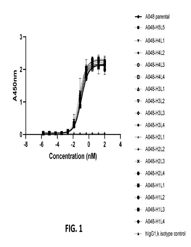
FIG. 1 shows humanized antibody binding to VV B5 protein. Antibodies were
assessed
for binding to VV B5 monomer protein by ELISA. Humanized anti-B5 hIgG1
antibodies were
titrated and showed similar pM EC50 compared to A048 parental positive
control. No binding was
observed with hIgG1 isotype control.
FIG. 2 shows humanized antibody binding to VV-infected cells. Antibodies were
assessed
for binding to VV infected cells by flow cytometry. HT29 cells were infected
with VVddeGFP
Western reserve virus ('WR') or VVCopenhagen (YFP) virus (Cop), and SKOV3
cells were
infected with VVddeGFP Western reserve virus (WR'). Humanized anti-B5 hl gG1
antibodies were
titrated and showed similar nM EC50 compared to A048 parental anti-B5 hIgG1
(positive control).
Negative control was human IgG1 isotype control.
FIG. 3 shows binding levels of humanized antibodies to different cell lines
infected with
Vaccinia virus overtime. HT29 (A), SKOV3 (B), and OVCAR3 (C) cells were
infected with Vaccinia
virus strains VVddeGFP Western Reserve ('WR') or VVCopenhagen (YFP) virus
(Cop') at an
2
CA 03211935 2023- 9- 12
WO 2022/193017
PCT/CA2022/050400
MOI of 1 or 0.1, or mock-infected. Cells were harvested at the indicated
timepoints to determine
A048-H1L4 and A048-H3L2 binding levels in infected cells. Primary antibodies
were conjugated
to AlexaFluor-647. Data was acquired by a BD LSRFortessa X-20 and analyzed by
FlowJo 10.8.1
software. Data represents biological triplicates. MOI, multiplicity of
infection.
FIG. 4 shows a schematic of a design of humanized B5-CAR-050 or B5-CAR-051
chimeric
antigen receptor (CAR) construct (FIG. 4A). Flow cytometry data showing
humanized scFv CAR
detection following lentiviral transduction and expansion of primary human T
cells (FIG. 4B).
Specific activation of human T cells expressing B5-CAR-050 or B5-CAR-051 when
co-cultured
with target expressing HT-29-B5, SKOV3-B5, and HEK293T-B5 lines (FIG. 4C).
Primary human
T cells expressing B5-CAR-050 or B5-CAR-051 showed upregulation of CD137
following co-
culture with target B5 expressing tumor lines. Minimal cross-reactivity
observed with WT target
cell lines.
FIG. 5 shows data demonstrating that human T cells expressing B5-CAR-050 or B5-
CAR-
051 showed high percent specific cytotoxicity (FIG. 5A). Primary human T cells
expressing B5-
CAR-050 or B5-CAR-051 exhibited specific cell lysis at 24h as determined by
percent decrease
of relative luminescence unit (RLU). Data demonstrating that primary human T
cells expressing
B5-CAR-050 or B5-CAR-051 showed morphological signs of direct tumor killing at
24 h following
co-culture with target B5 expressing tumor lines (FIG. 5B).
FIG. 6 shows data demonstrating that human T cells expressing B5-CAR-050 or B5-
CAR-
051 showed high percent specific cytotoxicity against vaccinia virus infected
target cell lines.
Primary human T cells expressing B5-CAR-050 or B5-CAR-051 exhibited specific
cell lysis at 24h
as determined by percent decrease of relative luminescence unit (RLU) against
vaccinia virus
infected target cells (FIG. 6A). In FIG. 6A, B5-CAR-050, B5-CAR-051 and
parental B5-CAR-043
are identified as VV5O_B5, VV51_B5 and VV43_B5_tEGFR, respectively. Data
demonstrating
that primary human T cells expressing B5-CAR-050 or B5-CAR-051 showed
morphological signs
of direct tumor killing at 24 h following co-culture with vaccinia virus
infected tumor lines (FIG. 6B-
6D).
FIG. 7 shows expected data demonstrating that human T cells expressing
humanized B5-
CAR result in diminished tumor growth when administered in combination with
vaccinia virus used
to treat a xenograft tumor model (FIG. 7A). Expected data demonstrating
improved overall
survival of the combined humanized B5-CAR and vaccinia virus therapy (FIG.
7B).
3
CA 03211935 2023- 9- 12
WO 2022/193017
PCT/CA2022/050400
DETAILED DESCRIPTION
Before the antibodies, compositions and methods of the present disclosure are
described
in greater detail, it is to be understood that the antibodies, compositions
and methods are not
limited to particular embodiments described, as such may, of course, vary. It
is also to be
understood that the terminology used herein is for the purpose of describing
particular
embodiments only, and is not intended to be limiting, since the scope of the
antibodies,
compositions and methods will be limited only by the appended claims.
Where a range of values is provided, it is understood that each intervening
value, to the
tenth of the unit of the lower limit unless the context clearly dictates
otherwise, between the upper
and lower limit of that range and any other stated or intervening value in
that stated range, is
encompassed within the antibodies, compositions and methods. The upper and
lower limits of
these smaller ranges may independently be included in the smaller ranges and
are also
encompassed within the antibodies, compositions and methods, subject to any
specifically
excluded limit in the stated range. VVhere the stated range includes one or
both of the limits,
ranges excluding either or both of those included limits are also included in
the antibodies,
compositions and methods.
Certain ranges are presented herein with numerical values being preceded by
the term
"about." The term "about" is used herein to provide literal support for the
exact number that it
precedes, as well as a number that is near to or approximately the number that
the term precedes.
In determining whether a number is near to or approximately a specifically
recited number, the
near or approximating unrecited number may be a number which, in the context
in which it is
presented, provides the substantial equivalent of the specifically recited
number.
Unless defined otherwise, all technical and scientific terms used herein have
the same
meaning as commonly understood by one of ordinary skill in the art to which
the antibodies,
compositions and methods belong. Although any antibodies, compositions and
methods similar
or equivalent to those described herein can also be used in the practice or
testing of the
antibodies, compositions and methods, representative illustrative antibodies,
compositions and
methods are now described.
All publications and patents cited in this specification are herein
incorporated by reference
as if each individual publication or patent were specifically and individually
indicated to be
incorporated by reference and are incorporated herein by reference to disclose
and describe the
materials and/or methods in connection with which the publications are cited.
The citation of any
4
CA 03211935 2023- 9- 12
WO 2022/193017
PCT/CA2022/050400
publication is for its disclosure prior to the filing date and should not be
construed as an admission
that the present antibodies, compositions and methods are not entitled to
antedate such
publication, as the date of publication provided may be different from the
actual publication date
which may need to be independently confirmed.
It is noted that, as used herein and in the appended claims, the singular
forms "a", "an",
and "the" include plural referents unless the context clearly dictates
otherwise. It is further noted
that the claims may be drafted to exclude any optional element. As such, this
statement is
intended to serve as antecedent basis for use of such exclusive terminology as
"solely," "only"
and the like in connection with the recitation of claim elements, or use of a
"negative" limitation.
It is appreciated that certain features of the antibodies, compositions and
methods, which
are, for clarity, described in the context of separate embodiments, may also
be provided in
combination in a single embodiment.
Conversely, various features of the antibodies,
compositions and methods, which are, for brevity, described in the context of
a single
embodiment, may also be provided separately or in any suitable sub-
combination. All
combinations of the embodiments are specifically embraced by the present
disclosure and are
disclosed herein just as if each and every combination was individually and
explicitly disclosed,
to the extent that such combinations embrace operable processes and/or
compositions. In
addition, all sub-combinations listed in the embodiments describing such
variables are also
specifically embraced by the present antibodies, compositions and methods and
are disclosed
herein just as if each and every such sub-combination was individually and
explicitly disclosed
herein.
As will be apparent to those of skill in the art upon reading this disclosure,
each of the
individual embodiments described and illustrated herein has discrete
components and features
which may be readily separated from or combined with the features of any of
the other several
embodiments without departing from the scope or spirit of the present methods.
Any recited
method can be carried out in the order of events recited or in any other order
that is logically
possible.
ANTI-VV B5 ANTI BODIES
Aspects of the present disclosure include antibodies that specifically bind
vaccinia virus
(VV) B5 antigen (VV B5). In certain embodiments, the antibodies are humanized
antibodies that
specifically bind VV B5. Vaccinia viruses are members of the poxvirus family
characterized by an
approximately 192kb double-stranded DNA genome that encodes numerous viral
enzymes and
factors that enable the virus to replicate independently from the host cell
machinery. VV can
5
CA 03211935 2023- 9- 12
WO 2022/193017
PCT/CA2022/050400
stably accommodate up to 25 kb of cloned exogenous DNA. Structurally, it
consists of a core
region composed of viral DNA and various viral enzymes including RNA
polymerase and polyA
polymerase encased in a lipoprotein core membrane. The outer layer of the
virus consists of
double lipid membrane envelope. VV has inherent characteristics that make VV
amenable for use
in oncolytic viral therapy such as natural tropism for tumors, strong lytic
ability, short life cycle with
rapid cell-to-cell spread, efficient gene expression and a large cloning
capacity. VV has a short
life cycle of about 8 hours that takes place in the cytoplasm eliminating the
risk of genome
integration. Replication typically starts about 2 hours after infection, at
which time host cell nucleic
acid synthesis shuts down and cellular resources are directed toward viral
replication. Cell lysis
takes place between 12 and 48 hours releasing packaged viral particles. VV
does not depend on
host mechanisms for mRNA transcription making it less susceptible to
biological changes of the
host cell. Unlike other oncolytic viruses (0Vs), VV does not require a
specific surface receptor
for cell entry, allowing it to infect a wide range of cells.
VV B5 protein is a 42 kDa type I transmembrane glycoprotein with an
extracellular domain
composed of four short consensus repeats (SCRs) characteristic of complement
control proteins.
After the SCRs, B5 has a stalk region before the transmembrane domain and a
short cytoplasmic
tail (CT). Both the SCRs and CT are dispensable for targeting B5 to the
extracellular enveloped
virus (EEV) membrane, although the latter affects its transport to the cell
surface and recycling
via endosomes. B5 is needed for intracellular mature virus (IMV) wrapping to
form intracellular
enveloped virus (I EV).
The term "antibody" (also used interchangeably with "immunoglobulin")
encompasses
polyclonal (e.g., rabbit polyclonal) and monoclonal antibody preparations
where the antibody may
be an antibody or immunoglobulin of any isotype (e.g., IgG (e.g., IgG1, IgG2,
IgG3, or IgG4), IgE,
IgD, IgA, IgM, etc.), whole antibodies (e.g., antibodies composed of a
tetramer which in turn is
composed of two dimers of a heavy and light chain polypeptide); single chain
antibodies (e.g.,
scFv); fragments of antibodies (e.g., fragments of whole or single chain
antibodies) which retain
specific binding to the compound, including, but not limited to single chain
Fv (scFv), Fab, (Fab')2,
(scFV)2, and diabodies; chimeric antibodies; monoclonal antibodies, humanized
antibodies,
human antibodies; and fusion proteins comprising an antigen-binding portion of
an antibody and
a non-antibody protein. In some embodiments, the antibody is selected from an
IgG, Fv, single
chain antibody, scFv, a Fab, a F(a1:02, and a F(ab'). The antibodies may be
further conjugated
to other moieties, such as members of specific binding pairs, e.g., biotin
(member of biotin-avidin
specific binding pair), and the like.
6
CA 03211935 2023- 9- 12
WO 2022/193017
PCT/CA2022/050400
Immunoglobulin polypeptides include the kappa and lambda light chains and the
alpha,
gamma (IgGi, IgG2, IgG3, Igat), delta, epsilon and mu heavy chains or
equivalents in other
species. Full-length immunoglobulin "light chains" (usually of about 25 kDa or
about 214 amino
acids) comprise a variable region of about 110 amino acids at the NH2-terminus
and a kappa or
lambda constant region at the COOH-terminus. Full-length immunoglobulin "heavy
chains" (of
about 150 kDa or about 446 amino acids), similarly comprise a variable region
(of about 116
amino acids) and one of the aforementioned heavy chain constant regions, e.g.,
gamma (of about
330 amino acids).
An immunoglobulin light or heavy chain variable region (VL and VH,
respectively) is
composed of a "framework" region (FR) interrupted by three hypervariable
regions, also called
"complementarity determining regions" or "CDRs". The extent of the framework
region and CDRs
have been defined (see, E. Kabat et al., Sequences of proteins of
immunological interest, 4th ed.
U.S. Dept. Health and Human Services, Public Health Services, Bethesda, MD
(1987); and
Lefranc et al. IMGT, the international ImMunoGeneTics information system .
Nucl. Acids Res.,
2005, 33, D593-D597)). The sequences of the framework regions of different
light or heavy chains
are relatively conserved within a species. The framework region of an
antibody, that is the
combined framework regions of the constituent light and heavy chains, serves
to position and
align the CDRs. The CDRs are primarily responsible for binding to an epitope
of an antigen. The
CDRs of the antibodies provided by the present disclosure are defined
according to Kabat, supra,
unless otherwise indicated.
An "antibody" thus encompasses a protein having one or more polypeptides that
can be
genetically encodable, e.g., by immunoglobulin genes or fragments of
immunoglobulin genes.
The recognized immunoglobulin genes include the kappa, lambda, alpha, gamma,
delta, epsilon
and mu constant region genes, as well as myriad immunoglobulin variable region
genes. Light
chains are classified as either kappa or lambda. Heavy chains are classified
as gamma, mu,
alpha, delta, or epsilon, which in turn define the immunoglobulin classes,
IgG, IgM, IgA, IgD and
IgE, respectively. In some embodiments, an antibody of the present disclosure
is an IgG antibody,
e.g., an IgG1 antibody, such as a human IgG1 antibody. In some embodiments, an
antibody of
the present disclosure comprises a human Fc domain.
A typical immunoglobulin (antibody) structural unit is known to comprise a
tetramer. Each
tetramer is composed of two identical pairs of polypeptide chains, each pair
having one "light"
(about 25 kD) and one "heavy" chain (about 50-70 kD). The N-terminus of each
chain defines a
variable region of about 100 to 110 or more amino acids primarily responsible
for antigen
7
CA 03211935 2023- 9- 12
WO 2022/193017
PCT/CA2022/050400
recognition. The terms variable light chain (VL) and variable heavy chain (VH)
refer to these light
and heavy chains, respectively.
Antibodies encompass intact immunoglobulins as well as a number of well
characterized
fragments which may be genetically encoded or produced by digestion with
various peptidases.
Thus, for example, pepsin digests an antibody below the disulfide linkages in
the hinge region to
produce F(ab)2, a dimer of Fab which itself is a light chain joined to VH-CHI
by a disulfide bond.
The F(ab)'2 may be reduced under mild conditions to break the disulfide
linkage in the hinge region
thereby converting the (Fab)2 dimer into an Fab monomer. The Fab' monomer is
essentially a
Fab with part of the hinge region (see, Fundamental Immunology, W.E. Paul,
ed., Raven Press,
N.Y. (1993), for a more detailed description of other antibody fragments).
While various antibody
fragments are defined in terms of the digestion of an intact antibody, one of
skill will appreciate
that such Fab' fragments may be synthesized de novo either chemically or by
utilizing
recombinant DNA methodology. Thus, the term antibody, as used herein, also
includes antibody
fragments either produced by the modification of whole antibodies or
synthesized de novo using
recombinant DNA methodologies, including but are not limited to, Fab'2, IgG,
IgM, IgA, scFv, dAb,
nanobodies, unibodies, and diabodies. In certain embodiments, an antibody of
the present
disclosure is selected from an IgG, Fv, single chain antibody (e.g., scFv),
Fab, F(ab')2, and Fab'.
According to some embodiments, an antibody of the present disclosure is a
monoclonal
antibody. "Monoclonal antibody" refers to a composition comprising one or more
antibodies
obtained from a population of substantially homogeneous antibodies, i.e., a
population the
individual antibodies of which are identical except for any naturally
occurring mutations that may
be present in minor amounts. Monoclonal antibodies are highly specific, being
directed against a
single antigenic site and generally to a single epitope on an antigen. The
modifier "monoclonal"
indicates the character of the antibody as being obtained from a substantially
homogeneous
population of antibodies, and does not require that the antibody be produced
by any particular
method or be the only antibody in the composition.
As summarized above, according to some embodiments, the antibodies of the
present
disclosure are humanized antibodies. In certain embodiments, provided are
antibodies which are
humanized versions of the parental rabbit antibody designated A048 described
in International
Patent Application No. PCT/CA202/051230 (WO 2021/046653), the disclosure of
which is
incorporated herein by reference in its entirety for all purposes. For
example, provided herein are
antibodies that comprise a VH which is humanized relative to the VH of the
A048 antibody, a VL
which is humanized relative to the VL of the A048 antibody, or both.
8
CA 03211935 2023- 9- 12
WO 2022/193017
PCT/CA2022/050400
As used herein, a "humanized" antibody is a recombinant polypeptide that is
derived from
a non-human (e.g., rabbit, rodent, or the like) antibody and has been modified
to contain at least
a portion of the framework and/or constant regions of a human antibody.
Humanized antibodies
also encompass chimeric antibodies and CDR-grafted antibodies in which various
regions may
be derived from different species. Chimeric antibodies may be antibodies that
include a variable
region from any source linked to a human constant region (e.g., a human Fc
domain). Thus, in
chimeric antibodies, the variable region can be non-human, and the constant
region is human.
CDR-grafted antibodies are antibodies that include the CDRs from a non-human
"donor" antibody
linked to the framework region from a human "recipient" antibody. For example,
an antibody of
the present disclosure in a form of an scFV may be linked to a human constant
region (e.g., Fc
domain) to be made into a human immunoglobulin.
In general, humanized antibodies produce a reduced immune response in a human
host
(exhibit reduced immunogenicity), as compared to a non-humanized version of
the same
antibody. Antibodies can be humanized using a variety of techniques including,
for example,
CDR-grafting, veneering or resurfacing, chain shuffling, and the like. In
certain embodiments,
framework substitutions are identified by modeling of the interactions of the
CDR and framework
residues to identify framework residues important for antigen binding and
sequence comparison
to identify unusual framework residues at particular positions.
The substitution of rabbit or mouse CDRs into a human variable domain
framework can
result in retention of their correct spatial orientation where, e.g., the
human variable domain
framework adopts the same or similar conformation to the rabbit or mouse
variable framework
from which the CDRs originated. This can be achieved by obtaining the human
variable domains
from human antibodies whose framework sequences exhibit a high degree of
sequence identity
with the rabbit or mouse variable framework domains from which the CDRs were
derived. The
heavy and light chain variable framework regions can be derived from the same
or different human
antibody sequences. The human antibody sequences can be the sequences of
naturally occurring
human antibodies or can be consensus sequences of several human antibodies.
Having identified the complementarity determining regions of the rabbit or
mouse donor
immunoglobulin and appropriate human acceptor immunoglobulins, a next step is
to determine
which, if any, residues from these components should be substituted to
optimize the properties of
the resulting humanized antibody. In general, substitution of human amino acid
residues with
rabbit or mouse should be minimized, because introduction of rabbit or mouse
residues increases
the risk of the antibody eliciting a human-anti-rabbit-antibody (HARA) or
human-anti-mouse-
antibody (HAMA) response in humans. Art-recognized methods of determining
immune response
9
CA 03211935 2023- 9- 12
WO 2022/193017
PCT/CA2022/050400
can be performed to monitor a HARA or HAMA response in a particular patient or
during clinical
trials. Patients administered humanized antibodies can be given an
immunogenicity assessment
at the beginning and throughout the administration of said therapy. The HARA
or HAMA response
is measured, for example, by detecting antibodies to the humanized therapeutic
reagent, in serum
samples from the patient using a method known to one in the art, including
surface plasmon
resonance technology (BIACORE) and/or solid-phase ELISA analysis. In many
embodiments, a
subject humanized antibody does not substantially elicit a HARA response in a
human subject.
Certain amino acids from the human variable region framework residues are
selected for
substitution based on their possible influence on CDR conformation and/or
binding to antigen.
The unnatural juxtaposition of rabbit or murine CDR regions with human
variable framework
region can result in unnatural conformational restraints, which, unless
corrected by substitution of
certain amino acid residues, lead to loss of binding affinity. The selection
of amino acid residues
for substitution can be determined, in part, by computer modeling. Computer
hardware and
software for producing three-dimensional images of immunoglobulin molecules
are known in the
art In general, molecular models are produced starting from solved structures
for immunoglobulin
chains or domains thereof. The chains to be modeled are compared for amino
acid sequence
similarity with chains or domains of solved three-dimensional structures, and
the chains or
domains showing the greatest sequence similarity is/are selected as starting
points for
construction of the molecular model. Chains or domains sharing at least 50%
sequence identity
are selected for modeling, and preferably those sharing at least 60%, 70%,
80%, 90% sequence
identity or more are selected for modeling. The solved starting structures are
modified to allow for
differences between the actual amino acids in the immunoglobulin chains or
domains being
modeled, and those in the starting structure. The modified structures are then
assembled into a
composite immunoglobulin. Finally, the model is refined by energy minimization
and by verifying
that all atoms are within appropriate distances from one another and that bond
lengths and angles
are within chemically acceptable limits.
When framework residues, as defined by, e.g., Kabat, constitute structural
loop residues
as defined by, e.g., Chothia, the amino acids present in the rabbit or mouse
antibody may be
selected for substitution into the humanized antibody. Residues which are
"adjacent to a CDR
region" include amino acid residues in positions immediately adjacent to one
or more of the CDRs
in the primary sequence of the humanized immunoglobulin chain, for example, in
positions
immediately adjacent to a CDR as defined by Kabat, or a CDR as defined by
Chothia (See e.g.,
Chothia and Lesk JMB 196:901 (1987)). These amino acids are particularly
likely to interact with
the amino acids in the CDRs and, if chosen from the acceptor, to distort the
donor CDRs and
CA 03211935 2023- 9- 12
WO 2022/193017
PCT/CA2022/050400
reduce affinity. Moreover, the adjacent amino acids may interact directly with
the antigen (Amit et
al., Science, 233:747 (1986)) and selecting these amino acids from the donor
may be desirable
to keep all the antigen contacts that provide affinity in the original
antibody. Approaches that may
be employed to humanize any of the antibodies described herein include, but
are not limited to,
those described in Williams, D., Matthews, D. & Jones, T. Humanising
Antibodies by CDR
Grafting. Antibody Engineering 319-339 (2010) doi: 10. 1007/978-3-642-01144-
3_21; Kuramochi,
T., Igawa, T., Tsunoda, H. & Hattori, K. Humanization and simultaneous
optimization of
monoclonal antibody. Methods Mol. Biol. 1060, 123-37 (2014); Hwang, W. Y.,
Almagro, J. C.,
Buss, T. N., Tan, P. & Foote, J. Use of human germline genes in a CDR homology-
based
approach to antibody humanization. Methods 36, 35-42 (2005); Lo, B. K.
Antibody humanization
by CDR grafting. Methods Mol. Biol. 248, 135-59 (2004); and Lefranc, M.-P. P.,
Ehrenmann, F.,
Ginestoux, C., Giudicelli, V. & Duroux, P. Use of IMGT(0) databases and tools
for antibody
engineering and humanization. Methods Mol. Biol. 907, 3-37 (2012); the
disclosures of which are
incorporated herein by reference in their entireties for all purposes.
The antibodies of the present disclosure specifically bind to the VV B5
antigen. An
antibody "specifically binds" or "preferentially binds" to a target if it
binds with greater affinity,
avidity, more readily, and/or with greater duration than it binds to other
substances, e.g., in a
sample. In certain embodiments, an antibody "specifically binds" an antigen if
it binds to or
associates with the antigen with an affinity or Ka (that is, an association
rate constant of a
particular binding interaction with units of 1/M) of, for example, greater
than or equal to about 104
M-1. Alternatively, affinity may be defined as an equilibrium dissociation
constant (KD) of a
particular binding interaction with units of M (e.g., 10-5 M to 10-13 M, or
less). In certain aspects,
specific binding means the antibody binds to the antigen with a KD of less
than or equal to about
10-5 M, less than or equal to about 10-6 M, less than or equal to about 10-7
M, less than or equal
to about 10-8 M, or less than or equal to about 10-9 M, 10-10 M, 10-11 M, or
10-12 M or less. The
binding affinity of the antibody for the antigen can be readily determined
using conventional
techniques, e.g., by competitive ELISA (enzyme-linked immunosorbent assay),
equilibrium
dialysis, by using surface plasmon resonance (SPR) technology (e.g., the
BlAcore 2000 or
BlAcore T200 instrument, using general procedures outlined by the
manufacturer); by
radioimmunoassay; or the like.
According to some embodiments, provided are antibodies (non-humanized or
humanized)
that compete for binding to VV B5 antigen with any of the antibodies (non-
humanized or
humanized) described elsewhere herein. Whether an antibody of the present
disclosure
"competes with" a second antibody for binding to the antigen may be readily
determined using
11
CA 03211935 2023- 9- 12
WO 2022/193017
PCT/CA2022/050400
competitive binding assays known in the art. Competing antibodies may be
identified, for
example, via an antibody competition assay. For example, a sample of a first
antibody can be
bound to a solid support. Then, a sample of a second antibody suspected of
being able to
compete with such first antibody is added. One of the two antibodies is
labeled. If the labeled
antibody and the unlabeled antibody bind to separate and discrete sites on the
antigen, the
labeled antibody will bind to the same level whether or not the suspected
competing antibody is
present. However, if the sites of interaction are identical or overlapping,
the unlabeled antibody
will compete, and the amount of labeled antibody bound to the antigen will be
lowered. If the
unlabeled antibody is present in excess, very little, if any, labeled antibody
will bind.
For purposes of the present disclosure, competing antibodies are those that
decrease the
binding of an antibody to the antigen by about 50% or more, about 60% or more,
about 70% or
more, about 80% or more, about 85% or more, about 90% or more, about 95% or
more, or about
99% or more. Details of procedures for carrying out such competition assays
are known and can
be found, for example, in Harlow and Lane, Antibodies, A Laboratory Manual,
Cold Spring Harbor
Laboratory Press, Cold Spring Harbor, New York, 1988, 567-569, 1988, ISBN 0-
87969-314-2.
Such assays can be made quantitative by using purified antibodies. A standard
curve may be
established by titrating one antibody against itself, i.e., the same antibody
is used for both the
label and the competitor. The capacity of an unlabeled competing antibody to
inhibit the binding
of the labeled antibody to the plate may be titrated. The results may be
plotted, and the
concentrations necessary to achieve the desired degree of binding inhibition
may be compared.
In certain embodiments, the antibodies of the present disclosure are humanized
versions
of the parental rabbit antibody designated A048 described in International
Patent Application No.
PCT/CA202/051230. The amino acid sequences of the VH and VL polypeptides of
the parental
A048 antibody, as well as non-limiting examples of humanized versions of such
VH and VL
polypeptides (designated A048-H1-H5 and A048-L1-L5) are provided in Table 1
below (with
CDRs underlined).
Table 1 ¨ Amino Acid Sequences of Example Humanized Anti-VV B5 Antibodies
Parental A048 VH QEQLEESGGG LVKPEGSLTLTCTASG FS FSS SYYM
CVVVRQAPG RG
(SEQ ID NO:1) LEWIACIYTSSGSAYYANVVAKG
RFTISRTSSTTVTLQMTRLTAADTAT
YFCVRNAVGSSYYLYLWG PGTLVTVSS
A048-H1 VH EVQLLESGGGLVQ PGGSLRLSCAASG
FSFSSSYYMCVVVRQAPG KG
(SEQ ID NO:2) LEWIACIYTSSGSAYYANWAKG RFT! S RDN SKNTLYLQM
NS LRAE DT
AVYYCVRNAVGSSYYLYLWGQGTLVTVSS
12
CA 03211935 2023- 9- 12
WO 2022/193017
PCT/CA2022/050400
A048-H2 VH EVQLLESGGGLVQPGGSLRLSCAASGFSFSSSYYMCWVRQAPGKG
(SEQ ID NO:3)
LEWIACIYTSSGSAYYANWAKGRFTISRTSSTTVTLQMNSLRAEDTA
VYYCVRNAVGSSYYLYLWGQGTLVTVSS
A048-H3 VH
EVQLLESGGGLVQPGGSLRLSCAASGFSFSSSYYMCVVVRQAPGKG
(SEQ ID NO:4)
LEWIACIYTSSGSAYYADSVKGRFTISRDNSKNTLYLQMNSLRAEDT
AVYYCVRNAVGSSYYLYLWGQGTLVTVSS
A048-H4 VH
EVQLLESGGGLVQPGGSLRLSCAASGFSFSSSYYMCVVVRQAPGKG
(SEQ ID NO:5)
LEWIACIYTSSGSAYYADSVKGRFTISRTSSTTVTLQMNSLRAEDTAV
YYCVRNAVGSSYYLYLWGQGTLVTVSS
A048-H5 VH
QVTLKESGPVLVKPTETLTLTCTASGFSFSSSYYMCVVVRQPPGKAL
(SEQ ID NO:6)
EWIACIYTSSGSAYYANWAKGRFTISRDTSKSQVVLTMTNMDPVDT
ATYFCVRNAVGSSYYLYLWGQGTLVTVSS
Parental A048 VL
AQVLTQTPSPVSAAVGGTVTISCQASQSVAGNNYLSVVYQQKPGQP
(SEQ ID NO:7)
PNLLIYSVSTLASGVPSRFKGSGSGTQFTLTISDLECDDAATYYCQG
YYNDGIWAFGGGTEVVVK
A048-L1 VL
DIQMTQSPSTLSASVGDRVTITCQASQSVAGNNYLSVVYQQKPGKAP
(SEQ ID NO:8)
KLLIYSVSTLASGVPSRFSGSGSGTEFTLTISSLQPDDFATYYCQGYY
NDGIWAFGQGTKVEIK
A048-L2 VL
AQVLIQSPSTLSASVGDRVTITCOASQSVAGNNYLSWYQQKPGKAP
(SEQ ID NO:9)
KLLIYSVSTLASGVPSRFSGSGSGTEFTLTISSLQPDDFATYYCQGYY
NDGIWAFGQGTKVEIK
A048-L3 VL
DIQMTQSPSSLSASVGDRVTITCQASQSVAGNNYLSVVYQQKPGKVP
(SEQ ID NO:10)
KLLIYSVSTLASGVPSRFSGSGSGTDFTLTISSLQPEDVATYYCQGYY
NDGIWAFGQGTKVEIK
A048-L4 VL
AQVLTQSPSSLSASVGDRVTITCQASQSVAGNNYLS1NYQQKPGKV
(SEQ ID NO:11)
PKLLIYSVSTLASGVPSRFSGSGSGTDFTLTISSLQPEDVATYYCQGY
YNDGIWAFGQGTKVEIK
A048-L5 VL
EQVLTQSPATLSLSPGERATLSCQASQSVAGNNYLSVVYQQKPGQA
(SEQ ID NO:12)
PRLLIYSVSTLASGIPARFSGSGSGTDFTLTISSLEPEDFAVYYCQGY
YNDGIWAFGQGTKLEIK
A048 VH CDR1 SSYYMC
(SEQ ID NO:13)
VIICDR2 ClYTSSGSAYYA(N/D)(W/S)(AN)KG
(SEQ ID NO:14)
13
CA 03211935 2023- 9- 12
WO 2022/193017
PCT/CA2022/050400
A048 VH CDR2 ClYTSSGSAYYANWAKG
(SEQ ID NO:15)
VH CDR2 ClYTSSGSAYYADSVKG
(SEQ ID NO:16)
A048 VH CDR3 NAVGSSYYLYL
(SEQ ID NO:17)
A048 VL CDR1 QASQSVAGNNYLS
(SEQ ID NO:18)
A048 VL CDR2 SVSTLAS
(SEQ ID NO:19)
A048 VL CDR3 QGYYNDGI1NA
(SEQ ID NO:20)
According to some embodiments, an antibody of the present disclosure comprises
any
desired combination of variable heavy chain (VH) polypeptide set forth in
Table 1 and variable
light chain (VL) polypeptide set forth in Table 1. By way of example, an
antibody of the present
disclosure may comprise the A048-HI VH paired with any of A048-L1 VL, A048-L2
VL, A048-L3
VL, A048-L4 VL, or A048-L5 VL. Such an antibody may be designated A048-H1L1,
A048-H1L2,
A048-H1L3, A048-H1 L4, or A048-H1 L5, respectively. Also by way of example, an
antibody of
the present disclosure may comprise the A048-H2 VH paired with any of A048-L1
VL, A048-L2
VL, A048-L3 VL, A048-L4 VL, or A048-L5 VL. Such an antibody may be designated
A048-H2L1,
A048-H2L2, A048-H2L3, A048-H2L4, or A048-H2L5, respectively. As will be
appreciated, an
antibody of the present disclosure may comprise any combination of A048-HI VH
to A048-H5 VH
and A048-L1 VL to A048-L5 VL.
In certain embodiments, an antibody of the present disclosure specifically
binds to
Vaccinia Virus B5 antigen (VV B5), wherein the antibody comprises:
a variable heavy chain (VH) polypeptide comprising 70% or greater sequence
identity to
the amino acid sequence set forth in SEQ ID NO: 1, wherein the VH polypeptide
comprises:
one or more mutations selected from the group consisting of: Q1E, E2V, Q3T,
E5L/K, G9P, G10V, K13Q, E15G/T, G16E, S17T, T19R, T21S, 123A, A41P,
R44K, G45A, T74D, S75N/T, S76_T77insK, T77N/S, T78Q, V79L, T80Y/V,
Q82T, T84N, R85S/N, L86M, T87R/D, A88P, A89E/V, T93V, F95Y, P112Q,
14
CA 03211935 2023- 9- 12
WO 2022/193017
PCT/CA2022/050400
and any combination thereof, wherein numbering is according to SEQ ID
NO:1;
a VH CDR1 comprising the amino acid sequence SSYYMC (SEQ ID NO:13),
a VH CDR2 comprising the amino acid sequence
ClYTSSGSAYYA(N/D)(W/S)(A/V)KG (SEQ ID NO:14), and
a VH CDR3 comprising the amino acid sequence NAVGSSYYLYL (SEQ ID
NO:17).
As used herein, a "mutation" encompasses an amino acid substitution, insertion
of one or
more amino acids, deletion of one or more amino acids, etc. relative to the
parental VH and VL
polypeptides of the A048 antibody set forth in SEQ ID NO:1 and SEQ ID NO:7,
respectively.
According to some embodiments, the VH polypeptide of such an antibody
comprises one, any
combination of, or each of the mutations E2V, E5L/K, E15G/T, R44K, R85S/N,
T87R/D, A89E/V,
and P1 12Q. For example, the VH polypeptide of such an antibody may comprise
each of the
mutations E2V, E5L/K, E15G/T, R44K, R855/N, T87R/D, A89E/V, P112Q. In certain
embodiments, the E5L/K mutation is E5L, the E15G/T mutation is E15G, the
R85S/N mutation is
R85S, the T87R/D mutation is T87R, and/or the A89E/V mutation is A89E.
According to some
embodiments, the VH polypeptide of such an antibody comprises the mutations
E5L, E15G, R85S,
T87R, and A89E. In certain embodiments, the VH polypeptide comprises one, any
combination
of, or each of the mutations Q1E, K13Q, T19R, T21S, 123A, T84N, T93V, and
F95Y. According
to some embodiments, the VH polypeptide comprises one, any combination of, or
each of the
mutations T74D, 575N/T, 576_T77insK, T77N/S, V79L, and T80Y/V. As used herein,
"576_T77insK" means that the VH polypeptide comprises an insertion of a K
(lysine residue)
between S76 and T77 relative to the parental A048 VH. In certain embodiments,
the T77N/S
mutation is T77N. According to some embodiments, the T80Y/V mutation is T80Y.
According to
some embodiments, the VH polypeptide comprises a VH CDR2 comprising the amino
acid
sequence CIYTSSGSAYYADSVKG (SEQ ID NO:16).
According to some embodiments, the VH polypeptide of such an antibody
comprises one,
any combination of, or each of the mutations Q3T, E5L/K, G9P, G10V, E15G/T,
G16E, S17T,
A41P, G45A, T74D, S75N/T, S76_T77insK, T77N/S, T78Q, T80Y/V, Q82T, R85S/N,
L86M,
T87R/D, A88P, and A89E/V. For example, the VH polypeptide of such an antibody
may comprise
each of the mutations Q3T, E5L/K, G9P, G10V, E15G/T, Gl6E, S17T, A41P, G45A,
T74D,
575N/T, 576_T77insK, T77N/S, T78Q, T80Y/V, Q82T, R855/N, L86M, T87R/D, A88P,
and
A89E/V. In certain embodiments, the E5L/K mutation is E5K, the E15G/T mutation
is E15T, the
S75N/T mutation is S75T, the T77N/S mutation is T77S, the T80Y/V mutation is
T80V, the
CA 03211935 2023- 9- 12
WO 2022/193017
PCT/CA2022/050400
R85S/N mutation is R85N, the T87R/D mutation is T87D, and/or the A89E/V
mutation is A89V.
According to some embodiments, the VH polypeptide of such an antibody
comprises the mutations
E5K, E15T, R85N, T87D, and A89V.
In certain embodiments, the VH polypeptide of a humanized antibody of the
present
disclosure comprises one or a desired combination of the mutations set forth
above and
comprises 80% or greater, 85% or greater, 90% or greater, 91% or greater, 92%
or greater, 93%
or greater, 94% or greater, 95% or greater, 96% or greater, 97% or greater,
98% or greater, 99%
or greater, or 100% sequence identity to the amino acid sequence set forth in
one of SEQ ID Nos:
2-6.
In certain embodiments, an antibody of the present disclosure specifically
binds to VV
B5, wherein the antibody comprises:
a variable light chain (VL) polypeptide comprising 70% or greater sequence
identity to
the amino acid sequence set forth in SEQ ID NO: 7, wherein the VL polypeptide
comprises:
one or more mutations selected from the group consisting of: A1D/E, Q2I, V3Q,
L4M, T7S, S9A, P1OT/S, V11 L, A13L, A14S, V15P, G17D/E, T18R, V19A,
I21L, S22T, Q44K, P45A/V, N47K/R, V60I, S62A, K65S, Q72E/D, D79S,
E81Q, C82P, D83E, A85F/V, T87V, G103Q, E106K, V107L, V108E, V109I,
and any combination thereof, wherein numbering is according to SEQ ID NO:
7;
a VL CDR1 comprising the amino acid sequence QASQSVAGNNYLS (SEQ ID
NO:18),
a VL CDR2 comprising the amino acid sequence SVSTLAS (SEQ ID NO:19), and
a VL CDR3 comprising the amino acid sequence QGYYNDGIWA (SEQ ID
NO:20).
According to some embodiments, the VL polypeptide of such an antibody
comprises one,
any combination of, or each of the mutations T7S, P10T/S, V11 L, A14S, G17D/E,
T18R, P45A/V,
N47K/R, K65S, Q72E/D, D79S, C82P, A85F/V, G103Q, E106K, V108E, and V1091. For
example,
the VL polypeptide of such an antibody may comprise each of the mutations T7S,
P1OT/S, V11L,
A14S, G17D/E, T18R, P45A/V, N47K/R, K65S, Q72E/D, D79S, C82P, A85F/V, G103Q,
E106K,
V108E, and V1091. According to some embodiments, a VL polypeptide of such an
antibody
comprises one, any combination of, or each of the mutations G17D, S22T, Q44K,
N47K, and
E81Q. In certain embodiments, the P1OT/S mutation is PIOT, the P45A/V mutation
is P45A, the
Q72E/D mutation is Q72E, and/or the A85F/V mutation is A85F. The VL
polypeptide of such an
16
CA 03211935 2023- 9- 12
WO 2022/193017
PCT/CA2022/050400
antibody may comprise one, any combination of, or each of the mutations A1 D,
Q2I, V3Q, and
L4M.
In certain embodiments, the VL polypeptide of an antibody of the present
disclosure
comprises the mutation D83E. According to some embodiments, the P1OT/S
mutation is P1OS,
the P45A/V mutation is P45V, the Q72E/D mutation is Q72D, and/or the A85F/V
mutation is A85V.
In certain embodiments, the VL polypeptide comprises one, any combination of,
or each of the
mutations A1 D, Q2I, V3Q, and L4M.
According to some embodiments, the VL polypeptide of an antibody of the
present
disclosure comprises one, any combination of, or each of the mutations A1E,
S9A, PIOT, A13L,
V15P, G17E, V19A, I21L, P45A, N47R, V60I, S62A, 072D, D83E, A85F, T87V, and
V107L. For
example, the VL polypeptide may comprise each of the mutations A1E, S9A, PIOT,
A13L, V15P,
G17E, V19A, I21L, P45A, N47R, V60I, S62A, Q72D, D83E, A85F, T87V, and V107L.
In certain embodiments, the VL polypeptide of a humanized antibody of the
present
disclosure comprises one or a desired combination of the VL mutations set
forth above and
comprises 80% or greater, 85% or greater, 90% or greater, 91% or greater, 92%
or greater, 93%
or greater, 94% or greater, 95% or greater, 96% or greater, 97% or greater,
98% or greater, 99%
or greater, or 100% sequence identity to the amino acid sequence set forth in
one of SEQ ID Nos:
8-12.
Also provided by the present disclosure are antibodies (non-humanized or
humanized
antibodies) that specifically bind VV B5, wherein the antibodies comprise:
a variable heavy chain (VH) polypeptide comprising:
a VH CDR1 comprising the amino acid sequence SSYYMC (SEQ ID NO:13),
a VH CDR2 comprising the amino acid sequence CIYTSSGSAYYADSVKG (SEQ ID
NO:16), and
a VH CDR3 comprising the amino acid sequence NAVGSSYYLYL (SEQ ID NO:17);
and
a variable light chain (VL) polypeptide comprising:
a VL CDR1 comprising the amino acid sequence QASQSVAGNNYLS (SEQ ID
NO:18),
a VL CDR2 comprising the amino acid sequence SVSTLAS (SEQ ID NO:19), and
a VL CDR3 comprising the amino acid sequence QGYYNDGIWA (SEQ ID NO:20).
The antibodies of the present disclosure specifically bind VV B5 antigen. Any
of the
antibodies may bind a VV B5 antigen from one or more VV strains, non-limiting
examples of which
17
CA 03211935 2023- 9- 12
WO 2022/193017
PCT/CA2022/050400
include the B5 antigen of the VVyeth, Western Reserve and/or Copenhagen
strains. The amino
acid sequences of the B5 antigens encoded by these strains are provided in
Table 2 below.
Table 2 ¨ B5 antigen sequences for Wyeth, Western Reserve and Copenhagen VV
VVyeth B5 MKTISVVTLLCVLPAVVYSTCTVPTMNNAKLTSTETSFNNNQKVTFT
CDQGYHSSDPNAVCETDKWKYENPCKKMCTVSDYVSELYDKPLY
(SEQ ID NO: 21)
EVNSTMTLSCNGETKYFRCEEKNGNTSWNDTVTCPNAECQPLQL
EHGSCQPVKEKYSFGEYITINCDVGYEVIGASYISCTANSWNVIPSC
QQKCDIPSLSNGLISGSTFSIGGVIHLSCKSGFILTGSPSSTCIDGKW
NPILPTCVRSNEKFDPVDDGPDDETDLSKLSKDVVQYEQEIESLEA
TYHIIIVALTIMGVIFLISVIVLVCSCDKNNDQYKFHKLLP
Western Reserve B5 M KTISVVTLLCVLPAVVYSTCTVPTMN NAKLTSTETSFN
DKQKVTFT
CDQGYHSSDPNAVCETDKWKYENPCKKMCTVSDYISELYNKPLYE
(Uniprot Q01227)
VNSTMTLSCNGETKYFRCEEKNGNTSWNDTVTCPNAECQPLQLE
HGSCQPVKEKYSFGEYMTINCDVGYEVIGASYISCTANSWNVIPSC
(SEQ ID NO: 22) QQKCDMPSLSNGLISGSTFSIGGVIHLSCKSGFTLTGSPSSTCIDGK
WNPVLPICVRTNEEFDPVDDGPDDETDLSKLSKDVVQYEQEIESLE
ATYHIIIVALTIMGVIFLISVIVLVCSCDKNNDQYKFHKLLP
Copenhagen B5 MKTISVVTLLCVLPAVVYSTCTVPTMNNAKLTSTETSFNNNQKVTFT
CDQGYHSSDPNAVCETDKWKYENPCKKMCTVSDYISELYNKPLYE
(Uniprot P21115)
VNSTMTLSCNGETKYFRCEEKNGNTSWNDTVTCPNAECQPLQLE
HGSCQPVKEKYSFGEYMTINCDVGYEVIGASYISCTANSWNVIPSC
(SE ID NO 23)
QQKCDIPSLSNGLISGSTFSIGGVIHLSCKSGFILTGSPSSTCIDGKW
Q :
NPVLPICVRTNEEFDPVDDGPDDETDLSKLSKDVVQYEQEIESLEA
TYHIIIVALTIMGVIFLISVIVLVCSCDKNNDQYKFHKLLP
Bispecific Antibodies
Also provided are bispecific antibodies. In certain embodiments, a bispecific
antibody of
the present disclosure comprises a first antigen-binding domain comprising a
VH polypeptide-VL
polypeptide pair of any of the anti-VV B5 antibodies of the present
disclosure, including any of
such antibodies described hereinabove. The bispecific antibody may include a
second antigen-
binding domain that specifically binds the VV B5 antigen bound by the first
antigen-binding
domain. In certain embodiments, the bispecific antibody includes a second
antigen-binding
domain that specifically binds a VV antigen other than the VV B5 antigen bound
by the first
antigen-binding domain.
According to some embodiments, a bispecific antibody of the present disclosure
includes
a second antigen-binding domain that specifically binds an antigen other than
a VV antigen. In
certain embodiments, the antigen other than a VV antigen is an immune cell
surface antigen.
Non-limiting examples of immune cell surface antigens are immune effector cell
surface antigens,
18
CA 03211935 2023- 9- 12
WO 2022/193017
PCT/CA2022/050400
e.g., a T cell surface antigen, a natural killer (NK) cell surface antigen, a
macrophage cell surface
antigen, and the like. Examples of T cell surface antigens that may be bound
by the second
antigen-binding domain include, but are not limited to, a T cell stimulatory
molecule, e.g., CD3,
CD28, etc.
Bispecific antibodies of the present disclosure include antibodies having a
full-length
antibody structure, and bispecific antibody fragments. "Full-length" as used
herein refers to an
antibody having two full-length antibody heavy chains and two full length
antibody light chains. A
full-length antibody heavy chain (HC) consists of well-known heavy chain
variable and constant
domains VH, CH1, CH2, and CH3. A full-length antibody light chain (LC)
consists of well-known
light chain variable and constant domains VL and CL. The full-length antibody
may be lacking the
C-terminal lysine in either one or both heavy chains. The term "Fab arm"
refers to one heavy
chain:light chain pair that specifically binds an antigen.
Full-length bispecific antibodies may be generated for example using Fab arm
exchange
(or half molecule exchange) between two monospecific bivalent antibodies by
introducing
substitutions at the heavy chain CH3 interface in each half molecule to favor
heterodimer
formation of two antibody half molecules having distinct specificity either in
vitro in a cell-free
environment or using co-expression. The Fab arm exchange reaction is the
result of a disulfide-
bond isomerization reaction and dissociation-association of CH3 domains. The
heavy chain
disulfide bonds in the hinge regions of the parent monospecific antibodies are
reduced. The
resulting free cysteines of one of the parent monospecific antibodies form an
inter heavy-chain
disulfide bond with cysteine residues of a second parent monospecific antibody
molecule and
simultaneously CH3 domains of the parent antibodies release and reform by
dissociation-
association. The CH3 domains of the Fab arms may be engineered to favor
heterodimerization
over homodimerization. The resulting product is a bispecific antibody having
two Fab arms or half
molecules which each bind a distinct epitope.
The "knob-in-hole" strategy (see, e.g., WO 2006/028936) may be used to
generate full
length bispecific antibodies. Briefly, selected amino acids forming the
interface of the CHS
domains in human IgG can be mutated at positions affecting CH3 domain
interactions to promote
heterodimer formation. An amino acid with a small side chain (hole) is
introduced into a heavy
chain of an antibody specifically binding a first antigen and an amino acid
with a large side chain
(knob) is introduced into a heavy chain of an antibody specifically binding a
second antigen. After
co-expression of the two antibodies, a heterodimer is formed as a result of
the preferential
interaction of the heavy chain with a "hole" with the heavy chain with a
"knob". Exemplary CH3
substitution pairs forming a knob and a hole are (expressed as modified
position in the first CH3
19
CA 03211935 2023- 9- 12
WO 2022/193017
PCT/CA2022/050400
domain of the first heavy chain/modified position in the second CH3 domain of
the second heavy
chain): T366Y7F405A, T366W/F405W, F405W/Y407A, T394W/Y407T, T3945/Y407A,
T366W/T394S, F405W/T394S and T366W/T366S_L368A_Y407V.
Other strategies such as promoting heavy chain heterodimerization using
electrostatic
interactions by substituting positively charged residues at one CH3 surface
and negatively
charged residues at a second CH3 surface may be used, as described in
US2010/0015133;
US2009/0182127; US2010/028637 or US2011/0123532. In other strategies.
heterodimerization
may be promoted by the following substitutions (expressed as modified position
in the first CH3
domain of the first heavy chain/modified position in the second CH3 domain of
the second heavy
chain): L351 Y_F405A_Y407V T394W,
T366 I_K392M_T394W/F405A_Y407V,
T366 L_K392 M_T394W/F405A_Y407V, L351
Y Y407AT366A K409F,
L351Y_Y407A/T366V_K409F, Y407A/T366A K409F,
or
1350V_L351Y_F405A_Y407V/1-350V_1366L_K392L_1394W as described in
US2012/0149876
or US2013/0195849.
Also provided are single chain bispecific antibodies. In some embodiments, a
single chain
bispecific antibody of the present disclosure is a bispecific scFv. Details
regarding bispecific
scFvs may be found, e.g., in Zhou et al. (2017) J Cancer 8(18):3689-3696.
Approaches that may be employed to produce multispecific (e.g., bispecific)
antibodies
from the antibodies described herein include, but are not limited to,
Ellerman, D. (2019).
"Bispecific T-cell engagers: Towards understanding variables influencing the
in vitro potency and
tumor selectivity and their modulation to enhance their efficacy and safety."
Methods 154: 102-
117; Brinkmann, U. and R. E. Kontermann (2017). "The making of bispecific
antibodies." mAbs
9(2): 182-212; and Suurs, F. V., et al. (2019). "A review of bispecific
antibodies and antibody
constructs in oncology and clinical challenges." Pharmacol Ther 201: 103-119;
the disclosures of
which are incorporated herein by reference in their entireties for all
purposes.
Fusion Proteins
Aspects of the present disclosure further include fusion proteins. In certain
embodiments,
a fusion protein of the present disclosure comprises a VH polypeptide, a VL
polypeptide, or both,
of any of the anti-VV B5 antibodies of the present disclosure, fused to a
heterologous sequence
of amino acids. The heterologous sequence of amino acids may be fused to the C-
terminus of
the chain of the antibody or the N-terminus of the chain of the antibody. In
certain embodiments,
a fusion protein of the present disclosure includes a heterologous sequence at
the C-terminus of
CA 03211935 2023- 9- 12
WO 2022/193017
PCT/CA2022/050400
the chain of the antibody and a heterologous sequence at the N-terminus of the
chain of the
antibody, wherein the heterologous sequences may be the same sequence or
different
sequences. "Heterologous" as used in the context of a nucleic acid or
polypeptide generally
means that the nucleic acid or polypeptide is from a different origin (e.g.,
molecule of different
sequence, different species origin, and the like) than that with which the
nucleic acid or
polypeptide is associated or joined, such that the nucleic acid or polypeptide
is one that is not
found in nature. For example, in a fusion protein, a light chain polypeptide
and a reporter
polypeptide (e.g., GFP, red fluorescent protein (e.g., mCherry), luciferase,
etc.) are said to be
"heterologous" to one another. Similarly, a CDR from a rabbit antibody and a
constant region from
a human antibody are "heterologous" to one another.
The VH polypeptide and/or VL polypeptide may be fused to any heterologous
sequence of
interest. Heterologous sequences of interest include, but are not limited to,
an albumin, a
transferrin, XTEN, a homo-amino acid polymer, a proline-alanine-serine
polymer, an elastin-like
peptide, or any combination thereof. In certain aspects, the heterologous
polypeptide increases
the stability and/or serum half-life of the anti-VV B5 antibody upon its
administration to an
individual in need thereof, as compared to the same antibody which is not
fused to the
heterologous sequence.
In certain embodiments, a fusion protein of the present disclosure comprises a
single
chain antibody, e.g., a single chain antibody (e.g., scFv) comprising a VH
polypeptide-VL
polypeptide pair of any of the anti-VV B5 antibodies of the present
disclosure, including any of
such antibodies described hereinabove. The amino acid sequences of non-
limiting examples of
humanized scFvs according to embodiments of the present disclosure are
provided in Table 3
below.
Table 3 ¨ Example Anti-VV B5 scFv Amino Acid Sequences
scFv of B5-CAR-050
EVQLLESGGGLVQPGGSLRLSCAASGFSFSSSYYMC1NVRQAPGKGL
= VH (A048 H3)
EVVIACIYTSSGSAYYADSVKGRFTISRDNSKNTLYLQMNSLRAEDTAVY
= Linker
YCVRNAVGSSYYLYLVVGQGTLVTVSSGSTSGSGKPGSGEGSTKGAQ
= VL (A048 L2) VLTQSPSTLSASVGDRVTI
TCQASQSVAGNNYLSINYQQKPG KAP KLLI
YSVSTLASGVPSRFSGSGSGTEFTLTISSLQPDDFATYYCQGYYNDGI
(SEQ ID NO:24) WAFG QGTKVE I K
21
CA 03211935 2023- 9- 12
WO 2022/193017
PCT/CA2022/050400
scFv of B5-CAR-051
EVOLLESGGGLVQPGGSLRLSCAASGFSFSSSYYMCVVVRQAPGKGL
= VH (A048 H1) EVVIACI YTSSGSAYYANVVAKG RFT!
SRDNSKNTLY LQM NS LRAE DTAV
= Linker
YYCVRNAVGSSYYLYUNGQGTLVTVSSGSTSGSGKPGSGEGSTKGA
= VL (A048 L4)
QVLTQSPSSLSASVGDRVTITCQASQSVAGNNYLSVVYQQKPGKVPKL
LlYSVSTLASGVPSRFSGSGSGTDFTLTISSLQPEDVATYYCQGYYNDG
(SEQ ID NO:25) IVVAFGQGTKVEIK
Also provided are the anti-VV B5 scFvs set forth in Table 3, but where the
orientation of
the VH and VL is reversed ¨ that is, where the VL is N-terminal to the WI.
According to some embodiments, when the fusion protein comprises a single
chain
antibody (e.g., any of the single chain antibodies of the present disclosure,
including any of the
scFvs described herein), the fusion protein is a chimeric antigen receptor
(CAR) comprising the
single chain antibody, a transmembrane domain, and an intracellular signaling
domain.
A CAR of the present disclosure may include one or more linker sequences
between the
various domains. A "variable region linking sequence" is an amino acid
sequence that connects
a heavy chain variable region to a light chain variable region and provides a
spacer function
compatible with interaction of the two sub-binding domains so that the
resulting polypeptide
retains a specific binding affinity to the same target molecule as an antibody
that includes the
same light and heavy chain variable regions. A non-limiting example of a
variable region linking
sequence is a serine-glycine linker, such as a serine-glycine linker that
includes the amino acid
sequence GGGGSGGGGSGGGGS (G4S)3 (SEQ ID NO:26). In certain embodiments, a
linker
separates one or more heavy or light chain variable domains, hinge domains,
transmembrane
domains, co-stimulatory domains, and/or primary signaling domains. In
particular embodiments,
the CAR includes one, two, three, four, or five or more linkers. In particular
embodiments, the
length of a linker is about 1 to about 25 amino acids, about 5 to about 20
amino acids, or about
10 to about 20 amino acids, or any intervening length of amino acids. In some
embodiments, the
linker is 1, 2, 3, 4, 5, 6, 7, 8, 9, 10, 11, 12, 13, 14, 15, 16, 17, 18, 19,
20, 21, 22, 23, 24, 25, or
more amino acids in length.
In some embodiments, the antigen binding domain of the CAR is followed by one
or more
spacer domains that moves the antigen binding domain away from the effector
cell surface (e.g.,
the surface of a T cell expressing the CAR) to enable proper cell/cell
contact, antigen binding
and/or activation. The spacer domain (and any other spacer domains, linkers,
and/or the like
described herein) may be derived either from a natural, synthetic, semi-
synthetic, or recombinant
22
CA 03211935 2023- 9- 12
WO 2022/193017
PCT/CA2022/050400
source. In certain embodiments, a spacer domain is a portion of an
immunoglobulin, including,
but not limited to, one or more heavy chain constant regions, e.g., CH2 and
CH3. The spacer
domain may include the amino acid sequence of a naturally occurring
immunoglobulin hinge
region or an altered immunoglobulin hinge region. In one embodiment, the
spacer domain
includes the CH2 and/or CH3 of IgG1, IgG4, or IgD. Illustrative spacer domains
suitable for use
in the CARs described herein include the hinge region derived from the
extracellular regions of
type 1 membrane proteins such as CD8a and CD4, which may be wild-type hinge
regions from
these molecules or variants thereof. In certain aspects, the hinge domain
includes a CD8a hinge
region. In some embodiments, the hinge is a PD-1 hinge or CD152 hinge.
The "transmembrane domain" (Tm domain) is the portion of the CAR that fuses
the
extracellular binding portion and intracellular signaling domain and anchors
the CAR to the
plasma membrane of the cell (e.g., immune effector cell). The Tm domain may be
derived either
from a natural, synthetic, semi-synthetic, or recombinant source. In some
embodiments, the Tm
domain is derived from (e.g., includes at least the transmembrane region(s) or
a functional portion
thereof) of the alpha or beta chain of the T-cell receptor, CD35, CDX CD3y,
CD3o, CD4, CD5,
CD8a, CD9, CD16, CD22, CD27, CD28, CD33, 0D37, CD45, CD64, CD80, CD86, 0D134,
CD137, CD152, CD154, or PD-1.
In one embodiment, a CAR includes a Tm domain derived from CD8a. In certain
aspects,
a CAR includes a Tm domain derived from CD8a and a short oligo- or polypeptide
linker, e.g.,
between 1, 2, 3, 4, 5, 6, 7, 8, 9, or 10 amino acids in length, that links the
Tm domain and the
intracellular signaling domain of the CAR. A glycine-serine linker may be
employed as such a
linker, for example.
The "intracellular signaling" domain of a CAR refers to the part of a CAR that
participates
in transducing the signal from CAR binding to a target molecule/antigen into
the interior of the
immune effector cell to elicit effector cell function, e.g., activation,
cytokine production,
proliferation and/or cytotoxic activity, including the release of cytotoxic
factors to the CAR-bound
target cell, or other cellular responses elicited with target molecule/antigen
binding to the
extracellular CAR domain. Accordingly, the term "intracellular signaling
domain" refers to the
portion of a protein which transduces the effector function signal and that
directs the cell to
perform a specialized function. To the extent that a truncated portion of an
intracellular signaling
domain is used, such truncated portion may be used in place of a full-length
intracellular signaling
domain as long as it transduces the effector function signal. The term
intracellular signaling
domain is meant to include any truncated portion of an intracellular signaling
domain sufficient for
transducing effector function signal.
23
CA 03211935 2023- 9- 12
WO 2022/193017
PCT/CA2022/050400
Signals generated through the T cell receptor (TCR) alone are insufficient for
full activation
of the T cell, and a secondary or costimulatory signal is also required. Thus,
T cell activation is
mediated by two distinct classes of intracellular signaling domains: primary
signaling domains that
initiate antigen-dependent primary activation through the TCR (e.g., a TCR/CD3
complex) and
costimulatory signaling domains that act in an antigen-independent manner to
provide a
secondary or costimulatory signal. As such, a CAR of the present disclosure
may include an
intracellular signaling domain that includes one or more "costimulatory
signaling domains" and a
"primary signaling domain."
Primary signaling domains regulate primary activation of the TCR complex
either in a
stimulatory manner, or in an inhibitory manner. Primary signaling domains that
act in a stimulatory
manner may contain signaling motifs which are known as immunoreceptor tyrosine-
based
activation motifs (or "ITAMs"). Non-limiting examples of ITAM-containing
primary signaling
domains suitable for use in a CAR of the present disclosure include those
derived from FcRy,
FcR6, CD3y, CD305, CD3c, CD3, 0D22, CD79a, 0D796, and CD665. In certain
embodiments, a
CAR includes a CD3 primary signaling domain and one or more costimulatory
signaling domains.
The intracellular primary signaling and costimulatory signaling domains are
operably linked to the
carboxyl terminus of the transmembrane domain.
In some embodiments, the CAR includes one or more costimulatory signaling
domains to
enhance the efficacy and expansion of immune effector cells (e.g., T cells)
expressing the CAR.
As used herein, the term "costimulatory signaling domain" or "costimulatory
domain" refers to an
intracellular signaling domain of a costimulatory molecule or an active
fragment thereof. Example
costimulatory molecules suitable for use in CARs contemplated in particular
embodiments include
TLR1, TLR2, TLR3, TLR4, TLR5, TLR6, TLR7, TLR8, TLR9, TLR10, CARD11, CD2, CD7,
CD27,
0D28, CD30, CD40, CD54 (ICAM), 0D83, 0D134 (0X40), 0D137 (4-1BB), 0D278
(ICOS),
DAP10, LAT, KD2C, SLP76, TRIM, and ZAP70. In some embodiments, the CAR
includes one
or more costimulatory signaling domains selected from the group consisting of
4-1BB (CD137),
CD28, and CD134, and a CD3 primary signaling domain.
A CAR of the present disclosure may include any variety of suitable domains
including but
not limited to a leader sequence; hinge, spacer and/or linker domain(s);
transmembrane
domain(s); costimulatory domain(s); signaling domain(s) (e.g., CD3 domain(s));
ribosomal skip
element(s); restriction enzyme sequence(s); reporter protein domains; and/or
the like. Non-
limiting examples of such domains that may be included in a CAR of the present
disclosure include
those provided in Table 4 below. As will be appreciated by one of ordinary
skill in the art, the
24
CA 03211935 2023- 9- 12
WO 2022/193017
PCT/CA2022/050400
amino acid sequence of one or more of the domains indicated in Table 4 (e.g.,
linker, hinge,
transmembrane, co-stimulatory, signaling, ribosomal skip element; restriction
enzyme sequence;
reporter protein etc.) may be modified as desired, e.g., for improved
functionality, etc. of the CAR.
Table 4 ¨ Example CAR Domain Amino Acid Sequences
Leader Signal Peptide(s)
GM-CSFR alpha (P15509) MLLLVTSLLLCELPHPAFLLIP
(SEQ ID NO:27)
Hinge/Spacer/Linker domain(s)
(SEQ ID NO:28) GSTSGSGKPGSGEGSTKG
(SEQ ID NO:26) GGGGSGGGGSGGGGS
CD8a hinge TTTPAPRPPTPAPTIASQPLSLRPEACRPAAGGAVHTRGLDFACD
(SEQ ID NO:29)
CD8a hinge 2
AKPTTTPAPRPPTPAPTIASQPLSLRPEACRPAAGGAVHTRGLDFAC
(SEQ ID NO:30)
extended CD8 hinge GGGGSGGGGSGGGGSGGTTTPAPRPPTPAPTIASQPLSLRPEACRP
(SEQ ID NO:31) AAGGAVHTRGLDFACD
CD28 hinge IEVMYPPPYLDNEKSNGTIIHVKGKHLCPSPLFPGPSKP
(SEQ ID NO:32)
GSG linker GGGSSGGGSG
(SEQ ID NO:33)
IgG4 hinge ESKYGPPCPSCP
(SEQ ID NO:34)
IgG4(CH3)
GQPREPQVYTLPPSQEEMTKNQVSLTCLVKGFYPSDIAVEVVESNGQ
(SEQ ID NO:35)
PENNYKTTPPVLDSDGSFFLYSRLTVDKSRVVQEGNVFSCVMHEALH
NHYTQKSLSLSLGK
IgG4 (P01861)
ESKYGPPCPSCPAPEFLGGPSVFLFPPKPKDTLMISRTPEVTCVVVD
(SEQ ID NO:36)
VSQEDPEVQFNVVYVDGVEVHNAKTKPREEQFNSTYRVVSVLTVLHQ
DVVLNGKEYKCKVSNKGLPSSIEKTISKAKGQPREPQVYTLPPSQEEM
TKNOVSLTCLVKGFYPSDIAVEWESNGQPENNYKTTPPVLDSDGSFF
LYSRLTVDKSRWQEGNVFSCSVMHEALHNHYTQKSLSLSLGK
Transmembrane domain(s)
CD8tm (NM_001768) IYIWAPLAGTCGVLLLSLVITLYC
(SEQ ID NO:37)
CD8tm2 (NM_001768) IYIWAPLAGTCGVLLLSLVITLY
(SEQ ID NO:38)
CD8tm3 (NM_001768) IYIWAPLAGTCGVLLLSLVITL
CA 03211935 2023- 9- 12
WO 2022/193017
PCT/CA2022/050400
(SEQ ID NO:39)
CD28tm (NM_006139) FINVLVVVGGVLACYSLLVTVAFIIFVVV
(SEQ ID NO:40)
CD28tm2 (NM_006139) MFVVVLVVVGGVLACYSLLVTVAFIIFINV
(SEQ ID NO:41)
CD3z (304132.1) LCYLLDGILFIYGVILTALFL
(SEQ ID NO:42)
CD4tm (M35160) MALIVLGGVAGLLLFIGLGIFF
(SEQ ID NO:43)
4-1 BB (NM_001561) IISFFLALTSTALLFLLFFLTLRFSVV
(SEQ ID NO:44)
Costimulatory domain(s)
4-1 BB (NM_001561) KRGRKKLLYIFKQPFMRPVQTTQEEDGCSCRFPEEEEGGCEL
(SEQ ID NO:45)
CD28 (NM_006139) RSKRSRLLHSDYMNMTPRRPGPTRKHYQPYAPPRDFAAYRS
(SEQ ID NO:46)
CD28gg (NM_006139) RSKRSRGGHSDYMNMTPRRPGPTRKHYQPYAPPRDFAAYRS
(SEQ ID NO:47)
0X40 (P43489) ALYLLRRDQRLPPDAHKPPGGGSFRTPIQEEQADAHSTLAKI
(SEQ ID NO:48)
CD3C domain(s)
CD3c RVKFSRSADAPAYQQGQNQLYNELNLGRREEYDVLDKRRGRDPEM
(SEQ ID NO:49) GGKPRRKNPQEGLYNELQKDKMAEAYSEIGMKGERRRGKGHDGLY
QGLSTATKDTYDALHMQALPPR
Ribosomal Skip Element(s)
E2A GSGQCTNYALLKLAGDVESNPGP
(SEQ ID NO:50)
T2A GSGEGRGSLLTCGDVEENPGP
(SEQ ID NO:51)
Restriction Enzyme Sequence(s)
Pad l LIN
26
CA 03211935 2023- 9- 12
WO 2022/193017
PCT/CA2022/050400
Reporter Protein(s)
eGFP
MVSKGEELFTGVVPILVELDGDVNGHKFSVSGEGEGDATYGKLTLKFI
(SEQ ID NO:52)
CTTGKLPVPWPTLVTTLTYGVQCFSRYPDHMKQHDFFKSAMPEGYV
QERTIFFKDDGNYKTRAEVKFEGDTLVNRIELKGIDFKEDGNILGHKLE
YNYNSHNVYIMADKQKNGIKVNFKIRHNIEDGSVQLADHYQQNTPIGD
GPVLLPDNHYLSTQSALSKDPNEKRDHMVLLEFVTAAGITLGMDELY
mCherry
MVSKGEEDNMAIIKEFMRFKVHMEGSVNGHEFEIEGEGEGRPYEGT
(SEQ ID NO:53)
QTAKLKVTKGGPLPFAWDILSPQFMYGSKAYVKHPADIPDYLKLSFPE
GFKWERVMNFEDGGVVTVTQDSSLQDGEFIYKVKLRGTNFPSDGPV
MQKKTMGWEASSERMYPEDGALKGEIKQRLKLKDGGHYDAEVKTTY
KAKKPVQLPGAYNVNIKLDITSHNEDYTIVEQYERAEGRHSTGGMDE
LYK
Truncated EGFR (partial
MLLLVTSLLLCELPHPAFLLIPRKVCNGIGIGEFKDSLSINATNIKHFKN
sequence from UniProtKB -
CTSISGDLHILPVAFRGDSFTHTPPLDPQELDILKTVKEITGFLLIQAWP
P00533)
ENRTDLHAFENLEIIRGRTKQHGQFSLAVVSLNITSLGLRSLKEISDGD
(SEQ ID NO:54)
VIISGNKNLCYANTINWKKLFGTSGQKTKIISNRGENSCKATGQVCHA
LCSPEGCWGPEPRDCVSCRNVSRGRECVDKCNLLEGEPREFVENS
ECIQCHPECLPQAMNITCTGRGPDNCIQCAHYIDGPHCVKTCPAGVM
GENNTLVWKYADAGHVCHLCHPNCTYGCTGPGLEGCPTNGPKIPSI
ATGMVGALLLLLVVALGIGLFM
In certain aspects, a CAR of the present disclosure comprises a single chain
antibody
(e.g., any of the scFvs of the present disclosure) that specifically binds VV
B5 antigen; a
transmembrane domain from a polypeptide selected from the group consisting of:
CD4, CD8a,
CD154, and PD-1; one or more intracellular costimulatory signaling domains
from a polypeptide
selected from the group consisting of: 4-1BB (0D137), CD28, and 0D134; and an
intracellular
signaling domain from a polypeptide selected from the group consisting of:
FcRy, FcR13, CD3y,
CD35, CD3E, CD3C, CD22, CD79a, CD79I3, and CD665. Such a CAR may further
include a
spacer domain between the antigen-binding portion and the transmembrane
domain, e.g., a CD8
alpha hinge.
According to some embodiments, provided are CARs that comprise ¨ from N-
terminus to
C-terminus ¨ a variable heavy chain (VH) polypeptide of an antibody described
herein, a linker, a
variable light chain (VL) polypeptide of an antibody described herein, a CD8
hinge region (which
in some embodiments is an extended CD8 hinge region), a CD8 transmembrane
domain, a 4-
27
CA 03211935 2023- 9- 12
WO 2022/193017
PCT/CA2022/050400
1BB costimulatory domain, and a CD3 c signaling domain. According to certain
embodiments,
provided are CARs that comprise ¨ from N-terminus to C-terminus ¨ a variable
light chain (VL)
polypeptide of an antibody described herein, a linker, a variable heavy chain
(VH) of an antibody
described herein, a CD8 hinge region (which in some embodiments is an extended
CD8 hinge
region), a CD8 transmembrane domain, a 4-1BB costimulatory domain, and a CD3 c
signaling
domain. In certain embodiments, provided are CARs that comprise ¨ from N-
terminus to C-
terminus ¨ a variable heavy chain (VH) polypeptide of an antibody described
herein, a linker, a
variable light chain (VL) of an antibody described herein, a CD28 hinge
region, a CD28
transmembrane domain, a 4-1 BB costimulatory domain, and a CD3 signaling
domain. According
to some embodiments, provided are CARs that comprise ¨ from N-terminus to C-
terminus ¨ a
variable light chain (VL) polypeptide of an antibody described herein, a
linker, a variable heavy
chain (VH) of an antibody described herein, a CD28 hinge region, a CD28
transmembrane domain,
a 4-1 BB costimulatory domain, and a CD3 signaling domain. Any of the CARs of
the present
disclosure may include a domain N-terminal to the VH polypeptide. For example,
a leader
sequence (e.g., a GM-CSFRalpha leader sequence) may be present at the N-
terminus of a CAR
of the present disclosure.
The amino acid sequences of non-limiting examples of anti-VV B5 CARs according
to
embodiments of the present disclosure are provided in Table 5 below, where the
amino acid
sequences of the CARs in Table 5 include an amino acid sequence of an N-
terminal leader
sequence (here, an N-terminal GM-CSFRalpha leader sequence). Any desired
leader sequence
(e.g., a GM-CSFRalpha leader sequence) may be present at the N-terminus of
such CARs. As
will be appreciated by one of ordinary skill in the art, the amino acid
sequence of one or more of
the domains indicated in Table 5 (e.g., linker, hinge, transmembrane, co-
stimulatory, signaling,
etc.) may be modified as desired, e.g., to improve the functionality, etc. of
the CAR. In Table 5,
segments/domains of the CARs are indicated by alternating underlining, and the
identities of the
segments/domains are provided in the left column.
Table 5 ¨ Example Anti-VV B5 CAR Amino Acid Sequences
B5-CAR-050
MLLLVTSLLLCELPHPAFLLIPEVQLLESGGGLVQPGGSLRLSCAAS
= GM-CSFRa Leader GFSFSSSYYMCVVVRQAPGKGLEWIACIYTSSGSAYYADSVKGRFT
= VH (A048-H3) I SRDNSKNTLYLQ M
NSLRAEDTAVYYCVRNAVGSSYYLYLVVGQGT
= Linker
LVTVSSGSTSGSGKPGSGEGSTKGAQVLTQSPSTLSASVGDRVTI
= VL (A048-L2)
TCQASQSVAGNNYLSVVYQQKPGKAPKLLIYSVSTLASGVPSRFSG
28
CA 03211935 2023- 9- 12
WO 2022/193017
PCT/CA2022/050400
= CD8 hinge
SGSGTEFTLTISSLQPDDFATYYCQGYYNDGIWAFGQGTKVEIKTT
= CD8 transmembrane TPAPRPPTPAPTIASQPLSLRPEACRPAAGGAVHTRGLDFACDIYI
= 4-1 BB
WAPLAGTCGVLLLSLVITLYCKRGRKKLLYIFKQPFMRPVQTTQEE
= CD3
DGCSCRFPEEEEGGCELRVKFSRSADAPAYQQGQNQLYNELNLG
RREEYDVLDKRRGRDPEMGGKPRRKNPQEGLYNELQKDKMAEAY
(SEQ ID NO:55) SEIGMKGERRRGKGHDGLYQGLSTATKDTYDALHMQALPPR
B5-CAR-051
MLLLVTSLLLCELPHPAFLLIPEVQLLESGGGLVQPGGSLRLSCAAS
= GM-CSFRa Leader GFSFSSSYYMCVVVRQAPGKGLEWIACIYTSSGSAYYANWAKGRF
= VH (A048-H1)
TISRDNSKNTLYLQMNSLRAEDTAVYYCVRNAVGSSYYLYLWGQG
= Linker
TLVTVSSGSTSGSGKPGSGEGSTKGAQVLTQSPSSLSASVGDRVT
= VL (A048-L4)
ITCQASQSVAGNNYLSWYQQKPGKVPKLLIYSVSTLASGVPSRFSG
= CD8 hinge
SGSGTDFTLTISSLQPEDVATYYCQGYYNDGIWAFGQGTKVEIKTT
= CD8 transmembrane TPAPRPPTPAPTIASQPLSLRPEACRPAAGGAVHTRGLDFACDIYI
= 4-1 BB
WAPLAGTCGVLLLSLVITLYCKRGRKKLLYIFKQPFMRPVQTTQEE
DGCSCRFPEEEEGGCELRVKFSRSADAPAYQQGQNQLYNELNLG
= CD&;
RREEYDVLDKRRGRDPEMGGKPRRKNPQEGLYNELQKDKMAEAY
(SEQ ID NO:56) SEIGMKGERRRGKGHDGLYQGLSTATKDTYDALHMQALPPR
A CAR of the present disclosure may include one or more additional domains as
desired.
Non-limiting examples of such additional domains include a ribosomal skip
element, an enzymatic
domain (e.g., a domain having nuclease activity, e.g., restriction
endonuclease activity), a domain
that enables detection of the CAR (e.g., a reporter protein domain (e.g., a
fluorescent protein
(e.g., eGFP, mCherry, or the like), a luminescent protein, and/or the like)),
etc. For example, in
certain embodiments, provided are CARs that comprise a ribosomal skip element,
a restriction
enzyme domain, and/or a reporter protein domain.
According to some embodiments, a CAR of the present disclosure is provided by
a single
polypeptide. In certain embodiments, a CAR of the present disclosure is
provided by two or more
polypeptides. When the CAR is provided by two or more polypeptides, the CAR
may be provided
in any useful multi-polypeptide format, including universal CAR formats such
as biotin-binding
immune receptor (BBIR) format (see, e.g., Urbanska K, Powell DJ. Development
of a novel
universal immune receptor for antigen targeting to infinity and beyond.
Oncoimmunology.
2012;1(5):777-779. doi:10.4161/onci.19730, and Urbanska K, Lanitis E, Poussin
M, et al. A
universal strategy for adoptive immunotherapy of cancer through use of a novel
T cell antigen
receptor. 2013;72(7):1844-1852. doi:10.1158/0008-5472.CAN-11-3890.A); a
switchable CAR
29
CA 03211935 2023- 9- 12
WO 2022/193017
PCT/CA2022/050400
format with peptide NeoEpitope (PNE) (see, e.g., Kim et al. (2015) J Am Chem
Soc.
2015;137(8):2832-2835; Ma et al. (2016) Proc Natl Acad Sci 113(4):E450-8;
Rodgers et al. (2016)
Proc Natl Acad Sci. 113(4):E459-E468; Viaud et al. (2018) Proc Natl Acad Sci
115(46):E10898-
E10906); a SUPRA CAR format with leucine zippers (see, e.g., Cho et al. (2108)
Cell 173(6):1426-
1438.e11); a CAR-T Adapter Molecule (CAM)-based format with FITC-folic acid
(see, e.g., Lee et
al. (2019) Cancer Res. 79(2):387-396; and Lu et al. (2019) Front Oncol.
9:151); anti-FITC-folic
acid adaptor format (see, e.g., Chu et al. (2018) Biosci Trends. 12(3):298-
308); anti-FITC antibody
adaptor CAR format (see, e.g., Tamada et al. (2012) Clin Cancer Res.
18(23):6436-6445); Fc-
targeting (e.g., anti-CD16) CAR + anti-tumor antibody format (see, e.g., Kudo
et al. (2014) Cancer
Res. 74(1):93-103); and the like.
Conjugates
The present disclosure also provides conjugates. According to some
embodiments, a
conjugate of the present disclosure comprises any of the antibodies or fusion
proteins of the
present disclosure, and an agent conjugated to the antibody or fusion protein
The term
"conjugated" generally refers to a chemical linkage, either covalent or non-
covalent, usually
covalent, that proximally associates one molecule of interest with a second
molecule of interest.
In certain embodiments, the agent conjugated to the antibody or fusion protein
is selected from a
chemotherapeutic agent, a toxin, a radiation-sensitizing agent, a radioactive
isotope (e.g., a
therapeutic radioactive isotope), a detectable label, and a half-life
extending moiety.
According to some embodiments, the agent is a therapeutic agent, e.g., a
chemotherapeutic agent. Therapeutic agents of interest include agents capable
of affecting the
function of a cell/tissue to which the conjugate binds via specific binding of
the antibody portion
of the conjugate to the antigen. When the function of the cell/tissue is
pathological, an agent that
reduces the function of the cell/tissue may be employed. In certain aspects, a
conjugate of the
present disclosure includes an agent that reduces the function of a target
cell/tissue by inhibiting
cell proliferation and/or killing the cell/tissue. Such agents may vary and
include cytostatic agents
and cytotoxic agents, e.g., an agent capable of killing a target cell tissue
with or without being
internalized into a target cell.
In certain embodiments, the therapeutic agent is a cytotoxic agent selected
from an
enediyne, a lexitropsin, a duocarmycin, a taxane, a puromycin, a dolastatin, a
maytansinoid, and
a vinca alkaloid. In some embodiments, the cytotoxic agent is paclitaxel,
docetaxel, CC-1065,
CPT-11 (SN-38), topotecan, doxorubicin, morpholino-doxorubicin, rhizoxin,
cyanomorpholino-
doxorubicin, dolastatin-10, echinomycin, combretastatin, calicheamicin,
maytansine, maytansine
CA 03211935 2023- 9- 12
WO 2022/193017
PCT/CA2022/050400
DM1, maytansine DM4, DM-1, an auristatin or other dolastatin derivatives, such
as auristatin E
or auristatin F, AEB (AEB-071), AEVB (5-benzoylvaleric acid-AE ester), AEFP
(antibody-
endostatin fusion protein), MMAE (monomethylauristatin E), MMAF
(monomethylauristatin F),
pyrrolobenzodiazepines (PBDs), eleutherobin, netropsin, or any combination
thereof.
According to some embodiments, the agent is a toxin, such as a protein toxin
selected
from hemiasterlin and hemiasterlin analogs such as HTI-286 (e.g., see USPN
7,579,323; WO
2004/026293; and USPN 8,129,407, the full disclosures of which are
incorporated herein by
reference), abrin, brucine, cicutoxin, diphtheria toxin, batrachotoxin,
botulism toxin, shiga toxin,
endotoxin, Pseudomonas exotoxin, Pseudomonas endotoxin, tetanus toxin,
pertussis toxin,
anthrax toxin, cholera toxin, falcarinol, fumonisin BI, fumonisin B2, afla
toxin, maurotoxin, agitoxin,
charybdotoxin, margatoxin, slotoxin, scyllatoxin, hefutoxin, calciseptine,
taicatoxin, calcicludine,
geldanamycin, gelonin, lotaustralin, ocratoxin A, patulin, ricin, strychnine,
trichothecene,
zearlenone, and tetradotoxin. Enzymatically active toxins and fragments
thereof which may be
employed include diphtheria A chain, non-binding active fragments of
diphtheria toxin, exotoxin A
chain (from Pseudomonas aeruginosa), ricin A chain, abrin A chain, modeccin A
chain, alpha-
sarcin, Aleurites fordii proteins, dianthin proteins, Phytolaca americana
proteins (PAPI, PAPII,
and PAP-S), Momordica charantia inhibitor, curcin, crotin, Sapaonaria
officinalis inhibitor, gelonin,
mitogellin, restrictocin, phenomycin, enomycin and the tricothecenes.
In certain embodiments, the agent is a radiation-sensitizing agent. As used
herein, a
"radiation-sensitizing agent" is an agent that enhances the ability of
radiation to kill tumor cells.
Non-limiting examples of radiation-sensitizing agents that may be conjugated
to the antibody or
fusion protein include cisplatin, 5-fluorouracil (5-FU), AZD7762, selumetinib,
and the like.
In certain embodiments, the agent is a radioisotope, e.g., useful for therapy
and/or
detection (e.g., imaging). Non-limiting examples of radioisotopes that may be
conjugated to the
antibody or fusion protein include but are not limited to 225Ac, 111Ag, 114Ag,
71As, 72As, 77As, 211At,
198AU, 199Au, 212Bi, 213Bi, 76Br, 76Br, 11C, 13C, 55co, 62cu, 64cu, 67ou,
165Dy, 166Dy, 169Er, 18F, 19F,
52Fe, 59Fe, 66Ga, 67Ga, 68Ga, 72Ga, 154-158Gd, 157Gd, 159Gd, 166H0, 1201,
1211, 1231, 1241, 1251, 1311, 1101n,
1111n, 113min, 1941r, 81mK1, 177Lu, 51mn, 52mn, 99mo, 13N, 15N, 150, 170, 32p,
33p, 211pb, 212pb, 109po,
149pm, 151pm, 142pr, 143pr, 191pT, 193mpT, 195mpt, 223Ra, 142Rb, 186Re, 188Re,
189Re, 105Rn,
Je
153Sm, 117mSn, 121S n,
83Sr, 89Sr, 161-b, gaTc, ggTc, ggmTc, 227Th, 201T1, 172Tm, 127Te, 90y, 169yb,
175yb,
133X, and 89Zr.
In certain embodiments, a radioisotope is conjugated to the antibody or fusion
protein via
a chelator, for example, a bifunctional chelator. A bifunctional chelator may
contain a metal
31
CA 03211935 2023-9-12
WO 2022/193017
PCT/CA2022/050400
chelating moiety that binds the radioisotope in a stable coordination complex
and a reactive
functional group that is covalently linked to a targeting moiety, such as any
of the antibodies or
fusion proteins of the present disclosure, so that the radioisotope may be
properly directed to the
desirable molecular target in vivo. Non-limiting examples of bifunctional
chelators that may be
employed to conjugate an antibody or fusion protein of the present disclosure
to a radioisotope
include p-SCN-Bn-DOTA and p-SCN-Bn-deferoxamine. Additional examples of
bifunctional
chelators that may be employed to conjugate an antibody or fusion protein of
the present
disclosure to a radioisotope include those described in Price & Orvig (2014)
Chem. Soc. Rev.
43:260; and Brechbiel (2008) Q J Nucl Med Mol Imaging 52(2):166-173.
According to some embodiments, the radioisotope is a therapeutic radioisotope.
In certain
embodiments, the radioisotope is an alpha emitting
radioisotope, .. e.g.,
225Ac, 211At, 212Bi/212pb, 213Bi,
223R a, or 227Th. In other embodiments, the radioisotope is a beta
minus emitting radioisotope, e.g., 32P, 33P, 67Cu, 90Y, 1311 or 177Lu.
According to some embodiments, the agent is a labeling agent. By "labeling
agent" (or
"detectable label") is meant the agent detectably labels the antibody or
fusion protein, such that
the antibody or fusion protein may be detected in an application of interest
(e.g., in vitro and/or in
vivo research and/or clinical applications). Detectable labels of interest
include radioisotopes
(e.g., gamma or positron emitters), enzymes that generate a detectable product
(e.g., horseradish
peroxidase, alkaline phosphatase, luciferase, etc.), fluorescent proteins,
paramagnetic atoms,
and the like. In certain aspects, the antibody or fusion protein is conjugated
to a specific binding
partner of detectable label, e.g., conjugated to biotin such that detection
may occur via a
detectable label that includes avidin/streptavidin.
In certain embodiments, the agent is a labeling agent that finds use in in
vivo imaging,
such as near-infrared (NIR) optical imaging, single-photon emission computed
tomography
(SPECT) CT imaging, positron emission tomography (PET) CT imaging, nuclear
magnetic
resonance (NMR) spectroscopy, or the like. Labeling agents that find use in
such applications
include, but are not limited to, fluorescent labels, radioisotopes, and the
like. In certain aspects,
the labeling agent is a multi-modal in vivo imaging agent that permits in vivo
imaging using two or
more imaging approaches (e.g., see Thorp-Greenwood and Coogan (2011) Dalton
Trans.
40:6129-6143).
In certain embodiments, the labeling agent is an in vivo imaging agent that
finds use in
near-infrared (NIR) imaging applications. Such agents include, but are not
limited to, a Kodak X-
SIGHT dye, Pz 247, DyLight 750 and 800 Fluors, Cy 5.5 and 7 Fluors, Alexa
Fluor 680 and 750
32
CA 03211935 2023- 9- 12
WO 2022/193017
PCT/CA2022/050400
Dyes, IRDye 680 and 8000W Fluors. According to some embodiments, the labeling
agent is an
in vivo imaging agent that finds use in SPECT imaging applications, non-
limiting examples of
which include 99mTc, min, 1231, 2011-1, and 133Xe. In certain embodiments, the
labeling agent is an
in vivo imaging agent that finds use in PET imaging applications, e.g., 110,
13N, 150, 18F, 640u,
62cu, 1241, 76Br, 82Rb, -d l.D or the like.
For half-life extension, the antibodies and fusion proteins of the present
disclosure may
be conjugated to an agent that provides for an improved pharmacokinetic
profile (e.g., by
PEGylation, hyperglycosylation, and the like). Modifications that can enhance
serum half-life are
of interest. A subject antibody or fusion protein may be "PEGylated", as
containing one or more
poly(ethylene glycol) (PEG) moieties. Methods and reagents suitable for
PEGylation of a protein
are well known in the art and may be found, e.g., in US Pat. No. 5,849,860.
PEG suitable for
conjugation to a protein is generally soluble in water at room temperature and
has the general
formula R(O-CH2-CH2)nO-R, where R is hydrogen or a protective group such as an
alkyl or an
alkanol group, and where n is an integer from 1 to 1000. Where R is a
protective group, it generally
has from 1 to 8 carbons. The PEG conjugated to the subject antibody or fusion
protein can be
linear. The PEG conjugated to the subject antibody or fusion protein may also
be branched.
Branched PEG derivatives such as those described in U.S. Pat. No. 5,643,575,
"star-PEGs" and
multi-armed PEGs. Star PEGs are described in the art including, e.g., in U.S.
Patent No.
6,046,305.
Where the subject antibody or fusion protein is to be isolated from a source,
the antibody
or fusion protein may be conjugated to one or more moieties that facilitate
purification, such as
members of specific binding pairs, e.g., biotin (member of biotin-avidin
specific binding pair), a
lectin, and the like. The antibody can also be bound to (e.g., immobilized
onto) a solid support,
including, but not limited to, polystyrene plates or beads, magnetic beads,
test strips, membranes,
and the like.
Where the antibodies or fusion proteins are to be detected in an assay, the
antibodies or
fusion proteins may contain a detectable label, e.g., a radioisotope (e.g.,
89Zr; ln, and the like),
an enzyme which generates a detectable product (e.g., luciferase, 13 -
galactosidase, horse radish
peroxidase, alkaline phosphatase, and the like), a fluorescent protein, a
chromogenic protein, dye
(e.g., fluorescein isothiocyanate, rhodamine, phycoerythrin, and the like);
fluorescence emitting
metals, e.g., 152Eu, or others of the lanthanide series, attached to the
protein through metal
chelating groups such as EDTA; chemiluminescent compounds, e.g., luminol,
isoluminol,
acridinium salts, and the like; bioluminescent compounds, e.g., luciferin;
fluorescent proteins; and
the like. Indirect labels include antibodies specific for a subject protein,
wherein the antibody may
33
CA 03211935 2023- 9- 12
WO 2022/193017
PCT/CA2022/050400
be detected via a secondary antibody; and members of specific binding pairs,
e.g., biotin-avidin,
and the like.
Any of the above agents may be conjugated to the antibody or fusion protein
via a linker.
If present, the linker molecule(s) may be of sufficient length to permit the
antibody or fusion protein
and the linked agent to allow some flexible movement between the antibody or
fusion protein and
the linked agent. Linker molecules may be, e.g., about 6-50 atoms long. Linker
molecules may
also be, e.g., aryl acetylene, ethylene glycol oligomers containing 2-10
monomer units, diamines,
diacids, amino acids, or combinations thereof.
Where the linkers are peptides, the linkers can be of any suitable length,
such as from 1
amino acid (e.g., Gly) to 20 or more amino acids, from 2 amino acids to 15
amino acids, from 3
amino acids to 12 amino acids, including 4 amino acids to 10 amino acids, 5
amino acids to 9
amino acids, 6 amino acids to 8 amino acids, or 7 amino acids to 8 amino
acids, and may be 1,
2, 3, 4, 5, 6, or 7 amino acids in length.
Flexible linkers include glycine polymers (G)n, glycine-serine polymers,
glycine-alanine
polymers, alanine-serine polymers, and other flexible linkers known in the
art. Glycine and
glycine-serine polymers may be used where relatively unstructured amino acids
are of interest,
and may serve as a neutral tether between components. The ordinarily skilled
artisan will
recognize that design of an antibody or fusion protein conjugated to any
agents described above
can include linkers that are all or partially flexible, such that the linker
can include a flexible linker
as well as one or more portions that confer a less flexible structure.
According to some embodiments, the antibody or fusion protein is conjugated to
the agent
via a non-cleavable linker. Non-cleavable linkers of interest include, but are
not limited to,
thioether linkers. An example of a thioether linker that may be employed
includes a succinimidyl
4-(N-maleimidomethyl)cyclohexane-1-carboxylate (SMCC) linker.
In certain embodiments, the antibody is conjugated to the agent via a
cleavable linker.
According to some embodiments, the linker is a chemically-labile linker, such
as an acid-cleavable
linker that is stable at neutral pH (bloodstream pH 7.3-7.5) but undergoes
hydrolysis upon
internalization into the mildly acidic endosomes (pH 5.0-6.5) and lysosomes
(pH 4.5-5.0) of a
target cell (e.g., a cancer cell). Chemically-labile linkers include, but are
not limited to, hydrazone-
based linkers, oxime-based linkers, carbonate-based linkers, ester-based
linkers, etc. In certain
embodiments, the linker is an enzyme-labile linker, such as an enzyme-labile
linker that is stable
in the bloodstream but undergoes enzymatic cleavage upon internalization into
a target cell, e.g.,
by a lysosomal protease (such as cathepsin or plasmin) in a lysosome of the
target cell (e.g., a
34
CA 03211935 2023- 9- 12
WO 2022/193017
PCT/CA2022/050400
cancer cell). Enzyme-labile linkers include, but are not limited to, linkers
that include peptidic
bonds, e.g., dipeptide-based linkers such as valine-citrulline (VC) linkers,
such as a
maleimidocaproyl-valine-citruline-p-aminobenzyl (MC-vc-PAB) linker, a valyl-
alanyl-para-
aminobenzyloxy (Val-Ala-PAB) linker, and the like. Chemically-labile linkers,
enzyme-labile, and
non-cleavable linkers are known and described in detail, e.g., in Ducry &
Stump (2010)
Bioconjugate Chem. 21:5-13; Nolting, B. (2013) Methods Mol Biol. 1045:71-100;
Tsuchikama and
An (2018) Protein & Cell 9(1):33-46; and elsewhere.
Numerous strategies are available for linking agents to an antibody or fusion
protein
directly, or indirectly via a linker. For example, the agent may be
derivatized by covalently
attaching a linker to the agent, where the linker has a functional group
capable of reacting with a
"chemical handle" on the antibody or fusion protein. The functional group on
the linker may vary
and may be selected based on compatibility with the chemical handle on the
antibody or fusion
protein. According to one embodiment, the chemical handle on the antibody or
fusion protein is
provided by incorporation of an unnatural amino acid having the chemical
handle into the antibody
or fusion protein. Unnatural amino acids which find use for preparing the
conjugates of the
present disclosure include those having a functional group selected from an
azide, alkyne, alkene,
amino-oxy, hydrazine, aldehyde (e.g., formylglycine, e.g., SMARTagTm
technology from Catalent
Pharma Solutions), nitrone, nitrile oxide, cyclopropene, norbornene, iso-
cyanide, aryl halide, and
boronic acid functional group. Unnatural amino acids which may be incorporated
into an antibody
of a conjugate of the present disclosure, which unnatural amino acid may be
selected to provide
a functional group of interest, are known and described in, e.g., Maza et al.
(2015) Bioconjug.
Chem. 26(9):1884-9; Patterson et al. (2014) ACS Chem. Biol. 9:592-605; Adumeau
et al. (2016)
Mo/. Imaging Biol. (2):153-65; and elsewhere. An unnatural amino acid may be
incorporated into
an antibody or fusion protein via chemical synthesis or recombinant
approaches, e.g., using a
suitable orthogonal amino acyl tRNA synthetase-tRNA pair for incorporation of
the unnatural
amino acid during translation of the antibody or fusion protein in a host
cell.
The functional group of an unnatural amino acid present in the antibody or
fusion protein
may be an azide, alkyne, alkene, amino-oxy, hydrazine, aldehyde, asaldehyde,
nitrone, nitrile
oxide, cyclopropene, norbornene, iso-cyanide, aryl halide, boronic acid,
diazo, tetrazine,
tetrazole, quadrocyclane, iodobenzene, or other suitable functional group, and
the functional
group on the linker is selected to react with the functional group of the
unnatural amino acid (or
vice versa). As just one example, an azide-bearing unnatural amino acid (e.g.,
5-azido-L-
norvaline, or the like) may be incorporated into the antibody or fusion
protein and the linker portion
of a linker-agent moiety may include an alkyne functional group, such that the
antibody or fusion
CA 03211935 2023- 9- 12
WO 2022/193017
PCT/CA2022/050400
protein and linker-agent moiety are covalently conjugated via azide-alkyne
cycloaddition.
Conjugation may be carried out using, e.g., a copper-catalyzed azide-alkyne
cycloaddition
reaction.
In certain embodiments, the chemical handle on the antibody or fusion protein
does not
involve an unnatural amino acid. An antibody containing no unnatural amino
acids may be
conjugated to the agent by utilizing, e.g., nucleophilic functional groups of
the antibody or fusion
protein (such as the N-terminal amine or the primary amine of lysine, or any
other nucleophilic
amino acid residue) as a nucleophile in a substitution reaction with a moiety
bearing a reactive
leaving group or other electrophilic group. An example would be to prepare an
agent-linker moiety
bearing an N-hydroxysuccinimidyl (NHS) ester and allow it to react with the
antibody or fusion
protein under aqueous conditions at elevated pH (-10) or in polar organic
solvents such as DMSO
with an added non-nucleophilic base, such as N, N-diisopropylethylamine.
It will be appreciated that the particular approach for attaching a linker,
agent and/or
antibody or fusion protein to each other may vary depending upon the
particular linker, agent
and/or antibody or fusion protein and functional groups selected and employed
for conjugating
the various components to each other.
Methods of Producing Antibodies
Using the information provided herein, the anti-VV B5 antibodies and fusion
proteins of
the present disclosure may be prepared using standard techniques known to
those of skill in the
art. For example, a nucleic acid sequence(s) encoding the amino acid sequence
of an antibody
or fusion protein of the present disclosure can be used to express the
antibodies or fusion
proteins. The polypeptide sequences provided herein (see, e.g., Tables 1, 3, 4
and 5) can be
used to determine appropriate nucleic acid sequences encoding the antibodies
or fusion proteins
and the nucleic acids sequences then used to express one or more antibodies or
fusion proteins
specific for VV B5. The nucleic acid sequence(s) can be optimized to reflect
particular codon
"preferences" for various expression systems according to standard methods
well known to those
of skill in the art. Using the sequence information provided, the nucleic
acids may be synthesized
according to a number of standard methods known to those of skill in the art.
Once a nucleic acid(s) encoding a subject antibody or fusion protein is
synthesized, it can
be amplified and/or cloned according to standard methods. Molecular cloning
techniques to
achieve these ends are known in the art. A wide variety of cloning and in
vitro amplification
36
CA 03211935 2023- 9- 12
WO 2022/193017
PCT/CA2022/050400
methods suitable for the construction of recombinant nucleic acids are known
to persons of skill
in the art and are the subjects of numerous textbooks and laboratory manuals.
Expression of natural or synthetic nucleic acids encoding the antibodies and
fusion
proteins of the present disclosure can be achieved by operably linking a
nucleic acid encoding
the antibody or fusion protein to a promoter (which is either constitutive or
inducible), and
incorporating the construct into an expression vector to generate a
recombinant expression
vector. The vectors can be suitable for replication and integration in
prokaryotes, eukaryotes, or
both. Typical cloning vectors contain functionally appropriately oriented
transcription and
translation terminators, initiation sequences, and promoters useful for
regulation of the expression
of the nucleic acid encoding the antibody. The vectors optionally contain
generic expression
cassettes containing at least one independent terminator sequence, sequences
permitting
replication of the cassette in both eukaryotes and prokaryotes, e.g., as found
in shuttle vectors,
and selection markers for both prokaryotic and eukaryotic systems.
To obtain high levels of expression of a cloned nucleic acid it is common to
construct
expression plasmids which typically contain a strong promoter to direct
transcription, a ribosome
binding site for translational initiation, and a transcription/translation
terminator, each in functional
orientation to each other and to the protein-encoding sequence. Examples of
regulatory regions
suitable for this purpose in E. coil are the promoter and operator region of
the E. coil tryptophan
biosynthetic pathway, the leftward promoter of phage lambda (PL), and the L-
arabinose (araBAD)
operon. The inclusion of selection markers in DNA vectors transformed in E.
coil is also useful.
Examples of such markers include genes specifying resistance to ampicillin,
tetracycline, or
chloramphenicol. Expression systems for expressing antibodies are available
using, for example,
E. coil, Bacillus sp. and Salmonella. E. coil systems may also be used.
The antibody gene(s) may also be subcloned into an expression vector that
allows for the
addition of a tag (e.g., FLAG, hexahistidine, and the like) at the C-terminal
end or the N-terminal
end of the antibody (e.g., IgG, Fab, scFv, etc.) to facilitate purification.
Methods of transfecting
and expressing genes in mammalian cells are known in the art. Transducing
cells with nucleic
acids can involve, for example, incubating lipidic microparticles containing
nucleic acids with cells
or incubating viral vectors containing nucleic acids with cells within the
host range of the vector.
The culture of cells used in the present disclosure, including cell lines and
cultured cells from
tissue (e.g., tumor) or blood samples is well known in the art.
Once the nucleic acid encoding a subject antibody or fusion protein is
isolated and cloned,
one can express the nucleic acid in a variety of recombinantly engineered
cells known to those of
37
CA 03211935 2023- 9- 12
WO 2022/193017
PCT/CA2022/050400
skill in the art. Examples of such cells include bacteria, yeast, filamentous
fungi, insect (e.g. those
employing baculoviral vectors), and mammalian cells.
Isolation and purification of a subject antibody or fusion protein can be
accomplished
according to methods known in the art. For example, a protein can be isolated
from a lysate of
cells genetically modified to express the protein constitutively and/or upon
induction, or from a
synthetic reaction mixture, by immunoaffinity purification (or precipitation
using Protein L or A),
washing to remove non-specifically bound material, and eluting the
specifically bound antibody.
The isolated antibody or fusion protein can be further purified by dialysis
and other methods
normally employed in protein purification methods. In one embodiment, the
antibody may be
isolated using metal chelate chromatography methods. Antibodies and fusion
proteins of the
present disclosure may contain modifications to facilitate isolation, as
discussed above.
The antibodies and fusion proteins may be prepared in substantially pure or
isolated form
(e.g., free from other polypeptides). The protein can be present in a
composition that is enriched
for the polypeptide relative to other components that may be present (e.g.,
other polypeptides or
other host cell components). Purified antibodies and fusion proteins may be
provided such that
the antibody or fusion protein is present in a composition that is
substantially free of other
expressed proteins, e.g., less than 90%, usually less than 60% and more
usually less than 50%
of the composition is made up of other expressed proteins.
The antibodies and fusion proteins produced by prokaryotic cells may require
exposure to
chaotropic agents for proper folding. During purification from E. coli, for
example, the expressed
protein can be optionally denatured and then renatured. This can be
accomplished, e.g., by
solubilizing the bacterially produced antibodies and fusion proteins in a
chaotropic agent such as
guanidine HCI. The antibody or fusion protein is then renatured, either by
slow dialysis or by gel
filtration. Alternatively, nucleic acid encoding the antibodies and fusion
proteins may be operably
linked to a secretion signal sequence such as pelB so that the antibodies and
fusion proteins are
secreted into the periplasm in correctly-folded form.
The present disclosure also provides cells that produce the antibodies and
fusion proteins
of the present disclosure, where suitable cells include eukaryotic cells,
e.g., mammalian cells. For
example, the present disclosure provides a recombinant host cell (also
referred to herein as a
"genetically modified host cell") that is genetically modified with one or
more nucleic acids
comprising a nucleotide sequence encoding a heavy and/or light chain of an
antibody of the
present disclosure.
38
CA 03211935 2023- 9- 12
WO 2022/193017
PCT/CA2022/050400
Techniques for creating recombinant DNA versions of the antigen-binding
regions of
antibody molecules which bypass the generation of hybridomas are also
contemplated herein.
DNA is cloned into a bacterial (e.g., bacteriophage), yeast (e.g.
Saccharomyces or Pichia), insect
or mammalian expression system, for example. One example of a suitable
technique uses a
bacteriophage lambda vector system having a leader sequence that causes the
expressed
antibody (e.g., Fab or scFv) to migrate to the periplasmic space (between the
bacterial cell
membrane and the cell wall) or to be secreted. One can rapidly generate great
numbers of
functional fragments (e.g., Fab or scFv) for those which bind the antigen of
interest.
The antibodies and fusion proteins that specifically bind VV B5 can be
prepared using a
wide variety of techniques known in the art including the use of recombinant,
phage display
technologies, Selected Lymphocyte Antibody Method (SLAM), or a combination
thereof. For
example, an antibody may be made and isolated using methods of phage display.
Phage display
is used for the high-throughput screening of protein interactions. Phages may
be utilized to display
antigen-binding domains expressed from a repertoire or combinatorial antibody
library (e.g.,
human or murine). Phage expressing an antigen binding domain that binds VV B5
can be selected
or identified with VV B5, e.g., using labeled VV B5 bound or captured to a
solid surface or bead.
Phage used in these methods are typically filamentous phage including fd and
M13 binding
domains expressed from phage with Fab, Fv (individual Fv region from light or
heavy chains) or
disulfide stabilized Fv antibody domains recombinantly fused to either the
phage gene III or gene
VIII protein. The production of high affinity human antibodies by chain
shuffling is known, as are
combinatorial infection and in vivo recombination as a strategy for
constructing large phage
libraries. In another embodiment, ribosomal display can be used to replace
bacteriophage as the
display platform. Cell surface libraries may be screened for antibodies. Such
procedures provide
alternatives to traditional hybridoma techniques for the isolation and
subsequent cloning of
monoclonal antibodies.
After phage selection, the antibody coding regions from the phage can be
isolated and
used to generate whole antibodies, or any desired antibody fragments, and
expressed in any
desired host, including mammalian cells, insect cells, plant cells, yeast, and
bacteria. For
example, techniques to recombinantly produce Fv, scFv, Fab, F(ab.)2, and Fab
fragments may
be employed using methods known in the art.
39
CA 03211935 2023- 9- 12
WO 2022/193017
PCT/CA2022/050400
Nucleic Acids, Expression Vectors and Cells
In view of the section above regarding methods of producing the antibodies and
fusion
proteins of the present disclosure, it will be appreciated that the present
disclosure also provides
nucleic acids, expression vectors and cells.
In certain embodiments, provided is a nucleic acid encoding a variable heavy
chain (VH)
polypeptide, a variable light chain (VL) polypeptide, or both, of an anti-VV
B5 antibody or fusion
protein of the present disclosure, e.g., any of such antibodies and fusion
proteins (e.g., CARs)
described hereinabove. According to some embodiments, the antibody is a single
chain antibody
(e.g., an scFv), and the nucleic acid encodes the single chain antibody. In
certain embodiments,
provided is a nucleic acid that encodes a CAR of the present disclosure, e.g.,
a CAR comprising:
a single chain antibody comprising a VH polypeptide and a VL polypeptide of an
anti-VV B5
antibody of the present disclosure; a transmembrane domain; and an
intracellular signaling
domain. Examples of such single chain antibodies, transmembrane domains, and
intracellular
signaling domains are described in detail above.
Also provided are expression vectors comprising any of the nucleic acids of
the present
disclosure. Expression of natural or synthetic nucleic acids encoding the anti-
VV B5 antibodies
and fusion proteins of the present disclosure can be achieved by operably
linking a nucleic acid
encoding the antibody or fusion protein to a promoter (which is either
constitutive or inducible)
and incorporating the construct into an expression vector to generate a
recombinant expression
vector. The vectors can be suitable for replication and integration in
prokaryotes, eukaryotes, or
both. Typical cloning vectors contain functionally appropriately oriented
transcription and
translation terminators, initiation sequences, and promoters useful for
regulation of the expression
of the nucleic acid encoding the antibody. The vectors optionally contain
generic expression
cassettes containing at least one independent terminator sequence, sequences
permitting
replication of the cassette in both eukaryotes and prokaryotes, e.g., as found
in shuttle vectors,
and selection markers for both prokaryotic and eukaryotic systems.
Cells that comprise any of the nucleic acids and/or expression vectors of the
present
disclosure are also provided. According to some embodiments, a cell of the
present disclosure
comprises a nucleic acid that encodes the VH polypeptide of the antibody and
the VL polypeptide
of the antibody. In certain such embodiments, the antibody is a single chain
antibody (e.g., an
scFv), and the nucleic acid encodes the single chain antibody. According to
some embodiments,
provided is a cell comprising a first nucleic acid encoding a variable heavy
chain (VH) polypeptide
of an antibody of the present disclosure, and a second nucleic acid encoding a
variable light chain
CA 03211935 2023- 9- 12
WO 2022/193017
PCT/CA2022/050400
(VL) polypeptide of the antibody. In certain embodiments, such as cell
comprises a first
expression vector comprising the first nucleic acid, and a second expression
vector comprising
the second nucleic acid.
Also provided are methods of making an anti-VV B5 antibody or fusion protein
of the
present disclosure, the methods including culturing a cell of the present
disclosure under
conditions suitable for the cell to express the anti-VV B5 or fusion protein,
wherein the antibody
or fusion protein is produced. The conditions for culturing the cell such that
the antibody or fusion
protein is expressed may vary. Such conditions may include culturing the cell
in a suitable
container (e.g., a cell culture plate or well thereof), in suitable medium
(e.g., cell culture medium,
such as DMEM, RPM I, MEM, IMDM, DMEM/F-12, or the like) at a suitable
temperature (e.g.,
32 C - 42 C, such as 37 C) and pH (e.g., pH 7.0 - 7.7, such as pH 7.4) in an
environment having
a suitable percentage of 002, e.g., 3% to 10%, such as 5%).
COMPOSITIONS
As summarized above, the present disclosure also provides compositions.
According to
some embodiments, a composition of the present disclosure includes an anti-VV
B5 antibody,
fusion protein, or conjugate of the present disclosure. For example, the
antibody, fusion protein,
or conjugate may be any of the antibodies, fusion proteins (e.g., CARs), or
conjugates described
in the Anti-VV B5 Antibodies section hereinabove, which descriptions are
incorporated but not
reiterated herein for purposes of brevity.
In certain aspects, a composition of the present disclosure includes the
antibody, fusion
protein, or conjugate present in a liquid medium. The liquid medium may be an
aqueous liquid
medium, such as water, a buffered solution, or the like. One or more additives
such as a salt
(e.g., NaCI, MgCl2, KCI, MgSO4), a buffering agent (a Tris buffer, N-(2-
Hydroxyethyl)piperazine-
N'-(2-ethanesulfonic acid) (HEPES), 2-(N-Morpholino)ethanesulfonic acid (M
ES), 2-(N-
Morpholino)ethanesulfonic acid sodium salt (MES), 3-(N-
Morpholino)propanesulfonic acid
(MOPS), N-tris[Hydroxymethyl]methy1-3-aminopropanesulfonic acid (TAPS), etc.),
a solubilizing
agent, a detergent (e.g., a non-ionic detergent such as Tween-20, etc.), a
nuclease inhibitor, a
protease inhibitor, glycerol, a chelating agent, and the like may be present
in such compositions.
Aspects of the present disclosure further include pharmaceutical compositions.
In some
embodiments, a pharmaceutical composition of the present disclosure includes
an anti-VV B5
antibody of the present disclosure (or conjugate or fusion protein comprising
same), and a
pharmaceutically acceptable carrier.
41
CA 03211935 2023- 9- 12
WO 2022/193017
PCT/CA2022/050400
The anti-VV B5 antibodies, fusion proteins (e.g., CARs), or conjugates can be
incorporated into a variety of formulations for therapeutic administration.
More particularly, the
anti-VV B5 antibodies, fusion proteins, or conjugates can be formulated into
pharmaceutical
compositions by combination with appropriate, pharmaceutically acceptable
excipients or
diluents, and may be formulated into preparations in solid, semi-solid, liquid
or gaseous forms,
such as tablets, capsules, powders, granules, ointments, solutions,
injections, inhalants and
aerosols.
Formulations of the antibodies, fusion proteins, or conjugates for
administration to an
individual (e.g., suitable for human administration) are generally sterile and
may further be free of
detectable pyrogens or other contaminants contraindicated for administration
to a patient
according to a selected route of administration.
In pharmaceutical dosage forms, the antibodies, fusion proteins, or conjugates
can be
administered in the form of their pharmaceutically acceptable salts, or they
may also be used
alone or in appropriate association, as well as in combination, with other
pharmaceutically active
compounds. The following methods and carriers/excipients are merely examples
and are in no
way limiting.
For oral preparations, the antibodies, fusion proteins, or conjugates can be
used alone or
in combination with appropriate additives to make tablets, powders, granules
or capsules, for
example, with conventional additives, such as lactose, mannitol, corn starch
or potato starch; with
binders, such as crystalline cellulose, cellulose derivatives, acacia, corn
starch or gelatins; with
disintegrators, such as corn starch, potato starch or sodium
carboxymethylcellulose; with
lubricants, such as talc or magnesium stearate; and if desired, with diluents,
buffering agents,
moistening agents, preservatives and flavoring agents.
The antibodies, fusion proteins, or conjugates can be formulated for
parenteral (e.g.,
intravenous, intra-arterial, intraosseous, intramuscular, intracerebral,
intracerebroventricular,
intrathecal, subcutaneous, etc.) administration. In certain aspects, the
antibodies, fusion proteins,
or conjugates are formulated for injection by dissolving, suspending or
emulsifying the antibodies,
fusion proteins, or conjugates in an aqueous or non-aqueous solvent, such as
vegetable or other
similar oils, synthetic aliphatic acid glycerides, esters of higher aliphatic
acids or propylene glycol;
and if desired, with conventional additives such as solubilizers, isotonic
agents, suspending
agents, emulsifying agents, stabilizers and preservatives.
Pharmaceutical compositions that include the antibodies, fusion proteins, or
conjugates
may be prepared by mixing the antibodies, fusion proteins, or conjugates
having the desired
42
CA 03211935 2023- 9- 12
WO 2022/193017
PCT/CA2022/050400
degree of purity with optional physiologically acceptable carriers,
excipients, stabilizers,
surfactants, buffers and/or tonicity agents. Acceptable carriers, excipients
and/or stabilizers are
nontoxic to recipients at the dosages and concentrations employed, and include
buffers such as
phosphate, citrate, and other organic acids; antioxidants including ascorbic
acid, glutathione,
cysteine, nnethionine and citric acid; preservatives (such as ethanol, benzyl
alcohol, phenol, m-
cresol, p-chlor-m-cresol, methyl or propyl parabens, benzalkonium chloride, or
combinations
thereof); amino acids such as arginine, glycine, ornithine, lysine, histidine,
glutamic acid, aspartic
acid, isoleucine, leucine, alanine, phenylalanine, tyrosine, tryptophan,
methionine, serine, proline
and combinations thereof; monosaccharides, disaccharides and other
carbohydrates; low
molecular weight (less than about 10 residues) polypeptides; proteins, such as
gelatin or serum
albumin; chelating agents such as EDTA; sugars such as trehalose, sucrose,
lactose, glucose,
mannose, maltose, galactose, fructose, sorbose, raffinose, glucosamine, N-
methylglucosamine,
galactosannine, and neuraminic acid; and/or non-ionic surfactants such as
Tween, Brij Pluronics,
Triton-X, or polyethylene glycol (PEG).
The pharmaceutical composition may be in a liquid form, a lyophilized form or
a liquid form
reconstituted from a lyophilized form, wherein the lyophilized preparation is
to be reconstituted
with a sterile solution prior to administration. The standard procedure for
reconstituting a
lyophilized composition is to add back a volume of pure water (typically
equivalent to the volume
removed during lyophilization); however solutions comprising antibacterial
agents may be used
for the production of pharmaceutical compositions for parenteral
administration.
An aqueous formulation of the antibodies, fusion proteins, or conjugates may
be prepared
in a pH-buffered solution, e.g., at pH ranging from about 4.0 to about 7.0, or
from about 5.0 to
about 6.0, or alternatively about 5.5. Examples of buffers that are suitable
for a pH within this
range include phosphate-, histidine-, citrate-, succinate-, acetate-buffers
and other organic acid
buffers. The buffer concentration can be from about 1 mM to about 100 mM, or
from about 5 mM
to about 50 mM, depending, e.g., on the buffer and the desired tonicity of the
formulation.
A tonicity agent may be included to modulate the tonicity of the formulation.
Example
tonicity agents include sodium chloride, potassium chloride, glycerin and any
component from the
group of amino acids, sugars as well as combinations thereof. In some
embodiments, the
aqueous formulation is isotonic, although hypertonic or hypotonic solutions
may be suitable. The
term "isotonic" denotes a solution having the same tonicity as some other
solution with which it is
compared, such as physiological salt solution or serum. Tonicity agents may be
used in an amount
of about 5 mM to about 350 mM, e.g., in an amount of 100 mM to 350 mM.
43
CA 03211935 2023- 9- 12
WO 2022/193017
PCT/CA2022/050400
A surfactant may also be added to the formulation to reduce aggregation and/or
minimize
the formation of particulates in the formulation and/or reduce adsorption.
Example surfactants
include polyoxyethylensorbitan fatty acid esters (Tween), polyoxyethylene
alkyl ethers (Brij),
alkylphenylpolyoxyethylene ethers (Triton-X), polyoxyethylene-polyoxypropylene
copolymer
(Poloxamer, Pluronic), and sodium dodecyl sulfate (SDS). Examples of suitable
polyoxyethylenesorbitan-fatty acid esters are polysorbate 20, (sold under the
trademark Tween
2OTM) and polysorbate 80 (sold under the trademark Tween 80Tm). Examples of
suitable
polyethylene-polypropylene copolymers are those sold under the names Pluronice
F68 or
Poloxamer 188TM. Examples of suitable Polyoxyethylene alkyl ethers are those
sold under the
trademark BrijTM. Example concentrations of surfactant may range from about
0.001% to about
1% w/v.
A lyoprotectant may also be added in order to protect the antibody and/or T
cell activator
against destabilizing conditions during a lyophilization process. For example,
known
lyoprotectants include sugars (including glucose and sucrose); polyols
(including mannitol,
sorbitol and glycerol); and amino acids (including alanine, glycine and
glutamic acid).
Lyoprotectants can be included, e.g., in an amount of about 10 mM to 500 nM.
In some embodiments, the pharmaceutical composition includes the antibody,
fusion
protein, or conjugate, and one or more of the above-identified components
(e.g., a surfactant, a
buffer, a stabilizer, a tonicity agent) and is essentially free of one or more
preservatives, such as
ethanol, benzyl alcohol, phenol, m-cresol, p-chlor-m-cresol, methyl or propyl
parabens,
benzalkonium chloride, and combinations thereof. In other embodiments, a
preservative is
included in the formulation, e.g., at concentrations ranging from about 0.001
to about 2% (w/v).
KITS
Aspects of the present disclosure further include kits. In certain
embodiments, the kits
find use in practicing the methods of the present disclosure, e.g., methods
comprising
administering a pharmaceutical composition of the present disclosure to an
individual to target
the anti-VV B5 antibody, fusion protein (e.g., CAR) or conjugate to vaccinia
virus-infected cells
(including but not limited to, cancer cells) in the individual.
Accordingly, in certain embodiments, a kit of the present disclosure comprises
any of the
pharmaceutical compositions of the present disclosure, and instructions for
administering the
pharmaceutical composition to an individual in need thereof. The
pharmaceutical composition
included in the kit may include any of the anti-VV B5 antibodies, fusion
proteins, and/or conjugates
of the present disclosure, e.g., any of the antibodies, fusion proteins,
and/or conjugates described
44
CA 03211935 2023- 9- 12
WO 2022/193017
PCT/CA2022/050400
hereinabove. As will be appreciated, the kits of the present disclosure may
include any of the
agents and features described above in the sections relating to the subject
anti-VV B5 antibodies,
fusion proteins, conjugates and compositions, which are not reiterated herein
for purposes of
brevity.
The kits of the present disclosure may include a quantity of the compositions,
present in
unit dosages, e.g., ampoules, or a multi-dosage format. As such, in certain
embodiments, the kits
may include one or more (e.g., two or more) unit dosages (e.g., ampoules) of a
composition that
includes an anti-VV B5 antibody, fusion protein, and/or conjugate of the
present disclosure. The
term "unit dosage", as used herein, refers to physically discrete units
suitable as unitary dosages
for human and animal subjects, each unit containing a predetermined quantity
of the composition
calculated in an amount sufficient to produce the desired effect. The amount
of the unit dosage
depends on various factors, such as the particular antibody, fusion protein,
and/or conjugate
employed, the effect to be achieved, and the pharmacodynamics associated with
the antibody,
fusion protein, and/or conjugate, in the individual. In yet other embodiments,
the kits may include
a single multi dosage amount of the composition.
In certain embodiments, a kit of the present disclosure includes instructions
for targeting
the anti-VV B5 antibody, fusion protein or conjugate present in the
pharmaceutical composition
to VV-infected cells in an individual. According to some embodiments, the
infected cells are VV-
infected cancer cells in an individual having cancer (e.g., to treat the
cancer of the individual),
e.g., by administering the pharmaceutical composition to the individual,
wherein the individual
comprises cancer cells infected with VV, and wherein the anti-VV B5 antibody,
fusion protein
(e.g., CARs) or conjugate is targeted to the infected cancer cells by VV
antigens expressed on
the surface of the infected cancer cells.
According to some embodiments, a kit of the present disclosure may further
include
pharmaceutical composition comprising VV (e.g., JX-594, GL-ONC1, a strain of
VV selected from
Western Reserve, VVyeth, Lister, Copenhagen, Temple of Heaven, Patwadangar,
and Modified
Vaccinia Virus Ankara, etc.). Such a kit may further include instructions for
administering to an
individual having cancer the pharmaceutical composition comprising VV in an
amount effective to
infect cells in the individual (e.g., infect cancer cells in an individual
having cancer), e.g., prior to
administration of a pharmaceutical composition comprising an anti-VV B5
antibody, fusion protein
or conjugate of the present disclosure.
The instructions (e.g., instructions for use (I FU)) included in the kits may
be recorded on
a suitable recording medium. For example, the instructions may be printed on a
substrate, such
CA 03211935 2023- 9- 12
WO 2022/193017
PCT/CA2022/050400
as paper or plastic, etc. As such, the instructions may be present in the kits
as a package insert,
in the labeling of the container of the kit or components thereof (i.e.,
associated with the packaging
or sub-packaging) etc. In other embodiments, the instructions are present as
an electronic
storage data file present on a suitable computer readable storage medium,
e.g., portable flash
drive, DVD, CD-ROM, diskette, etc. In yet other embodiments, the actual
instructions are not
present in the kit, but means for obtaining the instructions from a remote
source, e.g. via the
internet, are provided. An example of this embodiment is a kit that includes a
web address where
the instructions can be viewed and/or from which the instructions can be
downloaded. As with the
instructions, the means for obtaining the instructions is recorded on a
suitable substrate.
METHODS
Aspects of the present disclosure further include methods of using the anti-VV
B5
antibodies, fusion proteins (e.g., CARs), and conjugates of the present
disclosure. The methods
are useful in a variety of contexts, including in vitro and/or in vivo
research and/or clinical
applications.
In certain aspects, provided are methods that comprise administering an
effective amount
of a pharmaceutical composition comprising any of the anti-VV B5 antibodies of
the present
disclosure (including any of the fusion proteins or conjugates comprising such
antibodies) to an
individual, where the individual comprises cells infected with VV that encode
the VV B5 antigen
to which the antibodies bind, and where the anti-VV B5 antibody, fusion
protein or conjugate is
targeted to the infected cells by VV B5 antigens expressed on the surface of
the infected cells.
According to some embodiments, such methods further comprise, prior to
administering the
pharmaceutical composition to the individual, infecting the cells by
administering an effective
amount of the VV to the individual.
When bound on the infected cell surface (e.g., infected cancer cell surface),
the anti-VV
B5 antibody (or fusion protein or conjugate comprising same) may induce
cytotoxicity, e.g., via
antibody-dependent cellular cytotoxicity (ADCC), by recruiting complement in
complement
dependent cytotoxicity (CDC), via antibody-dependent cellular phagocytosis
(ADCP), via epitope
spreading, or by some other mechanism. The antibodies may be modified in the
Fc region to
provide desired or enhanced effector functions. This may be achieved by
introducing one or more
amino acid substitutions in an Fc region of the antibody. Alternatively, where
it is desirable to
eliminate or reduce effector function, so as to minimize side effects or
therapeutic complications,
certain other Fc regions may be used.
46
CA 03211935 2023- 9- 12
WO 2022/193017
PCT/CA2022/050400
According to some embodiments, provided are methods that comprise
administering an
effective amount of a pharmaceutical composition comprising any of the anti-VV
B5 antibodies of
the present disclosure (including any of the fusion proteins or conjugates
comprising such
antibodies) to an individual, where the individual comprises cancer cells
infected with VV that
encode the VV B5 antigen to which the antibodies bind, and where the anti-VV
B5 antibody, fusion
protein or conjugate is targeted to the infected cancer cells by VV B5
antigens expressed on the
surface of the infected cancer cells. According to some embodiments, such
methods further
comprise, prior to administering the pharmaceutical composition to the
individual, infecting the
cancer cells by administering an effective amount of the VV to the individual.
Such methods find
use, e.g., in treating the cancer of the individual.
In certain embodiments, the pharmaceutical composition comprises any of the
anti-VV B5
antibody conjugates of the present disclosure. For example, the pharmaceutical
composition may
comprise a conjugate, where the anti-VV B5 antibody is conjugated to a
detectable label or
radioactive isotope which is an in vivo imaging agent. Such methods may
further comprise
imaging the infected cells (e.g., infected cancer cells) in the individual
using the in vivo imaging
agent. The methods in which a conjugate comprising a detectable label or
radioactive isotope is
administered to the individual find use in imaging the cells (e.g., cancer
cells) in the individual,
e.g., for diagnostic, prognostic, and/or therapy (e.g., anti-cancer therapy)
monitoring purposes.
According to some embodiments, the pharmaceutical composition may comprise a
conjugate of the present disclosure, where the anti-VV B5 antibody is
conjugated to an agent
selected from a chemotherapeutic agent, a toxin, a radiation sensitizing
agent, a therapeutic
radioactive isotope, and a radioisotope that permits in vivo imaging of the
antibody. The agent
may be any such agents described in the Conjugates section above.
In certain embodiments, provided are methods of targeting a CAR that
specifically binds
an VV B5 antigen to VV-infected cells (e.g., VV-infected cancer cells) in an
individual. Such
methods comprise administering to the individual an effective amount of a
pharmaceutical
composition comprising a CAR of the present disclosure, where the target cells
in the individual
are infected with VV and express the VV B5 antigen on their surface. The CAR
may be expressed
on the surface of a cell, e.g., an immune cell, such as an immune effector
cell, e.g., a T cell, an
NK cell, an NKT cell, a macrophage, or the like. For example, the CAR may be
present on the
surface of T cells, where the method is a method of targeting CAR T cells to
the infected cells
(e.g., cancer cells) in the individual. According to some embodiments, such
methods further
comprise, prior to administering the pharmaceutical composition to the
individual, infecting the
47
CA 03211935 2023- 9- 12
WO 2022/193017
PCT/CA2022/050400
cells (e.g., cancer cells) by administering an effective amount of the OV to
the individual. Such
methods find use, e.g., in treating the cancer of the individual.
A pharmaceutical composition comprising cells that express a CAR on their
surface may
be prepared by a variety of methods. In some embodiments, a cell of the
present disclosure is
produced by transfecting the cell with a viral vector encoding the CAR. In
some embodiments,
the cell is a T cell, such that provided are methods of producing a CAR T
cell. In some
embodiments, such methods include activating a population of T cells (e.g., T
cells obtained from
an individual to whom a CAR T cell therapy will be administered), stimulating
the population of T
cells to proliferate, and transducing the T cell with a viral vector encoding
the CAR. In some
embodiments, the T cells are transduced with a retroviral vector, e.g., a
gamma retroviral vector,
encoding the CAR. In some embodiments, the T cells are transduced with a
lentiviral vector
encoding the CAR.
Cells of the present disclosure may be autologous/autogeneic ("self") or non-
autologous
("non-self," e.g., allogeneic, syngeneic or xenogeneic). "Autologous" as used
herein, refers to
cells from the same individual. "Allogeneic" as used herein refers to cells of
the same species
that differ genetically from the cell in comparison. "Syngeneic," as used
herein, refers to cells of
a different individual that are genetically identical to the cell in
comparison. In some embodiments,
the cells are T cells obtained from a mammal. In some embodiments, the mammal
is a primate.
In some embodiments, the primate is a human.
T cells may be obtained from a number of sources including, but not limited
to, peripheral
blood, peripheral blood mononuclear cells, bone marrow, lymph node tissue,
cord blood, thymus
tissue, tissue from a site of infection, ascites, pleural effusion, spleen
tissue, and tumors. In certain
embodiments, T cells can be obtained from a unit of blood collected from an
individual using any
number of known techniques such as sedimentation, e.g., FICOLLTM separation.
In some embodiments, an isolated or purified population of T cells is used. In
some
embodiments, Tcn and TH lymphocytes are purified from PBMCs. In some
embodiments, the TcTL
and TH lymphocytes are sorted into naïve (TN), memory (TmEm), and effector
(TEFF) T cell
subpopulations either before or after activation, expansion, and/or genetic
modification. Suitable
approaches for such sorting are known and include, e.g., magnetic-activated
cell sorting (MACS),
where TN are CD45RA CD6212 CD95-; TSCM are CD45RA CD6212 CD95 ; TCM are
CD45R0+
CD6212 CD95 ; and TEM are CD45R0+ CD62L- CD95 . An example approach for such
sorting
is described in Wang et al. (2016) Blood 127(24):2980-90.
48
CA 03211935 2023- 9- 12
WO 2022/193017
PCT/CA2022/050400
In some embodiments, a specific subpopulation of T cells expressing one or
more of the
following markers: CD3, CD4, CD8, 0D28, CD45RA, CD45RO, 0D62, 0D127, and HLA-
DR can
be further isolated by positive or negative selection techniques. In some
embodiments, a specific
subpopulation of T cells, expressing one or more of the markers selected from
the group
consisting of CD62L, CCR7, CD28, CD27, CD122, CD127, CD197; or CD38 or CD62L,
CD127,
CD197, and CD38, is further isolated by positive or negative selection
techniques. In some
embodiments, the manufactured T cell compositions do not express one or more
of the following
markers: CD57, CD244, CD 160, PD-1, CTLA4, TIM3, and LAG3. In some
embodiments, the
manufactured T cell compositions do not substantially express one or more of
the following
markers: 0D57, CD244, CD 160, PD-1, CTLA4, TIM3, and LAG3.
In order to achieve therapeutically effective doses of T cell compositions,
the T cells may
be subjected to one or more rounds of stimulation, activation and/or
expansion. T cells can be
activated and expanded generally using methods as described, for example, in
U.S. Patents
6,352,694; 6,534,055; 6,905,680; 6,692,964; 5,858,358; 6,887,466; 6,905,681;
7,144,575;
7,067,318; 7,172,869; 7,232,566; 7,175,843; 5,883,223; 6,905,874; 6,797,514;
and 6,867,041,
each of which is incorporated herein by reference in its entirety for all
purposes. In some
embodiments, T cells are activated and expanded for about 1 to 21 days, e.g.,
about 5 to 21 days.
In some embodiments, T cells are activated and expanded for about 1 day to
about 4 days, about
1 day to about 3 days, about 1 day to about 2 days, about 2 days to about 3
days, about 2 days
to about 4 days, about 3 days to about 4 days, or about 1 day, about 2 days,
about 3 days, or
about 4 days prior to introduction of a nucleic acid (e.g., expression vector)
encoding the CAR
into the T cells.
In some embodiments, T cells are activated and expanded for about 6 hours,
about 12
hours, about 18 hours or about 24 hours prior to introduction of a nucleic
acid (e.g., expression
vector) encoding the CAR into the T cells. In some embodiments, T cells are
activated at the
same time that a nucleic acid (e.g., an expression vector) encoding the CAR is
introduced into
the T cells.
In some embodiments, conditions appropriate for T cell culture include an
appropriate
media (e.g., Minimal Essential Media or RPM! Media 1640 or, X-vivo 15,
(Lonza)) and one or
more factors necessary for proliferation and viability including, but not
limited to serum (e.g., fetal
bovine or human serum), interleukin-2 (IL-2), insulin, IFN-y, IL-4, IL-7, IL-
21, GM-CSF, IL-10, IL-
12, IL-15, TGF13, and TNF-a or any other additives suitable for the growth of
cells known to the
skilled artisan. Further illustrative examples of cell culture media include,
but are not limited to
RPM! 1640, Clicks, AEVI-V, DM EM, MEM, a-MEM, F-12, X-Vivo 15, and X-Vivo 20,
Optimizer,
49
CA 03211935 2023- 9- 12
WO 2022/193017
PCT/CA2022/050400
with added amino acids, sodium pyruvate, and vitamins, either serum-free or
supplemented with
an appropriate amount of serum (or plasma) or a defined set of hormones,
and/or an amount of
cytokine(s) sufficient for the growth and expansion of T cells.
In some embodiments, the nucleic acid (e.g., an expression vector) encoding
the CAR is
introduced into the cell (e.g., a T cell) by microinjection, transfection,
lipofection, heat-shock,
electroporation, transduction, gene gun, microinjection, DEAE-dextran-mediated
transfer, and the
like. In some embodiments, the nucleic acid (e.g., expression vector) encoding
the CAR is
introduced into the cell (e.g., a T cell) by AAV transduction. The AAV vector
may comprise ITRs
from AAV2, and a serotype from any one of AAV1, AAV2, AAV3, AAV4, AAV5, AAV6,
AAV7,
AAV8, AAV9, or AAV 10. In some embodiments, the AAV vector comprises ITRs from
AAV2 and
a serotype from AAV6. In some embodiments, the nucleic acid (e.g., expression
vector) encoding
the CAR is introduced into the cell (e.g., a T cell) by lentiviral or
retroviral transduction. The
lentiviral vector backbone may be derived from HIV-1, HIV-2, visna-maedi virus
(VMV) virus,
caprine arthritis-encephalitis virus (CAEV), equine infectious anemia virus
(EIAV), feline
immunodeficiency virus (FIV), bovine immune deficiency virus (BIV), or simian
immunodeficiency
virus (Sly). The lentiviral vector may be integration competent or an
integrase deficient lentiviral
vector (TDLV). In one embodiment, IDLV vectors including an HIV-based vector
backbone (i.e.,
HIV cis-acting sequence elements) are employed.
In certain aspects, provided are methods of targeting a conjugate that
specifically binds a
VV B5 antigen to VV-infected cells (e.g., VV-infected cancer cells) in an
individual. Such methods
comprise administering to the individual an effective amount of a
pharmaceutical composition
comprising a conjugate comprising an anti-VV B5 antibody, where the target
cells (e.g., cancer
cells) in the individual are infected with VV and express the VV B5 antigen on
their surface.
According to some embodiments, such methods further comprise, prior to
administering the
pharmaceutical composition to the individual, infecting the cells (e.g.,
cancer cells) by
administering an effective amount of the VV to the individual. Such methods
find use, e.g., in
treating the cancer of the individual.
In certain aspects, provided are methods comprising administering a
pharmaceutical
composition comprising cells (e.g., cancer cells) infected with VV. The
pharmaceutical
composition may further include an anti-VV B5 antibody, conjugate, or fusion
protein of the
present disclosure that specifically binds VV B5 antigen expressed by the
infected cells, e.g.,
cancer cells. The cells (e.g., cancer cells) may have been removed from the
individual during
surgery. The cells (e.g., cancer cells) may have been altered (and killed) in
the lab to make them
more likely to be attacked by the immune system when administered back into
the patient. The
CA 03211935 2023- 9- 12
WO 2022/193017
PCT/CA2022/050400
patient's immune system then attacks the cells and any similar cells still in
the body. The antibody,
conjugate, or fusion protein of the present disclosure may be employed
according to this approach
to promote uptake of tumor particles/antigens by Fc receptors on professional
APC, leading to an
enhanced immune response against the tumor.
The pharmaceutical compositions may be administered to any of a variety of
individuals.
In certain aspects, the individual is a "mammal" or "mammalian," where these
terms are used
broadly to describe organisms which are within the class mammalia, including
the orders
carnivore (e.g., dogs and cats), rodentia (e.g., mice, guinea pigs, and rats),
and primates (e.g.,
humans, chimpanzees, and monkeys). In some embodiments, the individual is a
human. In
certain aspects, the individual is an animal model (e.g., a mouse model, a
primate model, or the
like) of a cellular proliferative disorder, e.g., cancer.
The individual in need thereof may have a cell proliferative disorder. By
"cell proliferative
disorder" is meant a disorder wherein unwanted cell proliferation of one or
more subset(s) of cells
in a multicellular organism occurs, resulting in harm, for example, pain or
decreased life
expectancy to the organism. Cell proliferative disorders include, but are not
limited to, cancer,
pre-cancer, benign tumors, blood vessel proliferative disorders (e.g.,
arthritis, restenosis, and the
like), fibrotic disorders (e.g., hepatic cirrhosis, atherosclerosis, and the
like), psoriasis, epidermic
and dermoid cysts, lipomas, adenomas, capillary and cutaneous hemangiomas,
lymphangiomas,
nevi lesions, teratomas, nephromas, myofibromatosis, osteoplastic tumors,
dysplastic masses,
mesangial cell proliferative disorders, and the like.
In some embodiments, the individual has cancer. The subject methods may be
employed
for the treatment of a large variety of cancers. "Tumor", as used herein,
refers to all neoplastic
cell growth and proliferation, whether malignant or benign, and all pre-
cancerous and cancerous
cells and tissues. The terms "cancer" and "cancerous" refer to or describe the
physiological
condition in mammals that is typically characterized by unregulated cell
growth/proliferation.
Examples of cancers that may be treated using the subject methods include, but
are not limited
to, carcinoma, lymphoma, blastoma, and sarcoma. More particular examples of
such cancers
include squamous cell cancer, small-cell lung cancer, non-small cell lung
cancer,
adenocarcinoma of the lung, squamous carcinoma of the lung, cancer of the
peritoneum,
hepatocellular cancer, gastrointestinal cancer, pancreatic cancer,
glioblastoma, cervical cancer,
ovarian cancer, liver cancer, bile duct cancer, bladder cancer, hepatoma,
breast cancer, colon
cancer, colorectal cancer, endometrial or uterine carcinoma, salivary gland
carcinoma, kidney
cancer, prostate cancer, vulval cancer, thyroid cancer, hepatic carcinoma,
various types of head
and neck cancer, and the like. In certain embodiments, the individual has a
cancer selected from
51
CA 03211935 2023- 9- 12
WO 2022/193017
PCT/CA2022/050400
a solid tumor, recurrent glioblastoma multiforme (GBM), non-small cell lung
cancer, metastatic
melanoma, melanoma, peritoneal cancer, epithelial ovarian cancer, glioblastoma
multiforme
(GBM), metastatic colorectal cancer, colorectal cancer, pancreatic ductal
adenocarcinoma,
squamous cell carcinoma, esophageal cancer, gastric cancer, neuroblastoma,
fallopian tube
cancer, bladder cancer, metastatic breast cancer, pancreatic cancer, soft
tissue sarcoma,
recurrent head and neck cancer squamous cell carcinoma, head and neck cancer,
anaplastic
astrocytoma, malignant pleural mesothelioma, breast cancer, squamous non-small
cell lung
cancer, rhabdomyosarcoma, metastatic renal cell carcinoma, basal cell
carcinoma (basal cell
epithelioma), and gliosarcoma. In certain aspects, the individual has a cancer
selected from
melanoma, Hodgkin lymphoma, renal cell carcinoma (RCC), bladder cancer, non-
small cell lung
cancer (NSCLC), and head and neck squamous cell carcinoma (HNSCC).
The anti-VV B5 antibodies, fusion proteins and conjugates of the present
disclosure may
be administered via a route of administration selected from oral (e.g., in
tablet form, capsule form,
liquid form, or the like), parenteral (e.g., by intravenous, intra-arterial,
subcutaneous,
intramuscular, or epidural injection), topical, intra-nasal, or intra-tumoral
administration.
The anti-VV B5 antibodies, fusion proteins and conjugates of the present
disclosure may
be administered in a pharmaceutical composition in a therapeutically effective
amount. By
"therapeutically effective amount" is meant a dosage sufficient to produce a
desired result, e.g.,
an amount sufficient to effect beneficial or desired therapeutic (including
preventative) results,
such as a reduction in a symptom, as compared to a control. With respect to
cancer, in some
embodiments, the therapeutically effective amount is sufficient to slow the
growth of a tumor,
reduce the size of a tumor, and/or the like. An effective amount can be
administered in one or
more administrations.
As described above, aspects of the present disclosure include methods for
treating an
individual, e.g., a cancer of an individual. By treatment is meant at least an
amelioration of one or
more symptoms associated with the medical condition (e.g., cancer) of the
individual, where
amelioration is used in a broad sense to refer to at least a reduction in the
magnitude of a
parameter, e.g. symptom, associated with the medical condition (e.g., cancer)
being treated. As
such, treatment also includes situations where the medical condition (e.g.,
cancer), or at least one
or more symptoms associated therewith, are completely inhibited, e.g.,
prevented from
happening, or stopped, e.g., terminated, such that the individual no longer
suffers from the
medical condition (e.g., cancer), or at least the symptoms that characterize
the medical condition
(e.g., cancer).
52
CA 03211935 2023- 9- 12
WO 2022/193017
PCT/CA2022/050400
An anti-VV B5 antibody, fusion protein, or conjugate of the present disclosure
may be
administered to the individual alone or in combination with a second agent.
Second agents of
interest include, but are not limited to, agents approved by the United States
Food and Drug
Administration and/or the European Medicines Agency (EMA) for use in treating
cancer. In some
embodiments, the second agent is an immune checkpoint inhibitor. Immune
checkpoint inhibitors
of interest include, but are not limited to, a cytotoxic T-lymphocyte-
associated antigen 4 (CTLA-
4) inhibitor, a programmed cell death-1 (PD-1) inhibitor, a programmed cell
death ligand-1 (PD-
L1) inhibitor, a lymphocyte activation gene-3 (LAG-3) inhibitor, a T-cell
immunoglobulin domain
and mucin domain 3 (TIM-3) inhibitor, an indoleamine (2,3)-dioxygenase (IDO)
inhibitor, a T cell
immunoreceptor with Ig and ITIM domains (TIGIT) inhibitor, a V-domain Ig
suppressor of T cell
activation (VISTA) inhibitor, a B7-H3 inhibitor, and any combination thereof.
When an antibody, fusion protein, or conjugate of the present disclosure is
administered
with a second agent, the antibody, fusion protein, or conjugate and the second
agent may be
administered to the individual according to any suitable administration
regimen. According to
certain embodiments, the antibody, fusion protein, or conjugate and the second
agent are
administered according to a dosing regimen approved for individual use. In
some embodiments,
the administration of the antibody, fusion protein, or conjugate permits the
second agent to be
administered according to a dosing regimen that involves one or more lower
and/or less frequent
doses, and/or a reduced number of cycles as compared with that utilized when
the second agent
is administered without administration of the antibody, fusion protein, or
conjugate. In certain
aspects, the administration of the second agent permits the antibody, fusion
protein, or conjugate
to be administered according to a dosing regimen that involves one or more
lower and/or less
frequent doses, and/or a reduced number of cycles as compared with that
utilized when the
antibody, fusion protein, or conjugate is administered without administration
of the second agent.
In some embodiments, one or more doses of the antibody, fusion protein, or
conjugate
and the second agent are administered concurrently to the individual. By
"concurrently" is meant
the antibody, fusion protein, or conjugate and the second agent are either
present in the same
pharmaceutical composition, or the antibody, fusion protein, or conjugate and
the second agent
are administered as separate pharmaceutical compositions within 1 hour or
less, 30 minutes or
less, or 15 minutes or less.
In some embodiments, one or more doses of the antibody, fusion protein, or
conjugate
and the second agent are administered sequentially to the individual.
53
CA 03211935 2023- 9- 12
WO 2022/193017
PCT/CA2022/050400
In some embodiments, the antibody, fusion protein, or conjugate and the second
agent
are administered to the individual in different compositions and/or at
different times. For example,
the antibody, fusion protein, or conjugate may be administered prior to
administration of the
second agent, e.g., in a particular cycle. Alternatively, the second agent may
be administered
prior to administration of the antibody, fusion protein, or conjugate, e.g.,
in a particular cycle. The
second agent to be administered may be administered a period of time that
starts at least 1 hour,
3 hours, 6 hours, 12 hours, 24 hours, 48 hours, 72 hours, or up to 5 days or
more after the
administration of the first agent to be administered.
In one example, the second agent is administered to the individual for a
desirable period
of time prior to administration of the antibody, fusion protein, or conjugate.
In certain aspects,
such a regimen "primes" the cancer cells to potentiate the anti-cancer effect
of the antibody, fusion
protein, or conjugate. Such a period of time separating a step of
administering the second agent
from a step of administering the antibody, fusion protein, or conjugate is of
sufficient length to
permit priming of the cancer cells, desirably so that the anti-cancer effect
of the antibody, fusion
protein, or conjugate is increased.
In some embodiments, administration of one agent is specifically timed
relative to
administration of the other agent. For example, in some embodiments, the
antibody, fusion
protein, or conjugate is administered so that a particular effect is observed
(or expected to be
observed, for example based on population studies showing a correlation
between a given dosing
regimen and the particular effect of interest).
In certain aspects, desired relative dosing regimens for agents administered
in
combination may be assessed or determined empirically, for example using ex
vivo, in vivo and/or
in vitro models; in some embodiments, such assessment or empirical
determination is made in
vivo, in a patient population (e.g., so that a correlation is established), or
alternatively in a
particular individual of interest.
In some embodiments, the antibody, fusion protein, or conjugate and the second
agent
are administered according to an intermittent dosing regimen including at
least two cycles. Where
two or more agents are administered in combination, and each by such an
intermittent, cycling,
regimen, individual doses of different agents may be interdigitated with one
another. In certain
aspects, one or more doses of a second agent is administered a period of time
after a dose of the
first agent. In some embodiments, each dose of the second agent is
administered a period of
time after a dose of the first agent. In certain aspects, each dose of the
first agent is followed
after a period of time by a dose of the second agent. In some embodiments, two
or more doses
54
CA 03211935 2023- 9- 12
WO 2022/193017
PCT/CA2022/050400
of the first agent are administered between at least one pair of doses of the
second agent; in
certain aspects, two or more doses of the second agent are administered
between at least one
pair of doses of the first agent. In some embodiments, different doses of the
same agent are
separated by a common interval of time; in some embodiments, the interval of
time between
different doses of the same agent varies. In certain aspects, different doses
of the antibody,
fusion protein, or conjugate and the second agent are separated from one
another by a common
interval of time; in some embodiments, different doses of the different agents
are separated from
one another by different intervals of time.
One exemplary protocol for interdigitating two intermittent, cycled dosing
regimens may
include: (a) a first dosing period during which a therapeutically effective
amount the antibody,
fusion protein, or conjugate is administered to the individual; (b) a first
resting period; (c) a second
dosing period during which a therapeutically effective amount of the second
agent is administered
to the individual; and (d) a second resting period. A second exemplary
protocol for interdigitating
two intermittent, cycled dosing regimens may include: (a) a first dosing
period during which a
therapeutically effective amount the second agent is administered to the
individual; (b) a first
resting period; (c) a second dosing period during which a therapeutically
effective amount of the
antibody, fusion protein, or conjugate is administered to the individual; and
(d) a second resting
period.
In some embodiments, the first resting period and second resting period may
correspond
to an identical number of hours or days. Alternatively, in some embodiments,
the first resting
period and second resting period are different, with either the first resting
period being longer than
the second one or, vice versa. In some embodiments, each of the resting
periods corresponds to
120 hours, 96 hours, 72 hours, 48 hours, 24 hours, 12 hours, 6 hours, 30
hours, 1 hour, or less.
In some embodiments, if the second resting period is longer than the first
resting period, it can be
defined as a number of days or weeks rather than hours (for instance 1 day, 3
days, 5 days, 1
week, 2, weeks, 4 weeks or more).
If the first resting period's length is determined by existence or development
of a particular
biological or therapeutic event, then the second resting period's length may
be determined on the
basis of different factors, separately or in combination. Exemplary such
factors may include type
and/or stage of a cancer against which the therapy is administered; properties
(e.g.,
pharmacokinetic properties) of the antibody, fusion protein, or conjugate,
and/or one or more
features of the patient's response to therapy with the antibody, fusion
protein, or conjugate. In
some embodiments, length of one or both resting periods may be adjusted in
light of
pharmacokinetic properties (e.g., as assessed via plasma concentration levels)
of one or the other
CA 03211935 2023- 9- 12
WO 2022/193017
PCT/CA2022/050400
of the administered agents. For example, a relevant resting period might be
deemed to be
completed when plasma concentration of the relevant agent is below a pre-
determined level,
optionally upon evaluation or other consideration of one or more features of
the individual's
response.
In certain aspects, the number of cycles for which a particular agent is
administered may
be determined empirically. Also, in some embodiments, the precise regimen
followed (e.g.,
number of doses, spacing of doses (e.g., relative to each other or to another
event such as
administration of another therapy), amount of doses, etc.) may be different
for one or more cycles
as compared with one or more other cycles.
The antibody, fusion protein, or conjugate and the second agent may be
administered
together or independently via any suitable route of administration. The
antibody, fusion protein,
or conjugate and the second agent may be administered via a route of
administration
independently selected from oral, parenteral (e.g., by intravenous, intra-
arterial, subcutaneous,
intramuscular, or epidural injection), topical, or intra-nasal administration.
According to certain
embodiments, antibody, fusion protein, or conjugate and the second agent are
both administered
orally (e.g., in tablet form, capsule form, liquid form, or the like) either
concurrently (in the same
pharmaceutical composition or separate pharmaceutical compositions) or
sequentially.
When the methods further comprise infecting the target cells (e.g., cancer
cells) by
administering the VV to the individual, any suitable administration regimen
may be employed to
infect the cancer cells. In certain embodiments, such methods comprise
administering the VV
and the pharmaceutical composition concurrently to the individual. Concurrent
administration may
take a variety of forms and encompasses administering, as separate
compositions, a first
composition comprising the VV and the pharmaceutical composition comprising
the antibody,
fusion protein, or conjugate. According to some embodiments, concurrent
administration
comprises administering, present in the same composition, the VV and the
antibody, fusion
protein, or conjugate. In one non-limiting example, a cell expressing a fusion
protein (e.g., a CAR
T cell of the present disclosure) is administered concurrently with the VV,
where concurrent
administration comprises administering the cells infected with the VV to the
individual. By way of
example, CAR T cells of the present disclosure infected with the VV may be
administered to the
individual to effect concurrent administration of the VV and CAR T cells to
the individual.
In certain embodiments, when the methods further comprise infecting the target
cells (e.g.,
cancer cells) by administering the VV to the individual, the target cells are
infected by
56
CA 03211935 2023- 9- 12
WO 2022/193017
PCT/CA2022/050400
administering the VV to the individual prior to administering the
pharmaceutical composition to
the individual.
Any suitable approach may be employed to infect the target cells (e.g., cancer
cells) in the
individual. Poxvirus replication takes place in the cytoplasm, as the virus is
sufficiently complex
to have acquired all the functions necessary for genome replication. Once in
the cell cytoplasm,
gene expression is carried out by viral enzymes associated with the core.
Expression is divided
into 2 phases: early genes: which represent about of 50% genome, and are
expressed before
genome replication, and late genes, which are expressed after genome
replication. The temporal
control of expression is provided by the late promoters, which are dependent
on DNA replication
for activity. Genome replication is believed to involve self-priming, leading
to the formation of high
molecular weight concatemers, which are subsequently cleaved and repaired to
make virus
genomes. Viral assembly occurs in the cytoskeleton and probably involves
interactions with the
cytoskeletal proteins (e.g., actin-binding proteins). Inclusions form in the
cytoplasm that mature
into virus particles. Vaccinia virus is unique among DNA viruses as it
replicates only in the
cytoplasm of the host cell_ Therefore, the large genome is required to code
for various enzymes
and proteins needed for viral DNA replication. During replication, vaccinia
produces several
infectious forms, which differ in their outer membranes: the intracellular
mature virion (IMV), the
intracellular enveloped virion (IEV), the cell-associated enveloped virion
(CEV), and the
extracellular enveloped virion (EEV).
To infect the target cells (e.g., cancer cells) with the VV, the VV is
administered using a
suitable route of administration. The route of administration may vary with
the location and nature
of the cancer, and may include, e.g., intradermal, transdermal, parenteral,
intravenous,
intramuscular, intranasal, subcutaneous, regional (e.g., in the proximity of a
tumor, particularly
with the vasculature or adjacent vasculature of a tumor), percutaneous,
intratracheal,
intraperitoneal, intraarterial, intravesical, intratumoral, inhalation,
perfusion, lavage, and oral
administration and formulation.
Intratumoral injection, or injection directly into the tumor
vasculature is specifically contemplated for discrete, solid, accessible
tumors. Local, regional or
systemic administration also may be appropriate. The viral particles may
advantageously be
contacted by administering multiple injections to the tumor, spaced, for
example, at approximately
1 cm intervals. Continuous administration also may be applied where
appropriate, for example,
by implanting a catheter into a tumor or into tumor vasculature. Such
continuous perfusion may
take place, for example, for a period of from about 1-2 hours, to about 2-6
hours, to about 6-12
hours, or about 12-24 hours following the initiation of administration.
Administration regimens
may vary, and often depend on tumor type, tumor location, disease progression,
and health and
57
CA 03211935 2023- 9- 12
WO 2022/193017
PCT/CA2022/050400
age of the patient. Certain types of tumor will require more aggressive
treatment, while at the
same time, certain patients cannot tolerate more taxing protocols. The
clinician will be best suited
to make such decisions.
Injection of nucleic acid constructs may be delivered by syringe or any other
method used
for injection of a solution, so long as the expression construct can pass
through the particular
gauge of needle required for injection. An exemplary needleless injection
system that may be
used for the administration of VV is exemplified in U.S. Pat. No. 5,846,233.
This system features
a nozzle defining an ampule chamber for holding the solution and an energy
device for pushing
the solution out of the nozzle to the site of delivery. Another exemplary
syringe system is one that
permits multiple injections of predetermined quantities of a solution
precisely at any depth (U.S.
Pat. No. 5,846,225). Mixtures of VV particles or nucleic acids encoding same
may be prepared
in water suitably mixed with one or more excipients, carriers, or diluents.
Dispersions may also
be prepared in glycerol, liquid polyethylene glycols, and mixtures thereof and
in oils.
A physician may start prescribing doses of VV vector at levels lower than that
required in
order to achieve the desired therapeutic effect and gradually increase the
dosage until the desired
effect is achieved. Alternatively, a physician may begin a treatment regimen
by administering a
dose of VV vector and subsequently administer progressively lower doses until
a therapeutic
effect is achieved, e.g., a reduction in the volume of one or more tumors.
Notwithstanding the appended claims, the present disclosure is also defined by
the following
embodiments.
1.
An antibody that specifically binds to Vaccinia Virus B5 antigen (VV B5),
wherein the
antibody comprises:
a variable heavy chain (VH) polypeptide comprising 70% or greater sequence
identity to
the amino acid sequence set forth in SEQ ID NO: 1, wherein the VH polypeptide
comprises:
one or more mutations selected from the group consisting of: Q1E, E2V, Q3T,
E5L/K, G9P, G10V, K13Q, E15G/T, G16E, S17T, T19R, T21S, T23A, A41P,
R44K, G45A, T74D, S75N/T, S76_T77insK, T77N/S, T78Q, V79L, T80Y/V,
Q82T, T84N, R85S/N, L86M, T87R/D, A88P, A89E/V, T93V, F95Y, P112Q,
and any combination thereof, wherein numbering is according to SEQ ID
NO:1;
a VH CDR1 comprising the amino acid sequence SSYYMC (SEQ ID NO:13),
58
CA 03211935 2023- 9- 12
WO 2022/193017
PCT/CA2022/050400
a VH CDR2 comprising the amino acid sequence
ClYTSSGSAYYA(N/D)(W/S)(A/V)KG (SEQ ID NO:14), and
a VH CDR3 comprising the amino acid sequence NAVGSSYYLYL (SEQ ID
NO:17); and
a variable light chain (VL) polypeptide comprising 70% or greater sequence
identity to
the amino acid sequence set forth in SEQ ID NO: 7, wherein the VL polypeptide
comprises:
one or more mutations selected from the group consisting of: AID/E, Q2I, V3Q,
L4M, T7S, S9A, P1OT/S, V11 L, A13L, A14S, V15P, G17D/E, T18R, V19A,
I21L, S22T, Q44K, P45A/V, N47K/R, V60I, S62A, K655, Q72E/D, D795,
E81Q, C82P, D83E, A85F/V, T87V, G103Q, E106K, V107L, V108E, V1091,
and any combination thereof, wherein numbering is according to SEQ ID NO:
7;
a VL CDR1 comprising the amino acid sequence QASQSVAGNNYLS (SEQ ID
NO:18),
a VL CDR2 comprising the amino acid sequence SVSTLAS (SEQ ID NO:19), and
a VL CDR3 comprising the amino acid sequence QGYYNDGIWA (SEQ ID
NO:20).
2. The antibody of embodiment 1, wherein the VH polypeptide comprises one,
any
combination of, or each of the mutations E2V, E5LJK, E15G/T, R44K, R85S/N,
T87R/D,
A89E/V, and P112Q.
3. The antibody of embodiment 2, wherein the VH polypeptide comprises each
of the
mutations E2V, E5L/K, E15G/T, R44K, R855/N, T87R/D, A89E/V, P112Q.
4. The antibody of embodiment 2 or embodiment 3, wherein the E5LJK mutation
is E5L.
5. The antibody of any one of embodiments 2 to 4, wherein the E15G/T
mutation is E15G.
6. The antibody of any one of embodiments 2 to 5, wherein the R855/N
mutation is R855.
7. The antibody of any one of embodiments 2 to 6, wherein the T87R/D
mutation is T87R.
8. The antibody of any one of embodiments 2 to 7, wherein the A89E/V
mutation is A89E.
9. The antibody of any one of embodiments 2 to 8, wherein the VH
polypeptide comprises
one, any combination of, or each of the mutations Q1E, K13Q, T19R, T21S, T23A,
T84N, T93V,
and F95Y.
10. The antibody of any one of embodiments 2 to 9, wherein the VH
polypeptide comprises a
VH CDR2 comprising the amino acid sequence CIYISSGSAYYADSVKG (SEQ ID NO:16).
59
CA 03211935 2023- 9- 12
WO 2022/193017
PCT/CA2022/050400
11. The antibody of any one of embodiments 2 to 9, wherein the VH
polypeptide comprises
one, any combination of, or each of the mutations T74D, S75N/T, S76_T77insK,
T77N/S, V79L,
and T80Y/V.
12. The antibody of embodiment 11, wherein the T77N/S mutation is T77N.
13. The antibody of embodiment 11 or embodiment 12, wherein the T80Y/V
mutation is
T80Y.
14. The antibody of any one of embodiments 11 to 13, wherein the VH
polypeptide
comprises a VH CDR2 comprising the amino acid sequence CIYTSSGSAYYADSVKG (SEQ
ID
NO:16).
15. The antibody of embodiment 2 or embodiment 3, wherein the VH
polypeptide comprises
one, any combination of, or each of the mutations Q3T, E5LJK, G9P, G10V,
E15G/T, G16E,
S17T, A41P, G45A, T74D, S75N/T, S76_T77insK, T77N/S, T78Q, T80Y/V, Q82T,
R85S/N,
L86M, T87R/D, A88P, and A89E/V.
16. The antibody of embodiment 15, wherein the E5L/K mutation is
E5K.
17. The antibody of embodiment 15 or embodiment 16, wherein the E15G/T
mutation is
E15T.
18. The antibody of any one of embodiments 15 to 17, wherein the S75N/T
mutation is
S75T.
19. The antibody of any one of embodiments 15 to 18, wherein the T77N/S
mutation is
T77S.
20. The antibody of any one of embodiments 15 to 19, wherein the T80Y/V
mutation is
T80V.
21. The antibody of any one of embodiments 15 to 20, wherein the R85S/N
mutation is
R85N.
22. The antibody of any one of embodiments 15 to 21, wherein the T87R/D
mutation is
T87D.
23. The antibody of any one of embodiments 15 to 22, wherein the A89E/V
mutation is
A89V.
24. The antibody of any one of embodiments 1 to 3, wherein the VH
polypeptide comprises
80% or greater, 85% or greater, 90% or greater, 91% or greater, 92% or
greater, 93% or
greater, 94% or greater, 95% or greater, 96% or greater, 97% or greater, 98%
or greater, 99%
or greater, or 100% sequence identity to the amino acid sequence set forth in
one of SEQ ID
Nos: 2-6.
CA 03211935 2023- 9- 12
WO 2022/193017
PCT/CA2022/050400
25. The antibody of any one of embodiments 2 to 9, wherein the VH
polypeptide comprises
90% or greater, 91% or greater, 92% or greater, 93% or greater, 94% or
greater, 95% or
greater, 96% or greater, 97% or greater, 98% or greater, 99% or greater, or
100% sequence
identity to the amino acid sequence set forth in SEQ ID No: 3.
26. The antibody of embodiment 10, wherein the VH polypeptide comprises 90%
or greater,
91% or greater, 92% or greater, 93% or greater, 94% or greater, 95% or
greater, 96% or
greater, 97% or greater, 98% or greater, 99% or greater, or 100% sequence
identity to the
amino acid sequence set forth in SEQ ID No: 5.
27. The antibody of any one of embodiments 11 to 13, wherein the VH
polypeptide
comprises 90% or greater, 91% or greater, 92% or greater, 93% or greater, 94%
or greater,
95% or greater, 96% or greater, 97% or greater, 98% or greater, 99% or
greater, or 100%
sequence identity to the amino acid sequence set forth in SEQ ID No: 2.
28. The antibody of embodiment 14, wherein the VH polypeptide comprises 90%
or greater,
91% or greater, 92% or greater, 93% or greater, 94% or greater, 95% or
greater, 96% or
greater, 97% or greater, 98% or greater, 99% or greater, or 100% sequence
identity to the
amino acid sequence set forth in SEQ ID No: 4.
29. The antibody of any one of embodiments 15 to 23, wherein the VH
polypeptide
comprises 90% or greater, 91% or greater, 92% or greater, 93% or greater, 94%
or greater,
95% or greater, 96% or greater, 97% or greater, 98% or greater, 99% or
greater, or 100%
sequence identity to the amino acid sequence set forth in SEQ ID No: 6.
30. The antibody of any one of embodiments 1 to 29, wherein the VL
polypeptide comprises
one, any combination of, or each of the mutations T7S, P10T/S, V11L, A14S,
G17D/E, T18R,
P45A/V, N47K/R, K65S, Q72E/D, D79S, C82P, A85F/V, G103Q, E106K, V108E, and
V1091.
31. The antibody of embodiment 30, wherein the VL polypeptide comprises
each of the
mutations T7S, P10T/S, V11L, A14S, G17D/E, T18R, P45A/V, N47K/R, K65S, Q72E/D,
D79S,
C82P, A85F/V, G103Q, E106K, V108E, and V1091.
32. The antibody of embodiment 30 or embodiment 31, wherein the VL
polypeptide
comprises one, any combination of, or each of the mutations G17D, S22T, Q44K,
N47K, and
E81Q.
33. The antibody of any one of embodiments 30 to 32, wherein the P1OT/S
mutation is
PIOT.
34. The antibody of any one of embodiments 30 to 33, wherein the
P45A/V mutation is
P45A.
61
CA 03211935 2023- 9- 12
WO 2022/193017
PCT/CA2022/050400
35. The antibody of any one of embodiments 30 to 34, wherein the Q72E/D
mutation is
Q72E.
36. The antibody of any one of embodiments 30 to 35, wherein the A85F/V
mutation is
A85F.
37. The antibody of any one of embodiments 33 to 36, wherein the VL
polypeptide comprises
one, any combination of, or each of the mutations Al D, Q2I, V3Q, and L4M.
38. The antibody of any one of embodiments 30 to 32, wherein the VL
polypeptide comprises
the mutation D83E.
39. The antibody of any one of embodiments 30 to 32 or 38, wherein the
P1OT/S mutation is
PlOS.
40. The antibody of any one of embodiments 30 to 32, 38, or 39, wherein the
P45A/V
mutation is P45V.
41. The antibody of any one of embodiments 30 to 32 or 38 to 40, wherein
the Q72E/D
mutation is Q72D.
42. The antibody of any one of embodiments 30 to 32 or 38 to 41, wherein
the A85F/V
mutation is A85V.
43. The antibody of any one of embodiments 38 to 42, wherein the VL
polypeptide comprises
one, any combination of, or each of the mutations Al D, 02I, V30, and L4M.
44. The antibody of embodiment 30 or embodiment 31, wherein the VL
polypeptide
comprises one, any combination of, or each of the mutations Al E, S9A, PIOT,
A13L, V15P,
G17E, V19A, I21L, P45A, N47R, V60I, S62A, Q72D, D83E, A85F, T87V, and V107L.
45. The antibody of embodiment 44, wherein the VL polypeptide comprises
each of the
mutations Al E, S9A, PIOT, A13L, V15P, G17E, V19A, I21L, P45A, N47R, V60I,
S62A, Q72D,
D83E, A85F, T87V, and V107L.
46. The antibody of embodiment 1, 30 or 31, wherein the VL polypeptide
comprises 80% or
greater, 85% or greater, 90% or greater, 91% or greater, 92% or greater, 93%
or greater, 94%
or greater, 95% or greater, 96% or greater, 97% or greater, 98% or greater,
99% or greater, or
100% sequence identity to the amino acid sequence set forth in one of SEQ ID
Nos: 8-12.
47. The antibody of any one of embodiments 33 to 36, wherein the VL
polypeptide comprises
90% or greater, 91% or greater, 92% or greater, 93% or greater, 94% or
greater, 95% or
greater, 96% or greater, 97% or greater, 98% or greater, 99% or greater, or
100% sequence
identity to the amino acid sequence set forth in SEQ ID No: 9.
48. The antibody of embodiment 37, wherein the VL polypeptide comprises 90%
or greater,
91% or greater, 92% or greater, 93% or greater, 94% or greater, 95% or
greater, 96% or
62
CA 03211935 2023- 9- 12
WO 2022/193017
PCT/CA2022/050400
greater, 97% or greater, 98% or greater, 99% or greater, or 100% sequence
identity to the
amino acid sequence set forth in SEQ ID No: 8.
49. The antibody of any one of embodiments 38 to 42, wherein the VL
polypeptide comprises
90% or greater, 91% or greater, 92% or greater, 93% or greater, 94% or
greater, 95% or
greater, 96% or greater, 97% or greater, 98% or greater, 99% or greater, or
100% sequence
identity to the amino acid sequence set forth in SEQ ID No: 11.
50. The antibody of embodiment 43, wherein the VL polypeptide comprises 90%
or greater,
91% or greater, 92% or greater, 93% or greater, 94% or greater, 95% or
greater, 96% or
greater, 97% or greater, 98% or greater, 99% or greater, or 100% sequence
identity to the
amino acid sequence set forth in SEQ ID No: 10.
51. The antibody of embodiment 44 or embodiment 45, wherein the VL
polypeptide
comprises 90% or greater, 91% or greater, 92% or greater, 93% or greater, 94%
or greater,
95% or greater, 96% or greater, 97% or greater, 98% or greater, 99% or
greater, or 100%
sequence identity to the amino acid sequence set forth in SEQ ID No: 12.
52. An antibody that specifically binds to Vaccinia Virus B5 antigen (VV
B5), wherein the
antibody comprises:
a variable heavy chain (VH) polypeptide comprising:
a VH CDR1 comprising the amino acid sequence SSYYMC (SEQ ID NO:13),
a VH CDR2 comprising the amino acid sequence CIYTSSGSAYYADSVKG (SEQ ID
NO:16), and
a VH CDR3 comprising the amino acid sequence NAVGSSYYLYL (SEQ ID NO:17);
and
a variable light chain (VL) polypeptide comprising:
a VL CDR1 comprising the amino acid sequence QASQSVAGNNYLS (SEQ ID
NO:18),
a VL CDR2 comprising the amino acid sequence SVSTLAS (SEQ ID NO:19), and
a VL CDR3 comprising the amino acid sequence QGYYNDGIWA (SEQ ID NO:20).
53. The antibody of embodiment 52, wherein the antibody is a humanized
antibody.
54. The antibody of any one of embodiments 1 to 53, wherein the antibody is
an IgG.
55. The antibody of embodiment 54, wherein the antibody comprises a human
Fc domain.
56. The antibody of any one of embodiments 1 to 53, wherein the antibody is
selected from
the group consisting of: a Fab, a F(a1:02, and a F(ab').
57. The antibody of any one of embodiments 1 to 53, wherein the antibody is
a single chain
antibody.
63
CA 03211935 2023- 9- 12
WO 2022/193017
PCT/CA2022/050400
58. The antibody of embodiment 57, wherein the single chain antibody is an
scFv.
59. The antibody of any one of embodiments 1 to 58, wherein the antibody is
a bispecific
antibody comprising a first antigen-binding domain comprising a VH polypeptide-
VL polypeptide
pair as defined in any one of embodiments 1 to 53.
60. The antibody of embodiment 59, wherein the bispecific antibody
comprises a second
antigen-binding domain that specifically binds an antigen other than a
Vaccinia Virus B5
antigen.
61. The antibody of embodiment 60, wherein the antigen other than a
Vaccinia Virus B5
antigen is an immune cell surface antigen.
62. The antibody of embodiment 61, wherein the immune cell surface antigen
is an immune
effector cell surface antigen.
63. The antibody of embodiment 62, wherein the immune cell surface antigen
is a T cell
surface antigen.
64. The antibody of embodiment 63, wherein the antigen is a T cell
stimulatory molecule.
65. The antibody of embodiment 64, wherein the T cell stimulatory molecule
is 003 or
CD28.
66. The antibody of embodiment 62, wherein the immune cell surface antigen
is a natural
killer (NK) cell surface antigen.
67. The antibody of embodiment 62, wherein the immune cell surface antigen
is a
macrophage cell surface antigen.
68. A fusion protein, comprising:
a chain of an antibody of any one of embodiments 1 to 53 fused to a
heterologous
sequence of amino acids.
69. The fusion protein of embodiment 68, wherein the heterologous sequence
of amino
acids is fused to the C-terminus of the chain of the antibody.
70. The fusion protein of embodiment 68 or embodiment 69, wherein the
antibody is the
single chain antibody of embodiment 57 or 58.
71. The fusion protein of embodiment 70, wherein the fusion protein is a
chimeric antigen
receptor (CAR) comprising:
the single chain antibody;
a transmembrane domain; and
an intracellular signaling domain.
64
CA 03211935 2023- 9- 12
WO 2022/193017
PCT/CA2022/050400
72. A conjugate, comprising:
an antibody of any one of embodiments 1 to 67 or a fusion protein of any one
of
embodiments 68 to 71; and
an agent conjugated to the antibody or fusion protein.
73. The conjugate of embodiment 72, wherein the agent is selected from the
group
consisting of: a chemotherapeutic agent, a toxin, a radiation sensitizing
agent, a radioactive
isotope, a detectable label, and a half-life extending moiety.
74. The conjugate of embodiment 73, wherein the radioactive isotope
is a therapeutic
radioactive isotope.
75. The conjugate of embodiment 73, wherein the detectable label is a
radiolabel.
76. The conjugate of embodiment 75, wherein the radiolabel is Zirconium-89
(89Zr).
77. The conjugate of any one of embodiments 72 to 76, wherein the agent is
conjugated to
the antibody or fusion protein via a non-cleavable linker.
78. The conjugate of any one of embodiments 72 to 76, wherein the agent is
conjugated to
the antibody or fusion protein via a cleavable linker.
79. The conjugate of embodiment 78, wherein the cleavable linker is an
enzyme-cleavable
linker.
80. The conjugate of embodiment 79, wherein the linker is cleavable by a
lysosomal
protease.
81. The conjugate of embodiment 80, wherein the linker is cleavable by
cathepsin or
plasmin.
82. A nucleic acid encoding a variable heavy chain (VH) polypeptide, a
variable light chain
(VL) polypeptide, or both, of an antibody of any one of embodiments 1 to 67.
83. A nucleic acid encoding the fusion protein of any one of embodiments 68
to 71.
84. An expression vector comprising the nucleic acid of embodiment 82 or
embodiment 83.
85. A cell comprising:
the nucleic acid of embodiment 82 or embodiment 83; or
the expression vector of embodiment 84.
86. The cell of embodiment 85, wherein the cell comprises the nucleic acid
of embodiment
83 or an expression vector comprising the nucleic acid of embodiment 83.
87. The cell of embodiment 86, wherein the nucleic acid encodes the CAR of
embodiment
71 and the cell expresses the CAR on its surface.
88. The cell of embodiment 87, wherein the cell is an immune cell.
89. The cell of embodiment 88, wherein the cell is an immune effector cell.
CA 03211935 2023- 9- 12
WO 2022/193017
PCT/CA2022/050400
90. The cell of embodiment 89, wherein the cell is a T cell.
91. The cell of embodiment 89, wherein the cell is an NK cell.
92. The cell of embodiment 89, wherein the cell is a macrophage.
93. A cell comprising:
a first nucleic acid encoding the variable heavy chain (VH) polypeptide of an
antibody of
any one of embodiments 1 to 53; and
a second nucleic acid encoding the variable light chain (VL) polypeptide of
the antibody.
94. The cell of embodiment 93, comprising:
a first expression vector comprising the first nucleic acid; and
a second expression vector comprising the second nucleic acid.
95. A pharmaceutical composition comprising the cell of any one of
embodiments 85 to 94.
96. The pharmaceutical composition of embodiment 95, comprising the cell of
any one of
embodiments 87 to 92.
97. The pharmaceutical composition of embodiment 95 or embodiment 96,
further
comprising a pharmaceutically acceptable carrier.
98. A method of producing the antibody of any one of embodiments 1 to 53,
comprising
culturing the cell of any one of embodiments 85 to 94 under conditions
suitable for the cell to
express the antibody, wherein the antibody is produced.
99. A pharmaceutical composition, comprising:
the antibody of any one of embodiments 1 to 67; and
a pharmaceutically acceptable carrier.
100. A pharmaceutical composition, comprising:
the fusion protein of any one of embodiments 68 to 71; and
a pharmaceutically acceptable carrier.
101. A pharmaceutical composition, comprising:
the conjugate of any one of embodiments 72 to 81; and
a pharmaceutically acceptable carrier.
102. A kit, comprising:
the pharmaceutical composition of any one of embodiments 95 to 97 or 99 to
101; and
instructions for administering the pharmaceutical composition to an individual
in need
thereof.
103. The kit of embodiment 102, wherein the pharmaceutical composition is
present in one or
more unit dosages.
66
CA 03211935 2023- 9- 12
WO 2022/193017
PCT/CA2022/050400
104. The kit of embodiment 102, wherein the pharmaceutical composition is
present in two or
more unit dosages.
105. A method, comprising:
administering the pharmaceutical composition of any one of embodiments 95 to
97 or 99
to 101 to an individual having cancer, wherein the individual comprises cancer
cells
infected with Vaccinia Virus (VV), and wherein the antibody, fusion protein or
conjugate is targeted to the infected cancer cells by VV B5 antigens expressed
on
the surface of the infected cancer cells.
106. The method according to embodiment 105, further comprising, prior to
administering the
pharmaceutical composition to the individual, infecting the cancer cells by
administering VV to
the individual.
107. The method according to embodiment 105 or 106, wherein the method is a
method of
treating the cancer of the individual.
108. The method according to any one of embodiments 105 to 107, wherein the
pharmaceutical composition of embodiment 101 is administered to the
individual, wherein the
conjugate comprises the antibody conjugated to a detectable label or
radioactive isotope which
is an in vivo imaging agent, and wherein the method comprises imaging the
infected cancer
cells in the individual using the in vivo imaging agent.
The following examples are offered by way of illustration and not by way of
limitation.
EXPERIMENTAL
Example 1 ¨ Desiqn and Expression of Humanized V-reqion Sequences
This example describes the design and expression of humanized variable regions
of the
rabbit anti-VV B5 antibody A048. The parental A048 antibody is described in
International Patent
Application No. PCT/CA202/051230. The A048 variable regions (V-regions) were
humanized
using sequence based-CDR grafting. An in silico homology modeling tool
(Schrodinger
Bioluminate) was used to analyze best fitting candidate templates from closest
aligning human
germline sequences for each variable (V) domain, with rabbit CDRs grafted in
and compared to
the non-humanized A048 parental rabbit antibody and germline antibody. The
complementarity
determining region (CDR) and framework (FVV) sequences were reviewed, taking
into
consideration a combination of Kabat, IMGT, Chothia, and in-house CDR
annotations to define
involvement in antigen interaction. The start of FW1 was selected based on
IMGT database rabbit
67
CA 03211935 2023- 9- 12
WO 2022/193017
PCT/CA2022/050400
antibody sequences. Structural analysis of the homology model assessed
conformational or
sequence deviations which may impact CDR stability and conformation, and
suitable back
mutations were identified.
IGHV3_23 was found to be the best fit for sequence homology for the A048
variable heavy
chain (VH) domain. The common germline IGHJ4_1 was found to be the best fit
for A048 VH FW4
due to high sequence homology. IGKV1_5 was found to be the best fit for VL
domain sequence
homology. Additionally, IGKV1_27, which differs by five amino acids, was
selected, as some
amino acid positions (IGKV1_5 to IGKV1_27; T10S, A45V, E72D, D83E, F85V) may
impact CDR
stability based on in silico modelling. IGKJ4_1 was chosen for VL FW4 due to
high sequence
homology. In 2 VH candidates (A048_H3 and A048_H4), 3 amino acids were mutated
to the
human sequence N63D, W64S, A65V rabbit to human) in the extended end of CDR2,
thereby
making the CDR2 more human. This region is relatively distant from VHCDR3, so
likely not
involved in antigen binding but solvent exposed with a potential impact on
immunogenicity. In 2
VH candidates (A048_H2 and A048_H4), back mutation from human to rabbit in FW3
(also known
as the HV4 loop) were introduced (human to rabbit: D74T, N75S, S76_T77delK,
N77T, L79V,
Y80T) to assess potential impact on binding. In 2 VL candidates (A048_L2 and
A048_L4) n-
termini exchange in FW1 from human to rabbit were introduced (D1A, I2Q, Q3V,
M4L) to assess
potential impact on binding.
Humanized VH domains (A048_H1, A048_H2, A048_H3, A048_H4) and humanized VL
domains (A048_L1, A048_L2, A048_L3, A048_L4) were combined to generate 16
humanized
IgGs for assessment.
One additional humanized antibody was generated using structural homology
modelling
first to identify the best template based on composite score and resolution
using SchrOdinger
Bioluminate software. Structural analysis of the CDR grafted prototype V-
domains checked for
conformational and sequence deviations. The human Fv #3L7F template was
selected for
structural grafting of rabbit CDRs of A048. The best matching human germlines
for 3L7F were
IGHV2 for VH, while no dominant IGKV family was identified. No conformational
or sequence
deviations which could potentially impact CDR stability and conformation were
observed in silico.
Therefore, no back mutations were introduced once the A048 rabbit CDRs were
grafted into
#3L7F VH and VL as described by A048_H5L5.
The sequences of the parental and humanized VH and VL domains are shown in
Table 1.
The humanized VH and VL sequences were synthesized and cloned into the ptt5
expression vector (NRC) containing either the constant region of human IgG1
heavy chain or
human kappa light chain. To produce recombinant antibody, VH and VL chain
plasmids were
68
CA 03211935 2023- 9- 12
WO 2022/193017
PCT/CA2022/050400
transfected into HEK293 cells using FectoPro (Polyplus, Cat#116-001). After
120 hours of
secretion, the antibody-containing supernatant was cleared of cells by
centrifugation and sterile
filtration (0.22 pm). Antibodies were purified using Protein A (Purolite
Praesto Jetted A050) resin.
Concentrations and yields of the purified antibodies were determined by UV
absorbance
measurements at 280 nm (A280) using a biospectrophotometer. Concentration
values were
calculated using A280 extinction coefficients determined for the individual
mAb sequences. Yields
(mg/L conditioned media, 'CM') determined by the A280 measurements are shown
in Table 9.
Yields of the humanized antibodies, which were 4- to 8-fold higher than the
yield of the parental
chimeric antibody, suggest good manufacturability of the humanized antibodies.
The purity of the
antibodies was tested by SDS-PAGE. The percent intact antibody was determined
by HPLC-SEC.
Antibody (12 pl) was added to a glass vial insert in a standard HPLC vial.
Sample (10 pl) was
injected into HPLC (Agilent) with a WCL006 SEC column (Wyatt Technology, Cat#:
VVTC-01055)
and eluted with PBS, pH 7.4 for 40 min at a flow rate of 0.5 mlimin. The
percentage of intact
antibody was determined by the peak area of the mAb peak among the peak area
of other peaks
if present. HPLC-SEC results are shown in Table 9.
Example 2 ¨ Humanized Antibody Binding to VV B5
Humanized antibodies were assessed for binding to VV B5 protein by ELISA. B5
protein
was generated using the B5 sequence from the Wyeth Vaccinia strain (Dr. John
Bell OH RI). The
B5 gene was synthesised with a C-terminal AVI tag for biotinylation and a HI
S6 tag for purification,
in ptt5 expression vector (NRC) and transfected into HEK293-6E cells (NRC)
using 293fectin
(Thermo, Cat# 12347019). After 96 hours of secretion, the protein-containing
supernatant was
collected and purified by IMAC and SEC using AKTA Pure FPLC system. The
protein was further
purified by SEC using a Superdex 200 Increase 10/300 GL column (Cytiva, Cat#:
28990944)
connected with a Superdex 75 Increase 10/300 GL column (Cytiva, Cat#:
29148721) with PBS,
pH 7.4. The fractions containing monomeric protein were combined and
concentrated through an
Amicon Ultra centrifugal filter device, 10k MWCO.
For binding ELISA, B5 monomer was coated at 1 pg/mL on a 96-well ELISA plate
(Greiner
Bio-One High Bind) in PBS, and stored at 4 C overnight. Following washing in
PBS/Tween wash
buffer, the plate was incubated in blocking buffer (1% BSA/PBS) for lh at RT.
Blocking buffer was
removed and each anti-B5 hIgG1 antibody was added in a serial titration from
100 nm in blocking
buffer. After lh incubation at RT, the plate was washed three times and
secondary antibody (rabbit
anti-hIgG HRP) was added for 1h. The plate was washed and TM B added, followed
by addition
of 2M H2SO4 stopping solution. 0D450 was read on a spectrophotometer. All
humanized
69
CA 03211935 2023- 9- 12
WO 2022/193017
PCT/CA2022/050400
antibodies bound to B5 protein at a similar EC50 as the A048 rabbit parental
antibody as shown
in Table 6 below and FIG. 1.
Table 6 ¨ Binding of Humanized Antibodies to VV B5
Antibody EC50 pM
A048 parental 115.6
A048-H5L5 104
A048-H4L1 95.27
A048-H4L2 106.8
A048-H4L3 114.3
A048-H4L4 114
A048-H3L1 85.43
A048-H3L2 83.47
A048-H3L3 97.91
A048-H3L4 106.2
A048-H2L1 96.95
A048-H2L2 91.38
A048-H2L3 51.49
A048-H2L4 84.44
A048-H1L1 86.17
A048-H1L2 71.71
A048-H1L3 80.18
A048-H1L4 101.6
Example 3 ¨ Humanized Antibody Binding to Vaccinia Virus-Infected Cells
Binding to Vaccinia Virus-infected cancer cells was determined by flow
cytometry. HT29
(ATCC HTB-38) and SKOV3 (ATCC# HTB-77) cells were seeded at 27.3 x 10⁶
cells/well in T175
tissue culture flasks (Corning, Cat# 353112) in McCoy's 5A (Gibco, Cat# 16600-
082) + 10% fetal
bovine serum (Corning, Cat# 35-015-CV) and left to adhere overnight at 37 C,
5% CO2. The next
day, culture medium was aspirated and cells were infected with either
VVdd(eGFP) or
VVcopenhagen(YFP) (Vaccinia virus strains Western Reserve and Copenhagen
respectively,
both gifts from Dr. John Bell, OHRI) at a multiplicity of infection (M01) of
0.1, or mock-infected, in
27.3 mL serum-free McCoy's 5A (1 x 10⁴ PFU/mL) at 37 C, 5% CO2. Infection
medium was
replaced at 2 h post-infection with 27.3 mL McCoy's 5A + 10% fetal bovine
serum and incubated
at 37 C, 5% 002. At 48 h post-infection, culture medium from each flask was
aspirated into a
CA 03211935 2023- 9- 12
WO 2022/193017
PCT/CA2022/050400
separate 50 mL falcon tube. Cells were rinsed with 10 mL phosphate buffered
saline (Gibco, Cat#
10010-023) and the wash added to the corresponding tube. Cells were
dissociated by incubation
in 10 mL of cell dissociation buffer (Gibco, Cat# 13151014)/flask at 37 C for
60 min. Meanwhile,
cells in the culture supernatant were pelleted by centrifuging tubes at 400 x
g for 5 min and
discarding the supernatant. After dissociation, cells were resuspended in 10
mL PBS and
transferred to the corresponding tube before pelleting by centrifugation at
400 x g for 5 minutes.
Supernatants were discarded and cells stained for viability using LIVE/DEAD
Fixable Violet Dead
Cell Stain diluted 1:1000 in 27.3 mL PBS (Invitrogen, Cat# L34955) for 30 min
at 4 C. After
staining, cells were pelleted by centrifugation at 400 x g for 5 minutes and
resuspended in 27.3
mL staining buffer (phosphate buffered saline + 2 mM EDTA + 0.5% bovine serum
albumin). Cells
were plated at 50 p1/well (5 x 10⁴ cells/well) in 96-well V-bottom plates
(Corning, Cat# 3894) and
centrifuged at 400 x g for 5 min. Cells were resuspended in 25 p1/well human
anti-B5 antibody
(A048-H1L1, A048-H1L2, A048-H1L3, A048-H1L4, A048-H2L1, A048-H2L2, A048-H2L3,
A048-
H2L4, A048-H3L1, A048-H3L2, A048-H3L3, A048-H3L4, A048-H4L1, A048-H4L2, A048-
H4L3,
A048-H4L4, A048-H5L5, A048, or human IgG1k isotype control) at a concentration
of 0.0034,
0.017, 0.085, 0.43, 2.1, 10.7, 53.6, or 268 nM and incubated at 4 C for 1 h
before addition of 100
pl staining buffer and centrifugation at 400 x g for 5 min.
Primary antibody binding was detected by incubating samples with 25 pl of 2
pg/mL
AlexaFluor-647-conjugated goat anti-human IgG (Jackson ImmunoResearch, Cat#
109-605-098)
in staining buffer at 4 C for 30 min. Cells were then fixed in 50 p1/well 4%
paraformaldehyde in
PBS (Thermo Scientific, Cat# 19943-K2) for 15 min at room temperature before
washing with 100
pl staining buffer. Samples were resuspended in 50 pl staining buffer for data
acquisition on an
Intellicyte iQue Screener PLUS flow cytometer and analysis using Intellicyt
ForeCyt software. All
17 humanized antibodies and A048 parental anti-B5 antibody showed similar
binding to Vaccinia
virus-infected cancer cells as shown in Table 7 below and FIG. 2A-2C. Shown in
FIG. 2A, 2B and
2C is humanized antibody binding to HT29 cells infected with Western Reserve
VV strain, HT29
cells infected with Copenhagen VV strain, and SKOV3 cells infected with
Western Reserve VV
strain, respectively.
71
CA 03211935 2023- 9- 12
WO 2022/193017
PCT/CA2022/050400
Table 7 - Binding of Humanized Antibodies to VV-Infected Cells
EC50 nM
HT29 SKOV3 HT29
Antibody WR WR Cop
A048-H1L1 0.97 1.03 2.37
A048-H1L2 2.20 0.76 0.77
A048-H1L3 3.48 1.20 1.88
A048-H1L4 0.68 0.44 0.98
A048-H2L1 1.37 0.53 1.48
A048-H2L2 1.71 0.80 1.28
A048-H2L3 1.73 0.55 0.77
A048-H2L4 1.68 0.69 2.85
A048-H3L1 0.86 0.94 0.89
A048-H3L2 1.32 0.67 1.87
A048-H3L3 1.81 0.83 1.03
A048-H3L4 3.78 1.28 1.96
A048-H4L1 2.77 0.88 1.34
A048-H4L2 6.59 0.71 1.78
A048-H4L3 3.50 1.37 1.12
A048-H4L4 3.05 2.36 1.32
A048-H5L5 2.28 1.36 2.39
A048 parental 3.51 3.98 1.08
Human IgG 1 k isotype 0.02 0.01 0.01
Example 4 - Humanized Antibody Affinity to VV B5
Affinity of the humanized antibodies was assessed using label-free biolayer
interferometry
on the OctetRed96E0 biolayer interferometer according to standard protocols.
Humanized anti-
B5 antibodies were captured using anti-human capture (AHC) biosensors
(Sartorius) with loading
for 600s. Loaded sensors were then dipped into B5 monomer analyte (40 nM,
titrated 1:2 down),
with 600s for association and 1800s dissociation. Sensors were regenerated for
30s. A control
biosensor without antibody capture was used for double reference subtraction.
Data was analyzed
using Octet Data Analysis software v11.1. The parental A048 antibody has a KD
of 9.5 nM, and
all 17 humanised antibodies were similar, ranging from 3-12 nM KD (Table 8).
Table 8 - Binding of Humanized Antibodies to VV-Infected Cells
Antibody KD nM Ka (1/Ms) Kd (1/s)
4 -4
A048-H1 L1 9.96 7.66x 10 7.63x 10
4 -4
A048-H1 L2 9.71 7.86x 10 7.637x 10
4 4
A048-H1 L3 8.76 9.64x 10 8.457x 10
72
CA 03211935 2023- 9- 12
WO 2022/193017
PCT/CA2022/050400
4 -4
A048-H1 L4 12.0 7.94x 10 9.527x 10
4 -4
A048-H2L1 7.21 9.30x 10 6.717x 10
4 -4
A048-H2L2 7.91 8.81 x 10 6.977x 10
4 -4
A048-H2L3 9.23 8.21 x 10 7.587x 10
4 -4
A048-H2L4 6.23 8.16x 10 5.117x 10
-4
A048-H3L1 4.37 1.07x 10 4.677x 10
5 -4
A048-H3L2 3.83 1.03x 10 3.967x 10
4 -4
A048-H3L3 7.71 9.49x 10 7.327x 10
4 -4
A048-H3L4 8.57 8.41 x 10 7.217x 10
4 -4
A048-H4L1 6.72 7.10 x 10 4.777x 10
4 -4
A048-H4L2 7.69 6.24x 10 4.807x 10
-4
A048-H4L3 6.31 7.52x 10⁴
4.757x 10
4 -4
A048-H4L4 9.60 5.70x 10 5.487x 10
4 -4
A048-H5L5 8.55 6.08x 10 5.207x 10
A048
4 -4
(parental) 9.51 6.24x 10 6.007x 10
Example 5 - Humanized Antibody Self-Association Determination
Propensities of self-association of antibodies were determined from affinity-
capture self-
interaction nanoparticle spectroscopy (AC-SINS) using gold nanoparticles (Au-
NP) (Ted Pella,
5 Cat#: 15705) (1,2). Briefly, goat IgG (Jackson, Cat#: 005-000-003) and
goat anti-human Fc IgG
(Jackson, Cat#: 109-005-098, 1:4 mole ratio) were used to coat the Au-NP. The
mixture was
incubated at room temperature for 1 hour with rotation. Final concentration of
poly(ethylene glycol)
methyl ether thiol was added to quench the reaction. The conjugated Au-NP was
then
concentrated by centrifugation at 13,000 xg for 5 min. 1/20 part of original
volume of PBS was
used to resuspend the Au-NP. 100 pL of 50 pg/mL of each antibody in
quadruplicate was mixed
with 10 pL of concentrated Au-NP on 96-well plate (Thermo ScientificTM NuncTM
96-Well
Polypropylene MicroWellTM Plates (Cat#: 12565369)). The mixture was incubated
at room
temperature for 2 hrs. The wavelength scan was measured with Synergy Neo2
plate reader. The
difference of maximum absorbance (AAõ,.) was calculated by subtracting of Amax
of each reaction
with the one of PBS buffer. The data was analyzed with Linest function in
Excel using second-
order polynomial fitting. Data is shown in Table 9. Cetuximab (DIN:02271249,
Lily), Trastuzumab
(DIN:02240692, Roche) and Infliximab (DIN:02419475, Hospira) were used as
controls. Infliximab
73
CA 03211935 2023- 9- 12
WO 2022/193017
PCT/CA2022/050400
has previously been reported as having a high AC-SINs (1), and based on the
literature < 11 is
used as a cutoff score.
Example 6 ¨ Humanized Antibody Polyreactivity Determination
Polyreactivity of the humanized antibodies against negatively charged
biomolecules was
determined by ELISA (3). Briefly, an ELISA plate (Nunc MaxiSorpTM flat-bottom
, Thermo, Cat#:
44-2404-21) was coated with 5 pg/mL of human insulin (SigmaAlrich, Cat#:
19278) and 10 pg/mL
of double stranded DNA (SigmaAlrich, Cat#: D1626-250MG) overnight. The plate
was blocked
with ELISA buffer (PBS, 1 mM EDTA, 0.05% Tween-20, pH 7.4). 10 pg/mL of test
antibodies were
loaded onto the plates in quadruplicates and incubated for 2 his. 0.01 pg/mL
of goat anti-human
Fc conjugated with HRP was then added and the plate was incubated for 1 hr.
The signal was
developed with TM B (Sigma, Cat#: T0440-1 L) and A450 absorbance was measured
with Synergy
Neo2 plate reader. The signal was normalized with the signal with non-coated
well for each
antibody tested. Data is shown in Table 9. Cetuximab (DIN:02271249, Lily),
Trastuzumab
(DIN:02240692, Roche) and Infliximab (DIN:02419475, Hospira) were used as
controls. These
control antibodies have previously been reported as having low polyreactivity
and the humanized
anti-VV B5 antibodies are comparable.
Example 7 ¨ Humanized Antibody Denaturing Temperatures
Denaturing temperatures (Tm) of the humanized antibodies were determined by
differential
scanning fluorimetry (DSF) using Protein Thermo Shift Dye KitTM (ThermoFisher,
Cat#: 4461146).
Briefly, 31 pg/mL of antibody was used in each reaction. Melting curves of the
antibodies were
generated using an Applied Biosystems QuantStudio 7 Flex Real-Time PCR System
with the
recommended settings stated in the kit manual. The Tm of each antibody was
then determined
using the ThermoFisher Protein Thermal Shift software (v.1.3). Cetuximab
(DIN:02271249, Lily),
Trastuzumab (DIN:02240692, Roche) and Infliximab (DIN:02419475, Hospira) were
used as
controls. Based on the literature, a Tm1 of 65 C is used as an acceptable
cutoff. DSF, as well
as HPLC-SEC, AC-SINS and polyreactivity data for the humanized antibodies is
provided in Table
9.
74
CA 03211935 2023- 9- 12
WO 2022/193017 PCT/CA2022/050400
Table 9 - Humanized Antibody Yield and Biophysical Characteristics
A280 DSF HPLC-SEC AC-SINS
Polyreactivity
Yield
Antibody (mg/L Tml ( C) % intact
Alt,.. Insulin Score dsDNA Score
CM)
A048-H1L1 158.5 70.5 99.0 4.3 2.1 1.6
A048-H1L2 141.5 70.7 94.4 3.7 2.5 1.7
A048-H1L3 227.5 70.6 100.0 3.5 2.1 1.3
A048-H1L4 143.5 70.4 100.0 3.3 2.1 1.4
A048-H2L1 142 70.5 100.0 5.7 2.2 1.4
A048-H2L2 144.5 71.2 100.0 4.5 2.5 1.7
A048-H2L3 144.5 70.5 100.0 4.4 2.3 1.5
A048-H2L4 125 70.6 100.0 4.4 2.4 1.5
A048-H3L1 138 70.7 100.0 3.3 1.5 1.3
A048-H3L2 127 70.7 100.0 2.6 1.6 1.3
A048-H3L3 152.5 70.6 100.0 2.7 1.6 1.2
A048-H3L4 141.5 70.6 100.0 2.5 1.5 1.2
A048-H4L1 145.5 70.6 100.0 4.5 1.6 1.3
A048-H4L2 134.5 70.6 97.1 3.7 1.8 1.3
A048-H4L3 165.5 70.7 100.0 3.8 1.7 1.3
A048-H4L4 144.5 70.6 100.0 3.1 1.7 1.3
A048-H5L5 104.5 70.6 100.0 2.0 1.8 1.6
A048 parental 26 70.5 93.4 1.6 1.1 1.1
Cetuximab nd" 71.1 nd" 4.4 2.5 1.4
Trastuzumab nd" 70.6 100.0 4.6 1.8 1.9
I nfliximab nd" 71.9 nd" 24.3 2.9 2.0
Example 8- Binding of Humanized Antibodies to Vaccinia Virus-infected Cells
Over Time
Described in this example is the determination of binding of example humanized
anti-B5
antibodies of the present disclosure to vaccinia virus-infected cells over
time.
HT29, SKOV3, and OVCAR3 cells were seeded at 20,000 cells/well in clear flat-
bottomed
96-well tissue culture plates (Corning, Cat# 353072) in complete culture
medium. For H129 and
SKOV3 cells, complete culture medium was McCoy's 5A (Gibco, Cal# 16600-082) +
10% fetal
CA 03211935 2023- 9- 12
WO 2022/193017
PCT/CA2022/050400
bovine serum (Corning, Cat# 35-015-CV). For OVCAR3 cells, complete culture
medium was
RPM 1-1640 (Gibco, Cat# A10491-01) + 0.01 mg/mL bovine insulin (Sigma Cat#
I0516-5M L) +
20% fetal bovine serum and left to adhere overnight at 37 C, 5% CO2. The next
day, culture
medium was aspirated and cells were infected with either VVddeGFP Western
Reserve (rWR') or
VVCopenhagen (YFP) virus (`Cop') at a multiplicity of infection (M01) of 0.1
or 1, or mock-infected,
in 100 pL serum-free McCoy's 5A at 37 C, 5% CO2.
Infection medium was replaced at 2 h post-infection with 100 pL complete
culture medium
as indicated above and incubated at 37 C, 5% CO2. At 2, 24, 48, 72, and 168 h
post-infection,
culture supernatants were transferred to V-bottom 96-well plates (Corning Cat#
3894). Cells were
then washed with 50 pL phosphate buffered saline (Gibco Cat# L34955) and the
wash pooled
with the supernatant. Cells were then dissociated by incubating them in 25 pL
cell dissociation
buffer (Gibco, Cat# 13151014) per well at 37 C for 30 min. Meanwhile, cells in
the culture
supernatant were pelleted by centrifuging the V-bottom plates at 400 x g for 5
min and discarding
the supernatant. After dissociation, cells were resuspended in 100 pL/well PBS
and pooled with
the harvested supernatant in V-bottom plates. Cells were centrifugated at 400
x g for 5 minutes
and resuspended in 50 pL/well LIVE/DEAD Fixable Violet Dead Cell Stain diluted
1:1000 in PBS
(Invitrogen, Cat# L34955) for 30 min at 4 C. After staining, cells were washed
in 100 pL/well
staining buffer (phosphate buffered saline + 2 mM EDTA + 0.5% bovine serum
albumin) and
pelleted by centrifugation at 400 x g for 5 minutes. Cells were resuspended in
50 pL/well
AlexaFluor-647-conjugated humanized anti-vaccinia virus B5 antibody (A048-H1
L4 or A048-
H3L2) at a concentration of 10 nM and incubated at 4 C for 1 hour before
addition of 100 kiL
staining buffer and centrifugation at 400 x g for 5 min. Cells were then fixed
in 50 pL/well 4%
paraformaldehyde in PBS (Thermo Scientific, Cat# 19943-K2) for 15 min at room
temperature
before washing with 100 pL staining buffer. Samples were resuspended in 50 pL
staining buffer
for data acquisition on a Beckton Dickson LSRFortessa X-20 and analysis using
FlowJo 10.8.1
software.
Results show that both A048-H1L4 and A048-H3L2 bind similarly to Vaccinia
virus-
infected cells (FIG. 3A ¨ HT29 cells; FIG. 3B ¨ SKOV3 cells; FIG. 3C ¨ OVCAR3
cells). The data
demonstrate that both antibodies bind multiple Vaccinia virus-infected cell
lines from different
cancer indications, and cells infected with different Vaccinia virus strains.
Additionally, the data
show that A048-H1L4 and A048-H3L2 binding to Vaccinia virus-infected cells can
occur over an
extended period of time.
76
CA 03211935 2023- 9- 12
WO 2022/193017
PCT/CA2022/050400
Example 9 ¨ Humanized Anti-VV B5 Chimeric Antigen Receptor (CAR) Design,
Detection, and Activation Following Lentiviral Transduction of Human T Cells
Described in this example are chimeric antigen receptors (CARs) comprising the
humanized anti-VV B5 antibodies described elsewhere herein. In this particular
example, and as
schematically illustrated in FIG. 4A, the humanized anti-VV B5 CAR constructs
comprise. a GM-
CSFR a leader sequence; the humanized B5 scFv (in this example, A048_H3 and
A048_L2,
referred to as B5-CAR-050 or A048 H1 and A048 L4, referred to as B5-CAR-051);
a human
CD8a hinge and transmembrane domain; a human 4-1BB intracellular signaling
domain; and a
human CD3 intracellular signaling domain. Some CAR constructs may also include
a reporter
protein (e.g., eGFP or truncated EGFR (tEGFR)) separated by a ribosomal skip
sequence, e.g.,
a T2A ribosomal skip sequence. The amino acid sequences of the CARs are
provided in Table 5.
Gene fragments encoding B5-CAR-050 or B5-CAR-051 were synthesized by Twist
Biosciences. Gene fragments were cloned into a 2nd generation transfer plasmid
(Twist
Biosciences). Lentivirus encoding VV-CAR constructs are generated using the
LipofectamineTM
3000 transfection reagent protocol (ThermoFisher Scientific, Cat# L300015) and
2" generation
packaging vectors (AddGene). To optimize CAR transduction, CD4+ and CD8+ T
cell populations
were isolated from healthy donor PBMC samples. 1 x 10⁶ healthy donor T cells
were activated
with Miltenyi TransActTm (Miltenyi Biotec, Cat# 130-111-160) and grown in
Miltenyi TexMACS
GMP (Miltenyi Biotec, Cat# 170-076-309) media supplemented with 3% human serum
(Sigma,
Cat#H4522), gentamicin sulfate (Sandoz, DIN:02268531) and human interleukin-7
(Miltenyi
Biotec, Cat# 130-095-367) and interleukin-15 (Miltenyi Biotec, Cat# 130-095-
764) (10 ng/ml). 24h
after activation, T cells were transduced with B5-CAR-050 or B5-CAR-051
lentivirus at an MOI of
1.0-5Ø Cells were expanded for an additional 12 days at a density less than
1 x 10⁶ cells per ml.
Shown in FIG. 4B is CD3+ and CAR expression data detected by a soluble B5
protein bound to
Streptavidin-PE. In order, CAR expression is not present on non-transduced T
cells and tEGFR
(SEQ ID NO:54) vector-only transduced T cells. Anti-VV B5 CAR is detected on T
cells transduced
with a lentivirus encoding the parental rabbit B5-CAR-043 CAR construct.
However, CAR
expression is greatest in the two humanized CAR constructs (B5-CAR-050, B5-CAR-
051).
Stable cell lines (in this example, cancer cell lines) expressing VV B5
antigen (e.g., B5
antigen from the Wyeth VV strain) were generated to enable CAR
characterization. B5-CAR-050
or B5-CAR-051 CAR T cells were plated in a co-culture assay with either HT-29-
B5 and HT-29-
VVT (ATCC HTB-38) cells, SKOV3-B5 and SKOV3-VVT (ATCC HTB-77) cells, or
HEK293T-B5
and HEK293T-VVT (ATCC CRL-3216). Cells were cultured overnight (16 h) at a 1:1
77
CA 03211935 2023- 9- 12
WO 2022/193017
PCT/CA2022/050400
Effector:Target (E:T) ratio (2 x 10⁵ total cells). The following day cells
were washed with PBS,
blocked with Human Trustain FcX (Biolegend, Cat# 422302), and stained for CD8
PerCP
(Biolegend, Cat# 344708), CD3 BV750 (Biolegend, Cat# 344846), 0D137 APC (BD,
Cat#
561702), and CD4 AF700 (Biolegend, Cat# 344622). Data is shown in FIG. 4C. B5-
CAR-050 and
B5-CAR-051 CAR T cells exhibited increased expression of CD137 when co-
cultured with cells
expressing the appropriate target antigen but minimal expression of CD137 when
co-cultured with
WT cells, indicative of target-specific activation. Both B5-CAR-050 and B5-CAR-
051 CAR T cells
exhibited heightened expression of CD137 as compared to parental B5-CAR-043
CAR T cells.
This analysis shows that primary PBMC-derived human T cells can be readily
transduced to
express VV-CAR constructs and expanded post-transduction. Further, these data
demonstrate
that effector T cells transduced to express B5-CAR-050 or B5-CAR-051 CARs
undergo activation
upon encountering target cells bearing the VV B5 antigen.
Example 10¨ Human T cells Expressing B5-CAR-050 or B5-CAR-051 Exhibit
Cytotoxicity
Against VV B5-Expressing Target Cells
A mammalian expression transfer plasmid encoding firefly luciferase, pCCL Luc
Puromycin, was used to generate lentivirus, as described in Example 9. HT-29,
SKOV3, and
HEK293T-VVT and -B5 lines were transduced with this luciferase-encoding
lentivirus and selected
over 2 weeks for resistance to puromycin (1.5 pg/ml, Sigma, Cat#P8833). The
expanded
puronnycin-resistant cell cultures were used as target populations for VV-CAR
killing assays.
Luciferase-expressing target HT29, SKOV3, and HEK293T lines were seeded at 2 x
104 cells,
100 pl per well in a 96-well plate (Corning, Cat# 3917) and incubated
overnight to adhere. A
population of expanded primary human B5-CAR-050, B5-CAR-051, parental B5-CAR-
043, and
vector control tEGFR T cells were plated (100 pl per well) in triplicate in a
96-well plate and diluted
in a two-fold series to establish an E:T ratio spanning 20:1 to 0.625:1
relative to overall CAR
expression. The T cell suspensions were then transferred to the adherent tumor
cells to achieve
a total volume of 200 pl. Additionally, 3 wells of target cells alone and 3
wells of media only were
plated to determine maximal and minimal relative luminescence units (RLU),
respectively. Cells
were cultured for 24-36 h at 37 C in 5% CO2. Following the incubation, 22 pl
10X stock of
XenolightTM D-Luciferin (Perkin Elmer, Cat#122799) was added to each well and
incubated for 10
min at RT in the dark. Plates were then scanned on a luminescence plate reader
(ThermoFisher
Varioskan Lux plate reader). Triplicate wells were averaged, and the percent
specific cytotoxicity
was determined by the following equation: percent specific cytotoxicity = 100
x ((Max
Luminescence RLU ¨ Test Luminescence RLU) / (Max Luminescence RLU ¨ Min
Luminescence
78
CA 03211935 2023- 9- 12
WO 2022/193017
PCT/CA2022/050400
RLU)). As shown in FIG. 5A, percent specific cytotoxicity and relative
luminescence for B5-CAR-
050, B5-CAR-051, parental B5-CAR-043, and vector control tEGFR 1.25:1 E:T co-
culture wells
exhibited high percent cytotoxicity specific for B5 target cell lines and
above-background
cytotoxicity with the tEGFR vector control condition. Further, B5-CAR-050 and
B5-CAR-051
appeared to exhibit improved overall cytotoxicity compared to that of parental
B5-CAR-043. B5-
expressing target cells co-cultured at 10:1 E:T with B5-CAR-050 or B5-CAR-051
T cells were also
imaged for morphological signs of cell death at 24 hours, and compared to B5
CARs cultured with
WT tumor cells. B5-CAR-050 and B5-CAR-051 exhibited clear cell clustering and
signs of
cytotoxicity when cultured with a B5 target expressing tumor cell line (FIG.
5B). Moreover, the
extent of CAR clustering appeared greater than that of parental B5-CAR-043.
These results show
that B5-CAR-050 or B5-CAR-051 T cells efficiently and specifically kill B5-
expressing tumor cells.
Further, by targeting OV antigens expressed on the surface of tumor cells,
target cells can be
made susceptible to humanized VV-CAR mediated destruction.
Example 11 ¨ Human T cells Expressing B5-CAR-050 or B5-CAR-051 Demonstrate
Cytotoxicity Aoainst Tumor Cells Infected with Vaccinia Virus
Luciferase-expressing target HT-29, SKOV3, and HEK293T WT lines were seeded at
2 x
104 cells in 100 pl per well in a 96-well plate (Corning, Cat# 3917) and
incubated overnight to
adhere. The following day, tumor cells were infected with Vaccinia virus (non-
limiting examples
of which include Western Reserve strain, Copenhagen strain, or the like) at a
multiplicity of
infection ranging from 0.01 to 1.0 in serum-free DMEM (Gibco, Cat# 11995-040)
50 pl per well
and incubated for 24 h, at 37 C in 5% CO2. Corresponding mock-uninfected wells
are set up as
a negative control. A population of expanded primary human B5-CAR-050, B5-CAR-
051, parental
B5-CAR-043, and vector control tEGFR T cells were plated (50 pl per well) in
triplicate in vaccinia-
infected plates to achieve an E:T ratio of 10:1 and total volume of 200 pl per
well. Additionally, 3
wells of target cells alone (+/- infection) and 3 wells of T cells only (+/-
infection) were plated to
determine maximal and minimal relative luminescence units (RLU). Cells were
cultured for an
additional 24-48 h at 37 C in 5% CO2. Each well received 22 pl 10X stock of
XenolightTM D-
Luciferin (Perkin Elmer, Cat#122799) and was incubated for 10 min at RT in the
dark. Plates were
then scanned on a luminescence plate reader (ThermoFisher Varioskan Lux plate
reader).
Triplicate well RLU were averaged, and the percent specific cytotoxicity was
determined as
described in Example 10. As shown in FIG. 6A, percent specific cytotoxicity
and relative
luminescence for B5-CAR-050, B5-CAR-051, parental B5-CAR-043, and vector
control tEGFR
10:1 E:T co-culture wells exhibited increased percent cytotoxicity specific
for vaccinia virus-
79
CA 03211935 2023- 9- 12
WO 2022/193017
PCT/CA2022/050400
infected target cell lines and above-background cytotoxicity present with
tEGFR vector control
condition. Vaccinia virus infected target cells co-cultured at 10:1 E:T with
B5-CAR-050 or B5-
CAR-051 T cells were also imaged for morphological signs of cell death at 24
hours, and
compared to B5 CARs cultured with uninfected tumor cells (FIG. 6B-6D). B5-CAR-
050 and B5-
CAR-051 exhibited clear cell clustering and signs of cytotoxicity when
cultured with a vaccinia
virus-infected tumor cell line. These findings demonstrate that B5-CAR-050 and
B5-CAR-051 are
able to specifically detect and kill tumor cells infected with vaccinia virus.
Example 12 - Anti-tumor Efficacy of Humanized B5-CAR Adainst a Vaccinia Virus-
Infected Xenograft Tumor Model
HT-29 tumor bearing mice will be treated with a combination of Vaccinia Virus
and
humanized B5-CAR. NSG mice will be implanted subcutaneously with 1 x 10⁶ cells
on day 0 and
monitored for tumor growth. When tumors reach a treatable size (-30-40mm2),
mice will be
injected intratumorally with 1 x 10⁷ PFU of vaccinia virus, receiving a total
of 1-3 doses.
Approximately 1-3 days following the last vaccinia virus injection, the mice
will receive 5 x 10⁶ B5-
CAR-positive T cells via tail vein injection. Tumor measurements will be
recorded every 2-3 days
and animals will be monitored for survival. Control groups will receive
combinations of either
intratumoral PBS injections and/or empty vector mock-transduced T cells
delivered via tail vein
injections. FIG. 7A: Expected tumor area across the various treatment
conditions. FIG. 7B:
Expected overall survival across the various treatment conditions.
References
1. Liu Y, Caffry I, Wu J, Geng SB, Jain T, Sun T, Reid F, Cao Y, Estep P, Yu
Y, Vasquez M,
Tessier PM, Xu Y. High-throughput screening for developability during early-
stage antibody
discovery using self-interaction nanoparticle spectroscopy. MAbs. 2014 Mar-
Apr;6(2):483-92.
doi: 10.4161/mabs.27431. Epub 2013 Dec 6. PMID: 24492294; PMCID: PM03984336.
2. Shan L, Mody N, Sormani P, Rosenthal KL, Damschroder MM, Esfandiary R.
Developability
Assessment of Engineered Monoclonal Antibody Variants with a Complex Self-
Association
Behavior Using Complementary Analytical and in Silica Tools. Mol Pharm. 2018
Dec
3;15(12):5697-5710. doi: 10.1021/acs.molpharmaceut.8b00867. Epub 2018 Nov 15.
PMID:
30395473.
3. Avery LB, Wade J, Wang M, Tam A, King A, Piche-Nicholas N, Kavosi MS, Penn
S, Cirelli D,
Kurz JC, Zhang M, Cunningham 0, Jones R, Fennell BJ, McDonnell B, Sakorafas P,
Apgar
J, Finlay WJ, Lin L, Bloom L, O'Hara DM. Establishing in vitro in vivo
correlations to screen
CA 03211935 2023- 9- 12
WO 2022/193017
PCT/CA2022/050400
monoclonal antibodies for physicochemical properties related to favorable
human
pharmacokinetics. MAbs. 2018 Feb/Mar; 10(2):244-
255. doi:
10.1080/19420862.2017.1417718. Epub 2018 Jan 29. PMID: 29271699; PMCID:
PMC5825195.
Accordingly, the preceding merely illustrates the principles of the present
disclosure. It
will be appreciated that those skilled in the art will be able to devise
various arrangements which,
although not explicitly described or shown herein, embody the principles of
the invention and are
included within its spirit and scope. Furthermore, all examples and
conditional language recited
herein are principally intended to aid the reader in understanding the
principles of the invention
and the concepts contributed by the inventors to furthering the art, and are
to be construed as
being without limitation to such specifically recited examples and conditions.
Moreover, all
statements herein reciting principles, aspects, and embodiments of the
invention as well as
specific examples thereof, are intended to encompass both structural and
functional equivalents
thereof. Additionally, it is intended that such equivalents include both
currently known equivalents
and equivalents developed in the future, i.e., any elements developed that
perform the same
function, regardless of structure. The scope of the present invention,
therefore, is not intended to
be limited to the exemplary embodiments shown and described herein.
81
CA 03211935 2023- 9- 12