Note: Descriptions are shown in the official language in which they were submitted.
WO 2022/193029
PCT/CA2022/050415
SPECIALIZED PRO-RESOLVING MEDIATORS (SPMs) AS MELANOCYTE GROWTH PROMOTER
AND PRO-SURVIVAL FACTORS AND USES THEREOF
CROSS REFERENCE TO RELATED APPLICATION
This application claims the benefit of U.S. Provisional Patent Application
Serial No. 63/163,241
filed on 19 March 2021, entitled "NOVEL USES OF A NATURAL COMPOUND AS A GROWTH
PROMOTER AND PRO-SURVIVAL FACTOR OF MELANOCYTES".
FIELD OF THE INVENTION
This invention relates to the use of specialized pro-resolving mediators
(SPMs) for use as
melanocyte growth promoters and pro-survival factors. More particularly, the
invention relates
to SPM compositions that are for promoting melanocyte growth and/or survival
to prevent
depigmentation of melanocytes and/or promoting re-pigmentation in non-
inflammatory
depignnentation of melanocytes. Furthermore, the compositions may be used to
treat dormant
vitiligo lesions, chemically induced vitiligo, vitiligo that is non-responsive
to inflammatory
therapies, or can ities.
BACKGROUND OF THE INVENTION
Physiological melanin pigmentation of post-embryonic skin and hair is
maintained by
melanocytes that are derived from differentiation and migration of melanocytes
from the
melanocyte stem cells (McSC) residing in the bulge region of the hair
follicles. Adequate
production and physiological distribution of pigmentation of the hair and skin
are important not
only for protection from harmful effects of ultraviolet light, but also for
the psychosocial
wellbeing of an individual. Melanin production can be lost due to melanocyte
depletion, which
occurs in a number of conditions, including vitiligo and canities.
Affecting 0.5% - 2% of world's population, vitiligo 01 results in development
of white patches of
skin or hair or both resulting in highly visible and disfiguring appearances
of the affected
individuals, and can cause significant reduction of quality of life (2-5).
Although multiple
pathogenic mechanisms have been proposed for vitiligo (6-10), melanocyte-
specific autoimmune
activation has the strongest experimental support. Specifically, immune active
cytotoxic cells
1
CA 03212601 2023- 9- 18
WO 2022/193029
PCT/CA2022/050415
such as CD8+ T cells and memory T cells and (11-13) and NK cells (14) have
been found in the skin
lesions. Repigmentation can occur with immune suppressive treatments such as
narrow band
ultraviolet B phototherapy (NBUVB), topical calcineurin inhibitors, and
topical or systemic
steroids (15-22) (23). Emerging and experimental therapies for vitiligo, such
as JAK inhibitors(24) and
IL15 inhibitors (25), also target various aspects of immune cytotoxicity
against the melanocytes.
These observations strongly support a pathogenic role of immune mediated
melanocyte
cytotoxicity in the development of vitiligo.
However, many aspects of vitiligo's clinical presentations cannot be fully
explained solely by
autoimmune / auto-inflammatory events: First, in most of vitiligo skin
lesions, there are no
typical signs of acute immune or inflammatory responses, such as erythema,
tissue induration,
elevated temperature or scale development, unlike other typical immune-
mediated skin
diseases such as eczema, a common Th2-mediated skin disease. Second, in a
significant
proportion of patients, especially those with vitiligo patches on the hands
and feet, there is
little response to treatments even with the strongest immune suppressants.
Even in those who
respond to immune suppressive treatments, the repigmentation is very slow
(often taking 3-6
months) and mostly partial. In contrast, classic immune-mediated skin disease
such as eczema
respond much more rapidly (often in a few days or a few weeks) and completely
to treatment
with immune suppressants. These results suggest that there are additional
factors present in
the skin microenvironment that contribute to the depletion of melanocytes and
preventing
them from returning to vitiligo skin lesions. However, the definitive evidence
of this has not
been available.
In contrast to the patchy distribution of depigmentation in skin and hair in
vitiligo, the
depigmentation in canities (spontaneous aging associated greying of hair and
skin) is much
more diffused, with the depigmented hairs mixed with and dispersed among hairs
with normal
melanin pigmentation. Canities affects almost everyone in advanced age.
Although not usually
associated with major somatic symptoms, canities can result in significant
psychosocial distress,
especially in individuals with premature canities (development of white hair
before age of 30).
The exact mechanism leading to melanocyte depletion in canities is unknown.
However,
2
CA 03212601 2023- 9- 18
WO 2022/193029
PCT/CA2022/050415
conclusive recent studies by Bing Zhang and colleagues using mouse models
showed that
melanocyte depletion in canities was not the result of immune response
involving T and B
lymphocytes. Instead it is the result of immune-independent exhaustion of
melanocyte stem
cells or precursor cells as a result of aging or hyper-activation of the
sympathetic nervous
system (26) (27).
The relationship between vitiligo and canities has been a subject of debate.
Emerging reports
suggest that these two conditions may be pathogenically related. First, J
Smith reported that a
child with vitiligo developed premature and diffused hair greying on the scalp
that was different
from the typical patchy distribution of vitiligo(28). Second, in a large
epidemiological analysis of
717 vitiligo patients, K Ezzedine etal. showed that 32.8% of vitiligo patients
had premature
canities, and this was increased to 46% in patients with pre-pubertal onset of
vitiligo (29,30).
Further, pre-pubertal onset vitiligo was strongly associated with an increased
familial history of
premature canities in family members without vitiligo (30). Finally, mental
stress has been
shown to be strongly associated with development of both canities (26) (27)
and vitiligo. (29, 30) (31)
However, there have been no unifying pathogenic mechanisms linking vitiligo
with canities.
Macrophages play important roles in the development of skin diseases,
especially in
inflammation, wound healing and neoplasia. M1 macrophages promote inflammation
while the
alternatively activated M2 macrophages are involved in resolution of the
immune or
inflammatory responses and in tissue repair. In addition, we and others have
found increased
M2-like macrophages in malignant conditions such as melanoma and cutaneous T
cell
lymphoma (32-35). M2 macrophages function through secreting functional
mediator maresin 1, a
member of the family of specialized pro-resolving mediators (SPMs) that are
derived from
enzymatic modification of polyunsaturated fatty acids (PUFAs)(36' 37). Maresin
1, which is mainly
secreted by M2 macrophages (36' 37), has been shown to have several important
functions, such
as promoting immune resolution (36' 37), promoting wound healing (38),
shifting macrophage
polarization toward M2, decreasing neuropathic pain (36, 39, 40), promoting
tissue regeneration
after resolution of inflammation (36, 41, 42), and promoting head regeneration
of brown planaria
nematode after surgical injury (36).
3
CA 03212601 2023- 9- 18
WO 2022/193029
PCT/CA2022/050415
Although macrophages are increased in vitiligo skin (43,44) there have been no
studies on
macrophage polarization in either vitiligo or canities.
SUMMARY OF THE INVENTION
This invention is based in part on the discovery that specialized pro-
resolving mediators (SPM)
compounds described herein are melanocyte growth promoters and pro-survival
factors. We
also found that a deficiency of M2 macrophages or M2 functional mediator
maresin 1
contributes to melanocyte depletion in vitiligo and canities. We further found
that exogenous
SPM compounds (for example, maresin 1) increased melanocyte survival in vitro.
Fortuitously,
it is also shown herein that maresin 1 significantly decrease melanocyte
depletion in mouse
models of vitiligo and canities and protecting melanocytes from depletion due
to immune-
independent causes. The invention provides compounds that may be suitable for
the
treatment of age-associated leukoderma and leukotrichia. The invention also
provides
compounds that may be suitable for the treatment of: dormant vitiligo lesions;
chemically
induced vitiligo; vitiligo that is non-responsive to inflammatory therapies;
or canities.
In a first aspect, there is provided a method of promoting melanocyte growth
and/or survival,
the method including the administration, to a subject in need thereof, of one
or more
specialized pro-resolving mediators (SPMs) or a pharmaceutically acceptable
salt, hydrate, or
hydrated salt, or its optical isomer, racemate, diastereoisomer or enantiomer
thereof, excluding
lipoxin A4.
In a further aspect, there is provided a use of one or more specialized pro-
resolving mediators
(SPMs) or a pharmaceutically acceptable salt, hydrate, or hydrated salt, or
its optical isomer,
racemate, diastereoisomer or enantiomer thereof, excluding lipoxin A4, or a
pharmaceutical
composition thereof, for promoting melanocyte growth and/or survival.
In a further aspect, there is provided a use of one or more specialized pro-
resolving mediators
(SPMs) or a pharmaceutically acceptable salt, hydrate, or hydrated salt, or
its optical isomer,
racemate, diastereoisomer or enantiomer thereof, excluding lipoxin A4, or a
pharmaceutical
4
CA 03212601 2023- 9- 18
WO 2022/193029
PCT/CA2022/050415
composition thereof in the manufacture of a medicament for promoting
melanocyte growth
and/or survival.
In a further aspect, there is provided a pharmaceutical composition for
preventing or treating
vitiligo including one or more SPM or a pharmaceutically acceptable salt,
hydrate, or hydrated
salt, or its optical isomer, racemate, diastereoisomer or enantiomer thereof,
excluding lipoxin
A4, as an active ingredient.
In a further aspect, there is provided a cosmetic composition for preventing
or treating vitiligo
or canities, the cosmetic composition including one or more SPM or a
pharmaceutically
acceptable salt, hydrate, or hydrated salt, or its optical isomer, racemate,
diastereoisomer or
enantiomer thereof, excluding lipoxin A4, as an active ingredient.
In a further aspect, there is provided a commercial package including (a) one
or more SPMs or a
pharmaceutically acceptable salt, hydrate, or hydrated salt, or its optical
isomer, racemate,
diastereoisomer or enantiomer thereof, excluding lipoxin A4; and (b)
instructions for the use
thereof for for promoting melanocyte growth and/or survival.
In a further aspect, there is provided a commercial package including (a) a
pharmaceutical
composition including one or more SPMs or a pharmaceutically acceptable salt,
hydrate, or
hydrated salt, or its optical isomer, racemate, diastereoisomer or enantiomer
thereof, excluding
lipoxin A4; and (b) instructions for the use thereof for promoting melanocyte
growth and/or
survival.
In a further aspect, there is provided a melanocyte growth media, the
melanocyte growth
media including one or more SPMs or a pharmaceutically acceptable salt,
hydrate, or hydrated
salt, or its optical isomer, racemate, diastereoisomer or enantiomer thereof,
excluding lipoxin
A4.
The SPM may be selected from one or more of the following: maresin 1; maresin
2; lipoxin B4;
protectin Dl; resolvin D2; and resolvin El. The SPM or a pharmaceutically
acceptable salt,
hydrate, or hydrated salt, or its optical isomer, racemate, diastereoisomer or
enantiomer
CA 03212601 2023- 9- 18
WO 2022/193029
PCT/CA2022/050415
thereof, may be maresin 1. The SPM or a pharmaceutically acceptable salt,
hydrate, or
hydrated salt, or its optical isomer, racemate, diastereoisomer or enantiomer
thereof, may be
maresin 2. The SPM or a pharmaceutically acceptable salt, hydrate, or hydrated
salt, or its
optical isomer, racemate, diastereoisomer or enantiomer thereof, may be
lipoxin B4. The SPM
or a pharmaceutically acceptable salt, hydrate, or hydrated salt, or its
optical isomer, racemate,
diastereoisomer or enantiomer thereof, may be protectin Dl. The SPM or a
pharmaceutically
acceptable salt, hydrate, or hydrated salt, or its optical isomer, racemate,
diastereoisomer or
enantiomer thereof, may be resolvin D2. The SPM or a pharmaceutically
acceptable salt,
hydrate, or hydrated salt, or its optical isomer, racemate, diastereoisomer or
enantiomer
thereof, may be resolvin El. The SPM may be selected from one or more of the
following:
maresin 1; maresin 2; lipoxin B4; epi-lipoxin B4; protectin Dl; 22-hydroxy-
protectin Dl;
protectin DX; 10-epi-protectin Dl; resolvin Dl; resolvin D2; resolvin D3;
resolvin D4; resolvin
D5; resolvin D6; resolvin El; resolvin E2; and resolvin E3. Alternatively, the
SPM or a
pharmaceutically acceptable salt, hydrate, or hydrated salt, or its optical
isomer, racemate,
diastereoisomer or enantiomer thereof, may be may be selected from one or more
of Formulas
1-12. The promoting of melanocyte growth and/or survival may prevent
depigmentation of
melanocytes and/or may promote re-pigmentation in non-inflammatory
depigmentation of
melanocytes. The melanocyte may be a skin melanocyte, a hair melanocyte, an
eye
melanocyte, or an ear melanocyte. The administration may be to reduce or
reverse non-
inflammatory loss of melanocytes. The administration may be for the treatment
of age-
associated leukodernna and leukotrichia. The promoting of melanocyte growth
and/or survival,
may be for the treatment of: dormant vitiligo lesions; chemically induced
vitiligo; vitiligo that is
non-responsive to inflammatory therapies; or canities. The vitiligo may be
selected from: a
dormant vitiligo lesion; a chemically induced vitiligo; and a vitiligo that is
non-responsive to
inflammatory therapies.
The administration may be as part of a combination therapy with phototherapy
or an immune-
suppressive therapy. The administration may be as part of a combination
therapy with surgical
melanocyte grafting therapy.
6
CA 03212601 2023- 9- 18
WO 2022/193029
PCT/CA2022/050415
The pharmaceutical composition may be formulated for systemic delivery, via
oral
administration, intravenous injection, subcutaneous injection or
intraperitoneal injection. The
pharmaceutical composition may be formulated for topical administration. The
cosmetic
composition may be a formulation of: a lotion; a cream; an ointment; a
suspension; a spray; or
a coating on an adhesive tape.
BRIEF DESCRIPTION OF THE DRAWINGS
FIGURE 1A shows immune cell profiles of Vitiligo Lesional and Non-Lesional
Skin Relative
to Healthy Normal Skin, wherein full thickness biopsies were obtained from the
vitiligo
lesional skin (LS, N=36), nonlesional skin (NLS, N=36) and healthy normal skin
(HNS, N=9)
and used for total RNA extraction; transcriptome sequencing was performed,
followed by
cellular deconvolution using the xCell algorithm, and the signatures of 64
recognizable cell
types in each sample are presented as fold changes relative to the average of
the HNS. *
p<0.05 (p1: LS vs NLS; p2: LS vs HNS; p3: NLS vs HNS); Dashed horizontal lines
highlight
the nnelanocytes, M2 and other mono-macrophages as defined by xCell algorithm.
FIGURE 1B shows that most cells with enrichment in vitiligo LS are also
enriched in vitiligo
NLS.
FIGURE 1C shows that cells depleted in vitiligo LS are not depleted in
vitiligo NLS.
FIGURE 2 shows a waterfall plot of distinct cell profiles of Vitiligo LS and
Eczema LS,
wherein the cells were ranked in descending order of their fold changes (FC)
in vitiligo LS
(Left Panel) relative to healthy normal skin (HNS), with the FC values; and
the Right Panel
shows the enrichment or depletion of these cells in eczema LS relative to HNS
(asterisks
indicate the changes reaching statistical significance, with p<0.05).
FIGURE 3 shows M1(CD80+) and M2 (CD163+) macrophages in Vitiligo skin
biopsies,
wherein (A) shows full thickness biopsies (5 mm punch) were obtained from
vitiligo
7
CA 03212601 2023- 9- 18
WO 2022/193029
PCT/CA2022/050415
patients' non-lesional skin (NS) and lesional skin (LS), and used for cell
isolation, after 12
hours of incubation, the cells were labeled with CD11b, CD80 (M1 marker) and
CD163
(M2 marker) antibodies and analyzed using a cell sorter; (B) shows the FACS
distribution
of CD11b + cells being M1 (03) and M2 (01) in LS and NLS of a representative
vitiligo
patient; and (C) shows the averages of M1 and M2 macrophages in NLS and LS
from three
vitiligo patients are shown.
FIGURE 4 shows skin resident M2 macrophages in B6 vitiligo mouse model with or
without maresin 1 treatment, where the B6 mice were pretreated with either
maresin 1
(800 ng/mouse by intraperitoneal injections (IP)) or saline IP daily for three
days three
times, followed by immunization with TRP2 peptide in an adjuvant containing
LPS and
CpG by intradermal injections (ID); and forty eight hours after the injection,
the mice were
euthanized and the skin from the injection site was harvested for cell
isolation and flow
cytometry using monoclonal antibodies to CD80 (M1 marker) and CD163 (M2
marker),
with M2/M1 ratios calculated based on CD163+ cells! CD80+ cells.
FIGURE 5 shows the effects of maresin 1 on melanocyte depletion in B6 mouse
Vitiligo
model; (A) shows C57BL/J6 mice at 8 weeks of age were treated with IP
injections of
saline (N=20) or maresin 1 (800 ng /mouse, N=20) three times a week for 8
weeks and
each mouse received TRP2 immunization by intradermal injections (ID) to the
left hock at
the end of week 1, and repeated at the end of week 3, wherein the mice were
observed
and photographed weekly for assessing depigmentation at the immunization site;
(B)
shows representative photographs of mice receiving saline IP or Maresin IP;
(C) shows A
depigmentation scoring template was used to measure the extent of
depigmentation at
each weekly assessment and the average depigmentation scores of the Saline IP
or
Maresin IP treated groups were plotted in (D); the peak immunization reaction
scores (0-
6) of the saline or maresin 1 treated groups; (E) shows a peak immunization
reaction
score, measured by combining erythema (0-3) and edema (0-3) at 48 hours after
immunization; (F) shows he reduction of vitiligo depigmentation achieved by
maresin 1
8
CA 03212601 2023- 9- 18
WO 2022/193029
PCT/CA2022/050415
treatment was strongly correlated with the degree of reduction of immunization
score. (p
<00001, Deming Regression). * p < 0.05 (two tailed t test).
FIGURE 6 shows the effect of maresin 1 on melanocyte depletion in B6 mouse
canities
model; (A) shows B6 mice (aged 8 weeks) were kept in standard conditions for 6
weeks,
when approximately 40% of the mice develop grey hair (canities); (B) shows
mice that
were sacrificed and the venous blood was collected and used for serum
preparation and
the concentrations of maresin 1 in the serum for mice with canities and mice
without
canities were determined using ELISA; (C) shows B6 mice (aged 8 weeks) divided
into two
groups (N=20 per group), where one group received maresin 1 by IP injections
three times
per week at 800 ng /mouse for an additional 6 weeks and the other group
received IP
injection of normal saline, while the mice were observed weekly with visual
inspection,
digital photography; (D) shows and body weight measurement; (E) shows the
percentage
of mice developing canities in each group was plotted over time; while (F)
shows the
statistical significance measured by Log Rank test.
FIGURE 7 shows maresin 1 promotes melanocyte survival in culture, wherein (A)
shows
primary human epidermal melanocytes (1X10⁶) from neonatal foreskin were
obtained
from ATCC, and cultured in complete melanocyte growth medium (MGM) and in the
presence of 0 to 300 ng /ml of maresin 1 for three days, where the total
number of cells in
each well was estimated using Clear Titer Blue (CBD) assay and the results are
expressed
as % of control (0 ng maresin /m1); and (B) shows the assay was done in the
same fashion
as in (A) except that the growth factor supplement was withdrawn, which
triggers
melanocyte depletion in the absence of maresin 1 treatment and in the presence
of
maresin 1 (10 to 300 ng / ml), there was a significant reduction of melanocyte
depletion,
as reflected by the significant increase of surviving melanocytes. ** p <
0.01..
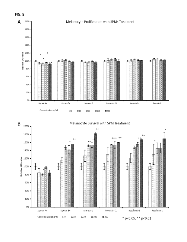
FIGURE 8 shows the effects of various specialized pro-resolving mediators on
depletion of
cultured melanocytes in vitro, wherein (A) shows the effects of SPMs (0 to 300
ng /m1) on
growth of primary epidermal melanocytes in complete melanocyte growth medium;
and
9
CA 03212601 2023- 9- 18
WO 2022/193029
PCT/CA2022/050415
(B) shows he same as (A), except that the growth factor supplements were
withdrawn
from the base melanocyte growth medium.. * p< 0.05; ** p < 0.01 (t test, each
concentration compared with the no treatment control (0 ng /ml) )
FIGURE 9 shows Vitiligo lesional skin cells with significant differences
between segmental
vitiligo and generalized (nonsegnnental) vitiligo.
FIGURE 10 shows Vitiligo lesional skin cells with significant differences
between recent
onset lesions or distant onset lesions of vitiligo.
FIGURE 11 shows cells that are depleted or enriched in Vitiligo LS as compared
to NLS,
where cells from vitiligo LS, vitiligo NLS and HNS were analyzed using xCell
deconvolution
from RNA sequencing analysis; (A) shows cells with depletion in vitiligo LS
relative to NLS;
while (B) shows cells with enrichment in vitiligo LS relative to NLS. * p<0.05
FIGURE 12 shows the impact of mental stress on canities development in B6
mice, where
B6 mice were housed in standard conditions in cages at a density of 4 mice per
cage, the
litternnates remained unchanged throughout the observation, but in about 40%
of the
cages, there is a littermate with (barbering) behavior, removing hair by
biting the other
littermates in the same cage, which can be identified by spotty hair loss and
visible bite
marks on inspection; the mice were stratified according to presence or absence
of a
barbering littermate sharing their cages, and the percentage of mice
developing canities
by week 6 were recorded and showed there was a significant increase in
percentage of
mice with canities development in mice sharing cages with a barbering mouse.
(Chi-sq, p
<0.0076)
DETAILED DESCRIPTION OF THE INVENTION
The following detailed description will be better understood when read in
conjunction with
the appended figures. For the purpose of illustrating the invention,
the figures
CA 03212601 2023- 9- 18
WO 2022/193029
PCT/CA2022/050415
demonstrate embodiments of the present invention. However, the invention is
not limited
to the precise arrangements, examples, and instrumentalities shown.
Any terms not directly defined herein shall be understood to have the meanings
commonly
associated with them as understood within the art of the invention.
Compounds as described herein may be in the free form or in the form of a salt
thereof. In
some embodiment, compounds as described herein may be in the form of a
pharmaceutically acceptable salt, which are known in the art (Berge S. M.
etal., J. Pharm.
Sci. (1977) 66(1):1-19). Pharmaceutically acceptable salt as used herein
includes, for
example, salts that have the desired pharmacological activity of the parent
compound (salts
which retain the biological effectiveness and/or properties of the parent
compound and
which are not biologically and/or otherwise undesirable). Compounds as
described herein
having one or more functional groups capable of forming a salt may be, for
example,
formed as a pharmaceutically acceptable salt. Compounds containing one or more
basic
functional groups may be capable of forming a pharmaceutically acceptable salt
with, for
example, a pharmaceutically acceptable organic or inorganic acid.
Pharmaceutically
acceptable salts may be derived from, for example, and without limitation,
acetic acid,
adipic acid, alginic acid, aspartic acid, ascorbic acid, benzoic acid,
benzenesulfonic acid,
butyric acid, cinnamic acid, citric acid, camphoric acid, camphorsulfonic
acid,
cyclopentanepropionic acid, diethylacetic acid, digluconic acid,
dodecylsulfonic acid,
ethanesulfonic acid, formic acid, fumaric acid, glucoheptanoic acid, gluconic
acid,
glycerophosphoric acid, glycolic acid, hemisulfonic acid, heptanoic acid,
hexanoic acid,
hydrochloric acid, hydrobromic acid, hydriodic acid, 2-hydroxyethanesulfonic
acid,
isonicotinic acid, lactic acid, malic acid, maleic acid, malonic acid,
mandelic acid,
methanesulfonic acid, 2-napthalenesulfonic acid, naphthalenedisulphonic acid,
p-
toluenesulfonic acid, nicotinic acid, nitric acid, oxalic acid, pamoic acid,
pectinic acid, 3-
phenylpropionic acid, phosphoric acid, picric acid, pimelic acid, pivalic
acid, propionic acid,
pyruvic acid, salicylic acid, succinic acid, sulfuric acid, sulfamic acid,
tartaric acid, thiocyanic
acid or undecanoic acid. Compounds containing one or more acidic functional
groups may
11
CA 03212601 2023- 9- 18
WO 2022/193029
PCT/CA2022/050415
be capable of forming pharmaceutically acceptable salts with a
pharmaceutically
acceptable base, for example, and without limitation, inorganic bases based on
alkaline
metals or alkaline earth metals or organic bases such as primary amine
compounds,
secondary amine compounds, tertiary amine compounds, quaternary amine
compounds,
substituted amines, naturally occurring substituted amines, cyclic amines or
basic ion-
exchange resins. Pharmaceutically acceptable salts may be derived from, for
example, and
without limitation, a hydroxide, carbonate, or bicarbonate of a
pharmaceutically
acceptable metal cation such as ammonium, sodium, potassium, lithium, calcium,
magnesium, iron, zinc, copper, manganese or aluminum, ammonia, benzathine,
meglumine, methylamine, dimethylamine, trimethylamine, ethylamine,
diethylamine,
triethylamine, isopropylamine, tripropylamine,
tributylamine, ethanolamine,
diethanolamine, 2-dimethylaminoethanol, 2-diethylaminoethanol,
dicyclohexylamine,
lysine, arginine, histidine, caffeine, hydrabamine, choline, betaine,
ethylenediamine,
glucosamine, glucamine, methylglucamine, theobromine, purines, piperazine,
piperidine,
procaine, N-ethylpiperidine, theobromine, tetramethylammonium compounds,
tetraethylammonium compounds, pyridine, N,N-dimethylaniline, N-
methylpiperidine,
morpholine, N-methylmorpholine, N-ethylmorpholine, dicyclohexylamine,
dibenzylamine,
N,N-dibenzylphenethylamine, 1-ephenamine, N,N'-dibenzylethylenediamine or
polyamine
resins. In some embodiments, compounds as described herein may contain both
acidic and
basic groups and may be in the form of inner salts or zwitterions, for
example, and without
limitation, betaines. Salts as described herein may be prepared by
conventional processes
known to a person skilled in the art, for example, and without limitation, by
reacting the
free form with an organic acid or inorganic acid or base, or by anion exchange
or cation
exchange from other salts. Those skilled in the art will appreciate that
preparation of salts
may occur in situ during isolation and purification of the compounds or
preparation of salts
may occur by separately reacting an isolated and purified compound.
In some embodiments, compounds and all different forms thereof (e.g. free
forms, salts,
polymorphs, isomeric forms) as described herein may be in the solvent addition
form, for
example, solvates. Solvates contain either stoichiometric or non-
stoichiometric amounts
12
CA 03212601 2023- 9- 18
WO 2022/193029
PCT/CA2022/050415
of a solvent in physical association the compound or salt thereof. The solvent
may be, for
example, and without limitation, a pharmaceutically acceptable solvent. For
example,
hydrates are formed when the solvent is water or alcoholates are formed when
the solvent
is an alcohol.
In some embodiments, compounds and all different forms thereof (e.g. free
forms, salts,
solvates, isomeric forms) as described herein may include crystalline and
amorphous forms,
for example, polymorphs, pseudopolymorphs, conformational polymorphs,
amorphous
forms, or a combination thereof. Polymorphs include different crystal
packing
arrangements of the same elemental composition of a compound. Polymorphs
usually
have different X-ray diffraction patterns, infrared spectra, melting points,
density,
hardness, crystal shape, optical and electrical properties, stability and/or
solubility. Those
skilled in the art will appreciate that various factors including
recrystallization solvent, rate
of crystallization and storage temperature may cause a single crystal form to
dominate.
In some embodiments, compounds and all different forms thereof (e.g. free
forms, salts,
solvates, polymorphs) as described herein include isomers such as geometrical
isomers,
optical isomers based on asymmetric carbon, stereoisomers, tautomers,
individual
enantiomers, individual diastereomers, racemates, diastereomeric mixtures and
combinations thereof, and are not limited by the description of the formulas
illustrated for
the sake of convenience.
In some embodiments, pharmaceutical compositions as described herein may
comprise a
salt of such a compound, preferably a pharmaceutically or physiologically
acceptable salt.
Pharmaceutical preparations will typically comprise one or more carriers,
excipients or
diluents acceptable for the mode of administration of the preparation, be it
by injection,
inhalation, topical administration, lavage, or other modes suitable for the
selected
treatment. Suitable carriers, excipients or diluents (used interchangeably
herein) are those
known in the art for use in such modes of administration.
13
CA 03212601 2023- 9- 18
WO 2022/193029
PCT/CA2022/050415
Suitable pharmaceutical compositions may be formulated by means known in the
art and
their mode of administration and dose determined by the skilled practitioner.
For
parenteral administration, a compound may be dissolved in sterile water or
saline or a
pharmaceutically acceptable vehicle used for administration of non water
soluble
compounds such as those used for vitamin K. For enteral administration, the
compound
may be administered in a tablet, capsule or dissolved in liquid form. The
tablet or capsule
may be enteric coated, or in a formulation for sustained release. Many
suitable
formulations are known, including, polymeric or protein nnicroparticles
encapsulating a
compound to be released, ointments, pastes, gels, hydrogels, or solutions
which can be
used topically or locally to administer a compound. A sustained release patch
or implant
may be employed to provide release over a prolonged period of time. Many
techniques
known to one of skill in the art are described in Remington: the Science &
Practice of
Pharmacy by Alfonso Gennaro, 20th ed., Lippencott Williams & Wilkins, (2000).
Formulations for parenteral administration may, for example, contain
excipients,
polyalkylene glycols such as polyethylene glycol, oils of vegetable origin, or
hydrogenated
naphthalenes. Biocompatible, biodegradable lactide polymer, lactide/glycolide
copolymer,
or polyoxyethylene polyoxypropylene copolymers may be used to control the
release of
the compounds. Other potentially useful parenteral delivery systems for
modulatory
compounds include ethylene vinyl acetate copolymer particles, osmotic pumps,
implantable infusion systems, and liposomes. Formulations for inhalation may
contain
excipients, for example, lactose, or may be aqueous solutions containing, for
example,
polyoxyethylene 9 lauryl ether, glycocholate and deoxycholate, or may be oily
solutions for
administration in the form of nasal drops, or as a gel.
Compounds or pharmaceutical compositions as described herein or for use as
described
herein may be administered by means of a medical device or appliance such as
an implant,
graft, prosthesis, stent, etc. Also, implants may be devised which are
intended to contain
and release such compounds or compositions. An example would be an implant
made of a
polymeric material adapted to release the compound over a period of time.
14
CA 03212601 2023- 9- 18
WO 2022/193029
PCT/CA2022/050415
An "effective amount" of a pharmaceutical composition as described herein
includes a
therapeutically effective amount or a prophylactically effective amount. A
"therapeutically
effective amount" refers to an amount effective, at dosages and for periods of
time
necessary, to achieve the desired therapeutic result, such as reduced vitiligo
or canities,
increased melanocyte growth and/or survival or prevention of depigmentation of
melanocytes and/or promotion of re-pigmentation in non-inflammatory
depigmented
melanocytes. A therapeutically effective amount of a compound may vary
according to
factors such as the disease state, age, sex, and weight of the subject, and
the ability of the
compound to elicit a desired response in the subject. Dosage regimens may be
adjusted to
provide the optimum therapeutic response. A therapeutically effective amount
is also one
in which any toxic or detrimental effects of the compound are outweighed by
the
therapeutically beneficial effects. A "prophylactically effective amount"
refers to an
amount effective, at dosages and for periods of time necessary, to achieve the
desired
prophylactic result. Typically, a prophylactic dose is used in subjects prior
to or at an earlier
stage of disease, so that a prophylactically effective amount may be less than
a
therapeutically effective amount.
It is to be noted that dosage values may vary with the severity of the
condition to be
alleviated. For any particular subject, specific dosage regimens may be
adjusted over time
according to the individual need and the professional judgment of the person
administering
or supervising the administration of the compositions. Dosage ranges set forth
herein are
exemplary only and do not limit the dosage ranges that may be selected by
medical
practitioners. The amount of active compound(s) in the composition may vary
according
to factors such as the disease state, age, sex, and weight of the subject.
Dosage regimens
may be adjusted to provide the optimum therapeutic response. For example, a
single bolus
may be administered, several divided doses may be administered over time or
the dose
may be proportionally reduced or increased as indicated by the exigencies of
the
therapeutic situation. It may be advantageous to formulate parenteral
compositions in
dosage unit form for ease of administration and uniformity of dosage.
CA 03212601 2023- 9- 18
WO 2022/193029
PCT/CA2022/050415
Maresin 1 is a member of a growing family of fatty acid-derived specialized
pro-resolving
mediators (SPMs), which also include protectins, resolvins (D series and E
series), and
lipoxins. Maresins, protectins and D series of resolvins are derivatives of an
omega-3 fatty
acid docosahexaenoic acid (DHA, 22:6(n-3)), while E series of resolvins are
derived from
another omega-3 fatty acid eicosapentaenoic acid (EPA, 20:5(n-3)). In
contrast, lipoxins are
derived from an omega-6 fatty acid, linoleic acids (LA, 18:2 (n-6)). LA also
gives rise to pro-
inflammatory mediators (leukotrienes and prostaglandins). It is of note that
in addition to
nnaresin1, protectins and resolvins also have strong pro-survival effects on
cultured human
epidermal melanocytes, a function not shared by lipoxin A4, which not only did
not protect
melanocytes from physiological distress, it accelerated melanocyte depletion.
Therefore,
all the SPMs are not interchangeable in their ability to protect melanocytes
or melanocyte
precursors, which may have implications in the development of therapies based
on SPMs
for the treatment of depigmentation diseases or other medical conditions in
the future.
However, as demonstrated herein maresin 1; maresin 2; lipoxin 134; protectin
Dl; resolvin
D2; and resolvin El all show ability to protect melanocytes.
Maresin 1 (macrophage mediator in resolution of inflammation 1) is a small
molecule
(molecular weight = 363) derivative of docosahexaenoic acid (DHA, an omega-3
fatty acid)
through 15 lipoxygenase-mediated oxygenation. To date, two types of cellular
receptors have
been found, G-protein coupled receptor LGR6 and nuclear receptor RORa. RORa is
expressed
by multiple skin cell types, including the melanocytes, whereas LGR6 is not
expressed by
melanocytes.
Such salts may be used in the pharmaceutical field, for example, conventional
acid addition
salts used in external preparations for skin such as salts derived from
inorganic acids such as
hydrochloric acid, bromic acid, sulfuric acid, sulfamic acid, phosphoric acid
or nitric acid and
salts derived from organic acids such as acetic acid, and organic acids such
as glycolic acid,
stearic acid, citric acid, maleic acid, malonic acid, methanesulfonic acid,
tartaric acid, malic acid,
phenylacetic acid, glutamic acid, benzoic acid, salicylic acid, 2-
acetoxybenzoic acid, fumaric
acid, toluenesulfonic acid, oxalic acid or trifluoroacetic acid. The salt may
be a base addition
16
CA 03212601 2023- 9- 18
WO 2022/193029 PCT/CA2022/050415
salt such as ammonium, dimethylamine, monomethylamine, monoethylamine or
diethylamine.
In addition, the salt may be in the form of a conventional metal salt, for
example, a salt derived
from a metal such as sodium, potassium, lithium, magnesium, or calcium. The
acid addition
salt, the base addition salt or the metal salt may be produced by a
conventional method.
Maresin 1 (MaR1) is a lipoxygenase (LOX) metabolite derived from omega-3 fatty
acid,
docosahexaenoic acid (DHA), and is a specialized pro-resolving mediator (SPM).
Maresin 1 may
be 7R, 14S-dihydroxy-4Z, 8E, 10E, 12Z, 16Z, 19Z-docosahexaenoic acid (CAS #
1268720-28-0)
may have the structure of Formula 1 below.
(E)
14 (E)I
\OH
=<Z01-1
(R)
(Z) (Z)
(Z) I
s's1COOH Formula 1
Maresin 1 may also be in the form of a pharmaceutically acceptable salt
thereof. For example,
a base addition salt or a metal salt can be prepared by reacting the ionic
form of maresin 1 may
have an appropriate base or metal ion or the like. A pharmaceutically
acceptable salt of
maresin 1 may have the structure of Formula 2 below.
14
OH
7
X+
COO- Formula 2
In Formula 2, X may for example, represent sodium, potassium, lithium,
magnesium, or
calcium. The pharmaceutically acceptable salt of maresin 1 may be sodium 7R,
14S-dihydroxy-
17
CA 03212601 2023- 9- 18
WO 2022/193029
PCT/CA2022/050415
4Z, 8E, 10E, 12Z, 16Z, 19Z-docosahexaenoic acid or may be potassium 7R, 14S-
dihydroxy- , 10E,
12Z, 16Z, 19Z-docohexaenoic acid. Maresin 1 may also be in the form of a
solvate thereof.
Maresin 2 (CAS # 1639809-46-3), 13R,14S-dihydroxy-4Z,7Z,9E,11E,16Z,19Z-
docosahexaenoic
acid, as represented by Formula 3 below is another tested SPM.
HO,Z)j
1 I 143
,'T;70H
COOH Formula 3
Alternatively, a pharmaceutically acceptable salt of maresin 2 may have the
structure of
Formula 4 below.
HO
JTJ
OH
X'
C00- Formula 4
Lipoxin B4 (CAS # 98049-69-5), 5S,14R,15S-trihydroxy-6E,8Z,10E,12E-
eicosatetraenoic acid, as
represented by Formula 5 below is another tested SPM.
OH
JCOOH
HO OH Formula 5
Alternatively, a pharmaceutically acceptable salt of lipoxin B4 may have the
structure of
Formula 6 below.
18
CA 03212601 2023- 9- 18
WO 2022/193029
PCT/CA2022/050415
OH X+
HO OH Formula 6
Protectin D1 (CAS # 660430-03-5), 10R,17S-dihydroxy-4Z,7Z,11E,13E,15Z,19Z-
docosahexaenoic
acid, as represented by Formula 7 below is another tested SPM.
.1 OH
=
COOH
OH Formula 7
Alternatively, a pharmaceutically acceptable salt of protectin D1 may have the
structure of
Formula 8 below.
OH COO-
OH Formula 8
Resolvin D2 (CAS # 810668-37-2), 7S,16R,17S-trihydroxy-4Z,8E,10Z,12E,14E,19Z-
docosahexaenoic acid (4Z,75,8E,10Z,12E,14E,16R,175,19Z)-7,16,17-
trihydroxydocosa-
4,8,10,12,14,19-hexaenoic Acid, as represented by Formula 9 below is another
tested SPM.
19
CA 03212601 2023- 9- 18
WO 2022/193029
PCT/CA2022/050415
COOH
HO
Formula 9
Alternatively, a pharmaceutically acceptable salt of resolvin D2 may have the
structure of
Formula 10 below.
X+
coo-
HO
Formula 10
Resolvin El (CAS # 552830-51-0), 55,12R,18R-trihydroxy-6Z,8E,10E,14Z,16E-
eicosapentaenoic
acid, as represented by Formula 11 below is another tested SPM.
OH
< OH COOH
Formula 11
Alternatively, a pharmaceutically acceptable salt of resolvin El may have the
structure of
Formula 12 below.
CA 03212601 2023- 9- 18
WO 2022/193029
PCT/CA2022/050415
OH
X+
H0000-
Formula 12
"Solvate" as used herein means a complex or aggregate formed by one or more
solute
molecules, i.e., a suitable SPM or a pharmaceutically acceptable salt thereof,
and one or more
solvent molecules. The solvate may be, for example, a complex or aggregate
formed with
water, methanol, ethanol, isopropanol or acetic acid.
A suitable SPM as described herein may also be in the form of its
stereoisomer. The
stereoisomers include all stereoisomers such as enantiomers and diastereomers.
The
compound may be a stereoisomerically pure form or a mixture of one or more
stereoisomers,
for example, a racemic mixture. The separation of certain stereoisomers can be
carried out by
any of the conventional methods known in the art.
"Active ingredient" is intended to carry out the function referred to in the
composition and
excludes those that do not fulfill the function as they are included in minor
amounts as
impurities.
A suitable SPM as described herein may be chemically synthesized or
commercially available or
extracted from natural sources.
The compositions of suitable SPMs as described herein may be one that promotes
melanocyte
growth and/or survival.
The composition may comprise a "therapeutically effective amount" of a
suitable SPM or a
pharmaceutically acceptable salt, solvate, or combination thereof. In this
composition,
"therapeutically effective amount" means an amount sufficient to exhibit a
therapeutic effect
when administered to a subject or a cell in need thereof. "Treatment" means
treating a disease
21
CA 03212601 2023- 9- 18
WO 2022/193029
PCT/CA2022/050415
or medical condition in a mammal, including a human, including an individual,
which includes:
(a) preventing the occurrence of the disease or medical condition, cure; (b)
relieving the disease
or medical condition, i.e., eliminating or ameliorating the disease or medical
condition in the
patient; (c) inhibiting the disease or medical condition, i.e. slowing or
stopping the progression
of the disease or medical condition in the individual; or (d) relieving the
disease or medical
condition in the subject. In particular, one that promotes melanocyte growth
and/or survival.
The one or more specialized pro-resolving mediators (SPMs) or a
pharmaceutically acceptable
salt, hydrate, or hydrated salt, or its optical isomer, racemate,
diastereoisomer or enantiomer
thereof, excluding lipoxin A4, may be present in an amount of from 0.001% to
80%, such as
from 0.01% to 60%, from 0.01% to 40%, from 0.01% to 30%, from 0.01% to 20% %,
0.01% to
10%, 0.01% to 5%, 0.05% to 60%, 0.05% to 40%, 0.05% to 30%, 0.05% to 20% From
0.05% to
10%, from 0.05% to 5%, from 0.1% to 60%, from 0.1% to 40%, from 0.1% to 30%,
from 0.1% to
20% % to 10% by weight, or 0.1% to 5% by weight of SPM as described herein. In
particular, for
topical vitiligo treatments a 0.01% maresin 1 was tested.
Drug delivery compositions may be prepared and utilized to treat or prevent a
variety of
diseases or conditions, particularly where the treatment site contains
melanocytes. For
example, skin melanocytes, hair melanocytes, eye melanocytes, or ear
melanocytes to
deliver SPMs described herein to promote melanocyte growth and/or survival.
The SPM
may be selected from one or more of the following: maresin 1; maresin 2;
lipoxin 84;
protectin Dl; resolvin D2; and resolvin El. The composition described herein
may be used
for the prevention of depigmentation of melanocytes and/or promoting re-
pigmentation
in non-inflammatory depigmentation of melanocytes. Examples of diseases or
conditions
that may be treated, may for example, include age-associated leukoderma and
leukotrichia;
dormant vitiligo lesions; chemically induced vitiligo; vitiligo that is non-
responsive to
inflammatory therapies; and canities.
22
CA 03212601 2023- 9- 18
WO 2022/193029
PCT/CA2022/050415
MATERIALS AND METHDS
Study subjects and Skin biopsies:
This study was approved by the Clinical Ethics Board of University of British
Columbia. For
transcriptonne sequencing and cellular profiling experiments, 4 mm punch
biopsies were
obtained from the lesional and nonlesional skin of 36 vitiligo patients (29
with generalized
vitiligo and 7 with segmental vitiligo), 15 patients with chronic eczema, and
healthy skin
from 9 volunteers as described previously (45). The biopsies were bisected,
with 1/2 placed
immediately in RNA Later solution (Life LabsTM) and stored at -20 until
further use. The
other 1/2 placed in formalin for histological assessment. For flow cytometry
analysis of
macrophages in vitiligo patients, 5 mm punch biopsies were obtained from the
lesional,
border and non-lesional skin, and immediately placed in saline for cell
isolation (See below).
RNA extraction and transcriptome sequencing:
Bulk RNA was extracted from skin biopsies using the RNeasyTM Fibrous Tissue
Mini Kit as we
had described previously (14, 46), and used for transcription sequencing by
Novo Gene'
(Tianjin China) using the illuminaTM platform (HiSeq PE150), generating at
least 30 million
clean reads for each sample. The expression of each transcript was normalized
to the total
number of transcripts and the length of the transcripts and expressed in FPKM.
Ingenuity
Pathway AnalysisTM (IPA) was used for analysis of differentially expressed
genes between
lesional and nonlesional skin, and between skin biopsies from vitiligo and
chronic eczema
patients and the skin biopsies from health controls using two fold change and
p<0.05 as the
cut off for statistical significance using R program.
In silico profiling of cellular infiltrates in skin biopsies:
We used xCell tool developed by Aron et al. (47, 48) to evaluate the relative
changes in
recognizable immune cells in the skin biopsies. Increasingly used for in
silico analysis of
cellular infiltrates in inflammatory as well as malignant diseases (4952),
this method is based
on the validated gene expression signatures of 64 types of cells involved in
inflammation
and immune responses, and is capable of estimating the relative abundance of
the
immune-active cells present in the tissue biopsies. In addition, a composite
score
23
CA 03212601 2023- 9- 18
WO 2022/193029
PCT/CA2022/050415
(lmmuneScoreTM) is generated for estimation of the general extent of immune
response in
a given tissue.
Isolation and detection of M1 and M2 macrophages in skin biopsies by flow
cytometry
For isolation of macrophages from human vitiligo skin: 5 mm punch biopsies
were minced
finely with scissors and mixed with 3 ml digestion buffer containing 0.8mg/m1
Collagenase
IV and 0.03mg/m1 DNAse (Sigma Aldrich"), 10% FBS, 1% penicillin/streptomycin
in RPM!
medium. Samples were incubated overnight in 5% CO2 at 37 C, then harvested in
PBS,
filtered through a 100-p.m strainer and centrifuged. Surface staining antibody
panel
included anti-humans CD11b-Alexa Fluor 488", CD163-BV421TM and CD80-Alexa
fluor 647"
(Biolegen"). Cells were incubated with antibodies for 30 min at 4 C, washed in
PBS
containing 1% FCS and analyzed using LSRiiTM cell sorter (BD Biosciences").
For isolation of macrophages from nnurine skin: 1 cm X 1 cm sized skin samples
were cut
into small pieces, placed in a solution of PBS containing 1 mg/ml dispase
(RocheTM) and
incubated for 1 hour at 37 C. The samples were then transferred into RPM I
containing 1
mg/ml Collagenase IV and 0.1mg/m1DNAse (Sigma Aldrich") and incubated for 90
minutes
at 37 C. Single cell suspension was prepared by passing through 100 p.m
strainer. Cells
were washed and stained in PBS without Ca' and Mg2+ supplemented with 1% heat-
inactivated FCS. Cell surface staining panel included anti-mouse CD163-
BV421TM, CD80-
Alexa fluor 647TM, and CD11b-Percp Cy5.STM (BioiegendTM) for 30 minutes at 4
C. Cells were
washed with PBS and analyzed with LSRiiTM cell sorter (BD Biosciences").
B6 mouse model of vitiligo with TRP2 immunization
Eight week old female C57BL/J6 mice were ordered from The Jackson
LaboratoryTM, and fed
a standard diet of Animal Care Facility of Vancouver Coastal Health Research
institute (4
mice / cage). For induction of vitiligo, mice were immunized by intradermal
injections (ID,
starting at Day 0) at the left hock, and repeated at 2-week, using the method
described by
S. You et al. that efficiently induces melanocyte specific CD8+ T cell
responses (53). The
immunogen consisted of TRP2-180 (50 g) peptide mixed with LPS (5 g) and CpG
ODN 1826
24
CA 03212601 2023- 9- 18
WO 2022/193029
PCT/CA2022/050415
(5 g) in 504 of PBS solution per mouse per injection. Immunization response
score was
recorded at 48 hours after the first immunization by evaluating the redness
and swelling
(0= none, 1= mild, 2 = moderate, 3 = severe). For assessing macrophage M1 and
M2
populations, the TRP-immunized and non-immunized mice were anaesthetized 48
hours
after immunization for skin sampling at the immunization site and used for
cell isolation as
described above.
For assessing the effects of maresin 1 treatment, C57BL/J6 mice were randomly
assigned
to the sham injection control group (200 1.1.1 saline IP, n= 20) and the
Maresin 1 injection
group (800 ng in 200 pi saline IP, n = 20). All mice received three IP pre-
treatments before
the first immunization and continued to receive IP injections 3 times per week
till the end
of study. Mice were monitored 5 times per week and evaluated using a
Depigmentation
Area Scoring Template (0 - 5) (Figure 5C).
At the end of the study (Day 35), after photography, the mice were euthanized
and full-
thickness skin biopsies (1 cm by 1 cm) of the left hock immunization sites
were collected
with surgical scissors. Half of the sample was store in RNAlaterTM for RNA
extraction and
the other half was fixed in OCT embedding medium for histopathology analysis.
Spleen and
inguinal lymph node tissues were collected for immunohistochemistry and RNA
isolation.
B6 mouse model of canities and serum maresin quantification
To observe aging-associated spontaneous hair greying (canities), C57BL/J6 mice
aged 8
weeks were purchased from JAX LabsTM and kept in standard conditions for 8
weeks. The
fur color as well as overall appearance and behaviors were monitored daily by
visual
inspection and by photography. By 6-8 weeks, about 20-40% of the mice would
develop
spontaneous canities. At the end of observation, central venous blood was
collected and
used for ELISA analysis using maresin 1 ELISA kit (Caymen ChemicalsTM)
following the
manufacturers' recommended protocol with purified maresin 1 serving as the
chemical
standard.
CA 03212601 2023- 9- 18
WO 2022/193029
PCT/CA2022/050415
Effects of maresin 1 on canities development in B6 mice
C57BL/J6 mice from The Jackson Laboratory', 8 weeks of age were divided into
two groups
that received intraperitoneal injections of maresin 1 (800 ng in 100 il
saline) or saline alone
three times a week for 6 weeks. The mice were observed at weekly intervals by
visual
inspection. At the end of the experiment, the mice were photographed and
weighed.
Melanocyte proliferation and survival assay in vitro
Human neonatal epidermal melanocytes (HEMn-DP) were purchased from
ThermoFisher
ScientificTM (C-202-5C). Melanocytes were cultured in full growth medium
(Medium 254
supplemented with Human Melanocyte Growth Supplement, both purchased from
ThermoFisher ScientificTM. Cells were expanded and passaged 3 times before
conducting
the assay. 4x10³ melanocytes per well were seeded into 96 well plate and
allowed to
recover for 4 hours before the treatment was added for each condition in 100
pi final
volume. Cells were treated and kept in 37 C, 5% CO2 for 3 days. Viability
assay was
conducted using CellTiter-Blue Cell Viability Assay KitTM (PromegaT", G8080)
and signal was
measured using GlowMax" plate reader (PromegaTM) after 4 hours at 37 C, 5%
CO2.
Merasin 1 (CaymanTM) was diluted into 11.a.g/m1 using PBS and added to the
culture medium
at concentrations ranging from 0 to 1 ig / ml. For survival assays, the
procedure was
essentially the same as proliferation assay except that melanocytes were kept
in M254
medium without human melanocyte growth supplement. The assays were conducted
in
triplicates.
EXAMPLES
EXAMPLE 1: In silico profiling of immune cell landscape reveals M2 macrophage
deficiency in vitiligo lesional skin microenvironment, but not in vitiligo
nonlesional skin
or skin affected by eczema
To gain sights into the cellular changes preferentially present in the
microenvironment of
vitiligo lesional skin, we performed whole transcriptome sequencing followed
by cellular
deconvolution analysis on vitiligo lesional skin, using vitiligo non-lesional
skin, and skin
26
CA 03212601 2023- 9- 18
WO 2022/193029
PCT/CA2022/050415
biopsies from healthy volunteers and eczema patients as the controls. Of the
64 cell types
evaluable by xCell method, 14 types of cells showed significant enrichment in
vitiligo skin
biopsies compared with healthy normal skin (Figure 1A), including cells
involved in innate
immunity (monocytes, macrophages and M1 macrophages) and adaptive immunity
(such
as CD8+, CD4+ T cells). Of these, the majority (including CD8+ T cm, CD8+
naive T,
neutrophils, CD8+ Tern, Th2, chondrocytes, CD4+ memory T, GMP, and mast cells)
were
enriched in both lesional and nonlesional vitiligo skin (Figure 1B). In
contrast, 7 types of
cells showed significant depletion specifically in vitiligo lesional skin
compared with healthy
normal skin, but not in vitiligo nonlesional skin, including melanocytes, M2
macrophages,
mesenchymal stem cells (MSC), mesangial cells, endothelial cells, ly
endothelial cells, and
my endothelial cells (Figure 1C).
To understand if the cellular changes observed in vitiligo lesional skin is
specific for this
immune mediated skin condition, we performed the same analysis on skin
biopsies from
patients with chronic eczema, a prototypical Th2 immune mediated inflammatory
skin
disease. As shown in Figure 2 the cells that were enriched in vitiligo
lesional skin were in
general also enriched in eczema lesional biopsies. However, the cells with
depletion in
vitiligo lesional skin were not depleted in eczema lesional skin, suggesting
that the
depletion of melanocytes and other cells (such as M2 macrophages) was specific
for vitiligo
skin lesions.
To test if the lesional cellular infiltrates are correlated with vitiligo
subtypes or disease
stages, we stratified the vitiligo patients into morphological groups
(generalized vs
segmental vitiligo) or disease duration groups (active vitiligo with onset
within 12 months,
vs relatively stable vitiligo with duration longer than 12 months). Of the 36
vitiligo
individuals analyzed, 29 had generalized vitiligo, and 7 had segmental
vitiligo. Two types
of cells showed significant differences between segmental vitiligo and
generalized vitiligo,
ly endothelial cells and my endothelial cells (Figure 9). These cells were
depleted in
generalized vitiligo but not in segmental vitiligo. When compared with
vitiligo that had
been present for more than 12 months, vitiligo lesions with shorted disease
duration
27
CA 03212601 2023- 9- 18
WO 2022/193029
PCT/CA2022/050415
(typically more active or progressive) had much higher immune score, and more
significant
enrichment of monocytes, granulocyte-monocyte progenitor cells and dendritic
cells
(Figure 10), suggesting that there is higher immunological reactions in early
stages of vitiligo
development.
EXAMPLE 2: Defective M2 macrophage polarization in human vitiligo lesional
skin by flow
cytometry
Since vitiligo lesional skin showed specific depletion of M2 macrophages in
the in silico cell
profiling analysis (Figures 1, 2, and 11), further experiments were performed
to verify this
discovery. Fresh skin biopsies were obtained from the lesional and nonlesional
skin of
patients with vitiligo, and used for cell isolation using protocol of R. Clark
etal. (54'55). Flow
cytometry was performed on CD11b+ cells with monoclonal antibodies against
CD80 (M1
marker) and CD163 (M2 marker). As shown in Figure 3A, the CD163+ cells were
significantly
reduced in vitiligo lesional skin (LS) compared with non-lesional skin (NLS).
While M2
macrophages accounted for 37.3% CD11b+ cells on average in the non-lesional
skin, they
were reduced to 13% in the lesional skin (p=0.037). The M1 macrophages showed
the
opposite changes, being enriched in vitiligo lesional skin compared with non-
lesional skin
(p=0.0035) (Figure 3B). Thus, there is a significant reduction of M2/M1 ratio
in vitiligo
lesional skin (0.5) as compared with nonlesional skin (4.7, p<0.016),
confirming that M2
polarization was defective in vitiligo lesional skin (Figure 3C).
EXAMPLE 3: Decreased M2 macrophage polarization in the skin of B6 mice induced
to
develop vitiligo by immunization using a melanocyte-specific antigen
To test if M2 macrophage depletion is also present in animal models of
vitiligo, we
employed a well-established B6 vitiligo mouse model that is mediated by
melanocyte-
specific cytotoxic CD8+ T cells (53' 56). This model involves intradermal
immunization of the
B6 (black) mouse with a melanocyte-specific antigen TRP-2 in an adjuvant that
contains LPS
and CpG, which elicits a robust acute immune response at the immunization site
(redness
and swelling), that is then followed by the development of white patches of
hair (vitiligo)
28
CA 03212601 2023- 9- 18
WO 2022/193029
PCT/CA2022/050415
in about 6 weeks. To test if this process involves M2 macrophage depletion, we
obtained
skin biopsies from the immunization sites 48 hours after immunization (when
the
immunization response is at its peak), and used flow cytometry to measure
M2/M1 ratio.
As shown in Figure 4, TRP2 immunization resulted in a significant reduction of
M2
polarization, with M2/M1 ratio decreasing from 0.44 to 0.21.
EXAMPLE 4: Maresin 1 treatment of B6 mice enhanced skin M2 macrophage
polarization and reduced melanocyte depletion upon vitiligo induction
M2 macrophages are known to secrete a potent soluble functional mediator,
maresin 1,
which not only mediates the diverse functions of M2 macrophages, but also
stimulates M2
macrophage polarization in an autocrine feedback loop (37). To test if M2
macrophage
depletion contributes to melanocyte depletion in B6 mouse vitiligo model, we
pretreated
the mice with maresin 1 IP prior to TRP2 immunization. As shown in Figure 4,
ma resin 1
treatment dramatically enriched M2 macrophages at the TRP2 immunization site,
restoring
the post-immunization M2/M1 ratio to a level above the pre-immunization state.
After immunization, the mice continued to receive maresin 1 or saline IP
treatments three
times per week for 8 weeks (Figure 5A). The mice were observed weekly by
visual
inspection and photography for development of vitiligo at the immunization
site (Figure
5B). To quantify the severity of vitiligo, a visual scoring template (Figure
5C) was used, and
the averages of depignnentation scores for each group were plotted according
the time of
observation. There was a significant reduction of vitiligo development in the
maresin 1
treated group compared with sham treated mice (Figure 5D).
EXAMPLE 5: Maresin l's inhibitory effects on melanocyte depletion was
correlated with
immure resolution effects
Since maresin 1 has strong pro-resolution effects on inflammation and immune
responses,
we evaluated the correlation between peak immunization response and the level
of
depigmentation in maresin 1 treated mice. As shown in Figure 5E, maresin 1
treatment
29
CA 03212601 2023- 9- 18
WO 2022/193029
PCT/CA2022/050415
significantly reduced the immunization response. Further, there was a
significant
correlation between immunization response score and vitiligo depigmentation
area score
(Figure 5F, p<0.0001), suggesting that maresin 1 has vitiligo-inhibitive
effects and that those
effects could in part could be attributed to its ability to suppress and
resolve melanocyte-
specific immune response induced by immunization with TRP-2 peptide.
EXAMPLE 6: 66 mouse as a model of canities
To evaluate if maresin 1 also could attenuate immune independent melanocyte
depletion,
we performed additional studies in B6 mouse canities model. Canities is a
natural
phenomenon that develops in humans and other mammals with advancing age.
Recent
studies demonstrated that canities development in mice is not the result of
immune
activation, but a result of aging-associated melanocyte decline due to
exhaustion of
melanocyte stem cells (57,58) In our vitiligo studies using B6 black mice, we
noted that non-
immunized B6 mice naturally developed canities starting at about 3 months of
age with
approximately 30% mice showing scattered white hairs that were mixed with
normal
pigmented black hairs in a diffused distribution throughout the hair covered
skin, and that
there was a significant association between canities development and
experience of mental
stress caused by sharing cage with a barbering aggressive mouse (Figure 12),
consistent
with the previous observation that sympathetic nervous hyperactivation rather
than
immune activation causes premature canities (57' 58).
EXAMPLE 7: Canities development in 66 mice was correlated with decreased M2
macrophage function
Since white hair development due to immune-induced melanocyte depletion in B6
vitiligo
mice was correlated with suppressed M2 macrophage polarization, we wondered if
non-
immune mediated melanocyte depletion in canities was also correlated with
reduced M2
macrophage function. To test this, we measured serum maresin 1 as a surrogate
marker
of M2 macrophages, as maresin 1 is mainly produced by M2 macrophages (36,37)=
As shown
in Figure 6A and 66, the serum level of maresin 1 was significantly lower in
mice that
CA 03212601 2023- 9- 18
WO 2022/193029
PCT/CA2022/050415
developed canites as compared with the age and sex matched B6 mice without
canities
(p=0.026).
EXAMPLE 8: Augmentation of M2 macrophage function with maresin 1 prevented
canities development as well as aging-associated weight gain in B6 mice
To test if maresin 1 reduction contributes to canities development, we divided
B6 mice into
two groups (Figure 6C), one receiving 800 ng maresin 1 IP injections three
times a week for
8 weeks. The other group receiving saline IP injections. The mice were
examined weekly
by visual inspection and photography to document hair color, general
appearance, and
behavior, and by gravimetry to assess aging associated weight gain. As shown
in Figure 4D,
meresin 1 treatment significantly enriched M2 macrophages in the skin of B6
mice,
increasing M2/M1 ration from 0.44 to 5.34. The maresin 1 treated mice
maintained
healthier body weight, with a significantly lower aging associated weight gain
compared
with saline treated control mice (14.2% weight gain vs 17.7% weight gain,
p=0.0423) (Figure
6E), suggesting maresin 1 may have a global antiaging effects in B6 mice.
Further, as shown
in Figure 6F, in the saline treated control group 15% of the mice developed
canities by 15
weeks of age. In the maresin 1 treatment group, canities development was
completely
blocked (p<0.0125, Log Rank Test).
EXAMPLE 9: Maresin 1 prevents depletion of cultured epidermal melanocytes due
to
physiological distress in vitro
It is unknown how maresin 1 treatment prevents melanocyte depletion in
vitiligo and
canities. Theoretically, this effect could be due to indirect effects, such as
through
suppressing immune mediated cytotoxicity against the melanocytes, or through
direct
protective effects on the melanocytes or melanocyte precursors /stem cells.
The fact that
the reduction of vitiligo depigmentation effects of maresin 1 were tightly
correlated with
its ability to decrease immunization reaction triggered by TRP2 immunization
suggests that
immune resolution could explain at least in part the mechanism used by maresin
1 to
prevent melanocyte depletion in vitiligo. However, the fact that maresin 1
could also
31
CA 03212601 2023- 9- 18
WO 2022/193029
PCT/CA2022/050415
prevent immune-independent melanocyte depletion caused by stem cell exhaustion
in
canities suggests that maresin 1 might also have a direct protective role on
the melanocytes
or their precursors.
To test this possibility, we cultured primary human epidermal melanocytes (and
precursors)
isolated from neonatal foreskin. Under normal culture conditions, the
melanocytes
increase in number as a result of proliferation or differentiation from
proliferating
precursors in the culture. However, under physiological distress (withdrawal
of growth
factors in the culture medium), there is a significant depletion in the
surviving melanocytes
after three days in culture. As shown in Figure 7, maresin 1 treatment had a
significant
protective effect on the melanocytes, markedly reducing melanocyte depletion
caused by
growth factor withdrawal. Since there were no immune cells present in the
assay, the anti-
depletion effects of maresin 1 on melanocytes appear to be independent of its
immune-
resolution effects. Instead, maresin 1 likely acts directly on melanocytes or
their precursors
to prevent melanocyte depletion due to growth factor withdrawal.
EXAMPLE 10: Melanocyte-protective effects of other specialized pro-resolving
mediators (SPMs)
Maresin 1 is a member of a growing family of SPMs, which are lipid derived
mediators
capable of resolving immune response and inflammation. There are four main
groups of
SPMs, the maresins, protectins, resolvins (E and D series), and lipoxins. To
test if the
protective effect of maresin 1 on melanocytes is present in other SPMs, we
performed in
vitro melanocyte proliferation and survival assays using mediators
representing various
SPM groups (i.e. maresin 1; ma resin 2; lipoxin A4; lipoxin B4; protectin Dl;
resolvin D2; and
resolvin El). As shown in Figure 8, most species of SPMs have robust
concentration-
dependent pro-survival effects directly on the melanocytes in vitro, with
lipoxin A4 being
the only exception. Lipoxin A4 not only did not have pro-survival effects on
the
melanocytes, it significantly decreased melanocyte survival in a concentration
independent
fashion (Figure 8).
32
CA 03212601 2023- 9- 18
WO 2022/193029
PCT/CA2022/050415
EXAMPLE 11: Topical maresin 1 leads to repigmentation of vitiligo patches
refractory to
topical anti-inflammatory therapies
Topical maresin 1 was tested on a 38 year old Asian man with a 5 year and 3
month
history of developing two white patches on the dorsal right hand and one white
macule
on the right mid abdomen. The white skin patched were first noted in the early
summer 5
years ago, and had not changed significantly in size. There were no signs of
inflammation
such as redness, or presence of scales. There were no symptoms such as
itchiness or
pain. The subject had been using topical mometasone fumarate 0.1% cream BID
for the
abdominal macule, and clobetasol 0.05% cream OD for the two patches on the
right hand
for the past 12 months and did not notice any change in the size of the white
areas.
The subject had been healthy, with no history or diagnosis of thyroid
diseases, cutaneous
lupus or other chronic inflammatory diseases. The subject reported no exposure
to
chemicals such as benzene or phenol, or used any known skin depigmentation
drugs such
as hydroquinone or monobenzoether of hydroquinone.
On examination, the subject appeared to be well. Complete skin examination was
performed. The entire skin appeared to be normal aside from three lesions of
depigmentation: (1) a 1.5 X1.2 cm white patch on the mid right abdomen; (2) a
1.3X1.0
cm white patch on the dorsum of right third metacarpal head; and (3) a 1.0 X
0.8 cm sized
white macule on the dorsum of right 4th metacarpal head.
Since the subject did not respond to the standard anti-inflammatory
medications for 12
months, the subject was looking for other treatment options, and decided to
try an
ethanol solution containing 0.01% maresin 1 twice daily. The subject did no
notice any
adverse events such as irritation, itchiness or redness. By the end of three
months, the
subject noted significant repigmentation starting from peripheral margin and
moving
centrally. The areas of the three depigmented lesions were decreased by 11.7%
(13.6%,
10.5% and 11.1%, for the three lesions, respectively).
33
CA 03212601 2023- 9- 18
WO 2022/193029
PCT/CA2022/050415
Discussion
Despite sharing the loss of melanin pigmentation, vitiligo and canities differ
significantly in
reported pathogenic mechanisms based on available literature. Melanocyte
depletion in
vitiligo is mainly mediated by immune destruction of differentiated
melanocytes in the
epidermis and hair follicles, whereas the melanocyte depletion in canities is
the result of
exhaustion of melanocyte stem cells that is independent of immune response.
Our results
collectively point to a previously unknown shared mechanism contributing to
melanocyte
depletion in vitiligo as well as in canities (i.e. through a deficiency in M2
macrophages or
their functional mediator maresin 1 or other suitable SPM).
Our results demonstrated that M2 macrophages, through production of functional
mediator maresin 1 or other suitable SPM, are required to maintain the
homeostasis niche
for the melanocytes by suppressing immune mediated attacks on melanocytes and
by
directly promoting survival of the melanocytes or their precursors. M2
macrophage
function may maintain melanocyte homeostasis by two complementary mechanisms.
One,
being the pro-resolving function of maresin 1 or other suitable SPM keeps the
immune
cytotoxicity low in the normal microenvironment for the melanocytes and their
precursors.
Two, being maresin 1 or other suitable SPM pro-survival function on
melanocytes or
precursors reduces melanocyte death caused by spontaneous aging, or distress
induced by
sympathetic hyper-activation or withdrawal of growth factors. In contrast,
deficiency of
M2 function makes the homeostatic niche unfavorable for the melanocytes,
leading to
melanocyte depletion due to unchecked immune cytotoxicity and inadequate
melanocyte
protection. Importantly, the homeostatic environment can be modulated
by
supplementation with exogenous M2 macrophage functional mediator maresin 1.
Our
experiments demonstrated that maresin 1 dampens immune cytotoxicity to
melanocytes
induced by TRP2 (Figure 5A), increases skin-resident M2 polarization under
immune
activation (Figure 4), reduces immune mediated melanocyte destruction in
vitiligo mouse
model (Figure 5), prevents non-immune-mediated melanocyte depletion in
canities mouse
34
CA 03212601 2023- 9- 18
WO 2022/193029
PCT/CA2022/050415
model (Figure 6), and protects melanocytes from death caused by growth factor
starvation
in vitro (Figure 7).
There is growing experimental evidence that M2 macrophages are involved in
tissue
homeostasis in adult stem cell niches (59), and in the adipose tissue, M2-like
macrophages
form the niche necessary for the adipocyte precursors (60, 61). Given that
exhaustion of
melanocyte stem cells is the main mechanism of canities pathogenesis (26)
(27),
our
observations of decreased M2 macrophage function (reduced serum maresin 1
levels) in
mice with canities (Figure 6B), and maresin 1 treatment preventing canities
development
(Figure 6D and F), strongly suggest that M2 macrophages also are necessary for
maintaining
melanocyte homeostasis.
Previous studies showed that melanocyte homeostasis depends on the maintenance
of the
pool of stems cells, as loss of the stems cells leads to the aging of the hair
follicles (62).
Maintenance of the niche required by the stem cells in the hair follicles
involves a network
of molecular signaling events, such as Co117a1(62), wnt/b-catenin signing
(63), SCF/kit
signaling (64, 65), suppression of oxidative stress(66), and CXCL12 (67). Our
results add M2
macrophages as another essential piece in the puzzle of the signaling network
required for
the maintenance of melanocyte homeostasis niche.
The studies in murine models by B Zhang et al. showed that hyperactive
sympathetic
signaling induced by mental stress can lead to exhaustion of melanocyte stem
cells resulting
in melanocyte depletion in the hair follicles, which is independent of immune
response (26).
Our results confirmed the strong link between mental stress and development of
canities
as mice housed in the same cage with an aggressive cage mates had much higher
rate of
development of canities (Figure 12). We further demonstrated that melanocyte
stem cell
exhaustion induced by mental stress could be prevented by treatment with
maresin 1
(Figure 6), thus expanding the scope of physiological functions of M2
macrophages to
include defending against psychological or mental distress.
CA 03212601 2023- 9- 18
WO 2022/193029
PCT/CA2022/050415
It is not known how maresin 1 or other SPMs function to maintain melanocyte
homeostasis
and prevent melanocyte depletion due to immune dependent or independent causes
is
unknown at present. Previous studies showed that maresin 1 can inhibit
oxidative stress
which has been shown to be important for vitiligo pathogenesis as well as
impairment of
melanocyte stem cell niche. It is possible that maresin 1 or other suitable
SPMs inhibit
melanocyte depletion by prevention of oxidative stress. The fact that in in
vitro culture of
isolated human epidermal melanocytes maresin 1 can significantly increase
melanocyte
resistance to depletion induced by physiological distress caused by withdrawal
of growth
supplements suggests that marein 1 or other suitable SPMs can directly act on
the
melanocytes or their precursors to promote their survival. The mechanism by
which this is
achieved is unknown at present.
Maresin 1 (macrophage mediator in resolution of inflammation 1) is a small
molecule
(molecular weight = 363) derivative of docosahexaenoic acid (DHA, an omega-3
fatty acid)
through 15 lipoxygenase-mediated oxygenation. A member of specialized pro-
resolving
mediators (SPMs) (68' 69), maresin 1 mediates M2 macrophages' functions in
resolution of
inflammation, would healing and tissue regeneration (36). To date, two types
of cellular
receptors have been found, G-protein coupled receptor LGR6 and nuclear
receptor RORa.
RORa is expressed by multiple skin cell types including the melanocytes,
whereas LGR6 is
not expressed by melanocytes. Further studies are needed to investigate the
mechanism
used by maresin 1 and other suitable SPMs to stimulate and protect
melanocytes.
Our results offer an explanation to the clinical characteristics of vitiligo
that could not be
explained fully by melanocyte destruction by autoreactive CD8+ T cells (11-
13), resident
memory T cells (25) and NK cells (46). Our data suggest that lack of growth
support for
melanocyte homeostasis in vitiligo lesional microenvironment may help explain
why
immune suppressants, such as cyclosporine, methotrexate, and cortical
steroids, are
generally ineffective in bringing back melanocytes to established vitiligo
lesions, especially
in locations on hands, feet and genital regions.
36
CA 03212601 2023- 9- 18
WO 2022/193029
PCT/CA2022/050415
Given that maresin 1 can suppress immune response as well as directly
stimulate
melanocytes or their precursors, therapies based on maresin 1 (or other M2
agonists) may
have particular advantages over traditional immune-suppression based therapies
in the
treatment of vitiligo, especially for lesions that are unresponsive to immune-
suppressive
therapies. In addition to vitiligo, several other conditions involve loss of
melanocytes,
including canities, chemical leukoderma and aging associated leukoderma. Our
results
suggest that maresin 1 and other suitable SPM molecules may be able to treat
these
conditions as well.
The mechanism leading to M2 deficiency in vitiligo and canities requires
further
investigation. Aging associated decrease in maresin 1 production has been
discovered for
skeletal muscles (70). Therefore, our observation of maresin 1 reduction in
canities
strengthens the link between declining maresin 1 production (and reduction in
M2 function
in general) and physiological aging, and raises the possibility that maresin
1, and related
molecules, may be a treatment for canities and other manifestations of
physiological aging.
Indeed, in B6 mice treated with martesin 1, we observed a reduction in
canities formation
as well as aging-associated weight gain, suggesting that maresin 1 may have a
more global
anti-aging benefits for the mice. Further experiments are warranted to
evaluate maresin
l's effects, if any, on other aspects of aging, including cardiovascular,
neural and cognitive
functions.
Maresin 1 is a member of a growing family of fatty acid-derived specialized
pro-resolving
mediators (SPMs), which also include protectins, resolvins (D series and E
series), and
lipoxins. Maresins, protectins and D series of resolvins are derivatives of an
omega-3 fatty
acid docosahexaenoic acid (DHA, 22:6(n-3)), while E series of resolvins are
derived from
another omega-3 fatty acid eicosapentaenoic acid (EPA, 20:5(n-3)). In
contrast, lipoxins are
derived from an omega-6 fatty acid, linoleic acids (LA, 18:2 (n-6)). LA also
gives rise to pro-
inflammatory mediators (leukotrienes and prostaglandins). It is of note that
in addition to
maresin1, protectins and resolvins also have strong pro-survival effects on
cultured human
epidermal melanocytes, a function not shared by lipoxin A4, which not only did
not protect
37
CA 03212601 2023- 9- 18
WO 2022/193029
PCT/CA2022/050415
melanocytes from physiological distress, it accelerated melanocyte depletion.
Therefore,
all the SPMs are not interchangeable in their ability to protect melanocytes
or melanocyte
precursors, which may have implications in the development of therapies based
on SPMs
for the treatment of depigmentation diseases or other medical conditions in
the future.
However, as demonstrated herein maresin 1; maresin 2; lipoxin 134; protectin
Dl; resolvin
D2; and resolvin El all show ability to protect melanocytes.
Our study also revealed previously unreported decrease of mesenchymal stem
cells (MSC)
and multiple types of endothelial cells in vitiligo lesional skin (Figures 1
and 2). Skin MSCs
are specialized fibroblast-like cells participating in tissue homeostasis.
They have been
found to maintain the microenvironment of central nervous system by regulating
the
polarization of macrophages(71), and secret pro-survival mediators such as
neuroregulin (72)
which can inhibit T cell homing to the skin in vitiligo (73). In addition,
they can serve as
precursor cells that give rise to other types of skin cells including
melanocytes(74). The
discovery of decreased endothelial cells in generalized vitiligo lesions is
surprising given
previous reports of increased angiogenesis in segmental vitiligo lesions
although no such
increase was observed with generalized vitiligo(75). Further investigations
are needed to
understand the pathogenic significance of the observed changes in MSC and
endothelial
cells.
Although various embodiments of the invention are disclosed herein, many
adaptations
and modifications may be made within the scope of the invention in accordance
with the
common general knowledge of those skilled in this art. Such modifications
include the
substitution of known equivalents for any aspect of the invention in order to
achieve the
same result in substantially the same way. Numeric ranges are inclusive of the
numbers
defining the range. The word "comprising" is used herein as an open-ended
term,
substantially equivalent to the phrase "including, but not limited to", and
the word
"comprises" has a corresponding meaning. As used herein, the singular forms
"a", "an"
38
CA 03212601 2023- 9- 18
WO 2022/193029
PCT/CA2022/050415
and "the" include plural referents unless the context clearly dictates
otherwise. Thus, for
example, reference to "a thing" includes more than one such thing. Citation of
references
herein is not an admission that such references are prior art to an embodiment
of the
present invention. The invention includes all embodiments and variations
substantially as
hereinbefore described and with reference to the examples and drawings.
39
CA 03212601 2023- 9- 18
WO 2022/193029
PCT/CA2022/050415
REFERENCES
1. Alikhan A, Felsten LM, Daly M, Petronic-Rosic V. Vitiligo: a
comprehensive overview
Part I. Introduction, epidemiology, quality of life, diagnosis, differential
diagnosis,
associations, histopathology, etiology, and work-up. J Am Acad Dermatol.
2011;65(3):473-
91.
2. Linthorst Homan MW, Sprangers MA, de Korte J, Bos JD, van der Veen JP.
Characteristics of patients with universal vitiligo and health-related quality
of life. Arch
Dermatol. 2008;144(8):1062-4.
3. Linthorst Homan MW, Spuls PI, de Korte J, Bos JD, Sprangers MA, van der
Veen JP. The
burden of vitiligo: patient characteristics associated with quality of life. J
Am Acad Dermatol.
2009;61(3):411-20.
4. Matto SK, Handa S, Kaur 1, Gupta N, Malhotra R. Psychiatric morbidity
in vitiligo and
psoriasis: a comparative study from India. J Dermatol. 2001;28(8):424-32.
5. Ongenae K, Beelaert L, van Geel N, Naeyaert JM. Psychosocial effects of
vitiligo. J Eur
Acad Dermatol Venereol. 2006;20(41-8.
6. Picardo M, Dell'Anna ML, Ezzedine K, Hamzavi I, Harris JE, Parsad D, et
al. Vitiligo. Nat
Rev Dis Primers. 2015;1:15011.
7. Ezzedine K, Lim HW, Suzuki T, Katayama 1, Hamzavi 1, Lan CC, et al.
Revised
classification/nomenclature of vitiligo and related issues: the Vitiligo
Global Issues
Consensus Conference. Pigment Cell Melanoma Res. 2012;25(3):E1-13.
8. Tang XF, Zhang Z, Hu DY, Xu AE, Zhou HS, Sun LD, et al. Association
analyses identify
three susceptibility Loci for vitiligo in the Chinese Han population. J Invest
Dermatol.
2013;133(2):403-10.
9. Quan C, Ren YQ, Xiang LH, Sun Lll, Xu AL, Gao XH, et al. Genome-wide
association
study for vitiligo identifies susceptibility loci at 6q27 and the MHC. Nat
Genet.
2010;42(7):614-8.
10. Ren Y, Yang S, Xu S. Gao M, Huang W, Gao T, et al. Genetic variation of
promoter
sequence modulates XBP1 expression and genetic risk for vitiligo. PLoS Genet.
2009;5(6):e1000523.
CA 03212601 2023- 9- 18
WO 2022/193029
PCT/CA2022/050415
11. Ryan GE, Harris JE, Richmond JM. Resident Memory T Cells in Autoimmune
Skin
Diseases. Front Immunol. 2021;12:652191.
12. Katz EL, Harris JE. Translational Research in Vitiligo. Front Immunol.
2021;12:624517.
13. Frisoli ML, Essien K, Harris JE. Vitiligo: Mechanisms of Pathogenesis
and Treatment.
Annu Rev Immunol. 2020;38:621-48.
14. Yu R, Huang Y, Zhang X, Zhou Y. Potential role of neurogenic
inflammatory factors in
the pathogenesis of vitiligo. J Cutan Med Surg. 2012;16(4):230-44.
15. Chang HC, Sung CW. Efficacy of combination therapy of narrowband-
ultraviolet B
phototherapy or excimer laser with topical tacrolimus for vitiligo: An updated
systematic
review and meta-analysis. Photodermatol Photoimmunol Photomed. 2020.
16. Gauthier Y, Almasi-Nasrabadi M, Carlo-Andre M, Pain C, Rakhshan A,
Ghalamkarpour
F. Tacrolimus (FK506) ointment combined with Nb-UVB could activate both hair
follicle (HF)
and dermal melanocyte precursors in vitiligo: the first histopathological and
clinical study.
Arch Dermatol Res. 2020.
17. Arora CJ, Rafiq M, Shumack S, Gupta M. The efficacy and safety of
tacrolimus as mono-
and adjunctive therapy for vitiligo: A systematic review of randomised
clinical trials.
Australas J Dermatol. 2020;61(1):e1-e9.
18. Satyanarayan HS, Kanwar AJ, Parsad D, Vinay K. Efficacy and
tolerability of combined
treatment with NB-UVB and topical tacrolimus versus NB-UVB alone in patients
with vitiligo
vulgaris: a randomized intra-individual open comparative trial. Indian J
Dermatol Venereol
Leprol. 2013;79(4):525-7.
19. Nordal EJ, Guleng GE, Ronnevig JR. Treatment of vitiligo with
narrowband-UVB
(TL01) combined with tacrolimus ointment (0.1%) vs. placebo ointment, a
randomized
right/left double-blind comparative study. J Eur Acad Dermatol Venereol.
2011;25(12):1440-3.
20. Hossani-Madani AR, Halder RM. Topical treatment and combination
approaches for
vitiligo: new insights, new developments. G Ital Dermatol Venereol.
2010;145(1):57-78.
21. Esfandiarpour I, Ekhlasi A, Farajzadeh S, Shamsadini S. The efficacy of
pimecrolimus
1% cream plus narrow-band ultraviolet B in the treatment of vitiligo: a double-
blind,
placebo-controlled clinical trial. J Dermatolog Treat. 2009;20(1):14-8.
41
CA 03212601 2023- 9- 18
WO 2022/193029
PCT/CA2022/050415
22. Fai D, Cassano N, Vena GA. Narrow-band UVB phototherapy combined with
tacrolimus ointment in vitiligo: a review of 110 patients. J Eur Acad Dermatol
Venereol.
2007;21(7):916-20.
23. Hamzavi 1, Jain H, McLean D, Shapiro J, Zeng H, Lui H. Parametric
modeling of
narrowband UV-B phototherapy for vitiligo using a novel quantitative tool: the
Vitiligo Area
Scoring Index. Arch Dermatol. 2004;140(6):677-83.
24. Craiglow BG, King BA. Tofacitinib Citrate for the Treatment of
Vitiligo: A
Pathogenesis-Directed Therapy. JAMA Dermatol. 2015;151(10):1110-2.
25. Richmond JM, Strassner JP, Zapata L, Jr., Garg M, Riding RL, Refat MA,
et al. Antibody
blockade of IL-15 signaling has the potential to durably reverse vitiligo. Sci
Transl Med.
2018;10(450).
26. Zhang B, Ma S, Rachmin I, He M, Baral P. Choi S. et al. Hyperactiyation
of sympathetic
nerves drives depletion of melanocyte stem cells. Nature. 2020;577(7792):676-
81.
27. Huang S, Rompolas P. The Psychology of Gray Hair. Dev Cell.
2020;52(5):548-9.
28. Smith J. Vitiligo with Grey Hair. Proc R Soc Med. 1933;26(8):1019.
29. Ezzedine K, Le Thuaut A, Jouary T, Ballanger F, Taieb A, Bastuji-Garin
S. Latent class
analysis of a series of 717 patients with vitiligo allows the identification
of two clinical
subtypes. Pigment Cell Melanoma Res. 2014;27(1):134-9.
30. Ezzedine K, Diallo A, Leaute-Labreze C, Seneschal J, Boniface K, Cario-
Andre M, et al.
Pre- vs. post-pubertal onset of vitiligo: multivariate analysis indicates
atopic diathesis
association in pre-pubertal onset vitiligo. Br J Dermatol. 2012;167(3):490-5.
31. Cucchi ML, Frattini P. Santagostino G, Preda S, Orecchia G.
Catecholamines increase in
the urine of non-segmental vitiligo especially during its active phase.
Pigment Cell Res.
2003;16(2):111-6.
32. Tanita K, Fujimura T, Sato Y, Lyu C, Kambayashi Y, Ogata D, et al.
Bexarotene Reduces
Production of CCL22 From Tumor-Associated Macrophages in Cutaneous T-Cell
Lymphoma.
Front Oncol. 2019;9:907.
33. Wu X, Hsu DK, Wang KH, Huang Y, Mendoza L, Zhou Y, et al. IL-10 is
overexpressed in
human cutaneous T-cell lymphoma and is required for maximal tumor growth in a
mouse
model. Leuk Lymphoma. 2019;60(5):1244-52.
42
CA 03212601 2023- 9- 18
WO 2022/193029
PCT/CA2022/050415
34. Wu X, Schulte BC, Zhou Y, Haribhai D, Mackinnon AC, Plaza JA, et al.
Depletion of M2-
like tumor-associated macrophages delays cutaneous T-cell lymphoma development
in vivo.
J Invest Dermatol. 2014;134(11):2814-22.
35. Sugaya M, Miyagaki T, Ohmatsu H, Suga H, Kai H, Kamata M, et al.
Association of the
numbers of CD163(+) cells in lesional skin and serum levels of soluble CD163
with disease
progression of cutaneous T cell lymphoma. J Dermatol Sci. 2012;68(1):45-51.
36. Serhan CN, Dalli J, Karamnov S, Choi A, Park CK, Xu ZZ, et al.
Macrophage proresolving
mediator maresin 1 stimulates tissue regeneration and controls pain. FASEB J.
2012;26(4):1755-65.
37. Serhan CN, Yang R, Martinod K, Kasuga K, Pillai PS, Porter TF, et al.
Maresins: novel
macrophage mediators with potent antiinflammatory and proresolving actions. J
Exp Med.
2009;206(1):15-23.
38. Wang CW, Yu SH, Fretwurst T, Larsson L, Sugai JV, Oh J, et al. Maresin
1 Promotes
Wound Healing and Socket Bone Regeneration for Alveolar Ridge Preservation. J
Dent Res.
2020;99(8):930-7.
39. IIwang SM, Chung G, Kim YII, Park CK. The Role of Maresins in
Inflammatory Pain:
Function of Macrophages in Wound Regeneration. Int J Mol Sci. 2019;20(23).
40. Gao J, Tang C, Tai LW, Ouyang Y, Li N, Hu Z, et al. Pro-resolving
mediator maresin 1
ameliorates pain hypersensitivity in a rat spinal nerve ligation model of
neuropathic pain. J
Pain Res. 2018;11:1511-9.
41. Albuquerque-Souza E, Schulte F, Chen T, Hardt M, Hasturk H, Van Dyke
TE, et al.
Maresin-1 and Resolvin El Promote Regenerative Properties of Periodontal
Ligament Stem
Cells Under Inflammatory Conditions. Front Immunol. 2020;11:585530.
42. Dalli J, Vlasakov I, Riley IR, Rodriguez AR, Spur BW, Petasis NA, et
al. Maresin
conjugates in tissue regeneration biosynthesis enzymes in human macrophages.
Proc Natl
Acad Sc! U SA. 2016;113(43):12232-7.
43. Le Poole IC, van den Wijngaard RM, Westerhof W, Das PK. Presence of T
cells and
macrophages in inflammatory vitiligo skin parallels melanocyte disappearance.
Am J Pathol.
1996;148(4):1219-28.
43
CA 03212601 2023- 9- 18
WO 2022/193029
PCT/CA2022/050415
44. Oiso N, Tanemura A, Kotobuki Y, Kimura M, Katayama I, Kawada A. Role of
macrophage infiltration in successful repigmentation in a new periphery-
spreading vitiligo
lesion in a male Japanese patient. J Dermatol. 2013;40(14915-8.
45. Yang Q, Zhang G, Su M, Leung G, Lui H, Zhou P. et al. Vitiligo Skin
Biomarkers
Associated With Favorable Therapeutic Response. Front Immunol. 2021;12:613031.
46. Yu R, Broady R, Huang Y, Wang Y, Yu J, Gao M, et al. Transcriptome
analysis reveals
markers of aberrantly activated innate immunity in vitiligo lesional and non-
lesional skin.
PLoS One. 2012;7(12):e51040.
47. Aran D. Cell-Type Enrichment Analysis of Bulk Transcriptomes Using
xCell. Methods
Mol Biol. 2020;2120:263-76.
48. Aran D, Hu Z, Butte AJ. xCell: digitally portraying the tissue cellular
heterogeneity
landscape. Genome Biol. 2017;18(1):220.
49. Zhang J, Wei X, Tang Z, Miao B, Luo Y, Hu X, et al. Elucidating the
molecular pathways
and immune system transcriptome during ischemia-reperfusion injury in renal
transplantation. Int Immunopharmacol. 2020;81:106246.
50. Tokumaru Y, Oshi M, Katsuta E, Yan L, Satyananda V. Matsuhashi N, et
al. KRAS
signaling enriched triple negative breast cancer is associated with favorable
tumor immune
microenvironment and better survival. Am J Cancer Res. 2020;10(3):897-907.
51. Lefrancois P. Xie P. Gunn S, Gantchev J, Villarreal AM, Sasseville D,
et al. In silico
analyses of the tumor microenvironment highlight tumoral inflammation, a Th2
cytokine
shift and a mesenchymal stem cell-like phenotype in advanced in basal cell
carcinomas. J Cell
Commun Signal. 2020;14(2):245-54.
52. Liss MA, Chen Y, Rodriguez R, Pruthi D, Johnson-Pais T, Wang H, et al.
Immunogenic
Heterogeneity of Renal Cell Carcinoma With Venous Tumor Thrombus. Urology.
2019;124:168-73.
53. You S, Cho YH, Byun JS, Shin EC. Melanocyte-specific CD8+ T cells are
associated with
epidermal depigmentation in a novel mouse model of vitiligo. Clin Exp Immunol.
2013;174(438-44.
54. Clark RA, Kupper TS. IL-15 and dermal fibroblasts induce proliferation
of natural
regulatory T cells isolated from human skin. Blood. 2007;109(1):194-202.
44
CA 03212601 2023- 9- 18
WO 2022/193029
PCT/CA2022/050415
55. Clark RA, Chong BF, Mirchandani N, Yamanaka K, Murphy GF, Dowgiert RK,
et al. A
novel method for the isolation of skin resident T cells from normal and
diseased human skin.
J Invest Dermatol. 2006;126(5):1059-70.
56. You S, Choi YS, Hong S, Shin EC. Priming of autoreactive CD8(+) T cells
is inhibited by
immunogenic peptides which are competitive for major histocompatibility
complex class I
binding. Immune Netw. 2013;13(3):86-93.
57. Zhang Z, Lei M, Xin H, Hu C, Yang T, Xing Y, et al. Wnt/beta-catenin
signaling promotes
aging-associated hair graying in mice. Oncotarget. 2017;8(4469316-27.
58. Tobin DJ. Age-related hair pigment loss. Curr Probl Dermatol.
2015;47:128-38.
59. Manole E, Niculite C, Lambrescu IM, Gaina G, Ioghen 0, Ceafalan LC, et
al. Macrophages
and Stem Cells-Two to Tango for Tissue Repair? Biomolecules. 2021;11(5).
60. Nawaz A, Tobe K. M2-like macrophages serve as a niche for adipocyte
progenitors in
adipose tissue. J Diabetes Investig. 2019;10(6):1394-400.
6L Lee YH, Thacker RI, Hall BE, Kong R, Granneman JG. Exploring the
activated
adipogenic niche: interactions of macrophages and adipocyte progenitors. Cell
Cycle.
2014;13(2):184-90.
62. Matsumura H, Mohri Y, Binh NT, Morinaga H, Fukuda M, Ito M, et al. Hair
follicle aging
is driven by transepidermal elimination of stem cells via COL17A1 proteolysis.
Science.
2016;351(6273):aad4395.
63. Hendaoui 1, Tucker RP, Zingg D, Bichet S. Schittny J, Chiquet-Ehrismann
R. Tenascin-
C is required for normal Wntibeta-catenin signaling in the whisker follicle
stem cell niche.
Matrix Biol. 2014;40:46-53.
64. Qiu W, Yang K, Lei M, Yan H, Tang H, Bai X, et al. SCF/c-kit signaling
is required in 12-
0-tetradecanoylphorb ol-13-acetate-induced migration and differentiation of
hair follicle
melanocytes for epidermal pigmentation. Cell Tissue Res. 2015;360(2):333-46.
65. Randall VA, Jenner TJ, Hibberts NA, De Oliveira 10, Vafaee T. Stem cell
factor/c-Kit
signalling in normal and androgenetic alopecia hair follicles. J Endocrinol.
2008;197(1):11-
23.
66. Seib erg M. Age-induced hair greying - the multiple effects of
oxidative stress. Int J
Cosmet Sci. 2013;35(6):532-8.
CA 03212601 2023- 9- 18
WO 2022/193029
PCT/CA2022/050415
67. Yamada T, Hasegawa S, Hasebe Y, Kawagishi-Hotta M, Arima M, Iwata Y,
etal. CXCL12
regulates differentiation of human immature melanocyte precursors as well as
their
migration. Arch Dermatol Res. 2019;311(455-62.
68. Chiang N, Serhan CN. Specialized pro-resolving mediator network: an
update on
production and actions. Essays Biochem. 2020;64(3):443-62.
69. Spite M, Claria J, Serhan CN. Resolvins, specialized proresolving lipid
mediators, and
their potential roles in metabolic diseases. Cell Metab. 2014;19(421-36.
70. Markworth JF, Brown LA, Lim E, Castor-Macias JA, Larouche J, Macpherson
PCD, et al.
Metabolipidomic profiling reveals an age-related deficiency of skeletal muscle
pro-resolving
mediators that contributes to ma [adaptive tissue remodeling. Aging Cell.
2021;20(6):e13393.
71. Xu C, Fu F, Li X, Zhang S. Mesenchymal stem cells maintain the
microenvironment of
central nervous system by regulating the polarization of macrophages/microglia
after
traumatic brain injury. Int J Neurosci. 2017;127(12):1124-35.
72. Bara II, Turner S, Roberts S, Griffiths G, Benson R, Trivedi JM, et al.
High content and
high throughput screening to assess the angiogenic and neurogenic actions of
mesenchymal
stem cells in vitro. Exp Cell Res. 2015;333(493-104.
73. Zhou MN, Zhang ZQ, Wu JL, Lin FQ Fu LF, Wang SQ, et al. Dermal
mesenchymal stem
cells (DMSCs) inhibit skin-homing CD8+ T cell activity, a determining factor
of vitiligo
patients autologous melanocytes transplantation efficiency. PLoS One.
2013;8(4):e60254.
74. Crigler L, Kazhanie A, Yoon TJ, Zakhari J, Anders J, Taylor B, et al.
Isolation of a
mesenchymal cell population from murine dermis that contains progenitors of
multiple cell
lineages. FASEB J. 2007;21(9):2050-63.
75. Aroni K, Voudouris S. Ioannidis E, Grapsa A, Kavantzas N, Patsouris E.
Increased
angiogenesis and mast cells in the centre compared to the periphery of
vitiligo lesions. Arch
Dermatol Res. 2010;302(8):601-7.
46
CA 03212601 2023- 9- 18